JP2023525720A - Vectors and methods for in vivo transduction - Google Patents
Vectors and methods for in vivo transduction Download PDFInfo
- Publication number
- JP2023525720A JP2023525720A JP2022567469A JP2022567469A JP2023525720A JP 2023525720 A JP2023525720 A JP 2023525720A JP 2022567469 A JP2022567469 A JP 2022567469A JP 2022567469 A JP2022567469 A JP 2022567469A JP 2023525720 A JP2023525720 A JP 2023525720A
- Authority
- JP
- Japan
- Prior art keywords
- mir
- cells
- cell
- receptor
- domain
- Prior art date
- Legal status (The legal status is an assumption and is not a legal conclusion. Google has not performed a legal analysis and makes no representation as to the accuracy of the status listed.)
- Pending
Links
- 238000000034 method Methods 0.000 title claims abstract description 72
- 238000001727 in vivo Methods 0.000 title claims abstract description 18
- 239000013598 vector Substances 0.000 title claims description 196
- 238000010361 transduction Methods 0.000 title description 29
- 230000026683 transduction Effects 0.000 title description 28
- 230000014509 gene expression Effects 0.000 claims abstract description 84
- 239000000203 mixture Substances 0.000 claims abstract description 35
- 108010083359 Antigen Receptors Proteins 0.000 claims abstract 4
- 102000006306 Antigen Receptors Human genes 0.000 claims abstract 4
- 230000036039 immunity Effects 0.000 claims abstract 3
- 210000004027 cell Anatomy 0.000 claims description 275
- 108091070501 miRNA Proteins 0.000 claims description 176
- 239000002679 microRNA Substances 0.000 claims description 123
- 108010019670 Chimeric Antigen Receptors Proteins 0.000 claims description 112
- -1 CD3δ Proteins 0.000 claims description 98
- 239000002157 polynucleotide Substances 0.000 claims description 79
- 102000040430 polynucleotide Human genes 0.000 claims description 78
- 108091033319 polynucleotide Proteins 0.000 claims description 78
- 108090000765 processed proteins & peptides Proteins 0.000 claims description 76
- 230000001177 retroviral effect Effects 0.000 claims description 70
- 102000004196 processed proteins & peptides Human genes 0.000 claims description 69
- 239000000427 antigen Substances 0.000 claims description 68
- 102000036639 antigens Human genes 0.000 claims description 68
- 108091007433 antigens Proteins 0.000 claims description 68
- 229920001184 polypeptide Polymers 0.000 claims description 66
- 206010028980 Neoplasm Diseases 0.000 claims description 65
- 108090000623 proteins and genes Proteins 0.000 claims description 64
- 108091026890 Coding region Proteins 0.000 claims description 62
- 210000001744 T-lymphocyte Anatomy 0.000 claims description 48
- 102000004169 proteins and genes Human genes 0.000 claims description 36
- 241000714177 Murine leukemia virus Species 0.000 claims description 35
- 239000002245 particle Substances 0.000 claims description 32
- 201000011510 cancer Diseases 0.000 claims description 31
- 230000008685 targeting Effects 0.000 claims description 30
- 208000037265 diseases, disorders, signs and symptoms Diseases 0.000 claims description 28
- 238000004519 manufacturing process Methods 0.000 claims description 28
- 108700019146 Transgenes Proteins 0.000 claims description 27
- 210000001185 bone marrow Anatomy 0.000 claims description 26
- 239000002773 nucleotide Substances 0.000 claims description 25
- 125000003729 nucleotide group Chemical group 0.000 claims description 24
- 230000000694 effects Effects 0.000 claims description 23
- 102000005962 receptors Human genes 0.000 claims description 22
- 108020003175 receptors Proteins 0.000 claims description 22
- 201000010099 disease Diseases 0.000 claims description 21
- 102100024222 B-lymphocyte antigen CD19 Human genes 0.000 claims description 20
- 101000980825 Homo sapiens B-lymphocyte antigen CD19 Proteins 0.000 claims description 20
- 210000001519 tissue Anatomy 0.000 claims description 19
- 210000003719 b-lymphocyte Anatomy 0.000 claims description 18
- 210000003958 hematopoietic stem cell Anatomy 0.000 claims description 18
- 230000004068 intracellular signaling Effects 0.000 claims description 18
- 210000000130 stem cell Anatomy 0.000 claims description 18
- 239000012634 fragment Substances 0.000 claims description 17
- 108010080611 Cytosine Deaminase Proteins 0.000 claims description 16
- 102000000311 Cytosine Deaminase Human genes 0.000 claims description 15
- 101000946843 Homo sapiens T-cell surface glycoprotein CD8 alpha chain Proteins 0.000 claims description 15
- 102100034922 T-cell surface glycoprotein CD8 alpha chain Human genes 0.000 claims description 15
- 101000633784 Homo sapiens SLAM family member 7 Proteins 0.000 claims description 13
- 102100029198 SLAM family member 7 Human genes 0.000 claims description 13
- 102000006601 Thymidine Kinase Human genes 0.000 claims description 13
- 108020004440 Thymidine kinase Proteins 0.000 claims description 13
- 239000003446 ligand Substances 0.000 claims description 13
- 102100029215 Signaling lymphocytic activation molecule Human genes 0.000 claims description 12
- 201000001441 melanoma Diseases 0.000 claims description 12
- 210000000822 natural killer cell Anatomy 0.000 claims description 12
- 108010064548 Lymphocyte Function-Associated Antigen-1 Proteins 0.000 claims description 11
- 229940127089 cytotoxic agent Drugs 0.000 claims description 11
- 229940002612 prodrug Drugs 0.000 claims description 11
- 239000000651 prodrug Substances 0.000 claims description 11
- 102100031573 Hematopoietic progenitor cell antigen CD34 Human genes 0.000 claims description 10
- 101000777663 Homo sapiens Hematopoietic progenitor cell antigen CD34 Proteins 0.000 claims description 10
- 101000935040 Homo sapiens Integrin beta-2 Proteins 0.000 claims description 10
- 108010021625 Immunoglobulin Fragments Proteins 0.000 claims description 10
- 102000008394 Immunoglobulin Fragments Human genes 0.000 claims description 10
- 102100025390 Integrin beta-2 Human genes 0.000 claims description 10
- 239000008194 pharmaceutical composition Substances 0.000 claims description 10
- 125000006850 spacer group Chemical group 0.000 claims description 10
- 102100024263 CD160 antigen Human genes 0.000 claims description 9
- 101000761938 Homo sapiens CD160 antigen Proteins 0.000 claims description 9
- 101001078158 Homo sapiens Integrin alpha-1 Proteins 0.000 claims description 9
- 101000716102 Homo sapiens T-cell surface glycoprotein CD4 Proteins 0.000 claims description 9
- 102100025323 Integrin alpha-1 Human genes 0.000 claims description 9
- 102100032816 Integrin alpha-6 Human genes 0.000 claims description 9
- 108091008874 T cell receptors Proteins 0.000 claims description 9
- 102100036011 T-cell surface glycoprotein CD4 Human genes 0.000 claims description 9
- 239000012642 immune effector Substances 0.000 claims description 9
- 229940121354 immunomodulator Drugs 0.000 claims description 9
- 102100026094 C-type lectin domain family 12 member A Human genes 0.000 claims description 8
- 101710188619 C-type lectin domain family 12 member A Proteins 0.000 claims description 8
- 101000633786 Homo sapiens SLAM family member 6 Proteins 0.000 claims description 8
- 108060003951 Immunoglobulin Proteins 0.000 claims description 8
- 102100038082 Natural killer cell receptor 2B4 Human genes 0.000 claims description 8
- 102100029197 SLAM family member 6 Human genes 0.000 claims description 8
- 108010003723 Single-Domain Antibodies Proteins 0.000 claims description 8
- 102000016266 T-Cell Antigen Receptors Human genes 0.000 claims description 8
- 210000000234 capsid Anatomy 0.000 claims description 8
- 239000003795 chemical substances by application Substances 0.000 claims description 8
- 102000018358 immunoglobulin Human genes 0.000 claims description 8
- 102000001301 EGF receptor Human genes 0.000 claims description 7
- 108060006698 EGF receptor Proteins 0.000 claims description 7
- 101000934341 Homo sapiens T-cell surface glycoprotein CD5 Proteins 0.000 claims description 7
- 101000799466 Homo sapiens Thrombopoietin receptor Proteins 0.000 claims description 7
- 102100025244 T-cell surface glycoprotein CD5 Human genes 0.000 claims description 7
- 102100022153 Tumor necrosis factor receptor superfamily member 4 Human genes 0.000 claims description 7
- 108010053099 Vascular Endothelial Growth Factor Receptor-2 Proteins 0.000 claims description 7
- 102100033177 Vascular endothelial growth factor receptor 2 Human genes 0.000 claims description 7
- 231100000433 cytotoxic Toxicity 0.000 claims description 7
- 239000002254 cytotoxic agent Substances 0.000 claims description 7
- 230000001472 cytotoxic effect Effects 0.000 claims description 7
- 208000035475 disorder Diseases 0.000 claims description 7
- 230000002463 transducing effect Effects 0.000 claims description 7
- 238000012546 transfer Methods 0.000 claims description 7
- 102100025064 Cellular tumor antigen p53 Human genes 0.000 claims description 6
- 102100038083 Endosialin Human genes 0.000 claims description 6
- 102100039619 Granulocyte colony-stimulating factor Human genes 0.000 claims description 6
- 101000721661 Homo sapiens Cellular tumor antigen p53 Proteins 0.000 claims description 6
- 101000994375 Homo sapiens Integrin alpha-4 Proteins 0.000 claims description 6
- 101000994365 Homo sapiens Integrin alpha-6 Proteins 0.000 claims description 6
- 101001046687 Homo sapiens Integrin alpha-E Proteins 0.000 claims description 6
- 101000935043 Homo sapiens Integrin beta-1 Proteins 0.000 claims description 6
- 101000738771 Homo sapiens Receptor-type tyrosine-protein phosphatase C Proteins 0.000 claims description 6
- 101000633780 Homo sapiens Signaling lymphocytic activation molecule Proteins 0.000 claims description 6
- 101000946860 Homo sapiens T-cell surface glycoprotein CD3 epsilon chain Proteins 0.000 claims description 6
- 101000914514 Homo sapiens T-cell-specific surface glycoprotein CD28 Proteins 0.000 claims description 6
- 102100032818 Integrin alpha-4 Human genes 0.000 claims description 6
- 102100022341 Integrin alpha-E Human genes 0.000 claims description 6
- 102100025304 Integrin beta-1 Human genes 0.000 claims description 6
- 102100037422 Receptor-type tyrosine-protein phosphatase C Human genes 0.000 claims description 6
- 102100027744 Semaphorin-4D Human genes 0.000 claims description 6
- 108010074687 Signaling Lymphocytic Activation Molecule Family Member 1 Proteins 0.000 claims description 6
- 102100035794 T-cell surface glycoprotein CD3 epsilon chain Human genes 0.000 claims description 6
- 102100027213 T-cell-specific surface glycoprotein CD28 Human genes 0.000 claims description 6
- 102100034196 Thrombopoietin receptor Human genes 0.000 claims description 6
- 102100028785 Tumor necrosis factor receptor superfamily member 14 Human genes 0.000 claims description 6
- 102100036856 Tumor necrosis factor receptor superfamily member 9 Human genes 0.000 claims description 6
- 239000002253 acid Substances 0.000 claims description 6
- 238000004458 analytical method Methods 0.000 claims description 6
- 230000004069 differentiation Effects 0.000 claims description 6
- 230000004927 fusion Effects 0.000 claims description 6
- 101710145634 Antigen 1 Proteins 0.000 claims description 5
- 102100038080 B-cell receptor CD22 Human genes 0.000 claims description 5
- 206010009944 Colon cancer Diseases 0.000 claims description 5
- 101710154606 Hemagglutinin Proteins 0.000 claims description 5
- 101000884305 Homo sapiens B-cell receptor CD22 Proteins 0.000 claims description 5
- 101000878602 Homo sapiens Immunoglobulin alpha Fc receptor Proteins 0.000 claims description 5
- 101000934338 Homo sapiens Myeloid cell surface antigen CD33 Proteins 0.000 claims description 5
- 101001109501 Homo sapiens NKG2-D type II integral membrane protein Proteins 0.000 claims description 5
- 101000934346 Homo sapiens T-cell surface antigen CD2 Proteins 0.000 claims description 5
- 102100038005 Immunoglobulin alpha Fc receptor Human genes 0.000 claims description 5
- 102100022339 Integrin alpha-L Human genes 0.000 claims description 5
- OUYCCCASQSFEME-QMMMGPOBSA-N L-tyrosine Chemical compound OC(=O)[C@@H](N)CC1=CC=C(O)C=C1 OUYCCCASQSFEME-QMMMGPOBSA-N 0.000 claims description 5
- 102100025243 Myeloid cell surface antigen CD33 Human genes 0.000 claims description 5
- 102100022680 NKG2-D type II integral membrane protein Human genes 0.000 claims description 5
- 101710141230 Natural killer cell receptor 2B4 Proteins 0.000 claims description 5
- 101710093908 Outer capsid protein VP4 Proteins 0.000 claims description 5
- 101710135467 Outer capsid protein sigma-1 Proteins 0.000 claims description 5
- 206010060862 Prostate cancer Diseases 0.000 claims description 5
- 208000000236 Prostatic Neoplasms Diseases 0.000 claims description 5
- 101710176177 Protein A56 Proteins 0.000 claims description 5
- 108010076504 Protein Sorting Signals Proteins 0.000 claims description 5
- 206010039491 Sarcoma Diseases 0.000 claims description 5
- 102100025237 T-cell surface antigen CD2 Human genes 0.000 claims description 5
- 239000000185 hemagglutinin Substances 0.000 claims description 5
- 210000004779 membrane envelope Anatomy 0.000 claims description 5
- OUYCCCASQSFEME-UHFFFAOYSA-N tyrosine Natural products OC(=O)C(N)CC1=CC=C(O)C=C1 OUYCCCASQSFEME-UHFFFAOYSA-N 0.000 claims description 5
- YBJHBAHKTGYVGT-ZKWXMUAHSA-N (+)-Biotin Chemical compound N1C(=O)N[C@@H]2[C@H](CCCCC(=O)O)SC[C@@H]21 YBJHBAHKTGYVGT-ZKWXMUAHSA-N 0.000 claims description 4
- RJBDSRWGVYNDHL-XNJNKMBASA-N (2S,4R,5S,6S)-2-[(2S,3R,4R,5S,6R)-5-[(2S,3R,4R,5R,6R)-3-acetamido-4,5-dihydroxy-6-(hydroxymethyl)oxan-2-yl]oxy-2-[(2R,3S,4R,5R,6R)-4,5-dihydroxy-2-(hydroxymethyl)-6-[(E,2R,3S)-3-hydroxy-2-(octadecanoylamino)octadec-4-enoxy]oxan-3-yl]oxy-3-hydroxy-6-(hydroxymethyl)oxan-4-yl]oxy-5-amino-6-[(1S,2R)-2-[(2S,4R,5S,6S)-5-amino-2-carboxy-4-hydroxy-6-[(1R,2R)-1,2,3-trihydroxypropyl]oxan-2-yl]oxy-1,3-dihydroxypropyl]-4-hydroxyoxane-2-carboxylic acid Chemical compound CCCCCCCCCCCCCCCCCC(=O)N[C@H](CO[C@@H]1O[C@H](CO)[C@@H](O[C@@H]2O[C@H](CO)[C@H](O[C@@H]3O[C@H](CO)[C@H](O)[C@H](O)[C@H]3NC(C)=O)[C@H](O[C@@]3(C[C@@H](O)[C@H](N)[C@H](O3)[C@H](O)[C@@H](CO)O[C@@]3(C[C@@H](O)[C@H](N)[C@H](O3)[C@H](O)[C@H](O)CO)C(O)=O)C(O)=O)[C@H]2O)[C@H](O)[C@H]1O)[C@@H](O)\C=C\CCCCCCCCCCCCC RJBDSRWGVYNDHL-XNJNKMBASA-N 0.000 claims description 4
- LTHJXDSHSVNJKG-UHFFFAOYSA-N 2-[2-[2-[2-(2-methylprop-2-enoyloxy)ethoxy]ethoxy]ethoxy]ethyl 2-methylprop-2-enoate Chemical compound CC(=C)C(=O)OCCOCCOCCOCCOC(=O)C(C)=C LTHJXDSHSVNJKG-UHFFFAOYSA-N 0.000 claims description 4
- 102100029824 ADP-ribosyl cyclase/cyclic ADP-ribose hydrolase 2 Human genes 0.000 claims description 4
- 102100033793 ALK tyrosine kinase receptor Human genes 0.000 claims description 4
- 101710168331 ALK tyrosine kinase receptor Proteins 0.000 claims description 4
- 102100037982 Alpha-1,6-mannosylglycoprotein 6-beta-N-acetylglucosaminyltransferase A Human genes 0.000 claims description 4
- 102100022005 B-lymphocyte antigen CD20 Human genes 0.000 claims description 4
- 108010051118 Bone Marrow Stromal Antigen 2 Proteins 0.000 claims description 4
- 102100037086 Bone marrow stromal antigen 2 Human genes 0.000 claims description 4
- 108700012439 CA9 Proteins 0.000 claims description 4
- 102100038077 CD226 antigen Human genes 0.000 claims description 4
- 102100027207 CD27 antigen Human genes 0.000 claims description 4
- 102100038078 CD276 antigen Human genes 0.000 claims description 4
- 101150013553 CD40 gene Proteins 0.000 claims description 4
- 102100032937 CD40 ligand Human genes 0.000 claims description 4
- 102100029390 CMRF35-like molecule 1 Human genes 0.000 claims description 4
- 101100518995 Caenorhabditis elegans pax-3 gene Proteins 0.000 claims description 4
- 102100025570 Cancer/testis antigen 1 Human genes 0.000 claims description 4
- 102100024423 Carbonic anhydrase 9 Human genes 0.000 claims description 4
- 102100038449 Claudin-6 Human genes 0.000 claims description 4
- 108090000229 Claudin-6 Proteins 0.000 claims description 4
- 102100030340 Ephrin type-A receptor 2 Human genes 0.000 claims description 4
- 101710116743 Ephrin type-A receptor 2 Proteins 0.000 claims description 4
- 102100031507 Fc receptor-like protein 5 Human genes 0.000 claims description 4
- 102000010449 Folate receptor beta Human genes 0.000 claims description 4
- 108050001930 Folate receptor beta Proteins 0.000 claims description 4
- 102100036939 G-protein coupled receptor 20 Human genes 0.000 claims description 4
- 101710108873 G-protein coupled receptor 20 Proteins 0.000 claims description 4
- 102100021197 G-protein coupled receptor family C group 5 member D Human genes 0.000 claims description 4
- 102100041003 Glutamate carboxypeptidase 2 Human genes 0.000 claims description 4
- 108010017080 Granulocyte Colony-Stimulating Factor Proteins 0.000 claims description 4
- 101000794082 Homo sapiens ADP-ribosyl cyclase/cyclic ADP-ribose hydrolase 2 Proteins 0.000 claims description 4
- 101000897405 Homo sapiens B-lymphocyte antigen CD20 Proteins 0.000 claims description 4
- 101000884298 Homo sapiens CD226 antigen Proteins 0.000 claims description 4
- 101000914511 Homo sapiens CD27 antigen Proteins 0.000 claims description 4
- 101000884279 Homo sapiens CD276 antigen Proteins 0.000 claims description 4
- 101000990055 Homo sapiens CMRF35-like molecule 1 Proteins 0.000 claims description 4
- 101000846908 Homo sapiens Fc receptor-like protein 5 Proteins 0.000 claims description 4
- 101001040713 Homo sapiens G-protein coupled receptor family C group 5 member D Proteins 0.000 claims description 4
- 101000892862 Homo sapiens Glutamate carboxypeptidase 2 Proteins 0.000 claims description 4
- 101000971605 Homo sapiens Kita-kyushu lung cancer antigen 1 Proteins 0.000 claims description 4
- 101000917826 Homo sapiens Low affinity immunoglobulin gamma Fc region receptor II-a Proteins 0.000 claims description 4
- 101001133056 Homo sapiens Mucin-1 Proteins 0.000 claims description 4
- 101000824971 Homo sapiens Sperm surface protein Sp17 Proteins 0.000 claims description 4
- 101000914484 Homo sapiens T-lymphocyte activation antigen CD80 Proteins 0.000 claims description 4
- 101000655352 Homo sapiens Telomerase reverse transcriptase Proteins 0.000 claims description 4
- 101000894428 Homo sapiens Transcriptional repressor CTCFL Proteins 0.000 claims description 4
- 101001047681 Homo sapiens Tyrosine-protein kinase Lck Proteins 0.000 claims description 4
- 101000814512 Homo sapiens X antigen family member 1 Proteins 0.000 claims description 4
- 241000701806 Human papillomavirus Species 0.000 claims description 4
- 102100029616 Immunoglobulin lambda-like polypeptide 1 Human genes 0.000 claims description 4
- 101710107067 Immunoglobulin lambda-like polypeptide 1 Proteins 0.000 claims description 4
- 102100039688 Insulin-like growth factor 1 receptor Human genes 0.000 claims description 4
- 102100021533 Kita-kyushu lung cancer antigen 1 Human genes 0.000 claims description 4
- 102100031413 L-dopachrome tautomerase Human genes 0.000 claims description 4
- 108010011942 LH Receptors Proteins 0.000 claims description 4
- 102100025586 Leukocyte immunoglobulin-like receptor subfamily A member 2 Human genes 0.000 claims description 4
- 101710196509 Leukocyte immunoglobulin-like receptor subfamily A member 2 Proteins 0.000 claims description 4
- 102100020943 Leukocyte-associated immunoglobulin-like receptor 1 Human genes 0.000 claims description 4
- 102100029204 Low affinity immunoglobulin gamma Fc region receptor II-a Human genes 0.000 claims description 4
- 102100040788 Lutropin-choriogonadotropic hormone receptor Human genes 0.000 claims description 4
- 102100032129 Lymphocyte antigen 6K Human genes 0.000 claims description 4
- 102100033486 Lymphocyte antigen 75 Human genes 0.000 claims description 4
- 101710157884 Lymphocyte antigen 75 Proteins 0.000 claims description 4
- 102100034256 Mucin-1 Human genes 0.000 claims description 4
- 101100518997 Mus musculus Pax3 gene Proteins 0.000 claims description 4
- 201000003793 Myelodysplastic syndrome Diseases 0.000 claims description 4
- 108010069196 Neural Cell Adhesion Molecules Proteins 0.000 claims description 4
- 102100023616 Neural cell adhesion molecule L1-like protein Human genes 0.000 claims description 4
- 102100025128 Olfactory receptor 51E2 Human genes 0.000 claims description 4
- 101710187841 Olfactory receptor 51E2 Proteins 0.000 claims description 4
- 102100032364 Pannexin-3 Human genes 0.000 claims description 4
- 101710165197 Pannexin-3 Proteins 0.000 claims description 4
- 108091005804 Peptidases Proteins 0.000 claims description 4
- 102100026181 Placenta-specific protein 1 Human genes 0.000 claims description 4
- 108050005093 Placenta-specific protein 1 Proteins 0.000 claims description 4
- 102100026547 Platelet-derived growth factor receptor beta Human genes 0.000 claims description 4
- 102100035703 Prostatic acid phosphatase Human genes 0.000 claims description 4
- 239000004365 Protease Substances 0.000 claims description 4
- 102000004245 Proteasome Endopeptidase Complex Human genes 0.000 claims description 4
- 108090000708 Proteasome Endopeptidase Complex Proteins 0.000 claims description 4
- 102100037686 Protein SSX2 Human genes 0.000 claims description 4
- 101710149284 Protein SSX2 Proteins 0.000 claims description 4
- 102100038098 Protein-glutamine gamma-glutamyltransferase 5 Human genes 0.000 claims description 4
- 108010045108 Receptor for Advanced Glycation End Products Proteins 0.000 claims description 4
- 102000005622 Receptor for Advanced Glycation End Products Human genes 0.000 claims description 4
- 102100020718 Receptor-type tyrosine-protein kinase FLT3 Human genes 0.000 claims description 4
- 102100027610 Rho-related GTP-binding protein RhoC Human genes 0.000 claims description 4
- MTCFGRXMJLQNBG-UHFFFAOYSA-N Serine Natural products OCC(N)C(O)=O MTCFGRXMJLQNBG-UHFFFAOYSA-N 0.000 claims description 4
- 102100027222 T-lymphocyte activation antigen CD80 Human genes 0.000 claims description 4
- 108090000253 Thyrotropin Receptors Proteins 0.000 claims description 4
- 102100029337 Thyrotropin receptor Human genes 0.000 claims description 4
- 102100021393 Transcriptional repressor CTCFL Human genes 0.000 claims description 4
- 102100031989 Transmembrane protease serine 2 Human genes 0.000 claims description 4
- 102100033733 Tumor necrosis factor receptor superfamily member 1B Human genes 0.000 claims description 4
- 101710187830 Tumor necrosis factor receptor superfamily member 1B Proteins 0.000 claims description 4
- 102100040245 Tumor necrosis factor receptor superfamily member 5 Human genes 0.000 claims description 4
- 102100024036 Tyrosine-protein kinase Lck Human genes 0.000 claims description 4
- 102000013532 Uroplakin II Human genes 0.000 claims description 4
- 108010065940 Uroplakin II Proteins 0.000 claims description 4
- 102100022748 Wilms tumor protein Human genes 0.000 claims description 4
- 101710127857 Wilms tumor protein Proteins 0.000 claims description 4
- 102100039490 X antigen family member 1 Human genes 0.000 claims description 4
- 108010034034 alpha-1,6-mannosylglycoprotein beta 1,6-N-acetylglucosaminyltransferase Proteins 0.000 claims description 4
- 108010051081 dopachrome isomerase Proteins 0.000 claims description 4
- 108010003374 fms-Like Tyrosine Kinase 3 Proteins 0.000 claims description 4
- 208000014829 head and neck neoplasm Diseases 0.000 claims description 4
- 108010025001 leukocyte-associated immunoglobulin-like receptor 1 Proteins 0.000 claims description 4
- 201000007270 liver cancer Diseases 0.000 claims description 4
- 208000014018 liver neoplasm Diseases 0.000 claims description 4
- 210000003071 memory t lymphocyte Anatomy 0.000 claims description 4
- 230000001483 mobilizing effect Effects 0.000 claims description 4
- 108010043671 prostatic acid phosphatase Proteins 0.000 claims description 4
- 108010073531 rhoC GTP-Binding Protein Proteins 0.000 claims description 4
- 238000012163 sequencing technique Methods 0.000 claims description 4
- 210000001550 testis Anatomy 0.000 claims description 4
- 230000001131 transforming effect Effects 0.000 claims description 4
- 108010058721 transglutaminase 5 Proteins 0.000 claims description 4
- 230000005945 translocation Effects 0.000 claims description 4
- 241001430294 unidentified retrovirus Species 0.000 claims description 4
- 108010056102 CD100 antigen Proteins 0.000 claims description 3
- 108010017009 CD11b Antigen Proteins 0.000 claims description 3
- 108010062802 CD66 antigens Proteins 0.000 claims description 3
- 102100027217 CD82 antigen Human genes 0.000 claims description 3
- 101710139831 CD82 antigen Proteins 0.000 claims description 3
- 102100037904 CD9 antigen Human genes 0.000 claims description 3
- 101100067721 Caenorhabditis elegans gly-3 gene Proteins 0.000 claims description 3
- 102100024533 Carcinoembryonic antigen-related cell adhesion molecule 1 Human genes 0.000 claims description 3
- 206010008342 Cervix carcinoma Diseases 0.000 claims description 3
- 101710144543 Endosialin Proteins 0.000 claims description 3
- 108010066687 Epithelial Cell Adhesion Molecule Proteins 0.000 claims description 3
- 102100031940 Epithelial cell adhesion molecule Human genes 0.000 claims description 3
- 102000003886 Glycoproteins Human genes 0.000 claims description 3
- 108090000288 Glycoproteins Proteins 0.000 claims description 3
- 102000010956 Glypican Human genes 0.000 claims description 3
- 108050001154 Glypican Proteins 0.000 claims description 3
- 108050007237 Glypican-3 Proteins 0.000 claims description 3
- 102100022132 High affinity immunoglobulin epsilon receptor subunit gamma Human genes 0.000 claims description 3
- 102100026122 High affinity immunoglobulin gamma Fc receptor I Human genes 0.000 claims description 3
- 208000017604 Hodgkin disease Diseases 0.000 claims description 3
- 208000010747 Hodgkins lymphoma Diseases 0.000 claims description 3
- 101000738354 Homo sapiens CD9 antigen Proteins 0.000 claims description 3
- 101000884275 Homo sapiens Endosialin Proteins 0.000 claims description 3
- 101000824104 Homo sapiens High affinity immunoglobulin epsilon receptor subunit gamma Proteins 0.000 claims description 3
- 101000913074 Homo sapiens High affinity immunoglobulin gamma Fc receptor I Proteins 0.000 claims description 3
- 101001035237 Homo sapiens Integrin alpha-D Proteins 0.000 claims description 3
- 101001046683 Homo sapiens Integrin alpha-L Proteins 0.000 claims description 3
- 101001046668 Homo sapiens Integrin alpha-X Proteins 0.000 claims description 3
- 101001015037 Homo sapiens Integrin beta-7 Proteins 0.000 claims description 3
- 101001043809 Homo sapiens Interleukin-7 receptor subunit alpha Proteins 0.000 claims description 3
- 101000971538 Homo sapiens Killer cell lectin-like receptor subfamily F member 1 Proteins 0.000 claims description 3
- 101000777628 Homo sapiens Leukocyte antigen CD37 Proteins 0.000 claims description 3
- 101000917858 Homo sapiens Low affinity immunoglobulin gamma Fc region receptor III-A Proteins 0.000 claims description 3
- 101000917839 Homo sapiens Low affinity immunoglobulin gamma Fc region receptor III-B Proteins 0.000 claims description 3
- 101001109503 Homo sapiens NKG2-C type II integral membrane protein Proteins 0.000 claims description 3
- 101000589305 Homo sapiens Natural cytotoxicity triggering receptor 2 Proteins 0.000 claims description 3
- 101000873418 Homo sapiens P-selectin glycoprotein ligand 1 Proteins 0.000 claims description 3
- 101001117317 Homo sapiens Programmed cell death 1 ligand 1 Proteins 0.000 claims description 3
- 101000633778 Homo sapiens SLAM family member 5 Proteins 0.000 claims description 3
- 101000596234 Homo sapiens T-cell surface protein tactile Proteins 0.000 claims description 3
- 101000809875 Homo sapiens TYRO protein tyrosine kinase-binding protein Proteins 0.000 claims description 3
- 101000795169 Homo sapiens Tumor necrosis factor receptor superfamily member 13C Proteins 0.000 claims description 3
- 101000648507 Homo sapiens Tumor necrosis factor receptor superfamily member 14 Proteins 0.000 claims description 3
- 101000801234 Homo sapiens Tumor necrosis factor receptor superfamily member 18 Proteins 0.000 claims description 3
- 101000679857 Homo sapiens Tumor necrosis factor receptor superfamily member 3 Proteins 0.000 claims description 3
- 101000851376 Homo sapiens Tumor necrosis factor receptor superfamily member 8 Proteins 0.000 claims description 3
- 108091059229 Homo sapiens miR-486-2 stem-loop Proteins 0.000 claims description 3
- 102100039904 Integrin alpha-D Human genes 0.000 claims description 3
- 102100022338 Integrin alpha-M Human genes 0.000 claims description 3
- 102100022297 Integrin alpha-X Human genes 0.000 claims description 3
- 108010041100 Integrin alpha6 Proteins 0.000 claims description 3
- 108010030465 Integrin alpha6beta1 Proteins 0.000 claims description 3
- 102100033016 Integrin beta-7 Human genes 0.000 claims description 3
- 102100021593 Interleukin-7 receptor subunit alpha Human genes 0.000 claims description 3
- 208000008839 Kidney Neoplasms Diseases 0.000 claims description 3
- 102100021458 Killer cell lectin-like receptor subfamily F member 1 Human genes 0.000 claims description 3
- 102100031586 Leukocyte antigen CD37 Human genes 0.000 claims description 3
- 102100029185 Low affinity immunoglobulin gamma Fc region receptor III-B Human genes 0.000 claims description 3
- 206010025323 Lymphomas Diseases 0.000 claims description 3
- 108010061593 Member 14 Tumor Necrosis Factor Receptors Proteins 0.000 claims description 3
- 102100022683 NKG2-C type II integral membrane protein Human genes 0.000 claims description 3
- 108010004217 Natural Cytotoxicity Triggering Receptor 1 Proteins 0.000 claims description 3
- 108010004222 Natural Cytotoxicity Triggering Receptor 3 Proteins 0.000 claims description 3
- 102100032870 Natural cytotoxicity triggering receptor 1 Human genes 0.000 claims description 3
- 102100032851 Natural cytotoxicity triggering receptor 2 Human genes 0.000 claims description 3
- 102100032852 Natural cytotoxicity triggering receptor 3 Human genes 0.000 claims description 3
- 208000015914 Non-Hodgkin lymphomas Diseases 0.000 claims description 3
- 108700020796 Oncogene Proteins 0.000 claims description 3
- 206010033128 Ovarian cancer Diseases 0.000 claims description 3
- 206010061535 Ovarian neoplasm Diseases 0.000 claims description 3
- 102100034925 P-selectin glycoprotein ligand 1 Human genes 0.000 claims description 3
- 206010061902 Pancreatic neoplasm Diseases 0.000 claims description 3
- 101710164680 Platelet-derived growth factor receptor beta Proteins 0.000 claims description 3
- 102100024216 Programmed cell death 1 ligand 1 Human genes 0.000 claims description 3
- 108010024221 Proto-Oncogene Proteins c-bcr Proteins 0.000 claims description 3
- 208000015634 Rectal Neoplasms Diseases 0.000 claims description 3
- 206010038389 Renal cancer Diseases 0.000 claims description 3
- 102100029216 SLAM family member 5 Human genes 0.000 claims description 3
- 102100035268 T-cell surface protein tactile Human genes 0.000 claims description 3
- 102100038717 TYRO protein tyrosine kinase-binding protein Human genes 0.000 claims description 3
- 108060008683 Tumor Necrosis Factor Receptor Proteins 0.000 claims description 3
- 102100029690 Tumor necrosis factor receptor superfamily member 13C Human genes 0.000 claims description 3
- 102100033728 Tumor necrosis factor receptor superfamily member 18 Human genes 0.000 claims description 3
- 102100022156 Tumor necrosis factor receptor superfamily member 3 Human genes 0.000 claims description 3
- 101710165473 Tumor necrosis factor receptor superfamily member 4 Proteins 0.000 claims description 3
- 102100036857 Tumor necrosis factor receptor superfamily member 8 Human genes 0.000 claims description 3
- 208000006105 Uterine Cervical Neoplasms Diseases 0.000 claims description 3
- 201000010881 cervical cancer Diseases 0.000 claims description 3
- 208000029742 colonic neoplasm Diseases 0.000 claims description 3
- 108091008039 hormone receptors Proteins 0.000 claims description 3
- 201000010982 kidney cancer Diseases 0.000 claims description 3
- 230000003211 malignant effect Effects 0.000 claims description 3
- 208000015486 malignant pancreatic neoplasm Diseases 0.000 claims description 3
- 201000002528 pancreatic cancer Diseases 0.000 claims description 3
- 208000008443 pancreatic carcinoma Diseases 0.000 claims description 3
- 102000016914 ras Proteins Human genes 0.000 claims description 3
- 206010038038 rectal cancer Diseases 0.000 claims description 3
- 201000001275 rectum cancer Diseases 0.000 claims description 3
- 125000005630 sialyl group Chemical group 0.000 claims description 3
- 102000003298 tumor necrosis factor receptor Human genes 0.000 claims description 3
- WEVYNIUIFUYDGI-UHFFFAOYSA-N 3-[6-[4-(trifluoromethoxy)anilino]-4-pyrimidinyl]benzamide Chemical compound NC(=O)C1=CC=CC(C=2N=CN=C(NC=3C=CC(OC(F)(F)F)=CC=3)C=2)=C1 WEVYNIUIFUYDGI-UHFFFAOYSA-N 0.000 claims description 2
- 102100031585 ADP-ribosyl cyclase/cyclic ADP-ribose hydrolase 1 Human genes 0.000 claims description 2
- 102000017918 ADRB3 Human genes 0.000 claims description 2
- 108060003355 ADRB3 Proteins 0.000 claims description 2
- 102100033350 ATP-dependent translocase ABCB1 Human genes 0.000 claims description 2
- 241000714175 Abelson murine leukemia virus Species 0.000 claims description 2
- 102100022907 Acrosin-binding protein Human genes 0.000 claims description 2
- 101710107749 Acrosin-binding protein Proteins 0.000 claims description 2
- 102100026402 Adhesion G protein-coupled receptor E2 Human genes 0.000 claims description 2
- 102100026423 Adhesion G protein-coupled receptor E5 Human genes 0.000 claims description 2
- 108060003345 Adrenergic Receptor Proteins 0.000 claims description 2
- 102000017910 Adrenergic receptor Human genes 0.000 claims description 2
- 102100023635 Alpha-fetoprotein Human genes 0.000 claims description 2
- 206010061424 Anal cancer Diseases 0.000 claims description 2
- 102100032187 Androgen receptor Human genes 0.000 claims description 2
- 102000009840 Angiopoietins Human genes 0.000 claims description 2
- 108010009906 Angiopoietins Proteins 0.000 claims description 2
- 102100023003 Ankyrin repeat domain-containing protein 30A Human genes 0.000 claims description 2
- 208000007860 Anus Neoplasms Diseases 0.000 claims description 2
- 102000030431 Asparaginyl endopeptidase Human genes 0.000 claims description 2
- 102100025218 B-cell differentiation antigen CD72 Human genes 0.000 claims description 2
- 108091007065 BIRCs Proteins 0.000 claims description 2
- 102100021663 Baculoviral IAP repeat-containing protein 5 Human genes 0.000 claims description 2
- 206010005003 Bladder cancer Diseases 0.000 claims description 2
- 101710149863 C-C chemokine receptor type 4 Proteins 0.000 claims description 2
- 102100023458 C-type lectin-like domain family 1 Human genes 0.000 claims description 2
- 102100032976 CCR4-NOT transcription complex subunit 6 Human genes 0.000 claims description 2
- 108010058905 CD44v6 antigen Proteins 0.000 claims description 2
- 102000024905 CD99 Human genes 0.000 claims description 2
- 108060001253 CD99 Proteins 0.000 claims description 2
- 108091008048 CMVpp65 Proteins 0.000 claims description 2
- 102100024152 Cadherin-17 Human genes 0.000 claims description 2
- 102100022529 Cadherin-19 Human genes 0.000 claims description 2
- 102100029756 Cadherin-6 Human genes 0.000 claims description 2
- 101710120600 Cancer/testis antigen 1 Proteins 0.000 claims description 2
- 102100039510 Cancer/testis antigen 2 Human genes 0.000 claims description 2
- 101710120595 Cancer/testis antigen 2 Proteins 0.000 claims description 2
- 102000013392 Carboxylesterase Human genes 0.000 claims description 2
- 108010051152 Carboxylesterase Proteins 0.000 claims description 2
- 102000000844 Cell Surface Receptors Human genes 0.000 claims description 2
- 108010001857 Cell Surface Receptors Proteins 0.000 claims description 2
- 102100021396 Cell surface glycoprotein CD200 receptor 1 Human genes 0.000 claims description 2
- 101710181340 Chaperone protein DnaK2 Proteins 0.000 claims description 2
- 108010062745 Chloride Channels Proteins 0.000 claims description 2
- 102000011045 Chloride Channels Human genes 0.000 claims description 2
- 102000002029 Claudin Human genes 0.000 claims description 2
- 108050009302 Claudin Proteins 0.000 claims description 2
- 102100035167 Coiled-coil domain-containing protein 54 Human genes 0.000 claims description 2
- 102000016736 Cyclin Human genes 0.000 claims description 2
- 108050006400 Cyclin Proteins 0.000 claims description 2
- 102000012466 Cytochrome P450 1B1 Human genes 0.000 claims description 2
- 108050002014 Cytochrome P450 1B1 Proteins 0.000 claims description 2
- 101100481408 Danio rerio tie2 gene Proteins 0.000 claims description 2
- 102100036466 Delta-like protein 3 Human genes 0.000 claims description 2
- 108010045579 Desmoglein 1 Proteins 0.000 claims description 2
- 102000007577 Desmoglein 3 Human genes 0.000 claims description 2
- 108010032035 Desmoglein 3 Proteins 0.000 claims description 2
- 102100034579 Desmoglein-1 Human genes 0.000 claims description 2
- 101150029707 ERBB2 gene Proteins 0.000 claims description 2
- 102100031334 Elongation factor 2 Human genes 0.000 claims description 2
- 208000001976 Endocrine Gland Neoplasms Diseases 0.000 claims description 2
- 102100038132 Endogenous retrovirus group K member 6 Pro protein Human genes 0.000 claims description 2
- 206010014733 Endometrial cancer Diseases 0.000 claims description 2
- 206010014759 Endometrial neoplasm Diseases 0.000 claims description 2
- 101710121417 Envelope glycoprotein Proteins 0.000 claims description 2
- 102100023721 Ephrin-B2 Human genes 0.000 claims description 2
- 108010044090 Ephrin-B2 Proteins 0.000 claims description 2
- 208000000461 Esophageal Neoplasms Diseases 0.000 claims description 2
- 102000010451 Folate receptor alpha Human genes 0.000 claims description 2
- 108050001931 Folate receptor alpha Proteins 0.000 claims description 2
- 102000027583 GPCRs class C Human genes 0.000 claims description 2
- 108091008882 GPCRs class C Proteins 0.000 claims description 2
- 102000044445 Galectin-8 Human genes 0.000 claims description 2
- 102000006771 Gonadotropins Human genes 0.000 claims description 2
- 108010086677 Gonadotropins Proteins 0.000 claims description 2
- 101150112082 Gpnmb gene Proteins 0.000 claims description 2
- 102100028113 Granulocyte-macrophage colony-stimulating factor receptor subunit alpha Human genes 0.000 claims description 2
- 102100028972 HLA class I histocompatibility antigen, A alpha chain Human genes 0.000 claims description 2
- 102100028971 HLA class I histocompatibility antigen, C alpha chain Human genes 0.000 claims description 2
- 102100028967 HLA class I histocompatibility antigen, alpha chain G Human genes 0.000 claims description 2
- 102100031547 HLA class II histocompatibility antigen, DO alpha chain Human genes 0.000 claims description 2
- 102100031546 HLA class II histocompatibility antigen, DO beta chain Human genes 0.000 claims description 2
- 108010075704 HLA-A Antigens Proteins 0.000 claims description 2
- 102000025850 HLA-A2 Antigen Human genes 0.000 claims description 2
- 108010074032 HLA-A2 Antigen Proteins 0.000 claims description 2
- 108010052199 HLA-C Antigens Proteins 0.000 claims description 2
- 108010010378 HLA-DP Antigens Proteins 0.000 claims description 2
- 102000015789 HLA-DP Antigens Human genes 0.000 claims description 2
- 108010062347 HLA-DQ Antigens Proteins 0.000 claims description 2
- 108010058597 HLA-DR Antigens Proteins 0.000 claims description 2
- 102000006354 HLA-DR Antigens Human genes 0.000 claims description 2
- 108010024164 HLA-G Antigens Proteins 0.000 claims description 2
- 101710178419 Heat shock protein 70 2 Proteins 0.000 claims description 2
- 241000709721 Hepatovirus A Species 0.000 claims description 2
- 208000021519 Hodgkin lymphoma Diseases 0.000 claims description 2
- 101100118545 Holotrichia diomphalia EGF-like gene Proteins 0.000 claims description 2
- 101000777636 Homo sapiens ADP-ribosyl cyclase/cyclic ADP-ribose hydrolase 1 Proteins 0.000 claims description 2
- 101000718211 Homo sapiens Adhesion G protein-coupled receptor E2 Proteins 0.000 claims description 2
- 101000718243 Homo sapiens Adhesion G protein-coupled receptor E5 Proteins 0.000 claims description 2
- 101000757191 Homo sapiens Ankyrin repeat domain-containing protein 30A Proteins 0.000 claims description 2
- 101000934359 Homo sapiens B-cell differentiation antigen CD72 Proteins 0.000 claims description 2
- 101000906643 Homo sapiens C-type lectin-like domain family 1 Proteins 0.000 claims description 2
- 101000762247 Homo sapiens Cadherin-17 Proteins 0.000 claims description 2
- 101000899410 Homo sapiens Cadherin-19 Proteins 0.000 claims description 2
- 101000794604 Homo sapiens Cadherin-6 Proteins 0.000 claims description 2
- 101000856237 Homo sapiens Cancer/testis antigen 1 Proteins 0.000 claims description 2
- 101000969553 Homo sapiens Cell surface glycoprotein CD200 receptor 1 Proteins 0.000 claims description 2
- 101000737052 Homo sapiens Coiled-coil domain-containing protein 54 Proteins 0.000 claims description 2
- 101000928513 Homo sapiens Delta-like protein 3 Proteins 0.000 claims description 2
- 101001099051 Homo sapiens GPI inositol-deacylase Proteins 0.000 claims description 2
- 101000608769 Homo sapiens Galectin-8 Proteins 0.000 claims description 2
- 101000916625 Homo sapiens Granulocyte-macrophage colony-stimulating factor receptor subunit alpha Proteins 0.000 claims description 2
- 101000866278 Homo sapiens HLA class II histocompatibility antigen, DO alpha chain Proteins 0.000 claims description 2
- 101000866281 Homo sapiens HLA class II histocompatibility antigen, DO beta chain Proteins 0.000 claims description 2
- 101000606465 Homo sapiens Inactive tyrosine-protein kinase 7 Proteins 0.000 claims description 2
- 101000998120 Homo sapiens Interleukin-3 receptor subunit alpha Proteins 0.000 claims description 2
- 101000608935 Homo sapiens Leukosialin Proteins 0.000 claims description 2
- 101000917824 Homo sapiens Low affinity immunoglobulin gamma Fc region receptor II-b Proteins 0.000 claims description 2
- 101000628547 Homo sapiens Metalloreductase STEAP1 Proteins 0.000 claims description 2
- 101001051490 Homo sapiens Neural cell adhesion molecule L1 Proteins 0.000 claims description 2
- 101001136981 Homo sapiens Proteasome subunit beta type-9 Proteins 0.000 claims description 2
- 101000884271 Homo sapiens Signal transducer CD24 Proteins 0.000 claims description 2
- 101000714168 Homo sapiens Testisin Proteins 0.000 claims description 2
- 101000638154 Homo sapiens Transmembrane protease serine 2 Proteins 0.000 claims description 2
- 108091068991 Homo sapiens miR-141 stem-loop Proteins 0.000 claims description 2
- 108091068992 Homo sapiens miR-143 stem-loop Proteins 0.000 claims description 2
- 108091067627 Homo sapiens miR-182 stem-loop Proteins 0.000 claims description 2
- 108091069527 Homo sapiens miR-223 stem-loop Proteins 0.000 claims description 2
- 108091053840 Homo sapiens miR-486 stem-loop Proteins 0.000 claims description 2
- 241001502974 Human gammaherpesvirus 8 Species 0.000 claims description 2
- 241000713772 Human immunodeficiency virus 1 Species 0.000 claims description 2
- 108010031794 IGF Type 1 Receptor Proteins 0.000 claims description 2
- 101710123134 Ice-binding protein Proteins 0.000 claims description 2
- 101710082837 Ice-structuring protein Proteins 0.000 claims description 2
- 108010091135 Immunoglobulin Fc Fragments Proteins 0.000 claims description 2
- 102000018071 Immunoglobulin Fc Fragments Human genes 0.000 claims description 2
- 102100039813 Inactive tyrosine-protein kinase 7 Human genes 0.000 claims description 2
- 102000055031 Inhibitor of Apoptosis Proteins Human genes 0.000 claims description 2
- 101710184277 Insulin-like growth factor 1 receptor Proteins 0.000 claims description 2
- 102100034349 Integrase Human genes 0.000 claims description 2
- 102000004553 Interleukin-11 Receptors Human genes 0.000 claims description 2
- 108010017521 Interleukin-11 Receptors Proteins 0.000 claims description 2
- 102100020793 Interleukin-13 receptor subunit alpha-2 Human genes 0.000 claims description 2
- 101710112634 Interleukin-13 receptor subunit alpha-2 Proteins 0.000 claims description 2
- 102100033493 Interleukin-3 receptor subunit alpha Human genes 0.000 claims description 2
- 102100034872 Kallikrein-4 Human genes 0.000 claims description 2
- 102100039564 Leukosialin Human genes 0.000 claims description 2
- 101710158212 Lymphocyte antigen 6K Proteins 0.000 claims description 2
- 108010009254 Lysosomal-Associated Membrane Protein 1 Proteins 0.000 claims description 2
- 102100035133 Lysosome-associated membrane glycoprotein 1 Human genes 0.000 claims description 2
- 102000016200 MART-1 Antigen Human genes 0.000 claims description 2
- 108010010995 MART-1 Antigen Proteins 0.000 claims description 2
- 108050008953 Melanoma-associated antigen Proteins 0.000 claims description 2
- 102000008840 Melanoma-associated antigen 1 Human genes 0.000 claims description 2
- 108050000731 Melanoma-associated antigen 1 Proteins 0.000 claims description 2
- 108010047230 Member 1 Subfamily B ATP Binding Cassette Transporter Proteins 0.000 claims description 2
- 102000003735 Mesothelin Human genes 0.000 claims description 2
- 108090000015 Mesothelin Proteins 0.000 claims description 2
- 102100026712 Metalloreductase STEAP1 Human genes 0.000 claims description 2
- 101100010421 Mus musculus Dsg1a gene Proteins 0.000 claims description 2
- 101100351020 Mus musculus Pax5 gene Proteins 0.000 claims description 2
- 101100481410 Mus musculus Tek gene Proteins 0.000 claims description 2
- 102100030124 N-myc proto-oncogene protein Human genes 0.000 claims description 2
- 102100035486 Nectin-4 Human genes 0.000 claims description 2
- 101710043865 Nectin-4 Proteins 0.000 claims description 2
- 102100024964 Neural cell adhesion molecule L1 Human genes 0.000 claims description 2
- KUIFHYPNNRVEKZ-VIJRYAKMSA-N O-(N-acetyl-alpha-D-galactosaminyl)-L-threonine Chemical compound OC(=O)[C@@H](N)[C@@H](C)O[C@H]1O[C@H](CO)[C@H](O)[C@H](O)[C@H]1NC(C)=O KUIFHYPNNRVEKZ-VIJRYAKMSA-N 0.000 claims description 2
- 206010030155 Oesophageal carcinoma Diseases 0.000 claims description 2
- 102100037504 Paired box protein Pax-5 Human genes 0.000 claims description 2
- 101710149067 Paired box protein Pax-5 Proteins 0.000 claims description 2
- 108010077519 Peptide Elongation Factor 2 Proteins 0.000 claims description 2
- 108091000080 Phosphotransferase Proteins 0.000 claims description 2
- 206010035226 Plasma cell myeloma Diseases 0.000 claims description 2
- 102100037891 Plexin domain-containing protein 1 Human genes 0.000 claims description 2
- 108050009432 Plexin domain-containing protein 1 Proteins 0.000 claims description 2
- 102100035764 Proteasome subunit beta type-9 Human genes 0.000 claims description 2
- 102100030086 Receptor tyrosine-protein kinase erbB-2 Human genes 0.000 claims description 2
- 101710100968 Receptor tyrosine-protein kinase erbB-2 Proteins 0.000 claims description 2
- 102100038081 Signal transducer CD24 Human genes 0.000 claims description 2
- 208000000453 Skin Neoplasms Diseases 0.000 claims description 2
- 102100037253 Solute carrier family 45 member 3 Human genes 0.000 claims description 2
- 102100022441 Sperm surface protein Sp17 Human genes 0.000 claims description 2
- 208000005718 Stomach Neoplasms Diseases 0.000 claims description 2
- 108010002687 Survivin Proteins 0.000 claims description 2
- 206010042971 T-cell lymphoma Diseases 0.000 claims description 2
- 208000027585 T-cell non-Hodgkin lymphoma Diseases 0.000 claims description 2
- 101150117918 Tacstd2 gene Proteins 0.000 claims description 2
- 108010017842 Telomerase Proteins 0.000 claims description 2
- 208000024313 Testicular Neoplasms Diseases 0.000 claims description 2
- 206010057644 Testis cancer Diseases 0.000 claims description 2
- 102100036494 Testisin Human genes 0.000 claims description 2
- 108010000499 Thromboplastin Proteins 0.000 claims description 2
- 208000024770 Thyroid neoplasm Diseases 0.000 claims description 2
- 102100030859 Tissue factor Human genes 0.000 claims description 2
- 101710081844 Transmembrane protease serine 2 Proteins 0.000 claims description 2
- 102100027212 Tumor-associated calcium signal transducer 2 Human genes 0.000 claims description 2
- 101710107540 Type-2 ice-structuring protein Proteins 0.000 claims description 2
- 102000003425 Tyrosinase Human genes 0.000 claims description 2
- 108060008724 Tyrosinase Proteins 0.000 claims description 2
- 208000007097 Urinary Bladder Neoplasms Diseases 0.000 claims description 2
- 101100351021 Xenopus laevis pax5 gene Proteins 0.000 claims description 2
- 108010080146 androgen receptors Proteins 0.000 claims description 2
- 201000011165 anus cancer Diseases 0.000 claims description 2
- 108010055066 asparaginylendopeptidase Proteins 0.000 claims description 2
- 229960002685 biotin Drugs 0.000 claims description 2
- 235000020958 biotin Nutrition 0.000 claims description 2
- 239000011616 biotin Substances 0.000 claims description 2
- 230000000711 cancerogenic effect Effects 0.000 claims description 2
- 231100000315 carcinogenic Toxicity 0.000 claims description 2
- 230000011748 cell maturation Effects 0.000 claims description 2
- 238000006243 chemical reaction Methods 0.000 claims description 2
- 210000000349 chromosome Anatomy 0.000 claims description 2
- 229940127276 delta-like ligand 3 Drugs 0.000 claims description 2
- 201000004101 esophageal cancer Diseases 0.000 claims description 2
- MHMNJMPURVTYEJ-UHFFFAOYSA-N fluorescein-5-isothiocyanate Chemical compound O1C(=O)C2=CC(N=C=S)=CC=C2C21C1=CC=C(O)C=C1OC1=CC(O)=CC=C21 MHMNJMPURVTYEJ-UHFFFAOYSA-N 0.000 claims description 2
- 102000011778 gamma-delta T-Cell Antigen Receptors Human genes 0.000 claims description 2
- 108010062214 gamma-delta T-Cell Antigen Receptors Proteins 0.000 claims description 2
- PFJKOHUKELZMLE-VEUXDRLPSA-N ganglioside GM3 Chemical compound O[C@@H]1[C@@H](O)[C@H](OC[C@@H]([C@H](O)/C=C/CCCCCCCCCCCCC)NC(=O)CCCCCCCCCCCCC\C=C/CCCCCCCC)O[C@H](CO)[C@H]1O[C@H]1[C@H](O)[C@@H](O[C@]2(O[C@H]([C@H](NC(C)=O)[C@@H](O)C2)[C@H](O)[C@H](O)CO)C(O)=O)[C@@H](O)[C@@H](CO)O1 PFJKOHUKELZMLE-VEUXDRLPSA-N 0.000 claims description 2
- 150000002270 gangliosides Chemical class 0.000 claims description 2
- 206010017758 gastric cancer Diseases 0.000 claims description 2
- 239000002622 gonadotropin Substances 0.000 claims description 2
- 230000002489 hematologic effect Effects 0.000 claims description 2
- 229940088597 hormone Drugs 0.000 claims description 2
- 239000005556 hormone Substances 0.000 claims description 2
- 208000037797 influenza A Diseases 0.000 claims description 2
- 230000000968 intestinal effect Effects 0.000 claims description 2
- 108010024383 kallikrein 4 Proteins 0.000 claims description 2
- 208000029559 malignant endocrine neoplasm Diseases 0.000 claims description 2
- 210000005075 mammary gland Anatomy 0.000 claims description 2
- 230000001394 metastastic effect Effects 0.000 claims description 2
- 206010061289 metastatic neoplasm Diseases 0.000 claims description 2
- 101710135378 pH 6 antigen Proteins 0.000 claims description 2
- 102000020233 phosphotransferase Human genes 0.000 claims description 2
- 238000012545 processing Methods 0.000 claims description 2
- 230000002062 proliferating effect Effects 0.000 claims description 2
- 108010079891 prostein Proteins 0.000 claims description 2
- 230000003362 replicative effect Effects 0.000 claims description 2
- 230000001568 sexual effect Effects 0.000 claims description 2
- 201000000849 skin cancer Diseases 0.000 claims description 2
- 201000011549 stomach cancer Diseases 0.000 claims description 2
- 201000003120 testicular cancer Diseases 0.000 claims description 2
- 238000002560 therapeutic procedure Methods 0.000 claims description 2
- 201000002510 thyroid cancer Diseases 0.000 claims description 2
- 201000005112 urinary bladder cancer Diseases 0.000 claims description 2
- 102100039620 Granulocyte-macrophage colony-stimulating factor Human genes 0.000 claims 3
- 208000024893 Acute lymphoblastic leukemia Diseases 0.000 claims 2
- 208000014697 Acute lymphocytic leukaemia Diseases 0.000 claims 2
- 208000025324 B-cell acute lymphoblastic leukemia Diseases 0.000 claims 2
- 102100026008 Breakpoint cluster region protein Human genes 0.000 claims 2
- 208000017815 Dendritic cell tumor Diseases 0.000 claims 2
- 108010060374 FSH Receptors Proteins 0.000 claims 2
- 102100027627 Follicle-stimulating hormone receptor Human genes 0.000 claims 2
- 101000691214 Haloarcula marismortui (strain ATCC 43049 / DSM 3752 / JCM 8966 / VKM B-1809) 50S ribosomal protein L44e Proteins 0.000 claims 2
- 208000006664 Precursor Cell Lymphoblastic Leukemia-Lymphoma Diseases 0.000 claims 2
- 208000009052 Precursor T-Cell Lymphoblastic Leukemia-Lymphoma Diseases 0.000 claims 2
- 102100023832 Prolyl endopeptidase FAP Human genes 0.000 claims 2
- 102100036735 Prostate stem cell antigen Human genes 0.000 claims 2
- 208000029052 T-cell acute lymphoblastic leukemia Diseases 0.000 claims 2
- 208000002495 Uterine Neoplasms Diseases 0.000 claims 2
- 239000003937 drug carrier Substances 0.000 claims 2
- 108010072257 fibroblast activation protein alpha Proteins 0.000 claims 2
- 201000003444 follicular lymphoma Diseases 0.000 claims 2
- 210000005134 plasmacytoid dendritic cell Anatomy 0.000 claims 2
- BNRNXUUZRGQAQC-UHFFFAOYSA-N sildenafil Chemical compound CCCC1=NN(C)C(C(N2)=O)=C1N=C2C(C(=CC=1)OCC)=CC=1S(=O)(=O)N1CCN(C)CC1 BNRNXUUZRGQAQC-UHFFFAOYSA-N 0.000 claims 2
- 206010041823 squamous cell carcinoma Diseases 0.000 claims 2
- 108010088201 squamous cell carcinoma-related antigen Proteins 0.000 claims 2
- 206010046766 uterine cancer Diseases 0.000 claims 2
- 206010000830 Acute leukaemia Diseases 0.000 claims 1
- 101150044980 Akap1 gene Proteins 0.000 claims 1
- 101001005269 Arabidopsis thaliana Ceramide synthase 1 LOH3 Proteins 0.000 claims 1
- 101001005312 Arabidopsis thaliana Ceramide synthase LOH1 Proteins 0.000 claims 1
- 108010014223 Armadillo Domain Proteins Proteins 0.000 claims 1
- 208000036170 B-Cell Marginal Zone Lymphoma Diseases 0.000 claims 1
- 208000010839 B-cell chronic lymphocytic leukemia Diseases 0.000 claims 1
- 208000032568 B-cell prolymphocytic leukaemia Diseases 0.000 claims 1
- 208000032791 BCR-ABL1 positive chronic myelogenous leukemia Diseases 0.000 claims 1
- 206010005949 Bone cancer Diseases 0.000 claims 1
- 208000018084 Bone neoplasm Diseases 0.000 claims 1
- 206010006143 Brain stem glioma Diseases 0.000 claims 1
- 208000011691 Burkitt lymphomas Diseases 0.000 claims 1
- 102100028914 Catenin beta-1 Human genes 0.000 claims 1
- 206010007953 Central nervous system lymphoma Diseases 0.000 claims 1
- 101710095468 Cyclase Proteins 0.000 claims 1
- 241000289632 Dasypodidae Species 0.000 claims 1
- 102000012804 EPCAM Human genes 0.000 claims 1
- 101150084967 EPCAM gene Proteins 0.000 claims 1
- 206010061850 Extranodal marginal zone B-cell lymphoma (MALT type) Diseases 0.000 claims 1
- 201000001342 Fallopian tube cancer Diseases 0.000 claims 1
- 208000013452 Fallopian tube neoplasm Diseases 0.000 claims 1
- 108010029961 Filgrastim Proteins 0.000 claims 1
- 201000003741 Gastrointestinal carcinoma Diseases 0.000 claims 1
- 108010017213 Granulocyte-Macrophage Colony-Stimulating Factor Proteins 0.000 claims 1
- 102100028976 HLA class I histocompatibility antigen, B alpha chain Human genes 0.000 claims 1
- 108010058607 HLA-B Antigens Proteins 0.000 claims 1
- 101001136592 Homo sapiens Prostate stem cell antigen Proteins 0.000 claims 1
- 108010042653 IgA receptor Proteins 0.000 claims 1
- 208000007766 Kaposi sarcoma Diseases 0.000 claims 1
- 208000031671 Large B-Cell Diffuse Lymphoma Diseases 0.000 claims 1
- 108010062867 Lenograstim Proteins 0.000 claims 1
- 208000030289 Lymphoproliferative disease Diseases 0.000 claims 1
- 201000003791 MALT lymphoma Diseases 0.000 claims 1
- 208000025205 Mantle-Cell Lymphoma Diseases 0.000 claims 1
- 102100027754 Mast/stem cell growth factor receptor Kit Human genes 0.000 claims 1
- 208000002030 Merkel cell carcinoma Diseases 0.000 claims 1
- 208000034578 Multiple myelomas Diseases 0.000 claims 1
- 101100341510 Mus musculus Itgal gene Proteins 0.000 claims 1
- 206010029266 Neuroendocrine carcinoma of the skin Diseases 0.000 claims 1
- 102000016978 Orphan receptors Human genes 0.000 claims 1
- 108070000031 Orphan receptors Proteins 0.000 claims 1
- 208000000821 Parathyroid Neoplasms Diseases 0.000 claims 1
- 206010061336 Pelvic neoplasm Diseases 0.000 claims 1
- 208000002471 Penile Neoplasms Diseases 0.000 claims 1
- 208000007913 Pituitary Neoplasms Diseases 0.000 claims 1
- 201000005746 Pituitary adenoma Diseases 0.000 claims 1
- 206010061538 Pituitary tumour benign Diseases 0.000 claims 1
- 108010051742 Platelet-Derived Growth Factor beta Receptor Proteins 0.000 claims 1
- 208000006994 Precancerous Conditions Diseases 0.000 claims 1
- 208000007541 Preleukemia Diseases 0.000 claims 1
- 208000035416 Prolymphocytic B-Cell Leukemia Diseases 0.000 claims 1
- 101710120463 Prostate stem cell antigen Proteins 0.000 claims 1
- 208000006265 Renal cell carcinoma Diseases 0.000 claims 1
- 208000021712 Soft tissue sarcoma Diseases 0.000 claims 1
- 101000668858 Spinacia oleracea 30S ribosomal protein S1, chloroplastic Proteins 0.000 claims 1
- 101000898746 Streptomyces clavuligerus Clavaminate synthase 1 Proteins 0.000 claims 1
- 101800001271 Surface protein Proteins 0.000 claims 1
- 101150057140 TACSTD1 gene Proteins 0.000 claims 1
- 208000023915 Ureteral Neoplasms Diseases 0.000 claims 1
- 206010046392 Ureteric cancer Diseases 0.000 claims 1
- 206010046431 Urethral cancer Diseases 0.000 claims 1
- 206010046458 Urethral neoplasms Diseases 0.000 claims 1
- 206010047741 Vulval cancer Diseases 0.000 claims 1
- 208000004354 Vulvar Neoplasms Diseases 0.000 claims 1
- 208000033559 Waldenström macroglobulinemia Diseases 0.000 claims 1
- 102100026497 Zinc finger protein 654 Human genes 0.000 claims 1
- 201000005188 adrenal gland cancer Diseases 0.000 claims 1
- 208000024447 adrenal gland neoplasm Diseases 0.000 claims 1
- 208000035250 cutaneous malignant susceptibility to 1 melanoma Diseases 0.000 claims 1
- 208000017763 cutaneous neuroendocrine carcinoma Diseases 0.000 claims 1
- 206010012818 diffuse large B-cell lymphoma Diseases 0.000 claims 1
- 230000007613 environmental effect Effects 0.000 claims 1
- 229960004177 filgrastim Drugs 0.000 claims 1
- 101150047047 gag-pol gene Proteins 0.000 claims 1
- 201000009277 hairy cell leukemia Diseases 0.000 claims 1
- 201000002313 intestinal cancer Diseases 0.000 claims 1
- 210000003734 kidney Anatomy 0.000 claims 1
- 229960002618 lenograstim Drugs 0.000 claims 1
- 230000003902 lesion Effects 0.000 claims 1
- 208000026037 malignant tumor of neck Diseases 0.000 claims 1
- 208000026045 malignant tumor of parathyroid gland Diseases 0.000 claims 1
- 201000007924 marginal zone B-cell lymphoma Diseases 0.000 claims 1
- 208000021937 marginal zone lymphoma Diseases 0.000 claims 1
- 108010032806 molgramostim Proteins 0.000 claims 1
- 229960003063 molgramostim Drugs 0.000 claims 1
- 108091008800 n-Myc Proteins 0.000 claims 1
- 206010061311 nervous system neoplasm Diseases 0.000 claims 1
- 208000002154 non-small cell lung carcinoma Diseases 0.000 claims 1
- 201000002575 ocular melanoma Diseases 0.000 claims 1
- 208000021310 pituitary gland adenoma Diseases 0.000 claims 1
- 210000003720 plasmablast Anatomy 0.000 claims 1
- 208000016800 primary central nervous system lymphoma Diseases 0.000 claims 1
- 210000003289 regulatory T cell Anatomy 0.000 claims 1
- 102200006531 rs121913529 Human genes 0.000 claims 1
- 108010038379 sargramostim Proteins 0.000 claims 1
- 229960002530 sargramostim Drugs 0.000 claims 1
- 229960003310 sildenafil Drugs 0.000 claims 1
- 210000003491 skin Anatomy 0.000 claims 1
- 230000000392 somatic effect Effects 0.000 claims 1
- 230000005747 tumor angiogenesis Effects 0.000 claims 1
- 208000029729 tumor suppressor gene on chromosome 11 Diseases 0.000 claims 1
- 201000011294 ureter cancer Diseases 0.000 claims 1
- 206010046885 vaginal cancer Diseases 0.000 claims 1
- 208000013139 vaginal neoplasm Diseases 0.000 claims 1
- 201000005102 vulva cancer Diseases 0.000 claims 1
- 230000001413 cellular effect Effects 0.000 abstract description 10
- 230000001939 inductive effect Effects 0.000 abstract description 4
- 230000003612 virological effect Effects 0.000 description 39
- 238000004806 packaging method and process Methods 0.000 description 38
- 235000018102 proteins Nutrition 0.000 description 32
- 241000700605 Viruses Species 0.000 description 31
- 108020004705 Codon Proteins 0.000 description 24
- 201000005505 Measles Diseases 0.000 description 21
- 239000013612 plasmid Substances 0.000 description 19
- 230000008569 process Effects 0.000 description 19
- 108091025686 miR-199a stem-loop Proteins 0.000 description 18
- 150000007523 nucleic acids Chemical class 0.000 description 18
- 238000012360 testing method Methods 0.000 description 17
- 239000013603 viral vector Substances 0.000 description 17
- 239000000543 intermediate Substances 0.000 description 16
- 108091056924 miR-124 stem-loop Proteins 0.000 description 16
- 108091029119 miR-34a stem-loop Proteins 0.000 description 16
- 230000006870 function Effects 0.000 description 15
- 210000000963 osteoblast Anatomy 0.000 description 15
- 230000011664 signaling Effects 0.000 description 15
- 108700011259 MicroRNAs Proteins 0.000 description 14
- 239000000463 material Substances 0.000 description 14
- 230000007115 recruitment Effects 0.000 description 14
- 108091028684 Mir-145 Proteins 0.000 description 13
- 230000003247 decreasing effect Effects 0.000 description 13
- 210000002889 endothelial cell Anatomy 0.000 description 13
- 108091024038 miR-133a stem-loop Proteins 0.000 description 13
- 108091032973 (ribonucleotides)n+m Proteins 0.000 description 12
- 108010008212 Integrin alpha4beta1 Proteins 0.000 description 12
- 108091028043 Nucleic acid sequence Proteins 0.000 description 12
- 235000001014 amino acid Nutrition 0.000 description 12
- 108091083769 miR-199a-1 stem-loop Proteins 0.000 description 12
- 108091047470 miR-199a-2 stem-loop Proteins 0.000 description 12
- 108091048350 miR-199a-3 stem-loop Proteins 0.000 description 12
- 108091056793 miR-199a-4 stem-loop Proteins 0.000 description 12
- 108091092012 miR-199b stem-loop Proteins 0.000 description 12
- 230000009467 reduction Effects 0.000 description 12
- 108010068327 4-hydroxyphenylpyruvate dioxygenase Proteins 0.000 description 11
- 102000004127 Cytokines Human genes 0.000 description 11
- 108090000695 Cytokines Proteins 0.000 description 11
- 108091027766 Mir-143 Proteins 0.000 description 11
- 108091061943 Mir-218 microRNA precursor family Proteins 0.000 description 11
- 210000002865 immune cell Anatomy 0.000 description 11
- 108091079658 miR-142-1 stem-loop Proteins 0.000 description 11
- 108091071830 miR-142-2 stem-loop Proteins 0.000 description 11
- 108091053592 miR-145-1 stem-loop Proteins 0.000 description 11
- 108091056559 miR-145-2 stem-loop Proteins 0.000 description 11
- 108091040176 miR-218 stem-loop Proteins 0.000 description 11
- 108091090583 miR-34c stem-loop Proteins 0.000 description 11
- 230000035755 proliferation Effects 0.000 description 11
- 101100076587 Saccharomyces cerevisiae (strain ATCC 204508 / S288c) MET22 gene Proteins 0.000 description 10
- 108010000134 Vascular Cell Adhesion Molecule-1 Proteins 0.000 description 10
- 229940024606 amino acid Drugs 0.000 description 10
- 150000001413 amino acids Chemical class 0.000 description 10
- 230000001965 increasing effect Effects 0.000 description 10
- 230000007774 longterm Effects 0.000 description 10
- 108091028606 miR-1 stem-loop Proteins 0.000 description 10
- 230000004048 modification Effects 0.000 description 10
- 238000012986 modification Methods 0.000 description 10
- 210000001616 monocyte Anatomy 0.000 description 10
- 230000010412 perfusion Effects 0.000 description 10
- 239000006228 supernatant Substances 0.000 description 10
- 108020004414 DNA Proteins 0.000 description 9
- IAZDPXIOMUYVGZ-UHFFFAOYSA-N Dimethylsulphoxide Chemical compound CS(C)=O IAZDPXIOMUYVGZ-UHFFFAOYSA-N 0.000 description 9
- DHMQDGOQFOQNFH-UHFFFAOYSA-N Glycine Chemical compound NCC(O)=O DHMQDGOQFOQNFH-UHFFFAOYSA-N 0.000 description 9
- 108010042918 Integrin alpha5beta1 Proteins 0.000 description 9
- 108091007773 MIR100 Proteins 0.000 description 9
- 108091028141 MiR-203 Proteins 0.000 description 9
- 241000713869 Moloney murine leukemia virus Species 0.000 description 9
- 102000003982 Parathyroid hormone Human genes 0.000 description 9
- 108090000445 Parathyroid hormone Proteins 0.000 description 9
- 238000003556 assay Methods 0.000 description 9
- 239000012737 fresh medium Substances 0.000 description 9
- 208000015181 infectious disease Diseases 0.000 description 9
- 230000003834 intracellular effect Effects 0.000 description 9
- 230000000670 limiting effect Effects 0.000 description 9
- 239000002609 medium Substances 0.000 description 9
- 108091062895 miR-144 stem-loop Proteins 0.000 description 9
- 108091027034 miR-148a stem-loop Proteins 0.000 description 9
- 108091043187 miR-30a stem-loop Proteins 0.000 description 9
- 108091082133 miR-34c-1 stem-loop Proteins 0.000 description 9
- 108091063340 miR-497 stem-loop Proteins 0.000 description 9
- 239000000199 parathyroid hormone Substances 0.000 description 9
- 229960001319 parathyroid hormone Drugs 0.000 description 9
- QIVBCDIJIAJPQS-VIFPVBQESA-N L-tryptophane Chemical compound C1=CC=C2C(C[C@H](N)C(O)=O)=CNC2=C1 QIVBCDIJIAJPQS-VIFPVBQESA-N 0.000 description 8
- 108091007772 MIRLET7C Proteins 0.000 description 8
- 108091028066 Mir-126 Proteins 0.000 description 8
- 108091093189 Mir-375 Proteins 0.000 description 8
- 108010088225 Nestin Proteins 0.000 description 8
- 102000008730 Nestin Human genes 0.000 description 8
- 102100021669 Stromal cell-derived factor 1 Human genes 0.000 description 8
- QIVBCDIJIAJPQS-UHFFFAOYSA-N Tryptophan Natural products C1=CC=C2C(CC(N)C(O)=O)=CNC2=C1 QIVBCDIJIAJPQS-UHFFFAOYSA-N 0.000 description 8
- 230000004071 biological effect Effects 0.000 description 8
- 238000004113 cell culture Methods 0.000 description 8
- 230000009368 gene silencing by RNA Effects 0.000 description 8
- 108091007423 let-7b Proteins 0.000 description 8
- 108091040501 miR-129 stem-loop Proteins 0.000 description 8
- 108091073532 miR-143 stem-loop Proteins 0.000 description 8
- 108091032320 miR-146 stem-loop Proteins 0.000 description 8
- 108091024530 miR-146a stem-loop Proteins 0.000 description 8
- 108091027943 miR-16 stem-loop Proteins 0.000 description 8
- 108091089775 miR-200b stem-loop Proteins 0.000 description 8
- 108091074450 miR-200c stem-loop Proteins 0.000 description 8
- 108091031479 miR-204 stem-loop Proteins 0.000 description 8
- 108091086421 miR-223 stem-loop Proteins 0.000 description 8
- 108091079013 miR-34b Proteins 0.000 description 8
- 108091084018 miR-34b stem-loop Proteins 0.000 description 8
- 108091063470 miR-34b-1 stem-loop Proteins 0.000 description 8
- 108091049916 miR-34b-2 stem-loop Proteins 0.000 description 8
- 108091057222 miR-34b-3 stem-loop Proteins 0.000 description 8
- 108091092639 miR-34b-4 stem-loop Proteins 0.000 description 8
- 108091023818 miR-7 stem-loop Proteins 0.000 description 8
- 108091007422 miR-98 Proteins 0.000 description 8
- 230000035772 mutation Effects 0.000 description 8
- 210000005055 nestin Anatomy 0.000 description 8
- 210000003819 peripheral blood mononuclear cell Anatomy 0.000 description 8
- 208000024891 symptom Diseases 0.000 description 8
- WQZGKKKJIJFFOK-GASJEMHNSA-N Glucose Natural products OC[C@H]1OC(O)[C@H](O)[C@@H](O)[C@@H]1O WQZGKKKJIJFFOK-GASJEMHNSA-N 0.000 description 7
- 101000897353 Homo sapiens Cell division cycle protein 123 homolog Proteins 0.000 description 7
- 101000617130 Homo sapiens Stromal cell-derived factor 1 Proteins 0.000 description 7
- 108091034054 MiR-138 Proteins 0.000 description 7
- 108091027966 Mir-137 Proteins 0.000 description 7
- 108091062154 Mir-205 Proteins 0.000 description 7
- 108091030071 RNAI Proteins 0.000 description 7
- 102100023543 Vascular cell adhesion protein 1 Human genes 0.000 description 7
- 230000001464 adherent effect Effects 0.000 description 7
- 125000000539 amino acid group Chemical group 0.000 description 7
- 230000008859 change Effects 0.000 description 7
- 230000007423 decrease Effects 0.000 description 7
- 108020001507 fusion proteins Proteins 0.000 description 7
- 102000037865 fusion proteins Human genes 0.000 description 7
- 239000008103 glucose Substances 0.000 description 7
- 230000028993 immune response Effects 0.000 description 7
- 230000002458 infectious effect Effects 0.000 description 7
- 230000003993 interaction Effects 0.000 description 7
- 108091033753 let-7d stem-loop Proteins 0.000 description 7
- 230000001404 mediated effect Effects 0.000 description 7
- 210000002901 mesenchymal stem cell Anatomy 0.000 description 7
- 108091091360 miR-125b stem-loop Proteins 0.000 description 7
- 108091079012 miR-133a Proteins 0.000 description 7
- 108091047757 miR-133a-1 stem-loop Proteins 0.000 description 7
- 108091057540 miR-133a-2 stem-loop Proteins 0.000 description 7
- 108091079016 miR-133b Proteins 0.000 description 7
- 108091043162 miR-133b stem-loop Proteins 0.000 description 7
- 108091047467 miR-136 stem-loop Proteins 0.000 description 7
- 108091030496 miR-138 stem-loop Proteins 0.000 description 7
- 108091059199 miR-200a stem-loop Proteins 0.000 description 7
- 108091061970 miR-26a stem-loop Proteins 0.000 description 7
- 108091083275 miR-26b stem-loop Proteins 0.000 description 7
- 108091031484 miR-335 stem-loop Proteins 0.000 description 7
- 210000002966 serum Anatomy 0.000 description 7
- 239000012679 serum free medium Substances 0.000 description 7
- 230000000638 stimulation Effects 0.000 description 7
- 102100031151 C-C chemokine receptor type 2 Human genes 0.000 description 6
- 101710149815 C-C chemokine receptor type 2 Proteins 0.000 description 6
- 102100035875 C-C chemokine receptor type 5 Human genes 0.000 description 6
- 101710149870 C-C chemokine receptor type 5 Proteins 0.000 description 6
- 108091033409 CRISPR Proteins 0.000 description 6
- 102100032817 Integrin alpha-5 Human genes 0.000 description 6
- 108010041014 Integrin alpha5 Proteins 0.000 description 6
- 241000124008 Mammalia Species 0.000 description 6
- 206010027476 Metastases Diseases 0.000 description 6
- 108091026807 MiR-214 Proteins 0.000 description 6
- 108091062140 Mir-223 Proteins 0.000 description 6
- 108091060585 Mir-31 Proteins 0.000 description 6
- 241000699670 Mus sp. Species 0.000 description 6
- 102000014736 Notch Human genes 0.000 description 6
- 108010070047 Notch Receptors Proteins 0.000 description 6
- 102000004264 Osteopontin Human genes 0.000 description 6
- 108010081689 Osteopontin Proteins 0.000 description 6
- 102000036693 Thrombopoietin Human genes 0.000 description 6
- 108010041111 Thrombopoietin Proteins 0.000 description 6
- 108091023045 Untranslated Region Proteins 0.000 description 6
- 230000006978 adaptation Effects 0.000 description 6
- 238000007792 addition Methods 0.000 description 6
- 230000015556 catabolic process Effects 0.000 description 6
- 238000003501 co-culture Methods 0.000 description 6
- 230000001086 cytosolic effect Effects 0.000 description 6
- 238000006731 degradation reaction Methods 0.000 description 6
- 230000012010 growth Effects 0.000 description 6
- 239000001963 growth medium Substances 0.000 description 6
- RQFCJASXJCIDSX-UUOKFMHZSA-N guanosine 5'-monophosphate Chemical compound C1=2NC(N)=NC(=O)C=2N=CN1[C@@H]1O[C@H](COP(O)(O)=O)[C@@H](O)[C@H]1O RQFCJASXJCIDSX-UUOKFMHZSA-N 0.000 description 6
- 238000003306 harvesting Methods 0.000 description 6
- 239000012510 hollow fiber Substances 0.000 description 6
- 108091091807 let-7a stem-loop Proteins 0.000 description 6
- 108091047557 let-7a-3 stem-loop Proteins 0.000 description 6
- 108091057746 let-7a-4 stem-loop Proteins 0.000 description 6
- 108091028376 let-7a-5 stem-loop Proteins 0.000 description 6
- 108091024393 let-7a-6 stem-loop Proteins 0.000 description 6
- 108091091174 let-7a-7 stem-loop Proteins 0.000 description 6
- 108091042844 let-7i stem-loop Proteins 0.000 description 6
- 210000002540 macrophage Anatomy 0.000 description 6
- 108091044592 miR-1-1 stem-loop Proteins 0.000 description 6
- 108091045542 miR-1-2 stem-loop Proteins 0.000 description 6
- 108091047831 miR-1-3 stem-loop Proteins 0.000 description 6
- 108091031494 miR-1-4 stem-loop Proteins 0.000 description 6
- 108091092839 miR-124-1 stem-loop Proteins 0.000 description 6
- 108091045380 miR-124-2 stem-loop Proteins 0.000 description 6
- 108091048120 miR-124-3 stem-loop Proteins 0.000 description 6
- 108091034147 miR-124-5 stem-loop Proteins 0.000 description 6
- 108091044988 miR-125a stem-loop Proteins 0.000 description 6
- 108091029379 miR-139 stem-loop Proteins 0.000 description 6
- 108091090860 miR-150 stem-loop Proteins 0.000 description 6
- 108091037426 miR-152 stem-loop Proteins 0.000 description 6
- 108091063796 miR-206 stem-loop Proteins 0.000 description 6
- 108091053935 miR-212 stem-loop Proteins 0.000 description 6
- 108091055145 miR-342 stem-loop Proteins 0.000 description 6
- 108091030938 miR-424 stem-loop Proteins 0.000 description 6
- 108091059135 miR-429 stem-loop Proteins 0.000 description 6
- 108091032770 miR-451 stem-loop Proteins 0.000 description 6
- 108091030646 miR-451a stem-loop Proteins 0.000 description 6
- 238000010899 nucleation Methods 0.000 description 6
- 102000039446 nucleic acids Human genes 0.000 description 6
- 108020004707 nucleic acids Proteins 0.000 description 6
- RXWNCPJZOCPEPQ-NVWDDTSBSA-N puromycin Chemical compound C1=CC(OC)=CC=C1C[C@H](N)C(=O)N[C@H]1[C@@H](O)[C@H](N2C3=NC=NC(=C3N=C2)N(C)C)O[C@@H]1CO RXWNCPJZOCPEPQ-NVWDDTSBSA-N 0.000 description 6
- 210000002536 stromal cell Anatomy 0.000 description 6
- 102000035160 transmembrane proteins Human genes 0.000 description 6
- 108091005703 transmembrane proteins Proteins 0.000 description 6
- 102000019034 Chemokines Human genes 0.000 description 5
- 108010012236 Chemokines Proteins 0.000 description 5
- 102100037362 Fibronectin Human genes 0.000 description 5
- 108010067306 Fibronectins Proteins 0.000 description 5
- GHASVSINZRGABV-UHFFFAOYSA-N Fluorouracil Chemical compound FC1=CNC(=O)NC1=O GHASVSINZRGABV-UHFFFAOYSA-N 0.000 description 5
- 241001663880 Gammaretrovirus Species 0.000 description 5
- 241001465754 Metazoa Species 0.000 description 5
- 108091028108 MiR-212 Proteins 0.000 description 5
- 108091062170 Mir-22 Proteins 0.000 description 5
- 230000006044 T cell activation Effects 0.000 description 5
- 125000003275 alpha amino acid group Chemical group 0.000 description 5
- 239000002246 antineoplastic agent Substances 0.000 description 5
- 238000013459 approach Methods 0.000 description 5
- 229910002091 carbon monoxide Inorganic materials 0.000 description 5
- 230000000139 costimulatory effect Effects 0.000 description 5
- 238000013461 design Methods 0.000 description 5
- 239000003814 drug Substances 0.000 description 5
- 229940079593 drug Drugs 0.000 description 5
- 239000013604 expression vector Substances 0.000 description 5
- 238000000684 flow cytometry Methods 0.000 description 5
- 229960002949 fluorouracil Drugs 0.000 description 5
- 230000003394 haemopoietic effect Effects 0.000 description 5
- 108091047626 let-7a-2 stem-loop Proteins 0.000 description 5
- 108091024449 let-7e stem-loop Proteins 0.000 description 5
- 108091044227 let-7e-1 stem-loop Proteins 0.000 description 5
- 108091071181 let-7e-2 stem-loop Proteins 0.000 description 5
- 108091029710 let-7f-1 stem-loop Proteins 0.000 description 5
- 108091041587 let-7f-2 stem-loop Proteins 0.000 description 5
- 108091007427 let-7g Proteins 0.000 description 5
- 210000004698 lymphocyte Anatomy 0.000 description 5
- 230000014759 maintenance of location Effects 0.000 description 5
- 239000003550 marker Substances 0.000 description 5
- 238000005259 measurement Methods 0.000 description 5
- 210000003593 megakaryocyte Anatomy 0.000 description 5
- 108091038720 miR-129-1 stem-loop Proteins 0.000 description 5
- 108091064138 miR-129-2 stem-loop Proteins 0.000 description 5
- 108091028466 miR-130b stem-loop Proteins 0.000 description 5
- 108091058688 miR-141 stem-loop Proteins 0.000 description 5
- 108091031326 miR-15b stem-loop Proteins 0.000 description 5
- 108091091751 miR-17 stem-loop Proteins 0.000 description 5
- 108091039097 miR-193b stem-loop Proteins 0.000 description 5
- 108091032382 miR-204-1 stem-loop Proteins 0.000 description 5
- 108091085803 miR-204-2 stem-loop Proteins 0.000 description 5
- 108091089766 miR-204-3 stem-loop Proteins 0.000 description 5
- 108091073500 miR-204-4 stem-loop Proteins 0.000 description 5
- 108091053626 miR-204-5 stem-loop Proteins 0.000 description 5
- 108091028397 miR-212-1 stem-loop Proteins 0.000 description 5
- 108091028945 miR-212-2 stem-loop Proteins 0.000 description 5
- 108091088477 miR-29a stem-loop Proteins 0.000 description 5
- 108091029716 miR-29a-1 stem-loop Proteins 0.000 description 5
- 108091092089 miR-29a-2 stem-loop Proteins 0.000 description 5
- 108091066559 miR-29a-3 stem-loop Proteins 0.000 description 5
- 108091007432 miR-29b Proteins 0.000 description 5
- 108091047189 miR-29c stem-loop Proteins 0.000 description 5
- 108091054490 miR-29c-2 stem-loop Proteins 0.000 description 5
- 108091037327 miR-449 stem-loop Proteins 0.000 description 5
- 108091040525 miR-449a stem-loop Proteins 0.000 description 5
- 108091087492 miR-490 stem-loop Proteins 0.000 description 5
- 108091036400 miR-490-1 stem-loop Proteins 0.000 description 5
- 108091057695 miR-490-2 stem-loop Proteins 0.000 description 5
- 108091033331 miR-503 stem-loop Proteins 0.000 description 5
- 201000006417 multiple sclerosis Diseases 0.000 description 5
- 210000000066 myeloid cell Anatomy 0.000 description 5
- 238000002360 preparation method Methods 0.000 description 5
- 230000002829 reductive effect Effects 0.000 description 5
- 230000001105 regulatory effect Effects 0.000 description 5
- 210000004881 tumor cell Anatomy 0.000 description 5
- 108020005345 3' Untranslated Regions Proteins 0.000 description 4
- 238000010354 CRISPR gene editing Methods 0.000 description 4
- AOJJSUZBOXZQNB-TZSSRYMLSA-N Doxorubicin Chemical compound O([C@H]1C[C@@](O)(CC=2C(O)=C3C(=O)C=4C=CC=C(C=4C(=O)C3=C(O)C=21)OC)C(=O)CO)[C@H]1C[C@H](N)[C@H](O)[C@H](C)O1 AOJJSUZBOXZQNB-TZSSRYMLSA-N 0.000 description 4
- 239000012739 FreeStyle 293 Expression medium Substances 0.000 description 4
- 239000004471 Glycine Substances 0.000 description 4
- 108091006905 Human Serum Albumin Proteins 0.000 description 4
- 102000008100 Human Serum Albumin Human genes 0.000 description 4
- 101150113031 Jag1 gene Proteins 0.000 description 4
- 108091007774 MIR107 Proteins 0.000 description 4
- 108091007771 MIRLET7A1 Proteins 0.000 description 4
- 108091028080 MiR-132 Proteins 0.000 description 4
- 108091093142 MiR-144 Proteins 0.000 description 4
- 108091033773 MiR-155 Proteins 0.000 description 4
- FAPWRFPIFSIZLT-UHFFFAOYSA-M Sodium chloride Chemical compound [Na+].[Cl-] FAPWRFPIFSIZLT-UHFFFAOYSA-M 0.000 description 4
- 230000004913 activation Effects 0.000 description 4
- 239000003098 androgen Substances 0.000 description 4
- 230000001093 anti-cancer Effects 0.000 description 4
- 210000004369 blood Anatomy 0.000 description 4
- 239000008280 blood Substances 0.000 description 4
- 230000010261 cell growth Effects 0.000 description 4
- 230000003013 cytotoxicity Effects 0.000 description 4
- 231100000135 cytotoxicity Toxicity 0.000 description 4
- 230000001419 dependent effect Effects 0.000 description 4
- 239000012636 effector Substances 0.000 description 4
- 230000003511 endothelial effect Effects 0.000 description 4
- 239000012091 fetal bovine serum Substances 0.000 description 4
- 230000005764 inhibitory process Effects 0.000 description 4
- 230000002147 killing effect Effects 0.000 description 4
- 238000004020 luminiscence type Methods 0.000 description 4
- 230000009401 metastasis Effects 0.000 description 4
- 108091064157 miR-106a stem-loop Proteins 0.000 description 4
- 108091047546 miR-124-4 stem-loop Proteins 0.000 description 4
- 108091028854 miR-124-6 stem-loop Proteins 0.000 description 4
- 108091023084 miR-126 stem-loop Proteins 0.000 description 4
- 108091065272 miR-126-1 stem-loop Proteins 0.000 description 4
- 108091081187 miR-126-2 stem-loop Proteins 0.000 description 4
- 108091030790 miR-126-3 stem-loop Proteins 0.000 description 4
- 108091092317 miR-126-4 stem-loop Proteins 0.000 description 4
- 108091045757 miR-129-3 stem-loop Proteins 0.000 description 4
- 108091090758 miR-129-4 stem-loop Proteins 0.000 description 4
- 108091065139 miR-129-5 stem-loop Proteins 0.000 description 4
- 108091055152 miR-133a-3 stem-loop Proteins 0.000 description 4
- 108091026523 miR-135a stem-loop Proteins 0.000 description 4
- 108091043612 miR-146b stem-loop Proteins 0.000 description 4
- 108091026495 miR-148b stem-loop Proteins 0.000 description 4
- 108091047577 miR-149 stem-loop Proteins 0.000 description 4
- 108091035696 miR-149-1 stem-loop Proteins 0.000 description 4
- 108091031096 miR-149-2 stem-loop Proteins 0.000 description 4
- 108091033783 miR-153 stem-loop Proteins 0.000 description 4
- 108091031103 miR-181a stem-loop Proteins 0.000 description 4
- 108091046591 miR-181a-4 stem-loop Proteins 0.000 description 4
- 108091049627 miR-181a-5 stem-loop Proteins 0.000 description 4
- 108091047758 miR-185 stem-loop Proteins 0.000 description 4
- 108091063348 miR-193 stem-loop Proteins 0.000 description 4
- 108091036762 miR-193a stem-loop Proteins 0.000 description 4
- 108091046261 miR-198 stem-loop Proteins 0.000 description 4
- 108091058987 miR-199b-1 stem-loop Proteins 0.000 description 4
- 108091090366 miR-199b-2 stem-loop Proteins 0.000 description 4
- 108091073659 miR-199b-3 stem-loop Proteins 0.000 description 4
- 108091062762 miR-21 stem-loop Proteins 0.000 description 4
- 108091026331 miR-214 stem-loop Proteins 0.000 description 4
- 108091048888 miR-214-1 stem-loop Proteins 0.000 description 4
- 108091078347 miR-214-2 stem-loop Proteins 0.000 description 4
- 108091035552 miR-214-3 stem-loop Proteins 0.000 description 4
- 108091035328 miR-217 stem-loop Proteins 0.000 description 4
- 108091039135 miR-217-1 stem-loop Proteins 0.000 description 4
- 108091029206 miR-217-2 stem-loop Proteins 0.000 description 4
- 108091025543 miR-219a-2 stem-loop Proteins 0.000 description 4
- 108091055059 miR-30c stem-loop Proteins 0.000 description 4
- 108091057431 miR-30d stem-loop Proteins 0.000 description 4
- 108091070038 miR-338 stem-loop Proteins 0.000 description 4
- 108091074070 miR-338-3 stem-loop Proteins 0.000 description 4
- 108091028761 miR-409 stem-loop Proteins 0.000 description 4
- 108091092564 miR-494 stem-loop Proteins 0.000 description 4
- 108091061974 miR-559 stem-loop Proteins 0.000 description 4
- 108091076732 miR-99a stem-loop Proteins 0.000 description 4
- 108091064318 miR-99a-1 stem-loop Proteins 0.000 description 4
- 108091086202 miR-99a-2 stem-loop Proteins 0.000 description 4
- 230000010076 replication Effects 0.000 description 4
- 230000002629 repopulating effect Effects 0.000 description 4
- 241000894007 species Species 0.000 description 4
- 239000000126 substance Substances 0.000 description 4
- 230000004083 survival effect Effects 0.000 description 4
- 239000000725 suspension Substances 0.000 description 4
- 230000001225 therapeutic effect Effects 0.000 description 4
- 238000001262 western blot Methods 0.000 description 4
- MTCFGRXMJLQNBG-REOHCLBHSA-N (2S)-2-Amino-3-hydroxypropansäure Chemical compound OC[C@H](N)C(O)=O MTCFGRXMJLQNBG-REOHCLBHSA-N 0.000 description 3
- 208000003950 B-cell lymphoma Diseases 0.000 description 3
- 241000283690 Bos taurus Species 0.000 description 3
- 108091003079 Bovine Serum Albumin Proteins 0.000 description 3
- 208000003174 Brain Neoplasms Diseases 0.000 description 3
- 102000017420 CD3 protein, epsilon/gamma/delta subunit Human genes 0.000 description 3
- 108050005493 CD3 protein, epsilon/gamma/delta subunit Proteins 0.000 description 3
- 102000016289 Cell Adhesion Molecules Human genes 0.000 description 3
- 108010067225 Cell Adhesion Molecules Proteins 0.000 description 3
- 238000002965 ELISA Methods 0.000 description 3
- 208000031886 HIV Infections Diseases 0.000 description 3
- 208000037357 HIV infectious disease Diseases 0.000 description 3
- 229920000209 Hexadimethrine bromide Polymers 0.000 description 3
- 241000725303 Human immunodeficiency virus Species 0.000 description 3
- 108010064593 Intercellular Adhesion Molecule-1 Proteins 0.000 description 3
- 102100037877 Intercellular adhesion molecule 1 Human genes 0.000 description 3
- 102000004889 Interleukin-6 Human genes 0.000 description 3
- 108090001005 Interleukin-6 Proteins 0.000 description 3
- 102000015696 Interleukins Human genes 0.000 description 3
- 108010063738 Interleukins Proteins 0.000 description 3
- 102100020880 Kit ligand Human genes 0.000 description 3
- ONIBWKKTOPOVIA-BYPYZUCNSA-N L-Proline Chemical compound OC(=O)[C@@H]1CCCN1 ONIBWKKTOPOVIA-BYPYZUCNSA-N 0.000 description 3
- COLNVLDHVKWLRT-QMMMGPOBSA-N L-phenylalanine Chemical compound OC(=O)[C@@H](N)CC1=CC=CC=C1 COLNVLDHVKWLRT-QMMMGPOBSA-N 0.000 description 3
- AYFVYJQAPQTCCC-GBXIJSLDSA-N L-threonine Chemical compound C[C@@H](O)[C@H](N)C(O)=O AYFVYJQAPQTCCC-GBXIJSLDSA-N 0.000 description 3
- 241000712079 Measles morbillivirus Species 0.000 description 3
- 108091007780 MiR-122 Proteins 0.000 description 3
- 108091046841 MiR-150 Proteins 0.000 description 3
- 108091028695 MiR-224 Proteins 0.000 description 3
- 108091028232 Mir-184 Proteins 0.000 description 3
- 108091028049 Mir-221 microRNA Proteins 0.000 description 3
- 108091080995 Mir-9/mir-79 microRNA precursor family Proteins 0.000 description 3
- 241000699666 Mus <mouse, genus> Species 0.000 description 3
- 241000204031 Mycoplasma Species 0.000 description 3
- NWIBSHFKIJFRCO-WUDYKRTCSA-N Mytomycin Chemical compound C1N2C(C(C(C)=C(N)C3=O)=O)=C3[C@@H](COC(N)=O)[C@@]2(OC)[C@@H]2[C@H]1N2 NWIBSHFKIJFRCO-WUDYKRTCSA-N 0.000 description 3
- ONIBWKKTOPOVIA-UHFFFAOYSA-N Proline Natural products OC(=O)C1CCCN1 ONIBWKKTOPOVIA-UHFFFAOYSA-N 0.000 description 3
- 241000700159 Rattus Species 0.000 description 3
- 108020004459 Small interfering RNA Proteins 0.000 description 3
- 108010039445 Stem Cell Factor Proteins 0.000 description 3
- 229930006000 Sucrose Natural products 0.000 description 3
- CZMRCDWAGMRECN-UGDNZRGBSA-N Sucrose Chemical compound O[C@H]1[C@H](O)[C@@H](CO)O[C@@]1(CO)O[C@@H]1[C@H](O)[C@@H](O)[C@H](O)[C@@H](CO)O1 CZMRCDWAGMRECN-UGDNZRGBSA-N 0.000 description 3
- 241001648840 Thosea asigna virus Species 0.000 description 3
- 108010003533 Viral Envelope Proteins Proteins 0.000 description 3
- 210000002945 adventitial reticular cell Anatomy 0.000 description 3
- 230000000840 anti-viral effect Effects 0.000 description 3
- 210000004271 bone marrow stromal cell Anatomy 0.000 description 3
- 239000006143 cell culture medium Substances 0.000 description 3
- 210000000170 cell membrane Anatomy 0.000 description 3
- 230000004663 cell proliferation Effects 0.000 description 3
- 238000012512 characterization method Methods 0.000 description 3
- 238000002512 chemotherapy Methods 0.000 description 3
- 238000010367 cloning Methods 0.000 description 3
- 230000000295 complement effect Effects 0.000 description 3
- 239000003636 conditioned culture medium Substances 0.000 description 3
- 238000010276 construction Methods 0.000 description 3
- 239000012228 culture supernatant Substances 0.000 description 3
- 206010052015 cytokine release syndrome Diseases 0.000 description 3
- 238000012217 deletion Methods 0.000 description 3
- 230000037430 deletion Effects 0.000 description 3
- 238000011161 development Methods 0.000 description 3
- 230000018109 developmental process Effects 0.000 description 3
- 238000010790 dilution Methods 0.000 description 3
- 239000012895 dilution Substances 0.000 description 3
- 229960004679 doxorubicin Drugs 0.000 description 3
- XRECTZIEBJDKEO-UHFFFAOYSA-N flucytosine Chemical compound NC1=NC(=O)NC=C1F XRECTZIEBJDKEO-UHFFFAOYSA-N 0.000 description 3
- 238000009472 formulation Methods 0.000 description 3
- 239000012537 formulation buffer Substances 0.000 description 3
- 239000003102 growth factor Substances 0.000 description 3
- 208000033519 human immunodeficiency virus infectious disease Diseases 0.000 description 3
- 238000003384 imaging method Methods 0.000 description 3
- 102000027596 immune receptors Human genes 0.000 description 3
- 108091008915 immune receptors Proteins 0.000 description 3
- 238000000338 in vitro Methods 0.000 description 3
- 239000012678 infectious agent Substances 0.000 description 3
- 230000036512 infertility Effects 0.000 description 3
- 102000006495 integrins Human genes 0.000 description 3
- 108010044426 integrins Proteins 0.000 description 3
- UWKQSNNFCGGAFS-XIFFEERXSA-N irinotecan Chemical compound C1=C2C(CC)=C3CN(C(C4=C([C@@](C(=O)OC4)(O)CC)C=4)=O)C=4C3=NC2=CC=C1OC(=O)N(CC1)CCC1N1CCCCC1 UWKQSNNFCGGAFS-XIFFEERXSA-N 0.000 description 3
- 238000012423 maintenance Methods 0.000 description 3
- 210000004962 mammalian cell Anatomy 0.000 description 3
- 210000004379 membrane Anatomy 0.000 description 3
- 239000012528 membrane Substances 0.000 description 3
- GLVAUDGFNGKCSF-UHFFFAOYSA-N mercaptopurine Chemical compound S=C1NC=NC2=C1NC=N2 GLVAUDGFNGKCSF-UHFFFAOYSA-N 0.000 description 3
- 108091064399 miR-10b stem-loop Proteins 0.000 description 3
- 108091051828 miR-122 stem-loop Proteins 0.000 description 3
- 108091055434 miR-124a stem-loop Proteins 0.000 description 3
- 108091082871 miR-124a-2 stem-loop Proteins 0.000 description 3
- 108091050112 miR-124a-4 stem-loop Proteins 0.000 description 3
- 108091054623 miR-124a-5 stem-loop Proteins 0.000 description 3
- 108091024680 miR-124a-6 stem-loop Proteins 0.000 description 3
- 108091091207 miR-127 stem-loop Proteins 0.000 description 3
- 108091047499 miR-1271 stem-loop Proteins 0.000 description 3
- 108091045872 miR-135 stem-loop Proteins 0.000 description 3
- 108091046685 miR-139-1 stem-loop Proteins 0.000 description 3
- 108091049185 miR-139-2 stem-loop Proteins 0.000 description 3
- 108091060382 miR-140 stem-loop Proteins 0.000 description 3
- 108091059964 miR-154 stem-loop Proteins 0.000 description 3
- 108091074057 miR-16-1 stem-loop Proteins 0.000 description 3
- 108091044046 miR-17-1 stem-loop Proteins 0.000 description 3
- 108091065423 miR-17-3 stem-loop Proteins 0.000 description 3
- 108091049641 miR-181-1 stem-loop Proteins 0.000 description 3
- 108091053227 miR-181a-1 stem-loop Proteins 0.000 description 3
- 108091092591 miR-181a-2 stem-loop Proteins 0.000 description 3
- 108091043222 miR-181b stem-loop Proteins 0.000 description 3
- 108091074194 miR-181b-1 stem-loop Proteins 0.000 description 3
- 108091038599 miR-181b-2 stem-loop Proteins 0.000 description 3
- 108091086416 miR-192 stem-loop Proteins 0.000 description 3
- 108091073055 miR-193a-1 stem-loop Proteins 0.000 description 3
- 108091040345 miR-193a-2 stem-loop Proteins 0.000 description 3
- 108091045665 miR-202 stem-loop Proteins 0.000 description 3
- 108091049679 miR-20a stem-loop Proteins 0.000 description 3
- 108091041631 miR-21-1 stem-loop Proteins 0.000 description 3
- 108091044442 miR-21-2 stem-loop Proteins 0.000 description 3
- 108091050113 miR-211 stem-loop Proteins 0.000 description 3
- 108091086642 miR-216a stem-loop Proteins 0.000 description 3
- 108091039812 miR-28 stem-loop Proteins 0.000 description 3
- 108091029750 miR-30a-1 stem-loop Proteins 0.000 description 3
- 108091030035 miR-30a-2 stem-loop Proteins 0.000 description 3
- 108091063344 miR-30b stem-loop Proteins 0.000 description 3
- 108091023108 miR-30e stem-loop Proteins 0.000 description 3
- 108091049902 miR-33a stem-loop Proteins 0.000 description 3
- 108091030670 miR-365 stem-loop Proteins 0.000 description 3
- 108091036688 miR-365-3 stem-loop Proteins 0.000 description 3
- 108091043953 miR-373 stem-loop Proteins 0.000 description 3
- 108091087125 miR-376a stem-loop Proteins 0.000 description 3
- 108091073138 miR-376a-3 stem-loop Proteins 0.000 description 3
- 108091048468 miR-383 stem-loop Proteins 0.000 description 3
- 108091062761 miR-448 stem-loop Proteins 0.000 description 3
- 108091080309 miR-483 stem-loop Proteins 0.000 description 3
- 108091039994 miR-486 stem-loop Proteins 0.000 description 3
- 108091090896 miR-506 stem-loop Proteins 0.000 description 3
- 108091051048 miR-6080 stem-loop Proteins 0.000 description 3
- 108091056454 miR-625 stem-loop Proteins 0.000 description 3
- 108091047084 miR-9 stem-loop Proteins 0.000 description 3
- 108091086713 miR-96 stem-loop Proteins 0.000 description 3
- 108091024443 miRa-135-1 stem-loop Proteins 0.000 description 3
- 229960005027 natalizumab Drugs 0.000 description 3
- 210000005259 peripheral blood Anatomy 0.000 description 3
- 239000011886 peripheral blood Substances 0.000 description 3
- 239000012466 permeate Substances 0.000 description 3
- COLNVLDHVKWLRT-UHFFFAOYSA-N phenylalanine Natural products OC(=O)C(N)CC1=CC=CC=C1 COLNVLDHVKWLRT-UHFFFAOYSA-N 0.000 description 3
- 229950010131 puromycin Drugs 0.000 description 3
- HXCHCVDVKSCDHU-PJKCJEBCSA-N s-[(2r,3s,4s,6s)-6-[[(2r,3s,4s,5r,6r)-5-[(2s,4s,5s)-5-(ethylamino)-4-methoxyoxan-2-yl]oxy-4-hydroxy-6-[[(2s,5z,9r,13e)-9-hydroxy-12-(methoxycarbonylamino)-13-[2-(methyltrisulfanyl)ethylidene]-11-oxo-2-bicyclo[7.3.1]trideca-1(12),5-dien-3,7-diynyl]oxy]-2-m Chemical compound C1[C@H](OC)[C@@H](NCC)CO[C@H]1O[C@H]1[C@H](O[C@@H]2C\3=C(NC(=O)OC)C(=O)C[C@@](C/3=C/CSSSC)(O)C#C\C=C/C#C2)O[C@H](C)[C@@H](NO[C@@H]2O[C@H](C)[C@@H](SC(=O)C=3C(=C(OC)C(O[C@H]4[C@@H]([C@H](OC)[C@@H](O)[C@H](C)O4)O)=C(I)C=3C)OC)[C@@H](O)C2)[C@@H]1O HXCHCVDVKSCDHU-PJKCJEBCSA-N 0.000 description 3
- 239000000523 sample Substances 0.000 description 3
- 230000019491 signal transduction Effects 0.000 description 3
- 238000006467 substitution reaction Methods 0.000 description 3
- 239000005720 sucrose Substances 0.000 description 3
- 238000004448 titration Methods 0.000 description 3
- 238000011282 treatment Methods 0.000 description 3
- 238000000108 ultra-filtration Methods 0.000 description 3
- IJGRMHOSHXDMSA-UHFFFAOYSA-N Atomic nitrogen Chemical compound N#N IJGRMHOSHXDMSA-UHFFFAOYSA-N 0.000 description 2
- 102100034605 Atrial natriuretic peptide receptor 3 Human genes 0.000 description 2
- 102000007350 Bone Morphogenetic Proteins Human genes 0.000 description 2
- 108010007726 Bone Morphogenetic Proteins Proteins 0.000 description 2
- 206010006187 Breast cancer Diseases 0.000 description 2
- 208000026310 Breast neoplasm Diseases 0.000 description 2
- 102100032912 CD44 antigen Human genes 0.000 description 2
- 241000282472 Canis lupus familiaris Species 0.000 description 2
- 102000014914 Carrier Proteins Human genes 0.000 description 2
- 241000700198 Cavia Species 0.000 description 2
- 108010008951 Chemokine CXCL12 Proteins 0.000 description 2
- 102000006573 Chemokine CXCL12 Human genes 0.000 description 2
- JWBOIMRXGHLCPP-UHFFFAOYSA-N Chloditan Chemical compound C=1C=CC=C(Cl)C=1C(C(Cl)Cl)C1=CC=C(Cl)C=C1 JWBOIMRXGHLCPP-UHFFFAOYSA-N 0.000 description 2
- 241000579895 Chlorostilbon Species 0.000 description 2
- 108700010070 Codon Usage Proteins 0.000 description 2
- 208000035473 Communicable disease Diseases 0.000 description 2
- CMSMOCZEIVJLDB-UHFFFAOYSA-N Cyclophosphamide Chemical compound ClCCN(CCCl)P1(=O)NCCCO1 CMSMOCZEIVJLDB-UHFFFAOYSA-N 0.000 description 2
- FBPFZTCFMRRESA-KVTDHHQDSA-N D-Mannitol Chemical compound OC[C@@H](O)[C@@H](O)[C@H](O)[C@H](O)CO FBPFZTCFMRRESA-KVTDHHQDSA-N 0.000 description 2
- 108010092160 Dactinomycin Proteins 0.000 description 2
- 239000006144 Dulbecco’s modified Eagle's medium Substances 0.000 description 2
- 108010055323 EphB4 Receptor Proteins 0.000 description 2
- 102000030797 EphB4 Receptor Human genes 0.000 description 2
- 241000283086 Equidae Species 0.000 description 2
- 241000713730 Equine infectious anemia virus Species 0.000 description 2
- 241000282326 Felis catus Species 0.000 description 2
- 108090000386 Fibroblast Growth Factor 1 Proteins 0.000 description 2
- 102100031706 Fibroblast growth factor 1 Human genes 0.000 description 2
- 238000012413 Fluorescence activated cell sorting analysis Methods 0.000 description 2
- 108090000100 Hepatocyte Growth Factor Proteins 0.000 description 2
- 102100021866 Hepatocyte growth factor Human genes 0.000 description 2
- 241000282412 Homo Species 0.000 description 2
- 101000924488 Homo sapiens Atrial natriuretic peptide receptor 3 Proteins 0.000 description 2
- 101000868273 Homo sapiens CD44 antigen Proteins 0.000 description 2
- 108010054477 Immunoglobulin Fab Fragments Proteins 0.000 description 2
- 102000001706 Immunoglobulin Fab Fragments Human genes 0.000 description 2
- 108010061833 Integrases Proteins 0.000 description 2
- 102000000588 Interleukin-2 Human genes 0.000 description 2
- 108010002350 Interleukin-2 Proteins 0.000 description 2
- 108700003486 Jagged-1 Proteins 0.000 description 2
- HNDVDQJCIGZPNO-YFKPBYRVSA-N L-histidine Chemical compound OC(=O)[C@@H](N)CC1=CN=CN1 HNDVDQJCIGZPNO-YFKPBYRVSA-N 0.000 description 2
- AGPKZVBTJJNPAG-WHFBIAKZSA-N L-isoleucine Chemical compound CC[C@H](C)[C@H](N)C(O)=O AGPKZVBTJJNPAG-WHFBIAKZSA-N 0.000 description 2
- FBOZXECLQNJBKD-ZDUSSCGKSA-N L-methotrexate Chemical compound C=1N=C2N=C(N)N=C(N)C2=NC=1CN(C)C1=CC=C(C(=O)N[C@@H](CCC(O)=O)C(O)=O)C=C1 FBOZXECLQNJBKD-ZDUSSCGKSA-N 0.000 description 2
- KZSNJWFQEVHDMF-BYPYZUCNSA-N L-valine Chemical compound CC(C)[C@H](N)C(O)=O KZSNJWFQEVHDMF-BYPYZUCNSA-N 0.000 description 2
- 239000005551 L01XE03 - Erlotinib Substances 0.000 description 2
- 239000002136 L01XE07 - Lapatinib Substances 0.000 description 2
- 241000713666 Lentivirus Species 0.000 description 2
- 102100031775 Leptin receptor Human genes 0.000 description 2
- 206010058467 Lung neoplasm malignant Diseases 0.000 description 2
- 229930195725 Mannitol Natural products 0.000 description 2
- 102000001776 Matrix metalloproteinase-9 Human genes 0.000 description 2
- 108010015302 Matrix metalloproteinase-9 Proteins 0.000 description 2
- 108091093073 MiR-134 Proteins 0.000 description 2
- 108091033433 MiR-191 Proteins 0.000 description 2
- 108091093085 MiR-338 Proteins 0.000 description 2
- 101710169105 Minor spike protein Proteins 0.000 description 2
- 101710081079 Minor spike protein H Proteins 0.000 description 2
- 108091028076 Mir-127 Proteins 0.000 description 2
- 108091060302 Mir-320 Proteins 0.000 description 2
- 108091061758 Mir-433 Proteins 0.000 description 2
- 108091027559 Mir-96 microRNA Proteins 0.000 description 2
- ZDZOTLJHXYCWBA-VCVYQWHSSA-N N-debenzoyl-N-(tert-butoxycarbonyl)-10-deacetyltaxol Chemical compound O([C@H]1[C@H]2[C@@](C([C@H](O)C3=C(C)[C@@H](OC(=O)[C@H](O)[C@@H](NC(=O)OC(C)(C)C)C=4C=CC=CC=4)C[C@]1(O)C3(C)C)=O)(C)[C@@H](O)C[C@H]1OC[C@]12OC(=O)C)C(=O)C1=CC=CC=C1 ZDZOTLJHXYCWBA-VCVYQWHSSA-N 0.000 description 2
- 229930012538 Paclitaxel Natural products 0.000 description 2
- 102000006461 Parathyroid Hormone Receptors Human genes 0.000 description 2
- 108010058828 Parathyroid Hormone Receptors Proteins 0.000 description 2
- 102000035195 Peptidases Human genes 0.000 description 2
- 102100032702 Protein jagged-1 Human genes 0.000 description 2
- 108700037966 Protein jagged-1 Proteins 0.000 description 2
- 102000015690 Proto-Oncogene Proteins c-bcr Human genes 0.000 description 2
- 239000012980 RPMI-1640 medium Substances 0.000 description 2
- UIIMBOGNXHQVGW-UHFFFAOYSA-M Sodium bicarbonate Chemical compound [Na+].OC([O-])=O UIIMBOGNXHQVGW-UHFFFAOYSA-M 0.000 description 2
- 102100035748 Squamous cell carcinoma antigen recognized by T-cells 3 Human genes 0.000 description 2
- 230000006052 T cell proliferation Effects 0.000 description 2
- FOCVUCIESVLUNU-UHFFFAOYSA-N Thiotepa Chemical compound C1CN1P(N1CC1)(=S)N1CC1 FOCVUCIESVLUNU-UHFFFAOYSA-N 0.000 description 2
- AYFVYJQAPQTCCC-UHFFFAOYSA-N Threonine Natural products CC(O)C(N)C(O)=O AYFVYJQAPQTCCC-UHFFFAOYSA-N 0.000 description 2
- 239000004473 Threonine Substances 0.000 description 2
- 102000046299 Transforming Growth Factor beta1 Human genes 0.000 description 2
- 101800002279 Transforming growth factor beta-1 Proteins 0.000 description 2
- 239000007983 Tris buffer Substances 0.000 description 2
- 102100040247 Tumor necrosis factor Human genes 0.000 description 2
- KZSNJWFQEVHDMF-UHFFFAOYSA-N Valine Natural products CC(C)C(N)C(O)=O KZSNJWFQEVHDMF-UHFFFAOYSA-N 0.000 description 2
- 108700005077 Viral Genes Proteins 0.000 description 2
- 150000007513 acids Chemical class 0.000 description 2
- RJURFGZVJUQBHK-UHFFFAOYSA-N actinomycin D Natural products CC1OC(=O)C(C(C)C)N(C)C(=O)CN(C)C(=O)C2CCCN2C(=O)C(C(C)C)NC(=O)C1NC(=O)C1=C(N)C(=O)C(C)=C2OC(C(C)=CC=C3C(=O)NC4C(=O)NC(C(N5CCCC5C(=O)N(C)CC(=O)N(C)C(C(C)C)C(=O)OC4C)=O)C(C)C)=C3N=C21 RJURFGZVJUQBHK-UHFFFAOYSA-N 0.000 description 2
- 238000004115 adherent culture Methods 0.000 description 2
- 229940100198 alkylating agent Drugs 0.000 description 2
- 239000002168 alkylating agent Substances 0.000 description 2
- 230000004075 alteration Effects 0.000 description 2
- ROBVIMPUHSLWNV-UHFFFAOYSA-N aminoglutethimide Chemical compound C=1C=C(N)C=CC=1C1(CC)CCC(=O)NC1=O ROBVIMPUHSLWNV-UHFFFAOYSA-N 0.000 description 2
- 229960003437 aminoglutethimide Drugs 0.000 description 2
- 238000010171 animal model Methods 0.000 description 2
- 238000005571 anion exchange chromatography Methods 0.000 description 2
- 230000002924 anti-infective effect Effects 0.000 description 2
- 230000000259 anti-tumor effect Effects 0.000 description 2
- 230000009286 beneficial effect Effects 0.000 description 2
- 108091008324 binding proteins Proteins 0.000 description 2
- 230000003115 biocidal effect Effects 0.000 description 2
- 239000003124 biologic agent Substances 0.000 description 2
- 239000012472 biological sample Substances 0.000 description 2
- 230000000903 blocking effect Effects 0.000 description 2
- 229940112869 bone morphogenetic protein Drugs 0.000 description 2
- 239000000872 buffer Substances 0.000 description 2
- 229930195731 calicheamicin Natural products 0.000 description 2
- 230000009702 cancer cell proliferation Effects 0.000 description 2
- 230000022131 cell cycle Effects 0.000 description 2
- 238000002659 cell therapy Methods 0.000 description 2
- 238000004587 chromatography analysis Methods 0.000 description 2
- 230000004186 co-expression Effects 0.000 description 2
- 210000005220 cytoplasmic tail Anatomy 0.000 description 2
- 238000009826 distribution Methods 0.000 description 2
- 229910052876 emerald Inorganic materials 0.000 description 2
- 239000010976 emerald Substances 0.000 description 2
- 238000005516 engineering process Methods 0.000 description 2
- AAKJLRGGTJKAMG-UHFFFAOYSA-N erlotinib Chemical compound C=12C=C(OCCOC)C(OCCOC)=CC2=NC=NC=1NC1=CC=CC(C#C)=C1 AAKJLRGGTJKAMG-UHFFFAOYSA-N 0.000 description 2
- 238000002474 experimental method Methods 0.000 description 2
- 229940029303 fibroblast growth factor-1 Drugs 0.000 description 2
- 238000001914 filtration Methods 0.000 description 2
- 229960004413 flucytosine Drugs 0.000 description 2
- OVBPIULPVIDEAO-LBPRGKRZSA-N folic acid Chemical compound C=1N=C2NC(N)=NC(=O)C2=NC=1CNC1=CC=C(C(=O)N[C@@H](CCC(O)=O)C(O)=O)C=C1 OVBPIULPVIDEAO-LBPRGKRZSA-N 0.000 description 2
- CHPZKNULDCNCBW-UHFFFAOYSA-N gallium nitrate Chemical compound [Ga+3].[O-][N+]([O-])=O.[O-][N+]([O-])=O.[O-][N+]([O-])=O CHPZKNULDCNCBW-UHFFFAOYSA-N 0.000 description 2
- 239000007789 gas Substances 0.000 description 2
- 230000002068 genetic effect Effects 0.000 description 2
- 208000005017 glioblastoma Diseases 0.000 description 2
- 201000010536 head and neck cancer Diseases 0.000 description 2
- 206010073071 hepatocellular carcinoma Diseases 0.000 description 2
- 231100000844 hepatocellular carcinoma Toxicity 0.000 description 2
- 210000003494 hepatocyte Anatomy 0.000 description 2
- 210000003630 histaminocyte Anatomy 0.000 description 2
- HNDVDQJCIGZPNO-UHFFFAOYSA-N histidine Natural products OC(=O)C(N)CC1=CN=CN1 HNDVDQJCIGZPNO-UHFFFAOYSA-N 0.000 description 2
- 230000002163 immunogen Effects 0.000 description 2
- 238000009169 immunotherapy Methods 0.000 description 2
- 230000000415 inactivating effect Effects 0.000 description 2
- 210000004263 induced pluripotent stem cell Anatomy 0.000 description 2
- 238000001802 infusion Methods 0.000 description 2
- 238000011081 inoculation Methods 0.000 description 2
- NOESYZHRGYRDHS-UHFFFAOYSA-N insulin Chemical compound N1C(=O)C(NC(=O)C(CCC(N)=O)NC(=O)C(CCC(O)=O)NC(=O)C(C(C)C)NC(=O)C(NC(=O)CN)C(C)CC)CSSCC(C(NC(CO)C(=O)NC(CC(C)C)C(=O)NC(CC=2C=CC(O)=CC=2)C(=O)NC(CCC(N)=O)C(=O)NC(CC(C)C)C(=O)NC(CCC(O)=O)C(=O)NC(CC(N)=O)C(=O)NC(CC=2C=CC(O)=CC=2)C(=O)NC(CSSCC(NC(=O)C(C(C)C)NC(=O)C(CC(C)C)NC(=O)C(CC=2C=CC(O)=CC=2)NC(=O)C(CC(C)C)NC(=O)C(C)NC(=O)C(CCC(O)=O)NC(=O)C(C(C)C)NC(=O)C(CC(C)C)NC(=O)C(CC=2NC=NC=2)NC(=O)C(CO)NC(=O)CNC2=O)C(=O)NCC(=O)NC(CCC(O)=O)C(=O)NC(CCCNC(N)=N)C(=O)NCC(=O)NC(CC=3C=CC=CC=3)C(=O)NC(CC=3C=CC=CC=3)C(=O)NC(CC=3C=CC(O)=CC=3)C(=O)NC(C(C)O)C(=O)N3C(CCC3)C(=O)NC(CCCCN)C(=O)NC(C)C(O)=O)C(=O)NC(CC(N)=O)C(O)=O)=O)NC(=O)C(C(C)CC)NC(=O)C(CO)NC(=O)C(C(C)O)NC(=O)C1CSSCC2NC(=O)C(CC(C)C)NC(=O)C(NC(=O)C(CCC(N)=O)NC(=O)C(CC(N)=O)NC(=O)C(NC(=O)C(N)CC=1C=CC=CC=1)C(C)C)CC1=CN=CN1 NOESYZHRGYRDHS-UHFFFAOYSA-N 0.000 description 2
- 108010043603 integrin alpha4beta7 Proteins 0.000 description 2
- 230000002452 interceptive effect Effects 0.000 description 2
- 229940100601 interleukin-6 Drugs 0.000 description 2
- 229960000310 isoleucine Drugs 0.000 description 2
- AGPKZVBTJJNPAG-UHFFFAOYSA-N isoleucine Natural products CCC(C)C(N)C(O)=O AGPKZVBTJJNPAG-UHFFFAOYSA-N 0.000 description 2
- BCFGMOOMADDAQU-UHFFFAOYSA-N lapatinib Chemical compound O1C(CNCCS(=O)(=O)C)=CC=C1C1=CC=C(N=CN=C2NC=3C=C(Cl)C(OCC=4C=C(F)C=CC=4)=CC=3)C2=C1 BCFGMOOMADDAQU-UHFFFAOYSA-N 0.000 description 2
- 108010019813 leptin receptors Proteins 0.000 description 2
- 108091045440 let-7a-1 stem-loop Proteins 0.000 description 2
- 108091063986 let-7f stem-loop Proteins 0.000 description 2
- 239000007788 liquid Substances 0.000 description 2
- 210000004185 liver Anatomy 0.000 description 2
- 201000005202 lung cancer Diseases 0.000 description 2
- 208000020816 lung neoplasm Diseases 0.000 description 2
- 229920002521 macromolecule Polymers 0.000 description 2
- 239000000594 mannitol Substances 0.000 description 2
- 235000010355 mannitol Nutrition 0.000 description 2
- 239000011159 matrix material Substances 0.000 description 2
- 230000007246 mechanism Effects 0.000 description 2
- 229960001428 mercaptopurine Drugs 0.000 description 2
- 108020004999 messenger RNA Proteins 0.000 description 2
- 229960000485 methotrexate Drugs 0.000 description 2
- 108091079786 miR-105 stem-loop Proteins 0.000 description 2
- 108091035155 miR-10a stem-loop Proteins 0.000 description 2
- 108091062447 miR-10b-1 stem-loop Proteins 0.000 description 2
- 108091069028 miR-10b-4 stem-loop Proteins 0.000 description 2
- 108091026501 miR-122a stem-loop Proteins 0.000 description 2
- 108091049513 miR-125a-1 stem-loop Proteins 0.000 description 2
- 108091040046 miR-125a-2 stem-loop Proteins 0.000 description 2
- 108091057630 miR-1264 stem-loop Proteins 0.000 description 2
- 108091070946 miR-128 stem-loop Proteins 0.000 description 2
- 108091084874 miR-128-2 stem-loop Proteins 0.000 description 2
- 108091031925 miR-138-2 stem-loop Proteins 0.000 description 2
- 108091030617 miR-140-1 stem-loop Proteins 0.000 description 2
- 108091023370 miR-140-2 stem-loop Proteins 0.000 description 2
- 108091056204 miR-16-2 stem-loop Proteins 0.000 description 2
- 108091069239 miR-17-2 stem-loop Proteins 0.000 description 2
- 108091064825 miR-181c stem-loop Proteins 0.000 description 2
- 108091044400 miR-181c-1 stem-loop Proteins 0.000 description 2
- 108091048818 miR-181c-2 stem-loop Proteins 0.000 description 2
- 108091032779 miR-181c-3 stem-loop Proteins 0.000 description 2
- 108091073628 miR-181d stem-loop Proteins 0.000 description 2
- 108091023796 miR-182 stem-loop Proteins 0.000 description 2
- 108091029500 miR-183 stem-loop Proteins 0.000 description 2
- 108091047641 miR-186 stem-loop Proteins 0.000 description 2
- 108091028751 miR-188 stem-loop Proteins 0.000 description 2
- 108091054642 miR-194 stem-loop Proteins 0.000 description 2
- 108091088515 miR-197 stem-loop Proteins 0.000 description 2
- 108091063841 miR-219 stem-loop Proteins 0.000 description 2
- 108091090052 miR-219-1 stem-loop Proteins 0.000 description 2
- 108091073060 miR-219-2 stem-loop Proteins 0.000 description 2
- 108091079034 miR-219-3 stem-loop Proteins 0.000 description 2
- 108091054264 miR-219-4 stem-loop Proteins 0.000 description 2
- 108091030639 miR-219a-1 stem-loop Proteins 0.000 description 2
- 108091061917 miR-221 stem-loop Proteins 0.000 description 2
- 108091063489 miR-221-1 stem-loop Proteins 0.000 description 2
- 108091055391 miR-221-2 stem-loop Proteins 0.000 description 2
- 108091031076 miR-221-3 stem-loop Proteins 0.000 description 2
- 108091092722 miR-23b stem-loop Proteins 0.000 description 2
- 108091031298 miR-23b-1 stem-loop Proteins 0.000 description 2
- 108091082339 miR-23b-2 stem-loop Proteins 0.000 description 2
- 108091085564 miR-25 stem-loop Proteins 0.000 description 2
- 108091080167 miR-25-1 stem-loop Proteins 0.000 description 2
- 108091083056 miR-25-2 stem-loop Proteins 0.000 description 2
- 108091084454 miR-302a stem-loop Proteins 0.000 description 2
- 108091075059 miR-3189 stem-loop Proteins 0.000 description 2
- 108091024082 miR-32 stem-loop Proteins 0.000 description 2
- 108091059501 miR-320a stem-loop Proteins 0.000 description 2
- 108091088570 miR-320a-1 stem-loop Proteins 0.000 description 2
- 108091070041 miR-320a-2 stem-loop Proteins 0.000 description 2
- 108091065447 miR-320a-3 stem-loop Proteins 0.000 description 2
- 108091054114 miR-320a-4 stem-loop Proteins 0.000 description 2
- 108091042879 miR-326 stem-loop Proteins 0.000 description 2
- 108091029997 miR-328 stem-loop Proteins 0.000 description 2
- 108091023968 miR-330 stem-loop Proteins 0.000 description 2
- 108091091696 miR-331 stem-loop Proteins 0.000 description 2
- 108091049667 miR-340 stem-loop Proteins 0.000 description 2
- 108091057189 miR-340-2 stem-loop Proteins 0.000 description 2
- 108091084066 miR-34c-2 stem-loop Proteins 0.000 description 2
- 108091068963 miR-361 stem-loop Proteins 0.000 description 2
- 108091036633 miR-370 stem-loop Proteins 0.000 description 2
- 108091062109 miR-372 stem-loop Proteins 0.000 description 2
- 108091071616 miR-376c stem-loop Proteins 0.000 description 2
- 108091055954 miR-377 stem-loop Proteins 0.000 description 2
- 108091024291 miR-378 stem-loop Proteins 0.000 description 2
- 108091027983 miR-378-1 stem-loop Proteins 0.000 description 2
- 108091089716 miR-378-2 stem-loop Proteins 0.000 description 2
- 108091025661 miR-378a stem-loop Proteins 0.000 description 2
- 108091041657 miR-381 stem-loop Proteins 0.000 description 2
- 108091092877 miR-3943 stem-loop Proteins 0.000 description 2
- 108091029369 miR-410 stem-loop Proteins 0.000 description 2
- 108091044721 miR-422a stem-loop Proteins 0.000 description 2
- 108091031190 miR-495 stem-loop Proteins 0.000 description 2
- 108091049470 miR-498 stem-loop Proteins 0.000 description 2
- 108091058133 miR-502 stem-loop Proteins 0.000 description 2
- 108091057331 miR-509 stem-loop Proteins 0.000 description 2
- 108091091880 miR-509-1 stem-loop Proteins 0.000 description 2
- 108091051359 miR-509-2 stem-loop Proteins 0.000 description 2
- 108091053449 miR-511 stem-loop Proteins 0.000 description 2
- 108091041877 miR-519d stem-loop Proteins 0.000 description 2
- 108091060330 miR-520a stem-loop Proteins 0.000 description 2
- 108091056921 miR-532 stem-loop Proteins 0.000 description 2
- 108091055140 miR-574 stem-loop Proteins 0.000 description 2
- 108091065430 miR-610 stem-loop Proteins 0.000 description 2
- 108091085620 miR-6130 stem-loop Proteins 0.000 description 2
- 108091086737 miR-630 stem-loop Proteins 0.000 description 2
- 108091038240 miR-638 stem-loop Proteins 0.000 description 2
- 108091035662 miR-646 stem-loop Proteins 0.000 description 2
- 108091026036 miR-655 stem-loop Proteins 0.000 description 2
- 108091037014 miR-7-1 stem-loop Proteins 0.000 description 2
- 108091030520 miR-7-2 stem-loop Proteins 0.000 description 2
- 108091025113 miR-7-3 stem-loop Proteins 0.000 description 2
- 108091089851 miR-7-4 stem-loop Proteins 0.000 description 2
- 108091058491 miR-877 stem-loop Proteins 0.000 description 2
- 108091053417 miR-885 stem-loop Proteins 0.000 description 2
- 108091030569 miR-940 stem-loop Proteins 0.000 description 2
- 108091070961 miR-96-3 stem-loop Proteins 0.000 description 2
- 108091053257 miR-99b stem-loop Proteins 0.000 description 2
- 238000001471 micro-filtration Methods 0.000 description 2
- 230000002297 mitogenic effect Effects 0.000 description 2
- 229960004857 mitomycin Drugs 0.000 description 2
- 229960000350 mitotane Drugs 0.000 description 2
- 210000000440 neutrophil Anatomy 0.000 description 2
- 238000005457 optimization Methods 0.000 description 2
- 210000000056 organ Anatomy 0.000 description 2
- 210000003463 organelle Anatomy 0.000 description 2
- 230000001582 osteoblastic effect Effects 0.000 description 2
- 229960001756 oxaliplatin Drugs 0.000 description 2
- DWAFYCQODLXJNR-BNTLRKBRSA-L oxaliplatin Chemical compound O1C(=O)C(=O)O[Pt]11N[C@@H]2CCCC[C@H]2N1 DWAFYCQODLXJNR-BNTLRKBRSA-L 0.000 description 2
- 229960001592 paclitaxel Drugs 0.000 description 2
- 230000036961 partial effect Effects 0.000 description 2
- 230000037361 pathway Effects 0.000 description 2
- 210000004786 perivascular cell Anatomy 0.000 description 2
- 210000001539 phagocyte Anatomy 0.000 description 2
- 239000000546 pharmaceutical excipient Substances 0.000 description 2
- 239000013600 plasmid vector Substances 0.000 description 2
- BASFCYQUMIYNBI-UHFFFAOYSA-N platinum Chemical compound [Pt] BASFCYQUMIYNBI-UHFFFAOYSA-N 0.000 description 2
- 239000013641 positive control Substances 0.000 description 2
- 239000002243 precursor Substances 0.000 description 2
- 239000000047 product Substances 0.000 description 2
- 230000017854 proteolysis Effects 0.000 description 2
- 238000000746 purification Methods 0.000 description 2
- 230000008929 regeneration Effects 0.000 description 2
- 238000011069 regeneration method Methods 0.000 description 2
- 239000011347 resin Substances 0.000 description 2
- 229920005989 resin Polymers 0.000 description 2
- 230000004044 response Effects 0.000 description 2
- 239000002924 silencing RNA Substances 0.000 description 2
- 239000011780 sodium chloride Substances 0.000 description 2
- 239000000243 solution Substances 0.000 description 2
- 230000006641 stabilisation Effects 0.000 description 2
- 238000011105 stabilization Methods 0.000 description 2
- 230000010473 stable expression Effects 0.000 description 2
- 238000003860 storage Methods 0.000 description 2
- PVYJZLYGTZKPJE-UHFFFAOYSA-N streptonigrin Chemical compound C=1C=C2C(=O)C(OC)=C(N)C(=O)C2=NC=1C(C=1N)=NC(C(O)=O)=C(C)C=1C1=CC=C(OC)C(OC)=C1O PVYJZLYGTZKPJE-UHFFFAOYSA-N 0.000 description 2
- 238000004114 suspension culture Methods 0.000 description 2
- RCINICONZNJXQF-MZXODVADSA-N taxol Chemical compound O([C@@H]1[C@@]2(C[C@@H](C(C)=C(C2(C)C)[C@H](C([C@]2(C)[C@@H](O)C[C@H]3OC[C@]3([C@H]21)OC(C)=O)=O)OC(=O)C)OC(=O)[C@H](O)[C@@H](NC(=O)C=1C=CC=CC=1)C=1C=CC=CC=1)O)C(=O)C1=CC=CC=C1 RCINICONZNJXQF-MZXODVADSA-N 0.000 description 2
- 229960001196 thiotepa Drugs 0.000 description 2
- WYWHKKSPHMUBEB-UHFFFAOYSA-N tioguanine Chemical compound N1C(N)=NC(=S)C2=C1N=CN2 WYWHKKSPHMUBEB-UHFFFAOYSA-N 0.000 description 2
- 231100000331 toxic Toxicity 0.000 description 2
- 230000002588 toxic effect Effects 0.000 description 2
- 238000002054 transplantation Methods 0.000 description 2
- KVJXBPDAXMEYOA-CXANFOAXSA-N trilostane Chemical compound OC1=C(C#N)C[C@]2(C)[C@H]3CC[C@](C)([C@H](CC4)O)[C@@H]4[C@@H]3CC[C@@]32O[C@@H]31 KVJXBPDAXMEYOA-CXANFOAXSA-N 0.000 description 2
- 229960001670 trilostane Drugs 0.000 description 2
- LENZDBCJOHFCAS-UHFFFAOYSA-N tris Chemical compound OCC(N)(CO)CO LENZDBCJOHFCAS-UHFFFAOYSA-N 0.000 description 2
- 238000011144 upstream manufacturing Methods 0.000 description 2
- 239000004474 valine Substances 0.000 description 2
- 230000035899 viability Effects 0.000 description 2
- 230000029812 viral genome replication Effects 0.000 description 2
- 238000011179 visual inspection Methods 0.000 description 2
- FLWWDYNPWOSLEO-HQVZTVAUSA-N (2s)-2-[[4-[1-(2-amino-4-oxo-1h-pteridin-6-yl)ethyl-methylamino]benzoyl]amino]pentanedioic acid Chemical compound C=1N=C2NC(N)=NC(=O)C2=NC=1C(C)N(C)C1=CC=C(C(=O)N[C@@H](CCC(O)=O)C(O)=O)C=C1 FLWWDYNPWOSLEO-HQVZTVAUSA-N 0.000 description 1
- CGMTUJFWROPELF-YPAAEMCBSA-N (3E,5S)-5-[(2S)-butan-2-yl]-3-(1-hydroxyethylidene)pyrrolidine-2,4-dione Chemical compound CC[C@H](C)[C@@H]1NC(=O)\C(=C(/C)O)C1=O CGMTUJFWROPELF-YPAAEMCBSA-N 0.000 description 1
- XRBSKUSTLXISAB-XVVDYKMHSA-N (5r,6r,7r,8r)-8-hydroxy-7-(hydroxymethyl)-5-(3,4,5-trimethoxyphenyl)-5,6,7,8-tetrahydrobenzo[f][1,3]benzodioxole-6-carboxylic acid Chemical compound COC1=C(OC)C(OC)=CC([C@@H]2C3=CC=4OCOC=4C=C3[C@H](O)[C@@H](CO)[C@@H]2C(O)=O)=C1 XRBSKUSTLXISAB-XVVDYKMHSA-N 0.000 description 1
- INAUWOVKEZHHDM-PEDBPRJASA-N (7s,9s)-6,9,11-trihydroxy-9-(2-hydroxyacetyl)-7-[(2r,4s,5s,6s)-5-hydroxy-6-methyl-4-morpholin-4-yloxan-2-yl]oxy-4-methoxy-8,10-dihydro-7h-tetracene-5,12-dione;hydrochloride Chemical compound Cl.N1([C@H]2C[C@@H](O[C@@H](C)[C@H]2O)O[C@H]2C[C@@](O)(CC=3C(O)=C4C(=O)C=5C=CC=C(C=5C(=O)C4=C(O)C=32)OC)C(=O)CO)CCOCC1 INAUWOVKEZHHDM-PEDBPRJASA-N 0.000 description 1
- RCFNNLSZHVHCEK-IMHLAKCZSA-N (7s,9s)-7-(4-amino-6-methyloxan-2-yl)oxy-6,9,11-trihydroxy-9-(2-hydroxyacetyl)-4-methoxy-8,10-dihydro-7h-tetracene-5,12-dione;hydrochloride Chemical compound [Cl-].O([C@H]1C[C@@](O)(CC=2C(O)=C3C(=O)C=4C=CC=C(C=4C(=O)C3=C(O)C=21)OC)C(=O)CO)C1CC([NH3+])CC(C)O1 RCFNNLSZHVHCEK-IMHLAKCZSA-N 0.000 description 1
- FPVKHBSQESCIEP-UHFFFAOYSA-N (8S)-3-(2-deoxy-beta-D-erythro-pentofuranosyl)-3,6,7,8-tetrahydroimidazo[4,5-d][1,3]diazepin-8-ol Natural products C1C(O)C(CO)OC1N1C(NC=NCC2O)=C2N=C1 FPVKHBSQESCIEP-UHFFFAOYSA-N 0.000 description 1
- AGNGYMCLFWQVGX-AGFFZDDWSA-N (e)-1-[(2s)-2-amino-2-carboxyethoxy]-2-diazonioethenolate Chemical compound OC(=O)[C@@H](N)CO\C([O-])=C\[N+]#N AGNGYMCLFWQVGX-AGFFZDDWSA-N 0.000 description 1
- 102000040650 (ribonucleotides)n+m Human genes 0.000 description 1
- BTOTXLJHDSNXMW-POYBYMJQSA-N 2,3-dideoxyuridine Chemical compound O1[C@H](CO)CC[C@@H]1N1C(=O)NC(=O)C=C1 BTOTXLJHDSNXMW-POYBYMJQSA-N 0.000 description 1
- QCXJFISCRQIYID-IAEPZHFASA-N 2-amino-1-n-[(3s,6s,7r,10s,16s)-3-[(2s)-butan-2-yl]-7,11,14-trimethyl-2,5,9,12,15-pentaoxo-10-propan-2-yl-8-oxa-1,4,11,14-tetrazabicyclo[14.3.0]nonadecan-6-yl]-4,6-dimethyl-3-oxo-9-n-[(3s,6s,7r,10s,16s)-7,11,14-trimethyl-2,5,9,12,15-pentaoxo-3,10-di(propa Chemical compound C[C@H]1OC(=O)[C@H](C(C)C)N(C)C(=O)CN(C)C(=O)[C@@H]2CCCN2C(=O)[C@H](C(C)C)NC(=O)[C@H]1NC(=O)C1=C(N=C2C(C(=O)N[C@@H]3C(=O)N[C@H](C(N4CCC[C@H]4C(=O)N(C)CC(=O)N(C)[C@@H](C(C)C)C(=O)O[C@@H]3C)=O)[C@@H](C)CC)=C(N)C(=O)C(C)=C2O2)C2=C(C)C=C1 QCXJFISCRQIYID-IAEPZHFASA-N 0.000 description 1
- FDAYLTPAFBGXAB-UHFFFAOYSA-N 2-chloro-n,n-bis(2-chloroethyl)ethanamine Chemical compound ClCCN(CCCl)CCCl FDAYLTPAFBGXAB-UHFFFAOYSA-N 0.000 description 1
- HRNGDAQBEIFYGL-UHFFFAOYSA-N 3,4-dihydroxy-4-tetradeca-3,6-dienoyloxybutanoic acid Chemical compound CCCCCCCC=CCC=CCC(=O)OC(O)C(O)CC(O)=O HRNGDAQBEIFYGL-UHFFFAOYSA-N 0.000 description 1
- PWMYMKOUNYTVQN-UHFFFAOYSA-N 3-(8,8-diethyl-2-aza-8-germaspiro[4.5]decan-2-yl)-n,n-dimethylpropan-1-amine Chemical compound C1C[Ge](CC)(CC)CCC11CN(CCCN(C)C)CC1 PWMYMKOUNYTVQN-UHFFFAOYSA-N 0.000 description 1
- AOJJSUZBOXZQNB-VTZDEGQISA-N 4'-epidoxorubicin Chemical compound O([C@H]1C[C@@](O)(CC=2C(O)=C3C(=O)C=4C=CC=C(C=4C(=O)C3=C(O)C=21)OC)C(=O)CO)[C@H]1C[C@H](N)[C@@H](O)[C@H](C)O1 AOJJSUZBOXZQNB-VTZDEGQISA-N 0.000 description 1
- LKDMKWNDBAVNQZ-UHFFFAOYSA-N 4-[[1-[[1-[2-[[1-(4-nitroanilino)-1-oxo-3-phenylpropan-2-yl]carbamoyl]pyrrolidin-1-yl]-1-oxopropan-2-yl]amino]-1-oxopropan-2-yl]amino]-4-oxobutanoic acid Chemical compound OC(=O)CCC(=O)NC(C)C(=O)NC(C)C(=O)N1CCCC1C(=O)NC(C(=O)NC=1C=CC(=CC=1)[N+]([O-])=O)CC1=CC=CC=C1 LKDMKWNDBAVNQZ-UHFFFAOYSA-N 0.000 description 1
- FWMNVWWHGCHHJJ-SKKKGAJSSA-N 4-amino-1-[(2r)-6-amino-2-[[(2r)-2-[[(2r)-2-[[(2r)-2-amino-3-phenylpropanoyl]amino]-3-phenylpropanoyl]amino]-4-methylpentanoyl]amino]hexanoyl]piperidine-4-carboxylic acid Chemical compound C([C@H](C(=O)N[C@H](CC(C)C)C(=O)N[C@H](CCCCN)C(=O)N1CCC(N)(CC1)C(O)=O)NC(=O)[C@H](N)CC=1C=CC=CC=1)C1=CC=CC=C1 FWMNVWWHGCHHJJ-SKKKGAJSSA-N 0.000 description 1
- 108020003589 5' Untranslated Regions Proteins 0.000 description 1
- NMUSYJAQQFHJEW-KVTDHHQDSA-N 5-azacytidine Chemical compound O=C1N=C(N)N=CN1[C@H]1[C@H](O)[C@H](O)[C@@H](CO)O1 NMUSYJAQQFHJEW-KVTDHHQDSA-N 0.000 description 1
- WYXSYVWAUAUWLD-SHUUEZRQSA-N 6-azauridine Chemical compound O[C@@H]1[C@H](O)[C@@H](CO)O[C@H]1N1C(=O)NC(=O)C=N1 WYXSYVWAUAUWLD-SHUUEZRQSA-N 0.000 description 1
- 229960005538 6-diazo-5-oxo-L-norleucine Drugs 0.000 description 1
- YCWQAMGASJSUIP-YFKPBYRVSA-N 6-diazo-5-oxo-L-norleucine Chemical compound OC(=O)[C@@H](N)CCC(=O)C=[N+]=[N-] YCWQAMGASJSUIP-YFKPBYRVSA-N 0.000 description 1
- STQGQHZAVUOBTE-UHFFFAOYSA-N 7-Cyan-hept-2t-en-4,6-diinsaeure Natural products C1=2C(O)=C3C(=O)C=4C(OC)=CC=CC=4C(=O)C3=C(O)C=2CC(O)(C(C)=O)CC1OC1CC(N)C(O)C(C)O1 STQGQHZAVUOBTE-UHFFFAOYSA-N 0.000 description 1
- HDZZVAMISRMYHH-UHFFFAOYSA-N 9beta-Ribofuranosyl-7-deazaadenin Natural products C1=CC=2C(N)=NC=NC=2N1C1OC(CO)C(O)C1O HDZZVAMISRMYHH-UHFFFAOYSA-N 0.000 description 1
- 102100040079 A-kinase anchor protein 4 Human genes 0.000 description 1
- 101710109924 A-kinase anchor protein 4 Proteins 0.000 description 1
- 208000030507 AIDS Diseases 0.000 description 1
- QTBSBXVTEAMEQO-UHFFFAOYSA-M Acetate Chemical compound CC([O-])=O QTBSBXVTEAMEQO-UHFFFAOYSA-M 0.000 description 1
- CEIZFXOZIQNICU-UHFFFAOYSA-N Alternaria alternata Crofton-weed toxin Natural products CCC(C)C1NC(=O)C(C(C)=O)=C1O CEIZFXOZIQNICU-UHFFFAOYSA-N 0.000 description 1
- 102000002659 Amyloid Precursor Protein Secretases Human genes 0.000 description 1
- 108010043324 Amyloid Precursor Protein Secretases Proteins 0.000 description 1
- 102000009088 Angiopoietin-1 Human genes 0.000 description 1
- 108010048154 Angiopoietin-1 Proteins 0.000 description 1
- 239000004475 Arginine Substances 0.000 description 1
- DCXYFEDJOCDNAF-UHFFFAOYSA-N Asparagine Natural products OC(=O)C(N)CC(N)=O DCXYFEDJOCDNAF-UHFFFAOYSA-N 0.000 description 1
- 208000023275 Autoimmune disease Diseases 0.000 description 1
- 241000271566 Aves Species 0.000 description 1
- VGGGPCQERPFHOB-MCIONIFRSA-N Bestatin Chemical compound CC(C)C[C@H](C(O)=O)NC(=O)[C@@H](O)[C@H](N)CC1=CC=CC=C1 VGGGPCQERPFHOB-MCIONIFRSA-N 0.000 description 1
- 229940122361 Bisphosphonate Drugs 0.000 description 1
- 108010006654 Bleomycin Proteins 0.000 description 1
- COVZYZSDYWQREU-UHFFFAOYSA-N Busulfan Chemical compound CS(=O)(=O)OCCCCOS(C)(=O)=O COVZYZSDYWQREU-UHFFFAOYSA-N 0.000 description 1
- 108010029697 CD40 Ligand Proteins 0.000 description 1
- 108010032795 CD8 receptor Proteins 0.000 description 1
- 101150017285 CSPG4 gene Proteins 0.000 description 1
- GAGWJHPBXLXJQN-UORFTKCHSA-N Capecitabine Chemical compound C1=C(F)C(NC(=O)OCCCCC)=NC(=O)N1[C@H]1[C@H](O)[C@H](O)[C@@H](C)O1 GAGWJHPBXLXJQN-UORFTKCHSA-N 0.000 description 1
- GAGWJHPBXLXJQN-UHFFFAOYSA-N Capecitabine Natural products C1=C(F)C(NC(=O)OCCCCC)=NC(=O)N1C1C(O)C(O)C(C)O1 GAGWJHPBXLXJQN-UHFFFAOYSA-N 0.000 description 1
- 241000283707 Capra Species 0.000 description 1
- 101710132601 Capsid protein Proteins 0.000 description 1
- 201000009030 Carcinoma Diseases 0.000 description 1
- AOCCBINRVIKJHY-UHFFFAOYSA-N Carmofur Chemical compound CCCCCCNC(=O)N1C=C(F)C(=O)NC1=O AOCCBINRVIKJHY-UHFFFAOYSA-N 0.000 description 1
- DLGOEMSEDOSKAD-UHFFFAOYSA-N Carmustine Chemical compound ClCCNC(=O)N(N=O)CCCl DLGOEMSEDOSKAD-UHFFFAOYSA-N 0.000 description 1
- 102100025975 Cathepsin G Human genes 0.000 description 1
- 108090000617 Cathepsin G Proteins 0.000 description 1
- 102220555871 Cell division control protein 42 homolog_G12V_mutation Human genes 0.000 description 1
- 102100023126 Cell surface glycoprotein MUC18 Human genes 0.000 description 1
- 102100037623 Centromere protein V Human genes 0.000 description 1
- 241000282693 Cercopithecidae Species 0.000 description 1
- MKQWTWSXVILIKJ-LXGUWJNJSA-N Chlorozotocin Chemical compound OC[C@@H](O)[C@@H](O)[C@H](O)[C@H](C=O)NC(=O)N(N=O)CCCl MKQWTWSXVILIKJ-LXGUWJNJSA-N 0.000 description 1
- 208000001333 Colorectal Neoplasms Diseases 0.000 description 1
- 102000010970 Connexin Human genes 0.000 description 1
- 108050001175 Connexin Proteins 0.000 description 1
- 208000011231 Crohn disease Diseases 0.000 description 1
- UHDGCWIWMRVCDJ-CCXZUQQUSA-N Cytarabine Chemical compound O=C1N=C(N)C=CN1[C@H]1[C@@H](O)[C@H](O)[C@@H](CO)O1 UHDGCWIWMRVCDJ-CCXZUQQUSA-N 0.000 description 1
- WQZGKKKJIJFFOK-QTVWNMPRSA-N D-mannopyranose Chemical compound OC[C@H]1OC(O)[C@@H](O)[C@@H](O)[C@@H]1O WQZGKKKJIJFFOK-QTVWNMPRSA-N 0.000 description 1
- 102000053602 DNA Human genes 0.000 description 1
- 229920002307 Dextran Polymers 0.000 description 1
- BWGNESOTFCXPMA-UHFFFAOYSA-N Dihydrogen disulfide Chemical compound SS BWGNESOTFCXPMA-UHFFFAOYSA-N 0.000 description 1
- 206010061819 Disease recurrence Diseases 0.000 description 1
- 101100044298 Drosophila melanogaster fand gene Proteins 0.000 description 1
- 108010024212 E-Selectin Proteins 0.000 description 1
- 102100023471 E-selectin Human genes 0.000 description 1
- KCXVZYZYPLLWCC-UHFFFAOYSA-N EDTA Chemical compound OC(=O)CN(CC(O)=O)CCN(CC(O)=O)CC(O)=O KCXVZYZYPLLWCC-UHFFFAOYSA-N 0.000 description 1
- 238000012286 ELISA Assay Methods 0.000 description 1
- SAMRUMKYXPVKPA-VFKOLLTISA-N Enocitabine Chemical compound O=C1N=C(NC(=O)CCCCCCCCCCCCCCCCCCCCC)C=CN1[C@H]1[C@@H](O)[C@H](O)[C@@H](CO)O1 SAMRUMKYXPVKPA-VFKOLLTISA-N 0.000 description 1
- HTIJFSOGRVMCQR-UHFFFAOYSA-N Epirubicin Natural products COc1cccc2C(=O)c3c(O)c4CC(O)(CC(OC5CC(N)C(=O)C(C)O5)c4c(O)c3C(=O)c12)C(=O)CO HTIJFSOGRVMCQR-UHFFFAOYSA-N 0.000 description 1
- OBMLHUPNRURLOK-XGRAFVIBSA-N Epitiostanol Chemical compound C1[C@@H]2S[C@@H]2C[C@]2(C)[C@H]3CC[C@](C)([C@H](CC4)O)[C@@H]4[C@@H]3CC[C@H]21 OBMLHUPNRURLOK-XGRAFVIBSA-N 0.000 description 1
- 208000032027 Essential Thrombocythemia Diseases 0.000 description 1
- 101150064015 FAS gene Proteins 0.000 description 1
- 108010008177 Fd immunoglobulins Proteins 0.000 description 1
- 102000002090 Fibronectin type III Human genes 0.000 description 1
- 108050009401 Fibronectin type III Proteins 0.000 description 1
- 201000008808 Fibrosarcoma Diseases 0.000 description 1
- 108090000123 Fos-related antigen 1 Proteins 0.000 description 1
- 102100023372 Fos-related antigen 1 Human genes 0.000 description 1
- 108010084795 Fusion Oncogene Proteins Proteins 0.000 description 1
- 102000005668 Fusion Oncogene Proteins Human genes 0.000 description 1
- 230000035519 G0 Phase Effects 0.000 description 1
- 101710177291 Gag polyprotein Proteins 0.000 description 1
- 108091022930 Glutamate decarboxylase Proteins 0.000 description 1
- 102100035902 Glutamate decarboxylase 1 Human genes 0.000 description 1
- WHUUTDBJXJRKMK-UHFFFAOYSA-N Glutamic acid Natural products OC(=O)C(N)CCC(O)=O WHUUTDBJXJRKMK-UHFFFAOYSA-N 0.000 description 1
- 102100032530 Glypican-3 Human genes 0.000 description 1
- 101150090209 HCST gene Proteins 0.000 description 1
- 102100029360 Hematopoietic cell signal transducer Human genes 0.000 description 1
- 102100031000 Hepatoma-derived growth factor Human genes 0.000 description 1
- 101100220044 Homo sapiens CD34 gene Proteins 0.000 description 1
- 101001014668 Homo sapiens Glypican-3 Proteins 0.000 description 1
- 101000990188 Homo sapiens Hematopoietic cell signal transducer Proteins 0.000 description 1
- 101001083798 Homo sapiens Hepatoma-derived growth factor Proteins 0.000 description 1
- 101001046677 Homo sapiens Integrin alpha-V Proteins 0.000 description 1
- 101001065550 Homo sapiens Lymphocyte antigen 6K Proteins 0.000 description 1
- 101001117312 Homo sapiens Programmed cell death 1 ligand 2 Proteins 0.000 description 1
- 101000869719 Homo sapiens Sodium-dependent phosphate transporter 2 Proteins 0.000 description 1
- 101000873927 Homo sapiens Squamous cell carcinoma antigen recognized by T-cells 3 Proteins 0.000 description 1
- 101000818543 Homo sapiens Tyrosine-protein kinase ZAP-70 Proteins 0.000 description 1
- VEXZGXHMUGYJMC-UHFFFAOYSA-N Hydrochloric acid Chemical compound Cl VEXZGXHMUGYJMC-UHFFFAOYSA-N 0.000 description 1
- VSNHCAURESNICA-UHFFFAOYSA-N Hydroxyurea Chemical compound NC(=O)NO VSNHCAURESNICA-UHFFFAOYSA-N 0.000 description 1
- 206010020751 Hypersensitivity Diseases 0.000 description 1
- XDXDZDZNSLXDNA-TZNDIEGXSA-N Idarubicin Chemical compound C1[C@H](N)[C@H](O)[C@H](C)O[C@H]1O[C@@H]1C2=C(O)C(C(=O)C3=CC=CC=C3C3=O)=C3C(O)=C2C[C@@](O)(C(C)=O)C1 XDXDZDZNSLXDNA-TZNDIEGXSA-N 0.000 description 1
- XDXDZDZNSLXDNA-UHFFFAOYSA-N Idarubicin Natural products C1C(N)C(O)C(C)OC1OC1C2=C(O)C(C(=O)C3=CC=CC=C3C3=O)=C3C(O)=C2CC(O)(C(C)=O)C1 XDXDZDZNSLXDNA-UHFFFAOYSA-N 0.000 description 1
- 108010067060 Immunoglobulin Variable Region Proteins 0.000 description 1
- 102000017727 Immunoglobulin Variable Region Human genes 0.000 description 1
- 208000026350 Inborn Genetic disease Diseases 0.000 description 1
- 102100023915 Insulin Human genes 0.000 description 1
- 108090001061 Insulin Proteins 0.000 description 1
- 102100034353 Integrase Human genes 0.000 description 1
- 102100022337 Integrin alpha-V Human genes 0.000 description 1
- 102100030698 Interleukin-12 subunit alpha Human genes 0.000 description 1
- 102100021592 Interleukin-7 Human genes 0.000 description 1
- 108010002586 Interleukin-7 Proteins 0.000 description 1
- 102000004890 Interleukin-8 Human genes 0.000 description 1
- 108090001007 Interleukin-8 Proteins 0.000 description 1
- 101150056261 Jag2 gene Proteins 0.000 description 1
- XUJNEKJLAYXESH-REOHCLBHSA-N L-Cysteine Chemical compound SC[C@H](N)C(O)=O XUJNEKJLAYXESH-REOHCLBHSA-N 0.000 description 1
- QNAYBMKLOCPYGJ-REOHCLBHSA-N L-alanine Chemical compound C[C@H](N)C(O)=O QNAYBMKLOCPYGJ-REOHCLBHSA-N 0.000 description 1
- ODKSFYDXXFIFQN-BYPYZUCNSA-P L-argininium(2+) Chemical compound NC(=[NH2+])NCCC[C@H]([NH3+])C(O)=O ODKSFYDXXFIFQN-BYPYZUCNSA-P 0.000 description 1
- DCXYFEDJOCDNAF-REOHCLBHSA-N L-asparagine Chemical compound OC(=O)[C@@H](N)CC(N)=O DCXYFEDJOCDNAF-REOHCLBHSA-N 0.000 description 1
- CKLJMWTZIZZHCS-REOHCLBHSA-N L-aspartic acid Chemical compound OC(=O)[C@@H](N)CC(O)=O CKLJMWTZIZZHCS-REOHCLBHSA-N 0.000 description 1
- WHUUTDBJXJRKMK-VKHMYHEASA-N L-glutamic acid Chemical compound OC(=O)[C@@H](N)CCC(O)=O WHUUTDBJXJRKMK-VKHMYHEASA-N 0.000 description 1
- ZDXPYRJPNDTMRX-VKHMYHEASA-N L-glutamine Chemical compound OC(=O)[C@@H](N)CCC(N)=O ZDXPYRJPNDTMRX-VKHMYHEASA-N 0.000 description 1
- ROHFNLRQFUQHCH-YFKPBYRVSA-N L-leucine Chemical compound CC(C)C[C@H](N)C(O)=O ROHFNLRQFUQHCH-YFKPBYRVSA-N 0.000 description 1
- KDXKERNSBIXSRK-YFKPBYRVSA-N L-lysine Chemical compound NCCCC[C@H](N)C(O)=O KDXKERNSBIXSRK-YFKPBYRVSA-N 0.000 description 1
- FFEARJCKVFRZRR-BYPYZUCNSA-N L-methionine Chemical compound CSCC[C@H](N)C(O)=O FFEARJCKVFRZRR-BYPYZUCNSA-N 0.000 description 1
- 229920001491 Lentinan Polymers 0.000 description 1
- ROHFNLRQFUQHCH-UHFFFAOYSA-N Leucine Natural products CC(C)CC(N)C(O)=O ROHFNLRQFUQHCH-UHFFFAOYSA-N 0.000 description 1
- 239000000232 Lipid Bilayer Substances 0.000 description 1
- GQYIWUVLTXOXAJ-UHFFFAOYSA-N Lomustine Chemical compound ClCCN(N=O)C(=O)NC1CCCCC1 GQYIWUVLTXOXAJ-UHFFFAOYSA-N 0.000 description 1
- 108060001084 Luciferase Proteins 0.000 description 1
- 239000005089 Luciferase Substances 0.000 description 1
- KDXKERNSBIXSRK-UHFFFAOYSA-N Lysine Natural products NCCCCC(N)C(O)=O KDXKERNSBIXSRK-UHFFFAOYSA-N 0.000 description 1
- 239000004472 Lysine Substances 0.000 description 1
- 108091007700 MIR543 Proteins 0.000 description 1
- 238000000719 MTS assay Methods 0.000 description 1
- 231100000070 MTS assay Toxicity 0.000 description 1
- 108700012912 MYCN Proteins 0.000 description 1
- 101150022024 MYCN gene Proteins 0.000 description 1
- 101710125418 Major capsid protein Proteins 0.000 description 1
- VJRAUFKOOPNFIQ-UHFFFAOYSA-N Marcellomycin Natural products C12=C(O)C=3C(=O)C4=C(O)C=CC(O)=C4C(=O)C=3C=C2C(C(=O)OC)C(CC)(O)CC1OC(OC1C)CC(N(C)C)C1OC(OC1C)CC(O)C1OC1CC(O)C(O)C(C)O1 VJRAUFKOOPNFIQ-UHFFFAOYSA-N 0.000 description 1
- 241000283923 Marmota monax Species 0.000 description 1
- 102000000440 Melanoma-associated antigen Human genes 0.000 description 1
- IVDYZAAPOLNZKG-KWHRADDSSA-N Mepitiostane Chemical compound O([C@@H]1[C@]2(CC[C@@H]3[C@@]4(C)C[C@H]5S[C@H]5C[C@@H]4CC[C@H]3[C@@H]2CC1)C)C1(OC)CCCC1 IVDYZAAPOLNZKG-KWHRADDSSA-N 0.000 description 1
- 108091080933 Mir-192/215 microRNA precursor Proteins 0.000 description 1
- 229930192392 Mitomycin Natural products 0.000 description 1
- 208000003445 Mouth Neoplasms Diseases 0.000 description 1
- 101100451915 Mus musculus Hoxb5 gene Proteins 0.000 description 1
- 208000014767 Myeloproliferative disease Diseases 0.000 description 1
- 108700026495 N-Myc Proto-Oncogene Proteins 0.000 description 1
- OVBPIULPVIDEAO-UHFFFAOYSA-N N-Pteroyl-L-glutaminsaeure Natural products C=1N=C2NC(N)=NC(=O)C2=NC=1CNC1=CC=C(C(=O)NC(CCC(O)=O)C(O)=O)C=C1 OVBPIULPVIDEAO-UHFFFAOYSA-N 0.000 description 1
- 229930193140 Neomycin Natural products 0.000 description 1
- 101100007739 Neosartorya fumigata (strain ATCC MYA-4609 / Af293 / CBS 101355 / FGSC A1100) crmA gene Proteins 0.000 description 1
- 206010029260 Neuroblastoma Diseases 0.000 description 1
- 241000526636 Nipah henipavirus Species 0.000 description 1
- 101710189818 Non-structural protein 2a Proteins 0.000 description 1
- 241000283973 Oryctolagus cuniculus Species 0.000 description 1
- 102000016979 Other receptors Human genes 0.000 description 1
- 240000007019 Oxalis corniculata Species 0.000 description 1
- 238000002944 PCR assay Methods 0.000 description 1
- 241000282579 Pan Species 0.000 description 1
- 108010067372 Pancreatic elastase Proteins 0.000 description 1
- 102000016387 Pancreatic elastase Human genes 0.000 description 1
- 241001494479 Pecora Species 0.000 description 1
- 108010057150 Peplomycin Proteins 0.000 description 1
- 101710151911 Phosphoprotein p30 Proteins 0.000 description 1
- 241000709664 Picornaviridae Species 0.000 description 1
- KMSKQZKKOZQFFG-HSUXVGOQSA-N Pirarubicin Chemical compound O([C@H]1[C@@H](N)C[C@@H](O[C@H]1C)O[C@H]1C[C@@](O)(CC=2C(O)=C3C(=O)C=4C=CC=C(C=4C(=O)C3=C(O)C=21)OC)C(=O)CO)[C@H]1CCCCO1 KMSKQZKKOZQFFG-HSUXVGOQSA-N 0.000 description 1
- 102100024616 Platelet endothelial cell adhesion molecule Human genes 0.000 description 1
- 108010038512 Platelet-Derived Growth Factor Proteins 0.000 description 1
- 102000010780 Platelet-Derived Growth Factor Human genes 0.000 description 1
- 101100335198 Pneumocystis carinii fol1 gene Proteins 0.000 description 1
- 241000288906 Primates Species 0.000 description 1
- 102100024213 Programmed cell death 1 ligand 2 Human genes 0.000 description 1
- 102100024924 Protein kinase C alpha type Human genes 0.000 description 1
- 101710109947 Protein kinase C alpha type Proteins 0.000 description 1
- 102000004022 Protein-Tyrosine Kinases Human genes 0.000 description 1
- 108090000412 Protein-Tyrosine Kinases Proteins 0.000 description 1
- 238000012228 RNA interference-mediated gene silencing Methods 0.000 description 1
- AHHFEZNOXOZZQA-ZEBDFXRSSA-N Ranimustine Chemical compound CO[C@H]1O[C@H](CNC(=O)N(CCCl)N=O)[C@@H](O)[C@H](O)[C@H]1O AHHFEZNOXOZZQA-ZEBDFXRSSA-N 0.000 description 1
- 102000005435 Receptor Tyrosine Kinase-like Orphan Receptors Human genes 0.000 description 1
- 108010006700 Receptor Tyrosine Kinase-like Orphan Receptors Proteins 0.000 description 1
- 108020004511 Recombinant DNA Proteins 0.000 description 1
- 241001068263 Replication competent viruses Species 0.000 description 1
- 201000000582 Retinoblastoma Diseases 0.000 description 1
- 241000712907 Retroviridae Species 0.000 description 1
- 241000283984 Rodentia Species 0.000 description 1
- 240000004808 Saccharomyces cerevisiae Species 0.000 description 1
- 241000831652 Salinivibrio sharmensis Species 0.000 description 1
- 108010034546 Serratia marcescens nuclease Proteins 0.000 description 1
- 102100032855 Sialoadhesin Human genes 0.000 description 1
- 108020004682 Single-Stranded DNA Proteins 0.000 description 1
- 102100032419 Sodium-dependent phosphate transporter 2 Human genes 0.000 description 1
- 101710185775 Squamous cell carcinoma antigen recognized by T-cells 3 Proteins 0.000 description 1
- 108091081024 Start codon Proteins 0.000 description 1
- 101710088580 Stromal cell-derived factor 1 Proteins 0.000 description 1
- 241000282887 Suidae Species 0.000 description 1
- 102000012753 TIE-2 Receptor Human genes 0.000 description 1
- 108010090091 TIE-2 Receptor Proteins 0.000 description 1
- CGMTUJFWROPELF-UHFFFAOYSA-N Tenuazonic acid Natural products CCC(C)C1NC(=O)C(=C(C)/O)C1=O CGMTUJFWROPELF-UHFFFAOYSA-N 0.000 description 1
- 108091036066 Three prime untranslated region Proteins 0.000 description 1
- 102100033732 Tumor necrosis factor receptor superfamily member 1A Human genes 0.000 description 1
- 101710187743 Tumor necrosis factor receptor superfamily member 1A Proteins 0.000 description 1
- 102100033254 Tumor suppressor ARF Human genes 0.000 description 1
- 101710102803 Tumor suppressor ARF Proteins 0.000 description 1
- 108091005906 Type I transmembrane proteins Proteins 0.000 description 1
- 102100021125 Tyrosine-protein kinase ZAP-70 Human genes 0.000 description 1
- 208000006593 Urologic Neoplasms Diseases 0.000 description 1
- 102000008790 VE-cadherin Human genes 0.000 description 1
- 102000009524 Vascular Endothelial Growth Factor A Human genes 0.000 description 1
- 108010073929 Vascular Endothelial Growth Factor A Proteins 0.000 description 1
- 108091034135 Vault RNA Proteins 0.000 description 1
- JXLYSJRDGCGARV-WWYNWVTFSA-N Vinblastine Natural products O=C(O[C@H]1[C@](O)(C(=O)OC)[C@@H]2N(C)c3c(cc(c(OC)c3)[C@]3(C(=O)OC)c4[nH]c5c(c4CCN4C[C@](O)(CC)C[C@H](C3)C4)cccc5)[C@@]32[C@H]2[C@@]1(CC)C=CCN2CC3)C JXLYSJRDGCGARV-WWYNWVTFSA-N 0.000 description 1
- 235000005811 Viola adunca Nutrition 0.000 description 1
- 240000009038 Viola odorata Species 0.000 description 1
- 235000013487 Viola odorata Nutrition 0.000 description 1
- 235000002254 Viola papilionacea Nutrition 0.000 description 1
- 108020005202 Viral DNA Proteins 0.000 description 1
- 108010067390 Viral Proteins Proteins 0.000 description 1
- 108020000999 Viral RNA Proteins 0.000 description 1
- 101710101493 Viral myc transforming protein Proteins 0.000 description 1
- 108091093126 WHP Posttrascriptional Response Element Proteins 0.000 description 1
- XZSRRNFBEIOBDA-CFNBKWCHSA-N [2-[(2s,4s)-4-[(2r,4s,5s,6s)-4-amino-5-hydroxy-6-methyloxan-2-yl]oxy-2,5,12-trihydroxy-7-methoxy-6,11-dioxo-3,4-dihydro-1h-tetracen-2-yl]-2-oxoethyl] 2,2-diethoxyacetate Chemical compound O([C@H]1C[C@](CC2=C(O)C=3C(=O)C4=CC=CC(OC)=C4C(=O)C=3C(O)=C21)(O)C(=O)COC(=O)C(OCC)OCC)[C@H]1C[C@H](N)[C@H](O)[C@H](C)O1 XZSRRNFBEIOBDA-CFNBKWCHSA-N 0.000 description 1
- 230000002159 abnormal effect Effects 0.000 description 1
- 229940028652 abraxane Drugs 0.000 description 1
- 230000002378 acidificating effect Effects 0.000 description 1
- 229930188522 aclacinomycin Natural products 0.000 description 1
- USZYSDMBJDPRIF-SVEJIMAYSA-N aclacinomycin A Chemical compound O([C@H]1[C@@H](O)C[C@@H](O[C@H]1C)O[C@H]1[C@H](C[C@@H](O[C@H]1C)O[C@H]1C[C@]([C@@H](C2=CC=3C(=O)C4=CC=CC(O)=C4C(=O)C=3C(O)=C21)C(=O)OC)(O)CC)N(C)C)[C@H]1CCC(=O)[C@H](C)O1 USZYSDMBJDPRIF-SVEJIMAYSA-N 0.000 description 1
- 229960004176 aclarubicin Drugs 0.000 description 1
- 208000017733 acquired polycythemia vera Diseases 0.000 description 1
- 229930183665 actinomycin Natural products 0.000 description 1
- RJURFGZVJUQBHK-IIXSONLDSA-N actinomycin D Chemical compound C[C@H]1OC(=O)[C@H](C(C)C)N(C)C(=O)CN(C)C(=O)[C@@H]2CCCN2C(=O)[C@@H](C(C)C)NC(=O)[C@H]1NC(=O)C1=C(N)C(=O)C(C)=C2OC(C(C)=CC=C3C(=O)N[C@@H]4C(=O)N[C@@H](C(N5CCC[C@H]5C(=O)N(C)CC(=O)N(C)[C@@H](C(C)C)C(=O)O[C@@H]4C)=O)C(C)C)=C3N=C21 RJURFGZVJUQBHK-IIXSONLDSA-N 0.000 description 1
- 239000013543 active substance Substances 0.000 description 1
- 210000001789 adipocyte Anatomy 0.000 description 1
- 239000002671 adjuvant Substances 0.000 description 1
- 238000011467 adoptive cell therapy Methods 0.000 description 1
- 230000001800 adrenalinergic effect Effects 0.000 description 1
- 239000000674 adrenergic antagonist Substances 0.000 description 1
- 229940009456 adriamycin Drugs 0.000 description 1
- 238000013019 agitation Methods 0.000 description 1
- 235000004279 alanine Nutrition 0.000 description 1
- SHGAZHPCJJPHSC-YCNIQYBTSA-N all-trans-retinoic acid Chemical compound OC(=O)\C=C(/C)\C=C\C=C(/C)\C=C\C1=C(C)CCCC1(C)C SHGAZHPCJJPHSC-YCNIQYBTSA-N 0.000 description 1
- 230000007815 allergy Effects 0.000 description 1
- WNROFYMDJYEPJX-UHFFFAOYSA-K aluminium hydroxide Chemical compound [OH-].[OH-].[OH-].[Al+3] WNROFYMDJYEPJX-UHFFFAOYSA-K 0.000 description 1
- BBDAGFIXKZCXAH-CCXZUQQUSA-N ancitabine Chemical compound N=C1C=CN2[C@@H]3O[C@H](CO)[C@@H](O)[C@@H]3OC2=N1 BBDAGFIXKZCXAH-CCXZUQQUSA-N 0.000 description 1
- 229950000242 ancitabine Drugs 0.000 description 1
- 229940030486 androgens Drugs 0.000 description 1
- 230000002491 angiogenic effect Effects 0.000 description 1
- 238000005349 anion exchange Methods 0.000 description 1
- 239000003242 anti bacterial agent Substances 0.000 description 1
- 230000000844 anti-bacterial effect Effects 0.000 description 1
- 230000000340 anti-metabolite Effects 0.000 description 1
- 229940088710 antibiotic agent Drugs 0.000 description 1
- 238000003452 antibody preparation method Methods 0.000 description 1
- 239000002518 antifoaming agent Substances 0.000 description 1
- 210000000612 antigen-presenting cell Anatomy 0.000 description 1
- 229940100197 antimetabolite Drugs 0.000 description 1
- 239000002256 antimetabolite Substances 0.000 description 1
- 229940045686 antimetabolites antineoplastic purine analogs Drugs 0.000 description 1
- 229940045687 antimetabolites folic acid analogs Drugs 0.000 description 1
- 229940045719 antineoplastic alkylating agent nitrosoureas Drugs 0.000 description 1
- 229940045688 antineoplastic antimetabolites pyrimidine analogues Drugs 0.000 description 1
- 239000003963 antioxidant agent Substances 0.000 description 1
- ODKSFYDXXFIFQN-UHFFFAOYSA-N arginine Natural products OC(=O)C(N)CCCNC(N)=N ODKSFYDXXFIFQN-UHFFFAOYSA-N 0.000 description 1
- 238000003491 array Methods 0.000 description 1
- 210000002565 arteriole Anatomy 0.000 description 1
- 125000003118 aryl group Chemical group 0.000 description 1
- 235000009582 asparagine Nutrition 0.000 description 1
- 229960001230 asparagine Drugs 0.000 description 1
- 235000003704 aspartic acid Nutrition 0.000 description 1
- QVGXLLKOCUKJST-UHFFFAOYSA-N atomic oxygen Chemical compound [O] QVGXLLKOCUKJST-UHFFFAOYSA-N 0.000 description 1
- 230000002238 attenuated effect Effects 0.000 description 1
- 229960002756 azacitidine Drugs 0.000 description 1
- VSRXQHXAPYXROS-UHFFFAOYSA-N azanide;cyclobutane-1,1-dicarboxylic acid;platinum(2+) Chemical compound [NH2-].[NH2-].[Pt+2].OC(=O)C1(C(O)=O)CCC1 VSRXQHXAPYXROS-UHFFFAOYSA-N 0.000 description 1
- 210000002469 basement membrane Anatomy 0.000 description 1
- 210000003651 basophil Anatomy 0.000 description 1
- 239000011324 bead Substances 0.000 description 1
- 230000008901 benefit Effects 0.000 description 1
- WQZGKKKJIJFFOK-VFUOTHLCSA-N beta-D-glucose Chemical compound OC[C@H]1O[C@@H](O)[C@H](O)[C@@H](O)[C@@H]1O WQZGKKKJIJFFOK-VFUOTHLCSA-N 0.000 description 1
- OQFSQFPPLPISGP-UHFFFAOYSA-N beta-carboxyaspartic acid Natural products OC(=O)C(N)C(C(O)=O)C(O)=O OQFSQFPPLPISGP-UHFFFAOYSA-N 0.000 description 1
- 230000008827 biological function Effects 0.000 description 1
- 230000033228 biological regulation Effects 0.000 description 1
- 239000000090 biomarker Substances 0.000 description 1
- 229960000074 biopharmaceutical Drugs 0.000 description 1
- 150000004663 bisphosphonates Chemical class 0.000 description 1
- 229960001561 bleomycin Drugs 0.000 description 1
- OYVAGSVQBOHSSS-UAPAGMARSA-O bleomycin A2 Chemical compound N([C@H](C(=O)N[C@H](C)[C@@H](O)[C@H](C)C(=O)N[C@@H]([C@H](O)C)C(=O)NCCC=1SC=C(N=1)C=1SC=C(N=1)C(=O)NCCC[S+](C)C)[C@@H](O[C@H]1[C@H]([C@@H](O)[C@H](O)[C@H](CO)O1)O[C@@H]1[C@H]([C@@H](OC(N)=O)[C@H](O)[C@@H](CO)O1)O)C=1N=CNC=1)C(=O)C1=NC([C@H](CC(N)=O)NC[C@H](N)C(N)=O)=NC(N)=C1C OYVAGSVQBOHSSS-UAPAGMARSA-O 0.000 description 1
- 210000003995 blood forming stem cell Anatomy 0.000 description 1
- 210000004204 blood vessel Anatomy 0.000 description 1
- 210000000988 bone and bone Anatomy 0.000 description 1
- 210000002798 bone marrow cell Anatomy 0.000 description 1
- 229960005520 bryostatin Drugs 0.000 description 1
- MJQUEDHRCUIRLF-TVIXENOKSA-N bryostatin 1 Chemical compound C([C@@H]1CC(/[C@@H]([C@@](C(C)(C)/C=C/2)(O)O1)OC(=O)/C=C/C=C/CCC)=C\C(=O)OC)[C@H]([C@@H](C)O)OC(=O)C[C@H](O)C[C@@H](O1)C[C@H](OC(C)=O)C(C)(C)[C@]1(O)C[C@@H]1C\C(=C\C(=O)OC)C[C@H]\2O1 MJQUEDHRCUIRLF-TVIXENOKSA-N 0.000 description 1
- MUIWQCKLQMOUAT-AKUNNTHJSA-N bryostatin 20 Natural products COC(=O)C=C1C[C@@]2(C)C[C@]3(O)O[C@](C)(C[C@@H](O)CC(=O)O[C@](C)(C[C@@]4(C)O[C@](O)(CC5=CC(=O)O[C@]45C)C(C)(C)C=C[C@@](C)(C1)O2)[C@@H](C)O)C[C@H](OC(=O)C(C)(C)C)C3(C)C MUIWQCKLQMOUAT-AKUNNTHJSA-N 0.000 description 1
- 239000007975 buffered saline Substances 0.000 description 1
- 229960002092 busulfan Drugs 0.000 description 1
- 108700002839 cactinomycin Proteins 0.000 description 1
- 229950009908 cactinomycin Drugs 0.000 description 1
- 108010018828 cadherin 5 Proteins 0.000 description 1
- 229940088954 camptosar Drugs 0.000 description 1
- 230000005907 cancer growth Effects 0.000 description 1
- 238000002619 cancer immunotherapy Methods 0.000 description 1
- 229960004117 capecitabine Drugs 0.000 description 1
- 150000001720 carbohydrates Chemical class 0.000 description 1
- 235000014633 carbohydrates Nutrition 0.000 description 1
- 229960004562 carboplatin Drugs 0.000 description 1
- 229960003261 carmofur Drugs 0.000 description 1
- 229960005243 carmustine Drugs 0.000 description 1
- 239000000969 carrier Substances 0.000 description 1
- 238000000423 cell based assay Methods 0.000 description 1
- 230000032823 cell division Effects 0.000 description 1
- 239000002771 cell marker Substances 0.000 description 1
- 230000012292 cell migration Effects 0.000 description 1
- 239000006285 cell suspension Substances 0.000 description 1
- 230000003833 cell viability Effects 0.000 description 1
- 108091092356 cellular DNA Proteins 0.000 description 1
- 230000007969 cellular immunity Effects 0.000 description 1
- 239000002738 chelating agent Substances 0.000 description 1
- 239000012707 chemical precursor Substances 0.000 description 1
- 239000003153 chemical reaction reagent Substances 0.000 description 1
- 230000035605 chemotaxis Effects 0.000 description 1
- 231100000196 chemotoxic Toxicity 0.000 description 1
- 230000002604 chemotoxic effect Effects 0.000 description 1
- 229960004630 chlorambucil Drugs 0.000 description 1
- JCKYGMPEJWAADB-UHFFFAOYSA-N chlorambucil Chemical compound OC(=O)CCCC1=CC=C(N(CCCl)CCCl)C=C1 JCKYGMPEJWAADB-UHFFFAOYSA-N 0.000 description 1
- 229960001480 chlorozotocin Drugs 0.000 description 1
- 210000001612 chondrocyte Anatomy 0.000 description 1
- 238000011097 chromatography purification Methods 0.000 description 1
- ZYVSOIYQKUDENJ-WKSBCEQHSA-N chromomycin A3 Chemical compound O([C@@H]1C[C@@H](O[C@H](C)[C@@H]1OC(C)=O)OC=1C=C2C=C3C[C@H]([C@@H](C(=O)C3=C(O)C2=C(O)C=1C)O[C@@H]1O[C@H](C)[C@@H](O)[C@H](O[C@@H]2O[C@H](C)[C@@H](O)[C@H](O[C@@H]3O[C@@H](C)[C@H](OC(C)=O)[C@@](C)(O)C3)C2)C1)[C@H](OC)C(=O)[C@@H](O)[C@@H](C)O)[C@@H]1C[C@@H](O)[C@@H](OC)[C@@H](C)O1 ZYVSOIYQKUDENJ-WKSBCEQHSA-N 0.000 description 1
- 229960004316 cisplatin Drugs 0.000 description 1
- DQLATGHUWYMOKM-UHFFFAOYSA-L cisplatin Chemical compound N[Pt](N)(Cl)Cl DQLATGHUWYMOKM-UHFFFAOYSA-L 0.000 description 1
- 238000003776 cleavage reaction Methods 0.000 description 1
- ACSIXWWBWUQEHA-UHFFFAOYSA-N clodronic acid Chemical compound OP(O)(=O)C(Cl)(Cl)P(O)(O)=O ACSIXWWBWUQEHA-UHFFFAOYSA-N 0.000 description 1
- 229960002286 clodronic acid Drugs 0.000 description 1
- 239000013599 cloning vector Substances 0.000 description 1
- 230000003021 clonogenic effect Effects 0.000 description 1
- 238000011198 co-culture assay Methods 0.000 description 1
- 239000011248 coating agent Substances 0.000 description 1
- 238000000576 coating method Methods 0.000 description 1
- 210000001072 colon Anatomy 0.000 description 1
- 239000002299 complementary DNA Substances 0.000 description 1
- 150000001875 compounds Chemical class 0.000 description 1
- 239000000356 contaminant Substances 0.000 description 1
- 230000001276 controlling effect Effects 0.000 description 1
- 230000008878 coupling Effects 0.000 description 1
- 238000010168 coupling process Methods 0.000 description 1
- 238000005859 coupling reaction Methods 0.000 description 1
- 238000012258 culturing Methods 0.000 description 1
- 239000002875 cyclin dependent kinase inhibitor Substances 0.000 description 1
- 229940043378 cyclin-dependent kinase inhibitor Drugs 0.000 description 1
- 229960004397 cyclophosphamide Drugs 0.000 description 1
- 235000018417 cysteine Nutrition 0.000 description 1
- XUJNEKJLAYXESH-UHFFFAOYSA-N cysteine Natural products SCC(N)C(O)=O XUJNEKJLAYXESH-UHFFFAOYSA-N 0.000 description 1
- 229960000684 cytarabine Drugs 0.000 description 1
- 230000016396 cytokine production Effects 0.000 description 1
- 102000003675 cytokine receptors Human genes 0.000 description 1
- 108010057085 cytokine receptors Proteins 0.000 description 1
- 230000001461 cytolytic effect Effects 0.000 description 1
- 210000001151 cytotoxic T lymphocyte Anatomy 0.000 description 1
- 229960000640 dactinomycin Drugs 0.000 description 1
- 230000006378 damage Effects 0.000 description 1
- 229960000975 daunorubicin Drugs 0.000 description 1
- STQGQHZAVUOBTE-VGBVRHCVSA-N daunorubicin Chemical compound O([C@H]1C[C@@](O)(CC=2C(O)=C3C(=O)C=4C=CC=C(C=4C(=O)C3=C(O)C=21)OC)C(C)=O)[C@H]1C[C@H](N)[C@H](O)[C@H](C)O1 STQGQHZAVUOBTE-VGBVRHCVSA-N 0.000 description 1
- 230000007547 defect Effects 0.000 description 1
- 230000002950 deficient Effects 0.000 description 1
- 230000003412 degenerative effect Effects 0.000 description 1
- 108700041286 delta Proteins 0.000 description 1
- 210000004443 dendritic cell Anatomy 0.000 description 1
- 230000001687 destabilization Effects 0.000 description 1
- 238000001514 detection method Methods 0.000 description 1
- 229950003913 detorubicin Drugs 0.000 description 1
- 238000011026 diafiltration Methods 0.000 description 1
- 239000003085 diluting agent Substances 0.000 description 1
- 239000004205 dimethyl polysiloxane Substances 0.000 description 1
- LOKCTEFSRHRXRJ-UHFFFAOYSA-I dipotassium trisodium dihydrogen phosphate hydrogen phosphate dichloride Chemical compound P(=O)(O)(O)[O-].[K+].P(=O)(O)([O-])[O-].[Na+].[Na+].[Cl-].[K+].[Cl-].[Na+] LOKCTEFSRHRXRJ-UHFFFAOYSA-I 0.000 description 1
- 208000037765 diseases and disorders Diseases 0.000 description 1
- 239000003534 dna topoisomerase inhibitor Substances 0.000 description 1
- 229960003668 docetaxel Drugs 0.000 description 1
- 231100000673 dose–response relationship Toxicity 0.000 description 1
- 230000003828 downregulation Effects 0.000 description 1
- ZWAOHEXOSAUJHY-ZIYNGMLESA-N doxifluridine Chemical compound O[C@@H]1[C@H](O)[C@@H](C)O[C@H]1N1C(=O)NC(=O)C(F)=C1 ZWAOHEXOSAUJHY-ZIYNGMLESA-N 0.000 description 1
- 229950005454 doxifluridine Drugs 0.000 description 1
- NOTIQUSPUUHHEH-UXOVVSIBSA-N dromostanolone propionate Chemical compound C([C@@H]1CC2)C(=O)[C@H](C)C[C@]1(C)[C@@H]1[C@@H]2[C@@H]2CC[C@H](OC(=O)CC)[C@@]2(C)CC1 NOTIQUSPUUHHEH-UXOVVSIBSA-N 0.000 description 1
- 229950004683 drostanolone propionate Drugs 0.000 description 1
- 241001493065 dsRNA viruses Species 0.000 description 1
- 230000009977 dual effect Effects 0.000 description 1
- VLCYCQAOQCDTCN-UHFFFAOYSA-N eflornithine Chemical compound NCCCC(N)(C(F)F)C(O)=O VLCYCQAOQCDTCN-UHFFFAOYSA-N 0.000 description 1
- 239000002158 endotoxin Substances 0.000 description 1
- 239000003623 enhancer Substances 0.000 description 1
- 229950011487 enocitabine Drugs 0.000 description 1
- 108010078428 env Gene Products Proteins 0.000 description 1
- 210000003979 eosinophil Anatomy 0.000 description 1
- 229960001904 epirubicin Drugs 0.000 description 1
- 229950002973 epitiostanol Drugs 0.000 description 1
- 229930013356 epothilone Natural products 0.000 description 1
- 150000003883 epothilone derivatives Chemical class 0.000 description 1
- 229960001433 erlotinib Drugs 0.000 description 1
- 229950002017 esorubicin Drugs 0.000 description 1
- ITSGNOIFAJAQHJ-BMFNZSJVSA-N esorubicin Chemical compound O([C@H]1C[C@@](O)(CC=2C(O)=C3C(=O)C=4C=CC=C(C=4C(=O)C3=C(O)C=21)OC)C(=O)CO)[C@H]1C[C@H](N)C[C@H](C)O1 ITSGNOIFAJAQHJ-BMFNZSJVSA-N 0.000 description 1
- QSRLNKCNOLVZIR-KRWDZBQOSA-N ethyl (2s)-2-[[2-[4-[bis(2-chloroethyl)amino]phenyl]acetyl]amino]-4-methylsulfanylbutanoate Chemical compound CCOC(=O)[C@H](CCSC)NC(=O)CC1=CC=C(N(CCCl)CCCl)C=C1 QSRLNKCNOLVZIR-KRWDZBQOSA-N 0.000 description 1
- VJJPUSNTGOMMGY-MRVIYFEKSA-N etoposide Chemical compound COC1=C(O)C(OC)=CC([C@@H]2C3=CC=4OCOC=4C=C3[C@@H](O[C@H]3[C@@H]([C@@H](O)[C@@H]4O[C@H](C)OC[C@H]4O3)O)[C@@H]3[C@@H]2C(OC3)=O)=C1 VJJPUSNTGOMMGY-MRVIYFEKSA-N 0.000 description 1
- 230000005713 exacerbation Effects 0.000 description 1
- 230000007717 exclusion Effects 0.000 description 1
- 239000013613 expression plasmid Substances 0.000 description 1
- 210000003754 fetus Anatomy 0.000 description 1
- 210000002950 fibroblast Anatomy 0.000 description 1
- 239000012467 final product Substances 0.000 description 1
- 238000007667 floating Methods 0.000 description 1
- 229960000961 floxuridine Drugs 0.000 description 1
- ODKNJVUHOIMIIZ-RRKCRQDMSA-N floxuridine Chemical compound C1[C@H](O)[C@@H](CO)O[C@H]1N1C(=O)NC(=O)C(F)=C1 ODKNJVUHOIMIIZ-RRKCRQDMSA-N 0.000 description 1
- 108700014844 flt3 ligand Proteins 0.000 description 1
- 229960000390 fludarabine Drugs 0.000 description 1
- GIUYCYHIANZCFB-FJFJXFQQSA-N fludarabine phosphate Chemical compound C1=NC=2C(N)=NC(F)=NC=2N1[C@@H]1O[C@H](COP(O)(O)=O)[C@@H](O)[C@@H]1O GIUYCYHIANZCFB-FJFJXFQQSA-N 0.000 description 1
- 239000012530 fluid Substances 0.000 description 1
- 238000002594 fluoroscopy Methods 0.000 description 1
- 235000019152 folic acid Nutrition 0.000 description 1
- 239000011724 folic acid Substances 0.000 description 1
- 229960000304 folic acid Drugs 0.000 description 1
- 150000002224 folic acids Chemical class 0.000 description 1
- 229960004783 fotemustine Drugs 0.000 description 1
- YAKWPXVTIGTRJH-UHFFFAOYSA-N fotemustine Chemical compound CCOP(=O)(OCC)C(C)NC(=O)N(CCCl)N=O YAKWPXVTIGTRJH-UHFFFAOYSA-N 0.000 description 1
- 230000008014 freezing Effects 0.000 description 1
- 238000007710 freezing Methods 0.000 description 1
- 108010027225 gag-pol Fusion Proteins Proteins 0.000 description 1
- 229940044658 gallium nitrate Drugs 0.000 description 1
- IRSCQMHQWWYFCW-UHFFFAOYSA-N ganciclovir Chemical compound O=C1NC(N)=NC2=C1N=CN2COC(CO)CO IRSCQMHQWWYFCW-UHFFFAOYSA-N 0.000 description 1
- 229960002963 ganciclovir Drugs 0.000 description 1
- 229940020967 gemzar Drugs 0.000 description 1
- 238000001476 gene delivery Methods 0.000 description 1
- 102000034356 gene-regulatory proteins Human genes 0.000 description 1
- 108091006104 gene-regulatory proteins Proteins 0.000 description 1
- 230000007274 generation of a signal involved in cell-cell signaling Effects 0.000 description 1
- 208000016361 genetic disease Diseases 0.000 description 1
- 210000004392 genitalia Anatomy 0.000 description 1
- 235000013922 glutamic acid Nutrition 0.000 description 1
- 239000004220 glutamic acid Substances 0.000 description 1
- ZDXPYRJPNDTMRX-UHFFFAOYSA-N glutamine Natural products OC(=O)C(N)CCC(N)=O ZDXPYRJPNDTMRX-UHFFFAOYSA-N 0.000 description 1
- RWSXRVCMGQZWBV-WDSKDSINSA-N glutathione Chemical compound OC(=O)[C@@H](N)CCC(=O)N[C@@H](CS)C(=O)NCC(O)=O RWSXRVCMGQZWBV-WDSKDSINSA-N 0.000 description 1
- 229930182470 glycoside Natural products 0.000 description 1
- 125000003630 glycyl group Chemical group [H]N([H])C([H])([H])C(*)=O 0.000 description 1
- 210000003714 granulocyte Anatomy 0.000 description 1
- 210000002443 helper t lymphocyte Anatomy 0.000 description 1
- 238000011134 hematopoietic stem cell transplantation Methods 0.000 description 1
- 230000011132 hemopoiesis Effects 0.000 description 1
- 208000006454 hepatitis Diseases 0.000 description 1
- 231100000283 hepatitis Toxicity 0.000 description 1
- 230000013632 homeostatic process Effects 0.000 description 1
- 238000002744 homologous recombination Methods 0.000 description 1
- 230000006801 homologous recombination Effects 0.000 description 1
- 229940099552 hyaluronan Drugs 0.000 description 1
- 229920002674 hyaluronan Polymers 0.000 description 1
- KIUKXJAPPMFGSW-MNSSHETKSA-N hyaluronan Chemical compound CC(=O)N[C@H]1[C@H](O)O[C@H](CO)[C@@H](O)C1O[C@H]1[C@H](O)[C@@H](O)[C@H](O[C@H]2[C@@H](C(O[C@H]3[C@@H]([C@@H](O)[C@H](O)[C@H](O3)C(O)=O)O)[C@H](O)[C@@H](CO)O2)NC(C)=O)[C@@H](C(O)=O)O1 KIUKXJAPPMFGSW-MNSSHETKSA-N 0.000 description 1
- 230000002209 hydrophobic effect Effects 0.000 description 1
- 229960001330 hydroxycarbamide Drugs 0.000 description 1
- 229960000908 idarubicin Drugs 0.000 description 1
- 229960001101 ifosfamide Drugs 0.000 description 1
- HOMGKSMUEGBAAB-UHFFFAOYSA-N ifosfamide Chemical compound ClCCNP1(=O)OCCCN1CCCl HOMGKSMUEGBAAB-UHFFFAOYSA-N 0.000 description 1
- 210000000987 immune system Anatomy 0.000 description 1
- 208000026278 immune system disease Diseases 0.000 description 1
- 102000028557 immunoglobulin binding proteins Human genes 0.000 description 1
- 108091009323 immunoglobulin binding proteins Proteins 0.000 description 1
- 229940072221 immunoglobulins Drugs 0.000 description 1
- 238000002513 implantation Methods 0.000 description 1
- DBIGHPPNXATHOF-UHFFFAOYSA-N improsulfan Chemical compound CS(=O)(=O)OCCCNCCCOS(C)(=O)=O DBIGHPPNXATHOF-UHFFFAOYSA-N 0.000 description 1
- 229950008097 improsulfan Drugs 0.000 description 1
- 230000006872 improvement Effects 0.000 description 1
- 238000000099 in vitro assay Methods 0.000 description 1
- 238000010348 incorporation Methods 0.000 description 1
- 230000006698 induction Effects 0.000 description 1
- 239000004615 ingredient Substances 0.000 description 1
- 239000003112 inhibitor Substances 0.000 description 1
- 230000002401 inhibitory effect Effects 0.000 description 1
- 229940125396 insulin Drugs 0.000 description 1
- 230000010354 integration Effects 0.000 description 1
- 238000001990 intravenous administration Methods 0.000 description 1
- 230000009545 invasion Effects 0.000 description 1
- 238000011835 investigation Methods 0.000 description 1
- 229960004768 irinotecan Drugs 0.000 description 1
- 238000002955 isolation Methods 0.000 description 1
- FZWBNHMXJMCXLU-BLAUPYHCSA-N isomaltotriose Chemical compound O[C@@H]1[C@@H](O)[C@H](O)[C@@H](CO)O[C@@H]1OC[C@@H]1[C@@H](O)[C@H](O)[C@@H](O)[C@@H](OC[C@@H](O)[C@@H](O)[C@H](O)[C@@H](O)C=O)O1 FZWBNHMXJMCXLU-BLAUPYHCSA-N 0.000 description 1
- 239000000644 isotonic solution Substances 0.000 description 1
- 229960004891 lapatinib Drugs 0.000 description 1
- 229940115286 lentinan Drugs 0.000 description 1
- 108091043994 let-7g stem-loop Proteins 0.000 description 1
- 208000032839 leukemia Diseases 0.000 description 1
- 210000000265 leukocyte Anatomy 0.000 description 1
- 108020001756 ligand binding domains Proteins 0.000 description 1
- 208000012987 lip and oral cavity carcinoma Diseases 0.000 description 1
- 230000004807 localization Effects 0.000 description 1
- 230000033001 locomotion Effects 0.000 description 1
- 229960002247 lomustine Drugs 0.000 description 1
- YROQEQPFUCPDCP-UHFFFAOYSA-N losoxantrone Chemical compound OCCNCCN1N=C2C3=CC=CC(O)=C3C(=O)C3=C2C1=CC=C3NCCNCCO YROQEQPFUCPDCP-UHFFFAOYSA-N 0.000 description 1
- 229950008745 losoxantrone Drugs 0.000 description 1
- 230000015654 memory Effects 0.000 description 1
- 229950009246 mepitiostane Drugs 0.000 description 1
- 230000002503 metabolic effect Effects 0.000 description 1
- 229930182817 methionine Natural products 0.000 description 1
- VJRAUFKOOPNFIQ-TVEKBUMESA-N methyl (1r,2r,4s)-4-[(2r,4s,5s,6s)-5-[(2s,4s,5s,6s)-5-[(2s,4s,5s,6s)-4,5-dihydroxy-6-methyloxan-2-yl]oxy-4-hydroxy-6-methyloxan-2-yl]oxy-4-(dimethylamino)-6-methyloxan-2-yl]oxy-2-ethyl-2,5,7,10-tetrahydroxy-6,11-dioxo-3,4-dihydro-1h-tetracene-1-carboxylat Chemical compound O([C@H]1[C@@H](O)C[C@@H](O[C@H]1C)O[C@H]1[C@H](C[C@@H](O[C@H]1C)O[C@H]1C[C@]([C@@H](C2=CC=3C(=O)C4=C(O)C=CC(O)=C4C(=O)C=3C(O)=C21)C(=O)OC)(O)CC)N(C)C)[C@H]1C[C@H](O)[C@H](O)[C@H](C)O1 VJRAUFKOOPNFIQ-TVEKBUMESA-N 0.000 description 1
- 108091092359 miR-10b-2 stem-loop Proteins 0.000 description 1
- 108091041893 miR-10b-3 stem-loop Proteins 0.000 description 1
- 108091090911 miR-1181 stem-loop Proteins 0.000 description 1
- 108091040341 miR-1236 stem-loop Proteins 0.000 description 1
- 108091063365 miR-1247 stem-loop Proteins 0.000 description 1
- 108091070774 miR-1250 stem-loop Proteins 0.000 description 1
- 108091037074 miR-1251 stem-loop Proteins 0.000 description 1
- 108091025483 miR-1258 stem-loop Proteins 0.000 description 1
- 108091023935 miR-1266 stem-loop Proteins 0.000 description 1
- 108091059786 miR-128-1 stem-loop Proteins 0.000 description 1
- 108091078862 miR-128-3 stem-loop Proteins 0.000 description 1
- 108091073005 miR-128-4 stem-loop Proteins 0.000 description 1
- 108091032903 miR-1285 stem-loop Proteins 0.000 description 1
- 108091074094 miR-1291 stem-loop Proteins 0.000 description 1
- 108091040930 miR-1295b stem-loop Proteins 0.000 description 1
- 108091087161 miR-1296 stem-loop Proteins 0.000 description 1
- 108091048147 miR-1297 stem-loop Proteins 0.000 description 1
- 108091081029 miR-1298 stem-loop Proteins 0.000 description 1
- 108091071567 miR-1307 stem-loop Proteins 0.000 description 1
- 108091047111 miR-135a-1 stem-loop Proteins 0.000 description 1
- 108091047375 miR-135a-2 stem-loop Proteins 0.000 description 1
- 108091040005 miR-135a-3 stem-loop Proteins 0.000 description 1
- 108091025226 miR-1469 stem-loop Proteins 0.000 description 1
- 108091072763 miR-151 stem-loop Proteins 0.000 description 1
- 108091041042 miR-18 stem-loop Proteins 0.000 description 1
- 108091085286 miR-181a-3 stem-loop Proteins 0.000 description 1
- 108091061809 miR-181b-3 stem-loop Proteins 0.000 description 1
- 108091084881 miR-182-1 stem-loop Proteins 0.000 description 1
- 108091078636 miR-182-2 stem-loop Proteins 0.000 description 1
- 108091058104 miR-187 stem-loop Proteins 0.000 description 1
- 108091023683 miR-187-1 stem-loop Proteins 0.000 description 1
- 108091062221 miR-18a stem-loop Proteins 0.000 description 1
- 108091046933 miR-18b stem-loop Proteins 0.000 description 1
- 108091054189 miR-196a stem-loop Proteins 0.000 description 1
- 108091050874 miR-19a stem-loop Proteins 0.000 description 1
- 108091068975 miR-19a-3 stem-loop Proteins 0.000 description 1
- 108091073472 miR-202-1 stem-loop Proteins 0.000 description 1
- 108091038627 miR-202-2 stem-loop Proteins 0.000 description 1
- 108091047942 miR-203b stem-loop Proteins 0.000 description 1
- 108091047268 miR-208b stem-loop Proteins 0.000 description 1
- 108091088730 miR-215 stem-loop Proteins 0.000 description 1
- 108091074368 miR-216 stem-loop Proteins 0.000 description 1
- 108091051988 miR-216b stem-loop Proteins 0.000 description 1
- 108091080321 miR-222 stem-loop Proteins 0.000 description 1
- 108091089977 miR-2278 stem-loop Proteins 0.000 description 1
- 108091037622 miR-233 stem-loop Proteins 0.000 description 1
- 108091035591 miR-23a stem-loop Proteins 0.000 description 1
- 108091092825 miR-24 stem-loop Proteins 0.000 description 1
- 108091048857 miR-24-1 stem-loop Proteins 0.000 description 1
- 108091047483 miR-24-2 stem-loop Proteins 0.000 description 1
- 108091070404 miR-27b stem-loop Proteins 0.000 description 1
- 108091057475 miR-29b-1 stem-loop Proteins 0.000 description 1
- 108091025088 miR-29b-2 stem-loop Proteins 0.000 description 1
- 108091040861 miR-300 stem-loop Proteins 0.000 description 1
- 108091029203 miR-301 stem-loop Proteins 0.000 description 1
- 108091026505 miR-301a stem-loop Proteins 0.000 description 1
- 108091030637 miR-301b stem-loop Proteins 0.000 description 1
- 108091039462 miR-301b-1 stem-loop Proteins 0.000 description 1
- 108091028786 miR-301b-2 stem-loop Proteins 0.000 description 1
- 108091050195 miR-302b stem-loop Proteins 0.000 description 1
- 108091056763 miR-302c stem-loop Proteins 0.000 description 1
- 108091091870 miR-30a-3 stem-loop Proteins 0.000 description 1
- 108091067477 miR-30a-4 stem-loop Proteins 0.000 description 1
- 108091072917 miR-30c-1 stem-loop Proteins 0.000 description 1
- 108091066131 miR-30c-2 stem-loop Proteins 0.000 description 1
- 108091090925 miR-30d-1 stem-loop Proteins 0.000 description 1
- 108091047055 miR-30d-2 stem-loop Proteins 0.000 description 1
- 108091027549 miR-30e-1 stem-loop Proteins 0.000 description 1
- 108091029213 miR-30e-2 stem-loop Proteins 0.000 description 1
- 108091085488 miR-30e-3 stem-loop Proteins 0.000 description 1
- 108091041555 miR-3182 stem-loop Proteins 0.000 description 1
- 108091041315 miR-3195 stem-loop Proteins 0.000 description 1
- 108091062225 miR-323 stem-loop Proteins 0.000 description 1
- 108091089005 miR-329 stem-loop Proteins 0.000 description 1
- 108091090692 miR-337 stem-loop Proteins 0.000 description 1
- 108091065159 miR-339 stem-loop Proteins 0.000 description 1
- 108091023791 miR-339-1 stem-loop Proteins 0.000 description 1
- 108091037290 miR-33b stem-loop Proteins 0.000 description 1
- 108091088856 miR-345 stem-loop Proteins 0.000 description 1
- 108091040342 miR-34a-1 stem-loop Proteins 0.000 description 1
- 108091035608 miR-34a-2 stem-loop Proteins 0.000 description 1
- 108091041426 miR-365b stem-loop Proteins 0.000 description 1
- 108091057188 miR-369 stem-loop Proteins 0.000 description 1
- 108091047175 miR-374a stem-loop Proteins 0.000 description 1
- 108091048101 miR-374b stem-loop Proteins 0.000 description 1
- 108091056437 miR-376c-1 stem-loop Proteins 0.000 description 1
- 108091079015 miR-379 Proteins 0.000 description 1
- 108091086215 miR-379 stem-loop Proteins 0.000 description 1
- 108091023805 miR-411 stem-loop Proteins 0.000 description 1
- 108091090987 miR-425 stem-loop Proteins 0.000 description 1
- 108091063235 miR-4266 stem-loop Proteins 0.000 description 1
- 108091029445 miR-432 stem-loop Proteins 0.000 description 1
- 108091057166 miR-4327 stem-loop Proteins 0.000 description 1
- 108091053086 miR-4458 stem-loop Proteins 0.000 description 1
- 108091029599 miR-4487 stem-loop Proteins 0.000 description 1
- 108091056281 miR-449b stem-loop Proteins 0.000 description 1
- 108091091051 miR-4500 stem-loop Proteins 0.000 description 1
- 108091070255 miR-4507 stem-loop Proteins 0.000 description 1
- 108091036674 miR-450a stem-loop Proteins 0.000 description 1
- 108091027743 miR-450b stem-loop Proteins 0.000 description 1
- 108091026346 miR-450b-1 stem-loop Proteins 0.000 description 1
- 108091041548 miR-450b-2 stem-loop Proteins 0.000 description 1
- 108091059056 miR-452 stem-loop Proteins 0.000 description 1
- 108091072779 miR-455 stem-loop Proteins 0.000 description 1
- 108091056879 miR-455-2 stem-loop Proteins 0.000 description 1
- 108091029231 miR-4723 stem-loop Proteins 0.000 description 1
- 108091088998 miR-4782 stem-loop Proteins 0.000 description 1
- 108091080700 miR-484 stem-loop Proteins 0.000 description 1
- 108091035982 miR-485 stem-loop Proteins 0.000 description 1
- 108091062429 miR-487b stem-loop Proteins 0.000 description 1
- 108091069917 miR-491 stem-loop Proteins 0.000 description 1
- 108091048941 miR-499a stem-loop Proteins 0.000 description 1
- 108091037417 miR-508 stem-loop Proteins 0.000 description 1
- 108091038238 miR-509-3 stem-loop Proteins 0.000 description 1
- 108091026919 miR-512 stem-loop Proteins 0.000 description 1
- 108091077010 miR-512-1 stem-loop Proteins 0.000 description 1
- 108091032013 miR-512-2 stem-loop Proteins 0.000 description 1
- 108091062489 miR-514a-1 stem-loop Proteins 0.000 description 1
- 108091046053 miR-514a-2 stem-loop Proteins 0.000 description 1
- 108091047994 miR-514a-3 stem-loop Proteins 0.000 description 1
- 108091032275 miR-517a stem-loop Proteins 0.000 description 1
- 108091040524 miR-518a-1 stem-loop Proteins 0.000 description 1
- 108091028747 miR-518a-2 stem-loop Proteins 0.000 description 1
- 108091045883 miR-518a-3 stem-loop Proteins 0.000 description 1
- 108091050322 miR-518a-4 stem-loop Proteins 0.000 description 1
- 108091024613 miR-520b stem-loop Proteins 0.000 description 1
- 108091086222 miR-520c stem-loop Proteins 0.000 description 1
- 108091063385 miR-526b stem-loop Proteins 0.000 description 1
- 108091076271 miR-543 stem-loop Proteins 0.000 description 1
- 108091040490 miR-545 stem-loop Proteins 0.000 description 1
- 108091049915 miR-551a stem-loop Proteins 0.000 description 1
- 108091047658 miR-564 stem-loop Proteins 0.000 description 1
- 108091087825 miR-5683 stem-loop Proteins 0.000 description 1
- 108091040489 miR-5694 stem-loop Proteins 0.000 description 1
- 108091058526 miR-573 stem-loop Proteins 0.000 description 1
- 108091048173 miR-577 stem-loop Proteins 0.000 description 1
- 108091059757 miR-582 stem-loop Proteins 0.000 description 1
- 108091046601 miR-584 stem-loop Proteins 0.000 description 1
- 108091041487 miR-584-1 stem-loop Proteins 0.000 description 1
- 108091091335 miR-584-2 stem-loop Proteins 0.000 description 1
- 108091072614 miR-584-3 stem-loop Proteins 0.000 description 1
- 108091042285 miR-584-4 stem-loop Proteins 0.000 description 1
- 108091035690 miR-584-5 stem-loop Proteins 0.000 description 1
- 108091059440 miR-584-6 stem-loop Proteins 0.000 description 1
- 108091082246 miR-584-7 stem-loop Proteins 0.000 description 1
- 108091088867 miR-584-8 stem-loop Proteins 0.000 description 1
- 108091024411 miR-590 stem-loop Proteins 0.000 description 1
- 108091041816 miR-6089-1 stem-loop Proteins 0.000 description 1
- 108091050321 miR-6089-2 stem-loop Proteins 0.000 description 1
- 108091038867 miR-612 stem-loop Proteins 0.000 description 1
- 108091080596 miR-614 stem-loop Proteins 0.000 description 1
- 108091090051 miR-615 stem-loop Proteins 0.000 description 1
- 108091041759 miR-622 stem-loop Proteins 0.000 description 1
- 108091062477 miR-627 stem-loop Proteins 0.000 description 1
- 108091046673 miR-628 stem-loop Proteins 0.000 description 1
- 108091079010 miR-632 stem-loop Proteins 0.000 description 1
- 108091069529 miR-637 stem-loop Proteins 0.000 description 1
- 108091056346 miR-642a stem-loop Proteins 0.000 description 1
- 108091089548 miR-656 stem-loop Proteins 0.000 description 1
- 108091063151 miR-660 stem-loop Proteins 0.000 description 1
- 108091041014 miR-663-1 stem-loop Proteins 0.000 description 1
- 108091050799 miR-663-10 stem-loop Proteins 0.000 description 1
- 108091038412 miR-663-11 stem-loop Proteins 0.000 description 1
- 108091062556 miR-663-2 stem-loop Proteins 0.000 description 1
- 108091036836 miR-663-3 stem-loop Proteins 0.000 description 1
- 108091056984 miR-663-4 stem-loop Proteins 0.000 description 1
- 108091064096 miR-663-5 stem-loop Proteins 0.000 description 1
- 108091089306 miR-663-6 stem-loop Proteins 0.000 description 1
- 108091074126 miR-663-7 stem-loop Proteins 0.000 description 1
- 108091074471 miR-663-8 stem-loop Proteins 0.000 description 1
- 108091053580 miR-663-9 stem-loop Proteins 0.000 description 1
- 108091025542 miR-668 stem-loop Proteins 0.000 description 1
- 108091040808 miR-675 stem-loop Proteins 0.000 description 1
- 108091024540 miR-7158 stem-loop Proteins 0.000 description 1
- 108091055142 miR-7515 stem-loop Proteins 0.000 description 1
- 108091049937 miR-759 stem-loop Proteins 0.000 description 1
- 108091043776 miR-765 stem-loop Proteins 0.000 description 1
- 108091056169 miR-770 stem-loop Proteins 0.000 description 1
- 108091029509 miR-802 stem-loop Proteins 0.000 description 1
- 108091084058 miR-874 stem-loop Proteins 0.000 description 1
- 108091034121 miR-92a stem-loop Proteins 0.000 description 1
- 108091041519 miR-92a-3 stem-loop Proteins 0.000 description 1
- 108091032902 miR-93 stem-loop Proteins 0.000 description 1
- 108091073613 miR-941-1 stem-loop Proteins 0.000 description 1
- 108091039878 miR-941-2 stem-loop Proteins 0.000 description 1
- 108091035558 miR-941-3 stem-loop Proteins 0.000 description 1
- 108091031187 miR-941-4 stem-loop Proteins 0.000 description 1
- 108091085604 miR-941-5 stem-loop Proteins 0.000 description 1
- 108091078206 miR-9500 stem-loop Proteins 0.000 description 1
- 108091028482 miR-96-1 stem-loop Proteins 0.000 description 1
- 108091090007 miR-96-2 stem-loop Proteins 0.000 description 1
- HPNSFSBZBAHARI-UHFFFAOYSA-N micophenolic acid Natural products OC1=C(CC=C(C)CCC(O)=O)C(OC)=C(C)C2=C1C(=O)OC2 HPNSFSBZBAHARI-UHFFFAOYSA-N 0.000 description 1
- 230000005012 migration Effects 0.000 description 1
- 238000013508 migration Methods 0.000 description 1
- 229960001156 mitoxantrone Drugs 0.000 description 1
- KKZJGLLVHKMTCM-UHFFFAOYSA-N mitoxantrone Chemical compound O=C1C2=C(O)C=CC(O)=C2C(=O)C2=C1C(NCCNCCO)=CC=C2NCCNCCO KKZJGLLVHKMTCM-UHFFFAOYSA-N 0.000 description 1
- 238000007799 mixed lymphocyte reaction assay Methods 0.000 description 1
- 238000010369 molecular cloning Methods 0.000 description 1
- 238000009126 molecular therapy Methods 0.000 description 1
- 238000002703 mutagenesis Methods 0.000 description 1
- 231100000350 mutagenesis Toxicity 0.000 description 1
- 229960000951 mycophenolic acid Drugs 0.000 description 1
- HPNSFSBZBAHARI-RUDMXATFSA-N mycophenolic acid Chemical compound OC1=C(C\C=C(/C)CCC(O)=O)C(OC)=C(C)C2=C1C(=O)OC2 HPNSFSBZBAHARI-RUDMXATFSA-N 0.000 description 1
- 206010028537 myelofibrosis Diseases 0.000 description 1
- 201000000050 myeloid neoplasm Diseases 0.000 description 1
- 239000002105 nanoparticle Substances 0.000 description 1
- 229960004927 neomycin Drugs 0.000 description 1
- 210000005170 neoplastic cell Anatomy 0.000 description 1
- 210000005036 nerve Anatomy 0.000 description 1
- 210000004126 nerve fiber Anatomy 0.000 description 1
- 208000015122 neurodegenerative disease Diseases 0.000 description 1
- 230000007935 neutral effect Effects 0.000 description 1
- 238000006386 neutralization reaction Methods 0.000 description 1
- 230000003472 neutralizing effect Effects 0.000 description 1
- 229960001420 nimustine Drugs 0.000 description 1
- VFEDRRNHLBGPNN-UHFFFAOYSA-N nimustine Chemical compound CC1=NC=C(CNC(=O)N(CCCl)N=O)C(N)=N1 VFEDRRNHLBGPNN-UHFFFAOYSA-N 0.000 description 1
- 229910052757 nitrogen Inorganic materials 0.000 description 1
- 208000037916 non-allergic rhinitis Diseases 0.000 description 1
- 108091027963 non-coding RNA Proteins 0.000 description 1
- 102000042567 non-coding RNA Human genes 0.000 description 1
- 231100000252 nontoxic Toxicity 0.000 description 1
- 230000003000 nontoxic effect Effects 0.000 description 1
- 235000006286 nutrient intake Nutrition 0.000 description 1
- 235000015097 nutrients Nutrition 0.000 description 1
- 108091008796 oncogenic growth factors Proteins 0.000 description 1
- 238000012261 overproduction Methods 0.000 description 1
- 239000001301 oxygen Substances 0.000 description 1
- 229910052760 oxygen Inorganic materials 0.000 description 1
- 230000003076 paracrine Effects 0.000 description 1
- 244000052769 pathogen Species 0.000 description 1
- 230000001717 pathogenic effect Effects 0.000 description 1
- 229960002340 pentostatin Drugs 0.000 description 1
- FPVKHBSQESCIEP-JQCXWYLXSA-N pentostatin Chemical compound C1[C@H](O)[C@@H](CO)O[C@H]1N1C(N=CNC[C@H]2O)=C2N=C1 FPVKHBSQESCIEP-JQCXWYLXSA-N 0.000 description 1
- QIMGFXOHTOXMQP-GFAGFCTOSA-N peplomycin Chemical compound N([C@H](C(=O)N[C@H](C)[C@@H](O)[C@H](C)C(=O)N[C@@H]([C@H](O)C)C(=O)NCCC=1SC=C(N=1)C=1SC=C(N=1)C(=O)NCCCN[C@@H](C)C=1C=CC=CC=1)[C@@H](O[C@H]1[C@H]([C@@H](O)[C@H](O)[C@H](CO)O1)O[C@@H]1[C@H]([C@@H](OC(N)=O)[C@H](O)[C@@H](CO)O1)O)C=1NC=NC=1)C(=O)C1=NC([C@H](CC(N)=O)NC[C@H](N)C(N)=O)=NC(N)=C1C QIMGFXOHTOXMQP-GFAGFCTOSA-N 0.000 description 1
- 229950003180 peplomycin Drugs 0.000 description 1
- 230000002093 peripheral effect Effects 0.000 description 1
- 230000002572 peristaltic effect Effects 0.000 description 1
- 239000000825 pharmaceutical preparation Substances 0.000 description 1
- 239000002953 phosphate buffered saline Substances 0.000 description 1
- 230000004962 physiological condition Effects 0.000 description 1
- 230000001766 physiological effect Effects 0.000 description 1
- 238000011020 pilot scale process Methods 0.000 description 1
- NUKCGLDCWQXYOQ-UHFFFAOYSA-N piposulfan Chemical compound CS(=O)(=O)OCCC(=O)N1CCN(C(=O)CCOS(C)(=O)=O)CC1 NUKCGLDCWQXYOQ-UHFFFAOYSA-N 0.000 description 1
- 229950001100 piposulfan Drugs 0.000 description 1
- 229960001221 pirarubicin Drugs 0.000 description 1
- 210000004180 plasmocyte Anatomy 0.000 description 1
- 229920003023 plastic Polymers 0.000 description 1
- 239000004033 plastic Substances 0.000 description 1
- 229910052697 platinum Inorganic materials 0.000 description 1
- 150000003057 platinum Chemical class 0.000 description 1
- 108010089520 pol Gene Products Proteins 0.000 description 1
- 108700004029 pol Genes Proteins 0.000 description 1
- 101150088264 pol gene Proteins 0.000 description 1
- 229920000435 poly(dimethylsiloxane) Polymers 0.000 description 1
- 208000037244 polycythemia vera Diseases 0.000 description 1
- 239000011148 porous material Substances 0.000 description 1
- 230000004481 post-translational protein modification Effects 0.000 description 1
- 230000001323 posttranslational effect Effects 0.000 description 1
- 239000003755 preservative agent Substances 0.000 description 1
- 125000002924 primary amino group Chemical group [H]N([H])* 0.000 description 1
- 230000001566 pro-viral effect Effects 0.000 description 1
- 230000001737 promoting effect Effects 0.000 description 1
- 230000001902 propagating effect Effects 0.000 description 1
- 108020001580 protein domains Proteins 0.000 description 1
- WOLQREOUPKZMEX-UHFFFAOYSA-N pteroyltriglutamic acid Chemical compound C=1N=C2NC(N)=NC(=O)C2=NC=1CNC1=CC=C(C(=O)NC(CCC(=O)NC(CCC(=O)NC(CCC(O)=O)C(O)=O)C(O)=O)C(O)=O)C=C1 WOLQREOUPKZMEX-UHFFFAOYSA-N 0.000 description 1
- 150000003212 purines Chemical class 0.000 description 1
- 150000003230 pyrimidines Chemical class 0.000 description 1
- 238000011002 quantification Methods 0.000 description 1
- 229960002185 ranimustine Drugs 0.000 description 1
- 230000001172 regenerating effect Effects 0.000 description 1
- 230000009711 regulatory function Effects 0.000 description 1
- 229930002330 retinoic acid Natural products 0.000 description 1
- VHXNKPBCCMUMSW-FQEVSTJZSA-N rubitecan Chemical compound C1=CC([N+]([O-])=O)=C2C=C(CN3C4=CC5=C(C3=O)COC(=O)[C@]5(O)CC)C4=NC2=C1 VHXNKPBCCMUMSW-FQEVSTJZSA-N 0.000 description 1
- 150000003839 salts Chemical class 0.000 description 1
- 238000013341 scale-up Methods 0.000 description 1
- 230000007017 scission Effects 0.000 description 1
- 238000012216 screening Methods 0.000 description 1
- 230000003248 secreting effect Effects 0.000 description 1
- 230000028327 secretion Effects 0.000 description 1
- 238000002864 sequence alignment Methods 0.000 description 1
- 125000003607 serino group Chemical group [H]N([H])[C@]([H])(C(=O)[*])C(O[H])([H])[H] 0.000 description 1
- 238000012807 shake-flask culturing Methods 0.000 description 1
- 108010061514 sialic acid receptor Proteins 0.000 description 1
- 238000002741 site-directed mutagenesis Methods 0.000 description 1
- 238000001542 size-exclusion chromatography Methods 0.000 description 1
- 150000003384 small molecules Chemical class 0.000 description 1
- 235000017557 sodium bicarbonate Nutrition 0.000 description 1
- 229910000030 sodium bicarbonate Inorganic materials 0.000 description 1
- 238000002415 sodium dodecyl sulfate polyacrylamide gel electrophoresis Methods 0.000 description 1
- 239000007787 solid Substances 0.000 description 1
- 229950006315 spirogermanium Drugs 0.000 description 1
- 210000004988 splenocyte Anatomy 0.000 description 1
- 108010087686 src-Family Kinases Proteins 0.000 description 1
- 102000009076 src-Family Kinases Human genes 0.000 description 1
- 238000010186 staining Methods 0.000 description 1
- 238000010561 standard procedure Methods 0.000 description 1
- 238000004659 sterilization and disinfection Methods 0.000 description 1
- 230000004936 stimulating effect Effects 0.000 description 1
- 229960001052 streptozocin Drugs 0.000 description 1
- ZSJLQEPLLKMAKR-GKHCUFPYSA-N streptozocin Chemical compound O=NN(C)C(=O)N[C@H]1[C@@H](O)O[C@H](CO)[C@@H](O)[C@@H]1O ZSJLQEPLLKMAKR-GKHCUFPYSA-N 0.000 description 1
- 150000003871 sulfonates Chemical class 0.000 description 1
- 239000013589 supplement Substances 0.000 description 1
- 230000003319 supportive effect Effects 0.000 description 1
- 230000001629 suppression Effects 0.000 description 1
- 210000002820 sympathetic nervous system Anatomy 0.000 description 1
- 229940120982 tarceva Drugs 0.000 description 1
- RCINICONZNJXQF-XAZOAEDWSA-N taxol® Chemical compound O([C@@H]1[C@@]2(CC(C(C)=C(C2(C)C)[C@H](C([C@]2(C)[C@@H](O)C[C@H]3OC[C@]3(C21)OC(C)=O)=O)OC(=O)C)OC(=O)[C@H](O)[C@@H](NC(=O)C=1C=CC=CC=1)C=1C=CC=CC=1)O)C(=O)C1=CC=CC=C1 RCINICONZNJXQF-XAZOAEDWSA-N 0.000 description 1
- 229940063683 taxotere Drugs 0.000 description 1
- 229960005353 testolactone Drugs 0.000 description 1
- BPEWUONYVDABNZ-DZBHQSCQSA-N testolactone Chemical compound O=C1C=C[C@]2(C)[C@H]3CC[C@](C)(OC(=O)CC4)[C@@H]4[C@@H]3CCC2=C1 BPEWUONYVDABNZ-DZBHQSCQSA-N 0.000 description 1
- 229960003087 tioguanine Drugs 0.000 description 1
- 229940044693 topoisomerase inhibitor Drugs 0.000 description 1
- 229960000303 topotecan Drugs 0.000 description 1
- UCFGDBYHRUNTLO-QHCPKHFHSA-N topotecan Chemical compound C1=C(O)C(CN(C)C)=C2C=C(CN3C4=CC5=C(C3=O)COC(=O)[C@]5(O)CC)C4=NC2=C1 UCFGDBYHRUNTLO-QHCPKHFHSA-N 0.000 description 1
- 238000013518 transcription Methods 0.000 description 1
- 230000035897 transcription Effects 0.000 description 1
- 238000001890 transfection Methods 0.000 description 1
- 230000001052 transient effect Effects 0.000 description 1
- 238000003146 transient transfection Methods 0.000 description 1
- 230000007704 transition Effects 0.000 description 1
- 238000013519 translation Methods 0.000 description 1
- 229960001727 tretinoin Drugs 0.000 description 1
- 229960004560 triaziquone Drugs 0.000 description 1
- PXSOHRWMIRDKMP-UHFFFAOYSA-N triaziquone Chemical compound O=C1C(N2CC2)=C(N2CC2)C(=O)C=C1N1CC1 PXSOHRWMIRDKMP-UHFFFAOYSA-N 0.000 description 1
- NOYPYLRCIDNJJB-UHFFFAOYSA-N trimetrexate Chemical compound COC1=C(OC)C(OC)=CC(NCC=2C(=C3C(N)=NC(N)=NC3=CC=2)C)=C1 NOYPYLRCIDNJJB-UHFFFAOYSA-N 0.000 description 1
- 229960001099 trimetrexate Drugs 0.000 description 1
- HDZZVAMISRMYHH-LITAXDCLSA-N tubercidin Chemical compound C1=CC=2C(N)=NC=NC=2N1[C@@H]1O[C@@H](CO)[C@H](O)[C@H]1O HDZZVAMISRMYHH-LITAXDCLSA-N 0.000 description 1
- 230000010304 tumor cell viability Effects 0.000 description 1
- 231100000588 tumorigenic Toxicity 0.000 description 1
- 230000000381 tumorigenic effect Effects 0.000 description 1
- 230000001875 tumorinhibitory effect Effects 0.000 description 1
- 229940094060 tykerb Drugs 0.000 description 1
- 229950009811 ubenimex Drugs 0.000 description 1
- 241000701161 unidentified adenovirus Species 0.000 description 1
- 241001529453 unidentified herpesvirus Species 0.000 description 1
- 229960005486 vaccine Drugs 0.000 description 1
- 210000005166 vasculature Anatomy 0.000 description 1
- 229960003048 vinblastine Drugs 0.000 description 1
- JXLYSJRDGCGARV-XQKSVPLYSA-N vincaleukoblastine Chemical compound C([C@@H](C[C@]1(C(=O)OC)C=2C(=CC3=C([C@]45[C@H]([C@@]([C@H](OC(C)=O)[C@]6(CC)C=CCN([C@H]56)CC4)(O)C(=O)OC)N3C)C=2)OC)C[C@@](C2)(O)CC)N2CCC2=C1NC1=CC=CC=C21 JXLYSJRDGCGARV-XQKSVPLYSA-N 0.000 description 1
- 230000006648 viral gene expression Effects 0.000 description 1
- 230000000007 visual effect Effects 0.000 description 1
- 239000011800 void material Substances 0.000 description 1
- 239000002699 waste material Substances 0.000 description 1
- 229960000641 zorubicin Drugs 0.000 description 1
- FBTUMDXHSRTGRV-ALTNURHMSA-N zorubicin Chemical compound O([C@H]1C[C@@](O)(CC=2C(O)=C3C(=O)C=4C=CC=C(C=4C(=O)C3=C(O)C=21)OC)C(\C)=N\NC(=O)C=1C=CC=CC=1)[C@H]1C[C@H](N)[C@H](O)[C@H](C)O1 FBTUMDXHSRTGRV-ALTNURHMSA-N 0.000 description 1
Images


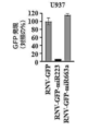


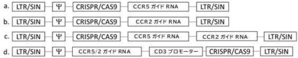


Classifications
-
- C—CHEMISTRY; METALLURGY
- C07—ORGANIC CHEMISTRY
- C07K—PEPTIDES
- C07K16/00—Immunoglobulins [IGs], e.g. monoclonal or polyclonal antibodies
- C07K16/18—Immunoglobulins [IGs], e.g. monoclonal or polyclonal antibodies against material from animals or humans
- C07K16/28—Immunoglobulins [IGs], e.g. monoclonal or polyclonal antibodies against material from animals or humans against receptors, cell surface antigens or cell surface determinants
- C07K16/2803—Immunoglobulins [IGs], e.g. monoclonal or polyclonal antibodies against material from animals or humans against receptors, cell surface antigens or cell surface determinants against the immunoglobulin superfamily
- C07K16/2815—Immunoglobulins [IGs], e.g. monoclonal or polyclonal antibodies against material from animals or humans against receptors, cell surface antigens or cell surface determinants against the immunoglobulin superfamily against CD8
-
- A—HUMAN NECESSITIES
- A61—MEDICAL OR VETERINARY SCIENCE; HYGIENE
- A61K—PREPARATIONS FOR MEDICAL, DENTAL OR TOILETRY PURPOSES
- A61K31/00—Medicinal preparations containing organic active ingredients
- A61K31/70—Carbohydrates; Sugars; Derivatives thereof
- A61K31/7088—Compounds having three or more nucleosides or nucleotides
- A61K31/7105—Natural ribonucleic acids, i.e. containing only riboses attached to adenine, guanine, cytosine or uracil and having 3'-5' phosphodiester links
-
- A—HUMAN NECESSITIES
- A61—MEDICAL OR VETERINARY SCIENCE; HYGIENE
- A61K—PREPARATIONS FOR MEDICAL, DENTAL OR TOILETRY PURPOSES
- A61K35/00—Medicinal preparations containing materials or reaction products thereof with undetermined constitution
- A61K35/66—Microorganisms or materials therefrom
- A61K35/76—Viruses; Subviral particles; Bacteriophages
-
- A—HUMAN NECESSITIES
- A61—MEDICAL OR VETERINARY SCIENCE; HYGIENE
- A61K—PREPARATIONS FOR MEDICAL, DENTAL OR TOILETRY PURPOSES
- A61K39/00—Medicinal preparations containing antigens or antibodies
- A61K39/46—Cellular immunotherapy
- A61K39/461—Cellular immunotherapy characterised by the cell type used
- A61K39/4611—T-cells, e.g. tumor infiltrating lymphocytes [TIL], lymphokine-activated killer cells [LAK] or regulatory T cells [Treg]
-
- A—HUMAN NECESSITIES
- A61—MEDICAL OR VETERINARY SCIENCE; HYGIENE
- A61K—PREPARATIONS FOR MEDICAL, DENTAL OR TOILETRY PURPOSES
- A61K39/00—Medicinal preparations containing antigens or antibodies
- A61K39/46—Cellular immunotherapy
- A61K39/463—Cellular immunotherapy characterised by recombinant expression
- A61K39/4631—Chimeric Antigen Receptors [CAR]
-
- A—HUMAN NECESSITIES
- A61—MEDICAL OR VETERINARY SCIENCE; HYGIENE
- A61K—PREPARATIONS FOR MEDICAL, DENTAL OR TOILETRY PURPOSES
- A61K39/00—Medicinal preparations containing antigens or antibodies
- A61K39/46—Cellular immunotherapy
- A61K39/464—Cellular immunotherapy characterised by the antigen targeted or presented
- A61K39/4643—Vertebrate antigens
- A61K39/4644—Cancer antigens
- A61K39/464402—Receptors, cell surface antigens or cell surface determinants
- A61K39/464411—Immunoglobulin superfamily
- A61K39/464412—CD19 or B4
-
- A—HUMAN NECESSITIES
- A61—MEDICAL OR VETERINARY SCIENCE; HYGIENE
- A61K—PREPARATIONS FOR MEDICAL, DENTAL OR TOILETRY PURPOSES
- A61K39/00—Medicinal preparations containing antigens or antibodies
- A61K39/46—Cellular immunotherapy
- A61K39/464—Cellular immunotherapy characterised by the antigen targeted or presented
- A61K39/4643—Vertebrate antigens
- A61K39/4644—Cancer antigens
- A61K39/464402—Receptors, cell surface antigens or cell surface determinants
- A61K39/464416—Receptors for cytokines
- A61K39/464417—Receptors for tumor necrosis factors [TNF], e.g. lymphotoxin receptor [LTR], CD30
-
- A—HUMAN NECESSITIES
- A61—MEDICAL OR VETERINARY SCIENCE; HYGIENE
- A61K—PREPARATIONS FOR MEDICAL, DENTAL OR TOILETRY PURPOSES
- A61K48/00—Medicinal preparations containing genetic material which is inserted into cells of the living body to treat genetic diseases; Gene therapy
- A61K48/005—Medicinal preparations containing genetic material which is inserted into cells of the living body to treat genetic diseases; Gene therapy characterised by an aspect of the 'active' part of the composition delivered, i.e. the nucleic acid delivered
-
- A—HUMAN NECESSITIES
- A61—MEDICAL OR VETERINARY SCIENCE; HYGIENE
- A61P—SPECIFIC THERAPEUTIC ACTIVITY OF CHEMICAL COMPOUNDS OR MEDICINAL PREPARATIONS
- A61P35/00—Antineoplastic agents
-
- C—CHEMISTRY; METALLURGY
- C07—ORGANIC CHEMISTRY
- C07K—PEPTIDES
- C07K14/00—Peptides having more than 20 amino acids; Gastrins; Somatostatins; Melanotropins; Derivatives thereof
- C07K14/435—Peptides having more than 20 amino acids; Gastrins; Somatostatins; Melanotropins; Derivatives thereof from animals; from humans
- C07K14/705—Receptors; Cell surface antigens; Cell surface determinants
- C07K14/70503—Immunoglobulin superfamily
- C07K14/7051—T-cell receptor (TcR)-CD3 complex
-
- C—CHEMISTRY; METALLURGY
- C07—ORGANIC CHEMISTRY
- C07K—PEPTIDES
- C07K14/00—Peptides having more than 20 amino acids; Gastrins; Somatostatins; Melanotropins; Derivatives thereof
- C07K14/435—Peptides having more than 20 amino acids; Gastrins; Somatostatins; Melanotropins; Derivatives thereof from animals; from humans
- C07K14/705—Receptors; Cell surface antigens; Cell surface determinants
- C07K14/70578—NGF-receptor/TNF-receptor superfamily, e.g. CD27, CD30, CD40, CD95
-
- C—CHEMISTRY; METALLURGY
- C07—ORGANIC CHEMISTRY
- C07K—PEPTIDES
- C07K16/00—Immunoglobulins [IGs], e.g. monoclonal or polyclonal antibodies
- C07K16/18—Immunoglobulins [IGs], e.g. monoclonal or polyclonal antibodies against material from animals or humans
-
- C—CHEMISTRY; METALLURGY
- C07—ORGANIC CHEMISTRY
- C07K—PEPTIDES
- C07K16/00—Immunoglobulins [IGs], e.g. monoclonal or polyclonal antibodies
- C07K16/18—Immunoglobulins [IGs], e.g. monoclonal or polyclonal antibodies against material from animals or humans
- C07K16/28—Immunoglobulins [IGs], e.g. monoclonal or polyclonal antibodies against material from animals or humans against receptors, cell surface antigens or cell surface determinants
- C07K16/2803—Immunoglobulins [IGs], e.g. monoclonal or polyclonal antibodies against material from animals or humans against receptors, cell surface antigens or cell surface determinants against the immunoglobulin superfamily
- C07K16/2812—Immunoglobulins [IGs], e.g. monoclonal or polyclonal antibodies against material from animals or humans against receptors, cell surface antigens or cell surface determinants against the immunoglobulin superfamily against CD4
-
- C—CHEMISTRY; METALLURGY
- C07—ORGANIC CHEMISTRY
- C07K—PEPTIDES
- C07K16/00—Immunoglobulins [IGs], e.g. monoclonal or polyclonal antibodies
- C07K16/18—Immunoglobulins [IGs], e.g. monoclonal or polyclonal antibodies against material from animals or humans
- C07K16/28—Immunoglobulins [IGs], e.g. monoclonal or polyclonal antibodies against material from animals or humans against receptors, cell surface antigens or cell surface determinants
- C07K16/2878—Immunoglobulins [IGs], e.g. monoclonal or polyclonal antibodies against material from animals or humans against receptors, cell surface antigens or cell surface determinants against the NGF-receptor/TNF-receptor superfamily, e.g. CD27, CD30, CD40, CD95
-
- C—CHEMISTRY; METALLURGY
- C12—BIOCHEMISTRY; BEER; SPIRITS; WINE; VINEGAR; MICROBIOLOGY; ENZYMOLOGY; MUTATION OR GENETIC ENGINEERING
- C12N—MICROORGANISMS OR ENZYMES; COMPOSITIONS THEREOF; PROPAGATING, PRESERVING, OR MAINTAINING MICROORGANISMS; MUTATION OR GENETIC ENGINEERING; CULTURE MEDIA
- C12N15/00—Mutation or genetic engineering; DNA or RNA concerning genetic engineering, vectors, e.g. plasmids, or their isolation, preparation or purification; Use of hosts therefor
- C12N15/09—Recombinant DNA-technology
- C12N15/11—DNA or RNA fragments; Modified forms thereof; Non-coding nucleic acids having a biological activity
- C12N15/113—Non-coding nucleic acids modulating the expression of genes, e.g. antisense oligonucleotides; Antisense DNA or RNA; Triplex- forming oligonucleotides; Catalytic nucleic acids, e.g. ribozymes; Nucleic acids used in co-suppression or gene silencing
-
- C—CHEMISTRY; METALLURGY
- C12—BIOCHEMISTRY; BEER; SPIRITS; WINE; VINEGAR; MICROBIOLOGY; ENZYMOLOGY; MUTATION OR GENETIC ENGINEERING
- C12N—MICROORGANISMS OR ENZYMES; COMPOSITIONS THEREOF; PROPAGATING, PRESERVING, OR MAINTAINING MICROORGANISMS; MUTATION OR GENETIC ENGINEERING; CULTURE MEDIA
- C12N15/00—Mutation or genetic engineering; DNA or RNA concerning genetic engineering, vectors, e.g. plasmids, or their isolation, preparation or purification; Use of hosts therefor
- C12N15/09—Recombinant DNA-technology
- C12N15/63—Introduction of foreign genetic material using vectors; Vectors; Use of hosts therefor; Regulation of expression
- C12N15/79—Vectors or expression systems specially adapted for eukaryotic hosts
- C12N15/85—Vectors or expression systems specially adapted for eukaryotic hosts for animal cells
- C12N15/86—Viral vectors
-
- C—CHEMISTRY; METALLURGY
- C12—BIOCHEMISTRY; BEER; SPIRITS; WINE; VINEGAR; MICROBIOLOGY; ENZYMOLOGY; MUTATION OR GENETIC ENGINEERING
- C12N—MICROORGANISMS OR ENZYMES; COMPOSITIONS THEREOF; PROPAGATING, PRESERVING, OR MAINTAINING MICROORGANISMS; MUTATION OR GENETIC ENGINEERING; CULTURE MEDIA
- C12N5/00—Undifferentiated human, animal or plant cells, e.g. cell lines; Tissues; Cultivation or maintenance thereof; Culture media therefor
- C12N5/06—Animal cells or tissues; Human cells or tissues
- C12N5/0602—Vertebrate cells
- C12N5/0634—Cells from the blood or the immune system
- C12N5/0636—T lymphocytes
-
- C—CHEMISTRY; METALLURGY
- C12—BIOCHEMISTRY; BEER; SPIRITS; WINE; VINEGAR; MICROBIOLOGY; ENZYMOLOGY; MUTATION OR GENETIC ENGINEERING
- C12N—MICROORGANISMS OR ENZYMES; COMPOSITIONS THEREOF; PROPAGATING, PRESERVING, OR MAINTAINING MICROORGANISMS; MUTATION OR GENETIC ENGINEERING; CULTURE MEDIA
- C12N9/00—Enzymes; Proenzymes; Compositions thereof; Processes for preparing, activating, inhibiting, separating or purifying enzymes
- C12N9/10—Transferases (2.)
- C12N9/12—Transferases (2.) transferring phosphorus containing groups, e.g. kinases (2.7)
- C12N9/1205—Phosphotransferases with an alcohol group as acceptor (2.7.1), e.g. protein kinases
- C12N9/1211—Thymidine kinase (2.7.1.21)
-
- C—CHEMISTRY; METALLURGY
- C12—BIOCHEMISTRY; BEER; SPIRITS; WINE; VINEGAR; MICROBIOLOGY; ENZYMOLOGY; MUTATION OR GENETIC ENGINEERING
- C12N—MICROORGANISMS OR ENZYMES; COMPOSITIONS THEREOF; PROPAGATING, PRESERVING, OR MAINTAINING MICROORGANISMS; MUTATION OR GENETIC ENGINEERING; CULTURE MEDIA
- C12N9/00—Enzymes; Proenzymes; Compositions thereof; Processes for preparing, activating, inhibiting, separating or purifying enzymes
- C12N9/14—Hydrolases (3)
- C12N9/78—Hydrolases (3) acting on carbon to nitrogen bonds other than peptide bonds (3.5)
-
- A—HUMAN NECESSITIES
- A61—MEDICAL OR VETERINARY SCIENCE; HYGIENE
- A61K—PREPARATIONS FOR MEDICAL, DENTAL OR TOILETRY PURPOSES
- A61K2239/00—Indexing codes associated with cellular immunotherapy of group A61K39/46
- A61K2239/31—Indexing codes associated with cellular immunotherapy of group A61K39/46 characterized by the route of administration
-
- A—HUMAN NECESSITIES
- A61—MEDICAL OR VETERINARY SCIENCE; HYGIENE
- A61K—PREPARATIONS FOR MEDICAL, DENTAL OR TOILETRY PURPOSES
- A61K2239/00—Indexing codes associated with cellular immunotherapy of group A61K39/46
- A61K2239/38—Indexing codes associated with cellular immunotherapy of group A61K39/46 characterised by the dose, timing or administration schedule
-
- A—HUMAN NECESSITIES
- A61—MEDICAL OR VETERINARY SCIENCE; HYGIENE
- A61K—PREPARATIONS FOR MEDICAL, DENTAL OR TOILETRY PURPOSES
- A61K2239/00—Indexing codes associated with cellular immunotherapy of group A61K39/46
- A61K2239/46—Indexing codes associated with cellular immunotherapy of group A61K39/46 characterised by the cancer treated
- A61K2239/48—Blood cells, e.g. leukemia or lymphoma
-
- C—CHEMISTRY; METALLURGY
- C07—ORGANIC CHEMISTRY
- C07K—PEPTIDES
- C07K2317/00—Immunoglobulins specific features
- C07K2317/60—Immunoglobulins specific features characterized by non-natural combinations of immunoglobulin fragments
- C07K2317/62—Immunoglobulins specific features characterized by non-natural combinations of immunoglobulin fragments comprising only variable region components
- C07K2317/622—Single chain antibody (scFv)
-
- C—CHEMISTRY; METALLURGY
- C07—ORGANIC CHEMISTRY
- C07K—PEPTIDES
- C07K2319/00—Fusion polypeptide
- C07K2319/01—Fusion polypeptide containing a localisation/targetting motif
- C07K2319/02—Fusion polypeptide containing a localisation/targetting motif containing a signal sequence
-
- C—CHEMISTRY; METALLURGY
- C07—ORGANIC CHEMISTRY
- C07K—PEPTIDES
- C07K2319/00—Fusion polypeptide
- C07K2319/01—Fusion polypeptide containing a localisation/targetting motif
- C07K2319/03—Fusion polypeptide containing a localisation/targetting motif containing a transmembrane segment
-
- C—CHEMISTRY; METALLURGY
- C12—BIOCHEMISTRY; BEER; SPIRITS; WINE; VINEGAR; MICROBIOLOGY; ENZYMOLOGY; MUTATION OR GENETIC ENGINEERING
- C12N—MICROORGANISMS OR ENZYMES; COMPOSITIONS THEREOF; PROPAGATING, PRESERVING, OR MAINTAINING MICROORGANISMS; MUTATION OR GENETIC ENGINEERING; CULTURE MEDIA
- C12N2310/00—Structure or type of the nucleic acid
- C12N2310/10—Type of nucleic acid
- C12N2310/11—Antisense
- C12N2310/113—Antisense targeting other non-coding nucleic acids, e.g. antagomirs
-
- C—CHEMISTRY; METALLURGY
- C12—BIOCHEMISTRY; BEER; SPIRITS; WINE; VINEGAR; MICROBIOLOGY; ENZYMOLOGY; MUTATION OR GENETIC ENGINEERING
- C12N—MICROORGANISMS OR ENZYMES; COMPOSITIONS THEREOF; PROPAGATING, PRESERVING, OR MAINTAINING MICROORGANISMS; MUTATION OR GENETIC ENGINEERING; CULTURE MEDIA
- C12N2310/00—Structure or type of the nucleic acid
- C12N2310/10—Type of nucleic acid
- C12N2310/14—Type of nucleic acid interfering N.A.
- C12N2310/141—MicroRNAs, miRNAs
-
- C—CHEMISTRY; METALLURGY
- C12—BIOCHEMISTRY; BEER; SPIRITS; WINE; VINEGAR; MICROBIOLOGY; ENZYMOLOGY; MUTATION OR GENETIC ENGINEERING
- C12N—MICROORGANISMS OR ENZYMES; COMPOSITIONS THEREOF; PROPAGATING, PRESERVING, OR MAINTAINING MICROORGANISMS; MUTATION OR GENETIC ENGINEERING; CULTURE MEDIA
- C12N2510/00—Genetically modified cells
-
- C—CHEMISTRY; METALLURGY
- C12—BIOCHEMISTRY; BEER; SPIRITS; WINE; VINEGAR; MICROBIOLOGY; ENZYMOLOGY; MUTATION OR GENETIC ENGINEERING
- C12N—MICROORGANISMS OR ENZYMES; COMPOSITIONS THEREOF; PROPAGATING, PRESERVING, OR MAINTAINING MICROORGANISMS; MUTATION OR GENETIC ENGINEERING; CULTURE MEDIA
- C12N2740/00—Reverse transcribing RNA viruses
- C12N2740/00011—Details
- C12N2740/10011—Retroviridae
- C12N2740/10041—Use of virus, viral particle or viral elements as a vector
-
- C—CHEMISTRY; METALLURGY
- C12—BIOCHEMISTRY; BEER; SPIRITS; WINE; VINEGAR; MICROBIOLOGY; ENZYMOLOGY; MUTATION OR GENETIC ENGINEERING
- C12N—MICROORGANISMS OR ENZYMES; COMPOSITIONS THEREOF; PROPAGATING, PRESERVING, OR MAINTAINING MICROORGANISMS; MUTATION OR GENETIC ENGINEERING; CULTURE MEDIA
- C12N2740/00—Reverse transcribing RNA viruses
- C12N2740/00011—Details
- C12N2740/10011—Retroviridae
- C12N2740/13011—Gammaretrovirus, e.g. murine leukeamia virus
- C12N2740/13041—Use of virus, viral particle or viral elements as a vector
- C12N2740/13043—Use of virus, viral particle or viral elements as a vector viral genome or elements thereof as genetic vector
-
- C—CHEMISTRY; METALLURGY
- C12—BIOCHEMISTRY; BEER; SPIRITS; WINE; VINEGAR; MICROBIOLOGY; ENZYMOLOGY; MUTATION OR GENETIC ENGINEERING
- C12N—MICROORGANISMS OR ENZYMES; COMPOSITIONS THEREOF; PROPAGATING, PRESERVING, OR MAINTAINING MICROORGANISMS; MUTATION OR GENETIC ENGINEERING; CULTURE MEDIA
- C12N2740/00—Reverse transcribing RNA viruses
- C12N2740/00011—Details
- C12N2740/10011—Retroviridae
- C12N2740/13011—Gammaretrovirus, e.g. murine leukeamia virus
- C12N2740/13051—Methods of production or purification of viral material
- C12N2740/13052—Methods of production or purification of viral material relating to complementing cells and packaging systems for producing virus or viral particles
-
- C—CHEMISTRY; METALLURGY
- C12—BIOCHEMISTRY; BEER; SPIRITS; WINE; VINEGAR; MICROBIOLOGY; ENZYMOLOGY; MUTATION OR GENETIC ENGINEERING
- C12N—MICROORGANISMS OR ENZYMES; COMPOSITIONS THEREOF; PROPAGATING, PRESERVING, OR MAINTAINING MICROORGANISMS; MUTATION OR GENETIC ENGINEERING; CULTURE MEDIA
- C12N2740/00—Reverse transcribing RNA viruses
- C12N2740/00011—Details
- C12N2740/10011—Retroviridae
- C12N2740/16011—Human Immunodeficiency Virus, HIV
- C12N2740/16041—Use of virus, viral particle or viral elements as a vector
- C12N2740/16043—Use of virus, viral particle or viral elements as a vector viral genome or elements thereof as genetic vector
-
- C—CHEMISTRY; METALLURGY
- C12—BIOCHEMISTRY; BEER; SPIRITS; WINE; VINEGAR; MICROBIOLOGY; ENZYMOLOGY; MUTATION OR GENETIC ENGINEERING
- C12N—MICROORGANISMS OR ENZYMES; COMPOSITIONS THEREOF; PROPAGATING, PRESERVING, OR MAINTAINING MICROORGANISMS; MUTATION OR GENETIC ENGINEERING; CULTURE MEDIA
- C12N2760/00—MICROORGANISMS OR ENZYMES; COMPOSITIONS THEREOF; PROPAGATING, PRESERVING, OR MAINTAINING MICROORGANISMS; MUTATION OR GENETIC ENGINEERING; CULTURE MEDIA ssRNA viruses negative-sense
- C12N2760/00011—Details
- C12N2760/18011—Paramyxoviridae
- C12N2760/18411—Morbillivirus, e.g. Measles virus, canine distemper
- C12N2760/18422—New viral proteins or individual genes, new structural or functional aspects of known viral proteins or genes
Abstract
本開示は、in vivoで抗原受容体結合構築物の細胞発現を介して免疫を誘導するための組成物及び方法を提供する。【選択図】図11The present disclosure provides compositions and methods for inducing immunity through cellular expression of antigen receptor binding constructs in vivo. [Selection drawing] Fig. 11
Description
本願は、35 U.S.C. §119に基づき、2020年5月11日に出願された米国仮出願第63/023,191号及び2020年12月31日に出願された米国仮出願第63/133,224号の優先権を主張する。これらの開示は、参照により本明細書に組み込まれる。 This application, pursuant to 35 U.S.C. claim. These disclosures are incorporated herein by reference.
本開示は、癌、感染症、アレルギー、変性及び免疫疾患の細胞療法のための組成物及び方法を提供する。 The present disclosure provides compositions and methods for cell therapy of cancer, infectious diseases, allergies, degenerative and immune diseases.
本願には、2021年5月11日に作成され、239,881バイトのデータを有し、IBMPC、MS-ウィンドウズオペレーティングシステム上で機械フォーマットされた"Sequence-Listing_ST25.txt"と題した配列表が添付されている。該配列表は、あらゆる目的のため、その全体が参照により本明細書に組み込まれる。 Attached to this application is a sequence listing entitled "Sequence-Listing_ST25.txt" created on May 11, 2021, having 239,881 bytes of data, machine formatted on an IBMPC, MS-Windows operating system. ing. The Sequence Listing is incorporated herein by reference in its entirety for all purposes.
養子免疫療法は、癌治療アプローチの最前線まで浮上している。T細胞は、腫瘍関連抗原を認識するキメラ抗原受容体(CAR)の遺伝子を発現するように操作することができる。CARは、T細胞を再誘導して腫瘍細胞を選択的に死滅させるように操作された免疫受容体である。癌免疫療法におけるそれらの使用の一般的な前提は、腫瘍標的化T細胞を迅速に生成することである。 Adoptive immunotherapy has emerged to the forefront of cancer treatment approaches. T cells can be engineered to express genes for chimeric antigen receptors (CARs) that recognize tumor-associated antigens. CARs are immune receptors engineered to redirect T cells to selectively kill tumor cells. A common premise for their use in cancer immunotherapy is the rapid generation of tumor-targeted T cells.
理想的な遺伝子送達システムは、製造が容易であり、投与が容易であり、正常細胞に対して非毒性であり、遺伝子情報を効率的かつ特異的に血流を介して標的組織に送達し、導入遺伝子が安定して発現されるように遺伝物質を宿主細胞に統合するであろう。本明細書に記載の新規なシステムは、これらに適合する。 An ideal gene delivery system would be easy to manufacture, easy to administer, non-toxic to normal cells, efficiently and specifically deliver genetic information to target tissues through the bloodstream, The genetic material will be integrated into the host cell such that the transgene is stably expressed. The novel system described herein fits these.
本開示は、(a)キメラ抗原受容体(CAR)をコードするポリヌクレオチド、及び(b)少なくとも1つのmiRNA標的配列を含むポリヌクレオチドを含む組換えベクターを提供し、ここで、(a)及び(b)が同一のポリヌクレオチド上で連結される。一実施形態では、前記ベクターは、(c)プロドラッグを細胞傷害性薬物に変換する細胞傷害性ポリペプチドをコードするポリヌクレオチドをさらに含む。別の実施形態では、前記(a)が、1つ以上の抗原結合ドメインをコードする第1ポリヌクレオチドドメイン、リンカーをコードする任意のポリヌクレオチドドメイン、前記第1ポリヌクレオチドドメインに作動可能に連結された第2ポリヌクレオチドドメイン(ここで、該第2ポリヌクレオチドドメインが膜貫通をコードする)、及び細胞内シグナル伝達ドメインをコードする。第3ポリヌクレオチドドメインを含む。さらなる実施形態では、前記第1ポリヌクレオチドドメインは、抗体断片、単一ドメイン抗体、単鎖可変断片、単一ドメイン抗体、ラクダ科VHHドメイン、非免疫グロブリン抗原結合足場、受容体または受容体断片、または二重特異性抗体をコードする。別の実施形態では、前記リンカーをコードする任意のポリヌクレオチドは、Gly3配列をコードする。さらに別の実施形態では、前記膜貫通ドメインは、T細胞受容体のα、βまたはζ鎖、CD3γ、CD3ε、CD3δ、CD28、CD45、CD4、CD5、CD8、CD9、CD16、CD22、CD33、CD37、CD64、CD80、CD86、CD134、CD137、CD154、KIRDS2、OX40、CD2、CD27、LFA-1 (CDl la, CD18) 、ICOS (CD278)、4-1BB (CD137)、GITR、CD40、BAFFR、HVEM (LIGHTR)、SLAMF7、NKp80 (KLRFl)、CD160、CD19、IL2Rβ、IL2Rγ、IL7R a、ITGA1、VLA1、CD49a、ITGA4、IA4、CD49D、ITGA6、VLA-6、CD49f、ITGAD、CDl ld、ITGAE、CD103、ITGAL、CDl la、LFA-1、ITGAM、CDl lb、ITGAX、CDl lc、ITGB1、CD29、ITGB2、CD18、LFA-1、ITGB7、TNFR2、DNAM1 (CD226)、SLAMF4 (CD244, 2B4)、 CD84、CD96 (Tactile)、CEACAM1、CRT AM、Ly9 (CD229)、CD160(BY55)、PSGL1、CD100 (SEMA4D)、SLAMF6 (NTB-A, Lyl08)、SLAM (SLAMF1, CD150, IPO-3)、BLAME (SLAMF8)、SELPLG (CD162)、LTBR、PAG/Cbp、NKp44、NKp30、NKp46、NKG2D、及び/またはNKG2Cからなる群から選択されるメンバーに由来する。さらに別の実施形態では、前記第3ポリヌクレオチドドメインは、CD3ζ、共通FeRγ(FCER1G)、Feγ RIIa、FeRβ(FeεR1b)、CD3γ、CD3δ、CD3ε、CD79a、CD79b、DAP1O、及びDAP12からなる群から選択される細胞内シグナル伝達ドメインをコードする。一実施形態では、前記プロドラッグを細胞傷害性薬物に変換する細胞傷害性ポリペプチドは、シトシンデアミナーゼ活性を有するポリペプチド、チミジンキナーゼ活性を有するポリペプチド、及びそれらの組み合わせからなる群から選択される。上記の実施形態のいずれにおいて、前記ベクターは、組み込みベクターである。さらなる実施形態では、前記ベクターは、レトロウイルスベクターである。さらに別の実施形態では、前記レトロウイルスベクターは、非複製γレトロウイルスベクターである。別の実施形態では、前記少なくとも1つのmiRNA標的配列は、hsa-miR-223-3p、hsa-miR143-3p、hsa-mir182-5p、hsa-miR-l0bp、hsa-miR141-3p、hsa-miR486-5p、及び前述の任意の組み合わせからなる群から選択されるmiRNAによって結合される。 The present disclosure provides a recombinant vector comprising (a) a polynucleotide encoding a chimeric antigen receptor (CAR) and (b) a polynucleotide comprising at least one miRNA target sequence, wherein (a) and (b) are ligated on the same polynucleotide. In one embodiment, the vector further comprises (c) a polynucleotide encoding a cytotoxic polypeptide that converts the prodrug into a cytotoxic drug. In another embodiment, (a) is a first polynucleotide domain encoding one or more antigen binding domains, any polynucleotide domain encoding a linker, operably linked to said first polynucleotide domain. a second polynucleotide domain, wherein the second polynucleotide domain encodes a transmembrane, and an intracellular signaling domain. It contains a third polynucleotide domain. In a further embodiment, said first polynucleotide domain is an antibody fragment, single domain antibody, single chain variable fragment, single domain antibody, camelid VHH domain, non-immunoglobulin antigen binding scaffold, receptor or receptor fragment; or encode a bispecific antibody. In another embodiment, any polynucleotide encoding said linker encodes a Gly3 sequence. In yet another embodiment, said transmembrane domain is a T cell receptor α, β or ζ chain, CD3γ, CD3ε, CD3δ, CD28, CD45, CD4, CD5, CD8, CD9, CD16, CD22, CD33, CD37 , CD64, CD80, CD86, CD134, CD137, CD154, KIRDS2, OX40, CD2, CD27, LFA-1 (CDl la, CD18), ICOS (CD278), 4-1BB (CD137), GITR, CD40, BAFFR, HVEM (LIGHTR), SLAMF7, NKp80 (KLRFl), CD160, CD19, IL2Rβ, IL2Rγ, IL7R a, ITGA1, VLA1, CD49a, ITGA4, IA4, CD49D, ITGA6, VLA-6, CD49f, ITGAD, CDl ld, ITGAE, CD103 , ITGAL, CDl la, LFA-1, ITGAM, CDl lb, ITGAX, CDl lc, ITGB1, CD29, ITGB2, CD18, LFA-1, ITGB7, TNFR2, DNAM1 (CD226), SLAMF4 (CD244, 2B4), CD84, CD96 (Tactile), CEACAM1, CRT AM, Ly9 (CD229), CD160 (BY55), PSGL1, CD100 (SEMA4D), SLAMF6 (NTB-A, Lyl08), SLAM (SLAMF1, CD150, IPO-3), BLAME (SLAMF8) ), SELPLG (CD162), LTBR, PAG/Cbp, NKp44, NKp30, NKp46, NKG2D, and/or NKG2C. In yet another embodiment, said third polynucleotide domain is selected from the group consisting of CD3ζ, common FeRγ (FCER1G), FeγRIIa, FeRβ (FeεR1b), CD3γ, CD3δ, CD3ε, CD79a, CD79b, DAP1O, and DAP12. encodes an intracellular signaling domain that is In one embodiment, the cytotoxic polypeptide that converts the prodrug into a cytotoxic drug is selected from the group consisting of polypeptides with cytosine deaminase activity, polypeptides with thymidine kinase activity, and combinations thereof. . In any of the above embodiments, said vector is an integrating vector. In a further embodiment, said vector is a retroviral vector. In yet another embodiment, said retroviral vector is a non-replicating gamma retroviral vector. In another embodiment, said at least one miRNA target sequence is hsa-miR-223-3p, hsa-miR143-3p, hsa-mir182-5p, hsa-miR-10bp, hsa-miR141-3p, hsa-miR486 -5p, and a miRNA selected from the group consisting of any combination of the foregoing.
本開示はまた、gagポリペプチド、polポリペプチド、envポリペプチド、及びレトロウイルスベクターのキャプシド内に含まれるレトロウイルスポリヌクレオチドを含む組換えレトロウイルス粒子を提供し、ここで、該レトロウイルスポリヌクレオチドが、5'から3'へ、(R-U5ドメイン)-(任意のシグナルペプチドコード配列ドメイン)-(結合ドメインコード配列ドメイン)-(任意のヒンジ/リンカーコード配列ドメイン)-(膜貫通(TM)コード配列ドメイン)-(miRNA標的ドメイン)-(U3-R ドメイン)を含む。一実施形態では、前記R-U5ドメインは、ヌクレオチド1から約ヌクレオチド145まで、配列番号:25と少なくとも80%同一である配列を有する。別のまたはさらなる実施形態では、前記結合ドメインコード配列には、シグナル配列が先行する。さらに別のまたはさらなる実施形態では、前記結合ドメインコード配列に続いて、任意のリンカー/スペーサードメイン配列が続く。さらに別のまたはさらなる実施形態では、前記レトロウイルスポリヌクレオチドは、キルスイッチドメインコード配列をさらに含む。さらなる実施形態では、前記キルスイッチドメインは、プロドラッグを細胞傷害性薬物に変換するポリペプチドのコード配列に作動可能に連結されたIRESを含む。さらなる実施形態では、前記ポリペプチドは、チミジンキナーゼ(TKO)活性またはシトシンデアミナーゼ(CD)活性を有する。別の実施形態では、前記レトロウイルスポリヌクレオチドは、少なくとも1つのmiRNA標的配列を含む。さらなる実施形態では、前記少なくとも1つのmiRNA標的配列は、複数のmiRNA標的配列を含む。さらに別の実施形態では、前記複数のmiRNA標的配列は、同一である。さらに別の実施形態では、前記複数のmiRNA標的配列の少なくとも2つが異なる。別の実施形態では、前記U3-Rドメインは、ヌクレオチド約5537から約6051まで、配列番号:25と少なくとも80%同一である配列を含む。
The disclosure also provides a recombinant retroviral particle comprising a gag polypeptide, a pol polypeptide, an env polypeptide, and a retroviral polynucleotide contained within the capsid of a retroviral vector, wherein the
本開示はさらに、本開示のベクターを用いたin vivo形質導入によって産生された遺伝子形質導入幹細胞を担持する非ヒト動物を提供する。 The disclosure further provides non-human animals bearing genetically transduced stem cells produced by in vivo transduction with the vectors of the disclosure.
本明細書及び添付の請求項に用いられる単数形の"1つ"、及び"該"は、文脈が他に明確に指示しない限り、複数の対象物を含む。従って、例えば、"一被験者"を言及することは、複数のこのような被験者への言及を含み、"該ベクター"を言及することは、当業者に公知の1つ以上のベクター及びその等価物への言及を含む。他についても同様である。 As used in this specification and the appended claims, the singular forms "a" and "the" include plural referents unless the context clearly dictates otherwise. Thus, for example, reference to "a subject" includes reference to a plurality of such subjects, and reference to "the vector" includes one or more vectors and equivalents thereof known to those skilled in the art. contains a reference to The same is true for others.
また、"または"は、他に記載されていない限り、"及び/または"を意味する。同様に、"含む"、"含んでいる"、"包含する"、及び"包含している"は、交換可能であり、限定を意図したものではない。 Also, the use of "or" means "and/or" unless stated otherwise. Similarly, "comprise," "include," "comprise," and "include" are interchangeable and are not intended to be limiting.
さらに理解すべきこととして、様々な実施形態の説明が"含む"という用語を使用する場合、当業者は、いくつかの特定の例において、代替的に"本質的に・・・からなる"または"・・・からなる"という用語を用いて実施形態を説明してもよいことを理解するであろう。 It should also be understood that where the description of various embodiments uses the term "comprising," those skilled in the art will, in some specific instances, alternatively "consist essentially of, or It will be appreciated that the term "consisting of" may be used to describe embodiments.
特に定義されない限り、本明細書で使用される全ての技術用語及び科学用語は、本発明が属する技術分野の当業者によって一般的に理解されるものと同じ意味を有する。Allenら, Remington:薬学と実践(第22版), Pharmaceutical Press(2012年9月15日); Hornyakら,ナノサイエンスとナノテクノロジー入門, CRC Press(2008); SingletonとSainsbury, 微生物学及び分子生物学辞書(第3版改定), J. Wiley & Sons (New York, NY 2006); Smith, Marchの上級有機化学反応、メカニズムと構造(第7版), J. Wiley & Sons(New York, NY 2013); Singleton, DNAとゲノム技術辞典(第3版), Wiley-Blackwell(2012年11月28日); 及びGreenとSambrook, 分子クローニング:実験マニュアル(第4版), Cold Spring Harbor Laboratory Press(Cold Spring Harbor, NY 2012)では、当業者向けの、本出願で使用される多くの用語への一般的なガイドが提供されている。抗体の調製方法については、Greenfield, 抗体:実験マニュアル(第2版), Cold Spring Harbor Press(Cold Spring Harbor NY,2013); KohlerとMilstein, 細胞融合による特異的な抗体産生組織培養及び腫瘍株の誘導, Eur. J. Immunol. 1976年7月,6(7):511-9; QueenとSelick,ヒト化免疫グロブリン,米国特許番号5,585,089(1996年12月);及びRiechmannら,治療用ヒト抗体の再形成, Nature, 1988年3月24日,332(6162):323-7A11を参照されたい。本明細書中で提供される全ての見出し及び副見出しは、単に読みを容易にするためだけであり、本発明を限定するものと解釈されるべきではない。本明細書に記載した方法及び材料と類似または同等の方法及び材料を本発明の実施または試験に使用することができるが、適切な方法及び材料を以下に記載する。本明細書で言及された全ての刊行物、特許出願、特許、及び他の参考文献は、その全体が参照により組み込まれる。矛盾する場合は、定義を含む本明細書が優先する。また、材料、方法、及び特定の実施例は、単に例示的なものであり、限定することを意図するものではない。 Unless defined otherwise, all technical and scientific terms used herein have the same meaning as commonly understood by one of ordinary skill in the art to which this invention belongs. Allen et al., Remington: Pharmaceutical Sciences and Practice (22nd ed.), Pharmaceutical Press (September 15, 2012); Hornyak et al., Introduction to Nanoscience and Nanotechnology, CRC Press (2008); Singleton and Sainsbury, Microbiology and Molecular Biology. Scientific Dictionary (revised 3rd ed.), J. Wiley & Sons (New York, NY 2006); Smith, March in Advanced Organic Chemistry, Mechanisms and Structures (7th ed.), J. Wiley & Sons (New York, NY) 2013); Singleton, Dictionary of DNA and Genome Technology (3rd ed.), Wiley-Blackwell (November 28, 2012); and Green and Sambrook, Molecular Cloning: A Laboratory Manual (4th ed.), Cold Spring Harbor Laboratory Press ( Cold Spring Harbor, NY 2012) provides a general guide for those skilled in the art to many of the terms used in this application. For antibody preparation methods, see Greenfield, Antibodies: Experimental Manual (2nd Edition), Cold Spring Harbor Press (Cold Spring Harbor NY, 2013); Induction, Eur. J. Immunol. 1976 July, 6(7):511-9; Queen and Selick, Humanized Immunoglobulin, U.S. Patent No. 5,585,089 (December 1996); and Riechmann et al., Therapeutic Human Antibodies. , Nature, Mar. 24, 1988, 332(6162):323-7A11. All headings and subheadings provided herein are for ease of reading only and should not be construed as limiting the invention. Although methods and materials similar or equivalent to those described herein can be used in the practice or testing of the present invention, suitable methods and materials are described below. All publications, patent applications, patents, and other references mentioned herein are incorporated by reference in their entirety. In case of conflict, the present specification, including definitions, will control. In addition, the materials, methods, and specific examples are illustrative only and not intended to be limiting.
本明細書に言及される全ての刊行物は、本明細書の記載と関連して使用され得る方法論を記載し、開示する目的のために、本明細書に完全に組み込まれる。さらに、本明細書において明確に定義された用語と類似している、または同一である1つ以上の刊行物に提示される任意の用語に関して、本明細書に明示的に提供される用語の定義は、全ての点で制御するであろう。 All publications mentioned in this specification are hereby incorporated in full for the purpose of describing and disclosing methodology that may be used in connection with the present description. Further, with respect to any term appearing in one or more publications that is similar to or identical to a term expressly defined herein, the definitions of the terms expressly provided herein will control at all points.
理解されたいこととして、本開示は、本明細書に記載される特定の方法論、プロトコル、及び試薬等に限定されるものではなく、これらは変化し得る。本明細書で使用される用語は、特定の実施形態または態様のみを説明するためのものであり、本開示の範囲を限定することを意図するものではない。 It is to be understood that this disclosure is not limited to the particular methodology, protocols, and reagents, etc., described herein, as such may vary. The terminology used herein is for the purpose of describing particular embodiments or aspects only and is not intended to be limiting of the scope of the disclosure.
実施例において、または別途に示される場合以外に、本明細書で使用される成分または反応条件の量を表す全ての数は、全ての場合において"約"という用語で修飾されると理解されるべきである。本明細書で使用される"約"という用語は、当業者によって測定された特定の値に対する許容誤差範囲内であることを意味することができ、その値がどのように計測または測定されるか、例えば、測定システムの限界に部分的に依存することができる。あるいは、"約"は、所定値のプラスまたはマイナス20%、プラスまたはマイナス10%、プラスまたはマイナス5%、またはプラスまたはマイナス1%の範囲を意味することができる。あるいは、特に、生物学的システムまたはプロセスに関して、該用語は、値の5倍以内、または2倍以内の大きさのオーダー内であることを意味することができる。特定の値が本願及び請求項に記載されている場合、特に断らない限り、特定の値に対する許容誤差範囲内での用語"約"の意味を仮定することができる。また、値の範囲及び/またはサブ範囲が提供される場合、該範囲及び/またはサブ範囲は、その終点を含むことができる。場合によっては、変動は、特定の量の20%、10%、5%、1%、0.5%、または0.1%の量または濃度を含むことができる。 Except as otherwise indicated in the examples or otherwise, all numbers expressing quantities of ingredients or reaction conditions used herein are understood to be modified in all instances by the term "about." should. As used herein, the term "about" can mean within an acceptable range of error for a particular value determined by one of ordinary skill in the art and how that value is measured or measured. , for example, may depend in part on the limitations of the measurement system. Alternatively, "about" can mean a range of plus or minus 20%, plus or minus 10%, plus or minus 5%, or plus or minus 1% of a given value. Alternatively, particularly with respect to a biological system or process, the term can mean within an order of magnitude within 5, or within 2 of the value. Where specific values are set forth in the application and claims, the meaning of the term "about" within a tolerance for the specific values can be assumed unless otherwise stated. Also, where ranges and/or subranges of values are provided, the ranges and/or subranges can include their endpoints. In some cases, variation can include amounts or concentrations of 20%, 10%, 5%, 1%, 0.5%, or 0.1% of the specified amount.
本明細書における数値範囲の引用のために、同じ程度の精度を有する各介在番号が明示的に意図されている。例えば、6~9の範囲の場合、6及び9の他に番号7及び8が明示的に意図され、また、6.0~7.0の範囲の場合、6.0、6.1、6.2、6.3、6.4、6.5、6.6、6.7、6.8、6.9、及び7.0が明示的に意図されている。
For the purposes of numerical range recitations herein, each intervening number with the same degree of precision is expressly intended. For example, for the
ウイルスのレトロウィルス科は、宿主ゲノムに組み込まれ、形質導入された細胞及びその子孫に長期間の遺伝子発現を提供するベクターを作製するために使用され得る。一般に、γレトロウイルスベクター、レンチウイルスベクター、及び泡状ウイルスベクターが、動員された幹細胞を含む細胞を形質導入するのに使用可能であり、有用である。本開示は、γレトロウイルスベクターに焦点を当てているが、当業者は、本発明が、ウイルス及び非ウイルスベクターを含む全てのタイプの組込みベクター、例えば、アデノウイルス-レトロウイルスハイブリッド、piggy-bac、及び眠れる森の美女トランスポゾン等を使用できることをすぐに理解するであろう。本開示は、遺伝的疾患、癌、感染症、及び自己免疫疾患を含む多種の疾患において治療効果を達成するためにベクターを直接に投与することにより、in vivoで細胞(造血幹細胞を含む)を形質導入するための組成物及び方法を提供する。いくつかの実施形態では、細胞は、in vivo感染の前に動員され得る。 The Retroviridae family of viruses can be used to create vectors that integrate into the host genome and provide long-term gene expression in transduced cells and their progeny. In general, gamma retroviral, lentiviral, and foamy viral vectors are available and useful for transducing cells, including mobilized stem cells. Although this disclosure focuses on gamma retroviral vectors, those skilled in the art will appreciate that the present invention applies to all types of integrating vectors, including viral and non-viral vectors, e.g. adenoviral-retroviral hybrids, piggy-bac , and Sleeping Beauty transposons, etc., can be used. The present disclosure demonstrates the use of cells (including hematopoietic stem cells) in vivo by direct administration of vectors to achieve therapeutic effects in a variety of diseases, including genetic diseases, cancer, infectious diseases, and autoimmune diseases. Compositions and methods for transducing are provided. In some embodiments, cells may be mobilized prior to in vivo infection.
本開示のベクター構築物を下記のドメインを有するモジュール(時にはカセットと呼ばれる)と考えることができる。一般に、該ベクターは、長鎖末端反復、結合ドメイン、ヒンジまたはリンカードメイン、膜貫通ドメイン、細胞内ドメイン、1つ以上のmiRNA標的ドメイン、任意のキルスイッチドメイン、及び任意の細胞活性調節ドメインを含む。前記結合ドメイン、ヒンジ/リンカー、膜貫通ドメイン及び細胞内ドメインは、一般に、第1世代、第2世代、第3世代、及び関連構築物を含むキメラ抗原受容体(CAR)を含む。 The vector constructs of this disclosure can be thought of as modules (sometimes called cassettes) with the following domains. Generally, the vector comprises a long terminal repeat, a binding domain, a hinge or linker domain, a transmembrane domain, an intracellular domain, one or more miRNA targeting domains, an optional killswitch domain, and an optional cellular activity modulating domain. . The binding domain, hinge/linker, transmembrane domain and intracellular domain generally comprise chimeric antigen receptors (CARs) including first generation, second generation, third generation and related constructs.
本明細書で使用される用語"抗体"は、抗原と特異的に結合する免疫グロブリン分子に由来するタンパク質またはポリペプチド配列を指す。抗体は、モノクローナル、またはポリクローナル、多鎖または単鎖、または無傷の免疫グロブリンであってもよく、天然の供給源または組換え供給源に由来してもよい。抗体は、免疫グロブリン分子の四量体であってもよい。該抗体は、"ヒト化"、"キメラ"または非ヒトであってもよい。 As used herein, the term "antibody" refers to a protein or polypeptide sequence derived from an immunoglobulin molecule that specifically binds an antigen. Antibodies may be monoclonal or polyclonal, multi- or single-chain, or intact immunoglobulins, and may be derived from natural or recombinant sources. An antibody may be a tetramer of immunoglobulin molecules. The antibody may be "humanized," "chimeric," or non-human.
用語"抗体断片"は、(例えば、結合、立体障害、安定化/不安定化、空間分布によって)抗原のエピトープと特異的に相互作用する能力を保持する抗体の少なくとも1つの部分を指す。抗体断片の例には、これらに限定されないが、Fab、Fab'、Fv断片、scFv抗体断片、ジスルフィド結合Fv、VH及びCH1ドメインからなるFd断片、線状抗体、vLまたはvHのいずれかのような単一ドメイン抗体(sdAb)、ラクダ類vHHドメイン、ヒンジ領域でジスルフィド架橋によって連結された2つのFab断片を含む二価断片のような抗体断片から形成された多特異的抗体、及び抗体の単離されたCDRまたは他のエピトープ結合断片が含まれる。抗原結合断片はまた、単一ドメイン抗体、マキシボディ、ミニボディ、ナノボディ、イントラボディ、ダイアボディ、トライアボディ、テトラボディ、v-NAR、及びbis-scFvに組み込むことができる(例えば、HollingerとHudson, Nature Biotechnology 23:1126-1136, 2005を参照)。抗原結合断片は、フィブロネクチンタイプIII(Fn3)のようなポリペプチドに基づいて足場に移植することもできる(フィブロネクチンポリペプチドミニボディを記載する米国特許第6,703,199号を参照)。 The term "antibody fragment" refers to at least one portion of an antibody that retains the ability to specifically interact with an epitope of an antigen (eg, by binding, steric hindrance, stabilization/destabilization, spatial distribution). Examples of antibody fragments include, but are not limited to, Fab, Fab', Fv fragments, scFv antibody fragments, disulfide-linked Fv, Fd fragments consisting of VH and CH1 domains, linear antibodies, such as either vL or vH. single domain antibodies (sdAb), camelid vHH domains, multispecific antibodies formed from antibody fragments such as bivalent fragments comprising two Fab fragments linked by disulfide bridges at the hinge region, and single antibody antibodies. Released CDRs or other epitope binding fragments are included. Antigen-binding fragments can also be incorporated into single domain antibodies, maxibodies, minibodies, nanobodies, intrabodies, diabodies, triabodies, tetrabodies, v-NARs, and bis-scFvs (e.g., Hollinger and Hudson , Nature Biotechnology 23:1126-1136, 2005). Antigen-binding fragments can also be grafted onto scaffolds based on polypeptides such as fibronectin type III (Fn3) (see US Pat. No. 6,703,199 describing fibronectin polypeptide minibodies).
用語"抗体重鎖"は、抗体分子中に存在する2種類のポリペプチド鎖の天然に存在する立体構造の中の比較的大きいものを指す。これは、通常には抗体が属するクラスを決定する。 The term "antibody heavy chain" refers to the larger of the naturally occurring conformations of the two polypeptide chains present in antibody molecules. This usually determines the class to which the antibody belongs.
用語"抗体軽鎖"は、抗体分子中に存在する2種類のポリペプチド鎖の天然に存在する立体構造の中の比較的小さいものを指す。カッパー(κ)及びラムダ(λ)軽鎖は、2つの主要な抗体軽鎖アイソタイプを指す。 The term "antibody light chain" refers to the smaller of the naturally occurring conformations of the two polypeptide chains present in antibody molecules. Kappa (κ) and lambda (λ) light chains refer to the two major antibody light chain isotypes.
"抗癌剤"は、異常な細胞分裂及び増殖を阻害し、新生物細胞の遊走を阻害し、侵襲性を阻害し、または癌の増殖及び転移を防止する薬剤を指す。該用語には、化学療法製剤、生物学的製剤(例えば、siRNA、遺伝子操作されたMLV等のウイルスベクター、アデノウイルス、細胞傷害性遺伝子を送達するヘルペスウイルス)、及び抗体等が含まれる。 "Anti-cancer agent" refers to an agent that inhibits abnormal cell division and proliferation, inhibits neoplastic cell migration, inhibits invasiveness, or prevents cancer growth and metastasis. The term includes chemotherapeutic agents, biological agents (eg, siRNA, genetically engineered viral vectors such as MLV, adenoviruses, herpes viruses that deliver cytotoxic genes), antibodies, and the like.
"抗癌作用"という用語は、様々な手段によって明らかになる生物学的効果を指す。該手段には、腫瘍体積の減少、癌細胞数の減少、転移数の減少、平均余命の延長、癌細胞増殖の減少、癌細胞生存率の低減、または癌の状態に関連する様々な生理学的症状の改善が含まれるが、これらに限定されない。"抗癌作用"は、原発場所における癌の発生を予防する際のCARの能力によっても明らかになる。 The term "anti-cancer effect" refers to biological effects manifested by various means. Said means include decreasing tumor volume, decreasing the number of cancer cells, decreasing the number of metastases, increasing life expectancy, decreasing cancer cell proliferation, decreasing cancer cell viability, or various physiological effects associated with the cancer state. Includes but is not limited to improvement of symptoms. An "anti-cancer effect" is also manifested by the ability of CARs in preventing cancer development at the primary site.
用語"抗原"または"Ag"は、免疫応答を誘発する分子を指す。該免疫応答は、抗体の産生、または特異的免疫応答性細胞の活性化のいずれか、またはその両方を含むことができる。当業者は、実質的に全てのタンパク質またはペプチドを含む任意の巨大分子が抗原として機能することができることを理解するであろう。さらに、抗原は、組換えまたはゲノムDNAから由来することができる。当業者は、免疫応答を誘発するタンパク質をコードするヌクレオチド配列または部分ヌクレオチド配列を含む任意のDNAが、本明細書で使用されるような"抗原"をコードすることを理解するであろう。さらに、当業者は、抗原が遺伝子の全長ヌクレオチド配列のみによってコードされる必要はないことを理解するであろう。限定するものではないが、本開示は、1つ以上の遺伝子の部分的ヌクレオチド配列の使用を含む。これらのヌクレオチド配列は、所望の免疫応答を誘発するポリペプチドをコードする様々な組み合わせで配列される。さらに、当業者は、抗原が"遺伝子"によってコードされる必要は全くないことを理解するであろう。明白なこととして、抗原が生成され、合成され、または生体試料から由来してもよく、あるいは、ポリペプチドの他の巨大分子であってもよい。このような生体試料には、組織試料、腫瘍試料、または他の生体成分を含む細胞または液体が含まれるが、これらに限定されない。 The term "antigen" or "Ag" refers to a molecule that elicits an immune response. The immune response can include either the production of antibodies or the activation of specific immunoresponsive cells, or both. Those skilled in the art will appreciate that virtually any macromolecule can serve as an antigen, including any protein or peptide. Additionally, antigens can be derived from recombinant or genomic DNA. Those skilled in the art will understand that any DNA containing a nucleotide sequence or partial nucleotide sequence that encodes a protein that elicits an immune response encodes an "antigen" as used herein. Furthermore, those skilled in the art will understand that an antigen need not be encoded solely by the full-length nucleotide sequence of a gene. Without limitation, the disclosure includes the use of partial nucleotide sequences of one or more genes. These nucleotide sequences are arranged in various combinations that encode polypeptides that elicit the desired immune response. Moreover, those skilled in the art will understand that an antigen need not be encoded by a "gene" at all. Obviously, the antigen may be generated, synthesized, or derived from a biological sample, or may be other macromolecules of polypeptides. Such biological samples include, but are not limited to, tissue samples, tumor samples, or cells or fluids containing other biological components.
標的され得る抗原の非限定的な例は、以下を含む:CD5、CD19、CD20、CD22、CD24、CD30、CD33、CD34、CD38、CD72、CD97、CD123、CD171、CS1(CD2サブセット1、CRACC、MPL、SLAMF7、CD319、及び19A24とも呼ばれる)、C型レクチン様分子-1(CLL-1またはCLECL1)、表皮成長因子受容体変異体III(EGFRviii)、ガングリオシドG2(GD2)、ガングリオシドGD3(aNeu5Ac(2-8)aNeu5Ac(2-3)bDGalp(l-4)bDGlcp(l-l)Cer)、TNF受容体ファミリーメンバーB細胞成熟(BCMA)、Tn抗原((Tn Ag)または(GalNAcα-Ser/Thr))、前立腺特異的膜抗原(PSMA)、受容体チロシンキナーゼ様オーファン受容体1(ROR1)、Fms様チロシンキナーゼ3(FLT3)、腫瘍関連糖タンパク質72(TAG72)、CD44v6、グリコシル化CD43エピトープ、発癌性抗原(CEA)、上皮細胞接着分子(EPCAM)、B7H3(CD276)、キット(CD117)、インターロイキン-13受容体サブユニットα-2(IL-13Ra2またはCD213A2)、メソテリン、インターロイキン11受容体α(IL-llRa)、前立腺幹細胞抗原(PSCA)、プロテアーゼセリン21(テスティシンまたはPRSS21)、血管内皮増殖因子受容体2(VEGFR2)、ルイス(Y)抗原、血小板由来増殖因子受容体β(PDGFR-β)、ステージ特異的胚抗原-4(SSEA-4)、葉酸受容体α(FRaまたはFR1)、葉酸受容体β(FRb)、受容体チロシンタンパク質キナーゼERBB2(Her2/neu)、ムチン1、細胞表面関連(MUC1)、表皮成長因子受容体(EGFR)、神経細胞接着分子(NCAM)、プロスターゼ、前立腺酸性ホスファターゼ(PAP)、変異された伸長因子2(ELF2M)、エフリンB2、線維芽細胞活性化タンパク質α(FAP)、インスリン様成長因子1受容体(IGF-I受容体)、カルボニックアンヒドラーゼIX(CA1X)、プロテアソーム(プロサム、マクロペイン)サブユニット、β型、9(LMP2)、糖タンパク質100(gpl00)、ブレークポイントクラスター領域(BCR)及びAbelsonマウス白血病ウイルス発癌遺伝子ホモログ 1(Abl)(bcr-abl)からなる癌遺伝子融合タンパク質、チロシナーゼ、エフリンA型受容体2(EphA2)、シアリルルイス接着分子(sLe)、ガングリオシドGM3(aNeu5Ac(2-3)bDClalp(l-4)bDGlcp(l-1)Cer)、トランスグルタミナーゼ5(TGS5)、高分子量メラノーマ関連抗原(HMWMAA)、o-アセチル-GD2ガングリオシド(OAcGD2)、腫瘍内皮マーカー1(TEM1/CD248)、腫瘍内皮マーカー7関連(TEM7R)、クローディン6(CLDN6)、甲状腺刺激ホルモン受容体(TSHR)、Gタンパク質共役型受容体クラスC群5、メンバーD(GPRC5D)、染色体Xオープンリーディングフレーム61(CXORF61)、CD179a、未分化リンパ腫キナーゼ(ALK)、ポリシアル酸、胎盤特異的1(PLAC1)、グロボHグリコセラミドの六糖部分(GloboH)、乳腺分化抗原(NY-BR-1)、ウロプラキン2(UPK2)、A型肝炎ウイルス細胞受容体1(HAVCR1)、アドレナリン受容体β3(ADRB3)、パネキシン3(PANX3)、Gタンパク質共役型受容体20(GPR20)、リンパ球抗原6複合体、遺伝子座K 9(LY6K)、嗅覚受容体51E2(OR51E2)、TCRγ代替リーディング フレーム タンパク質 (TARP)、ウィルムス腫瘍タンパク質(WT1)、癌/精巣抗原1(NY-ESO-1)、癌/精巣抗原2(LAGE-la)、メラノーマ関連抗原1(MAGE-A1)、染色体12pに位置するETSトランスロケーション変異遺伝子6(ETV6-AML)、精子タンパク質17(SPA17)、X抗原ファミリー、メンバー1A(XAGE1)、アンジオポエチン結合細胞表面受容体2(Tie 2)、メラノーマ癌精巣抗原-1(MAD-CT-1)、メラノーマ癌精巣抗原-2(MAD-CT-2)、Fos関連抗原1、腫瘍タンパク質p53(p53)、p53変異体、プロスタイン、サバイビン、テロメラーゼ、前立腺癌腫瘍抗原-1(PCT A-lまたはガレクチン8)、T細胞1によって認識されるメラノーマ抗原(MelanAまたはMARTI)、ラット肉腫(Ras)突然変異体、ヒトテロメラーゼ逆転写酵素(hTERT)、肉腫トランスロケーションブレークポイント、アポトーシスのメラノーマ阻害剤(ML-IAP)、ERG(膜貫通プロテアーゼ、セリン2(TMPRSS2)ETS融合遺伝子)、N-アセチルグルコサミニル-トランスフェラーゼV(NA17)、ペアボックスタンパク質Pax-3(PAX3)、アンドロゲン受容体、サイクリンBl、v-mycトリ骨髄球腫症ウイルス腫瘍遺伝子神経芽細胞腫由来のホモログ(MYCN)、RasホモログファミリーメンバーC(RhoC)、チロシナーゼ関連タンパク質2(TRP-2)、チトクロームP450 1B 1(CYP1B 1)、CCCTC-結合因子(亜鉛フィンガータンパク質)様(BORIS、またはインプリント部位の兄弟制御因子)、T細胞3によって認識された扁平上皮細胞癌抗原(SART3)、ペアボックスタンパク質Pax-5(PAX5)、プロアクロシン結合タンパク質sp32(OY-TES1)、リンパ球特異的タンパク質チロシンキナーゼ(LCK)、キナーゼアンカータンパク質4(AKAP-4)、滑膜肉腫、xブレークポイント2(SSX2)、最終糖化産物の受容体(RAGE-1)、腎ユビキタス1(RU1)、腎ユビキタス2(RU2)、レグマイン、ヒト乳頭腫ウイルスE6(HPV E6)、ヒト乳頭腫ウイルスE7(HPV E7)、腸管カルボキシルエステラーゼ、変異された熱ショックタンパク質70-2(mut hsp70-2)、CD79a、CD79b、白血球関連免疫グロブリン様受容体1(LAIR1))、IgA受容体のFc断片(FCARまたはCD89)、白血球免疫グロブリン様受容体サブファミリーAメンバー2(LILRA2)、CD300分子様ファミリーメンバーf(CD300LF)、C型レクチンドメインファミリー12メンバーA(CLEC12A)、骨髄間質細胞抗原2(BST2)、EGF様モジュールを含むムチン様ホルモン受容体様2(EMR2)、リンパ球抗原75(LY75)、グリピカン-3(GPC3)、Fc受容体様5(FCRL5)、免疫グロブリンλ様ポリペプチド1(IGLL1)、MPL、ビオチン、c-MYCエピトープタグ、LAMP1 TROP2、GFRα4、CDH17、CDH6、NYBR1、CDH19、CD200R、Slea(CA19.9、シアリルルイス抗原)、フコシル-GMl、PTK7、gpNMB、CDH1-CD324、DLL3、CD276/B7H3、ILllRa、IL13Ra2、CD179b-IGL11、TCRγ-δ、NKG2D、CD32(FCGR2A)、Tn ag、Timl-/HVCR1、CSF2RA(GM-CSFR-α)、TGFβR2、Lews Ag、TCR-β1鎖、TCR-β2鎖、TCR-γ鎖、TCR-δ鎖、FITC、黄体形成ホルモン受容体 (LHR)、卵胞刺激ホルモン受容体(FSHR)、ゴナドトロピンホルモン受容体(CGHRまたはGR)、CCR4、GD3、SLAMF6、SLAMF4、HIV1エンベロープ糖タンパク質、HTLVl-Tax、CMV pp65、EBV-EBNA3c、KSHV K8.1、KSHV-gH、インフルエンザAヘマグルチニン(HA)、GAD、PDL1、グアニル酸シクラーゼC(GCC)、デスモグレイン3に対する自己抗体(Dsg3)、デスモグレイン1に対する自己抗体 (Dsg1)、HLA、HLA-A、HLA-A2、HLA-B、HLA-C、HLA-DP、HLA-DM、HLA-DOA、HLA-DOB、HLA-DQ、HLA-DR、HLA-G、IgE、CD99、Ras G12V、組織因子1(TF1)、AFP、GPRC5D、クローディン18.2(CLD18A2またはCLDN18A.2)、P-糖タンパク質、STEAP1、Liv1、ネクチン-4、クリプト、gpA33、BST1/CD157、低透過性塩化物チャンネル、及びTNT抗体によって認識される抗原。 Non-limiting examples of antigens that can be targeted include: CD5, CD19, CD20, CD22, CD24, CD30, CD33, CD34, CD38, CD72, CD97, CD123, CD171, CS1 (CD2 subset 1, CRACC, MPL, SLAMF7, CD319, and 19A24), C-type lectin-like molecule-1 (CLL-1 or CLECL1), epidermal growth factor receptor variant III (EGFRviii), ganglioside G2 (GD2), ganglioside GD3 (aNeu5Ac ( 2-8) aNeu5Ac(2-3)bDGalp(l-4)bDGlcp(l-l)Cer), TNF receptor family member B cell maturation (BCMA), Tn antigen ((Tn Ag) or (GalNAcα-Ser/Thr) ), prostate-specific membrane antigen (PSMA), receptor tyrosine kinase-like orphan receptor 1 (ROR1), Fms-like tyrosine kinase 3 (FLT3), tumor-associated glycoprotein 72 (TAG72), CD44v6, glycosylated CD43 epitopes, Carcinogenic Antigen (CEA), Epithelial Cell Adhesion Molecule (EPCAM), B7H3 (CD276), Kit (CD117), Interleukin-13 Receptor Subunit Alpha-2 (IL-13Ra2 or CD213A2), Mesothelin, Interleukin 11 Receptor body alpha (IL-llRa), prostatic stem cell antigen (PSCA), protease serine 21 (testicin or PRSS21), vascular endothelial growth factor receptor 2 (VEGFR2), Lewis (Y) antigen, platelet-derived growth factor receptor β (PDGFR) -β), stage-specific embryonic antigen-4 (SSEA-4), folate receptor alpha (FRa or FR1), folate receptor beta (FRb), receptor tyrosine protein kinase ERBB2 (Her2/neu), mucin 1, cell surface associated (MUC1), epidermal growth factor receptor (EGFR), neural cell adhesion molecule (NCAM), prostase, prostatic acid phosphatase (PAP), mutated elongation factor 2 (ELF2M), ephrin B2, fibroblast activity protein α (FAP), insulin-like growth factor 1 receptor (IGF-I receptor), carbonic anhydrase IX (CA1X), proteasome (prosum, macropain) subunit, β-type, 9 (LMP2), sugar Oncogene fusion protein consisting of protein 100 (gpl00), breakpoint cluster region (BCR) and Abelson murine leukemia virus oncogene homolog 1 (Abl) (bcr-abl), tyrosinase, ephrin type A receptor 2 (EphA2), sialyl Lewis adhesion molecule (sLe), ganglioside GM3 (aNeu5Ac(2-3)bDClalp(l-4)bDGlcp(l-1)Cer), transglutaminase 5 (TGS5), high molecular weight melanoma-associated antigen (HMWMAA), o-acetyl- GD2 ganglioside (OAcGD2), tumor endothelial marker 1 (TEM1/CD248), tumor endothelial marker 7-related (TEM7R), claudin 6 (CLDN6), thyroid stimulating hormone receptor (TSHR), G protein-coupled receptor class C group 5, member D (GPRC5D), chromosome X open reading frame 61 (CXORF61), CD179a, anaplastic lymphoma kinase (ALK), polysialic acid, placenta-specific 1 (PLAC1), globo H hexasaccharide moiety of glycoceramide (GloboH) , mammary gland differentiation antigen (NY-BR-1), uroplakin 2 (UPK2), hepatitis A virus cell receptor 1 (HAVCR1), adrenergic receptor β3 (ADRB3), pannexin 3 (PANX3), G protein-coupled receptor 20 (GPR20), lymphocyte antigen 6 complex, locus K 9 (LY6K), olfactory receptor 51E2 (OR51E2), TCRγ alternative reading frame protein (TARP), Wilms tumor protein (WT1), cancer/testis antigen 1 ( NY-ESO-1), cancer/testis antigen 2 (LAGE-la), melanoma-associated antigen 1 (MAGE-A1), ETS translocation mutated gene 6 (ETV6-AML) located on chromosome 12p, sperm protein 17 (SPA17) ), X antigen family, member 1A (XAGE1), angiopoietin-binding cell surface receptor 2 (Tie 2), melanoma cancer testis antigen-1 (MAD-CT-1), melanoma cancer testis antigen-2 (MAD-CT-2 ), Fos-related antigen 1, tumor protein p53 (p53), p53 mutant, prostein, survivin, telomerase, prostate cancer tumor antigen-1 (PCT A-l or galectin 8), melanoma antigen recognized by T cell 1 (MelanA or MARTI), rat sarcoma (Ras) mutant, human telomerase reverse transcriptase (hTERT), sarcoma translocation breakpoint, melanoma inhibitor of apoptosis (ML-IAP), ERG (transmembrane protease, serine 2 (TMPRSS2) ETS fusion gene), N-acetylglucosaminyl-transferase V (NA17), pair box protein Pax-3 (PAX3), androgen receptor, cyclin Bl, v-myc avian myelocytomatosis viral oncogene neuroblastoma The homolog from the origin (MYCN), Ras homolog family member C (RhoC), tyrosinase-related protein 2 (TRP-2), cytochrome P450 1B 1 (CYP1B 1), CCCTC-binding factor (zinc finger protein)-like (BORIS, or insulin sibling regulator of print sites), squamous cell carcinoma antigen recognized by T cells 3 (SART3), paired box protein Pax-5 (PAX5), proacrosin-binding protein sp32 (OY-TES1), lymphocyte-specific protein Tyrosine kinase (LCK), kinase anchor protein 4 (AKAP-4), synovial sarcoma, x-breakpoint 2 (SSX2), receptor for advanced glycation end products (RAGE-1), renal ubiquitous 1 (RU1), renal ubiquitous 2 (RU2), legumain, human papillomavirus E6 (HPV E6), human papillomavirus E7 (HPV E7), intestinal carboxylesterase, mutated heat shock protein 70-2 (mut hsp70-2), CD79a, CD79b, leukocyte-associated immunoglobulin-like receptor 1 (LAIR1)), Fc fragment of IgA receptor (FCAR or CD89), leukocyte immunoglobulin-like receptor subfamily A member 2 (LILRA2), CD300 molecule-like family member f (CD300LF), C-type lectin domain family 12 member A (CLEC12A), bone marrow stromal cell antigen 2 (BST2), mucin-like hormone receptor-like 2 (EMR2) containing EGF-like module, lymphocyte antigen 75 (LY75), glypican-3 ( GPC3), Fc receptor-like 5 (FCRL5), immunoglobulin lambda-like polypeptide 1 (IGLL1), MPL, biotin, c-MYC epitope tag, LAMP1 TROP2, GFRα4, CDH17, CDH6, NYBR1, CDH19, CD200R, Slea ( CA19.9, sialyl Lewis antigen), Fucosyl-GMl, PTK7, gpNMB, CDH1-CD324, DLL3, CD276/B7H3, ILllRa, IL13Ra2, CD179b-IGL11, TCRγ-δ, NKG2D, CD32(FCGR2A), Tn ag, Timl- /HVCR1, CSF2RA (GM-CSFR-α), TGFβR2, Lews Ag, TCR-β1 chain, TCR-β2 chain, TCR-γ chain, TCR-δ chain, FITC, luteinizing hormone receptor (LHR), follicle stimulation hormone receptor (FSHR), gonadotropin hormone receptor (CGHR or GR), CCR4, GD3, SLAMF6, SLAMF4, HIV1 envelope glycoprotein, HTLVl-Tax, CMV pp65, EBV-EBNA3c, KSHV K8.1, KSHV-gH, Influenza A hemagglutinin (HA), GAD, PDL1, guanylate cyclase C (GCC), autoantibody to desmoglein 3 (Dsg3), autoantibody to desmoglein 1 (Dsg1), HLA, HLA-A, HLA-A2, HLA -B, HLA-C, HLA-DP, HLA-DM, HLA-DOA, HLA-DOB, HLA-DQ, HLA-DR, HLA-G, IgE, CD99, Ras G12V, tissue factor 1 (TF1), AFP , GPRC5D, claudin 18.2 (CLD18A2 or CLDN18A.2), P-glycoprotein, STEAP1, Liv1, nectin-4, crypt, gpA33, BST1/CD157, low-permeability chloride channel, and antigens recognized by TNT antibodies .
"抗原結合ドメイン"は、は、その一次、二次または三次配列、翻訳後修飾及び/または電荷により、高度の特異性で抗原に結合するポリペプチドまたはペプチドを指す。抗原結合ドメインは、例えば、抗体(全長重鎖、Fab断片、単鎖Fv (scFv)断片、二価単鎖抗体または二重特異性抗体)、非免疫グロブリン結合タンパク質、リガンドまたは受容体等の異なる供給源に由来し得る。しかし、 (サイトカイン受容体を有する細胞の認識を導く) 結合されたサイトカイン、アフィボディ、天然に存在する受容体からのリガンド結合ドメイン、(例えば、腫瘍細胞上の)受容体に対する可溶性タンパク質/ペプチドリガンド、ペプチド、及び免疫応答を促進するためのワクチンのような多くの代替物が存在する。いくつかの実施形態では、所定の同族または抗原を高い親和性で結合するほとんどの分子を、当業者に理解されるように、抗原結合ドメインとして使用することができる。いくつかの実施形態では、抗原結合ドメインは、T細胞受容体(TCR)またはその一部を含む。 "Antigen-binding domain" refers to a polypeptide or peptide that binds antigen with a high degree of specificity due to its primary, secondary or tertiary sequence, post-translational modifications and/or charge. Antigen binding domains can be different, e.g. antibodies (full length heavy chains, Fab fragments, single chain Fv (scFv) fragments, bivalent single chain antibodies or bispecific antibodies), non-immunoglobulin binding proteins, ligands or receptors. It can be derived from any source. However, conjugated cytokines (which direct recognition of cells with cytokine receptors), affibodies, ligand binding domains from naturally occurring receptors, soluble protein/peptide ligands to receptors (e.g. on tumor cells) , peptides, and vaccines to boost the immune response. In some embodiments, most molecules that bind a given cognate or antigen with high affinity can be used as antigen binding domains, as will be appreciated by those skilled in the art. In some embodiments, the antigen binding domain comprises a T cell receptor (TCR) or portion thereof.
用語"抗感染効果"は、感染性病原体の力価の低下、感染性病原体のコロニー数の減少、感染状態に関連する様々な生理学的症状の改善を含むが、これらに限定されない様々な手段によって現れることができる生物学的効果を指す。 The term "anti-infective effect" includes, but is not limited to, reducing the titer of infectious agents, reducing the number of colonies of infectious agents, and improving various physiological symptoms associated with infectious conditions. Refers to a biological effect that can occur.
用語"抗腫瘍効果"または"抗癌効果"は、例えば、腫瘍体積の減少、腫瘍細胞数の減少、腫瘍細胞増殖の減少、転移の抑制、または腫瘍細胞生存率の低減を含むが、これらに限定されない様々な手段によって現れることができる生物学的効果を指す。 The terms "anti-tumor effect" or "anti-cancer effect" include, for example, reduction in tumor volume, reduction in tumor cell number, reduction in tumor cell proliferation, inhibition of metastasis, or reduction in tumor cell viability, including but not limited to Refers to biological effects that can be manifested by a variety of non-limiting means.
本明細書で使用され"有益な結果"は、病状の重症度を軽減または緩和すること、病状の悪化を防ぐこと、病状を治すこと、病状の進行を防ぐこと、患者における病状を発症する可能性を低減させること、及び患者の寿命または平均余命を延長することを含んでもよいが、これらに限定されない。非限定的な例として、"有益な結果"は、1つまたは複数の症状の緩和、欠損の程度の減少、癌の進行状態の安定化(即ち、悪化しない)、転移または侵襲の遅延または緩徐、ならびに癌に関連する症状の改善または緩和であり得る。 As used herein, "beneficial result" means reducing or alleviating the severity of a medical condition, preventing exacerbation of a medical condition, curing a medical condition, preventing progression of a medical condition, preventing the development of a medical condition in a patient. and prolonging the patient's life span or life expectancy. By way of non-limiting example, a "beneficial result" includes alleviation of one or more symptoms, reduction in the extent of defect, stabilization of cancer progression (i.e., no worsening), delay or slowing of metastasis or invasion. , as well as amelioration or alleviation of symptoms associated with cancer.
本明細書で使用される"その生物学的等価物"という用語は、参照タンパク質、抗体またはその断片、ポリペプチドまたは核酸に言及する場合、"その等価物"と同義であることを意図し、所望の構造及び/または機能性を依然として維持しながら最小限の相同性を有するものを意図する。例えば、当等価物は、少なくとも約70%、または少なくとも80%、また、代わりに少なくとも約85%、または代わりに少なくとも約90%、または代わりに少なくとも約95%、または代わりに少なくとも98%の相同性または同一性を意図し、そして参照タンパク質、ポリペプチド、抗体またはその断片または核酸に対して実質的に同等の生物学的活性を示す。あるいは、ポリヌクレオチドに言及する場合、その等価物は、参照ポリヌクレオチドまたはその相補物に厳密な条件下でハイブリダイズし、同じ生物学的機能を有する(例えば、特定の、miRNAに結合するか、または比較されているポリヌクレオチドに対して同一または類似の生物学的効果を有するタンパク質またはポリペプチドをコードする)ポリヌクレオチドである。あるいは、ポリペプチドまたはタンパク質に言及する場合、その等価物は、参照ポリペプチドまたはタンパク質をコードするポリヌクレオチドまたはその相補体に厳密な条件下でハイブリダイズするポリヌクレオチドから発現されたポリペプチドまたはタンパク質である。 As used herein, the term "biological equivalents thereof" when referring to a reference protein, antibody or fragment thereof, polypeptide or nucleic acid is intended to be synonymous with "the equivalents thereof", It is intended to have minimal homology while still maintaining the desired structure and/or functionality. For example, equivalents are at least about 70%, or at least 80%, alternatively at least about 85%, or alternatively at least about 90%, alternatively at least about 95%, or alternatively at least about 98% homologous. is intended as gender or identity, and exhibit substantially equivalent biological activity to a reference protein, polypeptide, antibody or fragment or nucleic acid thereof. Alternatively, when referring to a polynucleotide, an equivalent thereof hybridizes under stringent conditions to the reference polynucleotide or its complement and has the same biological function (e.g., binding to a specific miRNA or or encodes a protein or polypeptide that has the same or similar biological effect as the polynucleotide to which it is being compared. Alternatively, when referring to a polypeptide or protein, the equivalent is a polypeptide or protein expressed from a polynucleotide that hybridizes under stringent conditions to a polynucleotide encoding the reference polypeptide or protein, or to its complement. be.
"癌"及び"癌性"は、典型的には調節されていない細胞増殖を特徴とする哺乳動物における生理学的状態を指すか、または説明する。癌の例には、B細胞リンパ腫(ホジキンリンパ腫及び/または非ホジキンリンパ腫)、T細胞リンパ腫、骨髄腫、骨髄異形成症候群、骨髄増殖性疾患(真性多血症、骨髄線維症、本態性血小板血症等)、皮膚癌、脳腫瘍、乳癌、結腸癌、直腸癌、食道癌、肛門癌、原発部位が未知の癌、内分泌癌、精巣癌、肺癌、肝細胞癌、胃癌、膵臓癌、子宮頸癌、卵巣癌、肝臓癌、膀胱癌、尿路癌、生殖器癌、甲状腺癌、腎癌、癌腫、黒色腫、頭頸部癌、脳腫瘍(例えば、神経膠芽細胞腫多型)、前立腺癌(アンドロゲン依存性前立腺癌及びアンドロゲン非依存性前立腺癌を含むが、これらに限定されない)、及び白血病が含まれるが、これらに限定されない。他の癌及び細胞増殖性障害は、当該技術分野において容易に認識されるであろう。用語"腫瘍"及び"癌"は、本明細書では互換的に使用され、例えば、両方の用語は、固体及び液体(例えば、拡散または循環)の腫瘍を包含する。本明細書で使用される用語"癌"または"腫瘍"には、前悪性ならびに悪性の癌及び腫瘍が含まれる。用語"癌"は、組織病理学的タイプまたは侵襲性の段階に関係なく、全てのタイプの癌性成長因子または腫瘍形成プロセス、転移組織または悪性に形質転換された細胞、組織または器官を含むことを意味する。 "Cancer" and "cancerous" refer to or describe the physiological condition in mammals that is typically characterized by unregulated cell growth. Examples of cancers include B-cell lymphoma (Hodgkin's lymphoma and/or non-Hodgkin's lymphoma), T-cell lymphoma, myeloma, myelodysplastic syndromes, myeloproliferative disorders (polycythemia vera, myelofibrosis, essential thrombocythemia). disease), skin cancer, brain tumor, breast cancer, colon cancer, rectal cancer, esophageal cancer, anal cancer, cancer of unknown primary site, endocrine cancer, testicular cancer, lung cancer, hepatocellular carcinoma, gastric cancer, pancreatic cancer, cervical cancer , ovarian cancer, liver cancer, bladder cancer, urinary tract cancer, genital cancer, thyroid cancer, renal cancer, carcinoma, melanoma, head and neck cancer, brain tumors (e.g. glioblastoma polymorphism), prostate cancer (androgen dependent) and androgen-independent prostate cancer), and leukemia. Other cancers and cell proliferative disorders will be readily recognized in the art. The terms "tumor" and "cancer" are used interchangeably herein, eg, both terms encompass solid and liquid (eg, diffuse or circulating) tumors. The term "cancer" or "tumor" as used herein includes pre-malignant and malignant cancers and tumors. The term "cancer" includes all types of oncogenic growth factors or tumorigenic processes, metastatic tissues or malignantly transformed cells, tissues or organs, regardless of histopathological type or stage of invasiveness. means
用語"細胞活性調節ドメイン"は、免疫細胞(例えば、T細胞、CAR-T細胞等)で発現され、免疫細胞の活性を減少、調節または改変する、PDL1、PDL2、CD80、CD86、crmA、p35、NEMO-K277A(またはその誘導体)、K13-opt、IKK2-SS/EE、IKK 1-SS/EE、41BBL、CD40L、vFLIP-K13、MC159等のいずれか1つまたは複数、及びそれらの組み合わせを指す。いくつかの実施形態では、付属モジュールは、CARまたはCAR発現細胞の発現または活性を増加、減少、調節または改変するためにCARのような免疫受容体と共発現される。該付属モジュールは、単一のベクターを用いて、または2つ以上の異なるベクターを用いてCARと共発現させることができる。 The term "cell activity regulatory domain" refers to PDL1, PDL2, CD80, CD86, crmA, p35, which are expressed in immune cells (e.g., T cells, CAR-T cells, etc.) and decrease, modulate or alter the activity of immune cells. , NEMO-K277A (or its derivatives), K13-opt, IKK2-SS/EE, IKK1-SS/EE, 41BBL, CD40L, vFLIP-K13, MC159, etc., and combinations thereof Point. In some embodiments, the accessory module is co-expressed with an immune receptor such as CAR to increase, decrease, modulate or alter the expression or activity of CAR or CAR-expressing cells. The accessory modules can be co-expressed with the CAR using a single vector or using two or more different vectors.
"化学療法剤"は、癌の化学療法に用いることで知られている化合物である。化学療法剤の非限定的な例には、以下が含まれる:細胞増殖を抑えるチオテパ及びCYTOXAN(登録商標)シクロスホスホアミドのようなアルキル化剤;ブスルファン、インプロスルファン及びピポスルファンのようなアルキルスルホン酸塩;カンプトテシン(合成類似体トポテカンを含む);ブリオスタチン;クロラムブシル、クロルナファジン、クロホスファミド、エストラムスチン、イホスファミド、メクロレタミン、メクロレタミンオキシド塩酸塩、メルファラン、ノベンビチン、フェネステリン、プレドニムスチン、トロホスファミド、ウラシルマスタードのようなナイトロジェンマスタード;カルムスチン、クロロゾトシン、フォテムスチン、ロムスチン、ニムスチン、及びラニムスチンのようなニトロソウレア;エンジイン抗生物質(例えば、カリケアマイシン、特にカリケアマイシンγ1I及びカリケアマイシンωI1(例えば、Agnew, Chem. Intl. Ed. Engl., 33: 183-186 (1994)を参照)のような抗生物質;ダイナミシンAを含むダイナミシン;クロドロネートのようなビスホスホネート;エスペアマイシン;ならびにネオカルゾノスタチン発色団及び関連の色素タンパク質エンジイン抗生物質発色団、アクラシノマイシン、アクチノマイシン、オートラマイシン、アザセリン、ブレオマイシン、カクチノマイシン、カラビシン、カミノマイシン、カルジノフィリン、クロモマイシン、ダクチノマイシン、ダウノルビシン、デトルビシン、6-ジアゾ-5-オキソ-L-ノルロイシン、ADRIAMYCIN(登録商標)ドキソルビシン(モルホリノ-ドキソルビシン、シアノモルホリン-ドキソルビシン、2-ピロリン-ドキソルビシン、及びデオキシドキソルビシンを含む)、エピルビシン、エソルビシン、イダルビシン、マルセロマイシン、マイトマイシンCのようなマイトマイシン、ミコフェノール酸、ノガラマイシン、オリボマイシン、ペプロマイシン、ポトフィロマイシン、ピューロマイシン、ケラマイシン、ロドルビシン、ストレプトニグリン、ストレプトゾシン、ツベルシジン、ウベニメクス、ジノスタチン、ゾルビシン;メトトレキサートや5-フルオロウラシル(5-FU)のような代謝拮抗剤;デノプテリン、メトトレキサート、プテロプテリン、トリメトレキサートのような葉酸類似体;フルダラビン、6-メルカプトプリン、チアミプリン、チオグアニンのようなプリン類似体;アンシタビン、アザシチジン、6-アザウリジン、カルモフル、シタラビン、ジデオキシウリジン、ドキシフルリジン、エノシタビン、フロクスウリジンのようなピリミジン類似体;カルステロン、プロピオン酸ドロスタノロン、エピチオスタノール、メピチオスタン、テストトラクトンようなアンドロゲン;アミノグルテチミド、ミトタン、トリロスタンのような男性ホルモン剤;アミノグルテチミド、ミトタン、トリロスタンのような抗副腎;フロリン酸のような葉酸補充剤;アセグラトン;アルドホスファミド配糖体;アミノレブリン酸;エニルラシル;アムサクリン;ベストラブシル;ビサントレン;エダトラクスエート;デフォファミン;デメコルシン;ジアジクオン;エルフォルミチン;酢酸エリプチニウム;エポチロン;エトグルシト;硝酸ガリウム;ヒドロキシ尿素;レンチナン;ロニダイニン;マイタンシンとアンサミトシンのようなマイタンシノイド;ミトグアゾン;ミトキサントロン;モピダンモール;ニトラエリン;ペントスタチン;フェナメット;ピラルビシン;ロソキサントロン;ポドフィリン酸;2-エチルヒドラジド;プロカルバジン;PSK(登録商標)多糖複合体(JHS天然物、Eugene, Oreg.);ラゾキサン;リゾキシン;シゾフラン;スピロゲルマニウム;テヌアゾン酸;トリアジクオン;2,2',2"-トリクロロトリエチルアミン;トリコテセン(特に、T-2毒素、ベラクリンA、ロリジンA、及びアンギジン);ウレタン;ビンデシン;ダカルバジン;マンノムスチン;ミトブロニトール;ミトラクトール;ピポブロマン;ガシトシン;アラビノシド("Ara-C");シクロホスファミド;チオテパ;タキソイド(例えば、TAXOL(登録商標)パクリタキセル(Bristol-Myers Squibb Oncology, Princeton, N.J.)、ABRAXANE(登録商標)Cremophor-free、パクリタキセルのアルブミン-遺伝子操作されたナノ粒子製剤(American Pharmaceutical Partners, Schaumberg, I11)、及びTAXOTERE (登録商標)ドセタキセル(Rhone-Poulenc Rorer, Antony, France);クロランブシル;GEMZAR(登録商標)ゲムシタビン;6-チオグアニン;メルカプトプリン;メトトレキセート;シスプラチン、オキサリプラチン、及びカルボプラチンのような白金類似体;ビンブラスチン;白金;エトポシド(VP-16);イホスファミド;ミトキサントロン;ビンクリスチン;NAVELBINE;ビノレルビン;ノバントロン;テニポシド;エダトレキサート;ダウノマイシン;アミノプテリン;ゼローダ;イバントネート;イリノテカン(Camptosar, CPT-11);トポイソメラーゼ阻害剤RFS 2000;ジフルオロメチルオルニチン(DMFO);レチノイン酸のようなレチノイド;カペシタビン;コブレタスタチン;ロイコボリン(LV);オキサリプラチン、ラパチニブ(Tykerb);PKC-α、Raf、H-Ras、EGFRの阻害剤(例えば、エルロチニブ(Tarceva(登録商標))、VEGF-A、及び上記のいずれか薬剤的に許容される塩、酸または誘導体、またはこれらの組み合わせ。 A "chemotherapeutic agent" is a chemical compound known for use in cancer chemotherapy. Non-limiting examples of chemotherapeutic agents include: alkylating agents such as thiotepa and CYTOXAN® cyclosphosphoamide to reduce cell proliferation; alkylating agents such as busulfan, improsulfan and piposulfan. sulfonates; camptothecins (including the synthetic analogue topotecan); bryostatin; Nitrosoureas such as carmustine, chlorozotocin, fotemustine, lomustine, nimustine, and ranimustine; enediyne antibiotics (e.g. calicheamicin, especially calicheamicin γ1I and calicheamicin ωI1 (e.g. dynamycins, including dynamycin A; bisphosphonates such as clodronate; espeamycin; zonostatin chromophore and related chromoproteins enediyne antibiotic chromophore, aclacinomycin, actinomycin, autoramycin, azaserin, bleomycin, cactinomycin, carabicin, caminomycin, cardinophilin, chromomycin, dactinomycin, Daunorubicin, detorubicin, 6-diazo-5-oxo-L-norleucine, ADRIAMYCIN® doxorubicin (including morpholino-doxorubicin, cyanomorpholine-doxorubicin, 2-pyrroline-doxorubicin, and deoxydoxorubicin), epirubicin, esorubicin, idarubicin , marcellomycin, mitomycin such as mitomycin C, mycophenolic acid, nogaramycin, olibomycin, peplomycin, potofilomycin, puromycin, keramycin, rhodorubicin, streptonigrin, streptozocin, tubercidin, ubenimex, dinostatin, zorubicin; antimetabolites such as 5-fluorouracil (5-FU); folic acid analogues such as denopterin, methotrexate, pteropterin, trimetrexate; purine analogues such as fludarabine, 6-mercaptopurine, thiamipurine, thioguanine; ancitabine, Pyrimidine analogues such as azacitidine, 6-azauridine, carmofur, cytarabine, dideoxyuridine, doxifluridine, enocitabine, floxuridine; androgens such as carsterone, drostanolone propionate, epithiostanol, mepitiostane, testolactone; aminoglutethimide androgen agents such as , mitotane, trilostane; anti-adrenal agents such as aminoglutethimide, mitotane, trilostane; folic acid supplements such as fluorolic acid; acegratone; aldophosphamide glycosides; elformitin; elformitinium acetate; epothilone; etoglucite; gallium nitrate; hydroxyurea; lentinan; mitoxantrone; mopidanmol; nitraeline; pentostatin; phenamet; pirarubicin; losoxantrone; podophyllic acid; schizofuran; spirogermanium; tenuazonic acid; triaziquone; 2,2′,2″-trichlorotriethylamine; cyclophosphamide; thiotepa; taxoids (e.g. TAXOL® paclitaxel (Bristol-Myers Squibb Oncology, Princeton, N.J.), ABRAXANE®) Cremophor-free, an albumin-engineered nanoparticle formulation of paclitaxel (American Pharmaceutical Partners, Schaumberg, I11), and TAXOTERE® docetaxel (Rhone-Poulenc Rorer, Antony, France); chlorambucil; GEMZAR® mercaptopurine; methotrexate; platinum analogues such as cisplatin, oxaliplatin, and carboplatin; vinblastine; platinum; etoposide (VP-16); ifosfamide; ivantonate; irinotecan (Camptosar, CPT-11); topoisomerase inhibitor RFS 2000; difluoromethylornithine (DMFO); retinoids such as retinoic acid; capecitabine; LV); oxaliplatin, lapatinib (Tykerb); inhibitors of PKC-α, Raf, H-Ras, EGFR (e.g., erlotinib (Tarceva®), VEGF-A, and any of the above pharmaceutically acceptable salts, acids or derivatives, or combinations thereof.
"キメラ抗原受容体"(CAR)は、養子細胞転写と呼ばれる技術を用いて癌、自己免疫疾患及び感染症の治療として使用するために企図される人工の(非天然発生)免疫細胞(例えば、T細胞)受容体である。CARはまた、人工T細胞受容体、キメラT細胞受容体またはキメラ免疫受容体としても知られている。CARは、CARが結合する特定の抗原に応答して、T細胞の活性化及び増殖を刺激するように特異的に構築される。一般に、CARは、ポリペプチドのセット、典型的には最も単純な実施形態において2つのポリペプチドを指し、免疫エフェクター細胞において発現される場合、標的抗原または細胞、典型的には癌細胞に対する特異性を細胞に提供し、そして細胞内シグナル生成を提供する。いくつかの実施形態では、CARは、少なくとも、刺激分子及び/または共刺激分子から誘導される機能シグナル伝達ドメインを含む細胞外抗原結合ドメイン、膜貫通ドメイン及び細胞質シグナル伝達ドメイン(本明細書では"細胞内シグナルドメイン"とも呼ばれる)を含む。いくつかの実施形態では、ポリペプチドのセットが、互いに隣接している。一実施形態では、CARは、CAR融合タンパク質のアミノ末端(N-ter)に任意のリーダー配列を含む。一実施形態では、CARは、細胞外抗原結合ドメインのN末端にリーダー配列をさらに含み、ここで、該リーダー配列は、CARの細胞膜への細胞処理中及び局在化中に抗原結合ドメイン(例えばscFV)から任意に切断される。種々の実施形態では、CARは、抗原結合ドメイン、ヒンジ領域(HR)、膜貫通ドメイン(TMD)、任意の共刺激ドメイン(CSD)、及び細胞内シグナル伝達ドメイン(ISD)を含む組換えポリペプチドである。該任意の共刺激ドメインは、一般に、第1世代CAR構築物には存在しない。抗原結合ドメイン(例えば、vL及びvH断片、vHH、リガンド及び受容体等)を含む第2世代CARは、典型的には、共刺激ドメイン(例えば、41BB)を組み込む。別段の指定がない限り、本明細書で使用する用語"CAR"は、細胞に抗原特異性を付与するためのより新しいアプローチも包含する。例えば、抗体-TCRキメラ分子またはAb-TCR(WO2017/070608A1、参照により本明細書に組み込まれる)、TCR受容体融合タンパク質またはTFP(WO2016/187349A1、参照により本明細書に組み込まれる)、三機能性T細胞抗原カプラー(Tri-TACまたはTAC)(WO2015/117229A1、参照により本明細書に組み込まれる)。典型的には、"CAR-T細胞"という用語は、キメラ抗原受容体を発現するように操作されたT細胞を指すために使用される。従って、このようなCARを有するTリンパ球は、一般にCAR-Tリンパ球と呼ばれる。CARは、造血幹細胞、人工多能性幹細胞(iPSC)、NK細胞、マクロファージ等、T細胞以外の細胞でも発現され得る。 A "chimeric antigen receptor" (CAR) is an artificial (non-naturally occurring) immune cell (e.g., T cell) receptor. CARs are also known as artificial T-cell receptors, chimeric T-cell receptors or chimeric immune receptors. CARs are specifically constructed to stimulate T-cell activation and proliferation in response to the specific antigens to which they bind. In general, CAR refers to a set of polypeptides, typically two polypeptides in the simplest embodiment, which when expressed in immune effector cells have specificity for a target antigen or cell, typically a cancer cell. to cells and provide intracellular signal generation. In some embodiments, the CAR comprises at least an extracellular antigen-binding domain, a transmembrane domain and a cytoplasmic signaling domain (referred to herein as "a functional signaling domain derived from a stimulatory and/or costimulatory molecule). (also called "intracellular signal domain"). In some embodiments, the sets of polypeptides are adjacent to each other. In one embodiment, the CAR includes an optional leader sequence at the amino terminus (N-ter) of the CAR fusion protein. In one embodiment, the CAR further comprises a leader sequence at the N-terminus of the extracellular antigen binding domain, wherein the leader sequence is the antigen binding domain during cellular processing and localization of the CAR to the cell membrane (e.g. arbitrarily cleaved from scFV). In various embodiments, the CAR is a recombinant polypeptide comprising an antigen-binding domain, a hinge region (HR), a transmembrane domain (TMD), an optional costimulatory domain (CSD), and an intracellular signaling domain (ISD). is. The optional co-stimulatory domain is generally absent in first generation CAR constructs. Second-generation CARs containing antigen-binding domains (eg, vL and vH fragments, vHH, ligands and receptors, etc.) typically incorporate co-stimulatory domains (eg, 41BB). Unless otherwise specified, the term "CAR" as used herein also encompasses newer approaches for conferring antigen specificity on cells. For example, antibody-TCR chimeric molecule or Ab-TCR (WO2017/070608A1, incorporated herein by reference), TCR receptor fusion protein or TFP (WO2016/187349A1, incorporated herein by reference), trifunctional sexual T-cell antigen couplers (Tri-TAC or TAC) (WO2015/117229A1, incorporated herein by reference). Typically, the term "CAR-T cells" is used to refer to T cells that have been engineered to express chimeric antigen receptors. Such CAR-bearing T lymphocytes are therefore commonly referred to as CAR-T lymphocytes. CARs can also be expressed in cells other than T cells, such as hematopoietic stem cells, induced pluripotent stem cells (iPSCs), NK cells, macrophages, and the like.
"コドンの最適化"または"種コドンバイアスの制御"は、特定の宿主細胞の好ましいコドン使用を指す。当業者には理解されるように、コード配列を改変して特定の宿主におけるその発現を増強することが有利になりうる。遺伝子コードは、64個の可能なコドンで冗長であるが、ほとんどの生物は、典型的には、これらのコドンのサブセットを使用する。種において、最も頻繁に利用されるコドンは、最適なコドンと呼ばれ、頻繁に利用されないコドンは、希少コドンまたは使用率の低いコドンとして分類される。コドン最適化の一部として、本開示のベクターのコード配列は、ApoBec媒介変異を制限するように改変することができる。一実施形態では、本開示のベクターは、それらの安定性及び/または発現を改変するように操作することができる。例えば、発現の変化は、細胞中で複製するときにベクター中に不活性化または減衰する突然変異が蓄積する頻度に起因して起こり得る。調査によると、最も頻繁に発生する事象の1つは、最初の複製段階からの単鎖DNAのマイナス鎖におけるGからAへの突然変異(CからTへの突然変異に対応する)に特徴的なApoBec媒介突然変異であることが示されている。これは、ベクターでコードされたタンパク質のアミノ酸組成の変化、及びTGG(トリプトファン)から停止コドン(TAGまたはTGA)へのコード化変化を引き起こす可能性がある。一実施態様では、該不活化変化は、ApoBec修飾の位置にあるフェニルアラニンやチロシン等、類似の化学的または構造的特性を持つ他のアミノ酸の置換コドンによって回避される。 "Codon optimization" or "species codon bias control" refers to the preferred codon usage of a particular host cell. As will be appreciated by those skilled in the art, it may be advantageous to modify a coding sequence to enhance its expression in a particular host. The genetic code is redundant with 64 possible codons, but most organisms typically use a subset of these codons. Codons that are most frequently used in a species are called optimal codons, and codons that are not frequently used are classified as rare or underutilized codons. As part of codon optimization, the coding sequences of the disclosed vectors can be modified to limit ApoBec-mediated mutation. In one embodiment, the disclosed vectors can be engineered to alter their stability and/or expression. For example, changes in expression can occur due to the frequency with which inactivating or attenuating mutations accumulate in the vector as it replicates in the cell. Investigations show that one of the most frequent events is characteristic of G to A mutations (corresponding to C to T mutations) in the minus strand of single-stranded DNA from the first replication stage. ApoBec-mediated mutations have been demonstrated. This can lead to a change in the amino acid composition of the vector-encoded protein and a coding change from TGG (tryptophan) to a stop codon (TAG or TGA). In one embodiment, the inactivating change is avoided by substituting codons for other amino acids with similar chemical or structural properties, such as phenylalanine or tyrosine, at the ApoBec modification position.
このような突然変異は、コードされたタンパク質の生物学的活性を維持する許容可能なコドンにトリプトファンコドンを変化させるベクタードメインのコード配列中の1つ以上のコドンの修飾を含むことができる。当該技術分野で知られているように、トリプトファンのコドンは、UGG(DNA中のTGG)である。さらに、当該技術分野で知られているように、"停止コドン"は、UAA、UAGまたはUGA(DNA中のTAA、TAGまたはTGA)である。トリプトファンコドンにおける単一の点突然変異は、不自然な停止コドン(例えば、UGG->UAGまたはUGG->UGA)を引き起こす可能性がある。また、ヒトAPOBEC3GF(hA3G/F)は、G->A超変異を介してレトロウイルス複製を阻害することも知られている(Neogiら、J. Int. AIDS Soc., 16((1):18472, 2013年2月25日)。従って、本開示は、トリプトファンコドンを許容可能な非トリプトファンコドンに改変することによって、ApoBec超変異を減少させるための、本開示のベクターのコード配列に対する改変を意図している。 Such mutations can involve modification of one or more codons in the vector domain coding sequence to change tryptophan codons to acceptable codons that maintain the biological activity of the encoded protein. As known in the art, the codon for tryptophan is UGG (TGG in DNA). Further, as known in the art, a "stop codon" is UAA, UAG or UGA (TAA, TAG or TGA in DNA). A single point mutation in a tryptophan codon can result in an unnatural stop codon (eg, UGG->UAG or UGG->UGA). Human APOBEC3GF (hA3G/F) is also known to inhibit retroviral replication via G->A hypermutation (Neogi et al., J. Int. AIDS Soc., 16 ((1): 18472, February 25, 2013. Accordingly, the present disclosure provides modifications to the coding sequences of the vectors of the present disclosure to reduce ApoBec hypermutation by modifying tryptophan codons to acceptable non-tryptophan codons. intended.
"保存的置換"または"保存的配列修飾"とは、コードされたタンパク質の結合特性または機能を有意に影響または変化させないアミノ酸修飾を指す。例えば、"保存的配列修飾"は、本開示のCAR構築物の結合特性または機能に有意に影響または改変しないアミノ酸修飾(例えば、定常鎖、抗体、抗体断片、または非免疫グロブリン結合ドメインにおける保存的変化)を指す。このような保存的修飾には、アミノ酸の置換、付加、及び欠失が含まれる。改変は、部位特異的突然変異誘発及びPCR媒介突然変異誘発等の当技術分野で知られている標準的な技術によって導入することができる。保存的アミノ酸の置換は、アミノ酸残基が類似の側鎖を有するアミノ酸残基で置換されたものである。類似の側鎖を有するアミノ酸残基のファミリーは、当該技術分野において定義されている。これらのファミリーには、塩基性側鎖(例えば、リジン、アルギニン、ヒスチジン)、酸性側鎖(例えば、アスパラギン酸、グルタミン酸)、非荷電極性側鎖(例えば、グリシン、アスパラギン、グルタミン、セリン、スレオニン、チロシン、システイン、トリプトファン)、無極性側鎖(例えば、アラニン、バリン、ロイシン、イソロイシン、プロリン、フェニルアラニン、メチオニン)、β-分枝側鎖(例えば、スレオニン、バリン、イソロイシン)、及び芳香族側鎖(例えば、チロシン、フェニルアラニン、トリプトファン、ヒスチジン)を有するアミノ酸が含まれる。従って、本開示のCAR内の1つまたは複数のアミノ酸残基を同じ側鎖ファミリーからの他のアミノ酸残基と置換し、そして改変されたCARを本明細書に記載の結合及び/または機能性アッセイを用いて試験することができる。 "Conservative substitutions" or "conservative sequence modifications" refer to amino acid modifications that do not significantly affect or alter the binding properties or function of the encoded protein. For example, "conservative sequence modifications" are amino acid modifications that do not significantly affect or alter the binding properties or function of the CAR constructs of this disclosure (e.g., conservative changes in the constant chain, antibody, antibody fragment, or non-immunoglobulin binding domain). ). Such conservative modifications include amino acid substitutions, additions and deletions. Modifications can be introduced by standard techniques known in the art, such as site-directed mutagenesis and PCR-mediated mutagenesis. Conservative amino acid substitutions are those in which the amino acid residue is replaced with an amino acid residue having a similar side chain. Families of amino acid residues with similar side chains have been defined in the art. These families include basic side chains (e.g. lysine, arginine, histidine), acidic side chains (e.g. aspartic acid, glutamic acid), uncharged polar side chains (e.g. glycine, asparagine, glutamine, serine, threonine, tyrosine, cysteine, tryptophan), nonpolar side chains (e.g. alanine, valine, leucine, isoleucine, proline, phenylalanine, methionine), β-branched side chains (e.g. threonine, valine, isoleucine), and aromatic side chains. (eg, tyrosine, phenylalanine, tryptophan, histidine). Accordingly, one or more amino acid residues within the CARs of this disclosure are substituted with other amino acid residues from the same side chain family, and the modified CARs exhibit binding and/or functionality as described herein. Assays can be used to test.
本明細書で使用される"共刺激ドメイン"とは、T細胞の増殖、生存及び/または発現を増強する生物学的薬剤を指す。共刺激ドメインは、例えば、TNFRスーパーファミリーのメンバー、CD28、CD137(4-1BB)、CD134(0X40)、Dap10、CD27、CD2、CD5、ICAM-1、LFA-1(CDlla/CD18)、Lck、TNFR-I、TNFR-II、Fas、CD30、CD40、またはそれらの組み合わせのいずれか1つまたはそれ以上の共刺激ドメインを含むことができる。他の共刺激ドメイン(例えば、他のタンパク質から)は、当業者には明らかであり、本開示の代替の実施形態と関連して使用され得る。 As used herein, a "co-stimulatory domain" refers to a biological agent that enhances T cell proliferation, survival and/or expression. Costimulatory domains include, for example, members of the TNFR superfamily, CD28, CD137(4-1BB), CD134(0X40), Dap10, CD27, CD2, CD5, ICAM-1, LFA-1 (CDlla/CD18), Lck, It can comprise any one or more co-stimulatory domains of TNFR-I, TNFR-II, Fas, CD30, CD40, or combinations thereof. Other co-stimulatory domains (eg, from other proteins) will be apparent to those skilled in the art and can be used in conjunction with alternative embodiments of the present disclosure.
サイトカイン放出症候群(CRS)は、発熱、低血圧、息切れ、腎機能障害、肺機能障害及び/または毛細血管漏出症候群等の徴候及び症状で現れる細胞療法(例えば、CAR-T、二重特異性T細胞結合抗体等)の合併症である。通常、CRSは、IL6及びIL1のようなサイトカインの過剰産生が原因である。 Cytokine release syndrome (CRS) is a cell therapy (e.g., CAR-T, bispecific T cell-bound antibody, etc.). CRS is usually caused by overproduction of cytokines such as IL6 and IL1.
本明細書で使用される"由来する"とは、第1分子と第2分子との間の関係を示す。これは、一般に、第1分子と第2分子との間の構造的類似性を指し、第2分子から由来する第1分子上にプロセスまたはソース制限を含まない。例えば、抗体分子から由来する抗原結合ドメインの場合、抗原結合ドメインは、必要な機能、即ち、抗原に結合する能力を有するような十分な抗体構造を保持する。これは、抗体を産生する特定のプロセスに対するいかなる限定も含まない。 As used herein, "derived from" indicates a relationship between a first molecule and a second molecule. It generally refers to structural similarity between a first molecule and a second molecule and does not include process or source limitations on the first molecule derived from the second molecule. For example, in the case of an antigen binding domain derived from an antibody molecule, the antigen binding domain retains sufficient antibody structure to have the required function, ie the ability to bind antigen. It does not include any limitation to the particular process of producing the antibody.
"ドメイン"または"モジュール"は、構築物の他のドメインまたはモジュールの機能に影響を与えることなく、類似のドメインと置換することができる、より大きな構築物の別個のセクションまたは部分を指す。例えば、キメラ抗原受容体ポリペプチドまたはコード核酸配列中の配列は、結合ドメイン、膜貫通ドメイン及び細胞内ドメインを有すると記載することができる。CARの各"ドメイン"は、CARの他のドメインに影響を与えることなく改変または変更することができる。例えば、結合ドメインは、本明細書に記載されるような多数の異なる結合ドメインのうちの1つであってもよい。結合ドメインは、CD19抗原に結合するポリペプチド配列であってもよい。このCD19結合ドメインは、膜貫通ドメインを変化させることに影響を与えることなく、CD20に結合する結合ドメインと置換することができる。同様に、ウイルスキャプシドに含まれる本開示のレトロウイルスベクターは、(5'から3'まで)5'反復(5'R)-U5-パッケージング配列-CAR配列-(例えばチミジンキナーゼコーディング配列に連結されたIRESドメインを含む任意のキルスイッチ)-miRNA標的配列-U3-3'反復(3'R)を含む多数のドメインを有するポリヌクレオチドセンスRNA鎖を含む。ウイルスポリヌクレオチドの各ドメイン/モジュールは、異なるCAR配列、異なるキルスイッチ(例えば、TKOまたはCD)、異なるmiRNA標的配列等を提供できるように変更することができる。本開示の構築物は、設計においてモジュール式である。構築物の各ドメイン/モジュール、ポリヌクレオチド構築物またはコードされたポリペプチド構築物が、変異がドメインの生物学的活性を破壊しない限り、配列の小さな変動を含むことができる。従って、例えば、膜貫通ドメインは、特定の膜貫通配列に対して80~100%の同一性を有することができる。 A "domain" or "module" refers to a discrete section or portion of a larger construct that can be substituted for similar domains without affecting the function of other domains or modules of the construct. For example, a sequence in a chimeric antigen receptor polypeptide or encoding nucleic acid sequence can be described as having a binding domain, a transmembrane domain and an intracellular domain. Each "domain" of the CAR can be modified or changed without affecting other domains of the CAR. For example, the binding domain can be one of many different binding domains as described herein. A binding domain may be a polypeptide sequence that binds to the CD19 antigen. This CD19 binding domain can be replaced with a binding domain that binds CD20 without affecting changing the transmembrane domain. Similarly, the retroviral vectors of the present disclosure contained in the viral capsid are linked (5' to 3') 5' repeat (5'R) - U5 - packaging sequence - CAR sequence - (e.g., thymidine kinase coding sequence). Any killswitch containing an IRES domain)-miRNA target sequence-polynucleotide sense RNA strand with multiple domains containing U3-3' repeats (3'R). Each domain/module of the viral polynucleotide can be altered to provide different CAR sequences, different kill switches (eg, TKO or CD), different miRNA target sequences, and the like. The constructs of the present disclosure are modular in design. Each domain/module of the construct, polynucleotide construct or encoded polypeptide construct, can contain minor variations in sequence so long as the mutation does not destroy the biological activity of the domain. Thus, for example, a transmembrane domain can have 80-100% identity to a particular transmembrane sequence.
本明細書で使用される"遺伝的に修飾された細胞"、"再誘導された細胞"、"遺伝子操作された細胞"または"改変細胞"は、例えば、CARを発現する細胞を指す。いくつかの実施形態では、遺伝的に改変された細胞は、CARをコードするベクターを含む。 As used herein, "genetically modified cells", "re-induced cells", "genetically engineered cells" or "modified cells" refer to cells that express, for example, a CAR. In some embodiments, the genetically modified cell comprises a CAR-encoding vector.
本明細書で使用される"ヒンジ領域"(HR)は、抗原結合ドメインとCARの膜貫通ドメインとの間の親水性領域を指す。ヒンジ領域は、抗体またはその断片または誘導体のFc断片、抗体またはその断片または誘導体のヒンジ領域、抗体のCH2領域、抗体のCH3領域、人工スペーサー配列、またはそれらの組み合わせを含むが、これらに限定されない。ヒンジ領域の例には、CD8aヒンジ、及び例えばGly3またはIgG(ヒトIgG4等)のCH1及びCH3ドメインと同程度に小さくてもよいポリペプチドで作製された人工スペーサーが含まれるが、これらに限定されるものではない。いくつかの実施形態では、ヒンジ領域は、(i)IgG4のヒンジ、CH2及びCH3領域、(ii)IgG4のヒンジ領域、(iii)IgG4のヒンジ領域及びCH2、(iv)CD8aのヒンジ領域、(v)IgG1のヒンジ、CH2及びCH3領域、(vi)IgG1のヒンジ領域、または(vi)IgG1のヒンジ及びCH2領域のいずれか1つ以上である。他のヒンジ領域は、当業者には明らかであり、本開示の代替の実施形態に関連して使用され得る。 As used herein, the "hinge region" (HR) refers to the hydrophilic region between the antigen binding domain and the transmembrane domain of CAR. Hinge regions include, but are not limited to, Fc fragments of antibodies or fragments or derivatives thereof, hinge regions of antibodies or fragments or derivatives thereof, CH2 regions of antibodies, CH3 regions of antibodies, artificial spacer sequences, or combinations thereof. . Examples of hinge regions include, but are not limited to, the CD8a hinge and artificial spacers made of polypeptides that may be as small as the CH1 and CH3 domains of e.g. Gly3 or IgG (such as human IgG4). not to be In some embodiments, the hinge region comprises (i) the IgG4 hinge, CH2 and CH3 regions, (ii) the IgG4 hinge region, (iii) the IgG4 hinge region and CH2, (iv) the CD8a hinge region, ( v) any one or more of the hinge, CH2 and CH3 regions of IgG1, (vi) the hinge region of IgG1, or (vi) the hinge and CH2 regions of IgG1. Other hinge regions will be apparent to those skilled in the art and may be used in conjunction with alternative embodiments of the present disclosure.
本明細書で使用される"免疫細胞"は、抗原提示細胞、B細胞、好塩基球、細胞傷害性T細胞、樹状細胞、好酸球、顆粒球、ヘルパーT細胞、白血球、リンパ球、マクロファージ、マスト細胞、メモリ細胞、単球、ナチュラルキラー細胞、好中球、食細胞、血漿細胞、及びT細胞を含むがこれらに限定されない哺乳動物免疫系の細胞を指す。"in vivo免疫細胞"は、被験者から分離または除去されていない被験者の体内に存在する免疫細胞を指す。 As used herein, "immune cells" include antigen presenting cells, B cells, basophils, cytotoxic T cells, dendritic cells, eosinophils, granulocytes, helper T cells, leukocytes, lymphocytes, Refers to cells of the mammalian immune system including, but not limited to, macrophages, mast cells, memory cells, monocytes, natural killer cells, neutrophils, phagocytes, plasma cells, and T cells. "In vivo immune cells" refer to immune cells present within the body of a subject that have not been isolated or removed from the subject.
本明細書で使用される用語"免疫エフェクター細胞"は、免疫応答に関与する細胞、例えば、免疫エフェクター応答の促進に関与する細胞を指す。免疫エフェクター細胞の例には、T細胞、例えば、α/βT細胞及びγ/δT細胞、B細胞、ナチュラルキラー(NK)細胞、ナチュラルキラーT(NKT)細胞、マスト細胞、及び骨髄由来の食細胞が含まれる。 As used herein, the term "immune effector cell" refers to a cell involved in an immune response, eg, a cell involved in promoting an immune effector response. Examples of immune effector cells include T cells such as α/β and γ/δ T cells, B cells, natural killer (NK) cells, natural killer T (NKT) cells, mast cells, and bone marrow-derived phagocytic cells. is included.
"細胞内シグナル伝達ドメイン"(ISD)または"細胞質ドメイン"は、分子の細胞内シグナル伝達部分を指す。細胞内シグナル伝達ドメインは、細胞の免疫エフェクター機能を促進するシグナルを産生する。免疫エフェクター機能の例には、サイトカインの分泌を含む細胞溶解活性及びヘルパー活性が含まれる。エフェクター機能シグナルを変換するドメインの例には、T細胞受容体複合体のz鎖またはそのホモログのいずれかが含まれるが、これらに限定されない。エフェクター機能シグナルを伝達するドメインの例には、T細胞受容体複合体のz鎖またはその相同体(例えば、h鎖、FceR1g及びb鎖、MB1(Iga)鎖、B29(Igb)鎖等)、ヒトCD3ζ鎖、CD3ポリペプチド(D、d、e)、sykファミリーチロシンキナーゼ(Syk、ZAP 70等)、srcファミリーチロシンキナーゼ(Lck、Fyn、Lyn等)、及びCD2、CD5及びCD28等のT細胞形質導入に関与する他の分子が含まれるが、これらに限定されない。他の細胞内シグナル伝達ドメインは、当業者には明らかであり、本開示の代替の実施形態と関連して使用され得る。 "Intracellular signaling domain" (ISD) or "cytoplasmic domain" refers to the intracellular signaling portion of a molecule. Intracellular signaling domains produce signals that promote immune effector functions of cells. Examples of immune effector functions include cytolytic and helper activities, including secretion of cytokines. Examples of domains that transduce effector function signals include, but are not limited to, the z-chain of the T-cell receptor complex or either its homologues. Examples of domains that transmit effector function signals include the z-chain of the T-cell receptor complex or homologues thereof (e.g., h-chain, FceR1g and b-chains, MB1 (Iga) chain, B29 (Igb) chain, etc.), Human CD3ζ chain, CD3 polypeptides (D, d, e), syk family tyrosine kinases (Syk, ZAP70, etc.), src family tyrosine kinases (Lck, Fyn, Lyn, etc.), and T cells such as CD2, CD5 and CD28 Other molecules involved in transduction include, but are not limited to. Other intracellular signaling domains will be apparent to those skilled in the art and may be used in conjunction with alternative embodiments of the present disclosure.
別の実施形態では、細胞内シグナル伝達ドメインは、一次細胞内シグナル伝達ドメインを含むことができる。一次細胞内シグナル伝達ドメインの例には、一次刺激または抗原依存性刺激の原因となる分子から由来するものが含まれる。別の実施形態では、細胞内シグナル伝達ドメインは、共刺激細胞内ドメインを含むことができる。例示的な共刺激細胞内シグナル伝達ドメインには、共刺激シグナルまたは抗原に依存しない刺激の原因となる分子から由来するものが含まれる。例えば、一次細胞内シグナル伝達ドメインは、CD3zまたはCD3zlxx(Feuhtら, Med 2019)の細胞質配列を含むことができ、また、共刺激細胞内シグナル伝達ドメインは、CD28または41BBのような共受容体または共刺激分子からの細胞質配列を含むことができる。 In another embodiment, the intracellular signaling domain can comprise a primary intracellular signaling domain. Examples of primary intracellular signaling domains include those derived from molecules responsible for primary or antigen-dependent stimulation. In another embodiment, the intracellular signaling domain can comprise a co-stimulatory intracellular domain. Exemplary costimulatory intracellular signaling domains include those derived from molecules responsible for costimulatory signals or antigen-independent stimulation. For example, the primary intracellular signaling domain can comprise the cytoplasmic sequences of CD3z or CD3zlxx (Feuht et al., Med 2019) and the co-stimulatory intracellular signaling domain can comprise a co-receptor such as CD28 or 41BB or Cytoplasmic sequences from co-stimulatory molecules can be included.
一次細胞内シグナル伝達ドメインは、免疫受容体チロシンベースの活性化モチーフまたはITAMとして知られているシグナル伝達モチーフを含むことができる。一次細胞質シグナル伝達配列を含むITAMの例には、CD3ζ、共通FeRγ(FCER1G)、FeγRIIa、FeRβ(FeεR1b)、CD3γ、CD3δ、CD3ε、CD7a、CD79b、DAP10、及びDAP12から由来するものが含まれるが、これらに限定されない。 A primary intracellular signaling domain may contain signaling motifs known as immunoreceptor tyrosine-based activation motifs or ITAMs. Examples of ITAMs containing primary cytoplasmic signaling sequences include those derived from CD3ζ, common FeRγ (FCER1G), FeγRIIa, FeRβ (FeεR1b), CD3γ, CD3δ, CD3ε, CD7a, CD79b, DAP10, and DAP12. , but not limited to.
本明細書で使用される用語"単離された"は、他の物質を実質的に含まない分子、生物製剤または細胞材料を指す。一実施形態では、用語"単離された"は、天然源に存在するDNAまたはRNAのような核酸;タンパク質またはポリペプチド;細胞または細胞器官;または他のDNAまたはRNAからそれぞれ分離された組織;またはタンパク質またはポリペプチド、または細胞または細胞小器官、または組織または器官を指す。用語"単離された"はまた、組換えDNA技術によって産生される場合、細胞物質、ウイルス物質、または培養培地を実質的に含まない核酸またはペプチドを指し、または化学的に合成された場合、化学前駆体または他の化学物質を指す。さらに、"単離された核酸"とは、断片として天然には存在せず、天然の状態では見出されない核酸断片を含むことを意味する。用語"単離された"は、本明細書において、他の細胞タンパク質から単離されたポリペプチドを指すためにも使用され、精製ポリペプチド及び組換えポリペプチドの両方を包含することを意味する。用語"単離された"は、本明細書において、他の細胞または組織から単離された細胞または組織を指すためにも使用され、培養され、操作された細胞または組織の両方を包含することを意味する。 As used herein, the term "isolated" refers to molecules, biologics or cellular material substantially free of other substances. In one embodiment, the term "isolated" refers to a nucleic acid such as DNA or RNA that occurs in a natural source; a protein or polypeptide; a cell or organelle; or a tissue that is separated from other DNA or RNA, respectively; or protein or polypeptide, or cell or organelle, or tissue or organ. The term "isolated" also refers to a nucleic acid or peptide that is substantially free of cellular material, viral material, or culture medium when produced by recombinant DNA techniques, or when chemically synthesized, Refers to chemical precursors or other chemicals. Additionally, "isolated nucleic acid" is meant to include nucleic acid fragments that do not naturally occur as fragments and are not found in the natural state. The term "isolated" is also used herein to refer to polypeptides that are isolated from other cellular proteins, and is meant to include both purified and recombinant polypeptides. . The term "isolated" is also used herein to refer to cells or tissues that have been isolated from other cells or tissues, and encompasses both cultured and manipulated cells or tissues. means
本明細書で使用される場合、用語"リンカー"("リンカードメイン"または"リンカー領域")は、それぞれCARポリヌクレオチドまたはポリペプチドの2つ以上のドメインまたは領域を結合するオリゴまたはペプチドを指す。リンカーは、長さ1~500個のアミノ酸または長さ3~1500個のヌクレオチドのいずれかであってもよい。いくつかの実施形態では、"リンカー"は、切断可能であるか、または切断不可能である。特に断らない限り、本明細書で使用される用語"リンカー"は、非切断性のリンカーを意味する。前記非切断性のリンカーは、互いに隣接するタンパク質ドメインが自由に移動できるようにする可撓性アミノ酸残基から構成され得る。このような残基の非限定的な例には、グリシン及びセリンが含まれる。いくつかの実施形態では、リンカーは、非可撓性アミノ酸残基を含む。切断可能なリンカーの例には、2Aリンカー(例えば、T2A)、2A様リンカーまたはその機能的等価物、及びそれらの組み合わせが含まれる。いくつかの実施形態では、リンカーは、ピコルナウイルス2A様リンカー、ブタテトロコウイルス(P2A)のCHYSEL配列、Thosea asignaウイルス(T2A)またはそれらの組み合わせ、変異体及び機能的等価物(例えば、GSG修飾変異体)を含む。いくつかの実施形態では、リンカー配列は、2Aグリシンと2Bプロリンとの間の切断を生じるモチーフを含むことができる。本明細書で使用できる他の切断可能なリンカーは、当業者には容易に理解される。 As used herein, the term "linker" ("linker domain" or "linker region") refers to an oligo or peptide that joins two or more domains or regions of a CAR polynucleotide or polypeptide, respectively. Linkers can be either 1-500 amino acids in length or 3-1500 nucleotides in length. In some embodiments, a "linker" is cleavable or non-cleavable. Unless otherwise specified, the term "linker" as used herein means a non-cleavable linker. The non-cleavable linker may be composed of flexible amino acid residues that allow free movement of protein domains adjacent to each other. Non-limiting examples of such residues include glycine and serine. In some embodiments, the linker comprises non-flexible amino acid residues. Examples of cleavable linkers include 2A linkers (eg, T2A), 2A-like linkers or functional equivalents thereof, and combinations thereof. In some embodiments, the linker is a picornavirus 2A-like linker, porcine tetrocovirus (P2A) CHYSEL sequence, Thosea asigna virus (T2A) or combinations, variants and functional equivalents thereof (e.g., GSG modified variants). In some embodiments, the linker sequence can include a motif that creates a cleavage between 2A glycine and 2B proline. Other cleavable linkers that can be used herein are readily understood by those skilled in the art.
本明細書で使用される用語"可撓性ポリペプチドリンカー"は、単独でまたは組み合わせて使用されるグリシン及び/またはセリン残基のようなアミノ酸からなり、ポリペプチド鎖(例えば、可変重鎖及び可変軽鎖領域)を一緒に連結するペプチドリンカーを指す。一実施形態では、可撓性ポリペプチドリンカーは、Gly/Serリンカーであり、アミノ酸配列(Gly3-Ser)nを含む。ここで、nは1以上の正の整数(例えば、1、2、3、4、5、6、7、8、9、または10等)である。 As used herein, the term "flexible polypeptide linker" consists of amino acids such as glycine and/or serine residues, used alone or in combination, to form a polypeptide chain (e.g., variable heavy chain and variable It refers to a peptide linker that links the light chain regions) together. In one embodiment, the flexible polypeptide linker is a Gly/Ser linker and comprises the amino acid sequence (Gly 3 -Ser) n . Here, n is a positive integer of 1 or more (eg, 1, 2, 3, 4, 5, 6, 7, 8, 9, or 10).
本明細書で使用される"哺乳動物"は、哺乳動物綱の任意のメンバーを指す。これは、チンパンジーや他の類人猿やサル種等のヒト及び非ヒト霊長類;ウシ、羊、豚、山羊、ウマ等の家畜;犬や猫等の家畜哺乳類;マウス、ラット、モルモット等のげっ歯類を含む実験動物を含むが、これらに限定されない。該用語は、特定の年齢または性別を示すものではない。従って、成人及び新生児の被験者、ならびに胎児、男性または女性に関わらず、該用語の範囲内に含まれることが意図されている。当業者は、CAR構築物及び関連する配列が特定の哺乳動物種において使用するために最適化される(例えば、コードされたタンパク質/ポリペプチドが処置される哺乳動物種から由来する)ことを認識するであろう。 As used herein, "mammal" refers to any member of the class Mammalia. This includes human and non-human primates such as chimpanzees and other ape and monkey species; domesticated animals such as cows, sheep, pigs, goats and horses; domesticated mammals such as dogs and cats; rodents such as mice, rats and guinea pigs. including, but not limited to, laboratory animals including genus. The term does not imply a specific age or gender. Thus, adult and neonatal subjects, as well as fetuses, whether male or female, are intended to be included within the scope of the term. Those skilled in the art will recognize that CAR constructs and associated sequences are optimized for use in a particular mammalian species (e.g., the encoded protein/polypeptide is derived from the mammalian species being treated). Will.
本明細書で使用される場合、"非特異的に形質導入された細胞"とは、開示のウイルスベクターによって感染されるが、該ベクターの遺伝子の発現が望ましくないかまたは望ましくない細胞を指す。当該技術分野において認識されるように、ウイルスベクターは、ウイルスエンベロープ上に標的タンパク質を組み込むことによって"標的化"され得る。さらにまたは代替的に、ウイルス遺伝子の発現は、組織特異的プロモーターの使用によって制御することができる。さらに他のまたはさらなる実施形態では、ウイルス遺伝子/構築物の発現は、自然の遺伝子発現制御を制御するために存在する細胞機械の使用によって制御することができる。この場合、RNAi標的配列を使用することができ、それによって、標的配列への自然miRNAの結合を使用して、非特異的な細胞型における発現を制御することができる。 As used herein, "non-specifically transduced cells" refer to cells that are infected by a disclosed viral vector, but in which expression of the vector's genes is undesirable or undesirable. As recognized in the art, viral vectors can be "targeted" by incorporating a targeting protein onto the viral envelope. Additionally or alternatively, viral gene expression can be controlled through the use of tissue-specific promoters. In still other or further embodiments, expression of viral genes/constructs can be controlled through the use of cellular machinery that exists to control natural gene expression regulation. In this case, an RNAi target sequence can be used, whereby the binding of natural miRNAs to the target sequence can be used to control expression in non-specific cell types.
用語"作動可能に連結された"または"機能的に連結された"は、各成分が機能し得るように、第1成分と第2成分との間の機能的な結合または関連を指す。例えば、作動可能に連結されたものは、調節配列と、結果として後者の発現をもたらす異種核酸配列との間の関連を含む。例えば、第1核酸配列は第2核酸配列と機能的に関係して配置されるときに、該第1核酸配列が、該第2核酸配列と作動可能に連結される。機能的に連結された2つのポリペプチドの状況では、第1ポリペプチドは、いかなる連結にも依存しないように機能し、第2ポリペプチドは、2つの間の連結が存在しないように機能する。 The terms "operably linked" or "operably linked" refer to a functional association or relationship between a first component and a second component such that each component can function. For example, operably linked includes the association between a regulatory sequence and a heterologous nucleic acid sequence that results in expression of the latter. For example, a first nucleic acid sequence is operably linked to a second nucleic acid sequence when the first nucleic acid sequence is placed in a functional relationship with the second nucleic acid sequence. In the context of two polypeptides that are operably linked, the first polypeptide functions independently of any linkage and the second polypeptide functions such that there is no linkage between the two.
2種以上の核酸またはポリペプチド配列の文脈における"同一性百分率"は、パーセント配列同一性によって関連する2つ以上の配列を指す。2つの配列は、次の配列比較アルゴリズムのいずれかを用いて、または手動整列及び目視検査での測定によって、比較ウィンドウまたは指定された領域で最大の対応を得るために比較及び整列された場合に、同じアミノ酸残基またはヌクレオチドの特定の百分率(例えば、指定された領域、または指定されていない場合は配列全体にわたって、60%の同一性、場合によって、70%、71%。72%。73%、74%、75%、76%、77%、78%、79%、80%、81%、82%、83%、84%、85%、86%、87%、88%、89%、90%、91%、92%、93%、94%、95%、96%、97%、98%、99%の同一性)を有すれば、該2つの配列が"実質的に同一"である。場合によって、該同一性は、長さが少なくとも約50個のヌクレオチド(または10個のアミノ酸)の領域、より好ましくは、長さが100~500個または1000個以上のヌクレオチド(または20個、50個、200個、またはそれ以上のアミノ酸)の領域にわたって存在する。 "Percent identity" in the context of two or more nucleic acid or polypeptide sequences refers to two or more sequences that are related by percent sequence identity. Two sequences are compared and aligned for maximum correspondence in a comparison window or designated region using any of the following sequence comparison algorithms or by manual alignment and measurement by visual inspection. , 60% identity, optionally 70%, 71%. 72%. , 74%, 75%, 76%, 77%, 78%, 79%, 80%, 81%, 82%, 83%, 84%, 85%, 86%, 87%, 88%, 89%, 90% %, 91%, 92%, 93%, 94%, 95%, 96%, 97%, 98%, 99% identity). . Optionally, the identity is a region of at least about 50 nucleotides (or 10 amino acids) in length, more preferably 100-500 or 1000 or more nucleotides (or 20, 50 or more) in length. 1, 200, or more amino acids).
配列比較では、一般に、1つの配列が参照配列として機能し、それに対して試験配列が比較される。配列比較アルゴリズムを使用する場合、試験配列及び参照配列がコンピュータに入力され、必要に応じてサブ配列座標が指定され、配列アルゴリズムプログラムパラメータが指定される。初期状態のプログラムパラメータを使用することができ、または代替のパラメータを指定することができる。次いで、配列比較アルゴリズムは、プログラムパラメータに基づいて、参照配列に対する試験配列の配列同一性の百分率を計算する。比較のための配列の整列方法は、当該技術分野で周知であり、公的に入手可能である。比較のための配列の最適な整列は、例えば、SmithとWaterman (1970), Adv. Appl. Math. 2:482cの局所ホモロジーアルゴリズム、NeedlemanとWunsch(1970), J. Mol. Biol. 48:443の相同性整列アルゴリズム、PearsonとLipman (1988), Proc. Nat'l. Acad. Sci. USA 85:2444の類似性の検索方法、これらのアルゴリズムのコンピューター化された実装(GAP, BESTFIT, FASTA, 及びウィスコンシン遺伝学ソフトウェアパッケージにおけるTFASTA, Genetics Computer Group, 575 Science Dr., Madison, WI)、または手動整列と目視検査(例えば、Brentら, (2003) 分子生物学の現在のプロトコルを参照)によって行うことができる。 For sequence comparison, generally one sequence acts as a reference sequence, to which test sequences are compared. When using a sequence comparison algorithm, test and reference sequences are entered into a computer, subsequence coordinates are designated, if necessary, and sequence algorithm program parameters are designated. Default program parameters can be used, or alternative parameters can be designated. The sequence comparison algorithm then calculates the percent sequence identities for the test sequences relative to the reference sequence, based on the program parameters. Sequence alignment methods for comparison are well known in the art and publicly available. Optimal alignment of sequences for comparison is determined, for example, by the local homology algorithm of Smith and Waterman (1970), Adv. Appl. Math. 2:482c, Needleman and Wunsch (1970), J. Mol. Biol. 48:443. Pearson and Lipman (1988), Proc. Nat'l. Acad. Sci. USA 85:2444, similarity search methods, computerized implementations of these algorithms (GAP, BESTFIT, FASTA, and TFASTA in the Wisconsin Genetics Software Package, Genetics Computer Group, 575 Science Dr., Madison, WI), or by manual alignment and visual inspection (see, e.g., Brent et al., (2003) Current Protocols in Molecular Biology). be able to.
配列同一性及び配列類似性の百分率の測定に使用できるアルゴリズムの2つの例は、Altschulら, (1977) Nuc. Acids Res. 25:3389-3402及びAltschulら, (1990) J. Mol. Bioi. 215:403-410にそれぞれ記載されているBLAST及びBLAST 2.0アルゴリズムである。BLAST分析を行うためのソフトウェアは、国立バイオテクノロジー情報センター(NCBI)を通じて公的に入手可能である。 Two examples of algorithms that can be used to determine percent sequence identity and sequence similarity are Altschul et al., (1977) Nuc. Acids Res. 25:3389-3402 and Altschul et al., (1990) J. Mol. Bioi. 215:403-410, respectively, the BLAST and BLAST 2.0 algorithms. Software for performing BLAST analyzes is publicly available through the National Center for Biotechnology Information (NCBI).
2つのアミノ酸配列の間の同一性百分率は、ALIGNプログラム(バージョン2.0)に組み込まれているE. MeyersとW. Miller (1988), Comput. Appl. Biosci. 4:11-17のアルゴリズムを用いて、PAM120ウエイト残基表、ギャップ長ペナルティ12、及びギャップペナルティ4で測定することもできる。また、2つのアミノ酸配列の間の同一性百分率は、GCGソフトウェアパッケージのGAPプログラム(www.gcg.comで入手可能)に組み込まれているNeedlemanとWunsch(1970), J. Mol. Bioi. 48:444-453のアルゴリズムを用いて、Blossom 62マトリックスまたはP AM250マトリックス、ギャップウエイト16、14、12、10、8、6、または4、及び長さウエイト1、2、3、4、5、または6で測定することができる。
The percent identity between two amino acid sequences is determined using the algorithm of E. Meyers and W. Miller (1988), Comput. Appl. Biosci. 4:11-17, incorporated in the ALIGN program (version 2.0). , a PAM120 weight residue table, a gap length penalty of 12, and a gap penalty of 4. The percent identity between two amino acid sequences can also be determined by Needleman and Wunsch (1970), J. Mol. Bioi. 48: which is incorporated in the GAP program of the GCG software package (available at www.gcg.com). Blossom 62 matrix or P AM250 matrix,
本明細書で使用される用語"RNAi標的配列"または"、miR標的配列"または"miR標的カセット"は、RNA干渉を誘導するdsRNA(干渉RNA)に特異的にハイブリダイズする核酸配列に関する。従って、RNAi標的配列は、少なくとも1つのRNAi誘導分子(干渉RNA)に対して本質的に相補的な配列である。RNAi標的配列は、miRNA標的配列またはsiRNA標的配列である。典型的には、RNAi標的配列は、miRNA標的配列である。以下に詳細に説明するように、本開示は、CARのコード配列、1つまたは複数のRNAi標的配列、及び任意のキルスイッチコード配列を含むポリヌクレオチド構築物を提供する。 The term "RNAi target sequence" or "miR target sequence" or "miR target cassette" as used herein relates to a nucleic acid sequence that specifically hybridizes to dsRNA that induces RNA interference (interfering RNA). Thus, an RNAi target sequence is a sequence essentially complementary to at least one RNAi-inducing molecule (interfering RNA). RNAi target sequences are miRNA target sequences or siRNA target sequences. Typically, RNAi target sequences are miRNA target sequences. As described in detail below, the present disclosure provides polynucleotide constructs comprising a CAR coding sequence, one or more RNAi target sequences, and optional kill switch coding sequences.
用語"単鎖可変領域"または"scFv"は、軽鎖可変領域を含む少なくとも1つの抗体断片及び重鎖可変領域を含む少なくとも1つの抗体断片を含む融合タンパク質を指す。ここで、該軽鎖可変領域及び重鎖可変領域は、例えば、短い可撓性ポリペプチドリンカーのような合成リンカーを介して連続的に連結され、単鎖ポリペプチドとして発現させることができる。ここで、該scFVは、それが由来する無傷の抗体の特異性を保持する。特に明記しない限り、本明細書で使用される場合、scFvは、例えば、ポリペプチドのN末端及びC末端に関して、いずれかの順序で、vL可変領域及びvH可変領域を有することができる。scFVは、vL-リンカー-vHを含んでもよく、またはvH-リンカー-vLを含んでもよい。あるいは、scFVは、(vL+vH)または(vH+vL)としても記載される。 The term "single-chain variable region" or "scFv" refers to a fusion protein comprising at least one antibody fragment containing the light chain variable region and at least one antibody fragment containing the heavy chain variable region. Here, the light and heavy chain variable regions can be contiguously linked via a synthetic linker, eg, a short flexible polypeptide linker, and expressed as a single polypeptide chain. Here, the scFV retains the specificity of the intact antibody from which it was derived. Unless otherwise stated, scFv as used herein can have vL and vH variable regions in either order, eg, with respect to the N-terminus and C-terminus of the polypeptide. The scFV may comprise vL-linker-vH or may comprise vH-linker-vL. Alternatively, the scFV is also written as (vL+vH) or (vH+vL).
用語"シグナル伝達ドメイン"は、細胞内で情報を伝達し、セカンドメッセンジャーを生成することによって、またはそのようなメッセンジャーに応答することによってエフェクターとして機能することによって、定義されたシグナル伝達経路を介して細胞活性を調節するタンパク質の機能性領域を指す。 The term "signaling domain" refers to the transduction of information within a cell through a defined signaling pathway, either by generating a second messenger or by functioning as an effector by responding to such a messenger. Refers to functional regions of proteins that regulate cellular activity.
用語"被験者"は、免疫応答を誘発できる生物(例えば、任意の家畜化された哺乳動物またはヒト)を含むことを意図されている。用語"被験者"または"個体"、"動物"、または"患者"は、本明細書では、本開示の組成物または医薬組成物の投与が望まれる任意の被験者、特に哺乳動物を指すために互換的に使用される。哺乳動物の被験者には、ヒト、非ヒト霊長類、イヌ、ネコ、モルモット、ウサギ、ラット、マウス、ウマ、ウシ、雌ウシ等が含まれるが、ヒトが好ましい。 The term "subject" is intended to include organisms capable of eliciting an immune response (eg, any domesticated mammal or human). The terms "subject" or "individual", "animal", or "patient" are used interchangeably herein to refer to any subject, particularly mammals, to whom administration of the compositions or pharmaceutical compositions of the present disclosure is desired. used for purposes. Mammalian subjects include humans, non-human primates, dogs, cats, guinea pigs, rabbits, rats, mice, horses, cows, cows, etc., with humans being preferred.
用語"T細胞"及び"Tリンパ球"は、交換可能であり、本明細書で同義的に使用される。その例には、ナイーブT細胞("リンパ球前駆体")、中央メモリT細胞、エフェクターメモリT細胞、幹メモリT細胞(Tscm)、iPSC由来T細胞、合成T細胞、またはそれらの組み合わせが含まれるが、これらに限定されない。 The terms "T cells" and "T lymphocytes" are interchangeable and are used interchangeably herein. Examples include naive T cells ("lymphocyte precursors"), central memory T cells, effector memory T cells, stem memory T cells (Tscm), iPSC-derived T cells, synthetic T cells, or combinations thereof. include but are not limited to:
"治療効果"という用語は、例えば、腫瘍体積の減少、癌細胞数の減少、転移数の減少、平均余命の増加、癌細胞増殖の減少、癌細胞生存の減少、感染病原体の力価の減少、病原体のコロニー数の減少、病状に関連する様々な生理学的症状の改善を含むが、これらに限定されない種々の手段によって現され得る生物学的効果を指す。"治療効果"はまた、ペプチド、ポリヌクレオチド、細胞及び抗体の、最初の疾患発生予防または疾患の再発予防における能力によって現され得る。 The term "therapeutic effect" includes, for example, decreased tumor volume, decreased number of cancer cells, decreased number of metastases, increased life expectancy, decreased cancer cell proliferation, decreased cancer cell survival, decreased titer of infectious agents. , refers to biological effects that can be exerted by a variety of means including, but not limited to, reduction in pathogen colony counts, amelioration of various physiological symptoms associated with disease states. "Therapeutic effect" can also be expressed by the ability of peptides, polynucleotides, cells and antibodies in preventing initial disease development or in preventing disease recurrence.
本明細書で使用される用語"治療有効量"は、疾患または障害の少なくとも1つまたは複数の症状を軽減するための、ベクターまたはin vivoで遺伝子操作された細胞を含む医薬組成物の量を指し、そして所望の効果を提供するのに十分な量の薬理組成物に関する。本明細書で使用される語句"治療有効量"は、任意の医療処置に適用可能な合理的な利益/リスク比で、障害を治療するのに組成物の十分な量を意味する。 As used herein, the term "therapeutically effective amount" refers to that amount of a pharmaceutical composition comprising a vector or in vivo genetically engineered cells to alleviate at least one or more symptoms of a disease or disorder. Refers to, and relates to an amount of the pharmaceutical composition sufficient to provide the desired effect. The phrase "therapeutically effective amount" as used herein means a sufficient amount of the composition to treat disorders, at a reasonable benefit/risk ratio applicable to any medical treatment.
症状の治療的または予防的に有意な軽減は、本明細書に記載のベクターを投与する前の対照または非処置の被験者または被験者の状態と比較して、例えば、測定されたパラメータにおいて、少なくとも約10%、少なくとも約20%、少なくとも約30%、少なくとも約40%、少なくとも約50%、少なくとも約60%、少なくとも約70%、少なくとも約80%、少なくとも約90%、少なくとも約100%、少なくとも約125%、少なくとも約150%、またはそれ以上の減少である。測定されたパラメータまたは測定可能なパラメータには、臨床的に検出可能な疾患のマーカー、例えば、生物学的マーカーのレベルの上昇または低下、ならびに臨床的に許容される症状の尺度または癌のマーカーに関連するパラメータが含まれる。しかし、理解されるように、本明細書に開示される組成物及び製剤の1日の総使用量は、健全な医学的判断の範囲内で主治医によって決定される。必要とされる精確な量は、被験者の治療される疾患のタイプ、性別、年齢、及び体重等の要素によって変化するであろう。 A therapeutically or prophylactically significant alleviation of symptoms is, e.g., in a measured parameter, at least about 10%, at least about 20%, at least about 30%, at least about 40%, at least about 50%, at least about 60%, at least about 70%, at least about 80%, at least about 90%, at least about 100%, at least about A reduction of 125%, at least about 150%, or more. Measured or measurable parameters include clinically detectable markers of disease, e.g., elevated or decreased levels of biological markers, as well as clinically acceptable symptom measures or markers of cancer. Contains relevant parameters. It will be understood, however, that the total daily usage of the compositions and formulations disclosed herein will be decided by the attending physician within the scope of sound medical judgment. The exact amount required will vary according to factors such as the type of disease being treated, sex, age and weight of the subject.
本明細書で使用される"膜貫通ドメイン"(TMD)は、血漿膜を通過するCARの領域を指す。本開示のCARの膜貫通ドメインは、膜貫通タンパク質(例えば、タイプI膜貫通タンパク質)の膜貫通領域、人工疎水性配列、またはそれらの組み合わせである。他の膜貫通ドメインは、当業者には明らかであり、本開示の代替の実施形態と関連して使用され得る。いくつかの実施形態では、前記TMDは、T細胞受容体のα、βまたはζ鎖、T細胞受容体、CD3γ、CD3ε、CD3δ、CD28、CD45、CD4、CD5、CD8、CD9、CD16、CD22、CD33、CD37、CD64、CD80、CD86、CD134、CD137、CD154、KIRDS2、OX40、CD2、CD27、LFA-1(CDl la, CD18)、ICOS(CD278)、4-1BB(CD137)、GITR、CD40、BAFFR、HVEM(LIGHTR)、SLAMF7、NKp80(KLRFl)、CD160、CD19、IL2Rβ、IL2Rγ、IL7R a、ITGA1、VLA1、CD49a、ITGA4、IA4、CD49D、ITGA6、VLA-6、CD49f、ITGAD、CDl ld、ITGAE、CD103、ITGAL、CDl la、LFA-1、ITGAM、CDl lb、ITGAX、CDl lc、ITGB1、CD29、ITGB2、CD18、LFA-1、ITGB7、TNFR2、DNAM1(CD226)、SLAMF4(CD244, 2B4)、CD84、CD96(Tactile)、CEACAM1、CRT AM、 Ly9(CD229)、CD160(BY55)、PSGL1、CD100(SEMA4D)、SLAMF6(NTB-A, Lyl08)、SLAM (SLAMF1, CD150, IPO-3)、BLAME(SLAMF8)、SELPLG(CD162)、LTBR、PAG/Cbp、NKp44、NKp30、NKp46、NKG2D、及び/またはNKG2Cの膜貫通ドメインから選択される。 As used herein, "transmembrane domain" (TMD) refers to the region of CAR that crosses the plasma membrane. A transmembrane domain of a CAR of this disclosure is a transmembrane region of a transmembrane protein (eg, a type I transmembrane protein), an artificial hydrophobic sequence, or a combination thereof. Other transmembrane domains will be apparent to those skilled in the art and may be used in conjunction with alternative embodiments of the present disclosure. In some embodiments, the TMD is a T cell receptor α, β or ζ chain, T cell receptor, CD3γ, CD3ε, CD3δ, CD28, CD45, CD4, CD5, CD8, CD9, CD16, CD22, CD33, CD37, CD64, CD80, CD86, CD134, CD137, CD154, KIRDS2, OX40, CD2, CD27, LFA-1(CDl la, CD18), ICOS(CD278), 4-1BB(CD137), GITR, CD40, BAFFR, HVEM(LIGHTR), SLAMF7, NKp80(KLRFl), CD160, CD19, IL2Rβ, IL2Rγ, IL7R a, ITGA1, VLA1, CD49a, ITGA4, IA4, CD49D, ITGA6, VLA-6, CD49f, ITGAD, CDl ld, ITGAE, CD103, ITGAL, CDl la, LFA-1, ITGAM, CDl lb, ITGAX, CDl lc, ITGB1, CD29, ITGB2, CD18, LFA-1, ITGB7, TNFR2, DNAM1(CD226), SLAMF4(CD244, 2B4) , CD84, CD96 (Tactile), CEACAM1, CRT AM, Ly9 (CD229), CD160 (BY55), PSGL1, CD100 (SEMA4D), SLAMF6 (NTB-A, Lyl08), SLAM (SLAMF1, CD150, IPO-3), selected from transmembrane domains of BLAME (SLAMF8), SELPLG (CD162), LTBR, PAG/Cbp, NKp44, NKp30, NKp46, NKG2D and/or NKG2C.
本明細書で使用される"ベクター"、"クローニングベクター"及び"発現ベクター"は、ポリヌクレオチド配列(例えば、外来遺伝子)を宿主細胞に導入し、宿主を形質転換し、導入された配列の発現(例えば、転写及び翻訳)を促進できる媒介物を指す。ベクターには、プラスミド、ファージ、ウイルス等が含まれる。 As used herein, "vector," "cloning vector," and "expression vector" refer to introducing a polynucleotide sequence (e.g., a foreign gene) into a host cell, transforming the host, and expressing the introduced sequence. Refers to mediators capable of facilitating (eg, transcription and translation). Vectors include plasmids, phages, viruses, and the like.
用語"ウイルスベクター"は、ウイルスから得られたまたは由来したベクターを指す。典型的には、該ウイルスは、レンチウイルス及びγレトロウイルスを含むが、これらに限定されないレトロウイルスである。本開示のウイルスベクターは、レトロウイルスベクター、例えば、γ-レトロウイルスベクターであってもよい。該ウイルスベクターは、ヒト免疫不全ウイルスに基づいてもよい。本開示のウイルスベクターは、レンチウイルスベクターであってもよい。該ベクターは、ウマ感染性貧血ウイルス(EIAV)のような非霊長類レンチウイルスに基づいてもよい。本開示のウイルスベクターは、ウイルスエンベロープにおける分裂促進的なT細胞活性化膜貫通タンパク質及び/またはサイトカインベースのT細胞活性化膜貫通タンパク質を含む。該分裂促進的なT細胞活性化膜貫通タンパク質及び/またはサイトカインベースのT細胞活性化膜貫通タンパク質は、上述したように、宿主細胞膜に由来する。 The term "viral vector" refers to a vector obtained or derived from a virus. Typically, the virus is a retrovirus, including but not limited to lentiviruses and gamma retroviruses. A viral vector of the disclosure may be a retroviral vector, eg, a γ-retroviral vector. The viral vector may be based on the human immunodeficiency virus. A viral vector of the present disclosure may be a lentiviral vector. The vector may be based on a non-primate lentivirus, such as equine infectious anemia virus (EIAV). Viral vectors of the present disclosure comprise mitogenic T cell activation transmembrane proteins and/or cytokine-based T cell activation transmembrane proteins in the viral envelope. The mitogenic T cell activation transmembrane protein and/or cytokine-based T cell activation transmembrane protein is derived from the host cell membrane, as described above.
本明細書で使用される"ウイルス様粒子"または"VLP"は、ウイルスゲノムを欠くウイルス粒子を指す。場合によっては、該VLPは、envタンパク質を欠く。完全なウイルス粒子の場合と同様に、それらは、宿主細胞脂質二重層(膜)から作られた外側ウイルスエンベロープを含み、従って、宿主細胞膜貫通タンパク質を含む。VLPは、本開示の方法及び組成物において使用することができる。 As used herein, "virus-like particle" or "VLP" refers to a viral particle that lacks a viral genome. Optionally, the VLP lacks an env protein. As with intact virus particles, they contain an outer viral envelope made up of a host cell lipid bilayer (membrane) and thus contain host cell transmembrane proteins. VLPs can be used in the methods and compositions of the present disclosure.
以下にさらに詳細に説明するように、本開示は、非特異的な形質導入細胞においてベクターに含まれるコード配列(例えば、CARコード配列)の発現を制御するために、該ベクターに挿入された1つまたは複数のmiRNA標的配列の複数のコピーを含む組換えウイルスベクターを提供する。特定の実施形態では、組換えウイルスベクターは、キャプシド化されたウイルスポリヌクレオチドに挿入されたmiRNA標的配列を含んでもよい。非特異的な細胞で発現したmiRNAは、そのようなmiRNA標的配列に結合し、該miRNA標的配列を含むウイルスポリヌクレオチドの発現を抑制することができ、それによって、非特異的な形質導入細胞におけるウイルス複製及び/またはベクター含有コード配列(例えば、CAR)の発現を制限する。このような組換えウイルスベクターは、miR を発現していない、または発現が低下している細胞と比較して、取り込まれたmiR標的配列に結合できる1つまたは複数のmiRNAを発現する細胞におけるベクター含有コード配列の複製及び/または発現の減少または減弱を示すため、本明細書では"弱毒化-miR "、"発現制限ベクター"、または"複製制限ベクター"と呼ぶことができる。 As described in more detail below, the present disclosure provides a method for controlling expression of a coding sequence (e.g., a CAR coding sequence) contained in the vector in non-specifically transduced cells. Recombinant viral vectors containing multiple copies of one or more miRNA target sequences are provided. In certain embodiments, a recombinant viral vector may comprise a miRNA target sequence inserted into an encapsidated viral polynucleotide. miRNAs expressed in non-specifically transduced cells can bind to such miRNA target sequences and suppress expression of viral polynucleotides containing the miRNA target sequences, thereby Restrict viral replication and/or expression of vector-containing coding sequences (eg, CAR). Such recombinant viral vectors are more effective in cells expressing one or more miRNAs capable of binding to internalized miR target sequences compared to cells that do not express or have reduced miR expression. They may be referred to herein as "attenuated-miRs," "expression-restricted vectors," or "replication-restricted vectors," as they exhibit reduced or attenuated replication and/or expression of the coding sequences they contain.
特定の実施形態では、1つまたは複数のmiRNA標的配列は、CARコード配列の3'非翻訳領域(UTR)及び/または3'UTR下流に組み込まれる。転写されると、CARコード配列のmRNA転写物は、1つまたは複数のmiRNA標的配列(例えば、miRNA標的配列カセット)を含む、miR標的配列(TS)を含む。いくつかの実施形態では、本明細書に記載のmiR-TSカセットは、少なくとも1つのmiRNA標的配列を含む。いくつかの実施形態では、本明細書に記載のmiR-TSカセットは、複数のmiRNA標的配列を含む。例えば、いくつかの実施形態では、本明細書に記載のmiR-TSカセットは、2、3、4、5、6、7、8、9、10、11、12、13、14、15、16、17、18、19、20、またはそれ以上のmiRNA標的配列を含む。このような実施形態では、前記miR-TSカセットが2つ以上のmiRNA標的配列を含む場合、該2つ以上の標的配列は、同じでもよく、異なっていてもよい。 In certain embodiments, one or more miRNA target sequences are integrated into the 3' untranslated region (UTR) and/or downstream of the 3'UTR of the CAR coding sequence. When transcribed, the mRNA transcript of the CAR coding sequence contains miR target sequences (TS), which contain one or more miRNA target sequences (eg, miRNA target sequence cassettes). In some embodiments, a miR-TS cassette described herein comprises at least one miRNA target sequence. In some embodiments, a miR-TS cassette described herein comprises multiple miRNA target sequences. For example, in some embodiments, the miR-TS cassettes described herein are 2,3,4,5,6,7,8,9,10,11,12,13,14,15,16 , 17, 18, 19, 20, or more miRNA target sequences. In such embodiments, when the miR-TS cassette comprises two or more miRNA target sequences, the two or more target sequences may be the same or different.
いくつかの実施形態では、前記miR-TSカセットは、複数のmiRNA標的配列を含み、ここで、該複数のmiRNA標的配列の各々は、同じmiRNAの標的配列である。例えば、前記miR-TSカセットは、ヌクレオチドスペーサー(例えば、1~10個のヌクレオチド)によって直接隣接するか、または分離された同じmiR標的配列の2、3、4、5、6、7、8、9、10個、またはそれ以上のコピーを含み得る。いくつかの実施形態では、前記miR-TSカセットは、同じmiR標的配列の2~6個のコピーを含む。いくつかの実施形態では、前記miR-TSカセットは、同じmiR標的配列の3個のコピーを含む。いくつかの実施形態では、前記miR-TSカセットは、同じmiR標的配列の4個のコピーを含む。いくつかの実施形態では、前記miR-TSカセットは、同じmiR標的配列の5個のコピーを含む。いくつかの実施形態では、前記miR-TSカセットは、同じmiR標的配列の6個のコピーを含む。いくつかの実施形態では、前記miR-TSカセットは、同じmiR標的配列の7個のコピーを含む。いくつかの実施形態では、前記miR-TSカセットは、同じmiR標的配列の8個のコピーを含む。いくつかの実施形態では、前記miR-TSカセットは、同じmiR標的配列の9個のコピーを含む。いくつかの実施形態では、前記miR-TSカセットは、同じmiR標的配列の10個のコピーを含む。
In some embodiments, said miR-TS cassette comprises a plurality of miRNA target sequences, wherein each of said plurality of miRNA target sequences is a target sequence of the same miRNA. For example, the miR-TS cassettes are directly flanked or separated by a nucleotide spacer (eg, 1-10 nucleotides) of the same
いくつかの実施形態では、本明細書に記載のmiR-TSカセットは、複数のmiRNA標的配列を含み、ここで、該複数のmiRNA標的配列は、少なくとも2つの異なるmiRNA標的配列を含む。いくつかの実施形態では、本明細書に記載のmiR-TSカセットは、2、3、4、5、6、7、8、9、または10個の異なるmiRNA標的配列を含む。例えば、いくつかの実施形態では、前記miR-TSカセットは、第1 miRNA標的配列の1つ以上のコピー及び第2 miRNA標的配列の1つ以上のコピーを含み得る。いくつかの実施形態では、前記miR-TSカセットは、第1 miR標的配列の少なくとも2、3、4、5、6、7、8、9、10個、またはそれ以上のコピー及び第2 miR標的配列の少なくとも2、3、4、5、6、7、8、9、10個、またはそれ以上のコピーを含む。いくつかの実施形態では、前記miR-TSカセットは、第1 miR標的配列の3または4個のコピー及び第2 miR標的配列の3または4個のコピーを含む。いくつかの実施形態では、前記複数のmiRNA標的配列は、少なくとも3つの異なるmiRNA標的配列を含む。例えば、いくつかの実施形態では、前記miR-TSカセットは、第1 miR標的配列の1つ以上のコピー、第2 miR標的配列の1つ以上のコピー、及び第3 miR標的配列の1つ以上のコピーを含む。いくつかの実施形態では、前記miR-TSカセットは、第1 miR標的配列の少なくとも2、3、4、5、6、7、8、9、10個、またはそれ以上のコピー、第2 miR標的配列の少なくとも2、3、4、5、6、7、8、9、10個、またはそれ以上のコピー、及び第3 miR標的配列の少なくとも2、3、4、5、6、7、8、9、10個、またはそれ以上のコピーを含む。いくつかの実施形態では、前記miR-TSカセットは、第1 miR標的配列の3または4個のコピー、第2 miR標的配列の3または4個のコピー、及び第3 miR標的配列の3または4個のコピーを含む。いくつかの実施形態では、前記複数のmiRNA標的配列は、少なくとも4つの異なるmiRNA標的配列を含む。例えば、いくつかの実施形態では、前記miR-TSカセットは、第1 miR標的配列の1つ以上のコピー、第2 miR標的配列の1つ以上のコピー、第3 miR標的配列の1つ以上のコピー、及び第4 miR標的配列の1つ以上のコピーを含む。いくつかの実施形態では、前記miR-TSカセットは、第1 miR標的配列の少なくとも2、3、4、5、6、7、8、9、10個、またはそれ以上のコピー、第2 miR標的配列の少なくとも2、3、4、5、6、7、8、9、10個、またはそれ以上のコピー、第3 miR標的配列の少なくとも2、3、4、5、6、7、8、9、10個、またはそれ以上のコピー、及び第4 miR標的配列の少なくとも2、3、4、5、6、7、8、9、10個、またはそれ以上のコピーを含む。いくつかの実施形態では、前記miR-TSカセットは、第1 miR標的配列の3または4個のコピー、第2 miR標的配列の3または4個のコピー、第3 miR標的配列の3または4個のコピー、及び第4 miR標的配列の3または4個のコピーを含む。 In some embodiments, a miR-TS cassette described herein comprises multiple miRNA target sequences, wherein said multiple miRNA target sequences comprise at least two different miRNA target sequences. In some embodiments, a miR-TS cassette described herein comprises 2, 3, 4, 5, 6, 7, 8, 9, or 10 different miRNA target sequences. For example, in some embodiments, the miR-TS cassette can include one or more copies of a first miRNA target sequence and one or more copies of a second miRNA target sequence. In some embodiments, the miR-TS cassette comprises at least 2, 3, 4, 5, 6, 7, 8, 9, 10, or more copies of the first miR target sequence and the second miR target Include at least 2, 3, 4, 5, 6, 7, 8, 9, 10, or more copies of the sequence. In some embodiments, the miR-TS cassette comprises 3 or 4 copies of the first miR target sequence and 3 or 4 copies of the second miR target sequence. In some embodiments, said plurality of miRNA target sequences comprises at least three different miRNA target sequences. For example, in some embodiments, the miR-TS cassette comprises one or more copies of the first miR target sequence, one or more copies of the second miR target sequence, and one or more copies of the third miR target sequence. including a copy of In some embodiments, the miR-TS cassette comprises at least 2, 3, 4, 5, 6, 7, 8, 9, 10, or more copies of the first miR target sequence, the second miR target at least 2, 3, 4, 5, 6, 7, 8, 9, 10, or more copies of the sequence and at least 2, 3, 4, 5, 6, 7, 8, of the third miR target sequence, Contains 9, 10, or more copies. In some embodiments, the miR-TS cassette comprises 3 or 4 copies of the first miR target sequence, 3 or 4 copies of the second miR target sequence, and 3 or 4 copies of the third miR target sequence. Contains 1 copy. In some embodiments, said plurality of miRNA target sequences comprises at least four different miRNA target sequences. For example, in some embodiments, the miR-TS cassette comprises one or more copies of the first miR target sequence, one or more copies of the second miR target sequence, one or more copies of the third miR target sequence. copy, and one or more copies of the fourth miR target sequence. In some embodiments, the miR-TS cassette comprises at least 2, 3, 4, 5, 6, 7, 8, 9, 10, or more copies of the first miR target sequence, the second miR target at least 2, 3, 4, 5, 6, 7, 8, 9, 10, or more copies of the sequence, at least 2, 3, 4, 5, 6, 7, 8, 9 of the third miR target sequence , 10 or more copies, and at least 2, 3, 4, 5, 6, 7, 8, 9, 10 or more copies of the fourth miR target sequence. In some embodiments, the miR-TS cassette comprises 3 or 4 copies of the first miR target sequence, 3 or 4 copies of the second miR target sequence, 3 or 4 copies of the third miR target sequence. and 3 or 4 copies of the fourth miR target sequence.
いくつかの実施形態では、前記miR-TSカセットが複数のmiRNA標的配列を含む場合、該複数のmiRNA標的配列は、介在する核酸配列なしに、直列に配列され得る。いくつかの態様では、前記複数のmiRNA標的配列は、リンカー配列によって分離され得る。いくつかの実施形態では、前記リンカー配列は、2、3、4、5、6、7、8、9、10、15、20、25個、またはそれ以上のヌクレオチドを含む。いくつかの実施形態では、前記リンカー配列は、約4~約20個のヌクレオチドを含む。さらなる実施形態では、前記リンカー配列は、約4~約16個のヌクレオチドを含む。例示的な実施形態として、miR-TSカセットは、1、2、3、4、5、6、7、8、9、10個、またはそれ以上の次のサブユニットを含み得る:第1 miRNA標的配列-リンカー-第2 miRNA標的配列。ここで、隣接するサブユニットは、他のリンカー配列によって分離される。いくつかの実施形態では、前記第1及び第2 miRNA標的配列は、同じmiRNAの標的である。いくつかの実施形態では、前記第1及び第2 miRNA標的配列は、異なるmiRNAの標的である。 In some embodiments, when the miR-TS cassette comprises multiple miRNA target sequences, the multiple miRNA target sequences may be arranged in tandem without intervening nucleic acid sequences. In some embodiments, the plurality of miRNA target sequences may be separated by linker sequences. In some embodiments, the linker sequence comprises 2, 3, 4, 5, 6, 7, 8, 9, 10, 15, 20, 25 or more nucleotides. In some embodiments, said linker sequence comprises from about 4 to about 20 nucleotides. In a further embodiment, said linker sequence comprises from about 4 to about 16 nucleotides. As an exemplary embodiment, a miR-TS cassette may comprise 1, 2, 3, 4, 5, 6, 7, 8, 9, 10, or more of the following subunits: first miRNA target sequence-linker-second miRNA target sequence. Here, adjacent subunits are separated by another linker sequence. In some embodiments, said first and second miRNA target sequences are targets of the same miRNA. In some embodiments, the first and second miRNA target sequences are targets of different miRNAs.
いくつかの実施形態では、前記miR標的配列は、miR-1251-5p、miR-219a-5p、miR-219a-2-3p、miR-124-3p、miR-448、miR-138-2-3p、miR-490-5p、miR-129-1-3p、miR-1264、miR-3943、miR-490-3p、miR-383-5p、miR-133b、miR-129-2-3p、miR-128-2-5p、miR-133a-3p、miR-129-5p、miR-1-3p、miR-885-3p、miR-124-5p、miR-759、miR-7158-3p、miR-770-5p、miR-135a-5p、miR-885-5p、let-7g-5p、miR-100、miR-101、miR-106a、miR-124、miR-124a、miR-125a、miR-125a-5p、miR-125b、miR-127-3p、miR-128、miR-129、miR-136、miR-137、miR-139-5p、miR-142-3p、miR-143、miR-145、miR-146b-5p、miR-149、miR-152、miR-153、miR-195、miR-21、miR-212-3p、miR-219-5p、miR-222、miR-29b、miR-31、miR-3189-3p、miR-320、miR-320a、miR-326、miR-330、miR-331-3p、miR-340、miR-342、miR-34a、miR-376a、miR-449a、miR-483-5p、miR-503、miR-577、miR-663、miR-7、miR-7-5p、miR-873、let-7a、let-7f、miR-107、miR-122、miR-124-5p、miR-139、miR-146a、miR-146b、miR-15b、miR-16、miR-181a、miR-181a-1、miR-181a-2、miR-181b、miR-181b-1、miR-181b-2、miR-181c、miR-181d、miR-184、miR-185、miR-199a-3p、miR-200a、miR-200b、miR-203、miR-204、miR-205、miR-218、miR-23b、miR-26b、miR-27a、miR-29c、miR-328、miR-34c-3p、miR-34c-5p、miR-375、miR-383、miR-451、miR-452、miR-495、miR-584、miR-622、miR-656、miR-98、miR-124-3p、miR-181b-5p、miR-200b、及び/またはmiR-3189-3pの標的配列である。さらなる実施形態では、該ベクターは、脳癌を治療するために使用されるCARコード配列を含む。 In some embodiments, the miR target sequence is miR-1251-5p, miR-219a-5p, miR-219a-2-3p, miR-124-3p, miR-448, miR-138-2-3p , miR-490-5p, miR-129-1-3p, miR-1264, miR-3943, miR-490-3p, miR-383-5p, miR-133b, miR-129-2-3p, miR-128 -2-5p, miR-133a-3p, miR-129-5p, miR-1-3p, miR-885-3p, miR-124-5p, miR-759, miR-7158-3p, miR-770-5p , miR-135a-5p, miR-885-5p, let-7g-5p, miR-100, miR-101, miR-106a, miR-124, miR-124a, miR-125a, miR-125a-5p, miR -125b, miR-127-3p, miR-128, miR-129, miR-136, miR-137, miR-139-5p, miR-142-3p, miR-143, miR-145, miR-146b-5p , miR-149, miR-152, miR-153, miR-195, miR-21, miR-212-3p, miR-219-5p, miR-222, miR-29b, miR-31, miR-3189-3p , miR-320, miR-320a, miR-326, miR-330, miR-331-3p, miR-340, miR-342, miR-34a, miR-376a, miR-449a, miR-483-5p, miR -503, miR-577, miR-663, miR-7, miR-7-5p, miR-873, let-7a, let-7f, miR-107, miR-122, miR-124-5p, miR-139 , miR-146a, miR-146b, miR-15b, miR-16, miR-181a, miR-181a-1, miR-181a-2, miR-181b, miR-181b-1, miR-181b-2, miR -181c, miR-181d, miR-184, miR-185, miR-199a-3p, miR-200a, miR-200b, miR-203, miR-204, miR-205, miR-218, miR-23b, miR -26b, miR-27a, miR-29c, miR-328, miR-34c-3p, miR-34c-5p, miR-375, miR-383, miR-451, miR-452, miR-495, miR-584 , miR-622, miR-656, miR-98, miR-124-3p, miR-181b-5p, miR-200b, and/or miR-3189-3p. In a further embodiment, the vector comprises a CAR coding sequence used to treat brain cancer.
いくつかの実施形態では、前記miR標的配列は、miR-10b-5p、miR-126-3p、miR-145-3p、miR-451a、miR-199b-5p、miR-5683、miR-3195、miR-3182、miR-1271-5p、miR-204-5p、miR-409-5p、miR-136-5p、miR-514a-5p、miR-559、miR-483-3p、miR-1-3p、miR-6080、miR-144-3p、miR-10b-3p、miR-6130、miR-6089、miR-203b-5p、miR-4266、miR-4327、miR-5694、miR-193b、let-7a、let-7a-1、let-7a-2、let-7a-3、let-7b、let-7c、let-7d、let-7e、let-7f-1、let-7f-2、let-7g、let-7i、miR-100、miR-107、miR-10a、miR-10b、miR-122、miR-124、miR-1258、miR-125a-5p、miR-125b、miR-126、miR-127、miR-129、miR-130a、miR-132、miR-133a、miR-143、miR-145、miR-146a、miR-146b、miR-147、miR-148a、miR-149、miR-152、miR-153、miR-15a、miR-16、miR-17-5p、miR-181a、miR-1826、miR-183、miR-185、miR-191、miR-193a-3p、miR-195、miR-199b-5p、miR-19a-3p、miR-200a、miR-200b、miR-200c、miR-205、miR-206、miR-211、miR-216b、miR-218、miR-22、miR-26a、miR-26b、miR-300、miR-30a、miR-31、miR-335、miR-339-5p、miR-33b、miR-34a、miR-34b、miR-34c、miR-374a、miR-379、miR-381、miR-383、miR-425、miR-429、miR-450b-3p、miR-494、miR-495、miR-497、miR-502-5p、miR-517a、miR-574-3p、miR-638、miR-7、miR-720、miR-873、miR-874、miR-92a、miR-98、miR-99a、mmu-miR-290-3p、及び/またはmmu-miR-290-5pの標的配列である。さらなる実施形態では、該ベクターは、乳癌を治療するために使用されるCARコード配列を含む。 In some embodiments, the miR target sequences are miR-10b-5p, miR-126-3p, miR-145-3p, miR-451a, miR-199b-5p, miR-5683, miR-3195, miR -3182, miR-1271-5p, miR-204-5p, miR-409-5p, miR-136-5p, miR-514a-5p, miR-559, miR-483-3p, miR-1-3p, miR -6080, miR-144-3p, miR-10b-3p, miR-6130, miR-6089, miR-203b-5p, miR-4266, miR-4327, miR-5694, miR-193b, let-7a, let -7a-1, let-7a-2, let-7a-3, let-7b, let-7c, let-7d, let-7e, let-7f-1, let-7f-2, let-7g, let -7i, miR-100, miR-107, miR-10a, miR-10b, miR-122, miR-124, miR-1258, miR-125a-5p, miR-125b, miR-126, miR-127, miR -129, miR-130a, miR-132, miR-133a, miR-143, miR-145, miR-146a, miR-146b, miR-147, miR-148a, miR-149, miR-152, miR-153 , miR-15a, miR-16, miR-17-5p, miR-181a, miR-1826, miR-183, miR-185, miR-191, miR-193a-3p, miR-195, miR-199b-5p , miR-19a-3p, miR-200a, miR-200b, miR-200c, miR-205, miR-206, miR-211, miR-216b, miR-218, miR-22, miR-26a, miR-26b , miR-300, miR-30a, miR-31, miR-335, miR-339-5p, miR-33b, miR-34a, miR-34b, miR-34c, miR-374a, miR-379, miR-381 , miR-383, miR-425, miR-429, miR-450b-3p, miR-494, miR-495, miR-497, miR-502-5p, miR-517a, miR-574-3p, miR-638 , miR-7, miR-720, miR-873, miR-874, miR-92a, miR-98, miR-99a, mmu-miR-290-3p, and/or mmu-miR-290-5p target sequences is. In further embodiments, the vector comprises a CAR coding sequence used to treat breast cancer.
いくつかの実施形態では、前記miR標的配列は、miR-143、miR-145、miR-17-5p、miR-203、miR-214、miR-218、miR-335、miR-342-3p、miR-372、miR-424、miR-491-5p、miR-497、miR-7、miR-99a、miR-99b、miR-100、miR-101、miR-15a、miR-16、miR-34a、miR-886-5p、miR-106a、miR-124、miR-148a、miR-29a、及び/またはmiR-375の標的配列である。さらなる実施形態では、該ベクターは、子宮頸癌を治療するために使用されるCARコード配列を含む。 In some embodiments, the miR target sequence is miR-143, miR-145, miR-17-5p, miR-203, miR-214, miR-218, miR-335, miR-342-3p, miR -372, miR-424, miR-491-5p, miR-497, miR-7, miR-99a, miR-99b, miR-100, miR-101, miR-15a, miR-16, miR-34a, miR A target sequence of -886-5p, miR-106a, miR-124, miR-148a, miR-29a, and/or miR-375. In a further embodiment, the vector comprises a CAR coding sequence used to treat cervical cancer.
いくつかの実施形態では、前記miR標的配列は、miR-133a-5p、miR-490-5p、miR-124-3p、miR-137、miR-655-3p、miR-376c-3p、miR-369-5p、miR-490-3p、miR-432-5p、miR-487b-3p、miR-342-3p、miR-223-3p、miR-136-3p、miR-136-3p、miR-143-5p、miR-1-3p、miR-214-3p、miR-143-3p、miR-199a-3p、miR-199b-3p、miR-451a、miR-127-3p、miR-133a-3p、miR-145-5p、miR-145-3p、miR-199a-5p、let-7a-1、let-7a-2、let-7a-3、let-7b、let-7c、let-7d、let-7e、let-7f-1、let-7f-2、let-7g、let-7i、miR-100、miR-101、miR-126、miR-142-3p、miR-143、miR-145、miR-192、miR-200c、miR-21、miR-214、miR-215、miR-22、miR-25、miR-302a、miR-320、miR-320a、miR-34a、miR-34c、miR-365、miR-373、miR-424、miR-429、miR-455、miR-484、miR-502、miR-503、miR-93、miR-98、miR-186、miR-30a-5p、miR-627、let-7a、miR-1、miR-124、miR-125a、miR-129、miR-1295b-3p、miR-1307、miR-130b、miR-132、miR-133a、miR-133b、miR-137、miR-138、miR-139、miR-139-5p、miR-140-5p、miR-148a、miR-148b、miR-149、miR-150-5p、miR-154、miR-15a、miR-15b、miR-16、miR-18a、miR-191、miR-193a-5p、miR-194、miR-195、miR-196a、miR-198、miR-199a-5p、miR-203、miR-204-5p、miR-206、miR-212、miR-218、miR-224、miR-24-3p、miR-26b、miR-27a、miR-28-3p、miR-28-5p、miR-29b、miR-30a-3p、miR-30b、miR-328、miR-338-3p、miR-342、miR-345、miR-34a-5p、miR-361-5p、miR-375、miR-378、miR-378a-3p、miR-378a-5p、miR-409-3p、miR-422a、miR-4487、miR-483、miR-497、miR-498、miR-518a-3p、miR-551a、miR-574-5p、miR-625、miR-638、miR-7、miR-96-5p、miR-202-3p、miR-30a、及び/またはmiR-451の標的配列である。さらなる実施形態では、該ベクターは、結腸または結腸直腸癌を治療するために使用されるCARコード配列を含む。 In some embodiments, the miR target sequences are miR-133a-5p, miR-490-5p, miR-124-3p, miR-137, miR-655-3p, miR-376c-3p, miR-369 -5p, miR-490-3p, miR-432-5p, miR-487b-3p, miR-342-3p, miR-223-3p, miR-136-3p, miR-136-3p, miR-143-5p , miR-1-3p, miR-214-3p, miR-143-3p, miR-199a-3p, miR-199b-3p, miR-451a, miR-127-3p, miR-133a-3p, miR-145 -5p, miR-145-3p, miR-199a-5p, let-7a-1, let-7a-2, let-7a-3, let-7b, let-7c, let-7d, let-7e, let -7f-1, let-7f-2, let-7g, let-7i, miR-100, miR-101, miR-126, miR-142-3p, miR-143, miR-145, miR-192, miR -200c, miR-21, miR-214, miR-215, miR-22, miR-25, miR-302a, miR-320, miR-320a, miR-34a, miR-34c, miR-365, miR-373 , miR-424, miR-429, miR-455, miR-484, miR-502, miR-503, miR-93, miR-98, miR-186, miR-30a-5p, miR-627, let-7a , miR-1, miR-124, miR-125a, miR-129, miR-1295b-3p, miR-1307, miR-130b, miR-132, miR-133a, miR-133b, miR-137, miR-138 , miR-139, miR-139-5p, miR-140-5p, miR-148a, miR-148b, miR-149, miR-150-5p, miR-154, miR-15a, miR-15b, miR-16 , miR-18a, miR-191, miR-193a-5p, miR-194, miR-195, miR-196a, miR-198, miR-199a-5p, miR-203, miR-204-5p, miR-206 , miR-212, miR-218, miR-224, miR-24-3p, miR-26b, miR-27a, miR-28-3p, miR-28-5p, miR-29b, miR-30a-3p, miR -30b, miR-328, miR-338-3p, miR-342, miR-345, miR-34a-5p, miR-361-5p, miR-375, miR-378, miR-378a-3p, miR-378a -5p, miR-409-3p, miR-422a, miR-4487, miR-483, miR-497, miR-498, miR-518a-3p, miR-551a, miR-574-5p, miR-625, miR -638, miR-7, miR-96-5p, miR-202-3p, miR-30a, and/or miR-451 target sequences. In a further embodiment, the vector comprises a CAR coding sequence used to treat colon or colorectal cancer.
いくつかの実施形態では、前記miR標的配列は、miR-101、miR-130a、miR-130b、miR-134、miR-143、miR-145、miR-152、miR-205、miR-223、miR-301a、miR-301b、miR-30c、miR-34a、miR-34c、miR-424、miR-449a、miR-543、及び/またはmiR-34bの標的配列である。さらなる実施形態では、該ベクターは、子宮内膜癌を治療するために使用されるCARコード配列を含む。 In some embodiments, the miR target sequence is miR-101, miR-130a, miR-130b, miR-134, miR-143, miR-145, miR-152, miR-205, miR-223, miR -301a, miR-301b, miR-30c, miR-34a, miR-34c, miR-424, miR-449a, miR-543, and/or miR-34b target sequences. In a further embodiment, the vector comprises a CAR coding sequence used to treat endometrial cancer.
いくつかの実施形態では、前記miR標的配列は、miR-125b、miR-138、miR-15a、miR-15b、miR-16、miR-16-1、miR-16-1-3p、miR-16-2、miR-181a、miR-181b、miR-195、miR-223、miR-29b、miR-34b、miR-34c、miR-424、miR-10a、miR-146a、miR-150、miR-151、miR-155、miR-2278、miR-26a、miR-30e、miR-31、miR-326、miR-564、miR-27a、let-7b、miR-124a、miR-142-3p、let-7c、miR-17、miR-20a、miR-29a、miR-30c、miR-720、miR-107、miR-342、miR-34a、miR-202、miR-142-5p、miR-29c、miR-145、miR-193b、miR-199a、miR-214、miR-22、miR-137、及び/またはmiR-197の標的配列である。さらなるの実施形態では、該ベクターは、血液癌を治療するために使用されるCARコード配列を含む。 In some embodiments, the miR target sequence is miR-125b, miR-138, miR-15a, miR-15b, miR-16, miR-16-1, miR-16-1-3p, miR-16 -2, miR-181a, miR-181b, miR-195, miR-223, miR-29b, miR-34b, miR-34c, miR-424, miR-10a, miR-146a, miR-150, miR-151 , miR-155, miR-2278, miR-26a, miR-30e, miR-31, miR-326, miR-564, miR-27a, let-7b, miR-124a, miR-142-3p, let-7c , miR-17, miR-20a, miR-29a, miR-30c, miR-720, miR-107, miR-342, miR-34a, miR-202, miR-142-5p, miR-29c, miR-145 , miR-193b, miR-199a, miR-214, miR-22, miR-137, and/or miR-197 target sequences. In further embodiments, the vector comprises a CAR coding sequence used to treat hematological cancers.
いくつかの実施形態では、前記miR標的配列は、miR-1、miR-145、miR-1826、miR-199a、miR-199a-3p、miR-203、miR-205、miR-497、miR-508-3p、miR-509-3p、let-7a、let-7d、miR-106a*、miR-126、miR-1285、miR-129-3p、miR-1291、miR-133a、miR-135a、miR-138、miR-141、miR-143、miR-182-5p、miR-200a、miR-218、miR-28-5p、miR-30a、miR-30c、miR-30d、miR-34a、miR-378、miR-429、miR-509-5p、miR-646、miR-133b、let-7b、let-7c、miR-200c、miR-204、miR-335、miR-377、及び/またはmiR-506の標的配列である。さらなる実施形態では、該ベクターは、腎臓癌を治療するために使用されるCARコード配列を含む。 In some embodiments, the miR target sequence is miR-1, miR-145, miR-1826, miR-199a, miR-199a-3p, miR-203, miR-205, miR-497, miR-508 -3p, miR-509-3p, let-7a, let-7d, miR-106a*, miR-126, miR-1285, miR-129-3p, miR-1291, miR-133a, miR-135a, miR- 138, miR-141, miR-143, miR-182-5p, miR-200a, miR-218, miR-28-5p, miR-30a, miR-30c, miR-30d, miR-34a, miR-378, Targets of miR-429, miR-509-5p, miR-646, miR-133b, let-7b, let-7c, miR-200c, miR-204, miR-335, miR-377, and/or miR-506 is an array. In a further embodiment, the vector comprises a CAR coding sequence used to treat kidney cancer.
いくつかの実施形態では、前記miR標的配列は、let-7a-1、let-7a-2、let-7a-3、let-7b、let-7c、let-7d、let-7e、let-7f、let-7f-1、let-7f-2、let-7g、let-7i、miR-1、miR-100、miR-101、miR-105、miR-122、miR-122a、miR-1236、miR-124、miR-125b、miR-126、miR-127、miR-1271、miR-128-3p、miR-129-5p、miR-130a、miR-130b、miR-133a、miR-134、miR-137、miR-138、miR-139、miR-139-5p、miR-140-5p、miR-141、miR-142-3p、miR-143、miR-144、miR-145、miR-146a、miR-148a、miR-148b、miR-150-5p、miR-15b、miR-16、miR-181a-5p、miR-185、miR-188-5p、miR-193b、miR-195、miR-195-5p、miR-197、miR-198、miR-199a、miR-199a-5p、miR-199b、miR-199b-5p、miR-200a、miR-200b、miR-200c、miR-202、miR-203、miR-204-3p、miR-205、miR-206、miR-20a、miR-21、miR-21-3p、miR-211、miR-212、miR-214、miR-217、miR-218、miR-219-5p、miR-22、miR-223、miR-26a、miR-26b、miR-29a、miR-29b-1、miR-29b-2、miR-29c、miR-302b、miR-302c、miR-30a、miR-30a-3p、miR-335、miR-338-3p、miR-33a、miR-34a、miR-34b、miR-365、miR-370、miR-372、miR-375、miR-376a、miR-377、miR-422a、miR-424、miR-424-5p、miR-433、miR-4458、miR-448、miR-450a、miR-451、miR-485-5p、miR-486-5p、miR-497、miR-503、miR-506、miR-519d、miR-520a、miR-520b、miR-520c-3p、miR-582-5p、miR-590-5p、miR-610、miR-612、miR-625、miR-637、miR-675、miR-7、miR-877、miR-940、miR-941、miR-98、miR-99a、miR-132、及び/またはmiR-31の標的配列である。さらなる実施形態では、該ベクターは、肝臓癌を治療するために使用されるCARコード配列を含む。さらなる実施形態では、前記肝臓癌は、肝細胞癌である。 In some embodiments, the miR target sequence is let-7a-1, let-7a-2, let-7a-3, let-7b, let-7c, let-7d, let-7e, let-7f , let-7f-1, let-7f-2, let-7g, let-7i, miR-1, miR-100, miR-101, miR-105, miR-122, miR-122a, miR-1236, miR -124, miR-125b, miR-126, miR-127, miR-1271, miR-128-3p, miR-129-5p, miR-130a, miR-130b, miR-133a, miR-134, miR-137 , miR-138, miR-139, miR-139-5p, miR-140-5p, miR-141, miR-142-3p, miR-143, miR-144, miR-145, miR-146a, miR-148a , miR-148b, miR-150-5p, miR-15b, miR-16, miR-181a-5p, miR-185, miR-188-5p, miR-193b, miR-195, miR-195-5p, miR -197, miR-198, miR-199a, miR-199a-5p, miR-199b, miR-199b-5p, miR-200a, miR-200b, miR-200c, miR-202, miR-203, miR-204 -3p, miR-205, miR-206, miR-20a, miR-21, miR-21-3p, miR-211, miR-212, miR-214, miR-217, miR-218, miR-219-5p , miR-22, miR-223, miR-26a, miR-26b, miR-29a, miR-29b-1, miR-29b-2, miR-29c, miR-302b, miR-302c, miR-30a, miR -30a-3p, miR-335, miR-338-3p, miR-33a, miR-34a, miR-34b, miR-365, miR-370, miR-372, miR-375, miR-376a, miR-377 , miR-422a, miR-424, miR-424-5p, miR-433, miR-4458, miR-448, miR-450a, miR-451, miR-485-5p, miR-486-5p, miR-497 , miR-503, miR-506, miR-519d, miR-520a, miR-520b, miR-520c-3p, miR-582-5p, miR-590-5p, miR-610, miR-612, miR-625 , miR-637, miR-675, miR-7, miR-877, miR-940, miR-941, miR-98, miR-99a, miR-132, and/or miR-31. In further embodiments, the vector comprises a CAR coding sequence used to treat liver cancer. In a further embodiment, said liver cancer is hepatocellular carcinoma.
いくつかの実施形態では、前記miR標的配列は、miR-143-3p、miR-126-3p、miR-126-5p、miR-1266-3p、miR-6130、miR-6080、miR-511-5p、miR-143-5p、miR-223-5p、miR-199b-5p、miR-199a-3p、miR-199b-3p、miR-451a、miR-142-5p、miR-144、miR-150-5p、miR-142-3p、miR-214-3p、miR-214-5p、miR-199a-5p、miR-145-3p、miR-145-5p、miR-1297、miR-141、miR-145、miR-16、miR-200a、miR-200b、miR-200c、miR-29b、miR-381、miR-409-3p、miR-429、miR-451、miR-511、miR-99a、let-7a-1、let-7a-2、let-7a-3、let-7b、let-7c、let-7d、let-7e、let-7f-1、let-7f-2、let-7g、let-7i、miR-1、miR-101、miR-133b、miR-138、miR-142-5p、miR-144、miR-1469、miR-146a、miR-153、miR-15a、miR-15b、miR-16-1、miR-16-2、miR-182、miR-192、miR-193a-3p、miR-194、miR-195、miR-198、miR-203、miR-217、miR-218、miR-22、miR-223、miR-26a、miR-26b、miR-29c、miR-33a、miR-34a、miR-34b、miR-34c、miR-365、miR-449a、miR-449b、miR-486-5p、miR-545、miR-610、miR-614、miR-630、miR-660、miR-7515、miR-9500、miR-98、miR-99b、miR-133a、let-7a、miR-100、miR-106a、miR-107、miR-124、miR-125a-3p、miR-125a-5p、miR-126、miR-126*、miR-129、miR-137、miR-140、miR-143、miR-146b、miR-148a、miR-148b、miR-149、miR-152、miR-154、miR-155、miR-17-5p、miR-181a-1、miR-181a-2、miR-181b、miR-181b-1、miR-181b-2、miR-181c、miR-181d、miR-184、miR-186、miR-193b、miR-199a、miR-204、miR-212、miR-221、miR-224、miR-27a、miR-27b、miR-29a、miR-30a、miR-30b、miR-30c、miR-30d、miR-30d-5p、miR-30e-5p、miR-32、miR-335、miR-338-3p、miR-340、miR-342-3p、miR-361-3p、miR-373、miR-375、miR-4500、miR-4782-3p、miR-497、miR-503、miR-512-3p、miR-520a-3p、miR-526b、miR-625*、及び/またはmiR-96の標的配列である。さらなる実施形態では、該ベクターは、肺癌を治療するために使用されるCARコード配列を含む。 In some embodiments, the miR target sequence is miR-143-3p, miR-126-3p, miR-126-5p, miR-1266-3p, miR-6130, miR-6080, miR-511-5p , miR-143-5p, miR-223-5p, miR-199b-5p, miR-199a-3p, miR-199b-3p, miR-451a, miR-142-5p, miR-144, miR-150-5p , miR-142-3p, miR-214-3p, miR-214-5p, miR-199a-5p, miR-145-3p, miR-145-5p, miR-1297, miR-141, miR-145, miR -16, miR-200a, miR-200b, miR-200c, miR-29b, miR-381, miR-409-3p, miR-429, miR-451, miR-511, miR-99a, let-7a-1 , let-7a-2, let-7a-3, let-7b, let-7c, let-7d, let-7e, let-7f-1, let-7f-2, let-7g, let-7i, miR -1, miR-101, miR-133b, miR-138, miR-142-5p, miR-144, miR-1469, miR-146a, miR-153, miR-15a, miR-15b, miR-16-1 , miR-16-2, miR-182, miR-192, miR-193a-3p, miR-194, miR-195, miR-198, miR-203, miR-217, miR-218, miR-22, miR -223, miR-26a, miR-26b, miR-29c, miR-33a, miR-34a, miR-34b, miR-34c, miR-365, miR-449a, miR-449b, miR-486-5p, miR -545, miR-610, miR-614, miR-630, miR-660, miR-7515, miR-9500, miR-98, miR-99b, miR-133a, let-7a, miR-100, miR-106a , miR-107, miR-124, miR-125a-3p, miR-125a-5p, miR-126, miR-126*, miR-129, miR-137, miR-140, miR-143, miR-146b, miR-148a, miR-148b, miR-149, miR-152, miR-154, miR-155, miR-17-5p, miR-181a-1, miR-181a-2, miR-181b, miR-181b- 1, miR-181b-2, miR-181c, miR-181d, miR-184, miR-186, miR-193b, miR-199a, miR-204, miR-212, miR-221, miR-224, miR- 27a, miR-27b, miR-29a, miR-30a, miR-30b, miR-30c, miR-30d, miR-30d-5p, miR-30e-5p, miR-32, miR-335, miR-338- 3p, miR-340, miR-342-3p, miR-361-3p, miR-373, miR-375, miR-4500, miR-4782-3p, miR-497, miR-503, miR-512-3p, Target sequence for miR-520a-3p, miR-526b, miR-625*, and/or miR-96. In further embodiments, the vector comprises a CAR coding sequence used to treat lung cancer.
いくつかの実施形態では、前記miR標的配列は、let-7b、miR-101、miR-125b、miR-1280、miR-143、miR-146a、miR-146b、miR-155、miR-17、miR-184、miR-185、miR-18b、miR-193b、miR-200c、miR-203、miR-204、miR-205、miR-206、miR-20a、miR-211、miR-218、miR-26a、miR-31、miR-33a、miR-34a、miR-34c、miR-376a、miR-376c、miR-573、miR-7-5p、miR-9、及び/またはmiR-98の標的配列である。さらなる実施形態では、該ベクターは、黒色腫を治療するために使用されるCARコード配列を含む。 In some embodiments, the miR target sequence is let-7b, miR-101, miR-125b, miR-1280, miR-143, miR-146a, miR-146b, miR-155, miR-17, miR -184, miR-185, miR-18b, miR-193b, miR-200c, miR-203, miR-204, miR-205, miR-206, miR-20a, miR-211, miR-218, miR-26a , miR-31, miR-33a, miR-34a, miR-34c, miR-376a, miR-376c, miR-573, miR-7-5p, miR-9, and/or miR-98 target sequences . In further embodiments, the vector comprises a CAR coding sequence used to treat melanoma.
いくつかの実施形態では、前記miR標的配列は、let-7d、miR-218、miR-34a、miR-375、miR-494、miR-100、miR-124、miR-1250、miR-125b、miR-126、miR-1271、miR-136、miR-138、miR-145、miR-147、miR-148a、miR-181a、miR-206、miR-220a、miR-26a、miR-26b、miR-29a、miR-32、miR-323-5p、miR-329、miR-338、miR-370、miR-410、miR-429、miR-433、miR-499a-5p、miR-503、miR-506、miR-632、miR-646、miR-668、miR-877、及び/またはmiR-9の標的配列である。さらなる実施形態では、該ベクターは、口腔癌を治療するために使用されるCARコード配列を含む。 In some embodiments, the miR target sequence is let-7d, miR-218, miR-34a, miR-375, miR-494, miR-100, miR-124, miR-1250, miR-125b, miR -126, miR-1271, miR-136, miR-138, miR-145, miR-147, miR-148a, miR-181a, miR-206, miR-220a, miR-26a, miR-26b, miR-29a , miR-32, miR-323-5p, miR-329, miR-338, miR-370, miR-410, miR-429, miR-433, miR-499a-5p, miR-503, miR-506, miR -632, miR-646, miR-668, miR-877, and/or miR-9 target sequences. In a further embodiment, the vector comprises a CAR coding sequence used to treat oral cancer.
いくつかの実施形態では、前記miR標的配列は、let-7i、miR-100、miR-124、miR-125b、miR-129-5p、miR-130b、miR-133a、miR-137、miR-138、miR-141、miR-145、miR-148a、miR-152、miR-153、miR-155、miR-199a、miR-200a、miR-200b、miR-200c、miR-212、miR-335、miR-34a、miR-34b、miR-34c、miR-409-3p、miR-411、miR-429、miR-432、miR-449a、miR-494、miR-497、miR-498、miR-519d、miR-655、miR-9、miR-98、miR-101、miR-532-5p、miR-124a、miR-192、miR-193a、及び/またはmiR-7の標的配列である。さらなる実施形態では、該ベクターは、卵巣癌を治療するために使用されるCARコード配列を含む。 In some embodiments, the miR target sequence is let-7i, miR-100, miR-124, miR-125b, miR-129-5p, miR-130b, miR-133a, miR-137, miR-138 , miR-141, miR-145, miR-148a, miR-152, miR-153, miR-155, miR-199a, miR-200a, miR-200b, miR-200c, miR-212, miR-335, miR -34a, miR-34b, miR-34c, miR-409-3p, miR-411, miR-429, miR-432, miR-449a, miR-494, miR-497, miR-498, miR-519d, miR -655, miR-9, miR-98, miR-101, miR-532-5p, miR-124a, miR-192, miR-193a, and/or miR-7 target sequences. In further embodiments, the vector comprises a CAR coding sequence used to treat ovarian cancer.
いくつかの実施形態では、前記miR標的配列は、miR-216a-5p、miR-802、miR-217、miR-145-3p、miR-143-3p、miR-451a、miR-375、miR-214-3p、miR-216b-3p、miR-432-5p、miR-216a-3p、miR-199b-5p、miR-199a-5p、miR-136-3p、miR-216b-5p、miR-136-5p、miR-145-5p、miR-127-3p、miR-199a-3p、miR-199b-3p、miR-559、miR-129-2-3p、miR-4507、miR-1-3p、miR-148a-3p、miR-101、miR-1181、miR-124、miR-1247、miR-133a、miR-141、miR-145、miR-146a、miR-148a、miR-148b、miR-150*、miR-150-5p、miR-152、miR-15a、miR-198、miR-203、miR-214、miR-216a、miR-29c、miR-335、miR-34a、miR-34b、miR-34c、miR-373、miR-375、miR-410、miR-497、miR-615-5p、miR-630、miR-96、miR-132、let-7a、let-7a-1、let-7a-2、let-7a-3、let-7b、let-7c、let-7d、let-7e、let-7f-1、let-7f-2、let-7g、let-7i、miR-126、miR-135a、miR-143、miR-144、miR-150、miR-16、miR-200a、miR-200b、miR-200c、miR-217、miR-218、miR-337、miR-494、及び/またはウイルス複製に必要な1つまたは複数のウイルス遺伝子の 5'UTR または 3'UTR に挿入されたmiR-98の標的配列である。さらなる実施形態では、該ベクターは、膵臓癌を治療するために使用されるCARコード配列を含む。 In some embodiments, the miR target sequences are miR-216a-5p, miR-802, miR-217, miR-145-3p, miR-143-3p, miR-451a, miR-375, miR-214 -3p, miR-216b-3p, miR-432-5p, miR-216a-3p, miR-199b-5p, miR-199a-5p, miR-136-3p, miR-216b-5p, miR-136-5p , miR-145-5p, miR-127-3p, miR-199a-3p, miR-199b-3p, miR-559, miR-129-2-3p, miR-4507, miR-1-3p, miR-148a -3p, miR-101, miR-1181, miR-124, miR-1247, miR-133a, miR-141, miR-145, miR-146a, miR-148a, miR-148b, miR-150*, miR- 150-5p, miR-152, miR-15a, miR-198, miR-203, miR-214, miR-216a, miR-29c, miR-335, miR-34a, miR-34b, miR-34c, miR- 373, miR-375, miR-410, miR-497, miR-615-5p, miR-630, miR-96, miR-132, let-7a, let-7a-1, let-7a-2, let- 7a-3, let-7b, let-7c, let-7d, let-7e, let-7f-1, let-7f-2, let-7g, let-7i, miR-126, miR-135a, miR- 143, miR-144, miR-150, miR-16, miR-200a, miR-200b, miR-200c, miR-217, miR-218, miR-337, miR-494, and/or required for viral replication A miR-98 target sequence inserted into the 5'UTR or 3'UTR of one or more viral genes. In a further embodiment, the vector comprises a CAR coding sequence used to treat pancreatic cancer.
いくつかの実施形態では、前記miR標的配列は、let-7a-3p、let-7c、miR-100、miR-101、miR-105、miR-124、miR-128、miR-1296、miR-130b、miR-133a-1、miR-133a-2、miR-133b、miR-135a、miR-143、miR-145、miR-146a、miR-154、miR-15a、miR-187、miR-188-5p、miR-199b、miR-200b、miR-203、miR-205、miR-212、miR-218、miR-221、miR-224、miR-23a、miR-23b、miR-25、miR-26a、miR-26b、miR-29b、miR-302a、miR-30a、miR-30b、miR-30c-1、miR-30c-2、miR-30d、miR-30e、miR-31、miR-330、miR-331-3p、miR-34a、miR-34b、miR-34c、miR-374b、miR-449a、miR-4723-5p、miR-497、miR-628-5p、miR-642a-5p、miR-765、及び/またはmiR-940の標的配列である。さらなる実施形態では、該ベクターは、前立腺癌を治療するために使用されるCARコード配列を含む。 In some embodiments, the miR target sequences are let-7a-3p, let-7c, miR-100, miR-101, miR-105, miR-124, miR-128, miR-1296, miR-130b , miR-133a-1, miR-133a-2, miR-133b, miR-135a, miR-143, miR-145, miR-146a, miR-154, miR-15a, miR-187, miR-188-5p , miR-199b, miR-200b, miR-203, miR-205, miR-212, miR-218, miR-221, miR-224, miR-23a, miR-23b, miR-25, miR-26a, miR -26b, miR-29b, miR-302a, miR-30a, miR-30b, miR-30c-1, miR-30c-2, miR-30d, miR-30e, miR-31, miR-330, miR-331 -3p, miR-34a, miR-34b, miR-34c, miR-374b, miR-449a, miR-4723-5p, miR-497, miR-628-5p, miR-642a-5p, miR-765, and / or a target sequence of miR-940. In further embodiments, the vector comprises a CAR coding sequence used to treat prostate cancer.
いくつかの実施形態では、前記miR標的配列は、miR-101、miR-183、miR-204、miR-34a、miR-365b-3p、miR-486-3p、及び/またはmiR-532-5pの標的配列である。さらなる実施形態では、該ベクターは、網膜芽細胞腫を治療するために使用されるCARコード配列を含む。 In some embodiments, the miR target sequence is miR-101, miR-183, miR-204, miR-34a, miR-365b-3p, miR-486-3p, and/or miR-532-5p. is the target sequence. In further embodiments, the vector comprises a CAR coding sequence used to treat retinoblastoma.
いくつかの実施形態では、前記miR標的配列は、miR-143-3p、miR-133b、miR-1264、miR-448、miR-1298-5p、miR-490-5p、miR-138-2-3p、miR-144-3p、miR-144-5p、miR-150-5p、miR-129-1-3p、miR-559、miR-1-3-p、miR-143-5p、miR-223-3p、miR-3943、miR-338-3p、miR-124-3p、miR-219a-5p、miR-219a-2-3p、miR-451a、miR-142-5p、miR-133a-3p、miR-145-5p、及び/またはmiR-145-3pの標的配列である。さらなる実施形態では、該ベクターは、膠芽腫を治療するために使用されるCARコード配列を含む。 In some embodiments, the miR target sequence is miR-143-3p, miR-133b, miR-1264, miR-448, miR-1298-5p, miR-490-5p, miR-138-2-3p , miR-144-3p, miR-144-5p, miR-150-5p, miR-129-1-3p, miR-559, miR-1-3-p, miR-143-5p, miR-223-3p , miR-3943, miR-338-3p, miR-124-3p, miR-219a-5p, miR-219a-2-3p, miR-451a, miR-142-5p, miR-133a-3p, miR-145 -5p and/or miR-145-3p target sequences. In further embodiments, the vector comprises a CAR coding sequence used to treat glioblastoma.
いくつかの実施形態では、前記miR標的配列は、miR-143-3p、miR-223-3p、miR-6080、miR-208b-3p、miR-206、miR-133a-5p、miR-133b、miR-199a-5p、miR-199b-5p、miR-145-3p、miR-145-5p、miR-150-5p、miR-142-3p、miR-144-3p、miR-144-5p、miR-338-3p、miR-214-3p、miR-559、miR-133a-3p、miR-1-3p、miR-126-3p、miR-142-5p、miR-451a、miR-199a-3p、及び/またはmiR-199b-3pの標的配列である。さらなる実施形態では、該ベクターは、頭頸部癌を治療するために使用されるCARコード配列を含む。 In some embodiments, the miR target sequence is miR-143-3p, miR-223-3p, miR-6080, miR-208b-3p, miR-206, miR-133a-5p, miR-133b, miR -199a-5p, miR-199b-5p, miR-145-3p, miR-145-5p, miR-150-5p, miR-142-3p, miR-144-3p, miR-144-5p, miR-338 -3p, miR-214-3p, miR-559, miR-133a-3p, miR-1-3p, miR-126-3p, miR-142-5p, miR-451a, miR-199a-3p, and/or Target sequence of miR-199b-3p. In a further embodiment, the vector comprises a CAR coding sequence used to treat head and neck cancer.
CAR及び該CARに操作された結合ドメインによって、前記方法及び組成物は、多数の疾患及び障害を治療するために使用され得る。例えば、表1の任意の数の"標的"を標的とする結合ドメインを用いて、該標的に関連する疾患を治療することができる。 With CARs and binding domains engineered into the CARs, the methods and compositions can be used to treat numerous diseases and disorders. For example, a binding domain that targets any number of "targets" in Table 1 can be used to treat a disease associated with that target.
表1:


本開示は、それを必要とする被験者に養子細胞免疫を提供する方法を提供する。本法は、CARが所望の免疫細胞型または免疫細胞幹細胞(例えば、造血幹細胞)において選択的に発現されるように、キメラ抗原受容体(CAR)をコードする本開示のベクターを被験者に投与することを含む。本法は、ウイルスゲノム由来のポリヌクレオチドを含むウイルスキャプシド及びエンベロープを含むウイルス構築物を投与することを含む。一実施形態では、前記ポリヌクレオチドは、RNAを含む。別の実施形態では、前記ポリヌクレオチドは、γレトロウイルスに由来する。さらに別の実施形態では、前記ポリヌクレオチドは、5'及び3'末端で長い末端反復を含む。さらに別の実施形態では、前記ポリヌクレオチドは、CARのコード配列を含む。さらに別の実施形態では、前記ポリヌクレオチドは、1つまたは複数のmiRNA標的配列を含む。さらに別の実施形態では、前記miRNA標的配列は、非特異的な細胞に存在するmiRNAの標的である。さらに別の実施形態では、前記ポリヌクレオチドは、プロドラッグを毒性薬物に変換するポリペプチドをコードする配列を含む。 The present disclosure provides methods of providing adoptive cellular immunity to a subject in need thereof. The method administers to a subject a vector of the disclosure encoding a chimeric antigen receptor (CAR) such that the CAR is selectively expressed in the desired immune cell type or immune cell stem cell (e.g., hematopoietic stem cell). Including. The method involves administering a viral construct comprising a viral capsid and an envelope containing a polynucleotide derived from the viral genome. In one embodiment, said polynucleotide comprises RNA. In another embodiment, said polynucleotide is derived from a gamma retrovirus. In yet another embodiment, the polynucleotide comprises long terminal repeats at the 5' and 3' ends. In yet another embodiment, said polynucleotide comprises a CAR coding sequence. In yet another embodiment, said polynucleotide comprises one or more miRNA target sequences. In yet another embodiment, said miRNA target sequences are targets of miRNAs present in non-specific cells. In yet another embodiment, said polynucleotide comprises a sequence encoding a polypeptide that converts a prodrug into a toxic drug.
本開示は、ウイルスキャプシド中にカプセル化されたポリヌクレオチドを産生する配列を含むプラスミドを提供する。一実施形態では、前記ポリヌクレオチドは、RNAを含む。別の実施形態では、前記ポリヌクレオチドは、γレトロウイルスに由来する。さらに別の実施形態では、前記ポリヌクレオチドは、5'及び3'末端で長い末端反復を含む。さらに別の実施形態では、前記ポリヌクレオチドは、CARのコード配列を含む。さらに別の実施形態では、前記ポリヌクレオチドは、1つまたは複数のmiRNA標的配列を含む。さらに別の実施形態では、前記miRNA標的配列は、非特異な細胞には存在するが、標的細胞に存在せず、感染性ベクターの作製に使用される細胞にも存在しないmiRNAの標的である。さらに別の実施形態では、前記ポリヌクレオチドは、プロドラッグを毒性薬物に変換するポリペプチドをコードする配列を含む。 The present disclosure provides plasmids containing sequences that produce a polynucleotide encapsulated in a viral capsid. In one embodiment, said polynucleotide comprises RNA. In another embodiment, said polynucleotide is derived from a gamma retrovirus. In yet another embodiment, the polynucleotide comprises long terminal repeats at the 5' and 3' ends. In yet another embodiment, said polynucleotide comprises a CAR coding sequence. In yet another embodiment, said polynucleotide comprises one or more miRNA target sequences. In yet another embodiment, said miRNA target sequence is the target of an miRNA that is present in non-specific cells but not present in target cells and not present in cells used to generate infectious vectors. In yet another embodiment, said polynucleotide comprises a sequence encoding a polypeptide that converts a prodrug into a toxic drug.
本開示は、5'から3'へ、シグナル伝達ドメインコード配列に作動可能に連結された膜貫通ドメインコード配列に作動可能に連結されたヒンジ/リンカーコード配列に作動可能に連結された結合ドメインのコード配列に作動可能に連結されたγレトロウイルスからの"R-U5"ドメイン、続いて1つまたは複数の miRNA標的配列(miR-TS;miR標的カセット)、続いてγレトロウイルスからの"U3-R"ドメインを含むウイルスポリヌクレオチド構築物を提供する。いくつかの実施形態では、前記ウイルスRNAは、IRESに作動可能に連結されたキルスイッチのためのコード配列を含むことができる。いくつかの実施形態では、前記IRES-キルスイッチは、前記miRNAカセットの上流または下流(5'または3')であり得る。前記ポリヌクレオチド配列は、R-U5-結合ドメイン-ヒンジ/リンカー-TMドメイン-シグナル伝達ドメイン-miRNA標的-U3-Rのように概略的に示すことができる(図1も参照)。 The present disclosure provides a binding domain operably linked to a hinge/linker coding sequence operably linked to a transmembrane domain coding sequence operably linked 5′ to 3′ to a signaling domain coding sequence. "R-U5" domain from gamma retrovirus operably linked to the coding sequence, followed by one or more miRNA target sequences (miR-TS; miR target cassette), followed by "U3" from gamma retrovirus A viral polynucleotide construct containing the -R" domain is provided. In some embodiments, the viral RNA can include a coding sequence for a kill switch operably linked to an IRES. In some embodiments, the IRES-kill switch can be upstream or downstream (5' or 3') of the miRNA cassette. Said polynucleotide sequence can be represented schematically as R-U5-binding domain-hinge/linker-TM domain-signaling domain-miRNA target-U3-R (see also Figure 1).
一実施形態では、前記R-U5ドメインは、配列:GCGCCAGUCCUCCGAUUGACUGAGUCGCCCGGGUACCCGUGUAUCCAAUAAACCCUCUUGCAGUUGCAUCCGACUUGUGGUCUCGCUGUUCCUUGGGAGGGUCUCCUCUGAGUGAUUGACUACCCGUCAGCGGGGGUCUUUCAUU (配列番号:25、ヌクレオチド1~145)
と少なくとも80~100%同一である配列を含むことができる。
In one embodiment, said R-U5 domain has the sequence: GCGCCAGUCCUCCGAUUGACUGAGUCGCCCGGGUACCCGUGUAUCCAAUAAACCCUCUUGCAGUUGCAUCCGACUUGUGGUCUCGCUGUUCCUUGGGAGGGUCUCCUCUGAGUGAUUGACUACCCGUCAGCGGGGGCUUUCAUU (SEQ ID NO: 25, nucleotides 1-145)
can include sequences that are at least 80-100% identical to
一実施形態では、前記R-U5パッケージングドメインは、配列:
GCGCCAGUCCUCCGAUUGACUGAGUCGCCCGGGUACCCGUGUAUCCAAUAAACCCUCUUGCAGUUGCAUCCGACUUGUGGUCUCGCUGUUCCUUGGGAGGGUCUCCUCUGAGUGAUUGACUACCCGUCAGCGGGGGUCUUUCAUUUGGGGGCUCGUCCGGGAUCGGGAGACCCCUGCCCAGGGACCACCGACCCACCACCGGGAGGUAAGCUGGCCAGCAACUUAUCUGUGUCUGUCCGAUUGUCUAGUGUCUAUGACUGAUUUUAUGCGCCUGCGUCGGUACUAGUUAGCUAACUAGCUCUGUAUCUGGCGGACCCGUGGUGGAACUGACGAGUUCGGAACACCCGGCCGCAACCCUGGGAGACGUCCCAGGGACUUCGGGGGCCGUUUUUGUGGCCCGACCUGAGUCCAAAAAUCCCGAUCGUUUUGGACUCUUUGGUGCACCCCCCUUAGAGGAGGGAUAUGUGGUUCUGGUAGGAGACGAGAACCUAAAACAGUUCCCGCCUCCGUCUGAAUUUUUGCUUUCGGUUUGGGACCGAAGCCGCGCCGCGCGUCUUGUCUGCUGCAGCAUCGUUCUGUGUUGUCUCUGUCUGACUGUGUUUCUGUAUUUGUCUGAGAAUUAAGGCCAGACUGUUACCACUCCCUGAAGUUUGACCUUAGGUCACUGGAAAGAUGUCGAGCGGAUCGCUCACAACCAGUCGGUAGAUGUCAAGAAGAGACGUUGGGUUACCUUCUGCUCUGCAGAAUGGCCAACCUUUAACGUCGGAUGGCCGCGAGACGGCACCUUUAACCGAGACCUCAUCACCCAGGUUAAGAUCAAGGUCUUUUCACCUGGCCCGCAUGGACACCCAGACCAGGUCCCCUACAUCGUGACCUGGGAAGCCUUGGCUUUUGACCCCCCUCCCUGGGUCAAGCCCUUUGUACACCCUAAGCCUCCGCCUCCUCUUCCUCCAUCCGCCCCGUCUCUCCCCCUUGAACCUCCUCGUUCGACCCCGCCUCGAUCCUCCCUUUAUCCAGCCCUCACUCCUUCUCUAGGCGCCGGAAUUAAUUCUCGA (配列番号:25、ヌクレオチド1~1055)
と少なくとも80~100%同一である配列を含むことができる。
In one embodiment, said R-U5 packaging domain has the sequence:
GCGCCAGUCCUCCGAUUGACUGAGUCGCCCGGGUACCCGUGUAUCCAAUAAACCCUCUUGCAGUUGCAUCCGACUUGUGGUCUCGCUGUUCCUUGGGAGGGUCUCCUGAGUGAUUGACUACCCGUCAGCGGGGGUCUUUCAUUGGGGGCUCGUCCGGGAUCGGGAGACCCCUGCCCAGGGACCACCGACCCACCGGGAGGUAAGCUGGCCAGCAACUUAUCUGUGUCUGUCCGAU UGUCUAGUGUCUAUGACUGAUUUUAUGCGCCUGCGUCGGUACUAGUUAGCUAACUAGCUCUGUAUCUGGCGGACCCGUGGUGGAACUGACGAGUUCGGAACACCCGGCCGCAACCCUGGGAGACGUCCCCAGGGACUUCGGGGGCCGUUUUUGGCCCGACCUGAGUCCAAAAAUCCCGAUCGUUUUGGACUCUUUGGUGCACCCCCCUUAGAGGAGGGAUAUGUGUCUGGU AGGAGACGAGAACCUAAAACAGUUCCCGCCUCCGUCUGAAUUUUUGCUUUCGGUUUGGGACCGAAGCCGCGCCGCGCGUCUUGUCUGCUGCAGCAUCGUUCUGUGUUGUCCUGUCUGACUGUGUUUCUGUAUUUGUCUGAGAAUUAAGGCCAGACUGUUACCACUCCCUGAAGUUUGACCUUAGGUCACUGGAAAGAUGUCGAGCGGAUCGUACACAACCAGUCGGGAGAUGUC AAGAAGAGACGUUGGGUUACCUUCUCCUGCAGAAUGGCCAACCUUUAACGUCGGAUGGCCGCGAGACGGCACCUUUAACCGAGACCUCAUCACCCAGGUUAAGAUCAAGGUCUUUUCACCUGGCCCGCAUGGACACCCAGACCAGGUCCCCUACAUCGUGACCUGGGAAGCCUUGGCUUUUGACCCCUCCCUGGGUCAAGCCCUUUGUAACACCCUAAGCCUCCGCCUCCUCU UCCUCCAUCCGCCCCGUCUCUCCCCCUUGAACCUCCUCGUUCGACCCCGCCUCGAUCCUCCCUUUAUCCAGCCCUCACUCCUUCUCUAGGCGCCGGAAUUAAUUCUCGA (SEQ ID NO: 25, nucleotides 1-1055)
can include sequences that are at least 80-100% identical to
一実施形態では、前記U3-Rドメインは、配列:
UGAAAGACCCCACCUGUAGGUUUGGCAAGCUAGCUUAAGUAACGCCAUUUUGCAAGGCAUGGAAAAAUACAUAACUGAGAAUAGAGAAGUUCAGAUCAAGGUCAGGAACAGAUGGAACAGCUGAAUAUGGGCCAAACAGGAUAUCUGUGGUAAGCAGUUCCUGCCCCGGCUCAGGGCCAAGAACAGAUGGAACAGCUGAAUAUGGGCCAAACAGGAUAUCUGUGGUAAGCAGUUCCUGCCCCGGCUCAGGGCCAAGAACAGAUGGUCCCCAGAUGCGGUCCAGCCCUCAGCAGUUUCUAGAGAACCAUCAGAUGUUUCCAGGGUGCCCCAAGGACCUGAAAUGACCCUGUGCCUUAUUUGAACUAACCAAUCAGUUCGCUUCUCGCUUCUGUUCGCGCGCUUCUGCUCCCCGAGCUCAAUAAAAGAGCCCACAACCCCUCACUCGGCGCGCCAGUCCUCCGAUUGACUGAGUCGCCCGGGUACCCGUGUAUCCAAUAAACCCUCUUGCAGUUGCA (配列番号:25、ヌクレオチド5537~6051)
と少なくとも80~100%同一である配列を含むことができる。
In one embodiment, said U3-R domain has the sequence:
UGAAAGACCCCACCUGUAGGUUUGGCAAGCUAGCUUAAGUAACGCCAUUUUGCAAGGCAUGGAAAAAUACAUAACUGAGAAUAGAGAAGUUCAGAUCAAGGUCAGGAACAGAUGGAACAGCUGAAAUUGGGCCAAACAGGAUAUCUGUGGUAAGCAGUUCCUGCCCCGGCUCAGGGCCAAGAACAGAUGGAACAGCUGAAUAUUGGGCCAAACAGGAUCUGUGGUAAGCAGUUCCU GCCCCGGCUCAGGGCCAAGAACAGAUGGUCCCCAGAUGCGGUCCAGCCCUCAGCAGUUUCUAGAGAACCAUCAGAUGUUUCCAGGGUGCCCCAAGGACCUGAAAUGACCCUGUGCCUUAUUUGAACUAACCAAUCAGUUCGCUUCUCGCUUCUGUUCGCGCGCUUCUGCUCCCCGAGCUCAAUAAAAGAGCCCACAACCCCUCACUCGGCGCGCCAGUCCUCCGAUUGACUGAGUCGC CCGGGUACCCGUGUAUCCAAUAAACCCUCUUGCAGUUGCA (SEQ ID NO: 25, nucleotides 5537-6051)
can include sequences that are at least 80-100% identical to
当業者に認識されるように、R-U5及びU3-R配列のDNAプラスミド配列が"T"で置換された"U"を有する。 As recognized by those skilled in the art, the DNA plasmid sequences for the R-U5 and U3-R sequences have "U" replaced with "T".
"CAR"の結合ドメインは、所望の標的抗原に結合するポリペプチドをコードする任意の配列であり得る。例えば、該結合ドメインは、所望の標的抗原(例えば、表1を参照)に誘導されたscFvのような抗体断片であり得る。表1に示された標的への種々の結合ドメインをコードする配列は、当該技術分野で知られており、多くの出願で公開されている。本開示のCARは、本質的にモジュラーであるため、所望の標的に応じて異なる"結合ドメイン"を付着させることができる。 A "CAR" binding domain can be any sequence that encodes a polypeptide that binds to a desired target antigen. For example, the binding domain can be an antibody fragment such as a scFv directed against a desired target antigen (see, eg, Table 1). Sequences encoding various binding domains to the targets shown in Table 1 are known in the art and have been published in many applications. Because the CARs of the present disclosure are modular in nature, different "binding domains" can be attached depending on the desired target.
上述したように、"ヒンジ"またはリンカーコード配列は、CARの結合ドメインに作動可能に連結され得る。場合によっては、該"ヒンジ"は任意であり、該結合ドメインは、膜貫通ドメインコード配列に直接結合することができる。別の実施形態では、前記結合ドメイン及び膜貫通ドメインは、最小のペプチドコード配列またはスペーサーによって分離される。種々のヒンジドメイン及びスペーサーは、当該技術分野で公知であり、本明細書に記載されている。 As noted above, a "hinge" or linker coding sequence may be operably linked to the binding domain of the CAR. In some cases, the "hinge" is optional and the binding domain can be directly linked to the transmembrane domain coding sequence. In another embodiment, the binding domain and transmembrane domain are separated by a minimal peptide coding sequence or spacer. Various hinge domains and spacers are known in the art and described herein.
miR標的配列またはカセットは、典型的には、ウイルス構築物のポリヌクレオチドの発現を阻害するmiRNA分子の標的を含む。例えば、該miR標的配列は、典型的には、miRNA標的配列は、典型的には、例えば、CARの発現が望ましくない、または望まれない組織または細胞で発現されるmiRNAに結合する。しかし、該miRNA は、ウイルス構築物からの発現が望まれる標的細胞やベクター産生細胞では発現しない。このような配列またはmiRNAが未知な場合、それらは、以下の方法よって容易に同定され、特徴付けられる。即ち、発現が望まれない標的組織の試料(例えば、腫瘍)や、発現が望ましい、または必要な細胞の試料(例えば、T細胞)から、全RNAを作成し、そのような試料に対してディープバルク配列決定を行い、次いで、当業者に知られている生体情報技術を使用して、さらなる試験のための候補miRNA及び対応する標的が同定される。また、本開示を利用して、当業者は、特定の癌または疾患を治療するための結合ドメイン、望ましくない組織及び/または細胞におけるCARの発現を防止するmiRNA標的配列、ならびに適切な"ヒンジ"、"膜貫通ドメイン"及び細胞内ドメインを容易に同定することができる。 A miR target sequence or cassette typically contains a target for a miRNA molecule that inhibits expression of the polynucleotide of the viral construct. For example, the miR target sequence typically binds to miRNAs that are typically expressed in tissues or cells in which, eg, CAR expression is or is not desired. However, the miRNA is not expressed in target cells or vector-producing cells where expression from the viral construct is desired. If such sequences or miRNAs are unknown, they are readily identified and characterized by the following methods. That is, from a sample of target tissue where expression is not desired (e.g. tumors) or from a sample of cells where expression is desired or required (e.g. T cells), total RNA is generated and deep analysis is performed on such samples. Bulk sequencing is performed and then bioinformatic techniques known to those skilled in the art are used to identify candidate miRNAs and corresponding targets for further study. Also, given the present disclosure, one skilled in the art will be able to identify binding domains, miRNA targeting sequences that prevent CAR expression in undesirable tissues and/or cells, and suitable "hinges" for treating particular cancers or diseases. , the "transmembrane domain" and the intracellular domain can be readily identified.
いくつかの実施形態では、本開示のベクター構築物は、さらなる安全機構としてキルスイッチを含み、これにより、該ベクター構築物の発現は、例えば、自殺遺伝子(例えば、チミジンキナーゼ(TK)またはシトシンデアミナーゼ(CD)活性を有するポリペプチド)の発現をもたらすであろう。被験者、細胞または組織が望ましくないベクター発現を起こした場合には、該被験者、組織または細胞をプロドラッグ(例えば、5-フルオロシトシン)と接触させ、これにより、例えば、シトシンデアミナーゼ活性を有するポリペプチドを発現する細胞は、5-FCと接触し、5-FCは、キルスイッチの発現部位で細胞傷害性5-FUに変換され、それによってベクター感染細胞を死滅させる。 In some embodiments, the vector constructs of the present disclosure comprise a kill switch as an additional safety mechanism whereby expression of the vector constructs, for example, suppresses suicide genes such as thymidine kinase (TK) or cytosine deaminase (CD ) active polypeptide). If the subject, cell or tissue has undesired vector expression, the subject, tissue or cell is contacted with a prodrug (e.g., 5-fluorocytosine), thereby e.g. cells that express are contacted with 5-FC, which is converted to cytotoxic 5-FU at the site of expression of the kill switch, thereby killing vector-infected cells.
一実施形態では、本開示は、gagポリペプチド、polポリペプチド及びenvポリペプチドを含むレトロウイルスベクター、及びレトロウイルスベクターのキャプシド内に含まれるレトロウイルスポリヌクレオチドを提供する。前記レトロウイルスポリヌクレオチドは、5'から3'まで:R-U5-結合ドメイン-ヒンジ/リンカー-TMドメイン-シグナル伝達ドメイン-miRNA標的-U3-Rを含む。一実施形態では、前記レトロウイルスポリヌクレオチドは、ヌクレオチド1~約ヌクレオチド145の配列番号:25の少なくとも80%、85%、87%、90%、92%、95%、98%、99%、または100%の同一性を有するR-U5核酸配列 (例えば、ヌクレオチド140、141、142、143、144、145、126、147、148、149、または150)を含む。さらなる実施形態では、前記レトロウイルスポリヌクレオチドは、R-U5ドメインに対するキメラ抗原受容体コード配列3'を含む。一実施形態では、前記CARコード配列は、抗原結合ドメインのコード配列(例えば、CD19に対するscFv)を含む。いくつかの実施形態では、前記結合ドメインコード配列は、シグナル配列によって先行され得る。さらなる実施形態では、前記結合ドメインコード配列に続いて任意のリンカー/スペーサードメイン配列が続く。さらに別の実施形態では、前記結合ドメインコード配列は、任意のスペーサー/リンカーコード配列に続いて、膜貫通ドメインをコードする核酸配列が続く。さらなる実施形態では、前記膜貫通コード配列に続いて、細胞質シグナル伝達ドメインをコードする核酸配列が続く。別の実施形態では、前記レトロウイルスポリヌクレオチドは、任意のキルスイッチドメインコード配列を含むことができる。前記任意のキルスイッチコードドメインは、プロドラッグを細胞傷害性薬物に変換するポリペプチドのコード配列に作動可能に連結されたIRESを含む。一実施形態では、前記ポリペプチドは、チミジンキナーゼ(TKO)またはシトシンデアミナーゼ(CD)である。さらなる実施形態では、前記レトロウイルスポリヌクレオチドは、少なくとも1つ、典型的には複数の同一または異なるmiRNA標的配列を含む。該miRNA標的配列は、CARドメインに対して3'である。前記レトロウイルスポリヌクレオチドは、該ポリヌクレオチドの3'末端にU3-Rドメインをさらに含む。一実施形態では、該U3-Rドメインは、ヌクレオチド約5537から約6051までの配列番号:25と少なくとも80%、85%、87%、90%、92%、95%、98%、99%、または100%同一である配列を含む。上記のいずれかの実施形態のいくつかでは、前記ドメインは、クローニングの意図的または人工的な産物であり得る約2~20個のヌクレオチドの小さなスペーサー配列によって分離され得る。 In one embodiment, the disclosure provides a retroviral vector comprising a gag, pol and env polypeptide, and a retroviral polynucleotide contained within the capsid of the retroviral vector. Said retroviral polynucleotide comprises from 5′ to 3′: R-U5-binding domain-hinge/linker-TM domain-signaling domain-miRNA target-U3-R. In one embodiment, the retroviral polynucleotide is at least 80%, 85%, 87%, 90%, 92%, 95%, 98%, 99%, or Includes an R-U5 nucleic acid sequence (eg, nucleotides 140, 141, 142, 143, 144, 145, 126, 147, 148, 149, or 150) with 100% identity. In a further embodiment, said retroviral polynucleotide comprises a chimeric antigen receptor coding sequence 3' to the R-U5 domain. In one embodiment, said CAR coding sequence comprises a coding sequence for an antigen binding domain (eg scFv against CD19). In some embodiments, the binding domain coding sequence may be preceded by a signal sequence. In further embodiments, the binding domain encoding sequence is followed by an optional linker/spacer domain sequence. In yet another embodiment, said binding domain encoding sequence is followed by an optional spacer/linker encoding sequence followed by a nucleic acid sequence encoding a transmembrane domain. In a further embodiment, said transmembrane coding sequence is followed by a nucleic acid sequence encoding a cytoplasmic signaling domain. In another embodiment, said retroviral polynucleotide can comprise any killswitch domain coding sequence. Said optional kill switch coding domain comprises an IRES operably linked to the coding sequence of a polypeptide that converts a prodrug into a cytotoxic drug. In one embodiment, said polypeptide is thymidine kinase (TKO) or cytosine deaminase (CD). In a further embodiment, said retroviral polynucleotide comprises at least one, and typically a plurality of identical or different miRNA target sequences. The miRNA target sequence is 3' to the CAR domain. Said retroviral polynucleotide further comprises a U3-R domain at the 3' end of said polynucleotide. In one embodiment, the U3-R domain is at least 80%, 85%, 87%, 90%, 92%, 95%, 98%, 99% with SEQ ID NO: 25 from about nucleotide 5537 to about 6051, or sequences that are 100% identical. In some of the embodiments of any of the above, the domains may be separated by a small spacer sequence of about 2-20 nucleotides, which may be a deliberate or artificial product of cloning.
本開示はまた、適当な宿主細胞で発現される場合、本開示のレトロウイルスベクターを産生するプラスミド配列も提供する。 The disclosure also provides plasmid sequences that produce the retroviral vectors of the disclosure when expressed in a suitable host cell.
本開示は、R-U5ドメイン、パッケージングドメイン、CARドメイン、及びmiRNA非標的化ドメイン(例えばmiRNA標的ドメイン)を有する組換えウイルスゲノムを含むレトロウイルスベクターを提供する。前記組換えウイルスゲノムは、自殺遺伝子のコード配列(例えば、TKOまたはCD活性を有するポリペプチドを産生する遺伝子)を含むキルスイッチをさらに含むことができる。前記CARドメインは、(当該技術分野で知られているように)所望の免疫細胞で発現されると、標的抗原に結合できる第1、第2または第3世代のCAR構築物を含むことができる。本開示のレトロウイルスベクターは、哺乳動物細胞に感染し、組換えウイルスゲノムを該哺乳動物の細胞に送達することができるエンベロープを含むレトロウイルスキャプシドを含む。前記レトロウイルスベクターは、in vivoで細胞を形質転換するために使用することができ、従って、現在の養子細胞療法において行われるように、細胞のex vivo単離の必要性を排除する。前記miRNA非標的化ドメインは、ウイルスゲノムの発現が望ましくない細胞において産生されるmiRNAによって結合され得る標的配列を含む。これらの細胞におけるmiRNAの結合は、miRNA標的配列を結合し、細胞内のウイルスゲノムの発現を防止する。 The disclosure provides retroviral vectors comprising a recombinant viral genome having an R-U5 domain, a packaging domain, a CAR domain, and an miRNA non-targeting domain (eg, an miRNA targeting domain). The recombinant viral genome can further comprise a kill switch comprising a coding sequence for a suicide gene (eg, a gene that produces a polypeptide with TKO or CD activity). The CAR domain may comprise first, second or third generation CAR constructs that are capable of binding target antigens when expressed in the desired immune cells (as known in the art). The retroviral vectors of the present disclosure comprise a retroviral capsid containing an envelope capable of infecting mammalian cells and delivering the recombinant viral genome to said mammalian cells. The retroviral vectors can be used to transform cells in vivo, thus eliminating the need for ex vivo isolation of cells as is done in current adoptive cell therapy. The miRNA non-targeting domain contains targeting sequences that can be bound by miRNAs produced in cells in which expression of the viral genome is undesirable. Binding of miRNAs in these cells binds the miRNA target sequences and prevents expression of the viral genome within the cells.
本開示は、癌の被験者を治療する方法も提供する。該方法は、生体内での免疫細胞操作を伴わずに、in vivoでCARの発現を誘導することを含む。該方法は、治療すべき疾患または障害に特異的な標的抗原を同定すること、該疾患または障害に特異的な抗原を標的とする結合ドメインを有するキメラ抗原受容体を構築すること、CARコード配列を本開示のベクターにクローニングすること、該CAR構築物をコードするポリヌクレオチドを含むウイルス構築物を産生すること、そして、該被験者の免疫細胞がin vivoで形質導入されてCARを発現するように、該ウイルス構築物を投与することを含むことができる。 The disclosure also provides methods of treating a subject with cancer. The method involves inducing CAR expression in vivo without in vivo immune cell manipulation. The method comprises: identifying a target antigen specific for a disease or disorder to be treated; constructing a chimeric antigen receptor having a binding domain that targets the antigen specific for the disease or disorder; into a vector of the present disclosure, producing a viral construct comprising a polynucleotide encoding the CAR construct, and wherein the subject's immune cells are transduced in vivo to express the CAR Administering a viral construct can be included.
本開示のベクターを本明細書に記載されるように産生し、精製し、被験者へ投与するための医薬製剤に調製することができる。 The vectors of the disclosure can be produced as described herein, purified, and prepared into pharmaceutical formulations for administration to a subject.
別の実施形態では、本開示は、本開示のベクターを用いた治療を計画している、受けている、または受けた被験者における幹細胞(例えば、造血幹細胞)を動員する方法を提供する。 In another embodiment, the disclosure provides methods of mobilizing stem cells (eg, hematopoietic stem cells) in a subject planning, undergoing, or undergoing treatment with a vector of the disclosure.
本開示は、造血幹細胞を動員するための多くの方法を提供する。造血幹細胞及び前駆細胞(HSPC)は、骨芽細胞、網状/間葉細胞、内皮細胞、マクロファージ、及び巨核球のようないくつかの細胞型によって占められている骨髄環境内の異なる微小環境に存在する。これらの微小環境細胞は、調節機能を果たし、HSPCの細胞周期への侵入を制限し、HSPCプールを維持及び再生できる限られた数のHSPCによる造血系の生涯にわたる再増殖を保証する。HSPCは、これらの微小環境から血液中に動員することができ、この動員は、成長因子、薬物、抗体等によって誘導され得る。動員されると、HSPCは血流を通って移動し、造血部位に戻ることができる。本開示は、比較的接近不能な骨髄微小環境に戻る前に血液中の造血幹細胞(HSC)の形質導入を可能にするためにこれを効果的に行ういくつかの異なる方法を提供する。 The present disclosure provides a number of methods for mobilizing hematopoietic stem cells. Hematopoietic stem and progenitor cells (HSPCs) reside in distinct microenvironments within the bone marrow environment populated by several cell types such as osteoblasts, reticular/mesenchymal cells, endothelial cells, macrophages, and megakaryocytes. do. These microenvironmental cells perform regulatory functions, restrict HSPC entry into the cell cycle, and ensure lifelong repopulation of the hematopoietic lineage by a limited number of HSPCs capable of maintaining and regenerating the HSPC pool. HSPCs can be recruited from their microenvironment into the blood, and this recruitment can be induced by growth factors, drugs, antibodies, and the like. Once recruited, HSPCs can migrate through the bloodstream and return to the site of hematopoiesis. The present disclosure provides several different ways of doing this effectively to allow transduction of hematopoietic stem cells (HSCs) in the blood before returning to the relatively inaccessible bone marrow microenvironment.
本開示は、これらの細胞の表面に示されたエピトープに結合するように設計された部分を有するレトロウイルスキャプシド上のエンベロープを用いて、HSCにレトロウイルスベクターを標的化する方法も提供する。 The present disclosure also provides methods of targeting retroviral vectors to HSCs using envelopes on retroviral capsids with moieties designed to bind to epitopes displayed on the surface of these cells.
骨芽細胞は、可溶性及び膜局在化因子の発現を介してHSPCの静止状態または増殖を調節する。例えば、骨芽細胞は、顆粒球コロニー刺激因子(G-CSF)及び肝細胞増殖因子(HGF)のような造血成長因子を、CD34+HSPCとの接触、またはPTH/PTHrP受容体(PPR)を介した副甲状腺ホルモン(PTH)または局所的に産生されるPTH関連タンパク質(PTHrP)による刺激の際に産生する。さらに、PTHの存在下で培養された骨髄間質細胞は、長期の骨髄惹起細胞(LTC-IC)を維持する能力を得る。また、PTHの適用は、骨髄再増殖活性を有するHSPCを増加させる。従って、PTHは、骨芽細胞間質細胞上のHSPCに対する関連する成長シグナルに影響を及ぼすことによってHSPC増殖を調節することができる(Calviら、2003)。 Osteoblasts regulate HSPC quiescence or proliferation through the expression of soluble and membrane-localized factors. For example, osteoblasts release hematopoietic growth factors such as granulocyte colony-stimulating factor (G-CSF) and hepatocyte growth factor (HGF), contact with CD34+ HSPCs, or PTH/PTHrP receptors (PPR). It is produced upon stimulation by mediated parathyroid hormone (PTH) or locally produced PTH-related protein (PTHrP). Furthermore, bone marrow stromal cells cultured in the presence of PTH gain the ability to maintain long-term myeloid-initiated cells (LTC-ICs). Also, application of PTH increases HSPCs with bone marrow repopulating activity. Thus, PTH can regulate HSPC proliferation by affecting relevant growth signals for HSPCs on osteoblastic stromal cells (Calvi et al., 2003).
HSPCを担持する骨芽細胞は、n-カドヘリン+CD45-の明確な表現型をさらに有し、骨形成タンパク質(BMP)によって調節される。これらの骨芽細胞は、C-X-Cモチーフケモカイン-12(CXCL12;間質由来因子-1(SDF1)としても知られている)、ならびに幹細胞因子(SCF)、インターロイキン-6(IL-6)、及びノッチリガンド、Jagged 1(Jag1)のようなケモカインを発現する。HSPCにおけるノッチシグナル伝達の増加、例えば、骨芽細胞におけるJag1のPRRのPTH/PTHrP活性化により、成熟造血細胞に影響を与えることなくHSPCの数を増加させる。一方、ノッチ活性化のγ-セクレターゼ阻害によるノッチシグナル伝達の遮断は、長期的な再増殖HSPCの数を減少させる(Calviら、2003;Stierら、2002)。 HSPC-bearing osteoblasts additionally have a distinct phenotype of n-cadherin + CD45 - and are regulated by bone morphogenetic proteins (BMPs). These osteoblasts produce C-X-C motif chemokine-12 (CXCL12; also known as stromal-derived factor-1 (SDF1)), as well as stem cell factor (SCF), interleukin-6 (IL-6), and It expresses chemokines such as the Notch ligand, Jagged 1 (Jag1). Increased Notch signaling in HSPCs, such as PTH/PTHrP activation of the Jag1 PRR in osteoblasts, increases HSPC numbers without affecting mature hematopoietic cells. On the other hand, blocking Notch signaling by γ-secretase inhibition of Notch activation reduces the number of long-term repopulating HSPCs (Calvi et al., 2003; Stier et al., 2002).
骨芽細胞は、アンギオポイエチン-1をさらに発現する(Araiら、2004)。これは、HSPC上のTie 2受容体に結合し、HSPC静止状態、HSPCの骨領域への付着、及びHSPCの維持を担持する。骨芽細胞は、トロンボポイエチン(TPO)も発現する。これは、骨髄中の静止状態のHSPC上で発現するMPL受容体を活性化する。TPO/MPL相互作用は、HSPC中でbetal-インテグリン及びサイクリン依存性キナーゼ阻害剤を増加させ、それによってHSPCの静止状態を誘導する。それにより、抗MPL中和抗体によるTPO/MPL経路の阻害は、静止状態のHSPCの数を減少させる(Yoshiharaら、2007;Qianら、2007)。 Osteoblasts additionally express angiopoietin-1 (Arai et al., 2004). It binds to Tie 2 receptors on HSPCs and is responsible for HSPC quiescence, attachment of HSPCs to bony regions, and maintenance of HSPCs. Osteoblasts also express thrombopoietin (TPO). This activates MPL receptors expressed on quiescent HSPCs in the bone marrow. TPO/MPL interaction increases beta-integrin and cyclin-dependent kinase inhibitors in HSPC, thereby inducing HSPC quiescence. Inhibition of the TPO/MPL pathway by anti-MPL neutralizing antibodies thereby reduces the number of quiescent HSPCs (Yoshihara et al., 2007; Qian et al., 2007).
骨芽細胞質の微小環境における原始的なHSPC増殖の調節における別の因子は、オステオポンチンである。これは、骨髄微小環境における原始細胞の膨張を制限する。オステオポンチンは、骨芽細胞により産生される。原始的なHSPCは、betalインテグリンを介してin vitroでオステオポンチンと特異的な接着を示す(Nilssonら、2005)。オステオポンチンの欠損または阻害は、ヒトCD34+HSPCにおける間質Jag1及びノッチl受容体の発現の有意な増加をもたらし、その結果、長期的に再増殖する HSPC の数が増加する(Stierら、2005;Iwataら、2004)。 Another factor in regulating primitive HSPC proliferation in the osteoblastic microenvironment is osteopontin. This limits the expansion of progenitor cells in the bone marrow microenvironment. Osteopontin is produced by osteoblasts. Primitive HSPCs show specific adhesion to osteopontin in vitro via betal integrins (Nilsson et al., 2005). Loss or inhibition of osteopontin results in a significant increase in the expression of stromal Jag1 and Notchl receptors in human CD34+ HSPCs, resulting in increased numbers of long-term repopulating HSPCs (Stier et al., 2005; Iwata et al., 2004).
骨髄間質は、骨髄細胞の付着画分の一部であり、骨髄が長期培養条件に置かれたときに造血支持接着層を形成する線維芽細胞様細胞をさらに含む。これらの線維芽細胞間葉系細胞は、骨芽細胞、軟骨細胞、または脂肪細胞のような種々の系統に分化することができ、骨髄間質細胞、間葉系幹細胞(MSC)、外膜網状細胞(ARC)、及びSTRO-1細胞として様々に呼ばれている。これらの細胞は、STRO-1、SH2、SH3、SH4、ネスティン、血小板由来増殖因子受容体-a (PDGFRa)、CD51、CD146のような細胞表面マーカーを認識する抗体を用いて同定及び単離することができ、CD45、CD31、及びTerll9に対して陰性である (Simonsら、1994;Sacchettiら、2007;Mendez-Ferrerら、2010;Pnhoら、2013)。間葉系細胞マーカーNG-2(Cspg4)を発現する動静脈管細胞も、HSPCの静止状態を維持する (Kunisakiら、2013)。 Bone marrow stroma is part of the adherent fraction of bone marrow cells and further contains fibroblast-like cells that form a hematopoietic supportive adherent layer when bone marrow is placed in long-term culture conditions. These fibroblast-mesenchymal cells can differentiate into various lineages such as osteoblasts, chondrocytes, or adipocytes, bone marrow stromal cells, mesenchymal stem cells (MSCs), adventitial reticular cells. variously called cells (ARC), and STRO-1 cells. These cells are identified and isolated using antibodies that recognize cell surface markers such as STRO-1, SH2, SH3, SH4, nestin, platelet-derived growth factor receptor-a (PDGFRa), CD51, CD146. They are capable of and negative for CD45, CD31, and Terll9 (Simons et al., 1994; Sacchetti et al., 2007; Mendez-Ferrer et al., 2010; Pnho et al., 2013). Arteriovenous ductal cells expressing the mesenchymal cell marker NG-2 (Cspg4) also maintain the quiescent state of HSPCs (Kunisaki et al., 2013).
特に、ネスティンは、HSPC及びアドレナリン性神経線維に空間的に関連する非造血MSCの小集団のマーカーである(Mendez-Ferrerら、2010)。ほとんどのHSPCは、大量のケモカインCXCL12を発現する間質細胞と密接に接触している。ケモカインCXCL12は、"CXCL12-豊富な網状"細胞と呼ばれ、シヌソイド内皮細胞を取り囲むか、またはエンドステムの近くにある(Sugiyamaら、2006)。CXCL12発現は、ネスティン-間質細胞よりもネスティン+において50倍以上高く、そして一次骨芽細胞よりも10倍高い。ネスティン+MSCsの枯渇は、未熟HSPCの50%の減少及び骨髄由来のHSPCの90%の減少をもたらす。さらに、PTHの投与は、ネスティン+MSCの増殖、それらの骨芽細胞への分化、及びHSPCプールの増加を誘発した(Mendez-Ferrerら、2010;Adamsら、2007)。 In particular, nestin is a marker for a subpopulation of non-hematopoietic MSCs that are spatially associated with HSPCs and adrenergic nerve fibers (Mendez-Ferrer et al., 2010). Most HSPCs are in close contact with stromal cells that express large amounts of the chemokine CXCL12. The chemokine CXCL12, called "CXCL12-rich reticular" cells, surrounds sinusoidal endothelial cells or is near the endo-stems (Sugiyama et al., 2006). CXCL12 expression is more than 50-fold higher in nestin+ than nestin-stromal cells and 10-fold higher than primary osteoblasts. Depletion of Nestin+MSCs results in a 50% reduction in immature HSPCs and a 90% reduction in bone marrow-derived HSPCs. Furthermore, administration of PTH induced proliferation of nestin+MSCs, their differentiation into osteoblasts, and an increase in the HSPC pool (Mendez-Ferrer et al., 2010; Adams et al., 2007).
さらに、ネスティン-MSCと比較して、ネスティン+MSCは、HSPC維持遺伝子SCF/c-キットリガンド、IL-7、及び血管細胞接着分子-1(VCAM-1)を、HSPC維持の逆調節因子、オステオポンチンとして、高度に発現する。また、CXCL12に富む網状細胞は、ネスティン+MSCにおけるコネキシン43及び45の高発現により確認されるように、交感神経系によって神経支配される。これは、?-アドレナリン作動性神経端子との電気機械的な結合を示す。?3アドレナリン受容体拮抗薬の投与は、HSPCの動員を増強する(Katayamaら、2006)。 Furthermore, compared to Nestin-MSCs, Nestin+MSCs expressed the HSPC maintenance genes SCF/c-kit ligand, IL-7, and vascular cell adhesion molecule-1 (VCAM-1) as counter-regulators of HSPC maintenance. As osteopontin, it is highly expressed. CXCL12-rich reticular cells are also innervated by the sympathetic nervous system, as confirmed by the high expression of connexins 43 and 45 in Nestin+MSCs. This indicates electromechanical coupling with ?-adrenergic nerve terminals. Administration of ?3 adrenergic receptor antagonists enhances HSPC recruitment (Katayama et al., 2006).
内皮細胞は、HSPC 微小環境シグナルにも寄与する。骨髄血管構造のイメージングは、85%の長期再増殖HSPCが、類洞血管の10 μm以内であり、レプチン受容体+及びCXCL12-高間質微小環境細胞と接触していることを示した(Acarら、2015)。最近の研究は、HoxB5の発現によりマークされた骨髄中のHSPCの94%以上が血管の内腔位置に見出され、VE-カドヘリン(Cdh5)発現内皮細胞に直接付着していることも示した(Chenら、2016)。 Endothelial cells also contribute to HSPC microenvironmental signals. Imaging of bone marrow vasculature showed that 85% of long-term repopulating HSPCs were within 10 μm of sinusoidal vessels and contacted leptin receptor+ and CXCL12− high stromal microenvironment cells (Acar et al., 2015). A recent study also showed that more than 94% of HSPCs in the bone marrow marked by HoxB5 expression were found in the luminal location of blood vessels, directly attached to VE-cadherin (Cdh5)-expressing endothelial cells. (Chen et al., 2016).
HSPCとヒト類洞内皮細胞との直接な細胞接触は、微小環境リガンドJagged 1(Jag1)、Jag2、δ様リガンド4(D114)、D111、及びD111由来の内皮標的合成融合タンパク質を介して、HSPCの再増殖能及び自己再生を増加させる。また、他の微小環境リガンドは、HSPCの再生を促進することができる(Butlerら、2010;Tianら、2013)。Notchシグナル伝達は、成人HSPCの恒常性維持には必須ではないが、Notch-リガンド接着相互作用は、HSCの静止状態及び微小環境保持を維持する。また、最近報告されているように、Notch2遮断(Notchl遮断ではなく)は、HSPCを動員刺激に感作し、骨髄から末梢血への流出を増大させる(Wang ら、2017)。 Direct cellular contact between HSPCs and human sinusoidal endothelial cells was established via the microenvironmental ligands Jagged 1 (Jag1), Jag2, delta-like ligand 4 (D114), D111, and an endothelial-targeted synthetic fusion protein derived from D111. increases the repopulation capacity and self-renewal of Also, other microenvironmental ligands can promote HSPC regeneration (Butler et al., 2010; Tian et al., 2013). Although Notch signaling is not essential for homeostasis in adult HSPCs, Notch-ligand adhesion interactions maintain HSC quiescence and microenvironmental retention. Also, as recently reported, Notch2 blockade (but not Notchl blockade) sensitizes HSPCs to mobilizing stimuli and increases their outflow from bone marrow to peripheral blood (Wang et al., 2017).
一方、報告されているように、3日間のモノクローナル抗体による、内皮細胞の生存に重要なVEカドヘリン依存性及び血管内皮増殖因子受容体2(VEGFR2)依存性の血管新生シグナル伝達経路の破壊も、造血細胞におけるNotchシグナル伝達を破壊した。その結果、HSPCの頻度が減少し、分化が誘導される(Butlerら、2010)。化学療法の間、細動脈及び類洞内皮細胞を含む類洞血管は、用量依存的に損傷を受け、VEGFR2は、それらの再生及びHSPC再構成に不可欠である(Hooperら、2009)。 On the other hand, as reported, disruption of VE-cadherin- and vascular endothelial growth factor receptor 2 (VEGFR2)-dependent angiogenic signaling pathways, which are important for endothelial cell survival, by monoclonal antibodies for 3 days also Disrupted Notch signaling in hematopoietic cells. As a result, HSPC frequency is reduced and differentiation is induced (Butler et al., 2010). During chemotherapy, sinusoidal vessels, including arterioles and sinusoidal endothelial cells, are damaged in a dose-dependent manner, and VEGFR2 is essential for their regeneration and HSPC reconstitution (Hooper et al., 2009).
HSPC増殖を調節する他の因子には、内皮細胞及び血管周囲細胞を発現するレプチン受容体中の幹細胞因子(SCF)が含まれる(Dingら、2012)。骨髄類洞内皮細胞によって発現され、分泌されるヘパリン結合成長因子プレニトロフィンは、造血幹細胞の自己再生及び保持を調節することも示されている(Himburgら、2012)。また、内皮細胞に特異的な接着分子の欠失及びE-セレクチンは、増加したHSPC静止状態の増加を誘導し、内皮細胞もHSPC増殖を調節できることを示唆している(Winklerら、2012)。 Other factors that regulate HSPC proliferation include stem cell factor (SCF) in leptin receptor expressing endothelial and perivascular cells (Ding et al., 2012). The heparin-binding growth factor prenitrophine, expressed and secreted by bone marrow sinusoidal endothelial cells, has also been shown to regulate hematopoietic stem cell self-renewal and retention (Himburg et al., 2012). Also, deletion of endothelial cell-specific adhesion molecules and E-selectin induced increased HSPC quiescence, suggesting that endothelial cells can also regulate HSPC proliferation (Winkler et al., 2012).
HSPCは、骨芽細胞の生存及びそれらの微小環境におけるHSPCの保持を支持する骨髄マクロファージと接触していることも見出されている(Chowら、2011;Christoherら、2011)。CD169+(Siglecl)マクロファージの枯渇は、骨髄中の間葉(ARC)微小環境におけるHSPCの保持を減少させ、その結果、HSPCは、血流中で動員される(Chowら、2011)。さらに、G-CSFは、骨芽細胞の機能及びHSPC動員の抑制につながる骨内膜性のマクロファージの枯渇を誘導する(Winklerら、2010)。 HSPCs have also been found to be in contact with bone marrow macrophages that support osteoblast survival and retention of HSPCs in their microenvironment (Chow et al., 2011; Christoher et al., 2011). Depletion of CD169+ (Siglecl) macrophages reduces HSPC retention in the mesenchymal (ARC) microenvironment in bone marrow, resulting in HSPC recruitment in the blood stream (Chow et al., 2011). In addition, G-CSF induces depletion of endosteal macrophages leading to suppression of osteoblast function and HSPC recruitment (Winkler et al., 2010).
HSPCは、HSPC微小環境にも寄与する巨核球と直接に接触することも見出されている。巨核球は、通常、HSPCを細胞周期のG0相に維持するトロンボポイエチン(TPO)、形質転換成長因子?1(TGF-?1)、及びケモカインC-X-Cモチーフリガンド-4(CXCL4)のような細胞周期調節因子を分泌する(Nakamura-Ishizuら、 2014;Brunsら、2014;Zhaoら、2014)。実際、TGF-β1は、骨芽細胞、内皮細胞、ネスティン+周辺血管細胞、及びCXCL12豊富な網状細胞を含む、骨髄の他のどの間質細胞型よりも巨核球でより高度に発現されることが報告されている。しかし、5-フルオロウラシル((5-FU)のような化学療法剤の化学毒性ストレス下では、巨核球は、線維芽細胞増殖因子1(FGF-1)を分泌し、TGF-β1を減少させ、そしてHSPCの増殖を刺激する(Zhaoら、2014)。 HSPCs have also been found to make direct contact with megakaryocytes, which also contribute to the HSPC microenvironment. Megakaryocytes are cells such as thrombopoietin (TPO), transforming growth factor-1 (TGF-1), and chemokine C-X-C motif ligand-4 (CXCL4) that normally maintain HSPCs in the G0 phase of the cell cycle. It secretes cycle regulators (Nakamura-Ishizu et al., 2014; Bruns et al., 2014; Zhao et al., 2014). Indeed, TGF-β1 is more highly expressed in megakaryocytes than in any other stromal cell type in bone marrow, including osteoblasts, endothelial cells, nestin+perivascular cells, and CXCL12-rich reticular cells. has been reported. However, under chemotoxic stress of chemotherapeutic agents such as 5-fluorouracil ((5-FU), megakaryocytes secrete fibroblast growth factor 1 (FGF-1), decrease TGF-β1, and stimulate HSPC proliferation (Zhao et al., 2014).
HSPCの動員は、多因子プロセスであり、HSPCと骨髄間質との間の相互作用を調節することにより骨髄微小環境のレベルで調節される。接着分子、パラクリンサイトカイン、及びケモカインは、この相互作用に関与している。微小環境におけるHSPCを係留する、及び/またはそれらの静止状態を誘導する主要な因子は、血管細胞接着分子(VCAM)-1、CD44、造血成長因子(例えば、幹細胞因子(SCF)及びFLT3リガンド)、CXCL12、成長調節タンパク質β及びIL-8を含むケモカイン、プロテアーゼ、ペプチド、及びヌクレオチドのような他の化学的伝達物質である。動員は、間質接着からの離脱によって開始され、続いて骨髄洞に向けて直接に移動し、続いて基底膜及び内皮層を通って放出する。 HSPC recruitment is a multifactorial process and is regulated at the level of the bone marrow microenvironment by modulating interactions between HSPCs and the bone marrow stroma. Adhesion molecules, paracrine cytokines, and chemokines are involved in this interaction. The major factors that anchor HSPCs in the microenvironment and/or induce their quiescence are vascular cell adhesion molecule (VCAM)-1, CD44, hematopoietic growth factors such as stem cell factor (SCF) and FLT3 ligand. , CXCL12, chemokines including growth regulatory protein β and IL-8, proteases, peptides and other chemical mediators such as nucleotides. Recruitment is initiated by disengagement from stromal adhesions, followed by migration directly towards the medullary sinus and subsequent release through the basement membrane and endothelial layers.
HSPCは、骨髄間質細胞上に見出されるリガンドを有する広範囲の細胞接着分子(CAM)を示す。動員及びホーミングプロセスにおける多くのCAM-リガンド対の役割及び寄与は、ほとんど知られていない。しかし、インテグリンLFA-1 (リンパ球機能関連抗原1)及びVLA-4(非常に後期抗原-4またはインテグリンα4β1)を含むいくつかの白血球接着分子は、骨髄に存在する前駆細胞と比較すると、循環前駆細胞では発現が減少する。これらの接着分子のそれぞれのリガンド(細胞間接着分子-1またはICAM-1、及び血管細胞接着分子-1またはVCAM-1)は、骨髄内皮及び間質細胞上に見出される。主に、VCAM-1タンパク質は、VLA-4(α4β1)の内皮リガンドであり、LPAM (リンパ球パイエルパッチ接着分子またはインテグリンα4β7)に弱く結合する。精製された全ての末梢血 CD34+細胞の 90% 以上がVLA-4(α4β1)インテグリンを発現するが、VLA-5(インテグリン α5β1またはフィブロネクチン受容体)を発現するのは10~15%のみである。VLA-4(α4β1)インテグリンは、単独に接着に影響を与える。一方、VLA-4(α4β1)及びVLA-5(α5β1)は、共に組換えフィブロネクチン上のクローン原性CD34+前駆細胞の走化性を媒介する(Carstanjenら、2005)。 HSPCs display a wide range of cell adhesion molecules (CAMs) with ligands found on bone marrow stromal cells. The roles and contributions of many CAM-ligand pairs in recruitment and homing processes are largely unknown. However, several leukocyte adhesion molecules, including integrin LFA-1 (lymphocyte function-associated antigen 1) and VLA-4 (very late antigen-4 or integrin α4β1), are associated with circulating progenitors compared to bone marrow-resident progenitors. Expression is decreased in progenitor cells. The ligands for each of these adhesion molecules (intercellular adhesion molecule-1 or ICAM-1 and vascular cell adhesion molecule-1 or VCAM-1) are found on bone marrow endothelial and stromal cells. Predominantly, the VCAM-1 protein is the endothelial ligand for VLA-4 (α4β1) and binds weakly to LPAM (lymphocyte Peyer's patch adhesion molecule or integrin α4β7). Over 90% of all purified peripheral blood CD34+ cells express the VLA-4 (α4β1) integrin, but only 10-15% express VLA-5 (integrin α5β1 or fibronectin receptor). VLA-4 (α4β1) integrin alone affects adhesion. On the other hand, both VLA-4 (α4β1) and VLA-5 (α5β1) mediate chemotaxis of clonogenic CD34+ progenitor cells on recombinant fibronectin (Carstanjen et al., 2005).
骨髄内の間質細胞からのHSPCの放出は、エラスターゼ及びカテプシンGによるVCAM-1のタンパク質分解、及び好中球プロテアーゼによるCXCL12のタンパク質分解によって行われる(Levesqueら、2002)。また、マトリックスメタロプロテイナーゼ9(MMP-9)による膜結合SCFの脱落は、HSPC動員に寄与することが見出された(Heissigら、2002)。これらの発見は、G-SCFによる刺激の後だけでなく、ケモカインや化学療法等の他の刺激の後の HSPC動員における共通の"末端経路"を示した。 Release of HSPCs from stromal cells in the bone marrow is mediated by proteolysis of VCAM-1 by elastase and cathepsin G and proteolysis of CXCL12 by neutrophil proteases (Levesque et al., 2002). Shedding of membrane-bound SCF by matrix metalloproteinase 9 (MMP-9) was also found to contribute to HSPC recruitment (Heissig et al., 2002). These findings indicated a common 'terminal pathway' in HSPC recruitment not only after stimulation with G-SCF, but also after other stimuli such as chemokines and chemotherapy.
初期の研究では、VLA-4及びVCAMに対する抗体は、前駆細胞を動員し、及び/または非ヒト霊長類における骨髄へのそれらのホーミングを阻害することが示された(パパイア(Papayannopoulouら、1995;PapayannopoulouとNakamoto、1993)。続いて、骨髄微小環境におけるHSPCの保持のためのHSPCと間質細胞の間の、VLA-4/VCAM-1相互作用の重要性だけでなく、VLA-5(?5?1;フィブロネクチン受容体)/フィブロネクチン及びCD44/ヒアルロナン/オステオポンチン相互作用の重要性も、機能遮断抗VLA-5及び抗CD44抗体を用いた研究によって示されており、全ての結果、HSPCの遊離及び血液中へのそれらの動員をもたらす(Vermeulenら、1998;van der Looら、1998)。重要なことに、VLA-4(α4β1)及びVLA-5(α5β1)に対する抗体は、ヒト末梢血CD34+細胞の移植後の骨髄の再増殖を独立に減少させる(Carstanjenら、2005)。 Early studies showed that antibodies against VLA-4 and VCAM recruit progenitor cells and/or inhibit their homing to the bone marrow in non-human primates (Papayannopoulou et al., 1995; Papayannopoulou and Nakamoto, 1993).Subsequently, the importance of the VLA-4/VCAM-1 interaction between HSPCs and stromal cells for the retention of HSPCs in the bone marrow microenvironment, as well as the VLA-5(? 5–1; fibronectin receptor)/fibronectin and CD44/hyaluronan/osteopontin interactions have also been demonstrated by studies with function-blocking anti-VLA-5 and anti-CD44 antibodies, all of which resulted in the release of HSPCs. and their recruitment into the blood (Vermeulen et al., 1998; van der Loo et al., 1998).Importantly, antibodies against VLA-4 (α4β1) and VLA-5 (α5β1) are associated with human peripheral blood CD34+ Independently reduces bone marrow repopulation after cell transplantation (Carstanjen et al., 2005).
ナタリズマブは、多発性硬化症及びクローン病の患者を治療するためにFDAに承認されたヒト化モノクローナル抗VLA-4抗体である。多発性硬化症のナタリズマブ点滴療法を受ける患者の連続測定では、約3倍の循環CD34+細胞の有意な増加が示された(Zohrenら、2008)。造血幹細胞移植及び幹細胞疾患の文脈において、ナタリズマブ単独または細胞傷害性薬物または他の抗体のいずれかとの組み合わせの使用は、幹細胞動員のための新しい様式であり得る(Neumann、Zohren、及びHaas、2009)。 Natalizumab is a humanized monoclonal anti-VLA-4 antibody approved by the FDA to treat patients with multiple sclerosis and Crohn's disease. Serial measurements in patients receiving natalizumab infusion therapy for multiple sclerosis showed a significant increase in circulating CD34+ cells of approximately 3-fold (Zohren et al., 2008). In the context of hematopoietic stem cell transplantation and stem cell disease, the use of natalizumab alone or in combination with either cytotoxic drugs or other antibodies may be a new modality for stem cell mobilization (Neumann, Zohren, and Haas, 2009). .
HSPC上の接着分子の低発現または減少された結合活性は、骨髄からのそれらの動員も促進し得る。例えば、インターロイキン(IL)-3、顆粒球-マクロファージCSF(GM-CSF)、及びキットリガンド(KL)のような特定のサイトカインは、CD34+細胞で発現されるVLA-4及びVLA-5の機能を改変し、それによってフィブロネクチンへの接着を調節できることが示されている(Levesqueら、1995)。逆に、幹細胞因子(SCF)刺激後、CD34+ HSPC上のVLA-4及びVLA-5の発現は、それぞれ約2倍及び4~10倍増加し、それにより、SCFによる刺激後にフィブロネクチンへの接着が増加することが報告されている(Hartら、2004)。 Low expression or reduced avidity of adhesion molecules on HSPCs may also facilitate their recruitment from the bone marrow. Certain cytokines, such as interleukin (IL)-3, granulocyte-macrophage CSF (GM-CSF), and kit ligand (KL), function as VLA-4 and VLA-5 expressed on CD34+ cells. have been shown to be able to modify the , thereby modulating adhesion to fibronectin (Levesque et al., 1995). Conversely, following stem cell factor (SCF) stimulation, VLA-4 and VLA-5 expression on CD34+ HSPCs increased approximately 2-fold and 4-10-fold, respectively, thereby increasing adhesion to fibronectin after stimulation with SCF. reported to increase (Hart et al., 2004).
さらに、骨髄類洞におけるEPHB4受容体と造血細胞におけるエフリンB2 リガンドの相互に排他的な分布は、HSPC動員におけるこれらの分子の相互作用の役割を示す。マウスにおけるEPHB4/エフリンB2シグナル伝達経路の遮断は、HSPC及び他の骨髄細胞の循環への動員を減少させることが示された(Kwakら、2016)。 Furthermore, the mutually exclusive distribution of EPHB4 receptors in bone marrow sinusoids and ephrinB2 ligands in hematopoietic cells indicates a role for the interaction of these molecules in HSPC recruitment. Blocking the EPHB4/EphrinB2 signaling pathway in mice was shown to reduce the recruitment of HSPCs and other myeloid cells into circulation (Kwak et al., 2016).
本開示の医薬組成物は、本明細書に記載されるように、1種以上の薬剤的または生理的に許容される担体、希釈剤または賦形剤と組み合わせて、ウイルスベクターを含んでもよい。このような組成物は、中性緩衝生理食塩水、リン酸緩衝生理食塩水等のような緩衝剤;グルコース、マンノース、スクロースまたはデキストラン、マンニトール等のような炭水化物;タンパク質;ポリペプチドまたはグリシンのようなアミノ酸;酸化防止剤;EDTAまたはグルタチオンのようなキレート化剤;アジュバント(例えば、水酸化アルミニウム);及び保存剤を含んでもよい。本開示の組成物は、静脈内投与のために製剤化することができる。該組成物は、二次活性剤(例えば、抗癌剤、抗ウイルス剤、または抗生物質剤)をさらに含んでもよい。 Pharmaceutical compositions of the present disclosure may comprise viral vectors in combination with one or more pharmaceutically or physiologically acceptable carriers, diluents or excipients, as described herein. Such compositions may contain buffers such as neutral buffered saline, phosphate buffered saline and the like; carbohydrates such as glucose, mannose, sucrose or dextran, mannitol and the like; proteins; antioxidants; chelating agents such as EDTA or glutathione; adjuvants (eg, aluminum hydroxide); and preservatives. Compositions of the disclosure can be formulated for intravenous administration. The composition may further comprise secondary active agents (eg, anticancer, antiviral, or antibiotic agents).
本開示の医薬組成物は、治療(または予防)すべき疾患に適した方法で投与してもよい。投与の量及び頻度は、患者の状態、及び患者の疾患の種類及び重症度のような要素によって決定される。"免疫学的に有効な量"、"抗腫瘍有効量"、"腫瘍抑制有効量"、"治療量"または"抗感染性"が示される場合、投与される本開示の組成物の量は、医師によって、症例における患者(被験者)の年齢や体重、腫瘍の大きさ、感染または転移の程度、及び患者(被験者)の状態での個人差を考慮して決定することができる。一般に、医薬組成物は、疾患または障害を治療するのに十分な免疫エフェクター細胞(例えば、T細胞、NK細胞)の形質導入を引き起こすのに十分な量で投与される。一実施形態では、本開示の医薬組成物は、103~1011形質転換単位/用量でベクター(例えば、103、104、105、106、107、108、109、1010、1011、または上記2つの値のいずれかの間の任意の値)を含む。該投与量は、1日に1回から数回投与してもよく、in vivoで免疫エフェクター細胞を誘導するのに必要な連続した日、週、または月にわたって投与してもよい。本開示のベクターを含む医薬組成物は、免疫療法において一般に知られている点滴技術を用いて投与することができる(例えば、Rosenbergら、New Eng. J. of Med. 319:1676, 1988を参照)。 The pharmaceutical compositions of this disclosure may be administered in a manner appropriate for the disease to be treated (or prevented). The amount and frequency of administration are determined by factors such as the patient's condition and the type and severity of the patient's disease. When referring to an "immunologically effective amount,""anti-tumor effective amount,""tumor-inhibitory effective amount,""therapeuticamount," or "anti-infective," the amount of a composition of the present disclosure administered is , it can be determined by a doctor in consideration of the patient's (subject's) age and weight, tumor size, degree of infection or metastasis, and individual differences in the patient's (subject's) condition. Generally, the pharmaceutical composition is administered in an amount sufficient to cause transduction of sufficient immune effector cells (eg, T cells, NK cells) to treat the disease or disorder. In one embodiment, the pharmaceutical composition of the present disclosure contains a vector (e.g., 103 , 104 , 105 , 106 , 107 , 108 , 109 , 10) at 103 to 1011 transforming units/dose. 10 , 10 11 , or any value between any of the above two values). The dosage may be administered once to several times a day, and may be administered for consecutive days, weeks, or months as required to induce immune effector cells in vivo. Pharmaceutical compositions containing vectors of the disclosure can be administered using infusion techniques commonly known in immunotherapy (see, e.g., Rosenberg et al., New Eng. J. of Med. 319:1676, 1988). ).
本開示は、以下の実験例を参照してさらに説明される。これらの実施例は、例示の目的でのみ提供され、特に断らない限り、限定することを意図するものではない。従って、本開示は、以下の実施例に限定されるものと解釈されるべきではなく、むしろ、本明細書に提供される教示の結果として明らかになる任意の及び全ての変形を包含するものと解釈されるべきである。 The disclosure is further illustrated with reference to the following experimental examples. These examples are provided for illustrative purposes only and are not intended to be limiting unless otherwise specified. Accordingly, this disclosure should not be construed as limited to the following examples, but rather encompasses any and all variations that become apparent as a result of the teachings provided herein. should be interpreted.
実施例1
miRNA標的配列を含む組換えレトロウイルスベクターからの選択的導入遺伝子発現のin vitro試験
Example 1
In vitro testing of selective transgene expression from recombinant retroviral vectors containing miRNA target sequences
本実験は、導入遺伝子のための転写されたコードRNA内にマイクロRNA標的配列を含む導入遺伝子を含有するRNVを用いて行われる。感染性ベクターは、実施例2~6に記載の方法のいずれかによって調製される。従来のように、プラスミド名は、全名に先行する小文字の"p"を有し、対応する感染性ベクターは、この"p"を付けず、または前または後に"v"を付けた同じ名称を有する。従って、pBA9-9b-hCD19-miRT223T4Xは、抗ヒトCD19CAR及びmiR223の標的配列の4つのコピーのコード配列を含むpBA-9B(配列番号:l、ここで、"U"は"T"であり得る)由来のウイルス骨格を担持するプラスミド(例えば、構築物7)を意味する。BA-9b-hCD19-miRT223T4XまたはvBA-9b-hCD19-miRT223T4XまたはBA-9b-hCD19-miRT223T4X(V)は、対応する感染性ベクターを表す。同族のマイクロRNAを発現する細胞は、miRNA標的配列を含むRNAを分解し、それによって、その細胞型における導入遺伝子の発現を制限する。導入遺伝子は、各マイクロRNA標的配列の1つ以上のコピーを含んでもよい。本実施例では、前記導入遺伝子は、転写物の3'末端にある各miRNA標的配列の4つのコピーを含む及び含まないCD19キメラ抗原受容体をコードする。一実施形態では、前記マイクロRNA標的配列は、miR223標的("miR223T(4X)")の4つの反復をコードし、形質導入された単球における導入遺伝子発現を減少させる(図1;構築物7;配列番号:2)。一実施態様では、前記マイクロRNA標的配列は、B細胞(リンパ腫)に特異的なmiRaBCT(4X)をコードし、形質導入されたB細胞における導入遺伝子発現を減少させる(図1)。一実施形態では、前記マイクロRNA標的配列は、miRaNKT(4X)をコードし、形質導入されたNK細胞における導入遺伝子発現を減少させる(図1)。一実施形態では、前記マイクロRNA標的配列は、miRaBCT(4X)及びmiR223T(4X)をコードし、形質導入されたB細胞及び単球における導入遺伝子発現を減少させる(図1、構築物5) 。一実施態様では、前記マイクロRNA標的配列は、miRaNKT(4X)及びmiR223T(4X)をコードし、形質導入されたB細胞及び単球における導入遺伝子発現を減少させる(図1)。一実施形態では、前記マイクロRNA標的配列は、miRaNKT(4X)、miRaNKT(4X)、及びmiR223T(4X)をコードし、形質導入されたB細胞及び単球における導入遺伝子発現を減少させる(図1)。別の実施形態では、RNVは、肝細胞における転写物の分解をもたらす導入遺伝子UTRにおいて、miRNA標的配列も含んでもよい。miRNA122aT(4X)及びmiR199aT(4X)配列を単一の標的として添加するか、またはUTRに組み合わせて添加すると、肝臓における導入遺伝子発現が減少する(図2、構築物36、37、38)。これらの肝臓非標的化miRNAは、図1からの成分と組み合わせてもよく、5つのmiRNA標的配列全てがRNV導入遺伝子UTR でコードされる組み合わせが含まれる(図2)。 This experiment is performed with RNVs containing transgenes that contain microRNA target sequences within the transcribed coding RNA for the transgene. Infectious vectors are prepared by any of the methods described in Examples 2-6. As before, plasmid names have a lowercase "p" preceding the full name, and the corresponding infectious vectors have the same name without this "p" or with a "v" before or after. have Thus, pBA9-9b-hCD19-miRT223T4X is pBA-9B (SEQ ID NO:l, where "U" can be "T") containing the coding sequence for four copies of the anti-human CD19CAR and miR223 target sequences. ) carrying the viral backbone (eg, construct 7). BA-9b-hCD19-miRT223T4X or vBA-9b-hCD19-miRT223T4X or BA-9b-hCD19-miRT223T4X (V) represent the corresponding infectious vectors. Cells expressing the cognate microRNA degrade the RNA containing the miRNA target sequence, thereby limiting transgene expression in that cell type. A transgene may contain one or more copies of each microRNA target sequence. In this example, the transgene encodes a CD19 chimeric antigen receptor with and without four copies of each miRNA target sequence at the 3' end of the transcript. In one embodiment, the microRNA target sequence encodes four repeats of a miR223 target ("miR223T(4X)") to reduce transgene expression in transduced monocytes (Figure 1; construct 7; SEQ ID NO: 2). In one embodiment, the microRNA target sequence encodes B cell (lymphoma) specific miRaBCT(4X) to reduce transgene expression in transduced B cells (FIG. 1). In one embodiment, the microRNA target sequence encodes miRaNKT(4X) and reduces transgene expression in transduced NK cells (Figure 1). In one embodiment, the microRNA target sequences encode miRaBCT(4X) and miR223T(4X) and reduce transgene expression in transduced B cells and monocytes (Fig. 1, construct 5). In one embodiment, the microRNA target sequences encode miRaNKT(4X) and miR223T(4X) and reduce transgene expression in transduced B cells and monocytes (Figure 1). In one embodiment, the microRNA target sequences encode miRaNKT(4X), miRaNKT(4X), and miR223T(4X) to reduce transgene expression in transduced B cells and monocytes (Figure 1 ). In another embodiment, the RNV may also contain a miRNA target sequence in the transgene UTR that leads to transcript degradation in hepatocytes. Addition of miRNA122aT(4X) and miR199aT(4X) sequences as single targets or in combination with UTRs reduces transgene expression in the liver (Fig. 2, constructs 36, 37, 38). These liver non-targeting miRNAs may be combined with components from Figure 1, including combinations in which all five miRNA target sequences are encoded by the RNV transgene UTR (Figure 2).
本開示のRNAウイルスポリヌクレオチド(vRNAゲノム)は、配列番号:lで提供される;太字/下線の部分は、複数のクローニング部位を示す:
GCGCCAGUCCUCCGAUUGACUGAGUCGCCCGGGUACCCGUGUAUCCAAUAAACCCUCUUGCAGUUGCAUCCGACUUGUGGUCUCGCUGUUCCUUGGGAGGGUCUCCUCUGAGUGAUUGACUACCCGUCAGCGGGGGUCUUUCAUUUGGGGGCUCGUCCGGGAUCGGGAGACCCCUGCCCAGGGACCACCGACCCACCACCGGGAGGUAAGCUGGCCAGCAACUUAUCUGUGUCUGUCCGAUUGUCUAGUGUCUAUGACUGAUUUUAUGCGCCUGCGUCGGUACUAGUUAGCUAACUAGCUCUGUAUCUGGCGGACCCGUGGUGGAACUGACGAGUUCGGAACACCCGGCCGCAACCCUGGGAGACGUCCCAGGGACUUCGGGGGCCGUUUUUGUGGCCCGACCUGAGUCCAAAAAUCCCGAUCGUUUUGGACUCUUUGGUGCACCCCCCUUAGAGGAGGGAUAUGUGGUUCUGGUAGGAGACGAGAACCUAAAACAGUUCCCGCCUCCGUCUGAAUUUUUGCUUUCGGUUUGGGACCGAAGCCGCGCCGCGCGUCUUGUCUGCUGCAGCAUCGUUCUGUGUUGUCUCUGUCUGACUGUGUUUCUGUAUUUGUCUGAGAAUUAAGGCCAGACUGUUACCACUCCCUGAAGUUUGACCUUAGGUCACUGGAAAGAUGUCGAGCGGAUCGCUCACAACCAGUCGGUAGAUGUCAAGAAGAGACGUUGGGUUACCUUCUGCUCUGCAGAAUGGCCAACCUUUAACGUCGGAUGGCCGCGAGACGGCACCUUUAACCGAGACCUCAUCACCCAGGUUAAGAUCAAGGUCUUUUCACCUGGCCCGCAUGGACACCCAGACCAGGUCCCCUACAUCGUGACCUGGGAAGCCUUGGCUUUUGACCCCCCUCCCUGGGUCAAGCCCUUUGUACACCCUAAGCCUCCGCCUCCUCUUCCUCCAUCCGCCCCGUCUCUCCCCCUUGAACCUCCUCGUUCGACCCCGCCUCGAUCCUCCCUUUAUCCAGCCCUCACUCCUUCUCUAGGCGCCGGAAUUAAUUCUCGAGGGGCCCAGAUCUGCGGCCGCUCGCGAGUCGACAAGCUUGGAUCCAUCGAUAAAAUAAAAGAUUUUAUUUAGUCUCCAGAAAAAGGGGGGAAUGAAAGACCCCACCUGUAGGUUUGGCAAGCUAGCUUAAGUAACGCCAUUUUGCAAGGCAUGGAAAAAUACAUAACUGAGAAUAGAGAAGUUCAGAUCAAGGUCAGGAACAGAUGGAACAGCUGAAUAUGGGCCAAACAGGAUAUCUGUGGUAAGCAGUUCCUGCCCCGGCUCAGGGCCAAGAACAGAUGGAACAGCUGAAUAUGGGCCAAACAGGAUAUCUGUGGUAAGCAGUUCCUGCCCCGGCUCAGGGCCAAGAACAGAUGGUCCCCAGAUGCGGUCCAGCCCUCAGCAGUUUCUAGAGAACCAUCAGAUGUUUCCAGGGUGCCCCAAGGACCUGAAAUGACCCUGUGCCUUAUUUGAACUAACCAAUCAGUUCGCUUCUCGCUUCUGUUCGCGCGCUUCUGCUCCCCGAGCUCAAUAAAAGAGCCCACAACCCCUCACUCGGCGCGCCAGUCCUCCGAUUGACUGAGUCGCCCGGGUACCCGUGUAUCCAAUAAACCCUCUUGCAGUUGCA
The RNA virus polynucleotide (vRNA genome) of the present disclosure is provided in SEQ ID NO: l; bolded/underlined portions indicate multiple cloning sites:
CUCGAGGGGCCCAGAUCUGCGGCCGCUCGCGAGUCGACAAGCUUGGAUCC
CAR構築物は、(miRNA標的ドメインやキルスイッチドメイン等のような他のドメインの有無にかかわらず)前記複数のクローニング部位にクローン化することができる。 CAR constructs can be cloned into the multiple cloning sites (with or without other domains such as miRNA targeting domains, kill switch domains, etc.).
種々の細胞型におけるmiRNA標的配列の特異性及び機能を確認するために、エメラルドGFPを含有するRNVをCD19CARの代わりに使用して、PMBC細胞型及び種々の細胞株にわたって導入遺伝子発現を調べる。図1に記載のベクターBA9B-emdGFP含有miR変異体を用いて、細胞株:Jurkat(T細胞);TALL-104(T細胞);Raji(B);NALM6(B);THP-1(単球);U937(単球);及びNK-92(NK)を形質導入した。陽性対照として、HT1080細胞を、ピューロマイシンと共に前駆体miRNAの単一または種々の組み合わせを発現する第3世代のSIN-レンチウイルスベクターで形質導入する。形質導入後のHT1080は、UTR中のmiRNA標的配列を有する所望の導入遺伝子をコードするRNV形質導入の前に、2週間、ピューロマイシン(6 μg/mL)で選択される。このことは、形質導入された少なくとも1つの細胞株が、分解のためにRNV導入遺伝子UTRを標的にするために所望のmiRNAを発現することを保証する。例えば、構築物5で形質導入された細胞において、mRNA標的配列のための同族miRNAを発現しない細胞株のみが、表2に要約されたように、eGFP発現を示す。同じGFP発現ベクターを用いてPBMCを形質導入し、主に、かなりのレベルと頻度でGFP導入遺伝子を発現するのは T 細胞のみであることを示す同様の結果が得られた(図 3)。
To confirm the specificity and function of miRNA target sequences in different cell types, RNV containing emerald GFP is used instead of CD19CAR to examine transgene expression across PMBC cell types and different cell lines. Cell lines: Jurkat (T cells); TALL-104 (T cells); Raji (B); NALM6 (B); ); U937 (monocytes); and NK-92 (NK). As a positive control, HT1080 cells are transduced with third generation SIN-lentiviral vectors expressing single or various combinations of precursor miRNAs with puromycin. Post-transduction HT1080 are selected with puromycin (6 μg/mL) for 2 weeks prior to RNV transduction encoding the desired transgene with a miRNA target sequence in the UTR. This ensures that at least one transduced cell line expresses the desired miRNA to target the RNV transgene UTR for degradation. For example, in cells transduced with
表2:BA9B-CD19CAR-miRaBCT4X-miRaNKT4X-miR223T4Xで形質導入した後のGFP発現(予測)

並行して、BA9B-CD19CAR-miRaBCT4X-miRaNKT4X-miR223T4Xベクターを10 MOIで使用して、ポリブレン(4~8 μg/ml)の存在下で混合リンパ球反応中にPBMCを形質導入する。百万個のPBMCを、24ウェル組織培養プレートの単一ウェル中に10%FBSを補充したRPMI1640培地中のle6/ml密度で播種する。形質導入後24~48時間後、細胞を採取し、サイトメーターで測定し、CD19CARの細胞型特異的発現を評価する(図3)。CD4またはCD8基準マーカーを発現するT細胞のみが、フローサイトメトリーによるCD19CAR発現を示し、一方、B細胞、NK細胞及び単球を含む他の細胞型は、CD19CAR発現を示さない。また、CD19+B細胞の発生頻度の減少は、T細胞からのCD19CAR活性によって、PBMCの培養物において見られる。さらに、T細胞を発現するCD19CARは、以下に記載するいくつかのin vitroアッセイの実行中に濃縮され得る。 In parallel, the BA9B-CD19CAR-miRaBCT4X-miRaNKT4X-miR223T4X vector is used at 10 MOI to transduce PBMC in mixed lymphocyte reactions in the presence of polybrene (4-8 μg/ml). One million PBMCs are seeded in single wells of 24-well tissue culture plates at le6/ml density in RPMI1640 medium supplemented with 10% FBS. Twenty-four to forty-eight hours after transduction, cells are harvested and measured on a cytometer to assess cell type-specific expression of CD19CAR (Figure 3). Only T cells expressing CD4 or CD8 reference markers show CD19CAR expression by flow cytometry, whereas other cell types including B cells, NK cells and monocytes do not show CD19CAR expression. A reduced frequency of CD19+ B cells is also seen in cultures of PBMCs due to CD19CAR activity from T cells. In addition, CD19CAR expressing T cells can be enriched during the performance of some in vitro assays described below.
初期化されたT細胞のCD19CAR特異的活性について、複数の短期間及び長期細胞アッセイを実施し、CAR媒介性増殖、細胞内と分泌の両方でのサイトカイン産生、及びNalm6-CD19WT及びNalm6-CD19KO 腫瘍細胞株に対する細胞毒性
を測定する。腫瘍系統Nalm6-CD19WTをATCCから購入し、初期化されたT細胞と共培養してCD19 CAR活性を試験するために、10%ウシ胎児血清を補充した培地RPMI-1640中に維持する。初期化されたT細胞のCD19 CAR非特異的活性を測定するために、元のNalm6-CD19WT親腫瘍系統から、CRISPR技術を用いてNalm6(Nalm6-CD19KO)のCD19抗原欠損変異体を産生する。異なるエフェクター対標的(E:T)比16:1、8:1、4:1、2:1、1:1、1:2、1:4、及び1:8で、初期化されたCD19 CAR-T細胞とNalm6との共培養物から上清を収集し、酵素結合免疫吸着アッセイ(ELISA)及びBiolegendのLegendplexアッセイを使用したフローサイトメトリーを用いて、分泌されたサイトカインのレベルを測定する。初期化されたCAR-T細胞は、Nalm6-CD19KOとの培養ではなく、Nalm6-CD19WTとの培養の上清のみに、IL2、IFNγ、及びTNF等のサイトカインをCD19 CAR特異的に分泌する。フローサイトメトリーに基づく方法は、Nalm6-CD19WT及びNalm6-CD19KO細胞株と別々に4時間の短期間共培養の後に、初期化されたCAR発現T細胞上の脱顆粒(CD107動員)及び細胞内サイトカインを検出するためにも使用される。初期化されたCAR-T細胞は、Nalm6-CD19KOとではなく、Nalm6-CD19WTとの共培養において、IL2、IFNγ、及びTNF等のサイトカインをCD19 CAR特異的に脱顆粒し、細胞内に保持する。フローサイトメトリーを用いて初期化されたT細胞のCAR媒介性増殖を測定するために、細胞追跡青紫色(CTV)で標識されたPBMCの全プールをRNV-hCD19で処理し、次いで、Nalm6-CD19WT及びNalm6-CD19KO細胞株と別々に72時間共培養する。初期化されたT細胞のみが、Nalm6-CD19KOとの培養物ではなく、Nalm6-CD19WTとの培養物中で優先的にCAR特異的に増殖する。
Multiple short-term and long-term cellular assays were performed for CD19CAR-specific activity of reprogrammed T cells, CAR-mediated proliferation, both intracellular and secretory cytokine production, and Nalm6-CD19WT and Nalm6-CD19KO tumors. Cytotoxicity against cell lines is measured. Tumor line Nalm6-CD19WT is purchased from ATCC and maintained in medium RPMI-1640 supplemented with 10% fetal bovine serum for co-culture with reprogrammed T cells to test CD19 CAR activity. To measure CD19 CAR non-specific activity of reprogrammed T cells, a CD19 antigen-deficient mutant of Nalm6 (Nalm6-CD19KO) is generated using CRISPR technology from the original Nalm6-CD19WT parental tumor line. CD19 CARs primed at different effector-to-target (E:T) ratios of 16:1, 8:1, 4:1, 2:1, 1:1, 1:2, 1:4, and 1:8 - Supernatants from co-cultures of T cells with Nalm6 are collected and levels of secreted cytokines are measured using enzyme-linked immunosorbent assay (ELISA) and flow cytometry using Biolegend's Legendplex assay. Reprogrammed CAR-T cells CD19 CAR-specific secrete cytokines such as IL2, IFNγ, and TNF only in the supernatant of cultures with Nalm6-CD19WT, but not with Nalm6-CD19KO. A flow cytometry-based method demonstrated degranulation (CD107 recruitment) and intracellular cytokines on reprogrammed CAR-expressing T cells after short-term co-culture for 4 h separately with Nalm6-CD19WT and Nalm6-CD19KO cell lines. Also used to detect Reprogrammed CAR-T cells degranulate and intracellularly retain cytokines such as IL2, IFNγ, and TNF in a CD19 CAR-specific co-culture with Nalm6-CD19WT, but not with Nalm6-CD19KO. . To measure CAR-mediated proliferation of reprogrammed T cells using flow cytometry, whole pools of cell-tracking blue-violet (CTV) labeled PBMCs were treated with RNV-hCD19 followed by Nalm6- Co-culture with CD19WT and Nalm6-CD19KO cell lines separately for 72 hours. Only reprogrammed T cells proliferate in a CAR-specific manner preferentially in cultures with Nalm6-CD19WT but not Nalm6-CD19KO.
初期化されたT細胞は、短期間及び長期共培養アッセイで測定したNalm6-CD19KOとの共培養と比較して、Nalm6-CD19WTと共培養したときに、CD19 CARに特異的な腫瘍細胞毒性も示す。ルシフェラーゼ発現Naml6-CD19WTに対する短期間で即時のCARに特異的な細胞毒性を測定するために、発光に基づく方法を使用する。Nalm6-CD19WTに対する短期及び長期のCARに特異的な腫瘍細胞毒性を測定するために、AgilentのxCELLigenceアッセイも使用する。xCELLigenceアッセイは、培養プレートに埋め込まれたバイオセンサーを用いてNalm6腫瘍系統数の任意の変化について、連続したリアルタイムな細胞分析(RTCA)を行う。異なるE:T比での初期化されたT細胞を添加すると、Nalm6-CD19KO細胞と比較して、Nalm6-CD19WTのCD19 CARに特異的な死滅が示される。さらに、72時間のxCELLigenceアッセイでは、Nalm6-CD19WT腫瘍細胞株がCARに特異的に死滅し、成長が阻害されることが示される。
実施例2
Reprogrammed T cells also exhibited CD19 CAR-specific tumor cytotoxicity when co-cultured with Nalm6-CD19WT compared to co-culture with Nalm6-CD19KO as measured in short-term and long-term co-culture assays. show. A luminescence-based method is used to measure short-term, immediate CAR-specific cytotoxicity against luciferase-expressing Naml6-CD19WT. Agilent's xCELLigence assay is also used to measure short- and long-term CAR-specific tumor cytotoxicity against Nalm6-CD19WT. The xCELLigence assay uses biosensors embedded in culture plates to perform continuous real-time cellular analysis (RTCA) for any change in Nalm6 tumor lineage numbers. Addition of reprogrammed T cells at different E:T ratios shows CD19 CAR-specific killing of Nalm6-CD19WT compared to Nalm6-CD19KO cells. Furthermore, the 72-hour xCELLigence assay shows that the Nalm6-CD19WT tumor cell line is CAR-specific killed and growth inhibited.
Example 2
親HT-1080細胞においてMLV由来のGag/pol及びEnvの遺伝子操作されたバージョンを発現するプラスミド構築をコードするHAL2パッケージング細胞株 HAL2 packaging cell line encoding plasmid constructs expressing engineered versions of MLV-derived Gag/pol and Env in parental HT-1080 cells
Sheridanら、MOLECULAR THERAPY Vol. 2, No. 3, 2000年9月に記載されたHA-LBパッケージング細胞と同様の方法で、HAL2パッケージング細胞株を開発した。本実施例では、Shridanら、Mol. Ther. 2000にも記載されたHAIIパッケージング細胞株を使用することができる。HAL2パッケージング細胞プラスミド構築物及びHIIパッケージング細胞株の構築物は、本明細書に記載されている。該プラスミド構築物を親HT-1080細胞株(ATCC-CCL-121)に安定に遺伝子導入した。あるいは、当業者であれば、gag/pol及びenv MLV由来の配列も、VSG-gで偽型化されたレンチウイルスベクターを用いて挿入し、発現させることができ、連続的な形質導入事象において該MLV配列を安定的に形質導入して、類似のHAL2パッケージング細胞株を得ることができる。前記gag/pol配列を安定に遺伝子導入または形質導入した後、gag/pol発現HT-1080中間体を希釈クローン化し、個々のクローンをウエスタンブロット分析により高gag/pol p30発現についてスクリーニングした。前記gag/polクローン中間体を、パッケージングシグナルに隣接する 5' LTR と 3' LTR の両方を含む MLV ベクター、選択可能なマーカー (ネオマイシン耐性等)、または機能的なMLV env配列も発現するマーカー遺伝子(エメラルドGFP等)で形質導入し、gag/pol パッケージング細胞株中間体が高力価MLVウイルス粒子を産生できることを確認することにより、前記クローンを機能力価の産生についてスクリーニングすることもできる。良好な力価のベクトルの生産能力を確認した後、MLV env配列を、該MLV env配列をコードするレンチウイルスベクターを用いて、安定な遺伝子導入または安定な形質導入により、選択されたgag/pol中間体に挿入する。該MLV配列が送達された後、gag/pol及びenvの両方を発現するパッケージング細胞株を希釈クローン化し、スクリーニングし、最も高いenv発現を同定し、また、ウイルス env 配列がない場合を除き、選択可能なマーカーまたはマーカー遺伝子を発現できる同様のMLVテストベクターを使用して、機能的な力価産生を確認するため、個々のクローンパッケージング細胞株の性能を評価する。HAL2パッケージング細胞株を産生するために使用される特定のMLV gag/pol及びenv構築物を以下にさらに説明する。 A HAL2 packaging cell line was developed in a manner similar to the HA-LB packaging cells described by Sheridan et al., MOLECULAR THERAPY Vol. 2, No. 3, September 2000. In this example, the HAII packaging cell line also described by Shridan et al., Mol. Ther. 2000 can be used. HAL2 packaging cell plasmid constructs and HII packaging cell line constructs are described herein. The plasmid construct was stably transfected into the parental HT-1080 cell line (ATCC-CCL-121). Alternatively, those skilled in the art can also insert and express gag/pol and env MLV-derived sequences using VSG-g pseudotyped lentiviral vectors, and in successive transduction events Similar HAL2 packaging cell lines can be obtained by stably transducing the MLV sequences. After stably transfecting or transducing the gag/pol sequences, gag/pol expressing HT-1080 intermediates were dilution cloned and individual clones screened for high gag/pol p30 expression by Western blot analysis. Convert the gag/pol clone intermediate to an MLV vector containing both the 5' LTR and 3' LTR flanking the packaging signal, a selectable marker (such as neomycin resistance), or a marker that also expresses a functional MLV env sequence. Said clones can also be screened for production of functional titers by transducing with a gene (such as Emerald GFP) and confirming that the gag/pol packaging cell line intermediate can produce high titer MLV virus particles. . After confirming the productivity of the vector with good titers, the MLV env sequences were transferred to selected gag/pol cells by stable gene transfer or stable transduction with lentiviral vectors encoding the MLV env sequences. Insert into the intermediate. After the MLV sequences have been delivered, packaging cell lines expressing both gag/pol and env are diluted cloned and screened to identify the highest env expression and Similar MLV test vectors capable of expressing a selectable marker or marker gene are used to assess the performance of individual clonal packaging cell lines to confirm functional titer production. Specific MLV gag/pol and env constructs used to produce HAL2 packaging cell lines are further described below.
MoMLV由来のgag/pol構築物。前記HAL2パッケージング細胞株は、相同な組換え事象によって複製可能なウイルスを減少させるように設計されたHA-LB MLVパッケージング細胞株に使用されるように、元のMoMLV由来のgag/polプラスミドpSCV10を使用する(例えば、WIPO特許公開WO 91/06852及びWO 92/05266を参照。それぞれの開示は、参照によりその全体が本明細書に組み込まれる)。HAIIパッケージング細胞株におけるgag/pol構築物もまた使用することができ、これは、レトロウイルスベクター及びenv発現構築物に対する配列相同性を低下させた。HAIIでは、gag/pol構築物発現カセットpCI-WGPMは、gagのコード領域の最初の約400 ntの縮重コードならびに全ての5'及び3'非翻訳配列の欠失を含む。また、pol遺伝子の最後の28個のアミノ酸をコードする配列が欠失され、切断されたインテグラーゼ遺伝子が得られる。プラスミドpCI-GPM及びpSCV10/5',3'trは、gagの5'領域が天然配列を含むことを除いて、pCI-WGPMと同じgag/pol cDNAを含む。 MoMLV-derived gag/pol constructs. The HAL2 packaging cell line is derived from the original MoMLV-derived gag/pol plasmid as used in the HA-LB MLV packaging cell line designed to reduce replication-competent virus by homologous recombination events. pSCV10 is used (see, eg, WIPO Patent Publications WO 91/06852 and WO 92/05266, the disclosures of each of which are incorporated herein by reference in their entirety). A gag/pol construct in the HAII packaging cell line can also be used, which reduces sequence homology to retroviral vectors and env expression constructs. In HAII, the gag/pol construct expression cassette pCI-WGPM contains the first approximately 400 nt of degenerate code of the gag coding region and deletion of all 5' and 3' untranslated sequences. Also, the sequences encoding the last 28 amino acids of the pol gene are deleted, resulting in a truncated integrase gene. Plasmids pCI-GPM and pSCV10/5',3'tr contain the same gag/pol cDNA as pCI-WGPM, except that the 5' region of gag contains the native sequence.
MoMLV由来のAmphoエンベロープ構築物。gag/pol及びレトロウイルスベクタープラスミドにおける配列重複を減少させるために、元の4070A由来の広宿主性発現プラスミドpCMVenvamDra (特許出願WO 91/06852)を用いて、2つのプラスミド、即ち、HA-LBパッケージング細胞株で使用されたように、env停止コドンの後に全ての3'非翻訳配列が欠失されたプラスミド(pCMVenvamDraLBGH)、または、HALLパッケージング細胞株で使用されたように、全ての3'及び5'非翻訳配列を欠失させたプラスミド(pCMV-β/envam)を産生した。 Ampho envelope constructs derived from MoMLV. To reduce sequence duplication in the gag/pol and retroviral vector plasmids, the original 4070A-derived amphotropic expression plasmid pCMVenv am Dra (patent application WO 91/06852) was used to generate two plasmids, HA- A plasmid with all 3′ untranslated sequences deleted after the env stop codon (pCMVenv am DraLBGH), as used in the LB packaging cell line, or A plasmid (pCMV-β/envam) was generated in which all 3' and 5' untranslated sequences were deleted.
pBA-9B-emdGFPmir233-3p4TXv2 MLVプラスミドベクターを使用して、非特異的または意図しない特定の細胞型におけるベクター発現低下のためにエメラルドGFP及びmiRNA配列をコードするMLVウイルス粒子を産生するための、HAL2からのベクター産生細胞株(VPCL)の生成 HAL2 using the pBA-9B-emdGFPmir233-3p4TXv2 MLV plasmid vector to produce MLV virus particles encoding emerald GFP and miRNA sequences for non-specific or unintended down-regulation of vector in specific cell types. Generation of vector-producing cell lines (VPCL) from
プラスミド構築物pBA-9B-emdGFPmir233-3p4TXv2(構築物7)は、細胞に特異的な非標的化ために改変されている。塩基性pBA-9B-emdGFP配列は、ベクターの安全性プロファイルを向上させ、意図しない細胞型での発現を防止するために、ベクター発現を細胞型に特異的に低減させる効果的な方法として、マイクロRNA (miR)標的配列もコードするように改変することができる。図5は、骨髄細胞、B細胞、及びNK細胞型における発現を阻害するために使用されるmiR標的配列の例を示す。 The plasmid construct pBA-9B-emdGFPmir233-3p4TXv2 (construct 7) is modified for cell-specific non-targeting. The basic pBA-9B-emdGFP sequence has been identified as an effective method for cell type-specific reduction of vector expression to improve the safety profile of the vector and prevent expression in unintended cell types. RNA (miR) target sequences can also be modified to encode. Figure 5 shows examples of miR target sequences used to inhibit expression in myeloid, B and NK cell types.
選択されたHAL2クローンパッケージング細胞株から、VSV-G偽型MLVベクター粒子を用いた単一または複数の形質導入の連続ラウンドを使用したm.o.t.'sが>20での高多重度の形質導入("m.o.t.")"高m.o.t."アプローチを用いて、レトロウイルス非クローンベクター産生細胞株及びその後の産生クローンを確立する。形質導入の多重度は、PCL非クローン細胞株の産生のためにPCL細胞あたりに使用される感染性ウイルス粒子の数と定義される。典型的には、形質導入の1日前に、PCL培養物を1×10⁵個の細胞/ウェルで6ウェルプレート中に播種する。次いで、(4、5、6、7、または 8 μg/ml ポリブレンの存在下、それぞれ 0.1、0.5、5、25、及び125のm.o.t.に相当)適切な体積のベクター上清をPCLに加える。20~24時間後、該ベクター上清を2 mlの新鮮な培地で置換する。M.o.t.'sを増加させるために、同じ体積のベクター上清を用いて、2日目に該形質導入手順を繰り返すことができる。生産者プールをコンフルエンスまで増殖させ、24、48、及び72時間後に上清を毎日収集して、PCR形質導入力価を測定し、遺伝子発現の伝達を示す。選択された非クローンプールは、96ウェルプレートへの限定された希釈播種を用いてクローン化する。その結果、各ウェルあたり単一の細胞が得られる。クローンの個々のグループが増殖するにつれて、これらについて、数回の力価測定及び発現アッセイの転送によって分析する。持続可能に高力価で産生されるVPCLクローンを凍結保存し、安全性(無菌性、マイコプラズマ、複製可能なレトロウイルスならびにFDAの考慮すべき点及びガイダンスの刊行物に記載されている他のウイルス性不定因子)について試験されたワーキングストックの凍結品を調製する。個々のクローンから誘導されるウイルス粒子のゲノムウイルス配列は、適格な細胞バンクストック特性評価の一部として、ゲノムMLV配列の正確性を保証するためにも実施される。一旦適格すれば、適格された細胞バンクストックのバイアルをさらに増殖し、GMPに準拠してさらに試験して、GMPマスター及びワーキング細胞バンクストックを生産できる。パッケージング細胞株またはベクター生産細胞株のいずれかを産生するための一連の事象を図6に要約している。
実施例3
Selected HAL2 clonal packaging cell lines were subjected to high multiplicity of transduction ("mot ") A retroviral non-clonal vector producing cell line and subsequent production clones are established using a "high mot" approach. Multiplicity of transduction is defined as the number of infectious virus particles used per PCL cell for the production of PCL non-clonal cell lines. Typically, one day before transduction, PCL cultures are seeded at 1×10 5 cells/well in 6-well plates. An appropriate volume of vector supernatant (corresponding to a mot of 0.1, 0.5, 5, 25, and 125, respectively, in the presence of 4, 5, 6, 7, or 8 μg/ml polybrene) is then added to the PCL. After 20-24 hours, the vector supernatant is replaced with 2 ml of fresh medium. To increase Mot's, the transduction procedure can be repeated on
Example 3
接着培養のための、複数のCorning CellSTACKs(登録商標)または試験規模での灌流供給細胞培養システムを使用する実験室規模としてのR&D研究のためのウイルス産生 Virus production for R&D studies as lab scale using multiple Corning CellSTACKs® for adherent culture or perfusion-fed cell culture system at pilot scale
以下の実施例は、小規模なR&D研究用のマウス白血病ウイルス(MuLV)を産生するために接着性ベクター生産者細胞株(VPCL)細胞を増殖させるための細胞培養ウイルスの産生方法を概説する。pBA-9B-emdGFPmir233-3p4TXv2構築物に基づく産生MuLVウイルスが例として使用されるが、該プロセスは、本開示に記載される全てのMLVウイルスベクターにも使用することができる。 The following example outlines a cell culture virus production method for propagating adherent vector producer cell line (VPCL) cells to produce murine leukemia virus (MuLV) for small-scale R&D studies. A production MuLV virus based on the pBA-9B-emdGFPmir233-3p4TXv2 construct is used as an example, but the process can also be used for all MLV viral vectors described in this disclosure.
本実施例では、親HT-1080細胞(ATCC CCL-121)から産生されたVPCLが記載されているが、該方法は、5%CO2と37℃の条件下で、HEK 293T、D17、またはCF2由来の細胞(それぞれCRL-1573、CCL-183、またはCRL-1430)から産生されたVPCLにも使用することができる。長期保存のために、VPCLは、液体窒素条件下で凍結保存され、10%DMSOと50~90%ウシ胎児血清を含む細胞培養増殖培地溶液で凍結された最大1.0x10⁷個の細胞を含む抗凍結のプラスチックバイアルに保存される。解凍すると、細胞を最初にT-75フラスコに播種し、その後2つのT-175'に増殖させ、続いて以下の増殖培地で複数のT-175フラスコの中に培養することによって、細胞を増殖させる。

コンフルエンスに達した後、細胞をTrpZean(登録商標)(Sigma)で収集し、標準的な細胞培養法を用いて同じ増殖培地で中和する。前記細胞を、DMEM完全培地中で約3.1×10⁴個の生存細胞/cm2の播種密度で3つの10層CellSTACKs(登録商標)(Corning)に播種し、ウイルスを産生する。各CellSTACKは、1.1 Lの増殖培地を含む。該CellSTACKs(登録商標)を37℃と5%CO2でインキュベートする。 After reaching confluence, cells are harvested with TrpZean® (Sigma) and neutralized with the same growth medium using standard cell culture methods. The cells are seeded into three 10-layer CellSTACKs® (Corning) at a seeding density of approximately 3.1×10 4 viable cells/cm 2 in DMEM complete medium to produce virus. Each CellSTACK contains 1.1 L growth medium. The CellSTACKs® are incubated at 37° C. and 5% CO 2 .
播種の約2日後、前記CellSTACKs(登録商標)培養物は、コンフルエンスに近づくか達する。コンフルエンスに達した後、各培養液の培地を、新鮮な培地交換後に収集された、産生されたウイルスを含む使用済み細胞培養培地を含む新鮮な培地と毎日交換する。あるいは、新鮮な培地の供給及びベクターの収集は、同じ容量(1.1 L)の新鮮な増殖培地で10~12時間毎に行うことができる。所望の容量が収集された後、精製のために収集された収集物をプールする。3つの収集物及びプールのウイルス力価を以下の表に列挙する。

Corning CellCube(登録商標)灌流システムを用いた接着性VPCLの大規模なベクター生産 Large-Scale Vector Production of Adherent VPCL Using the Corning CellCube® Perfusion System
大量のベクターが必要とされる場合には、接着細胞培養拡張列を用いた大規模なレトロウイルスベクター産生は、繊維状ディスク、マイクロキャリアビーズ、またはiCellis(登録商標)システム(Pall Corporation、NY)のような固定床のようなシステムのような複数の頓用システムを用いて実施することができる。CellCube(登録商標)システムを播種するために、10%のγ-照射された規定のウシ胎児血清を配合したDMEMを用いて225 cm2の組織培養フラスコ中でVPCLの増殖を行う。細胞を、サブコンフルエンスになるまでに約3~4日間増殖させる。次いで、CellCube(登録商標)システムに接種するために必要な数の細胞に到達するために、表面積を4×10-層のCell Factory(Nalge Nunc International、IL)まで増加させながら、該細胞を漸進的に継代する。新鮮な培地灌流供給は、1日あたり4システムの体積の最大培地交換までのグルコースの消費量に基づいて制御される。CellCubeモジュールの数に応じた生産量は、最大約13~16日間で200~1000リットルである。代謝プロファイル及びPCR形質導入力価分析のために、代表的なバイオリアクターの試料を採取する。
実施例4
When large amounts of vector are required, large-scale retroviral vector production using adherent cell culture expansion arrays can be performed using fibrous discs, microcarrier beads, or the iCellis® system (Pall Corporation, NY). It can be implemented using multiple single-use systems, such as systems such as fixed beds such as . To seed the CellCube® system, VPCL growth is performed in 225 cm 2 tissue culture flasks using DMEM supplemented with 10% γ-irradiated defined fetal bovine serum. Cells are grown for approximately 3-4 days until subconfluence. The cells are then progressively increased in surface area to 4×10-layer Cell Factory (Nalge Nunc International, Ill.) to reach the number of cells required to seed the CellCube® system. successively inherited. Fresh medium perfusion supply is controlled based on glucose consumption up to a maximum medium change of 4 system volumes per day. The production volume depending on the number of CellCube modules is up to 200-1000 liters in about 13-16 days. A representative bioreactor sample is taken for metabolic profile and PCR transduction titer analysis.
Example 4
HT-1080 MuLVウイルスベクター生産者細胞株の、血清及び付着依存性から無血清懸濁培養への適応 Adaptation of the HT-1080 MuLV viral vector producer cell line from serum- and adherence-dependent to serum-free suspension culture
簡単に述べると、HT-1080ベクター産生細胞株の適切な希釈クローンのスクリーニング及び同定の後に、無血清適応プロセスを実施する。該適応プロセスは、10 mlの前処置された5%血清を含む培地及び10 mlの選択された無血清培地を含む125 mLのシェーカーフラスコに約2x10⁷個の細胞を播種することによって開始される。これにより、血清濃度が 2.5% 低下する。本実施例では、無血清培地は、Invitrogen Corp、Carlsbad、CAから入手されたフリースタイル293発現培地であるが、当業者であれば、同等または通常の無血清培地を使用することもできる。前記培養物を、温度とCO2ガスの両方が制御された組織培養インキュベーター内に配置された振盪台上に放置する。該振盪台を約80 RPMに設定する。また、該インキュベーターを37℃及び好ましい5%CO2の条件に設定する。3~7日毎に、懸濁液中にある細胞を収集することによって前記培養物を再供給し、10 mL の同じ初期馴化培地と約2.5%の血清レベルを維持する10 mLの新鮮な無血清培地を含む新しいシェーカーフラスコに再播種する。毎回の再供給時に、細胞増殖の確認に実施が必要な生存細胞カウントを行い、前記培養物を検査する。前記細胞が細胞の倍増またはグルコース消費に基づく増殖の証拠を示す場合、次に条件培地及び新鮮な無血清培地の体積量を調節することにより、1.67%の血清濃度を目標とする。前記細胞を再度検査し、3~7日毎に再供給する。前記細胞が増殖の証拠を示す場合、条件培地及び新鮮な無血清培地の体積量を再度調節することにより、1.25%の血清濃度を目標とする。前記細胞が100%の無血清状態になるまで、後続の血清条件が1.0%、0.9%、0.83%の血清条件となることを目標に、該プロセスを継続する。該適応プロセスの間、約0.5~1.0x10⁶個の細胞/mLの最小生存培養を目標として、1,000 mLの振盪フラスコ中に細胞培養物を約200 mlの容量に増殖させる。前記細胞が100%無血清状態に達すると、無血清条件下で該細胞を連続的に継代し、より重い凝集細胞を撹拌せずに短時間沈降させることにより、単一の浮遊細胞を分離する。前記培養物が一貫して単一の細胞懸濁液の約 95%の集団で構成されると、標準的な哺乳動物細胞凍結条件を用いて、10%DMSO及び90%無血清培地からなる凍結保存培地中に該培養物を凍結できる。
所要材料:
・フリースタイル293発現培地、Invitrogen Corp.、Carlsbad、CA
・125-mL及び1000-mLの通気された振盪フラスコ
・DMSO、ISP (Cryoserv、Bionche Pharma USA、Lake Forest、IL)
実施例5
Briefly, after screening and identification of appropriately diluted clones of the HT-1080 vector-producing cell line, a serum-free adaptation process is performed. The adaptation process is initiated by seeding approximately 2×10 7 cells in a 125 mL shaker flask containing 10 ml of pretreated medium containing 5% serum and 10 ml of the selected serum-free medium. . This reduces the serum concentration by 2.5%. In this example, the serum-free medium is Freestyle 293 expression medium obtained from Invitrogen Corp, Carlsbad, Calif., but one skilled in the art can use equivalent or regular serum-free medium. The cultures are left on a shaking table placed inside a tissue culture incubator where both temperature and CO2 gas are controlled. Set the shaking table to about 80 RPM. The incubator is also set to conditions of 37° C. and preferably 5% CO 2 . Every 3-7 days, the culture is re-fed by harvesting cells that are in suspension with 10 mL of the same initial conditioned medium and 10 mL of fresh serum-free to maintain serum levels of approximately 2.5%. Re-seed into new shaker flasks containing medium. At each refeed, viable cell counts are performed and the cultures are examined as necessary to confirm cell growth. If the cells show evidence of cell doubling or proliferation based on glucose consumption, then target a serum concentration of 1.67% by adjusting the volumes of conditioned medium and fresh serum-free medium. The cells are checked again and re-fed every 3-7 days. If the cells show evidence of proliferation, target a serum concentration of 1.25% by again adjusting the volumes of conditioned medium and fresh serum-free medium. The process continues until the cells are 100% serum-free, targeting subsequent serum conditions of 1.0%, 0.9%, 0.83%. During the adaptation process, cell cultures are grown in 1,000 mL shake flasks to a volume of approximately 200 ml, targeting a minimal viable culture of approximately 0.5-1.0×10 6 cells/mL. When the cells reach 100% serum-free status, single floating cells are separated by serial passage of the cells under serum-free conditions and the heavier aggregated cells are briefly sedimented without agitation. do. Once the cultures consistently consisted of about 95% population of a single cell suspension, they were frozen using standard mammalian cell freezing conditions consisting of 10% DMSO and 90% serum-free medium. The culture can be frozen in storage medium.
Materials required:
・Freestyle 293 expression medium, Invitrogen Corp., Carlsbad, CA
125-mL and 1000-mL vented shake flasks DMSO, ISP (Cryoserv, Bionche Pharma USA, Lake Forest, IL)
Example 5
生産者細胞株の、懸濁培養及び試験的/前臨床規模での産生のための、ロッキングバッグバイオリアクターシステムにおけるウイルス産生への適応。細胞増殖は、VPCL接着性クローンを無血清懸濁液VPCLに適応させるための前記プロセスで、フラスコごとに所定量の細胞培養培地を含む一連のシェーカーフラスコ:125-mL(20 mL培養液);250-mL(40 mL);500-mL(100 mL);及び1-L(200 mL)(全てCorningより)を用いて開始する。細胞増殖は、0.1%ヒト血清アルブミン(HAS、Octapharma社より入手、USA、NJ)を補充した完全に規定された無血清培地(フリースタイル293発現培地、Gilco カタログ番号12338)を用いて実施する。37℃と5%CO2で、振盪速度約80 rpmで前記培養物をインキュベートする。5つの1-L振盪フラスコ培養を用いて、10 Lのワーキング容積を有する20-LのCellbagを含むWAVEバイオリアクター(WAVE 20/50 EHT、Cytica Healthcare Life Sciences/GE Healthcare、MA)を接種する。該バイオリアクターにおける初期細胞密度は、37℃で約4×10⁵個の生存細胞/mL (生存率91%)である。初期操作条件は、5%CO2、ロッカー速度15 rpm、角度6°、空気流量0.2 L/分である。pH制御は約7.2に、DO制御は約40%に設定する。pH及びDO制御の両方は、WAVE POD制御システムによって実施される。
Adaptation of producer cell lines to virus production in a rocking bag bioreactor system for suspension culture and pilot/preclinical scale production. Cell expansion is the process described above for adapting VPCL adherent clones to serum-free suspension VPCL in a series of shaker flasks containing a defined volume of cell culture medium per flask: 125-mL (20 mL culture); Start with 250-mL (40 mL); 500-mL (100 mL); and 1-L (200 mL) (all from Corning). Cell growth is performed using a complete defined serum-free medium (Freestyle 293 expression medium, Gilco catalog number 12338) supplemented with 0.1% human serum albumin (HAS, obtained from Octapharma, USA, NJ). Incubate the culture at 37° C. and 5% CO 2 with a shaking speed of approximately 80 rpm. Five 1-L shake flask cultures are used to inoculate a WAVE bioreactor (
細胞密度が約1×10⁶個の生存細胞/mlに達した後、中空繊維(部品番号CFP-6D-6A)カートリッジ(Cytica Healthcare Life Sciences/GE Healthcare、MA)を用いて、細胞灌流プロセスを開始させる。供給(及び通過)速度は、最初に約0.25体積/日に設定し、細胞密度に応じて約3.8体積/日まで徐々に増加させる。15日内にウイルスを含有する通過物の合計180 Lを収集する。約300 Lの収集された材料は、25 Lのワーキング容積を用いた50 LのWAVEバッグで収集することができる。前記180 L~300 Lの収集物中のウイルス力価は、5~8×10⁶ TU/mLであり得る。 After the cell density reached approximately 1×10 6 viable cells/ml, the cell perfusion process was performed using hollow fiber (part number CFP-6D-6A) cartridges (Cytica Healthcare Life Sciences/GE Healthcare, MA). let it start. Feed (and pass) rates are initially set at about 0.25 vol/day and gradually increased to about 3.8 vol/day depending on cell density. Collect a total of 180 L of virus-containing flow-through within 15 days. Approximately 300 L of collected material can be collected in a 50 L WAVE bag using a 25 L working volume. The virus titer in the 180 L-300 L collection can be 5-8×10 6 TU/mL.
単回使用のバイオリアクター(SUB)灌流システムを使用した、大規模な懸濁適応VPCLのベクター産生。SUBバイオリアクターのスケールアップの例として、最小52 Lのワーキング量を有する100 L SUB(Thermo Fisher Scientific)システムを使用して、約900~1000 Lの清澄化されたベクターを産生する。WAVEバイオリアクターロッキングシステムの接種のために、細胞を前述したように増殖する。SUB接種後、前記WAVEと同様に、2~8℃に冷却された単回使用のミキサー(SUM)において新鮮な培地を連続的に供給し、清澄化された通過液を回収する(14~21日間)灌流プロセスで、前記細胞をより高い細胞密度に増殖させる。 Vector production of large-scale suspension-adapted VPCLs using a single-use bioreactor (SUB) perfusion system. As an example of SUB bioreactor scale-up, a 100 L SUB (Thermo Fisher Scientific) system with a minimum working volume of 52 L is used to produce approximately 900-1000 L of clarified vector. For inoculation of the WAVE bioreactor locking system, cells are grown as previously described. After SUB inoculation, fresh medium is continuously fed in a single-use mixer (SUM) cooled to 2-8° C. and the clarified flow-through is collected, similar to the WAVE (14-21 The cells are grown to a higher cell density in a perfusion process.
培養物の灌流供給は、バイオリアクターが播種された後約3日で開始する。該細胞培養培地は、消泡剤B(Sigma-Aaldrich)のようなポリジメチルシロキサンに基づく低濃度の消泡剤を添加して、0.1%ヒト血清アルブミン(HSA)と共に使用する前に補充したGibcoフリースタイル293発現培地(Thermo Fisher Scientific)である。 Perfusion feeding of the culture begins approximately 3 days after the bioreactor is seeded. The cell culture medium was supplemented prior to use with 0.1% human serum albumin (HSA) by adding low concentrations of polydimethylsiloxane-based antifoam agents such as Antifoam B (Sigma-Aaldrich). Freestyle 293 expression medium (Thermo Fisher Scientific).
SUBバイオリアクターシステムにおける灌流プロセスの間、懸濁細胞は細胞培養循環中に保持される。一方、ベクター生成物を含む細胞培養上清は、収集され、続いて、公称孔径 0.45 μmの中空糸カートリッジによる接線流精密濾過を行う。"通過物"または清澄化されたベクターの収集物は、2~8℃に制御されたジャケット付き頓用ミキサー(SUM)内で一回使用の収集バッグに回収される。細胞密度が3~5×10⁶個の生存細胞/mLに達すると、収集物の収集は、5~9日間で開始する。収集プロセスは、11~17日間継続する。約900~1000 Lの清澄化されたベクター収集物が得られたときに、生産運転は終了する。該生産運転は、バイオリアクターを播種する時から約14~21日間かかる。 During the perfusion process in the SUB bioreactor system, cells in suspension are kept in cell culture circulation. Meanwhile, the cell culture supernatant containing the vector product is collected, followed by tangential flow microfiltration through hollow fiber cartridges with a nominal pore size of 0.45 μm. The "pass-through" or clarified vector collection is collected in a single-use collection bag in a jacketed single-use mixer (SUM) controlled at 2-8°C. Harvest collection begins in 5-9 days when the cell density reaches 3-5×10 6 viable cells/mL. The collection process continues for 11-17 days. The production run is terminated when approximately 900-1000 L of clarified vector harvest is obtained. The production run takes about 14-21 days from the time the bioreactor is seeded.
生産者細胞株の増殖及びベクターの製造プロセスを通じて、バイオリアクターの温度は、37℃に制御される。pHは、炭酸水素ナトリウムのコントローラ自動添加によって、7.20±0.15に制御される。また、溶存酸素は、バイオリアクター内へO2をスパージングすることにより、40%飽和設定点に制御される。CO2ガスの流量は、空気流量の5%に設定される。グルコース濃度は、供給速度の指標としてオンライン監視される。バイオリアクター中の細胞密度が約2.5×10⁶個の生存細胞/mLに達するか、バイオリアクター中のグルコースのレベルが(初期値5 g/Lから)0.5 g/Lまで減少すると、新鮮な培地の灌流が開始される。細胞密度が時間と共に増加するにつれて、供給速度は増加する。供給速度の増加は、BioXpert W7ソフトウェア(Applikon Biotechnology)に基づいて、ユーザ固有の及び開発された自動化コードを利用したアップリケ制御装置を介して自動化される。新鮮な培地は、バイオリアクターに供給され、細胞密度が増加するにつれてバイオリアクター内の栄養素の消費が増加するため、グルコース及び栄養素の枯渇を補う。接線流中空糸カートリッジから回収された清澄化されたベクター収集物の速度は、培地の供給速度が増加するにつれて増加する。供給及び通過速度の両方の増加は、バイオリアクターからのライングルコース測定に基づいて自動化される。バイオリアクターの重量(容積)も、同じユーザ固有の自動化コードによって、供給速度と同時的に増加した後に、通過速度(バイオリアクターから取り出された容積)を調節することによって、前記システムで制御される。
実施例6
The temperature of the bioreactor is controlled at 37° C. throughout the producer cell line growth and vector manufacturing process. The pH is controlled at 7.20±0.15 by controller automatic addition of sodium bicarbonate. Dissolved oxygen is also controlled to a 40% saturation set point by sparging O2 into the bioreactor. The CO2 gas flow rate is set to 5% of the air flow rate. Glucose concentration is monitored online as an indicator of feed rate. When the cell density in the bioreactor reaches approximately 2.5×10 6 viable cells/mL or the level of glucose in the bioreactor decreases to 0.5 g/L (from an initial value of 5 g/L), fresh medium is added. perfusion is initiated. The feed rate increases as the cell density increases with time. The feed rate increase is automated via an appliqué controller utilizing user-specific and developed automation code based on BioXpert W7 software (Applikon Biotechnology). Fresh medium is supplied to the bioreactor to compensate for glucose and nutrient depletion as nutrient consumption within the bioreactor increases as cell density increases. The velocity of the clarified vector harvest recovered from the tangential flow hollow fiber cartridge increases as the medium feed rate increases. Both feed and pass rate increases are automated based on line glucose measurements from the bioreactor. The weight (volume) of the bioreactor is also controlled in the system by the same user-specific automation code by adjusting the pass-through rate (the volume removed from the bioreactor) after increasing it simultaneously with the feed rate. .
Example 6
ウイルスの精製及び濃縮。精製されたウイルス調製物は、臨床用の医薬調製物として使用されるだけでなく、次にも必要である:(1)抗ウイルス抗体を検出するための精製捕捉された抗原としのELISAプレートのコーティング、及び(2)ELISAアッセイまたは他の免疫原性ウイルス検出方法のための陽性対照抗ウイルス抗体を産生するための、動物における精製された免疫原性抗原の使用。本開示のウイルスは、293T、MLV gag-polを発現する293細胞株(例えば、Burnsら、PNAS 90:8033-8037 1993 を参照)または HT1080細胞のいずれかの一過性遺伝子導入、またはベクター産生非クローン細胞株プールから、またはクローン化されたベクター産生細胞株からのいずれかにより、様々な様式で製造される。細胞株適応によって、培地は、血清または血清を含まないものであり得、そして、細胞は、接着細胞としてまたは懸濁液中で増殖させることができ、細胞培養上清をバッチ収集で収集するか、または灌流モードで収集し、2~8℃の冷蔵条件下で保存し得る。培養上清を収集し、4℃で2週間まで貯蔵する。該バルク収集物を、デッドエンド0.45ミクロンのフィルターカートリッジを通して濾過し、または、中空糸カートリッジまたはプレートを使用した接線流精密濾過及び大きな細胞破片を除去するために使用されるフレーム濾過システムを使用する。濾過後、2 mM MgCl2の存在下で2~5単位/mLのBenzonase(登録商標)(EMD Millipore、Damstadt Germany)で前記ベクターを処理し、最低温度4℃で15~30時間の範囲で一晩インキュベートして、細胞DNAを消化し(Shastryら、Hum Gene Ther., 15:221、2004)、その後、濃縮工程に供する。 Virus purification and concentration. Purified virus preparations are not only used as pharmaceutical preparations for clinical use, but are also required for: (1) preparation of ELISA plates as purified captured antigens to detect antiviral antibodies; coating, and (2) the use of purified immunogenic antigens in animals to produce positive control antiviral antibodies for ELISA assays or other immunogenic virus detection methods. The viruses of the present disclosure are either 293T, a 293 cell line expressing MLV gag-pol (see, e.g., Burns et al., PNAS 90:8033-8037 1993) or transient transfection of HT1080 cells, or vector production. They are produced in a variety of ways, either from non-clonal cell line pools or from cloned vector-producing cell lines. Depending on the cell line adaptation, the medium can be serum or serum-free, and the cells can be grown as adherent cells or in suspension, with cell culture supernatants collected in batches or , or may be collected in perfusion mode and stored under refrigerated conditions at 2-8°C. Culture supernatants are collected and stored at 4°C for up to 2 weeks. Filter the bulk collection through dead-end 0.45 micron filter cartridges or use tangential flow microfiltration using hollow fiber cartridges or plates and a flame filtration system used to remove large cell debris. After filtration, the vector was treated with 2-5 units/mL Benzonase® (EMD Millipore, Damstadt Germany) in the presence of 2 mM MgCl 2 and incubated at a minimum temperature of 4° C. for 15-30 hours. Incubate overnight to digest cellular DNA (Shastry et al., Hum Gene Ther., 15:221, 2004) and then undergo an enrichment step.
前記濃縮工程は、接線流限外濾過または陰イオン交換クロマトグラフィーを用いて行うことができる。限外濾過のために、ウイルスベクター材料の濃度は、再循環ループ内に大きなウイルス粒子を保持するように設計された500 MWカットオフ膜を使用して達成される。システムの消毒及び中和の後、処理後のベクター材料は、濃縮容器内で開始し、蠕動ポンプを通って、500 MW中空糸カートリッジを通って、次いで該濃縮容器に戻される再循環ループに濃縮される。一旦全ての空気が再循環システムから除去されると、通過液の出口は、開放されて濃縮工程を開始する。通過液の流れは、再循環速度の流量の約1/10に設定される。前記再循環速度は、中空糸カートリッジのサイズに依存するが、典型的には、ポンプ速度及び剪断速度がウイルスに損傷を与えない最大速度の約75%に設定される。通過液の廃棄物が収集されると、材料が所望の濃縮範囲に達するまで、他のベクター材料を該ループに添加する。通常では、力価が10~50倍増加し、対応して、容量が減少する。標的濃度が達成されると、製剤化用の緩衝液をダイアフィルタに使用し、緩衝液は、ベクターを目的のpH中性、等張 Tris 緩衝ショ糖溶液に交換する。次いで、この材料をフィルターで滅菌し、そのまま使用してもよく、さらなるクロマトグラフィー精製に供してもよい。研磨クロマトグラフィーは、(1)小さな分子量の荷電タンパク質をさらに除去できるCapto Core 400(Cytiva/GE Healthcare)のような多様式の樹脂の使用、または(2)緩衝液交換体として作用するS-500樹脂(Cytiva/GE Healthcare)のような標準サイズ排除クロマトグラフィーの使用、ならびに小さな分子量タンパク質を除去することのいずれかによって使用することができる。クロマトグラフィーの後、安定性を提供するために必要な賦形剤を用いて、前記ベクターを製剤化し、0.2 μフィルターで滅菌し、バイアル化し、マイナス65℃以下で凍結することができる。 Said concentration step can be carried out using tangential flow ultrafiltration or anion exchange chromatography. For ultrafiltration, the concentration of viral vector material is achieved using a 500 MW cut-off membrane designed to retain large viral particles within the recirculation loop. After disinfection and neutralization of the system, the processed vector material is concentrated in a recirculation loop starting in a concentration vessel, through a peristaltic pump, through a 500 MW hollow fiber cartridge, and then back into the concentration vessel. be done. Once all air has been removed from the recirculation system, the permeate outlet is opened to initiate the concentration process. The permeate flow is set to about 1/10 of the recirculation rate flow rate. The recirculation rate depends on the size of the hollow fiber cartridge, but is typically set at about 75% of the maximum rate at which the pump speed and shear rate do not damage the virus. Once the permeate waste is collected, other vector material is added to the loop until the material reaches the desired concentration range. Usually there is a 10-50 fold increase in titer with a corresponding decrease in capacity. Once the target concentration is achieved, a formulation buffer is applied to the diafilter, which exchanges the vector into the desired pH-neutral, isotonic Tris-buffered sucrose solution. This material may then be filter sterilized and used as is or subjected to further chromatographic purification. Abrasive chromatography can be performed using (1) the use of polymorphic resins such as Capto Core 400 (Cytiva/GE Healthcare), which can further remove small molecular weight charged proteins, or (2) S-500, which acts as a buffer exchanger. It can be used either by using standard size exclusion chromatography, such as resin (Cytiva/GE Healthcare), as well as removing small molecular weight proteins. After chromatography, the vectors can be formulated, 0.2μ filter sterilized, vialed, and frozen at -65°C or below, with excipients necessary to provide stability.
限外濾過の代わりに、ベクター濃度は、AEXクロマトグラフィー(例えば、米国特許第5,972,643号;Rodriguezら、J. Gene Med., 9:233,2007; Sheridanら、Mol. Ther., 2:262-275、2000を参照)を使用して達成することもできる。ベンズアミダーゼで処理されたウイルス調製物を陰イオン交換カラムに負荷し、そしてウイルスを段階的NaCl勾配で溶出させる。ウイルスを含む画分は、PCRアッセイにより、またはA215、A280並びにA400により同定することができる。陽性画分を集めてプールする。続いて、プールされた調製物をサイズ排除カラム(SEC)に負荷して、塩分及び他の残存する低分子汚染物を除去し、ウイルスを SEC製剤化用緩衝液(90 mM NaCl、1%スクロース、HClでpHを7.2に調整した1%マンニトールからなる20 mM Trisベースの等張液)に調整する。製剤化用緩衝液を用いて不規則条件の下で前記SECを実行し、SECカラムからのウイルス画分を空隙容量から収集する。陽性に同定された画分をプールし、最終の1 mg/mLのヒト血清アルブミンに補充し、製剤予備湿潤滅菌0.2 μmフィルターを通して濾過し、分取し、マイナス65℃以下で凍結する。該工程に使用される全ての成分は、製造工程を備えた、臨床使用のために GMP 要件の下で実行できるUSP公定級の材料である。ウイルス調製物は、無菌性、マイコプラズマ及び内毒素等の標準試験に基づいて出荷され、純度と一貫性は、SDS PAGEゲル分析によって評価される。力価は、標的細胞中の統合ウイルスDNAのPCR定量により形質導入単位(TU)として測定される。最終製品は、lxl08~lx109 TU/mlの力価範囲を目標としている。
実施例7
Instead of ultrafiltration, vector concentration can be determined by AEX chromatography (e.g., U.S. Pat. No. 5,972,643; Rodriguez et al., J. Gene Med., 9:233,2007; Sheridan et al., Mol. Ther., 2:262- 275, 2000). The benzamidase-treated virus preparation is loaded onto an anion exchange column and the virus is eluted with a stepwise NaCl gradient. Fractions containing virus can be identified by PCR assays or by A215, A280 and A400. Collect and pool the positive fractions. The pooled preparation was then loaded onto a size exclusion column (SEC) to remove salinity and other residual small molecule contaminants, and virus was transferred to SEC formulation buffer (90 mM NaCl, 1% sucrose). , a 20 mM Tris-based isotonic solution consisting of 1% mannitol adjusted to pH 7.2 with HCl). The SEC is performed under asymmetric conditions using formulation buffer and the virus fraction from the SEC column is collected from the void volume. Positively identified fractions are pooled, supplemented to a final 1 mg/mL human serum albumin, filtered through a formulation pre-wetted sterile 0.2 μm filter, aliquoted and frozen below minus 65°C. All components used in the process are USP official grade materials with manufacturing processes that can be performed under GMP requirements for clinical use. Virus preparations are shipped based on standard tests such as sterility, mycoplasma and endotoxin, and purity and consistency are assessed by SDS PAGE gel analysis. Titers are measured as transducing units (TU) by PCR quantification of integrated viral DNA in target cells. The final product is targeted for a titer range of lxl0 8 -lx10 9 TU/ml.
Example 7
麻疹H-CD8scFv及び切断された麻疹Fタンパク質分子の構築及び特徴付け Construction and Characterization of Measles H-CD8scFv and Truncated Measles F Protein Molecules
別の実施形態では、MLV-麻疹ハイブリッドベクターは、CD8陽性T細胞を標的とするために提供され、麻疹ヘマグルチン"H"構築物は、CD8発現細胞に特異的な単鎖抗体のキメラ配列をコードするように改変される。該設計において、キメラタンパク質を受容体結合配列でウイルスに固定するために使用されるHタンパク質細胞質尾部は、H受容体結合を破壊するために変異または欠失している。発現構築物HstalkscFvlhにおいて実証されているように、受容体結合はむしろ、リンカー配列を介して融合CD8標的scFv配列を使用して達成される。発現構築物HstalkscFvlhにおいて実証されているように、この及び他の受容体特異的scFv結合配列の代替重配列及び軽配列の方向を特定することによって、改良または代替標的化が達成される。該設計において、Edmonston B株の麻疹配列が使用されるが、麻疹ウイルスの他の株は、同等の改変のためにF及びHタンパク質を供与するために使用され得る。CD8細胞結合時のウイルス融合を達成するために、Pit2結合認識配列を含まない別個のキメラMLV env構築物を、EdB融合P DNA-4070テールを構築するために記載したように、麻疹"F"融合タンパク質に融合させる。このキメラ構築物は、広宿主性エンベロープの細胞質尾部を使用してウイルス粒子に結合される。別の設計では、テールレスFタンパク質を使用してウイルス粒子融合を達成することもできる。
実施例8
In another embodiment, an MLV-measles hybrid vector is provided for targeting CD8 positive T cells and the measles hemagglutin "H" construct encodes a chimeric sequence of a single chain antibody specific for CD8 expressing cells. is modified as follows: In the design, the H protein cytoplasmic tail used to anchor the chimeric protein to the virus with receptor binding sequences is mutated or deleted to abolish H receptor binding. Rather, receptor binding is achieved using a fused CD8-targeting scFv sequence via a linker sequence, as demonstrated in the expression construct HstalkscFvlh. Refinement or alternative targeting is achieved by specifying alternative heavy and light sequence orientations for this and other receptor-specific scFv binding sequences, as demonstrated in the expression construct HstalkscFvlh. In the design, Edmonston strain B measles sequences are used, but other strains of measles virus can be used to provide the F and H proteins for equivalent modifications. To achieve viral fusion upon CD8 cell binding, a separate chimeric MLV env construct lacking the Pit2-binding recognition sequence was added to the measles "F" fusion, as described for constructing the EdB-fused P DNA-4070 tail. fused to a protein. This chimeric construct is bound to the virus particle using the cytoplasmic tail of the amphotropic envelope. In another design, the tailless F protein can also be used to achieve virus particle fusion.
Example 8
麻疹"F"融合タンパク質及びCD8陽性T細胞標的化のために修飾された"H"結合タンパク質を有するMoMLVハイブリッドエンベロープを使用した標的CD8エンベロープを有するPCL/VPCLの構築及び試験 Construction and testing of PCL/VPCL with targeted CD8 envelopes using MoMLV hybrid envelopes with measles 'F' fusion proteins and 'H' binding proteins modified for CD8 positive T cell targeting
CD8陽性T細胞を標的化できるMoMLVウイルス粒子を産生できるパッケージング細胞株を作製するために、実施例7に記載の麻疹H-CD8scFv及び切断された麻疹Fタンパク質構築物を、実施例2に記載のpCMVenvamDraLBGHまたはpCMV-β/envamエンベロープ構築物の代わりに使用して、パッケージング細胞株を製作する。簡単に説明すると、H-CD8scFvをコードするプラスミド構築物(構築物52、pCMVenvMFhlCD8DraLBGH)及び切断された麻疹Fタンパク質発現ベクター(構築物53、pCMVenvMtFDraLBGH)を、安定なプラスミド遺伝子導入により、特徴的なgag/polパッケージング細胞株中間体に逐次遺伝子導入する。先に示したように、当業者はまた、操作されたレンチウイルスベクターを使用して、gag/pol中間細胞株を標的化H-CD8scFv及び切断された麻疹F(mF)配列と安定的に形質導入することができる。一実施形態では、前記麻疹Fタンパク質ベクターは、まず、麻疹Fタンパク質も発現するgag/pol中間細胞株を産生するために送達される。これは、別のウイルス標的化電位を有するパッケージング細胞株を作製するために、ウイルスハイブリッドエンベロープ標的配列の添加を必要とする、別のgag/pol-mF中間細胞株を可能にする。この新規なgag/pol-Fタンパク質中間体を、ウエスタンブロット分析によりgag/pol及びmFタンパク質配列の両方の最も高い共発現についてスクリーニングしたクローンで連続的に希釈して希釈クローンを産生する。一旦、いくつかのクローンが安定な発現のために同定されると、選択マーカーを発現できる同様のMLV試験ベクター、ならびにウイルスパッケージング細胞株の性能を評価するためのウイルス標的化H-CD8scFv配列を細胞株に導入することにより、該クローンを機能的に試験することができる。力価分析を実施する際に、発現の伝達を試験するためのナイーブ試験細胞は、
TALL-104(ATCC CRL-11386)のようなCD8陽性細胞、Molt4(ATCC CRL-1582) である必要があり、またはPC-3(ATCC CRL-1435)またはHT0180(ATCC CCL-121)のようなCD8受容体を安定的に発現するように改変された接着細胞の上にある必要がある。クローンgag/pol-mF細胞株中間体が一旦同定されると、該細胞は、標的化H-CD8scFV発現ベクターで安定的に遺伝子導入または形質導入され得る。続いて、該標的化H-CD8scFVパッケージング細胞株を個々のクローンを全ての関連ウイルスタンパク質配列(gag/pol、mF及びH-CD8scFV)についてスクリーニングされた個々のクローンで連続的に希釈する。最も高い発現を有するクローンを、蛍光マーカー及び/または薬物選択可能な耐性マーカーを発現するMLV試験ベクターを導入して、ウイルス粒子のナイーブCD8陽性ティッタリング細胞への発現の伝達により安定な力価産生を評価することによって、機能的性能について再度試験する。
To generate a packaging cell line capable of producing MoMLV viral particles capable of targeting CD8-positive T cells, the measles H-CD8 scFv described in Example 7 and the truncated measles F protein construct described in Example 2 were combined. Instead of pCMVenv am DraLBGH or pCMV-β/env am envelope constructs are used to generate packaging cell lines. Briefly, a plasmid construct encoding H-CD8scFv (construct 52, pCMVenv MFhlCD8 DraLBGH) and a truncated measles F protein expression vector (construct 53, pCMVenv MtF DraLBGH) were transformed by stable plasmid gene transfer into a The gag/pol packaging cell line intermediates are transfected sequentially. As indicated above, those skilled in the art have also used engineered lentiviral vectors to stably transfect gag/pol intermediate cell lines with targeted H-CD8scFv and truncated measles F (mF) sequences. can be introduced. In one embodiment, the measles F protein vector is first delivered to produce a gag/pol intermediate cell line that also expresses the measles F protein. This enables alternative gag/pol-mF intermediate cell lines that require the addition of viral hybrid envelope targeting sequences to create packaging cell lines with alternative viral targeting potentials. This novel gag/pol-F protein intermediate is serially diluted with clones screened for the highest co-expression of both gag/pol and mF protein sequences by Western blot analysis to generate diluted clones. Once several clones were identified for stable expression, similar MLV test vectors capable of expressing selectable markers, as well as virus-targeted H-CD8scFv sequences to assess the performance of virus packaging cell lines were developed. The clones can be functionally tested by introducing them into cell lines. Naive test cells for testing transduction of expression when performing titration assays
CD8 positive cells such as TALL-104 (ATCC CRL-11386), must be Molt4 (ATCC CRL-1582), or such as PC-3 (ATCC CRL-1435) or HT0180 (ATCC CCL-121) It must be on adherent cells that have been modified to stably express the CD8 receptor. Once the clonal gag/pol-mF cell line intermediate is identified, the cells can be stably transfected or transduced with the targeted H-CD8scFV expression vector. The targeted H-CD8scFV packaging cell line is then serially diluted with individual clones screened for all relevant viral protein sequences (gag/pol, mF and H-CD8scFV). Clones with the highest expression were transfected with MLV test vectors expressing fluorescent markers and/or drug-selectable resistance markers to generate stable titers by transfer of expression to viral particle naïve CD8-positive titering cells. Functional performance is tested again by assessing the
CD8標的化MLVパッケージング細胞株を用いたベクター産生細胞株(VPCL)の製作。上記のCD8標的化MLVパッケージング細胞株を用いてVPCLを産生するために、実施例2に示されたように、同一のMLVベクターpBA-9B-emdGFPmir233-3p4TXv2または代替のMLVベクター構築物を用いて、CD8標的PCLを安定的に遺伝子導入する。あるいは、該MLVベクター構築物を用いて、一時的に産生されたVSV-gまたは広宿主性由来のベクター粒子を産生し、CD8標的化MLVパッケージング細胞株を安定的に形質導入する。前記したように、レトロウイルスの非クローン性ベクター産生細胞株ならびにその後の産生クローンは、図6に参照される概要に従って確立する。しかし、本実施例では、gag/pol中間細胞株から産生されたVSV-G偽型MLVを使用するか、またはMLVベクターでHAL2細胞に一過的に遺伝子導入することにより両方向性ベクター生成粒子を使用する、単一または複数の連続バックラウンドの形質導入において使用される、同一のm.o.t.'sが>20での高い多重度の形質導入"高m.o.t."アプローチを使用して、CD8標的PCLをHAL2クローンパッケージング細胞株の代わりに使用する。典型的には、一過性形質導入の1日前に、PCL培養物を6ウェルプレート中に1×105個の細胞/ウェルで播種する。次いで、適切な量のベクター上清を(4~8 μg/mlポリブレンの存在下)PCLに加える。これは、0.1、0.5、5、25、及び125のm.o.t.'sに相当する。20~24時間後、該ベクター上清を2 mlの新鮮な培地で置換する。M.o.t.'sを増加させるために、2日目に同じ体積のベクター上清を用いて形質導入の操作を繰り返すことができる。生産者プールをコンフルエントに増殖させ、コンフルエンス後24、48、及び72 時間における日常培地再供給スケジュールを使用して上清を毎日収集し、CD8陽性細胞株をティッタリング細胞株として使用し、上記のようにPCR形質導入力価を測定し、及び/または遺伝子発現の伝達を評価する。96ウェルプレートに播種する限界希釈を使用して、選択された非クローンプールをクローン化し、ウェルあたり単一の細胞が得られる。これを、数回の力価測定、及び、個々のクローンが増殖するにつれて、発現アッセイの移行を使用して分析する。持続可能な高力価の産生を有するVPCLクローンを凍結保存して、冷凍のワーキングストックを調製する。これの安全性(無菌性、マイコプラズマ、複製可能なレトロウイルス、ならびに考慮すべき点に関するFDAの刊行物に記載されている他のウイルス性不定因子)について試験する。ゲノム MLV配列の正確性を確保するため、個々のクローンに由来するウイルス粒子のゲノムウイルス配列決定も適格な細胞バンクストックの特性評価の一環として実行すべきである。一旦適格すれば、適格された細胞バンクストックのバイアルをさらに増殖し、GMPに準拠してさらに試験して、最終的な臨床的生産のためにGMPマスター及びワーキング細胞バンクストックを生産できる。
実施例9
Generation of vector-producing cell lines (VPCL) using CD8-targeted MLV packaging cell lines. The same MLV vector pBA-9B-emdGFPmir233-3p4TXv2 or alternative MLV vector constructs were used as shown in Example 2 to produce VPCL using the CD8-targeted MLV packaging cell line described above. , stably transfects CD8-targeted PCL. Alternatively, the MLV vector constructs are used to produce transiently produced VSV-g or amphotropic derived vector particles to stably transduce CD8-targeted MLV packaging cell lines. As described above, retroviral non-clonal vector-producing cell lines and subsequent production clones are established according to the overview referenced in FIG. However, in this example, bi-directional vector-producing particles were either used using VSV-G pseudotyped MLV produced from a gag/pol intermediate cell line or by transiently transfecting HAL2 cells with an MLV vector. Using a high multiplicity of transduction with >20 identical mot's used in single or multiple sequential background transductions, the CD8-targeted PCL was transfected into the HAL2 clone package using the "high mot" approach. used in place of the Ang cell line. Typically, one day before transient transduction, PCL cultures are seeded at 1×10 5 cells/well in 6-well plates. An appropriate amount of vector supernatant (in the presence of 4-8 μg/ml polybrene) is then added to the PCL. This corresponds to mot's of 0.1, 0.5, 5, 25 and 125. After 20-24 hours, the vector supernatant is replaced with 2 ml of fresh medium. To increase Mot's, the transduction procedure can be repeated on
Example 9
麻疹"F"融合タンパク質及びCD4陽性T細胞標的化のための"H"結合タンパク質を有するMoMLVハイブリッドエンベロープを使用いた標的CD 4エンベロープを有するPCL/VPCLの構築及び試験 Construction and testing of PCL/VPCL with targeted CD4 envelope using MoMLV hybrid envelope with measles 'F' fusion protein and 'H' binding protein for targeting CD4 positive T cells
CD4陽性T細胞を標的化できるMoMLVウイルス粒子を産生できるパッケージング細胞株を作製するために、実施例7に記載の麻疹H-CD4scFv及び切断された麻疹Fタンパク質構築物を、実施例2に記載のpCMVenvamDraLBGHまたはpCMV-β/envam エンベロープ構築物の代わりに使用して、該パッケージング細胞株を作製する。簡単に説明すると、プラスミド構築物H-CD4scFv及び切断された麻疹Fタンパク質発現ベクターは、安定なプラスミドの遺伝子導入により特徴付けられたgag/polパッケージング細胞株中間体に逐次遺伝子導入される。先に示したように、当業者はまた、操作されたレンチウイルスベクターを使用して、該gag/pol中間細胞株を標的化H-CD4scFV及び切断された麻疹F(mF)配列と安定的に形質導入することができる。一実施形態では、前記麻疹Fタンパク質ベクターを最初に送達して、麻疹Fタンパク質も発現する代替gag/pol中間体を作製する。これは、ウイルス標的配列の付加を必要とするgag/pol-mF中間細胞株が、別のウイルス標的化可能性のあるパッケージング細胞株を産生することを可能にする。該gag/pol-Fタンパク質細胞株中間体を連続的に希釈して、希釈クローンを産生し、これらのクローンを、ウエスタンブロット分析によりgag/pol及びmFタンパク質配列の両方の最も高い共発現についてスクリーニングした。いくつかのクローンが安定な発現のために一旦同定されると、該クローンは、ウイルスパッケージング細胞株の性能を評価するために、選択マーカー及びウイルス標的化H-CD4scFV配列を発現できる同様のMLV試験ベクターを細胞株に導入することによって、機能的に試験することができる。力価分析を実施する際に、発現の伝達を試験するためのナイーブ試験細胞は、CCRF-CEM (ATCC CCL-119)等のCD4陽性細胞;A301またはPC-3(ATCC CRL-1435)または HT0180(ATCC CCL-121)等でCD4受容体を安定して発現するように修飾された接着細胞である必要がある。クローンgag/pol-mF中間体が一旦同定されると、該細胞は、標的化H-CD4scFV発現ベクターで遺伝子導入または形質導入され得る。続いて、前記標的化H-CD4scFVパッケージング細胞株を、全ての関連ウイルスタンパク質配列についてスクリーニングされた個々のクローン(gag/pol、mF及びH-CD4scFV)で連続的に希釈する。全てのタンパク質の最高発現を有するクローンを、蛍光マーカー及び/または薬物選択可能な配列を発現するMLV試験ベクターを導入して、ナイーブCD4陽性試験細胞上でのウイルス粒子の発現の伝達により安定な力価産生を評価することによって、機能的性能について再度試験する。 To generate a packaging cell line capable of producing MoMLV viral particles capable of targeting CD4-positive T cells, the measles H-CD4 scFv described in Example 7 and the truncated measles F protein construct described in Example 2 were combined. Instead of pCMVenv am DraLBGH or pCMV-β/env am envelope constructs are used to generate the packaging cell lines. Briefly, the plasmid construct H-CD4scFv and the truncated measles F protein expression vector are sequentially transfected into a gag/pol packaging cell line intermediate characterized by stable plasmid transfection. As indicated above, those skilled in the art have also used engineered lentiviral vectors to stably transfect the gag/pol intermediate cell line with targeted H-CD4 scFV and truncated measles F (mF) sequences. can be transduced. In one embodiment, the measles F protein vector is delivered first to create an alternative gag/pol intermediate that also expresses the measles F protein. This allows gag/pol-mF intermediate cell lines that require the addition of viral targeting sequences to produce packaging cell lines with alternative viral targeting potential. The gag/pol-F protein cell line intermediate was serially diluted to generate diluted clones and these clones screened for the highest co-expression of both gag/pol and mF protein sequences by Western blot analysis. bottom. Once several clones have been identified for stable expression, they can be used to evaluate the performance of viral packaging cell lines similar MLVs capable of expressing selectable markers and virally targeted H-CD4 scFV sequences. Functionality can be tested by introducing test vectors into cell lines. Naive test cells for testing the transfer of expression when performing titration assays are CD4 positive cells such as CCRF-CEM (ATCC CCL-119); A301 or PC-3 (ATCC CRL-1435) or HT0180 Adherent cells modified to stably express the CD4 receptor by (ATCC CCL-121) or the like must be used. Once the clonal gag/pol-mF intermediate is identified, the cells can be transfected or transduced with the targeted H-CD4scFV expression vector. The targeted H-CD4scFV packaging cell line is then serially diluted with individual clones (gag/pol, mF and H-CD4scFV) screened for all relevant viral protein sequences. Clones with the highest expression of all proteins were transfected with MLV test vectors expressing fluorescent markers and/or drug-selectable sequences to stabilize the transduction of viral particle expression on naive CD4-positive test cells. Functional performance is tested again by assessing titer production.
CD4標的化MLVパッケージング細胞株を用いたベクター産生細胞株(VPCL)の製作。上記のCD4標的化MLVパッケージング細胞株を用いてVPCLを産生するために、実施例2に示すように、同一のMLVベクターpBA-9B-emdGFPmir233-3p4TXv2または別のMLVベクター構築物を用いて、CD4標的PCLを直接安定的に遺伝子導入する。あるいは、別の実施形態では、該MLVベクター構築物を用いて、一時的に産生されたVSV-gまたは両性由来のベクター粒子を産生し、CD4標的化MLVパッケージング細胞株を安定的に形質導入する。前述したように、レトロウイルスの非クローン性ベクター産生細胞株ならびにその後の産生クローンを、図6に参照される概要に従って確立する。しかし、本実施例では、gag/pol中間細胞株から産生されたVSV-G偽型MLVを使用するか、またはMLVベクターでHAL2細胞に一過的に遺伝子導入することにより広宿主性ベクター生成粒子を使用する、単一または複数の連続バックラウンドの形質導入において使用される、同一のm.o.t.'sが>20での高い多重度の形質導入"高m.o.t."アプローチを使用して、CD8標的PCLをHAL2クローンパッケージング細胞株の代わりに使用する。典型的には、一過性形質導入の1日前に、PCL培養物を6ウェルプレート中に1×105個の細胞/ウェルで播種する。次いで、適切な量のベクター上清を(4~8 μg/mlポリブレンの存在下) PCLに加える。これは、0.1、0.5、5、25、及び125のm.o.t.'sに相当する。20~24時間後、該ベクター上清を2 mlの新鮮な培地で置換する。M.o.t.'sを増加させるために、2日目に同じ体積のベクター上清を用いて形質導入の操作を繰り返すことができる。生産者プールをコンフルエンスに増殖させ、コンフルエンス後24、48、及び72 時間における日常培地再供給スケジュールを使用して上清を毎日収集し、CD8陽性細胞株を使用し、上記のようにPCR形質導入力価を測定し、及び/または遺伝子発現の伝達を評価する。96ウェルプレートに播種する限界希釈を使用して、選択された非クローンプールをクローン化し、ウェルあたり単一の細胞が得られる。これを、数回の力価測定、及び、個々のクローンが増殖するにつれて、発現アッセイの移行を使用して分析する。持続可能な高力価の産生を有するVPCLクローンを凍結保存して、冷凍のワーキングストックを調製する。これの安全性(無菌性、マイコプラズマ、複製可能なレトロウイルス、ならびに考慮すべき点に関するFDAの刊行物に記載されている他のウイルス性不定因子)について試験する。ゲノム MLV配列の正確性を確保するため、個々のクローンに由来するウイルス粒子のゲノムウイルス配列決定も適格な細胞バンクストックの特性評価の一環として実施する。一旦適格すれば、適格された細胞バンクストックのバイアルをさらに増殖し、GMPに準拠してさらに試験して、最終的な臨床的生産のためにGMPマスター及びワーキング細胞バンクストックを生産できる。
実施例10
Generation of vector-producing cell lines (VPCL) using CD4-targeted MLV packaging cell lines. To produce VPCL using the CD4-targeted MLV packaging cell lines described above, the same MLV vector pBA-9B-emdGFPmir233-3p4TXv2 or another MLV vector construct, as shown in Example 2, was used to generate CD4 Targeted PCL is directly and stably transfected. Alternatively, in another embodiment, the MLV vector constructs are used to produce transiently produced VSV-g or amphoteric vector particles to stably transduce CD4-targeted MLV packaging cell lines. . As previously described, retroviral non-clonal vector producing cell lines and subsequent production clones are established according to the overview referenced in FIG. However, in the present example, either VSV-G pseudotyped MLV produced from a gag/pol intermediate cell line, or by transient transfection of HAL2 cells with an MLV vector, amphotropic vector-produced particles. High multiplicity of transduction with >20 identical mot's used in single or multiple sequential background transductions using a "high mot" approach to transduce CD8-targeted PCL into HAL2 clones. Use instead of packaging cell lines. Typically, one day before transient transduction, PCL cultures are seeded at 1×10 5 cells/well in 6-well plates. An appropriate amount of vector supernatant (in the presence of 4-8 μg/ml polybrene) is then added to the PCL. This corresponds to mot's of 0.1, 0.5, 5, 25 and 125. After 20-24 hours, the vector supernatant is replaced with 2 ml of fresh medium. To increase Mot's, the transduction procedure can be repeated on
Example 10
腫瘍細胞におけるベクター発現(GFP)を防止するためのmiRNAによるBリンパ腫細胞の非標的化のin vivo試験 In vivo study of non-targeting of B lymphoma cells by miRNAs to prevent vector expression (GFP) in tumor cells
分化した細胞型は、miRNA、標的mRNAを分解することによって遺伝子発現の転写後調節因子として作用する小さな非コードRNAの独特なセットを発現する。特異的miRNA配列を非複製レトロウイルスの転写カーゴへ付加すると、レトロウイルスの細胞型が特異的に分解する。それにより、該レトロウイルスは、レトロウイルス形質導入及び発現が望ましくない細胞において効果的に遮断され得る。該アプローチの有効性を実証するために、HT-1080ヒト線維肉腫細胞を操作し、レントウイルスベクターを用いて正常に発現しないmiRNA R223-3pを過剰発現させた。次いで、これらの細胞をプラスミドpBA-9B-GFP (c45)及びpBA-9B-GFPmiR 223-3p4TX (c7)から作製されたGFPベクターで負荷した。その結果、miR223-3pを発現するHT-1080細胞中のGFP-miRT223-3pベクターからGFP発現がほぼ完全に遮断されたが、miR223-3p発現の有無にかかわらず、細胞で未修飾 GFPを使用して同等の良好なレベルのGFP発現が見られた。あまり操作されていない状況で該効果を示すために、単球細胞株U937は、miR223-3pを含むベクターからではなく、未修飾のベクターまたは無関係なmiRNA標的 (663A-T)を含むベクターからGFPを発現することが示された(図10)。これらの実験は、一致したマイクロRNAを発現する特定の細胞型におけるmiRT修飾ベクターを用いて遺伝子発現を遮断する能力を示している。該能力は、IRES CDモジュールをコードするベクターについても実証されており、キル-スイッチ遺伝子(IRES-キル-スイッチ、この場合シトシンデアミナーゼ)の構成がこのタイプの発現制御に等しく適用可能であることを示す。まとめると、これらのデータは、形質導入された細胞によって発現されるmiRNA配列を活用して、RNVペイロードの発現を消滅させることができることを裏付けている。
実施例11
Differentiated cell types express a unique set of miRNAs, small non-coding RNAs that act as post-transcriptional regulators of gene expression by degrading target mRNAs. Addition of specific miRNA sequences to the transcriptional cargo of non-replicating retroviruses results in specific degradation of retroviral cell types. The retrovirus can thereby be effectively blocked in cells in which retroviral transduction and expression are undesirable. To demonstrate the efficacy of the approach, HT-1080 human fibrosarcoma cells were engineered to overexpress the non-expressing miRNA R223-3p using a lentiviral vector. These cells were then loaded with GFP vectors made from plasmids pBA-9B-GFP (c45) and pBA-9B-GFPmiR 223-3p4TX (c7). As a result, GFP expression was almost completely blocked from the GFP-miRT223-3p vector in HT-1080 cells expressing miR223-3p, whereas cells with or without miR223-3p expression used unmodified GFP. Similar good levels of GFP expression were seen in both To demonstrate this effect in a less engineered context, the monocytic cell line U937 was induced to express GFP from an unmodified vector or a vector containing an irrelevant miRNA target (663A-T), but not from a vector containing miR223-3p. (Fig. 10). These experiments demonstrate the ability to block gene expression using miRT-modified vectors in specific cell types that express matched microRNAs. This ability has also been demonstrated for vectors encoding IRES CD modules, demonstrating that the construction of kill-switch genes (IRES-kill-switch, in this case cytosine deaminase) is equally applicable to this type of expression control. show. Collectively, these data confirm that miRNA sequences expressed by transduced cells can be exploited to extinguish expression of RNV payloads.
Example 11
リンパ腫の同系マウスモデルにおける直接投与により抗マウスCD19 CARをコードするRNVのin vivo治療効果 In vivo therapeutic efficacy of RNV encoding an anti-mouse CD19 CAR by direct administration in a syngeneic mouse model of lymphoma.
プラスミドpBA-9B-mCD19(1D3)-IRESyCD (c40)は、WO2020/142780に記載の抗マウスCD19(ID3)CARベクターをIRES-CDカセットでコードし、実施例2~6に記載の感染性ベクターを作製するために使用される。 Plasmid pBA-9B-mCD19(1D3)-IRESyCD(c40) encodes the anti-mouse CD19(ID3) CAR vector described in WO2020/142780 with the IRES-CD cassette and the infectious vector described in Examples 2-6. used to make
Balb/cマウスにおける(ルシファー化された)A20リンパ腫株(Kueberuwa ら、J. Vis. Exp., (140), e58492, 2018)を用いて、マウス抗CD19 CAR RNV構築物、(mCD19-RNVCAR, mCD19 (1D3)-IRES yCD(v))、マウス抗CD19scFV 1D3、続いてマウスCDS膜貫通ドメイン、続いてマウス4-1BB細胞内ドメイン、続いてCD3i (構築物4)細胞内ドメインのin vivo感染の効力を評価した。簡単に述べると、A20リンパ腫細胞の移植の1日前、6~8週齢のBalb/cマウスに150 mg/kgのシクロホスファミドを腹膜(IP)に投与した。シクロホスファミドの前処理は、A20の頑強な腫瘍移植を可能にする。0日目に、A20 B細胞リンパ腫細胞をIV注射する(200 μL中1E5細胞)。A20移植の3日目から、該ベクターmCD19((1D3)-IRES yCD(v)を1日あたり1E8 TUの用量で4日間注射する。
A mouse anti-CD19 CAR RNV construct, (mCD19-RNVCAR, mCD19-RNVCAR, mCD19 Efficacy of in vivo infection of (1D3)-IRES yCD(v)), mouse anti-CD19scFV 1D3, followed by mouse CDS transmembrane domain, followed by mouse 4-1BB intracellular domain, followed by CD3i (construct 4) intracellular domain. evaluated. Briefly, 6- to 8-week-old Balb/c mice were administered 150 mg/kg cyclophosphamide intraperitoneally (IP) one day prior to transplantation of A20 lymphoma cells. Cyclophosphamide pretreatment enables robust tumor engraftment of A20. On
mCD19 (1D3)-IRES yCD(v)処理は、A20リンパ腫細胞からの発光シグナルにより評価されるように、25日目の溶媒処置対照と比較して、A20腫瘍負荷を低下させた(図7を参照)。放射輝度数値の有意な減少もまた、視覚的に明らかである(図7Cを参照、検出可能な腫瘍生存率が4/10に比べて、6/10の処置群については明らかな生存の利点があり、対照群には検出可能な腫瘍のない動物はいない)。A20腫瘍量の減少は、mCD19-RNVCARベクターのIV投与がA20腫瘍増殖を阻害することを示している。mCD19 (1D3)-IRES yCD(v)のIV投与後に発生する腫瘍成長阻害のために、ベクターは、循環T細胞に入らなければならず、mCD19-CARは、それらのT細胞の表面上に発現されなければならず、次に、それらのT細胞は、播種された腫瘍の部位に戻らなければならず、そして最終的に、T細胞の表面にあるmCD19-CARは、A20腫瘍細胞のCD19と結合し、A20腫瘍細胞の死滅を活性化させなければならない。
実施例12
mCD19 (1D3)-IRES yCD(v) treatment reduced A20 tumor burden compared to vehicle-treated controls on
Example 12
プロドラッグ活性化遺伝子の使用によるCAR-T細胞の非活性化 Deactivation of CAR-T cells by using prodrug-activating genes
A. CD遺伝子+5-FC及びTK遺伝子+ガンシクロビルを有する感染細胞のin vitro死滅-図9と説明文を参照 A. In vitro killing of infected cells with CD gene + 5-FC and TK gene + ganciclovir - see Figure 9 and legend
B. プロドラッグフルシトシンを5-FUに変換し、急性サイトカイン放出症候群(CRS)を軽減し、in vitroでの長期間のB細胞欠失を緩和する酵母シトシンデアミナーゼ(CD)キルスイッチを使用したCAR-T細胞のin vitroでの失活 B. CAR using a yeast cytosine deaminase (CD) kill switch that converts the prodrug flucytosine to 5-FU, attenuates acute cytokine release syndrome (CRS), and alleviates long-term B-cell depletion in vitro - In vitro deactivation of T cells
シトシンデアミナーゼ活性及び細胞を死滅させる能力を測定するためのin vitro及びin vivoアッセイは、米国特許公開第US20140178340A1及びUS9732326B2号に記載されている。該実施形態では、前記CDは、RNV形質導入されたCD19CAR陽性細胞を切除し、それによってCD19 CAR活性の治療を終了させるためのキルスイッチとして使用される。前記急性、CRS、及び長期間のB細胞形成不全は、例えば、構築物8及び11等のCD19 CARベクターに組み込まれた酵母CDキルスイッチを展開することによって軽減され得る。CD19CAR RNVで発現されるように操作されたシトシンデアミナーゼを有するポリペプチドの発現及び活性は、米国特許第9,732,326B2に記載のin vitroアッセイ及び酵母CD+構築物8)及びCD-ベクター(構築物14)で形質導入した細胞と比較した5-FC殺傷曲線により確認することができる。これらのデータは、CD+細胞の5FCに対する感度が、CD-細胞と比較して、ほぼ100倍増加していることを示している。
In vitro and in vivo assays for measuring cytosine deaminase activity and ability to kill cells are described in US Patent Publication Nos. US20140178340A1 and US9732326B2. In such embodiments, the CD is used as a kill switch to ablate RNV-transduced CD19CAR positive cells, thereby terminating treatment of CD19 CAR activity. The acute, CRS, and long-term B-cell hypoplasia can be alleviated by deploying yeast CD kill switches incorporated into CD19 CAR vectors, such as
別の実施形態では、シトシンデアミナーゼを有するポリペプチドは、キルスイッチとして最適化されたチミジンキナーゼ(TKO)で置換される。チミジンキナーゼ活性及び細胞殺傷能力を測定するためのin vitro及びin vivoアッセイにおけるTKOの配列は、米国特許公開第US20150273029A1号に記載されている。シトシンデアミナーゼと同様に、追加の治療用遺伝子を含有するRNVに加えた場合、TKO を使用すると、ガンシクロビル、アシクロビル、バラシクロビル (Valtrex(商標))等の一般的な抗ヘルペス薬や、RNV 形質導入細胞及び隣接細胞の細胞死をもたらす in situ でのリン酸化による他の類似体の活性化が可能になる。従って、抗ヘルペス薬と組み合わせた場合、CD19 CARベクターに組み込まれたTKOキルスイッチを展開することにより、急性CRSと長期のB細胞形成不全の両方を軽減できる。 In another embodiment, the polypeptide with cytosine deaminase is replaced with thymidine kinase (TKO) optimized as a kill switch. Sequences of TKOs in vitro and in vivo assays to measure thymidine kinase activity and cell-killing ability are described in US Patent Publication No. US20150273029A1. Similar to cytosine deaminase, when added to RNVs containing additional therapeutic genes, TKOs can be used to target common antiherpes drugs such as ganciclovir, acyclovir, and valacyclovir (Valtrex™), as well as RNV-transduced cells. and activation of other analogues by in situ phosphorylation leading to cell death of adjacent cells. Therefore, deployment of a TKO kill switch integrated into a CD19 CAR vector can alleviate both acute CRS and long-term B-cell hypoplasia when combined with anti-herpetic drugs.
ヒトにおけるCD19CAR治療の急性副作用は、CAR-T機能、サイトカイン放出症候群(CRS)によって産生されるある種のサイトカインのより高いレベルに関連する。該副作用は、長期入院につながる可能性があり、生命を脅かす可能性がある。応答を完全に排除することなく、CAR-T 細胞の数を減らすことによって、CAR-T 細胞機能によって媒介されるサイトカイン応答を減少させる能力は、安全性を高め、従来の CAR-T に関連する副作用を減らすことができる。CRSを減少させるキルスイッチの能力を評価するために、CD34+移植マウスをCD19CAR(v)またはCDCAR-yCD(v)でIV処置し、2日後に50~500 mg/kgの間の様々な濃度の5-フルシトシンを投与する。フルシトシンなしでは、約30%のマウスがCRS様症状を発現し、IL-6、IFN-g、GM-CSF、及びTNFaの血中サイトカイン濃度が上昇することを示す。上昇したサイトカインレベルを示すこれらのマウスはまた、PCR及びフローサイトメトリーによって、脾臓及び骨髄において強力なCD19CAR持続性を示す。高用量のフルシトシン(500 mg/kg)では、CD19CAR-yCD(v)で処置されたマウスのいずれも、末梢血サイトカインの上昇や脾臓及び骨髄でのCD19CARの持続性を示さず、一方、CD19CAR(v)で処置されたマウスでは、CRS様の症状を経験しているマウスの割合は変化していない。これは、サイトカイン上昇及びCRS様症状がCD19CARの存在と関連していることを示している。しかし、低用量のフルシトシン(50~150 mg/kg)では、CD19CAR-yCD(v)で処置されたマウスのいずれも、サイトカインの上昇やCRS 様症状を示さず、一方、CD19CAR(v)でのCRSの影響は30%のままである。CDCAR-yCD(v)で処置された動物では、検出可能なCD19CAR及び持続的なB 細胞形成不全が依然としてあり、これは、低用量のプロドラッグが、CAR及びCAR機能を排除することなく、CAR機能に関連するサイトカインレベルを低下させることができることを示唆している。低用量プロドラッグでサイトカイン応答を調整または調節する能力は、CDCAR-yCD(v)形質導入されたT細胞の機能を無効にすることなく、安全性を改善する可能性を開く。 Acute side effects of CD19CAR therapy in humans are associated with higher levels of certain cytokines produced by CAR-T function, cytokine release syndrome (CRS). The side effects can lead to prolonged hospitalization and can be life threatening. The ability to reduce cytokine responses mediated by CAR-T cell function, by reducing the number of CAR-T cells without completely eliminating the response, enhances safety and is associated with conventional CAR-T. side effects can be reduced. To assess the ability of the kill switch to reduce CRS, CD34+ transplanted mice were treated IV with CD19CAR(v) or CDCAR-yCD(v) two days later with various concentrations between 50 and 500 mg/kg. Administer 5-flucytosine. Without flucytosine, approximately 30% of mice develop CRS-like symptoms, indicating elevated circulating cytokine levels of IL-6, IFN-g, GM-CSF, and TNFa. Those mice with elevated cytokine levels also show strong CD19CAR persistence in the spleen and bone marrow by PCR and flow cytometry. At high doses of flucytosine (500 mg/kg), none of the mice treated with CD19CAR-yCD(v) showed elevated peripheral blood cytokines or persistence of CD19CAR in the spleen and bone marrow, whereas CD19CAR ( In mice treated with v), the proportion of mice experiencing CRS-like symptoms was unchanged. This indicates that cytokine elevation and CRS-like symptoms are associated with the presence of CD19CAR. However, at low doses of flucytosine (50–150 mg/kg), none of the mice treated with CD19CAR-yCD(v) showed elevated cytokines or CRS-like symptoms, whereas CD19CAR(v) The CRS impact remains at 30%. There was still detectable CD19CAR and persistent B cell hypoplasia in animals treated with CDCAR-yCD(v), indicating that low doses of the prodrug did not eliminate CAR and CAR function without eliminating CAR function. It suggests that function-related cytokine levels can be reduced. The ability to modulate or modulate cytokine responses with low-dose prodrugs opens up the possibility of improving safety without abrogating the function of CDCAR-yCD(v)-transduced T cells.
C. TKOキルスイッチを使用したCAR-T細胞のin vivoでの不活性化は、長期的なB細胞形成不全の影響と急性サイトカイン放出症候群(CRS)を軽減させる。 C. In vivo inactivation of CAR-T cells using a TKO kill switch attenuates the long-term effects of B-cell hypoplasia and acute cytokine release syndrome (CRS).
別の実施形態では、酵母シトシンデアミナーゼ(yCD)は、CD19CAR発現RNV中のTKOと置換される。長期間のB細胞形成不全の効果、及びCAR-T活性に関連する急性のCRS効果は、CD19CARベクターに組み込まれたTKOキルスイッチを展開することによって軽減され得る。上記のように同様のin vivo試験を実施し、同様の結果を得た:低濃度のプロドラッグ(例えば、5~20 mg/kg ガンシクロビルIP、1日2 回)によるTKOキルスイッチの活性化は、CD19CAR を導入した T 細胞が持続して腫瘍細胞を殺すことを可能にしながら、CRSに関連する末梢サイトカインを減少させる。また、高用量のプロドラッグ(例えば、50 mg/kgのガンシクロビルIP、2回/日)によるTKOキルスイッチの活性化は、CAR形質導入細胞及びCAR形質導入細胞の活性、即ちB細胞枯渇を効果的に排除する。
実施例13A
In another embodiment, yeast cytosine deaminase (yCD) is replaced with TKO in CD19CAR-expressing RNV. The long-term effects of B-cell hypoplasia and the acute CRS effects associated with CAR-T activity can be mitigated by deploying a TKO kill switch incorporated into the CD19CAR vector. A similar in vivo study was performed as described above with similar results: activation of the TKO kill switch by low concentrations of prodrugs (e.g., 5-20 mg/kg ganciclovir IP, twice daily) , allowing CD19CAR-transduced T cells to persist and kill tumor cells, while reducing CRS-associated peripheral cytokines. Activation of the TKO kill switch by high-dose prodrugs (e.g., 50 mg/kg ganciclovir IP, twice/day) also affected CAR-transduced cells and activity of CAR-transduced cells, namely B-cell depletion. outright exclude.
Example 13A
CD19を標的とするキメラ抗原受容体も発現する非複製レトロウイルスベクターで発現されるIL-12p70のin vitro試験 In vitro study of IL-12p70 expressed in a non-replicating retroviral vector that also expresses a chimeric antigen receptor targeting CD19
ヒトIL-12p70をヒトCAR発現構築物へ単鎖構築物(構築物16;配列番号:3)として添加することは、CARの持続的かつ強力なT細胞活性を支持し、非CARで形質導入された免疫細胞の抗癌免疫活性を促進する。一次周辺T細胞またはT細胞株(例えば、EALL-104;Jurkat)は、ヒトCD19標的CARをコードする、 またはIL-12p70 導入遺伝子 (pBA9b-CD19CAR、構築物6または8、及びpBAb-CD19CAR-IL-12p70、構築物16)を様々なMOI(例えば、0.1、1、及び10)で共発現するCD19標的CARをコードするRNVで形質導入される。形質導入された細胞の試料のウエスタンブロットを実行し、CD19 CARの用量依存的発現を確認した。これにより、構築物16では、使用したMOIの増加に対応して、IL-12p70タンパク質レベルが増加する。RNV形質導入細胞を増殖モニタリング色素(例えば、細胞トレースバイオレット)で標識する。次いで、in vitro培養実験において、該細胞をCD19-発現細胞(例えば、NALM6+/-CD19発現;一次B細胞、またはCD19を組換え発現する改変細胞)と混合する。培養2、6、12、24、及び48時間の時点で、細胞を混合した細胞反応から採取した。例えば、Nalm6-CD19WT及びNalm6-CD19KO腫瘍細胞株を使用して、共培養上清を収集して、酵素結合免疫吸着アッセイ(ELISA)及びフローサイトメトリーに基づくBiolegend’s Legendple(登録商標)アッセイを用いて、分泌されたサイトカインのレベルを測定する。初期化されたCAR-T細胞は、IL2、IFNg、TNF等のサイトカインを、Nalm6-CD19KO ではなく、Nalm6-CD19WTを含む培養上清にのみCAR特異的に分泌する。ELISAにより、IL-12p70がCD19CAR RNVによって共発現されたときに、MOI全体でインターフェロンγの産生が増加したことが確認される。インターフェロンγ産生における最大の差異は、より低いMOIで見られた。 Addition of human IL-12p70 to the human CAR expression construct as a single-chain construct (construct 16; SEQ ID NO:3) supported the sustained and potent T cell activity of CAR and reduced non-CAR-transduced immunity. Promotes anti-cancer immune activity of cells. Primary peripheral T cells or T cell lines (e.g., EALL-104; Jurkat) encode human CD19-targeted CARs, or IL-12p70 transgenes (pBA9b-CD19CAR, constructs 6 or 8, and pBAb-CD19CAR-IL- 12p70, construct 16) are transduced with RNVs encoding CD19-targeted CARs coexpressed at various MOIs (eg, 0.1, 1, and 10). Western blots of transduced cell samples were performed to confirm dose-dependent expression of CD19 CAR. This results in construct 16 increasing IL-12p70 protein levels corresponding to the increasing MOI used. RNV-transduced cells are labeled with a growth monitoring dye (eg, cell trace violet). In in vitro culture experiments, the cells are then mixed with CD19-expressing cells (eg, NALM6+/-CD19 expressing; primary B cells, or modified cells recombinantly expressing CD19). Cells were harvested from mixed cell reactions at 2, 6, 12, 24, and 48 hours of culture. For example, using Nalm6-CD19WT and Nalm6-CD19KO tumor cell lines, co-culture supernatants were collected and analyzed using an enzyme-linked immunosorbent assay (ELISA) and a flow cytometry-based Biolegend's Legendple® assay. , to measure the levels of secreted cytokines. Reprogrammed CAR-T cells secrete cytokines such as IL2, IFNg and TNF in a CAR-specific manner only to culture supernatants containing Nalm6-CD19WT but not Nalm6-CD19KO. ELISA confirms that interferon-γ production increased across MOIs when IL-12p70 was co-expressed with CD19CAR RNV. The greatest difference in interferon-γ production was seen at lower MOIs.
前記試料のフローサイトメトリーを行って、T細胞増殖ならびに細胞殺傷を測定した。3つの構築物の全てで、MOIの増加に伴う細胞増殖の増加が用量依存的に観察された。また、IL-12p70がCD19CAR RNV によって共発現された場合の形質導入細胞は、CD19CAR のみの発現と比較して、同じ時間枠で発生した細胞分裂の数の増加を示した。同様の効果が細胞殺傷について観察され、細胞殺傷の増加は、GFPmiRTと同様の方法を使用したT細胞分裂に対応していた。
実施例13B
Flow cytometry of the samples was performed to measure T cell proliferation as well as cell killing. A dose-dependent increase in cell proliferation with increasing MOI was observed for all three constructs. Also, transduced cells when IL-12p70 was co-expressed by CD19CAR RNV showed an increased number of cell divisions occurring in the same timeframe compared to expression of CD19CAR alone. A similar effect was observed for cell killing, with increased cell killing corresponding to T cell division using methods similar to GFPmiRT.
Example 13B
別の実施形態では、RNVを発現するCD19CARにおいて、IL12p70は、IL-2(構築物14;配列番号:5)で置換される。同様のin vitro試験を上記のように行って、同様の結果を得た。ウエスタンブロット及びELISAにより、IL-2が形質導入された細胞においてCD19 CARと共に発現されることが確認される。IL-2がRNV形質導入されたT細胞中のCD19 CARと一緒に共発現される場合、これらのT細胞は、活性(増殖)の増加、CD19(インターフェロンγ) による活性化によるサイトカイン分泌、及びアッセイにおける形質導入されたT細胞による CD19 発現細胞の死滅の増加を示す。
実施例13C
In another embodiment, IL12p70 is replaced with IL-2 (construct 14; SEQ ID NO:5) in a CD19CAR expressing RNV. A similar in vitro test was performed as described above with similar results. Western blot and ELISA confirm that IL-2 is co-expressed with CD19 CAR in transduced cells. When IL-2 is co-expressed with the CD19 CAR in RNV-transduced T cells, these T cells experience increased activity (proliferation), cytokine secretion upon activation by CD19 (interferon-γ), and Shows increased killing of CD19 expressing cells by transduced T cells in the assay.
Example 13C
別の実施形態では、RNVを発現するCD19CARにおいて、IL12p70は、IL-242A(IL-2supmut、構築物13;配列番号:6)で置換される。同様のin vitro試験を上記のように行って、同様の結果を得た。ウエスタンブロット及びELISAにより、IL-2supmutが形質導入された細胞においてCD19 CARと共に発現されることが確認される。IL-2sumutがRNV形質導入されたT細胞中のCD19 CARと一緒に共発現される場合、これらのT細胞は、活性(増殖)の増加、CD19(インターフェロンγ) による活性化によるサイトカイン分泌、及びアッセイにおける形質導入されたT細胞による CD19 発現細胞の死滅の増加を示す。また、構築物6、8、13、または 14 で形質導入された Treg細胞 (初代またはMT-2細胞株) と 1:1で混合された T 細胞株を比較するin vitro 試験を行った。構築物6、8、及び14で形質導入された細胞は、Tregの活性化とIL-10の発現を示したが、IL-2supmutで形質導入されたものは、ベースラインに匹敵するレベルを示した。IL-2supmutの使用により、同じ培養でのTreg集団の活性の増強が排除されながら、CD19CAR+TH1 CD4+及びCD8+T 細胞の活性が増加することが確認された。
実施例13D
In another embodiment, IL12p70 is replaced with IL-242A (IL-2supmut, construct 13; SEQ ID NO:6) in a CD19CAR expressing RNV. A similar in vitro test was performed as described above with similar results. Western blot and ELISA confirm that IL-2supmut is co-expressed with CD19 CAR in transduced cells. When IL-2sumut is co-expressed with the CD19 CAR in RNV-transduced T cells, these T cells exhibit increased activity (proliferation), cytokine secretion upon activation by CD19 (interferon-γ), and Shows increased killing of CD19 expressing cells by transduced T cells in the assay. In vitro studies were also performed comparing Treg cells (primary or MT-2 cell lines) transduced with
Example 13D
別の実施形態では、RNVを発現するCD19CARにおいて、IL12p70は、IL-7R-CPT(構築物15;配列番号:4)で置換される。インターロイキン-7受容体サブユニットα(IL7Ra)は、CD抗原CD127としても知られており、これは、I型サイトカイン受容体ファミリー及び4型サブファミリーに属する。IL7Ra/CD127は、ナイーブ及びメモリT細胞及び多くの他のものを含む種々の細胞タイプで発現される。IL7Raは、インターロイキン-2受容体サブユニットγ(IL2RG)とヘテロ二量体を形成し、IL-7シグナルを伝達する。しかし、IL7Raは、システイン及び/または非システイン突然変異を獲得して、ホモ二量体を形成することができる。これにより、リガンド(IL7)に依存しないシグナリング事象を導く。このリガンドに依存しないIL7Raホモ二量体(IL7-Ra-CPT)は、T細胞の継続した増殖及び長期持続性を裏付ける。同様のin vitro試験を上記のように行って、同様の結果を得た。ウエスタンブロット及びELISAにより、IL-7Ra-CPTが形質導入された細胞においてCD19 CARと共に発現されることが確認される。IL-7R-CPTがRNV形質導入されたT細胞中のCD19 CARと一緒に共発現される場合、上記のアッセイと同様に、これらのT細胞は、活性(増殖)の増加、CD19(インターフェロンγ) による活性化によるサイトカイン分泌、及びアッセイにおける形質導入されたT細胞による CD19 発現細胞の死滅の増加を示す。
実施例13E
In another embodiment, IL12p70 is replaced with IL-7R-CPT (construct 15; SEQ ID NO:4) in a CD19CAR expressing RNV. Interleukin-7 receptor subunit alpha (IL7Ra), also known as CD antigen CD127, belongs to the type I cytokine receptor family and
Example 13E
別の実施形態では、RNVを発現するCD19CARにおいて、IL12p70は、IL-12(構築物16)またはIL-12p70に関連しないIL-12誘導体で置換される。同様のin vitro試験を上記のように行って、同様の結果を得た。ウエスタンブロット及びELISAにより、RNVで形質導入されたT細胞において、IL-12がCD19CARと共に発現されることが確認される。IL-12がRNV形質導入されたT細胞中のCD19 CARと一緒に共発現される場合、上記のアッセイと同様に、これらのT細胞は、活性(増殖)の増加、CD19(インターフェロンγ) による活性化によるサイトカイン分泌、及びアッセイにおける形質導入されたT細胞による CD19 発現細胞の死滅の増加を示す。
実施例13F
In another embodiment, in a CD19CAR expressing RNV, IL12p70 is replaced with IL-12 (construct 16) or an IL-12 derivative unrelated to IL-12p70. A similar in vitro test was performed as described above with similar results. Western blot and ELISA confirm that IL-12 is co-expressed with CD19CAR in RNV-transduced T cells. When IL-12 is co-expressed with the CD19 CAR in RNV-transduced T cells, similar to the assays described above, these T cells show increased activity (proliferation), CD19 (interferon-gamma) Figure 2 shows increased cytokine secretion upon activation and killing of CD19-expressing cells by transduced T cells in the assay.
Example 13F
別の実施形態では、RNVを発現するCD19CARにおいて、IL12p70は、cJun(構築物12;配列番号:7)で置換される。c-Junの発現は、細胞増殖を支持し、一方、過剰発現はそれを加速する。T細胞の機能不全または消耗は、c-Jun レベルの低下と関連しており、その過剰発現は、Tエフェクター機能を回復させる一方で、継続的な細胞周期の進行と長期持続性を裏付ける。ウエスタンブロットにより、cJunが形質導入された細胞中のCD 19CARと共に発現されることが確認される。cJunがRNVで形質導入されたT細胞中のCD 19CARと一緒に共発現される場合、これらのT細胞は、上記のアッセイと同様に、これらのT細胞は、活性(増殖)及び生存率の増加、CD19(インターフェロンγ)による活性化によるサイトカイン分泌、及び形質導入されたT細胞によるCD19発現細胞の死滅の増加を示す。生存率の増加は、CD19陽性細胞との共培養後10日間まで時間経過を延長し、構築物6を使用してcJun 発現なしの結果と比較することによって測定される。
実施例13G
In another embodiment, IL12p70 is replaced with cJun (construct 12; SEQ ID NO:7) in a CD19CAR expressing RNV. Expression of c-Jun supports cell proliferation, while overexpression accelerates it. Dysfunction or exhaustion of T cells is associated with decreased levels of c-Jun, whose overexpression restores T effector function while supporting continued cell cycle progression and long-term persistence. Western blot confirms that cJun is co-expressed with CD19CAR in transduced cells. When cJun is co-expressed with CD19CAR in RNV-transduced T cells, these T cells, similar to the assays described above, show increased activity (proliferation) and viability. increase, cytokine secretion upon activation by CD19 (interferon gamma), and increased killing of CD19-expressing cells by transduced T cells. Increased viability is measured by extending the time course up to 10 days after co-culture with CD19 positive cells and using
Example 13G
別の実施形態では、RNVを発現するCD19CARにおいて、IL12p70は、IL-15(構築物9または10)で置換される。IL-15は、CD8 T細胞及びナチュラルキラー(NK)細胞の活性化、増殖、及び細胞溶解活性を刺激するサイトカインである。抗原の非存在下でメモリT細胞を維持する生存信号は、IL-15によって提供され、耐久性のあるCAR T応答を産生するのに有用であり得る。同様のin vitro試験を上記のように行って、同様の結果を得た。IL-15がRNVで形質導入されたT細胞中のCD19 CARと共に共発現される場合、これらのT細胞は、活性(増殖)の増加、CD19(IL2、IFNg、及びTNF) による活性化によるサイトカイン分泌、及び形質導入されたT細胞によるCD19発現細胞の死滅の増加を示す。さらなるin vitro試験は、IL-15発現が培養中の長期持続性メモリT細胞プールに寄与することを示している。
実施例13H
In another embodiment, IL12p70 is replaced with IL-15 (construct 9 or 10) in a CD19CAR expressing RNV. IL-15 is a cytokine that stimulates CD8 T cell and natural killer (NK) cell activation, proliferation, and cytolytic activity. A survival signal that maintains memory T cells in the absence of antigen is provided by IL-15 and may be useful in generating durable CAR T responses. A similar in vitro test was performed as described above with similar results. When IL-15 is co-expressed with the CD19 CAR in RNV-transduced T cells, these T cells exhibit increased activity (proliferation), cytokine Increased killing of CD19-expressing cells by secreted and transduced T cells. Further in vitro studies indicate that IL-15 expression contributes to the long-lasting memory T cell pool in culture.
Example 13H
CD19を標的とするキメラ抗原受容体も発現する非複製レトロウイルスベクターで発現される逆配向転写IL-15のin vitro試験。RNV骨格(構築物9、配列番号:15)においてCD19 CAR遺伝子に対して反対方向に転写されるように操作されたIL-15遺伝子の添加により、CD19 CARと同じ方向で転写されたIL-15導入遺伝子(構築物10、配列番号:8)に対して、IL-15の発現が増加する(コンストラクト 10; 配列番号:8)。CAR発現構築物は、CARの持続性及び強力なT細胞活性を支持し、非CAR形質導入免疫細胞の抗癌免疫活性を促進する。一次末梢T細胞またはT細胞株(例えば、TALL-104;Jurkat)は、CD19標的CAR(pBA9b-CD19CAR)をコードする、または様々なMOI(例えば、0.1、1、及び10)でいずれかの方向(pBAb-CD19CAR-IL-15、構築物9または10)で IL-15導入遺伝子を共発現するCD19標的CARをコードするRNVで形質導入される。形質導入された細胞の試料のウエスタンブロットを実施し、構築物9のCD19 CARの用量依存的な発現、使用されるMOIの増加に対応するIL-15タンパク質レベルの増加、並びにCD19CAR導入遺伝子に対して逆方向を使用する場合の発現の増加を確認した。RNV形質導入細胞を増殖モニタリング色素(例えば、細胞トレースバイオレット)で標識する。次いで、in vitro培養実験において、該細胞をCD19発現細胞(例えば、NALM6;一次B細胞、または組換え的にCD19を発現する改変細胞)と混合する。培養の2、6、12、24、48時間の時点で、細胞を混合した細胞反応から採取する。ELISAにより、IL-12p70が、上記アッセイと同様に、CD19CAR RNVによって共発現されたときに、MOI全体でインターフェロンγの産生が増加したことが確認される。
実施例13I
In vitro testing of reverse transcribed IL-15 expressed in a non-replicating retroviral vector that also expresses a chimeric antigen receptor targeting CD19. Addition of the IL-15 gene engineered to be transcribed in the opposite direction to the CD19 CAR gene in the RNV backbone (construct 9, SEQ ID NO: 15) induces IL-15 transduction transcribed in the same direction as the CD19 CAR gene. Expression of IL-15 is increased (
Example 13I
他の実施形態では、他の免疫調節因子は、導入遺伝子発現の増加のために逆方向に転写されるように操作することができる。IL2、IL2supmut、IL12p70サイトカイン、IL-7Ra-CPT、及びcJunは、実施例13a~fに記載されているように、IL-15の代わりに使用することができる。
実施例13J
In other embodiments, other immune modulators can be engineered to be reverse transcribed for increased transgene expression. IL2, IL2supmut, IL12p70 cytokines, IL-7Ra-CPT, and cJun can be used in place of IL-15, as described in Examples 13a-f.
Example 13J
他の実施形態では、pBA-9B RNV骨格は、自己不活性化(SIN)LTR (構築物27、配列番号:18)で置換され得る。RNV LTRプロモーター活性が破壊されると、置換プロモーターが使用される。イントロン無しのEFlaまたはCMVプロモーターを用いて、pSIN-RNVから導入遺伝子を発現させることができる。本実施例では、ヒトまたはマウスCD19CARは、IRESまたは2A配列を依然としてコードして、キルスイッチ(例えば、酵母シトシンデアミナーゼ、チミジンキナーゼまたはその変異体)(構築物27)を発現しながら、RNV内にコードされたEFlaプロモーターから発現される。さらに、追加の導入遺伝子は、前記キルスイッチの下流のCMVプロモーターから発現され得る。構築物27及び29において、IL-15サイトカインは、CMVプロモーターから発現される。該CMVプロモーター及びIL-15遺伝子は、CD19CAR(構築物9及び10)の転写方向に対して2つの方向に配置され得る。上記と同様の試験を用いて、これらの構築物中の全ての導入遺伝子の発現及び活性を特徴付ける。
実施例13K
In other embodiments, the pBA-9B RNV backbone can be replaced with a self-inactivating (SIN) LTR (construct 27, SEQ ID NO:18). Replacement promoters are used when RNV LTR promoter activity is disrupted. The intronless EFl or CMV promoter can be used to express the transgene from pSIN-RNV. In this example, a human or mouse CD19CAR is encoded within an RNV while still encoding an IRES or 2A sequence to express a killswitch (e.g., yeast cytosine deaminase, thymidine kinase or variants thereof) (construct 27). expressed from the EFla promoter. Additionally, additional transgenes can be expressed from the CMV promoter downstream of the kill switch. In constructs 27 and 29 the IL-15 cytokine is expressed from the CMV promoter. The CMV promoter and IL-15 gene can be oriented in two orientations relative to the direction of transcription of CD19CAR (constructs 9 and 10). Expression and activity of all transgenes in these constructs are characterized using tests similar to those described above.
Example 13K
他の実施形態では、pBA-9B RNV骨格は、自己不活化(SIN)レンチウイルス骨格(構築物28、配列番号:19)で置換され得る。CAR、IRES-キルスイッチ、免疫修飾因子、miRNA標的配列、shRNA、及びプロモーターを含む全ての設計された導入遺伝子を、本明細書に記載のように使用することができる。RNV配列(RNV LTR及びパッケージング配列を除く)に加えて、マーモット転写後調節要素を用いて、導入遺伝子の発現を増強する(Zuffereyら、“マーモット転写後調節要素は、レトロウイルスベクターによって送達された導入遺伝子の発現を増強させる”、Journal of Virology, 73((4):2886-2892、1999)。導入遺伝子の発現及び活性を特徴付けるために、本明細書に記載されている同じin vitro及びin vivoアッセイは、この非複製性SINレンチウイルスベクターに使用される。
実施例13L
In other embodiments, the pBA-9B RNV backbone can be replaced with a self-inactivating (SIN) lentiviral backbone (construct 28, SEQ ID NO: 19). All designed transgenes, including CARs, IRES-kill switches, immunomodulators, miRNA target sequences, shRNAs, and promoters can be used as described herein. In addition to the RNV sequences (excluding the RNV LTR and packaging sequences), the groundhog post-transcriptional regulatory element is used to enhance expression of the transgene (Zufferey et al., "The groundhog post-transcriptional regulatory element is delivered by a retroviral vector. , Journal of Virology, 73 ((4):2886-2892, 1999). To characterize transgene expression and activity, the same in vitro and An in vivo assay is used for this non-replicating SIN lentiviral vector.
Example 13L
siRNA構築物。本開示の非複製レトロウイルスベクターを用いて、免疫細胞を含む細胞の活性を支配する重要な遺伝子の発現を遮断または低下させる、操作されたsiRNA、shRNAまたはmiRNAの発現を伴う免疫細胞の活性を調節することができる。このような標的には、PD-1のような遺伝子、T細胞活性を調節するための中心的なチェックポイント、及び抗癌活性を予防または減少させるための重要なタンパク質が含まれる。shRNA-PD-1発現及び転写物ノックダウン能力を測定するためのin vitro及びin vivoアッセイは、米国特許US20150273029A1に記載されている。MOI 10で構築物19ベクターに感染した一次T細胞中のPD-1の効率的なノックダウンを実証するために、感染後3日目に採取したT細胞から全RNAを抽出する。PD-1の遺伝子発現は、RNA polIIIプロモーター転写物を正規化のための内部制御として用いてqRT-PCRにより測定する。PD-1のナイーブ一次T細胞に対する相対発現レベルは、ΔΔc(t)法を用いて計算する。このデータでは、感染後3日目にPD-1が70%以上減少することが示される。感染細胞をIL-2の存在下で最長10日間培養し、PD-1の持続的なノックダウンが観察される。並行して、同様に形質導入された初代 T 細胞は、上記の方法と実験を使用して CD19CAR 活性について特徴付けられる。PD-1 shRNAがCD19CARと組み合わせて発現される場合、72時間にわたるxCELLigenceアッセイでは、CARに特異的な死滅の倍増及びCD19CAR単独に対するNalm6-CD19WT腫瘍細胞株の増殖阻害が示される。
実施例14
siRNA constructs. Non-replicating retroviral vectors of the present disclosure can be used to inhibit the activity of immune cells with expression of engineered siRNAs, shRNAs or miRNAs that block or reduce the expression of key genes that govern the activity of cells, including immune cells. can be adjusted. Such targets include genes such as PD-1, central checkpoints for regulating T cell activity, and important proteins for preventing or reducing anticancer activity. In vitro and in vivo assays to measure shRNA-PD-1 expression and transcript knockdown ability are described in US20150273029A1. To demonstrate efficient knockdown of PD-1 in primary T cells infected with the
Example 14
CD19を標的とするキメラ抗原受容体も発現する非複製レトロウイルスベクターで発現されるIL-12p70のin vitro試験 In vitro study of IL-12p70 expressed in a non-replicating retroviral vector that also expresses a chimeric antigen receptor targeting CD19
IL-12は、2つのサブユニット、IL-12A(p35)及びIL-12B(p40)から構成され、ヘテロ二量体活性サイトカインIL-12p70を作製する。IL-12p70は、抗原刺激に応答して、樹状細胞及びマクロファージを含む抗原提示細胞によって自然に産生される。IL-12p70は、NK及びT細胞のIFNg産生及び細胞毒性エフェクター機能を増強し、CD4 T細胞及びADCC活性におけるThl表現型を促進し、樹状細胞及びマクロファージの化学誘引剤として作用する炎症性サイトカインである。これらのIL-12活性は、一緒になって宿主抗腫瘍活性を刺激することができる。IL-12p70をCAR発現構築物へ単鎖構築物(構築物16)として添加することは、CARの持続性及び強力なT細胞活性を支持し、非CAR形質導入免疫細胞の抗癌免疫活性を促進する。 IL-12 is composed of two subunits, IL-12A (p35) and IL-12B (p40), making the heterodimeric active cytokine IL-12p70. IL-12p70 is naturally produced by antigen-presenting cells, including dendritic cells and macrophages, in response to antigenic stimulation. IL-12p70 is an inflammatory cytokine that enhances IFNg production and cytotoxic effector function of NK and T cells, promotes the Thl phenotype in CD4 T cells and ADCC activity, and acts as a chemoattractant for dendritic cells and macrophages. is. Together these IL-12 activities can stimulate host anti-tumor activity. Addition of IL-12p70 to the CAR expression construct as a single-chain construct (construct 16) supports CAR persistence and potent T cell activity, and promotes anti-cancer immune activity of non-CAR-transduced immune cells.
ルシフェル化 Nalm6 急性リンパ芽球性白血病細胞とヒトPBMCを静脈内移植した8週齢のNSG マウスに、組換えレトロウイルスベクター(pBA9b-CD19CAR、構築物6または8、及びpBAb-CD19CAR-IL-12p70、構築物16(ヒトCAR、IL12)[構築物41はマウスCD19CAR&IL12である]を静脈内注射することにより、CD19標的CARを発現する同じウイルスベクターから発現されるIL-12p70の機能的効果をin vivoで試験した。有効性評価(腫瘍量及び生存率)のための群あたり10匹の動物及び免疫評価(フローサイトメトリー)のための群あたり5匹の動物からなる各処置群に静脈内注射によって、各ベクターストックまたは溶媒対照の1E6-5E8 TUの用量を投与する。腫瘍量は、ルシフェラーゼ信号を定期的なイメージングにより、試験期間にわたって測定する。対照動物及び処置動物の生存率を評価する。結果では、CD19CARを発現するウイルスベクター(構築物6)による処置は、ルシフェラーゼ信号によって測定される腫瘍量を軽減し、ゼシグナルによって測定される腫瘍負荷を軽減し、対照動物と比較して生存を延長することが示される。CD19CAR-IL-12p70(構築物16)は、ルシフェラーゼ信号によって測定される腫瘍量を軽減し、対照動物及びCD19CAR(構築物6)処理動物と比較して生存率を延長し、ある場合には、検出可能な腫瘍が完全に存在しなくなる。フローサイトメトリー分析は、処置の開始直後にNalm6影響リンパ節で実施する。結果では、CD19CAR(構築物6)と比較して、CD19CAR-IL-12p70(構築物16)で処置した動物において、T細胞活性化及び脱顆粒が増強したことが示される。 Recombinant retroviral vectors (pBA9b-CD19CAR, constructs 6 or 8, and pBAb-CD19CAR-IL-12p70, Functional effects of IL-12p70 expressed from the same viral vector expressing CD19-targeted CAR were tested in vivo by intravenous injection of construct 16 (human CAR, IL12) [construct 41 is mouse CD19CAR & IL12]. Each treatment group consisted of 10 animals per group for efficacy assessment (tumor burden and survival) and 5 animals per group for immune assessment (flow cytometry). A dose of 1E6-5E8 TU of vector stock or vehicle control is administered.Tumor burden is measured by periodic imaging of the luciferase signal over the study period.Survival of control and treated animals is assessed.The results are: Treatment with a CD19CAR-expressing viral vector (construct 6) was shown to reduce tumor burden as measured by luciferase signal, reduce tumor burden as measured by Ze signal, and prolong survival compared to control animals. CD19CAR-IL-12p70 (construct 16) reduced tumor burden as measured by luciferase signal, prolonged survival compared to control and CD19CAR (construct 6) treated animals, and in some cases, There is complete absence of detectable tumor.Flow cytometric analysis is performed on Nalm6-affected lymph nodes immediately after the initiation of treatment.The results show CD19CAR-IL-12p70 (construct 6) compared to CD19CAR (construct 6). It is shown that T cell activation and degranulation were enhanced in animals treated with 16).
免疫適格の、ルシフェリンA20 B細胞リンパ腫細胞を静脈内に移植した8週齢Balb/cマウスに、組換えレトロウイルスベクター(pBA9b-mCDl9CAR、構築物40(配列番号:16)及びpBAb-mCD19CAR-IL-12p70、構築物41;配列番号:13)を静脈内注射することにより、CD19標的CARを発現する同じウイルスベクターから発現されたIL-12p 70の機能的効果をin vivoで試験する。有効性評価(腫瘍量及び生存率)のための群あたり10匹の動物及び免疫評価(フローサイトメトリー)のための群あたり5匹の動物からなる各処置群に静脈内注射によって、各ベクターストックまたは溶媒対照の1E6-5E8 TUの用量を投与する。腫瘍量は、ルシフェラーゼ信号を定期的なイメージングにより、試験期間にわたって測定する。対照動物及び処置動物の生存率を評価する。結果では、mCD19CARを発現するウイルスベクター(構築物6)による処置は、ルシフェラーゼ信号によって測定される腫瘍量を軽減し、ゼシグナルによって測定される腫瘍負荷を軽減し、対照動物と比較して生存を延長することが示される。CD19CAR-IL-12p70(構築物41;配列番号:13)は、ルシフェラーゼ信号によって測定される腫瘍量を軽減し、対照動物と比較して生存を延長し、ある場合には、検出可能な腫瘍が完全に存在しなくなる。フローサイトメトリー分析は、処置の開始直後にA20患部リンパ節で実施する。結果では、mCD19CAR(構築物40)と比較して、mCD19CAR-IL-12p70(構築物41)で処置した動物において、T細胞活性化及び脱顆粒が増強したことが示される。さらに、検出可能な腫瘍を有しないmCD19CAR-IL-12p70処置動物は、他の処置なしに、A20細胞による再負荷に抵抗することができる。再負荷に対する耐性に関連する免疫記憶は、mCD19CAR-IL-12p70で形質導入されたT細胞の持続性と部分的に関連しているが、IL-12p70 発現によって媒介される炎症の増強及び抗原提示の活性化に関連する免疫学習にも関連している。免疫学習の寄与は、再負荷の前に、pBA9b-mCD19CAR及びpBAb-CD19CAR-IL-12p70ウイルスベクターの両方に存在するCDキルスイッチの活性化により、CAR発現細胞を死滅させることによって確認される。フルシトシンのIP投与に続いて、CD19CAR-IL-12p70を発現する細胞が完全に存在しないことを確認したところ、生存しているA20硬化動物は、A20再負荷に対して抵抗性であった。
実施例15
Immunocompetent, 8-week-old Balb/c mice engrafted intravenously with luciferin A20 B-cell lymphoma cells were injected with recombinant retroviral vectors (pBA9b-mCDl9CAR, construct 40 (SEQ ID NO: 16) and pBAb-mCD19CAR-IL- Functional effects of IL-12p70 expressed from the same viral vector expressing a CD19-targeted CAR are tested in vivo by intravenous injection of 12p70, construct 41; SEQ ID NO:13). Each vector stock was injected intravenously into each treatment group consisting of 10 animals per group for efficacy assessment (tumor burden and survival) and 5 animals per group for immune assessment (flow cytometry). Or administer a dose of 1E6-5E8 TU of vehicle control. Tumor burden is measured over the study period by periodic imaging of luciferase signal. Survival of control and treated animals is assessed. Results show that treatment with a viral vector expressing mCD19CAR (construct 6) reduces tumor burden as measured by luciferase signal, reduces tumor burden as measured by Ze signal, and prolongs survival compared to control animals. is shown. CD19CAR-IL-12p70 (construct 41; SEQ ID NO: 13) reduced tumor burden as measured by luciferase signal, prolonged survival compared to control animals, and in some cases, detectable tumors were complete. cease to exist in Flow cytometry analysis is performed on A20 affected lymph nodes immediately after the initiation of treatment. Results show enhanced T cell activation and degranulation in animals treated with mCD19CAR-IL-12p70 (construct 41) compared to mCD19CAR (construct 40). Moreover, mCD19CAR-IL-12p70-treated animals with no detectable tumors can resist re-challenge with A20 cells in the absence of other treatments. Immunological memory associated with resistance to rechallenge is partially associated with persistence of mCD19CAR-IL-12p70-transduced T cells, but enhanced inflammation and antigen presentation mediated by IL-12p70 expression It is also associated with immune learning associated with the activation of The contribution of immune learning is confirmed by killing CAR-expressing cells by activation of the CD kill switch present in both pBA9b-mCD19CAR and pBAb-CD19CAR-IL-12p70 viral vectors prior to reloading. Following IP administration of flucytosine, surviving A20 sclerotic animals were resistant to A20 re-challenge, confirming the complete absence of cells expressing CD19CAR-IL-12p70.
Example 15
CD19を標的とするキメラ抗原受容体も発現する非複製レトロウイルスベクターで発現されるIL-2F42Aのin vitro試験 In vitro study of IL-2F42A expressed in a non-replicating retroviral vector that also expresses a chimeric antigen receptor targeting CD19
別の実施形態では、IL12p70は、ヒト及びマウスCD19CAR発現RNV構築物14及び42中のIL-2(構築物42;配列番号:14)でそれぞれ置換される。IL-2は、高度に炎症性であり、周囲に作用して、ナイーブT細胞のエフェクター及びメモリT細胞への分化を促進する。IL-2は、活性化されたCD4+及びCD8+ T細胞によって自然に産生される。IL-2は、いくつかのセッティングにおいて抗腫瘍活性を有することが示されているが、IL-2の全身投与は、重篤な副作用と関連している。IL-2野生型は、Treg細胞にも作用し、免疫応答を抑制する。Treg細胞に作用しないIL-2F42AのCAR発現構築物への添加は、CARの持続性及び強力なT細胞活性を支持し、非CARで形質導入された免疫細胞の抗癌免疫活性を促進する。また、特定のCARに対する高抗原密度の腫瘍部位へのCAR発現T細胞の自然なホーミング能力は、IL-2発現を主に腫瘍内に制限する。 In another embodiment, IL12p70 is replaced with IL-2 (construct 42; SEQ ID NO: 14) in human and mouse CD19CAR-expressing RNV constructs 14 and 42, respectively. IL-2 is highly inflammatory and acts perimeter to promote the differentiation of naive T cells into effector and memory T cells. IL-2 is naturally produced by activated CD4+ and CD8+ T cells. Although IL-2 has been shown to have anti-tumor activity in some settings, systemic administration of IL-2 is associated with severe side effects. IL-2 wild-type also acts on Treg cells to suppress immune responses. The addition of IL-2F42A, which does not act on Treg cells, to CAR expression constructs supports CAR persistence and potent T cell activity and promotes anti-cancer immunoreactivity of non-CAR-transduced immune cells. Also, the natural homing ability of CAR-expressing T cells to tumor sites with high antigen density for a particular CAR restricts IL-2 expression primarily within the tumor.
同様のin vivo試験を上記のように実施し、同様の結果を得た。結果では、CD19CARと比較して、CD19CAR-IL-2F42A(v)(構築物42)で処置したNalm6 担癌、PBMC移植NSGマウスにおいて、T細胞の活性化と脱顆粒の増強、及びT細胞数の増加が示される。A20腫瘍を有する免疫適格動物についての結果では、mCD19CARと比較して、mCD19CAR-IL-2F42Aで処置されたマウスにおいて、T細胞の活性化及び脱顆粒の増強、及びT細胞の増加が示される。mCD19CAR(v)のみまたは対照の処置動物ではなく、mCD19CAR-IL-2F42A(v)によるA20担癌免疫適格マウスの処置も、本明細書に記載の免疫学習と共に完全な治癒をもたらす。
実施例16
A similar in vivo study was performed as described above with similar results. Results show enhanced T cell activation and degranulation, and T cell numbers in Nalm6 tumor-bearing, PBMC-engrafted NSG mice treated with CD19CAR-IL-2F42A(v) (construct 42) compared to CD19CAR. An increase is indicated. Results for immunocompetent animals bearing A20 tumors show enhanced T cell activation and degranulation, and increased T cells in mice treated with mCD19CAR-IL-2F42A compared to mCD19CAR. Treatment of A20 tumor-bearing immunocompetent mice with mCD19CAR-IL-2F42A(v), but not mCD19CAR(v) alone or control treated animals, also results in a complete cure along with immune learning as described herein.
Example 16
CD19を標的とするキメラ抗原受容体も発現する非複製レトロウイルスベクターで発現されるIL-15-IL-15Raのin vitro試験 In vitro study of IL-15-IL-15Ra expressed in a non-replicating retroviral vector that also expresses a chimeric antigen receptor targeting CD19
別の実施形態では、ヒト及びマウスCD19CAR発現RNVにおいて、IL12p70は、IL-15-IL-15Ra(構築物49及び54)で置換される。IL-15は、主に樹状細胞、単球、及びマクロファージにより発現され、T細胞及びNK細胞の活性化、増殖及び生存を支持する。IL-15は、IL 15Ra受容体と複合体化した膜結合形態で存在する。この強力なシグナル伝達複合体は、IL-15Ra (IL-15-IL15Ra)の寿司ドメインに連結されたIL-15の単鎖発現により可溶性形態で再調節することができる。単鎖構築物としての分泌されたIL-15-IL-15RaのCAR発現構築物への添加は、CARの持続性及び強力なT細胞活性、CAR及び非CAR T細胞の長期間のメモリT細胞への分化、及び非CARで形質導入された免疫細胞の抗癌免疫活性を裏付ける。 In another embodiment, IL12p70 is replaced with IL-15-IL-15Ra (constructs 49 and 54) in human and mouse CD19CAR-expressing RNVs. IL-15 is primarily expressed by dendritic cells, monocytes and macrophages and supports T and NK cell activation, proliferation and survival. IL-15 exists in a membrane bound form complexed with the IL15Ra receptor. This potent signaling complex can be reregulated in a soluble form by single-chain expression of IL-15 linked to the sushi domain of IL-15Ra (IL-15-IL15Ra). Addition of secreted IL-15-IL-15Ra as a single-chain construct to a CAR expression construct enhanced CAR persistence and potent T cell activity, CAR and non-CAR T cells becoming long-term memory T cells. Supports differentiation and anti-cancer immunoreactivity of non-CAR-transduced immune cells.
同様のin vivo試験を上記のように実施し、同様の結果を得た。結果では、CD19CARと比較して、CD19CAR-IL-15-IL-15Ra(v)(構築物49)で処置したNalm6 担癌、PBMC移植NSGマウスにおいて、T細胞の活性化と脱顆粒の増強、及びT細胞数の増加が示される。A20腫瘍を有する免疫適格動物についての結果では、mCD19CARと比較して、mCD19CAR-IL-15-IL-15Ra(v)(構築物49)で処置されたマウスにおいて、T細胞の活性化及び脱顆粒の増強が示される。mCD19CAR(v)のみまたは対照の処置動物ではなく、mCD19CAR-IL-2F42A(v)によるA20担癌免疫適格マウスの処置も、本明細書に記載の免疫学習と共に完全な治癒をもたらす。
実施例17
A similar in vivo study was performed as described above with similar results. Results showed enhanced T cell activation and degranulation in Nalm6 tumor-bearing, PBMC-engrafted NSG mice treated with CD19CAR-IL-15-IL-15Ra(v) (construct 49) compared to CD19CAR, and An increase in T cell numbers is shown. Results for immunocompetent animals bearing A20 tumors showed that T cell activation and degranulation increased in mice treated with mCD19CAR-IL-15-IL-15Ra(v) (Construct 49) compared to mCD19CAR. Enhancement is shown. Treatment of A20 tumor-bearing immunocompetent mice with mCD19CAR-IL-2F42A(v), but not mCD19CAR(v) alone or control treated animals, also results in a complete cure along with immune learning as described herein.
Example 17
CD19を標的とするキメラ抗原受容体も発現する非複製レトロウイルスベクターで発現されるインターロイキン-7受容体-a変異体のin vivo試験 In vivo testing of the interleukin-7 receptor-a mutant expressed in a non-replicating retroviral vector that also expresses a chimeric antigen receptor targeting CD19
別の実施形態では、CD19CAR発現RNVにおいて、IL12p70は、IL-7Ra-CPT(構築物15及び55-ヒト及びマウスCD19CAR)で置換される。インターロイキン-7受容体サブユニットα(IL7Ra)は、I型サイトカイン受容体ファミリー及び4型サブファミリーに属するCD抗原CD127としても知られている。IL7Ra/CD127は、ナイーブ及びメモリT細胞及びその他の多くを含む種々の細胞タイプで発現される。IL7Raは、IL7シグナルを伝達するために、インターロイキン-2受容体サブユニットγ(IL2RG)とヘテロ二量体を形成する。しかし、IL7Raは、システイン及び/または非システイン突然変異を獲得して、ホモ二量体を形成することができ、リガンド(IL7)に依存しないシグナリング事象を導く。このリガンドに依存しないIL7Raホモ二量体は、T細胞の継続した増殖及び長期持続性を裏付ける。CAR発現構築物への変異体IL7Ra構築物(構築物15)の添加は、in vivoで初期化されたT細胞の抗癌免疫活性を促進しながら、持続的な増殖、持続性及び効力を支持する。
In another embodiment, IL12p70 is replaced with IL-7Ra-CPT (constructs 15 and 55-human and mouse CD19CAR) in a CD19CAR-expressing RNV. Interleukin-7 receptor subunit alpha (IL7Ra) is also known as CD antigen CD127, which belongs to the type I cytokine receptor family and
同様のin vivo試験を上記のように実施し、同様の結果を得た。結果では、CD19CARと比較して、CD19CAR-IL-7Ra-CPT(構築物15)で処置したNalm6 担癌、PBMC移植NSGマウスにおいて、T細胞の活性化及び脱顆粒の増強、T細胞数の増加が示される。A20腫瘍を有する免疫適格動物についての結果では、mCD19CARと比較して、mCD19CAR-IL-7Ra-CPT(構築物55)で処置されたマウスにおいて、T細胞の活性化と脱顆粒の増強、及びT細胞の増加を示される。mCD19CAR(v)のみまたは対照の処置動物ではなく、mCD19CAR-IL-7Ra-CPT(v) によるA20担癌免疫適格マウスの処置も、本明細書に記載の免疫学習と共に完全な治癒をもたらす。
実施例18
A similar in vivo study was performed as described above with similar results. Results showed enhanced T cell activation and degranulation, increased T cell numbers in Nalm6 tumor-bearing, PBMC-engrafted NSG mice treated with CD19CAR-IL-7Ra-CPT (construct 15) compared to CD19CAR. shown. Results for immunocompetent animals bearing A20 tumors showed enhanced T cell activation and degranulation, and enhanced T cell degranulation in mice treated with mCD19CAR-IL-7Ra-CPT (construct 55) compared to mCD19CAR. shown an increase in Treatment of A20 tumor-bearing immunocompetent mice with mCD19CAR-IL-7Ra-CPT(v), but not mCD19CAR(v) alone or control treated animals, also results in a complete cure along with immune learning as described herein.
Example 18
CD19を標的とするキメラ抗原受容体も発現する非複製レトロウイルスベクターで発現されるc-Junのin vivo試験 In vivo testing of c-Jun expressed in a non-replicating retroviral vector that also expresses a chimeric antigen receptor targeting CD19
別の実施形態では、ヒト及びマウスCD19CAR発現RNVにおいて、IL12p70は、c-Jun (構築物12、配列番号:7)で置換される。c-Junは、JUN遺伝子によってコードされるタンパク質である。c-Junは、c-Fosと組み合わせて、AP-1初期応答転写因子を形成するし。c-Jun転写は、それ自身の生成物によって自己調節されるので、細胞外刺激からのシグナルを延長する。c-Junの発現は、細胞増殖を裏付けるが、過剰発現はそれを加速させる。T細胞の機能不全または消耗は、継続した細胞周期の進行及び長期持続性を裏付けながら、c-Junレベルの低下及びTエフェクター機能回復の過剰発現に関連している。c-Jun構築物(構築物xx)をCAR発現構築物に添加すると、エフェクター機能、持続的な増殖、持続性及び効力が支持され、同時に、in vivoでの初期化されたT細胞の抗癌免疫活性、持続的な増殖、持続性、及び効力が促進され、in vivoで初期化されたT細胞の抗癌免疫活性が促進される。 In another embodiment, IL12p70 is replaced with c-Jun (construct 12, SEQ ID NO:7) in human and mouse CD19CAR-expressing RNVs. c-Jun is a protein encoded by the JUN gene. c-Jun combines with c-Fos to form the AP-1 early response transcription factor. c-Jun transcription prolongs signals from extracellular stimuli because it is autoregulated by its own products. Expression of c-Jun supports cell proliferation, whereas overexpression accelerates it. T cell dysfunction or exhaustion is associated with decreased c-Jun levels and overexpression of T effector function recovery, underscoring continued cell cycle progression and long-term persistence. Addition of the c-Jun construct (construct xx) to the CAR expression construct supports effector function, sustained proliferation, persistence and efficacy, while concomitantly reprogrammed T cell anti-cancer immune activity in vivo, Sustained proliferation, persistence, and efficacy are promoted, and the anti-cancer immune activity of T cells reprogrammed in vivo.
同様のin vivo試験を上記のように実施し、同様の結果を得た。結果では、CD19CARと比較して、CD19CAR-c-Junで処置したNalm6担癌、PBMC移植NSGマウスにおいて、T細胞の活性化及び脱顆粒の増強、及びT細胞数の増加が示される。A20腫瘍を有する免疫適格動物についての結果では、mCD19CARと比較して、mCD19CAR-c-Junで処置されたマウスにおいて、T細胞の活性化及び脱顆粒の増強、及びT細胞の増加が示される。mCD19CAR(v)のみまたは対照の処置動物ではなく、mCD19CAR-c-Jun(v)によるA20担癌免疫適格マウスの処置も、本明細書に記載の免疫学習と共に完全な治癒をもたらす。
実施例19
A similar in vivo study was performed as described above with similar results. Results show enhanced T cell activation and degranulation, and increased T cell numbers in Nalm6 tumor-bearing, PBMC-engrafted NSG mice treated with CD19CAR-c-Jun compared to CD19CAR. Results for immunocompetent animals bearing A20 tumors show enhanced T cell activation and degranulation, and increased T cells in mice treated with mCD19CAR-c-Jun compared to mCD19CAR. Treatment of A20 tumor-bearing immunocompetent mice with mCD19CAR-c-Jun(v), but not mCD19CAR(v) alone or control treated animals, also results in a complete cure along with immune learning as described herein.
Example 19
CD19を標的とするキメラ抗原受容体も発現する非複製レトロウイルスベクターで発現されるチェックポイント阻害剤PD-1(shPD-1)に対する短いヘアピンRNAのin vivo試験 In vivo testing of short hairpin RNA against the checkpoint inhibitor PD-1 (shPD-1) expressed in a non-replicating retroviral vector that also expresses a chimeric antigen receptor targeting CD19.
別の実施形態では、CD19CAR発現RNVにおいて、IL12p70は、チェックポイント阻害剤PD-1(構築物19;配列番号:9)に対して短いヘアピンRNAで置換される。PD-1は、活性化後のT細胞によって発現され、T細胞疲弊の指標である。PD-1は、T細胞の炎症活性を抑制し、T細胞のアポトーシスを促進する。T細胞の機能不全または疲弊は、PD-1活性を遮断することによって逆転することができる。治療用抗体を用いたPD-1チェックポイント遮断は、癌細胞のT細胞殺傷を裏付けることにより、ある種の癌指標において臨床的に有効であることが示されている。shPD-1(構築物19)をCAR発現構築物へ添加すると、生存、増殖及び癌細胞殺傷を含むCAR発現T細胞の持続活性が裏付けられる。 In another embodiment, IL12p70 is replaced with a short hairpin RNA to the checkpoint inhibitor PD-1 (construct 19; SEQ ID NO:9) in a CD19CAR-expressing RNV. PD-1 is expressed by T cells after activation and is an indicator of T cell exhaustion. PD-1 suppresses T-cell inflammatory activity and promotes T-cell apoptosis. T cell dysfunction or exhaustion can be reversed by blocking PD-1 activity. PD-1 checkpoint blockade with therapeutic antibodies has been shown to be clinically effective in certain cancer indications by supporting T-cell killing of cancer cells. Addition of shPD-1 (construct 19) to the CAR expression construct supports sustained activity of CAR-expressing T cells, including survival, proliferation and cancer cell killing.
同様のin vivo試験を上記のように実施し、同様の結果を得た。結果では、CD19CARと比較して、CD19CAR-shPD-l(構築物19)で処置したNalm6担癌、PBMC移植NSGマウスにおいて、T細胞の活性化及び脱顆粒の増強、及びT細胞数の増加が示される。A20腫瘍を有する免疫適格動物についての結果では、mCD 19CAR(構築物40、配列番号:16)と比較して、mCD19CAR-shPD-l(構築物m-shPD-1)で処置されたマウスにおいて、T細胞の活性化及び脱顆粒の増強、及びT細胞の増加が示される。mCD19CAR(v)のみまたは対照の処置動物ではなく、mCD19CAR-shPD-l(v)によるA20担癌免疫適格マウスの処置も、上記と同様の免疫学習と共に完全な治癒をもたらす。
実施例20
A similar in vivo study was performed as described above with similar results. Results show enhanced T cell activation and degranulation, and increased T cell numbers in Nalm6 tumor-bearing, PBMC-engrafted NSG mice treated with CD19CAR-shPD-l (construct 19) compared to CD19CAR. be Results for immunocompetent animals bearing A20 tumors showed that in mice treated with mCD19CAR-shPD-1 (construct m-shPD-1), T cell is shown to enhance activation and degranulation of , and an increase in T cells. Treatment of A20 tumor-bearing immunocompetent mice with mCD19CAR-shPD-l(v), but not mCD19CAR(v) alone or control treated animals, also results in a complete cure with immune learning similar to that described above.
Example 20
BCMAは、他の場所では最小限しか発現されない成熟形質細胞マーカーであり、多発性骨髄腫の治療においてCAR認識標的として首尾よく使用されてきた(Kochenderfer JN.ら、N Engl J Med. 380(18):1726-1732, 2019年5月2日)。抗BCMA CARは、CD19以外の抗原に対するin vivo CAR送達システの効力を示すために使用される。 BCMA, a mature plasma cell marker that is minimally expressed elsewhere, has been successfully used as a CAR recognition target in the treatment of multiple myeloma (Kochenderfer JN. et al., N Engl J Med. 380 (18 ): 1726-1732, May 2, 2019). Anti-BCMA CARs are used to demonstrate the efficacy of the in vivo CAR delivery system against antigens other than CD19.
自殺遺伝子と、B細胞及び単球のmiRNA標的の非標的化によるa-BCMA CARベクターの構築及びin vitro試験 Construction and In Vitro Testing of a-BCMA CAR Vectors with Suicide Gene and Non-Targeting of B Cell and Monocyte miRNA Targets
前記ベクターの構造は、pBA-9b-BCMACAR1.FHVH33-CD8BBZ.miRT663A-223を製作するために、ヒト化VhのみのFHVH33-CD8BBZ(Lamら, Nature Communic., 2020)等の既知の抗BCMA CAR配列で置換された抗CD 19CARに関するものである。 The construction of the vector was modified from a known anti-BCMA CAR such as the humanized Vh-only FHVH33-CD8BBZ (Lam et al., Nature Communic., 2020) to create pBA-9b-BCMACAR1.FHVH33-CD8BBZ.miRT663A-223. It concerns an anti-CD 19 CAR substituted with a sequence.
対応するコドン最適化DNA核酸配列は、以下の通りである:
ATGGCCCTGCCCGTGACCGCCCTGCTGCTGCCCCTGGCCCTGCTGCTGCACGCCGCCAGGCCCGAGGTGCAGCTGCTGGAGAGCGGCGGCGGCCTGGTGCAGCCCGGCGGCAGCCTGAGGCTGAGCTGCGCCGCCAGCGGCTTCACCTTCAGCAGCTACGCCATGAGCTGGGTGAGGCAGGCCCCCGGCAAGGGCCTGGAGTGGGTGAGCAGCATCAGCGGCAGCGGCGACTACATCTACTACGCCGACAGCGTGAAGGGCAGGTTCACCATCAGCAGGGACATCAGCAAGAACACCCTGTACCTGCAGATGAACAGCCTGAGGGCCGAGGACACCGCCGTGTACTACTGCGCCAAGGAGGGCACCGGCGCCAACAGCAGCCTGGCCGACTACAGGGGCCAGGGCACCCTGGTGACCGTGAGCAGCTTCGTGCCCGTGTTCCTGCCCGCCAAGCCCACCACCACCCCCGCCCCCAGGCCCCCCACCCCCGCCCCCACCATCGCCAGCCAGCCCCTGAGCCTGAGGCCCGAGGCCTGCAGGCCCGCCGCCGGCGGCGCCGTGCACACCAGGGGCCTGGACTTCGCCTGCGACATCTACATCTGGGCCCCCCTGGCCGGCACCTGCGGCGTGCTGCTGCTGAGCCTGGTGATCACCCTGTACTGCAACCACAGGAACAAGAGGGGCAGGAAGAAGCTGCTGTACATCTTCAAGCAGCCCTTCATGAGGCCCGTGCAGACCACCCAGGAGGAGGACGGCTGCAGCTGCAGGTTCCCCGAGGAGGAGGAGGGCGGCTGCGAGCTGAGGGTGAAGTTCAGCAGGAGCGCCGACGCCCCCGCCTACCAGCAGGGCCAGAACCAGCTGTACAACGAGCTGAACCTGGGCAGGAGGGAGGAGTACGACGTGCTGGACAAGAGGAGGGGCAGGGACCCCGAGATGGGCGGCAAGCCCAGGAGGAAGAACCCCCAGGAGGGCCTGTACAACGAGCTGCAGAAGGACAAGATGGCCGAGGCCTACAGCGAGATCGGCATGAAGGGCGAGAGGAGGAGGGGCAAGGGCCACGACGGCCTGTACCAGGGCCTGAGCACCGCCACCAAGGACACCTACGACGCCCTGCACATGCAGGCCCTGCCCCCCAGG;または(SID50) pBA-9b-BCMACAR2.SID50.miRT663A-223を製作するために、WO 2015/158671におけるCARペプチドに対応するコドン最適化DNA核酸配列
ATGGCCCTGCCCGTGACCGCCCTGCTGCTGCCCCTGGCCCTGCTGCTGCACGCCGCCAGACCCCAGGTGCAGCTGGTGCAGAGCGGCGCCGAGGTGAAGAAGCCCGGCGCCAGCGTGAAGGTGAGCTGCAAGGCCAGCGGCTACAGCTTCCCCGACTACTACATCAACTGGGTGAGACAGGCCCCCGGCCAGGGCCTGGAGTGGATGGGCTGGATCTACTTCGCCAGCGGCAACAGCGAGTACAACCAGAAGTTCACCGGCAGAGTGACCATGACCAGAGACACCAGCATCAACACCGCCTACATGGAGCTGAGCAGCCTGACCAGCGAGGACACCGCCGTGTACTTCTGCGCCAGCCTGTACGACTACGACTGGTACTTCGACGTGTGGGGCCAGGGCACCATGGTGACCGTGAGCAGCGGCGGCGGCGGCAGCGGCGGCGGCGGCAGCGGCGGCGGCGGCAGCGACATCGTGATGACCCAGACCCCCCTGAGCCTGAGCGTGACCCCCGGCCAGCCCGCCAGCATCAGCTGCTGGAGCAGCCAGAGCCTGGTGCACAGCAACGGCAACACCTACCTGCACTGGTACCTGCAGAAGCCCGGCCAGAGCCCCCAGCTGCTGATCTACAAGGTGAGCAACAGATTCAGCGGCGTGCCCGACAGATTCAGCGGCAGCGGCAGCGGCACCGACTTCACCCTGAAGATCAGCAGAGTGGAGGCCGAGGACGTGGGCATCTACTACTGCAGCCAGAGCAGCATCTACCCCTGGACCTTCGGCCAGGGCACCAAGCTGGAGATCAAGACCACCACCCCCGCCCCCAGACCCCCCACCCCCGCCCCCACCATCGCCAGCCAGCCCCTGAGCCTGAGACCCGAGGCCTGCAGACCCGCCGCCGGCGGCGCCGTGCACACCAGAGGCCTGGACTTCGCCTGCGACATCTACATCTGGGCCCCCCTGGCCGGCACCTGCGGCGTGCTGCTGCTGAGCCTGGTGATCACCCTGTACTGCAAGAGAGGCAGAAAGAAGCTGCTGTACATCTTCAAGCAGCCCTTCATGAGACCCGTGCAGACCACCCAGGAGGAGGACGGCTGCAGCTGCAGATTCCCCGAGGAGGAGGAGGGCGGCTGCGAGCTGAGAGTGAAGTTCAGCAGAAGCGCCGACGCCCCCGCCTACCAGCAGGGCCAGAACCAGCTGTACAACGAGCTGAACCTGGGCAGAAGAGAGGAGTACGACGTGCTGGACAAGAGAAGAGGCAGAGACCCCGAGATGGGCGGCAAGCCCAGAAGAAAGAACCCCCAGGAGGGCCTGTACAACGAGCTGCAGAAGGACAAGATGGCCGAGGCCTACAGCGAGATCGGCATGAAGGGCGAGAGAAGAAGAGGCAAGGGCCACGACGGCCTGTACCAGGGCCTGAGCACCGCCACCAAGGACACCTACGACGCCCTGCACATGCAGGCCCTGCCCCCCAGA;または(SID56) pBA-9b-BCMACAR3.SID 56.miRT663A-223 を作成するために、WO 2015/158671におけるCAR ペプチドに対応するコドン最適化DNA核酸配列
ATGGCCCTGCCCGTGACCGCCCTGCTGCTGCCCCTGGCCCTGCTGCTGCACGCCGCCAGACCCCAGGTGCAGCTGGTGCAGAGCGGCGCCGAGGTGAAGAAGCCCGGCGCCAGCGTGAAGGTGAGCTGCAAGGCCAGCGGCTACAGCTTCCCCGACTACTACATCAACTGGGTGAGACAGGCCCCCGGCCAGGGCCTGGAGTGGATGGGCTGGATCTACTTCGCCAGCGGCAACAGCGAGTACAACCAGAAGTTCACCGGCAGAGTGACCATGACCAGAGACACCAGCATCAACACCGCCTACATGGAGCTGAGCAGCCTGACCAGCGAGGACACCGCCGTGTACTTCTGCGCCAGCCTGTACGACTACGACTGGTACTTCGACGTGTGGGGCCAGGGCACCATGGTGACCGTGAGCAGCGGCGGCGGCGGCAGCGGCGGCGGCGGCAGCGGCGGCGGCGGCAGCGACATCGTGATGACCCAGACCCCCCTGAGCCTGAGCGTGACCCCCGGCCAGCCCGCCAGCATCAGCTGCTGGAGCAGCCAGAGCCTGGTGCACAGCAACGGCAACACCTACCTGCACTGGTACCTGCAGAAGCCCGGCCAGAGCCCCCAGCTGCTGATCTACAAGGTGAGCAACAGATTCAGCGGCGTGCCCGACAGATTCAGCGGCAGCGGCAGCGGCACCGACTTCACCCTGAAGATCAGCAGAGTGGAGGCCGAGGACGTGGGCATCTACTACTGCAGCCAGAGCAGCATCTACCCCTGGACCTTCGGCCAGGGCACCAAGCTGGAGATCAAGACCACCACCCCCGCCCCCAGACCCCCCACCCCCGCCCCCACCATCGCCAGCCAGCCCCTGAGCCTGAGACCCGAGGCCTGCAGACCCGCCGCCGGCGGCGCCGTGCACACCAGAGGCCTGGACTTCGCCTGCGACATCTACATCTGGGCCCCCCTGGCCGGCACCTGCGGCGTGCTGCTGCTGAGCCTGGTGATCACCCTGTACTGCAAGAGAGGCAGAAAGAAGCTGCTGTACATCTTCAAGCAGCCCTTCATGAGACCCGTGCAGACCACCCAGGAGGAGGACGGCTGCAGCTGCAGATTCCCCGAGGAGGAGGAGGGCGGCTGCGAGCTGAGAGTGAAGTTCAGCAGAAGCGCCGACGCCCCCGCCTACCAGCAGGGCCAGAACCAGCTGTACAACGAGCTGAACCTGGGCAGAAGAGAGGAGTACGACGTGCTGGACAAGAGAAGAGGCAGAGACCCCGAGATGGGCGGCAAGCCCAGAAGAAAGAACCCCCAGGAGGGCCTGTACAACGAGCTGCAGAAGGACAAGATGGCCGAGGCCTACAGCGAGATCGGCATGAAGGGCGAGAGAAGAAGAGGCAAGGGCCACGACGGCCTGTACCAGGGCCTGAGCACCGCCACCAAGGACACCTACGACGCCCTGCACATGCAGGCCCTGCCCCCCAGA.
得られたプラスミドを用いて、対応する感染性RNV調製物を産生する。以下に記載するように、感染性ベクターを用いて、CARのin vitro及びin vivoでの活性を試験する。
The corresponding codon-optimized DNA nucleic acid sequences are as follows:
or (SID50) a codon-optimized DNA nucleic acid sequence corresponding to the CAR peptide in WO 2015/158671 to make pBA-9b-BCMACAR2.SID50.miRT663A-223
or (SID56) a codon optimized DNA nucleic acid sequence corresponding to the CAR peptide in WO 2015/158671 to create pBA-9b-BCMACAR3.SID 56.miRT663A-223
.
The resulting plasmids are used to produce the corresponding infectious RNV preparations. CAR is tested for in vitro and in vivo activity using infectious vectors, as described below.
in vitro試験 in vitro test
まず、本明細書に記載の適切なT細胞刺激によるPBMCの形質導入によって、CAR T細胞をin vitroで産生する。形質導入された細胞を特徴付けして、20~80%のT細胞形質導入をもたらす。次いで、これらの細胞をin vitro試験に使用する。 First, CAR T cells are generated in vitro by transduction of PBMCs with appropriate T cell stimulation as described herein. Transduced cells are characterized to yield 20-80% T cell transduction. These cells are then used for in vitro testing.
脱顆粒アッセイ(CD107aの動員) Degranulation assay (recruitment of CD107a)
T細胞を96ウェルプレート(40,000個の形質導入された細胞/ウェル)中で、BCMAタンパク質を発現するまたは発現しない等量の細胞と共にインキュベートする。共培養物を、X-Vivo(登録商標)-15培地(Lonza)の最終容量 100 μlにおいて37℃と5%CO2で6時間維持する。共培養の開始時に蛍光抗CD107a抗体(Miltenyi BiotecからのAPC 抱合体)を、1 μg/mlの抗CD49d(BD Pharmingen)、1 μg/mlの抗CD28(Miltenyi Biotec)、及び1xモネンシン溶液(eBioscience)とともに添加して、細胞刺激中にCD107a染色を行う。インキュベーションの6時間後、固定可能な生存度色素(eFluor 780、eBiosciencesから)及び蛍光色素抱合抗CD 8(PE抱合、Miltenyi Biotec)で細胞を染色し、フローサイトメトリーにより分析する。脱顆粒活性を、CD8+細胞のうちCD107a染色の平均蛍光強度シグナル(MFI)を測定することによって、CD8+/CD107a+細胞の%として測定する。脱顆粒アッセイは、PBMC/T細胞形質導入の少なくとも24時間後に実施する。 T cells are incubated in 96-well plates (40,000 transduced cells/well) with equal amounts of cells expressing or not expressing BCMA protein. Co-cultures are maintained for 6 hours at 37° C. and 5% CO 2 in a final volume of 100 μl of X-Vivo®-15 medium (Lonza). Fluorescent anti-CD107a antibody (APC conjugate from Miltenyi Biotec) was added to 1 μg/ml anti-CD49d (BD Pharmingen), 1 μg/ml anti-CD28 (Miltenyi Biotec), and 1× monensin solution (eBioscience) at the start of the co-culture. ) to perform CD107a staining during cell stimulation. After 6 hours of incubation, cells are stained with a fixable viability dye (eFluor 780, from eBiosciences) and fluorochrome-conjugated anti-CD8 (PE-conjugated, Miltenyi Biotec) and analyzed by flow cytometry. Degranulation activity is measured as % of CD8+/CD107a+ cells by measuring the mean fluorescence intensity signal (MFI) of CD107a staining among CD8+ cells. Degranulation assays are performed at least 24 hours after PBMC/T cell transduction.
[00253]抗BCMA CAR T細胞は、BCMA(RPMI8226及びNCI-H929)を発現する癌細胞を発現するBCMAに対して活性である。一方、CARが無関係な標的(例えば、マウスCD19)に対するものであるCAR T細胞において、またはGFPで形質導入されたT細胞において、活性は検出されない。標的がBCMA陰性(K562細胞)である場合には、いずれのタイプの形質導入されたT細胞でもバックグラウンド活性は見られない。
実施例21
[00253] Anti-BCMA CAR T cells are active against BCMA expressing cancer cells that express BCMA (RPMI8226 and NCI-H929). On the other hand, no activity is detected in CAR T cells in which CAR is directed against an irrelevant target (eg mouse CD19) or in T cells transduced with GFP. No background activity is seen with either type of transduced T cells when the target is BCMA negative (K562 cells).
Example 21
BCMA CARのin vivo試験 In vivo testing of BCMA CAR
s.c.異種移植片を確立するために、雌のNOD-Cg-Prkdcscid IL2rgtm1Wjl/SzJ (NSG;Jackson Laboratories)マウスに、0.2 mLの、E7 BCMA+RPMI-8226 MM腫瘍細胞を含む5E7細胞/ml懸濁液を皮下(s.c.)注射する。腫瘍移植の約10~15日目に、異種移植片を有するマウスに、0.2 mLの、E5~E8ヒトT細胞を含む細胞懸濁液を一回静脈内(i.v.)注射する。約18日目に、マウスにおける平均腫瘍体積が96+/-16 mm3になる際、かつ、ヒトT細胞の生着を確認した後に、マウスを10匹のマウス群にランダム化する。E3、E4、E5、E6、E7、E8、またはE9 TUのベクター用量で、CARをコードするベクターまたは対照ベクターを群にi.v.投与する。任意に、追加の陽性対照群に、4週間、週に2回で、l mg/kgのボルテゾミブ(Velcade)をi.v.投与する。マウスを腫瘍増殖について約100日まで監視する。対照群と比較して、腫瘍増殖阻害は、使用される用量及びベクター調製物に応じて、マウスの一部の群において観察され、他の群では観察されず、最も有効な用量及びベクターが特定される。
実施例22
To establish sc xenografts, female NOD-Cg-Prkdcscid IL2rgtm1Wjl/SzJ (NSG; Jackson Laboratories) mice were injected with 0.2 mL of a 5E7 cells/ml suspension containing E7 BCMA+RPMI-8226 MM tumor cells. Inject the fluid subcutaneously (sc). Approximately 10-15 days after tumor implantation, xenograft-bearing mice are injected intravenously (iv) once with 0.2 mL of a cell suspension containing E5-E8 human T cells. On approximately day 18, when the mean tumor volume in mice is 96+/-16 mm 3 and after confirming engraftment of human T cells, mice are randomized into groups of 10 mice. Groups are administered CAR-encoding or control vectors iv at vector doses of E3, E4, E5, E6, E7, E8, or E9 TUs. Optionally, an additional positive control group receives l mg/kg bortezomib (Velcade) iv twice weekly for 4 weeks. Mice are monitored for tumor growth up to approximately 100 days. Compared to control groups, tumor growth inhibition was observed in some groups of mice, but not in others, depending on the dose and vector preparation used, and the most effective dose and vector were identified. be done.
Example 22
BCMAを標的とし、IL-12p70を発現するキメラ抗原受容体を発現する非複製レトロウイルスベクターを含むCD8標的/偽型粒子のin vivo試験 In vivo testing of CD8-targeted/pseudotyped particles containing a non-replicating retroviral vector expressing a chimeric antigen receptor targeting BCMA and expressing IL-12p70
CD8+ T細胞のin vivo形質導入のために、aBCMA-CAR及び単鎖IL12p70をコードする非複製性レトロウイルス粒子を、抗CD8a scFvを麻疹エンベロープタンパク質(構築物52、配列番号:20)に組み込むこと及びウイルスが"H"タンパク質ハイブリッドを介して"結合"すると、融合を引き起こす切断型麻疹Fタンパク質により、偽型化する。これにより、主にCD8+ T細胞の精巧な形質導入が可能になる。これは、腫瘍細胞、非CD8+免疫細胞、及び増殖性肝臓及び内皮細胞を含む他の増殖細胞型の潜在的な形質導入を回避する。標的化されていない細胞がベクター堆積のためのシンクにもはや作用しないので、CD8+細胞の効率的な標的化は、形質導入されたCD8+細胞の相対数も増加させる。 non-replicating retroviral particles encoding aBCMA-CAR and single-chain IL12p70, incorporating an anti-CD8a scFv into the measles envelope protein (construct 52, SEQ ID NO:20) for in vivo transduction of CD8+ T cells; When the virus "binds" through the "H" protein hybrid, it is pseudotyped by the truncated measles F protein that causes the fusion. This allows for sophisticated transduction of primarily CD8+ T cells. This avoids potential transduction of tumor cells, non-CD8+ immune cells, and other proliferating cell types including proliferating liver and endothelial cells. Efficient targeting of CD8+ cells also increases the relative number of transduced CD8+ cells, as untargeted cells no longer act as sinks for vector deposition.
BCMA陽性腫瘍細胞株を皮下に移植し、ヒトPBMCを静脈内に移植した8週齢のNSGマウスに、広宿主性生産株及びハイブリッドエンベロープを有する生産株から作製された組換えレトロウイルスベクターBCMA-CAR-IL-12p70を静脈内注射し、BCMAを標的とし、IL-12p70を発現するキメラ抗原受容体を発現する非複製性レトロウイルスベクターを含有するCD8標的/偽型粒子の機能的効果をin vivoで試験する。有効性 (腫瘍体積) 評価のため、各ベクターストックまたは溶媒対照の1E6~5E8 TUの投与量を、1群あたり10匹の動物からなる各処置群に静脈内注射により投与する。試験期間にわたって腫瘍体積を測定する。結果では、BCMA-CAR-IL-12p70を発現するウイルスベクターによる処置が、対照の処置動物と比較して、腫瘍体積を減少させることが示される。CD8-PSMA-CAR-IL-12p70は、対照及び非標的PSMA-CAR-IL-12p70で処置された動物と比較して、腫瘍体積を減少させ、場合によって、測定可能な腫瘍が完全になくなることがある。組換えレトロウイルスベクターの静脈内注射の2日後の末梢血のフローサイトメトリー分析により、-CAR-IL-12p70形質導入がCD8+細胞に限定され、一方、非標的化BCMA-CAR-IL-12p70形質導入は、CD8+、CD4+、B細胞、及び単球を含む広い範囲の免疫細胞集団で検出可能であることが確認される。また、形質導入されたCD8+細胞の割合は、非標的ベクターと比較して、標的ベクターの方が高くなっている。
実施例23
Eight-week-old NSG mice subcutaneously implanted with a BCMA-positive tumor cell line and intravenously implanted with human PBMCs were transfected with the recombinant retroviral vector BCMA- CAR-IL-12p70 was injected intravenously to determine the functional effects of CD8-targeted/pseudotyped particles containing a non-replicating retroviral vector expressing a chimeric antigen receptor that targets BCMA and expresses IL-12p70. Test in vivo. For efficacy (tumor volume) assessment, doses of 1E6-5E8 TU of each vector stock or vehicle control are administered by intravenous injection to each treatment group of 10 animals per group. Tumor volumes are measured over the study period. Results show that treatment with viral vectors expressing BCMA-CAR-IL-12p70 reduces tumor volume compared to control treated animals. CD8-PSMA-CAR-IL-12p70 reduces tumor volume and in some cases complete absence of measurable tumor compared to control and non-targeted PSMA-CAR-IL-12p70 treated animals There is Flow cytometric analysis of
Example 23
miRNA発現レベルは、健康なドナーからのCD4+及びCD8+末梢血単核細胞に加えて、非ホジキンリンパ腫(DLBCLで表される)及び多発性骨髄腫患者からの一次生検から定量した。試料を全RNAについて抽出し、次いで、イルミナ小型 RNA配列ライブラリー構築を用いて、miRNA単離のために処理した。ライブラリーは、Illumina NextSeq配列決定を介して、1試料あたり約10,000,000回の読み取り、1回の末端読み取り、最小読み取り長1x75 bpを用いて、配列決定した。配列決定結果を処理し、Rで分析して、試料にわたるmiRNA発現を測定した。2つの方法、即ち、配列決定から得られたmiRNA発現プロファイルの微分及びランク付け分析で、標的miRNA及びそれらの対応する標的配列を同定した。 miRNA expression levels were quantified from CD4+ and CD8+ peripheral blood mononuclear cells from healthy donors, as well as primary biopsies from non-Hodgkin's lymphoma (designated DLBCL) and multiple myeloma patients. Samples were extracted for total RNA and then processed for miRNA isolation using Illumina small RNA sequencing library construction. Libraries were sequenced via Illumina NextSeq sequencing using approximately 10,000,000 reads per sample, 1 end read, and a minimum read length of 1x75 bp. Sequencing results were processed and analyzed in R to measure miRNA expression across samples. Target miRNAs and their corresponding target sequences were identified by two methods: differentiation and ranking analysis of miRNA expression profiles obtained from sequencing.
非ホジキンリンパ腫(NHL)miRNA候補の分析 Analysis of non-Hodgkin's lymphoma (NHL) miRNA candidates
表2及び3は、T細胞において十分に発現されないDLBCLにおいて発現されるmiRNAを計算及び同定するための2つの異なる方法(微分またはランク付け)の結果を示す。上位候補は、両方の表に強調表示している。表5は、両方の方法で同定された共通の標的を示している。 Tables 2 and 3 show the results of two different methods (differentiation or ranking) to calculate and identify miRNAs expressed in DLBCL that are poorly expressed in T cells. Top candidates are highlighted in both tables. Table 5 shows common targets identified by both methods.
表3:微分発現法(R isoMirsパッケージを用いて計数データについての微分発現分析を実施した)

表4:ランク百分位比較法(RPKMマトリックス表に基づいて細胞型で群分けされた全てのmiRNA発現をランク付けした);2つの群について、各miRNAのRPKM発現値の中央値を、これらの値のlog2FCと共に以下の表に示す。

表5:微分発現法及びランク付け比較法からの共通miRNA標的

多発性骨髄腫(MM)miRNA候補の分析 Analysis of multiple myeloma (MM) miRNA candidates
表6及び7は、T細胞において十分に発現されないMMにおいて発現される上位miRNAを計算及び同定するための2つの異なる方法(微分またはランク付け)の結果を示す。上位候補は、両方の表に強調表示している。表6は、両方の方法で同定された共通の標的を示している。 Tables 6 and 7 show the results of two different methods (differentiation or ranking) to calculate and identify top miRNAs expressed in MM that are poorly expressed in T cells. Top candidates are highlighted in both tables. Table 6 shows common targets identified by both methods.
表6: 微分発現法(R isoMirsパッケージを用いて計数データについての微分発現分析を実施した)

表7: ランク百分位比較法(RPKMマトリックス表に基づいて細胞型で群分けされた全てのmiRNA発現をランク付けした);2つの群について、各miRNAのRPKM発現値の中央値を、これらの値のlog2FCと共に以下の表に示す。

表8: 微分発現法及びランク付け比較法からの共通miRNA標的

図12は、対応する標的配列を遺伝子治療構築物に用いて、導入遺伝子の非特異的発現を減少させることができる理想的なmiRNA特性の例示的な箱ひげ図を示す。NHLとMMにおける強力なmiRNA候補には、癌における非特異的発現を減少させることが確認されたmiRNA hsa-miR-223-3p及びhsa-miR-143-3p との重複がある。RNVでコードされる候補miRNA及びmiRNA標的配列は、in vitroで生物学的活性を有することが確認された。例えば、図13は、RNAvベクター中にmiRNA標的配列を含む効果を示す。miR223-3pの標的配列をGFPベクターに挿入して、配列pBA-9B-GFPmiR223-3pB-4TX(構築物7;配列番号:2)を与え、感染性ベクターを作製するために使用される。miR223-3pは、単球または骨髄細胞においてのみ有意な濃度で産生されるマイクロRNAである。図13では、U937単球細胞株において、他の2つのベクターと比較して、GFPmiR223感染細胞株におけるGFP発現が100倍減少したことが示される。全ての3つのベクターは、HT-1080線維肉腫細胞または他の非単球細胞において同等の量のGFPを産生した。
実施例24
FIG. 12 shows exemplary boxplots of ideal miRNA properties whose corresponding target sequences can be used in gene therapy constructs to reduce non-specific expression of the transgene. Potent miRNA candidates in NHL and MM have overlap with miRNAs hsa-miR-223-3p and hsa-miR-143-3p confirmed to reduce non-specific expression in cancer. RNV-encoded candidate miRNAs and miRNA target sequences were confirmed to have biological activity in vitro. For example, Figure 13 shows the effect of including miRNA target sequences in RNAv vectors. The targeting sequence of miR223-3p is inserted into the GFP vector to give the sequence pBA-9B-GFPmiR223-3pB-4TX (construct 7; SEQ ID NO:2) and used to generate the infectious vector. miR223-3p is a microRNA that is produced in significant concentrations only in monocytes or myeloid cells. Figure 13 shows a 100-fold decrease in GFP expression in the GFPmiR223-infected cell line compared to the other two vectors in the U937 monocytic cell line. All three vectors produced comparable amounts of GFP in HT-1080 fibrosarcoma cells or other non-monocytic cells.
Example 24
ヒトアデノシンデアミナーゼ(ADA)またはベクターをコードするレトロウイルスベクターの大規模ベクター生産のための広宿主性パッケージング及び生産者細胞株の作製及び安全性向上と高力価を考慮した臨床応用 Amphotropic packaging and generation of producer cell lines for large-scale vector production of retroviral vectors encoding human adenosine deaminase (ADA) or vectors, and clinical applications considering improved safety and high titers
プラスミド構築。 Plasmid construction.
レトロウイルスベクター構築物。元のN2由来のレトロウイルスベクターpKT-1(特許出願WO 91/06852及びWO 92/05266)、及びその全ての安全性の修飾を図4A~Cに要約している。レトロウイルスベクターpCBβ-galは、pKT-1に由来し、β-ガラクトシダーゼ及びneor遺伝子をコードする。低減された相同性ベクターpBA-5bは、pKT-1に組み込まれたいくつかの安全性修飾の結果である。ベクター pKT-1は、gagの通常のATG開始部位の代わりに、修正ATTを既に含んでおり、拡張パッケージングシグナル(Ψ+)に2つの停止コドンを含むように修正されている。該ATTで改変された開始部位を停止コドンTAAに改変し、他のTGA停止コドンを21 nt下流に挿入した。5'LTRの上流、3'LTRの下流、及びポリプリン経路とenvの停止コドンとの間の全ての外来MLV由来のレトロウイルス配列を除去し、ベクターpBA-9bが産生される。 Retroviral vector constructs. The original N2-derived retroviral vector pKT-1 (patent applications WO 91/06852 and WO 92/05266) and all its safety modifications are summarized in Figures 4A-C. The retroviral vector pCBβ-gal is derived from pKT-1 and encodes the β-galactosidase and neo r genes. The reduced homology vector pBA-5b is the result of several security modifications incorporated into pKT-1. The vector pKT-1 already contains a modified ATT instead of the normal ATG start site of gag and has been modified to contain two stop codons in the extended packaging signal (Ψ+). The ATT-modified start site was modified to the stop codon TAA and another TGA stop codon was inserted 21 nt downstream. All foreign MLV-derived retroviral sequences upstream of the 5'LTR, downstream of the 3'LTR, and between the polypurine pathway and the stop codon of env are removed, producing the vector pBA-9b.
MoMLV由来のgag/pol構築物。元のMoMLV由来のgag/polプラスミドpSCV10 (特許出願WO 91/06852、WO 92/05266)に対する安全性修飾を行って、レトロウイルスベクター及びenv発現構築物に対する配列相同性を減少させた(図4B)。発現カセットpCI-WGPMは、gagのコード領域の約最初の400 ntの縮重コードならびに5'及び3'非翻訳配列の全ての欠失を含む。また、pol遺伝子の最後の28個のアミノ酸をコードする配列が、欠失され、切断されたインテグラーゼ遺伝子が得られる。プラスミドpCI-GPM及びpSCV10/5',3'tr.は、gagの5'領域が天然配列を含むことを除いて、pCI-WGPMと同じgag/pol cDNAを含む。 MoMLV-derived gag/pol constructs. Safety modifications were made to the original MoMLV-derived gag/pol plasmid pSCV10 (patent applications WO 91/06852, WO 92/05266) to reduce sequence homology to the retroviral vector and env expression construct (Fig. 4B). . The expression cassette pCI-WGPM contains approximately the first 400 nt of degenerate coding of the gag coding region and deletion of all 5' and 3' untranslated sequences. Also, the sequences encoding the last 28 amino acids of the pol gene are deleted, resulting in a truncated integrase gene. Plasmids pCI-GPM and pSCV10/5',3'tr. contain the same gag/pol cDNA as pCI-WGPM, except that the 5' region of gag contains the native sequence.
エンベロープ構築物。gag/pol及びレトロウイルスベクタープラスミドにおける配列重複を減少させるために、元の4070A由来の広宿主性発現プラスミドpCMVenvamDra(特許出願WO 91/06852)を用いて、env 停止コドンの後の全ての3'非翻訳配列が欠失された(pCMVenvamDraLBGH)、または全ての3'及び 5'非翻訳配列が欠失された(pCMV-β/envam)を有する2つのプラスミドを産生した(図4C)。異種性のレトロウイルスエンベロープ発現カセットpCMVxenoは、NZB9-1に由来し、広宿主性エンベロープ発現カセットpMLPenvamは、4070Aに由来する。 Envelope construct. To reduce sequence duplication in the gag/pol and retroviral vector plasmids, the original 4070A-derived amphotropic expression plasmid pCMVenv am Dra (patent application WO 91/06852) was used to reduce all sequences after the env stop codon. Two plasmids were produced with either the 3' untranslated sequences deleted (pCMVenv am DraLBGH) or all 3' and 5' untranslated sequences deleted (pCMV-β/env am ) (Fig. 4C). The heterologous retroviral envelope expression cassette pCMV xeno is derived from NZB9-1 and the amphotropic envelope expression cassette pMLPenv am is derived from 4070A.
親細胞。ヒト腎臓293細胞(ATCC CRL 1573)、ヒト線維肉腫HT-1080細胞(ATCC CCL 121)、イヌ肉腫D-17細胞(ATCC CRL 8468)、及びこれらの親細胞由来のレトロウイルスパッケージング及び生産者細胞株を、10%gγ照射され、規定された胎児ウシ血清(FBS、Hyclone laboratories Inc., UT)、20 mM Hepes (Irvine Scientific、CA)、1X非必須アミノ酸、及び1 mMピルビン酸ナトリウムで補充したDMEM(Irvine Scientific、CA)の中に維持する。細胞株を製作する臨床ベクターを産生するために使用される親細胞株を、由来(即ち、アイソザイム分析及び核型分析)、発現されたレトロウイルス配列の欠如、及びマイコプラズマ、細菌、真菌、及びウイルスを含む外来病原体に関するFDAガイドラインに従って、保管し、試験する。 parent cell. Human kidney 293 cells (ATCC CRL 1573), human fibrosarcoma HT-1080 cells (ATCC CCL 121), canine sarcoma D-17 cells (ATCC CRL 8468), and retroviral packaging and producer cells derived from these parental cells Strains were 10% gγ-irradiated and supplemented with defined fetal bovine serum (FBS, Hyclone laboratories Inc., UT), 20 mM Hepes (Irvine Scientific, Calif.), 1X non-essential amino acids, and 1 mM sodium pyruvate. Maintain in DMEM (Irvine Scientific, CA). The parental cell lines used to produce the clinical vector engineering cell lines were analyzed for the origin (i.e., isoenzyme and karyotyping), lack of expressed retroviral sequences, and mycoplasma, bacteria, fungi, and viruses. Store and test according to FDA guidelines for foreign pathogens, including
VSV-G偽型上清の製造。濃縮VSV-G(小胞性口内炎ウイルス糖タンパク質)偽型ベクター上清(G-上清)の大規模産生を、Yeeらに概説されているように、いくつかのマイナーな変更を加えて実施する。簡単に説明すると、HA-LBパッケージング細胞(表9)をT225フラスコに1×10⁷個の細胞/フラスコで蒔く。12~20時間後、ProFectionキット(Promega Corp., WI)を用いて、プラスミドpMLP-GをコードするVSV-G及びそれぞれのレトロウイルスベクターで該細胞をCaPO4-遺伝子導入する。DNA沈殿物と6~8時間インキュベートした後、DNA懸濁液を除去し、新鮮な培地を添加する。12~20時間後、該上清を採取し、新鮮な培地を充てんする。4~5回繰り返して採取し、G-上清をプールし、濾過し(0.45 μm)、9000 g及び8℃で8~18時間遠心分離することにより濃縮した。ペレットを少量の新鮮な培地に再懸濁し、分取し、液体窒素下で凍結し、-70℃で保存する。次いで、該濃縮されたウイルス上清を、発現の伝達(TOE、下記参照)及びPCR力価分析により、力価について評価した後、生産者プール及びクローンの高いm.o.t.産生を実施する。
実施例25
Production of VSV-G pseudotype supernatant. Large-scale production of concentrated VSV-G (vesicular stomatitis virus glycoprotein) pseudotyped vector supernatant (G-supernatant) is performed as outlined in Yee et al., with some minor modifications. . Briefly, HA-LB packaging cells (Table 9) are seeded into T225 flasks at 1 x 10⁷ cells/flask. After 12-20 hours, the cells are CaPO4-transfected with VSV-G encoding plasmid pMLP-G and the respective retroviral vectors using the ProFection kit (Promega Corp., WI). After 6-8 hours of incubation with the DNA precipitate, the DNA suspension is removed and fresh medium is added. After 12-20 hours, the supernatant is harvested and replenished with fresh medium. After 4-5 repeated harvests, the G-supernatants were pooled, filtered (0.45 μm) and concentrated by centrifugation at 9000 g and 8° C. for 8-18 hours. The pellet is resuspended in a small amount of fresh medium, aliquoted, frozen under liquid nitrogen and stored at -70°C. The concentrated viral supernatant is then assessed for titer by transfer of expression (TOE, see below) and PCR titer analysis before high mot production of producer pools and clones is performed.
Example 25
MLVベースのパッケージング及び生産者細胞株の産生及び分析 Production and analysis of MLV-based packaging and producer cell lines
パッケージング細胞株。PCLs DA、2A、HX、及び2Xの産生は、詳細に記載されている(特許番号WO 91/06852及びWO 92/05266号)、また、該手順は、PCL 2A-LB、HA-LB、HAII、DAII、及び DAwobの生成のためにさらに洗練された。
一般に、レトロウイルスgag/pol及びenv発現プラスミドを、フレオマイシンまたはメトトレキセートマーカープラスミドを用いてCaPO4媒介共遺伝子導入により細胞に順次導入し、続いて、2週間の適切な選択を行う。選択されたgag/pol中間体のプールをp30発現について分析した後、標準プロトコルに従って96ウェルプレートに希釈クローン化する。gag/pol中間体クローンを、ウエスタンブロット(J. Elderに提供されたポリクローナルヤギ抗 p30抗体)におけるp30発現、ならびに潜在力価について、広宿主性envプラス選択可能なマーカーをコードするレトロウイルスベクターで形質導入し、そして生成されたベクターを力価測定することによって分析する。最高の潜在力価を有するクローンを、レトロウイルスenv発現プラスミド及びマーカーで同時遺伝子導入し、遺伝子導入された細胞を選択し、希釈クローン化し、そして、PCLクローンをウエスタンブロット(ポリクローナルヤギ抗gp70抗体;Quality Biotech、MD)中のgp70発現及び潜在力価について分析する。潜在力価は、パッケージング能力の限界を試験するために、PCLに対するベクターの比率が高いPCLへのいくつかのレトロウイルスベクター構築物を使用した数回の形質導入で試験する。
表9: MLV ベースのパッケージング細胞株の特徴の概要

a 力価値は、本開示に記載の、または示されていないデータからのVPCLプール及びクローン力価を表す。
b 以前、参考資料26、28、及び36に記載れたPCL
c 5つの別個のDAをベースしたレトロウイルスベクターの合計
d HAIIにベースしたヒト因子VIII VPCLを、Chiron Corp.社が主催した血友病Aの臨床試験に使用した。
実施例26
packaging cell line. The production of PCLs DA, 2A, HX, and 2X has been described in detail (Patent Nos. WO 91/06852 and WO 92/05266), and the procedure is also applicable to PCL 2A-LB, HA-LB, HAII , DAII, and DAwob generation.
In general, retroviral gag/pol and env expression plasmids are sequentially introduced into cells by CaPO 4 -mediated co-gene transfer using phleomycin or methotrexate marker plasmids, followed by two weeks of appropriate selection. Pools of selected gag/pol intermediates are analyzed for p30 expression and then diluted cloned into 96-well plates according to standard protocols. gag/pol intermediate clones were tested for p30 expression in Western blots (polyclonal goat anti-p30 antibody provided by J. Elder) and potency in a retroviral vector encoding an amphotropic env plus a selectable marker. Transduce and analyze by titrating the vector produced. Clones with the highest potential titers were co-transfected with a retroviral env expression plasmid and marker, transfected cells were selected, dilution cloned, and PCL clones were Western blotted (polyclonal goat anti-gp70 antibody; Analyze for gp70 expression and latent titer in Quality Biotech, MD). Potential titers are tested in several rounds of transduction using several retroviral vector constructs into PCLs with high vector to PCL ratios to test the limits of packaging capacity.
Table 9: Summary of characteristics of MLV-based packaging cell lines.
a Titer values represent VPCL pool and clone titers from data described or not shown in this disclosure.
b PCL previously described in References 26, 28, and 36
c Sum of 5 separate DA-based retroviral vectors
d HAII-based human factor VIII VPCL was used in a hemophilia A clinical trial sponsored by Chiron Corp.
Example 26
細胞特異的な標的化のための操作された広宿主性のenv Engineered amphotropic envs for cell-specific targeting
広宿主性のenvは、4070A env配列のプロリンに富む領域の改変によって操作することができる。図14に示されるように、GFP (env発現に沿って追跡するための報告マーカーとして)、またはウイルス形質導入をCD8+細胞に優先的に標的化するために使用されるscFV抗CD8配列(両方の向きで提示される)は、4070Aエンベロープ配列のプロリンに富む領域のL(ロイシン)コドン配列にクローン化される。
実施例27
Amphotropic envs can be engineered by modification of the proline-rich region of the 4070A env sequence. As shown in Figure 14, GFP (as a reporting marker to follow along env expression), or scFV anti-CD8 sequences used to preferentially target viral transduction to CD8+ cells (both ) is cloned into the L (leucine) codon sequence of the proline-rich region of the 4070A envelope sequence.
Example 27
CD34標的化 CD34 targeting
A. CD34+細胞への標的化を達成するために、単一の変化断片可変(scFv)モノクローナル抗体を用いた標的化、設計されたアンキリン反復タンパク質 (DARPins)、及び2つの別々の受容体エピトープを認識する二重特異性抗体 を達成するために、麻疹Hタンパク質を種々の標的化タンパク質足場部分と共に使用することができる。麻疹ウイルスに対する個体の既存の免疫を最小にするために、標的部分を操作するためのHタンパク質の典型的な位置は、Edmonston麻疹菌株のH-Noose-エピトープである。Hタンパク質がその天然受容体を認識するのを阻止するために、認識配列を破壊するように、点突然変異を操作する。CD34+造血幹細胞(HSC)を標的とするために、抗HPCA-1モノクローナル抗体を標的とするCD34は、抗HPCA-1抗体による刺激後に受容体媒介エンドサイトーシスがCD34+造血細胞で引き起こされるため、使用して麻疹Hタンパク質配列に組み入れるのに好ましいscFV部分である。麻疹融合(F)タンパク質の使用と一緒に抗HPCA-1 scFVを用いたキメラ麻疹Hタンパク質の使用は、標的化及びCD34+細胞へのMuLV偽型ウイルスベクターの融合を可能にするであろう。 A. Targeting with single variable fragment variable (scFv) monoclonal antibodies, designed ankyrin repeat proteins (DARPins), and two separate receptor epitopes to achieve targeting to CD34+ cells The measles H protein can be used with a variety of targeting protein scaffold moieties to achieve bispecific antibody recognition. A typical location for the H protein to manipulate the targeting moiety to minimize an individual's pre-existing immunity to the measles virus is the H-Noose-epitope of the Edmonston measles strain. Point mutations are engineered to disrupt the recognition sequence to prevent the H protein from recognizing its natural receptor. To target CD34 hematopoietic stem cells (HSCs), CD34 targeting anti-HPCA-1 monoclonal antibody was used because receptor-mediated endocytosis is triggered in CD34 hematopoietic cells after stimulation with anti-HPCA-1 antibody. is a preferred scFV portion for incorporation into the measles H protein sequence. Use of chimeric measles H protein with anti-HPCA-1 scFV together with use of measles fusion (F) protein will allow targeting and fusion of MuLV pseudotyped viral vectors to CD34+ cells.
B. シンドビスウイルスエンベロープタンパク質は、細胞侵入レベルでCD34+特異性を付与することにより、レンチウイルス及びMuLVベクターをCD34+細胞に偽型化及び標的化するためも使用することができる。プロテインAのZZドメインをシンドビスエンベロープに組み込め、そして、ベクター形質導入は、標的細胞の表面上の特定の抗原に結合する抗体結合によって達成される。E3タンパク質中のアミノ酸61~64の欠失及びE2タンパク質中のアミノ酸、K159A、E160A、及びSLKQ68-71AAAAの変異を含むいくつかの突然変異をシンドビスエンベロープ糖タンパク質中で産生し、シンドビスの自然の指向性を低下させる。その結果、高いベクター力価を維持しながら、そのバックグラウンドの感染性を低減させる。Elタンパク質中のアミノ酸226及び227のS及びGへの突然変異は、Elが標的膜中のコレステロールの不在下で融合を媒介することを可能にし、それによって、エンベロープ2.2として指定されたシンドビス偽型ベクターの指向性及び感染性の両方を増加させる。該2.2ベクターは、ヒト白血球抗原(HLA)クラス I、CD4、CD19、CD20、CD45、CD146、メラノーマ細胞のP糖タンパク質、及びヒト造血前駆細胞を標的とするために使用される、CD34、CD133、及びC-キットを含む前立腺幹細胞抗原を標的とすることに成功している。CD34+細胞を標的とする好ましい抗体は、抗HPCAl、クローンMy10である。しかし、標的化を増加させるために、抗HPCAlと抗C-キット+部分の両方を共に使用することを可能にする二重特異的な抗体の使用は、標的化を増強するであろう。 B. Sindbis virus envelope proteins can also be used to pseudotype and target lentiviral and MuLV vectors to CD34+ cells by conferring CD34+ specificity at the level of cell entry. The ZZ domain of protein A can be incorporated into the Sindbis envelope, and vector transduction is achieved by antibody binding that binds to specific antigens on the surface of target cells. Several mutations were produced in the Sindbis envelope glycoprotein, including a deletion of amino acids 61-64 in the E3 protein and mutations of amino acids, K159A, E160A, and SLKQ68-71AAAA in the E2 protein, to Decrease directivity. As a result, the background infectivity is reduced while maintaining high vector titers. Mutation of amino acids 226 and 227 in the El protein to S and G allowed El to mediate fusion in the absence of cholesterol in the target membrane, thereby designating the Sindbis pseudotype as Envelope 2.2. It increases both the tropism and infectivity of the vector. The 2.2 vector is used to target human leukocyte antigen (HLA) class I, CD4, CD19, CD20, CD45, CD146, P-glycoprotein of melanoma cells, and human hematopoietic progenitor cells, CD34, CD133, and C-kit to successfully target prostate stem cell antigens. A preferred antibody that targets CD34+ cells is anti-HPCAl, clone My10. However, the use of bispecific antibodies that allow the use of both the anti-HPCAl and anti-C-kit+ moieties together to increase targeting would enhance targeting.
C. ニパキメラエンベロープ膜結合Gタンパク質及びFタンパク質変異体は、以下のいくつかの利点を持つ細胞特異的標的化に使用されている。(1)集団において既存の免疫を有しないことを示す。(2)より高いウイルス偽型力価を有する。(3)高い表面密度を有する。
実施例28
C. nipameric envelope membrane-bound G and F protein variants have been used for cell-specific targeting with several advantages: (1) showing no pre-existing immunity in the population; (2) have a higher viral pseudotype titer; (3) have a high surface density;
Example 28
マウスまたはヒトCD19 CARレトロウイルスベクターをコードする生産者細胞株 Producer cell lines encoding mouse or human CD19 CAR retroviral vectors
安全性が改変されたpBA-9bレトロウイルスベクターも、抗CD3zキメラ抗原受容体(CAR)遺伝子配列を持つ4-1BB共刺激シグナルドメインに連結されたscFV mAB-ヒンジ-TMドメイン領域からなるその基本的な構築形態で、マウスまたはヒトのいずれかの抗CD19CARベクター構築物をコードするように同様に操作される(図15A~B)。 The safety-modified pBA-9b retroviral vector also consists of a scFV mAB-hinge-TM domain region linked to a 4-1BB co-stimulatory signal domain with an anti-CD3z chimeric antigen receptor (CAR) gene sequence. In a similar construct form, it is similarly engineered to encode either mouse or human anti-CD19CAR vector constructs (FIGS. 15A-B).
単一または複数の形質導入の連続ラウンドを使用したm.o.t.'sが>20での高多重度の形質導入"高m.o.t."アプローチを用いて、レトロウイルスの非クローン生産者プールならびにクローンをクローンPCLから確立する(図6で参照されるように)。形質導入の多重度は、VPCL非クローン性プールの産生のためにPCL細胞あたりに使用される感染性ウイルス粒子の数と定義される。
典型的には、形質導入の1日前に、PCL培養物を1×10⁵細胞/ウェルで6ウェルプレート中に播種する。次いで、(8 μg/mlポリブレンの存在下、0.1、0.5、5、25、及び125のm.o.t.'sに対応する) 適切な体積のベクター上清をPCLに加える。20~24時間後、該ベクター上清を2 mlの新鮮な培地で置換する。M.o.t.'sを増加させるために、同じ体積のベクター上清を用いて、2日目に該形質導入手順を繰り返すことができる。生産者プールをコンフルエンスまで増殖させ、コンフルエンス後、24、48、及び72時間後に上清を毎日収集して、PCR力価を測定し、発現の伝達を示す。選択された非クローンプールは、96ウェルプレートへの限定された希釈播種を用いてクローン化する。その結果、各ウェルあたり単一の細胞が得られる。クローンの個々のグループが増殖するにつれて、これらについて、数回の力価測定及び発現アッセイの転送によって分析する。
実施例29
High multiplicity of transduction with mot's >20 using single or multiple successive rounds of transduction A "high mot" approach is used to establish retroviral non-clonal producer pools as well as clones from clonal PCLs. (as referenced in FIG. 6). Multiplicity of transduction is defined as the number of infectious virus particles used per PCL cell for the production of VPCL non-clonal pools.
Typically, one day before transduction, PCL cultures are seeded at 1×10 5 cells/well in 6-well plates. Appropriate volumes of vector supernatant (corresponding to mot's of 0.1, 0.5, 5, 25, and 125 in the presence of 8 μg/ml polybrene) are then added to the PCL. After 20-24 hours, the vector supernatant is replaced with 2 ml of fresh medium. To increase Mot's, the transduction procedure can be repeated on
Example 29
細胞特異的な非標的化のための、pBA-9b-CD19CAR配列の改変 Modification of the pBA-9b-CD19CAR sequence for cell-specific non-targeting
ベクターの安全性プロファイルを増加させ、意図しない細胞型での発現を防止するために、塩基性pBA-9B-CD19 CAR配列は、ベクター発現を細胞型特異的に減少させる効果的な方法としてマイクロRNA(miR)標的配列もコードするように改変することができる。図16は、骨髄細胞、B細胞、及びNK細胞型における発現を阻害するために使用される例示的なmiR標的配列を示す。
実施例30
To increase the vector's safety profile and prevent expression in unintended cell types, the basic pBA-9B-CD19 CAR sequence was used as an effective method for cell-type-specific reduction of vector expression in microRNAs. (miR) target sequences can also be modified to encode. Figure 16 shows exemplary miR target sequences used to inhibit expression in myeloid, B and NK cell types.
Example 30
自己不活性化(SIN)ベクターpSIN-BA9bを産生するための、pBA-9Bの改変 Modification of pBA-9B to produce the self-inactivating (SIN) vector pSIN-BA9b
レトロウイルスベクターの安全性を増加させるために、pBA-9bのSINバージョンを、LTR挿入突然変異誘発による癌遺伝子の活性化を排除するように操作することができる。図17は、複数のクローニング部位にクローン化されたhCD19 CAR遺伝子を有するpSIN-BA-9Bの例示的な設計を示す。
実施例31
To increase the safety of retroviral vectors, the SIN version of pBA-9b can be engineered to eliminate oncogene activation by LTR insertional mutagenesis. Figure 17 shows an exemplary design of pSIN-BA-9B with the hCD19 CAR gene cloned into multiple cloning sites.
Example 31
レトロウイルスベクター調製物の重要特性の測定 Measurement of key properties of retroviral vector preparations
A. PCRでコピー数定量による力価測定。ベクター試料のPCR力価分析を実施する。MLV特異的プライマー(5'-GCG-CCT-GCG-TCGGTA-CTA-G-3'(配列番号:26)、5'-GAC-TCA-GGT-CGG-GCC-ACA-A-3'(配列番号:27)、及びプローブ(5'-AGT-TCG-GAA-CAC-CCG-GCC-GC-3'(配列番号:28)を用いて80-bp産物を増幅する。該増幅反応は、50 μlの量で、200~400 μM dNTP、900 nMプライマー、及び100 nMプローブオリゴヌクレオチドを用いて実施される。得られた蛍光を検出し、形質導入単位/ml (TU/ml)として表されるプロベクターコピー数に基づいて力価を測定する。形質導入単位は、既知のコピー数標準と同等のゲノムあたりのプロベクターコピー数として定義され、ベクター統合単位の真の反映を表す。 A. Titer determination by copy number quantification by PCR. PCR titer analysis of vector samples is performed. MLV-specific primers (5'-GCG-CCT-GCG-TCGGTA-CTA-G-3' (SEQ ID NO: 26), 5'-GAC-TCA-GGT-CGG-GCC-ACA-A-3' (sequence number: 27), and probe (5'-AGT-TCG-GAA-CAC-CCG-GCC-GC-3' (SEQ ID NO: 28) to amplify an 80-bp product. Performed with 200-400 μM dNTPs, 900 nM primers, and 100 nM probe oligonucleotides in μl volumes The resulting fluorescence was detected and expressed as transduction units/ml (TU/ml). Titers are determined based on provector copy number, transducing units are defined as provector copy numbers per genome equivalent to known copy number standards and represent a true reflection of vector integration units.
B. 発現アッセイの転写及び力価測定。この一般的な力価測定アッセイは、形質導入の1日前に、ウェルあたり3×10⁵個の細胞で6ウェルプレート(Corning Costar、NY)に播種したHT-1080標的細胞を利用する。形質導入する2時間前に、ポリブレン(8 μg/ml)を、ベクター上清の連続希釈で加える。20~24時間後、該上清を1~2 mlの新鮮な培地で置換する。細胞をさらに24~48時間増殖させた後、上清またはゲノムDNAを、遺伝子産物の発現の伝達(TOE力価)または存在するプロベクターコピーの数(PCR力価)についてアッセイする。 B. Transcription and titration of expression assays. This general titration assay utilizes HT-1080 target cells seeded in 6-well plates (Corning Costar, NY) at 3×10 5 cells per well one day prior to transduction. Two hours before transduction, polybrene (8 μg/ml) is added in serial dilutions of the vector supernatant. After 20-24 hours, the supernatant is replaced with 1-2 ml of fresh medium. After growing cells for an additional 24-48 hours, supernatants or genomic DNA are assayed for transduction of gene product expression (TOE titer) or number of provector copies present (PCR titer).
C. 発現力価測定のβ-ガラクトシダーゼ転移。2つの独立した方法でβ-gal TOE力価を測定する。第1方法は、標準的なプロトコルに従って、X-gal染色を用いた生化学的染色法である。第2手順は、ガラクト-光プラスキット(Tropix、Inc., MA)を利用した化学発光検出法である。 C. β-galactosidase transfer for expression titration. β-gal TOE titers are measured by two independent methods. The first method is biochemical staining using X-gal staining according to standard protocols. The second procedure is a chemiluminescence detection method utilizing the Galacto-Light Plus Kit (Tropix, Inc., Mass.).
D. 複製可能なレトロウイルス(RCR)の検出。VPCLまたはベクター生成物中のRCRの有無を決定するために、それぞれ2つの手順を使用する。i)第1手順は、製造後のVPCLを試験した。これらの細胞を同数の複製許容細胞株M. dunni を含む培養液に播種する。vPCRを小規模(1×10⁷個の細胞)のフラスコまたは大規模(1×10⁸個の細胞)ローラーボトルに播種する。細胞を数代共培養し、最終的に収集する。細胞を含まない培養上清を、マーカーレスキューまたはPG4S+L-アッセイを用いて試験する。M.dunni細胞にハイブリッド マウス白血病ウイルスを感染させて産生した RCR 産生細胞株を、共培養手順の陽性対照として使用した。ナイーブ M. dunni細胞は、陰性対照として機能した。 (ii)第2手順は、ベクター調製物を直接試験する。未処理の生産収集物または精製されたバルク生成物を、ローラーボトルあたり100 mlの接種容量でM.dunni細胞に付加する。短い接種期間の後、150 mlの追加培地を培養物に添加し、そして、細胞を4~5回継代した後、培養上清の一部を収集し、濾過し、マーカーレスキューまたはPG4S+L-試験によりRCRについてアッセイする。最近のFDAガイダンス文書([www.]FDA.gov)によると、臨床的生産ロットからの300 mlの粗ベクターをRCRについてアッセイする。製品出荷に使用されるM.dunni増幅(大規模)及びPG4S +L-検出方法は、単一ユニットRCR検出に検証されている。
実施例32
D. Detection of replication competent retroviruses (RCR). Two procedures are used to determine the presence or absence of RCR in VPCL or vector products, respectively. i) The first procedure tested the VPCL after manufacture. These cells are seeded in culture medium containing an equal number of the replication permissive cell line M. dunni. vPCR is seeded in small scale (1×10 7 cells) flasks or large scale (1×10 8 cells) roller bottles. Cells are co-cultured for several passages and finally harvested. Cell-free culture supernatants are tested using marker rescue or PG4S+L- assays. An RCR producer cell line produced by infecting M. dunni cells with a hybrid murine leukemia virus was used as a positive control for the co-culture procedure. Naive M. dunni cells served as a negative control. (ii) The second procedure tests the vector preparation directly. Raw production harvest or purified bulk product is added to M. dunni cells at an inoculum volume of 100 ml per roller bottle. After a short inoculation period, 150 ml of supplemental medium was added to the culture, and after 4-5 passages of the cells, a portion of the culture supernatant was collected, filtered and subjected to marker rescue or PG4S+L. - Assay for RCR by test. According to a recent FDA guidance document ([www.]FDA.gov), 300 ml of crude vector from clinical production lots are assayed for RCR. The M. dunni amplification (large scale) and PG4S +L- detection methods used for product release have been validated for single unit RCR detection.
Example 32
複数の細胞工場を用いたウイルス産生 Virus production using multiple cell factories
以下の実施例は、ウイルス産生のためにマウス白血病ウイルス(MuLV)を産生する接着性VPCL細胞を増殖させるのに必要な細胞培養法を概説する。MuLVウイルスの産生は、一例として使用されるが、該手順は、本開示に記載の全てのウイルスにも使用することができる。 The following example outlines the cell culture methods necessary to grow adherent VPCL cells producing murine leukemia virus (MuLV) for virus production. MuLV virus production is used as an example, but the procedure can also be used for all viruses described in this disclosure.
本実施例では、親HT1080細胞(ATCC CCL-121)から産生されたVPCLが記載されているが、該方法は、37℃と5%CO2の好ましい条件下で、HEK 293T細胞(CRL-1573)、D-17(ATCC CCL-183)、及びCf2TH(ATCC CRL-1430)から産生されるVPCLにも使用することができる。長期保存のために、VPCLは、液体窒素条件下で凍結保存され、10%DMSOと50~90%ウシ胎児血清を含む細胞培養増殖培地溶液で凍結された最大1.0x10⁷個の細胞を含む抗凍結のプラスチックバイアルに保存される。解凍すると、細胞を最初にT-75フラスコに播種し、その後2つのT-175'に播種し、続いて以下の増殖培地で10個のT-175フラスコに培養することによって、細胞を増殖させる。

コンフルエンスに到達した後、細胞をTrpZean(登録商標)(Sigma)で収集し、標準的な細胞培養法を用いて同じ増殖培地で中和する。前記細胞を、前述したのと同じ培地中で、3.1×10^4個の生存細胞/cm2の好ましい播種密度で3つの10層細胞スタック(Corning)に播種し、ウイルスを産生する。各Cell Stackは、1.1 Lの増殖培地を含んでいた。該Cell Stackを37℃と5%CO2でインキュベートする。 After reaching confluence, cells are harvested with TrpZean® (Sigma) and neutralized with the same growth medium using standard cell culture methods. The cells are seeded in three 10-layer cell stacks (Corning) at a preferred seeding density of 3.1 x 10^4 viable cells/ cm2 in the same medium as described above to produce virus. Each Cell Stack contained 1.1 L of growth medium. Incubate the Cell Stack at 37° C. and 5% CO 2 .
播種の2日後、前記Cell Stack培養物は、コンフルエンスに近づくか達する。各培養物中の培地を新鮮な培地で置換する。2日後、生産されたウイルスを含む培地を収集し(収集物#1)、該培養物に同じ容量(1.1 L)の新鮮な培地を再供給した。10時間後、2回目の収集を行い(収集物#2)、細胞培養物に同じ容量(1.1 L)の新鮮な増殖培地を再供給した。2回目の収集の16時間後、3回目の収集を行う(収集物#3)。次いで、精製のために3回の収集物をプールする。3つの収集物及びプールのウイルス力価を以下の表に列挙する。

同じ技術で別の細胞株によるウイルス産生。実施例32に記載された同じ培養技術を用いて、ヒト細胞株HEK 293細胞(ATTC# CRL-1573)及びイヌ細胞株Cf2TH(ATCC# CRL-1430)またはD-17(ATCC# CCL-183)において、開示されたウイルスのいずれかを製造することができる。実施例32に記載されているように、3つの収集物を収集し、プールすることができる。Cf2TH細胞を用いた収集物プールにおけるウイルス力価は、4.9×10^6 TU/mLとすることができる。
実施例34
Virus production by another cell line with the same technique. Human cell line HEK 293 cells (ATTC# CRL-1573) and canine cell lines Cf2TH (ATCC# CRL-1430) or D-17 (ATCC# CCL-183) were cultured using the same culture techniques described in Example 32. Any of the viruses disclosed in can be produced. Three collections can be collected and pooled as described in Example 32. Viral titers in harvest pools using Cf2TH cells can be 4.9 x 10^6 TU/mL.
Example 34
動員に導く、HSPC-微小環境相互作用の薬理学的阻害 Pharmacological inhibition of HSPC-microenvironment interactions leading to recruitment
骨髄内皮にわたる遊走、即ち経皮遊走は、前駆細胞動員の調節における重要な段階である。血液から骨髄へのHSPC指向の遊走と骨髄微小環境での保持を調節する中央機構には、網状ネスチン+間葉系幹細胞及び前駆細胞(MSPC) (Mendez-Ferrerら 2010)を含む間質細胞の異なるサブセット上に発現されるケモカインCXCL12(ストロマ細胞由来因子-1(SDF1)としても知られている) (Nagasawaら 1996;Oberlinら 1996)によるHSPC上のCXCR4受容体の活性化、ヒト網状 CD146+ MSPC(Sacchettiら 2007)、及び血管周囲の網状レプチン受容体 + 細胞(Dingら 2012)が含まれる。 Migration across bone marrow endothelium, or transdermal migration, is a key step in the regulation of progenitor cell recruitment. Central mechanisms regulating HSPC-directed migration from blood to bone marrow and retention in the bone marrow microenvironment include stromal cells, including reticular nestin + mesenchymal stem and progenitor cells (MSPCs) (Mendez-Ferrer et al. 2010). Activation of the CXCR4 receptor on HSPCs by the chemokine CXCL12 (also known as stromal cell-derived factor-1 (SDF1)) expressed on different subsets (Nagasawa et al. 1996; Oberlin et al. 1996), human reticulated CD146+ MSPCs (Sacchetti et al. 2007), and perivascular reticular leptin receptor + cells (Ding et al. 2012).
これらの細胞によって分泌され、細胞外マトリックスに吸着されるCXCL12は、CXCL12勾配及び骨髄微小環境へのHSPC接着を誘導する(Sugiyamaら 2006)。例えば、マウスにおけるCXCR4またはCXCL12の条件的な欠失による、CXCL12/CXCR4相互作用との干渉の結果、骨髄でのHSPCの保持が減少し、末梢血及び脾臓へのHSPCの遊走の数が劇的に増加した(Tzengら 2011)。CXCL12(SDF1)自体は、細胞表面インテグリンVLA-4、VLA-5、及びLFA-1を活性化する(Peledら 2000)。堅い接着及び経内皮遊走につながるCXCL12(SDF-1)を有するCD34(+)細胞の活性化は、LFA-l/ICAM-1及びVLA-4/VCAM-1相互作用に依存し、さらに、CXCL12(SDF-1)による分極及び内皮の下にある細胞外マトリックスを介したCD34(+)/CXCR4(+)HSPCの血管外遊出は、VLA-4 と VLA-5 の両方に依存した(Peledら 2000)。さらに報告されているように、CXCL12(SDF1)はまた、接着分子CD44を活性化し、それによって固定化ヒアルロン酸に対するHSPC接着及びHSPCの骨髄へのホーミングを迅速かつ強力に刺激する。これは、抗CD44モノクローナル抗体または可溶性ヒアルロン酸により遮断され、ヒアルロニダーゼの静脈内注射後に有意に損なわれること可能性がある(Avigdorら 2004)。従って、CXCL12 (SDF1)/CXCR4相互作用の阻害は、複数のHSPC間質接着相互作用に対する下流効果を有する。 CXCL12 secreted by these cells and adsorbed to the extracellular matrix induces a CXCL12 gradient and HSPC adhesion to the bone marrow microenvironment (Sugiyama et al. 2006). Interference with the CXCL12/CXCR4 interaction, for example, by conditional deletion of CXCR4 or CXCL12 in mice, results in decreased retention of HSPCs in the bone marrow and dramatic numbers of HSPCs migrating to the peripheral blood and spleen. (Tzeng et al. 2011). CXCL12 (SDF1) itself activates the cell surface integrins VLA-4, VLA-5 and LFA-1 (Peled et al. 2000). Activation of CD34(+) cells with CXCL12 (SDF-1) leading to tight adhesion and transendothelial migration depends on LFA-l/ICAM-1 and VLA-4/VCAM-1 interactions, and CXCL12 Polarization by (SDF-1) and extravasation of CD34(+)/CXCR4(+) HSPCs through the extracellular matrix underlying the endothelium depended on both VLA-4 and VLA-5 (Peled et al. 2000). As further reported, CXCL12 (SDF1) also activates the adhesion molecule CD44, thereby rapidly and strongly stimulating HSPC adhesion to immobilized hyaluronic acid and HSPC homing to the bone marrow. This can be blocked by anti-CD44 monoclonal antibodies or soluble hyaluronic acid and significantly impaired after intravenous injection of hyaluronidase (Avigdor et al. 2004). Thus, inhibition of the CXCL12 (SDF1)/CXCR4 interaction has downstream effects on multiple HSPC-stromal adhesion interactions.
AMD3100(Plerixafor)は、最初に抗HIV薬として開発されたbiyclamクラスの合成有機分子である(De Clercq 2019)。CXCR4受容体を拮抗することにより、幹細胞を骨髄間質に結合させるCXCR4/CXCL12(SDF-1)相互作用と干渉することにより、AMD3100は種々の動物モデルにおいてHSPCを急速に動員することが見出された(Broxmeyerら 2005)。2008年に、AMD3100/Plerixafor(商品名Mozobil)は、非ホジキンリンパ腫または多発性骨髄腫の患者の採取及びその後の自家移植のためにHSPCを末梢血に動員させるために、G-CSFと併用することが米国で承認された。また、これは、G-CSFを介したHSPCの動員が十分な数の HSPCを誘導できない場合の支援策として使用される(De Clercq 2019)。AMD3100による急速な動員は、CXCL12レベルを調節するだけでなく、マトリックスメタロプロテアーゼ-9(MMP-9)及びウロキナーゼ型プラスミノーゲン活性化因子(uPA)のようなプロテアーゼの活性化も誘導する(Darら 2011)。活性骨髄間質及び内皮細胞はまた、AMD3100による刺激時にCXCL12(SDF-1)を循環に分泌する役割を果たす(Darら 2011)。 AMD3100 (Plerixafor) is a biyclam class of synthetic organic molecules that was originally developed as an anti-HIV drug (De Clercq 2019). By antagonizing the CXCR4 receptor, AMD3100 was found to rapidly recruit HSPCs in various animal models by interfering with the CXCR4/CXCL12 (SDF-1) interaction that binds stem cells to the bone marrow stroma. (Broxmeyer et al. 2005). In 2008, AMD3100/Plerixafor (trade name Mozobil) was used in combination with G-CSF to mobilize HSPCs into the peripheral blood for sampling and subsequent autologous transplantation in patients with non-Hodgkin's lymphoma or multiple myeloma. has been approved in the United States. It is also used as an aid when G-CSF-mediated HSPC recruitment fails to induce sufficient numbers of HSPCs (De Clercq 2019). Rapid recruitment by AMD3100 not only regulates CXCL12 levels, but also induces activation of proteases such as matrix metalloprotease-9 (MMP-9) and urokinase-type plasminogen activator (uPA) (Dar et al. 2011). Activated bone marrow stromal and endothelial cells also play a role in secreting CXCL12 (SDF-1) into circulation upon stimulation with AMD3100 (Dar et al. 2011).
さらに、AMD3100は、同様に CXCR4受容体を標的とし、KRH-1636や CX0714 等の幹細胞動員剤として潜在的に有用であり得る他の薬物化合物の研究のモデルとして機能する(De Clercq 2019)。T-140(4F-ベンゾイル-TN14003)は、マウスモデルでHSPCと赤芽球を動員し、G-CSFとの相乗効果を示す別のCXCR4 阻害剤である(Abrahamら 2007)。 Additionally, AMD3100 serves as a model for the study of other drug compounds that also target the CXCR4 receptor and could potentially be useful as stem cell mobilizers, such as KRH-1636 and CX0714 (De Clercq 2019). T-140 (4F-benzoyl-TN14003) is another CXCR4 inhibitor that recruits HSPCs and erythroblasts in a mouse model and shows synergy with G-CSF (Abraham et al. 2007).
α4β1及びα4β7インテグリン阻害剤Firategrastは、生後の造血微小環境におけるHSPC保持を破壊し、CXCR4阻害剤AMD3100と相乗的に相互作用することが示されている(Kimら 2016)。 The α4β1 and α4β7 integrin inhibitor Firategrast has been shown to disrupt HSPC retention in the postnatal hematopoietic microenvironment and interact synergistically with the CXCR4 inhibitor AMD3100 (Kim et al. 2016).
α4β7インテグリンに対するヒト化モノクローナル抗体ベドリズマブは、入手可能であり、クローン病及び潰瘍性大腸炎の臨床試験、そして最近、同種異系HSPC移植後の移植片対宿主の疾病の予防のために試験されている(Chenら 2019)。HSPC放出の減少を伴う血漿中CXCL12レベルの減少が、ベドリズマブで治療したマウスで観察されている。 The humanized monoclonal antibody vedolizumab against the α4β7 integrin is available and is being tested in clinical trials for Crohn's disease and ulcerative colitis, and recently for the prevention of graft-versus-host disease after allogeneic HSPC transplantation. (Chen et al. 2019). A decrease in plasma CXCL12 levels, accompanied by a decrease in HSPC release, has been observed in mice treated with vedolizumab.
BOP(N-(ベンゼンスルホニル)-l-プロリル-L-O-(1-ピロリジニルカルボニル)チロシン)は、インテグリンα9β1/α4β1との標的干渉を引き起こす化学化合物である。該小分子拮抗薬の1回の投与は、HSPCを再構成する長期の多血統を急速に動員し、AMD3100に誘導されたHSPC動員も強化することが報告されている(Caoら 2016)。 BOP (N-(benzenesulfonyl)-l-prolyl-L-O-(1-pyrrolidinylcarbonyl)tyrosine) is a chemical compound that causes targeted interference with integrin α9β1/α4β1. It has been reported that a single dose of the small molecule antagonist rapidly recruits long-term multilineage reconstituting HSPCs and also enhances AMD3100-induced HSPC recruitment (Cao et al. 2016).
Rac阻害剤:造血前駆細胞及びHSPCで発現する Rac small GTPase は、HSPC動員に複雑に関与していることが見出され、また、Rac活性(ただし、Cdc42 または RhoA 活性ではない) の特異的阻害剤の適用は、迅速なHSPC動員に使用され得る(Cancelasら 2005)。最近、Rac1活性化は、CXCL12/CXCR4 シグナル伝達を増強するヒトCXCR4の可逆的な立体構造上の変化につながることが示され、これらのシグナル伝達経路間の相互の掛け合い応答を示唆している(Zoughlamiら 2012)。 Rac inhibitors: The Rac small GTPases expressed in hematopoietic progenitor cells and HSPCs have been found to be intricately involved in HSPC recruitment and have been shown to specifically inhibit Rac activity, but not Cdc42 or RhoA activity. Application of agents can be used for rapid HSPC mobilization (Cancelas et al. 2005). Recently, Rac1 activation was shown to lead to reversible conformational changes in human CXCR4 that enhanced CXCL12/CXCR4 signaling, suggesting a reciprocal multiplication response between these signaling pathways ( Zoughlami et al. 2012).
造血成長因子による動員 Mobilization by hematopoietic growth factors
造血成長因子、特に顆粒球コロニー刺激因子(G-CSF、フィルグラスチム、レノグラスチム)及び顆粒球マクロファージコロニー刺激因子(GM-CSF、モルグラモスチム、サルグラモスチム)は、造血成長因子のみによる動員を使用して、ベースライン上で60倍までのレベルまで、HSPCを末梢血中に動員するのに有効であることが示されている(Petersら 1993;Gazitt 2002)。 Hematopoietic growth factors, particularly granulocyte colony-stimulating factor (G-CSF, filgrastim, lenograstim) and granulocyte-macrophage colony-stimulating factor (GM-CSF, molgramostim, sargramostim), are induced using mobilization by hematopoietic growth factors alone. It has been shown to be effective in recruiting HSPCs into peripheral blood to levels up to 60-fold above baseline (Peters et al. 1993; Gazitt 2002).
このアプローチは、動員を前向きに計画することができ、関連する化学療法の罹患率なしで実行できる場合に有利である。また、患者は、自宅で動員のための造血成長因子の投与を受け、その後、外来でさらに遺伝子導入の治療を受けることが可能である。 This approach is advantageous if mobilization can be planned prospectively and carried out without associated chemotherapy morbidity. Patients can also receive hematopoietic growth factors for mobilization at home, followed by further gene transfer therapy on an outpatient basis.
G-CSF:内皮細胞は、骨髄中のG-CSFの感染誘導発現の主要な構成源であることが示されている(Boettcherら 2014)。従って、これらの信号は、HSPCの動員に関連するか、またはその一部である可能性が高い。なお、通過型電子顕微鏡による研究で示されるように、好中球は、骨髄中の類洞内皮バリアを透過処理する(Leeら 2009)。しかし、骨髄内皮バリアの破壊は、繰り返しのG-CSF刺激中に顕著であり、内皮細胞が、HSPC動員中にG-CSFの標的として機能することを示している(Szumilasら 2005)。 G-CSF: Endothelial cells have been shown to be the major source of infection-induced expression of G-CSF in bone marrow (Boettcher et al. 2014). Therefore, these signals are likely to be associated with or part of HSPC recruitment. Furthermore, neutrophils permeabilize the sinusoidal endothelial barrier in the bone marrow, as shown by transmission electron microscopy studies (Lee et al. 2009). However, disruption of the bone marrow endothelial barrier is prominent during repeated G-CSF stimulation, indicating that endothelial cells serve as targets for G-CSF during HSPC recruitment (Szumilas et al. 2005).
骨芽細胞において、SCF/c-キットリガンド、IL-7、及びVCAM-1、ならびにHSPC維持の逆調節因子として、オステオポンチンは、全て、G-CSF処置による動員の間、またはβ3アドレナリン受容体活性化の後に選択的に減少される(Mendez-Ferrer、Battista、及びFrenette 2010)。G-SCFは、β-副腎交感神経活性を刺激するので、これは、HSPC動員を誘導するシグナル伝達鎖の証拠を提供する。 In osteoblasts, SCF/c-kit ligands, IL-7, and VCAM-1, and as counterregulators of HSPC maintenance, osteopontin are all induced during recruitment by G-CSF treatment or by increasing β3 adrenergic receptor activity. selective reduction after quenching (Mendez-Ferrer, Battista, and Frenette 2010). Since G-SCF stimulates β-adrenal sympathetic activity, this provides evidence for a signaling chain that induces HSPC recruitment.
さらに、G-CSF処置は、骨髄内の好中球の強力な増殖を誘導することが示され、そして、それらは、G-CSF投与後の動員の間に骨髄から出る最初の細胞である(DayとLink 2012)。G-CSF 受容体が好中球顆粒球に存在しない場合、外部刺激による 3つの古典的なタイプの HSPC 動員、即ちG-CSF、化学療法、及びケモカインは、全て破壊されるが、正常な造血にもかかわらずHSPCに存在しない場合はそうではない(Liu、Poursine-Laurent、及びLink 2000)。特に、G-CSF受容体は、FLT3Lに誘導された動員ではなく、シクロホスファミドまたはIL-8に誘導された動員に必要であることが示されている(Liu、Poursine-Laurent、及びLink 2000)。好中球顆粒球によって放出されたセリンプロテアーゼ及びメタロプロテイナーゼは、ストロマ微小環境細胞またはHSPCからVCAM-1、c-キット、SCF、及びCXCL12(SDF-1)を開裂することが示されており、好中球エラスターゼ、カテプシンG及びMMP-9を含む(Levesqueら 2002;Heissigら 2002)。これらのプロテアーゼは、G-CSF後の動員ステップ中、及びケモカインまたは化学療法での動員の間、好中球によって、最終ステップとして放出される。これにより、HSPC及び前駆細胞が循環に急速に流出する。顆粒球コロニー刺激因子受容体(G-CSFR)は、骨芽細胞の支持的役割を抑制し、CXCL12/CXCR4軸を破壊することにより、HSPCを血流に動員するように単球でシグナル伝達することも報告されている(Christopherら 2011)。 Furthermore, G-CSF treatment has been shown to induce a strong proliferation of neutrophils within the bone marrow, and they are the first cells to exit the bone marrow during mobilization following G-CSF administration ( Day and Link 2012). In the absence of G-CSF receptors on neutrophil granulocytes, the three classical types of HSPC recruitment by external stimuli, G-CSF, chemotherapy, and chemokines, are all disrupted, but normal hematopoiesis is maintained. Nonetheless, it is not the case if it is not present in HSPC (Liu, Poursine-Laurent, and Link 2000). In particular, the G-CSF receptor has been shown to be required for cyclophosphamide- or IL-8-induced recruitment, but not for FLT3L-induced recruitment (Liu, Poursine-Laurent, and Link 2000). Serine proteases and metalloproteinases released by neutrophil granulocytes have been shown to cleave VCAM-1, c-kit, SCF, and CXCL12 (SDF-1) from stromal microenvironment cells or HSPCs; Including neutrophil elastase, cathepsin G and MMP-9 (Levesque et al. 2002; Heissig et al. 2002). These proteases are released as a final step by neutrophils during the recruitment step after G-CSF and during recruitment with chemokines or chemotherapy. This causes a rapid efflux of HSPCs and progenitor cells into circulation. The granulocyte colony-stimulating factor receptor (G-CSFR) signals monocytes to recruit HSPCs into the blood stream by suppressing the supportive role of osteoblasts and disrupting the CXCL12/CXCR4 axis. have also been reported (Christopher et al. 2011).
留意すべきこととして、糖尿病マウスは、骨髄中のHSPC保持が増加し、G-CSF上でのHSPC動員が低下していることを示す(Ferraroら 2011)。G-SCFによるCXCL12(SDF-1)の低下調節は、これらの動物には存在しない。この欠陥は、AMD3100の適用により救済することができ、HSPCの動員が造血系外の疾患によって影響され得ることを示す。HSPCにおいてc-Metシグナル伝達がないことは、骨髄から血液へのHSPC放出の強力な障害をもたらし、それによって、CXCR4の遮断は、G-CSFに誘導されたc-Met活性化及びHSPC動員を防止した(Tesioら 2011、Petitら 2002)。表皮成長因子は、G-CSFに誘導されたHSPC動員を阻害することも示す(Ryanら 2010)。 Of note, diabetic mice show increased HSPC retention in bone marrow and reduced HSPC recruitment on G-CSF (Ferraro et al. 2011). Downregulation of CXCL12 (SDF-1) by G-SCF is absent in these animals. This defect can be rescued by application of AMD3100, indicating that HSPC recruitment can be affected by extrahematopoietic diseases. Absence of c-Met signaling in HSPCs results in a potent impairment of HSPC release from the bone marrow into the blood, whereby blockade of CXCR4 inhibits G-CSF-induced c-Met activation and HSPC recruitment. prevented (Tesio et al. 2011, Petit et al. 2002). Epidermal growth factor is also shown to inhibit G-CSF-induced HSPC recruitment (Ryan et al. 2010).
ミエロポエチン:インターロイキン-3(IL-3)及びG-CSF受容体の多機能刺激剤であるミエロポイエチン(MPO)も、正常な非ヒト霊長類における制御サイトカインと比較して、造血コロニー形成細胞(CFC)及びCD34+HSPCの効果的かつ効率的な動員者であることが報告されている(MacVittieら 1999)。 Myelopoietin: Myelopoietin (MPO), a multifunctional stimulator of interleukin-3 (IL-3) and G-CSF receptors, is also associated with hematopoietic colony-forming cell (CFC) activity compared to control cytokines in normal non-human primates. ) and to be an effective and efficient recruiter of CD34+ HSPCs (MacVittie et al. 1999).
VEGF:増殖因子刺激前駆細胞は、大量のサイトカイン(例えば、血管内皮増殖因子、VEGF)を産生し、これは、内皮細胞に作用して、それらの増殖、運動性、通過性、及び窓形成を改変し得る。rhVEGF164またはヒスタミンの静脈内投与の15分以内に、血管漏出が増加し、また、血液中のHSC数が2~3倍増加した(Smith-Berdanら 2015)。VEGFまたはVEGF受容体の非存在下で造血が発達しなかったマウスモデルによって示されるように、VEGFは、造血前駆細胞の動員及びホーミングにも特異的に関与している可能性がある(Shalabyら 1995)。しかし、特にVEGFの投与した後に、AMD3100によるマウスの処置は、内皮前駆細胞及び間質前駆細胞の両方の動員をもたらしたが、HSPC動員は、抑制された(Pitchfordら 2009)。 VEGF: Growth factor-stimulated progenitor cells produce large amounts of cytokines (e.g., vascular endothelial growth factor, VEGF), which act on endothelial cells to induce their proliferation, motility, transmigration, and fenestrae formation. can be modified. Within 15 minutes of intravenous administration of rhVEGF164 or histamine, there was an increase in vascular leakage and a 2- to 3-fold increase in HSC numbers in the blood (Smith-Berdan et al. 2015). VEGF may also be specifically involved in the recruitment and homing of hematopoietic progenitor cells, as shown by mouse models in which hematopoiesis did not develop in the absence of VEGF or VEGF receptors (Shalaby et al. 1995). However, treatment of mice with AMD3100 resulted in recruitment of both endothelial and stromal progenitor cells, but HSPC recruitment was suppressed, especially after administration of VEGF (Pitchford et al. 2009).
SCF:c-キット等のサイトカイン受容体、サイトカインキット-リガンドの受容体(幹細胞因子、SCF)も、循環HSPC上で減少される。キット-リガンド(SCF)のような膜結合サイトカインは、骨髄間質及び内皮細胞上で発現されるので、c-キットは、接着分子としても作用し、前駆細胞動員及びホーミングにおいて役割を果たし得る。骨髄中のCXCL12(SDF-1)の減少は、SCF及びFLT3-lの投与時にも観察された(Christopherら 2009)。外膜網状細胞と同様に、骨芽細胞性細胞は、β-刺激剤を分泌し、CXCL12、VCAM-1、及びSCFの発現を減少させる(Katayamaら 2006;Mendez-Ferrer、Battista、及びFrenette 2010)。 Cytokine receptors such as SCF:c-kit, receptors for cytokine kit-ligand (stem cell factor, SCF) are also reduced on circulating HSPCs. Since membrane-bound cytokines such as kit-ligand (SCF) are expressed on bone marrow stroma and endothelial cells, c-kit may also act as an adhesion molecule and play a role in progenitor cell recruitment and homing. A decrease in CXCL12 (SDF-1) in bone marrow was also observed upon administration of SCF and FLT3-1 (Christopher et al. 2009). Similar to adventitial reticular cells, osteoblastic cells secrete β-stimulatory agents and reduce the expression of CXCL12, VCAM-1, and SCF (Katayama et al. 2006; Mendez-Ferrer, Battista, and Frenette 2010). ).
補体因子:単球細胞は、補体カスケードを介して関与し、これは、放射線療法及び化学療法によって活性化され、それによって、アナフィラキシリンとして作用する補体因子C3(C3a、desArgC3a)及びC5(C5a及びdesArgC5a)切断断片を遊離させる(Ratajczakら 2013)。補体因子3(C3)ノックアウトマウスは、定常状態の条件下で、血液学的に正常であるが、野生型HSPCの照射または移植後の造血回復において有意な遅延を示す。また、C3補体因子は、CXCR4/CXCL12 軸に対するHSPCの応答性を高める(Ratajczakら 2013)。これは、HSPC動員における古典的な補体活性化経路の関与も示唆している。しかし、補体因子5(C5)-欠損マウスは、HSPC動員の障害を示した(Ratajczakら 2013)。 Complement Factors: Monocytic cells are engaged through the complement cascade, which is activated by radiotherapy and chemotherapy, thereby complement factors C3 (C3a, desArgC3a) and C5 acting as anaphylacticin (C5a and desArgC5a) liberate cleavage fragments (Ratajczak et al. 2013). Complement factor 3 (C3) knockout mice are haematologically normal under steady-state conditions but exhibit a significant delay in hematopoietic recovery after irradiation or transplantation of wild-type HSPCs. The C3 complement factor also enhances HSPC responsiveness to the CXCR4/CXCL12 axis (Ratajczak et al. 2013). This also suggests the involvement of the classical complement activation pathway in HSPC recruitment. However, complement factor 5 (C5)-deficient mice showed impaired HSPC recruitment (Ratajczak et al. 2013).
スフィンゴ脂質及びヌクレオチド:CXCL12(SDF-1)及びその受容体CXCR4に加えて、HSPCの遊走を誘導する多数の他の化学的誘引剤が知られている。これらには、Gタンパク質結合スフィンゴシン1-リン酸受容体1(S1P1)に結合するスフィンゴ脂質スフィンゴシン-l-ホスフェート(SIP)(Golanら 2012; Ratajczakら 2014) 及びセラミド-l-リン酸(C1P)(Ratajczakら 2014)が含まれる。さらに、アデノシン三リン酸(ATP)またはウリジン三リン酸(UTP)(Rossiら 2007)及び二価陽イオンCa2+及びその受容体(CaR)(Adams, 2006)、及びH+(Krewsonら 2020)のような細胞外ヌクレオチドは、内皮細胞等の幹細胞微小環境の特定の細胞だけでなく、HSPCの表面の接着分子を調節する。これらの分子の起源の細胞は、多くの場合、明確ではないが、それらの作用には、生着(移植後)、接着(定常状態下)、及び動員(誘導後)の媒介が含まれる。 Sphingolipids and Nucleotides: In addition to CXCL12 (SDF-1) and its receptor CXCR4, many other chemoattractants are known to induce migration of HSPCs. These include the sphingolipids sphingosine-l-phosphate (SIP) (Golan et al. 2012; Ratajczak et al. 2014) and ceramide-l-phosphate (C1P), which binds to the G protein-coupled sphingosine 1-phosphate receptor 1 (S1P1). (Ratajczak et al. 2014). In addition, such as adenosine triphosphate (ATP) or uridine triphosphate (UTP) (Rossi et al. 2007) and the divalent cation Ca2+ and its receptor (CaR) (Adams, 2006), and H+ (Krewson et al. 2020). These extracellular nucleotides regulate adhesion molecules on the surface of HSPCs as well as specific cells of the stem cell microenvironment such as endothelial cells. The cell of origin of these molecules is often unclear, but their actions include mediating engraftment (post-implantation), adhesion (under steady state), and recruitment (post-induction).
SlP:SlP及び受容体S1P1は、HSPC定常状態の脱出及び骨髄からの動員を調節することが示唆されている。SlPは、成熟赤血球及び活性化血小板によって産生され、血液中おいて、大部分がアルブミン及び高密度リポタンパク質(HDL)に結合するマイクロモルSlPの濃縮をもたらす(Pappuら 2007;Liuら 2011)。低濃度のSlPのみが固体組織において検出可能であるため、HSPCの定常状態放出に重要な骨髄と血液との間の一定のSlP濃度勾配が示唆されている。このアイデアは、S1P1受容体の特異的阻害剤FTY720による阻害が、HSPCの血流中への定常状態の動員を減少させるという知見によって裏付けている(Golanら 2012;liuら 2011)。これに関連して、HSPCにおけるS1P1受容体の過剰発現は、SlPを介した遊走を増加させ、in vitro CXCL12(SDF-1)に誘導された遊走の有意な阻害による CXCR4 表面発現の減少と骨髄への HSPC ホーミング電位の低下をもたらした(Ryserら 2008)。また、G-CSFまたはAMD3100による動員中に、一過性のS1P血漿濃度の増加が観察される(Golanら 2012)。これは、おそらく補体カスケード及び膜攻撃複合体の活性化による溶血の増加が原因であり(Ratajczakら 2013;Ratajczakら 2014)、S1Pが動員中のHSPCの放出に関与することを示唆している(Golanら 2012)。増加したSlPの骨髄濃度は、骨髄ネスティン+MSCからのCXCL12(SDF-1)分泌を誘導し、HSPCの血液中への放出も減少させた(Golanら 2012)。 SlP: SlP and the receptor S1P1 have been suggested to regulate HSPC steady-state escape and mobilization from the bone marrow. SlP is produced by mature erythrocytes and activated platelets, resulting in concentrations of micromolar SlP in the blood that are mostly bound to albumin and high density lipoproteins (HDL) (Pappu et al. 2007; Liu et al. 2011). Only low concentrations of SlP are detectable in solid tissues, suggesting a constant SlP concentration gradient between bone marrow and blood that is important for the steady-state release of HSPCs. This idea is supported by the finding that inhibition by the specific inhibitor FTY720 of the S1P1 receptor reduces steady-state recruitment of HSPCs into the bloodstream (Golan et al. 2012; liu et al. 2011). In this context, overexpression of the S1P1 receptor in HSPCs increased SlP-mediated migration and decreased CXCR4 surface expression due to significant inhibition of in vitro CXCL12 (SDF-1)-induced migration and bone marrow. resulted in a decrease in HSPC homing potential to the cells (Ryser et al. 2008). A transient increase in S1P plasma concentrations is also observed during mobilization with G-CSF or AMD3100 (Golan et al. 2012). This is likely due to increased hemolysis through activation of the complement cascade and membrane attack complex (Ratajczak et al. 2013; Ratajczak et al. 2014), suggesting that S1P is involved in the release of HSPCs during recruitment. (Golan et al. 2012). Increased bone marrow concentrations of SlP induced CXCL12 (SDF-1) secretion from bone marrow nestin+ MSCs and also decreased HSPC release into the blood (Golan et al. 2012).
HSPC上で発現される第2 SlP受容体、S1PR3は、骨髄微小環境におけるHSPCの保持に関与することが示唆されている。S1PR3の阻害またはノックアウトは、HSPCの血液循環への動員をもたらし、それにより、S1PR3拮抗作用は、骨髄及び血漿CXCL12(SDF-1)濃度を抑制する(Ogleら 2017)。一方、S1PR3拮抗作用は、AMD3100に誘導された動員を増加させ、S1PR3とCXCR4経路との間の相乗作用を示す。 A second SlP receptor, S1PR3, expressed on HSPCs has been suggested to be involved in the retention of HSPCs in the bone marrow microenvironment. Inhibition or knockout of S1PR3 results in the recruitment of HSPCs into the circulation, whereby S1PR3 antagonism suppresses bone marrow and plasma CXCL12 (SDF-1) concentrations (Ogle et al. 2017). On the other hand, S1PR3 antagonism increases AMD3100-induced recruitment, indicating synergy between the S1PR3 and CXCR4 pathways.
C1P:致死照射後、SlP及びC1Pのレベルは、骨髄微小環境において増加することが見られた;さらに、末梢循環HSPCは、循環中に存在する比較的高いレベルのSlP及びC1Pに暴露される。これらの機構の両方は、これらの生物活性脂質の潜在的なホーミング勾配に対する応答を減感することができる(Ratajczakら 2014)。 C1P: After lethal irradiation, levels of SlP and C1P were found to increase in the bone marrow microenvironment; in addition, peripheral circulating HSPCs are exposed to relatively high levels of SlP and C1P present in the circulation. Both of these mechanisms can desensitize the response to potential homing gradients of these bioactive lipids (Ratajczak et al. 2014).
ATP及びUTP:5-ヌクレオチド三リン酸、特にATP及びUTPは、HSPCを含む造血細胞上での増殖、分化、細胞死、及び走化のP2ヌクレオチド受容体媒介性調節と関与する(Lemoliら 2004)。UTPは、組織損傷及び細胞死に起因して細胞外環境に迅速に放出される内因性危険信号を表すと考えられる。UTP及び他のヌクレオチドは、損傷した組織への白血球の遊走を誘導し、細胞増殖の誘導により組織回復を刺激し、また、抗炎症性経路を活性化することによって免疫応答の分解能を促進する(Di Vigilio、BoeynaemsとRobson 2009)。UTPを用いたHSPCの予備インキュベーションは、in vitroでのHSPCの、CXCR4に誘導された遊走を有意に改善したが、UTP自体は、ただHSPCの限界走化性遊走を誘導する(Rossiら 2007)。また、UTP単独またはCXCL12/SDF-1と組み合わせての前処理は、フィブロネクチンに対する細胞接着性を有意に増加させ、UTP前処理は、骨髄に対するヒトHSPCのホーミングを改善する(Rossiら 2007)。PBSC動員におけるUTPシグナル伝達の関与は、まだ解明されていない。百日咳毒素によるCXCL12-及びUTP依存性走化性の阻害は、Rhoグアノシン5’-トリホスファターゼ (GTPase) Rac2とそのエフェクターRho GTPase 活性化キナーゼ1及び2(ROCK1/2)が、UTP 制御/CXCL12依存性HSPC遊走に関与することを示唆している(Rossiら 2007)。
ATP and UTP: 5-nucleotide triphosphates, particularly ATP and UTP, are involved in P2 nucleotide receptor-mediated regulation of proliferation, differentiation, cell death, and chemotaxis on hematopoietic cells, including HSPCs (Lemoli et al. 2004). ). UTP is thought to represent an endogenous danger signal that is rapidly released into the extracellular milieu due to tissue injury and cell death. UTP and other nucleotides induce migration of leukocytes into damaged tissues, stimulate tissue repair by inducing cell proliferation, and promote resolution of immune responses by activating anti-inflammatory pathways ( Di Vigilio, Boeynaems and Robson 2009). Pre-incubation of HSPCs with UTP significantly improved CXCR4-induced migration of HSPCs in vitro, whereas UTP itself only induces marginal chemotactic migration of HSPCs (Rossi et al. 2007). . Also, pretreatment with UTP alone or in combination with CXCL12/SDF-1 significantly increases cell adhesion to fibronectin, and UTP pretreatment improves homing of human HSPCs to bone marrow (Rossi et al. 2007). The involvement of UTP signaling in PBSC recruitment remains to be elucidated. Inhibition of CXCL12- and UTP-dependent chemotaxis by pertussis toxin caused Rho guanosine 5'-triphosphatase (GTPase) Rac2 and its effectors Rho GTPase-activated
ウリジン二リン酸-グルコース:ウリジン二リン酸-グルコース(UDP-glc)は、ストレスに応答して細胞外液に放出される。UDP-glcは、長期的に再増殖するHSPCを動員することが示されており、UDP-glcとG-CSF の同時投与により、G-CSF単独よりも大きいなHSPC 動員をもたらす(Kookら 2013)。競合的再増殖の実験では、UDP-glcとG-CSFで動員されたHSPCは、G-CSFのみで動員されたHSPCよりも良好に再増殖した。G-CSFと比較して、UDP-glcに動員されたHSPCは、よりリンパ様に偏った分化能を示し、UDP-glcがHSPCの機能的に異なるサブセットを動員することを示唆している(Kook ら 2013)。対照的に、阻害実験は、反応性酸素種(ROS)がUDP-glcに媒介されたHSPC動員の媒介物であることを示した。ROS阻害剤N-アセチル-L-システイン(NAC)の適用は、UDP-glcに誘導されたHSPC動員を有意に制限することができた。Kookらは、ROSが核因子カッパ Bリガンド(RANKL)発現の受容体活性化因子及びRANKL誘導性破骨細胞分化の増強を誘導し、HSPC動員につながる(Kookら 2013)。 Uridine diphosphate-glucose: Uridine diphosphate-glucose (UDP-glc) is released into the extracellular fluid in response to stress. UDP-glc has been shown to mobilize long-term repopulating HSPCs, and co-administration of UDP-glc and G-CSF results in greater HSPC recruitment than G-CSF alone (Kook et al. 2013). ). In competitive repopulation experiments, HSPCs mobilized with UDP-glc and G-CSF repopulated better than HSPCs mobilized with G-CSF alone. Compared to G-CSF, HSPCs recruited to UDP-glc displayed a more lymphoid-biased differentiation potential, suggesting that UDP-glc recruits functionally distinct subsets of HSPCs ( Kook et al. 2013). In contrast, inhibition experiments indicated that reactive oxygen species (ROS) are mediators of UDP-glc-mediated HSPC recruitment. Application of the ROS inhibitor N-acetyl-L-cysteine (NAC) could significantly limit UDP-glc-induced HSPC recruitment. Kook et al. showed that ROS induce receptor activator of nuclear factor kappa B ligand (RANKL) expression and enhanced RANKL-induced osteoclast differentiation, leading to HSPC recruitment (Kook et al. 2013).
Ca2+及びCaR:HSPCは、幹細胞微小環境におけるまたはそこからのHSPCの保持及び遊離に強く関与する7膜貫通架橋されたCaRを発現する。CaRを欠損した新生マウスは、骨髄における細胞性の低下及び原始HSPCの欠如を明らかにしたが、HSPCの数の増加は、循環系及び脾臓において見出された。CaR-/-マウスの胎児肝臓は、正常な増殖、分化、及び遊走の能力を有する正常な数のHSPCを有した。しかし、これらのHSPCは、コラーゲンIへの接着欠陥を明らかにし、骨内膜性微小環境への欠損した寄託をもたらす(Adamsら2006)。一方、CaRの陽性アロステリックモジュレーターであるシナカルセトによるCaRの薬理学的活性化は、HSPCのコラーゲンI及びフィブロネクチンへの接着の増加、CXCL12(SDF-1)へのin vitro遊走の増加、及び骨内微小環境へのin vivoホーミング及び寄託の増加をもたらした(Lam、Cunningham、及びAdams 2011。Ca2+処理は、骨髄細胞のCXCR4の転写と発現、及びSDF-1を介したCXCR4内在化を増加させたが、一方、Ca2+流入阻害剤または抗体によるCaRの遮断は、Ca2+ 誘導性CXCR4 発現を阻害した。HSPC上のCa2+に誘導された変化は、CXCR4の発現増加によって部分的に調節されることを示す(Wuら 2009)。 Ca2+ and CaR: HSPCs express seven transmembrane-bridged CaRs that are strongly involved in retention and release of HSPCs in and from the stem cell microenvironment. Newborn mice deficient in CaR revealed reduced cellularity and lack of primitive HSPCs in the bone marrow, whereas increased numbers of HSPCs were found in the circulation and spleen. Fetal livers of CaR-/- mice had normal numbers of HSPCs with normal proliferation, differentiation and migration potential. However, these HSPCs exhibit adhesion defects to collagen I, resulting in defective deposits to the endosteal microenvironment (Adams et al. 2006). On the other hand, pharmacological activation of CaR by cinacalcet, a positive allosteric modulator of CaR, resulted in increased HSPC adhesion to collagen I and fibronectin, increased in vitro migration to CXCL12 (SDF-1), and intraosseous microenvironment. resulted in increased in vivo homing and deposits to the environment (Lam, Cunningham, and Adams 2011. Although Ca2+ treatment increased CXCR4 transcription and expression in myeloid cells and SDF-1-mediated CXCR4 internalization, , whereas blockade of CaR by Ca2+ influx inhibitors or antibodies inhibited Ca2+-induced CXCR4 expression, indicating that Ca2+-induced changes on HSPCs are partially regulated by increased expression of CXCR4 ( Wu et al. 2009).
CXCR4の膜脂質ラフトへの組み込みを促進する分子:カチオン性抗菌ペプチド(CAMP)、カテリシジンLL-37、β2-デフェンシン、及びC3aは、CXCL12 (SDF-1)のピコモルレベル(1~2 ng/ml)に対するHSPCの応答性を大幅に改善し、これは、組織中の生理学的CXCL12(SDF-1)濃度を反映し、HSPCにおけるこのクラスの分子の生物学的有意性を裏付けていることが見出された(Wuら 2012)。これらの分子は、CXCR4受容体の膜脂質ラフトへの取り込みを改善する(Ratajczakら 2013)。膜脂質ラフト内では、HSPCの微小環境への配置、または遮断された場合の動員に重要であることが知られている、小さなグアニンヌクレオチドトリホスファターゼ(GTPase)Rac-1及びRac-2等のいくつかの細胞シグナル伝達分子が組み立てられている(Cancelasら 2005)。脂質ラフトにおけるCXCR4及びRac-1の共局在化は、Rac-1のGTP結合及び活性化を改善し得る。 Molecules that facilitate the incorporation of CXCR4 into membrane lipid rafts: cationic antimicrobial peptide (CAMP), cathelicidin LL-37, β2-defensin, and C3a are at picomolar levels (1–2 ng/ml) of CXCL12 (SDF-1). ), which reflects physiological CXCL12 (SDF-1) concentrations in tissues and supports the biological significance of this class of molecules in HSPCs. (Wu et al. 2012). These molecules improve the uptake of CXCR4 receptors into membrane lipid rafts (Ratajczak et al. 2013). Within membrane lipid rafts, several small guanine nucleotide triphosphatases (GTPases) such as Rac-1 and Rac-2 are known to be important for HSPC localization to the microenvironment, or recruitment when blocked. A cell signaling molecule has been assembled (Cancelas et al. 2005). Co-localization of CXCR4 and Rac-1 in lipid rafts may improve GTP binding and activation of Rac-1.
カテリシジンll-37は、骨髄間質細胞によって発現される抗菌ペプチドであり、骨髄照射後に増加される。LL-37は、HSPCの走化性遊走及び接着性を増強させ、移植前のll-37によるHSPCの短時間予備インキュベーションは、マウスにおける血小板及び好中球カウントの回復を促進することが示された(Wuら 2012)。動員対するその効果は、まだ測定されていない。 Cathelicidin ll-37 is an antimicrobial peptide expressed by bone marrow stromal cells and increased after bone marrow irradiation. LL-37 has been shown to enhance chemotactic migration and adhesion of HSPCs, and a brief pre-incubation of HSPCs with ll-37 prior to transplantation promotes recovery of platelet and neutrophil counts in mice. (Wu et al. 2012). Its effect on recruitment has not yet been measured.
プロスタグランジンE2:マウス及びヒトHSPCは、プロスタグランジンE2(PGE2)受容体を発現する。マウス移植実験では、PGE2への短期間のex vivo暴露は、HSPCのホーミング及び増殖を増強し、原始的な長期間再増殖性細胞の数を増加させる。これは、PGE2がHSPCの自己再生を支持することを示している(Northら 2007;Hoggattら 2009)。対照的に、コロニー形成単位-顆粒球マクロファージマクロファージ(CFU-GM)及びマクロファージ(CFU-M)に至る未成熟骨髄前駆細胞の分化は、PGE2によって抑制される(Pelusら 1979)。これは、PGE2による造血の差動的な調節を示唆している。インドメタシン、アスピリン、イブプロフェン、メロキシカム等の非ステロイド系抗炎症薬(NSAID)によるPGE2産生の遮断は、G-CSFに誘発されたHSPC動員を倍増させた。G-CSF+NSAIDに動員された移植片の移植は、移植後の造血のより速い再生に関連した(Hoggattら 2013)。骨髄微小環境におけるPEG2の有意性は、次の発見に裏付けられている:i)照射された骨髄間質細胞によるPGE2産生の増加、及びii)C1P及びS1Pによって誘導される、間質細胞におけるシクロオキシゲナーゼ2の発現の増加。これにより、C1P及びS1Pは、致死量の放射線を解放され、損傷を受けた骨髄細胞から放出される。これは、致死量の放射線によるHSPC移植のコンディショニングは、骨髄におけるPGE2産生を誘導することを示唆している(Ratajczakら 2014)。 Prostaglandin E2: Mouse and human HSPCs express prostaglandin E2 (PGE2) receptors. In mouse transplantation experiments, short-term ex vivo exposure to PGE2 enhances homing and proliferation of HSPCs and increases the number of primitive long-term repopulating cells. This indicates that PGE2 supports HSPC self-renewal (North et al. 2007; Hoggatt et al. 2009). In contrast, the differentiation of immature myeloid progenitors to colony forming units-granulocyte-macrophage macrophages (CFU-GM) and macrophages (CFU-M) is suppressed by PGE2 (Pelus et al. 1979). This suggests differential regulation of hematopoiesis by PGE2. Blockade of PGE2 production by non-steroidal anti-inflammatory drugs (NSAIDs) such as indomethacin, aspirin, ibuprofen, and meloxicam doubled G-CSF-induced HSPC recruitment. Transplantation of grafts mobilized to G-CSF+NSAIDs was associated with faster regeneration of hematopoiesis after transplantation (Hoggatt et al. 2013). The significance of PEG2 in the bone marrow microenvironment is supported by the following findings: i) increased PGE2 production by irradiated bone marrow stromal cells, and ii) cyclooxygenase in stromal cells induced by C1P and S1P. Increased expression of 2. This releases C1P and S1P from lethal doses of radiation and from damaged bone marrow cells. This suggests that conditioning HSPC transplants with lethal doses of radiation induces PGE2 production in the bone marrow (Ratajczak et al. 2014).
PGE2処置は、細胞CXCR4 mRNAの濃度及びHSPC表面上のCXCR4の発現を増加させ、in vitroでSDF-1/CXCL12への遊走及びin vivoで骨髄にホーミングを促進させる。さらに、PGE2は、サバイビンの発現の増加と細胞内活性カスパーゼ-3の減少により、HSPCの生存を強化する(Hoggattら 2009)。HSPC保持及びHSPCの血液中への流出のPGE2媒介性の調節のために重要な別のメカニズムは、オステオポンチン発現の調節である。NSAIDによるPGE2合成の遮断は、幹細胞微小環境におけるHSPCの保持に関与するオステオポンチン発現の減少につながることが示唆されている(Hoggattら 2013)。また、オステオポンチンは、幹細胞プールのサイズの負の調節因子であり、オステオポンチンの減少または非存在は、原始HSPC数の増加と関連している(Stierら 2005)。従って、間質細胞によって発現されるオステオポンチンは、G-CSFのみに動員された移植片と比較して、G-CSFとNSAIDに動員された幹細胞移植片の優れた再増殖能及び長期移植に関連し得る(Hoggattら 2013)。 PGE2 treatment increases the concentration of cellular CXCR4 mRNA and the expression of CXCR4 on the surface of HSPCs, promoting migration to SDF-1/CXCL12 in vitro and homing to bone marrow in vivo. Furthermore, PGE2 enhances HSPC survival by increasing survivin expression and decreasing intracellular active caspase-3 (Hoggatt et al. 2009). Another mechanism important for PGE2-mediated regulation of HSPC retention and HSPC efflux into the blood is regulation of osteopontin expression. Blockade of PGE2 synthesis by NSAIDs has been suggested to lead to decreased osteopontin expression, which is involved in the retention of HSPCs in the stem cell microenvironment (Hoggatt et al. 2013). Osteopontin is also a negative regulator of stem cell pool size, and reduction or absence of osteopontin is associated with increased numbers of primitive HSPCs (Stier et al. 2005). Thus, osteopontin expressed by stromal cells is associated with superior repopulation potential and long-term engraftment of stem cell grafts mobilized with G-CSF and NSAIDs compared to grafts mobilized with G-CSF alone. (Hoggatt et al. 2013).
内因性ヒトCXCR4拮抗物質であるEPI-X4:EPI-X4は、ヒト血液濾液及び血漿から単離された16個のアミノ酸ペプチドであり、炎症過程の間に免疫細胞から放出されるアスパラギン酸プロテアーゼカテプシンD及びEにより酸性条件下でアルブミンから産生される(Zirafiら 2015)。好中球は、アルブミンからEPI-X4を産生することが示された(Zirafiら 2015)。骨髄中の好中球顆粒球は、高度にタンパク質分解性の環境をもたらすG-CSFとのHSPC動員の間に強力に活性化されるため、そして、アルブミンは、骨髄の細胞外空間全体に分配されるため、骨髄中のEPI-X4産生によるCXCR4/CXCL12(SDF-1)軸の調節は、マウスへのEPI-X4の腹腔内単回注射が、致死的に照射された宿主を移植したHSPCの顕著な動員を生じたという発想に関連する可能性である(Levequeら 2002)。 EPI-X4, Endogenous Human CXCR4 Antagonist: EPI-X4 is a 16 amino acid peptide isolated from human blood filtrate and plasma and is an aspartic protease cathepsin released from immune cells during the inflammatory process. Produced from albumin under acidic conditions by D and E (Zirafi et al. 2015). Neutrophils have been shown to produce EPI-X4 from albumin (Zirafi et al. 2015). Because neutrophilic granulocytes in the bone marrow are strongly activated during HSPC recruitment with G-CSF, which provides a highly proteolytic environment, and albumin distributes throughout the extracellular space of the bone marrow. Therefore, regulation of the CXCR4/CXCL12 (SDF-1) axis by EPI-X4 production in the bone marrow suggests that a single i.p. This may be related to the notion that it resulted in a significant mobilization of .
CXCR2作用物質の切断形態であるGROβ:CXCR4拮抗物質AMD3100と組み合わせて、ヒトCXCR2ケモカイン作用物質GROβのN末端4-アミノ酸切断型を利用した迅速な幹細胞動員レジメンが最近報告された(Hoggattら 2018)。両方の薬剤をマウスに単回注入した結果、G-CSFの標準的な多日レジメンと同等の大きさであった15分以内に幹細胞動員のピーキングが生じた。この急速な動員は、好中球における相乗的シグナル伝達から生じ、これによりMMP-9の放出が増強された。また、MMP-9の遺伝子多型がこのレジメンの活性を変えることができることが見出された(Hoggattら 2018)。以前に、ヒト以外の霊長類で、SB-251353 (Kingら 2001)または SK&F 107647(Kingら 2000)として様々に指定されるN末端切断型GROβ単独またはG-CSFとの組み合わせで、同様の結果は報告された(Kingら 2001)。 GROβ, a truncated form of the CXCR2 agonist: A rapid stem cell mobilization regimen utilizing the N-terminal 4-amino acid truncated form of the human CXCR2 chemokine agonist GROβ was recently reported in combination with the CXCR4 antagonist AMD3100 (Hoggatt et al. 2018). . A single injection of both agents into mice resulted in peak stem cell mobilization within 15 minutes that was comparable to the standard multi-day regimen of G-CSF. This rapid recruitment resulted from synergistic signaling in neutrophils, which enhanced the release of MMP-9. It was also found that genetic polymorphisms in MMP-9 can alter the activity of this regimen (Hoggatt et al. 2018). Previously, in nonhuman primates, N-terminally truncated GROβ, variously designated as SB-251353 (King et al. 2001) or SK&F 107647 (King et al. 2000), alone or in combination with G-CSF, showed similar results. was reported (King et al. 2001).
シルデナフィル:単球に依存する可能性のある、HSPC動員を調節する因子は、酸化窒素である。誘導性酸化窒素合成酵素(iNOS)-/-マウスにおける動員の試験では、iNOSが造血細胞遊走の負の調節因子であり、動員中、末梢血中へのHSPC流出を防止することが示された(Adamiakら 2017)。ホスホジエステラーゼ5型(PDE5)阻害剤であるシルデナフィルクエン酸塩(バイアグラ)は、血管の内側を覆う平滑筋細胞の環状GMPの分解を阻害する。これにより、血管拡張を引き起こす。この阻害は、即時的であり、薬物の経口投与後の2時間で活性がピークに到達する(Andersson 2018)。最近の研究では、CXCR4拮抗薬AMD3100 の単回注射と組み合わせたシルデナフィルクエン酸塩の単回経口投与の迅速な2 時間レジメンが、G-CSFの5日間標準ケアのレジメン(Filgrastim/Neupogen)に匹敵するレベルで効率的なHSC動員をもたらすことが見出された(Smith-Berdanら 2019)。
Sildenafil: A potentially monocyte-dependent regulator of HSPC recruitment is nitric oxide. A study of inducible nitric oxide synthase (iNOS)-/- mobilization in mice showed that iNOS is a negative regulator of hematopoietic cell migration and prevents HSPC efflux into peripheral blood during mobilization. (Adamiak et al. 2017). Sildenafil citrate (Viagra), a phosphodiesterase type 5 (PDE5) inhibitor, inhibits the breakdown of cyclic GMP in smooth muscle cells lining blood vessels. This causes vasodilation. This inhibition is immediate, with
動員レジメンの選択は、HSPC表現型に影響を及ぼし得る。 Choice of mobilization regimen can affect HSPC phenotype.
抗原発現の分析では、動員されたCD34+細胞と定常状態のCD34+細胞との間に表現型の相違があり、化学療法を伴うまたは伴わない造血増殖因子を動員する選択は、CD34+細胞の収集亜集団に影響を及ぼし得ることが示された。従って、動員レジメンの選択は、動員されたHSPCからの特定の細胞系統の造血再構成に影響を与え得る。 Analysis of antigen expression revealed phenotypic differences between recruited CD34+ cells and steady-state CD34+ cells, and selection to recruit hematopoietic growth factors with or without chemotherapy was associated with the pooled subpopulation of CD34+ cells. It has been shown that it can affect Therefore, the choice of mobilization regimen can influence the hematopoietic reconstitution of specific cell lineages from mobilized HSPCs.
シクロホスファミドで動員されたCD34+細胞は、低いCD19発現と低いCD71発現によって測定されるように、プレBリンパ球をほとんど有していない。これは、活性な増殖の欠如、及び異なる患者間の亜集団の発生率の変動を示唆する。 CD34+ cells mobilized with cyclophosphamide have few pre-B lymphocytes as measured by low CD19 expression and low CD71 expression. This suggests a lack of active proliferation and variations in the incidence of subpopulations among different patients.
逆に、化学療法とG-CSFの両方を用いた動員は、異なる患者におけるCD 33及びCD71共発現の多様性、及び高いレベルのCD71発現を示す。 Conversely, mobilization with both chemotherapy and G-CSF shows variability in CD33 and CD71 co-expression and high levels of CD71 expression in different patients.
長期間の血小板回復は、その誘導される異質性CD34+細胞集団ではなく、移植された原始CD34+CD33-細胞の数によってより正確に予測されることを示唆する証拠が蓄積し始まっている。 Evidence is beginning to accumulate suggesting that long-term platelet recovery is more accurately predicted by the number of engrafted primitive CD34+CD33- cells, rather than by the heterogeneous CD34+ cell population that is induced.
初期のヒト前駆細胞の特徴であるCD38抗原(CD38-)の発現の欠如は、採用される動員レジメンによって影響され得る。G-CSFに動員されたPBPCは、化学療法+G-CSF(97%発現CD38)、化学療法+GM-CSF(96.4%発現CD38)、または高用量の化学療法のみ(99.1%発現CD38)により動員されたものよりも、著しく大きい割合の原始前駆細胞を含有する(CD38を発現する細胞の88%のみ)。 The lack of expression of the CD38 antigen (CD38-), a hallmark of early human progenitor cells, can be influenced by the mobilization regimen employed. PBPCs recruited to G-CSF were induced by chemotherapy plus G-CSF (97% expressing CD38), chemotherapy plus GM-CSF (96.4% expressing CD38), or high-dose chemotherapy alone (99.1% expressing CD38). It contains a significantly greater proportion of primitive progenitor cells than those recruited (only 88% of cells expressing CD38).
同様に、長期間の培養に惹起された細胞(LTCIC)の収集量の増加を予測し得る特定の動員レジメンが記載されている。
実施例35
動員のための化学療法剤レジメン
Similarly, specific mobilization regimens have been described that may predict increased harvests of long-term culture-induced cells (LTCIC).
Example 35
Chemotherapeutic regimen for mobilization
循環HSPCの数は、定常状態レベルと比較して、骨髄抑制の化学療法後の回復期間中に有意に増加することができる。 The number of circulating HSPCs can be significantly increased during the recovery period after myelosuppressive chemotherapy compared to steady-state levels.
例えば、4 g/m2シクロホスファミドを単回投与すると、CFU-GM の数が最大 25 倍に増加することができる。 For example, a single dose of 4 g/m 2 cyclophosphamide can increase the number of CFU-GM up to 25-fold.
同様に、シクロホスファミドを用量4~7 g/m2で投与した後、3.62x106個の細胞/kg程度のCD34+細胞収量が得られる。 Similarly, after administering cyclophosphamide at a dose of 4-7 g/m 2 , CD34+ cell yields of the order of 3.62×10 6 cells/kg are obtained.
HSPCを動員するための安全かつ有効な方法として、高用量エトポシド(2 g/m2)も使用してもよい。 High-dose etoposide (2 g/m 2 ) may also be used as a safe and effective method for mobilizing HSPCs.
高用量シクロホスファミドとエトポシド(600 mg/m2)との組み合せは、個々の化学療法剤で達成されるものよりもCFU-GM及びCD34+細胞含有量において優れたHSPCの動員をもたらす。 The combination of high-dose cyclophosphamide and etoposide (600 mg/m 2 ) results in mobilization of HSPCs superior in CFU-GM and CD34+ cell content to those achieved with the individual chemotherapeutic agents.
動員されたコロニー形成細胞の数は、導入化学療法レジメンの第1サイクルに続いて最も高くなるようであり、その後のサイクル後に数が減少する。 The number of colony forming cells recruited appears to be highest following the first cycle of the induction chemotherapy regimen and decreases after subsequent cycles.
しかし、これらの動員された細胞の分析では、第1サイクルよりも4回目の化学療法サイクル後のCD34+細胞の割合が高いことが示されている。 However, analysis of these recruited cells shows a higher percentage of CD34+ cells after the 4th chemotherapy cycle than the 1st cycle.
化学療法に誘発された動員は、有効な抗癌治療の投与における遅延を回避し、HSPC動員に加えてin vivoでのパージを可能にする場合がある。
実施例36
Chemotherapy-induced mobilization avoids delays in the administration of effective anti-cancer therapies and may allow HSPC mobilization as well as purging in vivo.
Example 36
動員のための造血成長因子レジメン Hematopoietic growth factor regimen for mobilization
G-CSF:G-CSF単独は、骨髄から末梢血中にHSPCを効果的に移動させることができる。 G-CSF: G-CSF alone can effectively translocate HSPCs from bone marrow into peripheral blood.
通常、フィルグラスチムは、用量10 μg/kg/日が十分であるが、一部の研究では、用量16 μg/kg/日が使用されている。 Filgrastim doses of 10 μg/kg/day are usually sufficient, although doses of 16 μg/kg/day have been used in some studies.
前駆細胞の循環数を増加させるためのG-CSFの容量は、Durhsenらによって最初に記載された。さらなる研究では、G-CSFが、種々の悪性疾患を有する患者において骨髄から末梢血に前駆細胞を動員することが示された。 The capacity of G-CSF to increase the number of circulating progenitor cells was first described by Durhsen et al. Further studies have shown that G-CSF mobilizes progenitor cells from bone marrow to peripheral blood in patients with various malignancies.
Sheridanらは、17名の予後不良の非骨髄性悪性疾患を有する患者において、G-CSF(12 μg/kg/日、6日間)を単独で使用し、HSPCを動員することを調べた。平均合計33×104 CFU-GM/kgを収集した。同様に、G-CSF(10 μg/kg/日)は、34名のホジキン病またはNHLの患者のHSPCを適切に動員し、中央値で32.6×104個のCFU-GM細胞/kg及び2.8×106個のCD34+細胞/kgを採取できた。De Arribaらは、G-CSF単独(0.84±0.1 μg/kg/日)での動員後の10名の乳癌患者において、1 回目と 2 回目の白血球除去後に、それぞれ0.77×10⁶個の CD34+細胞/kg及び1.42×106個のCD34+細胞/kgの平均収量を報告した。 Sheridan et al. investigated the use of G-CSF (12 μg/kg/day for 6 days) alone to mobilize HSPCs in 17 patients with poor prognosis non-myeloid malignancies. An average total of 33×10 4 CFU-GM/kg was collected. Similarly, G-CSF (10 μg/kg/day) adequately mobilized HSPCs from 34 patients with Hodgkin's disease or NHL, with a median of 32.6×10 4 CFU-GM cells/kg and 2.8 ×10 6 CD34+ cells/kg could be harvested. De Arriba et al. reported that in 10 breast cancer patients after mobilization with G-CSF alone (0.84 ± 0.1 μg/kg/day), 0.77 × 10 6 CD34+ cells were observed after the first and second rounds of leukapheresis, respectively. A mean yield of 1.42×10 6 CD34+ cells/kg was reported.
G-CSF用量応答効果。動員用量応答効果は、健康な成人及び患者において明らかである。G-CSF用量を5 μg/kg/日から10 μg/kg/日に増加させると、末梢血のCD34+含有量がベースラインの17倍~28倍に増加した。 G-CSF dose-response effect. A mobilizing dose-response effect is evident in healthy adults and patients. Increasing the G-CSF dose from 5 μg/kg/day to 10 μg/kg/day increased peripheral blood CD34+ content 17- to 28-fold over baseline.
同様に、G-CSF用量を10 μg/kg/日から24 μg/kg/日まで増加させると、CD34+HSPC収量が有意に改善される(11.32×107/kg細胞対48.25×107/kg細胞)。 Similarly, increasing the G-CSF dose from 10 μg/kg/day to 24 μg/kg/day significantly improves CD34+ HSPC yield (11.32×10 7 /kg cells vs. 48.25×10 7 /kg cells). kg cells).
動員されたHSPCの特徴付けはまた、投与されたG-CSF用量サイズによって影響され得る。G-CSF用量を100から200 μg/m2に上げると、成熟度の低い前駆細胞(即ち、混合コロニー形成細胞、CD34+CD33-細胞、及びCD34+HLA-DR-細胞)の循環系への動員が増加し得る。 Characterization of recruited HSPCs can also be influenced by the G-CSF dose size administered. Increasing the G-CSF dose from 100 to 200 μg/m 2 increased the entry of less mature progenitor cells (i.e., mixed colony forming cells, CD34 + CD33 − cells, and CD34 + HLA-DR − cells) into the circulation. Mobilization can increase.
実例(G-CSF):HSPC動員のための現在の推奨用量に基づいて、10 μg/kg/日のフィルグラスチムまたはレノグラスチムを新規動員として投与するか、化学療法(Amgen;Chugai Pharma UK Ltd/ Rhone-Poulenc Rorer)と組み合わせて投与する場合、5 g/kg/日のフィルグラスチムまたはレノグラスチムを少なくとも4日間投与する。ウイルスベクター点滴は、5日目に開始し、連続3日間継続することができる。
Illustrative example (G-CSF): Based on current recommended doses for HSPC mobilization, 10 μg/kg/day of filgrastim or lenograstim administered as de novo mobilization or chemotherapy (Amgen; Chugai Pharma UK Ltd/ Rhone-Poulenc Rorer), administer 5 g/kg/day of filgrastim or lenograstim for at least 4 days. Viral vector infusion can begin on
GM-CSF:GM-CSFか、HSPCの動員にも有効であり、癌患者に4~64 μg/kg/日の用量を最大 7 日間持続静脈内注入すると、末梢血中の HSPCが4~18 倍増加する。 GM-CSF: Either GM-CSF is also effective in mobilizing HSPCs, and continuous intravenous infusions of 4–64 μg/kg/day for up to 7 days in cancer patients reduce HSPCs in peripheral blood by 4–18. to double.
HSPC動員は、GM-CSFのみを使用して達成することができるが、留意すべきこととして、前駆細胞動員のためにGM-CSFを使用することの実現可能性は確認されているが、G-CSF単独での動員よりも優れているという証拠はない。 HSPC mobilization can be achieved using GM-CSF alone, although it should be noted that although the feasibility of using GM-CSF for progenitor cell mobilization has been confirmed, G -There is no evidence that CSF is superior to mobilization alone.
Hohausらによる二重盲検では、ホジキン病が再発した 26 人の患者が化学療法を受け、無作為に割り付けられ、化学療法後1日目から、 G-CSF(5 μg/kg/日、N=12)または GM-CSF(5 μg/kg/日、N=14)のいずれかを受けた。CD34+細胞の収率の中央値に有意な差は、観察されなかった(G-CSF及びGM-CSFについて、それぞれ7.6×106対5.6×106 CD34+細胞/kg)。 In a double-blind study by Hohaus et al., 26 patients with relapsed Hodgkin's disease received chemotherapy and were randomized to receive G-CSF (5 μg/kg/day, N =12) or GM-CSF (5 μg/kg/day, N=14). No significant difference in median CD34+ cell yield was observed (7.6×10 6 vs. 5.6×10 6 CD34+ cells/kg for G-CSF and GM-CSF, respectively).
Villevalらは、37名の患者を0.3~30 μg/kg/日の皮下注射または0.3~20 μg/kg/日の短い静脈内注射で治療した結果、血中GM-CFCのレベルが、4 ~5日間の治療後に有意に上昇し、さらに明らかな用量応答効果があったことが見出された。 Villeval et al. treated 37 patients with 0.3–30 μg/kg/day subcutaneous injections or 0.3–20 μg/kg/day short intravenous It was found to be significantly elevated after 5 days of treatment, with a clear dose-response effect.
GM-CSFによって動員されたHSPC集団のCFU-GM含量は、250 μg/m2の投与量で用量応答性であると思われ、125 μg/m2の投与量よりも優れた収量を生じる。 The CFU-GM content of HSPC populations mobilized by GM-CSF appears to be dose-responsive at a dose of 250 μg/m 2 , yielding better yields than a dose of 125 μg/m 2 .
しかし、留意されたいこととして、G-CSF(5 μg/kg/日)またはGM-CSF(5 μg/kg/日)単独で4日間の投与は、ベースラインを越えるCFU-GMの収集量の劇的な増加(それぞれ35.6及び33.7倍)を生じたが、既に7日間のGM-CSF(5 μg/kg/日)を投与された患者にG-CSF(5 μg/kg/日)をさらに5日間投与すると、HSPCがベースラインの80 倍に増加した。 It should be noted, however, that administration of G-CSF (5 μg/kg/day) or GM-CSF (5 μg/kg/day) alone for 4 days significantly reduced CFU-GM collection above baseline. G-CSF (5 μg/kg/day) was additionally administered to patients who had already received 7 days of GM-CSF (5 μg/kg/day), although there was a dramatic increase (35.6- and 33.7-fold, respectively). Five days of treatment resulted in an 80-fold increase in HSPC over baseline.
しかし、健常者では、G-CSF(5 μg/kg/日)とGM-CSF(5 μg/kg/日)との組み合わせによる動員は、G-CSF(10 μg/kg/日)単独(平均、101×106個のCD34+細胞/kg対119×106個のCD34+細胞/kg)による動員と比較した場合、収集量の有意な増加を生じなかった。
実施例37
However, in healthy subjects, mobilization by the combination of G-CSF (5 μg/kg/day) and GM-CSF (5 μg/kg/day) was lower than that of G-CSF (10 μg/kg/day) alone (mean , 101×10 6 CD34+ cells/kg vs. 119× 10 6 CD34+ cells/kg) did not result in a significant increase in harvest when compared to mobilization.
Example 37
化学療法プラス動員のための造血成長因子レジメン Hematopoietic growth factor regimen for chemotherapy plus mobilization
化学療法ベースの動員レジメンに造血成長因子を導入する目的は、早期の抗癌治療を支持しながらHSPCの収率を高めることである。化学療法後の造血成長因子の添加は、化学療法のみと比較して、HSPC動員の数を有意に増加させる。 The goal of introducing hematopoietic growth factors into chemotherapy-based mobilization regimens is to increase the yield of HSPCs while supporting early anticancer therapy. Addition of hematopoietic growth factors after chemotherapy significantly increases the number of HSPC mobilization compared to chemotherapy alone.
上記のように、動員レジメンに化学療法を含めることは、いくつかの腫瘍の治療を迅速に開始するために重要な場合がある。しかし、これが必要でない場合には、化学療法の副作用を注意深く考慮すべきであり、造血成長因子のみの動員レジメンは、より適切である場合がある。また、化学療法とG-CSFを繰り返し行うと、G-CSF単独で動員した場合よりも、長期培養開始細胞(LTC-IC)の誘導が少なくなり得る。 As noted above, the inclusion of chemotherapy in the mobilization regimen may be important for rapid initiation of treatment in some tumors. However, if this is not necessary, the side effects of chemotherapy should be carefully considered and a hematopoietic growth factor-only mobilization regimen may be more appropriate. Also, repeated chemotherapy and G-CSF may induce less long-term culture-initiating cells (LTC-IC) than mobilization with G-CSF alone.
多種多様な組み合わせレジメンが使用されており、最適な投与またはスケジュールに関するコンセンサス推奨がないが、4~7 g/m2のシクロホスファミドと5 μg/kg/日のG-CSFとの組み合わせが一般的に使用される。 Although a wide variety of combination regimens have been used and there is no consensus recommendation for optimal dosing or schedule, the combination of 4–7 g/m 2 cyclophosphamide and 5 μg/kg/day G-CSF has Commonly used.
臨床におけるレジメン Clinical regimen
G-CSFプラスシクロホスファミド:G-CSFと高用量シクロホスファミド(4~7 g/m2)との組み合わせは、血液中のCD34+HSPCレベルの収率を改善することが示されている。 G-CSF plus cyclophosphamide: The combination of G-CSF and high-dose cyclophosphamide (4-7 g/m 2 ) has been shown to improve the yield of CD34+ HSPC levels in blood. ing.
高用量のシクロホスファミドを使用して、HSPCの収率を改善することができる。例えば、多発性骨髄腫の患者は、高用量シクロホスファミド7 g/m2プラスG-CSF(300 μg/日)で動員することができる、その結果、シクロホスファミド4 g/m2プラスG-CSFの場合で得られる収量よりも有意に高い収量が得られる(>2.5×106個のCD34+細胞/kg)。 High doses of cyclophosphamide can be used to improve the yield of HSPCs. For example, patients with multiple myeloma can be mobilized with high dose cyclophosphamide 7 g/m 2 plus G-CSF (300 μg/day) resulting in cyclophosphamide 4 g/m 2 Yields are obtained that are significantly higher than those obtained with plus G-CSF (>2.5×10 6 CD34+ cells/kg).
G-CSFプラスエトポシド:エトポシドは、NHL、ホジキン病、及びより低い程度で乳癌の治療に有効であることが示されている。このような悪性腫瘍患者の動員レジメンに含めることは、動員段階で治療を提供するために論理的である。 G-CSF plus etoposide: Etoposide has been shown to be effective in treating NHL, Hodgkin's disease, and to a lesser extent breast cancer. Inclusion in a recruitment regimen for patients with such malignancies is logical to provide therapy during the recruitment phase.
例えば、乳癌、NHLまたはホジキン病の患者は、エトポシド点滴完了後の48時間から開始して、高用量エトポシド(2 g/m2)とG-CSF(5 μg/kg/日)を24 時間にわたって持続的に静脈内注入することにより、HSPCを動員した。 For example, patients with breast cancer, NHL, or Hodgkin's disease should receive high-dose etoposide (2 g/m 2 ) and G-CSF (5 µg/kg/day) for 24 hours, starting 48 hours after completing the etoposide infusion. HSPCs were mobilized by continuous intravenous infusion.
ホジキン病、NHL、及び乳癌の患者でそれぞれ動員された収量の中央値が、白血球数が1×10⁹/Lに回復した最初の日から測定された場合(エトポシド投与の平均10日後、7~16日の範囲)、それぞれ24×106個のCD34+細胞/kg(範囲:9.5~27.7×106/kg)、28.0×106個のCD34+細胞/kg(範囲:1.7~81.8×106/kg)、及び22.7×106個のCD34+細胞/kg (範囲:2.7~79.3×10⁶/kg)であった。これは、動員レジメンが効果的であることを証明した。 When median yields mobilized in patients with Hodgkin's disease, NHL, and breast cancer, respectively, were measured from the first day of restoration of white blood cell counts to 1×10 9 /L (mean 10 days after etoposide administration, 16 day range), 24×10 6 CD34+ cells/kg (range: 9.5-27.7×10 6 /kg), 28.0×10 6 CD34+ cells/kg (range: 1.7-81.8×10 6 /kg), respectively. kg), and 22.7×10 6 CD34+ cells/kg (range: 2.7-79.3×10 6 /kg). This proved that the mobilization regimen was effective.
G-CSFプラスシクロホスファミドプラスエトポシド:HSPC動員は、乳癌、骨髄腫、及びその他の悪性腫瘍の患者において、G-CSFの有無にかかわらず、高用量のシクロホスファミド(4 g/m2)プラスエトポシド(600 mg/m2)の後に達成することもできる。動員レジメンにG-CSFを含めると、シクロホスファミドとエトポシドのみで動員した後の約5倍のCD34+細胞の収量が得られる。 G-CSF plus cyclophosphamide plus etoposide: HSPC mobilization is associated with high-dose cyclophosphamide (4 g/m 2 ) can also be achieved after plus etoposide (600 mg/m 2 ). Inclusion of G-CSF in the mobilization regimen results in approximately five-fold higher yields of CD34+ cells than mobilization with cyclophosphamide and etoposide alone.
G-CSFプラス併用化学療法:乳癌の患者に、1日目に5-フルオロウラシル(500 mg/m2)、エピルビシン(120 mg/m2)、及びシクロホスファミド(500 mg/m2)を静脈内投与し、その後2日目からG-CSF(10 μg/kg/日)を静脈内投与する併用化学療法により、中央値17.7×106(範囲:9.4~50.6×106)個のCD34+細胞/kgが得られた。
G-CSF plus combination chemotherapy: Patients with breast cancer received 5-fluorouracil (500 mg/m 2 ), epirubicin (120 mg/m 2 ), and cyclophosphamide (500 mg/m 2 ) on
同様に、例えばパクリタキセルを含むがこれに限定されない他の化学療法薬を、シクロホスファミドプラスG-CSFの併用レジメンにさらに加えると、動員をさらに改善し得る。
実施例38
Similarly, further addition of other chemotherapeutic agents, including but not limited to paclitaxel, to the cyclophosphamide plus G-CSF combination regimen may further improve mobilization.
Example 38
臨床HIV予防 clinical HIV prevention
以前に、HIV陽性であり、CCR5Δ32ホモ接合ドナーから同種骨髄移植を受けた2名の患者("ベルリン"及び"ロンドン"と呼ばれる)(R.K.Guptaら Nature 2015) は、寛解を示し、HSC移植後に検出可能なウイルス血症が完全になくなった。従って、HSPCからCCR2及び/またはCCR5をノックアウトすることは、HIV感染を防止したり、既にHIVに感染している患者を寛解させたりするための実行可能な方法である。非複製レトロウイルスベクター(pBA-9b)は、HSCからCCR5またはCCR2遺伝子発現を減少またはノックアウトするためのいくつかの機構のうちの1つを含むように操作される。一例では、該ベクターは、CRISPR/CAS9及びガイドRNAをコードし、CCR5をノックアウトする(図18A)。別の例では、該ベクターは、CRISPR/CAS9及びガイドRNAをコードし、CCR2をノックアウトする(図18B)。さらなる例では、該ベクターは、CRISPR/CAS9及びガイドRNAをコードし、CCR5とCCR2の両方をノックアウトする(図5 18C)。さらなる例では、該ベクターは、標的細胞内で産生され、CCR5タンパク質の活性を遮断する、ラクダ科及びサメ抗体、またはダーピン、アフィマー、ヒカマー等のような他のタンパク質結合分子を含む単本鎖抗体をコードする(U. H. Wiedleら Cancer Genomics & Proteomics 10:155-168, 2013;K. SkrlecらTrends in Biotechnology, 33:408-418, 2015)。 Previously, two patients who were HIV-positive and received allogeneic bone marrow transplants from CCR5Δ32 homozygous donors (referred to as "Berlin" and "London") (R.K. Gupta et al. Nature 2015) showed remission and post-HSC transplantation. Complete resolution of detectable viremia. Therefore, knocking out CCR2 and/or CCR5 from HSPCs is a viable method to prevent HIV infection or bring remission in patients already infected with HIV. A non-replicating retroviral vector (pBA-9b) is engineered to contain one of several mechanisms for reducing or knocking out CCR5 or CCR2 gene expression from HSCs. In one example, the vector encodes CRISPR/CAS9 and guide RNA to knock out CCR5 (Figure 18A). In another example, the vector encodes CRISPR/CAS9 and guide RNA to knock out CCR2 (Figure 18B). In a further example, the vector encodes CRISPR/CAS9 and guide RNA, knocking out both CCR5 and CCR2 (Figure 5 18C). In a further example, the vector is a single chain antibody produced in target cells that blocks the activity of the CCR5 protein, including camelid and shark antibodies, or other protein binding molecules such as darpins, affimers, hicamers, etc. (U. H. Wiedle et al. Cancer Genomics & Proteomics 10:155-168, 2013; K. Skrlec et al. Trends in Biotechnology, 33:408-418, 2015).
さらなる実施形態は、CCR5及びCCR2が所望のT細胞集団においてノックアウトされるように、系統特異的プロモーターの添加を含む。本実施例では、CRISPR/CAS9は、CD3プロモーターによって駆動されるので、CRISPR/CAS9の発現は、ベクターによるHSPC形質導入後及びその後のHSPC成熟からのT細胞系列において起こる(図18D)。CD4及びCD8プロモーターを含む他のT細胞プロモーターは、置換されてもよい。 A further embodiment involves the addition of lineage specific promoters such that CCR5 and CCR2 are knocked out in the desired T cell population. In this example, CRISPR/CAS9 is driven by the CD3 promoter, so expression of CRISPR/CAS9 occurs in the T cell lineage after HSPC transduction with the vector and from subsequent HSPC maturation (FIG. 18D). Other T cell promoters may be substituted, including the CD4 and CD8 promoters.
さらなる実施形態では、該ベクターは、標的細胞内で産生され、CCR5及び/またはCCR2タンパク質の活性を遮断する、ラクダ科及びサメ抗体、またはダーピン、アフィマー、ヒカマー等のような他のタンパク質結合分子を含む単本鎖抗体をコードする(U. H. Wiedleら Cancer Genomics & Proteomics 10:155-168, 2013;K. SkrlecらTrends in Biotechnology, 33:408-418, 2015)(図19A-D)。 In further embodiments, the vectors contain camelid and shark antibodies or other protein binding molecules such as darpins, affimers, hicamers, etc. that are produced in target cells and block the activity of CCR5 and/or CCR2 proteins. (U. H. Wiedle et al. Cancer Genomics & Proteomics 10:155-168, 2013; K. Skrlec et al. Trends in Biotechnology, 33:408-418, 2015) (Figure 19A-D).
さらに別の実施形態では、CRISP/CAS 9は、CRISPR/CAS9システムと同じ配置を用いてCCR5及びCCR2の発現をノックダウンするshRNAまたはマイクロRNAまたはサイレンシングRNA(全てsiRNAと呼ばれる)で置換されている(図20A-D)。 In yet another embodiment, CRISPR/CAS9 is replaced with shRNAs or microRNAs or silencing RNAs (all called siRNAs) that knock down the expression of CCR5 and CCR2 using the same configuration as the CRISPR/CAS9 system. (Fig. 20A-D).
HIV感染患者の予防及び寛解の誘導に加えて、図21に記載のベクターは、多発性硬化症と診断された患者を治療するために使用される。本実施形態では、CRISPR/CAS9またはsiRNAを用いて、HSPCリネージュに特異的なCD3プロモーターを用いて、T細胞からCCR5及びCCR2発現を除去または低減する。また、前記ベクターは、治療の終了を可能にする、または急性炎症性エピソードを停止することを可能にする細胞キルスイッチ、チミジンキナーゼ(TK)を含み、それによって、患者は、TK発現細胞を殺す毒性化学物質に変換されるTKプロドラッグを与えられる(図21)。
ヒトの例:
1.ウイルス血症の HIV 患者は、レトロウイルスベクターを阻害することが知られている、または疑われる抗ウイルス薬を2~10日間中止し、次いで、E6、E7、E8、E9、E10、E11、または E12の用量の、CCR5をノックアウトするためのCRISPR/CAS9及びガイドRNAの導入遺伝子を含むレトロウイルスベクターの臨床的に許容される(GMP)調製物を投与する。ベクターの注入は、本明細書の実施例に記載されているように、比較的アクセスしにくい骨髄からアクセス可能な末梢へHSPCを動員する適切なアジュバントで補完される。移植後、患者のT細胞は、時間の経過とともにCCR5発現の喪失を示し、その後、血液中の陽性細胞におけるHIV DNA陽性T細胞のレベルが低下し、形質導入された HIVフリーT細胞のレベルが上昇する。患者HSCにノックアウトされたCCR5の生産的移植及びその後のT細胞更新の後、患者T細胞の大部分は、CCR5陰性に変換され、1~3ヶ月以内にHIV感染に対して対抗的である。時間が経過すると、HIVは、患者の体から排除され、患者は、長期寛解に入り、HIVに対する長期耐性を維持する。
2.疾患の初期の炎症段階にある多発性硬化症患者は、ベクター形質導入の後HSC及び子孫T細胞でCCR2/5をサイレンシングするように設計されたPC3細胞で、力価がE8 TU/ml以上のレトロウイルスベクターのGMP調製物を投与される。投与パラメータは、適当な動員剤による前処理を伴うHIV患者に使用されたものと同一または類似である。CCR2及びCCR5の損失は、T細胞がMSに関連する炎症に遊走するのを防止し、組織への損傷を防止する。また、MSの急性期のMS患者にガンシクロビルを投与した結果、患者のマークされたT細胞が枯渇し、MRIで測定された病変が減少する。次いで、該患者は、反復ベクター/動員剤療法を受け、HSPC集団を回復させる。MRI検出可能な病変の減少は、患者のT細胞におけるCCR2/5の損失と関連している。
3.小児アデノシンデアミナーゼ(ADA)欠損症の重症複合免疫不全(SCID)患者に、形質導入された細胞におけるヒトADAの発現につながるPC3細胞でE8 TU/ml以上の力価を持つレトロウイルスベクターのGMP調製物を投与する。投与量は、E6~E12の合計TUであり、HSPC動員剤による適切な投与が先行する。小児ADA SCID患者へのex vivo形質導入自己幹細胞の従来の移植後、正常または疑似正常免疫機能への回復は、A. Aiutiら NEJM 2009に記載されている通りである。
実施例40
In addition to prophylaxis and induction of remission in HIV-infected patients, the vectors described in Figure 21 are used to treat patients diagnosed with multiple sclerosis. In this embodiment, CRISPR/CAS9 or siRNA is used to ablate or reduce CCR5 and CCR2 expression from T cells using the CD3 promoter specific for HSPC lineage. The vector also contains a cell kill switch, thymidine kinase (TK), which allows termination of therapy or halts acute inflammatory episodes, thereby allowing the patient to kill TK-expressing cells. Given a TK prodrug that is converted to a toxic chemical (Figure 21).
Human example:
1. Viremic HIV patients should discontinue antiviral drugs known or suspected to inhibit retroviral vectors for 2-10 days, followed by E6, E7, E8, E9, E10, E11 or administer an E12 dose of a clinically acceptable (GMP) preparation of a retroviral vector containing a transgene for CRISPR/CAS9 and guide RNA to knock out CCR5. Vector injection is complemented with a suitable adjuvant that mobilizes HSPCs from the relatively inaccessible bone marrow to the accessible periphery, as described in the Examples herein. After transplantation, the patient's T cells showed loss of CCR5 expression over time, followed by decreased levels of HIV DNA-positive T cells in positive cells in the blood and decreased levels of transduced HIV-free T cells. Rise. After productive transplantation of knockout CCR5 into patient HSCs and subsequent T cell renewal, the majority of patient T cells are converted to CCR5 negative and refractory to HIV infection within 1-3 months. Over time, HIV is cleared from the patient's body and the patient enters long-term remission and maintains long-term resistance to HIV.
2. A multiple sclerosis patient in an early inflammatory stage of the disease showed that PC3 cells engineered to silence CCR2/5 in HSCs and progeny T cells after vector transduction had titers of E8 TU/ A GMP preparation of ≥ ml of retroviral vector is administered. Dosing parameters are identical or similar to those used for HIV patients with pretreatment with appropriate mobilizing agents. Loss of CCR2 and CCR5 prevents T cells from migrating into inflammation associated with MS and prevents damage to tissues. Also, administration of ganciclovir to MS patients in the acute phase of MS results in depletion of the patients' marked T cells and reduction in MRI-measured lesions. The patient then receives repeated vector/mobilizing agent therapy to restore the HSPC population. A reduction in MRI-detectable lesions is associated with loss of CCR2/5 in patient T cells.
3. In a pediatric adenosine deaminase (ADA)-deficient severe combined immunodeficiency (SCID) patient, a retroviral vector with a titer of ≥ E8 TU/ml in PC3 cells leading to the expression of human ADA in the transduced cells. Administer a GMP preparation. Dose is the total TU of E6-E12, preceded by appropriate dosing with HSPC mobilizing agents. Restoration to normal or pseudo-normal immune function following conventional transplantation of ex vivo transduced autologous stem cells into pediatric ADA SCID patients is as described in A. Aiuti et al. NEJM 2009.
Example 40
G-CSFなしまたはG-CSFありのプレリキサフォルを用いた効率的なHSC動員のための詳細なヒト投与プロトコル Detailed human dosing protocol for efficient HSC mobilization with plelixafor without or with G-CSF
プレリキサフォルは、単独でまたはG-CSFの補助剤として使用し、CD34+HSPCを特異的に動員させることができる。使用される用量は、プレリキサフォルの場合は5日目に160 μg/kg x 1であり、G-CSFの場合は0、1、2、3、4 日目に10 μg/kgであり、プレリキサフォルを単独で使用する場合は240 μg/kg である。240 μg/kgのプレリキサフォルの単回投与(皮下)は、従来のG-CSF ベースの動員に代わって、より迅速で毒性が低く、扱いにくい代替手段となり得る。しかし、G-CSF(毎日10 μg/kg皮下、最大8日間)とプレリキサフォルの組み合わせ(4日目の夕方から開始し、最大4日間毎日継続)の240 μg/kg用量での皮下投与は、FDA によって承認されており、非ホジキンリンパ腫または多発性骨髄腫の患者の自家幹細胞動員及び移植に推奨されている。
Plerixafor can be used alone or as an adjunct to G-CSF to specifically mobilize CD34+ HSPCs. The dose used is 160 μg/kg x 1 on
プレリキサフォルの従来の用量は、240 μg/kgであるが、(健常な個体において)該用量は、480 μg/kgまで安全に増加させることができる。プレリキサフォル240 μg/kgの皮下注射後、血液中のCD34+HSPCカウントは、8~10時間以内に、1~2個の細胞/μLから約25個の細胞/μLのピークまで上昇し、そして徐々に減少するが、注射後24時間で依然として約10個の細胞/μLである。480 μg/kgのプレリキサフォルの皮下注射を投与する場合、血液中のCD34+HSPCカウントは、8~10時間以内に、1~2個の細胞/μLから約30個の細胞/μLのピークまで上昇し、そして徐々に減少するが、注射後24時間で依然として約20個の細胞/μLである。
実施例41
The conventional dose of plelixafor is 240 μg/kg, but the dose can be safely increased to 480 μg/kg (in healthy individuals). After subcutaneous injection of 240 μg/kg plelixafor, CD34+ HSPC counts in the blood rose within 8-10 hours from 1-2 cells/μL to a peak of approximately 25 cells/μL, and gradually decreases, but is still around 10 cells/μL at 24 hours post-injection. When a subcutaneous injection of 480 μg/kg plelixafor is administered, blood CD34+ HSPC counts peak from 1-2 cells/μL to approximately 30 cells/μL within 8-10 hours. and gradually decreases, but is still about 20 cells/μL at 24 hours post-injection.
Example 41
シルデナフィル(バイアグラ)プラスプレリキサフォルを用いたHSC動員のための詳細なマウスプロトコル Detailed mouse protocol for HSC mobilization with sildenafil (Viagra) plus plelixafor
AMD3100(2.5 mg/kg)の単回皮下(SQ)注射の1時間前に、バイアグラをマウスに1回経口投与(OG)(3 mg/kg)した。対照マウスに、5日間のG-CSF処置を、毎日1回(250 mg/kg)投与した。AMD3100の1時間後またはG-CSFの24時間後に灌流により血液を採取し、致死量の放射線を照射されたレシピエントのフローサイトメトリー及び多系統再構成によって分析した。
Mice were given a single oral dose (OG) of Viagra (3 mg/kg) 1 hour prior to a single subcutaneous (SQ) injection of AMD3100 (2.5 mg/kg). Control mice were administered G-CSF treatment once daily (250 mg/kg) for 5 days. Blood was collected by
5日後、多用量G-CSF動員は、2-h バイアグラ+AMD3100動員プロトコルよりも有意に優れていなかった。高用量のバイアグラ(10及び30 mg/kg)は、AMD3100を介したHSC動員も改善したが、3 mg/kgよりも効果的ではなかった。さらに、AMD3100の単回注射と組み合わせた3日間の経口バイアグラレジメンは、AMD3100単独よりも、血流中のHSCを有意に増加させた。対照マウスと比較して、表現型HSCの数は、AMD3100のみ、AMD3100プラスバイアグラ単回投与、及びAMD3100プラス3日間のバイアグラの場合、それぞれ3倍、7.5倍、及び8.5倍増加した。迅速な2時間(2,500 HSC/マウス)及び3日間(2,800 HSC/マウス)バイアグラ/AMD3100の組み合わせにおける血流中のHSCの数は、G-CSF注射(3,400 HSC/マウス)の連続4日間の1日後に存在する数と同様であった。
実施例42
After 5 days, multidose G-CSF mobilization was not significantly superior to the 2-h Viagra+AMD3100 mobilization protocol. Higher dose Viagra (10 and 30 mg/kg) also improved AMD3100-mediated HSC mobilization, but was less effective than 3 mg/kg. Furthermore, a 3-day oral Viagra regimen combined with a single injection of AMD3100 significantly increased HSCs in the bloodstream than AMD3100 alone. Compared to control mice, the number of phenotypic HSCs increased 3-fold, 7.5-fold, and 8.5-fold for AMD3100 alone, AMD3100 plus a single dose of Viagra, and AMD3100 plus 3 days of Viagra, respectively. The number of HSCs in the bloodstream in the rapid 2-hour (2,500 HSC/mouse) and 3-day (2,800 HSC/mouse) Viagra/AMD3100 combinations increased by 1 over 4 consecutive days of G-CSF injection (3,400 HSC/mouse). Similar numbers were present after days.
Example 42
in vivoでのRNVによる造血幹細胞/前駆細胞(HSPC)形質導入 Hematopoietic stem/progenitor cell (HSPC) transduction by RNV in vivo
マウスHSPCのプロフィールは、Lin-、Scal+、KitlHI(LSK)であった。 Mouse HSPC profile was Lin-, Scal+, Kitl HI (LSK).
GFPをコードする高力価(>E8 TU/ml)精製RNVでin vivoで形質導入した後に、末梢血(PB)、脾臓(S)、及び骨髄(BM)からHSPCを採取し、2つの異なるアッセイ:コロニー形成及びFACS分析によって特徴づけた。 HSPCs were harvested from peripheral blood (PB), spleen (S), and bone marrow (BM) after in vivo transduction with high-titer (>E8 TU/ml) purified RNV encoding GFP and analyzed in two different Assay: Characterized by colony formation and FACS analysis.
動員プロトコルは、標的動物(Balb/cマウス)において作用することが示された。HSPCを動員するために、Balb/cマウスを使用した。第1群(1群)を、シクロホスファミド+GCSF +AMD3100を用いて以下のように動員した。
・シクロホスファミド + G-CSF + AMD3100動員レジメン
・0日目:シクロホスファミド(CY)(ip)4 mg/100 μL/20 gマウス(200 mg/kg/日)
・1日目:G-CSF(sc) 5 μg/μL/20 gマウス/日(250 μg/kg/日)
・2日目:G-CSF(sc) 5 μg/μL/20 gマウス/日(250 μg/kg/日)
・3日目:G-CSF(sc) 5 μg/μL/20 gマウス/日(250 μg/kg/日)
・4日目:AMD3100(sc) 100 μg/μL/20 gマウス(5 mg/kg)、G-CSFの最終投与の12~14時間後以内に注入。
AMD3100投与の1時間後、末梢血(PB)、脾臓(S)、及び骨髄(BM)を採取し、そして、PB、S、及びBM中のHSPC(マウスlin-、c-kit+、Sca-1+)の存在について、FACSによって分析した。
The mobilization protocol was shown to work in target animals (Balb/c mice). Balb/c mice were used to mobilize HSPCs. Group 1 (Group 1) was mobilized with cyclophosphamide + GCSF + AMD3100 as follows.
・Cyclophosphamide + G-CSF + AMD3100 mobilization regimen ・Day 0: Cyclophosphamide (CY) (ip) 4 mg/100 μL/20 g mouse (200 mg/kg/day)
・Day 1: G-CSF(sc) 5 μg/μL/20 g mouse/day (250 μg/kg/day)
・Day 2: G-CSF(sc) 5 μg/μL/20 g mouse/day (250 μg/kg/day)
・Day 3: G-CSF(sc) 5 μg/μL/20 g mouse/day (250 μg/kg/day)
• Day 4: AMD3100(sc) 100 μg/μL/20 g mouse (5 mg/kg), infused within 12-14 hours after last dose of G-CSF.
One hour after AMD3100 administration, peripheral blood (PB), spleen (S), and bone marrow (BM) were collected, and HSPCs (mouse lin − , c-kit + , Sca − ) in PB, S, and BM were analyzed. 1 + ) were analyzed by FACS.
第2群(2群)を動員しなかった。 A second group (Group 2) was not recruited.
FACS分析の結果を表10に要約する。 The results of FACS analysis are summarized in Table 10.
表10:PB、脾臓、及び骨髄におけるHSPC(lin-、Scal+、kitl+)の動員のFACS分析結果の要約。各数値は、個々のマウスからの値を表す。

これらの実験は、HSPCの末梢への動員を示した。 These experiments demonstrated HSPC recruitment to the periphery.
in vivoでの造血幹細胞/前駆細胞(HSPC)形質導入 Hematopoietic stem/progenitor cell (HSPC) transduction in vivo
次に、(A2実験)実験を上記のように行ったが、続いてin vivoで2時間レトロウイルス(RV)形質導入を行った。Balb/cマウスの3つの群を使用した:1群-動員され、RV無し(n=3、マウス#1、2、3);2群-動員され、RVあり(n=6、マウス#4、5、6、7、8、9);及び3群-動員されず、RV無し(対照)(n=1、マウス#10)。
Next (A2 experiment) experiments were performed as described above, but followed by retroviral (RV) transduction for 2 hours in vivo. Three groups of Balb/c mice were used: Group 1 - mobilized, no RV (n=3,
RV形質導入の2時間後に、末梢血(PB)、脾臓(S)、及び骨髄(BM)を採取した。PB、S、及びBM細胞を3日間培養し、GFP+ HSPC(Sca+kit+であるLIN-細胞)についてFACSにより分析した。GFP+ HSPCの多系統再構築は、メチルセルロース中においてPB、S、及びBM細胞を5日間培養することにより実施し、HSPCコロニーがGFP+であるかどうかを確認した。 Two hours after RV transduction, peripheral blood (PB), spleen (S), and bone marrow (BM) were harvested. PB, S, and BM cells were cultured for 3 days and analyzed by FACS for GFP + HSPCs (LIN − cells that are Sca + kit + ). Multilineage reconstruction of GFP + HSPCs was performed by culturing PB, S, and BM cells in methylcellulose for 5 days to confirm whether HSPC colonies were GFP + .
次に、(A3実験)実験を上記のように行ったが、続いてin vivoで2日間レトロウイルス(RV)形質導入を行った。Balb/cマウスの3つの群を使用した:1群-動員され、RV無し(n=3、マウス#1、2、3);2群-動員され、RVあり(n=6、マウス#4、5、6、7、8、9);及び3群-動員されず、RV無し(対照)(n=l、マウス#10)。
Next (A3 experiment) experiments were performed as described above, but followed by retroviral (RV) transduction for 2 days in vivo. Three groups of Balb/c mice were used: Group 1 - mobilized, no RV (n=3,
RV形質導入の2日後に、末梢血(PB)、脾臓(S)、及び骨髄(BM)を採取した。PB、S、及びBM細胞を3日間培養し、GFP+ HSPC(Sca+kit+であるLIN-細胞)についてFACSにより分析した。GFP+ HSPCの多系統再構築は、メチルセルロース中においてPB、S、及びBM細胞を10日間培養することにより実施し、HSPCコロニーがGFP+であるかどうかを確認した。 Two days after RV transduction, peripheral blood (PB), spleen (S), and bone marrow (BM) were harvested. PB, S, and BM cells were cultured for 3 days and analyzed by FACS for GFP + HSPCs (LIN − cells that are Sca + kit + ). Multilineage reconstruction of GFP + HSPCs was performed by culturing PB, S, and BM cells in methylcellulose for 10 days to confirm whether HSPC colonies were GFP + .
脾臓におけるGFP+細胞の画像については、図23を参照されたい。表11は、実験A2の結果を示す。 See Figure 23 for images of GFP + cells in the spleen. Table 11 shows the results of experiment A2.
表11:PB、S、及びBMからのHSPCのRNV-GFPによる動員及び形質導入;0.1%未満の数値は、FACS装置の信頼性できる検出の限界であるため、ゼロとしてカウントした。

表12:(実験A2) HPSC動員後in vivoで2時間のRNV形質導入:末梢血(PB);CFU:コロニー形成ユニット、M:単球/マクロファージ、G:顆粒球、GM:顆粒球/マクロファージ、GEMM:顆粒球/赤血球/マクロファージ/巨核球。1+2+3:動員されたが、形質導入されていない陰性対照からのプールされた末梢血、4~9:個々の、動員され、RNV形質導入されたマウスからの末梢血、10:動員されていなく、形質導入されていない対照からの末梢血:

表12は、MethoCult GFM3434(Stem Cell Technologies)中で、12ウェルプレートにおいてウェルあたり4xl04個の末梢血(PB)細胞を5日間培養した後に得られたGFP+造血幹細胞/前駆細胞(HSPC)由来コロニーの数を示す。 Table 12 shows GFP + hematopoietic stem/progenitor cell (HSPC)-derived cells obtained after culturing 4×10 4 peripheral blood (PB) cells per well for 5 days in MethoCult GFM3434 (Stem Cell Technologies) in 12-well plates. Colony numbers are indicated.
表13:(実験A2) HPSC動員後in vivoで2時間のRV形質導入:脾臓(S);CFU:コロニー形成単位、M:単球/マクロファージ、G:顆粒球、GM:顆粒球/マクロファージ、GEMM:顆粒球/赤血球/マクロファージ/巨核球。1+2+3:動員されたが、形質導入されていない陰性対照からのプールされた脾臓細胞、4~9:個々の、動員され、RNV形質導入されたマウスからの脾臓細胞、10:動員されていなく、形質導入されていない対照からの脾臓細胞:

表13は、MethoCult GFM3434(Stem Cell Technologies)中で、12ウェルプレートにおいてウェルあたり2xl05個の脾臓(S)細胞を5日間培養した後に得られたGFP+造血幹細胞/前駆細胞(HSPC)由来コロニーの数を示す。 Table 13 shows GFP + hematopoietic stem/progenitor cell (HSPC)-derived colonies obtained after culturing 2×10 5 spleen (S) cells per well for 5 days in a 12-well plate in MethoCult GFM3434 (Stem Cell Technologies). indicates the number of
表14:(実験A2) HPSC動員後in vivoで2時間のRV形質導入:骨髄(BM);CFU:コロニー形成ユニット、M:単球/マクロファージ、G:顆粒球、GM:顆粒球/マクロファージ、GEMM:顆粒球/赤血球/マクロファージ/巨核球。1+2+3:動員されたが、形質導入されていない陰性対照からのプールされた骨髄、4~9:個々の、動員され、RV形質導入されたマウスからの骨髄、10:動員化されていなく、形質導入されていない対照からの骨髄:

表14は、MethoCult GFM3434(Stem Cell Technologies)中で、12ウェルプレートにおいてウェルあたり3xl04個の骨髄(BM)細胞を5日間培養した後に得られたGFP+造血幹細胞/前駆細胞(HSPC)由来コロニーの数を示す。 Table 14 shows GFP + hematopoietic stem/progenitor cell (HSPC)-derived colonies obtained after culturing 3×10 4 bone marrow (BM) cells per well for 5 days in MethoCult GFM3434 (Stem Cell Technologies) in 12-well plates. indicates the number of
表15: (実験A3) HPSC動員後in vivoで2日間のRNV形質導入;CFU:コロニー形成ユニット、M:単球/マクロファージ、G:顆粒球、GM:顆粒球/マクロファージ、GEMM:顆粒球/赤血球/マクロファージ/巨核球。1+2+3:動員されたが、形質導入されていない陰性対照からのプールされた末梢血、4~9:個々の、動員され、RV形質導入されたマウスからの末梢血(注:#5は、RV注射後に死亡し、分析から除外され;#8からコロニーが形成されなかった)、10:動員化されていなく、形質導入されていない対照からの末梢血:

表15は、MethoCult GFM3434(Stem Cell Technologies)中で、12ウェルプレートにおいてウェルあたり4xl04個の末梢血(PB)細胞を7日間培養した後に得られたGFP+造血幹細胞/前駆細胞(HSPC)由来コロニーの数を示す。 Table 15 shows GFP + hematopoietic stem/progenitor cell (HSPC)-derived cells obtained after culturing 4×10 4 peripheral blood (PB) cells per well in 12-well plates for 7 days in MethoCult GFM3434 (Stem Cell Technologies). Colony numbers are indicated.
表16:(実験A3) HPSC動員後in vivoで2日間のRV形質導入:脾臓(S);CFU:コロニー形成ユニット、M:単球/マクロファージ、G:顆粒球、GM:顆粒球/マクロファージ、GEMM:顆粒球/赤血球/マクロファージ/巨核球。1+2+3:動員されたが、形質導入されていない陰性対照からのプールされた脾臓細胞、4~9:個々の、動員され、RV形質導入されたマウスからの脾臓細胞、(注:#5は、RV注射後に死亡し、分析から除外され;#8からコロニーが形成されなかった)、10:動員化されていなく、形質導入されていない対照からの脾臓細胞(コロニーも観察されず):

表16は、MethoCult GFM3434(Stem Cell Technologies)中で、12ウェルプレートにおいてウェルあたり2xl05個の脾臓(S)細胞を7日間培養した後に得られたGFP+造血幹細胞/前駆細胞(HSPC)由来コロニーの数を示す。 Table 16 shows GFP + hematopoietic stem/progenitor cell (HSPC)-derived colonies obtained after culturing 2×10 5 spleen (S) cells per well in 12-well plates for 7 days in MethoCult GFM3434 (Stem Cell Technologies). indicates the number of
表17:(実験A3) HPSC動員後in vivoで2日間のRNV形質導入:骨髄(BM);CFU:コロニー形成ユニット、M:単球/マクロファージ、G:顆粒球、GM:顆粒球/マクロファージ、GEMM:顆粒球/赤血球/マクロファージ/巨核球。1+2+3:動員されたが、形質導入されていない陰性対照からのプールされた骨髄、4~9:個々の、動員され、RV形質導入されたマウスから骨髄(注:#5は、RNAv注射後に死亡し、分析から除外された)、10:動員化されていなく、形質導入されていない対照からの骨髄:

表17は、MethoCult GFM3434(Stem Cell Technologies)中で、12ウェルプレートにおいてウェルあたり3xl04個の骨髄(BM)細胞を7日間培養した後に得られたGFP+造血幹細胞/前駆細胞(HSPC)由来コロニーの数を示す。 Table 17 shows GFP + hematopoietic stem/progenitor cell (HSPC)-derived colonies obtained after culturing 3×10 4 bone marrow (BM) cells per well for 7 days in MethoCult GFM3434 (Stem Cell Technologies) in 12-well plates. indicates the number of
これらの実験(A2及びA3)は、100~200 μLにおける約2E7 TUのRNVのin vivo(IV)送達が、形質導入されるHSPC由来のCFUの約7%までをもたらせることを示す。 These experiments (A2 and A3) show that in vivo (IV) delivery of approximately 2E7 TU of RNV in 100-200 μL results in up to approximately 7% of CFU from transduced HSPCs.
CD34+細胞形質導入を分析するために、追加の実験(実験Bl)を実施した。CD34+細胞の2つの群を使用した:1群-CD34+、RV無し(234ウェル-プレート中に1つのウェル);2群-CD34+プラスRV(24ウェル-プレート中に2つのウェル)。細胞を解凍し、培養し、無血清SFEMII+CC100サイトカインカクテル(StemCell)中で増殖させた。細胞を100 μLのCD34+細胞(3xl05)でプレートし(24ウェルプレート中に3つのウェル)、そして100 μlのRNV-GFPウイルスを総体積2 ml中の2群のウェルに添加した。 An additional experiment (experiment Bl) was performed to analyze CD34+ cell transduction. Two groups of CD34+ cells were used: Group 1 - CD34+, no RV (1 well in 234 well plate); Group 2 - CD34+ plus RV (24 well - 2 well in plate). Cells were thawed, cultured and grown in serum-free SFEMII+CC100 cytokine cocktail (StemCell). Cells were plated with 100 μL of CD34+ cells (3×10 5 ) (3 wells in a 24-well plate) and 100 μl of RNV-GFP virus was added to two groups of wells in a total volume of 2 ml.
細胞を15 mlのチューブに集め、PBSで洗浄し、24ウェルプレート中の新しいウェルに再プレートした。前記細胞をBBMM_CC100サイトカイン増殖因子の中でさらに2日間培養し、GFP陽性CD34+細胞の存在についてFACSにより分析した。また、CD34+細胞をメチルセルロース中で15日間培養することにより、GFP+HSPCの多系統再構築を実施し、HSPCコロニーがGPF+であるかどうかを確認した。 Cells were collected in 15 ml tubes, washed with PBS and replated in new wells in 24 well plates. The cells were cultured in BBMM_CC100 cytokine growth factor for an additional 2 days and analyzed by FACS for the presence of GFP-positive CD34+ cells. We also performed multi-lineage reconstruction of GFP+ HSPCs by culturing CD34+ cells in methylcellulose for 15 days to confirm whether HSPC colonies were GPF+.
表18:(実験Bl) ヒトCD34+ HSPC in vitro で2時間のRNV形質導入:形質導入対照試験;CFU:コロニー形成ユニット、M:単球/マクロファージ、G:顆粒球、GM:顆粒球/マクロファージ、GEMM:顆粒球/赤血球/マクロファージ/巨核球(形質導入されていない:形質導入されていない対照CD34+細胞;RV TD1、2:RV形質導入の反復):

表18は、MethoCult(GF H4434, Stem Cell Technologies)中で、35 mmのディッシュあたり5×10²個のヒトCD34+細胞を15日間培養した後に得られたGFP+造血幹細胞/前駆細胞(HSPC)由来コロニーの数を示す。 Table 18 shows GFP + hematopoietic stem/progenitor cell (HSPC)-derived cells obtained after culturing 5×10 2 human CD34 + cells per 35 mm dish for 15 days in MethoCult (GF H4434, Stem Cell Technologies). Colony numbers are indicated.
本実験(Bl)は、マウス幹細胞のin vivo形質導入を与えるRNVの調製物もヒトCD34+ HSPCを導入できることを示す。 This experiment (B1) shows that preparations of RNV that confer in vivo transduction of mouse stem cells can also transduce human CD34+ HSPCs.
本開示を例示するために、多くの実施形態が上に示されている。以下の請求項には、さらに本出願人は発明と見なすものが示される。 A number of embodiments are shown above to illustrate the present disclosure. The following claims further set forth what applicants regard as their inventions.
Claims (55)
(a)キメラ抗原受容体(CAR)をコードするポリヌクレオチド、及び
(b)少なくとも1つのmiRNA標的配列を含むポリヌクレオチド
を含み、
(a)及び(b)が同一のポリヌクレオチド上に連結されている、組換えベクター。 a recombinant vector,
(a) a polynucleotide encoding a chimeric antigen receptor (CAR), and
(b) comprising a polynucleotide comprising at least one miRNA target sequence;
A recombinant vector in which (a) and (b) are ligated on the same polynucleotide.
1つ以上の抗原結合ドメインをコードする第1ポリヌクレオチドドメイン、
リンカーをコードする任意のポリヌクレオチドドメイン、
前記第1ポリヌクレオチドドメインに作動可能に連結された第2ポリヌクレオチドドメイン、該第2ポリヌクレオチドドメインが膜貫通をコードし、及び
細胞内シグナル伝達ドメインをコードする第3ポリヌクレオチドドメイン
を含む、請求項1に記載の組換えベクター。 The above (a) is
a first polynucleotide domain encoding one or more antigen binding domains;
any polynucleotide domain encoding a linker,
comprising a second polynucleotide domain operably linked to said first polynucleotide domain, said second polynucleotide domain encoding a transmembrane and a third polynucleotide domain encoding an intracellular signaling domain. Item 1. The recombinant vector according to item 1.
gagポリペプチド、
polポリペプチド、
envポリペプチド、及び
レトロウイルスベクターのキャプシド内に含まれるレトロウイルスポリヌクレオチド
を含み、
該レトロウイルスポリヌクレオチドが、5'から3'へ、(R-U5ドメイン)-(任意のシグナルペプチドコード配列ドメイン)-(結合ドメインコード配列ドメイン)-(任意のヒンジ/リンカーコード配列ドメイン)-(膜貫通(TM)コード配列ドメイン)-(miRNA標的ドメイン)-(U3-R ドメイン)を含む、
組換えレトロウイルス粒子。 A recombinant retroviral particle,
gag polypeptide,
pol polypeptides,
env polypeptide and a retroviral polynucleotide contained within the capsid of the retroviral vector;
The retroviral polynucleotide comprises, from 5′ to 3′, (R-U5 domain)-(optional signal peptide coding sequence domain)-(binding domain coding sequence domain)-(optional hinge/linker coding sequence domain)- (transmembrane (TM) coding sequence domain)-(miRNA targeting domain)-(U3-R domain),
Recombinant retroviral particles.
(ii)miR標的配列またはmiR標的カセット;
(iii)任意のキルスイッチコードドメイン;及び
(iv)薬剤的に許容される担体
を含む、組成物。 (i) a retroviral vector comprising a polynucleotide encoding an antigen-binding receptor construct targeted to an antigen;
(ii) a miR target sequence or miR target cassette;
(iii) any kill switch coding domain; and
(iv) a composition comprising a pharmaceutically acceptable carrier;
細胞または組織から全RNAを単離すること、
該単離物RNAから配列ライブラリーを生成すること、
該配列ライブラリーを配列決定すること、
配列読み取りを処理し、miRNA配列を同定すること、
微分及びランク付け分析を用いて、前記miRNA配列を順位付けすること、及び
微分及びランク付け分析の両方に存在する、上位1~5個のmiRNA配列を同定すること
を含む、方法。 A method of identifying a non-specific miRNA expression profile, comprising:
isolating total RNA from cells or tissues;
generating a sequence library from said isolate RNA;
sequencing the sequence library;
processing sequence reads and identifying miRNA sequences;
ranking the miRNA sequences using a differentiation and ranking analysis, and identifying the top 1-5 miRNA sequences present in both the differentiation and ranking analysis.
骨髄から造血幹細胞を動員する薬剤を被験者に投与することを含む、方法。 A method of mobilizing stem cells for in vivo gene transfer therapy, comprising:
A method comprising administering to a subject an agent that mobilizes hematopoietic stem cells from bone marrow.
54. The method of claim 53, further comprising administering a composition of claim 27 comprising said vector.
Applications Claiming Priority (5)
| Application Number | Priority Date | Filing Date | Title |
|---|---|---|---|
| US202063023191P | 2020-05-11 | 2020-05-11 | |
| US63/023,191 | 2020-05-11 | ||
| US202063133224P | 2020-12-31 | 2020-12-31 | |
| US63/133,224 | 2020-12-31 | ||
| PCT/US2021/031878 WO2021231505A1 (en) | 2020-05-11 | 2021-05-11 | Vectors and methods for in vivo transduction |
Publications (1)
| Publication Number | Publication Date |
|---|---|
| JP2023525720A true JP2023525720A (en) | 2023-06-19 |
Family
ID=78524914
Family Applications (1)
| Application Number | Title | Priority Date | Filing Date |
|---|---|---|---|
| JP2022567469A Pending JP2023525720A (en) | 2020-05-11 | 2021-05-11 | Vectors and methods for in vivo transduction |
Country Status (9)
| Country | Link |
|---|---|
| US (1) | US20230304031A1 (en) |
| EP (1) | EP4149495A1 (en) |
| JP (1) | JP2023525720A (en) |
| KR (1) | KR20230010231A (en) |
| CN (1) | CN115968300A (en) |
| AU (1) | AU2021273253A1 (en) |
| CA (1) | CA3176697A1 (en) |
| IL (1) | IL297514A (en) |
| WO (1) | WO2021231505A1 (en) |
Families Citing this family (2)
| Publication number | Priority date | Publication date | Assignee | Title |
|---|---|---|---|---|
| CN116814664B (en) * | 2023-08-25 | 2023-12-12 | 中国医学科学院肿瘤医院 | Preparation and application of CEA chimeric antigen receptor T cells for expanding tumor recognition epitope |
| CN116814595B (en) * | 2023-08-30 | 2023-11-28 | 江苏申基生物科技有限公司 | Adenosine deaminase mutant and immobilization thereof |
Family Cites Families (8)
| Publication number | Priority date | Publication date | Assignee | Title |
|---|---|---|---|---|
| US5681746A (en) * | 1994-12-30 | 1997-10-28 | Chiron Viagene, Inc. | Retroviral delivery of full length factor VIII |
| US7001733B1 (en) * | 1998-05-12 | 2006-02-21 | Rigel Pharmaceuticals, Inc. | Methods and compositions for screening for modulations of IgE synthesis, secretion and switch rearrangement |
| US8383124B2 (en) * | 2008-03-12 | 2013-02-26 | Children's Hospital Medical Center | Mobilization of hematopoietic stem cells |
| EP2294218A2 (en) * | 2008-06-02 | 2011-03-16 | New York University | Compositions and methods for diagnosis, prognosis and treatment of mesothelioma |
| US20110020814A1 (en) * | 2009-06-05 | 2011-01-27 | Ipierian, Inc. | Methods and compositions for selection of stem cells |
| US9839696B2 (en) * | 2010-04-30 | 2017-12-12 | City Of Hope | Recombinant adeno-associated vectors for targeted treatment |
| EP3640262A1 (en) * | 2014-12-12 | 2020-04-22 | Bluebird Bio, Inc. | Bcma chimeric antigen receptors for use in the treatment of a hematological malignancy |
| US20180327857A1 (en) * | 2017-05-09 | 2018-11-15 | Shanghai Realgen Biotech Co., Ltd. | Diagnostic biomarker and diagnostic method |
-
2021
- 2021-05-11 AU AU2021273253A patent/AU2021273253A1/en active Pending
- 2021-05-11 JP JP2022567469A patent/JP2023525720A/en active Pending
- 2021-05-11 CA CA3176697A patent/CA3176697A1/en active Pending
- 2021-05-11 CN CN202180048754.2A patent/CN115968300A/en active Pending
- 2021-05-11 EP EP21803598.8A patent/EP4149495A1/en active Pending
- 2021-05-11 WO PCT/US2021/031878 patent/WO2021231505A1/en unknown
- 2021-05-11 US US17/924,080 patent/US20230304031A1/en active Pending
- 2021-05-11 KR KR1020227043191A patent/KR20230010231A/en unknown
- 2021-05-11 IL IL297514A patent/IL297514A/en unknown
Also Published As
| Publication number | Publication date |
|---|---|
| AU2021273253A1 (en) | 2022-11-17 |
| CN115968300A (en) | 2023-04-14 |
| CA3176697A1 (en) | 2021-11-18 |
| US20230304031A1 (en) | 2023-09-28 |
| EP4149495A1 (en) | 2023-03-22 |
| WO2021231505A1 (en) | 2021-11-18 |
| KR20230010231A (en) | 2023-01-18 |
| IL297514A (en) | 2022-12-01 |
Similar Documents
| Publication | Publication Date | Title |
|---|---|---|
| IL297528A (en) | Exon skipping oligomer conjugates for muscular dystrophy | |
| JP7228900B2 (en) | Engineered natural killer cells and uses thereof | |
| JP6877905B2 (en) | Methods and compositions for cell immunotherapy | |
| ES2876925T3 (en) | ROR1-specific chimeric antigen receptors (NTRKR1) for cancer immunotherapy | |
| KR102357004B1 (en) | Compositions and methods for boosting the efficacy of adoptive cellular immunotherapy | |
| JP6538716B2 (en) | MND promoter chimeric antigen receptor | |
| US20220168389A1 (en) | Methods of making chimeric antigen receptor-expressing cells | |
| BR112019018288A2 (en) | METHODS AND COMPOSITIONS FOR TRANSDUCING AND EXPANDING LYMPHOCYTES AND REGULATING THE SAME ACTIVITY | |
| JP2018504894A (en) | Chimeric antigen receptor and method of use thereof | |
| JP6971986B2 (en) | Mesenchymal stem cells to enhance the antitumor activity of immunotherapy | |
| TW202019474A (en) | Uses of anti-bcma chimeric antigen receptors | |
| TWI811278B (en) | Immunocompetent cells that specifically recognize cell surface molecules of human mesothelin, IL-7, and CCL19 | |
| JP2018500337A (en) | Carbonic anhydrase IX-specific chimeric antigen receptor and method of use thereof | |
| CN111163758A (en) | T cell or B cell enhancer having memory function, malignant tumor recurrence inhibitor, and inducer for inducing memory function in T cell or B cell | |
| US11325948B2 (en) | Methods and compositions for genetically modifying lymphocytes to express polypeptides comprising the intracellular domain of MPL | |
| JP2022546101A (en) | Methods and compositions for modification and delivery of lymphocytes | |
| CN115243713A (en) | Methods and compositions for delivering modified lymphocyte aggregates | |
| AU2019215031A1 (en) | Combination therapy using a chimeric antigen receptor | |
| US20230304031A1 (en) | Vectors and methods for in vivo transduction | |
| TW202124441A (en) | Bispecific antibody car cell immunotherapy | |
| US20200363420A1 (en) | Articles and methods directed to personalized therapy of cancer | |
| CN113840911A (en) | Methods and compositions for genetically modifying lymphocytes in blood or in enriched PBMCs | |
| CN113454115A (en) | Chimeric antigen receptor targeting sialyl lewis a and uses thereof | |
| JP2024515189A (en) | Chimeric costimulatory receptors, chemokine receptors, and their uses in cellular immunotherapy - Patents.com | |
| Kerr | Modulation of T Cells to Promote an Anti-Tumor Response: Activation And Inhibition Of T Cells For Efficacy In T-Cell Lymphomas And Melanoma |