JP2023518414A - Extracellular vesicles for therapy - Google Patents
Extracellular vesicles for therapy Download PDFInfo
- Publication number
- JP2023518414A JP2023518414A JP2022556038A JP2022556038A JP2023518414A JP 2023518414 A JP2023518414 A JP 2023518414A JP 2022556038 A JP2022556038 A JP 2022556038A JP 2022556038 A JP2022556038 A JP 2022556038A JP 2023518414 A JP2023518414 A JP 2023518414A
- Authority
- JP
- Japan
- Prior art keywords
- antigen
- scaffold
- linked
- aspects
- protein
- Prior art date
- Legal status (The legal status is an assumption and is not a legal conclusion. Google has not performed a legal analysis and makes no representation as to the accuracy of the status listed.)
- Pending
Links
- 238000002560 therapeutic procedure Methods 0.000 title description 3
- 108091007433 antigens Proteins 0.000 claims abstract description 956
- 102000036639 antigens Human genes 0.000 claims abstract description 955
- 239000000427 antigen Substances 0.000 claims abstract description 954
- 239000002671 adjuvant Substances 0.000 claims abstract description 270
- 241000711573 Coronaviridae Species 0.000 claims abstract description 144
- 241001678559 COVID-19 virus Species 0.000 claims abstract description 109
- 241000315672 SARS coronavirus Species 0.000 claims abstract description 99
- 238000000034 method Methods 0.000 claims abstract description 81
- 208000037265 diseases, disorders, signs and symptoms Diseases 0.000 claims abstract description 40
- 201000010099 disease Diseases 0.000 claims abstract description 27
- 208000035475 disorder Diseases 0.000 claims abstract description 13
- 208000015181 infectious disease Diseases 0.000 claims abstract description 7
- 208000035473 Communicable disease Diseases 0.000 claims abstract description 6
- 108090000623 proteins and genes Proteins 0.000 claims description 142
- 102000004169 proteins and genes Human genes 0.000 claims description 137
- 235000018102 proteins Nutrition 0.000 claims description 136
- 210000004027 cell Anatomy 0.000 claims description 134
- 241000700605 Viruses Species 0.000 claims description 93
- 210000001744 T-lymphocyte Anatomy 0.000 claims description 88
- 150000001413 amino acids Chemical class 0.000 claims description 82
- 235000001014 amino acid Nutrition 0.000 claims description 80
- 230000008685 targeting Effects 0.000 claims description 79
- 108090000765 processed proteins & peptides Proteins 0.000 claims description 75
- 230000028993 immune response Effects 0.000 claims description 66
- 229940044665 STING agonist Drugs 0.000 claims description 62
- 239000000556 agonist Substances 0.000 claims description 58
- 102100020864 Prostaglandin F2 receptor negative regulator Human genes 0.000 claims description 54
- 210000004443 dendritic cell Anatomy 0.000 claims description 53
- 101710116300 Prostaglandin F2 receptor negative regulator Proteins 0.000 claims description 52
- 230000027455 binding Effects 0.000 claims description 52
- 210000001808 exosome Anatomy 0.000 claims description 51
- 102000004196 processed proteins & peptides Human genes 0.000 claims description 48
- 238000011068 loading method Methods 0.000 claims description 44
- 229960005486 vaccine Drugs 0.000 claims description 40
- 238000004519 manufacturing process Methods 0.000 claims description 36
- 239000012528 membrane Substances 0.000 claims description 34
- 230000021615 conjugation Effects 0.000 claims description 31
- 239000003446 ligand Substances 0.000 claims description 31
- 102000005962 receptors Human genes 0.000 claims description 29
- 108020003175 receptors Proteins 0.000 claims description 29
- 239000000203 mixture Substances 0.000 claims description 27
- 210000003719 b-lymphocyte Anatomy 0.000 claims description 26
- 239000003795 chemical substances by application Substances 0.000 claims description 24
- 239000002955 immunomodulating agent Substances 0.000 claims description 24
- 229940121354 immunomodulator Drugs 0.000 claims description 24
- -1 Clec4c) Proteins 0.000 claims description 23
- 210000003071 memory t lymphocyte Anatomy 0.000 claims description 23
- 229920001184 polypeptide Polymers 0.000 claims description 23
- 102100031673 Corneodesmosin Human genes 0.000 claims description 21
- 101710139375 Corneodesmosin Proteins 0.000 claims description 21
- 230000028996 humoral immune response Effects 0.000 claims description 18
- 230000001939 inductive effect Effects 0.000 claims description 18
- 102000002689 Toll-like receptor Human genes 0.000 claims description 15
- 108020000411 Toll-like receptor Proteins 0.000 claims description 15
- 210000005134 plasmacytoid dendritic cell Anatomy 0.000 claims description 15
- 239000000126 substance Substances 0.000 claims description 15
- 230000002584 immunomodulator Effects 0.000 claims description 14
- 101150035137 Clec9a gene Proteins 0.000 claims description 13
- 101710204837 Envelope small membrane protein Proteins 0.000 claims description 13
- 101710145006 Lysis protein Proteins 0.000 claims description 13
- 125000004122 cyclic group Chemical group 0.000 claims description 13
- 102100028252 Brain acid soluble protein 1 Human genes 0.000 claims description 12
- 230000024932 T cell mediated immunity Effects 0.000 claims description 12
- 230000005867 T cell response Effects 0.000 claims description 12
- 239000003550 marker Substances 0.000 claims description 12
- 230000037452 priming Effects 0.000 claims description 12
- 239000002158 endotoxin Substances 0.000 claims description 11
- 101710087660 Brain acid soluble protein 1 Proteins 0.000 claims description 10
- 102100022516 Immunoglobulin superfamily member 2 Human genes 0.000 claims description 10
- 102100032818 Integrin alpha-4 Human genes 0.000 claims description 10
- 102100025354 Macrophage mannose receptor 1 Human genes 0.000 claims description 10
- 108091006313 SLC3A2 Proteins 0.000 claims description 10
- 239000003814 drug Substances 0.000 claims description 10
- 229920006008 lipopolysaccharide Polymers 0.000 claims description 10
- 101710181540 Immunoglobulin superfamily member 2 Proteins 0.000 claims description 9
- 102100022519 Immunoglobulin superfamily member 3 Human genes 0.000 claims description 9
- 108010041012 Integrin alpha4 Proteins 0.000 claims description 9
- 102000012355 Integrin beta1 Human genes 0.000 claims description 9
- 108010022222 Integrin beta1 Proteins 0.000 claims description 9
- 206010028980 Neoplasm Diseases 0.000 claims description 9
- 150000007523 nucleic acids Chemical group 0.000 claims description 9
- 108091032973 (ribonucleotides)n+m Proteins 0.000 claims description 8
- 241000894006 Bacteria Species 0.000 claims description 8
- 102100039521 C-type lectin domain family 9 member A Human genes 0.000 claims description 8
- 108010051109 Cell-Penetrating Peptides Proteins 0.000 claims description 8
- 102000020313 Cell-Penetrating Peptides Human genes 0.000 claims description 8
- 108020004414 DNA Proteins 0.000 claims description 8
- 101710085938 Matrix protein Proteins 0.000 claims description 8
- 101710127721 Membrane protein Proteins 0.000 claims description 8
- 239000002253 acid Substances 0.000 claims description 8
- 230000021633 leukocyte mediated immunity Effects 0.000 claims description 8
- 230000004936 stimulating effect Effects 0.000 claims description 8
- 102100033400 4F2 cell-surface antigen heavy chain Human genes 0.000 claims description 7
- 102100040840 C-type lectin domain family 7 member A Human genes 0.000 claims description 7
- 101001059479 Homo sapiens Myristoylated alanine-rich C-kinase substrate Proteins 0.000 claims description 7
- 101000728117 Homo sapiens Plasma membrane calcium-transporting ATPase 4 Proteins 0.000 claims description 7
- 108010031099 Mannose Receptor Proteins 0.000 claims description 7
- 102100028903 Myristoylated alanine-rich C-kinase substrate Human genes 0.000 claims description 7
- 102100029743 Plasma membrane calcium-transporting ATPase 4 Human genes 0.000 claims description 7
- 108010025838 dectin 1 Proteins 0.000 claims description 7
- 241001502567 Chikungunya virus Species 0.000 claims description 6
- 208000000307 Crimean Hemorrhagic Fever Diseases 0.000 claims description 6
- 201000003075 Crimean-Congo hemorrhagic fever Diseases 0.000 claims description 6
- 241000725619 Dengue virus Species 0.000 claims description 6
- 241000711549 Hepacivirus C Species 0.000 claims description 6
- 241000725303 Human immunodeficiency virus Species 0.000 claims description 6
- 241000701806 Human papillomavirus Species 0.000 claims description 6
- 108010050904 Interferons Proteins 0.000 claims description 6
- QNAYBMKLOCPYGJ-REOHCLBHSA-N L-alanine Chemical compound C[C@H](N)C(O)=O QNAYBMKLOCPYGJ-REOHCLBHSA-N 0.000 claims description 6
- 241000187479 Mycobacterium tuberculosis Species 0.000 claims description 6
- 241000725643 Respiratory syncytial virus Species 0.000 claims description 6
- 208000000705 Rift Valley Fever Diseases 0.000 claims description 6
- 201000003176 Severe Acute Respiratory Syndrome Diseases 0.000 claims description 6
- 125000002015 acyclic group Chemical group 0.000 claims description 6
- 235000004279 alanine Nutrition 0.000 claims description 6
- 230000002757 inflammatory effect Effects 0.000 claims description 6
- 244000045947 parasite Species 0.000 claims description 6
- 102100028681 C-type lectin domain family 4 member K Human genes 0.000 claims description 5
- 102100035294 Chemokine XC receptor 1 Human genes 0.000 claims description 5
- 108010067306 Fibronectins Proteins 0.000 claims description 5
- 241000893570 Hendra henipavirus Species 0.000 claims description 5
- 101000804783 Homo sapiens Chemokine XC receptor 1 Proteins 0.000 claims description 5
- 101000728125 Homo sapiens Plasma membrane calcium-transporting ATPase 2 Proteins 0.000 claims description 5
- 101000728115 Homo sapiens Plasma membrane calcium-transporting ATPase 3 Proteins 0.000 claims description 5
- 101000669447 Homo sapiens Toll-like receptor 4 Proteins 0.000 claims description 5
- 101150018316 Igsf3 gene Proteins 0.000 claims description 5
- 102000016943 Muramidase Human genes 0.000 claims description 5
- 108010014251 Muramidase Proteins 0.000 claims description 5
- 108010062010 N-Acetylmuramoyl-L-alanine Amidase Proteins 0.000 claims description 5
- 108091028043 Nucleic acid sequence Proteins 0.000 claims description 5
- 102100029744 Plasma membrane calcium-transporting ATPase 3 Human genes 0.000 claims description 5
- 102000003923 Protein Kinase C Human genes 0.000 claims description 5
- 108090000315 Protein Kinase C Proteins 0.000 claims description 5
- 241000194017 Streptococcus Species 0.000 claims description 5
- 102100039360 Toll-like receptor 4 Human genes 0.000 claims description 5
- 210000003595 dermal dendritic cell Anatomy 0.000 claims description 5
- 229940079593 drug Drugs 0.000 claims description 5
- 230000003100 immobilizing effect Effects 0.000 claims description 5
- 210000002074 inflammatory monocyte Anatomy 0.000 claims description 5
- 210000001865 kupffer cell Anatomy 0.000 claims description 5
- 239000004325 lysozyme Substances 0.000 claims description 5
- 235000010335 lysozyme Nutrition 0.000 claims description 5
- 229960000274 lysozyme Drugs 0.000 claims description 5
- 229940044655 toll-like receptor 9 agonist Drugs 0.000 claims description 5
- 108090000342 C-Type Lectins Proteins 0.000 claims description 4
- 102000003930 C-Type Lectins Human genes 0.000 claims description 4
- 101710163666 C-type lectin domain family 9 member A Proteins 0.000 claims description 4
- 101150000943 Clec10a gene Proteins 0.000 claims description 4
- 241000588724 Escherichia coli Species 0.000 claims description 4
- 241000233866 Fungi Species 0.000 claims description 4
- 101001018258 Homo sapiens Macrophage receptor MARCO Proteins 0.000 claims description 4
- 101710181459 Immunoglobulin superfamily member 3 Proteins 0.000 claims description 4
- 101100496087 Mus musculus Clec12a gene Proteins 0.000 claims description 4
- 102100028762 Neuropilin-1 Human genes 0.000 claims description 4
- 102100025386 Oxidized low-density lipoprotein receptor 1 Human genes 0.000 claims description 4
- 101710199789 Oxidized low-density lipoprotein receptor 1 Proteins 0.000 claims description 4
- 108010090804 Streptavidin Proteins 0.000 claims description 4
- 102000027596 immune receptors Human genes 0.000 claims description 4
- 108091008915 immune receptors Proteins 0.000 claims description 4
- 102000006495 integrins Human genes 0.000 claims description 4
- 108010044426 integrins Proteins 0.000 claims description 4
- 230000017730 intein-mediated protein splicing Effects 0.000 claims description 4
- 229940124597 therapeutic agent Drugs 0.000 claims description 4
- 239000013598 vector Substances 0.000 claims description 4
- DMWMUMWKGKGSNW-OPMCLZTFSA-N (2S)-6-amino-2-[[(2S)-2-[[(2S)-6-amino-2-[[(2S)-2-[[(2S)-2-[[(2S,3S)-2-[[(2S)-4-amino-2-[[(2S)-2-[[(2S)-2-[[(2S)-2-[[(2S)-4-amino-2-[[(2S)-4-amino-2-[[2-[[(2R)-2-amino-3-[(2R)-2,3-di(hexadecanoyloxy)propyl]sulfanylpropanoyl]amino]acetyl]amino]-4-oxobutanoyl]amino]-4-oxobutanoyl]amino]-3-carboxypropanoyl]amino]-4-carboxybutanoyl]amino]-3-hydroxypropanoyl]amino]-4-oxobutanoyl]amino]-3-methylpentanoyl]amino]-3-hydroxypropanoyl]amino]-3-phenylpropanoyl]amino]hexanoyl]amino]-4-carboxybutanoyl]amino]hexanoic acid Chemical compound CCCCCCCCCCCCCCCC(=O)OC[C@H](CSC[C@H](N)C(=O)NCC(=O)N[C@@H](CC(N)=O)C(=O)N[C@@H](CC(N)=O)C(=O)N[C@@H](CC(O)=O)C(=O)N[C@@H](CCC(O)=O)C(=O)N[C@@H](CO)C(=O)N[C@@H](CC(N)=O)C(=O)N[C@@H]([C@@H](C)CC)C(=O)N[C@@H](CO)C(=O)N[C@@H](Cc1ccccc1)C(=O)N[C@@H](CCCCN)C(=O)N[C@@H](CCC(O)=O)C(=O)N[C@@H](CCCCN)C(O)=O)OC(=O)CCCCCCCCCCCCCCC DMWMUMWKGKGSNW-OPMCLZTFSA-N 0.000 claims description 3
- FYGDTMLNYKFZSV-URKRLVJHSA-N (2s,3r,4s,5s,6r)-2-[(2r,4r,5r,6s)-4,5-dihydroxy-2-(hydroxymethyl)-6-[(2r,4r,5r,6s)-4,5,6-trihydroxy-2-(hydroxymethyl)oxan-3-yl]oxyoxan-3-yl]oxy-6-(hydroxymethyl)oxane-3,4,5-triol Chemical compound O[C@@H]1[C@@H](O)[C@H](O)[C@@H](CO)O[C@H]1OC1[C@@H](CO)O[C@@H](OC2[C@H](O[C@H](O)[C@H](O)[C@H]2O)CO)[C@H](O)[C@H]1O FYGDTMLNYKFZSV-URKRLVJHSA-N 0.000 claims description 3
- 102000040650 (ribonucleotides)n+m Human genes 0.000 claims description 3
- ZPDQFUYPBVXUKS-YADHBBJMSA-N 1-stearoyl-sn-glycero-3-phosphoserine Chemical compound CCCCCCCCCCCCCCCCCC(=O)OC[C@@H](O)COP(O)(=O)OC[C@H](N)C(O)=O ZPDQFUYPBVXUKS-YADHBBJMSA-N 0.000 claims description 3
- 101100339431 Arabidopsis thaliana HMGB2 gene Proteins 0.000 claims description 3
- 244000063299 Bacillus subtilis Species 0.000 claims description 3
- 235000014469 Bacillus subtilis Nutrition 0.000 claims description 3
- 101710125298 Beta-defensin 2 Proteins 0.000 claims description 3
- 102100038326 Beta-defensin 4A Human genes 0.000 claims description 3
- 101710176951 Beta-defensin 4A Proteins 0.000 claims description 3
- 229920002498 Beta-glucan Polymers 0.000 claims description 3
- 241000589562 Brucella Species 0.000 claims description 3
- 102100028668 C-type lectin domain family 4 member C Human genes 0.000 claims description 3
- 102100040839 C-type lectin domain family 6 member A Human genes 0.000 claims description 3
- 101710125370 C-type lectin domain family 6 member A Proteins 0.000 claims description 3
- 101150013553 CD40 gene Proteins 0.000 claims description 3
- 241000589875 Campylobacter jejuni Species 0.000 claims description 3
- 101710163595 Chaperone protein DnaK Proteins 0.000 claims description 3
- 102000006303 Chaperonin 60 Human genes 0.000 claims description 3
- 108010058432 Chaperonin 60 Proteins 0.000 claims description 3
- 229920002101 Chitin Polymers 0.000 claims description 3
- 241000606161 Chlamydia Species 0.000 claims description 3
- 241000606153 Chlamydia trachomatis Species 0.000 claims description 3
- 102000050083 Class E Scavenger Receptors Human genes 0.000 claims description 3
- 241001112696 Clostridia Species 0.000 claims description 3
- 241000193403 Clostridium Species 0.000 claims description 3
- 241000193468 Clostridium perfringens Species 0.000 claims description 3
- 241000186216 Corynebacterium Species 0.000 claims description 3
- 241000223935 Cryptosporidium Species 0.000 claims description 3
- 229920002558 Curdlan Polymers 0.000 claims description 3
- 239000001879 Curdlan Substances 0.000 claims description 3
- 241001115402 Ebolavirus Species 0.000 claims description 3
- 102100037354 Ectodysplasin-A Human genes 0.000 claims description 3
- 241000224431 Entamoeba Species 0.000 claims description 3
- 241000709661 Enterovirus Species 0.000 claims description 3
- 241000711950 Filoviridae Species 0.000 claims description 3
- 108010040721 Flagellin Proteins 0.000 claims description 3
- 241000710831 Flavivirus Species 0.000 claims description 3
- 102000003886 Glycoproteins Human genes 0.000 claims description 3
- 108090000288 Glycoproteins Proteins 0.000 claims description 3
- 108700010013 HMGB1 Proteins 0.000 claims description 3
- 101150021904 HMGB1 gene Proteins 0.000 claims description 3
- 241000606768 Haemophilus influenzae Species 0.000 claims description 3
- 101710178376 Heat shock 70 kDa protein Proteins 0.000 claims description 3
- 101710152018 Heat shock cognate 70 kDa protein Proteins 0.000 claims description 3
- 108010004889 Heat-Shock Proteins Proteins 0.000 claims description 3
- 102000002812 Heat-Shock Proteins Human genes 0.000 claims description 3
- 102000052923 Heavy Chain Fusion Regulatory Protein 1 Human genes 0.000 claims description 3
- 101710154606 Hemagglutinin Proteins 0.000 claims description 3
- 102100037907 High mobility group protein B1 Human genes 0.000 claims description 3
- 101000766907 Homo sapiens C-type lectin domain family 4 member C Proteins 0.000 claims description 3
- 101000880080 Homo sapiens Ectodysplasin-A Proteins 0.000 claims description 3
- 101001046686 Homo sapiens Integrin alpha-M Proteins 0.000 claims description 3
- 101000998120 Homo sapiens Interleukin-3 receptor subunit alpha Proteins 0.000 claims description 3
- 101000831567 Homo sapiens Toll-like receptor 2 Proteins 0.000 claims description 3
- 101000831496 Homo sapiens Toll-like receptor 3 Proteins 0.000 claims description 3
- 101000669460 Homo sapiens Toll-like receptor 5 Proteins 0.000 claims description 3
- 101000669406 Homo sapiens Toll-like receptor 6 Proteins 0.000 claims description 3
- 241000702617 Human parvovirus B19 Species 0.000 claims description 3
- 102100022338 Integrin alpha-M Human genes 0.000 claims description 3
- 102100033493 Interleukin-3 receptor subunit alpha Human genes 0.000 claims description 3
- 241000588747 Klebsiella pneumoniae Species 0.000 claims description 3
- 241000712902 Lassa mammarenavirus Species 0.000 claims description 3
- 241000713666 Lentivirus Species 0.000 claims description 3
- 102100033486 Lymphocyte antigen 75 Human genes 0.000 claims description 3
- 101710157884 Lymphocyte antigen 75 Proteins 0.000 claims description 3
- 241001082241 Lythrum hyssopifolia Species 0.000 claims description 3
- 241001115401 Marburgvirus Species 0.000 claims description 3
- 241000712079 Measles morbillivirus Species 0.000 claims description 3
- 201000009906 Meningitis Diseases 0.000 claims description 3
- 241000700627 Monkeypox virus Species 0.000 claims description 3
- 241000588655 Moraxella catarrhalis Species 0.000 claims description 3
- 101000944608 Mycobacterium tuberculosis (strain ATCC 25618 / H37Rv) Chaperonin GroEL 2 Proteins 0.000 claims description 3
- 108010063737 Myristoylated Alanine-Rich C Kinase Substrate Proteins 0.000 claims description 3
- 102000015695 Myristoylated Alanine-Rich C Kinase Substrate Human genes 0.000 claims description 3
- 241000588653 Neisseria Species 0.000 claims description 3
- 241000588652 Neisseria gonorrhoeae Species 0.000 claims description 3
- 102000003729 Neprilysin Human genes 0.000 claims description 3
- 108090000028 Neprilysin Proteins 0.000 claims description 3
- 241000526636 Nipah henipavirus Species 0.000 claims description 3
- 108700006640 OspA Proteins 0.000 claims description 3
- 101710093908 Outer capsid protein VP4 Proteins 0.000 claims description 3
- 101710135467 Outer capsid protein sigma-1 Proteins 0.000 claims description 3
- 241000224016 Plasmodium Species 0.000 claims description 3
- 108010013381 Porins Proteins 0.000 claims description 3
- 101710176177 Protein A56 Proteins 0.000 claims description 3
- 241000125945 Protoparvovirus Species 0.000 claims description 3
- 241000589517 Pseudomonas aeruginosa Species 0.000 claims description 3
- 229940044606 RIG-I agonist Drugs 0.000 claims description 3
- 241000607142 Salmonella Species 0.000 claims description 3
- 241000242678 Schistosoma Species 0.000 claims description 3
- 241000607768 Shigella Species 0.000 claims description 3
- 102000008736 Snapin Human genes 0.000 claims description 3
- 108050000529 Snapin Proteins 0.000 claims description 3
- 241000191967 Staphylococcus aureus Species 0.000 claims description 3
- 241000193985 Streptococcus agalactiae Species 0.000 claims description 3
- 241000193996 Streptococcus pyogenes Species 0.000 claims description 3
- 229940124613 TLR 7/8 agonist Drugs 0.000 claims description 3
- 102000007000 Tenascin Human genes 0.000 claims description 3
- 108010008125 Tenascin Proteins 0.000 claims description 3
- 206010043376 Tetanus Diseases 0.000 claims description 3
- 102100024333 Toll-like receptor 2 Human genes 0.000 claims description 3
- 102100024324 Toll-like receptor 3 Human genes 0.000 claims description 3
- 102100039357 Toll-like receptor 5 Human genes 0.000 claims description 3
- 102100039387 Toll-like receptor 6 Human genes 0.000 claims description 3
- 241000589884 Treponema pallidum Species 0.000 claims description 3
- 241000223104 Trypanosoma Species 0.000 claims description 3
- 102100040245 Tumor necrosis factor receptor superfamily member 5 Human genes 0.000 claims description 3
- 241000700647 Variola virus Species 0.000 claims description 3
- 241000607626 Vibrio cholerae Species 0.000 claims description 3
- 241000710772 Yellow fever virus Species 0.000 claims description 3
- 241000607479 Yersinia pestis Species 0.000 claims description 3
- 241000907316 Zika virus Species 0.000 claims description 3
- 229920000392 Zymosan Polymers 0.000 claims description 3
- 239000013566 allergen Substances 0.000 claims description 3
- WQZGKKKJIJFFOK-VFUOTHLCSA-N beta-D-glucose Chemical compound OC[C@H]1O[C@@H](O)[C@H](O)[C@@H](O)[C@@H]1O WQZGKKKJIJFFOK-VFUOTHLCSA-N 0.000 claims description 3
- 229940038705 chlamydia trachomatis Drugs 0.000 claims description 3
- 235000019316 curdlan Nutrition 0.000 claims description 3
- 229940078035 curdlan Drugs 0.000 claims description 3
- 239000000185 hemagglutinin Substances 0.000 claims description 3
- 238000002955 isolation Methods 0.000 claims description 3
- 108010051618 macrophage stimulatory lipopeptide 2 Proteins 0.000 claims description 3
- 239000008194 pharmaceutical composition Substances 0.000 claims description 3
- 229940115272 polyinosinic:polycytidylic acid Drugs 0.000 claims description 3
- 108091005418 scavenger receptor class E Proteins 0.000 claims description 3
- 241000701161 unidentified adenovirus Species 0.000 claims description 3
- 241000712461 unidentified influenza virus Species 0.000 claims description 3
- 229940051021 yellow-fever virus Drugs 0.000 claims description 3
- 102100031585 ADP-ribosyl cyclase/cyclic ADP-ribose hydrolase 1 Human genes 0.000 claims description 2
- 101710183165 C-type lectin domain family 4 member K Proteins 0.000 claims description 2
- 102100021992 CD209 antigen Human genes 0.000 claims description 2
- 108090000835 CX3C Chemokine Receptor 1 Proteins 0.000 claims description 2
- 102100039196 CX3C chemokine receptor 1 Human genes 0.000 claims description 2
- 102100021243 G-protein coupled receptor 182 Human genes 0.000 claims description 2
- 102100031573 Hematopoietic progenitor cell antigen CD34 Human genes 0.000 claims description 2
- 101000777636 Homo sapiens ADP-ribosyl cyclase/cyclic ADP-ribose hydrolase 1 Proteins 0.000 claims description 2
- 101000897416 Homo sapiens CD209 antigen Proteins 0.000 claims description 2
- 101001040797 Homo sapiens G-protein coupled receptor 182 Proteins 0.000 claims description 2
- 101000777663 Homo sapiens Hematopoietic progenitor cell antigen CD34 Proteins 0.000 claims description 2
- 101000917826 Homo sapiens Low affinity immunoglobulin gamma Fc region receptor II-a Proteins 0.000 claims description 2
- 101000917824 Homo sapiens Low affinity immunoglobulin gamma Fc region receptor II-b Proteins 0.000 claims description 2
- 101000917858 Homo sapiens Low affinity immunoglobulin gamma Fc region receptor III-A Proteins 0.000 claims description 2
- 101000917839 Homo sapiens Low affinity immunoglobulin gamma Fc region receptor III-B Proteins 0.000 claims description 2
- 101000576894 Homo sapiens Macrophage mannose receptor 1 Proteins 0.000 claims description 2
- 101000946889 Homo sapiens Monocyte differentiation antigen CD14 Proteins 0.000 claims description 2
- 101000577540 Homo sapiens Neuropilin-1 Proteins 0.000 claims description 2
- 101000863880 Homo sapiens Sialic acid-binding Ig-like lectin 6 Proteins 0.000 claims description 2
- 101000807561 Homo sapiens Tyrosine-protein kinase receptor UFO Proteins 0.000 claims description 2
- 101000863873 Homo sapiens Tyrosine-protein phosphatase non-receptor type substrate 1 Proteins 0.000 claims description 2
- 102100022297 Integrin alpha-X Human genes 0.000 claims description 2
- 102100029204 Low affinity immunoglobulin gamma Fc region receptor II-a Human genes 0.000 claims description 2
- 102100029185 Low affinity immunoglobulin gamma Fc region receptor III-B Human genes 0.000 claims description 2
- 102100035877 Monocyte differentiation antigen CD14 Human genes 0.000 claims description 2
- 102100029947 Sialic acid-binding Ig-like lectin 6 Human genes 0.000 claims description 2
- 241000193998 Streptococcus pneumoniae Species 0.000 claims description 2
- 102100037236 Tyrosine-protein kinase receptor UFO Human genes 0.000 claims description 2
- 102100029948 Tyrosine-protein phosphatase non-receptor type substrate 1 Human genes 0.000 claims description 2
- 238000012258 culturing Methods 0.000 claims description 2
- 239000003937 drug carrier Substances 0.000 claims description 2
- 230000002708 enhancing effect Effects 0.000 claims description 2
- 230000006698 induction Effects 0.000 claims description 2
- 230000005732 intercellular adhesion Effects 0.000 claims description 2
- 229940031000 streptococcus pneumoniae Drugs 0.000 claims description 2
- 208000025721 COVID-19 Diseases 0.000 claims 5
- 101000935689 Homo sapiens Brain acid soluble protein 1 Proteins 0.000 claims 2
- 101000931590 Homo sapiens Prostaglandin F2 receptor negative regulator Proteins 0.000 claims 2
- 102100032412 Basigin Human genes 0.000 claims 1
- 101150033066 CD101 gene Proteins 0.000 claims 1
- 102000016359 Fibronectins Human genes 0.000 claims 1
- SQUHHTBVTRBESD-UHFFFAOYSA-N Hexa-Ac-myo-Inositol Natural products CC(=O)OC1C(OC(C)=O)C(OC(C)=O)C(OC(C)=O)C(OC(C)=O)C1OC(C)=O SQUHHTBVTRBESD-UHFFFAOYSA-N 0.000 claims 1
- 101000994375 Homo sapiens Integrin alpha-4 Proteins 0.000 claims 1
- 101000935043 Homo sapiens Integrin beta-1 Proteins 0.000 claims 1
- 101001014572 Homo sapiens MARCKS-related protein Proteins 0.000 claims 1
- 102100025304 Integrin beta-1 Human genes 0.000 claims 1
- 102100032514 MARCKS-related protein Human genes 0.000 claims 1
- CDAISMWEOUEBRE-GPIVLXJGSA-N inositol Chemical compound O[C@H]1[C@H](O)[C@@H](O)[C@H](O)[C@H](O)[C@@H]1O CDAISMWEOUEBRE-GPIVLXJGSA-N 0.000 claims 1
- 229960000367 inositol Drugs 0.000 claims 1
- 229940023041 peptide vaccine Drugs 0.000 claims 1
- 102000007739 porin activity proteins Human genes 0.000 claims 1
- CDAISMWEOUEBRE-UHFFFAOYSA-N scyllo-inosotol Natural products OC1C(O)C(O)C(O)C(O)C1O CDAISMWEOUEBRE-UHFFFAOYSA-N 0.000 claims 1
- 229940024606 amino acid Drugs 0.000 description 67
- 241001465754 Metazoa Species 0.000 description 51
- 239000012634 fragment Substances 0.000 description 46
- 101710198474 Spike protein Proteins 0.000 description 38
- 229940096437 Protein S Drugs 0.000 description 36
- 230000014509 gene expression Effects 0.000 description 34
- 210000004379 membrane Anatomy 0.000 description 32
- 230000004048 modification Effects 0.000 description 25
- 238000012986 modification Methods 0.000 description 25
- 108010061994 Coronavirus Spike Glycoprotein Proteins 0.000 description 24
- 108010058846 Ovalbumin Proteins 0.000 description 22
- 229940092253 ovalbumin Drugs 0.000 description 22
- 238000011282 treatment Methods 0.000 description 21
- 108091005634 SARS-CoV-2 receptor-binding domains Proteins 0.000 description 20
- 230000000694 effects Effects 0.000 description 19
- 239000012636 effector Substances 0.000 description 18
- 150000002632 lipids Chemical class 0.000 description 18
- 150000003839 salts Chemical class 0.000 description 17
- 229940046168 CpG oligodeoxynucleotide Drugs 0.000 description 16
- 108091054437 MHC class I family Proteins 0.000 description 16
- 210000002865 immune cell Anatomy 0.000 description 16
- 108700043045 nanoluc Proteins 0.000 description 16
- 102000008949 Histocompatibility Antigens Class I Human genes 0.000 description 15
- 238000007792 addition Methods 0.000 description 15
- 150000003384 small molecules Chemical class 0.000 description 14
- 102000017420 CD3 protein, epsilon/gamma/delta subunit Human genes 0.000 description 13
- 108050005493 CD3 protein, epsilon/gamma/delta subunit Proteins 0.000 description 13
- 230000004927 fusion Effects 0.000 description 13
- 230000001404 mediated effect Effects 0.000 description 13
- 125000003729 nucleotide group Chemical group 0.000 description 13
- 102000043129 MHC class I family Human genes 0.000 description 12
- 102000043131 MHC class II family Human genes 0.000 description 12
- 108091054438 MHC class II family Proteins 0.000 description 12
- 210000004072 lung Anatomy 0.000 description 12
- 239000002773 nucleotide Substances 0.000 description 12
- 108010088652 Histocompatibility Antigens Class I Proteins 0.000 description 11
- 108091008874 T cell receptors Proteins 0.000 description 11
- 102000016266 T-Cell Antigen Receptors Human genes 0.000 description 11
- 125000003275 alpha amino acid group Chemical group 0.000 description 11
- KDCGOANMDULRCW-UHFFFAOYSA-N 7H-purine Chemical compound N1=CNC2=NC=NC2=C1 KDCGOANMDULRCW-UHFFFAOYSA-N 0.000 description 10
- 210000001266 CD8-positive T-lymphocyte Anatomy 0.000 description 10
- KDXKERNSBIXSRK-UHFFFAOYSA-N Lysine Natural products NCCCCC(N)C(O)=O KDXKERNSBIXSRK-UHFFFAOYSA-N 0.000 description 10
- 108700018351 Major Histocompatibility Complex Proteins 0.000 description 10
- 230000000975 bioactive effect Effects 0.000 description 10
- 150000001720 carbohydrates Chemical class 0.000 description 10
- 235000014633 carbohydrates Nutrition 0.000 description 10
- 230000020382 suppression by virus of host antigen processing and presentation of peptide antigen via MHC class I Effects 0.000 description 10
- 230000003612 virological effect Effects 0.000 description 10
- 239000004472 Lysine Substances 0.000 description 9
- 230000004913 activation Effects 0.000 description 9
- 210000000612 antigen-presenting cell Anatomy 0.000 description 9
- 238000005538 encapsulation Methods 0.000 description 9
- 108020001507 fusion proteins Proteins 0.000 description 9
- 102000037865 fusion proteins Human genes 0.000 description 9
- 238000001727 in vivo Methods 0.000 description 9
- 108010074328 Interferon-gamma Proteins 0.000 description 8
- 102100033467 L-selectin Human genes 0.000 description 8
- 101500027983 Rattus norvegicus Octadecaneuropeptide Proteins 0.000 description 8
- 229940037003 alum Drugs 0.000 description 8
- 238000004873 anchoring Methods 0.000 description 8
- 230000005875 antibody response Effects 0.000 description 8
- 210000001151 cytotoxic T lymphocyte Anatomy 0.000 description 8
- 125000003588 lysine group Chemical group [H]N([H])C([H])([H])C([H])([H])C([H])([H])C([H])([H])C([H])(N([H])[H])C(*)=O 0.000 description 8
- 229940046166 oligodeoxynucleotide Drugs 0.000 description 8
- 102000040430 polynucleotide Human genes 0.000 description 8
- 108091033319 polynucleotide Proteins 0.000 description 8
- 239000002157 polynucleotide Substances 0.000 description 8
- 102000004127 Cytokines Human genes 0.000 description 7
- 108090000695 Cytokines Proteins 0.000 description 7
- 102100037850 Interferon gamma Human genes 0.000 description 7
- 125000000539 amino acid group Chemical group 0.000 description 7
- 210000000170 cell membrane Anatomy 0.000 description 7
- 230000000875 corresponding effect Effects 0.000 description 7
- 238000010586 diagram Methods 0.000 description 7
- 210000000987 immune system Anatomy 0.000 description 7
- 229940127121 immunoconjugate Drugs 0.000 description 7
- 102000039446 nucleic acids Human genes 0.000 description 7
- 108020004707 nucleic acids Proteins 0.000 description 7
- 229920002704 polyhistidine Polymers 0.000 description 7
- 239000000047 product Substances 0.000 description 7
- 230000004044 response Effects 0.000 description 7
- 238000002415 sodium dodecyl sulfate polyacrylamide gel electrophoresis Methods 0.000 description 7
- 125000006850 spacer group Chemical group 0.000 description 7
- 210000001519 tissue Anatomy 0.000 description 7
- 239000003053 toxin Substances 0.000 description 7
- 231100000765 toxin Toxicity 0.000 description 7
- 108700012359 toxins Proteins 0.000 description 7
- 108010064528 Basigin Proteins 0.000 description 6
- 102000015279 Basigin Human genes 0.000 description 6
- 101710167800 Capsid assembly scaffolding protein Proteins 0.000 description 6
- 102000004190 Enzymes Human genes 0.000 description 6
- 108090000790 Enzymes Proteins 0.000 description 6
- 101001018097 Homo sapiens L-selectin Proteins 0.000 description 6
- 102100036489 Immunoglobulin superfamily member 8 Human genes 0.000 description 6
- 101710181535 Immunoglobulin superfamily member 8 Proteins 0.000 description 6
- 108091034117 Oligonucleotide Proteins 0.000 description 6
- 108091008606 PDGF receptors Proteins 0.000 description 6
- 102000011653 Platelet-Derived Growth Factor Receptors Human genes 0.000 description 6
- 101710130420 Probable capsid assembly scaffolding protein Proteins 0.000 description 6
- 101710204410 Scaffold protein Proteins 0.000 description 6
- 108010065667 Viral Matrix Proteins Proteins 0.000 description 6
- 230000001086 cytosolic effect Effects 0.000 description 6
- 238000012217 deletion Methods 0.000 description 6
- 230000037430 deletion Effects 0.000 description 6
- 229940088598 enzyme Drugs 0.000 description 6
- 238000003114 enzyme-linked immunosorbent spot assay Methods 0.000 description 6
- 238000011221 initial treatment Methods 0.000 description 6
- 230000004576 lipid-binding Effects 0.000 description 6
- 210000002540 macrophage Anatomy 0.000 description 6
- 108020004999 messenger RNA Proteins 0.000 description 6
- 230000008569 process Effects 0.000 description 6
- 238000006467 substitution reaction Methods 0.000 description 6
- 102100032912 CD44 antigen Human genes 0.000 description 5
- 108010078791 Carrier Proteins Proteins 0.000 description 5
- 241000282412 Homo Species 0.000 description 5
- 101000868273 Homo sapiens CD44 antigen Proteins 0.000 description 5
- HNDVDQJCIGZPNO-YFKPBYRVSA-N L-histidine Chemical compound OC(=O)[C@@H](N)CC1=CN=CN1 HNDVDQJCIGZPNO-YFKPBYRVSA-N 0.000 description 5
- 241001529936 Murinae Species 0.000 description 5
- 108700026244 Open Reading Frames Proteins 0.000 description 5
- 101710086987 X protein Proteins 0.000 description 5
- 229940049595 antibody-drug conjugate Drugs 0.000 description 5
- 239000002585 base Substances 0.000 description 5
- 230000008859 change Effects 0.000 description 5
- LOKCTEFSRHRXRJ-UHFFFAOYSA-I dipotassium trisodium dihydrogen phosphate hydrogen phosphate dichloride Chemical compound P(=O)(O)(O)[O-].[K+].P(=O)(O)([O-])[O-].[Na+].[Na+].[Cl-].[K+].[Cl-].[Na+] LOKCTEFSRHRXRJ-UHFFFAOYSA-I 0.000 description 5
- 238000000684 flow cytometry Methods 0.000 description 5
- HNDVDQJCIGZPNO-UHFFFAOYSA-N histidine Natural products OC(=O)C(N)CC1=CN=CN1 HNDVDQJCIGZPNO-UHFFFAOYSA-N 0.000 description 5
- 230000001965 increasing effect Effects 0.000 description 5
- 210000000440 neutrophil Anatomy 0.000 description 5
- 239000002953 phosphate buffered saline Substances 0.000 description 5
- 241000894007 species Species 0.000 description 5
- 238000007920 subcutaneous administration Methods 0.000 description 5
- 230000001225 therapeutic effect Effects 0.000 description 5
- 238000005199 ultracentrifugation Methods 0.000 description 5
- 238000001262 western blot Methods 0.000 description 5
- HUUSXLKCTQDPGL-UHFFFAOYSA-N 1-(1,2,3,5,6,7-hexahydro-s-indacen-4-yl)-3-[4-(2-hydroxypropan-2-yl)furan-2-yl]sulfonylurea Chemical compound CC(C)(O)C1=COC(S(=O)(=O)NC(=O)NC=2C=3CCCC=3C=C3CCCC3=2)=C1 HUUSXLKCTQDPGL-UHFFFAOYSA-N 0.000 description 4
- 102100025222 CD63 antigen Human genes 0.000 description 4
- 102100027221 CD81 antigen Human genes 0.000 description 4
- 102100037904 CD9 antigen Human genes 0.000 description 4
- 108091028732 Concatemer Proteins 0.000 description 4
- 102100037362 Fibronectin Human genes 0.000 description 4
- 108010043685 GPI-Linked Proteins Proteins 0.000 description 4
- 102000002702 GPI-Linked Proteins Human genes 0.000 description 4
- 101000888548 Homo sapiens C-type lectin domain family 9 member A Proteins 0.000 description 4
- 101000934368 Homo sapiens CD63 antigen Proteins 0.000 description 4
- 101000914479 Homo sapiens CD81 antigen Proteins 0.000 description 4
- 101000738354 Homo sapiens CD9 antigen Proteins 0.000 description 4
- 101000728095 Homo sapiens Plasma membrane calcium-transporting ATPase 1 Proteins 0.000 description 4
- 101000701334 Homo sapiens Sodium/potassium-transporting ATPase subunit alpha-1 Proteins 0.000 description 4
- 101000753197 Homo sapiens Sodium/potassium-transporting ATPase subunit alpha-2 Proteins 0.000 description 4
- 101000753178 Homo sapiens Sodium/potassium-transporting ATPase subunit alpha-3 Proteins 0.000 description 4
- 101000753174 Homo sapiens Sodium/potassium-transporting ATPase subunit alpha-4 Proteins 0.000 description 4
- 101000974834 Homo sapiens Sodium/potassium-transporting ATPase subunit beta-3 Proteins 0.000 description 4
- 108010009491 Lysosomal-Associated Membrane Protein 2 Proteins 0.000 description 4
- 102100038225 Lysosome-associated membrane glycoprotein 2 Human genes 0.000 description 4
- 241000127282 Middle East respiratory syndrome-related coronavirus Species 0.000 description 4
- 241000699666 Mus <mouse, genus> Species 0.000 description 4
- 102100029751 Plasma membrane calcium-transporting ATPase 1 Human genes 0.000 description 4
- 102100030458 Sodium/potassium-transporting ATPase subunit alpha-1 Human genes 0.000 description 4
- 102100021955 Sodium/potassium-transporting ATPase subunit alpha-2 Human genes 0.000 description 4
- 102100021952 Sodium/potassium-transporting ATPase subunit alpha-3 Human genes 0.000 description 4
- 102100021951 Sodium/potassium-transporting ATPase subunit alpha-4 Human genes 0.000 description 4
- 102100022792 Sodium/potassium-transporting ATPase subunit beta-3 Human genes 0.000 description 4
- 238000004458 analytical method Methods 0.000 description 4
- 230000000259 anti-tumor effect Effects 0.000 description 4
- 238000003556 assay Methods 0.000 description 4
- 150000001540 azides Chemical class 0.000 description 4
- 238000010170 biological method Methods 0.000 description 4
- 230000015572 biosynthetic process Effects 0.000 description 4
- HVYWMOMLDIMFJA-DPAQBDIFSA-N cholesterol Chemical compound C1C=C2C[C@@H](O)CC[C@]2(C)[C@@H]2[C@@H]1[C@@H]1CC[C@H]([C@H](C)CCCC(C)C)[C@@]1(C)CC2 HVYWMOMLDIMFJA-DPAQBDIFSA-N 0.000 description 4
- 230000006378 damage Effects 0.000 description 4
- 239000010432 diamond Substances 0.000 description 4
- 210000003162 effector t lymphocyte Anatomy 0.000 description 4
- 210000001163 endosome Anatomy 0.000 description 4
- 238000005755 formation reaction Methods 0.000 description 4
- 230000006870 function Effects 0.000 description 4
- 125000000524 functional group Chemical group 0.000 description 4
- 238000010353 genetic engineering Methods 0.000 description 4
- 210000003630 histaminocyte Anatomy 0.000 description 4
- 230000017555 immunoglobulin mediated immune response Effects 0.000 description 4
- 230000002401 inhibitory effect Effects 0.000 description 4
- 230000003993 interaction Effects 0.000 description 4
- 210000001821 langerhans cell Anatomy 0.000 description 4
- 230000000670 limiting effect Effects 0.000 description 4
- 229920002521 macromolecule Polymers 0.000 description 4
- 230000007246 mechanism Effects 0.000 description 4
- 239000000178 monomer Substances 0.000 description 4
- 238000006386 neutralization reaction Methods 0.000 description 4
- 244000052769 pathogen Species 0.000 description 4
- 230000037361 pathway Effects 0.000 description 4
- 238000000053 physical method Methods 0.000 description 4
- 238000002360 preparation method Methods 0.000 description 4
- 150000003141 primary amines Chemical group 0.000 description 4
- 238000004445 quantitative analysis Methods 0.000 description 4
- 210000000952 spleen Anatomy 0.000 description 4
- 238000002255 vaccination Methods 0.000 description 4
- NAGKYJATVFXZKN-UHFFFAOYSA-N 4-[(2-chloro-6-fluorophenyl)methyl]-n-(furan-2-ylmethyl)-3-oxo-1,4-benzothiazine-6-carboxamide Chemical compound FC1=CC=CC(Cl)=C1CN1C2=CC(C(=O)NCC=3OC=CC=3)=CC=C2SCC1=O NAGKYJATVFXZKN-UHFFFAOYSA-N 0.000 description 3
- 108020000948 Antisense Oligonucleotides Proteins 0.000 description 3
- 241000283690 Bos taurus Species 0.000 description 3
- 241000282472 Canis lupus familiaris Species 0.000 description 3
- 241000282326 Felis catus Species 0.000 description 3
- DHMQDGOQFOQNFH-UHFFFAOYSA-N Glycine Chemical compound NCC(O)=O DHMQDGOQFOQNFH-UHFFFAOYSA-N 0.000 description 3
- 108010027412 Histocompatibility Antigens Class II Proteins 0.000 description 3
- 102000018713 Histocompatibility Antigens Class II Human genes 0.000 description 3
- 101000936922 Homo sapiens Sarcoplasmic/endoplasmic reticulum calcium ATPase 2 Proteins 0.000 description 3
- 101800000324 Immunoglobulin A1 protease translocator Proteins 0.000 description 3
- 108010047761 Interferon-alpha Proteins 0.000 description 3
- 102000006992 Interferon-alpha Human genes 0.000 description 3
- COLNVLDHVKWLRT-QMMMGPOBSA-N L-phenylalanine Chemical compound OC(=O)[C@@H](N)CC1=CC=CC=C1 COLNVLDHVKWLRT-QMMMGPOBSA-N 0.000 description 3
- OUYCCCASQSFEME-QMMMGPOBSA-N L-tyrosine Chemical compound OC(=O)[C@@H](N)CC1=CC=C(O)C=C1 OUYCCCASQSFEME-QMMMGPOBSA-N 0.000 description 3
- 108700011259 MicroRNAs Proteins 0.000 description 3
- 208000025370 Middle East respiratory syndrome Diseases 0.000 description 3
- 241000283973 Oryctolagus cuniculus Species 0.000 description 3
- 241000700159 Rattus Species 0.000 description 3
- 101150036449 SIRPA gene Proteins 0.000 description 3
- 102100027732 Sarcoplasmic/endoplasmic reticulum calcium ATPase 2 Human genes 0.000 description 3
- 108020004459 Small interfering RNA Proteins 0.000 description 3
- 108091046869 Telomeric non-coding RNA Proteins 0.000 description 3
- 210000000068 Th17 cell Anatomy 0.000 description 3
- 108700019146 Transgenes Proteins 0.000 description 3
- KZSNJWFQEVHDMF-UHFFFAOYSA-N Valine Chemical compound CC(C)C(N)C(O)=O KZSNJWFQEVHDMF-UHFFFAOYSA-N 0.000 description 3
- 150000001345 alkine derivatives Chemical class 0.000 description 3
- AZDRQVAHHNSJOQ-UHFFFAOYSA-N alumane Chemical class [AlH3] AZDRQVAHHNSJOQ-UHFFFAOYSA-N 0.000 description 3
- 239000000611 antibody drug conjugate Substances 0.000 description 3
- 239000000074 antisense oligonucleotide Substances 0.000 description 3
- 238000012230 antisense oligonucleotides Methods 0.000 description 3
- 238000013459 approach Methods 0.000 description 3
- 230000009286 beneficial effect Effects 0.000 description 3
- 230000004071 biological effect Effects 0.000 description 3
- 210000004369 blood Anatomy 0.000 description 3
- 239000008280 blood Substances 0.000 description 3
- RFCBNSCSPXMEBK-INFSMZHSSA-N c-GMP-AMP Chemical compound C([C@H]1O2)OP(O)(=O)O[C@H]3[C@@H](O)[C@H](N4C5=NC=NC(N)=C5N=C4)O[C@@H]3COP(O)(=O)O[C@H]1[C@@H](O)[C@@H]2N1C(N=C(NC2=O)N)=C2N=C1 RFCBNSCSPXMEBK-INFSMZHSSA-N 0.000 description 3
- 230000034303 cell budding Effects 0.000 description 3
- 150000001875 compounds Chemical class 0.000 description 3
- 210000004544 dc2 Anatomy 0.000 description 3
- 230000001419 dependent effect Effects 0.000 description 3
- 235000014113 dietary fatty acids Nutrition 0.000 description 3
- 238000009826 distribution Methods 0.000 description 3
- 230000008030 elimination Effects 0.000 description 3
- 238000003379 elimination reaction Methods 0.000 description 3
- 239000000839 emulsion Substances 0.000 description 3
- 210000003979 eosinophil Anatomy 0.000 description 3
- 238000013401 experimental design Methods 0.000 description 3
- 229930195729 fatty acid Natural products 0.000 description 3
- 239000000194 fatty acid Substances 0.000 description 3
- 150000004665 fatty acids Chemical class 0.000 description 3
- 210000002861 immature t-cell Anatomy 0.000 description 3
- 230000002519 immonomodulatory effect Effects 0.000 description 3
- 238000000338 in vitro Methods 0.000 description 3
- 230000003834 intracellular effect Effects 0.000 description 3
- 238000002372 labelling Methods 0.000 description 3
- 210000002901 mesenchymal stem cell Anatomy 0.000 description 3
- 239000002679 microRNA Substances 0.000 description 3
- 238000002887 multiple sequence alignment Methods 0.000 description 3
- 210000003205 muscle Anatomy 0.000 description 3
- 230000035772 mutation Effects 0.000 description 3
- OHDXDNUPVVYWOV-UHFFFAOYSA-N n-methyl-1-(2-naphthalen-1-ylsulfanylphenyl)methanamine Chemical compound CNCC1=CC=CC=C1SC1=CC=CC2=CC=CC=C12 OHDXDNUPVVYWOV-UHFFFAOYSA-N 0.000 description 3
- 210000000822 natural killer cell Anatomy 0.000 description 3
- 150000003904 phospholipids Chemical class 0.000 description 3
- 238000000746 purification Methods 0.000 description 3
- 238000002864 sequence alignment Methods 0.000 description 3
- 230000019491 signal transduction Effects 0.000 description 3
- 239000004055 small Interfering RNA Substances 0.000 description 3
- 208000024891 symptom Diseases 0.000 description 3
- 230000010415 tropism Effects 0.000 description 3
- 239000003981 vehicle Substances 0.000 description 3
- MTCFGRXMJLQNBG-REOHCLBHSA-N (2S)-2-Amino-3-hydroxypropansäure Chemical compound OC[C@H](N)C(O)=O MTCFGRXMJLQNBG-REOHCLBHSA-N 0.000 description 2
- GVJHHUAWPYXKBD-UHFFFAOYSA-N (±)-α-Tocopherol Chemical compound OC1=C(C)C(C)=C2OC(CCCC(C)CCCC(C)CCCC(C)C)(C)CCC2=C1C GVJHHUAWPYXKBD-UHFFFAOYSA-N 0.000 description 2
- QRZUPJILJVGUFF-UHFFFAOYSA-N 2,8-dibenzylcyclooctan-1-one Chemical compound C1CCCCC(CC=2C=CC=CC=2)C(=O)C1CC1=CC=CC=C1 QRZUPJILJVGUFF-UHFFFAOYSA-N 0.000 description 2
- OUCMTIKCFRCBHK-UHFFFAOYSA-N 3,3-dibenzylcyclooctyne Chemical compound C1CCCCC#CC1(CC=1C=CC=CC=1)CC1=CC=CC=C1 OUCMTIKCFRCBHK-UHFFFAOYSA-N 0.000 description 2
- LRFVTYWOQMYALW-UHFFFAOYSA-N 9H-xanthine Chemical compound O=C1NC(=O)NC2=C1NC=N2 LRFVTYWOQMYALW-UHFFFAOYSA-N 0.000 description 2
- 102100022749 Aminopeptidase N Human genes 0.000 description 2
- 102100037435 Antiviral innate immune response receptor RIG-I Human genes 0.000 description 2
- 241000589969 Borreliella burgdorferi Species 0.000 description 2
- 229940123189 CD40 agonist Drugs 0.000 description 2
- 241000700199 Cavia porcellus Species 0.000 description 2
- 108020004705 Codon Proteins 0.000 description 2
- 102000053602 DNA Human genes 0.000 description 2
- 241000283073 Equus caballus Species 0.000 description 2
- 108050001049 Extracellular proteins Proteins 0.000 description 2
- WHUUTDBJXJRKMK-UHFFFAOYSA-N Glutamic acid Natural products OC(=O)C(N)CCC(O)=O WHUUTDBJXJRKMK-UHFFFAOYSA-N 0.000 description 2
- JZNWSCPGTDBMEW-UHFFFAOYSA-N Glycerophosphorylethanolamin Natural products NCCOP(O)(=O)OCC(O)CO JZNWSCPGTDBMEW-UHFFFAOYSA-N 0.000 description 2
- 108010017213 Granulocyte-Macrophage Colony-Stimulating Factor Proteins 0.000 description 2
- 102100039620 Granulocyte-macrophage colony-stimulating factor Human genes 0.000 description 2
- 101000952099 Homo sapiens Antiviral innate immune response receptor RIG-I Proteins 0.000 description 2
- 101001123834 Homo sapiens Neprilysin Proteins 0.000 description 2
- 108010044240 IFIH1 Interferon-Induced Helicase Proteins 0.000 description 2
- 229940076838 Immune checkpoint inhibitor Drugs 0.000 description 2
- 108060003951 Immunoglobulin Proteins 0.000 description 2
- 108010021625 Immunoglobulin Fragments Proteins 0.000 description 2
- 102000002227 Interferon Type I Human genes 0.000 description 2
- 108010014726 Interferon Type I Proteins 0.000 description 2
- 102100027353 Interferon-induced helicase C domain-containing protein 1 Human genes 0.000 description 2
- 108010065805 Interleukin-12 Proteins 0.000 description 2
- 102000013691 Interleukin-17 Human genes 0.000 description 2
- 108050003558 Interleukin-17 Proteins 0.000 description 2
- 108090001005 Interleukin-6 Proteins 0.000 description 2
- 108010092694 L-Selectin Proteins 0.000 description 2
- CKLJMWTZIZZHCS-REOHCLBHSA-N L-aspartic acid Chemical compound OC(=O)[C@@H](N)CC(O)=O CKLJMWTZIZZHCS-REOHCLBHSA-N 0.000 description 2
- WHUUTDBJXJRKMK-VKHMYHEASA-N L-glutamic acid Chemical compound OC(=O)[C@@H](N)CCC(O)=O WHUUTDBJXJRKMK-VKHMYHEASA-N 0.000 description 2
- AGPKZVBTJJNPAG-WHFBIAKZSA-N L-isoleucine Chemical compound CC[C@H](C)[C@H](N)C(O)=O AGPKZVBTJJNPAG-WHFBIAKZSA-N 0.000 description 2
- KDXKERNSBIXSRK-YFKPBYRVSA-N L-lysine Chemical compound NCCCC[C@H](N)C(O)=O KDXKERNSBIXSRK-YFKPBYRVSA-N 0.000 description 2
- AYFVYJQAPQTCCC-GBXIJSLDSA-N L-threonine Chemical compound C[C@@H](O)[C@H](N)C(O)=O AYFVYJQAPQTCCC-GBXIJSLDSA-N 0.000 description 2
- QIVBCDIJIAJPQS-VIFPVBQESA-N L-tryptophane Chemical compound C1=CC=C2C(C[C@H](N)C(O)=O)=CNC2=C1 QIVBCDIJIAJPQS-VIFPVBQESA-N 0.000 description 2
- KZSNJWFQEVHDMF-BYPYZUCNSA-N L-valine Chemical compound CC(C)[C@H](N)C(O)=O KZSNJWFQEVHDMF-BYPYZUCNSA-N 0.000 description 2
- 101710191666 Lactadherin Proteins 0.000 description 2
- 102100039648 Lactadherin Human genes 0.000 description 2
- 241000222722 Leishmania <genus> Species 0.000 description 2
- 239000000232 Lipid Bilayer Substances 0.000 description 2
- 108010052285 Membrane Proteins Proteins 0.000 description 2
- 101710169959 Membrane protein 2 Proteins 0.000 description 2
- 108010057466 NF-kappa B Proteins 0.000 description 2
- 102100028782 Neprilysin Human genes 0.000 description 2
- 108090000772 Neuropilin-1 Proteins 0.000 description 2
- 102100023050 Nuclear factor NF-kappa-B p105 subunit Human genes 0.000 description 2
- 108090001074 Nucleocapsid Proteins Proteins 0.000 description 2
- 102100039306 Nucleotide pyrophosphatase Human genes 0.000 description 2
- 102000007079 Peptide Fragments Human genes 0.000 description 2
- 108010033276 Peptide Fragments Proteins 0.000 description 2
- 206010057249 Phagocytosis Diseases 0.000 description 2
- 102000017033 Porins Human genes 0.000 description 2
- 101000779242 Severe acute respiratory syndrome coronavirus 2 ORF3a protein Proteins 0.000 description 2
- 108010003723 Single-Domain Antibodies Proteins 0.000 description 2
- 101800001089 Spike protein S2' Proteins 0.000 description 2
- 230000006044 T cell activation Effects 0.000 description 2
- 108700031126 Tetraspanins Proteins 0.000 description 2
- 102000043977 Tetraspanins Human genes 0.000 description 2
- AYFVYJQAPQTCCC-UHFFFAOYSA-N Threonine Natural products CC(O)C(N)C(O)=O AYFVYJQAPQTCCC-UHFFFAOYSA-N 0.000 description 2
- 239000004473 Threonine Substances 0.000 description 2
- 108010060818 Toll-Like Receptor 9 Proteins 0.000 description 2
- 102100033117 Toll-like receptor 9 Human genes 0.000 description 2
- QIVBCDIJIAJPQS-UHFFFAOYSA-N Tryptophan Natural products C1=CC=C2C(CC(N)C(O)=O)=CNC2=C1 QIVBCDIJIAJPQS-UHFFFAOYSA-N 0.000 description 2
- 241000251539 Vertebrata <Metazoa> Species 0.000 description 2
- 208000036142 Viral infection Diseases 0.000 description 2
- JLCPHMBAVCMARE-UHFFFAOYSA-N [3-[[3-[[3-[[3-[[3-[[3-[[3-[[3-[[3-[[3-[[3-[[5-(2-amino-6-oxo-1H-purin-9-yl)-3-[[3-[[3-[[3-[[3-[[3-[[5-(2-amino-6-oxo-1H-purin-9-yl)-3-[[5-(2-amino-6-oxo-1H-purin-9-yl)-3-hydroxyoxolan-2-yl]methoxy-hydroxyphosphoryl]oxyoxolan-2-yl]methoxy-hydroxyphosphoryl]oxy-5-(5-methyl-2,4-dioxopyrimidin-1-yl)oxolan-2-yl]methoxy-hydroxyphosphoryl]oxy-5-(6-aminopurin-9-yl)oxolan-2-yl]methoxy-hydroxyphosphoryl]oxy-5-(6-aminopurin-9-yl)oxolan-2-yl]methoxy-hydroxyphosphoryl]oxy-5-(6-aminopurin-9-yl)oxolan-2-yl]methoxy-hydroxyphosphoryl]oxy-5-(6-aminopurin-9-yl)oxolan-2-yl]methoxy-hydroxyphosphoryl]oxyoxolan-2-yl]methoxy-hydroxyphosphoryl]oxy-5-(5-methyl-2,4-dioxopyrimidin-1-yl)oxolan-2-yl]methoxy-hydroxyphosphoryl]oxy-5-(4-amino-2-oxopyrimidin-1-yl)oxolan-2-yl]methoxy-hydroxyphosphoryl]oxy-5-(5-methyl-2,4-dioxopyrimidin-1-yl)oxolan-2-yl]methoxy-hydroxyphosphoryl]oxy-5-(5-methyl-2,4-dioxopyrimidin-1-yl)oxolan-2-yl]methoxy-hydroxyphosphoryl]oxy-5-(6-aminopurin-9-yl)oxolan-2-yl]methoxy-hydroxyphosphoryl]oxy-5-(6-aminopurin-9-yl)oxolan-2-yl]methoxy-hydroxyphosphoryl]oxy-5-(4-amino-2-oxopyrimidin-1-yl)oxolan-2-yl]methoxy-hydroxyphosphoryl]oxy-5-(4-amino-2-oxopyrimidin-1-yl)oxolan-2-yl]methoxy-hydroxyphosphoryl]oxy-5-(4-amino-2-oxopyrimidin-1-yl)oxolan-2-yl]methoxy-hydroxyphosphoryl]oxy-5-(6-aminopurin-9-yl)oxolan-2-yl]methoxy-hydroxyphosphoryl]oxy-5-(4-amino-2-oxopyrimidin-1-yl)oxolan-2-yl]methyl [5-(6-aminopurin-9-yl)-2-(hydroxymethyl)oxolan-3-yl] hydrogen phosphate Polymers Cc1cn(C2CC(OP(O)(=O)OCC3OC(CC3OP(O)(=O)OCC3OC(CC3O)n3cnc4c3nc(N)[nH]c4=O)n3cnc4c3nc(N)[nH]c4=O)C(COP(O)(=O)OC3CC(OC3COP(O)(=O)OC3CC(OC3COP(O)(=O)OC3CC(OC3COP(O)(=O)OC3CC(OC3COP(O)(=O)OC3CC(OC3COP(O)(=O)OC3CC(OC3COP(O)(=O)OC3CC(OC3COP(O)(=O)OC3CC(OC3COP(O)(=O)OC3CC(OC3COP(O)(=O)OC3CC(OC3COP(O)(=O)OC3CC(OC3COP(O)(=O)OC3CC(OC3COP(O)(=O)OC3CC(OC3COP(O)(=O)OC3CC(OC3COP(O)(=O)OC3CC(OC3COP(O)(=O)OC3CC(OC3COP(O)(=O)OC3CC(OC3CO)n3cnc4c(N)ncnc34)n3ccc(N)nc3=O)n3cnc4c(N)ncnc34)n3ccc(N)nc3=O)n3ccc(N)nc3=O)n3ccc(N)nc3=O)n3cnc4c(N)ncnc34)n3cnc4c(N)ncnc34)n3cc(C)c(=O)[nH]c3=O)n3cc(C)c(=O)[nH]c3=O)n3ccc(N)nc3=O)n3cc(C)c(=O)[nH]c3=O)n3cnc4c3nc(N)[nH]c4=O)n3cnc4c(N)ncnc34)n3cnc4c(N)ncnc34)n3cnc4c(N)ncnc34)n3cnc4c(N)ncnc34)O2)c(=O)[nH]c1=O JLCPHMBAVCMARE-UHFFFAOYSA-N 0.000 description 2
- 230000003213 activating effect Effects 0.000 description 2
- 239000000853 adhesive Substances 0.000 description 2
- 230000001070 adhesive effect Effects 0.000 description 2
- 239000012670 alkaline solution Substances 0.000 description 2
- 229910052782 aluminium Inorganic materials 0.000 description 2
- XAGFODPZIPBFFR-UHFFFAOYSA-N aluminium Chemical compound [Al] XAGFODPZIPBFFR-UHFFFAOYSA-N 0.000 description 2
- 239000005557 antagonist Substances 0.000 description 2
- 230000008901 benefit Effects 0.000 description 2
- 230000008827 biological function Effects 0.000 description 2
- 230000001413 cellular effect Effects 0.000 description 2
- 235000012000 cholesterol Nutrition 0.000 description 2
- 229960001231 choline Drugs 0.000 description 2
- 239000000562 conjugate Substances 0.000 description 2
- PDXMFTWFFKBFIN-XPWFQUROSA-N cyclic di-AMP Chemical compound C([C@H]1O2)OP(O)(=O)O[C@H]3[C@@H](O)[C@H](N4C5=NC=NC(N)=C5N=C4)O[C@@H]3COP(O)(=O)O[C@H]1[C@@H](O)[C@@H]2N1C(N=CN=C2N)=C2N=C1 PDXMFTWFFKBFIN-XPWFQUROSA-N 0.000 description 2
- ZPWOOKQUDFIEIX-UHFFFAOYSA-N cyclooctyne Chemical class C1CCCC#CCC1 ZPWOOKQUDFIEIX-UHFFFAOYSA-N 0.000 description 2
- XUJNEKJLAYXESH-UHFFFAOYSA-N cysteine Natural products SCC(N)C(O)=O XUJNEKJLAYXESH-UHFFFAOYSA-N 0.000 description 2
- 235000018417 cysteine Nutrition 0.000 description 2
- 238000003745 diagnosis Methods 0.000 description 2
- 208000037765 diseases and disorders Diseases 0.000 description 2
- 238000012377 drug delivery Methods 0.000 description 2
- 238000001125 extrusion Methods 0.000 description 2
- 230000002068 genetic effect Effects 0.000 description 2
- 150000004676 glycans Chemical class 0.000 description 2
- 230000013595 glycosylation Effects 0.000 description 2
- 238000006206 glycosylation reaction Methods 0.000 description 2
- UYTPUPDQBNUYGX-UHFFFAOYSA-N guanine Chemical compound O=C1NC(N)=NC2=C1N=CN2 UYTPUPDQBNUYGX-UHFFFAOYSA-N 0.000 description 2
- FDGQSTZJBFJUBT-UHFFFAOYSA-N hypoxanthine Chemical compound O=C1NC=NC2=C1NC=N2 FDGQSTZJBFJUBT-UHFFFAOYSA-N 0.000 description 2
- 239000012274 immune-checkpoint protein inhibitor Substances 0.000 description 2
- 102000018358 immunoglobulin Human genes 0.000 description 2
- 230000005764 inhibitory process Effects 0.000 description 2
- 230000011488 interferon-alpha production Effects 0.000 description 2
- 238000001990 intravenous administration Methods 0.000 description 2
- DRAVOWXCEBXPTN-UHFFFAOYSA-N isoguanine Chemical compound NC1=NC(=O)NC2=C1NC=N2 DRAVOWXCEBXPTN-UHFFFAOYSA-N 0.000 description 2
- AGPKZVBTJJNPAG-UHFFFAOYSA-N isoleucine Natural products CCC(C)C(N)C(O)=O AGPKZVBTJJNPAG-UHFFFAOYSA-N 0.000 description 2
- 229960000310 isoleucine Drugs 0.000 description 2
- 210000004698 lymphocyte Anatomy 0.000 description 2
- 210000003712 lysosome Anatomy 0.000 description 2
- 230000001868 lysosomic effect Effects 0.000 description 2
- 239000002207 metabolite Substances 0.000 description 2
- 229940035032 monophosphoryl lipid a Drugs 0.000 description 2
- 208000002154 non-small cell lung carcinoma Diseases 0.000 description 2
- 108010067588 nucleotide pyrophosphatase Proteins 0.000 description 2
- 210000000056 organ Anatomy 0.000 description 2
- 230000036961 partial effect Effects 0.000 description 2
- 230000006320 pegylation Effects 0.000 description 2
- 230000002093 peripheral effect Effects 0.000 description 2
- 238000002823 phage display Methods 0.000 description 2
- 230000008782 phagocytosis Effects 0.000 description 2
- COLNVLDHVKWLRT-UHFFFAOYSA-N phenylalanine Natural products OC(=O)C(N)CC1=CC=CC=C1 COLNVLDHVKWLRT-UHFFFAOYSA-N 0.000 description 2
- 150000008104 phosphatidylethanolamines Chemical class 0.000 description 2
- 150000004713 phosphodiesters Chemical class 0.000 description 2
- 230000003389 potentiating effect Effects 0.000 description 2
- 230000000770 proinflammatory effect Effects 0.000 description 2
- 238000011321 prophylaxis Methods 0.000 description 2
- 210000003289 regulatory T cell Anatomy 0.000 description 2
- 230000009711 regulatory function Effects 0.000 description 2
- BXNMTOQRYBFHNZ-UHFFFAOYSA-N resiquimod Chemical compound C1=CC=CC2=C(N(C(COCC)=N3)CC(C)(C)O)C3=C(N)N=C21 BXNMTOQRYBFHNZ-UHFFFAOYSA-N 0.000 description 2
- 229950010550 resiquimod Drugs 0.000 description 2
- 208000002491 severe combined immunodeficiency Diseases 0.000 description 2
- 230000011664 signaling Effects 0.000 description 2
- 229940126586 small molecule drug Drugs 0.000 description 2
- 239000007858 starting material Substances 0.000 description 2
- 230000019635 sulfation Effects 0.000 description 2
- 238000005670 sulfation reaction Methods 0.000 description 2
- 230000004083 survival effect Effects 0.000 description 2
- 125000003396 thiol group Chemical group [H]S* 0.000 description 2
- 210000001541 thymus gland Anatomy 0.000 description 2
- 238000013518 transcription Methods 0.000 description 2
- 230000035897 transcription Effects 0.000 description 2
- 238000012546 transfer Methods 0.000 description 2
- 238000013519 translation Methods 0.000 description 2
- 239000013638 trimer Substances 0.000 description 2
- 208000029729 tumor suppressor gene on chromosome 11 Diseases 0.000 description 2
- OUYCCCASQSFEME-UHFFFAOYSA-N tyrosine Natural products OC(=O)C(N)CC1=CC=C(O)C=C1 OUYCCCASQSFEME-UHFFFAOYSA-N 0.000 description 2
- XGOYIMQSIKSOBS-UHFFFAOYSA-N vadimezan Chemical compound C1=CC=C2C(=O)C3=CC=C(C)C(C)=C3OC2=C1CC(O)=O XGOYIMQSIKSOBS-UHFFFAOYSA-N 0.000 description 2
- 239000004474 valine Substances 0.000 description 2
- 230000009385 viral infection Effects 0.000 description 2
- MZOFCQQQCNRIBI-VMXHOPILSA-N (3s)-4-[[(2s)-1-[[(2s)-1-[[(1s)-1-carboxy-2-hydroxyethyl]amino]-4-methyl-1-oxopentan-2-yl]amino]-5-(diaminomethylideneamino)-1-oxopentan-2-yl]amino]-3-[[2-[[(2s)-2,6-diaminohexanoyl]amino]acetyl]amino]-4-oxobutanoic acid Chemical compound OC[C@@H](C(O)=O)NC(=O)[C@H](CC(C)C)NC(=O)[C@H](CCCN=C(N)N)NC(=O)[C@H](CC(O)=O)NC(=O)CNC(=O)[C@@H](N)CCCCN MZOFCQQQCNRIBI-VMXHOPILSA-N 0.000 description 1
- AXKGIPZJYUNAIW-UHFFFAOYSA-N (4-aminophenyl)methanol Chemical compound NC1=CC=C(CO)C=C1 AXKGIPZJYUNAIW-UHFFFAOYSA-N 0.000 description 1
- TZCPCKNHXULUIY-RGULYWFUSA-N 1,2-distearoyl-sn-glycero-3-phosphoserine Chemical compound CCCCCCCCCCCCCCCCCC(=O)OC[C@H](COP(O)(=O)OC[C@H](N)C(O)=O)OC(=O)CCCCCCCCCCCCCCCCC TZCPCKNHXULUIY-RGULYWFUSA-N 0.000 description 1
- XRILCFTWUCUKJR-INFSMZHSSA-N 2'-3'-cGAMP Chemical compound C([C@H]([C@H]1O)O2)OP(O)(=O)O[C@H]3[C@@H](O)[C@H](N4C5=NC=NC(N)=C5N=C4)O[C@@H]3COP(O)(=O)O[C@H]1[C@@H]2N1C=NC2=C1NC(N)=NC2=O XRILCFTWUCUKJR-INFSMZHSSA-N 0.000 description 1
- QZDDFQLIQRYMBV-UHFFFAOYSA-N 2-[3-nitro-2-(2-nitrophenyl)-4-oxochromen-8-yl]acetic acid Chemical compound OC(=O)CC1=CC=CC(C(C=2[N+]([O-])=O)=O)=C1OC=2C1=CC=CC=C1[N+]([O-])=O QZDDFQLIQRYMBV-UHFFFAOYSA-N 0.000 description 1
- ZOOGRGPOEVQQDX-UUOKFMHZSA-N 3',5'-cyclic GMP Chemical compound C([C@H]1O2)OP(O)(=O)O[C@H]1[C@@H](O)[C@@H]2N1C(N=C(NC2=O)N)=C2N=C1 ZOOGRGPOEVQQDX-UUOKFMHZSA-N 0.000 description 1
- ODHCTXKNWHHXJC-VKHMYHEASA-N 5-oxo-L-proline Chemical compound OC(=O)[C@@H]1CCC(=O)N1 ODHCTXKNWHHXJC-VKHMYHEASA-N 0.000 description 1
- 230000005730 ADP ribosylation Effects 0.000 description 1
- 101000748061 Acholeplasma phage L2 Uncharacterized 16.1 kDa protein Proteins 0.000 description 1
- 229930024421 Adenine Natural products 0.000 description 1
- GFFGJBXGBJISGV-UHFFFAOYSA-N Adenine Chemical compound NC1=NC=NC2=C1N=CN2 GFFGJBXGBJISGV-UHFFFAOYSA-N 0.000 description 1
- 108010088751 Albumins Proteins 0.000 description 1
- 102000009027 Albumins Human genes 0.000 description 1
- 241000004176 Alphacoronavirus Species 0.000 description 1
- 108700042778 Antimicrobial Peptides Proteins 0.000 description 1
- 102000044503 Antimicrobial Peptides Human genes 0.000 description 1
- 239000004475 Arginine Substances 0.000 description 1
- DCXYFEDJOCDNAF-UHFFFAOYSA-N Asparagine Natural products OC(=O)C(N)CC(N)=O DCXYFEDJOCDNAF-UHFFFAOYSA-N 0.000 description 1
- 101150076800 B2M gene Proteins 0.000 description 1
- 102100027314 Beta-2-microglobulin Human genes 0.000 description 1
- 241000008904 Betacoronavirus Species 0.000 description 1
- 102100036301 C-C chemokine receptor type 7 Human genes 0.000 description 1
- 102100028990 C-X-C chemokine receptor type 3 Human genes 0.000 description 1
- 102100028667 C-type lectin domain family 4 member A Human genes 0.000 description 1
- 101710183461 C-type lectin domain family 4 member A Proteins 0.000 description 1
- 108010049990 CD13 Antigens Proteins 0.000 description 1
- 102100027207 CD27 antigen Human genes 0.000 description 1
- 108010029697 CD40 Ligand Proteins 0.000 description 1
- 102100032937 CD40 ligand Human genes 0.000 description 1
- 102100022002 CD59 glycoprotein Human genes 0.000 description 1
- 108050007957 Cadherin Proteins 0.000 description 1
- 102000000905 Cadherin Human genes 0.000 description 1
- 241000700198 Cavia Species 0.000 description 1
- 241000282693 Cercopithecidae Species 0.000 description 1
- 101000947615 Clostridium perfringens Uncharacterized 38.4 kDa protein Proteins 0.000 description 1
- 108091026890 Coding region Proteins 0.000 description 1
- 102000000989 Complement System Proteins Human genes 0.000 description 1
- 108010069112 Complement System Proteins Proteins 0.000 description 1
- 102100025680 Complement decay-accelerating factor Human genes 0.000 description 1
- 241000314928 Cordyline virus 1 Species 0.000 description 1
- 208000001528 Coronaviridae Infections Diseases 0.000 description 1
- 241000699802 Cricetulus griseus Species 0.000 description 1
- 108010041986 DNA Vaccines Proteins 0.000 description 1
- 229940021995 DNA vaccine Drugs 0.000 description 1
- 241001461743 Deltacoronavirus Species 0.000 description 1
- 102100025137 Early activation antigen CD69 Human genes 0.000 description 1
- 102100038132 Endogenous retrovirus group K member 6 Pro protein Human genes 0.000 description 1
- 101000964391 Enterococcus faecalis UPF0145 protein Proteins 0.000 description 1
- 101710091045 Envelope protein Proteins 0.000 description 1
- 241000283086 Equidae Species 0.000 description 1
- 108010008177 Fd immunoglobulins Proteins 0.000 description 1
- 102100028071 Fibroblast growth factor 7 Human genes 0.000 description 1
- 238000012413 Fluorescence activated cell sorting analysis Methods 0.000 description 1
- KRHYYFGTRYWZRS-UHFFFAOYSA-M Fluoride anion Chemical compound [F-] KRHYYFGTRYWZRS-UHFFFAOYSA-M 0.000 description 1
- 241000008920 Gammacoronavirus Species 0.000 description 1
- ZWZWYGMENQVNFU-UHFFFAOYSA-N Glycerophosphorylserin Natural products OC(=O)C(N)COP(O)(=O)OCC(O)CO ZWZWYGMENQVNFU-UHFFFAOYSA-N 0.000 description 1
- 239000004471 Glycine Substances 0.000 description 1
- 229930186217 Glycolipid Natural products 0.000 description 1
- 241000251728 Gnathostomata <vertebrate> Species 0.000 description 1
- 108060005986 Granzyme Proteins 0.000 description 1
- 102000001398 Granzyme Human genes 0.000 description 1
- 102100028972 HLA class I histocompatibility antigen, A alpha chain Human genes 0.000 description 1
- 102100028976 HLA class I histocompatibility antigen, B alpha chain Human genes 0.000 description 1
- 102100028971 HLA class I histocompatibility antigen, C alpha chain Human genes 0.000 description 1
- 102100031547 HLA class II histocompatibility antigen, DO alpha chain Human genes 0.000 description 1
- 102100031546 HLA class II histocompatibility antigen, DO beta chain Human genes 0.000 description 1
- 102100029966 HLA class II histocompatibility antigen, DP alpha 1 chain Human genes 0.000 description 1
- 108010075704 HLA-A Antigens Proteins 0.000 description 1
- 108010058607 HLA-B Antigens Proteins 0.000 description 1
- 108010052199 HLA-C Antigens Proteins 0.000 description 1
- 108010010378 HLA-DP Antigens Proteins 0.000 description 1
- 102000015789 HLA-DP Antigens Human genes 0.000 description 1
- 108010062347 HLA-DQ Antigens Proteins 0.000 description 1
- 108010058597 HLA-DR Antigens Proteins 0.000 description 1
- 102000006354 HLA-DR Antigens Human genes 0.000 description 1
- 101000748063 Haemophilus phage HP1 (strain HP1c1) Uncharacterized 11.1 kDa protein in rep-hol intergenic region Proteins 0.000 description 1
- 230000010556 Heparin Binding Activity Effects 0.000 description 1
- 101000716065 Homo sapiens C-C chemokine receptor type 7 Proteins 0.000 description 1
- 101000916050 Homo sapiens C-X-C chemokine receptor type 3 Proteins 0.000 description 1
- 101000914511 Homo sapiens CD27 antigen Proteins 0.000 description 1
- 101000897400 Homo sapiens CD59 glycoprotein Proteins 0.000 description 1
- 101000856022 Homo sapiens Complement decay-accelerating factor Proteins 0.000 description 1
- 101000934374 Homo sapiens Early activation antigen CD69 Proteins 0.000 description 1
- 101000866278 Homo sapiens HLA class II histocompatibility antigen, DO alpha chain Proteins 0.000 description 1
- 101000866281 Homo sapiens HLA class II histocompatibility antigen, DO beta chain Proteins 0.000 description 1
- 101000864089 Homo sapiens HLA class II histocompatibility antigen, DP alpha 1 chain Proteins 0.000 description 1
- 101000930802 Homo sapiens HLA class II histocompatibility antigen, DQ alpha 1 chain Proteins 0.000 description 1
- 101000968032 Homo sapiens HLA class II histocompatibility antigen, DR beta 3 chain Proteins 0.000 description 1
- 101001057504 Homo sapiens Interferon-stimulated gene 20 kDa protein Proteins 0.000 description 1
- 101001055144 Homo sapiens Interleukin-2 receptor subunit alpha Proteins 0.000 description 1
- 101000868279 Homo sapiens Leukocyte surface antigen CD47 Proteins 0.000 description 1
- 101000884271 Homo sapiens Signal transducer CD24 Proteins 0.000 description 1
- 101000946860 Homo sapiens T-cell surface glycoprotein CD3 epsilon chain Proteins 0.000 description 1
- 101000914514 Homo sapiens T-cell-specific surface glycoprotein CD28 Proteins 0.000 description 1
- 101000611023 Homo sapiens Tumor necrosis factor receptor superfamily member 6 Proteins 0.000 description 1
- UGQMRVRMYYASKQ-UHFFFAOYSA-N Hypoxanthine nucleoside Natural products OC1C(O)C(CO)OC1N1C(NC=NC2=O)=C2N=C1 UGQMRVRMYYASKQ-UHFFFAOYSA-N 0.000 description 1
- 101150050268 ITGB1 gene Proteins 0.000 description 1
- 208000034174 Immunodeficiency by defective expression of MHC class II Diseases 0.000 description 1
- 102000008394 Immunoglobulin Fragments Human genes 0.000 description 1
- 108700005091 Immunoglobulin Genes Proteins 0.000 description 1
- 102000016844 Immunoglobulin-like domains Human genes 0.000 description 1
- 108050006430 Immunoglobulin-like domains Proteins 0.000 description 1
- 108010034143 Inflammasomes Proteins 0.000 description 1
- 206010061218 Inflammation Diseases 0.000 description 1
- 229930010555 Inosine Natural products 0.000 description 1
- UGQMRVRMYYASKQ-KQYNXXCUSA-N Inosine Chemical compound O[C@@H]1[C@H](O)[C@@H](CO)O[C@H]1N1C2=NC=NC(O)=C2N=C1 UGQMRVRMYYASKQ-KQYNXXCUSA-N 0.000 description 1
- 102100025390 Integrin beta-2 Human genes 0.000 description 1
- 108010064600 Intercellular Adhesion Molecule-3 Proteins 0.000 description 1
- 102100037871 Intercellular adhesion molecule 3 Human genes 0.000 description 1
- 102100026720 Interferon beta Human genes 0.000 description 1
- 102100026688 Interferon epsilon Human genes 0.000 description 1
- 101710147309 Interferon epsilon Proteins 0.000 description 1
- 102100022469 Interferon kappa Human genes 0.000 description 1
- 108090000467 Interferon-beta Proteins 0.000 description 1
- 102000008070 Interferon-gamma Human genes 0.000 description 1
- 102100027268 Interferon-stimulated gene 20 kDa protein Human genes 0.000 description 1
- 102100033105 Interleukin-17C Human genes 0.000 description 1
- 102100030703 Interleukin-22 Human genes 0.000 description 1
- 108090000978 Interleukin-4 Proteins 0.000 description 1
- 108010002616 Interleukin-5 Proteins 0.000 description 1
- 102100021592 Interleukin-7 Human genes 0.000 description 1
- 108010002586 Interleukin-7 Proteins 0.000 description 1
- 102100021593 Interleukin-7 receptor subunit alpha Human genes 0.000 description 1
- 101710131799 Interleukin-7 receptor subunit alpha Proteins 0.000 description 1
- 101000790840 Klebsiella pneumoniae Uncharacterized 49.5 kDa protein in cps region Proteins 0.000 description 1
- XUJNEKJLAYXESH-REOHCLBHSA-N L-Cysteine Chemical compound SC[C@H](N)C(O)=O XUJNEKJLAYXESH-REOHCLBHSA-N 0.000 description 1
- ONIBWKKTOPOVIA-BYPYZUCNSA-N L-Proline Chemical compound OC(=O)[C@@H]1CCCN1 ONIBWKKTOPOVIA-BYPYZUCNSA-N 0.000 description 1
- ODKSFYDXXFIFQN-BYPYZUCNSA-P L-argininium(2+) Chemical compound NC(=[NH2+])NCCC[C@H]([NH3+])C(O)=O ODKSFYDXXFIFQN-BYPYZUCNSA-P 0.000 description 1
- DCXYFEDJOCDNAF-REOHCLBHSA-N L-asparagine Chemical compound OC(=O)[C@@H](N)CC(N)=O DCXYFEDJOCDNAF-REOHCLBHSA-N 0.000 description 1
- ZDXPYRJPNDTMRX-VKHMYHEASA-N L-glutamine Chemical compound OC(=O)[C@@H](N)CCC(N)=O ZDXPYRJPNDTMRX-VKHMYHEASA-N 0.000 description 1
- ROHFNLRQFUQHCH-YFKPBYRVSA-N L-leucine Chemical compound CC(C)C[C@H](N)C(O)=O ROHFNLRQFUQHCH-YFKPBYRVSA-N 0.000 description 1
- FFEARJCKVFRZRR-BYPYZUCNSA-N L-methionine Chemical compound CSCC[C@H](N)C(O)=O FFEARJCKVFRZRR-BYPYZUCNSA-N 0.000 description 1
- 125000000393 L-methionino group Chemical group [H]OC(=O)[C@@]([H])(N([H])[*])C([H])([H])C(SC([H])([H])[H])([H])[H] 0.000 description 1
- 125000000510 L-tryptophano group Chemical group [H]C1=C([H])C([H])=C2N([H])C([H])=C(C([H])([H])[C@@]([H])(C(O[H])=O)N([H])[*])C2=C1[H] 0.000 description 1
- 102000004856 Lectins Human genes 0.000 description 1
- 108090001090 Lectins Proteins 0.000 description 1
- ROHFNLRQFUQHCH-UHFFFAOYSA-N Leucine Natural products CC(C)CC(N)C(O)=O ROHFNLRQFUQHCH-UHFFFAOYSA-N 0.000 description 1
- 108010006444 Leucine-Rich Repeat Proteins Proteins 0.000 description 1
- 102100032913 Leukocyte surface antigen CD47 Human genes 0.000 description 1
- 108010064548 Lymphocyte Function-Associated Antigen-1 Proteins 0.000 description 1
- 108700005089 MHC Class I Genes Proteins 0.000 description 1
- 108700005092 MHC Class II Genes Proteins 0.000 description 1
- 201000009635 MHC class II deficiency Diseases 0.000 description 1
- 241000124008 Mammalia Species 0.000 description 1
- 102000018697 Membrane Proteins Human genes 0.000 description 1
- 241000699670 Mus sp. Species 0.000 description 1
- 102100022691 NACHT, LRR and PYD domains-containing protein 3 Human genes 0.000 description 1
- PKFDLKSEZWEFGL-UHFFFAOYSA-N Nc1nc(=O)c2ncn(C3OC4COP(O)(=O)OC5C(COP(O)(=O)OC4C3O)OC(C5O)n3cnc4c3[nH]c(N)nc4=O)c2[nH]1 Chemical compound Nc1nc(=O)c2ncn(C3OC4COP(O)(=O)OC5C(COP(O)(=O)OC4C3O)OC(C5O)n3cnc4c3[nH]c(N)nc4=O)c2[nH]1 PKFDLKSEZWEFGL-UHFFFAOYSA-N 0.000 description 1
- 101710141454 Nucleoprotein Proteins 0.000 description 1
- 101150001779 ORF1a gene Proteins 0.000 description 1
- 101001028244 Onchocerca volvulus Fatty-acid and retinol-binding protein 1 Proteins 0.000 description 1
- 241001494479 Pecora Species 0.000 description 1
- 239000002202 Polyethylene glycol Substances 0.000 description 1
- 241000288906 Primates Species 0.000 description 1
- ONIBWKKTOPOVIA-UHFFFAOYSA-N Proline Natural products OC(=O)C1CCCN1 ONIBWKKTOPOVIA-UHFFFAOYSA-N 0.000 description 1
- 102000004245 Proteasome Endopeptidase Complex Human genes 0.000 description 1
- 108090000708 Proteasome Endopeptidase Complex Proteins 0.000 description 1
- 102000002067 Protein Subunits Human genes 0.000 description 1
- 101710188315 Protein X Proteins 0.000 description 1
- 108010059278 Pyrin Proteins 0.000 description 1
- 102000005583 Pyrin Human genes 0.000 description 1
- 108010001946 Pyrin Domain-Containing 3 Protein NLR Family Proteins 0.000 description 1
- ODHCTXKNWHHXJC-GSVOUGTGSA-N Pyroglutamic acid Natural products OC(=O)[C@H]1CCC(=O)N1 ODHCTXKNWHHXJC-GSVOUGTGSA-N 0.000 description 1
- 108020004511 Recombinant DNA Proteins 0.000 description 1
- 108010008281 Recombinant Fusion Proteins Proteins 0.000 description 1
- 102000007056 Recombinant Fusion Proteins Human genes 0.000 description 1
- 241001222774 Salmonella enterica subsp. enterica serovar Minnesota Species 0.000 description 1
- 239000002262 Schiff base Substances 0.000 description 1
- 150000004753 Schiff bases Chemical class 0.000 description 1
- MTCFGRXMJLQNBG-UHFFFAOYSA-N Serine Natural products OCC(N)C(O)=O MTCFGRXMJLQNBG-UHFFFAOYSA-N 0.000 description 1
- 101000596375 Severe acute respiratory syndrome coronavirus 2 ORF7b protein Proteins 0.000 description 1
- 101000992426 Severe acute respiratory syndrome coronavirus 2 ORF9b protein Proteins 0.000 description 1
- 101001086079 Severe acute respiratory syndrome coronavirus 2 Putative ORF3b protein Proteins 0.000 description 1
- 101000992423 Severe acute respiratory syndrome coronavirus 2 Putative ORF9c protein Proteins 0.000 description 1
- 101000629318 Severe acute respiratory syndrome coronavirus 2 Spike glycoprotein Proteins 0.000 description 1
- 102100038081 Signal transducer CD24 Human genes 0.000 description 1
- 101800000904 Spike protein S1 Proteins 0.000 description 1
- 101800000905 Spike protein S2 Proteins 0.000 description 1
- 229930182558 Sterol Natural products 0.000 description 1
- 101710172711 Structural protein Proteins 0.000 description 1
- 241000282887 Suidae Species 0.000 description 1
- 102100035794 T-cell surface glycoprotein CD3 epsilon chain Human genes 0.000 description 1
- 102100027213 T-cell-specific surface glycoprotein CD28 Human genes 0.000 description 1
- 230000029662 T-helper 1 type immune response Effects 0.000 description 1
- 210000004241 Th2 cell Anatomy 0.000 description 1
- 229940123384 Toll-like receptor (TLR) agonist Drugs 0.000 description 1
- 108020004566 Transfer RNA Proteins 0.000 description 1
- 108060008682 Tumor Necrosis Factor Proteins 0.000 description 1
- 102100040247 Tumor necrosis factor Human genes 0.000 description 1
- 102100040403 Tumor necrosis factor receptor superfamily member 6 Human genes 0.000 description 1
- 102100036856 Tumor necrosis factor receptor superfamily member 9 Human genes 0.000 description 1
- 229930003427 Vitamin E Natural products 0.000 description 1
- 230000001594 aberrant effect Effects 0.000 description 1
- 230000002159 abnormal effect Effects 0.000 description 1
- 230000021736 acetylation Effects 0.000 description 1
- 238000006640 acetylation reaction Methods 0.000 description 1
- ODHCTXKNWHHXJC-UHFFFAOYSA-N acide pyroglutamique Natural products OC(=O)C1CCC(=O)N1 ODHCTXKNWHHXJC-UHFFFAOYSA-N 0.000 description 1
- 230000002378 acidificating effect Effects 0.000 description 1
- 230000009471 action Effects 0.000 description 1
- 239000008186 active pharmaceutical agent Substances 0.000 description 1
- 230000010933 acylation Effects 0.000 description 1
- 238000005917 acylation reaction Methods 0.000 description 1
- 229960000643 adenine Drugs 0.000 description 1
- 230000004075 alteration Effects 0.000 description 1
- WNROFYMDJYEPJX-UHFFFAOYSA-K aluminium hydroxide Chemical group [OH-].[OH-].[OH-].[Al+3] WNROFYMDJYEPJX-UHFFFAOYSA-K 0.000 description 1
- 230000009435 amidation Effects 0.000 description 1
- 238000007112 amidation reaction Methods 0.000 description 1
- 150000001412 amines Chemical group 0.000 description 1
- 210000001691 amnion Anatomy 0.000 description 1
- 238000010171 animal model Methods 0.000 description 1
- 230000000844 anti-bacterial effect Effects 0.000 description 1
- 230000000692 anti-sense effect Effects 0.000 description 1
- 230000014102 antigen processing and presentation of exogenous peptide antigen via MHC class I Effects 0.000 description 1
- 230000000890 antigenic effect Effects 0.000 description 1
- 230000007503 antigenic stimulation Effects 0.000 description 1
- 230000001640 apoptogenic effect Effects 0.000 description 1
- ODKSFYDXXFIFQN-UHFFFAOYSA-N arginine Natural products OC(=O)C(N)CCCNC(N)=N ODKSFYDXXFIFQN-UHFFFAOYSA-N 0.000 description 1
- 230000010516 arginylation Effects 0.000 description 1
- 125000003118 aryl group Chemical group 0.000 description 1
- 235000009582 asparagine Nutrition 0.000 description 1
- 229960001230 asparagine Drugs 0.000 description 1
- 235000003704 aspartic acid Nutrition 0.000 description 1
- 230000005784 autoimmunity Effects 0.000 description 1
- 230000001580 bacterial effect Effects 0.000 description 1
- 230000006399 behavior Effects 0.000 description 1
- 108010081355 beta 2-Microglobulin Proteins 0.000 description 1
- OQFSQFPPLPISGP-UHFFFAOYSA-N beta-carboxyaspartic acid Natural products OC(=O)C(N)C(C(O)=O)C(O)=O OQFSQFPPLPISGP-UHFFFAOYSA-N 0.000 description 1
- 239000012620 biological material Substances 0.000 description 1
- 230000008512 biological response Effects 0.000 description 1
- 239000000090 biomarker Substances 0.000 description 1
- ACBQROXDOHKANW-UHFFFAOYSA-N bis(4-nitrophenyl) carbonate Chemical compound C1=CC([N+](=O)[O-])=CC=C1OC(=O)OC1=CC=C([N+]([O-])=O)C=C1 ACBQROXDOHKANW-UHFFFAOYSA-N 0.000 description 1
- 210000003969 blast cell Anatomy 0.000 description 1
- 238000006664 bond formation reaction Methods 0.000 description 1
- 210000001185 bone marrow Anatomy 0.000 description 1
- 210000004899 c-terminal region Anatomy 0.000 description 1
- 201000011510 cancer Diseases 0.000 description 1
- 125000003178 carboxy group Chemical group [H]OC(*)=O 0.000 description 1
- 230000015556 catabolic process Effects 0.000 description 1
- 230000022534 cell killing Effects 0.000 description 1
- 239000002771 cell marker Substances 0.000 description 1
- 230000004663 cell proliferation Effects 0.000 description 1
- 230000004640 cellular pathway Effects 0.000 description 1
- 230000036755 cellular response Effects 0.000 description 1
- 238000001311 chemical methods and process Methods 0.000 description 1
- 238000006243 chemical reaction Methods 0.000 description 1
- 238000002512 chemotherapy Methods 0.000 description 1
- 230000007748 combinatorial effect Effects 0.000 description 1
- 230000000295 complement effect Effects 0.000 description 1
- 238000004132 cross linking Methods 0.000 description 1
- 239000013078 crystal Substances 0.000 description 1
- 210000004748 cultured cell Anatomy 0.000 description 1
- 210000000805 cytoplasm Anatomy 0.000 description 1
- 108091008878 cytoplasmic PRRs Proteins 0.000 description 1
- 210000005220 cytoplasmic tail Anatomy 0.000 description 1
- 230000007123 defense Effects 0.000 description 1
- 238000006731 degradation reaction Methods 0.000 description 1
- 230000017858 demethylation Effects 0.000 description 1
- 238000010520 demethylation reaction Methods 0.000 description 1
- 238000002298 density-gradient ultracentrifugation Methods 0.000 description 1
- 230000004069 differentiation Effects 0.000 description 1
- 230000006806 disease prevention Effects 0.000 description 1
- 210000002472 endoplasmic reticulum Anatomy 0.000 description 1
- 210000002889 endothelial cell Anatomy 0.000 description 1
- 238000005516 engineering process Methods 0.000 description 1
- 210000003386 epithelial cell of thymus gland Anatomy 0.000 description 1
- 210000003743 erythrocyte Anatomy 0.000 description 1
- 150000002148 esters Chemical class 0.000 description 1
- 210000003527 eukaryotic cell Anatomy 0.000 description 1
- 210000003722 extracellular fluid Anatomy 0.000 description 1
- 239000000835 fiber Substances 0.000 description 1
- 210000002950 fibroblast Anatomy 0.000 description 1
- 238000011354 first-line chemotherapy Methods 0.000 description 1
- 150000002211 flavins Chemical class 0.000 description 1
- 210000003953 foreskin Anatomy 0.000 description 1
- 230000022244 formylation Effects 0.000 description 1
- 238000006170 formylation reaction Methods 0.000 description 1
- 230000037433 frameshift Effects 0.000 description 1
- 230000006251 gamma-carboxylation Effects 0.000 description 1
- 210000004475 gamma-delta t lymphocyte Anatomy 0.000 description 1
- WIGCFUFOHFEKBI-UHFFFAOYSA-N gamma-tocopherol Natural products CC(C)CCCC(C)CCCC(C)CCCC1CCC2C(C)C(O)C(C)C(C)C2O1 WIGCFUFOHFEKBI-UHFFFAOYSA-N 0.000 description 1
- 210000001035 gastrointestinal tract Anatomy 0.000 description 1
- 239000003292 glue Substances 0.000 description 1
- 235000013922 glutamic acid Nutrition 0.000 description 1
- 239000004220 glutamic acid Substances 0.000 description 1
- ZDXPYRJPNDTMRX-UHFFFAOYSA-N glutamine Natural products OC(=O)C(N)CCC(N)=O ZDXPYRJPNDTMRX-UHFFFAOYSA-N 0.000 description 1
- 150000002327 glycerophospholipids Chemical class 0.000 description 1
- 238000011194 good manufacturing practice Methods 0.000 description 1
- 239000005090 green fluorescent protein Substances 0.000 description 1
- 150000003278 haem Chemical group 0.000 description 1
- 229940047650 haemophilus influenzae Drugs 0.000 description 1
- 244000000013 helminth Species 0.000 description 1
- 210000002443 helper t lymphocyte Anatomy 0.000 description 1
- 210000005260 human cell Anatomy 0.000 description 1
- 230000033444 hydroxylation Effects 0.000 description 1
- 238000005805 hydroxylation reaction Methods 0.000 description 1
- 238000001597 immobilized metal affinity chromatography Methods 0.000 description 1
- 230000003053 immunization Effects 0.000 description 1
- 238000002649 immunization Methods 0.000 description 1
- 229940072221 immunoglobulins Drugs 0.000 description 1
- 238000009169 immunotherapy Methods 0.000 description 1
- 230000006759 inflammatory activation Effects 0.000 description 1
- 230000004054 inflammatory process Effects 0.000 description 1
- 230000028709 inflammatory response Effects 0.000 description 1
- 238000001802 infusion Methods 0.000 description 1
- 230000000977 initiatory effect Effects 0.000 description 1
- 229960003786 inosine Drugs 0.000 description 1
- 238000003780 insertion Methods 0.000 description 1
- 230000037431 insertion Effects 0.000 description 1
- 230000035992 intercellular communication Effects 0.000 description 1
- 229960003130 interferon gamma Drugs 0.000 description 1
- 108010080375 interferon kappa Proteins 0.000 description 1
- 108700027921 interferon tau Proteins 0.000 description 1
- 108010074108 interleukin-21 Proteins 0.000 description 1
- 108010074109 interleukin-22 Proteins 0.000 description 1
- 239000000543 intermediate Substances 0.000 description 1
- 238000007918 intramuscular administration Methods 0.000 description 1
- 238000010253 intravenous injection Methods 0.000 description 1
- 230000006623 intrinsic pathway Effects 0.000 description 1
- 230000026045 iodination Effects 0.000 description 1
- 238000006192 iodination reaction Methods 0.000 description 1
- NBQNWMBBSKPBAY-UHFFFAOYSA-N iodixanol Chemical compound IC=1C(C(=O)NCC(O)CO)=C(I)C(C(=O)NCC(O)CO)=C(I)C=1N(C(=O)C)CC(O)CN(C(C)=O)C1=C(I)C(C(=O)NCC(O)CO)=C(I)C(C(=O)NCC(O)CO)=C1I NBQNWMBBSKPBAY-UHFFFAOYSA-N 0.000 description 1
- 229960004359 iodixanol Drugs 0.000 description 1
- PGHMRUGBZOYCAA-UHFFFAOYSA-N ionomycin Natural products O1C(CC(O)C(C)C(O)C(C)C=CCC(C)CC(C)C(O)=CC(=O)C(C)CC(C)CC(CCC(O)=O)C)CCC1(C)C1OC(C)(C(C)O)CC1 PGHMRUGBZOYCAA-UHFFFAOYSA-N 0.000 description 1
- PGHMRUGBZOYCAA-ADZNBVRBSA-N ionomycin Chemical compound O1[C@H](C[C@H](O)[C@H](C)[C@H](O)[C@H](C)/C=C/C[C@@H](C)C[C@@H](C)C(/O)=C/C(=O)[C@@H](C)C[C@@H](C)C[C@@H](CCC(O)=O)C)CC[C@@]1(C)[C@@H]1O[C@](C)([C@@H](C)O)CC1 PGHMRUGBZOYCAA-ADZNBVRBSA-N 0.000 description 1
- 239000002523 lectin Substances 0.000 description 1
- 230000000503 lectinlike effect Effects 0.000 description 1
- 210000004901 leucine-rich repeat Anatomy 0.000 description 1
- 210000000265 leukocyte Anatomy 0.000 description 1
- 108010052322 limitin Proteins 0.000 description 1
- GZQKNULLWNGMCW-PWQABINMSA-N lipid A (E. coli) Chemical class O1[C@H](CO)[C@@H](OP(O)(O)=O)[C@H](OC(=O)C[C@@H](CCCCCCCCCCC)OC(=O)CCCCCCCCCCCCC)[C@@H](NC(=O)C[C@@H](CCCCCCCCCCC)OC(=O)CCCCCCCCCCC)[C@@H]1OC[C@@H]1[C@@H](O)[C@H](OC(=O)C[C@H](O)CCCCCCCCCCC)[C@@H](NC(=O)C[C@H](O)CCCCCCCCCCC)[C@@H](OP(O)(O)=O)O1 GZQKNULLWNGMCW-PWQABINMSA-N 0.000 description 1
- 210000004185 liver Anatomy 0.000 description 1
- 210000005265 lung cell Anatomy 0.000 description 1
- 210000003563 lymphoid tissue Anatomy 0.000 description 1
- 230000002132 lysosomal effect Effects 0.000 description 1
- 238000012423 maintenance Methods 0.000 description 1
- 229930182817 methionine Natural products 0.000 description 1
- 230000011987 methylation Effects 0.000 description 1
- 238000007069 methylation reaction Methods 0.000 description 1
- 238000002156 mixing Methods 0.000 description 1
- 239000003607 modifier Substances 0.000 description 1
- 210000001616 monocyte Anatomy 0.000 description 1
- 210000002864 mononuclear phagocyte Anatomy 0.000 description 1
- 210000002487 multivesicular body Anatomy 0.000 description 1
- 210000000066 myeloid cell Anatomy 0.000 description 1
- 210000000581 natural killer T-cell Anatomy 0.000 description 1
- 239000013642 negative control Substances 0.000 description 1
- 210000005155 neural progenitor cell Anatomy 0.000 description 1
- 230000003472 neutralizing effect Effects 0.000 description 1
- 239000003921 oil Substances 0.000 description 1
- 230000014207 opsonization Effects 0.000 description 1
- 210000003463 organelle Anatomy 0.000 description 1
- 210000001672 ovary Anatomy 0.000 description 1
- 230000003647 oxidation Effects 0.000 description 1
- 238000007254 oxidation reaction Methods 0.000 description 1
- 230000008506 pathogenesis Effects 0.000 description 1
- 230000001575 pathological effect Effects 0.000 description 1
- 102000007863 pattern recognition receptors Human genes 0.000 description 1
- 108010089193 pattern recognition receptors Proteins 0.000 description 1
- 230000000149 penetrating effect Effects 0.000 description 1
- 229930192851 perforin Natural products 0.000 description 1
- 210000001539 phagocyte Anatomy 0.000 description 1
- 239000000546 pharmaceutical excipient Substances 0.000 description 1
- 238000009521 phase II clinical trial Methods 0.000 description 1
- 230000026731 phosphorylation Effects 0.000 description 1
- 238000006366 phosphorylation reaction Methods 0.000 description 1
- 230000010399 physical interaction Effects 0.000 description 1
- 229920001223 polyethylene glycol Polymers 0.000 description 1
- 229920000642 polymer Polymers 0.000 description 1
- 229920001282 polysaccharide Polymers 0.000 description 1
- 239000005017 polysaccharide Substances 0.000 description 1
- 239000013641 positive control Substances 0.000 description 1
- 230000013823 prenylation Effects 0.000 description 1
- 125000002924 primary amino group Chemical group [H]N([H])* 0.000 description 1
- 238000012545 processing Methods 0.000 description 1
- 238000004393 prognosis Methods 0.000 description 1
- 210000001236 prokaryotic cell Anatomy 0.000 description 1
- 230000004853 protein function Effects 0.000 description 1
- 230000004844 protein turnover Effects 0.000 description 1
- 230000002797 proteolythic effect Effects 0.000 description 1
- 230000002685 pulmonary effect Effects 0.000 description 1
- 238000002708 random mutagenesis Methods 0.000 description 1
- 230000008707 rearrangement Effects 0.000 description 1
- 238000004064 recycling Methods 0.000 description 1
- 230000009467 reduction Effects 0.000 description 1
- 150000004492 retinoid derivatives Chemical class 0.000 description 1
- 230000002441 reversible effect Effects 0.000 description 1
- 238000012552 review Methods 0.000 description 1
- 238000007363 ring formation reaction Methods 0.000 description 1
- 210000005212 secondary lymphoid organ Anatomy 0.000 description 1
- 230000003248 secreting effect Effects 0.000 description 1
- 230000028327 secretion Effects 0.000 description 1
- 238000004904 shortening Methods 0.000 description 1
- 210000003491 skin Anatomy 0.000 description 1
- 238000000527 sonication Methods 0.000 description 1
- 150000003408 sphingolipids Chemical class 0.000 description 1
- 230000007480 spreading Effects 0.000 description 1
- 238000003892 spreading Methods 0.000 description 1
- 150000003432 sterols Chemical class 0.000 description 1
- 235000003702 sterols Nutrition 0.000 description 1
- 230000000638 stimulation Effects 0.000 description 1
- 235000000346 sugar Nutrition 0.000 description 1
- 150000008163 sugars Chemical class 0.000 description 1
- 229920001059 synthetic polymer Polymers 0.000 description 1
- RYYWUUFWQRZTIU-UHFFFAOYSA-K thiophosphate Chemical compound [O-]P([O-])([O-])=S RYYWUUFWQRZTIU-UHFFFAOYSA-K 0.000 description 1
- 230000003614 tolerogenic effect Effects 0.000 description 1
- 230000005945 translocation Effects 0.000 description 1
- 102000035160 transmembrane proteins Human genes 0.000 description 1
- 108091005703 transmembrane proteins Proteins 0.000 description 1
- 230000004614 tumor growth Effects 0.000 description 1
- 238000010798 ubiquitination Methods 0.000 description 1
- 230000034512 ubiquitination Effects 0.000 description 1
- 229950008737 vadimezan Drugs 0.000 description 1
- 230000035899 viability Effects 0.000 description 1
- 230000029812 viral genome replication Effects 0.000 description 1
- 244000052613 viral pathogen Species 0.000 description 1
- 235000019165 vitamin E Nutrition 0.000 description 1
- 239000011709 vitamin E Substances 0.000 description 1
- 229940046009 vitamin E Drugs 0.000 description 1
- XLYOFNOQVPJJNP-UHFFFAOYSA-N water Substances O XLYOFNOQVPJJNP-UHFFFAOYSA-N 0.000 description 1
- 229940075420 xanthine Drugs 0.000 description 1
Images


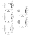


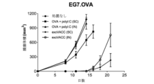


Classifications
-
- A—HUMAN NECESSITIES
- A61—MEDICAL OR VETERINARY SCIENCE; HYGIENE
- A61K—PREPARATIONS FOR MEDICAL, DENTAL OR TOILETRY PURPOSES
- A61K39/00—Medicinal preparations containing antigens or antibodies
- A61K39/12—Viral antigens
- A61K39/215—Coronaviridae, e.g. avian infectious bronchitis virus
-
- A—HUMAN NECESSITIES
- A61—MEDICAL OR VETERINARY SCIENCE; HYGIENE
- A61P—SPECIFIC THERAPEUTIC ACTIVITY OF CHEMICAL COMPOUNDS OR MEDICINAL PREPARATIONS
- A61P31/00—Antiinfectives, i.e. antibiotics, antiseptics, chemotherapeutics
- A61P31/12—Antivirals
- A61P31/14—Antivirals for RNA viruses
-
- A—HUMAN NECESSITIES
- A61—MEDICAL OR VETERINARY SCIENCE; HYGIENE
- A61K—PREPARATIONS FOR MEDICAL, DENTAL OR TOILETRY PURPOSES
- A61K39/00—Medicinal preparations containing antigens or antibodies
- A61K2039/555—Medicinal preparations containing antigens or antibodies characterised by a specific combination antigen/adjuvant
- A61K2039/55511—Organic adjuvants
- A61K2039/55555—Liposomes; Vesicles, e.g. nanoparticles; Spheres, e.g. nanospheres; Polymers
-
- A—HUMAN NECESSITIES
- A61—MEDICAL OR VETERINARY SCIENCE; HYGIENE
- A61K—PREPARATIONS FOR MEDICAL, DENTAL OR TOILETRY PURPOSES
- A61K39/00—Medicinal preparations containing antigens or antibodies
- A61K2039/70—Multivalent vaccine
-
- C—CHEMISTRY; METALLURGY
- C12—BIOCHEMISTRY; BEER; SPIRITS; WINE; VINEGAR; MICROBIOLOGY; ENZYMOLOGY; MUTATION OR GENETIC ENGINEERING
- C12N—MICROORGANISMS OR ENZYMES; COMPOSITIONS THEREOF; PROPAGATING, PRESERVING, OR MAINTAINING MICROORGANISMS; MUTATION OR GENETIC ENGINEERING; CULTURE MEDIA
- C12N2770/00—MICROORGANISMS OR ENZYMES; COMPOSITIONS THEREOF; PROPAGATING, PRESERVING, OR MAINTAINING MICROORGANISMS; MUTATION OR GENETIC ENGINEERING; CULTURE MEDIA ssRNA viruses positive-sense
- C12N2770/00011—Details
- C12N2770/20011—Coronaviridae
- C12N2770/20034—Use of virus or viral component as vaccine, e.g. live-attenuated or inactivated virus, VLP, viral protein
-
- C—CHEMISTRY; METALLURGY
- C12—BIOCHEMISTRY; BEER; SPIRITS; WINE; VINEGAR; MICROBIOLOGY; ENZYMOLOGY; MUTATION OR GENETIC ENGINEERING
- C12N—MICROORGANISMS OR ENZYMES; COMPOSITIONS THEREOF; PROPAGATING, PRESERVING, OR MAINTAINING MICROORGANISMS; MUTATION OR GENETIC ENGINEERING; CULTURE MEDIA
- C12N2770/00—MICROORGANISMS OR ENZYMES; COMPOSITIONS THEREOF; PROPAGATING, PRESERVING, OR MAINTAINING MICROORGANISMS; MUTATION OR GENETIC ENGINEERING; CULTURE MEDIA ssRNA viruses positive-sense
- C12N2770/00011—Details
- C12N2770/20011—Coronaviridae
- C12N2770/20071—Demonstrated in vivo effect
-
- Y—GENERAL TAGGING OF NEW TECHNOLOGICAL DEVELOPMENTS; GENERAL TAGGING OF CROSS-SECTIONAL TECHNOLOGIES SPANNING OVER SEVERAL SECTIONS OF THE IPC; TECHNICAL SUBJECTS COVERED BY FORMER USPC CROSS-REFERENCE ART COLLECTIONS [XRACs] AND DIGESTS
- Y02—TECHNOLOGIES OR APPLICATIONS FOR MITIGATION OR ADAPTATION AGAINST CLIMATE CHANGE
- Y02A—TECHNOLOGIES FOR ADAPTATION TO CLIMATE CHANGE
- Y02A50/00—TECHNOLOGIES FOR ADAPTATION TO CLIMATE CHANGE in human health protection, e.g. against extreme weather
- Y02A50/30—Against vector-borne diseases, e.g. mosquito-borne, fly-borne, tick-borne or waterborne diseases whose impact is exacerbated by climate change
Abstract
本開示は、コロナウイルス(例えば、SARS-CoV-1またはSARS-CoV-2)からの1つ以上の抗原と、任意選択でアジュバントと、を含む細胞外小胞に関する。また、本明細書において、EVを生成する方法、及びEVを使用して疾患または障害、例えば感染性疾患を治療及び/または予防するための方法を提供する。【選択図】なしThe present disclosure relates to extracellular vesicles comprising one or more antigens from a coronavirus (eg SARS-CoV-1 or SARS-CoV-2) and optionally an adjuvant. Also provided herein are methods of generating EVs and methods of using EVs to treat and/or prevent a disease or disorder, such as an infectious disease. [Selection figure] None
Description
関連出願の相互参照
このPCT出願は、2020年3月20日に出願された国際出願第PCT/US2020/024023号、2020年3月27日に出願された米国仮出願第63/001,088号、2020年3月31日に出願された第63/003,171号、2020年4月7日に出願された第63/006,585号、2020年4月29日に出願された第63/017,514号、及び2021年1月27日に出願された第63/142,418号の優先権を主張する。これらのそれぞれは、参照によりその全体が本明細書に組み込まれる。
CROSS-REFERENCE TO RELATED APPLICATIONS This PCT application is based on International Application No. PCT/US2020/024023 filed March 20, 2020, U.S. Provisional Application No. 63/001,088 filed March 27, 2020 , 63/003,171 filed March 31, 2020; 63/006,585 filed April 7, 2020; 63/006,585 filed April 29, 2020; 017,514 and 63/142,418 filed Jan. 27, 2021. each of which is incorporated herein by reference in its entirety.
EFS-WEBを通じて電子的に提出された配列表への言及
本出願中で提出された、電子的に送信された配列表の内容(名称:4000_097PC06_Seqlisting_ST25.txt;サイズ:352,293バイト;作成日:2021年3月22日)は、その全体が参照により本明細書に組み込まれる。
REFERENCE TO A SEQUENCE LISTING SUBMITTED ELECTRONICALLY THROUGH EFS-WEB Contents of the electronically submitted Sequence Listing submitted in this application (Name: 4000_097PC06_Seqlisting_ST25.txt; Size: 352,293 bytes; Date Created: March 22, 2021) is incorporated herein by reference in its entirety.
本開示は、感染性疾患を含むがこれに限定されないさまざまな医学的障害を治療及び/または予防するために使用できるワクチンとして有用である改変された細胞外小胞、例えばエキソソーム(例えば、抗原及びアジュバント/免疫モジュレーターなどの1つ以上のペイロードを含む)に関する。本開示はまた、そのようなEVを製造する方法及びその使用にも関する。 The present disclosure provides modified extracellular vesicles, such as exosomes (e.g., antigens and including one or more payloads such as adjuvants/immune modulators). The present disclosure also relates to methods of manufacturing such EVs and uses thereof.
EVは、細胞間コミュニケーションの重要なメディエーターである。それらは、がんなどの多く疾患の診断と予後における重要なバイオマーカーでもある。薬物送達ビヒクルとして、EVは、多くの治療分野における新規治療法として、従来の薬物送達方法(例えば、ペプチド免疫、DNAワクチン)に比べて多くの利点を提供する。しかしながら、その利点にもかかわらず、多くのEVは臨床効果が限られている。例えば、樹状細胞由来のエキソソーム(DEX)は、手術不能な非小細胞肺癌(NSCLC)患者における一次化学療法後の維持免疫療法として第II相臨床試験で調査された。しかし、主要評価項目(少なくとも50%の患者が化学療法中止後4ヶ月の無増悪生存期間(PFS)を有する)に達しなかったため、試験は中止された。Besse,B.,et al.,Oncoimmunology 5(4):e1071008(2015)。
したがって、EVベースの技術の治療的使用やその他の用途をより有効にするには、新しくより効果的な操作されたEVが必要である。
EVs are important mediators of intercellular communication. They are also important biomarkers in the diagnosis and prognosis of many diseases such as cancer. As drug delivery vehicles, EVs offer many advantages over conventional drug delivery methods (eg, peptide immunization, DNA vaccines) as novel therapeutics in many therapeutic areas. However, despite their advantages, many EVs have limited clinical efficacy. For example, dendritic cell-derived exosomes (DEX) were investigated in phase II clinical trials as maintenance immunotherapy after first-line chemotherapy in patients with inoperable non-small cell lung cancer (NSCLC). However, the trial was stopped because the primary endpoint (at least 50% of patients had progression-free survival (PFS) 4 months after ceasing chemotherapy) was not reached. Besse, B.; , et al. , Oncoimmunology 5(4): e1071008 (2015).
Therefore, new and more efficient engineered EVs are needed to make the therapeutic and other applications of EV-based technology more effective.
コロナウイルスに由来する少なくとも1つの抗原を含む単離された細胞外小胞(EV)が本明細書で提供される。いくつかの態様では、コロナウイルスは重症急性呼吸器症候群(SARS)コロナウイルスである。いくつかの態様では、抗原はユニバーサルSARSコロナウイルス抗原である。 Provided herein is an isolated extracellular vesicle (EV) comprising at least one antigen derived from a coronavirus. In some aspects, the coronavirus is a severe acute respiratory syndrome (SARS) coronavirus. In some aspects, the antigen is the universal SARS coronavirus antigen.
いくつかの態様では、本明細書に開示されるEVは、少なくとも2、3、4、5、6、7、8、9、10またはそれ以上の抗原を含み、第1の抗原は、SARS-CoV-1またはSARS-CoV-2(COVID-19)のウイルスに由来する。いくつかの態様では、第2の抗原がSARS-CoV-1またはSARS-CoV-2(COVID-19)のウイルスに由来する。いくつかの態様では、第2の抗原がSARS-CoV-1またはSARS-CoV-2(COVID-19)のウイルスに由来しない。特定の態様では、第1の抗原と第2の抗原は同じである。いくつかの態様では、第1の抗原と第2の抗原は異なる。 In some aspects, an EV disclosed herein comprises at least 2, 3, 4, 5, 6, 7, 8, 9, 10 or more antigens, wherein the first antigen is SARS- Derived from CoV-1 or SARS-CoV-2 (COVID-19) viruses. In some aspects, the second antigen is derived from the SARS-CoV-1 or SARS-CoV-2 (COVID-19) virus. In some aspects, the second antigen is not derived from the SARS-CoV-1 or SARS-CoV-2 (COVID-19) virus. In certain aspects, the first antigen and the second antigen are the same. In some aspects, the first antigen and the second antigen are different.
いくつかの態様では、本明細書に開示されるEVにおいて発現され得るCOVID-19ウイルスに由来する抗原は、スパイク(S)タンパク質に由来する。いくつかの態様では、抗原は、Sタンパク質の受容体結合ドメイン(RBD)を含む。特定の態様では、抗原は、Sタンパク質の少なくとも5、6、7、8、9、10、11、12、13、14、15、16、17、18、19、または20個のアミノ酸を含む。 In some aspects, antigens from the COVID-19 virus that can be expressed in the EVs disclosed herein are derived from the spike (S) protein. In some aspects, the antigen comprises the receptor binding domain (RBD) of the S protein. In particular aspects, the antigen comprises at least 5, 6, 7, 8, 9, 10, 11, 12, 13, 14, 15, 16, 17, 18, 19, or 20 amino acids of the S protein.
いくつかの態様では、本開示のEVにおいて発現されるCOVID-19ウイルスに由来する抗原は、エンベロープ(E)タンパク質に由来する。特定の態様では、抗原は、Eタンパク質の少なくとも5、6、7、8、9、10、11、12、13、14、15、16、17、18、19、または20個のアミノ酸を含む。 In some aspects, the COVID-19 virus-derived antigen expressed in the EVs of the present disclosure is derived from the envelope (E) protein. In particular aspects, the antigen comprises at least 5, 6, 7, 8, 9, 10, 11, 12, 13, 14, 15, 16, 17, 18, 19, or 20 amino acids of the E protein.
いくつかの態様では、本開示のEVにおいて発現され得るCOVID-19ウイルスに由来する抗原は、膜(M)タンパク質に由来する。特定の態様では、抗原は、Mタンパク質の少なくとも5、6、7、8、9、10、11、12、13、14、15、16、17、18、19、または20個のアミノ酸を含む。 In some aspects, antigens from the COVID-19 virus that can be expressed in the EVs of the present disclosure are derived from membrane (M) proteins. In particular aspects, the antigen comprises at least 5, 6, 7, 8, 9, 10, 11, 12, 13, 14, 15, 16, 17, 18, 19, or 20 amino acids of M protein.
いくつかの態様では、EVは第2の抗原を含み、第2の抗原はCOVID-19ウイルスのスパイク(S)タンパク質に由来する。いくつかの態様では、第2の抗原は、Sタンパク質の受容体結合ドメイン(RBD)を含む。特定の態様では、第2の抗原は、Sタンパク質の少なくとも5、6、7、8、9、10、11、12、13、14、15、16、17、18、19、または20個のアミノ酸を含む。 In some aspects, the EV comprises a second antigen, the second antigen being derived from the spike (S) protein of the COVID-19 virus. In some aspects, the second antigen comprises the receptor binding domain (RBD) of the S protein. In particular aspects, the second antigen comprises at least 5, 6, 7, 8, 9, 10, 11, 12, 13, 14, 15, 16, 17, 18, 19, or 20 amino acids of the S protein. including.
いくつかの態様では、EVは第2の抗原を含み、第2の抗原はCOVID-19ウイルスのエンベロープ(E)タンパク質に由来する。特定の態様では、第2の抗原は、Eタンパク質の少なくとも5、6、7、8、9、10、11、12、13、14、15、16、17、18、19、または20個のアミノ酸を含む。 In some aspects, the EV comprises a second antigen, wherein the second antigen is derived from the envelope (E) protein of the COVID-19 virus. In particular aspects, the second antigen comprises at least 5, 6, 7, 8, 9, 10, 11, 12, 13, 14, 15, 16, 17, 18, 19, or 20 amino acids of the E protein. including.
いくつかの態様では、EVは第2の抗原を含み、COVID-19ウイルスに由来する第2の抗原は膜(M)タンパク質に由来する。特定の態様では、第2の抗原は、Mタンパク質の少なくとも5、6、7、8、9、10、11、12、13、14、15、16、17、18、19、または20個のアミノ酸を含む。 In some aspects, the EV comprises a second antigen, and the second antigen from the COVID-19 virus is from the membrane (M) protein. In particular aspects, the second antigen comprises at least 5, 6, 7, 8, 9, 10, 11, 12, 13, 14, 15, 16, 17, 18, 19, or 20 amino acids of M protein. including.
いくつかの態様では、本開示のEVは、第1の抗原及び第2の抗原を含み、第1の抗原はSタンパク質の受容体結合ドメイン(RBD)を含み、第2の抗原はT抗原を含む。 In some aspects, an EV of the present disclosure comprises a first antigen and a second antigen, the first antigen comprising a receptor binding domain (RBD) of an S protein and the second antigen comprising a T antigen. include.
いくつかの態様では、本開示のEVは、少なくとも1つのアジュバントをさらに含む。 In some aspects, the EVs of this disclosure further comprise at least one adjuvant.
いくつかの態様では、本明細書に開示されるEVは、細胞性免疫応答、体液性免疫応答、または細胞性免疫応答と体液性免疫応答の両方を誘導する。特定の態様では、細胞性免疫応答、体液性免疫応答、または細胞性免疫応答と体液性免疫応答の両方の誘導が、(i)アジュバントもしくは抗原を含まない対応するEV、または(ii)EVを伴わないアジュバントもしくは抗原と比較して、少なくとも約5%、少なくとも約10%、少なくとも約20%、少なくとも約30%、少なくとも約40%、少なくとも約50%、少なくとも約60%、少なくとも約70%、少なくとも約80%、少なくとも約90%、少なくとも約100%、または以上増加する。 In some aspects, the EVs disclosed herein induce a cell-mediated immune response, a humoral immune response, or both a cell-mediated and humoral immune response. In certain aspects, the induction of a cell-mediated immune response, a humoral immune response, or both a cell-mediated and humoral immune response is achieved by (i) corresponding EVs without adjuvant or antigen, or (ii) EVs. at least about 5%, at least about 10%, at least about 20%, at least about 30%, at least about 40%, at least about 50%, at least about 60%, at least about 70%, compared to adjuvant or antigen without; Increase by at least about 80%, at least about 90%, at least about 100%, or more.
いくつかの態様では、本明細書に開示されるEVは、CD4+T細胞応答、CD8+T細胞応答、またはCD4+T細胞応答とCD8+T細胞応答の両方を誘導する。いくつかの態様では、本明細書に開示されるEVは、CD8+T細胞応答を誘導する。いくつかの態様では、EVは、組織常在性メモリーT細胞応答を拡大する。 In some aspects, the EVs disclosed herein induce a CD4+ T cell response, a CD8+ T cell response, or both a CD4+ T cell response and a CD8+ T cell response. In some aspects, the EVs disclosed herein induce a CD8+ T cell response. In some aspects, the EV expands tissue-resident memory T cell responses.
いくつかの態様では、本開示のEVは、第1の足場部分をさらに含む。特定の態様では、第1の抗原は、第1の足場部分に連結されている。いくつかの態様では、第2の抗原は、第1の足場部分に連結されている。いくつかの態様では、本明細書に開示される第1の足場部分を含むEVは、第2の足場部分をさらに含む。特定の態様では、第1の抗原は、第1の足場部分に連結され、第2の抗原は第2の足場部分に連結されている。いくつかの態様では、第1の足場部分と第2の足場部分は同じである。いくつかの態様では、第1の足場部分と第2の足場部分は異なっている。 In some aspects, an EV of the present disclosure further comprises a first scaffolding portion. In certain aspects, the first antigen is linked to the first scaffold portion. In some aspects, the second antigen is linked to the first scaffold portion. In some aspects, an EV comprising a first scaffolding portion disclosed herein further comprises a second scaffolding portion. In certain aspects, a first antigen is linked to a first scaffold portion and a second antigen is linked to a second scaffold portion. In some aspects, the first scaffold portion and the second scaffold portion are the same. In some aspects, the first scaffold portion and the second scaffold portion are different.
いくつかの態様では、第1の足場部分は足場Xである。いくつかの態様では、第1の足場部分は足場Yである。いくつかの態様では、第2の足場部分は足場Yである。いくつかの態様では、第2の足場部分は足場Xである。 In some aspects, the first scaffold portion is scaffold-X. In some aspects, the first scaffold portion is scaffold Y. In some aspects, the second scaffold portion is scaffold Y. In some aspects, the second scaffold portion is scaffold-X.
いくつかの態様では、本明細書に開示されるEVにおいて発現され得る足場Xは、(i)EVの内腔表面上に第1の抗原を固定すること、(ii)EVの外側表面上に第1の抗原を固定すること、(iii)EVの内腔表面上に第2の抗原を固定すること、(iv)EVの外側表面上に第2の抗原を固定すること、または(v)それらの組み合わせを可能とする。いくつかの態様では、足場Xは、プロスタグランジンF2受容体ネガティブレギュレーター(PTGFRNタンパク質);ベイシジン(BSGタンパク質);免疫グロブリンスーパーファミリーメンバー2(IGSF2タンパク質);免疫グロブリンスーパーファミリーメンバー3(IGSF3タンパク質);免疫グロブリンスーパーファミリーメンバー8(IGSF8タンパク質);インテグリンβ1(ITGB1タンパク質);インテグリンα4(ITGA4タンパク質);4F2細胞表面抗原重鎖(SLC3A2タンパク質);及びATPトランスポータータンパク質(ATP1A1、ATP1A2、ATP1A3、ATP1A4、ATP1B3、ATP2B1、ATP2B2、ATP2B3、ATP2B4タンパク質)のうちの一クラス;ならびにそれらの任意の組み合わせからなる群から選択される。 In some aspects, the scaffold X that can be expressed in the EV disclosed herein (i) anchors the first antigen on the luminal surface of the EV, (ii) on the lateral surface of the EV (iii) immobilizing a second antigen on the luminal surface of the EV; (iv) immobilizing the second antigen on the outer surface of the EV; or (v) allow a combination of them. In some aspects, scaffold X is a prostaglandin F2 receptor negative regulator (PTGFRN protein); Basidin (BSG protein); immunoglobulin superfamily member 2 (IGSF2 protein); immunoglobulin superfamily member 3 (IGSF3 protein) integrin β1 (ITGB1 protein); integrin α4 (ITGA4 protein); 4F2 cell surface antigen heavy chain (SLC3A2 protein); and ATP transporter proteins (ATP1A1, ATP1A2, ATP1A3, ATP1A4 , ATP1B3, ATP2B1, ATP2B2, ATP2B3, ATP2B4 proteins); and any combination thereof.
いくつかの態様では、本明細書に開示されるEVにおいて発現され得る足場Yは、(i)EVの内腔表面上に第1の抗原を固定すること、(ii)EVの内腔表面上に第2の抗原を固定すること、または(iii)それらの両方が可能である。特定の態様では、足場Yは、ミリストイル化アラニンリッチプロテインキナーゼC基質(MARCKSタンパク質);ミリストイル化アラニンリッチプロテインキナーゼC基質様1(MARCKSL1タンパク質);脳酸可溶性タンパク質1(BASP1タンパク質)、及びそれらの任意の組み合わせからなる群から選択される。 In some aspects, the scaffold Y that can be expressed in the EV disclosed herein comprises: (i) anchoring the first antigen on the luminal surface of the EV; or (iii) both. In particular aspects, scaffold Y is a myristoylated alanine-rich protein kinase C substrate (MARCKS protein); a myristoylated alanine-rich protein kinase C substrate-like 1 (MARCKSL1 protein); a brain acid soluble protein 1 (BASP1 protein); Selected from the group consisting of any combination.
いくつかの態様では、第1の抗原は、EVの内腔表面上の第1の足場部分に連結され、第2の抗原は、EVの内腔表面上の第2の足場部分に連結されている。特定の態様では、a)第1の足場部分及び第2の足場部分のそれぞれが足場Yであり;b)第1の足場部分は足場Yであり、第2の足場部分は足場Xであり;c)第1の足場部分は足場Xであり、第2の足場部分は足場Yであり;またはd)第1の足場部分及び第2の足場部分のそれぞれが足場Xである。 In some aspects, the first antigen is linked to a first scaffolding portion on the luminal surface of the EV and the second antigen is linked to a second scaffolding portion on the luminal surface of the EV. there is In certain embodiments, a) each of the first scaffold portion and the second scaffold portion is scaffold Y; b) the first scaffold portion is scaffold Y and the second scaffold portion is scaffold X; c) the first scaffold portion is scaffold X and the second scaffold portion is scaffold Y; or d) each of the first scaffold portion and the second scaffold portion is scaffold X.
いくつかの態様では、第1の抗原は、EVの内腔表面上の第1の足場部分に連結され、第2の抗原は、EVの内腔に存在する。いくつかの態様では、第1の抗原は、EVの内腔に存在し、第2の抗原は、EVの内腔表面上の第1の足場部分に連結されている。いくつかの態様では、第1の抗原は、EVの内腔表面上の第1の足場部分に連結され、第2の抗原は、EVの外側表面上の第2の足場部分に連結されている。いくつかの態様では、第1の足場部分は足場Yであり、第2の足場部分は足場Xであるか、またはb)第1の足場部分及び第2の足場部分のそれぞれが足場Xである。 In some aspects, the first antigen is linked to a first scaffolding portion on the luminal surface of the EV and the second antigen is present in the lumen of the EV. In some aspects, the first antigen is present in the lumen of the EV and the second antigen is linked to a first scaffolding moiety on the luminal surface of the EV. In some aspects, the first antigen is linked to a first scaffolding portion on the luminal surface of the EV and the second antigen is linked to a second scaffolding portion on the lateral surface of the EV. . In some embodiments, the first scaffold portion is scaffold Y and the second scaffold portion is scaffold X, or b) each of the first scaffold portion and the second scaffold portion is scaffold X. .
いくつかの態様では、第1の抗原は、EVの外側表面上の第1の足場部分に連結され、第2の抗原は、EVの内腔表面上の第2の足場部分に連結されている。特定の態様では、a)第1の足場部分は足場Xであり、第2の足場部分は足場Yであるか、またはb)第1の足場部分及び第2の足場部分のそれぞれが足場Xである。 In some aspects, the first antigen is linked to a first scaffolding portion on the outer surface of the EV and the second antigen is linked to a second scaffolding portion on the luminal surface of the EV. . In certain embodiments, a) the first scaffold portion is scaffold X and the second scaffold portion is scaffold Y, or b) each of the first scaffold portion and the second scaffold portion is scaffold X. be.
いくつかの態様では、第1の抗原は、EVの内腔に存在し、第2の抗原は、EVの内腔に存在する。 In some aspects, the first antigen is present in the lumen of the EV and the second antigen is present in the lumen of the EV.
いくつかの態様では、抗原は、EVの外側表面上の第1の足場部分に連結され、アジュバントは、EVの外側表面上の第2の足場部分に連結されている。そのような態様のいくつかでは、第1の足場部分と第2の足場部分は足場Xである。 In some aspects, the antigen is linked to a first scaffolding portion on the outer surface of the EV and the adjuvant is linked to a second scaffolding portion on the outer surface of the EV. In some such aspects, the first scaffolding portion and the second scaffolding portion are scaffold-X.
いくつかの態様では、第1の抗原は、EVの外側表面上の第1の足場部分に連結され、第2の抗原は、EVの内腔に存在する。そのような態様のいくつかでは、第1の足場部分は足場Xである。 In some aspects, the first antigen is linked to a first scaffolding portion on the outer surface of the EV and the second antigen is present in the lumen of the EV. In some such aspects, the first scaffold portion is scaffold-X.
いくつかの態様では、第1の抗原は、EVの内腔に存在し、第2の抗原は、EVの外側表面上の第1の足場部分に連結されている。そのような態様のいくつかでは、第1の足場部分は足場Xである。 In some aspects, the first antigen is present in the lumen of the EV and the second antigen is linked to the first scaffolding portion on the outer surface of the EV. In some such aspects, the first scaffold portion is scaffold-X.
いくつかの態様では、第1の抗原は、EVの表面上の第1の足場部分に連結され、第2の抗原は、EVの内腔表面上の第1の足場部分に連結されている。いくつかの態様では、第1の抗原は、EVの内腔表面上の第1の足場部分に連結され、第2の抗原は、EVの外側表面上の第1の足場部分に連結されている。そのような態様のいくつかでは、第1の足場部分は足場Xである。 In some aspects, the first antigen is linked to a first scaffolding portion on the surface of the EV and the second antigen is linked to the first scaffolding portion on the luminal surface of the EV. In some aspects, the first antigen is linked to a first scaffolding portion on the luminal surface of the EV and the second antigen is linked to the first scaffolding portion on the lateral surface of the EV. . In some such aspects, the first scaffold portion is scaffold-X.
いくつかの態様では、本明細書に開示されるEVは、第2の抗原を含み、(i)第2の抗原は、リンカー、親和性リガンド、またはその両方によって第1の足場部分に連結されるか、(ii)第2の抗原は、リンカー、親和性リガンド、またはその両方によって第2の足場に連結されるか、(iii)第2の抗原は、リンカー、親和性リガンド、またはその両方によって第1の足場部分に連結されるか、(iv)第2の抗原は、リンカー、親和性リガンド、またはその両方によって第2の足場部分に連結されるか、または(v)それらの組み合わせである。特定の態様では、リンカー及び/または親和性リガンドはポリペプチドである。いくつかの態様では、リンカーは非ポリペプチド部分である。 In some aspects, the EVs disclosed herein comprise a second antigen, wherein (i) the second antigen is linked to the first scaffold portion by a linker, an affinity ligand, or both or (ii) the second antigen is linked to the second scaffold by a linker, an affinity ligand, or both, or (iii) the second antigen is a linker, an affinity ligand, or both (iv) the second antigen is linked to the second scaffold portion by a linker, an affinity ligand, or both, or (v) a combination thereof be. In certain aspects, the linker and/or affinity ligand is a polypeptide. In some aspects, the linker is a non-polypeptide moiety.
いくつかの態様では、本明細書に開示されるEVの第1の足場部分または第2の足場部分は、PTGFRNタンパク質である。いくつかの態様では、第1の足場部分または第2の足場部分は、配列番号33に示されるアミノ酸配列を含む。いくつかの態様では、第1の足場部分または第2の足場部分は、配列番号1に対して、少なくとも50%、少なくとも60%、少なくとも70%、少なくとも80%、少なくとも85%、少なくとも90%、少なくとも95%、少なくとも96%、少なくとも97%、少なくとも98%、少なくとも99%、または約100%同一のアミノ酸配列を含む。 In some aspects, the EV first scaffolding portion or second scaffolding portion disclosed herein is a PTGFRN protein. In some aspects, the first scaffold portion or the second scaffold portion comprises the amino acid sequence set forth in SEQ ID NO:33. In some aspects, the first scaffolding portion or the second scaffolding portion is at least 50%, at least 60%, at least 70%, at least 80%, at least 85%, at least 90%, Contain amino acid sequences that are at least 95%, at least 96%, at least 97%, at least 98%, at least 99%, or about 100% identical.
いくつかの態様では、本明細書に開示されるEVの第1の足場部分または第2の足場部分は、BASP1タンパク質である。さらなる態様では、第1の足場部分または第2の足場部分は、(G)(π)(X)(Φ/π)(π)(+)(+)のペプチドを含み、それぞれの括弧内の位置はアミノ酸を表し、πはPro、Gly、Ala、及びSerからなる群から選択される任意のアミノ酸であり、Xは、任意のアミノ酸であり、Φは、Val、Ile、Leu、Phe、Trp、Tyr、及びMetからなる群から選択される任意のアミノ酸であり、(+)は、Lys、Arg、及びHisからなる群から選択される任意のアミノ酸であり;5位は(+)ではなく、6位は(+)でも(AspまたはGlu)でもない。いくつかの態様では、第1の足場部分または第2の足場部分は、配列番号50~155のうちのいずれか1つに示されるアミノ酸配列を含む。いくつかの態様では、第1の足場部分または第2の足場部分は、配列番号3に対して、少なくとも50%、少なくとも60%、少なくとも70%、少なくとも80%、少なくとも85%、少なくとも90%、少なくとも95%、少なくとも96%、少なくとも97%、少なくとも98%、少なくとも99%、または約100%同一のアミノ酸配列を含む。
In some aspects, the EV first scaffolding portion or second scaffolding portion disclosed herein is a BASP1 protein. In a further aspect, the first scaffolding portion or the second scaffolding portion comprises a peptide of (G)(π)(X)(Φ/π)(π)(+)(+) and within each parenthesis The positions represent amino acids, π is any amino acid selected from the group consisting of Pro, Gly, Ala, and Ser, X is any amino acid, Φ is Val, Ile, Leu, Phe, Trp , Tyr, and Met; (+) is any amino acid selected from the group consisting of Lys, Arg, and His;
また、本明細書では、(i)SARS-CoV-1またはSARS-CoV-2(COVID-19)のウイルスに由来する第1の抗原と、(ii)SARS-CoV-1またはSARS-CoV-2(COVID-19)のウイルスに由来する第2の抗原と、を含む単離された細胞外小胞が提供され、(a)第1の抗原は内腔表面上の第1の足場Yに連結され、第2の抗原はEVの内腔表面上の第2の足場Yに連結されているか;(b)第1の抗原は内腔表面上の足場Yに連結され、第2の抗原はEVの内腔に存在するか;(c)第1の抗原はEVの内腔に存在し、第2の抗原はEVの内腔表面上の足場Yに連結されているか;(d)第1の抗原はEVの内腔表面上の足場Yに連結され、第2の抗原はEVの外側表面上の足場Xに連結されているか;(e)第1の抗原はEVの内腔に存在し、第2の抗原はEVの外側表面上の足場Xに連結されているか;(f)第1の抗原はEVの内腔表面上の足場Yに連結され、第2の抗原はEVの内腔表面上の足場Xに連結されているか;(g)第1の抗原はEVの内腔に存在し、第2の抗原はEVの内腔表面上の足場Xに連結されているか;(h)第1の抗原はEVの内腔表面上の足場Xに連結され、第2の抗原はEVの外側表面上の足場Xに連結されているか;(i)第1の抗原はEVの外側表面上の第1の足場Xに連結され、第2の抗原はEVの外側表面上の第2の足場Xに連結されているか;(j)第1の抗原はEVの外側表面上の足場Xに連結され、第2の抗原はEVの内腔表面上の足場Yに連結されているか;(k)第1の抗原はEVの外側表面上の足場Xに連結され、第2の抗原はEVの内腔に存在するか;(l)第1の抗原はEVの外側表面上の足場Xに連結され、第2の抗原はEVの内腔表面上の足場Xに連結されているか;(m)第1の抗原はEVの内腔表面上の第1の足場Xに連結され、第2の抗原はEVの内腔表面上の第2の足場Xに連結されているか;(n)第1の抗原はEVの内腔表面上の足場Xに連結され、第2の抗原はEVの内腔表面上の足場Yに連結されているか;(o)第1の抗原はEVの内腔表面上の足場Xに連結され、第2の抗原はEVの内腔に存在するか;(p)第1の抗原はEVの外側表面上の第1の足場Xに連結され、第2の抗原はEVの内腔表面上の第2の足場Xに連結されているか;(q)第1の抗原はEVの内腔表面上の第1の足場Xに連結され、第2の抗原はEVの外側表面上の第2の足場Xに連結されているか;(r)第1の抗原はEVの内腔に存在し、第2の抗原はEVの内腔に存在するか;(s)第1の抗原はEVの内腔表面に直接連結され、第2の抗原はEVの内腔表面に直接連結されているか;(t)第1の抗原はEVの内腔表面に直接連結され、第2の抗原はEVの内腔に存在するか;(u)第1の抗原はEVの内腔表面に直接連結され、第2の抗原はEVの内腔表面上の足場Yに連結されているか;(v)第1の抗原はEVの内腔表面に直接連結され、第2の抗原はEVの内腔表面上の足場Xに連結されているか;(w)第1の抗原はEVの内腔表面に直接連結され、第2の抗原はEVの外側表面に直接連結されているか;(x)第1の抗原はEVの内腔表面に直接連結され、第2の抗原はEVの外側表面上の足場Xに連結されているか;(y)第1の抗原はEVの内腔表面上の足場Yに連結され、第2の抗原はEVの内腔表面に直接連結されているか;(z)第1の抗原はEVの内腔表面上の足場Yに連結され、第2の抗原はEVの外側表面に直接連結されているか;(aa)第1の抗原はEVの内腔表面上の足場Xに連結され、第2の抗原はEVの内腔表面に直接連結されているか;(bb)第1の抗原はEVの内腔表面上の足場Xに連結され、第2の抗原はEVの外側表面に直接連結されているか;(cc)第1の抗原はEVの内腔に存在し、第2の抗原はEVの内腔表面に直接連結されているか、または(dd)第1の抗原はEVの内腔に存在し、第2の抗原はEVの外側に直接連結されている。 Also herein, (i) a first antigen derived from a SARS-CoV-1 or SARS-CoV-2 (COVID-19) virus; and (ii) SARS-CoV-1 or SARS-CoV- 2 (COVID-19) virus-derived second antigen, wherein (a) the first antigen is attached to a first scaffold Y on the luminal surface; (b) the first antigen is linked to scaffold Y on the luminal surface, and the second antigen is linked to scaffold Y on the luminal surface of the EV; (c) the first antigen is present in the lumen of the EV and the second antigen is linked to scaffold Y on the luminal surface of the EV; (d) the first is linked to scaffold Y on the luminal surface of the EV and a second antigen is linked to scaffold X on the outer surface of the EV; (e) the first antigen is present in the lumen of the EV and , the second antigen is linked to scaffold X on the outer surface of the EV; (f) the first antigen is linked to scaffold Y on the luminal surface of the EV and the second antigen is linked to the lumen of the EV (g) the first antigen is present in the lumen of the EV and the second antigen is linked to the scaffold X on the luminal surface of the EV; (h) Is the first antigen linked to scaffold X on the luminal surface of the EV and the second antigen linked to scaffold X on the outer surface of the EV; (i) the first antigen is on the outer surface of the EV; and the second antigen is linked to the second scaffold X on the outer surface of the EV; (j) the first antigen is linked to the scaffold X on the outer surface of the EV (k) the first antigen is linked to scaffold X on the lateral surface of the EV and the second antigen is linked to scaffold Y on the luminal surface of the EV; (l) the first antigen is linked to scaffold X on the outer surface of the EV and the second antigen is linked to scaffold X on the luminal surface of the EV; (n) the first antigen is linked to a first scaffold X on the luminal surface of the EV and the second antigen is linked to a second scaffold X on the luminal surface of the EV; is linked to scaffold X on the luminal surface of the EV and the second antigen is linked to scaffold Y on the luminal surface of the EV; (o) the first antigen is linked to the scaffold Y on the luminal surface of the EV; (p) the first antigen is linked to the first scaffold X on the outer surface of the EV and the second antigen is present in the lumen of the EV; (q) the first antigen is linked to the first scaffold X on the luminal surface of the EV and the second antigen is linked to the outer surface of the EV; (r) the first antigen is present in the lumen of the EV and the second antigen is present in the lumen of the EV; (s) the first antigen is present in the EV and the second antigen is directly linked to the luminal surface of the EV; (t) the first antigen is directly linked to the luminal surface of the EV and the second antigen is the luminal surface of the EV (u) the first antigen is directly linked to the luminal surface of the EV and the second antigen is linked to a scaffold Y on the luminal surface of the EV; (v) the one antigen is directly linked to the luminal surface of the EV and the second antigen is linked to a scaffold X on the luminal surface of the EV; (w) the first antigen is directly linked to the luminal surface of the EV and the second antigen is directly linked to the outer surface of the EV; (x) the first antigen is directly linked to the luminal surface of the EV and the second antigen is linked to the scaffold X on the outer surface of the EV; (y) the first antigen is linked to scaffold Y on the luminal surface of the EV and the second antigen is directly linked to the luminal surface of the EV; (z) the first antigen is linked to scaffold Y on the luminal surface of the EV and the second antigen is directly linked to the outer surface of the EV; (aa) the first antigen is linked to scaffold X on the luminal surface of the EV , the second antigen is directly linked to the luminal surface of the EV; (bb) the first antigen is linked to a scaffold X on the luminal surface of the EV and the second antigen is directly linked to the lateral surface of the EV (cc) the first antigen is present in the lumen of the EV and the second antigen is directly linked to the luminal surface of the EV; or (dd) the first antigen is in the interior of the EV. Located in the lumen, a second antigen is directly linked to the outside of the EV.
いくつかの態様では、本明細書に開示されるEVは、免疫モジュレーターをさらに含む。特定の態様では、免疫モジュレーターは、EVの内腔表面または外側表面に直接連結されている。いくつかの態様では、免疫モジュレーターは、EVの外側表面上またはEVの内腔表面上の足場Xに連結されている。いくつかの態様では、免疫モジュレーターは、EVの内腔表面上の足場Yに連結されている。 In some aspects, the EVs disclosed herein further comprise an immune modulator. In certain aspects, the immunomodulator is directly linked to the luminal or lateral surface of the EV. In some aspects, the immunomodulator is linked to a scaffold X on the outer surface of the EV or on the luminal surface of the EV. In some aspects, the immunomodulator is linked to scaffold Y on the luminal surface of the EV.
いくつかの態様では、本明細書に開示されるEVはアジュバントを含み、アジュバントはEVの内腔表面または外側表面に直接連結されている。特定の態様では、アジュバントは、EVの外側表面上の足場Xに連結されているか、またはEVの内腔に存在する。いくつかの態様では、アジュバントは、EVの内腔表面上の足場Yに連結されている。いくつかの態様では、アジュバントはEVの内腔に存在する。 In some aspects, the EVs disclosed herein comprise an adjuvant, and the adjuvant is directly linked to the luminal or lateral surface of the EV. In certain aspects, the adjuvant is linked to scaffold X on the outer surface of the EV or is present in the lumen of the EV. In some aspects, the adjuvant is linked to a scaffold Y on the luminal surface of the EV. In some aspects, the adjuvant is present in the lumen of the EV.
いくつかの態様では、アジュバントは、インターフェロン遺伝子刺激因子(STING)アゴニスト、トール様受容体(TLR)アゴニスト、炎症メディエーター、RIG-Iアゴニスト、α-gal-cer(NKTアゴニスト)、熱ショックタンパク質(例えば、HSP65及びHSP70)、C型レクチンアゴニスト(例えば、ベータグルカン(デクチン1)、キチン、及びカードラン)、またはそれらの任意の組み合わせである。 In some aspects, the adjuvant is an interferon gene stimulating factor (STING) agonist, a Toll-like receptor (TLR) agonist, an inflammatory mediator, a RIG-I agonist, an α-gal-cer (NKT agonist), a heat shock protein (e.g. , HSP65 and HSP70), C-type lectin agonists (eg, beta-glucan (Dectin 1), chitin, and curdlan), or any combination thereof.
いくつかの態様では、アジュバントはSTINGアゴニストである。いくつかの態様では、STINGアゴニストは、環状ジヌクレオチドのSTINGアゴニストまたは非環状ジヌクレオチドのSTINGアゴニストを含む。 In some aspects, the adjuvant is a STING agonist. In some aspects, the STING agonist comprises a cyclic dinucleotide STING agonist or an acyclic dinucleotide STING agonist.
いくつかの態様では、アジュバントはTLRアゴニストである。いくつかの態様では、TLRアゴニストは、TLR2アゴニスト(例えば、リポテイコ酸、非定型LPS、MALP-2及びMALP-404、OspA、ポリン、LcrV、リポマンナン、GPIアンカー、リゾホスファチジルセリン、リポホスホグリカン(LPG)、グリコホスファチジルイノシトール(GPI)、ザイモサン、hsp60、gH/gL糖タンパク質、ヘマグルチニン)、TLR3アゴニスト(例えば、二本鎖RNA、例えば、ポリ(I:C))、TLR4アゴニスト(例えば、リポ多糖類(LPS)、リポテイコ酸、β-デフェンシン2、フィブロネクチンEDA、HMGB1、スナピン、テネイシンC)、TLR5アゴニスト(例えば、フラジェリン)、TLR6アゴニスト、TLR7/8アゴニスト(例えば、一本鎖RNA、CpG-A、ポリG10、ポリG3、レシキモド)、TLR9アゴニスト(例えば、メチル化されていないCpG DNA)、またはそれらの任意の組み合わせを含む。
In some aspects, the adjuvant is a TLR agonist. In some aspects, the TLR agonist is a TLR2 agonist (e.g., lipoteichoic acid, atypical LPS, MALP-2 and MALP-404, OspA, porin, LcrV, lipomannan, GPI anchor, lysophosphatidylserine, lipophosphoglycan ( LPG), glycophosphatidylinositol (GPI), zymosan, hsp60, gH/gL glycoprotein, hemagglutinin), TLR3 agonists (e.g. double-stranded RNA, e.g. poly(I:C)), TLR4 agonists (e.g. sugars (LPS), lipoteichoic acid, β-
いくつかの態様では、本明細書に開示されるEVはエキソソームである。 In some aspects, the EVs disclosed herein are exosomes.
いくつかの態様では、本開示のEVは、標的化部分をさらに含む。いくつかの態様では、標的化部分は、樹状細胞のマーカーに特異的に結合する。特定の態様では、マーカーは樹状細胞のみに存在する。いくつかの態様では、樹状細胞は、形質細胞様樹状細胞(pDC)、骨髄/通常型樹状細胞1(cDC1)、骨髄/通常型樹状細胞2(cDC2)、炎症性単球由来樹状細胞、ランゲルハンス細胞、真皮樹状細胞、リゾチーム発現樹状細胞(LysoDC)、クッパー細胞、またはそれらの任意の組み合わせを含む。いくつかの態様では、樹状細胞はcDC1である。いくつかの態様では、マーカーは、C型レクチンドメインファミリー9メンバーA(Clec9a)タンパク質、樹状細胞特異的細胞間接着分子-3-グラビング非インテグリン(DC-SIGN)、CD207、CD40、Clec6、樹状細胞免疫受容体(DCIR)、DEC-205、レクチン様酸化型低密度リポタンパク質受容体-1(LOX-1)、MARCO、Clec12a、Clec10a、DC-アシアロ糖タンパク質受容体(DC-ASGPR)、DC免疫受容体2(DCIR2)、デクチン-1、マクロファージマンノース受容体(MMR)、BDCA-1(CD303、Clec4c)、デクチン-2、Bst-2(CD317)、ランゲリン、CD206、CD11b、CD11c、CD123、CD304、XCR1、AXL、シグレック6、CD209、SIRPA、CX3CR1、GPR182、CD14、CD16、CD32、CD34、CD38、CD10、またはそれらの任意の組み合わせを含む。特定の態様では、マーカーはClec9aタンパク質である。
In some aspects, the EVs of this disclosure further comprise a targeting moiety. In some embodiments, the targeting moiety specifically binds to a dendritic cell marker. In certain aspects, the marker is present only on dendritic cells. In some aspects, the dendritic cell is plasmacytoid dendritic cell (pDC), myeloid/conventional dendritic cell 1 (cDC1), myeloid/conventional dendritic cell 2 (cDC2), inflammatory monocyte-derived Dendritic cells, Langerhans cells, dermal dendritic cells, lysozyme-expressing dendritic cells (LysoDC), Kupffer cells, or any combination thereof. In some aspects, the dendritic cell is cDC1. In some aspects, the marker is C-type
いくつかの態様では、標的化部分は、T細胞のマーカーに特異的に結合する。特定の態様では、マーカーはCD3分子を含む。 In some aspects, the targeting moiety specifically binds to a marker for T cells. In certain aspects, the marker comprises a CD3 molecule.
いくつかの態様では、標的化部分は、EVの外側表面に直接連結されている。特定の態様では、標的化部分は、EVの外側表面上の足場Xに連結されている。いくつかの態様では、標的化部分は、リンカー、親和性リガンド、またはその両方によってEVの外側表面に直接連結されている。いくつかの態様では、標的化部分は、リンカーによって足場Xに連結されている。特定の態様では、リンカー及び/または親和性リガンドはポリペプチドである。 In some aspects, the targeting moiety is directly linked to the outer surface of the EV. In certain aspects, the targeting moiety is linked to a scaffold X on the outer surface of the EV. In some aspects, the targeting moiety is directly linked to the outer surface of the EV by a linker, affinity ligand, or both. In some aspects, the targeting moiety is linked to scaffold X by a linker. In certain aspects, the linker and/or affinity ligand is a polypeptide.
本明細書で提供されるのは、本明細書に記載のEVと、薬学的に許容される担体と、を含む、医薬組成物である。 Provided herein are pharmaceutical compositions comprising an EV as described herein and a pharmaceutically acceptable carrier.
本明細書で提供されるのは、本開示のEVを産生する細胞である。本開示は、1つ以上のベクターを含む細胞をさらに提供し、ベクターは、(i)抗原(例えば、本明細書に記載のもの)、(ii)アジュバント(例えば、本明細書に記載のもの)、(iii)免疫モジュレーター、(iv)標的化部分(例えば、本明細書に記載のもの)、または(v)それらの組み合わせをコードする核酸配列を含む。 Provided herein are cells that produce the EVs of the present disclosure. The disclosure further provides cells comprising one or more vectors, wherein the vectors are: (i) an antigen (e.g., as described herein); (ii) an adjuvant (e.g., as described herein); ), (iii) an immune modulator, (iv) a targeting moiety (eg, as described herein), or (v) a combination thereof.
本明細書に記載のEV及び使用説明書を含むキットが本明細書に提供される。また、本明細書に記載のEVのいずれかを含むEV-薬物コンジュゲートも本明細書に提供される。 Kits are provided herein that include the EVs described herein and instructions for use. Also provided herein are EV-drug conjugates comprising any of the EVs described herein.
本明細書で提供されるのは、本明細書で開示される細胞を適切な条件下で培養することと、EVを得ることと、を含む、EVを作製する方法である。 Provided herein are methods of making EVs, comprising culturing the cells disclosed herein under suitable conditions and obtaining EVs.
本明細書で提供されるのは、本明細書に開示されるEVのいずれかを対象に投与することを含む、それを必要とする対象において免疫応答を誘導する方法である。 Provided herein are methods of inducing an immune response in a subject in need thereof comprising administering to the subject any of the EVs disclosed herein.
本明細書に提供されるのは、疾患を予防または治療することをそれを必要とする対象において行う方法であって、本明細書に開示されるEVのいずれかを対象に投与することを含み、疾患が抗原に関連する方法である。特定の態様では、疾患は感染症である。 Provided herein are methods of preventing or treating a disease in a subject in need thereof comprising administering to the subject any of the EVs disclosed herein. , is how disease is associated with antigens. In certain aspects, the disease is an infectious disease.
いくつかの態様では、本明細書に開示されるEVは、非経口、経口、静脈内、筋肉内、鼻腔内、皮下、または腹腔内に投与される。 In some aspects, the EVs disclosed herein are administered parenterally, orally, intravenously, intramuscularly, intranasally, subcutaneously, or intraperitoneally.
いくつかの態様では、本明細書に開示される方法(例えば、免疫応答を誘導する、または疾患を予防もしくは治療する)は、対象に追加の治療剤を投与することを含む。 In some aspects, a method disclosed herein (eg, inducing an immune response or preventing or treating a disease) comprises administering an additional therapeutic agent to the subject.
いくつかの態様では、本開示は、(i)アジュバント及び抗原を含む細胞外小胞を含むプライミング投与を対象に投与することと、(ii)抗原を含む細胞外小胞を含むブースティング投与を対象に投与することと、を含む、それを必要とする対象にワクチン接種する方法に関する。 In some aspects, the disclosure provides (i) administering to a subject a priming dose comprising extracellular vesicles comprising an adjuvant and an antigen; and (ii) a boosting dose comprising extracellular vesicles comprising an antigen. administering to a subject, and a method of vaccinating a subject in need thereof.
いくつかの態様では、抗原はコロナウイルスに由来する。 In some aspects, the antigen is derived from a coronavirus.
いくつかの態様では、プライミング投与は皮下投与される。いくつかの態様では、ブースト投与は鼻腔内に投与される。 In some aspects, the priming dose is administered subcutaneously. In some embodiments, the boost dose is administered intranasally.
いくつかの態様では、アジュバントはSTINGアゴニストである。いくつかの態様では、抗原は、足場部分に連結されている。いくつかの態様では、足場部分は足場Xである。 In some aspects, the adjuvant is a STING agonist. In some aspects, the antigen is linked to a scaffolding moiety. In some aspects, the scaffolding moiety is ScaffoldX.
いくつかの態様では、ブースティング投与中のEVは、アジュバントをいっさい含まない。 In some aspects, the EVs during boosting dosing do not contain any adjuvant.
いくつかの態様では、抗原、アジュバント、免疫モジュレーター、及び/または標的化部分は、アンカー部分、親和性剤、化学的コンジュゲーション、細胞透過性ペプチド(CPP)、スプリットインテイン、SpyTag/SpyCatcher、ALFAタグ、ストレプトアビジン/アビタグ、ソルターゼ、SNAPタグ、ProA/Fc結合ペプチド、またはそれらの任意の組み合わせによってEVの表面に連結されている。 In some aspects, the antigen, adjuvant, immunomodulator, and/or targeting moiety is an anchor moiety, affinity agent, chemical conjugation, cell penetrating peptide (CPP), split intein, SpyTag/SpyCatcher, ALFA tag , streptavidin/avitag, sortase, SNAP tag, ProA/Fc binding peptide, or any combination thereof to the surface of EVs.
本開示は、プロデューサー細胞から単離されたEVに抗原をロードすることを含む、ワクチンのための細胞外小胞(EV)を調製する方法をさらに提供する。プロデューサー細胞から単離された細胞外小胞(EV)に抗原をロードすることを含む、疾患または障害のためのワクチンを製造する方法もまた本明細書に開示される。 The disclosure further provides a method of preparing extracellular vesicles (EVs) for vaccines comprising loading antigens into EVs isolated from producer cells. Also disclosed herein is a method of making a vaccine for a disease or disorder comprising loading antigen into extracellular vesicles (EVs) isolated from producer cells.
いくつかの態様では、EVはアジュバントをさらに含む。特定の態様では、EVは、抗原のEVへのローディングの前にアジュバントを含む。 In some aspects, the EV further comprises an adjuvant. In certain aspects, the EV comprises an adjuvant prior to loading the EV with antigen.
いくつかの態様では、ワクチンのためのEVを調製する方法及び/または疾患もしくは障害のためのワクチンを製造する方法は、アジュバントをロードすることをさらに含む。特定の態様では、アジュバントは、抗原のローディングの前または後にロードされる。特定の態様では、アジュバントは、抗原と一緒にロードされる。 In some aspects, the method of preparing EVs for a vaccine and/or manufacturing a vaccine for a disease or disorder further comprises loading an adjuvant. In certain aspects, the adjuvant is loaded before or after antigen loading. In certain aspects, the adjuvant is loaded with the antigen.
いくつかの態様では、本明細書に記載の、ワクチンのためのEVを調製する方法及び/または疾患もしくは障害のためのワクチンを製造する方法に有用なEVの抗原は、アンカー部分、親和性剤、化学的コンジュゲーション、細胞透過性ペプチド(CPP)、スプリットインテイン、SpyTag/SpyCatcher、ALFAタグ、ストレプトアビジン/アビタグ、ソルターゼ、SNAPタグ、ProA/Fc結合ペプチド、またはそれらの任意の組み合わせによってEVの外側表面及び/または内腔表面に連結されている。 In some aspects, antigens of EVs useful in the methods of preparing EVs for vaccines and/or manufacturing vaccines for diseases or disorders described herein include anchor moieties, affinity agents , chemical conjugation, cell penetrating peptide (CPP), split intein, SpyTag/SpyCatcher, ALFA tag, streptavidin/avitag, sortase, SNAP tag, ProA/Fc binding peptide, or any combination thereof Connected to the surface and/or the luminal surface.
いくつかの態様では、抗原は、ウイルス、細菌、寄生虫、真菌、原生動物、腫瘍、アレルゲン、自己抗原、またはそれらの任意の組み合わせに由来するか、及び/またはそれらを含む。特定の態様では、抗原は、パンデミックを引き起こすウイルスに由来する。特定の態様では、抗原は、コロナウイルス、インフルエンザウイルス、エボラウイルス、チクングニアウイルス(CHIKV)、クリミア・コンゴ出血熱(CCGF)ウイルス、ヘンドラウイルス、ラッサウイルス、マールブルグウイルス、サル痘ウイルス、ニパウイルス、ヘンドラウイルス、リフトバレー熱(RVF)ウイルス、痘瘡ウイルス、黄熱病ウイルス、ジカウイルス、麻疹ウイルス、ヒト免疫不全ウイルス(HIV)、C型肝炎ウイルス(HCV)、デング熱ウイルス(DENV)、パルボウイルス(例えば、B19ウイルス)、ノルボウイルス、呼吸器合胞体ウイルス(RSV)、レンチウイルス、アデノウイルス、フラビウイルス、フィロウイルス、ライノウイルス、ヒトパピローマウイルス(HPV)、またはそれらの任意の組み合わせに由来する。いくつかの態様では、抗原は、Vibrio cholera、Yersinia pestis細菌、Mycobacterium tuberculosis(MTB)、streptococcus細菌(例えば、Streptococcus pyogenes、Streptococcus agalactiae、Streptococcus pneumoniae)、連鎖状球菌細菌(例えば、Staphylococcus aureus)、赤痢菌細菌、Escherichia coli、salmonella、クラミジア細菌(例えば、chlamydia trachomatis)、Pseudomonas aeruginosa、Klebsiella pneumoniae、Haemophilus influenza、Clostridia difficile、Plasmodium、Leishmania、Schistosoma、Trypanosoma、Brucella、Cryptosporidium、Entamoeba、Neisseria meningitis、Bacillus subtilis、Haemophilius influenzae、Neisseria gonorrhoeae、Borrelia burgdorferi、corynebacterium diphteriae、moraxella catarrhalis、campylobacter jejuni、clostridium tetanus、clostridium perfringens、treponema pallidum、またはそれらの任意の組み合わせに由来する。 In some aspects, the antigen is derived from and/or comprises a virus, bacterium, parasite, fungus, protozoan, tumor, allergen, autoantigen, or any combination thereof. In certain aspects, the antigen is derived from a pandemic-causing virus. In particular aspects, the antigen is coronavirus, influenza virus, Ebola virus, Chikungunya virus (CHIKV), Crimean-Congo hemorrhagic fever (CCGF) virus, Hendra virus, Lassa virus, Marburg virus, monkeypox virus, Nipah virus, Hen Doravirus, Rift Valley Fever (RVF) Virus, Variola Virus, Yellow Fever Virus, Zika Virus, Measles Virus, Human Immunodeficiency Virus (HIV), Hepatitis C Virus (HCV), Dengue Virus (DENV), Parvovirus (e.g. , B19 virus), norbovirus, respiratory syncytial virus (RSV), lentivirus, adenovirus, flavivirus, filovirus, rhinovirus, human papillomavirus (HPV), or any combination thereof. In some aspects, the antigen is Vibrio cholera, Yersinia pestis bacterium, Mycobacterium tuberculosis (MTB), Streptococcus bacterium (e.g., Streptococcus pyogenes, Streptococcus agalactiae, Streptococcus pneumoniae) eumoniae), Streptococcal bacteria (e.g. Staphylococcus aureus), Shigella Bacteria, Escherichia coli, salmonella, chlamydia bacteria (e.g. chlamydia trachomatis), Pseudomonas aeruginosa, Klebsiella pneumoniae, Haemophilus influenzae, Clostridia difficile, Plas Modium, Leishmania, Schistosoma, Trypanosoma, Brucella, Cryptosporidium, Entamoeba, Neisseria meningitis, Bacillus subtilis, Haemophilius influenzae , Neisseria gonorrhoeae, Borrelia burgdorferi, Corynebacterium diphteriae, Moraxella catarrhalis, Campylobacter jejuni, Clostridium tetanus, Clostridium perfringens, from treponema pallidum, or any combination thereof.
いくつかの態様では、EVへの抗原のローディングは、EVをプロデューサー細胞から単離後、少なくとも約1日、少なくとも約1週間、少なくとも約2週間、少なくとも約3週間、少なくとも約1ヶ月、少なくとも約2ヶ月、少なくとも約3ヶ月、少なくとも約4ヶ月、少なくとも約5ヶ月、少なくとも約6ヶ月、少なくとも約7ヶ月、少なくとも約8ヶ月、少なくとも約9ヶ月、少なくとも約10ヶ月、少なくとも約11ヶ月、少なくとも約12ヶ月、少なくとも約2年、少なくとも約3年、少なくとも約4年、少なくとも約5年、少なくとも6年、少なくとも7年、少なくとも8年、少なくとも9年、少なくとも10年、少なくとも15年、少なくとも20年、少なくとも25年、またはそれ以上で生じる。 In some aspects, loading of the antigen into the EVs is at least about 1 day, at least about 1 week, at least about 2 weeks, at least about 3 weeks, at least about 1 month, at least about 1 month after isolation of the EVs from the producer cells. 2 months, at least about 3 months, at least about 4 months, at least about 5 months, at least about 6 months, at least about 7 months, at least about 8 months, at least about 9 months, at least about 10 months, at least about 11 months, at least about 12 months, at least about 2 years, at least about 3 years, at least about 4 years, at least about 5 years, at least 6 years, at least 7 years, at least 8 years, at least 9 years, at least 10 years, at least 15 years, at least 20 years , at least 25 years, or longer.
いくつかの態様では、ワクチンの製造に必要な時間(「製造時間」)は、参照製造時間(例えば、抗原をプロデューサー細胞に導入することによって抗原のローディングが生じる方法の製造時間、または従来のペプチドワクチンなど、EVを含まないワクチンを産生するための方法の製造時間)と比較して短縮される。特定の態様では、製造時間は、参照製造時間と比較して、少なくとも約10%、少なくとも約20%、少なくとも約30%、少なくとも約40%、少なくとも約50%、少なくとも約60%、少なくとも約70%、少なくとも約80%、少なくとも約90%、またはそれ以上短縮される。いくつかの態様では、製造時間は、約12ヶ月未満、約11ヶ月未満、約10ヶ月未満、約9ヶ月未満、約8ヶ月未満、約7ヶ月未満、約6ヶ月未満、約5ヶ月未満、約4ヶ月未満、約3ヶ月未満、約2ヶ月未満、または約1ヶ月未満である。いくつかの態様では、製造時間は約6ヶ月未満である。 In some embodiments, the time required to manufacture the vaccine (“manufacturing time”) is a reference manufacturing time (e.g., manufacturing time for methods where antigen loading occurs by introducing antigen into producer cells, or conventional peptide manufacturing time of methods for producing EV-free vaccines, such as vaccines). In certain aspects, the manufacturing time is at least about 10%, at least about 20%, at least about 30%, at least about 40%, at least about 50%, at least about 60%, at least about 70%, compared to the reference manufacturing time. %, at least about 80%, at least about 90%, or more. In some aspects, the manufacturing time is less than about 12 months, less than about 11 months, less than about 10 months, less than about 9 months, less than about 8 months, less than about 7 months, less than about 6 months, less than about 5 months, Less than about 4 months, less than about 3 months, less than about 2 months, or less than about 1 month. In some aspects, the manufacturing time is less than about 6 months.
いくつかの態様では、本明細書に記載の、ワクチンのためのEVを調製する方法及び/または疾患もしくは障害のためのワクチンを製造する方法に有用なEVは、標的化部分をさらに含む。いくつかの態様では、標的化部分は、EVへの抗原のローディングの前である。 In some aspects, EVs useful in the methods of preparing EVs for vaccines and/or manufacturing vaccines for diseases or disorders described herein further comprise a targeting moiety. In some aspects, the targeting moiety is prior to antigen loading into the EV.
いくつかの態様では、ワクチンのためのEVを調製する方法及び/または疾患もしくは障害のためのワクチンを製造する方法は、標的化部分をロードすることをさらに含む。特定の態様では、標的化部分は、抗原のローディングの前または後にロードされる。特定の態様では、標的化部分は、抗原と一緒にロードされる。 In some aspects, the method of preparing an EV for a vaccine and/or manufacturing a vaccine for a disease or disorder further comprises loading a targeting moiety. In certain aspects, the targeting moiety is loaded before or after antigen loading. In certain aspects, the targeting moiety is loaded with the antigen.
いくつかの態様では、抗原のローディング後、EVは、T細胞免疫応答、B細胞免疫応答、またはT細胞免疫応答とB細胞免疫応答の両方を誘導することができる。 In some aspects, after antigen loading, EVs are capable of inducing a T-cell immune response, a B-cell immune response, or both T- and B-cell immune responses.
また、本明細書に記載の方法のいずれかによって調製されたEVも本明細書に提供される。EV及び使用説明書などを含むキットが本明細書に提供される。 Also provided herein are EVs prepared by any of the methods described herein. Kits are provided herein that include EVs, instructions for use, and the like.
また、本明細書に記載のEVのいずれかを含むワクチンも本明細書に提供され、抗原は、ワクチンの投与を受ける対象において免疫応答を誘発することができる。特定の態様では、ワクチンは地域化または個別化されている。 Also provided herein are vaccines comprising any of the EVs described herein, wherein the antigen is capable of eliciting an immune response in a subject receiving the vaccine. In certain aspects, the vaccine is localized or individualized.
本明細書で提供されるのは、本明細書に記載のEVまたはワクチンのいずれかを対象に投与することを含む、それを必要とする対象において疾患または障害を治療する方法である。特定の態様では、疾患または障害は感染性疾患を含む。 Provided herein are methods of treating a disease or disorder in a subject in need thereof comprising administering to the subject any of the EVs or vaccines described herein. In certain aspects, the disease or disorder comprises an infectious disease.
本開示は、例えばコロナウイルス、例えばSARS-CoV-1ウイルス及び/またはSARS-CoV-2ウイルスに由来する1つ以上の抗原を送達する操作されたEVを対象とする。EVプラットフォームは、1つ以上の抗原の内腔発現と、1つ以上の抗原の表面発現を可能にし、モジュラーワクチン接種システムを作成するように設計される。さまざまなアジュバントをEVに組み込むことで、さまざまな抗原に対する免疫応答を強化できる。操作されたEVは、1つ以上のペイロードを含むことができ、EVの少なくとも1つの特性(例えば、本明細書に開示されるものなど)及びその使用を改善することができる。いくつかの態様では、1つ以上のペイロードは、抗原、アジュバント、及び/または免疫モジュレーターを含む。特定の態様では、EVは、1つ以上の追加の部分(例えば、標的化部分)を含む。いくつかの態様では、1つ以上のペイロード(例えば、抗原、アジュバント、及び/または免疫モジュレーター)、及び/または1つ以上の追加の部分(例えば、標的化部分)は、EVの表面またはEVの内腔表面の1つ以上の足場部分に付着(または連結)させることができる。さまざまな態様の非限定的な例が、本開示に示されている。 The present disclosure is directed to engineered EVs that deliver one or more antigens, eg, from coronaviruses, eg, SARS-CoV-1 virus and/or SARS-CoV-2 virus. The EV platform is designed to allow luminal expression of one or more antigens and surface expression of one or more antigens, creating a modular vaccination system. Incorporating different adjuvants into EVs can enhance the immune response to different antigens. An engineered EV can include one or more payloads and can improve at least one property of the EV (eg, such as those disclosed herein) and uses thereof. In some aspects, one or more payloads include antigens, adjuvants, and/or immune modulators. In certain aspects, the EV comprises one or more additional moieties (eg, targeting moieties). In some aspects, one or more payloads (e.g., antigens, adjuvants, and/or immunomodulators) and/or one or more additional moieties (e.g., targeting moieties) are on the surface of an EV or It can be attached (or attached) to one or more scaffolding portions of the luminal surface. Non-limiting examples of various aspects are presented in this disclosure.
I. 定義
本記載を、より容易に理解することができるように、特定の用語を最初に定義する。さらなる定義は、詳細な説明の全体を通して記載されている。
I. Definitions Certain terms are first defined so that this description can be more readily understood. Additional definitions are set forth throughout the detailed description.
用語「a」または「an」の実体は、その実態の1つまたは複数を指し、例えば、「ヌクレオチド配列」は、1つまたは複数のヌクレオチド配列を表すと理解されることは留意されたい。したがって、用語「a」(または「an」)、「1つ以上」、及び「少なくとも1つ」は、本明細書中では交換可能に用いられる。 Note that the entity of the term "a" or "an" refers to one or more of that entity, eg, "nucleotide sequence" is understood to represent one or more nucleotide sequences. As such, the terms "a" (or "an"), "one or more," and "at least one" are used interchangeably herein.
さらに、「及び/または」は、本明細書で使用される場合、他のものを伴ってまたは伴わずに、2つの特定の特徴または構成要素のそれぞれの具体的な開示として受け取られるべきである。したがって、本明細書で「A及び/またはB」等の語句で使用されるとき、「及び/または」という用語は、「A及びB」、「AまたはB」、「A」(単独)、及び「B」(単独)を含むことを意図する。同様に、「A、B、及び/またはC」等の語句で使用されるとき、「及び/または」という用語は、次の態様の各々を包含することを意図する:A、B、及びC;A、B、またはC;AまたはC;AまたはB;BまたはC;A及びC;A及びB;B及びC;A(単独);B(単独);ならびにC(単独)。 Furthermore, "and/or" when used herein is to be taken as a specific disclosure of each of the two specified features or components, with or without the other. . Thus, when used herein in phrases such as "A and/or B," the term "and/or" means "A and B," "A or B," "A" (alone), and "B" (alone). Similarly, when used in phrases such as "A, B, and/or C," the term "and/or" is intended to encompass each of the following aspects: A, B, and C A or B; B or C; A and C; A and B; B and C;
それぞれの態様が「comprising(~を含む)」なる文言によって記述される場合、「consisting of(~からなる)」及び/または「consisting essentially of(~から本質的になる)」によって記述される以外の点では同様の他の態様も提供されるものと理解される。 Where each aspect is described by the language "comprising," except as described by "consisting of" and/or "consisting essentially of." It is understood that other aspects are provided that are similar in that respect.
別段の定義がなされないかぎり、本明細書で使用されるすべての技術用語及び科学用語は、本開示が関連する技術分野の当業者によって一般的に理解されるものと同じ意味を有するものとする。例えば、the Concise Dictionary of Biomedicine and Molecular Biology,Juo,Pei-Show,2nd ed.,2002,CRC Press;The Dictionary of Cell and Molecular Biology,3rd ed.,1999,Academic Press;及びthe Oxford Dictionary Of Biochemistry And Molecular Biology,Revised,2000,Oxford University Pressは、本開示で使用される多くの用語の一般的な辞書を当業者に提供する。 Unless defined otherwise, all technical and scientific terms used herein have the same meaning as commonly understood by one of ordinary skill in the art to which this disclosure pertains. . See, eg, the Concise Dictionary of Biomedicine and Molecular Biology, Juo, Pei-Show, 2nd ed. , 2002, CRC Press; The Dictionary of Cell and Molecular Biology, 3rd ed. , 1999, Academic Press; and the Oxford Dictionary Of Biochemistry And Molecular Biology, Revised, 2000, Oxford University Press, provide those skilled in the art with general dictionaries for many of the terms used in this disclosure.
単位、接頭辞、及び記号は、それらのSysteme International de Unites(SI)で承認された形式で示される。数値範囲は、その範囲を定義する数を含むものとする。特に明記しない限り、ヌクレオチド配列は5’から3’の方向で左から右に書かれている。アミノ酸配列は、左から右へアミノからカルボキシの方向で記述される。本明細書中で提供する見出しは、本明細書全体を参照することによって有し得る本開示のさまざまな態様を制限するものではない。したがって、すぐ下に定義されている用語は、明細書全体を参照することによってより完全に定義されている。 Units, prefixes and symbols are given in their System International de Unites (SI) approved form. Numeric ranges shall be inclusive of the numbers defining the range. Unless otherwise indicated, nucleotide sequences are written left to right in 5' to 3' orientation. Amino acid sequences are written left to right in amino to carboxy orientation. The headings provided herein are not limitations of the various aspects of the disclosure that can be had by reference to the specification as a whole. Accordingly, the terms defined immediately below are more fully defined by reference to the specification as a whole.
「約」という用語は、本明細書では、おおよそ、大体、おおよそ、またはその範囲内の意味で使用される。用語「約」を数値範囲と併用して使用する場合、この用語は、記載の数値の境界を上下に引き伸ばすことによって、その範囲を修飾する。一般に、「約」という用語は、例えば10パーセント、上または下(より高いまたはより低い)の分散によって、記載された値の上下に数値を修正することができる。 The term "about" is used herein to mean about, about, approximately, or within. When the term "about" is used in conjunction with a numerical range, it modifies that range by stretching the boundaries above and below the numerical values set forth. In general, the term "about" can modify a numerical value above or below the stated value, eg, by a variance above or below (higher or lower) by 10 percent.
本明細書で使用される場合、「細胞外小胞」または「EV」という用語は、内部空間を封入する膜を含む細胞由来の小胞を指す。細胞外小胞は、それらが由来する細胞よりも小さい直径を有するすべての膜結合小胞(例えば、エキソソーム、ナノ小胞)を含む。一般的に、細胞外小胞は直径が20nm~1000nmの範囲であり、内部空間(すなわち、内腔)内にある場合、細胞外小胞の外側表面に提示される場合、及び/または膜を貫通している場合、のいずれかの形で、さまざまなマクロ分子ペイロードを含むことができる。いくつかの態様では、ペイロードは、核酸、タンパク質、炭水化物、脂質、小分子、及び/またはそれらの組み合わせを含み得る。特定の態様では、細胞外小胞は、足場部分を含む。例として、かつ非限定的に、細胞外小胞は、アポトーシス小体、細胞の断片、直接的操作または間接的操作による細胞由来の小胞(例えば、連続押出またはアルカリ溶液での処理による)、小胞化オルガネラ、及び生細胞により産生される小胞(例えば、直接原形質膜出芽または後期エンドソームの原形質膜との融合による)を含む。細胞外小胞は、生きているもしくは死んだ生物、外植された組織もしくは臓器、原核細胞または真核細胞、及び/または培養された細胞に由来するものであってよい。いくつかの態様では、細胞外小胞は、1つ以上の導入遺伝子産物を発現する細胞によって生成される。 As used herein, the term "extracellular vesicle" or "EV" refers to a cell-derived vesicle that contains a membrane that encloses an interior space. Extracellular vesicles include all membrane bound vesicles (eg exosomes, nanovesicles) that have a smaller diameter than the cell from which they are derived. In general, extracellular vesicles range from 20 nm to 1000 nm in diameter and may be within the interior space (i.e., the lumen), when presented on the outer surface of the extracellular vesicle, and/or across the membrane. If penetrating, it can contain various macromolecular payloads in either form. In some aspects, payloads can include nucleic acids, proteins, carbohydrates, lipids, small molecules, and/or combinations thereof. In certain aspects, the extracellular vesicle comprises a scaffolding moiety. By way of example and not limitation, extracellular vesicles include apoptotic bodies, cell fragments, vesicles derived from cells by direct or indirect manipulation (e.g., by continuous extrusion or treatment with an alkaline solution), Includes vesiculated organelles, and vesicles produced by living cells (eg, by direct plasma membrane budding or fusion of late endosomes with the plasma membrane). Extracellular vesicles may be derived from living or dead organisms, explanted tissues or organs, prokaryotic or eukaryotic cells, and/or cultured cells. In some aspects, extracellular vesicles are produced by cells that express one or more transgene products.
本明細書中で使用される場合、「エキソソーム」という用語は、直径が20~300nm(例えば、40~200nm)の細胞外小胞を指す。エキソソームは、内部空間(すなわち、内腔)を囲む膜を含み、いくつかの態様では、直接の原形質膜出芽によって、または後期エンドソームもしくは多小胞体と原形質膜との融合によって、細胞(例えば、プロデューサー細胞)から生成され得る。特定の態様では、エキソソームは足場部分を含む。以下に記載するように、エキソソームは、プロデューサー細胞に由来することができ、そのサイズ、密度、生化学的パラメーター、またはそれらの組み合わせに基づいてプロデューサー細胞から単離され得る。いくつかの態様では、本開示のEVは、1つ以上の導入遺伝子産物を発現する細胞によって産生される。特に明記しない限り、「細胞外小胞」及び「エキソソーム」という用語は交換可能に使用することができる。 As used herein, the term "exosome" refers to extracellular vesicles that are 20-300 nm (eg, 40-200 nm) in diameter. Exosomes comprise a membrane that encloses an interior space (i.e., lumen) and, in some embodiments, are formed by direct plasma membrane budding or by fusion of late endosomes or multivesicular bodies with the plasma membrane (e.g. , producer cells). In certain aspects, the exosome comprises a scaffold portion. As described below, exosomes can be derived from producer cells and isolated from producer cells based on their size, density, biochemical parameters, or a combination thereof. In some aspects, the EVs of this disclosure are produced by cells that express one or more transgene products. Unless otherwise stated, the terms "extracellular vesicle" and "exosome" can be used interchangeably.
本明細書中で使用される場合、「ナノ小胞」という用語は、直径が20~250nm(例えば、30~150nm)の細胞外小胞を指し、細胞(例えば、プロデューサー細胞)から、操作なしでは細胞によってナノ小胞が生成されないような、直接または間接の操作によって生成される。ナノ小胞を産生するための細胞の適切な操作には、連続押出、アルカリ溶液での処理、超音波処理、またはそれらの組み合わせが含まれるが、これらに限定されない。いくつかの態様では、ナノ小胞の産生は、プロデューサー細胞の破壊を生じ得る。いくつかの態様では、本明細書に記載のナノ小胞の集団は、原形質膜からの直接出芽または後期エンドソームの原形質膜との融合により、細胞に由来する小胞を実質的に含まない。いくつかの態様では、ナノ小胞は、足場部分を含む。事前にプロデューサー細胞に由来したナノ小胞は、そのサイズ、密度、生化学的パラメーター、またはそれらの組み合わせに基づいてプロデューサー細胞から単離され得る。 As used herein, the term “nanovesicles” refers to extracellular vesicles of 20-250 nm (eg, 30-150 nm) in diameter, which are obtained from cells (eg, producer cells) without manipulation. generated by direct or indirect manipulation such that nanovesicles are not generated by cells in Suitable manipulations of cells to produce nanovesicles include, but are not limited to, continuous extrusion, treatment with alkaline solutions, sonication, or combinations thereof. In some aspects, production of nanovesicles can result in destruction of the producer cell. In some embodiments, the population of nanovesicles described herein is substantially free of cell-derived vesicles due to direct budding from the plasma membrane or fusion of late endosomes with the plasma membrane. . In some aspects, the nanovesicle comprises a scaffolding moiety. Nanovesicles previously derived from producer cells can be isolated from producer cells based on their size, density, biochemical parameters, or a combination thereof.
本明細書で使用される場合、用語「~と結合する」は、本開示のEVの文脈で使用される場合、EVとの物理的相互作用を指す。例えば、目的の部分(例えば、抗原、アジュバント、免疫モジュレーター、及び/または標的化部分)がEVに結合している場合、目的の部分はEVの外側表面に連結され、EVの内腔表面、及び/またはEVの内腔に連結されている。本明細書に記載されるように、目的の部分は、EVの外側表面及び/または内腔表面に直接連結され得る。いくつかの態様では、目的の部分は、本明細書に記載のものなどの1つ以上の足場/固定部分を介して、外側表面及び/または内腔表面に連結されている。したがって、目的の部分がEVと結合していない場合、いくつかの態様では、目的の部分は可溶型である。 As used herein, the term “associates with” refers to physical interaction with an EV when used in the context of EVs in this disclosure. For example, when a moiety of interest (e.g., antigen, adjuvant, immunomodulator, and/or targeting moiety) is bound to an EV, the moiety of interest is attached to the outer surface of the EV, the luminal surface of the EV, and /or connected to the lumen of the EV. As described herein, the moieties of interest can be directly attached to the outer surface and/or the luminal surface of the EV. In some aspects, the moieties of interest are coupled to the outer surface and/or the luminal surface via one or more scaffolding/anchoring moieties such as those described herein. Thus, if the moiety of interest is not bound to an EV, in some embodiments the moiety of interest is in soluble form.
本明細書で使用される場合、「表面を操作されたEV」(例えば、足場Xで操作されたEV)という用語は、膜またはEVの表面がその組成において改変されているため、操作されたEVの表面が操作される前のEVまたは天然に存在するEVの表面とは異なるEVを指す。操作は、EVの表面上、またはEVの膜内に対して行うことができ、その結果、EVの表面が変化する。例えば、膜は、そのタンパク質、脂質、小分子、炭水化物などの組成が改変される。組成は、化学的、物理的、もしくは生物学的方法によって、または化学的、物理的、もしくは生物学的方法によって、あらかじめまたは同時に改変した細胞から産生させることによって、変更することができる。具体的には、組成は、遺伝子操作によって、または遺伝子操作によってあらかじめ改変した細胞から産生させることによって、変更することができる。いくつかの態様では、表面を操作されたEVは、外因性タンパク質(すなわち、EVが天然に発現しないタンパク質)またはその断片もしくはバリアントを含み、これらは、EVの表面に露出し得るか、またはEVの表面に露出した部分の固定点(接着部)であり得る。他の態様では、表面を操作されたEVは、天然のエキソソームタンパク質(例えば、足場X)またはその断片もしくはバリアントのより高い発現(例えば、より多い数)を含み、これらは、EVの表面に露出し得るか、またはEVの表面に露出した部分の固定点(接着部)であり得る。 As used herein, the term "surface-engineered EVs" (e.g., scaffold X-engineered EVs) means that the surface of the membrane or EVs has been altered in its composition, thus It refers to an EV that differs from the surface of an EV before the surface of the EV has been manipulated or from a naturally occurring EV. Manipulation can be performed on the surface of the EV or within the membrane of the EV, resulting in a change in the surface of the EV. For example, membranes are modified in their protein, lipid, small molecule, carbohydrate, etc. composition. The composition can be altered by chemical, physical, or biological methods, or by production from cells previously or simultaneously modified by chemical, physical, or biological methods. Specifically, the composition can be altered by genetic engineering or by production from cells previously modified by genetic engineering. In some aspects, the surface-engineered EV comprises exogenous proteins (i.e., proteins not naturally expressed by the EV) or fragments or variants thereof, which may be exposed on the surface of the EV or can be a fixing point (adhesive part) of the part exposed on the surface of the In other aspects, the surface-engineered EV comprises higher expression (e.g., greater numbers) of native exosomal proteins (e.g., scaffold X) or fragments or variants thereof, which are exposed on the surface of the EV. or it may be a fixation point (adhesion) on the exposed portion of the EV surface.
本明細書で使用される場合、「内腔を操作されたEV」(例えば、足場Yで操作されたEV)という用語は、膜またはEVの内腔がその組成において改変されているため、操作されたEVの内腔が操作される前のEVまたは天然に存在するEVの内腔とは異なるEVを指す。操作は、EVの内腔または膜内で直接行うことができ、その結果、EVの内腔が変化する。例えば、膜は、そのタンパク質、脂質、小分子、炭水化物などの組成が改変され、その結果、EVの内腔が改変される。組成は、化学的、物理的、もしくは生物学的方法によって、または化学的、物理的、もしくは生物学的方法によって、あらかじめ改変した細胞から産生させることによって、変更することができる。具体的には、組成は、遺伝子操作によって、または遺伝子操作によってあらかじめ改変した細胞から産生させることによって、変更することができる。いくつかの態様では、内腔を操作されたエキソソームは、外因性タンパク質(すなわち、EVが天然に発現しないタンパク質)またはその断片もしくはバリアントを含み、これらは、EVの内腔に露出し得るか、またはEVの内部層に露出した部分の固定点(接着部)であり得る。いくつかの態様では、内腔を操作されたEVは、天然のエキソソームタンパク質(例えば、足場Xもしくは足場Y)またはその断片もしくはバリアントのより高い発現を含み、これらは、エキソソームの内腔に露出し得るか、またはエキソソームの内腔に露出した部分の固定点(接着部)であり得る。 As used herein, the term "luminal engineered EVs" (e.g., scaffold Y engineered EVs) is used because the membrane or lumen of the EV has been altered in its composition, The lumen of an engineered EV refers to an EV that is different from the lumen of an EV or a naturally occurring EV prior to manipulation. Manipulation can be performed directly within the lumen or membrane of the EV, resulting in a change in the lumen of the EV. For example, the membrane is modified in its protein, lipid, small molecule, carbohydrate, etc. composition, which in turn modifies the lumen of the EV. The composition can be altered by chemical, physical, or biological methods or by production from cells previously modified by chemical, physical, or biological methods. Specifically, the composition can be altered by genetic engineering or by production from cells previously modified by genetic engineering. In some aspects, the lumen-engineered exosomes comprise exogenous proteins (i.e., proteins not naturally expressed by the EV) or fragments or variants thereof, which may be exposed to the lumen of the EV; Or it can be a fixing point (glue) on the exposed part of the inner layer of the EV. In some aspects, the lumen engineered EVs comprise higher expression of native exosomal proteins (e.g., scaffold X or scaffold Y) or fragments or variants thereof, which are exposed to the lumen of the exosome. or may be anchoring points (adhesions) on the lumen-exposed portion of the exosome.
「改変された」という用語は、本明細書に記載のEVと関連して使用される場合、改変されたEVが天然に存在するEVとは異なるような、EV及び/またはそのプロデューサー細胞の改変または操作を指す。いくつかの態様では、本明細書に記載の改変されたEVは、天然に存在するEVの膜と比較して、タンパク質、脂質、小分子、炭水化物などの組成が異なる膜を含む(例えば、膜がより高い密度または多くの数の天然のエキソソームタンパク質を含む、及び/または膜がエキソソームには天然にみられないタンパク質(例えば、抗原、アジュバント、及び/または免疫モジュレーター)を含む)。特定の態様では、膜に対するそのような改変は、EV(例えば、本明細書に記載の表面を操作されたEV)の外側表面を変化させる。特定の態様では、膜に対するそのような改変は、EV(例えば、本明細書に記載の内腔を操作されたEV)の内腔を変化させる。 The term "modified," as used in connection with EVs described herein, includes modification of an EV and/or its producer cell such that the modified EV differs from naturally occurring EVs. Or point to an operation. In some aspects, the modified EVs described herein comprise membranes that differ in composition of proteins, lipids, small molecules, carbohydrates, etc. compared to membranes of naturally occurring EVs (e.g., membrane contains a higher density or number of native exosome proteins, and/or the membrane contains proteins not naturally found in exosomes (eg, antigens, adjuvants, and/or immune modulators). In certain aspects, such modifications to the membrane alter the outer surface of the EV (eg, the surface-engineered EVs described herein). In certain aspects, such modifications to the membrane alter the lumen of an EV (eg, a lumen-engineered EV described herein).
本明細書で使用される場合、「足場部分」という用語は、ペイロードまたは任意の他の目的の化合物(例えば、抗原、アジュバント、及び/または免疫モジュレーター)をEVに、EVの内腔表面上または外側表面上のいずれかに固定するため固定するのに使用できる分子を指す。特定の態様では、足場部分は合成分子を含む。いくつかの態様では、足場部分は、非ポリペプチド部分を含む。他の態様では、足場部分は、EVに天然に存在する脂質、炭水化物、またはタンパク質を含む。いくつかの態様では、足場部分は、EVに天然に存在しない脂質、炭水化物、またはタンパク質を含む。特定の態様では、足場部分は足場Xである。いくつかの態様では、足場部分は足場Yである。さらなる態様では、足場部分は足場Xと足場Yの両方を含む。本開示で使用できる他の足場の非限定的な例としては以下が挙げられる:アミノペプチダーゼN(CD13);ネプリライシン、AKA膜メタロエンドペプチダーゼ(MME);エクトヌクレオチドピロホスファターゼ/ホスホジエステラーゼファミリーメンバー1(ENPP1);ニューロピリン-1(NRP1);CD9、CD63、CD81、PDGFR、GPIアンカータンパク質、ラクトアドヘリン、LAMP2、及びLAMP2B。 As used herein, the term "scaffolding moiety" refers to the payload or any other compound of interest (e.g., antigens, adjuvants, and/or immunomodulators) to the EV, on the luminal surface of the EV, or Refers to a molecule that can be used to anchor to any on the outer surface. In certain embodiments, scaffolding moieties comprise synthetic molecules. In some aspects, the scaffolding portion comprises a non-polypeptide portion. In other aspects, scaffolding moieties comprise lipids, carbohydrates, or proteins naturally occurring in EVs. In some aspects, scaffolding moieties comprise lipids, carbohydrates, or proteins not naturally occurring in EVs. In certain aspects, the scaffolding portion is ScaffoldX. In some aspects, the scaffolding portion is scaffold Y. In a further aspect, the scaffold portion includes both scaffold X and scaffold Y. Non-limiting examples of other scaffolds that can be used in this disclosure include: aminopeptidase N (CD13); neprilysin, AKA membrane metalloendopeptidase (MME); ectonucleotide pyrophosphatase/phosphodiesterase family member 1 (ENPP1) ); neuropilin-1 (NRP1); CD9, CD63, CD81, PDGFR, GPI-anchored proteins, lactoadherin, LAMP2, and LAMP2B.
本明細書で使用される場合、「足場X」という用語は、エキソソームの表面で最近同定されたエキソソームタンパク質を指す。例えば、参照によりその全体が本明細書に組み込まれる米国特許第10,195,290号を参照されたい。足場Xタンパク質の非限定的な例としては、プロスタグランジンF2受容体ネガティブレギュレーター(「PTGFRNタンパク質」);ベイシジン(「BSGタンパク質」);免疫グロブリンスーパーファミリーメンバー2(「IGSF2タンパク質」);免疫グロブリンスーパーファミリーメンバー3(「IGSF3タンパク質」);免疫グロブリンスーパーファミリーメンバー8(「IGSF8タンパク質」);インテグリンβ1(「ITGB1タンパク質」);インテグリンα4(「ITGA4タンパク質」);4F2細胞表面抗原重鎖(「SLC3A2タンパク質」);及びATPトランスポータータンパク質のクラス(「ATP1A1タンパク質」、「ATP1A2タンパク質」、「ATP1A3タンパク質」、「ATP1A4タンパク質」、「ATP1B3タンパク質」、「ATP2B1タンパク質」、「ATP2B2タンパク質」、「ATP2B3タンパク質」、「ATP2Bタンパク質」)が挙げられる。いくつかの態様では、足場Xタンパク質は、タンパク質全体またはその断片(例えば、機能的断片、例えば、EVの外側表面上または内腔表面上に別の部分を固定することができる最小断片)であり得る。いくつかの態様では、足場Xは、部分(例えば、抗原、アジュバント、及び/または免疫モジュレーター)をエキソソームの外側表面または内腔表面に固定することができる。 As used herein, the term “scaffold X” refers to exosomal proteins recently identified on the surface of exosomes. See, for example, US Pat. No. 10,195,290, which is incorporated herein by reference in its entirety. Non-limiting examples of scaffold X proteins include prostaglandin F2 receptor negative regulator (“PTGFRN protein”); basidin (“BSG protein”); immunoglobulin superfamily member 2 (“IGSF2 protein”); superfamily member 3 (“IGSF3 protein”); immunoglobulin superfamily member 8 (“IGSF8 protein”); integrin beta 1 (“ITGB1 protein”); integrin alpha 4 (“ITGA4 protein”); 4F2 cell surface antigen heavy chain (“ SLC3A2 protein"); and classes of ATP transporter proteins ("ATP1A1 protein", "ATP1A2 protein", "ATP1A3 protein", "ATP1A4 protein", "ATP1B3 protein", "ATP2B1 protein", "ATP2B2 protein", "ATP2B3 protein", "ATP2B protein"). In some aspects, the Scaffold X protein is an entire protein or a fragment thereof (e.g., a functional fragment, e.g., a minimal fragment capable of anchoring another portion on the outer or luminal surface of an EV). obtain. In some aspects, scaffold X can anchor moieties (eg, antigens, adjuvants, and/or immunomodulators) to the outer or luminal surface of exosomes.
本明細書で使用される場合、「足場Y」という用語は、エキソソームの内腔で新たに同定されたエキソソームタンパク質を指す。例えば、国際出願第PCT/US2018/061679号を参照されたい。これは参照によりその全体が本明細書に組み込まれる。足場Yタンパク質の非限定的な例としては、以下が挙げられる:ミリストイル化アラニンリッチプロテインキナーゼC基質(「MARCKSタンパク質」);ミリストイル化アラニンリッチプロテインキナーゼC基質様1(「MARCKSL1タンパク質」);及び脳酸可溶性タンパク質1(「BASP1タンパク質」)。いくつかの態様では、足場Yタンパク質は、タンパク質全体またはその断片(例えば、機能的断片、例えば、エキソソームの内腔表面に部分を固定することができる最小断片)であり得る。いくつかの態様では、足場Yは、部分(例えば、抗原、アジュバント、及び/または免疫モジュレーター)をEVの内腔表面に固定することができる。 As used herein, the term “scaffold Y” refers to newly identified exosomal proteins in the lumen of exosomes. See, for example, International Application No. PCT/US2018/061679. which is incorporated herein by reference in its entirety. Non-limiting examples of scaffold Y proteins include: myristoylated alanine-rich protein kinase C substrate (“MARCKS protein”); myristoylated alanine-rich protein kinase C substrate-like 1 (“MARCKSL1 protein”); and Brain acid soluble protein 1 (“BASP1 protein”). In some aspects, the scaffold Y protein can be an entire protein or a fragment thereof (eg, a functional fragment, eg, a minimal fragment capable of anchoring a portion to the luminal surface of an exosome). In some aspects, scaffold Y can anchor moieties (eg, antigens, adjuvants, and/or immunomodulators) to the luminal surface of the EV.
本明細書で使用される場合、タンパク質(例えば、治療用タンパク質、足場X、または足場Y)の「断片」という用語は、天然に存在する配列よりも短いタンパク質のアミノ酸配列、天然に存在するタンパク質と比較してタンパク質のN末端及び/またはC末端が削除された、もしくはその任意の部分が削除されたタンパク質を指す。本明細書で使用される場合、「機能的断片」という用語は、タンパク質機能を保持したタンパク質断片を指す。したがって、いくつかの態様では、足場Xタンパク質の機能的断片は、部分をEVの内腔表面上または外側表面上に固定する能力を保持している。同様に、特定の態様では、足場Yタンパク質の機能的断片は、部分をEVの内腔表面上に固定する能力を保持している。断片が機能的断片であるかどうかは、ウエスタンブロット、FACS分析、及び断片と、例えばGFPなどの自家蛍光タンパク質との融合を含む、EVのタンパク質含有量を決定するための任意の周知の方法によって評価することができる。特定の態様では、足場Xタンパク質の機能的断片は、天然に存在する足場Xタンパク質の能力、例えば、部分を固定する能力の少なくとも約50%、少なくとも約60%、少なくとも約70%、少なくとも約80%、少なくとも約90%、または少なくとも約100%を保持する。いくつかの態様では、足場Yタンパク質の機能的断片は、天然に存在する足場Yタンパク質の能力、例えば、別の分子を固定する能力の少なくとも約50%、少なくとも約60%、少なくとも約70%、少なくとも約80%、少なくとも約90%、または少なくとも約100%を保持する。 As used herein, the term "fragment" of a protein (eg, Therapeutic Protein, Scaffold X, or Scaffold Y) refers to an amino acid sequence of the protein that is shorter than the naturally occurring sequence, the naturally occurring protein It refers to a protein in which the N-terminus and/or C-terminus of the protein has been deleted, or any portion thereof has been deleted, as compared to . As used herein, the term "functional fragment" refers to protein fragments that retain protein function. Thus, in some aspects, a functional fragment of a Scaffold X protein retains the ability to anchor a moiety on the luminal or lateral surface of an EV. Similarly, in certain aspects, a functional fragment of the scaffold Y protein retains the ability to anchor the moiety onto the luminal surface of an EV. Whether a fragment is a functional fragment can be determined by any known method for determining protein content of EVs, including Western blot, FACS analysis, and fusion of the fragment with an autofluorescent protein such as GFP. can be evaluated. In certain aspects, a functional fragment of a Scaffold X protein has at least about 50%, at least about 60%, at least about 70%, at least about 80% the ability of a naturally occurring Scaffold X protein, e.g., at least about 60%, at least about 70%, at least about 80% %, at least about 90%, or at least about 100%. In some aspects, a functional fragment of a Scaffold Y protein has at least about 50%, at least about 60%, at least about 70% of the ability of a naturally occurring Scaffold Y protein, e.g., at least about 60%, at least about 70%, Retain at least about 80%, at least about 90%, or at least about 100%.
本明細書で使用される場合、分子(例えば、機能性分子、抗原、足場X、及び/または足場Y)の「バリアント」という用語は、当該技術分野で既知の方法により比較した場合に、別の分子と特定の構造的及び機能的同一性を共有する分子を指す。例えば、あるタンパク質のバリアントは、別のタンパク質における置換、挿入、欠失、フレームシフトまたは再構成を含み得る。 As used herein, the term "variant" of a molecule (e.g., functional molecule, antigen, scaffold X, and/or scaffold Y) refers to different refers to molecules that share a specific structural and functional identity with molecules of For example, variants of one protein may include substitutions, insertions, deletions, frameshifts or rearrangements in another protein.
「保存的アミノ酸置換」は、アミノ酸残基が類似の側鎖を有するアミノ酸残基で置き換えられるものである。類似の側鎖を有するアミノ酸残基のファミリーは、当該技術分野において定義され、塩基性側鎖(例えば、リジン、アルギニン、ヒスチジン)、酸性側鎖(例えば、アスパラギン酸、グルタミン酸)、非荷電極性側鎖(例えば、グリシン、アスパラギン、グルタミン、セリン、トレオニン、チロシン、システイン)、非極性側鎖(例えば、アラニン、バリン、ロイシン、イソロイシン、プロリン、フェニルアラニン、メチオニン、トリプトファン)、β分岐側鎖(例えば、トレオニン、バリン、イソロイシン)、及び芳香族側鎖(例えば、チロシン、フェニルアラニン、トリプトファン、ヒスチジン)を有するアミノ酸を含む。したがって、ポリペプチドのアミノ酸が同じ側鎖ファミリーからの別のアミノ酸で置換される場合、その置換は保存的であるとみなされる。別の態様では、一連のアミノ酸は、側鎖ファミリーメンバーの順序及び/または組成が異なる、構造的に類似した一連のもので保存的に置換され得る。 A "conservative amino acid substitution" is one in which the amino acid residue is replaced with an amino acid residue having a similar side chain. Families of amino acid residues with similar side chains have been defined in the art and include basic side chains (e.g. lysine, arginine, histidine), acidic side chains (e.g. aspartic acid, glutamic acid), uncharged polar side chains. chains (e.g. glycine, asparagine, glutamine, serine, threonine, tyrosine, cysteine), nonpolar side chains (e.g. alanine, valine, leucine, isoleucine, proline, phenylalanine, methionine, tryptophan), β-branched side chains (e.g. threonine, valine, isoleucine), and amino acids with aromatic side chains (eg, tyrosine, phenylalanine, tryptophan, histidine). Thus, when an amino acid in a polypeptide is replaced with another amino acid from the same side chain family, the substitution is considered conservative. In another aspect, a string of amino acids can be conservatively replaced with a structurally similar string that differs in the order and/or composition of side chain family members.
2個のポリヌクレオチドまたはポリペプチド配列間の「配列同一パーセント」または「同一パーセント」という用語は、比較ウインドウにわたってそれぞれの配列によって共有される同一の一致位置の数を指し、2個の配列の最適なアラインメントを得るために導入する必要のある付加または欠失(すなわちギャップ)を考慮したものである。一致する位置は、同一のヌクレオチドまたはアミノ酸が標的配列と参照配列の両方に存在する任意の位置である。ギャップはヌクレオチドまたはアミノ酸ではないため、標的配列に存在するギャップはカウントされない。同様に、参照配列のヌクレオチドまたはアミノ酸ではなく、標的配列のヌクレオチドまたはアミノ酸がカウントされるため、参照配列に存在するギャップはカウントされない。 The terms "percent sequence identity" or "percent identity" between two polynucleotide or polypeptide sequences refer to the number of identical matching positions shared by the respective sequences over the comparison window, and the optimal number of the two sequences. It allows for additions or deletions (ie, gaps) that need to be introduced to obtain an accurate alignment. A matched position is any position where an identical nucleotide or amino acid is present in both the target and reference sequences. Gaps present in the target sequence are not counted because gaps are not nucleotides or amino acids. Similarly, gaps present in the reference sequence are not counted because the nucleotides or amino acids of the target sequence are counted, not the nucleotides or amino acids of the reference sequence.
配列同一性のパーセンテージ(%)は、同一のアミノ酸残基または核酸塩基が両方の配列に存在する位置の数を決定して一致する位置の数を得て、一致する位置の数を比較ウインドウ内の位置の総数で割り、その結果を100倍して配列同一性のパーセンテージを得ることによって計算される。配列同士の比較及び2個の配列間の配列同一パーセントの決定は、オンラインでの使用とダウンロード用に容易に入手可能なソフトウェアを使用して行うことができる。適切なソフトウェアプログラムは、さまざまな供給源から入手可能であり、タンパク質とヌクレオチド配列の両方のアライメント用のものがある。配列同一性パーセントを決定するための1つの適切なプログラムはbl2seqであり、これは米国政府の国立生物工学情報センターBLASTウェブサイト(blast.ncbi.nlm.nih.gov)から入手できるプログラムのBLASTスイートの一部である。Bl2seqは、BLASTNまたはBLASTPアルゴリズムを使用して2つの配列間の比較を実行する。BLASTNは核酸配列の比較に使用され、BLASTPはアミノ酸配列の比較に使用される。他の適切なプログラムは、例えば、バイオインフォマティクスプログラムのEMBOSSスイートの一部であり、www.ebi.ac.uk/Tools/psaの欧州バイオインフォマティクス研究所(EBI)からも入手可能な、Needle、Stretcher、Water、またはMatcherである。 Percentage (%) sequence identity is obtained by determining the number of positions where identical amino acid residues or nucleobases are present in both sequences to obtain the number of matching positions, and the number of matching positions within the comparison window. is calculated by dividing by the total number of positions and multiplying the result by 100 to obtain the percentage of sequence identity. Comparison of sequences and determination of percent sequence identity between two sequences can be performed using software readily available for online use and download. Suitable software programs are available from a variety of sources, including for alignments of both protein and nucleotide sequences. One suitable program for determining percent sequence identity is bl2seq, a BLAST suite of programs available from the US government's National Center for Biotechnology Information BLAST website (blast.ncbi.nlm.nih.gov). is part of Bl2seq performs a comparison between two sequences using the BLASTN or BLASTP algorithms. BLASTN is used for comparing nucleic acid sequences, and BLASTP is used for comparing amino acid sequences. Other suitable programs are, for example, part of the EMBOSS suite of bioinformatics programs, see www. ebi. ac. Needle, Stretcher, Water or Matcher, also available from the European Bioinformatics Institute (EBI) at uk/Tools/psa.
ポリヌクレオチドまたはポリペプチドの参照配列とアラインメントする単一のポリヌクレオチドまたはポリペプチドの標的配列内の異なる領域は、それぞれが、それら固有の配列同一性パーセントを有し得る。配列同一性パーセント値は最も近い小数に四捨五入されることに留意されたい。例えば、80.11、80.12、80.13、及び80.14は、80.1に切り捨てられ、80.15、80.16、80.17、80.18、及び80.19は80.2に切り上げられる。また、長さの値は常に整数である点に留意されたい。 Different regions within a single polynucleotide or polypeptide target sequence that aligns with a polynucleotide or polypeptide reference sequence can each have their own percent sequence identity. Note that percent sequence identity values are rounded to the nearest decimal point. For example, 80.11, 80.12, 80.13 and 80.14 are rounded down to 80.1, 80.15, 80.16, 80.17, 80.18 and 80.19 are rounded down to 80.18. Rounded up to 2. Also note that the length value is always an integer.
当業者であれば、配列同一パーセントを計算するための配列アラインメントの生成が、一次配列データによってのみ行われるバイナリー配列間比較に限定されない点は理解されよう。配列アラインメントは、複数の配列アラインメントから導出することができる。複数の配列アラインメントを生成するための1つの適切なプログラムは、ClustalW2であり、www.clustal.orgから入手できる。別の適切なプログラムは、www.drive5.com/muscle/から入手できるMUSCLEである。ClustalW2及びMUSCLEは、例えばEBIから代替的に入手可能である。 Those skilled in the art will appreciate that the generation of sequence alignments for calculating percent sequence identity is not limited to binary sequence comparisons made solely with primary sequence data. A sequence alignment can be derived from a multiple sequence alignment. One suitable program for generating multiple sequence alignments is ClustalW2, available at www. clustal. Available from www.org. Another suitable program is www. drive5. MUSCLE available from com/muscle/. ClustalW2 and MUSCLE are alternatively available from EBI, for example.
配列アラインメントは、配列データを、構造データ(例えば、結晶学的タンパク質構造)、機能データ(例えば、変異の位置)、または系統発生データなどの異種供給源からのデータと統合することによって生成できることも理解されるであろう。異種データを統合して多重配列アラインメントを生成する適当なプログラムとしてwww.tcoffee.orgで入手できるか、代替的に例えばEBIからも入手できるT-Coffeeがある。配列同一性パーセントを計算するために使用される最終的なアラインメントは、自動または手動のいずれかでキュレートすることができる点も理解されよう。 Sequence alignments can also be generated by integrating sequence data with data from heterogeneous sources, such as structural data (e.g., crystallographic protein structures), functional data (e.g., positions of mutations), or phylogenetic data. will be understood. Suitable programs for integrating heterogeneous data to generate multiple sequence alignments include www. t coffee. org, or alternatively available from eg EBI, T-Coffee. It will also be appreciated that the final alignment used to calculate percent sequence identity can be curated either automatically or manually.
ポリヌクレオチドバリアントは、コード領域、非コード領域、またはその両方に改変を含むことができる。一態様では、ポリヌクレオチドバリアントは、サイレントな置換、付加、または欠失を生じるが、コードされたポリペプチドの特性または活性を変化させない改変を含む。別の態様では、ヌクレオチドバリアントは、遺伝コードの縮重によるサイレント置換によって生成される。他の態様では、バリアントは、5~10個、1~5個、または1~2個のアミノ酸が任意の組み合わせで置換、欠失、または付加されている。ポリヌクレオチドバリアントは、さまざまな理由で、例えば特定の宿主のコドン発現を最適化する(ヒトmRNAのコドンを他のもの、例えばE. coliなどの細菌宿主に変更する)ために生成することができる。 Polynucleotide variants can contain alterations in coding regions, non-coding regions, or both. In one aspect, polynucleotide variants include modifications that produce silent substitutions, additions, or deletions, but do not alter the properties or activities of the encoded polypeptide. In another aspect, nucleotide variants are generated by silent substitutions due to the degeneracy of the genetic code. In other aspects, variants have 5-10, 1-5, or 1-2 amino acid substitutions, deletions, or additions in any combination. Polynucleotide variants can be generated for a variety of reasons, e.g., to optimize codon expression for a particular host (changing the codons of human mRNA to other, e.g., bacterial hosts such as E. coli). .
タンパク質工学及び組換えDNA技術の既知の方法を使用してバリアントを生成して、ポリペプチドの特性を改善または改変することができる。例えば、生物学的機能を実質的に失うことなく、分泌タンパク質のN末端またはC末端から1つ以上のアミノ酸を欠失させることができる。全体が参照により本明細書に組み込まれるRon et al.,J. Biol. Chem. 268:2984-2988(1993)は、3、8、または27個のアミノ末端アミノ酸残基を欠失した後でもヘパリン結合活性を有するバリアントKGFタンパク質を報告した。同様に、インターフェロン ガンマは、このタンパク質のカルボキシ末端から8~10アミノ酸残基を削除した後、最大10倍高い活性を示した(すべての内容が参照により本明細書に組み込まれるDobeli et al.,J. Biotechnology 7:199-216(1988))。 Variants can be generated using known methods of protein engineering and recombinant DNA technology to improve or alter the properties of a polypeptide. For example, one or more amino acids can be deleted from the N-terminus or C-terminus of a secreted protein without substantial loss of biological function. Ron et al., which is incorporated herein by reference in its entirety. , J. Biol. Chem. 268:2984-2988 (1993) reported variant KGF proteins that still have heparin binding activity after deletion of 3, 8, or 27 amino-terminal amino acid residues. Similarly, interferon gamma showed up to 10-fold higher activity after deletion of 8-10 amino acid residues from the carboxy terminus of this protein (Dobeli et al., the entire contents of which are incorporated herein by reference). J. Biotechnology 7:199-216 (1988)).
さらに、多くの証拠は、バリアントがしばしば天然に存在するタンパク質と同様の生物学的活性を保持していることを示している。例えば、Gayle及び共同研究者(全体が参照により本明細書に組み込まれるJ. Biol. Chem 268:22105-22111(1993))は、ヒトサイトカインIL-1aの広範な変異分析を実施した。彼らは、ランダムな変異導入を使用して、分子の全長にわたってバリアントあたり平均2.5アミノ酸の変化を持つ3,500を超える個々のIL-1a変異体を生成した。複数の変異を、考えられるすべてのアミノ酸位置で調べた。研究者らは、「ほとんどの分子は、[結合または生物学的活性]にほとんど影響を及ぼさずに改変できる」ことを発見した(要約を参照されたい)。実際、調べた3,500を超えるヌクレオチド配列のうち、23のユニークなアミノ酸配列のみが、野生型とは活性が大きく異なるタンパク質を生成した。 Moreover, extensive evidence indicates that variants often retain biological activity similar to the naturally occurring protein. For example, Gayle and co-workers, J. Biol. They used random mutagenesis to generate over 3,500 individual IL-1a variants with an average of 2.5 amino acid changes per variant over the entire length of the molecule. Multiple mutations were investigated at all possible amino acid positions. The researchers found that "most molecules can be modified with little effect on [binding or biological activity]" (see abstract). In fact, out of over 3,500 nucleotide sequences examined, only 23 unique amino acid sequences produced proteins with activities significantly different from wild-type.
上述のように、ポリペプチドバリアントには、例えば、改変されたポリペプチドが含まれる。改変としては、アセチル化、アシル化、ADP-リボシル化、アミド化、フラビンの共有結合、ヘム部分の共有結合、ヌクレオチドまたはヌクレオチド誘導体の共有結合、脂質または脂質誘導体の共有結合、ホスホチジルイノシトールの共有結合、架橋、環化、ジスルフィド結合の形成、脱メチル化、共有架橋の形成、システインの形成、ピログルタミン酸の形成、ホルミル化、γ-カルボキシル化、グリコシル化、GPIアンカーの形成、ヒドロキシル化、ヨード化、メチル化、ミリストリル化、酸化、PEG化(全体が参照により本明細書に組み込まれるMei et al.,Blood 116:270-79(2010)、タンパク質分解プロセシング、リン酸化、プレニル化、ラセム化、セレノイル化、硫酸化、アルギニル化などのタンパク質へのアミノ酸のトランスファーRNA媒介付加、及びユビキチン化が挙げられる。いくつかの態様では、足場X及び/または足場Yは、任意の便宜の良い位置で改変される。 As noted above, polypeptide variants include, for example, altered polypeptides. Modifications include acetylation, acylation, ADP-ribosylation, amidation, covalent conjugation of flavins, covalent conjugation of heme moieties, covalent conjugation of nucleotides or nucleotide derivatives, covalent conjugation of lipids or lipid derivatives, covalent conjugation of phosphotidylinositol covalent bonding, cross-linking, cyclization, disulfide bond formation, demethylation, covalent cross-link formation, cysteine formation, pyroglutamic acid formation, formylation, γ-carboxylation, glycosylation, GPI anchor formation, hydroxylation, iodination, methylation, myristorylation, oxidation, PEGylation (Mei et al., Blood 116:270-79 (2010), which is incorporated herein by reference in its entirety, proteolytic processing, phosphorylation, prenylation, racem transfer RNA-mediated addition of amino acids to proteins, such as sulfation, selenoylation, sulfation, arginylation, and ubiquitination, etc. In some aspects, scaffold X and/or scaffold Y can be placed at any convenient position. is modified by
本明細書で使用される場合、本明細書に記載の分子(例えば、抗原、アジュバント、免疫モジュレーター、標的化部分、親和性リガンド、及び/または足場部分)がEVで「発現する」場合、それは、分子がEV内またはEV上に存在することを意味する。本明細書に記載されるように、EVは、その外側表面上、その内腔表面上、内腔、またはそれらの組み合わせで目的の分子を発現することができる。いくつかの態様では、分子は、EVが目的の分子を発現するように、プロデューサー細胞に外因的に導入され得るか、またはEVに直接導入され得る。いくつかの態様では、目的の分子は、EVとは別に産生され得、次いで、EVが分子を発現するように、EVに存在する部分にコンジュゲートまたは連結され得る。例えば、いくつかの態様では、抗原(例えば、コロナウイルスのスパイクSタンパク質、例えば、スパイクSタンパク質の受容体結合ドメインに由来する)は、本明細書に開示される親和性リガンドに融合され得る。次に、抗原-親和性リガンド融合物を、親和性リガンドを介してEVの表面上に発現する足場部分に連結またはコンジュゲートさせることができる。EV内またはEV上で目的の分子を発現させる異なる方法に関する追加の開示は、本開示の他の箇所に記載されている。 As used herein, when a molecule described herein (e.g., antigen, adjuvant, immunomodulator, targeting moiety, affinity ligand, and/or scaffolding moiety) is "expressed" in an EV, it is , means that the molecule is in or on the EV. As described herein, an EV can express a molecule of interest on its outer surface, on its luminal surface, in the lumen, or a combination thereof. In some aspects, the molecule can be introduced exogenously into the producer cell or directly into the EV such that the EV expresses the molecule of interest. In some aspects, the molecule of interest can be produced separately from the EV and then conjugated or linked to moieties present in the EV such that the EV expresses the molecule. For example, in some aspects, an antigen (eg, derived from the spike S protein of coronavirus, eg, the receptor binding domain of the spike S protein) can be fused to an affinity ligand disclosed herein. The antigen-affinity ligand fusion can then be linked or conjugated via the affinity ligand to a scaffold moiety expressed on the surface of the EV. Additional disclosure regarding different methods of expressing a molecule of interest in or on an EV is provided elsewhere in this disclosure.
本明細書で使用される場合、「に連結される」、「融合される」、または「にコンジュゲートされる」という用語は交換可能に使用され、細胞外小胞の内腔表面または外側表面上の、第1の部分と第2の部分の間、例えば、それぞれ足場Xと抗原(またはアジュバントもしくは免疫モジュレーター)の間、例えば、それぞれ細胞外小胞内または細胞外小胞上に発現する足場部分と抗原の間、例えば足場X(例えば、PTGFRNタンパク質)に形成される共有または非共有結合を指す。いくつかの態様では、本明細書に開示されるペイロード(例えば、抗原、アジュバント、及び/または免疫モジュレーター)及び/または標的化部分は、EVの外側表面及び/または内腔表面に直接連結され得る。本明細書で使用される用語「直接連結される」、「直接融合される」、または「直接コンジュゲートされる」は、部分(例えば、ペイロード及び/または標的化部分)を、本明細書に開示される足場部分を使用しないでEVの表面に連結(融合またはコンジュゲート)するプロセスを指す。 As used herein, the terms "linked to," "fused to," or "conjugated to" are used interchangeably and refer to the luminal or outer surface of an extracellular vesicle. Above, between the first part and the second part, e.g. between the scaffold X and the antigen (or adjuvant or immunomodulator), respectively, e.g., scaffolds expressed in or on extracellular vesicles, respectively Refers to a covalent or non-covalent bond formed between a moiety and an antigen, eg, a scaffold X (eg, a PTGFRN protein). In some aspects, payloads (e.g., antigens, adjuvants, and/or immunomodulators) and/or targeting moieties disclosed herein can be directly linked to the outer and/or luminal surface of an EV. . The terms "directly linked," "directly fused," or "directly conjugated," as used herein, refer to moieties (e.g., payload and/or targeting moieties) as Refers to the process of linking (fusion or conjugation) to the surface of an EV without using the disclosed scaffold moieties.
本明細書で使用される場合、「融合タンパク質」という用語は、互いに連結またはコンジュゲートしている2つ以上のタンパク質を指す。例えば、いくつかの態様では、本明細書に開示されるEVにおいて発現され得る融合タンパク質は、(i)ペイロード(例えば、抗原、アジュバント、及び/または免疫モジュレーター)と、(ii)足場部分(例えば、足場X及び/または足場Y)と、を含む。いくつかの態様では、ペイロード(例えば、抗原、アジュバント、及び/または免疫モジュレーター)は、親和性リガンド(例えば、本明細書に記載のもの)を介して足場部分に連結またはコンジュゲートされる。いくつかの態様では、本開示に有用なEVで発現可能な融合タンパク質は、(i)標的化部分と、(ii)足場部分(例えば、足場X及び/または足場Y)と、を含む。いくつかの態様では、標的化部分は、親和性リガンド(例えば、本明細書に記載のもの)を介して足場部分に連結またはコンジュゲートされる。本明細書に記載されるように、いくつかの態様では、本開示のEVは複数の融合タンパク質を発現することができ、第1の融合タンパク質は、(i)ペイロード(例えば、抗原、アジュバント、及び/または免疫モジュレーター)と、(ii)足場部分(例えば、足場X及び/または足場Y)とを含み、第2の融合タンパク質は、(i)標的化部分と、(ii)足場部分(例えば、足場X及び/または足場Y)と、を含む。 As used herein, the term "fusion protein" refers to two or more proteins linked or conjugated together. For example, in some aspects, a fusion protein that can be expressed in an EV disclosed herein comprises (i) a payload (e.g., antigen, adjuvant, and/or immune modulator) and (ii) a scaffolding moiety (e.g., , scaffold X and/or scaffold Y). In some aspects, payloads (eg, antigens, adjuvants, and/or immune modulators) are linked or conjugated to scaffold moieties via affinity ligands (eg, those described herein). In some aspects, an EV-expressible fusion protein useful for the present disclosure comprises (i) a targeting moiety and (ii) a scaffolding moiety (eg, scaffold X and/or scaffold Y). In some embodiments, targeting moieties are linked or conjugated to scaffolding moieties via affinity ligands (eg, those described herein). As described herein, in some aspects, an EV of the present disclosure can express multiple fusion proteins, the first fusion protein comprising (i) a payload (e.g., antigen, adjuvant, and/or an immune modulator) and (ii) a scaffolding moiety (e.g., scaffold X and/or scaffold Y), wherein the second fusion protein comprises (i) a targeting moiety and (ii) a scaffolding moiety (e.g., , scaffold X and/or scaffold Y).
用語「カプセル化された」、またはこの用語の文法的に異なる形式(例えば、カプセル化、またはカプセル化すること)は、2つの部分を化学的または物理的に連結することなく、第1の部分(例えば、抗原、アジュバント、または免疫モジュレーター)が第2の部分(例えば、EV)の内部にある状態またはプロセスを指す。いくつかの態様では、「カプセル化された」という用語は、「の内腔内」及び「ロードされた」と交換可能に使用される場合がある。第1の部分(例えば、ペイロード、例えば、抗原、アジュバント、または免疫モジュレーター)を第2の部分(例えば、EV)にカプセル化する(またはロードする)非限定的な例は、本明細書の他の箇所に開示されている。 The term "encapsulated," or a grammatically different form of this term (e.g., encapsulating or encapsulating), refers to a first portion without chemically or physically linking the two portions. It refers to a state or process in which the (eg, antigen, adjuvant, or immunomodulator) is internal to the second portion (eg, EV). In some aspects, the term "encapsulated" may be used interchangeably with "within a lumen of" and "loaded." Non-limiting examples of encapsulating (or loading) a first portion (e.g., payload, e.g., antigen, adjuvant, or immune modulator) into a second portion (e.g., EV) are described elsewhere herein. is disclosed in
本明細書で使用される場合、「プロデューサー細胞」という用語は、EVを生成するために使用される細胞を指す。プロデューサー細胞は、インビトロで培養された細胞、またはインビボの細胞であってよい。プロデューサー細胞としては、EVの生成に有効であることが知られている細胞、例えば、HEK293細胞、チャイニーズハムスター卵巣(CHO)細胞、間葉系幹細胞(MSC)、BJヒト包皮線維芽細胞、fHDF線維芽細胞、AGE.HN(登録商標)神経前駆細胞、CAP(登録商標)羊膜細胞、脂肪間葉系幹細胞、RPTEC/TERT1細胞が挙げられるが、これらに限定されない。特定の態様では、プロデューサー細胞は、抗原提示細胞ではない。特定の態様では、プロデューサー細胞は、樹状細胞、B細胞、マスト細胞、マクロファージ、好中球、クッパー-ブロウィッツ細胞、これらの細胞のいずれかに由来する細胞、またはそれらの任意の組み合わせではない。いくつかの態様では、プロデューサー細胞は、天然に存在する抗原提示細胞ではない(すなわち、改変されている)。特定の態様では、プロデューサー細胞は、天然に存在する樹状細胞、B細胞、マスト細胞、マクロファージ、好中球、クッパー-ブロウィッツ細胞、これらの細胞のいずれかに由来する細胞、またはそれらの任意の組み合わせではない。そのようなプロデューサー細胞に関するさらなる開示は、本開示の他の箇所で提供される。いくつかの態様では、本開示で有用なEVは、EVの表面に露出したMHCクラスIまたはクラスII分子上に抗原を担持しない(すなわち、抗原はMHCクラスIまたはクラスII分子上に提示されない)が、代わりに、足場X及び/または足場Yへの付着により、EVの内腔またはEVの表面上に抗原を担持し得る。 As used herein, the term "producer cell" refers to a cell used to generate EVs. Producer cells may be cells cultured in vitro or cells in vivo. As producer cells, cells known to be effective in generating EVs, such as HEK293 cells, Chinese hamster ovary (CHO) cells, mesenchymal stem cells (MSC), BJ human foreskin fibroblasts, fHDF fibers Blast cells, AGE. HN® neural progenitor cells, CAP® amnion cells, adipose mesenchymal stem cells, RPTEC/TERT1 cells, but not limited to. In certain aspects, the producer cells are not antigen presenting cells. In certain aspects, the producer cells are not dendritic cells, B cells, mast cells, macrophages, neutrophils, Kupffer-Browitz cells, cells derived from any of these cells, or any combination thereof. In some aspects, the producer cell is not a naturally occurring antigen-presenting cell (ie, has been modified). In certain aspects, the producer cells are naturally occurring dendritic cells, B cells, mast cells, macrophages, neutrophils, Kupffer-Browitz cells, cells derived from any of these cells, or any of them. not a combination. Further disclosure regarding such producer cells is provided elsewhere in this disclosure. In some aspects, EVs useful in the present disclosure do not carry antigens on surface-exposed MHC class I or class II molecules of EVs (i.e., antigens are not presented on MHC class I or class II molecules). but may alternatively carry the antigen on the lumen of the EV or on the surface of the EV by attachment to scaffold X and/or scaffold Y.
本明細書で使用される場合、「MHCクラスI分子」は、MHCクラスI分子をコードする野生型またはバリアントのHLAクラスI遺伝子のタンパク質産物を指す。したがって、「HLAクラスI分子」及び「MHCクラスI分子」は、本明細書において交換可能に使用される。 As used herein, "MHC class I molecule" refers to the protein product of a wild-type or variant HLA class I gene that encodes an MHC class I molecule. Accordingly, "HLA class I molecule" and "MHC class I molecule" are used interchangeably herein.
MHCクラスI分子は、主要組織適合遺伝子複合体(MHC)分子(もう1つはMHCクラスII)の2つの主要なクラスの1つであり、有顎脊椎動物の体内のすべての有核細胞の細胞表面に見られる。血小板にも発生するが、赤血球には発生しない。それらの機能は、細胞内から細胞傷害性T細胞にタンパク質のペプチド断片を提示することである。これにより、MHCクラスIタンパク質の助けを借りて提示される特定の非自己抗原に対する免疫系からの即時応答がトリガーされる。MHCクラスI分子は細胞質タンパク質に由来するペプチドを提示するため、MHCクラスI提示の経路は細胞質経路または内因性経路と呼ばれることがよくある。 MHC class I molecules are one of the two major classes of major histocompatibility complex (MHC) molecules (the other is MHC class II), and are responsible for all nucleated cells in the body of jawed vertebrates. Found on the cell surface. It also occurs in platelets, but not in red blood cells. Their function is to present peptide fragments of proteins from within the cell to cytotoxic T cells. This triggers an immediate response from the immune system against specific non-self antigens presented with the help of MHC class I proteins. Because MHC class I molecules present peptides derived from cytoplasmic proteins, the pathway of MHC class I presentation is often referred to as the cytoplasmic pathway or the intrinsic pathway.
ヒトでは、MHCクラスIに対応するHLAは、HLA-A、HLA-B、及びHLA-Cである。MHCクラスI分子は、α鎖とβ2-ミクログロブリン(β2m)鎖の2つのタンパク質鎖で構成されている。ヒトβ2mはB2M遺伝子によってコードされている。クラスI MHC分子は、プロテアソームによる細胞質タンパク質の分解から主に生成するペプチドに結合する。次いで、MHC I:ペプチド複合体は、小胞体を介して細胞の外側原形質膜に挿入される。エピトープペプチドは、クラスI MHC分子の細胞外部分に結合する。したがって、クラスI MHCの機能は、細胞内タンパク質を細胞傷害性T細胞(CTL)に提示することである。しかしながら、クラスI MHCはまた、交差提示として知られるプロセスにおいて、外因性タンパク質から生成されたペプチドを提示することができる。 In humans, the HLAs corresponding to MHC class I are HLA-A, HLA-B and HLA-C. MHC class I molecules are composed of two protein chains, the α chain and the β2-microglobulin (β2m) chain. Human β2m is encoded by the B2M gene. Class I MHC molecules bind peptides that are primarily generated from the degradation of cytoplasmic proteins by the proteasome. The MHC I:peptide complex is then inserted into the outer plasma membrane of the cell via the endoplasmic reticulum. Epitope peptides bind to the extracellular portion of class I MHC molecules. Thus, the function of class I MHC is to present intracellular proteins to cytotoxic T cells (CTLs). However, Class I MHC can also present peptides generated from exogenous proteins in a process known as cross-presentation.
正常な細胞は、そのクラスI MHCで正常な細胞タンパク質のターンオーバーからのペプチドを表示し、CTLは中枢及び末梢の寛容メカニズムによりそれらに応答して活性化されない。ウイルス感染後などに細胞が外来タンパク質を発現すると、クラスI MHC の一部がこれらのペプチドを細胞表面上に表示する。その結果、MHC:ペプチド複合体に特異的なCTLは、存在する細胞を認識して殺傷する。代替的に、クラスI MHC自体がナチュラルキラー細胞(NK)の阻害リガンドとして機能し得る。一部のウイルスがCTL応答を回避するために採用するメカニズムである表面クラスI MHCの正常レベルの低下は、NK細胞の殺傷を活性化する。 Normal cells display peptides from normal cellular protein turnovers on their class I MHC, and CTLs are not activated in response to them by central and peripheral tolerance mechanisms. When cells express foreign proteins, such as after viral infection, some of the class I MHC display these peptides on the cell surface. As a result, CTLs specific for MHC:peptide complexes recognize and kill existing cells. Alternatively, Class I MHC itself may function as an inhibitory ligand for natural killer cells (NK). Reduction of normal levels of surface class I MHC, a mechanism employed by some viruses to evade CTL responses, activates NK cell killing.
本明細書で使用される場合、「MHCクラスII分子」は、MHCクラスII分子をコードする野生型またはバリアントのHLAクラスII遺伝子のタンパク質産物を指す。したがって、「HLAクラスII分子」及び「MHCクラスII分子」は、本明細書において交換可能に使用される。 As used herein, "MHC class II molecule" refers to the protein product of a wild-type or variant HLA class II gene that encodes an MHC class II molecule. Accordingly, "HLA class II molecule" and "MHC class II molecule" are used interchangeably herein.
MHCクラスII分子は、通常、樹状細胞、単核食細胞、一部の内皮細胞、胸腺上皮細胞、及びB細胞などのプロフェッショナルな抗原提示細胞にのみ見られる主要組織適合遺伝子複合体(MHC)分子のクラスである。これらの細胞は、免疫応答の開始に重要である。クラスIIペプチドによって提示される抗原は、細胞外タンパク質(MHCクラスIのような細胞質ではない)由来である。 MHC class II molecules are a major histocompatibility complex (MHC) normally found only in professional antigen-presenting cells such as dendritic cells, mononuclear phagocytes, some endothelial cells, thymic epithelial cells, and B cells. A class of molecules. These cells are important in the initiation of immune responses. Antigens presented by class II peptides are derived from extracellular proteins (not cytoplasmic like MHC class I).
MHCクラスI分子と同様に、クラスII分子もヘテロ二量体であるが、この場合、2つの均質なペプチド、α及びβ鎖からなり、その両方ともMHCにおいてコードされている。サブ表記α1、α2などは、HLA遺伝子内の別個のドメインを指す。それぞれのドメインは通常、遺伝子内の異なるエクソンによってコードされ、いくつかの遺伝子は、リーダー配列、膜貫通配列などをコードするドメインをさらに有している。これらの分子は、細胞外領域と膜貫通配列及び細胞質尾部との両方を持っている。鎖のα1とβ1領域は一緒になって膜遠位のペプチド結合ドメインを形成するが、鎖の残りの細胞外部分であるα2とβ2領域は膜近位の免疫グロブリン様ドメインを形成する。抗原またはペプチドが結合する抗原結合溝は、2つのαヘリックス壁とβシートで構成されている。MHCクラスII分子の抗原結合溝は両端で開放し、一方、クラスI分子上の対応する溝は、各末端で閉鎖されているため、MHCクラスII分子によって提示される抗原は、より長く、概して15~24個のアミノ酸残基長である。MHCクラスII分子のローディングは食作用によって起こる。細胞外タンパク質は、エンドサイトーシスされ、リソソームで消化され、得られたエピトープペプチド断片は、細胞表面への移動の前に、MHCクラスII分子上にローディングされる。ヒトでは、MHCクラスIIタンパク質複合体は、ヒト白血球抗原遺伝子複合体(HLA)によってコードされている。MHCクラスIIに対応するHLAは、HLA-DP、HLA-DM、HLA-DOA、HLA-DOB、HLA-DQ、及びHLA-DRである。HLA遺伝子複合体の変異は、MHCクラスII欠損症の一種である裸リンパ球症候群(BLS)を引き起こす可能性がある。 Like MHC class I molecules, class II molecules are also heterodimers, but in this case consist of two homogeneous peptides, the α and β chains, both encoded in the MHC. The subnotations α1, α2, etc. refer to separate domains within the HLA gene. Each domain is usually encoded by a different exon within a gene, and some genes have additional domains that encode leader sequences, transmembrane sequences, and the like. These molecules have both an extracellular region and a transmembrane sequence and a cytoplasmic tail. The α1 and β1 regions of the chain together form the membrane-distal peptide-binding domain, while the remaining extracellular portion of the chain, the α2 and β2 regions, form the membrane-proximal immunoglobulin-like domain. The antigen-binding cleft where antigens or peptides bind consists of two α-helical walls and a β-sheet. Antigens presented by MHC class II molecules are longer and generally It is 15-24 amino acid residues long. Loading of MHC class II molecules occurs by phagocytosis. Extracellular proteins are endocytosed and lysosomal digested, and the resulting epitope peptide fragments are loaded onto MHC class II molecules prior to translocation to the cell surface. In humans, the MHC class II protein complex is encoded by the human leukocyte antigen gene complex (HLA). The HLAs corresponding to MHC class II are HLA-DP, HLA-DM, HLA-DOA, HLA-DOB, HLA-DQ, and HLA-DR. Mutations in the HLA gene complex can cause bare lymphocyte syndrome (BLS), a type of MHC class II deficiency.
本明細書で使用される場合、「単離する」、「単離された」、及び「単離すること」、または「精製する」、「精製された」、及び「精製すること」、ならびに「抽出された」及び「抽出すること」という用語は交換可能に使用され、1つ以上の精製プロセス、例えば、所望のEV調製物の選択または濃縮が行われた、所望のEVの調製物(例えば、複数の既知または未知の量及び/または濃度)の状態を指す。いくつかの態様では、本明細書中で使用する単離することまたは精製することは、プロデューサー細胞を含む試料からEVを回収、(例えば、その画分を)部分的に回収するプロセスである。いくつかの態様では、単離されたEV組成物は検出可能な望ましくない活性を有しないか、または、代替的には、望ましくない活性のレベルもしくは量は、許容されるレベルもしくは量以下である。他の態様では、単離されたEV組成物は、許容される量及び/または濃度以上の所望のEVの量及び/または濃度を有する。他の態様では、単離されたEV組成物は、組成物が得られる出発物質(例えば、プロデューサー細胞調製物)と比較して濃縮されている。この濃縮は、出発材料と比較して10%、20%、30%、40%、50%、60%、70%、80%、90%、95%、96%、97%、98%、99%、99.9%、99.99%、99.999%、99.9999%、または99.9999%超であり得る。いくつかの態様では、単離されたEV調製物は、残留生物学的産物を実質的に含まない。いくつかの態様では、単離されたEV調製物は、いかなる混入生物物質も100%含まないか、99%含まないか、98%含まないか、97%含まないか、96%含まないか、95%含まないか、94%含まないか、93%含まないか、92%含まないか、91%含まないか、または90%含まない。残留生物学的産物としては、非生物物質(化学物質を含む)、または不要な核酸、タンパク質、脂質、もしくは代謝産物が挙げられ得る。残留生物学的産物を実質的に含まない、とは、EV組成物が検出可能なプロデューサー細胞を含有せず、EVのみが検出可能であることを意味する場合もある。 As used herein, "isolate," "isolated," and "isolating," or "purify," "purified," and "purifying," and The terms "extracted" and "extracting" are used interchangeably, and a preparation of the desired EVs that has been subjected to one or more purification processes, e.g. For example, it refers to a plurality of known or unknown amounts and/or concentrations). In some aspects, isolating or purifying as used herein is a process of recovering, partially recovering (eg, a fraction thereof) EVs from a sample comprising producer cells. In some aspects, the isolated EV composition has no detectable undesired activity, or alternatively, the level or amount of undesired activity is below an acceptable level or amount. . In other aspects, the isolated EV composition has a desired amount and/or concentration of EVs that is greater than or equal to an acceptable amount and/or concentration. In other aspects, an isolated EV composition is enriched relative to the starting material (eg, producer cell preparation) from which the composition is derived. This enrichment is 10%, 20%, 30%, 40%, 50%, 60%, 70%, 80%, 90%, 95%, 96%, 97%, 98%, 99% compared to the starting material. %, 99.9%, 99.99%, 99.999%, 99.9999%, or greater than 99.9999%. In some aspects, an isolated EV preparation is substantially free of residual biological products. In some aspects, the isolated EV preparation is 100% free, 99% free, 98% free, 97% free, 96% free of any contaminating biological material; 95% free, 94% free, 93% free, 92% free, 91% free, or 90% free. Residual biological products can include non-living substances (including chemicals), or unwanted nucleic acids, proteins, lipids, or metabolites. Substantially free of residual biological products can also mean that the EV composition does not contain detectable producer cells and that only EVs are detectable.
本明細書で使用される場合、「免疫モジュレーター」という用語は、細胞外小胞と接触する標的(例えば、標的細胞)に作用して、免疫系を調節する薬剤(すなわち、ペイロード)を指す。EV及び/またはプロデューサー細胞に導入することができる免疫モジュレーターの非限定的な例としては、チェックポイント阻害剤のモジュレーター、チェックポイント阻害剤のリガンド、サイトカイン、それらの誘導体、またはそれらの任意の組み合わせなどの薬剤が挙げられる。免疫モジュレーターはまた、アゴニスト、アンタゴニスト、抗体、抗原結合断片、ポリヌクレオチド、例えばsiRNA、アンチセンスオリゴヌクレオチド、ホスホロジアミデートモルホリノオリゴマー(PMO)、ペプチドコンジュゲートホスホロジアミデートモルホリノオリゴマー(PPMO)、miRNA、lncRNA、mRNA、DNA、または低分子が挙げられ得る。 As used herein, the term "immunomodulator" refers to an agent (ie, payload) that modulates the immune system by acting on targets (eg, target cells) in contact with extracellular vesicles. Non-limiting examples of immune modulators that can be introduced into the EV and/or producer cells include checkpoint inhibitor modulators, checkpoint inhibitor ligands, cytokines, derivatives thereof, or any combination thereof. drug. Immunomodulators also include agonists, antagonists, antibodies, antigen-binding fragments, polynucleotides such as siRNA, antisense oligonucleotides, phosphorodiamidate morpholino oligomers (PMO), peptide-conjugated phosphorodiamidate morpholino oligomers (PPMO), miRNA , lncRNA, mRNA, DNA, or small molecules.
本明細書中で使用される場合、「生体内分布改変剤」という用語は、インビボまたはインビトロでの(例えば、異なる種の細胞の混合培養物中での)細胞外小胞(例えば、エキソソーム、ナノ小胞)の分布を改変することができる薬剤を指す。いくつかの態様では、「標的化部分」という用語は、生体内分布改変剤という用語と交換可能に使用することができる。いくつかの態様では、標的化部分は、EVの指向性を改変する(「指向性部分」)。本明細書中で使用される場合、「指向性部分」という用語は、EV上で発現すると、EVの天然の挙動を改変及び/または増強する標的化部分を指す。例えば、いくつかの態様では、指向性部分は、特定の細胞、組織、または器官へのEVの取り込みを促進することができる。本開示で使用できる指向性部分の非限定的な例としては、樹状細胞(例えば、Clec9AまたはDEC205)またはT細胞(例えば、CD3)上で特異的に発現するマーカーに結合できるものが挙げられる。別段の指示がない限り、本明細書で使用される場合、「標的化部分」という用語は、指向性部分を包含する。生体分布剤は、タンパク質、ペプチド、脂質、もしくは炭水化物などの生体分子、または合成分子を含み得る。例えば、生体分布改変剤は、親和性リガンド(例えば、抗体、VHHドメイン、ファージディスプレイペプチド、フィブロネクチンドメイン、ラクダ科動物、VNAR)、合成ポリマー(例えば、PEG)、天然リガンド/分子(例えば、CD40L、アルブミン、CD47、CD24、CD55、CD59)、組換えタンパク質(例えば、XTEN)であり得るが、これらに限定されない。 As used herein, the term "biodistribution modifying agent" refers to extracellular vesicles (e.g., exosomes, It refers to agents that can alter the distribution of nanovesicles). In some aspects, the term "targeting moiety" can be used interchangeably with the term biodistribution modifier. In some aspects, the targeting moiety alters the tropism of the EV (“tropism moiety”). As used herein, the term "directing moiety" refers to a targeting moiety that alters and/or enhances the EV's native behavior when expressed on the EV. For example, in some aspects, a directional moiety can facilitate uptake of an EV into a particular cell, tissue, or organ. Non-limiting examples of targeting moieties that can be used in the present disclosure include those that can bind to markers that are specifically expressed on dendritic cells (e.g., Clec9A or DEC205) or T cells (e.g., CD3). . Unless otherwise indicated, the term "targeting moiety" as used herein includes targeting moieties. Biodistribution agents can include biomolecules such as proteins, peptides, lipids, or carbohydrates, or synthetic molecules. For example, biodistribution modifying agents include affinity ligands (e.g. antibodies, VHH domains, phage display peptides, fibronectin domains, camelids, VNAR), synthetic polymers (e.g. PEG), natural ligands/molecules (e.g. CD40L, albumin, CD47, CD24, CD55, CD59), recombinant proteins such as XTEN, but are not limited to these.
特定の態様では、生体内分布改変剤及び/または標的化部分は、EVの表面上に提示される。生体内分布改変剤は、足場タンパク質(例えば、足場X)に(例えば、遺伝的にコードされた融合分子として)融合されることにより、EV表面上に提示され得る。いくつかの態様では、生体内分布改変剤は、生体内分布改変剤をEV表面分子に付着させる化学反応によってEV表面上に提示され得る。非限定的な例はPEG化である。いくつかの態様では、本明細書に開示されるEVは、抗原、アジュバント、または免疫モジュレーターに加えて、生体内分布改変剤をさらに含むことができる。本開示で使用することのできる生体内分布改変剤または標的化剤の非限定的な例としては、C型レクチンドメインファミリー9メンバーA(Clec9a)タンパク質、樹状細胞特異的細胞間接着分子-3-グラビング非インテグリン(DC-SIGN)、CD207、CD40、Clec6、樹状細胞免疫受容体(DCIR)、DEC-205、レクチン様酸化型低密度リポタンパク質受容体-1(LOX-1)、MARCO、Clec12a、Clec10a、DC-アシアロ糖タンパク質受容体(DC-ASGPR)、DC免疫受容体2(DCIR2)、デクチン-1、マクロファージマンノース受容体(MMR)、BDCA-1(CD303、Clec4c)、デクチン-2、Bst-2(CD317)、CD3、またはそれらの任意の組み合わせが挙げられる。特定の態様では、標的化部分はClec9aタンパク質である。いくつかの態様では、標的化部分はCD3分子である。
In certain aspects, the biodistribution modifying agent and/or targeting moiety is displayed on the surface of the EV. A biodistribution modifying agent can be displayed on the EV surface by being fused (eg, as a genetically encoded fusion molecule) to a scaffold protein (eg, scaffold X). In some aspects, the biodistribution modifying agent can be presented on the EV surface by a chemical reaction that attaches the biodistribution modifying agent to the EV surface molecules. A non-limiting example is PEGylation. In some aspects, the EVs disclosed herein can further comprise biodistribution modifying agents in addition to antigens, adjuvants, or immune modulators. Non-limiting examples of biodistribution modifying or targeting agents that can be used in the present disclosure include C-type
本明細書で使用される場合、「C型レクチンドメインファミリー9メンバーA」(Clec9a)タンパク質という用語は、活性化受容体として機能し、骨髄系細胞(例、DC)上に発現するグループV C型レクチン様受容体(CTLR)を指す。Huysamen et al.,J Biol Chem 283(24):16693-701(2008);米国特許第9,988,431 B2号、そのそれぞれは参照によりその全体が本明細書に組み込まれる。Clec9aの同義語は知られており、CD370、DNGR-1、5B5、HEEE9341、及び9Aを含むC型レクチンドメインが含まれる。いくつかの態様では、Clec9aタンパク質は、ヒトcDC1細胞上で発現する。いくつかの態様では、Clec9aタンパク質は、マウスcDC1細胞及びpDC細胞の上で発現する。特に明記しない限り、本明細書中で使用される場合、Clec9aは、1つ以上の種(例えば、ヒト、非ヒト霊長類、イヌ、ネコ、モルモット、ウサギ、ラット、マウス、ウマ、ウシ、及びクマ)由来のClec9aを指し得る。
As used herein, the term "C-type
本明細書で使用される場合、「CD3」または「表面抗原分類3」という用語は、T細胞受容体(TCR)に結合するタンパク質複合体を指す。CD3分子は、4つの異なる鎖(CD3γ、CD3δ、及び2つのCD3ε鎖)で構成されている。これらの鎖は、T細胞受容体(TCR)及びζ鎖と結合して、Tリンパ球の活性化シグナルを生成する。TCR、ζ鎖、及びCD3分子が一緒になってTCR複合体を構成する。CD3分子は、CD4+T細胞とCD8+T細胞の両方を含むすべてのT細胞上で発現する。特に明記しない限り、本明細書中で使用される場合、CD3は、1つ以上の種(例えば、ヒト、非ヒト霊長類、イヌ、ネコ、モルモット、ウサギ、ラット、マウス、ウマ、ウシ、及びクマ)由来のCD3を指し得る。
As used herein, the term "CD3" or "
本明細書で使用される場合、「ペイロード」という用語は、EVと接触する標的(例えば、標的細胞)に作用する薬剤を指す。いくつかの態様では、別段の指示がない限り、ペイロードという用語は、「生物学的に活性な分子」という用語と交換可能に使用することができる。EVに含めることができるペイロードの非限定的な例は、抗原、アジュバント、及び/または免疫モジュレーターである。EV及び/またはプロデューサー細胞に導入することができるペイロードとしては、ヌクレオチド(例えば、検出可能な部分もしくは毒素を含む、または転写を妨げるヌクレオチド)、核酸(例えば、酵素などのポリペプチドをコードするDNAもしくはmRNA分子、またはmiRNA、dsDNA、lncRNA、siRNA、アンチセンスオリゴヌクレオチドのような制御機能を有するRNA分子、ホスホロジアミデートモルホリノオリゴマー(PMO)、ペプチドコンジュゲートホスホロジアミデートモルホリノオリゴマー(PPMO)、またはそれらの組み合わせ)、アミノ酸(例えば、検出可能な部分または毒素を含む、または翻訳を妨げるアミノ酸)、ポリペプチド(例えば、酵素)、脂質、炭水化物、及び小分子(例えば、小分子薬及び毒素)などの薬剤が挙げられる。特定の態様では、ペイロードは抗原を含む。本明細書中で使用される場合、「抗原」という用語は、対象に導入された場合にそれ自体に対する免疫応答(細胞性または体液性)を誘発する任意の薬剤を指す。 As used herein, the term "payload" refers to agents that act on targets (eg, target cells) that come into contact with an EV. In some aspects, the term payload can be used interchangeably with the term "biologically active molecule" unless otherwise indicated. Non-limiting examples of payloads that can be included in EVs are antigens, adjuvants, and/or immune modulators. Payloads that can be introduced into the EV and/or producer cells include nucleotides (e.g., nucleotides that contain detectable moieties or toxins, or that interfere with transcription), nucleic acids (e.g., DNA encoding polypeptides such as enzymes or mRNA molecules or RNA molecules with regulatory functions such as miRNA, dsDNA, lncRNA, siRNA, antisense oligonucleotides, phosphorodiamidate morpholino oligomers (PMO), peptide-conjugated phosphorodiamidate morpholino oligomers (PPMO), or combinations thereof), amino acids (e.g., amino acids that contain detectable moieties or toxins, or that interfere with translation), polypeptides (e.g., enzymes), lipids, carbohydrates, and small molecules (e.g., small molecule drugs and toxins), etc. drug. In certain aspects, the payload comprises an antigen. As used herein, the term "antigen" refers to any agent that elicits an immune response (either cellular or humoral) against itself when introduced into a subject.
本明細書で使用される場合、「親和性リガンド」という用語は、例えば標的細胞またはEV上で発現する特定のマーカー、例えば足場部分、例えばEV上のPTGFRNに選択的かつ優先的に結合することができる分子を指す。本明細書に記載されるように、いくつかの態様では、親和性リガンドは、目的の分子(例えば、抗原、アジュバント、免疫モジュレーター、及び/または標的化部分)の、EVの表面上の部分、例えば、本明細書に開示される足場部分に対する結合を増加させることができるペプチド(例えば、直鎖状ペプチド)またはタンパク質を含む。本開示で使用できる親和性リガンドの非限定的な例としては、抗体、ファージディスプレイペプチド、フィブロネクチンドメイン、ラクダ科動物、VNAR、VHHドメイン、及びそれらの組み合わせが挙げられる。本明細書で使用される場合、「抗体」という用語は、天然であるかまたは部分的もしくは全体的に合成されたものであるかによらず免疫グロブリン、及びその断片を包含する。この用語はまた、免疫グロブリン結合ドメインに相同である結合ドメインを有する任意のタンパク質を包含する。「抗体」は、抗原に特異的に結合し認識する免疫グロブリン遺伝子またはその断片由来のフレームワーク領域を含むポリペプチドをさらに含む。抗体という用語の使用には、全抗体、ポリクローナル抗体、モノクローナル抗体、及び組換え抗体、それらの断片を含むことを意味し、単鎖抗体、ヒト化抗体、マウス抗体、キメラ、マウス-ヒト、マウス-霊長類、霊長類-ヒトモノクローナル抗体、抗イディオタイプ抗体、抗体断片、例えば、scFv、(scFv)2、Fab、Fab’、及びF(ab’)2、F(ab1)2、Fv、dAb、及びFd断片、ダイアボディ、及び抗体関連ポリペプチドをさらに含む。抗体は、所望の生物学的活性または機能を有するものである限り、二重特異性抗体及び多重特異性抗体を含む。EVとともに使用できる親和性リガンドのさらなる説明は、本開示の他の箇所で提供される(例えば、セクションII.Iを参照されたい)。 As used herein, the term "affinity ligand" refers to a specific marker, e.g., a scaffolding moiety, e.g., PTGFRN on EVs, that selectively and preferentially binds, e.g. refers to a molecule that can As described herein, in some aspects, an affinity ligand is a portion of a molecule of interest (e.g., antigen, adjuvant, immunomodulator, and/or targeting moiety) on the surface of an EV; Examples include peptides (eg, linear peptides) or proteins that can increase binding to scaffold moieties disclosed herein. Non-limiting examples of affinity ligands that can be used in the present disclosure include antibodies, phage display peptides, fibronectin domains, camelids, VNARs, VHH domains, and combinations thereof. As used herein, the term "antibody" includes immunoglobulins, whether natural or partly or wholly synthetically produced, and fragments thereof. The term also includes any protein that has a binding domain that is homologous to an immunoglobulin binding domain. "Antibody" further includes polypeptides comprising framework regions from immunoglobulin genes or fragments thereof that specifically bind and recognize antigens. The use of the term antibody is meant to include whole antibodies, polyclonal antibodies, monoclonal antibodies, and recombinant antibodies, fragments thereof, single chain antibodies, humanized antibodies, murine antibodies, chimeric, murine-human, murine - primate, primate-human monoclonal antibodies, anti-idiotypic antibodies, antibody fragments such as scFv, (scFv) 2 , Fab, Fab' and F(ab') 2 , F(ab1) 2 , Fv, dAb , and Fd fragments, diabodies, and antibody-related polypeptides. Antibodies include bispecific and multispecific antibodies, so long as they possess the desired biological activity or function. Further description of affinity ligands that can be used with EVs is provided elsewhere in this disclosure (see, eg, Section II.I).
本明細書で使用される場合、「アクセプタードメイン」という用語は、同族タンパク質「ドナードメイン」との安定した相互作用(共有結合または非共有結合のいずれか)を形成するタンパク質配列を指す。本明細書で実証されるように、いくつかの態様では、アクセプタードメインは、足場部分(例えば、PTGFRN)への融合を介してEVの表面上に提示され得る。いくつかの態様では、アクセプターEV(すなわち、アクセプタードメインを含むEV)を可溶性ドナー(ドナードメイン及び目的の標的分子を含む)と混合することによって、目的の標的分子をEVの表面上に提示することが可能であり得る。 As used herein, the term "acceptor domain" refers to a protein sequence that forms a stable interaction (either covalent or non-covalent) with a cognate protein "donor domain". As demonstrated herein, in some aspects the acceptor domain can be displayed on the surface of the EV via fusion to a scaffolding moiety (eg, PTGFRN). In some embodiments, the target molecule of interest is presented on the surface of the EV by mixing the acceptor EV (i.e., an EV comprising the acceptor domain) with a soluble donor (comprising the donor domain and the target molecule of interest). it may be possible.
「個体」、「対象」、「宿主」、及び「患者」という用語は、本明細書において交換可能に使用され、診断、治療または療法が望まれる任意の哺乳動物対象、特にヒトを指す。本明細書に記載の組成物及び方法は、ヒトの治療と獣医学的用途の両方に適用可能である。いくつかの態様では、対象は哺乳動物であり、他の態様では、対象はヒトである。本明細書中で使用される場合、「哺乳動物対象」としては、ヒト、飼育動物(例えば、イヌ、ネコなど)、家畜(例えば、ウシ、ヒツジ、ブタ、ウマなど)、及び実験動物(例えば、サル、ラット、マウス、ウサギ、モルモットなど)を含むがこれらに限定されないすべての哺乳動物が含まれる。 The terms "individual," "subject," "host," and "patient" are used interchangeably herein and refer to any mammalian subject, particularly humans, for whom diagnosis, treatment or therapy is desired. The compositions and methods described herein are applicable to both human therapy and veterinary applications. In some embodiments the subject is a mammal, and in other embodiments the subject is human. As used herein, "mammalian subjects" include humans, domestic animals (e.g., dogs, cats, etc.), farm animals (e.g., cows, sheep, pigs, horses, etc.), and laboratory animals (e.g., , monkeys, rats, mice, rabbits, guinea pigs, etc.).
本明細書で使用される場合、「実質的に含まない」という用語は、EVを含む試料が、質量/体積(m/v)濃度パーセンテージで約10%未満のマクロ分子しか含まないことを意味する。特定の画分は、約0.001(m/v)%未満、約0.01(m/v)%未満、約0.05(m/v)%未満、約0.1(m/v)%未満、約0.2%未満(m/v)、約0.3(m/v)%未満、約0.4(m/v)%未満、約0.5(m/v)%未満、約0.6(m/v)%未満、約0.7(m/v)%未満、約0.8(m/v)%未満、約0.9(m/v)%未満、約1(m/v)%未満、約2(m/v)%未満、約3(m/v)%未満、約4(m/v)%未満、約5(m/v)%未満、約6(m/v)%未満、約7(m/v)%未満、約8(m/v)%未満、約9(m/v)%未満、または約10(m/v)%未満の高分子を含有してもよい。 As used herein, the term "substantially free" means that the sample containing EVs contains less than about 10% macromolecules by mass/volume (m/v) concentration percentage. do. A particular fraction is less than about 0.001 (m/v)%, less than about 0.01 (m/v)%, less than about 0.05 (m/v)%, less than about 0.1 (m/v )%, less than about 0.2% (m/v), less than about 0.3 (m/v)%, less than about 0.4 (m/v)%, about 0.5 (m/v)% less than about 0.6 (m/v)%, less than about 0.7 (m/v)%, less than about 0.8 (m/v)%, less than about 0.9 (m/v)%; less than about 1 (m/v)%, less than about 2 (m/v)%, less than about 3 (m/v)%, less than about 4 (m/v)%, less than about 5 (m/v)%; Less than about 6 (m/v)%, less than about 7 (m/v)%, less than about 8 (m/v)%, less than about 9 (m/v)%, or less than about 10 (m/v)% may contain a polymer of
本明細書中で使用される場合、「高分子」という用語は、核酸、混入タンパク質、脂質、炭水化物、代謝産物、またはそれらの組み合わせを意味する。 As used herein, the term "macromolecules" means nucleic acids, contaminating proteins, lipids, carbohydrates, metabolites, or combinations thereof.
本明細書中で使用される場合、「従来のエキソソームタンパク質」という用語とは、CD9、CD63、CD81、PDGFR、GPIアンカータンパク質、ラクトアドヘリン、LAMP2、及びLAMP2Bを含むがこれらに限定されない、エキソソーム内で豊富であることが以前に知られているタンパク質、その断片、またはそれに結合するペプチドを意味する。 As used herein, the term "conventional exosome proteins" includes, but is not limited to, CD9, CD63, CD81, PDGFR, GPI-anchored proteins, lactoadherin, LAMP2, and LAMP2B. A protein, fragment thereof, or peptide that binds to it previously known to be abundant in
本明細書中で使用される場合、「投与」とは、薬学的に許容される経路を介して、本明細書に開示するEVを含む組成物を対象に与えることを意味する。投与経路は、静脈内投与、例えば、静脈内注射及び静脈内注入であり得る。追加の投与経路として、例えば、皮下、筋肉内、経口、経鼻、及び肺への投与が挙げられる。EVは、少なくとも1つの賦形剤を含む医薬組成物の一部として投与することができる。 As used herein, "administering" means providing a composition comprising EVs disclosed herein to a subject via a pharmaceutically acceptable route. The route of administration can be intravenous administration, such as intravenous injection and intravenous infusion. Additional routes of administration include, for example, subcutaneous, intramuscular, oral, nasal, and pulmonary administration. EVs can be administered as part of a pharmaceutical composition comprising at least one excipient.
本明細書で使用される場合、「免疫応答」とは、外来因子または異常な、例えばコロナウイルスに対する脊椎動物内の生物学的応答を指し、この応答は、これらの因子及びそれらによって引き起こされる疾患から生物を保護する。免疫応答は、免疫系の1つ以上の細胞(例えば、Tリンパ球、Bリンパ球、ナチュラルキラー(NK)細胞、マクロファージ、好酸球、マスト細胞、樹状細胞、または好中球)、及びこれらの細胞または肝臓のいずれかによって生成される可溶性高分子(抗体、サイトカイン、及び補体を含む)の作用によって媒介され、侵入する病原体、病原体の感染した細胞もしくは組織、またはその他の異常細胞、または自己免疫または病的炎症の場合は、正常なヒト細胞または組織の選択的な標的化、結合、損傷、破壊、及び/または脊椎動物の体からの排除をもたらす。免疫反応には、例えば、T細胞、例えば、エフェクターT細胞、Th細胞、CD4+細胞、CD8+T細胞、もしくはTreg細胞の活性化もしくは阻害、または他の任意の免疫系の細胞、例えばNK細胞の活性化もしくは阻害が含まれる。したがって、免疫応答は、体液性免疫応答(例えば、B細胞によって媒介される)、細胞性免疫応答(例えば、T細胞によって媒介される)、または体液性と細胞性免疫応答の両方を含み得る。いくつかの態様では、免疫応答は「抑制性」免疫応答である。「抑制性」免疫応答は、刺激(例えば、抗原)の効果を遮断または減少させる免疫応答である。特定の態様では、抑制性免疫応答は、刺激に対する抑制性抗体の産生を含む。いくつかの態様では、免疫応答は「刺激性」免疫応答である。「刺激性」免疫応答は、コロナウイルスの標的抗原を破壊して排除できるエフェクター細胞(例えば、細胞傷害性Tリンパ球)の生成をもたらす免疫応答である。 As used herein, "immune response" refers to a biological response within a vertebrate to an exogenous agent or to an aberrant, e.g., coronavirus, response to these agents and diseases caused by them protect organisms from The immune response is directed to one or more cells of the immune system (e.g., T lymphocytes, B lymphocytes, natural killer (NK) cells, macrophages, eosinophils, mast cells, dendritic cells, or neutrophils), and Invading pathogens, pathogen-infected cells or tissues, or other abnormal cells, mediated by the action of soluble macromolecules (including antibodies, cytokines, and complement) produced by either these cells or the liver; or in the case of autoimmunity or pathologic inflammation, resulting in the selective targeting, binding, damage, destruction, and/or elimination of normal human cells or tissues from the vertebrate body. Immune responses include, for example, activation or inhibition of T cells, such as effector T cells, Th cells, CD4+ cells, CD8+ T cells, or Treg cells, or activation of any other cell of the immune system, such as NK cells or inhibition is included. Thus, an immune response can include a humoral immune response (eg, mediated by B cells), a cellular immune response (eg, mediated by T cells), or both humoral and cellular immune responses. In some aspects, the immune response is a "suppressive" immune response. An "inhibitory" immune response is an immune response that blocks or reduces the effect of a stimulus (eg, an antigen). In certain aspects, an inhibitory immune response comprises production of inhibitory antibodies to a stimulus. In some aspects, the immune response is a "stimulatory" immune response. A "stimulatory" immune response is an immune response that results in the generation of effector cells (eg, cytotoxic T lymphocytes) capable of destroying and eliminating the target antigen of the coronavirus.
本明細書で使用される場合、「細胞性免疫応答」という用語は、「細胞媒介性免疫応答」という用語と交換可能に使用でき、主に抗体が関与しない免疫応答を指す。代わりに、細胞性免疫応答は、活性化の際に(例えば、抗原刺激を介して)さまざまなエフェクター分子(例えば、サイトカイン、パーフォリン、グランザイム)を産生する異なる免疫細胞(例えば、食細胞及び抗原特異的細胞傷害性Tリンパ球)の活性化を伴う。本明細書で使用される場合、「体液性免疫応答」という用語は、分泌抗体、補体タンパク質、及び特定の抗菌ペプチドなどの細胞外液に見られる巨大分子によって主に媒介される免疫応答を指す。「抗体介在性免疫応答」という用語は、抗体によって媒介される体液性免疫応答の態様を指す。 As used herein, the term "cell-mediated immune response" can be used interchangeably with the term "cell-mediated immune response" and refers to immune responses that are not primarily antibody-mediated. Instead, the cellular immune response consists of different immune cells (e.g., phagocytes and antigen-specific cells) that upon activation (e.g., through antigenic stimulation) produce various effector molecules (e.g., cytokines, perforins, granzymes). associated with activation of cytotoxic T lymphocytes). As used herein, the term "humoral immune response" refers to an immune response that is mediated primarily by macromolecules found in the extracellular fluid such as secretory antibodies, complement proteins, and certain antimicrobial peptides. Point. The term "antibody-mediated immune response" refers to the aspect of the humoral immune response that is mediated by antibodies.
本明細書で使用される場合、「免疫細胞」という用語は、免疫応答の媒介に関与する免疫系の任意の細胞を指す。免疫細胞の非限定的な例としては、Tリンパ球、Bリンパ球、ナチュラルキラー(NK)細胞、マクロファージ、好酸球、マスト細胞、樹状細胞、好中球、またはそれらの組み合わせが挙げられる。いくつかの態様では、免疫細胞はCD3を発現する。特定の態様では、CD3発現免疫細胞はT細胞(例えば、CD4+T細胞またはCD8+T細胞)である。いくつかの態様では、本明細書に開示される標的化部分(例えば、抗CD3)で標的化され得る免疫細胞は、ナイーブCD4+T細胞を含む。いくつかの態様では、免疫細胞はメモリーCD4+T細胞を含む。いくつかの態様では、免疫細胞はエフェクターCD4+T細胞を含む。いくつかの態様では、免疫細胞はナイーブCD8+T細胞を含む。いくつかの態様では、免疫細胞はメモリーCD8+T細胞を含む。いくつかの態様では、免疫細胞はエフェクターCD8+T細胞を含む。いくつかの態様では、免疫細胞は樹状細胞である。特定の態様では、樹状細胞は、形質細胞様樹状細胞(pDC)、通常型樹状細胞1(cDC1)、通常型樹状細胞2(cDC2)、炎症性単球由来樹状細胞、ランゲルハンス細胞、真皮樹状細胞、リゾチーム発現樹状細胞(LysoDC)、クッパー細胞、またはそれらの任意の組み合わせを含む。したがって、特定の態様では、本明細書に開示されるEVが特異的に標的とすることができる免疫細胞としては、通常型樹状細胞1(cDC1)及び/または形質細胞様樹状細胞(pDC)が挙げられる。 As used herein, the term "immune cell" refers to any cell of the immune system that participates in mediating an immune response. Non-limiting examples of immune cells include T lymphocytes, B lymphocytes, natural killer (NK) cells, macrophages, eosinophils, mast cells, dendritic cells, neutrophils, or combinations thereof. . In some aspects, the immune cell expresses CD3. In certain aspects, the CD3-expressing immune cells are T cells (eg, CD4+ T cells or CD8+ T cells). In some aspects, immune cells that can be targeted with a targeting moiety (eg, anti-CD3) disclosed herein comprise naive CD4+ T cells. In some aspects, the immune cells comprise memory CD4+ T cells. In some aspects, the immune cells comprise effector CD4+ T cells. In some aspects, the immune cells comprise naive CD8+ T cells. In some aspects, the immune cells comprise memory CD8+ T cells. In some aspects, the immune cells comprise effector CD8+ T cells. In some aspects, the immune cells are dendritic cells. In certain aspects, the dendritic cell is plasmacytoid dendritic cell (pDC), conventional dendritic cell 1 (cDC1), conventional dendritic cell 2 (cDC2), inflammatory monocyte-derived dendritic cell, Langerhans cells, dermal dendritic cells, lysozyme-expressing dendritic cells (LysoDC), Kupffer cells, or any combination thereof. Thus, in certain aspects, immune cells that can be specifically targeted by the EVs disclosed herein include conventional dendritic cell 1 (cDC1) and/or plasmacytoid dendritic cell (pDC ).
本明細書で使用される場合、「T細胞」または「T-細胞」という用語は、胸腺で成熟するリンパ球のタイプを指す。T細胞は、細胞性免疫において重要な役割を果たし、細胞表面上にT細胞受容体が存在することによって、B細胞などの他のリンパ球と区別される。T細胞には、ヘルパーT細胞(CD4+細胞)、細胞傷害性T細胞(CD8+細胞)、ナチュラルキラーT細胞、制御性T細胞(Treg)、及びガンマ-デルタT細胞を含む、CD3を発現するすべての種類の免疫細胞が含まれる。 As used herein, the term "T cell" or "T-cell" refers to a type of lymphocyte that matures in the thymus. T cells play an important role in cell-mediated immunity and are distinguished from other lymphocytes such as B cells by the presence of T cell receptors on their surface. T cells include any that express CD3, including helper T cells (CD4+ cells), cytotoxic T cells (CD8+ cells), natural killer T cells, regulatory T cells (Treg), and gamma-delta T cells. types of immune cells are included.
「ナイーブ」T細胞は、免疫学的に未分化のままである(すなわち、活性化されていない)成熟T細胞を指す。胸腺でのポジティブ選択及びネガティブ選択の後、T細胞はCD4+ナイーブT細胞またはCD8+ナイーブT細胞として出現する。ナイーブ状態のT細胞は、L-セレクチン(CD62L+)、IL-7受容体-α(IL-7R-α)、及びCD132を発現するが、CD25、CD44、CD69、またはCD45ROを発現しない。本明細書で使用される場合、「未成熟」はまた、TSCM細胞またはTCM細胞などの、ナイーブT細胞または未熟T細胞のいずれかに特徴的な表現型を示すT細胞を指すことができる。例えば、未熟T細胞は、L-セレクチン(CD62L+)、IL-7Rα、CD132、CCR7、CD45RA、CD45RO、CD27、CD28、CD95、CXCR3、及びLFA-1のうちの1つ以上を発現することができる。ナイーブまたは未熟なT細胞は、TEM細胞及びTEFF細胞などの最終分化エフェクターT細胞と対比することができる。 "Naive" T cells refer to mature T cells that remain immunologically undifferentiated (ie, not activated). After positive and negative selection in the thymus, T cells emerge as CD4+ naive T cells or CD8+ naive T cells. Naive T cells express L-selectin (CD62L+), IL-7 receptor-α (IL-7R-α), and CD132, but not CD25, CD44, CD69, or CD45RO. As used herein, "immature" can also refer to T cells that exhibit phenotypes characteristic of either naive or immature T cells, such as TSCM or TCM cells. For example, immature T cells can express one or more of L-selectin (CD62L+), IL-7Rα, CD132, CCR7, CD45RA, CD45RO, CD27, CD28, CD95, CXCR3, and LFA-1. . Naive or immature T cells can be contrasted with terminally differentiated effector T cells such as TEM cells and TEFF cells.
本明細書で使用される場合、「エフェクター」T細胞または「TEFF」細胞という用語は、さらなる分化を必要とせずに病原体または細胞の除去を媒介できるT細胞を指す。このように、エフェクターT細胞はナイーブT細胞及びメモリーT細胞とは区別され、これらの細胞はしばしばエフェクター細胞になる前に分化及び増殖する必要がある。 As used herein, the term "effector" T cell or "T EFF " cell refers to a T cell capable of mediating the elimination of pathogens or cells without the need for further differentiation. Thus, effector T cells are distinguished from naive and memory T cells, which often need to differentiate and proliferate before becoming effector cells.
本明細書で使用される場合、「メモリー」T細胞という用語は、以前に遭遇し、その同族抗原に応答したT細胞のサブセットを指す。いくつかの態様では、この用語は、「抗原を経験した」T細胞と同義である。いくつかの態様では、メモリーT細胞は、エフェクターメモリーT細胞またはセントラルメモリーT細胞であり得る。いくつかの態様では、メモリーT細胞は、組織常在性メモリーT細胞である。本明細書で使用される場合、「組織常在性メモリーT細胞」または「TRM細胞」は、再循環することなく組織(例えば、皮膚、肺、胃腸管)を占有するT細胞の系列を指す。TRM細胞は、血液、二次リンパ器官のT細胞ゾーン、リンパ及び非リンパ組織の間を再循環するセントラルメモリー及びエフェクターメモリーT細胞とは転写的、表現型的、及び機能的に異なる。TRM細胞の役割の1つは、リンパ外組織の感染に対する免疫保護を提供することである。 As used herein, the term "memory" T cells refers to a subset of T cells that were previously encountered and responded to their cognate antigen. In some aspects, the term is synonymous with "antigen-challenged" T cells. In some aspects, the memory T cells can be effector memory T cells or central memory T cells. In some aspects, the memory T cells are tissue-resident memory T cells. As used herein, "tissue-resident memory T cells" or "TRM cells" refer to lineages of T cells that occupy tissues (e.g., skin, lung, gastrointestinal tract) without recycling. . TRM cells are transcriptionally, phenotypically, and functionally distinct from central memory and effector memory T cells that recirculate between the blood, T cell zones of secondary lymphoid organs, and lymphoid and non-lymphoid tissues. One of the roles of TRM cells is to provide immune protection against infection of extralymphatic tissues.
本明細書で使用される場合、「樹状細胞」または「DC」という用語は、細胞外及び細胞内タンパク質をプロセシングし、ナイーブT細胞をプライミングするためにMHC分子との関連で抗原を提示することができる骨髄由来免疫細胞のクラスを指す。いくつかの態様では、樹状細胞は、通常型樹状細胞1(cDC1)、通常型樹状細胞2(cDC2)、形質細胞様樹状細胞(pDC)、炎症性単球由来樹状細胞、ランゲルハンス細胞、真皮樹状細胞、リゾチーム発現樹状細胞(LysoDC)、クッパー細胞、またはそれらの組み合わせなどのさらなるサブタイプに分けることができる。特定の態様では、異なるDCサブセットは、それらの表現型発現に基づいて区別され得る。例えば、いくつかの態様では、ヒトcDC1細胞はCD1c-及びCD141+である。いくつかの態様では、ヒトcDC2細胞はCD1c+及びCD141-である。いくつかの態様では、ヒトpDC細胞はCD123+である。いくつかの態様では、マウスcDC1細胞は、XCR1+、Clec9a+、及びSirpa-である。いくつかの態様では、マウスcDC2細胞は、CD8+、CD11b+、Sirpa+、XCR1-、及びCD1c,b+である。いくつかの態様では、マウスpDC細胞は、CD137+、XCR1-、及びSirpa-である。異なるDCサブセットを識別するための他の表現型マーカーは、当該技術分野で知られている。例えば、Collin et al.,Immunology 154(1):3-20(2018)を参照されたい。いくつかの態様では、異なるDCサブセットは、それらの機能的特性に基づいて区別され得る。例えば、特定の態様では、pDCは大量のIFN-αを産生するが、cDC1及びcDC2はIL-12、IL-6、及びTNF-αなどの炎症性サイトカインを産生する。異なるDCサブセットを区別する他の方法は、当該技術分野で知られている。例えば、米国特許第8,426,565B2号及び第9,988,431号を参照されたい。これらのそれぞれは、その全体が参照により本明細書に組み込まれる。 As used herein, the term "dendritic cell" or "DC" processes extracellular and intracellular proteins and presents antigen in the context of MHC molecules to prime naive T cells. refers to a class of bone marrow-derived immune cells capable of In some aspects, the dendritic cell is a conventional dendritic cell 1 (cDC1), a conventional dendritic cell 2 (cDC2), a plasmacytoid dendritic cell (pDC), an inflammatory monocyte-derived dendritic cell, They can be divided into further subtypes such as Langerhans cells, dermal dendritic cells, lysozyme-expressing dendritic cells (LysoDC), Kupffer cells, or combinations thereof. In certain aspects, different DC subsets can be distinguished based on their phenotypic expression. For example, in some aspects, the human cDC1 cells are CD1c − and CD141 + . In some aspects, the human cDC2 cells are CD1c + and CD141 − . In some aspects, the human pDC cells are CD123 + . In some aspects, the murine cDC1 cells are XCR1 + , Clec9a + , and Sirpa − . In some aspects, the murine cDC2 cells are CD8 + , CD11b + , Sirpa + , XCR1 − , and CD1c,b + . In some aspects, the murine pDC cells are CD137 + , XCR1 − , and Sirpa − . Other phenotypic markers for distinguishing different DC subsets are known in the art. For example, Collin et al. , Immunology 154(1):3-20 (2018). In some aspects, different DC subsets can be distinguished based on their functional properties. For example, in certain aspects, pDCs produce large amounts of IFN-α, while cDC1 and cDC2 produce inflammatory cytokines such as IL-12, IL-6, and TNF-α. Other methods of distinguishing between different DC subsets are known in the art. See, for example, US Pat. Nos. 8,426,565B2 and 9,988,431. each of which is incorporated herein by reference in its entirety.
本明細書で使用される場合、「イムノコンジュゲート」という用語は、結合分子(例えば、抗体)と、結合分子に化学的にコンジュゲートした1つ以上の部分、例えば、治療用部分または診断用部分とを含む化合物を指す。一般に、イムノコンジュゲートは、一般式:A-(L-M)nによって定義され、ここで、Aは結合分子(例えば、抗体)であり、Lは任意選択のリンカーであり、Mは、例えば、治療剤、検出可能なラベルなどであってよい異種部分であり、nは整数である。いくつかの態様では、複数の異種部分は、同じ結合分子(例えば、抗体)中の異なる結合点に化学的にコンジュゲートされ得る。他の態様では、複数の異種部分が連結され、結合分子(例えば、抗体)中の結合点に結合され得る。他の態様では、複数の異種部分(同じまたは異なる)は、結合分子(例えば、抗体)にコンジュゲートされ得る。 As used herein, the term "immunoconjugate" refers to a binding molecule (e.g., an antibody) and one or more moieties, e.g., therapeutic or diagnostic moieties, chemically conjugated to the binding molecule. It refers to a compound containing moieties. In general, immunoconjugates are defined by the general formula: A-(LM)n, where A is a binding molecule (e.g., an antibody), L is an optional linker, and M is, for example , a therapeutic agent, a detectable label, etc., where n is an integer. In some embodiments, multiple heterologous moieties can be chemically conjugated to different points of attachment in the same binding molecule (eg, antibody). In other embodiments, multiple heterologous moieties can be linked and attached to a point of attachment in a binding molecule (eg, an antibody). In other embodiments, multiple heterologous moieties (same or different) can be conjugated to a binding molecule (eg, an antibody).
イムノコンジュゲートは、逆の順序で一般式によって定義することもできる。いくつかの態様では、イムノコンジュゲートは「抗体-薬物コンジュゲート」(「ADC」)である。本開示の文脈において、「イムノコンジュゲート」という用語は、化学的または酵素的に結合した分子に限定されない。本開示で使用される場合、「イムノコンジュゲート」という用語には、遺伝的融合も含まれる。本開示のいくつかの態様では、生物活性分子はイムノコンジュゲートである。「抗体-薬物コンジュゲート」及び「ADC」という用語は交換可能に使用され、治療剤(本明細書では薬剤、薬物、もしくは活性医薬成分と呼ばれることもある)、または複数の薬剤に、例えば共有結合で連結された抗体を指す。本開示のいくつかの態様では、生物活性分子(すなわち、ペイロード)は抗体-薬物コンジュゲートである。 Immunoconjugates can also be defined by the general formula in reverse order. In some aspects, the immunoconjugate is an "antibody-drug conjugate" ("ADC"). In the context of this disclosure, the term "immunoconjugate" is not limited to chemically or enzymatically linked molecules. As used in this disclosure, the term "immunoconjugate" also includes genetic fusions. In some aspects of the disclosure, the bioactive molecule is an immunoconjugate. The terms "antibody-drug conjugate" and "ADC" are used interchangeably and refer to a therapeutic agent (also referred to herein as an agent, drug, or active pharmaceutical ingredient), or multiple agents, e.g. Refers to a conjugated antibody. In some aspects of the disclosure, the bioactive molecule (ie, payload) is an antibody-drug conjugate.
本明細書で使用される場合、「治療する」、「治療」、または「治療すること」とは、例えば、疾患または状態の重症度の低下、疾患の経過期間の短縮、疾患または病態に関連する1つ以上の症状の改善または排除、疾患または病態を必ずしも治癒させることなく、疾患または病態を有する対象に有益な効果を提供することを意味する。この用語には、疾患もしくは状態またはそれらの症状の予防または防止も含まれる。一態様では、「治療すること」または「治療」という用語は、対象に抗原に対する免疫応答を誘導することを意味する。 As used herein, "treat," "treatment," or "treating" means, for example, reducing the severity of a disease or condition, shortening the duration of a disease, means amelioration or elimination of one or more symptoms, providing a beneficial effect to a subject having a disease or condition without necessarily curing the disease or condition. The term also includes prophylaxis or prevention of diseases or conditions or symptoms thereof. In one aspect, the term "treating" or "treatment" means inducing an immune response against an antigen in a subject.
本明細書中で使用される場合、「予防する」または「予防すること」とは、特定の結果の発生または重症度を低下または減少させることを指す。いくつかの態様では、結果を予防することは、予防的治療によって達成される。 As used herein, "prevent" or "preventing" refers to reducing or reducing the occurrence or severity of a particular outcome. In some aspects, preventing the outcome is accomplished by prophylactic treatment.
II. 細胞外小胞
本明細書に開示されるのは、対象の免疫系を調節することができるEVである。本明細書に記載されるように、本明細書に記載のEVは、EVが以下の特性のうちの1つ以上を含むという点で、対象の免疫系を調節するための他のプラットフォーム(例えば、タンパク質免疫)とは異なる:(i)部分(例えば、抗原)提示の柔軟性、(ii)多様なアジュバント及び免疫調節の組み合わせ、(iii)強化された細胞特異的指向性、(iv)強化されたクリアランス阻害、または(v)それらの任意の組み合わせ。
II. Extracellular Vesicles Disclosed herein are EVs capable of modulating a subject's immune system. As described herein, the EVs described herein are other platforms for modulating a subject's immune system (e.g., (i) flexibility of partial (e.g. antigen) presentation, (ii) combination of diverse adjuvants and immunomodulation, (iii) enhanced cell-specific targeting, (iv) enhanced or (v) any combination thereof.
いくつかの態様では、本開示のEVは、部分ディスプレイの柔軟性を提供する。例えば、目的の部分(例えば、抗原)は、(i)EVの表面(例えば、外側表面及び/または内腔表面)に直接連結することができ、(ii)足場部分(例えば、足場X及び/または足場Y)に連結して、次いでEVの表面(例えば、外側表面及び/または内腔表面)上に発現させることができ、(iii)EVの内腔に発現させることができ、または(iv)それらの組み合わせであり得る。EVを迅速に操作するそのような能力は、本明細書に記載の疾患及び障害を治療するためのEVベースのワクチンを開発するのに特に有用である。例えば、特定のペイロード及び/または標的化部分を発現するように操作された単一のEVは、部分(例えば、目的の抗原)を単にEVに「プラギング」(または、部分(例えば、目的の抗原)をEVへの「クリップオン」結合として急速に結合する)ことによって、広範囲の疾患または障害を治療するのに使用することができる。そのようなモジュラーまたは「プラグアンドプレイ」EVの生成方法を、本開示の他の箇所で提供する。 In some aspects, the EVs of the present disclosure provide partial display flexibility. For example, the moiety of interest (e.g., antigen) can be (i) directly linked to a surface of the EV (e.g., the outer surface and/or the luminal surface); (ii) a scaffold moiety (e.g., scaffold X and/or or scaffold Y) and then expressed on the surface of the EV (e.g., the lateral surface and/or the luminal surface), (iii) expressed in the lumen of the EV, or (iv ) can be a combination thereof. Such ability to rapidly manipulate EVs is particularly useful for developing EV-based vaccines to treat the diseases and disorders described herein. For example, a single EV engineered to express a particular payload and/or targeting moiety can simply "plug" the moiety (e.g., antigen of interest) into the EV (or ) as a “clip-on” attachment to EVs) can be used to treat a wide range of diseases or disorders. Methods for creating such modular or "plug and play" EVs are provided elsewhere in this disclosure.
いくつかの態様では、本開示のEVは、目的の異なる部分(例えば、抗原、アジュバント、免疫調節剤、及び/または標的化部分)の多様な組み合わせを可能にする。特定の態様では、EVは、広範囲のアジュバント及び免疫調節剤の組み合わせを可能にする。単一のEVに組み合わせることができるアジュバント及び免疫調節剤の非限定的な例としては、小分子アゴニスト(例えば、STING)、小分子アンタゴニスト、共刺激タンパク質、アンチセンス、及び細菌アジュバントオリゴヌクレオチドが挙げられる。一緒に組み合わせることができる異なる部分に関するさらなる開示は、本開示の他の箇所で提供される。 In some aspects, the EVs of the present disclosure allow for diverse combinations of different moieties of interest (eg, antigens, adjuvants, immunomodulatory agents, and/or targeting moieties). In certain aspects, EVs allow for a wide range of adjuvant and immunomodulatory combinations. Non-limiting examples of adjuvants and immunomodulators that can be combined in a single EV include small molecule agonists (e.g., STING), small molecule antagonists, co-stimulatory proteins, antisense, and bacterial adjuvant oligonucleotides. be done. Further disclosure regarding different parts that can be combined together is provided elsewhere in this disclosure.
いくつかの態様では、本明細書に記載のEVは、強化された細胞特異的指向性を示すように操作することができる。例えば、EVは、特定の細胞上のマーカーに特異的に結合できる標的化部分(例えば、抗体及び/またはタンパク質)をその外側表面上に発現するように操作することができる。いくつかの態様では、本明細書に記載のEVは、特定のタイプの免疫応答(例えば、T細胞、B細胞、及び/またはTreg/寛容原性免疫応答)を誘導するように操作することができる。そのような特性に関するさらなる開示は、本開示の他の箇所で提供される。 In some aspects, the EVs described herein can be engineered to exhibit enhanced cell-specific tropism. For example, an EV can be engineered to express on its outer surface targeting moieties (eg, antibodies and/or proteins) that can specifically bind to markers on particular cells. In some aspects, the EVs described herein can be engineered to induce specific types of immune responses (e.g., T cell, B cell, and/or Treg/tolerogenic immune responses). can. Further disclosure regarding such properties is provided elsewhere in this disclosure.
本開示において有用なEVは、複数(例えば、少なくとも2つ)の外因性生物活性分子(例えば、抗原、抗体またはその抗原結合断片、アジュバント、及び/または免疫モジュレーター)、及び/または他の部分(例えば、標的化部分)を一緒に単一のEV中に産生するように操作されている。いくつかの態様では、EVは、3つの外因性生物活性分子を含む。他の態様では、EVは、4つの外因性生物活性分子を含む。さらなる態様では、EVは、5つ以上の外因性生物活性分子を含む。いくつかの態様では、EVは、6、7、8、9、10、11、12、13、14、15、16、17、18、19、20、またはそれ以上の外因性生物活性分子を含む。 EVs useful in the present disclosure may comprise multiple (eg, at least two) exogenous bioactive molecules (eg, antigens, antibodies or antigen-binding fragments thereof, adjuvants, and/or immunomodulators), and/or other moieties ( targeting moieties) are engineered together to produce in a single EV. In some aspects, the EV comprises three exogenous bioactive molecules. In another aspect, the EV comprises four exogenous bioactive molecules. In a further aspect, the EV comprises 5 or more exogenous bioactive molecules. In some aspects, the EV comprises 6, 7, 8, 9, 10, 11, 12, 13, 14, 15, 16, 17, 18, 19, 20, or more exogenous bioactive molecules .
いくつかの態様では、EVは、2つ以上の外因性生物活性分子、例えば、(i)1つ以上の抗原と、(ii)1つ以上のアジュバントと、を含む。他の態様では、EVは、2つ以上の外因性生物活性分子、例えば、(i)1つ以上の抗原と、(ii)1つ以上の免疫モジュレーターと、を含む。さらなる態様では、EVは、2つ以上の外因性生物活性分子、例えば、(i)1つ以上の抗原と、(ii)1つ以上の免疫モジュレーターと、(iii)1つ以上のアジュバントと、を含む。特定の態様では、EVは、1つ以上の追加の部分、例えば、標的化部分をさらに含み得る。いくつかの態様では、抗原は、主要組織適合性複合体I及び/またはII分子上で発現(または提示)されない。他の態様では、EV中の抗原はMHCクラスIまたはII複合体の一部として発現または提示されないが、EVは依然としてEVの表面上にMHCクラスI/II分子を含むことができる。したがって、特定の態様では、本明細書に開示されるEVは、T細胞のT細胞受容体(TCR)と直接相互作用して抗原に対する免疫応答を誘導しない。同様に、特定の態様では、本開示のEVは、クロスドレッシングによって抗原を標的細胞(例えば、樹状細胞)の表面に直接移さない。クロスドレッシングは、T細胞の活性化を誘導するために、樹状細胞(DEX)に由来するEVによって一般的に使用されるメカニズムである。Pitt,J.M.,et al.,J Clin Invest 126(4):1224-32(2016)を参照されたい。他の態様では、本開示のEVは、抗原提示細胞によって飲み込まれ、MHCクラスI及び/またはMHCクラスII複合体として抗原提示細胞の表面上に発現され得る。 In some aspects, the EV comprises two or more exogenous bioactive molecules, eg, (i) one or more antigens and (ii) one or more adjuvants. In other aspects, the EV comprises two or more exogenous bioactive molecules, eg, (i) one or more antigens and (ii) one or more immune modulators. In a further aspect, the EV comprises two or more exogenous bioactive molecules, such as (i) one or more antigens, (ii) one or more immune modulators, (iii) one or more adjuvants, including. In certain aspects, the EV may further comprise one or more additional moieties, such as targeting moieties. In some aspects, the antigen is not expressed (or presented) on major histocompatibility complex I and/or II molecules. In other aspects, antigens in EVs are not expressed or presented as part of MHC class I or II complexes, but EVs can still contain MHC class I/II molecules on the surface of the EV. Thus, in certain aspects, the EVs disclosed herein do not directly interact with the T cell receptor (TCR) of T cells to induce an immune response to an antigen. Similarly, in certain aspects, the EVs of the present disclosure do not directly transfer antigens to the surface of target cells (eg, dendritic cells) by cross-dressing. Cross-dressing is a mechanism commonly used by dendritic cell (DEX)-derived EVs to induce T-cell activation. Pitt, J.; M. , et al. , J Clin Invest 126(4):1224-32 (2016). In other aspects, the EVs of the present disclosure can be engulfed by antigen presenting cells and expressed on the surface of antigen presenting cells as MHC class I and/or MHC class II complexes.
特定の態様では、本明細書に開示されるEVはまた、標的化部分などの追加の部分を含むことができる。いくつかの態様では、抗原は、主要組織適合性複合体I及び/またはII分子上で発現または提示される。他の態様では、EV中の抗原はMHCクラスIまたはII複合体の一部として発現または提示され、EVはEVの表面上にMHCクラスI/II分子を含むことができる。したがって、特定の態様では、本明細書に開示されるEVは、T細胞のT細胞受容体(TCR)と直接相互作用して抗原に対する免疫応答を誘導する。同様に、特定の態様では、本開示のEVは、クロスドレッシングによって抗原を標的細胞(例えば、樹状細胞)の表面に直接移す。「クロスドレッシング」は、T細胞の活性化を誘導するために、樹状細胞(DEX)に由来するEVによって一般的に使用されるメカニズムである。Pitt,J.M.,et al.,J Clin Invest 126(4):1224-32(2016)を参照されたい。他の態様では、本開示のEVは、抗原提示細胞によって飲み込まれ、MHCクラスI及び/またはMHCクラスII複合体として抗原提示細胞の表面上に発現され得る。 In certain aspects, the EVs disclosed herein can also include additional moieties, such as targeting moieties. In some aspects, the antigen is expressed or presented on major histocompatibility complex I and/or II molecules. In other aspects, antigens in EVs are expressed or presented as part of MHC class I or II complexes, and EVs can contain MHC class I/II molecules on the surface of the EV. Thus, in certain aspects, the EVs disclosed herein interact directly with the T cell receptor (TCR) of T cells to induce an immune response to an antigen. Similarly, in certain aspects, the EVs of the present disclosure transfer antigens directly to the surface of target cells (eg, dendritic cells) by cross-dressing. "Cross-dressing" is a mechanism commonly used by dendritic cell (DEX)-derived EVs to induce T-cell activation. Pitt, J.; M. , et al. , J Clin Invest 126(4):1224-32 (2016). In other aspects, the EVs of the present disclosure can be engulfed by antigen presenting cells and expressed on the surface of antigen presenting cells as MHC class I and/or MHC class II complexes.
上記のように、本明細書に記載のEVは、直径が約20~300nmの細胞外小胞である。特定の態様では、本開示のEVは、約20~290nm、約20~280nm、約20~270nm、約20~260nm、約20~250nm、約20~240nm、約20~230nm、約20~220nm、約20~210nm、約20~200nm、約20~190nm、約20~180nm、約20~170nm、約20~160nm、約20~150nm、約20~140nm、約20~130nm、約20~120nm、約20~110nm、約20~100nm、約20~90nm、約20~80nm、約20~70nm、約20~60nm、約20~50nm、約20~40nm、または約20~30nmの直径を有する。本明細書に記載のEVのサイズは、以下に記載する方法に従って測定することができる。 As noted above, the EVs described herein are extracellular vesicles approximately 20-300 nm in diameter. In certain aspects, the EVs of the present disclosure are about 20-290 nm, about 20-280 nm, about 20-270 nm, about 20-260 nm, about 20-250 nm, about 20-240 nm, about 20-230 nm, about 20-220 nm. , about 20-210 nm, about 20-200 nm, about 20-190 nm, about 20-180 nm, about 20-170 nm, about 20-160 nm, about 20-150 nm, about 20-140 nm, about 20-130 nm, about 20-120 nm , about 20-110 nm, about 20-100 nm, about 20-90 nm, about 20-80 nm, about 20-70 nm, about 20-60 nm, about 20-50 nm, about 20-40 nm, or about 20-30 nm. . The size of EVs described herein can be measured according to the method described below.
いくつかの態様では、本開示のEVは、内側表面及び外側表面を含む二脂質膜(「EV膜」)を含む。特定の態様では、内側表面は、EVの内部コア(すなわち、内腔)に面する。特定の態様では、外側表面は、プロデューサー細胞または標的細胞のエンドソーム、多小胞体、または膜/細胞質と接触した状態にあり得る。 In some aspects, an EV of the present disclosure comprises a bilipid membrane (“EV membrane”) that includes an inner surface and an outer surface. In certain aspects, the inner surface faces the inner core (ie, lumen) of the EV. In certain aspects, the outer surface may be in contact with the endosomes, multivesicles, or membrane/cytoplasm of the producer or target cell.
いくつかの態様では、EV膜は脂質及び脂肪酸を含む。いくつかの態様では、EV膜は、リン脂質、糖脂質、脂肪酸、スフィンゴ脂質、ホスホグリセリド、ステロール、コレステロール、及びホスファチジルセリンを含む。 In some aspects, the EV membrane comprises lipids and fatty acids. In some aspects, the EV membrane comprises phospholipids, glycolipids, fatty acids, sphingolipids, phosphoglycerides, sterols, cholesterol, and phosphatidylserine.
いくつかの態様では、EV膜は、内側リーフレット及び外側リーフレットを含む。内側リーフレット及び外側リーフレットの組成は、当該技術分野で既知の2層間分布アッセイによって決定され得、例えば、Kuypers et al.,Biohim Biophys Acta 1985 819:170を参照されたい。いくつかの態様では、外側リーフレットの組成は、およそ70~90%のコリンリン脂質、およそ0~15%の酸性リン脂質、及びおよそ5~30%のホスファチジルエタノールアミンである。いくつかの態様では、内側リーフレットの組成は、およそ15~40%のコリンリン脂質、およそ10~50%の酸性リン脂質、及びおよそ30~60%のホスファチジルエタノールアミンである。 In some aspects, the EV membrane includes an inner leaflet and an outer leaflet. The composition of the inner and outer leaflets can be determined by bilayer distribution assays known in the art, eg, Kuypers et al. , Biohim Biophys Acta 1985 819:170. In some aspects, the composition of the outer leaflet is approximately 70-90% choline phospholipids, approximately 0-15% acid phospholipids, and approximately 5-30% phosphatidylethanolamine. In some aspects, the composition of the inner leaflet is approximately 15-40% choline phospholipids, approximately 10-50% acid phospholipids, and approximately 30-60% phosphatidylethanolamine.
いくつかの態様では、EV膜は、グリカンなどの1つ以上の多糖類を含む。 In some aspects, the EV membrane comprises one or more polysaccharides, such as glycans.
いくつかの態様では、EV膜はさらに、1つ以上の足場部分を含み、これは、例えば、抗原及び/またはアジュバント及び/または免疫モジュレーターをEVに(例えば、内腔表面上または外側表面上のいずれかに)固定することができる。特定の態様では、足場部分はポリペプチド(「エキソソームタンパク質」)である。他の態様では、足場部分は非ポリペプチド部分である。いくつかの態様では、エキソソームタンパク質は、膜貫通タンパク質、内在性タンパク質及び末梢タンパク質などの、エキソソーム膜上で濃縮されたさまざまな膜タンパク質を含む。それらには、さまざまなCDタンパク質、トランスポーター、インテグリン、レクチン、及びカドヘリンが含まれる。特定の態様では、足場部分は、足場Xを含む。他の態様では、足場部分は、足場Yを含む。さらなる態様では、足場部分は、足場Xと足場Yの両方を含む。 In some aspects, the EV membrane further comprises one or more scaffolding moieties, which, for example, attach antigens and/or adjuvants and/or immunomodulators to the EV (e.g. either) can be fixed. In certain aspects, scaffolding moieties are polypeptides (“exosomal proteins”). In other aspects, the scaffolding moiety is a non-polypeptide moiety. In some aspects, exosomal proteins include various membrane proteins enriched on exosomal membranes, such as transmembrane proteins, integral proteins and peripheral proteins. They include various CD proteins, transporters, integrins, lectins, and cadherins. In certain aspects, the scaffolding portion comprises scaffold X. In another aspect, the scaffolding portion comprises scaffold Y. In a further aspect, the scaffold portion includes both scaffold X and scaffold Y.
いくつかの態様では、本明細書に開示されるEVは、ペイロード(例えば、抗原、アジュバント、及び/または免疫モジュレーター)を標的に送達することができる。ペイロードは、EVと接触する標的(例えば、標的細胞)に作用する薬剤である。接触は、インビトロで、または対象内で発生し得る。EVに導入することができるペイロードの非限定的な例としては、ヌクレオチド(例えば、検出可能な部分もしくは毒素を含む、または転写を妨げるヌクレオチド)、核酸(例えば、酵素などのポリペプチドをコードするDNAもしくはmRNA分子、またはmiRNA、dsDNA、lncRNA、siRNA、アンチセンスオリゴヌクレオチドのような制御機能を有するRNA分子、ホスホロジアミデートモルホリノオリゴマー(PMO)、またはペプチドコンジュゲートホスホロジアミデートモルホリノオリゴマー(PPMO))、アミノ酸(例えば、検出可能な部分または毒素を含む、翻訳を妨げるアミノ酸)、ポリペプチド(例えば、酵素)、脂質、炭水化物、及び小分子(例えば、小分子薬及び毒素)などの薬剤が挙げられる。 In some aspects, the EVs disclosed herein can deliver payloads (eg, antigens, adjuvants, and/or immune modulators) to targets. The payload is an agent that acts on targets (eg, target cells) that come into contact with the EV. Contacting can occur in vitro or within a subject. Non-limiting examples of payloads that can be introduced into an EV include nucleotides (e.g., nucleotides that contain detectable moieties or toxins, or that interfere with transcription), nucleic acids (e.g., DNA encoding polypeptides such as enzymes) or mRNA molecules, or RNA molecules with regulatory functions such as miRNA, dsDNA, lncRNA, siRNA, antisense oligonucleotides, phosphorodiamidate morpholino oligomers (PMO), or peptide-conjugated phosphorodiamidate morpholino oligomers (PPMO) ), amino acids (e.g., amino acids that interfere with translation, including detectable moieties or toxins), polypeptides (e.g., enzymes), lipids, carbohydrates, and small molecules (e.g., small molecule drugs and toxins). be done.
本明細書で実証されるように、いくつかの態様では、本開示のEVは、エフェクターT細胞及びメモリーT細胞を誘導することができる。特定の態様では、メモリーT細胞は、組織常在性メモリーT細胞である。そのようなEVは、本明細書に記載のものなどの特定の感染性疾患、例えばコロナウイルス、例えばSARS-CoV-1及び/またはSARS-CoV-2(COVID-19)のウイルスのワクチンとして特に有用であり得る。 As demonstrated herein, in some aspects, the EVs of the present disclosure are capable of inducing effector T cells and memory T cells. In certain aspects, the memory T cells are tissue-resident memory T cells. Such EVs are particularly useful as vaccines for certain infectious diseases, such as those described herein, for example coronaviruses, such as SARS-CoV-1 and/or SARS-CoV-2 (COVID-19) viruses. can be useful.
いくつかの態様では、本明細書に開示されるEVは、免疫応答に関与するシグナル伝達経路の活性化を本質的に誘導することができる。特定の態様では、免疫応答に関与するシグナル伝達経路は、トール様受容体(TLR)、レチノイド酸誘導性遺伝子I(RIG-I)様受容体(RLR)、インターフェロン遺伝子刺激因子(STING)の経路、またはその組み合わせを含む。いくつかの態様では、そのようなシグナル伝達経路の活性化は、I型インターフェロンの産生をもたらし得る。例えば、特定の態様では、本明細書に開示されるEVの二脂質膜は、以下の特徴のうちの1つを共有する1つ以上の脂質を含む:(i)不飽和脂質尾部、(ii)ジヒドロイミダゾールリンカー、(iii)環状アミン頭部基、及び(iv)それらの組み合わせ。このような特徴を持つ脂質は、TLR/RLR非依存性STING経路を活性化することが示されている。参照によりその全体が本明細書に組み込まれるMiao et al.,Nature Biotechnology 37:1174-1185(Oct. 2019)を参照されたい。 In some aspects, the EVs disclosed herein are inherently capable of inducing activation of signaling pathways involved in immune responses. In certain aspects, the signaling pathway involved in the immune response is the Toll-like receptor (TLR), retinoid acid-inducible gene I (RIG-I)-like receptor (RLR), interferon gene stimulator (STING) pathway , or combinations thereof. In some aspects, activation of such signaling pathways can result in the production of type I interferons. For example, in certain aspects, the bilipid membranes of EVs disclosed herein comprise one or more lipids that share one of the following characteristics: (i) unsaturated lipid tails, (ii) ) dihydroimidazole linkers, (iii) cyclic amine head groups, and (iv) combinations thereof. Lipids with such characteristics have been shown to activate the TLR/RLR independent STING pathway. Miao et al., which is incorporated herein by reference in its entirety. , Nature Biotechnology 37:1174-1185 (Oct. 2019).
II.A 抗原
いくつかの態様では、本明細書に開示されるEVベースのワクチンを産生するためにベースEVに添加することができる抗原は、対象において有益な免疫応答を誘発することができる、当該技術分野で知られている任意の抗原を含む。本明細書で使用される場合、「有益な免疫応答」は、本明細書に記載されるような疾患または障害を治療(例えば、1つ以上の症状を軽減及び/または緩和)及び/または予防することができる免疫応答である。
II. A Antigens In some aspects, antigens that can be added to a base EV to produce an EV-based vaccine disclosed herein are capable of eliciting a beneficial immune response in a subject. Including any antigen known in the art. As used herein, a "beneficial immune response" is defined as treating (e.g., reducing and/or alleviating one or more symptoms) and/or preventing a disease or disorder as described herein. It is an immune response that can
いくつかの態様では、抗原はペプチドを含む。特定の態様では、ペプチドは、天然ペプチド(例えば、天然に存在する生物、例えばウイルスに由来するものなど)を含む。いくつかの態様では、ペプチドは合成ペプチドを含む。いくつかの態様では、ペプチドは天然ペプチドと合成ペプチドの両方を含む。 In some aspects, the antigen comprises a peptide. In certain aspects, peptides include natural peptides (eg, such as those derived from naturally occurring organisms, such as viruses). In some aspects, the peptide comprises a synthetic peptide. In some embodiments, peptides include both natural and synthetic peptides.
いくつかの態様では、ペプチドは、約150アミノ酸未満の長さ、約140アミノ酸未満の長さ、約130アミノ酸未満の長さ、約120アミノ酸未満の長さ、約110アミノ酸未満の長さ、約100アミノ酸未満の長さ、約90アミノ酸未満の長さ、約80アミノ酸未満の長さ、約70アミノ酸未満の長さ、約60アミノ酸未満の長さ、約50アミノ酸未満の長さ、約40アミノ酸未満の長さ、約30アミノ酸未満の長さ、約20アミノ酸未満の長さ、または約10アミノ酸未満の長さである。特定の態様では、ペプチドは約100アミノ酸未満の長さである。いくつかの態様では、ペプチドは約80アミノ酸未満の長さである。 In some aspects, the peptide is less than about 150 amino acids in length, less than about 140 amino acids in length, less than about 130 amino acids in length, less than about 120 amino acids in length, less than about 110 amino acids in length, about less than 100 amino acids, less than about 90 amino acids, less than about 80 amino acids, less than about 70 amino acids, less than about 60 amino acids, less than about 50 amino acids, about 40 amino acids less than about 30 amino acids, less than about 20 amino acids, or less than about 10 amino acids. In certain aspects, peptides are less than about 100 amino acids in length. In some aspects, the peptide is less than about 80 amino acids in length.
いくつかの態様では、本開示に有用な抗原は、ポリヌクレオチド、例えば、mRNAである。いくつかの態様では、本開示に有用な抗原は、エピトープをコードする合成mRNAである。 In some aspects, antigens useful in the present disclosure are polynucleotides, eg, mRNA. In some aspects, antigens useful for the present disclosure are synthetic mRNAs that encode epitopes.
本明細書に記載されるように、いくつかの態様では、抗原は、アンカー部分、親和性剤、化学的コンジュゲーション、またはそれらの組み合わせを含むがこれらに限定されないさまざまな方法によって、EVの外側表面及び/または内腔表面に連結され得る。そのような方法を使用してEVの表面への抗原の結合を改善するために、本明細書に記載の抗原をさらに改変することができる。特定の態様では、抗原は、N末端リジンを含むように改変されたペプチドを含む。 As described herein, in some aspects, antigens are attached to the outside of an EV by various methods including, but not limited to, anchoring moieties, affinity agents, chemical conjugation, or combinations thereof. It can be connected to the surface and/or the luminal surface. The antigens described herein can be further modified to improve binding of the antigen to the surface of the EV using such methods. In certain aspects, the antigen comprises peptides modified to include an N-terminal lysine.
いくつかの態様では、そのような改変は、化学的コンジュゲーションによるEVの表面への抗原の付着結合を可能にする。例えば、クリックケミストリーコンジュゲーションを可能にするために、アジドまたは歪みアルキン(例えば、二フッ素化シクロオクチン(DIFO))をEV及び/または抗原に結合(または連結)させる必要がある。特定の態様では、DIFOは、抗原のN末端リジン上の第1級アミン側鎖にコンジュゲートし、次に、EVの表面に結合できるアジドと相互作用することができる。いくつかの態様では、アジドは(N末端リジン上の第1級アミン側鎖を介して)抗原に結合することができ、歪みアルキンはEVの表面に結合することができる。 In some aspects, such modifications allow adhesive binding of antigens to the surface of EVs by chemical conjugation. For example, azides or strained alkynes (eg, difluorinated cyclooctyne (DIFO)) need to be attached (or linked) to EVs and/or antigens to enable click chemistry conjugation. In certain aspects, DIFO can be conjugated to primary amine side chains on the N-terminal lysine of the antigen and then interact with azides that can bind to the surface of EVs. In some aspects, the azide can bind to the antigen (via the primary amine side chain on the N-terminal lysine) and the strained alkyne can bind to the surface of the EV.
いくつかの態様では、N末端リジンを含むように抗原を改変することは、固定部分を使用して抗原をEVの表面に連結するのにも有用であり得る。そのような態様では、アンカー部分(すなわち、コレステロール、脂肪酸、及び/またはビタミンE)は、第1級アミン側鎖を介してN末端リジンに結合することができる。結合すると、抗原はアンカー部分を介してEVの膜に容易に挿入できる。 In some aspects, modifying the antigen to include an N-terminal lysine may also be useful for linking the antigen to the surface of EVs using anchoring moieties. In such embodiments, anchor moieties (ie, cholesterol, fatty acids, and/or vitamin E) can be attached to the N-terminal lysine via primary amine side chains. Once bound, the antigen can readily insert into the EV membrane via the anchoring moiety.
当業者には明らかであるように、抗原をEVの外側表面及び/または内腔面に連結するための上記のアプローチは、EV上の1つ以上のタンパク質を改変して、アジド、歪みアルキン(例えば、二フッ素化シクロオクチン(DIFO)、またはそれらの組み合わせ)などの分子の結合を可能にする側鎖を有する非天然アミノ酸を含むようにすることによっても行うことができる。抗原をEVの表面に連結するためのそのようなアプローチに関するさらなる開示は、本開示の他の箇所で提供される。上記の開示は抗原の文脈で提供されているが、同様のアプローチを使用して他の目的の部分(例えば、アジュバント、標的化部分、及び/または足場部分)をEVの表面に結合させることができることは、当業者には容易に明らかであろう。 As will be apparent to those skilled in the art, the above approaches for linking antigens to the outer and/or luminal surface of EVs modify one or more proteins on EVs to include azides, strained alkynes ( It can also be done by including unnatural amino acids with side chains that allow for attachment of molecules such as, for example, difluorinated cyclooctyne (DIFO), or combinations thereof. Further disclosure regarding such approaches for linking antigens to the surface of EVs is provided elsewhere in this disclosure. Although the above disclosure is provided in the context of antigens, similar approaches can be used to attach other moieties of interest (e.g., adjuvants, targeting moieties, and/or scaffolding moieties) to the surface of EVs. It will be readily apparent to those skilled in the art what is possible.
本開示から明らかなように、本開示に有用な抗原は、さまざまな構造及び/または長さを含むことができる。いくつかの態様では、構造及び/または長さの違いは、本明細書に記載のEVベースのワクチンの効力に影響を及ぼし得る。いくつかの態様では、抗原は、それが由来するタンパク質の直鎖状エピトープ(例えば、コロナウイルスのT細胞抗原及び/またはB細胞抗原)、立体配座エピトープ、またはその両方を含む。 As will be apparent from the present disclosure, antigens useful in the present disclosure can comprise a variety of structures and/or lengths. In some aspects, differences in structure and/or length can affect efficacy of the EV-based vaccines described herein. In some aspects, an antigen includes a linear epitope (eg, a coronavirus T-cell antigen and/or B-cell antigen), a conformational epitope, or both, of the protein from which it is derived.
可能な抗原構造の非限定的な例を以下に提供する。本明細書で記載されるように、例示的な構造の任意の単一の態様を修正することができる。例えば、以下に示す構成要素のいずれかの長さは変更できる(長くしたり短くしたりできる)。さらに、構成要素のいずれかを除去するか(例えば、スペーサーを除去する)、または追加の構成要素を追加することができる(例えば、さらにスペーサーを追加する)。 Non-limiting examples of possible antigenic structures are provided below. Any single aspect of the exemplary structures can be modified as described herein. For example, the length of any of the components shown below can be changed (lengthened or shortened). Additionally, any of the components can be removed (eg, spacers removed) or additional components can be added (eg, more spacers added).
いくつかの態様では、抗原は単一の抗原を含む。いくつかの態様では、抗原は、CD8+T細胞エピトープ、CD4+T細胞エピトープ、またはその両方を含むT細胞抗原を含む。例えば、特定の態様では、抗原は以下の構造を有する:(第1の隣接領域)-(T細胞抗原)-(第2の隣接領域)。いくつかの態様では、T細胞抗原は、CD8+T細胞エピトープを含み、9アミノ酸の長さであり(例えば、コロナウイルスのT細胞抗原)、第1の隣接領域及び第2の隣接領域のそれぞれは5アミノ酸の長さであり、結果として抗原全体は19アミノ酸の長さとなる。いくつかの態様では、T細胞抗原は、CD4+T細胞エピトープを含み、25アミノ酸の長さであり(例えば、コロナウイルスのT細胞抗原)、第1の隣接領域及び第2の隣接領域のそれぞれは5アミノ酸の長さであり、結果として抗原全体は35アミノ酸の長さとなる。本明細書に記載されるように、いくつかの態様では、抗原はN末端リジンを含む。 In some aspects, the antigen comprises a single antigen. In some aspects, the antigen comprises a T cell antigen comprising CD8+ T cell epitopes, CD4+ T cell epitopes, or both. For example, in certain aspects, the antigen has the following structure: (first flanking region)-(T cell antigen)-(second flanking region). In some aspects, the T cell antigen comprises a CD8+ T cell epitope and is 9 amino acids in length (e.g., a coronavirus T cell antigen) and each of the first and second flanking regions is 5 Amino acids long, resulting in a total antigen length of 19 amino acids. In some aspects, the T cell antigen comprises a CD4+ T cell epitope and is 25 amino acids in length (e.g., a coronavirus T cell antigen) and each of the first and second flanking regions is 5 Amino acids long, resulting in a total antigen length of 35 amino acids. As described herein, in some aspects the antigen comprises an N-terminal lysine.
特定の態様では、抗原はB細胞抗原を含む。特定の態様では、そのような抗原は以下の構造を有する:(B細胞抗原)-(スペーサー)-(Tヘルパーペプチド)。特定の態様では、B細胞抗原(例えば、コロナウイルスのS2抗原)は31アミノ酸の長さであり、スペーサーは3アミノ酸の長さであり、Tヘルパーペプチド(例えば、PADRE)は13アミノ酸の長さであり、結果として抗原全体は47アミノ酸の長さとなる。使用できるスペーサーの非限定的な例は、以下のアミノ酸配列の1つを含む:CPGPG(配列番号579)、AAY、GSGSGS(配列番号580)、またはそれらの組み合わせ。本明細書に記載されるように、いくつかの態様では、抗原はN末端リジンを含む。 In certain aspects, the antigen comprises a B-cell antigen. In certain embodiments, such antigens have the following structure: (B cell antigen)-(spacer)-(T helper peptide). In certain aspects, the B cell antigen (e.g., the S2 antigen of coronavirus) is 31 amino acids long, the spacer is 3 amino acids long, and the T helper peptide (e.g., PADRE) is 13 amino acids long. , resulting in a total antigen length of 47 amino acids. Non-limiting examples of spacers that can be used include one of the following amino acid sequences: CPGPG (SEQ ID NO:579), AAY, GSGSGS (SEQ ID NO:580), or combinations thereof. As described herein, in some aspects the antigen comprises an N-terminal lysine.
いくつかの態様では、抗原は、抗原の複数のエピトープのコンカテマーを含む。例えば、特定の態様では、抗原は次の構造を有する:(第1の隣接領域)-(第1のT細胞抗原)-(第1のスペーサー)-(第2のT細胞抗原)-(第2のスペーサー)-(第3のT細胞抗原)-(第2の隣接領域)。いくつかの態様では、第1の隣接領域及び第2の隣接領域は5アミノ酸の長さであり;第1のT細胞抗原、第2のT細胞抗原、及び第3のT細胞抗原は、9アミノ酸の長さであり;第1のスペーサー及び第2のスペーサーは3アミノ酸の長さであり、結果として抗原全体は43アミノ酸の長さとなる。本明細書に記載されるように、いくつかの態様では、抗原はN末端リジンを含む。 In some aspects, the antigen comprises a concatemer of multiple epitopes of the antigen. For example, in certain embodiments, the antigen has the following structure: (first flanking region)-(first T cell antigen)-(first spacer)-(second T cell antigen)-(second 2 spacer)-(third T cell antigen)-(second flanking region). In some aspects, the first flanking region and the second flanking region are 5 amino acids in length; the first T cell antigen, the second T cell antigen, and the third T cell antigen are 9 Amino acids long; the first spacer and the second spacer are 3 amino acids long, resulting in a total antigen length of 43 amino acids. As described herein, in some aspects the antigen comprises an N-terminal lysine.
いくつかの態様では、本開示のEVベースのワクチンの効力は、構造及び/または全長(例えば、隣接領域、スペーサー、及び/またはT及びB細胞抗原の長さ)を改変することによって調節することができる。 In some aspects, the efficacy of EV-based vaccines of the present disclosure is modulated by altering the structure and/or overall length (e.g., flanking regions, spacers, and/or length of T and B cell antigens). can be done.
本開示の他の箇所に記載されるように、本明細書に記載のEVベースのワクチンは、例えば、目的の抗原をベースEVに単純に添加することによって、広範囲の疾患及び障害を治療するために使用することができる。いくつかの態様では、抗原は、ウイルス、細菌、寄生虫、真菌、原生動物、腫瘍、アレルゲン、自己抗原、またはそれらの任意の組み合わせに由来するか、及び/またはそれらを含む。 As described elsewhere in this disclosure, the EV-based vaccines described herein can be used to treat a wide range of diseases and disorders, for example, by simply adding the antigen of interest to the base EV. can be used for In some aspects, the antigen is derived from and/or comprises a virus, bacterium, parasite, fungus, protozoan, tumor, allergen, autoantigen, or any combination thereof.
いくつかの態様では、抗原はウイルスに由来する。いくつかの態様では、抗原は、パンデミックを引き起こすウイルスに由来する。本明細書で使用される場合、「パンデミック」という用語は、特定の疾患が急速に広まり、広い地域と大部分の人口を巻き込み、短期間で州、国、または大陸の境界を越えて世界的な流行を形成できることを指す。特定の態様では、本明細書に記載のEVベースのワクチンを生成するためにEVに添加することのできる抗原は、コロナウイルス、インフルエンザウイルス、エボラウイルス、チクングニアウイルス(CHIKV)、クリミア・コンゴ出血熱(CCGF)ウイルス、ヘンドラウイルス、ラッサウイルス、マールブルグウイルス、サル痘ウイルス、ニパウイルス、ヘンドラウイルス、リフトバレー熱(RVF)ウイルス、痘瘡ウイルス、黄熱病ウイルス、ジカウイルス、麻疹ウイルス、ヒト免疫不全ウイルス(HIV)、C型肝炎ウイルス(HCV)、デング熱ウイルス(DENV)、パルボウイルス(例えば、B19ウイルス)、ノルボウイルス、呼吸器合胞体ウイルス(RSV)、レンチウイルス、アデノウイルス、フラビウイルス、フィロウイルス、ライノウイルス、ヒトパピローマウイルス(HPV)、またはそれらの任意の組み合わせから選択されるウイルスに由来する。 In some aspects, the antigen is derived from a virus. In some embodiments, the antigen is derived from a pandemic-causing virus. As used herein, the term “pandemic” refers to the rapid spread of a particular disease, involving large areas and large portions of the population, and reaching a global scale across state, national, or continental borders in a short period of time. It refers to the ability to form a trend. In certain aspects, antigens that can be added to EVs to generate the EV-based vaccines described herein include coronavirus, influenza virus, Ebola virus, Chikungunya virus (CHIKV), Crimean Congo hemorrhagic fever (CCGF) Virus, Hendra Virus, Lassa Virus, Marburg Virus, Monkeypox Virus, Nipah Virus, Hendra Virus, Rift Valley Fever (RVF) Virus, Variola Virus, Yellow Fever Virus, Zika Virus, Measles Virus, Human Immunodeficiency Virus (HIV), hepatitis C virus (HCV), dengue virus (DENV), parvovirus (e.g. B19 virus), norbovirus, respiratory syncytial virus (RSV), lentivirus, adenovirus, flavivirus, filovirus, It is derived from a virus selected from rhinovirus, human papillomavirus (HPV), or any combination thereof.
いくつかの態様では、抗原は、非ウイルス性病原体(例えば、細菌、寄生虫、真菌、原生動物、またはそれらの組み合わせ)に由来する。そのような病原体の非限定的な例は、次を含む:Vibrio cholera、Yersinia pestis細菌、Mycobacterium tuberculosis(MTB)、streptococcus細菌(例えば、Streptococcus pyogenes、Streptococcus agalactiae、Streptococcus pneumoniae)、連鎖状球菌(例えば、Staphylococcus aureus)、赤痢菌、Escherichia coli、salmonella、クラミジア(例えば、chlamydia trachomatis)、Pseudomonas aeruginosa、Klebsiella pneumoniae、Haemophilus influenza、Clostridia difficile、Plasmodium、Leishmania、Schistosoma、Trypanosoma、Brucella、Cryptosporidium、Entamoeba、Neisseria meningitis、Bacillus subtilis、Haemophilius influenzae、Neisseria gonorrhoeae、Borrelia burgdorferi、corynebacterium diphteriae、moraxella catarrhalis、campylobacter jejuni、clostridium tetanus、clostridium perfringens、treponema pallidum、またはそれらの任意の組み合わせ。 In some embodiments, the antigen is derived from a non-viral pathogen (eg, bacteria, parasites, fungi, protozoa, or combinations thereof). Non-limiting examples of such pathogens include: Vibrio cholera, Yersinia pestis bacteria, Mycobacterium tuberculosis (MTB), Streptococcus bacteria (e.g., Streptococcus pyogenes, Streptococcus agalactiae, Streptococcus cus pneumoniae), streptococci (e.g. Staphylococcus aureus), Shigella, Escherichia coli, salmonella, chlamydia (e.g. chlamydia trachomatis), Pseudomonas aeruginosa, Klebsiella pneumoniae, Haemophilus influenza, Clostridia difficile, Plasmodium, Leishmania, Schistosoma, Trypanosoma, Brucella, Cryptosporidium, Entamoeba, Neisseria meningitis, Bacillus subtilis, Haemophilius influenzae, Neisseria gonorrhoeae, Borrelia burgdorferi, corynebacterium diphteriae, moraxella catarrhalis, campylobacter jejuni, clostrid ium tetanus, clostridium perfringens, treponema pallidum, or any combination thereof.
本開示のEVで(例えば、本明細書に記載のプラグアンドプレイ法を使用して)発現させることができる抗原の追加の例は、WO2020191361A2に提供されており、参照によりその全体が本明細書に組み込まれる。 Additional examples of antigens that can be expressed in EVs of the disclosure (e.g., using the plug-and-play method described herein) are provided in WO2020191361A2, herein incorporated by reference in its entirety. incorporated into.
いくつかの態様では、ペイロードは、対象において免疫応答を誘導することができる、コロナウイルス由来の抗原(本明細書において「コロナウイルス抗原」とも呼ばれる)である。いくつかの態様では、コロナウイルスは、アルファコロナウイルス、ベータコロナウイルス、ガンマコロナウイルス、デルタコロナウイルス、またはそれらの組み合わせである。そのようなコロナウイルスの例示的な説明は、例えば、Krichel et al.,Sci Adv 7(10):eabf1004(Mar. 2021)に提供され、参照によりその全体が本明細書に組み込まれる。いくつかの態様では、コロナウイルスは、SARS-CoV-1及び/またはSARS-CoV-2(COVID-19)を含む。いくつかの態様では、コロナウイルスは、中東呼吸器症候群関連コロナウイルス(MERS-CoV;EMC/2012としても知られる)を含む。別段の指示がない限り、本明細書に開示されるEVで発現され得る抗原は、コロナウイルスの任意の種に由来し得る。例えば、特定の態様では、本明細書に記載のEVは、参照によりその全体が本明細書に組み込まれるTsuda et al.,Arch Virol 157(12):2349-55(Dec. 2012)に記載されているようなコウモリコロナウイルス(BtCoV)に由来する抗原を含む。いくつかの態様では、コロナウイルスは、人畜共通コロナウイルス(すなわち、動物からヒトに飛び移った)を含む。例えば、参照によりその全体が本明細書に組み込まれるCohen et al.,Science 371(6530):735-741(Feb. 2021)を参照されたい。本開示の特定の態様はSARS-CoV-1及び/またはSARS-CoV-2(COVID-19)に関連するが、いくつかの態様では、そのような開示が他のタイプのコロナウイルス(例えば、MERS-CoV)に等しく適用できることは当業者には明らかであろう。さらに、本明細書に記載のコロナウイルスに関する任意の関連する開示が、本明細書に記載の他の抗原にも等しく適用できることは、当業者には明らかであろう。 In some aspects, the payload is an antigen derived from a coronavirus (also referred to herein as a "coronavirus antigen") capable of inducing an immune response in a subject. In some embodiments, the coronavirus is an alphacoronavirus, betacoronavirus, gammacoronavirus, deltacoronavirus, or a combination thereof. Exemplary descriptions of such coronaviruses can be found, for example, in Krichel et al. , Sci Adv 7(10):eabf1004 (Mar. 2021), which is incorporated herein by reference in its entirety. In some aspects, the coronavirus includes SARS-CoV-1 and/or SARS-CoV-2 (COVID-19). In some aspects, the coronavirus comprises Middle East respiratory syndrome-associated coronavirus (MERS-CoV; also known as EMC/2012). Unless otherwise indicated, the EV-expressible antigens disclosed herein may be derived from any species of coronavirus. For example, in certain aspects, the EVs described herein are described in Tsuda et al. , Arch Virol 157(12):2349-55 (Dec. 2012). In some embodiments, coronaviruses include zoonotic coronaviruses (ie, that jumped from animals to humans). For example, Cohen et al. , Science 371(6530):735-741 (Feb. 2021). Although certain aspects of this disclosure relate to SARS-CoV-1 and/or SARS-CoV-2 (COVID-19), in some aspects such disclosure may be directed to other types of coronaviruses (e.g., It will be clear to those skilled in the art that it is equally applicable to MERS-CoV). Furthermore, it will be apparent to those skilled in the art that any relevant disclosure regarding coronaviruses described herein is equally applicable to other antigens described herein.
いくつかの態様では、本明細書に開示されるEVは、単一の抗原を含む。いくつかの態様では、本明細書に開示されるEVは複数の抗原を含む。特定の態様では、複数の抗原のそれぞれは異なる。例えば、いくつかの態様では、複数の抗原は、同じ種のコロナウイルス(例えば、COVID-19の複数の異なるタンパク質)に由来することができる。いくつかの態様では、複数の抗原は、コロナウイルスの異なる種に由来し得る(例えば、SARS-CoV-1、SARS-CoV-2、及びMERS-CoVに由来する1つ以上のタンパク質)。いくつかの態様では、本明細書に開示されるEVは、少なくとも2、3、4、5、6、7、8、9、10、またはそれ以上の異なる抗原を含む。本明細書に開示されるように、抗原は、足場部分(例えば、足場X及び/または足場Y)を使用してEVの表面に連結され得る。特定の態様では、抗原は、EVの表面に直接(すなわち、足場部分を使用せずに)連結され得る。いくつかの態様では、抗原はEVの内腔に存在し得る。本開示から明らかなように、いくつかの態様では、抗原(例えば、コロナウイルス抗原)を含むEVは、可溶型である(すなわち、EVと結合していない)1つ以上の追加の抗原と組み合わせて使用することができる。例えば、特定の態様では、本明細書に記載のEVベースのワクチンは、(i)コロナウイルスのRBDタンパク質(「exoRBD」)を含むEVと、(ii)コロナウイルスT細胞エピトープを含む可溶性ペプチド抗原と、を含む。本明細書に記載されるように、いくつかの態様では、exoRBDは、アジュバント(例えば、STINGアゴニスト)をさらに含むことができる。 In some aspects, the EVs disclosed herein comprise a single antigen. In some aspects, the EVs disclosed herein comprise multiple antigens. In certain aspects, each of the multiple antigens is different. For example, in some aspects, multiple antigens can be derived from the same species of coronavirus (eg, multiple different proteins of COVID-19). In some aspects, the multiple antigens can be from different species of coronaviruses (eg, one or more proteins from SARS-CoV-1, SARS-CoV-2, and MERS-CoV). In some aspects, the EVs disclosed herein comprise at least 2, 3, 4, 5, 6, 7, 8, 9, 10, or more different antigens. As disclosed herein, antigens can be linked to the surface of an EV using scaffold moieties (eg, scaffold X and/or scaffold Y). In certain aspects, antigens can be linked directly to the surface of an EV (ie, without the use of scaffolding moieties). In some aspects, the antigen can be present in the lumen of an EV. As will be apparent from the present disclosure, in some aspects an EV comprising an antigen (e.g., a coronavirus antigen) is in a soluble form (i.e., not bound to the EV) and one or more additional antigens. Can be used in combination. For example, in certain aspects, the EV-based vaccines described herein comprise (i) an EV comprising a coronavirus RBD protein (“exoRBD”) and (ii) a soluble peptide antigen comprising a coronavirus T-cell epitope. and including. As described herein, in some aspects, an exoRBD can further comprise an adjuvant (eg, a STING agonist).
いくつかの態様では、EVは、本明細書に記載の1つ以上の追加のペイロード(例えば、アジュバント及び/または免疫モジュレーター)と組み合わせて、1つ以上の抗原を含む。いくつかの態様では、EVは、1つ以上の追加の部分(例えば、標的化部分)を含み得る。例えば、特定の態様では、本明細書に開示されるEVは、(i)1つ以上の追加の抗原と、(ii)1つ以上の追加のペイロード(例えば、アジュバント及び/または免疫モジュレーターと、(iii)1つ以上の標的化部分と、を含み得る。 In some aspects, the EV comprises one or more antigens in combination with one or more additional payloads (eg, adjuvants and/or immune modulators) described herein. In some aspects, an EV may comprise one or more additional moieties (eg, targeting moieties). For example, in certain aspects, the EVs disclosed herein contain: (i) one or more additional antigens; (ii) one or more additional payloads (e.g., adjuvants and/or immune modulators; (iii) one or more targeting moieties;
いくつかの態様では、本開示に有用な抗原は、任意のSARSコロナウイルス、例えば、SARS-CoV-1及びSARS-CoV-2(COVID-19)のウイルスに対する免疫応答を誘導することができるユニバーサル抗原である。したがって、いくつかの態様では、本開示に有用な抗原は、SARSコロナウイルスのタンパク質由来の少なくとも5つの連続したアミノ酸に対して、少なくとも約90%、少なくとも約95%、少なくとも約96%、少なくとも約97%、少なくとも約98%、少なくとも約99%、または約100%同一であるアミノ酸配列を含む。特定の態様では、ユニバーサル抗原は、複数のコロナウイルス(例えば、SARS-CoV-1、SARS-CoV-2、及びMERS)のタンパク質に由来するため、ユニバーサル抗原を含むEVは、EVが対象に投与されたとき、多くの異なるタイプのコロナウイルスに対する免疫応答を誘導することができる(すなわち、交差反応性免疫応答)。本明細書に記載されるように、いくつかの態様では、ユニバーサル抗原は、異なるコロナウイルスのRBDタンパク質に由来する(例えば、SARS-CoV-1、SARS-CoV-2、及びMERSに由来するRBD)。 In some aspects, antigens useful in the present disclosure are universal antigens capable of inducing an immune response against any SARS coronavirus, for example, SARS-CoV-1 and SARS-CoV-2 (COVID-19) viruses. is an antigen. Thus, in some aspects, antigens useful for the present disclosure are at least about 90%, at least about 95%, at least about 96%, at least about It includes amino acid sequences that are 97%, at least about 98%, at least about 99%, or about 100% identical. In certain aspects, universal antigens are derived from proteins of multiple coronaviruses (e.g., SARS-CoV-1, SARS-CoV-2, and MERS), so that EVs containing universal antigens are administered to a subject by EVs When administered, they can induce immune responses against many different types of coronaviruses (ie, cross-reactive immune responses). As described herein, in some aspects, the universal antigen is derived from RBD proteins of different coronaviruses (e.g., RBDs from SARS-CoV-1, SARS-CoV-2, and MERS). ).
本開示に有用なコロナウイルス抗原は、コロナウイルスゲノムの任意のコード領域に由来することができる。例えば、いくつかの態様では、コロナウイルス抗原は、ウイルス複製に必須ではないが病因において役割を果たしていると思われるものなどのアクセサリータンパク質をコードするオープンリーディングフレーム(ORF)の1つ以上に由来する。このようなORFの非限定的な例としては、ORF1a、ORF1b、ORF3a、ORF3b、ORF7b、ORF9b、ORF9c、ORF10、またはそれらの組み合わせが挙げられる。例えば、Michel et al.,Virol J 17(1):131(Aug. 2020);及びFinkel et al.,Nature 589:125-130(Sep. 2020)を参照されたい。これらのそれぞれは、参照によりその全体が本明細書に組み込まれる。特定の態様では、コロナウイルス抗原は、コロナウイルス構造タンパク質である、スパイクタンパク質、膜タンパク質、エンベロープタンパク質、及びヌクレオキャプシドタンパク質のうちの1つ以上に由来する。 Coronavirus antigens useful for this disclosure can be derived from any coding region of the coronavirus genome. For example, in some aspects, coronavirus antigens are derived from one or more open reading frames (ORFs) encoding accessory proteins, such as those that are not essential for viral replication but are believed to play a role in pathogenesis. . Non-limiting examples of such ORFs include ORF1a, ORF1b, ORF3a, ORF3b, ORF7b, ORF9b, ORF9c, ORF10, or combinations thereof. For example, Michel et al. , Virol J 17(1):131 (Aug. 2020); and Finkel et al. , Nature 589:125-130 (Sep. 2020). each of which is incorporated herein by reference in its entirety. In certain aspects, the coronavirus antigen is derived from one or more of the coronavirus structural proteins spike protein, membrane protein, envelope protein, and nucleocapsid protein.
いくつかの態様では、本開示のEVは少なくとも2つの抗原を含み、第1の抗原は体液性免疫応答、例えばB細胞応答を誘導することができ、第2の抗原は細胞性免疫応答、例えばT細胞(例えば、CD8+細胞)応答を誘導することができる。いくつかの態様では、体液性免疫応答誘導抗原は、EVの外側表面に存在する。いくつかの態様では、細胞性免疫応答誘導抗原は、EVの内腔内(内腔表面上)に存在する。いくつかの態様では、体液性免疫応答誘導抗原は、EVの内腔内(内腔表面上)に存在する。いくつかの態様では、細胞性免疫応答誘導抗原は、EVの外側表面に存在する。 In some aspects, an EV of the present disclosure comprises at least two antigens, a first antigen capable of inducing a humoral immune response, e.g., a B cell response, and a second antigen, a cellular immune response, e.g. A T cell (eg, CD8+ cell) response can be induced. In some aspects, the humoral immune response-inducing antigen is present on the outer surface of the EV. In some aspects, the cellular immune response-inducing antigen is present within the lumen (on the luminal surface) of the EV. In some aspects, the humoral immune response-inducing antigen is present within the lumen (on the luminal surface) of the EV. In some aspects, the cellular immune response-inducing antigen is present on the outer surface of the EV.
いくつかの態様では、本開示に有用な抗原は、SARS-CoV-1及び/またはSARS-CoV-2(COVID-19)のウイルスに由来するスパイクタンパク質の受容体結合モチーフ(「受容体結合ドメイン」(RBD)としても知られる)を含む。本明細書で使用される場合、「受容体結合ドメイン」または「RBD」という用語は、配列番号581に示されるようなコロナウイルスのすべての既知のRBD、及びその任意のバリアントを含む。いくつかの態様では、抗原は、SARS-CoV-1及び/またはSARS-CoV-2(COVID-19)のウイルスに由来するSタンパク質の細胞外ドメインの少なくとも5つのアミノ酸を含む。本明細書に開示されるコロナウイルスのスパイクタンパク質は、三量体クラスI融合タンパク質を含み、これは、ダウン(閉鎖)またはアップ(開放)の立体配座であり得る。図3に示すように、コロナウイルススパイクタンパク質の受容体結合ドメインは、スパイクタンパク質がアップ(開放)立体配座にあるときに露出する。本明細書に記載されるように、いくつかの態様では、EVはコロナウイルスのスパイクタンパク質(またはその断片)を抗原として含む。そのような態様では、スパイクタンパク質は、三量体構成であり得る。いくつかの態様では、スパイクタンパク質は、単量体サブユニットとしてEVに提示され得る。本明細書に開示されるコロナウイルスのスパイクタンパク質、ならびに異なるサブユニット(例えば、RBD)に関するさらなる開示は、Du,L.,et al.,Nature Reviews Microbiology 7:226-236(2009)において提供され、その全体が参照により本明細書に組み込まれる。特定の態様では、RBDは単量体である。いくつかの態様では、RBDは、参照によりその全体が本明細書に組み込まれるDai et al.,Cell 182(3):722-733(Aug. 2020)に記載されているように二量体である。 In some aspects, antigens useful for the present disclosure are receptor binding motifs (“receptor binding domains ” (RBD)). As used herein, the term "receptor binding domain" or "RBD" includes all known RBDs of coronaviruses, such as shown in SEQ ID NO:581, and any variants thereof. In some aspects, the antigen comprises at least 5 amino acids of the extracellular domain of the S protein from the SARS-CoV-1 and/or SARS-CoV-2 (COVID-19) viruses. The coronavirus spike protein disclosed herein comprises a trimeric Class I fusion protein, which can be in the down (closed) or up (open) conformation. As shown in Figure 3, the receptor-binding domain of the coronavirus spike protein is exposed when the spike protein is in the up (open) conformation. As described herein, in some aspects, an EV comprises a coronavirus spike protein (or fragment thereof) as an antigen. In such aspects, the spike protein may be in a trimeric configuration. In some aspects, spike proteins may be presented to EVs as monomeric subunits. Further disclosure regarding the coronavirus spike protein disclosed herein, as well as different subunits (eg, RBD), can be found in Du, L.; , et al. , Nature Reviews Microbiology 7:226-236 (2009), which is incorporated herein by reference in its entirety. In certain aspects, the RBD is monomeric. In some aspects, the RBD is as described in Dai et al. , Cell 182(3):722-733 (Aug. 2020).
いくつかの態様では、本開示に有用な抗原は、SARS-CoV-1及び/またはSARS-CoV-2(COVID-19)のウイルスに由来する、スパイクタンパク質S1、S2、及び/またはS2’に由来する少なくとも5つのアミノ酸を含む。いくつかの態様では、本開示に有用な抗原は、SARS-CoV-1及び/またはSARS-CoV-2(COVID-19)のウイルスに由来する、スパイクタンパク質S1に由来する少なくとも5つのアミノ酸を含む。いくつかの態様では、本開示に有用な抗原は、SARS-CoV-1及び/またはSARS-CoV-2(COVID-19)のウイルスに由来する、スパイクタンパク質S2に由来する少なくとも5つのアミノ酸を含む。いくつかの態様では、抗原は、グリコシル化を欠くコロナウイルススパイクS2タンパク質の高度に保存された領域(「グリカンホール」)を含む。例えば、参照によりその全体が本明細書に組み込まれるYuan et al.,Nat Commun 8:15092(Apr. 2017)を参照されたい。いくつかの態様では、本開示に有用な抗原は、SARS-CoV-1及び/またはSARS-CoV-2(COVID-19)のウイルスに由来する、スパイクタンパク質S2’に由来する少なくとも5つのアミノ酸を含む。いくつかの態様では、本開示に有用な抗原は、SARS-CoV-1及び/またはSARS-CoV-2(COVID-19)のウイルスに由来する、スパイクタンパク質S2’に由来する少なくとも5つのアミノ酸を含む。特定の態様では、抗原は、S2タンパク質に由来する直鎖状エピトープを含む。いくつかの態様では、抗原は、S2タンパク質の立体配座エピトープを含み、立体配座エピトープは、免疫応答を誘発することができ、及び/またはS2タンパク質に天然に見られる折り畳みに折り畳むことができる。 In some aspects, antigens useful for the present disclosure are spike proteins S1, S2, and/or S2′ from the SARS-CoV-1 and/or SARS-CoV-2 (COVID-19) viruses. containing at least five amino acids derived from In some aspects, antigens useful for the present disclosure comprise at least 5 amino acids from spike protein S1 from the SARS-CoV-1 and/or SARS-CoV-2 (COVID-19) viruses . In some aspects, antigens useful for the present disclosure comprise at least 5 amino acids from spike protein S2 from the SARS-CoV-1 and/or SARS-CoV-2 (COVID-19) viruses . In some aspects, the antigen comprises a highly conserved region of the coronavirus spike S2 protein that lacks glycosylation (the "glycan hole"). For example, Yuan et al. , Nat Commun 8:15092 (Apr. 2017). In some aspects, antigens useful for the present disclosure include at least five amino acids from the spike protein S2' from the SARS-CoV-1 and/or SARS-CoV-2 (COVID-19) virus. include. In some aspects, antigens useful for the present disclosure include at least five amino acids from the spike protein S2' from the SARS-CoV-1 and/or SARS-CoV-2 (COVID-19) virus. include. In certain aspects, the antigen comprises a linear epitope derived from the S2 protein. In some aspects, the antigen comprises a conformational epitope of the S2 protein, the conformational epitope being capable of eliciting an immune response and/or being capable of folding into a fold that is naturally found in the S2 protein. .
いくつかの態様では、抗原は、コロナウイルス、例えばコロナウイルス、例えばSARS-CoV-1及び/またはSARS-CoV-2(COVID-19)のウイルスに由来するアミノ酸エピトープを含む。いくつかの態様では、EVは、少なくとも2、3、4、5、6、7、8、9、10またはそれ以上の抗原を含み、第1の抗原は、コロナウイルス、例えばSARS-CoV-1及び/またはSARS-CoV-2(COVID-19)のウイルスに由来する。いくつかの態様では、第2の抗原もまた、コロナウイルス、例えばARS-CoV-1及び/またはSARS-CoV-2(COVID-19)のウイルスに由来する。いくつかの態様では、第2の抗原は、コロナウイルス、例えばSARS-CoV-1及び/またはSARS-CoV-2(COVID-19)のウイルスに由来しない。いくつかの態様では、第1の抗原及び第2の抗原は、コロナウイルス、例えばSARS-CoV-1及び/またはSARS-CoV-2(COVID-19)のウイルスに由来する。いくつかの態様では、第1の抗原と第2の抗原は同じである。いくつかの態様では、第1の抗原と第2の抗原は同じである。他の態様では、第1の抗原はコロナウイルス、例えばSARS-CoV-1及び/またはSARS-CoV-2(COVID-19)のウイルスに由来し、第2の抗原はコロナウイルス、例えばSARS-CoV-1及び/またはSARS-CoV-2(COVID-19)のウイルスに由来しない。 In some aspects, the antigen comprises an amino acid epitope derived from a coronavirus, eg, a coronavirus, eg, SARS-CoV-1 and/or SARS-CoV-2 (COVID-19) viruses. In some aspects, the EV comprises at least 2, 3, 4, 5, 6, 7, 8, 9, 10 or more antigens and the first antigen is a coronavirus, such as SARS-CoV-1 and/or derived from the SARS-CoV-2 (COVID-19) virus. In some aspects, the second antigen is also derived from a coronavirus, eg, ARS-CoV-1 and/or SARS-CoV-2 (COVID-19) viruses. In some aspects, the second antigen is not derived from a coronavirus, eg, SARS-CoV-1 and/or SARS-CoV-2 (COVID-19) viruses. In some aspects, the first antigen and the second antigen are derived from a coronavirus, eg, SARS-CoV-1 and/or SARS-CoV-2 (COVID-19) viruses. In some aspects, the first antigen and the second antigen are the same. In some aspects, the first antigen and the second antigen are the same. In other aspects, the first antigen is derived from a coronavirus, such as SARS-CoV-1 and/or SARS-CoV-2 (COVID-19) viruses, and the second antigen is a coronavirus, such as SARS-CoV -1 and/or SARS-CoV-2 (COVID-19) viruses.
いくつかの態様では、コロナウイルス、例えばSARS-CoV-1及び/またはSARS-CoV-2(COVID-19)のウイルスに由来する抗原は、スパイク(S)タンパク質(任意のそのバリアントを含む)に由来する。特定の態様では抗原はSタンパク質全体を含む。いくつかの態様では、抗原は、Sタンパク質の断片を含む。いくつかの態様では、抗原は、Sタンパク質の少なくとも5、6、7、8、9、10、11、12、13、14、15、16、17、18、19、または20個のアミノ酸を含む。 In some aspects, the antigen from a coronavirus, such as a virus of SARS-CoV-1 and/or SARS-CoV-2 (COVID-19), is attached to the spike (S) protein (including any variant thereof). derived from In certain aspects, the antigen comprises the entire S protein. In some aspects, the antigen comprises a fragment of the S protein. In some aspects, the antigen comprises at least 5, 6, 7, 8, 9, 10, 11, 12, 13, 14, 15, 16, 17, 18, 19, or 20 amino acids of the S protein .
いくつかの態様では、コロナウイルス、例えばSARS-CoV-1及び/またはSARS-CoV-2(COVID-19)のウイルスに由来する抗原は、エンベロープ(E)タンパク質(任意のそのバリアントを含む)に由来する。特定の態様では抗原はEタンパク質全体を含む。いくつかの態様では、抗原は、Eタンパク質の断片を含む。いくつかの態様では、抗原は、Eタンパク質の少なくとも5、6、7、8、9、10、11、12、13、14、15、16、17、18、19、または20個のアミノ酸を含む。 In some aspects, the antigen from a coronavirus, such as a virus of SARS-CoV-1 and/or SARS-CoV-2 (COVID-19), is attached to the envelope (E) protein (including any variant thereof). derived from In certain aspects, the antigen comprises the entire E protein. In some aspects, the antigen comprises a fragment of the E protein. In some aspects, the antigen comprises at least 5, 6, 7, 8, 9, 10, 11, 12, 13, 14, 15, 16, 17, 18, 19, or 20 amino acids of the E protein .
いくつかの態様では、コロナウイルス、例えばSARS-CoV-1及び/またはSARS-CoV-2(COVID-19)のウイルスに由来する抗原は、膜(M)タンパク質(任意のそのバリアントを含む)に由来する。特定の態様では抗原はMタンパク質全体を含む。いくつかの態様では、抗原は、Mタンパク質の断片を含む。いくつかの態様では、抗原は、Mタンパク質の少なくとも5、6、7、8、9、10、11、12、13、14、15、16、17、18、19、または20個のアミノ酸を含む。 In some aspects, the antigen from a coronavirus, such as the SARS-CoV-1 and/or SARS-CoV-2 (COVID-19) virus, is attached to the membrane (M) protein (including any variant thereof). derived from In certain aspects, the antigen comprises the entire M protein. In some aspects, the antigen comprises a fragment of the M protein. In some aspects, the antigen comprises at least 5, 6, 7, 8, 9, 10, 11, 12, 13, 14, 15, 16, 17, 18, 19, or 20 amino acids of M protein .
いくつかの態様では、第1の抗原はコロナウイルス、例えばSARS-CoV-1及び/またはSARS-CoV-2(COVID-19)のウイルスのSタンパク質に由来し、第2の抗原はコロナウイルス、例えばコロナウイルス、例えばSARS-CoV-1及び/またはSARS-CoV-2(COVID-19)のウイルスのSタンパク質に由来する。いくつかの態様では、第1の抗原はコロナウイルス、例えばSARS-CoV-1及び/またはSARS-CoV-2(COVID-19)のウイルス、例えばCOVID-19ウイルスのSタンパク質に由来し、第2の抗原はコロナウイルス、例えばSARS-CoV-1及び/またはSARS-CoV-2(COVID-19)のウイルスのEタンパク質に由来する。いくつかの態様では、第1の抗原はコロナウイルス、例えばSARS-CoV-1及び/またはSARS-CoV-2(COVID-19)のウイルスのSタンパク質に由来し、第2の抗原はコロナウイルス、例えばSARS-CoV-1及び/またはSARS-CoV-2(COVID-19)のウイルスのMタンパク質に由来する。 In some aspects, the first antigen is derived from the viral S protein of a coronavirus, such as SARS-CoV-1 and/or SARS-CoV-2 (COVID-19), and the second antigen is a coronavirus, For example, from the viral S protein of a coronavirus, such as SARS-CoV-1 and/or SARS-CoV-2 (COVID-19). In some aspects, the first antigen is derived from a coronavirus, such as the SARS-CoV-1 and/or SARS-CoV-2 (COVID-19) virus, such as the COVID-19 virus S protein; is derived from the viral E protein of coronaviruses, such as SARS-CoV-1 and/or SARS-CoV-2 (COVID-19). In some aspects, the first antigen is derived from the viral S protein of a coronavirus, such as SARS-CoV-1 and/or SARS-CoV-2 (COVID-19), and the second antigen is a coronavirus, For example, it is derived from the viral M protein of SARS-CoV-1 and/or SARS-CoV-2 (COVID-19).
いくつかの態様では、第1の抗原はコロナウイルス、例えばSARS-CoV-1及び/またはSARS-CoV-2(COVID-19)のウイルスのEタンパク質に由来し、第2の抗原はコロナウイルス、例えばSARS-CoV-1及び/またはSARS-CoV-2(COVID-19)のウイルスのSタンパク質に由来する。いくつかの態様では、第1の抗原はコロナウイルス、例えばSARS-CoV-1及び/またはSARS-CoV-2(COVID-19)のウイルスのEタンパク質に由来し、第2の抗原はコロナウイルス、例えばSARS-CoV-1及び/またはSARS-CoV-2(COVID-19)のウイルスのEタンパク質に由来する。いくつかの態様では、第1の抗原はコロナウイルス、例えばSARS-CoV-1及び/またはSARS-CoV-2(COVID-19)のウイルスのEタンパク質に由来し、第2の抗原はコロナウイルス、例えばSARS-CoV-1及び/またはSARS-CoV-2(COVID-19)のウイルスのMタンパク質に由来する。 In some aspects, the first antigen is derived from the viral E protein of a coronavirus, such as SARS-CoV-1 and/or SARS-CoV-2 (COVID-19), and the second antigen is a coronavirus, For example, it is derived from the viral S protein of SARS-CoV-1 and/or SARS-CoV-2 (COVID-19). In some aspects, the first antigen is derived from the viral E protein of a coronavirus, such as SARS-CoV-1 and/or SARS-CoV-2 (COVID-19), and the second antigen is a coronavirus, For example, from the E protein of the SARS-CoV-1 and/or SARS-CoV-2 (COVID-19) virus. In some aspects, the first antigen is derived from the viral E protein of a coronavirus, such as SARS-CoV-1 and/or SARS-CoV-2 (COVID-19), and the second antigen is a coronavirus, For example, it is derived from the viral M protein of SARS-CoV-1 and/or SARS-CoV-2 (COVID-19).
いくつかの態様では、第1の抗原はコロナウイルス、例えばSARS-CoV-1及び/またはSARS-CoV-2(COVID-19)のウイルスのMタンパク質に由来し、第2の抗原はコロナウイルス、例えばSARS-CoV-1及び/またはSARS-CoV-2(COVID-19)のウイルスのSタンパク質に由来する。いくつかの態様では、第1の抗原はコロナウイルス、例えばSARS-CoV-1及び/またはSARS-CoV-2(COVID-19)のウイルスのMタンパク質に由来し、第2の抗原はコロナウイルス、例えばSARS-CoV-1及び/またはSARS-CoV-2(COVID-19)のウイルスのEタンパク質に由来する。いくつかの態様では、第1の抗原はコロナウイルス、例えばSARS-CoV-1及び/またはSARS-CoV-2(COVID-19)のウイルスのMタンパク質に由来し、第2の抗原はコロナウイルス、例えばSARS-CoV-1及び/またはSARS-CoV-2(COVID-19)のウイルスのMタンパク質に由来する。例示的なコロナウイルス配列を以下に開示する:

いくつかの態様では、本開示に有用な(例えば、体液性免疫応答を誘導するための)抗原は、スパイクタンパク質(S1タンパク質)の受容体結合ドメイン及び/または他の保存された領域(例えば、Mタンパク質及び/またはNタンパク質内)以外のコロナウイルスの領域に由来する。特定の態様では、そのような抗原を含むEVは、ウイルス感染の抗体依存性増強のリスクを低減することができる。 In some aspects, antigens useful in the present disclosure (e.g., for inducing a humoral immune response) are spike protein (S1 protein) receptor binding domains and/or other conserved regions (e.g., derived from regions of the coronavirus other than the M protein and/or the N protein). In certain aspects, EVs containing such antigens can reduce the risk of antibody-dependent enhancement of viral infection.
本明細書に記載されるように、いくつかの態様では、コロナウイルス由来の抗原は、足場部分(例えば、足場X及び/または足場Y)に連結されたEVにおいて発現する。いくつかの態様では、本明細書に開示されるコロナウイルス抗原は、EVの表面(例えば、外側表面)に直接連結され得る。本開示の他の箇所でさらに記載されているように、いくつかの態様では、本開示のEVで発現することができるコロナウイルス抗原は、コロナウイルスに由来するスパイクタンパク質を含む。特定の態様では、コロナウイルスは、SARS-CoV-1、SARS-CoV-2(COVID-19)、MERS-CoV、またはそれらの組み合わせを含む。 As described herein, in some aspects, coronavirus-derived antigens are expressed in EVs linked to scaffold moieties (eg, scaffold X and/or scaffold Y). In some aspects, the coronavirus antigens disclosed herein can be directly linked to the surface (eg, the outer surface) of an EV. As further described elsewhere in this disclosure, in some aspects, coronavirus antigens that can be expressed in EVs of this disclosure include a spike protein from a coronavirus. In certain aspects, the coronavirus comprises SARS-CoV-1, SARS-CoV-2 (COVID-19), MERS-CoV, or combinations thereof.
いくつかの態様では、EVで発現され得るコロナウイルススパイクタンパク質(すなわち、抗原)は、完全な(すなわち、全長)三量体タンパク質(すなわち、「スパイク三量体」)を含む。したがって、特定の態様では、スパイク三量体は、EVの表面(例えば、外側表面)に直接連結されている。 In some aspects, the coronavirus spike protein (ie, antigen) that can be expressed in an EV comprises a complete (ie, full-length) trimeric protein (ie, a “spike trimer”). Thus, in certain aspects, the spike trimer is directly linked to the surface (eg, outer surface) of the EV.
いくつかの態様では、EVで発現され得るコロナウイルススパイクタンパク質(すなわち、抗原)は、三量体スパイクタンパク質の単量体サブユニット(すなわち、「スパイク単量体」)を含む。いくつかの態様では、スパイク単量体は、EVの表面(例えば、外側表面)に直接連結されている。いくつかの態様では、スパイク単量体は、本明細書に開示される足場部分(例えば、足場X及び/または足場Y)に連結されたEVの表面(例えば、外側表面)上で発現する。特定の態様では、スパイク単量体は、足場Xに連結されたEVの外側表面上で発現する。 In some aspects, a coronavirus spike protein (ie, an antigen) that can be expressed in an EV comprises a monomeric subunit of a trimeric spike protein (ie, a "spike monomer"). In some aspects, the spike monomer is directly linked to the surface (eg, outer surface) of the EV. In some aspects, spike monomers are expressed on the surface (e.g., the outer surface) of an EV linked to scaffold moieties disclosed herein (e.g., scaffold X and/or scaffold Y). In certain aspects, spike monomers are expressed on the outer surface of EVs linked to scaffold X.
いくつかの態様では、EVで発現され得るコロナウイルススパイクタンパク質(すなわち、抗原)は、全長スパイクタンパク質の1つ以上のサブユニット(すなわち、「exo-スプリット-スパイク」)を含む。コロナウイルススパイクタンパク質の構造は、その異なるサブユニットとともに、当該技術分野で知られている。例えば、Fang Li,Annu Rev Virol 3(1):237-261(Sep. 2016)を参照されたい。いくつかの態様では、コロナウイルススパイクタンパク質のサブユニットのいずれかが、本開示のEVにおいて発現され得る。本明細書に記載されるように、いくつかの態様では、スパイクタンパク質の1つ以上のサブユニットは、スパイクタンパク質の受容体結合ドメイン(RBD)を含む。いくつかの態様では、RBDは、EVの表面(例えば、外側表面)に直接連結されている。いくつかの態様では、RBDは、本明細書に開示される足場部分(例えば、足場X及び/または足場Y)に連結されたEVの表面(例えば、外側表面)上で発現する。特定の態様では、コロナウイルススパイクタンパク質のRBDは、足場Xに連結されたEVの外側表面上で発現される。当業者には明らかである。 In some aspects, the coronavirus spike protein (ie, antigen) that can be expressed in EVs comprises one or more subunits of the full-length spike protein (ie, “exo-split-spike”). The structure of the coronavirus spike protein, along with its different subunits, are known in the art. See, eg, Fang Li, Annu Rev Virol 3(1):237-261 (Sep. 2016). In some aspects, any of the coronavirus spike protein subunits can be expressed in the EVs of the present disclosure. As described herein, in some aspects, one or more subunits of the spike protein comprise a receptor binding domain (RBD) of the spike protein. In some aspects, the RBD is directly linked to the surface (eg, outer surface) of the EV. In some aspects, the RBD is expressed on the surface (eg, the lateral surface) of the EV linked to scaffold moieties disclosed herein (eg, scaffold X and/or scaffold Y). In a particular aspect, the RBD of the coronavirus spike protein is expressed on the outer surface of the EV linked to scaffold X. It will be clear to those skilled in the art.
本明細書に記載されるように、いくつかの態様では、本明細書に記載のEVは、複数(例えば、2つ以上)のコロナウイルス抗原(例えば、本明細書に開示される)を発現することができる。特定の態様では、本明細書に開示されるEVは、スパイクタンパク質(例えば、全長タンパク質またはそのサブユニット)と、T細胞エピトープを含むコロナウイルス抗原(「T抗原」)と、を発現することができる(例えば、図2を参照されたい)。これらの態様のいくつかにおいて、スパイクタンパク質抗原(例えば、受容体結合ドメイン)は、EVの外側表面上で発現できるが、T抗原は、EVの内腔表面上で発現する。 As described herein, in some aspects, the EVs described herein express multiple (e.g., two or more) coronavirus antigens (e.g., disclosed herein). can do. In certain aspects, the EVs disclosed herein are capable of expressing a spike protein (e.g., a full-length protein or a subunit thereof) and a coronavirus antigen that includes a T cell epitope ("T antigen"). (See, eg, FIG. 2). In some of these embodiments, the spike protein antigen (eg, receptor binding domain) can be expressed on the outer surface of the EV, while the T antigen is expressed on the luminal surface of the EV.
したがって、いくつかの態様では、本明細書に開示されるEVは、(i)スパイクタンパク質抗原(例えば、受容体結合ドメイン)と、(ii)T抗原と、を含み、スパイクタンパク質抗原は、EVの外側表面上の足場Xに(例えば、N末端で)連結され、T抗原は、EVの内腔表面上の足場Xに(例えば、C末端で)連結されている。いくつかの態様では、EVは以下を含む:(i)スパイクタンパク質抗原(例えば、受容体結合ドメイン)と、(ii)T抗原と、を含み、スパイクタンパク質抗原は、EVの外側表面上の足場Xに(例えば、N末端で)連結され、T抗原は、EVの内腔表面上の足場Yに連結されている。いくつかの態様では、EVは以下を含む:(i)スパイクタンパク質抗原(例えば、受容体結合ドメイン)と、(ii)T抗原と、を含み、スパイクタンパク質抗原は、EVの外側表面上の足場Xに(例えば、N末端で)連結され、T抗原は、EVの内腔表面に直接連結されている。いくつかの態様では、EVは以下を含む:(i)スパイクタンパク質抗原(例えば、受容体結合ドメイン)と、(ii)T抗原と、を含み、スパイクタンパク質抗原は、EVの外側表面に直接連結され、T抗原は、EVの内腔表面上の足場Xに(例えば、C末端で)連結されている。いくつかの態様では、EVは、(i)スパイクタンパク質抗原(例えば、受容体結合ドメイン)と、(ii)T抗原と、を含み、スパイクタンパク質抗原はEVの外側表面に直接連結され、T抗原はEVの内腔表面上の足場Yに連結されている。いくつかの態様では、EVは、(i)スパイクタンパク質抗原(例えば、受容体結合ドメイン)と、(ii)T抗原と、を含み、スパイクタンパク質抗原はEVの外側表面に直接連結され、T抗原はEVの内腔表面に直接連結されている。 Thus, in some aspects, an EV disclosed herein comprises (i) a spike protein antigen (e.g., a receptor binding domain) and (ii) a T antigen, wherein the spike protein antigen is an EV is linked (eg, N-terminally) to a scaffold X on the outer surface of the EV, and the T antigen is linked (eg, C-terminally) to a scaffold X on the luminal surface of the EV. In some aspects, the EV comprises: (i) a spike protein antigen (e.g., a receptor binding domain); and (ii) a T antigen, wherein the spike protein antigen is a scaffold on the outer surface of the EV. Linked to the X (eg, at the N-terminus), the T antigen is linked to the scaffold Y on the luminal surface of the EV. In some aspects, the EV comprises: (i) a spike protein antigen (e.g., a receptor binding domain); and (ii) a T antigen, wherein the spike protein antigen is a scaffold on the outer surface of the EV. Linked to the X (eg, at the N-terminus), the T antigen is linked directly to the luminal surface of the EV. In some aspects, the EV comprises: (i) a spike protein antigen (e.g., a receptor binding domain); and (ii) a T antigen, wherein the spike protein antigen is directly linked to the outer surface of the EV. and the T antigen is linked (eg, at the C-terminus) to the scaffold X on the luminal surface of the EV. In some aspects, the EV comprises (i) a spike protein antigen (e.g., a receptor binding domain) and (ii) a T antigen, wherein the spike protein antigen is directly linked to the outer surface of the EV and the T antigen is connected to a scaffold Y on the luminal surface of the EV. In some aspects, the EV comprises (i) a spike protein antigen (e.g., a receptor binding domain) and (ii) a T antigen, wherein the spike protein antigen is directly linked to the outer surface of the EV and the T antigen are directly connected to the luminal surface of the EV.
いくつかの態様では、本明細書に記載のEVベースのワクチンを産生するためにEVに添加することができる抗原は、コロナウイルスT細胞抗原のCD8+T細胞エピトープ、例えば、それぞれが参照によりその全体が本明細書に組み込まれる、Wang et al.,J Virol 78(11):5612-8(Jun. 2004);Wang et al.,Blood 104(1):200-6(Jul. 2004);Tsao et al.,Biochem Biophys Res Commun 344(1):63-71(May 2006);Lv et al.,BMC Immunol 10:61(Dec. 2009);及びAhmed et al.,Viruses 12(3):254(Mar. 2020);Grifoni et al.,Cell Host & Microbe 27:671-680(Apr. 2020)に記載されるようなものである。表2(以下)も参照されたい。


本明細書に記載されるように、いくつかの態様では、上記のスパイクタンパク質抗原及びT抗原を含むEVは、本明細書に開示される1つ以上の追加の部分(例えば、アジュバント、免疫モジュレーター、及び/または標的化部分)をさらに発現することができる。そのようなEVに関するさらなる開示は、本開示の他の箇所で提供される。 As described herein, in some aspects, an EV comprising a spike protein antigen and a T antigen as described above has one or more additional moieties disclosed herein (e.g., adjuvants, immune modulators). , and/or targeting moieties) can be further expressed. Further disclosure regarding such EVs is provided elsewhere in this disclosure.
いくつかの態様では、抗原、例えば、第1の抗原及び/または第2の抗原は、EVの外側表面上または内腔(例えば、内腔表面上)で発現する。いくつかの態様では、第1の抗原及び/または第2の抗原は、脂質二重層に直接接続してEVの外側表面上または内腔表面内に発現する。そのような態様では、第1の抗原及び/または第2の抗原は、足場部分(例えば、足場X及び/または足場Y)に連結され得る。 In some aspects, the antigens, eg, the first antigen and/or the second antigen, are expressed on the outer surface or lumen (eg, on the luminal surface) of the EV. In some aspects, the first antigen and/or the second antigen are expressed on the outer surface or within the luminal surface of the EV directly attached to the lipid bilayer. In such aspects, the first antigen and/or the second antigen can be linked to scaffold moieties (eg, scaffold X and/or scaffold Y).
いくつかの態様では、本明細書に記載のEVは、第1の足場部分を含む。特定の態様では、第1の抗原は、第1の足場部分に連結されている。他の態様では、第2の抗原は、第1の足場部分に連結されている。さらなる態様では第1の抗原と第2の抗原の両方は、第1の足場部分に連結されている。いくつかの態様では、EVは、第2の足場部分をさらに含む。特定の態様では、第1の抗原は、第1の足場部分に連結され、第2の抗原は第2の足場部分に連結されている。いくつかの態様では、第1の足場部分と第2の足場部分は同じ(例えば、両方ともが足場Xまたは両方ともが足場Y)である。他の態様では、第1の足場部分と第2の足場部分は異なる(例えば、第1の足場部分が足場Xであり、第2の足場部分が足場Yである;あるいは、第1の足場部分が足場Yであり、第2の足場部分が足場Xである)。 In some aspects, an EV described herein comprises a first scaffolding portion. In certain aspects, the first antigen is linked to the first scaffold portion. In other aspects, the second antigen is linked to the first scaffold portion. In a further aspect, both the first antigen and the second antigen are linked to the first scaffold portion. In some aspects, the EV further comprises a second scaffolding portion. In certain aspects, a first antigen is linked to a first scaffold portion and a second antigen is linked to a second scaffold portion. In some aspects, the first scaffold portion and the second scaffold portion are the same (eg, both are scaffold X or both are scaffold Y). In other embodiments, the first scaffold portion and the second scaffold portion are different (eg, the first scaffold portion is scaffold X and the second scaffold portion is scaffold Y; is scaffold Y and the second scaffold portion is scaffold X).
足場Xの非限定的な例としては以下が挙げられる:プロスタグランジンF2受容体ネガティブ制御因子(PTGFRN)、ベイシジン(BSG)、免疫グロブリンスーパーファミリーメンバー2(IGSF2)、免疫グロブリンスーパーファミリーメンバー3(IGSF3)、免疫グロブリンスーパーファミリーメンバー8(IGSF8)、インテグリンベータ-1(ITGB1)、インテグリンアルファ-4(ITGA4)、4F2細胞表面抗原重鎖(SLC3A2)、及びATPトランスポータータンパク質のクラス(ATP1A1、ATP1A2、ATP1A3、ATP1A4、ATP1B3、ATP2B1、ATP2B2、ATP2B3、ATP2B)。特定の態様では、足場Xはタンパク質全体である。他の態様では、足場Xはタンパク質断片(例えば、機能的断片)である。 Non-limiting examples of scaffold X include: prostaglandin F2 receptor negative regulator (PTGFRN), basigin (BSG), immunoglobulin superfamily member 2 (IGSF2), immunoglobulin superfamily member 3 ( IGSF3), immunoglobulin superfamily member 8 (IGSF8), integrin beta-1 (ITGB1), integrin alpha-4 (ITGA4), 4F2 cell surface antigen heavy chain (SLC3A2), and classes of ATP transporter proteins (ATP1A1, ATP1A2 , ATP1A3, ATP1A4, ATP1B3, ATP2B1, ATP2B2, ATP2B3, ATP2B). In certain aspects, scaffold X is a whole protein. In other aspects, scaffold X is a protein fragment (eg, functional fragment).
他の態様では、本開示に有用な足場部分、第1の足場部分、第2の足場部分、及び/または第3の足場部分としては、テトラスパニン分子(例えば、CD63、CD81、CD9など)、リソソーム関連膜タンパク質2(LAMP2及びLAMP2B)、血小板由来増殖因子受容体(PDGFR)、GPIアンカータンパク質、ラクトアドヘリン及びその断片、これらのタンパク質またはその断片のいずれかに親和性を有するペプチド、またはそれらの任意の組み合わせを含むがこれらに限定されない従来のエキソソームタンパク質が挙げられる。 In other aspects, scaffolding moieties, first scaffolding moieties, second scaffolding moieties, and/or third scaffolding moieties useful in the present disclosure include tetraspanin molecules (e.g., CD63, CD81, CD9, etc.), lysosomes Related membrane protein 2 (LAMP2 and LAMP2B), platelet-derived growth factor receptor (PDGFR), GPI-anchored proteins, lactadherin and fragments thereof, peptides having affinity for any of these proteins or fragments thereof, or Conventional exosomal proteins include, but are not limited to, any combination.
足場Yの非限定的な例としては、以下が挙げられる:ミリストイル化アラニンリッチプロテインキナーゼC基質(MARCKS)タンパク質;ミリストイル化アラニンリッチプロテインキナーゼC基質様1(MARCKSL1)タンパク質;及び脳酸可溶性タンパク質1(BASP1)タンパク質。いくつかの態様では、足場Yはタンパク質全体である。特定の態様では、足場Yはタンパク質断片(例えば、機能的断片)である。
Non-limiting examples of scaffold Y include: myristoylated alanine-rich protein kinase C substrate (MARCKS) protein; myristoylated alanine-rich protein kinase C substrate-like 1 (MARCKSL1) protein; and brain acid
いくつかの態様では、第1の抗原は、EVの内腔表面の第1の足場部分に連結され、第2の抗原は、EVの内腔に存在する。本明細書で使用される場合、分子(例えば、第1の抗原または第2の抗原)がEVの「内腔に存在する」と記載されている場合、それは、分子が本明細書に記載の足場部分に連結されていないことを意味する。いくつかの態様では、第1の抗原は、EVの内腔に存在し、第2の抗原は、EVの内腔表面上の第1の足場部分に連結されている。そのような態様では、第1の足場は足場Xまたは足場Yであり得る。 In some aspects, the first antigen is linked to a first scaffold portion of the luminal surface of the EV and the second antigen is present in the lumen of the EV. As used herein, when a molecule (e.g., a first antigen or a second antigen) is described as "present in the lumen" of an EV, it means that the molecule is described herein. It means not connected to the scaffolding part. In some aspects, the first antigen is present in the lumen of the EV and the second antigen is linked to a first scaffolding moiety on the luminal surface of the EV. In such aspects, the first scaffold can be scaffold X or scaffold Y.
いくつかの態様では、第1の抗原は、EVの内腔表面上の第1の足場部分に連結され、第2の抗原は、EVの外側表面上の第2の足場部分に連結されている。いくつかの態様では、第2の抗原は、EVの内腔表面上の第1の足場部分に連結され、第1の抗原は、EVの外側表面上の第2の足場部分に連結されている。これらの態様では、第1の足場部分は足場Yであってよく、第2の足場部分は足場Xであってよい。他の態様では、第1の足場部分及び第2の足場部分のそれぞれが足場Xであってよい。 In some aspects, the first antigen is linked to a first scaffolding portion on the luminal surface of the EV and the second antigen is linked to a second scaffolding portion on the lateral surface of the EV. . In some aspects, the second antigen is linked to a first scaffolding portion on the luminal surface of the EV and the first antigen is linked to a second scaffolding portion on the lateral surface of the EV. . In these aspects, the first scaffold portion may be scaffold Y and the second scaffold portion may be scaffold X. In other aspects, each of the first scaffolding portion and the second scaffolding portion may be scaffold-X.
いくつかの態様では、第1の抗原は、EVの外側表面上の第1の足場部分に連結され、第2の抗原は、EVの内腔表面上の第2の足場部分に連結されている。いくつかの態様では、第2の抗原は、EVの外側表面上の第1の足場部分に連結され、第1の抗原は、EVの内腔表面上の第2の足場部分に連結されている。そのような態様では、第1の足場部分は足場Xであり、第2の足場部分は足場Yであるか、また第1の足場部分及び第2の足場部分のそれぞれが足場Xである。 In some aspects, the first antigen is linked to a first scaffolding portion on the outer surface of the EV and the second antigen is linked to a second scaffolding portion on the luminal surface of the EV. . In some aspects, the second antigen is linked to a first scaffolding portion on the outer surface of the EV and the first antigen is linked to a second scaffolding portion on the luminal surface of the EV. . In such embodiments, the first scaffold portion is scaffold X, the second scaffold portion is scaffold Y, and each of the first scaffold portion and the second scaffold portion is scaffold X.
いくつかの態様では、第1の抗原は、EVの内腔に存在し、第2の抗原は、EVの内腔に存在する。 In some aspects, the first antigen is present in the lumen of the EV and the second antigen is present in the lumen of the EV.
いくつかの態様では、第1の抗原は、EVの外側表面上の第1の足場部分に連結され、第2の抗原は、EVの外側表面上の第2の足場部分に連結されている。他の態様では、第2の抗原は、EVの外側表面上の第1の足場部分に連結され、第1の抗原は、EVの外側表面上の第2の足場部分に連結されている。いくつかの態様では、第1の足場部分と第2の足場部分は足場Xである。 In some aspects, the first antigen is linked to a first scaffolding portion on the outer surface of the EV and the second antigen is linked to a second scaffolding portion on the outer surface of the EV. In other aspects, the second antigen is linked to a first scaffolding portion on the outer surface of the EV and the first antigen is linked to a second scaffolding portion on the outer surface of the EV. In some aspects, the first scaffolding portion and the second scaffolding portion are scaffold-X.
いくつかの態様では、第1の抗原は、EVの外側表面上の第1の足場部分に連結され、第2の抗原は、EVの内腔に存在する。いくつかの態様では、第1の抗原は、EVの内腔に存在し、第2の抗原は、EVの外側表面上の第1の足場部分に連結されている。そのような態様では、第1の足場は足場Xであり得る。 In some aspects, the first antigen is linked to a first scaffolding portion on the outer surface of the EV and the second antigen is present in the lumen of the EV. In some aspects, the first antigen is present in the lumen of the EV and the second antigen is linked to the first scaffolding portion on the outer surface of the EV. In such aspects, the first scaffold may be scaffold X.
いくつかの態様では、第1の抗原は、EVの外側表面上の第1の足場部分に連結され、第2の抗原は、EVの内腔表面上の第1の足場部分に連結されている。他の態様では、第1の抗原は、EVの内腔表面上の第1の足場部分に連結され、第2の抗原は、EVの外側表面上の第1の足場部分に連結されている。これらの態様では、第1の足場は足場Xであり得る。 In some aspects, the first antigen is linked to a first scaffolding portion on the outer surface of the EV and the second antigen is linked to the first scaffolding portion on the luminal surface of the EV. . In other aspects, the first antigen is linked to a first scaffolding portion on the luminal surface of the EV and the second antigen is linked to the first scaffolding portion on the lateral surface of the EV. In these aspects, the first scaffold can be scaffold X.
特定の態様の非限定的な例には、(i)第1の抗原と、(ii)第2の抗原と、を含むEVが含まれ、
(a)第1の抗原はEVの内腔表面の第1の足場Yに連結され、第2の抗原はEVの内腔表面の第2の足場Yに連結されているか;
(b)第1の抗原はEVの内腔表面の足場Yに連結され、第2の抗原はEVの内腔に存在していずれの足場部分にも連結されていないか;
(c)第1の抗原はEVの内腔に存在していずれの足場部分にも連結されておらず、第2の抗原はEVの内腔表面の足場Yに連結されているか;
(d)第1の抗原はEVの内腔表面の足場Yに連結され、第2の抗原はEVの外側表面の足場Xに連結されているか;
(e)第1の抗原はEVの内腔に存在していずれの足場部分にも連結されておらず、第2の抗原はEVの外側表面上の足場Xに連結されているか;
(f)第1の抗原はEVの内腔表面の足場Yに連結され、第2の抗原はEVの内腔表面の足場Xに連結されているか;
(g)第1の抗原はEVの内腔に存在していずれの足場部分にも連結されておらず、第2の抗原はEVの内腔表面の足場Xに連結されているか;
(h)第1の抗原はEVの内腔表面上の足場Xに連結され、第2の抗原はEVの外側表面上の足場Xに連結されているか;
(i)第1の抗原はEVの外側表面の第1の足場Xに連結され、第2の抗原はEVの外側表面の第2の足場Xに連結されているか;
(j)第1の抗原はEVの外側表面の足場Xに連結され、第2の抗原はEVの内腔表面の足場Yに連結されているか;
(k)第1の抗原はEVの外側表面の足場Xに連結され、第2の抗原はEVの内腔に存在していずれの足場部分にも連結されていないか;
(l)第1の抗原はEVの外側表面の足場Xに連結され、第2の抗原はEVの内腔表面の足場Xに連結されているか;
(m)第1の抗原はEVの内腔表面の第1の足場Xに連結され、第2の抗原はEVの内腔表面の第2の足場Xに連結されているか;
(n)第1の抗原はEVの内腔表面上の足場Xに連結され、第2の抗原はEVの内腔表面上の足場Yに連結されているか;
(o)第1の抗原はEVの内腔表面の足場Xに連結され、第2の抗原はEVの内腔に存在していずれの足場部分にも連結されていないか;
(p)第1の抗原はEVの外側表面の第1の足場Xに連結され、第2の抗原はEVの内腔表面の第2の足場Xに連結されているか;
(q)第1の抗原はEVの内腔表面の第1の足場Xに連結され、第2の抗原はEVの外側表面の第2の足場Xに連結されているか;
(r)第1の抗原はEVの内腔に存在していずれの足場部分にも連結されておらず、第2の抗原はEVの内腔に存在していずれの足場部分にも連結されていないか;
(s)第1の抗原はEVの内腔表面に直接連結され、第2の抗原はEVの内腔表面に直接連結されているか;
(t)第1の抗原はEVの内腔表面に直接連結され、第2の抗原はEVの内腔に存在するか;
(u)第1の抗原はEVの内腔表面に直接連結され、第2の抗原はEVの内腔表面の足場Yに連結されているか;
(v)第1の抗原はEVの内腔表面に直接連結され、第2の抗原はEVの内腔表面の足場Xに連結されているか;
(w)第1の抗原はEVの内腔表面に直接連結され、第2の抗原はEVの外側に直接連結されているか;
(x)第1の抗原はEVの内腔表面に直接連結され、第2の抗原はEVの外側の足場Xに連結されているか;
(y)抗原はEVの内腔表面の足場Yに連結され、第2の抗原はEVの内腔表面に直接連結されているか;
(z)第1の抗原はEVの内腔表面の足場Yに連結され、第2の抗原はEVの外側に直接連結されているか;
(aa)第1の抗原はEVの内腔表面の足場Xに連結され、第2の抗原はEVの内腔表面に直接連結されているか;
(bb)第1の抗原はEVの内腔表面の足場Xに連結され、第2の抗原はEVの外側に直接連結されているか;
(cc)第1の抗原はEVの内腔に存在し、第2の抗原はEVの内腔表面に直接連結されているか、または
(dd)第1の抗原はEVの内腔に存在し、第2の抗原はEVの外側に直接連結されている。
Non-limiting examples of certain aspects include an EV comprising (i) a first antigen and (ii) a second antigen,
(a) is the first antigen linked to a first scaffold Y on the luminal surface of the EV and the second antigen linked to a second scaffold Y on the luminal surface of the EV;
(b) the first antigen is linked to scaffold Y on the luminal surface of the EV and the second antigen is present in the lumen of the EV and not linked to any scaffold moieties;
(c) the first antigen is present in the lumen of the EV and is not linked to any scaffold moiety and the second antigen is linked to scaffold Y on the luminal surface of the EV;
(d) the first antigen is linked to scaffold Y on the luminal surface of the EV and the second antigen is linked to scaffold X on the outer surface of the EV;
(e) the first antigen is present in the lumen of the EV and is not linked to any scaffold portion and the second antigen is linked to scaffold X on the outer surface of the EV;
(f) the first antigen is linked to scaffold Y on the luminal surface of the EV and the second antigen is linked to scaffold X on the luminal surface of the EV;
(g) the first antigen is present in the lumen of the EV and not linked to any scaffolding moiety, and the second antigen is linked to scaffold X on the luminal surface of the EV;
(h) the first antigen is linked to scaffold X on the luminal surface of the EV and the second antigen is linked to scaffold X on the outer surface of the EV;
(i) the first antigen is linked to a first scaffold X on the outer surface of the EV and the second antigen is linked to a second scaffold X on the outer surface of the EV;
(j) the first antigen is linked to scaffold X on the outer surface of the EV and the second antigen is linked to scaffold Y on the luminal surface of the EV;
(k) the first antigen is linked to scaffold X on the outer surface of the EV and the second antigen is present in the lumen of the EV and not linked to any scaffold portion;
(l) the first antigen is linked to scaffold X on the outer surface of the EV and the second antigen is linked to scaffold X on the luminal surface of the EV;
(m) the first antigen is linked to a first scaffold X on the luminal surface of the EV and the second antigen is linked to a second scaffold X on the luminal surface of the EV;
(n) is the first antigen linked to scaffold X on the luminal surface of the EV and the second antigen linked to scaffold Y on the luminal surface of the EV;
(o) the first antigen is linked to scaffold X on the luminal surface of the EV and the second antigen is present in the lumen of the EV and not linked to any scaffold moieties;
(p) the first antigen is linked to a first scaffold X on the outer surface of the EV and the second antigen is linked to a second scaffold X on the luminal surface of the EV;
(q) is the first antigen linked to a first scaffold X on the luminal surface of the EV and the second antigen linked to a second scaffold X on the outer surface of the EV;
(r) the first antigen is present in the lumen of the EV and is not linked to any scaffold moiety and the second antigen is present in the lumen of the EV and is not linked to any scaffold moiety; not;
(s) the first antigen is directly linked to the luminal surface of the EV and the second antigen is directly linked to the luminal surface of the EV;
(t) is the first antigen directly linked to the luminal surface of the EV and the second antigen is present in the lumen of the EV;
(u) the first antigen is directly linked to the luminal surface of the EV and the second antigen is linked to scaffold Y on the luminal surface of the EV;
(v) whether the first antigen is directly linked to the luminal surface of the EV and the second antigen is linked to a scaffold X on the luminal surface of the EV;
(w) the first antigen is directly linked to the luminal surface of the EV and the second antigen is directly linked to the exterior of the EV;
(x) the first antigen is directly linked to the luminal surface of the EV and the second antigen is linked to the scaffold X outside the EV;
(y) the antigen is linked to scaffold Y on the luminal surface of the EV and the second antigen is linked directly to the luminal surface of the EV;
(z) the first antigen is linked to scaffold Y on the luminal surface of the EV and the second antigen is directly linked to the exterior of the EV;
(aa) the first antigen is linked to scaffold X on the luminal surface of the EV and the second antigen is linked directly to the luminal surface of the EV;
(bb) the first antigen is linked to scaffold X on the luminal surface of the EV and the second antigen is directly linked to the exterior of the EV;
(cc) the first antigen is present in the lumen of the EV and the second antigen is directly linked to the luminal surface of the EV, or (dd) the first antigen is present in the lumen of the EV; A second antigen is directly linked to the outside of the EV.
いくつかの態様では、本開示のEVは、(i)第1の抗原と、(ii)第2の抗原と、を含み、第1の抗原はEVの内腔表面上の第1の足場Yに連結され、第2の抗原はEVの内腔表面上の第2の足場Yに連結されている。いくつかの態様では、本開示のEVは、(i)第1の抗原と、(ii)第2の抗原と、を含み、第1の抗原はEVの内腔表面上の足場Yに連結され、第2の抗原はEVの内腔に存在していずれの足場部分にも連結されていない。いくつかの態様では、本開示のEVは、(i)第1の抗原と、(ii)第2の抗原と、を含み、第1の抗原はEVの内腔に存在していずれの足場部分にも連結されておらず、第2の抗原はEVの内腔表面上の足場Yに連結されている。いくつかの態様では、本開示のEVは、(i)第1の抗原と、(ii)第2の抗原と、を含み、第1の抗原はEVの内腔表面上の足場Yに連結され、第2の抗原はEVの外側表面上の足場Xに連結されている。いくつかの態様では、本開示のEVは、(i)第1の抗原と、(ii)第2の抗原と、を含み、第1の抗原はEVの内腔に存在していずれの足場部分にも連結されておらず、第2の抗原はEVの外側表面上の足場Xに連結されている。いくつかの態様では、本開示のEVは、(i)第1の抗原と、(ii)第2の抗原と、を含み、第1の抗原はEVの内腔表面上の足場Yに連結され、第2の抗原はEVの内腔表面上の足場Xに連結されている。いくつかの態様では、本開示のEVは、(i)第1の抗原と、(ii)第2の抗原と、を含み、第1の抗原はEVの内腔に存在していずれの足場部分にも連結されておらず、第2の抗原はEVの内腔表面上の足場Xに連結されている。いくつかの態様では、本開示のEVは、(i)第1の抗原と、(ii)第2の抗原と、を含み、第1の抗原はEVの内腔表面上の足場Xに連結され、第2の抗原はEVの外側表面上の足場Xに連結されている。いくつかの態様では、本開示のEVは、(i)第1の抗原と、(ii)第2の抗原と、を含み、第1の抗原はEVの外側表面上の第1の足場Xに連結され、第2の抗原はEVの外側表面上の第2の足場Xに連結されている。いくつかの態様では、本開示のEVは、(i)第1の抗原と、(ii)第2の抗原と、を含み、第1の抗原はEVの外側表面上の足場Xに連結され、第2の抗原はEVの内腔表面上の足場Yに連結されている。いくつかの態様では、本開示のEVは、(i)第1の抗原と、(ii)第2の抗原と、を含み、第1の抗原はEVの外側表面上の足場Xに連結され、第2の抗原はEVの内腔に存在していずれの足場部分にも連結されていない。いくつかの態様では、本開示のEVは、(i)第1の抗原と、(ii)第2の抗原と、を含み、第1の抗原はEVの外側表面上の足場Xに連結され、第2の抗原はEVの内腔表面上の足場Xに連結されている。いくつかの態様では、本開示のEVは、(i)第1の抗原と、(ii)第2の抗原と、を含み、第1の抗原はEVの内腔表面上の第1の足場Xに連結され、第2の抗原はEVの内腔表面上の第2の足場Xに連結されている。いくつかの態様では、本開示のEVは、(i)第1の抗原と、(ii)第2の抗原と、を含み、第1の抗原はEVの内腔表面上の足場Xに連結され、第2の抗原はEVの内腔表面上の足場Yに連結されている。いくつかの態様では、本開示のEVは、(i)第1の抗原と、(ii)第2の抗原と、を含み、第1の抗原はEVの内腔表面上の足場Xに連結され、第2の抗原はEVの内腔に存在していずれの足場部分にも連結されていない。いくつかの態様では、本開示のEVは、(i)第1の抗原と、(ii)第2の抗原と、を含み、第1の抗原はEVの外側表面上の第1の足場Xに連結され、第2の抗原はEVの内腔表面上の第2の足場Xに連結されている。いくつかの態様では、本開示のEVは、(i)第1の抗原と、(ii)第2の抗原と、を含み、第1の抗原はEVの内腔表面上の第1の足場Xに連結され、第2の抗原はEVの外側表面上の第2の足場Xに連結されている。いくつかの態様では、本開示のEVは、(i)第1の抗原と、(ii)第2の抗原と、を含み、第1の抗原はEVの内腔に存在していずれの足場部分にも連結されておらず、第2の抗原はEVの内腔に存在していずれの足場部分にも連結されていない。いくつかの態様では、EVは、(i)第1の抗原と、(ii)第2の抗原と、を含み、第1の抗原はEVの内腔表面に直接連結され、第2の抗原はEVの内腔表面に直接連結されている。いくつかの態様では、EVは、(i)第1の抗原と、(ii)第2の抗原と、を含み、第1の抗原はEVの内腔表面に直接連結され、第2の抗原はEVの内腔に存在している。いくつかの態様では、EVは、(i)第1の抗原と、(ii)第2の抗原と、を含み、第1の抗原はEVの内腔表面に直接連結され、第2の抗原はEVの内腔表面上の足場Yに連結されている。いくつかの態様では、EVは、(i)第1の抗原と、(ii)第2の抗原と、を含み、第1の抗原はEVの内腔表面に直接連結され、第2の抗原はEVの内腔表面上の足場Xに連結されている。いくつかの態様では、EVは、(i)第1の抗原と、(ii)第2の抗原と、を含み、第1の抗原はEVの内腔表面に直接連結され、第2の抗原はEVの外側に直接連結されている。いくつかの態様では、EVは、(i)第1の抗原と、(ii)第2の抗原と、を含み、第1の抗原はEVの内腔表面に直接連結され、第2の抗原はEVの外側上の足場Xに連結されている。いくつかの態様では、EVは、(i)第1の抗原と、(ii)第2の抗原と、を含み、第1の抗原はEVの内腔表面上の足場Yに連結され、第2の抗原はEVの内腔表面に直接連結されている。いくつかの態様では、EVは、(i)第1の抗原と、(ii)第2の抗原と、を含み、第1の抗原はEVの内腔表面上の足場Yに連結され、第2の抗原はEVの外側に直接連結されている。いくつかの態様では、EVは、(i)第1の抗原と、(ii)第2の抗原と、を含み、第1の抗原はEVの内腔表面上の足場Xに連結され、第2の抗原はEVの内腔表面に直接連結されている。いくつかの態様では、EVは、(i)第1の抗原と、(ii)第2の抗原と、を含み、第1の抗原はEVの内腔表面上の足場Xに連結され、第2の抗原はEVの外側に直接連結されている。いくつかの態様では、EVは、(i)第1の抗原と、(ii)第2の抗原と、を含み、第1の抗原はEVの内腔に存在し、第2の抗原はEVの内腔表面に直接連結されている。いくつかの態様では、EVは、(i)第1の抗原と、(ii)第2の抗原と、を含み、第1の抗原はEVの内腔に存在し、第2の抗原はEVの外側に直接連結されている。 In some aspects, the EVs of the present disclosure comprise (i) a first antigen and (ii) a second antigen, wherein the first antigen is a first scaffold Y on the luminal surface of the EV. and a second antigen is linked to a second scaffold Y on the luminal surface of the EV. In some aspects, the EVs of the present disclosure comprise (i) a first antigen and (ii) a second antigen, wherein the first antigen is linked to a scaffold Y on the luminal surface of the EV. , the second antigen is present in the lumen of the EV and is not linked to any scaffold moiety. In some aspects, an EV of the present disclosure comprises (i) a first antigen and (ii) a second antigen, wherein the first antigen is present in the lumen of the EV and any scaffold portion A second antigen is linked to scaffold Y on the luminal surface of the EV. In some aspects, the EVs of the present disclosure comprise (i) a first antigen and (ii) a second antigen, wherein the first antigen is linked to a scaffold Y on the luminal surface of the EV. , the second antigen is linked to the scaffold X on the outer surface of the EV. In some aspects, an EV of the present disclosure comprises (i) a first antigen and (ii) a second antigen, wherein the first antigen is present in the lumen of the EV and any scaffold portion A second antigen is linked to a scaffold X on the outer surface of the EV. In some aspects, the EVs of the present disclosure comprise (i) a first antigen and (ii) a second antigen, wherein the first antigen is linked to a scaffold Y on the luminal surface of the EV. , the second antigen is linked to the scaffold X on the luminal surface of the EV. In some aspects, an EV of the present disclosure comprises (i) a first antigen and (ii) a second antigen, wherein the first antigen is present in the lumen of the EV and any scaffold portion A second antigen is linked to a scaffold X on the luminal surface of the EV. In some aspects, the EVs of the present disclosure comprise (i) a first antigen and (ii) a second antigen, the first antigen linked to a scaffold X on the luminal surface of the EV. , the second antigen is linked to the scaffold X on the outer surface of the EV. In some aspects, an EV of the present disclosure comprises (i) a first antigen and (ii) a second antigen, wherein the first antigen is on a first scaffold X on the outer surface of the EV. A second antigen is linked to a second scaffold X on the outer surface of the EV. In some aspects, an EV of the present disclosure comprises (i) a first antigen and (ii) a second antigen, the first antigen linked to a scaffold X on the outer surface of the EV, A second antigen is linked to scaffold Y on the luminal surface of the EV. In some aspects, an EV of the present disclosure comprises (i) a first antigen and (ii) a second antigen, the first antigen linked to a scaffold X on the outer surface of the EV, A second antigen is present in the lumen of the EV and is not linked to any scaffold portion. In some aspects, an EV of the present disclosure comprises (i) a first antigen and (ii) a second antigen, the first antigen linked to a scaffold X on the outer surface of the EV, A second antigen is linked to a scaffold X on the luminal surface of the EV. In some aspects, an EV of the present disclosure comprises (i) a first antigen and (ii) a second antigen, wherein the first antigen is a first scaffold X on the luminal surface of the EV. and a second antigen is linked to a second scaffold X on the luminal surface of the EV. In some aspects, the EVs of the present disclosure comprise (i) a first antigen and (ii) a second antigen, the first antigen linked to a scaffold X on the luminal surface of the EV. , the second antigen is linked to the scaffold Y on the luminal surface of the EV. In some aspects, the EVs of the present disclosure comprise (i) a first antigen and (ii) a second antigen, the first antigen linked to a scaffold X on the luminal surface of the EV. , the second antigen is present in the lumen of the EV and is not linked to any scaffold moiety. In some aspects, an EV of the present disclosure comprises (i) a first antigen and (ii) a second antigen, wherein the first antigen is on a first scaffold X on the outer surface of the EV. A second antigen is linked to a second scaffold X on the luminal surface of the EV. In some aspects, an EV of the present disclosure comprises (i) a first antigen and (ii) a second antigen, wherein the first antigen is a first scaffold X on the luminal surface of the EV. and a second antigen is linked to a second scaffold X on the outer surface of the EV. In some aspects, an EV of the present disclosure comprises (i) a first antigen and (ii) a second antigen, wherein the first antigen is present in the lumen of the EV and any scaffold portion The second antigen is present in the lumen of the EV and is not linked to any scaffold portion. In some aspects, the EV comprises (i) a first antigen and (ii) a second antigen, wherein the first antigen is directly linked to the luminal surface of the EV and the second antigen is It is directly connected to the luminal surface of the EV. In some aspects, the EV comprises (i) a first antigen and (ii) a second antigen, wherein the first antigen is directly linked to the luminal surface of the EV and the second antigen is It is present in the lumen of EVs. In some aspects, the EV comprises (i) a first antigen and (ii) a second antigen, wherein the first antigen is directly linked to the luminal surface of the EV and the second antigen is Connected to a scaffold Y on the luminal surface of the EV. In some aspects, the EV comprises (i) a first antigen and (ii) a second antigen, wherein the first antigen is directly linked to the luminal surface of the EV and the second antigen is Connected to a scaffold X on the luminal surface of the EV. In some aspects, the EV comprises (i) a first antigen and (ii) a second antigen, wherein the first antigen is directly linked to the luminal surface of the EV and the second antigen is Directly connected to the outside of the EV. In some aspects, the EV comprises (i) a first antigen and (ii) a second antigen, wherein the first antigen is directly linked to the luminal surface of the EV and the second antigen is Connected to scaffold X on the outside of the EV. In some aspects, the EV comprises (i) a first antigen and (ii) a second antigen, wherein the first antigen is linked to a scaffold Y on the luminal surface of the EV; antigens are directly linked to the luminal surface of EVs. In some aspects, the EV comprises (i) a first antigen and (ii) a second antigen, wherein the first antigen is linked to a scaffold Y on the luminal surface of the EV; antigens are directly linked to the outside of the EV. In some aspects, the EV comprises (i) a first antigen and (ii) a second antigen, the first antigen linked to a scaffold X on the luminal surface of the EV, the second antigens are directly linked to the luminal surface of EVs. In some aspects, the EV comprises (i) a first antigen and (ii) a second antigen, the first antigen linked to a scaffold X on the luminal surface of the EV, the second antigens are directly linked to the outside of the EV. In some aspects, the EV comprises (i) a first antigen and (ii) a second antigen, wherein the first antigen is present in the lumen of the EV and the second antigen is Directly connected to the luminal surface. In some aspects, the EV comprises (i) a first antigen and (ii) a second antigen, wherein the first antigen is present in the lumen of the EV and the second antigen is Directly connected to the outside.
II.B アジュバント
上記のように、本開示のEVは、アジュバントを(例えば、抗原及び/または本明細書に開示される他のペイロードと組み合わせて)含むことができる。いくつかの態様では、本明細書に開示されるEVは複数のアジュバントを含む。特定の態様では、複数のアジュバントのそれぞれは異なる。いくつかの態様では、本明細書に開示されるEVは、少なくとも2、3、4、5、6、7、8、9、10、またはそれ以上の異なるアジュバントを含む。本明細書に開示されるように、アジュバントは、足場部分(例えば、足場X及び/または足場Y)を使用してEVの表面に連結され得る。特定の態様では、アジュバントは、EVの表面に直接(すなわち、足場部分を使用せずに)連結され得る。いくつかの態様では、アジュバントはEVの内腔に存在し得る。
II. B. Adjuvants As noted above, the EVs of the present disclosure can include adjuvants (eg, in combination with antigens and/or other payloads disclosed herein). In some aspects, the EVs disclosed herein comprise multiple adjuvants. In certain aspects, each of the multiple adjuvants is different. In some aspects, the EVs disclosed herein comprise at least 2, 3, 4, 5, 6, 7, 8, 9, 10, or more different adjuvants. As disclosed herein, adjuvants can be linked to the surface of an EV using a scaffolding moiety (eg, scaffold X and/or scaffold Y). In certain aspects, adjuvants can be directly linked to the surface of an EV (ie, without the use of a scaffolding moiety). In some aspects, the adjuvant can be present in the lumen of the EV.
いくつかの態様では、EVは、1つ以上の追加のペイロード(例えば、抗原、及び/または免疫モジュレーター)と組み合わせて1つ以上のアジュバントを含む。例えば、特定の態様では、本明細書に記載のEVは、抗原とアジュバントとを含み、アジュバントは、抗原の添加前にEVに存在する。そのような態様では、アジュバントは、EV(例えば、ベースEV)を産生するときにプロデューサー細胞に導入することができる。いくつかの態様では、アジュバントは、プロデューサー細胞から単離された後にEVに添加され得る。そのような態様では、アジュバントは、抗原を添加する前に、単離されたEVに添加され得る。いくつかの態様では、アジュバントは抗原を添加した後にEVに添加される。いくつかの態様では、アジュバントは、抗原と一緒にEVに添加される。いくつかの態様では、EVは、1つ以上の追加の部分(例えば、標的化部分)を含み得る。例えば、特定の態様では、本明細書に開示されるEVは、(i)1つ以上の追加のアジュバントと、(ii)1つ以上の追加のペイロード(例えば、抗原及び/または免疫モジュレーターと、(iii)1つ以上の標的化部分と、を含み得る。本明細書に記載されているように、いくつかの態様では、アジュバント、追加のペイロード、及び/または標的化部分のいずれかが、EVの他の部分のいずれかの添加の前にEVに存在し得る(例えば、アジュバント、免疫モジュレーター、及び/または標的化部分は、抗原の添加前にEVに存在することができる)。特定の態様では、アジュバント、追加のペイロード、及び/または標的化部分をEVに同時に追加することができる。 In some aspects, the EV comprises one or more adjuvants in combination with one or more additional payloads (eg, antigens and/or immune modulators). For example, in certain aspects, an EV described herein comprises an antigen and an adjuvant, the adjuvant being present in the EV prior to addition of the antigen. In such aspects, the adjuvant can be introduced into the producer cell when producing EVs (eg, base EVs). In some aspects, adjuvants can be added to EVs after isolation from producer cells. In such aspects, an adjuvant can be added to the isolated EVs prior to the addition of the antigen. In some aspects, the adjuvant is added to the EV after adding the antigen. In some aspects, the adjuvant is added to the EV along with the antigen. In some aspects, an EV may comprise one or more additional moieties (eg, targeting moieties). For example, in certain aspects, the EVs disclosed herein contain (i) one or more additional adjuvants; (ii) one or more additional payloads (e.g., antigens and/or immune modulators; (iii) one or more targeting moieties, and as described herein, in some aspects any of the adjuvants, additional payloads, and/or targeting moieties It may be present in the EV prior to addition of any other portion of the EV (e.g., adjuvants, immunomodulators, and/or targeting moieties may be present in the EV prior to addition of antigen). In aspects, adjuvants, additional payloads, and/or targeting moieties can be added to the EV at the same time.
本明細書で使用される場合、「アジュバント」という用語は、ペイロードの治療効果を高める(例えば、抗原に対する免疫応答を高める)任意の物質を指す。したがって、アジュバントを含む本明細書に記載のEVは、例えば抗原に対する免疫応答を、参照(例えば、アジュバントを含まない対応するEV、または抗原を単独でもしくはアジュバントと組み合わせて含む非EV送達ビヒクル)と比較して、少なくとも約5%、少なくとも約10%、少なくとも約20%、少なくとも約30%、少なくとも約40%、少なくとも約50%、少なくとも約60%、少なくとも約70%、少なくとも約80%、少なくとも約90%、少なくとも約100%、少なくとも約250%、少なくとも約500%、少なくとも約750%、少なくとも約1,000%、またはそれ以上高めることができる。いくつかの態様では、本明細書に開示されるアジュバントをEVに組み込むことは、例えば抗原に対する免疫応答を、参照(例えば、抗原単独を含む対応するEV、または抗原単独もしくはアジュバントとの組み合わせを含む非EV送達ビヒクル)と比較して、少なくとも約1倍、少なくとも約2倍、少なくとも約3倍、少なくとも約4倍、少なくとも約5倍、少なくとも約6倍、少なくとも約7倍、少なくとも約8倍、少なくとも約9倍、少なくとも約10倍、少なくとも約20倍、少なくとも約30倍、少なくとも約40倍、少なくとも約50倍、少なくとも約60倍、少なくとも約70倍、少なくとも約80倍、少なくとも約90倍、少なくとも約100倍、少なくとも約200倍、少なくとも約300倍、少なくとも約400倍、少なくとも約500倍、少なくとも約600倍、少なくとも約700倍、少なくとも約800倍、少なくとも約900倍、少なくとも約1,000倍、少なくとも約2,000倍、少なくとも約3,000倍、少なくとも約4,000倍、少なくとも約5,000倍、少なくとも約6,000倍、少なくとも約7,000倍、少なくとも約8,000倍、少なくとも約9,000倍、少なくとも約10,000倍、またはそれ以上高めることができる。 As used herein, the term "adjuvant" refers to any substance that enhances the therapeutic efficacy of a payload (eg, enhances the immune response to an antigen). Thus, an EV as described herein comprising an adjuvant can, for example, elicit an immune response to an antigen with reference (e.g., a corresponding EV without adjuvant, or a non-EV delivery vehicle comprising antigen alone or in combination with an adjuvant). by comparison, at least about 5%, at least about 10%, at least about 20%, at least about 30%, at least about 40%, at least about 50%, at least about 60%, at least about 70%, at least about 80%, at least It can be increased by about 90%, at least about 100%, at least about 250%, at least about 500%, at least about 750%, at least about 1,000%, or more. In some aspects, incorporating an adjuvant disclosed herein into an EV includes, for example, an immune response to an antigen, a corresponding EV containing the antigen alone, or the antigen alone or in combination with an adjuvant at least about 1-fold, at least about 2-fold, at least about 3-fold, at least about 4-fold, at least about 5-fold, at least about 6-fold, at least about 7-fold, at least about 8-fold, compared to non-EV delivery vehicles); at least about 9-fold, at least about 10-fold, at least about 20-fold, at least about 30-fold, at least about 40-fold, at least about 50-fold, at least about 60-fold, at least about 70-fold, at least about 80-fold, at least about 90-fold, at least about 100-fold, at least about 200-fold, at least about 300-fold, at least about 400-fold, at least about 500-fold, at least about 600-fold, at least about 700-fold, at least about 800-fold, at least about 900-fold, at least about 1,000-fold times, at least about 2,000 times, at least about 3,000 times, at least about 4,000 times, at least about 5,000 times, at least about 6,000 times, at least about 7,000 times, at least about 8,000 times , at least about 9,000-fold, at least about 10,000-fold, or more.
いくつかの態様では、本明細書に記載のEV(例えば、ワクチンとして使用できるもの)は、単一のアジュバントを含む。いくつかの態様では、本明細書に開示されるEVは複数のアジュバントを含む。そのような態様では、複数のアジュバントの一部またはすべては、(i)アジュバントを含むベースEVが産生されるように、プロデューサー細胞に導入することができるか;(ii)EVに添加でき、EVはプロデューサー細胞から単離されているが、抗原の追加前であるか;(iii)抗原の添加後にEVに追加できるか;(iv)抗原と一緒にEVに追加できるか;または(v)それらの任意の組み合わせであり得る。 In some aspects, the EVs described herein (eg, those that can be used as vaccines) comprise a single adjuvant. In some aspects, the EVs disclosed herein comprise multiple adjuvants. In such embodiments, some or all of the multiple adjuvants can be (i) introduced into the producer cell such that a base EV containing the adjuvant is produced; are isolated from producer cells but prior to addition of antigen; (iii) can be added to EVs after addition of antigen; (iv) can be added to EVs together with antigen; can be any combination of
いくつかの態様では、複数のアジュバントのそれぞれは異なる。いくつかの態様では、本明細書に開示されるEVは、少なくとも2、3、4、5、6、7、8、9、10、またはそれ以上の異なるアジュバントを含む。本明細書に開示されるように、アジュバントは、足場部分(例えば、足場X及び/または足場Y)を使用してEVの表面に連結され得る。特定の態様では、アジュバントは、EVの表面に直接(すなわち、足場部分を使用せずに)連結され得る。いくつかの態様では、アジュバントはEVの内腔に存在し得る。 In some aspects, each of the multiple adjuvants is different. In some aspects, the EVs disclosed herein comprise at least 2, 3, 4, 5, 6, 7, 8, 9, 10, or more different adjuvants. As disclosed herein, adjuvants can be linked to the surface of an EV using a scaffolding moiety (eg, scaffold X and/or scaffold Y). In certain aspects, adjuvants can be directly linked to the surface of an EV (ie, without the use of a scaffolding moiety). In some aspects, the adjuvant can be present in the lumen of the EV.
本開示で使用することのできるアジュバントの非限定的な例としては以下が挙げられる:インターフェロン遺伝子刺激因子(STING)アゴニスト、トール様受容体(TLR)アゴニスト、炎症メディエーター、RIG-Iアゴニスト、α-gal-cer(NKTアゴニスト)、熱ショックタンパク質(例えば、HSP65及びHSP70)、C型レクチンアゴニスト(例えば、ベータグルカン(デクチン1)、キチン、及びカードラン)、及びそれらの組み合わせ。本明細書に記載のEVとともに使用することができるアジュバントの追加の例は、本開示を通して提供される。 Non-limiting examples of adjuvants that can be used in the present disclosure include: interferon gene stimulating factor (STING) agonists, toll-like receptor (TLR) agonists, inflammatory mediators, RIG-I agonists, α- gal-cer (an NKT agonist), heat shock proteins (eg, HSP65 and HSP70), C-type lectin agonists (eg, beta-glucan (Dectin 1), chitin, and curdlan), and combinations thereof. Additional examples of adjuvants that can be used with the EVs described herein are provided throughout this disclosure.
いくつかの態様では、アジュバントはTLR9アゴニストである。いくつかの態様では、TLR9アゴニストはCpGオリゴヌクレオチドを含む。本明細書で使用される場合、「CpGオリゴヌクレオチド」(CpG ODN)という用語は、特定の配列コンテクスト(CpGモチーフ)においてメチル化されていないCpGジヌクレオチドを含む短い合成一本鎖核酸分子を指す。CpG ODNには、クラスA(タイプD)、クラスB(タイプK)、及びクラスCの3つの主要なクラスがある。いくつかの態様では、アジュバントはCpG-A ODNである。「CpG-A」ODNは、ホスホジエステル(PO)中心CpG含有パリンドロームモチーフ及びホスホロチオ化(PS)改変3’ポリGストリングによって特徴付けられる。それらはpDCからの高いIFN-α産生を誘導するが、TLR9依存性NF-κBシグナル伝達及び炎症促進性サイトカイン(例えば、IL-6)産生の弱い刺激因子である。いくつかの態様では、アジュバントはCpG-B ODNである。「CpG-B」ODNは、1つ以上のCpGジヌクレオチドを含む完全なPS骨格を含む。それらはB細胞とTLR9依存性NF-κBシグナル伝達を強く活性化するが、IFN-α分泌を弱く刺激する。いくつかの態様では、アジュバントは、CpG-C ODNである。「CpG-C」ODNは、クラスAとクラスBの両方の機能を兼ね備えている。それらは完全なPS骨格とCpG含有回文モチーフを含む。CクラスCpG ODNは、pDCからの強力なIFN-α産生とB細胞刺激を誘導する。 In some aspects, the adjuvant is a TLR9 agonist. In some aspects, the TLR9 agonist comprises a CpG oligonucleotide. As used herein, the term "CpG oligonucleotide" (CpG ODN) refers to a short synthetic single-stranded nucleic acid molecule containing unmethylated CpG dinucleotides in a specific sequence context (CpG motif). . There are three major classes of CpG ODNs: class A (type D), class B (type K), and class C. In some aspects, the adjuvant is a CpG-A ODN. "CpG-A" ODNs are characterized by a phosphodiester (PO)-centered CpG-containing palindromic motif and a phosphorothioated (PS)-modified 3' poly-G string. They induce high IFN-α production from pDCs, but are weak stimulators of TLR9-dependent NF-κB signaling and pro-inflammatory cytokine (eg IL-6) production. In some aspects, the adjuvant is a CpG-B ODN. A "CpG-B" ODN contains a complete PS backbone containing one or more CpG dinucleotides. They strongly activate B cells and TLR9-dependent NF-κB signaling, but weakly stimulate IFN-α secretion. In some aspects, the adjuvant is a CpG-C ODN. The "CpG-C" ODN combines both class A and class B functionality. They contain a complete PS backbone and CpG-containing palindromic motifs. C-class CpG ODNs induce potent IFN-α production and B cell stimulation from pDCs.
いくつかの態様では、アジュバントはTLR4アゴニストである。特定の態様では、TLR4アゴニストは、モノホスホリルリピドA(MPLA)、例えば、Salmonella minnesota R595リポ多糖(LPSまたはエンドトキシン)由来のリピドAの誘導体を含む。 In some aspects, the adjuvant is a TLR4 agonist. In particular aspects, the TLR4 agonist comprises monophosphoryl lipid A (MPLA), eg, a derivative of lipid A from Salmonella minnesota R595 lipopolysaccharide (LPS or endotoxin).
いくつかの態様では、アジュバント(例えば、本明細書に開示されるものなど)をEVに組み込むことは、EVによって誘導される免疫応答を拡大することができる。本明細書で使用される場合、「免疫応答を拡大する」とは、免疫応答の多様性を増強することを指す。いくつかの態様では、免疫応答の多様性は、エピトープ拡散(すなわち、抗原上のより多くの/多様なエピトープに対する免疫応答(細胞性免疫応答及び/または体液性免疫応答)を誘導及び/または増加させること)によって増強することができる。いくつかの態様では、免疫応答の多様性は、異なる及び/または複数の抗体アイソタイプ(例えば、IgG、IgA、IgD、IgM、及び/またはIgE)の産生を通じて増強され得る。 In some aspects, incorporating an adjuvant (such as those disclosed herein) into an EV can magnify the immune response induced by the EV. As used herein, "expanding the immune response" refers to enhancing the diversity of the immune response. In some aspects, the diversity of the immune response induces and/or increases epitope spreading (i.e., the immune response (cellular and/or humoral immune response) to more/diverse epitopes on the antigen). can be enhanced by In some aspects, the diversity of immune responses may be enhanced through the production of different and/or multiple antibody isotypes (eg, IgG, IgA, IgD, IgM, and/or IgE).
いくつかの態様では、アジュバント(例えば、本明細書に開示されるものなど)はまた、EVによって誘導される免疫応答の種類を調節することもできる。例えば、いくつかの態様では、EVにアジュバントを組み込むことは、免疫応答をよりTh1表現型に向かわせるのを助けることができる。本明細書で使用される場合、「Th1」免疫応答は、一般的に、自然細胞(例えば、マクロファージ)の殺菌活性を活性化し、B細胞がオプソニン作用(食作用のマーキング)及び補体結合抗体を作るのを助け、及び/または細胞性免疫(すなわち、抗体によって媒介されない)をもたらすことのできる、IFN-γの産生によって特徴付けられる。一般に、Th1応答は、細胞内病原体(宿主細胞内にあるウイルス及び細菌)に対してより効果的である。 In some aspects, adjuvants (eg, those disclosed herein) can also modulate the type of immune response induced by EVs. For example, in some aspects, incorporating an adjuvant into EVs can help drive the immune response more toward a Th1 phenotype. As used herein, a “Th1” immune response generally activates the bactericidal activity of natural cells (e.g., macrophages), and B cells respond to opsonization (marking of phagocytosis) and complement-fixing antibodies. and/or can provide cell-mediated immunity (ie, not mediated by antibodies), by the production of IFN-γ. In general, Th1 responses are more effective against intracellular pathogens (viruses and bacteria that reside within host cells).
いくつかの態様では、EVにアジュバントを組み込むことは、免疫応答をよりTh2表現型に向かわせるのを助けることができる。本明細書で使用される場合、「Th2」免疫応答は、IL-5(寄生虫のクリアランスにおいて好酸球を誘導する)及びIL-4(B細胞アイソタイプスイッチングを促進する)などの特定のサイトカインの放出によって特徴付けることができる。一般的に、Th2応答は、細胞外細菌、蠕虫及び毒素を含む寄生虫に対してより効果的である。 In some aspects, incorporating adjuvants into EVs can help drive immune responses toward a more Th2 phenotype. As used herein, a "Th2" immune response includes specific cytokines such as IL-5 (which induces eosinophils in parasite clearance) and IL-4 (which promotes B-cell isotype switching). can be characterized by the emission of In general, Th2 responses are more effective against parasites including extracellular bacteria, helminths and toxins.
いくつかの態様では、EVにアジュバントを組み込むことは、免疫応答をよりTh17表現型に向かわせるのを助けることができる。本明細書で使用される場合、「Th17」免疫応答は、Th17細胞によって媒介される。本明細書で使用される場合、「Th17細胞」は、IL-17A、IL-17F、IL-21、IL-22、及び顆粒球マクロファージコロニー刺激因子(GM-CSF)などの炎症促進性サイトカインの産生によって特徴付けられるCD4+T細胞のサブセットを指す。Th17細胞は、好中球及びマクロファージを感染組織に動員することにより、感染に対する宿主防御において重要な役割を果たすと一般に考えられている。 In some aspects, incorporating adjuvants into EVs can help drive the immune response more towards a Th17 phenotype. As used herein, a "Th17" immune response is mediated by Th17 cells. As used herein, a "Th17 cell" refers to a cell line of pro-inflammatory cytokines such as IL-17A, IL-17F, IL-21, IL-22, and granulocyte-macrophage colony-stimulating factor (GM-CSF). Refers to a subset of CD4+ T cells characterized by production. Th17 cells are generally believed to play an important role in host defense against infection by recruiting neutrophils and macrophages to infected tissues.
いくつかの態様では、EVにアジュバントを組み込むことは、免疫応答をより細胞性免疫応答(例えば、T細胞媒介の)に向かわせるのを助けることができる。いくつかの態様では、EVにアジュバントを組み込むことは、免疫応答をより体液性免疫応答(例えば、抗体媒介の)に向かわせるのを助けることができる。 In some aspects, incorporating adjuvants into EVs can help direct the immune response toward a more cellular immune response (eg, T-cell mediated). In some aspects, incorporating an adjuvant into an EV can help direct the immune response toward a more humoral immune response (eg, antibody-mediated).
いくつかの態様では、アジュバントは、サイトゾルパターン認識受容体の活性化を誘導する。いくつかの態様では、そのようなアジュバントはウイルス核酸模倣物である。いずれか1つの理論に拘束されるものではないが、そのようなアジュバントを含むEVは、Th1(例えば、IFN)及び/または抗体媒介性免疫応答を優先的に誘導することができる。細胞質パターン認識受容体の非限定的な例としては以下が挙げられる:インターフェロン遺伝子刺激因子(STING)、レチノイン酸誘導性遺伝子I(RIG-1)、メラノーマ分化関連タンパク質5(MDA5)、ヌクレオチド結合オリゴマー化ドメイン、ロイシンリッチリピート及びピリンドメイン含有(NLRP)、インフラマソーム、またはそれらの組み合わせ。特定の態様では、アジュバントはSTINGアゴニストである。インターフェロン遺伝子刺激因子(STING)は、典型的には細菌によって産生される環状ジヌクレオチドの細胞質センサーである。活性化されると、I型インターフェロン(例えば、IFN-α(アルファ)、IFN-β(ベータ)、IFN-κ(カッパ)、IFN-δ(デルタ)、IFN-ε(イプシロン)、IFN-τ(タウ)、IFN-ω(オメガ)、及びIFN-ζ(ゼータ、リミチンとしても知られる))の産生を導き、免疫応答を開始する。特定の態様では、STINGアゴニストは、環状ジヌクレオチドのSTINGアゴニストまたは非環状ジヌクレオチドのSTINGアゴニストを含む。本明細書で記載されるように、いくつかの態様では、STINGアゴニストは、EVの内腔にロードされる。いくつかの態様では、そのようなEVは、本明細書では「exoSTING」と呼ばれる。exoSTINGの非限定的な例は、国際公開第WO2019183578A1号に提供され、参照によりその全体が本明細書に組み込まれる。有用なSTINGアゴニストのさらなる開示も、本開示全体にわたって提供される。 In some aspects, the adjuvant induces activation of cytosolic pattern recognition receptors. In some embodiments, such adjuvants are viral nucleic acid mimetics. Without being bound by any one theory, EVs containing such adjuvants can preferentially induce Th1 (eg, IFN) and/or antibody-mediated immune responses. Non-limiting examples of cytoplasmic pattern recognition receptors include: interferon gene stimulating factor (STING), retinoic acid-inducible gene I (RIG-1), melanoma differentiation-associated protein 5 (MDA5), nucleotide binding oligomers. domain, leucine-rich repeat and pyrin domain containing (NLRP), inflammasome, or combinations thereof. In certain aspects, the adjuvant is a STING agonist. Interferon gene stimulating factor (STING) is a cytoplasmic sensor of cyclic dinucleotides typically produced by bacteria. When activated, type I interferons (eg, IFN-α (alpha), IFN-β (beta), IFN-κ (kappa), IFN-δ (delta), IFN-ε (epsilon), IFN-τ (tau), IFN-ω (omega), and IFN-ζ (zeta, also known as limitin)) to initiate the immune response. In certain aspects, the STING agonist comprises a cyclic dinucleotide STING agonist or an acyclic dinucleotide STING agonist. As described herein, in some aspects, a STING agonist is loaded into the lumen of an EV. In some aspects, such EVs are referred to herein as "exoSTINGs." A non-limiting example of exoSTING is provided in International Publication No. WO2019183578A1, which is incorporated herein by reference in its entirety. Further disclosure of useful STING agonists is also provided throughout this disclosure.
cGMP、環状ジ-GMP(c-di-GMP)、cAMP、環状ジ-AMP(c-di-AMP)、環状-GMP-AMP(cGAMP)、環状di-IMP(c-di-IMP)、環状AMP-IMP(cAIMP)などであるがこれらに限定されない環状プリンジヌクレオチド、及びそれらの任意の類似体は、患者の免疫応答または炎症応答を刺激または増強することが知られている。CDNは、環状ジヌクレオチド同士を結合する2’2’、2’3’、2’5’、3’3’、もしくは3’5’結合、またはそれらの任意の組み合わせを有し得る。 cGMP, cyclic di-GMP (c-di-GMP), cAMP, cyclic di-AMP (c-di-AMP), cyclic-GMP-AMP (cGAMP), cyclic di-IMP (c-di-IMP), cyclic Cyclic purine dinucleotides, such as, but not limited to, AMP-IMP (cAIMP), and any analogues thereof, are known to stimulate or enhance immune or inflammatory responses in patients. CDNs can have 2'2', 2'3', 2'5', 3'3', or 3'5' linkages, or any combination thereof, that join the circular dinucleotides.
環状プリンジヌクレオチドは、標準的な有機化学技術によって改変することでプリンジヌクレオチドの類似体を生成することができる。適切なプリンジヌクレオチドとしては、アデニン、グアニン、イノシン、ヒポキサンチン、キサンチン、イソグアニン、または当該技術分野で知られている他の適切なプリンジヌクレオチドが挙げられるがこれらに限定されない。環状ジヌクレオチドは改変された類似体であってもよい。ホスホロチオエート、ビホスホロチオエート、フルオリネート、及びジフルオリネートの改変を含むがこれらに限定されない、当該技術分野で知られている任意の適切な改変を使用することができる。 Cyclic purine dinucleotides can be modified by standard organic chemistry techniques to produce analogs of purine dinucleotides. Suitable purine dinucleotides include, but are not limited to, adenine, guanine, inosine, hypoxanthine, xanthine, isoguanine, or other suitable purine dinucleotides known in the art. A cyclic dinucleotide may be a modified analogue. Any suitable modification known in the art can be used including, but not limited to, phosphorothioate, biphosphorothioate, fluoride, and difluorinate modifications.
5,6-ジメチルキサンテノン-4-酢酸(DMXAA)、または当該技術分野で知られている他の任意の非環状ジヌクレオチドアゴニストなどの非環状ジヌクレオチドアゴニストも使用することができる。 Acyclic dinucleotide agonists such as 5,6-dimethylxanthenone-4-acetic acid (DMXAA), or any other acyclic dinucleotide agonist known in the art can also be used.
本開示で使用できるSTINGアゴニストの非限定的な例としては、DMXAA、STINGアゴニスト-1、ML RR-S2 CDA、ML RR-S2c-ジ-GMP、ML-RR-S2 cGAMP、2’3’-c-di-AM(PS)2、2’3’-cGAMP、2’3’-cGAMPdFHS、3’3’-cGAMP、3’3’-cGAMPdFSH、cAIMP、cAIM(PS)2、3’3-cAIMP、3’3’-cAIMPdFSH、2’2’-cGAMP、2’3’-cGAM(PS)2、3’3’-cGAMP、及びそれらの組み合わせが挙げられる。STINGアゴニストの非限定的な例は、米国特許第9,695,212号、WO2014/189805A1、WO2014/179335A1、WO2018/100558A1、米国特許第10,011,630B2号、WO2017/027646A1、WO2017/161349A1、及びWO2016/096174A1に見出すことができ、これらはそれぞれ参照によりその全体が組み込まれる。 Non-limiting examples of STING agonists that can be used in the present disclosure include DMXAA, STING agonist-1, ML RR-S2 CDA, ML RR-S2c-di-GMP, ML-RR-S2 cGAMP, 2'3'- c-di-AM(PS)2, 2'3'-cGAMP, 2'3'-cGAMPdFHS, 3'3'-cGAMP, 3'3'-cGAMPdFSH, cAIMP, cAIM(PS)2, 3'3- cAIMP, 3'3'-cAIMPdFSH, 2'2'-cGAMP, 2'3'-cGAM(PS)2, 3'3'-cGAMP, and combinations thereof. Non-limiting examples of STING agonists are US Pat. 9A1, and WO2016/096174A1, each of which is incorporated by reference in its entirety.
いくつかの態様では、本開示で有用なSTINGアゴニストは、該化合物または薬学的に許容されるその塩を含む。全体が参照により本明細書に組み込まれるWO2016/096174A1を参照されたい。 In some aspects, STING agonists useful in this disclosure include said compounds or pharmaceutically acceptable salts thereof. See WO2016/096174A1, which is incorporated herein by reference in its entirety.
いくつかの態様では、本開示に有用なSTINGアゴニストは、WO2014/093936、WO2014/189805、WO2015/077354、Cell reports 11,1018-1030(2015)、WO2013/185052、Sci. Transl. Med.283,283ra52(2015)、WO2014/189806、WO2015/185565、WO2014/179760、WO2014/179335、WO2015/017652、WO2016/096577、WO2016/120305、WO2016/145102、WO2017/027646、WO2017/075477、WO2017/027645、WO2018/100558、WO2017/175147、WO2017/175156に記載される化合物を含み、これらのそれぞれは、その全体が参照により本明細書に組み込まれる。 In some aspects, STING agonists useful in the present disclosure are those described in WO2014/093936, WO2014/189805, WO2015/077354, Cell reports 11, 1018-1030 (2015), WO2013/185052, Sci. Transl. Med. 283, 283ra52 (2015), WO2014/189806, WO2015/185565, WO2014/179760, WO2014/179335, WO2015/017652, WO2016/096577, WO2016/120305, WO2016 /145102, WO2017/027646, WO2017/075477, WO2017/027645 , WO2018/100558, WO2017/175147, WO2017/175156, each of which is incorporated herein by reference in its entirety.
いくつかの態様では、本開示で有用なSTINGアゴニストは、CL606、CL611、CL602、CL655、CL604、CL609、CL614、CL656、CL647、CL626、CL629、CL603、CL632、CL633、CL659、またはそれらの薬学的に許容されるそれらの塩である。いくつかの態様では、本開示で有用なSTINGアゴニストは、CL606またはその薬学的に許容されるその塩である。いくつかの態様では、本開示で有用なSTINGアゴニストは、CL611またはその薬学的に許容されるその塩である。いくつかの態様では、本開示で有用なSTINGアゴニストは、CL602またはその薬学的に許容されるその塩である。いくつかの態様では、本開示で有用なSTINGアゴニストは、CL655またはその薬学的に許容されるその塩である。いくつかの態様では、本開示で有用なSTINGアゴニストは、CL604またはその薬学的に許容されるその塩である。いくつかの態様では、本開示で有用なSTINGアゴニストは、CL609またはその薬学的に許容されるその塩である。いくつかの態様では、本開示で有用なSTINGアゴニストは、CL614またはその薬学的に許容されるその塩である。いくつかの態様では、本開示で有用なSTINGアゴニストは、CL656またはその薬学的に許容されるその塩である。いくつかの態様では、本開示で有用なSTINGアゴニストは、CL647またはその薬学的に許容されるその塩である。いくつかの態様では、本開示で有用なSTINGアゴニストは、CL626またはその薬学的に許容されるその塩である。いくつかの態様では、本開示で有用なSTINGアゴニストは、CL629またはその薬学的に許容されるその塩である。いくつかの態様では、本開示で有用なSTINGアゴニストは、CL603またはその薬学的に許容されるその塩である。いくつかの態様では、本開示で有用なSTINGアゴニストは、CL632またはその薬学的に許容されるその塩である。いくつかの態様では、本開示で有用なSTINGアゴニストは、CL633またはその薬学的に許容されるその塩である。いくつかの態様では、本開示で有用なSTINGアゴニストは、CL659またはその薬学的に許容されるその塩である。 In some aspects, the STING agonists useful in the present disclosure are CL606, CL611, CL602, CL655, CL604, CL609, CL614, CL656, CL647, CL626, CL629, CL603, CL632, CL633, CL659, or pharmaceutical are those salts that are acceptable for In some aspects, a STING agonist useful in the present disclosure is CL606 or a pharmaceutically acceptable salt thereof. In some aspects, a STING agonist useful in the present disclosure is CL611 or a pharmaceutically acceptable salt thereof. In some aspects, a STING agonist useful in the present disclosure is CL602 or a pharmaceutically acceptable salt thereof. In some aspects, a STING agonist useful in the present disclosure is CL655 or a pharmaceutically acceptable salt thereof. In some aspects, a STING agonist useful in the present disclosure is CL604 or a pharmaceutically acceptable salt thereof. In some aspects, a STING agonist useful in the present disclosure is CL609 or a pharmaceutically acceptable salt thereof. In some aspects, a STING agonist useful in the present disclosure is CL614 or a pharmaceutically acceptable salt thereof. In some aspects, a STING agonist useful in the present disclosure is CL656 or a pharmaceutically acceptable salt thereof. In some aspects, a STING agonist useful in the present disclosure is CL647 or a pharmaceutically acceptable salt thereof. In some aspects, a STING agonist useful in the present disclosure is CL626 or a pharmaceutically acceptable salt thereof. In some aspects, a STING agonist useful in the present disclosure is CL629 or a pharmaceutically acceptable salt thereof. In some aspects, a STING agonist useful in the present disclosure is CL603 or a pharmaceutically acceptable salt thereof. In some aspects, a STING agonist useful in the present disclosure is CL632 or a pharmaceutically acceptable salt thereof. In some aspects, a STING agonist useful in the present disclosure is CL633 or a pharmaceutically acceptable salt thereof. In some aspects, a STING agonist useful in the present disclosure is CL659 or a pharmaceutically acceptable salt thereof.
いくつかの態様では、EVは、環状ジヌクレオチドのSTINGアゴニスト及び/または非環状ジヌクレオチドのSTINGアゴニストを含む。いくつかの態様では、いくつかの環状ジヌクレオチドSTINGアゴニストが本明細書に開示されるEV上に存在する場合、そのようなSTINGアゴニストは同じであっても異なっていてもよい。いくつかの態様では、いくつかの非環状ジヌクレオチドSTINGアゴニストが存在する場合、そのようなSTINGアゴニストは同じであっても異なっていてもよい。いくつかの態様では、本開示のEV組成物は、EVの2つ以上の集団を含むことができ、EVのそれぞれの集団は、異なるSTINGアゴニストまたはそれらの組み合わせを含む。 In some aspects, the EV comprises a cyclic dinucleotide STING agonist and/or an acyclic dinucleotide STING agonist. In some aspects, when several cyclic dinucleotide STING agonists are present on the EVs disclosed herein, such STING agonists may be the same or different. In some aspects, when there are several non-cyclic dinucleotide STING agonists, such STING agonists may be the same or different. In some aspects, an EV composition of the present disclosure can comprise two or more populations of EVs, each population of EVs comprising a different STING agonist or combination thereof.
STINGアゴニストはまた、細胞外小胞またはEV(例えば、内腔に結合していない)中へアゴニストのカプセル化(すなわち、ローディング)を増加させるように改変することができる。いくつかの態様では、STINGアゴニストは、足場部分、例えば、足場Yに連結されている。特定の態様では、改変は、EVの外側表面上のSTINGアゴニスト(例えば、本明細書に開示される足場部分、例えば足場Xに連結された)のより良好な発現を可能とする。この改変には、アゴニストを化学物質または酵素で処理することによる、またはSTINGアゴニストの極性または電荷を物理的または化学的に変化させることによる脂質結合タグの付加が含まれ得る。STINGアゴニストは、単一の処理によって、または処理の組み合わせ、例えば、脂質結合タグのみを付加することのみ、または脂質結合タグを付加し、かつ極性を変化させることによって改変することができる。上記の例は、非限定的な例示的な例として意図されている。任意の改変の組み合わせを実施することができることが想到される。改変は、非改変アゴニストのカプセル化(すなわち、ローディング)と比較して、EVにおけるアゴニストのカプセル化(すなわち、ローディング)を約2倍~約10,000倍、約10倍~約1,000倍、または約100倍~約500倍増加させることができる。改変は、非改変アゴニストのカプセル化(すなわち、ローディング)と比較して、EVにおけるアゴニストのカプセル化(すなわち、ローディング)を、少なくとも約2倍、少なくとも約5倍、少なくとも約10倍、少なくとも約20倍、少なくとも約30倍、少なくとも約40倍、少なくとも約50倍、少なくとも約60倍、少なくとも約70倍、少なくとも約80倍、少なくとも約90倍、少なくとも約100倍、少なくとも約200倍、少なくとも約300倍、少なくとも約400倍、少なくとも約500倍、少なくとも約600倍、少なくとも約700倍、少なくとも約800倍、少なくとも約900倍、少なくとも約1,000倍、少なくとも約2000倍、少なくとも約3,000倍、少なくとも約4,000倍、少なくとも約5,000倍、少なくとも約6,000倍、少なくとも約7,000倍、少なくとも約8,000倍、少なくとも約9,000倍、または少なくとも約10,000倍増加させることができる。 STING agonists can also be modified to increase encapsulation (ie, loading) of the agonist into extracellular vesicles or EVs (eg, not bound to the lumen). In some aspects, the STING agonist is linked to a scaffolding moiety, eg, scaffold-Y. In certain aspects, the modification allows better expression of a STING agonist (eg, linked to a scaffolding moiety disclosed herein, eg, scaffold X) on the outer surface of an EV. This modification can include the addition of lipid binding tags by treating the agonist with chemicals or enzymes, or by physically or chemically altering the polarity or charge of the STING agonist. STING agonists can be modified by a single treatment or by a combination of treatments, such as adding lipid binding tags alone or adding lipid binding tags and changing polarity. The above examples are intended as non-limiting illustrative examples. It is envisioned that any combination of modifications can be implemented. Modification increases agonist encapsulation (ie, loading) in EVs by about 2-fold to about 10,000-fold, about 10-fold to about 1,000-fold compared to unmodified agonist encapsulation (ie, loading). , or can be increased from about 100-fold to about 500-fold. The modification reduces agonist encapsulation (i.e., loading) in EVs by at least about 2-fold, at least about 5-fold, at least about 10-fold, at least about 20-fold, compared to unmodified agonist encapsulation (i.e., loading). times, at least about 30 times, at least about 40 times, at least about 50 times, at least about 60 times, at least about 70 times, at least about 80 times, at least about 90 times, at least about 100 times, at least about 200 times, at least about 300 times times, at least about 400 times, at least about 500 times, at least about 600 times, at least about 700 times, at least about 800 times, at least about 900 times, at least about 1,000 times, at least about 2000 times, at least about 3,000 times , at least about 4,000-fold, at least about 5,000-fold, at least about 6,000-fold, at least about 7,000-fold, at least about 8,000-fold, at least about 9,000-fold, or at least about 10,000-fold can be increased.
いくつかの態様では、STINGアゴニストは、EVの表面(例えば、EVの外側表面及び/または内腔表面(例えば本明細書に開示される足場部分、例えば、足場X及び/または足場Yに連結されている))上でのアゴニストのより良い発現を可能にするように改変され得る。上記の改変のいずれかを使用できる。改変は、EV、例えば、エキソソームの表面及び/または内腔表面上でのアゴニストの発現を、非改変のアゴニストの対応する発現と比較して、約2倍~10,000倍、約10倍~1,000倍、または約100倍~500倍増加させることができる。改変は、非改変アゴニストの発現と比較して、EVの外側表面上のアゴニストの発現を、少なくとも約2倍、少なくとも約5倍、少なくとも約10倍、少なくとも約20倍、少なくとも約30倍、少なくとも約40倍、少なくとも約50倍、少なくとも約60倍、少なくとも約70倍、少なくとも約80倍、少なくとも約90倍、少なくとも約100倍、少なくとも約200倍、少なくとも約300倍、少なくとも約400倍、少なくとも約500倍、少なくとも約600倍、少なくとも約700倍、少なくとも約800倍、少なくとも約900倍、少なくとも約1,000倍、少なくとも約2,000倍、少なくとも約3,000倍、少なくとも約4,000倍、少なくとも約5,000倍、少なくとも約6,000倍、少なくとも約7,000倍、少なくとも約8,000倍、少なくとも約9,000倍、または少なくとも約10,000倍増加させることができる。改変は、非改変アゴニストの発現と比較して、EVの内腔表面上のアゴニストの発現を、少なくとも約2倍、少なくとも約5倍、少なくとも約10倍、少なくとも約20倍、少なくとも約30倍、少なくとも約40倍、少なくとも約50倍、少なくとも約60倍、少なくとも約70倍、少なくとも約80倍、少なくとも約90倍、少なくとも約100倍、少なくとも約200倍、少なくとも約300倍、少なくとも約400倍、少なくとも約500倍、少なくとも約600倍、少なくとも約700倍、少なくとも約800倍、少なくとも約900倍、少なくとも約1,000倍、少なくとも約2,000倍、少なくとも約3,000倍、少なくとも約4,000倍、少なくとも約5,000倍、少なくとも約6,000倍、少なくとも約7,000倍、少なくとも約8,000倍、少なくとも約9,000倍、少なくとも約9,000倍、または少なくとも約10,000倍増加させることができる。 In some aspects, the STING agonist is linked to a surface of an EV (e.g., an outer surface and/or a luminal surface of an EV (e.g., a scaffold portion disclosed herein, e.g., scaffold X and/or scaffold Y). )) can be modified to allow better expression of agonists. Any of the above modifications can be used. The modification reduces agonist expression on the surface and/or luminal surface of an EV, e.g., an exosome, by about 2-fold to 10,000-fold, about 10-fold to 10,000-fold compared to the corresponding expression of unmodified agonist. It can be increased 1,000-fold, or about 100- to 500-fold. The modification increases the expression of the agonist on the outer surface of the EV by at least about 2-fold, at least about 5-fold, at least about 10-fold, at least about 20-fold, at least about 30-fold, at least about 40-fold, at least about 50-fold, at least about 60-fold, at least about 70-fold, at least about 80-fold, at least about 90-fold, at least about 100-fold, at least about 200-fold, at least about 300-fold, at least about 400-fold, at least about 500-fold, at least about 600-fold, at least about 700-fold, at least about 800-fold, at least about 900-fold, at least about 1,000-fold, at least about 2,000-fold, at least about 3,000-fold, at least about 4,000-fold The increase can be a fold, at least about 5,000 fold, at least about 6,000 fold, at least about 7,000 fold, at least about 8,000 fold, at least about 9,000 fold, or at least about 10,000 fold. the modification increases expression of the agonist on the luminal surface of the EV by at least about 2-fold, at least about 5-fold, at least about 10-fold, at least about 20-fold, at least about 30-fold, compared to expression of the unmodified agonist; at least about 40-fold, at least about 50-fold, at least about 60-fold, at least about 70-fold, at least about 80-fold, at least about 90-fold, at least about 100-fold, at least about 200-fold, at least about 300-fold, at least about 400-fold, at least about 500-fold, at least about 600-fold, at least about 700-fold, at least about 800-fold, at least about 900-fold, at least about 1,000-fold, at least about 2,000-fold, at least about 3,000-fold, at least about 4, 000 fold, at least about 5,000 fold, at least about 6,000 fold, at least about 7,000 fold, at least about 8,000 fold, at least about 9,000 fold, at least about 9,000 fold, or at least about 10, 000 times increase.
EVに結合するSTINGアゴニストの濃度は、約0.01μM~1000μMであり得る。結合するSTINGアゴニストの濃度は、約0.01~0.05μM、約0.05~0.1μM、約0.1~0.5μM、約0.5~1μM、約1~5μM、約5~10μM、約10~15μM、約15~20μM、約20~25μM、約25~30μM、約30~35μM、約35~40μM、約45~50μM、約55~60μM、約65~70μM、約70~75μM、約75~80μM、約80~85μM、約85~90μM、約90~95μM、約95~100μM、約100~150μM、約150~200μM、約200~250μM、約250~300μM、約300~350μM、約250~400μM、約400~450μM、約450~500μM、約500~550μM、約550~600μM、約600~650μM、約650~700μM、約700~750μM、約750~800μM、約800~850μM、約805~900μM、約900~950μM、または約950~1000μMであり得る。結合するSTINGアゴニストの濃度は、約0.01μM、約0.1μM、約0.5μM、約1μM、約5μM、約10μM、約15μM、約20μM、約25μM、約30μM、約35μM、約40μM、約45μM、約50μM、約55μM、約60μM、約65μM、約70μM、約75μM、約80μM、約85μM、約90μM、約95μM、約100μM、約150μM、約200μM、約250μM、約300μM、約350μM、約400μM、約450μM、約500μM、約550μM、約600μM、約650μM、約700μM、約750μM、約800μM、約850μM、約900μM、約950μM、または約1,000μM以上であり得る。 The concentration of STING agonist that binds EVs can be about 0.01 μM to 1000 μM. The concentration of the STING agonist that binds is about 0.01-0.05 μM, about 0.05-0.1 μM, about 0.1-0.5 μM, about 0.5-1 μM, about 1-5 μM, about 5-5 μM. 10 μM, about 10-15 μM, about 15-20 μM, about 20-25 μM, about 25-30 μM, about 30-35 μM, about 35-40 μM, about 45-50 μM, about 55-60 μM, about 65-70 μM, about 70- 75 μM, about 75-80 μM, about 80-85 μM, about 85-90 μM, about 90-95 μM, about 95-100 μM, about 100-150 μM, about 150-200 μM, about 200-250 μM, about 250-300 μM, about 300- 350 μM, about 250-400 μM, about 400-450 μM, about 450-500 μM, about 500-550 μM, about 550-600 μM, about 600-650 μM, about 650-700 μM, about 700-750 μM, about 750-800 μM, about 800 μM It can be 850 μM, about 805-900 μM, about 900-950 μM, or about 950-1000 μM. The concentration of the STING agonist that binds is about 0.01 μM, about 0.1 μM, about 0.5 μM, about 1 μM, about 5 μM, about 10 μM, about 15 μM, about 20 μM, about 25 μM, about 30 μM, about 35 μM, about 40 μM, about 45 μM, about 50 μM, about 55 μM, about 60 μM, about 65 μM, about 70 μM, about 75 μM, about 80 μM, about 85 μM, about 90 μM, about 95 μM, about 100 μM, about 150 μM, about 200 μM, about 250 μM, about 300 μM, about 350 μM , about 400 μM, about 450 μM, about 500 μM, about 550 μM, about 600 μM, about 650 μM, about 700 μM, about 750 μM, about 800 μM, about 850 μM, about 900 μM, about 950 μM, or about 1,000 μM or more.
いくつかの態様では、アジュバントはTLRアゴニストである。TLRアゴニストの非限定的な例は、次を含む:TLR2アゴニスト(例えば、リポテイコ酸、非定型LPS、MALP-2及びMALP-404、OspA、ポリン、LcrV、リポマンナン、GPIアンカー、リゾホスファチジルセリン、リポホスホグリカン(LPG)、グリコホスファチジルイノシトール(GPI)、ザイモサン、hsp60、gH/gL糖タンパク質、ヘマグルチニン)、TLR3アゴニスト(例えば、二本鎖RNA、例えば、ポリ(I:C))、TLR4アゴニスト(例えば、リポ多糖類(LPS)、リポテイコ酸、β-デフェンシン2、フィブロネクチンEDA、HMGB1、スナピン、テネイシンC)、TLR5アゴニスト(例えば、フラジェリン)、TLR6アゴニスト、TLR7/8アゴニスト(例えば、一本鎖RNA、CpG-A、ポリG10、ポリG3、レシキモド)、TLR9アゴニスト(例えば、メチル化されていないCpG DNA)、及びそれらの組み合わせ。TLRアゴニストの非限定的な例は、WO2008115319A2、US20130202707A1、US20120219615A1、US20100029585A1、WO2009030996A1、WO2009088401A2、及びWO2011044246A1に見ることができ、これらのそれぞれは参照によりその全体が組み込まれる。いずれか1つの理論に拘束されるものではないが、いくつかの態様では、アジュバントとしてTLRアゴニストを含むEVは、Th1及び/または抗体媒介性免疫応答を優先的に誘導することができる。
In some aspects, the adjuvant is a TLR agonist. Non-limiting examples of TLR agonists include: TLR2 agonists (eg, lipoteichoic acid, atypical LPS, MALP-2 and MALP-404, OspA, porins, LcrV, lipomannans, GPI anchors, lysophosphatidylserine, lipophosphoglycan (LPG), glycophosphatidylinositol (GPI), zymosan, hsp60, gH/gL glycoprotein, hemagglutinin), TLR3 agonists (e.g. double-stranded RNA, e.g. poly(I:C)), TLR4 agonists ( lipopolysaccharide (LPS), lipoteichoic acid, β-
いくつかの態様では、アジュバントは炎症メディエーターである。 In some aspects, the adjuvant is an inflammatory mediator.
いくつかの態様では、アジュバントは、アルミニウム含有アジュバント(本明細書では「ミョウバン」とも呼ばれる)を含む。いくつかの態様では、アジュバントはアルミニウム塩を含む。特定の態様では、アルミニウム塩は水酸化アルミニウムである。いずれか1つの理論に拘束されるものではないが、いくつかの態様では、アジュバントとしてアルミニウム塩を含むEVは、抗原提示細胞(APC)の損傷関連分子パターン(DAMP)(例えば、NLRP3)活性化を媒介することができる。特定の態様では、そのようなEVは、Th2細胞及び/または抗体媒介性免疫応答を優先的に誘導することができる。いくつかの態様では、アルミニウム含有アジュバントは、CpGなどの1つ以上の追加のアジュバントと組み合わせて使用することができる。 In some aspects, the adjuvant comprises an aluminum-containing adjuvant (also referred to herein as "alum"). In some aspects, the adjuvant comprises an aluminum salt. In a particular aspect, the aluminum salt is aluminum hydroxide. Without being bound by any one theory, in some aspects, EVs containing an aluminum salt as an adjuvant stimulate damage-associated molecular pattern (DAMP) (e.g., NLRP3) activation of antigen presenting cells (APCs). can be mediated. In certain aspects, such EVs are capable of preferentially inducing Th2 cell and/or antibody-mediated immune responses. In some aspects, an aluminum-containing adjuvant can be used in combination with one or more additional adjuvants such as CpG.
いくつかの態様では、本開示のEVとともに使用できるアジュバントは、エマルション(油中水)を含む。特定の態様では、エマルションはMF59及びAS03を含む。いずれか1つの理論に拘束されるものではないが、いくつかの態様では、エマルションをアジュバントとして含むEVは、APC抗原の取り込みを増強することができる。特定の態様では、そのようなEVは、強力な中和抗体を誘導することができる。いくつかの態様では、そのようなEVは、Th1媒介免疫応答とTh2媒介免疫応答の両方を誘導するのに有用である。いくつかの態様では、当該技術分野で知られている任意の適切なアジュバントを本開示とともに使用することができる(例えば、AS04及びAS01)。 In some aspects, adjuvants that can be used with the EVs of the present disclosure include emulsions (water-in-oil). In certain aspects, the emulsion comprises MF59 and AS03. Without being bound by any one theory, in some aspects, EVs adjuvanted with emulsions can enhance uptake of APC antigens. In certain aspects, such EVs are capable of inducing potent neutralizing antibodies. In some aspects, such EVs are useful for inducing both Th1- and Th2-mediated immune responses. In some aspects, any suitable adjuvant known in the art can be used with the present disclosure (eg, AS04 and AS01).
いくつかの態様では、1つ以上の抗原は、EVの外側表面上または内腔(例えば、内腔表面上)で発現する。いくつかの態様では、アジュバントは、脂質二重層に直接接続してEVの外側表面上または内腔表面内に発現する。そのような態様では、抗原、例えば第1の抗原及び/または第2の抗原、及び/またはアジュバントは、足場部分(例えば、足場X及び/または足場Y)に連結され得る。 In some aspects, the one or more antigens are expressed on the outer surface or lumen (eg, on the luminal surface) of the EV. In some aspects, the adjuvant is expressed on the outer surface or within the luminal surface of the EV in direct contact with the lipid bilayer. In such aspects, an antigen, eg, a first antigen and/or a second antigen, and/or an adjuvant can be linked to a scaffold portion (eg, scaffold X and/or scaffold Y).
いくつかの態様では、本明細書に記載のEVは、第1の足場部分を含む。特定の態様では、抗原は、第1の足場部分に連結されている。他の態様では、アジュバントは、第1の足場部分に連結されている。さらなる態様では抗原とアジュバントの両方は、第1の足場部分に連結されている。いくつかの態様では、EVは、第2の足場部分をさらに含む。特定の態様では、抗原は第1の足場部分に連結され、アジュバントは第2の足場部分に連結されている。いくつかの態様では、第1の足場部分と第2の足場部分は同じ(例えば、両方ともが足場Xまたは両方ともが足場Y)である。他の態様では、第1の足場部分と第2の足場部分は異なる(例えば、第1の足場部分が足場Xであり、第2の足場部分が足場Yである;あるいは、第1の足場部分が足場Yであり、第2の足場部分が足場Xである)。 In some aspects, an EV described herein comprises a first scaffolding portion. In certain aspects, the antigen is linked to the first scaffold portion. In other aspects, the adjuvant is linked to the first scaffold portion. In a further aspect, both the antigen and adjuvant are linked to the first scaffold portion. In some aspects, the EV further comprises a second scaffolding portion. In certain aspects, the antigen is linked to a first scaffold portion and the adjuvant is linked to a second scaffold portion. In some aspects, the first scaffold portion and the second scaffold portion are the same (eg, both are scaffold X or both are scaffold Y). In other embodiments, the first scaffold portion and the second scaffold portion are different (eg, the first scaffold portion is scaffold X and the second scaffold portion is scaffold Y; is scaffold Y and the second scaffold portion is scaffold X).
足場Xの非限定的な例としては以下が挙げられる:プロスタグランジンF2受容体ネガティブ制御因子(PTGFRN)、ベイシジン(BSG)、免疫グロブリンスーパーファミリーメンバー2(IGSF2)、免疫グロブリンスーパーファミリーメンバー3(IGSF3)、免疫グロブリンスーパーファミリーメンバー8(IGSF8)、インテグリンベータ-1(ITGB1)、インテグリンアルファ-4(ITGA4)、4F2細胞表面抗原重鎖(SLC3A2)、及びATPトランスポータータンパク質のクラス(ATP1A1、ATP1A2、ATP1A3、ATP1A4、ATP1B3、ATP2B1、ATP2B2、ATP2B3、ATP2B)。特定の態様では、足場Xはタンパク質全体である。他の態様では、足場Xはタンパク質断片(例えば、機能的断片)である。 Non-limiting examples of scaffold X include: prostaglandin F2 receptor negative regulator (PTGFRN), basigin (BSG), immunoglobulin superfamily member 2 (IGSF2), immunoglobulin superfamily member 3 ( IGSF3), immunoglobulin superfamily member 8 (IGSF8), integrin beta-1 (ITGB1), integrin alpha-4 (ITGA4), 4F2 cell surface antigen heavy chain (SLC3A2), and classes of ATP transporter proteins (ATP1A1, ATP1A2 , ATP1A3, ATP1A4, ATP1B3, ATP2B1, ATP2B2, ATP2B3, ATP2B). In certain aspects, scaffold X is a whole protein. In other aspects, scaffold X is a protein fragment (eg, functional fragment).
他の態様では、本開示に有用な足場部分、第1の足場部分、第2の足場部分、及び/または第3の足場部分としては、テトラスパニン分子(例えば、CD63、CD81、CD9など)、リソソーム関連膜タンパク質2(LAMP2及びLAMP2B)、血小板由来増殖因子受容体(PDGFR)、GPIアンカータンパク質、ラクトアドヘリン及びその断片、これらのタンパク質またはその断片のいずれかに親和性を有するペプチド、またはそれらの任意の組み合わせを含むがそれらに限定されない従来のエキソソームタンパク質が挙げられる。 In other aspects, scaffolding moieties, first scaffolding moieties, second scaffolding moieties, and/or third scaffolding moieties useful in the present disclosure include tetraspanin molecules (e.g., CD63, CD81, CD9, etc.), lysosomes Related membrane protein 2 (LAMP2 and LAMP2B), platelet-derived growth factor receptor (PDGFR), GPI-anchored proteins, lactadherin and fragments thereof, peptides having affinity for any of these proteins or fragments thereof, or Conventional exosomal proteins include, but are not limited to, any combination.
足場Yの非限定的な例としては、以下が挙げられる:ミリストイル化アラニンリッチプロテインキナーゼC基質(MARCKS)タンパク質;ミリストイル化アラニンリッチプロテインキナーゼC基質様1(MARCKSL1)タンパク質;及び脳酸可溶性タンパク質1(BASP1)タンパク質。いくつかの態様では、足場Yはタンパク質全体である。特定の態様では、足場Yはタンパク質断片(例えば、機能的断片)である。
Non-limiting examples of scaffold Y include: myristoylated alanine-rich protein kinase C substrate (MARCKS) protein; myristoylated alanine-rich protein kinase C substrate-like 1 (MARCKSL1) protein; and brain acid
いくつかの態様では、例えばコロナウイルス、例えばSARS-CoV-1及び/またはSARS-CoV-2(COVID-19)のウイルスに由来する抗原は、EVの内腔表面上の第1の足場部分に連結され、アジュバントはEVの内腔に存在する。いくつかの態様では、例えばコロナウイルス、例えばSARS-CoV-1及び/またはSARS-CoV-2(COVID-19)のウイルスに由来する抗原は、EVの内腔に存在し、アジュバントはEVの内腔表面上の第1の足場部分に連結されている。そのような態様では、第1の足場は足場Xまたは足場Yであり得る。 In some embodiments, antigens derived from, for example, coronaviruses, such as viruses such as SARS-CoV-1 and/or SARS-CoV-2 (COVID-19), are attached to the first scaffold portion on the luminal surface of the EV. Ligated, the adjuvant resides in the lumen of the EV. In some aspects, the antigens, eg, from viruses such as coronaviruses, eg, SARS-CoV-1 and/or SARS-CoV-2 (COVID-19), are present in the lumen of the EV and the adjuvant is present in the EV lumen. Connected to the first scaffold portion on the cavity surface. In such aspects, the first scaffold can be scaffold X or scaffold Y.
いくつかの態様では、例えばコロナウイルス、例えばSARS-CoV-1及び/またはSARS-CoV-2(COVID-19)のウイルスに由来する抗原は、EVの内腔表面上の第1の足場部分に連結され、アジュバントは、EVの外側表面上の第2の足場部分に連結されている。いくつかの態様では、アジュバントは、EVの内腔表面上の第1の足場部分に連結され、例えばコロナウイルス、例えばSARS-CoV-1及び/またはSARS-CoV-2(COVID-19)のウイルスに由来する抗原は、EVの外側表面上の第2の足場部分に連結されている。これらの態様では、第1の足場部分は足場Yであってよく、第2の足場部分は足場Xであってよい。他の態様では、第1の足場部分及び第2の足場部分のそれぞれが足場Xであってよい。 In some embodiments, antigens from, for example, coronaviruses, such as viruses such as SARS-CoV-1 and/or SARS-CoV-2 (COVID-19), are attached to the first scaffold portion on the luminal surface of the EV. linked, the adjuvant is linked to a second scaffolding portion on the outer surface of the EV. In some aspects, the adjuvant is linked to a first scaffolding moiety on the luminal surface of the EV, such as a coronavirus, such as SARS-CoV-1 and/or SARS-CoV-2 (COVID-19) viruses. is linked to a second scaffold portion on the outer surface of the EV. In these aspects, the first scaffold portion may be scaffold Y and the second scaffold portion may be scaffold X. In other aspects, each of the first scaffolding portion and the second scaffolding portion may be scaffold-X.
いくつかの態様では、例えばコロナウイルス、例えばSARS-CoV-1及び/またはSARS-CoV-2(COVID-19)のウイルスに由来する抗原は、EVの外側表面上の第1の足場部分に連結され、アジュバントは、EVの内腔表面上の第2の足場部分に連結されている。他の態様では、アジュバントは、EVの外側表面上の第1の足場部分に連結され、例えばコロナウイルス、例えばSARS-CoV-1及び/またはSARS-CoV-2(COVID-19)のウイルスに由来する抗原は、EVの内腔表面上の第2の足場部分に連結されている。そのような態様では、第1の足場部分は足場Xであり、第2の足場部分は足場Yであるか、また第1の足場部分及び第2の足場部分のそれぞれが足場Xである。 In some aspects, antigens, eg, from coronaviruses, eg, SARS-CoV-1 and/or SARS-CoV-2 (COVID-19) viruses, are linked to the first scaffolding portion on the outer surface of the EV. and the adjuvant is linked to a second scaffolding moiety on the luminal surface of the EV. In other aspects, the adjuvant is linked to a first scaffolding moiety on the outer surface of the EV, eg, derived from a coronavirus, eg, SARS-CoV-1 and/or SARS-CoV-2 (COVID-19) viruses. The antigen that binds is linked to a second scaffold portion on the luminal surface of the EV. In such embodiments, the first scaffold portion is scaffold X, the second scaffold portion is scaffold Y, and each of the first scaffold portion and the second scaffold portion is scaffold X.
いくつかの態様では、例えばコロナウイルス、例えばSARS-CoV-1及び/またはSARS-CoV-2(COVID-19)のウイルスに由来する抗原は、EVの内腔に存在し、アジュバントはEVの内腔に存在している。 In some aspects, the antigens, eg, from viruses such as coronaviruses, eg, SARS-CoV-1 and/or SARS-CoV-2 (COVID-19), are present in the lumen of the EV and the adjuvant is present in the EV lumen. present in the cavity.
いくつかの態様では、例えばコロナウイルス、例えばSARS-CoV-1及び/またはSARS-CoV-2(COVID-19)のウイルスに由来する抗原は、EVの外側表面上の第1の足場部分に連結され、アジュバントは、EVの外側表面上の第2の足場部分に連結されている。他の態様では、アジュバントは、EVの外側表面上の第1の足場部分に連結され、例えばコロナウイルス、例えばSARS-CoV-1及び/またはSARS-CoV-2(COVID-19)のウイルスに由来する抗原は、EVの外側表面上の第2の足場部分に連結されている。いくつかの態様では、第1の足場部分と第2の足場部分は足場Xである。 In some aspects, antigens, eg, from coronaviruses, eg, SARS-CoV-1 and/or SARS-CoV-2 (COVID-19) viruses, are linked to the first scaffolding portion on the outer surface of the EV. and the adjuvant is linked to a second scaffolding moiety on the outer surface of the EV. In other aspects, the adjuvant is linked to a first scaffolding moiety on the outer surface of the EV, eg, derived from a coronavirus, eg, SARS-CoV-1 and/or SARS-CoV-2 (COVID-19) viruses. The antigen that binds is linked to a second scaffold portion on the outer surface of the EV. In some aspects, the first scaffolding portion and the second scaffolding portion are scaffold-X.
いくつかの態様では、例えばコロナウイルス、例えばSARS-CoV-1及び/またはSARS-CoV-2(COVID-19)のウイルスに由来する抗原は、EVの外側表面上の第1の足場部分に連結され、アジュバントはEVの内腔に存在する。いくつかの態様では、例えばコロナウイルス、例えばSARS-CoV-1及び/またはSARS-CoV-2(COVID-19)のウイルスに由来する抗原は、EVの内腔に存在し、アジュバントはEVの外側表面上の第1の足場部分に連結されている。そのような態様では、第1の足場は足場Xであり得る。 In some aspects, antigens, eg, from coronaviruses, eg, SARS-CoV-1 and/or SARS-CoV-2 (COVID-19) viruses, are linked to the first scaffolding portion on the outer surface of the EV. and the adjuvant is present in the lumen of the EV. In some embodiments, antigens from viruses such as coronaviruses, such as SARS-CoV-1 and/or SARS-CoV-2 (COVID-19), are present in the lumen of the EV and the adjuvant is outside the EV. Connected to the first scaffold portion on the surface. In such aspects, the first scaffold may be Scaffold X.
いくつかの態様では、例えばコロナウイルス、例えばSARS-CoV-1及び/またはSARS-CoV-2(COVID-19)のウイルスに由来する抗原は、EVの外側表面上の第1の足場部分に連結され、アジュバントは、EVの内腔表面上の第1の足場部分に連結されている。他の態様では、例えばコロナウイルス、例えばSARS-CoV-1及び/またはSARS-CoV-2(COVID-19)のウイルスに由来する抗原は、EVの内腔表面上の第1の足場部分に連結され、アジュバントは、EVの外側表面上の第1の足場部分に連結されている。これらの態様では、第1の足場は足場Xであり得る。 In some aspects, antigens, eg, from coronaviruses, eg, SARS-CoV-1 and/or SARS-CoV-2 (COVID-19) viruses, are linked to the first scaffolding portion on the outer surface of the EV. and the adjuvant is linked to the first scaffolding portion on the luminal surface of the EV. In other aspects, antigens from viruses such as coronaviruses, such as SARS-CoV-1 and/or SARS-CoV-2 (COVID-19), are linked to a first scaffold portion on the luminal surface of the EV. and the adjuvant is linked to the first scaffolding portion on the outer surface of the EV. In these aspects, the first scaffold can be scaffold X.
特定の態様の非限定的な例としては、(i)例えばコロナウイルス、例えばSARS-CoV-1及び/またはSARS-CoV-2(COVID-19)のウイルスに由来する抗原と、(ii)アジュバントと、を含むEVが挙げられ、以下のようになっている:
(a)抗原はEVの内腔表面上の第1の足場Yに連結され、アジュバントはEVの内腔表面上の第2の足場Yに連結されているか;
(b)抗原はEVの内腔表面上の足場Yに連結され、アジュバントはEVの内腔に存在していずれの足場部分にも連結されていないか;
(c)抗原はEVの内腔に存在していずれの足場部分にも連結されておらず、アジュバントはEVの内腔表面上の足場Yに連結されているか;
(d)抗原はEVの内腔表面上の足場Yに連結され、アジュバントはEVの外側表面上の足場Xに連結されているか;
(e)抗原はEVの内腔に存在していずれの足場部分にも連結されておらず、アジュバントはEVの外側表面上の足場Xに連結されているか;
(f)抗原はEVの内腔表面上の足場Yに連結され、アジュバントはEVの内腔表面上の足場Xに連結されているか;
(g)抗原はEVの内腔に存在していずれの足場部分にも連結されておらず、アジュバントはEVの内腔表面上の足場Xに連結されているか;
(h)抗原はEVの内腔表面上の足場Xに連結され、アジュバントはEVの外側表面上の足場Xに連結されているか;
(i)抗原はEVの外側表面上の第1の足場Xに連結され、アジュバントはEVの外側表面上の第2の足場Xに連結されているか;
(j)抗原はEVの外側表面上の足場Xに連結され、アジュバントはEVの内腔表面上の足場Yに連結されているか;
(k)抗原はEVの外側表面上の足場Xに連結され、アジュバントはEVの内腔に存在していずれの足場部分にも連結されていないか;
(l)抗原はEVの外側表面上の足場Xに連結され、アジュバントはEVの内腔表面上の足場Xに連結されているか;
(m)抗原はEVの内腔表面上の第1の足場Xに連結され、アジュバントはEVの内腔表面上の第2の足場Xに連結されているか;
(n)抗原はEVの内腔表面上の足場Xに連結され、アジュバントはEVの内腔表面上の足場Yに連結されているか;
(o)抗原はEVの内腔表面上の足場Xに連結され、アジュバントはEVの内腔に存在していずれの足場部分にも連結されていないか;
(p)抗原はEVの外側表面上の第1の足場Xに連結され、アジュバントはEVの内腔表面上の第2の足場Xに連結されているか;
(q)抗原はEVの内腔表面上の第1の足場Xに連結され、アジュバントはEVの外側表面上の第2の足場Xに連結されているか;
(r)抗原はEVの内腔に存在していずれの足場部分にも連結されておらず、アジュバントはEVの内腔に存在していずれの足場部分にも連結されていないか;
(s)抗原はEVの内腔表面に直接連結され、アジュバントはEVの内腔表面に直接連結されているか;
(t)抗原はEVの内腔表面に直接連結され、アジュバントはEVの内腔に存在するか;
(u)抗原はEVの内腔表面に直接連結され、アジュバントはEVの内腔表面上の足場Yに連結されているか;
(v)抗原はEVの内腔表面に直接連結され、アジュバントはEVの内腔表面上の足場Xに連結されているか;
(w)抗原はEVの内腔表面に直接連結され、アジュバントはEVの外側に直接連結されているか;
(x)抗原はEVの内腔表面に直接連結され、アジュバントはEVの外側上の足場Xに連結されているか;
(y)抗原はEVの内腔表面上の足場Yに連結され、アジュバントはEVの内腔表面に直接連結されているか;
(z)抗原はEVの内腔表面上の足場Yに連結され、アジュバントはEVの外側に直接連結されているか;
(aa)抗原はEVの内腔表面上の足場Xに連結され、アジュバントはEVの内腔表面に直接連結されているか;
(bb)抗原はEVの内腔表面上の足場Xに連結され、アジュバントはEVの外側に直接連結されているか;
(cc)抗原はEVの内腔に存在し、アジュバントはEVの内腔表面に直接連結されているか、または
(dd)抗原はEVの内腔に存在し、アジュバントはEVの外側に直接連結されている。
Non-limiting examples of particular embodiments include (i) an antigen from a virus such as a coronavirus, such as SARS-CoV-1 and/or SARS-CoV-2 (COVID-19), and (ii) an adjuvant and EVs are listed as follows:
(a) is the antigen linked to a first scaffold Y on the luminal surface of the EV and the adjuvant linked to a second scaffold Y on the luminal surface of the EV;
(b) the antigen is linked to scaffold Y on the luminal surface of the EV and the adjuvant is present in the lumen of the EV and not linked to any scaffold moieties;
(c) whether the antigen is present in the lumen of the EV and not linked to any scaffold moiety and the adjuvant is linked to scaffold Y on the luminal surface of the EV;
(d) the antigen is linked to scaffold Y on the luminal surface of the EV and the adjuvant is linked to scaffold X on the outer surface of the EV;
(e) whether the antigen is present in the lumen of the EV and not linked to any scaffolding moiety and the adjuvant is linked to scaffold X on the outer surface of the EV;
(f) the antigen is linked to scaffold Y on the luminal surface of the EV and the adjuvant is linked to scaffold X on the luminal surface of the EV;
(g) whether the antigen is present in the lumen of the EV and not linked to any scaffold moiety and the adjuvant is linked to scaffold X on the luminal surface of the EV;
(h) the antigen is linked to scaffold X on the luminal surface of the EV and the adjuvant is linked to scaffold X on the outer surface of the EV;
(i) the antigen is linked to a first scaffold X on the outer surface of the EV and the adjuvant is linked to a second scaffold X on the outer surface of the EV;
(j) the antigen is linked to scaffold X on the outer surface of the EV and the adjuvant is linked to scaffold Y on the luminal surface of the EV;
(k) the antigen is linked to scaffold X on the outer surface of the EV and the adjuvant is present in the lumen of the EV and not linked to any scaffold moieties;
(l) the antigen is linked to scaffold X on the outer surface of the EV and the adjuvant is linked to scaffold X on the luminal surface of the EV;
(m) whether the antigen is linked to a first scaffold X on the luminal surface of the EV and the adjuvant is linked to a second scaffold X on the luminal surface of the EV;
(n) the antigen is linked to scaffold X on the luminal surface of the EV and the adjuvant is linked to scaffold Y on the luminal surface of the EV;
(o) the antigen is linked to scaffold X on the luminal surface of the EV and the adjuvant is present in the lumen of the EV and not linked to any scaffold moieties;
(p) is the antigen linked to a first scaffold X on the outer surface of the EV and the adjuvant linked to a second scaffold X on the luminal surface of the EV;
(q) whether the antigen is linked to a first scaffold X on the luminal surface of the EV and the adjuvant is linked to a second scaffold X on the outer surface of the EV;
(r) the antigen is present in the lumen of the EV and not linked to any scaffold moiety, and the adjuvant is present in the lumen of the EV and not linked to any scaffold moiety;
(s) whether the antigen is directly linked to the luminal surface of the EV and the adjuvant is directly linked to the luminal surface of the EV;
(t) the antigen is directly linked to the luminal surface of the EV and the adjuvant is present in the lumen of the EV;
(u) the antigen is directly linked to the luminal surface of the EV and the adjuvant is linked to scaffold Y on the luminal surface of the EV;
(v) whether the antigen is directly linked to the luminal surface of the EV and the adjuvant is linked to the scaffold X on the luminal surface of the EV;
(w) the antigen is directly linked to the luminal surface of the EV and the adjuvant is directly linked to the exterior of the EV;
(x) the antigen is directly linked to the luminal surface of the EV and the adjuvant is linked to the scaffold X on the outside of the EV;
(y) the antigen is linked to scaffold Y on the luminal surface of the EV and the adjuvant is linked directly to the luminal surface of the EV;
(z) the antigen is linked to scaffold Y on the luminal surface of the EV and the adjuvant is linked directly to the outside of the EV;
(aa) the antigen is linked to scaffold X on the luminal surface of the EV and the adjuvant is linked directly to the luminal surface of the EV;
(bb) the antigen is linked to scaffold X on the luminal surface of the EV and the adjuvant is linked directly to the outside of the EV;
(cc) the antigen is present in the lumen of the EV and the adjuvant is directly linked to the luminal surface of the EV, or (dd) the antigen is present in the lumen of the EV and the adjuvant is directly linked to the outside of the EV. ing.
いくつかの態様では、本開示のEVは、(i)例えばコロナウイルス、例えばSARS-CoV-1及び/またはSARS-CoV-2(COVID-19)のウイルスに由来する抗原と、(ii)アジュバントと、を含み、抗原は、EVの内腔表面上の第1の足場Yに連結され、アジュバントは、EVの内腔表面上の第2の足場Yに連結されている。いくつかの態様では、本開示のEVは、(i)例えばコロナウイルス、例えばSARS-CoV-1及び/またはSARS-CoV-2(COVID-19)のウイルスに由来する抗原と、(ii)アジュバントと、を含み、抗原は、EVの内腔表面上の足場Yに連結され、アジュバントはEVの内腔に存在していずれの足場部分にも連結されていない。いくつかの態様では、本開示のEVは、(i)例えばコロナウイルス、例えばSARS-CoV-1及び/またはSARS-CoV-2(COVID-19)のウイルスに由来する抗原と、(ii)アジュバントと、を含み、抗原は、EVの内腔に存在していずれの足場部分にも連結されておらず、アジュバントはEVの内腔表面上の足場Yに連結されている。いくつかの態様では、本開示のEVは、(i)例えばコロナウイルス、例えばSARS-CoV-1及び/またはSARS-CoV-2(COVID-19)のウイルスに由来する抗原と、(ii)アジュバントと、を含み、抗原は、EVの内腔表面上の足場Yに連結され、アジュバントは、EVの外側表面上の足場Xに連結されている。いくつかの態様では、本開示のEVは、(i)例えばコロナウイルス、例えばSARS-CoV-1及び/またはSARS-CoV-2(COVID-19)のウイルスに由来する抗原と、(ii)アジュバントと、を含み、抗原は、EVの内腔に存在していずれの足場部分にも連結されておらず、アジュバントは、EVの外側表面上の足場Xに連結されている。いくつかの態様では、本開示のEVは、(i)例えばコロナウイルス、例えばSARS-CoV-1及び/またはSARS-CoV-2(COVID-19)のウイルスに由来する抗原と、(ii)アジュバントと、を含み、抗原は、EVの内腔表面上の足場Yに連結され、アジュバントは、EVの内腔表面上の足場Xに連結されている。いくつかの態様では、本開示のEVは、(i)例えばコロナウイルス、例えばSARS-CoV-1及び/またはSARS-CoV-2(COVID-19)のウイルスに由来する抗原と、(ii)アジュバントと、を含み、抗原は、EVの内腔に存在していずれの足場部分にも連結されておらず、アジュバントはEVの内腔表面上の足場Xに連結されている。いくつかの態様では、本開示のEVは、(i)例えばコロナウイルス、例えばSARS-CoV-1及び/またはSARS-CoV-2(COVID-19)のウイルスに由来する抗原と、(ii)アジュバントと、を含み、抗原は、EVの内腔表面上の足場Xに連結され、アジュバントは、EVの外側表面上の足場Xに連結されている。いくつかの態様では、本開示のEVは、(i)例えばコロナウイルス、例えばSARS-CoV-1及び/またはSARS-CoV-2(COVID-19)のウイルスに由来する抗原と、(ii)アジュバントと、を含み、抗原は、EVの外側表面上の第1の足場Xに連結され、アジュバントは、EVの外側表面上の第2の足場Xに連結されている。いくつかの態様では、EVは、例えばコロナウイルス、例えばSARS-CoV-1及び/またはSARS-CoV-2(COVID-19)のウイルスに由来する抗原と、(ii)アジュバントと、を含み、抗原は、EVの外側表面上の足場Xに連結され、アジュバントは、EVの内腔表面上の足場Yに連結されている。いくつかの態様では、EVは、(i)例えばコロナウイルス、例えばSARS-CoV-1及び/またはSARS-CoV-2(COVID-19)のウイルスに由来する抗原と、(ii)アジュバントと、を含み、抗原は、EVの外側表面上の足場Xに連結され、アジュバントはEVの内腔に存在していずれの足場部分にも連結されていない。いくつかの態様では、EVは、(i)例えばコロナウイルス、例えばSARS-CoV-1及び/またはSARS-CoV-2(COVID-19)のウイルスに由来する抗原と、(ii)アジュバントと、を含み、抗原は、EVの外側表面上の足場Xに連結され、アジュバントは、EVの内腔表面上の足場Xに連結されている。いくつかの態様では、EVは、(i)例えばコロナウイルス、例えばSARS-CoV-1及び/またはSARS-CoV-2(COVID-19)のウイルスに由来する抗原と、(ii)アジュバントと、を含み、抗原は、EVの内腔表面上の第1の足場Xに連結され、アジュバントは、EVの内腔表面上の第2の足場Xに連結されている。いくつかの態様では、EVは、(i)例えばコロナウイルス、例えばSARS-CoV-1及び/またはSARS-CoV-2(COVID-19)のウイルスに由来する抗原と、(ii)アジュバントと、を含み、抗原は、EVの内腔表面上の足場Xに連結され、アジュバントは、EVの内腔表面上の足場Yに連結されている。いくつかの態様では、EVは、(i)例えばコロナウイルス、例えばSARS-CoV-1及び/またはSARS-CoV-2(COVID-19)のウイルスに由来する抗原と、(ii)アジュバントと、を含み、抗原は、EVの内腔表面上の足場Xに連結され、アジュバントはEVの内腔に存在していずれの足場部分にも連結されていない。いくつかの態様では、EVは、(i)例えばコロナウイルス、例えばSARS-CoV-1及び/またはSARS-CoV-2(COVID-19)のウイルスに由来する抗原と、(ii)アジュバントと、を含み、抗原は、EVの外側表面上の第1の足場Xに連結され、アジュバントは、EVの内腔表面上の第2の足場Xに連結されている。いくつかの態様では、EVは、(i)例えばコロナウイルス、例えばSARS-CoV-1及び/またはSARS-CoV-2(COVID-19)のウイルスに由来する抗原と、(ii)アジュバントと、を含み、抗原は、EVの内腔表面上の第1の足場Xに連結され、アジュバントは、EVの外側表面上の第2の足場Xに連結されている。いくつかの態様では、EVは、(i)例えばコロナウイルス、例えばSARS-CoV-1及び/またはSARS-CoV-2(COVID-19)のウイルスに由来する抗原と、(ii)アジュバントと、を含み、抗原はEVの内腔に存在していずれの足場部分にも連結されておらず、アジュバントはEVの内腔に存在していずれの足場部分にも連結されていない。いくつかの態様では、EVは、(i)例えばコロナウイルス、例えばSARS-CoV-1及び/またはSARS-CoV-2(COVID-19)のウイルスに由来する抗原と、(ii)アジュバントと、を含み、抗原は、EVの内腔表面に直接連結され、アジュバントは、EVの内腔表面に直接連結されている。いくつかの態様では、EVは、(i)例えばコロナウイルス、例えばSARS-CoV-1及び/またはSARS-CoV-2(COVID-19)のウイルスに由来する抗原と、(ii)アジュバントと、を含み、抗原は、EVの内腔表面に直接連結され、アジュバントは、EVの内腔に存在している。いくつかの態様では、EVは、(i)例えばコロナウイルス、例えばSARS-CoV-1及び/またはSARS-CoV-2(COVID-19)のウイルスに由来する抗原と、(ii)アジュバントと、を含み、抗原は、EVの内腔表面に直接連結され、アジュバントは、EVの内腔表面上の足場Yに連結されている。いくつかの態様では、EVは、(i)例えばコロナウイルス、例えばSARS-CoV-1及び/またはSARS-CoV-2(COVID-19)のウイルスに由来する抗原と、(ii)アジュバントと、を含み、抗原は、EVの内腔表面に直接連結され、アジュバントは、EVの内腔表面上の足場Xに連結されている。いくつかの態様では、EVは、(i)例えばコロナウイルス、例えばSARS-CoV-1及び/またはSARS-CoV-2(COVID-19)のウイルスに由来する抗原と、(ii)アジュバントと、を含み、抗原は、EVの内腔表面に直接連結され、アジュバントは、EVの外側に直接連結されている。いくつかの態様では、EVは、(i)例えばコロナウイルス、例えばSARS-CoV-1及び/またはSARS-CoV-2(COVID-19)のウイルスに由来する抗原と、(ii)アジュバントと、を含み、抗原は、EVの内腔表面に直接連結され、アジュバントは、EVの外側上の足場Xに連結されている。いくつかの態様では、EVは、(i)例えばコロナウイルス、例えばSARS-CoV-1及び/またはSARS-CoV-2(COVID-19)のウイルスに由来する抗原と、(ii)アジュバントと、を含み、抗原は、EVの内腔表面上の足場Yに連結され、アジュバントは、EVの内腔表面に直接連結されている。いくつかの態様では、EVは、(i)例えばコロナウイルス、例えばSARS-CoV-1及び/またはSARS-CoV-2(COVID-19)のウイルスに由来する抗原と、(ii)アジュバントと、を含み、抗原は、EVの内腔表面上の足場Yに連結され、アジュバントは、EVの外側に直接連結されている。いくつかの態様では、EVは、(i)例えばコロナウイルス、例えばSARS-CoV-1及び/またはSARS-CoV-2(COVID-19)のウイルスに由来する抗原と、(ii)アジュバントと、を含み、抗原は、EVの内腔表面上の足場Xに連結され、アジュバントは、EVの内腔表面に直接連結されている。いくつかの態様では、EVは、(i)例えばコロナウイルス、例えばSARS-CoV-1及び/またはSARS-CoV-2(COVID-19)のウイルスに由来する抗原と、(ii)アジュバントと、を含み、抗原は、EVの内腔表面上の足場Xに連結され、アジュバントは、EVの外側に直接連結されている。いくつかの態様では、EVは、(i)例えばコロナウイルス、例えばSARS-CoV-1及び/またはSARS-CoV-2(COVID-19)のウイルスに由来する抗原と、(ii)アジュバントと、を含み、抗原は、EVの内腔に存在し、アジュバントは、EVの内腔表面に直接連結されている。いくつかの態様では、EVは、(i)例えばコロナウイルス、例えばSARS-CoV-1及び/またはSARS-CoV-2(COVID-19)のウイルスに由来する抗原と、(ii)アジュバントと、を含み、抗原は、EVの内腔に存在し、アジュバントは、EVの外側に直接連結されている。 In some aspects, the EVs of the present disclosure comprise (i) an antigen from a virus, such as a coronavirus, such as SARS-CoV-1 and/or SARS-CoV-2 (COVID-19), and (ii) an adjuvant and wherein the antigen is linked to a first scaffold Y on the luminal surface of the EV and the adjuvant is linked to a second scaffold Y on the luminal surface of the EV. In some aspects, the EVs of the present disclosure comprise (i) an antigen from a virus, such as a coronavirus, such as SARS-CoV-1 and/or SARS-CoV-2 (COVID-19), and (ii) an adjuvant and wherein the antigen is linked to scaffold Y on the luminal surface of the EV and the adjuvant is present in the lumen of the EV and not linked to any scaffold moieties. In some aspects, the EVs of the present disclosure comprise (i) an antigen from a virus, such as a coronavirus, such as SARS-CoV-1 and/or SARS-CoV-2 (COVID-19), and (ii) an adjuvant and , wherein the antigen is present in the lumen of the EV and is not linked to any scaffold moiety, and the adjuvant is linked to scaffold Y on the luminal surface of the EV. In some aspects, the EVs of the present disclosure comprise (i) an antigen from a virus, such as a coronavirus, such as SARS-CoV-1 and/or SARS-CoV-2 (COVID-19), and (ii) an adjuvant wherein the antigen is linked to scaffold Y on the luminal surface of the EV and the adjuvant is linked to scaffold X on the outer surface of the EV. In some aspects, the EVs of the present disclosure comprise (i) an antigen from a virus, such as a coronavirus, such as SARS-CoV-1 and/or SARS-CoV-2 (COVID-19), and (ii) an adjuvant and wherein the antigen is present in the lumen of the EV and is not linked to any scaffold moiety, and the adjuvant is linked to the scaffold X on the outer surface of the EV. In some aspects, the EVs of the present disclosure comprise (i) an antigen from a virus, such as a coronavirus, such as SARS-CoV-1 and/or SARS-CoV-2 (COVID-19), and (ii) an adjuvant and wherein the antigen is linked to scaffold Y on the luminal surface of the EV and the adjuvant is linked to scaffold X on the luminal surface of the EV. In some aspects, the EVs of the present disclosure comprise (i) an antigen from a virus, such as a coronavirus, such as SARS-CoV-1 and/or SARS-CoV-2 (COVID-19), and (ii) an adjuvant and wherein the antigen is present in the lumen of the EV and is not linked to any scaffold moiety, and the adjuvant is linked to the scaffold X on the luminal surface of the EV. In some aspects, the EVs of the present disclosure comprise (i) an antigen from a virus, such as a coronavirus, such as SARS-CoV-1 and/or SARS-CoV-2 (COVID-19), and (ii) an adjuvant and wherein the antigen is linked to scaffold X on the luminal surface of the EV and the adjuvant is linked to scaffold X on the outer surface of the EV. In some aspects, the EVs of the present disclosure comprise (i) an antigen from a virus, such as a coronavirus, such as SARS-CoV-1 and/or SARS-CoV-2 (COVID-19), and (ii) an adjuvant and wherein the antigen is linked to a first scaffold X on the outer surface of the EV and the adjuvant is linked to a second scaffold X on the outer surface of the EV. In some aspects, the EV comprises antigens derived from viruses, such as coronaviruses, such as SARS-CoV-1 and/or SARS-CoV-2 (COVID-19), and (ii) an adjuvant, wherein the antigen is linked to scaffold X on the outer surface of the EV and the adjuvant is linked to scaffold Y on the luminal surface of the EV. In some aspects, the EV comprises (i) an antigen from a virus, such as a coronavirus, such as SARS-CoV-1 and/or SARS-CoV-2 (COVID-19), and (ii) an adjuvant. Including, the antigen is linked to scaffold X on the outer surface of the EV and the adjuvant is present in the lumen of the EV and not linked to any scaffold portion. In some aspects, the EV comprises (i) an antigen from a virus, such as a coronavirus, such as SARS-CoV-1 and/or SARS-CoV-2 (COVID-19), and (ii) an adjuvant. The antigen is linked to scaffold X on the outer surface of the EV and the adjuvant is linked to scaffold X on the luminal surface of the EV. In some aspects, the EV comprises (i) an antigen from a virus, such as a coronavirus, such as SARS-CoV-1 and/or SARS-CoV-2 (COVID-19), and (ii) an adjuvant. The antigen is linked to a first scaffold X on the luminal surface of the EV and the adjuvant is linked to a second scaffold X on the luminal surface of the EV. In some aspects, the EV comprises (i) an antigen from a virus, such as a coronavirus, such as SARS-CoV-1 and/or SARS-CoV-2 (COVID-19), and (ii) an adjuvant. The antigen is linked to scaffold X on the luminal surface of the EV and the adjuvant is linked to scaffold Y on the luminal surface of the EV. In some aspects, the EV comprises (i) an antigen from a virus, such as a coronavirus, such as SARS-CoV-1 and/or SARS-CoV-2 (COVID-19), and (ii) an adjuvant. Including, the antigen is linked to scaffold X on the luminal surface of the EV and the adjuvant is present in the lumen of the EV and not linked to any scaffold moieties. In some aspects, the EV comprises (i) an antigen from a virus, such as a coronavirus, such as SARS-CoV-1 and/or SARS-CoV-2 (COVID-19), and (ii) an adjuvant. The antigen is linked to a first scaffold X on the outer surface of the EV and the adjuvant is linked to a second scaffold X on the luminal surface of the EV. In some aspects, the EV comprises (i) an antigen from a virus, such as a coronavirus, such as SARS-CoV-1 and/or SARS-CoV-2 (COVID-19), and (ii) an adjuvant. The antigen is linked to a first scaffold X on the luminal surface of the EV and the adjuvant is linked to a second scaffold X on the outer surface of the EV. In some aspects, the EV comprises (i) an antigen from a virus, such as a coronavirus, such as SARS-CoV-1 and/or SARS-CoV-2 (COVID-19), and (ii) an adjuvant. The antigen is present in the lumen of the EV and not linked to any scaffold moiety, and the adjuvant is present in the lumen of the EV and not linked to any scaffold moiety. In some aspects, the EV comprises (i) an antigen from a virus, such as a coronavirus, such as SARS-CoV-1 and/or SARS-CoV-2 (COVID-19), and (ii) an adjuvant. The antigen is directly linked to the luminal surface of the EV and the adjuvant is directly linked to the luminal surface of the EV. In some aspects, the EV comprises (i) an antigen from a virus, such as a coronavirus, such as SARS-CoV-1 and/or SARS-CoV-2 (COVID-19), and (ii) an adjuvant. Including, the antigen is directly linked to the luminal surface of the EV and the adjuvant is present in the lumen of the EV. In some aspects, the EV comprises (i) an antigen from a virus, such as a coronavirus, such as SARS-CoV-1 and/or SARS-CoV-2 (COVID-19), and (ii) an adjuvant. The antigen is directly linked to the luminal surface of the EV and the adjuvant is linked to a scaffold Y on the luminal surface of the EV. In some aspects, the EV comprises (i) an antigen from a virus, such as a coronavirus, such as SARS-CoV-1 and/or SARS-CoV-2 (COVID-19), and (ii) an adjuvant. The antigen is directly linked to the luminal surface of the EV and the adjuvant is linked to a scaffold X on the luminal surface of the EV. In some aspects, the EV comprises (i) an antigen from a virus, such as a coronavirus, such as SARS-CoV-1 and/or SARS-CoV-2 (COVID-19), and (ii) an adjuvant. Including, the antigen is directly linked to the luminal surface of the EV and the adjuvant is directly linked to the exterior of the EV. In some aspects, the EV comprises (i) an antigen from a virus, such as a coronavirus, such as SARS-CoV-1 and/or SARS-CoV-2 (COVID-19), and (ii) an adjuvant. Including, the antigen is directly linked to the luminal surface of the EV and the adjuvant is linked to the scaffold X on the outside of the EV. In some aspects, the EV comprises (i) an antigen from a virus, such as a coronavirus, such as SARS-CoV-1 and/or SARS-CoV-2 (COVID-19), and (ii) an adjuvant. The antigen is linked to scaffold Y on the luminal surface of the EV and the adjuvant is linked directly to the luminal surface of the EV. In some aspects, the EV comprises (i) an antigen from a virus, such as a coronavirus, such as SARS-CoV-1 and/or SARS-CoV-2 (COVID-19), and (ii) an adjuvant. Including, the antigen is linked to scaffold Y on the luminal surface of the EV and the adjuvant is linked directly to the outside of the EV. In some aspects, the EV comprises (i) an antigen from a virus, such as a coronavirus, such as SARS-CoV-1 and/or SARS-CoV-2 (COVID-19), and (ii) an adjuvant. The antigen is linked to a scaffold X on the luminal surface of the EV and the adjuvant is linked directly to the luminal surface of the EV. In some aspects, the EV comprises (i) an antigen from a virus, such as a coronavirus, such as SARS-CoV-1 and/or SARS-CoV-2 (COVID-19), and (ii) an adjuvant. Including, the antigen is linked to a scaffold X on the luminal surface of the EV and the adjuvant is linked directly to the outside of the EV. In some aspects, the EV comprises (i) an antigen from a virus, such as a coronavirus, such as SARS-CoV-1 and/or SARS-CoV-2 (COVID-19), and (ii) an adjuvant. Including, the antigen is present in the lumen of the EV and the adjuvant is directly linked to the luminal surface of the EV. In some aspects, the EV comprises (i) an antigen from a virus, such as a coronavirus, such as SARS-CoV-1 and/or SARS-CoV-2 (COVID-19), and (ii) an adjuvant. Including, the antigen is present in the lumen of the EV and the adjuvant is directly linked to the outside of the EV.
いくつかの態様では、アジュバント及び/または抗原は、EVにおけるカプセル化(すなわち、ローディング)を増加させるために改変され得る。この改変には、アゴニスト(すなわち、アジュバント及び/または抗原)を化学物質または酵素で処理することによる、またはアジュバント及び/または抗原の極性または電荷を物理的または化学的に変化させることによる脂質結合タグの付加が含まれ得る。アジュバント及び/または抗原は、単一の処理によって、または処理の組み合わせ、例えば、脂質結合タグのみを付加することのみ、または脂質結合タグを付加し、かつ極性を変化させることによって改変することができる。上記の例は、非限定的な例示的な例として意図されている。任意の改変の組み合わせを実施することができることが想到される。改変は、非改変アゴニスト(すなわち、アジュバント及び/または抗原)のカプセル化(すなわち、ローディング)と比較して、EVにおけるアジュバント及び/または抗原のカプセル化(すなわち、ローディング)を、約2倍~約10,000倍、約10倍~1,000倍、または約100倍~約500倍増加させることができる。改変は、非改変のアジュバント及び/または抗原のカプセル化(すなわち、ローディング)と比較して、EVにおけるアジュバント及び/または抗原のカプセル化(すなわち、ローディング)を、少なくとも約2倍、約5倍、約10倍、約20倍、約30倍、約40倍、約50倍、約60倍、約70倍、約80倍、約90倍、約100倍、約200倍、約300倍、約400倍、約500倍、約600倍、約700倍、約800倍、約900倍、約1,000倍、約2,000倍、約3,000倍、約4,000倍、約5,000倍、約6,000倍、約7,000倍、約8,000倍、約9,000倍、または約10,000倍増加させることができる。 In some aspects, adjuvants and/or antigens may be modified to increase encapsulation (ie, loading) in EVs. This modification includes lipid binding tags by treating the agonist (i.e., adjuvant and/or antigen) with chemicals or enzymes, or by physically or chemically altering the polarity or charge of the adjuvant and/or antigen. may include the addition of Adjuvants and/or antigens can be modified by a single treatment or by a combination of treatments, e.g., adding only lipid binding tags or adding lipid binding tags and changing polarity. . The above examples are intended as non-limiting illustrative examples. It is envisioned that any combination of modifications can be implemented. The modification reduces encapsulation (ie, loading) of adjuvant and/or antigen in EVs by about 2-fold to about The increase can be 10,000-fold, about 10-fold to 1,000-fold, or about 100-fold to about 500-fold. The modification increases adjuvant and/or antigen encapsulation (i.e., loading) in EVs by at least about 2-fold, about 5-fold, as compared to unmodified adjuvant and/or antigen encapsulation (i.e., loading). about 10 times, about 20 times, about 30 times, about 40 times, about 50 times, about 60 times, about 70 times, about 80 times, about 90 times, about 100 times, about 200 times, about 300 times, about 400 times times, about 500 times, about 600 times, about 700 times, about 800 times, about 900 times, about 1,000 times, about 2,000 times, about 3,000 times, about 4,000 times, about 5,000 times The increase can be a fold, about 6,000 fold, about 7,000 fold, about 8,000 fold, about 9,000 fold, or about 10,000 fold.
いくつかの態様では、アジュバント及び/または抗原は、EVの表面(例えば、EVの外側表面及び/または内腔表面(例えば本明細書に開示される足場部分、例えば、足場X及び/または足場Yに連結されている))上でのより良い発現を可能にするように改変され得る。上記の改変のいずれかを使用できる。改変は、EV、例えば、エキソソームの表面及び/または内腔表面上でのアゴニストの発現を、非改変のアゴニストの対応する発現と比較して、約2倍~10,000倍、約10倍~1,000倍、または約100倍~500倍増加させることができる。改変は、非改変アゴニストの発現と比較して、EVの外側表面上のアゴニストの発現を、少なくとも約2倍、少なくとも約5倍、少なくとも約10倍、少なくとも約20倍、少なくとも約30倍、少なくとも約40倍、少なくとも約50倍、少なくとも約60倍、少なくとも約70倍、少なくとも約80倍、少なくとも約90倍、少なくとも約100倍、少なくとも約200倍、少なくとも約300倍、少なくとも約400倍、少なくとも約500倍、少なくとも約600倍、少なくとも約700倍、少なくとも約800倍、少なくとも約900倍、少なくとも約1,000倍、少なくとも約2,000倍、少なくとも約3,000倍、少なくとも約4,000倍、少なくとも約5,000倍、少なくとも約6,000倍、少なくとも約7,000倍、少なくとも約8,000倍、少なくとも約9,000倍、または少なくとも約10,000倍増加させることができる。改変は、非改変アゴニストの発現と比較して、EVの内腔表面上のアゴニストの発現を、少なくとも約2倍、少なくとも約5倍、少なくとも約10倍、少なくとも約20倍、少なくとも約30倍、少なくとも約40倍、少なくとも約50倍、少なくとも約60倍、少なくとも約70倍、少なくとも約80倍、少なくとも約90倍、少なくとも約100倍、少なくとも約200倍、少なくとも約300倍、少なくとも約400倍、少なくとも約500倍、少なくとも約600倍、少なくとも約700倍、少なくとも約800倍、少なくとも約900倍、少なくとも約1,000倍、少なくとも約2,000倍、少なくとも約3,000倍、少なくとも約4,000倍、少なくとも約5,000倍、少なくとも約6,000倍、少なくとも約7,000倍、少なくとも約8,000倍、少なくとも約9,000倍、または少なくとも約10,000倍増加させることができる。 In some aspects, the adjuvant and/or antigen is a surface of an EV (e.g., an outer surface and/or a luminal surface of an EV (e.g., a scaffold portion disclosed herein, e.g., scaffold X and/or scaffold Y linked to)) can be modified to allow better expression on the Any of the above modifications can be used. The modification reduces agonist expression on the surface and/or luminal surface of an EV, e.g., an exosome, by about 2-fold to 10,000-fold, about 10-fold to 10,000-fold compared to the corresponding expression of unmodified agonist. It can be increased 1,000-fold, or about 100- to 500-fold. The modification increases the expression of the agonist on the outer surface of the EV by at least about 2-fold, at least about 5-fold, at least about 10-fold, at least about 20-fold, at least about 30-fold, at least about 40-fold, at least about 50-fold, at least about 60-fold, at least about 70-fold, at least about 80-fold, at least about 90-fold, at least about 100-fold, at least about 200-fold, at least about 300-fold, at least about 400-fold, at least about 500-fold, at least about 600-fold, at least about 700-fold, at least about 800-fold, at least about 900-fold, at least about 1,000-fold, at least about 2,000-fold, at least about 3,000-fold, at least about 4,000-fold The increase can be a fold, at least about 5,000 fold, at least about 6,000 fold, at least about 7,000 fold, at least about 8,000 fold, at least about 9,000 fold, or at least about 10,000 fold. the modification increases expression of the agonist on the luminal surface of the EV by at least about 2-fold, at least about 5-fold, at least about 10-fold, at least about 20-fold, at least about 30-fold, compared to expression of the unmodified agonist; at least about 40-fold, at least about 50-fold, at least about 60-fold, at least about 70-fold, at least about 80-fold, at least about 90-fold, at least about 100-fold, at least about 200-fold, at least about 300-fold, at least about 400-fold, at least about 500-fold, at least about 600-fold, at least about 700-fold, at least about 800-fold, at least about 900-fold, at least about 1,000-fold, at least about 2,000-fold, at least about 3,000-fold, at least about 4, 000-fold, at least about 5,000-fold, at least about 6,000-fold, at least about 7,000-fold, at least about 8,000-fold, at least about 9,000-fold, or at least about 10,000-fold .
II.C 標的化部分(例えば、指向性部分)
いくつかの態様では、EVは、EVに関連するコンビナトリアル効果(例えば、エキソソームにロードされたペイロード、例えばSTINGアゴニストの効果)を増強するために、EVの取り込み(例えば、標的化部分)を指示すること、細胞経路を活性化すること、またはブロックすることを助けることができる追加のタンパク質(またはその断片)を提示するようにさらに改変される。特定の態様では、本明細書に開示されるEVは、インビボまたはインビトロでEVの分布を改変することができる標的化部分をさらに含む。いくつかの態様では、標的化部分はタンパク質、ペプチド、脂質などの生体分子、または合成分子を含み得る。いずれかの理論に拘束されるものではないが、本開示から明らかなように、いくつかの態様では、そのような部分の添加は、例えば、対象に投与された場合に、本明細書に記載のEVの治療効果をさらに増強することができる。さらに、本明細書に記載されるように、いくつかの態様では、標的化部分は、本明細書に記載の他の部分(例えば、抗原)の添加前にEVに存在し得る。そのような態様では、標的化部分は、EV(例えば、ベースEV)を産生するときにプロデューサー細胞に導入することができる。いくつかの態様では、標的化部分は、プロデューサー細胞から単離された後にEVに添加され得る。そのような態様では、標的化部分は、本明細書に記載の他の部分(例えば、抗原)を添加する前に、単離されたEVに添加することができる。いくつかの態様では、標的化部分は、本明細書に記載の他の部分(例えば、抗原)を添加した後に、EVに添加される。いくつかの態様では、標的化部分は、本明細書に記載の他の部分と一緒にEVに添加される。
II. C Targeting Moiety (e.g., Directing Moiety)
In some aspects, the EV directs uptake (e.g., a targeting moiety) of the EV to enhance combinatorial effects associated with the EV (e.g., effects of exosome-loaded payloads, e.g., STING agonists). It is further modified to present additional proteins (or fragments thereof) that can help activate, or block cellular pathways. In certain aspects, the EVs disclosed herein further comprise a targeting moiety capable of modifying the distribution of the EVs in vivo or in vitro. In some embodiments, targeting moieties can include biomolecules such as proteins, peptides, lipids, or synthetic molecules. Without being bound by any theory, it is apparent from this disclosure that in some embodiments, the addition of such moieties, e.g. can further enhance the therapeutic effect of EVs. Further, as described herein, in some aspects the targeting moiety may be present in the EV prior to addition of other moieties (eg, antigens) described herein. In such embodiments, the targeting moiety can be introduced into the producer cell when producing the EV (eg, base EV). In some aspects, targeting moieties can be added to EVs after they have been isolated from producer cells. In such aspects, the targeting moiety can be added to the isolated EVs prior to the addition of other moieties (eg, antigens) described herein. In some aspects, the targeting moiety is added to the EV after adding other moieties (eg, antigens) described herein. In some aspects, the targeting moiety is added to the EV along with other moieties described herein.
いくつかの態様では、本開示の標的化部分は、特定の種類の細胞のマーカーに特異的に結合する。いくつかの態様では、細胞は免疫細胞、例えば、樹状細胞である。特定の態様では、マーカーは樹状細胞のみで発現する。いくつかの態様では、樹状細胞は、前駆(Pre)樹状細胞、炎症性単樹状細胞、形質細胞様樹状細胞(pDC)、骨髄/通常型樹状細胞1(cDC1)、骨髄/通常型樹状細胞2(cDC2)、炎症性単球由来樹状細胞、ランゲルハンス細胞、真皮樹状細胞、リゾチーム発現樹状細胞(LysoDC)、クッパー細胞、非古典的単球、またはそれらの任意の組み合わせを含む。これらの樹状細胞で発現するマーカーは、当該技術分野で知られている。例えば、Collin et al.,Immunology 154(1):3-20(2018)を参照されたい。いくつかの態様では、標的化部分はタンパク質であり、タンパク質は、DEC205、CLEC9A、CLEC6、DCIR、DC-SIGN、LOX-1、MARCO、Clec12a、Clec10a、DC-アシアロ糖タンパク質受容体(DC-ASGPR)、DC免疫受容体2(DCIR2)、デクチン-1、マクロファージマンノース受容体(MMR)、BDCA-2(CD303、Clec4c)、デクチン-2、Bst-2(CD317)、ランゲリン、CD206、CD11b、CD11c、CD123、CD304、XCR1、AXL、シグレック6、CD209、SIRPA、CX3CR1、GPR182、CD14、CD16、CD32、CD34、CD38、CD10、またはそれらの任意の組み合わせから選択されるマーカーに特異的に結合することができる抗体またはその断片である。いくつかの態様では、本開示に有用なマーカーは、C型レクチン様ドメインを含む。特定の態様では、マーカーはClec9aであり、樹状細胞はcDC1である。
In some aspects, the targeting moieties of the present disclosure specifically bind to markers of particular types of cells. In some aspects, the cells are immune cells, eg, dendritic cells. In certain aspects, the marker is expressed only on dendritic cells. In some aspects, the dendritic cells are progenitor (Pre) dendritic cells, inflammatory monodendritic cells, plasmacytoid dendritic cells (pDC), myeloid/conventional dendritic cells 1 (cDC1), myeloid/ conventional dendritic cells 2 (cDC2), inflammatory monocyte-derived dendritic cells, Langerhans cells, dermal dendritic cells, lysozyme-expressing dendritic cells (LysoDC), Kupffer cells, non-classical monocytes, or any thereof Including combinations. Markers expressed in these dendritic cells are known in the art. For example, Collin et al. , Immunology 154(1):3-20 (2018). In some aspects, the targeting moiety is a protein, and the protein is DEC205, CLEC9A, CLEC6, DCIR, DC-SIGN, LOX-1, MARCO, Clec12a, Clec10a, DC-asialoglycoprotein receptor (DC-ASGPR ), DC immune receptor 2 (DCIR2), dectin-1, macrophage mannose receptor (MMR), BDCA-2 (CD303, Clec4c), dectin-2, Bst-2 (CD317), langerin, CD206, CD11b, CD11c , CD123, CD304, XCR1, AXL,
いくつかの態様では、本明細書に開示される標的化部分は、その任意のバリアントを含む、ヒトとマウスの両方のClec9aに結合することができる。いくつかの態様では、本開示の標的化部分は、チンパンジー、アカゲザル、イヌ、ウシ、ウマ、またはラットを含むがこれらに限定されない他の種由来のClec9aに結合することができる。そのようなClec9aタンパク質の配列は当該技術分野で知られている。例えば、全体が参照により本明細書に援用される米国特許第8,426,565B2号を参照されたい。 In some aspects, the targeting moieties disclosed herein can bind to both human and mouse Clec9a, including any variants thereof. In some aspects, the targeting moieties of the present disclosure can bind Clec9a from other species including, but not limited to, chimpanzee, rhesus monkey, dog, cow, horse, or rat. The sequences of such Clec9a proteins are known in the art. See, for example, US Pat. No. 8,426,565 B2, which is hereby incorporated by reference in its entirety.
いくつかの態様では、本開示の標的化部分は、T細胞のマーカーに特異的に結合する。特定の態様では、T細胞はCD4+T細胞である。いくつかの態様では、T細胞はCD8+T細胞である。 In some aspects, the targeting moieties of the present disclosure specifically bind to markers for T cells. In certain aspects, the T cells are CD4+ T cells. In some aspects, the T cells are CD8+ T cells.
いくつかの態様では、本明細書に開示される標的化部分は、ヒトCD3タンパク質またはその断片に結合する。ヒトCD3タンパク質の配列は当該技術分野で知られている。 In some aspects, the targeting moieties disclosed herein bind to the human CD3 protein or fragments thereof. The sequence of human CD3 protein is known in the art.
いくつかの態様では、本明細書に開示される標的化部分は、その任意のバリアントを含む、ヒトとマウスの両方のCD3に結合することができる。いくつかの態様では、本開示の標的化部分は、チンパンジー、アカゲザル、イヌ、ウシ、ウマ、またはラットを含むがこれらに限定されない他の種由来のCD3に結合することができる。そのようなCD3タンパク質の配列はまた当該技術分野で知られている。 In some aspects, the targeting moieties disclosed herein can bind to both human and mouse CD3, including any variants thereof. In some aspects, the targeting moieties of the present disclosure can bind CD3 from other species including, but not limited to, chimpanzees, rhesus monkeys, dogs, cows, horses, or rats. The sequences of such CD3 proteins are also known in the art.
いくつかの態様では、本開示に有用な標的化部分は、B細胞上で発現するマーカーに特異的に結合する。B細胞上で発現するマーカーの非限定的な例としては、CD40、CD22、CD19、B220、IgM、MHCII、またはそれらの組み合わせが挙げられる。 In some aspects, targeting moieties useful in the present disclosure specifically bind to markers expressed on B cells. Non-limiting examples of markers expressed on B cells include CD40, CD22, CD19, B220, IgM, MHCII, or combinations thereof.
いくつかの態様では、標的化部分は、濾胞性DCによるEVの取り込みを増加させることができる。本明細書で使用される場合、「濾胞性」DC(FDC)は、リンパ組織(例えば、リンパ節、脾臓、及び粘膜関連リンパ組織(MALT))のB細胞領域の一次濾胞及び二次濾胞に見られる免疫細胞の非移動性集団である。FDCは、骨髄造血幹細胞に由来するのではなく、間葉由来であるという点でDCとは異なる。FDCは胚中心内のB細胞に抗原を提示し、B細胞抗体の親和性成熟とB細胞メモリー応答を調節する。そのような指向性部分の非限定的な例としては、IgG、IgG-抗原複合体、IgG-Fc、S aureus Dドメイン二量体、抗CR1抗体、抗CR2抗体、またはそれらの組み合わせが挙げられる。 In some aspects, the targeting moiety can increase uptake of EVs by follicular DCs. As used herein, "follicular" DC (FDC) are defined as primary and secondary follicles in B-cell regions of lymphoid tissue (e.g., lymph nodes, spleen, and mucosa-associated lymphoid tissue (MALT)). It is the non-migratory population of immune cells found. FDCs differ from DCs in that they are mesenchymal-derived rather than derived from bone marrow hematopoietic stem cells. FDCs present antigens to B cells within germinal centers and regulate the affinity maturation of B cell antibodies and B cell memory responses. Non-limiting examples of such targeting moieties include IgG, IgG-antigen complexes, IgG-Fc, Saureus D domain dimers, anti-CR1 antibodies, anti-CR2 antibodies, or combinations thereof. .
いくつかの態様では、本明細書に開示される標的部分は、標的部分に特異的なマーカーを発現する細胞(例えば、CD3:CD4+T細胞及び/またはCD8+T細胞;Clec9a:樹状細胞;CD40、CD22、またはCD19:B細胞)によるEVのより大きな取り込みを可能にすることができる。いくつかの態様では、EVの取り込みは、参照(例えば、標的化部分を含まない対応するEVまたは非EV送達ビヒクル)と比較して、少なくとも約1倍、少なくとも約2倍、少なくとも約3倍、少なくとも約4倍、少なくとも約5倍、少なくとも約6倍、少なくとも約7倍、少なくとも約8倍、少なくとも約9倍、少なくとも約10倍、少なくとも約20倍、少なくとも約30倍、少なくとも約40倍、少なくとも約50倍、少なくとも約60倍、少なくとも約70倍、少なくとも約80倍、少なくとも約90倍、少なくとも約100倍、少なくとも約200倍、少なくとも約300倍、少なくとも約400倍、少なくとも約500倍、少なくとも約600倍、少なくとも約700倍、少なくとも約800倍、少なくとも約900倍、少なくとも約1,000倍、少なくとも約2,000倍、少なくとも約3,000倍、少なくとも約4,000倍、少なくとも約5,000倍、少なくとも約6,000倍、少なくとも約7,000倍、少なくとも約8,000倍、少なくとも約9,000倍、少なくとも約10,000倍、またはそれ以上増加する。いくつかの態様では、参照は、本明細書に開示される標的化部分を発現しないEVを含む。 In some aspects, the targeting moieties disclosed herein are targeted to cells expressing markers specific to the targeting moiety (e.g., CD3: CD4+ T cells and/or CD8+ T cells; Clec9a: dendritic cells; CD40, CD22 , or CD19:B cells) can allow greater uptake of EVs. In some aspects, EV uptake is at least about 1-fold, at least about 2-fold, at least about 3-fold, at least about 4-fold, at least about 5-fold, at least about 6-fold, at least about 7-fold, at least about 8-fold, at least about 9-fold, at least about 10-fold, at least about 20-fold, at least about 30-fold, at least about 40-fold, at least about 50-fold, at least about 60-fold, at least about 70-fold, at least about 80-fold, at least about 90-fold, at least about 100-fold, at least about 200-fold, at least about 300-fold, at least about 400-fold, at least about 500-fold, at least about 600-fold, at least about 700-fold, at least about 800-fold, at least about 900-fold, at least about 1,000-fold, at least about 2,000-fold, at least about 3,000-fold, at least about 4,000-fold, at least about 5,000-fold, at least about 6,000-fold, at least about 7,000-fold, at least about 8,000-fold, at least about 9,000-fold, at least about 10,000-fold, or more. In some aspects, the reference includes an EV that does not express a targeting moiety disclosed herein.
いくつかの態様では、本明細書に開示されるEVの取り込みの増加は、より大きな免疫応答を可能にすることができる。したがって、特定の態様では、本明細書に開示される標的化部分を発現するEVは、免疫応答(例えば、エキソソーム上にロードされたコロナウイルス抗原に対する)を、参照(例えば、標的化部分を含まない対応するEVまたは非EV送達ビヒクル)と比較して、少なくとも約1倍、少なくとも約2倍、少なくとも約3倍、少なくとも約4倍、少なくとも約5倍、少なくとも約6倍、少なくとも約7倍、少なくとも約8倍、少なくとも約9倍、少なくとも約10倍、少なくとも約20倍、少なくとも約30倍、少なくとも約40倍、少なくとも約50倍、少なくとも約60倍、少なくとも約70倍、少なくとも約80倍、少なくとも約90倍、少なくとも約100倍、少なくとも約200倍、少なくとも約300倍、少なくとも約400倍、少なくとも約500倍、少なくとも約600倍、少なくとも約700倍、少なくとも約800倍、少なくとも約900倍、少なくとも約1,000倍、少なくとも約2,000倍、少なくとも約3,000倍、少なくとも約4,000倍、少なくとも約5,000倍、少なくとも約6,000倍、少なくとも約7,000倍、少なくとも約8,000倍、少なくとも約9,000倍、少なくとも約10,000倍、またはそれ以上増加させる。いくつかの態様では、参照は、本明細書に開示される標的化部分を発現しないEVを含む。特定の態様では、免疫応答は、T細胞(例えば、CD8+T細胞またはCD4+T細胞)及び/またはB細胞によって媒介される。 In some aspects, increased uptake of EVs disclosed herein can enable greater immune responses. Thus, in certain aspects, EVs expressing the targeting moieties disclosed herein are responsive to immune responses (e.g., against coronavirus antigens loaded onto exosomes), referencing (e.g., targeting moieties). at least about 1-fold, at least about 2-fold, at least about 3-fold, at least about 4-fold, at least about 5-fold, at least about 6-fold, at least about 7-fold, compared to a corresponding EV or non-EV delivery vehicle); at least about 8-fold, at least about 9-fold, at least about 10-fold, at least about 20-fold, at least about 30-fold, at least about 40-fold, at least about 50-fold, at least about 60-fold, at least about 70-fold, at least about 80-fold, at least about 90-fold, at least about 100-fold, at least about 200-fold, at least about 300-fold, at least about 400-fold, at least about 500-fold, at least about 600-fold, at least about 700-fold, at least about 800-fold, at least about 900-fold, at least about 1,000-fold, at least about 2,000-fold, at least about 3,000-fold, at least about 4,000-fold, at least about 5,000-fold, at least about 6,000-fold, at least about 7,000-fold, at least Increase about 8,000-fold, at least about 9,000-fold, at least about 10,000-fold, or more. In some aspects, the reference includes an EV that does not express a targeting moiety disclosed herein. In certain aspects, the immune response is mediated by T cells (eg, CD8+ T cells or CD4+ T cells) and/or B cells.
上記のように、本明細書に開示される標的化部分は、ペプチド、抗体もしくはその抗原結合断片、化合物、またはそれらの任意の組み合わせを含み得る。 As noted above, the targeting moieties disclosed herein may comprise peptides, antibodies or antigen-binding fragments thereof, compounds, or any combination thereof.
いくつかの態様では、標的化部分は、Clec9aに特異的に結合できるペプチドである。例えば、Yan et al.,Oncotarget 7(26):40437-40450(2016)を参照されたい。例えば、特定の態様では、ペプチドはClec9aの可溶性断片を含む。そのようなペプチドの非限定的な例は、米国特許第9,988,431B2号に記載されており、その全体が参照により本明細書に組み込まれる。特定の態様では、ペプチドは、Ahrens et al.,Immunity 36(4):635-45(2012);及びZhang et al.,Immunity 36(4):646-57(2012)に記載されるようなClec9aのリガンド(天然または合成)を含む。Clec9aリガンドを含むペプチドの非限定的な例は、国際公開第WO2013/053008 A2号に記載され、これは参照によりその全体が本明細書に組み込まれる。 In some aspects, the targeting moiety is a peptide capable of specifically binding Clec9a. For example, Yan et al. , Oncotarget 7(26):40437-40450 (2016). For example, in certain aspects the peptide comprises a soluble fragment of Clec9a. Non-limiting examples of such peptides are described in US Pat. No. 9,988,431 B2, incorporated herein by reference in its entirety. In certain aspects, the peptide is as described in Ahrens et al. , Immunity 36(4):635-45 (2012); and Zhang et al. , Immunity 36(4):646-57 (2012). Non-limiting examples of peptides comprising Clec9a ligands are described in International Publication No. WO2013/053008 A2, which is incorporated herein by reference in its entirety.
いくつかの態様では、標的化部分は、CD3に特異的に結合できるペプチドである。例えば、特定の態様では、ペプチドはCD3の可溶性断片を含む。特定の態様では、ペプチドは、CD3のリガンド(天然または合成)を含む。 In some aspects, the targeting moiety is a peptide capable of specifically binding CD3. For example, in certain aspects the peptide comprises a soluble fragment of CD3. In certain aspects, the peptide comprises a ligand (natural or synthetic) for CD3.
いくつかの態様では、標的化部分は、抗体またはその抗原結合断片である。特定の態様では、標的化部分は、単鎖Fv抗体断片である。特定の態様では、標的化部分は、単鎖F(ab)抗体断片である。特定の態様では、標的化部分はナノボディである。特定の態様では、標的化部分はモノボディである。 In some aspects, the targeting moiety is an antibody or antigen-binding fragment thereof. In certain aspects, the targeting moiety is a single-chain Fv antibody fragment. In certain aspects, the targeting moiety is a single chain F(ab) antibody fragment. In certain embodiments, targeting moieties are Nanobodies. In certain aspects, the targeting moiety is a monobody.
いくつかの態様では、本明細書に開示されるEVは、1つ以上(例えば、2、3、4、5、またはそれ以上)の標的化部分を含む。特定の態様では、1つ以上の標的化部分は、本明細書に開示される他の外因性の生物学活性分子(例えば、治療用分子、アジュバント、または免疫モジュレーター)と組み合わせて発現する。いくつかの態様では、1つ以上の標的化部分は、EVの外側表面上に発現させることができる。したがって、特定の態様では、1つ以上の標的化部分は、EVの外側表面上の足場部分(例えば、足場X)に連結されている。1つ以上の標的化部分が他の外因性生物活性分子(例えば、治療分子、アジュバント、または免疫モジュレーター)と組み合わせて発現する場合、他の外因性生物活性分子はEVの表面(例えば、外側表面または内腔表面)上、または内腔で発現し得る。 In some aspects, the EVs disclosed herein comprise one or more (eg, 2, 3, 4, 5, or more) targeting moieties. In certain aspects, one or more targeting moieties are expressed in combination with other exogenous biologically active molecules disclosed herein (eg, therapeutic molecules, adjuvants, or immune modulators). In some aspects, one or more targeting moieties can be expressed on the outer surface of the EV. Thus, in certain aspects, one or more targeting moieties are linked to a scaffolding moiety (eg, scaffold X) on the outer surface of the EV. When one or more targeting moieties are expressed in combination with other exogenous bioactive molecules (e.g., therapeutic molecules, adjuvants, or immunomodulators), the other exogenous bioactive molecules are on the surface of the EV (e.g., the outer surface or luminal surface) or in the lumen.
プロデューサー細胞は、追加のタンパク質またはその断片をコードする追加の外因性配列を含むように改変することができる。代替的に、追加のタンパク質またはその断片は、当該技術分野で既知の任意の適切な連結化学によってEVに共有結合で連結またはコンジュゲートさせることもできる。適切な連結化学の非限定的な例としては、アミン反応性基、カルボキシル反応性基、スルフヒドリル反応性基、アルデヒド反応性基、光反応性基、ClickIT化学、ビオチン-ストレプトアビジンもしくは他のアビジンコンジュゲーション、またはそれらの任意の組み合わせが挙げられる。 Producer cells can be modified to contain additional exogenous sequences encoding additional proteins or fragments thereof. Alternatively, additional proteins or fragments thereof can be covalently linked or conjugated to EVs by any suitable linking chemistry known in the art. Non-limiting examples of suitable linking chemistries include amine-reactive groups, carboxyl-reactive groups, sulfhydryl-reactive groups, aldehyde-reactive groups, photoreactive groups, ClickIT chemistry, biotin-streptavidin or other avidin conjugates. gation, or any combination thereof.
II.D 免疫モジュレーター
いくつかの態様では、本開示のEVは、免疫モジュレーターを(例えば、抗原及び/または本明細書に開示される他のペイロードとともに)含むことができる。いくつかの態様では、本明細書に開示されるEVは、複数の免疫モジュレーターを含む。特定の態様では、複数の免疫モジュレーターのそれぞれは異なる。いくつかの態様では、本明細書に開示されるEVは、少なくとも2、3、4、5、6、7、8、9、10、またはそれ以上の異なる免疫モジュレーターを含む。
II. D Immunomodulators In some aspects, an EV of the present disclosure can comprise an immunomodulator (eg, together with antigens and/or other payloads disclosed herein). In some aspects, the EVs disclosed herein comprise multiple immune modulators. In certain aspects, each of the multiple immune modulators is different. In some aspects, the EVs disclosed herein comprise at least 2, 3, 4, 5, 6, 7, 8, 9, 10, or more different immune modulators.
特定の態様では、EVは、1つ以上の追加のペイロード(例えば、抗原、及び/またはアジュバント)と組み合わせて1つ以上の免疫モジュレーターを含む。いくつかの態様では、EVは、1つ以上の追加の部分(例えば、標的化部分)を含み得る。例えば、特定の態様では、本明細書に記載の開示されるEVは、(i)1つ以上の免疫モジュレーターと、(ii)1つ以上の追加のペイロード(例えば、抗原及び/またはアジュバント)と、(iii)1つ以上の標的化部分と、を含み得る。本明細書に記載されるように、特定の態様では、免疫モジュレーターは、本明細書に記載の他の部分(例えば、抗原)の添加前にEVに存在し得る。そのような態様では、免疫モジュレーターは、EV(例えば、ベースEV)を産生するときにプロデューサー細胞に導入することができる。いくつかの態様では、免疫モジュレーターは、プロデューサー細胞から単離された後にEVに添加され得る。そのような態様では、免疫モジュレーターは、本明細書に記載の他の部分(例えば、抗原)を添加する前に、単離されたEVに添加することができる。いくつかの態様では、免疫モジュレーターは、本明細書に記載の他の部分(例えば、抗原)を添加した後に、EVに添加される。いくつかの態様では、免疫モジュレーターは、本明細書に記載の他の部分と一緒にEVに添加される。 In certain aspects, an EV comprises one or more immune modulators in combination with one or more additional payloads (eg, antigens and/or adjuvants). In some aspects, an EV may comprise one or more additional moieties (eg, targeting moieties). For example, in certain aspects, the disclosed EVs described herein contain (i) one or more immune modulators and (ii) one or more additional payloads (e.g., antigens and/or adjuvants). , (iii) one or more targeting moieties. As described herein, in certain aspects the immune modulator may be present in the EV prior to addition of other moieties (eg, antigens) described herein. In such aspects, the immunomodulator can be introduced into the producer cell when producing the EV (eg, base EV). In some aspects, the immunomodulator can be added to the EV after isolation from the producer cells. In such aspects, the immunomodulator can be added to the isolated EVs prior to the addition of other moieties (eg, antigens) described herein. In some aspects, the immune modulator is added to the EV after adding other moieties (eg, antigens) described herein. In some aspects, the immune modulator is added to the EV along with other moieties described herein.
いくつかの態様では、免疫モジュレーターは、EVの表面(例えば、外側表面または内腔表面)上または内腔で発現し得る。したがって、特定の態様では、免疫モジュレーターは、EVの外側表面上またはEVの内腔表面上の足場部分(例えば、足場X)に連結されている。他の態様では、免疫モジュレーターは、EVの内腔表面上の足場部分(例えば、足場Y)に連結されている。さらなる態様では、免疫モジュレーターは、エキソソームの内腔に存在する(すなわち、足場Xまたは足場Yのいずれにも連結されていない)。いくつかの態様では、免疫モジュレーターは、EVの外側表面及び/または内腔表面に直接(すなわち、足場部分を使用せずに)連結され得る。 In some aspects, the immunomodulator may be expressed on the surface (eg, the outer surface or the luminal surface) or in the lumen of the EV. Thus, in certain aspects, the immunomodulator is linked to a scaffolding moiety (eg, scaffold X) on the outer surface of the EV or on the luminal surface of the EV. In other aspects, the immunomodulator is linked to a scaffolding moiety (eg, scaffold Y) on the luminal surface of the EV. In a further aspect, the immunomodulator is present in the lumen of the exosome (ie, not linked to either scaffold X or scaffold Y). In some aspects, the immunomodulator can be linked directly (ie, without the use of scaffolding moieties) to the outer and/or luminal surface of the EV.
そのような態様の非限定的な例としては、(i)例えばコロナウイルス、例えばSARS-CoV-1及び/またはSARS-CoV-2(COVID-19)のウイルスに由来する抗原と、(ii)免疫モジュレーターと、を含むEVが挙げられ、以下のようになっている:
(a)抗原はEVの内腔表面上の第1の足場Yに連結され、免疫モジュレーターはEVの内腔表面上の第2の足場Yに連結されているか;
(b)抗原はEVの内腔表面上の足場Yに連結され、免疫モジュレーターはEVの内腔に存在していずれの足場部分にも連結されていないか;
(c)抗原はEVの内腔に存在していずれの足場部分にも連結されておらず、免疫モジュレーターはEVの内腔表面上の足場Yに連結されているか;
(d)抗原はEVの内腔表面上の足場Yに連結され、免疫モジュレーターはEVの外側表面上の足場Xに連結されているか;
(e)抗原はEVの内腔表面上の足場Xに連結され、免疫モジュレーターはEVの内腔表面上の足場Yに連結されているか;
(f)抗原はEVの内腔表面上の足場Xに連結され、免疫モジュレーターはEVの内腔表面上の足場Yに連結されているか;
(g)抗原はEVの外側表面上の足場Xに連結され、免疫モジュレータートはEVの内腔表面上の足場Yに連結されている。
Non-limiting examples of such embodiments include (i) antigens from viruses such as coronaviruses, such as SARS-CoV-1 and/or SARS-CoV-2 (COVID-19); and (ii) EVs containing immunomodulators and are listed as follows:
(a) is the antigen linked to a first scaffold Y on the luminal surface of the EV and the immunomodulator linked to a second scaffold Y on the luminal surface of the EV;
(b) the antigen is linked to scaffold Y on the luminal surface of the EV and the immunomodulator resides in the lumen of the EV and is not linked to any scaffold moieties;
(c) the antigen is present in the lumen of the EV and is not linked to any scaffold moiety, and the immunomodulator is linked to scaffold Y on the luminal surface of the EV;
(d) the antigen is linked to scaffold Y on the luminal surface of the EV and the immunomodulator is linked to scaffold X on the outer surface of the EV;
(e) the antigen is linked to scaffold X on the luminal surface of the EV and the immunomodulator is linked to scaffold Y on the luminal surface of the EV;
(f) the antigen is linked to scaffold X on the luminal surface of the EV and the immunomodulator is linked to scaffold Y on the luminal surface of the EV;
(g) The antigen is linked to scaffold X on the outer surface of the EV and the immunomodulator is linked to scaffold Y on the luminal surface of the EV.
特定の態様の非限定的な例としては、(i)例えばコロナウイルス、例えばSARS-CoV-1及び/またはSARS-CoV-2(COVID-19)のウイルスに由来する抗原と、(ii)免疫モジュレーターと、を含むEVが挙げられ、以下のようになっている:
(a)抗原はEVの内腔表面上の第1の足場Yに連結され、免疫モジュレーターはEVの内腔表面上の第2の足場Yに連結されているか;
(b)抗原はEVの内腔表面上の足場Yに連結され、免疫モジュレーターはEVの内腔に存在していずれの足場部分にも連結されていないか;
(c)抗原はEVの内腔に存在していずれの足場部分にも連結されておらず、免疫モジュレーターはEVの内腔表面上の足場Yに連結されているか;
(d)抗原はEVの内腔表面上の足場Yに連結され、免疫モジュレーターはEVの外側表面上の足場Xに連結されているか;
(e)抗原はEVの内腔に存在していずれの足場部分にも連結されておらず、免疫モジュレーターはEVの外側表面上の足場Xに連結されているか;
(f)抗原はEVの内腔表面上の足場Yに連結され、免疫モジュレーターはEVの内腔表面上の足場Xに連結されているか;
(g)抗原はEVの内腔に存在していずれの足場部分にも連結されておらず、免疫モジュレーターはEVの内腔表面上の足場Xに連結されているか;
(h)抗原はEVの内腔表面上の足場Xに連結され、免疫モジュレーターはEVの外側表面上の足場Xに連結されているか;
(i)抗原はEVの外側表面上の第1の足場Xに連結され、免疫モジュレーターはEVの外側表面上の第2の足場Xに連結されているか;
(j)抗原はEVの外側表面上の足場Xに連結され、免疫モジュレーターはEVの内腔表面上の足場Yに連結されているか;
(k)抗原はEVの外側表面上の足場Xに連結され、免疫モジュレーターはEVの内腔に存在していずれの足場部分にも連結されていないか;
(l)抗原はEVの外側表面上の足場Xに連結され、免疫モジュレーターはEVの内腔表面上の足場Xに連結されているか;
(m)抗原はEVの内腔表面上の第1の足場Xに連結され、免疫モジュレーターはEVの内腔表面上の第2の足場Xに連結されているか;
(n)抗原はEVの内腔表面上の足場Xに連結され、免疫モジュレーターはEVの内腔表面上の足場Yに連結されているか;
(o)抗原はEVの内腔表面上の足場Xに連結され、免疫モジュレーターはEVの内腔に存在していずれの足場部分にも連結されていないか;
(p)抗原はEVの外側表面上の第1の足場Xに連結され、免疫モジュレーターはEVの内腔表面上の第2の足場Xに連結されているか;
(q)抗原はEVの内腔表面上の第1の足場Xに連結され、免疫モジュレーターはEVの外側表面上の第2の足場Xに連結されているか;
(r)抗原はEVの内腔に存在していずれの足場部分にも連結されておらず、免疫モジュレーターはEVの内腔に存在していずれの足場部分にも連結されていないか;
(s)抗原はEVの内腔表面に直接連結され、免疫モジュレーターはEVの内腔表面に直接連結されているか;
(t)抗原はEVの内腔表面に直接連結され、免疫モジュレーターはEVの内腔に存在するか;
(u)抗原はEVの内腔表面に直接連結され、免疫モジュレーターはEVの内腔表面上の足場Yに連結されているか;
(v)抗原はEVの内腔表面に直接連結され、免疫モジュレーターはEVの内腔表面上の足場Xに連結されているか;
(w)抗原はEVの内腔表面に直接連結され、免疫モジュレーターはEVの外側に直接連結されているか;
(x)抗原はEVの内腔表面に直接連結され、免疫モジュレーターはEVの外側上の足場Xに連結されているか;
(y)抗原はEVの内腔表面上の足場Yに連結され、免疫モジュレーターはEVの内腔表面に直接連結されているか;
(z)抗原はEVの内腔表面上の足場Yに連結され、免疫モジュレーターはEVの外側に直接連結されているか;
(aa)抗原はEVの内腔表面上の足場Xに連結され、免疫モジュレーターはEVの内腔表面に直接連結されているか;
(bb)抗原はEVの内腔表面の足場Xに連結され、免疫モジュレーターはEVの外側に直接連結されているか;
(cc)抗原はEVの内腔に存在し、免疫モジュレーターはEVの内腔表面に直接連結されているか、または
(dd)抗原はEVの内腔に存在し、免疫モジュレーターはEVの外側に直接連結されている。
Non-limiting examples of particular embodiments include (i) antigens from viruses such as coronaviruses, such as SARS-CoV-1 and/or SARS-CoV-2 (COVID-19); EVs containing modulators and are listed as follows:
(a) is the antigen linked to a first scaffold Y on the luminal surface of the EV and the immunomodulator linked to a second scaffold Y on the luminal surface of the EV;
(b) the antigen is linked to scaffold Y on the luminal surface of the EV and the immunomodulator resides in the lumen of the EV and is not linked to any scaffold moieties;
(c) the antigen is present in the lumen of the EV and is not linked to any scaffold moiety, and the immunomodulator is linked to scaffold Y on the luminal surface of the EV;
(d) the antigen is linked to scaffold Y on the luminal surface of the EV and the immunomodulator is linked to scaffold X on the outer surface of the EV;
(e) the antigen is present in the lumen of the EV and not linked to any scaffolding moiety, and the immunomodulator is linked to scaffold X on the outer surface of the EV;
(f) the antigen is linked to scaffold Y on the luminal surface of the EV and the immunomodulator is linked to scaffold X on the luminal surface of the EV;
(g) the antigen is present in the lumen of the EV and not linked to any scaffolding moiety, and the immunomodulator is linked to scaffold X on the luminal surface of the EV;
(h) the antigen is linked to scaffold X on the luminal surface of the EV and the immunomodulator is linked to scaffold X on the outer surface of the EV;
(i) the antigen is linked to a first scaffold X on the outer surface of the EV and the immunomodulator is linked to a second scaffold X on the outer surface of the EV;
(j) the antigen is linked to scaffold X on the outer surface of the EV and the immunomodulator is linked to scaffold Y on the luminal surface of the EV;
(k) the antigen is linked to scaffold X on the outer surface of the EV and the immunomodulator resides in the lumen of the EV and is not linked to any scaffold moieties;
(l) the antigen is linked to scaffold X on the outer surface of the EV and the immunomodulator is linked to scaffold X on the luminal surface of the EV;
(m) is the antigen linked to a first scaffold X on the luminal surface of the EV and the immune modulator linked to a second scaffold X on the luminal surface of the EV;
(n) is the antigen linked to scaffold X on the luminal surface of the EV and the immunomodulator linked to scaffold Y on the luminal surface of the EV;
(o) the antigen is linked to scaffold X on the luminal surface of the EV and the immunomodulator resides in the lumen of the EV and is not linked to any scaffold moieties;
(p) is the antigen linked to a first scaffold X on the outer surface of the EV and the immune modulator linked to a second scaffold X on the luminal surface of the EV;
(q) whether the antigen is linked to a first scaffold X on the luminal surface of the EV and the immunomodulator is linked to a second scaffold X on the outer surface of the EV;
(r) the antigen is present in the lumen of the EV and not linked to any scaffold moiety, and the immunomodulator is present in the lumen of the EV and not linked to any scaffold moiety;
(s) the antigen is directly linked to the luminal surface of the EV and the immunomodulator is directly linked to the luminal surface of the EV;
(t) the antigen is directly linked to the luminal surface of the EV and the immunomodulator resides in the lumen of the EV;
(u) the antigen is directly linked to the luminal surface of the EV and the immunomodulator is linked to a scaffold Y on the luminal surface of the EV;
(v) whether the antigen is directly linked to the luminal surface of the EV and the immunomodulator is linked to a scaffold X on the luminal surface of the EV;
(w) the antigen is directly linked to the luminal surface of the EV and the immunomodulator is directly linked to the exterior of the EV;
(x) the antigen is directly linked to the luminal surface of the EV and the immunomodulator is linked to the scaffold X on the outside of the EV;
(y) the antigen is linked to scaffold Y on the luminal surface of the EV and the immunomodulator is linked directly to the luminal surface of the EV;
(z) the antigen is linked to scaffold Y on the luminal surface of the EV and the immunomodulator is linked directly to the outside of the EV;
(aa) the antigen is linked to a scaffold X on the luminal surface of the EV and the immunomodulator is linked directly to the luminal surface of the EV;
(bb) the antigen is linked to scaffold X on the luminal surface of the EV and the immunomodulator is linked directly to the exterior of the EV;
(cc) the antigen is present in the lumen of the EV and the immunomodulator is directly linked to the luminal surface of the EV, or (dd) the antigen is present in the lumen of the EV and the immunomodulator is directly outside the EV. Concatenated.
いくつかの態様では、本開示のEVは、(i)例えばコロナウイルス、例えばSARS-CoV-1及び/またはSARS-CoV-2(COVID-19)のウイルスに由来する抗原と、(ii)免疫モジュレーターと、を含み、抗原は、EVの内腔表面上の第1の足場Yに連結され、免疫モジュレーターは、EVの内腔表面上の第2の足場Yに連結されている。いくつかの態様では、本開示のEVは、(i)例えばコロナウイルス、例えばSARS-CoV-1及び/またはSARS-CoV-2(COVID-19)のウイルスに由来する抗原と、(ii)免疫モジュレーターと、を含み、抗原は、EVの内腔表面上の足場Yに連結され、免疫モジュレーターはEVの内腔に存在していずれの足場部分にも連結されていない。いくつかの態様では、本開示のEVは、(i)例えばコロナウイルス、例えばSARS-CoV-1及び/またはSARS-CoV-2(COVID-19)のウイルスに由来する抗原と、(ii)免疫モジュレーターと、を含み、抗原は、EVの内腔に存在していずれの足場部分にも連結されておらず、免疫モジュレーターはEVの内腔表面上の足場Yに連結されている。いくつかの態様では、本開示のEVは、(i)例えばコロナウイルス、例えばSARS-CoV-1及び/またはSARS-CoV-2(COVID-19)のウイルスに由来する抗原と、(ii)免疫モジュレーターと、を含み、抗原は、EVの内腔表面上の足場Yに連結され、免疫モジュレーターは、EVの外側表面上の足場Xに連結されている。いくつかの態様では、本開示のEVは、(i)例えばコロナウイルス、例えばSARS-CoV-1及び/またはSARS-CoV-2(COVID-19)のウイルスに由来する抗原と、(ii)免疫モジュレーターと、を含み、抗原は、EVの内腔に存在していずれの足場部分にも連結されておらず、免疫モジュレーターは、EVの外側表面上の足場Xに連結されている。いくつかの態様では、本開示のEVは、(i)例えばコロナウイルス、例えばSARS-CoV-1及び/またはSARS-CoV-2(COVID-19)のウイルスに由来する抗原と、(ii)免疫モジュレーターと、を含み、抗原は、EVの内腔表面上の足場Yに連結され、免疫モジュレーターは、EVの内腔表面上の足場Xに連結されている。いくつかの態様では、本開示のEVは、(i)例えばコロナウイルス、例えばSARS-CoV-1及び/またはSARS-CoV-2(COVID-19)のウイルスに由来する抗原と、(ii)免疫モジュレーターと、を含み、抗原は、EVの内腔に存在していずれの足場部分にも連結されておらず、免疫モジュレーターはEVの内腔表面上の足場Xに連結されている。いくつかの態様では、本開示のEVは、(i)例えばコロナウイルス、例えばSARS-CoV-1及び/またはSARS-CoV-2(COVID-19)のウイルスに由来する抗原と、(ii)免疫モジュレーターと、を含み、抗原は、EVの内腔表面上の足場Xに連結され、免疫モジュレーターは、EVの外側表面上の足場Xに連結されている。いくつかの態様では、本開示のEVは、(i)例えばコロナウイルス、例えばSARS-CoV-1及び/またはSARS-CoV-2(COVID-19)のウイルスに由来する抗原と、(ii)免疫モジュレーターと、を含み、抗原は、EVの外側表面上の第1の足場Xに連結され、免疫モジュレーターは、EVの外側表面上の第2の足場Xに連結されている。いくつかの態様では、本開示のEVは、(i)例えばコロナウイルス、例えばSARS-CoV-1及び/またはSARS-CoV-2(COVID-19)のウイルスに由来する抗原と、(ii)免疫モジュレーターと、を含み、抗原は、EVの外側表面上の足場Xに連結され、免疫モジュレーターは、EVの内腔表面上の足場Yに連結されている。いくつかの態様では、本開示のEVは、(i)例えばコロナウイルス、例えばSARS-CoV-1及び/またはSARS-CoV-2(COVID-19)のウイルスに由来する抗原と、(ii)免疫モジュレーターと、を含み、抗原は、EVの外側表面上の足場Xに連結され、免疫モジュレーターはEVの内腔に存在していずれの足場部分にも連結されていない。いくつかの態様では、本開示のEVは、(i)例えばコロナウイルス、例えばSARS-CoV-1及び/またはSARS-CoV-2(COVID-19)のウイルスに由来する抗原と、(ii)免疫モジュレーターと、を含み、抗原は、EVの外側表面上の足場Xに連結され、免疫モジュレーターは、EVの内腔表面上の足場Xに連結されている。いくつかの態様では、本開示のEVは、(i)例えばコロナウイルス、例えばSARS-CoV-1及び/またはSARS-CoV-2(COVID-19)のウイルスに由来する抗原と、(ii)免疫モジュレーターと、を含み、抗原は、EVの内腔表面上の第1の足場Xに連結され、免疫モジュレーターは、EVの内腔表面上の第2の足場Xに連結されている。いくつかの態様では、本開示のEVは、(i)例えばコロナウイルス、例えばSARS-CoV-1及び/またはSARS-CoV-2(COVID-19)のウイルスに由来する抗原と、(ii)免疫モジュレーターと、を含み、抗原は、EVの内腔表面上の足場Xに連結され、免疫モジュレーターは、EVの内腔表面上の足場Yに連結されている。いくつかの態様では、本開示のEVは、(i)例えばコロナウイルス、例えばSARS-CoV-1及び/またはSARS-CoV-2(COVID-19)のウイルスに由来する抗原と、(ii)免疫モジュレーターと、を含み、抗原は、EVの内腔表面上の足場Xに連結され、免疫モジュレーターはEVの内腔に存在していずれの足場部分にも連結されていない。いくつかの態様では、本開示のEVは、(i)例えばコロナウイルス、例えばSARS-CoV-1及び/またはSARS-CoV-2(COVID-19)のウイルスに由来する抗原と、(ii)免疫モジュレーターと、を含み、抗原は、EVの外側表面上の第1の足場Xに連結され、免疫モジュレーターは、EVの内腔表面上の第2の足場Xに連結されている。いくつかの態様では、本開示のEVは、(i)例えばコロナウイルス、例えばSARS-CoV-1及び/またはSARS-CoV-2(COVID-19)のウイルスに由来する抗原と、(ii)免疫モジュレーターと、を含み、抗原は、EVの内腔表面上の第1の足場Xに連結され、免疫モジュレーターは、EVの外側表面上の第2の足場Xに連結されている。いくつかの態様では、本開示のEVは、(i)例えばコロナウイルス、例えばSARS-CoV-1及び/またはSARS-CoV-2(COVID-19)のウイルスに由来する抗原と、(ii)免疫モジュレーターと、を含み、抗原はEVの内腔に存在していずれの足場部分にも連結されておらず、免疫モジュレーターはEVの内腔に存在していずれの足場部分にも連結されていない。いくつかの態様では、EVは、(i)例えばコロナウイルス、例えばSARS-CoV-1及び/またはSARS-CoV-2(COVID-19)のウイルスに由来する抗原と、(ii)免疫モジュレーターと、を含み、抗原は、EVの内腔表面に直接連結され、免疫モジュレーターは、EVの内腔表面に直接連結されている。いくつかの態様では、EVは、(i)例えばコロナウイルス、例えばSARS-CoV-1及び/またはSARS-CoV-2(COVID-19)のウイルスに由来する抗原と、(ii)免疫モジュレーターと、を含み、抗原は、EVの内腔表面に直接連結され、免疫モジュレーターは、EVの内腔に存在している。いくつかの態様では、EVは、(i)例えばコロナウイルス、例えばSARS-CoV-1及び/またはSARS-CoV-2(COVID-19)のウイルスに由来する抗原と、(ii)免疫モジュレーターと、を含み、抗原は、EVの内腔表面に直接連結され、免疫モジュレーターは、EVの内腔表面上の足場Yに連結されている。いくつかの態様では、EVは、(i)例えばコロナウイルス、例えばSARS-CoV-1及び/またはSARS-CoV-2(COVID-19)のウイルスに由来する抗原と、(ii)免疫モジュレーターと、を含み、抗原は、EVの内腔表面に直接連結され、免疫モジュレーターは、EVの内腔表面上の足場Xに連結されている。いくつかの態様では、EVは、(i)例えばコロナウイルス、例えばSARS-CoV-1及び/またはSARS-CoV-2(COVID-19)のウイルスに由来する抗原と、(ii)免疫モジュレーターと、を含み、抗原は、EVの内腔表面に直接連結され、免疫モジュレーターは、EVの外側に直接連結されている。いくつかの態様では、EVは、(i)例えばコロナウイルス、例えばSARS-CoV-1及び/またはSARS-CoV-2(COVID-19)のウイルスに由来する抗原と、(ii)免疫モジュレーターと、を含み、抗原は、EVの内腔表面に直接連結され、免疫モジュレーターは、EVの外側上の足場Xに連結されている。いくつかの態様では、EVは、(i)例えばコロナウイルス、例えばSARS-CoV-1及び/またはSARS-CoV-2(COVID-19)のウイルスに由来する抗原と、(ii)免疫モジュレーターと、を含み、抗原は、EVの内腔表面上の足場Yに連結され、免疫モジュレーターは、EVの内腔表面に直接連結されている。いくつかの態様では、EVは、(i)例えばコロナウイルス、例えばSARS-CoV-1及び/またはSARS-CoV-2(COVID-19)のウイルスに由来する抗原と、(ii)免疫モジュレーターと、を含み、抗原は、EVの内腔表面上の足場Yに連結され、免疫モジュレーターは、EVの外側に直接連結されている。いくつかの態様では、EVは、(i)例えばコロナウイルス、例えばSARS-CoV-1及び/またはSARS-CoV-2(COVID-19)のウイルスに由来する抗原と、(ii)免疫モジュレーターと、を含み、抗原は、EVの内腔表面上の足場Xに連結され、免疫モジュレーターは、EVの内腔表面に直接連結されている。いくつかの態様では、EVは、(i)例えばコロナウイルス、例えばSARS-CoV-1及び/またはSARS-CoV-2(COVID-19)のウイルスに由来する抗原と、(ii)免疫モジュレーターと、を含み、抗原は、EVの内腔表面上の足場Xに連結され、免疫モジュレーターは、EVの外側に直接連結されている。いくつかの態様では、EVは、(i)例えばコロナウイルス、例えばSARS-CoV-1及び/またはSARS-CoV-2(COVID-19)のウイルスに由来する抗原と、(ii)免疫モジュレーターと、を含み、抗原は、EVの内腔に存在し、免疫モジュレーターは、EVの内腔表面に直接連結されている。いくつかの態様では、EVは、(i)例えばコロナウイルス、例えばSARS-CoV-1及び/またはSARS-CoV-2(COVID-19)のウイルスに由来する抗原と、(ii)免疫モジュレーターと、を含み、抗原は、EVの内腔に存在し、免疫モジュレーターは、EVの外側に直接連結されている。 In some aspects, the EVs of the present disclosure contain (i) antigens from viruses such as coronaviruses, such as SARS-CoV-1 and/or SARS-CoV-2 (COVID-19); a modulator, wherein the antigen is linked to a first scaffold Y on the luminal surface of the EV and the immunomodulator is linked to a second scaffold Y on the luminal surface of the EV. In some aspects, the EVs of the present disclosure contain (i) antigens from viruses such as coronaviruses, such as SARS-CoV-1 and/or SARS-CoV-2 (COVID-19); A modulator, wherein the antigen is linked to scaffold Y on the luminal surface of the EV, and the immunomodulator resides in the lumen of the EV and is not linked to any scaffold moieties. In some aspects, the EVs of the present disclosure contain (i) antigens from viruses such as coronaviruses, such as SARS-CoV-1 and/or SARS-CoV-2 (COVID-19); the modulator is present in the lumen of the EV and is not linked to any scaffold moiety, and the immunomodulator is linked to scaffold Y on the luminal surface of the EV. In some aspects, the EVs of the present disclosure contain (i) antigens from viruses such as coronaviruses, such as SARS-CoV-1 and/or SARS-CoV-2 (COVID-19); the antigen is linked to scaffold Y on the luminal surface of the EV and the immunomodulator is linked to scaffold X on the outer surface of the EV. In some aspects, the EVs of the present disclosure contain (i) antigens from viruses such as coronaviruses, such as SARS-CoV-1 and/or SARS-CoV-2 (COVID-19); the modulator is present in the lumen of the EV and is not linked to any scaffold portion, and the immunomodulator is linked to the scaffold X on the outer surface of the EV. In some aspects, the EVs of the present disclosure contain (i) antigens from viruses such as coronaviruses, such as SARS-CoV-1 and/or SARS-CoV-2 (COVID-19); a modulator, wherein the antigen is linked to scaffold Y on the luminal surface of the EV and the immunomodulator is linked to scaffold X on the luminal surface of the EV. In some aspects, the EVs of the present disclosure contain (i) antigens from viruses such as coronaviruses, such as SARS-CoV-1 and/or SARS-CoV-2 (COVID-19); the modulator is present in the lumen of the EV and is not linked to any scaffold moiety, and the immunomodulator is linked to the scaffold X on the luminal surface of the EV. In some aspects, the EVs of the present disclosure contain (i) antigens from viruses such as coronaviruses, such as SARS-CoV-1 and/or SARS-CoV-2 (COVID-19); a modulator, wherein the antigen is linked to scaffold X on the luminal surface of the EV and the immune modulator is linked to scaffold X on the outer surface of the EV. In some aspects, the EVs of the present disclosure contain (i) antigens from viruses such as coronaviruses, such as SARS-CoV-1 and/or SARS-CoV-2 (COVID-19); a modulator, wherein the antigen is linked to a first scaffold X on the outer surface of the EV and the immunomodulator is linked to a second scaffold X on the outer surface of the EV. In some aspects, the EVs of the present disclosure contain (i) antigens from viruses such as coronaviruses, such as SARS-CoV-1 and/or SARS-CoV-2 (COVID-19); a modulator, wherein the antigen is linked to scaffold X on the outer surface of the EV and the immunomodulator is linked to scaffold Y on the luminal surface of the EV. In some aspects, the EVs of the present disclosure contain (i) antigens from viruses such as coronaviruses, such as SARS-CoV-1 and/or SARS-CoV-2 (COVID-19); A modulator, wherein the antigen is linked to scaffold X on the outer surface of the EV, and the immunomodulator resides in the lumen of the EV and is not linked to any scaffold portion. In some aspects, the EVs of the present disclosure contain (i) antigens from viruses such as coronaviruses, such as SARS-CoV-1 and/or SARS-CoV-2 (COVID-19); a modulator, wherein the antigen is linked to scaffold X on the outer surface of the EV and the immune modulator is linked to scaffold X on the luminal surface of the EV. In some aspects, the EVs of the present disclosure contain (i) antigens from viruses such as coronaviruses, such as SARS-CoV-1 and/or SARS-CoV-2 (COVID-19); a modulator, wherein the antigen is linked to a first scaffold X on the luminal surface of the EV and the immunomodulator is linked to a second scaffold X on the luminal surface of the EV. In some aspects, the EVs of the present disclosure contain (i) antigens from viruses such as coronaviruses, such as SARS-CoV-1 and/or SARS-CoV-2 (COVID-19); a modulator, wherein the antigen is linked to scaffold X on the luminal surface of the EV and the immunomodulator is linked to scaffold Y on the luminal surface of the EV. In some aspects, the EVs of the present disclosure contain (i) antigens from viruses such as coronaviruses, such as SARS-CoV-1 and/or SARS-CoV-2 (COVID-19); A modulator, wherein the antigen is linked to a scaffold X on the luminal surface of the EV, and the immunomodulator resides in the lumen of the EV and is not linked to any scaffold portion. In some aspects, the EVs of the present disclosure contain (i) antigens from viruses such as coronaviruses, such as SARS-CoV-1 and/or SARS-CoV-2 (COVID-19); a modulator, wherein the antigen is linked to a first scaffold X on the outer surface of the EV and the immunomodulator is linked to a second scaffold X on the luminal surface of the EV. In some aspects, the EVs of the present disclosure contain (i) antigens from viruses such as coronaviruses, such as SARS-CoV-1 and/or SARS-CoV-2 (COVID-19); a modulator, wherein the antigen is linked to a first scaffold X on the luminal surface of the EV and the immunomodulator is linked to a second scaffold X on the outer surface of the EV. In some aspects, the EVs of the present disclosure contain (i) antigens from viruses such as coronaviruses, such as SARS-CoV-1 and/or SARS-CoV-2 (COVID-19); A modulator, wherein the antigen is present in the lumen of the EV and not linked to any scaffolding portion, and the immunomodulator is present in the lumen of the EV and not linked to any scaffolding portion. In some aspects, the EV comprises (i) an antigen from a virus, such as a coronavirus, such as SARS-CoV-1 and/or SARS-CoV-2 (COVID-19), and (ii) an immune modulator, wherein the antigen is directly linked to the luminal surface of the EV and the immunomodulator is directly linked to the luminal surface of the EV. In some aspects, the EV comprises (i) an antigen from a virus, such as a coronavirus, such as SARS-CoV-1 and/or SARS-CoV-2 (COVID-19), and (ii) an immune modulator, wherein the antigen is directly linked to the luminal surface of the EV and the immunomodulator is present in the lumen of the EV. In some aspects, the EV comprises (i) an antigen from a virus, such as a coronavirus, such as SARS-CoV-1 and/or SARS-CoV-2 (COVID-19), and (ii) an immune modulator, wherein the antigen is directly linked to the luminal surface of the EV and the immunomodulator is linked to a scaffold Y on the luminal surface of the EV. In some aspects, the EV comprises (i) an antigen from a virus, such as a coronavirus, such as SARS-CoV-1 and/or SARS-CoV-2 (COVID-19), and (ii) an immune modulator, wherein the antigen is directly linked to the luminal surface of the EV and the immunomodulator is linked to a scaffold X on the luminal surface of the EV. In some aspects, the EV comprises (i) an antigen from a virus, such as a coronavirus, such as SARS-CoV-1 and/or SARS-CoV-2 (COVID-19), and (ii) an immune modulator, wherein the antigen is directly linked to the luminal surface of the EV and the immunomodulator is directly linked to the exterior of the EV. In some aspects, the EV comprises (i) an antigen from a virus, such as a coronavirus, such as SARS-CoV-1 and/or SARS-CoV-2 (COVID-19), and (ii) an immune modulator, wherein the antigen is directly linked to the luminal surface of the EV and the immunomodulator is linked to the scaffold X on the outside of the EV. In some aspects, the EV comprises (i) an antigen from a virus, such as a coronavirus, such as SARS-CoV-1 and/or SARS-CoV-2 (COVID-19), and (ii) an immune modulator, wherein the antigen is linked to scaffold Y on the luminal surface of the EV and the immunomodulator is linked directly to the luminal surface of the EV. In some aspects, the EV comprises (i) an antigen from a virus, such as a coronavirus, such as SARS-CoV-1 and/or SARS-CoV-2 (COVID-19), and (ii) an immune modulator, wherein the antigen is linked to a scaffold Y on the luminal surface of the EV and the immunomodulator is linked directly to the outside of the EV. In some aspects, the EV comprises (i) an antigen from a virus, such as a coronavirus, such as SARS-CoV-1 and/or SARS-CoV-2 (COVID-19), and (ii) an immune modulator, wherein the antigen is linked to a scaffold X on the luminal surface of the EV and the immunomodulator is linked directly to the luminal surface of the EV. In some aspects, the EV comprises (i) an antigen from a virus, such as a coronavirus, such as SARS-CoV-1 and/or SARS-CoV-2 (COVID-19), and (ii) an immune modulator, wherein the antigen is linked to a scaffold X on the luminal surface of the EV and the immunomodulator is linked directly to the outside of the EV. In some aspects, the EV comprises (i) an antigen from a virus, such as a coronavirus, such as SARS-CoV-1 and/or SARS-CoV-2 (COVID-19), and (ii) an immune modulator, wherein the antigen is present in the lumen of the EV and the immunomodulator is directly linked to the luminal surface of the EV. In some aspects, the EV comprises (i) an antigen from a virus, such as a coronavirus, such as SARS-CoV-1 and/or SARS-CoV-2 (COVID-19), and (ii) an immune modulator, wherein the antigen is present in the lumen of the EV and the immunomodulator is directly linked to the exterior of the EV.
いくつかの態様では、免疫モジュレーターは、自然免疫応答を調節することができる。いくつかの態様では、免疫モジュレーターは、適応免疫応答を調節することができる。いくつかの態様では、免疫モジュレーターは、細胞傷害性T細胞を標的とすることによって適応免疫応答を調節する。さらなる態様では、免疫モジュレーターは、B細胞を標的とすることによって適応免疫応答を調節する(例えば、抗原特異的抗体の産生をもたらす)。特定の態様では、本明細書に開示される免疫モジュレーターは、細胞傷害性T細胞またはB細胞へのエキソソームの分布を調節することができる(すなわち、生体内分布改変剤)。 In some aspects, an immune modulator can modulate an innate immune response. In some aspects, an immune modulator can modulate an adaptive immune response. In some aspects, the immune modulator modulates the adaptive immune response by targeting cytotoxic T cells. In a further aspect, the immune modulator modulates an adaptive immune response by targeting B cells (eg, resulting in the production of antigen-specific antibodies). In certain aspects, the immunomodulators disclosed herein can modulate the distribution of exosomes to cytotoxic T cells or B cells (ie, biodistribution modifiers).
いくつかの態様では、本開示に有用な免疫モジュレーターは、特定のリンパ球サブセットの活性化を特異的に誘導することができる。例えば、いくつかの態様では、免疫モジュレーターは、CD4+ヘルパーT細胞の活性化を特異的に誘導することができる。CD4+Tヘルパー細胞は、ほぼすべての適応免疫応答に必要とされるため、おそらく適応免疫において最も重要な細胞である。それらは、B細胞を活性化して抗体を分泌させ、マクロファージが摂取した微生物を破壊するのを助けるだけでなく、細胞傷害性T細胞を活性化して感染した標的細胞を殺傷するのも助ける(Crott S.,Nat Rev Immunol 15(3):185-189(Mar. 2015))。特定の態様では、免疫モジュレーターは、CD4+ヘルパーT細胞の活性化を特異的に誘導できるペプチドである。いくつかの態様では、そのようなペプチドは、本明細書において「CD4+Tヘルパーペプチド」と呼ばれる。いくつかの態様では、CD4+Tヘルパーペプチドは、破傷風、麻疹、ジフテリア毒素、またはそれらの組み合わせに由来する。本開示に有用なCD4+Tヘルプペプチドはまた、PADREペプチド(AKFVAAWTLKAAA;配列番号386)を含み得る。特定の態様では、そのようなペプチドは、抗原非依存的にCD4+ヘルパーT細胞の活性化(すなわち、非特異的活性化)を誘導することができるので、本明細書において「ユニバーサルCD4+Tヘルパーペプチド」と呼ばれる。いくつかの態様では、CD4+T細胞エピトープは、アミノ酸配列QYIKANSKFIGITE(配列番号383)(破傷風のアミノ酸残基830~843)を含む。いくつかの態様では、CD4+T細胞エピトープは、アミノ酸配列QSIALSSLMVAQAIP(配列番号384)(ジフテリア毒素のアミノ酸残基356~370)を含む。 In some aspects, immune modulators useful in the present disclosure can specifically induce activation of particular lymphocyte subsets. For example, in some aspects the immunomodulator can specifically induce the activation of CD4+ helper T cells. CD4+ T helper cells are perhaps the most important cells in adaptive immunity, as they are required for nearly all adaptive immune responses. They not only activate B cells to secrete antibodies and help macrophages destroy ingested microorganisms, but also activate cytotoxic T cells to help kill infected target cells (Crott S., Nat Rev Immunol 15(3):185-189 (Mar. 2015)). In certain aspects, the immunomodulator is a peptide capable of specifically inducing activation of CD4+ helper T cells. In some aspects, such peptides are referred to herein as "CD4+ T helper peptides." In some aspects, the CD4+ T helper peptide is derived from tetanus, measles, diphtheria toxin, or a combination thereof. A CD4+ T help peptide useful for this disclosure may also include the PADRE peptide (AKFVAAWTLKAAA; SEQ ID NO:386). In certain aspects, such peptides are capable of inducing activation of CD4+ helper T cells in an antigen-independent manner (i.e., non-specific activation), and are thus referred to herein as "universal CD4+ T helper peptides". called. In some aspects, the CD4+ T cell epitope comprises the amino acid sequence QYIKANSKFIGITE (SEQ ID NO:383) (tetanus amino acid residues 830-843). In some aspects, the CD4+ T cell epitope comprises the amino acid sequence QSIALSSLMVAQAIP (SEQ ID NO:384) (amino acid residues 356-370 of diphtheria toxin).
いくつかの態様では、免疫モジュレーターは、ネガティブチェックポイント調節因子の阻害剤、またはネガティブチェックポイント調節因子の結合パートナーの阻害剤を含む。特定の態様では、ネガティブチェックポイント調節因子は、細胞障害性Tリンパ球関連タンパク質4(CTLA-4)、プログラム細胞死タンパク質1(PD-1)、リンパ球活性化遺伝子3(LAG-3)、T細胞免疫グロブリンムチン含有タンパク質3(TIM-3)、B及びTリンパ球アテニュエーター(BTLA)、Ig及びITIMドメインを有するT細胞免疫受容体(TIGIT)、T細胞活性化のVドメインIgサプレッサー(VISTA)、アデノシンA2a受容体(A2aR)、キラー細胞免疫グロブリン様受容体(KIR)、インドールアミン2,3-ジオキシゲナーゼ(IDO)、CD20、CD39、CD73、またはそれらの任意の組み合わせを含む。
In some aspects, the immune modulator comprises an inhibitor of a negative checkpoint regulator or an inhibitor of a binding partner of a negative checkpoint regulator. In particular aspects, the negative checkpoint regulator is cytotoxic T lymphocyte-associated protein 4 (CTLA-4), programmed cell death protein 1 (PD-1), lymphocyte activation gene 3 (LAG-3), T cell immunoglobulin mucin-containing protein 3 (TIM-3), B and T lymphocyte attenuator (BTLA), T cell immunoreceptor with Ig and ITIM domains (TIGIT), V domain Ig suppressor of T cell activation (VISTA), adenosine A2a receptor (A2aR), killer cell immunoglobulin-like receptor (KIR),
いくつかの態様では、免疫モジュレーターは、細胞傷害性Tリンパ球関連タンパク質4(CTLA-4)の阻害剤である。特定の態様では、CTLA-4阻害剤は、CTLA-4のモノクローナル抗体(「抗CTLA-4抗体」)である。特定の態様では、阻害剤は、CTLA-4のモノクローナル抗体の断片である。特定の態様では、抗体断片は、CTLA-4のモノクローナル抗体のscFv、(scFv)2、Fab、Fab’、及びF(ab’)2、F(ab1)2、Fv、dAb、またはFdである。特定の態様では、阻害剤は、CTLA-4に対するナノボディ、二重特異性抗体、または多重特異性抗体である。いくつかの態様では、抗CTLA-4抗体は、イピリムマブである。他の態様では、抗CTLA-4抗体は、トレメリムマブである。 In some aspects, the immunomodulator is an inhibitor of cytotoxic T lymphocyte-associated protein 4 (CTLA-4). In certain aspects, the CTLA-4 inhibitor is a monoclonal antibody to CTLA-4 (“anti-CTLA-4 antibody”). In certain aspects, the inhibitor is a monoclonal antibody fragment of CTLA-4. In particular aspects, the antibody fragment is scFv, (scFv) 2 , Fab, Fab′, and F(ab′) 2 , F(ab1) 2 , Fv, dAb, or Fd of a monoclonal antibody to CTLA-4 . In a particular aspect, the inhibitor is a nanobody, bispecific antibody or multispecific antibody against CTLA-4. In some aspects, the anti-CTLA-4 antibody is ipilimumab. In another aspect, the anti-CTLA-4 antibody is tremelimumab.
いくつかの態様では、免疫モジュレーターは、プログラム細胞死タンパク質1(PD-1)の阻害剤である。いくつかの態様では、免疫モジュレーターは、プログラム死リガンド1(PD-L1)の阻害剤である。いくつかの態様では、免疫モジュレーターは、プログラム死リガンド2(PD-L2)の阻害剤である。特定の態様では、PD-1、PD-L1、またはPD-L2の阻害剤は、PD-1(「抗PD-1抗体」)、PD-L1(「抗PD-L1抗体」)、またはPD-L2(「抗PD-L2抗体」)のモノクローナル抗体である。いくつかの態様では、阻害剤は、抗PD-1抗体、抗PD-L1抗体、または抗PD-L2抗体の断片である。特定の態様では、抗体断片は、PD-1、PD-L1、またはPD-L2のモノクローナル抗体のscFv、(scFv)2、Fab、Fab’、及びF(ab’)2、F(ab1)2、Fv、dAb、またはFdである。特定の態様では、阻害剤は、PD-1、PD-L1、またはPD-L2に対するナノボディ、二重特異性抗体、または多重特異性抗体である。いくつかの態様では、抗PD-1抗体はニボルマブである。いくつかの態様では、抗PD1抗体は、ペムブロリズマブである。いくつかの態様では、抗PD1抗体は、ピディリズマブである。いくつかの態様では、抗PD-L1抗体はアテゾリズマブである。他の態様では、抗PD-L1抗体はアベルマブである。 In some aspects, the immunomodulator is an inhibitor of programmed cell death protein 1 (PD-1). In some aspects, the immunomodulator is an inhibitor of programmed death ligand 1 (PD-L1). In some aspects, the immunomodulator is an inhibitor of programmed death ligand 2 (PD-L2). In certain aspects, the inhibitor of PD-1, PD-L1, or PD-L2 is PD-1 (“anti-PD-1 antibody”), PD-L1 (“anti-PD-L1 antibody”), or PD-L1 -L2 (“anti-PD-L2 antibody”) monoclonal antibody. In some aspects, the inhibitor is a fragment of an anti-PD-1 antibody, an anti-PD-L1 antibody, or an anti-PD-L2 antibody. In particular aspects, the antibody fragments are scFv, (scFv) 2 , Fab, Fab′, and F(ab′) 2 , F(ab1) 2 of PD-1, PD-L1, or PD-L2 monoclonal antibodies. , Fv, dAb, or Fd. In a particular aspect, the inhibitor is a nanobody, bispecific antibody, or multispecific antibody against PD-1, PD-L1, or PD-L2. In some aspects, the anti-PD-1 antibody is nivolumab. In some aspects, the anti-PD1 antibody is pembrolizumab. In some aspects, the anti-PD1 antibody is pidilizumab. In some aspects, the anti-PD-L1 antibody is atezolizumab. In another aspect, the anti-PD-L1 antibody is avelumab.
いくつかの態様では、免疫モジュレーターは、リンパ球活性化遺伝子3(LAG3)の阻害剤である。特定の態様では、LAG3の阻害剤はLAG3のモノクローナル抗体(「抗LAG3抗体」)である。いくつかの態様では、阻害剤は抗LAG3抗体の断片、例えば、scFv、(scFv)2、Fab、Fab’、及びF(ab’)2、F(ab1)2、Fv、dAb、またはFdである。特定の態様では、阻害剤は、LAG3に対するナノボディ、二重特異性抗体、または多重特異性抗体である。 In some aspects, the immunomodulator is an inhibitor of lymphocyte activation gene 3 (LAG3). In certain aspects, the inhibitor of LAG3 is a monoclonal antibody to LAG3 (“anti-LAG3 antibody”). In some aspects , the inhibitor is a fragment of an anti-LAG3 antibody , e.g. be. In a particular aspect, the inhibitor is a nanobody, bispecific or multispecific antibody against LAG3.
いくつかの態様では、免疫モジュレーターは、T細胞免疫グロブリンムチン含有タンパク質3(TIM-3)の阻害剤である。いくつかの態様では、免疫モジュレーターは、B及びTリンパ球アテニュエーター(BTLA)の阻害剤である。いくつかの態様では、免疫モジュレーターは、Ig及びITIMドメインを有するT細胞免疫受容体(TIGIT)の阻害剤である。いくつかの態様では、免疫モジュレーターは、T細胞活性化のVドメインIg抑制因子(VISTA)の阻害剤である。いくつかの態様では、免疫モジュレーターは、アデノシンA2a受容体(A2aR)の阻害剤である。いくつかの態様では、免疫モジュレーターは、キラー細胞免疫グロブリン様受容体(KIR)の阻害剤である。いくつかの態様では、免疫モジュレーターは、インドールアミン2,3-ジオキシゲナーゼ(IDO)の阻害剤である。いくつかの態様では、免疫モジュレーターは、CD20、CD39、またはCD73の阻害剤である。
In some aspects, the immunomodulator is an inhibitor of T cell immunoglobulin mucin-containing protein 3 (TIM-3). In some aspects, the immunomodulator is an inhibitor of B and T lymphocyte attenuator (BTLA). In some aspects, the immunomodulator is an inhibitor of Ig and T cell immunoreceptor with ITIM domains (TIGIT). In some aspects, the immunomodulator is an inhibitor of V-domain Ig suppressor of T cell activation (VISTA). In some aspects, the immunomodulator is an inhibitor of the adenosine A2a receptor (A2aR). In some aspects, the immunomodulator is an inhibitor of killer cell immunoglobulin-like receptors (KIR). In some aspects, the immune modulator is an inhibitor of
いくつかの態様では、免疫モジュレーターは、正の共刺激分子の活性化剤または正の共刺激分子の結合パートナーの活性化剤を含む。特定の態様では、正の共刺激分子は、TNF受容体スーパーファミリーメンバー(例えば、CD120a、CD120b、CD18、OX40、CD40、Fas受容体、M68、CD27、CD30、4-1BB、TRAILR1、TRAILR2、TRAILR3、TRAILR4、RANK、OCIF、TWEAK受容体、TACI、BAFF受容体、ATAR、CD271、CD269、AITR、TROY、CD358、TRAMP、及びXEDAR)を含む。いくつかの態様では、正の共刺激分子の活性化剤は、TNFスーパーファミリーメンバー(TNFα、TNF-C、OX40L、CD40L、FasL、LIGHT、TL1A、CD27L、Siva、CD153、4-1BBリガンド、TRAIL、RANKL、TWEAK、APRIL、BAFF、CAMLG、NGF、BDNF、NT-3、NT-4、GITRリガンド、及びEDA-2)である。 In some embodiments, the immunomodulator comprises an activator of a positive costimulatory molecule or an activator of a binding partner of a positive costimulatory molecule. In certain aspects, the positive co-stimulatory molecule is a TNF receptor superfamily member (e.g., CD120a, CD120b, CD18, OX40, CD40, Fas receptor, M68, CD27, CD30, 4-1BB, TRAILR1, TRAILR2, TRAILR3 , TRAILR4, RANK, OCIF, TWEAK receptors, TACI, BAFF receptors, ATAR, CD271, CD269, AITR, TROY, CD358, TRAMP, and XEDAR). In some aspects, the activator of positive co-stimulatory molecules is a TNF superfamily member (TNFα, TNF-C, OX40L, CD40L, FasL, LIGHT, TL1A, CD27L, Siva, CD153, 4-1BB ligand, TRAIL , RANKL, TWEAK, APRIL, BAFF, CAMLG, NGF, BDNF, NT-3, NT-4, GITR ligand, and EDA-2).
いくつかの態様では、免疫モジュレーターは、TNF受容体スーパーファミリーメンバー4(OX40)の活性化剤である。特定の態様では、OX40の活性化剤は、アゴニスト抗OX40抗体である。さらなる態様では、OX40の活性化剤は、OX40リガンド(OX40L)である。 In some aspects, the immunomodulator is an activator of TNF receptor superfamily member 4 (OX40). In certain aspects, the activator of OX40 is an agonistic anti-OX40 antibody. In a further aspect, the activator of OX40 is OX40 ligand (OX40L).
いくつかの態様では、免疫モジュレーターは、CD27の活性化剤である。特定の態様では、CD27の活性化剤は、アゴニスト抗CD27抗体である。他の態様では、CD27の活性化剤は、CD27リガンド(CD27L)である。 In some aspects, the immunomodulator is an activator of CD27. In certain aspects, the activator of CD27 is an agonistic anti-CD27 antibody. In another aspect, the activator of CD27 is CD27 ligand (CD27L).
いくつかの態様では、免疫モジュレーターは、CD40の活性化剤である。特定の態様では、CD40の活性化剤は、アゴニスト抗CD40抗体である。いくつかの態様では、CD40の活性化剤は、CD40リガンド(CD40L)である。特定の態様では、CD40Lは単量体CD40Lである。他の態様では、CD40Lは三量体CD40Lである。 In some aspects, the immune modulator is an activator of CD40. In certain aspects, the activator of CD40 is an agonistic anti-CD40 antibody. In some aspects, the activator of CD40 is CD40 ligand (CD40L). In certain aspects, the CD40L is monomeric CD40L. In another aspect, the CD40L is trimeric CD40L.
いくつかの態様では、免疫モジュレーターは、グルココルチコイド誘導性TNFR関連タンパク質(GITR)の活性化剤である。特定の態様では、GITRの活性化剤は、アゴニスト抗GITR抗体である。他の態様では、GITRの活性化剤は、GITRの天然リガンドである。 In some aspects, the immune modulator is an activator of glucocorticoid-inducible TNFR-related protein (GITR). In certain aspects, the activator of GITR is an agonistic anti-GITR antibody. In another aspect, the activator of GITR is a natural ligand of GITR.
いくつかの態様では、免疫モジュレーターは、4-1BBの活性化剤である。特定の態様では、4-1BBの活性化剤は、アゴニスト抗4-1BB抗体である。特定の態様では、4-1BBの活性化剤は、4-1BBの天然リガンドである。 In some aspects, the immune modulator is an activator of 4-1BB. In certain aspects, the activator of 4-1BB is an agonistic anti-4-1BB antibody. In certain aspects, the activator of 4-1BB is the natural ligand of 4-1BB.
いくつかの態様では、免疫モジュレーターはFas受容体(Fas)である。そのような態様では、Fas受容体はEVの表面上に提示されている。いくつかの態様では、免疫モジュレーターは、Fasリガンド(FasL)である。特定の態様では、FasリガンドはEVの表面上に提示されている。いくつかの態様では、免疫モジュレーターは、抗Fas抗体または抗FasL抗体である。 In some aspects, the immunomodulator is the Fas receptor (Fas). In such aspects, the Fas receptor is displayed on the surface of the EV. In some aspects, the immunomodulator is Fas ligand (FasL). In certain aspects, the Fas ligand is displayed on the surface of the EV. In some aspects, the immunomodulator is an anti-Fas or anti-FasL antibody.
いくつかの態様では、免疫モジュレーターは、CD28スーパーファミリー共刺激分子の活性化剤である。特定の態様では、CD28スーパーファミリー共刺激分子は、ICOSまたはCD28である。いくつかの態様では、免疫調節成分は、ICOSL、CD80、またはCD86である。 In some aspects, the immunomodulator is an activator of the CD28 superfamily co-stimulatory molecule. In particular aspects, the CD28 superfamily co-stimulatory molecule is ICOS or CD28. In some aspects, the immunomodulatory component is ICOSL, CD80, or CD86.
いくつかの態様では、免疫モジュレーターは、誘導性T細胞共刺激因子(ICOS)の活性化剤である。特定の態様では、ICOSの活性化剤は、アゴニスト抗ICOS抗体である。他の態様では、ICOSの活性化剤は、ICOSのリガンド(ICOSL)である。 In some aspects, the immunomodulator is an activator of inducible T cell co-stimulatory factor (ICOS). In certain aspects, the activator of ICOS is an agonistic anti-ICOS antibody. In another aspect, the activator of ICOS is a ligand of ICOS (ICOSL).
いくつかの態様では、免疫モジュレーターは、CD28の活性化剤である。いくつかの態様では、CD28の活性化剤は、アゴニスト抗CD28抗体である。他の態様では、CD28の活性化剤は、CD28の天然リガンドである。特定の態様では、CD28のリガンドは、CD80である。 In some aspects, the immune modulator is an activator of CD28. In some aspects, the activator of CD28 is an agonistic anti-CD28 antibody. In another aspect, the activator of CD28 is the natural ligand of CD28. In certain aspects, the ligand for CD28 is CD80.
いくつかの態様では、免疫モジュレーターは、サイトカインまたはサイトカインの結合パートナーを含む。いくつかの態様では、サイトカインは、(i)サイトカインの共通ガンマ鎖ファミリー;(ii)サイトカインのIL-1ファミリー;(iii)造血サイトカイン;(iv)インターフェロン(例えば、I型、II型、またはIII型);(v)サイトカインのTNFファミリー;(vi)サイトカインのIL-17ファミリー;(vii)損傷関連分子パターン(DAMP);(viii)寛容原性サイトカイン;または(ix)それらの組み合わせから選択される。特定の態様では、サイトカインとしては、IL-2、IL-4、IL-7、IL-10、IL-12、IL-15、IL-21、IFN-γ、IL-1α、IL-1β、IL-1ra、IL-18、IL-33、IL-36α、IL-36β、IL-36γ、IL-36ra、IL-37、IL-38、IL-3、IL-5、IL-6、IL-11、IL-13、IL-23、顆粒球マクロファージコロニー刺激因子(GM-CSF)、顆粒球コロニー刺激因子(G-CSF)、白血病抑制因子(LIF)、幹細胞因子(SCF)、トロンボポエチン(TPO)、マクロファージ-コロニー刺激因子(M-CSF)、エリスロポエチン(EPO)、Flt-3、IFN-α、IFN-β、IFN-γ、IL-19、IL-20、IL-22、IL-24、TNF-α、TNF-β、BAFF、APRIL、リンホトキシンベータ(TNF-γ)、IL-17A、IL-17B、IL-17C、IL-17D、IL-17E、IL-17F、IL-25、TSLP、IL-35、IL-27、TGF-β、またはそれらの組み合わせが挙げられる。 In some aspects, the immune modulator comprises a cytokine or binding partner of a cytokine. (ii) the IL-1 family of cytokines; (iii) a hematopoietic cytokine; (iv) an interferon (e.g., type I, II, or III (v) the TNF family of cytokines; (vi) the IL-17 family of cytokines; (vii) damage-associated molecular patterns (DAMPs); (viii) tolerogenic cytokines; or (ix) combinations thereof. be. In particular aspects, the cytokines include IL-2, IL-4, IL-7, IL-10, IL-12, IL-15, IL-21, IFN-γ, IL-1α, IL-1β, IL -1ra, IL-18, IL-33, IL-36α, IL-36β, IL-36γ, IL-36ra, IL-37, IL-38, IL-3, IL-5, IL-6, IL-11 , IL-13, IL-23, Granulocyte Macrophage Colony Stimulating Factor (GM-CSF), Granulocyte Colony Stimulating Factor (G-CSF), Leukemia Inhibitory Factor (LIF), Stem Cell Factor (SCF), Thrombopoietin (TPO), Macrophage-colony stimulating factor (M-CSF), Erythropoietin (EPO), Flt-3, IFN-α, IFN-β, IFN-γ, IL-19, IL-20, IL-22, IL-24, TNF- α, TNF-β, BAFF, APRIL, lymphotoxin beta (TNF-γ), IL-17A, IL-17B, IL-17C, IL-17D, IL-17E, IL-17F, IL-25, TSLP, IL-35, IL-27, TGF-β, or combinations thereof.
いくつかの態様では、免疫モジュレーターは、ケモカインを含む。特定の態様では、ケモカインとしては、(i)CCケモカイン(例えば、CCL1、CCL2、CCL3、CCL4、CCL5、CCL6、CCL7、CCL8、CCL9、CCL10、CCL11、CCL12、CCL13、CCL14、CCL15、CCL16、CCL17、CCL18、CCL19、CCL20、CCL21、CCL22、CCL23、CCL24、CCL25、CCL26、CCL27、CCL28);(ii)CXCケモカイン(例えば、CXCL1、CXCL2、CXCL3、CXCL4、CXCL5、CXCL6、CXCL7、CXCL8、CXCL9、CXCL10、CXCL11、CXCL12、CXCL13、CXCL14、CXCL15、CXCL16、CXCL17);(iii)Cケモカイン(例えば、XCL1、XCL2);(iv)CX3Cケモカイン(例えば、CX3CL1);(v)またはその組み合わせが挙げられる。 In some aspects, immune modulators include chemokines. In certain aspects, the chemokines include (i) CC chemokines (e.g., CCL1, CCL2, CCL3, CCL4, CCL5, CCL6, CCL7, CCL8, CCL9, CCL10, CCL11, CCL12, CCL13, CCL14, CCL15, CCL16, CCL17 , CCL18, CCL19, CCL20, CCL21, CCL22, CCL23, CCL24, CCL25, CCL26, CCL27, CCL28); CL9, (iii) C chemokines (e.g., XCL1, XCL2); (iv) CX3C chemokines (e.g., CX3CL1); (v) or combinations thereof. .
いくつかの態様では、免疫モジュレーターは、リゾホスファチジン酸(LPA)の阻害剤を含む。LPAは、プログラム細胞死から細胞を保護及びレスキューする非常に強力な内因性脂質メディエーターである。LPAは、その高親和性LPA-1受容体を介して、線維形成の重要なメディエーターである。 In some aspects, the immune modulator comprises an inhibitor of lysophosphatidic acid (LPA). LPA is a highly potent endogenous lipid mediator that protects and rescues cells from programmed cell death. LPA is an important mediator of fibrogenesis through its high-affinity LPA-1 receptor.
いくつかの態様では、本開示で使用できる免疫モジュレーターは、胚中心応答に必要な細胞内相互作用をサポートするタンパク質を含む。特定の態様では、そのようなタンパク質は、シグナル伝達リンパ球活性化分子(SLAM)ファミリーメンバーまたはSLAM関連タンパク質(SAP)を含む。いくつかの態様では、SLAMファミリーメンバーは、SLAM、CD48、CD229(Ly9)、Ly108、2B4、CD84、NTB-A、CRACC、BLAME、CD2F-10、またはそれらの組み合わせを含む。胚中心応答において役割を果たすことができる他の免疫モジュレーターの非限定的な例としては、ICOS-ICOSL、CD40-40L、CD28/B7、PD-1/L1、IL-4/IL4R、IL21/IL21R、TLR4、TLR7、TLR8、TLR9、CD180、CD22、及びそれらの組み合わせが挙げられる。 In some aspects, immune modulators that can be used in the present disclosure include proteins that support intracellular interactions necessary for germinal center responses. In particular aspects, such proteins include signaling lymphocyte activation molecule (SLAM) family members or SLAM-associated proteins (SAPs). In some aspects, the SLAM family member comprises SLAM, CD48, CD229 (Ly9), Ly108, 2B4, CD84, NTB-A, CRACC, BLAME, CD2F-10, or a combination thereof. Non-limiting examples of other immune modulators that can play a role in the germinal center response include ICOS-ICOSL, CD40-40L, CD28/B7, PD-1/L1, IL-4/IL4R, IL21/IL21R. , TLR4, TLR7, TLR8, TLR9, CD180, CD22, and combinations thereof.
いくつかの態様では、免疫モジュレーターは、T細胞受容体(TCR)またはその誘導体を含む。特定の態様では、免疫モジュレーターはTCRα鎖またはその誘導体である。他の態様では、免疫モジュレーターは、TCRβ鎖またはその誘導体を含む。さらなる態様では、免疫モジュレーターはT細胞の共受容体またはその誘導体である。 In some aspects, the immune modulator comprises a T cell receptor (TCR) or derivative thereof. In a particular aspect, the immune modulator is the TCRα chain or derivative thereof. In another aspect, the immune modulator comprises a TCR beta chain or derivative thereof. In a further aspect, the immunomodulator is a T cell co-receptor or derivative thereof.
いくつかの態様では、免疫モジュレーターは、キメラ抗原受容体(CAR)またはその誘導体である。いくつかの態様では、CARは、本明細書に開示される抗原(例えば、腫瘍抗原、例えば、アルファ-フェトプロテイン(AFP)、がん胎児性抗原(CEA)、上皮腫瘍抗原(ETA)、ムチン1(MUC1)、Tn-MUC1、ムチン16(MUC16)、チロシナーゼ、黒色腫関連抗原(MAGE)、腫瘍タンパク質p53(p53)、CD4、CD8、CD45、CD80、CD86、プログラム死リガンド1(PD-L1)、プログラム死リガンド2(PD-L2)、NY-ESO-1、PSMA、TAG-72、HER2、GD2、cMET、EGFR、メソセリン、VEGFR、アルファ葉酸受容体、CE7R、IL-3、がん精巣抗原、MART-1 gp100、及びTNF関連アポトーシス誘導性リガンド)のうちの1つ以上に結合する。 In some aspects, the immune modulator is a chimeric antigen receptor (CAR) or derivative thereof. In some aspects, the CAR is an antigen disclosed herein (e.g., tumor antigens such as alpha-fetoprotein (AFP), carcinoembryonic antigen (CEA), epithelial tumor antigen (ETA), mucin 1 (MUC1), Tn-MUC1, mucin 16 (MUC16), tyrosinase, melanoma-associated antigen (MAGE), tumor protein p53 (p53), CD4, CD8, CD45, CD80, CD86, programmed death ligand 1 (PD-L1) , programmed death ligand 2 (PD-L2), NY-ESO-1, PSMA, TAG-72, HER2, GD2, cMET, EGFR, mesothelin, VEGFR, alpha folate receptor, CE7R, IL-3, cancer testis antigen , MART-1 gp100, and TNF-related apoptosis-inducing ligands).
いくつかの態様では、免疫モジュレーターは、T細胞受容体または共受容体の活性化剤である。特定の態様では、免疫調節成分は、CD3の活性化剤である。特定の態様では、活性化剤は、CD3のモノクローナル抗体の断片である。特定の態様では、抗体断片は、CD3に対するモノクローナル抗体のscFv、(scFv)2、Fab、Fab’、及びF(ab’)2、F(ab1)2、Fv、dAb、またはFdである。特定の態様では、活性化剤は、CD3に対するナノボディ、二重特異性抗体、または多重特異性抗体である。特定の態様では、免疫調節成分は、CD28の活性化剤である。特定の態様では、活性化剤は、CD28のモノクローナル抗体の断片である。特定の態様では、抗体断片は、CD28のモノクローナル抗体のscFv、(scFv)2、Fab、Fab’、及びF(ab’)2、F(ab1)2、Fv、dAb、またはFdである。特定の態様では、活性化剤は、CD28に対するナノボディ、二重特異性抗体、または多重特異性抗体である。 In some aspects, the immunomodulator is a T cell receptor or co-receptor activator. In certain aspects, the immunomodulatory component is an activator of CD3. In certain aspects, the activating agent is a fragment of a CD3 monoclonal antibody. In particular aspects, the antibody fragment is scFv, (scFv) 2 , Fab, Fab′, and F(ab′) 2 , F(ab1) 2 , Fv, dAb, or Fd of a monoclonal antibody against CD3. In a particular aspect, the activating agent is a nanobody, bispecific antibody or multispecific antibody against CD3. In certain aspects, the immunomodulatory component is an activator of CD28. In certain aspects, the activating agent is a fragment of a CD28 monoclonal antibody. In particular aspects, the antibody fragment is scFv, (scFv) 2 , Fab, Fab′, and F(ab′) 2 , F(ab1) 2 , Fv, dAb, or Fd of a CD28 monoclonal antibody. In a particular aspect, the activating agent is a nanobody, bispecific or multispecific antibody against CD28.
いくつかの態様では、免疫モジュレーターは、寛容誘導因子を含む。特定の態様では、寛容誘導因子はNF-κB阻害剤を含む。本開示で使用できるNF-κB阻害剤の非限定的な例としては、IKK複合体阻害剤(例えば、TPCA-1、NF-κB活性化阻害剤VI(BOT-64)、BMS 345541、Amlexanox、SC-514(GK 01140)、IMD 0354、IKK-16)、IκB分解阻害剤(例えば、BAY 11-7082、MG-115、MG-132、ラクタシスチン、エポキソマイシン、パルテノライド、カルフィルゾミブ、MLN-4924(ペボネディスタット))、NF-κB核移行阻害剤(例えば、JSH-23、ロリプラム)、p65アセチル化阻害剤(例えば、没食子酸、アナカルド酸)、NF-κB-DNA結合阻害剤(例えば、GYY 4137、p-XSC、CV 3988、プロスタグランジンE2(PGE2))、NF-κBトランス活性化阻害剤(例えば、LY 294002、ワートマニン、メサラミン)、またはそれらの組み合わせが挙げられる。参照によりその全体が本明細書に組み込まれるGupta,S.C.,et al.,Biochim Biophys Acta 1799:775-787(2010)も参照されたい。いくつかの態様では、NF-κB活性を阻害することができ、本明細書に開示されるEVとともに使用することができる免疫モジュレーターは、NF-κBを特異的に標的とするアンチセンスオリゴヌクレオチドを含む。さらなる態様では、寛容を誘導することができる免疫モジュレーターとしては、COX-2阻害剤、mTOR阻害剤(例えば、ラパマイシン及び誘導体、例えば、mTorを標的とするアンチセンスオリゴヌクレオチド)、プロスタグランジン、非ステロイド性抗炎症剤(NSAIDS)、抗ロイコトリエン、アリール炭化水素受容体(AhR)リガンド、ビタミンD、レチノイン酸、ステロイド、Fas受容体/リガンド、CD22リガンド、IL-10、IL-35、IL-27、代謝調節剤(例えば、グルタミン酸)、糖鎖(例えば、ES62、LewisX、LNFPIII)、ペルオキシソーム増殖因子活性化受容体(PPAR)アゴニスト、受容体の免疫グロブリン様転写物(ILT)ファミリー(例えば、ILT3、ILT4、HLA-G、ILT-2)、ミノサイクリン、TLR4アゴニスト、またはそれらの組み合わせが挙げられる。 In some aspects, an immune modulator comprises a tolerogenic factor. In certain aspects, the tolerogenic factor comprises an NF-κB inhibitor. Non-limiting examples of NF-κB inhibitors that can be used in the present disclosure include IKK complex inhibitors (eg, TPCA-1, NF-κB activation inhibitor VI (BOT-64), BMS 345541, Amlexanox, SC-514 (GK 01140), IMD 0354, IKK-16), IκB degradation inhibitors (e.g., BAY 11-7082, MG-115, MG-132, lactacystin, epoxomicin, parthenolide, carfilzomib, MLN-4924 (per bonedistat)), NF-κB nuclear translocation inhibitors (eg, JSH-23, rolipram), p65 acetylation inhibitors (eg, gallic acid, anacardic acid), NF-κB-DNA binding inhibitors (eg, GYY) 4137, p-XSC, CV 3988, prostaglandin E2 (PGE2)), NF-κB transactivation inhibitors (eg LY 294002, wortmannin, mesalamine), or combinations thereof. Gupta, S.; C. , et al. , Biochim Biophys Acta 1799:775-787 (2010). In some aspects, immunomodulators capable of inhibiting NF-κB activity and that can be used with the EVs disclosed herein are antisense oligonucleotides that specifically target NF-κB. include. In a further aspect, immune modulators capable of inducing tolerance include COX-2 inhibitors, mTOR inhibitors (e.g. rapamycin and derivatives such as antisense oligonucleotides targeting mTor), prostaglandins, Steroidal anti-inflammatory drugs (NSAIDS), anti-leukotrienes, aryl hydrocarbon receptor (AhR) ligands, vitamin D, retinoic acid, steroids, Fas receptor/ligands, CD22 ligands, IL-10, IL-35, IL-27 , metabolic regulators (e.g. glutamate), carbohydrates (e.g. ES62, LewisX, LNFPIII), peroxisome proliferator-activated receptor (PPAR) agonists, immunoglobulin-like transcript (ILT) family of receptors (e.g. ILT3 , ILT4, HLA-G, ILT-2), minocycline, TLR4 agonists, or combinations thereof.
いくつかの態様では、免疫モジュレーターは、アゴニストである。特定の態様では、アゴニストは、ホルモンまたは神経伝達物質のような内因性アゴニストである。他の態様では、アゴニストは、薬物のような外因性アゴニストである。いくつかの態様では、アゴニストは、受容体に結合することなくアゴニスト応答を生み出すことができる物理的アゴニストである。いくつかの態様では、アゴニストは、内因性アゴニストよりも大きな最大応答を産生できるスーパーアゴニストである。特定の態様では、アゴニストは、受容体で完全な効力を有する完全アゴニストである。他の態様では、アゴニストは、完全アゴニストと比較して、受容体で部分的効力のみを有する部分アゴニストである。いくつかの態様では、アゴニストは、受容体の恒常的活性を阻害できる逆アゴニストである。いくつかの態様では、アゴニストは、受容体に対する効果を産生するために他の共アゴニストと協働する共アゴニストである。特定の態様では、アゴニストは、共有結合の形成を通じて受容体に永久的に結合する不可逆的アゴニストである。特定の態様では、アゴニストは、特定の型の受容体に対する選択的アゴニストである。 In some aspects, the immune modulator is an agonist. In certain aspects, the agonist is an endogenous agonist such as a hormone or neurotransmitter. In other aspects, the agonist is an exogenous agonist, such as a drug. In some aspects, the agonist is a physical agonist capable of producing an agonist response without binding to the receptor. In some embodiments, the agonist is a superagonist capable of producing a greater maximal response than the endogenous agonist. In certain aspects, the agonist is a full agonist with full efficacy at the receptor. In other embodiments, the agonist is a partial agonist that has only partial efficacy at the receptor compared to a full agonist. In some aspects, the agonist is an inverse agonist capable of inhibiting constitutive activity of the receptor. In some aspects, the agonist is a co-agonist that cooperates with other co-agonists to produce an effect on the receptor. In certain aspects, the agonist is an irreversible agonist that permanently binds to the receptor through the formation of a covalent bond. In certain aspects, the agonist is a selective agonist for a particular type of receptor.
いくつかの態様では、免疫モジュレーターは、アンタゴニストである。いくつかの態様では、アンタゴニストは、競合的アンタゴニストであり、受容体を活性化することなく内因性リガンドまたはアゴニストと同じ結合部位で受容体に可逆的に結合する。競合的アンタゴニストは、最大応答を達成するために必要なアゴニストの量に影響を及ぼし得る。いくつかの態様では、アンタゴニストは非競合的アンタゴニストであり、受容体の活性部位または受容体のアロステリック部位に結合する。非競合的アンタゴニストは、任意の量のアゴニストによって達成され得る最大応答の規模を低下できる。いくつかの他の態様では、アンタゴニストは、別個のアロステリック結合部位へのその結合の前にアゴニストによる受容体活性化を必要とする非競合的アンタゴニストである。 In some aspects, the immune modulator is an antagonist. In some embodiments, the antagonist is a competitive antagonist and reversibly binds to the receptor at the same binding site as the endogenous ligand or agonist without activating the receptor. Competitive antagonists can influence the amount of agonist required to achieve maximal response. In some embodiments, the antagonist is a non-competitive antagonist and binds to the active site of the receptor or the allosteric site of the receptor. Non-competitive antagonists can reduce the magnitude of the maximal response that can be achieved by any amount of agonist. In some other embodiments, the antagonist is a non-competitive antagonist that requires receptor activation by an agonist prior to its binding to distinct allosteric binding sites.
さまざまな態様では、免疫モジュレーターは、抗体または抗原結合断片を含む。免疫調節成分は、全長タンパク質またはその断片であり得る。抗体または抗原結合断片は、天然の供給源に由来するか、または部分的もしくは全体的に合成的に産生され得る。いくつかの態様では、抗体は、モノクローナル抗体である。いくつかの態様では、モノクローナル抗体は、IgG抗体である。特定の態様では、モノクローナル抗体は、IgG1、IgG2、IgG3、またはIgG4である。いくつかの他の態様では、抗体は、ポリクローナル抗体である。ある特定の態様では、抗原結合断片は、Fab、Fab’、及びF(ab’)2、F(ab1)2、Fv、dAb、及びFd断片から選択される。ある特定の態様では、抗原結合断片は、scFvまたは(scFv)2断片である。特定の他の態様では、抗体または抗原結合断片は、ナノボディ(登録商標)(単一ドメイン抗体)である。いくつかの態様では、抗体または抗原結合断片は、二重特異性または多重特異性抗体である。 In various aspects, an immune modulator comprises an antibody or antigen-binding fragment. An immunomodulatory component can be a full-length protein or a fragment thereof. Antibodies or antigen-binding fragments can be derived from natural sources, or partly or wholly synthetically produced. In some aspects, the antibody is a monoclonal antibody. In some aspects, the monoclonal antibody is an IgG antibody. In particular aspects, the monoclonal antibody is IgG1, IgG2, IgG3, or IgG4. In some other aspects, the antibody is a polyclonal antibody. In certain aspects, the antigen-binding fragment is selected from Fab, Fab′, and F(ab′) 2 , F(ab1) 2 , Fv, dAb, and Fd fragments. In certain aspects, the antigen-binding fragment is a scFv or (scFv) 2 fragment. In certain other aspects, the antibody or antigen-binding fragment is a Nanobody® (single domain antibody). In some aspects, the antibody or antigen-binding fragment is a bispecific or multispecific antibody.
さまざまな態様では、抗体または抗原結合断片は、完全にヒトである。いくつかの態様では、抗体または抗原結合断片は、ヒト化されている。いくつかの態様では、抗体または抗原結合断片は、キメラである。これらの態様のいくつかでは、キメラ抗体は、非ヒトV領域ドメインとヒトC領域ドメインとを有する。いくつかの態様では、抗体または抗原結合断片は、マウスまたは獣医学のような非ヒトである。 In various aspects, the antibody or antigen-binding fragment is fully human. In some aspects, the antibody or antigen-binding fragment is humanized. In some aspects, the antibody or antigen-binding fragment is chimeric. In some of these aspects, the chimeric antibody has non-human V region domains and human C region domains. In some embodiments, the antibody or antigen-binding fragment is non-human, such as murine or veterinary.
特定の態様では、免疫調節成分は、ポリヌクレオチドである。これらの態様のいくつかにおいて、ポリヌクレオチドとしては、mRNA、miRNA、siRNA、アンチセンスオリゴヌクレオチド(例えば、アンチセンスRNAまたはアンチセンスDNA)、ホスホロジアミデートモルホリノオリゴマー(PMO)、ペプチドコンジュゲートホスホロジアミデートモルホリノオリゴマー(PPMO)、shRNA、lncRNA、dsDNA、及びそれらの組み合わせが挙げられるが、これらに限定されない。いくつかの態様では、ポリヌクレオチドは、RNA(例えば、mRNA、miRNA、siRNA、アンチセンスオリゴヌクレオチド(例えば、アンチセンスRNA)、shRNA、またはlncRNA)である。これらの態様のいくつかでは、ポリヌクレオチドがmRNAである場合、それは所望のポリペプチドに翻訳され得る。いくつかの態様では、ポリヌクレオチドは、マイクロRNA(miRNA)またはpre-miRNA分子である。これらの態様のいくつかでは、miRNA分子が標的細胞においてネイティブmRNAをサイレンシングできるように、miRNAは、標的細胞の細胞質に送達される。いくつかの態様では、ポリヌクレオチドは、がん遺伝子または他の調節不全ポリペプチドの発現に干渉できる低分子干渉RNA(siRNA)またはショートヘアピンRNA(shRNA)である。これらの態様のいくつかでは、siRNA分子が標的細胞においてネイティブmRNAをサイレンシングできるように、siRNAは、標的細胞の細胞質に送達される。いくつかの態様では、ポリヌクレオチドは、mRNAに相補的なアンチセンスオリゴヌクレオチド(例えば、アンチセンスRNA)である。いくつかの態様では、ポリヌクレオチドは、遺伝子発現を制御し、疾患を調節することができる長い非コードRNA(lncRNA)である。いくつかの実施形態では、ポリヌクレオチドは、RNAに転写され得るDNAである。これらの態様のいくつかでは、転写されたRNAは、所望のポリペプチドに翻訳され得る。 In certain aspects, the immunomodulatory component is a polynucleotide. In some of these embodiments, the polynucleotides include mRNA, miRNA, siRNA, antisense oligonucleotides (e.g., antisense RNA or antisense DNA), phosphorodiamidate morpholino oligomers (PMOs), peptide-conjugated Including, but not limited to, amidate morpholino oligomers (PPMO), shRNA, lncRNA, dsDNA, and combinations thereof. In some aspects, the polynucleotide is RNA (eg, mRNA, miRNA, siRNA, antisense oligonucleotides (eg, antisense RNA), shRNA, or lncRNA). In some of these aspects, when the polynucleotide is mRNA, it can be translated into a desired polypeptide. In some aspects, the polynucleotide is a microRNA (miRNA) or pre-miRNA molecule. In some of these aspects, the miRNA is delivered to the cytoplasm of the target cell such that the miRNA molecule can silence native mRNA in the target cell. In some aspects, the polynucleotide is a small interfering RNA (siRNA) or short hairpin RNA (shRNA) that can interfere with the expression of an oncogene or other dysregulated polypeptide. In some of these aspects, the siRNA is delivered to the cytoplasm of the target cell such that the siRNA molecule can silence native mRNA in the target cell. In some aspects, the polynucleotide is an antisense oligonucleotide (eg, antisense RNA) complementary to the mRNA. In some aspects, the polynucleotide is a long non-coding RNA (lncRNA) that can regulate gene expression and modulate disease. In some embodiments, the polynucleotide is DNA that can be transcribed into RNA. In some of these aspects, the transcribed RNA can be translated into a desired polypeptide.
いくつかの態様では、免疫調節成分は、タンパク質、ペプチド、糖脂質、または糖タンパク質である。 In some aspects, the immunomodulatory component is a protein, peptide, glycolipid, or glycoprotein.
さまざまな態様では、EV組成物は、原子、分子などの、混合物、融合物、組み合わせ、及びコンジュゲートを含む、2つ以上の上記の免疫調節成分を含む。いくつかの態様では、組成物は、膜に結合するか、または細胞外小胞の密閉容積内に封入された、1、2、3、4、5、6、7、8、9、10、11、または12の異なる免疫調節成分を含む。特定の態様では、組成物は、ポリペプチドと組み合わされた核酸を含む。特定の態様では、組成物は、互いにコンジュゲート化した2つ以上のポリペプチドを含む。特定の態様では、組成物は、生物学的活性分子にコンジュゲート化したタンパク質を含む。これらの態様のいくつかでは、生物学的活性分子は、プロドラッグである。 In various aspects, an EV composition comprises two or more of the above immunomodulatory moieties, including mixtures, fusions, combinations, and conjugates, such as atoms, molecules. In some aspects, the composition comprises 1, 2, 3, 4, 5, 6, 7, 8, 9, 10, membrane-bound or encapsulated within the closed volume of the extracellular vesicle. Contains 11, or 12 different immunomodulatory components. In certain aspects, the composition comprises a nucleic acid in combination with a polypeptide. In certain aspects, the composition comprises two or more polypeptides conjugated to each other. In certain aspects, the composition comprises a protein conjugated to a biologically active molecule. In some of these embodiments, the biologically active molecule is a prodrug.
いくつかの態様では、任意の適切な方法を使用して、目的の抗原または他の任意の分子(例えば、本明細書に記載のアジュバント、免疫モジュレーター、及び/または標的化部分)をEVの外側表面及び/または内腔表面に連結することができる。特定の態様では、目的の抗原または任意の他の分子は、当該技術分野で知られている任意の好適なカップリング戦略によってEVの外側表面及び/または内腔表面に連結されている。いくつかの態様では、カップリング戦略としては、アンカー部分、親和性剤、化学的コンジュゲーション、細胞透過性ペプチド(CPP)、スプリットインテイン、SpyTag/SpyCatcher、ALFAタグ、ストレプトアビジン/アビタグ、ソルターゼ、SNAPタグ、ProA/Fc結合ペプチド、またはそれらの任意の組み合わせが挙げられる。いくつかの態様では、アンカー部分としては、コレステロール、脂肪酸(例えば、パルミチン酸)、トコフェロール(例えば、ビタミンE)、アルキル鎖、芳香環、またはそれらの任意の組み合わせが挙げられる。いくつかの態様では、化学的コンジュゲーションとしては、マレイミド部分、銅を含まない、双直交クリック化学(例えば、アジド/歪みアルキン(DIFO))、金属触媒クリック化学(例えば、CUAAC、RUAAC)、またはそれらの任意の組み合わせが挙げられる。目的の抗原または任意の他の分子を連結する異なるアプローチに関する追加の説明は、本開示の他の箇所で提供される。例えば、いくつかの態様では、上記のカップリング戦略のいずれも、本明細書に記載の足場部分(例えば、足場X、例えば、PTGFRN)と組み合わせて使用することができる。 In some aspects, antigens of interest or any other molecules (e.g., adjuvants, immune modulators, and/or targeting moieties described herein) are placed outside the EV using any suitable method. It can be connected to the surface and/or the luminal surface. In certain aspects, the antigen or any other molecule of interest is linked to the outer and/or luminal surface of the EV by any suitable coupling strategy known in the art. In some aspects, coupling strategies include anchor moieties, affinity agents, chemical conjugation, cell penetrating peptides (CPPs), split inteins, SpyTag/SpyCatcher, ALFA tags, streptavidin/avitags, sortases, SNAP tags, ProA/Fc binding peptides, or any combination thereof. In some aspects, anchor moieties include cholesterol, fatty acids (eg, palmitic acid), tocopherols (eg, vitamin E), alkyl chains, aromatic rings, or any combination thereof. In some aspects, chemical conjugation includes maleimide moieties, copper-free, biorthogonal click chemistry (e.g., azide/strained alkynes (DIFO)), metal-catalyzed click chemistry (e.g., CUAAC, RUAAC), or Any combination thereof is included. Additional discussion regarding different approaches to linking antigens or any other molecules of interest is provided elsewhere in this disclosure. For example, in some aspects, any of the coupling strategies described above can be used in combination with scaffolding moieties described herein (eg, scaffold X, eg, PTGFRN).
II.E 足場Xの操作されたEV
いくつかの態様では、本開示のEVは、その組成が改変された膜を含む。例えば、それらの膜組成は、膜のタンパク質、脂質、またはグリカン含量を変更することによって改変することができる。
II. E Manipulated EV of Scaffold X
In some aspects, an EV of the present disclosure includes a membrane whose composition has been altered. For example, their membrane composition can be altered by altering the protein, lipid, or glycan content of the membrane.
いくつかの態様では、表面を操作されたEVは、PEG誘導融合及び/または超音波融合などの化学的及び/または物理的方法により生成される。他の態様では、表面を操作されたEVは、遺伝子操作によって生成される。遺伝子改変されたプロデューサー細胞または遺伝子改変された細胞の子孫から生成されるEVは、改変された膜組成を含み得る。いくつかの態様では、表面を操作されたEVは、より高いまたはより低い密度(例えば、より多い数)の足場部分(例えば、足場X)を有するか、または足場部分のバリアントもしくは断片を含む。 In some embodiments, surface-engineered EVs are produced by chemical and/or physical methods such as PEG-induced fusion and/or ultrasonic fusion. In other aspects, surface-engineered EVs are produced by genetic engineering. EVs generated from genetically modified producer cells or progeny of genetically modified cells may contain altered membrane composition. In some aspects, the surface-engineered EVs have a higher or lower density (e.g., a greater number) of scaffolding moieties (e.g., scaffold X) or comprise variants or fragments of scaffolding moieties.
例えば、表面(例えば、足場X)を操作されたEVは、足場部分(例えば、足場X)またはそのバリアントもしくはその断片をコードする外因性配列で形質転換された細胞(例えば、HEK293細胞)から生成される。外因性配列から発現される足場部分を含むEVは、改変された膜組成を含み得る。 For example, surface (e.g., scaffold X) engineered EVs are generated from cells (e.g., HEK293 cells) transformed with an exogenous sequence encoding a scaffold portion (e.g., scaffold X) or a variant or fragment thereof. be done. EVs containing scaffold portions expressed from exogenous sequences may contain altered membrane composition.
足場部分のさまざまな改変または断片を、本開示の態様で使用することができる。例えば、結合剤への親和性が高められるように改変された足場部分は、結合剤を使用して精製することができる表面を操作されたEVを生成するために使用することができる。EV及び/または膜をより効果的に標的化するように改変された足場部分を使用することができる。エキソソーム膜への特異的かつ効果的な標的化に必要な最小限の断片を含むように改変された足場部分も使用することができる。 Various modifications or fragments of scaffolding portions can be used in aspects of the present disclosure. For example, scaffold moieties modified to have increased affinity for a binding agent can be used to generate surface-engineered EVs that can be purified using the binding agent. Scaffold moieties that are modified to target EVs and/or membranes more effectively can be used. Scaffold moieties modified to contain the minimal fragments required for specific and effective targeting to exosome membranes can also be used.
足場部分は、融合分子、例えば抗原、アジュバント、及び/または免疫モジュレーターへの足場Xの融合分子として発現されるように操作することができる。例えば、融合分子は、抗原、アジュバント、及び/または免疫モジュレーターに連結された本明細書に開示される足場部分(例えば、足場X、例えば、PTGFRN、BSG、IGSF2、IGSF3、IGSF8、ITGB1、ITGA4、SLC3A2、ATPトランスポーター、またはそれらの断片もしくはバリアント)を含み得る。融合分子の場合、抗原、アジュバント、及び/または免疫モジュレーターは、天然ペプチド、組換えペプチド、合成ペプチド、またはそれらの任意の組み合わせであり得る。いくつかの態様では、本明細書に開示される融合分子は、親和性リガンドをさらに含む。例えば、特定の態様では、親和性リガンドは、目的の分子(例えば、抗原、アジュバント、免疫モジュレーター、及び/または標的化部分)に融合され、次いで、目的の分子は、親和性リガンドを介して、EV上の部分、例えば、足場Xにコンジュゲートされる。特定の態様では、親和性リガンドは、目的の分子のEV上の部分、例えば足場Xへの結合を増加させる。 Scaffold moieties can be engineered to be expressed as fusion molecules, eg, fusion molecules of scaffold X to antigens, adjuvants, and/or immune modulators. For example, the fusion molecule may be a scaffold moiety disclosed herein (e.g., scaffold X, e.g., PTGFRN, BSG, IGSF2, IGSF3, IGSF8, ITGB1, ITGA4, SLC3A2, ATP transporter, or fragments or variants thereof). In the case of fusion molecules, antigens, adjuvants, and/or immune modulators can be natural peptides, recombinant peptides, synthetic peptides, or any combination thereof. In some aspects, the fusion molecules disclosed herein further comprise an affinity ligand. For example, in certain embodiments, an affinity ligand is fused to a molecule of interest (e.g., an antigen, adjuvant, immunomodulator, and/or targeting moiety), and then the molecule of interest, via the affinity ligand, Conjugated to a moiety on the EV, eg, scaffold X. In certain aspects, the affinity ligand increases the binding of the molecule of interest to a portion of the EV, such as the scaffold X.
いくつかの態様では、本明細書に記載の表面(例えば、足場X)を操作されたEVは、当該技術分野で知られたEVと比較して優れた特性を示す。例えば、表面(例えば、足場X)を操作されたものは、天然に存在するEV、または従来のエキソソームタンパク質を使用して生成されたEVと比較して、それらの表面上により高度に濃縮された改変タンパク質を含む。さらに、本開示の表面(例えば、足場X)を操作されたEVは、天然に存在するEV、または従来のエキソソームタンパク質を使用して生成されたEVと比較して、より高い、より特異的な、またはより制御された生物活性を有し得る。 In some aspects, EVs engineered with surfaces (eg, scaffold X) described herein exhibit superior properties compared to EVs known in the art. For example, those engineered surfaces (e.g., Scaffold X) were more highly enriched on their surface compared to naturally occurring EVs, or EVs generated using conventional exosomal proteins. Contains modified proteins. In addition, surface-engineered EVs of the present disclosure (e.g., Scaffold X) have higher, more specific , or may have a more controlled biological activity.
いくつかの態様では、足場Xは、プロスタグランジンF2受容体ネガティブレギュレーター(PTGFRNポリペプチド)を含む。PTGFRNタンパク質は、CD9パートナー1(CD9P-1)、Glu-Trp-Ile EWIモチーフ含有タンパク質F(EWI-F)、プロスタグランジンF2α受容体調節タンパク質、プロスタグランジンF2α受容体関連タンパク質、またはCD315と称される場合もある。ヒトPTGFRNタンパク質(Uniprotアクセッション番号Q9P2B2)の全長アミノ酸配列は、表7に配列番号1として示されている。PTGFRNポリペプチドはシグナルペプチド(配列番号1のアミノ酸1~25)、細胞外ドメイン(配列番号1のアミノ酸26~832)、膜貫通ドメイン(配列番号1のアミノ酸833~853)、及び細胞質ドメイン(配列番号1のアミノ酸854~879)を含む。成熟PTGFRNポリペプチドは、シグナルペプチドを含まない配列番号1、すなわち、配列番号1のアミノ酸26~879からなる。いくつかの態様では、本開示に有用なPTGFRNポリペプチド断片は、PTGFRNポリペプチドの膜貫通ドメインを含む。他の態様では、本開示で有用なPTGFRNポリペプチド断片は、PTGFRNポリペプチドの膜貫通ドメイン、及び(i)膜貫通ドメインのN末端の少なくとも5個、少なくとも10個、少なくとも15個、少なくとも20個、少なくとも25個、少なくとも30個、少なくとも40個、少なくとも50個、少なくとも70個、少なくとも80個、少なくとも90個、少なくとも100個、少なくとも110個、少なくとも120個、少なくとも130、少なくとも140、少なくとも150個のアミノ酸、(ii)膜貫通ドメインのC末端の少なくとも5個、少なくとも10個、少なくとも15個、少なくとも20個、または少なくとも25個のアミノ酸、または(i)と(ii)の両方を含む。 In some aspects, scaffold X comprises a prostaglandin F2 receptor negative regulator (PTGFRN polypeptide). PTGFRN protein may be associated with CD9 partner 1 (CD9P-1), Glu-Trp-Ile EWI motif-containing protein F (EWI-F), prostaglandin F2α receptor regulatory protein, prostaglandin F2α receptor-related protein, or CD315. It is sometimes called The full-length amino acid sequence of the human PTGFRN protein (Uniprot Accession No. Q9P2B2) is shown in Table 7 as SEQ ID NO:1. The PTGFRN polypeptide has a signal peptide (amino acids 1-25 of SEQ ID NO:1), an extracellular domain (amino acids 26-832 of SEQ ID NO:1), a transmembrane domain (amino acids 833-853 of SEQ ID NO:1), and a cytoplasmic domain (SEQ ID NO:1). amino acids 854-879 of number 1). The mature PTGFRN polypeptide consists of SEQ ID NO:1 without the signal peptide, ie, amino acids 26-879 of SEQ ID NO:1. In some aspects, a PTGFRN polypeptide fragment useful for this disclosure comprises a transmembrane domain of a PTGFRN polypeptide. In other aspects, PTGFRN polypeptide fragments useful in this disclosure include the transmembrane domain of the PTGFRN polypeptide and (i) at least 5, at least 10, at least 15, at least 20 N-terminal portions of the transmembrane domain. , at least 25, at least 30, at least 40, at least 50, at least 70, at least 80, at least 90, at least 100, at least 110, at least 120, at least 130, at least 140, at least 150 (ii) at least 5, at least 10, at least 15, at least 20, or at least 25 amino acids C-terminal to the transmembrane domain, or both (i) and (ii).
いくつかの態様では、PTGFRNポリペプチドの断片は、IgVなどの1つ以上の機能的ドメインまたは構造的ドメインを欠いている。 In some aspects, the PTGFRN polypeptide fragment lacks one or more functional or structural domains, such as IgV.
他の態様では、足場Xは、配列番号1のアミノ酸26~879に対して、少なくとも約70%、少なくとも約75%、少なくとも約80%、少なくとも約85%、少なくとも約90%、少なくとも約95%、少なくとも約96%、少なくとも約97%、少なくとも約98%、少なくとも約99%、または約100%同一であるアミノ酸配列を含む。他の態様では、足場Xは、配列番号33に対して、少なくとも約70%、少なくとも約75%、少なくとも約80%、少なくとも約85%、少なくとも約90%、少なくとも約95%、少なくとも約96%、少なくとも約97%、少なくとも約98%、少なくとも約99%、または約100%同一であるアミノ酸配列を含む。他の態様では、足場Xは、配列番号33のアミノ酸配列を含むが、ただし1つのアミノ酸変異、2つのアミノ酸変異、3つのアミノ酸変異、4つのアミノ酸変異、5つのアミノ酸変異、6つのアミノ酸変異、または7つのアミノ酸変異を除く。変異は、置換、挿入、欠失、またはそれらの任意の組み合わせであり得る。いくつかの態様では、足場Xは、配列番号33のアミノ酸配列と、1アミノ酸、2アミノ酸、3アミノ酸、4アミノ酸、5アミノ酸、6アミノ酸、7アミノ酸、8アミノ酸、9アミノ酸、10アミノ酸、11アミノ酸、12アミノ酸、13アミノ酸、14アミノ酸、15アミノ酸、16アミノ酸、17アミノ酸、18アミノ酸、19アミノ酸、または20アミノ酸、またはそれ以上の、配列番号33のN末端及び/またはC末端と、を含む。 In other aspects, scaffold X is at least about 70%, at least about 75%, at least about 80%, at least about 85%, at least about 90%, at least about 95% relative to amino acids 26-879 of SEQ ID NO:1 , at least about 96%, at least about 97%, at least about 98%, at least about 99%, or at least about 100% identical amino acid sequences. In other aspects, scaffold X is at least about 70%, at least about 75%, at least about 80%, at least about 85%, at least about 90%, at least about 95%, at least about 96% relative to SEQ ID NO:33 , at least about 97%, at least about 98%, at least about 99%, or at least about 100% identical amino acid sequences. In other aspects, scaffold X comprises the amino acid sequence of SEQ ID NO: 33, but with 1 amino acid mutation, 2 amino acid mutations, 3 amino acid mutations, 4 amino acid mutations, 5 amino acid mutations, 6 amino acid mutations, Or excluding 7 amino acid mutations. Mutations can be substitutions, insertions, deletions, or any combination thereof. In some aspects, scaffold X has the amino acid sequence of SEQ ID NO: 33 and , 12 amino acids, 13 amino acids, 14 amino acids, 15 amino acids, 16 amino acids, 17 amino acids, 18 amino acids, 19 amino acids, or 20 amino acids, or more of the N-terminus and/or C-terminus of SEQ ID NO:33.
他の態様では、足場Xは、配列番号2、3、4、5、6、または7に対して、少なくとも約70%、少なくとも約75%、少なくとも約80%、少なくとも約85%、少なくとも約90%、少なくとも約95%、少なくとも約96%、少なくとも約97%、少なくとも約98%、少なくとも約99%、または約100%同一であるアミノ酸配列を含む。他の態様では、足場Xは、配列番号2、3、4、5、6、または7のアミノ酸配列を含むが、ただし1つのアミノ酸変異、2つのアミノ酸変異、3つのアミノ酸変異、4つのアミノ酸変異、5つのアミノ酸変異、6つのアミノ酸変異、または7つのアミノ酸変異を除く。変異は、置換、挿入、欠失、またはそれらの任意の組み合わせであり得る。いくつかの態様では、足場Xは、配列番号2、3、4、5、6、または7のアミノ酸配列と、1アミノ酸、2アミノ酸、3アミノ酸、4アミノ酸、5アミノ酸、6アミノ酸、7アミノ酸、8アミノ酸、9アミノ酸、10アミノ酸、11アミノ酸、12アミノ酸、13アミノ酸、14アミノ酸、15アミノ酸、16アミノ酸、17アミノ酸、18アミノ酸、19アミノ酸、または20アミノ酸、またはそれ以上の、配列番号2、3、4、5、6、または7のN末端及び/またはC末端と、を含む。


他の足場Xタンパク質の非限定的な例は、2019年2月5日に発行された米国特許第US10195290B1号に見出すことができ、これはその全体が参照により本明細書に組み込まれる。 Other non-limiting examples of Scaffold X proteins can be found in US Pat. No. US10195290B1, issued Feb. 5, 2019, which is incorporated herein by reference in its entirety.
いくつかの態様では、配列は、天然タンパク質のN末端から少なくとも5、10、50、100、200、300、400、500、600、700、または800アミノ酸を欠く足場部分の断片をコードする。いくつかの態様では、配列は、天然タンパク質のC末端から少なくとも5、10、50、100、200、300、400、500、600、700、または800アミノ酸を欠く足場部分の断片をコードする。いくつかの態様では、配列は、天然タンパク質のN末端とC末端の両方から少なくとも5、10、50、100、200、300、400、500、600、700、または800アミノ酸を欠く足場部分の断片をコードする。いくつかの態様では、配列は、天然タンパク質の1つ以上の機能的または構造的ドメインを欠く足場部分の断片をコードする。 In some aspects, the sequence encodes a fragment of the scaffold portion that lacks at least 5, 10, 50, 100, 200, 300, 400, 500, 600, 700, or 800 amino acids from the N-terminus of the native protein. In some aspects, the sequence encodes a fragment of the scaffold portion that lacks at least 5, 10, 50, 100, 200, 300, 400, 500, 600, 700, or 800 amino acids from the C-terminus of the native protein. In some aspects, the sequence is a fragment of the scaffold portion lacking at least 5, 10, 50, 100, 200, 300, 400, 500, 600, 700, or 800 amino acids from both the N-terminus and the C-terminus of the native protein. code the In some aspects, the sequences encode fragments of the scaffold portion that lack one or more functional or structural domains of the native protein.
いくつかの態様では、足場部分、例えば、足場X、例えば、PTGFRNタンパク質は、1つ以上の異種タンパク質に連結されている。1つ以上の異種タンパク質を、足場部分のN末端に連結することができる。1つ以上の異種タンパク質を、足場部分のC末端に連結することができる。いくつかの態様では、1つ以上の異種タンパク質を、足場部分のN末端とC末端の両方に連結する。いくつかの態様では、異種タンパク質は哺乳動物タンパク質である。いくつかの態様では、異種タンパク質はヒトタンパク質である。 In some aspects, scaffold moieties, eg, scaffold X, eg, PTGFRN protein, are linked to one or more heterologous proteins. One or more heterologous proteins can be linked to the N-terminus of the scaffold portion. One or more heterologous proteins can be attached to the C-terminus of the scaffold portion. In some aspects, one or more heterologous proteins are linked to both the N-terminus and the C-terminus of the scaffold portion. In some aspects, the heterologous protein is a mammalian protein. In some aspects, the heterologous protein is a human protein.
いくつかの態様では、足場Xを使用して、任意の部分をEVの内腔表面と外側表面上に同時に連結することもできる。例えば、PTGFRNポリペプチドを使用して、EVの外側表面に加えて、内腔内部(例えば、内腔表面上)に本明細書に開示される1つ以上のペイロード(例えば、抗原、アジュバント、及び/または免疫モジュレーター)を連結することができる。したがって、特定の態様では、足場Xは、二重の目的、例えば、EVの内腔表面上の抗原、及びEVの外側表面上のアジュバントもしくは免疫モジュレーターのためにか、EVの外側表面上の抗原、及び内腔表面上のアジュバントもしくは免疫モジュレーターのためにか、EVの内腔表面上のアジュバント及び外側表面上の免疫モジュレーターのためにか、またはEVの内腔表面上の免疫モジュレーター及びEVの外側表面上のアジュバントのために使用できる。 In some aspects, the scaffold X can also be used to tether any portion onto the luminal and lateral surfaces of the EV simultaneously. For example, PTGFRN polypeptides can be used to deliver one or more payloads disclosed herein (e.g., antigens, adjuvants, and /or immunomodulators) can be linked. Thus, in certain aspects, scaffold X serves a dual purpose, e.g., for antigens on the luminal surface of an EV and as an adjuvant or immunomodulator on the outer surface of an EV, or for antigens on the outer surface of an EV. , and for an adjuvant or immunomodulator on the luminal surface, or for an adjuvant on the luminal surface of the EV and an immunomodulator on the outer surface, or an immunomodulator on the luminal surface of the EV and the exterior of the EV. Can be used for surface adjuvants.
II.F 足場Yの操作されたEV
いくつかの態様では、本開示のEVは、天然のEVとは異なる内部空間(すなわち、内腔)を含む。例えば、EVの内腔表面の組成が、天然のエキソソームの組成とは異なるタンパク質、脂質、またはグリカン含有量を有するように、EVを変化させることができる。
II. F Manipulated EV on scaffold Y
In some aspects, the EVs of the present disclosure comprise an internal space (ie, lumen) that differs from natural EVs. For example, an EV can be altered such that the luminal surface composition of the EV has a different protein, lipid, or glycan content than the composition of native exosomes.
いくつかの態様では、操作されたEVは、足場部分(例えば、足場Y)、またはEVの内腔表面の組成または含有量を変化させる足場部分の改変もしくは断片をコードする外因性配列で形質転換された細胞から生成することができる。EVの内腔表面上で発現させることができるエキソソームタンパク質のさまざまな改変または断片を、本開示の態様で使用することができる。 In some aspects, the engineered EV is transformed with an exogenous sequence encoding a scaffold portion (e.g., scaffold Y), or a modification or fragment of the scaffold portion that alters the composition or content of the luminal surface of the EV. can be generated from cells that have been Various modifications or fragments of exosomal proteins that can be expressed on the luminal surface of an EV can be used in aspects of the present disclosure.
いくつかの態様では、EVの内腔表面を変化させることができるエキソソームタンパク質としては、ミリストイル化アラニンリッチプロテインキナーゼC基質(MARCKS)タンパク質、ミリストイル化アラニンリッチプロテインキナーゼC基質様1(MARCKSL1)タンパク質、脳酸可溶性タンパク質1(BASP1)タンパク質、またはそれらの任意の組み合わせが挙げられるがこれらに限定されない。

成熟BASP1タンパク質配列は、配列番号49からの最初のMetを欠いており、したがって、配列番号49のアミノ酸2~227を含んでいる。同様に、成熟MARCKS及びMARCKSL1タンパク質も、それぞれ配列番号47及び48からの最初のMetを欠いている。したがって、成熟MARCKSタンパク質は、配列番号47のアミノ酸2~332を含む。成熟MARCKSL1タンパク質は、配列番号48のアミノ酸2~227を含む。 The mature BASP1 protein sequence lacks the first Met from SEQ ID NO:49 and therefore includes amino acids 2-227 of SEQ ID NO:49. Similarly, the mature MARCKS and MARCKSL1 proteins also lack the first Met from SEQ ID NOS: 47 and 48, respectively. Thus, the mature MARCKS protein includes amino acids 2-332 of SEQ ID NO:47. The mature MARCKSL1 protein comprises amino acids 2-227 of SEQ ID NO:48.
他の態様では、本開示に有用な足場Yは、配列番号49のアミノ酸2~227に対して、少なくとも約70%、少なくとも約75%、少なくとも約80%、少なくとも約85%、少なくとも約90%、少なくとも約95%、少なくとも約96%、少なくとも約97%、少なくとも約98%、少なくとも約99%、または約100%同一であるアミノ酸配列を含む。他の態様では、足場Yは、配列番号50~155のいずれか1つに対して、少なくとも約70%、少なくとも約75%、少なくとも約80%、少なくとも約85%、少なくとも約90%、少なくとも約95%、少なくとも約96%、少なくとも約97%、少なくとも約98%、少なくとも約99%、または約100%同一であるアミノ酸配列を含む。他の態様では、本開示に有用な足場Yは、配列番号49のアミノ酸配列を含むが、ただし1つのアミノ酸変異、2つのアミノ酸変異、3つのアミノ酸変異、4つのアミノ酸変異、5つのアミノ酸変異、6つのアミノ酸変異、または7つのアミノ酸変異であることを除く。変異は、置換、挿入、欠失、またはそれらの任意の組み合わせであり得る。いくつかの態様では、本開示に有用な足場Yは、配列番号50~155のうちのいずれか1つのアミノ酸配列と、1アミノ酸、2アミノ酸、3アミノ酸、4アミノ酸、5アミノ酸、6アミノ酸、7アミノ酸、8アミノ酸、9アミノ酸、10アミノ酸、11アミノ酸、12アミノ酸、13アミノ酸、14アミノ酸、15アミノ酸、16アミノ酸、17アミノ酸、18アミノ酸、19アミノ酸、または20アミノ酸、またはそれ以上の、配列番号50~155のN末端及び/またはC末端と、を含む。 In other aspects, scaffold Y useful in the present disclosure is at least about 70%, at least about 75%, at least about 80%, at least about 85%, at least about 90% relative to amino acids 2-227 of SEQ ID NO:49. , at least about 95%, at least about 96%, at least about 97%, at least about 98%, at least about 99%, or at least about 100% identical amino acid sequences. In other aspects, scaffold Y is at least about 70%, at least about 75%, at least about 80%, at least about 85%, at least about 90%, at least about It includes amino acid sequences that are 95%, at least about 96%, at least about 97%, at least about 98%, at least about 99%, or about 100% identical. In other aspects, scaffold Y useful in the present disclosure comprises the amino acid sequence of SEQ ID NO: 49, but with 1 amino acid mutation, 2 amino acid mutations, 3 amino acid mutations, 4 amino acid mutations, 5 amino acid mutations, Except for 6 amino acid mutations, or 7 amino acid mutations. Mutations can be substitutions, insertions, deletions, or any combination thereof. In some aspects, a scaffold Y useful for this disclosure has the amino acid sequence of any one of SEQ ID NOs: 50-155 and 1 amino acid, 2 amino acids, 3 amino acids, 4 amino acids, 5 amino acids, 6 amino acids, 7 amino acids, 8 amino acids, 9 amino acids, 10 amino acids, 11 amino acids, 12 amino acids, 13 amino acids, 14 amino acids, 15 amino acids, 16 amino acids, 17 amino acids, 18 amino acids, 19 amino acids, or 20 amino acids or more, SEQ ID NO:50 -155 N-terminus and/or C-terminus.
いくつかの態様では、配列番号47~155のいずれかのタンパク質配列は、本開示のための足場Y(例えば、抗原及び/またはアジュバント及び/または免疫モジュレーターに連結された足場部分)であるのに十分である。 In some aspects, the protein sequence of any of SEQ ID NOS: 47-155 is scaffold Y (e.g., scaffold moieties linked to antigens and/or adjuvants and/or immune modulators) for the purposes of this disclosure. It is enough.
足場タンパク質の非限定的な例は、2019年5月23日に公開されたWO/2019/099942及び2020年5月22日に公開されたWO/2020/101740に見出すことができ、これらはその全体が参照により本明細書に組み込まれる。 Non-limiting examples of scaffold proteins can be found in WO/2019/099942 published May 23, 2019 and WO/2020/101740 published May 22, 2020, which The entirety is incorporated herein by reference.
他の態様では、脂質アンカーは、当該技術分野で公知の任意の脂質アンカー、例えば、パルミチン酸またはグリコシルホスファチジルイノシトールであり得る。通常ではない状況において、例えば、ミリスチン酸が制限されている培地を使用することにより、短鎖及び不飽和を含む他のいくつかの脂肪酸をN末端グリシンに結合させることができる。例えば、BKチャネルでは、ミリスチン酸は、ヒドロキシエステル結合を介して、翻訳後に内部のセリン/トレオニンまたはチロシン残基に結合することが報告されている。当該技術分野で知られている膜アンカーを以下の表に示す:

II.G コンジュゲート化EV
抗体とは異なり、EVは、その表面に結合した多数の分子、例えば、EVあたり数千から数万の分子を収容することができる。したがって、EV-薬物コンジュゲートは、高濃度の治療用化合物を個別の細胞型に送達すると同時に、化合物への全体的な全身曝露を制限し、これにより、オフターゲット毒性が減少するためのプラットフォームを表す。
II. G Conjugated EV
Unlike antibodies, EVs can accommodate a large number of molecules bound to their surface, eg, thousands to tens of thousands per EV. Thus, EV-drug conjugates provide a platform for delivering high concentrations of therapeutic compounds to discrete cell types while limiting overall systemic exposure to compounds, thereby reducing off-target toxicity. show.
本開示は、図31に示すように、遊離チオール基を含む第1の分子実体をマレイミド基を含む第2の分子実体と反応させることによって操作されたEVであって、マレイミド部分は、マレイミド部分を介して、第1の分子実体を第2の分子実体と共有結合させるEVを提供する。 The present disclosure provides engineered EVs by reacting a first molecular entity containing a free thiol group with a second molecular entity containing a maleimide group, as shown in FIG. providing an EV that covalently bonds the first molecular entity to the second molecular entity via the .
マレイミド部分を介してEVに結合することのできる生物活性分子の非限定的な例としては、ヌクレオチド(例えば、検出可能な部分もしくは毒素を含む、または転写を妨げるヌクレオチド)、核酸(例えば、酵素などのポリペプチドをコードするDNAもしくはmRNA分子、またはmiRNA、dsDNA、lncRNA、もしくはsiRNAのような制御機能を有するRNA分子)、モルホリノ、アミノ酸(例えば、検出可能な部分または毒素を含む、翻訳を妨げるアミノ酸)、ポリペプチド(例えば、酵素)、脂質、炭水化物、及び小分子(例えば、小分子薬及び毒素)、抗原(例えばワクチン抗原)、アジュバント(例えば、ワクチンアジュバント)などの薬剤が挙げられる。 Non-limiting examples of bioactive molecules that can bind to EVs via maleimide moieties include nucleotides (e.g., nucleotides that contain detectable moieties or toxins, or that interfere with transcription), nucleic acids (e.g., enzymes, etc.). or RNA molecules with regulatory functions such as miRNA, dsDNA, lncRNA, or siRNA), morpholinos, amino acids (e.g. amino acids that interfere with translation, including detectable moieties or toxins) ), polypeptides (eg, enzymes), lipids, carbohydrates, and small molecules (eg, small molecule drugs and toxins), antigens (eg, vaccine antigens), adjuvants (eg, vaccine adjuvants).
いくつかの態様では、本開示のEVは、1つを超えるタイプの生物活性分子を含むことができる。いくつかの態様では、生物活性分子は、例えば、環状ジヌクレオチドなどの小分子、オーリスタチンなどの毒物(例えば、モノエチルオーリスタチンE、MMAE)、抗体(例えば、ネイキッド抗体または抗体-薬物コンジュゲート)、STINGアゴニスト、寛容化剤、アンチセンスオリゴヌクレオチド、PROTAC、モルホリノ、リゾホスファチジン酸受容体アンタゴニスト(例えば、LPA1 アンタゴニスト)、またはそれらの任意の組み合わせであり得る。いくつかの態様では、本開示のEVは、例えば、ワクチン抗原と、任意選択でワクチンアジュバントと、を含み得る。いくつかの態様では、本開示のEVは、治療用ペイロード(例えば、STINGまたは以下に開示される1つのペイロード)、ならびに標的化部分及び/または指向性部分を含み得る。 In some aspects, the EVs of this disclosure can comprise more than one type of bioactive molecule. In some embodiments, the bioactive molecule is, for example, a small molecule such as a cyclic dinucleotide, a toxicant such as auristatin (eg, monoethylauristatin E, MMAE), an antibody (eg, naked antibody or antibody-drug conjugate ), STING agonists, tolerizing agents, antisense oligonucleotides, PROTACs, morpholinos, lysophosphatidic acid receptor antagonists (eg, LPA1 antagonists), or any combination thereof. In some aspects, an EV of the present disclosure can include, for example, a vaccine antigen and optionally a vaccine adjuvant. In some aspects, an EV of the present disclosure can include a therapeutic payload (eg, STING or one of the payloads disclosed below) as well as a targeting moiety and/or a directing moiety.
II.H リンカー
上記のように、本開示の細胞外小胞(EV)は、目的の分子(例えば、抗原、アジュバント、または免疫モジュレーター)をEV(例えば、外側表面または内腔表面上)に連結する1つ以上のリンカーを含み得る。いくつかの態様では、目的の分子(すなわち、ペイロード)(例えば、抗原、アジュバント、または免疫モジュレーター)は、EVに直接または足場部分(例えば、足場Xもしくは足場Y)を介して連結されている。例えば、特定の態様では、ペイロード(例えば、抗原、アジュバント、及び/または免疫モジュレーター)は、足場Xを介してエキソソームの外側表面に連結されている。さらなる態様では、ペイロード(例えば、抗原、アジュバント、及び/または免疫モジュレーター)は、足場Xまたは足場Yを介してエキソソームの内腔表面に連結されている。いくつかの態様では、ペイロード(例えば、抗原、アジュバント、及び/または免疫モジュレーター)は、足場Xを介してエキソソームの内腔表面に連結されている。いくつかの態様では、ペイロード(例えば、抗原、アジュバント、及び/または免疫モジュレーター)は、足場Yを介してエキソソームの内腔表面に連結されている。いくつかの態様では、ペイロード(例えば、抗原、アジュバント、及び/または免疫モジュレーター)は、足場X及び足場Yを介してエキソソームの内腔表面に連結されている。いくつかの態様では、ペイロード(例えば、抗原、アジュバント、及び/または免疫モジュレーター)は、リンカー(例えば本明細書に記載されるもの)を介して足場Xにコンジュゲートされている。特定の態様では、ペイロード(例えば、抗原、アジュバント、及び/または免疫モジュレーター)は、1つを超えるリンカー(すなわち、「リンカーの組み合わせ」)を使用して足場Xにコンジュゲートされている。いくつかの態様では、ペイロード(例えば、抗原、アジュバント、及び/または免疫モジュレーター)は、リンカー(例えば、本明細書に記載のもの)を介して足場Yにコンジュゲートされている。特定の態様では、ペイロード(例えば、抗原、アジュバント、及び/または免疫モジュレーター)は、リンカーの組み合わせを使用して足場Xにコンジュゲートされている。いくつかの態様では、リンカーの組み合わせは、本明細書に開示する少なくとも2つ、少なくとも3つ、少なくとも4つ、少なくとも5つ、または少なくとも6つ以上の異なるリンカーを含む。いくつかの態様では、リンカーの組み合わせにおけるリンカーは、エステル結合(例えば、ホスホジエステルまたはホスホロチオエートエステル)によって連結され得る。
II. H Linker As noted above, extracellular vesicles (EVs) of the present disclosure link molecules of interest (e.g., antigens, adjuvants, or immunomodulators) to EVs (e.g., on the outer or luminal surface). It may contain more than one linker. In some aspects, the molecule of interest (i.e., payload) (e.g., antigen, adjuvant, or immune modulator) is linked to the EV directly or via a scaffolding moiety (e.g., scaffold X or scaffold Y). For example, in certain aspects, payloads (eg, antigens, adjuvants, and/or immunomodulators) are linked via scaffold X to the outer surface of exosomes. In a further aspect, the payload (eg, antigen, adjuvant, and/or immunomodulator) is linked to the luminal surface of the exosome via scaffold X or scaffold Y. In some aspects, payloads (eg, antigens, adjuvants, and/or immune modulators) are linked to the luminal surface of exosomes via scaffold X. In some aspects, payloads (eg, antigens, adjuvants, and/or immunomodulators) are linked via scaffold Y to the luminal surface of exosomes. In some aspects, payloads (eg, antigens, adjuvants, and/or immunomodulators) are linked to the luminal surface of the exosome via scaffold X and scaffold Y. In some aspects, payloads (eg, antigens, adjuvants, and/or immune modulators) are conjugated to scaffold X via linkers (eg, those described herein). In certain aspects, payloads (eg, antigens, adjuvants, and/or immune modulators) are conjugated to scaffold X using more than one linker (ie, a “linker combination”). In some aspects, payloads (eg, antigens, adjuvants, and/or immune modulators) are conjugated to scaffold Y via linkers (eg, those described herein). In certain aspects, payloads (eg, antigens, adjuvants, and/or immune modulators) are conjugated to scaffold X using a combination of linkers. In some aspects, the linker combination comprises at least 2, at least 3, at least 4, at least 5, or at least 6 or more different linkers disclosed herein. In some aspects, the linkers in a linker combination can be linked by an ester bond (eg, a phosphodiester or phosphorothioate ester).
リンカーは、当該技術分野で知られている任意の化学部分であってよい。 A linker can be any chemical moiety known in the art.
本明細書中で使用される場合、「リンカー」という用語は、ペプチドまたはポリペプチド配列(例えば、合成ペプチドまたはポリペプチド配列)または非ポリペプチド、例えば、アルキル鎖を指す。いくつかの態様では、2つ以上のリンカーをタンデムに連結することができる。複数のリンカーが存在する場合、それぞれのリンカーは同じであっても異なっていてよい。一般に、リンカーは柔軟性を与えるか、または立体障害を防止/改善する。リンカーは典型的には切断されないが、特定の態様では、そのような切断が望ましい場合もある。したがって、いくつかの態様では、リンカーは、リンカーの配列内に位置するか、またはリンカー配列のいずれかの末端でリンカーに隣接し得る、1つ以上のプロテアーゼ切断可能な部位を含み得る。 As used herein, the term "linker" refers to peptide or polypeptide sequences (eg, synthetic peptide or polypeptide sequences) or non-polypeptide, eg, alkyl chains. In some aspects, two or more linkers can be linked in tandem. When multiple linkers are present, each linker may be the same or different. Generally, linkers provide flexibility or prevent/ameliorate steric hindrance. Although linkers are typically not cleaved, such cleavage may be desirable in certain embodiments. Thus, in some aspects, a linker can include one or more protease cleavable sites that can be located within the sequence of the linker or can flank the linker at either end of the linker sequence.
いくつかの態様では、リンカーは、ペプチドリンカーである。いくつかの態様では、ペプチドリンカーは、少なくとも約2、少なくとも約3、少なくとも約4、少なくとも約5、少なくとも約10、少なくとも約15、少なくとも約20、少なくとも約25、少なくとも約30、少なくとも約35、少なくとも約40、少なくとも約45、少なくとも約50、少なくとも約55、少なくとも約60、少なくとも約65、少なくとも約70、少なくとも約75、少なくとも約80、少なくとも約85、少なくとも約90、少なくとも約95、または少なくとも約100のアミノ酸を含み得る。 In some aspects, the linker is a peptide linker. In some aspects, the peptide linker is at least about 2, at least about 3, at least about 4, at least about 5, at least about 10, at least about 15, at least about 20, at least about 25, at least about 30, at least about 35, at least about 40, at least about 45, at least about 50, at least about 55, at least about 60, at least about 65, at least about 70, at least about 75, at least about 80, at least about 85, at least about 90, at least about 95, or at least It can contain about 100 amino acids.
いくつかの態様では、ペプチドリンカーは、少なくとも約110、少なくとも約120、少なくとも約130、少なくとも約140、少なくとも約150、少なくとも約160、少なくとも約170、少なくとも約180、少なくとも約190、または少なくとも約200のアミノ酸を含み得る。 In some aspects, the peptide linker is at least about 110, at least about 120, at least about 130, at least about 140, at least about 150, at least about 160, at least about 170, at least about 180, at least about 190, or at least about 200 of amino acids.
他の態様では、ペプチドリンカーは、少なくとも約200、少なくとも約300、少なくとも約400、少なくとも約500、少なくとも約600、少なくとも約700、少なくとも約800、少なくとも約900、または少なくとも約1,000のアミノ酸を含み得る。ペプチドリンカーは、1~約5アミノ酸、1~約10アミノ酸、1~約20アミノ酸、約10~約50アミノ酸、約50~約100アミノ酸、約100~約200アミノ酸、約200~約300アミノ酸、約300~約400アミノ酸、約400~約500アミノ酸、約500~約600アミノ酸、約600~約700アミノ酸、約700~約800アミノ酸、約800~約900のアミノ酸、または約900~約1000のアミノ酸を含み得る。 In other aspects, the peptide linker comprises at least about 200, at least about 300, at least about 400, at least about 500, at least about 600, at least about 700, at least about 800, at least about 900, or at least about 1,000 amino acids. can contain. Peptide linkers are 1 to about 5 amino acids, 1 to about 10 amino acids, 1 to about 20 amino acids, about 10 to about 50 amino acids, about 50 to about 100 amino acids, about 100 to about 200 amino acids, about 200 to about 300 amino acids, about 300 to about 400 amino acids, about 400 to about 500 amino acids, about 500 to about 600 amino acids, about 600 to about 700 amino acids, about 700 to about 800 amino acids, about 800 to about 900 amino acids, or about 900 to about 1000 amino acids It may contain amino acids.
本明細書に記載されるように、いくつかの態様では、本開示に有用なリンカーは、グリシン/セリンリンカーを含む。いくつかの態様では、ペプチドリンカーは、式[(Gly)n-Ser]m(配列番号374)(式中、nは、1~100の任意の整数であり、mは、1~100の任意の整数である)によるグリシン/セリンリンカーである。いくつかの態様では、グリシン/セリンリンカーは、式[(Gly)x-Sery]z(配列番号375)(式中、xは、1~4の整数であり、yは0または1であり、zは、1~50の整数である)で記載される。いくつかの態様では、ペプチドリンカーは、配列Gn(配列番号376)(nは、1~100の整数であり得る)を含む。いくつかの態様では、ペプチドリンカーは、配列(GlyAla)n(配列番号377)(式中、nは、1~100の整数である)を含み得る。いくつかの態様では、ペプチドリンカーは、配列(GlyGlySer)n(配列番号378)(nは、1~100の整数である)を含み得る。特定の態様では、ペプチドリンカーは、配列GGGG(配列番号197)を含む。 As described herein, in some aspects, linkers useful in the present disclosure include glycine/serine linkers. In some aspects, the peptide linker has the formula [(Gly)n-Ser]m (SEQ ID NO:374), where n is any integer from 1-100, m is any integer from 1-100 is an integer of ). In some aspects, the glycine/serine linker has the formula [(Gly)x-Sery]z (SEQ ID NO:375), where x is an integer from 1 to 4, y is 0 or 1, z is an integer from 1 to 50). In some aspects, the peptide linker comprises the sequence Gn (SEQ ID NO:376) (n can be an integer from 1-100). In some aspects, the peptide linker can comprise the sequence (GlyAla)n (SEQ ID NO:377), where n is an integer from 1-100. In some aspects, the peptide linker can comprise the sequence (GlyGlySer)n (SEQ ID NO:378), where n is an integer from 1-100. In certain aspects, the peptide linker comprises the sequence GGGG (SEQ ID NO: 197).
いくつかの態様では、ペプチドリンカーは、配列(GGGS)n(配列番号203)を含む。特定の態様では、ペプチドリンカーは、配列(GGS)n(GGGGS)n(配列番号204)を含む。そのような態様では、nは、1~100の整数であり得る。いくつかの態様では、nは1から20までの整数、すなわち、1、2、3、4、5、6、7、8、9、10、11、12、13、14、15、16、17、18、19、または20であり得る。いくつかの態様では、nは、1~100の整数である。 In some aspects, the peptide linker comprises the sequence (GGGS)n (SEQ ID NO:203). In certain aspects, the peptide linker comprises the sequence (GGS)n(GGGGS)n (SEQ ID NO:204). In such aspects, n can be an integer from 1-100. In some aspects, n is an integer from 1 to 20, i.e., 1, 2, 3, 4, 5, 6, 7, 8, 9, 10, 11, 12, 13, 14, 15, 16, 17 , 18, 19, or 20. In some aspects, n is an integer from 1-100.
本開示に有用なリンカーの例としては、GGG、SGGSGGS(配列番号198)、GGSGGSGGSGGSGGG(配列番号199)、GGSGGSGGGGSGGGGS(配列番号200)、GGSGGSGGSGGSGGSGGS(配列番号201)、またはGGGGSGGGGSGGGGS(配列番号202)が挙げられるが、これらに限定されない。いくつかの態様では、リンカーは、ポリG配列(GGGG)n(配列番号373)(式中、nは1~100の整数であり得る)である。 Examples of linkers useful in this disclosure include GGG, SGGSGGS (SEQ ID NO: 198), GGSGGSGGSGSGSGGG (SEQ ID NO: 199), GGSGGSGGGGSGGGGS (SEQ ID NO: 200), GGSGGSGGSGGSGGSGGS (SEQ ID NO: 201), or GGGGSGGGGSGGGGS (SEQ ID NO: 201) 202) mentioned include but are not limited to: In some aspects, the linker is a poly-G sequence (GGGG)n (SEQ ID NO:373), where n can be an integer from 1-100.
いくつかの態様では、ペプチドリンカーは、合成、すなわち非天然である。一態様では、ペプチドリンカーは、アミノ酸の第1の直線配列を、第1の直鎖状配列が自然では連結されていない、または遺伝子的に融合されていないアミノ酸の第2の直鎖状配列に連結または遺伝子的に融合するアミノ酸配列を含むペプチド(またはポリペプチド)(例えば、天然または非天然のペプチド)を含む。例えば、一態様では、ペプチドリンカーは、天然に存在するポリペプチドの改変された形態(例えば、付加、置換、または欠失などの変異を含む)である非天然のポリペプチドを含むことができる。 In some aspects, the peptide linker is synthetic, ie, non-natural. In one aspect, a peptide linker connects a first linear sequence of amino acids to a second linear sequence of amino acids to which the first linear sequence is not naturally linked or genetically fused. Included are peptides (or polypeptides) (eg, natural or non-natural peptides) comprising linked or genetically fused amino acid sequences. For example, in one aspect, a peptide linker can comprise a non-naturally occurring polypeptide that is a modified form of a naturally occurring polypeptide (eg, including mutations such as additions, substitutions, or deletions).
いくつかの態様では、ペプチドリンカーは、非天然アミノ酸を含み得る。さらに他の態様では、ペプチドリンカーは、天然には存在しない直鎖状配列中に存在する天然アミノ酸を含み得る。さらに他の態様では、ペプチドリンカーは、天然のポリペプチド配列を含み得る。 In some aspects, a peptide linker can comprise unnatural amino acids. In still other aspects, the peptide linker can comprise natural amino acids present in a non-naturally occurring linear sequence. In still other aspects, a peptide linker can comprise a naturally occurring polypeptide sequence.
いくつかの態様では、リンカーは、非ペプチドリンカーを含む。他の態様では、リンカーは、非ペプチドリンカーからなる。いくつかの態様では、非ペプチドリンカーは、例えば、マレイミドカプロイル(MC)、マレイミドプロパノイル(MP)、メトキシルポリエチレングリコール(MPEG)、スクシンイミジル4-(N-マレイミドメチル)-シクロヘキサン-1-カルボキシレート(SMCC)、m-マレイミドベンゾイル-N-ヒドロキシスクシンイミドエステル(MBS)、スクシンイミジル4-(p-マレイミドフェニル)ブチレート(SMPB)、N-スクシンイミジル(4-ヨードアセチル)アミノベンゾネート(SIAB)、スクシンイミジル6-[3-(2-ピリジルジチオ)-プロピオンアミド]ヘキサノエート(LC-SPDP)、4-スクシンイミジルオキシカルボニル-アルファ-メチル-アルファ-(2-ピリジルジチオ)トルエン(SMPT)などであり得る(例えば、参照によりその全体が本明細書に組み込まれる米国特許第7,375,078号を参照されたい)。 In some aspects, the linker comprises a non-peptide linker. In other aspects, the linker consists of a non-peptide linker. In some aspects, the non-peptide linker is, for example, maleimidocaproyl (MC), maleimidopropanoyl (MP), methoxyl polyethylene glycol (MPEG), succinimidyl 4-(N-maleimidomethyl)-cyclohexane-1-carboxylate (SMCC), m-maleimidobenzoyl-N-hydroxysuccinimide ester (MBS), succinimidyl 4-(p-maleimidophenyl) butyrate (SMPB), N-succinimidyl (4-iodoacetyl) aminobenzoate (SIAB), succinimidyl 6 -[3-(2-pyridyldithio)-propionamido]hexanoate (LC-SPDP), 4-succinimidyloxycarbonyl-alpha-methyl-alpha-(2-pyridyldithio)toluene (SMPT), etc. (See, eg, US Pat. No. 7,375,078, which is incorporated herein by reference in its entirety).
リンカーは切断しやすい場合があり(「切断可能なリンカー」)、それにより、生体活性分子(例えば、抗原、アジュバント、または免疫モジュレーター)の放出を促進する。したがって、いくつかの態様では、本開示で使用できるリンカーは、切断可能なリンカーを含む。そのような切断可能なリンカーは、例えば、生体活性分子が活性状態を維持する条件下で、酸誘導性切断、光誘導性切断、ペプチダーゼ誘導性切断、エステラーゼ誘導性切断、及びジスルフィド結合の切断を受けやすい。いくつかの態様では、切断可能なリンカーは、スペーサーを含む。特定の態様では、スペーサーはPEGを含む。 Linkers may be cleavable (“cleavable linkers”), thereby facilitating release of bioactive molecules (eg, antigens, adjuvants, or immunomodulators). Thus, in some aspects, linkers that can be used in the present disclosure include cleavable linkers. Such cleavable linkers can, for example, undergo acid-induced cleavage, light-induced cleavage, peptidase-induced cleavage, esterase-induced cleavage, and disulfide bond cleavage under conditions that maintain the bioactive molecule in its active state. easy to receive. In some aspects, the cleavable linker comprises a spacer. In certain aspects, the spacer comprises PEG.
いくつかの態様では、リンカーは、「還元感受性リンカー」である。いくつかの態様では、還元感受性リンカーは、ジスルフィド結合を含む。いくつかの態様では、リンカーは、「酸不安定性リンカー」である。いくつかの態様では、酸不安定性リンカーは、ヒドラゾンを含む。適切な酸不安定性リンカーとしてはまた、例えば、cis-アコニットリンカー、ヒドラジドリンカー、チオカルバモイルリンカー、またはそれらの任意の組み合わせも挙げられる。 In some aspects, the linker is a "reduction sensitive linker." In some aspects, the reduction sensitive linker comprises a disulfide bond. In some aspects, the linker is an "acid-labile linker." In some aspects, the acid-labile linker comprises a hydrazone. Suitable acid-labile linkers also include, for example, cis-aconitate linkers, hydrazide linkers, thiocarbamoyl linkers, or any combination thereof.
いくつかの態様では、リンカーは、切断不可能なリンカー(すなわち、切断に対して耐性または実質的に耐性)を含む。 In some aspects, the linker comprises a non-cleavable linker (ie, resistant or substantially resistant to cleavage).
いくつかの態様では、本明細書に開示するリンカーの組み合わせは、切断可能なリンカーのみを含む。いくつかの態様では、本明細書に開示するリンカーの組み合わせは、切断不可能なリンカーのみを含む。いくつかの態様では、本明細書に開示するリンカーの組み合わせは切断可能なリンカーと切断不可能なリンカーの両方を含む。本開示で使用できる切断可能なリンカー及び切断不可能なリンカーに関する追加の開示は以下に提供される。 In some aspects, the linker combinations disclosed herein comprise only cleavable linkers. In some aspects, the linker combinations disclosed herein comprise only non-cleavable linkers. In some aspects, the linker combinations disclosed herein include both cleavable and non-cleavable linkers. Additional disclosure regarding cleavable and non-cleavable linkers that can be used in the present disclosure is provided below.
II.I 親和性リガンド
本明細書に開示されるEVは、目的の分子(例えば、抗原、アジュバント、免疫モジュレーター、及び/または標的化部分)をEV(例えば、外側表面もしくは内腔表面上)に、または標的細胞に、連結またはコンジュゲートする1つ以上の親和性リガンドを含むことができる。いくつかの態様では、本明細書に開示される親和性リガンドは、以下の特性の1つ以上を有する:(i)合成ライブラリーに由来する、(ii)遅いオフレートに重点を置いて、足場部分(例えば、足場X)に対するサブナノモルの親和性、(iii)足場部分(例えば、足場X)の膜遠位IgVドメイン上のエピトープに結合する、(iv)ジスルフィド結合を含まない、(v)N結合型グリコシル化部位を含まない、(vi)20個未満のアミノ酸長、(vii)単量体、(viii)生理的pHで電気的に中性、(ix)親水性、(x)プロテアーゼ消化に耐性、(xi)原核生物及び真核生物の宿主での発現に適している、(xii)N末端またはC末端での融合に対応可能である、(xiii)非免疫原性、(xiv)例えばEVの精製及び/または分離のためのタグを含み得る、ならびに(xv)それらの組み合わせ。本明細書に記載されるように、いくつかの態様では、本明細書に開示される親和性リガンドは、EVの表面上に発現する部分に特異的に(例えば、高い親和性で)結合することができる。特定の態様では、親和性リガンドは、EVの表面上に発現する足場部分に特異的に結合する。いくつかの態様では、親和性リガンドは、EVの表面上に発現する任意の部分(例えば、コレステロール)に特異的に結合する。いくつかの態様では、本明細書に開示される親和性リガンドは、標的細胞上に発現する部分に特異的に(例えば、高い親和性で)結合することができる。そのような親和性リガンドの非限定的な例は、本開示全体にわたって提供される。
II. I Affinity Ligands The EVs disclosed herein carry molecules of interest (e.g., antigens, adjuvants, immunomodulators, and/or targeting moieties) onto the EV (e.g., on the outer or luminal surface), or Target cells can contain one or more affinity ligands that are linked or conjugated. In some aspects, the affinity ligands disclosed herein have one or more of the following properties: (i) derived from synthetic libraries, (ii) with emphasis on slow off-rates, (iii) binds to an epitope on the membrane-distal IgV domain of the scaffolding moiety (eg, scaffold X); (iv) contains no disulfide bonds; (v) (vi) less than 20 amino acids long, (vii) monomeric, (viii) electrically neutral at physiological pH, (ix) hydrophilic, (x) protease without N-linked glycosylation sites (xi) suitable for expression in prokaryotic and eukaryotic hosts; (xii) capable of N- or C-terminal fusion; (xiii) non-immunogenic; ), for example, tags for EV purification and/or isolation, and (xv) combinations thereof. As described herein, in some aspects, the affinity ligands disclosed herein specifically bind (e.g., with high affinity) to moieties expressed on the surface of an EV be able to. In certain aspects, the affinity ligand specifically binds to scaffold moieties expressed on the surface of an EV. In some aspects, the affinity ligand specifically binds to any moiety (eg, cholesterol) expressed on the surface of an EV. In some aspects, the affinity ligands disclosed herein are capable of binding specifically (eg, with high affinity) to moieties expressed on target cells. Non-limiting examples of such affinity ligands are provided throughout this disclosure.
上記のように、本開示に有用な親和性リガンドは、1つ以上のタグを発現するように操作することができる。いくつかの態様では、そのようなタグは、親和性リガンドにコンジュゲートされた薬剤の精製及び/または分離において有用であり得る。例えば、いくつかの態様では、EVは、目的の分子(例えば、抗原、アジュバント、免疫モジュレーター、及び/または標的化部分)と、タグと、を含む親和性リガンド融合にコンジュゲートされた足場部分を含む。そのような態様では、タグを使用して、EVを含む試料からEVを精製及び/または分離することができる。いくつかの態様では、上記の親和性リガンド融合のタグは、親和性リガンドと目的の分子との間に存在する。いくつかの態様では、上記の親和性リガンド融合のタグは、タグが目的の分子の活性を妨害しない限り、目的の分子の末端(例えば、N末端)に存在することができる。試料から薬剤を精製及び/または分離するために当該技術分野で有用な任意のタグを、本開示で使用することができる。このようなタグの非限定的な例としては、ポリヒスチジンタグ、ポリアルギニンタグ、グルタチオン-S-トランスフェラーゼ(GST)、マルトース結合タンパク質(MBP)、S-タグ、インフルエンザウイルスHAタグ、チオレドキシン、ブドウ球菌プロテインAタグ、FLAG(商標)エピトープ、AviTagエピトープ(その後のビオチン化のため)、c-mycエピトープ、及びそれらの組み合わせが挙げられる。例えば、全体が参照により本明細書に組み込まれる米国特許第7,655,413号を参照されたい。 As noted above, affinity ligands useful in this disclosure can be engineered to express one or more tags. In some embodiments, such tags may be useful in the purification and/or separation of affinity ligand-conjugated agents. For example, in some aspects, the EV has a scaffold moiety conjugated to an affinity ligand fusion comprising a molecule of interest (e.g., antigen, adjuvant, immunomodulator, and/or targeting moiety) and a tag. include. In such aspects, the tags can be used to purify and/or separate EVs from samples containing EVs. In some embodiments, the affinity ligand fusion tag described above is present between the affinity ligand and the molecule of interest. In some embodiments, the affinity ligand fusion tag described above can be present at the terminal end (eg, N-terminal) of the molecule of interest, so long as the tag does not interfere with the activity of the molecule of interest. Any tag useful in the art for purifying and/or separating agents from samples can be used in the present disclosure. Non-limiting examples of such tags include polyhistidine tag, polyarginine tag, glutathione-S-transferase (GST), maltose binding protein (MBP), S-tag, influenza virus HA tag, thioredoxin, staphylococcus Protein A tags, FLAG™ epitopes, AviTag epitopes (for subsequent biotinylation), c-myc epitopes, and combinations thereof. See, for example, US Pat. No. 7,655,413, which is incorporated herein by reference in its entirety.
本明細書に記載されるように、いくつかの態様では、目的の分子は、足場部分を介してEVの表面上で発現し得る。これらの態様のいくつかにおいて、目的の分子は、親和性リガンドを介して足場部分に連結またはコンジュゲートすることができる。例えば、本明細書に記載されるように、特定の態様では、親和性リガンドは、目的の分子(例えば、抗原、アジュバント、免疫モジュレーター、及び/または標的化部分)に融合され得、次いで、目的の分子は、親和性リガンドを介して、EVの表面上に発現する部分(例えば、足場部分)にコンジュゲートされ得る。いくつかの態様では、親和性リガンドは、目的の分子(例えば、抗原、アジュバント、免疫モジュレーター、及び/または標的化部分)のEV上の部分(例えば、足場部分)への結合を増加させる。特定の態様では、目的の分子のEV上の部分(例えば、足場部分)への結合は、参照(例えば、目的の分子の親和性リガンドを使用しないEV上の部分(例えば、足場部分)への結合)と比較して、少なくとも約1倍、少なくとも約2倍、少なくとも約3倍、少なくとも約4倍、少なくとも約5倍、少なくとも約6倍、少なくとも約7倍、少なくとも約8倍、少なくとも約9倍、少なくとも約10倍、少なくとも約20倍、少なくとも約30倍、少なくとも約40倍、少なくとも約50倍、少なくとも約60倍、少なくとも約70倍、少なくとも約80倍、少なくとも約90倍、少なくとも約100倍、少なくとも約200倍、少なくとも約300倍、少なくとも約400倍、少なくとも約500倍、少なくとも約600倍、少なくとも約700倍、少なくとも約800倍、少なくとも約900倍、少なくとも約1,000倍、少なくとも約2,000倍、少なくとも約3,000倍、少なくとも約4,000倍、少なくとも約5,000倍、少なくとも約6,000倍、少なくとも約7,000倍、少なくとも約8,000倍、少なくとも約9,000倍、少なくとも約10,000倍、またはそれ以上増加する。 As described herein, in some aspects the molecule of interest can be expressed on the surface of the EV via a scaffolding moiety. In some of these embodiments, the molecule of interest can be linked or conjugated to the scaffolding moiety via an affinity ligand. For example, as described herein, in certain aspects, an affinity ligand can be fused to a molecule of interest (e.g., an antigen, adjuvant, immunomodulator, and/or targeting moiety) and then molecules can be conjugated to moieties (eg, scaffold moieties) expressed on the surface of EVs via affinity ligands. In some aspects, the affinity ligand increases binding of a molecule of interest (eg, antigen, adjuvant, immunomodulator, and/or targeting moiety) to a portion (eg, scaffolding moiety) on the EV. In certain aspects, the binding of the molecule of interest to a portion (e.g., scaffolding moiety) on EVs is performed by binding to a reference (e.g., a moiety (e.g., scaffolding moiety) on EVs of the molecule of interest without an affinity ligand). binding) at least about 1-fold, at least about 2-fold, at least about 3-fold, at least about 4-fold, at least about 5-fold, at least about 6-fold, at least about 7-fold, at least about 8-fold, at least about 9-fold at least about 10-fold, at least about 20-fold, at least about 30-fold, at least about 40-fold, at least about 50-fold, at least about 60-fold, at least about 70-fold, at least about 80-fold, at least about 90-fold, at least about 100-fold times, at least about 200 times, at least about 300 times, at least about 400 times, at least about 500 times, at least about 600 times, at least about 700 times, at least about 800 times, at least about 900 times, at least about 1,000 times, at least about 2,000 times, at least about 3,000 times, at least about 4,000 times, at least about 5,000 times, at least about 6,000 times, at least about 7,000 times, at least about 8,000 times, at least about 9,000-fold, at least about 10,000-fold, or more.
いくつかの態様では、本開示で使用できる親和性リガンドは直鎖状ペプチドを含む。いくつかの態様では、親和性リガンドは、少なくとも約2、少なくとも約3、少なくとも約4、少なくとも約5、少なくとも約7、少なくとも約8、少なくとも約9、少なくとも約10、少なくとも約11、少なくとも約12、少なくとも約13、少なくとも約14、少なくとも約15、少なくとも約16、少なくとも約17、少なくとも約18、少なくとも約19、少なくとも約20、少なくとも約25、少なくとも約30、少なくとも約35、少なくとも約40、少なくとも約45、少なくとも約50、少なくとも約55、少なくとも約60、少なくとも約65、少なくとも約70、少なくとも約75、少なくとも約80、少なくとも約85、少なくとも約90、少なくとも約95、または少なくとも約100のアミノ酸を含む。 In some aspects, affinity ligands that can be used in the present disclosure comprise linear peptides. In some aspects, the affinity ligand is at least about 2, at least about 3, at least about 4, at least about 5, at least about 7, at least about 8, at least about 9, at least about 10, at least about 11, at least about 12 , at least about 13, at least about 14, at least about 15, at least about 16, at least about 17, at least about 18, at least about 19, at least about 20, at least about 25, at least about 30, at least about 35, at least about 40, at least about 45, at least about 50, at least about 55, at least about 60, at least about 65, at least about 70, at least about 75, at least about 80, at least about 85, at least about 90, at least about 95, or at least about 100 amino acids include.
いずれか1つの理論に拘束されるものではないが、親和性リガンド(例えば、本明細書に開示される)を使用して、目的の分子(例えば、抗原、アジュバント、免疫モジュレーター、及び/または標的化部分)をEVの表面上に発現する部分(例えば足場部分)に連結またはコンジュゲートさせると、本明細書に開示されるEVの1つ以上の特性を改善することができる。例えば、いくつかの態様では、目的の分子のEV上の部分(例えば、足場部分)への結合を増加させることによって、本明細書に開示される親和性リガンドは、EVの表面(例えば、外側表面)上の目的の分子の発現の増加を可能にすることができる。したがって、特定の態様では、(i)目的の分子(例えば、抗原、アジュバント、免疫モジュレーター、及び/または標的化部分)と、(ii)親和性リガンドと、(iii)足場部分と、を含む融合タンパク質は、参照(例えば、親和性リガンドを含まない対応する融合タンパク質)と比較して、より高い密度でEV(例えば、外側表面)に存在する。いくつかの態様では、エキソソームの表面上の融合タンパク質の密度は、参照と比較して、少なくとも約1倍、少なくとも約2倍、少なくとも約3倍、少なくとも約4倍、少なくとも約5倍、少なくとも約6倍、少なくとも約7倍、少なくとも約8倍、少なくとも約9倍、少なくとも約10倍、少なくとも約20倍、少なくとも約30倍、少なくとも約40倍、少なくとも約50倍、少なくとも約60倍、少なくとも約70倍、少なくとも約80倍、少なくとも約90倍、少なくとも約100倍、少なくとも約200倍、少なくとも約300倍、少なくとも約400倍、少なくとも約500倍、少なくとも約600倍、少なくとも約700倍、少なくとも約800倍、少なくとも約900倍、少なくとも約1,000倍、少なくとも約2,000倍、少なくとも約3,000倍、少なくとも約4,000倍、少なくとも約5,000倍、少なくとも約6,000倍、少なくとも約7,000倍、少なくとも約8,000倍、少なくとも約9,000倍、少なくとも約10,000倍、またはそれ以上増加する。 Without being bound by any one theory, affinity ligands (e.g., disclosed herein) can be used to target molecules of interest (e.g., antigens, adjuvants, immune modulators, and/or targets). Linking or conjugation of moieties (eg, scaffolding moieties) expressed on the surface of an EV can improve one or more properties of the EVs disclosed herein. For example, in some aspects, the affinity ligands disclosed herein are directed to the surface (e.g., the outside) of an EV by increasing binding of the molecule of interest to a portion (e.g., a scaffolding portion) on the EV. surface) to allow increased expression of the molecule of interest. Thus, in certain aspects, a fusion comprising (i) a molecule of interest (e.g., antigen, adjuvant, immunomodulator, and/or targeting moiety), (ii) an affinity ligand, and (iii) a scaffolding moiety The protein is present at a higher density on the EV (eg, outer surface) compared to the reference (eg, corresponding fusion protein without affinity ligand). In some aspects, the density of the fusion protein on the surface of the exosome is at least about 1-fold, at least about 2-fold, at least about 3-fold, at least about 4-fold, at least about 5-fold, at least about 6-fold, at least about 7-fold, at least about 8-fold, at least about 9-fold, at least about 10-fold, at least about 20-fold, at least about 30-fold, at least about 40-fold, at least about 50-fold, at least about 60-fold, at least about 70-fold, at least about 80-fold, at least about 90-fold, at least about 100-fold, at least about 200-fold, at least about 300-fold, at least about 400-fold, at least about 500-fold, at least about 600-fold, at least about 700-fold, at least about 800-fold, at least about 900-fold, at least about 1,000-fold, at least about 2,000-fold, at least about 3,000-fold, at least about 4,000-fold, at least about 5,000-fold, at least about 6,000-fold, An increase of at least about 7,000-fold, at least about 8,000-fold, at least about 9,000-fold, at least about 10,000-fold, or more.
いくつかの態様では、目的の分子(例えば、抗原、アジュバント、免疫モジュレーター、及び/または標的化部分)のEVの表面上に発現する部分(例えば、足場部分)への改善された結合は、本明細書に開示されるEVを産生するために必要な時間を短縮することができる。したがって、いくつかの態様では、本明細書に開示される親和性リガンドは、本明細書に開示される操作されたEV(例えば、目的の分子及び足場部分を含む)を産生するために必要な時間を短縮することができる。いくつかの態様では、操作されたEVを産生するために必要な時間は、参照(例えば、親和性リガンドを含まない対応するEVを産生するために必要な時間)と比較して、少なくとも約5%、少なくとも約10%、少なくとも約20%、少なくとも約30%、少なくとも約40%、少なくとも約50%、少なくとも約60%、少なくとも約70%、少なくとも約80%、少なくとも約90%、またはそれ以上短縮される。 In some aspects, improved binding of molecules of interest (e.g., antigens, adjuvants, immunomodulators, and/or targeting moieties) to moieties expressed on the surface of EVs (e.g., scaffolding moieties) are provided by the present invention. The time required to produce the EVs disclosed herein can be shortened. Thus, in some aspects, the affinity ligands disclosed herein are those necessary to produce an engineered EV disclosed herein (e.g., comprising a molecule of interest and a scaffolding moiety). can save time. In some aspects, the time required to produce an engineered EV is at least about 5 hours compared to a reference (e.g., the time required to produce a corresponding EV that does not contain an affinity ligand). %, at least about 10%, at least about 20%, at least about 30%, at least about 40%, at least about 50%, at least about 60%, at least about 70%, at least about 80%, at least about 90%, or more shortened.
いくつかの態様では、本開示に有用な親和性リガンドは、プロテアーゼ(例えば、トロンビン)切断部位などの切断部位を含む。 In some aspects, affinity ligands useful for the present disclosure comprise a cleavage site, such as a protease (eg, thrombin) cleavage site.
親和性リガンドを含むEVの非限定的な例を以下に記載する。本明細書に開示される親和性リガンドが、本明細書に開示される他のEVと組み合わせて使用できることは、当業者には明らかであろう。 Non-limiting examples of EVs containing affinity ligands are described below. It will be apparent to those skilled in the art that the affinity ligands disclosed herein can be used in combination with other EVs disclosed herein.
いくつかの態様では、EVは、(i)抗原と、(ii)足場部分と、(iii)親和性リガンドと、を含み、親和性リガンドは、抗原を足場部分に連結またはコンジュゲートするために使用される。図7に示されるように、いくつかの態様では、そのようなEVを生成するために、親和性リガンドを抗原(例えば、スパイクSタンパク質)に融合させ、次いで抗原-親和性リガンド融合体を親和性リガンドを介して足場部分に連結またはコンジュゲートさせる。特定の態様では、抗原は、本明細書に開示されるコロナウイルス(例えば、COVID-19)のスパイク(S)タンパク質の受容体結合ドメイン(RBD)を含む。特定の態様では、足場部分は足場Xを含む。したがって、いくつかの態様では、EVは、(i)コロナウイルスSタンパク質のRBDと、(ii)足場Xと、(iii)親和性リガンドと、を含み、親和性リガンドは、コロナウイルスSタンパク質のRBDを足場Xに連結またはコンジュゲートするために使用される。いくつかの態様では、親和性リガンドは、コロナウイルスSタンパク質のRBDの断片を連結またはコンジュゲートするために使用され得る。特定の態様では、本明細書に開示される親和性リガンドを使用して足場Xに連結またはコンジュゲートすることができるコロナウイルスSタンパク質のRBDの断片は、約100アミノ酸未満(例えば、約90アミノ酸未満、約80未満のアミノ酸、約70未満のアミノ酸、約60未満のアミノ酸、約50未満のアミノ酸、約40未満のアミノ酸、約30未満のアミノ酸、約20未満のアミノ酸、約10アミノ酸未満、またはそれ以上)の長さである。 In some aspects, the EV comprises (i) an antigen, (ii) a scaffolding moiety, and (iii) an affinity ligand, wherein the affinity ligand links or conjugates the antigen to the scaffolding moiety. used. As shown in FIG. 7, in some embodiments, to generate such EVs, affinity ligands are fused to antigens (eg, spike S proteins) and then antigen-affinity ligand fusions are fused to affinity ligands. linked or conjugated to the scaffolding moiety via a sex ligand. In particular aspects, the antigen comprises the receptor binding domain (RBD) of the spike (S) protein of the coronavirus (eg, COVID-19) disclosed herein. In certain aspects, the scaffolding portion comprises scaffold X. Thus, in some aspects, the EV comprises (i) an RBD of the coronavirus S protein, (ii) a scaffold X, and (iii) an affinity ligand, wherein the affinity ligand is Used to link or conjugate the RBD to the scaffold X. In some aspects, affinity ligands can be used to link or conjugate fragments of the RBD of the coronavirus S protein. In certain aspects, the fragment of the RBD of the coronavirus S protein that can be linked or conjugated to scaffold X using the affinity ligands disclosed herein is less than about 100 amino acids (e.g., about 90 amino acids) less than about 80 amino acids, less than about 70 amino acids, less than about 60 amino acids, less than about 50 amino acids, less than about 40 amino acids, less than about 30 amino acids, less than about 20 amino acids, less than about 10 amino acids, or or longer).
III. 医薬組成物
本明細書において、所望の程度の純度を有する本開示のEVと、薬学的に許容される担体または賦形剤とを、対象への投与に適した形態で含む医薬組成物が提供される。薬学的に許容される賦形剤または担体は、部分的には、投与される特定の組成物によって、及び組成物を投与するために使用する特定の方法によって決定することができる。したがって、複数の細胞外小胞を含む医薬組成物の多種多様な好適な製剤が存在する。(例えば、Remington’s Pharmaceutical Sciences,Mack Publishing Co.,Easton,Pa. 21st ed.(2005)を参照されたい)。医薬組成物は一般的に滅菌して製剤化され、米国食品医薬品局の医薬品製造管理及び品質管理基準(GMP)規制に完全に準拠する。
III. Pharmaceutical Compositions Provided herein are pharmaceutical compositions comprising EVs of the present disclosure having a desired degree of purity and a pharmaceutically acceptable carrier or excipient in a form suitable for administration to a subject. be done. Pharmaceutically acceptable excipients or carriers can be determined, in part, by the particular composition being administered and by the particular method used to administer the composition. Accordingly, there is a wide variety of suitable formulations of pharmaceutical compositions comprising multiple extracellular vesicles. (See, eg, Remington's Pharmaceutical Sciences, Mack Publishing Co., Easton, Pa. 21st ed. (2005)). Pharmaceutical compositions are generally formulated as sterile and in full compliance with the US Food and Drug Administration's Good Manufacturing Practice (GMP) regulations.
いくつかの態様では、医薬組成物は、本明細書に記載の1つ以上の治療剤とエキソソームとを含む。特定の態様では、EVは、薬学的に許容される担体中で1つ以上の追加の治療剤と共投与される。いくつかの態様では、EVを含む医薬組成物は、追加の治療剤の投与の前に投与される。他の態様では、EVを含む医薬組成物は、追加の治療剤の投与の後に投与される。さらなる態様では、EVを含む医薬組成物は、追加の治療剤と同時に投与される。 In some aspects, pharmaceutical compositions comprise one or more therapeutic agents described herein and exosomes. In certain aspects, EVs are co-administered with one or more additional therapeutic agents in a pharmaceutically acceptable carrier. In some aspects, the pharmaceutical composition comprising EVs is administered prior to administration of the additional therapeutic agent. In other aspects, a pharmaceutical composition comprising EVs is administered after administration of an additional therapeutic agent. In a further aspect, a pharmaceutical composition comprising EVs is co-administered with an additional therapeutic agent.
許容される担体、賦形剤、または安定剤は、使用される用量及び濃度において、レシピエント(例えば、動物またはヒト)に対して非毒性であり、リン酸、クエン酸、及び他の有機酸などの緩衝液;アスコルビン酸及びメチオニンを含む抗酸化剤;防腐剤(塩化オクタデシルジメチルベンジルアンモニウム;塩化ヘキサメトニウム;塩化ベンザルコニウム、塩化ベンゼトニウム;フェノール、ブチル、またはベンジルアルコール;メチルまたはプロピルパラベン等のアルキルパラベン;カテコール;レゾルシノール;シクロヘキサノール;3-ペンタノール;及びm-クレゾール);低分子量(約10残基未満)ポリペプチド;血清アルブミン、ゼラチン、または免疫グロブリン等のタンパク質;ポリビニルピロリドンなどの親水性ポリマー;グリシン、グルタミン、アスパラギン、ヒスチジン、アルギニン、リジン等のアミノ酸;単糖類、二糖類、及びグルコース、マンノース、またはデキストリンを含む他の炭水化物;EDTA等のキレート剤;スクロース、マンニトール、トレハロースまたはソルビトール等の糖類;ナトリウム等の塩形成対イオン;金属錯体(例えば、Zn-タンパク質錯体);及び/またはTWEEN(登録商標)、PLURONICS(登録商標)またはポリエチレングリコール(PEG)等の非イオン性界面活性剤を含む。 Acceptable carriers, excipients, or stabilizers are nontoxic to recipients (e.g., animals or humans) at the dosages and concentrations employed and include phosphoric, citric, and other organic acids. antioxidants, including ascorbic acid and methionine; preservatives (octadecyldimethylbenzylammonium chloride; hexamethonium chloride; benzalkonium chloride, benzethonium chloride; phenol, butyl, or benzyl alcohol; methyl or propylparaben, etc.); catechol; resorcinol; cyclohexanol; 3-pentanol; and m-cresol); low molecular weight (less than about 10 residues) polypeptides; proteins such as serum albumin, gelatin, or immunoglobulins; hydrophilic polymers; amino acids such as glycine, glutamine, asparagine, histidine, arginine, lysine; monosaccharides, disaccharides, and other carbohydrates including glucose, mannose, or dextrin; chelating agents such as EDTA; sugars such as sorbitol; salt-forming counterions such as sodium; metal complexes (eg, Zn-protein complexes); and/or non-ionic interfaces such as TWEEN®, PLURONICS® or polyethylene glycol (PEG). Contains active agents.
担体または希釈剤の例としては、水、生理食塩水、リンガー溶液、デキストロース溶液、及び5%ヒト血清アルブミンが挙げられるが、これらに限定されない。薬学的活性物質のためのそのような媒体及び化合物の使用は、当該技術分野で公知である。任意の従来の媒体または化合物が本明細書に記載の細胞外小胞と不適合でない限り、組成物におけるその使用が企図される。補助的な治療剤もまた、組成物中に組み込むことができる。典型的には、医薬組成物は、その意図された投与経路に適合するように製剤化される。EVは、非経口、局所、静脈内、経口、皮下、動脈内、皮内、経皮、直腸内、頭蓋内、腹腔内、鼻腔内、筋肉内経路、または吸入剤によって投与され得る。特定の態様では、エキソソームを含む医薬組成物は、例えば、注射によって静脈内投与される。EVは、任意選択、EVが意図される疾患、障害、または状態の治療に少なくとも部分的に有効である他の治療剤と組み合わせて投与することができる。 Examples of carriers or diluents include, but are not limited to, water, saline, Ringer's solutions, dextrose solution, and 5% human serum albumin. The use of such media and compounds for pharmaceutical active substances is known in the art. Insofar as any conventional media or compound is incompatible with the extracellular vesicles described herein, use thereof in the compositions is contemplated. Supplementary therapeutic agents can also be incorporated into the compositions. Typically, a pharmaceutical composition is formulated to be compatible with its intended route of administration. EVs can be administered parenterally, topically, intravenously, orally, subcutaneously, intraarterially, intradermally, transdermally, intrarectally, intracranial, intraperitoneally, intranasally, intramuscularly, or by inhalation. In certain aspects, pharmaceutical compositions comprising exosomes are administered intravenously, eg, by injection. EVs can optionally be administered in combination with other therapeutic agents that are at least partially effective in treating the disease, disorder, or condition for which the EVs are intended.
溶液または懸濁液は、以下の成分、すなわち、水、生理食塩水、不揮発性油、ポリエチレングリコール、グリセリン、プロピレングリコールまたは他の合成溶媒のような滅菌希釈剤;ベンジルアルコールまたはメチルパラベンのような抗微生物化合物;アスコルビン酸または亜硫酸水素ナトリウムのような抗酸化剤;エチレンジアミン四酢酸(EDTA)のようなキレート化合物;酢酸塩、クエン酸塩、またはリン酸塩のような緩衝液;及び塩化ナトリウムまたはデキストロースのような浸透圧を調節するための化合物を含むことができる。pHは、塩酸または水酸化ナトリウムなどの酸または塩基で調整することができる。製剤は、アンプル、使い捨て注射器、またはガラスまたはプラスチック製の複数回投与用バイアルに封入することができる。 Solutions or suspensions may contain the following components: sterile diluents such as water, saline, fixed oils, polyethylene glycols, glycerine, propylene glycol or other synthetic solvents; antimicrobials such as benzyl alcohol or methylparaben; microbial compounds; antioxidants such as ascorbic acid or sodium bisulfite; chelating compounds such as ethylenediaminetetraacetic acid (EDTA); buffers such as acetate, citrate, or phosphate; and sodium chloride or dextrose. Compounds for regulating osmotic pressure can be included, such as. pH can be adjusted with acids or bases, such as hydrochloric acid or sodium hydroxide. The formulation can be enclosed in ampoules, disposable syringes or multiple dose vials made of glass or plastic.
注射可能な使用に適した医薬組成物としては、滅菌水溶液(水溶性の場合)または分散液及び滅菌粉末が挙げられる。静脈内投与の場合、適切な担体には、生理食塩水、静菌水、Cremophor EL(商標)(BASF,Parsippany,N.J.)またはリン酸緩衝生理食塩水(PBS)が含まれる。組成物は一般的に滅菌されており、容易に注射器で使用できる程度の流動性を有する。担体は、例えば、水、エタノール、ポリオール(例えば、グリセロール、プロピレングリコール、及び液体ポリエチレングリコールなど)、及びそれらの適当な混合物を含有する溶媒または分散媒であり得る。適切な流動性は、例えば、レシチンのようなコーティングの使用によって、分散剤の場合には必要とされる粒子サイズの維持によって、及び界面活性剤の使用によって維持することができる。微生物の作用の防御は、さまざまな抗菌剤及び抗真菌剤、例えばパラベン、クロロブタノール、フェノール、アルコルビン酸、チメロサールなどによっても達成できる。必要に応じて、等張化合物、例えば、糖、マンニトール、ソルビトールなどのポリアルコール、及び塩化ナトリウムを、組成物に添加することができる。例えば、モノステアリン酸アルミニウムまたはゼラチンなどの吸収を遅延させる化合物を組成物に含めることによって注射用組成物の長期的吸収をもたらすことができる。 Pharmaceutical compositions suitable for injectable use include sterile aqueous solutions (where water soluble) or dispersions and sterile powders. For intravenous administration, suitable carriers include physiological saline, bacteriostatic water, Cremophor EL™ (BASF, Parsippany, NJ) or phosphate buffered saline (PBS). The composition is generally sterile and fluid to the extent that easy syringability exists. The carrier can be a solvent or dispersion medium containing, for example, water, ethanol, polyols (eg, glycerol, propylene glycol, liquid polyethylene glycol, and the like), and suitable mixtures thereof. Proper fluidity can be maintained, for example, through the use of a coating such as lecithin, through maintenance of the required particle size in the case of dispersants, and through the use of surfactants. Protection from the action of microorganisms can also be achieved by various antibacterial and antifungal agents such as parabens, chlorobutanol, phenol, ascorbic acid, thimerosal, and the like. If desired, isotonic compounds can be added to the composition, for example, sugars, polyalcohols such as mannitol, sorbitol, and sodium chloride. Prolonged absorption of an injectable composition can be brought about by including in the composition a compound that delays absorption, for example, aluminum monostearate or gelatin.
EVを、有効量で、かつ適切な溶媒中に、必要に応じて本明細書中に列挙した成分のうちの1つまたは組み合わせとともに混合することによって滅菌注射液を調製することができる。一般的に、分散液は、EVを、塩基性分散媒及び任意の所望の他の成分を含む無菌ビヒクルに混合することによって調製される。滅菌注射用溶液の調製のための滅菌粉末の場合、調製方法は、以前に滅菌濾過されたその溶液から活性成分と任意の追加の所望の成分との粉末が得られる、真空乾燥及び凍結乾燥である。EVは、EVの持続的またはパルス放出を可能とする形で製剤化することができるデポー注射またはインプラント製剤の形態で投与することができる。 Sterile injectable solutions can be prepared by mixing EV in an effective amount and in an appropriate solvent with one or a combination of ingredients enumerated herein, as required. Generally, dispersions are prepared by incorporating the EVs into a sterile vehicle that contains a basic dispersion medium and any other desired ingredients. In the case of sterile powders for the preparation of sterile injectable solutions, methods of preparation include vacuum drying and lyophilization, which yields a powder of the active ingredient plus any additional desired ingredients from a previously sterile-filtered solution thereof. be. EVs can be administered in the form of depot injection or implant formulations, which can be formulated to allow for sustained or pulsatile release of EVs.
エキソソームを含む組成物の全身投与は、経粘膜手段によることもできる。経粘膜投与の場合、浸透する障壁に適した浸透剤が製剤に使用される。このような浸透剤は、当該技術分野で周知であり、例えば、経粘膜投与の場合、界面活性剤、胆汁酸塩、及びフシジン酸誘導体が挙げられる。経粘膜投与は、例えば、点鼻薬の使用を通じて達成され得る。 Systemic administration of compositions comprising exosomes can also be by transmucosal means. For transmucosal administration, penetrants appropriate to the barrier to be permeated are used in the formulation. Such penetrants are well known in the art, and include, for example, for transmucosal administration, detergents, bile salts, and fusidic acid derivatives. Transmucosal administration can be accomplished, for example, through the use of nasal sprays.
特定の態様では、エキソソームを含む医薬組成物は、医薬組成物の恩恵を受けるであろう対象に静脈内投与される。ある特定の他の態様では、組成物は、リンパ系に、例えば、リンパ内注射によってまたは結節内注射によって(例えば、Senti et al.,PNAS 105( 46):17908(2008)を参照されたい)、または筋肉内注射によって、皮下投与によって、胸腺もしくは肝臓への直接注射によって投与される。 In certain aspects, a pharmaceutical composition comprising exosomes is administered intravenously to a subject who would benefit from the pharmaceutical composition. In certain other embodiments, the composition is administered to the lymphatic system, e.g., by intralymphatic injection or by intranodal injection (see, e.g., Senti et al., PNAS 105(46):17908 (2008)). , or by intramuscular injection, by subcutaneous administration, by direct injection into the thymus or liver.
特定の態様では、エキソソームを含む医薬組成物は、液体懸濁液として投与される。特定の態様では、医薬組成物は、投与に続いてデポーを形成できる製剤として投与される。特定の好ましい態様では、デポーは、EVを循環中に徐放するか、またはデポー形態のままで維持する。 In certain aspects, pharmaceutical compositions comprising exosomes are administered as liquid suspensions. In certain aspects, the pharmaceutical compositions are administered in a formulation capable of forming a depot following administration. In certain preferred embodiments, the depot slowly releases EVs into the circulation or remains in depot form.
典型的には、薬学的に許容される組成物は、汚染物質を含まないように高度に精製され、生体適合性があり、毒性がなく、対象への投与に適している。水が担体の成分である場合、水は高度に精製され、エンドトキシンなどの汚染物質を含まないように処理される。 Typically, a pharmaceutically acceptable composition is highly purified to be free of contaminants, biocompatible, non-toxic, and suitable for administration to a subject. When water is a component of the carrier, the water is highly purified and treated to be free of contaminants such as endotoxins.
薬学的に許容される担体は、ラクトース、デキストロース、スクロース、ソルビトール、マンニトール、デンプン、アカシアガム、リン酸カルシウム、アルギン酸塩、ゼラチン、ケイ酸カルシウム、微結晶性セルロース、ポリビニルピロリドン、セルロース、水、シロップ、メチルセルロース、メチルヒドロキシ安息香酸塩、プロピルヒドロキシ安息香酸塩、タルク、ステアリン酸マグネシウム、及び/または鉱油であり得るが、これらに限定されない。医薬組成物は、潤滑剤、湿潤剤、甘味料、風味増強剤、乳化剤、懸濁剤、及び/または防腐剤をさらに含み得る。 Pharmaceutically acceptable carriers include lactose, dextrose, sucrose, sorbitol, mannitol, starch, gum acacia, calcium phosphate, alginate, gelatin, calcium silicate, microcrystalline cellulose, polyvinylpyrrolidone, cellulose, water, syrups, methylcellulose. , methyl hydroxybenzoate, propyl hydroxybenzoate, talc, magnesium stearate, and/or mineral oil. Pharmaceutical compositions can further include lubricating agents, wetting agents, sweetening agents, flavor enhancers, emulsifying agents, suspending agents and/or preservatives.
本明細書に記載の医薬組成物は、本明細書に記載のEVと、任意選択で、薬学的に活性な薬剤または治療剤と、を含む。治療剤は、生物学的薬剤、小分子薬剤、または核酸薬剤であり得る。 The pharmaceutical compositions described herein comprise EVs described herein and, optionally, a pharmaceutically active agent or therapeutic agent. A therapeutic agent can be a biological agent, a small molecule agent, or a nucleic acid agent.
本明細書に記載のEVを含む医薬組成物を含む剤形を提供する。いくつかの態様では、剤形は、静脈内注射用の液体懸濁液として製剤化される。 A dosage form is provided that includes a pharmaceutical composition comprising EVs described herein. In some embodiments, dosage forms are formulated as liquid suspensions for intravenous injection.
特定の態様では、エキソソームの製剤を、残留複製能のある核酸を損傷するために、放射線、例えば、X線、γ線、β粒子、α粒子、中性子、陽子、元素核、紫外線に曝露させる。 In certain aspects, the exosome preparation is exposed to radiation, e.g., x-rays, gamma rays, beta particles, alpha particles, neutrons, protons, elemental nuclei, ultraviolet light, to damage residual replication-competent nucleic acids.
特定の態様では、エキソソームの製剤を、1、5、10、15、20、25、30、35、40、50、60、70、80、90、100、または100kGyを超える照射線量を使用するγ照射に曝露させる。 In certain aspects, formulations of exosomes are γ Exposure to irradiation.
特定の態様では、エキソソームの製剤を、0.1、0.5、1、5、10、15、20、25、30、35、40、50、60、70、80、90、100、200、300、400、500、600、700、800、900、1000、2000、3000、4000、5000、6000、7000、8000、9000、10000、または10000mSvを超える照射線量を使用するX線照射に曝露させる。 In particular aspects, the formulation of exosomes is Exposure to X-ray radiation using a radiation dose greater than 300, 400, 500, 600, 700, 800, 900, 1000, 2000, 3000, 4000, 5000, 6000, 7000, 8000, 9000, 10000, or 10000 mSv.
IV. キット
本明細書に記載の1つ以上のエキソソームを含むキットも本明細書で提供される。いくつかの態様では、本明細書において、本明細書に記載の医薬組成物の1つ以上の成分、例えば、本明細書中に提供する1つ以上のエキソソームで満たされた1つ以上の容器と、任意選択で使用説明書を含む医薬パックまたはキットが提供される。いくつかの態様では、キットは、本明細書に記載の医薬組成物及び本明細書に記載のものなどの任意の予防薬または治療剤を含む。
IV. Kits Also provided herein are kits comprising one or more exosomes described herein. In some aspects, herein, one or more containers filled with one or more components of the pharmaceutical compositions described herein, e.g., one or more exosomes provided herein and, optionally, a pharmaceutical pack or kit containing instructions for use. In some aspects, the kit includes a pharmaceutical composition described herein and any prophylactic or therapeutic agents such as those described herein.
V. EVを産生する方法
本開示のEVは、目的の部分(例えば、抗原、アジュバント、免疫モジュレーター、及び/または標的化部分)を発現するようにEVを迅速に操作できるという点で、従来のワクチンとは異なる。本明細書に記載されるように、目的の部分は、(i)EVの表面(例えば、外側表面及び/または内腔表面)に直接連結することができ、(ii)足場部分(例えば、足場X及び/または足場Y)に連結して、次いでEVの表面(例えば、外側表面及び/または内腔表面)上に発現させることができ、(iii)EVの内腔に発現させることができ、または(iv)それらの組み合わせであり得る。そのようなEVを迅速に操作できる能力は、EVベースのワクチンを開発するのに特に有用である。例えば、特定のペイロード及び/または標的化部分を発現するように操作された単一のEVは、目的の抗原を単にEVに「差し込む」ことによって、広範囲の疾患または障害を治療するのに使用することができる。
V. Methods of Producing EVs The EVs of the present disclosure differ from conventional vaccines in that they can be rapidly engineered to express moieties of interest (e.g., antigens, adjuvants, immune modulators, and/or targeting moieties). is different. As described herein, the moieties of interest can be (i) directly attached to a surface of the EV (e.g., the outer surface and/or the luminal surface) and (ii) a scaffolding moiety (e.g., a scaffolding X and/or scaffold Y) and then expressed on the surface of the EV (e.g., the lateral surface and/or the luminal surface); (iii) expressed in the lumen of the EV; or (iv) a combination thereof. The ability to rapidly manipulate such EVs is particularly useful for developing EV-based vaccines. For example, single EVs engineered to express specific payloads and/or targeting moieties can be used to treat a wide range of diseases or disorders by simply "inserting" the antigen of interest into the EV. be able to.
したがって、いくつかの態様では、本開示は、そのようなモジュラーまたは「プラグアンドプレイ」EVワクチンを産生する方法を対象とする。特定の態様では、EVベースのワクチンを産生する方法は、目的の抗原が操作されたEVで発現されるように、操作されたEVを目的の抗原と混合することを含む。本明細書に記載されるように、いくつかの態様では、EVで発現され得る目的の抗原は、コロナウイルスの全長タンパク質(例えば、スパイクタンパク質、エンベロープタンパク質、及び/または膜タンパク質)を含む。特定の態様では、目的の抗原は、全長タンパク質のサブユニット(例えば、スパイクタンパク質の受容体結合ドメイン)を含む。いくつかの態様では、EVは、本明細書に開示される方法を使用して、複数(例えば、2つ以上)のコロナウイルス抗原、例えば、本開示に記載されるものなどを発現するように操作され得る。いくつかの態様では、操作されたEVは、本明細書に開示される1つ以上のペイロード(例えば、抗原、アジュバント、及び/または免疫モジュレーター)を含む。特定の態様では、操作されたEVは、1つ以上の足場部分(例えば、足場X及び/または足場Y)をさらに含む。いくつかの態様では、操作されたEVは、1つ以上の標的化部分をさらに含む。いくつかの態様では、操作されたEVは、本明細書に開示される方法のいずれかを使用して産生され得る。 Accordingly, in some aspects, the present disclosure is directed to methods of producing such modular or "plug and play" EV vaccines. In certain aspects, a method of producing an EV-based vaccine comprises mixing an engineered EV with an antigen of interest such that the antigen of interest is expressed in the engineered EV. As described herein, in some aspects, antigens of interest that can be expressed in EVs include full-length proteins of coronaviruses (eg, spike, envelope, and/or membrane proteins). In certain aspects, the antigen of interest comprises a full-length protein subunit (eg, the receptor binding domain of the spike protein). In some aspects, the EV is engineered using the methods disclosed herein to express multiple (e.g., two or more) coronavirus antigens, such as those described in the present disclosure. can be manipulated. In some aspects, the engineered EV comprises one or more payloads (eg, antigens, adjuvants, and/or immune modulators) disclosed herein. In certain aspects, the engineered EV further comprises one or more scaffold moieties (eg, scaffold X and/or scaffold Y). In some aspects, the engineered EV further comprises one or more targeting moieties. In some aspects, engineered EVs can be produced using any of the methods disclosed herein.
本開示から明らかなように、本明細書に開示されるEVベースのワクチンプラットフォーム(例えば、モジュラーまたは「プラグアンドプレイ」EV)の特徴の1つは、EVがプロデューサー細胞から単離され、それらが本明細書に開示される方法とともに使用されるまで無期限に保存できることである。本明細書で使用される場合、「プロデューサー細胞から単離された」EVは、それらが産生された細胞から独立して存在するEVを指す。いくつかの態様では、産生されると、本開示に有用なEVは、プロデューサー細胞を含む培養物から精製または抽出され、さらなる使用(例えば、1つ以上の本明細書に開示されている抗原の添加)の準備が整うまで別の容器に保存される。このようなEVは、本明細書では「ベースEV」または「ベースエキソソーム」とも呼ばれる。本明細書で記載されるように、ベースEVは、天然に存在するEVとは異なり得る。例えば、ベースEVは、(例えば、産生中に目的の部分をプロデューサー細胞に導入することによって)遺伝子改変することができ、またはEVが産生されてプロデューサー細胞から単離された後に改変することができる。 As will be apparent from the present disclosure, one of the features of the EV-based vaccine platforms disclosed herein (e.g., modular or "plug and play" EVs) is that EVs are isolated from producer cells and It can be stored indefinitely until used with the methods disclosed herein. As used herein, EVs "isolated from producer cells" refer to EVs that exist independently of the cell in which they were produced. In some aspects, once produced, EVs useful for the present disclosure are purified or extracted from cultures containing producer cells for further use (e.g., one or more of the antigens disclosed herein). stored in a separate container until ready for addition). Such EVs are also referred to herein as "base EVs" or "base exosomes." As described herein, base EVs can be different from naturally occurring EVs. For example, the base EV can be genetically modified (e.g., by introducing the moiety of interest into the producer cell during production) or modified after the EV is produced and isolated from the producer cell. .
例えば、本明細書に記載されるように、ベースEVを産生する際に、それらは、広範囲の疾患または障害(例えば、アジュバント及び/または標的化部分)において有益であるもののような、1つ以上の目的の部分を含むように最初に産生され得る。次いで、必要に応じて、神経障害の治療に有用な抗原などの目的の特定の抗原をプラグインまたはクリッピングするだけでベースEVを迅速に改変できる。これにより、本明細書に記載の疾患または障害(例えば、神経障害)を治療するのに使用できるワクチンを産生または製造できる。そのような抗原は、プロデューサー細胞からベースEVを単離した後、少なくとも約1ヶ月、少なくとも約2ヶ月、少なくとも約3ヶ月、少なくとも約4ヶ月、少なくとも約5ヶ月、少なくとも約6ヶ月、少なくとも約7ヶ月、少なくとも約8ヶ月、少なくとも約9ヶ月、少なくとも約10ヶ月、少なくとも約11ヶ月、少なくとも約12ヶ月、少なくとも約2年、少なくとも約3年、少なくとも約4年、もしくは少なくとも約5年、またはそれ以上でベースEVに添加することができる。 For example, as described herein, in producing base EVs, one or more can be first produced to contain the desired portion of If desired, the base EV can then be rapidly modified by simply plugging in or clipping in specific antigens of interest, such as antigens useful in the treatment of neurological disorders. This allows the production or manufacture of vaccines that can be used to treat the diseases or disorders (eg, neuropathies) described herein. Such antigens are at least about 1 month, at least about 2 months, at least about 3 months, at least about 4 months, at least about 5 months, at least about 6 months, at least about 7 months after isolation of the base EV from the producer cells. months, at least about 8 months, at least about 9 months, at least about 10 months, at least about 11 months, at least about 12 months, at least about 2 years, at least about 3 years, at least about 4 years, or at least about 5 years, or more The above can be added to the base EV.
いずれか1つの理論に拘束されるものではないが、そのようなベースEVの使用は、特に大規模な製造規模で、ワクチン産生の1つ以上の態様を大幅に改善することができる。多くの伝統的なワクチン(例えば、ペプチドベース)が特定の疾患または障害の治療及び/または予防に使用されてきたが、それらは一般に免疫原性が低く、反復投与及び/または高用量を必要とする。例えば、参照によりその全体が本明細書に組み込まれるHos,B.J.,et al.,Front Immunol 9:884(2018)を参照されたい。さらに、国や年齢層ごとに異なる製剤が必要なため、製造が複雑なため、安全で効果的なワクチンを開発及び製造するには、多くの場合、数年かかる。例えば、参照によりその全体が本明細書に組み込まれるSmith,J.,et al.,Lancet 378(9789):428-438(Jul. 2011)を参照されたい。したがって、目的の抗原をベースEVに単純に「プラグ」または「クリップオン」する能力は、特定の個体または個体のサブセット(例えば、年齢層)においてより地域的及び/またはより蔓延している疾患または障害を治療しようとする場合に非常に有利であり得る。 Without being bound by any one theory, the use of such base EVs can significantly improve one or more aspects of vaccine production, especially at large manufacturing scales. Although many traditional vaccines (e.g., peptide-based) have been used to treat and/or prevent certain diseases or disorders, they are generally poorly immunogenic and require repeated administration and/or high doses. do. See, for example, Hos, B. et al., incorporated herein by reference in its entirety. J. , et al. , Front Immunol 9:884 (2018). Moreover, the complexity of manufacturing as different formulations are required for different countries and age groups often takes years to develop and manufacture a safe and effective vaccine. See, for example, Smith, J.; , et al. , Lancet 378(9789):428-438 (Jul. 2011). Thus, the ability to simply 'plug' or 'clip-on' an antigen of interest to a base EV may be useful for diseases or diseases that are more regionally and/or more prevalent in certain individuals or subsets of individuals (e.g. age groups). It can be very advantageous when trying to treat disorders.
したがって、いくつかの態様では、本明細書に開示される方法により、ワクチンの製造または生産に必要な時間(「製造時間」)は、参照製造時間と比較して短縮される。特定の態様では、参照製造時間は、非EVベースのワクチンを製造または生産するのに必要な時間を指す。いくつかの態様では、参照製造時間は、抗原がプロデューサー細胞から単離されたEVに添加されない(例えば、抗原をプロデューサー細胞に導入することによってEVが産生されると、EVが抗原を含むようになる)EVベースのワクチンを製造または産生するのに必要な時間を指す。いくつかの態様では、製造時間は、参照製造時間と比較して、少なくとも約10%、少なくとも約20%、少なくとも約30%、少なくとも約40%、少なくとも約50%、少なくとも約60%、少なくとも約70%、少なくとも約80%、少なくとも約90%、またはそれ以上短縮される。いくつかの態様では、製造時間は、約12ヶ月未満、約11ヶ月未満、約10ヶ月未満、約9ヶ月未満、約8ヶ月未満、約7ヶ月未満、約6ヶ月未満、約5ヶ月未満、約4ヶ月未満、約3ヶ月未満、約2ヶ月未満、または約1ヶ月未満である。いくつかの態様では、製造時間は約6ヶ月未満である。 Thus, in some aspects, the methods disclosed herein reduce the time required to manufacture or produce a vaccine (“manufacturing time”) as compared to the reference manufacturing time. In certain aspects, reference manufacturing time refers to the time required to manufacture or produce a non-EV-based vaccine. In some aspects, the reference production time is such that the antigen is not added to the EVs isolated from the producer cells (e.g., when the EVs are produced by introducing the antigen into the producer cells, the EVs contain the antigen). refers to the time required to manufacture or produce an EV-based vaccine. In some aspects, the manufacturing time is at least about 10%, at least about 20%, at least about 30%, at least about 40%, at least about 50%, at least about 60%, at least about shortened by 70%, at least about 80%, at least about 90%, or more. In some aspects, the manufacturing time is less than about 12 months, less than about 11 months, less than about 10 months, less than about 9 months, less than about 8 months, less than about 7 months, less than about 6 months, less than about 5 months, Less than about 4 months, less than about 3 months, less than about 2 months, or less than about 1 month. In some aspects, the manufacturing time is less than about 6 months.
いくつかの態様では、本明細書に記載の方法を使用して産生または製造できるEVは、地域化されたワクチンである。本明細書で使用される場合、「地域化されたワクチン」または「地域ワクチン」という用語は、世界の特定の地域に合わせて調整されたワクチンを指す。例えば、感染性病原体(ウイルスなど)の特定の遺伝的サブタイプ/血清型の地理的単離には、世界中の病原体の広範な多様性に対処するように設計されたワクチンとは対照的に、よりカスタマイズされたワクチンが必要になる可能性がある。いくつかの態様では、本明細書に開示される方法を使用して、プロデューサー細胞から単離されたEVに抗原を加えることによって、そのような地域化されたワクチンを産生または製造することができ、抗原は、世界の特定の地域内で蔓延している特定の病原体(または病原体の遺伝子サブタイプ/血清型)に関連していると判定されている。そのような病原体の非限定的な例は、本開示の他の箇所で提供される。 In some aspects, the EVs that can be produced or manufactured using the methods described herein are localized vaccines. As used herein, the term "localized vaccine" or "regional vaccine" refers to a vaccine tailored to a particular region of the world. For example, geographic isolation of specific genetic subtypes/serotypes of infectious agents (e.g. viruses) may be required as opposed to vaccines designed to address the wide diversity of pathogens worldwide. , may require more customized vaccines. In some aspects, such localized vaccines can be produced or manufactured by adding antigen to EVs isolated from producer cells using the methods disclosed herein. , antigens have been determined to be associated with specific pathogens (or pathogen genosubtypes/serotypes) prevalent within specific regions of the world. Non-limiting examples of such pathogens are provided elsewhere in this disclosure.
いくつかの態様では、本明細書に記載の方法を使用して産生または製造できるEVベースのワクチンは、個別化されたワクチンである。本明細書で使用される場合、「個別化されたワクチン(individualized vaccines)」及び「個別化されたワクチン(personalized vaccines)」という用語は、交換可能に使用でき、特定の個体または個体のサブセットに合わせて調整されたワクチンを指す。多くのネオアンチゲンは、個体または個体のサブセット(例えば、特定の遺伝的背景を共有する人)の特定のがん細胞に特異的であるため、そのような個別化ワクチンは、例えば、ネオアンチゲンを使用するがんワクチンにとって特に興味深い可能性がある。いくつかの態様では、本明細書に開示される方法を使用して、プロデューサー細胞から単離されたEVに抗原を添加することによって、そのような地域化されたワクチンを産生または製造することができ、抗原は、特定の個体または個体のサブセットにおいて、治療効果(例えば、免疫応答を誘導する)を有する(または有する可能性が高い)と判定されている。 In some aspects, an EV-based vaccine that can be produced or manufactured using the methods described herein is a personalized vaccine. As used herein, the terms "individualized vaccines" and "personalized vaccines" can be used interchangeably and are intended for specific individuals or subsets of individuals. Refers to a tailored vaccine. Such personalized vaccines, for example, use neoantigens, because many neoantigens are specific to particular cancer cells in an individual or subset of individuals (e.g., people who share a particular genetic background). It may be of particular interest for cancer vaccines. In some aspects, such localized vaccines can be produced or manufactured by adding antigen to EVs isolated from producer cells using the methods disclosed herein. An antigen can be determined to have (or likely have) a therapeutic effect (eg, induce an immune response) in a particular individual or subset of individuals.
当業者には明らかであるように、地域特異的及び/または個体特異的抗原を同定する方法は、当該技術分野で知られている。例えば、そのそれぞれが参照によりその全体が本明細書に組み込まれるUS2009/0169576;US2014/0178438;Sakkhachornphop,S.,et al.,J Virol Methods 217:70-8(Jun. 2015);及びXu,K.,et al.,Sci Rep 8(1):1067(Jan. 2018)を参照されたい。 As will be apparent to those skilled in the art, methods for identifying region-specific and/or individual-specific antigens are known in the art. See, for example, US2009/0169576; US2014/0178438; Sakkhachornhop, S.; , et al. , J Virol Methods 217:70-8 (Jun. 2015); , et al. , Sci Rep 8(1):1067 (Jan. 2018).
いくつかの態様では、本開示はまた、本明細書に記載のEVの産生方法に関する。いくつかの態様では、方法は、EVの2つ以上の成分(例えば、(i)抗原及びアジュバント、(ii)抗原及び免疫モジュレーター、(iii)抗原及び標的化部分、(iv)抗原、アジュバント、及び標的化部分、(v)抗原、免疫モジュレーター、及び標的化部分、(vi)抗原、アジュバント、及び免疫モジュレーター、(vii)抗原、アジュバント、免疫モジュレーター、及び標的化部分)を含むプロデューサー細胞からEVを取得することと、任意選択で、取得したEVを単離することと、を含む。いくつかの態様では、方法は、本明細書に開示されるEVの2つ以上の成分(例えば、(i)抗原及びアジュバント、(ii)抗原及び免疫モジュレーター、(iii)抗原及び標的化部分、(iv)抗原、アジュバント、及び標的化部分、(v)抗原、免疫モジュレーター、及び標的化部分、(vi)抗原、アジュバント、及び免疫モジュレーター、(vii)抗原、アジュバント、免疫モジュレーター、及び標的化部分)を導入することによりプロデューサー細胞を改変することと、改変されたプロデューサー細胞からEVを取得することと、任意選択で、取得したEVを単離することと、を含む。さらなる態様では、方法は、プロデューサー細胞からEVを取得することと、取得されたEVを単離することと、単離されたEVを改変すること(例えば、抗原、アジュバント、及び/または免疫モジュレーター、及び/または標的化部分など、複数の外因性生物活性分子を挿入することによって)と、を含む。特定の態様では、方法は、単離されたEVを医薬組成物に製剤化することをさらに含む。 In some aspects, the disclosure also relates to methods of producing EVs described herein. In some aspects, the methods involve the use of two or more components of the EV (e.g., (i) antigen and adjuvant, (ii) antigen and immune modulator, (iii) antigen and targeting moiety, (iv) antigen, adjuvant, and targeting moieties, (v) antigens, immunomodulators and targeting moieties, (vi) antigens, adjuvants and immunomodulators, (vii) antigens, adjuvants, immunomodulators and targeting moieties). and optionally isolating the obtained EV. In some aspects, the method comprises combining two or more components of an EV disclosed herein (e.g., (i) an antigen and an adjuvant, (ii) an antigen and an immune modulator, (iii) an antigen and a targeting moiety, (iv) antigens, adjuvants and targeting moieties, (v) antigens, immune modulators and targeting moieties, (vi) antigens, adjuvants and immune modulators, (vii) antigens, adjuvants, immune modulators and targeting moieties ), obtaining EVs from the modified producer cells, and optionally isolating the obtained EVs. In a further aspect, the method comprises obtaining EVs from a producer cell, isolating the obtained EVs, and modifying the isolated EVs (e.g., using antigens, adjuvants, and/or immune modulators, and/or by inserting multiple exogenous bioactive molecules, such as targeting moieties). In certain aspects, the method further comprises formulating the isolated EV into a pharmaceutical composition.
本明細書に記載されるように、いくつかの態様では、本明細書に提供される方法を使用して産生され得るEVは、以下の特徴のうちの1つ以上を含む:(i)内腔T細胞抗原(例えば、足場部分を使用してEVの内腔表面に結合されている);(ii)表面B細胞抗原(例えば、足場部分を使用してEVの外側表面に結合されている);(iii)STINGアゴニスト(例えば、EVの内腔にロードされる)。本明細書に記載の足場部分(例えば、足場X及び足場Y)に加えて、任意の適切な方法を使用して、目的の抗原または他の任意の分子(例えば、アジュバント、及び/または標的化部分)をEVの外側表面及び/または内腔表面に連結することができる。特定の態様では、目的の抗原または任意の他の分子(例えば、アジュバント及び/または標的化部分)は、当該技術分野で知られている任意の好適なカップリング戦略によってEVの外側表面及び/または内腔表面に連結されている。いくつかの態様では、カップリング戦略としては、アンカー部分、親和性剤、化学的コンジュゲーション、細胞透過性ペプチド(CPP)、スプリットインテイン、SpyTag/SpyCatcher、ALFAタグ、ストレプトアビジン/アビタグ、ソルターゼ、SNAPタグ、ProA/Fc結合ペプチド、またはそれらの任意の組み合わせが挙げられる。いくつかの態様では、アンカー部分としては、コレステロール、脂肪酸(例えば、パルミチン酸)、トコフェロール(例えば、ビタミンE)、アルキル鎖、芳香環、またはそれらの任意の組み合わせが挙げられる。いくつかの態様では、化学的コンジュゲーションとしては、マレイミド部分、銅を含まない、双直交クリック化学(例えば、アジド/歪みアルキン(DIFO))、金属触媒クリック化学(例えば、CUAAC、RUAAC)、またはそれらの任意の組み合わせが挙げられる。目的の抗原または任意の他の分子(例えば、アジュバント及び/または標的化部分)を連結する異なるアプローチに関する追加の説明は、本開示の他の箇所で提供される。 As described herein, in some aspects, EVs that can be produced using the methods provided herein include one or more of the following characteristics: (i) within (ii) a surface B cell antigen (e.g., bound to the outer surface of an EV using a scaffolding moiety); ); (iii) a STING agonist (eg, loaded into the lumen of the EV). In addition to the scaffold moieties described herein (e.g., scaffold X and scaffold Y), antigens of interest or any other molecules (e.g., adjuvants, and/or targeting agents) may be used using any suitable method. portion) can be connected to the outer surface and/or the luminal surface of the EV. In certain aspects, the antigen of interest or any other molecule (e.g., adjuvant and/or targeting moiety) is coupled to the outer surface of the EV and/or via any suitable coupling strategy known in the art. Connected to the luminal surface. In some aspects, coupling strategies include anchor moieties, affinity agents, chemical conjugation, cell penetrating peptides (CPPs), split inteins, SpyTag/SpyCatcher, ALFA tags, streptavidin/avitags, sortases, SNAP tags, ProA/Fc binding peptides, or any combination thereof. In some aspects, anchor moieties include cholesterol, fatty acids (eg, palmitic acid), tocopherols (eg, vitamin E), alkyl chains, aromatic rings, or any combination thereof. In some aspects, chemical conjugation includes maleimide moieties, copper-free, biorthogonal click chemistry (e.g., azide/strained alkynes (DIFO)), metal-catalyzed click chemistry (e.g., CUAAC, RUAAC), or Any combination thereof is included. Additional discussion regarding different approaches to linking antigens or any other molecules of interest (eg, adjuvants and/or targeting moieties) are provided elsewhere in this disclosure.
いくつかの態様では、本開示に有用なT細胞抗原を同定するために、インシリコ構造ベースのネットワーク分析を使用して、病原体、例えば、コロナウイルス(例えば、SARS-CoV-1、SARS-CoV-2(COVID-19)、及び/またはMERS-CoV)の1つ以上の保存されたT細胞(例えば、CD8+T細胞)エピトープを決定することができる。コロナウイルスのT細胞エピトープの非限定的な例を表2に提供する。いくつかの態様では、ネットワーク分析は、コロナウイルスのスパイク、ヌクレオキャプシド、及び/または非構造タンパク質(例えば、SARS-CoV-1、SARS-CoV-2(COVID-19)、及び/またはMERS-CoV)に適用される。いくつかの態様では、T細胞エピトープはCD8+T細胞エピトープであり、異なるタイプのコロナウイルス(例えば、SARS-CoV-1、SARS-CoV-2(COVID-19)、及び/またはMERS-CoV)にわたって保存されている。このような分析に関する追加の開示は、例えば、その全体が参照により本明細書に組み込まれるGaiha et al.,Science 364(6439):480-484(May 2019)に提供される。 In some aspects, in silico structure-based network analysis is used to identify T cell antigens useful in the present disclosure, pathogens, e.g., coronaviruses (e.g., SARS-CoV-1, SARS-CoV-1, SARS-CoV- 2 (COVID-19), and/or MERS-CoV), one or more conserved T cell (eg, CD8+ T cell) epitopes can be determined. Non-limiting examples of coronavirus T-cell epitopes are provided in Table 2. In some aspects, the network analysis analyzes the coronavirus spike, nucleocapsid, and/or nonstructural proteins (e.g., SARS-CoV-1, SARS-CoV-2 (COVID-19), and/or MERS-CoV ). In some aspects, the T cell epitope is a CD8+ T cell epitope, conserved across different types of coronaviruses (eg, SARS-CoV-1, SARS-CoV-2 (COVID-19), and/or MERS-CoV). It is Additional disclosure regarding such analyzes can be found, for example, in Gaiha et al. , Science 364(6439):480-484 (May 2019).
いくつかの態様では、本開示に有用なB細胞抗原を同定するために、コロナウイルス(例えば、SARS-CoV-1、SARS-CoV-2(COVID-19)、及び/またはMERS-CoV)のスパイクタンパク質を評価して、さまざまなタイプのコロナウイルス(例えば、SARS-CoV-1、SARS-CoV-2(COVID-19)、及び/またはMERS-CoV)にわたって保存されているB細胞エピトープを決定できる。本明細書に記載されるように、特定の態様では、EVに(例えば、足場部分を使用してEVの外側表面に)結合させることができるB細胞抗原は、コロナウイルススパイクタンパク質の受容体結合ドメイン(RBD)を含む。RBDに関する追加の開示は、本明細書の他の箇所で提供される。 In some aspects, coronavirus (eg, SARS-CoV-1, SARS-CoV-2 (COVID-19), and/or MERS-CoV) antigens are used to identify B cell antigens useful in the present disclosure. Evaluate spike proteins to determine conserved B-cell epitopes across different types of coronaviruses (e.g., SARS-CoV-1, SARS-CoV-2 (COVID-19), and/or MERS-CoV) can. As described herein, in certain aspects, the B-cell antigen that can be bound to an EV (e.g., to the outer surface of an EV using a scaffolding moiety) is receptor-binding of the coronavirus spike protein. Contains the domain (RBD). Additional disclosure regarding RBDs is provided elsewhere herein.
V. プロデューサー細胞を改変する方法
上記のように、いくつかの態様では、EVを産生する方法は、プロデューサー細胞を複数(例えば、2つ以上)の目的の分子(例えば、本明細書に記載の外因性生物活性分子(例えば、抗原、アジュバント、免疫モジュレーター)、及び/または標的化部分)で改変することを含む。いくつかの態様では、本明細書に開示されるプロデューサー細胞は、本明細書に開示される足場部分(例えば、足場Xまたは足場Y)でさらに改変され得る。
V. Methods of Altering Producer Cells As noted above, in some aspects, methods of producing EVs include cultivating producer cells with multiple (eg, two or more) molecules of interest (eg, exogenous molecules described herein). modification with bioactive molecules (eg, antigens, adjuvants, immunomodulators), and/or targeting moieties). In some aspects, the producer cells disclosed herein may be further modified with scaffold moieties disclosed herein (eg, scaffold X or scaffold Y).
いくつかの態様では、プロデューサー細胞は、哺乳動物細胞株、植物細胞株、昆虫細胞株、真菌細胞株、または原核細胞株であり得る。ある特定の態様では、プロデューサー細胞は、哺乳動物細胞株である。哺乳動物細胞株の非限定的な例としては、ヒト胎児腎(HEK)細胞株、チャイニーズハムスター卵巣(CHO)細胞株、HT-1080細胞株、HeLa細胞株、PERC-6細胞株、CEVEC細胞株、線維芽細胞株、羊膜細胞株、上皮細胞株、間葉系幹細胞(MSC)細胞株、及びこれらの組み合わせが挙げられる。特定の態様では、哺乳動物細胞株としては、HEK-293細胞、BJヒト包皮線維芽細胞、fHDF線維芽細胞、AGE.HN(登録商標)神経前駆細胞、CAP(登録商標)羊膜細胞、脂肪性間葉系幹細胞、RPTEC/TERT1細胞、またはこれらの組み合わせが挙げられる。いくつかの実施態様では、プロデューサー細胞は、初代細胞である。特定の態様では、初代細胞は、初代哺乳動物細胞、初代植物細胞、初代昆虫細胞、初代真菌細胞、または初代原核細胞であり得る。 In some aspects, a producer cell can be a mammalian, plant, insect, fungal, or prokaryotic cell line. In certain aspects, the producer cell is a mammalian cell line. Non-limiting examples of mammalian cell lines include human embryonic kidney (HEK) cell line, Chinese hamster ovary (CHO) cell line, HT-1080 cell line, HeLa cell line, PERC-6 cell line, CEVEC cell line. , fibroblast cell lines, amniotic cell lines, epithelial cell lines, mesenchymal stem cell (MSC) cell lines, and combinations thereof. In particular aspects, mammalian cell lines include HEK-293 cells, BJ human foreskin fibroblasts, fHDF fibroblasts, AGE. HN® neural progenitor cells, CAP® amniotic cells, adipose mesenchymal stem cells, RPTEC/TERT1 cells, or combinations thereof. In some embodiments, producer cells are primary cells. In certain aspects, primary cells can be primary mammalian cells, primary plant cells, primary insect cells, primary fungal cells, or primary prokaryotic cells.
いくつかの態様では、プロデューサー細胞は、抗原提示細胞、T細胞、B細胞、ナチュラルキラー細胞(NK細胞)、マクロファージ、Tヘルパー細胞、または制御性T細胞(Treg細胞)などの免疫細胞ではない。他の態様では、プロデューサー細胞は、抗原提示細胞(例えば、樹状細胞、マクロファージ、B細胞、マスト細胞、好中球、クッパー-ブロウィッツ細胞、またはそのような細胞のいずれかに由来する細胞)ではない。いくつかの態様では、プロデューサー細胞は、天然に存在する抗原提示細胞ではない(すなわち、改変されている)。特定の態様では、プロデューサー細胞は、天然に存在する樹状細胞、B細胞、マスト細胞、マクロファージ、好中球、クッパー-ブロウィッツ細胞、これらの細胞のいずれかに由来する細胞、またはそれらの任意の組み合わせではない。 In some aspects, the producer cells are not immune cells such as antigen presenting cells, T cells, B cells, natural killer cells (NK cells), macrophages, T helper cells, or regulatory T cells (Treg cells). In other aspects, the producer cells are antigen presenting cells (e.g., dendritic cells, macrophages, B cells, mast cells, neutrophils, Kupffer-Browitz cells, or cells derived from any of such cells). do not have. In some aspects, the producer cell is not a naturally occurring antigen-presenting cell (ie, has been modified). In certain aspects, the producer cells are naturally occurring dendritic cells, B cells, mast cells, macrophages, neutrophils, Kupffer-Browitz cells, cells derived from any of these cells, or any of them. not a combination.
いくつかの態様では、1つ以上の部分(例えば、ペイロード及び/または標的化部分)は、導入遺伝子またはmRNAであり得、トランスフェクション、ウイルス形質導入、エレクトロポレーション、押し出し、超音波処理、細胞融合、または当該技術分野で知られている他の方法によってプロデューサー細胞に導入することができる。 In some aspects, one or more moieties (e.g., payload and/or targeting moieties) can be transgenes or mRNA, transfection, viral transduction, electroporation, extrusion, sonication, cell It can be introduced into the producer cell by fusion or other methods known in the art.
いくつかの態様では、1つ以上の部分(例えば、ペイロード及び/または標的化部分)は、トランスフェクションによってプロデューサー細胞に導入される。いくつかの態様では、カチオン性脂質及びポリマーのような合成マクロ分子を使用して1つ以上の部分(例えば、ペイロード及び/または標的化部分)を適切なプロデューサー細胞に導入することができる(Papapetrou et al.,Gene Therapy 12:S118-S130(2005))。いくつかの態様では、カチオン性脂質は、電荷相互作用によって1つ以上の部分(例えば、ペイロード及び/または標的化部分)と複合体を形成する。これらの態様のいくつかでは、正に荷電した複合体は、負に荷電した細胞表面に結合し、エンドサイトーシスによって細胞に取り込まれる。いくつかの他の態様では、カチオン性ポリマーを使用してプロデューサー細胞にトランスフェクトすることができる。これらの態様のいくつかでは、カチオン性ポリマーは、ポリエチレンイミン(PEI)である。特定の態様では、リン酸カルシウム、シクロデキストリン、またはポリブレンのような化学物質を使用して1つ以上の部分(例えば、ペイロード及び/または標的化部分)をプロデューサー細胞に導入することができる。1つ以上の部分(例えば、ペイロード及び/または標的化部分)はまた、粒子介在型トランスフェクション、「遺伝子銃」、微粒子銃、またはパーティクルボンバードメント法などの物理的方法を使用してプロデューサー細胞中に導入することができる(Papapetrou et al.,Gene Therapy 12:S118-S130(2005))。例えば、βガラクトシダーゼ、クロラムフェニコールアセチルトランスフェラーゼ、ルシフェラーゼ、または緑色蛍光タンパク質のようなレポーター遺伝子を使用してプロデューサー細胞のトランスフェクション効率を評価することができる。 In some aspects, one or more moieties (eg, payload and/or targeting moieties) are introduced into producer cells by transfection. In some aspects, synthetic macromolecules such as cationic lipids and polymers can be used to introduce one or more moieties (e.g., payloads and/or targeting moieties) into appropriate producer cells (Papapetrou et al., Gene Therapy 12: S118-S130 (2005)). In some aspects, the cationic lipid is complexed with one or more moieties (eg, payload and/or targeting moieties) through charge interactions. In some of these embodiments, positively charged complexes bind to negatively charged cell surfaces and are taken up by cells by endocytosis. In some other embodiments, cationic polymers can be used to transfect producer cells. In some of these aspects, the cationic polymer is polyethyleneimine (PEI). In certain aspects, chemicals such as calcium phosphate, cyclodextrins, or polybrene can be used to introduce one or more moieties (eg, payloads and/or targeting moieties) into producer cells. One or more moieties (e.g., payload and/or targeting moieties) may also be delivered into producer cells using physical methods such as particle-mediated transfection, "gene guns," microprojectile bombardment, or particle bombardment methods. (Papapetrou et al., Gene Therapy 12:S118-S130 (2005)). For example, reporter genes such as β-galactosidase, chloramphenicol acetyltransferase, luciferase, or green fluorescent protein can be used to assess transfection efficiency of producer cells.
特定の態様では、1つ以上の部分(例えば、ペイロード及び/または標的化部分)は、ウイルス形質導入によってプロデューサー細胞に導入される。モロニーマウス白血病ウイルス(MMLV)、アデノウイルス、アデノ随伴ウイルス(AAV)、単純ヘルペスウイルス(HSV)、レンチウイルス、及びスプーマウイルスを含む多くのウイルスを遺伝子導入ビヒクルとして使用することができる。ウイルス媒介型の遺伝子導入ビヒクルとしては、アデノウイルス、アデノ随伴ウイルス、及びヘルペスウイルスのようなDNAウイルスに基づくベクター、ならびにレトロウイルスに基づくベクターが挙げられる。 In certain aspects, one or more moieties (eg, payload and/or targeting moieties) are introduced into producer cells by viral transduction. Many viruses can be used as gene transfer vehicles, including Moloney murine leukemia virus (MMLV), adenovirus, adeno-associated virus (AAV), herpes simplex virus (HSV), lentivirus, and spumavirus. Viral-mediated gene transfer vehicles include DNA virus-based vectors such as adenovirus, adeno-associated virus, and herpes virus, and retrovirus-based vectors.
特定の態様では、1つ以上の部分(例えば、ペイロード及び/または標的化部分)は、エレクトロポレーションによってプロデューサー細胞に導入される。エレクトロポレーションは、細胞膜に一過性の孔を形成し、細胞内へのさまざまな分子の導入を可能とする。いくつかの態様では、DNA及びRNA、ならびにポリペプチド及び非ポリペプチド治療剤を、エレクトロポレーションによってプロデューサー細胞に導入することができる。 In certain aspects, one or more moieties (eg, payloads and/or targeting moieties) are introduced into producer cells by electroporation. Electroporation creates transient pores in the cell membrane, allowing the introduction of various molecules into the cell. In some aspects, DNA and RNA, and polypeptide and non-polypeptide therapeutic agents can be introduced into producer cells by electroporation.
特定の態様では、1つ以上の部分(例えば、ペイロード及び/または標的化部分)は、マイクロインジェクションによってプロデューサー細胞に導入される。いくつかの態様では、ガラス製マイクロピペットを使用して、顕微鏡レベルで、1つ以上の部分(例えば、ペイロード及び/または標的化部分)をプロデューサー細胞内に注射することができる。 In certain aspects, one or more moieties (eg, payloads and/or targeting moieties) are introduced into producer cells by microinjection. In some aspects, a glass micropipette can be used to inject one or more moieties (eg, payload and/or targeting moieties) into producer cells at a microscopic level.
特定の態様では、1つ以上の部分(例えば、ペイロード及び/または標的化部分)は、押し出しによってプロデューサー細胞に導入される。 In certain aspects, one or more moieties (eg, payload and/or targeting moieties) are introduced into producer cells by extrusion.
特定の態様では、1つ以上の部分(例えば、ペイロード及び/または標的化部分)は、超音波処理によってプロデューサー細胞に導入される。いくつかの態様では、プロデューサー細胞は、高強度音波に曝露され、細胞膜の一過性の破壊が引き起こされ、1つ以上の部分(例えば、ペイロード及び/または標的化部分)のローディングを可能にする。 In certain aspects, one or more moieties (eg, payload and/or targeting moieties) are introduced into producer cells by sonication. In some aspects, producer cells are exposed to high intensity acoustic waves, causing a transient disruption of cell membranes, allowing loading of one or more moieties (e.g., payloads and/or targeting moieties). .
特定の態様では、1つ以上の部分(例えば、ペイロード及び/または標的化部分)は、細胞融合によってプロデューサー細胞に導入される。いくつかの態様では、1つ以上の部分(例えば、ペイロード及び/または標的化部分)は、電気的細胞融合によって導入される。他の態様では、ポリエチレングリコール(PEG)を使用してプロデューサー細胞を融合する。さらなる態様では、センダイウイルスを使用してプロデューサー細胞を融合する。 In certain aspects, one or more moieties (eg, payload and/or targeting moieties) are introduced into producer cells by cell fusion. In some aspects, one or more moieties (eg, payload and/or targeting moieties) are introduced by electrical cell fusion. In other embodiments, polyethylene glycol (PEG) is used to fuse the producer cells. In a further embodiment, Sendai virus is used to fuse the producer cells.
いくつかの態様では、1つ以上の部分(例えば、ペイロード及び/または標的化部分)は、低張溶解によってプロデューサー細胞に導入される。そのような態様では、プロデューサー細胞を、低イオン強度緩衝液に曝露し、それらを破裂させて1つ以上の部分(例えば、ペイロード及び/または標的化部分)のローディングを可能にすることができる。他の態様では、低張溶液に対する制御された透析を使用して、プロデューサー細胞を膨潤させ、プロデューサー細胞膜に孔を作製することができる。プロデューサー細胞を、その後、膜の再シール形成を可能にする条件に曝露する。 In some aspects, one or more moieties (eg, payload and/or targeting moieties) are introduced into producer cells by hypotonic lysis. In such aspects, producer cells can be exposed to a low ionic strength buffer to rupture them and allow loading of one or more moieties (e.g., payload and/or targeting moieties). In other embodiments, controlled dialysis against a hypotonic solution can be used to swell the producer cells and create pores in the producer cell membrane. The producer cells are then exposed to conditions that allow membrane resealing.
いくつかの態様では、1つ以上の部分(例えば、ペイロード及び/または標的化部分)は、界面活性剤処理によってプロデューサー細胞に導入される。特定の態様では、1つ以上の部分(例えば、ペイロード及び/または標的化部分)のローディングを可能にする孔を作製することによってプロデューサー細胞膜を一過性に損わせる穏やかな界面活性剤で、プロデューサー細胞を処理する。プロデューサー細胞にロードした後、界面活性剤を洗い流し、それによって膜を再シール形成する。 In some aspects, one or more moieties (eg, payload and/or targeting moieties) are introduced into producer cells by detergent treatment. In certain aspects, a mild detergent that transiently compromises the producer cell membrane by creating pores that allow loading of one or more moieties (e.g., payloads and/or targeting moieties); Treat producer cells. After loading the producer cells, the detergent is washed away, thereby resealing the membrane.
いくつかの態様では、1つ以上の部分(例えば、ペイロード及び/または標的化部分)は、受容体媒介性エンドサイトーシスによってプロデューサー細胞に導入される。特定の態様では、プロデューサー細胞は、1つ以上の部分(例えば、ペイロード及び/または標的化部分)の結合時に受容体及び結合する部分の内部移行を誘導する表面受容体を有する。 In some aspects, one or more moieties (eg, payload and/or targeting moieties) are introduced into producer cells by receptor-mediated endocytosis. In certain aspects, the producer cell has a surface receptor that induces internalization of the receptor and binding moiety upon binding of one or more moieties (eg, payload and/or targeting moieties).
いくつかの態様では、1つ以上の部分(例えば、ペイロード及び/または標的化部分)は、濾過によってプロデューサー細胞に導入される。特定の態様では、プロデューサー細胞と1つ以上の部分(例えば、ペイロード及び/または標的化部分)をプロデューサー細胞より小さい孔径のフィルターに強制的に通すことで、プロデューサー細胞膜の一過性の破壊を引き起こし、1つ以上の部分(例えば、ペイロード及び/または標的化部分)をプロデューサー細胞に入れることができる。 In some aspects, one or more moieties (eg, payload and/or targeting moieties) are introduced into producer cells by filtration. In certain embodiments, the producer cell and one or more moieties (e.g., payload and/or targeting moieties) are forced through a filter with a pore size smaller than the producer cell, causing transient disruption of the producer cell membrane. , one or more moieties (eg, payload and/or targeting moieties) can enter the producer cell.
いくつかの態様では、プロデューサー細胞を、数回の凍結融解サイクルに供し、細胞膜破壊を生じさせ、1つ以上の部分(例えば、ペイロード及び/または標的化部分のローディングを可能にする。 In some aspects, producer cells are subjected to several freeze-thaw cycles to cause cell membrane disruption and allow loading of one or more moieties (e.g., payload and/or targeting moieties).
V.B EVを改変する方法
いくつかの態様では、EVの産生方法は、1つ以上の部分(例えば、ペイロード及び/または標的化部分)をEVに直接導入することによって、単離されたEVを改変することを含む。特定の態様では、1つ以上の部分は、抗原、アジュバント、免疫モジュレーター、標的化部分、またはそれらの組み合わせを含む。いくつかの態様では、1つ以上の部分は、本明細書に開示される足場部分(例えば、足場Xまたは足場Y)を含む。
V. B Methods of Modifying EVs In some aspects, methods of producing EVs modify an isolated EV by directly introducing one or more moieties (e.g., payload and/or targeting moieties) into the EV. including doing In certain aspects, the one or more moieties comprise antigens, adjuvants, immunomodulators, targeting moieties, or combinations thereof. In some aspects, one or more moieties comprise a scaffolding moiety disclosed herein (eg, scaffold X or scaffold Y).
特定の態様では、1つ以上の部分(例えば、ペイロード及び/または標的化部分)は、トランスフェクションによってEVに導入される。いくつかの態様では、カチオン性脂質及びポリマーのような合成マクロ分子を使用して1つ以上の部分(例えば、ペイロード及び/または標的化部分)をEVに導入することができる(Papapetrou et al.,Gene Therapy 12:S118-S130(2005))。特定の態様では、リン酸カルシウム、シクロデキストリン、またはポリブレンのような化学物質を使用して1つ以上の部分(例えば、ペイロード及び/または標的化部分)をEVに導入することができる。 In certain aspects, one or more moieties (eg, payload and/or targeting moieties) are introduced into EVs by transfection. In some aspects, synthetic macromolecules such as cationic lipids and polymers can be used to introduce one or more moieties (eg, payloads and/or targeting moieties) into EVs (Papapetrou et al. , Gene Therapy 12: S118-S130 (2005)). In certain aspects, chemicals such as calcium phosphate, cyclodextrin, or polybrene can be used to introduce one or more moieties (eg, payload and/or targeting moieties) into the EV.
特定の態様では、1つ以上の部分(例えば、ペイロード及び/または標的化部分)は、エレクトロポレーションによってEVに導入される。いくつかの態様では、EV膜に一過性の孔を生じさせ、1つ以上の部分(例えば、ペイロード及び/または標的化部分)のローディングを可能にする電場に、EVを曝露する。 In certain aspects, one or more moieties (eg, payload and/or targeting moieties) are introduced into the EV by electroporation. In some aspects, the EV is exposed to an electric field that induces transient pores in the EV membrane and allows loading of one or more moieties (eg, payload and/or targeting moieties).
特定の態様では、1つ以上の部分(例えば、ペイロード及び/または標的化部分)は、マイクロインジェクションによってEVに導入される。いくつかの態様では、ガラス製マイクロピペットを使用して、顕微鏡レベルで、1つ以上の部分(例えば、ペイロード及び/または標的化部分)を直接、EV内に注射することができる。 In certain aspects, one or more moieties (eg, payload and/or targeting moieties) are introduced into the EV by microinjection. In some aspects, a glass micropipette can be used to inject one or more moieties (eg, payload and/or targeting moieties) directly into the EV at a microscopic level.
特定の態様では、1つ以上の部分(例えば、ペイロード及び/または標的化部分)は、押し出しによってEVに導入される。 In certain aspects, one or more moieties (eg, payload and/or targeting moieties) are introduced into the EV by extrusion.
特定の態様では、1つ以上の部分(例えば、ペイロード及び/または標的化部分)は、超音波処理によってEVに導入される。いくつかの態様では、EVは、高強度音波に曝露され、EV膜の一過性の破壊が引き起こされ、1つ以上の部分(例えば、ペイロード及び/または標的化部分)のローディングを可能にする。 In certain aspects, one or more moieties (eg, payload and/or targeting moieties) are introduced into the EV by sonication. In some aspects, the EV is exposed to high intensity acoustic waves, causing transient disruption of the EV membrane, allowing loading of one or more moieties (e.g., payload and/or targeting moieties). .
いくつかの態様では、1つ以上の部分(例えば、ペイロード及び/または標的化部分)をEVの表面にコンジュゲートすることができる(すなわち、EVの外側表面またはEVの内腔表面に直接コンジュゲートまたは連結することができる)。コンジュゲーションは、当該技術分野で公知の方法により、化学的または酵素的に達成することができる。 In some aspects, one or more moieties (e.g., payload and/or targeting moieties) can be conjugated to the surface of the EV (i.e., conjugated directly to the outer surface of the EV or the luminal surface of the EV). or concatenated). Conjugation can be accomplished chemically or enzymatically by methods known in the art.
いくつかの態様では、EVは、化学的にコンジュゲートされた1つ以上の部分(例えば、ペイロード及び/または標的化部分)を含む。化学的コンジュゲーションは、本明細書に開示されるリンカーまたは親和性リガンドを使用して、または使用せずに、1つ以上の部分(例えば、ペイロード及び/または標的化部分)を別の分子に共有結合させることによって達成することができる。そのようなコンジュゲートの形成は、当該技術分野の範囲内であり、コンジュゲーションを達成するためのさまざまな技法が知られており、特定の技法の選択はコンジュゲートさせる物質によって導かれる。特定の態様では、ポリペプチドをEVとコンジュゲートさせる。いくつかの態様では、脂質、炭水化物、核酸、及び小分子のような非ポリペプチドを、EVとコンジュゲートさせる。 In some aspects, the EV comprises one or more chemically conjugated moieties (eg, payload and/or targeting moieties). Chemical conjugation involves joining one or more moieties (e.g., payloads and/or targeting moieties) to another molecule with or without the use of linkers or affinity ligands disclosed herein. It can be achieved by covalent bonding. Formation of such conjugates is within the skill in the art and a variety of techniques are known for effecting conjugation, the choice of particular technique being guided by the substance to be conjugated. In certain aspects, the polypeptide is conjugated to an EV. In some aspects, non-polypeptides such as lipids, carbohydrates, nucleic acids, and small molecules are conjugated to EVs.
いくつかの態様では、1つ以上の部分(例えば、ペイロード及び/または標的化部分)は、低張溶解によってEVに導入される。そのような態様では、EVを、低イオン強度緩衝液に曝露し、それらを破裂させて1つ以上の部分(例えば、ペイロード及び/または標的化部分)のローディングを可能にすることができる。他の態様では、低張溶液に対する制御された透析を使用して、EVを膨潤させ、EV膜に孔を作製することができる。EVを、その後、膜の再シール形成を可能にする条件に曝露する。 In some aspects, one or more moieties (eg, payload and/or targeting moieties) are introduced into the EV by hypotonic lysis. In such aspects, the EVs can be exposed to a low ionic strength buffer to rupture them and allow loading of one or more moieties (eg, payload and/or targeting moieties). In other aspects, controlled dialysis against a hypotonic solution can be used to swell the EVs and create pores in the EV membrane. The EV is then exposed to conditions that allow resealing of the membrane.
いくつかの態様では、1つ以上の部分(例えば、ペイロード及び/または標的化部分)は、界面活性剤処理によってEVに導入される。特定の態様では、1つ以上の部分(例えば、ペイロード及び/または標的化部分)のローディングを可能にする孔を作製することによってEV膜を一過性に損わせる穏やかな界面活性剤で、細胞外小胞を処理する。EVにロードした後、界面活性剤を洗い流し、それによって膜を再シール形成する。 In some aspects, one or more moieties (eg, payload and/or targeting moieties) are introduced into the EV by detergent treatment. In certain aspects, a mild surfactant that transiently compromises the EV membrane by creating pores that allow loading of one or more moieties (e.g., payloads and/or targeting moieties); process extracellular vesicles; After loading the EV, the surfactant is washed away, thereby resealing the membrane.
いくつかの態様では、1つ以上の部分(例えば、ペイロード及び/または標的化部分)は、受容体媒介性エンドサイトーシスによってEVに導入される。特定の態様では、EVは、1つ以上の部分(例えば、ペイロード及び/または標的化部分)の結合時に受容体及び結合する部分の内部移行を誘導する表面受容体を有する。 In some aspects, one or more moieties (eg, payload and/or targeting moieties) are introduced into the EV by receptor-mediated endocytosis. In certain aspects, the EV has surface receptors that induce internalization of the receptors and binding moieties upon binding of one or more moieties (eg, payload and/or targeting moieties).
いくつかの態様では、1つ以上の部分(例えば、ペイロード及び/または標的化部分)を、機械による発火によってEVに導入する。特定の態様では、細胞外小胞は、金マイクロキャリアなどの重い粒子または荷電粒子に結合させた1つ以上の部分(例えば、ペイロード及び/または標的化部分)によって粒子を当てられ得る。これらの態様のいくつかでは、粒子は、粒子がEV膜を横断するように機械的または電気的に加速され得る。 In some aspects, one or more moieties (eg, payload and/or targeting moieties) are introduced into the EV by mechanical ignition. In certain aspects, extracellular vesicles can be particle bombarded with one or more moieties (eg, payloads and/or targeting moieties) attached to heavy or charged particles, such as gold microcarriers. In some of these aspects, the particles can be mechanically or electrically accelerated so that they traverse the EV membrane.
いくつかの態様では、細胞外小胞を、数回の凍結融解サイクルに供し、EV膜破壊を生じさせ、1つ以上の部分(例えば、ペイロード及び/または標的化部分のローディングを可能にする。 In some aspects, the extracellular vesicles are subjected to several freeze-thaw cycles to cause EV membrane disruption and allow loading of one or more moieties (eg, payload and/or targeting moieties).
V.C EVを分離する方法
いくつかの態様では、本明細書に開示するEVの産生方法は、プロデューサー細胞からEVを単離することを含む。特定の態様では、EVは、プロデューサー細胞から細胞培地中に放出される。EVの単離のすべての既知の様式は、本明細書での使用に適すると思われることが企図される。例えば、電荷(例えば、電気泳動分離)、サイズ(例えば、濾過、分子ふるいなど)、密度(例えば、通常の遠心分離または勾配遠心分離)、スベドベリ定数(例えば、外力を伴うかまたは伴わない沈降など)に基づく分離を含めて、EVの物理的特性を用いて、EVを培地または他の供給源材料から分離することができる。代替的にまたは追加的に、単離は、1つ以上の生物学的特性に基づくことができ、表面マーカーを使用し得る方法(例えば、沈殿、固相への可逆的結合、FACS分離、特異的リガンド結合、非特異的リガンド結合、アフィニティ精製など)を含む。
V. Methods of Isolating C EVs In some aspects, the methods of producing EVs disclosed herein comprise isolating EVs from producer cells. In certain aspects, EVs are released from producer cells into the cell culture medium. It is contemplated that all known modes of EV isolation are deemed suitable for use herein. For example, charge (e.g., electrophoretic separation), size (e.g., filtration, molecular sieving, etc.), density (e.g., normal centrifugation or gradient centrifugation), Svedberg constant (e.g., sedimentation with or without external force, etc.) The physical properties of EVs can be used to separate EVs from culture medium or other source material, including separation based on ). Alternatively or additionally, isolation can be based on one or more biological properties and methods that can use surface markers (e.g., precipitation, reversible binding to a solid phase, FACS separation, specific specific ligand binding, non-specific ligand binding, affinity purification, etc.).
単離及び濃縮は、典型的には連続遠心分離を含む、一般的かつ非選択的様式で行うことができる。代替的に、単離及び濃縮は、EVまたはプロデューサー細胞特異的表面マーカーを使用するような、より特異的かつ選択的な様式で行うことができる。例えば、特異的表面マーカーは、免疫沈降、FACSソーティング、アフィニティ精製、及びビーズ結合リガンドを用いた磁気分離において使用され得る。 Isolation and concentration can be performed in a general and non-selective manner, typically involving continuous centrifugation. Alternatively, isolation and enrichment can be performed in a more specific and selective manner, such as using EV or producer cell specific surface markers. For example, specific surface markers can be used in immunoprecipitation, FACS sorting, affinity purification, and magnetic separation using bead-bound ligands.
いくつかの態様では、サイズ排除クロマトグラフィーを利用して、EVを単離することができる。サイズ排除クロマトグラフィー技法は、当該技術分野で公知である。例示的で非限定的な技法を、本明細書に提供する。いくつかの態様では、ボイド容積画分は単離され、目的のEVを含む。さらに、いくつかの態様では、当該技術分野で一般に公知のように、(1つ以上のクロマトグラフィー画分の)遠心分離技法によって、クロマトグラフィー分離後にEVをさらに単離することができる。いくつかの態様では、例えば、密度勾配遠心分離を利用して、細胞外小胞をさらに単離することができる。特定の態様では、プロデューサー細胞由来のEVを他の起源のEVからさらに分離することが望ましい場合がある。例えば、プロデューサー細胞に特異的な抗原抗体を使用する免疫吸着剤捕捉により、プロデューサー細胞由来のEVを非プロデューサー細胞由来のEVから分離することができる。 In some embodiments, size exclusion chromatography can be used to isolate EVs. Size exclusion chromatography techniques are known in the art. Exemplary, non-limiting techniques are provided herein. In some aspects, the void volume fraction is isolated and contains the EVs of interest. Additionally, in some aspects, EVs can be further isolated after chromatographic separation by centrifugation techniques (of one or more chromatographic fractions), as is generally known in the art. In some embodiments, extracellular vesicles can be further isolated using, for example, density gradient centrifugation. In certain aspects, it may be desirable to further separate EVs derived from producer cells from EVs of other origins. For example, producer-cell-derived EVs can be separated from non-producer-cell-derived EVs by immunoadsorbent capture using producer-cell-specific antigen antibodies.
いくつかの態様では、EVの単離は、示差遠心分離、サイズに基づく膜濾過、免疫沈降、FACSソーティング、及び磁気分離を含むがこれらに限定されない方法の組み合わせを含み得る。 In some aspects, isolation of EVs may involve a combination of methods including, but not limited to, differential centrifugation, size-based membrane filtration, immunoprecipitation, FACS sorting, and magnetic separation.
VII. 治療の方法
本開示は、感染性の疾患または障害、例えばコロナウイルス感染症を予防及び/または治療することをそれを必要とする対象において行う方法であって、本明細書中に開示されるEVを、対象に投与することを含む、方法を提供する。いずれかの1つの理論に拘束されるものではないが、いくつかの態様では、本明細書に開示されるEVは、感染性の疾患または障害に関連する分子(例えば、Sタンパク質、Mタンパク質、及び/またはEタンパク質)に特異的に結合することができる中和抗体を誘導することによって、これらの感染性の疾患または障害を治療及び/または予防することができる。
VII. Methods of Treatment The present disclosure provides methods of preventing and/or treating an infectious disease or disorder, such as a coronavirus infection, in a subject in need thereof, comprising: to a subject. Without being bound by any one theory, in some aspects, the EVs disclosed herein are molecules associated with infectious diseases or disorders (e.g., S protein, M protein, and/or E proteins), these infectious diseases or disorders can be treated and/or prevented.
本開示のEVは、当該技術分野で知られている任意の有用な方法及び/または経路によって対象に投与することができる。いくつかの態様では、EVは、対象の循環系に静脈内投与される。いくつかの態様では、EVは、好適な液体に注入され、対象の静脈中に投与される。 EVs of the present disclosure can be administered to a subject by any useful method and/or route known in the art. In some aspects, the EVs are administered intravenously into the subject's circulatory system. In some aspects, the EVs are injected into a suitable liquid and administered intravenously to the subject.
いくつかの態様では、EVは、対象の循環系に動脈内投与される。いくつかの態様では、EVは、好適な液体に注入され、対象の動脈中に投与される。 In some aspects, the EVs are administered intra-arterially into the subject's circulatory system. In some aspects, the EVs are infused into a suitable liquid and administered into the subject's artery.
いくつかの態様では、EVは、鼻腔内投与によって対象に投与される。いくつかの態様では、EVは、局所投与または全身投与のいずれかの形態で、鼻を通して吹き込むことができる。特定の態様では、EVは鼻スプレーとして投与される。いくつかの態様では、鼻腔内投与は、本明細書に開示されるEVの胃腸組織への効果的な送達を可能にすることができる。胃腸組織に送達されるそのようなEVは、さまざまな腸関連病原体に対する保護を提供するのに有用であり得る。 In some aspects, the EVs are administered to the subject by intranasal administration. In some aspects, EVs can be inhaled through the nose, either in the form of topical or systemic administration. In certain embodiments, EVs are administered as a nasal spray. In some aspects, intranasal administration can enable effective delivery of the EVs disclosed herein to gastrointestinal tissues. Such EVs delivered to gastrointestinal tissue may be useful in providing protection against various gut-associated pathogens.
いくつかの態様では、EVは、腹腔内投与によって対象に投与される。いくつかの態様では、EVは、好適な液体に注入され、対象の腹膜中に投与される。いくつかの態様では、腹腔内投与は、リンパ管へのEVの分布をもたらす。いくつかの態様では、腹腔内投与は、胸腺、脾臓、及び/または骨髄へのEVの分布をもたらす。いくつかの態様では、腹腔内投与は、1つ以上のリンパ節へのEVの分布をもたらす。いくつかの態様では、腹腔内投与は、頸部リンパ節、鼠径部リンパ節、縦隔リンパ節、または胸骨リンパ節のうちの1つ以上へのEVの分布をもたらす。いくつかの態様では、腹腔内投与は、膵臓へのEVの分布をもたらす。 In some aspects, the EVs are administered to the subject by intraperitoneal administration. In some aspects, the EVs are injected into a suitable liquid and administered into the subject's peritoneum. In some aspects, intraperitoneal administration results in distribution of EVs to lymphatics. In some aspects, intraperitoneal administration results in distribution of EVs to the thymus, spleen, and/or bone marrow. In some aspects, intraperitoneal administration results in distribution of EVs to one or more lymph nodes. In some aspects, intraperitoneal administration results in distribution of EVs to one or more of cervical, inguinal, mediastinal, or sternal lymph nodes. In some aspects, intraperitoneal administration results in distribution of EVs to the pancreas.
本明細書に開示されるEVを投与するために使用できる他の投与経路の非限定的な例としては、非経口、局所、経口、皮下、皮内、経皮、直腸、腹腔内、筋肉内、舌下、またはそれらの組み合わせが挙げられる。 Non-limiting examples of other routes of administration that can be used to administer the EVs disclosed herein include parenteral, topical, oral, subcutaneous, intradermal, transdermal, rectal, intraperitoneal, intramuscular , sublingual, or combinations thereof.
本明細書に開示されるように、いくつかの態様では、本明細書に開示されるEVは、1つ以上の追加の治療剤と組み合わせて対象に投与することができる。特定の態様では、1つ以上の追加の治療剤及びEVが同時に投与される。いくつかの態様では、1つ以上の追加の治療剤及びEVが逐次的に投与される。いくつかの態様では、EVは、1つ以上の追加の治療剤の投与の前に対象に投与される。特定の態様では、EVは、1つ以上の追加の治療剤の投与の後に対象に投与される。本明細書で使用される場合、「治療剤」という用語は、本明細書で開示される感染性の疾患または障害の治療に使用できる任意の薬剤を指す。いくつかの態様では、本開示のEVと組み合わせて使用できる1つ以上の追加の治療剤は、EVでは発現しないペイロード(例えば、抗原、アジュバント、及び/または免疫モジュレーター)を含む。例えば、本明細書に開示される治療方法は、それを必要とする対象に(i)抗原のないEVと、(ii)EVで発現されない抗原(例えば、可溶性抗原)と、を投与することを含み得る。 As disclosed herein, in some aspects, the EVs disclosed herein can be administered to a subject in combination with one or more additional therapeutic agents. In certain aspects, one or more additional therapeutic agents and EV are administered simultaneously. In some aspects, one or more additional therapeutic agents and EV are administered sequentially. In some aspects, the EV is administered to the subject prior to administration of one or more additional therapeutic agents. In certain aspects, the EV is administered to the subject after administration of one or more additional therapeutic agents. As used herein, the term "therapeutic agent" refers to any agent that can be used to treat an infectious disease or disorder disclosed herein. In some aspects, one or more additional therapeutic agents that can be used in combination with the EVs of the present disclosure include payloads (eg, antigens, adjuvants, and/or immune modulators) that are not expressed in EVs. For example, the therapeutic methods disclosed herein comprise administering (i) antigen-free EVs and (ii) antigens not expressed in EVs (e.g., soluble antigens) to a subject in need thereof. can contain.
いくつかの態様では、本開示で治療することができる対象は、ヒトである。いくつかの態様では、対象は非ヒト哺乳動物(例えば、非ヒト霊長類、イヌ、ネコ、モルモット、ウサギ、ラット、マウス、ウマ、ウシ、ニワトリ、トリ、及びクマ)である。したがって、いくつかの態様では、本明細書に開示されるEVは、動物(すなわち、非ヒト哺乳動物)の健康を改善するために使用することができる。 In some embodiments, subjects that can be treated with the present disclosure are humans. In some embodiments, the subject is a non-human mammal (eg, non-human primates, dogs, cats, guinea pigs, rabbits, rats, mice, horses, cows, chickens, birds, and bears). Thus, in some aspects, the EVs disclosed herein can be used to improve the health of animals (ie, non-human mammals).
いくつかの態様では、本開示は、(i)アジュバント及び抗原を含む細胞外小胞を含むプライミング投与を対象に投与することと、(ii)抗原を含む細胞外小胞を含むブースティング投与を対象に投与することと、を含む、それを必要とする対象にワクチン接種する方法に関する。 In some aspects, the present disclosure provides (i) administering to a subject a priming dose comprising extracellular vesicles comprising an adjuvant and an antigen; and (ii) a boosting dose comprising extracellular vesicles comprising an antigen. administering to a subject, and a method of vaccinating a subject in need thereof.
いくつかの態様では、抗原はコロナウイルスに由来する。 In some aspects, the antigen is derived from a coronavirus.
いくつかの態様では、プライミング投与は皮下投与される。いくつかの態様では、ブースト投与は鼻腔内に投与される。 In some aspects, the priming dose is administered subcutaneously. In some embodiments, the boost dose is administered intranasally.
いくつかの態様では、アジュバントはSTINGアゴニストである。いくつかの態様では、抗原は、足場部分に連結されている。いくつかの態様では、足場部分は足場Xである。 In some aspects, the adjuvant is a STING agonist. In some aspects, the antigen is linked to a scaffolding moiety. In some aspects, the scaffolding moiety is ScaffoldX.
いくつかの態様では、ブースティング投与中のEVは、アジュバントをいっさい含まない。 In some aspects, the EVs during boosting dosing do not contain any adjuvant.
本明細書に記載の投与方法のいずれかにおいて、いくつかの態様では、EVは、「プライムプル」投与レジメンを使用して対象に投与することができる。本明細書で使用される用語「プライムプル」投与レジメンは、対象が第1の投与レジメン(本明細書では「プライミング投与」とも呼ばれる)で最初に免疫され、次いでその後、第2の投与レジメン(本明細書では「ブースティング投与」とも呼ばれる)を受ける投与スケジュールを指す。特定の態様では、第1の投与レジメンは第1のEVを含み、第2の投与レジメンは第2のEVを含み、第1のEVと第2のEVは組成が異なる。例えば、特定の態様では、第1のEVは、抗原及び本明細書に記載の1つ以上の他の部分(例えば、アジュバント、免疫調節、及び/または標的化部分)を含み、第2のEVは、抗原を含むが、第1のEVに存在する1つ以上の他の部分を含まない。いくつかの態様では、第1の投与レジメンと第2の投与レジメンは、異なる投与経路(例えば、当該技術分野で知られている及び/または本明細書に開示されている投与経路の任意の組み合わせ)によって対象に投与される。 In any of the administration methods described herein, in some aspects, EVs can be administered to a subject using a "prime-pull" dosing regimen. As used herein, the term "prime-pull" dosing regimen means that a subject is first immunized with a first dosing regimen (also referred to herein as a "priming dose") and then followed by a second dosing regimen ( Also referred to herein as a "boosting dose". In certain aspects, the first dosing regimen comprises a first EV, the second dosing regimen comprises a second EV, and the first EV and the second EV differ in composition. For example, in certain aspects, a first EV comprises an antigen and one or more other moieties described herein (e.g., adjuvant, immunomodulatory, and/or targeting moieties), and a second EV contains the antigen but does not contain one or more other moieties present in the first EV. In some aspects, the first dosing regimen and the second dosing regimen are different routes of administration (e.g., any combination of routes of administration known in the art and/or disclosed herein). ) to the subject.
したがって、いくつかの態様では、本明細書に記載の投与(またはワクチン接種)方法は、(i)抗原及びアジュバントを含む第1のEVを含むプライミング投与を対象に投与することと、(ii)抗原を含むが第1のEVに存在するアジュバントを含まない第2のEVを含むブースティング投与を対象に投与することと、を含む。いくつかの態様では、第2のEVはアジュバントを含まない。いずれか1つの理論に束縛されるものではないが、いくつかの態様では、プライムプル投与レジメンは、例えば対象にブースティング投与を投与する際にアジュバントの使用を必要としないことにより、本明細書に記載のEVベースのワクチンの安全性をさらに改善することができる。これにより、特定のアジュバントで発生する可能性のある非特異的な炎症のリスクを回避できる。 Thus, in some aspects, the methods of administration (or vaccination) described herein comprise (i) administering to a subject a priming dose comprising a first EV comprising an antigen and an adjuvant; administering to the subject a boosting dose comprising a second EV that contains the antigen but does not contain the adjuvant present in the first EV. In some aspects, the second EV does not contain an adjuvant. Without being bound by any one theory, in some aspects, the prime-pull dosing regimen is described herein, e.g., by not requiring the use of an adjuvant when administering a boosting dose to a subject. can further improve the safety of the EV-based vaccines described in . This avoids the risk of non-specific inflammation that can occur with certain adjuvants.
さらに、いくつかの態様では、プライムプル投与レジメンの使用は、目的の特定の組織(例えば、肺またはコロナウイルス感染の他の部位)への免疫細胞の遊走を増強し得る。例えば、特定の態様では、対象は、1つ以上の免疫細胞をプライミングまたは活性化するための第1の投与レジメンを受け、その後、第2の投与レジメンを受け、この第2の投与レジメンは、プライミングされた免疫細胞の目的の特定の組織への遊走を促進することができる。いくつかの態様では、これは、(i)組織特異的投与経路を用いて第2の投与レジメンを投与すること、(ii)第2の投与レジメンのEVを1つ以上の組織特異的標的化部分を含むように改変すること、または(iii)(i)と(ii)の両方によって達成することができる。そのような投与経路及び標的化部分の非限定的な例は、本開示全体にわたって提供される。 Moreover, in some embodiments, use of a prime-pull dosing regimen may enhance migration of immune cells to specific tissues of interest (eg, lungs or other sites of coronavirus infection). For example, in certain aspects, the subject receives a first dosing regimen to prime or activate one or more immune cells, and then receives a second dosing regimen, wherein the second dosing regimen comprises Migration of primed immune cells to specific tissues of interest can be facilitated. In some aspects, this comprises: (i) administering the second administration regimen using a tissue-specific administration route; (ii) targeting the EVs of the second administration regimen to one or more tissue-specific modified to include a portion, or (iii) can be achieved by both (i) and (ii). Non-limiting examples of such routes of administration and targeting moieties are provided throughout this disclosure.
本開示を実施する際には、別段の指示がない限り、細胞生物学、細胞培養、分子生物学、トランスジェニック生物学、微生物学、組換えDNA、及び免疫学の従来的技法を採用し、このような技法は当業者の技能の範囲内である。そのような技法は、文献において十分に説明されている。例えば、Sambrook et al.,ed.(1989) Molecular Cloning A Laboratory Manual(2nd ed.;Cold Spring Harbor Laboratory Press);Sambrook et al.,ed.(1992) Molecular Cloning:A Laboratory Manual,(Cold Springs Harbor Laboratory,NY);D. N. Glover ed.,(1985) DNA Cloning,Volumes I and II;Gait,ed.(1984) Oligonucleotide Synthesis;Mullis et al. 米国特許第4,683,195号;Hames and Higgins,eds.(1984) Nucleic Acid Hybridization;Hames and Higgins,eds.(1984) Transcription And Translation;Freshney(1987) Culture Of Animal Cells(Alan R. Liss,Inc.);Immobilized Cells And Enzymes(IRL Press)(1986);Perbal(1984) A Practical Guide To Molecular Cloning;the treatise,Methods In Enzymology(Academic Press,Inc.,N.Y.);Miller and Calos eds.(1987) Gene Transfer Vectors For Mammalian Cells,(Cold Spring Harbor Laboratory);Wu et al.,eds.,Methods In Enzymology,Vols. 154 and 155;Mayer and Walker,eds.(1987) Immunochemical Methods In Cell And Molecular Biology(Academic Press,London);Weir and Blackwell,eds.,(1986) Handbook Of Experimental Immunology,Volumes I-IV;Manipulating the Mouse Embryo,Cold Spring Harbor Laboratory Press,Cold Spring Harbor,N.Y.,(1986);Crooke,Antisense drug Technology:Principles,Strategies and Applications,2nd Ed. CRC Press(2007)及びAusubel et al.(1989) Current Protocols in Molecular Biology(John Wiley and Sons,Baltimore,Md.)を参照されたい。 In practicing the present disclosure, unless otherwise indicated, conventional techniques of cell biology, cell culture, molecular biology, transgenic biology, microbiology, recombinant DNA, and immunology were employed; Such techniques are within the skill of those skilled in the art. Such techniques are explained fully in the literature. For example, Sambrook et al. , ed. (1989) Molecular Cloning A Laboratory Manual (2nd ed.; Cold Spring Harbor Laboratory Press); Sambrook et al. , ed. (1992) Molecular Cloning: A Laboratory Manual, (Cold Springs Harbor Laboratory, NY); N. Glover ed. , (1985) DNA Cloning, Volumes I and II; Gait, ed. (1984) Oligonucleotide Synthesis; Mullis et al. U.S. Pat. No. 4,683,195; Hames and Higgins, eds. (1984) Nucleic Acid Hybridization; Hames and Higgins, eds. (1984) Transcription And Translation; Freshney (1987) Culture Of Animal Cells (Alan R. Liss, Inc.); Immobilized Cells And Enzymes (IRL Press) (1986); (1984) A Practical Guide To Molecular Cloning; , Methods In Enzymology (Academic Press, Inc., N.Y.); Miller and Calos eds. (1987) Gene Transfer Vectors For Mammalian Cells, (Cold Spring Harbor Laboratory); Wu et al. , eds. , Methods In Enzymology, Vols. 154 and 155; Mayer and Walker, eds. (1987) Immunochemical Methods In Cell And Molecular Biology (Academic Press, London); Weir and Blackwell, eds. , (1986) Handbook Of Experimental Immunology, Volumes I-IV; Y. , (1986); Crooke, Antisense Drug Technology: Principles, Strategies and Applications, 2nd Ed. CRC Press (2007) and Ausubel et al. (1989) Current Protocols in Molecular Biology (John Wiley and Sons, Baltimore, Md.).
上で引用されるすべての参照文献、及び本明細書で引用されるすべての参照文献は、参照によりそれらの全体が本明細書に組み込まれる。 All references cited above and all references cited herein are hereby incorporated by reference in their entirety.
次の実施例は、例示のために提供されるものであり、限定のためのものではない。 The following examples are offered by way of illustration and not by way of limitation.
実施例1:操作されたエキソソームの生成
本明細書に記載のエキソソームを生成するために、ヒト胎児腎臓(HEK)細胞株(HEK293SF)を使用した。次いで、目的の薬剤(例えば、抗原、アジュバント、または免疫モジュレーター)に連結された足場X及び/または足場Yで細胞を安定的にトランスフェクトした。図1A、図1B、及び図2を参照されたい。例えば、CD40L発現エキソソームは、HEK293SF細胞に、単量体(pCB-518からpCB-526)または強制三量体(pCB-607及びpCB-527)として発現されたCD40L-GFP PTGFRN融合分子をトランスフェクトすることによって生成された。三量体CD40L-GFP PTGFRN融合分子の例を図1Aに示す。同様に、ニワトリオボアルブミン(OVA)発現エキソソームを生成するために、HEK293SF細胞において、BASP1のアミノ酸1~10への融合物(「BASP1(1-10)-OVA」)としてオボアルブミンを安定に発現させた。
Example 1 Generation of Engineered Exosomes A human embryonic kidney (HEK) cell line (HEK293SF) was used to generate the exosomes described herein. Cells were then stably transfected with scaffold X and/or scaffold Y linked to an agent of interest (eg, antigen, adjuvant, or immunomodulator). See FIGS. 1A, 1B, and 2. FIG. For example, CD40L-expressing exosomes were transfected into HEK293SF cells with CD40L-GFP PTGFRN fusion molecules expressed as monomers (pCB-518 to pCB-526) or forced trimers (pCB-607 and pCB-527). generated by An example of a trimeric CD40L-GFP PTGFRN fusion molecule is shown in FIG. 1A. Similarly, to generate chicken ovalbumin (OVA)-expressing exosomes, ovalbumin was stably expressed as a fusion to amino acids 1-10 of BASP1 (“BASP1(1-10)-OVA”) in HEK293SF cells. let me
トランスフェクション時に、HEK293SF細胞を、既知組成培地で7日間、高密度に増殖させる。培養上清を回収し、300~800×gで5分間、室温で遠心分離して、細胞と大きな残屑を除去した。次いで、培地上清に1000U/LのBENZONASE(登録商標)を補充し、37℃の水浴中で1時間インキュベートした。上清を回収し、16000×gで30分間、4℃で遠心分離して残留する細胞残屑及びその他の大きな夾雑物を除去した。次いで、上清を133,900×gで3時間、4℃で超遠心分離にかけ、エキソソームをペレット化した。上清を捨て、残留培地をチューブの底からすべて吸引した。ペレットを200~1000μLのPBS(-Ca-Mg)中に再懸濁した。 Upon transfection, HEK293SF cells are grown to high density in chemically defined medium for 7 days. Culture supernatants were collected and centrifuged at 300-800×g for 5 minutes at room temperature to remove cells and large debris. The culture supernatant was then supplemented with 1000 U/L BENZONASE® and incubated in a 37° C. water bath for 1 hour. Supernatants were collected and centrifuged at 16000×g for 30 min at 4° C. to remove residual cell debris and other large contaminants. Supernatants were then ultracentrifuged at 133,900×g for 3 hours at 4° C. to pellet exosomes. The supernatant was discarded and any residual medium was aspirated from the bottom of the tube. The pellet was resuspended in 200-1000 μL PBS (-Ca-Mg).
エキソソーム集団をさらに濃縮するため、ペレットを密度勾配精製(スクロースまたはOPTIPREP(商標))により処理した。 To further enrich the exosome population, pellets were processed by density gradient purification (sucrose or OPTIPREP™).
この勾配を、SW41 Tiローターに入れた12mLのUltra-Clear(344059)チューブ中、200,000×gで16時間、4℃でスピンしてエキソソーム画分を分離した。 The gradient was spun at 200,000×g for 16 hours at 4° C. in a 12 mL Ultra-Clear (344059) tube in a SW41 Ti rotor to separate the exosome fraction.
このエキソソーム層を上層から静かに取り出し、38.5mLのUltra-Clear(344058)チューブ中、約32.5mLのPBSに希釈し、133,900×gで3時間、4℃で再度超遠心分離を行って、精製されたエキソソームをペレット化した。得られたペレットを最小量のPBS(約200μL)に再懸濁し、4℃で保存した。 The exosome layer was decanted from the upper layer, diluted into approximately 32.5 mL of PBS in a 38.5 mL Ultra-Clear (344058) tube, and ultracentrifuged again at 133,900 xg for 3 hours at 4°C. was performed to pellet the purified exosomes. The resulting pellet was resuspended in a minimal volume of PBS (approximately 200 μL) and stored at 4°C.
OPTIPREP(商標)勾配では、SW41 Tiローター用の12mLのUltra-Clear(344059)チューブ中、等量の10%、30%、及び45%のOPTIPREP(商標)を用いて3段階の無菌勾配を調製する。ペレットをOPTIPREP(商標)勾配に加え、200000×gで16時間、4℃で超遠心分離を行ってエキソソーム画分を分離した。次いで、エキソソーム層を、チューブの上層約3mLから静かに回収した。 For the OPTIPREP™ gradient, prepare a 3-step sterile gradient with equal volumes of 10%, 30%, and 45% OPTIPREP™ in 12 mL Ultra-Clear (344059) tubes for SW41 Ti rotors. do. The pellet was added to an OPTIPREP™ gradient and ultracentrifuged at 200,000 xg for 16 hours at 4°C to separate the exosome fraction. The exosome layer was then gently collected from the top 3 mL of the tube.
このエキソソーム画分を、38.5mLのUltra-Clear(344058)チューブ中、約32mLのPBSに希釈し、133900×gで3時間、4℃で超遠心分離を行って精製されたエキソソームをペレット化した。次いで、ペレット化したエキソソームを、最小量のPBS(約200μL)に再懸濁し、使用する準備ができるまで4℃で保存する。 The exosome fraction is diluted in approximately 32 mL of PBS in a 38.5 mL Ultra-Clear (344058) tube and ultracentrifuged at 133900 xg for 3 hours at 4°C to pellet the purified exosomes. bottom. Pelleted exosomes are then resuspended in a minimal volume of PBS (approximately 200 μL) and stored at 4° C. until ready to use.
実施例2:抗原特異的T細胞応答を誘導する操作されたエキソソームの有効性
ウイルス特異的CD8T細胞は、一次SARS-CoV感染後の病原体クリアランスに必要である。SARS-CoV特異的メモリーCD8 T細胞は、影響を受けやすい宿主を致命的なSARS-CoV感染から保護する。強力な抗原特異的CD8+T細胞応答を生成するexoVACCプラットフォームの能力と、組織常在性メモリーT細胞応答を拡大する能力は、SARS-Cov2に対するCD8 T細胞ベースのワクチンを開発するユニークな機会を提供する。
Example 2 Efficacy of Engineered Exosomes to Induce Antigen-Specific T Cell Responses Virus-specific CD8 T cells are required for pathogen clearance after primary SARS-CoV infection. SARS-CoV-specific memory CD8 T cells protect susceptible hosts from lethal SARS-CoV infection. The ability of the exoVACC platform to generate potent antigen-specific CD8+ T cell responses and expand tissue-resident memory T cell responses provides a unique opportunity to develop CD8 T cell-based vaccines against SARS-Cov2. .
本明細書に記載のEVが抗原特異的T細胞応答を誘導できることを実証するために、オボアルブミン(OVA)を含むEVを本明細書に記載の方法を使用して構築した。OVAはPTGFRNのN末端に融合され、EVの外側表面上に提示された(「PrX-OVA」)。EVの一部には、EVの内腔にSTINGアゴニストがさらにロードされた(「PrX-OVA-STING」)。次いで、本明細書に記載のプライムアンドプル投与戦略を使用して、EVをマウスに投与した。図8に示すように、皮下(SQ)、鼻腔内(IN)、または皮内(ID)投与により、動物に2回(すなわち、0日目及び初回投与後7日目)ワクチン接種した。初回投与後14日目に動物を屠殺し、動物においてT細胞免疫応答を観察した。
To demonstrate that the EVs described herein can induce antigen-specific T cell responses, EVs containing ovalbumin (OVA) were constructed using the methods described herein. OVA was fused to the N-terminus of PTGFRN and displayed on the outer surface of EVs (“PrX-OVA”). Some of the EVs were additionally loaded with a STING agonist into the lumen of the EVs (“PrX-OVA-STING”). EVs were then administered to mice using the prime-and-pull dosing strategy described herein. As shown in Figure 8, animals were vaccinated twice (ie,
図9A、9B、10A、10B、10C、11A、及び11Bに示されるように、本明細書に記載のEVを受けた動物は、組換えOVA+可溶性STINGアゴニスト(すなわち、群9)で処置された動物と比較して、肺(すなわち群2~8)において有意に多くの全T細胞数を有しており、本明細書に記載のEVの効力を実証する。総T細胞の増加は、OVA特異的エフェクターメモリーCD8+T細胞を含む、OVA特異的なCD4+T細胞とCD8+T細胞の両方の増加と相関していた。投与経路のさまざまな組み合わせは、最小限の効果しかないように見えた。さらに、OVA特異的エフェクターメモリーCD8+T細胞は、ブーストに使用されたEVがSTINGアゴニストを含まない肺でも観察され(すなわち、グループ3、4、及び7)、本明細書に記載のEVを使用してブーストするためにアジュバントは必要ではないことを示唆する。また、図10Cに示されるように、アジュバント添加ブースティング投与が鼻腔内投与された群2及び5と比較して、本明細書に記載のプライムプル投与戦略は、肺における非特異的T細胞炎症性活性化をもたらさなかった(群3及び4)。同様の結果が脾臓で観察された(図12A、12B、13A、及び13Bを参照されたい)。
As shown in Figures 9A, 9B, 10A, 10B, 10C, 11A, and 11B, animals that received EV as described herein were treated with recombinant OVA plus a soluble STING agonist (i.e., Group 9). They have significantly higher total T cell numbers in the lungs (ie groups 2-8) compared to animals, demonstrating the efficacy of the EVs described herein. Increases in total T cells correlated with increases in both OVA-specific CD4+ T cells and CD8+ T cells, including OVA-specific effector memory CD8+ T cells. Various combinations of routes of administration appeared to have minimal effect. Furthermore, OVA-specific effector memory CD8+ T cells were also observed in lungs where the EVs used for boosting did not contain the STING agonist (i.e.,
上記の結果は、本明細書に記載のEVの安全性と、本開示のプライミングアンドプルレジメンの有効性を実証している。 The above results demonstrate the safety of EVs described herein and the efficacy of the priming and pulling regimen of the present disclosure.
実施例3:コロナウイルス細胞外小胞ワクチンの開発
コロナウイルスの治療における本明細書に記載のEVの有効性を評価するために、コロナウイルススパイクタンパク質のRBDを含むEVを、本明細書に記載の方法を使用して構築した(「exoRBD」)。RBDタンパク質は、全長PTGFRN(「exoRBD(l)」)またはPTGFRN断片(「exoRBD(s)」)のいずれかのN末端に融合された。EVの一部には、EVの内腔にSTINGアゴニストがさらにロードされた(「STING exoRBD」)。次いで、図14Aに示すように、異なるEVを使用してマウスにワクチン接種した。動物のそれぞれは、皮下(SQ)または鼻腔内(IN)投与のいずれかを介して2回の投与(0日目及び14日目)を受けた。次いで、初回投与後28日目に動物から血清を採取し、ELISAアッセイを用いて抗RBD抗体応答を評価した。
Example 3: Development of a Coronavirus Extracellular Vesicle Vaccine To evaluate the efficacy of the EVs described herein in treating coronavirus, EVs containing the RBD of the coronavirus spike protein are described herein. (“exoRBD”) using the method of The RBD protein was fused to the N-terminus of either full-length PTGFRN (“exoRBD(l)”) or a PTGFRN fragment (“exoRBD(s)”). Some of the EVs were additionally loaded with a STING agonist into the lumen of the EVs (“STING exoRBD”). Mice were then vaccinated using different EVs, as shown in Figure 14A. Each animal received two doses (
図14Bに示されるように、exoRBD(STINGアゴニストを含むかまたは含まない)でワクチン接種されたすべての動物において、血清中に有意なレベルの抗RBD抗体があった。同様に、投与経路の特定の組み合わせは、有意な効果があるようには見えなかった。また、実施例2で観察されたように、STINGアゴニストが2回目の投与の一部として含まれていない場合(可溶性STINGとして、またはEVの内腔にロードされて)でさえ、有意なレベルの抗RBD抗体が観察された。さらに、EVワクチン接種群のいくつかからの動物で観察された抗体も中和していた(例えば、図14C及び14Dの群1、3、5、及び8を参照されたい)。
As shown in Figure 14B, there were significant levels of anti-RBD antibodies in the serum in all animals vaccinated with exoRBD (with or without STING agonist). Likewise, certain combinations of routes of administration did not appear to have significant effects. Also, as observed in Example 2, significant levels of Anti-RBD antibodies were observed. In addition, the antibodies observed in animals from some of the EV vaccination groups were also neutralizing (see, eg,
次に、TヘルパーエピトープまたはB細胞免疫モジュレーターの追加が抗RBD抗体レベルをさらに高めることができるかどうかを評価するために、TヘルパーエピトープItgb1またはオボアルブミン(OVA)のいずれかを含むEVは、外側表面上でPTGFRNに融合されている(「PrX-Itgb1」)。または(「PrX-OVA」)を、外側表面上でRBDがPTGFRNに融合したEVと共投与するか、または代替的に外側表面上でRBDがPTGFRNと融合したEVをB細胞共刺激剤(抗CD40アゴニスト抗体)とともに投与した。マウスは、図15に示されるように、異なるEVでワクチン接種された(0日目及び14日目)。次いで、初回投与後35日目に動物を屠殺し、動物の抗RBD抗体レベル及び中和抗体活性を観察した。
Next, to assess whether the addition of T helper epitopes or B cell immune modulators could further enhance anti-RBD antibody levels, EVs containing either the T helper epitopes Itgb1 or ovalbumin (OVA) were It is fused to PTGFRN on the outer surface (“PrX-Itgb1”). or (“PrX-OVA”) was co-administered with EVs with RBD fused to PTGFRN on their outer surface, or alternatively, EVs with RBD fused to PTGFRN on their outer surface were co-administered with a B cell co-stimulatory agent (antioxidant). CD40 agonist antibody). Mice were vaccinated with different EVs (
図15B~15Gに示すように、CD40活性化によるB細胞共刺激は、抗RBD抗体レベル及び中和活性を増強した。 As shown in Figures 15B-15G, B cell costimulation by CD40 activation enhanced anti-RBD antibody levels and neutralizing activity.
実施例4:コロナウイルス抗原を含むEVのさらなる特性評価
本明細書に記載のEVが、コロナウイルス感染症などの感染性疾患の治療に有用な抗原を発現するように操作できることをさらに実証するために、コロナウイルスのB細胞抗原とT細胞抗原の両方を含むようにEVを操作した。具体的には、実施例3に記載されているように、コロナウイルススパイクタンパク質のRBDタンパク質をPTGFRN(全長と断片の両方)のN末端に融合させ、EVの外側表面上に提示した。さらに、EVはコロナウイルスのT細胞エピトープ(例えば、スパイクタンパク質、ヌクレオキャプシド、膜タンパク質、及び/またはORF3a)を含むように操作され、単一ペプチドとして、コンカテマーペプチド抗原として、またはコンカテマータンパク質抗原として、EVの内腔表面上に発現した(PTGFRNまたはBASP1に融合)(図19A、19B、及び19Cを参照されたい)。抗原の発現は、ウエスタンブロットとHiBiTアッセイの両方によって確認された(それぞれ図20A及び20Bを参照されたい)。
Example 4: Further Characterization of EVs Containing Coronavirus Antigens To further demonstrate that the EVs described herein can be engineered to express antigens useful for the treatment of infectious diseases such as coronavirus infections. In addition, EVs were engineered to contain both coronavirus B and T cell antigens. Specifically, as described in Example 3, the RBD protein of the coronavirus spike protein was fused to the N-terminus of PTGFRN (both full-length and fragments) and displayed on the outer surface of EVs. In addition, EVs are engineered to contain coronavirus T-cell epitopes (e.g., spike protein, nucleocapsid, membrane protein, and/or ORF3a), either as single peptides, as concatemeric peptide antigens, or as concatemeric protein antigens. Expressed on the luminal surface of EVs (fused to PTGFRN or BASP1) (see Figures 19A, 19B, and 19C). Antigen expression was confirmed by both Western blot and HiBiT assays (see Figures 20A and 20B, respectively).
コロナウイルス抗原の発現レベルを評価するために、EVは、PTGFRNのN末端に融合した、コロナウイルスの、RBDタンパク質またはスパイクタンパク質全体のいずれかを含むように操作され、EVの外側表面上に提示された(図21Aを参照されたい)。図21Bに示されるように、構築されたEVのそれぞれは、外側表面上にコロナウイルス抗原の複数のコピーを発現した。 To assess the level of expression of coronavirus antigens, EVs were engineered to contain either the RBD protein or the entire spike protein of coronavirus fused to the N-terminus of PTGFRN and displayed on the outer surface of the EV. (see Figure 21A). As shown in Figure 21B, each of the constructed EVs expressed multiple copies of coronavirus antigens on their outer surface.
次に、EVにおけるコロナウイルスのT細胞エピトープの発現をさらに評価するために、EVを改変して、外側表面上(PTGFRNに融合)または内腔表面上(PTGFRNまたはBASP-1に融合)にコロナウイルスのコンカテマーT細胞エピトープを含むようにした(図22Aを参照されたい)。発現は、ウエスタンブロットとHiBiTアッセイの両方によって確認された(図22B及び22Cを参照されたい)。 Next, to further assess the expression of coronavirus T-cell epitopes in EVs, EVs were modified to contain coronal cells on their outer surface (fused to PTGFRN) or on their luminal surface (fused to PTGFRN or BASP-1). The viral concatemeric T cell epitopes were included (see Figure 22A). Expression was confirmed by both Western blot and HiBiT assays (see Figures 22B and 22C).
上記の結果は、本明細書に開示されるさまざまな疾患または障害、例えば、コロナウイルス感染の治療に有用であり得る複数の抗原(B細胞抗原とT細胞抗原の両方)を含むように操作することができるため、本明細書に記載されるEVの汎用性を実証する。 The above results manipulate the inclusion of multiple antigens (both B and T cell antigens) that may be useful in the treatment of various diseases or disorders disclosed herein, such as coronavirus infections. , demonstrating the versatility of the EVs described herein.
実施例5. exoVACC(商標):強力で調整可能なB細胞応答及びT細胞応答を誘導する、新規なエキソソームベースのワクチンプラットフォーム。
背景:exoVACC(商標)は、外因性抗原、アジュバント、及び免疫モジュレーターを抗原提示細胞に選択的に送達して、強力な細胞性免疫応答及び体液性免疫応答を誘導するように設計された、新規なエキソソームベースのワクチンプラットフォームである。エキソソームは天然の細胞外送達小胞であるが、本質的に非免疫原性である。exoVACC(商標)免疫原性に影響を及ぼす特性をよりよく理解するために、さまざまな抗原アジュバントの設計を調査した。エキソソーム足場タンパク質PTGFRN及びBASP1を使用して、PTGFRNとの融合によってエキソソーム表面上に、またはBASP1との融合によって内腔に、モデル抗原OVAを選択的に発現させた。本発明者らは、異なるアジュバントを共ロードし、異なる投与経路で免疫したマウスの体液性免疫応答と細胞性免疫応答の両方を評価した。異なる処置群を図16Aに記載する。
Example 5. exoVACC™: A novel exosome-based vaccine platform that induces potent and tunable B- and T-cell responses.
Background: exoVACC™ is a novel novel drug designed to selectively deliver exogenous antigens, adjuvants, and immune modulators to antigen-presenting cells to induce potent cellular and humoral immune responses. exosome-based vaccine platform. Exosomes are natural extracellular delivery vesicles, but are inherently non-immunogenic. To better understand the properties that affect exoVACC™ immunogenicity, various antigenic adjuvant designs were investigated. The exosome scaffolding proteins PTGFRN and BASP1 were used to selectively express the model antigen OVA on the exosome surface by fusion with PTGFRN or in the lumen by fusion with BASP1. We evaluated both humoral and cellular immune responses in mice co-loaded with different adjuvants and immunized by different routes of administration. The different treatment groups are described in Figure 16A.
結果:エキソソーム表面上に発現したOVAによる免疫は、単回の免疫の14日後の早期に抗OVA IgG抗体応答を誘導し、隔週のブースト免疫後に増加した。図16B~16Gを参照されたい。抗体応答は、アジュバントなしで、従来の可溶性OVA及びアジュバントで免疫したマウスよりも高いレベルで誘導された。特に、ミョウバン、CpG、またはミョウバンとCpGアジュバントの両方がエキソソームにロードされた場合、抗体応答は増強されなかった。対照的に、内腔にOVAを発現するエキソソームで免疫したマウスは、数回のブースト免疫後でも抗体応答が非常に乏しかった。抗OVA IgG抗体レベルは、同量の可溶性OVAで免疫したマウスよりも有意に低かった。しかしながら、内腔OVAを発現するエキソソームにアジュバントをロードすると、表面OVAを発現するエキソソームで免疫したマウスに匹敵する抗体応答が誘導された。同様に、アジュバントなしで内腔OVAを発現するエキソソームの免疫化も、複数回の投与後及び複数の投与経路(RoA)を介しても、抗原特異的T細胞応答を誘導できなかった。 Results: Immunization with OVA expressed on the exosome surface induced anti-OVA IgG antibody responses as early as 14 days after a single immunization and increased after biweekly boost immunizations. See Figures 16B-16G. Antibody responses were induced at higher levels than mice immunized with conventional soluble OVA and adjuvant without adjuvant. Notably, antibody responses were not enhanced when exosomes were loaded with alum, CpG, or both alum and CpG adjuvants. In contrast, mice immunized with exosomes expressing OVA luminally had very poor antibody responses even after several boost immunizations. Anti-OVA IgG antibody levels were significantly lower than mice immunized with the same amount of soluble OVA. However, loading exosomes expressing luminal OVA with an adjuvant induced antibody responses comparable to mice immunized with exosomes expressing surface OVA. Similarly, immunization of exosomes expressing luminal OVA without adjuvant failed to induce antigen-specific T cell responses after multiple doses and via multiple routes of administration (RoA).
先に示したように(例えば、実施例2を参照されたい)、アジュバントのローディングは、複数のRoAを介した単一の免疫化後に強力なTエフェクターメモリー(TEM)及び組織常在性メモリーT細胞(TRM)応答を誘導した。特に、抗原特異的TRMの誘導は、可溶性OVA及びアジュバントによる免疫と比較して著しく優れていた。表面上にOVAを発現するエキソソームも、強力なT細胞応答を誘導した。OVA特異的な肺のTRM及びTEMは、STINGアジュバント添加エキソソームのIN免疫を介して誘導されたが、マウスを最初にアジュバント添加エキソソームによってSCで免疫し、続いてアジュバント添加せずにINでブーストを行った「プライムプル」レジメンによっても誘導された。対照的に、プライムプッシュレジメンを使用して可溶性OVA及びSTINGアジュバントで免疫化されたマウスは、強いT細胞応答を誘発しなかった。 As shown previously (see, e.g., Example 2), adjuvant loading resulted in strong T effector memory (TEM) and tissue resident memory T after a single immunization via multiple RoA. A cellular (TRM) response was induced. In particular, induction of antigen-specific TRM was significantly superior compared to immunization with soluble OVA and adjuvant. Exosomes expressing OVA on their surface also induced strong T cell responses. OVA-specific pulmonary TRM and TEM were induced via IN immunization with STING-adjuvanted exosomes, in which mice were first immunized SC with adjuvanted exosomes followed by an IN boost without adjuvant. It was also induced by the 'prime-pull' regimen that followed. In contrast, mice immunized with soluble OVA and STING adjuvant using the prime push regimen did not elicit strong T cell responses.
最後に、E.G7-OVA腫瘍モデルにおいて、内腔でOVAを発現し、STINGアゴニストアジュバント(「exoVACC」)をロードされたエキソソームの有効性を評価した。図23Aに示すように、マウスは、以下のうちの1つのうちの1つを単回投与された:(1)皮下投与によるPBS単独;(2)鼻腔内投与によるexoVACC;(3)皮下投与によるexoVACC;(4)鼻腔内投与による可溶性OVA+PolyI:C;(5)皮下投与による可溶性OVA+PolyI:C。皮下(SC)または鼻腔内(IN)のいずれかで投与された単一の予防免疫は、可溶性OVA及びポリI:Cで免疫されたマウスと比較して、腫瘍増殖速度を有意に低下させ(図23B及び23D)、生存率を改善した(図23C)。OVAエキソソームによってSCで免疫したマウスの33%が完全奏功を示した。 Finally, E. The efficacy of exosomes luminally expressing OVA and loaded with the STING agonist adjuvant (“exoVACC”) was assessed in the G7-OVA tumor model. As shown in Figure 23A, mice received a single dose of one of the following: (1) PBS alone by subcutaneous administration; (2) exoVACC by intranasal administration; (3) subcutaneous administration. (4) soluble OVA+PolyI:C by intranasal administration; (5) soluble OVA+PolyI:C by subcutaneous administration. A single prophylactic immunization administered either subcutaneously (SC) or intranasally (IN) significantly reduced tumor growth rates compared to mice immunized with soluble OVA and poly I:C ( 23B and 23D) and improved survival (FIG. 23C). Thirty-three percent of mice immunized SC with OVA exosomes showed a complete response.
結論:exoVACC(商標)は、抗原の向きとアジュバントのローディングによって抗原特異的免疫応答を調節できるようにする汎用性の高いワクチンプラットフォームである。exoVACC(商標)は、さまざまな動物モデルにおいて、従来のワクチン製剤と比較して、複数の投与経路を介して優れた全身性及び組織常在性免疫応答を誘導した。 Conclusion: exoVACC™ is a versatile vaccine platform that allows for modulation of antigen-specific immune responses through antigen orientation and adjuvant loading. exoVACC™ induced superior systemic and tissue-resident immune responses via multiple routes of administration compared to conventional vaccine formulations in various animal models.
実施例6. コロナウイルス抗原を含むExo-VACC(商標)のワクチン接種
コロナウイルス抗原のみを有するExo-VACC(商標)、及びコロナウイルス抗原とアジュバント、例えば、STINGアゴニストとを有するExo-VACC(商標)は、本開示、例えば、実施例3及び4に従って作製された。エキソソームを調整したのち、エキソソームがワクチン接種レジメン、例えばプライムプルレジメンを用いて対象に投与された。例えば、特定の例では、対象は、コロナウイルス抗原及びアジュバント、例えばSTINGアゴニストを含むエキソソームを含むプライミング投与を皮下投与された。次いで、対象は、コロナウイルス抗原を含むがアジュバントを含まないエキソソームを含むブースティング投与を鼻腔内投与された。
Example 6. Vaccination with Exo-VACC™ with Coronavirus Antigen Made according to the disclosure, eg, Examples 3 and 4. After preparing the exosomes, the exosomes were administered to the subject using a vaccination regimen, such as the prime pull regimen. For example, in certain instances, subjects were administered a priming dose containing exosomes containing a coronavirus antigen and an adjuvant, eg, a STING agonist, subcutaneously. Subjects then received a boosting dose of exosomes containing coronavirus antigens but no adjuvant intranasally.
実施例3でさらに記載されるように、そのようなプライム-プル投与レジメンは、インビボでコロナウイルス特異的免疫応答を誘導するのに有効であった(例えば、図14C、14D、及び15B~15Eを参照されたい)。ブースティング投与の一部としてアジュバントは必要ないため、本明細書で提供される結果は、本明細書に記載のEVを使用したプライムプル投与戦略が、コロナウイルスの治療において安全かつ効果的であり得ることを示唆している。 As further described in Example 3, such prime-pull dosing regimens were effective in inducing coronavirus-specific immune responses in vivo (e.g., Figures 14C, 14D, and 15B-15E). (see ). Since no adjuvant is required as part of the boosting administration, the results provided herein demonstrate that the EV-based prime-pull administration strategy described herein is safe and effective in treating coronavirus. suggesting to get
実施例7. ALFAプラグアンドプレイシステムを用いたRBDを含むEVの構築
本明細書に記載のEVを使用してコロナウイルスを治療できることのさらなる実証として、本明細書に記載のALFAプラグアンドプレイシステムを使用して、コロナウイルススパイクタンパク質のRBDタンパク質を含むようにEVを改変した。簡潔に述べると、NbALFAナノボディは、表面提示足場PTGFRNへの融合物としてEV表面上に発現した。RBDは、それぞれ従来の一過性トランスフェクションとカラムクロマトグラフィーを使用して発現及び精製され、ALFAタグに融合された。図17A。室温で30分間混合することにより、RBD-ALFAtagをNbALFA EVにコンジュゲート結合させた。非結合のALFAタグ付きRBDは、超遠心分離またはサイズ排除クロマトグラフィーによって除去され、その結果、NbALFA EVの表面上にALFAタグ付きRBDが安定して結合した。図17B~17Fは、SDS-PAGEとウエスタンブロットの両方を使用して、EVへのALFAタグ付きRBDの成功した精製及びローディングを示す。
Example 7. Construction of EVs Containing RBDs Using the ALFA Plug and Play System As a further demonstration that the EVs described herein can be used to treat coronaviruses, the ALFA plug and play system described herein was used to , modified EVs to contain the RBD protein of the coronavirus spike protein. Briefly, NbALFA nanobodies were expressed on the EV surface as fusions to the surface-presenting scaffold PTGFRN. RBD was expressed and purified using conventional transient transfection and column chromatography, respectively, and fused to the ALFA tag. Figure 17A. RBD-ALFAtag was conjugated to NbALFA EV by mixing for 30 minutes at room temperature. Unbound ALFA-tagged RBDs were removed by ultracentrifugation or size exclusion chromatography, resulting in stable binding of ALFA-tagged RBDs on the surface of NbALFA EVs. Figures 17B-17F demonstrate successful purification and loading of ALFA-tagged RBD into EVs using both SDS-PAGE and Western blot.
次いで、EVをマウスに投与し、抗RBD抗体応答を観察した。具体的には、動物に以下のうちの1つを単回皮下投与した:(i)「exoRBD」=PTGFRNに直接融合されたRBD;(ii)「rRBD+STING」=組み換えRBDタンパク質+可溶性STINGアゴニスト;(iii)「rRBD+Exo」=組み換えRBDタンパク質+EV単独(すなわち、EVに結合していないRBD);及び(iv)PBS単独。図17Bにおいて、動物は以下のうちの1つでワクチン接種された:(i)「NbALFA Exo+RBD-ALFAtag」=ALFAプラグアンドプレイシステムを用いてPTGFRNにコンジュゲートされたRBD;(ii)「rRBD」=組み換えRBDタンパク質単独;(iii)「rRBD」=組み換えRBDタンパク質+可溶性STINGアゴニスト;(iv)「Exo+rRBD」=組み換えRBDタンパク質+EV単独(すなわち、EVに結合していないRBD);及び(v)PBS単独。対照動物には、8μgの組換えRBD単独(rRBD)、環状ジヌクレオチドSTINGアゴニストをアジュバント添加した20μgのrRBD(rRBD+STING)、8μgのrRBDをエキソソームと共投与(rRBD+Exo)、またはPBS単独を投与した。ワクチン接種の14日後、それぞれの動物から非致死採血を行い、抗RBD抗体力価を従来のプレートベースのELISAによって決定した。 EVs were then administered to mice and anti-RBD antibody responses were observed. Specifically, animals received a single subcutaneous dose of one of the following: (i) "exoRBD" = RBD fused directly to PTGFRN; (ii) "rRBD + STING" = recombinant RBD protein + soluble STING agonist; (iii) “rRBD+Exo”=recombinant RBD protein+EV alone (ie RBD not bound to EV); and (iv) PBS alone. In Figure 17B, animals were vaccinated with one of the following: (i) "NbALFA Exo + RBD-ALFAtag" = RBD conjugated to PTGFRN using the ALFA plug and play system; (ii) "rRBD" = recombinant RBD protein alone; (iii) "rRBD" = recombinant RBD protein + soluble STING agonist; (iv) "Exo + rRBD" = recombinant RBD protein + EV alone (i.e. RBD not bound to EV); and (v) PBS. Alone. Control animals received 8 μg recombinant RBD alone (rRBD), 20 μg rRBD adjuvanted with a cyclic dinucleotide STING agonist (rRBD+STING), 8 μg rRBD co-administered with exosomes (rRBD+Exo), or PBS alone. Fourteen days after vaccination, each animal was non-lethally bled and anti-RBD antibody titers were determined by conventional plate-based ELISA.
図18A及び18Bに示されるように、rRBD、アジュバント添加rRBD、またはrRBDと共投与されたエキソソームと比較して、ALFAタグ付きRBDを提示するエキソソームから優れた免疫原性が観察され、エキソソーム表面上の抗原の提示が免疫原性応答を誘発するために重要であったことを示唆した。データはまた、ALFAシステムがインビボ環境で期待どおりに機能していたことを示唆している。 As shown in Figures 18A and 18B, superior immunogenicity was observed from exosomes displaying ALFA-tagged RBD compared to exosomes co-administered with rRBD, adjuvanted rRBD, or rRBD, indicating that suggested that the presentation of the antigens of the larvae was important for eliciting immunogenic responses. The data also suggest that the ALFA system worked as expected in the in vivo environment.
まとめると、上記の実施例で提供されたデータは、本明細書に記載のEV(例えば、コロナウイルス抗原を含む)が強力なコロナウイルス特異的免疫応答を誘発できることを示しており、COVID-19などのコロナウイルスの治療に有用であり得ることを示唆している。 Taken together, the data provided in the Examples above demonstrate that the EVs described herein (e.g., containing coronavirus antigens) can elicit strong coronavirus-specific immune responses, and are effective against COVID-19. It suggests that it may be useful for the treatment of coronaviruses such as.
実施例8.PTGFRNに融合したアクセプタードメインを含むEVの構築
ワクチンプラットフォームとして本明細書に記載されているEVの汎用性をさらに実証するために、EVの表面に分子を迅速かつ効率的に結合する能力について、いくつかの異なる「プラグアンドプレイ」または「クリップオン」戦略を分析した。
Example 8. Construction of EVs Containing Acceptor Domains Fused to PTGFRN To further demonstrate the versatility of EVs described herein as vaccine platforms, the ability to rapidly and efficiently attach molecules to the surface of EVs was investigated. Several different 'plug and play' or 'clip on' strategies were analyzed.
簡潔に述べると、HEK293細胞を、PTGFRNのN末端に融合したアクセプタードメインをコードする構築物でトランスフェクトした。3つの異なるアクセプタードメインを分析した:(1)SpyCatcher、(2)CfaC、及び(3)ALFANb。図25A及び25Bに示されるように、PTGFRN融合タンパク質のすべてが、トランスフェクション後少なくとも1週間安定して発現した。次いで、PTGFRN融合タンパク質を含むEV(例えば、エキソソーム)が首尾よく産生され、プロデューサー細胞から精製された(図25C及び25Dを参照されたい)。 Briefly, HEK293 cells were transfected with a construct encoding an acceptor domain fused to the N-terminus of PTGFRN. Three different acceptor domains were analyzed: (1) SpyCatcher, (2) CfaC, and (3) ALFANb. As shown in Figures 25A and 25B, all of the PTGFRN fusion proteins were stably expressed for at least one week after transfection. EVs (eg, exosomes) containing the PTGFRN fusion protein were then successfully produced and purified from the producer cells (see Figures 25C and 25D).
本明細書に記載され、さらに図26に示されるように、本明細書で生成されたEV(例えば、エキソソーム)は、目的の部分(例えば、抗原)が迅速に結合され得る「ベース」EVとして有用であり得る。 As described herein and further shown in FIG. 26, the EVs (e.g., exosomes) generated herein are used as "base" EVs to which moieties of interest (e.g., antigens) can be rapidly bound. can be useful.
実施例9. ALFAタグに融合したNanoLucルシフェラーゼを含むEVの構築
本明細書に記載のEVベースのワクチンの能力にさらに取り組むために、PTGFRNに融合したALFANbを過剰発現するEVを、ALFAタグペプチドに融合した可溶発現NanoLucで機能化した。ALFAタグはALFANbと相互作用して、安定した非共有相互作用を形成することができる。図27Aは、使用される一般的な方法を提供する。そして、図27Bに示されるように、SDS-PAGE分析は、NanoLuc-ALFAtagの1Xまたは10Xモル過剰で、NanoLuc-ALFAtagがエキソソーム表面上に発現したALFANb-PTGFRNタンパク質と結合することができることを確認した。図28A及び28Bに示されるように、対照(すなわち、ポリヒスチジンタグに融合されたNanoLucと混合されたエキソソーム)と比較して、NanoLuc-ALFAtagのローディングがエキソソーム上で有意に高く、ローディングがALFAタグとエキソソーム上のALFANb-PTGFRN融合体との間の特異的な相互作用のためであったことを示している。そして、図29A及び29Bに示されるように、ALFAタグとALFANb-PTGFRN融合体との間の相互作用は、生理学的条件下で長期間安定したままであった。
Example 9. Construction of EVs Containing NanoLuc Luciferase Fused to an ALFA Tag To further address the potential of the EV-based vaccines described herein, EVs overexpressing ALFANb fused to PTGFRN were soluble Functionalized with expressed NanoLuc. The ALFA tag can interact with ALFANb to form stable non-covalent interactions. Figure 27A provides the general method used. And, as shown in FIG. 27B, SDS-PAGE analysis confirmed that at 1X or 10X molar excess of NanoLuc-ALFAtag, NanoLuc-ALFAtag can bind to ALFANb-PTGFRN protein expressed on the exosome surface. . As shown in Figures 28A and 28B, loading of NanoLuc-ALFAtag was significantly higher on exosomes compared to controls (i.e., exosomes mixed with NanoLuc fused to a polyhistidine tag), and loading was significantly higher than that of the ALFA tag. and the ALFANb-PTGFRN fusion on exosomes. And, as shown in Figures 29A and 29B, the interaction between the ALFA tag and the ALFANb-PTGFRN fusion remained stable for long periods of time under physiological conditions.
実施例10. Cfaスプリットインテインに融合されたNanoLucルシフェラーゼを含むEVの構築
上記の結果を確認するために、PTGFRNに融合したCfaCを過剰発現するEVを、CfaNに融合した可溶発現NanoLucで機能化した。同様に、ポリヒスチジンタグに融合したNanoLucを対照として使用した。使用される一般的な方法を図30Aに提供する。インテイントランススプライシングはNanoLucの共有結合をもたらすので、分子量の明確なシフトがSDS-PAGE(NL-PTGFRN)によって観察された(図30Bを参照されたい)。NanoLucの発現をエキソソームで測定したところ、NanoLucの成功した結合がさらに確認された。図31A及び31Bに示されるように、NanoLuc-ヒスチジン対照と混合されたエキソソームと比較して、NanoLuc-CfaNと混合されたエキソソームは、有意に高いNanoLuc発現を有していた。
Example 10. Construction of EVs Containing NanoLuc Luciferase Fused to Cfa Split Intein To confirm the above results, EVs overexpressing CfaC fused to PTGFRN were functionalized with soluble expressing NanoLuc fused to CfaN. Similarly, NanoLuc fused to a polyhistidine tag was used as a control. The general method used is provided in FIG. 30A. A distinct shift in molecular weight was observed by SDS-PAGE (NL-PTGFRN) as intein trans-splicing results in covalent attachment of NanoLuc (see Figure 30B). Measurement of NanoLuc expression in exosomes further confirmed successful binding of NanoLuc. As shown in Figures 31A and 31B, exosomes mixed with NanoLuc-CfaN had significantly higher NanoLuc expression compared to exosomes mixed with the NanoLuc-histidine control.
実施例11. モジュラーEVの追加構築
上記の実施例に加えて、図32Aに記載されるように、PTGFRNに融合したSpyCatcherを過剰発現する単離されたEVを、SpyTagに融合した可溶発現NanoLucで機能化した。図32Bに提供されるSDS-PAGEは、SpyCatcher-PTGFRN過剰発現エキソソームへのNanoLuc-SpyTagのローディングを確認する。SpyCatcher/SpyTagは自発的なイソペプチド結合を形成するので、NanoLuc-SpyTagの共有結合は分子量の明らかな変化をもたらし、SDS-PAGE(NL-SpyTag/SpyCatcher-PTGFRN)によって見ることができる(図32Bを参照されたい)。図33A及び33Bは、結果を定量的に確認する。
Example 11. Additional construction of modular EVs In addition to the above examples, isolated EVs overexpressing SpyCatcher fused to PTGFRN were functionalized with soluble expressing NanoLuc fused to SpyTag, as described in Figure 32A. . SDS-PAGE provided in Figure 32B confirms the loading of NanoLuc-SpyTag into SpyCatcher-PTGFRN overexpressing exosomes. Since SpyCatcher/SpyTag forms a spontaneous isopeptide bond, covalent binding of NanoLuc-SpyTag results in a clear change in molecular weight, visible by SDS-PAGE (NL-SpyTag/SpyCatcher-PTGFRN) (Fig. 32B). (see ). Figures 33A and 33B quantitatively confirm the results.
図34に示されるように、先に議論したアクセプタードメイン(すなわち、ALFANb、CfaC、及びSpyCatcher)で同様の結果が観察された。 Similar results were observed with the previously discussed acceptor domains (ie, ALFANb, CfaC, and SpyCatcher), as shown in FIG.
まとめると、上記の結果は、本明細書に記載のEV(例えば、エキソソーム)が、広範囲の疾患及び障害を迅速に治療するための有用なワクチンプラットフォームになり得ることを確認する。 Taken together, the above results confirm that the EVs (eg, exosomes) described herein can be a useful vaccine platform for rapidly treating a wide range of diseases and disorders.
実施例12.EVにおける複数のペイロードのローディングの分析
次に、本明細書に記載のEVが複数のペイロード(例えば、抗原、アジュバント、免疫調節部分、及び/または標的化部分)を含む能力を評価した。簡潔に述べると、ALFAタグペプチド(10μg)に融合したNANOLUC(商標)ルシフェラーゼ(Nluc)(Nluc-ALFAtag)と、ALFAタグペプチドに融合したマウスIL-12(mIL12-ALFAtag)のモル当量を、以下のEVと個別または同時に混合した:(1)天然EV;または(2)PTGFRNに融合したALFA特異的ナノボディ(NbALFA)を過剰発現する操作されたEV(NbALFA-EV)。混合物を室温で30分間インキュベートした。次に、未結合のNluc-ALFAtag及び/またはmIL12-ALFAtagを超遠心分離(100,000×gで20分間)により除去した。EVペレットをPBSに再懸濁し、SDS-PAGE及びウエスタンブロットで分析した。
Example 12. Analysis of Multiple Payload Loading in EVs Next, the ability of the EVs described herein to contain multiple payloads (eg, antigens, adjuvants, immunomodulatory moieties, and/or targeting moieties) was assessed. Briefly, the molar equivalents of NANOLUC™ luciferase (Nluc) fused to the ALFA tag peptide (10 μg) (Nluc-ALFAtag) and mouse IL-12 fused to the ALFA tag peptide (mIL12-ALFAtag) were calculated as follows: or (2) engineered EVs (NbALFA-EVs) overexpressing an ALFA-specific nanobody (NbALFA) fused to PTGFRN. The mixture was incubated at room temperature for 30 minutes. Unbound Nluc-ALFAtag and/or mIL12-ALFAtag were then removed by ultracentrifugation (100,000×g for 20 minutes). EV pellets were resuspended in PBS and analyzed by SDS-PAGE and Western blot.
図44に示されるように、天然EVでは、Nluc-ALFAtagまたはmIL12-ALFAtagのいずれについても有意なローディングは観察されなかった。しかしながら、NbALFA-EVでは、ウエスタンブロットとSDS-PAGEの両方で測定されるとき、Nluc-ALFAtagとmIL12-ALFAtagの有意なローディングが観察された。同様の結果は、Nluc-ALFAtagとmIL12-ALFAtagが個別に、または同時にロードされた場合に観察された。 As shown in Figure 44, no significant loading was observed for either Nluc-ALFAtag or mIL12-ALFAtag in native EVs. However, in NbALFA-EVs, significant loading of Nluc-ALFAtag and mIL12-ALFAtag was observed as measured by both Western blot and SDS-PAGE. Similar results were observed when Nluc-ALFAtag and mIL12-ALFAtag were loaded individually or simultaneously.
上記の結果は、本開示のEVが、複数のペイロードを同時に含むように容易に改変できることをさらに実証している。本明細書に記載されるように、そのようなEVは、本明細書に開示されるようなさまざまな疾患及び障害を治療するのに有用であり得る。 The above results further demonstrate that the EVs of the present disclosure can be readily modified to contain multiple payloads simultaneously. As described herein, such EVs may be useful for treating various diseases and disorders as disclosed herein.
参照による組み込み
本出願に引用されるすべての刊行物、特許、特許出願、及び他の文書は、恰もそれぞれの個別の刊行物、特許、特許出願、または他の文書があらゆる目的で参照によって個別に組み込まれていることが示されているものと同様にして、あらゆる目的でそれらの全体を参照により本明細書に組み込まれている。
INCORPORATION BY REFERENCE All publications, patents, patent applications, and other documents cited in this application are hereby incorporated by reference for all purposes, as if each individual publication, patent, patent application, or other document was incorporated individually by reference. are hereby incorporated by reference in their entireties for all purposes as if they were indicated to be incorporated.
均等物
さまざまな具体的態様を説明及び記載したが、上記の明細書は限定的なものではない。本開示(複数可)の趣旨及び範囲から逸脱することなく、さまざまな変更を行うことができることは認識されよう。多くの変形例が、本明細書を検討することで当業者には明らかとなろう。
Equivalents While various specific embodiments have been illustrated and described, the above specification is not meant to be limiting. It will be appreciated that various changes can be made without departing from the spirit and scope of the disclosure(s). Many variations will become apparent to those skilled in the art upon review of this specification.
Claims (150)
b.前記第1の足場部分が足場Yであり、前記第2の足場部分が足場Xであるか;
c.前記第1の足場部分が足場Xであり、前記第2の足場部分が足場Yであるか;
d.前記第1の足場部分と前記第2の足場部分のそれぞれが足場Xである、請求項43に記載のEV。 a. whether each of said first scaffold portion and said second scaffold portion is scaffold Y;
b. whether said first scaffold portion is scaffold Y and said second scaffold portion is scaffold X;
c. whether said first scaffold portion is scaffold X and said second scaffold portion is scaffold Y;
d. 44. The EV of claim 43, wherein each of said first scaffolding portion and said second scaffolding portion is scaffold-X.
b.前記第1の足場部分と前記第2の足場部分のそれぞれが足場Xである、請求項33~44のいずれか一項に記載のEV。 a. whether said first scaffold portion is scaffold Y and said second scaffold portion is scaffold X;
b. 45. The EV of any one of claims 33-44, wherein each of said first scaffolding portion and said second scaffolding portion is scaffold X.
b.前記第1の足場部分と前記第2の足場部分のそれぞれが足場Xである、請求項51に記載のEV。 a. whether said first scaffold portion is scaffold X and said second scaffold portion is scaffold Y;
b. 52. The EV of claim 51, wherein each of said first scaffolding portion and said second scaffolding portion is scaffold-X.
a.前記第1の抗原が内腔表面上の第1の足場Yに連結され、前記第2の抗原が前記EVの内腔表面上の第2の足場Yに連結されているか;
b.前記第1の抗原が内腔表面上の足場Yに連結され、前記第2の抗原が前記EVの内腔に存在するか;
c.前記第1の抗原が前記EVの内腔に存在し、前記第2の抗原が前記EVの内腔表面上の足場Yに連結されているか;
d.前記第1の抗原が前記EVの内腔表面上の足場Yに連結され、前記第2の抗原が前記EVの外側表面上の足場Xに連結されているか;
e.前記第1の抗原が前記EVの内腔に存在し、前記第2の抗原が前記EVの外側表面上の足場Xに連結されているか;
f.前記第1の抗原が前記EVの内腔表面上の足場Yに連結され、前記第2の抗原が前記EVの内腔表面上の足場Xに連結されているか;
g.前記第1の抗原が前記EVの内腔に存在し、前記第2の抗原が前記EVの内腔表面上の足場Xに連結されているか;
h.前記第1の抗原が前記EVの内腔表面上の足場Xに連結され、前記第2の抗原が前記EVの外側表面上の足場Xに連結されているか;
i.前記第1の抗原が前記EVの外側表面上の第1の足場Xに連結され、前記第2の抗原が前記EVの外側表面上の第2の足場Xに連結されているか;
j.前記第1の抗原が前記EVの外側表面上の足場Xに連結され、前記第2の抗原が前記EVの内腔表面上の足場Yに連結されているか;
k.前記第1の抗原が前記EVの外側表面上の足場Xに連結され、前記第2の抗原が前記EVの内腔に存在するか;
l.前記第1の抗原が前記EVの外側表面上の足場Xに連結され、前記第2の抗原が前記EVの内腔表面上の前記足場Xに連結されているか;
m.前記第1の抗原が前記EVの内腔表面上の第1の足場Xに連結され、前記第2の抗原が前記EVの内腔表面上の第2の足場Xに連結されているか;
n.前記第1の抗原が前記EVの内腔表面上の足場Xに連結され、前記第2の抗原は前記EVの内腔表面上の足場Yに連結されているか;
o.前記第1の抗原が前記EVの内腔表面上の足場Xに連結され、前記第2の抗原が前記EVの内腔に存在するか;
p.前記第1の抗原が前記EVの外側表面上の第1の足場Xに連結され、前記第2の抗原が前記EVの内腔表面上の第2の足場Xに連結されているか;
q.前記第1の抗原が前記EVの内腔表面上の第1の足場Xに連結され、前記第2の抗原が前記EVの内腔表面上の第2の足場Xに連結されているか;
r.前記第1の抗原が前記EVの内腔に存在し、前記第2の抗原が前記EVの内腔に存在するか;
s.前記第1の抗原が前記EVの内腔表面に直接連結され、前記第2の抗原が前記EVの内腔表面に直接連結されているか;
t.前記第1の抗原が前記EVの内腔表面に直接連結され、前記第2の抗原が前記EVの内腔に存在するか;
u.前記第1の抗原が前記EVの内腔表面に直接連結され、前記第2の抗原が前記EVの内腔表面上の足場Yに連結されているか;
v.前記第1の抗原が前記EVの内腔表面に直接連結され、前記第2の抗原が前記EVの内腔表面上の足場Xに連結されているか;
w.前記第1の抗原が前記EVの内腔表面に直接連結され、前記第2の抗原が前記EVの外側表面に直接連結されているか;
x.前記第1の抗原が前記EVの内腔表面に直接連結され、前記第2の抗原が前記EVの外側表面上の足場Xに連結されているか;
y.前記第1の抗原が前記EVの内腔表面上の足場Yに連結され、前記第2の抗原が前記EVの内腔表面に直接連結されているか;
z.前記第1の抗原が前記EVの内腔表面上の足場Yに連結され、前記第2の抗原が前記EVの外側表面に直接連結されているか;
aa.前記第1の抗原が前記EVの内腔表面上の足場Xに連結され、前記第2の抗原が前記EVの内腔表面に直接連結されているか;
bb.前記第1の抗原が前記EVの内腔表面上の足場Xに連結され、前記第2の抗原が前記EVの外側表面に直接連結されているか;
cc.前記第1の抗原が前記EVの内腔に存在し、前記第2の抗原が前記EVの内腔表面に直接連結されているか、または
dd.前記第1の抗原が前記EVの内腔に存在し、前記第2の抗原が前記EVの外側に直接連結されている、単離された細胞外小胞。 (i) a first antigen from a SARS-CoV-1 or SARS-CoV-2 (COVID-19) virus; and (ii) a SARS-CoV-1 or SARS-CoV-2 (COVID-19) a second antigen derived from a virus; and
a. said first antigen is linked to a first scaffold Y on the luminal surface and said second antigen is linked to a second scaffold Y on the luminal surface of said EV;
b. whether the first antigen is linked to scaffold Y on the luminal surface and the second antigen is present in the lumen of the EV;
c. said first antigen is present in the lumen of said EV and said second antigen is linked to scaffold Y on the luminal surface of said EV;
d. said first antigen is linked to scaffold Y on the luminal surface of said EV and said second antigen is linked to scaffold X on the outer surface of said EV;
e. said first antigen is present in the lumen of said EV and said second antigen is linked to a scaffold X on the outer surface of said EV;
f. said first antigen is linked to scaffold Y on the luminal surface of said EV and said second antigen is linked to scaffold X on the luminal surface of said EV;
g. said first antigen is present in the lumen of said EV and said second antigen is linked to a scaffold X on the luminal surface of said EV;
h. said first antigen is linked to scaffold X on the luminal surface of said EV and said second antigen is linked to scaffold X on the outer surface of said EV;
i. said first antigen is linked to a first scaffold X on the outer surface of said EV and said second antigen is linked to a second scaffold X on the outer surface of said EV;
j. said first antigen is linked to scaffold X on the outer surface of said EV and said second antigen is linked to scaffold Y on the luminal surface of said EV;
k. whether the first antigen is linked to a scaffold X on the outer surface of the EV and the second antigen is present in the lumen of the EV;
l. said first antigen is linked to scaffold X on the outer surface of said EV and said second antigen is linked to said scaffold X on the luminal surface of said EV;
m. said first antigen is linked to a first scaffold X on the luminal surface of said EV and said second antigen is linked to a second scaffold X on the luminal surface of said EV;
n. said first antigen is linked to scaffold X on the luminal surface of said EV and said second antigen is linked to scaffold Y on the luminal surface of said EV;
o. whether the first antigen is linked to a scaffold X on the luminal surface of the EV and the second antigen is present in the lumen of the EV;
p. said first antigen is linked to a first scaffold X on the outer surface of said EV and said second antigen is linked to a second scaffold X on the luminal surface of said EV;
q. said first antigen is linked to a first scaffold X on the luminal surface of said EV and said second antigen is linked to a second scaffold X on the luminal surface of said EV;
r. whether the first antigen is present in the lumen of the EV and the second antigen is present in the lumen of the EV;
s. the first antigen is directly linked to the luminal surface of the EV and the second antigen is directly linked to the luminal surface of the EV;
t. whether the first antigen is directly linked to the luminal surface of the EV and the second antigen is present in the lumen of the EV;
u. the first antigen is directly linked to the luminal surface of the EV and the second antigen is linked to a scaffold Y on the luminal surface of the EV;
v. the first antigen is directly linked to the luminal surface of the EV and the second antigen is linked to a scaffold X on the luminal surface of the EV;
w. whether the first antigen is directly linked to the luminal surface of the EV and the second antigen is directly linked to the outer surface of the EV;
x. the first antigen is directly linked to the luminal surface of the EV and the second antigen is linked to a scaffold X on the outer surface of the EV;
y. whether the first antigen is linked to a scaffold Y on the luminal surface of the EV and the second antigen is linked directly to the luminal surface of the EV;
z. whether the first antigen is linked to scaffold Y on the luminal surface of the EV and the second antigen is linked directly to the outer surface of the EV;
aa. the first antigen is linked to a scaffold X on the luminal surface of the EV and the second antigen is linked directly to the luminal surface of the EV;
bb. whether the first antigen is linked to a scaffold X on the luminal surface of the EV and the second antigen is linked directly to the outer surface of the EV;
cc. said first antigen is present in the lumen of said EV and said second antigen is directly linked to the luminal surface of said EV, or dd. An isolated extracellular vesicle, wherein said first antigen is present in the lumen of said EV and said second antigen is directly linked to the exterior of said EV.
Applications Claiming Priority (13)
| Application Number | Priority Date | Filing Date | Title |
|---|---|---|---|
| USPCT/US2020/024023 | 2020-03-20 | ||
| PCT/US2020/024023 WO2020191361A2 (en) | 2019-03-21 | 2020-03-20 | Extracellular vesicles for vaccine delivery |
| US202063001088P | 2020-03-27 | 2020-03-27 | |
| US63/001,088 | 2020-03-27 | ||
| US202063003171P | 2020-03-31 | 2020-03-31 | |
| US63/003,171 | 2020-03-31 | ||
| US202063006585P | 2020-04-07 | 2020-04-07 | |
| US63/006,585 | 2020-04-07 | ||
| US202063017514P | 2020-04-29 | 2020-04-29 | |
| US63/017,514 | 2020-04-29 | ||
| US202163142418P | 2021-01-27 | 2021-01-27 | |
| US63/142,418 | 2021-01-27 | ||
| PCT/US2021/023485 WO2021189047A2 (en) | 2020-03-20 | 2021-03-22 | Extracellular vesicles for therapy |
Publications (2)
| Publication Number | Publication Date |
|---|---|
| JP2023518414A true JP2023518414A (en) | 2023-05-01 |
| JPWO2021189047A5 JPWO2021189047A5 (en) | 2024-04-16 |
Family
ID=77771656
Family Applications (1)
| Application Number | Title | Priority Date | Filing Date |
|---|---|---|---|
| JP2022556038A Pending JP2023518414A (en) | 2020-03-20 | 2021-03-22 | Extracellular vesicles for therapy |
Country Status (5)
| Country | Link |
|---|---|
| EP (1) | EP4121450A2 (en) |
| JP (1) | JP2023518414A (en) |
| AU (1) | AU2021236763A1 (en) |
| CA (1) | CA3171623A1 (en) |
| WO (1) | WO2021189047A2 (en) |
Families Citing this family (5)
| Publication number | Priority date | Publication date | Assignee | Title |
|---|---|---|---|---|
| WO2022013609A1 (en) * | 2020-07-13 | 2022-01-20 | Immunovaccine Technologies, Inc. | Sars-cov-2 vaccine compositions and methods of preparation and use |
| WO2023056468A1 (en) * | 2021-09-30 | 2023-04-06 | Codiak Biosciences, Inc. | Extracellular vesicle comprising cholesterol tagged sting-agonist |
| WO2023085821A1 (en) * | 2021-11-10 | 2023-05-19 | 주식회사 씨케이엑소젠 | Exosome-based antiviral vaccine and manufacturing method thereof |
| WO2023230233A1 (en) * | 2022-05-25 | 2023-11-30 | Malcolm Thomas | Allogeneic hypoimmune biomimetic nanovesicle for the treatment of cancer |
| WO2024040076A1 (en) * | 2022-08-17 | 2024-02-22 | Lonza Sales Ag | Extracellular vesicle comprising a biologically active molecule and a cell penetratng peptide cleavable linker |
Family Cites Families (39)
| Publication number | Priority date | Publication date | Assignee | Title |
|---|---|---|---|---|
| US4683195A (en) | 1986-01-30 | 1987-07-28 | Cetus Corporation | Process for amplifying, detecting, and/or-cloning nucleic acid sequences |
| WO2005003313A2 (en) | 2003-06-26 | 2005-01-13 | Lifesensors, Inc. | Methods and compositions for enhanced protein expression and purification |
| EP1718667B1 (en) | 2004-02-23 | 2013-01-09 | Genentech, Inc. | Heterocyclic self-immolative linkers and conjugates |
| WO2007051036A2 (en) | 2005-10-26 | 2007-05-03 | Protelix, Inc. | Influenza combinatorial antigen vaccine |
| ES2552471T3 (en) | 2007-02-07 | 2015-11-30 | The Regents Of The University Of California | Conjugates of synthetic TLR agonists and their uses |
| JP2010536388A (en) | 2007-08-30 | 2010-12-02 | ザ ウォルター アンド エリザ ホール インスティテュート オブ メディカル リサーチ | Dendritic cell markers and uses thereof |
| WO2009030996A1 (en) | 2007-09-05 | 2009-03-12 | Coley Pharmaceutical Group, Inc. | Triazole compounds as toll-like receptor (tlr) agonists |
| US9421254B2 (en) | 2007-09-24 | 2016-08-23 | The United States Of America, As Represented By The Secretary, Department Of Health And Human Services | Immunostimulatory combinations of TLR ligands and methods of use |
| US9370558B2 (en) | 2008-02-13 | 2016-06-21 | President And Fellows Of Harvard College | Controlled delivery of TLR agonists in structural polymeric devices |
| US8242106B2 (en) | 2008-08-01 | 2012-08-14 | Ventirx Pharmaceuticals, Inc. | Toll-like receptor agonist formulations and their use |
| EA021842B1 (en) | 2009-10-06 | 2015-09-30 | Общество С Ограниченной Ответственностью "Панацела Лабс" | Use of toll-like receptor and agonist for treating cancer |
| RU2603467C2 (en) | 2010-10-01 | 2016-11-27 | Вентиркс Фармасьютикалз, Инк. | Therapeutic use of the tlr agonist and combined therapy |
| SG193553A1 (en) | 2011-05-24 | 2013-10-30 | Biontech Ag | Individualized vaccines for cancer |
| WO2013053008A2 (en) | 2011-10-14 | 2013-04-18 | The Walter And Eliza Hall Institute Of Medical Research | Molecules which bind clec9a |
| WO2013185052A1 (en) | 2012-06-08 | 2013-12-12 | Aduro Biotech | Compostions and methods for cancer immunotherapy |
| ES2718910T3 (en) | 2012-12-13 | 2019-07-05 | Aduro Biotech Inc | Compositions comprising cyclic purine dinucleotides having defined stereochemicals and methods for their preparation and use |
| CA2908154C (en) | 2013-04-29 | 2023-11-28 | Memorial Sloan Kettering Cancer Center | Compositions and methods for altering second messenger signaling |
| JP2016518140A (en) | 2013-05-03 | 2016-06-23 | ザ リージェンツ オブ ザ ユニバーシティ オブ カリフォルニア | Method for inducing cyclic dinucleotide of type I interferon |
| WO2014189806A1 (en) | 2013-05-18 | 2014-11-27 | Aduro Biotech, Inc. | Compositions and methods for inhibiting "stimulator of interferon gene" dependent signalling |
| DK2996473T3 (en) | 2013-05-18 | 2019-11-04 | Aduro Biotech Inc | COMPOSITIONS AND METHODS FOR ACTIVATING THE "INTERFERON GENE STIMULATOR" -DENGED SIGNAL |
| US10176292B2 (en) | 2013-07-31 | 2019-01-08 | Memorial Sloan-Kettering Cancer Center | STING crystals and modulators |
| WO2015077354A1 (en) | 2013-11-19 | 2015-05-28 | The University Of Chicago | Use of sting agonist as cancer treatment |
| SG11201609021QA (en) | 2014-06-04 | 2016-11-29 | Glaxosmithkline Ip Dev Ltd | Cyclic di-nucleotides as modulators of sting |
| DK3233882T3 (en) | 2014-12-16 | 2020-01-27 | Kayla Therapeutics | FLUORINATED CYCLEIC DINUCLEOTIDES FOR CYTOKININ INDUCTION |
| EP3233191A1 (en) | 2014-12-16 | 2017-10-25 | Invivogen | Combined use of a chemotherapeutic agent and a cyclic dinucleotide for cancer treatment |
| GB201501462D0 (en) | 2015-01-29 | 2015-03-18 | Glaxosmithkline Ip Dev Ltd | Novel compounds |
| SG11201706756VA (en) | 2015-03-10 | 2017-09-28 | Aduro Biotech Inc | Compositions and methods for activating "stimulator of interferon gene" -dependent signalling |
| EP3838918B1 (en) * | 2015-05-18 | 2022-08-31 | BiOMVis Srl | Immunogenic compositions containing bacterial outer membrane vesicles and therapeutic uses thereof |
| BR112018002757A8 (en) | 2015-08-13 | 2023-04-11 | Merck Sharp & Dohme | COMPOUND, PHARMACEUTICAL COMPOSITION, AND METHODS FOR INDUCING AN IMMUNE RESPONSE, FOR INDUCING TYPE I INTERFERON PRODUCTION, AND FOR TREATMENT OF A DISORDER |
| EP3368072A4 (en) | 2015-10-28 | 2019-07-03 | Aduro BioTech, Inc. | Compositions and methods for activating "stimulator of interferon gene"-dependent signalling |
| CA3017524A1 (en) | 2016-03-18 | 2017-09-21 | Immune Sensor, Llc | Cyclic di-nucleotide compounds and methods of use |
| SG11201808621TA (en) | 2016-04-07 | 2018-10-30 | Glaxosmithkline Ip Dev Ltd | Heterocyclic amides useful as protein modulators |
| JP2019510802A (en) | 2016-04-07 | 2019-04-18 | グラクソスミスクライン、インテレクチュアル、プロパティー、ディベロップメント、リミテッドGlaxosmithkline Intellectual Property Development Limited | Heterocyclic amides useful as protein modulators |
| JOP20170192A1 (en) | 2016-12-01 | 2019-01-30 | Takeda Pharmaceuticals Co | Cyclic dinucleotide |
| SG11202001008RA (en) | 2017-08-25 | 2020-03-30 | Codiak Biosciences Inc | Preparation of therapeutic exosomes using membrane proteins |
| JP2021503300A (en) | 2017-11-17 | 2021-02-12 | コディアック バイオサイエンシーズ, インコーポレイテッド | Manipulation exosome composition and how to load the lumen exosome payload |
| JP2021518386A (en) | 2018-03-23 | 2021-08-02 | コディアック バイオサイエンシーズ, インコーポレイテッド | Extracellular vesicles containing STING agonists |
| ES2907967T3 (en) | 2018-11-16 | 2022-04-27 | Codiak Biosciences Inc | Modified extracellular vesicles and their uses |
| CN114080232A (en) | 2019-03-21 | 2022-02-22 | 科迪亚克生物科学公司 | Extracellular vesicles for vaccine delivery |
-
2021
- 2021-03-22 AU AU2021236763A patent/AU2021236763A1/en active Pending
- 2021-03-22 WO PCT/US2021/023485 patent/WO2021189047A2/en unknown
- 2021-03-22 CA CA3171623A patent/CA3171623A1/en active Pending
- 2021-03-22 JP JP2022556038A patent/JP2023518414A/en active Pending
- 2021-03-22 EP EP21717740.1A patent/EP4121450A2/en active Pending
Also Published As
| Publication number | Publication date |
|---|---|
| CA3171623A1 (en) | 2021-09-23 |
| WO2021189047A2 (en) | 2021-09-23 |
| WO2021189047A3 (en) | 2021-11-04 |
| AU2021236763A1 (en) | 2022-10-06 |
| EP4121450A2 (en) | 2023-01-25 |
Similar Documents
| Publication | Publication Date | Title |
|---|---|---|
| US20230263885A1 (en) | Extracellular vesicles for vaccine delivery | |
| JP2023518414A (en) | Extracellular vesicles for therapy | |
| KR20210098473A (en) | Engineered extracellular vesicles and uses thereof | |
| JP2022525924A (en) | Extracellular vesicle conjugate and its use | |
| AU2018309172A1 (en) | Cytokine conjugates for the treatment of autoimmune diseases | |
| US20230181758A1 (en) | Extracellular vesicles targeting dendritic cells and uses thereof | |
| CN114072157A (en) | Engineered chimeric fusion protein compositions and methods of use thereof | |
| US20240082389A1 (en) | Methods of producing extracellular vesicles | |
| WO2021172596A1 (en) | Immunoregulatory method, nucleic acid composition for immunoregulation and use thereof | |
| US11642409B2 (en) | Immunomodulatory fusion protein-metal hydroxide complexes and methods thereof | |
| US20230114434A1 (en) | Extracellular vesicles for treating neurological disorders | |
| AU2020347154A1 (en) | IL-2 conjugates and methods of use to treat autoimmune diseases | |
| US20220251200A1 (en) | Extracellular vesicles targeting t cells and uses thereof | |
| JP2022519275A (en) | Membrane protein scaffolding for exosome manipulation | |
| US20210340524A1 (en) | Methods for identifying chimeric antigen receptor-targeting ligands and uses thereof | |
| US20210338833A1 (en) | Chimeric antigen receptor-targeting ligands and uses thereof |
Legal Events
| Date | Code | Title | Description |
|---|---|---|---|
| A711 | Notification of change in applicant |
Free format text: JAPANESE INTERMEDIATE CODE: A711 Effective date: 20240311 |
|
| A521 | Request for written amendment filed |
Free format text: JAPANESE INTERMEDIATE CODE: A523 Effective date: 20240321 |
|
| A621 | Written request for application examination |
Free format text: JAPANESE INTERMEDIATE CODE: A621 Effective date: 20240321 |