JP2021527649A - Staufen1 regulator and related methods - Google Patents
Staufen1 regulator and related methods Download PDFInfo
- Publication number
- JP2021527649A JP2021527649A JP2020569831A JP2020569831A JP2021527649A JP 2021527649 A JP2021527649 A JP 2021527649A JP 2020569831 A JP2020569831 A JP 2020569831A JP 2020569831 A JP2020569831 A JP 2020569831A JP 2021527649 A JP2021527649 A JP 2021527649A
- Authority
- JP
- Japan
- Prior art keywords
- seq
- nucleotide sequence
- staufen1
- therapeutic agent
- nucleotides
- Prior art date
- Legal status (The legal status is an assumption and is not a legal conclusion. Google has not performed a legal analysis and makes no representation as to the accuracy of the status listed.)
- Pending
Links
- 238000000034 method Methods 0.000 title claims abstract description 69
- 239000000203 mixture Substances 0.000 claims abstract description 98
- 230000001225 therapeutic effect Effects 0.000 claims abstract description 62
- 230000004060 metabolic process Effects 0.000 claims abstract description 20
- 239000003937 drug carrier Substances 0.000 claims abstract description 18
- 208000030159 metabolic disease Diseases 0.000 claims abstract description 4
- 239000002773 nucleotide Substances 0.000 claims description 541
- 125000003729 nucleotide group Chemical group 0.000 claims description 541
- 239000000074 antisense oligonucleotide Substances 0.000 claims description 159
- 238000012230 antisense oligonucleotides Methods 0.000 claims description 159
- 108091034117 Oligonucleotide Proteins 0.000 claims description 157
- 230000014509 gene expression Effects 0.000 claims description 82
- 108091032973 (ribonucleotides)n+m Proteins 0.000 claims description 81
- 102100028516 Receptor-type tyrosine-protein phosphatase U Human genes 0.000 claims description 80
- 108020004999 messenger RNA Proteins 0.000 claims description 80
- 101001098880 Homo sapiens Purkinje cell protein 2 homolog Proteins 0.000 claims description 79
- 239000003814 drug Substances 0.000 claims description 71
- 229940124597 therapeutic agent Drugs 0.000 claims description 67
- 101710150875 TAR DNA-binding protein 43 Proteins 0.000 claims description 63
- 108020004459 Small interfering RNA Proteins 0.000 claims description 58
- 239000004055 small Interfering RNA Substances 0.000 claims description 58
- 230000000694 effects Effects 0.000 claims description 47
- 239000003795 chemical substances by application Substances 0.000 claims description 44
- 230000035772 mutation Effects 0.000 claims description 40
- 230000004900 autophagic degradation Effects 0.000 claims description 31
- 238000012986 modification Methods 0.000 claims description 31
- 230000004048 modification Effects 0.000 claims description 30
- 239000013598 vector Substances 0.000 claims description 30
- 230000003993 interaction Effects 0.000 claims description 27
- -1 hexitol nucleic acid Chemical class 0.000 claims description 23
- FWMNVWWHGCHHJJ-SKKKGAJSSA-N 4-amino-1-[(2r)-6-amino-2-[[(2r)-2-[[(2r)-2-[[(2r)-2-amino-3-phenylpropanoyl]amino]-3-phenylpropanoyl]amino]-4-methylpentanoyl]amino]hexanoyl]piperidine-4-carboxylic acid Chemical compound C([C@H](C(=O)N[C@H](CC(C)C)C(=O)N[C@H](CCCCN)C(=O)N1CCC(N)(CC1)C(O)=O)NC(=O)[C@H](N)CC=1C=CC=CC=1)C1=CC=CC=C1 FWMNVWWHGCHHJJ-SKKKGAJSSA-N 0.000 claims description 22
- 239000000872 buffer Substances 0.000 claims description 18
- 230000004770 neurodegeneration Effects 0.000 claims description 16
- 102000039446 nucleic acids Human genes 0.000 claims description 16
- 108020004707 nucleic acids Proteins 0.000 claims description 16
- 208000015122 neurodegenerative disease Diseases 0.000 claims description 15
- 230000001603 reducing effect Effects 0.000 claims description 15
- 208000012902 Nervous system disease Diseases 0.000 claims description 14
- 102100021851 Calbindin Human genes 0.000 claims description 13
- 101000898082 Homo sapiens Calbindin Proteins 0.000 claims description 13
- 150000007523 nucleic acids Chemical class 0.000 claims description 13
- 239000003755 preservative agent Substances 0.000 claims description 13
- 230000001256 tonic effect Effects 0.000 claims description 13
- 239000002904 solvent Substances 0.000 claims description 12
- 101150014718 C9orf72 gene Proteins 0.000 claims description 11
- 108090000765 processed proteins & peptides Proteins 0.000 claims description 11
- 239000013603 viral vector Substances 0.000 claims description 11
- 230000003612 virological effect Effects 0.000 claims description 10
- 208000025966 Neurological disease Diseases 0.000 claims description 9
- 239000002270 dispersing agent Substances 0.000 claims description 8
- 108091093037 Peptide nucleic acid Proteins 0.000 claims description 7
- 238000009472 formulation Methods 0.000 claims description 7
- 238000002347 injection Methods 0.000 claims description 7
- 239000007924 injection Substances 0.000 claims description 7
- 101150070547 MAPT gene Proteins 0.000 claims description 6
- 239000002679 microRNA Substances 0.000 claims description 5
- 102000004196 processed proteins & peptides Human genes 0.000 claims description 5
- 239000002562 thickening agent Substances 0.000 claims description 5
- XLYOFNOQVPJJNP-UHFFFAOYSA-N water Substances O XLYOFNOQVPJJNP-UHFFFAOYSA-N 0.000 claims description 5
- 239000011230 binding agent Substances 0.000 claims description 4
- 239000004067 bulking agent Substances 0.000 claims description 4
- 239000002738 chelating agent Substances 0.000 claims description 4
- 239000000412 dendrimer Substances 0.000 claims description 4
- 229920000736 dendritic polymer Polymers 0.000 claims description 4
- 239000007884 disintegrant Substances 0.000 claims description 4
- 239000000693 micelle Substances 0.000 claims description 4
- 125000004573 morpholin-4-yl group Chemical group N1(CCOCC1)* 0.000 claims description 4
- 229920000642 polymer Polymers 0.000 claims description 4
- 238000002054 transplantation Methods 0.000 claims description 4
- 108090000994 Catalytic RNA Proteins 0.000 claims description 3
- 102000053642 Catalytic RNA Human genes 0.000 claims description 3
- 230000000903 blocking effect Effects 0.000 claims description 3
- 239000003974 emollient agent Substances 0.000 claims description 3
- 239000003995 emulsifying agent Substances 0.000 claims description 3
- 239000002502 liposome Substances 0.000 claims description 3
- 229920001184 polypeptide Polymers 0.000 claims description 3
- 108091092562 ribozyme Proteins 0.000 claims description 3
- 150000003384 small molecules Chemical class 0.000 claims description 3
- RYYWUUFWQRZTIU-UHFFFAOYSA-K thiophosphate Chemical compound [O-]P([O-])([O-])=S RYYWUUFWQRZTIU-UHFFFAOYSA-K 0.000 claims description 3
- 108091023037 Aptamer Proteins 0.000 claims description 2
- 241000701022 Cytomegalovirus Species 0.000 claims description 2
- 241000702421 Dependoparvovirus Species 0.000 claims description 2
- 241000713666 Lentivirus Species 0.000 claims description 2
- OVRNDRQMDRJTHS-CBQIKETKSA-N N-Acetyl-D-Galactosamine Chemical compound CC(=O)N[C@H]1[C@@H](O)O[C@H](CO)[C@H](O)[C@@H]1O OVRNDRQMDRJTHS-CBQIKETKSA-N 0.000 claims description 2
- MBLBDJOUHNCFQT-UHFFFAOYSA-N N-acetyl-D-galactosamine Natural products CC(=O)NC(C=O)C(O)C(O)C(O)CO MBLBDJOUHNCFQT-UHFFFAOYSA-N 0.000 claims description 2
- NINIDFKCEFEMDL-UHFFFAOYSA-N Sulfur Chemical compound [S] NINIDFKCEFEMDL-UHFFFAOYSA-N 0.000 claims description 2
- 230000005856 abnormality Effects 0.000 claims description 2
- 229940035676 analgesics Drugs 0.000 claims description 2
- 239000000730 antalgic agent Substances 0.000 claims description 2
- 239000002260 anti-inflammatory agent Substances 0.000 claims description 2
- 229940121363 anti-inflammatory agent Drugs 0.000 claims description 2
- 239000000164 antipsychotic agent Substances 0.000 claims description 2
- 229940005529 antipsychotics Drugs 0.000 claims description 2
- QVGXLLKOCUKJST-UHFFFAOYSA-N atomic oxygen Chemical group [O] QVGXLLKOCUKJST-UHFFFAOYSA-N 0.000 claims description 2
- 239000000544 cholinesterase inhibitor Substances 0.000 claims description 2
- 229940052760 dopamine agonists Drugs 0.000 claims description 2
- 239000003136 dopamine receptor stimulating agent Substances 0.000 claims description 2
- 239000000411 inducer Substances 0.000 claims description 2
- 150000002632 lipids Chemical class 0.000 claims description 2
- 239000002105 nanoparticle Substances 0.000 claims description 2
- 239000001301 oxygen Substances 0.000 claims description 2
- 229910052760 oxygen Inorganic materials 0.000 claims description 2
- 150000004713 phosphodiesters Chemical class 0.000 claims description 2
- 230000002335 preservative effect Effects 0.000 claims description 2
- 239000002096 quantum dot Substances 0.000 claims description 2
- 238000006467 substitution reaction Methods 0.000 claims description 2
- 239000011593 sulfur Substances 0.000 claims description 2
- 229910052717 sulfur Inorganic materials 0.000 claims description 2
- 241000701161 unidentified adenovirus Species 0.000 claims description 2
- 241001430294 unidentified retrovirus Species 0.000 claims description 2
- 239000011782 vitamin Substances 0.000 claims description 2
- 235000013343 vitamin Nutrition 0.000 claims description 2
- 229940088594 vitamin Drugs 0.000 claims description 2
- 229930003231 vitamin Natural products 0.000 claims description 2
- 108700011259 MicroRNAs Proteins 0.000 claims 4
- 101150043003 Htt gene Proteins 0.000 claims 1
- 108010021625 Immunoglobulin Fragments Proteins 0.000 claims 1
- 102000008394 Immunoglobulin Fragments Human genes 0.000 claims 1
- 101150035190 PSEN1 gene Proteins 0.000 claims 1
- 125000004122 cyclic group Chemical group 0.000 claims 1
- 230000000926 neurological effect Effects 0.000 claims 1
- 239000003002 pH adjusting agent Substances 0.000 claims 1
- 150000003722 vitamin derivatives Chemical class 0.000 claims 1
- 230000008482 dysregulation Effects 0.000 abstract description 14
- 230000000626 neurodegenerative effect Effects 0.000 abstract description 6
- 238000010586 diagram Methods 0.000 abstract description 4
- 210000004027 cell Anatomy 0.000 description 377
- 102000007370 Ataxin2 Human genes 0.000 description 278
- 108010032951 Ataxin2 Proteins 0.000 description 278
- 108090000623 proteins and genes Proteins 0.000 description 142
- 238000001262 western blot Methods 0.000 description 122
- 201000003622 Spinocerebellar ataxia type 2 Diseases 0.000 description 117
- 102000004169 proteins and genes Human genes 0.000 description 114
- 235000018102 proteins Nutrition 0.000 description 113
- 239000000284 extract Substances 0.000 description 91
- 208000009415 Spinocerebellar Ataxias Diseases 0.000 description 86
- 230000001965 increasing effect Effects 0.000 description 84
- 210000002950 fibroblast Anatomy 0.000 description 73
- 102100040347 TAR DNA-binding protein 43 Human genes 0.000 description 65
- 241000699670 Mus sp. Species 0.000 description 64
- 108010065917 TOR Serine-Threonine Kinases Proteins 0.000 description 53
- 102000013530 TOR Serine-Threonine Kinases Human genes 0.000 description 53
- 238000002474 experimental method Methods 0.000 description 47
- 241000699666 Mus <mouse, genus> Species 0.000 description 46
- 230000002829 reductive effect Effects 0.000 description 43
- 102000007469 Actins Human genes 0.000 description 41
- 108010085238 Actins Proteins 0.000 description 41
- 102100020814 Sequestosome-1 Human genes 0.000 description 38
- 239000003112 inhibitor Substances 0.000 description 37
- 238000004458 analytical method Methods 0.000 description 34
- 101710137189 Amyloid-beta A4 protein Proteins 0.000 description 32
- 102100022704 Amyloid-beta precursor protein Human genes 0.000 description 32
- 101710151993 Amyloid-beta precursor protein Proteins 0.000 description 32
- 230000002490 cerebral effect Effects 0.000 description 32
- 238000011282 treatment Methods 0.000 description 31
- 101800001821 Precursor of protein E3/E2 Proteins 0.000 description 30
- DZHSAHHDTRWUTF-SIQRNXPUSA-N amyloid-beta polypeptide 42 Chemical compound C([C@@H](C(=O)N[C@@H](C)C(=O)N[C@@H](CCC(O)=O)C(=O)N[C@@H](CC(O)=O)C(=O)N[C@H](C(=O)NCC(=O)N[C@@H](CO)C(=O)N[C@@H](CC(N)=O)C(=O)N[C@@H](CCCCN)C(=O)NCC(=O)N[C@@H](C)C(=O)N[C@H](C(=O)N[C@@H]([C@@H](C)CC)C(=O)NCC(=O)N[C@@H](CC(C)C)C(=O)N[C@@H](CCSC)C(=O)N[C@@H](C(C)C)C(=O)NCC(=O)NCC(=O)N[C@@H](C(C)C)C(=O)N[C@@H](C(C)C)C(=O)N[C@@H]([C@@H](C)CC)C(=O)N[C@@H](C)C(O)=O)[C@@H](C)CC)C(C)C)NC(=O)[C@H](CC=1C=CC=CC=1)NC(=O)[C@@H](NC(=O)[C@H](CC(C)C)NC(=O)[C@H](CCCCN)NC(=O)[C@H](CCC(N)=O)NC(=O)[C@H](CC=1N=CNC=1)NC(=O)[C@H](CC=1N=CNC=1)NC(=O)[C@@H](NC(=O)[C@H](CCC(O)=O)NC(=O)[C@H](CC=1C=CC(O)=CC=1)NC(=O)CNC(=O)[C@H](CO)NC(=O)[C@H](CC(O)=O)NC(=O)[C@H](CC=1N=CNC=1)NC(=O)[C@H](CCCNC(N)=N)NC(=O)[C@H](CC=1C=CC=CC=1)NC(=O)[C@H](CCC(O)=O)NC(=O)[C@H](C)NC(=O)[C@@H](N)CC(O)=O)C(C)C)C(C)C)C1=CC=CC=C1 DZHSAHHDTRWUTF-SIQRNXPUSA-N 0.000 description 30
- 101800002664 p62 Proteins 0.000 description 30
- 238000011068 loading method Methods 0.000 description 28
- 206010002026 amyotrophic lateral sclerosis Diseases 0.000 description 27
- 108010040003 polyglutamine Proteins 0.000 description 27
- 238000001890 transfection Methods 0.000 description 27
- 102000013498 tau Proteins Human genes 0.000 description 26
- 108010026424 tau Proteins Proteins 0.000 description 26
- FAPWRFPIFSIZLT-UHFFFAOYSA-M Sodium chloride Chemical compound [Na+].[Cl-] FAPWRFPIFSIZLT-UHFFFAOYSA-M 0.000 description 25
- 230000035882 stress Effects 0.000 description 25
- 238000011529 RT qPCR Methods 0.000 description 24
- 239000002299 complementary DNA Substances 0.000 description 23
- 229920000155 polyglutamine Polymers 0.000 description 23
- 230000014616 translation Effects 0.000 description 22
- 239000008187 granular material Substances 0.000 description 21
- 108020005345 3' Untranslated Regions Proteins 0.000 description 20
- 108091033409 CRISPR Proteins 0.000 description 20
- 101150097381 Mtor gene Proteins 0.000 description 20
- 210000001638 cerebellum Anatomy 0.000 description 20
- YPHMISFOHDHNIV-FSZOTQKASA-N cycloheximide Chemical compound C1[C@@H](C)C[C@H](C)C(=O)[C@@H]1[C@H](O)CC1CC(=O)NC(=O)C1 YPHMISFOHDHNIV-FSZOTQKASA-N 0.000 description 20
- 230000002401 inhibitory effect Effects 0.000 description 20
- 208000023105 Huntington disease Diseases 0.000 description 19
- LOKCTEFSRHRXRJ-UHFFFAOYSA-I dipotassium trisodium dihydrogen phosphate hydrogen phosphate dichloride Chemical compound P(=O)(O)(O)[O-].[K+].P(=O)(O)([O-])[O-].[Na+].[Na+].[Cl-].[K+].[Cl-].[Na+] LOKCTEFSRHRXRJ-UHFFFAOYSA-I 0.000 description 19
- 239000002953 phosphate buffered saline Substances 0.000 description 19
- 238000013519 translation Methods 0.000 description 19
- DNIAPMSPPWPWGF-UHFFFAOYSA-N Propylene glycol Chemical compound CC(O)CO DNIAPMSPPWPWGF-UHFFFAOYSA-N 0.000 description 18
- 230000030279 gene silencing Effects 0.000 description 18
- 238000001114 immunoprecipitation Methods 0.000 description 18
- 230000037396 body weight Effects 0.000 description 16
- 238000013518 transcription Methods 0.000 description 16
- 230000035897 transcription Effects 0.000 description 16
- 208000024827 Alzheimer disease Diseases 0.000 description 15
- LFQSCWFLJHTTHZ-UHFFFAOYSA-N Ethanol Chemical compound CCO LFQSCWFLJHTTHZ-UHFFFAOYSA-N 0.000 description 15
- 238000009825 accumulation Methods 0.000 description 15
- 230000004642 autophagic pathway Effects 0.000 description 15
- 238000001727 in vivo Methods 0.000 description 15
- 230000000415 inactivating effect Effects 0.000 description 15
- 210000000278 spinal cord Anatomy 0.000 description 15
- 241001465754 Metazoa Species 0.000 description 14
- 230000002018 overexpression Effects 0.000 description 14
- 108091028043 Nucleic acid sequence Proteins 0.000 description 13
- 230000005754 cellular signaling Effects 0.000 description 13
- 230000001276 controlling effect Effects 0.000 description 13
- 230000007423 decrease Effects 0.000 description 13
- 230000005764 inhibitory process Effects 0.000 description 13
- 239000013612 plasmid Substances 0.000 description 13
- QFJCIRLUMZQUOT-HPLJOQBZSA-N sirolimus Chemical compound C1C[C@@H](O)[C@H](OC)C[C@@H]1C[C@@H](C)[C@H]1OC(=O)[C@@H]2CCCCN2C(=O)C(=O)[C@](O)(O2)[C@H](C)CC[C@H]2C[C@H](OC)/C(C)=C/C=C/C=C/[C@@H](C)C[C@@H](C)C(=O)[C@H](OC)[C@H](O)/C(C)=C/[C@@H](C)C(=O)C1 QFJCIRLUMZQUOT-HPLJOQBZSA-N 0.000 description 13
- 239000011780 sodium chloride Substances 0.000 description 13
- 238000002415 sodium dodecyl sulfate polyacrylamide gel electrophoresis Methods 0.000 description 13
- 108700028369 Alleles Proteins 0.000 description 12
- PEDCQBHIVMGVHV-UHFFFAOYSA-N Glycerine Chemical compound OCC(O)CO PEDCQBHIVMGVHV-UHFFFAOYSA-N 0.000 description 12
- 208000002569 Machado-Joseph Disease Diseases 0.000 description 12
- 208000036834 Spinocerebellar ataxia type 3 Diseases 0.000 description 12
- 238000000692 Student's t-test Methods 0.000 description 12
- XSQUKJJJFZCRTK-UHFFFAOYSA-N Urea Chemical compound NC(N)=O XSQUKJJJFZCRTK-UHFFFAOYSA-N 0.000 description 12
- 239000007788 liquid Substances 0.000 description 12
- 239000002245 particle Substances 0.000 description 12
- 235000015961 tonic Nutrition 0.000 description 12
- 102000043334 C9orf72 Human genes 0.000 description 11
- 108700030955 C9orf72 Proteins 0.000 description 11
- 230000008859 change Effects 0.000 description 11
- 208000037265 diseases, disorders, signs and symptoms Diseases 0.000 description 11
- 230000006870 function Effects 0.000 description 11
- 239000012528 membrane Substances 0.000 description 11
- 208000033808 peripheral neuropathy Diseases 0.000 description 11
- 108020003589 5' Untranslated Regions Proteins 0.000 description 10
- 102000007371 Ataxin-3 Human genes 0.000 description 10
- 108020004414 DNA Proteins 0.000 description 10
- 108010021466 Mutant Proteins Proteins 0.000 description 10
- 102000008300 Mutant Proteins Human genes 0.000 description 10
- 229920002675 Polyoxyl Polymers 0.000 description 10
- 230000027455 binding Effects 0.000 description 10
- 230000000670 limiting effect Effects 0.000 description 10
- 230000001404 mediated effect Effects 0.000 description 10
- 210000002569 neuron Anatomy 0.000 description 10
- 230000007823 neuropathy Effects 0.000 description 10
- 201000001119 neuropathy Diseases 0.000 description 10
- 238000003757 reverse transcription PCR Methods 0.000 description 10
- 101150029341 ATXN2 gene Proteins 0.000 description 9
- 108020000948 Antisense Oligonucleotides Proteins 0.000 description 9
- 108010032947 Ataxin-3 Proteins 0.000 description 9
- KCXVZYZYPLLWCC-UHFFFAOYSA-N EDTA Chemical compound OC(=O)CN(CC(O)=O)CCN(CC(O)=O)CC(O)=O KCXVZYZYPLLWCC-UHFFFAOYSA-N 0.000 description 9
- 101100164975 Homo sapiens ATXN2 gene Proteins 0.000 description 9
- 101000919019 Homo sapiens Probable ATP-dependent RNA helicase DDX6 Proteins 0.000 description 9
- 229920001213 Polysorbate 20 Polymers 0.000 description 9
- 102100029480 Probable ATP-dependent RNA helicase DDX6 Human genes 0.000 description 9
- 108091030071 RNAI Proteins 0.000 description 9
- 238000010240 RT-PCR analysis Methods 0.000 description 9
- 102100023085 Serine/threonine-protein kinase mTOR Human genes 0.000 description 9
- 210000004957 autophagosome Anatomy 0.000 description 9
- 230000008045 co-localization Effects 0.000 description 9
- 230000001086 cytosolic effect Effects 0.000 description 9
- 230000003247 decreasing effect Effects 0.000 description 9
- 230000009368 gene silencing by RNA Effects 0.000 description 9
- 235000010979 hydroxypropyl methyl cellulose Nutrition 0.000 description 9
- 239000001866 hydroxypropyl methyl cellulose Substances 0.000 description 9
- 229920003088 hydroxypropyl methyl cellulose Polymers 0.000 description 9
- 235000010486 polyoxyethylene sorbitan monolaurate Nutrition 0.000 description 9
- 239000000256 polyoxyethylene sorbitan monolaurate Substances 0.000 description 9
- 230000008685 targeting Effects 0.000 description 9
- 229960000716 tonics Drugs 0.000 description 9
- 238000010354 CRISPR gene editing Methods 0.000 description 8
- 201000011240 Frontotemporal dementia Diseases 0.000 description 8
- 241000283973 Oryctolagus cuniculus Species 0.000 description 8
- 201000004562 autosomal dominant cerebellar ataxia Diseases 0.000 description 8
- UFVKGYZPFZQRLF-UHFFFAOYSA-N hydroxypropyl methyl cellulose Chemical compound OC1C(O)C(OC)OC(CO)C1OC1C(O)C(O)C(OC2C(C(O)C(OC3C(C(O)C(O)C(CO)O3)O)C(CO)O2)O)C(CO)O1 UFVKGYZPFZQRLF-UHFFFAOYSA-N 0.000 description 8
- 239000002609 medium Substances 0.000 description 8
- 239000003981 vehicle Substances 0.000 description 8
- 241000283707 Capra Species 0.000 description 7
- 239000002202 Polyethylene glycol Substances 0.000 description 7
- 229940121920 TDP-43 inhibitor Drugs 0.000 description 7
- 238000003556 assay Methods 0.000 description 7
- 239000011324 bead Substances 0.000 description 7
- 230000010339 dilation Effects 0.000 description 7
- 201000010099 disease Diseases 0.000 description 7
- 238000005516 engineering process Methods 0.000 description 7
- 238000000338 in vitro Methods 0.000 description 7
- 239000003607 modifier Substances 0.000 description 7
- 229920001223 polyethylene glycol Polymers 0.000 description 7
- ZAHRKKWIAAJSAO-UHFFFAOYSA-N rapamycin Natural products COCC(O)C(=C/C(C)C(=O)CC(OC(=O)C1CCCCN1C(=O)C(=O)C2(O)OC(CC(OC)C(=CC=CC=CC(C)CC(C)C(=O)C)C)CCC2C)C(C)CC3CCC(O)C(C3)OC)C ZAHRKKWIAAJSAO-UHFFFAOYSA-N 0.000 description 7
- 229960002930 sirolimus Drugs 0.000 description 7
- 208000024891 symptom Diseases 0.000 description 7
- QKNYBSVHEMOAJP-UHFFFAOYSA-N 2-amino-2-(hydroxymethyl)propane-1,3-diol;hydron;chloride Chemical compound Cl.OCC(N)(CO)CO QKNYBSVHEMOAJP-UHFFFAOYSA-N 0.000 description 6
- 102100031181 Glyceraldehyde-3-phosphate dehydrogenase Human genes 0.000 description 6
- 101000891649 Homo sapiens Transcription elongation factor A protein-like 1 Proteins 0.000 description 6
- 108020004518 RNA Probes Proteins 0.000 description 6
- 239000003391 RNA probe Substances 0.000 description 6
- VYPSYNLAJGMNEJ-UHFFFAOYSA-N Silicium dioxide Chemical compound O=[Si]=O VYPSYNLAJGMNEJ-UHFFFAOYSA-N 0.000 description 6
- 239000000853 adhesive Substances 0.000 description 6
- 230000001070 adhesive effect Effects 0.000 description 6
- 230000002776 aggregation Effects 0.000 description 6
- 238000004220 aggregation Methods 0.000 description 6
- XDHNQDDQEHDUTM-UHFFFAOYSA-N bafliomycin A1 Natural products COC1C=CC=C(C)CC(C)C(O)C(C)C=C(C)C=C(OC)C(=O)OC1C(C)C(O)C(C)C1(O)OC(C(C)C)C(C)C(O)C1 XDHNQDDQEHDUTM-UHFFFAOYSA-N 0.000 description 6
- 229960000074 biopharmaceutical Drugs 0.000 description 6
- 210000004556 brain Anatomy 0.000 description 6
- 239000004359 castor oil Substances 0.000 description 6
- 235000019438 castor oil Nutrition 0.000 description 6
- 230000015556 catabolic process Effects 0.000 description 6
- 238000004113 cell culture Methods 0.000 description 6
- KRKNYBCHXYNGOX-UHFFFAOYSA-N citric acid Chemical compound OC(=O)CC(O)(C(O)=O)CC(O)=O KRKNYBCHXYNGOX-UHFFFAOYSA-N 0.000 description 6
- 238000006731 degradation reaction Methods 0.000 description 6
- 230000001419 dependent effect Effects 0.000 description 6
- 239000012091 fetal bovine serum Substances 0.000 description 6
- 230000002068 genetic effect Effects 0.000 description 6
- 108020004445 glyceraldehyde-3-phosphate dehydrogenase Proteins 0.000 description 6
- ZEMPKEQAKRGZGQ-XOQCFJPHSA-N glycerol triricinoleate Natural products CCCCCC[C@@H](O)CC=CCCCCCCCC(=O)OC[C@@H](COC(=O)CCCCCCCC=CC[C@@H](O)CCCCCC)OC(=O)CCCCCCCC=CC[C@H](O)CCCCCC ZEMPKEQAKRGZGQ-XOQCFJPHSA-N 0.000 description 6
- 230000001976 improved effect Effects 0.000 description 6
- HQKMJHAJHXVSDF-UHFFFAOYSA-L magnesium stearate Chemical compound [Mg+2].CCCCCCCCCCCCCCCCCC([O-])=O.CCCCCCCCCCCCCCCCCC([O-])=O HQKMJHAJHXVSDF-UHFFFAOYSA-L 0.000 description 6
- 239000011159 matrix material Substances 0.000 description 6
- KINULKKPVJYRON-PVNXHVEDSA-N n-[(e)-[10-[(e)-(4,5-dihydro-1h-imidazol-2-ylhydrazinylidene)methyl]anthracen-9-yl]methylideneamino]-4,5-dihydro-1h-imidazol-2-amine;hydron;dichloride Chemical compound Cl.Cl.N1CCN=C1N\N=C\C(C1=CC=CC=C11)=C(C=CC=C2)C2=C1\C=N\NC1=NCCN1 KINULKKPVJYRON-PVNXHVEDSA-N 0.000 description 6
- 239000000137 peptide hydrolase inhibitor Substances 0.000 description 6
- 239000001267 polyvinylpyrrolidone Substances 0.000 description 6
- 229920000036 polyvinylpyrrolidone Polymers 0.000 description 6
- 235000013855 polyvinylpyrrolidone Nutrition 0.000 description 6
- 229960004063 propylene glycol Drugs 0.000 description 6
- 238000004445 quantitative analysis Methods 0.000 description 6
- 230000009467 reduction Effects 0.000 description 6
- 230000010076 replication Effects 0.000 description 6
- 239000000523 sample Substances 0.000 description 6
- 238000012360 testing method Methods 0.000 description 6
- 210000001519 tissue Anatomy 0.000 description 6
- 108091003079 Bovine Serum Albumin Proteins 0.000 description 5
- FBPFZTCFMRRESA-FSIIMWSLSA-N D-Glucitol Natural products OC[C@H](O)[C@H](O)[C@@H](O)[C@H](O)CO FBPFZTCFMRRESA-FSIIMWSLSA-N 0.000 description 5
- FBPFZTCFMRRESA-JGWLITMVSA-N D-glucitol Chemical compound OC[C@H](O)[C@@H](O)[C@H](O)[C@H](O)CO FBPFZTCFMRRESA-JGWLITMVSA-N 0.000 description 5
- IAZDPXIOMUYVGZ-UHFFFAOYSA-N Dimethylsulphoxide Chemical compound CS(C)=O IAZDPXIOMUYVGZ-UHFFFAOYSA-N 0.000 description 5
- 239000006144 Dulbecco’s modified Eagle's medium Substances 0.000 description 5
- 108020005004 Guide RNA Proteins 0.000 description 5
- 101000644537 Homo sapiens Sequestosome-1 Proteins 0.000 description 5
- 208000018737 Parkinson disease Diseases 0.000 description 5
- 229920000954 Polyglycolide Polymers 0.000 description 5
- 230000004570 RNA-binding Effects 0.000 description 5
- 238000010818 SYBR green PCR Master Mix Methods 0.000 description 5
- 101150014554 TARDBP gene Proteins 0.000 description 5
- HATRDXDCPOXQJX-UHFFFAOYSA-N Thapsigargin Natural products CCCCCCCC(=O)OC1C(OC(O)C(=C/C)C)C(=C2C3OC(=O)C(C)(O)C3(O)C(CC(C)(OC(=O)C)C12)OC(=O)CCC)C HATRDXDCPOXQJX-UHFFFAOYSA-N 0.000 description 5
- 230000004913 activation Effects 0.000 description 5
- 239000012148 binding buffer Substances 0.000 description 5
- 229920001400 block copolymer Polymers 0.000 description 5
- 230000007850 degeneration Effects 0.000 description 5
- 238000001514 detection method Methods 0.000 description 5
- 238000004520 electroporation Methods 0.000 description 5
- 239000000499 gel Substances 0.000 description 5
- 235000011187 glycerol Nutrition 0.000 description 5
- 238000012744 immunostaining Methods 0.000 description 5
- 230000001771 impaired effect Effects 0.000 description 5
- 239000003550 marker Substances 0.000 description 5
- 201000006417 multiple sclerosis Diseases 0.000 description 5
- 239000006186 oral dosage form Substances 0.000 description 5
- 238000011002 quantification Methods 0.000 description 5
- 230000004044 response Effects 0.000 description 5
- 230000035939 shock Effects 0.000 description 5
- 235000020183 skimmed milk Nutrition 0.000 description 5
- 239000000600 sorbitol Substances 0.000 description 5
- 235000010356 sorbitol Nutrition 0.000 description 5
- 239000000758 substrate Substances 0.000 description 5
- IXFPJGBNCFXKPI-FSIHEZPISA-N thapsigargin Chemical compound CCCC(=O)O[C@H]1C[C@](C)(OC(C)=O)[C@H]2[C@H](OC(=O)CCCCCCC)[C@@H](OC(=O)C(\C)=C/C)C(C)=C2[C@@H]2OC(=O)[C@@](C)(O)[C@]21O IXFPJGBNCFXKPI-FSIHEZPISA-N 0.000 description 5
- GUBGYTABKSRVRQ-XLOQQCSPSA-N Alpha-Lactose Chemical compound O[C@@H]1[C@@H](O)[C@@H](O)[C@@H](CO)O[C@H]1O[C@@H]1[C@@H](CO)O[C@H](O)[C@H](O)[C@H]1O GUBGYTABKSRVRQ-XLOQQCSPSA-N 0.000 description 4
- IJGRMHOSHXDMSA-UHFFFAOYSA-N Atomic nitrogen Chemical compound N#N IJGRMHOSHXDMSA-UHFFFAOYSA-N 0.000 description 4
- 229920002134 Carboxymethyl cellulose Polymers 0.000 description 4
- 102100032620 Cytotoxic granule associated RNA binding protein TIA1 Human genes 0.000 description 4
- 101710086368 Cytotoxic granule associated RNA binding protein TIA1 Proteins 0.000 description 4
- 238000002965 ELISA Methods 0.000 description 4
- 108010010803 Gelatin Proteins 0.000 description 4
- DHMQDGOQFOQNFH-UHFFFAOYSA-N Glycine Chemical compound NCC(O)=O DHMQDGOQFOQNFH-UHFFFAOYSA-N 0.000 description 4
- GUBGYTABKSRVRQ-QKKXKWKRSA-N Lactose Natural products OC[C@H]1O[C@@H](O[C@H]2[C@H](O)[C@@H](O)C(O)O[C@@H]2CO)[C@H](O)[C@@H](O)[C@H]1O GUBGYTABKSRVRQ-QKKXKWKRSA-N 0.000 description 4
- 239000012097 Lipofectamine 2000 Substances 0.000 description 4
- 108060001084 Luciferase Proteins 0.000 description 4
- 239000005089 Luciferase Substances 0.000 description 4
- TWRXJAOTZQYOKJ-UHFFFAOYSA-L Magnesium chloride Chemical compound [Mg+2].[Cl-].[Cl-] TWRXJAOTZQYOKJ-UHFFFAOYSA-L 0.000 description 4
- 108020002230 Pancreatic Ribonuclease Proteins 0.000 description 4
- 102000005891 Pancreatic ribonuclease Human genes 0.000 description 4
- 108010012887 Poly(A)-Binding Protein I Proteins 0.000 description 4
- 102100026090 Polyadenylate-binding protein 1 Human genes 0.000 description 4
- 239000004372 Polyvinyl alcohol Substances 0.000 description 4
- WCUXLLCKKVVCTQ-UHFFFAOYSA-M Potassium chloride Chemical compound [Cl-].[K+] WCUXLLCKKVVCTQ-UHFFFAOYSA-M 0.000 description 4
- 229940124158 Protease/peptidase inhibitor Drugs 0.000 description 4
- 102000044126 RNA-Binding Proteins Human genes 0.000 description 4
- 229930006000 Sucrose Natural products 0.000 description 4
- CZMRCDWAGMRECN-UGDNZRGBSA-N Sucrose Chemical compound O[C@H]1[C@H](O)[C@@H](CO)O[C@@]1(CO)O[C@@H]1[C@H](O)[C@@H](O)[C@H](O)[C@@H](CO)O1 CZMRCDWAGMRECN-UGDNZRGBSA-N 0.000 description 4
- 230000002159 abnormal effect Effects 0.000 description 4
- 239000002253 acid Substances 0.000 description 4
- 239000003855 balanced salt solution Substances 0.000 description 4
- 230000006399 behavior Effects 0.000 description 4
- 230000033228 biological regulation Effects 0.000 description 4
- 239000002775 capsule Substances 0.000 description 4
- 239000004202 carbamide Substances 0.000 description 4
- 239000001768 carboxy methyl cellulose Substances 0.000 description 4
- 210000003618 cortical neuron Anatomy 0.000 description 4
- 210000000805 cytoplasm Anatomy 0.000 description 4
- 230000007812 deficiency Effects 0.000 description 4
- 230000002950 deficient Effects 0.000 description 4
- 230000000593 degrading effect Effects 0.000 description 4
- 239000004205 dimethyl polysiloxane Substances 0.000 description 4
- 235000013870 dimethyl polysiloxane Nutrition 0.000 description 4
- 231100000673 dose–response relationship Toxicity 0.000 description 4
- 239000011536 extraction buffer Substances 0.000 description 4
- 239000008273 gelatin Substances 0.000 description 4
- 229920000159 gelatin Polymers 0.000 description 4
- 235000019322 gelatine Nutrition 0.000 description 4
- 235000011852 gelatine desserts Nutrition 0.000 description 4
- BXWNKGSJHAJOGX-UHFFFAOYSA-N hexadecan-1-ol Chemical compound CCCCCCCCCCCCCCCCO BXWNKGSJHAJOGX-UHFFFAOYSA-N 0.000 description 4
- 239000012133 immunoprecipitate Substances 0.000 description 4
- 239000008101 lactose Substances 0.000 description 4
- 239000012139 lysis buffer Substances 0.000 description 4
- 230000007246 mechanism Effects 0.000 description 4
- 238000010172 mouse model Methods 0.000 description 4
- 239000013642 negative control Substances 0.000 description 4
- 238000010606 normalization Methods 0.000 description 4
- GLDOVTGHNKAZLK-UHFFFAOYSA-N octadecan-1-ol Chemical compound CCCCCCCCCCCCCCCCCCO GLDOVTGHNKAZLK-UHFFFAOYSA-N 0.000 description 4
- QIQXTHQIDYTFRH-UHFFFAOYSA-N octadecanoic acid Chemical compound CCCCCCCCCCCCCCCCCC(O)=O QIQXTHQIDYTFRH-UHFFFAOYSA-N 0.000 description 4
- 102000013415 peroxidase activity proteins Human genes 0.000 description 4
- 108040007629 peroxidase activity proteins Proteins 0.000 description 4
- 239000000546 pharmaceutical excipient Substances 0.000 description 4
- 229920000435 poly(dimethylsiloxane) Polymers 0.000 description 4
- 102000040430 polynucleotide Human genes 0.000 description 4
- 108091033319 polynucleotide Proteins 0.000 description 4
- 239000002157 polynucleotide Substances 0.000 description 4
- 229920002451 polyvinyl alcohol Polymers 0.000 description 4
- 230000001124 posttranscriptional effect Effects 0.000 description 4
- 238000002360 preparation method Methods 0.000 description 4
- 239000000047 product Substances 0.000 description 4
- 210000000449 purkinje cell Anatomy 0.000 description 4
- RXWNCPJZOCPEPQ-NVWDDTSBSA-N puromycin Chemical compound C1=CC(OC)=CC=C1C[C@H](N)C(=O)N[C@H]1[C@@H](O)[C@H](N2C3=NC=NC(=C3N=C2)N(C)C)O[C@@H]1CO RXWNCPJZOCPEPQ-NVWDDTSBSA-N 0.000 description 4
- 238000011084 recovery Methods 0.000 description 4
- 230000001105 regulatory effect Effects 0.000 description 4
- 239000012723 sample buffer Substances 0.000 description 4
- 238000012216 screening Methods 0.000 description 4
- 210000001626 skin fibroblast Anatomy 0.000 description 4
- 239000005720 sucrose Substances 0.000 description 4
- 239000012096 transfection reagent Substances 0.000 description 4
- YIMATHOGWXZHFX-WCTZXXKLSA-N (2r,3r,4r,5r)-5-(hydroxymethyl)-3-(2-methoxyethoxy)oxolane-2,4-diol Chemical compound COCCO[C@H]1[C@H](O)O[C@H](CO)[C@H]1O YIMATHOGWXZHFX-WCTZXXKLSA-N 0.000 description 3
- WRIDQFICGBMAFQ-UHFFFAOYSA-N (E)-8-Octadecenoic acid Natural products CCCCCCCCCC=CCCCCCCC(O)=O WRIDQFICGBMAFQ-UHFFFAOYSA-N 0.000 description 3
- LQJBNNIYVWPHFW-UHFFFAOYSA-N 20:1omega9c fatty acid Natural products CCCCCCCCCCC=CCCCCCCCC(O)=O LQJBNNIYVWPHFW-UHFFFAOYSA-N 0.000 description 3
- QSBYPNXLFMSGKH-UHFFFAOYSA-N 9-Heptadecensaeure Natural products CCCCCCCC=CCCCCCCCC(O)=O QSBYPNXLFMSGKH-UHFFFAOYSA-N 0.000 description 3
- 102000014837 CACNA1G Human genes 0.000 description 3
- 101150092532 CALB1 gene Proteins 0.000 description 3
- VEXZGXHMUGYJMC-UHFFFAOYSA-M Chloride anion Chemical compound [Cl-] VEXZGXHMUGYJMC-UHFFFAOYSA-M 0.000 description 3
- FBPFZTCFMRRESA-KVTDHHQDSA-N D-Mannitol Chemical compound OC[C@@H](O)[C@@H](O)[C@H](O)[C@H](O)CO FBPFZTCFMRRESA-KVTDHHQDSA-N 0.000 description 3
- 206010012289 Dementia Diseases 0.000 description 3
- LYCAIKOWRPUZTN-UHFFFAOYSA-N Ethylene glycol Chemical compound OCCO LYCAIKOWRPUZTN-UHFFFAOYSA-N 0.000 description 3
- 108050000946 Eukaryotic translation initiation factor 4E-binding protein 1 Proteins 0.000 description 3
- 102100022466 Eukaryotic translation initiation factor 4E-binding protein 1 Human genes 0.000 description 3
- WQZGKKKJIJFFOK-GASJEMHNSA-N Glucose Natural products OC[C@H]1OC(O)[C@H](O)[C@@H](O)[C@@H]1O WQZGKKKJIJFFOK-GASJEMHNSA-N 0.000 description 3
- 102000010029 Homer Scaffolding Proteins Human genes 0.000 description 3
- 101000697574 Homo sapiens Double-stranded RNA-binding protein Staufen homolog 1 Proteins 0.000 description 3
- 101000975428 Homo sapiens Inositol 1,4,5-trisphosphate receptor type 1 Proteins 0.000 description 3
- 101000617536 Homo sapiens Presenilin-1 Proteins 0.000 description 3
- 101000867850 Homo sapiens Voltage-dependent T-type calcium channel subunit alpha-1G Proteins 0.000 description 3
- 239000004354 Hydroxyethyl cellulose Substances 0.000 description 3
- 229920000663 Hydroxyethyl cellulose Polymers 0.000 description 3
- 229920002153 Hydroxypropyl cellulose Polymers 0.000 description 3
- DGAQECJNVWCQMB-PUAWFVPOSA-M Ilexoside XXIX Chemical compound C[C@@H]1CC[C@@]2(CC[C@@]3(C(=CC[C@H]4[C@]3(CC[C@@H]5[C@@]4(CC[C@@H](C5(C)C)OS(=O)(=O)[O-])C)C)[C@@H]2[C@]1(C)O)C)C(=O)O[C@H]6[C@@H]([C@H]([C@@H]([C@H](O6)CO)O)O)O.[Na+] DGAQECJNVWCQMB-PUAWFVPOSA-M 0.000 description 3
- 102100024039 Inositol 1,4,5-trisphosphate receptor type 1 Human genes 0.000 description 3
- 239000012741 Laemmli sample buffer Substances 0.000 description 3
- 229930195725 Mannitol Natural products 0.000 description 3
- 229920000168 Microcrystalline cellulose Polymers 0.000 description 3
- 102100040243 Microtubule-associated protein tau Human genes 0.000 description 3
- 101100135809 Mus musculus Pcp2 gene Proteins 0.000 description 3
- 101100422529 Mus musculus Stau1 gene Proteins 0.000 description 3
- 206010029260 Neuroblastoma Diseases 0.000 description 3
- 239000005642 Oleic acid Substances 0.000 description 3
- ZQPPMHVWECSIRJ-UHFFFAOYSA-N Oleic acid Natural products CCCCCCCCC=CCCCCCCCC(O)=O ZQPPMHVWECSIRJ-UHFFFAOYSA-N 0.000 description 3
- 238000012408 PCR amplification Methods 0.000 description 3
- 229930040373 Paraformaldehyde Natural products 0.000 description 3
- 229920002873 Polyethylenimine Polymers 0.000 description 3
- KWYUFKZDYYNOTN-UHFFFAOYSA-M Potassium hydroxide Chemical compound [OH-].[K+] KWYUFKZDYYNOTN-UHFFFAOYSA-M 0.000 description 3
- 102100022033 Presenilin-1 Human genes 0.000 description 3
- 108700020471 RNA-Binding Proteins Proteins 0.000 description 3
- 102000006382 Ribonucleases Human genes 0.000 description 3
- 108010083644 Ribonucleases Proteins 0.000 description 3
- 108010034782 Ribosomal Protein S6 Kinases Proteins 0.000 description 3
- 102000009738 Ribosomal Protein S6 Kinases Human genes 0.000 description 3
- 108700026518 Sequestosome-1 Proteins 0.000 description 3
- HEMHJVSKTPXQMS-UHFFFAOYSA-M Sodium hydroxide Chemical compound [OH-].[Na+] HEMHJVSKTPXQMS-UHFFFAOYSA-M 0.000 description 3
- 101150063416 add gene Proteins 0.000 description 3
- 238000010171 animal model Methods 0.000 description 3
- 230000000692 anti-sense effect Effects 0.000 description 3
- 230000008901 benefit Effects 0.000 description 3
- UREZNYTWGJKWBI-UHFFFAOYSA-M benzethonium chloride Chemical compound [Cl-].C1=CC(C(C)(C)CC(C)(C)C)=CC=C1OCCOCC[N+](C)(C)CC1=CC=CC=C1 UREZNYTWGJKWBI-UHFFFAOYSA-M 0.000 description 3
- 229960001950 benzethonium chloride Drugs 0.000 description 3
- WQZGKKKJIJFFOK-VFUOTHLCSA-N beta-D-glucose Chemical compound OC[C@H]1O[C@@H](O)[C@H](O)[C@@H](O)[C@@H]1O WQZGKKKJIJFFOK-VFUOTHLCSA-N 0.000 description 3
- 210000000133 brain stem Anatomy 0.000 description 3
- 235000010948 carboxy methyl cellulose Nutrition 0.000 description 3
- 239000008112 carboxymethyl-cellulose Substances 0.000 description 3
- 229940105329 carboxymethylcellulose Drugs 0.000 description 3
- 230000001413 cellular effect Effects 0.000 description 3
- 238000000576 coating method Methods 0.000 description 3
- 230000000295 complement effect Effects 0.000 description 3
- NKLPQNGYXWVELD-UHFFFAOYSA-M coomassie brilliant blue Chemical compound [Na+].C1=CC(OCC)=CC=C1NC1=CC=C(C(=C2C=CC(C=C2)=[N+](CC)CC=2C=C(C=CC=2)S([O-])(=O)=O)C=2C=CC(=CC=2)N(CC)CC=2C=C(C=CC=2)S([O-])(=O)=O)C=C1 NKLPQNGYXWVELD-UHFFFAOYSA-M 0.000 description 3
- 210000004748 cultured cell Anatomy 0.000 description 3
- 238000012217 deletion Methods 0.000 description 3
- 230000037430 deletion Effects 0.000 description 3
- 238000001739 density measurement Methods 0.000 description 3
- 239000012895 dilution Substances 0.000 description 3
- 238000010790 dilution Methods 0.000 description 3
- 208000035475 disorder Diseases 0.000 description 3
- 230000034431 double-strand break repair via homologous recombination Effects 0.000 description 3
- 239000000945 filler Substances 0.000 description 3
- 229910021485 fumed silica Inorganic materials 0.000 description 3
- 238000001415 gene therapy Methods 0.000 description 3
- 238000010362 genome editing Methods 0.000 description 3
- 238000003205 genotyping method Methods 0.000 description 3
- 230000036541 health Effects 0.000 description 3
- 235000019447 hydroxyethyl cellulose Nutrition 0.000 description 3
- 235000010977 hydroxypropyl cellulose Nutrition 0.000 description 3
- 239000001863 hydroxypropyl cellulose Substances 0.000 description 3
- 239000004615 ingredient Substances 0.000 description 3
- 238000007913 intrathecal administration Methods 0.000 description 3
- QXJSBBXBKPUZAA-UHFFFAOYSA-N isooleic acid Natural products CCCCCCCC=CCCCCCCCCC(O)=O QXJSBBXBKPUZAA-UHFFFAOYSA-N 0.000 description 3
- 239000000314 lubricant Substances 0.000 description 3
- 235000019359 magnesium stearate Nutrition 0.000 description 3
- 239000000594 mannitol Substances 0.000 description 3
- 235000010355 mannitol Nutrition 0.000 description 3
- 239000000463 material Substances 0.000 description 3
- 229920000609 methyl cellulose Polymers 0.000 description 3
- 235000010981 methylcellulose Nutrition 0.000 description 3
- 239000001923 methylcellulose Substances 0.000 description 3
- 235000019813 microcrystalline cellulose Nutrition 0.000 description 3
- 239000008108 microcrystalline cellulose Substances 0.000 description 3
- 229940016286 microcrystalline cellulose Drugs 0.000 description 3
- 239000000178 monomer Substances 0.000 description 3
- 208000005264 motor neuron disease Diseases 0.000 description 3
- 229960004927 neomycin Drugs 0.000 description 3
- ZQPPMHVWECSIRJ-KTKRTIGZSA-N oleic acid Chemical compound CCCCCCCC\C=C/CCCCCCCC(O)=O ZQPPMHVWECSIRJ-KTKRTIGZSA-N 0.000 description 3
- 239000012188 paraffin wax Substances 0.000 description 3
- 229920002866 paraformaldehyde Polymers 0.000 description 3
- 239000008188 pellet Substances 0.000 description 3
- 235000019271 petrolatum Nutrition 0.000 description 3
- 230000010399 physical interaction Effects 0.000 description 3
- 229920001606 poly(lactic acid-co-glycolic acid) Polymers 0.000 description 3
- 229920002503 polyoxyethylene-polyoxypropylene Polymers 0.000 description 3
- 235000019422 polyvinyl alcohol Nutrition 0.000 description 3
- 239000013641 positive control Substances 0.000 description 3
- 230000000750 progressive effect Effects 0.000 description 3
- 239000003531 protein hydrolysate Substances 0.000 description 3
- 238000003753 real-time PCR Methods 0.000 description 3
- 230000002441 reversible effect Effects 0.000 description 3
- 238000002473 ribonucleic acid immunoprecipitation Methods 0.000 description 3
- 210000003491 skin Anatomy 0.000 description 3
- 239000011734 sodium Substances 0.000 description 3
- 229910052708 sodium Inorganic materials 0.000 description 3
- 229940083542 sodium Drugs 0.000 description 3
- 235000015424 sodium Nutrition 0.000 description 3
- 239000007787 solid Substances 0.000 description 3
- 239000000243 solution Substances 0.000 description 3
- 239000000454 talc Substances 0.000 description 3
- 229910052623 talc Inorganic materials 0.000 description 3
- 230000002103 transcriptional effect Effects 0.000 description 3
- 230000009261 transgenic effect Effects 0.000 description 3
- HDTRYLNUVZCQOY-UHFFFAOYSA-N α-D-glucopyranosyl-α-D-glucopyranoside Natural products OC1C(O)C(O)C(CO)OC1OC1C(O)C(O)C(O)C(CO)O1 HDTRYLNUVZCQOY-UHFFFAOYSA-N 0.000 description 2
- 102000040650 (ribonucleotides)n+m Human genes 0.000 description 2
- FFJCNSLCJOQHKM-CLFAGFIQSA-N (z)-1-[(z)-octadec-9-enoxy]octadec-9-ene Chemical compound CCCCCCCC\C=C/CCCCCCCCOCCCCCCCC\C=C/CCCCCCCC FFJCNSLCJOQHKM-CLFAGFIQSA-N 0.000 description 2
- ZORQXIQZAOLNGE-UHFFFAOYSA-N 1,1-difluorocyclohexane Chemical compound FC1(F)CCCCC1 ZORQXIQZAOLNGE-UHFFFAOYSA-N 0.000 description 2
- FWBHETKCLVMNFS-UHFFFAOYSA-N 4',6-Diamino-2-phenylindol Chemical compound C1=CC(C(=N)N)=CC=C1C1=CC2=CC=C(C(N)=N)C=C2N1 FWBHETKCLVMNFS-UHFFFAOYSA-N 0.000 description 2
- HIQIXEFWDLTDED-UHFFFAOYSA-N 4-hydroxy-1-piperidin-4-ylpyrrolidin-2-one Chemical compound O=C1CC(O)CN1C1CCNCC1 HIQIXEFWDLTDED-UHFFFAOYSA-N 0.000 description 2
- JYCQQPHGFMYQCF-UHFFFAOYSA-N 4-tert-Octylphenol monoethoxylate Chemical compound CC(C)(C)CC(C)(C)C1=CC=C(OCCO)C=C1 JYCQQPHGFMYQCF-UHFFFAOYSA-N 0.000 description 2
- XZIIFPSPUDAGJM-UHFFFAOYSA-N 6-chloro-2-n,2-n-diethylpyrimidine-2,4-diamine Chemical compound CCN(CC)C1=NC(N)=CC(Cl)=N1 XZIIFPSPUDAGJM-UHFFFAOYSA-N 0.000 description 2
- 108010013238 70-kDa Ribosomal Protein S6 Kinases Proteins 0.000 description 2
- MHUWZNTUIIFHAS-XPWSMXQVSA-N 9-octadecenoic acid 1-[(phosphonoxy)methyl]-1,2-ethanediyl ester Chemical compound CCCCCCCC\C=C\CCCCCCCC(=O)OCC(COP(O)(O)=O)OC(=O)CCCCCCC\C=C\CCCCCCCC MHUWZNTUIIFHAS-XPWSMXQVSA-N 0.000 description 2
- 101150053137 AIF1 gene Proteins 0.000 description 2
- 235000019489 Almond oil Nutrition 0.000 description 2
- DJHGAFSJWGLOIV-UHFFFAOYSA-K Arsenate3- Chemical class [O-][As]([O-])([O-])=O DJHGAFSJWGLOIV-UHFFFAOYSA-K 0.000 description 2
- CIWBSHSKHKDKBQ-JLAZNSOCSA-N Ascorbic acid Chemical compound OC[C@H](O)[C@H]1OC(=O)C(O)=C1O CIWBSHSKHKDKBQ-JLAZNSOCSA-N 0.000 description 2
- 241000894006 Bacteria Species 0.000 description 2
- QFOHBWFCKVYLES-UHFFFAOYSA-N Butylparaben Chemical compound CCCCOC(=O)C1=CC=C(O)C=C1 QFOHBWFCKVYLES-UHFFFAOYSA-N 0.000 description 2
- 101100281516 Caenorhabditis elegans fox-1 gene Proteins 0.000 description 2
- 229920002785 Croscarmellose sodium Polymers 0.000 description 2
- RTZKZFJDLAIYFH-UHFFFAOYSA-N Diethyl ether Chemical compound CCOCC RTZKZFJDLAIYFH-UHFFFAOYSA-N 0.000 description 2
- 241000283074 Equus asinus Species 0.000 description 2
- 101150090032 Fam107b gene Proteins 0.000 description 2
- 208000002339 Frontotemporal Lobar Degeneration Diseases 0.000 description 2
- 239000004471 Glycine Substances 0.000 description 2
- 101150102913 HOMER3 gene Proteins 0.000 description 2
- 101000895114 Homo sapiens Ataxin-2 Proteins 0.000 description 2
- 101000665449 Homo sapiens RNA binding protein fox-1 homolog 1 Proteins 0.000 description 2
- 241000701044 Human gammaherpesvirus 4 Species 0.000 description 2
- VEXZGXHMUGYJMC-UHFFFAOYSA-N Hydrochloric acid Chemical compound Cl VEXZGXHMUGYJMC-UHFFFAOYSA-N 0.000 description 2
- 102000001706 Immunoglobulin Fab Fragments Human genes 0.000 description 2
- 108010054477 Immunoglobulin Fab Fragments Proteins 0.000 description 2
- 108010032354 Inositol 1,4,5-Trisphosphate Receptors Proteins 0.000 description 2
- 102000007640 Inositol 1,4,5-Trisphosphate Receptors Human genes 0.000 description 2
- PIWKPBJCKXDKJR-UHFFFAOYSA-N Isoflurane Chemical compound FC(F)OC(Cl)C(F)(F)F PIWKPBJCKXDKJR-UHFFFAOYSA-N 0.000 description 2
- XUJNEKJLAYXESH-REOHCLBHSA-N L-Cysteine Chemical compound SC[C@H](N)C(O)=O XUJNEKJLAYXESH-REOHCLBHSA-N 0.000 description 2
- 239000004166 Lanolin Substances 0.000 description 2
- 108091092878 Microsatellite Proteins 0.000 description 2
- 208000026072 Motor neurone disease Diseases 0.000 description 2
- 229930193140 Neomycin Natural products 0.000 description 2
- 101150025562 PCP4 gene Proteins 0.000 description 2
- 208000034530 PLAA-associated neurodevelopmental disease Diseases 0.000 description 2
- 241001494479 Pecora Species 0.000 description 2
- 239000004264 Petrolatum Substances 0.000 description 2
- ISWSIDIOOBJBQZ-UHFFFAOYSA-N Phenol Chemical compound OC1=CC=CC=C1 ISWSIDIOOBJBQZ-UHFFFAOYSA-N 0.000 description 2
- NBIIXXVUZAFLBC-UHFFFAOYSA-N Phosphoric acid Chemical compound OP(O)(O)=O NBIIXXVUZAFLBC-UHFFFAOYSA-N 0.000 description 2
- 108010029485 Protein Isoforms Proteins 0.000 description 2
- 102000001708 Protein Isoforms Human genes 0.000 description 2
- 238000011530 RNeasy Mini Kit Methods 0.000 description 2
- 102000007056 Recombinant Fusion Proteins Human genes 0.000 description 2
- 108010008281 Recombinant Fusion Proteins Proteins 0.000 description 2
- 108010052090 Renilla Luciferases Proteins 0.000 description 2
- 101150087871 Rgs8 gene Proteins 0.000 description 2
- 101150000337 STAU1 gene Proteins 0.000 description 2
- 101150109818 STU1 gene Proteins 0.000 description 2
- 240000004808 Saccharomyces cerevisiae Species 0.000 description 2
- XUIMIQQOPSSXEZ-UHFFFAOYSA-N Silicon Chemical group [Si] XUIMIQQOPSSXEZ-UHFFFAOYSA-N 0.000 description 2
- DBMJMQXJHONAFJ-UHFFFAOYSA-M Sodium laurylsulphate Chemical compound [Na+].CCCCCCCCCCCCOS([O-])(=O)=O DBMJMQXJHONAFJ-UHFFFAOYSA-M 0.000 description 2
- IYFATESGLOUGBX-YVNJGZBMSA-N Sorbitan monopalmitate Chemical compound CCCCCCCCCCCCCCCC(=O)OC[C@@H](O)[C@H]1OC[C@H](O)[C@H]1O IYFATESGLOUGBX-YVNJGZBMSA-N 0.000 description 2
- GWEVSGVZZGPLCZ-UHFFFAOYSA-N Titan oxide Chemical compound O=[Ti]=O GWEVSGVZZGPLCZ-UHFFFAOYSA-N 0.000 description 2
- HDTRYLNUVZCQOY-WSWWMNSNSA-N Trehalose Natural products O[C@@H]1[C@@H](O)[C@@H](O)[C@@H](CO)O[C@@H]1O[C@@H]1[C@H](O)[C@@H](O)[C@@H](O)[C@@H](CO)O1 HDTRYLNUVZCQOY-WSWWMNSNSA-N 0.000 description 2
- 229920004890 Triton X-100 Polymers 0.000 description 2
- 239000013504 Triton X-100 Substances 0.000 description 2
- YJQCOFNZVFGCAF-UHFFFAOYSA-N Tunicamycin II Natural products O1C(CC(O)C2C(C(O)C(O2)N2C(NC(=O)C=C2)=O)O)C(O)C(O)C(NC(=O)C=CCCCCCCCCC(C)C)C1OC1OC(CO)C(O)C(O)C1NC(C)=O YJQCOFNZVFGCAF-UHFFFAOYSA-N 0.000 description 2
- 241000700605 Viruses Species 0.000 description 2
- JLCPHMBAVCMARE-UHFFFAOYSA-N [3-[[3-[[3-[[3-[[3-[[3-[[3-[[3-[[3-[[3-[[3-[[5-(2-amino-6-oxo-1H-purin-9-yl)-3-[[3-[[3-[[3-[[3-[[3-[[5-(2-amino-6-oxo-1H-purin-9-yl)-3-[[5-(2-amino-6-oxo-1H-purin-9-yl)-3-hydroxyoxolan-2-yl]methoxy-hydroxyphosphoryl]oxyoxolan-2-yl]methoxy-hydroxyphosphoryl]oxy-5-(5-methyl-2,4-dioxopyrimidin-1-yl)oxolan-2-yl]methoxy-hydroxyphosphoryl]oxy-5-(6-aminopurin-9-yl)oxolan-2-yl]methoxy-hydroxyphosphoryl]oxy-5-(6-aminopurin-9-yl)oxolan-2-yl]methoxy-hydroxyphosphoryl]oxy-5-(6-aminopurin-9-yl)oxolan-2-yl]methoxy-hydroxyphosphoryl]oxy-5-(6-aminopurin-9-yl)oxolan-2-yl]methoxy-hydroxyphosphoryl]oxyoxolan-2-yl]methoxy-hydroxyphosphoryl]oxy-5-(5-methyl-2,4-dioxopyrimidin-1-yl)oxolan-2-yl]methoxy-hydroxyphosphoryl]oxy-5-(4-amino-2-oxopyrimidin-1-yl)oxolan-2-yl]methoxy-hydroxyphosphoryl]oxy-5-(5-methyl-2,4-dioxopyrimidin-1-yl)oxolan-2-yl]methoxy-hydroxyphosphoryl]oxy-5-(5-methyl-2,4-dioxopyrimidin-1-yl)oxolan-2-yl]methoxy-hydroxyphosphoryl]oxy-5-(6-aminopurin-9-yl)oxolan-2-yl]methoxy-hydroxyphosphoryl]oxy-5-(6-aminopurin-9-yl)oxolan-2-yl]methoxy-hydroxyphosphoryl]oxy-5-(4-amino-2-oxopyrimidin-1-yl)oxolan-2-yl]methoxy-hydroxyphosphoryl]oxy-5-(4-amino-2-oxopyrimidin-1-yl)oxolan-2-yl]methoxy-hydroxyphosphoryl]oxy-5-(4-amino-2-oxopyrimidin-1-yl)oxolan-2-yl]methoxy-hydroxyphosphoryl]oxy-5-(6-aminopurin-9-yl)oxolan-2-yl]methoxy-hydroxyphosphoryl]oxy-5-(4-amino-2-oxopyrimidin-1-yl)oxolan-2-yl]methyl [5-(6-aminopurin-9-yl)-2-(hydroxymethyl)oxolan-3-yl] hydrogen phosphate Polymers Cc1cn(C2CC(OP(O)(=O)OCC3OC(CC3OP(O)(=O)OCC3OC(CC3O)n3cnc4c3nc(N)[nH]c4=O)n3cnc4c3nc(N)[nH]c4=O)C(COP(O)(=O)OC3CC(OC3COP(O)(=O)OC3CC(OC3COP(O)(=O)OC3CC(OC3COP(O)(=O)OC3CC(OC3COP(O)(=O)OC3CC(OC3COP(O)(=O)OC3CC(OC3COP(O)(=O)OC3CC(OC3COP(O)(=O)OC3CC(OC3COP(O)(=O)OC3CC(OC3COP(O)(=O)OC3CC(OC3COP(O)(=O)OC3CC(OC3COP(O)(=O)OC3CC(OC3COP(O)(=O)OC3CC(OC3COP(O)(=O)OC3CC(OC3COP(O)(=O)OC3CC(OC3COP(O)(=O)OC3CC(OC3COP(O)(=O)OC3CC(OC3CO)n3cnc4c(N)ncnc34)n3ccc(N)nc3=O)n3cnc4c(N)ncnc34)n3ccc(N)nc3=O)n3ccc(N)nc3=O)n3ccc(N)nc3=O)n3cnc4c(N)ncnc34)n3cnc4c(N)ncnc34)n3cc(C)c(=O)[nH]c3=O)n3cc(C)c(=O)[nH]c3=O)n3ccc(N)nc3=O)n3cc(C)c(=O)[nH]c3=O)n3cnc4c3nc(N)[nH]c4=O)n3cnc4c(N)ncnc34)n3cnc4c(N)ncnc34)n3cnc4c(N)ncnc34)n3cnc4c(N)ncnc34)O2)c(=O)[nH]c1=O JLCPHMBAVCMARE-UHFFFAOYSA-N 0.000 description 2
- 230000001594 aberrant effect Effects 0.000 description 2
- 238000002679 ablation Methods 0.000 description 2
- 239000012190 activator Substances 0.000 description 2
- 239000004480 active ingredient Substances 0.000 description 2
- 230000002411 adverse Effects 0.000 description 2
- 239000008168 almond oil Substances 0.000 description 2
- HDTRYLNUVZCQOY-LIZSDCNHSA-N alpha,alpha-trehalose Chemical compound O[C@@H]1[C@@H](O)[C@H](O)[C@@H](CO)O[C@@H]1O[C@@H]1[C@H](O)[C@@H](O)[C@H](O)[C@@H](CO)O1 HDTRYLNUVZCQOY-LIZSDCNHSA-N 0.000 description 2
- 238000000137 annealing Methods 0.000 description 2
- 101150031224 app gene Proteins 0.000 description 2
- 208000029560 autism spectrum disease Diseases 0.000 description 2
- 230000004908 autophagic flux Effects 0.000 description 2
- 230000004922 autophagy dysfunction Effects 0.000 description 2
- 239000012822 autophagy inhibitor Substances 0.000 description 2
- 230000001580 bacterial effect Effects 0.000 description 2
- XDHNQDDQEHDUTM-JQWOJBOSSA-N bafilomycin A1 Chemical compound CO[C@H]1\C=C\C=C(C)\C[C@H](C)[C@H](O)[C@H](C)\C=C(/C)\C=C(OC)\C(=O)O[C@@H]1[C@@H](C)[C@@H](O)[C@H](C)[C@]1(O)O[C@H](C(C)C)[C@@H](C)[C@H](O)C1 XDHNQDDQEHDUTM-JQWOJBOSSA-N 0.000 description 2
- XDHNQDDQEHDUTM-ZGOPVUMHSA-N bafilomycin A1 Natural products CO[C@H]1C=CC=C(C)C[C@H](C)[C@H](O)[C@H](C)C=C(C)C=C(OC)C(=O)O[C@@H]1[C@@H](C)[C@@H](O)[C@H](C)[C@]1(O)O[C@H](C(C)C)[C@@H](C)[C@H](O)C1 XDHNQDDQEHDUTM-ZGOPVUMHSA-N 0.000 description 2
- 235000013871 bee wax Nutrition 0.000 description 2
- 239000012166 beeswax Substances 0.000 description 2
- 229940092738 beeswax Drugs 0.000 description 2
- 239000003181 biological factor Substances 0.000 description 2
- 239000013060 biological fluid Substances 0.000 description 2
- 239000012472 biological sample Substances 0.000 description 2
- 210000001124 body fluid Anatomy 0.000 description 2
- 239000010839 body fluid Substances 0.000 description 2
- 238000010805 cDNA synthesis kit Methods 0.000 description 2
- 239000001506 calcium phosphate Substances 0.000 description 2
- 239000000969 carrier Substances 0.000 description 2
- 125000002091 cationic group Chemical group 0.000 description 2
- 230000030833 cell death Effects 0.000 description 2
- 230000004637 cellular stress Effects 0.000 description 2
- 229960000541 cetyl alcohol Drugs 0.000 description 2
- 229960001927 cetylpyridinium chloride Drugs 0.000 description 2
- YMKDRGPMQRFJGP-UHFFFAOYSA-M cetylpyridinium chloride Chemical compound [Cl-].CCCCCCCCCCCCCCCC[N+]1=CC=CC=C1 YMKDRGPMQRFJGP-UHFFFAOYSA-M 0.000 description 2
- 239000003153 chemical reaction reagent Substances 0.000 description 2
- OSASVXMJTNOKOY-UHFFFAOYSA-N chlorobutanol Chemical compound CC(C)(O)C(Cl)(Cl)Cl OSASVXMJTNOKOY-UHFFFAOYSA-N 0.000 description 2
- HVYWMOMLDIMFJA-DPAQBDIFSA-N cholesterol Chemical compound C1C=C2C[C@@H](O)CC[C@]2(C)[C@@H]2[C@@H]1[C@@H]1CC[C@H]([C@H](C)CCCC(C)C)[C@@]1(C)CC2 HVYWMOMLDIMFJA-DPAQBDIFSA-N 0.000 description 2
- 239000003240 coconut oil Substances 0.000 description 2
- 235000019864 coconut oil Nutrition 0.000 description 2
- 239000003086 colorant Substances 0.000 description 2
- 229920001577 copolymer Polymers 0.000 description 2
- 230000009089 cytolysis Effects 0.000 description 2
- 231100000135 cytotoxicity Toxicity 0.000 description 2
- 230000003013 cytotoxicity Effects 0.000 description 2
- 238000004925 denaturation Methods 0.000 description 2
- 230000036425 denaturation Effects 0.000 description 2
- 238000013461 design Methods 0.000 description 2
- 235000014113 dietary fatty acids Nutrition 0.000 description 2
- 229940008099 dimethicone Drugs 0.000 description 2
- 229940042399 direct acting antivirals protease inhibitors Drugs 0.000 description 2
- 239000006185 dispersion Substances 0.000 description 2
- POULHZVOKOAJMA-UHFFFAOYSA-N dodecanoic acid Chemical compound CCCCCCCCCCCC(O)=O POULHZVOKOAJMA-UHFFFAOYSA-N 0.000 description 2
- 229940079593 drug Drugs 0.000 description 2
- 239000003623 enhancer Substances 0.000 description 2
- 150000002148 esters Chemical class 0.000 description 2
- 238000010195 expression analysis Methods 0.000 description 2
- 239000013613 expression plasmid Substances 0.000 description 2
- 239000000194 fatty acid Substances 0.000 description 2
- 229930195729 fatty acid Natural products 0.000 description 2
- 235000003599 food sweetener Nutrition 0.000 description 2
- 239000008103 glucose Substances 0.000 description 2
- IPCSVZSSVZVIGE-UHFFFAOYSA-N hexadecanoic acid Chemical compound CCCCCCCCCCCCCCCC(O)=O IPCSVZSSVZVIGE-UHFFFAOYSA-N 0.000 description 2
- 102000056418 human ATXN2 Human genes 0.000 description 2
- 238000010166 immunofluorescence Methods 0.000 description 2
- 238000011534 incubation Methods 0.000 description 2
- 230000006698 induction Effects 0.000 description 2
- 230000003834 intracellular effect Effects 0.000 description 2
- 238000001990 intravenous administration Methods 0.000 description 2
- PGHMRUGBZOYCAA-UHFFFAOYSA-N ionomycin Natural products O1C(CC(O)C(C)C(O)C(C)C=CCC(C)CC(C)C(O)=CC(=O)C(C)CC(C)CC(CCC(O)=O)C)CCC1(C)C1OC(C)(C(C)O)CC1 PGHMRUGBZOYCAA-UHFFFAOYSA-N 0.000 description 2
- PGHMRUGBZOYCAA-ADZNBVRBSA-N ionomycin Chemical compound O1[C@H](C[C@H](O)[C@H](C)[C@H](O)[C@H](C)/C=C/C[C@@H](C)C[C@@H](C)C(/O)=C/C(=O)[C@@H](C)C[C@@H](C)C[C@@H](CCC(O)=O)C)CC[C@@]1(C)[C@@H]1O[C@](C)([C@@H](C)O)CC1 PGHMRUGBZOYCAA-ADZNBVRBSA-N 0.000 description 2
- 229960002725 isoflurane Drugs 0.000 description 2
- 238000011813 knockout mouse model Methods 0.000 description 2
- JVTAAEKCZFNVCJ-UHFFFAOYSA-N lactic acid Chemical compound CC(O)C(O)=O JVTAAEKCZFNVCJ-UHFFFAOYSA-N 0.000 description 2
- 235000019388 lanolin Nutrition 0.000 description 2
- 229940039717 lanolin Drugs 0.000 description 2
- 239000003446 ligand Substances 0.000 description 2
- 229910001629 magnesium chloride Inorganic materials 0.000 description 2
- 230000013011 mating Effects 0.000 description 2
- LXCFILQKKLGQFO-UHFFFAOYSA-N methylparaben Chemical compound COC(=O)C1=CC=C(O)C=C1 LXCFILQKKLGQFO-UHFFFAOYSA-N 0.000 description 2
- 230000007388 microgliosis Effects 0.000 description 2
- 239000002480 mineral oil Substances 0.000 description 2
- 235000010446 mineral oil Nutrition 0.000 description 2
- 210000002161 motor neuron Anatomy 0.000 description 2
- GOQYKNQRPGWPLP-UHFFFAOYSA-N n-heptadecyl alcohol Natural products CCCCCCCCCCCCCCCCCO GOQYKNQRPGWPLP-UHFFFAOYSA-N 0.000 description 2
- 229910052757 nitrogen Inorganic materials 0.000 description 2
- 230000037311 normal skin Effects 0.000 description 2
- 210000004940 nucleus Anatomy 0.000 description 2
- 229950004053 octoxinol Drugs 0.000 description 2
- 239000002674 ointment Substances 0.000 description 2
- 210000004248 oligodendroglia Anatomy 0.000 description 2
- 239000004006 olive oil Substances 0.000 description 2
- 235000008390 olive oil Nutrition 0.000 description 2
- 229940056211 paraffin Drugs 0.000 description 2
- 230000036961 partial effect Effects 0.000 description 2
- 230000001717 pathogenic effect Effects 0.000 description 2
- 230000001575 pathological effect Effects 0.000 description 2
- 230000037361 pathway Effects 0.000 description 2
- 230000035515 penetration Effects 0.000 description 2
- 229940066842 petrolatum Drugs 0.000 description 2
- 230000004962 physiological condition Effects 0.000 description 2
- 229920000136 polysorbate Polymers 0.000 description 2
- 229950008882 polysorbate Drugs 0.000 description 2
- 229940068984 polyvinyl alcohol Drugs 0.000 description 2
- 239000001103 potassium chloride Substances 0.000 description 2
- 235000011164 potassium chloride Nutrition 0.000 description 2
- 239000000843 powder Substances 0.000 description 2
- 230000008569 process Effects 0.000 description 2
- 238000012545 processing Methods 0.000 description 2
- 230000002035 prolonged effect Effects 0.000 description 2
- 230000000069 prophylactic effect Effects 0.000 description 2
- QELSKZZBTMNZEB-UHFFFAOYSA-N propylparaben Chemical compound CCCOC(=O)C1=CC=C(O)C=C1 QELSKZZBTMNZEB-UHFFFAOYSA-N 0.000 description 2
- 238000000159 protein binding assay Methods 0.000 description 2
- 238000001243 protein synthesis Methods 0.000 description 2
- 229950010131 puromycin Drugs 0.000 description 2
- 239000011541 reaction mixture Substances 0.000 description 2
- 238000012552 review Methods 0.000 description 2
- 150000003839 salts Chemical class 0.000 description 2
- 238000012163 sequencing technique Methods 0.000 description 2
- 230000009919 sequestration Effects 0.000 description 2
- 210000002966 serum Anatomy 0.000 description 2
- 239000008159 sesame oil Substances 0.000 description 2
- 235000011803 sesame oil Nutrition 0.000 description 2
- 230000011664 signaling Effects 0.000 description 2
- 239000010703 silicon Substances 0.000 description 2
- 229910052710 silicon Inorganic materials 0.000 description 2
- 229940047047 sodium arsenate Drugs 0.000 description 2
- PTLRDCMBXHILCL-UHFFFAOYSA-M sodium arsenite Chemical compound [Na+].[O-][As]=O PTLRDCMBXHILCL-UHFFFAOYSA-M 0.000 description 2
- 235000019333 sodium laurylsulphate Nutrition 0.000 description 2
- 229940035044 sorbitan monolaurate Drugs 0.000 description 2
- 235000011069 sorbitan monooleate Nutrition 0.000 description 2
- 239000001593 sorbitan monooleate Substances 0.000 description 2
- 229940035049 sorbitan monooleate Drugs 0.000 description 2
- 235000011071 sorbitan monopalmitate Nutrition 0.000 description 2
- 239000001570 sorbitan monopalmitate Substances 0.000 description 2
- 229940031953 sorbitan monopalmitate Drugs 0.000 description 2
- 201000003624 spinocerebellar ataxia type 1 Diseases 0.000 description 2
- 201000003570 spinocerebellar ataxia type 17 Diseases 0.000 description 2
- PRAKJMSDJKAYCZ-UHFFFAOYSA-N squalane Chemical compound CC(C)CCCC(C)CCCC(C)CCCCC(C)CCCC(C)CCCC(C)C PRAKJMSDJKAYCZ-UHFFFAOYSA-N 0.000 description 2
- 238000007619 statistical method Methods 0.000 description 2
- UCSJYZPVAKXKNQ-HZYVHMACSA-N streptomycin Chemical compound CN[C@H]1[C@H](O)[C@@H](O)[C@H](CO)O[C@H]1O[C@@H]1[C@](C=O)(O)[C@H](C)O[C@H]1O[C@@H]1[C@@H](NC(N)=N)[C@H](O)[C@@H](NC(N)=N)[C@H](O)[C@H]1O UCSJYZPVAKXKNQ-HZYVHMACSA-N 0.000 description 2
- 239000000126 substance Substances 0.000 description 2
- 239000006228 supernatant Substances 0.000 description 2
- 230000008093 supporting effect Effects 0.000 description 2
- 239000000725 suspension Substances 0.000 description 2
- 239000003765 sweetening agent Substances 0.000 description 2
- HLZKNKRTKFSKGZ-UHFFFAOYSA-N tetradecan-1-ol Chemical compound CCCCCCCCCCCCCCO HLZKNKRTKFSKGZ-UHFFFAOYSA-N 0.000 description 2
- 230000001052 transient effect Effects 0.000 description 2
- 239000003656 tris buffered saline Substances 0.000 description 2
- ZHSGGJXRNHWHRS-VIDYELAYSA-N tunicamycin Chemical compound O([C@H]1[C@@H]([C@H]([C@@H](O)[C@@H](CC(O)[C@@H]2[C@H]([C@@H](O)[C@@H](O2)N2C(NC(=O)C=C2)=O)O)O1)O)NC(=O)/C=C/CC(C)C)[C@H]1O[C@H](CO)[C@@H](O)[C@H](O)[C@H]1NC(C)=O ZHSGGJXRNHWHRS-VIDYELAYSA-N 0.000 description 2
- MEYZYGMYMLNUHJ-UHFFFAOYSA-N tunicamycin Natural products CC(C)CCCCCCCCCC=CC(=O)NC1C(O)C(O)C(CC(O)C2OC(C(O)C2O)N3C=CC(=O)NC3=O)OC1OC4OC(CO)C(O)C(O)C4NC(=O)C MEYZYGMYMLNUHJ-UHFFFAOYSA-N 0.000 description 2
- 238000011144 upstream manufacturing Methods 0.000 description 2
- 239000002601 urease inhibitor Substances 0.000 description 2
- KIUKXJAPPMFGSW-DNGZLQJQSA-N (2S,3S,4S,5R,6R)-6-[(2S,3R,4R,5S,6R)-3-Acetamido-2-[(2S,3S,4R,5R,6R)-6-[(2R,3R,4R,5S,6R)-3-acetamido-2,5-dihydroxy-6-(hydroxymethyl)oxan-4-yl]oxy-2-carboxy-4,5-dihydroxyoxan-3-yl]oxy-5-hydroxy-6-(hydroxymethyl)oxan-4-yl]oxy-3,4,5-trihydroxyoxane-2-carboxylic acid Chemical compound CC(=O)N[C@H]1[C@H](O)O[C@H](CO)[C@@H](O)[C@@H]1O[C@H]1[C@H](O)[C@@H](O)[C@H](O[C@H]2[C@@H]([C@@H](O[C@H]3[C@@H]([C@@H](O)[C@H](O)[C@H](O3)C(O)=O)O)[C@H](O)[C@@H](CO)O2)NC(C)=O)[C@@H](C(O)=O)O1 KIUKXJAPPMFGSW-DNGZLQJQSA-N 0.000 description 1
- JNYAEWCLZODPBN-JGWLITMVSA-N (2r,3r,4s)-2-[(1r)-1,2-dihydroxyethyl]oxolane-3,4-diol Chemical compound OC[C@@H](O)[C@H]1OC[C@H](O)[C@H]1O JNYAEWCLZODPBN-JGWLITMVSA-N 0.000 description 1
- LNAZSHAWQACDHT-XIYTZBAFSA-N (2r,3r,4s,5r,6s)-4,5-dimethoxy-2-(methoxymethyl)-3-[(2s,3r,4s,5r,6r)-3,4,5-trimethoxy-6-(methoxymethyl)oxan-2-yl]oxy-6-[(2r,3r,4s,5r,6r)-4,5,6-trimethoxy-2-(methoxymethyl)oxan-3-yl]oxyoxane Chemical compound CO[C@@H]1[C@@H](OC)[C@H](OC)[C@@H](COC)O[C@H]1O[C@H]1[C@H](OC)[C@@H](OC)[C@H](O[C@H]2[C@@H]([C@@H](OC)[C@H](OC)O[C@@H]2COC)OC)O[C@@H]1COC LNAZSHAWQACDHT-XIYTZBAFSA-N 0.000 description 1
- YTKBWWKAVMSYHE-OALUTQOASA-N (3s)-3-[3-(3-hydroxy-4-methoxyphenyl)propylamino]-4-[[(2s)-1-methoxy-1-oxo-3-phenylpropan-2-yl]amino]-4-oxobutanoic acid Chemical compound C([C@@H](C(=O)OC)NC(=O)[C@H](CC(O)=O)NCCCC=1C=C(O)C(OC)=CC=1)C1=CC=CC=C1 YTKBWWKAVMSYHE-OALUTQOASA-N 0.000 description 1
- ALSTYHKOOCGGFT-KTKRTIGZSA-N (9Z)-octadecen-1-ol Chemical compound CCCCCCCC\C=C/CCCCCCCCO ALSTYHKOOCGGFT-KTKRTIGZSA-N 0.000 description 1
- YFMFNYKEUDLDTL-UHFFFAOYSA-N 1,1,1,2,3,3,3-heptafluoropropane Chemical compound FC(F)(F)C(F)C(F)(F)F YFMFNYKEUDLDTL-UHFFFAOYSA-N 0.000 description 1
- RKDVKSZUMVYZHH-UHFFFAOYSA-N 1,4-dioxane-2,5-dione Chemical compound O=C1COC(=O)CO1 RKDVKSZUMVYZHH-UHFFFAOYSA-N 0.000 description 1
- AXTGDCSMTYGJND-UHFFFAOYSA-N 1-dodecylazepan-2-one Chemical compound CCCCCCCCCCCCN1CCCCCC1=O AXTGDCSMTYGJND-UHFFFAOYSA-N 0.000 description 1
- OUUCZGCOAXRCHN-UHFFFAOYSA-N 1-hexadecoxyoctadecane Chemical compound CCCCCCCCCCCCCCCCCCOCCCCCCCCCCCCCCCC OUUCZGCOAXRCHN-UHFFFAOYSA-N 0.000 description 1
- IXPNQXFRVYWDDI-UHFFFAOYSA-N 1-methyl-2,4-dioxo-1,3-diazinane-5-carboximidamide Chemical compound CN1CC(C(N)=N)C(=O)NC1=O IXPNQXFRVYWDDI-UHFFFAOYSA-N 0.000 description 1
- IIZPXYDJLKNOIY-JXPKJXOSSA-N 1-palmitoyl-2-arachidonoyl-sn-glycero-3-phosphocholine Chemical compound CCCCCCCCCCCCCCCC(=O)OC[C@H](COP([O-])(=O)OCC[N+](C)(C)C)OC(=O)CCC\C=C/C\C=C/C\C=C/C\C=C/CCCCC IIZPXYDJLKNOIY-JXPKJXOSSA-N 0.000 description 1
- 102100027831 14-3-3 protein theta Human genes 0.000 description 1
- DGSZGZSCHSQXFV-UHFFFAOYSA-N 2,3-bis(2-ethylhexanoyloxy)propyl 2-ethylhexanoate Chemical compound CCCCC(CC)C(=O)OCC(OC(=O)C(CC)CCCC)COC(=O)C(CC)CCCC DGSZGZSCHSQXFV-UHFFFAOYSA-N 0.000 description 1
- JKMHFZQWWAIEOD-UHFFFAOYSA-N 2-[4-(2-hydroxyethyl)piperazin-1-yl]ethanesulfonic acid Chemical compound OCC[NH+]1CCN(CCS([O-])(=O)=O)CC1 JKMHFZQWWAIEOD-UHFFFAOYSA-N 0.000 description 1
- QCDWFXQBSFUVSP-UHFFFAOYSA-N 2-phenoxyethanol Chemical compound OCCOC1=CC=CC=C1 QCDWFXQBSFUVSP-UHFFFAOYSA-N 0.000 description 1
- 102100024049 A-kinase anchor protein 13 Human genes 0.000 description 1
- WBZFUFAFFUEMEI-UHFFFAOYSA-M Acesulfame k Chemical compound [K+].CC1=CC(=O)[N-]S(=O)(=O)O1 WBZFUFAFFUEMEI-UHFFFAOYSA-M 0.000 description 1
- 239000004394 Advantame Substances 0.000 description 1
- 108010088751 Albumins Proteins 0.000 description 1
- 102000009027 Albumins Human genes 0.000 description 1
- 244000144927 Aloe barbadensis Species 0.000 description 1
- 235000002961 Aloe barbadensis Nutrition 0.000 description 1
- 239000005995 Aluminium silicate Substances 0.000 description 1
- 208000000340 Alzheimer disease type 1 Diseases 0.000 description 1
- 108010011485 Aspartame Proteins 0.000 description 1
- 241000416162 Astragalus gummifer Species 0.000 description 1
- 206010003591 Ataxia Diseases 0.000 description 1
- 102000007372 Ataxin-1 Human genes 0.000 description 1
- 108010032963 Ataxin-1 Proteins 0.000 description 1
- 102100021321 Ataxin-3 Human genes 0.000 description 1
- 206010003694 Atrophy Diseases 0.000 description 1
- 108010077805 Bacterial Proteins Proteins 0.000 description 1
- LSNNMFCWUKXFEE-UHFFFAOYSA-M Bisulfite Chemical compound OS([O-])=O LSNNMFCWUKXFEE-UHFFFAOYSA-M 0.000 description 1
- 101100028791 Caenorhabditis elegans pbs-5 gene Proteins 0.000 description 1
- OYPRJOBELJOOCE-UHFFFAOYSA-N Calcium Chemical compound [Ca] OYPRJOBELJOOCE-UHFFFAOYSA-N 0.000 description 1
- UXVMQQNJUSDDNG-UHFFFAOYSA-L Calcium chloride Chemical compound [Cl-].[Cl-].[Ca+2] UXVMQQNJUSDDNG-UHFFFAOYSA-L 0.000 description 1
- 239000004215 Carbon black (E152) Substances 0.000 description 1
- 102000014914 Carrier Proteins Human genes 0.000 description 1
- 102000000844 Cell Surface Receptors Human genes 0.000 description 1
- 108010001857 Cell Surface Receptors Proteins 0.000 description 1
- 108010051109 Cell-Penetrating Peptides Proteins 0.000 description 1
- 102000020313 Cell-Penetrating Peptides Human genes 0.000 description 1
- 206010050389 Cerebral ataxia Diseases 0.000 description 1
- 208000004051 Chronic Traumatic Encephalopathy Diseases 0.000 description 1
- 108091026890 Coding region Proteins 0.000 description 1
- 108020004635 Complementary DNA Proteins 0.000 description 1
- 208000032170 Congenital Abnormalities Diseases 0.000 description 1
- 229920002261 Corn starch Polymers 0.000 description 1
- 229920000858 Cyclodextrin Polymers 0.000 description 1
- UHDGCWIWMRVCDJ-CCXZUQQUSA-N Cytarabine Chemical compound O=C1N=C(N)C=CN1[C@H]1[C@@H](O)[C@H](O)[C@@H](CO)O1 UHDGCWIWMRVCDJ-CCXZUQQUSA-N 0.000 description 1
- XMSXQFUHVRWGNA-UHFFFAOYSA-N Decamethylcyclopentasiloxane Chemical compound C[Si]1(C)O[Si](C)(C)O[Si](C)(C)O[Si](C)(C)O[Si](C)(C)O1 XMSXQFUHVRWGNA-UHFFFAOYSA-N 0.000 description 1
- 201000008163 Dentatorubral pallidoluysian atrophy Diseases 0.000 description 1
- FEWJPZIEWOKRBE-JCYAYHJZSA-N Dextrotartaric acid Chemical compound OC(=O)[C@H](O)[C@@H](O)C(O)=O FEWJPZIEWOKRBE-JCYAYHJZSA-N 0.000 description 1
- 235000019739 Dicalciumphosphate Nutrition 0.000 description 1
- 206010061818 Disease progression Diseases 0.000 description 1
- 102100035102 E3 ubiquitin-protein ligase MYCBP2 Human genes 0.000 description 1
- 102000001301 EGF receptor Human genes 0.000 description 1
- 108060006698 EGF receptor Proteins 0.000 description 1
- LVGKNOAMLMIIKO-UHFFFAOYSA-N Elaidinsaeure-aethylester Natural products CCCCCCCCC=CCCCCCCCC(=O)OCC LVGKNOAMLMIIKO-UHFFFAOYSA-N 0.000 description 1
- 239000004386 Erythritol Substances 0.000 description 1
- UNXHWFMMPAWVPI-UHFFFAOYSA-N Erythritol Natural products OCC(O)C(O)CO UNXHWFMMPAWVPI-UHFFFAOYSA-N 0.000 description 1
- HKVAMNSJSFKALM-GKUWKFKPSA-N Everolimus Chemical compound C1C[C@@H](OCCO)[C@H](OC)C[C@@H]1C[C@@H](C)[C@H]1OC(=O)[C@@H]2CCCCN2C(=O)C(=O)[C@](O)(O2)[C@H](C)CC[C@H]2C[C@H](OC)/C(C)=C/C=C/C=C/[C@@H](C)C[C@@H](C)C(=O)[C@H](OC)[C@H](O)/C(C)=C/[C@@H](C)C(=O)C1 HKVAMNSJSFKALM-GKUWKFKPSA-N 0.000 description 1
- 241000282326 Felis catus Species 0.000 description 1
- 229930091371 Fructose Natural products 0.000 description 1
- 239000005715 Fructose Substances 0.000 description 1
- RFSUNEUAIZKAJO-ARQDHWQXSA-N Fructose Chemical compound OC[C@H]1O[C@](O)(CO)[C@@H](O)[C@@H]1O RFSUNEUAIZKAJO-ARQDHWQXSA-N 0.000 description 1
- 229920002907 Guar gum Polymers 0.000 description 1
- 239000007995 HEPES buffer Substances 0.000 description 1
- 101001120470 Haemophilus influenzae (strain ATCC 51907 / DSM 11121 / KW20 / Rd) Peptidoglycan-associated lipoprotein Proteins 0.000 description 1
- 108010077223 Homer Scaffolding Proteins Proteins 0.000 description 1
- 101000833679 Homo sapiens A-kinase anchor protein 13 Proteins 0.000 description 1
- 101000989501 Homo sapiens Guanine nucleotide exchange factor C9orf72 Proteins 0.000 description 1
- 101000623857 Homo sapiens Serine/threonine-protein kinase mTOR Proteins 0.000 description 1
- 101000891092 Homo sapiens TAR DNA-binding protein 43 Proteins 0.000 description 1
- 101000891654 Homo sapiens TATA-box-binding protein Proteins 0.000 description 1
- 108090000144 Human Proteins Proteins 0.000 description 1
- 102000003839 Human Proteins Human genes 0.000 description 1
- 238000009015 Human TaqMan MicroRNA Assay kit Methods 0.000 description 1
- 206010020751 Hypersensitivity Diseases 0.000 description 1
- 206010020843 Hyperthermia Diseases 0.000 description 1
- 208000026350 Inborn Genetic disease Diseases 0.000 description 1
- PWKSKIMOESPYIA-BYPYZUCNSA-N L-N-acetyl-Cysteine Chemical compound CC(=O)N[C@@H](CS)C(O)=O PWKSKIMOESPYIA-BYPYZUCNSA-N 0.000 description 1
- 239000004201 L-cysteine Substances 0.000 description 1
- 235000013878 L-cysteine Nutrition 0.000 description 1
- HNDVDQJCIGZPNO-YFKPBYRVSA-N L-histidine Chemical compound OC(=O)[C@@H](N)CC1=CN=CN1 HNDVDQJCIGZPNO-YFKPBYRVSA-N 0.000 description 1
- 239000005639 Lauric acid Substances 0.000 description 1
- 235000010643 Leucaena leucocephala Nutrition 0.000 description 1
- 240000007472 Leucaena leucocephala Species 0.000 description 1
- KDXKERNSBIXSRK-UHFFFAOYSA-N Lysine Natural products NCCCCC(N)C(O)=O KDXKERNSBIXSRK-UHFFFAOYSA-N 0.000 description 1
- 239000004472 Lysine Substances 0.000 description 1
- 208000002720 Malnutrition Diseases 0.000 description 1
- 241000124008 Mammalia Species 0.000 description 1
- 102000018697 Membrane Proteins Human genes 0.000 description 1
- 108010052285 Membrane Proteins Proteins 0.000 description 1
- 102000029749 Microtubule Human genes 0.000 description 1
- 108091022875 Microtubule Proteins 0.000 description 1
- 239000004368 Modified starch Substances 0.000 description 1
- 229920000881 Modified starch Polymers 0.000 description 1
- 206010061296 Motor dysfunction Diseases 0.000 description 1
- 208000016285 Movement disease Diseases 0.000 description 1
- 108010085220 Multiprotein Complexes Proteins 0.000 description 1
- 102000007474 Multiprotein Complexes Human genes 0.000 description 1
- 241000699660 Mus musculus Species 0.000 description 1
- FXHOOIRPVKKKFG-UHFFFAOYSA-N N,N-Dimethylacetamide Chemical compound CN(C)C(C)=O FXHOOIRPVKKKFG-UHFFFAOYSA-N 0.000 description 1
- QPCDCPDFJACHGM-UHFFFAOYSA-N N,N-bis{2-[bis(carboxymethyl)amino]ethyl}glycine Chemical compound OC(=O)CN(CC(O)=O)CCN(CC(=O)O)CCN(CC(O)=O)CC(O)=O QPCDCPDFJACHGM-UHFFFAOYSA-N 0.000 description 1
- 108010093901 N-(N-(3-(3-hydroxy-4-methoxyphenyl) propyl)-alpha-aspartyl)-L-phenylalanine 1-methyl ester Proteins 0.000 description 1
- 239000004384 Neotame Substances 0.000 description 1
- 206010056677 Nerve degeneration Diseases 0.000 description 1
- 101710163270 Nuclease Proteins 0.000 description 1
- 239000002033 PVDF binder Substances 0.000 description 1
- 235000021314 Palmitic acid Nutrition 0.000 description 1
- 108090000526 Papain Proteins 0.000 description 1
- 239000005662 Paraffin oil Substances 0.000 description 1
- 235000019483 Peanut oil Nutrition 0.000 description 1
- 229930182555 Penicillin Natural products 0.000 description 1
- JGSARLDLIJGVTE-MBNYWOFBSA-N Penicillin G Chemical compound N([C@H]1[C@H]2SC([C@@H](N2C1=O)C(O)=O)(C)C)C(=O)CC1=CC=CC=C1 JGSARLDLIJGVTE-MBNYWOFBSA-N 0.000 description 1
- OAICVXFJPJFONN-UHFFFAOYSA-N Phosphorus Chemical compound [P] OAICVXFJPJFONN-UHFFFAOYSA-N 0.000 description 1
- 108091000080 Phosphotransferase Proteins 0.000 description 1
- 229920003171 Poly (ethylene oxide) Polymers 0.000 description 1
- 229920002367 Polyisobutene Polymers 0.000 description 1
- 208000024777 Prion disease Diseases 0.000 description 1
- 239000004365 Protease Substances 0.000 description 1
- 102000001253 Protein Kinase Human genes 0.000 description 1
- 108010026552 Proteome Proteins 0.000 description 1
- 108010045134 Purkinje cell protein L7 Proteins 0.000 description 1
- 102100038188 RNA binding protein fox-1 homolog 1 Human genes 0.000 description 1
- 239000012979 RPMI medium Substances 0.000 description 1
- MUPFEKGTMRGPLJ-RMMQSMQOSA-N Raffinose Natural products O(C[C@H]1[C@@H](O)[C@H](O)[C@@H](O)[C@@H](O[C@@]2(CO)[C@H](O)[C@@H](O)[C@@H](CO)O2)O1)[C@@H]1[C@H](O)[C@@H](O)[C@@H](O)[C@@H](CO)O1 MUPFEKGTMRGPLJ-RMMQSMQOSA-N 0.000 description 1
- 101710138774 Receptor-type tyrosine-protein phosphatase U Proteins 0.000 description 1
- 102100030811 Regulator of G-protein signaling 8 Human genes 0.000 description 1
- 101710140395 Regulator of G-protein signaling 8 Proteins 0.000 description 1
- 241000283984 Rodentia Species 0.000 description 1
- 229920001800 Shellac Polymers 0.000 description 1
- 229920002125 Sokalan® Polymers 0.000 description 1
- HVUMOYIDDBPOLL-XWVZOOPGSA-N Sorbitan monostearate Chemical compound CCCCCCCCCCCCCCCCCC(=O)OC[C@@H](O)[C@H]1OC[C@H](O)[C@H]1O HVUMOYIDDBPOLL-XWVZOOPGSA-N 0.000 description 1
- 201000003620 Spinocerebellar ataxia type 6 Diseases 0.000 description 1
- 201000003629 Spinocerebellar ataxia type 8 Diseases 0.000 description 1
- 102100037997 Squalene synthase Human genes 0.000 description 1
- 229920002472 Starch Polymers 0.000 description 1
- 235000021355 Stearic acid Nutrition 0.000 description 1
- 244000228451 Stevia rebaudiana Species 0.000 description 1
- 239000004376 Sucralose Substances 0.000 description 1
- 102100040296 TATA-box-binding protein Human genes 0.000 description 1
- QJJXYPPXXYFBGM-LFZNUXCKSA-N Tacrolimus Chemical compound C1C[C@@H](O)[C@H](OC)C[C@@H]1\C=C(/C)[C@@H]1[C@H](C)[C@@H](O)CC(=O)[C@H](CC=C)/C=C(C)/C[C@H](C)C[C@H](OC)[C@H]([C@H](C[C@H]2C)OC)O[C@@]2(O)C(=O)C(=O)N2CCCC[C@H]2C(=O)O1 QJJXYPPXXYFBGM-LFZNUXCKSA-N 0.000 description 1
- 102000040945 Transcription factor Human genes 0.000 description 1
- 108091023040 Transcription factor Proteins 0.000 description 1
- 108700019146 Transgenes Proteins 0.000 description 1
- GSEJCLTVZPLZKY-UHFFFAOYSA-N Triethanolamine Chemical compound OCCN(CCO)CCO GSEJCLTVZPLZKY-UHFFFAOYSA-N 0.000 description 1
- MUPFEKGTMRGPLJ-UHFFFAOYSA-N UNPD196149 Natural products OC1C(O)C(CO)OC1(CO)OC1C(O)C(O)C(O)C(COC2C(C(O)C(O)C(CO)O2)O)O1 MUPFEKGTMRGPLJ-UHFFFAOYSA-N 0.000 description 1
- 108091023045 Untranslated Region Proteins 0.000 description 1
- 241000251539 Vertebrata <Metazoa> Species 0.000 description 1
- 235000018936 Vitellaria paradoxa Nutrition 0.000 description 1
- 241001135917 Vitellaria paradoxa Species 0.000 description 1
- 208000027418 Wounds and injury Diseases 0.000 description 1
- TVXBFESIOXBWNM-UHFFFAOYSA-N Xylitol Natural products OCCC(O)C(O)C(O)CCO TVXBFESIOXBWNM-UHFFFAOYSA-N 0.000 description 1
- 229920002494 Zein Polymers 0.000 description 1
- RXDLGFMMQFNVLI-UHFFFAOYSA-N [Na].[Na].[Ca] Chemical compound [Na].[Na].[Ca] RXDLGFMMQFNVLI-UHFFFAOYSA-N 0.000 description 1
- 235000010358 acesulfame potassium Nutrition 0.000 description 1
- 229960004998 acesulfame potassium Drugs 0.000 description 1
- 239000000619 acesulfame-K Substances 0.000 description 1
- 239000008351 acetate buffer Substances 0.000 description 1
- VJHCJDRQFCCTHL-UHFFFAOYSA-N acetic acid 2,3,4,5,6-pentahydroxyhexanal Chemical compound CC(O)=O.OCC(O)C(O)C(O)C(O)C=O VJHCJDRQFCCTHL-UHFFFAOYSA-N 0.000 description 1
- DPXJVFZANSGRMM-UHFFFAOYSA-N acetic acid;2,3,4,5,6-pentahydroxyhexanal;sodium Chemical compound [Na].CC(O)=O.OCC(O)C(O)C(O)C(O)C=O DPXJVFZANSGRMM-UHFFFAOYSA-N 0.000 description 1
- 229960004308 acetylcysteine Drugs 0.000 description 1
- 150000007513 acids Chemical class 0.000 description 1
- NIXOWILDQLNWCW-UHFFFAOYSA-N acrylic acid group Chemical group C(C=C)(=O)O NIXOWILDQLNWCW-UHFFFAOYSA-N 0.000 description 1
- 230000009471 action Effects 0.000 description 1
- 239000000654 additive Substances 0.000 description 1
- 235000019453 advantame Nutrition 0.000 description 1
- 239000000443 aerosol Substances 0.000 description 1
- 235000010443 alginic acid Nutrition 0.000 description 1
- 229920000615 alginic acid Polymers 0.000 description 1
- 239000000783 alginic acid Substances 0.000 description 1
- 229960001126 alginic acid Drugs 0.000 description 1
- 150000004781 alginic acids Chemical class 0.000 description 1
- 208000026935 allergic disease Diseases 0.000 description 1
- 235000011399 aloe vera Nutrition 0.000 description 1
- 229910052782 aluminium Inorganic materials 0.000 description 1
- XAGFODPZIPBFFR-UHFFFAOYSA-N aluminium Chemical compound [Al] XAGFODPZIPBFFR-UHFFFAOYSA-N 0.000 description 1
- 229910000147 aluminium phosphate Inorganic materials 0.000 description 1
- 235000012211 aluminium silicate Nutrition 0.000 description 1
- 150000001413 amino acids Chemical class 0.000 description 1
- 230000003321 amplification Effects 0.000 description 1
- 230000000181 anti-adherent effect Effects 0.000 description 1
- 239000000427 antigen Substances 0.000 description 1
- 108091007433 antigens Proteins 0.000 description 1
- 102000036639 antigens Human genes 0.000 description 1
- 238000013459 approach Methods 0.000 description 1
- 229910052785 arsenic Inorganic materials 0.000 description 1
- RQNWIZPPADIBDY-UHFFFAOYSA-N arsenic atom Chemical compound [As] RQNWIZPPADIBDY-UHFFFAOYSA-N 0.000 description 1
- 239000008122 artificial sweetener Substances 0.000 description 1
- 235000021311 artificial sweeteners Nutrition 0.000 description 1
- 125000003118 aryl group Chemical group 0.000 description 1
- 235000010323 ascorbic acid Nutrition 0.000 description 1
- 229960005070 ascorbic acid Drugs 0.000 description 1
- 239000011668 ascorbic acid Substances 0.000 description 1
- 239000000605 aspartame Substances 0.000 description 1
- 235000010357 aspartame Nutrition 0.000 description 1
- IAOZJIPTCAWIRG-QWRGUYRKSA-N aspartame Chemical compound OC(=O)C[C@H](N)C(=O)N[C@H](C(=O)OC)CC1=CC=CC=C1 IAOZJIPTCAWIRG-QWRGUYRKSA-N 0.000 description 1
- 229960003438 aspartame Drugs 0.000 description 1
- 230000037444 atrophy Effects 0.000 description 1
- 210000003719 b-lymphocyte Anatomy 0.000 description 1
- 230000003542 behavioural effect Effects 0.000 description 1
- 230000009286 beneficial effect Effects 0.000 description 1
- 239000000440 bentonite Substances 0.000 description 1
- 229910000278 bentonite Inorganic materials 0.000 description 1
- 229940092782 bentonite Drugs 0.000 description 1
- 235000012216 bentonite Nutrition 0.000 description 1
- SVPXDRXYRYOSEX-UHFFFAOYSA-N bentoquatam Chemical compound O.O=[Si]=O.O=[Al]O[Al]=O SVPXDRXYRYOSEX-UHFFFAOYSA-N 0.000 description 1
- 229960000686 benzalkonium chloride Drugs 0.000 description 1
- CADWTSSKOVRVJC-UHFFFAOYSA-N benzyl(dimethyl)azanium;chloride Chemical compound [Cl-].C[NH+](C)CC1=CC=CC=C1 CADWTSSKOVRVJC-UHFFFAOYSA-N 0.000 description 1
- 239000003613 bile acid Substances 0.000 description 1
- 108091008324 binding proteins Proteins 0.000 description 1
- 229920002988 biodegradable polymer Polymers 0.000 description 1
- 239000004621 biodegradable polymer Substances 0.000 description 1
- 239000000090 biomarker Substances 0.000 description 1
- 230000015572 biosynthetic process Effects 0.000 description 1
- 238000009835 boiling Methods 0.000 description 1
- 229940067596 butylparaben Drugs 0.000 description 1
- 210000004900 c-terminal fragment Anatomy 0.000 description 1
- 239000011575 calcium Substances 0.000 description 1
- 229910052791 calcium Inorganic materials 0.000 description 1
- 239000001110 calcium chloride Substances 0.000 description 1
- 229910001628 calcium chloride Inorganic materials 0.000 description 1
- AXCZMVOFGPJBDE-UHFFFAOYSA-L calcium dihydroxide Chemical compound [OH-].[OH-].[Ca+2] AXCZMVOFGPJBDE-UHFFFAOYSA-L 0.000 description 1
- 239000000920 calcium hydroxide Substances 0.000 description 1
- 229910001861 calcium hydroxide Inorganic materials 0.000 description 1
- 229910000389 calcium phosphate Inorganic materials 0.000 description 1
- 235000011010 calcium phosphates Nutrition 0.000 description 1
- 235000013877 carbamide Nutrition 0.000 description 1
- 235000010418 carrageenan Nutrition 0.000 description 1
- 239000000679 carrageenan Substances 0.000 description 1
- 229920001525 carrageenan Polymers 0.000 description 1
- 229940113118 carrageenan Drugs 0.000 description 1
- 230000003915 cell function Effects 0.000 description 1
- 230000004663 cell proliferation Effects 0.000 description 1
- 230000030570 cellular localization Effects 0.000 description 1
- 235000010980 cellulose Nutrition 0.000 description 1
- 229920002678 cellulose Polymers 0.000 description 1
- 239000001913 cellulose Substances 0.000 description 1
- 210000003169 central nervous system Anatomy 0.000 description 1
- 238000005119 centrifugation Methods 0.000 description 1
- 210000001175 cerebrospinal fluid Anatomy 0.000 description 1
- 229960004926 chlorobutanol Drugs 0.000 description 1
- 235000012000 cholesterol Nutrition 0.000 description 1
- 230000001684 chronic effect Effects 0.000 description 1
- 230000037326 chronic stress Effects 0.000 description 1
- HGCIXCUEYOPUTN-UHFFFAOYSA-N cis-cyclohexene Natural products C1CCC=CC1 HGCIXCUEYOPUTN-UHFFFAOYSA-N 0.000 description 1
- ALSTYHKOOCGGFT-UHFFFAOYSA-N cis-oleyl alcohol Natural products CCCCCCCCC=CCCCCCCCCO ALSTYHKOOCGGFT-UHFFFAOYSA-N 0.000 description 1
- 238000003776 cleavage reaction Methods 0.000 description 1
- 238000000749 co-immunoprecipitation Methods 0.000 description 1
- 238000012761 co-transfection Methods 0.000 description 1
- 239000011248 coating agent Substances 0.000 description 1
- 229940110456 cocoa butter Drugs 0.000 description 1
- 235000019868 cocoa butter Nutrition 0.000 description 1
- 229940075614 colloidal silicon dioxide Drugs 0.000 description 1
- 238000004891 communication Methods 0.000 description 1
- 230000000052 comparative effect Effects 0.000 description 1
- 230000009850 completed effect Effects 0.000 description 1
- 239000002131 composite material Substances 0.000 description 1
- 150000001875 compounds Chemical class 0.000 description 1
- 238000012790 confirmation Methods 0.000 description 1
- 238000007796 conventional method Methods 0.000 description 1
- 235000005687 corn oil Nutrition 0.000 description 1
- 239000002285 corn oil Substances 0.000 description 1
- 239000008120 corn starch Substances 0.000 description 1
- 239000002385 cottonseed oil Substances 0.000 description 1
- 235000012343 cottonseed oil Nutrition 0.000 description 1
- 239000006071 cream Substances 0.000 description 1
- 229960005168 croscarmellose Drugs 0.000 description 1
- 239000001767 crosslinked sodium carboxy methyl cellulose Substances 0.000 description 1
- 229940086555 cyclomethicone Drugs 0.000 description 1
- 230000006378 damage Effects 0.000 description 1
- 238000000354 decomposition reaction Methods 0.000 description 1
- 208000017004 dementia pugilistica Diseases 0.000 description 1
- 239000002274 desiccant Substances 0.000 description 1
- 238000011161 development Methods 0.000 description 1
- 230000018109 developmental process Effects 0.000 description 1
- 239000008121 dextrose Substances 0.000 description 1
- NEFBYIFKOOEVPA-UHFFFAOYSA-K dicalcium phosphate Chemical compound [Ca+2].[Ca+2].[O-]P([O-])([O-])=O NEFBYIFKOOEVPA-UHFFFAOYSA-K 0.000 description 1
- 229940038472 dicalcium phosphate Drugs 0.000 description 1
- 229910000390 dicalcium phosphate Inorganic materials 0.000 description 1
- 239000003085 diluting agent Substances 0.000 description 1
- 230000005750 disease progression Effects 0.000 description 1
- WBZKQQHYRPRKNJ-UHFFFAOYSA-L disulfite Chemical compound [O-]S(=O)S([O-])(=O)=O WBZKQQHYRPRKNJ-UHFFFAOYSA-L 0.000 description 1
- LQZZUXJYWNFBMV-UHFFFAOYSA-N dodecan-1-ol Chemical compound CCCCCCCCCCCCO LQZZUXJYWNFBMV-UHFFFAOYSA-N 0.000 description 1
- 239000002552 dosage form Substances 0.000 description 1
- 238000012137 double-staining Methods 0.000 description 1
- 108091000370 double-stranded RNA binding proteins Proteins 0.000 description 1
- 230000004064 dysfunction Effects 0.000 description 1
- 238000002635 electroconvulsive therapy Methods 0.000 description 1
- 238000010828 elution Methods 0.000 description 1
- 230000013020 embryo development Effects 0.000 description 1
- 230000007613 environmental effect Effects 0.000 description 1
- UNXHWFMMPAWVPI-ZXZARUISSA-N erythritol Chemical compound OC[C@H](O)[C@H](O)CO UNXHWFMMPAWVPI-ZXZARUISSA-N 0.000 description 1
- 235000019414 erythritol Nutrition 0.000 description 1
- 229940009714 erythritol Drugs 0.000 description 1
- LVGKNOAMLMIIKO-QXMHVHEDSA-N ethyl oleate Chemical compound CCCCCCCC\C=C/CCCCCCCC(=O)OCC LVGKNOAMLMIIKO-QXMHVHEDSA-N 0.000 description 1
- 229940093471 ethyl oleate Drugs 0.000 description 1
- 102000022577 eukaryotic initiation factor 4E binding proteins Human genes 0.000 description 1
- 108091012329 eukaryotic initiation factor 4E binding proteins Proteins 0.000 description 1
- 229960005167 everolimus Drugs 0.000 description 1
- 239000013604 expression vector Substances 0.000 description 1
- 150000004665 fatty acids Chemical class 0.000 description 1
- 238000001914 filtration Methods 0.000 description 1
- 239000006260 foam Substances 0.000 description 1
- OVBPIULPVIDEAO-LBPRGKRZSA-N folic acid Chemical compound C=1N=C2NC(N)=NC(=O)C2=NC=1CNC1=CC=C(C(=O)N[C@@H](CCC(O)=O)C(O)=O)C=C1 OVBPIULPVIDEAO-LBPRGKRZSA-N 0.000 description 1
- 239000012634 fragment Substances 0.000 description 1
- 239000003205 fragrance Substances 0.000 description 1
- 239000012737 fresh medium Substances 0.000 description 1
- 230000004927 fusion Effects 0.000 description 1
- 239000007789 gas Substances 0.000 description 1
- 208000016361 genetic disease Diseases 0.000 description 1
- 210000004602 germ cell Anatomy 0.000 description 1
- 108010014606 glutathione-bicarbonate-Ringer solution Proteins 0.000 description 1
- 229960005150 glycerol Drugs 0.000 description 1
- 235000010417 guar gum Nutrition 0.000 description 1
- 239000000665 guar gum Substances 0.000 description 1
- 229960002154 guar gum Drugs 0.000 description 1
- HNDVDQJCIGZPNO-UHFFFAOYSA-N histidine Natural products OC(=O)C(N)CC1=CN=CN1 HNDVDQJCIGZPNO-UHFFFAOYSA-N 0.000 description 1
- 230000006801 homologous recombination Effects 0.000 description 1
- 238000002744 homologous recombination Methods 0.000 description 1
- 102000045815 human C9orf72 Human genes 0.000 description 1
- 210000005260 human cell Anatomy 0.000 description 1
- 229920002674 hyaluronan Polymers 0.000 description 1
- 229960003160 hyaluronic acid Drugs 0.000 description 1
- 229930195733 hydrocarbon Natural products 0.000 description 1
- 150000002430 hydrocarbons Chemical class 0.000 description 1
- 150000005828 hydrofluoroalkanes Chemical class 0.000 description 1
- 239000000017 hydrogel Substances 0.000 description 1
- 230000009610 hypersensitivity Effects 0.000 description 1
- 230000036031 hyperthermia Effects 0.000 description 1
- 229960003943 hypromellose Drugs 0.000 description 1
- 238000003119 immunoblot Methods 0.000 description 1
- 230000002055 immunohistochemical effect Effects 0.000 description 1
- 238000003364 immunohistochemistry Methods 0.000 description 1
- 230000006872 improvement Effects 0.000 description 1
- 210000003000 inclusion body Anatomy 0.000 description 1
- 230000001939 inductive effect Effects 0.000 description 1
- 208000015181 infectious disease Diseases 0.000 description 1
- 239000007972 injectable composition Substances 0.000 description 1
- 208000014674 injury Diseases 0.000 description 1
- 238000010255 intramuscular injection Methods 0.000 description 1
- 239000007927 intramuscular injection Substances 0.000 description 1
- 238000010253 intravenous injection Methods 0.000 description 1
- 238000007914 intraventricular administration Methods 0.000 description 1
- 229940119170 jojoba wax Drugs 0.000 description 1
- NLYAJNPCOHFWQQ-UHFFFAOYSA-N kaolin Chemical compound O.O.O=[Al]O[Si](=O)O[Si](=O)O[Al]=O NLYAJNPCOHFWQQ-UHFFFAOYSA-N 0.000 description 1
- 239000004310 lactic acid Substances 0.000 description 1
- 235000014655 lactic acid Nutrition 0.000 description 1
- 229960001375 lactose Drugs 0.000 description 1
- 201000010901 lateral sclerosis Diseases 0.000 description 1
- 235000010445 lecithin Nutrition 0.000 description 1
- 239000000787 lecithin Substances 0.000 description 1
- 229940067606 lecithin Drugs 0.000 description 1
- 230000003902 lesion Effects 0.000 description 1
- 238000004895 liquid chromatography mass spectrometry Methods 0.000 description 1
- 238000001294 liquid chromatography-tandem mass spectrometry Methods 0.000 description 1
- 230000004807 localization Effects 0.000 description 1
- 230000007774 longterm Effects 0.000 description 1
- 239000006210 lotion Substances 0.000 description 1
- 238000003468 luciferase reporter gene assay Methods 0.000 description 1
- 239000006166 lysate Substances 0.000 description 1
- RLSSMJSEOOYNOY-UHFFFAOYSA-N m-cresol Chemical compound CC1=CC=CC(O)=C1 RLSSMJSEOOYNOY-UHFFFAOYSA-N 0.000 description 1
- 229940124302 mTOR inhibitor Drugs 0.000 description 1
- ZLNQQNXFFQJAID-UHFFFAOYSA-L magnesium carbonate Chemical compound [Mg+2].[O-]C([O-])=O ZLNQQNXFFQJAID-UHFFFAOYSA-L 0.000 description 1
- 239000001095 magnesium carbonate Substances 0.000 description 1
- 229910000021 magnesium carbonate Inorganic materials 0.000 description 1
- 230000001071 malnutrition Effects 0.000 description 1
- 235000000824 malnutrition Nutrition 0.000 description 1
- 239000003628 mammalian target of rapamycin inhibitor Substances 0.000 description 1
- HEBKCHPVOIAQTA-UHFFFAOYSA-N meso ribitol Natural products OCC(O)C(O)C(O)CO HEBKCHPVOIAQTA-UHFFFAOYSA-N 0.000 description 1
- 230000007102 metabolic function Effects 0.000 description 1
- 229940100630 metacresol Drugs 0.000 description 1
- 125000002496 methyl group Chemical group [H]C([H])([H])* 0.000 description 1
- 239000004292 methyl p-hydroxybenzoate Substances 0.000 description 1
- 235000010270 methyl p-hydroxybenzoate Nutrition 0.000 description 1
- 229960002216 methylparaben Drugs 0.000 description 1
- 108091070501 miRNA Proteins 0.000 description 1
- 239000010445 mica Substances 0.000 description 1
- 229910052618 mica group Inorganic materials 0.000 description 1
- 239000004200 microcrystalline wax Substances 0.000 description 1
- 235000019808 microcrystalline wax Nutrition 0.000 description 1
- 210000004688 microtubule Anatomy 0.000 description 1
- 229940042472 mineral oil Drugs 0.000 description 1
- 102000035118 modified proteins Human genes 0.000 description 1
- 108091005573 modified proteins Proteins 0.000 description 1
- 235000019426 modified starch Nutrition 0.000 description 1
- PJUIMOJAAPLTRJ-UHFFFAOYSA-N monothioglycerol Chemical compound OCC(O)CS PJUIMOJAAPLTRJ-UHFFFAOYSA-N 0.000 description 1
- 210000003205 muscle Anatomy 0.000 description 1
- 230000003387 muscular Effects 0.000 description 1
- 229940043348 myristyl alcohol Drugs 0.000 description 1
- 125000001421 myristyl group Chemical group [H]C([*])([H])C([H])([H])C([H])([H])C([H])([H])C([H])([H])C([H])([H])C([H])([H])C([H])([H])C([H])([H])C([H])([H])C([H])([H])C([H])([H])C([H])([H])C([H])([H])[H] 0.000 description 1
- WQEPLUUGTLDZJY-UHFFFAOYSA-N n-Pentadecanoic acid Natural products CCCCCCCCCCCCCCC(O)=O WQEPLUUGTLDZJY-UHFFFAOYSA-N 0.000 description 1
- JXTPJDDICSTXJX-UHFFFAOYSA-N n-Triacontane Natural products CCCCCCCCCCCCCCCCCCCCCCCCCCCCCC JXTPJDDICSTXJX-UHFFFAOYSA-N 0.000 description 1
- PKKBZODRBKXRRW-YFJIGQAWSA-N n-[(2s,3r,4r,5s,6r)-5-hydroxy-6-(hydroxymethyl)-2-[(2s,3r,4s,5r,6r)-3,4,5-trihydroxy-6-(hydroxymethyl)-2-(4-nitrophenyl)oxan-2-yl]oxy-4-[(2r,3r,4s,5r,6r)-3,4,5-trihydroxy-6-(hydroxymethyl)oxan-2-yl]oxyoxan-3-yl]acetamide Chemical compound O([C@H]1[C@@H]([C@H]([C@H](O)[C@@H](CO)O1)O[C@H]1[C@@H]([C@@H](O)[C@@H](O)[C@@H](CO)O1)O)NC(=O)C)[C@]1(C=2C=CC(=CC=2)[N+]([O-])=O)O[C@H](CO)[C@H](O)[C@H](O)[C@H]1O PKKBZODRBKXRRW-YFJIGQAWSA-N 0.000 description 1
- 210000003928 nasal cavity Anatomy 0.000 description 1
- 235000021096 natural sweeteners Nutrition 0.000 description 1
- 238000002663 nebulization Methods 0.000 description 1
- 229910052754 neon Inorganic materials 0.000 description 1
- GKAOGPIIYCISHV-UHFFFAOYSA-N neon atom Chemical compound [Ne] GKAOGPIIYCISHV-UHFFFAOYSA-N 0.000 description 1
- 235000019412 neotame Nutrition 0.000 description 1
- HLIAVLHNDJUHFG-HOTGVXAUSA-N neotame Chemical compound CC(C)(C)CCN[C@@H](CC(O)=O)C(=O)N[C@H](C(=O)OC)CC1=CC=CC=C1 HLIAVLHNDJUHFG-HOTGVXAUSA-N 0.000 description 1
- 108010070257 neotame Proteins 0.000 description 1
- 210000000653 nervous system Anatomy 0.000 description 1
- 230000001537 neural effect Effects 0.000 description 1
- 210000004498 neuroglial cell Anatomy 0.000 description 1
- 230000016273 neuron death Effects 0.000 description 1
- 108091027963 non-coding RNA Proteins 0.000 description 1
- 102000042567 non-coding RNA Human genes 0.000 description 1
- 238000003199 nucleic acid amplification method Methods 0.000 description 1
- 208000015380 nutritional deficiency disease Diseases 0.000 description 1
- OQCDKBAXFALNLD-UHFFFAOYSA-N octadecanoic acid Natural products CCCCCCCC(C)CCCCCCCCC(O)=O OQCDKBAXFALNLD-UHFFFAOYSA-N 0.000 description 1
- 239000003883 ointment base Substances 0.000 description 1
- 235000021313 oleic acid Nutrition 0.000 description 1
- 210000000287 oocyte Anatomy 0.000 description 1
- 229940055729 papain Drugs 0.000 description 1
- 235000019834 papain Nutrition 0.000 description 1
- 235000019809 paraffin wax Nutrition 0.000 description 1
- 239000000312 peanut oil Substances 0.000 description 1
- 235000010987 pectin Nutrition 0.000 description 1
- 239000001814 pectin Substances 0.000 description 1
- 229920001277 pectin Polymers 0.000 description 1
- 229940049954 penicillin Drugs 0.000 description 1
- 229960003330 pentetic acid Drugs 0.000 description 1
- 210000001428 peripheral nervous system Anatomy 0.000 description 1
- 238000002823 phage display Methods 0.000 description 1
- 229960003742 phenol Drugs 0.000 description 1
- 229960005323 phenoxyethanol Drugs 0.000 description 1
- WVDDGKGOMKODPV-ZQBYOMGUSA-N phenyl(114C)methanol Chemical compound O[14CH2]C1=CC=CC=C1 WVDDGKGOMKODPV-ZQBYOMGUSA-N 0.000 description 1
- PDTFCHSETJBPTR-UHFFFAOYSA-N phenylmercuric nitrate Chemical compound [O-][N+](=O)O[Hg]C1=CC=CC=C1 PDTFCHSETJBPTR-UHFFFAOYSA-N 0.000 description 1
- 239000011574 phosphorus Substances 0.000 description 1
- 229910052698 phosphorus Inorganic materials 0.000 description 1
- 102000020233 phosphotransferase Human genes 0.000 description 1
- 230000006461 physiological response Effects 0.000 description 1
- 210000002381 plasma Anatomy 0.000 description 1
- 229920000747 poly(lactic acid) Polymers 0.000 description 1
- 239000004584 polyacrylic acid Substances 0.000 description 1
- 229920001610 polycaprolactone Polymers 0.000 description 1
- 239000004632 polycaprolactone Substances 0.000 description 1
- 239000004633 polyglycolic acid Substances 0.000 description 1
- 239000004626 polylactic acid Substances 0.000 description 1
- 108010055896 polyornithine Proteins 0.000 description 1
- 235000010482 polyoxyethylene sorbitan monooleate Nutrition 0.000 description 1
- 239000000244 polyoxyethylene sorbitan monooleate Substances 0.000 description 1
- 229920001451 polypropylene glycol Polymers 0.000 description 1
- 210000002729 polyribosome Anatomy 0.000 description 1
- 229920001282 polysaccharide Polymers 0.000 description 1
- 239000005017 polysaccharide Substances 0.000 description 1
- 150000004804 polysaccharides Chemical class 0.000 description 1
- 229920000053 polysorbate 80 Polymers 0.000 description 1
- 229920002981 polyvinylidene fluoride Polymers 0.000 description 1
- 230000008092 positive effect Effects 0.000 description 1
- 230000002265 prevention Effects 0.000 description 1
- 239000003380 propellant Substances 0.000 description 1
- 238000011321 prophylaxis Methods 0.000 description 1
- 239000004405 propyl p-hydroxybenzoate Substances 0.000 description 1
- 235000010232 propyl p-hydroxybenzoate Nutrition 0.000 description 1
- 229960003415 propylparaben Drugs 0.000 description 1
- 235000004252 protein component Nutrition 0.000 description 1
- 238000000730 protein immunoprecipitation Methods 0.000 description 1
- 108060006633 protein kinase Proteins 0.000 description 1
- 239000012474 protein marker Substances 0.000 description 1
- 230000004850 protein–protein interaction Effects 0.000 description 1
- HNJBEVLQSNELDL-UHFFFAOYSA-N pyrrolidin-2-one Chemical compound O=C1CCCN1 HNJBEVLQSNELDL-UHFFFAOYSA-N 0.000 description 1
- 150000004040 pyrrolidinones Chemical class 0.000 description 1
- MUPFEKGTMRGPLJ-ZQSKZDJDSA-N raffinose Chemical compound O[C@H]1[C@H](O)[C@@H](CO)O[C@@]1(CO)O[C@@H]1[C@H](O)[C@@H](O)[C@H](O)[C@@H](CO[C@@H]2[C@@H]([C@@H](O)[C@@H](O)[C@@H](CO)O2)O)O1 MUPFEKGTMRGPLJ-ZQSKZDJDSA-N 0.000 description 1
- HELXLJCILKEWJH-NCGAPWICSA-N rebaudioside A Chemical compound O([C@H]1[C@H](O)[C@@H](CO)O[C@H]([C@@H]1O[C@H]1[C@@H]([C@@H](O)[C@H](O)[C@@H](CO)O1)O)O[C@]12C(=C)C[C@@]3(C1)CC[C@@H]1[C@@](C)(CCC[C@]1([C@@H]3CC2)C)C(=O)O[C@H]1[C@@H]([C@@H](O)[C@H](O)[C@@H](CO)O1)O)[C@@H]1O[C@H](CO)[C@@H](O)[C@H](O)[C@H]1O HELXLJCILKEWJH-NCGAPWICSA-N 0.000 description 1
- 230000007115 recruitment Effects 0.000 description 1
- 230000027082 regulation of RNA metabolic process Effects 0.000 description 1
- 230000022532 regulation of transcription, DNA-dependent Effects 0.000 description 1
- 238000011160 research Methods 0.000 description 1
- 230000003938 response to stress Effects 0.000 description 1
- 108020004418 ribosomal RNA Proteins 0.000 description 1
- 238000010825 rotarod performance test Methods 0.000 description 1
- 235000019204 saccharin Nutrition 0.000 description 1
- CVHZOJJKTDOEJC-UHFFFAOYSA-N saccharin Chemical compound C1=CC=C2C(=O)NS(=O)(=O)C2=C1 CVHZOJJKTDOEJC-UHFFFAOYSA-N 0.000 description 1
- 229940081974 saccharin Drugs 0.000 description 1
- 239000000901 saccharin and its Na,K and Ca salt Substances 0.000 description 1
- HFHDHCJBZVLPGP-UHFFFAOYSA-N schardinger α-dextrin Chemical compound O1C(C(C2O)O)C(CO)OC2OC(C(C2O)O)C(CO)OC2OC(C(C2O)O)C(CO)OC2OC(C(O)C2O)C(CO)OC2OC(C(C2O)O)C(CO)OC2OC2C(O)C(O)C1OC2CO HFHDHCJBZVLPGP-UHFFFAOYSA-N 0.000 description 1
- 230000007017 scission Effects 0.000 description 1
- 229940057910 shea butter Drugs 0.000 description 1
- 239000004208 shellac Substances 0.000 description 1
- 229940113147 shellac Drugs 0.000 description 1
- 235000013874 shellac Nutrition 0.000 description 1
- ZLGIYFNHBLSMPS-ATJNOEHPSA-N shellac Chemical compound OCCCCCC(O)C(O)CCCCCCCC(O)=O.C1C23[C@H](C(O)=O)CCC2[C@](C)(CO)[C@@H]1C(C(O)=O)=C[C@@H]3O ZLGIYFNHBLSMPS-ATJNOEHPSA-N 0.000 description 1
- 239000000377 silicon dioxide Substances 0.000 description 1
- 210000004927 skin cell Anatomy 0.000 description 1
- 235000010413 sodium alginate Nutrition 0.000 description 1
- 239000000661 sodium alginate Substances 0.000 description 1
- 229940005550 sodium alginate Drugs 0.000 description 1
- 235000019812 sodium carboxymethyl cellulose Nutrition 0.000 description 1
- 229920001027 sodium carboxymethylcellulose Polymers 0.000 description 1
- 229920003109 sodium starch glycolate Polymers 0.000 description 1
- 239000008109 sodium starch glycolate Substances 0.000 description 1
- 229940079832 sodium starch glycolate Drugs 0.000 description 1
- 210000001082 somatic cell Anatomy 0.000 description 1
- 235000011076 sorbitan monostearate Nutrition 0.000 description 1
- 239000001587 sorbitan monostearate Substances 0.000 description 1
- 229940035048 sorbitan monostearate Drugs 0.000 description 1
- 229960002920 sorbitol Drugs 0.000 description 1
- 239000003549 soybean oil Substances 0.000 description 1
- 235000012424 soybean oil Nutrition 0.000 description 1
- 201000003632 spinocerebellar ataxia type 7 Diseases 0.000 description 1
- 229940032094 squalane Drugs 0.000 description 1
- 230000006641 stabilisation Effects 0.000 description 1
- 238000011105 stabilization Methods 0.000 description 1
- 230000000087 stabilizing effect Effects 0.000 description 1
- 238000010186 staining Methods 0.000 description 1
- 238000010561 standard procedure Methods 0.000 description 1
- 239000008107 starch Substances 0.000 description 1
- 229940032147 starch Drugs 0.000 description 1
- 235000019698 starch Nutrition 0.000 description 1
- 239000008117 stearic acid Substances 0.000 description 1
- 229960005322 streptomycin Drugs 0.000 description 1
- 238000010254 subcutaneous injection Methods 0.000 description 1
- 239000007929 subcutaneous injection Substances 0.000 description 1
- 235000019408 sucralose Nutrition 0.000 description 1
- BAQAVOSOZGMPRM-QBMZZYIRSA-N sucralose Chemical compound O[C@@H]1[C@@H](O)[C@@H](Cl)[C@@H](CO)O[C@@H]1O[C@@]1(CCl)[C@@H](O)[C@H](O)[C@@H](CCl)O1 BAQAVOSOZGMPRM-QBMZZYIRSA-N 0.000 description 1
- 229960004793 sucrose Drugs 0.000 description 1
- 235000000346 sugar Nutrition 0.000 description 1
- 150000008163 sugars Chemical class 0.000 description 1
- 239000013589 supplement Substances 0.000 description 1
- 230000001629 suppression Effects 0.000 description 1
- 230000004083 survival effect Effects 0.000 description 1
- 230000002459 sustained effect Effects 0.000 description 1
- 210000000225 synapse Anatomy 0.000 description 1
- 208000011580 syndromic disease Diseases 0.000 description 1
- 239000006188 syrup Substances 0.000 description 1
- 235000020357 syrup Nutrition 0.000 description 1
- 229960001967 tacrolimus Drugs 0.000 description 1
- QJJXYPPXXYFBGM-SHYZHZOCSA-N tacrolimus Natural products CO[C@H]1C[C@H](CC[C@@H]1O)C=C(C)[C@H]2OC(=O)[C@H]3CCCCN3C(=O)C(=O)[C@@]4(O)O[C@@H]([C@H](C[C@H]4C)OC)[C@@H](C[C@H](C)CC(=C[C@@H](CC=C)C(=O)C[C@H](O)[C@H]2C)C)OC QJJXYPPXXYFBGM-SHYZHZOCSA-N 0.000 description 1
- 238000004885 tandem mass spectrometry Methods 0.000 description 1
- 229940095064 tartrate Drugs 0.000 description 1
- 230000002123 temporal effect Effects 0.000 description 1
- 150000003505 terpenes Chemical class 0.000 description 1
- 235000007586 terpenes Nutrition 0.000 description 1
- 239000010936 titanium Substances 0.000 description 1
- 239000004408 titanium dioxide Substances 0.000 description 1
- 229930003799 tocopherol Natural products 0.000 description 1
- 239000011732 tocopherol Substances 0.000 description 1
- 125000002640 tocopherol group Chemical class 0.000 description 1
- 235000019149 tocopherols Nutrition 0.000 description 1
- 230000000699 topical effect Effects 0.000 description 1
- 231100000331 toxic Toxicity 0.000 description 1
- 230000002588 toxic effect Effects 0.000 description 1
- 230000001988 toxicity Effects 0.000 description 1
- 231100000419 toxicity Toxicity 0.000 description 1
- 235000013619 trace mineral Nutrition 0.000 description 1
- 239000011573 trace mineral Substances 0.000 description 1
- 238000011222 transcriptome analysis Methods 0.000 description 1
- 238000011830 transgenic mouse model Methods 0.000 description 1
- QORWJWZARLRLPR-UHFFFAOYSA-H tricalcium bis(phosphate) Chemical compound [Ca+2].[Ca+2].[Ca+2].[O-]P([O-])([O-])=O.[O-]P([O-])([O-])=O QORWJWZARLRLPR-UHFFFAOYSA-H 0.000 description 1
- 230000007306 turnover Effects 0.000 description 1
- 229940045136 urea Drugs 0.000 description 1
- AXFGWXLCWCNPHP-UHFFFAOYSA-N versetamide Chemical compound COCCNC(=O)CN(CC(O)=O)CCN(CC(O)=O)CCN(CC(O)=O)CC(=O)NCCOC AXFGWXLCWCNPHP-UHFFFAOYSA-N 0.000 description 1
- 229960002569 versetamide Drugs 0.000 description 1
- 238000005406 washing Methods 0.000 description 1
- 239000001993 wax Substances 0.000 description 1
- 235000010493 xanthan gum Nutrition 0.000 description 1
- 239000000230 xanthan gum Substances 0.000 description 1
- 229920001285 xanthan gum Polymers 0.000 description 1
- 229940082509 xanthan gum Drugs 0.000 description 1
- 239000000811 xylitol Substances 0.000 description 1
- 235000010447 xylitol Nutrition 0.000 description 1
- 229960002675 xylitol Drugs 0.000 description 1
- HEBKCHPVOIAQTA-SCDXWVJYSA-N xylitol Chemical compound OC[C@H](O)[C@@H](O)[C@H](O)CO HEBKCHPVOIAQTA-SCDXWVJYSA-N 0.000 description 1
- 239000005019 zein Substances 0.000 description 1
- 229940093612 zein Drugs 0.000 description 1
- UHVMMEOXYDMDKI-JKYCWFKZSA-L zinc;1-(5-cyanopyridin-2-yl)-3-[(1s,2s)-2-(6-fluoro-2-hydroxy-3-propanoylphenyl)cyclopropyl]urea;diacetate Chemical compound [Zn+2].CC([O-])=O.CC([O-])=O.CCC(=O)C1=CC=C(F)C([C@H]2[C@H](C2)NC(=O)NC=2N=CC(=CC=2)C#N)=C1O UHVMMEOXYDMDKI-JKYCWFKZSA-L 0.000 description 1
Images

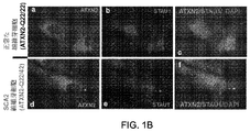


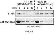


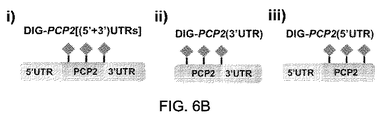
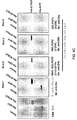

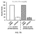


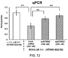


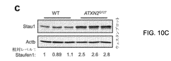

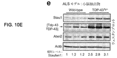


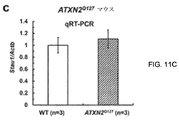


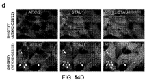


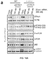


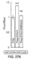


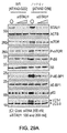


Classifications
-
- C—CHEMISTRY; METALLURGY
- C12—BIOCHEMISTRY; BEER; SPIRITS; WINE; VINEGAR; MICROBIOLOGY; ENZYMOLOGY; MUTATION OR GENETIC ENGINEERING
- C12N—MICROORGANISMS OR ENZYMES; COMPOSITIONS THEREOF; PROPAGATING, PRESERVING, OR MAINTAINING MICROORGANISMS; MUTATION OR GENETIC ENGINEERING; CULTURE MEDIA
- C12N15/00—Mutation or genetic engineering; DNA or RNA concerning genetic engineering, vectors, e.g. plasmids, or their isolation, preparation or purification; Use of hosts therefor
- C12N15/09—Recombinant DNA-technology
- C12N15/11—DNA or RNA fragments; Modified forms thereof; Non-coding nucleic acids having a biological activity
- C12N15/113—Non-coding nucleic acids modulating the expression of genes, e.g. antisense oligonucleotides; Antisense DNA or RNA; Triplex- forming oligonucleotides; Catalytic nucleic acids, e.g. ribozymes; Nucleic acids used in co-suppression or gene silencing
-
- A—HUMAN NECESSITIES
- A61—MEDICAL OR VETERINARY SCIENCE; HYGIENE
- A61K—PREPARATIONS FOR MEDICAL, DENTAL OR TOILETRY PURPOSES
- A61K45/00—Medicinal preparations containing active ingredients not provided for in groups A61K31/00 - A61K41/00
- A61K45/06—Mixtures of active ingredients without chemical characterisation, e.g. antiphlogistics and cardiaca
-
- C—CHEMISTRY; METALLURGY
- C12—BIOCHEMISTRY; BEER; SPIRITS; WINE; VINEGAR; MICROBIOLOGY; ENZYMOLOGY; MUTATION OR GENETIC ENGINEERING
- C12N—MICROORGANISMS OR ENZYMES; COMPOSITIONS THEREOF; PROPAGATING, PRESERVING, OR MAINTAINING MICROORGANISMS; MUTATION OR GENETIC ENGINEERING; CULTURE MEDIA
- C12N2310/00—Structure or type of the nucleic acid
- C12N2310/10—Type of nucleic acid
- C12N2310/11—Antisense
-
- C—CHEMISTRY; METALLURGY
- C12—BIOCHEMISTRY; BEER; SPIRITS; WINE; VINEGAR; MICROBIOLOGY; ENZYMOLOGY; MUTATION OR GENETIC ENGINEERING
- C12N—MICROORGANISMS OR ENZYMES; COMPOSITIONS THEREOF; PROPAGATING, PRESERVING, OR MAINTAINING MICROORGANISMS; MUTATION OR GENETIC ENGINEERING; CULTURE MEDIA
- C12N2310/00—Structure or type of the nucleic acid
- C12N2310/10—Type of nucleic acid
- C12N2310/12—Type of nucleic acid catalytic nucleic acids, e.g. ribozymes
-
- C—CHEMISTRY; METALLURGY
- C12—BIOCHEMISTRY; BEER; SPIRITS; WINE; VINEGAR; MICROBIOLOGY; ENZYMOLOGY; MUTATION OR GENETIC ENGINEERING
- C12N—MICROORGANISMS OR ENZYMES; COMPOSITIONS THEREOF; PROPAGATING, PRESERVING, OR MAINTAINING MICROORGANISMS; MUTATION OR GENETIC ENGINEERING; CULTURE MEDIA
- C12N2310/00—Structure or type of the nucleic acid
- C12N2310/10—Type of nucleic acid
- C12N2310/14—Type of nucleic acid interfering N.A.
- C12N2310/141—MicroRNAs, miRNAs
-
- C—CHEMISTRY; METALLURGY
- C12—BIOCHEMISTRY; BEER; SPIRITS; WINE; VINEGAR; MICROBIOLOGY; ENZYMOLOGY; MUTATION OR GENETIC ENGINEERING
- C12N—MICROORGANISMS OR ENZYMES; COMPOSITIONS THEREOF; PROPAGATING, PRESERVING, OR MAINTAINING MICROORGANISMS; MUTATION OR GENETIC ENGINEERING; CULTURE MEDIA
- C12N2310/00—Structure or type of the nucleic acid
- C12N2310/10—Type of nucleic acid
- C12N2310/20—Type of nucleic acid involving clustered regularly interspaced short palindromic repeats [CRISPRs]
-
- C—CHEMISTRY; METALLURGY
- C12—BIOCHEMISTRY; BEER; SPIRITS; WINE; VINEGAR; MICROBIOLOGY; ENZYMOLOGY; MUTATION OR GENETIC ENGINEERING
- C12N—MICROORGANISMS OR ENZYMES; COMPOSITIONS THEREOF; PROPAGATING, PRESERVING, OR MAINTAINING MICROORGANISMS; MUTATION OR GENETIC ENGINEERING; CULTURE MEDIA
- C12N2310/00—Structure or type of the nucleic acid
- C12N2310/30—Chemical structure
- C12N2310/34—Spatial arrangement of the modifications
- C12N2310/341—Gapmers, i.e. of the type ===---===
-
- C—CHEMISTRY; METALLURGY
- C12—BIOCHEMISTRY; BEER; SPIRITS; WINE; VINEGAR; MICROBIOLOGY; ENZYMOLOGY; MUTATION OR GENETIC ENGINEERING
- C12N—MICROORGANISMS OR ENZYMES; COMPOSITIONS THEREOF; PROPAGATING, PRESERVING, OR MAINTAINING MICROORGANISMS; MUTATION OR GENETIC ENGINEERING; CULTURE MEDIA
- C12N2320/00—Applications; Uses
- C12N2320/30—Special therapeutic applications
- C12N2320/32—Special delivery means, e.g. tissue-specific
Abstract
【解決手段】 Staufen1に関連したRNA代謝の調節障害を最小化する方法は、調節障害を最小化するのに十分な量のStaufen1−調節剤を標的細胞に導入する工程を含み得る。Staufen1−誘導性RNA代謝異常に関連する神経変性状態を治療するための治療用組成物は、治療上有効な量のStaufen1−調節剤と、薬学的に許容可能な担体とを含み得る。
【選択図】 図1AA method of minimizing the dysregulation of RNA metabolism associated with Staufen1 may include introducing into target cells a sufficient amount of the Staufen1-modulator to minimize the dysregulation. Therapeutic compositions for treating neurodegenerative conditions associated with Staufen1-induced RNA metabolism disorders may comprise a therapeutically effective amount of Staufen1-modulator and a pharmaceutically acceptable carrier.
[Selection diagram] FIG. 1A
Description
本出願は、2018年6月14日に出願された米国仮特許出願番号62/685,269の利益を主張し、参照により本明細書に組み込まれる。 This application claims the interests of US Provisional Patent Application No. 62 / 685,269 filed on June 14, 2018 and is incorporated herein by reference.
神経変性疾患は、脳や末梢神経系の神経細胞が時間の経過とともに機能を失い、最終的に死滅することで生じる。また、一般的に神経細胞は自分自身で再生したり、入れ替わったりすることはない。神経変性疾患に罹患するリスクは、年齢とともに増加する。神経変性疾患に関連する身体的または精神的症状の一部を治療によって緩和することができるかもしれないが、現在のところ治療法はない。神経変性疾患の非限定的な例としては、末梢神経障害、アルツハイマー病(AD)、パーキンソン病(PD)、ハンチントン病(HD)、脊髄小脳失調症(SCA)、プリオン病、運動ニューロン病(MND)、筋萎縮性側索硬化症(ALS)、多発性硬化症(MS)、および脊髄筋萎縮症(SMA)などが挙げられる。 Neurodegenerative diseases occur when nerve cells in the brain and peripheral nervous system lose their function over time and eventually die. Also, in general, nerve cells do not regenerate or replace themselves. The risk of developing neurodegenerative disease increases with age. Treatment may relieve some of the physical or psychological symptoms associated with neurodegenerative diseases, but there is currently no cure. Non-limiting examples of neurodegenerative diseases include peripheral neuropathy, Alzheimer's disease (AD), Parkinson's disease (PD), Huntington's disease (HD), spinal cerebral ataxia (SCA), prion's disease, and motor neuron disease (MND). ), Amyotrophic lateral sclerosis (ALS), multiple sclerosis (MS), and spinal cord atrophy (SMA).
本発明の性質および利点の完全な理解のために、参照は、好ましい実施形態の以下の詳細な説明、および添付の図面との関連で行われる:
以下の詳細な説明は、図示の目的のために多くの具体的な内容を含んでいるが、当技術分野の通常の当業者であれば、以下の詳細な内容に対する多くの変形および変更が可能であり、本明細書に含まれると考えられることを理解するだろう。したがって、以下の実施形態は、一般性を損なうことなく、また、記載された請求項に限定を課すことなく、記載される。また、本明細書で使用される用語は、特定の実施形態を説明する目的のみのためのものであり、限定することを意図しないことを理解されたい。別段の定義がない限り、本明細書で使用されるすべての技術的および科学的用語は、本開示が属する当技術分野の通常の熟練者が一般的に理解するのと同じ意味を有する。 The detailed description below includes many specific contents for the purposes of illustration, but ordinary persons skilled in the art can make many modifications and changes to the following detailed contents. You will understand that it is believed to be included herein. Accordingly, the following embodiments are described without compromising generality and without imposing restrictions on the stated claims. It should also be understood that the terms used herein are for purposes of describing a particular embodiment only and are not intended to be limiting. Unless otherwise defined, all technical and scientific terms used herein have the same meaning as commonly understood by ordinary experts in the art to which this disclosure belongs.
本明細書で使用されるように、単数形「a」、「an」および「the」は、文脈が明確にそうでないことを指示しない限り、複数の参照語を明示的に支持することを含む。したがって、例えば、「ポリマー」への参照は、そのようなポリマーの複数を含むことができる。 As used herein, the singular forms "a," "an," and "the" include explicitly supporting multiple references unless the context explicitly indicates otherwise. .. Thus, for example, a reference to a "polymer" can include a plurality of such polymers.
本明細書で使用されるように、「被験体」は、Staufen1調節剤での処置から利益を得ることができる哺乳動物を指す。被験体が、Staufen1調節剤が治療的に有効な治療または予防手段である疾患または状態を有するか、または発症する危険性がある場合、利益を得ることができる。いくつかの局面では、そのような対象はヒトであっても良い。 As used herein, "subject" refers to a mammal that can benefit from treatment with a Staufen1 regulator. Subjects can benefit if they have or are at risk of developing a disease or condition in which the Staufen1 regulator is a therapeutically effective therapeutic or prophylactic measure. In some aspects, such an object may be human.
本明細書で使用されるように、「治療する(treat)」、「治療(treatment)」、または「治療の(treating)」という用語は、組成物およびその投与形態を含む、STAU1遺伝子を標的とするsiRNAまたはアンチセンスオリゴヌクレオチド(ASO)などのStaufen1調節剤の投与と関連して使用される場合、無症候性または症候性のいずれかである被験者への投与を指す。言い換えれば、「治療する(treat」、「治療(treatment)」、または「治療の(treating)」は、対象に存在する状態に関連する症状を軽減、改善、または除去することができ、または予防的であり得る(すなわち、対象における症状の発生を予防または軽減することができる)。そのような予防的治療は、状態の予防とも呼ばれ得る。治療結果は、予想されるものであっても良いし、予想外のものであっても良い。ある特定の側面において、治療の結果は、疾患もしくは状態、またはその徴候もしくは症状の発生もしくは発症の遅延であり得る。別の側面では、治療は、対象者が悩まされる状態からの救済(すなわち、緩和)を対象者に提供すること、またはその状態の徴候または症状からの救済を提供することであり得る。 As used herein, the terms "treat," "treatment," or "treating," target the STU1 gene, including the composition and its dosage form. When used in connection with the administration of a Staufen1 regulator such as siRNA or antisense oligonucleotide (ASO), it refers to administration to a subject who is either asymptomatic or symptomatic. In other words, "treat," "treatment," or "treating" can reduce, ameliorate, or eliminate, or prevent, the symptoms associated with the condition present in the subject. It can be targeted (ie, it can prevent or alleviate the occurrence of symptoms in the subject). Such prophylactic treatment can also be referred to as prevention of the condition, even if the outcome of the treatment is expected. It may be good or unexpected. In one particular aspect, the outcome of treatment may be the onset or delay of the disease or condition, or its signs or symptoms. In another aspect, the treatment may be. It can be to provide the subject with relief (ie, alleviation) from a condition that the subject is suffering from, or to provide relief from the symptoms or symptoms of that condition.
本明細書で使用されるように、「治療剤」とは、適切な量または効果的な量で対象者に投与されたときに、対象者に有益なまたは肯定的な効果をもたらすことができる薬剤を意味する。 As used herein, a "therapeutic agent" can have a beneficial or positive effect on a subject when administered to the subject in an appropriate or effective amount. Means a drug.
本明細書で使用されるように、用語「阻害する(inhibit)」または「阻害の(inhibiting)」は、様々な阻害技術を参照するために使用される。例えば、「「阻害する(inhibit)」または「阻害の(inhibiting)」という用語は、転写前および/または転写後の阻害を指すことができる。転写前の阻害に関して、「阻害する(inhibit)」または「阻害の(inhibiting)」という用語は、永久的、半永久的、または一過性であるかどうかにかかわらず、遺伝子の転写を防止または減少させること、遺伝子の改変された転写を誘導すること、および/または遺伝子の転写速度を減少させることを意味することができる。したがって、いくつかの例では、「阻害する(inhibit)」または「阻害の(inhibiting)」は、DNAに対する恒久的な変化を指すことができ、他の例では、DNAに対する恒久的な変化は行われない。転写後の阻害に関して、「阻害する(inhibit)」または「阻害の(inhibiting)」は、恒久的、半恒久的、または一過性のいずれであっても、タンパク質への遺伝的配列の翻訳を阻止または減少させること、改変されたタンパク質への遺伝的配列の改変された翻訳を誘導すること(例えば、ミスフォールドされたタンパク質などとして)、および/またはタンパク質への遺伝的配列の翻訳速度を減少させることを指すことができる。いくつかの特定の例において、「阻害する(inhibit)」または「阻害の(inhibiting)」は、転写前の阻害を指すことができる。他の特定の例では、「阻害する(inhibit)」または「阻害の(inhibiting)」は、転写後の阻害を指すことができる。もちろん、阻害のタイプは、採用される阻害剤(複数可)または治療剤(複数可)の特定のタイプ(複数可)に依存し得る。したがって、「阻害する(inhibit)」または「阻害の(inhibiting)」は、転写前または転写後、部分的または完全なものであるかどうかにかかわらず、ネイティブ発現と比較して遺伝子の発現が減少することを含むことができる。 As used herein, the terms "inhibiting" or "inhibiting" are used to refer to various inhibition techniques. For example, the terms "inhibiting" or "inhibiting" can refer to pre-transcriptional and / or post-transcriptional inhibition. With respect to pre-transcriptional inhibition, the terms "inhibiting" or "inhibiting" prevent or reduce gene transcription, whether permanent, semi-permanent, or transient. It can be meant to induce, induce modified transcription of the gene, and / or reduce the transcription rate of the gene. Thus, in some examples, "inhibiting" or "inhibiting" can refer to a permanent change to DNA, and in other cases, a permanent change to DNA is a row. I can't. With respect to post-transcriptional inhibition, "inhibiting" or "inhibiting" translates the genetic sequence into a protein, whether permanent, semi-permanent, or transient. Blocking or reducing, inducing a modified translation of the genetic sequence into the modified protein (eg, as a misfolded protein), and / or reducing the rate of translation of the genetic sequence into the protein. It can be pointed out to let. In some particular examples, "inhibiting" or "inhibiting" can refer to pretranscriptional inhibition. In another particular example, "inhibiting" or "inhibiting" can refer to post-transcriptional inhibition. Of course, the type of inhibition may depend on the particular type (s) of the inhibitor (s) or therapeutics (s) employed. Thus, "inhibiting" or "inhibiting" reduces gene expression compared to native expression, whether pre-transcriptional or post-transcriptional, partial or complete. Can include doing.
本明細書で使用されるように、用語「製剤」および「組成物」は互換的に使用され、2つ以上の化合物、元素、または分子の混合物を指す。いくつかの局面では、「製剤」および「組成物」という用語は、担体または他の賦形剤と1種以上の活性剤との混合物を指すために使用され得る。組成物は、固体、液体(すなわち溶液)、または気体を含む、ほぼすべての物理的状態をとり得る。さらに、「投与形態」という用語は、被験体への投与のための形態で提供される1つ以上の製剤(複数可)または組成物(複数可)を含むことができる。 As used herein, the terms "formulation" and "composition" are used interchangeably and refer to a mixture of two or more compounds, elements, or molecules. In some aspects, the terms "formulation" and "composition" can be used to refer to a mixture of a carrier or other excipient and one or more activators. The composition can take almost any physical state, including solids, liquids (ie, solutions), or gases. In addition, the term "form of administration" can include one or more formulations (s) or compositions (s) provided in a form for administration to a subject.
活性成分の「有効量」、「治療的に有効な量」、または「治療的に有効な率」という表現は、薬剤が送達される疾患または状態の治療において治療的結果を達成するために、毒性はないが、活性成分または治療剤の十分な量または送達率を意味する。様々な生物学的要因が、意図されたタスクを実行するための物質の能力に影響を与え得ることが理解される。したがって、「有効量」、「治療的に有効な量」、または「治療的に有効な率(複数可)」は、いくつかの実施例において、そのような生物学的要因に依存する可能性がある。さらに、治療効果の達成は、当技術分野で知られる評価を使用して、医師または他の資格を有する医療従事者によって測定されても良いが、個人差および治療に対する応答が、治療効果の達成を主観的な決定にしても良いことが認識される。治療的に有効な量または送達率の決定は、薬学および医学の分野における通常の技術の範囲内で十分に可能である。例えば、Meiner and Tonascia,"Clinical Trials.デザイン、実施、および分析、"Monographs in Epidemiology and Biostatistics,Vol.8(1986)を参照のこと。 The expressions "effective amount," "therapeutically effective amount," or "therapeutically effective rate" of the active ingredient are used to achieve therapeutic outcomes in the treatment of the disease or condition to which the drug is delivered. Not toxic, but means a sufficient amount or delivery rate of the active ingredient or therapeutic agent. It is understood that various biological factors can affect a substance's ability to perform its intended task. Therefore, the "effective amount," "therapeutically effective amount," or "therapeutically effective rate (s)" may depend on such biological factors in some embodiments. There is. In addition, the achievement of therapeutic efficacy may be measured by physicians or other qualified healthcare professionals using assessments known in the art, but individual differences and responses to treatment are the achievement of therapeutic efficacy. It is recognized that can be a subjective decision. Determining a therapeutically effective amount or delivery rate is well within the bounds of conventional techniques in the fields of pharmacy and medicine. For example, Meiner and Tonascia, "Clinical Trials. Design, Implementation, and Analysis," Monographs in Epidemiology and Biostatistics, Vol. 8 (1986).
本明細書で使用されるように、「投与レジメン」または「治療投与レジメン」または「予防投与レジメン」のような「レジメン」とは、意図された治療または効果を達成するために、組成物の投与量をどのように、いつ、どのくらいの量、およびどのくらいの期間、被験者に投与することができるか、または投与すべきかを指す。 As used herein, a "regimen" such as a "dose regimen" or a "therapeutic dosing regimen" or a "prophylactic dosing regimen" is a composition to achieve the intended treatment or effect. It refers to how, when, how much, and for how long a dose can or should be administered to a subject.
本明細書で使用されるように、「担体」および「薬学的に許容される担体」は互換的に使用され、任意の液体、ゲル、軟膏、溶媒、液体、希釈剤、液体軟膏基剤、リポソーム、ミセル、ジャイアントミセル、またはそのようなもの、または治療剤を標的細胞(例えば、神経細胞)へおよび/または標的細胞内へ送達するのに適した任意の他の適切な担体を指す。例えば、神経細胞)に送達するのに適しており、有害な生理学的応答を引き起こすことなく、被験体または被験体の組織と接触して使用するのに適しており、かつ組成物の他の成分と劇症的な方法で相互作用しない。 As used herein, "carriers" and "pharmaceutically acceptable carriers" are used interchangeably in any liquid, gel, ointment, solvent, liquid, diluent, liquid ointment base, Refers to liposomes, micelles, giant micelles, or any other suitable carrier suitable for delivering therapeutic agents to and / or into target cells (eg, nerve cells). Suitable for delivery to, for example, nerve cells), suitable for use in contact with a subject or subject's tissue without causing an adverse physiological response, and other components of the composition. Does not interact in a fulminant way.
本出願において、「含む(comprises)」、「含んでいる(comprising)」、「含有する(containin)」、および「有する(having)」およびそのような用語は、米国特許法においてそれらに内接する意味を有し得、「含む(includes)」、「含んでいる(including)」、およびそのような意味を有し得、一般的にはオープンエンドの用語であると解釈される。「からなっている(consisting of)」または「からなる(consists of)」という用語は、閉鎖的な用語であり、そのような用語に関連して具体的に記載された構成要素、構造、工程、またはそのようなもの、および米国特許法に従ったもののみを含む。「から本質的になる(Consisting essentially of)」または「から本質的になっている(consists essentially of)」は、米国特許法によって一般的にそれらに帰結される意味を有する。特に、このような用語は一般的に閉ざされた用語であり、それに関連して使用される項目の基本的かつ新規な特徴または機能に実質的に影響を与えない追加の項目、材料、構成要素、工程、または要素を含むことを認める例外を除いて、一般的に閉ざされた用語である。例えば、組成物中に存在するが、組成物の性質または特性に影響を与えない微量元素は、そのような用語に続く項目のリストに明示的に記載されていなくても、「本質的にからなっている(consisting essentially of)」という言葉の下に存在する場合には許容されるであろう。本明細書において、「含んでいる(comprising)」や「含む(including)」のようなオープンエンドの用語を使用する場合には、明示的に述べられているかのように「から本質的になっている(consisting essentially of)」言語と同様に「からなる(consisting of)」言語にも直接的な支持が与えられるべきであり、またその逆もまた然りであることが理解されよう。 In this application, "comprises," "comprising," "continin," and "having," and such terms are inscribed in them in US patent law. It can have meaning, "includes", "inclusion", and can have such meaning, and is generally interpreted as an open-ended term. The terms "consisting of" or "consisting of" are closed terms, and the components, structures, processes specifically described in connection with such terms. , Or such, and only those in accordance with US patent law. "Consisting essentially of" or "consisting essentially of" has the meaning generally consequent to them by US patent law. In particular, such terms are generally closed terms and are additional items, materials, components that do not substantially affect the basic and new features or functions of the items used in connection with them. , A process, or a generally closed term, with exceptions that allow it to include elements. For example, trace elements that are present in a composition but do not affect the properties or properties of the composition are "essentially from" even if they are not explicitly listed in the list of items that follow such terms. It would be acceptable if it were present under the word "consisting essentially of". When we use open-ended terms such as "comprising" and "inclusion" herein, we are essentially "from" as if explicitly stated. It will be understood that direct support should be given to languages that are "consisting of" as well as languages that are "consisting" and vice versa.
本明細書および特許請求の範囲における「第一」、「第二」、「第三」、「第四」などの用語は、類似の要素を区別するために使用されており、必ずしも特定の順序または年代順を記述するために使用されているわけではない。そのように使用される任意の用語は、本明細書に記載された実施形態が、例えば、図示されているか、または別の方法で本明細書に記載されているものとは別の順序で動作可能であるような適切な状況下で互換性があることが理解されるだろう。同様に、方法が一連の工程からなるものとして本明細書に記載されている場合、本明細書に記載されているような工程の順序は、必ずしもそのような工程が実行され得る唯一の順序ではなく、記載される工程の一部が省略される可能性があり、および/または本明細書に記載されていない他の特定の工程が方法に追加される可能性がある。 Terms such as "first," "second," "third," and "fourth" in the specification and claims are used to distinguish similar elements and are not necessarily in a particular order. Or it is not used to describe chronological order. Any term so used is such that the embodiments described herein operate in a different order than those described herein, eg, illustrated or otherwise described herein. It will be understood that they are compatible under the appropriate circumstances as possible. Similarly, if the method is described herein as consisting of a series of steps, the sequence of steps as described herein is not necessarily the only sequence in which such steps can be performed. None of the steps described may be omitted, and / or other specific steps not described herein may be added to the method.
本明細書で使用されるように、用語「実質的に」とは、行為、特性、特性、性質、状態、構造、項目、または結果の完全なまたはほぼ完全な範囲または程度を意味する。例えば、「実質的に」囲まれる物体は、その物体が完全に囲まれるか、またはほぼ完全に囲まれることを意味する。絶対的完全性からの正確な許容される逸脱の程度は、場合によっては特定の文脈に依存することがある。しかし、一般的には、完成の近さは、絶対的な完全な完成が得られた場合と同様の全体的な結果が得られるようになる。「実質的に」の使用は、ある作用、特性、特性、性質、状態、構造、項目、または結果の完全なまたは完全に近い欠如を指すために否定的な意味合いで使用される場合にも同様に適用可能である。例えば、「実質的に粒子を含まない」組成物は、完全に粒子を欠いているか、またはほぼ完全に粒子を欠いているので、効果は完全に粒子を欠いている場合と同じだろう。言い換えれば、成分または要素を「実質的に含まない」組成物は、測定可能な効果がない限り、そのような項目を実際に含んでいても良い。 As used herein, the term "substantially" means the complete or near-complete range or extent of an act, characteristic, characteristic, property, condition, structure, item, or result. For example, an object that is "substantially" enclosed means that the object is completely or nearly completely enclosed. The exact degree of permissible deviation from absolute completeness may in some cases depend on the particular context. However, in general, the closeness of completion will give the same overall result as if an absolute complete completion had been obtained. The use of "substantially" also applies when used in a negative sense to refer to the complete or near-complete lack of an action, property, property, property, state, structure, item, or result. Applicable to. For example, a "substantially particle-free" composition would be completely or almost completely particle-free, so the effect would be the same as if it were completely particle-free. In other words, a composition that is "substantially free" of ingredients or elements may actually contain such items as long as there is no measurable effect.
本明細書で使用されるように、「約」という用語は、所与の値が終点を「少し上」または「少し下」にしても良いことを提供することによって、数値範囲の終点に柔軟性を提供するために使用される。別段の記載がない限り、特定の数値または数値範囲に応じて「約」という用語を使用することは、「約」という用語を使用せずに、そのような数値用語または数値範囲のサポートを提供することも理解されるべきである。例えば、便宜上および簡潔さのために、「約50ミリグラム〜約80ミリグラム」の数値範囲は、「50ミリグラム〜80ミリグラム」の範囲の支持を提供することも理解されるべきである。さらに、本明細書では、「約」という用語が使用されている場合でも、実際の数値のサポートが提供されることを理解されるべきである。例えば、「約」30の暗唱は、30の少し上の値および少し下の値の支持を提供するだけでなく、同様に30の実際の数値の支持を提供するものと解釈されるべきである。 As used herein, the term "about" is flexible to the end of a numerical range by providing that a given value may have an end point "a little above" or "a little below". Used to provide sex. Unless otherwise stated, using the term "about" according to a particular number or range provides support for such a number or range without using the term "about". It should also be understood to do. For example, for convenience and brevity, it should also be understood that the numerical range of "about 50 milligrams to about 80 milligrams" provides support for the range of "50 milligrams to 80 milligrams". In addition, it should be understood that this specification provides actual numerical support even when the term "about" is used. For example, the recitation of "about" 30 should be interpreted as providing support for values slightly above and slightly below 30 as well as providing support for actual numbers of 30 as well. ..
本明細書で使用されるように、複数の項目、構造要素、構成要素、および/または材料が、便宜上、共通のリストで提示されても良い。しかしながら、これらのリストは、リストの各メンバーが個々に個別の固有のメンバーとして識別されているかのように解釈されるべきである。したがって、このようなリストのいかなる個々のメンバーも、それとは反対のことを示すことなく、共通のグループ内でのそれらの提示にのみ基づいて、同じリストの他のメンバーの事実上の等価なものとして解釈されるべきではない。 As used herein, multiple items, structural elements, components, and / or materials may be presented in a common list for convenience. However, these lists should be interpreted as if each member of the list was individually identified as an individual and unique member. Therefore, any individual member of such a list is a de facto equivalent of the other members of the same list, based solely on their presentation within a common group, without showing the opposite. Should not be interpreted as.
濃度、量、およびその他の数値データは、本明細書では、範囲の形式で表現または表示されても良い。このような範囲の形式は、単に便宜上および簡潔さのために使用され、したがって、範囲の限界として明示的に言及された数値だけでなく、各数値およびサブ範囲が明示的に言及されているかのように、その範囲内に包含されるすべての個々の数値またはサブ範囲を包含するように柔軟に解釈されるべきであることが理解されるべきである。例示として、「約1〜約5」という数値範囲は、「約1〜約5」という明示的に暗唱された数値だけでなく、指示された範囲内に包含される個々の数値やサブレンジも含むと解釈されるべきである。したがって、この数値範囲には、2、3、4などの個別の値と、1〜3、2〜4、3〜5などのサブレンジ、および1、2、3、4、5などの個別の値が含まれる。 Concentrations, quantities, and other numerical data may be expressed or displayed herein in the form of ranges. The form of such a range is used solely for convenience and brevity, and therefore whether each number and subrange is explicitly mentioned, not just the number explicitly mentioned as the limit of the range. As such, it should be understood that it should be flexibly interpreted to include all individual numbers or subranges contained within that range. By way of example, the numerical range of "about 1 to about 5" includes not only the explicitly recited numerical value of "about 1 to about 5" but also individual numerical values and subranges included within the indicated range. Should be interpreted as. Therefore, this numerical range includes individual values such as 2, 3, 4 and subranges such as 1-3, 2-4, 3-5, and individual values such as 1, 2, 3, 4, 5. Is included.
この原則は、最小値または最大値として1つの数値のみを記載した範囲にも適用される。さらに、このような解釈は、範囲の幅や記載されている特性に関係なく適用されるべきである。 This principle also applies to ranges where only one number is stated as the minimum or maximum value. Moreover, such an interpretation should be applied regardless of the width of the range or the properties described.
本願では、「改善された」または「強化された」性能を提供する組成物、システム、または方法について言及しても良い。別段の記載がない限り、そのような「改善」または「強化」は、先行技術における組成物、システムまたは方法との比較に基づいて得られる利益の尺度であることが理解されるべきである。さらに、改善または強化された性能の程度は、開示された実施形態の間で変化し得ること、および改善または強化の量、程度、または実現における均等性または一貫性が、普遍的に適用可能であると仮定されないことが理解されるべきである。 The present application may refer to compositions, systems, or methods that provide "improved" or "enhanced" performance. Unless otherwise stated, it should be understood that such "improvement" or "enhancement" is a measure of profit obtained on the basis of comparison with the composition, system or method in the prior art. Moreover, the degree of improved or enhanced performance can vary between the disclosed embodiments, and the amount, degree, or uniformity or consistency in realization of the amount, degree, or realization of the improved or enhanced performance is universally applicable. It should be understood that it is not assumed to be.
本明細書全体を通して「実施例において」と言及することは、実施例に関連して記載された特定の特徴、構造、または特性が、少なくとも1つの実施形態に含まれることを意味する。したがって、本明細書中の様々な場所での「例示的な」という表現の出現は、必ずしもすべてが同じ実施形態を参照しているわけではない。 Reference to "in Examples" throughout the specification means that the particular features, structures, or properties described in connection with the Examples are included in at least one embodiment. Therefore, the appearance of the expression "exemplary" in various places throughout the specification does not necessarily refer to the same embodiment.
例示的な実施形態
本発明の実施形態の最初の概要が以下に提供され、その後、特定の実施形態がさらに詳細に説明される。この初期概要は、読者が技術的概念をより迅速に理解するのを助けることを意図するが、その重要なまたは本質的な特徴を特定することを意図しておらず、また、請求された主題の範囲を限定することを意図していない。
Exemplary Embodiments An initial overview of embodiments of the present invention is provided below, after which specific embodiments will be described in more detail. This initial overview is intended to help the reader understand the technical concept more quickly, but is not intended to identify its important or essential features, and the claimed subject matter. Is not intended to limit the scope of.
神経学的障害は、先天的異常、環境因子、遺伝的障害、感染症、傷害、生活習慣、および/または栄養不良などの様々な原因に起因し得る。 Neurological disorders can result from a variety of causes, including congenital abnormalities, environmental factors, genetic disorders, infections, injuries, lifestyles, and / or malnutrition.
一例として、常染色体優性小脳失調症(ADCAs)は、小脳、脳幹、および脊髄の進行性変性によって特徴づけられる神経変性疾患の不均一なグループである。退化は細胞レベルで起こり、特定のサブタイプでは細胞死に至る。細胞死または機能不全は、中枢神経系から体内の標的筋肉への通信ラインを妨害することがある。 As an example, autosomal dominant cerebellar ataxia (ADCAs) is a heterogeneous group of neurodegenerative diseases characterized by progressive degeneration of the cerebellum, brain stem, and spinal cord. Degeneration occurs at the cellular level, leading to cell death in certain subtypes. Cell death or dysfunction can interfere with the line of communication from the central nervous system to target muscles in the body.
臨床的には、ADCAは、3つのタイプ(ADCAタイプI、III、およびIII)に分けられており、これらはさらに様々なサブタイプに分けられる。これらのタイプやサブタイプはそれぞれ、様々な悪影響を及ぼす可能性がある。タイプIのADCA、サブタイプ1の状態は、典型的には、ポリグルタミンをコードするDNA中のCAGヌクレオチドリピートによって特徴付けられる。これらのポリグルタミンセグメントは、タンパク質レベルで変性効果を引き起こし得る。非限定的な例として、ADCAタイプI、サブタイプ1の状態は、SCA1、SCA2、SCA3、SCA17、およびDRPLAなどを含むことができる。しかしながら、多くの神経変性疾患は、CAGヌクレオチドリピート以外の他のトリヌクレオチドリピート拡張、または他の遺伝的リピート拡張と関連する可能性がある。
Clinically, ADCA is divided into three types (ADCA types I, III, and III), which are further subdivided into various subtypes. Each of these types and subtypes can have a variety of adverse effects. Type I ADCA,
神経変性疾患の一例として、スピノ小脳性失調症2型(SCA2)は、小脳プルキンエ細胞(PC)の進行性変性、および脳幹および脊髄内のニューロンの選択的損失によって特徴づけられる常染色体優性小脳性失調症である。前述のように、SCA2の遺伝的欠損は、ATXN2遺伝子のエクソン1におけるCAGリピート(ポリグルタミンをコードする)の拡大である。ポリグルタミンが過剰になった変異型アタキシン−2(ATXN2)タンパク質は、機能の獲得を獲得し、SCA2脳内で凝集を示す。ATXN2の凝集と小脳PCの変性、およびRNA発現の変化は、SCA2細胞における拡大したCAGリピート発現の病理学的結果である。
As an example of neurodegenerative disease, spinocerebellar ataxia type 2 (SCA2) is autosomal dominant cerebellar cerebellar characterized by progressive degeneration of cerebellar Purkinje cells (PCs) and selective loss of neurons in the brain stem and spinal cord. I have ataxia. As mentioned above, the genetic deficiency of SCA2 is an extension of the CAG repeat (encoding polyglutamine) in
ATXN2は哺乳類の神経系で広く発現する。ATXN2はEGF受容体であるイノシトール1,4,5−三リン酸受容体(IP3R)の制御に関与しており、トランスレーショナルコントロールや胚発生にも関与する。ATXN2は、RNAスプライシング因子A2BP1/Fox1、polyA binding protein 1 (PABP1)、DDX6、Tar DNA binding protein−43(TDP−43)などの複数のRNA結合タンパク質と相互作用し、RNA代謝に特異的な役割を果たすことを示す。さらに、ATXN2はストレス顆粒(SG)やP−bodyなどの細胞内構成要素の構成タンパク質であり、ストレス時のmRNAの隔離やタンパク質の翻訳制御にも関与していることが明らかになった。
ATXN2 is widely expressed in the mammalian nervous system. ATXN2 is involved in the regulation of the
さらに、二本鎖RNA結合タンパク質であるSTAufen1(STAU1)やSTAufen−2(STAU2)は、脳オリゴデンドロサイトなどの培養細胞の細胞質内包物にリクルートされ、ストレス顆粒のダイナミクスを調節することができる。Staufen1は、細胞ストレス下でRNA顆粒に局在するタンパク質であるだけでなく、mRNAの輸送や分解など、多くのRNA関連機能を果たすことができる。Staufen1は、脊椎動物の卵母細胞と体細胞の両方でmRNA輸送に関連するが、Staufen1は様々な細胞タイプのRNA代謝調節に関与する多機能タンパク質である。さらに、Staufen1は5'UTRを介してmRNAの翻訳効率を制御し、3'UTRを介して転写物の安定性を制御することができ、そのメカニズムはシュタウフェン媒介RNA崩壊(SMD)と言及される。 Furthermore, the double-stranded RNA-binding proteins STAufen1 (STAU1) and STAufen-2 (STAU2) can be recruited to the cytoplasmic inclusions of cultured cells such as brain oligodendrocytes to regulate the dynamics of stress granules. Staufen1 is not only a protein localized in RNA granules under cell stress, but can also perform many RNA-related functions such as mRNA transport and degradation. Staufen1 is involved in mRNA transport in both vertebrate oocytes and somatic cells, whereas Staufen1 is a multifunctional protein involved in the regulation of RNA metabolism in various cell types. In addition, Staufen1 can regulate the translation efficiency of mRNA via the 5'UTR and the stability of transcripts via the 3'UTR, the mechanism of which is referred to as Staufen-mediated RNA disruption (SMD). ..
翻訳、輸送、貯蔵、分解を含むRNA代謝は、健康と疾患の両方において細胞機能に重要な役割を担うことが指摘される。ストレス顆粒は、細胞ストレス時に翻訳を調節し、RNA顆粒はmRNAの輸送や局所的な発現制御を調節することができるため、RNA代謝に重要な役割を果たす。SGは、mRNA、リボソームRNA、ノンコーディングRNAのほか、細胞ごとに異なるタンパク質の特定の補体で構成される。 It is pointed out that RNA metabolism, including translation, transport, storage and degradation, plays an important role in cell function in both health and disease. Stress granules play an important role in RNA metabolism because they can regulate translation during cellular stress and RNA granules can regulate mRNA transport and local expression regulation. SG is composed of mRNA, ribosomal RNA, non-coding RNA, as well as specific complements of proteins that differ from cell to cell.
このことを考慮すると、ATXN2は、PABP1、A2BP1/Fox1、DDX6およびTDP−43を含む複数のRNA結合タンパク質と相互作用することができ、RNA代謝におけるATXN2のユニークな役割を示唆する。さらに、Staufen1はATXN2のRNA依存性インタラクターである。タンパク質−タンパク質相互作用の強さは、野生型および変異型ATXN2については差がないようであるが、変異型ATXN2の存在下では、定常状態のStaufen1レベルは、SCA2患者由来細胞において大幅に増加することができる。したがって、本明細書中でさらに詳細に記載されるように、いくつかの実施形態では、変異型ATXN2タンパク質中のポリグルタミン領域の拡張、またはポリヌクレオチド拡張を有する他のタンパク質は、SCA2患者由来細胞中のStaufen1レベルの増加を促進することができる。さらにまた、神経細胞におけるStaufen1のレベルの増加は、ADCA(例えば、ADCAタイプI、サブタイプ1など)および他の神経変性疾患の劇症的効果において重要な役割を果たし得る。
With this in mind, ATXN2 can interact with multiple RNA-binding proteins, including PABP1, A2BP1 / Fox1, DDX6 and TDP-43, suggesting a unique role for ATXN2 in RNA metabolism. In addition, Staufen1 is an RNA-dependent interactor of ATXN2. The strength of the protein-protein interaction does not appear to be different for wild-type and mutant ATXN2, but in the presence of mutant ATXN2, steady-state Staufen1 levels are significantly increased in cells derived from SCA2 patients. be able to. Therefore, as described in more detail herein, in some embodiments, other proteins with dilation of the polyglutamine region, or polynucleotide extension, in the mutant ATXN2 protein are cells derived from SCA2 patients. It is possible to promote an increase in the
いくつかの追加の例として、ヒト第9染色体オープンリーディングフレーム72(c9orf72)は、遺伝子c9orf72によってエンドコードされるタンパク質である。しかしながら、C9orff72の変異は、少なくとも家族性前頭側頭葉性痴呆症(FTD)と筋萎縮性側索硬化症(ALS)との間の遺伝的関連性であることが確認される。より詳細には、C9orf72の変異は、6文字列のヌクレオチドGGGGGCCのヘキサヌクレオチドリピート拡大である。通常、C9orf72遺伝子中に存在するこれらのヘキサヌクレオチドリピートは約20〜30個以下である。しかしながら、C9orf72変異が存在する場合、このヘキサヌクレオチドリピートは、30回を超える頻度で(例えば、100回を超える頻度で)発生し得る。この変異は、C9orf72タンパク質の正常な発現を阻害し得る。 As some additional examples, the human chromosome 9 open reading frame 72 (c9orf72) is a protein endcoded by the gene c9orf72. However, the mutation in C9orff72 confirms that it is at least a genetic association between familial frontotemporal lobar dementia (FTD) and amyotrophic lateral sclerosis (ALS). More specifically, the mutation in C9orf72 is a hexanucleotide repeat extension of the 6-string nucleotide GGGGGGCC. Normally, the number of these hexanucleotide repeats present in the C9orf72 gene is about 20 to 30 or less. However, in the presence of the C9orf72 mutation, this hexanucleotide repeat can occur more than 30 times (eg, more than 100 times). This mutation can inhibit the normal expression of the C9orf72 protein.
また、TAR DNA結合タンパク質43(TDP−43)は、TARDBP遺伝子によってコードされるタンパク質である。しかしながら、TARDBP遺伝子および/またはTDP−43における変異は、TDP−43の病原性形態を引き起こし得、これは、例えば、前頭側頭型認知症の特定の形態、ALS、慢性外傷性脳症、アルツハイマー病の特定のサブセットなどの多くの神経疾患と関連し得る。 In addition, TAR DNA-binding protein 43 (TDP-43) is a protein encoded by the TARDBP gene. However, mutations in the TARDBP gene and / or TDP-43 can cause a pathogenic form of TDP-43, such as certain forms of frontotemporal dementia, ALS, chronic traumatic encephalopathy, Alzheimer's disease. Can be associated with many neurological disorders such as a particular subset of.
さらに、タウタンパク質は、微小管関連タンパク質タウ(MAPT)として指定された単一の遺伝子からの代替スプライシングの産物である。しかしながら、MAPTおよび/またはタウタンパク質における特定の変異は、タウの病原性フロムをもたらし、これは、アルツハイマー病、前頭側頭型認知症、および他の様々な神経学的障害などの多くの神経学的障害に関連し得る。 In addition, tau protein is the product of alternative splicing from a single gene designated as the microtubule-related protein tau (MAPT). However, certain mutations in MAPT and / or tau protein result in the pathogenic fromm of tau, which is associated with many neurology such as Alzheimer's disease, frontotemporal dementia, and various other neurological disorders. May be related to a neurological disorder.
さらに、アミロイド前駆体タンパク質(APP)は、ニューロンのシナプスに集中している膜タンパク質である。しかしながら、APPおよび/またはAPPの変異は、アルツハイマー病の家族性感受性と関連し得る。 In addition, amyloid precursor protein (APP) is a membrane protein that is concentrated at neuronal synapses. However, APP and / or mutations in APP may be associated with familial susceptibility to Alzheimer's disease.
このように、様々な変異が神経疾患に関与し得る。しかしながら、非限定的な例として、Staufen1レベルは、AXTN2におけるCAGリピートエクスポジションを有する細胞において、C9orf72におけるヘキサヌクレオチドリピートエクスポジションを有する細胞において、TDP−43変異を有する細胞において、MAPT変異を有する細胞において、およびAPP変異を有する細胞において、増加させることができる。これは、本明細書でさらに詳細に記載されるように、Staufen1が直接または間接的に標的化されているかどうかにかかわらず、Staufen1が様々な神経疾患の治療において貴重な治療標的となり得ることを示す追加の証拠である。 Thus, various mutations can be involved in neurological disorders. However, as a non-limiting example, Staufen1 levels are found in cells with CAG repeat expositions in AXTN2, in cells with hexanucleotide repeat expositions in C9orf72, in cells with TDP-43 mutations, in cells with MAPT mutations. Can be increased in and in cells with APP mutations. This indicates that Staufen1 can be a valuable therapeutic target in the treatment of various neurological disorders, whether or not Staufen1 is directly or indirectly targeted, as described in more detail herein. Additional evidence to show.
従って、本開示は、標的細胞(例えば、神経細胞)におけるStaufen1のレベルまたは活性を正常化または制御するための組成物および方法を記載する。一つの非限定的な例として、Staufen1関連RNA代謝の調節障害を最小化する方法が記載されている。方法は、調節異常を最小化するのに十分な量のStaufen1調節剤を標的細胞に導入する工程を含むことができる。 Therefore, the present disclosure describes compositions and methods for normalizing or controlling the level or activity of Staufen1 in target cells (eg, nerve cells). As one non-limiting example, a method of minimizing dysregulation of Staufen1-related RNA metabolism has been described. The method can include introducing a sufficient amount of Staufen1 regulator into the target cells to minimize dysregulation.
Staufen1活性を制御する方法も記載される。方法は、Staufen1調節剤を導入する前または導入しない前のStaufen1活性と比較して、Staufen1活性を低下させるのに十分な量のStaufen1調節剤を標的細胞に導入する工程を含むことができる。 Methods for controlling Staufen1 activity are also described. The method can include the step of introducing a sufficient amount of Staufen1 regulator into the target cells to reduce the Staufen1 activity as compared to the Staufen1 activity before or without the introduction of the Staufen1 regulator.
さらに、標的細胞におけるStaufen1蓄積を制御する方法が記載される。方法は、Staufen1調節剤を導入する前または導入しない前の標的細胞内の濃度と比較し、標的細胞内のStaufen1の濃度を低下させるのに十分な量のStaufen1調節剤を標的細胞に導入する工程を含むことができる。 In addition, methods of controlling Staufen1 accumulation in target cells are described. The method is a step of introducing a sufficient amount of Staufen1 regulator into a target cell to reduce the concentration of Staufen1 in the target cell as compared with the concentration in the target cell before or without the introduction of the Staufen1 regulator. Can be included.
さらにまた、Staufen1誘導性RNA代謝異常に関連する神経変性疾患を治療する方法が記載される。方法は、治療上有効な量のStaufen1調節剤を被験者の標的細胞に投与する工程を含むことができる。 Furthermore, methods for treating neurodegenerative diseases associated with Staufen1-induced RNA metabolism disorders are described. The method can include administering a therapeutically effective amount of the Staufen1 regulator to the target cells of the subject.
RNA代謝のStaufen1誘発性調節障害に関連する神経変性疾患を治療するための治療剤または組成物も記載される。治療剤は、Staufen1調節剤であり得る。治療用組成物は、治療上有効な量のStaufen1調節剤と、薬学的に許容される担体とを含むことができる。 Also described are therapeutic agents or compositions for treating neurodegenerative diseases associated with Staufen1-induced dysregulation of RNA metabolism. The therapeutic agent can be a Staufen1 regulator. The therapeutic composition can include a therapeutically effective amount of the Staufen1 modifier and a pharmaceutically acceptable carrier.
本開示において、様々な方法、治療剤、および治療用組成物を議論する際に、これらの議論の各々は、その例の文脈で明示的に議論されるか否かにかかわらず、これらの例の各々に適用可能であると考えられることに留意されたい。したがって、例えば、方法そのものについての詳細な議論において、そのような議論はまた、本明細書に記載された他の方法、治療剤、治療用組成物、治療用投与量および形態、およびその逆も参照する。 In discussing the various methods, therapeutic agents, and therapeutic compositions in the present disclosure, each of these discussions, whether or not explicitly discussed in the context of that example, is these examples. Note that it is considered applicable to each of the above. Thus, for example, in a detailed discussion of the method itself, such discussion also includes other methods, therapeutic agents, therapeutic compositions, therapeutic dosages and forms, and vice versa, as described herein. refer.
さらに詳細には、本開示は、Staufen1関連RNA代謝の調節障害を最小化する方法を記載する。上述したように、Staufen1は、例えば、mRNA輸送および分解などの多くのRNA関連機能を実行することができる。したがって、いくつかの例では、標的細胞において上昇したStaufen1レベルを有することは、Staufen1関連RNA代謝の調節障害をもたらし得る。さらに、いくつかの例では、標的細胞におけるStaufen1のレベルの上昇は、様々な細胞成分の望ましくない加速された分解をもたらすことができる。従って、方法は、RNA代謝のStaufen1関連の調節障害を最小化するために、十分な量のStaufen1調節剤を標的細胞に導入することを含むことができる。 In more detail, the present disclosure describes methods of minimizing dysregulation of Staufen1-related RNA metabolism. As mentioned above, Staufen1 can perform many RNA-related functions, such as mRNA transport and degradation. Thus, in some examples, having elevated Staufen1 levels in target cells can result in dysregulation of Staufen1-related RNA metabolism. Moreover, in some examples, elevated levels of Staufen1 in target cells can result in unwanted accelerated degradation of various cellular components. Thus, the method can include introducing a sufficient amount of Staufen1 regulator into the target cells to minimize the Staufen1-related dysregulation of RNA metabolism.
種々のStaufen1調節剤が、本明細書に記載された方法において採用され得る。Staufen1調節剤をそれ自体で論じる場合、Staufen1調節剤は、Staufen1と直接相互作用することなく、Staufen1を調節するように作用または機能し得ることが理解されるべきであることに留意されたい。例えば、いくつかの例では、Staufen1調節剤は、Staufen1を調節するためにStaufen1から上流に作用する薬剤であり得る。非限定的な例として、いくつかの実施例において、Staufen1調節剤は、変異体ATXN2の発現を阻害する治療剤であり得る。このようにして、Staufen1調節剤は、Staufen1タンパク質またはSTAU1遺伝子と直接相互作用することなく、むしろ、寿命、滞留時間、結合利用可能性、安定性、細胞蓄積などの特性または挙動を含むStaufen1タンパク質に直接または間接的に影響を与える細胞成分または遺伝子との間で、Staufen1の調節を促進することができる。したがって、いくつかの例では、有効なStaufen1調節剤はまた、Staufen1の調節を促進するために変異対立遺伝子を野生型対立遺伝子に改変するためのような遺伝子治療剤を含むことができる。 Various Staufen1 modifiers can be employed in the methods described herein. When discussing the Staufen1 regulator on its own, it should be noted that it should be understood that the Staufen1 regulator may act or function to regulate Staufen1 without interacting directly with Staufen1. For example, in some examples, the Staufen1 regulator can be an agent that acts upstream from Staufen1 to regulate Staufen1. As a non-limiting example, in some examples, the Staufen1 regulator can be a therapeutic agent that inhibits the expression of mutant ATXN2. In this way, the Staufen1 regulator does not interact directly with the Staufen1 protein or the STAU1 gene, but rather to the Staufen1 protein that contains properties or behaviors such as lifespan, residence time, binding availability, stability, cell accumulation, etc. It can promote the regulation of Protein1 with cellular components or genes that directly or indirectly affect it. Thus, in some examples, an effective Staufen1 regulator can also include a gene therapy agent such as for modifying a mutant allele into a wild-type allele to promote the regulation of Staufen1.
非限定的な例として、いくつかの実施形態では、Staufen1調節剤は、Staufen1阻害剤であるか、またはそれを含むことができる。Staufen1阻害剤は、標的細胞におけるStaufen1の転写または翻訳を完全にまたは部分的に阻害する任意の剤を含むことができる。いくつかの追加の例では、Staufen1調節剤は、Staufen1不活性化剤であるか、またはそれを含むことができる。Staufen1不活性化剤は、標的細胞におけるStaufen1タンパク質のRNA代謝(例えば、RNA輸送、RNA分解など)機能を低下または排除するように、Staufen1タンパク質に結合、分解などを行う任意の剤を含むことができる。いくつかの例において、Staufen1調節剤は、Staufen1とmRNAとの相互作用を減少させるかまたは遮断することができ、その結果、mRNAの発現または豊富さが変化することができる。 As a non-limiting example, in some embodiments, the Staufen1 regulator is or can include a Staufen1 inhibitor. The Staufen1 inhibitor can include any agent that completely or partially inhibits the transcription or translation of Staufen1 in the target cell. In some additional examples, the Staufen1 regulator can be or can be a Staufen1 inactivating agent. The Staufen1 inactivating agent may include any agent that binds to, degrades, etc. the Staufen1 protein so as to reduce or eliminate the RNA metabolism (eg, RNA transport, RNA degradation, etc.) function of the Staufen1 protein in the target cell. can. In some examples, Staufen1 regulators can reduce or block the interaction of Staufen1 with mRNA, resulting in altered expression or abundance of mRNA.
いくつかの例では、Staufen1調節剤は、変異ATXN2阻害剤(または過剰なヌクレオチドリピートを有する別の変異遺伝子の阻害剤)であるか、またはそれを含むことができる。変異ATXN2阻害剤は、標的細胞における変異ATXN2(例えば、32個以上のポリグルタミンリピートを有するATXN2)の転写または翻訳を阻害する任意の剤であり得る。いくつかの例では、変異ATXN2阻害剤は、変異ATXN2の転写または翻訳を部分的にまたは完全に沈黙させることができる薬剤であるか、またはそれを含むことができる。いくつかのさらなる例では、ATXN2阻害剤は、変異型ATXN2遺伝子を編集して、変異型対立遺伝子を野生型配列に改変することができる(例えば、約21〜約31まで、約21〜約25まで、または約22〜約23までのポリグルタミンリピートを含むように、32以上のポリグルタミンリピートを含むように)薬剤であるか、またはそれを含むことができる。当技術分野に熟練した者によって認識されるように、他の神経変性疾患は、通常のリピート数が上記の範囲から変化し得るATXN2以外の遺伝子上の過剰なポリヌクレオチドリピートによって影響を受け得る。このような場合、変異遺伝子は、野生型の範囲内のポリヌクレオチドリピートを含むように編集することができる。いくつかの例では、Staufen1調節剤は、変異体ATXN2不活性化剤であるか、またはそれを含むことができる。変異ATXN2不活性化剤は、変異ATXN2タンパク質(または過剰なヌクレオチドリピートを有する別の変異タンパク質)に結合し、分解し、または別の方法で不活性化するか、または標的細胞におけるStaufen1の過剰な蓄積を最小化する方法で変異ATXN2タンパク質とStaufen1との相互作用を防止または減少させる任意の剤を含むことができる。 In some examples, the Staufen1 regulator is or can be a mutant ATXN2 inhibitor (or an inhibitor of another mutant gene with excess nucleotide repeats). The mutant ATXN2 inhibitor can be any agent that inhibits transcription or translation of the mutant ATXN2 (eg, ATXN2 with 32 or more polyglutamine repeats) in the target cell. In some examples, the mutant ATXN2 inhibitor is, or can include, an agent capable of partially or completely silencing transcription or translation of the mutant ATXN2. In some further examples, the ATXN2 inhibitor can edit the mutant ATXN2 gene to modify the mutant allele into a wild-type sequence (eg, from about 21 to about 31, about 21 to about 25). (To include 32 or more polyglutamine repeats), or to include about 22 to about 23 polyglutamine repeats). As recognized by those skilled in the art, other neurodegenerative diseases can be affected by excess polynucleotide repeats on genes other than ATXN2, where the normal number of repeats can vary from the above range. In such cases, the mutant gene can be edited to include polynucleotide repeats within the wild-type range. In some examples, the Staufen1 regulator is or can include the mutant ATXN2 inactivating agent. The mutant ATXN2 inactivating agent binds to, degrades, or otherwise inactivates the mutant ATXN2 protein (or another mutant protein with excess nucleotide repeats), or is an excess of Staufen1 in the target cell. Any agent can be included that prevents or reduces the interaction of the mutant ATXN2 protein with Nucleotide in a manner that minimizes accumulation.
例えば、いくつかの実施形態では、Staufen1調節剤は、変異c9orf72阻害剤(または過剰なヌクレオチドリピートを有する別の変異遺伝子の阻害剤)であるか、またはそれを含むことができる。突然変異c9orf72阻害剤は、標的細胞における突然変異c9orf72(例えば、30回以上のGGGCCヘキサヌクレオチドリピートを有するc9orf72(例えば、100回以上のGGGCCヘキサヌクレオチドリピートを有するc9orf72))の転写または翻訳を阻害する任意の剤であり得る。いくつかの例では、変異c9orf72阻害剤は、変異c9orf72の転写または翻訳を部分的にまたは完全に沈黙させることができる薬剤であるか、またはそれを含むことができる。いくつかのさらなる例において、変異型c9orf72阻害剤は、変異型c9orf72遺伝子を編集して、変異型対立遺伝子を野生型配列に改変することができる(例えば、約20〜30GGGCCヘキサヌクレオチドリピートを含むように、またはそれ以下の配列に改変することができる)薬剤であるか、またはそれを含むことができる。いくつかの例では、Staufen1調節剤は、変異体c9orf72不活性化剤であるか、またはそれを含むことができる。変異型c9orf72不活性化剤は、変異型c9orf72タンパク質(または過剰なヌクレオチドリピートを有する別の変異型タンパク質)に結合し、分解し、または別の方法で不活性化するか、または標的細胞におけるStaufen1の過剰な蓄積を最小化する方法で変異型c9orf72タンパク質とStaufen1との相互作用を防止または減少させる任意の剤を含むことができる。 For example, in some embodiments, the Staufen1 regulator is or can be a mutant c9orf72 inhibitor (or an inhibitor of another mutant gene with excess nucleotide repeats). Mutant c9orf72 inhibitors inhibit transcription or translation of mutant c9orf72 (eg, c9orf72 with 30 or more GGGCC hexanucleotide repeats (eg, c9orf72 with 100 or more GGGCC hexanucleotide repeats)) in target cells. It can be any agent. In some examples, the mutant c9orf72 inhibitor is, or can include, an agent capable of partially or completely silencing transcription or translation of the mutant c9orf72. In some further examples, the mutant c9orf72 inhibitor can edit the mutant c9orf72 gene to modify the mutant allele into a wild-type sequence (eg, including about 20-30 GGGCC hexanucleotide repeats). (Can be modified to a sequence below or below) or can include it. In some examples, the Staufen1 regulator is or can include the mutant c9orf72 inactivating agent. The mutant c9orf72 inactivating agent binds to, degrades, or otherwise inactivates the mutant c9orf72 protein (or another mutant protein with excess nucleotide repeats), or Staufen1 in the target cell. Any agent can be included that prevents or reduces the interaction of the mutant c9orf72 protein with Nucleotides in a manner that minimizes the excessive accumulation of.
いくつかの追加の例では、Staufen1調節剤は、変異型TDP−43−阻害剤であるか、またはそれを含むことができる。変異型TDP−43阻害剤は、標的細胞における変異型TDP−43の転写または翻訳を阻害する任意の剤であり得る。いくつかの例では、変異TDP−43阻害剤は、変異TDP−43の転写または翻訳を部分的にまたは完全に沈黙させることができる剤であるか、またはそれを含むことができる。いくつかのさらなる例において、TDP−43阻害剤は、変異型TARDBP遺伝子を編集して、変異型対立遺伝子を野生型配列に改変することができる剤であるか、またはそれを含むことができる。いくつかの例では、Staufen1調節剤は、変異型TDP−43−不活性化剤であるか、またはそれを含むことができる。変異TDP−43−不活性化剤は、変異TDP−43タンパク質に結合、分解、またはその他の方法で変異TDP−43タンパク質を不活性化するか、または標的細胞におけるStaufen1の過剰な蓄積を最小化する方法で変異TDP−43タンパク質とStaufen1との相互作用を防止または減少させる任意の剤を含むことができる。 In some additional examples, the Staufen1 regulator is or can include a mutant TDP-43-inhibitor. The mutant TDP-43 inhibitor can be any agent that inhibits transcription or translation of mutant TDP-43 in target cells. In some examples, the mutant TDP-43 inhibitor is, or can include, an agent capable of partially or completely silencing transcription or translation of mutant TDP-43. In some further examples, the TDP-43 inhibitor is an agent capable of editing the mutant TARDBP gene to modify the mutant allele into a wild-type sequence, or can include it. In some examples, the Staufen1 regulator is or can be a mutant TDP-43-inactivating agent. Mutant TDP-43-inactivating agent binds, degrades, or otherwise inactivates mutant TDP-43 protein or minimizes excessive accumulation of Staufen1 in target cells. Any agent that prevents or reduces the interaction of the mutant TDP-43 protein with Staufen1 can be included.
いくつかのさらなる例では、Staufen1調節剤は、変異タウ阻害剤であるか、またはそれを含むことができる。変異タウ阻害剤は、標的細胞における変異タウの転写または翻訳を阻害する任意の剤であり得る。いくつかの例では、変異タウ阻害剤は、変異タウの転写または翻訳を部分的にまたは完全に沈黙させることができる剤であるか、またはそれを含むことができる。いくつかのさらなる例では、タウ阻害剤は、変異型MAPT遺伝子を編集して、変異型対立遺伝子を野生型配列に改変することができる剤であるか、またはそれを含むことができる。いくつかの例では、Staufen1調節剤は、変異タウ不活性化剤であるか、またはそれを含むことができる。変異タウ不活性化剤は、変異タウタンパク質に結合、分解、またはその他の方法で変異タウタンパク質を不活性化するか、または標的細胞におけるStaufen1の過剰な蓄積を最小化する方法で変異タウタンパク質とStaufen1との相互作用を防止または減少させる任意の剤を含むことができる。 In some further examples, the Staufen1 regulator can be or include a mutant tau inhibitor. The mutant tau inhibitor can be any agent that inhibits transcription or translation of the mutant tau in the target cell. In some examples, the mutant tau inhibitor is, or can include, an agent capable of partially or completely silencing transcription or translation of the mutant tau. In some further examples, the tau inhibitor can be, or include, an agent capable of editing the mutant MAPT gene to modify the mutant allele into a wild-type sequence. In some examples, the Staufen1 regulator is, or can include, a mutant tau inactivating agent. Mutant tau inactivating agents combine with mutant tau protein by binding, degrading, or otherwise inactivating the mutant tau protein, or by minimizing the excessive accumulation of Staufen1 in target cells. Any agent can be included that prevents or reduces the interaction with Tauphen1.
さらに他の例では、Staufen1調節剤は、変異APP阻害剤であるか、またはそれを含むことができる。変異APP阻害剤は、標的細胞における変異APPの転写または翻訳を阻害する任意の剤であり得る。いくつかの例では、変異APP阻害剤は、変異APPの転写または翻訳を部分的にまたは完全に沈黙させることができる薬剤であるか、またはそれを含むことができる。いくつかのさらなる例では、APP阻害剤は、変異APP遺伝子を編集して変異アリールを野生型配列に改変することができる薬剤であるか、またはそれを含むことができる。いくつかの例において、Staufen1調節剤は、変異型APP不活性化剤であり得るか、またはそれを含むことができる。変異APP不活性化剤は、変異APPタンパク質に結合し、分解し、または別の方法で変異APPタンパク質を不活性化するか、または標的細胞におけるStaufen1の過剰な蓄積を最小化する方法で変異APPタンパク質とStaufen1との相互作用を防止または減少させる任意の剤を含むことができる。 In yet another example, the Staufen1 regulator is or can include a mutant APP inhibitor. The mutant APP inhibitor can be any agent that inhibits transcription or translation of the mutant APP in the target cell. In some examples, the mutant APP inhibitor is, or can include, an agent capable of partially or completely silencing transcription or translation of the mutant APP. In some further examples, the APP inhibitor can be, or can include, an agent capable of editing the mutated APP gene to modify the mutated aryl into a wild-type sequence. In some examples, the Staufen1 regulator can be or can be a mutant APP inactivating agent. Mutant APP inactivating agent binds to and degrades the mutant APP protein, or otherwise inactivates the mutant APP protein, or minimizes the excessive accumulation of Staufen1 in target cells. Any agent can be included that prevents or reduces the interaction of the protein with Staufen1.
遺伝子の1コピーのみが変異し、その遺伝子が野生型表現型を産生するために機能的に重要である場合に、特定の変異遺伝子の発現を阻害することは、必ずしも望ましいことではないかもしれないことに留意されたい。例えば、いくつかの場合には、ある遺伝子の機能的に重要な単一のコピーで野生型の表現型を産生するのに十分であるかもしれない。対照的に、変異遺伝子の発現を阻害すると、変異していない遺伝子の発現も阻害され、野生型表現型を産生するのに十分な発現が得られない場合がある。しかし、このような懸念がある場合には、場合によっては、変異体遺伝子の発現阻害が望ましい野生型表現型を産生するのに十分な量で、外因性発現ベクターを介して野生型対立遺伝子を発現させることができる場合がある。 Inhibiting the expression of a particular mutated gene may not always be desirable if only one copy of the gene is mutated and the gene is functionally important for producing a wild-type phenotype. Please note that. For example, in some cases, a single functionally important copy of a gene may be sufficient to produce a wild-type phenotype. In contrast, inhibition of the expression of a mutated gene also inhibits the expression of the unmutated gene, which may not be sufficient to produce the wild-type phenotype. However, in the case of such concerns, in some cases, inhibition of the expression of the mutant gene may result in a wild-type allele via an exogenous expression vector in an amount sufficient to produce the desired wild-type phenotype. It may be possible to express it.
いくつかの実施例において、Staufen1調節剤は、ラパマイシン(mTOR)阻害剤の機械的標的であり得る。mTOR阻害剤は、標的細胞におけるmTORの転写または翻訳を阻害する任意の剤であり得る。いくつかのさらなる例では、Staufen1調節剤は、mTOR不活性化剤であり得る。mTOR不活性化剤は、mTORタンパク質に結合、分解等して、Staufen1タンパク質との相互作用を防止または最小化することができる任意の剤であり得る。 In some examples, the Staufen1 regulator may be a mechanical target for a rapamycin (mTOR) inhibitor. The mTOR inhibitor can be any agent that inhibits the transcription or translation of mTOR in the target cell. In some further examples, the Staufen1 regulator can be an mTOR inactivating agent. The mTOR inactivating agent can be any agent capable of binding to, degrading, etc. the mTOR protein to prevent or minimize interaction with the Staufen1 protein.
したがって、これらの機能のうちの1つ以上を実行するために採用され得る様々なStaufen1調節剤が存在する。Staufen1調節剤の非限定的な例としては、siRNA、miRNA、アンチセンスオリゴヌクレオチド(ホスホロチオエート塩基対連結、2'−メトキシエチル、2'−O−メチル、2'−フルオロ、2'−(3−ヒドロキシ)−プロピル、ロック核酸(LNA)を含むケミストリを含む)、ペプチド核酸(PNA)、シクロヘキセン核酸(CeNA)、ヘキシトール核酸(HNA)、モルホリノ、混合物、キメラまたはギャップマーを含む組み合わせ)、リボザイム、ペプチド核酸、モルホリノ、小分子、そのようなもの、またはそれらの組み合わせなどを含むことができる。 Therefore, there are various Staufen1 regulators that can be employed to perform one or more of these functions. Non-limiting examples of Staufen1 regulators include siRNA, miRNA, antisense oligonucleotides (phosphorothioate base-paired linkages, 2'-methoxyethyl, 2'-O-methyl, 2'-fluoro, 2'-(3-). (Hydroxy) -propyl, including chemistries containing lock nucleic acid (LNA)), peptide nucleic acid (PNA), cyclohexene nucleic acid (CeNA), hexitol nucleic acid (HNA), morpholino, mixture, chimera or combination containing gapmer), ribozyme, It can include peptide nucleic acids, morpholinos, small molecules, such, or combinations thereof.
いくつかの特定の例では、Staufen1調節剤は、アンチセンスオリゴヌクレオチド(ASO)であり得る。いくつかの側面において、ASOは、酸素置換のための硫黄などの1つまたは複数の好適な修飾(例えばホスホロチオエート連結の導入)、2'−OH修飾、2'−O−メチル修飾、2'−フルオロ修飾、2'−O−メトキシエチル修飾、ロック核酸(LNA)修飾、架橋核酸(BNA)修飾、ペプチド核酸(PNA)修飾、モルホリノ修飾、ヘキシトール核酸(HNA)修飾、中心ホスホジエステル残基またはホスホロチオエート残基の導入(例えば、「ギャップマー」を形成するため)などの修飾、またはそれらの組み合わせなどを含むことができる。 In some specific examples, the Staufen1 regulator can be an antisense oligonucleotide (ASO). In some aspects, ASO is one or more suitable modifications such as sulfur for oxygen substitution (eg, introduction of phosphodiester bonds), 2'-OH modification, 2'-O-methyl modification, 2'-. Fluoro-modification, 2'-O-methoxyethyl modification, lock nucleic acid (LNA) modification, cross-linked nucleic acid (BNA) modification, peptide nucleic acid (PNA) modification, morpholino modification, hexitol nucleic acid (HNA) modification, central phosphodiester residue or phosphorothioate Modifications such as introduction of residues (eg, to form "gapmers"), or combinations thereof, can be included.
いくつかの例では、ASOは、Ax−Gn−Axギャップマーなどのギャップマーであり得、ここで、Aは人工ヌクレオチドモノマーを表し、xは2から7までの整数であり、Gは天然アンチセンスヌクレオチドモノマーのギャップまたはブロックを表し、nは8から12までの整数である。ASOがギャップマーである場合、5'ウイングおよび3'ウイングには、上述したような種々の人工ヌクレオチドモノマーを使用することができる。例えば、非限定的な例としては、2'−O−メトキシエチル(MOE)、2',4'−架橋核酸(BNA)、ロック核酸(LNA)、N−メアミノオキシBNA、2'−N−(メチル)−4'−C−アミノオキシメチレン2',4'−架橋核酸、N−メアミノオキシ2',4'−架橋核酸、2'−N−(メチル)−4'−C−アミノオキシメチレン2',4'−架橋核酸、2',4'−BNANC[NMe]、2'−O,4'−C−(N−メチル)アミノメチレン2',4'−架橋核酸、2',4'−BNANC、2'−O,4'−C−アミノメチレン2',4'−架橋核酸、またはそれらの組み合わせなどが挙げられる。いくつかの特定の例では、ガパーは、MOEガパーであり得る。他の特定の例では、ガパーは、LNAガパーであり得る。いくつかのさらなる例では、ガパーは、5−8−5ガパー、5−10−5ガパー、または5−12−5ガパーであり得る。いくつかのさらなる例では、ガパーは、4−8−4ガパー、4−10−4ガパー、または4−12−4ガパーであり得る。まだ追加の例では、ガパーは、3−8−3ガパー、3−10−3ガパー、または3−12−3ガパーであり得る。 In some examples, ASO will be a gapmer such A x -G n -A x gapmer, wherein, A represents an artificial nucleotide monomer, x is an integer from 2 to 7, G Represents a gap or block of natural antisense nucleotide monomers, where n is an integer from 8 to 12. When the ASO is a gapmer, various artificial nucleotide monomers as described above can be used for the 5'wing and the 3'wing. For example, as a non-limiting example, 2'-O-methoxyethyl (MOE), 2', 4'-bridged nucleic acid (BNA), lock nucleic acid (LNA), N-meaminooxy BNA, 2'-N-( Methyl) -4'-C-aminooxymethylene 2', 4'-crosslinked nucleic acid, N-meaminooxy2', 4'-crosslinked nucleic acid, 2'-N- (methyl) -4'-C-aminooxymethylene 2 ', 4'-crosslinked nucleic acid, 2', 4'-BNA NC [NMe], 2'-O, 4'-C- (N-methyl) aminomethylene 2', 4'-crosslinked nucleic acid, 2', 4 '-BNA NC , 2'-O, 4'-C-aminomethylene 2', 4'-crosslinked nucleic acid, or a combination thereof and the like can be mentioned. In some specific examples, the gapper can be a MOE gapper. In another particular example, the gapper can be an LNA gapper. In some further examples, the gapper can be 5-8-5 gapper, 5-10-5 gapper, or 5-12-5 gapper. In some further examples, the gapper can be 4-8-4 gapper, 4-10-4 gapper, or 4-12-4 gapper. In yet additional examples, the gapper can be 3-8-3 gapper, 3-10-3 gapper, or 3-12-3 gapper.
表1は、本明細書に記載されるような治療用組成物に含有され得るASOの非限定的な例を示す。

いくつかの非限定的な実施例では、ASOは、配列ID番号:1、配列ID番号:2、配列ID番号:3、配列ID番号:4、配列ID番号:5、配列ID番号:6、配列ID番号:7、配列ID番号:8、配列ID番号:9、配列ID番号:10、配列ID番号:11、配列ID番号:12、配列ID番号:13、配列ID番号:14、配列ID番号:15、配列ID番号:16、配列ID番号:17、配列ID番号:18、配列ID番号:19、配列ID番号:20、配列ID番号:21、配列ID番号:22、配列ID番号:23、配列ID番号:24、配列ID番号:25、配列ID番号:26、配列ID番号:27、配列ID番号:28、配列ID番号:29、配列ID番号:30、配列ID番号:31、配列ID番号:32、配列ID番号:33、配列ID番号:34、配列ID番号:35、配列ID番号:36、配列ID番号:37、配列ID番号:38、配列ID番号:39、配列ID番号:40、またはそれらの組み合わせのヌクレオチド配列を含む。いくつかの追加の実施例では、配列ID番号:1、配列ID番号:2、配列ID番号:3、配列ID番号:4、配列ID番号:5、配列ID番号:6、配列ID番号:7、配列ID番号:8、配列ID番号:9、配列ID番号:10、配列ID番号:11、配列ID番号:12、配列ID番号:13、配列ID番号:14、配列ID番号:15、配列ID番号:16、配列ID番号:17、配列ID番号:18、配列ID番号:19、配列ID番号:20、配列ID番号:21、配列ID番号:22、配列ID番号:23、配列ID番号:24、配列ID番号:25、配列ID番号:26、配列ID番号:27、配列ID番号:28、配列ID番号:29、配列ID番号:30、配列ID番号:31、配列ID番号:32、配列ID番号:33、配列ID番号:34、配列ID番号:35、配列ID番号:36、配列ID番号:37、配列ID番号:38、配列ID番号:39、または配列ID番号:40、またはその中間の8、10、または12ヌクレオチドであるか、または少なくとも95%同一である1またはそれ以上のヌクレオチド配列を含む。さらにいくつかの追加の実施例では、配列ID番号:1、配列ID番号:2、配列ID番号:3、配列ID番号:4、配列ID番号:5、配列ID番号:6、配列ID番号:7、配列ID番号:8、配列ID番号:9、配列ID番号:10、配列ID番号:11、配列ID番号:12、配列ID番号:13、配列ID番号:14、配列ID番号:15、配列ID番号:16、配列ID番号:17、配列ID番号:18、配列ID番号:19、配列ID番号:20、配列ID番号:21、配列ID番号:22、配列ID番号:23、配列ID番号:24、配列ID番号:25、配列ID番号:26、配列ID番号:27、配列ID番号:28、配列ID番号:29、配列ID番号:30、配列ID番号:31、配列ID番号:32、配列ID番号:33、配列ID番号:34、配列ID番号:35、配列ID番号:36、配列ID番号:37、配列ID番号:38、配列ID番号:39、または配列ID番号:40、またはその中間の8、10、または12ヌクレオチドであるか、または少なくとも90%同一である1またはそれ以上のヌクレオチド配列を含む。さらにいくつかの追加の実施例では、配列ID番号:1、配列ID番号:2、配列ID番号:3、配列ID番号:4、配列ID番号:5、配列ID番号:6、配列ID番号:7、配列ID番号:8、配列ID番号:9、配列ID番号:10、配列ID番号:11、配列ID番号:12、配列ID番号:13、配列ID番号:14、配列ID番号:15、配列ID番号:16、配列ID番号:17、配列ID番号:18、配列ID番号:19、配列ID番号:20、配列ID番号:21、配列ID番号:22、配列ID番号:23、配列ID番号:24、配列ID番号:25、配列ID番号:26、配列ID番号:27、配列ID番号:28、配列ID番号:29、配列ID番号:30、配列ID番号:31、配列ID番号:32、配列ID番号:33、配列ID番号:34、配列ID番号:35、配列ID番号:36、配列ID番号:37、配列ID番号:38、配列ID番号:39、または配列ID番号:40、またはその中間の8、10、または12ヌクレオチドであるか、または少なくとも85%同一である1またはそれ以上のヌクレオチド配列を含む。。さらにいくつかの追加の実施例では、配列ID番号:1、配列ID番号:2、配列ID番号:3、配列ID番号:4、配列ID番号:5、配列ID番号:6、配列ID番号:7、配列ID番号:8、配列ID番号:9、配列ID番号:10、配列ID番号:11、配列ID番号:12、配列ID番号:13、配列ID番号:14、配列ID番号:15、配列ID番号:16、配列ID番号:17、配列ID番号:18、配列ID番号:19、配列ID番号:20、配列ID番号:21、配列ID番号:22、配列ID番号:23、配列ID番号:24、配列ID番号:25、配列ID番号:26、配列ID番号:27、配列ID番号:28、配列ID番号:29、配列ID番号:30、配列ID番号:31、配列ID番号:32、配列ID番号:33、配列ID番号:34、配列ID番号:35、配列ID番号:36、配列ID番号:37、配列ID番号:38、配列ID番号:39、または配列ID番号:40、またはその中間の8、10、または12ヌクレオチドであるか、または少なくとも80%同一である1またはそれ以上のヌクレオチド配列を含む。 In some non-limiting examples, the ASO is SEQ ID NO: 1, SEQ ID NO: 2, SEQ ID NO: 3, SEQ ID NO: 4, SEQ ID NO: 5, SEQ ID NO: 6, Sequence ID number: 7, sequence ID number: 8, sequence ID number: 9, sequence ID number: 10, sequence ID number: 11, sequence ID number: 12, sequence ID number: 13, sequence ID number: 14, sequence ID Number: 15, SEQ ID NO: 16, SEQ ID NO: 17, SEQ ID NO: 18, SEQ ID NO: 19, SEQ ID NO: 20, SEQ ID NO: 21, SEQ ID NO: 22, SEQ ID NO:: 23, SEQ ID NO: 24, SEQ ID NO: 25, SEQ ID NO: 26, SEQ ID NO: 27, SEQ ID NO: 28, SEQ ID NO: 29, SEQ ID NO: 30, SEQ ID NO: 31, SEQ ID NO: 32, SEQ ID NO: 33, SEQ ID NO: 34, SEQ ID NO: 35, SEQ ID NO: 36, SEQ ID NO: 37, SEQ ID NO: 38, SEQ ID NO: 39, Arrange ID Includes nucleotide sequences of number: 40, or a combination thereof. In some additional embodiments, SEQ ID NO: 1, SEQ ID NO: 2, SEQ ID NO: 3, SEQ ID NO: 4, SEQ ID NO: 5, SEQ ID NO: 6, SEQ ID NO: 7 , SEQ ID NO: 8, SEQ ID NO: 9, SEQ ID NO: 10, SEQ ID NO: 11, SEQ ID NO: 12, SEQ ID NO: 13, SEQ ID NO: 14, SEQ ID NO: 15, Arrangement ID number: 16, sequence ID number: 17, sequence ID number: 18, sequence ID number: 19, sequence ID number: 20, sequence ID number: 21, sequence ID number: 22, sequence ID number: 23, sequence ID number : 24, SEQ ID NO: 25, SEQ ID NO: 26, SEQ ID NO: 27, SEQ ID NO: 28, SEQ ID NO: 29, SEQ ID NO: 30, SEQ ID NO: 31, SEQ ID NO: 32 , SEQ ID NO: 33, SEQ ID NO: 34, SEQ ID NO: 35, SEQ ID NO: 36, SEQ ID NO: 37, SEQ ID NO: 38, SEQ ID NO: 39, or SEQ ID NO: 40, Includes one or more nucleotide sequences that are 8, 10, or 12 nucleotides in between, or at least 95% identical. In some additional embodiments, SEQ ID NO: 1, SEQ ID NO: 2, SEQ ID NO: 3, SEQ ID NO: 4, SEQ ID NO: 5, SEQ ID NO: 6, SEQ ID NO: 6: 7, SEQ ID NO: 8, SEQ ID NO: 9, SEQ ID NO: 10, SEQ ID NO: 11, SEQ ID NO: 12, SEQ ID NO: 13, SEQ ID NO: 14, SEQ ID NO: 15, SEQ ID NO: 16, SEQ ID NO: 17, SEQ ID NO: 18, SEQ ID NO: 19, SEQ ID NO: 20, SEQ ID NO: 21, SEQ ID NO: 22, SEQ ID NO: 23, Arrange ID Number: 24, SEQ ID NO: 25, SEQ ID NO: 26, SEQ ID NO: 27, SEQ ID NO: 28, SEQ ID NO: 29, SEQ ID NO: 30, SEQ ID NO: 31, SEQ ID NO:: 32, SEQ ID NO: 33, SEQ ID NO: 34, SEQ ID NO: 35, SEQ ID NO: 36, SEQ ID NO: 37, SEQ ID NO: 38, SEQ ID NO: 39, or SEQ ID NO: 40 , Or intermediate, containing one or more nucleotide sequences of 8, 10, or 12 nucleotides, or at least 90% identical. In some additional embodiments, SEQ ID NO: 1, SEQ ID NO: 2, SEQ ID NO: 3, SEQ ID NO: 4, SEQ ID NO: 5, SEQ ID NO: 6, SEQ ID NO: 6: 7, SEQ ID NO: 8, SEQ ID NO: 9, SEQ ID NO: 10, SEQ ID NO: 11, SEQ ID NO: 12, SEQ ID NO: 13, SEQ ID NO: 14, SEQ ID NO: 15, SEQ ID NO: 16, SEQ ID NO: 17, SEQ ID NO: 18, SEQ ID NO: 19, SEQ ID NO: 20, SEQ ID NO: 21, SEQ ID NO: 22, SEQ ID NO: 23, Arrange ID Number: 24, SEQ ID NO: 25, SEQ ID NO: 26, SEQ ID NO: 27, SEQ ID NO: 28, SEQ ID NO: 29, SEQ ID NO: 30, SEQ ID NO: 31, SEQ ID NO:: 32, SEQ ID NO: 33, SEQ ID NO: 34, SEQ ID NO: 35, SEQ ID NO: 36, SEQ ID NO: 37, SEQ ID NO: 38, SEQ ID NO: 39, or SEQ ID NO: 40 , Or intermediate, containing one or more nucleotide sequences of 8, 10, or 12 nucleotides, or at least 85% identical. .. In some additional embodiments, SEQ ID NO: 1, SEQ ID NO: 2, SEQ ID NO: 3, SEQ ID NO: 4, SEQ ID NO: 5, SEQ ID NO: 6, SEQ ID NO: 6: 7, SEQ ID NO: 8, SEQ ID NO: 9, SEQ ID NO: 10, SEQ ID NO: 11, SEQ ID NO: 12, SEQ ID NO: 13, SEQ ID NO: 14, SEQ ID NO: 15, SEQ ID NO: 16, SEQ ID NO: 17, SEQ ID NO: 18, SEQ ID NO: 19, SEQ ID NO: 20, SEQ ID NO: 21, SEQ ID NO: 22, SEQ ID NO: 23, Arrange ID Number: 24, SEQ ID NO: 25, SEQ ID NO: 26, SEQ ID NO: 27, SEQ ID NO: 28, SEQ ID NO: 29, SEQ ID NO: 30, SEQ ID NO: 31, SEQ ID NO:: 32, SEQ ID NO: 33, SEQ ID NO: 34, SEQ ID NO: 35, SEQ ID NO: 36, SEQ ID NO: 37, SEQ ID NO: 38, SEQ ID NO: 39, or SEQ ID NO: 40 , Or intermediate, containing one or more nucleotide sequences of 8, 10, or 12 nucleotides, or at least 80% identical.
いくつかの実施例では、ASOは、配列ID番号:1、配列ID番号:2、配列ID番号:3、配列ID番号:4、配列ID番号:5、配列ID番号:6、配列ID番号:7、配列ID番号:8、配列ID番号:9、配列ID番号:10、配列ID番号:11、配列ID番号:12、配列ID番号:13、配列ID番号:14、配列ID番号:15、配列ID番号:16、配列ID番号:17、配列ID番号:18、配列ID番号:19、配列ID番号:20、配列ID番号:21、配列ID番号:22、配列ID番号:23、配列ID番号:24、配列ID番号:25、配列ID番号:26、配列ID番号:27、配列ID番号:28、配列ID番号:29、配列ID番号:30、配列ID番号:31、配列ID番号:32、配列ID番号:33、配列ID番号:34、配列ID番号:35、配列ID番号:36、配列ID番号:37、配列ID番号:38、配列ID番号:39、または配列ID番号:40の少なくとも6つの連続したヌクレオチドを有する1またはそれ以上のヌクレオチド配列であるかそれらを含む。いくつかの実施例では、ASOは、記載されたヌクレオチド配列の6つの中間ヌクレオチドであり得る。いくつかの実施例では、ASOは、配列ID番号:1、配列ID番号:2、配列ID番号:3、配列ID番号:4、配列ID番号:5、配列ID番号:6、配列ID番号:7、配列ID番号:8、配列ID番号:9、配列ID番号:10、配列ID番号:11、配列ID番号:12、配列ID番号:13、配列ID番号:14、配列ID番号:15、配列ID番号:16、配列ID番号:17、配列ID番号:18、配列ID番号:19、配列ID番号:20、配列ID番号:21、配列ID番号:22、配列ID番号:23、配列ID番号:24、配列ID番号:25、配列ID番号:26、配列ID番号:27、配列ID番号:28、配列ID番号:29、配列ID番号:30、配列ID番号:31、配列ID番号:32、配列ID番号:33、配列ID番号:34、配列ID番号:35、配列ID番号:36、配列ID番号:37、配列ID番号:38、配列ID番号:39、または配列ID番号:40の少なくとも8つの連続したヌクレオチドを有する1またはそれ以上のヌクレオチド配列であるかそれらを含む。いくつかの実施例では、8つの連続したヌクレオチドは、記載されたヌクレオチド配列の8つの中間ヌクレオチドであり得る。いくつかの実施例では、ASOは、配列ID番号:1、配列ID番号:2、配列ID番号:3、配列ID番号:4、配列ID番号:5、配列ID番号:6、配列ID番号:7、配列ID番号:8、配列ID番号:9、配列ID番号:10、配列ID番号:11、配列ID番号:12、配列ID番号:13、配列ID番号:14、配列ID番号:15、配列ID番号:16、配列ID番号:17、配列ID番号:18、配列ID番号:19、配列ID番号:20、配列ID番号:21、配列ID番号:22、配列ID番号:23、配列ID番号:24、配列ID番号:25、配列ID番号:26、配列ID番号:27、配列ID番号:28、配列ID番号:29、配列ID番号:30、配列ID番号:31、配列ID番号:32、配列ID番号:33、配列ID番号:34、配列ID番号:35、配列ID番号:36、配列ID番号:37、配列ID番号:38、配列ID番号:39、または配列ID番号:40の少なくとも10個の連続したヌクレオチドを有する1またはそれ以上のヌクレオチド配列であるかそれらを含む。いくつかの例では、10個の連続したヌクレオチドは、記載されたヌクレオチド配列の10個の中間ヌクレオチドであり得る。いくつかの実施例では、ASOは、配列ID番号:1、配列ID番号:2、配列ID番号:3、配列ID番号:4、配列ID番号:5、配列ID番号:6、配列ID番号:7、配列ID番号:8、配列ID番号:9、配列ID番号:10、配列ID番号:11、配列ID番号:12、配列ID番号:13、配列ID番号:14、配列ID番号:15、配列ID番号:16、配列ID番号:17、配列ID番号:18、配列ID番号:19、配列ID番号:20、配列ID番号:21、配列ID番号:22、配列ID番号:23、配列ID番号:24、配列ID番号:25、配列ID番号:26、配列ID番号:27、配列ID番号:28、配列ID番号:29、配列ID番号:30、配列ID番号:31、配列ID番号:32、配列ID番号:33、配列ID番号:34、配列ID番号:35、配列ID番号:36、配列ID番号:37、配列ID番号:38、配列ID番号:39、または配列ID番号:40の少なくとも12個の連続したヌクレオチドを有する1またはそれ以上のヌクレオチド配列であるかそれらを含む。いくつかの例では、12個の連続したヌクレオチドは、記載されたヌクレオチド配列の12個の中間ヌクレオチドであり得る。いくつかの実施例では、14個の連続したヌクレオチドは、配列ID番号:1、配列ID番号:2、配列ID番号:3、配列ID番号:4、配列ID番号:5、配列ID番号:6、配列ID番号:7、配列ID番号:8、配列ID番号:9、配列ID番号:10、配列ID番号:11、配列ID番号:12、配列ID番号:13、配列ID番号:14、配列ID番号:15、配列ID番号:16、配列ID番号:17、配列ID番号:18、配列ID番号:19、配列ID番号:20、配列ID番号:21、配列ID番号:22、配列ID番号:23、配列ID番号:24、配列ID番号:25、配列ID番号:26、配列ID番号:27、配列ID番号:28、配列ID番号:29、配列ID番号:30、配列ID番号:31、配列ID番号:32、配列ID番号:33、配列ID番号:34、配列ID番号:35、配列ID番号:36、配列ID番号:37、配列ID番号:38、配列ID番号:39、または配列ID番号:40の少なくとも12個の連続したヌクレオチドを有する1またはそれ以上のヌクレオチド配列であるかそれらを含む。いくつかの例では、14個の連続したヌクレオチドは、記載されたヌクレオチド配列の14個の中間ヌクレオチドであり得る。 In some embodiments, the ASO has SEQ ID NO: 1, SEQ ID NO: 2, SEQ ID NO: 3, SEQ ID NO: 4, SEQ ID NO: 5, SEQ ID NO: 6, SEQ ID NO: 6: 7, SEQ ID NO: 8, SEQ ID NO: 9, SEQ ID NO: 10, SEQ ID NO: 11, SEQ ID NO: 12, SEQ ID NO: 13, SEQ ID NO: 14, SEQ ID NO: 15, SEQ ID NO: 16, SEQ ID NO: 17, SEQ ID NO: 18, SEQ ID NO: 19, SEQ ID NO: 20, SEQ ID NO: 21, SEQ ID NO: 22, SEQ ID NO: 23, Arrange ID Number: 24, SEQ ID NO: 25, SEQ ID NO: 26, SEQ ID NO: 27, SEQ ID NO: 28, SEQ ID NO: 29, SEQ ID NO: 30, SEQ ID NO: 31, SEQ ID NO:: 32, SEQ ID NO: 33, SEQ ID NO: 34, SEQ ID NO: 35, SEQ ID NO: 36, SEQ ID NO: 37, SEQ ID NO: 38, SEQ ID NO: 39, or SEQ ID NO: 40 One or more nucleotide sequences having at least 6 contiguous nucleotides of. In some examples, the ASO can be the 6 intermediate nucleotides of the nucleotide sequence described. In some embodiments, the ASO has SEQ ID NO: 1, SEQ ID NO: 2, SEQ ID NO: 3, SEQ ID NO: 4, SEQ ID NO: 5, SEQ ID NO: 6, SEQ ID NO: 6: 7, SEQ ID NO: 8, SEQ ID NO: 9, SEQ ID NO: 10, SEQ ID NO: 11, SEQ ID NO: 12, SEQ ID NO: 13, SEQ ID NO: 14, SEQ ID NO: 15, SEQ ID NO: 16, SEQ ID NO: 17, SEQ ID NO: 18, SEQ ID NO: 19, SEQ ID NO: 20, SEQ ID NO: 21, SEQ ID NO: 22, SEQ ID NO: 23, Arrange ID Number: 24, SEQ ID NO: 25, SEQ ID NO: 26, SEQ ID NO: 27, SEQ ID NO: 28, SEQ ID NO: 29, SEQ ID NO: 30, SEQ ID NO: 31, SEQ ID NO:: 32, SEQ ID NO: 33, SEQ ID NO: 34, SEQ ID NO: 35, SEQ ID NO: 36, SEQ ID NO: 37, SEQ ID NO: 38, SEQ ID NO: 39, or SEQ ID NO: 40 1 or more nucleotide sequences having at least 8 contiguous nucleotides of. In some examples, the eight contiguous nucleotides can be the eight intermediate nucleotides of the nucleotide sequence described. In some embodiments, the ASO has SEQ ID NO: 1, SEQ ID NO: 2, SEQ ID NO: 3, SEQ ID NO: 4, SEQ ID NO: 5, SEQ ID NO: 6, SEQ ID NO: 6: 7, SEQ ID NO: 8, SEQ ID NO: 9, SEQ ID NO: 10, SEQ ID NO: 11, SEQ ID NO: 12, SEQ ID NO: 13, SEQ ID NO: 14, SEQ ID NO: 15, SEQ ID NO: 16, SEQ ID NO: 17, SEQ ID NO: 18, SEQ ID NO: 19, SEQ ID NO: 20, SEQ ID NO: 21, SEQ ID NO: 22, SEQ ID NO: 23, Arrange ID Number: 24, SEQ ID NO: 25, SEQ ID NO: 26, SEQ ID NO: 27, SEQ ID NO: 28, SEQ ID NO: 29, SEQ ID NO: 30, SEQ ID NO: 31, SEQ ID NO:: 32, SEQ ID NO: 33, SEQ ID NO: 34, SEQ ID NO: 35, SEQ ID NO: 36, SEQ ID NO: 37, SEQ ID NO: 38, SEQ ID NO: 39, or SEQ ID NO: 40 One or more nucleotide sequences having at least 10 contiguous nucleotides of. In some examples, the 10 contiguous nucleotides can be the 10 intermediate nucleotides of the nucleotide sequence described. In some embodiments, the ASO has SEQ ID NO: 1, SEQ ID NO: 2, SEQ ID NO: 3, SEQ ID NO: 4, SEQ ID NO: 5, SEQ ID NO: 6, SEQ ID NO: 6: 7, SEQ ID NO: 8, SEQ ID NO: 9, SEQ ID NO: 10, SEQ ID NO: 11, SEQ ID NO: 12, SEQ ID NO: 13, SEQ ID NO: 14, SEQ ID NO: 15, SEQ ID NO: 16, SEQ ID NO: 17, SEQ ID NO: 18, SEQ ID NO: 19, SEQ ID NO: 20, SEQ ID NO: 21, SEQ ID NO: 22, SEQ ID NO: 23, Arrange ID Number: 24, SEQ ID NO: 25, SEQ ID NO: 26, SEQ ID NO: 27, SEQ ID NO: 28, SEQ ID NO: 29, SEQ ID NO: 30, SEQ ID NO: 31, SEQ ID NO:: 32, SEQ ID NO: 33, SEQ ID NO: 34, SEQ ID NO: 35, SEQ ID NO: 36, SEQ ID NO: 37, SEQ ID NO: 38, SEQ ID NO: 39, or SEQ ID NO: 40 One or more nucleotide sequences having at least 12 contiguous nucleotides of. In some examples, the 12 contiguous nucleotides can be the 12 intermediate nucleotides of the nucleotide sequence described. In some embodiments, the 14 contiguous nucleotides are SEQ ID NO: 1, SEQ ID NO: 2, SEQ ID NO: 3, SEQ ID NO: 4, SEQ ID NO: 5, SEQ ID NO: 6 , SEQ ID NO: 7, SEQ ID NO: 8, SEQ ID NO: 9, SEQ ID NO: 10, SEQ ID NO: 11, SEQ ID NO: 12, SEQ ID NO: 13, SEQ ID NO: 14, Arrangement ID number: 15, sequence ID number: 16, sequence ID number: 17, sequence ID number: 18, sequence ID number: 19, sequence ID number: 20, sequence ID number: 21, sequence ID number: 22, sequence ID number : 23, SEQ ID NO: 24, SEQ ID NO: 25, SEQ ID NO: 26, SEQ ID NO: 27, SEQ ID NO: 28, SEQ ID NO: 29, SEQ ID NO: 30, SEQ ID NO: 31 , SEQ ID NO: 32, SEQ ID NO: 33, SEQ ID NO: 34, SEQ ID NO: 35, SEQ ID NO: 36, SEQ ID NO: 37, SEQ ID NO: 38, SEQ ID NO: 39, or SEQ ID NO: 1 or more nucleotide sequences having at least 12 contiguous nucleotides of SEQ ID NO: 40 or include them. In some examples, the 14 contiguous nucleotides can be the 14 intermediate nucleotides of the nucleotide sequence described.
いくつかの実施例では、ASOは、配列ID番号:1に対して少なくとも95%、90%、85%、または80%の相同性を有するヌクレオチド配列であるか、またはそれを含むことができる。いくつかのさらなる例では、ASOは、配列ID番号:1の少なくとも6、8、10、12、または14の連続したヌクレオチドを有するヌクレオチド配列であるか、またはそれを含むことができる。いくつかの実施例では、配列ID番号:1の連続したヌクレオチドは、配列ID番号:1の中間の6、8、10、12、または14のヌクレオチドを含むことができる。いくつかの特定の実施例では、配列ID番号:1の連続したヌクレオチドは、中間の10ヌクレオチド(すなわち、TGTTAGTT)を含むことができる。いくつかの実施例では、ASOは、配列ID番号:1の中間10ヌクレオチドと少なくとも90%、80%、または70%の相同性を有するヌクレオチド配列であるか、またはそれを含むことができる。 In some examples, the ASO is or can be a nucleotide sequence having at least 95%, 90%, 85%, or 80% homology to SEQ ID NO: 1. In some further examples, the ASO is or can be a nucleotide sequence having at least 6, 8, 10, 12, or 14 consecutive nucleotides of SEQ ID NO: 1. In some examples, contiguous nucleotides of SEQ ID NO: 1 can include nucleotides of 6, 8, 10, 12, or 14 in between SEQ ID NO: 1. In some specific embodiments, the contiguous nucleotides of SEQ ID NO: 1 can include an intermediate 10 nucleotides (ie, TGTTAGTT). In some examples, the ASO is or can be a nucleotide sequence having at least 90%, 80%, or 70% homology with the intermediate 10 nucleotides of SEQ ID NO: 1.
いくつかの実施例では、ASOは、配列ID番号:2に対して少なくとも95%、90%、85%、または80%の相同性を有するヌクレオチド配列であるか、またはそれを含むことができる。いくつかのさらなる例では、ASOは、配列ID番号:2の少なくとも6、8、10、12、または14の連続したヌクレオチドを有するヌクレオチド配列であるか、またはそれを含むことができる。いくつかの実施例では、配列ID番号:2の連続するヌクレオチドは、配列ID番号:2の中間の6、8、10、12、または14のヌクレオチドを含むことができる。いくつかの特定の実施例では、配列ID番号:2の連続したヌクレオチドは、中間の10のヌクレオチドを含むことができる。いくつかの実施例では、ASOは、配列ID番号:2の中間10ヌクレオチドと少なくとも90%、80%、または70%の相同性を有するヌクレオチド配列であるか、またはそれを含むことができる。 In some examples, the ASO is or can be a nucleotide sequence having at least 95%, 90%, 85%, or 80% homology to SEQ ID NO: 2. In some further examples, the ASO is or can be a nucleotide sequence having at least 6, 8, 10, 12, or 14 consecutive nucleotides of SEQ ID NO: 2. In some examples, contiguous nucleotides of SEQ ID NO: 2 can include nucleotides of 6, 8, 10, 12, or 14 in between SEQ ID NO: 2. In some particular examples, contiguous nucleotides of SEQ ID NO: 2 can include 10 intermediate nucleotides. In some examples, the ASO is or can be a nucleotide sequence having at least 90%, 80%, or 70% homology with the intermediate 10 nucleotides of SEQ ID NO: 2.
いくつかの実施例では、ASOは、配列ID番号:3に対して少なくとも95%、90%、85%、または80%の相同性を有するヌクレオチド配列であるか、またはそれを含むことができ、いくつかのさらなる実施例では、ASOは、配列ID番号:3の少なくとも6、8、10、12、または14個の連続したヌクレオチドを有するヌクレオチド配列であるか、またはそれを含むことができる。いくつかの実施例では、配列ID番号:3の連続したヌクレオチドは、配列ID番号:3の中間の6、8、10、12、または14のヌクレオチドを含むことができる。いくつかの特定の実施例では、配列ID番号:3の連続したヌクレオチドは、中間の10のヌクレオチドを含むことができる。いくつかの実施例では、ASOは、配列ID番号:3の中間10ヌクレオチドと少なくとも90%、80%、または70%の相同性を有するヌクレオチド配列であるか、またはそれを含むことができる。 In some examples, the ASO is, or can include, a nucleotide sequence having at least 95%, 90%, 85%, or 80% homology to SEQ ID NO: 3. In some further embodiments, the ASO is or can be a nucleotide sequence having at least 6, 8, 10, 12, or 14 contiguous nucleotides of SEQ ID NO: 3. In some examples, contiguous nucleotides of SEQ ID NO: 3 can include nucleotides of 6, 8, 10, 12, or 14 in the middle of SEQ ID NO: 3. In some particular embodiments, the contiguous nucleotides of SEQ ID NO: 3 can include 10 intermediate nucleotides. In some examples, the ASO is or can be a nucleotide sequence having at least 90%, 80%, or 70% homology with the intermediate 10 nucleotides of SEQ ID NO: 3.
いくつかの実施例では、ASOは、配列ID番号:4に対して少なくとも95%、90%、85%、または80%の相同性を有するヌクレオチド配列であるか、または含むことができる。いくつかのさらなる実施例では、ASOは、配列ID番号:4の少なくとも6、8、10、12、または14個の連続したヌクレオチドを有するヌクレオチド配列であるか、または含むことができる。いくつかの実施例では、配列ID番号:4の連続したヌクレオチドは、配列ID番号:4の中間の6、8、10、12、または14のヌクレオチドを含むことができる。いくつかの特定の実施例では、配列ID番号:4の連続したヌクレオチドは、中間の10のヌクレオチドを含むことができる。いくつかの実施例では、ASOは、配列ID番号:4の中間10ヌクレオチドと少なくとも90%、80%、または70%の相同性を有するヌクレオチド配列であるか、またはそれを含むことができる。 In some examples, the ASO is or can be a nucleotide sequence having at least 95%, 90%, 85%, or 80% homology to SEQ ID NO: 4. In some further embodiments, the ASO is or can be a nucleotide sequence having at least 6, 8, 10, 12, or 14 contiguous nucleotides of SEQ ID NO: 4. In some examples, contiguous nucleotides of SEQ ID NO: 4 can include nucleotides of 6, 8, 10, 12, or 14 in the middle of SEQ ID NO: 4. In some particular embodiments, the contiguous nucleotides of SEQ ID NO: 4 can include 10 intermediate nucleotides. In some examples, the ASO is or can be a nucleotide sequence having at least 90%, 80%, or 70% homology with the intermediate 10 nucleotides of SEQ ID NO: 4.
いくつかの実施例では、ASOは、配列ID番号:5に対して少なくとも95%、90%、85%、または80%の相同性を有するヌクレオチド配列であるか、またはそれを含むことができる。いくつかのさらなる実施例では、ASOは、配列ID番号:5の少なくとも6、8、10、12、または14の連続したヌクレオチドを有するヌクレオチド配列であるか、またはそれを含むことができる。いくつかの実施例では、配列ID番号:5の連続するヌクレオチドは、配列ID番号:5の中間の6、8、10、12、または14のヌクレオチドを含むことができる。いくつかの特定の実施例では、配列ID番号:5の連続するヌクレオチドは、中間の10のヌクレオチドを含むことができる。いくつかの実施例では、ASOは、配列ID番号:5の中間10ヌクレオチドと少なくとも90%、80%、または70%の相同性を有するヌクレオチド配列であるか、またはそれを含むことができる。 In some examples, the ASO is or can be a nucleotide sequence having at least 95%, 90%, 85%, or 80% homology to SEQ ID NO: 5. In some further embodiments, the ASO is or can be a nucleotide sequence having at least 6, 8, 10, 12, or 14 consecutive nucleotides of SEQ ID NO: 5. In some examples, contiguous nucleotides of SEQ ID NO: 5 can include nucleotides of 6, 8, 10, 12, or 14 in the middle of SEQ ID NO: 5. In some particular embodiments, the contiguous nucleotides of SEQ ID NO: 5 can include 10 intermediate nucleotides. In some examples, the ASO is or can be a nucleotide sequence having at least 90%, 80%, or 70% homology with the intermediate 10 nucleotides of SEQ ID NO: 5.
いくつかの実施例では、ASOは、配列ID番号:6に対して少なくとも95%、90%、85%、または80%の相同性を有するヌクレオチド配列であるか、または含むことができる。いくつかのさらなる例では、ASOは、配列ID番号:6の少なくとも6、8、10、12、または14の連続したヌクレオチドを有するヌクレオチド配列であるか、またはそれを含むことができる。いくつかの実施例では、配列ID番号:6の連続したヌクレオチドは、配列ID番号:6の中間の6、8、10、12、または14のヌクレオチドを含むことができる。いくつかの特定の実施例では、配列ID番号:6の連続したヌクレオチドは、中間の10のヌクレオチドを含むことができる。いくつかの例では、ASOは、配列ID番号:6の中間10ヌクレオチドと少なくとも90%、80%、または70%の相同性を有するヌクレオチド配列であるか、またはそれを含むことができる。 In some examples, the ASO is or can be a nucleotide sequence having at least 95%, 90%, 85%, or 80% homology to SEQ ID NO: 6. In some further examples, the ASO is or can be a nucleotide sequence having at least 6, 8, 10, 12, or 14 consecutive nucleotides of SEQ ID NO: 6. In some examples, contiguous nucleotides of SEQ ID NO: 6 can include nucleotides of 6, 8, 10, 12, or 14 in the middle of SEQ ID NO: 6. In some particular examples, contiguous nucleotides of SEQ ID NO: 6 can include 10 intermediate nucleotides. In some examples, the ASO is or can be a nucleotide sequence having at least 90%, 80%, or 70% homology with the intermediate 10 nucleotides of SEQ ID NO: 6.
いくつかの実施例では、ASOは、配列ID番号:7に対して少なくとも95%、90%、85%、または80%の相同性を有するヌクレオチド配列であるか、またはそれを含むことができる。いくつかのさらなる例では、ASOは、配列ID番号:7の少なくとも6、8、10、12、または14の連続したヌクレオチドを有するヌクレオチド配列であるか、または含むことができる。いくつかの実施例では、配列ID番号:7の連続したヌクレオチドは、配列ID番号:7の中間の6、8、10、12、または14のヌクレオチドを含むことができる。いくつかの特定の実施例では、配列ID番号:7の連続したヌクレオチドは、中間の10のヌクレオチドを含むことができる。いくつか実施例では、ASOは、配列ID番号:7の中間10ヌクレオチドと少なくとも90%、80%、または70%の相同性を有するヌクレオチド配列であるか、または含むことができる。 In some examples, the ASO is or can be a nucleotide sequence having at least 95%, 90%, 85%, or 80% homology to SEQ ID NO: 7. In some further examples, the ASO can be or can be a nucleotide sequence having at least 6, 8, 10, 12, or 14 consecutive nucleotides of SEQ ID NO: 7. In some examples, contiguous nucleotides of SEQ ID NO: 7 can include nucleotides of 6, 8, 10, 12, or 14 in the middle of SEQ ID NO: 7. In some particular embodiments, the contiguous nucleotides of SEQ ID NO: 7 can include 10 intermediate nucleotides. In some examples, the ASO is or can be a nucleotide sequence having at least 90%, 80%, or 70% homology with the intermediate 10 nucleotides of SEQ ID NO: 7.
いくつかの実施例では、ASOは、配列ID番号:8と少なくとも95%、90%、85%、または80%の相同性を有するヌクレオチド配列であるか、またはそれを含むことができ、いくつかのさらなる例では、ASOは、配列ID番号:8の少なくとも6、8、10、12、または14の連続したヌクレオチドを有するヌクレオチド配列であるか、またはそれを含むことができる。いくつかの具体例において、配列ID番号:8の連続したヌクレオチドは、配列ID番号:8の中間の6、8、10、12、または14のヌクレオチドを含むことができ、いくつかの特定の実施例では、配列ID番号:8の連続したヌクレオチドは、中間の10のヌクレオチドを含むことができる。いくつかの実施例では、ASOは、配列ID番号:8の中間10ヌクレオチドと少なくとも90%、80%、または70%の相同性を有するヌクレオチド配列であるか、またはそれを含むことができる。 In some embodiments, the ASO is, or can include, a nucleotide sequence having at least 95%, 90%, 85%, or 80% homology with SEQ ID NO: 8. In a further example of, the ASO is or can be a nucleotide sequence having at least 6, 8, 10, 12, or 14 contiguous nucleotides of SEQ ID NO: 8. In some embodiments, contiguous nucleotides of SEQ ID NO: 8 can include nucleotides of 6, 8, 10, 12, or 14 in the middle of SEQ ID NO: 8, and some specific practices. In the example, contiguous nucleotides of SEQ ID NO: 8 can contain 10 nucleotides in the middle. In some examples, the ASO is or can be a nucleotide sequence having at least 90%, 80%, or 70% homology with the intermediate 10 nucleotides of SEQ ID NO: 8.
いくつかの実施例では、ASOは、配列ID番号:9の少なくとも95%、90%、85%、または80%の相同性を有するヌクレオチド配列であるか、またはそれを含むことができる。いくつかのさらなる実施例では、ASOは、配列ID番号:9の少なくとも6、8、10、12、または14の連続したヌクレオチドを有するヌクレオチド配列であるか、または含むことができる。いくつかの実施例では、配列ID番号:9の連続したヌクレオチドは、配列ID番号:9の中間の6、8、10、12、または14のヌクレオチドを含むことができる。いくつかの特定の実施例では、配列ID番号:9の連続したヌクレオチドは、中間の10のヌクレオチドを含むことができる。いくつかの実施例では、ASOは、配列ID番号:9の中間10ヌクレオチドと少なくとも90%、80%、または70%の相同性を有するヌクレオチド配列であるか、またはそれを含むことができる。 In some examples, the ASO is or can be a nucleotide sequence having at least 95%, 90%, 85%, or 80% homology of SEQ ID NO: 9. In some further embodiments, the ASO is or can be a nucleotide sequence having at least 6, 8, 10, 12, or 14 consecutive nucleotides of SEQ ID NO: 9. In some examples, contiguous nucleotides of SEQ ID NO: 9 can include nucleotides of 6, 8, 10, 12, or 14 in the middle of SEQ ID NO: 9. In some particular embodiments, the contiguous nucleotides of SEQ ID NO: 9 can include 10 intermediate nucleotides. In some examples, the ASO is or can be a nucleotide sequence having at least 90%, 80%, or 70% homology with the intermediate 10 nucleotides of SEQ ID NO: 9.
いくつかの実施例では、ASOは、配列ID番号:10の少なくとも95%、90%、85%、または80%の相同性を有するヌクレオチド配列であるか、または含むことができる。いくつかのさらなる実施例では、ASOは、配列ID番号:10の少なくとも6、8、10、12、または14の連続したヌクレオチドを有するヌクレオチド配列であるか、または含むことができる。いくつかの実施例では、配列ID番号:10の連続するヌクレオチドは、配列ID番号:10の中間の6、8、10、12、または14のヌクレオチドを含むことができる。いくつかの特定の実施例では、配列ID番号:10の連続するヌクレオチドは、中間の10のヌクレオチドを含むことができる。いくつかの実施例では、ASOは、配列ID番号:10の中間10ヌクレオチドと少なくとも90%、80%、または70%の相同性を有するヌクレオチド配列であるか、またはそれを含むことができる。 In some examples, the ASO is or can be a nucleotide sequence having at least 95%, 90%, 85%, or 80% homology of SEQ ID NO: 10. In some further embodiments, the ASO is or can be a nucleotide sequence having at least 6, 8, 10, 12, or 14 consecutive nucleotides of SEQ ID NO: 10. In some examples, contiguous nucleotides of SEQ ID NO: 10 can include nucleotides of 6, 8, 10, 12, or 14 in the middle of SEQ ID NO: 10. In some particular embodiments, the contiguous nucleotides of SEQ ID NO: 10 can comprise an intermediate 10 nucleotides. In some examples, the ASO is or can be a nucleotide sequence having at least 90%, 80%, or 70% homology with the intermediate 10 nucleotides of SEQ ID NO: 10.
いくつかの実施例では、ASOは、配列ID番号:11の少なくとも95%、90%、85%、または80%の相同性を有するヌクレオチド配列であるか、または含むことができる。いくつかのさらなる実施例では、ASOは、配列ID番号:11の少なくとも6、8、10、12、または14の連続したヌクレオチドを有するヌクレオチド配列であるか、または含むことができる。いくつかの実施例において、配列ID番号:11の連続したヌクレオチドは、配列ID番号:11の中間の6、8、10、12、または14のヌクレオチドを含むことができる。いくつかの特定の実施例では、配列ID番号:11の連続したヌクレオチドは、中間の10のヌクレオチドを含むことができる。いくつかの実施例では、ASOは、配列ID番号:11の中間10ヌクレオチドと少なくとも90%、80%、または70%の相同性を有するヌクレオチド配列であるか、またはそれを含むことができる。 In some examples, the ASO is or can be a nucleotide sequence having at least 95%, 90%, 85%, or 80% homology of SEQ ID NO: 11. In some further embodiments, the ASO is or can be a nucleotide sequence having at least 6, 8, 10, 12, or 14 consecutive nucleotides of SEQ ID NO: 11. In some examples, contiguous nucleotides of SEQ ID NO: 11 can include nucleotides of 6, 8, 10, 12, or 14 in the middle of SEQ ID NO: 11. In some particular embodiments, the contiguous nucleotides of SEQ ID NO: 11 can include 10 intermediate nucleotides. In some examples, the ASO is or can be a nucleotide sequence having at least 90%, 80%, or 70% homology with the intermediate 10 nucleotides of SEQ ID NO: 11.
いくつかの実施例では、ASOは、配列ID番号:12の少なくとも95%、90%、85%、または80%の相同性を有するヌクレオチド配列であるか、または含むことができる。いくつかのさらなる実施例では、ASOは、配列ID番号:12の少なくとも6、8、10、12、または14の連続したヌクレオチドを有するヌクレオチド配列であるか、または含むことができる。いくつかの実施例において、配列ID番号:12の連続したヌクレオチドは、配列ID番号:12の中間の6、8、10、12、または14のヌクレオチドを含むことができる。いくつかの特定の実施例では、配列ID番号:12の連続したヌクレオチドは、中間の10のヌクレオチドを含むことができる。いくつかの実施例では、ASOは、配列ID番号:12の中間10ヌクレオチドと少なくとも90%、80%、または70%の相同性を有するヌクレオチド配列であるか、または含むことができる。 In some examples, the ASO is or can be a nucleotide sequence having at least 95%, 90%, 85%, or 80% homology of SEQ ID NO: 12. In some further embodiments, the ASO is or can be a nucleotide sequence having at least 6, 8, 10, 12, or 14 consecutive nucleotides of SEQ ID NO: 12. In some examples, contiguous nucleotides of SEQ ID NO: 12 can include nucleotides of 6, 8, 10, 12, or 14 in the middle of SEQ ID NO: 12. In some particular embodiments, the contiguous nucleotides of SEQ ID NO: 12 can include 10 intermediate nucleotides. In some examples, the ASO is or can be a nucleotide sequence having at least 90%, 80%, or 70% homology with the intermediate 10 nucleotides of SEQ ID NO: 12.
いくつかの実施例では、ASOは、配列ID番号:13の少なくとも95%、90%、85%、または80%の相同性を有するヌクレオチド配列であるか、または含むことができる。いくつかのさらなる実施例では、ASOは、配列ID番号:13の少なくとも6、8、10、12、または14の連続したヌクレオチドを有するヌクレオチド配列であるか、または含むことができる。いくつかの実施例において、配列ID番号:13の連続したヌクレオチドは、配列ID番号:13の中間の6、8、10、12、または14のヌクレオチドを含むことができる。いくつかの特定の実施例では、配列ID番号:13の連続したヌクレオチドは、中間の10のヌクレオチドを含むことができる。いくつかの実施例では、ASOは、配列ID番号:13の中間10ヌクレオチドと少なくとも90%、80%、または70%の相同性を有するヌクレオチド配列であるか、またはそれを含むことができる。 In some examples, the ASO is or can be a nucleotide sequence having at least 95%, 90%, 85%, or 80% homology of SEQ ID NO: 13. In some further embodiments, the ASO is or can be a nucleotide sequence having at least 6, 8, 10, 12, or 14 consecutive nucleotides of SEQ ID NO: 13. In some examples, contiguous nucleotides of SEQ ID NO: 13 can include nucleotides of 6, 8, 10, 12, or 14 in the middle of SEQ ID NO: 13. In some particular embodiments, the contiguous nucleotides of SEQ ID NO: 13 can include 10 intermediate nucleotides. In some examples, the ASO is or can be a nucleotide sequence having at least 90%, 80%, or 70% homology with the intermediate 10 nucleotides of SEQ ID NO: 13.
いくつかの実施例では、ASOは、配列ID番号:14の少なくとも95%、90%、85%、または80%の相同性を有するヌクレオチド配列であるか、または含むことができる。いくつかのさらなる実施例では、ASOは、配列ID番号:14の少なくとも6、8、10、12、または14の連続したヌクレオチドを有するヌクレオチド配列であるか、または含むことができる。いくつかの実施例では、配列ID番号:14の連続したヌクレオチドは、配列ID番号:14の中間の6、8、10、12、または14のヌクレオチドを含むことができる。いくつかの特定の実施例では、配列ID番号:14の連続したヌクレオチドは、中間の10のヌクレオチドを含むことができる。いくつかの実施例では、ASOは、配列ID番号:14の中間10ヌクレオチドと少なくとも90%、80%、または70%の相同性を有するヌクレオチド配列であるか、またはそれを含むことができる。 In some examples, the ASO is or can be a nucleotide sequence having at least 95%, 90%, 85%, or 80% homology of SEQ ID NO: 14. In some further embodiments, the ASO is or can be a nucleotide sequence having at least 6, 8, 10, 12, or 14 consecutive nucleotides of SEQ ID NO: 14. In some examples, contiguous nucleotides of SEQ ID NO: 14 can include nucleotides of 6, 8, 10, 12, or 14 in the middle of SEQ ID NO: 14. In some particular embodiments, the contiguous nucleotides of SEQ ID NO: 14 can include 10 intermediate nucleotides. In some examples, the ASO is or can be a nucleotide sequence having at least 90%, 80%, or 70% homology with the intermediate 10 nucleotides of SEQ ID NO: 14.
いくつかの実施例では、ASOは、配列ID番号:15と少なくとも95%、90%、85%、または80%の相同性を有するヌクレオチド配列であるか、またはそれを含むことができる。いくつかのさらなる実施例では、ASOは、配列ID番号:15の少なくとも6、8、10、12、または14の連続したヌクレオチドを有するヌクレオチド配列であるか、またはそれを含むことができる。いくつかの実施例において、配列ID番号:15の連続したヌクレオチドは、配列ID番号:15の中間の6、8、10、12、または14のヌクレオチドを含むことができる。いくつかの特定の実施例では、配列ID番号:15の連続したヌクレオチドは、中間の10のヌクレオチドを含むことができる。いくつかの実施例では、ASOは、配列ID番号:15の中間10ヌクレオチドと少なくとも90%、80%、または70%の相同性を有するヌクレオチド配列であるか、またはそれを含むことができる。 In some examples, the ASO is or can be a nucleotide sequence having at least 95%, 90%, 85%, or 80% homology with SEQ ID NO: 15. In some further embodiments, the ASO is or can be a nucleotide sequence having at least 6, 8, 10, 12, or 14 consecutive nucleotides of SEQ ID NO: 15. In some examples, contiguous nucleotides of SEQ ID NO: 15 can include nucleotides of 6, 8, 10, 12, or 14 in the middle of SEQ ID NO: 15. In some particular examples, contiguous nucleotides of SEQ ID NO: 15 can include 10 intermediate nucleotides. In some examples, the ASO is or can be a nucleotide sequence having at least 90%, 80%, or 70% homology with the intermediate 10 nucleotides of SEQ ID NO: 15.
いくつかの実施例では、ASOは、配列ID番号:16の少なくとも95%、90%、85%、または80%の相同性を有するヌクレオチド配列であるか、または含むことができる。いくつかのさらなる実施例では、ASOは、配列ID番号:16の少なくとも6、8、10、12、または14の連続したヌクレオチドを有するヌクレオチド配列であるか、または含むことができる。いくつかの実施例において、配列ID番号:16の連続したヌクレオチドは、配列ID番号:16の中間の6、8、10、12、または14のヌクレオチドを含むことができる。いくつかの特定の実施例では、配列ID番号:16の連続したヌクレオチドは、中間の10のヌクレオチドを含むことができる。いくつかの実施例では、ASOは、配列ID番号:16の中間10ヌクレオチドと少なくとも90%、80%、または70%の相同性を有するヌクレオチド配列であるか、またはそれを含むことができる。 In some examples, the ASO is or can be a nucleotide sequence having at least 95%, 90%, 85%, or 80% homology of SEQ ID NO: 16. In some further embodiments, the ASO is or can be a nucleotide sequence having at least 6, 8, 10, 12, or 14 consecutive nucleotides of SEQ ID NO: 16. In some examples, contiguous nucleotides of SEQ ID NO: 16 can include nucleotides of 6, 8, 10, 12, or 14 in the middle of SEQ ID NO: 16. In some particular examples, contiguous nucleotides of SEQ ID NO: 16 can include 10 intermediate nucleotides. In some examples, the ASO is or can be a nucleotide sequence having at least 90%, 80%, or 70% homology with the intermediate 10 nucleotides of SEQ ID NO: 16.
いくつかの実施例では、ASOは、配列ID番号:17の少なくとも95%、90%、85%、または80%の相同性を有するヌクレオチド配列であるか、または含むことができる。いくつかのさらなる実施例では、ASOは、配列ID番号:17の少なくとも6、8、10、12、または14の連続したヌクレオチドを有するヌクレオチド配列であるか、または含むことができる。いくつかの実施例において、配列ID番号:17の連続したヌクレオチドは、配列ID番号:17の中間の6、8、10、12、または14のヌクレオチドを含むことができる。いくつかの特定の実施例では、配列ID番号:17の連続したヌクレオチドは、中間の10のヌクレオチドを含むことができる。いくつかの実施例では、ASOは、配列ID番号:17の中間10ヌクレオチドと少なくとも90%、80%、または70%の相同性を有するヌクレオチド配列であるか、またはそれを含むことができる。 In some examples, the ASO is or can be a nucleotide sequence having at least 95%, 90%, 85%, or 80% homology of SEQ ID NO: 17. In some further embodiments, the ASO is or can be a nucleotide sequence having at least 6, 8, 10, 12, or 14 consecutive nucleotides of SEQ ID NO: 17. In some examples, contiguous nucleotides of SEQ ID NO: 17 can include nucleotides of 6, 8, 10, 12, or 14 in the middle of SEQ ID NO: 17. In some particular embodiments, the contiguous nucleotides of SEQ ID NO: 17 can include 10 intermediate nucleotides. In some examples, the ASO is or can be a nucleotide sequence having at least 90%, 80%, or 70% homology with the intermediate 10 nucleotides of SEQ ID NO: 17.
いくつかの実施例では、ASOは、配列ID番号:18の少なくとも95%、90%、85%、または80%の相同性を有するヌクレオチド配列であるか、または含むことができる。いくつかのさらなる実施例では、ASOは、配列ID番号:18の少なくとも6、8、10、12、または14の連続したヌクレオチドを有するヌクレオチド配列であるか、または含むことができる。いくつかの実施例において、配列ID番号:18の連続したヌクレオチドは、配列ID番号:18の中間の6、8、10、12、または14のヌクレオチドを含むことができる。いくつかの特定の実施例では、配列ID番号:18の連続したヌクレオチドは、中間の10のヌクレオチドを含むことができる。いくつかの実施例では、ASOは、配列ID番号:18の中間10ヌクレオチドと少なくとも90%、80%、または70%の相同性を有するヌクレオチド配列であるか、またはそれを含むことができる。 In some examples, the ASO is or can be a nucleotide sequence having at least 95%, 90%, 85%, or 80% homology of SEQ ID NO: 18. In some further embodiments, the ASO is or can be a nucleotide sequence having at least 6, 8, 10, 12, or 14 consecutive nucleotides of SEQ ID NO: 18. In some examples, contiguous nucleotides of SEQ ID NO: 18 can include nucleotides of 6, 8, 10, 12, or 14 in the middle of SEQ ID NO: 18. In some particular embodiments, the contiguous nucleotides of SEQ ID NO: 18 can include 10 intermediate nucleotides. In some examples, the ASO is or can be a nucleotide sequence having at least 90%, 80%, or 70% homology with the intermediate 10 nucleotides of SEQ ID NO: 18.
いくつかの実施例では、ASOは、配列ID番号:19の少なくとも95%、90%、85%、または80%の相同性を有するヌクレオチド配列であるか、または含むことができる。いくつかのさらなる実施例では、ASOは、配列ID番号:19の少なくとも6、8、10、12、または14の連続したヌクレオチドを有するヌクレオチド配列であるか、または含むことができる。いくつかの実施例では、配列ID番号:19の連続したヌクレオチドは、配列ID番号:19の中間の6、8、10、12、または14のヌクレオチドを含むことができる。いくつかの特定の実施例では、配列ID番号:19の連続したヌクレオチドは、中間の10のヌクレオチドを含むことができる。いくつかの実施例では、ASOは、配列ID番号:19の中間10ヌクレオチドと少なくとも90%、80%、または70%の相同性を有するヌクレオチド配列であるか、またはそれを含むことができる。 In some examples, the ASO is or can be a nucleotide sequence having at least 95%, 90%, 85%, or 80% homology of SEQ ID NO: 19. In some further embodiments, the ASO is or can be a nucleotide sequence having at least 6, 8, 10, 12, or 14 consecutive nucleotides of SEQ ID NO: 19. In some examples, contiguous nucleotides of SEQ ID NO: 19 can include nucleotides of 6, 8, 10, 12, or 14 in the middle of SEQ ID NO: 19. In some particular embodiments, the contiguous nucleotides of SEQ ID NO: 19 can include 10 intermediate nucleotides. In some examples, the ASO is or can be a nucleotide sequence having at least 90%, 80%, or 70% homology with the intermediate 10 nucleotides of SEQ ID NO: 19.
いくつかの実施例では、ASOは、配列ID番号:20の少なくとも95%、90%、85%、または80%の相同性を有するヌクレオチド配列であるか、または含むことができる。いくつかのさらなる実施例では、ASOは、配列ID番号:20の少なくとも6、8、10、12、または14の連続したヌクレオチドを有するヌクレオチド配列であるか、または含むことができる。いくつかの実施例において、配列ID番号:20の連続したヌクレオチドは、配列ID番号:20の中間の6、8、10、12、または14のヌクレオチドを含むことができる。いくつかの特定の実施例では、配列ID番号:20の連続したヌクレオチドは、中間の10のヌクレオチドを含むことができる。いくつかの実施例では、ASOは、配列ID番号:20の中間10ヌクレオチドと少なくとも90%、80%、または70%の相同性を有するヌクレオチド配列であるか、またはそれを含むことができる。 In some examples, the ASO is or can be a nucleotide sequence having at least 95%, 90%, 85%, or 80% homology of SEQ ID NO: 20. In some further embodiments, the ASO is or can be a nucleotide sequence having at least 6, 8, 10, 12, or 14 consecutive nucleotides of SEQ ID NO: 20. In some examples, contiguous nucleotides of SEQ ID NO: 20 can include nucleotides of 6, 8, 10, 12, or 14 in the middle of SEQ ID NO: 20. In some particular examples, contiguous nucleotides of SEQ ID NO: 20 can include 10 intermediate nucleotides. In some examples, the ASO is or can be a nucleotide sequence having at least 90%, 80%, or 70% homology with the intermediate 10 nucleotides of SEQ ID NO: 20.
いくつかの実施例では、ASOは、配列ID番号:21と少なくとも95%、90%、85%、または80%の相同性を有するヌクレオチド配列であるか、またはそれを含むことができる。いくつかのさらなる実施例では、ASOは、配列ID番号:21の少なくとも6、8、10、12、または14の連続したヌクレオチドを有するヌクレオチド配列であるか、またはそれを含むことができる。いくつかの実施例において、配列ID番号:21の連続したヌクレオチドは、配列ID番号:21の中間の6、8、10、12、または14のヌクレオチドを含むことができる。いくつかの特定の実施例では、配列ID番号:21の連続したヌクレオチドは、中間の10のヌクレオチドを含むことができる。いくつかの実施例では、ASOは、配列ID番号:21の中間10ヌクレオチドと少なくとも90%、80%、または70%の相同性を有するヌクレオチド配列であるか、またはそれを含むことができる。 In some examples, the ASO is or can be a nucleotide sequence having at least 95%, 90%, 85%, or 80% homology with SEQ ID NO: 21. In some further embodiments, the ASO is or can be a nucleotide sequence having at least 6, 8, 10, 12, or 14 consecutive nucleotides of SEQ ID NO: 21. In some examples, contiguous nucleotides of SEQ ID NO: 21 can include nucleotides of 6, 8, 10, 12, or 14 in the middle of SEQ ID NO: 21. In some particular embodiments, the contiguous nucleotides of SEQ ID NO: 21 can include 10 intermediate nucleotides. In some examples, the ASO is or can be a nucleotide sequence having at least 90%, 80%, or 70% homology with the intermediate 10 nucleotides of SEQ ID NO: 21.
いくつかの実施例では、ASOは、配列ID番号:22の少なくとも95%、90%、85%、または80%の相同性を有するヌクレオチド配列であるか、または含むことができる。いくつかのさらなる実施例では、ASOは、配列ID番号:22の少なくとも6、8、10、12、または14の連続したヌクレオチドを有するヌクレオチド配列であるか、または含むことができる。いくつかの実施例において、配列ID番号:22の連続したヌクレオチドは、配列ID番号:22の中間の6、8、10、12、または14のヌクレオチドを含むことができる。いくつかの特定の実施例では、配列ID番号:22の連続したヌクレオチドは、中間の10のヌクレオチドを含むことができる。いくつかの実施例では、ASOは、配列ID番号:22の中間10ヌクレオチドと少なくとも90%、80%、または70%の相同性を有するヌクレオチド配列であるか、または含むことができる。 In some examples, the ASO is or can be a nucleotide sequence having at least 95%, 90%, 85%, or 80% homology of SEQ ID NO: 22. In some further embodiments, the ASO is or can be a nucleotide sequence having at least 6, 8, 10, 12, or 14 consecutive nucleotides of SEQ ID NO: 22. In some examples, contiguous nucleotides of SEQ ID NO: 22 can include nucleotides of 6, 8, 10, 12, or 14 in the middle of SEQ ID NO: 22. In some particular embodiments, the contiguous nucleotides of SEQ ID NO: 22 can include 10 intermediate nucleotides. In some examples, the ASO is or can be a nucleotide sequence having at least 90%, 80%, or 70% homology with the intermediate 10 nucleotides of SEQ ID NO: 22.
いくつかの実施例では、ASOは、配列ID番号:23の少なくとも95%、90%、85%、または80%の相同性を有するヌクレオチド配列であるか、または含むことができる。いくつかのさらなる実施例では、ASOは、配列ID番号:20の少なくとも6、8、10、12、または14の連続したヌクレオチドを有するヌクレオチド配列であるか、または含むことができる。いくつかの実施例において、配列ID番号:23の連続したヌクレオチドは、配列ID番号:23の中間の6、8、10、12、または14のヌクレオチドを含むことができる。いくつかの特定の実施例では、、配列ID番号:23の連続したヌクレオチドは、中間の10のヌクレオチドを含むことができる。いくつかの実施例では、ASOは、配列ID番号:23の中間10ヌクレオチドと少なくとも90%、80%、または70%の相同性を有するヌクレオチド配列であるか、またはそれを含むことができる。 In some examples, the ASO is or can be a nucleotide sequence having at least 95%, 90%, 85%, or 80% homology of SEQ ID NO: 23. In some further embodiments, the ASO is or can be a nucleotide sequence having at least 6, 8, 10, 12, or 14 consecutive nucleotides of SEQ ID NO: 20. In some examples, contiguous nucleotides of SEQ ID NO: 23 can include nucleotides of 6, 8, 10, 12, or 14 in the middle of SEQ ID NO: 23. In some particular embodiments, the contiguous nucleotides of SEQ ID NO: 23 can include 10 intermediate nucleotides. In some examples, the ASO is or can be a nucleotide sequence having at least 90%, 80%, or 70% homology with the intermediate 10 nucleotides of SEQ ID NO: 23.
いくつかの実施例では、ASOは、配列ID番号:24に対して少なくとも95%、90%、85%、または80%の相同性を有するヌクレオチド配列であるか、またはそれを含むことができる。いくつかのさらなる実施例では、ASOは、配列ID番号:24の少なくとも6、8、10、12、または14の連続したヌクレオチドを有するヌクレオチド配列であるか、またはそれを含むことができる。いくつかの実施例では、配列ID番号:24の連続したヌクレオチドは、配列ID番号:24の中間の6、8、10、12、または14のヌクレオチドを含むことができる。いくつかの特定の実施例では、配列ID番号:24の連続したヌクレオチドは、中間の10ヌクレオチドを含むことができる。いくつかの実施例では、ASOは、配列ID番号:24の中間10ヌクレオチドと少なくとも90%、80%、または70%の相同性を有するヌクレオチド配列であるか、またはそれを含むことができる。 In some examples, the ASO is or can be a nucleotide sequence having at least 95%, 90%, 85%, or 80% homology to SEQ ID NO: 24. In some further embodiments, the ASO is or can be a nucleotide sequence having at least 6, 8, 10, 12, or 14 consecutive nucleotides of SEQ ID NO: 24. In some examples, contiguous nucleotides of SEQ ID NO: 24 can include nucleotides of 6, 8, 10, 12, or 14 in the middle of SEQ ID NO: 24. In some particular embodiments, the contiguous nucleotides of SEQ ID NO: 24 can include an intermediate 10 nucleotides. In some examples, the ASO is or can be a nucleotide sequence having at least 90%, 80%, or 70% homology with the intermediate 10 nucleotides of SEQ ID NO: 24.
いくつかの実施例では、ASOは、配列ID番号:25の少なくとも95%、90%、85%、または80%の相同性を有するヌクレオチド配列であるか、または含むことができる。いくつかのさらなる実施例では、ASOは、配列ID番号:25の少なくとも6、8、10、12、または14の連続したヌクレオチドを有するヌクレオチド配列であるか、または含むことができる。いくつかの実施例において、配列ID番号:25の連続したヌクレオチドは、配列ID番号:25の中間の6、8、10、12、または14のヌクレオチドを含むことができる。いくつかの特定の実施例では、配列ID番号:25の連続したヌクレオチドは、中間の10のヌクレオチドを含むことができる。いくつかの実施例では、ASOは、配列ID番号:25の中間10ヌクレオチドと少なくとも90%、80%、または70%の相同性を有するヌクレオチド配列であるか、または含むことができる。 In some examples, the ASO is or can be a nucleotide sequence having at least 95%, 90%, 85%, or 80% homology of SEQ ID NO: 25. In some further embodiments, the ASO is or can be a nucleotide sequence having at least 6, 8, 10, 12, or 14 consecutive nucleotides of SEQ ID NO: 25. In some examples, contiguous nucleotides of SEQ ID NO: 25 can include nucleotides of 6, 8, 10, 12, or 14 in the middle of SEQ ID NO: 25. In some particular embodiments, the contiguous nucleotides of SEQ ID NO: 25 can include 10 intermediate nucleotides. In some examples, the ASO is or can be a nucleotide sequence having at least 90%, 80%, or 70% homology with the intermediate 10 nucleotides of SEQ ID NO: 25.
いくつかの実施例では、ASOは、配列ID番号:26の少なくとも95%、90%、85%、または80%の相同性を有するヌクレオチド配列であるか、または含むことができる。いくつかのさらなる実施例では、ASOは、配列ID番号:26の少なくとも6、8、10、12、または14の連続したヌクレオチドを有するヌクレオチド配列であるか、または含むことができる。いくつかの実施例において、配列ID番号:26の連続するヌクレオチドは、配列ID番号:26の中間の6、8、10、12、または14のヌクレオチドを含むことができる。いくつかの特定の実施例では、配列ID番号:26の連続するヌクレオチドは、中間の10のヌクレオチドを含むことができる。いくつかの実施例では、ASOは、配列ID番号:26の中間10ヌクレオチドと少なくとも90%、80%、または70%の相同性を有するヌクレオチド配列であるか、またはそれを含むことができる。 In some examples, the ASO is or can be a nucleotide sequence having at least 95%, 90%, 85%, or 80% homology of SEQ ID NO: 26. In some further embodiments, the ASO is or can be a nucleotide sequence having at least 6, 8, 10, 12, or 14 consecutive nucleotides of SEQ ID NO: 26. In some examples, contiguous nucleotides of SEQ ID NO: 26 can include nucleotides of 6, 8, 10, 12, or 14 in the middle of SEQ ID NO: 26. In some particular embodiments, the contiguous nucleotides of SEQ ID NO: 26 can include 10 intermediate nucleotides. In some examples, the ASO is or can be a nucleotide sequence having at least 90%, 80%, or 70% homology with the intermediate 10 nucleotides of SEQ ID NO: 26.
いくつかの実施例では、ASOは、配列ID番号:27の少なくとも95%、90%、85%、または80%の相同性を有するヌクレオチド配列であるか、または含むことができる。いくつかのさらなる実施例では、ASOは、配列ID番号:27の少なくとも6、8、10、12、または14の連続したヌクレオチドを有するヌクレオチド配列であるか、または含むことができる。いくつかの実施例において、配列ID番号:27の連続したヌクレオチドは、配列ID番号:27の中間の6、8、10、12、または14のヌクレオチドを含むことができる。いくつかの特定の実施例では、配列ID番号:27の連続したヌクレオチドは、中間の10のヌクレオチドを含むことができる。いくつかの実施例では、ASOは、配列ID番号:27の中間10ヌクレオチドと少なくとも90%、80%、または70%の相同性を有するヌクレオチド配列であるか、またはそれを含むことができる。 In some examples, the ASO is or can be a nucleotide sequence having at least 95%, 90%, 85%, or 80% homology of SEQ ID NO: 27. In some further embodiments, the ASO is or can be a nucleotide sequence having at least 6, 8, 10, 12, or 14 consecutive nucleotides of SEQ ID NO: 27. In some examples, contiguous nucleotides of SEQ ID NO: 27 can include nucleotides of 6, 8, 10, 12, or 14 in the middle of SEQ ID NO: 27. In some particular embodiments, the contiguous nucleotides of SEQ ID NO: 27 can include 10 intermediate nucleotides. In some examples, the ASO is or can be a nucleotide sequence having at least 90%, 80%, or 70% homology with the intermediate 10 nucleotides of SEQ ID NO: 27.
いくつかの実施例では、ASOは、配列ID番号:28と少なくとも95%、90%、85%、または80%の相同性を有するヌクレオチド配列であるか、またはそれを含むことができる。いくつかのさらなる実施例では、ASOは、配列ID番号:28の少なくとも6、8、10、12、または14個の連続したヌクレオチドを有するヌクレオチド配列であるか、またはそれを含むことができる。いくつかの実施例において、配列ID番号:28の連続したヌクレオチドは、配列ID番号:28の中間の6、8、10、12、または14のヌクレオチドを含むことができる。いくつかの特定の実施例では、配列ID番号:28の連続したヌクレオチドは、中間の10のヌクレオチドを含むことができる。いくつかの実施例では、ASOは、配列ID番号:28の中間10ヌクレオチドと少なくとも90%、80%、または70%の相同性を有するヌクレオチド配列であるか、またはそれを含むことができる。 In some examples, the ASO is or can be a nucleotide sequence having at least 95%, 90%, 85%, or 80% homology with SEQ ID NO: 28. In some further embodiments, the ASO is or can be a nucleotide sequence having at least 6, 8, 10, 12, or 14 contiguous nucleotides of SEQ ID NO: 28. In some examples, contiguous nucleotides of SEQ ID NO: 28 can include nucleotides of 6, 8, 10, 12, or 14 in the middle of SEQ ID NO: 28. In some particular embodiments, the contiguous nucleotides of SEQ ID NO: 28 can include 10 intermediate nucleotides. In some examples, the ASO is or can be a nucleotide sequence having at least 90%, 80%, or 70% homology with the intermediate 10 nucleotides of SEQ ID NO: 28.
いくつかの実施例では、ASOは、配列ID番号:29の少なくとも95%、90%、85%、または80%の相同性を有するヌクレオチド配列であるか、または含むことができ、いくつかのさらなる実施例では、ASOは、配列ID番号:29の少なくとも6、8、10、12、または14の連続したヌクレオチドを有するヌクレオチド配列であるか、または含むことができる。いくつかの実施例において、配列ID番号:29の連続したヌクレオチドは、配列ID番号:29の中間の6、8、10、12、または14のヌクレオチドを含むことができる。いくつかの特定の実施例では、配列ID番号:29の連続したヌクレオチドは、中間の10のヌクレオチドを含むことができる。いくつかの実施例では、ASOは、配列ID番号:29の中間10ヌクレオチドと少なくとも90%、80%、または70%の相同性を有するヌクレオチド配列であるか、または含むことができる。 In some examples, the ASO can be or can be a nucleotide sequence having at least 95%, 90%, 85%, or 80% homology of SEQ ID NO: 29, and some additional. In an example, the ASO can be or can be a nucleotide sequence having at least 6, 8, 10, 12, or 14 contiguous nucleotides of SEQ ID NO: 29. In some examples, contiguous nucleotides of SEQ ID NO: 29 can include nucleotides of 6, 8, 10, 12, or 14 in the middle of SEQ ID NO: 29. In some particular embodiments, the contiguous nucleotides of SEQ ID NO: 29 can include 10 intermediate nucleotides. In some examples, the ASO is or can be a nucleotide sequence having at least 90%, 80%, or 70% homology with the intermediate 10 nucleotides of SEQ ID NO: 29.
いくつかの実施例では、ASOは、配列ID番号:30に対して少なくとも95%、90%、85%、または80%の相同性を有するヌクレオチド配列であるか、またはそれを含むことができる。いくつかのさらなる実施例では、ASOは、配列ID番号:30の少なくとも6、8、10、12、または14の連続したヌクレオチドを有するヌクレオチド配列であるか、またはそれを含むことができる。いくつかの実施例では、配列ID番号:30の連続したヌクレオチドは、配列ID番号:30の中間の6、8、10、12、または14のヌクレオチドを含むことができる。いくつかの特定の実施例では、配列ID番号:30の連続したヌクレオチドは、中間の10ヌクレオチドを含むことができる。いくつかの実施例では、ASOは、配列ID番号:30の中間10ヌクレオチドと少なくとも90%、80%、または70%の相同性を有するヌクレオチド配列であるか、またはそれを含むことができる。 In some examples, the ASO is or can be a nucleotide sequence having at least 95%, 90%, 85%, or 80% homology to SEQ ID NO: 30. In some further embodiments, the ASO is or can be a nucleotide sequence having at least 6, 8, 10, 12, or 14 consecutive nucleotides of SEQ ID NO: 30. In some examples, contiguous nucleotides of SEQ ID NO: 30 can include nucleotides of 6, 8, 10, 12, or 14 in the middle of SEQ ID NO: 30. In some particular embodiments, the contiguous nucleotides of SEQ ID NO: 30 can include an intermediate 10 nucleotides. In some examples, the ASO is or can be a nucleotide sequence having at least 90%, 80%, or 70% homology with the intermediate 10 nucleotides of SEQ ID NO: 30.
いくつかの実施例では、ASOは、配列ID番号:31の少なくとも95%、90%、85%、または80%の相同性を有するヌクレオチド配列であるか、または含むことができ、いくつかのさらなる実施例では、ASOは、配列ID番号:31の少なくとも6、8、10、12、または14の連続したヌクレオチドを有するヌクレオチド配列であるか、または含むことができる。いくつかの実施例において、配列ID番号:31の連続したヌクレオチドは、配列ID番号:31の中間の6、8、10、12、または14のヌクレオチドを含むことができる。いくつかの特定の実施例では、配列ID番号:31の連続したヌクレオチドは、中間の10のヌクレオチドを含むことができる。いくつかの例では、ASOは、配列ID番号:31の中間10ヌクレオチドと少なくとも90%、80%、または70%の相同性を有するヌクレオチド配列であるか、またはそれを含むことができる。 In some examples, the ASO can be or can be a nucleotide sequence having at least 95%, 90%, 85%, or 80% homology of SEQ ID NO: 31, and some additional. In an example, the ASO can be or can be a nucleotide sequence having at least 6, 8, 10, 12, or 14 contiguous nucleotides of SEQ ID NO: 31. In some examples, contiguous nucleotides of SEQ ID NO: 31 can include nucleotides of 6, 8, 10, 12, or 14 in the middle of SEQ ID NO: 31. In some particular embodiments, the contiguous nucleotides of SEQ ID NO: 31 can include 10 intermediate nucleotides. In some examples, the ASO is or can be a nucleotide sequence having at least 90%, 80%, or 70% homology with the intermediate 10 nucleotides of SEQ ID NO: 31.
いくつかの実施例では、ASOは、配列ID番号:32に対して少なくとも95%、90%、85%、または80%の相同性を有するヌクレオチド配列であるか、またはそれを含むことができる。いくつかのさらなる実施例では、ASOは、配列ID番号:32の少なくとも6、8、10、12、または14の連続したヌクレオチドを有するヌクレオチド配列であるか、またはそれを含むことができる。いくつかの実施例では、配列ID番号:32の連続したヌクレオチドは、配列ID番号:32の中間の6、8、10、12、または14のヌクレオチドを含むことができる。いくつかの特定の実施例では、配列ID番号:32の連続したヌクレオチドは、中間の10ヌクレオチドを含むことができる。いくつかの実施例では、ASOは、配列ID番号:32の中間10ヌクレオチドと少なくとも90%、80%、または70%の相同性を有するヌクレオチド配列であるか、またはそれを含むことができる。 In some examples, the ASO is or can be a nucleotide sequence having at least 95%, 90%, 85%, or 80% homology to SEQ ID NO: 32. In some further embodiments, the ASO is or can be a nucleotide sequence having at least 6, 8, 10, 12, or 14 consecutive nucleotides of SEQ ID NO: 32. In some examples, contiguous nucleotides of SEQ ID NO: 32 can include nucleotides of 6, 8, 10, 12, or 14 in the middle of SEQ ID NO: 32. In some particular embodiments, the contiguous nucleotides of SEQ ID NO: 32 can include an intermediate 10 nucleotides. In some examples, the ASO is or can be a nucleotide sequence having at least 90%, 80%, or 70% homology with the intermediate 10 nucleotides of SEQ ID NO: 32.
いくつかの実施例では、ASOは、配列ID番号:33の少なくとも95%、90%、85%、または80%の相同性を有するヌクレオチド配列であるか、または含むことができる。いくつかのさらなる実施例では、ASOは、配列ID番号:33の少なくとも6、8、10、12、または14の連続したヌクレオチドを有するヌクレオチド配列であるか、または含むことができる。いくつかの実施例において、配列ID番号:33の連続したヌクレオチドは、配列ID番号:33の中間の6、8、10、12、または14のヌクレオチドを含むことができる。いくつかの特定の実施例では、配列ID番号:33の連続したヌクレオチドは、中間の10のヌクレオチドを含むことができる。いくつかの実施例では、ASOは、配列ID番号:33の中間10ヌクレオチドと少なくとも90%、80%、または70%の相同性を有するヌクレオチド配列であるか、またはそれを含むことができる。 In some examples, the ASO is or can be a nucleotide sequence having at least 95%, 90%, 85%, or 80% homology of SEQ ID NO: 33. In some further embodiments, the ASO is or can be a nucleotide sequence having at least 6, 8, 10, 12, or 14 consecutive nucleotides of SEQ ID NO: 33. In some examples, contiguous nucleotides of SEQ ID NO: 33 can include nucleotides of 6, 8, 10, 12, or 14 in the middle of SEQ ID NO: 33. In some particular embodiments, the contiguous nucleotides of SEQ ID NO: 33 can include 10 intermediate nucleotides. In some examples, the ASO is or can be a nucleotide sequence having at least 90%, 80%, or 70% homology with the intermediate 10 nucleotides of SEQ ID NO: 33.
いくつかの実施例では、ASOは、配列ID番号:34の少なくとも95%、90%、85%、または80%の相同性を有するヌクレオチド配列であるか、または含むことができる。いくつかのさらなる実施例では、ASOは、配列ID番号:34の少なくとも6、8、10、12、または14の連続したヌクレオチドを有するヌクレオチド配列であるか、または含むことができる。いくつかの実施例では、配列ID番号:34の連続したヌクレオチドは、配列ID番号:34の中間の6、8、10、12、または14のヌクレオチドを含むことができる。いくつかの特定の実施例では、配列ID番号:34の連続したヌクレオチドは、中間の10のヌクレオチドを含むことができる。いくつかの実施例では、ASOは、配列ID番号:34の中間10ヌクレオチドと少なくとも90%、80%、または70%の相同性を有するヌクレオチド配列であるか、または含むことができる。 In some examples, the ASO is or can be a nucleotide sequence having at least 95%, 90%, 85%, or 80% homology of SEQ ID NO: 34. In some further embodiments, the ASO is or can be a nucleotide sequence having at least 6, 8, 10, 12, or 14 consecutive nucleotides of SEQ ID NO: 34. In some examples, contiguous nucleotides of SEQ ID NO: 34 can include nucleotides of 6, 8, 10, 12, or 14 in the middle of SEQ ID NO: 34. In some particular embodiments, the contiguous nucleotides of SEQ ID NO: 34 can include 10 intermediate nucleotides. In some examples, the ASO is or can be a nucleotide sequence having at least 90%, 80%, or 70% homology with the intermediate 10 nucleotides of SEQ ID NO: 34.
いくつかの実施例では、ASOは、配列ID番号:35と少なくとも95%、90%、85%、または80%の相同性を有するヌクレオチド配列であるか、またはそれを含むことができる。いくつかのさらなる実施例では、ASOは、配列ID番号:35の少なくとも6、8、10、12、または14の連続したヌクレオチドを有するヌクレオチド配列であるか、またはそれを含むことができる。いくつかの実施例では、配列ID番号:35の連続したヌクレオチドは、配列ID番号:35の中間の6、8、10、12、または14のヌクレオチドを含むことができる。いくつかの特定の例では、配列ID番号:35の連続したヌクレオチドは、中間の10ヌクレオチドを含むことができる。いくつかの実施例では、ASOは、配列ID番号:35の中間10ヌクレオチドと少なくとも90%、80%、または70%の相同性を有するヌクレオチド配列であるか、またはそれを含むことができる。 In some examples, the ASO is or can be a nucleotide sequence having at least 95%, 90%, 85%, or 80% homology with SEQ ID NO: 35. In some further embodiments, the ASO is or can be a nucleotide sequence having at least 6, 8, 10, 12, or 14 consecutive nucleotides of SEQ ID NO: 35. In some examples, contiguous nucleotides of SEQ ID NO: 35 can include nucleotides of 6, 8, 10, 12, or 14 in the middle of SEQ ID NO: 35. In some particular examples, the contiguous nucleotides of SEQ ID NO: 35 can include an intermediate 10 nucleotides. In some examples, the ASO is or can be a nucleotide sequence having at least 90%, 80%, or 70% homology with the intermediate 10 nucleotides of SEQ ID NO: 35.
いくつかの実施例では、ASOは、配列ID番号:36の少なくとも95%、90%、85%、または80%の相同性を有するヌクレオチド配列であるか、または含むことができる。いくつかのさらなる実施例では、ASOは、配列ID番号:36の少なくとも6、8、10、12、または14の連続したヌクレオチドを有するヌクレオチド配列であるか、または含むことができる。いくつかの実施例において、配列ID番号:36の連続したヌクレオチドは、配列ID番号:36の中間の6、8、10、12、または14のヌクレオチドを含むことができる。いくつかの特定の実施例では、配列ID番号:36の連続したヌクレオチドは、中間の10のヌクレオチドを含むことができる。いくつかの実施例では、ASOは、配列ID番号:36の中間10ヌクレオチドと少なくとも90%、80%、または70%の相同性を有するヌクレオチド配列であるか、またはそれを含むことができる。 In some examples, the ASO is or can be a nucleotide sequence having at least 95%, 90%, 85%, or 80% homology of SEQ ID NO: 36. In some further embodiments, the ASO is or can be a nucleotide sequence having at least 6, 8, 10, 12, or 14 consecutive nucleotides of SEQ ID NO: 36. In some examples, contiguous nucleotides of SEQ ID NO: 36 can include nucleotides of 6, 8, 10, 12, or 14 in the middle of SEQ ID NO: 36. In some particular embodiments, the contiguous nucleotides of SEQ ID NO: 36 can include 10 intermediate nucleotides. In some examples, the ASO is or can be a nucleotide sequence having at least 90%, 80%, or 70% homology with the intermediate 10 nucleotides of SEQ ID NO: 36.
いくつかの実施例では、ASOは、配列ID番号:37の少なくとも95%、90%、85%、または80%の相同性を有するヌクレオチド配列であるか、または含むことができる。いくつかのさらなる実施例では、ASOは、配列ID番号:37の少なくとも6、8、10、12、または14の連続したヌクレオチドを有するヌクレオチド配列であるか、または含むことができる。いくつかの実施例において、配列ID番号:37の連続したヌクレオチドは、配列ID番号:37の中間の6、8、10、12、または14のヌクレオチドを含むことができる。いくつかの特定の実施例では、配列ID番号:37の連続したヌクレオチドは、中間の10のヌクレオチドを含むことができる。いくつかの実施例では、ASOは、配列ID番号:37の中間10ヌクレオチドと少なくとも90%、80%、または70%の相同性を有するヌクレオチド配列であるか、またはそれを含むことができる。 In some examples, the ASO is or can be a nucleotide sequence having at least 95%, 90%, 85%, or 80% homology of SEQ ID NO: 37. In some further embodiments, the ASO is or can be a nucleotide sequence having at least 6, 8, 10, 12, or 14 consecutive nucleotides of SEQ ID NO: 37. In some examples, contiguous nucleotides of SEQ ID NO: 37 can include nucleotides of 6, 8, 10, 12, or 14 in the middle of SEQ ID NO: 37. In some particular embodiments, the contiguous nucleotides of SEQ ID NO: 37 can include 10 intermediate nucleotides. In some examples, the ASO is or can be a nucleotide sequence having at least 90%, 80%, or 70% homology with the intermediate 10 nucleotides of SEQ ID NO: 37.
いくつかの実施例では、ASOは、配列ID番号:38の少なくとも95%、90%、85%、または80%の相同性を有するヌクレオチド配列であるか、または含むことができる。いくつかのさらなる実施例では、ASOは、配列ID番号:38の少なくとも6、8、10、12、または14の連続したヌクレオチドを有するヌクレオチド配列であるか、または含むことができる。いくつかの実施例において、配列ID番号:38の連続したヌクレオチドは、配列ID番号:38の中間の6、8、10、12、または14のヌクレオチドを含むことができる。いくつかの特定の実施例では、配列ID番号:38の連続したヌクレオチドは、中間の10のヌクレオチドを含むことができる。いくつかの実施例では、ASOは、配列ID番号:38の中間10ヌクレオチドと少なくとも90%、80%、または70%の相同性を有するヌクレオチド配列であるか、またはそれを含むことができる。 In some examples, the ASO is or can be a nucleotide sequence having at least 95%, 90%, 85%, or 80% homology of SEQ ID NO: 38. In some further embodiments, the ASO is or can be a nucleotide sequence having at least 6, 8, 10, 12, or 14 consecutive nucleotides of SEQ ID NO: 38. In some examples, contiguous nucleotides of SEQ ID NO: 38 can include nucleotides of 6, 8, 10, 12, or 14 in the middle of SEQ ID NO: 38. In some particular embodiments, the contiguous nucleotides of SEQ ID NO: 38 can include 10 intermediate nucleotides. In some examples, the ASO is or can be a nucleotide sequence having at least 90%, 80%, or 70% homology with the intermediate 10 nucleotides of SEQ ID NO: 38.
いくつかの実施例では、ASOは、配列ID番号:39の少なくとも95%、90%、85%、または80%の相同性を有するヌクレオチド配列であるか、または含むことができる。いくつかのさらなる実施例では、ASOは、配列ID番号:39の少なくとも6、8、10、12、または14の連続したヌクレオチドを有するヌクレオチド配列であるか、または含むことができる。いくつかの実施例において、配列ID番号:39の連続したヌクレオチドは、配列ID番号:39の中間の6、8、10、12、または14のヌクレオチドを含むことができる。いくつかの特定の実施例では、配列ID番号:39の連続したヌクレオチドは、中間の10のヌクレオチドを含むことができる。いくつかの実施例では、ASOは、配列ID番号:39の中間10ヌクレオチドと少なくとも90%、80%、または70%の相同性を有するヌクレオチド配列であるか、またはそれを含むことができる。 In some examples, the ASO is or can be a nucleotide sequence having at least 95%, 90%, 85%, or 80% homology of SEQ ID NO: 39. In some further embodiments, the ASO is or can be a nucleotide sequence having at least 6, 8, 10, 12, or 14 consecutive nucleotides of SEQ ID NO: 39. In some examples, contiguous nucleotides of SEQ ID NO: 39 can include nucleotides of 6, 8, 10, 12, or 14 in the middle of SEQ ID NO: 39. In some particular embodiments, the contiguous nucleotides of SEQ ID NO: 39 can include 10 intermediate nucleotides. In some examples, the ASO is or can be a nucleotide sequence having at least 90%, 80%, or 70% homology with the intermediate 10 nucleotides of SEQ ID NO: 39.
いくつかの実施例では、ASOは、配列ID番号:40に対して少なくとも95%、90%、85%、または80%の相同性を有するヌクレオチド配列であるか、またはそれを含むことができる。いくつかのさらなる実施例では、ASOは、配列ID番号:40の少なくとも6、8、10、12、または14の連続したヌクレオチドを有するヌクレオチド配列であるか、またはそれを含むことができる。いくつかの実施例では、配列ID番号:40の連続したヌクレオチドは、配列ID番号:40の中間の6、8、10、12、または14のヌクレオチドを含むことができる。いくつかの特定の実施例では、配列ID番号:40の連続したヌクレオチドは、中間の10ヌクレオチドを含むことができる。いくつかの実施例では、ASOは、配列ID番号:40の中間10ヌクレオチドと少なくとも90%、80%、または70%の相同性を有するヌクレオチド配列であるか、またはそれを含むことができる。 In some examples, the ASO is or can be a nucleotide sequence having at least 95%, 90%, 85%, or 80% homology to SEQ ID NO: 40. In some further embodiments, the ASO is or can be a nucleotide sequence having at least 6, 8, 10, 12, or 14 consecutive nucleotides of SEQ ID NO: 40. In some examples, contiguous nucleotides of SEQ ID NO: 40 can include nucleotides of 6, 8, 10, 12, or 14 in the middle of SEQ ID NO: 40. In some particular embodiments, the contiguous nucleotides of SEQ ID NO: 40 can include an intermediate 10 nucleotides. In some examples, the ASO is or can be a nucleotide sequence having at least 90%, 80%, or 70% homology with the intermediate 10 nucleotides of SEQ ID NO: 40.
いくつかの追加の特定の実施例では、Staufen1調節剤は、small interfering RNA(siRNA)であり得る。いくつかの実施例では、siRNAは、2'−糖修飾、改変された環構造、核酸塩基修飾、そのようなもの、またはそれらの組み合わせなどの1つ以上の適切な修飾を含むことができる。siRNAは、任意の適切な3'ヌクレオチドオーバーハングを含むことができることがさらに記載される。いくつかの特定の実施例では、siRNAは、ガイド/アンチセンス鎖上に5'−CCUAUAACUACAACAUGAGdTdT−3'(配列ID番号:41)、5'−GAGCCUUGUGAUCCUAA−3'(配列ID番号:42)などのヌクレオチド配列を含むことができる。いくつかのさらなる実施例では、siRNAは、ガイド/アンチセンス鎖上の配列ID番号:41に対して少なくとも95%、90%、85%、または80%の相同性を有するヌクレオチド配列を含むことができる。いくつかの他の実施例では、siRNAは、配列ID番号:41の少なくとも10、12、14、16、または18の連続したヌクレオチドを有するヌクレオチド配列を含むことができ、いくつかのさらなる例では、siRNAは、配列ID番号:42に対して少なくとも95%、90%、85%、または80%の相同性を有するヌクレオチド配列を含むことができる。いくつかの他の実施例では、siRNAは、配列ID番号:42から少なくとも10、12、14、16、または18の連続したヌクレオチドを有するヌクレオチド配列を含むことができる。 In some additional specific examples, the Staufen1 regulator can be small interfering RNA (siRNA). In some examples, the siRNA can include one or more suitable modifications such as 2'-sugar modifications, modified ring structures, nucleobase modifications, such, or combinations thereof. It is further noted that the siRNA can contain any suitable 3'nucleotide overhang. In some specific embodiments, the siRNA is on the guide / antisense strand, such as 5'-CCUAUAACUACAAACAUGAGdTdT-3'(SEQ ID NO: 41), 5'-GAGCCUUGUGAUCCUAA-3' (SEQ ID NO: 42). It can contain a nucleotide sequence. In some further examples, the siRNA may contain a nucleotide sequence having at least 95%, 90%, 85%, or 80% homology to SEQ ID NO: 41 on the guide / antisense strand. can. In some other examples, the siRNA can comprise a nucleotide sequence having at least 10, 12, 14, 16, or 18 consecutive nucleotides of SEQ ID NO: 41, and in some further examples, the siRNA can contain. The siRNA can include a nucleotide sequence having at least 95%, 90%, 85%, or 80% homology to SEQ ID NO: 42. In some other examples, the siRNA can comprise a nucleotide sequence having at least 10, 12, 14, 16, or 18 contiguous nucleotides from SEQ ID NO: 42.
いくつかの実施例では、Staufen1調節剤は、Staufen1タンパク質またはSTAU1遺伝子と直接相互作用しない遺伝子治療剤(例えば、変異ATXN2阻害剤、変異c9orf72阻害剤、変異TDP−43阻害剤、変異タウ阻害剤、および/または変異APP阻害剤など)であり得る。そのような実施例では、Staufen1調節剤は、遺伝子治療(例えば、相同組換え、CRISPR/Cas9遺伝子編集など)に使用し、変異ATXN2(または過剰なCAGリピートまたは他のポリヌクレオチドリピートを含む他の類似遺伝子)、変異C9orf72、変異TARDBP、変異MAPT、および/または変異APPの発現を阻止するためにDNAを恒久的に変化させることができる。いくつかの特定の実施例では、CRISPR/Cas9システムを採用することができる。例えば、合成ガイドRNAと複合化したCas9ヌクレアーゼを標的細胞に送達することにより、細胞のゲノムを所望の位置で切断することができ、既存の遺伝子を除去すること、および/または改変された遺伝子を追加することができる。したがって、いくつかの実施例では、神経変性疾患を有する個体にCRISPR/Cas9システムを投与して、変異ATXN2遺伝子変異C9orf72遺伝子、変異TARDBP遺伝子、変異MAPT遺伝子、および/または変異APP遺伝子を除去または編集し、それを遺伝子の野生型バージョンに置き換えることができる。 In some examples, the Staufen1 regulator is a gene therapy agent that does not interact directly with the Staufen1 protein or the STAU1 gene (eg, mutant ATXN2 inhibitor, mutant c9orf72 inhibitor, mutant TDP-43 inhibitor, mutant tau inhibitor, And / or a mutant APP inhibitor, etc.). In such examples, the Staufen1 regulator is used for gene therapy (eg, homologous recombination, CRISPR / Cas9 gene editing, etc.) and other mutant ATXN2 (or other including excess CAG repeats or other polynucleotide repeats). The DNA can be permanently altered to block the expression of mutant genes), mutant C9orf72, mutant TARDBP, mutant MAPT, and / or mutant APP. In some specific embodiments, the CRISPR / Cas9 system can be employed. For example, by delivering a Cas9 nuclease complexed with a synthetic guide RNA to a target cell, the cell's genome can be cleaved at the desired location, removing existing genes and / or modifying the modified gene. Can be added. Therefore, in some examples, the CRISPR / Cas9 system is administered to individuals with neurodegenerative disease to remove or edit the mutant ATXN2 gene mutant C9orf72 gene, the mutant TARDBP gene, the mutant MAPT gene, and / or the mutant APP gene. And it can be replaced with a wild version of the gene.
Staufen1調節剤は、様々な方法で標的細胞に導入することができる。いくつかの実施例では、Staufen1−調節剤は、送達ベクターまたはビヒクルに結合されておらず、または送達ベクターまたはビヒクルによって運ばれていない「遊離した」Staufen1−調節剤として標的細胞に導入することができる。しかしながら、当業者によって認識されるように、特定のタイプの治療剤のインビボ送達には多くの課題がある。このように、他の実施例では、Staufen1調節剤は、送達ベクターまたはビヒクルを介して標的細胞に導入することができる。例えば、いくつかの場合で、Staufen1調節剤は、標的細胞への導入を容易にするために、特定の細胞表面受容体を標的とする細胞ターゲティング剤に結合されているか、または担持されることができる。そのような細胞標的化剤は、アプタマー、抗体/フラグメント、ポリペプチド(例えば、ファージディスプレイライブラリーに由来するポリペプチド)、N−アセチルガラクトサミン、ビタミン、小有機分子、そのようなもの、またはそれらの組み合わせを含むことができる。いくつかの実施例では、Staufen1調節剤は、標的細胞におけるStaufen1調節剤の膜貫通を促進するために、細胞貫通剤に結合されているか、または細胞貫通剤によって運ばれることができる。そのような細胞浸透剤は、ポリカチオン化ペプチド(例えば、アルギンおよび/またはリジンを豊富に含むポリカチオン性ペプチド、他の細胞浸透ペプチドなど)、ポリマー(例えば、PEG化ポリカチオン、ポリエチレンイミン(PEI)、カチオン性ブロック共重合体など)、デンドリマー(例えば、オクタグアニジンデンドリマー、PEIベースのデンドリマーなど)、脂質(例えば、カチオン性脂質、コレステロールなど)、リソソーム担体、リポソーム、ミセル、量子ドット、ナノ粒子など、またはそれらの組み合わせを含み得る。まだ他の例では、Staufen1調節剤は、ウイルスベクターを介して標的細胞に導入することができる。採用され得るウイルスベクターの非限定的な実施例としては、レトロウイルス、レンチウイルス、サイトメガロウイルス、アデノウイルス、アデノ随伴ウイルス、またはそれらの組み合わせが挙げられる。いくつかのさらなる実施例において、ウイルスベクターは、標的細胞におけるStaufen1調節剤の長期発現のために使用することができる。したがって、いくつかの実施例では、Staufen1調節剤は、ウイルスベクターを介して標的細胞に導入することができる。他の例では、Staufen1調節剤は、非ウイルス性ベクターを介して標的細胞に導入することができる。さらに他の実施例では、Staufen1調節剤は、送達ベクターまたはビヒクルなしで標的細胞に導入することができる。いくつかのさらなる実施例では、それらの任意の適切な組み合わせを採用することができる。 The Staufen1 regulator can be introduced into target cells in a variety of ways. In some examples, the Staufen1-modulator may be introduced into the target cell as a "free" Staufen1-regulator that is not bound to the delivery vector or vehicle or is not carried by the delivery vector or vehicle. can. However, as will be appreciated by those skilled in the art, in vivo delivery of certain types of therapeutic agents presents many challenges. Thus, in other examples, the Staufen1 regulator can be introduced into target cells via a delivery vector or vehicle. For example, in some cases, the Staufen1 regulator may be bound to or supported by a cell targeting agent that targets a particular cell surface receptor to facilitate introduction into the target cell. can. Such cell targeting agents include aptamers, antibodies / fragments, polypeptides (eg, polypeptides from the phage display library), N-acetylgalactosamine, vitamins, small organic molecules, such, or theirs. Combinations can be included. In some examples, the Staufen1 regulator can be bound to or carried by the cell penetrant to facilitate transmembrane penetration of the Staufen1 regulator in the target cell. Such cell penetrants include polycationized peptides (eg, polycationic peptides rich in argin and / or lysine, other cell penetrating peptides, etc.), polymers (eg, PEGylated polycations, polyethyleneimine (PEI)). ), Cationic block copolymers, etc.), dendrimers (eg, octaguanidine dendrimers, PEI-based dendrimers, etc.), lipids (eg, cationic lipids, cholesterol, etc.), lithosome carriers, liposomes, micelles, quantum dots, nanoparticles. Etc., or may include combinations thereof. Yet in another example, the Staufen1 regulator can be introduced into target cells via a viral vector. Non-limiting examples of viral vectors that can be employed include retroviruses, lentiviruses, cytomegaloviruses, adenoviruses, adeno-associated viruses, or combinations thereof. In some further examples, the viral vector can be used for long-term expression of the Staufen1 regulator in target cells. Thus, in some examples, the Staufen1 regulator can be introduced into target cells via a viral vector. In another example, the Staufen1 regulator can be introduced into target cells via a non-viral vector. In yet another example, the Staufen1 regulator can be introduced into target cells without a delivery vector or vehicle. In some further embodiments, any suitable combination thereof can be employed.
標的細胞に導入されるStaufen1調節剤の量は、当業者であれば理解されるように、採用される特定のStaufen1調節剤、状態のタイプおよび重症度、投与レジメン、送達ベクターのタイプ(採用される場合)などの様々な要因に依存し得る。したがって、標的細胞に導入されるStaufen1調節剤の量を、標的細胞に対する量の効果によって記述することは、より効果的であり得る。例えば、有効量のStaufen1調節剤は、Staufen1調節剤を導入する前または導入しない前のこれらのmRNAのレベルと比較して、標的細胞におけるPCP2 mRNAのレベル、標的細胞におけるCALB1 mRNAのレベル、Staufen1関連代謝を介して代謝される標的細胞における他のmRNAのレベル、またはそれらの組み合わせを回復/増加させることができる。いくつかの実施例では、これらのmRNAの1つ以上のレベルは、Staufen1調節剤を導入する前のレベルまたは導入しない前のレベルと比較して、Staufen1調節剤の導入を介して少なくとも10%、20%、30%、40%またはそれ以上増加することができる。 The amount of Staufen1 regulator introduced into the target cells will be understood by those skilled in the art, the particular Staufen1 regulator to be employed, the type and severity of the condition, the dosing regimen, the type of delivery vector (adopted). It can depend on various factors such as). Therefore, it may be more effective to describe the amount of Staufen1 regulator introduced into a target cell by the effect of the amount on the target cell. For example, an effective amount of Staufen1 regulator is associated with the level of PCP2 mRNA in the target cell, the level of CALB1 mRNA in the target cell, and Staufen1-related, as compared to the level of these mRNAs before or without the introduction of the Staufen1 regulator. Levels of other mRNAs in target cells that are metabolized via metabolism, or combinations thereof, can be restored / increased. In some examples, one or more levels of these mRNAs are at least 10% through the introduction of the Staufen1 regulator, as compared to the levels prior to the introduction or non-introduction of the Staufen1 regulator. It can be increased by 20%, 30%, 40% or more.
いくつかの実施例において、標的細胞に導入されるStaufen1調節剤の量は、Staufen1調節剤を導入する前または導入しない前の標的細胞に存在するStaufen1の量と比較して、標的細胞に存在するStaufen1の量を減少させることができる。いくつかの特定の実施例において、標的細胞中に存在するStaufen1の量は、Staufen1調節剤の導入前または導入なしのStaufen1の量と比較して、少なくとも30%、40%、50%、60%、または70%減少させることができる。 In some examples, the amount of Staufen1 regulator introduced into the target cell is present in the target cell as compared to the amount of Staufen1 present in the target cell before or without the introduction of the Staufen1 regulator. The amount of Staufen1 can be reduced. In some specific examples, the amount of Staufen1 present in the target cells is at least 30%, 40%, 50%, 60% compared to the amount of Staufen1 before or without the introduction of the Staufen1 regulator. , Or can be reduced by 70%.
さらに別の実施例では、Staufen1調節剤が変異ATXN2阻害剤である場合、Staufen1調節剤の量は、Staufen1調節剤の投与前または投与なしに、変異ATXN2レベルと比較して、標的細胞における変異ATXN2レベル(または細長いポリグルタミン管または類似の拡張管を含む別のタンパク質のレベル)を減少させることができる。いくつかの特定の実施例では、標的細胞中に存在する変異体ATXN2(または伸長ポリグルタミン管または類似の拡張管を含む他のタンパク質)の量は、Staufen1調節剤の導入前または導入なしの変異体ATXN2レベルと比較して、少なくとも30%、40%、50%、60%、または70%減少させることができる。 In yet another example, when the Staufen1 regulator is a mutant ATXN2 inhibitor, the amount of the Staufen1 regulator is that of the mutant ATXN2 in the target cells compared to the mutant ATXN2 level before or without administration of the Staufen1 regulator. Levels (or levels of other proteins, including elongated polyglutamine tubes or similar dilation tubes) can be reduced. In some specific examples, the amount of mutant ATXN2 (or other protein, including elongated polyglutamine or similar dilation vessels) present in the target cell is the mutation before or without the introduction of the Staufen1 regulator. It can be reduced by at least 30%, 40%, 50%, 60%, or 70% compared to body ATXN2 levels.
さらに別の実施例では、Staufen1調節剤が変異c9orf72阻害剤である場合、Staufen1調節剤の量は、Staufen1調節剤の投与前または投与しない前の変異c9orf72レベルと比較して、標的細胞中の変異c9orf72レベル(または伸長ヌクレオチド拡張管を含む他のタンパク質のレベル)を減少させることができる。いくつかの特定の実施例では、標的細胞中に存在する変異体c9orf72(または細長いヌクレオチド拡張管を含む他のタンパク質)の量は、Staufen1調節剤の導入前または導入なしの変異体c9orf72レベルと比較して、少なくとも30%、40%、50%、60%、または70%減少させることができる。 In yet another example, when the Protein1 regulator is a mutant c9orf72 inhibitor, the amount of the Protein1 regulator is a mutation in the target cell compared to the mutant c9orf72 level before or without administration of the Protein1 regulator. The c9orf72 level (or the level of other proteins, including extended nucleotide dilation tubes) can be reduced. In some specific examples, the amount of mutant c9orf72 (or other protein containing elongated nucleotide dilators) present in the target cells is compared to mutant c9orf72 levels before or without introduction of the Staufen1 regulator. It can be reduced by at least 30%, 40%, 50%, 60%, or 70%.
さらなる実施例では、Staufen1−調節剤が変異TDP−43阻害剤である場合、Staufen1−調節剤の量は、Staufen1−調節剤の投与前または投与せずに、変異TDP−43レベルと比較して、標的細胞内の変異TDP−43レベルを減少させることができる。いくつかの特定の実施例では、標的細胞中に存在する変異型TDP−43の量は、Staufen1調節剤の導入前または導入せずに、変異型TDP−43レベルと比較して、少なくとも30%、40%、50%、60%、または70%減少させることができる。 In a further example, when the Staufen1-modulator is a mutant TDP-43 inhibitor, the amount of the Staufen1-modulator is compared to the mutant TDP-43 level before or without administration of the Staufen1-modulator. , Mutant TDP-43 levels in target cells can be reduced. In some specific examples, the amount of mutant TDP-43 present in the target cells is at least 30% compared to mutant TDP-43 levels with or without the introduction of the Staufen1 regulator. , 40%, 50%, 60%, or 70%.
まだ他の実施例では、Staufen1−調節剤が変異タウ阻害剤である場合、Staufen1−調節剤の量は、Staufen1−調節剤の投与前または投与せずに、変異タウレベルと比較して、標的細胞内の変異タウレベルを減少させることができる。いくつかの特定の実施例では、標的細胞中に存在する変異タウの量は、Staufen1調節剤の導入前または導入なしの変異タウレベルと比較して、少なくとも30%、40%、50%、60%、または70%減少させることができる。 Yet in other examples, when the Staufen1-modulator is a mutant tau inhibitor, the amount of the Staufen1-modulator is compared to the mutant tau level before or without administration of the Staufen1-modulator to the target cells. Mutant tau levels within can be reduced. In some specific examples, the amount of mutant tau present in the target cells is at least 30%, 40%, 50%, 60% compared to the mutant tau levels before or without introduction of the Staufen1 regulator. , Or can be reduced by 70%.
まだ他の実施例では、Staufen1−調節剤が変異APP阻害剤である場合、Staufen1−調節剤の量は、Staufen1−調節剤の投与前または投与しない前の変異APPレベルと比較して、標的細胞内の変異APPレベルを減少させることができる。いくつかの特定の実施例では、標的細胞中に存在する変異APPの量は、Staufen1調節剤の導入前または導入なしの変異APPレベルと比較して、少なくとも30%、40%、50%、60%、または70%減少させることができる。 In yet another example, when the Staufen1-modulator is a mutant APP inhibitor, the amount of the Staufen1-modulator is compared to the mutant APP level before or without administration of the Staufen1-modulator to the target cells. Mutant APP levels within can be reduced. In some specific examples, the amount of mutant APP present in the target cells is at least 30%, 40%, 50%, 60 compared to the mutant APP levels before or without introduction of the Staufen1 regulator. It can be reduced by% or 70%.
標的細胞におけるStaufen1活性を制御、調節、または正常化する方法は、Staufen1調節剤を導入する前または導入しない前のStaufen1活性と比較してStaufen1活性を低下させるのに十分な量のStaufen1調節剤を標的細胞に導入することを含むことができる。この特定の方法では、標的細胞におけるStaufen1活性を制御し、正常化し、または最小化するために、標的細胞に存在するStaufen1の量を減少させる必要はないことに留意されたい。しかしながら、これもまた、標的細胞における減少したStaufen1活性を達成するための1つのメカニズムである。他の実施例では、(例えば、変異ATXN2不活性化剤または変異ATXN2阻害剤、または他の上流の不活性化剤または阻害剤を介して)Staufen1活性は、標的細胞中に存在する変異ATXN2(または細長いポリグルタミントラクトまたは類似の拡張トラクトを含む他のタンパク質、または上述のような他の変異タンパク質)を減少させることによって低減させることができる。これは、変異ATXN2遺伝子(または細長いポリグルタミントラクトもしくは類似の拡張トラクト、または神経障害に関連する他の変異遺伝子を含む他のタンパク質)の発現を阻害すること、変異ATXN2遺伝子(または細長いポリグルタミントラクトもしくは類似の拡張トラクト、または神経障害に関連する他の変異遺伝子を含む他のタンパク質、上述したようなもの)を野生型配列に編集すること、またはそれらの組み合わせなど、多くの方法によって達成することができる。まだ他の実施例では、Staufen1に結合し、分解し、等することができるStaufen1調節剤を導入することにより、Staufen1の活性を低下させることができる。Staufen1に結合し、分解するなどして、標的細胞におけるStaufen1の代謝機能を不活性化するか、またはその他の方法で妨害することができる。さらに他の例では、Staufen1調節剤は、変異体ATXN2(または細長いポリグルタミントラクトまたは類似の拡張トラクトを含む他のタンパク質、または上述のような神経疾患に関連する他の変異体タンパク質)に結合、分解などして、Staufen1の凝集、Staufen1の安定化、またはそれらの組み合わせを最小化することができる。いくつかの追加の例では、Staufen1調節剤は、Staufen1とmRNAとの相互作用を減少させるかまたはブロックすることができ、その結果、mRNAの発現または豊富さが変化し得る。 Methods of controlling, regulating, or normalizing Staufen1 activity in target cells include a sufficient amount of Staufen1 regulator to reduce Staufen1 activity compared to Staufen1 activity prior to introduction or non-introduction of Staufen1 regulator. It can include introduction into target cells. It should be noted that this particular method does not require reducing the amount of Staufen1 present in the target cells in order to control, normalize, or minimize Staufen1 activity in the target cells. However, this is also a mechanism for achieving reduced Staufen1 activity in target cells. In other examples, the Staufen1 activity (via, for example, a mutant ATXN2 inactivating agent or a mutant ATXN2 inhibitor, or another upstream inactivating agent or inhibitor) is that the mutant ATXN2 (via the mutant ATXN2 or mutant ATXN2 inhibitor) is present in the target cell ( Alternatively, it can be reduced by reducing other proteins, including elongated polyglutamine tracts or similar extended tracts, or other mutant proteins as described above. It inhibits the expression of the mutant ATXN2 gene (or elongated polyglutamine tract or similar extended tract, or other protein containing other mutant genes associated with neuropathy), the mutant ATXN2 gene (or elongated polyglutamine tract). Or achieved by many methods, such as editing a similar extended tract, or other protein containing other mutated genes associated with neuropathy, such as those described above) into a wild-type sequence, or a combination thereof. Can be done. In yet another example, the activity of Staufen1 can be reduced by introducing a Staufen1 modifier that can bind, degrade, and equalize Staufen1. It can inactivate or otherwise interfere with the metabolic function of Staufen1 in target cells, such as by binding to and degrading Staufen1. In yet another example, the Staufen1 regulator binds to mutant ATXN2 (or other protein containing elongated polyglutamine tracts or similar extended tracts, or other mutant proteins associated with neurological disorders as described above). Aggregation of Protein1, stabilization of Protein1, or a combination thereof can be minimized by decomposition or the like. In some additional examples, the Staufen1 regulator can reduce or block the interaction of Staufen1 with the mRNA, resulting in altered expression or abundance of the mRNA.
本明細書の上で述べられているStaufen1調節剤と同じ種類のものを、標的細胞におけるStaufen1活性を制御する方法にも同様に採用することができる。さらに、本明細書上に記載されているStaufen1調節剤を標的細胞に導入する方法も同様に、Staufen1活性を制御する方法に採用することができる。さらに、Staufen1活性を制御する方法には、特定の患者、状態の種類および重症度、投与レジメン、採用されるStaufen1調節剤などに応じて、本明細書に記載されているのと同じ量または類似の量を採用することができる。 The same type of Staufen1 regulator as described above can be similarly employed in methods of controlling Staufen1 activity in target cells. Furthermore, the method of introducing the Staufen1 regulator described herein into target cells can also be adopted as a method of controlling Staufen1 activity. In addition, methods of controlling Staufen1 activity may be the same or similar as described herein, depending on the particular patient, type and severity of condition, dosing regimen, Staufen1 regulator adopted, and the like. The amount of can be adopted.
標的細胞におけるStaufen1蓄積を制御する方法は、Staufen1調節剤を導入する前または導入しない前の標的細胞における濃度と比較して、標的細胞におけるStaufen1の濃度を低下させるのに十分な量のStaufen1調節剤を標的細胞に導入することを含むことができる。この特定の方法において、標的細胞におけるStaufen1の蓄積量は、様々な方法で制御、正規化、または最小化することができる。いくつかの例では、標的細胞におけるStaufen1の蓄積は、Staufen1の発現を阻害することによって制御することができる。いくつかのさらなる例において、Staufen1蓄積を制御することは、例えば、変異ATXN2遺伝子(または細長いポリグルタミントラクトまたは類似の拡張トラクトを含む他のタンパク質、または上述のような神経障害に関連する他の変異タンパク質)の発現を阻害することによって、標的細胞内に存在する変異ATXN2遺伝子(または細長いポリグルタミントラクトまたは類似の拡張トラクトを含む他のタンパク質)を減少させることによって達成され得る。または上述のような神経障害に関連する他の変異タンパク質)、変異ATXN2遺伝子(または細長いポリグルタミン管または類似の拡張管を含む他の遺伝子、または上述のような神経障害に関連する他の変異遺伝子)を野生型配列に編集すること、等、またはそれらの組み合わせによって達成することができる。他の実施例では、Staufen1蓄積を制御することは、変異体ATXN2(または細長いポリグルタミントラクトもしくは類似の拡張トラクトを含む他のタンパク質、または上述のような神経障害に関連する他の変異体タンパク質)に結合、分解等して、Staufen1のそれに対する凝集を最小化すること、Staufen1の安定化、等、またはそれらの組み合わせによって達成することができる。
A method of controlling Staufen1 accumulation in a target cell is an amount of the Staufen1 regulator sufficient to reduce the concentration of Staufen1 in the target cell as compared to the concentration in the target cell before or without the introduction of the Staufen1 regulator. Can be included in introducing into target cells. In this particular method, the amount of Staufen1 accumulated in target cells can be controlled, normalized, or minimized in a variety of ways. In some examples, the accumulation of Staufen1 in target cells can be controlled by inhibiting the expression of Staufen1. In some further examples, controlling Staufen1 accumulation may include, for example, the mutant ATXN2 gene (or other proteins containing elongated polyglutamine tracts or similar extended tracts, or other mutations associated with neuropathy as described above). It can be achieved by inhibiting the expression of the protein) by reducing the mutant ATXN2 gene (or other protein containing elongated polyglutamine tract or similar extended tract) present in the target cell. Or other mutant proteins associated with neuropathy as described above), the mutated ATXN2 gene (or other genes including elongated polyglutamine or similar dilated tubes), or other mutated genes associated with neuropathy as described above. ) Can be achieved by editing the wild-type sequence, etc., or a combination thereof. In other examples, controlling Staufen1 accumulation is the mutant ATXN2 (or other protein containing elongated polyglutamine tracts or similar extended tracts, or other mutant proteins associated with neuropathy as described above). It can be achieved by minimizing the aggregation of
本明細書の上で述べたStaufen1調節剤と同じ種類のStaufen1調節剤を、標的細胞におけるStaufen1の蓄積を制御する方法にも採用することができる。さらに、本明細書に記載されるStaufen1調節剤を標的細胞に導入する方法と同様の方法を、標的細胞におけるStaufen1蓄積を制御する方法に用いることができる。さらに、標的細胞へのStaufen1蓄積を制御する方法においては、特定の患者、病態の種類と重症度、投与レジメン、使用するStaufen1調節剤等に応じて、本明細書に記載される量と同量または類似の量を採用することができる。 The same type of Staufen1 regulator as the Staufen1 regulator described above can also be employed in methods of controlling the accumulation of Staufen1 in target cells. Furthermore, a method similar to the method of introducing the Staufen1 regulator described herein into a target cell can be used as a method of controlling the Staufen1 accumulation in the target cell. Furthermore, in the method of controlling the accumulation of Staufen1 in the target cells, the amount is the same as that described in the present specification, depending on the specific patient, the type and severity of the pathological condition, the administration regimen, the Staufen1 regulator to be used, and the like. Alternatively, a similar amount can be adopted.
RNA代謝のStaufen1誘発性調節障害に関連する神経変性状態を治療する方法は、治療上有効な量のStaufen1調節剤を被験体に投与することを含むことができる。治療上有効な量のStaufen1調節剤の投与を介して治療され得る神経変性状態が多数存在することに留意されたい。非限定的な実施例としては、スピノ小脳失調症1型、スピノ小脳失調症2型、スピノ小脳失調症3型(マチャド−ジョセフ病)、スピノ小脳失調症6型、スピノ小脳失調症7型、脊髄小脳失調症8型、脊髄小脳失調症17型、ハンチントン病、筋萎縮性側索硬化症、アルツハイマー病、前頭側頭型認知症、脆弱性X症候群、ミトン性ジストロフィーなど、またはそれらの組み合わせを含み得る。
A method of treating a neurodegenerative condition associated with a Staufen1-induced dysregulation of RNA metabolism can include administering to a subject a therapeutically effective amount of a Staufen1-modulator. It should be noted that there are a number of neurodegenerative conditions that can be treated through the administration of therapeutically effective amounts of Staufen1 regulators. Non-limiting examples include
本発明のStaufen1調節剤は、多くの方法で投与することができる。非限定的な実施例としては、注射(例えば、静脈内、髄腔内など)、経口/経口投与、経粘膜投与、経皮投与、移植、またはそれらの組み合わせによる投与が挙げられる。 The Staufen1 regulator of the present invention can be administered in many ways. Non-limiting examples include injection (eg, intravenous, intrathecal, etc.), oral / oral administration, transmucosal administration, transdermal administration, transplantation, or a combination thereof.
投与の様式、採用される特定のStaufen1調節剤、状態の種類および重症度、投与されるStaufen1調節剤の量などに応じて、投与は、様々な頻度で実施され得る。いくつかの実施例では、Staufen1調節剤を1日に1回以上(例えば、1日に1回、2回、3回、または4回)投与することが有利であり得る。他の実施例では、Staufen1調節剤を2日に1回〜7日に1回まで、1週間に1回〜2週間に1回まで、2週間に1回〜6週間に1回まで、1ヶ月に1回〜12ヶ月に1回まで、6ヶ月に1回〜18ヶ月に1回まで、またはそれらの組み合わせで投与することが有利であり得る。 Administration can be performed at varying frequencies, depending on the mode of administration, the particular Staufen1 regulator adopted, the type and severity of the condition, the amount of Staufen1 regulator administered, and the like. In some examples, it may be advantageous to administer the Staufen1 regulator more than once a day (eg, once, twice, three times, or four times a day). In other examples, the Staufen1 regulator is applied once every two days to once every seven days, once a week to once every two weeks, once every two weeks to once every six weeks, 1 It may be advantageous to administer once a month to once every 12 months, once every 6 months to once every 18 months, or a combination thereof.
投与されるStaufen1調節剤の量は、同様に、治療される特定の対象、採用される特定のStaufen1調節剤、状態の種類および重症度、投与様式などに応じて変化し得る。いくつかの実施例では、治療上有効な量または用量は、体重1kgあたり約0.001μg〜体重1kgあたり約100gまでの量であり得る。いくつかの追加の実施例では、治療上有効な量または用量は、体重1kgあたり約0.001μg〜体重1kgあたり約0.1μgまでの量であり得る。他の実施例では、治療上有効な量または用量は、体重1kgあたり約0.1μg〜体重1kgあたり約10mgまでの量であり得る。さらに他の実施例では、治療上有効な量または用量は、体重1kgあたり約1mg〜体重1kgあたり約50mgまでであり得る。さらなる実施例では、治療上有効な量または用量は、体重1kgあたり約50mg〜体重1kgあたり約500mgまでであり得る。さらなる実施例では、治療上有効な量または用量は、体重1kgあたり約500mg〜体重1kgあたり約1000mgまでであり得る。さらなる実施例では、治療上有効な量または用量は、体重1kgあたり約10000mg〜体重1kgあたり約10gまでであり得る。さらなる実施例では、治療上有効な量または用量は、体重1kgあたり約10g〜体重1kgあたり約100gまでであり得る。いくつかの特定の実施例では、治療上有効な量または用量は、約0.1mg〜約1mgまで、約1mg〜約15mgまで、約15mg〜約50mgまで、約50mg〜約100mgまで、または約100mg〜約1000mgまでの量であり得る。これらの量は、Staufen1調節剤自体に基づくものであり、いかなるリガンド、送達ベクター等も考慮に入れないことに留意されたい。 The amount of Staufen1 regulator administered can also vary depending on the particular subject being treated, the particular Staufen1 regulator employed, the type and severity of the condition, the mode of administration, and the like. In some examples, the therapeutically effective amount or dose can be from about 0.001 μg / kg body weight to about 100 g / kg body weight. In some additional examples, the therapeutically effective amount or dose can be from about 0.001 μg / kg body weight to about 0.1 μg / kg body weight. In other examples, the therapeutically effective amount or dose can be from about 0.1 μg / kg body weight to about 10 mg / kg body weight. In yet another example, a therapeutically effective amount or dose can range from about 1 mg / kg body weight to about 50 mg / kg body weight. In a further example, a therapeutically effective amount or dose can range from about 50 mg / kg body weight to about 500 mg / kg body weight. In a further example, a therapeutically effective amount or dose can range from about 500 mg / kg body weight to about 1000 mg / kg body weight. In a further example, a therapeutically effective amount or dose can range from about 10000 mg / kg body weight to about 10 g / kg body weight. In a further example, a therapeutically effective amount or dose can range from about 10 g / kg body weight to about 100 g / kg body weight. In some specific examples, therapeutically effective amounts or doses are from about 0.1 mg to about 1 mg, from about 1 mg to about 15 mg, from about 15 mg to about 50 mg, from about 50 mg to about 100 mg, or about. It can be in an amount from 100 mg to about 1000 mg. It should be noted that these amounts are based on the Staufen1 regulator itself and do not take into account any ligands, delivery vectors, etc.
いくつかの実施例では、Staufen1調節剤がウイルスベクターを介して投与される場合、治療上有効な量は、約1×101〜約1×10²⁰のウイルス粒子であり得る。他の実施例では、治療上有効な量は、約1×10⁵〜約1×10¹⁰のウイルス粒子を含むことができる。他の例では、治療上有効な量は、約1×10⁷〜約1×10¹²ウイルス粒子を含むことができる。さらに他の実施例では、治療上有効な量は、約1×10⁹〜約1×10¹⁵ウイルス粒子を含むことができる。さらなる実施例では、治療上有効な量は、約1×10¹⁴〜約1×10²⁰ウイルス粒子を含むことができる。 In some examples, when the Staufen1 regulator is administered via a viral vector, a therapeutically effective amount can be from about 1x10 1 to about 1x10 20 viral particles. In other examples, a therapeutically effective amount can include from about 1 × 10 5 to about 1 × 10 10 viral particles. In another example, the therapeutically effective amount may comprise from about 1 × 10 7 ~ about 1 × 10 12 viral particles. In yet another example, a therapeutically effective amount can include from about 1 × 10 9 to about 1 × 10 15 viral particles. In a further example, a therapeutically effective amount can include from about 1x10 14 to about 1x10 20 viral particles.
Staufen1調節剤の治療上有効な量は、投与様式、治療される対象、状態の種類および重症度、採用される特定のStaufen1調節剤(複数可)などに依存し得る。いくつかの実施例では、治療上有効なStaufen1調節剤の量は、Staufen1調節剤を導入する前または導入しない前のこれらのmRNAのレベルと比較して、標的細胞内のPCP2 mRNAレベル、標的細胞内のCALB1 mRNAレベル、Staufen1関連代謝を介して代謝される標的細胞内の他のmRNA、またはそれらの組み合わせを増加させるのに十分な量とすることができる。いくつかの実施例では、治療上有効な量は、Staufen1調節剤の投与前または投与なしのレベルと比較して、これらのmRNAの1つ以上を少なくとも10%、20%、30%、40%、50%、60%、70%、80%、90%、またはそれ以上増加させるのに十分な量であり得る。いくつかの実施例において、治療上有効なStaufen1調節剤の量は、Staufen1調節剤の投与前または投与せずに、標的細胞内に存在するStaufen1の量と比較して、標的細胞内に存在するStaufen1の量を減少させるのに十分な量であり得る。いくつかの特定の実施例において、治療上有効な量は、Staufen1調節剤の投与前または投与せずに、標的細胞中のStaufen1の量と比較して、標的細胞中のStaufen1を少なくとも30%、40%、50%、60%、または70%減少させるのに十分な量であり得る。 The therapeutically effective amount of the Staufen1 regulator may depend on the mode of administration, the subject being treated, the type and severity of the condition, the particular Staufen1 regulator employed (s) and the like. In some examples, the amount of therapeutically effective Staufen1 regulator is the level of PCP2 mRNA within the target cell, the target cell, compared to the level of these mRNAs before or without the introduction of the Staufen1 regulator. The amount can be sufficient to increase the level of CALB1 mRNA within, other mRNA in the target cell that is metabolized via Staufen1-related metabolism, or a combination thereof. In some examples, therapeutically effective amounts are at least 10%, 20%, 30%, 40% of one or more of these mRNAs compared to pre- or non-administrative levels of the Staufen1 regulator. , 50%, 60%, 70%, 80%, 90%, or more. In some examples, the amount of therapeutically effective Staufen1 regulator is present in the target cell as compared to the amount of Staufen1 present in the target cell before or without administration of the Staufen1 regulator. It can be an amount sufficient to reduce the amount of Staufen1. In some specific examples, a therapeutically effective amount is at least 30% of Staufen1 in the target cell compared to the amount of Staufen1 in the target cell before or without administration of the Staufen1 regulator. It can be an amount sufficient to reduce by 40%, 50%, 60%, or 70%.
まだ他の実施例では、Staufen1調節剤の治療的に有効な量は、変異ATXN2レベル(または細長いポリグルタミントラクトまたは類似の拡張トラクトを含む別のタンパク質、または神経障害に関連する他の変異タンパク質のレベル)を低下させるのに十分な量であり得る。例えば、Staufen1調節剤の投与前または投与しない前の変異ATXN2レベル(または変異c9orf72レベル、または変異TDP−43レベル、または変異TAUレベル、または変異APPレベル、例えば上述のような)と比較して、標的細胞内の変異ATXN2レベル(または変異c9orf72レベル、または変異TDP−43レベル、または変異TAUレベル、または変異APPレベル)を低減する。いくつかの特定の実施例では、治療上有効な量は、変異ATXN2(または細長いポリグルタミントラクトまたは類似の拡張トラクトを含む他のタンパク質、または神経障害に関連する他の変異タンパク質)を減少させるのに十分な量であり得る。例えば、上記に記載されたような)を、Staufen1調節剤(例えば、Staufen1調節剤が、変異ATXN2レベル(または変異c9orf72レベル、または変異TDP−43レベル、または変異タウレベル、または変異APPレベル)の投与前または投与しない場合と比較して、標的細胞内で少なくとも30%、40%、50%、60%、または70%の割合で投与する。例えば、Staufen1調節剤が、例えば、変異ATXN2阻害剤、変異c9orf72阻害剤、変異TDP−43阻害剤、変異タウ阻害剤、および/または変異APP阻害剤である場合)。)さらに、いくつかのさらなる例において、治療的に有効な量は、対象の運動性能のさらなる枯渇を最小化するのに十分な量、以前に枯渇した運動性能の一部を回復するのに十分な量、またはその両方であり得る。 Yet in other examples, therapeutically effective amounts of the Staufen1 regulator are mutant ATXN2 levels (or other proteins, including elongated polyglutamine tracts or similar extended tracts, or other mutant proteins associated with neuropathy. The amount can be sufficient to reduce the level). For example, as compared to mutant ATXN2 levels (or mutant c9orf72 levels, or mutant TDP-43 levels, or mutant TAU levels, or mutant APP levels, eg, as described above) before or without administration of the Staufen1 regulator. Reduce mutant ATXN2 levels (or mutant c9orf72 levels, or mutant TDP-43 levels, or mutant TAU levels, or mutant APP levels) in target cells. In some specific examples, therapeutically effective amounts reduce mutant ATXN2 (or other proteins, including elongated polyglutamine tracts or similar extended tracts, or other mutant proteins associated with neuropathy). Can be a sufficient amount. For example, administration of a Staufen1 regulator (eg, a Staufen1 regulator at a mutant ATXN2 level (or mutant c9orf72 level, or mutant TDP-43 level, or mutant tau level, or mutant APP level), as described above). It is administered at a rate of at least 30%, 40%, 50%, 60%, or 70% within the target cells as compared to before or without administration. Mutant c9orf72 inhibitor, mutant TDP-43 inhibitor, mutant tau inhibitor, and / or mutant APP inhibitor). In addition, in some further examples, a therapeutically effective amount is sufficient to minimize further depletion of the subject's motor performance, sufficient to restore some of the previously depleted motor performance. Amount, or both.
RNA代謝のStaufen1誘発性調節障害に関連する神経変性状態を治療するためのStaufen1調節剤などの治療用組成物は、本明細書の他の場所に記載されたものを含むことができる。RNA代謝のStaufen1誘発性調節障害に関連する神経変性状態を治療するための治療用組成物は、治療上有効な量のStaufen1調節剤と、薬学的に許容される担体とを含むことができる。ここでも、治療用組成物中のStaufen1調節剤は、本明細書の他の場所に記載されたものを含むことができる。 Therapeutic compositions, such as Staufen1 regulators for treating neurodegenerative conditions associated with Staufen1-induced dysregulation of RNA metabolism, can include those described elsewhere herein. Therapeutic compositions for treating neurodegenerative conditions associated with Staufen1-induced dysregulation of RNA metabolism can include therapeutically effective amounts of Staufen1 regulators and pharmaceutically acceptable carriers. Again, the Staufen1 modifier in the therapeutic composition can include those described elsewhere herein.
Staufen1−調節剤の治療上有効な量は、投与様式、治療される対象、状態の種類および重症度、採用される特定のStaufen1−調節剤(複数可)などに依存することができる。いくつかの実施例では、Staufen1調節剤の治療上有効な量は、Staufen1調節剤の投与前または投与しない場合のこれらのmRNAのレベルと比較して、有効な投与レジメンで投与された場合に、標的細胞内のPCP2 mRNAレベル、標的細胞内のCALB1 mRNAレベル、Staufen1関連代謝を介して代謝される標的細胞内の他のmRNA、またはそれらの組み合わせを増加させるのに十分な量とすることができる。いくつかの実施例では、治療上有効な量は、有効な投与レジメンで投与されたときに、Staufen1調節剤の投与前または非投与時のレベルと比較して、これらのmRNAの1つ以上を少なくとも10%、20%、30%、40%、またはそれ以上増加させるのに十分な量であり得る。いくつかの実施例において、Staufen1調節剤の治療上有効な量は、Staufen1調節剤の投与前または投与せずに標的細胞内に存在するStaufen1の量と比較して、有効な投与レジメンで投与されたときに標的細胞内に存在するStaufen1の量を減少させるのに十分な量であり得る。いくつかの特定の実施例において、治療上有効な量は、Staufen1調節剤の投与前または投与せずに、Staufen1レベルと比較して、有効な投与レジメンで投与されたときに、標的細胞中のStaufen1を少なくとも30%、40%、50%、60%、または70%減少させるのに十分な量であり得る。 The therapeutically effective amount of the Staufen1-modulator can depend on the mode of administration, the subject being treated, the type and severity of the condition, the particular Staufen1-modifier (s) employed, and the like. In some examples, the therapeutically effective amount of the Staufen1 regulator is compared to the level of these mRNAs before or without the Staufen1 regulator when administered in an effective dosing regimen. It can be sufficient to increase PCP2 mRNA levels within the target cell, CALB1 mRNA levels within the target cell, other mRNAs within the target cell that are metabolized via Staufen1-related metabolism, or a combination thereof. .. In some examples, a therapeutically effective amount of one or more of these mRNAs, when administered in an effective dosing regimen, compared to pre- or non-administrative levels of the Staufen1 regulator. It can be an amount sufficient to increase at least 10%, 20%, 30%, 40%, or more. In some examples, the therapeutically effective amount of the Staufen1 regulator is administered in an effective dosing regimen as compared to the amount of Staufen1 present in the target cells before or without administration of the Staufen1 regulator. The amount may be sufficient to reduce the amount of Staufen1 present in the target cell at the time. In some specific examples, therapeutically effective amounts in target cells when administered in an effective dosing regimen compared to Staufen1 levels before or without administration of the Staufen1 regulator. The amount may be sufficient to reduce Staufen1 by at least 30%, 40%, 50%, 60%, or 70%.
まだ他の実施例では、Staufen1調節剤の治療的に有効な量は、変異ATXN2レベル(または細長いポリグルタミントラクトまたは類似の拡張トラクトを含む別のタンパク質、または神経障害に関連する他の変異タンパク質のレベル)を低下させるのに十分な量であり得る。Staufen1調節剤の投与に先立って、または投与しないで、変異ATXN2レベル(または変異c9orf72レベル、または変異TDP−43レベル、または変異タウレベル、または変異APPレベル、例えば)と比較して、有効な投与レジメンで投与した場合に、標的細胞内のATXN2レベル(または変異c9orf72レベル、または変異TDP−43レベル、または変異タウレベル、または変異APPレベル、例えば、上述のような)を減少させることができる。いくつかの特定の例では、治療上有効な量は、標的細胞中のATXN2(または細長いポリグルタミントラクトまたは類似の拡張トラクトを含む他のタンパク質、または上述のような神経障害に関連する他の変異タンパク質)を少なくとも30%減少させるのに十分な量であり得る。Staufen1調節剤の投与に先立って、または投与しないで、ATXN2レベル(または変異c9orf72レベル、または変異TDP−43レベル、または変異タウレベル、または変異APPレベル、例えば)と比較して、有効な投与レジメンで投与された場合、40%、50%、60%、または70%の量が得られる。 Yet in other examples, therapeutically effective amounts of the Staufen1 regulator are mutant ATXN2 levels (or other proteins, including elongated polyglutamine tracts or similar extended tracts, or other mutant proteins associated with neuropathy. The amount can be sufficient to reduce the level). An effective dosing regimen compared to mutant ATXN2 levels (or mutant c9orf72 levels, or mutant TDP-43 levels, or mutant tau levels, or mutant APP levels, eg) prior to or without administration of the Staufen1 regulator. ATXN2 levels (or mutant c9orf72 levels, or mutant TDP-43 levels, or mutant tau levels, or mutant APP levels, such as those described above) in target cells can be reduced when administered in. In some specific cases, therapeutically effective amounts are ATXN2 (or other proteins, including elongated polyglutamine tracts or similar extended tracts) in target cells, or other mutations associated with neuropathy as described above. It can be sufficient to reduce protein) by at least 30%. With an effective dosing regimen compared to ATXN2 levels (or mutant c9orf72 levels, or mutant TDP-43 levels, or mutant tau levels, or mutant APP levels, for example) prior to or without administration of the Staufen1 regulator. When administered, 40%, 50%, 60%, or 70% amounts are obtained.
いくつかの特定の実施例では、治療上有効な量は、約1ピコモル(pM)〜約100ミリモル(mM)までの量であり得る。いくつかの他の実施例では、治療上有効な量は、約1ピコモル(pM)〜約100ピコモル(mM)までの量であり得る。まだ他の実施例では、治療上有効な量は、約50pM〜約500pMまでの量であり得る。さらに他の実施例では、治療上有効な量は、約300pM〜約1000pMまでの量であり得る。さらなる実施例では、治療上有効な量は、約1ナノモル(nM)〜約50nMまでの量であり得る。さらなる追加の実施例では、治療上有効な量は、約10nM〜約500nMまでの量であり得る。さらなる実施例では、治療上有効な量は、約400nM〜約1mMまでの量であり得る。さらなる実施例では、治療上有効な量は、約700nM〜約10mMまでの量であり得る。 In some specific examples, the therapeutically effective amount can be from about 1 picomolar (pM) to about 100 mmol (mM). In some other examples, the therapeutically effective amount can be from about 1 picomolar (pM) to about 100 picomolar (mM). Yet in other examples, therapeutically effective amounts can be from about 50 pM to about 500 pM. In yet another embodiment, the therapeutically effective amount can be from about 300 pM to about 1000 pM. In a further example, the therapeutically effective amount can be from about 1 nanomol (nM) to about 50 nM. In a further additional example, the therapeutically effective amount can be from about 10 nM to about 500 nM. In a further example, the therapeutically effective amount can be from about 400 nM to about 1 mM. In a further example, the therapeutically effective amount can be from about 700 nM to about 10 mM.
他のいくつかの特定の実施例では、治療上有効な量は、治療用組成物(μg/g)のグラム(g)あたりのStaufen1調節剤の約0.001μg〜治療用組成物(mg/g)のグラム(g)あたりのStaufen1調節剤の約200mgまでの量とすることができる。他の実施例では、治療上有効な量は、治療用組成物中の約0.01μg/g〜約1μg/gのStaufen1調節剤であり得る。いくつかの他の実施例では、治療上有効な量は、治療用組成物中の約0.1μg/g〜約10μg/gStaufen1−調節剤であり得る。さらに他の実施例では、治療上有効な量は、治療用組成物中の約1μg/g〜約100μg/gStaufen1−調節剤であり得る。さらに別の実施例では、治療上有効な量は、治療用組成物中の約10μg/g〜約1mg/gStaufen1−調節剤であり得る。いくつかのさらなる実施例では、治療上有効な量は、治療用組成物中の約100μg/g〜約10mg/gStaufen1調節剤であり得る。いくつかのさらなる実施例では、治療上有効な量は、治療用組成物中の約1mg/g〜約100mg/gStaufen1調節剤であり得る。本明細書に開示される治療上有効な量は、特に指定されない限り、一般に、関連するリガンド、送達ビヒクル等を含まないStaufen1調節剤自体の量に基づくことに再び留意されたい。 In some other specific examples, the therapeutically effective amount is from about 0.001 μg of Staufen1 regulator per gram (g) of the therapeutic composition (μg / g) to the therapeutic composition (mg / g). The amount of Staufen1 modifier per gram (g) of g) can be up to about 200 mg. In other examples, a therapeutically effective amount may be from about 0.01 μg / g to about 1 μg / g of Staufen1 regulator in the therapeutic composition. In some other examples, the therapeutically effective amount may be from about 0.1 μg / g to about 10 μg / g Staufen1-modulator in the therapeutic composition. In yet another example, a therapeutically effective amount may be from about 1 μg / g to about 100 μg / g Staufen1-modulator in the therapeutic composition. In yet another example, a therapeutically effective amount may be from about 10 μg / g to about 1 mg / g Staufen1-modulator in the therapeutic composition. In some further examples, the therapeutically effective amount may be from about 100 μg / g to about 10 mg / g Staufen1 regulator in the therapeutic composition. In some further examples, the therapeutically effective amount may be from about 1 mg / g to about 100 mg / g Staufen1 regulator in the therapeutic composition. It should be noted again that the therapeutically effective amounts disclosed herein are generally based on the amount of Staufen1 regulator itself, which is free of relevant ligands, delivery vehicles, etc., unless otherwise specified.
いくつかの追加の特定の実施例では、Staufen1調節剤がウイルスベクターによって担持される場合、治療上有効な量は、治療用組成物の1グラム当たり約1×10⁵〜約1×10¹⁵のウイルス粒子を含むことができる。いくつかの実施例では、治療上有効な量は、治療用組成物の1グラムあたり約1×10⁵〜約1×10⁷のウイルス粒子を含むことができる。いくつかのさらなる実施例では、治療上有効な量は、治療用組成物の1グラムあたり約1×10⁷〜約1×10⁹のウイルス粒子を含むことができる。いくつかの他の実施例では、治療上有効な量は、治療用組成物の1グラムあたり約1×10⁹〜約1×10¹¹ウイルス粒子を含むことができる。さらに他の実施例では、治療上有効な量は、治療用組成物の1グラムあたり約1×10¹¹〜約1×10¹³のウイルス粒子を含むことができる。さらに別の実施例では、治療上有効な量は、治療用組成物の1グラムあたり約1×10¹³〜約1×10¹⁵ウイルス粒子を含むことができる。 In some additional specific examples, when the Staufen1 regulator is carried by a viral vector, the therapeutically effective amount is from about 1 × 10 5 to about 1 × 10 15 per gram of therapeutic composition. Can contain viral particles. In some embodiments, a therapeutically effective amount may comprise from about 1 × 10 5 ~ about 1 × 10 7 virus particles per gram of the therapeutic composition. In some further embodiments, the therapeutically effective amount may comprise from about 1 × 10 7 ~ about 1 × 10 9 viral particles per gram of the therapeutic composition. In some other examples, a therapeutically effective amount can contain from about 1 × 10 9 to about 1 × 10 11 viral particles per gram of therapeutic composition. In yet another example, a therapeutically effective amount can contain from about 1 × 10 11 to about 1 × 10 13 virus particles per gram of therapeutic composition. In yet another example, a therapeutically effective amount can include from about 1 × 10 13 to about 1 × 10 15 viral particles per gram of therapeutic composition.
製薬学的に許容される担体は、様々な投与様式のために配合することができる。例えば、薬学的に許容される担体は、注射(例えば、静脈内、髄腔内など)、経口/経口投与、経皮投与、経粘膜投与、吸入、移植などを介してStaufen1調節剤を投与するために配合することができる。 Pharmaceutically acceptable carriers can be formulated for a variety of modes of administration. For example, a pharmaceutically acceptable carrier administers a Staufen1 regulator via injection (eg, intravenous, intrathecal, etc.), oral / oral administration, transdermal administration, transmucosal administration, inhalation, transplantation, etc. Can be compounded for.
いくつかの実施例では、薬学的に許容される担体は、筋肉内注射、静脈内注射、皮下注射、皮内注射、髄腔内注射、またはそのようなもののような注射を介して投与するための治療用組成物を提供するように配合することができる。そのような実施例では、薬学的に許容される担体は、水、可溶化剤、分散剤、強壮剤、pH調整剤、緩衝剤、防腐剤、キレート剤、増量剤、そのようなもの、またはそれらの組み合わせなどの様々な成分を含むことができる。 In some embodiments, the pharmaceutically acceptable carrier is to be administered via intramuscular injection, intravenous injection, subcutaneous injection, intradermal injection, intrathecal injection, or injection such as such. Can be formulated to provide a therapeutic composition for. In such examples, the pharmaceutically acceptable carriers are water, solubilizers, dispersants, tonics, pH regulators, buffers, preservatives, chelators, bulking agents, such, or It can contain various components such as combinations thereof.
いくつかの実施例では、注射可能な治療用組成物は、可溶化剤または分散剤を含むことができる。可溶化剤または分散剤の非限定的な実施例としては、ポリオキシエチレンソルビタンモノオレエート、レシチン、ポリオキシエチレンポリオキシプロピレン共重合体、プロピレングリコール、グリセリン、エタノール、ポリエチレングリコール、ソルビトール、ジメチルアセトアミド、ポリエトキシル化ヒマシ油、N−ラクタミド、シクロデキストリン、カボキシメチルセルロース、アカシア、ゼラチン、メチルセルロース、ポリビニルピロリドンなど、またはそれらの組み合わせを挙げることができる。 In some examples, the injectable therapeutic composition can include a solubilizer or dispersant. Non-limiting examples of solubilizers or dispersants include polyoxyethylene sorbitan monooleate, lecithin, polyoxyethylene polyoxypropylene copolymer, propylene glycol, glycerin, ethanol, polyethylene glycol, sorbitol, dimethylacetamide. , Polyethoxyylated castor oil, N-lactamide, cyclodextrin, caboxymethylcellulose, acacia, gelatin, methylcellulose, polyvinylpyrrolidone, etc., or combinations thereof.
いくつかの実施例では、注射可能な治療用組成物は、強壮剤を含むことができる。強壮剤の非限定的な実施例としては、塩化ナトリウム、塩化カリウム、塩化カルシウム、塩化マグネシウム、マンニトール、ソルビトール、デキストロース、グリセリン、プロピレングリコール、エタノール、トレハロース、リン酸緩衝生理食塩水(PBS)、DulbeccoのPBS、Alseverの溶液、トリス緩衝生理食塩水(TBS)、水、HankのBSS、EarleのBSS、GreyのBSS、PuckのBSS、SimmのBSS、TyrodeのBSS、およびBSS Plusなどのバランス塩溶液(BSS)、またはそれらの組み合わせを含み得る。強壮剤は、治療用組成物の適切な強壮性を提供するために使用することができる。一つの側面において、治療用組成物の強壮剤は、約250から約350ミリオスモール/リットル(mOsm/L)であり得る。別の側面において、治療用組成物の強直性は、約277〜約310mOsm/Lであり得る。 In some examples, the injectable therapeutic composition can include a tonic. Non-limiting examples of tonics include sodium chloride, potassium chloride, calcium chloride, magnesium chloride, mannitol, sorbitol, dextrose, glycerin, propylene glycol, ethanol, trehalose, phosphate buffered saline (PBS), Dulbecco. PBS, Alsever solution, Tris buffered saline (TBS), water, Hank's BSS, Earle's BSS, Grey's BSS, Pack's BSS, Simm's BSS, Tyrode's BSS, and balanced salt solutions such as BSS Plus. (BSS), or a combination thereof. Tonics can be used to provide the appropriate tonicity of the therapeutic composition. In one aspect, the tonic of the therapeutic composition can be from about 250 to about 350 milliosmol / liter (mOsm / L). In another aspect, the tonicity of the therapeutic composition can be from about 277 to about 310 mOsm / L.
いくつかの実施例では、注射可能な治療用組成物は、pH調整剤または緩衝剤を含むことができる。pH調整剤または緩衝剤の非限定的な実施例としては、塩酸、リン酸、クエン酸、水酸化ナトリウム、水酸化カリウム、水酸化カルシウム、酢酸緩衝剤、クエン酸緩衝剤、酒石酸緩衝剤、リン酸緩衝剤、トリエタノールアミン(TRIS)緩衝剤などの多数の酸、塩基、およびそれらの組み合わせを含み得る。典型的には、治療用組成物のpHは、約5〜約9、または約6〜約8であり得るが、他の好適なpHもまた好ましい。 In some examples, the injectable therapeutic composition can include a pH regulator or buffer. Non-limiting examples of pH adjusters or buffers include hydrochloric acid, phosphoric acid, citric acid, sodium hydroxide, potassium hydroxide, calcium hydroxide, acetate buffer, citric acid buffer, tartrate buffer, phosphorus. It may contain a number of acids, bases, and combinations thereof, such as acid buffers, triethanolamine (TRIS) buffers. Typically, the pH of the therapeutic composition can be about 5 to about 9, or about 6 to about 8, but other suitable pHs are also preferred.
いくつかの実施例では、注射可能な治療用組成物は、防腐剤を含むことができる。防腐剤の非限定的な実施例としては、アスコルビン酸、アセチルシステイン、重亜硫酸塩、メタ重亜硫酸塩、モノチオグリセロール、フェノール、メタクレゾール、ベンジルアルコール、メチルパラベン、プロピルパラベン、ブチルパラベン、塩化ベンザルコニウム、塩化ベンゼトニウム、ブチル化ヒドロキシルトルエン、塩化ミリスチルγ−ピコリミウム、2−フェノキシエタノール、硝酸フェニル水銀、クロロブタノール、チメロサール、トコフェロール等、またはそれらの組み合わせを含み得る。 In some examples, the injectable therapeutic composition can include preservatives. Non-limiting examples of preservatives include ascorbic acid, acetylcysteine, bisulfite, metabisulfite, monothioglycerol, phenol, metacresol, benzyl alcohol, methylparaben, propylparaben, butylparaben, benzalkonium chloride. It may include nium, benzethonium chloride, hydroxyltoluated butylated, myristyl γ-picolimium chloride, 2-phenoxyethanol, phenylmercuric nitrate, chlorobutanol, timerosal, tocopherols and the like, or a combination thereof.
いくつかの実施例では、注射可能な治療用組成物は、キレート剤を含み得る。キレート剤の非限定的な実施例としては、エチレンジアミン四酢酸、カルシウム、カルシウム二ナトリウム、バーセタミド、カルテリドール、ジエチレントリアミンペンタ酢酸等、またはそれらの組み合わせを含み得る。 In some examples, the injectable therapeutic composition may comprise a chelating agent. Non-limiting examples of chelating agents may include ethylenediaminetetraacetic acid, calcium, disodium calcium, versetamide, cartelidol, diethylenetriaminepentaacetic acid and the like, or combinations thereof.
いくつかの実施例では、注射可能な治療用組成物は、増量剤を含むことができる。増量剤の非限定的な実施例は、ショ糖、ラクトース、トレハロース、マンニトール、ソルビトール、グルコース、ラフィノース、グリシン、ヒスチジン、ポリビニルピロリドン等、またはそれらの組み合わせを含むことができる。 In some examples, the injectable therapeutic composition can include a bulking agent. Non-limiting examples of bulking agents can include sucrose, lactose, trehalose, mannitol, sorbitol, glucose, raffinose, glycine, histidine, polyvinylpyrrolidone and the like, or combinations thereof.
さらに他の実施例では、薬学的に許容される担体は、固体経口投与形態または液体経口投与形態を介してのような、経腸投与のための治療用組成物を提供するために配合することができる。固体経口投与形態の場合、薬学的に許容される担体は、カプセル、錠剤などを形成するのに適した様々な成分を含むことができる。液体経口投与形態の場合、薬学的に許容される担体は、分散液、懸濁液、シロップ、エリキシル、またはそのようなものを形成するのに適した様々な成分を含むことができる。 In yet another embodiment, the pharmaceutically acceptable carrier is formulated to provide a therapeutic composition for enteral administration, such as via a solid or liquid oral dosage form. Can be done. For solid oral dosage forms, the pharmaceutically acceptable carrier can include a variety of ingredients suitable for forming capsules, tablets and the like. For liquid oral dosage forms, the pharmaceutically acceptable carrier can include dispersions, suspensions, syrups, elixirs, or various components suitable for forming such.
いくつかの特定の実施例では、治療用組成物は、錠剤として製剤化することができる。そのような実施例では、治療用組成物は、典型的にはバインダーを含むことができる。結合剤の非限定的な例としては、ラクトース、リン酸カルシウム、ショ糖、コーンスターチ、微結晶セルロース、ゼラチン、ポリエチレングリコール(PEG)、ポリビニルピロリドン(PVP)、ヒドロキシプロピルセルロース、ヒドロキシエチルセルロース、カルボキシメチルセルロース(CMC)等、またはそれらの組み合わせを含むことができる。 In some specific examples, the therapeutic composition can be formulated as a tablet. In such examples, the therapeutic composition can typically include a binder. Non-limiting examples of binders include lactose, calcium phosphate, sucrose, corn starch, microcrystalline cellulose, gelatin, polyethylene glycol (PEG), polyvinylpyrrolidone (PVP), hydroxypropyl cellulose, hydroxyethyl cellulose, carboxymethyl cellulose (CMC). Etc., or combinations thereof.
治療用組成物が錠剤として製剤化される場合、いくつかの実施例では、治療用組成物はまた、崩壊剤を含むことができる。崩壊剤の非限定的な実施例としては、架橋PVP、架橋CMC、変性デンプン、デンプングリコール酸ナトリウム、そのようなもの、またはそれらの組み合わせを含むことができる。 When the therapeutic composition is formulated as a tablet, in some embodiments, the therapeutic composition can also include a disintegrant. Non-limiting examples of disintegrants can include crosslinked PVP, crosslinked CMC, modified starch, sodium starch glycolate, such, or a combination thereof.
いくつかの実施例では、錠剤はまた、充填剤を含むことができる。充填剤の非限定的な実施例は、ラクトース、リン酸二カルシウム、ショ糖、微結晶セルロース、そのようなもの、またはそれらの組み合わせを含むことができる。 In some embodiments, the tablets can also contain a filler. Non-limiting examples of fillers can include lactose, dicalcium phosphate, sucrose, microcrystalline cellulose, such, or a combination thereof.
いくつかのさらなる実施例では、錠剤はコーティングを含むことができる。そのようなコーティングは、ヒドロキシプロピルメチルセルロース(HPMC)、シェラック、ゼイン、種々の多糖類、種々のエンターチックス、そのようなもの、またはそれらの組み合わせなどの種々の材料で形成することができる。 In some further embodiments, the tablet can include a coating. Such coatings can be formed from a variety of materials such as hydroxypropyl methylcellulose (HPMC), shellac, zein, various polysaccharides, various enterixes, such, or combinations thereof.
いくつかの実施例では、錠剤は、所望により、抗付着剤(例えば、ステアリン酸マグネシウム)、着色剤、滑沢剤(例えば、ヒュームドシリカ、タルク、炭酸マグネシウム)、滑沢剤(例えば、タルク、シリカ、ステアリン酸マグネシウム、ステアリン酸)、防腐剤、乾燥剤、および/または他の適切な錠剤賦形剤などの様々な他の成分を含むことができる。 In some examples, the tablets are optionally anti-adhesive (eg magnesium stearate), colorants, lubricants (eg fumed silica, talc, magnesium carbonate), lubricants (eg talc). , Silica, Magnesium Stearate, Stearate), preservatives, desiccants, and / or other suitable tablet excipients.
いくつかの他の実施例では、治療用組成物は、カプセルとして製剤化することができる。そのような例では、カプセル自体は、典型的には、ゼラチン、ヒプロメロース、HPMC、CMC、そのようなもの、またはそれらの組み合わせを含むことができる。様々な賦形剤もまた、カプセル内に、例えば錠剤に関して上述したような、結合剤、崩壊剤、充填剤、滑沢剤、防腐剤、コーティング剤、そのようなもの、またはそれらの組み合わせなどを含むことができる、または他の適切なバリエーションを含むことができる。 In some other examples, the therapeutic composition can be formulated as a capsule. In such an example, the capsule itself can typically contain gelatin, hypromellose, HPMC, CMC, such, or a combination thereof. Various excipients are also contained in capsules, such as binders, disintegrants, fillers, lubricants, preservatives, coatings, such, or combinations thereof, as described above for tablets, for example. Can include, or can include other suitable variations.
いくつかの実施例では、治療用組成物は、液体経口投与形態として製剤化することができる。液体経口投与形態は、液体ビヒクル、可溶化剤、増粘剤もしくは分散剤、防腐剤、強壮剤、pH調整剤もしくは緩衝剤、甘味料、そのようなもの、またはそれらの組み合わせなどの様々な賦形剤を含むことができる。液体ビヒクルの非限定的な例としては、水、エタノール、グリセロール、プロピレングリコール、そのようなもの、またはそれらの組み合わせを挙げることができる。可溶化剤の非限定的な例としては、塩化バンザルコニウム、塩化ベンゼトニウム、塩化セチルピリジニウム、ドクセートナトリウム、ノノノキシノール−9、オクトキシノール、ポリオキシエチレンポリオキシプロピレン共重合体、ポリオキシルヒマシ油、ポリオキシル水添ヒマシ油などを挙げることができる。ポリオキシルオレイルエーテル、ポリオキシルセチルステアリルエーテル、ポリオキシルステアレート、ポリソルベート、ラウリル硫酸ナトリウム、モノラウリン酸ソルビタン、モノオレイン酸ソルビタン、モノパルミチン酸ソルビタン、モノステアリン酸ソルビタン、チロキサポールなど、またはそれらの組み合わせ。増粘剤または分散剤の非限定的な例としては、アルギン酸ナトリウム、メチルセルロース、ヒドロキシエチルセルロース、ヒドロキシプロピルセルロース、HPMC、CMC、微結晶セルロース、トラガント、キサンタンガム、ベントナイト、カラギーナン、グアーガム、コロイダル二酸化ケイ素など、またはそれらの組み合わせを挙げることができる。防腐剤、強壮剤、pH調整剤または緩衝剤は、典型的には、注射可能な製剤に関して上述したもの、または他の適切な防腐剤、強壮剤、pH調整剤または緩衝剤のいずれであっても良い。甘味料は、天然および/または人工甘味料、例えば、ショ糖、グルコース、フルクトース、ステビア、エリスリトール、キシリトール、アスパルテーム、スクラロース、ネオテーム、アセスルファムカリウム、サッカリン、アドバンテーム、ソルビトールなどの甘味料、またはそれらの組み合わせを含んでも良い。 In some examples, the therapeutic composition can be formulated as a liquid oral dosage form. Liquid oral dosage forms include various additives such as liquid vehicles, solubilizers, thickeners or dispersants, preservatives, tonics, pH regulators or buffers, sweeteners, such, or combinations thereof. Molds can be included. Non-limiting examples of liquid vehicles include water, ethanol, glycerol, propylene glycol, such, or combinations thereof. Non-limiting examples of solubilizers include banzarkonium chloride, benzethonium chloride, cetylpyridinium chloride, sodium doxate, nononoxynol-9, octoxinol, polyoxyethylene polyoxypropylene copolymer, polyoxyl castor oil. , Polyoxyl hydrogenated castor oil and the like. Polyoxyl oleyl ether, polyoxyl cetylstearyl ether, polyoxyl stearate, polysorbate, sodium lauryl sulfate, sorbitan monolaurate, sorbitan monooleate, sorbitan monopalmitate, sorbitan monostearate, tyroxapol, etc., or a combination thereof. Non-limiting examples of thickeners or dispersants include sodium alginate, methyl cellulose, hydroxyethyl cellulose, hydroxypropyl cellulose, HPMC, CMC, microcrystalline cellulose, tragant, xanthan gum, bentonite, carrageenan, guar gum, colloidal silicon dioxide, etc. Or a combination thereof can be mentioned. Preservatives, tonics, pH regulators or buffers are typically either those described above for injectable formulations or other suitable preservatives, tonics, pH regulators or buffers. Is also good. Sweeteners include natural and / or artificial sweeteners such as sucrose, glucose, fructose, stevia, erythritol, xylitol, aspartame, sucralose, neotame, acesulfame potassium, saccharin, advantame, sorbitol, or theirs. Combinations may be included.
さらに別の実施例では、薬学的に許容される担体は、皮膚を介して、鼻腔を介して、または同様に、またはそれらの組み合わせを介して、経皮投与または経粘膜投与のための治療用組成物を提供するように配合することができる。治療用組成物が経皮または経粘膜投与用に製剤化される場合、薬学的に許容される担体は、懸濁液、分散液、ローション、クリーム、軟膏、ゲル、泡、パッチ、粉末、ペースト、スポンジ、そのようなもの、またはそれらの組み合わせを形成するのに適した様々な成分を含むことができる。非限定的な実施例としては、可溶化剤、乳化剤、分散剤、増粘剤、エモリエント剤、pH調整剤、強壮剤、防腐剤、接着剤、浸透促進剤など、またはそれらの組み合わせを挙げることができる。可溶化剤および/または乳化剤の非限定的な実施例としては、水、エタノール、プロピレングリコール、エチレングリコール、グリセリン、ポリエチレングリコール、塩化バンザルコニウム、塩化ベンゼトニウム、塩化セチルピリジニウム、ドクセートナトリウム、ノノノキシノール−9、オクトキシノール、ポリオキシエチレンポリオキシプロピレン共重合体などを挙げることができる。ポリオキシルヒマシ油、ポリオキシル水添ヒマシ油、ポリオキシルオレイルエーテル、ポリオキシルセチルステアリルエーテル、ポリオキシルステアレート、ポリソルベート、ラウリル硫酸ナトリウム、モノラウリン酸ソルビタン、モノオレイン酸ソルビタン、モノパルミチン酸ソルビタン、モノステアリン酸ソルビタン、チロキサポールなど、またはそれらの組み合わせを挙げることができる。いくつかの実施例では、可溶化剤はまた、炭化水素または脂肪物質、例えば、ペトロラタム、マイクロクリスタリンワックス、パラフィンワックス、鉱物油、セレシ、ヤシ油、ミツロウ、オリーブ油、ラノリン、落花生油、スペルマセティワックス、ゴマ油、アーモンド油、水添ヒマシ油などを含むことができる。綿実油、大豆油、コーン油、水添硫酸化ヒマシ油、セチルアルコール、ステアリルアルコール、オレイルアルコール、ラウリルアルコール、ミリスチルアルコール、ステアリン酸、オレイン酸、パルミチン酸、ラウリン酸、オレイン酸エチル、ミリスチン酸イソプロピル、またはそれらの組み合わせを挙げることができる。いくつかの実施例では、可溶化剤は、ケイ素、例えば、ポリジメチルシロキサン、メチコン、ジメチルプロピルシロキサン、メチルフェニルポリシロキサン、ジメチルポリシロキサンのステリルエステル、エトキシル化ジメチコン、エトキシル化メチコン、そのようなもの、またはそれらの組み合わせを含むことができる。 In yet another embodiment, the pharmaceutically acceptable carrier is therapeutic for transdermal or transmucosal administration via the skin, through the nasal cavity, or similarly, or in combination thereof. It can be formulated to provide the composition. When the therapeutic composition is formulated for transdermal or transmucosal administration, pharmaceutically acceptable carriers are suspensions, dispersions, lotions, creams, ointments, gels, foams, patches, powders, pastes. , Sponge, such, or various components suitable for forming combinations thereof. Non-limiting examples include solubilizers, emulsifiers, dispersants, thickeners, emollients, pH regulators, tonics, preservatives, adhesives, penetration enhancers, etc., or combinations thereof. Can be done. Non-limiting examples of solubilizers and / or emulsifiers include water, ethanol, propylene glycol, ethylene glycol, glycerin, polyethylene glycol, banzarkonium chloride, benzethonium chloride, cetylpyridinium chloride, sodium doxate, nononoxynol-. 9. Octoxinol, polyoxyethylene polyoxypropylene copolymer and the like can be mentioned. Polyoxyl castor oil, polyoxyl hydrogenated castor oil, polyoxyl oleyl ether, polyoxyl cetyl stearyl ether, polyoxyl stearate, polysorbate, sodium lauryl sulfate, sorbitan monolaurate, sorbitan monooleate, sorbitan monopalmitate, monostearic acid Sorbitan, tyroxapol, etc., or a combination thereof can be mentioned. In some examples, the solubilizer is also a hydrocarbon or fatty substance such as petrolatum, microcrystalline wax, paraffin wax, mineral oil, celesi, coconut oil, beeswax, olive oil, lanolin, peanut oil, spermasseti wax. , Sesame oil, almond oil, paraffin oil, etc. can be included. Cotton seed oil, soybean oil, corn oil, hydrogenated sulfated castor oil, cetyl alcohol, stearyl alcohol, oleic alcohol, lauryl alcohol, myristyl alcohol, stearic acid, oleic acid, palmitic acid, lauric acid, ethyl oleate, isopropyl myristate, Or a combination thereof can be mentioned. In some examples, the solubilizer is silicon, such as polydimethylsiloxane, methicone, dimethylpropylsiloxane, methylphenylpolysiloxane, steryl ester of dimethylpolysiloxane, dimethicone ethoxylated, methicone ethoxylated, and the like. , Or combinations thereof.
いくつかのさらなる実施例では、治療用組成物は、分散剤および/または増粘剤、例えばポリアクリル酸(例えばカルボポール)、ゼラチン、ペクチン、トラガカンス、メチルセルロース、ヒドロキシエチルセルロース、ヒドロキシプロピルセルロース、HPMC、CMC、アルギン酸、デンプン、ポリビニルアルコール、ポリビニルピロリドン、ポリオキシエチレンとポリオキシプロピレンの共重合体、ポリエチレングリコール、そのようなもの、またはそれらの組み合わせを含むことができる。 In some further examples, the therapeutic composition is a dispersant and / or thickener, such as polyacrylic acid (eg, carbolol), gelatin, pectin, tragacans, methylcellulose, hydroxyethylcellulose, hydroxypropylcellulose, HPMC, CMC, alginic acid, starch, polyvinyl alcohol, polyvinylpyrrolidone, copolymers of polyoxyethylene and polyoxypropylene, polyethylene glycol, such, or combinations thereof can be included.
いくつかの実施例では、治療用組成物は、アロエベラ、ラノリン、尿素、ペトロラタム、シアバター、ココアバター、鉱物油、パラフィン、蜜蝋、スクワレン、ホホバ油、ココナッツ油、セサミ油、アーモンド油、セチルアルコール、ステアリルアルコール、オリーブ油、オレイン酸、トリエチルヘキサノイン、グリセロール、ソルビトール、プロピレングリコール、シクロメチコン、ジメチコン等のエモリエント剤、またはそれらの組み合わせを含むことができる。 In some examples, the therapeutic composition is aloe vera, lanolin, urea, petrolatum, shea butter, cocoa butter, mineral oil, paraffin, beeswax, squalane, jojoba oil, coconut oil, sesame oil, almond oil, cetyl alcohol. , Stearyl alcohol, olive oil, oleic acid, triethylhexanoin, glycerol, sorbitol, propylene glycol, cyclomethicone, dimethicone and other emollients, or combinations thereof.
いくつかの実施例では、治療用組成物は、接着剤、例えばアクリル系接着剤、ポリイソブチレン系接着剤、シリコン系接着剤、ヒドロゲル系接着剤等、またはそれらの組み合わせを含むことができる。 In some examples, the therapeutic composition can include adhesives such as acrylic adhesives, polyisobutylene adhesives, silicon adhesives, hydrogel adhesives and the like, or combinations thereof.
いくつかの実施例では、治療用組成物は、エタノール、プロピレングリコール、オレイン酸および他の脂肪酸、アゾン、テルペン、テルペノイド、胆汁酸、ミリスチン酸イソプロピルおよび他の脂肪酸エステル、ジメチルスルホキシド、N−メチル−2−ピロリドンおよび他のピロリドンなどの浸透促進剤、そのようなもの、またはそれらの組み合わせを含むことができる。 In some examples, the therapeutic composition is ethanol, propylene glycol, oleic acid and other fatty acids, azone, terpen, terpenoids, bile acids, isopropyl myristate and other fatty acid esters, dimethylsulfoxide, N-methyl-. Permeation enhancers such as 2-pyrrolidone and other pyrrolidones, such, or combinations thereof can be included.
局所、経皮、または経粘膜治療用組成物中のpH調整剤、強直剤、および防腐剤は、一般に、上記のpH調整剤および緩衝剤、強直剤、および防腐剤、または特定の製剤および/またはその使用に適した他の任意のpH調整剤、緩衝剤、強直剤、または防腐剤を含むことができる。いくつかの例では、治療用組成物はまた、ヒュームドシリカ、マイカ、タルク、二酸化チタン、カオリン、グリシン酸アルミニウム、エチレンジアミン四酢酸、香料、着色剤、上記のような他の成分、同様のもの、またはそれらの組み合わせを含むことができる。 PH regulators, tonics, and preservatives in topical, transdermal, or transmucosal therapeutic compositions generally include the above pH regulators and buffers, tonics, and preservatives, or specific formulations and /. Or any other pH regulator, buffer, tonic, or preservative suitable for its use can be included. In some examples, the therapeutic composition is also fumed silica, mica, talc, titanium dioxide, kaolin, aluminum glycine, ethylenediaminetetraacetic acid, fragrances, colorants, other ingredients as described above, and the like. , Or a combination thereof.
いくつかの追加の実施例では、薬学的に許容される担体は、吸入を介して投与するために製剤化することができる。いくつかの実施例では、そのような製剤は、ヒドロフルオロアルカン、例えばHFA134a、HFA227、または他の適切な推進剤を含むことができる。さらに他の実施例では、治療用組成物は、ネブライゼーションを介して投与するために製剤化することができる。いずれの場合も、治療用組成物はまた、上述のような種々の可溶化剤を含むことができる。他の実施例では、治療用組成物は、乾燥粉末エアロゾルとして製剤化することができる。いくつかの実施例では、治療用組成物は、粒子状担体および/または他の粒子状賦形剤、例えば、ラクトース、マンニトール、他の結晶性糖、ヒュームドシリカ、ステアリン酸マグネシウム、アミノ酸、そのようなもの、またはそれらの組み合わせを含むことができる。 In some additional examples, the pharmaceutically acceptable carrier can be formulated for administration via inhalation. In some examples, such formulations can include hydrofluoroalkanes such as HFA134a, HFA227, or other suitable propellants. In yet another example, the therapeutic composition can be formulated for administration via nebulization. In either case, the therapeutic composition can also include various solubilizers as described above. In another example, the therapeutic composition can be formulated as a dry powder aerosol. In some embodiments, the Therapeutic composition comprises a particulate carrier and / or other particulate excipients such as lactose, mannitol, other crystalline sugars, fumed silica, magnesium stearate, amino acids thereof. Such as, or a combination thereof.
まだ追加の実施例では、薬学的に許容される担体は、被験体への移植のための生分解性マトリックスとして配合することができる。適当なマトリックス材料の特定の非限定的な実施例としては、PLGA(乳酸とグリコリド含有量の異なる比率、および酸またはエステル末端などの末端基)、PVA、PEG、PLA、PGA、HPMC、ヒドロキシプロピルセルロース、カルボキシメチルセルロースナトリウム、クロスカメロースナトリウム、ポリカプロラクトン、ヒアルロン酸、アルブミン、それらの塩化ナトリウムブロックコポリマーなどの生分解性ポリマーを含むことができ、これらの組み合わせが挙げられる。ポリ乳酸−ポリグリコール酸ブロック共重合体(PLGA)、ポリグリコール酸−ポリビニルアルコールブロック共重合体(PGA/PVA)、ヒドロキシプロピルメチルセルロース(HPMC)、ポリカプロラクトン−ポリエチレングリコールブロック共重合体、クロスカルメロース等の特定の共重合体が特に有効であり得る。ある側面において、活性剤マトリックスは、約45〜80%のPLAおよび約65%のPLAおよび約35%のPGAのような約55〜20%のPGAを有するPLGAであり得る。 Yet in additional examples, the pharmaceutically acceptable carrier can be formulated as a biodegradable matrix for transplantation into a subject. Specific non-limiting examples of suitable matrix materials include PLGA (different ratios of lactic acid and glycolide content and terminal groups such as acid or ester terminals), PVA, PEG, PLA, PGA, HPMC, hydroxypropyl. Biodegradable polymers such as cellulose, sodium carboxymethyl cellulose, sodium croscamerose, polycaprolactone, hyaluronic acid, albumin, and their sodium chloride block copolymers can be included, including combinations thereof. Polylactic acid-polyglycolic acid block copolymer (PLGA), polyglycolic acid-polyvinyl alcohol block copolymer (PGA / PVA), hydroxypropylmethyl cellulose (HPMC), polycaprolactone-polyethylene glycol block copolymer, croscarmellose Certain copolymers such as, etc. may be particularly effective. In one aspect, the activator matrix can be a PLGA with about 55-20% PGA, such as about 45-80% PLA and about 65% PLA and about 35% PGA.
いくつかの実施例では、生分解性マトリックスは、約1週間〜約2週間の期間にわたって生分解するように構成され得る。さらに他の実施例では、生分解性マトリックスは、約2週間〜約4週間の期間にわたって生分解するように構成することができる。さらに他の実施例では、生分解性マトリックスは、約4週間〜約6週間またはそれ以上の期間にわたって生分解するように構成することができる。 In some examples, the biodegradable matrix can be configured to biodegrade over a period of about 1 to about 2 weeks. In yet another example, the biodegradable matrix can be configured to biodegrade over a period of about 2 to about 4 weeks. In yet another example, the biodegradable matrix can be configured to biodegrade over a period of about 4 weeks to about 6 weeks or more.
いくつかの実施例では、治療用組成物は、Staufen1調節剤に加えて、治療剤(例えば、補助的な治療剤)を含むことができる。非限定的な実施例としては、ドーパミン作動薬、コリンエステラーゼ阻害薬、抗精神病薬、鎮痛薬、抗炎症薬、オートファジー誘導薬(例えば、シロリムス、エベロリムス、タクロリムスなど)、同様のもの、またはそれらの組み合わせを含むことができる。 In some examples, the therapeutic composition can include a therapeutic agent (eg, an adjunctive therapeutic agent) in addition to the Staufen1 modifier. Non-limiting examples include dopamine agonists, cholinesterase inhibitors, antipsychotics, analgesics, anti-inflammatory agents, autophagy inducers (eg, sirolimus, everolimus, tacrolimus, etc.), similar ones, or theirs. Combinations can be included.
本開示はまた、被験体における神経学的障害または状態を治療する方法を含む。方法は、生体液中のStaufen1のレベルを同定すること、生体液中のStaufen1のレベルが、対象者が神経学的障害または状態を有するかまたは有する可能性が高いことを示すかどうかを決定すること、および神経学的障害または状態を有するかまたは有する可能性が高い対象者にStaufen1調節剤を投与することを含むことができる。生体液は、脳脊髄液、血液血清、血漿など、またはそれらの組み合わせを含むことができる。 The disclosure also includes methods of treating neurological disorders or conditions in a subject. The method identifies the level of Staufen1 in the body fluid and determines whether the level of Staufen1 in the body fluid indicates that the subject has or is likely to have a neurological disorder or condition. It can also include administering a Staufen1 regulator to a subject who has or is likely to have a neurological disorder or condition. Biofluids can include cerebrospinal fluid, blood serum, plasma and the like, or combinations thereof.
したがって、Staufen1は、Staufen1調節剤によるその後の治療のために、対象者における神経学的障害または状態の存在または可能性を検出するために、生物学的流体中の抗原またはバイオマーカーとして使用することができる。さらに、Staufen1は、様々な神経学的障害または状態を有する個体において高度に上昇し得るので、Staufen1は、容易に検出可能であり、広いダイナミックレンジを有し、複数の障害または状態のマーカーとなり得る。 Therefore, Staufen1 should be used as an antigen or biomarker in a biological fluid to detect the presence or potential of a neurological disorder or condition in a subject for subsequent treatment with a Staufen1 regulator. Can be done. In addition, Staufen1 can be highly elevated in individuals with various neurological disorders or conditions, so that Staufen1 is easily detectable, has a wide dynamic range, and can be a marker for multiple disorders or conditions. ..
様々な分析技術を用いて、Staufen1の有無を識別し、生体液中のStaufen1レベルを定量することができる。非限定的な実施例としては、液体クロマトグラフィー−質量分析法(LC/MS)、液体クロマトグラフィー−タンデム質量分析法(LC/MS/MS)、酵素結合免疫吸着アッセイ(ELISA)、酵素免疫測定法(EIA)、メソスケールディスカバリー(MSD)アッセイなど、またはそれらの組み合わせを挙げることができる。 Various analytical techniques can be used to identify the presence or absence of Staufen1 and quantify the Staufen1 level in the biofluid. Non-limiting examples include liquid chromatography-mass spectrometry (LC / MS), liquid chromatography-tandem mass spectrometry (LC / MS / MS), enzyme-linked immunosorbent assay (ELISA), and enzyme-linked immunosorbent assay (ELISA). Methods (EIA), mesoscale discovery (MSD) assays, etc., or combinations thereof can be mentioned.
本開示はまた、生物学的流体中のStaufen1を検出するためのキットを記載する。いくつかの実施例では、キットは、上述したように、被験体における神経疾患または状態を治療する方法のうちの1つ以上を採用することができる。いくつかの実施例において、キットは、Staufen1捕捉抗体、Staufen1検出抗体、またはそれらの組み合わせを含むことができる。いくつかのさらなる実施例では、Staufen1捕捉抗体およびStaufen1検出抗体のうちの1つまたは複数を標識することができる。いくつかの実施例では、キットは、Staufen1捕捉抗体を含むことができる。いくつかの実施例では、キットはStaufen1検出抗体を含むことができる。いくつかの実施例では、キットは、Staufen1捕捉抗体およびStaufen1検出抗体の両方を含むことができる。さらなる実施例では、キットは、生物学的試料中のStaufen1の検出を容易にするために、1つまたは複数の適切な試薬、緩衝剤、またはそれらの組み合わせをさらに含むことができる。 The disclosure also describes a kit for detecting Staufen1 in biological fluids. In some embodiments, the kit can employ one or more of the methods of treating a neurological disorder or condition in a subject, as described above. In some examples, the kit can include a Staufen1 capture antibody, a Staufen1 detection antibody, or a combination thereof. In some further examples, one or more of the Staufen1 capture antibody and the Staufen1 detection antibody can be labeled. In some examples, the kit can include a Staufen1 capture antibody. In some examples, the kit can include a Staufen1 detection antibody. In some examples, the kit can include both Staufen1 capture antibody and Staufen1 detection antibody. In a further embodiment, the kit can further comprise one or more suitable reagents, buffers, or combinations thereof to facilitate the detection of Staufen1 in the biological sample.
さらなる実施例では、キットは、生物学的試料中のStaufen1を検出する方法に関する指示を含むことができる。指示は、採用された特定のタイプの分析方法、採用された装置などに応じて異なる。 In a further embodiment, the kit can include instructions on how to detect Staufen1 in a biological sample. The instructions vary depending on the particular type of analysis method adopted, the equipment adopted, and the like.
実施例1−SCA2線維芽細胞におけるATXN2およびStaufen1の共局在化とその物理的相互作用
脳オリゴデンドロサイトやその他の培養細胞では、Staufen1とSGsとの関連が報告される。ATXN2はSGsの構成成分であることから、ストレス条件下でATXN2とStaufen1が共局在しているかどうかを調べた。HEK−293細胞に熱ショックを与え、ATXN2とStaufen1の細胞内局在を調べた。生理的条件下では、ATXN2とStaufen1の免疫染色により、ATXN2とStaufen1の両方がHEK−293細胞の細胞質全体に分布していることが示された(図1A)。しかし、熱ショックにさらされた細胞では、ATXN2とStaufen1の共局在化がSGに似た細胞質の病巣で観察された(図1A)。追加の共局在化研究は、SGのタンパク質マーカーであるTIA−1抗体を用いて実施した。ATXN2とTIA−1は、熱ショック処理時に細胞内のSGに共局在した(図1A)。このように、ATXN2とStaufen1の両方がストレス条件下(熱ショック)でSGに共局在した。
Example 1-Colocalization of ATXN2 and Staufen1 in SCA2 fibroblasts and their physical interactions In brain oligodendrocytes and other cultured cells, an association between Staufen1 and SGs has been reported. Since ATXN2 is a component of SGs, it was investigated whether ATXN2 and Staufen1 were co-localized under stress conditions. Heat shock was applied to HEK-293 cells, and the intracellular localization of ATXN2 and Staufen1 was examined. Under physiological conditions, immunostaining of ATXN2 and Staufen1 showed that both ATXN2 and Staufen1 were distributed throughout the cytoplasm of HEK-293 cells (FIG. 1A). However, in heat shock-exposed cells, co-localization of ATXN2 and Staufen1 was observed in SG-like cytoplasmic lesions (FIG. 1A). Additional co-localization studies were performed with the TIA-1 antibody, a protein marker for SG. ATXN2 and TIA-1 co-localized to intracellular SG during heat shock treatment (Fig. 1A). Thus, both ATXN2 and Staufen1 co-localized to SG under stress conditions (heat shock).
ATXN2の凝集はSCA2脳で明らかであるので、正常およびストレス条件下でのSCA2線維芽細胞におけるStaufen1のリクルートを調べた。正常(ATXN2−Q22/22)とSCA2線維芽細胞(ATXN2−Q22/42)をATXN2抗体とStaufen1抗体で染色したところ、SCA2線維芽細胞では変異体ATXN2とStaufen1の共局在が見られたが、正常線維芽細胞ではストレスグラニュール様の凝集体は見られなかった。ImageJでは、集合体あたりの平均面積は、野生型細胞よりもSCA2細胞の方が約15%高いことがわかった(図1B−1C)。興味深いことに、ストレス条件下では、ATXN2とStaufen1を共局在化した顆粒の数が正常細胞とSCA2線維芽細胞で増加したが、SCA2細胞では顆粒の数がより顕著であった(図1Dおよび1E)。 Since ATXN2 aggregation is apparent in the SCA2 brain, the recruitment of Staufen1 in SCA2 fibroblasts under normal and stress conditions was investigated. When normal (ATXN2-Q22 / 22) and SCA2 fibroblasts (ATXN2-Q22 / 42) were stained with ATXN2 antibody and Staufen1 antibody, co-localization of mutants ATXN2 and Staufen1 was observed in SCA2 fibroblasts. No stress granule-like aggregates were found in normal fibroblasts. In ImageJ, the average area per aggregate was found to be about 15% higher in SCA2 cells than in wild-type cells (Fig. 1B-1C). Interestingly, under stress conditions, the number of granules co-localized with ATXN2 and Staufen1 increased in normal cells and SCA2 fibroblasts, whereas the number of granules was more pronounced in SCA2 cells (Fig. 1D and). 1E).
ATXN2とStaufen1がSCA2細胞の SG状凝集体に共局在していることから、これら2つのタンパク質の生体内での物理的相互作用が予測される。この考えを検証するために、−Q22リピートまたは−Q108リピートを含むフラグタグ付きATXN2を過剰発現させたHEK−293培養細胞を用いて、タンパク質免疫沈降(IP)実験を行った。全細胞抽出物(RNase A処理の有無にかかわらず)をFlag mAbビーズを用いて免疫沈降を行い、溶出した結合タンパク質複合体をウエスタンブロットで解析した。溶出したタンパク質のウエスタンブロット解析を行ったところ、Flag−ATXN2タンパク質の発現が認められ、内因性Staufen1とFlag−ATXN2−Q22またはFlag−ATXN2−Q108とのRNA依存的な相互作用が認められた(図1F)。免疫沈降物はまた、生体内でATXN2と相互作用し得るPABPC1(RNA依存性)およびDDX6(RNA−および非RNA依存性の両方)のco−IPを示した。さらに、これらのIP産物をタンデム質量分析法で解析し、ATXN2とStaufen1との相互作用の真偽を確認した。これらのデータは、これらのデータを合わせ、SCA2細胞におけるストレスグラニュール様凝集体におけるATXN2とStaufen1の物理的相互作用および共局在化を示す。 Since ATXN2 and Stufen1 are co-localized in the SG-like aggregates of SCA2 cells, the physical interaction of these two proteins in vivo is predicted. To test this idea, protein immunoprecipitation (IP) experiments were performed using HEK-293 cultured cells overexpressing flag-tagged ATXN2 containing −Q22 repeats or −Q108 repeats. Whole cell extracts (with or without RNase A treatment) were immunoprecipitated with Flag mAb beads and the eluted bound protein complexes were analyzed by Western blot. Western blot analysis of the eluted protein revealed expression of the Flag-ATXN2 protein and an RNA-dependent interaction between the endogenous Staufen1 and the Flag-ATXN2-Q22 or Flag-ATXN2-Q108 ( FIG. 1F). The immunoprecipitate also showed co-IP of PABPC1 (RNA-dependent) and DDX6 (both RNA- and non-RNA-dependent) that could interact with ATXN2 in vivo. Furthermore, these IP products were analyzed by tandem mass spectrometry to confirm the authenticity of the interaction between ATXN2 and Staufen1. Together, these data show the physical interaction and co-localization of ATXN2 and Staufen1 in stress granule-like aggregates in SCA2 cells.
実施例2−マウス小脳におけるStaufen1の発現
SCA2では小脳の変性が顕著であることから、野生型マウスの小脳切片におけるStaufen1の発現を調べた。抗Staufen抗体を用いた免疫染色により、小脳にStaufen1が発現していることが確認された(図2A)。このことから、ヒト蛋白質アトラスでは、小脳で有意な発現レベルを持つStaufen1が広く発現していることが明らかになった。抗Staufen抗体の特異性は、ヒトHEK−293細胞またはマウスN2a神経芽腫細胞をヒトまたはマウスStaufen1に対して指示されたsiRNAで処理した細胞抽出物を用いたウエスタンブロット分析において、内因性Staufen1レベルを測定することによって確認された(図2Bおよび2C)。
Example 2-Expression of Staufen1 in mouse cerebellum Since degeneration of the cerebellum is remarkable in SCA2, the expression of Staufen1 in the cerebellum section of wild-type mice was examined. Immunostaining with an anti-Staufen antibody confirmed that Staufen1 was expressed in the cerebellum (Fig. 2A). From this, it was clarified that Staufen1, which has a significant expression level in the cerebellum, is widely expressed in the human protein atlas. The specificity of the anti-Staufen antibody was determined by Western blot analysis using a cell extract of human HEK-293 cells or mouse N2a neuroblastoma cells treated with siRNA indicated for human or mouse Staufen1 at the endogenous Staufen1 level. Was confirmed by measuring (FIGS. 2B and 2C).
実施例3−定常状態のStaufen1レベルは、SCA2患者細胞株およびBAC−SCA2マウスセレベラにおいて増加する。
Staufen1はATXN2と相互作用し、SCA2線維芽細胞において変異ATXN2と凝集体を形成するため、SCA2線維芽細胞および−リンパ芽細胞B(LB)細胞におけるStaufen1の定常状態レベルを測定した。使用した細胞は、(a)Q222リピートを有する正常皮膚線維芽細胞4個、(b)Q35、Q35、Q42、Q45リピートを有するSCA2皮膚線維芽細胞5個、(c)Q22リピートを有する正常LB細胞2個、(d)ATXN2遺伝子のQ40、Q46、Q52リピートを有するSCA2−LB細胞3個であった。収穫した細胞からLaemmli SDS−PAGEサンプルバッファーを用いて全細胞抽出物を調製し、ウエスタンブロットにより分析し、Staufen1定常状態レベルを測定した。55−KD Staufen1アイソフォームの定常状態レベルは、正常線維芽細胞4個と比較して、5個のSCA2線維芽細胞で〜4.0倍、〜4.8倍、〜4.5倍、〜3.6倍、〜4.9倍上昇した。同様に、3つのSCA2−LB細胞は、2つの正常LB細胞と比較して、Staufen1のレベルが〜3.7、〜3.2、及び〜4.1倍に増加したことを示した(図3A−3B)。別のATXN2インタラクターであるDDX6タンパク質のレベルは、正常細胞と比較して、SCA2−線維芽細胞または−LB細胞では有意な変化は見られなかった。Staufen1レベルが変異型ATXN2によってin vivoで特異的に影響を受けるかどうかを決定するために、生後24週齢のBAC−SCA2マウスモデルの小脳抽出物を用いて、Staufen1の相対レベルをウエスタンブロット法で測定した。BAC−Q72マウスではStaufen1の発現量の増加が観察された(〜1.81倍および〜2.01倍)が、BAC−Q22マウスでは野生型マウスと比較して変化しなかった(図3C)。逆に、SCA3患者線維芽細胞または変異ATXN3(Flag−ATXN3−Q56)を発現するHEK−293細胞では、対照と比較して、Staufen1レベルの有意な変化は観察されなかった(図3A−3Bおよび3D)。これらの結果から、SCA2病ではStaufen1のレベルが上昇していることが明らかになった。Staufen1の上昇は、複数の細胞株およびin vivoにおいて一貫した。
Example 3-Stady-state Staufen1 levels are increased in SCA2 patient cell lines and BAC-SCA2 mouse celebera.
Since Staufen1 interacts with ATXN2 and forms aggregates with mutant ATXN2 in SCA2 fibroblasts, steady-state levels of Staufen1 in SCA2 fibroblasts and-lymphoblast B (LB) cells were measured. The cells used were (a) 4 normal skin fibroblasts with Q222 repeats, (b) 5 SCA2 skin fibroblasts with Q35, Q35, Q42, Q45 repeats, and (c) normal LB with Q22 repeats. There were 2 cells and 3 SCA2-LB cells having (d) ATXN2 gene Q40, Q46 and Q52 repeats. Whole cell extracts were prepared from harvested cells using Laemmli SDS-PAGE sample buffer and analyzed by Western blot to measure Staufen1 steady state levels. Steady-state levels of the 55-KD Staufen1 isoform were ~ 4.0 times, ~ 4.8 times, ~ 4.5 times, ~ for 5 SCA2 fibroblasts compared to 4 normal fibroblasts. It increased 3.6 times and ~ 4.9 times. Similarly, the three SCA2-LB cells showed increased levels of Staufen1 by ~ 3.7, ~ 3.2, and ~ 4.1 times compared to the two normal LB cells (Fig.). 3A-3B). Levels of another ATXN2 interactor, DDX6 protein, were not significantly changed in SCA2-fibroblasts or -LB cells compared to normal cells. Western blotting of relative levels of Staufen1 using a cerebellar extract of a 24-week-old BAC-SCA2 mouse model to determine whether Staufen1 levels are specifically affected in vivo by mutant ATXN2. Measured in. An increase in the expression level of Staufen1 was observed in BAC-Q72 mice (~ 1.81 times and ~ 2.01 times), but did not change in BAC-Q22 mice compared with wild-type mice (Fig. 3C). .. Conversely, no significant changes in Staufen1 levels were observed in SCA3 patient fibroblasts or HEK-293 cells expressing mutant ATXN3 (Flag-ATXN3-Q56) compared to controls (FIGS. 3A-3B and). 3D). From these results, it was clarified that the level of Staufen1 was increased in SCA2 disease. The elevation of Staufen1 was consistent in multiple cell lines and in vivo.
次に、siATXN2によりATXN2レベルを低下させたHEK−293細胞とAtxn2ノックアウトマウス(8週齢)のセレベラを用いて、タンパク質およびmRNAレベルでのStaufen1の定常状態をqPCRにより測定した。これらの実験では、ATXN2レベルが低下したHEK−293細胞およびAtxn2ノックアウトマウスでは、Staufen1定常タンパク質およびmRNAレベルは変化しなかった(図3E−3G)。したがって、SCA2におけるStaufen1の豊富さの増加は、アタクシン−2の機能的損失に起因するものではなかった。 Next, using HEK-293 cells whose ATXN2 levels were lowered by siATXN2 and Celebera of Atxn2 knockout mice (8 weeks old), the steady state of Staufen1 at protein and mRNA levels was measured by qPCR. In these experiments, Staufen1 constant protein and mRNA levels were unchanged in HEK-293 cells and Atxn2 knockout mice with reduced ATXN2 levels (Fig. 3E-3G). Therefore, the increase in the abundance of Staufen1 in SCA2 was not due to the functional loss of Ataxin-2.
実施例4−HEK−293細胞における変異体ATXN2の発現は、Staufen1の増加したアバンダンスを再現し、Staufen1タンパク質の安定性はSCA2線維芽細胞において増加する。
Staufen1レベルの上昇が長いポリQトラクトを持つATXN2を発現させた場合に再発現するかどうかを調べるために、フラグタグ付きATXN2−Q22、−Q58、−Q108を発現させたHEK−293細胞の全細胞抽出物を用いて、Staufen1レベルの定常状態を測定した。ウェスタンブロット分析は、ATXN2−Q22およびコントロールと比較し、ATXN2−Q58または−Q108の発現は、Staufen1レベルの増加(〜2.2および〜2.7倍)をもたらすのに十分であることを示した(図4A)。Staufen1レベルの増加がATXN2−Q58または−Q108発現の選択的細胞毒性の結果ではないかどうかをアッセイするために、内因性PABPC1およびDDX6レベルをウエスタンブロットによってこれらの細胞抽出物中で測定した。ATXN2−Q22、−Q58または−Q108を発現したHEK−293細胞では、PABPC1およびDDX6のレベルは変化しなかった(図4A)。このように、これらのデータは、HEK−293細胞における変異型ATXN2発現の結果として、Staufen1の増加が特異的に起こることを示す。
Expression of mutant ATXN2 in Example 4-HEK-293 cells reproduces the increased abundance of Staufen1 and the stability of the Staufen1 protein is increased in SCA2 fibroblasts.
All cells of HEK-293 cells expressing flag-tagged ATXN2-Q22, -Q58, -Q108 to investigate whether elevated
ATXN2 polyQ拡張が介在するStaufen1の豊富さの基礎的なメカニズムを同定するために、正常およびSCA2線維芽細胞においてStaufen1転写物レベルを調べた。qPCR データの結果、Staufen1 トランスクリプトレベルにはほとんど差がないことが示され(データ表示なし)、ATXN2 polyQの拡張がStaufen1タンパク質レベルに影響を与えている可能性が示唆された。このことを検証するために、Staufen1の比較タンパク質安定性を、正常線維芽細胞およびSCA2線維芽細胞におけるシクロヘキシミド(CHX)−チェイスアッセイによって測定した。収穫した正常およびSCA2線維芽細胞抽出物からStaufen1の相対的なレベルを分析した。図4Bおよび4C)。ウェスタンブロット分析は、正常線維芽細胞においてCHX処理の4時間後にStaufen1レベルが減少したことを実証した。また、実験的対照としてp21 Waf1/Cip1レベルを測定したところ、正常線維芽細胞およびSCA2線維芽細胞の両方において、CHX処理後4時間後にp21 Waf1/Cip1レベルが90%程度まで低下したことが示された。したがって、これらのデータは、SCA2細胞における変異型ATXN2の存在がStaufen1タンパク質の安定性を増加させることを示唆する。
To identify the underlying mechanism of Staufen1 abundance mediated by ATXN2 polyQ expansion, Staufen1 transcript levels were examined in normal and SCA2 fibroblasts. The results of the qPCR data showed that there was little difference in the Staufen1 transcript level (no data displayed), suggesting that the extension of ATXN2 polyQ may affect the Staufen1 protein level. To verify this, the comparative protein stability of Staufen1 was measured by cycloheximide (CHX) -chase assay in normal fibroblasts and SCA2 fibroblasts. Relative levels of Staufen1 were analyzed from harvested normal and SCA2 fibroblast extracts. 4B and 4C). Western blot analysis demonstrated that Staufen1 levels were reduced after 4 hours of CHX treatment in normal fibroblasts. Moreover, when the p21 Waf1 / Cip1 level was measured as an experimental control, it was shown that the p21 Waf1 / Cip1 level decreased to about 90% in both normal fibroblasts and
実施例5−Staufen1のレベルの上昇は、RNA標的の異常な処理をもたらす
SCA2で観察される異常な遺伝子発現パターンの発生におけるStaufen1の役割を調べるために、HEK−293細胞におけるPCP2およびCALB1遺伝子発現に対するStaufen1投与量の変化の影響を評価し、その結果をSCA2−LB細胞で観察された結果と比較した。まず、SCA2−LB(ATXN2−Q22/52)細胞において、PCP2 mRNAレベルをqPCRを用いて測定した。CALB1 mRNAは発現量が検出されなかったため、解析できなかった。PCP2 mRNAのレベルは、正常LB(ATXN2−Q22/22)細胞と比較して、SCA2−LB細胞において45.4±7.7%減少した(図5A)。対照的に、正常およびSCA2−LB細胞では、ATXN2転写物レベルの変化は観察されなかった。別個の実験では、HEK−293細胞におけるPCP2およびCALB1 mRNAアバンダンスに対するStaufen1レベルの増加の役割を評価した。Staufen1タンパク質は、SCA2−線維芽細胞または−LB細胞で観察される変化を模倣する範囲で過剰発現した。Staufen1の発現亢進は、PCP2およびCALB1のmRNAレベルをそれぞれ40.8±7.9%および32.8±8.5%減少させることを誘導した。Staufen1過剰発現の選択的細胞毒性を除外するために、内因性ATXN2 mRNAレベルを測定した。その結果、Staufen1を過剰発現させたHEK−293細胞ではATXN2転写物のレベルは変化しなかった(図5Bおよび5C)。このように、これらのデータは、SCA2細胞における異常な遺伝子発現パターンが細胞培養モデルで再現され得ることを実証する。
Example 5-Elevated levels of Staufen1 result in aberrant processing of RNA targets PCP2 and CALB1 gene expression in HEK-293 cells to investigate the role of Staufen1 in the development of the abnormal gene expression pattern observed in SCA2. The effect of changes in the Staufen1 dose on SCA2-LB cells was evaluated and the results were compared with the results observed in SCA2-LB cells. First, PCP2 mRNA levels were measured using qPCR in SCA2-LB (ATXN2-Q22 / 52) cells. The expression level of CALB1 mRNA was not detected, so it could not be analyzed. PCP2 mRNA levels were reduced by 45.4 ± 7.7% in SCA2-LB cells compared to normal LB (ATXN2-Q22 / 22) cells (FIG. 5A). In contrast, no changes in ATXN2 transcript levels were observed in normal and SCA2-LB cells. In a separate experiment, the role of increasing Staufen1 levels in PCP2 and CALB1 mRNA abundance in HEK-293 cells was evaluated. The Staufen1 protein was overexpressed to the extent that it mimics the changes observed in SCA2-fibroblasts or -LB cells. Increased expression of Staufen1 induced a decrease in PCP2 and CALB1 mRNA levels by 40.8 ± 7.9% and 32.8 ± 8.5%, respectively. Endogenous ATXN2 mRNA levels were measured to rule out selective cytotoxicity of Staufen1 overexpression. As a result, the level of ATXN2 transcript did not change in HEK-293 cells overexpressing Staufen1 (FIGS. 5B and 5C). Thus, these data demonstrate that aberrant gene expression patterns in SCA2 cells can be reproduced in cell culture models.
次に、Staufen1レベルの上昇に伴うPCP2タンパク質の豊富さを分析した。内因性PCP2タンパク質は(検出不可能な発現レベルのため)解析できなかったが、外因性MYCタグ付きPCP2は、フラグタグ付きStaufen1を発現した短期ハイグロマイシン選択HEK−293細胞において発現され、Staufen1レベルの増加が外因性MYCタグ付きPCP2発現に及ぼす調節効果を試験した。5'および3'UTR(i)を含む、または3'UTR(ii)を含まないMYCタグ付きPCP2 cDNAを、CMVプロモーターの転写制御下でクローニングした。トランスフェクションの48時間後にウエスタンブロット分析を行ったところ、MYCタグ付きPCP2 cDNA[(5'+3'UTR]](i)をトランスフェクションしたStaufen1発現細胞では、外因性のPCP2レベルが対照と比較して有意に低下した。しかしながら、Staufen1レベルの増加は、コントロールと比較して、MYCタグ付きPCP2 cDNA[(+5'UTR)](ii)の発現に対する抑制効果を示さなかった(図5D〜5F)。均等なトランスフェクションをコントロールするために、MYCタグ付きPCP2プラスミド中で独立したカセットとして発現されたネオマイシンタンパク質レベルをモニターした。したがって、これらのデータは、Staufen1がその3'UTRを介してPCP2の発現を調節することを示唆する。 Next, the abundance of PCP2 protein associated with elevated Staufen1 levels was analyzed. Although the endogenous PCP2 protein could not be analyzed (due to undetectable expression levels), exogenous MYC-tagged PCP2 was expressed in short-term hyglomycin-selected HEK-293 cells expressing flag-tagged Staufen1 and at Staufen1 levels. The regulatory effect of the increase on exogenous MYC-tagged PCP2 expression was tested. MYC-tagged PCP2 cDNA containing 5'and 3'UTR (i) or not containing 3'UTR (ii) was cloned under transcriptional control of the CMV promoter. Western blot analysis was performed 48 hours after transfection and found that extrinsic PCP2 levels were compared to controls in Staufen1-expressing cells transfected with MYC-tagged PCP2 cDNA [(5'+ 3'UTR]] (i). However, increased Staufen1 levels showed no inhibitory effect on the expression of MYC-tagged PCP2 cDNA [(+ 5'UTR)] (ii) compared to controls (FIGS. 5D-5F). The levels of neomycin protein expressed as an independent cassette in the MYC-tagged PCP2 plasmid were monitored to control uniform transfection. Therefore, these data were obtained by Staufen1 via its 3'UTR of PCP2. Suggests to regulate expression.
実施例6−Staufen1はPCP2およびCALB1 RNAと相互作用する
PCP2とCALB1のmRNA が減少し、Staufen1の上昇によりPCP2のタンパク質発現が抑制されたことから、Staufen1は一部のmRNAと相互作用し、その発現を制御している可能性が示唆された。Staufen1の標的を同定するために、Flagタグ付きStaufen1を過剰発現させたHEK−293細胞を用いて、タンパク質−RNA 免疫沈降(IP)実験を行った。非RNase A処理した全細胞抽出物をFlag mAbビーズで免疫沈降し、溶出した蛋白質−RNA複合体を2つのアリコートに分割してウエスタンブロットおよびRT−PCR解析を行った。Flag−Staufen1はATXN2のco−IPを示した(図6A)。2番目のアリコートからのRT−PCR解析では、Flag−staufen1はPCP2とCALB1のmRNAをプルダウンしたが、GAPDHのmRNAをコントロールしなかったことが明らかになった(図6A)。
Example 6-Staufen1 interacts with PCP2 and CALB1 RNA Since the mRNAs of PCP2 and CALB1 decreased and the protein expression of PCP2 was suppressed by the increase of Staufen1, Staufen1 interacted with some mRNAs thereof. It was suggested that the expression may be regulated. To identify the target of Staufen1, protein-RNA immunoprecipitation (IP) experiments were performed using HEK-293 cells overexpressing Flag-tagged Staufen1. The non-RNase A-treated whole cell extract was immunoprecipitated with Flag mAb beads, and the eluted protein-RNA complex was divided into two aliquots for Western blot and RT-PCR analysis. Flag-Staufen1 showed the co-IP of ATXN2 (FIG. 6A). RT-PCR analysis from the second aliquot revealed that Flag-staufen1 pulled down PCP2 and CALB1 mRNA but did not control GAPDH mRNA (FIG. 6A).
Staufen1とその標的との間の直接的な結合を確認するために、バクテリアで発現された組換えHisタグ付きStaufen1タンパク質およびin vitroで転写されたDIG標識PCP2 RNAプローブを用いて、ノースウェスタンブロッティング実験を実施した。PCP2 RNAのどの部分がStaufen1と相互作用するかを調べることができるように、いくつかのプローブを作製した。使用したプローブは以下の通りである。DIG−PCP2[(5'+3'UTR)](i)、DIG−PCP2(3'UTR)(ii)、およびDIG−PCP2(5'UTR)(iii)であった(図6B)。Staufen1は、PCP2[(5'+3'UTR)](i)またはPCP2(3'UTR)(ii)RNAには結合したが、PCP2(5'UTR)(iii)RNAには結合せず、それによって相互作用の特異性を示した(図6C)。これらの結果を合わせて、これらの結果は、in vivoおよびin vitroの両方で、Staufen1が3'UTRを介してPCP2 mRNAと相互作用することを示す。 Northwestern blotting experiments using recombinant His-tagged Staufen1 protein expressed in bacteria and DIG-labeled PCP2 RNA probe transcribed in vitro to confirm direct binding between Staufen1 and its target. Was carried out. Several probes were made so that it was possible to investigate which portion of PCP2 RNA interacts with Staufen1. The probes used are as follows. They were DIG-PCP2 [(5'+ 3'UTR)] (i), DIG-PCP2 (3'UTR) (ii), and DIG-PCP2 (5'UTR) (iii) (FIG. 6B). Staufen1 bound to PCP2 [(5'+ 3'UTR)] (i) or PCP2 (3'UTR) (ii) RNA, but not to PCP2 (5'UTR) (iii) RNA, which The specificity of the interaction was shown by (Fig. 6C). Together, these results indicate that Staufen1 interacts with PCP2 mRNA via the 3'UTR, both in vivo and in vitro.
実施例7−Staufen1または変異体ATXN2のサイレンシングは、SCA2細胞におけるPCP2 mRNAレベルを回復する。
本研究で使用したsiRNAは以下の通りである。All Star Negative Control siRNA(Qiagen Inc.USA;Cat#1027280)、siATXN2[Hs_ATXN2_2_2、(Qiagen Inc.USA;Cat#SI00308196)、ヒトsiSTAU1:5' CCUAUAACUACAACAUGAGdTdT−3'およびマウスsiStau1:5'−CAACUGUACUACCUUCC AdTdT−3'を使用した。Staufen1 siRNAオリゴヌクレオチドは、米国Invitrogen Inc.により合成された。オリゴヌクレオチドを脱保護し、相補鎖をアニーリングした。
Silencing Example 7-Staufen1 or mutant ATXN2 restores PCP2 mRNA levels in SCA2 cells.
The siRNA used in this study is as follows. All Star Negative Control siRNA (Qiagen Inc. USA; Cat # 1027280), siATXN2 [Hs_ATXN2_2_2, (Qiagen Inc. USA; Cat # SI00308196), Human siSTAU1: 5ACAU1: 5AC 3'was used. Staufen1 siRNA oligonucleotides are available from Invitrogen Inc., USA. Was synthesized by. The oligonucleotide was deprotected and the complementary strand was annealed.
SCA2−線維芽細胞または−LB細胞は、Staufen1の増加した豊富さを実証した(図3A〜3G)。BAC−SCA2マウスモデルまたは細胞培養モデルにおける変異体ATXN2の発現によるStaufen1レベルの増加の再現、および細胞培養におけるStaufen1の過剰発現によるRNA処理の調節障害(図4A−4Cおよび5A−5F)は、Staufen1のサイレンシングがStaufen1を標的とするRNAの正常レベルの回復につながることを予測する。 SCA2-fibroblasts or -LB cells demonstrated an increased abundance of Staufen1 (FIGS. 3A-3G). Reproduction of increased Staufen1 levels by expression of mutant ATXN2 in BAC-SCA2 mouse or cell culture models, and dysregulation of RNA processing due to overexpression of Staufen1 in cell culture (FIGS. 4A-4C and 5A-5F) Silencing is expected to lead to the restoration of normal levels of RNA targeting Staufen1.
これをテストするために、Staufen1に対するsiRNAによってStaufen1レベルを減少させたSCA2−LB(ATXN2−Q22/52)細胞におけるPCP2 mRNAレベルの影響を研究した。qPCR分析は、SCA2−LB細胞におけるStaufen1の枯渇(〜83%)がPCP2 mRNAレベルを、コントロールsiRNAで処理した正常LB細胞で見られたレベルと同様のレベルまで回復させることを明らかにした(図7A−7C)。しかしながら、正常LB細胞におけるStaufen1のサイレンシング(〜81%)は、コントロールsiRNAで処理した正常LB細胞と比較して、PCP2 mRNAレベルの有意な変化を示さなかった(図7A〜7C)。注目すべきことに、Staufen1のサイレンシングは、正常LB細胞またはSCA2−LB細胞におけるATXN2転写物レベルには何の影響も示さない(図7D)。したがって、Staufen1のサイレンシングは、SCA2で観察された異常なRNA代謝に対する変異体ATXN2の効果を救済した。 To test this, the effect of PCP2 mRNA levels on SCA2-LB (ATXN2-Q22 / 52) cells in which Staufen1 levels were reduced by siRNA against Staufen1 was studied. QPCR analysis revealed that Staufen1 depletion (~ 83%) in SCA2-LB cells restores PCP2 mRNA levels to levels similar to those found in normal LB cells treated with control siRNA (Figure). 7A-7C). However, silencing of Staufen1 in normal LB cells (~ 81%) showed no significant changes in PCP2 mRNA levels compared to normal LB cells treated with control siRNA (FIGS. 7A-7C). Notably, silencing of Staufen1 has no effect on ATXN2 transcript levels in normal LB or SCA2-LB cells (Fig. 7D). Therefore, silencing of Staufen1 relieved the effect of mutant ATXN2 on the abnormal RNA metabolism observed in SCA2.
次に、変異体ATXN2を、SCA2−線維芽細胞(ATXN2−Q22/42)または−LB(ATXN2−Q22/52)細胞において、用量依存的にsiATXN2 RNAを用いて枯渇させた。ATXN2 siRNAでの処理は、両方の細胞型において、用量依存的に変異型ATXN2の減少をもたらした(図7E、7F、7H、および7I)。ATXN2を枯渇させると、対照siRNAと比較して、SCA2−LB細胞および線維芽細胞において、Staufen1アバンダンスの有意な減少およびPCP2 mRNAレベルの有意な増加が観察された(図7E−7J)。このように、ATXN2の段階的な損失は、増加したstaufen1のアバンダンスを低下させ、SCA2細胞におけるPCP2 mRNAレベルの回復をもたらした。 The mutant ATXN2 was then depleted in SCA2-fibroblasts (ATXN2-Q22 / 42) or -LB (ATXN2-Q22 / 52) cells in a dose-dependent manner using siATXN2 RNA. Treatment with ATXN2 siRNA resulted in a dose-dependent reduction of mutant ATXN2 in both cell types (FIGS. 7E, 7F, 7H, and 7I). When ATXN2 was depleted, a significant decrease in Staufen1 abundance and a significant increase in PCP2 mRNA levels were observed in SCA2-LB cells and fibroblasts compared to control siRNA (Fig. 7E-7J). Thus, the gradual loss of ATXN2 reduced the abundance of increased staufen1 and resulted in the restoration of PCP2 mRNA levels in SCA2 cells.
実施例8−Staufen1を標的とするASOはStaufen1レベルを低下させる
ATXN2 CAG22リピートを、CRISPR/Cas9/HDR(homology−directed−repair)技術を用いて、HEK−293細胞においてATXN2遺伝子座のCAG58リピートに編集した。ヒトATXN2遺伝子座は、1つのATXN2対立遺伝子においてCAG22をCAG58リピートで置換するように設計された。遺伝子座特異的な0.6Kbの左右相同腕(HA)を持つ修飾CAG58リピートをドナーベクターにクローニングした。ATXN2を標的としたシングルガイドRNA(sgRNA)をhSpCas9−2A−Puro(PX459)ベクター(Addgene#62988)にクローニングした。HEK−293細胞を、リポフェクタミン2000トランスフェクション試薬(ThermoFisher scientific)を用いて、1.0μgの直線化ドナーベクターと1.0μgのsgRNAおよびpgRNA−Cas9ベクターでトランスフェクトした。この細胞を1.0μg/mlのピュロマイシンで7〜10日間培養した。プロマイシン耐性細胞を、ウェルが細胞と〜80%コンフルエントになるまで、96ウェルプレート中で1セル/ウェルでプレーティングし、培養した。次いで、ノックイン陽性細胞を同定するために、細胞を拡大し、PCRスクリーニングのために維持した。
ASO targeting Example 8-Staufen1 reduces Staufen1 levels ATXN2 CAG22 repeats, using CRISPR / Cas9 / HDR (homology-directed-repair) technology on ATXN2 locus CAG58 repeats in HEK-293 cells. Edited. The human ATXN2 locus was designed to replace CAG22 with CAG58 repeats in one ATXN2 allele. A modified CAG58 repeat with a locus-specific 0.6 Kb left-right homologous arm (HA) was cloned into a donor vector. A single guide RNA (sgRNA) targeting ATXN2 was cloned into the hSpCas9-2A-Puro (PX459) vector (Addgene # 62988). HEK-293 cells were transfected with 1.0 μg of linearized donor vector and 1.0 μg of sgRNA and pgRNA-Cas9 vectors using Lipofectamine 2000 transfection reagent (Thermo Fisher scientific). The cells were cultured in 1.0 μg / ml puromycin for 7-10 days. Promycin-resistant cells were plated and cultured at 1 cell / well in a 96-well plate until the wells were -80% confluent with the cells. The cells were then expanded and maintained for PCR screening to identify knock-in positive cells.
HEK−293細胞を、10%FBSおよび1Xペニシリン/ストレプトマイシンを5%CO2中に添加した標準的なDMEM中で培養した。STAU1 siRNAまたはSTAU1 ASOのトランスフェクションは、標準的な条件でLipofectamineトランスフェクション試薬を用いて行った。細胞は、3−5日後のトランスフェクションを収穫した。 HEK-293 cells were cultured in standard DMEM supplemented with 10% FBS and 1X penicillin / streptomycin in 5% CO2. Transfection of STAU1 siRNA or STAU1 ASO was performed under standard conditions using Lipofectamine transfection reagents. Cells were harvested after 3-5 days of transfection.
細胞ペレットをSDS−PAGEサンプルバッファー(Laemmliサンプルバッファー)に懸濁し、5分間煮沸することにより、タンパク質抽出物を調製した。同量の抽出物をウェスタンブロット分析に供し、指示された抗体を用いてタンパク質の定常レベルを測定した。タンパク質抽出物をSDS−PAGEで分解し、Hybond P膜(Amersham Bioscience Inc.0.1%Tween 20/PBS中の5%スキムミルクでブロッキングした後、膜を0.1%Tween 20/PBS中の5%スキムミルク中の一次抗体で室温で2時間、または4℃で一晩インキュベートした。0.1% Tween 20/PBSで数回洗浄した後、膜を0.1% Tween 20/PBS中の5%スキムミルク中のHRPと共役した対応する二次抗体で室温で2時間インキュベートした。0.1% Tween 20/PBSで3回の洗浄を行った後、製造元のプロトコルに従って、Immobilon Western Chemiluminescent HRP Substrate (Millipore Inc.,米国;cat# WBKLSO100)を用いてシグナルを検出した。使用した抗体としては、ATXN2 mAb[(1:3000),BD Biosciences Inc.(BD Biosciences Inc.);cat#611378]、ウサギ抗Staufen抗体(Novus biologicals Inc.,NBP1−33202)、SQSTM1/p62抗体(Cell signaling,#5114)、LC3B抗体(Novus biologicals Inc.,NB100−2220)、mTOR3B抗体(Millipore Inc.NB100−2220)、mTOR抗体(Cell signaling、#2972)、Phospho−mTOR(Ser2448)抗体(Cell signaling #2971)、β−Actin mAb conjugated with HRP[(1:10,000),Sigma Inc.](Sigma Inc.;cat# A3858)を用いた。二次抗体は、馬抗マウスIgG−HRP抗体(Vector laboratories,PI−2000)、およびヤギ抗ウサギIgG−HRP抗体(Vector laboratories,PI−1000)であった。
A protein extract was prepared by suspending the cell pellet in SDS-PAGE sample buffer (Laemmli sample buffer) and boiling for 5 minutes. Equal amounts of the extract were subjected to Western blot analysis and constant levels of protein were measured using the indicated antibody. The protein extract was degraded with SDS-PAGE and blocked with Hybrid P membrane (Amersham Bioscience Inc. 0.1
図8Aに示すように、STAUFEN1枯渇は、ATXN2−Q22/58ノックイン細胞においてオートファジー経路タンパク質を回復させた。細胞をSTAU1 RNAiでトランスフェクトし、ウエスタンブロットにより分析した。評価されたオートファジータンパク質には、オートファジーの阻害剤であるラパマイシンの総(MTOR)および活性化(pMTOR)メカニスティックターゲット、SQSTM1/p62、またはオートファゴソームを標的とするタンパク質と結合するseckestosome−1が含まれた。オートファジー機能の一般的なマーカーであるLC3。LC3−Iは細胞質に存在する一方で、より高い移動性を持つ切断形態である。LC3−II、オートファゴソームに局在する。LC3−IIの量は、オートファゴソームの量の指標であり、オートファジーが欠損しているときに典型的に増加する。 As shown in FIG. 8A, STAUFEN1 depletion restored the autophagy pathway protein in ATXN2-Q22 / 58 knock-in cells. Cells were transfected with STAU1 RNAi and analyzed by Western blot. The autophagy proteins evaluated include sequestosome-1 that binds to the total (MTOR) and activation (pMTOR) mechanical targets of the autophagy inhibitor rapamycin, SQSTM1 / p62, or proteins that target autophagosomes. Was included. LC3 is a general marker for autophagy. LC3-I is a truncated form that is present in the cytoplasm but has higher mobility. LC3-II, localized in autophagosomes. The amount of LC3-II is an indicator of the amount of autophagosomes and typically increases when autophagy is deficient.
さらに、図8Bおよび図8Cに示されるように、アンチシスは、オートファゴソームに局在する。8Bおよび8Cは、STAUFEN1を標的とするアンチセンスオリゴヌクレオチド(ASO)は、HEK−293−ATXN2[Q22/Q22]細胞におけるSTAUFEN1の発現を低下させる。STAU1 ASO−A(5'−TCTCATGTTGTAGTTATAGG−3')(8B)(配列ID番号:1)またはSTAU1 ASO−B(5'−CTGGAAAGATAGTCCAGTTG−3')(8C)(配列ID番号:2)を指示された用量で使用すると、ウエスタンブロット法により決定されたSTAU1のレベルが低下した。アクチンに対する発現。 In addition, as shown in FIGS. 8B and 8C, antisis is localized to autophagosomes. 8B and 8C are antisense oligonucleotides (ASOs) that target STAUFEN1 reduce the expression of STAUFEN1 in HEK-293-ATXN2 [Q22 / Q22] cells. Instructed to STAU1 ASO-A (5'-TCCATGTTGTAGTTATAGG-3') (8B) (SEQ ID NO: 1) or STAU1 ASO-B (5'-CTGGAAAGATAGTCAGTTGG-3') (8C) (SEQ ID NO: 2) When used at a high dose, the level of STAU1 determined by Western blotting was reduced. Expression to actin.
実施例9−Staufen1は神経変性におけるRNAストレス顆粒とオートファジーをリンクさせる。
DNAコンストラクト。STAU1(NM_001037328)のヒトcDNA配列をNCBI DNAデータベースから得て、HEK−293細胞から作製したcDNAライブラリーからコーディング配列をPCR増幅するためのプライマーを設計するために使用した。拡張されたCAGリピートを有するATXN2を含むすべてのcDNAを、その後、適切なベクター;pcDNA3.1/Flag(Agilent Technologies,米国)プラスミドにクローニングした。インビトロRNA結合アッセイのために、ヒトStaufen1およびGFPコード化配列をPCR増幅し、pET−His プラスミドにクローニングした。すべての構築物は、配列決定によって確認された。
Example 9-Staufen1 links RNA stress granules and autophagy in neurodegeneration.
DNA construct. The human cDNA sequence of STAU1 (NM_0010373328) was obtained from the NCBI DNA database and used to design primers for PCR amplification of the coding sequence from a cDNA library prepared from HEK-293 cells. All cDNAs containing ATXN2 with extended CAG repeats were then cloned into the appropriate vector; cDNA 3.1 / Flag (Agilent Technologies, USA) plasmid. For in vitro RNA binding assays, human Staufen1 and GFP-encoding sequences were PCR amplified and cloned into the pET-His plasmid. All constructs were confirmed by sequencing.
siRNA。本研究で使用したsiRNAは以下の通りである。All Star Negative Control siRNA(Qiagen Inc.USA;Cat#1027280)、ヒト siATXN2[Hs_ATXN2_2(Qiagen Inc.USA;Cat#SI00308196)]、ヒトsiSTAU1:5'−CCUAUAACUACAACAUGAGAGdTdT−3'(配列ID番号:41)、およびsimTOR:5'−GAGCCUUGUGAUCCUUAA−3'(配列ID番号:42)。Staufen1およびmTOR siRNAオリゴヌクレオチドは、Invitrogen Inc.オリゴヌクレオチドを脱保護し、相補鎖をアニーリングした。 siRNA. The siRNA used in this study is as follows. All Star Negative Control siRNA (Qiagen Inc. USA; Cat # 1027280), human siATXN2 [Hs_ATXN2_2 (Qiagen Inc. USA; Cat # SI00308196)], human siSTAU1: 5ACU And simTOR: 5'-GAGCCUUGUGAUGAUCCUUA-3'(SEQ ID NO: 42). Staufen1 and mTOR siRNA oligonucleotides are available from Invitrogen Inc. The oligonucleotide was deprotected and the complementary strand was annealed.
細胞培養およびトランスフェクション。つの正常皮膚線維芽細胞(CAG22を有するATXN2)および5つのSCA2患者由来皮膚FB(CAGリピートを有するATXN2;35、35、42および45)を、10%ウシ胎児血清を含むDMEM培地中で維持した。2つの正常(CAG22を有するATXN2)および3つのSCA2患者由来(拡張CAGリピートを有するATXN2;40、46および52)エプスタインバーウイルス(EBV)不死化LBCを、15%ウシ胎児血清を含むRPMI培地中に維持した。すべての被験者は、書面による同意を与え、すべての手順は、ユタ大学の機関審査委員会によって承認された。以下の一次ヒト線維芽細胞を、Coriell Cell Repositories(米国、ニュージャージー州、カムデン)から入手した。ALS患者(TDP−43G298S変異)(ND32947)、ハンチントン病(HD)患者(ND33392)、およびSCA3(Machado−Joseph病、MJD)患者(GM06153)。TDP−43G298S、HD、SCA3、およびHEK−293細胞は、10%ウシ胎児血清を含むDMEM培地中で維持された。 Cell culture and transfection. One normal skin fibroblast (ATXN2 with CAG22) and five SCA2 patient-derived skin FBs (ATXN2 with CAG repeat; 35, 35, 42 and 45) were maintained in DMEM medium containing 10% fetal bovine serum. .. Two normal (ATXN2 with CAG22) and three SCA2 patient-derived (ATXN2 with extended CAG repeats; 40, 46 and 52) Epstein-Barr virus (EBV) immortalized LBC in RPMI medium containing 15% fetal bovine serum. Maintained in. All subjects gave written consent and all procedures were approved by the University of Utah's Institutional Review Board. The following primary human fibroblasts were obtained from Coriell Cell Repositories (Camden, NJ, USA). ALS patients (TDP-43G298S mutation) (ND32947), Huntington's disease (HD) patients (ND33392), and SCA3 (Machado-Joseph disease, MJD) patients (GM06153). TDP-43G298S, HD, SCA3, and HEK-293 cells were maintained in DMEM medium containing 10% fetal bovine serum.
組換えタンパク質の過剰発現のために、HEK−293細胞を100mmまたは6ウェルディッシュ上に播種し、一晩インキュベートした。その後、細胞をプラスミドDNAでトランスフェクションし、トランスフェクションの48時間後に収穫し、タンパク質およびRNA分析のために2つのアリコートとして処理した。siRNA実験のために、細胞をリポフェクタミン2000を用いてsiRNAでトランスフェクトし、リンパ芽細胞B細胞(1X10⁶)を製造業者のプロトコルに従ってNeonトランスフェクションシステム(米国、Invitrogen社)を用いてsiRNAでエレクトロポレーションし、6ウェルプレートに播種した。事前の標準化実験は、最大サイレンシングがトランスフェクション/エレクトロポレーションの4〜5日後に達成されたことを示した。 For overexpression of recombinant proteins, HEK-293 cells were seeded on 100 mm or 6 well dishes and incubated overnight. The cells were then transfected with plasmid DNA, harvested 48 hours after transfection and treated as two aliquots for protein and RNA analysis. For siRNA experiments, cells were transfected with siRNA using lipofectamine 2000 and lymphoblastic B cells (1X10⁶) were electroporated with siRNA using a Neon transfection system (Invitrogen, USA) according to the manufacturer's protocol. It was rated and seeded on a 6-well plate. Preliminary standardization experiments showed that maximal silencing was achieved 4-5 days after transfection / electroporation.
HEK−293−ATXN2−Q22/58ノックイン(KI)細胞の生成。CRISPR/Cas9媒介遺伝子編集を、公表されたプロトコールに従って実施した。ヒトATXN2遺伝子座を、ATXN2を含むBACクローン(米国、Empire Genomics.)中のATXN2エキソン−1のCAG22をCAG58リピートで置換するように改変した。フランキング遺伝子座特異的な0.6 Kbの左右相同腕(HA)を有する改変CAG58リピートをドナーベクターにクローニングした。ATXN2遺伝子座を標的とするsgRNAオリゴヌクレオチドをアニーリングし、hSpCas9−2A−Puro(PX459)ベクター(Addgene#62988)にクローニングした。HEK−293細胞を、リポフェクタミン2000トランスフェクション試薬(ThermoFisher scientific)を用いて、線状化ドナーベクターおよびシングルガイドRNA pgRNA−Cas9ベクターでトランスフェクトした。細胞を1.0μg/mlのピュロマイシンで〜7〜10日間培養した。プロマイシン耐性細胞を、ほとんどのウェルが細胞と〜80%コンフルエントになるまで、96ウェルプレート中で1セル/ウェルでプレーティングし、培養した。次いで、ノックイン陽性細胞を同定するためのPCRスクリーニングのために細胞を拡大し、維持した。 Generation of HEK-293-ATXN2-Q22 / 58 knock-in (KI) cells. CRISPR / Cas9 mediated gene editing was performed according to published protocols. The human ATXN2 locus was modified to replace CAG22 of ATXN2 exon-1 in a BAC clone (Empire Genetics, USA) containing ATXN2 with CAG58 repeat. A modified CAG58 repeat with a flanking locus-specific 0.6 Kb left-right homologous arm (HA) was cloned into a donor vector. An sgRNA oligonucleotide targeting the ATXN2 locus was annealed and cloned into the hSpCas9-2A-Puro (PX459) vector (Addgene # 62988). HEK-293 cells were transfected with a linearized donor vector and a single guide RNA pgRNA-Cas9 vector using the Lipofectamine 2000 transfection reagent (Thermo Fisher scientific). Cells were cultured in 1.0 μg / ml puromycin for ~ 7-10 days. Promycin-resistant cells were plated and cultured at 1 cell / well in a 96-well plate until most wells were -80% confluent with the cells. The cells were then expanded and maintained for PCR screening to identify knock-in positive cells.
マウス。B6;D2バックグラウンドのATXN2Q127(Pcp2−ATXN2[Q127])マウス、およびFVBバックグラウンドのBAC−SCA2マウス(BAC−Q22およびBAC−Q72)を使用した。Stau1tm1Apa(−/−)(Stau1−/−)マウスは、ドイツ・ミュンヘンのルートヴィヒ・マクシミリアン大学のMichael A.Kiebler教授からの寛大な贈り物であり、B6バックグラウンドで維持された。TDP−43トランスジーン[B6;SJL−Tg(Thy1−TARDBP)4Singh/J]ALSマウスは、Jackson laboratory(Stock no.012836)から購入し、B6バックグラウンドで維持した。動物のジェノタイピングは、公表されたプロトコールに従った。すべてのマウスは、国立衛生研究所のガイドラインに準拠した標準的な条件で飼育および維持され、承認されたユタ大学のIACUCプロトコルに準拠した。 mouse. B6; ATXN2 Q127 (Pcp2-ATXN2 [Q127]) mice in D2 background and BAC-SCA2 mice in FVB background (BAC-Q22 and BAC-Q72) were used. Stau1t m1Apa (-/-) (Stau1 -/- ) mice were obtained from Michael A. of Ludwig Maximilian University in Munich, Germany. A generous gift from Professor Kiebler, maintained in a B6 background. TDP-43 transgene [B6; SJL-Tg (Thy1-TARDBP) 4Singh / J] ALS mice were purchased from Jackson laboratory (Stock no. 012836) and maintained in B6 background. Animal genotyping followed published protocols. All mice were bred and maintained under standard conditions in accordance with National Institutes of Health guidelines and were compliant with the University of Utah's IACUC protocol.
抗体。ウェスタンブロッティングおよび免疫組織化学に使用した抗体およびそれらの希釈は以下の通りであった:マウス抗アタキシン−2抗体(クローン22/アタキシン−2)(BD Biosciences Inc.611378)、ウサギ抗ネオマイシンホスホトランスフェラーゼII(NPTII)抗体(EMD Millipore、AC113)、ウサギ抗Staufen抗体(Novus biologicals Inc.NB200−191)、RGS8抗体(1:5000)(Novus biologicals、NBP2−20153)、LC3B抗体(Novus biologicals Inc.、NB100−2220)、モノクローナル抗FLAG M2抗体(Sigma Inc.F3165)、モノクローナル抗カルビンジン−D−28K抗体[(1:50000)、Sigma Inc.、C9848]、モノクローナル抗β−アクチンペルオキシダーゼ抗体(クローンAC−15)(Sigma Inc.、A3854)、ヤギ抗TIA−1抗体(C−20)(Santa Cruz Inc.、sc−1751)、PCP−2抗体(F−3)[(1:3000),Santa Cruz Inc.,sc−137064],Homer−3抗体(E−6)[(1:2000),Santa Cruz Inc.sc−376155]、抗PCP4抗体[(1:5000)Abcam Inc.、ab197377]、抗FAM107B抗体(1:5000)(Abcam Inc.、ab175148)、ウサギ抗PABP抗体(Abcam Inc.、ab153930)、p21抗体[(1:5000)Abcam Inc.、ab197377]。ab153930)、p21 Waf1/Cip1(12D1)ウサギmAb(Cell signaling、2947)、mTOR抗体(Cell signaling、#2972)、Phospho−mTOR(Ser2448)抗体(Cell signaling、#2971)、SQSTM1/p62抗体(Cell signaling.##5114)、抗c−Mycエピトープタグモノクローナル抗体、HRPコンジュゲート(Thermo Fisher Scientific、R951−21)、6x−Hisエピトープタグモノクローナル抗体、HRPコンジュゲート(HIS.H8)(Invitrogen Inc.MA1−21315−HRP)、およびヒツジ抗ジゴキシゲニン−POD、Fabフラグメント(Roche Life Science、11207733910)を用いた。二次抗体は、馬抗マウスIgG−HRP抗体(Vector laboratories、PI−2000)、ヤギ抗ウサギIgG−HRP抗体(Vector laboratories、PI−1000)、馬抗ヤギIgG−HRP抗体(Vector laboratories、PI−9500)であった。蛍光二次抗体は、ヤギ抗マウスIgG(H+L)抗体DyLight−488(Invitrogen Inc.84540)、ヤギ抗ウサギIgG、DyLight−549(H&L)抗体(Thermo Scientific、355557)、ヤギ抗ウサギIgG(H+L)抗体、DyLight−488(Invitrogen Inc.、355552)、およびロバ抗ヤギIgG(H+L)抗体、DyLight−550(Invitrogen Inc.、SA5−10087)。
antibody. Antibodies used for western blotting and immunohistochemistry and their dilutions were as follows: Mouse Anti-Ataxin-2 Antibody (
免疫蛍光。ATXN2およびStaufen1の共局在化を決定するために、免疫蛍光研究を行った。簡潔に言うと、SCA2線維芽細胞およびHEK−293−ATXN2−Q22/58 KI細胞をカバースライド上に一晩プレートし、4%パラホルムアルデヒド/PBSで固定した。ストレス顆粒中のStaufen1とATXN2の局在を決定するために、HEK−293細胞をカバースライド上で一晩培養し、1時間43.5℃で熱ショックを与えた。細胞を4%パラホルムアルデヒド/PBSで固定し、0.1%トリトンX−100で透過させ、対応する一次抗体および蛍光二次抗体を用いた免疫染色のために処理した。核は、Fluoromount−G(Southern Biotech Inc.、0100−01)でマウントした後、DAPIで染色した。細胞は、共焦点顕微鏡(ニコンEclipse Ti顕微鏡)を使用して画像化し、ニコンEZ−C1ソフトウェアを使用して分析した。ImageJの共焦点化プラグインを使用して、強度ベースの閾値を介して共焦点化された領域を定義した。次に、Analyze Particlesツールを使用して、4ピクセル(ベースライン)以上の共局在領域の数をカウントした。分離された小脳を4%パラホルムアルデヒドで固定し、パラフィンに埋め込んだ。切片は、標準的な条件を用いて脱パラフィン化し、PBS中の5%ロバ血清0.3%トリトンX−100でブロックし、免疫染色のために処理した。一次抗体:カスタムデザインされたATXN2ウサギポリクローナル抗体[SCA2−280(1:250)]、STAU1抗体(C−4)[(1:250)、Santa Cruz Inc.、sc−390820]、および対応する蛍光二次抗体を使用した。画像は共焦点顕微鏡(Nikon Eclipse Ti microscopy)を用いて取得し、Nikon EZ−C1ソフトウェアで解析した。 Immunofluorescence. Immunofluorescence studies were performed to determine the co-localization of ATXN2 and Staufen1. Briefly, SCA2 fibroblasts and HEK-293-ATXN2-Q22 / 58 KI cells were plated overnight on cover slides and fixed with 4% paraformaldehyde / PBS. To determine the localization of Staufen1 and ATXN2 in stress granules, HEK-293 cells were cultured overnight on cover slides and heat shocked at 43.5 ° C. for 1 hour. Cells were fixed with 4% paraformaldehyde / PBS, permeated with 0.1% Triton X-100 and treated for immunostaining with the corresponding primary and fluorescent secondary antibodies. The nuclei were mounted with Fluoromount-G (Southern Biotech Inc., 0100-01) and then stained with DAPI. Cells were imaged using a confocal microscope (Nikon Eclipse Ti microscope) and analyzed using Nikon EZ-C1 software. ImageJ's confocal plug-in was used to define confocal regions via intensity-based thresholds. Next, the Analysis Particles tool was used to count the number of co-localized regions of 4 pixels (baseline) and above. The isolated cerebellum was fixed with 4% paraformaldehyde and embedded in paraffin. Sections were deparaffinized using standard conditions, blocked with 5% donkey serum 0.3% Triton X-100 in PBS and treated for immunostaining. Primary antibody: Custom designed ATXN2 rabbit polyclonal antibody [SCA2-280 (1: 250)], STAU1 antibody (C-4) [(1: 250), Santa Cruz Inc. , Sc-390820], and the corresponding fluorescent secondary antibody. Images were acquired using a confocal microscope (Nikon Eclipse Microscope) and analyzed with Nikon EZ-C1 software.
タンパク質ライセートの調製およびウエスタンブロット分析。細胞抽出物は、シングルステップライシス法により調製した。収穫した細胞をSDS−PAGEサンプルバッファー[Laemmli sample buffer(Bio−Rad Inc.;cat#161−0737)]に懸濁し、5分間煮沸した。同量の抽出物をウエスタンブロット分析に使用した。小脳タンパク質抽出物は、抽出バッファー[25mM Tris−HClpH7.6、300mM NaCl、0.5% Nonidet P−40、2 mM EDTA、2mM MgCl2、0.5M尿素およびプロテアーゼ阻害剤(Sigma Inc.;cat#P−8340)]でマウス小脳をホモジナイズすることにより調製した。上清のみをウエスタンブロッティングに使用した。タンパク質抽出物をSDS−PAGEにより分解し、Hybond P膜(Amersham Bioscience Inc.0.1%Tween20/PBS中の5%スキムミルクでブロッキングした後、対応する一次抗体および二次抗体を用いて免疫染色のために膜を処理した。シグナルは、製造業者のプロトコルに従って、Immobilon Western Chemiluminescent HRP Substrate(Millipore Inc.,USA;cat#WBKLSO100)を用いて検出した。タンパク質の強度をImageJソフトウェア解析システムを用いて決定し、相対的なタンパク質量をβ−アクチンに対する比として表した。
Preparation of protein lysates and Western blot analysis. Cell extracts were prepared by the single step lysis method. Harvested cells were suspended in SDS-PAGE sample buffer [Laemmli sample buffer (Bio-Rad Inc .; cat # 161-0737)] and boiled for 5 minutes. Equal amounts of extract were used for Western blot analysis. Cerebellar protein extracts include extraction buffers [25 mM Tris-HCl pH 7.6, 300 mM NaCl, 0.5% Nonidet P-40, 2 mM EDTA, 2 mM MgCl 2 , 0.5 M urea and protease inhibitors (Sigma Inc .; cat). # P-8340)] was prepared by homogenizing the mouse cerebellum. Only the supernatant was used for Western blotting. The protein extract was degraded by SDS-PAGE and blocked with 5% skim milk in Hybrid P membrane (Amersham Bioscience Inc. 0.1
免疫沈降。タンパク質−タンパク質またはタンパク質−RNA相互作用を決定するために、Flag−ATXN2−Q22およびFlag−ATXN2−Q108またはFlag−Staufen1を発現するHEK−293細胞を用いて、タンパク質−RNA免疫沈降(IP)実験を行った。全細胞抽出物の調製およびIP法は、以前に公表された方法に従った。まず、細胞を細胞質抽出バッファー[25mM Tris−HCl pH7.6、10mM NaCl、0.5% NP40、2mMEDTA、2mM MgCl2、プロテアーゼ阻害剤(Sigma Inc.;Cat#P−8340)]で溶解し、細胞質抽出物を14,000 RPMで20分間遠心分離した。次に、得られたペレットを核溶解緩衝液または高塩溶解緩衝液(25mM Tris−HCl、pH7.6、500mM NaCl、0.5% Nonidet P−40、2mM EDTA、2mM MgCl2、プロテアーゼ阻害剤)に懸濁し、核抽出物を14,000RPMで20分間遠心分離した。次いで、核抽出物を細胞質抽出物と組み合わせ、全細胞抽出物と表記した。具体的には、細胞質抽出物と核抽出物を組み合わせる際には、in vivoでの相互作用を保存するために、NaCl濃度を生理的緩衝条件(〜150mM)に調整した。非RNA媒介の相互作用を同定するために、全細胞抽出物を1.0mg/ml RNase A(Amersham Bioscience Inc.,米国)で37℃で15分間処理した後、IP処理を行った。細胞抽出物の90%は、ATXN2またはStaufen1相互作用するタンパク質−RNA複合体を免疫沈降させるために、Flagモノクローナル抗体(mAb)IP(Anticati−FlagM2 Affinition Gell,Sigma Inc.全細胞抽出物の残りの10%は、ウエスタンブロッティングおよびRT−PCR分析のための入力コントロールとして保存した。IPを200mM NaClを含む緩衝液で洗浄し、結合したタンパク質−タンパク質またはタンパク質−RNA複合体を、Flagペプチド競合(100μg/ml)でビーズから溶出させた。溶出した画分を2つの等しい部分に分割した。一方の部分は、ATXN2とStaufen1との間の相互作用を決定するために、SDS−PAGEに続いてウエスタンブロッティングによって分析した。RNAを他の画分から単離し、RT−PCR分析を行い、Staufen1に結合したRNAを同定した。 Immunoprecipitation. Protein-RNA immunoprecipitation (IP) experiments using HEK-293 cells expressing Flag-ATXN2-Q22 and Flag-ATXN2-Q108 or Flag-Staufen1 to determine protein-protein or protein-RNA interactions. Was done. Preparation of whole cell extracts and IP methods followed previously published methods. First, cells were lysed with cytoplasmic extraction buffer [25 mM Tris-HCl pH 7.6, 10 mM NaCl, 0.5% NP40, 2 mM EDTA, 2 mM MgCl 2 , protease inhibitor (Sigma Inc .; Cat # P-8340)]. The cytoplasmic extract was centrifuged at 14,000 RPM for 20 minutes. The resulting pellet was then subjected to nuclear lysis buffer or high salt lysis buffer (25 mM Tris-HCl, pH 7.6, 500 mM NaCl, 0.5% Nonidet P-40, 2 mM EDTA, 2 mM MgCl 2 , protease inhibitor). ), And the nuclear extract was centrifuged at 14,000 RPM for 20 minutes. The nuclear extract was then combined with the cytoplasmic extract and labeled as whole cell extract. Specifically, when combining the cytoplasmic extract and the nuclear extract, the NaCl concentration was adjusted to physiological buffer conditions (~ 150 mM) in order to preserve the in vivo interactions. To identify non-RNA-mediated interactions, whole cell extracts were treated with 1.0 mg / ml RNase A (Amersham Bioscience Inc., USA) at 37 ° C. for 15 minutes followed by IP treatment. 90% of the cell extract is the rest of the Flag Monoclonal Antibody (mAb) IP (Anticati-FlagM2 Attachment Gel, Sigma Inc. whole cell extract to immunoprecipitate the protein-RNA complex that interacts with ATXN2 or Staufen1. 10% was stored as an input control for Western blotting and RT-PCR analysis. IP was washed with a buffer containing 200 mM NaCl and the bound protein-protein or protein-RNA complex was subjected to Flag peptide competition (100 μg). Elution from beads at / ml). The eluted fraction was divided into two equal portions, one portion following SDS-PAGE followed by Western to determine the interaction between ATXN2 and Protein1. RNA analyzed by blotting was isolated from other fractions and RT-PCR analysis was performed to identify RNA bound to Protein1.
インビトロRNA結合(ノースウェスタン)アッセイ。ノースウェスタンブロットアッセイは、いくつかの変更を加えた公表されたプロトコルに従って実施した。His−Staufen1、His−GFPおよび空のpETベクターを担持したBL21<DE3>細胞を対数中期まで増殖させた。収穫された細胞からの全細胞溶解物をSDS−PAGE上で実行し、ハイボンドP膜上にエレクトロブロットした。転送されたタンパク質は、次のように再変性された:ブロットは、最初に室温で5分間1mM DTTと6 M尿素と結合緩衝液(0.1M HEPES、pH 7.9、0.1M MgCl2、0.1 M KCl、0.5μM ZnSO4)でインキュベートされた。ブロットは、その後、結合バッファーが尿素なしで1mM DTTになるまで継続的なインキュベーションステップを持つ結合バッファー/1mM DTTと尿素の5つのシリアル2倍希釈を通し、それぞれ、5分間インキュベートした。ブロットプレハイブリダイゼーションは、室温で1時間のためのバインディングバッファー(1mM DTT、5%BSA、1μg/ml酵母tRNA)で行った。ブロットは、その後、4℃で一晩バインディングバッファー(1mM DTT、0.25%BSA、1μg/ml酵母tRNA)でDIG標識RNAプローブとハイブリダイズした。0.1%Tween 20/PBSでいくつかの洗浄後、膜は室温で2時間0.1%Tween 20/PBSで5%スキムミルク中の抗DIG−POD抗体でインキュベートした。0.1% Tween 20/PBSで3回の洗浄を行った後、製造元のプロトコルに従い、Immobilon Western Chemiluminescent HRP Substrateを用いてシグナルを検出した。
In vitro RNA binding (Northwestern) assay. The Northwestern blot assay was performed according to a published protocol with some modifications. BL21 <DE3> cells carrying His-Staufen1, His-GFP and an empty pET vector were grown to mid-log. Whole cytolysates from harvested cells were run on SDS-PAGE and electroblotted onto a high-bond P membrane. The transferred protein was re-denatured as follows: The blot was first prepared at room temperature for 5 minutes with 1 mM DTT and 6 M urea and binding buffer (0.1 M HEPES, pH 7.9, 0.1 M MgCl 2). , 0.1 M KCl, 0.5 μM ZnSO 4 ). Blots were then passed through 5 serial 2-fold dilutions of binding buffer / 1 mM DTT and urea with continuous incubation steps until the binding buffer was 1 mM DTT without urea and incubated for 5 minutes each. Blot prehybridization was performed in binding buffer (1 mM DTT, 5% BSA, 1 μg / ml yeast tRNA) for 1 hour at room temperature. Blots were then hybridized to DIG-labeled RNA probes in binding buffer (1 mM DTT, 0.25% BSA, 1 μg / ml yeast tRNA) overnight at 4 ° C. After some washes with 0.1
定量的RT−PCRによるRNA発現解析。マウスをイソフルランで深く麻酔した。マウス小脳を除去し、直ちに液体窒素中に沈めた。組織は処理時まで−80℃で保存した。全RNAは、RNaeasyミニキットを製造元のプロトコル(Qiagen Inc.、米国)に従って使用して、マウス小脳および採取した細胞から抽出した。DNAse Iで処理したRNAを使用して、ProtoScript cDNA合成キット(New England Biolabs Inc.、米国)を使用してcDNAを合成した。定量的RT−PCRは、QuantStudio 12K(Life Technologies、Inc.;米国)で、Power SYBR Green PCRMaster Mix(Applied Biosystems Inc.;米国)を使用して実行した。定量的RT−PCRは、QuantStudio 12K(Life Technologies,Inc.;米国)を用いて、Power SYBR Green PCRMaster Mix(Applied Biosystems Inc.;米国)を用いて行った。PCR反応混合物は、SYBR Green PCRMaster Mix、合成したcDNAおよび0.5pmolプライマーを含み、PCR増幅は、95℃で10秒間の変性、60℃で10秒間のアニーリング、および72℃で40秒間の伸長という45サイクルで実施した。各サンプルのしきい値サイクルは、線形範囲から選択し、プライマーの各セットについて同じプレート上で実行した標準曲線から補間することにより、開始量に変換した。すべての遺伝子発現レベルを、ACTBまたはGAPDHまたはActb mRNAレベルに正規化した。qRT−PCRおよびRT−PCRのために設計されたプライマー対を、それぞれフォワードおよびリバースとして与え、以下の表2に列挙する。
ロタロッド試験。ジェノタイピングの後、マウスを性に応じて無作為にケージに割り当て、各可能な遺伝子型が各ケージで表現されることを確認した。5日間の試験の間に、マウスは別の試験室に連れて行かれ、1時間のために慣らすために許可された。試験は毎日同じ時間に開始され、同じ技術者によって完了された。技術者は、各ケージに入っているマウスの遺伝子型を盲検化した。1日目にマウスを1匹につき2分間処理した。2日目にマウスを加速ロータロッド装置(Rotamex−5,Columbus Instruments、米国、オハイオ州、コロンバス)の上に置き、最初は4RPMで2分間回転させた。3−5日目には、マウスを加速ロタロド上で0rpmから、51rpmの最高速度に達するまで、9秒ごとに1rpmずつ加速した(最大時間459秒)。マウスが回転バーから落下した時間(落下までの潜伏時間)を記録した。回転棒データの統計的比較は、Stata12を用いた独立相関オプションを用いた一般化推定方程式(GEE)の方法を用いて決定した(手順xtsetに続いてxtgee)。
Rotarod test. After genotyping, mice were randomly assigned to cages according to sex and confirmed that each possible genotype was represented in each cage. During the 5-day study, mice were taken to another laboratory and allowed to acclimatize for 1 hour. The test started at the same time each day and was completed by the same technician. Technicians blinded the genotypes of the mice in each cage. On
統計解析。ウェスタンブロット分析については、実験を3回実施し、適切な場合はどこでも、ゲルをスキャンし、画像の反転後にImageJ分析によってバンド強度を定量した。グループ間の差が有意であるかどうかを判定するために、ペアワイズスチューデントのt検定を用いてP値を算出した。有意性のレベルは、P≦0.05とした。図中、シングルアスタリスクはP<0.05、ダブルアスタリスクはP<0.01、トリプルアスタリスクはP<0.001、nsはP>0.05(Student's t−test)を示す。 Statistical analysis. For Western blot analysis, experiments were performed three times, gels were scanned where appropriate, and band intensities were quantified by ImageJ analysis after image inversion. A pairwise student's t-test was used to calculate the P value to determine if the difference between the groups was significant. The level of significance was P ≦ 0.05. In the figure, a single asterisk indicates P <0.05, a double asterisk indicates P <0.01, a triple asterisk indicates P <0.001, and ns indicates P> 0.05 (Student's t-test).
結果。RNA代謝の障害は、それぞれのタンパク質の変異を介して直接の原因として、またはRNAストレス顆粒の形成とこれらの顆粒へのタンパク質の隔離を介して寄与しているとして、最近注目される。このように、RNA結合タンパク質であるTar DNA結合タンパク質−43(TDP−43)の変異は、ALSおよびALS/前頭側頭葉変性症(FTLD)を引き起こすが、他の遺伝的または非遺伝的形態のALSもまた、脊髄運動ニューロンおよび皮質ニューロンにおけるTDP−43の蓄積を示す。 result. Impaired RNA metabolism has recently been noted as a direct cause through mutations in each protein, or as contributing through the formation of RNA stress granules and the sequestration of proteins into these granules. Thus, mutations in the RNA-binding protein Tar DNA-binding protein-43 (TDP-43) cause ALS and ALS / frontotemporal lobar degeneration (FTLD), but other genetic or non-genetic forms. ALS also shows the accumulation of TDP-43 in spinal motor neurons and cortical neurons.
SCA2とTDP43が媒介する運動ニューロン死におけるストレス応答異常の構成要素をさらに特定するために、ATXN2タンパク質複合体を精製した。上述したように、HEK−293細胞におけるATXN2複合体の中で著名なタンパク質はSTAU1であり、共免疫沈降法により野生型または変異型ATXN2とのRNA依存性の相互作用を示した(図1F)。STAU1は、よく特徴づけられた二本鎖RNA結合タンパク質であり、RNAストレス顆粒(SG)の構成要素である。上述したように、熱ストレスを受けたHEK−293細胞は、ATXN2およびSTAU1の両方に陽性のSGを産生した(図1A)。SCA2患者線維芽細胞(FB)では、SCA2(ATXN2Q127)マウスのプルキンエ細胞と同様に、ストレスがない場合でもATXN2/STAU1 SGが構成的に存在していることが観察された(図1B−1Cおよび図9A−9B)。SCA2患者FBおよびリンパ芽球細胞(LBC)におけるタンパク質のレベルを確認したところ、STAU1レベルは、内因性変異体ATXN2の存在下で有意に増加することが指摘された(図10A−10B)。この観察はまた、SCA2の2つの動物モデル(ATXN2Q127およびBAC−Q72)からの小脳抽出物の分析によっても確認された(図10C〜10D)。 To further identify components of stress response abnormalities in SCA2 and TDP43-mediated motor neuron death, the ATXN2 protein complex was purified. As mentioned above, the prominent protein in the ATXN2 complex in HEK-293 cells is STAU1, which showed RNA-dependent interaction with wild-type or mutant ATXN2 by co-immunoprecipitation (Fig. 1F). .. STAU1 is a well-characterized double-stranded RNA-binding protein and a component of RNA stress granules (SG). As mentioned above, heat-stressed HEK-293 cells produced SG positive for both ATXN2 and STAU1 (FIG. 1A). In SCA2 patient fibroblasts (FB), it was observed that ATXN2 / STAU1 SG was constitutively present even in the absence of stress, similar to Purkinje cells of SCA2 (ATXN2 Q127) mice (Fig. 1B-1C). And FIGS. 9A-9B). Confirmation of protein levels in SCA2 patient FB and lymphoblastic cells (LBC) indicated that STAU1 levels were significantly increased in the presence of the endogenous mutant ATXN2 (FIGS. 10A-10B). This observation was also confirmed by analysis of cerebellar extracts from two animal models of SCA2 (ATXN2 Q127 and BAC-Q72) (FIGS. 10C-10D).
細胞ストレスに対するSTAU1の感受性は、この経路が他の神経変性状態においても活性であるかどうかの検討を促した。ALSのマウスモデルである野生型TDP−43を過剰発現させた動物の小脳および脊髄からタンパク質抽出物を調製した。これらの実験には、表現型の発現が15ヶ月と遅いヘミ接合体のトランスジェニック動物を用いた。小脳と脊髄の両方でStau1と野生型Atxn2の有意な増加が見られた(図10E−10F)、野生型Atxn2を治療的に減少させることでTDP−43マウスの生存率が改善されたことと一致した。STAU1レベルは、TDP−43G298S変異を有するALS患者FBにおいても増加した(図10G)。次に、ハンチントン病(HD)またはSCA3患者の細胞株でSTAU1レベルを測定した。生理的条件下では、正常な線維芽細胞と比較して、HDおよびSCA3線維芽細胞においてSTAU1レベルに差はなかった。しかし、培養物に亜ヒ酸ナトリウムを添加した場合には、明らかな違いが現れた。亜ヒ酸塩への応答でのみ緩やかなSTAU1の増加が検出され、6時間の処理後にのみ認められたが、患者の細胞は、これらの細胞におけるSTAU1経路の過敏性を示す亜ヒ酸塩への早期かつ誇張された応答を示した(図10H)。 The susceptibility of STAU1 to cellular stress prompted the examination of whether this pathway is also active in other neurodegenerative states. Protein extracts were prepared from the cerebellum and spinal cord of animals overexpressing wild-type TDP-43, a mouse model of ALS. For these experiments, transgenic animals of hemi-conjugated animals with slow phenotypic expression of 15 months were used. Significant increases in Stau1 and wild-type Atxn2 were observed in both the cerebellum and spinal cord (Fig. 10E-10F), and therapeutic reduction of wild-type Atxn2 improved survival in TDP-43 mice. It matched. STAU1 levels were also increased in ALS patient FBs with the TDP-43 G298S mutation (Fig. 10G). Next, STAU1 levels were measured in cell lines of patients with Huntington's disease (HD) or SCA3. Under physiological conditions, there was no difference in STAU1 levels in HD and SCA3 fibroblasts compared to normal fibroblasts. However, when sodium arsenate was added to the culture, a clear difference appeared. A gradual increase in STAU1 was detected only in response to arsenates and was only observed after 6 hours of treatment, but the patient's cells went to arsenates showing hypersensitivity to the STAU1 pathway in these cells. Showed an early and exaggerated response (Fig. 10H).
ストレス顆粒はオートファジーによって分解されるので、STAU1レベルの増加がターンオーバーの低下によるものかどうかを調べた。STAU1 mRNAレベルは、患者細胞株およびげっ歯類小脳ではqRT−PCR(図11A〜11D)によって増加しなかったが、シクロヘキシミド(CHX)パルスチェース実験(図12および図4C)によって決定されたSTAU1の分解は遅くなった。実際、SCA2患者細胞のオートファジーフラックスの分析は、変異体ATXN2の存在下でオートファジーが損なわれていることを示した(図13A)。オートファジーにおけるSTAU1を直接調べるために、CRISPR/Cas9ゲノム編集を使用して、ヒトHEK−293細胞の1つのATXN2対立遺伝子に拡張CAG58(Q58)リピートを導入した。これらの細胞をATXN2−Q22/58ノックイン(KI)細胞と命名した。ATXN2−Q22/58 KI細胞は、STAU1のアバンダンスが上昇し、ATXN2/STAU1 SGが増加した(図13Cおよび図14A−14D)。先行研究では、小脳PCP2、CACNA1G、およびITPR1 mRNAの発現量の減少を含む、SCA2マウスモデルにおいて重要なトランスクリプトームの変化が同定された。これらの変化は、HEK−293ATXN2−Q22/58KI細胞において、1つの変異型ATXN2対立遺伝子の存在下で再現された(図14A−14D)。 Since stress granules are degraded by autophagy, it was investigated whether the increase in STAU1 levels was due to a decrease in turnover. STAU1 mRNA levels were not increased by qRT-PCR (FIGS. 11A-11D) in patient cell lines and rodent cerebellum, but in STAU1 determined by cycloheximide (CHX) pulse chase experiments (FIGS. 12 and 4C). Disassembly was slow. In fact, analysis of autophagy flux in SCA2 patient cells showed that autophagy was impaired in the presence of mutant ATXN2 (FIG. 13A). To directly examine STAU1 in autophagy, CRISPR / Cas9 genome editing was used to introduce extended CAG 58 (Q58) repeats into one ATXN2 allele in human HEK-293 cells. These cells were named ATXN2-Q22 / 58 knock-in (KI) cells. ATXN2-Q22 / 58 KI cells had increased STAU1 abundance and increased ATXN2 / STAU1 SG (FIGS. 13C and 14A-14D). Previous studies have identified significant transcriptome changes in the SCA2 mouse model, including reduced expression of cerebellar PCP2, CACNA1G, and ITPR1 mRNA. These changes were reproduced in HEK-293ATXN2-Q22 / 58KI cells in the presence of one mutant ATXN2 allele (FIGS. 14A-14D).
野生型HEK−293細胞と比較して、内因性変異体ATXN2の存在は、ラパマイシンの活性な機械論的標的(mTOR)、シークエストソーム1(SQSTM1/p62)、および処理されたLC3−II(LC3−Iの脂質化アイソフォーム)のアバンダンスを有意に増加させた(図13C)。Q58およびQ108リピートを有するATXN2を過剰発現させたHEK−293細胞では、Q22ではなくQ58およびQ108リピートを有するATXN2を過剰発現させた後に、STAU1アバンダンスを含む同様のオートファジーの変化が起こることが確認された(図15A)。オートファジーの阻害と一致して、オートファゴソーム−リソソーム融合の阻害剤であるバフィロマイシンA1(Baf)で処理したHEK−293細胞は、p62を発現し、ATXN2−Q58を発現した細胞と同様にLC3−IIIを処理したことを示した(図15B)。しかしながら、Baf処理によるLC3−IIのわずかな上昇は、ATXN2−Q22/58KI細胞において部分的なオートファジー能力が持続していることを示唆した。 Compared to wild-type HEK-293 cells, the presence of the endogenous mutant ATXN2 was due to the active mechanistic target of rapamycin (mTOR), sequestosome 1 (SQSTM1 / p62), and treated LC3-II ( The abundance of the lipidized isoform of LC3-I) was significantly increased (Fig. 13C). In HEK-293 cells overexpressing ATXN2 with Q58 and Q108 repeats, similar autophagy changes, including STAU1 abundance, may occur after overexpressing ATXN2 with Q58 and Q108 repeats instead of Q22. It was confirmed (Fig. 15A). Consistent with the inhibition of autophagy, HEK-293 cells treated with bafilomycin A1 (Baf), an inhibitor of autophagosome-lysosomal fusion, expressed p62, as well as cells expressing ATXN2-Q58. It was shown that LC3-III was treated (Fig. 15B). However, a slight increase in LC3-II by Baf treatment suggested that partial autophagy was sustained in ATXN2-Q22 / 58KI cells.
オートファジーの変化が主にSTAU1レベルの上昇によるものかどうかを調べるために、STAU1をHEK−293細胞で外因的に発現させた。この結果、活性化されたP−mTOR、mTOR、p62およびLC3−IIの発現が増加し、STAU1の標的であるPCP2タンパク質が減少した(図15C)。ラパマイシンまたはラパログ誘発性mTORシグナル伝達阻害は、オートファジーを誘導し、HDモデルにおけるポリグルタミン毒性および運動障害を減少させ、自閉症スペクトラム障害(ASD)をモデル化したマウスにおける運動表現型を改善することが示される。実際、STAU1レベルおよび活性化されたP−mTOR、mTOR、p62およびLC3−IIの異常レベルは、ラパマイシン処理によって、またATXN2−Q22/58KI細胞におけるRNAiによるSTAU1、ATXN2またはmTORのいずれかを低下させることによっても回復することが実証された(図16A−16Bおよび図17A−17B)。これらの実験はまた、ATXN2−RNAiがSTAU1を減少させたが、STAU1−RNAiはATXN2レベルを変化させなかったので、STAU1がATXN2の下流で作用することを示した。さらに、STAU1とmTORは相互作用しており、どちらか一方へのRNA導入により他方のタンパク質のレベルを正常化することができた。 To investigate whether changes in autophagy were primarily due to elevated STAU1 levels, STAU1 was exogenously expressed in HEK-293 cells. As a result, the expression of activated P-mTOR, mTOR, p62 and LC3-II was increased, and the PCP2 protein, which is the target of STAU1, was decreased (Fig. 15C). Inhibition of rapamycin or rapalog-induced mTOR signaling induces autophagy, reduces polyglutamine toxicity and movement disorders in HD models, and improves motor phenotype in mice modeling autism spectrum disorders (ASD). Is shown. In fact, STAU1 levels and abnormal levels of activated P-mTOR, mTOR, p62 and LC3-II decrease either by rapamycin treatment and by RNAi by RNAi in ATXN2-Q22 / 58KI cells. It was also demonstrated that recovery was achieved (FIGS. 16A-16B and 17A-17B). These experiments also showed that ATXN2-RNAi reduced STAU1, but STAU1-RNAi did not alter ATXN2 levels, thus indicating that STAU1 acts downstream of ATXN2. Furthermore, MTOR and mTOR are interacting, and RNA introduction into one of them was able to normalize the level of the other protein.
STAU1の機能の一つは、STAU1媒介RNA崩壊(SMD)と呼ばれるナンセンス媒介崩壊に似たメカニズムを介して、分解のためにmRNAのサブセットを標的とすることである。SCA2動物モデルを用いたゲノムワイドなトランスクリプトーム解析では、多くのmRNAの存在量が著しく減少することが明らかになった。これらのRNAの中には、hiCLIPによりSTAU1インタラクターとして同定されたものもあった。あるmRNAのSMDは、STAU1がそれぞれのmRNAの3'UTRに結合していることを予測する。 One of the functions of STAU1 is to target a subset of mRNA for degradation through a mechanism similar to nonsense-mediated disruption called STAU1-mediated RNA disruption (SMD). Genome-wide transcriptome analysis using the SCA2 animal model revealed a significant reduction in the abundance of many mRNAs. Some of these RNAs were identified by hiCLIP as STAU1 interactors. The SMD of one mRNA predicts that STAU1 is bound to the 3'UTR of each mRNA.
PCP2 mRNAのSMDをテストするために、STAU1がPCP2に直接結合することが示され、その結合は3'UTRを欠いたmRNAでは廃止された(図13Bおよび図6B−6C)。これは、RNAiを介してSTAU1を減少させた後のSCA2患者LBCにおけるPCP2発現の回復、およびPCP2 3'UTRとの相互作用を介してPCP2発現を低下させるSTAU1の能力によってさらに支持される(図18A−18Cおよび図19A−19B)。SMDは、STAU1がそれぞれのmRNAの3'UTRに結合することによって、SCA2患者細胞および動物モデルにおいて同定されたmRNAのサブセットに対するRNAの増加を説明することができるかもしれない。また、STAU1は5'UTRとポリソーム結合3を介して翻訳効率を制御する。このことから、STAU1と5'UTR−mTOR RNAとの相互作用がSTAU1免疫沈降法で示され(図13B)、mTOR翻訳におけるSTAU1の役割を裏付けることがわかった。 To test the SMD of PCP2 mRNA, STAU1 was shown to bind directly to PCP2, which binding was abolished in mRNA lacking the 3'UTR (FIGS. 13B and 6B-6C). This is further supported by the recovery of PCP2 expression in the SCA2 patient LBC after reducing STAU1 via RNAi, and the ability of STAU1 to reduce PCP2 expression through interaction with PCP2 3'UTR (Figure). 18A-18C and FIGS. 19A-19B). SMD may be able to explain the increase in RNA relative to a subset of mRNAs identified in SCA2 patient cells and animal models by the binding of STAU1 to the 3'UTR of each mRNA. In addition, STAU1 controls translation efficiency via 5'UTR and polysome binding 3. From this, it was found that the interaction between MTOR1 and 5'UTR-mTOR RNA was shown by the STAU1 immunoprecipitation method (Fig. 13B), supporting the role of MTOR1 in mTOR translation.
Stau1を生体内で標的とした試験を行うために、我々はPCに変異ATXN2Q127を発現させた小脳神経変性モデルを用いた。ATXN2Q127マウスは、8週齢で発症し、進行性の行動障害とプロテオーム障害を発症する。我々はこれらのマウスを、Stau1対立遺伝子を1つ欠損したマウス系統と交配させた。Stau1の投与量を50%削減すると、ATXN2Q127マウスの運動機能障害が有意に改善された(図16C)。Stau1の減少は生殖細胞に存在したため、症状の発症前には存在したが、運動性能への効果は8週間後にのみ明らかになった。このことは、Staufen1の減少が慢性的なストレスを経験しているニューロンにおいて主要な効果を発揮することを示唆するかもしれない。ATXN2Q127マウスでは、主要なプルキンエ細胞のmRNAの有意かつ漸進的な減少と、それぞれのコードされたタンパク質(Calb1、Pcp2、Pcp4、Rgs8、Homer3、Fam107b)のより大きな減少が報告される。生体内でStau1の発現を50%減少させると、これら6つのタンパク質のレベルは正常化に向かって有意に増加した。ATXN2Q127マウスでStau1を完全に遺伝子的にアブレーションすると、これらのタンパク質のレベルは正常に戻った(図16D−16Fおよび図20)。 To perform in vivo targeted tests on Stau1 , we used a cerebral nerve degeneration model in which the mutant ATXN2 Q127 was expressed on a PC. ATXN2 Q127 mice develop at 8 weeks of age and develop progressive behavioral disorders and proteome disorders. We crossed these mice with a mouse strain lacking one Stau1 allele. A 50% reduction in the dose of Stau1 significantly improved motor dysfunction in ATXN2 Q127 mice (Fig. 16C). Since the decrease in Stau1 was present in germ cells, it was present before the onset of symptoms, but the effect on motor performance was only apparent after 8 weeks. This may suggest that a decrease in Staufen1 has a major effect on neurons experiencing chronic stress. In ATXN2 Q127 mice, a significant and gradual decrease in mRNA of major Purkinje cells and a greater decrease in their respective encoded proteins (Calb1, Pcp2, Pcp4, Rgs8, Homer3, Fam107b) are reported. When the expression of Stau1 was reduced by 50% in vivo, the levels of these 6 proteins increased significantly towards normalization. Complete genetic ablation of Stau1 in ATXN2Q127 mice returned normal levels of these proteins (FIGS. 16D-16F and 20).
注目すべきは、我々のin vitroでの研究と同様に、in vivoでのSTAU1アブレーションは野生型動物では最小限の影響しかなかったことである。変異トランスジェニック動物のSTAU1欠失によって有意に影響を受けるタンパク質のレベルは、野生型動物では変化しなかった。オートファジーのマーカーについても同様であった(図16D−16Fおよび図20)。このことは、STAU1が細胞ストレスのセンサーとして機能し、一度関与すると、mTORおよびオートファジーに対する相互作用を介して信号の迅速な増幅を示すことを示唆する。ストレスがない場合、STAU1レベルの変化は、少なくとも本研究で測定されたパラメータの範囲内では、比較的軽微な影響を及ぼすようである。 It should be noted that, as in our in vitro studies, STAU1 ablation in vivo had minimal effect in wild-type animals. The levels of protein significantly affected by the STAU1 deletion in mutant transgenic animals did not change in wild-type animals. The same was true for autophagy markers (FIGS. 16D-16F and 20). This suggests that STAU1 acts as a sensor of cell stress and, once involved, exhibits rapid amplification of the signal through its interaction with mTOR and autophagy. In the absence of stress, changes in STAU1 levels appear to have relatively minor effects, at least within the parameters measured in this study.
実施例10−ALS細胞およびアルツハイマー病およびその関連する認知症(AD/ADRD)におけるSTAU1。
TDP−43(G298S)変異を有するALS患者線維芽細胞は、STAU1を増加させた(図21A)。TDP−43またはTDP−43の変異体[TDP−43(G298S)、TDP−43(G348C)およびTDP−43(A382T)または切断されたTDP−25]をトランスフェクトしたHEK293細胞は、STAU1およびLC3−IIレベルを増加させた(図21B−21D)。
Example 10-STAU1 in ALS cells and Alzheimer's disease and its associated dementia (AD / ADRD).
ALS patient fibroblasts with the TDP-43 (G298S) mutation increased STAU1 (Fig. 21A). HEK293 cells transfected with TDP-43 or TDP-43 variants [TDP-43 (G298S), TDP-43 (G348C) and TDP-43 (A382T) or cleaved TDP-25] are STAU1 and LC3. -II levels were increased (Fig. 21B-21D).
STAU1のアバンダンスもまた、C9ORF72−ALSのGGGGCCリピート拡張を有する細胞において上昇した(図22A)。別の実験では、非変異体C9ORF72を過剰発現させ、STAU1およびLC3−IIの上昇が観察された(図22B)。また、MAPT(N279K)変異を有するPD患者の線維芽細胞においてSTAU1の過剰発現が観察され(図22A)、前頭側頭型認知症(FTD)の原因となるP301L変異を有するGFP−TAU、および家族性ADの原因となるSwedish/Indiana変異を有するアミロイド前駆体タンパク質(APP)を過剰発現したHEK293細胞においてもSTAU1の過剰発現が観察された(図22C)。 The abundance of STAU1 was also elevated in cells with GGGGGCC repeat dilation of C9ORF72-ALS (FIG. 22A). In another experiment, the non-mutant C9ORF72 was overexpressed and elevated STAU1 and LC3-II were observed (Fig. 22B). In addition, overexpression of STAU1 was observed in fibroblasts of PD patients with MAPT (N279K) mutation (FIG. 22A), GFP-TAU with P301L mutation responsible for frontotemporal dementia (FTD), and Overexpression of STAU1 was also observed in HEK293 cells that overexpressed amyloid precursor protein (APP) carrying the Swedish / Indiana mutation that causes familial AD (Fig. 22C).
実施例11−Staufenとオートファジーの関係
方法
DNAコンストラクト。この研究で使用したプラスミド構築物は、3XFlagタグ付きSTAU1または野生型TDP−43、His−タグ付きSTAU1またはGFP、およびpRK5−EGFP−TAU(Addgene、プラスミド#46904)であった。変異型TDP−43cDNA(TDP−43G298SおよびTDP−43A382T)を、TDP−43変異(G298S#ND32947およびA382T#ND41003)を有するALS患者FBのcDNAライブラリーからPCR増幅した(Coriell Celll Repositorys、米国、ニュージャージー州、カムデン)。TDP−43G348C、TDP−43のC末端フラグメント(TDP−43CTF)およびSTAU1のRNA結合ドメイン3欠失(STAU1[RBD△3])コンストラクトを、それぞれTDP−43およびSTAU1のcDNAをテンプレートとして使用して生成した。PCR産物をpCMV−3XFlagプラスミド(Agilent Technologies、米国)にクローニングした。すべての構築物は、シーケンシングによって確認された。3XFlagは、本文および図中でFlagと呼ぶ。
Example 11-Relationship between Staufen and autophagy
Method DNA construct. The plasmid constructs used in this study were 3XFlag tagged STAU1 or wild-type TDP-43, His-tagged STAU1 or GFP, and pRK5-EGFP-TAU (Addgene, plasmid # 46904). Variant TDP-43 cDNA (TDP-43 G298S and TDP-43 A382T ) was PCR-amplified from a cDNA library of ALS patient FBs carrying TDP-43 mutations (G298S # ND32947 and A382T # ND41003) (Coriell Cell Reports, USA). , Camden, NJ). TDP-43 G348C , TDP-43 C-terminal fragment (TDP-43 CTF ) and SAU1 RNA-binding
siRNAおよび試薬。本研究で使用したsiRNAは以下の通りである。オールスターネガティブコントロールsiRNA(Qiagen、Cat#1027280)およびヒトsiSTAU1:5'−CCUAUAACUACAACAUGAGAGdTdT−3'。Staufen1 siRNAオリゴヌクレオチドは、Invitrogen(米国)により合成された。オリゴヌクレオチドを脱保護し、相補鎖をアニーリングした。タプシガルギン(Tocris 米国、Cat#1138)、チュニカマイシン(Tocris 米国、Cat#3516)、イオノマイシン(Tocris 米国、Cat#1704)および亜ヒ酸ナトリウム溶液(Sigma−Aldrich,Cat#1062771000)を使用した。 siRNA and reagents. The siRNA used in this study is as follows. All-star negative control siRNA (Qiagen, Cat # 1027280) and human siSTAU 1: 5'-CCUAUAACUACAAACAUGAGAGdTdT-3'. The Staufen1 siRNA oligonucleotide was synthesized by Invitrogen (USA). The oligonucleotide was deprotected and the complementary strand was annealed. Thapsigargin (Tocris USA, Cat # 1138), Tunicamycin (Tocris USA, Cat # 3516), Ionomycin (Tocris USA, Cat # 1704) and sodium arsenite solution (Sigma-Aldrich, Cat # 1062771000) were used.
細胞培養およびトランスフェクション。5つのSCA2患者由来皮膚FB(CAGリピートを有するATXN2;35、35、35、42、および45)を、10%ウシ胎児血清を含むDMEM培地中で維持した。すべての被験者は書面による同意を与え、すべての手順はユタ大学の機関審査委員会(IRB)によって承認された。以下の一次ヒト線維芽細胞を、Coriell Cell Repositories(米国、ニュージャージー州、カムデン)から入手した。正常FB(#ND29510、#ND34769および#ND38530)、TDP−43変異を有する2人のALS患者(G298S#ND32947およびA382T#ND41003)、C9orf72変異を有する3人のALS患者(#ND40069。#ND42504および#ND42506)、ハンチントン病(HD)患者3名(Q44#ND31038、Q57#ND33392およびQ66#ND40536)、アルツハイマー病(AD)患者4名[(PSEN1変異。M146I #ND34730、E184D #ND34732およびIntron4 Var #ND41001)およびMAPT変異。N279K #ND40074]。本研究で使用したHEK−293−ATXN2−Q22/58ノックイン(KI)細胞3を含むすべてのFBは、10%ウシ胎児血清を含むDMEM培地中で維持した。
Cell culture and transfection. Skin FBs derived from 5 SCA2 patients (ATXN2 with CAG repeats; 35, 35, 35, 42, and 45) were maintained in DMEM medium containing 10% fetal bovine serum. All subjects gave written consent and all procedures were approved by the University of Utah's Institutional Review Board (IRB). The following primary human fibroblasts were obtained from Coriell Cell Repositories (Camden, NJ, USA). Normal FB (# ND29510, # ND34769 and # ND38530), 2 ALS patients with TDP-43 mutation (G298S # ND32947 and A382T # ND41003), 3 ALS patients with C9orf72 mutation (# ND40069. # ND42504 and # ND42506), 3 patients with Huntington's disease (HD) (Q44 # ND31038, Q57 # ND33392 and Q66 # ND40536), 4 patients with Alzheimer's disease (AD) [(PSEN1 mutation. M146I # ND34730, E184D # ND34732 and Intron4Var) ND41001) and MAPT mutations. N279K # ND40074]. All FBs containing HEK-293-ATXN2-Q22 / 58 knock-in (KI)
組換えタンパク質の過剰発現のために、HEK−293細胞を6ウェルディッシュに播種し、一晩インキュベートした。その後、細胞をプラスミドDNAでトランスフェクションし、トランスフェクションの48時間後に収穫し、タンパク質およびRNA分析のために2つのアリコートとして処理した。siRNA実験のために、リポフェクタミン2000トランスフェクション試薬(ThermoFisher Scientific)を用いて、製造者のプロトコルに従って細胞をsiRNAでトランスフェクトした。事前の標準化実験では、最大サイレンシングはトランスフェクションの4〜5日後に達成されたことが示された。 Due to overexpression of the recombinant protein, HEK-293 cells were seeded in 6 well dishes and incubated overnight. The cells were then transfected with plasmid DNA, harvested 48 hours after transfection and treated as two aliquots for protein and RNA analysis. For siRNA experiments, cells were transfected with siRNA using the Lipofectamine 2000 Transfection Reagent (Thermo Fisher Scientific) according to the manufacturer's protocol. Preliminary standardization experiments have shown that maximal silencing was achieved 4-5 days after transfection.
細胞株の認証。本研究で使用した細胞株(HEK−293細胞またはヒト線維芽細胞などとして)を、GenePrint 24システム(Promega、米国)を使用して、性識別のためのアメロジェニンを含む24の遺伝子座を評価するアプローチであるショートタンデムリピート(STR)解析によって認証した。患者由来の細胞における変異の存在を確認するために、PCRシークエンスも使用された。これは、生物医学的研究の実施における科学的厳密さに関するNIHガイドライン(NOT−OD−15−103)への対応の一部である。
Cell line certification. The cell lines used in this study (such as HEK-293 cells or human fibroblasts) are evaluated using the
マウス。ATXN2Q127(Pcp2−ATXN2[Q127])マウスを、B6D2F1/Jバックグラウンドで維持した。Stau1tm1Apa(−/−)(Stau1−/−)マウスは、ドイツ、ミュンヘンのルートヴィヒ・マクシミリアン大学のMichael A.Kiebler教授からの寛大な贈り物であり、C57BL/6Jバックグラウンドで維持された。 mouse. ATXN2 Q127 (Pcp2-ATXN2 [Q127]) mice were maintained in B6D2F1 / J background. Stau1t m1Apa (-/-) (Stau1 -/- ) mice were obtained from Michael A. of Ludwig Maximilian University in Munich, Germany. A generous gift from Professor Kiebler, maintained in the C57BL / 6J background.
ATXN2Q127マウスをStau1−/−マウスと交配し、ATXN2Q127;Stau1+/−マウスおよびATXN2WT;Stau1+/−マウスを作製した。次に、これらのマウスを交配して、ATXN2Q127;Stau1−/−マウスおよびATXN2WT;Stau1−/−マウスを作製した。これらのマウスは、B6D2F1/J C57BL/6Jの混合バックグラウンドであった。B6;SJL−Tg(Thy1−TARDBP)4Singh/J(株012836)をジャクソンラボラトリーズから購入した。ジャクソン・ラボラトリーズのB6SJLF1/Jマウス(ストック番号100012)を、Stau1−/−マウスに交配する前に、C57BL/6JにN2まで逆交配した。動物のジェノタイピングは、公開されたプロトコルに従って達成された。すべてのマウスを飼育し、国立衛生研究所のガイドラインと一致する標準的な条件の下で維持し、ユタ州IACUCプロトコルの承認された大学に準拠した。野生型およびBAC−C9orf72マウス脳抽出物は、ローラP.W.ラヌム、神経学、フロリダ大学、ゲインズビル、フロリダ州、フロリダ州32610、米国の大学の学科によって提供された。 ATXN2 Q127 mice were bred with Stau1 − / − mice to produce ATXN2 Q127 ; Stau1 +/- mice and ATXN2WT; Stau1 +/- mice. Next, these mice were crossed to produce ATXN2 Q127 ; Stau1 − / − mice and ATXN2WT; Stau1 − / − mice. These mice had a mixed background of B6D2F1 / J C57BL / 6J. B6; SJL-Tg (Th1-TARDBP) 4Singh / J Co., Ltd. 012836 was purchased from Jackson Laboratories. Jackson Laboratories B6SJLF1 / J mice (stock number 100012) were reverse mated to C57BL / 6J up to N2 before mating to Stau1 − / − mice. Animal genotyping was accomplished according to published protocols. All mice were bred and maintained under standard conditions consistent with National Institutes of Health guidelines and were compliant with an approved university of the Utah IACUC Protocol. Wild-type and BAC-C9orf72 mouse brain extracts are available from Laura P. et al. W. Provided by Departments of Ranum, Neurology, University of Florida, Gainesville, Florida, Florida 32610, University of the United States.
大脳皮質ニューロンの一次培養。大脳皮質ニューロンの一次培養を、新生児野生型またはStau1−/−マウス(Stau1tm1Apa(−/−))から調製した。6〜7匹の動物から脳皮質を単離し、1.0mMのL−システインおよび0.5mMのEDTAを含むアールの平衡塩溶液(EBSS)中で50ユニットのパパイン(Worthington Biochemical、米国)を用いて37℃で15分間インキュベートし、次いでEBSS中で洗浄し、機械的なトリチュレーションを行った。残りの組織は、50μmのストレーナー(ファルコン)を通したろ過により除去した。神経細胞を、2%のB27プラスサプリメントを含むNeurobasal Plus培地(Life Technologies、米国)中でポリ−L−オルニチンおよびラミニンでコーティングされたプレート上に、50×103/cm2で播種した。2日目には、グリア細胞の増殖を防ぐために10μMのシトシンアラビノシドを24時間添加し、以後2〜3日ごとに75%の培地容量を補充した。実験は、タプシガルギンまたはビヒクル(DMSO)を含む新鮮な培地ですべての培地を交換することにより、9〜10日目に実施し、18時間後にタンパク質溶解物を標準的な手順に従って調製した。
Primary culture of cerebral cortical neurons. Primary cultures of cerebral cortical neurons were prepared from neonatal wild-type or Stau1 − / − mice (Stau1t m1Apa (− / −)). Brain cortex was isolated from 6-7 animals and used with 50 units of papain (Worthington Biochemical, USA) in Earl's Balanced Salt Solution (EBSS) containing 1.0 mM L-cysteine and 0.5 mM EDTA. Incubated at 37 ° C. for 15 minutes, then washed in EBSS and mechanically triturated. The remaining tissue was removed by filtration through a 50 μm strainer (falcon). Nerve cells were seeded at 50 × 10 3 / cm 2 on plates coated with poly-L-ornithine and laminin in Neurobasal Plus medium (Life Technologies, USA) containing 2% B27 plus supplement. On
抗体。ウェスタンブロットに使用した抗体およびその希釈は以下の通りであった:マウス抗Ataxin−2抗体(クローン22/Ataxin−2)[(1:4000),BD Biosciences,Cat#611378],ウサギ抗Staufen抗体[(1:5000),Novus biologicals,NBP1−33202],LC3B抗体[(1.8000)、Novus biologicals、NB100−2220]、TDP−43抗体[(1:8000)、Proteintech、Cat#10782−2−AP]、モノクローナル抗FLAG M2抗体[(1:10,000)、Sigma−Aldrich、F3165]、モノクローナル抗β−アクチンペルオキシダーゼ抗体(クローンAC−15)[(1.30,000),Sigma−Aldrich,A3854]、SQSTM1/p62抗体[(1:4000),Cell Signaling,Cat#5114],mTOR抗体[(1:4000),Cell Signaling,Cat#2972],Phospho−mTOR(Ser2448)抗体[(1:3000),Cell Signaling,Cat#2971],p70 S6キナーゼ抗体[(1:4000),Cell Signaling,Cat#2971],p70 S6キナーゼ抗体[(1:1.4000),Cell Signaling,Cat#9202]、Phospho−p70 S6 キナーゼ(Thr389)抗体[(1:3000),Cell Signaling,Cat#9205],4E−BP1 抗体[(1:3000),Cell Signaling,Cat#9205],4E−BP1 抗体[(1.4000),Cell Signaling, Cat#9452]、Phospho−4E−BP1(Thr37/46)(236B4)ウサギ mAb [1:3000),Cell Signaling,Cat#2855]、6x−His Tag モノクローナル抗体(HIS.H8),HRP[(1:3000), Cell Signaling,Cat#9205]。H8)、HRP[(1:10,000)(ThermoFisher Scientific、MA1−21315−HRP)]、およびヒツジ抗ジゴキシゲニン−POD、Fabフラグメント[(1:10,000)、Roche Life Science、Cat#11207733910]を用いた。二次抗体は以下の通りであった。ペルオキシダーゼ標識馬抗マウスIgG(H+L)抗体[(1:50000)、ベクター研究所、PI−2000]およびペルオキシダーゼ標識アフィニピュアヤギ抗ウサギIgG(H+L)抗体[(1:50000)、Jackson ImmunoReseach Laboratories、Cat#111−035−144]であった。
antibody. The antibodies used in the Western blot and their dilutions were as follows: Mouse anti-Ataxin-2 antibody (
タンパク質溶解物の調製、ウエスタンブロッティングおよびインビトロRNA結合アッセイ。細胞抽出物は、一段階溶解法により調製した。収穫した細胞をSDS−PAGEサンプルバッファー[Laemmli sample buffer(Bio−Rad, Cat#161−0737)]に懸濁し、その後5分間煮沸した。同量の抽出物をウエスタンブロット分析に使用した。 Preparation of protein lysates, Western blotting and in vitro RNA binding assays. Cell extracts were prepared by a one-step lysis method. Harvested cells were suspended in SDS-PAGE sample buffer [Laemmli sample buffer (Bio-Rad, Cat # 161-0737)] and then boiled for 5 minutes. Equal amounts of extract were used for Western blot analysis.
小脳または脊髄タンパク質抽出物は、抽出バッファー[25mM Tris−HClpH7.6、300mM NaCl、0.5% Nonidet P−40、2mM EDTA、2mM MgCl2、0.5M 尿素およびプロテアーゼ阻害剤(Sigma−Aldrich、P−8340)]中のマウス組織のホモジナイズに続いて、4℃で20分間、14,000RPMで遠心分離することによって調製した。上清のみをウエスタンブロッティングに使用した。タンパク質抽出物をSDS−PAGEにより分解し、Hybond P膜(Amersham Bioscience、米国)に移し、次いで、我々の公表されたプロトコルに従ってウエスタンブロッティングのために処理した。シグナルは、製造者のプロトコルに従って、Immobilon Western Chemiluminescent HRP Substrate(EMD Millipore、WBKLSO100)を使用して検出した。ブロット画像をスキャンし、画像の反転後にImageJソフトウェア解析によりバンド強度を定量し、相対的なタンパク質量をβ−Actinに対する比として表した。既述のプロトコルを用いて、STAU1およびmTOR−5'UTR相互作用を決定するために、インビトロRNA結合(Northwestern)アッセイを実施した。 Extraction buffer [25 mM Tris-HCl pH 7.6, 300 mM NaCl, 0.5% Nonidet P-40, 2 mM EDTA, 2 mM MgCl2, 0.5 M urea and protease inhibitors (Sigma-Aldrich, P) -8340)] was prepared by homogenizing the mouse tissue in, followed by centrifugation at 14,000 RPM for 20 minutes at 4 ° C. Only the supernatant was used for Western blotting. Protein extracts were degraded by SDS-PAGE and transferred to Hybrid P membranes (Amersham Bioscience, USA), which were then processed for Western blotting according to our published protocol. Signals were detected using the Immunobillion Western Chemiluminescent HRP Substrate (EMD Millipore, WBKLSO100) according to the manufacturer's protocol. Blot images were scanned, band intensity was quantified by ImageJ software analysis after image inversion, and relative protein content was expressed as a ratio to β-Actin. An in vitro RNA binding (Northwestern) assay was performed to determine the STAU1 and mTOR-5'UTR interactions using the protocol described above.
免疫沈降。タンパク質−RNA相互作用を決定するために、タンパク質−RNA免疫沈降(IP)実験を、Flag−STAU1を発現するHEK−293細胞を用いて行った。全細胞抽出物の調製およびIP法は、これまでに公表されている方法に従った。まず、細胞を細胞質抽出バッファー[25mM Tris−HCl pH7.6、10mM NaCl、0.5% NP40、2mMEDTA、2mM MgCl2、プロテアーゼ阻害剤]で溶解し、細胞質抽出物を14,000 RPMで20分間遠心分離した。次に、得られたペレットを核溶解緩衝液または高塩溶解緩衝液(25mM Tris−HCl、pH7.6、500mM NaCl、0.5%Nonidet P−40、2mM EDTA、2mM MgCl2、プロテアーゼ阻害剤)に懸濁し、核抽出物を14,000 RPMで20分間遠心分離した。次いで、核抽出物を細胞質抽出物と組み合わせ、全細胞抽出物と表記した。具体的には、細胞質抽出物と核抽出物を結合させながら、NaCl濃度を生理的緩衝条件(〜150mM)に調整し、in vivoでの相互作用を保存した。細胞抽出物の90%は、STAU1相互作用タンパク質−RNA複合体を免疫沈降させるために、フラグモノクローナル抗体(mAb)IP(Anti−FlagM2 Affinity Gel、Sigma−Aldrich、A2220−1ML)を受けた。全細胞抽出物の残りの10%は、ウエスタンブロッティングおよびRT−PCR分析のための入力コントロールとして保存した。IPを200mM NaClを含む緩衝液で洗浄し、結合したタンパク質−RNA複合体を、Flagペプチド競合(100μg/ml)(Flagペプチド、Sigma−Aldrich、F3290−4MG)を用いてビーズから溶出させた。溶出した画分を2等分した。一方の部分は、SDS−PAGEに続いてウエスタンブロットにより分析し、STAU1発現を決定した。他方の画分からRNAを単離し、STAU1に結合したRNAを同定するためにRT−PCR分析を行った。 Immunoprecipitation. To determine protein-RNA interactions, protein-RNA immunoprecipitation (IP) experiments were performed with HEK-293 cells expressing Flag-STAU1. The preparation of whole cell extracts and the IP method followed the methods published so far. First, cells are lysed with a cytoplasmic extraction buffer [25 mM Tris-HCl pH 7.6, 10 mM NaCl, 0.5% NP40, 2 mM EDTA, 2 mM MgCl 2 , protease inhibitor], and the cytoplasmic extract is lysed at 14,000 RPM for 20 minutes. Centrifuged. The resulting pellet was then subjected to nuclear lysis buffer or high salt lysis buffer (25 mM Tris-HCl, pH 7.6, 500 mM NaCl, 0.5% Nonidet P-40, 2 mM EDTA, 2 mM MgCl 2 , protease inhibitor). ), And the nuclear extract was centrifuged at 14,000 RPM for 20 minutes. The nuclear extract was then combined with the cytoplasmic extract and labeled as whole cell extract. Specifically, the NaCl concentration was adjusted to physiological buffer conditions (~ 150 mM) while binding the cytoplasmic extract and the nuclear extract, and the in vivo interaction was preserved. 90% of the cell extracts received a flag monoclonal antibody (mAb) IP (Anti-FlagM2 Infinity Gel, Sigma-Aldrich, A2220-1ML) to immunoprecipitate the STAU1 interacting protein-RNA complex. The remaining 10% of the whole cell extract was stored as input controls for Western blotting and RT-PCR analysis. IP was washed with buffer containing 200 mM NaCl and the bound protein-RNA complex was eluted from the beads using Flag peptide competition (100 μg / ml) (Flag peptide, Sigma-Aldrich, F3290-4MG). The eluted fraction was divided into two equal parts. One portion was analyzed by Western blot following SDS-PAGE to determine STAU1 expression. RNA was isolated from the other fraction and RT-PCR analysis was performed to identify RNA bound to STAU1.
定量的RT−PCRによるRNA発現解析。マウスをイソフルランで深く麻酔した後、首を切断した。マウスの小脳または脊髄を除去し、直ちに液体窒素中に沈めた。組織は処理時まで−80℃で保存した。製造者のプロトコル(Qiagen、米国)に従って、RNeasy Mini−Kitを用いて、マウス組織からTotal RNAを抽出し、細胞を採取した。DNAse I処理したRNAを、ProtoScript cDNA合成キット(New England Biolabs、米国)を用いてcDNAを合成した。定量的RT−PCRは、QuantStudio 12K(Life Technologies、米国)を用いて、Power SYBR Green PCRMaster Mix(Applied Biosystems、米国)を用いて、ユタ大学ゲノミクスコアラボを用いて行った。PCR反応混合物は、SYBR Green PCRMaster Mix、合成したcDNAおよび0.5pmolプライマーを含み、PCR増幅は、95℃で10秒間の変性、60℃で10秒間のアニーリング、および72℃で40秒間の伸長という45サイクルで行った。各サンプルのしきい値サイクルは、線形範囲から選択し、プライマーの各セットについて同じプレート上で実行した標準曲線から補間することにより、開始量に変換した。市販のTaqManアッセイ(Cat# 4331182。STAU1プローブのためのHs00244999_m1およびACTBプローブのためのHs01060665_g1、ThermoFisher Scientific)を使用して、STAU1を検出した。すべての遺伝子発現レベルは、ACTBまたはGAPDHまたはActb mRNAレベルに正規化した。RT−PCRおよびqRT−PCRのために設計されたプライマー対は、それぞれフォワードおよびリバースとして与えられ、以下の表3に列挙される。
統計的分析。スチューデントのt検定を用いて、群間の差が有意であるかどうかを判定した。有意水準はP≦0.05とした。*P≦0.05,**P≦0.01,***P≦0.001,ns=P>0.05.平均値±SDは、特に指定のない限り、全体を通して示す。 Statistical analysis. Student's t-test was used to determine if the differences between the groups were significant. The significance level was P ≦ 0.05. * P ≤ 0.05, ** P ≤ 0.01, *** P ≤ 0.001, ns = P> 0.05. The mean ± SD is shown throughout unless otherwise specified.
結果
STAU1過剰発現は神経変性疾患患者由来の細胞株でよく見られる。STAU1の過剰発現が、変異型疾患タンパク質やその他の慢性ストレス因子の存在によってストレスを受けた細胞のより一般的な反応であるかどうかを決定するために、さらなる研究が行われた。様々なTDP−43変異を持つALS患者、またはC9orf72のリピート拡張を持つALS患者、ハンチンチン(HTT)ポリQ拡張を持つHD患者、PSEN1またはMAPT変異を持つアルツハイマー病患者からFBを取得した。定量的ウエスタンブロット分析により、STAU1はすべての患者細胞株において有意に増加した(図23A−E)。しかし、STAU1のmRNAレベルは変化しなかった(図24A−D)。
Results STAU1 overexpression is common in cell lines derived from patients with neurodegenerative diseases. Further studies have been conducted to determine if overexpression of STAU1 is a more common response of cells stressed by the presence of mutant disease proteins and other chronic stressors. FBs were obtained from ALS patients with various TDP-43 mutations, or ALS patients with repeat dilation of C9orf72, HD patients with huntingtin (HTT) polyQ dilation, and Alzheimer's disease patients with PSEN1 or MAPT mutations. Quantitative Western blot analysis showed that STAU1 was significantly increased in all patient cell lines (FIGS. 23A-E). However, the mRNA level of STAU1 did not change (Fig. 24A-D).
STAU1の過剰発現はオートファジー機能不全と関連する。上述したように、先行研究では、STAU1は主にオートファゴソームを介して分解されることが判明しており、おそらくRNA顆粒との関連に起因すると考えられる。ここでは、内因性にSTAU1が上昇している細胞でオートファジーが障害されるかどうか、また、疾患に関連した変異タンパク質の外因性発現がSTAU1の過剰とオートファジーの障害につながるかどうかを調べるために、さらなる試験を行った。オートファジーのマスターレギュレーターはmTORであり、一旦リン酸化されるとP−mTORはリボソームタンパク質S6キナーゼ(S6K1)や真核生物の開始因子4E結合タンパク質(4E−BP1)を含むいくつかの標的のキナーゼとして機能する。SCA2患者から得られたFBはすべて、総mTORおよび活性P−mTORレベルの増加、およびp62およびLC3−IIレベルの増加を示し、オートファジーフラックスの減少を示した(図23E)。mTORの活性化は、ALS、HDおよびAD患者の細胞においても明らかであった(図23A−D)。これらのFBにおけるSTAU1およびmTORレベルの増加は、転写変化の結果ではなかった(図24A−D)。 Overexpression of STAU1 is associated with autophagy dysfunction. As mentioned above, previous studies have shown that STAU1 is degraded primarily via autophagosomes, probably due to its association with RNA granules. Here, we investigate whether autophagy is impaired in cells with endogenously elevated STAU1 and whether exogenous expression of disease-related mutant proteins leads to excess STAU1 and impaired autophagy. Therefore, further tests were conducted. The master regulator of autophagy is mTOR, and once phosphorylated, P-mTOR is a several target kinases, including the ribosomal protein S6 kinase (S6K1) and the eukaryotic initiation factor 4E binding protein (4E-BP1). Functions as. All FBs obtained from SCA2 patients showed increased total mTOR and active P-mTOR levels, and increased p62 and LC3-II levels, indicating a decrease in autophagy flux (FIG. 23E). Activation of mTOR was also evident in cells of ALS, HD and AD patients (FIGS. 23A-D). Increased STAU1 and mTOR levels in these FBs were not the result of transcriptional changes (FIGS. 24A-D).
患者由来の細胞は、それぞれの原因となるメンデル変異に加えて、他の未知の変異を含んでいる可能性がある。したがって、2つの独立したアプローチを用いて、疾患タンパク質からオートファゴソーム機能不全へのSTAU1の過剰供給への因果関係を確立した。まず、HEK−293細胞にCRISPR/Cas9システムを用いて疾患の原因となる変異を導入した。その結果、ATXN2−Q22/58KIに内因性変異を導入したHEK−293細胞では、野生型HEK−293細胞と比較して、STAU1、mTOR、P−mTORのレベルが上昇した。過剰なP−mTORの存在下で予想されるように、mTORシグナル伝達の下流エフェクターも同様に増加した豊富さおよびリン酸化を示した(P−S6およびP−4E−BP1)(図25A)。 Patient-derived cells may contain other unknown mutations in addition to their respective causative Mendelian mutations. Therefore, two independent approaches were used to establish a causal link to STAU1 oversupply from disease proteins to autophagosome dysfunction. First, disease-causing mutations were introduced into HEK-293 cells using the CRISPR / Cas9 system. As a result, the levels of STAU1, mTOR, and P-mTOR were increased in HEK-293 cells into which an endogenous mutation was introduced into ATXN2-Q22 / 58KI, as compared with wild-type HEK-293 cells. As expected in the presence of excess P-mTOR, downstream effectors of mTOR signaling also showed increased abundance and phosphorylation (P-S6 and P-4E-BP1) (FIG. 25A).
第二に、疾患原因タンパク質をHEK−293細胞で外因的に発現させ、それらがSTAU1の過剰発現およびmTOR活性化を再現しているかどうかを決定した。これには、野生型または変異型TDP−43(G298S、A382TおよびG348C)、TDP−43のC末端フラグメント(CTF)、およびMAPT変異が含まれた。疾患関連タンパク質の外因性発現は、mTOR活性の増加をもたらした(図25B−D)。これらの結果は、変異型疾患関連タンパク質の存在がSTAU1の過剰発現とオートファジー障害に直接関係していることを示唆する。 Second, disease-causing proteins were exogenously expressed in HEK-293 cells to determine if they were reproducing STAU1 overexpression and mTOR activation. This included wild-type or mutant TDP-43 (G298S, A382T and G348C), C-terminal fragment of TDP-43 (CTF), and MAPT mutations. Exogenous expression of disease-related proteins resulted in increased mTOR activity (Fig. 25BD). These results suggest that the presence of mutant disease-related proteins is directly related to STAU1 overexpression and autophagy disorders.
神経変性のマウスモデルでは、STAU1は過剰に存在する。STAU1の過剰発現とオートファジー障害は、様々な神経変性疾患患者の多くの細胞株で見られたが、今回の解析では分裂が活発な細胞を用いた。生体内の分裂後の神経細胞におけるSTAU1レベルを調べるために、ヒト変異体タンパク質を発現した多くのマウスモデルの脳タンパク質抽出物を用いて評価した。前述したように、SCA2マウスの小脳抽出物ではSTAU1が過剰に存在した。オートファジーの状態をこれらのマウスで追加的に評価したところ、in vivoでの結果はSCA2細胞株を用いて得られた結果と同じであることがわかった(図26A)。 In a mouse model of neurodegeneration, STAU1 is abundant. Overexpression of STAU1 and autophagy disorders were found in many cell lines of patients with various neurodegenerative diseases, but this analysis used cells with active division. To investigate STAU1 levels in post-mitotic neurons in vivo, we evaluated using brain protein extracts from many mouse models expressing human variant proteins. As mentioned above, STAU1 was excessively present in the cerebellum extract of SCA2 mice. Additional evaluation of the autophagy status in these mice revealed that the in vivo results were similar to those obtained using the SCA2 cell line (FIG. 26A).
さらに、野生型TDP−43を発現するマウス株と、変異型C9orf72リピートを持つヒトBACを発現するマウス株にも解析を拡張した。ウェスタンブロット分析により、TDP−43Tg/+ヘミ接合マウスとBAC−C9orf72マウスの両方で、ATXN2Q127マウスで見られたものと同様のStau1レベルの大幅な上昇を示した(図26B−D)。Stau1の上昇に関連して、オートファジー経路の構成要素が変化し、非効率的なオートファゴソーム−リソソーム融合によるフラックスの減少を予測することが判明した。これには、全リン酸化されたmTor、p62、ユビキチン化基質分解のための選択的オートファジー受容体として機能するLC3−IIなどが含まれた(図26A−D)。 Furthermore, the analysis was extended to a mouse strain expressing wild-type TDP-43 and a mouse strain expressing human BAC having a mutant C9orf72 repeat. Western blot analysis showed a significant increase in Stau1 levels in both TDP-43 Tg / + hemizygous and BAC-C9orf72 mice, similar to that seen in ATXN2 Q127 mice (FIG. 26BD). It has been found that the components of the autophagy pathway are altered in association with the increase in Stau1 and predict the reduction of flux due to inefficient autophagosome-lysosomal fusion. This included totally phosphorylated mTor, p62, LC3-II acting as a selective autophagy receptor for ubiquitinated substrate degradation, and the like (FIGS. 26A-D).
STAU1はmTORの5'UTRと直接相互作用し、翻訳を促進する。さらに、HEK−293細胞においてSTAU1を外因的に発現させることで、神経変性疾患に関連した変異を持つ細胞で見られたのと同様の変化が得られるかどうかを調べた。STAU1の発現は、mTOR、P−mTOR、p62およびLC3−IIレベルの増加をもたらした(図27A)。mTOR転写物のレベルは変化しなかった(図27B)。これらの結果は、STAU1が過剰に存在することでオートファジー機能不全を誘導するのに十分であることを示唆する。 STAU1 interacts directly with the mTOR 5'UTR to facilitate translation. Furthermore, it was investigated whether extrinsic expression of STAU1 in HEK-293 cells yielded changes similar to those seen in cells with mutations associated with neurodegenerative diseases. Expression of STAU1 resulted in increased levels of mTOR, P-mTOR, p62 and LC3-II (Fig. 27A). The level of the mTOR transcript did not change (Fig. 27B). These results suggest that the presence of STAU1 in excess is sufficient to induce autophagy dysfunction.
STAU1は5'UTRに結合してmRNAの翻訳を増強することができるので、STAU1がmTOR RNAに結合することができるかどうか、およびこの相互作用がmTOR mRNAの翻訳を増強するかどうかをさらに試験した。STAU1とmTOR RNAとの相互作用を確立するために、2つの独立したアプローチを用いた。第一に、フラグタグ付きSTAU1を発現したHEK−293細胞におけるタンパク質−RNA免疫沈降(非RNase A処理)後のRT−PCRにより、フラグ−STAU1はmTOR mRNAをプルダウンさせるが、コントロールGAPDHはプルダウンさせないことが示された(図27C)。第二に、細菌学的に発現された組換えHisタグ付きSTAU1タンパク質およびin vitro転写されたDIG標識mTOR RNAプローブを用いて、mTOR−5'UTRへのSTAU1の相互作用を、北西ブロット分析によってマッピングした(図27D−E)。 Since STAU1 can bind to 5'UTR and enhance translation of mRNA, further testing whether STAU1 can bind to mTOR RNA and whether this interaction enhances translation of mTOR mRNA. bottom. Two independent approaches were used to establish the interaction between MTOR1 and mTOR RNA. First, by RT-PCR after protein-RNA immunoprecipitation (non-RNase A treatment) in HEK-293 cells expressing flag-tagged STAU1, flag-STAU1 pulls down mTOR mRNA, but control GAPDH does not. Was shown (Fig. 27C). Second, using a bacteriologically expressed recombinant His-tagged STAU1 protein and an in vitro transcribed DIG-labeled mTOR RNA probe, the interaction of STAU1 with mTOR-5'UTR was analyzed by northwest blot analysis. Mapped (Fig. 27D-E).
STAU1のmTOR RNAへの結合の影響をテストするために、mTOR−5'UTRをルシフェラーゼの上流に挿入した。外因性STAU1の存在下では、翻訳は大幅に増加した(図27F−H)。STAU1とUTR配列との相互作用の特異性は、PCP2がその5'UTRによって媒介されないSMDを受けることのみが知られているので、mTOR−5'UTRをPCP2−5'UTRで置換することによってさらに試験された。外因性STAU1は、mTOR−5'UTR−LUC構築物(図27F−H)からのみルシフェラーゼ活性の有意な誘導をもたらした。STAU1の二本鎖RNA結合ドメイン3(dsRBD3)は、標的RNAとの相互作用のための主要な機能ドメインとして機能する。翻訳の増加はこのドメインの存在に依存しており、その欠失は翻訳に対するSTAU1の効果を無効にしていた(図27I−K)。これらのデータはmTORの増加を説明し、相互作用をmTOR RNAの5'UTRとSTAU1のdsRBD3ドメインにマッピングする。 To test the effect of STAU1 binding to mTOR RNA, mTOR-5'UTR was inserted upstream of luciferase. In the presence of exogenous STAU1, translation increased significantly (Fig. 27F-H). The specificity of the interaction between MTOR1 and the UTR sequence is by substituting the mTOR-5'UTR with the PCP2-5'UTR, as it is only known that PCP2 undergoes an SMD that is not mediated by its 5'UTR. Further tested. Exogenous SAU1 resulted in significant induction of luciferase activity only from the mTOR-5'UTR-LUC construct (Fig. 27F-H). The double-stranded RNA-binding domain 3 (dsRBD3) of STAU1 functions as the primary functional domain for interaction with the target RNA. The increase in translation was dependent on the presence of this domain, the deletion of which negated the effect of STAU1 on translation (Fig. 27I-K). These data explain the increase in mTOR and map the interactions to the 5'UTR of mTOR RNA and the dsRBD3 domain of MTOR1.
STAU1は様々なストレス因子に応答する。さらに、疾患関連遺伝子の発現以外の様々なストレスに細胞を曝露することで、STAU1とオートファジー機能に影響を与えるかどうかを検証した。具体的には、HEK−293細胞を小胞体(ER)ストレス因子(タプシガルギンまたはツニカマイシン)、カルシウムイオノフォア(イオノマイシン)、酸化ストレス因子(ツニカマイシン)、金属ストレス因子(亜ヒ酸ナトリウム)、温度ストレス因子(ハイパーサーミア)に曝露した後、ウエスタンブロッティングを行った。異なる形態のストレスはすべて、STAU1の過剰発現、mTORの上昇、およびmTORターゲットのリン酸化の増加を誘導した(図28A−E)。この経路の活性化におけるSTAU1の関与を確認するために、野生型およびStau1−/−マウスの皮質ニューロンの初代培養を行った。野生型ニューロンでは、タプシガルギンを投与することにより、STAU1の過剰発現とmTorの活性化が誘導され、p62とLC3−IIのレベルが上昇した。対照的に、Stau1−/−のニューロンでは、tapsigarginに対する反応は最小限であり(図28F)、mTORとその下流の標的の活性化はSTAU1に依存していることが示唆された。正確なシグナル伝達経路はまだ知られていないが、これらの知見は、STAU1がいくつかの細胞ストレス経路の交差点に位置することを示す。 STAU1 responds to various stressors. Furthermore, it was examined whether exposing cells to various stresses other than the expression of disease-related genes affects STAU1 and autophagy function. Specifically, HEK-293 cells are subjected to endoplasmic reticulum (ER) stress factor (thapsigargin or tunicamycin), calcium ionophore (ionomycin), oxidative stress factor (tunicamycin), metal stress factor (sodium hydride), and temperature stress factor (sodium arphoide). After exposure to hyperthermia), western blotting was performed. All different forms of stress induced overexpression of MTOR1, increased mTOR, and increased phosphorylation of mTOR targets (FIGS. 28A-E). To confirm the involvement of STAU1 in the activation of this pathway, primary cultures of wild-type and Stau1 − / − mouse cortical neurons were performed. In wild-type neurons, administration of thapsigargin induced overexpression of STAU1 and activation of mTor, and increased levels of p62 and LC3-II. In contrast, in Stau1 − / − neurons, the response to thapsigargin was minimal (Fig. 28F), suggesting that activation of mTOR and its downstream targets is STAU1 dependent. Although the exact signaling pathway is not yet known, these findings indicate that STAU1 is located at the intersection of several cellular stress pathways.
過剰なSTAU1の減少はオートファジーを正常化する。外因性のSTAU1発現は、上記のように、オートファジーのフラックスを変化させるのに十分であった。したがって、STAU1が必要かどうかをさらに決定した。これを試験するために、STAU1を、インビトロおよびインビボで、変異型疾患タンパク質の存在下でノックダウンした。ATXN2−Q58 KI細胞においてSTAU1をSTAU1 RNAiにより枯渇させると、mTORレベルは正常化された。mTOR活性の低下は、P−mTOR、P−S6、およびP−4EBP1レベルの低下、およびp62およびLC3−IIターンオーバーの回復によって確認された(図29A)。このように、STAU1レベルを低下させることは、SCA2細胞におけるオートファジーのフラックスを救済するのに十分であった。 Excessive reduction of STAU1 normalizes autophagy. Exogenous STAU1 expression was sufficient to alter the flux of autophagy, as described above. Therefore, it was further determined whether STAU1 was needed. To test this, STAU1 was knocked down in vitro and in vivo in the presence of mutant disease proteins. Depletion of STAU1 with STAU1 RNAi in ATXN2-Q58 KI cells normalized mTOR levels. Decreased mTOR activity was confirmed by decreased levels of P-mTOR, P-S6, and P-4EBP1 and recovery of p62 and LC3-II turnover (FIG. 29A). Thus, lowering STAU1 levels was sufficient to rescue the autophagy flux in SCA2 cells.
さらに、STAU1レベルを低下させることがALS細胞モデルにおいてオートファジーフラックスを回復させることができるかどうかを調べた。ATXN2−KI細胞と同様に、ALS−TDP−43またはC9orf72細胞モデルにおいても、RNAiによりSTAU1レベルを低下させると、オートファジーのフラックスが回復した(図29B−C)。注目すべきことに、STAU1のサイレンシングはTDP−43タンパク質レベルに影響を与えなかった。これらのデータは、STAU1のノックダウンは、細胞ストレスに対する不適応応答を防ぐために有用である可能性があることを示す。 In addition, it was investigated whether lowering STAU1 levels could restore autophagy flux in ALS cell models. Similar to ATXN2-KI cells, in the ALS-TDP-43 or C9orf72 cell model, reducing STAU1 levels with RNAi restored the autophagy flux (FIG. 29BC). Notably, silencing of STAU1 did not affect TDP-43 protein levels. These data indicate that knockdown of STAU1 may be useful in preventing maladapted responses to cellular stress.
STAU1レベルを低下させることは、SCA2、TDP−43、およびC9orf72変異に関連した異常なオートファジーを正常化することができるので、STAU1レベルを低下させることが病理学的ストレスの保護につながるかどうかについて、さらに試験を行った。具体的には、HEK−293細胞をSTAU1 RNAiで処理した後、野生型または変異型TDP−43をトランスフェクションした。いずれの場合も、STAU1を低下させると、mTORシグナル伝達とオートファジーが正常化された(図30)。これらの結果は、STAU1が様々な病理学的ストレス因子から細胞を保護する上で重要な役割を果たすことを証明する。 Whether lowering STAU1 levels leads to protection of pathological stress, as lowering STAU1 levels can normalize abnormal autophagy associated with SCA2, TDP-43, and C9orf72 mutations. Was further tested. Specifically, HEK-293 cells were treated with STAU1 RNAi and then transfected with wild-type or mutant TDP-43. In either case, lowering STAU1 normalized mTOR signaling and autophagy (Fig. 30). These results demonstrate that STAU1 plays an important role in protecting cells from various pathological stressors.
インビボでの結果。STAU1機能喪失対立遺伝子を有するマウスの利用可能性は、遺伝的相互作用を用いてSTAU1減少の効果を決定することを可能にした。SCA2またはTDP−43マウス系統を、Stau1タンパク質レベルが50%または0%のStau1 KOマウスを生成する動物と交配させた。前述のように、Stau1の減少がSCA2マウスの運動行動とプロテオミクスを改善することは以前に観察される。野生型動物では、Stau1の投与量を変化させても、オートファジーマーカー蛋白質の変化は認められなかった。対照的に、SCA2マウスで病的に上昇したStau1の投与量を減少させると、オートファジーのマーカーが用量依存的に改善され、すべてのタンパク質のレベルが正常に近いレベルに戻った(図31A、B、D)。SCA2マウスと同様に、TDP−43Tg/+マウスでもStau1の投与量を50%減少させると、mTOR活性が低下し、オートファジー経路のタンパク質が回復した(図31C、E)。 Results in vivo. The availability of mice carrying the STAU1 loss-of-function allele made it possible to determine the effect of STAU1 reduction using genetic interactions. SCA2 or TDP-43 mouse strains were crossed with animals producing Stau1 KO mice with Stau1 protein levels of 50% or 0%. As mentioned above, it has been previously observed that a decrease in Stau1 improves motor behavior and proteomics in SCA2 mice. In wild-type animals, no change in autophagy marker protein was observed even when the dose of Stau1 was changed. In contrast, reducing the dose of pathologically elevated Stau1 in SCA2 mice improved autophagy markers in a dose-dependent manner, returning all protein levels to near-normal levels (FIG. 31A, FIG. 31A). B, D). Similar to SCA2 mice, reducing the dose of Stau1 by 50% reduced mTOR activity and restored proteins in the autophagy pathway in TDP-43 Tg / + mice (FIGS. 31C, E).
これらの結果は、オートファジーを制御するメカニズムの基礎的な理解と、神経変性の治療のための新規なターゲットの同定に意味を持つ。オートファジーに関する現在の理解では、キナーゼカスケードによる制御、およびオートファジーに関与する主要遺伝子の転写変化による制御が強調される。本研究では、RNA結合タンパク質が関与し、既に転写されたmRNAの翻訳に急性的に影響を与えるSGを形成するという新たな制御機構を明らかにした。二本鎖RNA結合タンパク質STAU1はmTOR−mRNAと直接相互作用し、その翻訳を促進する。STAU1はそれ自体がオートファジーによって分解されるため、オートファジー阻害の増幅機構が確立される。また、STAU1はRNAの崩壊にも重要な役割を果たしており、その過剰発現はオートファジーに加えて、細胞機能やシグナル伝達カスケードに関与するRNAネットワークに影響を与えている可能性が高い。 These results have implications for a basic understanding of the mechanisms that control autophagy and for the identification of new targets for the treatment of neurodegeneration. Current understanding of autophagy emphasizes regulation by the kinase cascade and by transcriptional alterations of the major genes involved in autophagy. In this study, we revealed a new regulatory mechanism in which RNA-binding proteins are involved and form SGs that acutely affect the translation of already transcribed mRNAs. The double-stranded RNA-binding protein SAU1 interacts directly with mTOR-mRNA and promotes its translation. Since STAU1 itself is degraded by autophagy, an amplification mechanism for autophagy inhibition is established. STAU1 also plays an important role in RNA disruption, and its overexpression is likely to affect RNA networks involved in cell function and signaling cascades, in addition to autophagy.
実施例12−STAUFEN1を標的とする様々なASOの効果の比較
表4に示すASOを調製し、Staufen1を標的とするための潜在的な治療薬として評価した。
表4−ASO配列
Table 4-ASO sequence
マウスNIH3T3細胞を表4に示す250nMのASO、または陰性対照としてリン酸緩衝生理食塩水でトランスフェクションした。トランスフェクションの4日後、Stau1発現は、エクソン4を標的とするプライマーを用いた定量的PCRによって決定された。すべてのStaufenASOは、ヒトおよびマウスの両方のStaufen1配列を標的とし、5−10−5 MOEギャップマー化学を有していた。いくつかのASOの結果は、図XXに示されている。さらに、Stau1発現をウエスタンブロッティングによって決定した。ウェスタンブロットの結果のいくつかは、図32Aに示されている。
Mouse NIH3T3 cells were transfected with 250 nM ASO shown in Table 4 or phosphate buffered saline as a negative control. Four days after transfection, Stau1 expression was determined by quantitative PCR with
SCA2患者由来の皮膚細胞線維芽細胞(SCA2−03[Q22/Q35])もまた、様々なASOまたはリン酸緩衝生理食塩水の200nMまたは300nMの量のいずれかでトランスフェクトした。4日後、STAU1発現を定量的PCRにより決定した。いくつかの結果を図32Bに示す。 Skin cell fibroblasts from SCA2 patients (SCA2-03 [Q22 / Q35]) were also transfected with either 200 nM or 300 nM amounts of various ASO or phosphate buffered saline. After 4 days, STAU1 expression was determined by quantitative PCR. Some results are shown in FIG. 32B.
さらに、TDP−43±トランスジェニックマウスを、250mgの様々なASOまたは生理食塩水で脳静脈内注射した。15日の治療後、脊髄を除去し、Stau1の発現を定量的PCR(図32C)によって決定した。微小グリア症もまた、Aif1発現を測定することによって評価され、対照と比較してAif1発現の増加は認められなかったか、または微小グリア症の証拠は認められなかった(図32D−E)。値は平均および標準偏差である。N数マウスは、5(PBS)、4(UUU00181)、1(UUU00186)、2(UUU0045)、4(UUU00193)、5(UUU00199)であった。 In addition, TDP-43 ± transgenic mice were injected cerebral vein with 250 mg of various ASO or saline. After 15 days of treatment, the spinal cord was removed and the expression of Stau1 was determined by quantitative PCR (FIG. 32C). Microgliosis was also assessed by measuring Aif1 expression, with no increase in Aif1 expression compared to controls, or no evidence of microgliosis (FIG. 32D-E). Values are mean and standard deviation. The N-number mice were 5 (PBS), 4 (UUU00181), 1 (UUU00186), 2 (UUU0045), 4 (UUU00193), and 5 (UUU00199).
上述の方法は、本発明のいくつかの実施形態の例示に過ぎないことが理解されるべきである。本発明の精神および範囲から逸脱することなく、多数の改変および代替的な配置が、当技術分野に熟練した者によって考案されてもよく、添付の特許請求の範囲は、そのような改変および配置をカバーすることを意図する。したがって、本発明は、本発明の最も実用的で好ましい実施形態であると現時点で考えられるものに関連し、上述で特定かつ詳細に説明されたが、本明細書に記載された原理および概念から逸脱することなく、本発明を含む、バリエーションがなされ得ることは、当技術分野の通常の当業者には明らかだろう。 It should be understood that the methods described above are merely examples of some embodiments of the present invention. Numerous modifications and alternative arrangements may be devised by skilled personnel in the art without departing from the spirit and scope of the invention, and the appended claims are such modifications and arrangements. Intended to cover. Accordingly, the invention relates to what is currently considered to be the most practical and preferred embodiment of the invention, which has been specified and described in detail above, but from the principles and concepts described herein. It will be apparent to those skilled in the art that variations, including the present invention, can be made without deviation.
Claims (72)
配列ID番号:1、配列ID番号:2、配列ID番号:3、配列ID番号:4、配列ID番号:5、配列ID番号:6、配列ID番号:7、配列ID番号:8、配列ID番号:9、配列ID番号:10、配列ID番号:11、配列ID番号:12、配列ID番号:13、配列ID番号:14、配列ID番号:15、配列ID番号:16、配列ID番号:17、配列ID番号:18、配列ID番号:19、配列ID番号:20、配列ID番号:21、配列ID番号:22、配列ID番号:23、配列ID番号:24、配列ID番号:25、配列ID番号:26、配列ID番号:27、配列ID番号:28、配列ID番号:29、配列ID番号:30、配列ID番号:31、配列ID番号:32、配列ID番号:33、配列ID番号:34、配列ID番号:35、配列ID番号:36、配列ID番号:37、配列ID番号:38、配列ID番号:39、および配列ID番号:40の1つまたはそれ以上の10の中間ヌクレオチドと少なくとも80%同一であるヌクレオチド配列を有するアンチセンスオリゴヌクレオチド(ASO)を含む、治療剤。 A therapeutic agent for treating neurodegenerative diseases associated with Staufen1-induced RNA metabolism disorders.
SEQ ID NO: 1, SEQ ID NO: 2, SEQ ID NO: 3, SEQ ID NO: 4, SEQ ID NO: 5, SEQ ID NO: 6, SEQ ID NO: 7, SEQ ID NO: 8, Array ID Number: 9, SEQ ID NO: 10, SEQ ID NO: 11, SEQ ID NO: 12, SEQ ID NO: 13, SEQ ID NO: 14, SEQ ID NO: 15, SEQ ID NO: 16, Array ID number: 17, SEQ ID NO: 18, SEQ ID NO: 19, SEQ ID NO: 20, SEQ ID NO: 21, SEQ ID NO: 22, SEQ ID NO: 23, SEQ ID NO: 24, SEQ ID NO: 25, SEQ ID NO: 26, SEQ ID NO: 27, SEQ ID NO: 28, SEQ ID NO: 29, SEQ ID NO: 30, SEQ ID NO: 31, SEQ ID NO: 32, SEQ ID NO: 33, Arrange ID Number: 34, SEQ ID NO: 35, SEQ ID NO: 36, SEQ ID NO: 37, SEQ ID NO: 38, SEQ ID NO: 39, and SEQ ID NO: 40, one or more of 10 intermediate A therapeutic agent comprising an antisense oligonucleotide (ASO) having a nucleotide sequence that is at least 80% identical to the nucleotide.
配列ID番号:41または配列ID番号:42と少なくとも80%同一であるヌクレオチド配列を有する小干渉RNA(siRNA)を含む、治療剤。 A therapeutic agent for treating neurodegenerative diseases associated with Staufen1-induced RNA metabolism disorders.
A therapeutic agent comprising a small interfering RNA (siRNA) having a nucleotide sequence that is at least 80% identical to SEQ ID NO: 41 or SEQ ID NO: 42.
Staufen1−調節剤の投与前または投与しない場合のStaufen1のレベルと比較して、効果的な投与レジメンで投与した場合に、標的細胞内のStaufen1を少なくとも30%減少させるのに十分な量のStaufen1−調節剤と、薬学的に許容可能な担体と、を含む組成物。 A therapeutic composition for treating neurological disorders
A sufficient amount of Staufen1- to reduce Staufen1 in target cells by at least 30% when administered with an effective dosing regimen compared to the level of Staufen1 before or without the Staufen1-modulator. A composition comprising a regulator and a pharmaceutically acceptable carrier.
RNA代謝のStaufen1−誘導性異常を最小化するのに十分な量のStaufen1調節剤を標的細胞に導入する工程を含む、方法。 A method of treating a neurological condition associated with a mutation in the C9orf72 gene, TDP-43 gene, HTT gene, PSEN1 gene, MAPT gene, or a combination thereof.
A method comprising the step of introducing a sufficient amount of Staufen1 regulator into target cells to minimize Staufen1-induced abnormalities in RNA metabolism.
Applications Claiming Priority (3)
| Application Number | Priority Date | Filing Date | Title |
|---|---|---|---|
| US201862685269P | 2018-06-14 | 2018-06-14 | |
| US62/685,269 | 2018-06-14 | ||
| PCT/US2019/037355 WO2019241734A1 (en) | 2018-06-14 | 2019-06-14 | Staufen1 regulating agents and associated methods |
Publications (2)
| Publication Number | Publication Date |
|---|---|
| JP2021527649A true JP2021527649A (en) | 2021-10-14 |
| JPWO2019241734A5 JPWO2019241734A5 (en) | 2022-06-14 |
Family
ID=68842359
Family Applications (1)
| Application Number | Title | Priority Date | Filing Date |
|---|---|---|---|
| JP2020569831A Pending JP2021527649A (en) | 2018-06-14 | 2019-06-14 | Staufen1 regulator and related methods |
Country Status (4)
| Country | Link |
|---|---|
| US (1) | US11946046B2 (en) |
| EP (1) | EP3807291A4 (en) |
| JP (1) | JP2021527649A (en) |
| WO (1) | WO2019241734A1 (en) |
Family Cites Families (7)
| Publication number | Priority date | Publication date | Assignee | Title |
|---|---|---|---|---|
| US20040241845A1 (en) | 1998-05-22 | 2004-12-02 | Luc Desgroseillers | Mammalian staufen and use thereof |
| TR200401292T3 (en) * | 2000-12-01 | 2004-07-21 | Max@Planck@Gesellschaft�Zur�F�Rderung�Der�Wissenschaften | the rnaágirişimineáyoláaçanáküçükárnaámolekül |
| EP1432724A4 (en) | 2002-02-20 | 2006-02-01 | Sirna Therapeutics Inc | Rna interference mediated inhibition of map kinase genes |
| WO2006039399A2 (en) | 2004-09-29 | 2006-04-13 | University Of Rochester | Staufen 1 (stau1) - mediated mrna decay |
| WO2009143371A2 (en) * | 2008-05-21 | 2009-11-26 | Intradigm Corporation | COMPOSITIONS COMPRISING mTOR SIRNA AND METHODS OF USE THEREOF |
| WO2012109476A2 (en) | 2011-02-09 | 2012-08-16 | The University Of Rochester | Methods and compositions related to staufen 1 binding sites formed by duplexing alu elements |
| WO2013034684A1 (en) | 2011-09-07 | 2013-03-14 | Fundació Institut D'investigació Biomèdica De Bellvitge (Idibell) | Strategies for prevention and/or treatment of diseases based on cd40 silencing |
-
2019
- 2019-06-14 US US15/734,174 patent/US11946046B2/en active Active
- 2019-06-14 JP JP2020569831A patent/JP2021527649A/en active Pending
- 2019-06-14 EP EP19819812.9A patent/EP3807291A4/en active Pending
- 2019-06-14 WO PCT/US2019/037355 patent/WO2019241734A1/en unknown
Also Published As
| Publication number | Publication date |
|---|---|
| EP3807291A1 (en) | 2021-04-21 |
| US11946046B2 (en) | 2024-04-02 |
| US20210238590A1 (en) | 2021-08-05 |
| WO2019241734A1 (en) | 2019-12-19 |
| EP3807291A4 (en) | 2022-02-23 |
Similar Documents
| Publication | Publication Date | Title |
|---|---|---|
| Tabrizi et al. | Huntington disease: new insights into molecular pathogenesis and therapeutic opportunities | |
| Long et al. | Novel upregulation of amyloid-β precursor protein (APP) by microRNA-346 via targeting of APP mRNA 5′-untranslated region: Implications in Alzheimer’s disease | |
| Ai et al. | MicroRNA-195 protects against dementia induced by chronic brain hypoperfusion via its anti-amyloidogenic effect in rats | |
| Zhang et al. | miR‐21 represses FasL in microglia and protects against microglia‐mediated neuronal cell death following hypoxia/ischemia | |
| JP4179562B2 (en) | Suppression of huntingtin gene expression | |
| CN105641700B (en) | Means and methods for combating muscle disorders | |
| Gagliardi et al. | Micro RNA s as regulators of cell death mechanisms in amyotrophic lateral sclerosis | |
| EP3362564B1 (en) | Nucleic acid based tia-1 inhibitors | |
| WO2008157747A1 (en) | Use of inhibition of exonuclease 1 in methods for therapy and diagnostic of neurodegenerative diseases, eye diseases, and mitochondrial disorders | |
| Giorgio et al. | Allele-specific silencing as treatment for gene duplication disorders: proof-of-principle in autosomal dominant leukodystrophy | |
| Martinez et al. | Altered microRNA expression in animal models of Huntington’s disease and potential therapeutic strategies | |
| JP2018531046A6 (en) | Nucleic acid based TIA-1 inhibitors | |
| EP3027753B1 (en) | Neurocalcin delta inhibitors and therapeutic and non-therapeutic uses thereof | |
| US10617709B2 (en) | miRNAs as novel therapeutic adjuvants and biomarkers for the prognosis and treatment of drug resistant breast cancers | |
| Fan et al. | miR-431 is involved in regulating cochlear function by targeting Eya4 | |
| US11459570B2 (en) | Transgenic mouse expressing amyloid precursor protein that has olfactory neuron degeneration | |
| CN113966396A (en) | Treatment and detection of hereditary neuropathy and related disorders | |
| Khodabakhsh et al. | MicroRNA‐140‐5p inhibitor attenuates memory impairment induced by amyloid‐ß oligomer in vivo possibly through Pin1 regulation | |
| JP2021527649A (en) | Staufen1 regulator and related methods | |
| US11723912B2 (en) | Staufen1 agents and associated methods | |
| US20230045629A1 (en) | Treatment of progressive supranuclear palsy | |
| Neault | High-throughput screening of kinase siRNAs and small molecule compounds identify novel candidates for the development of Myotonic Dystrophy Type 1 therapies: A step towards therapeutic advancements in DM1 | |
| Tan | The role of Fragile X Mental Retardation Protein in Parkinson’s disease | |
| Lavigna et al. | Trazodone, dibenzoylmethane and tauroursodeoxycholic acid do not prevent motor dysfunction and neurodegeneration in Marinesco-Sjögren syndrome mice | |
| US20120029055A1 (en) | Modulators of apoptosis and the uses thereof |
Legal Events
| Date | Code | Title | Description |
|---|---|---|---|
| A521 | Request for written amendment filed |
Free format text: JAPANESE INTERMEDIATE CODE: A523 Effective date: 20220604 |
|
| A621 | Written request for application examination |
Free format text: JAPANESE INTERMEDIATE CODE: A621 Effective date: 20220604 |
|
| A131 | Notification of reasons for refusal |
Free format text: JAPANESE INTERMEDIATE CODE: A131 Effective date: 20230523 |
|
| A601 | Written request for extension of time |
Free format text: JAPANESE INTERMEDIATE CODE: A601 Effective date: 20230822 |
|
| A601 | Written request for extension of time |
Free format text: JAPANESE INTERMEDIATE CODE: A601 Effective date: 20231020 |
|
| A521 | Request for written amendment filed |
Free format text: JAPANESE INTERMEDIATE CODE: A523 Effective date: 20231124 |
|
| A02 | Decision of refusal |
Free format text: JAPANESE INTERMEDIATE CODE: A02 Effective date: 20240130 |