JP2021518104A - Manipulated cells, T cell immunomodulatory antibodies, and how to use them - Google Patents
Manipulated cells, T cell immunomodulatory antibodies, and how to use them Download PDFInfo
- Publication number
- JP2021518104A JP2021518104A JP2020548722A JP2020548722A JP2021518104A JP 2021518104 A JP2021518104 A JP 2021518104A JP 2020548722 A JP2020548722 A JP 2020548722A JP 2020548722 A JP2020548722 A JP 2020548722A JP 2021518104 A JP2021518104 A JP 2021518104A
- Authority
- JP
- Japan
- Prior art keywords
- seq
- cells
- region
- cell
- cdr1
- Prior art date
- Legal status (The legal status is an assumption and is not a legal conclusion. Google has not performed a legal analysis and makes no representation as to the accuracy of the status listed.)
- Withdrawn
Links
- 210000004027 cell Anatomy 0.000 title claims description 327
- 210000001744 T-lymphocyte Anatomy 0.000 title claims description 92
- 230000002519 immonomodulatory effect Effects 0.000 title description 9
- 206010028980 Neoplasm Diseases 0.000 claims abstract description 180
- 108010019670 Chimeric Antigen Receptors Proteins 0.000 claims abstract description 117
- 108091007433 antigens Proteins 0.000 claims abstract description 83
- 102000036639 antigens Human genes 0.000 claims abstract description 83
- 239000000427 antigen Substances 0.000 claims abstract description 82
- 201000011510 cancer Diseases 0.000 claims abstract description 75
- 238000000034 method Methods 0.000 claims abstract description 59
- 108090000765 processed proteins & peptides Proteins 0.000 claims abstract description 50
- 150000007523 nucleic acids Chemical class 0.000 claims abstract description 49
- 102000004196 processed proteins & peptides Human genes 0.000 claims abstract description 48
- 229920001184 polypeptide Polymers 0.000 claims abstract description 45
- 102000039446 nucleic acids Human genes 0.000 claims abstract description 32
- 108020004707 nucleic acids Proteins 0.000 claims abstract description 32
- 239000012634 fragment Substances 0.000 claims abstract description 23
- 239000013598 vector Substances 0.000 claims description 59
- 101000922348 Homo sapiens C-X-C chemokine receptor type 4 Proteins 0.000 claims description 51
- 102100031650 C-X-C chemokine receptor type 4 Human genes 0.000 claims description 49
- 239000000203 mixture Substances 0.000 claims description 23
- 102000004161 Claudin-4 Human genes 0.000 claims description 22
- 108090000601 Claudin-4 Proteins 0.000 claims description 22
- 230000002147 killing effect Effects 0.000 claims description 21
- 108091007741 Chimeric antigen receptor T cells Proteins 0.000 claims description 17
- 101000831007 Homo sapiens T-cell immunoreceptor with Ig and ITIM domains Proteins 0.000 claims description 12
- 102100024834 T-cell immunoreceptor with Ig and ITIM domains Human genes 0.000 claims description 12
- 230000004663 cell proliferation Effects 0.000 claims description 10
- 102100024216 Programmed cell death 1 ligand 1 Human genes 0.000 claims description 9
- 210000000987 immune system Anatomy 0.000 claims description 9
- 108010074708 B7-H1 Antigen Proteins 0.000 claims description 8
- 238000012258 culturing Methods 0.000 claims description 5
- 210000000822 natural killer cell Anatomy 0.000 claims description 5
- 101100407308 Mus musculus Pdcd1lg2 gene Proteins 0.000 claims description 4
- 108700030875 Programmed Cell Death 1 Ligand 2 Proteins 0.000 claims description 4
- 102100024213 Programmed cell death 1 ligand 2 Human genes 0.000 claims description 4
- 101710149863 C-C chemokine receptor type 4 Proteins 0.000 claims description 3
- 108700012439 CA9 Proteins 0.000 claims description 3
- 102100032976 CCR4-NOT transcription complex subunit 6 Human genes 0.000 claims description 3
- 102100024423 Carbonic anhydrase 9 Human genes 0.000 claims description 3
- 101000801234 Homo sapiens Tumor necrosis factor receptor superfamily member 18 Proteins 0.000 claims description 3
- 102100033728 Tumor necrosis factor receptor superfamily member 18 Human genes 0.000 claims description 3
- 230000001737 promoting effect Effects 0.000 claims description 3
- 102100032530 Glypican-3 Human genes 0.000 claims description 2
- 101001014668 Homo sapiens Glypican-3 Proteins 0.000 claims description 2
- 208000003874 Simpson-Golabi-Behmel syndrome Diseases 0.000 claims description 2
- 238000002156 mixing Methods 0.000 claims description 2
- 238000010899 nucleation Methods 0.000 claims description 2
- 238000011156 evaluation Methods 0.000 claims 1
- 238000010586 diagram Methods 0.000 abstract description 6
- 108090000623 proteins and genes Proteins 0.000 description 48
- 230000014509 gene expression Effects 0.000 description 47
- 208000003721 Triple Negative Breast Neoplasms Diseases 0.000 description 45
- 208000022679 triple-negative breast carcinoma Diseases 0.000 description 45
- 238000011282 treatment Methods 0.000 description 40
- 230000027455 binding Effects 0.000 description 39
- 108020004414 DNA Proteins 0.000 description 28
- 239000003446 ligand Substances 0.000 description 26
- 210000001519 tissue Anatomy 0.000 description 22
- 150000001413 amino acids Chemical class 0.000 description 21
- 102000005962 receptors Human genes 0.000 description 20
- 239000003795 chemical substances by application Substances 0.000 description 19
- 239000012636 effector Substances 0.000 description 19
- 102000004169 proteins and genes Human genes 0.000 description 19
- 108020003175 receptors Proteins 0.000 description 19
- 230000011664 signaling Effects 0.000 description 19
- 230000000694 effects Effects 0.000 description 18
- 230000006870 function Effects 0.000 description 18
- 230000004068 intracellular signaling Effects 0.000 description 18
- 238000002560 therapeutic procedure Methods 0.000 description 16
- 102000017420 CD3 protein, epsilon/gamma/delta subunit Human genes 0.000 description 15
- 241000700605 Viruses Species 0.000 description 15
- 230000001105 regulatory effect Effects 0.000 description 15
- 102000008394 Immunoglobulin Fragments Human genes 0.000 description 14
- 238000002512 chemotherapy Methods 0.000 description 14
- 238000009169 immunotherapy Methods 0.000 description 14
- 230000008685 targeting Effects 0.000 description 14
- 210000004881 tumor cell Anatomy 0.000 description 14
- 108010021625 Immunoglobulin Fragments Proteins 0.000 description 13
- 102100034922 T-cell surface glycoprotein CD8 alpha chain Human genes 0.000 description 13
- 239000003623 enhancer Substances 0.000 description 13
- 238000002659 cell therapy Methods 0.000 description 12
- -1 mut hsp70-2 Proteins 0.000 description 12
- 101000914514 Homo sapiens T-cell-specific surface glycoprotein CD28 Proteins 0.000 description 11
- 108091028043 Nucleic acid sequence Proteins 0.000 description 11
- 102100040678 Programmed cell death protein 1 Human genes 0.000 description 11
- 102100027213 T-cell-specific surface glycoprotein CD28 Human genes 0.000 description 11
- 210000001151 cytotoxic T lymphocyte Anatomy 0.000 description 11
- 239000013604 expression vector Substances 0.000 description 11
- 108020001507 fusion proteins Proteins 0.000 description 11
- 102000037865 fusion proteins Human genes 0.000 description 11
- 230000001506 immunosuppresive effect Effects 0.000 description 11
- 238000003556 assay Methods 0.000 description 10
- 238000011577 humanized mouse model Methods 0.000 description 10
- 108020001756 ligand binding domains Proteins 0.000 description 10
- 210000004698 lymphocyte Anatomy 0.000 description 10
- 230000001225 therapeutic effect Effects 0.000 description 10
- 241000701161 unidentified adenovirus Species 0.000 description 10
- 108020004684 Internal Ribosome Entry Sites Proteins 0.000 description 9
- 101710089372 Programmed cell death protein 1 Proteins 0.000 description 9
- 108091008874 T cell receptors Proteins 0.000 description 9
- 102000016266 T-Cell Antigen Receptors Human genes 0.000 description 9
- 230000004913 activation Effects 0.000 description 9
- 238000013518 transcription Methods 0.000 description 9
- 230000035897 transcription Effects 0.000 description 9
- 206010006187 Breast cancer Diseases 0.000 description 8
- 208000026310 Breast neoplasm Diseases 0.000 description 8
- 101100540120 Caenorhabditis elegans vab-3 gene Proteins 0.000 description 8
- 238000003501 co-culture Methods 0.000 description 8
- 239000003814 drug Substances 0.000 description 8
- 238000000338 in vitro Methods 0.000 description 8
- 239000003112 inhibitor Substances 0.000 description 8
- 101150091368 mab-20 gene Proteins 0.000 description 8
- 238000004519 manufacturing process Methods 0.000 description 8
- 241001430294 unidentified retrovirus Species 0.000 description 8
- 102000004127 Cytokines Human genes 0.000 description 7
- 108090000695 Cytokines Proteins 0.000 description 7
- 241000713666 Lentivirus Species 0.000 description 7
- 108060008683 Tumor Necrosis Factor Receptor Proteins 0.000 description 7
- 230000000139 costimulatory effect Effects 0.000 description 7
- 238000000684 flow cytometry Methods 0.000 description 7
- 210000002865 immune cell Anatomy 0.000 description 7
- 230000000670 limiting effect Effects 0.000 description 7
- 230000004044 response Effects 0.000 description 7
- 230000003248 secreting effect Effects 0.000 description 7
- 238000001890 transfection Methods 0.000 description 7
- 102000003298 tumor necrosis factor receptor Human genes 0.000 description 7
- 239000013603 viral vector Substances 0.000 description 7
- 108700018351 Major Histocompatibility Complex Proteins 0.000 description 6
- 230000000735 allogeneic effect Effects 0.000 description 6
- 230000008901 benefit Effects 0.000 description 6
- 230000022534 cell killing Effects 0.000 description 6
- 239000003153 chemical reaction reagent Substances 0.000 description 6
- 230000001086 cytosolic effect Effects 0.000 description 6
- 230000003013 cytotoxicity Effects 0.000 description 6
- 231100000135 cytotoxicity Toxicity 0.000 description 6
- 229940079593 drug Drugs 0.000 description 6
- 230000028993 immune response Effects 0.000 description 6
- 230000001939 inductive effect Effects 0.000 description 6
- 201000007270 liver cancer Diseases 0.000 description 6
- 208000014018 liver neoplasm Diseases 0.000 description 6
- 239000002609 medium Substances 0.000 description 6
- 238000004806 packaging method and process Methods 0.000 description 6
- 239000013612 plasmid Substances 0.000 description 6
- 230000008569 process Effects 0.000 description 6
- 238000001959 radiotherapy Methods 0.000 description 6
- 230000010076 replication Effects 0.000 description 6
- 230000001177 retroviral effect Effects 0.000 description 6
- 230000020382 suppression by virus of host antigen processing and presentation of peptide antigen via MHC class I Effects 0.000 description 6
- 230000003612 virological effect Effects 0.000 description 6
- 108091008928 CXC chemokine receptors Proteins 0.000 description 5
- 102000054900 CXCR Receptors Human genes 0.000 description 5
- 101000738771 Homo sapiens Receptor-type tyrosine-protein phosphatase C Proteins 0.000 description 5
- 241000725303 Human immunodeficiency virus Species 0.000 description 5
- 241000124008 Mammalia Species 0.000 description 5
- 102100037422 Receptor-type tyrosine-protein phosphatase C Human genes 0.000 description 5
- 238000004458 analytical method Methods 0.000 description 5
- 210000000612 antigen-presenting cell Anatomy 0.000 description 5
- 230000006907 apoptotic process Effects 0.000 description 5
- 230000001580 bacterial effect Effects 0.000 description 5
- 239000001045 blue dye Substances 0.000 description 5
- 230000021164 cell adhesion Effects 0.000 description 5
- 238000011161 development Methods 0.000 description 5
- 230000018109 developmental process Effects 0.000 description 5
- 208000037265 diseases, disorders, signs and symptoms Diseases 0.000 description 5
- 210000003527 eukaryotic cell Anatomy 0.000 description 5
- 210000003958 hematopoietic stem cell Anatomy 0.000 description 5
- 230000003463 hyperproliferative effect Effects 0.000 description 5
- 230000001965 increasing effect Effects 0.000 description 5
- 208000015181 infectious disease Diseases 0.000 description 5
- 230000005764 inhibitory process Effects 0.000 description 5
- 238000002347 injection Methods 0.000 description 5
- 239000007924 injection Substances 0.000 description 5
- 108091008146 restriction endonucleases Proteins 0.000 description 5
- 238000001356 surgical procedure Methods 0.000 description 5
- 230000004614 tumor growth Effects 0.000 description 5
- 108091032973 (ribonucleotides)n+m Proteins 0.000 description 4
- 101150013553 CD40 gene Proteins 0.000 description 4
- 102000019034 Chemokines Human genes 0.000 description 4
- 108010012236 Chemokines Proteins 0.000 description 4
- 102000002029 Claudin Human genes 0.000 description 4
- 108050009302 Claudin Proteins 0.000 description 4
- 206010009944 Colon cancer Diseases 0.000 description 4
- 238000002965 ELISA Methods 0.000 description 4
- 241000588724 Escherichia coli Species 0.000 description 4
- 208000009329 Graft vs Host Disease Diseases 0.000 description 4
- 208000002250 Hematologic Neoplasms Diseases 0.000 description 4
- 108060003951 Immunoglobulin Proteins 0.000 description 4
- 206010062016 Immunosuppression Diseases 0.000 description 4
- 108010074328 Interferon-gamma Proteins 0.000 description 4
- 206010060862 Prostate cancer Diseases 0.000 description 4
- 208000000236 Prostatic Neoplasms Diseases 0.000 description 4
- 206010039491 Sarcoma Diseases 0.000 description 4
- 102100022153 Tumor necrosis factor receptor superfamily member 4 Human genes 0.000 description 4
- 101710165473 Tumor necrosis factor receptor superfamily member 4 Proteins 0.000 description 4
- 102100040245 Tumor necrosis factor receptor superfamily member 5 Human genes 0.000 description 4
- 238000013459 approach Methods 0.000 description 4
- 210000003719 b-lymphocyte Anatomy 0.000 description 4
- 230000000903 blocking effect Effects 0.000 description 4
- 210000000481 breast Anatomy 0.000 description 4
- 238000002648 combination therapy Methods 0.000 description 4
- 230000001419 dependent effect Effects 0.000 description 4
- 239000000975 dye Substances 0.000 description 4
- 230000004927 fusion Effects 0.000 description 4
- 208000024908 graft versus host disease Diseases 0.000 description 4
- 206010073071 hepatocellular carcinoma Diseases 0.000 description 4
- 102000018358 immunoglobulin Human genes 0.000 description 4
- 229940121354 immunomodulator Drugs 0.000 description 4
- 238000001727 in vivo Methods 0.000 description 4
- 230000008595 infiltration Effects 0.000 description 4
- 238000001764 infiltration Methods 0.000 description 4
- 230000000977 initiatory effect Effects 0.000 description 4
- 208000032839 leukemia Diseases 0.000 description 4
- 239000000463 material Substances 0.000 description 4
- 230000035772 mutation Effects 0.000 description 4
- 239000013600 plasmid vector Substances 0.000 description 4
- 238000004393 prognosis Methods 0.000 description 4
- 230000028327 secretion Effects 0.000 description 4
- 230000019491 signal transduction Effects 0.000 description 4
- 239000000126 substance Substances 0.000 description 4
- 238000002626 targeted therapy Methods 0.000 description 4
- 230000009466 transformation Effects 0.000 description 4
- 238000013519 translation Methods 0.000 description 4
- 241001515965 unidentified phage Species 0.000 description 4
- 238000011144 upstream manufacturing Methods 0.000 description 4
- 102100023635 Alpha-fetoprotein Human genes 0.000 description 3
- 101710145634 Antigen 1 Proteins 0.000 description 3
- 102100038078 CD276 antigen Human genes 0.000 description 3
- 101710185679 CD276 antigen Proteins 0.000 description 3
- 102100035793 CD83 antigen Human genes 0.000 description 3
- 201000009030 Carcinoma Diseases 0.000 description 3
- 108010001857 Cell Surface Receptors Proteins 0.000 description 3
- 102000009410 Chemokine receptor Human genes 0.000 description 3
- 108050000299 Chemokine receptor Proteins 0.000 description 3
- 102100038447 Claudin-4 Human genes 0.000 description 3
- 102000053602 DNA Human genes 0.000 description 3
- 102000004190 Enzymes Human genes 0.000 description 3
- 108090000790 Enzymes Proteins 0.000 description 3
- 108010070675 Glutathione transferase Proteins 0.000 description 3
- 102100029100 Hematopoietic prostaglandin D synthase Human genes 0.000 description 3
- 241000282412 Homo Species 0.000 description 3
- 101000946856 Homo sapiens CD83 antigen Proteins 0.000 description 3
- 101000882890 Homo sapiens Claudin-4 Proteins 0.000 description 3
- 101000716102 Homo sapiens T-cell surface glycoprotein CD4 Proteins 0.000 description 3
- 101000851376 Homo sapiens Tumor necrosis factor receptor superfamily member 8 Proteins 0.000 description 3
- 101000851370 Homo sapiens Tumor necrosis factor receptor superfamily member 9 Proteins 0.000 description 3
- 102000037982 Immune checkpoint proteins Human genes 0.000 description 3
- 108091008036 Immune checkpoint proteins Proteins 0.000 description 3
- 102100037850 Interferon gamma Human genes 0.000 description 3
- 108010063738 Interleukins Proteins 0.000 description 3
- 102000015696 Interleukins Human genes 0.000 description 3
- 102100034872 Kallikrein-4 Human genes 0.000 description 3
- 208000008839 Kidney Neoplasms Diseases 0.000 description 3
- 206010025323 Lymphomas Diseases 0.000 description 3
- 241000699666 Mus <mouse, genus> Species 0.000 description 3
- 206010061902 Pancreatic neoplasm Diseases 0.000 description 3
- 206010038389 Renal cancer Diseases 0.000 description 3
- 102100036011 T-cell surface glycoprotein CD4 Human genes 0.000 description 3
- 102100036857 Tumor necrosis factor receptor superfamily member 8 Human genes 0.000 description 3
- 102100036856 Tumor necrosis factor receptor superfamily member 9 Human genes 0.000 description 3
- 230000002491 angiogenic effect Effects 0.000 description 3
- 239000002246 antineoplastic agent Substances 0.000 description 3
- 201000000053 blastoma Diseases 0.000 description 3
- 239000000872 buffer Substances 0.000 description 3
- 238000010367 cloning Methods 0.000 description 3
- 238000011498 curative surgery Methods 0.000 description 3
- 230000004069 differentiation Effects 0.000 description 3
- 201000010099 disease Diseases 0.000 description 3
- 201000008184 embryoma Diseases 0.000 description 3
- 238000009472 formulation Methods 0.000 description 3
- 238000001415 gene therapy Methods 0.000 description 3
- 230000002068 genetic effect Effects 0.000 description 3
- 238000002744 homologous recombination Methods 0.000 description 3
- 230000006801 homologous recombination Effects 0.000 description 3
- 238000001794 hormone therapy Methods 0.000 description 3
- 102000053523 human CXCR4 Human genes 0.000 description 3
- 230000003993 interaction Effects 0.000 description 3
- 230000003834 intracellular effect Effects 0.000 description 3
- 108010024383 kallikrein 4 Proteins 0.000 description 3
- 201000010982 kidney cancer Diseases 0.000 description 3
- 210000004962 mammalian cell Anatomy 0.000 description 3
- 238000005259 measurement Methods 0.000 description 3
- 230000007246 mechanism Effects 0.000 description 3
- 239000000843 powder Substances 0.000 description 3
- 230000035755 proliferation Effects 0.000 description 3
- 238000010188 recombinant method Methods 0.000 description 3
- 238000011084 recovery Methods 0.000 description 3
- 238000011160 research Methods 0.000 description 3
- 238000002271 resection Methods 0.000 description 3
- 230000000638 stimulation Effects 0.000 description 3
- 239000003053 toxin Substances 0.000 description 3
- 231100000765 toxin Toxicity 0.000 description 3
- 108700012359 toxins Proteins 0.000 description 3
- 238000012546 transfer Methods 0.000 description 3
- 238000002054 transplantation Methods 0.000 description 3
- 230000005909 tumor killing Effects 0.000 description 3
- 210000003171 tumor-infiltrating lymphocyte Anatomy 0.000 description 3
- 241001529453 unidentified herpesvirus Species 0.000 description 3
- 230000003827 upregulation Effects 0.000 description 3
- YBJHBAHKTGYVGT-ZKWXMUAHSA-N (+)-Biotin Chemical compound N1C(=O)N[C@@H]2[C@H](CCCCC(=O)O)SC[C@@H]21 YBJHBAHKTGYVGT-ZKWXMUAHSA-N 0.000 description 2
- JOEHPBQVSCDCHE-BKGQOYFSSA-N (4r,7s,10s,13s,19s,22s,25s,28s,31s,34r)-34-amino-22-(4-aminobutyl)-10-(3-amino-3-oxopropyl)-31-benzyl-13,19-bis[3-(diaminomethylideneamino)propyl]-25-[(1r)-1-hydroxyethyl]-28-(2-methylpropyl)-6,9,12,15,18,21,24,27,30,33-decaoxo-7-propan-2-yl-1,2-dithia-5, Chemical compound N1C(=O)[C@@H](N)CSSC[C@@H](C(O)=O)NC(=O)[C@H](C(C)C)NC(=O)[C@H](CCC(N)=O)NC(=O)[C@H](CCCN=C(N)N)NC(=O)CNC(=O)[C@H](CCCN=C(N)N)NC(=O)[C@H](CCCCN)NC(=O)[C@H]([C@@H](C)O)NC(=O)[C@H](CC(C)C)NC(=O)[C@@H]1CC1=CC=CC=C1 JOEHPBQVSCDCHE-BKGQOYFSSA-N 0.000 description 2
- 208000010507 Adenocarcinoma of Lung Diseases 0.000 description 2
- 102100038080 B-cell receptor CD22 Human genes 0.000 description 2
- 102100022005 B-lymphocyte antigen CD20 Human genes 0.000 description 2
- 241000894006 Bacteria Species 0.000 description 2
- 206010005003 Bladder cancer Diseases 0.000 description 2
- 238000011357 CAR T-cell therapy Methods 0.000 description 2
- 102100027207 CD27 antigen Human genes 0.000 description 2
- 210000001239 CD8-positive, alpha-beta cytotoxic T lymphocyte Anatomy 0.000 description 2
- 102000000844 Cell Surface Receptors Human genes 0.000 description 2
- 206010008342 Cervix carcinoma Diseases 0.000 description 2
- 241000193468 Clostridium perfringens Species 0.000 description 2
- 102100035932 Cocaine- and amphetamine-regulated transcript protein Human genes 0.000 description 2
- 108091026890 Coding region Proteins 0.000 description 2
- 208000001333 Colorectal Neoplasms Diseases 0.000 description 2
- 108010047041 Complementarity Determining Regions Proteins 0.000 description 2
- 108091035707 Consensus sequence Proteins 0.000 description 2
- CMSMOCZEIVJLDB-UHFFFAOYSA-N Cyclophosphamide Chemical compound ClCCN(CCCl)P1(=O)NCCCO1 CMSMOCZEIVJLDB-UHFFFAOYSA-N 0.000 description 2
- 102100039498 Cytotoxic T-lymphocyte protein 4 Human genes 0.000 description 2
- 230000005778 DNA damage Effects 0.000 description 2
- 231100000277 DNA damage Toxicity 0.000 description 2
- 241000702421 Dependoparvovirus Species 0.000 description 2
- AOJJSUZBOXZQNB-TZSSRYMLSA-N Doxorubicin Chemical compound O([C@H]1C[C@@](O)(CC=2C(O)=C3C(=O)C=4C=CC=C(C=4C(=O)C3=C(O)C=21)OC)C(=O)CO)[C@H]1C[C@H](N)[C@H](O)[C@H](C)O1 AOJJSUZBOXZQNB-TZSSRYMLSA-N 0.000 description 2
- 206010014733 Endometrial cancer Diseases 0.000 description 2
- 206010014759 Endometrial neoplasm Diseases 0.000 description 2
- 102000018651 Epithelial Cell Adhesion Molecule Human genes 0.000 description 2
- 108010066687 Epithelial Cell Adhesion Molecule Proteins 0.000 description 2
- 102000010834 Extracellular Matrix Proteins Human genes 0.000 description 2
- 108010037362 Extracellular Matrix Proteins Proteins 0.000 description 2
- 102000016621 Focal Adhesion Protein-Tyrosine Kinases Human genes 0.000 description 2
- 108010067715 Focal Adhesion Protein-Tyrosine Kinases Proteins 0.000 description 2
- 206010017993 Gastrointestinal neoplasms Diseases 0.000 description 2
- 206010018341 Gliosis Diseases 0.000 description 2
- 102100031573 Hematopoietic progenitor cell antigen CD34 Human genes 0.000 description 2
- 101000884305 Homo sapiens B-cell receptor CD22 Proteins 0.000 description 2
- 101000897405 Homo sapiens B-lymphocyte antigen CD20 Proteins 0.000 description 2
- 101000914511 Homo sapiens CD27 antigen Proteins 0.000 description 2
- 101000715592 Homo sapiens Cocaine- and amphetamine-regulated transcript protein Proteins 0.000 description 2
- 101000777663 Homo sapiens Hematopoietic progenitor cell antigen CD34 Proteins 0.000 description 2
- 101001109503 Homo sapiens NKG2-C type II integral membrane protein Proteins 0.000 description 2
- 101000611936 Homo sapiens Programmed cell death protein 1 Proteins 0.000 description 2
- 101000651309 Homo sapiens Retinoic acid receptor responder protein 1 Proteins 0.000 description 2
- 101001100101 Homo sapiens Retinoic acid-induced protein 3 Proteins 0.000 description 2
- 101000946843 Homo sapiens T-cell surface glycoprotein CD8 alpha chain Proteins 0.000 description 2
- 101000953872 Human herpesvirus 8 type P (isolate GK18) Viral macrophage inflammatory protein 2 Proteins 0.000 description 2
- 108090000723 Insulin-Like Growth Factor I Proteins 0.000 description 2
- 102000014429 Insulin-like growth factor Human genes 0.000 description 2
- 102000003855 L-lactate dehydrogenase Human genes 0.000 description 2
- 108700023483 L-lactate dehydrogenases Proteins 0.000 description 2
- FBOZXECLQNJBKD-ZDUSSCGKSA-N L-methotrexate Chemical compound C=1N=C2N=C(N)N=C(N)C2=NC=1CN(C)C1=CC=C(C(=O)N[C@@H](CCC(O)=O)C(O)=O)C=C1 FBOZXECLQNJBKD-ZDUSSCGKSA-N 0.000 description 2
- GUBGYTABKSRVRQ-QKKXKWKRSA-N Lactose Natural products OC[C@H]1O[C@@H](O[C@H]2[C@H](O)[C@@H](O)C(O)O[C@@H]2CO)[C@H](O)[C@@H](O)[C@H]1O GUBGYTABKSRVRQ-QKKXKWKRSA-N 0.000 description 2
- 206010027476 Metastases Diseases 0.000 description 2
- 241001465754 Metazoa Species 0.000 description 2
- 101710151805 Mitochondrial intermediate peptidase 1 Proteins 0.000 description 2
- 102100022683 NKG2-C type II integral membrane protein Human genes 0.000 description 2
- 206010061309 Neoplasm progression Diseases 0.000 description 2
- 108010038807 Oligopeptides Proteins 0.000 description 2
- 102000015636 Oligopeptides Human genes 0.000 description 2
- 206010033128 Ovarian cancer Diseases 0.000 description 2
- 206010061535 Ovarian neoplasm Diseases 0.000 description 2
- 208000006265 Renal cell carcinoma Diseases 0.000 description 2
- 102100027682 Retinoic acid receptor responder protein 1 Human genes 0.000 description 2
- 208000003837 Second Primary Neoplasms Diseases 0.000 description 2
- 206010041067 Small cell lung cancer Diseases 0.000 description 2
- 108091081024 Start codon Proteins 0.000 description 2
- 230000006044 T cell activation Effects 0.000 description 2
- 108700026226 TATA Box Proteins 0.000 description 2
- 108010017842 Telomerase Proteins 0.000 description 2
- 208000024770 Thyroid neoplasm Diseases 0.000 description 2
- 208000007097 Urinary Bladder Neoplasms Diseases 0.000 description 2
- 208000006105 Uterine Cervical Neoplasms Diseases 0.000 description 2
- 208000002495 Uterine Neoplasms Diseases 0.000 description 2
- RJURFGZVJUQBHK-UHFFFAOYSA-N actinomycin D Natural products CC1OC(=O)C(C(C)C)N(C)C(=O)CN(C)C(=O)C2CCCN2C(=O)C(C(C)C)NC(=O)C1NC(=O)C1=C(N)C(=O)C(C)=C2OC(C(C)=CC=C3C(=O)NC4C(=O)NC(C(N5CCCC5C(=O)N(C)CC(=O)N(C)C(C(C)C)C(=O)OC4C)=O)C(C)C)=C3N=C21 RJURFGZVJUQBHK-UHFFFAOYSA-N 0.000 description 2
- 208000009956 adenocarcinoma Diseases 0.000 description 2
- 230000002411 adverse Effects 0.000 description 2
- WYTGDNHDOZPMIW-RCBQFDQVSA-N alstonine Natural products C1=CC2=C3C=CC=CC3=NC2=C2N1C[C@H]1[C@H](C)OC=C(C(=O)OC)[C@H]1C2 WYTGDNHDOZPMIW-RCBQFDQVSA-N 0.000 description 2
- 230000033115 angiogenesis Effects 0.000 description 2
- 230000000259 anti-tumor effect Effects 0.000 description 2
- 238000009175 antibody therapy Methods 0.000 description 2
- 239000007864 aqueous solution Substances 0.000 description 2
- 210000004369 blood Anatomy 0.000 description 2
- 239000008280 blood Substances 0.000 description 2
- 230000037396 body weight Effects 0.000 description 2
- 101150058049 car gene Proteins 0.000 description 2
- 230000020411 cell activation Effects 0.000 description 2
- 230000012292 cell migration Effects 0.000 description 2
- 230000036755 cellular response Effects 0.000 description 2
- 230000005754 cellular signaling Effects 0.000 description 2
- 201000010881 cervical cancer Diseases 0.000 description 2
- 230000008859 change Effects 0.000 description 2
- 238000006243 chemical reaction Methods 0.000 description 2
- 230000000973 chemotherapeutic effect Effects 0.000 description 2
- 238000003776 cleavage reaction Methods 0.000 description 2
- 208000029742 colonic neoplasm Diseases 0.000 description 2
- 239000012141 concentrate Substances 0.000 description 2
- 229960004397 cyclophosphamide Drugs 0.000 description 2
- 238000004163 cytometry Methods 0.000 description 2
- 231100000433 cytotoxic Toxicity 0.000 description 2
- 229940127089 cytotoxic agent Drugs 0.000 description 2
- 230000001472 cytotoxic effect Effects 0.000 description 2
- 230000002950 deficient Effects 0.000 description 2
- 238000012217 deletion Methods 0.000 description 2
- 230000037430 deletion Effects 0.000 description 2
- 210000004443 dendritic cell Anatomy 0.000 description 2
- 208000035475 disorder Diseases 0.000 description 2
- 239000002158 endotoxin Substances 0.000 description 2
- 238000005516 engineering process Methods 0.000 description 2
- 108010038795 estrogen receptors Proteins 0.000 description 2
- 210000002744 extracellular matrix Anatomy 0.000 description 2
- 230000001605 fetal effect Effects 0.000 description 2
- 210000002950 fibroblast Anatomy 0.000 description 2
- MHMNJMPURVTYEJ-UHFFFAOYSA-N fluorescein-5-isothiocyanate Chemical compound O1C(=O)C2=CC(N=C=S)=CC=C2C21C1=CC=C(O)C=C1OC1=CC(O)=CC=C21 MHMNJMPURVTYEJ-UHFFFAOYSA-N 0.000 description 2
- 210000003976 gap junction Anatomy 0.000 description 2
- 230000002496 gastric effect Effects 0.000 description 2
- 210000004392 genitalia Anatomy 0.000 description 2
- 210000004602 germ cell Anatomy 0.000 description 2
- 230000007387 gliosis Effects 0.000 description 2
- 201000010536 head and neck cancer Diseases 0.000 description 2
- 208000014829 head and neck neoplasm Diseases 0.000 description 2
- 230000036541 health Effects 0.000 description 2
- 231100000844 hepatocellular carcinoma Toxicity 0.000 description 2
- 210000004408 hybridoma Anatomy 0.000 description 2
- 238000003384 imaging method Methods 0.000 description 2
- 239000012642 immune effector Substances 0.000 description 2
- 239000002955 immunomodulating agent Substances 0.000 description 2
- 239000003018 immunosuppressive agent Substances 0.000 description 2
- 229940125721 immunosuppressive agent Drugs 0.000 description 2
- 230000002458 infectious effect Effects 0.000 description 2
- 238000003780 insertion Methods 0.000 description 2
- 230000037431 insertion Effects 0.000 description 2
- 230000010354 integration Effects 0.000 description 2
- 238000011031 large-scale manufacturing process Methods 0.000 description 2
- 210000000265 leukocyte Anatomy 0.000 description 2
- 239000006193 liquid solution Substances 0.000 description 2
- 201000005249 lung adenocarcinoma Diseases 0.000 description 2
- 201000005243 lung squamous cell carcinoma Diseases 0.000 description 2
- 230000036210 malignancy Effects 0.000 description 2
- 208000015486 malignant pancreatic neoplasm Diseases 0.000 description 2
- 239000003550 marker Substances 0.000 description 2
- 230000001404 mediated effect Effects 0.000 description 2
- 229960000485 methotrexate Drugs 0.000 description 2
- 244000005700 microbiome Species 0.000 description 2
- 238000013508 migration Methods 0.000 description 2
- 210000000581 natural killer T-cell Anatomy 0.000 description 2
- 239000013642 negative control Substances 0.000 description 2
- 210000002569 neuron Anatomy 0.000 description 2
- 208000002154 non-small cell lung carcinoma Diseases 0.000 description 2
- 238000011275 oncology therapy Methods 0.000 description 2
- 210000003463 organelle Anatomy 0.000 description 2
- 201000002528 pancreatic cancer Diseases 0.000 description 2
- 208000008443 pancreatic carcinoma Diseases 0.000 description 2
- 239000002245 particle Substances 0.000 description 2
- 230000037361 pathway Effects 0.000 description 2
- 201000002628 peritoneum cancer Diseases 0.000 description 2
- 210000001236 prokaryotic cell Anatomy 0.000 description 2
- 230000000069 prophylactic effect Effects 0.000 description 2
- 238000000746 purification Methods 0.000 description 2
- 238000011002 quantification Methods 0.000 description 2
- 230000005855 radiation Effects 0.000 description 2
- 230000003439 radiotherapeutic effect Effects 0.000 description 2
- 230000006798 recombination Effects 0.000 description 2
- 238000005215 recombination Methods 0.000 description 2
- 230000002829 reductive effect Effects 0.000 description 2
- 230000008439 repair process Effects 0.000 description 2
- 230000003362 replicative effect Effects 0.000 description 2
- 230000007017 scission Effects 0.000 description 2
- 208000000587 small cell lung carcinoma Diseases 0.000 description 2
- 239000002904 solvent Substances 0.000 description 2
- 241000894007 species Species 0.000 description 2
- 210000000952 spleen Anatomy 0.000 description 2
- 206010041823 squamous cell carcinoma Diseases 0.000 description 2
- 230000004936 stimulating effect Effects 0.000 description 2
- 238000006467 substitution reaction Methods 0.000 description 2
- 230000001629 suppression Effects 0.000 description 2
- 101150047061 tag-72 gene Proteins 0.000 description 2
- RCINICONZNJXQF-MZXODVADSA-N taxol Chemical compound O([C@@H]1[C@@]2(C[C@@H](C(C)=C(C2(C)C)[C@H](C([C@]2(C)[C@@H](O)C[C@H]3OC[C@]3([C@H]21)OC(C)=O)=O)OC(=O)C)OC(=O)[C@H](O)[C@@H](NC(=O)C=1C=CC=CC=1)C=1C=CC=CC=1)O)C(=O)C1=CC=CC=C1 RCINICONZNJXQF-MZXODVADSA-N 0.000 description 2
- 201000002510 thyroid cancer Diseases 0.000 description 2
- 230000005026 transcription initiation Effects 0.000 description 2
- 102000035160 transmembrane proteins Human genes 0.000 description 2
- 108091005703 transmembrane proteins Proteins 0.000 description 2
- 230000005751 tumor progression Effects 0.000 description 2
- 208000029729 tumor suppressor gene on chromosome 11 Diseases 0.000 description 2
- 201000005112 urinary bladder cancer Diseases 0.000 description 2
- 206010046766 uterine cancer Diseases 0.000 description 2
- FFILOTSTFMXQJC-QCFYAKGBSA-N (2r,4r,5s,6s)-2-[3-[(2s,3s,4r,6s)-6-[(2s,3r,4r,5s,6r)-5-[(2s,3r,4r,5r,6r)-3-acetamido-4,5-dihydroxy-6-(hydroxymethyl)oxan-2-yl]oxy-2-[(2r,3s,4r,5r,6r)-4,5-dihydroxy-2-(hydroxymethyl)-6-[(e)-3-hydroxy-2-(octadecanoylamino)octadec-4-enoxy]oxan-3-yl]oxy-3-hy Chemical compound O[C@@H]1[C@@H](O)[C@H](OCC(NC(=O)CCCCCCCCCCCCCCCCC)C(O)\C=C\CCCCCCCCCCCCC)O[C@H](CO)[C@H]1O[C@H]1[C@H](O)[C@@H](O[C@]2(O[C@@H]([C@@H](N)[C@H](O)C2)C(O)C(O)CO[C@]2(O[C@@H]([C@@H](N)[C@H](O)C2)C(O)C(O)CO)C(O)=O)C(O)=O)[C@@H](O[C@H]2[C@@H]([C@@H](O)[C@@H](O)[C@@H](CO)O2)NC(C)=O)[C@@H](CO)O1 FFILOTSTFMXQJC-QCFYAKGBSA-N 0.000 description 1
- BGFTWECWAICPDG-UHFFFAOYSA-N 2-[bis(4-chlorophenyl)methyl]-4-n-[3-[bis(4-chlorophenyl)methyl]-4-(dimethylamino)phenyl]-1-n,1-n-dimethylbenzene-1,4-diamine Chemical compound C1=C(C(C=2C=CC(Cl)=CC=2)C=2C=CC(Cl)=CC=2)C(N(C)C)=CC=C1NC(C=1)=CC=C(N(C)C)C=1C(C=1C=CC(Cl)=CC=1)C1=CC=C(Cl)C=C1 BGFTWECWAICPDG-UHFFFAOYSA-N 0.000 description 1
- UAIUNKRWKOVEES-UHFFFAOYSA-N 3,3',5,5'-tetramethylbenzidine Chemical compound CC1=C(N)C(C)=CC(C=2C=C(C)C(N)=C(C)C=2)=C1 UAIUNKRWKOVEES-UHFFFAOYSA-N 0.000 description 1
- 108010082808 4-1BB Ligand Proteins 0.000 description 1
- 239000013607 AAV vector Substances 0.000 description 1
- 108010012934 Albumin-Bound Paclitaxel Proteins 0.000 description 1
- GUBGYTABKSRVRQ-XLOQQCSPSA-N Alpha-Lactose Chemical compound O[C@@H]1[C@@H](O)[C@@H](O)[C@@H](CO)O[C@H]1O[C@@H]1[C@@H](CO)O[C@H](O)[C@H](O)[C@H]1O GUBGYTABKSRVRQ-XLOQQCSPSA-N 0.000 description 1
- 102000006306 Antigen Receptors Human genes 0.000 description 1
- 108010083359 Antigen Receptors Proteins 0.000 description 1
- 208000023275 Autoimmune disease Diseases 0.000 description 1
- 102100029822 B- and T-lymphocyte attenuator Human genes 0.000 description 1
- 108010008014 B-Cell Maturation Antigen Proteins 0.000 description 1
- 102000006942 B-Cell Maturation Antigen Human genes 0.000 description 1
- 102100024222 B-lymphocyte antigen CD19 Human genes 0.000 description 1
- 241000283690 Bos taurus Species 0.000 description 1
- COVZYZSDYWQREU-UHFFFAOYSA-N Busulfan Chemical compound CS(=O)(=O)OCCCCOS(C)(=O)=O COVZYZSDYWQREU-UHFFFAOYSA-N 0.000 description 1
- 102100021943 C-C motif chemokine 2 Human genes 0.000 description 1
- 101710155857 C-C motif chemokine 2 Proteins 0.000 description 1
- 102100032367 C-C motif chemokine 5 Human genes 0.000 description 1
- 101710082513 C-X-C chemokine receptor type 4 Proteins 0.000 description 1
- 108050005493 CD3 protein, epsilon/gamma/delta subunit Proteins 0.000 description 1
- 102100025221 CD70 antigen Human genes 0.000 description 1
- 108010021064 CTLA-4 Antigen Proteins 0.000 description 1
- 229940045513 CTLA4 antagonist Drugs 0.000 description 1
- 108050006947 CXC Chemokine Proteins 0.000 description 1
- 102000019388 CXC chemokine Human genes 0.000 description 1
- 108010061299 CXCR4 Receptors Proteins 0.000 description 1
- 102000012000 CXCR4 Receptors Human genes 0.000 description 1
- BHPQYMZQTOCNFJ-UHFFFAOYSA-N Calcium cation Chemical compound [Ca+2] BHPQYMZQTOCNFJ-UHFFFAOYSA-N 0.000 description 1
- KLWPJMFMVPTNCC-UHFFFAOYSA-N Camptothecin Natural products CCC1(O)C(=O)OCC2=C1C=C3C4Nc5ccccc5C=C4CN3C2=O KLWPJMFMVPTNCC-UHFFFAOYSA-N 0.000 description 1
- 102100025570 Cancer/testis antigen 1 Human genes 0.000 description 1
- 241000282472 Canis lupus familiaris Species 0.000 description 1
- 102000013392 Carboxylesterase Human genes 0.000 description 1
- 108010051152 Carboxylesterase Proteins 0.000 description 1
- 108010022366 Carcinoembryonic Antigen Proteins 0.000 description 1
- 102100025466 Carcinoembryonic antigen-related cell adhesion molecule 3 Human genes 0.000 description 1
- 102100025475 Carcinoembryonic antigen-related cell adhesion molecule 5 Human genes 0.000 description 1
- 102000053642 Catalytic RNA Human genes 0.000 description 1
- 108090000994 Catalytic RNA Proteins 0.000 description 1
- 231100000023 Cell-mediated cytotoxicity Toxicity 0.000 description 1
- 206010057250 Cell-mediated cytotoxicity Diseases 0.000 description 1
- 102100025064 Cellular tumor antigen p53 Human genes 0.000 description 1
- 241000282693 Cercopithecidae Species 0.000 description 1
- 108010055166 Chemokine CCL5 Proteins 0.000 description 1
- 108010008951 Chemokine CXCL12 Proteins 0.000 description 1
- 102000006573 Chemokine CXCL12 Human genes 0.000 description 1
- 206010008631 Cholera Diseases 0.000 description 1
- VYZAMTAEIAYCRO-UHFFFAOYSA-N Chromium Chemical compound [Cr] VYZAMTAEIAYCRO-UHFFFAOYSA-N 0.000 description 1
- 241000701022 Cytomegalovirus Species 0.000 description 1
- 108010008286 DNA nucleotidylexotransferase Proteins 0.000 description 1
- 230000004543 DNA replication Effects 0.000 description 1
- 102000004163 DNA-directed RNA polymerases Human genes 0.000 description 1
- 108090000626 DNA-directed RNA polymerases Proteins 0.000 description 1
- 108010092160 Dactinomycin Proteins 0.000 description 1
- 102000009058 Death Domain Receptors Human genes 0.000 description 1
- 238000012286 ELISA Assay Methods 0.000 description 1
- 102100027723 Endogenous retrovirus group K member 6 Rec protein Human genes 0.000 description 1
- 102100037241 Endoglin Human genes 0.000 description 1
- 108010036395 Endoglin Proteins 0.000 description 1
- 101710091045 Envelope protein Proteins 0.000 description 1
- YQYJSBFKSSDGFO-UHFFFAOYSA-N Epihygromycin Natural products OC1C(O)C(C(=O)C)OC1OC(C(=C1)O)=CC=C1C=C(C)C(=O)NC1C(O)C(O)C2OCOC2C1O YQYJSBFKSSDGFO-UHFFFAOYSA-N 0.000 description 1
- 241000283086 Equidae Species 0.000 description 1
- 241000701959 Escherichia virus Lambda Species 0.000 description 1
- 102100038595 Estrogen receptor Human genes 0.000 description 1
- 102100029951 Estrogen receptor beta Human genes 0.000 description 1
- 241000206602 Eukaryota Species 0.000 description 1
- 241000282326 Felis catus Species 0.000 description 1
- 108010067306 Fibronectins Proteins 0.000 description 1
- 102000016359 Fibronectins Human genes 0.000 description 1
- GHASVSINZRGABV-UHFFFAOYSA-N Fluorouracil Chemical compound FC1=CNC(=O)NC1=O GHASVSINZRGABV-UHFFFAOYSA-N 0.000 description 1
- 108091006027 G proteins Proteins 0.000 description 1
- 102000030782 GTP binding Human genes 0.000 description 1
- 108091000058 GTP-Binding Proteins 0.000 description 1
- 102000002464 Galactosidases Human genes 0.000 description 1
- 108010093031 Galactosidases Proteins 0.000 description 1
- 108700028146 Genetic Enhancer Elements Proteins 0.000 description 1
- 208000032612 Glial tumor Diseases 0.000 description 1
- 206010018338 Glioma Diseases 0.000 description 1
- 102100041003 Glutamate carboxypeptidase 2 Human genes 0.000 description 1
- 108010069236 Goserelin Proteins 0.000 description 1
- BLCLNMBMMGCOAS-URPVMXJPSA-N Goserelin Chemical compound C([C@@H](C(=O)N[C@H](COC(C)(C)C)C(=O)N[C@@H](CC(C)C)C(=O)N[C@@H](CCCN=C(N)N)C(=O)N1[C@@H](CCC1)C(=O)NNC(N)=O)NC(=O)[C@H](CO)NC(=O)[C@H](CC=1C2=CC=CC=C2NC=1)NC(=O)[C@H](CC=1NC=NC=1)NC(=O)[C@H]1NC(=O)CC1)C1=CC=C(O)C=C1 BLCLNMBMMGCOAS-URPVMXJPSA-N 0.000 description 1
- 108010017213 Granulocyte-Macrophage Colony-Stimulating Factor Proteins 0.000 description 1
- 102100039620 Granulocyte-macrophage colony-stimulating factor Human genes 0.000 description 1
- 102000009465 Growth Factor Receptors Human genes 0.000 description 1
- 108010009202 Growth Factor Receptors Proteins 0.000 description 1
- 208000031886 HIV Infections Diseases 0.000 description 1
- 102100028967 HLA class I histocompatibility antigen, alpha chain G Human genes 0.000 description 1
- 108010024164 HLA-G Antigens Proteins 0.000 description 1
- 108010027412 Histocompatibility Antigens Class II Proteins 0.000 description 1
- 102000018713 Histocompatibility Antigens Class II Human genes 0.000 description 1
- 101000864344 Homo sapiens B- and T-lymphocyte attenuator Proteins 0.000 description 1
- 101000980825 Homo sapiens B-lymphocyte antigen CD19 Proteins 0.000 description 1
- 101000934356 Homo sapiens CD70 antigen Proteins 0.000 description 1
- 101000856237 Homo sapiens Cancer/testis antigen 1 Proteins 0.000 description 1
- 101000914337 Homo sapiens Carcinoembryonic antigen-related cell adhesion molecule 3 Proteins 0.000 description 1
- 101000721661 Homo sapiens Cellular tumor antigen p53 Proteins 0.000 description 1
- 101000889276 Homo sapiens Cytotoxic T-lymphocyte protein 4 Proteins 0.000 description 1
- 101001010910 Homo sapiens Estrogen receptor beta Proteins 0.000 description 1
- 101000892862 Homo sapiens Glutamate carboxypeptidase 2 Proteins 0.000 description 1
- 101001103039 Homo sapiens Inactive tyrosine-protein kinase transmembrane receptor ROR1 Proteins 0.000 description 1
- 101000599951 Homo sapiens Insulin-like growth factor I Proteins 0.000 description 1
- 101001057504 Homo sapiens Interferon-stimulated gene 20 kDa protein Proteins 0.000 description 1
- 101001055144 Homo sapiens Interleukin-2 receptor subunit alpha Proteins 0.000 description 1
- 101001034314 Homo sapiens Lactadherin Proteins 0.000 description 1
- 101000984189 Homo sapiens Leukocyte immunoglobulin-like receptor subfamily B member 2 Proteins 0.000 description 1
- 101000984186 Homo sapiens Leukocyte immunoglobulin-like receptor subfamily B member 4 Proteins 0.000 description 1
- 101001014223 Homo sapiens MAPK/MAK/MRK overlapping kinase Proteins 0.000 description 1
- 101000934338 Homo sapiens Myeloid cell surface antigen CD33 Proteins 0.000 description 1
- 101001109501 Homo sapiens NKG2-D type II integral membrane protein Proteins 0.000 description 1
- 101001103036 Homo sapiens Nuclear receptor ROR-alpha Proteins 0.000 description 1
- 101000610551 Homo sapiens Prominin-1 Proteins 0.000 description 1
- 101001012157 Homo sapiens Receptor tyrosine-protein kinase erbB-2 Proteins 0.000 description 1
- 101000884271 Homo sapiens Signal transducer CD24 Proteins 0.000 description 1
- 101000617130 Homo sapiens Stromal cell-derived factor 1 Proteins 0.000 description 1
- 101000874179 Homo sapiens Syndecan-1 Proteins 0.000 description 1
- 101100207070 Homo sapiens TNFSF8 gene Proteins 0.000 description 1
- 101000597785 Homo sapiens Tumor necrosis factor receptor superfamily member 6B Proteins 0.000 description 1
- 101150003028 Hprt1 gene Proteins 0.000 description 1
- 241000713772 Human immunodeficiency virus 1 Species 0.000 description 1
- 241000713340 Human immunodeficiency virus 2 Species 0.000 description 1
- 102000009438 IgE Receptors Human genes 0.000 description 1
- 108010073816 IgE Receptors Proteins 0.000 description 1
- 102100039615 Inactive tyrosine-protein kinase transmembrane receptor ROR1 Human genes 0.000 description 1
- 102000048143 Insulin-Like Growth Factor II Human genes 0.000 description 1
- 108090001117 Insulin-Like Growth Factor II Proteins 0.000 description 1
- 102100037852 Insulin-like growth factor I Human genes 0.000 description 1
- 102000006992 Interferon-alpha Human genes 0.000 description 1
- 108010047761 Interferon-alpha Proteins 0.000 description 1
- 102000008070 Interferon-gamma Human genes 0.000 description 1
- 108010002350 Interleukin-2 Proteins 0.000 description 1
- 102100026878 Interleukin-2 receptor subunit alpha Human genes 0.000 description 1
- 102100030704 Interleukin-21 Human genes 0.000 description 1
- 108010002386 Interleukin-3 Proteins 0.000 description 1
- 102000000646 Interleukin-3 Human genes 0.000 description 1
- 108020003285 Isocitrate lyase Proteins 0.000 description 1
- QIVBCDIJIAJPQS-VIFPVBQESA-N L-tryptophane Chemical compound C1=CC=C2C(C[C@H](N)C(O)=O)=CNC2=C1 QIVBCDIJIAJPQS-VIFPVBQESA-N 0.000 description 1
- OUYCCCASQSFEME-QMMMGPOBSA-N L-tyrosine Chemical compound OC(=O)[C@@H](N)CC1=CC=C(O)C=C1 OUYCCCASQSFEME-QMMMGPOBSA-N 0.000 description 1
- 102100039648 Lactadherin Human genes 0.000 description 1
- 108010000851 Laminin Receptors Proteins 0.000 description 1
- 102000002297 Laminin Receptors Human genes 0.000 description 1
- 102100020872 Leucyl-cystinyl aminopeptidase Human genes 0.000 description 1
- 108010028275 Leukocyte Elastase Proteins 0.000 description 1
- 102100025583 Leukocyte immunoglobulin-like receptor subfamily B member 2 Human genes 0.000 description 1
- 102100025578 Leukocyte immunoglobulin-like receptor subfamily B member 4 Human genes 0.000 description 1
- 206010058467 Lung neoplasm malignant Diseases 0.000 description 1
- 102000018170 Lymphotoxin beta Receptor Human genes 0.000 description 1
- 108010091221 Lymphotoxin beta Receptor Proteins 0.000 description 1
- 102100031520 MAPK/MAK/MRK overlapping kinase Human genes 0.000 description 1
- 102000043129 MHC class I family Human genes 0.000 description 1
- 108091054437 MHC class I family Proteins 0.000 description 1
- 102100030301 MHC class I polypeptide-related sequence A Human genes 0.000 description 1
- 108010046938 Macrophage Colony-Stimulating Factor Proteins 0.000 description 1
- 102100028123 Macrophage colony-stimulating factor 1 Human genes 0.000 description 1
- 108010061593 Member 14 Tumor Necrosis Factor Receptors Proteins 0.000 description 1
- 102000018697 Membrane Proteins Human genes 0.000 description 1
- 108010052285 Membrane Proteins Proteins 0.000 description 1
- 229930192392 Mitomycin Natural products 0.000 description 1
- PCZOHLXUXFIOCF-UHFFFAOYSA-N Monacolin X Natural products C12C(OC(=O)C(C)CC)CC(C)C=C2C=CC(C)C1CCC1CC(O)CC(=O)O1 PCZOHLXUXFIOCF-UHFFFAOYSA-N 0.000 description 1
- 101100207071 Mus musculus Tnfsf8 gene Proteins 0.000 description 1
- 101100268066 Mus musculus Zap70 gene Proteins 0.000 description 1
- 241000699670 Mus sp. Species 0.000 description 1
- 102100025243 Myeloid cell surface antigen CD33 Human genes 0.000 description 1
- NWIBSHFKIJFRCO-WUDYKRTCSA-N Mytomycin Chemical compound C1N2C(C(C(C)=C(N)C3=O)=O)=C3[C@@H](COC(N)=O)[C@@]2(OC)[C@@H]2[C@H]1N2 NWIBSHFKIJFRCO-WUDYKRTCSA-N 0.000 description 1
- ZDZOTLJHXYCWBA-VCVYQWHSSA-N N-debenzoyl-N-(tert-butoxycarbonyl)-10-deacetyltaxol Chemical compound O([C@H]1[C@H]2[C@@](C([C@H](O)C3=C(C)[C@@H](OC(=O)[C@H](O)[C@@H](NC(=O)OC(C)(C)C)C=4C=CC=CC=4)C[C@]1(O)C3(C)C)=O)(C)[C@@H](O)C[C@H]1OC[C@]12OC(=O)C)C(=O)C1=CC=CC=C1 ZDZOTLJHXYCWBA-VCVYQWHSSA-N 0.000 description 1
- 102100022680 NKG2-D type II integral membrane protein Human genes 0.000 description 1
- 229930193140 Neomycin Natural products 0.000 description 1
- 102100033174 Neutrophil elastase Human genes 0.000 description 1
- 241001028048 Nicola Species 0.000 description 1
- 108010077850 Nuclear Localization Signals Proteins 0.000 description 1
- 102000004473 OX40 Ligand Human genes 0.000 description 1
- 108010042215 OX40 Ligand Proteins 0.000 description 1
- 208000001388 Opportunistic Infections Diseases 0.000 description 1
- 241000283973 Oryctolagus cuniculus Species 0.000 description 1
- 229930012538 Paclitaxel Natural products 0.000 description 1
- 241001494479 Pecora Species 0.000 description 1
- 108010087702 Penicillinase Proteins 0.000 description 1
- 201000005702 Pertussis Diseases 0.000 description 1
- 101710114878 Phospholipase A-2-activating protein Proteins 0.000 description 1
- 102100022427 Plasmalemma vesicle-associated protein Human genes 0.000 description 1
- 101710193105 Plasmalemma vesicle-associated protein Proteins 0.000 description 1
- 241000288906 Primates Species 0.000 description 1
- 102100040120 Prominin-1 Human genes 0.000 description 1
- 108010072866 Prostate-Specific Antigen Proteins 0.000 description 1
- 102100038358 Prostate-specific antigen Human genes 0.000 description 1
- 101710188315 Protein X Proteins 0.000 description 1
- 108090000412 Protein-Tyrosine Kinases Proteins 0.000 description 1
- 102000004022 Protein-Tyrosine Kinases Human genes 0.000 description 1
- 101800001295 Putative ATP-dependent helicase Proteins 0.000 description 1
- 101800001006 Putative helicase Proteins 0.000 description 1
- 230000006819 RNA synthesis Effects 0.000 description 1
- 241000700159 Rattus Species 0.000 description 1
- 102100030086 Receptor tyrosine-protein kinase erbB-2 Human genes 0.000 description 1
- 108020004511 Recombinant DNA Proteins 0.000 description 1
- 102000007056 Recombinant Fusion Proteins Human genes 0.000 description 1
- 108010008281 Recombinant Fusion Proteins Proteins 0.000 description 1
- 208000005074 Retroviridae Infections Diseases 0.000 description 1
- 108010039491 Ricin Proteins 0.000 description 1
- 241000283984 Rodentia Species 0.000 description 1
- 240000004808 Saccharomyces cerevisiae Species 0.000 description 1
- 101100225046 Schizosaccharomyces pombe (strain 972 / ATCC 24843) ecl2 gene Proteins 0.000 description 1
- 101100225047 Schizosaccharomyces pombe (strain 972 / ATCC 24843) ecl3 gene Proteins 0.000 description 1
- 108010032838 Sialoglycoproteins Proteins 0.000 description 1
- 102000007365 Sialoglycoproteins Human genes 0.000 description 1
- 102100038081 Signal transducer CD24 Human genes 0.000 description 1
- 241000713311 Simian immunodeficiency virus Species 0.000 description 1
- 241000700584 Simplexvirus Species 0.000 description 1
- 241000710960 Sindbis virus Species 0.000 description 1
- 108010003723 Single-Domain Antibodies Proteins 0.000 description 1
- 102100037253 Solute carrier family 45 member 3 Human genes 0.000 description 1
- 108010090804 Streptavidin Proteins 0.000 description 1
- 102100021669 Stromal cell-derived factor 1 Human genes 0.000 description 1
- 102100035721 Syndecan-1 Human genes 0.000 description 1
- 230000020385 T cell costimulation Effects 0.000 description 1
- 230000006052 T cell proliferation Effects 0.000 description 1
- 230000005867 T cell response Effects 0.000 description 1
- 108700042076 T-Cell Receptor alpha Genes Proteins 0.000 description 1
- 108700042077 T-Cell Receptor beta Genes Proteins 0.000 description 1
- 239000004098 Tetracycline Substances 0.000 description 1
- 102000006601 Thymidine Kinase Human genes 0.000 description 1
- 108020004440 Thymidine kinase Proteins 0.000 description 1
- 108700009124 Transcription Initiation Site Proteins 0.000 description 1
- 108091023040 Transcription factor Proteins 0.000 description 1
- 102000040945 Transcription factor Human genes 0.000 description 1
- 101800000385 Transmembrane protein Proteins 0.000 description 1
- QIVBCDIJIAJPQS-UHFFFAOYSA-N Tryptophan Natural products C1=CC=C2C(CC(N)C(O)=O)=CNC2=C1 QIVBCDIJIAJPQS-UHFFFAOYSA-N 0.000 description 1
- 108060008682 Tumor Necrosis Factor Proteins 0.000 description 1
- 102000000160 Tumor Necrosis Factor Receptor-Associated Peptides and Proteins Human genes 0.000 description 1
- 108010080432 Tumor Necrosis Factor Receptor-Associated Peptides and Proteins Proteins 0.000 description 1
- 102100031988 Tumor necrosis factor ligand superfamily member 6 Human genes 0.000 description 1
- 108050002568 Tumor necrosis factor ligand superfamily member 6 Proteins 0.000 description 1
- 102100032100 Tumor necrosis factor ligand superfamily member 8 Human genes 0.000 description 1
- 102100032101 Tumor necrosis factor ligand superfamily member 9 Human genes 0.000 description 1
- 102100028785 Tumor necrosis factor receptor superfamily member 14 Human genes 0.000 description 1
- 102100033726 Tumor necrosis factor receptor superfamily member 17 Human genes 0.000 description 1
- 102100035284 Tumor necrosis factor receptor superfamily member 6B Human genes 0.000 description 1
- 102000003425 Tyrosinase Human genes 0.000 description 1
- 108060008724 Tyrosinase Proteins 0.000 description 1
- 108090000848 Ubiquitin Proteins 0.000 description 1
- 102000044159 Ubiquitin Human genes 0.000 description 1
- 241000700618 Vaccinia virus Species 0.000 description 1
- 108010003533 Viral Envelope Proteins Proteins 0.000 description 1
- 101710145727 Viral Fc-gamma receptor-like protein UL119 Proteins 0.000 description 1
- 108700005077 Viral Genes Proteins 0.000 description 1
- 208000036142 Viral infection Diseases 0.000 description 1
- INAPMGSXUVUWAF-GCVPSNMTSA-N [(2r,3s,5r,6r)-2,3,4,5,6-pentahydroxycyclohexyl] dihydrogen phosphate Chemical compound OC1[C@H](O)[C@@H](O)C(OP(O)(O)=O)[C@H](O)[C@@H]1O INAPMGSXUVUWAF-GCVPSNMTSA-N 0.000 description 1
- 238000010317 ablation therapy Methods 0.000 description 1
- 230000002159 abnormal effect Effects 0.000 description 1
- 229940028652 abraxane Drugs 0.000 description 1
- RJURFGZVJUQBHK-IIXSONLDSA-N actinomycin D Chemical compound C[C@H]1OC(=O)[C@H](C(C)C)N(C)C(=O)CN(C)C(=O)[C@@H]2CCCN2C(=O)[C@@H](C(C)C)NC(=O)[C@H]1NC(=O)C1=C(N)C(=O)C(C)=C2OC(C(C)=CC=C3C(=O)N[C@@H]4C(=O)N[C@@H](C(N5CCC[C@H]5C(=O)N(C)CC(=O)N(C)[C@@H](C(C)C)C(=O)O[C@@H]4C)=O)C(C)C)=C3N=C21 RJURFGZVJUQBHK-IIXSONLDSA-N 0.000 description 1
- 230000003044 adaptive effect Effects 0.000 description 1
- 210000000577 adipose tissue Anatomy 0.000 description 1
- 239000000443 aerosol Substances 0.000 description 1
- 239000000556 agonist Substances 0.000 description 1
- 108010026331 alpha-Fetoproteins Proteins 0.000 description 1
- 229960000473 altretamine Drugs 0.000 description 1
- 125000000539 amino acid group Chemical group 0.000 description 1
- BSJGASKRWFKGMV-UHFFFAOYSA-L ammonia dichloroplatinum(2+) Chemical compound N.N.Cl[Pt+2]Cl BSJGASKRWFKGMV-UHFFFAOYSA-L 0.000 description 1
- 229960000723 ampicillin Drugs 0.000 description 1
- AVKUERGKIZMTKX-NJBDSQKTSA-N ampicillin Chemical compound C1([C@@H](N)C(=O)N[C@H]2[C@H]3SC([C@@H](N3C2=O)C(O)=O)(C)C)=CC=CC=C1 AVKUERGKIZMTKX-NJBDSQKTSA-N 0.000 description 1
- 230000003321 amplification Effects 0.000 description 1
- 230000003042 antagnostic effect Effects 0.000 description 1
- 230000001093 anti-cancer Effects 0.000 description 1
- 230000000692 anti-sense effect Effects 0.000 description 1
- 238000011319 anticancer therapy Methods 0.000 description 1
- 230000000890 antigenic effect Effects 0.000 description 1
- 238000003782 apoptosis assay Methods 0.000 description 1
- 239000012736 aqueous medium Substances 0.000 description 1
- 210000004507 artificial chromosome Anatomy 0.000 description 1
- 230000003190 augmentative effect Effects 0.000 description 1
- 230000003305 autocrine Effects 0.000 description 1
- 230000001363 autoimmune Effects 0.000 description 1
- VSRXQHXAPYXROS-UHFFFAOYSA-N azanide;cyclobutane-1,1-dicarboxylic acid;platinum(2+) Chemical compound [NH2-].[NH2-].[Pt+2].OC(=O)C1(C(O)=O)CCC1 VSRXQHXAPYXROS-UHFFFAOYSA-N 0.000 description 1
- 230000004888 barrier function Effects 0.000 description 1
- 239000011324 bead Substances 0.000 description 1
- 239000011230 binding agent Substances 0.000 description 1
- 238000012575 bio-layer interferometry Methods 0.000 description 1
- 230000015572 biosynthetic process Effects 0.000 description 1
- 229960002685 biotin Drugs 0.000 description 1
- 235000020958 biotin Nutrition 0.000 description 1
- 239000011616 biotin Substances 0.000 description 1
- 230000036770 blood supply Effects 0.000 description 1
- 210000004204 blood vessel Anatomy 0.000 description 1
- 210000001185 bone marrow Anatomy 0.000 description 1
- 210000002798 bone marrow cell Anatomy 0.000 description 1
- 238000010322 bone marrow transplantation Methods 0.000 description 1
- 229960002092 busulfan Drugs 0.000 description 1
- BQRGNLJZBFXNCZ-UHFFFAOYSA-N calcein am Chemical compound O1C(=O)C2=CC=CC=C2C21C1=CC(CN(CC(=O)OCOC(C)=O)CC(=O)OCOC(C)=O)=C(OC(C)=O)C=C1OC1=C2C=C(CN(CC(=O)OCOC(C)=O)CC(=O)OCOC(=O)C)C(OC(C)=O)=C1 BQRGNLJZBFXNCZ-UHFFFAOYSA-N 0.000 description 1
- 229910001424 calcium ion Inorganic materials 0.000 description 1
- 229910000389 calcium phosphate Inorganic materials 0.000 description 1
- 239000001506 calcium phosphate Substances 0.000 description 1
- 235000011010 calcium phosphates Nutrition 0.000 description 1
- 229940127093 camptothecin Drugs 0.000 description 1
- VSJKWCGYPAHWDS-FQEVSTJZSA-N camptothecin Chemical compound C1=CC=C2C=C(CN3C4=CC5=C(C3=O)COC(=O)[C@]5(O)CC)C4=NC2=C1 VSJKWCGYPAHWDS-FQEVSTJZSA-N 0.000 description 1
- 230000005880 cancer cell killing Effects 0.000 description 1
- 238000002619 cancer immunotherapy Methods 0.000 description 1
- 229960004562 carboplatin Drugs 0.000 description 1
- 230000003197 catalytic effect Effects 0.000 description 1
- 238000004113 cell culture Methods 0.000 description 1
- 230000022131 cell cycle Effects 0.000 description 1
- 230000024245 cell differentiation Effects 0.000 description 1
- 230000032823 cell division Effects 0.000 description 1
- 230000003915 cell function Effects 0.000 description 1
- 230000004709 cell invasion Effects 0.000 description 1
- 230000006727 cell loss Effects 0.000 description 1
- 210000000170 cell membrane Anatomy 0.000 description 1
- 239000006285 cell suspension Substances 0.000 description 1
- 230000010307 cell transformation Effects 0.000 description 1
- 230000017455 cell-cell adhesion Effects 0.000 description 1
- 230000023549 cell-cell signaling Effects 0.000 description 1
- 230000005890 cell-mediated cytotoxicity Effects 0.000 description 1
- 230000001413 cellular effect Effects 0.000 description 1
- 230000008614 cellular interaction Effects 0.000 description 1
- 238000005119 centrifugation Methods 0.000 description 1
- 238000007385 chemical modification Methods 0.000 description 1
- 239000003638 chemical reducing agent Substances 0.000 description 1
- 230000035605 chemotaxis Effects 0.000 description 1
- 208000011654 childhood malignant neoplasm Diseases 0.000 description 1
- 208000012191 childhood neoplasm Diseases 0.000 description 1
- 210000003763 chloroplast Anatomy 0.000 description 1
- 229910052804 chromium Inorganic materials 0.000 description 1
- 239000011651 chromium Substances 0.000 description 1
- 210000000349 chromosome Anatomy 0.000 description 1
- 230000001684 chronic effect Effects 0.000 description 1
- 229960004316 cisplatin Drugs 0.000 description 1
- DQLATGHUWYMOKM-UHFFFAOYSA-L cisplatin Chemical compound N[Pt](N)(Cl)Cl DQLATGHUWYMOKM-UHFFFAOYSA-L 0.000 description 1
- 238000011198 co-culture assay Methods 0.000 description 1
- 230000004186 co-expression Effects 0.000 description 1
- 238000011278 co-treatment Methods 0.000 description 1
- 238000009096 combination chemotherapy Methods 0.000 description 1
- 238000011220 combination immunotherapy Methods 0.000 description 1
- 230000002301 combined effect Effects 0.000 description 1
- 230000000295 complement effect Effects 0.000 description 1
- 239000002299 complementary DNA Substances 0.000 description 1
- 238000010276 construction Methods 0.000 description 1
- 238000007796 conventional method Methods 0.000 description 1
- 238000002681 cryosurgery Methods 0.000 description 1
- 230000016396 cytokine production Effects 0.000 description 1
- 230000001461 cytolytic effect Effects 0.000 description 1
- 210000005220 cytoplasmic tail Anatomy 0.000 description 1
- 238000002784 cytotoxicity assay Methods 0.000 description 1
- 229960000640 dactinomycin Drugs 0.000 description 1
- 230000006378 damage Effects 0.000 description 1
- 230000034994 death Effects 0.000 description 1
- 230000007423 decrease Effects 0.000 description 1
- 238000001514 detection method Methods 0.000 description 1
- 239000003085 diluting agent Substances 0.000 description 1
- 238000010790 dilution Methods 0.000 description 1
- 239000012895 dilution Substances 0.000 description 1
- 239000006185 dispersion Substances 0.000 description 1
- 238000010494 dissociation reaction Methods 0.000 description 1
- 230000005593 dissociations Effects 0.000 description 1
- 238000009826 distribution Methods 0.000 description 1
- VSJKWCGYPAHWDS-UHFFFAOYSA-N dl-camptothecin Natural products C1=CC=C2C=C(CN3C4=CC5=C(C3=O)COC(=O)C5(O)CC)C4=NC2=C1 VSJKWCGYPAHWDS-UHFFFAOYSA-N 0.000 description 1
- 229960003668 docetaxel Drugs 0.000 description 1
- 230000003828 downregulation Effects 0.000 description 1
- 229960004679 doxorubicin Drugs 0.000 description 1
- 101150058725 ecl1 gene Proteins 0.000 description 1
- 238000004520 electroporation Methods 0.000 description 1
- 210000002472 endoplasmic reticulum Anatomy 0.000 description 1
- 210000002889 endothelial cell Anatomy 0.000 description 1
- 230000029578 entry into host Effects 0.000 description 1
- 108700004025 env Genes Proteins 0.000 description 1
- 238000001976 enzyme digestion Methods 0.000 description 1
- 230000008472 epithelial growth Effects 0.000 description 1
- 210000000981 epithelium Anatomy 0.000 description 1
- 102000015694 estrogen receptors Human genes 0.000 description 1
- VJJPUSNTGOMMGY-MRVIYFEKSA-N etoposide Chemical compound COC1=C(O)C(OC)=CC([C@@H]2C3=CC=4OCOC=4C=C3[C@@H](O[C@H]3[C@@H]([C@@H](O)[C@@H]4O[C@H](C)OC[C@H]4O3)O)[C@@H]3[C@@H]2C(OC3)=O)=C1 VJJPUSNTGOMMGY-MRVIYFEKSA-N 0.000 description 1
- 125000004030 farnesyl group Chemical group [H]C([*])([H])C([H])=C(C([H])([H])[H])C([H])([H])C([H])([H])C([H])=C(C([H])([H])[H])C([H])([H])C([H])([H])C([H])=C(C([H])([H])[H])C([H])([H])[H] 0.000 description 1
- 229960000390 fludarabine Drugs 0.000 description 1
- GIUYCYHIANZCFB-FJFJXFQQSA-N fludarabine phosphate Chemical compound C1=NC=2C(N)=NC(F)=NC=2N1[C@@H]1O[C@H](COP(O)(O)=O)[C@@H](O)[C@@H]1O GIUYCYHIANZCFB-FJFJXFQQSA-N 0.000 description 1
- 229960002949 fluorouracil Drugs 0.000 description 1
- 230000037406 food intake Effects 0.000 description 1
- 108700004026 gag Genes Proteins 0.000 description 1
- 238000011223 gene expression profiling Methods 0.000 description 1
- 102000034356 gene-regulatory proteins Human genes 0.000 description 1
- 108091006104 gene-regulatory proteins Proteins 0.000 description 1
- 238000010353 genetic engineering Methods 0.000 description 1
- 230000018853 germ cell migration Effects 0.000 description 1
- 208000005017 glioblastoma Diseases 0.000 description 1
- 230000012010 growth Effects 0.000 description 1
- 239000001963 growth medium Substances 0.000 description 1
- 230000003862 health status Effects 0.000 description 1
- 201000005787 hematologic cancer Diseases 0.000 description 1
- 208000024200 hematopoietic and lymphoid system neoplasm Diseases 0.000 description 1
- 230000011132 hemopoiesis Effects 0.000 description 1
- 210000003494 hepatocyte Anatomy 0.000 description 1
- 229940022353 herceptin Drugs 0.000 description 1
- 239000000833 heterodimer Substances 0.000 description 1
- UUVWYPNAQBNQJQ-UHFFFAOYSA-N hexamethylmelamine Chemical compound CN(C)C1=NC(N(C)C)=NC(N(C)C)=N1 UUVWYPNAQBNQJQ-UHFFFAOYSA-N 0.000 description 1
- 210000005260 human cell Anatomy 0.000 description 1
- 229940084986 human chorionic gonadotropin Drugs 0.000 description 1
- 229960001101 ifosfamide Drugs 0.000 description 1
- HOMGKSMUEGBAAB-UHFFFAOYSA-N ifosfamide Chemical compound ClCCNP1(=O)OCCCN1CCCl HOMGKSMUEGBAAB-UHFFFAOYSA-N 0.000 description 1
- 230000005965 immune activity Effects 0.000 description 1
- 230000005931 immune cell recruitment Effects 0.000 description 1
- 229940126546 immune checkpoint molecule Drugs 0.000 description 1
- 230000001900 immune effect Effects 0.000 description 1
- 230000008073 immune recognition Effects 0.000 description 1
- 238000003018 immunoassay Methods 0.000 description 1
- 229940072221 immunoglobulins Drugs 0.000 description 1
- 238000002650 immunosuppressive therapy Methods 0.000 description 1
- 238000002513 implantation Methods 0.000 description 1
- 230000001976 improved effect Effects 0.000 description 1
- 230000006872 improvement Effects 0.000 description 1
- 230000002779 inactivation Effects 0.000 description 1
- 238000010348 incorporation Methods 0.000 description 1
- 238000011534 incubation Methods 0.000 description 1
- 231100000405 induce cancer Toxicity 0.000 description 1
- 239000000411 inducer Substances 0.000 description 1
- 230000006698 induction Effects 0.000 description 1
- 210000004969 inflammatory cell Anatomy 0.000 description 1
- 239000004615 ingredient Substances 0.000 description 1
- 230000002401 inhibitory effect Effects 0.000 description 1
- 230000008611 intercellular interaction Effects 0.000 description 1
- 229960003130 interferon gamma Drugs 0.000 description 1
- 108010074108 interleukin-21 Proteins 0.000 description 1
- 230000000968 intestinal effect Effects 0.000 description 1
- 210000003093 intracellular space Anatomy 0.000 description 1
- 238000010253 intravenous injection Methods 0.000 description 1
- BPHPUYQFMNQIOC-NXRLNHOXSA-N isopropyl beta-D-thiogalactopyranoside Chemical compound CC(C)S[C@@H]1O[C@H](CO)[C@H](O)[C@H](O)[C@H]1O BPHPUYQFMNQIOC-NXRLNHOXSA-N 0.000 description 1
- 210000003734 kidney Anatomy 0.000 description 1
- 239000008101 lactose Substances 0.000 description 1
- 238000002430 laser surgery Methods 0.000 description 1
- 238000001638 lipofection Methods 0.000 description 1
- 244000144972 livestock Species 0.000 description 1
- PCZOHLXUXFIOCF-BXMDZJJMSA-N lovastatin Chemical compound C([C@H]1[C@@H](C)C=CC2=C[C@H](C)C[C@@H]([C@H]12)OC(=O)[C@@H](C)CC)C[C@@H]1C[C@@H](O)CC(=O)O1 PCZOHLXUXFIOCF-BXMDZJJMSA-N 0.000 description 1
- 229960004844 lovastatin Drugs 0.000 description 1
- QLJODMDSTUBWDW-UHFFFAOYSA-N lovastatin hydroxy acid Natural products C1=CC(C)C(CCC(O)CC(O)CC(O)=O)C2C(OC(=O)C(C)CC)CC(C)C=C21 QLJODMDSTUBWDW-UHFFFAOYSA-N 0.000 description 1
- 201000005202 lung cancer Diseases 0.000 description 1
- 208000020816 lung neoplasm Diseases 0.000 description 1
- 238000012423 maintenance Methods 0.000 description 1
- 230000003211 malignant effect Effects 0.000 description 1
- 229960001924 melphalan Drugs 0.000 description 1
- SGDBTWWWUNNDEQ-LBPRGKRZSA-N melphalan Chemical compound OC(=O)[C@@H](N)CC1=CC=C(N(CCCl)CCCl)C=C1 SGDBTWWWUNNDEQ-LBPRGKRZSA-N 0.000 description 1
- 239000012528 membrane Substances 0.000 description 1
- 210000004779 membrane envelope Anatomy 0.000 description 1
- 210000003071 memory t lymphocyte Anatomy 0.000 description 1
- 230000009401 metastasis Effects 0.000 description 1
- 230000001394 metastastic effect Effects 0.000 description 1
- 206010061289 metastatic neoplasm Diseases 0.000 description 1
- 239000010445 mica Substances 0.000 description 1
- 229910052618 mica group Inorganic materials 0.000 description 1
- 238000012737 microarray-based gene expression Methods 0.000 description 1
- 230000000813 microbial effect Effects 0.000 description 1
- 210000000274 microglia Anatomy 0.000 description 1
- 238000002406 microsurgery Methods 0.000 description 1
- 230000005012 migration Effects 0.000 description 1
- 210000003470 mitochondria Anatomy 0.000 description 1
- 229960004857 mitomycin Drugs 0.000 description 1
- ZAHQPTJLOCWVPG-UHFFFAOYSA-N mitoxantrone dihydrochloride Chemical compound Cl.Cl.O=C1C2=C(O)C=CC(O)=C2C(=O)C2=C1C(NCCNCCO)=CC=C2NCCNCCO ZAHQPTJLOCWVPG-UHFFFAOYSA-N 0.000 description 1
- 230000004048 modification Effects 0.000 description 1
- 238000012986 modification Methods 0.000 description 1
- 239000002991 molded plastic Substances 0.000 description 1
- 238000012544 monitoring process Methods 0.000 description 1
- 238000010172 mouse model Methods 0.000 description 1
- 210000004877 mucosa Anatomy 0.000 description 1
- 238000012243 multiplex automated genomic engineering Methods 0.000 description 1
- 238000002703 mutagenesis Methods 0.000 description 1
- 231100000350 mutagenesis Toxicity 0.000 description 1
- 229960004927 neomycin Drugs 0.000 description 1
- 230000009826 neoplastic cell growth Effects 0.000 description 1
- 230000001613 neoplastic effect Effects 0.000 description 1
- 210000000653 nervous system Anatomy 0.000 description 1
- 210000000440 neutrophil Anatomy 0.000 description 1
- 238000003199 nucleic acid amplification method Methods 0.000 description 1
- 239000002773 nucleotide Substances 0.000 description 1
- 125000003729 nucleotide group Chemical group 0.000 description 1
- 210000000056 organ Anatomy 0.000 description 1
- 230000005305 organ development Effects 0.000 description 1
- 210000001672 ovary Anatomy 0.000 description 1
- 230000002018 overexpression Effects 0.000 description 1
- 229960001592 paclitaxel Drugs 0.000 description 1
- 238000011499 palliative surgery Methods 0.000 description 1
- 201000008129 pancreatic ductal adenocarcinoma Diseases 0.000 description 1
- 238000004091 panning Methods 0.000 description 1
- 230000003076 paracrine Effects 0.000 description 1
- 230000001717 pathogenic effect Effects 0.000 description 1
- 230000007170 pathology Effects 0.000 description 1
- 229950009506 penicillinase Drugs 0.000 description 1
- 230000010412 perfusion Effects 0.000 description 1
- 239000011886 peripheral blood Substances 0.000 description 1
- 210000005259 peripheral blood Anatomy 0.000 description 1
- 210000003819 peripheral blood mononuclear cell Anatomy 0.000 description 1
- 230000002688 persistence Effects 0.000 description 1
- 230000000144 pharmacologic effect Effects 0.000 description 1
- 230000026731 phosphorylation Effects 0.000 description 1
- 238000006366 phosphorylation reaction Methods 0.000 description 1
- 230000035790 physiological processes and functions Effects 0.000 description 1
- BLFWHYXWBKKRHI-JYBILGDPSA-N plap Chemical compound N([C@@H](CC(C)C)C(=O)N[C@@H]([C@@H](C)CC)C(=O)N[C@@H](C)C(=O)N[C@@H](CCCCN)C(=O)N[C@@H](C(C)C)C(=O)N[C@@H](CC(C)C)C(=O)N[C@@H]([C@@H](C)O)C(=O)N[C@@H]([C@@H](C)O)C(=O)N[C@@H](CCC(O)=O)C(=O)N1[C@@H](CCC1)C(=O)N1[C@@H](CCC1)C(=O)N[C@@H]([C@@H](C)CC)C(=O)N[C@@H]([C@@H](C)CC)C(=O)N[C@@H]([C@@H](C)O)C(=O)N1[C@@H](CCC1)C(=O)N[C@@H](C(C)C)C(=O)N[C@@H](CCCNC(N)=N)C(=O)N[C@@H](CCCNC(N)=N)C(O)=O)C(=O)[C@@H]1CCCN1C(=O)[C@H](CO)NC(=O)[C@@H](N)CCC(O)=O BLFWHYXWBKKRHI-JYBILGDPSA-N 0.000 description 1
- 108700004029 pol Genes Proteins 0.000 description 1
- 229920001481 poly(stearyl methacrylate) Polymers 0.000 description 1
- 108091033319 polynucleotide Proteins 0.000 description 1
- 102000040430 polynucleotide Human genes 0.000 description 1
- 239000002157 polynucleotide Substances 0.000 description 1
- 230000029279 positive regulation of transcription, DNA-dependent Effects 0.000 description 1
- 238000001556 precipitation Methods 0.000 description 1
- 239000002243 precursor Substances 0.000 description 1
- CPTBDICYNRMXFX-UHFFFAOYSA-N procarbazine Chemical compound CNNCC1=CC=C(C(=O)NC(C)C)C=C1 CPTBDICYNRMXFX-UHFFFAOYSA-N 0.000 description 1
- 229960000624 procarbazine Drugs 0.000 description 1
- 238000012545 processing Methods 0.000 description 1
- 230000005522 programmed cell death Effects 0.000 description 1
- 238000011321 prophylaxis Methods 0.000 description 1
- 210000002307 prostate Anatomy 0.000 description 1
- 108010079891 prostein Proteins 0.000 description 1
- 239000013608 rAAV vector Substances 0.000 description 1
- 210000003370 receptor cell Anatomy 0.000 description 1
- 230000010837 receptor-mediated endocytosis Effects 0.000 description 1
- 238000003259 recombinant expression Methods 0.000 description 1
- 230000007115 recruitment Effects 0.000 description 1
- 210000003289 regulatory T cell Anatomy 0.000 description 1
- 230000002441 reversible effect Effects 0.000 description 1
- 108091092562 ribozyme Proteins 0.000 description 1
- 229960004641 rituximab Drugs 0.000 description 1
- 150000003839 salts Chemical class 0.000 description 1
- 238000009094 second-line therapy Methods 0.000 description 1
- 238000000926 separation method Methods 0.000 description 1
- 238000012163 sequencing technique Methods 0.000 description 1
- 238000013207 serial dilution Methods 0.000 description 1
- 125000005630 sialyl group Chemical group 0.000 description 1
- 230000009131 signaling function Effects 0.000 description 1
- 238000002415 sodium dodecyl sulfate polyacrylamide gel electrophoresis Methods 0.000 description 1
- 239000007787 solid Substances 0.000 description 1
- 239000000243 solution Substances 0.000 description 1
- 230000009870 specific binding Effects 0.000 description 1
- 230000010473 stable expression Effects 0.000 description 1
- 238000010186 staining Methods 0.000 description 1
- 238000011301 standard therapy Methods 0.000 description 1
- 238000011272 standard treatment Methods 0.000 description 1
- 210000000130 stem cell Anatomy 0.000 description 1
- 238000011476 stem cell transplantation Methods 0.000 description 1
- 239000012089 stop solution Substances 0.000 description 1
- 210000002536 stromal cell Anatomy 0.000 description 1
- 238000011477 surgical intervention Methods 0.000 description 1
- 238000012360 testing method Methods 0.000 description 1
- 229960002180 tetracycline Drugs 0.000 description 1
- 229930101283 tetracycline Natural products 0.000 description 1
- 235000019364 tetracycline Nutrition 0.000 description 1
- 150000003522 tetracyclines Chemical class 0.000 description 1
- 210000001578 tight junction Anatomy 0.000 description 1
- 230000000699 topical effect Effects 0.000 description 1
- 230000002103 transcriptional effect Effects 0.000 description 1
- 238000011222 transcriptome analysis Methods 0.000 description 1
- 238000010361 transduction Methods 0.000 description 1
- 230000026683 transduction Effects 0.000 description 1
- 238000011426 transformation method Methods 0.000 description 1
- 230000001131 transforming effect Effects 0.000 description 1
- 230000009261 transgenic effect Effects 0.000 description 1
- 230000032258 transport Effects 0.000 description 1
- QORWJWZARLRLPR-UHFFFAOYSA-H tricalcium bis(phosphate) Chemical compound [Ca+2].[Ca+2].[Ca+2].[O-]P([O-])([O-])=O.[O-]P([O-])([O-])=O QORWJWZARLRLPR-UHFFFAOYSA-H 0.000 description 1
- 230000001960 triggered effect Effects 0.000 description 1
- 238000005829 trimerization reaction Methods 0.000 description 1
- 102000003390 tumor necrosis factor Human genes 0.000 description 1
- 230000007306 turnover Effects 0.000 description 1
- OUYCCCASQSFEME-UHFFFAOYSA-N tyrosine Natural products OC(=O)C(N)CC1=CC=C(O)C=C1 OUYCCCASQSFEME-UHFFFAOYSA-N 0.000 description 1
- 125000001493 tyrosinyl group Chemical group [H]OC1=C([H])C([H])=C(C([H])=C1[H])C([H])([H])C([H])(N([H])[H])C(*)=O 0.000 description 1
- 238000009281 ultraviolet germicidal irradiation Methods 0.000 description 1
- 241000712461 unidentified influenza virus Species 0.000 description 1
- 210000003932 urinary bladder Anatomy 0.000 description 1
- 230000002485 urinary effect Effects 0.000 description 1
- 229960005486 vaccine Drugs 0.000 description 1
- 230000002792 vascular Effects 0.000 description 1
- JXLYSJRDGCGARV-CFWMRBGOSA-N vinblastine Chemical compound C([C@H](C[C@]1(C(=O)OC)C=2C(=CC3=C([C@]45[C@H]([C@@]([C@H](OC(C)=O)[C@]6(CC)C=CCN([C@H]56)CC4)(O)C(=O)OC)N3C)C=2)OC)C[C@@](C2)(O)CC)N2CCC2=C1NC1=CC=CC=C21 JXLYSJRDGCGARV-CFWMRBGOSA-N 0.000 description 1
- 229960003048 vinblastine Drugs 0.000 description 1
- 229960004528 vincristine Drugs 0.000 description 1
- OGWKCGZFUXNPDA-XQKSVPLYSA-N vincristine Chemical compound C([N@]1C[C@@H](C[C@]2(C(=O)OC)C=3C(=CC4=C([C@]56[C@H]([C@@]([C@H](OC(C)=O)[C@]7(CC)C=CCN([C@H]67)CC5)(O)C(=O)OC)N4C=O)C=3)OC)C[C@@](C1)(O)CC)CC1=C2NC2=CC=CC=C12 OGWKCGZFUXNPDA-XQKSVPLYSA-N 0.000 description 1
- OGWKCGZFUXNPDA-UHFFFAOYSA-N vincristine Natural products C1C(CC)(O)CC(CC2(C(=O)OC)C=3C(=CC4=C(C56C(C(C(OC(C)=O)C7(CC)C=CCN(C67)CC5)(O)C(=O)OC)N4C=O)C=3)OC)CN1CCC1=C2NC2=CC=CC=C12 OGWKCGZFUXNPDA-UHFFFAOYSA-N 0.000 description 1
- 229960002066 vinorelbine Drugs 0.000 description 1
- FKBHRUQOROFRGD-IELIFDKJSA-N vinorelbine Chemical compound C1N(CC=2[C]3C=CC=CC3=NC=22)CC(CC)=C[C@H]1C[C@]2(C(=O)OC)C1=CC([C@]23[C@H]([C@@]([C@H](OC(C)=O)[C@]4(CC)C=CCN([C@H]34)CC2)(O)C(=O)OC)N2C)=C2C=C1OC FKBHRUQOROFRGD-IELIFDKJSA-N 0.000 description 1
- 230000009385 viral infection Effects 0.000 description 1
- 210000002845 virion Anatomy 0.000 description 1
- 229940033942 zoladex Drugs 0.000 description 1
Images


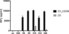

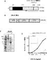


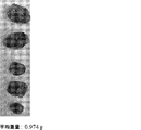


Classifications
-
- C—CHEMISTRY; METALLURGY
- C07—ORGANIC CHEMISTRY
- C07K—PEPTIDES
- C07K16/00—Immunoglobulins [IGs], e.g. monoclonal or polyclonal antibodies
- C07K16/18—Immunoglobulins [IGs], e.g. monoclonal or polyclonal antibodies against material from animals or humans
- C07K16/28—Immunoglobulins [IGs], e.g. monoclonal or polyclonal antibodies against material from animals or humans against receptors, cell surface antigens or cell surface determinants
- C07K16/2866—Immunoglobulins [IGs], e.g. monoclonal or polyclonal antibodies against material from animals or humans against receptors, cell surface antigens or cell surface determinants against receptors for cytokines, lymphokines, interferons
-
- C—CHEMISTRY; METALLURGY
- C07—ORGANIC CHEMISTRY
- C07K—PEPTIDES
- C07K14/00—Peptides having more than 20 amino acids; Gastrins; Somatostatins; Melanotropins; Derivatives thereof
- C07K14/435—Peptides having more than 20 amino acids; Gastrins; Somatostatins; Melanotropins; Derivatives thereof from animals; from humans
- C07K14/705—Receptors; Cell surface antigens; Cell surface determinants
-
- A—HUMAN NECESSITIES
- A61—MEDICAL OR VETERINARY SCIENCE; HYGIENE
- A61K—PREPARATIONS FOR MEDICAL, DENTAL OR TOILETRY PURPOSES
- A61K39/00—Medicinal preparations containing antigens or antibodies
- A61K39/46—Cellular immunotherapy
- A61K39/461—Cellular immunotherapy characterised by the cell type used
- A61K39/4611—T-cells, e.g. tumor infiltrating lymphocytes [TIL], lymphokine-activated killer cells [LAK] or regulatory T cells [Treg]
-
- A—HUMAN NECESSITIES
- A61—MEDICAL OR VETERINARY SCIENCE; HYGIENE
- A61K—PREPARATIONS FOR MEDICAL, DENTAL OR TOILETRY PURPOSES
- A61K39/00—Medicinal preparations containing antigens or antibodies
- A61K39/46—Cellular immunotherapy
- A61K39/463—Cellular immunotherapy characterised by recombinant expression
- A61K39/4631—Chimeric Antigen Receptors [CAR]
-
- A—HUMAN NECESSITIES
- A61—MEDICAL OR VETERINARY SCIENCE; HYGIENE
- A61K—PREPARATIONS FOR MEDICAL, DENTAL OR TOILETRY PURPOSES
- A61K39/00—Medicinal preparations containing antigens or antibodies
- A61K39/46—Cellular immunotherapy
- A61K39/464—Cellular immunotherapy characterised by the antigen targeted or presented
- A61K39/4643—Vertebrate antigens
- A61K39/4644—Cancer antigens
- A61K39/464402—Receptors, cell surface antigens or cell surface determinants
- A61K39/464416—Receptors for cytokines
- A61K39/464421—Receptors for chemokines
-
- C—CHEMISTRY; METALLURGY
- C07—ORGANIC CHEMISTRY
- C07K—PEPTIDES
- C07K16/00—Immunoglobulins [IGs], e.g. monoclonal or polyclonal antibodies
- C07K16/18—Immunoglobulins [IGs], e.g. monoclonal or polyclonal antibodies against material from animals or humans
- C07K16/28—Immunoglobulins [IGs], e.g. monoclonal or polyclonal antibodies against material from animals or humans against receptors, cell surface antigens or cell surface determinants
- C07K16/2803—Immunoglobulins [IGs], e.g. monoclonal or polyclonal antibodies against material from animals or humans against receptors, cell surface antigens or cell surface determinants against the immunoglobulin superfamily
- C07K16/2818—Immunoglobulins [IGs], e.g. monoclonal or polyclonal antibodies against material from animals or humans against receptors, cell surface antigens or cell surface determinants against the immunoglobulin superfamily against CD28 or CD152
-
- C—CHEMISTRY; METALLURGY
- C07—ORGANIC CHEMISTRY
- C07K—PEPTIDES
- C07K16/00—Immunoglobulins [IGs], e.g. monoclonal or polyclonal antibodies
- C07K16/18—Immunoglobulins [IGs], e.g. monoclonal or polyclonal antibodies against material from animals or humans
- C07K16/28—Immunoglobulins [IGs], e.g. monoclonal or polyclonal antibodies against material from animals or humans against receptors, cell surface antigens or cell surface determinants
- C07K16/2803—Immunoglobulins [IGs], e.g. monoclonal or polyclonal antibodies against material from animals or humans against receptors, cell surface antigens or cell surface determinants against the immunoglobulin superfamily
- C07K16/2827—Immunoglobulins [IGs], e.g. monoclonal or polyclonal antibodies against material from animals or humans against receptors, cell surface antigens or cell surface determinants against the immunoglobulin superfamily against B7 molecules, e.g. CD80, CD86
-
- C—CHEMISTRY; METALLURGY
- C12—BIOCHEMISTRY; BEER; SPIRITS; WINE; VINEGAR; MICROBIOLOGY; ENZYMOLOGY; MUTATION OR GENETIC ENGINEERING
- C12N—MICROORGANISMS OR ENZYMES; COMPOSITIONS THEREOF; PROPAGATING, PRESERVING, OR MAINTAINING MICROORGANISMS; MUTATION OR GENETIC ENGINEERING; CULTURE MEDIA
- C12N5/00—Undifferentiated human, animal or plant cells, e.g. cell lines; Tissues; Cultivation or maintenance thereof; Culture media therefor
- C12N5/06—Animal cells or tissues; Human cells or tissues
- C12N5/0602—Vertebrate cells
- C12N5/0634—Cells from the blood or the immune system
- C12N5/0636—T lymphocytes
-
- A—HUMAN NECESSITIES
- A61—MEDICAL OR VETERINARY SCIENCE; HYGIENE
- A61K—PREPARATIONS FOR MEDICAL, DENTAL OR TOILETRY PURPOSES
- A61K39/00—Medicinal preparations containing antigens or antibodies
- A61K2039/505—Medicinal preparations containing antigens or antibodies comprising antibodies
- A61K2039/507—Comprising a combination of two or more separate antibodies
-
- A—HUMAN NECESSITIES
- A61—MEDICAL OR VETERINARY SCIENCE; HYGIENE
- A61K—PREPARATIONS FOR MEDICAL, DENTAL OR TOILETRY PURPOSES
- A61K39/00—Medicinal preparations containing antigens or antibodies
- A61K2039/80—Vaccine for a specifically defined cancer
- A61K2039/812—Breast
-
- A—HUMAN NECESSITIES
- A61—MEDICAL OR VETERINARY SCIENCE; HYGIENE
- A61K—PREPARATIONS FOR MEDICAL, DENTAL OR TOILETRY PURPOSES
- A61K2239/00—Indexing codes associated with cellular immunotherapy of group A61K39/46
- A61K2239/10—Indexing codes associated with cellular immunotherapy of group A61K39/46 characterized by the structure of the chimeric antigen receptor [CAR]
- A61K2239/23—On/off switch
-
- A—HUMAN NECESSITIES
- A61—MEDICAL OR VETERINARY SCIENCE; HYGIENE
- A61K—PREPARATIONS FOR MEDICAL, DENTAL OR TOILETRY PURPOSES
- A61K2239/00—Indexing codes associated with cellular immunotherapy of group A61K39/46
- A61K2239/27—Indexing codes associated with cellular immunotherapy of group A61K39/46 characterized by targeting or presenting multiple antigens
- A61K2239/29—Multispecific CARs
-
- A—HUMAN NECESSITIES
- A61—MEDICAL OR VETERINARY SCIENCE; HYGIENE
- A61K—PREPARATIONS FOR MEDICAL, DENTAL OR TOILETRY PURPOSES
- A61K2239/00—Indexing codes associated with cellular immunotherapy of group A61K39/46
- A61K2239/46—Indexing codes associated with cellular immunotherapy of group A61K39/46 characterised by the cancer treated
- A61K2239/49—Breast
-
- C—CHEMISTRY; METALLURGY
- C07—ORGANIC CHEMISTRY
- C07K—PEPTIDES
- C07K2317/00—Immunoglobulins specific features
- C07K2317/30—Immunoglobulins specific features characterized by aspects of specificity or valency
- C07K2317/31—Immunoglobulins specific features characterized by aspects of specificity or valency multispecific
-
- C—CHEMISTRY; METALLURGY
- C07—ORGANIC CHEMISTRY
- C07K—PEPTIDES
- C07K2317/00—Immunoglobulins specific features
- C07K2317/50—Immunoglobulins specific features characterized by immunoglobulin fragments
- C07K2317/56—Immunoglobulins specific features characterized by immunoglobulin fragments variable (Fv) region, i.e. VH and/or VL
- C07K2317/565—Complementarity determining region [CDR]
-
- C—CHEMISTRY; METALLURGY
- C12—BIOCHEMISTRY; BEER; SPIRITS; WINE; VINEGAR; MICROBIOLOGY; ENZYMOLOGY; MUTATION OR GENETIC ENGINEERING
- C12N—MICROORGANISMS OR ENZYMES; COMPOSITIONS THEREOF; PROPAGATING, PRESERVING, OR MAINTAINING MICROORGANISMS; MUTATION OR GENETIC ENGINEERING; CULTURE MEDIA
- C12N2510/00—Genetically modified cells
Abstract
本発明は、操作された細胞およびそれを使用するための方法に関する。実施形態では、操作された細胞は、キメラ抗原受容体およびポリペプチドをコードする核酸を含み、キメラ抗原受容体は、癌細胞の表面上の2種類以上の抗原に特異的であり、ポリペプチドは、操作された細胞から分泌され得る抗体またはその断片を含む。【選択図】図14The present invention relates to engineered cells and methods for using them. In an embodiment, the engineered cell comprises a nucleic acid encoding a chimeric antigen receptor and a polypeptide, the chimeric antigen receptor is specific for two or more antigens on the surface of a cancer cell, the polypeptide. Includes antibodies or fragments thereof that can be secreted from the engineered cells. [Selection diagram] FIG. 14
Description
この出願は、2018年3月14日に出願された米国仮特許出願第62/643,040号、および2018年4月13日に出願された米国仮特許出願第62/657,151号の優先権を主張し、それぞれの内容は参照によりその全体が本明細書に組み込まれる。 This application is the priority of US Provisional Patent Application No. 62 / 643,040 filed on March 14, 2018, and US Provisional Patent Application No. 62 / 657,151 filed on April 13, 2018. Claiming rights, each content is incorporated herein by reference in its entirety.
本明細書に引用されるすべての特許、特許出願、および刊行物は、参照によりその全体が本明細書に組み込まれる。これらの刊行物の開示は、本明細書に記載され特許請求される本発明の日付の当業者に既知の最新技術をより完全に記載するために、参照により本出願に組み込まれる。 All patents, patent applications, and publications cited herein are incorporated herein by reference in their entirety. Disclosures of these publications are incorporated herein by reference to more fully describe the state of the art known to those skilled in the art on the date of the invention described and claimed herein.
この特許開示は、著作権保護の対象となる、材料を含む。著作権所有者は、米国特許商標局の特許ファイルまたは記録に表れているように、特許文書または特許開示のいずれかによってファクシミリ複製に異議はないが、それ以外では、何であれすべての著作権を留保する。 This patent disclosure includes materials that are subject to copyright protection. The copyright owner has no objection to the facsimile reproduction by either the patent document or the patent disclosure, as shown in the patent file or record of the U.S. Patent and Trademark Office, but otherwise all copyrights. Reserve.
政府の権益
この発明は、米国国立衛生研究所によって授与された助成金番号T32−CA207021に基づく政府の支援を受けて行われた。政府は本発明に一定の権利を有する。
Government Interests This invention was made with government support under grant number T32-CA207021 awarded by the National Institutes of Health. The government has certain rights to the present invention.
発明の分野
本発明は、操作された細胞およびそれを使用する方法を対象とする。実施形態では、操作された細胞は、キメラ抗原受容体およびポリペプチドをコードする核酸を含み、キメラ抗原受容体は、癌細胞の表面上の2種類以上の抗原に特異的であり、ポリペプチドは、操作された細胞から分泌され得る抗体またはその断片を含む。
Field of Invention The present invention is directed to engineered cells and methods of using them. In an embodiment, the engineered cell comprises a nucleic acid encoding a chimeric antigen receptor and a polypeptide, the chimeric antigen receptor is specific for two or more antigens on the surface of the cancer cell, the polypeptide. Includes antibodies or fragments thereof that can be secreted from the engineered cells.
発明の背景
トリプルネガティブ乳癌(TNBC)を標的にする治療法は存在しないため、化学療法が患者に対する唯一の治療法である。抗PD−L1のような免疫療法は、TNBCの限定的な成功(18%の奏効率)を示し、これは、既存の療法が効果的な治療ではないことを示している。
Background of the Invention Chemotherapy is the only treatment for patients because there is no treatment that targets triple-negative breast cancer (TNBC). Immunotherapy, such as anti-PD-L1, has shown limited success in TNBC (18% response rate), indicating that existing therapies are not effective treatments.
本発明の態様は、キメラ抗原受容体およびポリペプチドをコードする核酸を含む操作された細胞に向けられている。 Aspects of the invention are directed to engineered cells containing a nucleic acid encoding a chimeric antigen receptor and a polypeptide.
実施形態では、操作された細胞は、操作されたT細胞である。非限定的な例は、細胞傷害性T細胞(TC、細胞傷害性Tリンパ球、CTL、T−キラー細胞、細胞溶解性T細胞、CD8+T細胞またはキラーT細胞としても知られる)、CD4+T細胞、NK細胞、およびNKT細胞を含む。 In an embodiment, the engineered cell is an engineered T cell. Non-limiting examples include cytotoxic T cells (also known as TC, cytotoxic T lymphocytes, CTL, T-killer cells, cytolytic T cells, CD8 + T cells or killer T cells), CD4 + T cells, Includes NK cells and NKT cells.
実施形態では、キメラ抗原受容体は、抗原認識ドメインおよびシグナル伝達および/または刺激ドメインを含む。 In embodiments, the chimeric antigen receptor comprises an antigen recognition domain and a signal transduction and / or stimulation domain.
実施形態では、抗原認識ドメインは、細胞の表面上の2種類以上の抗原に特異的である。例えば、細胞は癌細胞である。例えば、抗原は、CXCケモカイン受容体タイプ4およびクローディン4を含む。実施形態では、キメラ抗原受容体(および/または抗原認識ドメイン)は、1つ以上の抗体断片を含む。実施形態では、キメラ抗原受容体はscFVを含む。実施形態では、キメラ抗原受容体は、CXCケモカイン受容体タイプ4およびクローディン4を対象とする(標的とする)scFvを含む。
In embodiments, the antigen recognition domain is specific for two or more antigens on the surface of the cell. For example, cells are cancer cells. For example, antigens include CXC
実施形態では、シグナル伝達および/または刺激ドメインは、CD28、41BB、CD3−ゼータ細胞内シグナル伝達ドメイン、またはそれらの断片を含む。 In embodiments, signaling and / or stimulating domains include CD28, 41BB, CD3-zeta intracellular signaling domains, or fragments thereof.
実施形態では、ポリペプチドは、操作された細胞から分泌され得る抗体またはその断片を含む。実施形態では、分泌されたポリペプチドは、対象の免疫系を調節する。 In embodiments, the polypeptide comprises an antibody or fragment thereof that can be secreted from the engineered cell. In embodiments, the secreted polypeptide regulates the subject's immune system.
実施形態では、ポリペプチドは、ミニボディなどの抗体またはその断片を含む。抗体の非限定的な例には、モノクローナル抗体、ポリクローナル抗体、多重特異性抗体(例えば、二重特異性抗体)、ヒト化抗体、完全ヒト抗体が含まれる。実施形態では、ポリペプチドは、単一特異性抗体、二重特異性抗体、三重特異性抗体、または多重特異性抗体を含む。抗体断片の非限定的な例は、Fv、Fab、Fab’、Fab’−SH、F(ab’)2、ダイアボディ、直鎖状抗体、一本鎖抗体分子(例えば、scFv)、抗体断片から形成された多重特異性抗体を含む。一実施形態では、本発明は、標的抗原に向けられた(および標的抗原に特異的な)scFVを含む。 In embodiments, the polypeptide comprises an antibody such as a minibody or a fragment thereof. Non-limiting examples of antibodies include monoclonal antibodies, polyclonal antibodies, multispecific antibodies (eg, bispecific antibodies), humanized antibodies, fully human antibodies. In embodiments, the polypeptide comprises a monospecific antibody, a bispecific antibody, a trispecific antibody, or a multispecific antibody. Non-limiting examples of antibody fragments include Fv, Fab, Fab', Fab'-SH, F (ab') 2, diabodies, linear antibodies, single-chain antibody molecules (eg, scFv), antibody fragments. Contains multispecific antibodies formed from. In one embodiment, the invention comprises a target antigen-directed (and target antigen-specific) scFV.
実施形態では、ポリペプチドは、CARを発現するものとは別個であるが、CARと同じDNAベクターの構成要素である発現構築物から発現される。実施形態では、ポリペプチドは、CARを発現するものとは別個であり、CARとは異なるDNAベクターの構成要素である発現構築物から発現される。 In embodiments, the polypeptide is expressed from an expression construct that is a component of the same DNA vector as CAR, although it is separate from those that express CAR. In embodiments, the polypeptide is separate from those expressing CAR and is expressed from an expression construct that is a component of a DNA vector different from CAR.
実施形態では、構築物は、1つ以上のウイルスベクターにクローニングされる。非限定的な例は、レンチウイルスベクターを含む。 In an embodiment, the construct is cloned into one or more viral vectors. Non-limiting examples include lentiviral vectors.
本発明の態様は、さらに癌に罹患している対象を治療する方法に向けられている。 Aspects of the invention are further directed to methods of treating a subject suffering from cancer.
本発明の態様は、さらに対象における癌の進行を低減させるかまたは癌の退縮を促進させる方法に向けられている。 Aspects of the present invention are further directed to methods of reducing cancer progression or promoting cancer regression in a subject.
なおさらに、本発明の態様は、対象における癌細胞の細胞増殖を減少させる方法に向けられている。 Furthermore, aspects of the invention are directed to methods of reducing cell proliferation of cancer cells in a subject.
さらに、本発明の態様は、細胞、好ましくは癌細胞の細胞傷害性を誘導する方法に向けられている。 Furthermore, aspects of the invention are directed to methods of inducing cytotoxicity of cells, preferably cancer cells.
実施形態では、方法は、操作されたT細胞など、本明細書に記載の操作された細胞の治療上有効量を対象に投与することを含む。 In embodiments, the method comprises administering to the subject a therapeutically effective amount of the engineered cells described herein, such as engineered T cells.
他の実施形態では、この方法は、本明細書に記載の治療上有効量の分泌されたポリペプチドを対象に投与することを含む。 In other embodiments, the method comprises administering to the subject a therapeutically effective amount of a secreted polypeptide described herein.
癌の非限定的な例には、癌腫、リンパ腫、芽細胞腫、肉腫、および白血病が含まれる。そのような癌のより具体的な例には、扁平上皮癌、小細胞肺癌、非小細胞肺癌、肺腺癌、肺扁平上皮癌、腹膜癌、肝細胞癌、胃腸癌、膵臓癌、神経膠芽腫、子宮頸部癌、卵巣癌、肝臓癌、膀胱癌、ヘパトーマ、乳癌、結腸癌、結腸直腸癌、子宮内膜癌または子宮癌、唾液腺癌、腎臓癌、肝臓癌、前立腺癌、外陰癌、甲状腺癌、肝癌、およびさまざまなタイプの頭頸部癌が含まれる。 Non-limiting examples of cancers include carcinomas, lymphomas, blastomas, sarcomas, and leukemias. More specific examples of such cancers include squamous cell carcinoma, small cell lung cancer, non-small cell lung cancer, lung adenocarcinoma, lung squamous cell carcinoma, peritoneal cancer, hepatocellular carcinoma, gastrointestinal cancer, pancreatic cancer, gliosis. Sprouting cancer, cervical cancer, ovarian cancer, liver cancer, bladder cancer, hepatoma, breast cancer, colon cancer, colorectal cancer, endometrial cancer or uterine cancer, salivary adenocarcinoma, kidney cancer, liver cancer, prostate cancer, genital cancer , Thyroid cancer, liver cancer, and various types of head and neck cancer.
実施形態では、癌は、トリプルネガティブ乳癌(TNBC)を含む。 In embodiments, the cancer comprises triple negative breast cancer (TNBC).
本発明の態様は、さらに、操作されたCAR T細胞の殺傷能力(killing capability)を評価するための方法に向けられている。 Aspects of the invention are further directed to methods for assessing the killing capacity of manipulated CAR T cells.
実施形態では、その方法は、1つ以上のタイプの癌から細胞を入手することと、細胞を染色するために、細胞を色素と混合することと、マルチウェルプレートに細胞を播種することと、混合物をある期間にわたってインキュベートすることと、異なるT細胞タイプを混合物に追加して、第2の混合物を作製することと、第2の混合物をある期間にわたって共培養することと、操作されたCAR T細胞の殺傷能力を評価することと、を含む。実施形態では、評価することは、プレートを走査して分析することを含む。実施形態では、プレートは、明視野および青色蛍光チャネルを使用して分析することができる。 In embodiments, the method involves obtaining cells from one or more types of cancer, mixing the cells with a dye to stain the cells, and seeding the cells in a multiwell plate. Incubating the mixture over a period of time, adding different T cell types to the mixture to make a second mixture, co-culturing the second mixture over a period of time, and engineered CAR T. Includes assessing the killing ability of cells. In embodiments, assessing involves scanning and analyzing the plate. In embodiments, the plates can be analyzed using brightfield and blue fluorescent channels.
実施形態では、細胞は、1つ以上のタイプの癌細胞を含む。実施形態では、細胞は、腎臓(例えば、HEK293T細胞)、乳癌細胞(例えば、MDA−MB−231細胞、MDA−MB−468細胞、HCC38細胞)、および腎臓癌細胞(sk−rc−59細胞)から単離された細胞のうちの1つ以上を含む。 In an embodiment, the cell comprises one or more types of cancer cells. In embodiments, the cells are kidney (eg, HEK293T cells), breast cancer cells (eg, MDA-MB-231 cells, MDA-MB-468 cells, HCC38 cells), and kidney cancer cells (sk-rc-59 cells). Contains one or more of the cells isolated from.
実施形態では、色素は、ViaStain(商標)トレーサーブルー色素を含む。 In embodiments, the dye comprises a ViaStain ™ Tracer Blue dye.
実施形態では、プレートは、4、6、8、12、24、48、96、または96を超えるウェルを有するマルチウェルプレートを含む。 In embodiments, the plate comprises a multi-well plate having more than 4, 6, 8, 12, 24, 48, 96, or 96 wells.
実施形態では、期間は、約2時間、4時間、6時間、8時間、12時間、16時間、24時間、36時間、48時間、または48時間より長い時間を指す。 In embodiments, the period refers to a time of about 2 hours, 4 hours, 6 hours, 8 hours, 12 hours, 16 hours, 24 hours, 36 hours, 48 hours, or longer than 48 hours.
本発明の他の目的および利点は、以下の記載から容易に明らかになるであろう。 Other objects and advantages of the present invention will be readily apparent from the description below.
発明の詳細な説明
トリプルネガティブ乳癌(TNBC)は、臨床予後が不良の高悪性度の乳癌のサブタイプである。化学療法で治療している間の、高い再発率は新規のターゲティングされた療法の必要性を示している。
Description of the Invention Triple-negative breast cancer (TNBC) is a subtype of high-grade breast cancer with a poor clinical prognosis. High recurrence rates while being treated with chemotherapy indicate the need for new targeted therapies.
キメラ抗原受容体(CAR)T細胞療法は、CARの外因性発現によって患者のT細胞を、腫瘍細胞を殺傷するようにリダイレクトする。CARは、抗体の抗原認識ドメインをT細胞受容体および共受容体の細胞内シグナル伝達ドメインに連結する、膜貫通型融合タンパク質であり得る。固形腫瘍は、CAR−T療法に特有の課題を提示する。血液癌とは異なり、腫瘍に関連する標的タンパク質は腫瘍と健康な組織の間で過剰発現し、その結果、オンターゲット/オフ腫瘍の、T細胞による健康な組織の殺傷が起こる。さらに、腫瘍微小環境(TME)における免疫抑制は、腫瘍の殺傷に向かうCAR−T細胞の活性化を制限する。 Chimeric antigen receptor (CAR) T cell therapy redirects a patient's T cells to kill tumor cells by exogenous expression of CAR. CAR can be a transmembrane fusion protein that links the antigen recognition domain of an antibody to the intracellular signaling domains of T cell receptors and co-receptors. Solid tumors present challenges specific to CAR-T therapy. Unlike hematological malignancies, tumor-related target proteins are overexpressed between the tumor and healthy tissue, resulting in on-target / off-tumor killing of healthy tissue by T cells. In addition, immunosuppression in the tumor microenvironment (TME) limits the activation of CAR-T cells towards tumor killing.
本発明は、TNBCに対する抗体を分泌する操作されたキメラ抗原受容体(CAR)T細胞ファクトリに関する。理論に縛られることを望まないが、TNBCなどの固形腫瘍の2種類の抗原を標的とする二重特異性CARは、オンターゲット/オフ腫瘍のT細胞による殺傷を軽減し、チェックポイント遮断抗体の分泌によって腫瘍微小環境での抑制を取り除く。局所的な免疫回復に続いて、CAR−T細胞とTME内の他の細胞は相乗的に作用して、腫瘍を縮小させおよび排除する。 The present invention relates to an engineered chimeric antigen receptor (CAR) T cell factory that secretes antibodies against TNBC. Although not bound by theory, bispecific CARs that target two antigens in solid tumors such as TNBC reduce T cell killing of on-target / off tumors and are checkpoint blocking antibodies. Secretion removes inhibition in the tumor microenvironment. Following local immune recovery, CAR-T cells and other cells within the TME act synergistically to shrink and eliminate the tumor.
略語と定義
1つ以上の好ましい実施形態の詳細な説明が本明細書に提供されている。しかしながら、本発明が、さまざまな形式で具現化され得ることが、理解される。したがって、本明細書に開示された特定の詳細は、限定として解釈されるべきではなく、特許請求の範囲の根拠として、および当業者に本発明を任意の適切な方法で用いるように教示するための代表的な根拠として解釈されるべきである。
Abbreviations and Definitions A detailed description of one or more preferred embodiments is provided herein. However, it is understood that the present invention can be embodied in various forms. Therefore, the particular details disclosed herein should not be construed as limiting, as a basis for the claims and to teach those skilled in the art to use the invention in any suitable manner. Should be interpreted as a representative basis for.
単数形「1つの(a)」、「1つの(an)」、および「その(the)」は、文脈が明確に他のことを指示しない限り、複数の参照を含む。特許請求の範囲および/または明細書において「含む」という用語とともに使用されるときの「1つの(a)」または「1つの(an)」という単語の使用は、「1つ」を意味し得るが、「1つ以上」、「少なくとも1つ」および「1つ以上」の意味と両立する。 The singular forms "one (a)", "one (an)", and "the" include multiple references unless the context explicitly indicates otherwise. The use of the word "one (a)" or "one (an)" when used with the term "contains" in the claims and / or specification may mean "one." Is compatible with the meanings of "one or more", "at least one" and "one or more".
句「例えば」、「など」、「含む」などが本明細書で使用される場合は常に、他に明確に明記されない限り、句「限定することなく」が付随することが理解される。同様に、「一例」、「例示的」などは、非限定的であると理解される。 It is understood that whenever the phrase "eg", "etc.", "contains", etc. are used herein, they are accompanied by the phrase "without limitation" unless otherwise explicitly stated. Similarly, "example", "exemplary", etc. are understood to be non-limiting.
用語「実質的に」は、意図した目的に悪影響を与えない記述語からの逸脱を可能にする。記述用語は、単語「実質的に」が明確に列挙されていなくても、用語「実質的に」によって修正されることが理解される。 The term "substantially" allows deviations from descriptive terms that do not adversely affect the intended purpose. It is understood that the descriptive term is modified by the term "substantially" even if the word "substantially" is not explicitly listed.
用語「含んでいる(comprising)」および「含んでいる(including)」、ならびに「有している(having)」および「伴っている(involving)」(および同様に「含む(comprises)」、「含む(includes)」、「有する(has)」、および「伴う(involves)」)などは、交換可能に使用され、同じ意味を有している。具体的には、用語それぞれは、「含んでいる(comprising)」の一般的な米国特許法の定義と一致して定義され、それ故、非限定用語(open term)の意味「少なくとも以下の(at least the following)」であると解釈され、追加の特徴、限定、態様などを除外しないとも解釈される。したがって、例えば、「工程a、b、およびcを伴うプロセス」は、プロセスが少なくとも工程a、bおよびcを含むことを意味している。用語「1つの(a)」、「1つの(an)」が使用される場合は常に、そのような解釈が文脈において無意味でない限り、「1以上」と理解される。 The terms "comprising" and "inclusion", as well as "having" and "involving" (and similarly "comprising", "comprises", " "Includes", "has", and "involves") are used interchangeably and have the same meaning. Specifically, each term is defined in line with the general definition of "comprising" in US patent law, and therefore the meaning of the non-limiting term (open term) is "at least (at least: It is interpreted as "at least the following", and it is also interpreted as not excluding additional features, limitations, aspects, and the like. Thus, for example, "process involving steps a, b, and c" means that the process comprises at least steps a, b, and c. Whenever the terms "one (a)" and "one (an)" are used, they are understood as "one or more" unless such an interpretation is meaningless in the context.
本明細書で使用される「約」という用語は、本明細書では、おおよそ、大まかに、およそ、またはその領域内を意味するために使用される。「約」という用語が数値範囲とともに使用される場合、それは、記載された数値の上および下の境界を拡張することによってその範囲を変更する。一般に、用語「約」は、本明細書では、数値を、記載された値の上下に20パーセントの変動(上下)で変更するために使用される。 The term "about" as used herein is used herein to mean roughly, roughly, roughly, or within that area. When the term "about" is used with a numerical range, it modifies the range by extending the boundaries above and below the listed numbers. Generally, the term "about" is used herein to change a number with a 20 percent variation (up and down) above and below the stated value.
操作されたCAR T細胞
キメラ抗原受容体(CAR)T細胞療法は、CARの外因性発現によって患者のT細胞を腫瘍細胞を殺傷するようにリダイレクトする。CARは、抗体または断片の抗原認識ドメインをT細胞受容体および共受容体の細胞内シグナル伝達ドメインに連結する、膜貫通型融合タンパク質である。例えば、キメラ抗原受容体は、抗原特異的な抗体断片をT細胞共刺激ドメインとCD3ゼータ細胞内シグナル伝達ドメインに融合させて、目的の細胞、例えば、腫瘍細胞上に提示された抗原へのT細胞のリダイレクションを可能にする。
Manipulated CAR T cells Chimeric antigen receptor (CAR) T cell therapy redirects a patient's T cells to kill tumor cells by exogenous expression of CAR. CAR is a transmembrane fusion protein that links the antigen recognition domain of an antibody or fragment to the intracellular signaling domain of T cell receptors and co-receptors. For example, a chimeric antigen receptor fuses an antigen-specific antibody fragment into a T cell co-stimulation domain and a CD3 zeta intracellular signaling domain to T to an antigen presented on a cell of interest, eg, a tumor cell. Allows cell redirection.
本明細書における「抗体」という用語は、最も広い意味で使用され、免疫グロブリン分子および免疫グロブリン(Ig)分子の免疫学的に活性な部分、すなわち、抗原と特異的に結合する(と免疫反応する)抗原結合部位を含む分子を指す。「特異的に結合する」または「と免疫反応する」は、所望の抗原の1つ以上の抗原決定基と反応し、他のポリペプチドと反応しない抗体を指すことができる。本発明の抗体には、ポリクローナル、モノクローナル、ヒト化、完全ヒト、二重特異性、多重特異性、キメラ、dAb(ドメイン抗体)、一本鎖抗体、Fab、Fab’およびF(ab’)2断片、scFv、ダイアボディ、ミニボディ、scFv−Fc融合、およびFab発現ライブラリが含まれるが、これらに限定されない。反対の明示がない限り、本明細書でなされる「抗体(単数)」または「抗体(複数)」への言及は、それらが所望の抗原結合活性を示す限り、例えば、これらの分子の任意の(またはすべての)ものを包含する。 The term "antibody" as used herein is used in the broadest sense and specifically binds (and reacts to) an immunologically active portion of an immunoglobulin molecule and an immunoglobulin (Ig) molecule, i.e. an antigen. Refers to a molecule that contains an antigen-binding site. "Specifically binds" or "immune reacts with" can refer to an antibody that reacts with one or more antigenic determinants of the desired antigen and does not react with other polypeptides. The antibodies of the invention include polyclonal, monoclonal, humanized, fully human, bispecific, multispecific, chimeric, dAb (domain antibody), single chain antibody, Fab, Fab'and F (ab') 2 Fragments, scFv, diabody, minibody, scFv-Fc fusion, and Fab expression libraries are included, but not limited to. Unless otherwise stated, references made herein to "antibodies (singular)" or "antibodies (plural)" are made, for example, of any of these molecules, as long as they exhibit the desired antigen-binding activity. Includes (or all) things.
一本鎖Fv(「scFv」)ポリペプチド分子は、共有結合的に連結されたVH::VLヘテロダイマーであり、ペプチドをコード化するリンカーによって連結された、VHをコード化する遺伝子およびVLをコード化する遺伝子を含む遺伝子融合体から発現できる。(Huston et al.(1988)Proc Nat Acad Sci USA 85(16):5879−5883を参照されたい)。自然に凝集しているが化学的に分離された、軽鎖および重鎖ポリペプチド鎖を、抗体V領域から、抗原結合部位の構造と実質的に同様の三次元構造に折り畳まれるscFv分子に変換するための化学構造を識別するための多くの方法が説明されている。例えば、米国特許第5,091,513号、同第5,132,405号、および同第4,946,778号を参照されたい。 The single-chain Fv (“scFv”) polypeptide molecule is a covalently linked VH :: VL heterodimer that contains VH-encoding genes and VL linked by a peptide-encoding linker. It can be expressed from a gene fusion containing the encoding gene. (See Huston et al. (1988) Proc Nat Acad Sci USA 85 (16): 5879-5883). Converts naturally aggregated but chemically separated light and heavy chain polypeptide chains from the antibody V region into scFv molecules that fold into a three-dimensional structure that is substantially similar to the structure of the antigen binding site. Many methods for identifying the chemical structure to do so are described. See, for example, U.S. Pat. Nos. 5,091,513, 5,132,405, and 4,946,778.
二重特異性抗体とは、少なくとも2つの異なる抗原に対して結合特異性を有する抗体を指す。例えば、二重特異性抗体は、ヒト抗体またはヒト化抗体などのモノクローナル抗体であり得る。この場合、結合特異性の1つはCXCR4および/またはクローディン4である。第2の結合標的は任意の他の抗原であり、有利なことに細胞表面タンパク質または受容体または受容体サブユニットである。たとえば、結合特異性の1つはCXCR4に対するもので、第2の結合特異性はクローディン4に対するものである。
Bispecific antibody refers to an antibody that has binding specificity for at least two different antigens. For example, the bispecific antibody can be a monoclonal antibody such as a human antibody or a humanized antibody. In this case, one of the binding specificities is CXCR4 and / or
二重特異性抗体は、全長抗体または抗体断片(例えば、F(ab’)2二重特異性抗体)として調製することができる。二重特異性抗体を作製する方法は当技術分野で知られている。例えば、その全体が参照により本明細書に組み込まれる米国特許第8,329,178号を参照されたい。 Bispecific antibodies, full length antibodies or antibody fragments (e.g., F (ab ') 2 bispecific antibodies) can be prepared as. Methods for making bispecific antibodies are known in the art. See, for example, US Pat. No. 8,329,178, which is incorporated herein by reference in its entirety.
一般に、ヒトから得られる抗体分子は、分子中に存在する重鎖の性質が互いに異なる、IgG、IgM、IgA、IgE、およびIgDのクラスのいずれかに関連する。特定のクラスには、IgG1、IgG2などのサブクラスもある。さらに、ヒトでは、軽鎖はカッパ鎖またはラムダ鎖であり得る。 Generally, antibody molecules obtained from humans are associated with any of the IgG, IgM, IgA, IgE, and IgD classes in which the properties of the heavy chains present in the molecule are different from each other. Certain classes also include subclasses such as IgG 1 and IgG 2. Moreover, in humans, the light chain can be a kappa chain or a lambda chain.
「抗原結合部位」または「結合部分」という用語は、抗原結合に関与する免疫グロブリン分子の部分を指す。抗原結合部位は、重(「H」)鎖および軽(「L」)鎖のN末端可変(「V」)領域のアミノ酸残基によって形成される。「超可変領域」と呼ばれる、重鎖と軽鎖のV領域内の3つの高度に異なる区間(stretch)は、「フレームワーク領域」または「FR」として知られるより保存された隣接区間の間に挿入される。したがって、「FR」という用語は、免疫グロブリンの超可変領域の間、およびそれに隣接して天然に見出されるアミノ酸配列を指す。抗体分子において、軽鎖の3つの超可変領域および重鎖の3つの超可変領域は、3次元空間で互いに対して配置されて、抗原結合表面を形成する。抗原結合表面は、結合した抗原の3次元表面に相補的であり、重鎖および軽鎖のそれぞれの3つの超可変領域は、「相補性決定領域」または「CDR」と呼ばれる。 The term "antigen binding site" or "binding portion" refers to the portion of an immunoglobulin molecule involved in antigen binding. The antigen binding site is formed by amino acid residues in the N-terminal variable (“V”) region of the heavy (“H”) and light (“L”) chains. Three highly different sections (stretches) within the heavy and light chain V regions, called "hypervariable regions", are between more conserved adjacent sections known as "framework regions" or "FRs". Will be inserted. Thus, the term "FR" refers to a naturally found amino acid sequence between and adjacent to hypervariable regions of immunoglobulins. In an antibody molecule, the three hypervariable regions of the light chain and the three hypervariable regions of the heavy chain are located relative to each other in three-dimensional space to form an antigen-binding surface. The antigen-binding surface is complementary to the three-dimensional surface of the bound antigen, and each of the three hypervariable regions of the heavy and light chains is referred to as the "complementarity determining regions" or "CDRs."
固形腫瘍は、CAR−T療法に特有の課題を提示する。血液癌とは異なり、腫瘍に関連する標的タンパク質は腫瘍と健康な組織の間で過剰発現し、その結果、オンターゲット/オフ腫瘍の、T細胞による健康な組織の殺傷が起こる。さらに、腫瘍微小環境(TME)における免疫抑制は、腫瘍の殺傷に向かうCAR−T細胞の活性化を制限する。本発明の態様は、これらの問題に対処する。例えば、実施形態は、(a)癌細胞上の2種類の抗原を標的にして、オンターゲット/オフ腫瘍のT細胞による殺傷を軽減し、および(b)腫瘍微小環境における抑制を取り除くチェックポイント遮断抗体を分泌する、二重特異性CARを含むT細胞を含む。 Solid tumors present challenges specific to CAR-T therapy. Unlike hematological malignancies, tumor-related target proteins are overexpressed between the tumor and healthy tissue, resulting in on-target / off-tumor killing of healthy tissue by T cells. In addition, immunosuppression in the tumor microenvironment (TME) limits the activation of CAR-T cells towards tumor killing. Aspects of the invention address these issues. For example, embodiments are checkpoint blockers that (a) target two antigens on cancer cells to reduce T cell killing of on-target / off tumors and (b) remove inhibition in the tumor microenvironment. Includes T cells containing bispecific CAR that secrete antibodies.
腫瘍の進行に関連する新たなメカニズムは免疫チェックポイント経路であり、これには、正常な条件下でT細胞の過剰な活性化を防いで、自己限定的な方法でT細胞の機能を可能にする、細胞相互作用が含まれる。回避メカニズムとして、多くの腫瘍は免疫チェックポイント分子の発現を刺激することができ、腫瘍の進行を抑制できないT細胞のアネルギー性表現型をもたらす。たとえば、新しい臨床データは、細胞傷害性Tリンパ球による癌細胞の殺傷を防止する、免疫チェックポイントとしての1つの阻害性リガンドと受容体のペアであるプログラム死リガンド1(PD−L1、B7−H1およびCD274)とプログラム死受容体1(PD−1、CD279)の重要性を強調している。PD1受容体は、T細胞、B細胞、ナチュラルキラー細胞(NK)、宿主組織などの多くの細胞型で発現している。PD−L1を発現する腫瘍および抗原提示細胞(APC)は、受容体PD−1への結合を介して細胞傷害性Tリンパ球のT細胞受容体(TCR)シグナル伝達を遮断でき、サイトカインの生成およびT細胞増殖を減少させる。PD−L1の過剰発現は多くの腫瘍タイプで見られ、CD80(B7.1)を含む他のタンパク質との相互作用を通じて免疫抑制機能を媒介し、CD28への結合を介してT細胞を活性化する能力をブロックする。 A new mechanism associated with tumor progression is the immune checkpoint pathway, which prevents excessive activation of T cells under normal conditions and allows T cell function in a self-limiting manner. Includes cell interactions. As an evasion mechanism, many tumors can stimulate the expression of immune checkpoint molecules, resulting in an anergistic phenotype of T cells that cannot suppress tumor progression. For example, new clinical data show program death ligand 1 (PD-L1, B7-), which is a pair of inhibitory ligand and receptor as an immune checkpoint that prevents the killing of cancer cells by cytotoxic T lymphocytes. It emphasizes the importance of H1 and CD274) and Program Death Receptor 1 (PD-1, CD279). The PD1 receptor is expressed in many cell types such as T cells, B cells, natural killer cells (NK), and host tissues. Tumors and antigen-presenting cells (APCs) that express PD-L1 can block the T cell receptor (TCR) signaling of cytotoxic T lymphocytes through binding to receptor PD-1 and produce cytokines. And reduce T cell proliferation. Overexpression of PD-L1 is found in many tumor types, mediates immunosuppressive function through interaction with other proteins, including CD80 (B7.1), and activates T cells through binding to CD28. Block the ability to do.
腫瘍指向性キメラ抗原受容体(CAR)を発現するヒトリンパ球の遺伝子操作は、タンパク質抗原のプロセシングおよび提示の異常による腫瘍免疫回避メカニズムを迂回する抗腫瘍エフェクター細胞を生成できる。さらに、これらのトランスジェニック受容体は、タンパク質由来ではない腫瘍関連抗原を対象とすることができる。本発明の特定の実施形態では、少なくともCARを含むように改変されたリンパ球(CART)があり、本発明の特別な実施形態では、単一のCARが2種類以上の抗原を標的とする。いくつかの実施形態では、CARTは、例えば抗体またはサイトカインなどの1つ以上のポリペプチドを発現および分泌するようにさらに改変される。そのようなCARTは、本明細書では、武装化(armed)CARTまたはCARファクトリと呼ばれる。武装化CARTは、標的部位(すなわち、腫瘍部位)での局所的なポリペプチドの同時分泌を可能にする。 Genetic engineering of human lymphocytes expressing a tumor-directed chimeric antigen receptor (CAR) can generate antitumor effector cells that bypass the tumor immune avoidance mechanism due to abnormal processing and presentation of protein antigens. In addition, these transgenic receptors can target tumor-related antigens that are not protein-derived. In certain embodiments of the invention there are lymphocytes (CART) modified to contain at least CAR, and in a particular embodiment of the invention a single CAR targets two or more antigens. In some embodiments, the CART is further modified to express and secrete one or more polypeptides, such as antibodies or cytokines. Such CARTs are referred to herein as armed CARTs or CAR factories. Armed CART allows for co-secretion of local polypeptides at the target site (ie, the tumor site).
特定の腫瘍関連抗原に対する高親和性によって以前に選択された一本鎖可変抗体断片(scFv)を含むキメラ抗原受容体(CAR)と呼ばれる改変TCRは、癌に対する強力な新しいアプローチである。CARに提示されているscFvは、CD3ζを含む細胞内シグナル伝達ブロックに連結されて、T細胞の活性化を誘導してから抗原に結合する。この構造は第一世代CARに特徴的であり、CD28、4−1BB、もしくはOX40のシグナル伝達共刺激エンドドメインをCD3に連結する第2世代CAR、またはCD3ζに2つの要素をタンデムに連結する第3世代CAR、に改善されたものである。これらのエンドドメインは、抗原提示細胞(APC)によるTCR認識中の完全なT細胞活性化に必要であり、CAR−T細胞のサイトカイン産生と増殖を改善する。特有な腫瘍関連抗原を見つけることが困難なため、腫瘍の場所へのT細胞の非効率的なホーミングのため、体内でのT細胞の持続性の低下のため、および固形腫瘍の免疫抑制性微小環境のため、CART細胞の効果はこれまで固形腫瘍の治療に控えめであった。 A modified TCR called a chimeric antigen receptor (CAR) containing a single chain variable antibody fragment (scFv) previously selected for its high affinity for a particular tumor-related antigen is a powerful new approach to cancer. The scFv presented in the CAR is linked to an intracellular signaling block containing CD3ζ to induce T cell activation before binding to the antigen. This structure is characteristic of the first generation CAR, the second generation CAR linking the signaling costimulatory end domain of CD28, 4-1BB, or OX40 to CD3, or the second element linking two elements to CD3ζ in tandem. It is an improvement to the 3rd generation CAR. These endodomains are required for complete T cell activation during TCR recognition by antigen presenting cells (APCs) and improve cytokine production and proliferation of CAR-T cells. Due to the difficulty in finding specific tumor-related antigens, inefficient homing of T cells to the location of the tumor, reduced persistence of T cells in the body, and immunosuppressive microscopic of solid tumors Due to the environment, the effects of CART cells have traditionally been modest in the treatment of solid tumors.
特定の場合において、リンパ球は、キメラで、非天然であり、そして少なくとも部分的に人の手によって操作された受容体を含み得る。特定の場合において、操作されたキメラ抗原受容体(CAR)は、1、2、3、4、またはそれ以上の成分を有し、いくつかの実施形態では、1つ以上の成分が、リンパ球が1つ以上の腫瘍抗原含有癌細胞を標的にするまたはそれに結合するのを促進する。 In certain cases, lymphocytes may contain receptors that are chimeric, unnatural, and at least partially manipulated by the human hand. In certain cases, the engineered chimeric antigen receptor (CAR) has 1, 2, 3, 4, or more components, and in some embodiments, one or more components are lymphocytes. Promotes targeting or binding to one or more tumor antigen-containing cancer cells.
本発明によるCARは、一般に、少なくとも1つの細胞外リガンド結合ドメインを含む少なくとも1つの膜貫通ポリペプチドと、少なくとも1つの細胞内シグナル伝達ドメインを含む1つの膜貫通ポリペプチドを含み、その結果ポリペプチドが一緒に集合してキメラ抗原受容体を形成する。本発明の態様で有用な例示的なCARには、例えばPCT/US2015/067225に開示されているものが含まれる。 CARs according to the invention generally include at least one transmembrane polypeptide containing at least one extracellular ligand binding domain and one transmembrane polypeptide containing at least one intracellular signaling domain, resulting in the polypeptide. Aggregate together to form a chimeric antigen receptor. Illustrative CARs useful in aspects of the invention include, for example, those disclosed in PCT / US2015 / 067225.
本明細書で使用される「細胞外リガンド結合ドメイン」という用語は、リガンドに結合することができるオリゴまたはポリペプチドを指すことができる。ドメインは、細胞表面分子と相互作用することができる。例えば、細胞外リガンド結合ドメインは、特定の疾患状態に関連する標的細胞上の細胞表面マーカーとして作用するリガンドを認識するように選択され得る。 As used herein, the term "extracellular ligand binding domain" can refer to an oligo or polypeptide capable of binding to a ligand. Domains can interact with cell surface molecules. For example, extracellular ligand binding domains may be selected to recognize ligands that act as cell surface markers on target cells associated with a particular disease state.
特に、細胞外リガンド結合ドメインは、標的の抗原に対する抗体に由来する抗原結合ドメインまたは抗原認識ドメインを含み得る。抗原結合ドメインまたは抗原認識ドメインは、抗体断片であり得る。「抗体断片」は、無傷の抗体が結合する抗原に結合する無傷の抗体の一部を含む無傷の抗体以外の分子であり得る。抗体断片の例には、Fv、Fab、Fab’、Fab’−SH、F(ab’)2、ダイアボディ、直鎖状抗体、一本鎖抗体分子(例えばscFv)、および抗体断片から形成された多重特異性抗体が含まれるがこれらに限定されない。図2を参照すると、例えば、一実施形態は、抗原認識ドメインとしてscFvを有するCARを含む。 In particular, the extracellular ligand binding domain may include an antigen binding domain or antigen recognition domain derived from an antibody against a target antigen. The antigen binding domain or antigen recognition domain can be an antibody fragment. An "antibody fragment" can be a molecule other than an intact antibody that comprises a portion of the intact antibody that binds to the antigen to which the intact antibody binds. Examples of antibody fragments are formed from Fv, Fab, Fab', Fab'-SH, F (ab') 2, diabody, linear antibody, single chain antibody molecule (eg scFv), and antibody fragment. Multispecific antibodies are included, but are not limited to these. Referring to FIG. 2, for example, one embodiment comprises a CAR having scFv as an antigen recognition domain.
抗原認識ドメインは、目的の任意の抗原標的に向けることができる。実施形態では、目的の抗原標的は、癌細胞の表面などの細胞の表面上にある。抗原標的の非限定的な例は、CXCケモカイン受容体タイプ4(CXCR−4)および/またはクローディン4を含む。
The antigen recognition domain can be directed to any antigen target of interest. In embodiments, the antigen target of interest is on the surface of a cell, such as the surface of a cancer cell. Non-limiting examples of antigen targets include CXC chemokine receptor type 4 (CXCR-4) and / or
非限定的な例として、標的の抗原は、腫瘍関連表面抗原、例えばErbB2(HER2/neu)、癌胎児性抗原(CEA)、上皮細胞接着分子(EpCAM)、上皮成長因子受容体(EGFR)、EGFRバリアントIII(EGFRvIII)、CD19、CD20、CD30、CD40、ジシアロガングリオシドGD2、管上皮ムチン、gp36、TAG−72、スフィンゴ糖脂質、神経膠腫関連抗原、β−ヒト絨毛性ゴナドトロピン、アルファフェトプロテイン(AFP)、レクチン−反応性AFP、チログロブリン、RAGE−1、MN−CA IX、ヒトテロメラーゼ逆転写酵素、RU1、RU2(AS)、腸内カルボキシルエステラーゼ、mut hsp70−2、M−CSF、プロスターゼ(prostase)、プロスターゼ特異抗原(PSA)、PAP、NY−ESO−1、LAGA−1a、p53、プロスタイン(prostein)、PSMA、生存(surviving)およびテロメラーゼ、前立腺癌腫瘍抗原−1(PCTA−1)、MAGE、ELF2M、好中球エラスターゼ、エフリンB2、CD22、インスリン様成長因子(IGF1)−I、IGF−II、IGFI受容体、中皮、腫瘍特異的ペプチドエピトープを示す主要組織適合性複合体(MHC)分子、5T4、ROR1、Nkp30、NKG2D、腫瘍間質抗原、フィブロネクチンのエクストラドメインA(EDA)およびエクストラドメインB(EDB)、およびテネイシンC(TnC A1)および線維芽細胞関連タンパク質(fap)のA1ドメイン、CD3、CD4、CD8、CD24、CD25、CD33、CD34、CD133、CD138、CTLA−4、B7−1(CD80)、B7−2(CD86)などの系譜特異的もしくは組織特異的な抗原、エンドグリン、主要組織適合性複合体(MHC)分子、BCMA(CD269、TNFRSF 17)、またはウイルス特異表面抗原、例えばHIV特異抗原(HIV gp120など)、EBV特異抗原、CMV特異抗原、HPV特異抗原、ラッセ(Lasse)ウイルス特異抗原、インフルエンザウイルス特異抗原、ならびにこれらの表面マーカーの任意の誘導体またはバリアントであり得る。 As a non-limiting example, the target antigens are tumor-related surface antigens such as ErbB2 (HER2 / neu), cancer fetal antigen (CEA), epithelial cell adhesion molecule (EpCAM), epithelial growth factor receptor (EGFR), EGFR variant III (EGFRvIII), CD19, CD20, CD30, CD40, disialoganglioside GD2, vascular epithelial mutin, gp36, TAG-72, sphingoglycolipid, glioma-related antigen, β-human chorionic gonadotropin, alphafetoprotein ( AFP), lectin-reactive AFP, tyroglobulin, RAGE-1, MN-CAIX, human telomerase reverse transcript, RU1, RU2 (AS), intestinal carboxylesterase, mut hsp70-2, M-CSF, prostase ( prostase), prostase-specific antigen (PSA), PAP, NY-ESO-1, LAGA-1a, p53, prostein, PSMA, surviving and telomerase, prostate cancer tumor antigen-1 (PCTA-1) , MAGE, ELF2M, neutrophil elastase, efrin B2, CD22, insulin-like growth factor (IGF1) -I, IGF-II, IGFI receptor, lining, major histocompatibility complex showing tumor-specific peptide antigens ( MHC) molecule, 5T4, ROR1, Nkp30, NKG2D, tumor stromal antigen, extradomain A (EDA) and extradomain B (EDB) of fibronectin, and tenesin C (TnC A1) and fibroblast-related protein (fap) Genealogy-specific or tissue-specific antigens such as A1 domain, CD3, CD4, CD8, CD24, CD25, CD33, CD34, CD133, CD138, CTLA-4, B7-1 (CD80), B7-2 (CD86), Endoglin, major histocompatibility complex (MHC) molecule, BCMA (CD269, TNFRSF 17), or virus-specific surface antigen, such as HIV-specific antigen (HIV gp120, etc.), EBV-specific antigen, CMV-specific antigen, HPV-specific antigen, It can be a Lasse virus-specific antigen, an influenza virus-specific antigen, and any derivative or variant of these surface markers.
いくつかの実施形態では、CARはCXCR4および/またはクローディン4に特異的である。 In some embodiments, CAR is specific for CXCR4 and / or claudin4.
実施形態では、前記細胞外リガンド結合ドメインは、可動性リンカーによって結合された標的抗原特異的モノクローナル抗体の軽鎖(VL)および重鎖(VH)可変断片を含む一本鎖抗体断片(scFv)である。例えば、scFv抗体はCXCR4および/またはクローディン4に特異的である。 In an embodiment, the extracellular ligand binding domain is a single chain antibody fragment (scFv) comprising a light chain (VL ) and heavy chain (V H ) variable fragment of a target antigen-specific monoclonal antibody bound by a mobile linker. ). For example, the scFv antibody is specific for CXCR4 and / or claudin4.
本発明によるCARの構築に有用な抗体の非限定的な例には、例えば、WO/2005/060520、WO/2006/089141、WO/2007/065027、WO/2009/086514、WO/2009/079259、WO/2011/153380、WO/2014/055897、WO2015/143194、WO2015/164865、WO2013/166500、およびWO2014/144061、PCT/US2015/054202、PCT/US2015/054010、PCT/US2015/067225、およびPCT/US2016/026800に開示されている抗体が含まれ、これらの内容は、参照によりその全体が本明細書に組み込まれる。 Non-limiting examples of antibodies useful in constructing CAR according to the invention include, for example, WO / 2005/060520, WO / 2006/089141, WO / 2007/065027, WO / 2009/086514, WO / 2009/079259. , WO / 2011/153380, WO / 2014/055897, WO2015 / 143194, WO2015 / 164856, WO2013 / 166500, and WO2014 / 144061, PCT / US2015 / 0542202, PCT / US2015 / 054010, PCT / US2015 / 067225, and PCT. Antibodies disclosed in / US2016 / 026800 are included, the contents of which are incorporated herein by reference in their entirety.
CAR−T、例えばCXCケモカイン受容体タイプ4(CXCR−4)および/またはクローディン4に向けられたscFVを構築するのに有用な抗原認識ドメインは、核酸(例えば、DNA)を使用して合成、操作および/または産生することができる。抗原認識ドメインをコードするDNAは、例えば、これらに限定されないがCD28および41BBおよびCD3−ゼータ細胞内シグナル伝達ドメインなどの、免疫学的に関心対象である分子の、例えば、これらに限定されないが、CD8ヒンジ領域、膜貫通ドメイン、共刺激ドメインなどの必要なCAR−T要素をコードするDNAに対してインフレームでクローニングできる。たとえば、図2を参照されたい。
Antigen recognition domains useful for constructing scFVs directed to CAR-T, eg, CXC chemokine receptor type 4 (CXCR-4) and / or
CXCR4
ケモカイン(CXCモチーフ)受容体4(「CXCR4」)(フーシン、LESTR、またはHUMSTRとしても知られている)は、通常は細胞膜内に埋め込まれているGタンパク質共役型7回膜貫通型ドメインケモカイン受容体である。CXCR4は、最も特徴付けられたケモカイン受容体の1つである。ヒトCXCR4受容体の352アミノ酸配列(ヘリカルホイールおよびセルペンチン図とともに)を図14(上部および下部パネル)に示す。図14の下部パネルに示すように、CXCR4は4つの異なる細胞外領域:N末端ドメイン、ECL1、ECL2、およびECL3、によって特徴付けられる。
CXCR4
Chemokine (CXC motif) receptor 4 (“CXCR4”) (also known as fusin, LESTR, or HUMSTR) is a G protein-coupled 7-transmembrane domain chemokine receptor that is normally embedded within the cell membrane. The body. CXCR4 is one of the most characterized chemokine receptors. The 352 amino acid sequence of the human CXCR4 receptor (along with the helical wheel and serpentine diagrams) is shown in FIG. 14 (upper and lower panels). As shown in the lower panel of FIG. 14, CXCR4 is characterized by four different extracellular regions: the N-terminal domain, ECL1, ECL2, and ECL3.
CXCR4は少なくとも、樹状細胞、ナイーブな非メモリT細胞、制御性T細胞、ニューロンおよびミクログリア、新鮮な初代単球、内皮細胞、好中球およびB細胞;乳癌細胞、腎細胞癌腫細胞、非小細胞肺癌細胞、前立腺癌細胞、および神経膠芽腫細胞を含むがこれらに限定されない腫瘍細胞、およびCD34+造血幹細胞、で発現している。CXCR4は、白血球輸送、走化性、B細胞リンパ球新生および骨髄新生、幹細胞遊走、腫瘍または癌細胞の転移、腫瘍細胞の血管形成、消化管の血管新生、神経細胞および生殖細胞の遊走、および宿主細胞のX4指向性HIV侵入、に不可欠である。(Li et al.,Cancer Cell 6:459−69(2004)、Hernandez et al.,Nat.Genet.34:70−74(2003)、Nagasawa et al.,Nature 382:635−38(1996)、Knaut et al.,Nature 421:279−82(2003)、Kunwar et al.,Nature 421:226−27(2003)、Connor et al.,J.Exp.Med.185:621−28(1997)およびScarlatti et al.,Nat.Med.3:1259−65(1997)を参照されたい)。 CXCR4 is at least dendritic cells, naive non-memory T cells, regulatory T cells, neurons and microglia, fresh primary monospheres, endothelial cells, neutrophils and B cells; breast cells, renal cell carcinoma cells, non-small Cells Expressed in tumor cells, including but not limited to lung cancer cells, prostate cancer cells, and glioblastoma cells, and CD34 + hematopoietic stem cells. CXCR4 is leukocyte transport, chemotaxis, B-cell lymphocyte and myeloid neoplasia, stem cell migration, tumor or cancer cell metastasis, tumor cell angiogenesis, gastrointestinal angiogenesis, nerve cell and germ cell migration, and It is essential for X4-directional HIV invasion of host cells. (Li et al., Cancer Cell 6: 459-69 (2004), Hernandez et al., Nat. Genet. 34: 70-74 (2003), Nagasawa et al., Nature 382: 635-38 (1996), Knaut et al., Nature 421: 279-82 (2003), Kunwar et al., Nature 421: 226-27 (2003), Connor et al., J. Exp. Med. 185: 621-28 (1997) and See Scarlatti et al., Nat. Med. 3: 1259-65 (1997)).
α−ケモカイン間質細胞由来因子(SDF−1)(CXCL12としても知られている)は、CXCR4の天然のリガンドである。SDF−1αは、ケモカイン受容体を1つだけ有する唯一のケモカインである。(Imitola et al.,Proc.Natl.Acad.Sci.USA 101(52):18117−22(2004)、Lu et al.,Proc.Natl.Acad.Sci.USA 99:2090−95(2002)を参照されたい)。CXCR4へのSDF−1の結合は、細胞侵入と遊走を制御するように機能する複数の経路を活性化する。(Benovic et al.,Cancer Cell 6:429−30(2004)を参照されたい)。たとえば、CXCR4はそのリガンドの結合に応答して、免疫細胞の遊走と動員を引き起こす。さらに、このリガンドと受容体のペアは、神経系の発達にも役割を果たし得る。CXCR4へのSDF−1の結合は、造血発生と器官形成にも重要な役割を果たす。(Nagasawi et al.,Nature 382:635(1996)を参照されたい)。CXCR4はまた、拮抗性ケモカイン、ヒトヘルペスウイルスIII型によってコード化されるウイルス性マクロファージ炎症性タンパク質II(vMIP−II)によっても認識される。(Zhou et al.,9th Conference on Retroviruses and Opportunistic Infections,Session 39 Poster Session,Abstract 189−M(2002)を参照されたい)。 The α-chemokine stromal cell-derived factor (SDF-1) (also known as CXCL12) is a natural ligand for CXCR4. SDF-1α is the only chemokine that has only one chemokine receptor. (Imitola et al., Proc. Natl. Acad. Sci. USA 101 (52): 18117-22 (2004), Lu et al., Proc. Natl. Acad. Sci. USA 99: 2090-95 (2002). Please refer to). Binding of SDF-1 to CXCR4 activates multiple pathways that function to control cell invasion and migration. (See Benovic et al., Cancer Cell 6: 429-30 (2004)). For example, CXCR4 provokes migration and recruitment of immune cells in response to binding of its ligand. In addition, this ligand-receptor pair can also play a role in the development of the nervous system. Binding of SDF-1 to CXCR4 also plays an important role in hematopoiesis development and organogenesis. (See Nagasawa et al., Nature 382: 635 (1996)). CXCR4 is also recognized by the antagonistic chemokine, a viral macrophage inflammatory protein II (vMIP-II) encoded by human herpesvirus type III. (See Zhou et al., 9th Conference on Retrovirus and Opportunistic Infections, Session 39 Poster Session, Abstract 189-M (2002)).
本発明の実施形態は、抗CXCR4抗体を含むことができる。例示的な抗CXCR4抗体には、その全体が参照により本明細書に組み込まれる、米国特許第8,329,178号に記載されているものが含まれる。 Embodiments of the invention can include anti-CXCR4 antibodies. Exemplary anti-CXCR4 antibodies include those described in US Pat. No. 8,329,178, which is incorporated herein by reference in its entirety.
例えば、抗CXCR4抗体は、mAb18、mAb19、mAb20、mAB33、mAB48、mAb2N、またはmAb6Rを含むことができる。mAb18、mAb19、mAb20、mAb33、mAb48、mAb2N、およびmAb6RのVHおよびVL領域のアミノ酸配列が、図13に提供される。図13はまた、コンセンサス配列(例えば、太字の文字、参照によりその全体が本明細書に組み込まれる米国特許第8,329,178号も参照されたい)も提供する。このコンセンサスでは、4つ以上のクローンが所与位置に同じアミノ酸を持っている場合、コンセンサス内のその位置はそのアミノ酸によって指定される。抗CXCR4抗体は12G5を含むこともできる。12G5は、マウスハイブリドーマ由来の市販のCXCR−4認識抗体である。 For example, the anti-CXCR4 antibody can include mAb18, mAb19, mAb20, mAB33, mAB48, mAb2N, or mAb6R. The amino acid sequences of the VH and VL regions of mAb18, mAb19, mAb20, mAb33, mAb48, mAb2N, and mAb6R are provided in FIG. FIG. 13 also provides a consensus sequence (see also, eg, bold letters, US Pat. No. 8,329,178, which is incorporated herein by reference in its entirety). In this consensus, if four or more clones have the same amino acid at a given position, that position within the consensus is specified by that amino acid. The anti-CXCR4 antibody can also include 12G5. 12G5 is a commercially available CXCR-4 recognition antibody derived from a mouse hybridoma.
図13に示すように、mAb2N重鎖のVH領域のCDR1は、配列:SYGMH(配列番号17)を有し、mAb2N重鎖のVH領域のCDR2は、配列:VISYDGSNKYYADSVKG(配列番号18)を有し、mAb2N重鎖のVH領域のCDR3は、配列:DLVAAAGTAFDI(配列番号19)を有し、mAb2N軽鎖のVL領域のCDR1は、配列:TGTISDVGGHNFVS(配列番号20)を有し、mAb2N軽鎖のVL領域のCDR2は、配列:EVTKRPA(配列番号21)を有し、およびmAb2N軽鎖のVL領域のCDR3は、配列:SSYGGSNDVI(配列番号22)を有する。 As shown in FIG. 13, CDR1 of the VH region of the mAb2N heavy chain has the sequence: SYGMH (SEQ ID NO: 17), and CDR2 of the VH region of the mAb2N heavy chain has the sequence: VISYDGSNKYADSVKG (SEQ ID NO: 18). , The CDR3 of the VH region of the mAb2N heavy chain has the sequence: DLVAAAGTAFDI (SEQ ID NO: 19) and the CDR1 of the VL region of the mAb2N light chain has the sequence: TGTISDVGGHNFVS (SEQ ID NO: 20) and the VL of the mAb2N light chain. The CDR2 of the region has the sequence: EVTKRPA (SEQ ID NO: 21), and the CDR3 of the VL region of the mAb2N light chain has the sequence: SSYGGSNDVI (SEQ ID NO: 22).
さらに、図13に示すように、mAb6R重鎖のVH領域のCDR1は、配列:SNFVAWN(配列番号23)を有し、mAb6R重鎖のVH領域のCDR2は、配列:RTYYRSRWYNDYAVSVQS(配列番号24)を有し、mAb6R重鎖のVH領域のCDR3は、配列:GQHSGFDF(配列番号25)を有し、mAb6R軽鎖のVL領域のCDR1は、配列TGNSNNVGNQGAA(配列番号26)を有し、mAb6R軽鎖のVL領域のCDR2は、配列:RNNNRPS(配列番号27)を有し、およびmAb6R軽鎖のVL領域のCDR3は、配列:SAWDNRLKTYV(配列番号28)を有する。 Further, as shown in FIG. 13, CDR1 of the VH region of the mAb6R heavy chain has the sequence: SNFVAWN (SEQ ID NO: 23), and CDR2 of the VH region of the mAb6R heavy chain has the sequence: RTYYRSRWYNDYAVSVQS (SEQ ID NO: 24). The CDR3 of the VH region of the mAb6R heavy chain has the sequence: GQHSGFDF (SEQ ID NO: 25) and the CDR1 of the VL region of the mAb6R light chain has the sequence TGNSNNVGNNQGAA (SEQ ID NO: 26) of the mAb6R light chain. CDR2 of the VL region has the sequence: RNNNRPS (SEQ ID NO: 27), and CDR3 of the VL region of the mAb6R light chain has the sequence: SAWDNRLKTYV (SEQ ID NO: 28).
図13にも示されるように、mAb18重鎖のVH領域のCDR1は、配列:SYGIS(配列番号29)を有し、mAb18重鎖のVH領域のCDR2は、配列:WISAYNGNTNYAQKLQG(配列番号30)を有し、mAb18重鎖のVH領域のCDR3は、配列:DTPGIAARRYYYYGMDV(配列番号31)を有し、mAb18軽鎖のVL領域のCDR1は、配列QGDSLRKFFAS(配列番号32)を有し、mAb18軽鎖のVL領域のCDR2は、配列:GKNSRPS(配列番号33)を有し、およびmAb18軽鎖のVL領域のCDR3は、配列:NSRDSRDNHQV(配列番号:34)を有する。 As also shown in FIG. 13, CDR1 of the VH region of the mAb18 heavy chain has the sequence: SYGIS (SEQ ID NO: 29), and CDR2 of the VH region of the mAb18 heavy chain has the sequence: WISAYNGNTNYAQKLQG (SEQ ID NO: 30). The CDR3 of the VH region of the mAb18 heavy chain has the sequence: DTPGIAARRYYYYGMDD (SEQ ID NO: 31) and the CDR1 of the VL region of the mAb18 light chain has the sequence QGDSLRKFFAS (SEQ ID NO: 32) of the mAb18 light chain. CDR2 of the VL region has the sequence: GKNSRPS (SEQ ID NO: 33), and CDR3 of the VL region of the mAb18 light chain has the sequence: NSRDSRDNHQV (SEQ ID NO: 34).
同様に、図13に示すように、mAb19重鎖のVH領域のCDR1は、配列:SYPMH(配列番号35)を有し、mAb19重鎖のVH領域のCDR2は、配列:VISSDGRNKYYPDSVKG(配列番号36)を有し、mAb19重鎖のVH領域のCDR3は、配列:GGYHDFWSGPDY(配列番号37)を有し、mAb19軽鎖のVL領域のCDR1は、配列RASQSVNTNLA(配列番号38)を有し、mAb19軽鎖のVL領域のCDR2は、配列:GASSRAT(配列番号39)を有し、およびmAb19軽鎖のVL領域のCDR3は、配列:QHYGSSPLT(配列番号40)を有する。 Similarly, as shown in FIG. 13, CDR1 of the VH region of the mAb19 heavy chain has the sequence: SYSTEMH (SEQ ID NO: 35), and CDR2 of the VH region of the mAb19 heavy chain has the sequence: VISSDGRNKYPDSVKG (SEQ ID NO: 36). The CDR3 of the VH region of the mAb19 heavy chain has the sequence: GGYHDFWSGPDY (SEQ ID NO: 37), and the CDR1 of the VL region of the mAb19 light chain has the sequence RASQSVNTNLA (SEQ ID NO: 38) and the mAb19 light chain. The CDR2 of the VL region of is having the sequence: GASSRAT (SEQ ID NO: 39), and the CDR3 of the VL region of the mAb19 light chain has the sequence: QHYGSSPLT (SEQ ID NO: 40).
図13に示すように、mAb20重鎖のVH領域のCDR1は、配列:SYAMS(配列番号41)を有し、mAb20重鎖のVH領域のCDR2は、配列:NIKQDGSEKYYVDSVKG(配列番号42)を有し、mAb20重鎖のVH領域のCDR3は、配列:DQVSGITIFGGKWRSPDV(配列番号43)を有し、mAb20軽鎖のVL領域のCDR1は、配列QGDSLRSYYAS(配列番号44)を有し、mAb20軽鎖のVL領域のCDR2は、配列:GKNNRPS(配列番号45)を有し、およびmAb20軽鎖のVL領域のCDR3は、配列:NSRSGSQRV(配列番号46)を有する。 As shown in FIG. 13, CDR1 of the VH region of the mAb20 heavy chain has the sequence: SYAMS (SEQ ID NO: 41), and CDR2 of the VH region of the mAb20 heavy chain has the sequence: NIKQDGSEKYVDSVKG (SEQ ID NO: 42). , The CDR3 of the VH region of the mAb20 heavy chain has the sequence: DQVSGITIFGGGKWRSPDV (SEQ ID NO: 43), the CDR1 of the VL region of the mAb20 light chain has the sequence QGDSLRYYAS (SEQ ID NO: 44), and the VL region of the mAb20 light chain. CDR2 has the sequence: GKNNRPS (SEQ ID NO: 45), and CDR3 in the VL region of the mAb20 light chain has the sequence: NSRSGSQRV (SEQ ID NO: 46).
さらに、mAb33重鎖のVH領域のCDR1は、配列:NYGLH(配列番号47)を有し、mAb33重鎖のVH領域のCDR2は、配列:VISHDGTKKYYADSVKG(配列番号48)を有し、mAb33重鎖のVH領域のCDR3は、配列DGGYCSGGRCYSYGMDV(配列番号49)を有し、mAb33軽鎖のVL領域のCDR1は、配列SGSRSNIGSNTVN(配列番号50)を有し、mAb33軽鎖のVL領域のCDR2は、配列:TNNQRPS(配列番号51)を有し、およびmAb33軽鎖のVL領域のCDR3は、配列:LSFDSSLTSYV(配列番号:52)を有する。 Further, the CDR1 of the VH region of the mAb33 heavy chain has the sequence: NYGLH (SEQ ID NO: 47), and the CDR2 of the VH region of the mAb33 heavy chain has the sequence: VISHDGTKKYADSVKG (SEQ ID NO: 48) of the mAb33 heavy chain. CDR3 of the VH region has the sequence DGGYCSGGRCYSYGMDV (SEQ ID NO: 49), CDR1 of the VL region of the mAb33 light chain has the sequence SGSRSNIGSNTVN (SEQ ID NO: 50), and CDR2 of the VL region of the mAb33 light chain has the sequence: It has TNNQRPS (SEQ ID NO: 51), and CDR3 of the VL region of the mAb33 light chain has the sequence: LSFDSSLTSYV (SEQ ID NO: 52).
同様に、図13にも示されているように、mAb48重鎖のVH領域のCDR1は、配列:RYGMH(配列番号53)を有し、mAb48重鎖のVH領域のCDR2は、配列:LISYDGSKTFYGESVKG(配列番号54)を有し、mAb48重鎖のVH領域のCDR3は、配列:ATVTTDGYYYMDV(配列番号55)を有し、mAb48軽鎖のVL領域のCDR1は、配列SGSRSNIGGNTVN(配列番号56)を有し、mAb48軽鎖のVL領域のCDR2は、配列:ANNQRPS(配列番号57)を有し、およびmAb48軽鎖のVL領域のCDR3は、配列:AAWDDNLSGHVV(配列番号58)を有する。 Similarly, as shown in FIG. 13, CDR1 of the VH region of the mAb48 heavy chain has the sequence: RYGMH (SEQ ID NO: 53), and CDR2 of the VH region of the mAb48 heavy chain has the sequence: LISYDGSKTFYGESVKG (SEQ ID NO: 53). SEQ ID NO: 54), CDR3 of the VH region of the mAb48 heavy chain has the sequence: ATVTTDGYYMDV (SEQ ID NO: 55), and CDR1 of the VL region of the mAb48 light chain has the sequence SGSRSNIGGNTVN (SEQ ID NO: 56). , The CDR2 of the VL region of the mAb48 light chain has the sequence: ANNQRPS (SEQ ID NO: 57), and the CDR3 of the VL region of the mAb48 light chain has the sequence: AAWDDNLSGHVV (SEQ ID NO: 58).
実施形態では、抗CXCR4抗体は、配列番号17、23、29、35、41、47、もしくは53のCDR1、配列番号18、24、30、36、42、48、もしくは54のCDR2、および配列番号19、25、31、37、43、49、もしくは55のCDR3、またはそれらの任意の組み合わせを含むVHを有する重鎖を含むことができる。 In embodiments, the anti-CXCR4 antibody is CDR1, SEQ ID NO: 17, 23, 29, 35, 41, 47, or 53, CDR2 of SEQ ID NO: 18, 24, 30, 36, 42, 48, or 54, and SEQ ID NO: It can include heavy chains with VH containing CDR3 of 19, 25, 31, 37, 43, 49, or 55, or any combination thereof.
実施形態では、抗CXCR4抗体は、配列番号20、26、32、38、44、50、もしくは56のCDR1、配列番号21、27、33、39、45、51、もしくは57のCDR2、および配列番号22、28、34、40、46、52、もしくは58のCDR3、またはそれらの任意の組み合わせを含むVLを有する軽鎖を含むことができる。 In embodiments, the anti-CXCR4 antibody is CDR1, SEQ ID NO: 20, 26, 32, 38, 44, 50, or 56, CDR2 of SEQ ID NO: 21, 27, 33, 39, 45, 51, or 57, and SEQ ID NO: It can include a light chain having a CDR3 of 22, 28, 34, 40, 46, 52, or 58, or a VL containing any combination thereof.
本発明はまた、一本鎖抗体を包含する。例えば、本発明は、scFv 18、19、20、33、48、2N、および6R、ならびに本明細書に開示された方法に従って同定された他の任意のscFvを包含する。
The present invention also includes single chain antibodies. For example, the invention includes
クローディン4
クローディンは、多重遺伝子接着分子ファミリーに属し、細胞の密着結合に見られる膜貫通タンパク質である。これらのタンパク質は、上皮の細胞内空間を通過する分子の流れを制御する傍細胞バリアの確立に関与していると考えられている。クローディンは、細胞シグナル伝達リガンドにも結合するため、細胞の増殖と分化を調節すると考えられている。組織特異的な発現パターンを有する既知のクローディンは約24個あり、それらのパターンまたは分布の変化は、癌を含むさまざまな疾患の病理に関与することが示唆されている。遺伝子発現プロファイリングにより、クローディン4は膵管腺癌で過剰発現しており、乳房、卵巣、前立腺、膀胱、消化管粘膜の正常組織でも発現していることが見出された。しかし、このタンパク質の発現レベルは原発性および転移性膵臓癌で有意に高いため、この癌の画像化のための早期マーカーまたは治療標的としてのクローディン4の使用の可能性が示唆された。[125I]Anti−claudin 4 monoclonal antibody from The MICAD Research Team;June 28,2007を参照されたい。
Claudins belong to the multigene adhesion molecule family and are transmembrane proteins found in cell tight junctions. These proteins are thought to be involved in the establishment of paracellular barriers that control the flow of molecules through the intracellular space of the epithelium. Claudins are also thought to regulate cell proliferation and differentiation because they also bind to cell signaling ligands. There are about 24 known claudins with tissue-specific expression patterns, and changes in their patterns or distributions have been suggested to be involved in the pathology of various diseases, including cancer. Gene expression profiling revealed that
図15に示すように、EK01重鎖のVH領域のCDR1は、配列GFTV....SSNY(配列番号59)を有し、VH領域のCDR2は、配列IYSG...GST(配列番号60)を有し、および、VH領域のCDR3は、配列ARDNPLSAFDI(配列番号61)を有し、EK01軽鎖のVL領域のCDR1は、配列QSI......NSW(配列番号62)を有し、VL領域のCDR2は、配列KA.......S(配列番号63)を有し、およびVL領域のCDR3は、配列QQYDDLPLT(配列番号64)を有する。 As shown in FIG. 15, CDR1 in the VH region of the EK01 heavy chain is represented by the sequence GFTV. .. .. .. It has SSNY (SEQ ID NO: 59), and CDR2 in the VH region is composed of the sequence IYSG. .. .. It has GST (SEQ ID NO: 60), and CDR3 in the VH region has the sequence ARDNPLSAFDI (SEQ ID NO: 61), and CDR1 in the VL region of the EK01 light chain has the sequence QSI. .. .. .. .. .. It has NSW (SEQ ID NO: 62), and CDR2 in the VL region is composed of the sequence KA. .. .. .. .. .. .. It has S (SEQ ID NO: 63), and CDR3 in the VL region has the sequence QQYDDLPLT (SEQ ID NO: 64).
非限定的な例として、ラクダ科の単一ドメイン抗体断片または受容体リガンド、抗体結合ドメイン、抗体超可変ループまたはCDRなど、scFv以外の結合ドメインもリンパ球の事前定義されたターゲティングに使用できる。 As a non-limiting example, binding domains other than scFv, such as single-domain antibody fragments or receptor ligands of the Camelid family, antibody-binding domains, antibody hypervariable loops or CDRs, can also be used for predefined targeting of lymphocytes.
好ましい実施形態では、該膜貫通ドメインは、該細胞外リガンド結合ドメインと該膜貫通ドメインとの間にストーク領域をさらに含む。本明細書で使用される「ストーク領域」という用語は、膜貫通ドメインを細胞外リガンド結合ドメインに連結するように機能する任意のオリゴまたはポリペプチドを意味することができる。特に、ストーク領域は、細胞外リガンド結合ドメインにより多くの柔軟性とアクセス性を提供するために使用される。ストーク領域は、最大300アミノ酸、好ましくは10〜100アミノ酸、最も好ましくは25〜50アミノ酸を含み得る。ストーク領域は、CD8、CD4またはCD28の細胞外領域の全部または一部などの天然に存在する分子の全部または一部、または抗体定常領域の全部または一部に由来し得る。あるいは、ストーク領域は、天然に存在するストーク配列に対応する合成配列であってもよく、または完全に合成ストーク配列であってもよい。一実施形態では、該ストーク領域は、ヒトCD8アルファ鎖の一部である。 In a preferred embodiment, the transmembrane domain further comprises a stalk region between the extracellular ligand binding domain and the transmembrane domain. As used herein, the term "stoke region" can mean any oligo or polypeptide that functions to link a transmembrane domain to an extracellular ligand binding domain. In particular, the stalk region is used to provide more flexibility and accessibility to the extracellular ligand binding domain. The stalk region may contain up to 300 amino acids, preferably 10 to 100 amino acids, most preferably 25 to 50 amino acids. The stalk region can be derived from all or part of a naturally occurring molecule, such as all or part of the extracellular region of CD8, CD4 or CD28, or all or part of the antibody constant region. Alternatively, the stalk region may be a synthetic sequence corresponding to a naturally occurring stalk sequence, or it may be a fully synthetic stalk sequence. In one embodiment, the stalk region is part of the human CD8 alpha chain.
本発明のCARのシグナル伝達ドメインまたは細胞内シグナル伝達ドメインは、細胞外リガンド結合ドメインの標的への結合後の細胞内シグナル伝達に関与し、免疫細胞の活性化および免疫応答をもたらす。換言すれば、シグナル伝達ドメインは、CARが発現する免疫細胞の正常なエフェクター機能の少なくとも1つの活性化に関与している。例えば、T細胞のエフェクター機能は、サイトカインの分泌を含む細胞溶解活性またはヘルパー活性であり得る。したがって、「シグナル伝達ドメイン」という用語は、エフェクターシグナル機能シグナルを伝達し、細胞に特定の機能を実行するように指示するタンパク質の部分を指すことができる。 The CAR signaling or intracellular signaling domains of the invention are involved in intracellular signaling after binding of the extracellular ligand binding domain to a target, resulting in immune cell activation and immune response. In other words, the signaling domain is involved in the activation of at least one of the normal effector functions of CAR-expressed immune cells. For example, the effector function of T cells can be cytolytic activity or helper activity, including the secretion of cytokines. Thus, the term "signal transduction domain" can refer to a portion of a protein that transmits an effector signaling function signal and directs cells to perform a particular function.
シグナル伝達ドメインは、抗原依存性の一次活性化を開始するものと、抗原非依存的に作用して二次または共刺激シグナルを提供するものとの、細胞質シグナル伝達配列の2つの異なるクラスを含むことができる。一次細胞質シグナル伝達配列は、ITAMである免疫受容体活性化チロシンモチーフとして知られているシグナル伝達モチーフを含むことができる。ITAMは、syk/zap70クラスのチロシンキナーゼの結合部位として機能するさまざまな受容体の細胞質内尾部に見られる明確に定義されたシグナル伝達モチーフである。本発明で使用されるITAMの例には、非限定的な例として、TCRゼータ、FcRガンマ、FcRベータ、FcRイプシロン、CD3ガンマ、CD3デルタ、CD3イプシロン、CD5、CD22、CD79a、CD79bおよびCD66dに由来するものが含まれ得る。好ましい実施形態では、CARのシグナル伝達ドメインは、CD3ゼータシグナル伝達ドメイン、またはFcイプシロンRIベータまたはガンマ鎖の細胞質内ドメインを含むことができる。別の好ましい実施形態では、シグナル伝達は、CD28および例えば4−1BBまたはOX40などの)腫瘍壊死因子受容体(TNFr)によって提供される共刺激とともに、CD3ゼータによって提供される。 The signaling domain comprises two different classes of cytoplasmic signaling sequences, one that initiates antigen-dependent primary activation and one that acts antigen-independently to provide secondary or co-stimulating signals. be able to. The primary cytoplasmic signaling sequence can include a signaling motif known as an immunoreceptor-activated tyrosine motif that is ITAM. ITAM is a well-defined signaling motif found in the cytoplasmic inner tail of various receptors that function as binding sites for syk / zap70 class tyrosine kinases. Examples of ITAM used in the present invention include, but are not limited to, TCR Zeta, FcR Gamma, FcR Beta, FcR Epsilon, CD3 Gamma, CD3 Delta, CD3 Epsilon, CD5, CD22, CD79a, CD79b and CD66d. It may include those of origin. In a preferred embodiment, the CAR signaling domain can include the CD3 zeta signaling domain, or the intracytoplasmic domain of the Fc epsilon RI beta or gamma chain. In another preferred embodiment, signal transduction is provided by the CD3 zeta, along with co-stimulation provided by the tumor necrosis factor receptor (TNFr) (such as CD28 and 4-1BB or OX40).
一実施形態では、本発明のCARの細胞内シグナル伝達ドメインは、共刺激シグナル分子を含む。いくつかの実施形態では、細胞内シグナル伝達ドメインは、タンデムに2、3、4またはそれ以上の共刺激分子を含む。共刺激分子は、効率的な免疫応答に必要な抗原受容体またはそのリガンド以外の細胞表面分子である。 In one embodiment, the intracellular signaling domain of the CAR of the present invention comprises a co-stimulating signal molecule. In some embodiments, the intracellular signaling domain comprises 2, 3, 4 or more costimulatory molecules in tandem. Co-stimulatory molecules are cell surface molecules other than antigen receptors or their ligands that are required for an efficient immune response.
「共刺激リガンド」は、T細胞上の同族の共刺激分子に特異的に結合し、それにより、例えば、ペプチドが搭載されたMHC分子とTCR/CD3複合体の結合によって提供される一次シグナルに加えて、増殖活性化、分化などを含むがこれらに限定されないT細胞応答を媒介するシグナルを提供する、抗原提示細胞上の分子を指すことができる。共刺激リガンドには、CD7、B7−1(CD80)、B7−2(CD86)、PD−L1、PD−L2、4−1BBL、OX40L、誘導性共刺激リガンド(ICOS−L)、細胞間接着分子(ICAM、CD30L、CD40、CD70、CD83、HLA−G、MICA、M1CB、HVEM、リンホトキシンベータ受容体、3/TR6、ILT3、ILT4、Tollリガンド受容体とリガンドを結合するアゴニストまたは抗体、および特にB7−H3と特異的に結合するリガンドが含まれ得るが、これらに限定されない。共刺激リガンドは、とりわけ、T細胞上に存在する共刺激分子、例えば、限定されないが、CD27、CD28、4−IBB、OX40、CD30、CD40、PD−1、ICOS、リンパ球機能関連抗原−1(LFA−1)、CD2、CD7、LTGHT、NKG2C、B7−H3、CD83と特異的に結合するリガンド、と特異的に結合する抗体も包含する。 A "co-stimulatory ligand" specifically binds to a co-stimulatory molecule on a T cell, thereby resulting in a primary signal provided, for example, by binding of a peptide-laden MHC molecule to the TCR / CD3 complex. In addition, it can refer to molecules on antigen-presenting cells that provide signals that mediate T cell responses, including but not limited to proliferation activation, differentiation, and the like. Co-stimulatory ligands include CD7, B7-1 (CD80), B7-2 (CD86), PD-L1, PD-L2, 4-1BBL, OX40L, inducible costimulatory ligand (ICOS-L), and cell-cell adhesion. Molecules (ICAM, CD30L, CD40, CD70, CD83, HLA-G, MICA, M1CB, HVEM, lymphotoxin beta receptors, 3 / TR6, ILT3, ILT4, Toll ligand receptors and ligand-binding agonists or antibodies, And in particular ligands that specifically bind to B7-H3 may be included, but not limited to. Costimulatory ligands are, among other things, costimulatory molecules present on T cells, such as, but not limited to, CD27, CD28, and 4-IBB, OX40, CD30, CD40, PD-1, ICOS, lymphocyte function-related antigen-1 (LFA-1), CD2, CD7, LTGHT, NKG2C, B7-H3, ligand that specifically binds to CD83, Also includes antibodies that specifically bind to.
「共刺激分子」は、共刺激リガンドと特異的に結合し、それにより、増殖などの、しかしそれに限定されない細胞による共刺激応答を媒介する、T細胞上の同族結合パートナーを指すことができる。共刺激分子には、MHCクラス1分子、BTLAおよびトール(Toll)リガンド受容体が含まれるが、これらに限定されない。共刺激分子の例には、CD27、CD28、CD8、4−1BB(CD137)、OX40、CD30、CD40、PD−1、ICOS、リンパ球機能関連抗原−1(LFA−1)、CD2、CD7、LIGHT、NKG2C、B7−H3、およびCD83と特異的に結合するリガンドなど、が含まれる。
A "co-stimulatory molecule" can refer to a co-stimulatory partner on a T cell that specifically binds to a co-stimulatory ligand, thereby mediating a co-stimulation response by the cell, such as, but not limited to, proliferation. Co-stimulatory molecules include, but are not limited to,
別の特定の実施形態では、該シグナル伝達ドメインは、共刺激性TNFRメンバーファミリーの細胞質内尾部であるTNFR関連因子2(TRAF2)結合モチーフである。共刺激性TNFRファミリーメンバーの細胞質尾部には、主要な保存モチーフ(P/S/A)X(Q/E)E)またはマイナーモチーフ(PXQXXD)からなるTRAF2結合モチーフが含まれる(前記Xは任意のアミノ酸である)。TRAFタンパク質は、受容体の三量体化に応答して、多くのTNFRの細胞内尾部に動員される。 In another particular embodiment, the signaling domain is a TNFR-related factor 2 (TRAF2) binding motif, which is the cytoplasmic inner tail of the costimulatory TNFR member family. The cytoplasmic tail of a co-stimulatory TNFR family member contains a TRAF2-binding motif consisting of a major conserved motif (P / S / A) X (Q / E) E) or a minor motif (PXQXXXD) (where X is optional). Amino acid). The TRAF protein is recruited to the intracellular tail of many TNFRs in response to receptor trimerization.
キメラ抗原受容体は、抗原認識ドメインを、細胞シグナル伝達を調節する(すなわち刺激する)シグナル伝達ドメイン(刺激ドメインとも呼ばれる)に融合する。そのような刺激ドメインの非限定的な例は、CD28、41BB、および/またはCD3−ゼータ細胞内シグナル伝達ドメインのものを含む。たとえば、図2を参照されたい。 Chimeric antigen receptors fuse antigen recognition domains into signaling domains (also called stimulating domains) that regulate (ie, stimulate) cell signaling. Non-limiting examples of such stimulation domains include those of the CD28, 41BB, and / or CD3-zeta intracellular signaling domains. See, for example, FIG.
適切な膜貫通ポリペプチドの際立った特徴は、免疫細胞、特にリンパ球細胞またはナチュラルキラー(NK)細胞の表面で発現し、所定の標的細胞に対する免疫細胞の細胞応答を誘導するために相互作用する能力を含む。細胞外リガンド結合ドメインおよび/またはシグナル伝達ドメインを含む本発明のCARの異なる膜貫通ポリペプチドは、一緒に相互作用して、標的リガンドとの結合後のシグナル伝達に関与し、免疫応答を誘発する。膜貫通ドメインは、天然由来または合成由来のいずれかに由来し得る。膜貫通ドメインは、任意の膜結合または膜貫通タンパク質に由来し得る。 A distinguishing feature of suitable transmembrane polypeptides is that they are expressed on the surface of immune cells, especially lymphocytes or natural killer (NK) cells, and interact to induce the cellular response of immune cells to a given target cell. Including ability. Different transmembrane polypeptides of the CAR of the invention, including extracellular ligand binding domains and / or signaling domains, interact together to participate in post-binding signaling with a target ligand and elicit an immune response. .. The transmembrane domain can be derived either naturally or synthetically. The transmembrane domain can be derived from any membrane binding or transmembrane protein.
本明細書で使用される「の一部」という用語は、分子の任意のサブセット、すなわちより短いペプチドを指すことができる。あるいは、ポリペプチドのアミノ酸配列機能的バリアントは、ポリペプチドをコードするDNAの変異によって調製することができる。そのようなバリアントまたは機能的バリアントには、例えば、アミノ酸配列内の残基からの欠失、または挿入または置換が含まれる。最終構築物が所望の活性を有し、特に特異的な抗標的細胞性免疫活性を示すことを条件として、削除、挿入、および置換の任意の組み合わせを行って、最終構築物に到達してもよい。宿主細胞内の本発明のCARの機能性は、特定の標的が結合すると、該CARのシグナル伝達能力を実証するのに適したアッセイで検出可能である。そのようなアッセイは、当業者に利用可能である。例えば、このアッセイは、カルシウムイオン放出の増加、細胞内チロシンリン酸化、イノシトールリン酸代謝回転、またはこのようにしてもたらされるインターロイキン(IL)2、インターフェロンガンマ、GM−CSF、IL−3、IL−4の産生、の測定を含むアッセイなど、標的の結合時にトリガーされるシグナル伝達経路の検出を可能にする。 As used herein, the term "part of" can refer to any subset of molecules, i.e. shorter peptides. Alternatively, the amino acid sequence functional variant of the polypeptide can be prepared by mutation of the DNA encoding the polypeptide. Such variants or functional variants include, for example, deletions, insertions or substitutions from residues within the amino acid sequence. Any combination of deletion, insertion, and substitution may be made to reach the final construct, provided that the final construct has the desired activity and exhibits particularly specific antitarget cell-mediated immune activity. The functionality of the CAR of the present invention in the host cell can be detected by an assay suitable for demonstrating the signaling ability of the CAR when a particular target binds. Such assays are available to those of skill in the art. For example, this assay includes increased calcium ion release, intracellular tyrosine phosphorylation, inositol phosphate turnover, or thus interleukin (IL) 2, interferon gamma, GM-CSF, IL-3, IL. Allows detection of signaling pathways triggered during target binding, such as assays involving measurements of -4 production.
細胞
本発明の実施形態は、CARを発現する細胞(すなわち、CART)を含む。細胞は、癌治療のためにCARを発現することができる免疫細胞、またはCARをコードする発現ベクターを保有する細菌細胞などの細胞を含む、任意の種類のものであり得る。本明細書中で使用される場合、用語「細胞」、「細胞株」、および「細胞培養物」は、交換可能に使用され得る。これらのすべての用語には、それらの子孫も含まれ、これは、あらゆる後続世代である。計画的または偶発性の突然変異のために、すべての子孫が同一ではない可能性があることが理解されている。異種核酸配列を発現する状況において、「宿主細胞」は、ベクターを複製し、および/またはベクターによってコードされる異種遺伝子を発現することができる真核細胞を指す。宿主細胞はベクターのレシピエントとして使用でき、使用されてきた。宿主細胞は、「トランスフェクト」または「形質転換」されてもよく、これは、外因性核酸が宿主細胞に移入または導入されるプロセスを指す。形質転換された細胞は、初代対象細胞およびその子孫を含む。本明細書中で使用される場合、用語「操作された」および「組換え」細胞または宿主細胞は、例えば、ベクターなどの外因性核酸配列が導入された細胞を指すことが意図される。したがって、組換え細胞は、組換えによって導入された核酸を含まない天然に存在する細胞と区別できる。本発明の実施形態では、宿主細胞は、細胞傷害性T細胞(TC、細胞傷害性Tリンパ球、CTL、Tキラー細胞、細胞溶解性T細胞、CD8+T細胞またはキラーT細胞としても知られる)を含むT細胞であり、NK細胞およびNKT細胞もまた本発明に含まれる。
Cells Embodiments of the invention include cells expressing CAR (ie, CART). The cells can be of any type, including immune cells capable of expressing CAR for cancer treatment, or cells such as bacterial cells carrying an expression vector encoding CAR. As used herein, the terms "cell", "cell line", and "cell culture" can be used interchangeably. All these terms also include their descendants, which are all subsequent generations. It is understood that not all offspring may be identical due to planned or accidental mutations. In the context of expressing a heterologous nucleic acid sequence, a "host cell" refers to a eukaryotic cell capable of replicating a vector and / or expressing a heterologous gene encoded by the vector. Host cells can and have been used as recipients of vectors. The host cell may be "transfected" or "transformed", which refers to the process by which exogenous nucleic acid is transferred or introduced into the host cell. Transformed cells include primary target cells and their progeny. As used herein, the terms "manipulated" and "recombinant" cells or host cells are intended to refer to cells into which an exogenous nucleic acid sequence, such as a vector, has been introduced. Thus, recombinant cells can be distinguished from naturally occurring cells that do not contain the nucleic acids introduced by recombination. In embodiments of the invention, the host cell is a cytotoxic T cell (also known as TC, cytotoxic T lymphocyte, CTL, T killer cell, cytolytic T cell, CD8 + T cell or killer T cell). Included T cells, NK cells and NKT cells are also included in the invention.
いくつかのベクターは、原核細胞および真核細胞の両方で複製および/または発現されることを可能にする制御配列を使用することができる。当業者は、上記の宿主細胞のすべてをインキュベートしてそれらを維持し、ベクターの複製を可能にする条件をさらに理解するであろう。また、ベクターの大規模生産、ならびにベクターによってコードされる核酸およびそれらの同族のポリペプチド、タンパク質、またはペプチドの生産を可能にする技術および条件も理解され、知られている。 Some vectors can use regulatory sequences that allow replication and / or expression in both prokaryotic and eukaryotic cells. One of skill in the art will further understand the conditions under which all of the above host cells are incubated to maintain them and allow vector replication. Also understood and known are techniques and conditions that allow large-scale production of vectors, as well as the production of nucleic acids encoded by the vectors and their cognate polypeptides, proteins, or peptides.
細胞は、自己細胞、同系細胞、同種異系細胞、場合によっては異種細胞であってもよい。 The cells may be autologous cells, allogeneic cells, allogeneic cells, and in some cases heterologous cells.
多くの状況では、改変CTLを、治療の終了が望まれる場合、該細胞が腫瘍性になる場合、該細胞の存在後にその細胞が不在になることが関心対象である研究において、または他の事象において、殺傷できることが望ましい場合がある。この目的のために、誘導性自殺遺伝子など、制御された条件下で改変細胞を殺傷することができる特定の遺伝子産物の発現を提供することができる。 In many situations, modified CTLs are used in studies where the end of treatment is desired, the cells become neoplastic, the absence of the cells after the presence of the cells is of interest, or other events. In some cases, it is desirable to be able to kill. To this end, it is possible to provide expression of a particular gene product capable of killing modified cells under controlled conditions, such as an inducible suicide gene.
武装化CART
本発明はさらに、1つ以上のポリペプチドを分泌するように改変されたCARTを含む。このようなCARTは、CARTファクトリまたは武装化CARTと呼ばれる。ポリペプチドは、例えば、本明細書に記載される抗体またはその断片であり得る。例えば、ポリペプチドは、抗体またはサイトカインであり得る。実施形態では、抗体は、TIGIT、CAIX、GITR、PD−L1、PD−L2.PD−1、またはCCR4に特異的である。
Armed CART
The present invention further comprises a CART modified to secrete one or more polypeptides. Such CART is called a CART factory or armed CART. The polypeptide can be, for example, an antibody or fragment thereof described herein. For example, the polypeptide can be an antibody or a cytokine. In embodiments, the antibodies are TIGIT, CAIX, GITR, PD-L1, PD-L2. It is specific for PD-1 or CCR4.
例えば、CARをコードするものと同じDNAベクター(例えば、抗原認識ドメイン)または第2の別個のベクターにあってよい第2の発現構築物は、目的の単一または複数の抗原に対して指向する、ミニボディ(scFv−Fc)または抗体またはその断片をコードするのに使用でき、内部リボソーム侵入部位(IRES)の後にクローニングすることができる。図2を参照すると、第2の発現カセットは、蛍光分子または免疫調節ミニボディのいずれかを含む。 For example, a second expression construct that may be in the same DNA vector that encodes CAR (eg, an antigen recognition domain) or a second separate vector is directed towards a single or multiple antigens of interest. It can be used to encode a minibody (scFv-Fc) or antibody or fragment thereof and can be cloned after an internal ribosome entry site (IRES). With reference to FIG. 2, the second expression cassette contains either a fluorescent molecule or an immunomodulatory minibody.
武装化CARTには、標的部位、例えば腫瘍部位でポリペプチドを同時に分泌するという利点がある。図6、16および17を参照すると、例えば、武装化CARTは抗TIGIT抗体またはその断片を分泌することができる。TIGITは、抗腫瘍およびCD8+T細胞依存性免疫応答などの他のT細胞依存性慢性免疫応答を制限するT細胞共抑制受容体である。TIGITは、活性化T細胞とナチュラルキラー(NK)細胞のサブセットに発現される。たとえば、TIGITは腫瘍浸潤性T細胞で高度に発現している。癌モデルでは、TIGITの抗体遮断がCD8+T細胞のエフェクター機能と腫瘍クリアランスの向上に寄与した。 Armed CART has the advantage of simultaneously secreting the polypeptide at the target site, eg, the tumor site. With reference to FIGS. 6, 16 and 17, for example, armed CART can secrete anti-TIGIT antibody or fragments thereof. TIGIT is a T cell co-suppressive receptor that limits antitumor and other T cell-dependent chronic immune responses such as CD8 + T cell-dependent immune responses. TIGIT is expressed on a subset of activated T cells and natural killer (NK) cells. For example, TIGIT is highly expressed on tumor-infiltrating T cells. In the cancer model, TIGIT antibody blockade contributed to improved effector function and tumor clearance of CD8 + T cells.
実施形態では、武装化CARTの抗TIGIT抗体は、図17に記載される、1つ以上の抗TIGIT抗体クローン(またはその断片、例えばFR1、FR2、FR3、FR4、CDR1、CDR2、CDR3、またはフレームワークおよび/またはその中のCDR領域の任意の組み合わせ)を含む。 In embodiments, the armed CART anti-TIGIT antibody is one or more anti-TIGIT antibody clones (or fragments thereof, such as FR1, FR2, FR3, FR4, CDR1, CDR2, CDR3, or frames, set forth in FIG. Includes any combination of workpieces and / or CDR regions within them).
例えば、抗TIGIT抗体は、配列:GYTF....TSYG(配列番号65)を有するVH領域のCDR1、配列:ISAY..NGNT(配列番号66)を有するVH領域のCDR2、配列:ARDPGLWFGLTHDYYFDY(配列番号67)を有するVH領域のCDR3、配列SSNI....GSNT(配列番号68)を有するVL領域のCDR1、配列:RN.......N(配列番号69)を有するVL領域のCDR2、および配列:AAWDDSRSGPV(配列番号70)を有するVL領域のCDR3を含むことができる。 For example, the anti-TIGIT antibody has the sequence: GYTF. .. .. .. CDR1, SEQ ID NO: ISAY. Of the VH region having TSYG (SEQ ID NO: 65). .. CDR2 of the VH region having NGNT (SEQ ID NO: 66), SEQ ID NO: CDR3 of the VH region having ARDPGLWFGLTHDYFDY (SEQ ID NO: 67), SEQ ID NO: SSNI. .. .. .. CDR1, SEQ ID NO: RN. Of the VL region having GSNT (SEQ ID NO: 68). .. .. .. .. .. .. CDR2 of the VL region having N (SEQ ID NO: 69) and CDR3 of the VL region having the sequence: AAWDDSRSGPV (SEQ ID NO: 70) can be included.
例えば、抗TIGIT抗体は、配列:GFTF....SDYS(配列番号71)を有するVH領域のCDR1、配列:INSD..GSRT(配列番号72)を有するVH領域のCDR2、配列:ARGPGFFGFDI(配列番号73)を有するVH領域のCDR3、配列RSNI....GRNS(配列番号74)を有するVL領域のCDR1、配列:SN.......N(配列番号75)を有するVL領域のCDR2、および配列:AAWDARLTGPL(配列番号76)を有するVL領域のCDR3を含むことができる。 For example, the anti-TIGIT antibody has the sequence: GFTF. .. .. .. CDR1, SEQ ID NO: INSD. Of the VH region having SDYS (SEQ ID NO: 71). .. CDR2 of the VH region with GSRT (SEQ ID NO: 72), SEQ: CDR3 of the VH region with ARGPGFFGFDI (SEQ ID NO: 73), SEQ ID NO: RSNI. .. .. .. CDR1, SEQ ID NO: SN. Of the VL region having GRNS (SEQ ID NO: 74). .. .. .. .. .. .. CDR2 of the VL region having N (SEQ ID NO: 75) and CDR3 of the VL region having the sequence: AAWDARLTGPL (SEQ ID NO: 76) can be included.
例えば、抗TIGIT抗体は、配列:GYSF....TNYW(配列番号77)を有するVH領域のCDR1、配列:INPV..NSRT(配列番号78)を有するVH領域のCDR2、配列:ARYYYYAMEV(配列番号79)を有するVH領域のCDR3、配列SSNI....GSNT(配列番号80)を有するVL領域のCDR1、配列:RN.......N(配列番号81)を有するVL領域のCDR2、および配列:EAWDDSLNGPV(配列番号82)を有するVL領域のCDR3を含むことができる。 For example, the anti-TIGIT antibody has the sequence: GYSF. .. .. .. CDR1, SEQ ID NO: INPV. Of the VH region having TNYW (SEQ ID NO: 77). .. CDR2 of the VH region having NSRT (SEQ ID NO: 78), CDR3 of the VH region having the sequence: ARYYYYAMEV (SEQ ID NO: 79), SEQ ID NO: SSNI. .. .. .. CDR1, SEQ ID NO: RN. Of the VL region having GSNT (SEQ ID NO: 80). .. .. .. .. .. .. CDR2 of the VL region having N (SEQ ID NO: 81) and CDR3 of the VL region having the sequence: EAWDDSLNGPV (SEQ ID NO: 82) can be included.
例えば、抗TIGIT抗体は、配列:GYTF....TNYG(配列番号83)を有するVH領域のCDR1、配列:VDNN..NGNI(配列番号84)を有するVH領域のCDR2、配列:ARGLFSSRWYLWFDP(配列番号85)を有するVH領域のCDR3、配列SSDVG...GYNY(配列番号86)を有するVL領域のCDR1、配列:EV.......T(配列番号87)を有するVL領域のCDR2、および配列:SSYTRSSTSYVV(配列番号88)を有するVL領域のCDR3を含むことができる。 For example, the anti-TIGIT antibody has the sequence: GYTF. .. .. .. CDR1, SEQ ID NO: VDNN. Of the VH region having TNYG (SEQ ID NO: 83). .. CDR2 of the VH region having NGNI (SEQ ID NO: 84), SEQ: CDR3 of the VH region having ARGLFSSRWYLWFDP (SEQ ID NO: 85), SEQ ID NO: SSDVG. .. .. CDR1 of the VL region having GYNY (SEQ ID NO: 86), SEQ ID NO: EV. .. .. .. .. .. .. CDR2 of the VL region having T (SEQ ID NO: 87) and CDR3 of the VL region having the sequence: SSYTRSSTYVV (SEQ ID NO: 88) can be included.
例えば、抗TIGIT抗体は、配列:GGTF....SSYA(配列番号89)を有するVH領域のCDR1、配列:ILPM..FGST(配列番号90)を有するVH領域のCDR2、配列:ARGRDIVAPSNSGFDV(配列番号91)を有するVH領域のCDR3、配列SNNV....GNQG(配列番号92)を有するVL領域のCDR1、配列:RN.......D(配列番号93)を有するVL領域のCDR2、および配列:SAYDRSLNAWV(配列番号94)を有するVL領域のCDR3を含むことができる。 For example, the anti-TIGIT antibody has the sequence: GGTF. .. .. .. CDR1, SEQ ID NO: ILPM. Of the VH region having SSYA (SEQ ID NO: 89). .. CDR2 of the VH region having FGST (SEQ ID NO: 90), SEQ: CDR3 of the VH region having ARGRDIVAPSNFBDV (SEQ ID NO: 91), SEQ ID NO: SNNV. .. .. .. CDR1, SEQ ID NO: RN. Of the VL region having GNQG (SEQ ID NO: 92). .. .. .. .. .. .. CDR2 of the VL region having D (SEQ ID NO: 93) and CDR3 of the VL region having the sequence: SAYDRSLNAWV (SEQ ID NO: 94) can be included.
実施形態では、抗TIGIT抗体は、配列番号65、71、77、83、もしくは89のCDR1、配列番号66、72、78、84、もしくは90のCDR2、および配列番号67、73、79、85、もしくは91のCDR3、またはそれらの任意の組み合わせを含むVHを有する重鎖を含み得る。 In embodiments, the anti-TIGIT antibody is CDR1 of SEQ ID NO: 65, 71, 77, 83, or 89, CDR2 of SEQ ID NO: 66, 72, 78, 84, or 90, and SEQ ID NO: 67, 73, 79, 85, Alternatively, it may include a heavy chain having a VH containing 91 CDR3s, or any combination thereof.
実施形態では、抗TIGIT抗体は、配列番号68、74、80、86、もしくは92のCDR1、配列番号69、75、81、87、もしくは93のCDR2、および配列番号70、76、82、88、もしくは94のCDR3、またはそれらの任意の組み合わせを含むVLを有する軽鎖を含み得る。 In embodiments, the anti-TIGIT antibody is CDR1 of SEQ ID NO: 68, 74, 80, 86, or 92, CDR2 of SEQ ID NO: 69, 75, 81, 87, or 93, and SEQ ID NO: 70, 76, 82, 88, Alternatively, it may comprise a light chain having a VL containing 94 CDR3s, or any combination thereof.
武装化CARTは、細胞内シグナル伝達ドメインの後に目的のポリペプチドをコードする核酸を含めることで構築できる。好ましくは、細胞内シグナル伝達ドメインと目的のポリペプチドとの間に配置された内部リボソーム侵入部位(IRES)が存在する。当業者は、複数のIRES配列をタンデムで使用することにより、2つ以上のポリペプチドを発現できることを理解することができる。 Armed CART can be constructed by including the nucleic acid encoding the polypeptide of interest after the intracellular signaling domain. Preferably, there is an internal ribosome entry site (IRES) located between the intracellular signaling domain and the polypeptide of interest. One of skill in the art can understand that two or more polypeptides can be expressed by using multiple IRES sequences in tandem.
一実施形態では、本明細書に提示される方法および組成物は、例えばT細胞の消耗と戦うため、腫瘍微小環境においてポリペプチドを分泌する力を備えた、第2世代のCXCR4および/またはクローディン4に対して特異性を有するT細胞などの標的特異的T細胞を提供する。
In one embodiment, the methods and compositions presented herein are second generation CXCR4 and / or claudins capable of secreting polypeptides in the tumor microenvironment, eg, to combat T cell depletion. It provides target-specific T cells, such as T cells that have specificity for
CTLへの構築物の導入
CARをコードする発現ベクターは、1つ以上のDNA分子または構築物として導入され得、そこには、該構築物(複数可)を含む宿主細胞の選択を可能にする、少なくとも1つのマーカーが存在してもよい。
Introduction of Construct into CTL An expression vector encoding CAR can be introduced as one or more DNA molecules or constructs, wherein at least one that allows selection of a host cell containing the construct (s). There may be one marker.
構築物は、遺伝子および調節領域が適宜単離、ライゲーション、適切なクローニング宿主においてクローニング、制限もしくは配列決定により分析され得る従来の方法、または他の便利な手段で調製することができる。特に、PCRを使用すると、機能単位の全部または一部を含む個々の断片を分離でき、「プライマー修復」、ライゲーション、インビトロ変異誘発などを適宜使用して、1つ以上の変異を導入できる。一旦完成し、適切な配列を有することが実証された構築物(複数可)は、次いで、任意の好都合な手段によってCTLに導入され得る。構築物は、細胞への感染や形質導入のために、アデノウイルス、アデノ随伴ウイルス(AAV)、単純ヘルペスウイルス(HSV)または他のウイルスのような、非複製型の欠陥のあるウイルスゲノム(レトロウイルスベクターまたはレンチウイルスベクターを含む)の中に組み込むおよびパッケージ化してもよい。構築物は、必要に応じて、トランスフェクションのためのウイルス配列を含み得る。あるいは、構築物は、融合、エレクトロポレーション、バイオリスティック法、トランスフェクション、リポフェクションなどによって導入されてもよい。宿主細胞は、構築物(複数可)を導入する前に培養物中で成長および増大(expand)させ、その後、構築物(複数可)を導入するおよび構築物(複数可)を組み込むための適切な処理を行うことができる。次に、細胞を増大させ、構築物中に存在するマーカーによってスクリーニングする。首尾良く使用できるさまざまなマーカーには、hprt、ネオマイシン耐性、チミジンキナーゼ、ハイグロマイシン耐性などがある。 Constructs can be prepared by conventional methods, or other convenient means, in which genes and regulatory regions can be appropriately isolated, ligated, analyzed by cloning, restriction or sequencing in a suitable cloning host. In particular, PCR can be used to separate individual fragments containing all or part of a functional unit, and one or more mutations can be introduced using "primer repair", ligation, in vitro mutagenesis, etc. as appropriate. Once completed and proven to have the proper sequence, the construct (s) can then be introduced into the CTL by any convenient means. The construct is a non-replicating defective viral genome (retrovirus), such as adenovirus, adeno-associated virus (AAV), simple herpesvirus (HSV) or other viruses, due to cell infection or transfection. It may be incorporated and packaged within (including vectors or lentiviral vectors). The construct may optionally contain a viral sequence for transfection. Alternatively, the construct may be introduced by fusion, electroporation, violistic methods, transfection, lipofection, and the like. The host cell is allowed to grow and expand in culture prior to introduction of the construct (s), followed by appropriate treatment for introduction of the construct (s) and incorporation of the construct (s). It can be carried out. The cells are then grown and screened by markers present in the construct. Various markers that can be successfully used include hprt, neomycin resistance, thymidine kinase, and hygromycin resistance.
いくつかの例では、構築物が特定の遺伝子座に組み込まれることが望まれる場合、相同組換えのための標的部位を有し得る。例えば、)は、内因性遺伝子をノックアウトし、相同組換えについて当技術分野で知られている材料および方法を使用して、それを(同じ遺伝子座または他の場所で)構築物によってコードされる遺伝子で置き換えることができる。相同組換えのために、.OMEGA.またはO−ベクターのいずれかを使用することができる。例えば、Thomas and Capecchi,Cell(1987)51,503−512;Mansour,et al.,Nature(1988)336,348−352、およびJoyner,et al.,Nature(1989)338,153−156を参照されたい。 In some examples, if the construct is desired to be integrated into a particular locus, it may have a target site for homologous recombination. For example,) knocks out an endogenous gene and uses materials and methods known in the art for homologous recombination to make it a gene encoded by a construct (at the same locus or elsewhere). Can be replaced with. For homologous recombination ,. OMEGA. Alternatively, either O-vector can be used. For example, Thomas and Capecchi, Cell (1987) 51,503-512; Mansour, et al. , Nature (1988) 336, 348-352, and Joiner, et al. , Nature (1989) 338, 153-156.
構築物は、少なくともCARおよび任意選択で別の遺伝子をコードする単一のDNA分子、または1つ以上の遺伝子を有する異なるDNA分子として導入することができる。他の遺伝子には、例えば、治療分子または自殺遺伝子をコードする遺伝子が含まれる。構築物は、同時にまたは連続的に導入され得、それぞれが同じまたは異なるマーカーを有する。 The construct can be introduced as a single DNA molecule encoding another gene, at least CAR and optionally, or as a different DNA molecule with one or more genes. Other genes include, for example, a therapeutic molecule or a gene encoding a suicide gene. The constructs can be introduced simultaneously or sequentially, each with the same or different markers.
構築物DNAのストックの調製やトランスフェクションの実施に使用できる、細菌または酵母の複製起点、選択可能および/または増幅可能なマーカー、原核生物または真核生物での発現用のプロモーター/エンハンサー要素などの有用な要素を含むベクターは、当技術分野でよく知られており、多くは市販されている。 Useful such as bacterial or yeast origin of replication, selectable and / or amplifyable markers, promoter / enhancer elements for expression in prokaryotes or eukaryotes, which can be used to prepare stocks of construct DNA and perform transfection. Vectors containing these elements are well known in the art and many are commercially available.
使用方法
本発明の態様は、癌に罹患している対象を治療する方法に向けられている。
Methods of Use Aspects of the present invention are directed to methods of treating a subject suffering from cancer.
本発明の態様は、さらに対象における癌の進行を停止または低減させるか、または癌の退縮を促進させる方法に向けられている。 Aspects of the invention are further directed to methods of stopping or reducing the progression of cancer in a subject or promoting cancer regression.
なおさらに、本発明の態様は、対象における癌細胞の細胞増殖を低減させる方法に向けられている。 Furthermore, aspects of the invention are directed to methods of reducing cell proliferation of cancer cells in a subject.
本発明の態様はまた、細胞の細胞傷害性を誘導する方法に向けられている。例えば、図2および図3を参照すると、CXCR−4発現細胞の特異的な細胞傷害性は、CXCR−4標的化キメラ抗原受容体T細胞によって誘導される。 Aspects of the invention are also directed to methods of inducing cytotoxicity of cells. For example, with reference to FIGS. 2 and 3, the specific cytotoxicity of CXCR-4 expressing cells is induced by CXCR-4 targeted chimeric antigen receptor T cells.
「癌」および「癌性」は、例えば、典型的には無秩序な細胞増殖を特徴とする哺乳動物の生理学的状態を指すか、または記述する。癌の例としては、癌腫、リンパ腫、芽細胞腫、肉腫、および白血病が挙げられるが、これらに限定されない。そのような癌のより具体的な例には、扁平上皮癌、小細胞肺癌、非小細胞肺癌、肺腺癌、肺扁平上皮癌、腹膜癌、肝細胞癌、胃腸癌、膵臓癌、神経膠芽腫、子宮頸部癌、卵巣癌、肝臓癌、膀胱癌、ヘパトーマ、乳癌、結腸癌、結腸直腸癌、子宮内膜癌または子宮癌、唾液腺癌、腎臓癌、肝臓癌、前立腺癌、外陰癌、甲状腺癌、肝癌、およびさまざまなタイプの頭頸部癌が含まれる。 "Cancer" and "cancerous" refer to or describe, for example, the physiological state of a mammal, typically characterized by disordered cell proliferation. Examples of cancers include, but are not limited to, carcinomas, lymphomas, blastomas, sarcomas, and leukemias. More specific examples of such cancers include squamous cell carcinoma, small cell lung cancer, non-small cell lung cancer, lung adenocarcinoma, lung squamous cell carcinoma, peritoneal cancer, hepatocellular carcinoma, gastrointestinal cancer, pancreatic cancer, gliosis. Sprouting cancer, cervical cancer, ovarian cancer, liver cancer, bladder cancer, hepatoma, breast cancer, colon cancer, colorectal cancer, endometrial cancer or uterine cancer, salivary adenocarcinoma, kidney cancer, liver cancer, prostate cancer, genital cancer , Thyroid cancer, liver cancer, and various types of head and neck cancer.
癌において、組織内の正常な細胞間相互作用は破壊され、腫瘍微小環境は増殖する腫瘍に対応するように進化する。腫瘍微小環境(TME)は、周囲の血管、免疫細胞、線維芽細胞、骨髄由来炎症細胞、リンパ球、シグナル伝達分子、細胞外マトリックス(ECM)などの構成要素を含む、腫瘍が存在する細胞環境を指す。腫瘍微小環境は複雑で、免疫系の影響を強く受ける。 In cancer, normal cell-cell interactions within tissues are disrupted and the tumor microenvironment evolves to accommodate growing tumors. The tumor microenvironment (TME) is the cellular environment in which the tumor is present, including components such as surrounding blood vessels, immune cells, fibroblasts, bone marrow-derived inflammatory cells, lymphocytes, signaling molecules, and extracellular matrix (ECM). Point to. The tumor microenvironment is complex and strongly influenced by the immune system.
本発明は、とりわけ(本明細書に記載されているような)トリプルネガティブ乳癌のためのCAR−T細胞療法を提供する。腫瘍部位でのCAR−T細胞による単一特異性、二重特異性、または三重特異性ミニボディ、抗体、またはミニボディ/抗体融合タンパク質の分泌は、免疫抑制性腫瘍微小環境を変化させる(すなわち、調節する)ことにより、さらなる利益をもたらす可能性がある。 The present invention provides CAR-T cell therapy specifically for triple-negative breast cancer (as described herein). Secretion of unispecific, bispecific, or trispecific minibody, antibody, or minibody / antibody fusion proteins by CAR-T cells at the tumor site alters the immunosuppressive tumor microenvironment (ie). , Adjusting) may bring additional benefits.
実施形態では、方法は、癌に罹患している対象に、本明細書に記載の操作された細胞の治療上有効量を投与することを含む。治療上有効量は、癌の重症度と経過、以前の治療、対象の健康状態、体重、薬物への反応、および担当医師の判断に依存し得る。 In embodiments, the method comprises administering to a subject suffering from cancer a therapeutically effective amount of the engineered cells described herein. The therapeutically effective dose may depend on the severity and course of the cancer, previous treatment, the subject's health, weight, response to the drug, and the judgment of the attending physician.
対象は、液性癌(すなわち、血液癌)および/または固形癌(すなわち、腫瘍)などの癌に罹患している可能性がある。癌は良性または悪性であり得、免疫系によって影響を受けるものであり得る。 Subjects may have cancers such as humoral cancer (ie, blood cancer) and / or solid cancer (ie, tumor). Cancer can be benign or malignant and can be affected by the immune system.
本明細書に記載される実施形態は、癌に罹患している対象を治療するように免疫系を調節することができる。「調節すること」は、阻害の上方制御、誘導、刺激、増強、および/または解放、ならびに阻害、減弱および/または下方制御または抑制を指すことができる。実施形態では、対象の免疫系の活性が調節されるか、癌細胞および/または腫瘍を取り囲む微小環境が調節されるか、あるいはその両方である。例えば、本明細書に記載の実施形態は、腫瘍細胞を殺傷するように免疫系を調節する(または免疫系による腫瘍細胞の殺傷を可能にする)よう、免疫抑制性腫瘍微小環境を変化させ、微小環境依存性免疫抑制を低減することができる。 The embodiments described herein can regulate the immune system to treat a subject suffering from cancer. "Regulating" can refer to upregulation, induction, stimulation, enhancement, and / or release of inhibition, and inhibition, attenuation and / or downregulation or suppression. In embodiments, the activity of the subject's immune system is regulated, and / or the microenvironment surrounding the cancer cells and / or tumor is regulated, or both. For example, the embodiments described herein modify the immunosuppressive tumor microenvironment to regulate the immune system to kill tumor cells (or allow the immune system to kill tumor cells). Microenvironmentally dependent immunosuppression can be reduced.
一実施形態は、臨床的予後が不良の高悪性度の乳癌のサブタイプ(例えば、固形腫瘍)であるトリプルネガティブ乳癌(TNBC)に罹患している対象を治療する方法に向けられている。化学療法で治療している間の、高い再発率は新規のターゲティングされた療法の必要性を示している。本明細書に記載されるような免疫療法は、TNBCのエキサイティングな治療選択肢を提示する。例えば、実施形態は、TNBC用の、免疫調節抗体を分泌するCAR T細胞であるキメラ抗原受容体(CAR)T細胞ファクトリを、工学技術により作製することを含む。 One embodiment is directed to a method of treating a subject suffering from triple-negative breast cancer (TNBC), which is a subtype of high-grade breast cancer with a poor clinical prognosis (eg, solid tumor). High recurrence rates while being treated with chemotherapy indicate the need for new targeted therapies. Immunotherapy as described herein presents exciting treatment options for TNBC. For example, embodiments include engineeringly crafting a chimeric antigen receptor (CAR) T cell factory for TNBC, which is a CAR T cell that secretes immunomodulatory antibodies.
「個体」または「対象」は哺乳動物であり得る。哺乳動物には、家畜(例:牛、羊、猫、犬、馬)、霊長類(例:ヒトおよびサルなどの非人類霊長類)、ウサギ、げっ歯類(例:マウスおよびラット)が含まれるが、これらに限定されない。特定の実施形態では、個体または対象はヒトである。 The "individual" or "subject" can be a mammal. Mammals include livestock (eg cows, sheep, cats, dogs, horses), primates (eg non-human primates such as humans and monkeys), rabbits, rodents (eg mice and rats). However, it is not limited to these. In certain embodiments, the individual or subject is a human.
本発明による細胞は、癌の治療を必要とする患者の癌を治療するために使用することができる。別の実施形態では、本発明による該単離された細胞は、癌、自己免疫障害のウイルス感染の治療を必要とする患者における該治療のための医薬の製造において使用することができる。 The cells according to the invention can be used to treat cancer in patients who require treatment for cancer. In another embodiment, the isolated cells according to the invention can be used in the manufacture of a medicament for the treatment in a patient who requires treatment for a viral infection of cancer, an autoimmune disorder.
本発明は、治療を必要とする患者を治療する方法に依存し、該方法は、以下のステップのうちの少なくとも1つを含む:(a)本発明によるキメラ抗原受容体細胞を提供するステップ、および(b)該患者に該細胞を投与するステップ。 The invention depends on a method of treating a patient in need of treatment, the method comprising at least one of the following steps: (a) the step of providing chimeric antigen receptor cells according to the invention. And (b) the step of administering the cell to the patient.
該治療は、寛解、治癒または予防であり得る。それは、自己免疫療法の一部または同種異系免疫療法治療の一部であり得る。自己とは、患者の治療に使用される細胞、細胞株または細胞集団が、該患者に由来またはヒト白血球抗原(HLA)適合ドナーに由来することを意味する。同種異系とは、患者の治療に使用される細胞または細胞集団が、該患者に由来するのではなく、ドナーに由来することを意味する。 The treatment can be remission, cure or prophylaxis. It can be part of autoimmune therapy or part of allogeneic immunotherapy treatment. Self means that the cells, cell lines or cell populations used to treat a patient are derived from the patient or from a human leukocyte antigen (HLA) compatible donor. Allogeneic means that the cells or cell population used to treat a patient are not derived from the patient, but from the donor.
本発明は、典型的にはドナーから得られるT細胞の非アロ反応性細胞への形質転換を可能にする限り、同種異系免疫療法に特に適している。これは、標準プロトコルの下で実行され得、必要な回数だけ複製され得る。得られた改変T細胞をプールして、1人または数人の患者に投与し、「既製の」治療用製品として利用可能にすることができる。 The present invention is particularly suitable for allogeneic immunotherapy as long as it allows the transformation of T cells, typically obtained from donors, into non-aroreactive cells. This can be done under standard protocols and can be replicated as many times as needed. The resulting modified T cells can be pooled and administered to one or several patients for availability as a "off-the-shelf" therapeutic product.
開示された方法で使用できる細胞は、前項に記載されている。該治療は、癌と診断された患者を治療するために使用することができる。治療され得る癌には、血管新生されていない、またはまだ実質的に血管新生されていない腫瘍、ならびに血管新生腫瘍が含まれる。癌は、非固形腫瘍(例えば、血液腫瘍、例えば、白血病およびリンパ腫)を含んでよく、または固形腫瘍を含んでよい。本発明のCARで治療される癌のタイプには、癌腫、芽細胞腫、および肉腫、ならびに特定の白血病またはリンパ性悪性腫瘍、良性および悪性腫瘍、および悪性腫瘍、例えば、肉腫、癌腫、および黒色腫が含まれるが、これらに限定されない。成人腫瘍/癌および小児腫瘍/癌も含まれる。 The cells that can be used in the disclosed method are described in the previous section. The treatment can be used to treat a patient diagnosed with cancer. Cancers that can be treated include tumors that are not angiogenic or are not yet substantially angiogenic, as well as angiogenic tumors. Cancers may include non-solid tumors (eg, hematological malignancies, such as leukemia and lymphoma), or may include solid tumors. The types of cancer treated with the CAR of the present invention include carcinomas, blastomas, and sarcomas, as well as certain leukemia or lymphoid malignancies, benign and malignant tumors, and malignancies such as sarcomas, cancers, and blacks. Includes, but is not limited to, tumors. Adult tumors / cancers and childhood tumors / cancers are also included.
それは、抗体療法、化学療法、サイトカイン療法、樹状細胞療法、遺伝子療法、ホルモン療法、レーザー光線療法および放射線療法の群から選択される癌に対する1つ以上の療法と組み合わせた治療であり得る。 It can be a combination of one or more therapies for cancer selected from the group of antibody therapy, chemotherapy, cytokine therapy, dendritic cell therapy, gene therapy, hormone therapy, laser beam therapy and radiation therapy.
本発明の実施形態によれば、該治療は、免疫抑制治療を受けている患者に投与することができる。実際、本発明は、好ましくは、そのような免疫抑制剤の受容体をコードする遺伝子の不活性化のために少なくとも1つの免疫抑制剤に対して耐性にされた細胞または細胞集団に依存する。この態様では、免疫抑制治療は、患者内で本発明によるT細胞の選択および拡大を助けるはずである。 According to embodiments of the present invention, the treatment can be administered to a patient undergoing immunosuppressive treatment. In fact, the invention preferably relies on cells or cell populations that have been made resistant to at least one immunosuppressive agent due to the inactivation of the gene encoding the receptor for such immunosuppressive agent. In this aspect, immunosuppressive therapy should aid in the selection and expansion of T cells according to the invention within the patient.
さらなる実施形態では、本発明の細胞組成物は、骨髄移植、フルダラビンなどの化学療法剤、体外照射療法(XRT)、シクロホスファミド、またはOKT3やCAM PATHなどの抗体のいずれかを使用するT細胞除去療法(T cell ablative therapy)と組み合わせて(例えば、前に、同時にまたは後に)患者に投与される。別の実施形態では、本発明の細胞組成物は、CD20と反応する薬剤、例えばリツキサンなどのB細胞除去療法(B−cell ablative therapy)後に投与される。例えば、一実施形態では、対象は、大量化学療法による標準治療を受け、その後末梢血幹細胞移植を受けることができる。特定の実施形態では、移植後、対象は、本発明の増大された免疫細胞の注入を受ける。さらなる実施形態では、増大された細胞は、手術の前または後に投与される。本明細書に記載される方法のうちのいずれかによって得られる該改変細胞は、宿主対移植片(HvG)拒絶および移植片対宿主病(GvHD)に対して治療を必要とする患者を治療するための本発明の特定の態様において使用でき、したがって、本発明の範囲には、宿主対移植片(HvG)拒絶および移植片対宿主病(GvHD)に対して治療を必要とする患者を治療する方法であって、不活化されたTCRアルファおよび/またはTCRベータ遺伝子を含む改変細胞の有効量を該患者に投与することにより該患者を治療することを含む、方法がある。 In a further embodiment, the cell compositions of the invention use either bone marrow transplantation, a chemotherapeutic agent such as fludarabine, in vitro irradiation therapy (XRT), cyclophosphamide, or an antibody such as OKT3 or CAM PATH. It is administered to patients (eg, before, at the same time or after) in combination with T cell antibody therapy. In another embodiment, the cell composition of the invention is administered after a B-cell ablative therapy such as a drug that reacts with CD20, such as Rituxan. For example, in one embodiment, the subject can receive standard treatment with high-dose chemotherapy followed by peripheral blood stem cell transplantation. In certain embodiments, after transplantation, the subject receives an injection of augmented immune cells of the invention. In a further embodiment, the enlarged cells are administered before or after surgery. The modified cells, obtained by any of the methods described herein, treat patients in need of treatment for host-versus-graft (HvG) rejection and graft-versus-host disease (GvHD). Can be used in a particular embodiment of the invention for, therefore, the scope of the invention is to treat patients in need of treatment for host-versus-graft (HvG) rejection and graft-versus-host disease (GvHD). Methods include treating the patient by administering to the patient an effective amount of modified cells containing an inactivated TCR alpha and / or TCR beta gene.
細胞の投与
本発明は、典型的にはドナーから得られるT細胞の非アロ反応性細胞への形質転換を可能にする限り、同種異系免疫療法に特に適している。これは、標準プロトコルの下で実行され得、必要な回数だけ複製され得る。得られた改変T細胞をプールして、1人または数人の患者に投与し、「既製の」治療用製品として利用可能にすることができる。
Cell Administration The present invention is particularly suitable for allogeneic immunotherapy as long as it allows the transformation of T cells, typically obtained from donors, into non-alloactive cells. This can be done under standard protocols and can be replicated as many times as needed. The resulting modified T cells can be pooled and administered to one or several patients for availability as a "off-the-shelf" therapeutic product.
細胞の性質に応じて、細胞は多種多様な方法で宿主生物、例えば哺乳動物に導入することができる。特定の実施形態では、細胞は腫瘍の部位に導入することができるが、代替の実施形態では、細胞は癌に集中する(hone)か、または癌に集中するように改変される。使用される細胞の数は、様々な状況、導入の目的、細胞の寿命、使用されるプロトコル、例えば、投与回数、細胞が増殖する能力、組換え構築物の安定性などに依存する。細胞は、分散液として適用されてもよく、通常、目的の部位またはその近くに注入される。細胞は、生理学的に許容される媒体中にあってよい。 Depending on the nature of the cell, the cell can be introduced into a host organism, such as a mammal, in a wide variety of ways. In certain embodiments, the cells can be introduced into the site of the tumor, but in alternative embodiments, the cells either concentrate on the cancer or are modified to concentrate on the cancer. The number of cells used depends on various circumstances, purpose of introduction, cell lifespan, protocol used, such as number of doses, ability of cells to proliferate, stability of recombinant constructs, and the like. The cells may be applied as a dispersion and are usually injected at or near the site of interest. The cells may be in a physiologically acceptable medium.
いくつかの実施形態では、免疫認識を阻害するために細胞はカプセル化され、腫瘍の部位に配置される。 In some embodiments, cells are encapsulated and placed at the site of the tumor to inhibit immune recognition.
細胞は所望に応じて投与できる。所望の応答、投与方法、細胞の寿命、存在する細胞の数に応じて、様々なプロトコルを使用することができる。投与回数は、少なくとも部分的に上記の要因に依存する。 The cells can be administered as desired. Various protocols can be used, depending on the desired response, method of administration, cell lifespan, and number of cells present. The number of doses depends, at least in part, on the above factors.
本発明による細胞または細胞集団の投与は、エアロゾル吸入、注射、摂取、輸血、インプラント術または移植を含む、任意の都合のよい方法で行うことができる。本明細書に記載の組成物は、皮下、皮内、腫瘍内、結節内、髄内、筋肉内、静脈内またはリンパ内注射、または腹腔内で患者に投与することができる。一実施形態では、本発明の細胞組成物は、好ましくは、静脈内注射によって投与される。 Administration of cells or cell populations according to the present invention can be carried out by any convenient method, including aerosol inhalation, injection, ingestion, blood transfusion, implantation or transplantation. The compositions described herein can be administered to a patient subcutaneously, intradermally, intratumorally, intranodulely, intramedullarily, intramuscularly, intravenously or intralymphally, or intraperitoneally. In one embodiment, the cell composition of the invention is preferably administered by intravenous injection.
細胞または細胞集団の投与は、好ましくは体重kg当たり104〜109個の細胞、好ましくは、105〜106個の細胞/kg体重の投与からなることができ、これらの範囲内の細胞数のすべての整数値を含む。細胞または細胞集団は、1回以上の用量で投与することができる。別の実施形態では、該有効量の細胞は、単回用量として投与される。別の実施形態では、該有効量の細胞は、ある期間にわたって2回以上の用量として投与される。投与のタイミングは、主治医の判断の範囲内であり、患者の臨床状態に依存する。細胞または細胞集団は、血液バンクまたはドナーなどの任意の供給源から得ることができる。個々の必要性は変化するが、特定の疾患または状態に対する所与の細胞型の有効量の最適範囲の決定は当技術の範囲内である。有効量は、治療的または予防的利益を提供する量を意味する。投与される投与量は、レシピエントの年齢、健康状態および体重、もしあれば同時治療の種類、治療の頻度および所望の効果の性質に依存するであろう。 Administration of cells or cell populations can preferably consist of administration of 10 4 to 9 cells per kg body weight, preferably 10 5 to 6 cells / kg body weight, and cells within these ranges. Contains all integer values of numbers. Cells or cell populations can be administered in one or more doses. In another embodiment, the effective amount of cells is administered as a single dose. In another embodiment, the effective amount of cells is administered as two or more doses over a period of time. The timing of administration is within the judgment of the attending physician and depends on the clinical condition of the patient. The cell or cell population can be obtained from any source such as a blood bank or donor. Although individual needs vary, determining the optimal range of effective amounts of a given cell type for a particular disease or condition is within the scope of the art. Effective amount means an amount that provides a therapeutic or prophylactic benefit. The dose administered will depend on the age, health status and weight of the recipient, the type of co-treatment, if any, the frequency of treatment and the nature of the desired effect.
系は、リガンドに対する細胞応答、発現効率、および適切な場合には分泌レベル、発現産物の活性、患者の特定の必要性など、時間および状況、細胞の損失または個々の細胞の発現活性の結果としての細胞活性の損失率などによって変化し得る多くの変数に影響されることを理解されたい。したがって、個々の患者ごとに、集団全体に投与できる万能細胞があったとしても、各患者はその個人の適切な投与量についてモニターされることが期待され、患者をモニターするそのような慣行は当技術分野では日常的に行われている。 The system is the result of time and circumstances, cell loss or expression activity of individual cells, such as cellular response to ligand, expression efficiency, and, where appropriate, secretory levels, expression product activity, patient specific needs, etc. It should be understood that it is affected by many variables that can change depending on the loss rate of cell activity of the cell. Therefore, for each individual patient, even if there are pluripotent cells that can be administered to the entire population, each patient is expected to be monitored for the appropriate dose for that individual, and such practices for monitoring patients are relevant. It is practiced on a daily basis in the technical field.
核酸ベースの発現系
本発明のCARは、発現ベクターから発現させることができる。そのような発現ベクターを作製するための組換え技術は、当技術分野でよく知られている。
Nucleic acid-based expression system The CAR of the present invention can be expressed from an expression vector. Recombinant techniques for producing such expression vectors are well known in the art.
本明細書に記載される「DNAベクター」とも呼ばれ得るDNA構築物は、ポリペプチドおよび/またはその断片を分泌するキメラ抗原受容体T細胞を形質導入および産生するために使用されるベクターにクローニングされ得る。例えば、DNA構築物は、腫瘍部位で単一特異性、二重特異性、または三重特異性免疫調節抗体/ミニボディおよび/または抗体融合タンパク質を分泌するキメラ抗原受容体T細胞を形質導入および産生するために使用されるレンチウイルスの産生のためのレンチウイルスベクターにクローニングされ得る。 The DNA construct, also referred to herein as the "DNA vector," has been cloned into a vector used to transduce and produce chimeric antigen receptor T cells that secrete polypeptides and / or fragments thereof. obtain. For example, the DNA construct transduces and produces chimeric antigen receptor T cells that secrete unispecific, bispecific, or trispecific immunomodulatory antibodies / minibodies and / or antibody fusion proteins at the tumor site. Can be cloned into a lentiviral vector for the production of lentivirus used for.
ベクター
「ベクター」という用語は、核酸配列をそれが複製され得る細胞に導入するために挿入することができるキャリア核酸分子を指すために使用される。核酸配列は「外因性」であり得、これは、ベクターが導入される細胞にとって外来であるか、または配列が細胞内の配列と相同であるが、配列が通常見出されない宿主細胞核酸内位置にあることを意味する。ベクターには、プラスミド、コスミド、ウイルス(バクテリオファージ、動物ウイルス、および植物ウイルス)、人工染色体(YACなど)が含まれる。当業者であれば、標準的な組換え技術を用いてベクターを構築するために十分な設備を有しているであろう(例えば、ともに参照により本明細書に組み込まれる、Maniatis et al.,1988およびAusubel et al.,1994を参照されたい)。
Vector The term "vector" is used to refer to a carrier nucleic acid molecule that can be inserted to introduce a nucleic acid sequence into a cell in which it can be replicated. The nucleic acid sequence can be "exogenous", which is a position within the host cell nucleic acid that is exogenous to the cell into which the vector is introduced, or whose sequence is homologous to the intracellular sequence but where the sequence is not normally found. Means that it is in. Vectors include plasmids, cosmids, viruses (bacteriophage, animal viruses, and plant viruses), artificial chromosomes (such as YAC). One of ordinary skill in the art will have sufficient equipment to construct the vector using standard recombinant techniques (eg, Maniatis et al., Both incorporated herein by reference. See 1988 and Ausubel et al., 1994).
「発現ベクター」という用語は、転写され得るRNAをコードする核酸を含む任意のタイプの遺伝子構築物を指す。いくつかの場合では、次に、RNA分子はタンパク質、ポリペプチド、またはペプチドに翻訳される。他の場合では、これらの配列は、例えばアンチセンス分子またはリボザイムの産生において翻訳されない。発現ベクターは、特定の宿主細胞における作動可能に連結されたコード配列の転写およびおそらくは翻訳に必要な核酸配列を指す、様々な「制御配列」を含むことができる。転写および翻訳を支配する制御配列に加えて、ベクターおよび発現ベクターは、他の機能も果たす核酸配列を含み得、以下に記載される。 The term "expression vector" refers to any type of genetic construct that includes nucleic acids that encode RNA that can be transcribed. In some cases, the RNA molecule is then translated into a protein, polypeptide, or peptide. In other cases, these sequences are not translated, for example in the production of antisense molecules or ribozymes. Expression vectors can include a variety of "regulatory sequences" that refer to nucleic acid sequences required for transcription and possibly translation of operably linked coding sequences in a particular host cell. In addition to the regulatory sequences that govern transcription and translation, vectors and expression vectors may contain nucleic acid sequences that also perform other functions and are described below.
プロモーターおよびエンハンサー
「プロモーター」は、転写の開始および速度が制御される核酸配列の領域である制御配列である。それは、RNAポリメラーゼおよび他の転写因子などの調節タンパク質および分子が結合して核酸配列の特定の転写を開始できる遺伝的要素を含み得る。「作動可能に配置された」、「作動可能に連結された」、「制御下」、および「転写制御下」という語句は、プロモーターが、その配列の転写開始および/または発現を制御するために、核酸配列に関して正しい機能的位置および/または方向にあることを意味する。
Promoters and Enhancers A "promoter" is a regulatory sequence that is a region of a nucleic acid sequence in which the initiation and rate of transcription is controlled. It may include genetic elements that allow regulatory proteins and molecules, such as RNA polymerase and other transcription factors, to bind and initiate specific transcription of nucleic acid sequences. The terms "operably located,""operablylinked,""controlled," and "transcriptionally controlled" are used by the promoter to control transcription initiation and / or expression of that sequence. , Means being in the correct functional position and / or orientation with respect to the nucleic acid sequence.
プロモーターは一般に、RNA合成の開始部位の位置を特定するように機能する配列を含む。これの最もよく知られている例はTATAボックスであるが、例えば哺乳動物のターミナルデオキシヌクレオチジルトランスフェラーゼ遺伝子のプロモーターやSV40後期遺伝子のプロモーターなど、TATAボックスのない一部のプロモーターでは、開始部位自体を覆う個別の要素自体が、開始場所を固定するのを助けている。追加のプロモーター要素は、転写開始の頻度を調節する。典型的には、これらは開始部位の上流30 110bpの領域に位置するが、いくつかのプロモーターは開始部位の下流にも機能的要素を含むことが示されている。コード配列をプロモーターの「制御下」に置くために、転写リーディングフレームの転写開始部位の5’末端を選択されたプロモーターの「下流」(すなわち、3’)に配置する。「上流」プロモーターがDNAの転写を刺激し、コードされたRNAの発現を促進する。
Promoters generally include sequences that function to locate the initiation site of RNA synthesis. The best-known example of this is the TATA box, but some promoters without the TATA box, such as the promoter of the terminal deoxynucleotidyltransferase gene in mammals and the promoter of the late SV40 gene, have the starting site itself. The individual elements that cover themselves help fix the starting point. Additional promoter elements regulate the frequency of transcription initiation. Typically, they are located in the
多くの場合、プロモーター要素間の間隔は柔軟であり、そのため、要素が互いに反転したり移動したりしても、プロモーターの機能は維持される。tkプロモーターでは、活性が低下し始める前に、プロモーター要素間の間隔を50bpまで広げることができる。プロモーターに応じて、個々の要素が協調的にまたは独立して機能して転写を活性化することができると思われる。プロモーターは、核酸配列の転写活性化に関与するシス作用性調節配列を指す「エンハンサー」と組み合わせて使用してもしなくてもよい。 In many cases, the spacing between promoter elements is flexible, so that promoter function is maintained as the elements flip or move from each other. With the tk promoter, the spacing between promoter elements can be increased to 50 bp before activity begins to decline. Depending on the promoter, it appears that the individual elements can function cooperatively or independently to activate transcription. The promoter may or may not be used in combination with an "enhancer" that refers to a cis-acting regulatory sequence involved in transcriptional activation of a nucleic acid sequence.
プロモーターは、コードセグメントおよび/またはエキソンの上流に位置する5プライム’非コード配列を単離することにより得ることができるように、核酸配列と自然に関連するものであってもよい。このようなプロモーターは、「内因性」と呼ばれ得る。同様に、エンハンサーは、その配列の下流または上流のいずれかに位置する、核酸配列と自然に関連するものであってもよい。あるいは、コード化核酸セグメントを組換えまたは異種プロモーターの制御下に配置することによって特定の利点が得られるが、これは、その自然環境において核酸配列と通常関連しないプロモーターを指す。組換えまたは異種エンハンサーはまた、その自然環境において核酸配列と通常関連しないエンハンサーも指す。そのようなプロモーターまたはエンハンサーは、他の遺伝子のプロモーターまたはエンハンサー、および他のウイルス、または原核細胞または真核細胞から単離されたプロモーターまたはエンハンサー、および「天然に存在しない」、すなわち異なる転写調節領域の異なる要素、および/または発現を変化させる突然変異を含むプロモーターまたはエンハンサーを含み得る。例えば、組換えDNA構築で最も一般的に使用されるプロモーターには、ラクタマーゼ(ペニシリナーゼ)、ラクトースおよびトリプトファン(trp)プロモーター系が含まれる。プロモーターおよびエンハンサーの核酸配列を合成的に生成することに加えて、配列は、本明細書に開示される組成物に関連して、PCR.TM.を含む組換えクローニングおよび/または核酸増幅技術を使用して生成され得る(それぞれ参照により本明細書に組み込まれる米国特許第4,683,202号および第5,928,906号を参照)。さらに、ミトコンドリア、葉緑体などの非核オルガネラ内の配列の転写および/または発現を指示する制御配列も同様に使用できると考えられる。 The promoter may be naturally associated with the nucleic acid sequence, as can be obtained by isolating the 5 prime'non-coding sequence located upstream of the coding segment and / or exon. Such promoters may be referred to as "endogenous." Similarly, the enhancer may be one that is naturally associated with the nucleic acid sequence, located either downstream or upstream of that sequence. Alternatively, placing the encoded nucleic acid segment under the control of a recombinant or heterologous promoter provides certain benefits, which refers to promoters that are not normally associated with the nucleic acid sequence in their natural environment. Recombinant or heterologous enhancers also refer to enhancers that are not normally associated with nucleic acid sequences in their natural environment. Such promoters or enhancers include promoters or enhancers of other genes, and promoters or enhancers isolated from other viruses, or prokaryotic or eukaryotic cells, and "non-naturally occurring" or different transcriptional regulatory regions. Can include promoters or enhancers containing different elements of and / or mutations that alter expression. For example, the most commonly used promoters in recombinant DNA construction include the lactamase (penicillinase), lactose and tryptophan (trp) promoter systems. In addition to synthetically generating promoter and enhancer nucleic acid sequences, the sequences are associated with the compositions disclosed herein in PCR. TM. Can be generated using recombinant cloning and / or nucleic acid amplification techniques, including (see US Pat. Nos. 4,683,202 and 5,928,906, respectively incorporated herein by reference). In addition, regulatory sequences that direct transcription and / or expression of sequences within non-nuclear organelles such as mitochondria and chloroplasts could be used as well.
当然のことながら、発現のために選択された細胞小器官、細胞型、組織、器官、または生物におけるDNAセグメントの発現を効果的に導くプロモーターおよび/またはエンハンサーを使用することが重要であろう。分子生物学の当業者は、タンパク質発現のためのプロモーター、エンハンサー、および細胞型の組み合わせの使用を一般に知っている(例えば、参照により本明細書に組み込まれるSambrook et al.1989を参照)。使用されるプロモーターは、構成的、組織特異的、誘導可能な、および/または組換えタンパク質および/またはペプチドの大規模生産に有利であるような、導入されたDNAセグメントの高レベル発現を指示する適切な条件下で有用であり得る。プロモーターは、異種または内因性であり得る。 Of course, it will be important to use promoters and / or enhancers that effectively guide the expression of DNA segments in organelles, cell types, tissues, organs, or organisms selected for expression. Those skilled in the art of molecular biology are generally aware of the use of promoter, enhancer, and cell type combinations for protein expression (see, eg, Sambrook et al. 1989, which is incorporated herein by reference). The promoter used directs high levels of expression of introduced DNA segments that are constitutive, tissue-specific, inducible, and / or advantageous for large-scale production of recombinant proteins and / or peptides. Can be useful under suitable conditions. Promoters can be heterologous or endogenous.
さらに、プロモーター/エンハンサーの任意の組み合わせを使用して発現を促進することもできる。T3、T7またはSP6細胞質発現系の使用は、別の可能な実施形態である。真核細胞は、適切な細菌ポリメラーゼが送達複合体の一部として、または追加の遺伝子発現構築物として提供される場合、特定の細菌プロモーターからの細胞質転写をサポートできる。 In addition, any combination of promoter / enhancer can be used to promote expression. The use of a T3, T7 or SP6 cytoplasmic expression system is another possible embodiment. Eukaryotic cells can support cytoplasmic transcription from a particular bacterial promoter if the appropriate bacterial polymerase is provided as part of the delivery complex or as an additional gene expression construct.
組織特異的プロモーターまたは要素の同一性、ならびにそれらの活性を特徴付けるアッセイは、当業者に周知である。 Assays that characterize the identity of tissue-specific promoters or elements, as well as their activity, are well known to those of skill in the art.
コード配列の効率的な翻訳には、特定の開始シグナルも必要になる場合がある。これらのシグナルには、ATG開始コドンまたは隣接する配列が含まれる。ATG開始コドンを含む外因性翻訳制御シグナルを提供する必要がある場合がある。当業者であれば、これを容易に決定し、必要なシグナルを提供することができるであろう。 Efficient translation of the code sequence may also require a specific start signal. These signals include the ATG start codon or adjacent sequences. It may be necessary to provide an exogenous translation control signal containing the ATG start codon. A person skilled in the art will be able to easily determine this and provide the required signal.
本発明の特定の実施形態において、内部リボソーム侵入部位(IRES)要素の使用は、多重遺伝子、または多シストロン性の、メッセージを作製するために使用され、これらは、本発明において使用され得る。 In certain embodiments of the invention, the use of internal ribosome entry sites (IRES) elements is used to generate multigene, or polycistron, messages, which can be used in the present invention.
ベクターには、複数の制限酵素部位を含む核酸領域であるマルチクローニングサイト(MCS)を含めることができ、これらのいずれも、標準的な組換え技術と組み合わせてベクターを消化するために使用できる。「制限酵素消化」は、核酸分子の特定の位置でのみ機能する酵素による核酸分子の触媒的切断を指す。これらの制限酵素の多くは市販されている。そのような酵素の使用は、当業者によって広く理解されている。多くの場合、MCS内で切断する制限酵素を使用してベクターを線形化または断片化し、外来配列をベクターにライゲーションできるようにする。「ライゲーション」とは、2つの核酸断片間にホスホジエステル結合を形成するプロセスを指し、これは互いに隣接していてもいなくてもよい。制限酵素およびライゲーション反応を含む技術は、組換え技術の当業者によく知られている。 The vector can include a multicloning site (MCS), which is a nucleic acid region containing multiple restriction enzyme sites, all of which can be used to digest the vector in combination with standard recombination techniques. "Restriction enzyme digestion" refers to the catalytic cleavage of a nucleic acid molecule by an enzyme that functions only at specific positions in the nucleic acid molecule. Many of these restriction enzymes are commercially available. The use of such enzymes is widely understood by those of skill in the art. Often, restriction enzymes that cleave within the MCS are used to linearize or fragment the vector, allowing foreign sequences to be ligated into the vector. "Ligation" refers to the process of forming a phosphodiester bond between two nucleic acid fragments, which may or may not be adjacent to each other. Techniques involving restriction enzymes and ligation reactions are well known to those skilled in the art of recombinant techniques.
スプライシング部位、終結シグナル、複製起点、および選択可能なマーカーも使用することができる。 Splicing sites, termination signals, origins of replication, and selectable markers can also be used.
プラスミドベクター
特定の実施形態では、プラスミドベクターは、宿主細胞を形質転換するための使用が企図される。一般に、宿主細胞と適合性のある種に由来するレプリコンおよび制御配列を含むプラスミドベクターは、これらの宿主に関連して使用される。ベクターは通常、複製部位、ならびに形質転換細胞において表現型選択を提供することができるマーキング配列を保有する。非限定的な例では、大腸菌(E.coli)は、大腸菌種に由来するプラスミドであるpBR322の誘導体を使用してしばしば形質転換される。pBR322にはアンピシリンおよびテトラサイクリン耐性の遺伝子が含まれているため、形質転換された細胞を簡単に同定できる。pBRプラスミド、または他の微生物プラスミドもしくはファージはまた、例えば、微生物がそれ自体のタンパク質の発現のために使用することができるプロモーターを含むか、または含むように改変されなければならない。
Plasmid Vectors In certain embodiments, plasmid vectors are intended for use in transforming host cells. Generally, plasmid vectors containing replicons and control sequences from species compatible with the host cell are used in connection with these hosts. Vectors usually carry replication sites as well as marking sequences that can provide phenotypic selection in transformed cells. In a non-limiting example, E. coli is often transformed with a derivative of pBR322, a plasmid derived from E. coli species. Since pBR322 contains ampicillin and tetracycline resistance genes, transformed cells can be easily identified. The pBR plasmid, or other microbial plasmid or phage, must also contain, or be modified to contain, for example, a promoter that the microorganism can use for the expression of its own protein.
さらに、宿主微生物と適合性のあるレプリコンおよび制御配列を含むファージベクターは、これらの宿主と関連して形質転換ベクターとして使用され得る。例えば、ファージラムダGEM.TM.11は、例えば、大腸菌LE392などの宿主細胞を形質転換するために使用することができる組換えファージベクターを作製する際に利用することができる。 In addition, phage vectors containing replicons and regulatory sequences compatible with host microorganisms can be used as transformation vectors in association with these hosts. For example, Phage Lambda GEM. TM. 11 can be used, for example, in preparing a recombinant phage vector that can be used to transform a host cell such as Escherichia coli LE392.
後で精製および分離または切断するためのグルタチオンSトランスフェラーゼ(GST)可溶性融合タンパク質の生成に使用するための、さらに有用なプラスミドベクターには、pINベクター(Inouye et al.,1985)、およびpGEXベクターが含まれる。他の適切な融合タンパク質は、ガラクトシダーゼ、ユビキチンなどを有するものである。 More useful plasmid vectors for use in the production of glutathione S transferase (GST) soluble fusion proteins for later purification and separation or cleavage include the pIN vector (Inouye et al., 1985), and the pGEX vector. included. Other suitable fusion proteins include those having galactosidase, ubiquitin, and the like.
発現ベクターを含む細菌宿主細胞、例えば、大腸菌は、任意の多くの適切な培地、例えば、LBで増殖される。当業者に理解されるように、宿主細胞を特定のプロモーターに特異的な薬剤と接触させることにより、例えば培地にIPTGを添加することにより、またはインキュベーションを高温に切り替えることにより、特定のベクターにおける組換えタンパク質の発現を誘導することができる。細菌をさらに2〜24時間培養した後、遠心分離により細胞を回収し、洗浄して残留培地を除去する。 Bacterial host cells containing the expression vector, such as E. coli, are grown in any suitable medium, such as LB. As will be appreciated by those skilled in the art, the set in a particular vector by contacting the host cell with a drug specific for a particular promoter, eg, by adding IPTG to the medium, or by switching the incubation to high temperature. The expression of the replacement protein can be induced. After culturing the bacteria for an additional 2 to 24 hours, the cells are collected by centrifugation and washed to remove residual medium.
ウイルスベクター
受容体を介したエンドサイトーシスを介して細胞に感染する、細胞に侵入する、宿主細胞のゲノムに組み込む、ウイルス遺伝子を安定かつ効率的に発現する特定のウイルスの能力は、外来核酸の細胞(例、哺乳動物細胞)への導入のための魅力的な候補となっている。本発明の構成要素は、本発明の1つ以上のCARをコードするウイルスベクターであり得る。本発明の核酸を送達するために使用され得るウイルスベクターの非限定的な例は、以下に記載される。
Viral Vector The ability of certain viruses to stably and efficiently express viral genes, infect cells via receptor-mediated endocytosis, invade cells, integrate into the genome of host cells, is the ability of foreign nucleic acids. It is an attractive candidate for introduction into cells (eg, mammalian cells). A component of the invention can be a viral vector encoding one or more CARs of the invention. Non-limiting examples of viral vectors that can be used to deliver the nucleic acids of the invention are described below.
アデノウイルスベクター
核酸の送達のための特定の方法は、アデノウイルス発現ベクターの使用を含む。アデノウイルスベクターはゲノムDNAへの組み込み能力が低いことが知られているが、この特徴は、これらのベクターによって得られる高い遺伝子導入効率によって相殺される。「アデノウイルス発現ベクター」は、(a)構築物のパッケージングをサポートするのに十分な、(b)その中にクローニングされた組織または細胞特異的構築物を最終的に発現するのに十分なアデノウイルス配列を含む構築物を含むことを意味する。遺伝的構成またはアデノウイルスが、36kb、直鎖状、二本鎖DNAウイルスであるという知識により、アデノウイルスDNAの大きな断片を最大7kbの外来配列で置換することができる(Grunhaus and Horwitz,1992)。
Adenovirus Vectors Specific methods for delivery of nucleic acids include the use of adenovirus expression vectors. Adenovirus vectors are known to have low ability to integrate into genomic DNA, a feature offset by the high gene transfer efficiency obtained by these vectors. An "adenovirus expression vector" is an adenovirus sufficient to (a) support the packaging of the construct and (b) ultimately express the tissue or cell-specific construct cloned into it. Means to include constructs containing sequences. The knowledge that the genetic makeup or adenovirus is a 36 kb, linear, double-stranded DNA virus allows large fragments of adenovirus DNA to be replaced with foreign sequences up to 7 kb (Grunhaus and Horwitz, 1992). ..
AAVベクター
アデノウイルス支援トランスフェクションを使用して、核酸を細胞に導入することができる。アデノウイルス共役系(adenovirus coupled system)を使用した細胞系でトランスフェクション効率の増加が報告されている(Kelleher and Vos,1994;Cotten et al.,1992;Curiel,1994)。アデノ随伴ウイルス(AAV)は、組み込みの頻度が高く、非分裂細胞に感染することができるため、本発明の細胞で使用するための魅力的なベクター系であり、したがって、例えば、組織培養(Muzyczka,1992)またはインビボで、哺乳動物細胞への遺伝子の送達に有用である。AAVは感染性の宿主範囲が広い(Tratschin et al.,1984;Laughlin et al.,1986;Lebkowski et al.,1988;McLaughlin et al.,1988)。rAAVベクターの生成および使用に関する詳細は、米国特許第5,139,941号および第4,797,368号に記載されており、それぞれ参照により本明細書に組み込まれる。
AAV vector adenovirus-assisted transfection can be used to introduce nucleic acids into cells. Increased transfection efficiency has been reported in cell lines using an adenovirus conjugated system (Kelleher and Vos, 1994; Cotten et al., 1992; Curiel, 1994). Adeno-associated virus (AAV) is an attractive vector system for use in the cells of the invention because of its high frequency of integration and its ability to infect non-dividing cells, thus, for example, tissue culture (Muziczka). , 1992) or in vivo, useful for delivery of genes to mammalian cells. AAV has a wide range of infectious hosts (Tratschin et al., 1984; Laughlin et al., 1986; Lebkowski et al., 1988; McLaughlin et al., 1988). Details regarding the generation and use of rAAV vectors are described in US Pat. Nos. 5,139,941 and 4,797,368, respectively, which are incorporated herein by reference.
レトロウイルスベクター
レトロウイルスは、それらの遺伝子を宿主ゲノムに組み込むそれらの能力、大量の外来遺伝物質を導入すること、広範囲の種および細胞型に感染すること、および、特別な細胞株にパッケージ化されることにより、送達ベクターとして有用である(Miller,1992)。
Retroviral Vectors Retroviruses have their ability to integrate their genes into the host genome, introduce large amounts of foreign genetic material, infect a wide range of species and cell types, and are packaged in special cell lines. Therefore, it is useful as a delivery vector (Miller, 1992).
レトロウイルスベクターを構築するために、核酸(例えば、所望の配列をコードするもの)が、特定のウイルス配列の代わりにウイルスゲノムに挿入されて、複製欠損のあるウイルスを産生する。ビリオンを生成するためには、gag、pol、およびenv遺伝子を含むが、LTRおよびパッケージング構成要素を含まないパッケージング細胞株を構築する(Mann et al.,1983)。レトロウイルスのLTRおよびパッケージング配列とともに、cDNAを含む組換えプラスミドが特別な細胞株に導入されると(たとえば、リン酸カルシウム沈殿などにより)、パッケージング配列により、組換えプラスミドのRNA転写物をウイルス粒子にパッケージ化でき、次にそれは培養培地に分泌される(Nicolas and Rubenstein,1988;Temin,1986;Mann et al.,1983)。次に、組換えレトロウイルスを含む培地を収集し、任意で濃縮し、遺伝子導入に使用する。レトロウイルスベクターはさまざまな種類の細胞に感染することができる。しかしながら、組み込みおよび安定した発現には、宿主細胞の分裂が必要である(Paskind et al.,1975)。 To construct a retroviral vector, a nucleic acid (eg, one encoding the desired sequence) is inserted into the viral genome in place of a particular viral sequence to produce a replication-deficient virus. To generate virions, a packaging cell line is constructed that contains the gag, pol, and env genes but does not contain the LTR and packaging components (Mann et al., 1983). When a recombinant plasmid containing the cDNA, along with the retrovirus LTR and packaging sequence, is introduced into a special cell line (eg, by calcium phosphate precipitation), the packaging sequence causes the RNA transcript of the recombinant plasmid to be viral particles. It can then be packaged in a culture medium (Nicolas and Rubenstein, 1988; Temin, 1986; Mann et al., 1983). The medium containing the recombinant retrovirus is then collected, optionally concentrated and used for gene transfer. Retroviral vectors can infect various types of cells. However, integration and stable expression require host cell division (Paskind et al., 1975).
レンチウイルスは複雑なレトロウイルスであり、一般的なレトロウイルス遺伝子であるgag、pol、およびenvに加えて、調節または構造機能を持つ他の遺伝子を含有する。レンチウイルスベクターは当技術分野でよく知られている(例えば、Naldini et al.,1996;Zufferey et al.,1997;Blomer et al.,1997、米国特許第6,013,516号および第5,994,136号を参照されたい)。レンチウイルスのいくつかの例には、ヒト免疫不全ウイルス:HIV−1、HIV−2およびサル免疫不全ウイルス:SIVが含まれる。レンチウイルスベクターは、HIV病原性遺伝子を多重弱毒化することによって生成され、例えば、遺伝子env、vif、vpr、vpuおよびnefは削除されており、生物学的に安全なベクターになっている。 Lentiviruses are complex retroviruses that contain the common retroviral genes gag, pol, and env, as well as other genes with regulatory or structural functions. Lentiviral vectors are well known in the art (eg, Naldini et al., 1996; Zuffery et al., 1997; Bloomer et al., 1997, US Pat. Nos. 6,013,516 and 5, See 994, 136). Some examples of lentiviruses include human immunodeficiency viruses: HIV-1, HIV-2 and simian immunodeficiency viruses: SIV. The lentiviral vector is generated by multiple attenuation of the HIV pathogenic gene, for example, the genes env, viv, vpr, vpu and nef have been deleted, making it a biologically safe vector.
組換えレンチウイルスベクターは非分裂細胞に感染することができ、インビボとエクスビボの両方で遺伝子導入と核酸配列の発現に使用できる。例えば、適切な宿主細胞がパッケージング機能、すなわちgag、polおよびenv、ならびにrevおよびtatを保有する2つ以上のベクターでトランスフェクトされている非分裂細胞に感染することができる組換えレンチウイルスは、米国特許第5,994,136号に記載され、参照により本明細書に組み込まれる。エンベロープタンパク質と、特定の細胞型の受容体に対してターゲティングするための抗体または特定のリガンドとの結合によって、組換えウイルスをターゲティングすることができる。例えば、特定の標的細胞上の受容体に対するリガンドをコードする別の遺伝子とともに、関心対象の配列(調節領域を含む)をウイルスベクターに挿入することにより、ベクターは今や標的特異的である。 Recombinant lentiviral vectors can infect non-dividing cells and can be used for gene transfer and nucleic acid sequence expression both in vivo and in vivo. For example, a recombinant lentivirus capable of infecting non-dividing cells in which suitable host cells are transfected with two or more vectors carrying the packaging function, gag, pol and env, and rev and tat. , US Pat. No. 5,994,136 and incorporated herein by reference. Recombinant viruses can be targeted by binding of enveloped proteins to antibodies or ligands for targeting to receptors of a particular cell type. The vector is now target-specific by inserting the sequence of interest (including the regulatory region) into the viral vector, for example, along with another gene encoding a ligand for a receptor on a particular target cell.
他のウイルスベクター
本発明では、他のウイルスベクターをワクチン構築物として使用することができる。ワクシニアウイルス(Ridgeway,1988;Baichwal and Sugden,1986;Coupar et al.,1988)、シンドビスウイルス、サイトメガロウイルスおよび単純ヘルペスウイルスなどのウイルスに由来するベクターを使用することができる。これらは、さまざまな哺乳動物細胞にいくつかの魅力的な特徴を提供する(Friedmann,1989;Ridgeway,1988;Baichwal and Sugden,1986;Coupar et al.,1988;Horwich et al.,1990)。
Other Viral Vectors In the present invention, other viral vectors can be used as vaccine constructs. Vectors derived from viruses such as vaccinia virus (Ridgeway, 1988; Baichwal and Sugden, 1986; Coupar et al., 1988), Sindbis virus, cytomegalovirus and herpes simplex virus can be used. They provide several attractive features for a variety of mammalian cells (Friedmann, 1989; Ridgeway, 1988; Baichwal and Sugden, 1986; Coupar et al., 1988; Horwich et al., 1990).
改変ウイルスを使用した送達
送達される核酸は、特異的結合リガンドを発現するように操作された感染性ウイルス内に収容されてもよい。したがって、ウイルス粒子は標的細胞の同族受容体に特異的に結合し、内容物を細胞に送達する。レトロウイルスベクターの特異的ターゲティングを可能にするために設計された新しいアプローチは、ウイルスエンベロープへのラクトース残基の化学的付加によるレトロウイルスの化学修飾に基づいて開発された。この改変により、シアロ糖タンパク質受容体を介した肝細胞の特異的感染が可能になる。
Delivery Using Modified Virus The nucleic acid delivered may be contained within an infectious virus engineered to express a specific binding ligand. Thus, the viral particles specifically bind to homologous receptors on the target cell and deliver the contents to the cell. A new approach designed to allow specific targeting of retroviral vectors has been developed based on the chemical modification of retroviruses by the chemical addition of lactose residues to the viral envelope. This modification allows specific infection of hepatocytes via sialoglycoprotein receptors.
組換えレトロウイルスのターゲティングのための別のアプローチが設計され、このアプローチでは、レトロウイルスエンベロープタンパク質に対するビオチン化抗体および特定の細胞受容体に対するビオチン化抗体が使用された。これらの抗体は、ストレプトアビジンを使用することにより、ビオチン成分を介して結合された(Roux et al.,1989)。主要組織適合性複合体クラスIおよびクラスII抗原に対する抗体を使用して、彼らは、これらの表面抗原を保有するさまざまなヒト細胞の、エコトロピックウイルスによる感染を、インビトロで示した(Roux et al.,1989)。 Another approach was designed for targeting recombinant retroviruses, which used biotinylated antibodies to retroviral envelope proteins and biotinylated antibodies to specific cell receptors. These antibodies were bound via the biotin component by using streptavidin (Roux et al., 1989). Using antibodies against major histocompatibility complex class I and class II antigens, they demonstrated in vitro infection of various human cells carrying these surface antigens with the ecotropic virus (Roux et al). ., 1989).
ベクター送達と細胞形質転換
細胞のトランスフェクションまたは形質転換のための核酸送達に適した方法は、当業者に知られている。そのような方法には、エクスビボトランスフェクション、注射などによるDNAの直接送達が含まれるが、これらに限定されない。当該技術分野で既知の技術の適用により、細胞は安定的にまたは一時的に形質転換され得る。
Vector Delivery and Cell Transformation Suitable methods for nucleic acid delivery for transfection or transformation of cells are known to those of skill in the art. Such methods include, but are not limited to, direct delivery of DNA by exvivotransfection, injection, and the like. By applying techniques known in the art, cells can be transformed stably or temporarily.
エクスビボ形質転換
生体外で生体から取り出された真核細胞および組織をトランスフェクトする方法は、当業者に知られている。したがって、本発明の核酸を使用して、細胞または組織をエクスビボで取り出しおよびトランスフェクトすることができると考えられる。特定の態様では、移植された細胞または組織は、生物に配置され得る。好ましい面では、核酸は移植された細胞で発現される。
Exvivo transformation Methods of transfecting eukaryotic cells and tissues removed from the body in vitro are known to those of skill in the art. Therefore, it is believed that the nucleic acids of the invention can be used to remove and transfect cells or tissues with Exvivo. In certain embodiments, the transplanted cell or tissue can be placed in the organism. On the preferred side, the nucleic acid is expressed in the transplanted cells.
本発明のキット
本明細書に記載される組成物のいずれもキットに含まれ得る。非限定的な例では、細胞治療で使用するための1つ以上の細胞、および/または細胞治療で使用するための1つ以上の細胞を生成するための、組換え発現ベクターを含む試薬を、キットに含めることができる。キットの構成要素は、適切な容器手段で提供される。
Kits of the Invention Any of the compositions described herein may be included in the kit. In a non-limiting example, a reagent comprising a recombinant expression vector for producing one or more cells for use in cell therapy and / or one or more cells for use in cell therapy. Can be included in the kit. The components of the kit are provided by suitable container means.
キットの一部の構成要素は、水性媒体または凍結乾燥された形態のいずれかで包装され得る。キットの容器手段は、概して、構成成分が入れられ、好ましくは適切に分注され得る、少なくとも1つのバイアル、試験管、フラスコ、ボトル、シリンジまたは他の容器手段を含む。キットに2つ以上の構成成分がある場合、キットには通常、追加の構成要素を個別に配置できる第2の、第3の、またはその他の追加の容器も含まれる。しかしながら、構成要素の様々な組み合わせがバイアルに含まれてもよい。本発明のキットはまた、典型的には、商業的販売のために構成要素を厳重に封じ込めて含むための手段を含む。そのような容器は、所望のバイアルが保持される射出成形またはブロー成形プラスチック容器を含み得る。 Some components of the kit may be packaged in either an aqueous medium or in lyophilized form. The container means of the kit generally includes at least one vial, test tube, flask, bottle, syringe or other container means that contains the components and can preferably be dispensed appropriately. If the kit has more than one component, the kit typically also includes a second, third, or other additional container in which additional components can be placed individually. However, various combinations of components may be included in the vial. The kits of the present invention also typically include means for tightly confining and including components for commercial sale. Such a container may include an injection molded or blow molded plastic container in which the desired vial is held.
キットの構成要素が1つおよび/または複数の液体溶液で提供される場合、液体溶液は水溶液であり、無菌水溶液が特に有用である。場合によっては、容器手段自体がシリンジ、ピペット、および/または他の同様の装置であってもよく、そこから製剤を身体の感染領域に適用し、動物に注射し、および/またはキットの他の構成要素に適用および/または他の構成要素と混合することもできる。 When the components of the kit are provided in one and / or multiple liquid solutions, the liquid solution is an aqueous solution, and sterile aqueous solutions are particularly useful. In some cases, the container means itself may be a syringe, pipette, and / or other similar device, from which the formulation is applied to the infected area of the body, injected into the animal, and / or other of the kit. It can also be applied to and / or mixed with other components.
しかしながら、キットの構成要素は、乾燥粉末(複数可)として提供されてもよい。試薬および/または成分が乾燥粉末として提供される場合、粉末は、適切な溶媒の添加により再構成され得る。溶媒はまた、別の容器手段で提供され得ることが想定される。キットはまた、無菌の薬学的に許容される緩衝液および/または他の希釈剤を含むための第2の容器手段を含み得る。 However, the components of the kit may be provided as a dry powder (s). If the reagents and / or ingredients are provided as a dry powder, the powder can be reconstituted by the addition of the appropriate solvent. It is envisioned that the solvent may also be provided by another container means. The kit may also include a second container means for containing sterile pharmaceutically acceptable buffers and / or other diluents.
本発明の特定の実施形態では、細胞療法に使用される細胞はキットで提供され、場合によっては、細胞は本質的にキットの唯一の構成要素である。キットは、所望の細胞を作製するための試薬および材料を含み得る。特定の実施形態では、試薬および材料は、所望の配列を増幅するためのプライマー、ヌクレオチド、適切な緩衝液または緩衝液試薬、塩などを含み、場合によっては、試薬は、本明細書に記載のCARをコードするベクターおよび/またはDNAを含み、および/またはそのための調節エレメントを含む。 In certain embodiments of the invention, the cells used for cell therapy are provided in the kit, and in some cases, the cells are essentially the only component of the kit. The kit may include reagents and materials for making the desired cells. In certain embodiments, reagents and materials include primers, nucleotides, suitable buffers or buffer reagents, salts, etc. to amplify the desired sequence, and in some cases, reagents are described herein. Contains vectors and / or DNAs encoding CAR, and / or contains regulatory elements for it.
特定の実施形態では、個体から1つ以上の試料を抽出するのに適したキット内に1つ以上の装置が存在する。装置は、注射器、メスなどであり得る。 In certain embodiments, there is one or more devices in a kit suitable for extracting one or more samples from an individual. The device can be a syringe, a scalpel, and the like.
本発明のいくつかの場合において、キットは、細胞療法の実施形態に加えて、例えば、化学療法、ホルモン療法、および/または免疫療法などの第2の癌療法も含む。キット(複数可)は、個体の特定の癌に合わせて調整することができ、個体のそれぞれの第2の癌療法を含むことができる。 In some cases of the invention, in addition to cell therapy embodiments, the kit also includes a second cancer therapy such as, for example, chemotherapy, hormonal therapy, and / or immunotherapy. The kit (s) can be tailored to the individual's particular cancer and can include a second cancer therapy for each individual.
併用療法
本発明の特定の実施形態では、臨床的側面のための本発明の方法は、抗癌剤などの過剰増殖性疾患の治療に有効な他の薬剤と組み合わされる。「抗癌」剤は、例えば、癌細胞を殺傷すること、癌細胞のアポトーシスを誘導すること、癌細胞の増殖率を減少させること、転移の発生率または数を減少させること、腫瘍サイズを減少させること、腫瘍増殖を阻害すること、腫瘍または癌細胞への血液供給の減少させること、癌細胞または腫瘍に対する免疫応答の促進すること、癌の進行の防止または阻害すること、または癌を有する対象の寿命の延ばすことにより、対象の癌に悪影響を与えることができる、より一般的には、これらの他の組成物は、細胞の殺傷または増殖を阻害するのに有効な組み合わせ量で提供される。このプロセスは、癌細胞を、発現構築物および薬剤(複数可)または複数の因子(複数可)と同時に接触させることを含み得る。これは、細胞を単一の組成物または両方の薬剤を含む薬理学的製剤と接触させることによって、または同時に2つの異なる組成物(一方の組成物は発現構築物を含み、もう一方の組成物は第2の薬剤(複数可)を含む)または製剤と、細胞を接触させることによって達成できる。
Combination Therapy In certain embodiments of the invention, the methods of the invention for clinical aspects are combined with other agents effective in treating hyperproliferative disorders such as anticancer agents. "Anti-cancer" agents, for example, kill cancer cells, induce cancer cell apoptosis, reduce the growth rate of cancer cells, reduce the incidence or number of metastases, reduce tumor size. To cause, to inhibit tumor growth, to reduce the blood supply to the tumor or cancer cells, to promote the cancer cells or the immune response to the tumor, to prevent or inhibit the progression of cancer, or to have cancer More generally, these other compositions are provided in a combination effective amount to inhibit cell killing or proliferation, which can adversely affect the cancer of interest by extending their lifespan. .. This process may involve contacting the cancer cells with the expression construct and drug (s) or factors (s) at the same time. This is done by contacting the cells with a pharmacological formulation containing a single composition or both agents, or at the same time two different compositions, one composition containing an expression construct and the other composition. This can be achieved by contacting the cells with the second agent (s) or formulation.
化学療法剤および放射線療法剤に対する腫瘍細胞の耐性は、臨床腫瘍学における主要な問題である。現在の癌研究の1つの目標は、化学療法と放射線療法を他の治療法と組み合わせることで、その効果を改善する方法を見つけることである。本発明との関連で、細胞療法は、化学療法、放射線療法、または免疫療法の介入、ならびにアポトーシス促進剤または細胞周期調節剤と併せて同様に使用できると考えられる。 Tumor cell resistance to chemotherapeutic and radiotherapeutic agents is a major issue in clinical oncology. One goal of current cancer research is to find ways to improve the effects of chemotherapy and radiation therapy in combination with other therapies. In the context of the present invention, cell therapy may be used similarly with chemotherapy, radiotherapy, or immunotherapy interventions, as well as apoptosis promoters or cell cycle regulators.
あるいは、本発明の治療は、数分から数週間の範囲の間隔で他の薬剤の治療に先行または後行することができる。他の薬剤と本発明が個別に適用される実施形態では、概して各送達の時間の間にかなりの期間が満了しないことを確実にし、その結果、薬剤および本発明の治療が、依然として細胞に対して有利に組み合わされた効果を発揮することができる。そのような場合、互いに約12〜24時間以内、より好ましくは互いに約6〜12時間以内に両方のモダリティで細胞を接触させることができると考えられる。いくつかの状況では、治療期間を大幅に延長することが望ましい場合があるが、それぞれの投与の間に、数日(2、3、4、5、6または7)から数週間(1、2、3、4、5、6、7または8)が経過する。 Alternatively, the treatment of the present invention can precede or follow the treatment of other agents at intervals ranging from minutes to weeks. In embodiments where the invention is applied separately from other agents, it is generally ensured that a significant period of time does not expire between the times of each delivery, so that the agent and the treatment of the invention remain on the cells. It is possible to exert an advantageously combined effect. In such cases, it is believed that cells can be brought into contact with each other within about 12-24 hours, more preferably within about 6-12 hours with each other, in both modality. In some situations, it may be desirable to significantly extend the duration of treatment, between days (2, 3, 4, 5, 6 or 7) to weeks (1, 2) between each dose. 3, 4, 5, 6, 7 or 8) elapses.
治療サイクルは必要に応じて繰り返されることが予想される。また、本発明の細胞療法と組み合わせて、様々な標準的な療法、ならびに外科的介入を適用することができると考えられる。 The treatment cycle is expected to be repeated as needed. It is also believed that various standard therapies, as well as surgical interventions, can be applied in combination with the cell therapies of the present invention.
化学療法
癌治療には、化学物質と放射線に基づく治療の両方を用いたさまざまな併用療法も含まれる。併用化学療法には、例えば、アブラキサン、アルトレタミン、ドセタキセル、ハーセプチン、メトトレキサート、ノバントロン、ゾラデックス、シスプラチン(CDDP)、カルボプラチン、プロカルバジン、メクロレタミン、シクロホスファミド、カンプトテシン、イホスファミド、メルファラン、クロラムブシル、ブスルファン、ニトロスウレア、ダクチノマイシン、ダウノルビシン、ドキソルビシン、ブレオマイシン、プリコマイシン、マイトマイシン、エトポシド(VP16)、タモキシフェン、ラロキシフェン、エストロゲン受容体結合剤、タキソール、ゲムシタビエン、ナベルビン、ファルネシルプロテインタンスフェラーゼ阻害剤、トランスプラチン(transplatinum)、5−フルオロウラシル、ビンクリスチン、ビンブラスチンおよびメトトレキサート、または前述の任意の類似体もしくは誘導体のバリアント、およびこれらの組み合わせが含まれる。
Chemotherapy Cancer treatments also include various combination therapies that use both chemical and radiation-based therapies. Combination chemotherapy includes, for example, abraxane, altretamine, docetaxel, herceptin, methotrexate, novantron, zoladex, cisplatin (CDDP), carboplatin, procarbazine, mechloretamine, cyclophosphamide, camptothecin, iphosphamide, melphalan, chlorambusyl, busulfan, nitrosulea. , Dactinomycin, Daunorbisin, Doxorubicin, Breomycin, Plycomycin, Mitomycin, Etoposide (VP16), Tamoxyphene, Laroxyphene, Estrogen receptor binder, Taxol, Gemsitabiene, Navelbin, Farnesyl protein tanspherase inhibitor, Transplatin Includes 5-fluorouracil, vincristine, vinblastin and methotrexate, or variants of any of the analogs or derivatives described above, and combinations thereof.
特定の実施形態では、個体に対する化学療法は、本発明と組み合わせて、例えば、本発明の投与の前、間、および/または後に採用される。 In certain embodiments, chemotherapy for an individual is employed in combination with the present invention, eg, before, during, and / or after administration of the present invention.
放射線療法
DNA損傷を引き起こしかつ広く使用されてきた他の要因には、γ線、X線として一般に知られているもの、および/または腫瘍細胞への放射性同位元素の誘導送達が含まれる。マイクロ波や紫外線照射など、他の形式のDNA損傷因子も考えられる。これらの要因のすべてが、DNA、DNAの前駆体、DNAの複製と修復、染色体の組み立てと維持に広範囲の損傷を与える可能性が最も高い。X線の線量範囲は、長期間(3〜4週間)についての1日の線量50〜200レントゲンから、単回線量の2000〜6000レントゲンまでである。放射性同位元素の線量範囲は大きく異なり、同位元素の半減期、放出される放射線の強度と種類、新生細胞による取り込みに依存する。
Radiation Therapy Other factors that have caused and have been widely used for DNA damage include those commonly known as gamma rays, x-rays, and / or induced delivery of radioisotopes to tumor cells. Other forms of DNA damage factors, such as microwaves and UV irradiation, are also conceivable. All of these factors are most likely to cause extensive damage to DNA, DNA precursors, DNA replication and repair, and chromosome assembly and maintenance. The dose range of X-rays ranges from daily doses of 50 to 200 X-rays for long periods (3 to 4 weeks) to 2000 to 6000 X-rays for single line doses. The dose range of radioisotopes varies widely and depends on the half-life of the isotope, the intensity and type of radiation emitted, and the uptake by newborn cells.
本明細書では、細胞に適用される場合の「接触」および「曝露」という用語は、治療用構築物および化学療法剤または放射線治療剤が標的細胞に送達される、または標的細胞と直接並置されるプロセスを説明するために使用される。細胞の殺傷または静止を達成するために、両方の薬剤は、細胞を殺傷するか、または細胞が分裂するのを防ぐのに有効な合計量で細胞に送達される。 As used herein, the terms "contact" and "exposure" when applied to cells are used to deliver therapeutic constructs and chemotherapeutic or radiotherapeutic agents to or juxtapose the target cells directly. Used to describe the process. To achieve cell killing or quiescence, both agents are delivered to the cell in a total amount effective to kill the cell or prevent it from dividing.
免疫療法
免疫療法は一般に、癌細胞を標的とするおよび破壊するために免疫エフェクター細胞および分子を使用することに依存する。免疫エフェクターは、例えば、腫瘍細胞の表面上のいくつかのマーカーに特異的な抗体であり得る。抗体単独で治療のエフェクターとして機能し得るか、他の細胞を動員して実際に細胞を殺傷し得る。抗体はまた、薬物または毒素(化学療法剤、放射性核種、リシンA鎖、コレラ毒素、百日咳毒素など)に結合され得、および単にターゲティング剤として機能し得る。あるいは、エフェクターは、腫瘍細胞標的と直接的または間接的に相互作用する表面分子を運ぶリンパ球であってもよい。様々なエフェクター細胞には、細胞傷害性T細胞およびNK細胞が含まれる。
Immunotherapy Immunotherapy generally relies on the use of immune effector cells and molecules to target and destroy cancer cells. Immune effectors can be, for example, antibodies specific for some markers on the surface of tumor cells. The antibody alone can act as a therapeutic effector, or it can mobilize other cells to actually kill the cells. Antibodies can also bind to drugs or toxins such as chemotherapeutic agents, radionuclides, ricin A chains, cholera toxins, pertussis toxins, and simply act as targeting agents. Alternatively, the effector may be a lymphocyte carrying a surface molecule that interacts directly or indirectly with the tumor cell target. Various effector cells include cytotoxic T cells and NK cells.
したがって、本明細書に記載される本発明の療法以外の免疫療法は、本細胞療法と組み合わせて、併用療法の一部として使用され得る。併用療法の一般的なアプローチを以下に説明する。一般に、腫瘍細胞は、標的にしやすい、すなわち、他の細胞の大部分には存在しないいくつかのマーカーを持たなければならない。多くの腫瘍マーカーが存在し、これらのいずれもが、本発明の状況下で標的化に適している可能性がある。一般的な腫瘍マーカーには、PD−1、PD−L1、CTLA4、癌胎児性抗原、前立腺特異抗原、泌尿器腫瘍関連抗原、胎児性抗原、チロシナーゼ(p97)、gp68、TAG−72、HMFG、シアリルルイス抗原、MucA、MucB、PLAP、エストロゲン受容体、ラミニン受容体、erb Bおよびp155が含まれる。 Therefore, immunotherapies other than the therapies of the invention described herein can be used in combination with the cell therapies as part of a combination therapy. The general approach to combination therapy is described below. In general, tumor cells must have some markers that are easy to target, i.e., which are not present in most of the other cells. There are many tumor markers, all of which may be suitable for targeting in the context of the present invention. Common tumor markers include PD-1, PD-L1, CTLA4, carcinoembryonic antigen, prostate-specific antigen, urinary tumor-related antigen, fetal antigen, tyrosinase (p97), gp68, TAG-72, HMFG, and sialyl Lewis. Includes antigens, MucA, MucB, PLAP, estrogen receptors, laminin receptors, erb B and p155.
遺伝子
さらに別の実施形態では、二次治療は、本発明の臨床的実施形態の前、後、または同時に治療用ポリヌクレオチドが投与される遺伝子治療である。細胞増殖の誘導物質、細胞増殖の阻害剤、またはプログラムされた細胞死の制御因子を含む、様々な発現産物が本発明に含まれる。
Gene In yet another embodiment, the second-line therapy is gene therapy in which the therapeutic polynucleotide is administered before, after, or at the same time as the clinical embodiment of the invention. Various expression products are included in the present invention, including cell proliferation inducers, cell proliferation inhibitors, or programmed cell death regulators.
手術
癌患者の約60%は、予防的、診断的、病期診断的、治癒的および緩和的手術を含む、何らかの種類の手術を受ける。治癒的手術は、本発明の治療、化学療法、放射線療法、ホルモン療法、遺伝子療法、免疫療法および/または代替療法などの他の療法と組み合わせて使用できる癌治療である。
Surgery Approximately 60% of cancer patients undergo some type of surgery, including prophylactic, diagnostic, staging, curative and palliative surgery. Curative surgery is a cancer treatment that can be used in combination with other therapies such as the therapies of the invention, chemotherapy, radiation therapy, hormone therapy, gene therapy, immunotherapy and / or alternative therapies.
治癒的手術には、癌性組織の全部または一部が物理的に除去され、切り取られ、および/または破壊される切除が含まれる。腫瘍切除とは、腫瘍の少なくとも一部を物理的に除去することを指す。腫瘍の切除に加えて、手術による治療には、レーザー手術、凍結手術、電気手術、および顕微鏡下手術(モース術)が含まれる。本発明は、表在癌、前癌、または付随的な量の正常組織の除去と組み合わせて使用され得ることがさらに企図される。 Curative surgery includes excision in which all or part of the cancerous tissue is physically removed, excised, and / or destroyed. Tumor resection refers to the physical removal of at least a portion of a tumor. In addition to tumor resection, surgical treatments include laser surgery, cryosurgery, electrosurgery, and microsurgery (Morse surgery). It is further contemplated that the present invention may be used in combination with superficial cancer, precancerous, or concomitant amounts of removal of normal tissue.
すべての癌性細胞、組織、または腫瘍の一部を切除すると、体内に空洞が形成されることがある。治療は、灌流、直接注射、または追加の抗癌療法によるその部位への局所適用によって達成され得る。このような治療は、例えば、1、2、3、4、5、6、もしくは7日ごと、もしくは1、2、3、4、および5週間ごと、または1、2、3、4、5、6、7、8、9、10、11、もしくは12か月ごとに繰り返すことができる。これらの治療は、同様に様々な投与量であり得る。 Resection of all cancerous cells, tissues, or parts of a tumor can result in the formation of cavities in the body. Treatment can be achieved by perfusion, direct injection, or topical application to the site with additional anticancer therapy. Such treatments are, for example, every 1, 2, 3, 4, 5, 6, or 7 days, or every 1, 2, 3, 4, and 5 weeks, or 1, 2, 3, 4, 5, It can be repeated every 6, 7, 8, 9, 10, 11, or 12 months. These treatments can be at various doses as well.
他の薬剤
治療の治療効果を改善するために、他の薬剤を本発明と組み合わせて使用することができると考えられる。これらの追加の薬剤には、免疫調節剤、細胞表面受容体とGAP結合の上方制御に影響を与える薬剤、細胞増殖抑制剤と分化剤、細胞接着阻害剤、またはアポトーシス誘導物質に対する過剰増殖細胞の感受性を高める薬剤が含まれる。免疫調節剤には腫瘍壊死因子、インターフェロンアルファ、ベータ、ガンマ;IL−2および他のサイトカイン;F42Kおよび他のサイトカイン類似体;またはMIP−1、MIP−1ベータ、MCP−1、RANTES、およびその他のケモカインが含まれる。さらに、細胞表面受容体またはそれらのリガンド、例えばFas/Fasリガンド、DR4またはDR5/TRAILの上方制御は、過剰増殖細胞に対するオートクリンまたはパラクリン効果の確立により、本発明のアポトーシス誘導能力を増強すると考えられる。GAP結合の数を増やすことによって細胞間シグナル伝達を増加させると、隣接する過剰増殖細胞集団に対する抗過剰増殖効果が増加する。他の実施形態では、細胞増殖抑制剤または分化剤を本発明と組み合わせて使用して、治療の抗過剰増殖効力を改善することができる。細胞接着阻害剤は、本発明の効力を改善すると考えられる。細胞接着阻害剤の例は、焦点接着キナーゼ(FAK)阻害剤およびロバスタチンである。さらに、抗体c225などのアポトーシスに対する過剰増殖細胞の感受性を高める他の薬剤を本発明と組み合わせて使用して、治効を改善することができると考えられる。
It is believed that other agents can be used in combination with the present invention to improve the therapeutic effect of other agent treatments. These additional agents include immunomodulators, agents that affect the upregulation of cell surface receptors and GAP junctions, cell proliferation inhibitors and differentiation agents, cell adhesion inhibitors, or hyperproliferative cells against apoptosis-inducing agents. Includes drugs that increase susceptibility. Immunomodulators include tumor necrosis factor, interferon alpha, beta, gamma; IL-2 and other cytokines; F42K and other cytokine analogs; or MIP-1, MIP-1 beta, MCP-1, RANTES, and others. Contains chemokines. Furthermore, upregulation of cell surface receptors or their ligands, such as Fas / Fas ligand, DR4 or DR5 / TRAIL, is believed to enhance the ability of the present invention to induce apoptosis by establishing an autocrine or paracrine effect on hyperproliferative cells. Be done. Increasing cell-cell signaling by increasing the number of GAP junctions increases the anti-hyperproliferative effect on adjacent hyperproliferative cell populations. In other embodiments, cell proliferation inhibitors or differentiation agents can be used in combination with the present invention to improve the anti-hyperproliferative efficacy of treatment. Cell adhesion inhibitors are believed to improve the efficacy of the present invention. Examples of cell adhesion inhibitors are focal adhesion kinase (FAK) inhibitors and lovastatin. Furthermore, it is believed that other agents that increase the susceptibility of hyperproliferative cells to apoptosis, such as antibody c225, can be used in combination with the present invention to improve efficacy.
操作されたCAR T細胞の活性を評価する方法
本発明の態様は、さらに操作されたCAR T細胞の殺傷能力を評価するための方法およびキットに向けられている。具体的には、実施形態は、癌細胞との共培養中のCAR T細胞活性を決定するための免疫複合体分析方法およびキットに向けられている。まず、さまざまな標的癌細胞(例えば、HEK293T、MDA−MB−231、MDA−MB−468、HCC38、およびskrc59)を色素(例えば、ViaStain(商標)Tracer Blue色素)で染色し、プレート(例えば、96ウェルプレート)に播種し、ある期間(例えば、12時間、または一晩)にわたってインキュベートする。次に、異なるT細胞タイプ(例えば、2つの異なるT細胞タイプ)をウェルに(例えば、エフェクターと標的(E:T)の比率が20:1の比率で)追加し、ある期間(例えば、24時間)にわたって共培養させる。最後に、(例えば、明視野および青色蛍光チャネルを使用して)プレートを走査して分析する。免疫複合体をコンフルエンス測定によって分析し、形質導入されていないT細胞の陰性対照と比較した。結果として、データプロットは、試験されたすべての標的細胞とエフェクター細胞の組み合わせについてCAR T細胞の活性を表示した。イメージサイトメトリのプラットフォームを利用することで、エフェクター細胞と標的細胞間の相互作用を視覚的に確認できるため、精度の高いロバストな結果を得ることができる。
Methods for Assessing the Activity of Manipulated CAR T Cells Aspects of the present invention are directed to methods and kits for further assessing the killing ability of manipulated CAR T cells. Specifically, embodiments are directed to immune complex analysis methods and kits for determining CAR T cell activity in co-culture with cancer cells. First, various target cancer cells (eg, HEK293T, MDA-MB-231, MDA-MB-468, HCC38, and skrc59) are stained with a dye (eg, ViaStain ™ Tracer Blue dye) and a plate (eg, ViaStain ™ Tracer Blue dye). Seed in 96-well plates) and incubated for a period of time (eg, 12 hours, or overnight). Next, different T cell types (eg, two different T cell types) are added to the wells (eg, with an effector to target (E: T) ratio of 20: 1) for a period of time (eg, 24). Co-culture for hours). Finally, the plate is scanned and analyzed (eg, using brightfield and blue fluorescent channels). Immune complexes were analyzed by confluence measurements and compared to negative controls on untransduced T cells. As a result, the data plot displayed CAR T cell activity for all target and effector cell combinations tested. By using the image cytometry platform, the interaction between effector cells and target cells can be visually confirmed, so that highly accurate and robust results can be obtained.
実施例は、本発明のより完全な理解を促進するために以下に提供される。以下の実施例は、本発明を作製および実施する例示的な様式を例示する。しかしながら、本発明の範囲は、これらの実施例に開示されている特定の実施形態に限定されず、代わりの方法を使用して同様の結果を得ることができるため、例示のみを目的としている。 Examples are provided below to facilitate a more complete understanding of the invention. The following examples illustrate exemplary modalities in which the invention is made and practiced. However, the scope of the present invention is not limited to the particular embodiments disclosed in these examples, and alternative methods can be used to obtain similar results and are therefore for illustration purposes only.
実施例1
免疫調節抗体を分泌するCXCR4およびクローディン4二重特異性キメラ抗原受容体T細胞療法
キメラ抗原受容体は、抗原特異的な抗体断片をT細胞共刺激ドメインおよびCD3ゼータ細胞内シグナル伝達に融合させ、腫瘍細胞を含む目的の細胞上に提示された抗原へのT細胞のリダイレクションを可能にする。本発明は、CXCケモカイン受容体タイプ4およびクローディン4に向けられた一本鎖可変断片(scFv)を含むがこれらに限定されない抗体断片を組み合わせる。scFVをコードするDNAは、例えば限定されないがCD28および41BBおよびCD3−ゼータ細胞内シグナル伝達ドメインなどの免疫学的に関心対象である分子の、例えば限定されないがCD8ヒンジ領域、膜貫通ドメイン、共刺激ドメインなどの必要なCAR−T要素をコードするDNAに、インフレームでクローニングされる。さらに、同じDNAベクターの第2の発現構築物では、目的の単一または複数の抗原に対するミニボディ(scFv−Fc)または抗体が、内部リボソーム侵入部位(IRES)の後にクローニングされる。これらのDNA構築物は、腫瘍部位で単一特異性、二重特異性、または三重特異性免疫調節抗体/ミニボディおよび/または抗体融合タンパク質を分泌するキメラ抗原受容体T細胞を形質導入および産生するために使用されるレンチウイルスの産生のためのレンチウイルスベクターにクローニングされる。
Example 1
CXCR4 and
トリプルネガティブ乳癌(TNBC)を標的にする治療法は存在しないため、化学療法が患者に対する唯一の治療法である。抗PD_L1のような免疫療法は、TNBCでの成功が限られており(18%の奏功率)、これらの療法だけでは効果的な治療ではないことを示唆している。本発明は、トリプルネガティブ乳癌のための最初のCAR−T細胞療法を提供するであろう。腫瘍部位でのCAR−T細胞による単一特異性、二重特異性、または三重特異性ミニボディ、抗体またはミニボディ/抗体融合タンパク質の分泌は、免疫抑制性腫瘍微小環境を変化させることにより、さらなる利益をもたらす可能性がある。 Chemotherapy is the only treatment for patients because there is no treatment that targets triple-negative breast cancer (TNBC). Immunotherapies such as anti-PD_L1 have limited success in TNBC (18% response rate), suggesting that these therapies alone are not effective treatments. The present invention will provide the first CAR-T cell therapy for triple-negative breast cancer. Secretion of unispecific, bispecific, or trispecific minibody, antibody or minibody / antibody fusion proteins by CAR-T cells at the tumor site by altering the immunosuppressive tumor microenvironment. May bring additional benefits.
実施例2
Celigoイメージサイトメータを使用するCAR T細胞媒介性細胞傷害性のハイスループット免疫複合体分析法
癌免疫療法は、癌研究の分野で勢いを増している。併用免疫療法ならびにキメラ抗原受容体(CAR)T技術の進歩により、さまざまな癌疾患と闘うための新しい方法が導入されている。操作されたCAR T細胞の殺傷能力を評価するには、直接的な細胞媒介性細胞傷害性アッセイが必要である。従来、これらのアッセイは、標的の癌細胞がCAR T細胞で殺傷された後に、放出されたクロム、カルセインAM、または乳酸脱水素酵素(LDH)分子の量を測定することによって行われている。これらの方法は大量の標的細胞を必要とし、ドナー一次サンプルを扱う場合には理想的でないかもしれない。さらに、それらは、CAR T細胞による殺傷中に形成された免疫複合体を具体的に分析することはできない。画像技術における最近の進歩により、これらの免疫複合体を評価するための新しい方法が開発された。本研究では、Celigoイメージサイトメータを使用して、癌細胞との共培養中にCAR T細胞活性を測定する免疫複合体分析法を実証した。まず、さまざまな標的癌細胞(例えば、HEK293T、MDA−MB−231、MDA−MB−468、HCC38、およびskrc59)を色素(例えば、ViaStain(商標)Tracer Blue色素)で染色し、プレート(例えば、96ウェルプレート)に播種し、ある期間(例えば、12時間、または一晩)にわたってインキュベートする。次に、異なるT細胞タイプ(例えば、2つの異なるT細胞タイプ)をウェルに(例えば、エフェクターと標的(E:T)の比率が20:1の比率で)追加し、ある期間(例えば、24時間)にわたって共培養させる。最後に、(例えば、明視野および青色蛍光チャネルを使用して)プレートを走査して分析する。免疫複合体をコンフルエンス測定によって分析し、形質導入されていないT細胞の陰性対照と比較した。結果として、データプロットは、試験されたすべての標的細胞とエフェクター細胞の組み合わせについてCAR T細胞の活性を表示した。イメージサイトメトリのプラットフォームを利用することで、エフェクター細胞と標的細胞間の相互作用を視覚的に確認できるため、精度の高いロバストな結果を得ることができる。従来の放出アッセイとは異なり、共培養アッセイ中に形成された免疫複合体を解析する能力は、エフェクター細胞の追加の重要な機能情報を提供できる。
Example 2
CAR T cell-mediated cytotoxic high-throughput immune complex analysis using the Celigo image cytometer Cancer immunotherapy is gaining momentum in the field of cancer research. Advances in combination immunotherapy and chimeric antigen receptor (CAR) T technology have introduced new ways to combat a variety of cancer disorders. A direct cell-mediated cytotoxicity assay is required to assess the killing ability of engineered CAR T cells. Traditionally, these assays have been performed by measuring the amount of chromium, calcein AM, or lactate dehydrogenase (LDH) molecules released after the target cancer cells have been killed by CAR T cells. These methods require large numbers of target cells and may not be ideal when dealing with primary donor samples. Moreover, they cannot specifically analyze the immune complexes formed during killing by CAR T cells. Recent advances in imaging technology have led to the development of new methods for assessing these immune complexes. In this study, we demonstrated an immune complex analysis method that measures CAR T cell activity during co-culture with cancer cells using a Celigo image cytometer. First, various target cancer cells (eg, HEK293T, MDA-MB-231, MDA-MB-468, HCC38, and skrc59) are stained with a dye (eg, ViaStain ™ Tracer Blue dye) and a plate (eg, ViaStain ™ Tracer Blue dye). Seed in 96-well plates) and incubated for a period of time (eg, 12 hours, or overnight). Next, different T cell types (eg, two different T cell types) are added to the wells (eg, with an effector to target (E: T) ratio of 20: 1) for a period of time (eg, 24). Co-culture for hours). Finally, the plate is scanned and analyzed (eg, using brightfield and blue fluorescent channels). Immune complexes were analyzed by confluence measurements and compared to negative controls on untransduced T cells. As a result, the data plot displayed CAR T cell activity for all target and effector cell combinations tested. By using the image cytometry platform, the interaction between effector cells and target cells can be visually confirmed, so that highly accurate and robust results can be obtained. Unlike traditional release assays, the ability to analyze immune complexes formed during co-culture assays can provide additional important functional information for effector cells.
実施例3
トリプルネガティブ乳癌の二重特異性キメラ抗原受容体T細胞ファクトリ
トリプルネガティブ乳癌(TNBC)は、臨床予後が不良の高悪性度の乳癌のサブタイプである。化学療法で治療している間の、高い再発率は新規のターゲティングされた療法の必要性を示している。免疫療法はTNBCのエキサイティングな治療法の選択肢を提示する。
Example 3
Triple-negative breast cancer bispecific chimeric antigen receptor T cell factory Triple-negative breast cancer (TNBC) is a subtype of high-grade breast cancer with a poor clinical prognosis. High recurrence rates while being treated with chemotherapy indicate the need for new targeted therapies. Immunotherapy presents exciting treatment options for TNBC.
本明細書に記載される実施形態は、TNBC用の、免疫調節抗体を分泌するCAR T細胞であるキメラ抗原受容体(CAR)T細胞ファクトリを、工学技術により作製することを含む。 The embodiments described herein include engineeringly crafting a chimeric antigen receptor (CAR) T cell factory, which is a CAR T cell that secretes immunomodulatory antibodies, for TNBC.
キメラ抗原受容体(CAR)T細胞療法は、CARの外因性発現によって患者のT細胞を、腫瘍細胞を殺傷するようにリダイレクトする。CARは、抗体の抗原認識ドメインをT細胞受容体および共受容体の細胞内シグナル伝達ドメインに連結する、膜貫通型融合タンパク質である。固形腫瘍は、CAR−T療法に特有の課題を提示する。血液癌とは異なり、腫瘍に関連する標的タンパク質は腫瘍と健康な組織の間で過剰発現し、その結果、オンターゲット/オフ腫瘍の、T細胞による健康な組織の殺傷が起こる。さらに、腫瘍微小環境(TME)における免疫抑制は、腫瘍を死滅させるCAR−T細胞の活性化を制限する。 Chimeric antigen receptor (CAR) T cell therapy redirects a patient's T cells to kill tumor cells by exogenous expression of CAR. CAR is a transmembrane fusion protein that links the antigen recognition domain of an antibody to the intracellular signaling domains of T cell receptors and co-receptors. Solid tumors present challenges specific to CAR-T therapy. Unlike hematological malignancies, tumor-related target proteins are overexpressed between the tumor and healthy tissue, resulting in on-target / off-tumor killing of healthy tissue by T cells. In addition, immunosuppression in the tumor microenvironment (TME) limits the activation of CAR-T cells that kill tumors.
理論に縛られることを望まないが、TNBC上の2種類の抗原を標的とする二重特異性CARは、オンターゲット/オフ腫瘍のT細胞による殺傷を軽減し、チェックポイント遮断抗体の分泌によって腫瘍微小環境での抑制を取り除く。局所的な免疫回復に続いて、CAR−T細胞とTME内の他の細胞は相乗的に作用して、腫瘍を縮小させおよび排除する。 Without wishing to be bound by theory, bispecific CARs targeting two antigens on TNBC reduce T cell killing of on-target / off tumors and tumors by secreting checkpoint blocking antibodies. Remove inhibition in the microenvironment. Following local immune recovery, CAR-T cells and other cells within the TME act synergistically to shrink and eliminate the tumor.
本発明者らの現在の研究では、CARターゲティング部分として機能するように、ヒト一本鎖可変断片(scFv)を評価する。本発明者らは、インビトロで標的細胞を特異的に殺傷するためのscFvの有効性を評価している。この研究では、本発明者らの二重特異性CARの操作で使用されるリードscFvを定義する。今後の研究では、TNBCのヒト化マウスモデルでこの治療法を評価する予定である。 In our current study, human single chain variable fragments (scFv) are evaluated to function as CAR targeting moieties. We are evaluating the effectiveness of scFv for specifically killing target cells in vitro. In this study, we define the read scFv used in our bispecific CAR manipulation. Future studies will evaluate this treatment in a humanized mouse model of TNBC.
実施例4
トリプルネガティブ乳癌の二重特異性CAR T細胞ファクトリ
トリプルネガティブ乳癌(TNBC)は、臨床予後が不良の高悪性度の乳癌のサブタイプである(1)。化学療法で治療している間の、高い再発率は新規のターゲティングされた療法の必要性を示している。免疫療法はTNBCのエキサイティングな治療法の選択肢を提示する。目標は、TNBC用の、免疫調節抗体を分泌するCAR T細胞であるキメラ抗原受容体(CAR)T細胞ファクトリを、工学技術により作製することである(2)。固形腫瘍に対するCARは、オンターゲット/オフ腫瘍の殺傷および免疫抑制的腫瘍微小環境(TME、3)によって阻止される。理論に縛られることなく、TNBC上の2種類の抗原を標的とする二重特異性CARは、オンターゲット/オフ腫瘍のT細胞による殺傷を軽減でき、チェックポイント遮断抗体の分泌によって、腫瘍微小環境での抑制を取り除くことができる。局所的な免疫回復に続いて、CAR−T細胞とTME内の他の細胞は相乗的に作用して、腫瘍を縮小させおよび排除できる。この治療法は、TNBCのヒト化マウスモデルで評価される。
Example 4
Bispecific CAR T Cell Factory for Triple Negative Breast Cancer Triple Negative Breast Cancer (TNBC) is a subtype of high-grade breast cancer with a poor clinical prognosis (1). High recurrence rates while being treated with chemotherapy indicate the need for new targeted therapies. Immunotherapy presents exciting treatment options for TNBC. The goal is to create a chimeric antigen receptor (CAR) T cell factory for TNBC, which is a CAR T cell that secretes immunomodulatory antibodies, by engineering (2). CAR for solid tumors is blocked by on-target / off tumor killing and immunosuppressive tumor microenvironment (TME, 3). Without being bound by theory, bispecific CARs targeting two antigens on TNBC can reduce T cell killing of on-target / off tumors, and by secreting checkpoint blocking antibodies, tumor microenvironment. The suppression in can be removed. Following local immune recovery, CAR-T cells and other cells within the TME can act synergistically to shrink and eliminate the tumor. This treatment is evaluated in a humanized mouse model of TNBC.
目的:(1)CAR−T細胞ターゲティングドメインとして、CXCR4認識一本鎖可変断片(scfv)を評価し、(2)ヒト化マウスモデルにおけるTNBC異種移植片の免疫浸潤を評価する。 Objectives: (1) evaluate CXCR4 recognition single-chain variable fragments (scfv) as CAR-T cell targeting domains, and (2) evaluate immune infiltration of TNBC xenografts in humanized mouse models.
方法:
インビトロのCAR−Tによる殺傷
CD8+T細胞はレンチウイルスを介してCAR遺伝子で形質導入された。安定した蛍光の、CXCR4形質導入された、TNBC細胞株を、CAR T細胞と共培養した。標的細胞は、Nexcelom、Celigoイメージサイトメータを使用して数えられた。
Method:
In vitro CAR-T killing CD8 + T cells were transduced with the CAR gene via lentivirus. Stable fluorescent, CXCR4 transduced, TNBC cell lines were co-cultured with CAR T cells. Target cells were counted using Nexelom, Celigo image cytometer.
ヒト化マウスモデル
新生仔NSG−SGM3にヒト造血幹細胞を移植した。人為的な免疫系の生着は、12〜20週で確認された。400万のCXCR4形質導入TNBC細胞が乳房脂肪パッドに移植された。腫瘍の単一細胞懸濁液のフローサイトメトリー分析を行った。
Humanized mouse model Human hematopoietic stem cells were transplanted into neonatal NSG-SGM3. Artificial immune system engraftment was confirmed at 12-20 weeks. Four million CXCR4 transduced TNBC cells were transplanted into breast fat pads. Flow cytometric analysis of the tumor single cell suspension was performed.
結論:2つのCXCR4形質導入TNBC細胞株に対するさまざまな殺傷動態を有する、CXCR4を標的とする4つのCARが識別された。HCC38は、ヒト化マウスモデルで高度に免疫浸潤された異種移植片として同定された。 CONCLUSIONS: Four CXCR4 targeting CARs with different killing kinetics against two CXCR4 transduced TNBC cell lines were identified. HCC38 was identified as a highly immunoinflated xenograft in a humanized mouse model.
その他の実施形態:
二重特異性CARの開発
一実施形態では、標的細胞を殺傷するための第2の抗原に対して向けられたscFvを評価することができる。別の実施形態では、二重特異性CARを工学技術により作製することができる。次に、そのような二重特異性CAR−Tファクトリの治療効果を試験することができる。
Other embodiments:
Development of Bispecific CAR In one embodiment, scFv directed against a second antigen for killing target cells can be evaluated. In another embodiment, the bispecific CAR can be made by engineering techniques. The therapeutic effect of such a bispecific CAR-T factory can then be tested.
マウスモデルの開発
一実施形態では、腫瘍および腫瘍浸潤リンパ球(TIL)のトランスクリプトーム分析を実施することができる。別の実施形態では、腫瘍微小環境のマルチパラメータIHCを実施することができる。いくつかの実施形態では、浸潤は、第2の造血幹細胞(HSC)ドナーで確認することができる。
Development of Mouse Models In one embodiment, transcriptome analysis of tumors and tumor-infiltrating lymphocytes (TILs) can be performed. In another embodiment, multi-parameter IHC of the tumor microenvironment can be performed. In some embodiments, infiltration can be confirmed with a second hematopoietic stem cell (HSC) donor.
この実施例の参照文献:
1−Foulkes WD,et al.(2010)Triple−negative breast cancer.The New England journal of medicine.2010;363(20):1938−48.
2−Suarez ER,et al.(2016)Chimeric antigen receptor T cells secreting anti−PD−L1 antibodies more effectively regress renal cell carcinoma in a humanized mouse model.Oncotarget 7(23):34341−34355.
3−Kunert A,et al.(2018)Engineering T cells for adoptive therapy: outsmarting the tumor.Current Opinion in Immunology 51:133−139.
References to this example:
1-Folkes WD, et al. (2010) Triple-negative breast cancer. The New England journal of medicine. 2010; 363 (20): 1938-48.
2-Suarez ER, et al. (2016) Chimeric antigen receptor T cells secretting anti-PD-L1 antibody more effective regress renal cell carcinoma in a humanized mouse. Oncotarget 7 (23): 34341-34355.
3-Kunert A, et al. (2018) Engineering T cells for adaptive tumor. Outsmarting the tumor. Current Opinion in Immunology 51: 133-139.
実施例5
CAR−Tによる殺傷
安定した蛍光の標的細胞株は、カルボキシル末端核局在化シグナルを有するmCardinalを含むレンチウイルス構築物を用いた形質導入によって作製された。mCardinal発現細胞は、フローサイトメトリーを使用して選別し、蛍光集団を濃縮した。アッセイウェルあたり3000個の標的細胞をプレーティングした。
Example 5
Killing with CAR-T Stable fluorescent target cell lines were created by transduction with a lentiviral construct containing mCardinal with a carboxyl-terminal nuclear localization signal. mCardinal-expressing cells were screened using flow cytometry and the fluorescent population was enriched. 3000 target cells were plated per assay well.
CAR−T細胞を作るために、健康なドナーからのPBMCからCD8+細胞を精製し、インターロイキン−21およびCD3/CD28共刺激ビーズを含む培地で培養した。24時間の培養後、T細胞を、指定のCARを含むレンチウイルスで形質導入した。5〜9日の培養後、CAR−T細胞を、10:1または2:1のT細胞(エフェクター)対標的(乳癌細胞株)で標的細胞と混合した。0、24、および48時間の時点で、Celigoイメージサイトメータを使用してアッセイを画像化し、蛍光標的細胞をカウントした。 To make CAR-T cells, CD8 + cells were purified from PBMCs from healthy donors and cultured in medium containing interleukin-21 and CD3 / CD28 co-stimulating beads. After culturing for 24 hours, T cells were transduced with a lentivirus containing the designated CAR. After culturing for 5-9 days, CAR-T cells were mixed with the target cells at 10: 1 or 2: 1 T cell (effector) vs. target (breast cancer cell line). At 0, 24, and 48 hours, the assay was imaged using a Celigo image cytometer and fluorescence target cells were counted.
同等物
当業者であれば、ルーチンの実験以上のものを使用せずに、本明細書に具体的に記載された特定の物質および手順に対する多数の同等物を認識または確認できるであろう。そのような同等物は、本発明の範囲内であると考えられ、添付の特許請求の範囲に包含される。
Equivalents One of ordinary skill in the art will be able to recognize or confirm a number of equivalents for the particular substances and procedures specifically described herein without the use of more than routine experimentation. Such equivalents are considered to be within the scope of the present invention and are included in the appended claims.
Claims (21)
SYGMH(配列番号17)を含むCDR1、VISYDGSNKYYADSVKG(配列番号18)を含むCDR2、DLVAAAGTAFDI(配列番号19)を含むCDR3を有するVHと、
TGTISDVGGHNFVS(配列番号20)を含むCDR1、EVTKRPA(配列番号21)を含むCDR2、SSYGGSNDVI(配列番号22)を含むCDR3を有するVL、
SNFVAWN(配列番号23)を含むCDR1、RTYYRSRWYNDYAVSVQS(配列番号24)を含むCDR2、GQHSGFDF(配列番号25)を含むCDR3を有するVHと、
TGNSNNVGNQGAA(配列番号26)を含むCDR1、RNNNRPS(配列番号27)を含むCDR2、およびSAWDNRLKTYV(配列番号28)を含むCDR3を有するVL、
SYGIS(配列番号29)を含むCDR1、WISAYNGNTNYAQKLQG(配列番号30)を含むCDR2、DTPGIAARRYYYYGMDV(配列番号31)を含むCDR3を有するVHと、
QGDSLRKFFAS(配列番号32)を含むCDR1、GKNSRPS(配列番号33)を含むCDR2、およびNSRDSRDNHQV(配列番号34)を含むCDR3を有するVL、
SYPMH(配列番号35)を含むCDR1、VISSDGRNKYYPDSVKG(配列番号36)を含むCDR2、およびGGYHDFWSGPDY(配列番号37)を含むCDR3を有するVHと、
RASQSVNTNLA(配列番号38)を含むCDR1、GASSRAT(配列番号39)を含むCDR2、およびQHYGSSPLT(配列番号40)を含むCDR3を有するVL、
SYAMS(配列番号41)を含むCDR1、NIKQDGSEKYYVDSVKG(配列番号42)を含むCDR2、およびDQVSGITIFGGKWRSPDV(配列番号43)を含むCDR3を有するVHと、
QGDSLRSYYAS(配列番号44)を含むCDR1、GKNNRPS(配列番号45)を含むCDR2、およびNSRSGSQRV(配列番号46)を含むCDR3を有するVL、
NYGLH(配列番号47)を含むCDR1、VISHDGTKKYYADSVKG(配列番号48)を含むCDR2、およびDGGYCSGGRCYSYGMDV(配列番号49)を含むCDR3を有するVHと、
SGSRSNIGSNTVN(配列番号50)を含むCDR1、TNNQRPS(配列番号51)を含むCDR2、およびLSFDSSLTSYV(配列番号52)を含むCDR3を有するVL、
RYGMH(配列番号53)を含むCDR1、LISYDGSKTFYGESVKG(配列番号54)を含むCDR2、およびATVTTDGYYYMDV(配列番号55)を含むCDR3を有するVHと、
SGSRSNIGGNTVN(配列番号56)を含むCDR1、ANNQRPS(配列番号57)を含むCDR2、およびAAWDDNLSGHVV(配列番号58)を含むCDR3を有するVL、
またはそれらの任意の組み合わせを含む、請求項1に記載の操作されたT細胞。 The chimeric antigen receptor
CDR1 including SYGMH (SEQ ID NO: 17), CDR2 containing VISYDGSNKYYADSVKG (SEQ ID NO: 18), CDR3 containing DLVAAAGTAFDI (SEQ ID NO: 19), and VH.
CDR1 containing TGTISDVGGHNFVS (SEQ ID NO: 20), CDR2 containing EVTKRPA (SEQ ID NO: 21), VL with CDR3 containing SSYGGSNDVI (SEQ ID NO: 22),
CDR1 including SNFVAWN (SEQ ID NO: 23), CDR2 containing RTYYRSRWYNDYAVSVQS (SEQ ID NO: 24), VH having CDR3 containing GQHSGFDF (SEQ ID NO: 25), and
CDR1 with TGNSNVNVGNQGAA (SEQ ID NO: 26), CDR2 with RNNNRPS (SEQ ID NO: 27), and VL with CDR3 with SAWDNRLKTYV (SEQ ID NO: 28),
CDR1 including SYGIS (SEQ ID NO: 29), CDR2 containing WISAYNGNTNYAQKLQG (SEQ ID NO: 30), VH having CDR3 containing DTPGIAARRYYYYGMDD (SEQ ID NO: 31), and
CDR1 with QGDSLRKFFAS (SEQ ID NO: 32), CDR2 with GKNSRPS (SEQ ID NO: 33), and VL with CDR3 with NSRDSRDNHQV (SEQ ID NO: 34).
CDR1 including SYSTEMH (SEQ ID NO: 35), CDR2 containing VISSDGRNKYYPDSVKG (SEQ ID NO: 36), and VH containing CDR3 containing GGYHDFWSGPDY (SEQ ID NO: 37).
CDR1 with RASQSVNTNLA (SEQ ID NO: 38), CDR2 with GASSRAT (SEQ ID NO: 39), and VL with CDR3 with QHYGSSPLT (SEQ ID NO: 40),
A VH having CDR1 including SYAMS (SEQ ID NO: 41), CDR2 containing NIKQDGSEKYVDSVKG (SEQ ID NO: 42), and CDR3 containing DQVSGITIFGKGKWRSPDV (SEQ ID NO: 43).
CDR1 with QGDSLRSYYAS (SEQ ID NO: 44), CDR2 with GKNNRPS (SEQ ID NO: 45), and VL with CDR3 with NSRSGSQRV (SEQ ID NO: 46).
A VH having a CDR1 containing NYGLH (SEQ ID NO: 47), a CDR2 containing VISHDGTKKYADSVKG (SEQ ID NO: 48), and a CDR3 containing DGGYCSGGRCYSYGMDV (SEQ ID NO: 49).
CDR1 including SGSRSNIGSNTVN (SEQ ID NO: 50), CDR2 containing TNNQRPS (SEQ ID NO: 51), and VL with CDR3 containing LSFDSSLTSYV (SEQ ID NO: 52).
A VH having CDR1 including RYGMH (SEQ ID NO: 53), CDR2 containing LISYDGSKTFYGESVKG (SEQ ID NO: 54), and CDR3 containing ATVTTDGYYMDV (SEQ ID NO: 55).
CDR1 with SGSRSNIGGNNTVN (SEQ ID NO: 56), CDR2 with ANNQRPS (SEQ ID NO: 57), and VL with CDR3 with AAWDDNLSGHVV (SEQ ID NO: 58).
The engineered T cell according to claim 1, which comprises any combination thereof.
GFTV....SSNY(配列番号59)を含むCDR1、IYSG...GST(配列番号60)を含むCDR2、およびARDNPLSAFDI(配列番号61)を含むCDR3を有するVHと、
QSI......NSW(配列番号62)を含むCDR1、KA.......S(配列番号63)を含むCDR2、およびQQYDDLPLT(配列番号64)を含むCDR3を有するVL
を含む、請求項1に記載の操作された細胞。 The chimeric antigen receptor
GFTV. .. .. .. CDR1, IYSG, including SSNY (SEQ ID NO: 59). .. .. VH with CDR2 containing GST (SEQ ID NO: 60) and CDR3 containing ARDNPLSAFDI (SEQ ID NO: 61), and
QSI. .. .. .. .. .. CDR1, KA, including NSW (SEQ ID NO: 62). .. .. .. .. .. .. VL with CDR2 containing S (SEQ ID NO: 63) and CDR3 containing QQYDDLPLT (SEQ ID NO: 64)
The engineered cell according to claim 1.
配列:GYTF....TSYG(配列番号65)を有するVH領域のCDR1、配列:ISAY..NGNT(配列番号66)を有するVH領域のCDR2、配列:ARDPGLWFGLTHDYYFDY(配列番号67)を有するVH領域のCDR3と、
配列SSNI....GSNT(配列番号68)を有するVL領域のCDR1、配列:RN.......N(配列番号69)を有するVL領域のCDR2、および配列:AAWDDSRSGPV(配列番号70)を有するVL領域のCDR3、
配列:GFTF....SDYS(配列番号71)を有するVH領域のCDR1、配列:INSD..GSRT(配列番号72)を有するVH領域のCDR2、配列:ARGPGFFGFDI(配列番号73)を有するVH領域のCDR3、
配列RSNI....GRNS(配列番号74)を有するVL領域のCDR1、配列:SN.......N(配列番号75)を有するVL領域のCDR2、および配列:AAWDARLTGPL(配列番号76)を有するVL領域のCDR3、
配列:GYSF....TNYW(配列番号77)を有するVH領域のCDR1、配列:INPV..NSRT(配列番号78)を有するVH領域のCDR2、配列:ARYYYYAMEV(配列番号79)を有するVH領域のCDR3、
配列SSNI....GSNT(配列番号80)を有するVL領域のCDR1、配列:RN.......N(配列番号81)を有するVL領域のCDR2、および配列:EAWDDSLNGPV(配列番号82)を有するVL領域のCDR3、
配列:GYTF....TNYG(配列番号83)を有するVH領域のCDR1、配列:VDNN..NGNI(配列番号84)を有するVH領域のCDR2、配列:ARGLFSSRWYLWFDP(配列番号85)を有するVH領域のCDR3と、
配列SSDVG...GYNY(配列番号86)を有するVL領域のCDR1、配列:EV.......T(配列番号87)を有するVL領域のCDR2、および配列:SSYTRSSTSYVV(配列番号88)を有するVL領域のCDR3、
配列:GGTF....SSYA(配列番号89)を有するVH領域のCDR1、配列:ILPM..FGST(配列番号90)を有するVH領域のCDR2、配列:ARGRDIVAPSNSGFDV(配列番号91)を有するVH領域のCDR3、
配列SNNV....GNQG(配列番号92)を有するVL領域のCDR1、配列:RN.......D(配列番号93)を有するVL領域のCDR2、および配列:SAYDRSLNAWV(配列番号94)を有するVL領域のCDR3、
またはそれらの任意の組み合わせを含む、請求項1に記載の操作された細胞。 The secreted polypeptide
Sequence: GYTF. .. .. .. CDR1, SEQ ID NO: ISAY. Of the VH region having TSYG (SEQ ID NO: 65). .. CDR2 of the VH region having NGNT (SEQ ID NO: 66), CDR3 of the VH region having the sequence: ARDPGLWFGLTHDYFDY (SEQ ID NO: 67), and
Array SSNI. .. .. .. CDR1, SEQ ID NO: RN. Of the VL region having GSNT (SEQ ID NO: 68). .. .. .. .. .. .. CDR2 of the VL region having N (SEQ ID NO: 69), and CDR3 of the VL region having the sequence: AAWDDSRSGPV (SEQ ID NO: 70),
Sequence: GFTF. .. .. .. CDR1, SEQ ID NO: INSD. Of the VH region having SDYS (SEQ ID NO: 71). .. CDR2 of the VH region with GSRT (SEQ ID NO: 72), SEQ: CDR3 of the VH region with ARGPGFFGFDI (SEQ ID NO: 73),
Sequence RSNI. .. .. .. CDR1, SEQ ID NO: SN. Of the VL region having GRNS (SEQ ID NO: 74). .. .. .. .. .. .. CDR2 of the VL region having N (SEQ ID NO: 75), and CDR3 of the VL region having the sequence: AAWDARLTGPL (SEQ ID NO: 76),
Sequence: GYSF. .. .. .. CDR1, SEQ ID NO: INPV. Of the VH region having TNYW (SEQ ID NO: 77). .. CDR2 of the VH region with NSRT (SEQ ID NO: 78), SEQ: CDR3 of the VH region with ARYYYAMEV (SEQ ID NO: 79),
Array SSNI. .. .. .. CDR1, SEQ ID NO: RN. Of the VL region having GSNT (SEQ ID NO: 80). .. .. .. .. .. .. CDR2 of the VL region having N (SEQ ID NO: 81), and CDR3 of the VL region having the sequence: EAWDDSLNGPV (SEQ ID NO: 82),
Sequence: GYTF. .. .. .. CDR1, SEQ ID NO: VDNN. Of the VH region having TNYG (SEQ ID NO: 83). .. CDR2 of the VH region having NGNI (SEQ ID NO: 84), CDR3 of the VH region having the sequence: ARGLFSSRWYLWFDP (SEQ ID NO: 85), and
Sequence SSDVG. .. .. CDR1 of the VL region having GYNY (SEQ ID NO: 86), SEQ ID NO: EV. .. .. .. .. .. .. CDR2 of the VL region having T (SEQ ID NO: 87), and CDR3 of the VL region having the sequence: SSYTRSSTYVV (SEQ ID NO: 88),
Sequence: GGTF. .. .. .. CDR1, SEQ ID NO: ILPM. Of the VH region having SSYA (SEQ ID NO: 89). .. CDR2 of the VH region with FGST (SEQ ID NO: 90), SEQ: CDR3 of the VH region with ARGRDIVAPSNFFDV (SEQ ID NO: 91),
Sequence SNNV. .. .. .. CDR1, SEQ ID NO: RN. Of the VL region having GNQG (SEQ ID NO: 92). .. .. .. .. .. .. CDR2 of the VL region having D (SEQ ID NO: 93), and CDR3 of the VL region having the sequence: SAYDRSLNAWV (SEQ ID NO: 94),
The engineered cell according to claim 1, which comprises any combination thereof.
1つ以上のタイプの癌から細胞を得ることと、
前記細胞を染色するために、前記細胞を色素と混合することと、
プレートに前記細胞を播種することと、
前記混合物をある期間にわたってインキュベートすることと、
異なるT細胞タイプを前記混合物に追加して、第2の混合物を作製することと、
前記第2の混合物をある期間にわたって共培養することと、
前記操作されたCAR T細胞の殺傷能力を評価することと
を含む、前記方法。 A method for assessing the killing ability of engineered CAR T cells,
Obtaining cells from one or more types of cancer,
Mixing the cells with a dye to stain the cells,
Seeding the cells on a plate and
Incubating the mixture over a period of time
To make a second mixture by adding different T cell types to the mixture,
Co-culturing the second mixture over a period of time
The method comprising assessing the killing ability of the engineered CAR T cells.
Priority Applications (1)
| Application Number | Priority Date | Filing Date | Title |
|---|---|---|---|
| JP2024018466A JP2024054286A (en) | 2018-03-14 | 2024-02-09 | Engineered cells, T cell immunomodulatory antibodies, and methods of use thereof |
Applications Claiming Priority (5)
| Application Number | Priority Date | Filing Date | Title |
|---|---|---|---|
| US201862643040P | 2018-03-14 | 2018-03-14 | |
| US62/643,040 | 2018-03-14 | ||
| US201862657151P | 2018-04-13 | 2018-04-13 | |
| US62/657,151 | 2018-04-13 | ||
| PCT/US2019/022272 WO2019178356A1 (en) | 2018-03-14 | 2019-03-14 | Engineered cells, t cell immune modulating antibodies and methods for using the same |
Related Child Applications (1)
| Application Number | Title | Priority Date | Filing Date |
|---|---|---|---|
| JP2024018466A Division JP2024054286A (en) | 2018-03-14 | 2024-02-09 | Engineered cells, T cell immunomodulatory antibodies, and methods of use thereof |
Publications (2)
| Publication Number | Publication Date |
|---|---|
| JP2021518104A true JP2021518104A (en) | 2021-08-02 |
| JPWO2019178356A5 JPWO2019178356A5 (en) | 2022-04-05 |
Family
ID=67908100
Family Applications (2)
| Application Number | Title | Priority Date | Filing Date |
|---|---|---|---|
| JP2020548722A Withdrawn JP2021518104A (en) | 2018-03-14 | 2019-03-14 | Manipulated cells, T cell immunomodulatory antibodies, and how to use them |
| JP2024018466A Pending JP2024054286A (en) | 2018-03-14 | 2024-02-09 | Engineered cells, T cell immunomodulatory antibodies, and methods of use thereof |
Family Applications After (1)
| Application Number | Title | Priority Date | Filing Date |
|---|---|---|---|
| JP2024018466A Pending JP2024054286A (en) | 2018-03-14 | 2024-02-09 | Engineered cells, T cell immunomodulatory antibodies, and methods of use thereof |
Country Status (5)
| Country | Link |
|---|---|
| US (1) | US20210163570A1 (en) |
| EP (1) | EP3765070A4 (en) |
| JP (2) | JP2021518104A (en) |
| AU (1) | AU2019235900A1 (en) |
| WO (1) | WO2019178356A1 (en) |
Families Citing this family (5)
| Publication number | Priority date | Publication date | Assignee | Title |
|---|---|---|---|---|
| WO2021164959A1 (en) * | 2020-02-17 | 2021-08-26 | Miltenyi Biotec B.V. & Co. KG | Method for providing personalized cells with chimeric antigen receptors (car) against tumor microenvironment cells |
| BR112023021950A2 (en) * | 2021-04-22 | 2023-12-19 | Astellas Pharma Inc | ANTI-CLDN4/ANTI-CD137 BISPECIFIC ANTIBODY |
| WO2023147776A1 (en) * | 2022-02-07 | 2023-08-10 | Hangzhou Qihan Biotechnology Co., Ltd. | Systems and methods for enhanced immunotherapies |
| CN114686592B (en) * | 2022-05-30 | 2022-09-09 | 广东忠信生物科技有限公司 | Kit for multi-type target combined detection, preparation method and use method |
| WO2024039670A1 (en) | 2022-08-15 | 2024-02-22 | Dana-Farber Cancer Institute, Inc. | Antibodies against cldn4 and methods of use thereof |
Citations (2)
| Publication number | Priority date | Publication date | Assignee | Title |
|---|---|---|---|---|
| US20170362297A1 (en) * | 2014-12-19 | 2017-12-21 | Dana-Farber Cancer Institute, Inc. | Chimeric antigen receptors and methods of use thereof |
| US20170362325A1 (en) * | 2014-12-01 | 2017-12-21 | Deutsches Krebsforschungszentrum | Use of blocking-reagents for reducing unspecific t cell-activation |
Family Cites Families (7)
| Publication number | Priority date | Publication date | Assignee | Title |
|---|---|---|---|---|
| WO2006089141A2 (en) * | 2005-02-18 | 2006-08-24 | Dana-Farber Cancer Institute | Antibodies against cxcr4 and methods of use thereof |
| US8258257B2 (en) * | 2008-04-30 | 2012-09-04 | The Regents Of The University Of California | Claudin-4 binding peptides, compositions and methods of use |
| US20140170149A1 (en) * | 2011-04-20 | 2014-06-19 | Genmab A/S | Bispecific antibodies against her2 and cd3 |
| CN103534272B (en) * | 2011-05-09 | 2016-05-11 | 株式会社英仙蛋白质科学 | Antibody that can specific recognition TfR |
| US8691231B2 (en) * | 2011-06-03 | 2014-04-08 | Merrimack Pharmaceuticals, Inc. | Methods of treatment of tumors expressing predominantly high affinity EGFR ligands or tumors expressing predominantly low affinity EGFR ligands with monoclonal and oligoclonal anti-EGFR antibodies |
| JO3620B1 (en) * | 2015-08-05 | 2020-08-27 | Amgen Res Munich Gmbh | Immune-checkpoint inhibitors for use in the treatment of blood-borne cancers cancers |
| AU2016322934A1 (en) * | 2015-09-14 | 2018-04-12 | Compass Therapeutics Llc | Compositions and methods for treating cancer via antagonism of the CD155/TIGIT pathway and TGF-beta |
-
2019
- 2019-03-14 AU AU2019235900A patent/AU2019235900A1/en active Pending
- 2019-03-14 EP EP19767652.1A patent/EP3765070A4/en active Pending
- 2019-03-14 WO PCT/US2019/022272 patent/WO2019178356A1/en unknown
- 2019-03-14 US US16/980,742 patent/US20210163570A1/en active Pending
- 2019-03-14 JP JP2020548722A patent/JP2021518104A/en not_active Withdrawn
-
2024
- 2024-02-09 JP JP2024018466A patent/JP2024054286A/en active Pending
Patent Citations (2)
| Publication number | Priority date | Publication date | Assignee | Title |
|---|---|---|---|---|
| US20170362325A1 (en) * | 2014-12-01 | 2017-12-21 | Deutsches Krebsforschungszentrum | Use of blocking-reagents for reducing unspecific t cell-activation |
| US20170362297A1 (en) * | 2014-12-19 | 2017-12-21 | Dana-Farber Cancer Institute, Inc. | Chimeric antigen receptors and methods of use thereof |
Also Published As
| Publication number | Publication date |
|---|---|
| JP2024054286A (en) | 2024-04-16 |
| EP3765070A1 (en) | 2021-01-20 |
| EP3765070A4 (en) | 2021-12-15 |
| AU2019235900A1 (en) | 2020-08-13 |
| US20210163570A1 (en) | 2021-06-03 |
| WO2019178356A1 (en) | 2019-09-19 |
Similar Documents
| Publication | Publication Date | Title |
|---|---|---|
| AU2020204373B2 (en) | Chimeric antigen receptors and methods of use thereof | |
| JP6867347B2 (en) | Engager cells for immunotherapy | |
| JP7082574B2 (en) | Modified cells for immunotherapy | |
| JP6884709B2 (en) | MAb-promoted chimeric antigen receptor system for the selection / depletion of engineered immune cells | |
| US20190112380A1 (en) | Chimeric antigen receptors targeting cancer | |
| JP2023085479A (en) | Synthetic immune receptors and methods of use thereof | |
| JP2023107931A (en) | Compositions and methods for immunotherapy | |
| CN111629734A (en) | Novel platform for co-stimulation, novel CAR design and other enhancements of adoptive cell therapy | |
| KR20210057705A (en) | Various antigen binding domains, novel platforms and other enhancements for cell therapy | |
| JP2019536452A (en) | Compositions and methods for reprogramming TCRs using fusion proteins | |
| KR20210122272A (en) | Antibodies and chimeric antigen receptors specific for receptor tyrosine kinase-like orphan receptor 1 (ROR1) | |
| JP2021518104A (en) | Manipulated cells, T cell immunomodulatory antibodies, and how to use them | |
| JP2022513668A (en) | Chimeric antigen receptor factory and how to use it | |
| JP2021510081A (en) | Antibody-modified chimeric antigen receptor-modified T cells and their use | |
| JP2018500337A (en) | Carbonic anhydrase IX-specific chimeric antigen receptor and method of use thereof | |
| JP2021514188A (en) | FOXP3 Target Factor Composition and Usage for Adoptive Cell Therapy | |
| US20230019849A1 (en) | Method for Preparing CD7-Negative, CD3-Positive T Cells | |
| WO2024030970A2 (en) | Genetic editing of target genes to enhance natural killer cell function |
Legal Events
| Date | Code | Title | Description |
|---|---|---|---|
| A521 | Request for written amendment filed |
Free format text: JAPANESE INTERMEDIATE CODE: A523 Effective date: 20200915 Free format text: JAPANESE INTERMEDIATE CODE: A523 Effective date: 20201106 |
|
| RD04 | Notification of resignation of power of attorney |
Free format text: JAPANESE INTERMEDIATE CODE: A7424 Effective date: 20210622 |
|
| A621 | Written request for application examination |
Free format text: JAPANESE INTERMEDIATE CODE: A621 Effective date: 20220307 |
|
| A521 | Request for written amendment filed |
Free format text: JAPANESE INTERMEDIATE CODE: A523 Effective date: 20220328 |
|
| A131 | Notification of reasons for refusal |
Free format text: JAPANESE INTERMEDIATE CODE: A131 Effective date: 20230215 |
|
| A601 | Written request for extension of time |
Free format text: JAPANESE INTERMEDIATE CODE: A601 Effective date: 20230511 |
|
| RD04 | Notification of resignation of power of attorney |
Free format text: JAPANESE INTERMEDIATE CODE: A7424 Effective date: 20230710 |
|
| A521 | Request for written amendment filed |
Free format text: JAPANESE INTERMEDIATE CODE: A523 Effective date: 20230815 |
|
| A02 | Decision of refusal |
Free format text: JAPANESE INTERMEDIATE CODE: A02 Effective date: 20231010 |
|
| A521 | Request for written amendment filed |
Free format text: JAPANESE INTERMEDIATE CODE: A523 Effective date: 20240209 |
|
| A761 | Written withdrawal of application |
Free format text: JAPANESE INTERMEDIATE CODE: A761 Effective date: 20240328 |