JP2021502156A - Endoscopy system - Google Patents
Endoscopy system Download PDFInfo
- Publication number
- JP2021502156A JP2021502156A JP2020524790A JP2020524790A JP2021502156A JP 2021502156 A JP2021502156 A JP 2021502156A JP 2020524790 A JP2020524790 A JP 2020524790A JP 2020524790 A JP2020524790 A JP 2020524790A JP 2021502156 A JP2021502156 A JP 2021502156A
- Authority
- JP
- Japan
- Prior art keywords
- elongated member
- flexible elongated
- endoscopic system
- endoscope
- flexible
- Prior art date
- Legal status (The legal status is an assumption and is not a legal conclusion. Google has not performed a legal analysis and makes no representation as to the accuracy of the status listed.)
- Pending
Links
Images
Classifications
-
- A—HUMAN NECESSITIES
- A61—MEDICAL OR VETERINARY SCIENCE; HYGIENE
- A61B—DIAGNOSIS; SURGERY; IDENTIFICATION
- A61B1/00—Instruments for performing medical examinations of the interior of cavities or tubes of the body by visual or photographical inspection, e.g. endoscopes; Illuminating arrangements therefor
- A61B1/012—Instruments for performing medical examinations of the interior of cavities or tubes of the body by visual or photographical inspection, e.g. endoscopes; Illuminating arrangements therefor characterised by internal passages or accessories therefor
- A61B1/018—Instruments for performing medical examinations of the interior of cavities or tubes of the body by visual or photographical inspection, e.g. endoscopes; Illuminating arrangements therefor characterised by internal passages or accessories therefor for receiving instruments
-
- A—HUMAN NECESSITIES
- A61—MEDICAL OR VETERINARY SCIENCE; HYGIENE
- A61B—DIAGNOSIS; SURGERY; IDENTIFICATION
- A61B34/00—Computer-aided surgery; Manipulators or robots specially adapted for use in surgery
- A61B34/30—Surgical robots
-
- A—HUMAN NECESSITIES
- A61—MEDICAL OR VETERINARY SCIENCE; HYGIENE
- A61B—DIAGNOSIS; SURGERY; IDENTIFICATION
- A61B1/00—Instruments for performing medical examinations of the interior of cavities or tubes of the body by visual or photographical inspection, e.g. endoscopes; Illuminating arrangements therefor
- A61B1/00112—Connection or coupling means
- A61B1/00121—Connectors, fasteners and adapters, e.g. on the endoscope handle
- A61B1/00128—Connectors, fasteners and adapters, e.g. on the endoscope handle mechanical, e.g. for tubes or pipes
-
- A—HUMAN NECESSITIES
- A61—MEDICAL OR VETERINARY SCIENCE; HYGIENE
- A61B—DIAGNOSIS; SURGERY; IDENTIFICATION
- A61B1/00—Instruments for performing medical examinations of the interior of cavities or tubes of the body by visual or photographical inspection, e.g. endoscopes; Illuminating arrangements therefor
- A61B1/005—Flexible endoscopes
- A61B1/0051—Flexible endoscopes with controlled bending of insertion part
- A61B1/0057—Constructional details of force transmission elements, e.g. control wires
-
- A—HUMAN NECESSITIES
- A61—MEDICAL OR VETERINARY SCIENCE; HYGIENE
- A61B—DIAGNOSIS; SURGERY; IDENTIFICATION
- A61B34/00—Computer-aided surgery; Manipulators or robots specially adapted for use in surgery
- A61B34/25—User interfaces for surgical systems
-
- A—HUMAN NECESSITIES
- A61—MEDICAL OR VETERINARY SCIENCE; HYGIENE
- A61B—DIAGNOSIS; SURGERY; IDENTIFICATION
- A61B34/00—Computer-aided surgery; Manipulators or robots specially adapted for use in surgery
- A61B34/70—Manipulators specially adapted for use in surgery
- A61B34/71—Manipulators operated by drive cable mechanisms
-
- A—HUMAN NECESSITIES
- A61—MEDICAL OR VETERINARY SCIENCE; HYGIENE
- A61B—DIAGNOSIS; SURGERY; IDENTIFICATION
- A61B34/00—Computer-aided surgery; Manipulators or robots specially adapted for use in surgery
- A61B34/70—Manipulators specially adapted for use in surgery
- A61B34/74—Manipulators with manual electric input means
-
- A—HUMAN NECESSITIES
- A61—MEDICAL OR VETERINARY SCIENCE; HYGIENE
- A61B—DIAGNOSIS; SURGERY; IDENTIFICATION
- A61B90/00—Instruments, implements or accessories specially adapted for surgery or diagnosis and not covered by any of the groups A61B1/00 - A61B50/00, e.g. for luxation treatment or for protecting wound edges
- A61B90/50—Supports for surgical instruments, e.g. articulated arms
-
- A—HUMAN NECESSITIES
- A61—MEDICAL OR VETERINARY SCIENCE; HYGIENE
- A61B—DIAGNOSIS; SURGERY; IDENTIFICATION
- A61B34/00—Computer-aided surgery; Manipulators or robots specially adapted for use in surgery
- A61B34/30—Surgical robots
- A61B2034/301—Surgical robots for introducing or steering flexible instruments inserted into the body, e.g. catheters or endoscopes
-
- A—HUMAN NECESSITIES
- A61—MEDICAL OR VETERINARY SCIENCE; HYGIENE
- A61B—DIAGNOSIS; SURGERY; IDENTIFICATION
- A61B34/00—Computer-aided surgery; Manipulators or robots specially adapted for use in surgery
- A61B34/30—Surgical robots
- A61B2034/305—Details of wrist mechanisms at distal ends of robotic arms
-
- A—HUMAN NECESSITIES
- A61—MEDICAL OR VETERINARY SCIENCE; HYGIENE
- A61B—DIAGNOSIS; SURGERY; IDENTIFICATION
- A61B34/00—Computer-aided surgery; Manipulators or robots specially adapted for use in surgery
- A61B34/70—Manipulators specially adapted for use in surgery
- A61B34/74—Manipulators with manual electric input means
- A61B2034/742—Joysticks
-
- A—HUMAN NECESSITIES
- A61—MEDICAL OR VETERINARY SCIENCE; HYGIENE
- A61B—DIAGNOSIS; SURGERY; IDENTIFICATION
- A61B90/00—Instruments, implements or accessories specially adapted for surgery or diagnosis and not covered by any of the groups A61B1/00 - A61B50/00, e.g. for luxation treatment or for protecting wound edges
- A61B90/50—Supports for surgical instruments, e.g. articulated arms
- A61B2090/508—Supports for surgical instruments, e.g. articulated arms with releasable brake mechanisms
-
- A—HUMAN NECESSITIES
- A61—MEDICAL OR VETERINARY SCIENCE; HYGIENE
- A61B—DIAGNOSIS; SURGERY; IDENTIFICATION
- A61B2218/00—Details of surgical instruments, devices or methods for transferring non-mechanical forms of energy to or from the body
- A61B2218/001—Details of surgical instruments, devices or methods for transferring non-mechanical forms of energy to or from the body having means for irrigation and/or aspiration of substances to and/or from the surgical site
- A61B2218/002—Irrigation
-
- A—HUMAN NECESSITIES
- A61—MEDICAL OR VETERINARY SCIENCE; HYGIENE
- A61B—DIAGNOSIS; SURGERY; IDENTIFICATION
- A61B2218/00—Details of surgical instruments, devices or methods for transferring non-mechanical forms of energy to or from the body
- A61B2218/001—Details of surgical instruments, devices or methods for transferring non-mechanical forms of energy to or from the body having means for irrigation and/or aspiration of substances to and/or from the surgical site
- A61B2218/007—Aspiration
-
- A—HUMAN NECESSITIES
- A61—MEDICAL OR VETERINARY SCIENCE; HYGIENE
- A61B—DIAGNOSIS; SURGERY; IDENTIFICATION
- A61B34/00—Computer-aided surgery; Manipulators or robots specially adapted for use in surgery
- A61B34/30—Surgical robots
- A61B34/37—Master-slave robots
-
- A—HUMAN NECESSITIES
- A61—MEDICAL OR VETERINARY SCIENCE; HYGIENE
- A61M—DEVICES FOR INTRODUCING MEDIA INTO, OR ONTO, THE BODY; DEVICES FOR TRANSDUCING BODY MEDIA OR FOR TAKING MEDIA FROM THE BODY; DEVICES FOR PRODUCING OR ENDING SLEEP OR STUPOR
- A61M25/00—Catheters; Hollow probes
- A61M25/0043—Catheters; Hollow probes characterised by structural features
- A61M2025/0059—Catheters; Hollow probes characterised by structural features having means for preventing the catheter, sheath or lumens from collapsing due to outer forces, e.g. compressing forces, or caused by twisting or kinking
Landscapes
- Health & Medical Sciences (AREA)
- Life Sciences & Earth Sciences (AREA)
- Surgery (AREA)
- Engineering & Computer Science (AREA)
- Animal Behavior & Ethology (AREA)
- Veterinary Medicine (AREA)
- Biomedical Technology (AREA)
- Heart & Thoracic Surgery (AREA)
- Medical Informatics (AREA)
- Molecular Biology (AREA)
- Nuclear Medicine, Radiotherapy & Molecular Imaging (AREA)
- General Health & Medical Sciences (AREA)
- Public Health (AREA)
- Robotics (AREA)
- Pathology (AREA)
- Physics & Mathematics (AREA)
- Biophysics (AREA)
- Optics & Photonics (AREA)
- Radiology & Medical Imaging (AREA)
- Mechanical Engineering (AREA)
- Human Computer Interaction (AREA)
- Oral & Maxillofacial Surgery (AREA)
- Endoscopes (AREA)
Abstract
内部に中空管が形成された内視鏡、動作制御のための第1の端部とロボット部材の動作のための第2の遠位端とを有する可撓性の細長い部材、前記第1の端部で前記可撓性の細長い部材に結合可能な1つ以上のアクチュエータ、及び前記1つ以上のアクチュエータの並進中の前記可撓性の細長い部材の座屈を防止するために、当該内視鏡の前記第1の端部の前記中空管に対して配置された座屈防止チューブ、を含む内視鏡検査システムが提供される。異なる実施形態であって、実質的に長方形の断面を有するワイヤを含むワイヤコイルシースを有する1つ以上の可撓性腱を含む内視鏡、又は回転運動伝達デバイス、1つ以上の可撓性腱、及びねじれ防止支持体がその上にある1つ以上の電線を含む内視鏡、前記ロボット部材を非対称の動作範囲に拘束するカップリング手段、又は前記ロボット部材を結合するための中央に位置合わせされたプーリーを含むトルクジョイント手段を含む内視鏡、を含む異なる実施形態も開示されている。An endoscope having a hollow tube formed therein, a flexible elongated member having a first end for motion control and a second distal end for motion of the robot member, said first. To prevent buckling of one or more actuators that can be coupled to the flexible elongated member at the end of the flexible elongated member and the flexible elongated member during translation of the one or more actuators. An endoscopy system is provided that includes an anti- buckling tube arranged with respect to the hollow tube at the first end of the endoscope. An endoscope comprising one or more flexible tendons having a wire coil sheath comprising a wire having a substantially rectangular cross section, or a rotational motion transmission device, one or more flexible embodiments. An endoscope containing a tendon, and one or more wires on which an anti-twist support is placed, a coupling means that constrains the robot member to an asymmetric range of motion, or a central location for connecting the robot member. Different embodiments are also disclosed, including an endoscope, including a torque joint means including a combined pulley.
Description
優先権主張
本出願は、2017年11月9日に提出されたシンガポール特許出願番号10201709245Xの優先権を主張する。
Priority Claim This application claims the priority of Singapore Patent Application No. 10201709245X filed on November 9, 2017.
技術分野
本発明は、概して、内視鏡検査システムに関するが、これに限定されない。
Technical Fields The present invention generally relates to, but is not limited to, endoscopy systems.
開示の背景
内視鏡は、身体の中空器官又は空洞の内部に器具を検査する、及び/又は送達するために使用される中空管である。例えば、内視鏡を使用して、上部消化管(例えば、喉、食道もしくは胃)又は下部消化管(例えば、結腸)を検査することができる。内視鏡は通常、内部領域に光を提供し、且つ内視鏡医が臓器又は腔内をナビゲートするための視野を提供する。治療が必要な領域が特定されると、特定された場所の治療に必要な器具が内視鏡内の中空管に挿入され、且つその領域まで操縦される。この器具は、例えば、結腸のポリープを除去するために、又は試験のために識別された領域内から生検組織サンプルを採取するために使用され得る。
Background of Disclosure An endoscope is a hollow tube used to inspect and / or deliver an instrument inside a hollow organ or cavity of the body. For example, an endoscope can be used to examine the upper gastrointestinal tract (eg, throat, esophagus or stomach) or lower gastrointestinal tract (eg, colon). Endoscopes typically provide light to the internal area and provide a field of view for the endoscopist to navigate the organ or cavity. Once the area in need of treatment has been identified, the instruments required for treatment at the identified location are inserted into the hollow tube within the endoscope and steered to that area. This instrument can be used, for example, to remove polyps in the colon or to take a biopsy tissue sample from within the area identified for testing.
器具は、内視鏡の中空管を通って治療部位に送られる可撓性の細長い部材である。器具を正確に操作するためには、可撓性の細長い部材のねじれや座屈(buckling)を防ぐことが重要である。いくつかの実施形態において、コイルシースがケーブルの周りに包まれる。ただし、円形のコイルシースを備えたケーブルは、圧縮力を伝達するのに十分であるが、ワイヤコイルシースに大きな曲がりがあると、座屈したりよじれたりしやすくなり、ワイヤコイルシースの内側の領域が狭くなる。ワイヤコイルシースの座屈/よじれによる内腔(lumen)の狭小化により、ケーブルとワイヤコイルシースとの間の摩擦が増加し、ケーブルの力伝達効率が低下する。 The instrument is a flexible elongated member that is sent to the treatment site through the hollow tube of the endoscope. It is important to prevent twisting and buckling of flexible elongated members in order to operate the instrument accurately. In some embodiments, the coil sheath is wrapped around the cable. However, while cables with a circular coil sheath are sufficient to transmit compressive force, large bends in the wire coil sheath make it more prone to buckling and twisting, leaving the area inside the wire coil sheath It gets narrower. The narrowing of the lumen due to buckling / kinking of the wire coil sheath increases the friction between the cable and the wire coil sheath and reduces the power transmission efficiency of the cable.
したがって、必要なのは、上記の欠点を克服するための内視鏡デバイス及び内視鏡検査システムである。さらに、他の望ましい特徴及び特性は、添付の図面及び本開示のこの背景とともに、以下の詳細な説明及び添付の特許請求の範囲から明らかになるであろう。 Therefore, what is needed is an endoscopic device and an endoscopy system to overcome the above drawbacks. In addition, other desirable features and properties will become apparent from the following detailed description and the appended claims, along with the accompanying drawings and this background of the present disclosure.
概要
本発明の一態様によれば、内視鏡検査システムが提供される。当該内視鏡検査システムは、内視鏡、可撓性の細長い部材、1つ以上のアクチュエータ、及び座屈防止(anti-buckling)チューブを包含する。前記内視鏡は、その中に形成された中空管を有し、且つドッキングステーションに結合可能な第1の端部と、第2の遠位端とを有する。前記可撓性の細長い部材は、前記内視鏡の前記中空管を通して挿入可能であり、且つ動作制御のための第1の端部と、前記内視鏡の遠位端でのロボット部材の動作のための第2の遠位端とを有する。前記1つ以上のアクチュエータは、前記第1の端部で前記可撓性の細長い部材に結合可能であり、且つ、動作中に前記可撓性の細長い部材の前記第2の遠位端の微動を可能にするために、前記中空管の中心軸に平行な方向に並進移動可能である。そして、前記座屈防止チューブは、前記内視鏡の前記第1の端部の前記中空管に対して配置される。それによって、前記1つ以上のアクチュエータの並進中の前記可撓性の細長い部材の座屈を防止するために、前記1つ以上のアクチュエータの下流の前記座屈防止チューブを通して前記可撓性の細長い部材が挿入される。
Overview According to one aspect of the present invention, an endoscopy system is provided. The endoscopy system includes an endoscope, a flexible elongated member, one or more actuators, and an anti-buckling tube. The endoscope has a hollow tube formed therein and has a first end that can be coupled to a docking station and a second distal end. The flexible elongated member can be inserted through the hollow tube of the endoscope, and the robot member at the first end for motion control and the distal end of the endoscope. It has a second distal end for movement. The one or more actuators can be coupled to the flexible elongated member at the first end and fine movement of the second distal end of the flexible elongated member during operation. The hollow tube can be translated in a direction parallel to the central axis of the hollow tube. Then, the buckling prevention tube is arranged with respect to the hollow tube at the first end portion of the endoscope. Thereby, in order to prevent buckling of the flexible elongated member during translation of the one or more actuators, the flexible elongated member is passed through the buckling prevention tube downstream of the one or more actuators. The member is inserted.
本発明の第2の態様によれば、内視鏡と可撓性の細長い部材とを包含する内視鏡検査システムが提供される。前記内視鏡には、前記可撓性の細長い部材を挿入するための中空管がその中に形成されている。前記可撓性の細長い部材は、動作制御のための第1の端部と、善意内視鏡の遠位端でのロボット部材の動作のための第2の遠位端とを有する。前記可撓性の細長い部材はまた、前記第1の端部から前記第2の遠位端の前記ロボット部材に動作制御を提供する1つ以上の可撓性腱を包含し、前記1つ以上の可撓性腱のそれぞれは、前記1つ以上の可撓性腱のうちの対応する1つに巻き付けられた実質的に長方形の断面を有するワイヤを包含するワイヤコイルシースを有する。 According to a second aspect of the present invention, there is provided an endoscopy system that includes an endoscope and a flexible elongated member. A hollow tube for inserting the flexible elongated member is formed in the endoscope. The flexible elongated member has a first end for motion control and a second distal end for motion of the robot member at the distal end of a well-meaning endoscope. The flexible elongated member also includes one or more flexible tendons that provide motion control from the first end to the robot member at the second distal end. Each of the flexible tendons of the above has a wire coil sheath that includes a wire having a substantially rectangular cross section wound around the corresponding one of the one or more flexible tendons.
本発明の第3の態様によれば、内視鏡検査システムにおいて使用するための可撓性の細長い部材が提供される。当該可撓性の細長い部材は、動作制御のための第1の端部と、第2の遠位端であって、前記第2の遠位端でのロボット部材の動作のための第2の遠位端と、を有する。当該可撓性の細長い部材は、前記第1の端部から前記第2の遠位端の前記ロボット部材への動作制御を提供するための1つ以上の可撓性の腱を包含する。前記1つ以上の可撓性腱のそれぞれは、前記1つ以上の可撓性腱のうち前記対応する1つの周りに巻かれた実質的に長方形の断面を有するワイヤを包含するワイヤコイルシースを有する。 According to a third aspect of the present invention, a flexible elongated member for use in an endoscopy system is provided. The flexible elongated member is a first end for motion control and a second distal end for the motion of the robot member at the second distal end. It has a distal end. The flexible elongated member includes one or more flexible tendons for providing motion control from the first end to the robot member at the second distal end. Each of the one or more flexible tendons comprises a wire coil sheath comprising a wire having a substantially rectangular cross section wound around the corresponding one of the one or more flexible tendons. Have.
本発明の第4の態様によれば、内視鏡検査システムが提供される。当該内視鏡検査システムは、内視鏡及び可撓性の細長い部材を包含する。前記内視鏡には、前記可撓性の細長い部材を挿入するための中空管がその中に形成されている。当該可撓性の細長い部材は、動作制御のための第1の端部を有し、且つその動作のために前記内視鏡の遠位端でロボット部材に結合される。当該可撓性の細長い部材は、前記第1の端部から前記第2の遠位端の前記ロボット部材に作動(actuation)を伝搬させるための当該可撓性の細長い部材のシャフトを形成する回転運動伝達デバイスを包含する。 According to a fourth aspect of the present invention, an endoscopy system is provided. The endoscopy system includes an endoscope and a flexible elongated member. A hollow tube for inserting the flexible elongated member is formed in the endoscope. The flexible elongated member has a first end for motion control and is coupled to the robot member at the distal end of the endoscope for that motion. The flexible elongated member is a rotation that forms a shaft of the flexible elongated member for propagating an actuation from the first end to the robot member at the second distal end. Includes motion transmission devices.
本発明の第5の態様によれば、内視鏡検査システムにおいて使用するための可撓性の細長い部材が提供される。当該可撓性の細長い部材は、動作制御のための第1の端部を有し、且つ第2の遠位端でロボット部材に結合される。当該可撓性の細長い部材は、前記第1の端部から前記第2の遠位端の前記ロボット部材に作動を伝搬させるための党ギア可撓性の細長い部材のシャフトを形成する回転運動伝達デバイスを包含する。 According to a fifth aspect of the present invention, a flexible elongated member for use in an endoscopy system is provided. The flexible elongated member has a first end for motion control and is coupled to the robot member at the second distal end. The flexible elongated member forms a shaft of a party gear flexible elongated member for propagating action from the first end to the robot member at the second distal end. Includes devices.
本発明の第6の態様によれば、内視鏡検査システムが提供される。当該内視鏡システムは、内視鏡、可撓性の細長い部材、及び少なくとも1つのねじれ防止支持体(anti-kink support)を包含する。前記内視鏡には、前記可撓性の細長い部材を挿入するための中空管がその中に形成されている。前記可撓性の細長い部材は、動作制御のための第1の端部と、前記内視鏡の遠位端でのロボット部材の動作のための第2の遠位端とを有する。前記可撓性の細長い部材は、前記第1の端部から前記第2の遠位端の前記ロボット部材への動作制御を提供するための1つ以上の可撓性の腱を包含する。前記少なくとも1つのねじれ防止支持体は、前記1つ以上の可撓性腱のうちの前記1つに最小曲げ半径を強制するために、前記1つ以上の可撓性腱のうちの1つに配置され、前記ねじれ防止支持体は、前記1つ以上の可撓性腱のうちの前記1つを中心に自由に旋回する。 According to a sixth aspect of the present invention, an endoscopy system is provided. The endoscope system includes an endoscope, a flexible elongated member, and at least one anti-kink support. A hollow tube for inserting the flexible elongated member is formed in the endoscope. The flexible elongated member has a first end for motion control and a second distal end for motion of the robot member at the distal end of the endoscope. The flexible elongated member includes one or more flexible tendons for providing motion control from the first end to the robot member at the second distal end. The at least one anti-twist support is attached to one of the one or more flexible tendons in order to force a minimum bend radius on the one of the one or more flexible tendons. Arranged, the anti-twist support swivels freely around said one of the one or more flexible tendons.
本発明の第7の態様によれば、内視鏡検査システムが提供される。当該内視鏡検査システムは、内視鏡、可撓性の細長い部材、少なくとも1つのロボット部材、及び前記少なくとも1つのロボット部材を前記可撓性の細長い部材に結合するための結合手段を包含する。前記内視鏡には、その中に中空管が形成されている。前記可撓性の細長い部材は、前記中空管を通して挿入可能であり、且つ動作制御のための第1の端部と、それに結合されたカメラを有する第2の遠位端とを有する。前記少なくとも1つのロボット部材は、前記可撓性の細長い部材の第2の遠位端に配置され、且つ、前記結合手段は、前記ロボット部材を非対称の運動範囲に拘束しながら、前記少なくとも1つのロボット部材を前記可撓性の細長い部材に結合する。 According to a seventh aspect of the present invention, an endoscopy system is provided. The endoscopy system includes an endoscope, a flexible elongated member, at least one robot member, and coupling means for connecting the at least one robot member to the flexible elongated member. .. A hollow tube is formed in the endoscope. The flexible elongated member is insertable through the hollow tube and has a first end for motion control and a second distal end with a camera coupled thereto. The at least one robot member is located at the second distal end of the flexible elongated member, and the coupling means constrains the robot member to an asymmetric range of motion while at least one of the robot members. The robot member is coupled to the flexible elongated member.
本発明の第8の態様によれば、内視鏡検査システムが提供される。当該内視鏡検査システムは、内視鏡、可撓性の細長い部材、少なくとも1つのロボット部材、及び前記少なくとも1つのロボット部材を前記可撓性の細長い部材に結合するためのトルクジョイント手段を含む。前記内視鏡には、その中に中空管が形成されている。前記可撓性の細長い部材は、前記中空管を通して挿入可能であり、且つ動作制御のための第1の端部及び第2の遠位端を有する。前記少なくとも1つのロボット部材は、前記可撓性の細長い部材の前記第2の遠位端に配置され、且つ前記トルクジョイント手段は、中央に位置合わせされたプーリーを包含する。 According to an eighth aspect of the present invention, an endoscopy system is provided. The endoscopy system includes an endoscope, a flexible elongated member, at least one robot member, and torque joint means for joining the at least one robot member to the flexible elongated member. .. A hollow tube is formed in the endoscope. The flexible elongated member is insertable through the hollow tube and has a first end and a second distal end for motion control. The at least one robot member is located at the second distal end of the flexible elongated member, and the torque joint means includes a centrally aligned pulley.
図面の簡単な説明
同様の参照番号は、別個の図全体を通して同一又は機能的に同様の要素を指し、以下の詳細な説明とともに本明細書に組み込まれ、その一部を形成する、添付の図面は、様々な実施形態を示し、本実施形態による様々な原理及び利点を説明するのに役立つ。
Brief Description of Drawings Similar reference numbers refer to the same or functionally similar elements throughout separate drawings and are incorporated herein by reference and form in part thereof with the following detailed description. Shows various embodiments and serves to explain the various principles and advantages of this embodiment.
本発明の例示的な実施形態は、以下の書面による説明から、単なる例として、及び図面と併せて、当業者にはよりよく理解され、容易に明らかになるであろう。図面は必ずしも縮尺通りではなく、代わりに、本発明の原理を説明することに一般的に重点が置かれている。 An exemplary embodiment of the invention will be better understood and readily apparent to those skilled in the art from the following written description, merely as an example and in conjunction with the drawings. The drawings are not necessarily on scale and instead a general focus is placed on explaining the principles of the invention.
詳細な説明
以下の説明では、図面を参照して様々な実施形態が説明され、同様の参照文字は、概して、異なる図全体を通して同じ部分を指す。
Detailed Description In the following description, various embodiments will be described with reference to the drawings, and similar reference characters generally refer to the same part throughout the different figures.
図1は、内視鏡検査システム10の斜視図を提供する概略図である。前記内視鏡システム10は、マスター側要素を有するマスター又はマスター側セクション100と、スレーブ側要素を有するスレーブ又はスレーブ側セクション200とを有する。
FIG. 1 is a schematic view providing a perspective view of the endoscopy system 10. The endoscope system 10 has a master or master side section 100 having a master side element and a slave or
図2を参照すると、前記マスターセクション100及び前記スレーブセクション200は、互いに信号通信するように構成されている。それにより、前記マスターセクション100が前記スレーブセクション200にコマンドを発行でき、且つ、前記スレーブセクション200は、マスターセクション100の入力に応答して、
(a)前記スレーブセクション200の輸送内視鏡(transport endoscope)320によって担持される又は支持されるロボット部材410のセットであって、前記輸送内視鏡320は、可撓性の細長いシャフトを有する、セット;
(b)前記輸送内視鏡320によって担持される又は支持される撮像内視鏡又は撮像プローブ部材;
(c)空気又はCO2の注入、水の灌漑、流体の吸引を行うのに使用されるバルブであって、前記バルブは、前記輸送内視鏡320によって担持又は支持される通過管(passage tubes)に結合される、チューブ;及び
(d)外科的処置、例えば電気焼灼(電気焼灼を使用)又はレーザー発振(レーザーを使用)の1つ以上による組織操作又は収縮(retraction)、切開(incision)、切開(dissection)、及び/又は止血、のためのプローブであって、前記プローブに接続する電気配線は、前記プローブ又は輸送内視鏡320によって担持又は支持される、プローブ;
を正確に制御、操縦(maneuver)、操る(manipulate)、位置決め、及び/又は操作する(operate)ことができる。前記マスター及びスレーブセクション100、200はさらに、前記スレーブセクション200は、前記ロボット部材410が位置決め、操縦される、又は操作されるときに、触知(tactile)/触覚(haptic)を前記マスターセクション100に動的に提供することができる(例えば、力(force)フィードバック信号)。そのような触知/触覚フィードバック信号は、手術台20上の生物など、前記ロボット部材410が存在する環境内で前記ロボット部材410に加えられる力と相関するか、又は対応する。ロボット部材410(図14を参照)は、組織をつかんで持ち上げることができるアーム又はグリッパーを意味する。ロボット部材は、組織の切開又は止血のための電気焼灼プローブをオプションでホストできる。アーム又はグリッパーの作動は、ケーブルのペア(「腱(tendon)」とも呼ばれる、そのうちの1つは図16、17、18に示され、参照番号1604を使用して示される)によって引き起こされる。前記ケーブル/腱は、図16、17、18には示されていないが、シャフト内に内部で配置された図16aの断面図で参照番号1602を使用して示されている、シースで保護できる(図9A、9B及び14の参照番号1402を使用して示される)。保護カバー1606によって絶縁され得る前記シャフトは、前記アーム又はグリッパーを並進及び/又は回転させるために使用される。このシャフト、内部に配置されたケーブルペア、及び保護カバーは、可撓性の細長い部材1600と呼ばれる(図16aを参照)。前記ケーブルペアは、前記アーム又はグリッパーの関節を動かす働きをし、それにより、前記ロボット部材410は組織をつかむか、又は解剖することができ、又は他の医療目的のためである。前記ケーブルペアのためのアクチュエータは、アダプタ(図9A、9B、10の参照番号906を参照)に動作可能に結合された平行移動可能なモーターハウジング(図9A、9B、10、11A、12A及び12Bの参照番号926を参照)に収納されている。このアダプタ906、前記可撓性の細長い部材1600、及び前記ロボット部材410は、外科器具と呼ばれ、それによって前記ロボット部材410は、前記外科器具の遠位端にある。
Referring to FIG. 2, the master section 100 and the
(A) A set of
(B) An imaging endoscope or an imaging probe member supported or supported by the
(C) A valve used to inject air or CO 2 , irrigate water, and aspirate fluid, said passage tubes carried or supported by the
Can be precisely controlled, manipulated, manipulated, positioned, and / or operated. The master and
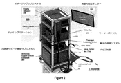
図2は、図1の内視鏡検査システム10の前記スレーブセクション200の概略図である。前記スレーブセクション200は、少なくともいくつかのスレーブセクション要素を運ぶように構成された患者側カート、スタンド、又はラック202を有する。前記患者側カート202は、前記輸送内視鏡320を取り外すことができる(例えば、取り付け/ドッキング及び取り外し/ドッキング解除)ドッキングステーション500及び関連するバルブコントローラボックス348を有する。前記患者側カート202は、典型的には、前記スレーブセクション200の容易な携帯性及び位置決めを促進するホイール204を包含する。
FIG. 2 is a schematic view of the
図3は、図1の内視鏡検査システム10の前記マスターセクション100及び前記スレーブセクション200のいずれか又は両方に配置されたモジュールのブロック図を示している。図2の前記輸送内視鏡320の空気注入、水灌注及び流体吸引能力は、これらのモジュールに関連付けられている。前記バルブコントローラボックス348は、前記モジュールのうちいくつかを収容し、ここで、前記バルブコントローラボックス348は、図2に示される前記患者側カート202上に配置されるか、又は前記患者側カート202から離れているが、その近くにある。残りのモジュール、安全システムモジュール352及びモーションコントロールシステムモジュール354は、前記患者側カート202に配置されるか、前記患者側カート202に配置されたスレーブ側要素、例えば前記ドッキングステーション500の一部である前記並進移動可能なハウジング926に結合されている前記ロボット部材410など、に取り付けられるかのいずれかである。
FIG. 3 shows a block diagram of modules located in either or both of the master section 100 and the
前記バルブコントローラボックス348にある前記モジュールは、バルブボックスプリント回路基板アセンブリ(PCBA)364、緊急停止PCBA 362、カート電源出力ポート356(定格12Vを有する)、バルブコントローラボックスパワーモジュール358(定格24Vを有する)、ソレノイドバルブ360、緊急スイッチ366、カート電源スイッチ368及び空気/水電源スイッチ370を包含する。
The modules in the
前記バルブボックスPCBA364は、前記輸送内視鏡320の前記空気注入、水灌注、及び流体吸引機能のために前記ソレノイドバルブ360を制御する。前記緊急停止PCBA362は、前記安全システムモジュール352を制御し、前記安全システムモジュール352は、次に、運動制御システムモジュール354を制御する。前記運動制御システムモジュール354は、前記ロボット部材410を制御する。
The valve box PCBA364 controls the solenoid valve 360 for the air injection, water irrigation, and fluid suction functions of the
前記バルブコントローラボックス348は、前記カート電力出力ポート356及び前記バルブコントローラボックス電力モジュール358にDC電源を提供するためのAC−DCコンバータを包含するAC入口電力ポート372を有する。前記カート電力出力ポート356及び前記バルブコントローラボックス電力モジュール358は、前記緊急停止PCBA362及び前記バルブコントローラボックス電力モジュール358にそれぞれ電力を供給する。前記カート電力スイッチ368及び前記空気/水電力スイッチ370がそれぞれオンに切り替えられるとき、電力は、前記緊急停止PCBA 362及び前記バルブコントローラボックス電力モジュール358に供給される。
The
図3に示されるモジュールのための電気システムは、前記ソレノイドバルブ360、前記バルブコントローラボックス348、前記バルブボックスPCBA364、及び前記空気/水電力スイッチ370を回路から電気的に絶縁して接続する、前記モジュールの残りが属している回路を有するように構成される。この構成は、前記緊急スイッチ366が作動すると、前記ソレノイドバルブ360が作動し続けるようなものである。前記ソレノイドバルブ360を機能させ続ける理由は、前記緊急スイッチ366の作動が、医療機器の安全基準に従って外科手術中に害をもたらすべきではないからである。
The electrical system for the module shown in FIG. 3 connects the solenoid valve 360, the
前記カート電力出力ポート356及び前記バルブコントローラボックス電力モジュール358の両方が前記AC入口電力ポート372に接続される第1の実装では、前記カート電力出力ポート356及び前記バルブコントローラボックス電力モジュール358は、前記AC入口電力ポート372との並列電気接続上にある。前記緊急停止PCBA 362の動作はまた、前記緊急スイッチ366の作動が前記ロボット部材410への電力を遮断するという点で、前記緊急スイッチ366によって制御される。これにより、前記ロボット部材410は動作を停止し、一方、前記バルブコントローラボックス電力モジュール358は、前記ソレノイドバルブ360が動作し続けることを可能にするために電力を供給されたままである。前記ロボット部材410への電力遮断は、いくつかの方法、例えば:
前記カート電力出力ポート356と前記AC入口電力ポート372との間の接続を終了すること;
前記カート電力出力ポート356と前記安全システムモジュール352との間の接続を終了すること;又は
前記安全システムモジュール352と前記運動制御システムモジュール354との間の接続を終了すること;
のうちの1つで行うことができる。
In the first implementation in which both the cart
Terminating the connection between the cart
Terminating the connection between the cart
It can be done with one of them.
第2の実装(図示せず)では、前記カート電力出力ポートは、前記スレーブセクション200の前記患者側カートのすべてのコンポーネントに電力を供給する(図2を参照)。この2番目の実装では、前記カートの電力出力ポートとそれに関連するモジュール;及び前記バルブコントローラボックス電力モジュールはそれに関連するモジュールは、別々のエンクロージャにある。これらの2つのエンクロージャのそれぞれは、電気的な絶縁を達成するために、AC入口電力ポートに個別に接続される。 In a second implementation (not shown), the cart power output port powers all components of the patient-side cart in the slave section 200 (see FIG. 2). In this second implementation, the power output port of the cart and its associated modules; and the valve controller box power module associated with it, the modules are in separate enclosures. Each of these two enclosures is individually connected to an AC inlet power port to achieve electrical isolation.
前記バルブコントローラボックス電力モジュール358及び前記カート電力出力ポート356は互いに独立しているため、上記の電気的絶縁により、前記カート電力出力ポート356への電力を遮断した後でも、前記バルブコントローラボックス電力モジュール358に電力が供給される。したがって、前記ソレノイドバルブ360は動作したままであり、且つ前記空気注入、水灌注、及び流体吸引機能は影響を受けない、すなわち、前記輸送内視鏡320によって担持され又は支持され、且つ前記ソレノイドバルブ360に結合された前記通過管(passage tubes)は、依然として前記ソレノイドバルブ360から前記手術台20上の生物に空気及び水を運び、且つ生物から前記ソレノイドバルブ360に流体を運ぶ。図3は、図4に示される前記バルブコントローラボックス348の斜視図を提供する概略図である。
Since the valve controller box power module 358 and the cart
前記緊急スイッチ366、前記カート電力スイッチ368及び前記空気/水電力スイッチ370は、前記バルブコントローラボックス348の前部に配置される。前記バルブコントローラボックス348はまた、前記安全システムモジュール352が接続されるポート;位置入力デバイス(PID)702(図7を参照)が接続されている;及びディスプレイ704(図7を参照);を有する。
The
図7から、PID 702は前記内視鏡検査システム10の前記マスターセクション100に配置されていることが理解されよう。前記PID 702は、前記ロボット部材410の移動制御、且つ、前記ソレノイドバルブ360の送気、注水、及び流体吸引機能の作動を可能にする(図3を参照)。一方、前記ソレノイドバルブ360の送気、注水、及び流体吸引機能は、前記輸送内視鏡320のボタンを介して作動させることもできる。1つのPID 702の拡大図を提供する図8を参照すると、各PID 702は、2つのボタン804を備えたハンドル802を有する。前記4つのボタン804のうちの3つのそれぞれは、前記ソレノイドバルブ360の前記空気注入、水灌注及び流体吸引機能のうちの1つの遠隔制御を提供するために割り当てられる。第4のボタンは、前記ロボット部材410の遠隔操作(teleoperation)をアクティベートすることである。これは、手術器具が初期化及び較正され、且つ前記PID 702を介してリモートコントロールされる準備ができた後に、ユーザーが遠隔操作を開始するつもりかどうかを確認するためである。この遠隔操作開始コマンドは、フットペダルなどの別のチャネルを介して送信することができる。
From FIG. 7, it will be understood that the
前記3つのボタン804が、それらにそれぞれ割り当てられた送気、水灌注、及びそれに360の流体吸引機能を制御できるようになる前に、前記カート電力スイッチ368及び前記空気/水電力スイッチ370をオンにする必要がある。これにより、前記ボタン804を押すと、送気、水灌漑、及び流体吸引が行われる。これらは、消化管の膨張、前記輸送内視鏡320の前記可撓性の細長いシャフトを通して挿入された(又は前記輸送内視鏡320の遠位端に埋め込まれた)カメラレンズの洗浄、及び不要な流体の除去(カメラレンズの洗浄など)などの目的で、手術中に不可欠である。
The
前記ディスプレイ704は、前記PID 702からの前記遠隔制御可能なバルブボックスコントローラ348の作動状態;前記ロボット部材410の較正ステータス(図5の参照番号504を参照);前記輸送内視鏡320のボタン及び/又は前記PID 702の3つのボタン804を介して命令される、前記空気注入、水灌注及び流体吸引機能の作動状態;を示す働きをする。前記空気/水電力スイッチ370が「ON」に切り替えられ、且つ前記バルブコントローラボックス348のリモートコントロールがアクティベートされると(前記ディスプレイ704に「ON」と表示されている図6の参照番号602を参照)、前記ボタン804が前記ソレノイドバルブ360の遠隔制御を可能にするためにアクティベートされる。前記バルブコントローラボックスのリモートコントロールが非アクティブ化されると、前記ディスプレイ704は「OFF」という単語を表示して、前記ボタン804が非アクティブ化されたことを伝える。前記ディスプレイ704はまた、前記ボタン804及び/又は前記輸送内視鏡320のボタンが押されると、且つ空気注入、水灌注及び流体吸引機能のいずれがいつでも操作されているかについての指示を提供するときに、更新される。前記輸送内視鏡320の前記可撓性の細長いシャフトを通して挿入された前記カメラレンズによってストリーミングされた前記空気注入、水灌注、及び流体吸引の画像を示すメインディスプレイ706とともに、前記ディスプレイ704は、前記空気注入、水灌注、及び流体吸引機能のうちのいずれが任意の時点で操作されているかをオペレータが確認するための追加の方法を提供する。図4から8から、前記コントローラボックス348は、空気注入、水灌注、及び流体吸引機能の動作及びモニタリングを統合する手段を提供することが理解されよう。
The
図9Aは、前記輸送内視鏡320の近位端920が取り付けられる前記ドッキングステーション500の構成要素を示す。
FIG. 9A shows the components of the
前記ドッキングステーション500は、その遠位端で前記ロボット部材410に結合された前記可撓性の細長い部材1600を回転させるために使用されるアクチュエータを内に含むモーターボックスを収容する(図14を参照)。前記アクチュエータはまた、前記細長い部材1600の遠位先端で前記ロボット部材410を関節でつなぐ(articulate)。前記モーターボックスは、可動上部928が並進する静止下部部分930を含む並進移動可能ハウジング926内に配置される。前記可動上部部分928の寸法は、前記静止下部部分930の寸法よりも大きいので、前記静止下部部分930及び前記可動上部部分928は、前記可動下部部分928の並進方向に応じて、前記静止下部部分930の一部が前記可動上部部分928に入るか、又は前記可動上部部分928から引き出されるという点で入れ子式の構造配置を有する。前記静止下部部分930と前記可動上部部分928との間のギャップ又は自由遊びは、異物粒子が平行移動可能なハウジング926内に収容された前記モーターボックスに入るのが防止されるように調整される。一方、前記モーターボックスが内視鏡手術から引き付ける流体及び粒子は、前記平行移動可能なハウジング926の外側に保持される。
The
前記可動上部部分928は、前記ロボット部材410が前記手術台20上の生物内の微動を可能にするように並進する。
The movable
前記可動上部部分928が並進して、前記可撓性の細長い部材1600を前記輸送内視鏡320の前記可撓性の細長いシャフト内のぴったりとはめ込まれた管腔内にさらに押し込むと、図9Aの点線部分に示されるように、前記可撓性の細長い部材1600が座屈する傾向がある。さらに、前記保護カバー1606は、並進中に削り取られてもよい。図9Aでは、前記可撓性の細長い部材1600が前記輸送内視鏡320に向かって前記モーターボックスの下流に入り、具体的には前記モーターボックスの前記アクチュエーターと前記輸送内視鏡320の近位端920との間の露出部分にある、座屈防止チューブ924の使用を通じて、座屈が最小化される。したがって、この座屈防止チューブ924は、前記可撓性の細長い部材1600のアクチュエータと前記輸送内視鏡320との間のガイド部材として機能する。前記座屈防止チューブ924は、支持体932によって所定の位置に保持される。これは、前記輸送内視鏡320がドッキングするベース934又は平行移動可能なモーターハウジング926の静止下部部分930のいずれかの部分から延びる。さらに、前記座屈防止チューブ924は、例えば、外科的処置の終わりに、又は異なる機能の新しい外科用器具に切り替えるときに行われる、前記細長い部材1600の挿入及び取り外し中の前記ロボット部材410の矯正を容易にし、挿入及び取り出し中の前記座屈防止チューブ924の端部の鋭利な特徴による前記保護カバー1606への損傷を防ぐために、両端がフレアになっている。前記座屈防止チューブ924は、パイプなどの剛性構造を使用して実現することができる。
The movable
前記細長い部材1600のこの座屈は、以下でさらに説明されるように、図9B、10及び11Aに示される実装を通じてさらに最小化される。
This buckling of the
図9Bの第1の実装は、前記シャフト1402の少なくとも一部1420を剛性にすることにより、前記可撓性の細長い部材1600が前記アダプタ906に取り付けられる場所に隣接することにより、上記の座屈及び削り取りの問題をさらに軽減する。
The first implementation of FIG. 9B is to make at least a
図10及び11Aは、前記輸送内視鏡320の近位端920が取り付けられる前記ドッキングステーション500の第2の実装のスケッチを示している。この第2の実装は、図9Aに示される前記可撓性の細長い部材1600の座屈を緩和することを目的とする。図10は、完全に伸長した状態の前記可動上部部分928を示し、図11Aは、並進中の前記可動上部部分928を示す。
10 and 11A show a sketch of a second implementation of the
前記第1の実装は、単一の座屈防止チューブ924を使用しているが、前記第2の実装では、2つの座屈防止チューブ1024aと1024bを使用する。前記2つの座屈防止チューブ1024a及び1024bは、2つの座屈防止管1024a及び1024bの一方が他方よりも大きい寸法を有するという点で伸縮自在の構造配置を有する。ここでは、前記並進移動可能なハウジング926の前記可動上部部分928が並進すると、より小さい寸法を有する前記座屈防止チューブ1024a、1024bは、より大きい寸法を有する前記座屈防止チューブ1024a、1024bに入る。図10及び11Aでは、前記座屈防止チューブ1024aはより小さい寸法を有し、且つ内側ガイドとして作用するが、前記座屈防止チューブ1024bはより大きい寸法を有し、外側ガイドとして作用することが示されている。しかしながら、前記座屈防止チューブ1024aがより大きな寸法を有する一方で、前記座屈防止チューブ1024bがより小さな寸法を有することも可能である。図10及び11Aの前記第2の実装では、前記可撓性の細長い部材1600の一部を剛性にすることが任意になることが理解されるであろう。
The first mounting uses a single buckling
前記座屈防止チューブ1024aは、前記可動上部部分928から突出する支持体1032aによって適所に保持され、一方、前記座屈防止チューブ1024bは、前記輸送内視鏡320がドッキングする前記ベース934か、又は前記並進移動可能なモーターハウジング926の静止した下部部分930のいずれかの部分から突出する支持体1032bによって適所に保持される。前記移動可能な上部部分928が並進すると、前記座屈防止チューブ1024aも並進する。前記座屈防止チューブ1024bと前記細長い部材1600との間の相対的な並進運動を排除することにより、前記保護カバー1606の摩耗が低減される。図9A及び9Bの前記座屈防止チューブ924と同様に、デュオピース(duo piece)座屈防止チューブ1024a及び1024bは、前記ロボット部材410の取り外しを容易にするために、その端部でフレアになっている。第1及び第2の実装の両方において、前記単一の座屈防止チューブ924及び前記デュオピース座屈防止チューブ1024a及び1024bは、滅菌のため、又は新しい単一の座屈防止チューブ924と新しいデュオピースの座屈防止チューブ1024a及び1024bとの交換のために前記ドッキングステーション500から取り外し可能である。
The buckling
図9A、9B、10及び11Aは、前記並進移動可能ハウジング926が実質的に垂直方向を有し、ここで、前記可動上部部分928が垂直並進を受けて前記可撓性mp細長い部材1600を移動させることを示す。ただし、前記並進移動可能ハウジング926は、水平方向などのその他の向き(図示せず)に配置することができ、これにより、前記可動部分は実質的に水平に、又は傾斜した状態で移動し、それにより前記可動部分は傾斜した軸に沿って移動することが理解されよう。
In FIGS. 9A, 9B, 10 and 11A, the translational
前記座屈防止チューブ1024a及び1024bは、使用中に汚れる可能性がある。したがって、それらは、所定の位置で洗浄及び滅菌されるように設計されているか、そうでなければそれらを再使用できるように別個に洗浄するために取り外し可能であることが有利である。あるいは、前記座屈防止チューブ1024a及び1024bは、シングルユース及び使い捨て用に設計されてもよく、その場合、各手順で新しいチューブが供給される。
The buckling
前記座屈防止チューブ1024a及び1024bが再利用されるように設計される場合、前記座屈防止チューブ1024a及び1024bの材料は、規定の洗浄及び滅菌方法との適合性を確実にするように選択されるべきである。世界中のさまざまな地域で幅広い洗浄及び滅菌ソリューションが使用されているため、複数のソリューションにわたって幅広い互換性を持つ材料が有利である。そのため、ステンレス鋼などの耐食性金属又は耐食性ポリマーは、前記座屈防止チューブ1024a及び1024bに適した材料である。
When the buckling
前記座屈防止チューブ1024a及び1024bが、洗浄及び滅菌中に前記並進移動可能なモーターハウジング926からの動作可能な分離のために設計される場合、前記並進移動可能なモーターハウジング926からの前記座屈防止チューブ1024a及び1024bの分離を容易にする取り付け手段もまた、洗浄及び/又は滅菌の容易さ及び完全さを促進するはずである。多くの可能な洗浄可能な取り付け手段が考えられる。例えば、磁石を使用する取り付け手段は、ブラシで簡単に掃除したり、布で拭いたりするために、裂け目があったとしてもわずかである、それらを埋め込むことができるため、特に有利である。一実施形態において、前記支持体1032a及び1032bは、磁性材料を使用して製造されるか、又は少なくとも埋め込み磁石を有し、それにより、前記支持体1032aは、前記座屈防止チューブ1024aに溶接され、且つ前記支持体1032bは、前記座屈防止チューブ1024bに溶接される。
If the buckling
図11B及び11Cは、図9A、9B、10及び11の前記平行移動可能なモーターハウジング926に座屈防止チューブを取り付けるための機械的手段などの、非磁性手段を使用する構造を示す。非磁性手段は、前記支持体1032a及び1032b(図10及び11Aを参照)で使用される磁石が、前記並進移動可能なモーターハウジング926内の磁性構成要素の動作を妨害する可能性があるシナリオで利用される。
11B and 11C show structures using non-magnetic means, such as mechanical means for attaching a buckling prevention tube to the parallel
前記座屈防止チューブ1024a及び1024bの磁気的取り付けが可能でない場合、図11Bに示される動的係合機構1135が使用され得る。前記動的係合機構1135は、前記座屈防止チューブ7のハンドル6の少なくとも一部分を収容するための第1の開口部を有する本体9を有する。前記本体9は、洗浄のために前記座屈防止チューブ7が取り外されるときに、前記本体9から前記座屈防止チューブ7を解放する機械的キャッチ装置(arrangement)を収容する。図11Bに示される実装では、前記機械的キャッチ装置は、ロッド2、当接部材3、リリースボタン1及び付勢構造(biasing structure)5を含む。前記ロッド2は、前記当接部材3及び前記リリースボタン1に枢動可能に接続され、且つ前記本体9の縦断面に沿って動くように配置される。前記本体9は、前記リリースボタン1の一部分が前記本体9から突出する第2の開口部を有する。;一方、前記本体9内にある前記リリースボタン1の一部分は、前記付勢構造5に結合される。膜/不透過性バリア4は、前記本体9から突出する前記リリースボタン1の前記一部分を覆う。
If magnetic attachment of the buckling
前記リリースボタン1が前記膜/不透過性バリア4を介して操作されるとき、前記当接部材3は、矢印に示される方向に機械的に作動され、それにより、前記ロッド2は、前記当接部材3を下方に引っ張る。前記座屈防止チューブ7に溶接された前記ハンドル6は、方向8に解放される。前記膜/不透過性バリア4は、前記本体9に恒久的に取り付けられ得るか、又は洗浄及び滅菌のために取り外し可能であり得る。
When the release button 1 is operated through the membrane /
図11Cは、図11Bに示される実装の変形を示す。図11Cの前記動的係合機構1135は、図11Bの前記動的係合機構1135と同じである。しかしながら、膜/不透過性バリア4を使用する代わりに、図11Cの前記動的係合機構1135の前記本体9は、前記本体9を汚れから封止するために動的シール4”を使用する。前記動的シール4”は、例えば、それを貫通して前記ボタン1が突き出る前記本体9の前記第2の開口部の壁と前記動的シール4”の接面(facing surface)との間の摩擦係合が、流体が前記本体9内部空洞に入るのを妨げる、ワッシャーであってもよい。
FIG. 11C shows a variant of the implementation shown in FIG. 11B. The
図11Dから11Gは、前記座屈防止チューブ1340の非永久的な塑性変形特性を使用する機械的手段によって達成することができる、前記座屈防止チューブホルダ1342への前記座屈防止チューブ1340の前記対のさらに別の変形を図示する。前記座屈防止チューブ1340は、前記座屈防止チューブホルダ1342の剛性部分に嵌合するように一時的に変形することができるコンプライアンス機能/形状1344の一部分を含む。前記座屈防止チューブ1340は、前記モーターハウジング面1350に垂直な方向の前記座屈防止チューブホルダー1342に取り付け/取り外しすることができるか、又は取り付け角度1349は、モーターハウジング面1350に対して鋭角であり得る。さらに、コンプライアンス機能/ジオメトリ1344上の3つ以上の側面1346は、前記座屈防止チューブ1340の中心面1348が常に前記モーターハウジング面1350に対して垂直であることを可能にする。これは、前記可撓性の細長い部材が、前記座屈防止チューブ1340を通して挿入されることを可能にする。
11D-11G show the buckling
図12Aは、図9A、9B、10、11A及び11Bの前記並進移動可能なハウジング926内の並進機構の第1の実装を示し、一方、図12Bは、図9A、9B、10、11A及び11Bの前記並進移動可能なハウジング926内の並進機構の第2の実装を示す。図12A及び12Bの両方において、前記ハウジングは示されていない。前記並進移動可能ハウジング926の並進機構は、モーター(図12Aの参照番号1202及び図12Bの参照番号1204を使用して示される)及び送りねじ(lead screw)機構又はボールねじ機構(図12Aの参照番号1222及び図12Bの参照番号1224を使用して示される)を含む。前記並進移動可能ハウジング926の全高926hは、以下で説明するように、前記並進移動可能ハウジング926の並進機構(モータ及び送りねじ機構又はボールねじ機構を含む)の構成によって影響を受ける。
12A shows the first implementation of the translational mechanism in the translationally
図12Aでは、並進運動を駆動する前記モーター1202は、プラットフォーム1220に取り付けられ、且つ前記プラットフォーム1220と一緒に並進する。前記プラットフォーム1220が並進すると、前記並進移動可能なハウジング926の高さ926hは、前記送りねじ機構1222の完全に引き込まれた状態と完全に挿入された状態との間で変化する。
In FIG. 12A, the
低い高さ926hは、前記プラットフォーム1220上の前記ロボット部材410の駆動機構のドッキングを容易にするので望ましい。前記駆動機構の一部分、すなわち機器アダプタ(これには、図16、17、及び18に示されているケーブルのペアが近位端で巻き付けられるドラムが含まれる)は、図9A、9B及び10に示され、参照番号906を使用して示される。
A low height of 926h is desirable as it facilitates docking of the drive mechanism of the
図12Bの構成では、前記プラットフォーム1220を並進させる並進モータ1204は、静止ブラケット上に取り付けられている。前記プラットフォーム1220(それ上に前記可動上部部分928の前記ハウジングが配置される)は、前記並進モータ1204によって駆動される前記送りねじ機構1224に回転可能に結合される部材1226に取り付けられることによって、並進することが可能になる。この部材1226は、ナットなどの穴のあるオブジェクトであってよい。
In the configuration of FIG. 12B, the
並進運動の同じ範囲について、図12Bの前記並進移動可能なハウジング926の高さ926hは、図12Bのねじ機構1224が並進しないので、図12Aのそれよりも低くなるが、一方、図12Aのモーター1202のねじ機構1222は、それがモーター1202によって駆動されている間に並進する。
For the same range of translational motion, the
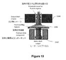
出力軸シャーシ1304とモーター出力軸1302との間には隙間がある。そのような隙間への流体の進入がある場合、それは、前記出力軸1302の周りの流体に敏感な構成要素に危険又は機能不全をもたらす。
There is a gap between the
シールド1306は、流体感受性コンポーネントと前記出力シャフトシャーシ1304の内壁との間で前記出力シャフト1302の周りに取り付けられる。このシールドは、隙間に進入する流体をはじき、且つそれにより進入した流体が、前記流体感受性コンポーネントと接触するのを防ぐ。前記シールド1306は、そのような流体の進入を防止するためにシャフトシールを使用するよりも特に有利であり、それにより、前記出力シャフト1302の周りの前記シャフトシールの使用は、シャフト回転に摩擦を導入する。
The
図14は、その遠位端でロボット部材410に結合され、且つ、その近位端で駆動機構に結合される可撓性の細長い部材1600の概略図を示す。前記駆動機構は、アクチュエータから前記アダプタ906(図9A及び9B)に運動を伝達する一連の機械的リンケージからなる。前記アダプタ906は、例えば1つ以上のドラムなどのモティベータ(motivators)を含み、前記ロボット部材410の移動制御を可能にするケーブル対がその周りを巻き、前記ケーブル対は前記可撓性の細長い部材1600内を走っている。一実装では、1つのモータシャフトを含有するモータボックス(図13の参照番号1302を参照)は、前記ロボット部材410が接続される前記可撓性の細長い部材1600を回転させるために使用される。
FIG. 14 shows a schematic view of a flexible
前記可撓性の細長い部材1600は、その近位端で適用される作動(回転又は並進など)を遠位端に効果的に伝播しなければならないので、その一部であるシャフト1402は、回転バックラッシュ(backlash)が低く、トルク伝達が良好で、且つ圧縮率が低いトルクコイルなどの回転運動伝達デバイスを使用して実現され得る。前記回転運動伝達デバイスはまた、前記輸送内視鏡320に適合するのに十分な可撓性でなければならない。この特性の混合を達成するために、前記シャフト1402に使用される前記回転運動伝達デバイスは、以下の特徴の1つ以上を組み込むように設計される。
The flexible
第1の特徴は、フラットコイルを使用し、そのうちの1つのセグメント1502が図15に示されている。断面図から、前記コイルのより長い部分Wは、前記フラットコイルがエッジからエッジまで巻かれるときに前記シャフト1402の長手方向の長さを形成するように横になっている。前記フラットコイルの例示的な断面寸法は、0.2〜0.4mmの幅W及び0.05〜0.15mmの厚さTである。長手方向に隣接するコイル間の接触により、バックラッシュの少ない圧縮力が伝達される。また、層が薄いほど、所定の外径制約に対して管腔空間を大きくできるため、前記コイル内の所定の層数のワイヤコイルを丸めるよりも、フラットワイヤコイルの方が適している。
The first feature uses a flat coil, one of which,
第2の特徴は、前記第1の特徴に従って製造されたフラットコイルの複数の層を使用する。巻線の方向は層間で交互になり、これにより、得られるシャフト402は、バックラッシュの少ない1対1のトルクを提供する。例えば、図15に示される断面カットアウト1504は、3つの層4、5及び6を有する。前記内層4及び前記外層6は、同じ方向に、例えば、左巻き(left hand winding)方向としても知られるS回転、に巻かれる。一方、中間層5は他の方向、右巻き方向とも呼ばれるZ回転、に巻かれる。各層は、らせん状に巻かれた8〜12本のフラットワイヤストランドで構成されており、その結果、コイルの外径は3〜6mmの範囲になる。
The second feature uses a plurality of layers of flat coils manufactured according to the first feature. The winding directions alternate between the layers so that the resulting shaft 402 provides one-to-one torque with less backlash. For example, the cross-section cutout 1504 shown in FIG. 15 has three
層間の巻き方向を交互にすることにより、以下のようにして、バックラッシュの少ない近位端と遠位端との間で1対1の回転がもたらされる。所定の方向にトルクを伝達する場合、1つ以上の左巻きの(left-hand wound)コイルは、その直径を拡大することによって回転バックラッシュを導入する。同じ方向のねじれの下で、1つ以上の右巻きの(right hand wound)コイルはその直径を小さくすることにより回転バックラッシュを導入する。交互の風向(alternating wind directions)のコイルが互いに内側に重ねられると、この回転バックラッシュの原因が防止される。上記の例では、左巻きのコイルの径方向の膨張は、次の外層の右巻きのコイルの径方向の圧縮によって打ち消される。両方向の回転バックラッシュのこの原因を取り除くには、最低3層の交互の風向コイル(wind dirction coils)が必要である。 Alternating winding directions between the layers results in one-to-one rotation between the proximal and distal ends with less backlash, as follows: When transmitting torque in a given direction, one or more left-hand wound coils introduce rotational backlash by increasing their diameter. Under twist in the same direction, one or more right hand wound coils introduce rotational backlash by reducing their diameter. When coils with alternating wind directions are stacked inside each other, the cause of this rotational backlash is prevented. In the above example, the radial expansion of the left-handed coil is canceled by the radial compression of the next outer layer right-handed coil. To eliminate this cause of bidirectional rotational backlash, a minimum of three layers of alternating wind dirction coils are required.
図16は、円形コイルシースのセグメントの断面図を示し、ここで、円形コイルシースは、その管腔を通って延びるケーブル1604を備えた円形ワイヤコイル1602を有する。このような円形のワイヤコイルシースは、圧縮力を伝達するために使用される。ワイヤコイル1602ルーメンの内側を走るケーブル1604は、アクション反作用対に張力を提供する。引張力の伝達損失を防ぐには、これら2つのコンポーネント間の摩擦が低いことが望ましい。ワイヤコイル1602シースに大きな曲げを課すコンジットの場合、ワイヤコイル1602シースに加えられる小さな圧縮力により、ワイヤコイル1602シースに座屈/ねじれが生じ、ワイヤコイルシースのルーメンが狭くなる。ワイヤコイルシースの座屈/よじれによる内腔の狭小化により、ケーブル1604との摩擦が増加し、ケーブル1604の力伝達効率が低下する。
FIG. 16 shows a cross-sectional view of a segment of the circular coil sheath, wherein the circular coil sheath has a
図16aは、保護カバー1606、シャフト1402、多数の円形コイルシース1602、及び各円形コイルシース1602内に含まれるケーブル1604を包含する、(図14に示されるような)可撓性の細長い部材1600の詳細な断面図を示す。
FIG. 16a details a flexible elongated member 1600 (as shown in FIG. 14) that includes a
簡単にするために、ワイヤコイル1a〜1dの4つのピッチのみが図16に示されている。1bに対する圧縮力4a、4bは、隣接するピッチ1a及び1cから生じる。円形のコイルシースを曲げると、この圧縮力には上向きの成分がある。ワイヤコイル1602がその元の形状を保持する傾向から、相殺する下向きの力5が存在する。ケーブル1604によって1bに加えられる小さな下向きの力6もある。ただし、4a、4bからの圧縮力が十分に大きい場合、1bが1a及び1cとインターフェース接続する円形の表面は不安定な平衡状態を形成し、これにより、上方向に1bの増分スリップがあると、隣接するピッチ間の接触角が著しく変化する。これにより、隣接するピッチ間の圧縮反力4a、4bの方向が著しく変化する。これにより、好循環でさらに上向きの1bの横滑りが発生し、且つ座屈/ねじれと呼ばれるコイルの永久変形が発生する。図18に示すように、この不安定性はシースの長さに沿って伝播する可能性がある。
For simplicity, only the four pitches of the wire coils 1a-1d are shown in FIG. The
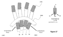
図17は、図16の円形コイルシースに対する改良による、長方形ワイヤコイルシースのセグメントの断面図を示す。長方形ワイヤコイルシースは、実質的に長方形の断面を有するワイヤコイル1702を有する。図16の円形コイルシースと同様に、ケーブル1604は、図17の長方形コイルシースの管腔を貫通する。
FIG. 17 shows a cross-sectional view of a segment of the rectangular wire coil sheath, which is an improvement over the circular coil sheath of FIG. The rectangular wire coil sheath has a
図16に対する改善は、円形の断面を有するコイルワイヤの代わりに、長方形の断面を有するコイルワイヤ1702が使用されることである。シースに巻かれると、シースの各ピッチは隣接するピッチとより安定して接触するため、ワイヤコイルに加わる外部の曲げ力と圧縮力によって、交互のピッチが横方向にずれる可能性が低くなる。長方形の断面は、ワイヤコイルシースの方向に短くなっている。これによりピッチ密度が増加し、ピッチあたりの曲げ量が少なくなるため、及びしたがって同じ曲げ半径でのピッチ密度の低いコイルの誘導ひずみよりも小さい、曲げによる断面の誘導ひずみが発生する。曲げ力と圧縮力によって誘発されたひずみが材料の降伏ひずみ以下に保たれている場合、ワイヤコイル1702の永久的な座屈/ねじれはない。コイル3bの上方へのわずかな動きは、コイル3a及び3cからの圧縮力の迎え角を変更しない。ピッチを小さくすると、迎え角の変化がさらに小さくなり、同じ曲げ特性を維持しながら、円形ワイヤコイル1よりも優れた座屈防止性能が得られる。
An improvement over FIG. 16 is the use of
長方形のコイルシースの特定の実施形態は、断面が等しい寸法を有する場合、すなわち、正方形のコイルシースである場合である。これは、ピッチ密度の増加という利点を提供しないが、3bに対するコイル3aと3cからの圧縮力の迎え角が横方向のたわみで変化しないため、これは座屈に対してより安定している。また、正方形断面のコイルは、同等のピッチの円形ワイヤコイルの円形断面積よりも断面積が大きくなる。これにより、横方向のせん断力に対する耐性が高まる。
A particular embodiment of a rectangular coil sheath is when the cross sections have equal dimensions, i.e., a square coil sheath. This does not provide the advantage of increased pitch density, but it is more stable against buckling because the angle of attack of the compressive forces from the
図18は、図16の円形コイル状ワイヤシースのセグメントの同様の断面図を示す。この図では、コイルドワイヤシース1802は、超弾性材料で作られている。超弾性材料の一例は、ニッケルとチタンの合金であるニチノール(nitinol)である。ニチノールは独特の材料特性を備えており、荷重によって大幅に変形することができるが、荷重を取り除いた後でも元の形状を回復することができる。図18では、ニチノール円形コイル状ワイヤシース1802は、ケーブル1604に高い負荷がかかっている期間中に高度に圧縮され、及びしたがって、一時的な座屈を受ける。これにより、ケーブル1604の摩擦が増加し(図16及び図16aに示すように)、且つ効率が低下する。コイル状ワイヤ1802が無負荷/屈曲解除された後、コイル状ワイヤ1802は、塑性変形した不規則な形状のままではなく、その元の形状に戻る。したがって、コイル状ワイヤシース1802は、時間の経過に伴う効率の低下なしに、ほとんどの時間、最大効率で動作している可能性がある。
FIG. 18 shows a similar cross-sectional view of the segment of the circular coiled wire sheath of FIG. In this figure, the coiled
図19は、電線用のねじれ防止支持体1902の概略図を示す。ねじれ防止支持体は、ロボット部材410に配置することができ、且つ回転継手の機能に影響を与えることなく、ワイヤ1604(図14及び16aを参照)のねじれ抵抗を増加させることができる。腱/ケーブル1604に加えて、シャフトは電線も運ぶ(簡略化のために図16aには示されていない)ことを理解することができる。図19に示されているように、ねじれ防止支持体1902は、本発明は、湾曲した入口1906を有することにより、ワイヤ1604に最小曲げ半径を強制する。最小曲げ半径は、ワイヤが曲がるのに対応するためにデバイス内部に必要なスペースを最小限に抑えながら、デバイスの予想寿命内でワイヤが曲げ疲労破損を経験しないように実験的に決定される。ねじれ防止支持体1902は、ピン1910a及び1910b又は同様の非ピン形状構造上で自由に旋回できるようにするサポート1908を包含する。これにより、ワイヤ1604は、そのエネルギー的に最も安定した状態、すなわち、最小の総曲げ量を有する状態を達成することができる。ねじれ防止支持体1902の回転軸は、ロボット部材410の関節運動の軸と実質的に同じ場所に配置され得、その結果、ワイヤは、ロボット部材410の関節運動範囲全体にわたって、感知できるほどの伸張力又は圧縮力を受けない。
FIG. 19 shows a schematic view of the
図20aは、例示的な実施形態によるロボット部材410のうちの1つの拡大断面図を示す。ロボット部材410は、カメラによる視認性が向上するように非対称の運動範囲を有することができ、これにより、器具のより安全な操作がもたらされ得る。ロボット部材410の関節2006は、機械的ハードストップ2008a、2008b、2010a、2010bを使用することにより、カメラ軸に向かう方向よりもカメラ軸から離れる方向により小さい角度で移動することができる。代替の実施形態は、同じ効果を達成するためのソフトウェア及び位置感知の使用を含み得る。
FIG. 20a shows an enlarged cross-sectional view of one of the
例示的な実施形態において、図20aのロボット部材410は、3段階の関節動作、すなわち、中立(neutral)位置(0°)、中立位置の反時計回りに90°、及び中立位置の時計回りに45°、で示されている。ベースジョイント2002は固定され、且つ基準軸2012に平行である。遠位ジョイント2004は、ヒンジ2006を中心に回転する。上部ハードストップ2008a及び2008bは、許容される上向き回転の最大程度を提供する。図示の例示的実施形態では、中立位置を中心にして最大で反時計回りの90°の回転が可能である。下側のハードストップ2010a及び2010bの表面は、ハードストップ2008a及び2008bの表面と比較して異なる角度を有し、それにより、下向きの回転の最大の程度が制限される。図示の例示的実施形態では、中立位置を中心とした時計回りの最大45°の回転が許可されている。複数の関節にわたる非対称性の累積効果は、ロボット部材410の遠位端での作業空間を決定し得る。関節間の非対称性は、遠位端エフェクター2014の最大の可視性を提供するように最適化され得る。
In an exemplary embodiment, the
例示的な実施形態において、ロボット部材410に隣接するカメラ2016と共に、上記の概念の実装が図20bに示されている。ロボット部材410は、カメラの視線方向に実質的に平行にあるが、横方向オフセット2020が小さい。ロボット部材410がカメラ2016から離れて関節運動するとき、機械的ハードストップ2010a、2010bは、遠位エンドエフェクター2014の動きが視距離(visual range)から離れすぎないように制限する。
In an exemplary embodiment, an implementation of the above concept is shown in FIG. 20b, along with a
内視鏡検査システムは、省スペースの板金構造コンポーネントを備えた中央に位置合わせされたプーリを利用し、且つ中央ピンを使用しない、ロボット機器に配置されたトルクジョイントを包含し得る。このようなトルクジョイントは断面空間をできるだけ小さくするが、プーリーの中心をロボット部材410の所定の直径に対して最大化できるようにプーリーを中央に位置合わせすることにより、所定のケーブル力に対して最大量のトルクを提供する。さらに、トルクジョイントにより、通過要素をプーリーの両側に沿って妨害されることなく配線できる。中央に配置されたプーリーを取り付け、及びそのねじり力をトルクジョイントの残りに伝達するのに板金を使用すると、同等の製造コストで他の取り付けソリューションと比較して壁を薄くできるため、よりコンパクトなプーリー構造を得ることができる。プーリーがヒンジジョイントにもはや直接接続されなくなったため、プーリーの回転軸とトルクジョイントの回転軸との間の良好な位置合わせを維持するため、組み立て中に一時的に位置決めピンを使用することができる。
The endoscopy system may include torque joints located in robotic equipment that utilize centered pulleys with space-saving sheet metal structural components and do not use central pins. Such a torque joint minimizes the cross-sectional space, but by aligning the pulley to the center so that the center of the pulley can be maximized for a given diameter of the
図21aは、プーリー2102の側面図を示す一方、図21bは、プーリー2102を備えたトルクジョイントの斜視図を示す。プーリー2102は、三角形の板金取り付けブラケット2116a及び2116bを包含し得る。プーリー2102はまた、プーリーワイヤを保持することを助けるフィーチャ(features)2118a〜2118dを内に含み得る(contain)。板金コンポーネント2106は、作動ワイヤ2108を拘束し、且つプーリー2102を遠位継手セクション2110に構造的に固定するのを助けることができる。ヒンジ継手2104a及び2104bは、遠位継手セクション2110に回転点を提供し、且つプーリー2102と軸方向に整列する。図21c及び21dは、プーリー2102を備えたトルクジョイントの断面を示している。図では、管腔2112は、中央に位置合わせされたプーリー2102により二等分されている。
21a shows a side view of the
図21eは、例示的な実施形態による、管腔2112の断面を示し、且つ図21fは、例示的な実施形態による、時計回りに90度回転された同じ断面を示す。図において、構成は、通過要素2114(図21eには示されていない)を妨げることなく、プーリー2102のためのスペースを確保する。中央に配置されたプーリーは、その直径を可能な限り大きくすることを可能にし、これによりジョイントの機械的利点が向上する。中央のリベットピンがないことは、ルーメン2112が過度に切開されないことを意味し、これにより、通過要素2114に利用可能なルーメン断面積が減少する。本発明のトルクジョイントはまた、通過要素2114関節が関節運動しているときにそれほどひどく曲がらないようにする。
FIG. 21e shows a cross section of the
図22は、典型的なヒンジジョイント2202の断面図を示す。ジョイント2202は、近位セグメント2204及び遠位セグメント2206を包含し得、且つ(図14に示されるように)ロボット部材410に配置され得る。回転は、近位セグメント2204と遠位セグメント2206の両方が重なる点の周りで発生する。示される好ましい実施形態では、セグメント2204、2206の両方は、2つの別個のリベットピン2208a及び2208bを使用して拘束され得る。これは、内部管腔空間2210が他の用途のために確保されることを可能にし得る。
FIG. 22 shows a cross-sectional view of a typical hinge joint 2202. Joint 2202 may include
ピン2208a、2208bと関節セグメント2204及び2206との間には、継手の円滑な回転を可能にする隙間ばめ(clearance fit)が存在する。しかしながら、そのようなクリアランスは、2つのセグメント2204、2206のピボット軸を整列させることの容易さを妨げる可能性がある。セグメントが整列しない場合、極端な運動範囲での移動が困難になる可能性がある。したがって、ピン2208a、2208bは、ブリッジインサート2212によって互いに位置合わせする(aligned)ことができる。したがって、組み立て中にブリッジインサートの使用を通じて2つの別個のヒンジ継手の軸方向位置合わせを確実にする方法を提供することができる。ブリッジインサートは、ジョイントの一部になることができるか、引き抜くことができる。図22に示されるような実施形態において、インサート2212は、リベットピン2208a、2208bの穴を貫通するロッドであってもよく、及びしたがって、セグメントが確実に整列されるようにすることができる。リベットピン2208a、2208bが近位セグメント2204に溶接されて保持されたジョイントを作成した後、ブリッジングインサート2212が取り外される。
Between the pins 2208a, 2208b and the
並進移動アクチュエータが輸送内視鏡320の内外にロボット部材410を並進させるために使用される内視鏡システムでは(図2を参照。これにより、ロボット部材410は、その近位端920を介して輸送内視鏡320に導入される。図9Aを参照)、並進アクチュエータから電力が取り除かれたとき、並進移動アクチュエータは定位置に固定されたままであることが有利である。重力又は他の力のような予想される外力の下で並進移動アクチュエータが逆駆動可能であるシステムでは、アクチュエータから電力が除去されると、ロボット部材410は輸送内視鏡320に出入りする。これは、ロボット部材410の命令されていない並進運動が、ロボット部材410を敏感な組織と意図せずに接触させる場合に特に不利である。したがって、内視鏡検査システムの並進移動アクチュエータは、電源が切られたときに逆駆動可能ではない(non-back drivable)ことが有利である。
In an endoscope system in which a translational moving actuator is used to translate the
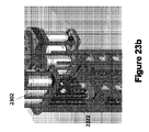
図23aは、典型的な並進機構の斜視図を示す一方、図23bは、典型的な並進機構の斜視図の拡大図を示す。典型的な並進移動アクチュエータは、しばしば、ボールねじ2322などの低摩擦駆動機構を利用して、電気モーター2302の回転運動をアクチュエータの直線運動2326に変換する。ボールねじ2322は、消費される摩擦が少ないために業界で好まれており、特定の入力コマンドに対して繰り返し可能な動作を提供する。典型的なボールねじ2322は、滑り接触要素の代わりに転がり接触要素を包含し、且つ低い摩耗率のために長い耐用年数を有する。しかしながら、典型的なボールねじ2322は、内視鏡検査システムにおける並進移動アクチュエータとしての使用にそれらを不適切にする少なくとも2つの特徴を有する。第一に、ボールねじ2322の低摩擦の転動体により、予想される外部荷重よりも低い力でそれらが逆駆動可能になる。第2に、ボールねじ2322のピッチを減少させると、その逆駆動抵抗が増加する。ボールねじのピッチを下げることができる量は、他の伝達要素と比較して制限される。これは、転動体とその往復ガイドレースを収容するために、隣接するピッチ間に十分なスペースを確保する必要があるためである。
FIG. 23a shows a perspective view of a typical translation mechanism, while FIG. 23b shows an enlarged view of a perspective view of a typical translation mechanism. A typical translational moving actuator often utilizes a low friction drive mechanism such as a
図23c、23d及び23eは、現在の産業用リニアアクチュエータの逆駆動性を改善することができる並進機構2300の実装の断面図を示す。図23cから23eに示されるように、内視鏡検査システム並進機構2300は、モーター2304の回転運動をロボット部材410の並進運動に変換するため(図2参照)、回転モーター2304及び送りねじ2306を包含する。送りねじ2306は、低摩擦の転動体接触を有するボールねじと比較して、送りねじ2306と送りねじナット2308との間の滑り接触のために本質的により大きい摩擦を有するので、この実施形態で使用される。さらに、親ねじ2306は、同等のサイズのボールねじよりも低いピッチを有することができ、親ねじ2306により多くの逆駆動抵抗を与える。並進機構2300はまた、並進機構2300を支持するためにモータ2304及びモータプラットフォームベース2312を囲むための静止モータプラットフォーム2310を包含し得る。図23d及び23eは、モーター2304が親ねじ2306を回転させるときの、垂直に平行移動された親ねじナット2308の様々な位置を示す。
23c, 23d and 23e show cross-sectional views of the implementation of a
図示されていない別の実施形態において、並進機構2300は、回転モーター及びボールねじを包含することができ、その結果、モーターは、必要な後方駆動抵抗を提供するのに十分な内部歯車減速を有する。そのような実施形態は、親ねじのより高い摩擦及びより低い耐用年数が並進機構2300において許容できない場合に有利であり得る。そのようなモータの好ましい実施形態は、遊星歯車減速又はハーモニックドライブ(登録商標)を包含し、そのどちらもコンパクトなサイズで大きなギア減速比を可能にする。
In another embodiment not shown, the
並進機構2300のさらなる実施形態は、モーター、ボールねじ、及びモーターから電力が取り除かれると自動的に作動してアクチュエーターの動きを停止する電気的に作動するデバイスを包含し得る。そのような実施形態は、親ねじのより高い摩擦及びより低い耐用年数が許容できない場合、及びモーター内の大きなギア減速が許容できないほど遅い並進速度をもたらす場合に、有利であり得る。そのような摩擦デバイスの好ましい実施形態は、モーターシャフト又はボールねじに接続された回転電磁ブレーキである。
Further embodiments of the
内視鏡ドッキングシステムは、ドッキングステーション500、輸送内視鏡320、及び関連するバルブコントローラボックス348(図2に示されるように)を包含し得る。内視鏡ドッキングシステムの高さを調整して、さまざまな患者のテーブルの高さだけでなくさまざまな臨床医の高さに対応することが有利である。安全上の理由により、内視鏡ドッキングシステムは、内視鏡検査中に突然の予期せぬ変化を避けるために、望ましい高さに固定しておく必要がある。
The endoscope docking system may include a
現在、内視鏡ドッキングシステムに高さ調整機能を提供できるさまざまな単純なメカニズムがある。このようなメカニズムの1つの例は、調整機能を提供する高さの異なる複数のボルト締め位置を持つ単純なボルト締めインターフェースが包含するが、調整を実行するためのツールと、調整中のメカニズムの重量を支えるための追加の人員の使用が必要になる場合がある。これは、手順中のボルト締めインターフェースの調整を面倒で非実用的なものにする。機械的に操作される垂直スクリューアジャスター又は機械的に操作される油圧リフトシリンダーなどの他の調整可能な高さ機構を使用することができる。ただし、これらのデバイスの機械的制御を、接続された内視鏡の制御本体のユーザーの手の近く、又はユーザーの足の近くに配置することは、動作を目的の場所に伝達するために必要な多数の機械的リンケージのため、実用的ではない。つまり、ユーザーは自分がやっていることをやめてコントロールの位置に移動し、そして元の場所に戻って調整が十分であったかどうかを評価する必要がある。これは、ユーザーに遅延及び不便を追加する。 Currently, there are various simple mechanisms that can provide height adjustment capabilities for endoscopic docking systems. One example of such a mechanism includes a simple bolting interface with multiple bolting positions at different heights that provides adjustment functionality, but with tools for performing the adjustment and the mechanism being adjusted. It may be necessary to use additional personnel to support the weight. This makes adjusting the bolting interface during the procedure cumbersome and impractical. Other adjustable height mechanisms such as mechanically operated vertical screw adjusters or mechanically operated hydraulic lift cylinders can be used. However, it is necessary to place the mechanical control of these devices near the user's hand or the user's foot on the control body of the connected endoscope in order to convey the movement to the desired location. It is not practical due to the large number of mechanical linkages. That is, the user needs to stop doing what he is doing, move to the control position, and then return to the original location to evaluate whether the adjustment was sufficient. This adds delay and inconvenience to the user.
別の解決策は、制御インターフェースを介したユーザからの入力に基づいて、内視鏡ドッキングシステムの高さを調整するための電気作動アクチュエータの使用を包含し得る。図24aは、内視鏡ドッキングシステム2402の典型的な電気的に作動する高さ調整機構の斜視図を示す。この機構の電気アクチュエータ2404は、内視鏡ドッキングシステム2402の全体の重量を確実に支持及び操作しなければならない。内視鏡ドッキングシステム2402のサイズ及び重量が大きいため、電気アクチュエータ2404のコストも高い。したがって、電気的に作動されるアクチュエータ2404は、ユーザ制御の位置により多くの自由を与えるが、それはまた、内視鏡検査システムにかなりのコストを追加する。
Another solution may include the use of electrically actuated actuators to adjust the height of the endoscopic docking system based on input from the user via the control interface. FIG. 24a shows a perspective view of a typical electrically actuated height adjustment mechanism of the
本明細書に開示されているのは、既存の機械的調整機構の不便さを排除することができ、且つ電気アクチュエータシステムよりも低いコストを提供することができる機構である。開示された機構は、重量補償デバイス及び電気的に作動するロックデバイスを包含し得る。重量補償デバイスは、内視鏡ドッキングシステムの重量の大部分を相殺する可能性がある。重量の残りはフレキシブルに吊り下げられているため、無理なく垂直方向に簡単に調整できる。重量補償デバイスはまた、内視鏡ドッキングシステムの高さを調整するための高さ調整機構を包含し得る。 What is disclosed herein is a mechanism that can eliminate the inconvenience of existing mechanical adjustment mechanisms and can provide a lower cost than an electric actuator system. The disclosed mechanisms may include weight compensation devices and electrically actuated locking devices. The weight compensation device can offset most of the weight of the endoscopic docking system. The rest of the weight is flexibly suspended so it can be easily adjusted vertically without difficulty. The weight compensating device may also include a height adjusting mechanism for adjusting the height of the endoscopic docking system.
電気的に作動するロックデバイスは、コントローラ及びユーザ制御を包含し得る。エレクトロニクスであるため、ユーザーコントロールは、例えば図7を参照して、マスターセクションのPID 702又はフットペダルの近くに、手又は足の場所の近くに配置できる。推奨されるユーザーコントロールは、手動ボタン又はフットスイッチのいずれかで構成される。デバイスはデフォルトでロック状態になり、且つユーザーコントロールのアクティブ化中にのみロック解除される。
The electrically actuated lock device may include a controller and user control. Being electronic, the user control can be placed near the
図24bは、両方とも第1の実装による、重量補償デバイス及び電気作動式ロックデバイスを組み込んだ図2のスレーブセクション200の側面図を示す。図24bに示されるように、スレーブセクション200は、患者側カート202、内視鏡ドッキングシステム2406、高さ調整機構2408、及びリニア電磁ブレーキ2410を包含する。内視鏡ドッキングシステム2406は、ドッキングステーション500及び輸送内視鏡320を包含する(図2に示すように)。この実装では、高さ調整機構2408は、そこから内視鏡ドッキングシステム2406が直接吊るされる一定力のばねを包含し得る。一定力荷重ばねの力は、内視鏡ドッキングシステムの2406の最も重い構成の重量とほぼ同じになるようにカスタマイズされている。
FIG. 24b shows a side view of the
図24cに示されるように、内視鏡ドッキングシステム2406は、リニア電磁ブレーキ2410が解放された状態で、その最も高い位置にある。これにより、内視鏡ドッキングシステム2406の重量を維持しながら、高さ調整機構2408による内視鏡ドッキングシステム2406の高さ調整が可能になる。
As shown in FIG. 24c, the
図24dは、リニア電磁ブレーキ2410が係合された、最も高い位置にある内視鏡ドッキングシステム2406を示す。内視鏡ドッキングシステム2406が所望の高さに調整された後、係合したリニア電磁ブレーキ2410は内視鏡ドッキングシステム2406をしっかりと固定して、内視鏡検査中に患者を危険にさらす可能性のある突然の予期しない変化を回避する。より具体的には、リニア電磁ブレーキ2410は、内視鏡ドッキングシステム2406の垂直移動を防止するため、摩擦を使用して内視鏡ドッキングシステム2406に直接係合することができる。
FIG. 24d shows the highest position
スレーブセクション200は、インターロック構成要素を使用して内視鏡ドッキングシステム2406に直接係合して内視鏡ドッキングシステム2406の垂直移動を防止する、リニア電磁係合スプライン又はラチェット(図示せず)を包含し得る。インターロック構成要素の例は、アクチュエータ、ギア、及び/又はバルブを包含する。図24e及び24fは、内視鏡ドッキングシステム2406がその最下点にあるときのそれぞれ、係合解除及び係合位置にある電磁ブレーキ2410を示す。
図24gは、両方とも第2の実装による、重量補償デバイス及び電気作動式ロックデバイスを組み込んだ図2のスレーブセクション200の側面図を示す。図24gに示されるように、スレーブセクション200は、患者側カート202、内視鏡ドッキングシステム2406及び高さ調整機構2408を包含する。内視鏡ドッキングシステム2406は、ドッキングステーション500及び輸送内視鏡320を包含する(図2に示されるように)。
FIG. 24g shows a side view of the
この第2の実装では、高さ調整機構2408は、カウンターウェイト及びプーリーシステムであり得る。ここで、カウンターウェイト2412は、内視鏡ドッキングシステム2406と実質的に同じ重量である。細長い可撓性部材2414は、前記細長い可撓性部材2414が前記プーリー2416を越えてルーティングされるように、前記カウンターウェイト2412を前記内視鏡ドッキングシステム2406に接続する。細長い部材2414は非弾性であってもよく、且つ、ドッキングシステム2406が垂直運動するときに細長い部材2414とプーリー2416との間に滑りがないように、滑り止め材料で作られてもよい。プーリー2416及び細長い可撓性部材2414の好ましい実施形態は、歯付き歯(cog tooth)ベルト及び歯付き歯プーリー、又はチェーンスプロケット及びチェーンのいずれかからなり得る。さらに、高さ調整機構2408は、細長い部材2414が係合するプーリー2416に適用される回転電磁ブレーキ(図示せず)を包含し得る。図24h及び24iはそれぞれ、内視鏡ドッキングシステム2406がその最高位置及び最低位置にあるときのカウンターウェイト及びプーリーシステムの実装をそれぞれ示す。
In this second implementation, the
広範に記載される本発明の精神又は範囲から逸脱することなく、実施形態に示されるように、本発明に対して多くの変形及び/又は修正がなされ得ることは、当業者によって理解される。したがって、実施形態は、あらゆる点で例示的であり、限定的ではないと見なされるべきである。
It will be appreciated by those skilled in the art that many modifications and / or modifications to the invention may be made as set forth in the embodiments without departing from the broadly described spirit or scope of the invention. Therefore, embodiments should be considered exemplary in all respects and not limiting.
10 内視鏡検査システム
20 手術台
100 マスター側セクション
200 スレーブ側セクション
320 輸送内視鏡
10 Endoscopy system 20 Operating table 100
Claims (40)
内部に中空管が形成された内視鏡であって、ここで、前記内視鏡は、ドッキングステーションに結合可能な第1の端部と第2の遠位端とを有する、内視鏡;
前記中空管を貫通して挿入するための可撓性の細長い部材であって、ここで、前記可撓性の細長い部材は、動作制御のための第1の端部と、前記内視鏡の遠位端のロボット部材の動作のための第2の遠位端とを有する、可撓性の細長い部材;
それの前記第1の端部で前記可撓性の細長い部材に結合可能な1つ以上のアクチュエータであって、動作中に前記可撓性の細長い部材の前記第2の遠位端の微動を可能にするために、前記中空管の中心軸に平行な方向に並進移動可能な前記1つ以上のアクチュエータ;及び
前記内視鏡の前記第1の端部の前記中空管に対して配置された座屈防止チューブであって、前記可撓性の細長い部材は、前記1つ以上のアクチュエータの並進中の前記可撓性の細長い部材の座屈を防ぐために、前記1つ以上のアクチュエータの下流の前記座屈防止チューブを貫通して挿入される、座屈防止チューブ;
を含む内視鏡システム。 It is an endoscopic system,
An endoscope having a hollow tube formed therein, wherein the endoscope has a first end and a second distal end that can be coupled to a docking station. ;
A flexible elongated member for insertion through the hollow tube, wherein the flexible elongated member has a first end for motion control and the endoscope. Flexible elongated member having a second distal end for the operation of the robot member at the distal end of the
One or more actuators capable of coupling to the flexible elongated member at the first end of it, which trembles the second distal end of the flexible elongated member during operation. To enable, one or more actuators capable of translating in a direction parallel to the central axis of the hollow tube; and the first end of the endoscope placed relative to the hollow tube. A buckling prevention tube, wherein the flexible elongated member is of the one or more actuators in order to prevent buckling of the flexible elongated member during translation of the one or more actuators. An anti-buckling tube inserted through the anti-buckling tube downstream;
Endoscopic system including.
内部に中空管が形成された内視鏡;
前記中空管を貫通して挿入するための可撓性の細長い部材であって、ここで、前記可撓性の細長い部材は、動作制御のための第1の端部と、前記内視鏡の遠位端のロボット部材の動作のための第2の遠位端とを有する、前記第1の端部から前記第2の遠位端の前記ロボット部材への動作制御を提供する1つ以上の可撓性の腱を包含する前記可撓性の細長い部材であって、ここで、前記1つ以上の可撓性の腱のそれぞれは、ワイヤコイルシースを有し、前記ワイヤコイルシースは、前記1つ以上の可撓性腱のうち対応する1つの周りに巻かれた実質的に長方形の断面を有するワイヤを含む、可撓性の細長い部材;
を含む内視鏡システム。 It is an endoscopic system,
An endoscope with a hollow tube formed inside;
A flexible elongated member for insertion through the hollow tube, wherein the flexible elongated member includes a first end for motion control and the endoscope. One or more that provide motion control from the first end to the robot member at the second distal end, having a second distal end for the motion of the robot member at the distal end of the. The flexible elongated member comprising the flexible tendon of, wherein each of the one or more flexible tendons has a wire coil sheath, wherein the wire coil sheath is: A flexible elongated member comprising a wire having a substantially rectangular cross section wound around a corresponding one of the one or more flexible tendons;
Endoscopic system including.
前記第1の端部から前記第2の遠位端の前記ロボット部材に動作制御を提供する1つ以上の可撓性腱であって、ここで、前記1つ以上の可撓性腱のそれぞれは、ワイヤコイルシースを有し、前記ワイヤコイルシースは、前記1つ以上の可撓性腱のうち対応する1つの周りに巻かれた実質的に長方形の断面を有するワイヤを含む、1つ以上の可撓性腱;
を含む可撓性の細長い部材。 A flexible elongated member for use in an endoscopic system, the flexible elongated member of a robot member having a first end and a second distal end for motion control. The flexible elongated member having a second distal end for operation is:
One or more flexible tendons that provide motion control from the first end to the robot member at the second distal end, wherein each of the one or more flexible tendons. Has a wire coil sheath, wherein the wire coil sheath comprises one or more wires having a substantially rectangular cross section wound around a corresponding one of the one or more flexible tendons. Flexible tendon;
Flexible elongated member including.
内部に中空管が形成された内視鏡;及び
前記中空管を貫通して挿入するための可撓性の細長い部材であって、ここで、前記可撓性の細長い部材は、動作制御のための第1の端部を有し、且つ、その動作のための前記内視鏡の遠位端のロボット部材に結合する、前記可撓性の細長い部材は、前記第1の端部から前記第2の遠位端の前記ロボット部材に作動を伝播させるための前記可撓性の細長い部材のシャフトを形成する回転運動伝達デバイスを含む、可撓性の細長い部材;
を含む内視鏡システム。 It is an endoscopic system,
An endoscope having a hollow tube formed inside; and a flexible elongated member for inserting through the hollow tube, wherein the flexible elongated member controls motion. The flexible elongated member having a first end for the operation and coupling to a robot member at the distal end of the endoscope for its operation is from the first end. A flexible elongated member comprising a rotational motion transmission device that forms a shaft of the flexible elongated member for propagating action to the robot member at the second distal end;
Endoscopic system including.
前記第1の端部から前記第2の遠位端部の前記ロボット部材に作動を伝搬させるための当該可撓性の細長い部材のシャフトを形成する回転運動伝達デバイス、
を含む可撓性の細長い部材。 A flexible elongated member having a first end for motion control and being coupled to a robot member at a second distal end, the flexible elongated member is:
A rotary motion transmission device that forms a shaft of the flexible elongated member for propagating motion from the first end to the robot member at the second distal end.
Flexible elongated member including.
内部に中空管が形成された内視鏡;
前記中空管を貫通して挿入するための可撓性の細長い部材であって、ここで、前記可撓性の細長い部材は、動作制御のための第1の端部と、前記内視鏡の遠位端のロボット部材の動作のための第2の遠位端とを有する、前記可撓性の細長い部材は、前記第1の端部から前記第2の遠位端の前記ロボット部材への動作制御を提供する1つ以上の可撓性の腱、及び前記第1の端部から前記第2の遠位端の前記ロボット部材に電力を提供するための1つ以上の電線を包含する、可撓性の細長い部材;及び
前記1つ以上のワイヤのうちの1つに最小曲げ半径を適用するために、前記1つ以上のワイヤのうちの1つに対する少なくとも1つのねじれ防止支持体であって、前記ねじれ防止支持体は、前記1つ以上のワイヤのうちの1つを中心に自由に旋回する、少なくとも1つのねじれ防止支持体;
を含む内視鏡システム。 It is an endoscopic system,
An endoscope with a hollow tube formed inside;
A flexible elongated member for insertion through the hollow tube, wherein the flexible elongated member includes a first end for motion control and the endoscope. The flexible elongated member having a second distal end for the operation of the robot member at the distal end of the first end to the robot member at the second distal end. Includes one or more flexible tendons that provide motion control of the robot, and one or more wires for powering the robot member from the first end to the second distal end. With at least one anti-twist support for one of the one or more wires to apply the minimum bending radius to one of the one or more wires. The anti-twist support is at least one anti-twist support that freely swivels around one of the one or more wires;
Endoscopic system including.
内部に中空管が形成された内視鏡;
前記中空管を貫通して挿入するための可撓性の細長い部材であって、ここで、前記可撓性の細長い部材は、動作制御のための第1の端部と、それに結合されたカメラを有する第2の遠位端とを有する、可撓性の細長い部材;
前記可撓性の細長い部材の前記第2の遠位端に配置された少なくとも1つのロボット部材;及び
前記少なくとも1つのロボット部材を前記可撓性の細長い部材に結合するための結合手段であって、前記結合手段は、前記ロボット部材を非対称の運動範囲に拘束する、結合手段;
を含む内視鏡システム。 It is an endoscopic system,
An endoscope with a hollow tube formed inside;
A flexible elongated member for insertion through the hollow tube, wherein the flexible elongated member is coupled to a first end for motion control. Flexible elongated member with a second distal end with a camera;
At least one robot member located at the second distal end of the flexible elongated member; and a coupling means for connecting the at least one robot member to the flexible elongated member. , The coupling means constrains the robot member to an asymmetric range of motion;
Endoscopic system including.
内部に中空管が形成された内視鏡;
前記中空管を貫通して挿入するための可撓性の細長い部材であって、ここで、前記可撓性の細長い部材は、動作制御のための第1の端部と第2の遠位端とを有する、可撓性の細長い部材;
前記可撓性の細長い部材の前記第2の遠位端に配置された少なくとも1つのロボット部材;及び
前記少なくとも1つのロボット部材を前記可撓性の細長い部材に連結するためのトルクジョイント手段であって、前記トルクジョイント手段は中央に位置合わせされたプーリーを含む、トルクジョイント手段;
を含む内視鏡システム。 It is an endoscopic system,
An endoscope with a hollow tube formed inside;
A flexible elongated member for insertion through the hollow tube, wherein the flexible elongated member is a first end and a second distal for motion control. Flexible elongated member with edges;
At least one robot member located at the second distal end of the flexible elongated member; and a torque joint means for connecting the at least one robot member to the flexible elongated member. The torque joint means includes a centrally aligned pulley;
Endoscopic system including.
Applications Claiming Priority (3)
| Application Number | Priority Date | Filing Date | Title |
|---|---|---|---|
| SG10201709245X | 2017-11-09 | ||
| SG10201709245X | 2017-11-09 | ||
| PCT/SG2018/050563 WO2019093968A1 (en) | 2017-11-09 | 2018-11-09 | Endoscopy system |
Publications (2)
| Publication Number | Publication Date |
|---|---|
| JP2021502156A true JP2021502156A (en) | 2021-01-28 |
| JP2021502156A5 JP2021502156A5 (en) | 2021-12-09 |
Family
ID=66438868
Family Applications (1)
| Application Number | Title | Priority Date | Filing Date |
|---|---|---|---|
| JP2020524790A Pending JP2021502156A (en) | 2017-11-09 | 2018-11-09 | Endoscopy system |
Country Status (6)
| Country | Link |
|---|---|
| US (2) | US11490973B2 (en) |
| EP (1) | EP3681367A4 (en) |
| JP (1) | JP2021502156A (en) |
| CN (1) | CN111343896A (en) |
| SG (1) | SG11202004166TA (en) |
| WO (1) | WO2019093968A1 (en) |
Families Citing this family (2)
| Publication number | Priority date | Publication date | Assignee | Title |
|---|---|---|---|---|
| WO2021127426A1 (en) * | 2019-12-19 | 2021-06-24 | Noah Medical Corporation | Systems and methods for robotic bronchoscopy |
| CN115715702B (en) * | 2023-01-09 | 2023-04-18 | 北京云力境安科技有限公司 | Flexible endoscope operation robot system |
Citations (7)
| Publication number | Priority date | Publication date | Assignee | Title |
|---|---|---|---|---|
| JP2004033491A (en) * | 2002-07-03 | 2004-02-05 | Bekutoronikusu:Kk | Medical remote controller |
| US20070016067A1 (en) * | 2005-05-19 | 2007-01-18 | The Johns Hopkins University | Distal bevel-tip needle control device and algorithm |
| US20090018390A1 (en) * | 2007-07-09 | 2009-01-15 | Olympus Medical Systems Corp. | Medical system |
| US20090287043A1 (en) * | 2008-05-13 | 2009-11-19 | Olympus Medical Systems Corp. | Electric medical instrument fitting which is attached to a medical instrument holding device |
| US20120071895A1 (en) * | 2010-09-17 | 2012-03-22 | Stahler Gregory J | Anti-buckling mechanisms and methods |
| WO2016084092A1 (en) * | 2014-11-29 | 2016-06-02 | Xact Robotics Ltd. | Insertion guide |
| JP2016537056A (en) * | 2013-10-15 | 2016-12-01 | コリンダス、インコーポレイテッド | Flexible trajectory for guide catheter control |
Family Cites Families (20)
| Publication number | Priority date | Publication date | Assignee | Title |
|---|---|---|---|---|
| JP2002330923A (en) * | 2001-03-07 | 2002-11-19 | Fuji Photo Optical Co Ltd | Flexible tube of endoscope and method for manufacturing the same |
| EP1624790A4 (en) * | 2003-05-19 | 2008-05-21 | Usgi Medical Inc | Endoluminal tool deployment system |
| US8133171B2 (en) * | 2003-06-02 | 2012-03-13 | Karl Storz Endovision, Inc. | Wire spring guide for flexible endoscope |
| US8753262B2 (en) * | 2003-07-29 | 2014-06-17 | Hoya Corporation | Internal treatment apparatus having circumferential side holes |
| JP2005081100A (en) * | 2003-09-11 | 2005-03-31 | Olympus Corp | Flexible tube of endoscope |
| JP5073415B2 (en) * | 2006-08-28 | 2012-11-14 | オリンパスメディカルシステムズ株式会社 | Ultrasound endoscope |
| JP5105605B2 (en) * | 2008-01-22 | 2012-12-26 | 富士フイルム株式会社 | Endoscope |
| US9254123B2 (en) | 2009-04-29 | 2016-02-09 | Hansen Medical, Inc. | Flexible and steerable elongate instruments with shape control and support elements |
| US8545515B2 (en) * | 2009-09-23 | 2013-10-01 | Intuitive Surgical Operations, Inc. | Curved cannula surgical system |
| JP5824456B2 (en) * | 2009-11-13 | 2015-11-25 | インテュイティブ サージカル オペレーションズ, インコーポレイテッド | Surgical instrument having a curved cannula, a robotic manipulator, and a passively flexible shaft |
| WO2011145533A1 (en) * | 2010-05-18 | 2011-11-24 | オリンパスメディカルシステムズ株式会社 | Medical device |
| US8523808B2 (en) * | 2011-11-18 | 2013-09-03 | Biosense Webster (Israel), Ltd. | Medical device control handle with independent self holding puller wire actuators |
| US20130190561A1 (en) | 2012-01-10 | 2013-07-25 | Boston Scientific Scimed, Inc. | Steerable medical device having an imaging system |
| JP6001189B2 (en) * | 2013-02-08 | 2016-10-05 | オリンパス株式会社 | manipulator |
| WO2015190284A1 (en) * | 2014-06-09 | 2015-12-17 | オリンパス株式会社 | Medical instrument |
| WO2016037133A1 (en) * | 2014-09-05 | 2016-03-10 | Auris Surgical Robotics, Inc. | Method, apparatus, and a system for facilitating bending of an instrument in a surgical or medical robotic environment |
| CN105559888B (en) * | 2014-10-30 | 2019-11-22 | 香港中文大学 | Robot system |
| US10265088B2 (en) | 2014-12-19 | 2019-04-23 | Boston Scientific Scimed, Inc. | Medical retrieval systems and related methods |
| CN106999011B (en) | 2015-05-18 | 2019-04-09 | 奥林巴斯株式会社 | Flexible pipe and the insertion equipment and endoscope for using the flexible pipe |
| EP3369358A4 (en) | 2015-10-28 | 2019-06-26 | Olympus Corporation | Insertion device |
-
2018
- 2018-11-09 JP JP2020524790A patent/JP2021502156A/en active Pending
- 2018-11-09 SG SG11202004166TA patent/SG11202004166TA/en unknown
- 2018-11-09 CN CN201880072777.5A patent/CN111343896A/en active Pending
- 2018-11-09 WO PCT/SG2018/050563 patent/WO2019093968A1/en unknown
- 2018-11-09 EP EP18875814.8A patent/EP3681367A4/en not_active Withdrawn
- 2018-11-09 US US16/757,223 patent/US11490973B2/en active Active
-
2022
- 2022-10-05 US US17/960,292 patent/US20230036248A1/en not_active Abandoned
Patent Citations (8)
| Publication number | Priority date | Publication date | Assignee | Title |
|---|---|---|---|---|
| JP2004033491A (en) * | 2002-07-03 | 2004-02-05 | Bekutoronikusu:Kk | Medical remote controller |
| US20070016067A1 (en) * | 2005-05-19 | 2007-01-18 | The Johns Hopkins University | Distal bevel-tip needle control device and algorithm |
| US20090018390A1 (en) * | 2007-07-09 | 2009-01-15 | Olympus Medical Systems Corp. | Medical system |
| JP2009011809A (en) * | 2007-07-09 | 2009-01-22 | Olympus Medical Systems Corp | Medical system |
| US20090287043A1 (en) * | 2008-05-13 | 2009-11-19 | Olympus Medical Systems Corp. | Electric medical instrument fitting which is attached to a medical instrument holding device |
| US20120071895A1 (en) * | 2010-09-17 | 2012-03-22 | Stahler Gregory J | Anti-buckling mechanisms and methods |
| JP2016537056A (en) * | 2013-10-15 | 2016-12-01 | コリンダス、インコーポレイテッド | Flexible trajectory for guide catheter control |
| WO2016084092A1 (en) * | 2014-11-29 | 2016-06-02 | Xact Robotics Ltd. | Insertion guide |
Also Published As
| Publication number | Publication date |
|---|---|
| US20200315716A1 (en) | 2020-10-08 |
| EP3681367A4 (en) | 2021-06-02 |
| WO2019093968A1 (en) | 2019-05-16 |
| CN111343896A (en) | 2020-06-26 |
| EP3681367A1 (en) | 2020-07-22 |
| US11490973B2 (en) | 2022-11-08 |
| SG11202004166TA (en) | 2020-06-29 |
| US20230036248A1 (en) | 2023-02-02 |
Similar Documents
| Publication | Publication Date | Title |
|---|---|---|
| US6569084B1 (en) | Endoscope holder and endoscope device | |
| JP7220240B2 (en) | Steerable medical device with strain relief elements | |
| US10238271B2 (en) | Traction balance adjustment mechanism, manipulator and manipulator system | |
| US8597280B2 (en) | Surgical instrument actuator | |
| US20190321111A1 (en) | Articulating robotic probes, systems and methods incorporating the same, and methods for performing surgical procedures | |
| US20180228557A1 (en) | Articulating robotic probes, systems and methods incorporating the same, and methods for performing surgical procedures | |
| US20230036248A1 (en) | Endoscopy system | |
| JP6364013B2 (en) | Self-conflict drive for medical devices | |
| JP2022105638A (en) | Systems and methods for instrument based insertion architectures | |
| JP6373581B2 (en) | Surgical positioning and support system | |
| EP2098159A1 (en) | Endoscope | |
| JP5331507B2 (en) | Endoscope | |
| EP4013339A1 (en) | Robotic medical system having multiple medical instruments | |
| JP2022068337A (en) | Endoscopy system components | |
| US20020103418A1 (en) | Endoscope device | |
| JP6049585B2 (en) | Surgical tool | |
| WO2014123245A1 (en) | Manipulator | |
| JP2009090087A (en) | Endoscope | |
| US20200237463A1 (en) | Surgical instrument | |
| JP5232679B2 (en) | Endoscope | |
| JP5102074B2 (en) | Endoscope | |
| JP5371185B2 (en) | Medical equipment | |
| US20170319223A1 (en) | Surgical tool | |
| JP2007222671A (en) | Electric curving endoscope | |
| JP5049824B2 (en) | Endoscope |
Legal Events
| Date | Code | Title | Description |
|---|---|---|---|
| A521 | Request for written amendment filed |
Free format text: JAPANESE INTERMEDIATE CODE: A523 Effective date: 20211028 |
|
| A621 | Written request for application examination |
Free format text: JAPANESE INTERMEDIATE CODE: A621 Effective date: 20211028 |
|
| A131 | Notification of reasons for refusal |
Free format text: JAPANESE INTERMEDIATE CODE: A131 Effective date: 20220920 |
|
| A977 | Report on retrieval |
Free format text: JAPANESE INTERMEDIATE CODE: A971007 Effective date: 20220930 |
|
| A601 | Written request for extension of time |
Free format text: JAPANESE INTERMEDIATE CODE: A601 Effective date: 20221215 |
|
| A02 | Decision of refusal |
Free format text: JAPANESE INTERMEDIATE CODE: A02 Effective date: 20230418 |


