JP2021029258A - Surgery support system, surgery support method, information processing device, and information processing program - Google Patents
Surgery support system, surgery support method, information processing device, and information processing program Download PDFInfo
- Publication number
- JP2021029258A JP2021029258A JP2019148454A JP2019148454A JP2021029258A JP 2021029258 A JP2021029258 A JP 2021029258A JP 2019148454 A JP2019148454 A JP 2019148454A JP 2019148454 A JP2019148454 A JP 2019148454A JP 2021029258 A JP2021029258 A JP 2021029258A
- Authority
- JP
- Japan
- Prior art keywords
- information
- surgical
- image
- surgery
- risk
- Prior art date
- Legal status (The legal status is an assumption and is not a legal conclusion. Google has not performed a legal analysis and makes no representation as to the accuracy of the status listed.)
- Pending
Links
- 238000001356 surgical procedure Methods 0.000 title claims abstract description 378
- 230000010365 information processing Effects 0.000 title claims description 151
- 238000000034 method Methods 0.000 title claims description 111
- 238000004458 analytical method Methods 0.000 claims abstract description 163
- 238000012502 risk assessment Methods 0.000 claims abstract description 120
- 238000012545 processing Methods 0.000 claims description 85
- 230000008569 process Effects 0.000 claims description 53
- 230000000740 bleeding effect Effects 0.000 claims description 50
- 230000006870 function Effects 0.000 claims description 39
- 238000012549 training Methods 0.000 claims description 24
- 230000008859 change Effects 0.000 claims description 20
- 238000009958 sewing Methods 0.000 claims description 20
- 238000010801 machine learning Methods 0.000 claims description 7
- 238000004891 communication Methods 0.000 description 49
- 208000032843 Hemorrhage Diseases 0.000 description 46
- 230000005540 biological transmission Effects 0.000 description 32
- 238000013500 data storage Methods 0.000 description 28
- 238000003384 imaging method Methods 0.000 description 27
- 230000003287 optical effect Effects 0.000 description 21
- 238000002674 endoscopic surgery Methods 0.000 description 19
- 238000010586 diagram Methods 0.000 description 17
- 238000012986 modification Methods 0.000 description 11
- 230000004048 modification Effects 0.000 description 11
- 238000013473 artificial intelligence Methods 0.000 description 8
- 239000000284 extract Substances 0.000 description 8
- 210000001519 tissue Anatomy 0.000 description 8
- 238000001514 detection method Methods 0.000 description 7
- 238000010336 energy treatment Methods 0.000 description 7
- 230000004044 response Effects 0.000 description 7
- 230000004397 blinking Effects 0.000 description 6
- 238000006243 chemical reaction Methods 0.000 description 6
- 238000013528 artificial neural network Methods 0.000 description 5
- 208000005646 Pneumoperitoneum Diseases 0.000 description 4
- 238000013135 deep learning Methods 0.000 description 4
- 238000005401 electroluminescence Methods 0.000 description 4
- 230000005284 excitation Effects 0.000 description 4
- 239000004973 liquid crystal related substance Substances 0.000 description 4
- 230000015654 memory Effects 0.000 description 4
- 210000004204 blood vessel Anatomy 0.000 description 3
- 238000013527 convolutional neural network Methods 0.000 description 3
- 230000000694 effects Effects 0.000 description 3
- 238000005286 illumination Methods 0.000 description 3
- 239000010410 layer Substances 0.000 description 3
- 238000007789 sealing Methods 0.000 description 3
- 239000004065 semiconductor Substances 0.000 description 3
- 101001111655 Homo sapiens Retinol dehydrogenase 11 Proteins 0.000 description 2
- 208000035965 Postoperative Complications Diseases 0.000 description 2
- 102100023916 Retinol dehydrogenase 11 Human genes 0.000 description 2
- 210000003815 abdominal wall Anatomy 0.000 description 2
- 230000003872 anastomosis Effects 0.000 description 2
- 230000017531 blood circulation Effects 0.000 description 2
- 239000003153 chemical reaction reagent Substances 0.000 description 2
- 238000012937 correction Methods 0.000 description 2
- 238000011161 development Methods 0.000 description 2
- 201000010099 disease Diseases 0.000 description 2
- 208000037265 diseases, disorders, signs and symptoms Diseases 0.000 description 2
- 238000005516 engineering process Methods 0.000 description 2
- MOFVSTNWEDAEEK-UHFFFAOYSA-M indocyanine green Chemical compound [Na+].[O-]S(=O)(=O)CCCCN1C2=CC=C3C=CC=CC3=C2C(C)(C)C1=CC=CC=CC=CC1=[N+](CCCCS([O-])(=O)=O)C2=CC=C(C=CC=C3)C3=C2C1(C)C MOFVSTNWEDAEEK-UHFFFAOYSA-M 0.000 description 2
- 229960004657 indocyanine green Drugs 0.000 description 2
- 230000002265 prevention Effects 0.000 description 2
- 230000008685 targeting Effects 0.000 description 2
- VZSRBBMJRBPUNF-UHFFFAOYSA-N 2-(2,3-dihydro-1H-inden-2-ylamino)-N-[3-oxo-3-(2,4,6,7-tetrahydrotriazolo[4,5-c]pyridin-5-yl)propyl]pyrimidine-5-carboxamide Chemical compound C1C(CC2=CC=CC=C12)NC1=NC=C(C=N1)C(=O)NCCC(N1CC2=C(CC1)NN=N2)=O VZSRBBMJRBPUNF-UHFFFAOYSA-N 0.000 description 1
- 206010060921 Abdominal abscess Diseases 0.000 description 1
- 208000016583 Anus disease Diseases 0.000 description 1
- 125000002066 L-histidyl group Chemical group [H]N1C([H])=NC(C([H])([H])[C@](C(=O)[*])([H])N([H])[H])=C1[H] 0.000 description 1
- 206010035664 Pneumonia Diseases 0.000 description 1
- 206010058046 Post procedural complication Diseases 0.000 description 1
- 208000010378 Pulmonary Embolism Diseases 0.000 description 1
- 206010037394 Pulmonary haemorrhage Diseases 0.000 description 1
- 208000015634 Rectal Neoplasms Diseases 0.000 description 1
- 206010052428 Wound Diseases 0.000 description 1
- 206010048038 Wound infection Diseases 0.000 description 1
- 208000027418 Wounds and injury Diseases 0.000 description 1
- 210000001015 abdomen Anatomy 0.000 description 1
- 239000003086 colorant Substances 0.000 description 1
- 230000000295 complement effect Effects 0.000 description 1
- 239000002131 composite material Substances 0.000 description 1
- 238000012790 confirmation Methods 0.000 description 1
- 238000007796 conventional method Methods 0.000 description 1
- 238000002073 fluorescence micrograph Methods 0.000 description 1
- 230000002496 gastric effect Effects 0.000 description 1
- 238000010191 image analysis Methods 0.000 description 1
- 230000001771 impaired effect Effects 0.000 description 1
- 230000010354 integration Effects 0.000 description 1
- 230000002452 interceptive effect Effects 0.000 description 1
- 208000003243 intestinal obstruction Diseases 0.000 description 1
- 230000001678 irradiating effect Effects 0.000 description 1
- 238000002350 laparotomy Methods 0.000 description 1
- 230000031700 light absorption Effects 0.000 description 1
- 230000001926 lymphatic effect Effects 0.000 description 1
- 229910044991 metal oxide Inorganic materials 0.000 description 1
- 150000004706 metal oxides Chemical class 0.000 description 1
- 238000002406 microsurgery Methods 0.000 description 1
- 239000003595 mist Substances 0.000 description 1
- 230000000877 morphologic effect Effects 0.000 description 1
- 210000004400 mucous membrane Anatomy 0.000 description 1
- 239000013307 optical fiber Substances 0.000 description 1
- 230000001151 other effect Effects 0.000 description 1
- 230000002980 postoperative effect Effects 0.000 description 1
- 238000007639 printing Methods 0.000 description 1
- 206010038038 rectal cancer Diseases 0.000 description 1
- 201000001275 rectum cancer Diseases 0.000 description 1
- 230000000306 recurrent effect Effects 0.000 description 1
- 230000009467 reduction Effects 0.000 description 1
- 230000006403 short-term memory Effects 0.000 description 1
- 230000001954 sterilising effect Effects 0.000 description 1
- 238000004659 sterilization and disinfection Methods 0.000 description 1
- 239000002344 surface layer Substances 0.000 description 1
- 230000002194 synthesizing effect Effects 0.000 description 1
- 238000012360 testing method Methods 0.000 description 1
- 238000013526 transfer learning Methods 0.000 description 1
- 238000012795 verification Methods 0.000 description 1
Images


Classifications
-
- G—PHYSICS
- G16—INFORMATION AND COMMUNICATION TECHNOLOGY [ICT] SPECIALLY ADAPTED FOR SPECIFIC APPLICATION FIELDS
- G16H—HEALTHCARE INFORMATICS, i.e. INFORMATION AND COMMUNICATION TECHNOLOGY [ICT] SPECIALLY ADAPTED FOR THE HANDLING OR PROCESSING OF MEDICAL OR HEALTHCARE DATA
- G16H50/00—ICT specially adapted for medical diagnosis, medical simulation or medical data mining; ICT specially adapted for detecting, monitoring or modelling epidemics or pandemics
- G16H50/30—ICT specially adapted for medical diagnosis, medical simulation or medical data mining; ICT specially adapted for detecting, monitoring or modelling epidemics or pandemics for calculating health indices; for individual health risk assessment
-
- G—PHYSICS
- G06—COMPUTING; CALCULATING OR COUNTING
- G06T—IMAGE DATA PROCESSING OR GENERATION, IN GENERAL
- G06T7/00—Image analysis
- G06T7/0002—Inspection of images, e.g. flaw detection
- G06T7/0012—Biomedical image inspection
-
- G—PHYSICS
- G16—INFORMATION AND COMMUNICATION TECHNOLOGY [ICT] SPECIALLY ADAPTED FOR SPECIFIC APPLICATION FIELDS
- G16H—HEALTHCARE INFORMATICS, i.e. INFORMATION AND COMMUNICATION TECHNOLOGY [ICT] SPECIALLY ADAPTED FOR THE HANDLING OR PROCESSING OF MEDICAL OR HEALTHCARE DATA
- G16H30/00—ICT specially adapted for the handling or processing of medical images
- G16H30/20—ICT specially adapted for the handling or processing of medical images for handling medical images, e.g. DICOM, HL7 or PACS
-
- G—PHYSICS
- G16—INFORMATION AND COMMUNICATION TECHNOLOGY [ICT] SPECIALLY ADAPTED FOR SPECIFIC APPLICATION FIELDS
- G16H—HEALTHCARE INFORMATICS, i.e. INFORMATION AND COMMUNICATION TECHNOLOGY [ICT] SPECIALLY ADAPTED FOR THE HANDLING OR PROCESSING OF MEDICAL OR HEALTHCARE DATA
- G16H30/00—ICT specially adapted for the handling or processing of medical images
- G16H30/40—ICT specially adapted for the handling or processing of medical images for processing medical images, e.g. editing
-
- G—PHYSICS
- G16—INFORMATION AND COMMUNICATION TECHNOLOGY [ICT] SPECIALLY ADAPTED FOR SPECIFIC APPLICATION FIELDS
- G16H—HEALTHCARE INFORMATICS, i.e. INFORMATION AND COMMUNICATION TECHNOLOGY [ICT] SPECIALLY ADAPTED FOR THE HANDLING OR PROCESSING OF MEDICAL OR HEALTHCARE DATA
- G16H40/00—ICT specially adapted for the management or administration of healthcare resources or facilities; ICT specially adapted for the management or operation of medical equipment or devices
- G16H40/60—ICT specially adapted for the management or administration of healthcare resources or facilities; ICT specially adapted for the management or operation of medical equipment or devices for the operation of medical equipment or devices
- G16H40/63—ICT specially adapted for the management or administration of healthcare resources or facilities; ICT specially adapted for the management or operation of medical equipment or devices for the operation of medical equipment or devices for local operation
-
- G—PHYSICS
- G06—COMPUTING; CALCULATING OR COUNTING
- G06T—IMAGE DATA PROCESSING OR GENERATION, IN GENERAL
- G06T2207/00—Indexing scheme for image analysis or image enhancement
- G06T2207/10—Image acquisition modality
- G06T2207/10016—Video; Image sequence
-
- G—PHYSICS
- G06—COMPUTING; CALCULATING OR COUNTING
- G06T—IMAGE DATA PROCESSING OR GENERATION, IN GENERAL
- G06T2207/00—Indexing scheme for image analysis or image enhancement
- G06T2207/20—Special algorithmic details
- G06T2207/20081—Training; Learning
Abstract
Description
本開示は、手術支援システム、手術支援方法、情報処理装置、及び情報処理プログラムに関する。 The present disclosure relates to a surgical support system, a surgical support method, an information processing device, and an information processing program.
手術室では、内視鏡カメラ、術野カメラ、術場カメラ等の様々な撮像手段が用いられており、これらの撮像手段により撮像された画像(手術画像)は、手術中に表示される。また、これらの撮像手段の撮像により得られた手術画像は、手術後の検証・確認等に用いるために記録される。 In the operating room, various imaging means such as an endoscopic camera, a surgical field camera, and a surgical field camera are used, and images (surgical images) captured by these imaging means are displayed during the operation. In addition, the surgical images obtained by imaging with these imaging means are recorded for use in verification / confirmation after surgery.
従来技術によれば、手術の動画を編集する際の便宜に供するための処理を、手術中の撮像時に自動で実行する。具体的には、撮像部の動作指示の入力を受け付けたタイミングを示すチャプタ情報をメタデータとして手術画像に自動的に付与する。 According to the prior art, a process for convenience when editing a moving image of surgery is automatically executed at the time of imaging during surgery. Specifically, chapter information indicating the timing at which the input of the operation instruction of the imaging unit is received is automatically added to the surgical image as metadata.
しかしながら、上記の従来技術では、手術の動画を編集する際、すなわち手術後の動画編集を容易にするために手術画像にメタデータを付与しているに過ぎず、手術画像のさらなる活用が望まれている。また、手術により、例えば縫合不全等の外科的合併症のように、手術に関連した合併症が発生し得る。そのため、例えば合併症のリスクの予測に用いる情報等のように、手術を支援する情報を手術画像とともに提供することが望まれている。 However, in the above-mentioned prior art, when editing a video of surgery, that is, only adding metadata to the surgical image in order to facilitate video editing after surgery, further utilization of the surgical image is desired. ing. Surgical complications can also occur, such as surgical complications such as anastomotic insufficiency. Therefore, it is desired to provide information supporting surgery, such as information used for predicting the risk of complications, together with surgical images.
そこで、本開示では、手術を支援する情報を適切に出力することができる手術支援システム、手術支援方法、情報処理装置、及び情報処理プログラムを提案する。 Therefore, the present disclosure proposes a surgery support system, a surgery support method, an information processing device, and an information processing program that can appropriately output information that supports surgery.
上記の課題を解決するために、本開示に係る一形態の手術支援システムは、手術画像である第1手術画像を取得する取得部と、前記第1手術画像とは異なる手術画像である第2手術画像と、手術による合併症リスクに関する情報とを含む学習データを用いて生成された学習済みモデルに対して、前記取得部により取得された前記第1手術画像を適用することにより、前記第1手術画像のリスク解析情報を生成する解析部と、前記解析部により生成された前記リスク解析情報に基づく手術支援情報を、手術画像に重畳させて出力する出力部と、を有する。 In order to solve the above problems, one form of the surgical support system according to the present disclosure includes an acquisition unit that acquires a first surgical image, which is a surgical image, and a second surgical image that is different from the first surgical image. By applying the first surgical image acquired by the acquisition unit to the trained model generated using the surgical image and the training data including information on the risk of complications due to surgery, the first It has an analysis unit that generates risk analysis information of a surgical image, and an output unit that superimposes and outputs surgical support information based on the risk analysis information generated by the analysis unit on a surgical image.
以下に、本開示の実施形態について図面に基づいて詳細に説明する。なお、この実施形態により本願にかかる手術支援システム、手術支援方法、情報処理装置、及び情報処理プログラムが限定されるものではない。また、以下の各実施形態において、同一の部位には同一の符号を付することにより重複する説明を省略する。 Hereinafter, embodiments of the present disclosure will be described in detail with reference to the drawings. The operation support system, the operation support method, the information processing device, and the information processing program according to the present application are not limited by this embodiment. Further, in each of the following embodiments, duplicate description will be omitted by assigning the same reference numerals to the same parts.
以下に示す項目順序に従って本開示を説明する。
1.システム構成例
1−1.実施形態に係る手術支援システムの構成
2.概要
2−1.合併症
2−2.手術画像
2−3.合併症リスク情報
3.実施形態の詳細
3−1.実施形態に係る情報処理装置の構成
3−2.実施形態に係る出力装置の例
3−3.実施形態に係る手術支援処理の手順
3−4.実施形態に係る手術支援処理の概要
3−5.手術支援情報の出力例
3−5−1.手術支援情報の表示
3−5−2.2段階出力
3−6.手術支援システムの処理例
4.その他の実施形態
4−1.その他の構成例
4−1−1.手術支援システムの変形例
4−1−2.情報処理装置の変形例
4−2.手術支援情報の出力態様
4−3.その他
5.本開示に係る効果
6.ハードウェア構成
The present disclosure will be described according to the order of items shown below.
1. 1. System configuration example 1-1. Configuration of the surgery support system according to the
[1.システム構成例]
まず、図面を参照しながら、本開示の実施形態に係る手術支援システムの一例の構成例について説明する。本開示の実施形態に係る手術支援システムの例としては様々なシステムが想定される。詳細は図5を用いて後述するが、手術支援システム1には、手術室システム5100と、情報処理装置100とが含まれる。情報処理装置100は、合併症リスクを予測する学習済みモデル(以下単に「モデル」ともいう)を用いて手術画像のリスク解析情報を生成する。手術室システム5100には、情報処理装置100が生成したリスク解析情報に基づく手術支援情報を出力する出力部として機能する出力装置である表示装置5155が含まれる。なお、以下では、表示装置5155を出力装置の一例として説明するが、出力装置は、手術支援情報を出力する出力部として機能すれば、表示装置5155に限らず、表示装置5103A〜5103Dや集中操作パネル5111等、どのような装置であってもよい。また、手術支援情報を出力する出力部は、手術室システム5100とは別に設けられてもよいが、この点についての詳細は後述する。
[1. System configuration example]
First, a configuration example of an example of the surgical support system according to the embodiment of the present disclosure will be described with reference to the drawings. Various systems are assumed as examples of the surgical support system according to the embodiment of the present disclosure. The details will be described later with reference to FIG. 5, but the
また、手術支援システム1には、複数の手術室システム5100が含まれてもよい。この場合、情報処理装置100は、各手術室システム5100との間で通信を行い、以下で説明する手術支援処理を実行する。また、手術支援システム1が1つの手術室システム5100を対象とする場合、情報処理装置100は、手術室システム5100に含まれてもよい。この場合、手術支援システム1は、手術室システム5100であってもよい。以下では、まず、本開示の実施形態に係る手術支援システムを構成する要素の一例として、手術室システムの構成例について主に説明する。
Further, the
図1は、本開示に係る技術が適用され得る手術室システム5100の全体構成を概略的に示す図である。図1を参照すると、手術室システム5100は、手術室内に設置される装置群が視聴覚コントローラ(AV Controller)5107及び手術室制御装置5109を介して互いに連携可能に接続されることにより構成される。
FIG. 1 is a diagram schematically showing an overall configuration of an
手術室には、様々な装置が設置され得る。図1では、一例として、内視鏡下手術のための各種の装置群5101と、手術室の天井に設けられ術者の手元を撮像するシーリングカメラ5187と、手術室の天井に設けられ手術室全体の様子を撮像する術場カメラ5189と、複数の表示装置5103A〜5103Dと、レコーダ5105と、患者ベッド5183と、照明5191と、を図示している。
Various devices can be installed in the operating room. In FIG. 1, as an example,
ここで、これらの装置のうち、装置群5101は、後述する内視鏡手術システム5113に属するものであり、内視鏡や当該内視鏡によって撮像された画像を表示する表示装置等からなる。内視鏡手術システム5113に属する各装置は医療機器とも呼称される。一方、表示装置5103A〜5103D、レコーダ5105、患者ベッド5183及び照明5191は、内視鏡手術システム5113とは別個に、例えば手術室に備え付けられている装置である。これらの内視鏡手術システム5113に属さない各装置は非医療機器とも呼称される。視聴覚コントローラ5107及び/又は手術室制御装置5109は、これら医療機器及び非医療機器の動作を互いに連携して制御する。
Here, among these devices, the
視聴覚コントローラ5107は、医療機器及び非医療機器における画像表示に関する処理を、統括的に制御する。具体的には、手術室システム5100が備える装置のうち、装置群5101、シーリングカメラ5187及び術場カメラ5189は、手術中に表示すべき情報(以下、表示情報ともいう)を発信する機能を有する装置(以下、発信元の装置とも呼称する)であり得る。また、表示装置5103A〜5103Dは、表示情報が出力される装置(以下、出力先の装置とも呼称する)であり得る。また、レコーダ5105は、発信元の装置及び出力先の装置の双方に該当する装置であり得る。視聴覚コントローラ5107は、発信元の装置及び出力先の装置の動作を制御し、発信元の装置から表示情報を取得するとともに、当該表示情報を出力先の装置に送信し、表示又は記録させる機能を有する。なお、表示情報とは、手術中に撮像された各種の画像や、手術に関する各種の情報(例えば、患者の身体情報や、過去の検査結果、術式についての情報等)等である。
The
具体的には、視聴覚コントローラ5107には、装置群5101から、表示情報として、内視鏡によって撮像された患者の体腔内の術部の画像についての情報が送信され得る。また、シーリングカメラ5187から、表示情報として、当該シーリングカメラ5187によって撮像された術者の手元の画像についての情報が送信され得る。また、術場カメラ5189から、表示情報として、当該術場カメラ5189によって撮像された手術室全体の様子を示す画像についての情報が送信され得る。なお、手術室システム5100に撮像機能を有する他の装置が存在する場合には、視聴覚コントローラ5107は、表示情報として、当該他の装置からも当該他の装置によって撮像された画像についての情報を取得してもよい。
Specifically, the
あるいは、例えば、レコーダ5105には、過去に撮像されたこれらの画像についての情報が視聴覚コントローラ5107によって記録されている。視聴覚コントローラ5107は、表示情報として、レコーダ5105から当該過去に撮像された画像についての情報を取得することができる。なお、レコーダ5105には、手術に関する各種の情報も事前に記録されていてもよい。
Alternatively, for example, the
視聴覚コントローラ5107は、出力先の装置である表示装置5103A〜5103Dの少なくともいずれかに、取得した表示情報(すなわち、手術中に撮影された画像や、手術に関する各種の情報)を表示させる。図示する例では、表示装置5103Aは手術室の天井から吊り下げられて設置される表示装置であり、表示装置5103Bは手術室の壁面に設置される表示装置であり、表示装置5103Cは手術室内の机上に設置される表示装置であり、表示装置5103Dは表示機能を有するモバイル機器(例えば、タブレットPC(Personal Computer))である。
The
また、図1では図示を省略しているが、手術室システム5100には、手術室の外部の装置が含まれてもよい。手術室の外部の装置は、例えば、病院内外に構築されたネットワークに接続されるサーバや、医療スタッフが用いるPC、病院の会議室に設置されるプロジェクタ等であり得る。このような外部装置が病院外にある場合には、視聴覚コントローラ5107は、遠隔医療のために、テレビ会議システム等を介して、他の病院の表示装置に表示情報を表示させることもできる。
Further, although not shown in FIG. 1, the
手術室制御装置5109は、非医療機器における画像表示に関する処理以外の処理を、統括的に制御する。例えば、手術室制御装置5109は、患者ベッド5183、シーリングカメラ5187、術場カメラ5189及び照明5191の駆動を制御する。
The operating
手術室システム5100には、集中操作パネル5111が設けられており、ユーザは、当該集中操作パネル5111を介して、視聴覚コントローラ5107に対して画像表示についての指示を与えたり、手術室制御装置5109に対して非医療機器の動作についての指示を与えることができる。集中操作パネル5111は、表示装置の表示面上にタッチパネルが設けられて構成される。
The
図2は、集中操作パネル5111における操作画面の表示例を示す図である。図2では、一例として、手術室システム5100に、出力先の装置として、2つの表示装置が設けられている場合に対応する操作画面を示している。図2を参照すると、操作画面5193には、発信元選択領域5195と、プレビュー領域5197と、コントロール領域5201と、が設けられる。
FIG. 2 is a diagram showing a display example of an operation screen on the
発信元選択領域5195には、手術室システム5100に備えられる発信元装置と、当該発信元装置が有する表示情報を表すサムネイル画面と、が紐付けられて表示される。ユーザは、表示装置に表示させたい表示情報を、発信元選択領域5195に表示されているいずれかの発信元装置から選択することができる。
In the
プレビュー領域5197には、出力先の装置である2つの表示装置(Monitor1、Monitor2)に表示される画面のプレビューが表示される。図示する例では、1つの表示装置において4つの画像がPinP表示されている。当該4つの画像は、発信元選択領域5195において選択された発信元装置から発信された表示情報に対応するものである。4つの画像のうち、1つはメイン画像として比較的大きく表示され、残りの3つはサブ画像として比較的小さく表示される。ユーザは、4つの画像が表示された領域を適宜選択することにより、メイン画像とサブ画像を入れ替えることができる。また、4つの画像が表示される領域の下部には、ステータス表示領域5199が設けられており、当該領域に手術に関するステータス(例えば、手術の経過時間や、患者の身体情報等)が適宜表示され得る。
In the
コントロール領域5201には、発信元の装置に対して操作を行うためのGUI(Graphical User Interface)部品が表示される発信元操作領域5203と、出力先の装置に対して操作を行うためのGUI部品が表示される出力先操作領域5205と、が設けられる。図示する例では、発信元操作領域5203には、撮像機能を有する発信元の装置におけるカメラに対して各種の操作(パン、チルト及びズーム)を行うためのGUI部品が設けられている。ユーザは、これらのGUI部品を適宜選択することにより、発信元の装置におけるカメラの動作を操作することができる。なお、図示は省略しているが、発信元選択領域5195において選択されている発信元の装置がレコーダである場合(すなわち、プレビュー領域5197において、レコーダに過去に記録された画像が表示されている場合)には、発信元操作領域5203には、当該画像の再生、再生停止、巻き戻し、早送り等の操作を行うためのGUI部品が設けられ得る。
The
また、出力先操作領域5205には、出力先の装置である表示装置における表示に対する各種の操作(スワップ、フリップ、色調整、コントラスト調整、2D表示と3D表示の切り替え)を行うためのGUI部品が設けられている。ユーザは、これらのGUI部品を適宜選択することにより、表示装置における表示を操作することができる。
Further, in the output
なお、集中操作パネル5111に表示される操作画面は図示する例に限定されず、ユーザは、集中操作パネル5111を介して、手術室システム5100に備えられる、視聴覚コントローラ5107及び手術室制御装置5109によって制御され得る各装置に対する操作入力が可能であってよい。
The operation screen displayed on the
図3は、以上説明した手術室システムが適用された手術の様子の一例を示す図である。シーリングカメラ5187及び術場カメラ5189は、手術室の天井に設けられ、患者ベッド5183上の患者5185の患部に対して処置を行う術者(医者)5181の手元及び手術室全体の様子を撮影可能である。シーリングカメラ5187及び術場カメラ5189には、倍率調整機能、焦点距離調整機能、撮影方向調整機能等が設けられ得る。照明5191は、手術室の天井に設けられ、少なくとも術者5181の手元を照射する。照明5191は、その照射光量、照射光の波長(色)及び光の照射方向等を適宜調整可能であってよい。
FIG. 3 is a diagram showing an example of a state of surgery to which the operating room system described above is applied. The
内視鏡手術システム5113、患者ベッド5183、シーリングカメラ5187、術場カメラ5189及び照明5191は、図1に示すように、視聴覚コントローラ5107及び手術室制御装置5109(図3では図示せず)を介して互いに連携可能に接続されている。手術室内には、集中操作パネル5111が設けられており、上述したように、ユーザは、当該集中操作パネル5111を介して、手術室内に存在するこれらの装置を適宜操作することが可能である。
The
以下、内視鏡手術システム5113の構成について詳細に説明する。図示するように、内視鏡手術システム5113は、内視鏡5115と、その他の術具5131と、内視鏡5115を支持する支持アーム装置5141と、内視鏡下手術のための各種の装置が搭載されたカート5151と、から構成される。
Hereinafter, the configuration of the
内視鏡手術では、腹壁を切って開腹する代わりに、トロッカ5139a〜5139dと呼ばれる筒状の開孔器具が腹壁に複数穿刺される。そして、トロッカ5139a〜5139dから、内視鏡5115の鏡筒5117や、その他の術具5131が患者5185の体腔内に挿入される。図示する例では、その他の術具5131として、気腹チューブ5133、エネルギー処置具5135及び鉗子5137が、患者5185の体腔内に挿入されている。また、エネルギー処置具5135は、高周波電流や超音波振動により、組織の切開及び剥離、又は血管の封止等を行う処置具である。ただし、図示する術具5131はあくまで一例であり、術具5131としては、例えば攝子、レトラクタ等、一般的に内視鏡下手術において用いられる各種の術具が用いられてよい。
In endoscopic surgery, instead of cutting the abdominal wall to open the abdomen, a plurality of tubular laparotomy devices called troccas 5139a to 5139d are punctured into the abdominal wall. Then, from the troccers 5139a to 5139d, the
内視鏡5115によって撮影された患者5185の体腔内の術部の画像が、表示装置5155に表示される。術者5181は、表示装置5155に表示された術部の画像をリアルタイムで見ながら、エネルギー処置具5135や鉗子5137を用いて、例えば患部を切除する等の処置を行う。なお、図示は省略しているが、気腹チューブ5133、エネルギー処置具5135及び鉗子5137は、手術中に、術者5181又は助手等によって支持される。
An image of the surgical site in the body cavity of the
(支持アーム装置)
支持アーム装置5141は、ベース部5143から延伸するアーム部5145を備える。図示する例では、アーム部5145は、関節部5147a、5147b、5147c、及びリンク5149a、5149bから構成されており、アーム制御装置5159からの制御により駆動される。アーム部5145によって内視鏡5115が支持され、その位置及び姿勢が制御される。これにより、内視鏡5115の安定的な位置の固定が実現され得る。
(Support arm device)
The
(内視鏡)
内視鏡5115は、先端から所定の長さの領域が患者5185の体腔内に挿入される鏡筒5117と、鏡筒5117の基端に接続されるカメラヘッド5119と、から構成される。図示する例では、硬性の鏡筒5117を有するいわゆる硬性鏡として構成される内視鏡5115を図示しているが、内視鏡5115は、軟性の鏡筒5117を有するいわゆる軟性鏡として構成されてもよい。
(Endoscope)
The
鏡筒5117の先端には、対物レンズが嵌め込まれた開口部が設けられている。内視鏡5115には光源装置5157が接続されており、当該光源装置5157によって生成された光が、鏡筒5117の内部に延設されるライトガイドによって当該鏡筒の先端まで導光され、対物レンズを介して患者5185の体腔内の観察対象に向かって照射される。なお、内視鏡5115は、直視鏡であってもよいし、斜視鏡又は側視鏡であってもよい。
An opening in which an objective lens is fitted is provided at the tip of the
カメラヘッド5119の内部には光学系及び撮像素子が設けられており、観察対象からの反射光(観察光)は当該光学系によって当該撮像素子に集光される。当該撮像素子によって観察光が光電変換され、観察光に対応する電気信号、すなわち観察像に対応する画像信号が生成される。当該画像信号は、RAWデータとしてカメラコントロールユニット(CCU:Camera Control Unit)5153に送信される。なお、カメラヘッド5119には、その光学系を適宜駆動させることにより、倍率及び焦点距離を調整する機能が搭載される。
An optical system and an image sensor are provided inside the
なお、例えば立体視(3D表示)等に対応するために、カメラヘッド5119には撮像素子が複数設けられてもよい。この場合、鏡筒5117の内部には、当該複数の撮像素子のそれぞれに観察光を導光するために、リレー光学系が複数系統設けられる。
The
(カートに搭載される各種の装置)
CCU5153は、CPU(Central Processing Unit)やGPU(Graphics Processing Unit)等によって構成され、内視鏡5115及び表示装置5155の動作を統括的に制御する。具体的には、CCU5153は、カメラヘッド5119から受け取った画像信号に対して、例えば現像処理(デモザイク処理)等の、当該画像信号に基づく画像を表示するための各種の画像処理を施す。CCU5153は、当該画像処理を施した画像信号を表示装置5155に提供する。また、CCU5153には、図1に示す視聴覚コントローラ5107が接続される。CCU5153は、画像処理を施した画像信号を視聴覚コントローラ5107にも提供する。また、CCU5153は、カメラヘッド5119に対して制御信号を送信し、その駆動を制御する。当該制御信号には、倍率や焦点距離等、撮像条件に関する情報が含まれ得る。当該撮像条件に関する情報は、入力装置5161を介して入力されてもよいし、上述した集中操作パネル5111を介して入力されてもよい。
(Various devices mounted on the cart)
The
表示装置5155は、CCU5153からの制御により、当該CCU5153によって画像処理が施された画像信号に基づく画像を表示する。内視鏡5115が例えば4K(水平画素数3840×垂直画素数2160)又は8K(水平画素数7680×垂直画素数4320)等の高解像度の撮影に対応したものである場合、及び/又は3D表示に対応したものである場合には、表示装置5155としては、それぞれに対応して、高解像度の表示が可能なもの、及び/又は3D表示可能なものが用いられ得る。4K又は8K等の高解像度の撮影に対応したものである場合、表示装置5155として55インチ以上のサイズのものを用いることで一層の没入感が得られる。また、用途に応じて、解像度、サイズが異なる複数の表示装置5155が設けられてもよい。
The
光源装置5157は、例えばLED(light emitting diode)等の光源から構成され、術部を撮影する際の照射光を内視鏡5115に供給する。
The
アーム制御装置5159は、例えばCPU等のプロセッサによって構成され、所定のプログラムに従って動作することにより、所定の制御方式に従って支持アーム装置5141のアーム部5145の駆動を制御する。
The
入力装置5161は、内視鏡手術システム5113に対する入力インタフェースである。ユーザは、入力装置5161を介して、内視鏡手術システム5113に対して各種の情報の入力や指示入力を行うことができる。例えば、ユーザは、入力装置5161を介して、患者の身体情報や、手術の術式についての情報等、手術に関する各種の情報を入力する。また、例えば、ユーザは、入力装置5161を介して、アーム部5145を駆動させる旨の指示や、内視鏡5115による撮像条件(照射光の種類、倍率及び焦点距離等)を変更する旨の指示、エネルギー処置具5135を駆動させる旨の指示等を入力する。
The
入力装置5161の種類は限定されず、入力装置5161は各種の公知の入力装置であってよい。入力装置5161としては、例えば、マウス、キーボード、タッチパネル、スイッチ、フットスイッチ5171及び/又はレバー等が適用され得る。入力装置5161としてタッチパネルが用いられる場合には、当該タッチパネルは表示装置5155の表示面上に設けられてもよい。
The type of the
あるいは、入力装置5161は、例えばメガネ型のウェアラブルデバイスやHMD(Head Mounted Display)等の、ユーザによって装着されるデバイスであり、これらのデバイスによって検出されるユーザのジェスチャや視線に応じて各種の入力が行われる。また、入力装置5161は、ユーザの動きを検出可能なカメラを含み、当該カメラによって撮像された映像から検出されるユーザのジェスチャや視線に応じて各種の入力が行われる。更に、入力装置5161は、ユーザの声を収音可能なマイクロフォンを含み、当該マイクロフォンを介して音声によって各種の入力が行われる。このように、入力装置5161が非接触で各種の情報を入力可能に構成されることにより、特に清潔域に属するユーザ(例えば術者5181)が、不潔域に属する機器を非接触で操作することが可能となる。また、ユーザは、所持している術具から手を離すことなく機器を操作することが可能となるため、ユーザの利便性が向上する。
Alternatively, the
処置具制御装置5163は、組織の焼灼、切開又は血管の封止等のためのエネルギー処置具5135の駆動を制御する。気腹装置5165は、内視鏡5115による視野の確保及び術者の作業空間の確保の目的で、患者5185の体腔を膨らめるために、気腹チューブ5133を介して当該体腔内にガスを送り込む。レコーダ5167は、手術に関する各種の情報を記録可能な装置である。プリンタ5169は、手術に関する各種の情報を、テキスト、画像又はグラフ等各種の形式で印刷可能な装置である。
The treatment
以下、内視鏡手術システム5113において特に特徴的な構成について、更に詳細に説明する。
Hereinafter, a configuration particularly characteristic of the
(支持アーム装置)
支持アーム装置5141は、基台であるベース部5143と、ベース部5143から延伸するアーム部5145と、を備える。図示する例では、アーム部5145は、複数の関節部5147a、5147b、5147cと、関節部5147bによって連結される複数のリンク5149a、5149bと、から構成されているが、図3では、簡単のため、アーム部5145の構成を簡略化して図示している。実際には、アーム部5145が所望の自由度を有するように、関節部5147a〜5147c及びリンク5149a、5149bの形状、数及び配置、並びに関節部5147a〜5147cの回転軸の方向等が適宜設定され得る。例えば、アーム部5145は、好適に、6自由度以上の自由度を有するように構成され得る。これにより、アーム部5145の可動範囲内において内視鏡5115を自由に移動させることが可能になるため、所望の方向から内視鏡5115の鏡筒5117を患者5185の体腔内に挿入することが可能になる。
(Support arm device)
The
関節部5147a〜5147cにはアクチュエータが設けられており、関節部5147a〜5147cは当該アクチュエータの駆動により所定の回転軸まわりに回転可能に構成されている。当該アクチュエータの駆動がアーム制御装置5159によって制御されることにより、各関節部5147a〜5147cの回転角度が制御され、アーム部5145の駆動が制御される。これにより、内視鏡5115の位置及び姿勢の制御が実現され得る。この際、アーム制御装置5159は、力制御又は位置制御等、各種の公知の制御方式によってアーム部5145の駆動を制御することができる。
An actuator is provided in the
例えば、術者5181が、入力装置5161(フットスイッチ5171を含む)を介して適宜操作入力を行うことにより、当該操作入力に応じてアーム制御装置5159によってアーム部5145の駆動が適宜制御され、内視鏡5115の位置及び姿勢が制御されてよい。当該制御により、アーム部5145の先端の内視鏡5115を任意の位置から任意の位置まで移動させた後、その移動後の位置で固定的に支持することができる。なお、アーム部5145は、いわゆるマスタースレイブ方式で操作されてもよい。この場合、アーム部5145は、手術室から離れた場所に設置される入力装置5161を介してユーザによって遠隔操作され得る。
For example, when the
また、力制御が適用される場合には、アーム制御装置5159は、ユーザからの外力を受け、その外力にならってスムーズにアーム部5145が移動するように、各関節部5147a〜5147cのアクチュエータを駆動させる、いわゆるパワーアシスト制御を行ってもよい。これにより、ユーザが直接アーム部5145に触れながらアーム部5145を移動させる際に、比較的軽い力で当該アーム部5145を移動させることができる。従って、より直感的に、より簡易な操作で内視鏡5115を移動させることが可能となり、ユーザの利便性を向上させることができる。
When force control is applied, the
ここで、一般的に、内視鏡下手術では、スコピストと呼ばれる医師によって内視鏡5115が支持されていた。これに対して、支持アーム装置5141を用いることにより、人手によらずに内視鏡5115の位置をより確実に固定することが可能になるため、術部の画像を安定的に得ることができ、手術を円滑に行うことが可能になる。
Here, in general, in endoscopic surgery, the
なお、アーム制御装置5159は必ずしもカート5151に設けられなくてもよい。また、アーム制御装置5159は必ずしも1つの装置でなくてもよい。例えば、アーム制御装置5159は、支持アーム装置5141のアーム部5145の各関節部5147a〜5147cにそれぞれ設けられてもよく、複数のアーム制御装置5159が互いに協働することにより、アーム部5145の駆動制御が実現されてもよい。
The
(光源装置)
光源装置5157は、内視鏡5115に術部を撮影する際の照射光を供給する。光源装置5157は、例えばLED、レーザ光源又はこれらの組み合わせによって構成される白色光源から構成される。このとき、RGBレーザ光源の組み合わせにより白色光源が構成される場合には、各色(各波長)の出力強度及び出力タイミングを高精度に制御することができるため、光源装置5157において撮像画像のホワイトバランスの調整を行うことができる。また、この場合には、RGBレーザ光源それぞれからのレーザ光を時分割で観察対象に照射し、その照射タイミングに同期してカメラヘッド5119の撮像素子の駆動を制御することにより、RGBそれぞれに対応した画像を時分割で撮像することも可能である。当該方法によれば、当該撮像素子にカラーフィルタを設けなくても、カラー画像を得ることができる。
(Light source device)
The
また、光源装置5157は、出力する光の強度を所定の時間ごとに変更するようにその駆動が制御されてもよい。その光の強度の変更のタイミングに同期してカメラヘッド5119の撮像素子の駆動を制御して時分割で画像を取得し、その画像を合成することにより、いわゆる黒つぶれ及び白とびのない高ダイナミックレンジの画像を生成することができる。
Further, the drive of the
また、光源装置5157は、特殊光観察に対応した所定の波長帯域の光を供給可能に構成されてもよい。特殊光観察では、例えば、体組織における光の吸収の波長依存性を利用して、通常の観察時における照射光(すなわち、白色光)に比べて狭帯域の光を照射することにより、粘膜表層の血管等の所定の組織を高コントラストで撮影する、いわゆる狭帯域光観察(Narrow Band Imaging)が行われる。あるいは、特殊光観察では、励起光を照射することにより発生する蛍光により画像を得る蛍光観察が行われてもよい。蛍光観察では、体組織に励起光を照射し当該体組織からの蛍光を観察するもの(自家蛍光観察)、又はインドシアニングリーン(ICG)等の試薬を体組織に局注するとともに当該体組織にその試薬の蛍光波長に対応した励起光を照射し蛍光像を得るもの等が行われ得る。光源装置5157は、このような特殊光観察に対応した狭帯域光及び/又は励起光を供給可能に構成され得る。
Further, the
なお、光源装置5157は、上記に限らず種々の構成であってもよい。光源装置5157は、スペックル観察(スペックルによる血流検出)に用いられるレーザ光源であってもよい。このように、レーザ光源である光源装置5157を用いたスペックル観察により、生体の深部観察が行われてもよい。この場合、スペックル干渉等を利用したスペックルイメージングに関する技術を用いて生体の深部観察が行われる。これにより、手術室システム5100は、スペックル観察による画像(スペックル画像)を生成してもよい。このようなスペックル観察が行われる場合、光源装置5157は、スペックル観察に対応した光(レーザ光等)を供給可能に構成されてもよい。なお、手術室システム5100は、特殊光観察に用いられる光源装置5157とは別に、スペックル観察に用いられる他の光源装置を有してもよい。
The
例えば、レーザ光源を用いたスペックル観察に関しては、下記のような文献に開示がされている。例えば、手術室システム5100は、下記のような文献に開示がされた手法により、各照射条件に対応するスペックル強調画像から、平準化スペックル画像を生成してもよい。
・特開2016−151524号公報
For example, speckle observation using a laser light source is disclosed in the following documents. For example, the
-Japanese Unexamined Patent Publication No. 2016-151524
(カメラヘッド及びCCU)
図4を参照して、内視鏡5115のカメラヘッド5119及びCCU5153の機能についてより詳細に説明する。図4は、図3に示すカメラヘッド5119及びCCU5153の機能構成の一例を示すブロック図である。
(Camera head and CCU)
The functions of the
図4を参照すると、カメラヘッド5119は、その機能として、レンズユニット5121と、撮像部5123と、駆動部5125と、通信部5127と、カメラヘッド制御部5129と、を有する。また、CCU5153は、その機能として、通信部5173と、画像処理部5175と、制御部5177と、を有する。カメラヘッド5119とCCU5153とは、伝送ケーブル5179によって双方向に通信可能に接続されている。
Referring to FIG. 4, the
まず、カメラヘッド5119の機能構成について説明する。レンズユニット5121は、鏡筒5117との接続部に設けられる光学系である。鏡筒5117の先端から取り込まれた観察光は、カメラヘッド5119まで導光され、当該レンズユニット5121に入射する。レンズユニット5121は、ズームレンズ及びフォーカスレンズを含む複数のレンズが組み合わされて構成される。レンズユニット5121は、撮像部5123の撮像素子の受光面上に観察光を集光するように、その光学特性が調整されている。また、ズームレンズ及びフォーカスレンズは、撮像画像の倍率及び焦点の調整のため、その光軸上の位置が移動可能に構成される。
First, the functional configuration of the
撮像部5123は撮像素子によって構成され、レンズユニット5121の後段に配置される。レンズユニット5121を通過した観察光は、当該撮像素子の受光面に集光され、光電変換によって、観察像に対応した画像信号が生成される。撮像部5123によって生成された画像信号は、通信部5127に提供される。
The image pickup unit 5123 is composed of an image pickup element and is arranged after the
撮像部5123を構成する撮像素子としては、例えばCMOS(Complementary Metal Oxide Semiconductor)タイプのイメージセンサであり、Bayer配列を有するカラー撮影可能なものが用いられる。なお、当該撮像素子としては、例えば4K以上の高解像度の画像の撮影に対応可能なものが用いられてもよい。術部の画像が高解像度で得られることにより、術者5181は、当該術部の様子をより詳細に把握することができ、手術をより円滑に進行することが可能となる。
As the image sensor constituting the image pickup unit 5123, for example, a CMOS (Complementary Metal Oxide Semiconductor) type image sensor, which has a Bayer array and is capable of color photographing, is used. As the image pickup device, for example, an image pickup device capable of capturing a high-resolution image of 4K or higher may be used. By obtaining the image of the surgical site in high resolution, the
また、撮像部5123を構成する撮像素子は、3D表示に対応する右目用及び左目用の画像信号をそれぞれ取得するための1対の撮像素子を有するように構成される。3D表示が行われることにより、術者5181は術部における生体組織の奥行きをより正確に把握することが可能になる。なお、撮像部5123が多板式で構成される場合には、各撮像素子に対応して、レンズユニット5121も複数系統設けられる。
Further, the image pickup elements constituting the image pickup unit 5123 are configured to have a pair of image pickup elements for acquiring image signals for the right eye and the left eye corresponding to 3D display, respectively. The 3D display enables the
また、撮像部5123は、必ずしもカメラヘッド5119に設けられなくてもよい。例えば、撮像部5123は、鏡筒5117の内部に、対物レンズの直後に設けられてもよい。
Further, the imaging unit 5123 does not necessarily have to be provided on the
駆動部5125は、アクチュエータによって構成され、カメラヘッド制御部5129からの制御により、レンズユニット5121のズームレンズ及びフォーカスレンズを光軸に沿って所定の距離だけ移動させる。これにより、撮像部5123による撮像画像の倍率及び焦点が適宜調整され得る。
The
通信部5127は、CCU5153との間で各種の情報を送受信するための通信装置によって構成される。通信部5127は、撮像部5123から得た画像信号をRAWデータとして伝送ケーブル5179を介してCCU5153に送信する。この際、術部の撮像画像を低レイテンシで表示するために、当該画像信号は光通信によって送信されることが好ましい。手術の際には、術者5181が撮像画像によって患部の状態を観察しながら手術を行うため、より安全で確実な手術のためには、術部の動画像が可能な限りリアルタイムに表示されることが求められるからである。光通信が行われる場合には、通信部5127には、電気信号を光信号に変換する光電変換モジュールが設けられる。画像信号は当該光電変換モジュールによって光信号に変換された後、伝送ケーブル5179を介してCCU5153に送信される。
The
また、通信部5127は、CCU5153から、カメラヘッド5119の駆動を制御するための制御信号を受信する。当該制御信号には、例えば、撮像画像のフレームレートを指定する旨の情報、撮像時の露出値を指定する旨の情報、並びに/又は撮像画像の倍率及び焦点を指定する旨の情報等、撮像条件に関する情報が含まれる。通信部5127は、受信した制御信号をカメラヘッド制御部5129に提供する。なお、CCU5153からの制御信号も、光通信によって伝送されてもよい。この場合、通信部5127には、光信号を電気信号に変換する光電変換モジュールが設けられ、制御信号は当該光電変換モジュールによって電気信号に変換された後、カメラヘッド制御部5129に提供される。
Further, the
なお、上記のフレームレートや露出値、倍率、焦点等の撮像条件は、取得された画像信号に基づいてCCU5153の制御部5177によって自動的に設定される。つまり、いわゆるAE(Auto Exposure)機能、AF(Auto Focus)機能及びAWB(Auto White Balance)機能が内視鏡5115に搭載される。
The imaging conditions such as the frame rate, exposure value, magnification, and focus are automatically set by the
カメラヘッド制御部5129は、通信部5127を介して受信したCCU5153からの制御信号に基づいて、カメラヘッド5119の駆動を制御する。例えば、カメラヘッド制御部5129は、撮像画像のフレームレートを指定する旨の情報及び/又は撮像時の露光を指定する旨の情報に基づいて、撮像部5123の撮像素子の駆動を制御する。また、例えば、カメラヘッド制御部5129は、撮像画像の倍率及び焦点を指定する旨の情報に基づいて、駆動部5125を介してレンズユニット5121のズームレンズ及びフォーカスレンズを適宜移動させる。カメラヘッド制御部5129は、更に、鏡筒5117やカメラヘッド5119を識別するための情報を記憶する機能を備えてもよい。
The camera
なお、レンズユニット5121や撮像部5123等の構成を、気密性及び防水性が高い密閉構造内に配置することで、カメラヘッド5119について、オートクレーブ滅菌処理に対する耐性を持たせることができる。
By arranging the
次に、CCU5153の機能構成について説明する。通信部5173は、カメラヘッド5119との間で各種の情報を送受信するための通信装置によって構成される。通信部5173は、カメラヘッド5119から、伝送ケーブル5179を介して送信される画像信号を受信する。この際、上記のように、当該画像信号は好適に光通信によって送信され得る。この場合、光通信に対応して、通信部5173には、光信号を電気信号に変換する光電変換モジュールが設けられる。通信部5173は、電気信号に変換した画像信号を画像処理部5175に提供する。
Next, the functional configuration of the
また、通信部5173は、カメラヘッド5119に対して、カメラヘッド5119の駆動を制御するための制御信号を送信する。当該制御信号も光通信によって送信されてよい。
Further, the communication unit 5173 transmits a control signal for controlling the driving of the
画像処理部5175は、カメラヘッド5119から送信されたRAWデータである画像信号に対して各種の画像処理を施す。当該画像処理としては、例えば現像処理、高画質化処理(帯域強調処理、超解像処理、NR(Noise reduction)処理及び/又は手ブレ補正処理等)、並びに/又は拡大処理(電子ズーム処理)等、各種の公知の信号処理が含まれる。また、画像処理部5175は、AE、AF及びAWBを行うための、画像信号に対する検波処理を行う。
The image processing unit 5175 performs various image processing on the image signal which is the RAW data transmitted from the
画像処理部5175は、CPUやGPU等のプロセッサによって構成され、当該プロセッサが所定のプログラムに従って動作することにより、上述した画像処理や検波処理が行われ得る。なお、画像処理部5175が複数のGPUによって構成される場合には、画像処理部5175は、画像信号に係る情報を適宜分割し、これら複数のGPUによって並列的に画像処理を行う。 The image processing unit 5175 is composed of a processor such as a CPU or GPU, and when the processor operates according to a predetermined program, the above-mentioned image processing and detection processing can be performed. When the image processing unit 5175 is composed of a plurality of GPUs, the image processing unit 5175 appropriately divides the information related to the image signal and performs image processing in parallel by the plurality of GPUs.
制御部5177は、内視鏡5115による術部の撮像、及びその撮像画像の表示に関する各種の制御を行う。例えば、制御部5177は、カメラヘッド5119の駆動を制御するための制御信号を生成する。この際、撮像条件がユーザによって入力されている場合には、制御部5177は、当該ユーザによる入力に基づいて制御信号を生成する。あるいは、内視鏡5115にAE機能、AF機能及びAWB機能が搭載されている場合には、制御部5177は、画像処理部5175による検波処理の結果に応じて、最適な露出値、焦点距離及びホワイトバランスを適宜算出し、制御信号を生成する。
The
また、制御部5177は、画像処理部5175によって画像処理が施された画像信号に基づいて、術部の画像を表示装置5155に表示させる。この際、制御部5177は、各種の画像認識技術を用いて術部画像内における各種の物体を認識する。例えば、制御部5177は、術部画像に含まれる物体のエッジの形状や色等を検出することにより、鉗子等の術具、特定の生体部位、出血、エネルギー処置具5135使用時のミスト等を認識することができる。制御部5177は、表示装置5155に術部の画像を表示させる際に、その認識結果を用いて、各種の手術支援情報を当該術部の画像に重畳表示させる。手術支援情報が重畳表示され、術者5181に提示されることにより、より安全かつ確実に手術を進めることが可能になる。
Further, the
カメラヘッド5119及びCCU5153を接続する伝送ケーブル5179は、電気信号の通信に対応した電気信号ケーブル、光通信に対応した光ファイバ、又はこれらの複合ケーブルである。
The
ここで、図示する例では、伝送ケーブル5179を用いて有線で通信が行われていたが、カメラヘッド5119とCCU5153との間の通信は無線で行われてもよい。両者の間の通信が無線で行われる場合には、伝送ケーブル5179を手術室内に敷設する必要がなくなるため、手術室内における医療スタッフの移動が当該伝送ケーブル5179によって妨げられる事態が解消され得る。
Here, in the illustrated example, the communication is performed by wire using the
以上、本開示に係る技術が適用され得る手術室システム5100の一例について説明した。なお、ここでは、一例として手術室システム5100が適用される医療用システムが内視鏡手術システム5113である場合について説明したが、手術室システム5100の構成はかかる例に限定されない。例えば、手術室システム5100は、内視鏡手術システム5113に代えて、検査用軟性内視鏡システムや顕微鏡手術システムに適用されてもよい。
The example of the
[1−1.実施形態に係る手術支援システムの構成]
図5に示す手術支援システム1について説明する。図5に示すように、手術支援システム1には、手術室システム5100と、情報処理装置100とが含まれる。手術室システム5100と、情報処理装置100とは所定の通信網(ネットワークN)を介して、有線または無線により通信可能に接続される。図5は、実施形態に係る手術支援システムの構成例を示す図である。なお、図5に示した手術支援システム1には、複数の手術室システム5100や、複数の情報処理装置100が含まれてもよい。例えば、手術支援システム1は、手術支援に関する各種の情報処理を実現する。手術支援の対象者は、術者に限らず、手術に関わる種々の医療従事者であってもよい。ここでいう医療従事者には、スコピスト、麻酔科医、助手、看護師といった種々の手術に関わる人員が含まれる。
[1-1. Configuration of Surgical Support System According to Embodiment]
The
手術室システム5100は、図1〜図4に示すように、手術を行うために用いられる各種装置を含むシステムである。例えば、手術室システム5100は、リスク解析情報に基づく手術支援情報を、手術画像に重畳させて出力する出力装置である表示装置5155を有する。表示装置5155は、リスク解析情報に基づく手術支援情報を、手術画像に重畳させて表示する。
The
情報処理装置100は、手術支援に関するサービスを提供するために用いられる。情報処理装置100は、手術支援システムに関する各種情報処理を行う。情報処理装置100は、手術画像である第1手術画像を取得し、学習済みモデルに対して、取得した第1手術画像を適用することにより、第1手術画像のリスク解析情報を生成するコンピュータである。
The
なお、情報処理装置100は、所望の情報処理を実行可能であれば、手術室システム5100が設けられた施設(例えば病院等)に配置されたサーバ装置等であってもよいし、施設外に配置されたサーバ装置等(例えばクラウドサーバ等)であってもよい。このように、手術支援システム1における手術支援システム1の運用形態は、オンプレミスの形態であってもよいし、クラウドの形態であってもよい。すなわち、手術支援システム1における情報処理装置100は、手術支援システムの運用に関する条件等を満たせば、設置位置はいずれの位置であってもよい。
The
[2.概要]
続いて、本開示に係る技術の概要について説明する。本発明の目的の1つとしては、例えば、AI(人工知能)を用いて縫合不全などの外科合併症リスクを判定し、判定結果を手術中にリアルタイムに表示する手術支援システムを提供することにある。ここで、縫合不全による手術の合併症リスクは、不完全な吻合、吻合部への過度の緊張や血流障害などが因子とされ、通常、不完全な手術操作(縫う位置や縫い方)により発生する。
[2. Overview]
Subsequently, the outline of the technique according to the present disclosure will be described. One of the objects of the present invention is to provide a surgical support system for determining the risk of surgical complications such as suture failure using AI (artificial intelligence) and displaying the determination result in real time during surgery. is there. Here, the risk of surgical complications due to suture failure is considered to be factors such as incomplete anastomosis, excessive tension at the anastomotic site, and impaired blood flow, and is usually due to incomplete surgical operation (sewing position and sewing method). appear.
例えば、縫合不全による手術の合併症リスクに関しては、下記のような文献に開示がされている。
・「特集 主題 I:縫合不全に対する予防と対策 II.直腸癌術後縫合不全の予防と対策」(日本大腸肛門病会誌 62:812―817,2009)
For example, the risk of surgical complications due to anastomotic insufficiency is disclosed in the following documents.
・ "Special Feature I: Prevention and Countermeasures for Suture Failure II. Prevention and Countermeasures for Postoperative Suture Failure for Rectal Cancer" (Journal of the Japanese Society of Colorectal and Anal Diseases 62: 812-817, 2009)
手術支援システム1では、記録された過去の手術映像と、術後合併症の位置とグレードをAIに学習させることで、術中の画像から縫合不全等の合併症リスクを予測することが可能となる。
In the
手術支援システム1は、内視鏡で撮影された手術映像に対して、縫合不全等の合併症リスクの判定モデルを適用することで、手術映像の縫合リスクの度合いなどの合併症リスクを判定し、判定結果を手術支援情報(「支援情報」ともいう)として術者に提示する。支援情報として、リスクの発生確率/影響度および縫合不全の部位等の合併症発生部位に関する情報を提供することで、縫合不全等による合併症リスクを未然に防ぐことが可能となる。
The
例えば、手術支援システム1は、ディープラーニングによる解析技術を用いた消化管内視鏡手術における術中縫合不全自動解析システムとしても適用可能である。この場合、手術支援システム1は、術中内視鏡映像からサーバへのネットワーク経由での映像伝送、ディープラーニング技術による自動解析の判定結果を高画質低遅延映像への画像重畳をリアルタイムで行う次世代院内映像システムとしても適用可能である。
For example, the
[2−1.合併症]
手術で発生するインシデントは大きく、ガーゼの置き忘れなど過失(ミス)による医療事故と、手術に伴い一定程度起こりうる合併症に分けることができる。合併症の発生は、再手術、追加治療、入院期間の延長等に繋がり、病院・患者・医療費の観点で、影響・負担が大きい。そのため、合併症リスクを予測することは、再手術、追加治療、入院期間の延長等が生じることを抑制し、病院・患者・医療費の観点での影響や負担を低減することが可能にする。そこで、手術支援システム1では、手術で発生するインシデントのうち、合併症をリスク予測の対象とする。また、手術支援システム1では、合併症のうち、縫合不全や腸閉塞や創感染や腹腔内膿瘍といった外科的合併症を主にリスク予測の対象とする。
[2-1. complications]
Incidents that occur during surgery are large and can be divided into medical accidents due to negligence (mistakes) such as misplacement of gauze and complications that may occur to some extent during surgery. The occurrence of complications leads to reoperation, additional treatment, extension of hospital stay, etc., and has a large impact and burden from the viewpoint of hospitals, patients, and medical expenses. Therefore, predicting the risk of complications suppresses the occurrence of reoperation, additional treatment, extension of hospital stay, etc., and makes it possible to reduce the impact and burden from the viewpoint of hospitals, patients, and medical expenses. .. Therefore, in the
このように、手術支援システム1は、術者等の過失(ミス)ではなく、手術が行われた場合に一定の確率で発生する事象である合併症を対象として処理を行う。なお、手術支援システム1は、上記に限らず、適用可能であれば、種々の合併症を対象としてもよい。例えば、手術支援システム1は、肺炎や肺塞栓症等のような一般合併症や出血やリンパ漏をリスク予測の対象としてもよい。このような、手術支援システム1は、種々の合併症に対して効果があるが、特に、手術後に合併症が発見され、閉創時には確認しづらい合併症に効果がある。
In this way, the
[2−2.手術画像]
手術画像は、内視鏡によって撮像された画像等、手術に関する画像である。例えば、第2手術画像は、外科合併症の要因となる部位を含む画像情報である。第2手術画像は、外科合併症の要因となる部位が撮像された画像情報である。第2手術画像は、縫合部位が撮像された画像情報である。第2手術画像は、出血部位が撮像された画像情報である。なお、手術画像は、手術に関する画像であれば、種々の対象が撮像された画像であってもよい。手術画像は、動画像(映像)や特殊光観察画像などであってもよい。なお、ここでいう第1手術画像は、手術支援情報が重畳して出力される対象となる画像に対応する。例えば、第1手術画像は、手術支援情報の出力時点において、継続中の手術において撮像された手術画像に対応する。また、ここでいう第2手術画像は、学習済みモデルの生成に用いられた画像に対応する。例えば、第2手術画像は、手術支援情報の出力時点よりも過去の手術画像手術において撮像された手術画像に対応する。
[2-2. Surgical image]
Surgical images are images related to surgery, such as images taken by an endoscope. For example, the second surgical image is image information including a site that causes surgical complications. The second surgical image is image information obtained by capturing a site that causes surgical complications. The second surgical image is image information obtained by capturing the suture site. The second surgical image is image information obtained by capturing the bleeding site. The surgical image may be an image obtained by capturing various objects as long as it is an image related to surgery. The surgical image may be a moving image (video), a special light observation image, or the like. The first surgical image referred to here corresponds to an image to be output in which surgical support information is superimposed and output. For example, the first surgical image corresponds to the surgical image captured in the ongoing surgery at the time of output of the surgical support information. Further, the second surgical image referred to here corresponds to the image used for generating the trained model. For example, the second surgical image corresponds to a surgical image captured in a surgical image surgery prior to the time when the surgical support information is output.
このように、第1手術画像及び第2手術画像は、相対的な概念であって、一の手術画像が第1手術画像となり、その後に第2手術画像となる場合がある。例えば、一の手術画像が手術支援情報の重畳出力の対象となる場合、一の手術画像は、第1手術画像となる。また、一の手術画像が学習済みモデルの生成に用いる学習データに含まれる場合、一の手術画像は、第2手術画像となる。以下では、第1手術画像及び第2手術画像を特に区別せずに説明する場合、単に「手術画像」と記載する場合がある。 As described above, the first surgical image and the second surgical image are relative concepts, and one surgical image may become the first surgical image and then the second surgical image. For example, when one surgical image is the target of superimposed output of surgical support information, the one surgical image is the first surgical image. Further, when one surgical image is included in the training data used for generating the trained model, the one surgical image becomes the second surgical image. In the following, when the first surgical image and the second surgical image are described without particular distinction, they may be simply referred to as “surgical images”.
手術画像は、4K以上の高解像度画像であるほど判定精度が向上するため好ましい。手術画像は、4K又は8K等の高解像度を用いることにより、判定精度を向上させることができる。手術画像は、可視光以外の波長の電磁波を用いて撮像された画像であってもよい。また、NBI(Narrow Band Imaging=狭帯域光観察)といった技術を用いる場合、手術画像は、狭帯域光等の特殊光により観察された画像(特殊光観察画像)であってもよい。手術画像は、上述したようなスペックルイメージングに関する技術を用いて撮像された画像(スペックル画像)であってもよい。また、手術画像は、縫合箇所が映った縫合画像であってもよい。 It is preferable that the surgical image is a high-resolution image of 4K or higher because the determination accuracy is improved. The determination accuracy can be improved by using a high resolution such as 4K or 8K for the surgical image. The surgical image may be an image captured using an electromagnetic wave having a wavelength other than visible light. Further, when a technique such as NBI (Narrow Band Imaging) is used, the surgical image may be an image observed by special light such as narrow band light (special light observation image). The surgical image may be an image (speckle image) taken by using the technique related to speckle imaging as described above. Further, the surgical image may be a suture image showing the sutured portion.
[2−3.合併症リスク情報]
学習データとして用いられる合併症リスクに関する情報(「合併症リスク情報」ともいう)は、合併症のリスクを示す情報であれば、種々の情報であってもよい。合併症リスク情報は、合併症の発生確率を示す情報や、合併症のリスクの影響度を示す情報であってもよい。合併症リスク情報が合併症の発生確率を示す情報を含む場合、合併症リスク情報は、合併症ありか、合併症なしかを示す2値の情報を含んでもよい。例えば、合併症の発生確率を示す情報は、合併症ありの場合「1」であり、合併症なしの場合「0」である2値の情報であってもよい。
[2-3. Complication risk information]
The information on the complication risk used as the learning data (also referred to as “complication risk information”) may be various information as long as it is information indicating the risk of complications. The complication risk information may be information indicating the probability of occurrence of complications or information indicating the degree of influence of the risk of complications. When the complication risk information includes information indicating the probability of occurrence of complications, the complication risk information may include binary information indicating whether or not there are complications. For example, the information indicating the probability of occurrence of complications may be binary information of "1" when there are complications and "0" when there are no complications.
例えば、情報処理装置100は、一の手術の手術画像を入力データとし、一の手術での合併症有無を示す合併症リスク情報を正解情報として学習することにより、手術画像の入力に応じて、合併症の発生可能性が高い程大きいスコアを出力するモデルを生成する。例えば、情報処理装置100は、一の手術の手術画像を入力データとし、一の手術での合併症有無を示す合併症リスク情報を正解情報として学習することにより、手術画像の入力に応じて、合併症の発生可能性が高い程「1」に近い値を出力するモデルを生成する。
For example, the
なお、合併症の発生確率を示す情報は、2値の情報に限らず、合併症の発生確率を示す情報であればどのような情報であってもよい。例えば、合併症の発生確率を示す情報は、0〜1(0〜100%)の範囲の連続値であってもよい。例えば、合併症の発生確率を示す情報は、0〜100%のうち、発生確率を示すいずれかの値(例えば70%等)であってもよい。 The information indicating the probability of occurrence of complications is not limited to binary information, and may be any information as long as it is information indicating the probability of occurrence of complications. For example, the information indicating the probability of occurrence of complications may be a continuous value in the range of 0 to 1 (0 to 100%). For example, the information indicating the probability of occurrence of complications may be any value (for example, 70%, etc.) indicating the probability of occurrence from 0 to 100%.
合併症リスク情報が合併症のリスクの影響度を示す情報である場合、合併症リスク情報は、合併症のリスクの影響度を5段階で示す情報を含んでもよい。例えば合併症のリスクの影響度を示す情報は、JCOG基準に基づく5段階の情報であってもよい。 When the complication risk information is information indicating the degree of influence of the risk of complications, the complication risk information may include information indicating the degree of influence of the risk of complications in five stages. For example, the information indicating the degree of influence of the risk of complications may be five-step information based on the JCOG standard.
例えば、JCOG基準に関しては、例えば下記のような開示がされている。
・「JCOG術後合併症規準(Clavien-Dindo分類)」<http://www.jcog.jp/doctor/tool/JCOG_Clavien-Dindo_ver2.0.pdf>
For example, regarding the JCOG standard, for example, the following disclosure is made.
・ "JCOG Postoperative Complication Criteria (Clavien-Dindo Classification)"<http://www.jcog.jp/doctor/tool/JCOG_Clavien-Dindo_ver2.0.pdf>
例えば、情報処理装置100は、一の手術の手術画像を入力データとし、一の手術での合併症の影響度を示す合併症リスク情報を正解情報として学習することにより、手術画像の入力に応じて、合併症の影響度が高い程大きいスコアを出力するモデルを生成する。例えば、情報処理装置100は、一の手術の手術画像を入力データとし、一の手術での合併症の影響度を示す合併症リスク情報を正解情報として学習することにより、手術画像の入力に応じて、合併症の影響度が高い程「5」に近い値を出力するモデルを生成する。
For example, the
また、合併症リスク情報は、リスク発生に関連する部位を示す情報を含んでもよい。合併症リスク情報は、リスクの発生部位に関する情報を含んでもよい。合併症リスク情報は、縫合部位に関する情報を含んでもよい。合併症リスク情報は、出血部位に関する情報を含んでもよい。合併症リスク情報は、対応する手術画像のうち、合併症の発生リスクが高い部位を示す情報を含んでもよい。合併症リスク情報は、対応する手術画像のうち、リスクが発生した部位を示す情報を含んでもよい。合併症リスク情報は、画像データ(手術画像)に付されるアノテーション(メタデータ)であってもよい。 In addition, the complication risk information may include information indicating a site related to the occurrence of risk. Complication risk information may include information about the site of risk occurrence. Complication risk information may include information about the suture site. Complication risk information may include information about the site of bleeding. The complication risk information may include information indicating a site having a high risk of complications in the corresponding surgical image. The complication risk information may include information indicating the site where the risk has occurred in the corresponding surgical image. The complication risk information may be annotations (metadata) attached to image data (surgical images).
例えば、情報処理装置100は、手術画像を入力データとし、その手術画像での合併症が発生した部位を示す合併症リスク情報を正解情報として学習することにより、手術画像の入力に応じて、手術画像のうち合併症が発生する可能性が高い部位を示す情報を出力するモデルを生成する。例えば、情報処理装置100は、合併症が発生した手術画像群(第1画像群)と、合併症が発生しなかった手術画像群(第2画像群)とを用いて、学習することにより、合併症が発生する可能性が高い部位が含まれる手術画像に対してのみ、部位を示す情報を出力するモデルを生成することができる。例えば、情報処理装置100は、第1画像群の画像と、発生部位を示す合併症リスク情報を正解情報として学習することにより、モデルを生成する。例えば、情報処理装置100は、第2画像群の画像と、発生部位がないことを示す合併症リスク情報を正解情報として学習することにより、モデルを生成する。
For example, the
[3.実施形態の詳細]
[3−1.実施形態に係る情報処理装置の構成]
次に、実施形態に係る情報処理を実行する情報処理装置の一例である情報処理装置100の構成について説明する。図6は、本開示の実施形態に係る情報処理装置100の構成例を示す図である。
[3. Details of the embodiment]
[3-1. Configuration of Information Processing Device According to Embodiment]
Next, the configuration of the
図6に示すように、情報処理装置100は、通信部110と、記憶部120と、制御部130とを有する。なお、情報処理装置100は、情報処理装置100の管理者等から各種操作を受け付ける入力部(例えば、キーボードやマウス等)や、各種情報を表示するための表示部(例えば、液晶ディスプレイ等)を有してもよい。
As shown in FIG. 6, the
通信部110は、例えば、NIC(Network Interface Card)等によって実現される。そして、通信部110は、ネットワークN(図5参照)と有線または無線で接続され、手術室システム5100との間で情報の送受信を行う。例えば、通信部110は、手術室システム5100の表示装置5155やCCU5153等の各装置との間で情報の送受信を行ってもよい。
The
記憶部120は、例えば、RAM(Random Access Memory)、フラッシュメモリ(Flash Memory)等の半導体メモリ素子、または、ハードディスク、光ディスク等の記憶装置によって実現される。実施形態に係る記憶部120は、図6に示すように、学習用データ記憶部121と、モデル情報記憶部122とを有する。
The
学習用データ記憶部121は、学習に用いるデータに関する各種情報(学習用データ)を記憶する。学習用データ記憶部121は、モデルの生成に用いる教師データを記憶する。学習用データ記憶部121は、入力に用いるデータやそのデータに対応する正解情報(正解ラベル)や出力(予測ラベル)といった情報を含む学習用データ情報を記憶する。
The learning
学習用データ記憶部121は、入力に用いる手術画像(第2手術画像)のデータと、その手術画像に対応する手術(第2手術)による合併症リスクに関する情報(正解ラベル)とが対応付けられた学習データを記憶する。学習用データ記憶部121は、過去の手術画像である第2手術画像と、第2手術画像に対応する手術による合併症リスクに関する情報との組合せを、学習データ(教師データ)として記憶する。
The learning
なお、学習用データ記憶部121は、上記に限らず、目的に応じて種々の情報を記憶してもよい。学習用データ記憶部121は、手術画像に対応するメタ情報(「手術属性情報」ともいう)を手術画像に対応付けて記憶してもよい。この場合、情報処理装置100は、手術画像と手術属性情報とを入力とするモデルを生成してもよい。
The learning
例えば、学習用データ記憶部121は、手術画像に対応する手術の対象となる患者に関する情報(「患者情報」ともいう)を含む手術属性情報を手術画像に対応付けて記憶してもよい。患者情報には、患者の属性情報が含まれてもよい。例えば、患者情報には、年齢、性別、人種、容体などの種々の情報が含まれてもよい。患者情報は、例えばHIS(Hospital Information System)、やEMR (Electronic Medical Record、電子カルテともいう)等から取得される。
For example, the learning
例えば、学習用データ記憶部121は、手術画像に対応する手術を行う術者に関する情報(「術者情報」ともいう)を含む手術属性情報を手術画像に対応付けて記憶してもよい。術者情報には、術者の属性情報が含まれてもよい。例えば、術者情報には、術者の年齢や性別や経歴などの種々の属性情報が含まれてもよい。
For example, the learning
また、学習用データ記憶部121は、手術映像の特徴情報を学習データとして記憶してもよい。学習用データ記憶部121は、手技または手術シーン変化のうち少なくとも1つの情報を学習データとして記憶してもよい。学習用データ記憶部121は、縫い方または縫う位置のうち少なくとも1つを含む縫合プロセスの情報を学習データとして記憶してもよい。学習用データ記憶部121は、術具の軌道に基づき検出される縫合プロセスの情報を学習データとして記憶してもよい。学習用データ記憶部121は、術具の先端位置の時系列変化に基づき検出される縫合プロセスの情報を学習データとして記憶してもよい。学習用データ記憶部121は、手術の動作時間に基づき検出される特徴情報を学習データとして記憶してもよい。
Further, the learning
例えば、学習用データ記憶部121は、縫合不全のリスクと縫合不全箇所のアノテーション付の教師データ(学習データ)が格納されたデータベースであってもよい。また、情報処理装置100は、データベースにアクセスして、学習データを取得してもよい。この場合、情報処理装置100は、学習用データ記憶部121を有しなくてもよい。例えば、情報処理装置100は、縫合不全のリスクと縫合不全箇所のアノテーション付の教育済みデータベースにアクセスして、縫合不全のリスクと縫合不全箇所のアノテーション付きの学習データを取得してもよい。
For example, the learning
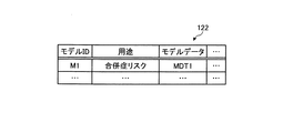
実施形態に係るモデル情報記憶部122は、モデルに関する情報を記憶する。例えば、モデル情報記憶部122は、学習処理により学習(生成)された学習済みモデル(モデル)の情報(モデルデータ)を記憶する。図7は、本開示の第1の実施形態に係るモデル情報記憶部の一例を示す図である。図7に、第1の実施形態に係るモデル情報記憶部122の一例を示す。図7に示した例では、モデル情報記憶部122は、「モデルID」、「用途」、「モデルデータ」といった項目が含まれる。
The model
「モデルID」は、モデルを識別するための識別情報を示す。「用途」は、対応するモデルの用途を示す。「モデルデータ」は、モデルのデータを示す。図7では「モデルデータ」に「MDT1」といった概念的な情報が格納される例を示したが、実際には、モデルに含まれるネットワークに関する情報や関数等、そのモデルを構成する種々の情報が含まれる。 The "model ID" indicates identification information for identifying the model. "Use" indicates the use of the corresponding model. "Model data" indicates model data. In FIG. 7, an example in which conceptual information such as "MDT1" is stored in "model data" is shown, but in reality, various information constituting the model such as information and functions related to the network included in the model are stored. included.
図7に示す例では、モデルID「M1」により識別されるモデル(モデルM1)は、用途が「合併症リスク」であることを示す。また、モデルM1のモデルデータは、モデルデータMDT1であることを示す。 In the example shown in FIG. 7, the model (model M1) identified by the model ID "M1" indicates that the use is "complication risk". Further, it is shown that the model data of the model M1 is the model data MDT1.
なお、モデル情報記憶部122は、上記に限らず、目的に応じて種々の情報を記憶してもよい。
The model
図6に戻り、説明を続ける。制御部130は、例えば、CPU(Central Processing Unit)やMPU(Micro Processing Unit)等によって、情報処理装置100内部に記憶されたプログラム(例えば、本開示に係る情報処理プログラム等)がRAM(Random Access Memory)等を作業領域として実行されることにより実現される。また、制御部130は、コントローラ(controller)であり、例えば、ASIC(Application Specific Integrated Circuit)やFPGA(Field Programmable Gate Array)等の集積回路により実現される。
Returning to FIG. 6, the description will be continued. In the control unit 130, for example, a program (for example, an information processing program according to the present disclosure) stored in the
図6に示すように、制御部130は、取得部131と、学習部132と、解析部133と、送信部134とを有し、以下に説明する手術支援処理の機能や作用を実現または実行する。なお、制御部130の内部構成は、図6に示した構成に限られず、後述する情報処理を行う構成であれば他の構成であってもよい。また、制御部130が有する各処理部の接続関係は、図6に示した接続関係に限られず、他の接続関係であってもよい。
As shown in FIG. 6, the control unit 130 includes an
取得部131は、各種情報を取得する。取得部131は、外部の情報処理装置から各種情報を取得する。取得部131は、手術室システム5100から各種情報を取得する。取得部131は、音声認識サーバ等の他の情報処理装置から各種情報を取得する。
The
取得部131は、記憶部120から各種情報を取得する。取得部131は、学習用データ記憶部121やモデル情報記憶部122から各種情報を取得する。
The
例えば、取得部131は、モデルを取得してもよい。取得部131は、モデルを提供する外部の情報処理装置や記憶部120からモデルを取得する。例えば、取得部131は、モデルM1等をモデル情報記憶部122から取得する。
For example, the
取得部131は、学習部132が学習した各種情報を取得する。取得部131は、解析部133が解析した各種情報を取得する。取得部131は、解析部133が算出した各種情報を取得する。取得部131は、解析部133が生成した各種情報を取得する。取得部131は、解析部133が決定した各種情報を取得する。
The
取得部131は、手術画像である第1手術画像を取得する。取得部131は、第2手術画像に対応する第2手術による合併症リスクに関する情報を含む学習データを用いて生成された学習済みモデルを取得する。
The
取得部131は、画質に関する画質条件及び遅延に関する遅延条件を満たす高画質かつ低遅延の画像情報である第1手術画像を取得する。取得部131は、4K解像度以上の画像情報である第1手術画像を取得する。取得部131は、特殊光観察画像である第1手術画像を取得する。
The
取得部131は、外科合併症の要因となる部位を含む画像情報である第2手術画像を取得する。取得部131は、縫合部位または出血部位のうち少なくとも1つを含む画像情報である第2手術画像を取得する。取得部131は、画像認識により検出された部位を含む画像情報である第2手術画像を取得する。
The
取得部131は、特殊光観察画像である第2手術画像を取得する。取得部131は、縫合部位または出血部位の内部状態を可視化した画像である第2手術画像を取得する。
The
取得部131は、合併症の発生確率または影響度のうち少なくとも1つを示す情報である合併症リスクに関する情報を取得する。取得部131は、合併症ありか合併症なしかを示す2値の情報である発生確率を取得する。取得部131は、合併症に関する影響の度合いを示す5段階の情報である影響度を取得する。
The
取得部131は、リスクの発生部位に関する情報である合併症リスクに関する情報を取得する。取得部131は、縫合部位または出血部位のうち少なくとも1つに関する情報であるリスクの発生部位に関する情報を取得する。
The
取得部131は、外科合併症の要因となる部位を含む画像情報である第2手術画像を用いて生成された学習済みモデルを取得する。取得部131は、縫合部位または出血部位のうち少なくとも1つを含む画像情報である第2手術画像を用いて生成された学習済みモデルを取得する。
The
取得部131は、画像認識により検出された部位を含む画像情報である第2手術画像を用いて生成された学習済みモデルを取得する。取得部131は、特殊光観察画像である第2手術画像を用いて生成された学習済みモデルを取得する。取得部131は、縫合部位または出血部位の内部状態を可視化した画像である第2手術画像を用いて生成された学習済みモデルを取得する。
The
取得部131は、合併症の発生確率または影響度のうち少なくとも1つを示す情報である合併症リスクに関する情報を用いて生成された学習済みモデルを取得する。取得部131は、合併症ありか合併症なしかを示す2値の情報である発生確率を用いて生成された学習済みモデルを取得する。取得部131は、合併症に関する影響の度合いを示す5段階の情報である影響度を用いて生成された学習済みモデルを取得する。
The
取得部131は、リスクの発生部位に関する情報である合併症リスクに関する情報を用いて生成された学習済みモデルを取得する。取得部131は、縫合部位または出血部位のうち少なくとも1つに関する情報であるリスクの発生部位に関する情報を用いて生成された学習済みモデルを取得する。
The
取得部131は、手術映像である学習済みモデルの入力データを取得する。取得部131は、手術映像である学習データを取得する。取得部131は、時系列画像である学習済みモデルの入力データを取得する。取得部131は、時系列画像である学習データを取得する。
The
取得部131は、手術画像に対応するメタ情報(手術属性情報)を取得する。取得部131は、記憶部120や外部の情報処理装置から手術属性情報を取得する。例えば、取得部131は、手術画像に対応する手術の対象となる患者の患者情報を含む手術属性情報を取得する。例えば、取得部131は、手術画像に対応する手術を行う術者の術者情報を含む手術属性情報を取得する。例えば、取得部131は、外部の情報処理装置から手術属性情報を取得した場合、記憶部120に格納する。例えば、取得部131は、外部の情報処理装置から第2手術画像に対応する手術属性情報を取得した場合、取得した手術属性情報を第2手術画像に対応付けて学習用データ記憶部121に格納する。
The
学習部132は、学習処理を行う。学習部132は、各種学習を行う。学習部132は、モデルを学習(生成)する。学習部132は、モデル等の各種情報を学習する。学習部132は、学習によりモデルを生成する。学習部132は、種々の機械学習に関する技術を用いて、モデルを学習する。学習部132は、学習によりモデルを更新する。例えば、学習部132は、ネットワークのパラメータを学習する。 The learning unit 132 performs a learning process. The learning unit 132 performs various learning. The learning unit 132 learns (generates) the model. The learning unit 132 learns various information such as a model. The learning unit 132 generates a model by learning. The learning unit 132 learns a model by using various techniques related to machine learning. The learning unit 132 updates the model by learning. For example, the learning unit 132 learns network parameters.
学習部132は、学習用データ記憶部121に記憶された学習データ(教師データ)を用いて、学習処理を行うことにより、判定器(学習済モデル)を生成する。例えば、学習部132は、合併症リスクの判別モデルを生成する。学習部132は、モデルM1を生成する。学習部132による学習の手法は特に限定されないが、例えば、ラベル情報(合併症リスク情報等)と手術画像群とを紐づけた学習データを用意し、その学習データを多層ニューラルネットワークに基づいた計算モデルに入力して学習してもよい。また、例えばCNN(Convolutional Neural Network)、3D−CNN等のDNN(Deep Neural Network)に基づく手法が用いられてもよい。学習部132は、手術映像等の動画像(動画)のような時系列データを対象とする場合、再帰型ニューラルネットワーク(Recurrent Neural Network:RNN)やRNNを拡張したLSTM(Long Short-Term Memory units)に基づく手法を用いてもよい。
The learning unit 132 generates a determination device (learned model) by performing a learning process using the learning data (teacher data) stored in the learning
例えば、学習部132は、外部の情報処理装置からの情報や記憶部120に記憶された情報に基づいて、各種情報を学習する。学習部132は、手術室システム5100からの情報に基づいて、各種情報を学習する。学習部132は、学習用データ記憶部121やモデル情報記憶部122に記憶された情報に基づいて、各種情報を学習する。学習部132は、学習により生成したモデルをモデル情報記憶部122に格納する。学習部132は、モデルM1等を生成する。
For example, the learning unit 132 learns various types of information based on information from an external information processing device and information stored in the
学習部132は、取得部131により取得された各種情報に基づいて、各種情報を学習する。学習部132は、解析部133により解析された各種情報に基づいて、各種情報を学習する。学習部132は、解析部133により生成された各種情報に基づいて、各種情報を学習する。学習部132は、解析部133により決定された各種情報に基づいて、各種情報を学習する。学習部132は、過去の手術画像である第2手術画像と、手術による合併症リスクに関する情報を教師データとして用い、入力を手術画像とし、出力をその手術における合併症のリスクとする判定モデルである学習済みモデルを機械学習により生成する。学習部132は、機械学習としてディープラーニングを用いて学習済みモデルを生成する。
The learning unit 132 learns various information based on the various information acquired by the
学習部132は、手術画像を入力とし、リスク発生の部位を示す情報とその発生有無を示す情報との組合せを正解情報として学習を行うことにより、手術画像の入力に応じて、スク発生の部位に関する情報とその発生確率とを示す情報を出力するモデルを生成する。例えば、学習部132は、手術画像を入力とし、縫合不全の部位を示す情報と、その発生有無を示す情報との組合せを正解情報として学習を行う。これにより、学習部132は、手術画像の入力に応じて、縫合不全が発生する可能性が有る部位とその発生確率とを示す情報を出力するモデルを生成する。例えば、学習部132は、手術画像を入力とし、出血部位を示す情報と、その発生有無を示す情報との組合せを正解情報として学習を行う。これにより、学習部132は、手術画像の入力に応じて、出血が発生する可能性が有る部位とその発生確率とを示す情報を出力するモデルを生成する。例えば、学習部132は、手術画像を入力とし、縫合不全の部位を示す情報と、その影響度を示す情報との組合せを正解情報として学習を行う。これにより、学習部132は、手術画像の入力に応じて、縫合不全が発生する可能性が有る部位とその影響度とを示す情報を出力するモデルを生成する。例えば、学習部132は、手術画像を入力とし、出血部位を示す情報と、その影響度を示す情報との組合せを正解情報として学習を行う。これにより、学習部132は、手術画像の入力に応じて、出血が発生する可能性が有る部位とその影響度とを示す情報を出力するモデルを生成する。 The learning unit 132 receives the surgical image as an input, and learns by using the combination of the information indicating the site where the risk occurs and the information indicating the presence or absence of the risk as the correct answer information, so that the site where the risk occurs depends on the input of the surgical image. Generate a model that outputs information indicating the information about and the probability of its occurrence. For example, the learning unit 132 receives a surgical image as an input, and learns using a combination of information indicating the site of suture failure and information indicating the presence or absence of the occurrence as correct answer information. As a result, the learning unit 132 generates a model that outputs information indicating a site where anastomotic insufficiency may occur and the probability of occurrence thereof in response to the input of the surgical image. For example, the learning unit 132 receives a surgical image as an input, and learns using a combination of information indicating a bleeding site and information indicating the presence or absence of the occurrence as correct answer information. As a result, the learning unit 132 generates a model that outputs information indicating a site where bleeding may occur and the probability of occurrence thereof in response to the input of the surgical image. For example, the learning unit 132 receives a surgical image as an input, and learns using a combination of information indicating the site of suture failure and information indicating the degree of influence as correct answer information. As a result, the learning unit 132 generates a model that outputs information indicating a site where anastomotic insufficiency may occur and the degree of influence thereof in response to the input of the surgical image. For example, the learning unit 132 receives a surgical image as an input, and learns using a combination of information indicating a bleeding site and information indicating the degree of influence as correct answer information. As a result, the learning unit 132 generates a model that outputs information indicating a site where bleeding may occur and the degree of influence thereof in response to the input of the surgical image.
例えば、学習部132は、過去の手術においてリスクが発生したことを示す情報を正解情報とし、その手術においてリスクが顕在化する前の時点の手術画像を入力として、モデルを学習する。これにより、学習部132は、第1時点の手術画像を入力として、第1時点よりも後の第2時点においてリスクが発生するかを予測するモデルを学習する。学習部132は、手術映像の特徴情報を含む学習データを用いて学習済みモデルを生成する。学習部132は、手技または手術シーン変化のうち少なくとも1つである特徴情報を含む学習データを用いて学習済みモデルを生成する。学習部132は、縫い方または縫う位置のうち少なくとも1つを含む縫合プロセスである特徴情報報を含む学習データを用いて学習済みモデルを生成する。 For example, the learning unit 132 learns a model by using information indicating that a risk has occurred in a past operation as correct answer information and inputting an operation image at a time before the risk becomes apparent in the operation. As a result, the learning unit 132 learns a model that predicts whether a risk will occur at a second time point after the first time point by inputting the surgical image at the first time point. The learning unit 132 generates a trained model using the learning data including the feature information of the surgical image. The learning unit 132 generates a trained model using the learning data including the feature information which is at least one of the procedure or the surgical scene change. The learning unit 132 generates a trained model using the training data including the feature information information which is the sewing process including at least one of the sewing method and the sewing position.
学習部132は、術具の軌道に基づき検出される縫合プロセスを含む学習データを用いて学習済みモデルを生成する。学習部132は、術具の先端位置の時系列変化に基づき検出される縫合プロセスを含む学習データを用いて学習済みモデルを生成する。学習部132は、手術の動作時間に基づき検出される特徴情報を含む学習データを用いて学習済みモデルを生成する。例えば、学習部132は、上述のような手術映像等の動画像(動画)を対象とする場合、RNNやLSTMに基づくモデルを生成する。学習部132は、手技を特徴情報として含む動画像を用いてモデルを生成する。学習部132は、手術シーン変化を特徴情報として含む動画像を用いてモデルを生成する。学習部132は、縫合プロセスを特徴情報として含む動画像を用いてモデルを生成する。学習部132は、縫い方や縫う位置等を特徴情報として含む動画像を用いてモデルを生成する。 The learning unit 132 generates a trained model using the training data including the suturing process detected based on the trajectory of the surgical instrument. The learning unit 132 generates a trained model using the training data including the suturing process detected based on the time-series change of the tip position of the surgical instrument. The learning unit 132 generates a trained model using the learning data including the feature information detected based on the operation time of the operation. For example, the learning unit 132 generates a model based on RNN or LSTM when targeting a moving image (moving image) such as a surgical image as described above. The learning unit 132 generates a model using a moving image including the procedure as feature information. The learning unit 132 generates a model using a moving image including a change in a surgical scene as feature information. The learning unit 132 generates a model using a moving image including the suturing process as feature information. The learning unit 132 generates a model using a moving image including a sewing method, a sewing position, and the like as feature information.
学習部132は、外科合併症の要因となる部位を含む画像情報である第2手術画像を用いて学習済みモデルを学習する。学習部132は、縫合部位または出血部位のうち少なくとも1つを含む画像情報である第2手術画像を用いて学習済みモデルを学習する。 The learning unit 132 learns the learned model by using the second surgical image which is the image information including the site which causes the surgical complication. The learning unit 132 learns the trained model using the second surgical image which is the image information including at least one of the suture site and the bleeding site.
学習部132は、画像認識により検出された部位を含む画像情報である第2手術画像を用いて学習済みモデルを学習する。学習部132は、特殊光観察画像である第2手術画像を用いて学習済みモデルを学習する。学習部132は、縫合部位または出血部位の内部状態を可視化した画像である第2手術画像を用いて学習済みモデルを学習する。 The learning unit 132 learns the trained model by using the second surgical image which is the image information including the part detected by the image recognition. The learning unit 132 learns the trained model using the second surgical image which is a special light observation image. The learning unit 132 learns the trained model by using the second surgical image which is an image that visualizes the internal state of the suture site or the bleeding site.
学習部132は、合併症の発生確率または影響度のうち少なくとも1つを示す情報である合併症リスクに関する情報を用いて学習済みモデルを学習する。学習部132は、合併症ありか合併症なしかを示す2値の情報である発生確率を用いて学習済みモデルを学習する。学習部132は、合併症に関する影響の度合いを示す5段階の情報である影響度を用いて学習済みモデルを学習する。 The learning unit 132 learns the learned model by using the information on the complication risk, which is the information indicating at least one of the probability of occurrence of complications or the degree of influence. The learning unit 132 learns the trained model using the probability of occurrence, which is binary information indicating whether or not there are complications. The learning unit 132 learns the trained model using the degree of influence, which is five-step information indicating the degree of influence regarding complications.
学習部132は、リスクの発生部位に関する情報である合併症リスクに関する情報を用いて学習済みモデルを学習する。学習部132は、縫合部位または出血部位のうち少なくとも1つに関する情報であるリスクの発生部位に関する情報を用いて学習済みモデルを学習する。 The learning unit 132 learns the learned model by using the information on the complication risk, which is the information on the site where the risk occurs. The learning unit 132 learns the trained model using the information about the risk occurrence site, which is the information about at least one of the suture site or the bleeding site.
学習部132は、手術画像に対応するメタ情報(手術属性情報)を用いて、モデルを生成してもよい。この場合、学習部132は、手術画像と手術属性情報とを入力とするモデルを生成してもよい。例えば、学習部132は、手術画像に対応する手術の対象となる患者の患者情報を含む手術属性情報を用いて、モデルを生成してもよい。例えば、学習部132は、手術画像に対応する手術を行う術者の術者情報を含む手術属性情報を用いて、モデルを生成してもよい。 The learning unit 132 may generate a model by using the meta information (surgical attribute information) corresponding to the surgical image. In this case, the learning unit 132 may generate a model in which the surgical image and the surgical attribute information are input. For example, the learning unit 132 may generate a model by using the surgical attribute information including the patient information of the patient to be operated on, which corresponds to the surgical image. For example, the learning unit 132 may generate a model by using the surgical attribute information including the surgeon information of the surgeon who performs the surgery corresponding to the surgical image.
解析部133は、各種情報を解析する。解析部133は、外部の情報処理装置からの情報や記憶部120に記憶された情報に基づいて、各種情報を解析する。解析部133は、学習用データ記憶部121やモデル情報記憶部122に記憶された情報に基づいて、各種情報を解析する。解析部133は、各種情報を特定する。解析部133は、各種情報を推定する。
The analysis unit 133 analyzes various information. The analysis unit 133 analyzes various information based on the information from the external information processing device and the information stored in the
解析部133は、各種情報を抽出する。解析部133は、各種情報を選択する。解析部133は、外部の情報処理装置からの情報や記憶部120に記憶された情報に基づいて、各種情報を抽出する。解析部133は、記憶部120から、各種情報を抽出する。解析部133は、学習用データ記憶部121やモデル情報記憶部122から、各種情報を抽出する。
The analysis unit 133 extracts various information. The analysis unit 133 selects various types of information. The analysis unit 133 extracts various information based on the information from the external information processing device and the information stored in the
解析部133は、取得部131により取得された各種情報に基づいて、各種情報を抽出する。解析部133は、解析部133により生成された情報に基づいて、各種情報を抽出する。また、解析部133は、解析部133により決定された各種情報に基づいて、各種情報を抽出する。解析部133は、学習部132により学習された各種情報に基づいて、各種情報を抽出する。
The analysis unit 133 extracts various information based on the various information acquired by the
解析部133は、各種情報を生成する。解析部133は、外部の情報処理装置からの情報や記憶部120に記憶された情報に基づいて、各種情報を生成する。解析部133は、手術室システム5100からの情報に基づいて、各種情報を生成する。解析部133は、学習用データ記憶部121やモデル情報記憶部122に記憶された情報に基づいて、各種情報を生成する。
The analysis unit 133 generates various information. The analysis unit 133 generates various information based on the information from the external information processing device and the information stored in the
解析部133は、各種情報を決定する。解析部133は、各種の判断を行う。例えば、解析部133は、外部の情報処理装置からの情報や記憶部120に記憶された情報に基づいて、各種情報を決定する。解析部133は、手術室システム5100からの情報に基づいて、各種情報を決定する。解析部133は、学習用データ記憶部121やモデル情報記憶部122に記憶された情報に基づいて、各種情報を決定する。
The analysis unit 133 determines various information. The analysis unit 133 makes various determinations. For example, the analysis unit 133 determines various information based on the information from the external information processing device and the information stored in the
解析部133は、第1手術画像とは異なる手術画像である第2手術画像と、手術による合併症リスクに関する情報とを含む学習データを用いて生成された学習済みモデルに対して、第1手術画像を適用することにより、第1手術画像のリスク解析情報を生成する。 The analysis unit 133 performs the first operation on the trained model generated using the training data including the second operation image which is a operation image different from the first operation image and the information on the risk of complications due to the operation. By applying the image, risk analysis information of the first surgical image is generated.
解析部133は、第1手術画像に対応する第1手術による合併症リスクを示すリスク解析情報を生成する。解析部133は、取得部131により取得された学習済みモデルに、第1手術画像を入力することにより、リスク解析情報を生成する。解析部133は、入力に応じてリスク発生の部位を示す情報を出力するモデルを用いる場合、モデルの出力を用いて、リスク発生の部位に関する情報を含むリスク解析情報を生成する。例えば、解析部133は、縫合不全の部位を示す情報と、その発生確率を出力するモデルを用いる場合、モデルの出力を用いて、縫合不全の可能性が有る部位とその発生確率を示すリスク解析情報を生成する。例えば、解析部133は、出血部位を示す情報と、その発生確率を出力するモデルを用いる場合、モデルの出力を用いて、出血部位の可能性が有る部位とその発生確率を示すリスク解析情報を生成する。例えば、解析部133は、縫合不全の部位を示す情報と、その影響度を出力するモデルを用いる場合、モデルの出力を用いて、縫合不全の可能性が有る部位とその影響度を示すリスク解析情報を生成する。例えば、解析部133は、出血部位を示す情報と、その影響度を出力するモデルを用いる場合、モデルの出力を用いて、出血部位の可能性が有る部位とその影響度を示すリスク解析情報を生成する。
The analysis unit 133 generates risk analysis information indicating the risk of complications due to the first surgery corresponding to the first surgery image. The analysis unit 133 generates risk analysis information by inputting the first surgical image into the trained model acquired by the
解析部133は、画質に関する画質条件及び遅延に関する遅延条件を満たす高画質かつ低遅延の画像情報である第1手術画像を用いて、リスク解析情報を生成する。解析部133は、4K解像度以上の画像情報である第1手術画像を用いて、リスク解析情報を生成する。解析部133は、特殊光観察画像である第1手術画像を用いて、リスク解析情報を生成する。 The analysis unit 133 generates risk analysis information using the first surgical image, which is high-quality and low-delay image information that satisfies the image quality condition regarding image quality and the delay condition regarding delay. The analysis unit 133 generates risk analysis information using the first surgical image which is the image information of 4K resolution or higher. The analysis unit 133 generates risk analysis information using the first surgical image which is a special light observation image.
解析部133は、外科合併症の要因となる部位を含む画像情報である第2手術画像を用いて生成された学習済みモデルを用いて、リスク解析情報を生成する。解析部133は、縫合部位または出血部位のうち少なくとも1つを含む画像情報である第2手術画像を用いて生成された学習済みモデルを用いて、リスク解析情報を生成する。 The analysis unit 133 generates risk analysis information using a learned model generated using a second surgical image, which is image information including a site that causes surgical complications. The analysis unit 133 generates risk analysis information using a trained model generated using a second surgical image, which is image information including at least one of a suture site or a bleeding site.
解析部133は、画像認識により検出された部位を含む画像情報である第2手術画像を用いて生成された学習済みモデルを用いて、リスク解析情報を生成する。解析部133は、特殊光観察画像である第2手術画像を用いて生成された学習済みモデルを用いて、リスク解析情報を生成する。解析部133は、縫合部位または出血部位の内部状態を可視化した画像である第2手術画像を用いて生成された学習済みモデルを用いて、リスク解析情報を生成する。 The analysis unit 133 generates risk analysis information using a learned model generated using a second surgical image, which is image information including a site detected by image recognition. The analysis unit 133 generates risk analysis information using the trained model generated by using the second surgical image which is a special light observation image. The analysis unit 133 generates risk analysis information using a learned model generated using a second surgical image which is an image that visualizes the internal state of the suture site or the bleeding site.
解析部133は、合併症の発生確率または影響度のうち少なくとも1つを示す情報である合併症リスクに関する情報を用いて生成された学習済みモデルを用いて、リスク解析情報を生成する。解析部133は、合併症ありか合併症なしかを示す2値の情報である発生確率を用いて生成された学習済みモデルを用いて、リスク解析情報を生成する。解析部133は、合併症に関する影響の度合いを示す5段階の情報である影響度を用いて生成された学習済みモデルを用いて、リスク解析情報を生成する。 The analysis unit 133 generates risk analysis information using a trained model generated using information on complication risk, which is information indicating at least one of the probability of occurrence of complications or the degree of influence. The analysis unit 133 generates risk analysis information using a learned model generated using the probability of occurrence, which is binary information indicating the presence or absence of complications. The analysis unit 133 generates risk analysis information using a learned model generated using the degree of influence, which is five-step information indicating the degree of influence regarding complications.
解析部133は、リスクの発生部位に関する情報である合併症リスクに関する情報を用いて生成された学習済みモデルを用いて、リスク解析情報を生成する。解析部133は、縫合部位または出血部位のうち少なくとも1つに関する情報であるリスクの発生部位に関する情報を用いて生成された学習済みモデルを用いて、リスク解析情報を生成する。 The analysis unit 133 generates risk analysis information using a trained model generated using information on complication risk, which is information on the site where risk occurs. The analysis unit 133 generates risk analysis information using a trained model generated using the information about the risk occurrence site, which is the information about at least one of the suture site or the bleeding site.
解析部133は、学習済みモデルに対して、補正処理した第1手術画像を適用することにより、リスク解析情報を生成する。解析部133は、回転、歪み、または解像度のうち少なくとも1つに関する処理である補正処理がされた第1手術画像を適用することにより、リスク解析情報を生成する。 The analysis unit 133 generates risk analysis information by applying the corrected first surgical image to the trained model. The analysis unit 133 generates risk analysis information by applying a corrected first surgical image, which is a process relating to at least one of rotation, distortion, and resolution.
解析部133は、合併症リスクの発生確率または影響度のうち少なくとも1つを含む情報であるリスク解析情報を生成する。解析部133は、リスク発生の部位に関する情報を含むリスク解析情報を生成する。解析部133は、縫合不全の部位または出血部位のうち少なくとも1つに関する情報と、部位における合併症リスクの発生確率または影響度のうち少なくとも1つと、を含む情報であるリスク解析情報を生成する。 The analysis unit 133 generates risk analysis information which is information including at least one of the probability of occurrence of complication risk or the degree of influence. The analysis unit 133 generates risk analysis information including information on the site where the risk occurs. The analysis unit 133 generates risk analysis information which is information including information on at least one of the site of anastomotic insufficiency or the site of bleeding and at least one of the probability of occurrence or the degree of influence of the risk of complications at the site.
解析部133は、リスク解析のうち、所定の処理はクラウドサーバで実行する。解析部133は、合併症とトレードオフの関係にある情報を加味した情報であるリスク解析情報を生成する。解析部133は、縫合不全とトレードオフの関係にある情報を加味した情報であるリスク解析情報を生成する。解析部133は、縫合不全とトレードオフの関係にある手術時間を加味した情報であるリスク解析情報を生成する。解析部133は、手術時間が所定の閾値を超える場合、手術支援情報を出力しないと決定する。解析部133は、手術支援情報の出力により、手術時間が所定の閾値を超える場合、手術支援情報を出力しないと決定する。解析部133は、予測される手術時間が所定の閾値を超える場合、手術支援情報を出力しないと決定する。 The analysis unit 133 executes a predetermined process of the risk analysis on the cloud server. The analysis unit 133 generates risk analysis information which is information including information having a trade-off relationship with complications. The analysis unit 133 generates risk analysis information which is information including information having a trade-off relationship with anastomotic insufficiency. The analysis unit 133 generates risk analysis information which is information including the operation time which is in a trade-off relationship with the suture failure. The analysis unit 133 determines that the operation support information is not output when the operation time exceeds a predetermined threshold value. The analysis unit 133 determines that the operation support information is not output when the operation time exceeds a predetermined threshold value based on the output of the operation support information. The analysis unit 133 determines that the operation support information is not output when the predicted operation time exceeds a predetermined threshold value.
解析部133は、リスク解析情報に基づく手術支援情報を生成する。解析部133は、リスク解析情報に基づく手術支援情報を生成する。解析部133は、リスク部位の位置を示す情報である手術支援情報を生成する。例えば、解析部133は、縫合不全の部位を示す情報と、その発生確率を出力するモデルを用いる場合、モデルが出力する発生確率が所定の閾値(例えば70%等)以上である場合、モデルが出力した縫合不全の部位を示す手術支援情報を生成する。例えば、解析部133は、出血部位の部位を示す情報と、その発生確率を出力するモデルを用いる場合、モデルが出力する発生確率が所定の閾値(例えば75%等)以上である場合、モデルが出力した出血部位を示す手術支援情報を生成する。例えば、解析部133は、縫合不全の部位を示す情報と、その影響度を出力するモデルを用いる場合、モデルが出力する影響度が所定の閾値(例えば第4段階等)以上である場合、モデルが出力した縫合不全の部位を示す手術支援情報を生成する。例えば、解析部133は、出血部位の部位を示す情報と、その影響度を出力するモデルを用いる場合、モデルが出力する影響度が所定の閾値(例えば第3段階等)以上である場合、モデルが出力した出血部位を示す手術支援情報を生成する。 The analysis unit 133 generates surgical support information based on the risk analysis information. The analysis unit 133 generates surgical support information based on the risk analysis information. The analysis unit 133 generates surgical support information which is information indicating the position of the risk site. For example, when the analysis unit 133 uses information indicating the site of suture failure and a model that outputs the probability of occurrence of the suture, if the probability of occurrence output by the model is equal to or higher than a predetermined threshold value (for example, 70%), the model Generate surgical support information indicating the output site of suture failure. For example, when the analysis unit 133 uses information indicating the site of the bleeding site and a model that outputs the probability of occurrence of the bleeding site, if the probability of occurrence output by the model is equal to or higher than a predetermined threshold value (for example, 75%), the model Generates surgical support information indicating the output bleeding site. For example, when the analysis unit 133 uses information indicating the site of suture failure and a model that outputs the degree of influence, if the degree of influence output by the model is equal to or higher than a predetermined threshold value (for example, the fourth stage), the model Generates surgical support information indicating the site of suture failure output by. For example, when the analysis unit 133 uses information indicating the site of the bleeding site and a model that outputs the degree of influence, the model is used when the degree of influence output by the model is equal to or higher than a predetermined threshold value (for example, the third stage). Generates surgical support information indicating the bleeding site output by.
解析部133は、リスク部位と重ならない位置に重畳される手術支援情報を生成する。解析部133は、リスクの発生確率または影響度のうち少なくとも1つを示す情報である手術支援情報を生成する。解析部133は、形状または色が所定の態様である手術支援情報を生成する。 The analysis unit 133 generates surgical support information superimposed on a position that does not overlap with the risk site. The analysis unit 133 generates surgical support information which is information indicating at least one of the probability of occurrence of risk or the degree of influence. The analysis unit 133 generates surgical support information in which the shape or color is a predetermined aspect.
解析部133は、形状または色がリスクの発生確率または影響度のうち少なくとも1つに応じて変化する手術支援情報を生成する。解析部133は、手術画像上で移動または点滅のうち少なくとも1つを行う手術支援情報を生成する。解析部133は、移動速度または動きのうち少なくとも1つが、リスクの発生確率または影響度のうち少なくとも1つに応じて制御される手術支援情報を生成する。 The analysis unit 133 generates surgical support information in which the shape or color changes according to at least one of the probability of occurrence of risk or the degree of influence. The analysis unit 133 generates surgical support information that performs at least one of moving or blinking on the surgical image. The analysis unit 133 generates surgical support information in which at least one of the movement speeds or movements is controlled according to at least one of the probability of occurrence of risk or the degree of influence.
解析部133は、手術時間または手術シーンのうち少なくとも1つに基づき動的に内容が変更される手術支援情報を生成する。解析部133は、表示が所定の基準に基づき制御される手術支援情報を生成する。解析部133は、音声で報知された後、所定の期間経過後に表示される手術支援情報を生成する。解析部133は、手術中の所定の期間表示される手術支援情報を生成する。 The analysis unit 133 generates surgical support information whose contents are dynamically changed based on at least one of the surgical time or the surgical scene. The analysis unit 133 generates surgical support information whose display is controlled based on a predetermined standard. The analysis unit 133 generates surgical support information to be displayed after a lapse of a predetermined period after being notified by voice. The analysis unit 133 generates surgical support information displayed for a predetermined period during surgery.
解析部133は、特殊光観察画像の場合は、所定の態様で表示される手術支援情報を生成する。解析部133は、疾患、部位、術式により手術の難易度が変動する手術を対象として、リスク解析情報を生成する。解析部133は、通常光により縫合の外部状態を解析して、特殊光により縫合の内部状態を解析することにより、リスク解析情報を生成する。 In the case of a special light observation image, the analysis unit 133 generates surgical support information displayed in a predetermined mode. The analysis unit 133 generates risk analysis information for surgery whose difficulty level varies depending on the disease, site, and surgical procedure. The analysis unit 133 generates risk analysis information by analyzing the external state of the suture with normal light and analyzing the internal state of the suture with special light.
解析部133は、リスク部位の特定を行ってもよい。解析部133は、画像認識等の種々の従来技術を適宜用いて、リスク部位の特定を行ってもよい。解析部133は、リスク発生の判定モデルに一の手術画像が入力され、判定モデルが所定の閾値以上のスコアを出力した場合、一の手術画像を画像解析することにより、一の手術画像中のリスク部位の特定を行ってもよい。 The analysis unit 133 may identify the risk site. The analysis unit 133 may identify the risk site by appropriately using various conventional techniques such as image recognition. When one surgical image is input to the risk occurrence determination model and the determination model outputs a score equal to or higher than a predetermined threshold, the analysis unit 133 performs image analysis of the one surgical image in the one surgical image. The risk site may be identified.
解析部133は、種々の技術を適宜用いて、外部の情報処理装置へ提供する画面(画像情報)等の種々の情報を生成してもよい。解析部133は、出力部へ提供する画面(画像情報)等の種々の情報を生成してもよい。解析部133は、手術室システム5100へ提供する画面(画像情報)等を生成する。例えば、解析部133は、記憶部120に記憶された情報に基づいて、手術室システム5100へ提供する画面(画像情報)等を生成する。解析部133は、外部の情報処理装置へ提供する画面(画像情報)等が生成可能であれば、どのような処理により画面(画像情報)等を生成してもよい。例えば、解析部133は、画像生成や画像処理等に関する種々の技術を適宜用いて、手術室システム5100へ提供する画面(画像情報)を生成する。例えば、解析部133は、Java(登録商標)等の種々の技術を適宜用いて、手術室システム5100へ提供する画面(画像情報)を生成する。なお、解析部133は、CSSやJavaScript(登録商標)やHTMLの形式に基づいて、手術室システム5100へ提供する画面(画像情報)を生成してもよい。また、例えば、解析部133は、JPEG(Joint Photographic Experts Group)やGIF(Graphics Interchange Format)やPNG(Portable Network Graphics)など様々な形式で画面(画像情報)を生成してもよい。
The analysis unit 133 may generate various information such as a screen (image information) to be provided to an external information processing device by appropriately using various techniques. The analysis unit 133 may generate various information such as a screen (image information) provided to the output unit. The analysis unit 133 generates a screen (image information) or the like to be provided to the
送信部134は、外部の情報処理装置へ各種情報を提供する。送信部134は、外部の情報処理装置へ各種情報を送信する。例えば、送信部134は、手術室システム5100へ各種情報を送信する。送信部134は、記憶部120に記憶された情報を提供する。送信部134は、記憶部120に記憶された情報を送信する。
The transmission unit 134 provides various information to an external information processing device. The transmission unit 134 transmits various information to an external information processing device. For example, the transmission unit 134 transmits various information to the
送信部134は、手術室システム5100からの情報に基づいて、各種情報を提供する。送信部134は、記憶部120に記憶された情報に基づいて、各種情報を提供する。送信部134は、学習用データ記憶部121やモデル情報記憶部122に記憶された情報に基づいて、各種情報を提供する。
The transmission unit 134 provides various information based on the information from the
送信部134は、リスク解析情報に基づく手術支援情報を送信する。送信部134は、リスク解析情報に基づく手術支援情報を送信する。送信部134は、リスク部位の位置を示す情報である手術支援情報を送信する。送信部134は、リスク部位と重ならない位置に重畳される手術支援情報を送信する。送信部134は、リスクの発生確率または影響度のうち少なくとも1つを示す情報である手術支援情報を送信する。 The transmission unit 134 transmits surgical support information based on the risk analysis information. The transmission unit 134 transmits surgical support information based on the risk analysis information. The transmission unit 134 transmits surgical support information, which is information indicating the position of the risk site. The transmission unit 134 transmits surgical support information superimposed on a position that does not overlap with the risk site. The transmission unit 134 transmits surgical support information which is information indicating at least one of the probability of occurrence of risk or the degree of influence.
送信部134は、形状または色が所定の態様である手術支援情報を送信する。送信部134は、形状または色がリスクの発生確率または影響度のうち少なくとも1つに応じて変化する手術支援情報を送信する。送信部134は、手術画像上で移動または点滅のうち少なくとも1つを行う手術支援情報を送信する。 The transmission unit 134 transmits surgical support information whose shape or color is a predetermined embodiment. The transmission unit 134 transmits surgical support information whose shape or color changes according to at least one of the probability of occurrence of risk or the degree of influence. The transmission unit 134 transmits surgical support information that performs at least one of moving or blinking on the surgical image.
送信部134は、移動速度または動きのうち少なくとも1つが、リスクの発生確率または影響度のうち少なくとも1つに応じて制御される手術支援情報を送信する。送信部134は、手術時間または手術シーンのうち少なくとも1つに基づき動的に内容が変更される手術支援情報を送信する。 The transmission unit 134 transmits surgical support information in which at least one of the movement speeds or movements is controlled according to at least one of the probability of occurrence of risk or the degree of influence. The transmission unit 134 transmits the operation support information whose contents are dynamically changed based on at least one of the operation time or the operation scene.
送信部134は、表示が所定の基準に基づき制御される手術支援情報を送信する。送信部134は、音声で報知された後、所定の期間経過後に表示される手術支援情報を送信する。送信部134は、手術中の所定の期間表示される手術支援情報を送信する。送信部134は、特殊光観察画像の場合は、所定の態様で表示される手術支援情報を送信する。 The transmission unit 134 transmits surgical support information whose display is controlled based on a predetermined standard. The transmission unit 134 transmits the surgical support information displayed after a lapse of a predetermined period after being notified by voice. The transmission unit 134 transmits surgical support information displayed for a predetermined period during surgery. In the case of a special light observation image, the transmission unit 134 transmits surgical support information displayed in a predetermined mode.
[3−2.実施形態に係る出力装置の例]
次に、実施形態に係る出力装置の例について説明する。上述の例では、手術室システム5100の表示装置5155が、情報処理装置100が生成したリスク解析情報に基づく手術支援情報を出力する出力部として機能する出力装置である。例えば、表示装置5155は、例えば液晶ディスプレイや有機EL(Electro-Luminescence)ディスプレイ等を有する。表示装置5155は、各種情報を表示する。表示装置5155は、情報処理装置100から取得した情報や手術室システム5100の各種装置から取得した情報を表示する。表示装置5155は、情報処理装置100から取得した手術支援情報や手術室システム5100の各種装置から取得した手術画像を表示する。
[3-2. Example of output device according to the embodiment]
Next, an example of the output device according to the embodiment will be described. In the above example, the
表示装置5155は、出力装置(出力部)として機能する。表示装置5155は、解析部133により生成されたリスク解析情報に基づく手術支援情報を、手術画像に重畳させて出力する。表示装置5155は、リスク解析情報に基づく手術支援情報を、第1手術の手術画像に重畳させて出力する。
The
表示装置5155は、リスク部位の位置を示す情報である手術支援情報を、第1手術の手術画像に重畳させて出力する。表示装置5155は、リスク部位と重ならない位置に重畳される手術支援情報を、第1手術の手術画像に重畳させて出力する。表示装置5155は、第1手術の手術画像のうち、リスク部位と重ならない位置に手術支援情報を重畳させて出力する。
The
表示装置5155は、リスクの発生確率または影響度のうち少なくとも1つを示す情報である手術支援情報を、第1手術の手術画像に重畳させて出力する。表示装置5155は、形状または色が所定の態様である手術支援情報を、第1手術の手術画像に重畳させて出力する。
The
表示装置5155は、形状または色がリスクの発生確率または影響度のうち少なくとも1つに応じて変化する手術支援情報を、第1手術の手術画像に重畳させて出力する。表示装置5155は、手術画像上で移動または点滅のうち少なくとも1つを行う手術支援情報を、第1手術の手術画像に重畳させて出力する。
The
表示装置5155は、移動速度または動きのうち少なくとも1つが、リスクの発生確率または影響度のうち少なくとも1つに応じて制御される手術支援情報を、第1手術の手術画像に重畳させて出力する。表示装置5155は、手術時間または手術シーンのうち少なくとも1つに基づき動的に内容が変更される手術支援情報を、第1手術の手術画像に重畳させて出力する。
The
表示装置5155は、表示が所定の基準に基づき制御される手術支援情報を、第1手術の手術画像に重畳させて出力する。表示装置5155は、音声で報知された後、所定の期間経過後に表示される手術支援情報を、第1手術の手術画像に重畳させて出力する。
The
表示装置5155は、手術中の所定の期間表示される手術支援情報を、第1手術の手術画像に重畳させて出力する。表示装置5155は、手術中の所定の期間、手術支援情報を、第1手術の手術画像に重畳させて表示する。
The
表示装置5155は、特殊光観察画像の場合は、所定の態様で表示される手術支援情報を、第1手術の手術画像に重畳させて出力する。表示装置5155は、特殊光観察画像の場合は、手術支援情報を、所定の態様で第1手術の手術画像に重畳させて表示する。
In the case of a special light observation image, the
なお、出力装置は、表示装置5103A〜5103Dや集中操作パネル5111等、どのような装置であってもよい。
The output device may be any device such as
[3−3.実施形態に係る手術支援処理の手順]
次に、図8を用いて、実施形態に係る各種手術支援処理の手順について説明する。図8は、本開示の実施形態に係る手術支援処理の手順を示すフローチャートである。なお、各ステップの処理は、情報処理装置100や手術室システム5100の各装置等、手術支援システム1に含まれるいずれの装置が行ってもよい。
[3-3. Procedure for surgical support processing according to the embodiment]
Next, the procedure of various surgical support processing according to the embodiment will be described with reference to FIG. FIG. 8 is a flowchart showing a procedure of surgical support processing according to the embodiment of the present disclosure. The processing of each step may be performed by any device included in the
図8に示すように、手術支援システム1は、第1手術画像を取得する(ステップS101)。例えば、情報処理装置100は、第1手術画像を取得する。例えば、情報処理装置100は、手術が行われている手術室システム5100において撮像された第1手術画像を取得する。
As shown in FIG. 8, the
手術支援システム1は、第2手術画像と、手術による合併症リスクに関する情報とを含む学習データを用いて生成された学習済みモデルに対して、第1手術画像を適用することによって、手術画像のリスク解析情報を生成する(ステップS102)。例えば、情報処理装置100は、学習した学習済みモデルに対して、第1手術画像を適用することによって、手術画像のリスク解析情報を生成する。例えば、情報処理装置100は、モデル情報記憶部122(図7参照)に記憶された学習済みモデルであるモデルM1に、第1手術画像を入力することにより、手術画像のリスク解析情報を生成する。
The
手術支援システム1は、解析情報に基づく手術支援情報を手術画像に重畳させて出力する(ステップS103)。例えば、情報処理装置100は、解析情報に基づく手術支援情報を生成し、手術室システム5100に送信する。手術支援情報を受信した手術室システム5100は、手術支援情報を手術画像に重畳させて出力する。手術室システム5100の表示装置5155は、手術支援情報を手術画像に重畳させて表示する。
The
[3−4.実施形態に係る手術支援処理の概要]
図9は、本開示の実施形態に係る手術支援処理の一例を示す図である。本開示の実施形態に係る情報処理は、上述した手術支援システム1に含まれる構成要素によって実現される。
[3-4. Outline of surgical support processing according to the embodiment]
FIG. 9 is a diagram showing an example of the surgical support process according to the embodiment of the present disclosure. The information processing according to the embodiment of the present disclosure is realized by the components included in the
まず、情報処理装置100は、手術画像IM11を第1手術画像として取得する(ステップS11)。例えば、スコピストが硬性内視鏡が操作して、執刀医が縫合中の映像を撮影する。図9の例では、情報処理装置100は、手術室システム5100の内視鏡5115により撮像された手術画像IM11を取得する。情報処理装置100は、ネットワークNを介して手術室システム5100から手術画像IM11を取得する。手術室システム5100に含まれる通信機能を有する通信装置CM(例えばCCU5153等)は、ネットワークNを介して情報処理装置100に手術画像IM11を送信する。通信装置CMは、内視鏡5115により撮像された手術画像IM11を取得し、情報処理装置100に手術画像IM11を送信する。通信装置CMは、手術画像IM11を内視鏡手術システム5113から取得し、情報処理装置100に手術画像IM11を送信する。なお、図9の例では、手術画像IM11のみを図示するが、手術画像が手術映像であってもよい。この場合、手術室システム5100から情報処理装置100に、手術映像がネットワークNを介して伝送される。
First, the
そして、情報処理装置100は、解析処理を行う(ステップS12)。例えば、手術支援システム1は、クラウドAIやサーバ解析などにより、解析処理を行う。図9の例では、情報処理装置100は、手術画像IM11を用いて解析処理を行う。例えば、情報処理装置100は、縫合不全のリスクと縫合不全箇所のアノテーション付の教育済みデータベースを用いて解析処理を行ってもよい。情報処理装置100は、認識解析や形態解析等の技術を適宜用いて、解析処理を行ってもよい。例えば、情報処理装置100は、縫合不全のリスク判定を含む解析処理を行ってもよい。
Then, the
情報処理装置100は、モデルM1に手術画像IM11を適用することにより、解析処理を行う。情報処理装置100は、モデルM1に手術画像IM11を入力し、モデルM1から出力される情報を用いて、解析処理を行う。情報処理装置100は、手術画像IM11が入力されたモデルM1が出力するリスク発生確率を示す情報やリスクが発生する部位を示す情報を用いて、解析処理を行う。
The
情報処理装置100は、手術画像IM11が入力されたモデルM1が出力するリスク発生確率を示す情報が所定の閾値以上である場合、リスクが有ると判定する。また、情報処理装置100は、手術画像IM11が入力されたモデルM1が出力するリスク発生確率を示す情報が所定の閾値未満である場合、リスクが無いと判定する。そして、情報処理装置100は、判定結果を示すリスク判定情報を生成する。情報処理装置100は、手術画像IM11が入力されたモデルM1が出力するリスクが発生する部位を示す情報を用いて危険位置情報を生成する。図9の例では、情報処理装置100は、リスクが有ると判定したリスク判定情報を生成し、リスク発生の部位が手術画像IM11中の右側中央部であることを示す情報を生成する。具体的には、情報処理装置100は、手術画像IM11のうち、リスク発生の部位がマークMK11の内部に位置する領域であることを示す情報を生成する。
The
そして、情報処理装置100は、生成したリスク判定情報や危険位置情報を手術室システム5100に送信する(ステップS13)。手術室システム5100の通信機能を有する装置に生成したリスク判定情報や危険位置情報を送信する。例えば、手術室システム5100のCCU5153(図3参照)は、ネットワークNを介して情報処理装置100との間で情報を送受信する。また、CCU5153は、手術室システム5100の通信機能を有する他の装置(通信装置CM)等を介して情報処理装置100との間で情報を送受信してもよい。CCU5153は、情報処理装置100により生成されたリスク判定情報や危険位置情報を取得する。
Then, the
CCU5153は、リスク判定情報や危険位置情報を用いて、画像処理を行う(ステップS14)。CCU5153は、画像への情報の重畳などの画像処理(信号処理)を行う。例えば、CCU5153は、高画質低遅延映像に判定結果を画像重畳させる。図9の例では、CCU5153は、リスク発生の部位が手術画像IM11中の右側中央部であることを示す手術支援情報を、手術画像IM11に重畳させる。具体的には、CCU5153は、手術画像IM11のうち、リスク発生の部位と判定された領域を囲む円環状のマークMK11を、手術支援情報として手術画像IM11に重畳させる。このように、マークMK11は、手術画像IM11のうち、リスク部位の周囲を囲むように重畳される。すなわち、マークMK11は、手術画像IM11のうち、リスク部位と重ならない位置に重畳される。
CCU5153 performs image processing using the risk determination information and the dangerous position information (step S14). CCU5153 performs image processing (signal processing) such as superimposing information on an image. For example, CCU5153 superimposes a determination result on a high-quality low-delay video. In the example of FIG. 9, the CCU5153 superimposes the surgical support information indicating that the site where the risk occurs is the central right side in the surgical image IM11 on the surgical image IM11. Specifically, the
そして、CCU5153は、手術支援情報としてマークMK11が重畳された手術画像IM11を表示装置5155に表示させる(ステップS15)。CCU5153は、手術支援情報としてマークMK11が重畳された手術画像IM11を表示装置5155に送信することにより、表示装置5155にマークMK11が重畳された手術画像IM11を表示させる。CCU5153からマークMK11が重畳された手術画像IM11を受信した表示装置5155は、マークMK11が重畳された手術画像IM11を表示する。これにより、術者(ドクター)等の医療従事者DTは、マークMK11が重畳された手術画像IM11を確認する。
Then, the
表示装置5155に表示された情報を確認した医療従事者DTは、情報の内容に基づいて、縫い直しなどの処置を行う(ステップS16)。これにより、手術支援システム1は、縫合不全の合併症が減少させることができる。
The medical worker DT who has confirmed the information displayed on the
なお、図9例では、手術室システム5100から情報処理装置100に手術画像(高解像度画像等)を送信する場合を示したが、手術支援システム1は、以下に示すような処理を行ってもよい。例えば、手術支援システム1は、手術室システム5100側で患部のランドマーク(縫合部、出血部位、糸等)の検出を行ってもよい。この場合、手術支援システム1は、手術室システム5100側で積層イメージセンサを用いて、センサ内の信号処理を用いて、上記のような患部のランドマークを検出する。そして、手術支援システム1は、手術室システム5100から(高解像度画像でなく)検出結果だけをサーバ(情報処理装置100)にMIPI(Mobile Industry Processor Interface)経由で送信し、クラウドサーバ(情報処理装置100)側で認識処理を行う。
In the example of FIG. 9, a case where a surgical image (high resolution image or the like) is transmitted from the
これにより、手術支援システム1において、サーバ側の負荷集中の回避、伝送データ量の削減が可能となる。例えば、手術支援システム1は、患部のランドマークの検出を手術室システム5100側で行うことにより、サーバ(情報処理装置100)側の負荷集中の回避することができる。また、手術支援システム1は、高解像度画像(手術画像等)ではなく、患部のランドマークの検出結果を、手術室システム5100から情報処理装置100へ送信することより、サーバ(情報処理装置100)への伝送データ量の削減することができる。また、この場合、手術支援システム1は、サーバ側で手術支援情報を生成し、クライアントサイトに送信、重畳して表示する。例えば、情報処理装置100は、取得した患部のランドマークの検出結果を基に手術支援情報を生成し、手術室システム5100へ送信する。手術支援情報を受信した手術室システム5100は、表示装置5155等に手術画像に手術支援情報を重畳して表示する。
As a result, in the
[3−5.手術支援情報の出力例]
手術支援システム1は、手術画像に重畳させて手術支援情報を出力すれば、どのような態様により、手術支援情報を出力してもよい。この点について、図10及び図11を用いて説明する。
[3-5. Output example of surgery support information]
The
[3−5−1.手術支援情報の表示]
まず、図10を用いて手術支援情報の表示について説明する。図10は、手術支援情報の出力の一例を示す図である。
[3-5-1. Display of surgical support information]
First, the display of the surgical support information will be described with reference to FIG. FIG. 10 is a diagram showing an example of output of surgical support information.
図10の例では、表示装置5155は、マークMK11が重畳された手術画像IM11を表示する。表示装置5155は、リスク発生の部位が手術画像IM11中の右側中央部であることを示す手術支援情報を、手術画像IM11に重畳させて表示する。具体的には、表示装置5155は、手術画像IM11のうち、リスク発生の部位と判定された領域を囲む円環状のマークMK11を、手術支援情報として手術画像IM11に重畳させて表示する。このように、表示装置5155は、リスク部位と関連する位置に手術支援情報を表示する。
In the example of FIG. 10, the
なお、図10に示す表示は一例であり、手術支援システム1は、どのように手術支援情報を手術画像に重畳させて表示してもよい。表示装置5155は、PinP(Picture in Picture)表示により、手術支援情報を表示してもよい。例えば、表示装置5155は、手術画像IM11(主画像)とは別に、手術画像IM11に手術支援情報を重畳させた画像(副画像)を表示する。表示装置5155は、主画像である手術画像IM11を最大で表示し、手術支援情報を重畳させた副画像を、主画像よりも小さいサイズで表示する。これにより、医療従事者は、手術支援情報により、主画像中の処置部分が見えづらくなるとこが無く、かつリスク部分も副画像により確認することができる。
The display shown in FIG. 10 is an example, and the
例えば、手術支援システム1は、手術画像のうち、手術の妨げとなる可能性が低い位置に手術支援情報を重畳させてもよい。例えば、手術支援システム1は、手術画像IM11のうち、四隅の黒色の領域に、リスク部位を文字(言語)で示す手術支援情報を表示してもよい。手術支援システム1は、手術画像IM11のうち、右上の黒色の領域に、「右側中央部にリスク有」といった文字情報を、手術支援情報として表示してもよい。
For example, the
手術支援システム1は、図10に示す円環状のマークMK11に限らず、半透明のマーク等、種々の手術支援情報を表示してもよい。また、手術支援システム1は、リスクに応じて色やサイズが変動するマークを手術支援情報として表示してもよい。例えば、手術支援システム1は、リスクが大きくなるにつれて、寒色系から暖色系に色が変動するマークを手術支援情報として表示してもよい。例えば、手術支援システム1は、リスクが小さい場合は青で表示され、リスクが大きい場合は赤で表示されるマークを手術支援情報として表示してもよい。例えば、手術支援システム1は、リスクが大きくなるにつれて、サイズが大きくなるマークを手術支援情報として表示してもよい。例えば、手術支援システム1は、縫合部位をリスクに応じて色分けして表示してもよい。
The
また、手術支援システム1は、種々の情報に基づいて、表示態様を変更してもよい。手術支援システム1は、手術属性情報に基づいて、表示態様を変更してもよい。手術支援システム1は、手術の内容に応じて、表示態様を変更してもよい。手術支援システム1は、手術の難易度に応じて、表示態様を変更してもよい。例えば、手術支援システム1は、手術の難易度が高い程、表示する情報を詳細にしたりしてもよい。
Further, the
手術支援システム1は、術者に応じて表示を変更してもよい。手術支援システム1は、術者情報に基づいて、表示態様を変更してもよい。手術支援システム1は、術者の経歴が長い程、表示する回数や情報量を少なくしてもよい。
The
[3−5−2.2段階出力]
次に、図11を用いて2段階出力について説明する。図11は、手術支援情報の出力の他の一例を示す図である。なお、図11の例では、2段階出力の一例として2段階表示を説明するが、出力が2段階であれば、表示に限らず、種々の出力態様であってもよい。例えば、最初の段階(第1段階)では、音声による通知を行い、通知に対して反応した場合に、次の段階(第2段階)で表示を行ってもよい。この場合、第1段階では、「リスクがあります。表示しますか?」といった音声による通知を行い、術者が表示を許可した場合に、第2段階でマークなどを手術画像に重畳表示してもよい。このように、第1段階は音声で行い、第2段階は表示で行うというように、各段階で異なる出力態様であってもよい。
[3-5-2.2 step output]
Next, the two-stage output will be described with reference to FIG. FIG. 11 is a diagram showing another example of output of surgical support information. In the example of FIG. 11, the two-stage display will be described as an example of the two-stage output, but as long as the output is two-stage, the display is not limited to the display, and various output modes may be used. For example, in the first stage (first stage), a voice notification may be performed, and when the notification is reacted, the display may be performed in the next stage (second stage). In this case, in the first stage, a voice notification such as "There is a risk. Do you want to display it?" Is given, and if the surgeon permits the display, the mark etc. is superimposed and displayed on the surgical image in the second stage. May be good. As described above, the output mode may be different in each stage, such that the first stage is performed by voice and the second stage is performed by display.
図11の例では、第1段階の表示状態に対応する表示装置5155−1と、第2段階の表示状態に対応する表示装置5155−2とを用いて説明する。なお、表示装置5155−1及び表示装置5155−2は、同じ表示装置5155であり、特に区別せずに説明する場合は表示装置5155と記載する。
In the example of FIG. 11, a display device 5155-1 corresponding to the display state of the first stage and a display device 5155-2 corresponding to the display state of the second stage will be described. The display device 5155-1 and the display device 5155-2 are the
手術支援システム1は、リスクが有ると判定された場合、第1段階として、リスクが有ることの通知を行う(ステップS21)。表示装置5155−1は、手術画像IM11のうち、右上の領域に「リスク有」という文字情報NT11を表示することにより、術者へリスクが有ることを通知(報知)する。
When it is determined that there is a risk, the
そして、リスクが有ることを通知(報知)された術者は、リスクの詳細を表示することを選択する(ステップS22)。例えば、術者は、「詳細を表示して」といった音声や、所定の操作により、リスクの詳細を表示することを選択する。 Then, the operator notified (notified) that there is a risk selects to display the details of the risk (step S22). For example, the surgeon chooses to display the risk details by a voice such as "display details" or by a predetermined operation.
そして、手術支援システム1は、第2段階として、リスクの詳細を表示する(ステップS23)。表示装置5155−2は、手術画像IM11にマークMK11を重畳させて表示する。表示装置5155は、リスク発生の部位が手術画像IM11中の右側中央部であることを示す手術支援情報を、手術画像IM11に重畳させて表示する。なお、術者が第2段階の出力を選択しなかった場合、手術支援システム1は、第2段階の出力を行わない。
Then, the
このように、手術支援システム1は、2段階に分けて出力を行い、第1段階で術者が許可した場合に第2段階の出力を行うことにより、術者の妨げになることなく、手術支援情報を手術画像に重畳させて出力することができる。
In this way, the
[3−6.手術支援システムの処理例]
次に、図12を用いて手術支援システムの処理を概念的に説明する。図12は、手術支援システムの処理の一例を示す図である。例えば、図12に示す処理は、手術支援システム1により実行される。
[3-6. Processing example of surgery support system]
Next, the processing of the surgical support system will be conceptually described with reference to FIG. FIG. 12 is a diagram showing an example of processing of the surgery support system. For example, the process shown in FIG. 12 is executed by the
図12に示す手術支援システムの処理例は、AI(人工知能)を作成する学習フェーズPH1と、AIを利用する利用フェーズPH2とを含む。なお、手術支援システムの処理は、学習フェーズPH1の後に利用フェーズPH2が行われる場合に限られない。手術支援システムの処理では、学習フェーズPH1と、利用フェーズPH2とが並行して行われることにより、学習済みモデルを利用しながら、学習済みモデルの更新(改良)を繰り返してもよい。学習フェーズPH1と、利用フェーズPH2とを含む処理全体がAIを用いた処理全体に対応する。 The processing example of the surgery support system shown in FIG. 12 includes a learning phase PH1 for creating AI (artificial intelligence) and a utilization phase PH2 using AI. The processing of the surgery support system is not limited to the case where the utilization phase PH2 is performed after the learning phase PH1. In the processing of the surgery support system, the learning phase PH1 and the utilization phase PH2 are performed in parallel, so that the learning phase may be repeatedly updated (improved) while using the learned model. The entire process including the learning phase PH1 and the utilization phase PH2 corresponds to the entire process using AI.
図12に示す生データDT2は、第2手術画像に対応する。第2手術画像である生データDT2を用いて、学習用データLD1を生成する処理PS1が実行される。学習用データLD1には、手術映像(手術画像)や部位やグレード(影響度等)を示す合併症リスク等が含まれる。学習用プログラムPG1は、どのような学習方法を用いるかを示す情報が含まれてもよい。学習用プログラムPG1は、教師有学習や教師なし学習等、どの学習方法を用いるかを示す情報が含まれてもよい。学習用プログラムPG1は、学習アルゴリズム等が含まれてもよい。 The raw data DT2 shown in FIG. 12 corresponds to the second surgical image. Using the raw data DT2 which is the second surgical image, the process PS1 for generating the learning data LD1 is executed. The learning data LD1 includes a surgical image (surgical image) and a risk of complications indicating a site and grade (degree of influence, etc.). The learning program PG1 may include information indicating what kind of learning method is used. The learning program PG1 may include information indicating which learning method is used, such as supervised learning or unsupervised learning. The learning program PG1 may include a learning algorithm and the like.
学習用データLD1と、学習用プログラムPG1とを用いた学習の処理PS2により、学習済モデルであるモデルM1が生成される。 The model M1 which is a trained model is generated by the learning process PS2 using the learning data LD1 and the learning program PG1.
図12に示す生データDT1は、第1手術画像に対応する。第1手術画像である生データDT1を用いて、学習済モデルであるモデルM1に適用することにより、分析結果を生成する処理PS3が実行される。例えば、処理PS3により分類等としての機能が実現される。生データDT1をモデルM1に入力することにより、モデルM1が出力した情報を用いて、分析結果RS1が生成される。分析結果RS1は、リスク解析情報やリスクやその部位を示す手術支援情報に対応する。そして、分析結果RS1を利用する処理PS4が実行される。例えば、生データDT1に手術支援情報を重畳して出力することにより、術者に対して情報を出力することで、分析結果RS1を利用する。分析結果RS1の利用は、種々の情報の表示方法であってもよい。 The raw data DT1 shown in FIG. 12 corresponds to the first surgical image. The process PS3 for generating the analysis result is executed by applying the raw data DT1 which is the first surgical image to the model M1 which is the trained model. For example, the processing PS3 realizes a function as a classification or the like. By inputting the raw data DT1 into the model M1, the analysis result RS1 is generated using the information output by the model M1. The analysis result RS1 corresponds to risk analysis information and surgical support information indicating the risk and its site. Then, the process PS4 using the analysis result RS1 is executed. For example, the analysis result RS1 is used by outputting the information to the operator by superimposing the surgical support information on the raw data DT1 and outputting it. The use of the analysis result RS1 may be a display method of various information.
また、学習済モデルであるモデルM1と、学習用データLD1と、学習用プログラムPG1とを用いた学習の処理PS5により、派生モデルDM1が生成されてもよい。処理PS5による派生モデルDM1の生成は、モデルの加工であってもよい。学習の処理PS5は、転移学習等の種々の学習手法を用いた処理であってもよい。 Further, the derived model DM1 may be generated by the learning process PS5 using the model M1 which is a trained model, the learning data LD1, and the learning program PG1. The generation of the derivative model DM1 by the processing PS5 may be the processing of the model. Learning process PS5 may be a process using various learning methods such as transfer learning.
[4.その他の実施形態]
上述した実施形態や変形例に係る処理は、上記実施形態や変形例以外にも種々の異なる形態(変形例)にて実施されてよい。例えば、手術支援システム1は、手術室システム5100の全ての構成を有しなくてもよい。手術支援システム1は、上述したリスクを解析し、解析結果を出力するために必要な構成のみを有してもよい。この場合、手術支援システム1は、解析処理を行う情報処理装置100と、手術画像を情報処理装置100へ提供し、解析結果を出力するための手術室システムのみを有してもよい。そして、手術室システムは、手術画像を撮像する撮像手段としてのカメラ(内視鏡等)と、画像への情報の重畳などの画像処理(信号処理)を行う装置(CCU等)と、情報を出力する出力装置(モニタ等)のみを有してもよい。なお、上記構成は一例であり、手術支援システム1は、上述したリスクを解析し、解析結果を出力する処理が実現可能であれば、どのような装置構成であってもよい。例えば、手術支援システム1において、情報処理装置100が画像への情報の重畳などの画像処理(信号処理)を行う場合、手術室システムには、画像処理(信号処理)を行う装置が含まれなくてもよい。
[4. Other embodiments]
The processing related to the above-described embodiment or modification may be performed in various different forms (modifications) other than the above-described embodiment or modification. For example, the
[4−1.その他の構成例]
上述した例では、情報の出力を行う出力部として機能する装置が手術室システム5100に含まれる場合を一例として説明したが、出力部は手術室システム5100に含まれる場合に限られない。この点について以下説明する。なお、実施形態に係る手術支援システム1や情報処理装置100と同様の点については、適宜説明を省略する。
[4-1. Other configuration examples]
In the above-mentioned example, the case where the device functioning as the output unit for outputting information is included in the
[4−1−1.手術支援システムの変形例]
例えば、出力部が手術室システム5100や情報処理装置100とは別に設けられる構成であってもよい。この点について、図13を用いて説明する。図13は、本開示の変形例に係る情報処理システムの構成例を示す図である。
[4-1-1. Modification example of surgery support system]
For example, the output unit may be provided separately from the
図13に示すように、手術支援システム1Aは、出力装置10と、情報処理装置100とが含まれる。出力装置10と、情報処理装置100とは所定の通信網(ネットワークN)を介して、有線または無線により通信可能に接続される。図13は、実施形態に係る手術支援システムの構成例を示す図である。なお、図13に示した手術支援システム1Aには、複数の出力装置10や、複数の情報処理装置100が含まれてもよい。なお、手術支援システム1Aは、手術室システム5100が含まれてもよい。この場合、情報処理装置100または出力装置10は、画像生成や画像処理等に関する種々の技術を適宜用いて、手術支援情報を第1手術の手術画像に重畳させる処理を行う。
As shown in FIG. 13, the surgery support system 1A includes an
出力装置10は、リスク解析情報に基づく手術支援情報を、手術画像に重畳させて出力する。出力装置10は、各種情報を表示する。出力装置10は、例えば液晶ディスプレイや有機ELディスプレイ等を有する。出力装置10は、リスク解析情報に基づく手術支援情報を、手術画像に重畳させて表示する。出力装置10は、情報処理装置100により生成されたリスク解析情報に基づく手術支援情報を、手術画像に重畳させて表示する。例えば、出力装置10は、情報処理装置100により生成されたリスク解析情報に基づく手術支援情報を、手術室システム5100から取得した手術画像に重畳させて表示する。
The
例えば、出力装置10は、画像IM11やマークMK11等を表示する。出力装置10は、手術画像である画像IM11に、手術支援情報であるマークMK11を重畳させて表示する。なお、出力装置10は、手術支援情報を音声で出力する場合は、音声を出力する機能を有する。例えば、出力装置10は、音声を出力するスピーカーを有する。
For example, the
出力装置10は、例えば、NICや通信回路等によって実現される通信部を備え、ネットワークN(インターネット等)と有線又は無線で接続され、ネットワークNを介して、情報処理装置100や手術室システム5100等との間で情報の送受信を行う。
The
例えば、出力装置10は、手術支援情報の出力処理を実行するコンピュータ(情報処理装置)である。出力装置10のプロセッサは、CPUやGPUやFPGA等、種々のプロセッサであってもよい。例えば、出力装置10は、CPUやGPUやFPGA等、により実現される制御部を有し、出力処理等の各種情報処理を実行する。
For example, the
出力装置10は、出力部として機能する。出力装置10は、解析部133により生成されたリスク解析情報に基づく手術支援情報を、手術画像に重畳させて出力する。出力装置10は、リスク解析情報に基づく手術支援情報を、第1手術の手術画像に重畳させて出力する。
The
出力装置10は、リスク部位の位置を示す情報である手術支援情報を、第1手術の手術画像に重畳させて出力する。出力装置10は、リスク部位と重ならない位置に重畳される手術支援情報を、第1手術の手術画像に重畳させて出力する。出力装置10は、第1手術の手術画像のうち、リスク部位と重ならない位置に手術支援情報を重畳させて出力する。
The
出力装置10は、リスクの発生確率または影響度のうち少なくとも1つを示す情報である手術支援情報を、第1手術の手術画像に重畳させて出力する。出力装置10は、形状または色が所定の態様である手術支援情報を、第1手術の手術画像に重畳させて出力する。
The
出力装置10は、形状または色がリスクの発生確率または影響度のうち少なくとも1つに応じて変化する手術支援情報を、第1手術の手術画像に重畳させて出力する。出力装置10は、手術画像上で移動または点滅のうち少なくとも1つを行う手術支援情報を、第1手術の手術画像に重畳させて出力する。
The
出力装置10は、移動速度または動きのうち少なくとも1つが、リスクの発生確率または影響度のうち少なくとも1つに応じて制御される手術支援情報を、第1手術の手術画像に重畳させて出力する。出力装置10は、手術時間または手術シーンのうち少なくとも1つに基づき動的に内容が変更される手術支援情報を、第1手術の手術画像に重畳させて出力する。
The
出力装置10は、表示が所定の基準に基づき制御される手術支援情報を、第1手術の手術画像に重畳させて出力する。出力装置10は、音声で報知された後、所定の期間経過後に表示される手術支援情報を、第1手術の手術画像に重畳させて出力する。
The
出力装置10は、手術中の所定の期間表示される手術支援情報を、第1手術の手術画像に重畳させて出力する。出力装置10は、手術中の所定の期間、手術支援情報を、第1手術の手術画像に重畳させて表示する。
The
出力装置10は、特殊光観察画像の場合は、所定の態様で表示される手術支援情報を、第1手術の手術画像に重畳させて出力する。出力装置10は、特殊光観察画像の場合は、手術支援情報を、所定の態様で第1手術の手術画像に重畳させて表示する。
In the case of a special light observation image, the
[4−1−2.情報処理装置の変形例]
また、情報処理装置が出力部を有する構成であってもよい。この点について、図14を用いて説明する。図14は、本開示の変形例に係る情報処理装置の構成例を示す図である。この場合、情報処理装置100Aは、画像生成や画像処理等に関する種々の技術を適宜用いて、手術支援情報を第1手術の手術画像に重畳させる処理を行う。
[4-1-2. Modification example of information processing device]
Further, the information processing device may have an output unit. This point will be described with reference to FIG. FIG. 14 is a diagram showing a configuration example of an information processing device according to a modified example of the present disclosure. In this case, the information processing apparatus 100A performs a process of superimposing the surgical support information on the surgical image of the first surgery by appropriately using various techniques related to image generation, image processing, and the like.
図14に示すように、情報処理装置100Aは、通信部110と、記憶部120と、制御部130と、出力部140を有する。情報処理装置100Aは、通信部110によりネットワークNを介して、手術室システム5100等との間で情報の送受信を行う。例えば、解析部133は、画像生成や画像処理等に関する種々の技術を適宜用いて、手術支援情報を第1手術の手術画像に重畳させる処理を行う。
As shown in FIG. 14, the information processing device 100A includes a
出力部140は、各種情報を出力する。出力部140は、解析部133により生成された各種情報を出力する。出力部140は、リスク解析情報に基づく手術支援情報を出力する。出力部140は、手術室システム5100から取得した各種情報を出力する。
The
出力部140は、各種情報を表示する。出力部140は、例えば液晶ディスプレイや有機ELディスプレイ等によって実現される。出力部140は、リスク解析情報に基づく手術支援情報を、手術画像に重畳させて表示可能であれば、どのような手段により実現されてもよい。
The
例えば、出力部140は、画像IM11やマークMK11等を表示する。出力部140は、手術画像である画像IM11に、手術支援情報であるマークMK11を重畳させて表示する。なお、手術支援情報を音声で出力する場合は、出力部13は、音声を出力する機能を有する。例えば、出力部13は、音声を出力するスピーカーを有する。
For example, the
出力部140は、解析部133により生成されたリスク解析情報に基づく手術支援情報を、手術画像に重畳させて出力する。出力部140は、リスク解析情報に基づく手術支援情報を、第1手術の手術画像に重畳させて出力する。
The
出力部140は、リスク部位の位置を示す情報である手術支援情報を、第1手術の手術画像に重畳させて出力する。出力部140は、リスク部位と重ならない位置に重畳される手術支援情報を、第1手術の手術画像に重畳させて出力する。出力部140は、第1手術の手術画像のうち、リスク部位と重ならない位置に手術支援情報を重畳させて出力する。
The
出力部140は、リスクの発生確率または影響度のうち少なくとも1つを示す情報である手術支援情報を、第1手術の手術画像に重畳させて出力する。出力部140は、形状または色が所定の態様である手術支援情報を、第1手術の手術画像に重畳させて出力する。
The
出力部140は、形状または色がリスクの発生確率または影響度のうち少なくとも1つに応じて変化する手術支援情報を、第1手術の手術画像に重畳させて出力する。出力部140は、手術画像上で移動または点滅のうち少なくとも1つを行う手術支援情報を、第1手術の手術画像に重畳させて出力する。
The
出力部140は、移動速度または動きのうち少なくとも1つが、リスクの発生確率または影響度のうち少なくとも1つに応じて制御される手術支援情報を、第1手術の手術画像に重畳させて出力する。出力部140は、手術時間または手術シーンのうち少なくとも1つに基づき動的に内容が変更される手術支援情報を、第1手術の手術画像に重畳させて出力する。
The
出力部140は、表示が所定の基準に基づき制御される手術支援情報を、第1手術の手術画像に重畳させて出力する。出力部140は、音声で報知された後、所定の期間経過後に表示される手術支援情報を、第1手術の手術画像に重畳させて出力する。
The
出力部140は、手術中の所定の期間表示される手術支援情報を、第1手術の手術画像に重畳させて出力する。出力部140は、手術中の所定の期間、手術支援情報を、第1手術の手術画像に重畳させて表示する。
The
出力部140は、特殊光観察画像の場合は、所定の態様で表示される手術支援情報を、第1手術の手術画像に重畳させて出力する。出力部140は、特殊光観察画像の場合は、手術支援情報を、所定の態様で第1手術の手術画像に重畳させて表示する。
In the case of a special light observation image, the
[4−2.手術支援情報の出力態様]
なお、上述した手術支援情報の出力は、情報の表示に限らず、種々の出力態様であってもよい。例えば、上述した手術支援情報の出力は、音声による出力(音声出力)であってもよい。この場合、ディスプレイ等の情報を表示する装置に手術画像が表示された状態において、手術支援情報が音声として出力されることにより、手術画像に手術支援情報を重畳させて出力する。
[4-2. Output mode of surgery support information]
The output of the surgical support information described above is not limited to the display of information, and may be in various output modes. For example, the output of the above-mentioned surgical support information may be a voice output (voice output). In this case, the surgery support information is output as voice in a state where the surgery image is displayed on a device that displays information such as a display, so that the surgery support information is superimposed on the surgery image and output.
このように、手術画像に手術支援情報を重畳させて出力すること(「重畳出力」ともいう)には、ディスプレイなどの画面上に情報が重畳して表示されることに限らず、時間的に重畳して出力されることも含む概念である。すなわち、ここでいう重畳出力には、手術画像の表示と、手術支援情報の音声出力とのように、出力態様が異なる情報の提供が時間的に重畳して行われることも含む概念である。
重畳して出力する
In this way, superimposing and outputting surgical support information on a surgical image (also referred to as "superimposed output") is not limited to displaying the information superimposing on a screen such as a display, but in terms of time. It is a concept that includes overlapping and output. That is, the superimposed output referred to here is a concept that includes the display of surgical images and the provision of information having different output modes in time, such as audio output of surgical support information.
Superimpose and output
[4−3.その他]
また、上記各実施形態において説明した各処理のうち、自動的に行われるものとして説明した処理の全部または一部を手動的に行うこともでき、あるいは、手動的に行われるものとして説明した処理の全部または一部を公知の方法で自動的に行うこともできる。この他、上記文書中や図面中で示した処理手順、具体的名称、各種のデータやパラメータを含む情報については、特記する場合を除いて任意に変更することができる。例えば、各図に示した各種情報は、図示した情報に限られない。
[4-3. Others]
Further, among the processes described in each of the above embodiments, all or a part of the processes described as being automatically performed can be manually performed, or the processes described as being manually performed. It is also possible to automatically perform all or part of the above by a known method. In addition, the processing procedure, specific name, and information including various data and parameters shown in the above document and drawings can be arbitrarily changed unless otherwise specified. For example, the various information shown in each figure is not limited to the illustrated information.
また、図示した各装置の各構成要素は機能概念的なものであり、必ずしも物理的に図示の如く構成されていることを要しない。すなわち、各装置の分散・統合の具体的形態は図示のものに限られず、その全部または一部を、各種の負荷や使用状況などに応じて、任意の単位で機能的または物理的に分散・統合して構成することができる。 Further, each component of each of the illustrated devices is a functional concept, and does not necessarily have to be physically configured as shown in the figure. That is, the specific form of distribution / integration of each device is not limited to the one shown in the figure, and all or part of the device is functionally or physically distributed / physically in arbitrary units according to various loads and usage conditions. Can be integrated and configured.
また、上述してきた各実施形態及び変形例は、処理内容を矛盾させない範囲で適宜組み合わせることが可能である。 In addition, the above-described embodiments and modifications can be appropriately combined as long as the processing contents do not contradict each other.
また、本明細書に記載された効果はあくまで例示であって限定されるものでは無く、他の効果があってもよい。 Further, the effects described in the present specification are merely examples and are not limited, and other effects may be obtained.
[5.本開示に係る効果]
上述のように、本開示に係る手術支援システム(実施形態では手術支援システム1)は、取得部(実施形態では取得部131)と、解析部(実施形態では解析部133)と、出力部(実施形態では表示装置5155や出力装置10や出力部140等)と、を有する。取得部は、手術画像である第1手術画像を取得する。解析部は、第1手術画像とは異なる手術画像である第2手術画像と、手術による合併症リスクに関する情報とを含む学習データを用いて生成された学習済みモデルに対して、取得部により取得された第1手術画像を適用することにより、第1手術画像のリスク解析情報を生成する。出力部は、解析部により生成されたリスク解析情報に基づく手術支援情報を、手術画像に重畳させて出力する。
[5. Effect of this disclosure]
As described above, the surgery support system (
これにより、本開示に係る手術支援システムは、学習済みモデルに対して、第1手術画像を適用することにより、第1手術画像のリスク解析情報を生成し、生成したリスク解析情報に基づく手術支援情報を、手術画像に重畳させて出力することで、例えば継続中の手術の手術画像に手術支援情報を重畳させて出力することができる。このように、手術支援システムは、手術画像に手術支援情報を重畳させて出力することで、手術を支援する情報を適切に出力することができる。 As a result, the surgery support system according to the present disclosure generates risk analysis information of the first surgery image by applying the first surgery image to the trained model, and surgical support based on the generated risk analysis information. By superimposing the information on the surgical image and outputting it, for example, it is possible to superimpose the surgical support information on the surgical image of the ongoing surgery and output it. In this way, the surgery support system can appropriately output the information that supports the surgery by superimposing the surgery support information on the surgery image and outputting it.
また、解析部は、第1手術画像に対応する第1手術による合併症リスクを示すリスク解析情報を生成する。出力部は、リスク解析情報に基づく手術支援情報を、第1手術の手術画像に重畳させて出力する。これにより、手術支援システムは、第1手術画像に対応する第1手術による合併症リスクの手術支援情報を、第1手術の手術画像に重畳させて出力することで、手術を支援する情報を適切に出力することができる。 In addition, the analysis unit generates risk analysis information indicating the risk of complications due to the first surgery corresponding to the first surgery image. The output unit outputs the surgical support information based on the risk analysis information by superimposing it on the surgical image of the first surgery. As a result, the surgery support system appropriately outputs the information to support the surgery by superimposing the surgery support information on the risk of complications due to the first surgery corresponding to the first surgery image on the surgery image of the first surgery. Can be output to.
また、取得部は、第2手術画像に対応する第2手術による合併症リスクに関する情報を含む学習データを用いて生成された学習済みモデルを取得する。解析部は、取得部により取得された学習済みモデルに、第1手術画像を入力することにより、リスク解析情報を生成する。これにより、手術支援システムは、第2手術画像に対応する第2手術による合併症リスクと、第2手術画像との組合せを基に学習された学習済みモデルを用いることで、精度よく第1手術画像のリスク解析情報を生成することができる。したがって、手術支援システムは、精確なリスク解析情報に基づく手術支援情報を、手術画像に重畳させて出力することで、手術を支援する情報を適切に出力することができる。 In addition, the acquisition unit acquires a trained model generated using learning data including information on the risk of complications due to the second surgery corresponding to the second surgery image. The analysis unit generates risk analysis information by inputting the first surgical image into the trained model acquired by the acquisition unit. As a result, the surgery support system uses a learned model learned based on the combination of the risk of complications due to the second surgery corresponding to the second surgery image and the second surgery image, so that the first surgery can be performed accurately. Image risk analysis information can be generated. Therefore, the surgery support system can appropriately output the information to support the surgery by superimposing the surgery support information based on the accurate risk analysis information on the surgery image and outputting it.
また、第2手術画像は、外科合併症の要因となる部位を含む画像情報である。これにより、手術支援システムは、外科合併症の要因となる部位を含む画像を基に学習された学習済みモデルを用いることで、外科合併症の要因となる部位に関する第1手術画像のリスク解析情報を生成することができる。したがって、手術支援システムは、外科合併症の要因となる部位に関するリスク解析情報に基づく手術支援情報を、手術画像に重畳させて出力することで、手術を支援する情報を適切に出力することができる。 The second surgical image is image information including a site that causes surgical complications. As a result, the surgical support system uses a trained model learned based on images including the sites that cause surgical complications, thereby risk analysis information of the first surgical image regarding the sites that cause surgical complications. Can be generated. Therefore, the surgery support system can appropriately output the information to support the surgery by superimposing the surgery support information based on the risk analysis information on the site causing the surgical complication on the surgery image and outputting it. ..
また、第2手術画像は、縫合部位または出血部位のうち少なくとも1つを含む画像情報である。これにより、手術支援システムは、縫合部位または出血部位のうち少なくとも1つを含む画像を基に学習された学習済みモデルを用いることで、縫合部位または出血部位のうち少なくとも1つに関する第1手術画像のリスク解析情報を生成することができる。したがって、手術支援システムは、縫合部位または出血部位のうち少なくとも1つに関するリスク解析情報に基づく手術支援情報を、手術画像に重畳させて出力することで、手術を支援する情報を適切に出力することができる。 The second surgical image is image information including at least one of a suture site or a bleeding site. Thereby, the surgical support system uses a trained model learned based on an image containing at least one of the sutured or bleeding sites to provide a first surgical image of at least one of the sutured or bleeding sites. Risk analysis information can be generated. Therefore, the surgery support system appropriately outputs the information to support the surgery by superimposing the surgery support information based on the risk analysis information on at least one of the suture site or the bleeding site on the surgery image and outputting it. Can be done.
また、合併症リスクに関する情報は、リスクの発生部位に関する情報である。これにより、手術支援システムは、リスクの発生部位に関する情報を基に学習された学習済みモデルを用いることで、リスクの発生部位に関する第1手術画像のリスク解析情報を生成することができる。したがって、手術支援システムは、リスクの発生部位に関するリスク解析情報に基づく手術支援情報を、手術画像に重畳させて出力することで、手術を支援する情報を適切に出力することができる。 In addition, the information on the risk of complications is the information on the site where the risk occurs. As a result, the surgery support system can generate risk analysis information of the first surgical image regarding the risk occurrence site by using the learned model learned based on the information regarding the risk occurrence site. Therefore, the surgery support system can appropriately output the information to support the surgery by superimposing the surgery support information based on the risk analysis information on the risk occurrence site on the surgery image and outputting it.
また、リスクの発生部位に関する情報は、縫合部位または出血部位のうち少なくとも1つに関する情報である。これにより、手術支援システムは、縫合部位または出血部位のうち少なくとも1つを基に学習された学習済みモデルを用いることで、縫合部位または出血部位のうち少なくとも1つに関する第1手術画像のリスク解析情報を生成することができる。したがって、手術支援システムは、縫合部位または出血部位のうち少なくとも1つに関するリスク解析情報に基づく手術支援情報を、手術画像に重畳させて出力することで、手術を支援する情報を適切に出力することができる。 In addition, the information regarding the site where the risk occurs is information regarding at least one of the suture site or the bleeding site. Thereby, the surgical support system uses a trained model learned based on at least one of the sutured or bleeding sites to perform risk analysis of the first surgical image for at least one of the sutured or bleeding sites. Information can be generated. Therefore, the surgery support system appropriately outputs the information to support the surgery by superimposing the surgery support information based on the risk analysis information on at least one of the suture site or the bleeding site on the surgery image and outputting it. Can be done.
また、手術支援システムは、学習部(実施形態では学習部132)を有する。学習部は、過去の手術画像である第2手術画像と、手術による合併症リスクに関する情報を教師データとして用い、入力を手術画像とし、出力をその手術における合併症のリスクとする判定モデルである学習済みモデルを機械学習により生成する。これにより、手術支援システムは、手術画像が入力された場合に、その手術画像に対応する手術における合併症のリスクの判定に用いる情報を出力する学習済みモデルを生成することができる。そして、手術支援システムは、学習済みモデルに対して、第1手術画像を適用して生成したリスク解析情報に基づく手術支援情報を、手術画像に重畳させて出力する。これにより、手術支援システムは、手術画像に手術支援情報を重畳させて出力することで、手術を支援する情報を適切に出力することができる。 In addition, the surgery support system has a learning unit (learning unit 132 in the embodiment). The learning department is a judgment model that uses the second surgical image, which is a past surgical image, and information on the risk of complications due to surgery as teacher data, and uses the input as the surgical image and the output as the risk of complications in the surgery. Generate a trained model by machine learning. Thereby, the surgery support system can generate a trained model that outputs information used for determining the risk of complications in the surgery corresponding to the surgery image when the surgery image is input. Then, the surgery support system superimposes the surgery support information based on the risk analysis information generated by applying the first surgery image on the trained model and outputs it. As a result, the surgery support system can appropriately output the information that supports the surgery by superimposing the surgery support information on the surgery image and outputting it.
また、学習データは、手術映像の特徴情報を含む。これにより、手術支援システムは、手術画像等を含む手術映像の特徴情報を基に学習された学習済みモデルを用いることで、手術映像の特徴を反映した第1手術画像のリスク解析情報を生成することができる。したがって、手術支援システムは、手術映像の特徴を反映したリスク解析情報に基づく手術支援情報を、手術画像に重畳させて出力することで、手術を支援する情報を適切に出力することができる。 In addition, the learning data includes feature information of surgical images. As a result, the surgical support system generates risk analysis information of the first surgical image that reflects the characteristics of the surgical image by using the learned model learned based on the characteristic information of the surgical image including the surgical image and the like. be able to. Therefore, the surgery support system can appropriately output the information to support the surgery by superimposing the surgery support information based on the risk analysis information reflecting the characteristics of the surgery image on the surgery image and outputting it.
また、特徴情報は、手技または手術シーン変化のうち少なくとも1つである。これにより、手術支援システムは、手技または手術シーン変化のうち少なくとも1つを含む特徴情報を基に学習された学習済みモデルを用いることで、手技または手術シーン変化のうち少なくとも1つを反映した第1手術画像のリスク解析情報を生成することができる。したがって、手術支援システムは、手技または手術シーン変化のうち少なくとも1つを反映したリスク解析情報に基づく手術支援情報を、手術画像に重畳させて出力することで、手術を支援する情報を適切に出力することができる。 Also, the feature information is at least one of the procedure or surgical scene changes. As a result, the surgery support system reflects at least one of the procedure or surgery scene changes by using a trained model learned based on feature information that includes at least one of the procedure or surgery scene changes. 1 Risk analysis information of surgical images can be generated. Therefore, the surgery support system appropriately outputs the information to support the surgery by superimposing the surgery support information based on the risk analysis information reflecting at least one of the procedure or the change in the surgery scene on the surgery image. can do.
また、特徴情報は、縫い方または縫う位置のうち少なくとも1つを含む縫合プロセスである。これにより、手術支援システムは、縫い方または縫う位置のうち少なくとも1つを含む縫合プロセスを含む特徴情報を基に学習された学習済みモデルを用いることで、縫い方または縫う位置のうち少なくとも1つを含む縫合プロセスを反映した第1手術画像のリスク解析情報を生成することができる。したがって、手術支援システムは、縫合プロセスを反映したリスク解析情報に基づく手術支援情報を、手術画像に重畳させて出力することで、手術を支援する情報を適切に出力することができる。 Also, the feature information is a suturing process that includes at least one of a sewing method or a sewing position. Thereby, the surgical support system uses at least one of the sewing methods or sewing positions by using a trained model learned based on the feature information including the suturing process including at least one of the sewing methods or sewing positions. It is possible to generate risk analysis information of the first surgical image reflecting the suturing process including. Therefore, the surgery support system can appropriately output the information to support the surgery by superimposing the surgery support information based on the risk analysis information reflecting the suturing process on the surgery image and outputting it.
また、リスク解析情報は、リスク発生の部位に関する情報を含む。これにより、手術支援システムは、リスク発生の部位に関する情報を含むリスク解析情報を生成し、生成したリスク解析情報に基づく手術支援情報を、手術画像に重畳させて出力することで、例えば継続中の手術の手術画像に、リスク発生の部位に関する情報を含む手術支援情報を重畳させて出力することができる。このように、手術支援システムは、手術画像にリスク発生の部位に関する情報を含む手術支援情報を重畳させて出力することで、手術を支援する情報を適切に出力することができる。 In addition, the risk analysis information includes information on the site where the risk occurs. As a result, the surgery support system generates risk analysis information including information on the site where the risk occurs, and outputs the surgery support information based on the generated risk analysis information by superimposing it on the surgery image, for example, on-going. Surgical support information including information on the site of risk occurrence can be superimposed and output on the surgical image of the surgery. As described above, the surgery support system can appropriately output the information to support the surgery by superimposing the surgery support information including the information on the site where the risk occurs on the surgery image and outputting it.
また、リスク解析情報は、縫合不全の部位または出血部位のうち少なくとも1つに関する情報と、部位における合併症リスクの発生確率または影響度のうち少なくとも1つと、を含む情報である。これにより、手術支援システムは、縫合不全、出血部位、部位における合併症リスクの発生確率または影響度等を含むリスク解析情報を生成し、生成したリスク解析情報に基づく手術支援情報を、手術画像に重畳させて出力することで、例えば継続中の手術の手術画像に、縫合不全、出血部位、部位における合併症リスクの発生確率または影響度等を含む手術支援情報を重畳させて出力することができる。このように、手術支援システムは、手術画像に縫合不全、出血部位、部位における合併症リスクの発生確率または影響度等を含む手術支援情報を重畳させて出力することで、手術を支援する情報を適切に出力することができる。 In addition, the risk analysis information is information including information on at least one of the site of anastomotic insufficiency or the site of bleeding, and at least one of the probability of occurrence or the degree of influence of the risk of complications at the site. As a result, the surgery support system generates risk analysis information including the probability of occurrence or the degree of impact of complication risk at the suture failure, bleeding site, and site, and the surgery support information based on the generated risk analysis information is converted into a surgical image. By superimposing and outputting, for example, it is possible to superimpose and output surgical support information including the occurrence probability or the degree of influence of complication risk at suture failure, bleeding site, site, etc. on the surgical image of ongoing surgery. .. In this way, the surgery support system outputs information that supports surgery by superimposing surgical support information including suture failure, bleeding site, occurrence probability of complication risk at the site, degree of influence, etc. on the surgical image. It can be output properly.
また、手術支援情報は、リスク部位の位置を示す情報である。これにより、手術支援システムは、リスク部位の位置を示す手術支援情報を、手術画像に重畳させて出力することで、例えば継続中の手術の手術画像に、リスク部位の位置を示す手術支援情報を重畳させて出力することができる。このように、手術支援システムは、手術画像にリスク部位の位置を示す手術支援情報を重畳させて出力することで、手術を行う術者(医師)等の医療従事者に適切にリスク部位を認識させることができる。したがって、手術支援システムは、手術を支援する情報を適切に出力することができる。 In addition, the surgical support information is information indicating the position of the risk site. As a result, the surgical support system superimposes the surgical support information indicating the position of the risk site on the surgical image and outputs the surgical support information indicating the position of the risk site on the surgical image of the ongoing surgery, for example. It can be superimposed and output. In this way, the surgery support system appropriately recognizes the risk site to the medical staff such as the surgeon (doctor) who performs the surgery by superimposing the surgery support information indicating the position of the risk site on the surgery image and outputting it. Can be made to. Therefore, the surgery support system can appropriately output information that supports surgery.
また、手術支援情報は、リスク部位と重ならない位置に重畳される。これにより、手術支援システムは、手術支援情報を、手術画像のうちリスク部位と重ならない位置に重畳させて出力することで、手術を行う術者等のリスク部位に対する処置を妨げることなく、適切にリスク部位を認識させることができる。したがって、手術支援システムは、手術を支援する情報を適切に出力することができる。 In addition, the surgical support information is superimposed at a position that does not overlap with the risk site. As a result, the surgical support system outputs the surgical support information by superimposing it on the position of the surgical image that does not overlap with the risk site, so that the surgical support system can appropriately perform the operation without interfering with the treatment of the risk site such as the surgeon. The risk site can be recognized. Therefore, the surgery support system can appropriately output information that supports surgery.
また、手術支援情報は、形状または色がリスクの発生確率または影響度のうち少なくとも1つに応じて変化する。これにより、手術支援情報を、形状または色が所定の態様で出力することで、手術を行う術者等に、適切に手術支援情報を認識させることができる。例えば、手術支援システムは、手術支援情報を、形状または色がリスクの発生確率または影響度のうち少なくとも1つに応じて変化させて出力することで、手術を行う術者等に、適切に手術支援情報を認識させることができる。したがって、手術支援システムは、手術を支援する情報を適切に出力することができる。 Also, the surgical support information changes in shape or color depending on at least one of the probability of occurrence or the degree of impact of risk. As a result, the surgical support information can be appropriately recognized by the surgeon or the like performing the surgery by outputting the surgical support information in a predetermined form in shape or color. For example, the surgery support system outputs surgical support information by changing the shape or color according to at least one of the probability of occurrence of risk or the degree of influence, so that the surgeon who performs surgery can perform surgery appropriately. Support information can be recognized. Therefore, the surgery support system can appropriately output information that supports surgery.
また、手術支援システムは、取得部と、解析部とを備える情報処理装置(実施形態では情報処理装置100)と、出力部として機能する出力装置(実施形態では表示装置5155や出力装置10等)を有する。これにより、手術支援システムは、情報処理装置が生成した手術支援情報を、出力装置が手術画像に重畳させて出力することで、手術を支援する情報を適切に出力することができる。
Further, the surgery support system includes an information processing device (
[6.ハードウェア構成]
上述してきた各実施形態や変形例に係る情報処理装置100等の情報機器は、例えば図15に示すような構成のコンピュータ1000によって実現される。図15は、情報処理装置100等の情報処理装置の機能を実現するコンピュータ1000の一例を示すハードウェア構成図である。以下、実施形態に係る情報処理装置100を例に挙げて説明する。コンピュータ1000は、CPU1100、RAM1200、ROM(Read Only Memory)1300、HDD(Hard Disk Drive)1400、通信インターフェイス1500、及び入出力インターフェイス1600を有する。コンピュータ1000の各部は、バス1050によって接続される。
[6. Hardware configuration]
The information device such as the
CPU1100は、ROM1300又はHDD1400に格納されたプログラムに基づいて動作し、各部の制御を行う。例えば、CPU1100は、ROM1300又はHDD1400に格納されたプログラムをRAM1200に展開し、各種プログラムに対応した処理を実行する。
The CPU 1100 operates based on a program stored in the
ROM1300は、コンピュータ1000の起動時にCPU1100によって実行されるBIOS(Basic Input Output System)等のブートプログラムや、コンピュータ1000のハードウェアに依存するプログラム等を格納する。
The
HDD1400は、CPU1100によって実行されるプログラム、及び、かかるプログラムによって使用されるデータ等を非一時的に記録する、コンピュータが読み取り可能な記録媒体である。具体的には、HDD1400は、プログラムデータ1450の一例である本開示に係る情報処理プログラムを記録する記録媒体である。
The
通信インターフェイス1500は、コンピュータ1000が外部ネットワーク1550(例えばインターネット)と接続するためのインターフェイスである。例えば、CPU1100は、通信インターフェイス1500を介して、他の機器からデータを受信したり、CPU1100が生成したデータを他の機器へ送信したりする。
The
入出力インターフェイス1600は、入出力デバイス1650とコンピュータ1000とを接続するためのインターフェイスである。例えば、CPU1100は、入出力インターフェイス1600を介して、キーボードやマウス等の入力デバイスからデータを受信する。また、CPU1100は、入出力インターフェイス1600を介して、ディスプレイやスピーカーやプリンタ等の出力デバイスにデータを送信する。また、入出力インターフェイス1600は、所定の記録媒体(メディア)に記録されたプログラム等を読み取るメディアインターフェイスとして機能してもよい。メディアとは、例えばDVD(Digital Versatile Disc)、PD(Phase change rewritable Disk)等の光学記録媒体、MO(Magneto-Optical disk)等の光磁気記録媒体、テープ媒体、磁気記録媒体、または半導体メモリ等である。例えば、コンピュータ1000が実施形態に係る情報処理装置100として機能する場合、コンピュータ1000のCPU1100は、RAM1200上にロードされた情報処理プログラムを実行することにより、制御部130等の機能を実現する。また、HDD1400には、本開示に係る情報処理プログラムや、記憶部120内のデータが格納される。なお、CPU1100は、プログラムデータ1450をHDD1400から読み取って実行するが、他の例として、外部ネットワーク1550を介して、他の装置からこれらのプログラムを取得してもよい。
The input /
なお、本技術は以下のような構成も取ることができる。
(1)
手術画像である第1手術画像を取得する取得部と、
前記第1手術画像とは異なる手術画像である第2手術画像と、手術による合併症リスクに関する情報とを含む学習データを用いて生成された学習済みモデルに対して、前記取得部により取得された前記第1手術画像を適用することにより、前記第1手術画像のリスク解析情報を生成する解析部と、
前記解析部により生成された前記リスク解析情報に基づく手術支援情報を、手術画像に重畳させて出力する出力部と、
を有する手術支援システム。
(2)
前記解析部は、
前記第1手術画像に対応する第1手術による前記合併症リスクを示す前記リスク解析情報を生成し、
前記出力部は、
前記リスク解析情報に基づく手術支援情報を、前記第1手術の手術画像に重畳させて出力する
(1)に記載の手術支援システム。
(3)
前記取得部は、
前記第2手術画像に対応する第2手術による前記合併症リスクに関する情報を含む前記学習データを用いて生成された前記学習済みモデルを取得し、
前記解析部は、
前記取得部により取得された前記学習済みモデルに、前記第1手術画像を入力することにより、前記リスク解析情報を生成する
(1)または(2)に記載の手術支援システム。
(4)
前記第1手術画像は、
画質に関する画質条件及び遅延に関する遅延条件を満たす高画質かつ低遅延の画像情報である
(1)〜(3)のいずれか1項に記載の手術支援システム。
(5)
前記第1手術画像は、
4K解像度以上の画像情報である
(1)〜(4)のいずれか1項に記載の手術支援システム。
(6)
前記第1手術画像は、
特殊光観察画像である
(1)〜(5)のいずれか1項に記載の手術支援システム。
(7)
前記第2手術画像は、
外科合併症の要因となる部位を含む画像情報である
(1)〜(6)のいずれか1項に記載の手術支援システム。
(8)
前記第2手術画像は、
縫合部位または出血部位のうち少なくとも1つを含む画像情報である
(7)に記載の手術支援システム。
(9)
前記第2手術画像は、
画像認識により検出された前記部位を含む画像情報である
(7)または(8)に記載の手術支援システム。
(10)
前記第2手術画像は、特殊光観察画像である
(1)〜(9)のいずれか1項に記載の手術支援システム。
(11)
前記第2手術画像は、
縫合部位または出血部位の内部状態を可視化した画像である
(10)に記載の手術支援システム。
(12)
前記合併症リスクに関する情報は、
合併症の発生確率または影響度のうち少なくとも1つを示す情報である
(1)〜(11)のいずれか1項に記載の手術支援システム。
(13)
前記発生確率は、
前記合併症ありか前記合併症なしかを示す2値の情報である
(12)に記載の手術支援システム。
(14)
前記影響度は、
前記合併症に関する影響の度合いを示す5段階の情報である
(12)または(13)に記載の手術支援システム。
(15)
前記合併症リスクに関する情報は、
リスクの発生部位に関する情報である
(1)〜(14)のいずれか1項に記載の手術支援システム。
(16)
前記リスクの発生部位に関する情報は、
縫合部位または出血部位のうち少なくとも1つに関する情報である
(15)に記載の手術支援システム。
(17)
過去の手術画像である前記第2手術画像と、手術による合併症リスクに関する情報を教師データとして用い、入力を手術画像とし、出力をその手術における合併症のリスクとする判定モデルである前記学習済みモデルを機械学習により生成する学習部、
をさらに備える(1)〜(16)のいずれか1項に記載の手術支援システム。
(18)
前記学習部は、
前記機械学習としてディープラーニングを用いて前記学習済みモデルを生成する
(17)に記載の手術支援システム。
(19)
前記学習データは、
手術映像の特徴情報を含む
(1)〜(18)のいずれか1項に記載の手術支援システム。
(20)
前記特徴情報は、
手技または手術シーン変化のうち少なくとも1つである
(19)に記載の手術支援システム。
(21)
前記特徴情報は、
縫い方または縫う位置のうち少なくとも1つを含む縫合プロセスである
(19)または(20)に記載の手術支援システム。
(22)
前記縫合プロセスは、
術具の軌道に基づき検出される
(21)に記載の手術支援システム。
(23)
前記縫合プロセスは、
前記術具の先端位置の時系列変化に基づき検出される
(22)に記載の手術支援システム。
(24)
前記特徴情報は、
手術の動作時間に基づき検出される
(19)〜(23)のいずれか1項に記載の手術支援システム。
(25)
前記解析部は、
前記学習済みモデルに対して、補正処理した前記第1手術画像を適用することにより、前記リスク解析情報を生成する
(1)〜(24)のいずれか1項に記載の手術支援システム。
(26)
前記補正処理は、
回転、歪み、または解像度のうち少なくとも1つに関する処理である
(25)に記載の手術支援システム。
(27)
前記リスク解析情報は、
合併症リスクの発生確率または影響度のうち少なくとも1つを含む情報である
(1)〜(26)のいずれか1項に記載の手術支援システム。
(28)
前記リスク解析情報は、
リスク発生の部位に関する情報を含む
(1)〜(27)のいずれか1項に記載の手術支援システム。
(29)
前記リスク解析情報は、
縫合不全の部位または出血部位のうち少なくとも1つに関する情報と、部位における合併症リスクの発生確率または影響度のうち少なくとも1つと、を含む情報である
(28)に記載の手術支援システム。
(30)
リスク解析のうち、所定の処理はクラウドサーバで実行する
(1)〜(29)のいずれか1項に記載の手術支援システム。
(31)
前記リスク解析情報は、
縫合不全とトレードオフの関係にある情報を加味した情報である
(1)〜(30)のいずれか1項に記載の手術支援システム。
(32)
前記手術支援情報は、
リスク部位の位置を示す情報である
(1)〜(31)のいずれか1項に記載の手術支援システム。
(33)
前記手術支援情報は、
リスク部位と重ならない位置に重畳される
(32)に記載の手術支援システム。
(34)
前記手術支援情報は、
リスクの発生確率または影響度のうち少なくとも1つを示す情報である
(1)〜(33)のいずれか1項に記載の手術支援システム。
(35)
前記手術支援情報は、
形状または色が所定の態様である
(1)〜(34)のいずれか1項に記載の手術支援システム。
(36)
前記手術支援情報は、
前記形状または前記色がリスクの発生確率または影響度のうち少なくとも1つに応じて変化する
(35)に記載の手術支援システム。
(37)
前記手術支援情報は、
手術画像上で移動または点滅のうち少なくとも1つを行う
(1)〜(36)のいずれか1項に記載の手術支援システム。
(38)
前記手術支援情報は、
移動速度または動きのうち少なくとも1つが、リスクの発生確率または影響度のうち少なくとも1つに応じて制御される
(37)に記載の手術支援システム。
(39)
前記手術支援情報は、
手術時間または手術シーンのうち少なくとも1つに基づき動的に内容が変更される
(1)〜(38)のいずれか1項に記載の手術支援システム。
(40)
前記手術支援情報は、
表示が所定の基準に基づき制御される
(1)〜(39)のいずれか1項に記載の手術支援システム。
(41)
前記手術支援情報は、
音声で報知された後、所定の期間経過後に表示される
(1)〜(40)のいずれか1項に記載の手術支援システム。
(42)
前記手術支援情報は、
手術中の所定の期間表示される
(1)〜(41)のいずれか1項に記載の手術支援システム。
(43)
前記手術支援情報は、
特殊光観察画像の場合は、所定の態様で表示される
(1)〜(42)のいずれか1項に記載の手術支援システム。
(44)
前記学習済みモデルの入力データ及び学習データは、
手術映像である
(1)〜(43)のいずれか1項に記載の手術支援システム。
(45)
前記学習済みモデルの入力データ及び学習データは、
時系列画像である
(1)〜(44)のいずれか1項に記載の手術支援システム。
(46)
疾患、部位、術式により手術の難易度が変動する
(1)〜(45)のいずれか1項に記載の手術支援システム。
(47)
通常光により縫合の外部状態を見て、特殊光により縫合の内部状態を見る
(1)〜(46)のいずれか1項に記載の手術支援システム。
(48)
前記取得部と、前記解析部とを備える情報処理装置と、
前記出力部として機能する出力装置と、
を有する(1)〜(47)のいずれか1項に記載の手術支援システム。
(49)
手術画像である第1手術画像を取得し、
前記第1手術画像とは異なる手術画像である第2手術画像と、手術による合併症リスクに関する情報とを含む学習データを用いて生成された学習済みモデルに対して、取得した前記第1手術画像を適用することにより、前記第1手術画像のリスク解析情報を生成し、
生成し前記リスク解析情報に基づく手術支援情報を、手術画像に重畳させて出力する、
処理を実行する手術支援方法。
(50)
手術画像である第1手術画像を取得する取得部と、
前記第1手術画像とは異なる手術画像である第2手術画像と、手術による合併症リスクに関する情報とを含む学習データを用いて生成された学習済みモデルに対して、前記取得部により取得された前記第1手術画像を適用することにより、前記第1手術画像のリスク解析情報を生成する解析部と、
を備える情報処理装置。
(51)
手術画像である第1手術画像を取得し、
前記第1手術画像とは異なる手術画像である第2手術画像と、手術による合併症リスクに関する情報とを含む学習データを用いて生成された学習済みモデルに対して、取得した前記第1手術画像を適用することにより、前記第1手術画像のリスク解析情報を生成する、
処理を実行させる情報処理プログラム。
(52)
所定のパラメータを有する多層ニューラルネットワークであって、入力データに対して縫合不全か否かを分類する分類器と、
前記分類器の分類に基づき、縫合不全か否かを出力する出力部と、
を備え、
前記所定のパラメータは、縫合箇所が映った縫合画像に基づき生成された学習データであって、それぞれの学習データに縫合不全か否かのラベルが付与された学習データを用いて多層ニューラルネットワークによる機械学習を行って生成されたパラメータである、
縫合不全判定システム。
The present technology can also have the following configurations.
(1)
The acquisition department that acquires the first surgical image, which is a surgical image,
A trained model generated using training data including a second surgical image, which is a surgical image different from the first surgical image, and information on the risk of complications due to surgery, was acquired by the acquisition unit. An analysis unit that generates risk analysis information of the first surgical image by applying the first surgical image, and an analysis unit.
An output unit that superimposes and outputs surgical support information based on the risk analysis information generated by the analysis unit on a surgical image, and an output unit.
Surgical support system with.
(2)
The analysis unit
The risk analysis information indicating the complication risk due to the first surgery corresponding to the first surgery image is generated, and the risk analysis information is generated.
The output unit
The surgical support system according to (1), wherein the surgical support information based on the risk analysis information is superimposed and output on the surgical image of the first surgery.
(3)
The acquisition unit
The trained model generated using the training data including information on the complication risk due to the second surgery corresponding to the second surgery image is acquired.
The analysis unit
The surgery support system according to (1) or (2), which generates the risk analysis information by inputting the first surgery image into the learned model acquired by the acquisition unit.
(4)
The first surgical image is
The surgical support system according to any one of (1) to (3), which is high-quality and low-delay image information that satisfies the image quality condition regarding image quality and the delay condition regarding delay.
(5)
The first surgical image is
The surgical support system according to any one of (1) to (4), which is image information having a resolution of 4K or higher.
(6)
The first surgical image is
The surgical support system according to any one of (1) to (5), which is a special light observation image.
(7)
The second surgical image is
The surgical support system according to any one of (1) to (6), which is image information including a site that causes surgical complications.
(8)
The second surgical image is
The surgical support system according to (7), which is image information including at least one of a suture site or a bleeding site.
(9)
The second surgical image is
The surgical support system according to (7) or (8), which is image information including the site detected by image recognition.
(10)
The surgical support system according to any one of (1) to (9), wherein the second surgical image is a special light observation image.
(11)
The second surgical image is
The surgical support system according to (10), which is an image that visualizes the internal state of a suture site or a bleeding site.
(12)
Information on the complication risk
The surgical support system according to any one of (1) to (11), which is information indicating at least one of the probability of occurrence of complications or the degree of influence.
(13)
The probability of occurrence is
The surgical support system according to (12), which is binary information indicating the presence or absence of the complications.
(14)
The degree of influence is
The surgical support system according to (12) or (13), which is five-step information indicating the degree of influence related to the complication.
(15)
Information on the complication risk
The surgical support system according to any one of (1) to (14), which is information on the site where the risk occurs.
(16)
Information on the site of occurrence of the risk is available.
The surgical support system according to (15), which is information about at least one of a suture site or a bleeding site.
(17)
The learned model is a determination model in which the second surgical image, which is a past surgical image, and information on the risk of complications due to surgery are used as teacher data, the input is a surgical image, and the output is the risk of complications in the surgery. Learning department that generates models by machine learning,
The surgical support system according to any one of (1) to (16).
(18)
The learning unit
The surgical support system according to (17), which generates the trained model by using deep learning as the machine learning.
(19)
The learning data is
The surgical support system according to any one of (1) to (18), which includes feature information of surgical images.
(20)
The feature information is
The surgical support system according to (19), which is at least one of a procedure or a surgical scene change.
(21)
The feature information is
The surgical support system according to (19) or (20), which is a suturing process comprising at least one of a sewing method or a sewing position.
(22)
The suturing process
The surgical support system according to (21), which is detected based on the trajectory of the surgical instrument.
(23)
The suturing process
The surgical support system according to (22), which is detected based on a time-series change in the tip position of the surgical instrument.
(24)
The feature information is
The surgery support system according to any one of (19) to (23), which is detected based on the operating time of surgery.
(25)
The analysis unit
The surgery support system according to any one of (1) to (24), which generates the risk analysis information by applying the corrected first surgery image to the trained model.
(26)
The correction process is
The surgical support system according to (25), which is a process relating to at least one of rotation, distortion, or resolution.
(27)
The risk analysis information is
The surgical support system according to any one of (1) to (26), which is information including at least one of the probability of occurrence of complication risk or the degree of influence.
(28)
The risk analysis information is
The surgical support system according to any one of (1) to (27), which includes information on the site of risk occurrence.
(29)
The risk analysis information is
The surgical support system according to (28), which includes information on at least one of the sites of anastomotic insufficiency or bleeding sites and at least one of the probability of occurrence or degree of impact of complication risk at the sites.
(30)
The surgery support system according to any one of (1) to (29), wherein a predetermined process is executed on a cloud server in the risk analysis.
(31)
The risk analysis information is
The surgical support system according to any one of (1) to (30), which is information that takes into account information that has a trade-off relationship with anastomotic insufficiency.
(32)
The surgical support information is
The surgical support system according to any one of (1) to (31), which is information indicating the position of a risk site.
(33)
The surgical support information is
The surgical support system according to (32), which is superimposed on a position that does not overlap with the risk site.
(34)
The surgical support information is
The surgical support system according to any one of (1) to (33), which is information indicating at least one of a risk occurrence probability or an influence degree.
(35)
The surgical support information is
The surgical support system according to any one of (1) to (34), wherein the shape or color is a predetermined embodiment.
(36)
The surgical support information is
35. The surgical support system according to (35), wherein the shape or color changes depending on at least one of the probability of occurrence or the degree of impact of risk.
(37)
The surgical support information is
The surgical support system according to any one of (1) to (36), wherein at least one of moving or blinking is performed on the surgical image.
(38)
The surgical support information is
The surgical support system according to (37), wherein at least one of the speeds of movement or movement is controlled according to at least one of the probabilities of occurrence or the degree of impact of risk.
(39)
The surgical support information is
The surgery support system according to any one of (1) to (38), wherein the content is dynamically changed based on at least one of the surgery time and the surgery scene.
(40)
The surgical support information is
The surgical support system according to any one of (1) to (39), wherein the display is controlled based on a predetermined standard.
(41)
The surgical support information is
The surgical support system according to any one of (1) to (40), which is displayed after a lapse of a predetermined period after being notified by voice.
(42)
The surgical support information is
The surgery support system according to any one of (1) to (41), which is displayed for a predetermined period during surgery.
(43)
The surgical support information is
The surgical support system according to any one of (1) to (42), which is displayed in a predetermined mode in the case of a special light observation image.
(44)
The input data and the training data of the trained model are
The surgical support system according to any one of (1) to (43), which is a surgical image.
(45)
The input data and the training data of the trained model are
The surgical support system according to any one of (1) to (44), which is a time-series image.
(46)
The surgery support system according to any one of (1) to (45), wherein the difficulty of surgery varies depending on the disease, site, and surgical procedure.
(47)
The surgical support system according to any one of (1) to (46), wherein the external state of the suture is viewed with normal light and the internal state of the suture is viewed with special light.
(48)
An information processing device including the acquisition unit and the analysis unit,
An output device that functions as the output unit,
The surgical support system according to any one of (1) to (47).
(49)
Obtain the first surgical image, which is a surgical image,
The first surgical image acquired for a trained model generated using training data including a second surgical image which is a surgical image different from the first surgical image and information on the risk of complications due to surgery. By applying, the risk analysis information of the first surgical image is generated, and
The generated surgical support information based on the risk analysis information is superimposed on the surgical image and output.
Surgical support method to perform the process.
(50)
The acquisition department that acquires the first surgical image, which is a surgical image,
A trained model generated using training data including a second surgical image, which is a surgical image different from the first surgical image, and information on the risk of complications due to surgery, was acquired by the acquisition unit. An analysis unit that generates risk analysis information of the first surgical image by applying the first surgical image, and an analysis unit.
Information processing device equipped with.
(51)
Obtain the first surgical image, which is a surgical image,
The first surgical image acquired for a trained model generated using training data including a second surgical image which is a surgical image different from the first surgical image and information on the risk of complications due to surgery. By applying, the risk analysis information of the first surgical image is generated.
An information processing program that executes processing.
(52)
A multi-layer neural network with predetermined parameters, a classifier that classifies whether or not the input data is suture failure, and a classifier.
Based on the classification of the classifier, an output unit that outputs whether or not there is an anastomosis, and
With
The predetermined parameter is learning data generated based on a suture image showing a sutured portion, and a machine by a multi-layer neural network using the training data in which each training data is labeled as whether or not suture is incomplete. It is a parameter generated by learning,
Anastomotic failure determination system.
1 手術支援システム
5100 手術室システム
5155 表示装置
100 情報処理装置
110 通信部
120 記憶部
121 学習用データ記憶部
122 モデル情報記憶部
130 制御部
131 取得部
132 学習部
133 解析部
134 送信部
1 Operating
Claims (20)
前記第1手術画像とは異なる手術画像である第2手術画像と、手術による合併症リスクに関する情報とを含む学習データを用いて生成された学習済みモデルに対して、前記取得部により取得された前記第1手術画像を適用することにより、前記第1手術画像のリスク解析情報を生成する解析部と、
前記解析部により生成された前記リスク解析情報に基づく手術支援情報を、手術画像に重畳させて出力する出力部と、
を有する手術支援システム。 The acquisition department that acquires the first surgical image, which is a surgical image,
A trained model generated using training data including a second surgical image, which is a surgical image different from the first surgical image, and information on the risk of complications due to surgery, was acquired by the acquisition unit. An analysis unit that generates risk analysis information of the first surgical image by applying the first surgical image, and an analysis unit.
An output unit that superimposes and outputs surgical support information based on the risk analysis information generated by the analysis unit on a surgical image, and an output unit.
Surgical support system with.
前記第1手術画像に対応する第1手術による前記合併症リスクを示す前記リスク解析情報を生成し、
前記出力部は、
前記リスク解析情報に基づく手術支援情報を、前記第1手術の手術画像に重畳させて出力する
請求項1に記載の手術支援システム。 The analysis unit
The risk analysis information indicating the complication risk due to the first surgery corresponding to the first surgery image is generated, and the risk analysis information is generated.
The output unit
The surgery support system according to claim 1, wherein the surgery support information based on the risk analysis information is superimposed and output on the surgery image of the first surgery.
前記第2手術画像に対応する第2手術による前記合併症リスクに関する情報を含む前記学習データを用いて生成された前記学習済みモデルを取得し、
前記解析部は、
前記取得部により取得された前記学習済みモデルに、前記第1手術画像を入力することにより、前記リスク解析情報を生成する
請求項1に記載の手術支援システム。 The acquisition unit
The trained model generated using the training data including information on the complication risk due to the second surgery corresponding to the second surgery image is acquired.
The analysis unit
The surgery support system according to claim 1, wherein the risk analysis information is generated by inputting the first surgery image into the learned model acquired by the acquisition unit.
外科合併症の要因となる部位を含む画像情報である
請求項1に記載の手術支援システム。 The second surgical image is
The surgical support system according to claim 1, which is image information including a site that causes surgical complications.
縫合部位または出血部位のうち少なくとも1つを含む画像情報である
請求項4に記載の手術支援システム。 The second surgical image is
The surgical support system according to claim 4, which is image information including at least one of a suture site or a bleeding site.
リスクの発生部位に関する情報である
請求項1に記載の手術支援システム。 Information on the complication risk
The surgical support system according to claim 1, which is information on the site where the risk occurs.
縫合部位または出血部位のうち少なくとも1つに関する情報である
請求項6に記載の手術支援システム。 Information on the site of occurrence of the risk is available.
The surgical support system according to claim 6, which is information on at least one of a suture site or a bleeding site.
をさらに備える請求項1に記載の手術支援システム。 The learned model is a determination model in which the second surgical image, which is a past surgical image, and information on the risk of complications due to surgery are used as teacher data, the input is a surgical image, and the output is the risk of complications in the surgery. Learning department that generates models by machine learning,
The surgical support system according to claim 1, further comprising.
手術映像の特徴情報を含む
請求項1に記載の手術支援システム。 The learning data is
The surgical support system according to claim 1, which includes feature information of surgical images.
手技または手術シーン変化のうち少なくとも1つである
請求項9に記載の手術支援システム。 The feature information is
The surgical support system according to claim 9, which is at least one of a procedure or a surgical scene change.
縫い方または縫う位置のうち少なくとも1つを含む縫合プロセスである
請求項9に記載の手術支援システム。 The feature information is
The surgical support system according to claim 9, which is a suturing process including at least one of a sewing method or a sewing position.
リスク発生の部位に関する情報を含む
請求項1に記載の手術支援システム。 The risk analysis information is
The surgical support system according to claim 1, which includes information on the site of risk occurrence.
縫合不全の部位または出血部位のうち少なくとも1つに関する情報と、部位における合併症リスクの発生確率または影響度のうち少なくとも1つと、を含む情報である
請求項12に記載の手術支援システム。 The risk analysis information is
The surgical support system according to claim 12, which is information including information on at least one of a site of anastomotic insufficiency or a site of bleeding and at least one of the probability of occurrence or degree of impact of complication risk at the site.
リスク部位の位置を示す情報である
請求項1に記載の手術支援システム。 The surgical support information is
The surgical support system according to claim 1, which is information indicating the position of a risk site.
リスク部位と重ならない位置に重畳される
請求項14に記載の手術支援システム。 The surgical support information is
The surgical support system according to claim 14, wherein the surgical support system is superimposed on a position that does not overlap with the risk site.
形状または色がリスクの発生確率または影響度のうち少なくとも1つに応じて変化する
請求項1に記載の手術支援システム。 The surgical support information is
The surgical support system according to claim 1, wherein the shape or color changes according to at least one of the probability of occurrence or the degree of influence of risk.
前記出力部として機能する出力装置と、
を有する請求項1に記載の手術支援システム。 An information processing device including the acquisition unit and the analysis unit,
An output device that functions as the output unit,
The surgical support system according to claim 1.
前記第1手術画像とは異なる手術画像である第2手術画像と、手術による合併症リスクに関する情報とを含む学習データを用いて生成された学習済みモデルに対して、取得した前記第1手術画像を適用することにより、前記第1手術画像のリスク解析情報を生成し、
生成し前記リスク解析情報に基づく手術支援情報を、手術画像に重畳させて出力する、
処理を実行する手術支援方法。 Obtain the first surgical image, which is a surgical image,
The first surgical image acquired for a trained model generated using training data including a second surgical image which is a surgical image different from the first surgical image and information on the risk of complications due to surgery. By applying, the risk analysis information of the first surgical image is generated, and
The generated surgical support information based on the risk analysis information is superimposed on the surgical image and output.
Surgical support method to perform the process.
前記第1手術画像とは異なる手術画像である第2手術画像と、手術による合併症リスクに関する情報とを含む学習データを用いて生成された学習済みモデルに対して、前記取得部により取得された前記第1手術画像を適用することにより、前記第1手術画像のリスク解析情報を生成する解析部と、
を備える情報処理装置。 The acquisition department that acquires the first surgical image, which is a surgical image,
A trained model generated using training data including a second surgical image, which is a surgical image different from the first surgical image, and information on the risk of complications due to surgery, was acquired by the acquisition unit. An analysis unit that generates risk analysis information of the first surgical image by applying the first surgical image, and an analysis unit.
Information processing device equipped with.
前記第1手術画像とは異なる手術画像である第2手術画像と、手術による合併症リスクに関する情報とを含む学習データを用いて生成された学習済みモデルに対して、取得した前記第1手術画像を適用することにより、前記第1手術画像のリスク解析情報を生成する、
処理を実行させる情報処理プログラム。 Obtain the first surgical image, which is a surgical image,
The first surgical image acquired for a trained model generated using training data including a second surgical image which is a surgical image different from the first surgical image and information on the risk of complications due to surgery. By applying, the risk analysis information of the first surgical image is generated.
An information processing program that executes processing.
Priority Applications (6)
| Application Number | Priority Date | Filing Date | Title |
|---|---|---|---|
| JP2019148454A JP2021029258A (en) | 2019-08-13 | 2019-08-13 | Surgery support system, surgery support method, information processing device, and information processing program |
| EP20719772.4A EP3994702B1 (en) | 2019-08-13 | 2020-03-27 | Surgery support system, surgery support method, information processing apparatus, and information processing program |
| PCT/JP2020/014168 WO2021029106A1 (en) | 2019-08-13 | 2020-03-27 | Surgery support system, surgery support method, information processing apparatus, and information processing program |
| CN202080055567.2A CN114207735A (en) | 2019-08-13 | 2020-03-27 | Operation support system, operation support method, information processing device, and information processing program |
| EP23180733.0A EP4231314A1 (en) | 2019-08-13 | 2020-03-27 | Surgery support system, surgery support method, information processing apparatus, and information processing program |
| US17/630,518 US20220246307A1 (en) | 2019-08-13 | 2020-03-27 | Surgery support system, surgery support method, information processing apparatus, and information processing program |
Applications Claiming Priority (1)
| Application Number | Priority Date | Filing Date | Title |
|---|---|---|---|
| JP2019148454A JP2021029258A (en) | 2019-08-13 | 2019-08-13 | Surgery support system, surgery support method, information processing device, and information processing program |
Publications (1)
| Publication Number | Publication Date |
|---|---|
| JP2021029258A true JP2021029258A (en) | 2021-03-01 |
Family
ID=70293032
Family Applications (1)
| Application Number | Title | Priority Date | Filing Date |
|---|---|---|---|
| JP2019148454A Pending JP2021029258A (en) | 2019-08-13 | 2019-08-13 | Surgery support system, surgery support method, information processing device, and information processing program |
Country Status (5)
| Country | Link |
|---|---|
| US (1) | US20220246307A1 (en) |
| EP (2) | EP3994702B1 (en) |
| JP (1) | JP2021029258A (en) |
| CN (1) | CN114207735A (en) |
| WO (1) | WO2021029106A1 (en) |
Cited By (3)
| Publication number | Priority date | Publication date | Assignee | Title |
|---|---|---|---|---|
| WO2023286336A1 (en) * | 2021-07-13 | 2023-01-19 | オリンパス株式会社 | System, program and determination method |
| WO2023166859A1 (en) * | 2022-03-01 | 2023-09-07 | ソニーセミコンダクタソリューションズ株式会社 | Solid-state imaging device, information processing system and factory automation system |
| WO2024053698A1 (en) * | 2022-09-09 | 2024-03-14 | 慶應義塾 | Surgery assistance program, surgery assistance device, and surgery assistance method |
Families Citing this family (1)
| Publication number | Priority date | Publication date | Assignee | Title |
|---|---|---|---|---|
| CN113053533B (en) * | 2021-03-17 | 2021-12-28 | 曜立科技(北京)有限公司 | Cardiovascular surgery risk assessment system based on big data analysis |
Citations (9)
| Publication number | Priority date | Publication date | Assignee | Title |
|---|---|---|---|---|
| JP2006320427A (en) * | 2005-05-17 | 2006-11-30 | Hitachi Medical Corp | Endoscopic operation support system |
| JP2010506629A (en) * | 2006-10-13 | 2010-03-04 | アライン テクノロジー, インコーポレイテッド | System and method for facilitating automated tooth measurement and diagnosis |
| JP2014042818A (en) * | 2012-07-30 | 2014-03-13 | Univ Of Tokyo | Artificial hip joint surgical instrument |
| WO2018089812A1 (en) * | 2016-11-11 | 2018-05-17 | Intuitive Surgical Operations, Inc. | Teleoperated surgical system with patient health records based instrument control |
| JP2018528040A (en) * | 2015-07-29 | 2018-09-27 | エブネット、イエンスEbnet, Jens | Puncture system |
| CN108697304A (en) * | 2016-02-24 | 2018-10-23 | 索尼公司 | Medical information processing unit, information processing method, medical information processing system |
| WO2018235533A1 (en) * | 2017-06-21 | 2018-12-27 | Sony Corporation | Medical imaging system, method and computer program product |
| US20190069957A1 (en) * | 2017-09-06 | 2019-03-07 | Verily Life Sciences Llc | Surgical recognition system |
| WO2019116593A1 (en) * | 2017-12-14 | 2019-06-20 | オリンパス株式会社 | Surgery support device |
Family Cites Families (7)
| Publication number | Priority date | Publication date | Assignee | Title |
|---|---|---|---|---|
| CN102117378B (en) * | 2009-12-31 | 2012-07-25 | 苏州瑞派宁科技有限公司 | Hepatic tumor comprehensive surgical planning analogy method and system thereof based on three-dimensional multimode images |
| CN102982238B (en) * | 2012-11-13 | 2016-04-20 | 深圳市环球博洋机电科技有限公司 | A kind of three-dimensional kidney neoplasms surgical simulation method based on CT film and platform thereof |
| JP6419487B2 (en) | 2014-08-21 | 2018-11-07 | ソニー・オリンパスメディカルソリューションズ株式会社 | Medical observation system |
| JP2016151524A (en) | 2015-02-18 | 2016-08-22 | ソニー株式会社 | Speckle imaging device, speckle imaging system, and speckle imaging method |
| EP3586306A4 (en) * | 2017-02-21 | 2020-12-30 | Koh Young Technology Inc. | Method and apparatus for processing histological image captured by medical imaging device |
| US11166764B2 (en) * | 2017-07-27 | 2021-11-09 | Carlsmed, Inc. | Systems and methods for assisting and augmenting surgical procedures |
| CN108133755A (en) * | 2017-12-20 | 2018-06-08 | 安徽紫薇帝星数字科技有限公司 | A kind of atlas pivot surgery simulation system and its analogy method based on three-dimensional visualization |
-
2019
- 2019-08-13 JP JP2019148454A patent/JP2021029258A/en active Pending
-
2020
- 2020-03-27 EP EP20719772.4A patent/EP3994702B1/en active Active
- 2020-03-27 EP EP23180733.0A patent/EP4231314A1/en active Pending
- 2020-03-27 CN CN202080055567.2A patent/CN114207735A/en active Pending
- 2020-03-27 US US17/630,518 patent/US20220246307A1/en active Pending
- 2020-03-27 WO PCT/JP2020/014168 patent/WO2021029106A1/en unknown
Patent Citations (9)
| Publication number | Priority date | Publication date | Assignee | Title |
|---|---|---|---|---|
| JP2006320427A (en) * | 2005-05-17 | 2006-11-30 | Hitachi Medical Corp | Endoscopic operation support system |
| JP2010506629A (en) * | 2006-10-13 | 2010-03-04 | アライン テクノロジー, インコーポレイテッド | System and method for facilitating automated tooth measurement and diagnosis |
| JP2014042818A (en) * | 2012-07-30 | 2014-03-13 | Univ Of Tokyo | Artificial hip joint surgical instrument |
| JP2018528040A (en) * | 2015-07-29 | 2018-09-27 | エブネット、イエンスEbnet, Jens | Puncture system |
| CN108697304A (en) * | 2016-02-24 | 2018-10-23 | 索尼公司 | Medical information processing unit, information processing method, medical information processing system |
| WO2018089812A1 (en) * | 2016-11-11 | 2018-05-17 | Intuitive Surgical Operations, Inc. | Teleoperated surgical system with patient health records based instrument control |
| WO2018235533A1 (en) * | 2017-06-21 | 2018-12-27 | Sony Corporation | Medical imaging system, method and computer program product |
| US20190069957A1 (en) * | 2017-09-06 | 2019-03-07 | Verily Life Sciences Llc | Surgical recognition system |
| WO2019116593A1 (en) * | 2017-12-14 | 2019-06-20 | オリンパス株式会社 | Surgery support device |
Cited By (3)
| Publication number | Priority date | Publication date | Assignee | Title |
|---|---|---|---|---|
| WO2023286336A1 (en) * | 2021-07-13 | 2023-01-19 | オリンパス株式会社 | System, program and determination method |
| WO2023166859A1 (en) * | 2022-03-01 | 2023-09-07 | ソニーセミコンダクタソリューションズ株式会社 | Solid-state imaging device, information processing system and factory automation system |
| WO2024053698A1 (en) * | 2022-09-09 | 2024-03-14 | 慶應義塾 | Surgery assistance program, surgery assistance device, and surgery assistance method |
Also Published As
| Publication number | Publication date |
|---|---|
| EP3994702A1 (en) | 2022-05-11 |
| EP3994702B1 (en) | 2023-06-28 |
| WO2021029106A1 (en) | 2021-02-18 |
| US20220246307A1 (en) | 2022-08-04 |
| EP4231314A1 (en) | 2023-08-23 |
| CN114207735A (en) | 2022-03-18 |
Similar Documents
| Publication | Publication Date | Title |
|---|---|---|
| WO2021029106A1 (en) | Surgery support system, surgery support method, information processing apparatus, and information processing program | |
| JP6950707B2 (en) | Information processing equipment and methods, and programs | |
| US11503201B2 (en) | Focus detection device and method | |
| US11694725B2 (en) | Information processing apparatus and information processing method | |
| JP7259743B2 (en) | Display control device, display control method and display control program | |
| JP7095693B2 (en) | Medical observation system | |
| US20210019921A1 (en) | Image processing device, image processing method, and program | |
| US10694141B2 (en) | Multi-camera system, camera, camera processing method, confirmation device, and confirmation device processing method | |
| US11883120B2 (en) | Medical observation system, medical signal processing device, and medical signal processing device driving method | |
| US20220361739A1 (en) | Image processing apparatus, image processing method, and endoscope apparatus | |
| US20200388057A1 (en) | Information processor, information processing method, and program | |
| WO2021140923A1 (en) | Medical image generation device, medical image generation method, and medical image generation program | |
| JP2021182950A (en) | Information processing device and method | |
| JPWO2018216538A1 (en) | Information processing apparatus, information processing method, and information processing program | |
| WO2021010193A1 (en) | Medical instrument control system, control apparatus, and control program | |
| US11902692B2 (en) | Video processing apparatus and video processing method | |
| WO2020009127A1 (en) | Medical observation system, medical observation device, and medical observation device driving method | |
| US20200412959A1 (en) | Image processing apparatus, image processing method, and image processing program | |
| JP2020141946A (en) | Medical image processing device, driving method of medical image processing device and medical observation system |
Legal Events
| Date | Code | Title | Description |
|---|---|---|---|
| A621 | Written request for application examination |
Free format text: JAPANESE INTERMEDIATE CODE: A621 Effective date: 20220725 |
|
| A131 | Notification of reasons for refusal |
Free format text: JAPANESE INTERMEDIATE CODE: A131 Effective date: 20230523 |
|
| A521 | Request for written amendment filed |
Free format text: JAPANESE INTERMEDIATE CODE: A523 Effective date: 20230704 |
|
| A131 | Notification of reasons for refusal |
Free format text: JAPANESE INTERMEDIATE CODE: A131 Effective date: 20230808 |
|
| A521 | Request for written amendment filed |
Free format text: JAPANESE INTERMEDIATE CODE: A523 Effective date: 20230913 |
|
| A02 | Decision of refusal |
Free format text: JAPANESE INTERMEDIATE CODE: A02 Effective date: 20231212 |