JP2020524132A - Compositions and methods for treating Alzheimer's disease - Google Patents
Compositions and methods for treating Alzheimer's disease Download PDFInfo
- Publication number
- JP2020524132A JP2020524132A JP2019565526A JP2019565526A JP2020524132A JP 2020524132 A JP2020524132 A JP 2020524132A JP 2019565526 A JP2019565526 A JP 2019565526A JP 2019565526 A JP2019565526 A JP 2019565526A JP 2020524132 A JP2020524132 A JP 2020524132A
- Authority
- JP
- Japan
- Prior art keywords
- ptpσ
- therapeutic
- app
- therapeutic agent
- peptide
- Prior art date
- Legal status (The legal status is an assumption and is not a legal conclusion. Google has not performed a legal analysis and makes no representation as to the accuracy of the status listed.)
- Pending
Links
- 238000000034 method Methods 0.000 title claims abstract description 82
- 208000024827 Alzheimer disease Diseases 0.000 title claims description 36
- 239000000203 mixture Substances 0.000 title description 15
- 239000003814 drug Substances 0.000 claims abstract description 75
- 229940124597 therapeutic agent Drugs 0.000 claims abstract description 66
- 230000002776 aggregation Effects 0.000 claims abstract description 32
- 238000004220 aggregation Methods 0.000 claims abstract description 32
- 230000007082 Aβ accumulation Effects 0.000 claims abstract description 15
- 230000009467 reduction Effects 0.000 claims abstract description 10
- 230000005764 inhibitory process Effects 0.000 claims abstract description 9
- 108010044260 Class 2 Receptor-Like Protein Tyrosine Phosphatases Proteins 0.000 claims description 226
- 102000006442 Class 2 Receptor-Like Protein Tyrosine Phosphatases Human genes 0.000 claims description 219
- 108090000765 processed proteins & peptides Proteins 0.000 claims description 169
- 230000001225 therapeutic effect Effects 0.000 claims description 90
- 210000004027 cell Anatomy 0.000 claims description 88
- 150000001413 amino acids Chemical class 0.000 claims description 51
- 102000004160 Phosphoric Monoester Hydrolases Human genes 0.000 claims description 41
- 108090000608 Phosphoric Monoester Hydrolases Proteins 0.000 claims description 41
- 238000006467 substitution reaction Methods 0.000 claims description 16
- 230000003197 catalytic effect Effects 0.000 claims description 11
- 230000003248 secreting effect Effects 0.000 claims description 8
- 239000002532 enzyme inhibitor Substances 0.000 claims description 7
- 210000003061 neural cell Anatomy 0.000 claims description 6
- 230000019491 signal transduction Effects 0.000 claims description 6
- 229940125532 enzyme inhibitor Drugs 0.000 claims description 5
- 229940125759 BACE1 protease inhibitor Drugs 0.000 claims description 4
- 230000009131 signaling function Effects 0.000 claims description 4
- 229940121773 Secretase inhibitor Drugs 0.000 claims 1
- 238000010586 diagram Methods 0.000 abstract description 7
- 108090000623 proteins and genes Proteins 0.000 description 106
- 241000699670 Mus sp. Species 0.000 description 90
- 102000004196 processed proteins & peptides Human genes 0.000 description 85
- 210000004556 brain Anatomy 0.000 description 63
- 102000013498 tau Proteins Human genes 0.000 description 60
- 108010026424 tau Proteins Proteins 0.000 description 60
- 230000014509 gene expression Effects 0.000 description 59
- 102000004169 proteins and genes Human genes 0.000 description 59
- 239000013598 vector Substances 0.000 description 57
- 235000018102 proteins Nutrition 0.000 description 56
- 101710138741 Receptor-type tyrosine-protein phosphatase F Proteins 0.000 description 53
- 102100039663 Receptor-type tyrosine-protein phosphatase F Human genes 0.000 description 53
- 229940024606 amino acid Drugs 0.000 description 45
- 230000007812 deficiency Effects 0.000 description 44
- 229920001184 polypeptide Polymers 0.000 description 44
- 235000001014 amino acid Nutrition 0.000 description 43
- 210000002569 neuron Anatomy 0.000 description 42
- 108020004414 DNA Proteins 0.000 description 39
- 102000053602 DNA Human genes 0.000 description 39
- 230000000694 effects Effects 0.000 description 37
- 150000007523 nucleic acids Chemical class 0.000 description 36
- 241000699666 Mus <mouse, genus> Species 0.000 description 35
- 102000002659 Amyloid Precursor Protein Secretases Human genes 0.000 description 31
- 102000039446 nucleic acids Human genes 0.000 description 30
- 108020004707 nucleic acids Proteins 0.000 description 30
- 108010043324 Amyloid Precursor Protein Secretases Proteins 0.000 description 25
- 230000027455 binding Effects 0.000 description 25
- 230000002950 deficient Effects 0.000 description 25
- 230000009261 transgenic effect Effects 0.000 description 24
- 230000006870 function Effects 0.000 description 23
- 108700019146 Transgenes Proteins 0.000 description 22
- 208000037265 diseases, disorders, signs and symptoms Diseases 0.000 description 22
- 230000003834 intracellular effect Effects 0.000 description 21
- 238000003776 cleavage reaction Methods 0.000 description 20
- 125000003275 alpha amino acid group Chemical group 0.000 description 19
- 239000000047 product Substances 0.000 description 19
- 210000001519 tissue Anatomy 0.000 description 19
- 230000032258 transport Effects 0.000 description 19
- 239000012634 fragment Substances 0.000 description 16
- 102000040430 polynucleotide Human genes 0.000 description 16
- 108091033319 polynucleotide Proteins 0.000 description 16
- 239000002157 polynucleotide Substances 0.000 description 16
- 241001465754 Metazoa Species 0.000 description 15
- 239000000872 buffer Substances 0.000 description 15
- 210000001947 dentate gyrus Anatomy 0.000 description 15
- 238000002347 injection Methods 0.000 description 15
- 239000007924 injection Substances 0.000 description 15
- 230000002829 reductive effect Effects 0.000 description 15
- 239000003795 chemical substances by application Substances 0.000 description 14
- 208000035475 disorder Diseases 0.000 description 14
- 238000012546 transfer Methods 0.000 description 14
- 239000013603 viral vector Substances 0.000 description 14
- 150000001875 compounds Chemical class 0.000 description 13
- 238000002474 experimental method Methods 0.000 description 13
- 238000011830 transgenic mouse model Methods 0.000 description 13
- 102000004190 Enzymes Human genes 0.000 description 12
- 108090000790 Enzymes Proteins 0.000 description 12
- 229940088598 enzyme Drugs 0.000 description 12
- 238000001802 infusion Methods 0.000 description 12
- 230000011664 signaling Effects 0.000 description 12
- 241000701161 unidentified adenovirus Species 0.000 description 12
- 102100021257 Beta-secretase 1 Human genes 0.000 description 11
- 101000894895 Homo sapiens Beta-secretase 1 Proteins 0.000 description 11
- 241000699660 Mus musculus Species 0.000 description 11
- 125000000539 amino acid group Chemical group 0.000 description 11
- 238000000749 co-immunoprecipitation Methods 0.000 description 11
- 239000003550 marker Substances 0.000 description 11
- 230000007017 scission Effects 0.000 description 11
- 238000001262 western blot Methods 0.000 description 11
- ONIBWKKTOPOVIA-BYPYZUCNSA-N L-Proline Chemical compound OC(=O)[C@@H]1CCCN1 ONIBWKKTOPOVIA-BYPYZUCNSA-N 0.000 description 10
- 101710205482 Nuclear factor 1 A-type Proteins 0.000 description 10
- 102100022165 Nuclear factor 1 B-type Human genes 0.000 description 10
- 101710170464 Nuclear factor 1 B-type Proteins 0.000 description 10
- 101710113455 Nuclear factor 1 C-type Proteins 0.000 description 10
- 101710140810 Nuclear factor 1 X-type Proteins 0.000 description 10
- ONIBWKKTOPOVIA-UHFFFAOYSA-N Proline Natural products OC(=O)C1CCCN1 ONIBWKKTOPOVIA-UHFFFAOYSA-N 0.000 description 10
- 238000000692 Student's t-test Methods 0.000 description 10
- 230000007131 anti Alzheimer effect Effects 0.000 description 10
- 238000003556 assay Methods 0.000 description 10
- 239000013311 covalent triazine framework Substances 0.000 description 10
- -1 or a portion thereof Proteins 0.000 description 10
- 230000007170 pathology Effects 0.000 description 10
- 210000004129 prosencephalon Anatomy 0.000 description 10
- 235000004400 serine Nutrition 0.000 description 10
- 108010090849 Amyloid beta-Peptides Proteins 0.000 description 9
- 102000013455 Amyloid beta-Peptides Human genes 0.000 description 9
- MTCFGRXMJLQNBG-UHFFFAOYSA-N Serine Natural products OCC(N)C(O)=O MTCFGRXMJLQNBG-UHFFFAOYSA-N 0.000 description 9
- 230000015572 biosynthetic process Effects 0.000 description 9
- 230000000903 blocking effect Effects 0.000 description 9
- 210000003169 central nervous system Anatomy 0.000 description 9
- 229940079593 drug Drugs 0.000 description 9
- 239000003112 inhibitor Substances 0.000 description 9
- 230000004048 modification Effects 0.000 description 9
- 238000012986 modification Methods 0.000 description 9
- 210000000653 nervous system Anatomy 0.000 description 9
- 239000000816 peptidomimetic Substances 0.000 description 9
- 108020003175 receptors Proteins 0.000 description 9
- 102000005962 receptors Human genes 0.000 description 9
- 239000000758 substrate Substances 0.000 description 9
- 238000012353 t test Methods 0.000 description 9
- MTCFGRXMJLQNBG-REOHCLBHSA-N (2S)-2-Amino-3-hydroxypropansäure Chemical compound OC[C@H](N)C(O)=O MTCFGRXMJLQNBG-REOHCLBHSA-N 0.000 description 8
- 229940124810 Alzheimer's drug Drugs 0.000 description 8
- DHMQDGOQFOQNFH-UHFFFAOYSA-N Glycine Chemical compound NCC(O)=O DHMQDGOQFOQNFH-UHFFFAOYSA-N 0.000 description 8
- 241000700159 Rattus Species 0.000 description 8
- 230000033228 biological regulation Effects 0.000 description 8
- 210000004899 c-terminal region Anatomy 0.000 description 8
- 239000003599 detergent Substances 0.000 description 8
- LOKCTEFSRHRXRJ-UHFFFAOYSA-I dipotassium trisodium dihydrogen phosphate hydrogen phosphate dichloride Chemical compound P(=O)(O)(O)[O-].[K+].P(=O)(O)([O-])[O-].[Na+].[Na+].[Cl-].[K+].[Cl-].[Na+] LOKCTEFSRHRXRJ-UHFFFAOYSA-I 0.000 description 8
- 201000010099 disease Diseases 0.000 description 8
- 238000009472 formulation Methods 0.000 description 8
- 238000001727 in vivo Methods 0.000 description 8
- 235000005772 leucine Nutrition 0.000 description 8
- 125000003729 nucleotide group Chemical group 0.000 description 8
- 239000002953 phosphate buffered saline Substances 0.000 description 8
- 238000012545 processing Methods 0.000 description 8
- 239000000523 sample Substances 0.000 description 8
- 239000000126 substance Substances 0.000 description 8
- 238000012360 testing method Methods 0.000 description 8
- 102000009091 Amyloidogenic Proteins Human genes 0.000 description 7
- 108010048112 Amyloidogenic Proteins Proteins 0.000 description 7
- 102000053171 Glial Fibrillary Acidic Human genes 0.000 description 7
- 101710193519 Glial fibrillary acidic protein Proteins 0.000 description 7
- ROHFNLRQFUQHCH-UHFFFAOYSA-N Leucine Natural products CC(C)CC(N)C(O)=O ROHFNLRQFUQHCH-UHFFFAOYSA-N 0.000 description 7
- DBMJMQXJHONAFJ-UHFFFAOYSA-M Sodium laurylsulphate Chemical compound [Na+].CCCCCCCCCCCCOS([O-])(=O)=O DBMJMQXJHONAFJ-UHFFFAOYSA-M 0.000 description 7
- 230000003942 amyloidogenic effect Effects 0.000 description 7
- 230000003542 behavioural effect Effects 0.000 description 7
- 239000003540 gamma secretase inhibitor Substances 0.000 description 7
- 238000001476 gene delivery Methods 0.000 description 7
- 238000001415 gene therapy Methods 0.000 description 7
- 210000005046 glial fibrillary acidic protein Anatomy 0.000 description 7
- HNDVDQJCIGZPNO-UHFFFAOYSA-N histidine Natural products OC(=O)C(N)CC1=CN=CN1 HNDVDQJCIGZPNO-UHFFFAOYSA-N 0.000 description 7
- 235000014304 histidine Nutrition 0.000 description 7
- 230000003993 interaction Effects 0.000 description 7
- 230000035772 mutation Effects 0.000 description 7
- 230000001537 neural effect Effects 0.000 description 7
- 210000004498 neuroglial cell Anatomy 0.000 description 7
- 210000001428 peripheral nervous system Anatomy 0.000 description 7
- 230000001105 regulatory effect Effects 0.000 description 7
- 229920002477 rna polymer Polymers 0.000 description 7
- 210000000130 stem cell Anatomy 0.000 description 7
- 238000011282 treatment Methods 0.000 description 7
- 101000823051 Homo sapiens Amyloid-beta precursor protein Proteins 0.000 description 6
- HNDVDQJCIGZPNO-YFKPBYRVSA-N L-histidine Chemical compound OC(=O)[C@@H](N)CC1=CN=CN1 HNDVDQJCIGZPNO-YFKPBYRVSA-N 0.000 description 6
- KDXKERNSBIXSRK-YFKPBYRVSA-N L-lysine Chemical compound NCCCC[C@H](N)C(O)=O KDXKERNSBIXSRK-YFKPBYRVSA-N 0.000 description 6
- AYFVYJQAPQTCCC-GBXIJSLDSA-N L-threonine Chemical group C[C@@H](O)[C@H](N)C(O)=O AYFVYJQAPQTCCC-GBXIJSLDSA-N 0.000 description 6
- KDXKERNSBIXSRK-UHFFFAOYSA-N Lysine Natural products NCCCCC(N)C(O)=O KDXKERNSBIXSRK-UHFFFAOYSA-N 0.000 description 6
- 239000004472 Lysine Substances 0.000 description 6
- 102400000058 Neuregulin-1 Human genes 0.000 description 6
- 108090000556 Neuregulin-1 Proteins 0.000 description 6
- AYFVYJQAPQTCCC-UHFFFAOYSA-N Threonine Natural products CC(O)C(N)C(O)=O AYFVYJQAPQTCCC-UHFFFAOYSA-N 0.000 description 6
- 239000004473 Threonine Substances 0.000 description 6
- 206010002022 amyloidosis Diseases 0.000 description 6
- 230000006399 behavior Effects 0.000 description 6
- 230000009286 beneficial effect Effects 0.000 description 6
- 108091007737 beta-secretases Proteins 0.000 description 6
- 230000006378 damage Effects 0.000 description 6
- 230000000971 hippocampal effect Effects 0.000 description 6
- 239000003446 ligand Substances 0.000 description 6
- 239000006166 lysate Substances 0.000 description 6
- 235000018977 lysine Nutrition 0.000 description 6
- 238000004519 manufacturing process Methods 0.000 description 6
- 230000007246 mechanism Effects 0.000 description 6
- 239000002773 nucleotide Substances 0.000 description 6
- 239000013612 plasmid Substances 0.000 description 6
- 230000008685 targeting Effects 0.000 description 6
- 235000008521 threonine Nutrition 0.000 description 6
- 241000710929 Alphavirus Species 0.000 description 5
- 206010012289 Dementia Diseases 0.000 description 5
- ROHFNLRQFUQHCH-YFKPBYRVSA-N L-leucine Chemical compound CC(C)C[C@H](N)C(O)=O ROHFNLRQFUQHCH-YFKPBYRVSA-N 0.000 description 5
- 241000124008 Mammalia Species 0.000 description 5
- 241000714177 Murine leukemia virus Species 0.000 description 5
- 102000014736 Notch Human genes 0.000 description 5
- 108010070047 Notch Receptors Proteins 0.000 description 5
- FAPWRFPIFSIZLT-UHFFFAOYSA-M Sodium chloride Chemical compound [Na+].[Cl-] FAPWRFPIFSIZLT-UHFFFAOYSA-M 0.000 description 5
- 102000004874 Synaptophysin Human genes 0.000 description 5
- 108090001076 Synaptophysin Proteins 0.000 description 5
- 208000027418 Wounds and injury Diseases 0.000 description 5
- 230000006933 amyloid-beta aggregation Effects 0.000 description 5
- 230000003943 amyloidogenic processing Effects 0.000 description 5
- 238000013459 approach Methods 0.000 description 5
- 208000037875 astrocytosis Diseases 0.000 description 5
- 125000002091 cationic group Chemical group 0.000 description 5
- 239000000835 fiber Substances 0.000 description 5
- 108020001507 fusion proteins Proteins 0.000 description 5
- 102000037865 fusion proteins Human genes 0.000 description 5
- 102000038383 gamma-secretases Human genes 0.000 description 5
- 108091007739 gamma-secretases Proteins 0.000 description 5
- 230000002068 genetic effect Effects 0.000 description 5
- 102000046783 human APP Human genes 0.000 description 5
- 238000003119 immunoblot Methods 0.000 description 5
- 230000002401 inhibitory effect Effects 0.000 description 5
- 208000014674 injury Diseases 0.000 description 5
- 150000002632 lipids Chemical class 0.000 description 5
- 201000001119 neuropathy Diseases 0.000 description 5
- 230000007823 neuropathy Effects 0.000 description 5
- 230000001575 pathological effect Effects 0.000 description 5
- 208000033808 peripheral neuropathy Diseases 0.000 description 5
- 230000002797 proteolythic effect Effects 0.000 description 5
- 238000000746 purification Methods 0.000 description 5
- 230000006886 spatial memory Effects 0.000 description 5
- 230000002459 sustained effect Effects 0.000 description 5
- 238000013518 transcription Methods 0.000 description 5
- 230000035897 transcription Effects 0.000 description 5
- 241001430294 unidentified retrovirus Species 0.000 description 5
- 230000003612 virological effect Effects 0.000 description 5
- QGZKDVFQNNGYKY-UHFFFAOYSA-N Ammonia Chemical compound N QGZKDVFQNNGYKY-UHFFFAOYSA-N 0.000 description 4
- 101710137189 Amyloid-beta A4 protein Proteins 0.000 description 4
- 101710151993 Amyloid-beta precursor protein Proteins 0.000 description 4
- 102100022704 Amyloid-beta precursor protein Human genes 0.000 description 4
- 241000283074 Equus asinus Species 0.000 description 4
- 206010018341 Gliosis Diseases 0.000 description 4
- 239000004471 Glycine Substances 0.000 description 4
- 241000713666 Lentivirus Species 0.000 description 4
- 108010025020 Nerve Growth Factor Proteins 0.000 description 4
- 208000012902 Nervous system disease Diseases 0.000 description 4
- 208000036110 Neuroinflammatory disease Diseases 0.000 description 4
- 108091028043 Nucleic acid sequence Proteins 0.000 description 4
- 102100037422 Receptor-type tyrosine-protein phosphatase C Human genes 0.000 description 4
- 241000700584 Simplexvirus Species 0.000 description 4
- 230000004075 alteration Effects 0.000 description 4
- 230000007341 astrogliosis Effects 0.000 description 4
- 210000003050 axon Anatomy 0.000 description 4
- 125000003178 carboxy group Chemical group [H]OC(*)=O 0.000 description 4
- 238000006243 chemical reaction Methods 0.000 description 4
- 230000007547 defect Effects 0.000 description 4
- 238000000326 densiometry Methods 0.000 description 4
- 238000009826 distribution Methods 0.000 description 4
- 210000001353 entorhinal cortex Anatomy 0.000 description 4
- 125000000524 functional group Chemical group 0.000 description 4
- 210000001320 hippocampus Anatomy 0.000 description 4
- 238000003125 immunofluorescent labeling Methods 0.000 description 4
- 238000001114 immunoprecipitation Methods 0.000 description 4
- 230000001771 impaired effect Effects 0.000 description 4
- 230000001404 mediated effect Effects 0.000 description 4
- BDAGIHXWWSANSR-UHFFFAOYSA-N methanoic acid Natural products OC=O BDAGIHXWWSANSR-UHFFFAOYSA-N 0.000 description 4
- 210000005036 nerve Anatomy 0.000 description 4
- 210000002682 neurofibrillary tangle Anatomy 0.000 description 4
- 230000003959 neuroinflammation Effects 0.000 description 4
- 239000008194 pharmaceutical composition Substances 0.000 description 4
- 230000026731 phosphorylation Effects 0.000 description 4
- 238000006366 phosphorylation reaction Methods 0.000 description 4
- 230000008569 process Effects 0.000 description 4
- 238000011002 quantification Methods 0.000 description 4
- 230000004044 response Effects 0.000 description 4
- 230000002269 spontaneous effect Effects 0.000 description 4
- 238000007920 subcutaneous administration Methods 0.000 description 4
- FWBHETKCLVMNFS-UHFFFAOYSA-N 4',6-Diamino-2-phenylindol Chemical compound C1=CC(C(=N)N)=CC=C1C1=CC2=CC=C(C(N)=N)C=C2N1 FWBHETKCLVMNFS-UHFFFAOYSA-N 0.000 description 3
- 238000010173 Alzheimer-disease mouse model Methods 0.000 description 3
- 206010002942 Apathy Diseases 0.000 description 3
- 239000004475 Arginine Substances 0.000 description 3
- 230000007351 Aβ plaque formation Effects 0.000 description 3
- KCXVZYZYPLLWCC-UHFFFAOYSA-N EDTA Chemical compound OC(=O)CN(CC(O)=O)CCN(CC(O)=O)CC(O)=O KCXVZYZYPLLWCC-UHFFFAOYSA-N 0.000 description 3
- 238000002965 ELISA Methods 0.000 description 3
- 238000012286 ELISA Assay Methods 0.000 description 3
- LFQSCWFLJHTTHZ-UHFFFAOYSA-N Ethanol Chemical compound CCO LFQSCWFLJHTTHZ-UHFFFAOYSA-N 0.000 description 3
- 241000287828 Gallus gallus Species 0.000 description 3
- WHUUTDBJXJRKMK-UHFFFAOYSA-N Glutamic acid Natural products OC(=O)C(N)CCC(O)=O WHUUTDBJXJRKMK-UHFFFAOYSA-N 0.000 description 3
- 241000282412 Homo Species 0.000 description 3
- 101000738771 Homo sapiens Receptor-type tyrosine-protein phosphatase C Proteins 0.000 description 3
- 241000725303 Human immunodeficiency virus Species 0.000 description 3
- ODKSFYDXXFIFQN-BYPYZUCNSA-P L-argininium(2+) Chemical compound NC(=[NH2+])NCCC[C@H]([NH3+])C(O)=O ODKSFYDXXFIFQN-BYPYZUCNSA-P 0.000 description 3
- 102000015336 Nerve Growth Factor Human genes 0.000 description 3
- 208000028389 Nerve injury Diseases 0.000 description 3
- 102000035195 Peptidases Human genes 0.000 description 3
- 108091005804 Peptidases Proteins 0.000 description 3
- 102000045595 Phosphoprotein Phosphatases Human genes 0.000 description 3
- 108700019535 Phosphoprotein Phosphatases Proteins 0.000 description 3
- 229920001213 Polysorbate 20 Polymers 0.000 description 3
- 102000002727 Protein Tyrosine Phosphatase Human genes 0.000 description 3
- 239000012083 RIPA buffer Substances 0.000 description 3
- 241000711975 Vesicular stomatitis virus Species 0.000 description 3
- 241000700605 Viruses Species 0.000 description 3
- OIPILFWXSMYKGL-UHFFFAOYSA-N acetylcholine Chemical compound CC(=O)OCC[N+](C)(C)C OIPILFWXSMYKGL-UHFFFAOYSA-N 0.000 description 3
- 229960004373 acetylcholine Drugs 0.000 description 3
- 230000004913 activation Effects 0.000 description 3
- 235000004279 alanine Nutrition 0.000 description 3
- 229960003767 alanine Drugs 0.000 description 3
- 125000003277 amino group Chemical group 0.000 description 3
- 108010064397 amyloid beta-protein (1-40) Proteins 0.000 description 3
- DZHSAHHDTRWUTF-SIQRNXPUSA-N amyloid-beta polypeptide 42 Chemical compound C([C@@H](C(=O)N[C@@H](C)C(=O)N[C@@H](CCC(O)=O)C(=O)N[C@@H](CC(O)=O)C(=O)N[C@H](C(=O)NCC(=O)N[C@@H](CO)C(=O)N[C@@H](CC(N)=O)C(=O)N[C@@H](CCCCN)C(=O)NCC(=O)N[C@@H](C)C(=O)N[C@H](C(=O)N[C@@H]([C@@H](C)CC)C(=O)NCC(=O)N[C@@H](CC(C)C)C(=O)N[C@@H](CCSC)C(=O)N[C@@H](C(C)C)C(=O)NCC(=O)NCC(=O)N[C@@H](C(C)C)C(=O)N[C@@H](C(C)C)C(=O)N[C@@H]([C@@H](C)CC)C(=O)N[C@@H](C)C(O)=O)[C@@H](C)CC)C(C)C)NC(=O)[C@H](CC=1C=CC=CC=1)NC(=O)[C@@H](NC(=O)[C@H](CC(C)C)NC(=O)[C@H](CCCCN)NC(=O)[C@H](CCC(N)=O)NC(=O)[C@H](CC=1N=CNC=1)NC(=O)[C@H](CC=1N=CNC=1)NC(=O)[C@@H](NC(=O)[C@H](CCC(O)=O)NC(=O)[C@H](CC=1C=CC(O)=CC=1)NC(=O)CNC(=O)[C@H](CO)NC(=O)[C@H](CC(O)=O)NC(=O)[C@H](CC=1N=CNC=1)NC(=O)[C@H](CCCNC(N)=N)NC(=O)[C@H](CC=1C=CC=CC=1)NC(=O)[C@H](CCC(O)=O)NC(=O)[C@H](C)NC(=O)[C@@H](N)CC(O)=O)C(C)C)C(C)C)C1=CC=CC=C1 DZHSAHHDTRWUTF-SIQRNXPUSA-N 0.000 description 3
- ODKSFYDXXFIFQN-UHFFFAOYSA-N arginine Natural products OC(=O)C(N)CCCNC(N)=N ODKSFYDXXFIFQN-UHFFFAOYSA-N 0.000 description 3
- 235000009697 arginine Nutrition 0.000 description 3
- 230000006736 behavioral deficit Effects 0.000 description 3
- 230000008901 benefit Effects 0.000 description 3
- 239000002439 beta secretase inhibitor Substances 0.000 description 3
- 230000004071 biological effect Effects 0.000 description 3
- 210000000234 capsid Anatomy 0.000 description 3
- 230000015556 catabolic process Effects 0.000 description 3
- 210000000170 cell membrane Anatomy 0.000 description 3
- 210000001175 cerebrospinal fluid Anatomy 0.000 description 3
- 230000008859 change Effects 0.000 description 3
- 235000013330 chicken meat Nutrition 0.000 description 3
- 239000000544 cholinesterase inhibitor Substances 0.000 description 3
- 210000003703 cisterna magna Anatomy 0.000 description 3
- 230000008045 co-localization Effects 0.000 description 3
- 239000012141 concentrate Substances 0.000 description 3
- 231100000433 cytotoxic Toxicity 0.000 description 3
- 230000001472 cytotoxic effect Effects 0.000 description 3
- 238000006731 degradation reaction Methods 0.000 description 3
- 238000012217 deletion Methods 0.000 description 3
- 230000037430 deletion Effects 0.000 description 3
- 210000001787 dendrite Anatomy 0.000 description 3
- 239000003937 drug carrier Substances 0.000 description 3
- 239000003623 enhancer Substances 0.000 description 3
- 230000004927 fusion Effects 0.000 description 3
- 235000013922 glutamic acid Nutrition 0.000 description 3
- 239000004220 glutamic acid Substances 0.000 description 3
- 239000012133 immunoprecipitate Substances 0.000 description 3
- 230000006872 improvement Effects 0.000 description 3
- 238000007913 intrathecal administration Methods 0.000 description 3
- 210000004379 membrane Anatomy 0.000 description 3
- 239000012528 membrane Substances 0.000 description 3
- 229930182817 methionine Natural products 0.000 description 3
- 238000010369 molecular cloning Methods 0.000 description 3
- 229940053128 nerve growth factor Drugs 0.000 description 3
- 230000004770 neurodegeneration Effects 0.000 description 3
- 208000015122 neurodegenerative disease Diseases 0.000 description 3
- 238000010606 normalization Methods 0.000 description 3
- 238000012261 overproduction Methods 0.000 description 3
- 235000019629 palatability Nutrition 0.000 description 3
- 238000007911 parenteral administration Methods 0.000 description 3
- 239000002245 particle Substances 0.000 description 3
- 235000010486 polyoxyethylene sorbitan monolaurate Nutrition 0.000 description 3
- 239000000256 polyoxyethylene sorbitan monolaurate Substances 0.000 description 3
- 238000002360 preparation method Methods 0.000 description 3
- 230000003518 presynaptic effect Effects 0.000 description 3
- 125000002924 primary amino group Chemical group [H]N([H])* 0.000 description 3
- 230000000750 progressive effect Effects 0.000 description 3
- 125000006239 protecting group Chemical group 0.000 description 3
- 238000000751 protein extraction Methods 0.000 description 3
- 230000020978 protein processing Effects 0.000 description 3
- 108020000494 protein-tyrosine phosphatase Proteins 0.000 description 3
- 230000010076 replication Effects 0.000 description 3
- 238000007363 ring formation reaction Methods 0.000 description 3
- 125000003607 serino group Chemical group [H]N([H])[C@]([H])(C(=O)[*])C(O[H])([H])[H] 0.000 description 3
- 239000011780 sodium chloride Substances 0.000 description 3
- 239000007787 solid Substances 0.000 description 3
- 210000000278 spinal cord Anatomy 0.000 description 3
- 239000006228 supernatant Substances 0.000 description 3
- 208000024891 symptom Diseases 0.000 description 3
- 230000000946 synaptic effect Effects 0.000 description 3
- 230000005062 synaptic transmission Effects 0.000 description 3
- 238000003786 synthesis reaction Methods 0.000 description 3
- 238000007910 systemic administration Methods 0.000 description 3
- 238000011191 terminal modification Methods 0.000 description 3
- 238000011200 topical administration Methods 0.000 description 3
- 230000002103 transcriptional effect Effects 0.000 description 3
- 238000011144 upstream manufacturing Methods 0.000 description 3
- XLYOFNOQVPJJNP-UHFFFAOYSA-N water Chemical compound O XLYOFNOQVPJJNP-UHFFFAOYSA-N 0.000 description 3
- UKAUYVFTDYCKQA-UHFFFAOYSA-N -2-Amino-4-hydroxybutanoic acid Natural products OC(=O)C(N)CCO UKAUYVFTDYCKQA-UHFFFAOYSA-N 0.000 description 2
- QKNYBSVHEMOAJP-UHFFFAOYSA-N 2-amino-2-(hydroxymethyl)propane-1,3-diol;hydron;chloride Chemical compound Cl.OCC(N)(CO)CO QKNYBSVHEMOAJP-UHFFFAOYSA-N 0.000 description 2
- OSWFIVFLDKOXQC-UHFFFAOYSA-N 4-(3-methoxyphenyl)aniline Chemical compound COC1=CC=CC(C=2C=CC(N)=CC=2)=C1 OSWFIVFLDKOXQC-UHFFFAOYSA-N 0.000 description 2
- 239000005541 ACE inhibitor Substances 0.000 description 2
- 102000007469 Actins Human genes 0.000 description 2
- 108010085238 Actins Proteins 0.000 description 2
- 108700031308 Antennapedia Homeodomain Proteins 0.000 description 2
- DCXYFEDJOCDNAF-UHFFFAOYSA-N Asparagine Natural products OC(=O)C(N)CC(N)=O DCXYFEDJOCDNAF-UHFFFAOYSA-N 0.000 description 2
- 238000009020 BCA Protein Assay Kit Methods 0.000 description 2
- 241000283707 Capra Species 0.000 description 2
- 108091026890 Coding region Proteins 0.000 description 2
- 238000000116 DAPI staining Methods 0.000 description 2
- 241000702421 Dependoparvovirus Species 0.000 description 2
- 206010061818 Disease progression Diseases 0.000 description 2
- 241000701044 Human gammaherpesvirus 4 Species 0.000 description 2
- 108700002232 Immediate-Early Genes Proteins 0.000 description 2
- XUJNEKJLAYXESH-REOHCLBHSA-N L-Cysteine Chemical compound SC[C@H](N)C(O)=O XUJNEKJLAYXESH-REOHCLBHSA-N 0.000 description 2
- AHLPHDHHMVZTML-BYPYZUCNSA-N L-Ornithine Chemical compound NCCC[C@H](N)C(O)=O AHLPHDHHMVZTML-BYPYZUCNSA-N 0.000 description 2
- QNAYBMKLOCPYGJ-REOHCLBHSA-N L-alanine Chemical compound C[C@H](N)C(O)=O QNAYBMKLOCPYGJ-REOHCLBHSA-N 0.000 description 2
- DCXYFEDJOCDNAF-REOHCLBHSA-N L-asparagine Chemical compound OC(=O)[C@@H](N)CC(N)=O DCXYFEDJOCDNAF-REOHCLBHSA-N 0.000 description 2
- CKLJMWTZIZZHCS-REOHCLBHSA-N L-aspartic acid Chemical compound OC(=O)[C@@H](N)CC(O)=O CKLJMWTZIZZHCS-REOHCLBHSA-N 0.000 description 2
- WHUUTDBJXJRKMK-VKHMYHEASA-N L-glutamic acid Chemical compound OC(=O)[C@@H](N)CCC(O)=O WHUUTDBJXJRKMK-VKHMYHEASA-N 0.000 description 2
- ZDXPYRJPNDTMRX-VKHMYHEASA-N L-glutamine Chemical compound OC(=O)[C@@H](N)CCC(N)=O ZDXPYRJPNDTMRX-VKHMYHEASA-N 0.000 description 2
- UKAUYVFTDYCKQA-VKHMYHEASA-N L-homoserine Chemical compound OC(=O)[C@@H](N)CCO UKAUYVFTDYCKQA-VKHMYHEASA-N 0.000 description 2
- KZSNJWFQEVHDMF-BYPYZUCNSA-N L-valine Chemical compound CC(C)[C@H](N)C(O)=O KZSNJWFQEVHDMF-BYPYZUCNSA-N 0.000 description 2
- BAVYZALUXZFZLV-UHFFFAOYSA-N Methylamine Chemical compound NC BAVYZALUXZFZLV-UHFFFAOYSA-N 0.000 description 2
- 229940099433 NMDA receptor antagonist Drugs 0.000 description 2
- 208000007125 Neurotoxicity Syndromes Diseases 0.000 description 2
- 239000000020 Nitrocellulose Substances 0.000 description 2
- 101800001628 Notch 1 intracellular domain Proteins 0.000 description 2
- 102400000552 Notch 1 intracellular domain Human genes 0.000 description 2
- AHLPHDHHMVZTML-UHFFFAOYSA-N Orn-delta-NH2 Natural products NCCCC(N)C(O)=O AHLPHDHHMVZTML-UHFFFAOYSA-N 0.000 description 2
- UTJLXEIPEHZYQJ-UHFFFAOYSA-N Ornithine Natural products OC(=O)C(C)CCCN UTJLXEIPEHZYQJ-UHFFFAOYSA-N 0.000 description 2
- 102100022033 Presenilin-1 Human genes 0.000 description 2
- 239000004365 Protease Substances 0.000 description 2
- 101710149951 Protein Tat Proteins 0.000 description 2
- 102000016611 Proteoglycans Human genes 0.000 description 2
- 108010067787 Proteoglycans Proteins 0.000 description 2
- 230000010799 Receptor Interactions Effects 0.000 description 2
- 108020004511 Recombinant DNA Proteins 0.000 description 2
- 241000283984 Rodentia Species 0.000 description 2
- 241000710961 Semliki Forest virus Species 0.000 description 2
- 238000012300 Sequence Analysis Methods 0.000 description 2
- 241000710960 Sindbis virus Species 0.000 description 2
- 239000007983 Tris buffer Substances 0.000 description 2
- 239000013504 Triton X-100 Substances 0.000 description 2
- 229920004890 Triton X-100 Polymers 0.000 description 2
- KZSNJWFQEVHDMF-UHFFFAOYSA-N Valine Natural products CC(C)C(N)C(O)=O KZSNJWFQEVHDMF-UHFFFAOYSA-N 0.000 description 2
- 108700005077 Viral Genes Proteins 0.000 description 2
- 230000021736 acetylation Effects 0.000 description 2
- 238000006640 acetylation reaction Methods 0.000 description 2
- 230000010933 acylation Effects 0.000 description 2
- 238000005917 acylation reaction Methods 0.000 description 2
- 238000007792 addition Methods 0.000 description 2
- 230000002411 adverse Effects 0.000 description 2
- 230000032683 aging Effects 0.000 description 2
- 239000000556 agonist Substances 0.000 description 2
- 150000001370 alpha-amino acid derivatives Chemical class 0.000 description 2
- 235000008206 alpha-amino acids Nutrition 0.000 description 2
- 230000009435 amidation Effects 0.000 description 2
- 238000007112 amidation reaction Methods 0.000 description 2
- 229910021529 ammonia Inorganic materials 0.000 description 2
- 230000003941 amyloidogenesis Effects 0.000 description 2
- 238000004458 analytical method Methods 0.000 description 2
- 239000000427 antigen Substances 0.000 description 2
- 108091007433 antigens Proteins 0.000 description 2
- 102000036639 antigens Human genes 0.000 description 2
- 235000009582 asparagine Nutrition 0.000 description 2
- 229960001230 asparagine Drugs 0.000 description 2
- 235000003704 aspartic acid Nutrition 0.000 description 2
- 210000001130 astrocyte Anatomy 0.000 description 2
- 239000011324 bead Substances 0.000 description 2
- OQFSQFPPLPISGP-UHFFFAOYSA-N beta-carboxyaspartic acid Natural products OC(=O)C(N)C(C(O)=O)C(O)=O OQFSQFPPLPISGP-UHFFFAOYSA-N 0.000 description 2
- 230000001588 bifunctional effect Effects 0.000 description 2
- 210000004900 c-terminal fragment Anatomy 0.000 description 2
- 238000011088 calibration curve Methods 0.000 description 2
- 239000002775 capsule Substances 0.000 description 2
- 239000000969 carrier Substances 0.000 description 2
- 230000001364 causal effect Effects 0.000 description 2
- 238000010367 cloning Methods 0.000 description 2
- 238000010226 confocal imaging Methods 0.000 description 2
- 235000018417 cysteine Nutrition 0.000 description 2
- XUJNEKJLAYXESH-UHFFFAOYSA-N cysteine Natural products SCC(N)C(O)=O XUJNEKJLAYXESH-UHFFFAOYSA-N 0.000 description 2
- 230000001419 dependent effect Effects 0.000 description 2
- 238000001514 detection method Methods 0.000 description 2
- 238000011161 development Methods 0.000 description 2
- 230000018109 developmental process Effects 0.000 description 2
- 238000006471 dimerization reaction Methods 0.000 description 2
- 230000005750 disease progression Effects 0.000 description 2
- ADEBPBSSDYVVLD-UHFFFAOYSA-N donepezil Chemical compound O=C1C=2C=C(OC)C(OC)=CC=2CC1CC(CC1)CCN1CC1=CC=CC=C1 ADEBPBSSDYVVLD-UHFFFAOYSA-N 0.000 description 2
- 210000005110 dorsal hippocampus Anatomy 0.000 description 2
- 238000012377 drug delivery Methods 0.000 description 2
- 230000008482 dysregulation Effects 0.000 description 2
- 238000004520 electroporation Methods 0.000 description 2
- 239000013604 expression vector Substances 0.000 description 2
- 235000019253 formic acid Nutrition 0.000 description 2
- ASUTZQLVASHGKV-JDFRZJQESA-N galanthamine Chemical compound O1C(=C23)C(OC)=CC=C2CN(C)CC[C@]23[C@@H]1C[C@@H](O)C=C2 ASUTZQLVASHGKV-JDFRZJQESA-N 0.000 description 2
- BTCSSZJGUNDROE-UHFFFAOYSA-N gamma-aminobutyric acid Chemical compound NCCCC(O)=O BTCSSZJGUNDROE-UHFFFAOYSA-N 0.000 description 2
- ZDXPYRJPNDTMRX-UHFFFAOYSA-N glutamine Natural products OC(=O)C(N)CCC(N)=O ZDXPYRJPNDTMRX-UHFFFAOYSA-N 0.000 description 2
- 235000004554 glutamine Nutrition 0.000 description 2
- 230000036541 health Effects 0.000 description 2
- 125000000487 histidyl group Chemical group [H]N([H])C(C(=O)O*)C([H])([H])C1=C([H])N([H])C([H])=N1 0.000 description 2
- 239000011539 homogenization buffer Substances 0.000 description 2
- 239000003689 hormone receptor blocking agent Substances 0.000 description 2
- 229910052739 hydrogen Inorganic materials 0.000 description 2
- 230000002209 hydrophobic effect Effects 0.000 description 2
- 125000002887 hydroxy group Chemical group [H]O* 0.000 description 2
- 238000003364 immunohistochemistry Methods 0.000 description 2
- 230000001976 improved effect Effects 0.000 description 2
- 238000000338 in vitro Methods 0.000 description 2
- 230000000977 initiatory effect Effects 0.000 description 2
- 238000003780 insertion Methods 0.000 description 2
- 230000037431 insertion Effects 0.000 description 2
- 238000001361 intraarterial administration Methods 0.000 description 2
- 238000007918 intramuscular administration Methods 0.000 description 2
- 238000007912 intraperitoneal administration Methods 0.000 description 2
- 238000001990 intravenous administration Methods 0.000 description 2
- 238000010253 intravenous injection Methods 0.000 description 2
- 210000003140 lateral ventricle Anatomy 0.000 description 2
- 230000003902 lesion Effects 0.000 description 2
- 125000001909 leucine group Chemical group [H]N(*)C(C(*)=O)C([H])([H])C(C([H])([H])[H])C([H])([H])[H] 0.000 description 2
- 238000012417 linear regression Methods 0.000 description 2
- 239000002502 liposome Substances 0.000 description 2
- 238000011068 loading method Methods 0.000 description 2
- 230000004807 localization Effects 0.000 description 2
- 230000007774 longterm Effects 0.000 description 2
- 210000004705 lumbosacral region Anatomy 0.000 description 2
- 239000000463 material Substances 0.000 description 2
- 238000005259 measurement Methods 0.000 description 2
- 230000004060 metabolic process Effects 0.000 description 2
- 125000001360 methionine group Chemical group N[C@@H](CCSC)C(=O)* 0.000 description 2
- 125000002496 methyl group Chemical group [H]C([H])([H])* 0.000 description 2
- 230000003278 mimic effect Effects 0.000 description 2
- 239000000178 monomer Substances 0.000 description 2
- 238000010172 mouse model Methods 0.000 description 2
- 239000003703 n methyl dextro aspartic acid receptor blocking agent Substances 0.000 description 2
- OGZQTTHDGQBLBT-UHFFFAOYSA-N neramexane Chemical compound CC1(C)CC(C)(C)CC(C)(N)C1 OGZQTTHDGQBLBT-UHFFFAOYSA-N 0.000 description 2
- 230000008764 nerve damage Effects 0.000 description 2
- 210000005155 neural progenitor cell Anatomy 0.000 description 2
- 208000004296 neuralgia Diseases 0.000 description 2
- 210000002241 neurite Anatomy 0.000 description 2
- 230000000626 neurodegenerative effect Effects 0.000 description 2
- 208000021722 neuropathic pain Diseases 0.000 description 2
- 230000007171 neuropathology Effects 0.000 description 2
- 229920001220 nitrocellulos Polymers 0.000 description 2
- 229960003104 ornithine Drugs 0.000 description 2
- 230000002018 overexpression Effects 0.000 description 2
- 230000008506 pathogenesis Effects 0.000 description 2
- 230000004481 post-translational protein modification Effects 0.000 description 2
- 239000002243 precursor Substances 0.000 description 2
- 210000001176 projection neuron Anatomy 0.000 description 2
- 230000002035 prolonged effect Effects 0.000 description 2
- 230000001737 promoting effect Effects 0.000 description 2
- 230000001177 retroviral effect Effects 0.000 description 2
- FSYKKLYZXJSNPZ-UHFFFAOYSA-N sarcosine Chemical compound C[NH2+]CC([O-])=O FSYKKLYZXJSNPZ-UHFFFAOYSA-N 0.000 description 2
- 210000002265 sensory receptor cell Anatomy 0.000 description 2
- 210000002966 serum Anatomy 0.000 description 2
- 210000003625 skull Anatomy 0.000 description 2
- 150000003384 small molecules Chemical class 0.000 description 2
- 239000000243 solution Substances 0.000 description 2
- 239000002904 solvent Substances 0.000 description 2
- 241000894007 species Species 0.000 description 2
- ATHGHQPFGPMSJY-UHFFFAOYSA-N spermidine Chemical compound NCCCCNCCCN ATHGHQPFGPMSJY-UHFFFAOYSA-N 0.000 description 2
- PFNFFQXMRSDOHW-UHFFFAOYSA-N spermine Chemical compound NCCCNCCCCNCCCN PFNFFQXMRSDOHW-UHFFFAOYSA-N 0.000 description 2
- 238000010186 staining Methods 0.000 description 2
- 238000010561 standard procedure Methods 0.000 description 2
- 238000012916 structural analysis Methods 0.000 description 2
- 230000007470 synaptic degeneration Effects 0.000 description 2
- 238000002560 therapeutic procedure Methods 0.000 description 2
- CWERGRDVMFNCDR-UHFFFAOYSA-N thioglycolic acid Chemical compound OC(=O)CS CWERGRDVMFNCDR-UHFFFAOYSA-N 0.000 description 2
- 230000002463 transducing effect Effects 0.000 description 2
- 238000010361 transduction Methods 0.000 description 2
- 230000026683 transduction Effects 0.000 description 2
- 238000013519 translation Methods 0.000 description 2
- LENZDBCJOHFCAS-UHFFFAOYSA-N tris Chemical compound OCC(N)(CO)CO LENZDBCJOHFCAS-UHFFFAOYSA-N 0.000 description 2
- 239000004474 valine Substances 0.000 description 2
- 229940124648 γ-Secretase Modulator Drugs 0.000 description 2
- VBRJFXSFCYEZMQ-HNNXBMFYSA-N (2s)-2-amino-3-[3-(2-chlorophenyl)-5-(phosphonomethyl)phenyl]propanoic acid Chemical compound OC(=O)[C@@H](N)CC1=CC(CP(O)(O)=O)=CC(C=2C(=CC=CC=2)Cl)=C1 VBRJFXSFCYEZMQ-HNNXBMFYSA-N 0.000 description 1
- DSWZLNIYFFMRJD-HNNXBMFYSA-N (2s)-2-amino-3-[3-phenyl-5-(phosphonomethyl)phenyl]propanoic acid Chemical compound OC(=O)[C@@H](N)CC1=CC(CP(O)(O)=O)=CC(C=2C=CC=CC=2)=C1 DSWZLNIYFFMRJD-HNNXBMFYSA-N 0.000 description 1
- VHNYOQKVZQVBLC-RTCGXNAVSA-N (4r,7e,9as)-7-[[3-methoxy-4-(4-methylimidazol-1-yl)phenyl]methylidene]-4-(3,4,5-trifluorophenyl)-1,3,4,8,9,9a-hexahydropyrido[2,1-c][1,4]oxazin-6-one Chemical compound C1([C@@H]2COC[C@@H]3CC\C(C(N32)=O)=C/C=2C=C(C(=CC=2)N2C=C(C)N=C2)OC)=CC(F)=C(F)C(F)=C1 VHNYOQKVZQVBLC-RTCGXNAVSA-N 0.000 description 1
- GGUSQTSTQSHJAH-UHFFFAOYSA-N 1-(4-chlorophenyl)-2-[4-(4-fluorobenzyl)piperidin-1-yl]ethanol Chemical compound C=1C=C(Cl)C=CC=1C(O)CN(CC1)CCC1CC1=CC=C(F)C=C1 GGUSQTSTQSHJAH-UHFFFAOYSA-N 0.000 description 1
- JSYSQUAJOCDMJW-UHFFFAOYSA-N 1-benzothiophen-5-ylmethanol Chemical compound OCC1=CC=C2SC=CC2=C1 JSYSQUAJOCDMJW-UHFFFAOYSA-N 0.000 description 1
- YSGASDXSLKIKOD-UHFFFAOYSA-N 2-amino-N-(1,2-diphenylpropan-2-yl)acetamide Chemical compound C=1C=CC=CC=1C(C)(NC(=O)CN)CC1=CC=CC=C1 YSGASDXSLKIKOD-UHFFFAOYSA-N 0.000 description 1
- UAIUNKRWKOVEES-UHFFFAOYSA-N 3,3',5,5'-tetramethylbenzidine Chemical compound CC1=C(N)C(C)=CC(C=2C=C(C)C(N)=C(C)C=2)=C1 UAIUNKRWKOVEES-UHFFFAOYSA-N 0.000 description 1
- CWEHWZPCDBRUNO-WLHGVMLRSA-N 3-(1-benzylpiperidin-4-yl)-1-(2,3,4,5-tetrahydro-1h-1-benzazepin-8-yl)propan-1-one;(e)-but-2-enedioic acid Chemical compound OC(=O)\C=C\C(O)=O.C=1C=C2CCCCNC2=CC=1C(=O)CCC(CC1)CCN1CC1=CC=CC=C1 CWEHWZPCDBRUNO-WLHGVMLRSA-N 0.000 description 1
- SNKZJIOFVMKAOJ-UHFFFAOYSA-N 3-Aminopropanesulfonate Chemical compound NCCCS(O)(=O)=O SNKZJIOFVMKAOJ-UHFFFAOYSA-N 0.000 description 1
- BRMWTNUJHUMWMS-UHFFFAOYSA-N 3-Methylhistidine Natural products CN1C=NC(CC(N)C(O)=O)=C1 BRMWTNUJHUMWMS-UHFFFAOYSA-N 0.000 description 1
- DNFGPFRQSJOKEZ-UHFFFAOYSA-N 3-[2-(1-benzylpiperidin-4-yl)ethyl]-2,5-dihydropyrrolo[3,2-f][1,2]benzoxazol-6-one Chemical compound N1OC2=CC3=NC(=O)CC3=CC2=C1CCC(CC1)CCN1CC1=CC=CC=C1 DNFGPFRQSJOKEZ-UHFFFAOYSA-N 0.000 description 1
- JLAKCHGEEBPDQI-UHFFFAOYSA-N 4-(4-fluorobenzyl)piperidine Chemical compound C1=CC(F)=CC=C1CC1CCNCC1 JLAKCHGEEBPDQI-UHFFFAOYSA-N 0.000 description 1
- UYNVMODNBIQBMV-UHFFFAOYSA-N 4-[1-hydroxy-2-[4-(phenylmethyl)-1-piperidinyl]propyl]phenol Chemical compound C1CC(CC=2C=CC=CC=2)CCN1C(C)C(O)C1=CC=C(O)C=C1 UYNVMODNBIQBMV-UHFFFAOYSA-N 0.000 description 1
- ZUQOBHTUMCEQBG-UHFFFAOYSA-N 4-amino-5-hydroxynaphthalene-1,7-disulfonic acid Chemical compound OS(=O)(=O)C1=CC(O)=C2C(N)=CC=C(S(O)(=O)=O)C2=C1 ZUQOBHTUMCEQBG-UHFFFAOYSA-N 0.000 description 1
- 229940117976 5-hydroxylysine Drugs 0.000 description 1
- ODHCTXKNWHHXJC-VKHMYHEASA-N 5-oxo-L-proline Chemical compound OC(=O)[C@@H]1CCC(=O)N1 ODHCTXKNWHHXJC-VKHMYHEASA-N 0.000 description 1
- SLXKOJJOQWFEFD-UHFFFAOYSA-N 6-aminohexanoic acid Chemical compound NCCCCCC(O)=O SLXKOJJOQWFEFD-UHFFFAOYSA-N 0.000 description 1
- 150000004325 8-hydroxyquinolines Chemical class 0.000 description 1
- 239000013607 AAV vector Substances 0.000 description 1
- 230000005730 ADP ribosylation Effects 0.000 description 1
- 229940100578 Acetylcholinesterase inhibitor Drugs 0.000 description 1
- 208000000044 Amnesia Diseases 0.000 description 1
- 208000037259 Amyloid Plaque Diseases 0.000 description 1
- 108010032595 Antibody Binding Sites Proteins 0.000 description 1
- 206010003694 Atrophy Diseases 0.000 description 1
- 230000006970 Aβ cytotoxicity Effects 0.000 description 1
- 241000283690 Bos taurus Species 0.000 description 1
- 108090000715 Brain-derived neurotrophic factor Proteins 0.000 description 1
- 102100037597 Brain-derived neurotrophic factor Human genes 0.000 description 1
- 241000282472 Canis lupus familiaris Species 0.000 description 1
- 108010078791 Carrier Proteins Proteins 0.000 description 1
- 241000700199 Cavia porcellus Species 0.000 description 1
- 108091006146 Channels Proteins 0.000 description 1
- 229940122041 Cholinesterase inhibitor Drugs 0.000 description 1
- QCDFBFJGMNKBDO-UHFFFAOYSA-N Clioquinol Chemical compound C1=CN=C2C(O)=C(I)C=C(Cl)C2=C1 QCDFBFJGMNKBDO-UHFFFAOYSA-N 0.000 description 1
- 208000028698 Cognitive impairment Diseases 0.000 description 1
- 101100538048 Colletotrichum orbiculare (strain 104-T / ATCC 96160 / CBS 514.97 / LARS 414 / MAFF 240422) TRM8 gene Proteins 0.000 description 1
- 206010010071 Coma Diseases 0.000 description 1
- 108010069514 Cyclic Peptides Proteins 0.000 description 1
- 102000001189 Cyclic Peptides Human genes 0.000 description 1
- 150000008574 D-amino acids Chemical class 0.000 description 1
- LEVWYRKDKASIDU-QWWZWVQMSA-N D-cystine Chemical compound OC(=O)[C@H](N)CSSC[C@@H](N)C(O)=O LEVWYRKDKASIDU-QWWZWVQMSA-N 0.000 description 1
- 102000052510 DNA-Binding Proteins Human genes 0.000 description 1
- 101710096438 DNA-binding protein Proteins 0.000 description 1
- 206010059866 Drug resistance Diseases 0.000 description 1
- 206010052804 Drug tolerance Diseases 0.000 description 1
- 241000283086 Equidae Species 0.000 description 1
- 241000588724 Escherichia coli Species 0.000 description 1
- 108091029865 Exogenous DNA Proteins 0.000 description 1
- 102000010834 Extracellular Matrix Proteins Human genes 0.000 description 1
- 108010037362 Extracellular Matrix Proteins Proteins 0.000 description 1
- 241000282326 Felis catus Species 0.000 description 1
- 102100037362 Fibronectin Human genes 0.000 description 1
- 108010067306 Fibronectins Proteins 0.000 description 1
- VZCYOOQTPOCHFL-OWOJBTEDSA-N Fumaric acid Chemical compound OC(=O)\C=C\C(O)=O VZCYOOQTPOCHFL-OWOJBTEDSA-N 0.000 description 1
- 108091006027 G proteins Proteins 0.000 description 1
- 102000030782 GTP binding Human genes 0.000 description 1
- 108091000058 GTP-Binding Proteins 0.000 description 1
- 229940121710 HMGCoA reductase inhibitor Drugs 0.000 description 1
- 101150074178 HNT2 gene Proteins 0.000 description 1
- 239000012981 Hank's balanced salt solution Substances 0.000 description 1
- 208000009889 Herpes Simplex Diseases 0.000 description 1
- 101000891579 Homo sapiens Microtubule-associated protein tau Proteins 0.000 description 1
- 101001087394 Homo sapiens Tyrosine-protein phosphatase non-receptor type 1 Proteins 0.000 description 1
- 108010070875 Human Immunodeficiency Virus tat Gene Products Proteins 0.000 description 1
- ZRJBHWIHUMBLCN-SEQYCRGISA-N Huperzine A Natural products N1C(=O)C=CC2=C1C[C@H]1/C(=C/C)[C@]2(N)CC(C)=C1 ZRJBHWIHUMBLCN-SEQYCRGISA-N 0.000 description 1
- PMMYEEVYMWASQN-DMTCNVIQSA-N Hydroxyproline Chemical compound O[C@H]1CN[C@H](C(O)=O)C1 PMMYEEVYMWASQN-DMTCNVIQSA-N 0.000 description 1
- 102000016844 Immunoglobulin-like domains Human genes 0.000 description 1
- 108050006430 Immunoglobulin-like domains Proteins 0.000 description 1
- 102100021244 Integral membrane protein GPR180 Human genes 0.000 description 1
- YQEZLKZALYSWHR-UHFFFAOYSA-N Ketamine Chemical compound C=1C=CC=C(Cl)C=1C1(NC)CCCCC1=O YQEZLKZALYSWHR-UHFFFAOYSA-N 0.000 description 1
- SNDPXSYFESPGGJ-BYPYZUCNSA-N L-2-aminopentanoic acid Chemical compound CCC[C@H](N)C(O)=O SNDPXSYFESPGGJ-BYPYZUCNSA-N 0.000 description 1
- RHGKLRLOHDJJDR-BYPYZUCNSA-N L-citrulline Chemical compound NC(=O)NCCC[C@H]([NH3+])C([O-])=O RHGKLRLOHDJJDR-BYPYZUCNSA-N 0.000 description 1
- XIGSAGMEBXLVJJ-YFKPBYRVSA-N L-homocitrulline Chemical compound NC(=O)NCCCC[C@H]([NH3+])C([O-])=O XIGSAGMEBXLVJJ-YFKPBYRVSA-N 0.000 description 1
- AGPKZVBTJJNPAG-WHFBIAKZSA-N L-isoleucine Chemical compound CC[C@H](C)[C@H](N)C(O)=O AGPKZVBTJJNPAG-WHFBIAKZSA-N 0.000 description 1
- FFEARJCKVFRZRR-BYPYZUCNSA-N L-methionine Chemical compound CSCC[C@H](N)C(O)=O FFEARJCKVFRZRR-BYPYZUCNSA-N 0.000 description 1
- SNDPXSYFESPGGJ-UHFFFAOYSA-N L-norVal-OH Natural products CCCC(N)C(O)=O SNDPXSYFESPGGJ-UHFFFAOYSA-N 0.000 description 1
- LRQKBLKVPFOOQJ-YFKPBYRVSA-N L-norleucine Chemical compound CCCC[C@H]([NH3+])C([O-])=O LRQKBLKVPFOOQJ-YFKPBYRVSA-N 0.000 description 1
- OUYCCCASQSFEME-QMMMGPOBSA-N L-tyrosine Chemical compound OC(=O)[C@@H](N)CC1=CC=C(O)C=C1 OUYCCCASQSFEME-QMMMGPOBSA-N 0.000 description 1
- 108010013709 Leukocyte Common Antigens Proteins 0.000 description 1
- 108010000817 Leuprolide Proteins 0.000 description 1
- 239000007987 MES buffer Substances 0.000 description 1
- 239000007993 MOPS buffer Substances 0.000 description 1
- 102000029749 Microtubule Human genes 0.000 description 1
- 108091022875 Microtubule Proteins 0.000 description 1
- JDHILDINMRGULE-LURJTMIESA-N N(pros)-methyl-L-histidine Chemical compound CN1C=NC=C1C[C@H](N)C(O)=O JDHILDINMRGULE-LURJTMIESA-N 0.000 description 1
- 102000004868 N-Methyl-D-Aspartate Receptors Human genes 0.000 description 1
- 108090001041 N-Methyl-D-Aspartate Receptors Proteins 0.000 description 1
- HOKKHZGPKSLGJE-GSVOUGTGSA-N N-Methyl-D-aspartic acid Chemical compound CN[C@@H](C(O)=O)CC(O)=O HOKKHZGPKSLGJE-GSVOUGTGSA-N 0.000 description 1
- JUUFBMODXQKSTD-UHFFFAOYSA-N N-[2-amino-6-[(4-fluorophenyl)methylamino]-3-pyridinyl]carbamic acid ethyl ester Chemical compound N1=C(N)C(NC(=O)OCC)=CC=C1NCC1=CC=C(F)C=C1 JUUFBMODXQKSTD-UHFFFAOYSA-N 0.000 description 1
- 229940127523 NMDA Receptor Antagonists Drugs 0.000 description 1
- 238000005481 NMR spectroscopy Methods 0.000 description 1
- RHGKLRLOHDJJDR-UHFFFAOYSA-N Ndelta-carbamoyl-DL-ornithine Natural products OC(=O)C(N)CCCNC(N)=O RHGKLRLOHDJJDR-UHFFFAOYSA-N 0.000 description 1
- 102000007072 Nerve Growth Factors Human genes 0.000 description 1
- 229940122116 Nerve growth factor agonist Drugs 0.000 description 1
- 108091034117 Oligonucleotide Proteins 0.000 description 1
- 108700026244 Open Reading Frames Proteins 0.000 description 1
- 241000283973 Oryctolagus cuniculus Species 0.000 description 1
- 208000002193 Pain Diseases 0.000 description 1
- 229930040373 Paraformaldehyde Natural products 0.000 description 1
- 206010033799 Paralysis Diseases 0.000 description 1
- 241001494479 Pecora Species 0.000 description 1
- 108010043958 Peptoids Proteins 0.000 description 1
- PIJVFDBKTWXHHD-UHFFFAOYSA-N Physostigmine Natural products C12=CC(OC(=O)NC)=CC=C2N(C)C2C1(C)CCN2C PIJVFDBKTWXHHD-UHFFFAOYSA-N 0.000 description 1
- 102000010780 Platelet-Derived Growth Factor Human genes 0.000 description 1
- 108010038512 Platelet-Derived Growth Factor Proteins 0.000 description 1
- 108010036933 Presenilin-1 Proteins 0.000 description 1
- 102000012419 Presenilin-2 Human genes 0.000 description 1
- 108010036908 Presenilin-2 Proteins 0.000 description 1
- 102000015499 Presenilins Human genes 0.000 description 1
- 108010050254 Presenilins Proteins 0.000 description 1
- 229940124158 Protease/peptidase inhibitor Drugs 0.000 description 1
- 108010076504 Protein Sorting Signals Proteins 0.000 description 1
- ODHCTXKNWHHXJC-GSVOUGTGSA-N Pyroglutamic acid Natural products OC(=O)[C@H]1CCC(=O)N1 ODHCTXKNWHHXJC-GSVOUGTGSA-N 0.000 description 1
- 235000014443 Pyrus communis Nutrition 0.000 description 1
- FTALBRSUTCGOEG-UHFFFAOYSA-N Riluzole Chemical compound C1=C(OC(F)(F)F)C=C2SC(N)=NC2=C1 FTALBRSUTCGOEG-UHFFFAOYSA-N 0.000 description 1
- XSVMFMHYUFZWBK-NSHDSACASA-N Rivastigmine Chemical compound CCN(C)C(=O)OC1=CC=CC([C@H](C)N(C)C)=C1 XSVMFMHYUFZWBK-NSHDSACASA-N 0.000 description 1
- 108010077895 Sarcosine Proteins 0.000 description 1
- 229920002684 Sepharose Polymers 0.000 description 1
- ZRJBHWIHUMBLCN-UHFFFAOYSA-N Shuangyiping Natural products N1C(=O)C=CC2=C1CC1C(=CC)C2(N)CC(C)=C1 ZRJBHWIHUMBLCN-UHFFFAOYSA-N 0.000 description 1
- 229930006000 Sucrose Natural products 0.000 description 1
- CZMRCDWAGMRECN-UGDNZRGBSA-N Sucrose Chemical compound O[C@H]1[C@H](O)[C@@H](CO)O[C@@]1(CO)O[C@@H]1[C@H](O)[C@@H](O)[C@H](O)[C@@H](CO)O1 CZMRCDWAGMRECN-UGDNZRGBSA-N 0.000 description 1
- 241000282887 Suidae Species 0.000 description 1
- 102000001435 Synapsin Human genes 0.000 description 1
- 108050009621 Synapsin Proteins 0.000 description 1
- 102000017299 Synapsin-1 Human genes 0.000 description 1
- 108050005241 Synapsin-1 Proteins 0.000 description 1
- 108700005078 Synthetic Genes Proteins 0.000 description 1
- 101710192266 Tegument protein VP22 Proteins 0.000 description 1
- 108091023040 Transcription factor Proteins 0.000 description 1
- 102000040945 Transcription factor Human genes 0.000 description 1
- 108020004566 Transfer RNA Proteins 0.000 description 1
- 239000007984 Tris EDTA buffer Substances 0.000 description 1
- 102100033001 Tyrosine-protein phosphatase non-receptor type 1 Human genes 0.000 description 1
- 241000251539 Vertebrata <Metazoa> Species 0.000 description 1
- 241000219977 Vigna Species 0.000 description 1
- 108010003533 Viral Envelope Proteins Proteins 0.000 description 1
- 108010067390 Viral Proteins Proteins 0.000 description 1
- GELXFVQAWNTGPQ-UHFFFAOYSA-N [N].C1=CNC=N1 Chemical compound [N].C1=CNC=N1 GELXFVQAWNTGPQ-UHFFFAOYSA-N 0.000 description 1
- 238000009825 accumulation Methods 0.000 description 1
- ODHCTXKNWHHXJC-UHFFFAOYSA-N acide pyroglutamique Natural products OC(=O)C1CCC(=O)N1 ODHCTXKNWHHXJC-UHFFFAOYSA-N 0.000 description 1
- 230000002378 acidificating effect Effects 0.000 description 1
- 230000009471 action Effects 0.000 description 1
- 239000013543 active substance Substances 0.000 description 1
- MGSKVZWGBWPBTF-UHFFFAOYSA-N aebsf Chemical compound NCCC1=CC=C(S(F)(=O)=O)C=C1 MGSKVZWGBWPBTF-UHFFFAOYSA-N 0.000 description 1
- 125000003295 alanine group Chemical group N[C@@H](C)C(=O)* 0.000 description 1
- 102000038380 alpha-secretases Human genes 0.000 description 1
- 108091007736 alpha-secretases Proteins 0.000 description 1
- 230000007792 alzheimer disease pathology Effects 0.000 description 1
- DKNWSYNQZKUICI-UHFFFAOYSA-N amantadine Chemical compound C1C(C2)CC3CC2CC1(N)C3 DKNWSYNQZKUICI-UHFFFAOYSA-N 0.000 description 1
- 229960003805 amantadine Drugs 0.000 description 1
- 230000001668 ameliorated effect Effects 0.000 description 1
- 150000001408 amides Chemical class 0.000 description 1
- 150000003862 amino acid derivatives Chemical class 0.000 description 1
- 229960002684 aminocaproic acid Drugs 0.000 description 1
- 108010064539 amyloid beta-protein (1-42) Proteins 0.000 description 1
- 230000007450 amyloidogenic pathway Effects 0.000 description 1
- 210000003484 anatomy Anatomy 0.000 description 1
- 238000010171 animal model Methods 0.000 description 1
- 239000005557 antagonist Substances 0.000 description 1
- 230000003302 anti-idiotype Effects 0.000 description 1
- 230000000692 anti-sense effect Effects 0.000 description 1
- 230000000890 antigenic effect Effects 0.000 description 1
- 101150089041 aph-1 gene Proteins 0.000 description 1
- 230000006907 apoptotic process Effects 0.000 description 1
- BFNCJMURTMZBTE-UHFFFAOYSA-N aptiganel Chemical compound CCC1=CC=CC(N(C)C(N)=NC=2C3=CC=CC=C3C=CC=2)=C1 BFNCJMURTMZBTE-UHFFFAOYSA-N 0.000 description 1
- 229950001180 aptiganel Drugs 0.000 description 1
- 210000001742 aqueous humor Anatomy 0.000 description 1
- 239000008135 aqueous vehicle Substances 0.000 description 1
- 230000010516 arginylation Effects 0.000 description 1
- 230000037444 atrophy Effects 0.000 description 1
- 230000003376 axonal effect Effects 0.000 description 1
- 230000028600 axonogenesis Effects 0.000 description 1
- 230000001580 bacterial effect Effects 0.000 description 1
- 230000004888 barrier function Effects 0.000 description 1
- 125000001584 benzyloxycarbonyl group Chemical group C(=O)(OCC1=CC=CC=C1)* 0.000 description 1
- 239000013060 biological fluid Substances 0.000 description 1
- 230000031018 biological processes and functions Effects 0.000 description 1
- 230000005540 biological transmission Effects 0.000 description 1
- OWMVSZAMULFTJU-UHFFFAOYSA-N bis-tris Chemical compound OCCN(CCO)C(CO)(CO)CO OWMVSZAMULFTJU-UHFFFAOYSA-N 0.000 description 1
- 238000006664 bond formation reaction Methods 0.000 description 1
- 230000003925 brain function Effects 0.000 description 1
- 229940077737 brain-derived neurotrophic factor Drugs 0.000 description 1
- 230000021523 carboxylation Effects 0.000 description 1
- 238000006473 carboxylation reaction Methods 0.000 description 1
- 210000005056 cell body Anatomy 0.000 description 1
- 230000009134 cell regulation Effects 0.000 description 1
- 238000005119 centrifugation Methods 0.000 description 1
- CTYSKGVFLJLGGX-BTJKTKAUSA-N chembl329012 Chemical compound OC(=O)\C=C/C(O)=O.N=1OC=2C=C3NC(=O)CC3=CC=2C=1CCC(CC1)CCN1CC1=CC=CC=C1 CTYSKGVFLJLGGX-BTJKTKAUSA-N 0.000 description 1
- 238000007385 chemical modification Methods 0.000 description 1
- 239000003153 chemical reaction reagent Substances 0.000 description 1
- 125000002668 chloroacetyl group Chemical group ClCC(=O)* 0.000 description 1
- 230000001713 cholinergic effect Effects 0.000 description 1
- 230000012085 chronic inflammatory response Effects 0.000 description 1
- 235000013477 citrulline Nutrition 0.000 description 1
- 229960002173 citrulline Drugs 0.000 description 1
- 229960005228 clioquinol Drugs 0.000 description 1
- 208000010877 cognitive disease Diseases 0.000 description 1
- 230000001149 cognitive effect Effects 0.000 description 1
- 230000003920 cognitive function Effects 0.000 description 1
- 230000000112 colonic effect Effects 0.000 description 1
- 230000000295 complement effect Effects 0.000 description 1
- 239000002299 complementary DNA Substances 0.000 description 1
- 239000002131 composite material Substances 0.000 description 1
- 238000010276 construction Methods 0.000 description 1
- 210000003792 cranial nerve Anatomy 0.000 description 1
- 239000006071 cream Substances 0.000 description 1
- 238000004132 cross linking Methods 0.000 description 1
- 238000012258 culturing Methods 0.000 description 1
- 229960003067 cystine Drugs 0.000 description 1
- 210000000805 cytoplasm Anatomy 0.000 description 1
- 230000007423 decrease Effects 0.000 description 1
- 230000006735 deficit Effects 0.000 description 1
- 230000003412 degenerative effect Effects 0.000 description 1
- YSMODUONRAFBET-UHFFFAOYSA-N delta-DL-hydroxylysine Natural products NCC(O)CCC(N)C(O)=O YSMODUONRAFBET-UHFFFAOYSA-N 0.000 description 1
- 230000017858 demethylation Effects 0.000 description 1
- 238000010520 demethylation reaction Methods 0.000 description 1
- 238000004925 denaturation Methods 0.000 description 1
- 230000036425 denaturation Effects 0.000 description 1
- 229960003964 deoxycholic acid Drugs 0.000 description 1
- 230000008021 deposition Effects 0.000 description 1
- 238000013461 design Methods 0.000 description 1
- 230000006866 deterioration Effects 0.000 description 1
- 230000001627 detrimental effect Effects 0.000 description 1
- 238000003745 diagnosis Methods 0.000 description 1
- 230000004069 differentiation Effects 0.000 description 1
- 230000029087 digestion Effects 0.000 description 1
- 239000012470 diluted sample Substances 0.000 description 1
- 239000000539 dimer Substances 0.000 description 1
- 230000003292 diminished effect Effects 0.000 description 1
- 231100000676 disease causative agent Toxicity 0.000 description 1
- 206010013395 disorientation Diseases 0.000 description 1
- 238000010494 dissociation reaction Methods 0.000 description 1
- 230000005593 dissociations Effects 0.000 description 1
- 239000012153 distilled water Substances 0.000 description 1
- LBOJYSIDWZQNJS-CVEARBPZSA-N dizocilpine Chemical compound C12=CC=CC=C2[C@]2(C)C3=CC=CC=C3C[C@H]1N2 LBOJYSIDWZQNJS-CVEARBPZSA-N 0.000 description 1
- 229950004794 dizocilpine Drugs 0.000 description 1
- PMMYEEVYMWASQN-UHFFFAOYSA-N dl-hydroxyproline Natural products OC1C[NH2+]C(C([O-])=O)C1 PMMYEEVYMWASQN-UHFFFAOYSA-N 0.000 description 1
- 229960003530 donepezil Drugs 0.000 description 1
- 230000004064 dysfunction Effects 0.000 description 1
- 238000013399 early diagnosis Methods 0.000 description 1
- 238000001962 electrophoresis Methods 0.000 description 1
- 229950005455 eliprodil Drugs 0.000 description 1
- 238000005516 engineering process Methods 0.000 description 1
- 230000002255 enzymatic effect Effects 0.000 description 1
- 210000002919 epithelial cell Anatomy 0.000 description 1
- YSMODUONRAFBET-UHNVWZDZSA-N erythro-5-hydroxy-L-lysine Chemical compound NC[C@H](O)CC[C@H](N)C(O)=O YSMODUONRAFBET-UHNVWZDZSA-N 0.000 description 1
- 150000002148 esters Chemical class 0.000 description 1
- 125000001301 ethoxy group Chemical group [H]C([H])([H])C([H])([H])O* 0.000 description 1
- 125000004494 ethyl ester group Chemical group 0.000 description 1
- DEFVIWRASFVYLL-UHFFFAOYSA-N ethylene glycol bis(2-aminoethyl)tetraacetic acid Chemical compound OC(=O)CN(CC(O)=O)CCOCCOCCN(CC(O)=O)CC(O)=O DEFVIWRASFVYLL-UHFFFAOYSA-N 0.000 description 1
- 210000003527 eukaryotic cell Anatomy 0.000 description 1
- 230000001747 exhibiting effect Effects 0.000 description 1
- 210000002744 extracellular matrix Anatomy 0.000 description 1
- 239000000284 extract Substances 0.000 description 1
- 238000000605 extraction Methods 0.000 description 1
- 239000003889 eye drop Substances 0.000 description 1
- 229940012356 eye drops Drugs 0.000 description 1
- 210000000744 eyelid Anatomy 0.000 description 1
- 235000013861 fat-free Nutrition 0.000 description 1
- 230000002349 favourable effect Effects 0.000 description 1
- 229960003472 felbamate Drugs 0.000 description 1
- WKGXYQFOCVYPAC-UHFFFAOYSA-N felbamate Chemical compound NC(=O)OCC(COC(N)=O)C1=CC=CC=C1 WKGXYQFOCVYPAC-UHFFFAOYSA-N 0.000 description 1
- 230000001605 fetal effect Effects 0.000 description 1
- 150000002211 flavins Chemical class 0.000 description 1
- 229960003667 flupirtine Drugs 0.000 description 1
- 125000002485 formyl group Chemical group [H]C(*)=O 0.000 description 1
- 230000022244 formylation Effects 0.000 description 1
- 238000006170 formylation reaction Methods 0.000 description 1
- 229960003980 galantamine Drugs 0.000 description 1
- ASUTZQLVASHGKV-UHFFFAOYSA-N galanthamine hydrochloride Natural products O1C(=C23)C(OC)=CC=C2CN(C)CCC23C1CC(O)C=C2 ASUTZQLVASHGKV-UHFFFAOYSA-N 0.000 description 1
- 229960003692 gamma aminobutyric acid Drugs 0.000 description 1
- 239000000499 gel Substances 0.000 description 1
- 230000002518 glial effect Effects 0.000 description 1
- 125000000291 glutamic acid group Chemical group N[C@@H](CCC(O)=O)C(=O)* 0.000 description 1
- 230000013595 glycosylation Effects 0.000 description 1
- 238000006206 glycosylation reaction Methods 0.000 description 1
- 230000012010 growth Effects 0.000 description 1
- 210000004326 gyrus cinguli Anatomy 0.000 description 1
- 230000026781 habituation Effects 0.000 description 1
- 150000003278 haem Chemical group 0.000 description 1
- 230000006801 homologous recombination Effects 0.000 description 1
- 238000002744 homologous recombination Methods 0.000 description 1
- 108091008039 hormone receptors Proteins 0.000 description 1
- 102000057063 human MAPT Human genes 0.000 description 1
- ZRJBHWIHUMBLCN-YQEJDHNASA-N huperzine A Chemical compound N1C(=O)C=CC2=C1C[C@H]1\C(=C/C)[C@]2(N)CC(C)=C1 ZRJBHWIHUMBLCN-YQEJDHNASA-N 0.000 description 1
- 238000009396 hybridization Methods 0.000 description 1
- 229940042795 hydrazides for tuberculosis treatment Drugs 0.000 description 1
- 239000001257 hydrogen Substances 0.000 description 1
- 230000033444 hydroxylation Effects 0.000 description 1
- 238000005805 hydroxylation reaction Methods 0.000 description 1
- 229960002591 hydroxyproline Drugs 0.000 description 1
- 229950010480 icopezil Drugs 0.000 description 1
- 229960003998 ifenprodil Drugs 0.000 description 1
- 230000036039 immunity Effects 0.000 description 1
- 238000012744 immunostaining Methods 0.000 description 1
- 238000009169 immunotherapy Methods 0.000 description 1
- 239000007943 implant Substances 0.000 description 1
- 238000010348 incorporation Methods 0.000 description 1
- 238000011534 incubation Methods 0.000 description 1
- 208000015181 infectious disease Diseases 0.000 description 1
- 230000002757 inflammatory effect Effects 0.000 description 1
- 230000028709 inflammatory response Effects 0.000 description 1
- 239000007972 injectable composition Substances 0.000 description 1
- 230000030214 innervation Effects 0.000 description 1
- 230000009878 intermolecular interaction Effects 0.000 description 1
- 230000000968 intestinal effect Effects 0.000 description 1
- 230000010189 intracellular transport Effects 0.000 description 1
- 238000007917 intracranial administration Methods 0.000 description 1
- 238000010255 intramuscular injection Methods 0.000 description 1
- 239000007927 intramuscular injection Substances 0.000 description 1
- 238000007914 intraventricular administration Methods 0.000 description 1
- 230000026045 iodination Effects 0.000 description 1
- 238000006192 iodination reaction Methods 0.000 description 1
- 238000004255 ion exchange chromatography Methods 0.000 description 1
- 238000002955 isolation Methods 0.000 description 1
- 229960000310 isoleucine Drugs 0.000 description 1
- AGPKZVBTJJNPAG-UHFFFAOYSA-N isoleucine Natural products CCC(C)C(N)C(O)=O AGPKZVBTJJNPAG-UHFFFAOYSA-N 0.000 description 1
- RPCVIAXDAUMJJP-PZBABLGHSA-N ispronicline Chemical compound CN[C@@H](C)C\C=C\C1=CN=CC(OC(C)C)=C1 RPCVIAXDAUMJJP-PZBABLGHSA-N 0.000 description 1
- 229950001646 ispronicline Drugs 0.000 description 1
- 229960003299 ketamine Drugs 0.000 description 1
- 238000011813 knockout mouse model Methods 0.000 description 1
- 150000002614 leucines Chemical class 0.000 description 1
- GFIJNRVAKGFPGQ-LIJARHBVSA-N leuprolide Chemical compound CCNC(=O)[C@@H]1CCCN1C(=O)[C@H](CCCNC(N)=N)NC(=O)[C@H](CC(C)C)NC(=O)[C@@H](CC(C)C)NC(=O)[C@@H](NC(=O)[C@H](CO)NC(=O)[C@H](CC=1C2=CC=CC=C2NC=1)NC(=O)[C@H](CC=1N=CNC=1)NC(=O)[C@H]1NC(=O)CC1)CC1=CC=C(O)C=C1 GFIJNRVAKGFPGQ-LIJARHBVSA-N 0.000 description 1
- 229960004338 leuprorelin Drugs 0.000 description 1
- 239000006193 liquid solution Substances 0.000 description 1
- 231100000053 low toxicity Toxicity 0.000 description 1
- 238000009593 lumbar puncture Methods 0.000 description 1
- 229920002521 macromolecule Polymers 0.000 description 1
- 238000013507 mapping Methods 0.000 description 1
- 230000013011 mating Effects 0.000 description 1
- 230000007721 medicinal effect Effects 0.000 description 1
- BUGYDGFZZOZRHP-UHFFFAOYSA-N memantine Chemical compound C1C(C2)CC3(C)CC1(C)CC2(N)C3 BUGYDGFZZOZRHP-UHFFFAOYSA-N 0.000 description 1
- 229960004640 memantine Drugs 0.000 description 1
- 230000015654 memory Effects 0.000 description 1
- 108020004999 messenger RNA Proteins 0.000 description 1
- 230000011987 methylation Effects 0.000 description 1
- 238000007069 methylation reaction Methods 0.000 description 1
- 229960001952 metrifonate Drugs 0.000 description 1
- 210000004688 microtubule Anatomy 0.000 description 1
- 102000021160 microtubule binding proteins Human genes 0.000 description 1
- 108091011150 microtubule binding proteins Proteins 0.000 description 1
- 235000013336 milk Nutrition 0.000 description 1
- 239000008267 milk Substances 0.000 description 1
- 210000004080 milk Anatomy 0.000 description 1
- 238000001823 molecular biology technique Methods 0.000 description 1
- 230000004001 molecular interaction Effects 0.000 description 1
- 230000009456 molecular mechanism Effects 0.000 description 1
- 230000008450 motivation Effects 0.000 description 1
- 210000002161 motor neuron Anatomy 0.000 description 1
- 238000002703 mutagenesis Methods 0.000 description 1
- 231100000350 mutagenesis Toxicity 0.000 description 1
- 230000007498 myristoylation Effects 0.000 description 1
- YCRCWHFFTTVANS-UHFFFAOYSA-N n-(4-acetylpiperazin-1-yl)-4-fluorobenzamide;hydrate Chemical compound O.C1CN(C(=O)C)CCN1NC(=O)C1=CC=C(F)C=C1 YCRCWHFFTTVANS-UHFFFAOYSA-N 0.000 description 1
- SQAZQLMBEHYFJA-BTJKTKAUSA-N n-(benzo[b][1]benzoxepin-5-ylmethyl)-n-methylprop-2-yn-1-amine;(z)-but-2-enedioic acid Chemical compound OC(=O)\C=C/C(O)=O.C#CCN(C)CC1=CC2=CC=CC=C2OC2=CC=CC=C12 SQAZQLMBEHYFJA-BTJKTKAUSA-N 0.000 description 1
- 239000007923 nasal drop Substances 0.000 description 1
- 229940100662 nasal drops Drugs 0.000 description 1
- 239000013642 negative control Substances 0.000 description 1
- 229960002362 neostigmine Drugs 0.000 description 1
- LULNWZDBKTWDGK-UHFFFAOYSA-M neostigmine bromide Chemical compound [Br-].CN(C)C(=O)OC1=CC=CC([N+](C)(C)C)=C1 LULNWZDBKTWDGK-UHFFFAOYSA-M 0.000 description 1
- 229950004543 neramexane Drugs 0.000 description 1
- 230000007991 neuronal integrity Effects 0.000 description 1
- 231100000189 neurotoxic Toxicity 0.000 description 1
- 230000002887 neurotoxic effect Effects 0.000 description 1
- 239000002581 neurotoxin Substances 0.000 description 1
- 231100000618 neurotoxin Toxicity 0.000 description 1
- 238000006386 neutralization reaction Methods 0.000 description 1
- 108700022821 nicastrin Proteins 0.000 description 1
- 102000046701 nicastrin Human genes 0.000 description 1
- 231100000252 nontoxic Toxicity 0.000 description 1
- 230000003000 nontoxic effect Effects 0.000 description 1
- 238000012758 nuclear staining Methods 0.000 description 1
- 210000000956 olfactory bulb Anatomy 0.000 description 1
- 210000001706 olfactory mucosa Anatomy 0.000 description 1
- 230000003204 osmotic effect Effects 0.000 description 1
- 230000003647 oxidation Effects 0.000 description 1
- 238000007254 oxidation reaction Methods 0.000 description 1
- 230000001590 oxidative effect Effects 0.000 description 1
- 229920002866 paraformaldehyde Polymers 0.000 description 1
- 230000036961 partial effect Effects 0.000 description 1
- 230000008529 pathological progression Effects 0.000 description 1
- 230000037361 pathway Effects 0.000 description 1
- YZPOQCQXOSEMAZ-UHFFFAOYSA-N pbt2 Chemical compound ClC1=CC(Cl)=C(O)C2=NC(CN(C)C)=CC=C21 YZPOQCQXOSEMAZ-UHFFFAOYSA-N 0.000 description 1
- 239000000137 peptide hydrolase inhibitor Substances 0.000 description 1
- 238000010647 peptide synthesis reaction Methods 0.000 description 1
- 230000002093 peripheral effect Effects 0.000 description 1
- 230000002085 persistent effect Effects 0.000 description 1
- 230000004526 pharmaceutical effect Effects 0.000 description 1
- 239000000825 pharmaceutical preparation Substances 0.000 description 1
- 230000009038 pharmacological inhibition Effects 0.000 description 1
- JTJMJGYZQZDUJJ-UHFFFAOYSA-N phencyclidine Chemical compound C1CCCCN1C1(C=2C=CC=CC=2)CCCCC1 JTJMJGYZQZDUJJ-UHFFFAOYSA-N 0.000 description 1
- 229950010883 phencyclidine Drugs 0.000 description 1
- 150000003905 phosphatidylinositols Chemical class 0.000 description 1
- 230000035790 physiological processes and functions Effects 0.000 description 1
- PIJVFDBKTWXHHD-HIFRSBDPSA-N physostigmine Chemical compound C12=CC(OC(=O)NC)=CC=C2N(C)[C@@H]2[C@@]1(C)CCN2C PIJVFDBKTWXHHD-HIFRSBDPSA-N 0.000 description 1
- 229960001697 physostigmine Drugs 0.000 description 1
- 229920000642 polymer Polymers 0.000 description 1
- 239000011148 porous material Substances 0.000 description 1
- 230000001323 posttranslational effect Effects 0.000 description 1
- 230000013823 prenylation Effects 0.000 description 1
- 230000035755 proliferation Effects 0.000 description 1
- 239000001294 propane Substances 0.000 description 1
- 235000019833 protease Nutrition 0.000 description 1
- 235000019419 proteases Nutrition 0.000 description 1
- 238000000159 protein binding assay Methods 0.000 description 1
- 238000000730 protein immunoprecipitation Methods 0.000 description 1
- 238000001742 protein purification Methods 0.000 description 1
- 238000010379 pull-down assay Methods 0.000 description 1
- 239000013014 purified material Substances 0.000 description 1
- 238000004445 quantitative analysis Methods 0.000 description 1
- ZRJBHWIHUMBLCN-BMIGLBTASA-N rac-huperzine A Natural products N1C(=O)C=CC2=C1C[C@@H]1C(=CC)[C@@]2(N)CC(C)=C1 ZRJBHWIHUMBLCN-BMIGLBTASA-N 0.000 description 1
- 230000006340 racemization Effects 0.000 description 1
- 239000011541 reaction mixture Substances 0.000 description 1
- 230000007342 reactive astrogliosis Effects 0.000 description 1
- 229940044551 receptor antagonist Drugs 0.000 description 1
- 239000002464 receptor antagonist Substances 0.000 description 1
- 102000027426 receptor tyrosine kinases Human genes 0.000 description 1
- 108091008598 receptor tyrosine kinases Proteins 0.000 description 1
- 238000011084 recovery Methods 0.000 description 1
- 229950000659 remacemide Drugs 0.000 description 1
- 238000011160 research Methods 0.000 description 1
- 230000000241 respiratory effect Effects 0.000 description 1
- 230000000717 retained effect Effects 0.000 description 1
- 230000002207 retinal effect Effects 0.000 description 1
- 238000012552 review Methods 0.000 description 1
- 229960004181 riluzole Drugs 0.000 description 1
- 229960004136 rivastigmine Drugs 0.000 description 1
- 150000003839 salts Chemical class 0.000 description 1
- 229940043230 sarcosine Drugs 0.000 description 1
- 210000004116 schwann cell Anatomy 0.000 description 1
- 238000005204 segregation Methods 0.000 description 1
- 230000001953 sensory effect Effects 0.000 description 1
- 210000001044 sensory neuron Anatomy 0.000 description 1
- 238000013207 serial dilution Methods 0.000 description 1
- 238000001542 size-exclusion chromatography Methods 0.000 description 1
- 239000002002 slurry Substances 0.000 description 1
- 239000011734 sodium Substances 0.000 description 1
- FHHPUSMSKHSNKW-SMOYURAASA-M sodium deoxycholate Chemical compound [Na+].C([C@H]1CC2)[C@H](O)CC[C@]1(C)[C@@H]1[C@@H]2[C@@H]2CC[C@H]([C@@H](CCC([O-])=O)C)[C@@]2(C)[C@@H](O)C1 FHHPUSMSKHSNKW-SMOYURAASA-M 0.000 description 1
- 238000010532 solid phase synthesis reaction Methods 0.000 description 1
- 210000004092 somatosensory cortex Anatomy 0.000 description 1
- 230000009870 specific binding Effects 0.000 description 1
- 229940063673 spermidine Drugs 0.000 description 1
- 229940063675 spermine Drugs 0.000 description 1
- 208000020431 spinal cord injury Diseases 0.000 description 1
- 230000007480 spreading Effects 0.000 description 1
- 238000003892 spreading Methods 0.000 description 1
- 230000004936 stimulating effect Effects 0.000 description 1
- 238000003860 storage Methods 0.000 description 1
- 210000002330 subarachnoid space Anatomy 0.000 description 1
- 239000005720 sucrose Substances 0.000 description 1
- 230000019635 sulfation Effects 0.000 description 1
- 238000005670 sulfation reaction Methods 0.000 description 1
- 230000001629 suppression Effects 0.000 description 1
- 230000004083 survival effect Effects 0.000 description 1
- 239000000725 suspension Substances 0.000 description 1
- 210000000225 synapse Anatomy 0.000 description 1
- 230000009885 systemic effect Effects 0.000 description 1
- 239000003826 tablet Substances 0.000 description 1
- 229960001685 tacrine Drugs 0.000 description 1
- YLJREFDVOIBQDA-UHFFFAOYSA-N tacrine Chemical compound C1=CC=C2C(N)=C(CCCC3)C3=NC2=C1 YLJREFDVOIBQDA-UHFFFAOYSA-N 0.000 description 1
- 238000002626 targeted therapy Methods 0.000 description 1
- NQRYJNQNLNOLGT-UHFFFAOYSA-N tetrahydropyridine hydrochloride Natural products C1CCNCC1 NQRYJNQNLNOLGT-UHFFFAOYSA-N 0.000 description 1
- 230000004797 therapeutic response Effects 0.000 description 1
- 231100000331 toxic Toxicity 0.000 description 1
- 231100000167 toxic agent Toxicity 0.000 description 1
- 230000002588 toxic effect Effects 0.000 description 1
- 229960003570 tramiprosate Drugs 0.000 description 1
- VZCYOOQTPOCHFL-UHFFFAOYSA-N trans-butenedioic acid Natural products OC(=O)C=CC(O)=O VZCYOOQTPOCHFL-UHFFFAOYSA-N 0.000 description 1
- 230000037317 transdermal delivery Effects 0.000 description 1
- 230000001052 transient effect Effects 0.000 description 1
- 230000010474 transient expression Effects 0.000 description 1
- 108091005703 transmembrane proteins Proteins 0.000 description 1
- 102000035160 transmembrane proteins Human genes 0.000 description 1
- NFACJZMKEDPNKN-UHFFFAOYSA-N trichlorfon Chemical compound COP(=O)(OC)C(O)C(Cl)(Cl)Cl NFACJZMKEDPNKN-UHFFFAOYSA-N 0.000 description 1
- 235000002374 tyrosine Nutrition 0.000 description 1
- OUYCCCASQSFEME-UHFFFAOYSA-N tyrosine Natural products OC(=O)C(N)CC1=CC=C(O)C=C1 OUYCCCASQSFEME-UHFFFAOYSA-N 0.000 description 1
- 230000034512 ubiquitination Effects 0.000 description 1
- 238000010798 ubiquitination Methods 0.000 description 1
- 230000009452 underexpressoin Effects 0.000 description 1
- 239000003981 vehicle Substances 0.000 description 1
- 230000002861 ventricular Effects 0.000 description 1
- 210000000605 viral structure Anatomy 0.000 description 1
- 210000002845 virion Anatomy 0.000 description 1
- 210000004127 vitreous body Anatomy 0.000 description 1
- 230000002747 voluntary effect Effects 0.000 description 1
- 230000003442 weekly effect Effects 0.000 description 1
- 238000002424 x-ray crystallography Methods 0.000 description 1
- 229960004664 xaliproden Drugs 0.000 description 1
- WJJYZXPHLSLMGE-UHFFFAOYSA-N xaliproden Chemical compound FC(F)(F)C1=CC=CC(C=2CCN(CCC=3C=C4C=CC=CC4=CC=3)CC=2)=C1 WJJYZXPHLSLMGE-UHFFFAOYSA-N 0.000 description 1
- PMBLXLOXUGVTGB-UHFFFAOYSA-N zanapezil Chemical compound C=1C=C2CCCCNC2=CC=1C(=O)CCC(CC1)CCN1CC1=CC=CC=C1 PMBLXLOXUGVTGB-UHFFFAOYSA-N 0.000 description 1
- 229950010696 zanapezil Drugs 0.000 description 1
Images
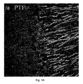


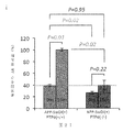


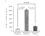


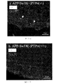


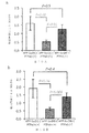


Classifications
-
- A—HUMAN NECESSITIES
- A61—MEDICAL OR VETERINARY SCIENCE; HYGIENE
- A61K—PREPARATIONS FOR MEDICAL, DENTAL OR TOILETRY PURPOSES
- A61K38/00—Medicinal preparations containing peptides
- A61K38/16—Peptides having more than 20 amino acids; Gastrins; Somatostatins; Melanotropins; Derivatives thereof
- A61K38/17—Peptides having more than 20 amino acids; Gastrins; Somatostatins; Melanotropins; Derivatives thereof from animals; from humans
- A61K38/1703—Peptides having more than 20 amino acids; Gastrins; Somatostatins; Melanotropins; Derivatives thereof from animals; from humans from vertebrates
- A61K38/1709—Peptides having more than 20 amino acids; Gastrins; Somatostatins; Melanotropins; Derivatives thereof from animals; from humans from vertebrates from mammals
- A61K38/1716—Amyloid plaque core protein
-
- A—HUMAN NECESSITIES
- A61—MEDICAL OR VETERINARY SCIENCE; HYGIENE
- A61K—PREPARATIONS FOR MEDICAL, DENTAL OR TOILETRY PURPOSES
- A61K38/00—Medicinal preparations containing peptides
- A61K38/04—Peptides having up to 20 amino acids in a fully defined sequence; Derivatives thereof
- A61K38/08—Peptides having 5 to 11 amino acids
-
- A—HUMAN NECESSITIES
- A61—MEDICAL OR VETERINARY SCIENCE; HYGIENE
- A61K—PREPARATIONS FOR MEDICAL, DENTAL OR TOILETRY PURPOSES
- A61K38/00—Medicinal preparations containing peptides
- A61K38/04—Peptides having up to 20 amino acids in a fully defined sequence; Derivatives thereof
- A61K38/10—Peptides having 12 to 20 amino acids
-
- A—HUMAN NECESSITIES
- A61—MEDICAL OR VETERINARY SCIENCE; HYGIENE
- A61K—PREPARATIONS FOR MEDICAL, DENTAL OR TOILETRY PURPOSES
- A61K38/00—Medicinal preparations containing peptides
- A61K38/16—Peptides having more than 20 amino acids; Gastrins; Somatostatins; Melanotropins; Derivatives thereof
-
- A—HUMAN NECESSITIES
- A61—MEDICAL OR VETERINARY SCIENCE; HYGIENE
- A61K—PREPARATIONS FOR MEDICAL, DENTAL OR TOILETRY PURPOSES
- A61K38/00—Medicinal preparations containing peptides
- A61K38/16—Peptides having more than 20 amino acids; Gastrins; Somatostatins; Melanotropins; Derivatives thereof
- A61K38/162—Peptides having more than 20 amino acids; Gastrins; Somatostatins; Melanotropins; Derivatives thereof from virus
-
- A—HUMAN NECESSITIES
- A61—MEDICAL OR VETERINARY SCIENCE; HYGIENE
- A61K—PREPARATIONS FOR MEDICAL, DENTAL OR TOILETRY PURPOSES
- A61K45/00—Medicinal preparations containing active ingredients not provided for in groups A61K31/00 - A61K41/00
- A61K45/06—Mixtures of active ingredients without chemical characterisation, e.g. antiphlogistics and cardiaca
-
- A—HUMAN NECESSITIES
- A61—MEDICAL OR VETERINARY SCIENCE; HYGIENE
- A61K—PREPARATIONS FOR MEDICAL, DENTAL OR TOILETRY PURPOSES
- A61K47/00—Medicinal preparations characterised by the non-active ingredients used, e.g. carriers or inert additives; Targeting or modifying agents chemically bound to the active ingredient
- A61K47/50—Medicinal preparations characterised by the non-active ingredients used, e.g. carriers or inert additives; Targeting or modifying agents chemically bound to the active ingredient the non-active ingredient being chemically bound to the active ingredient, e.g. polymer-drug conjugates
- A61K47/51—Medicinal preparations characterised by the non-active ingredients used, e.g. carriers or inert additives; Targeting or modifying agents chemically bound to the active ingredient the non-active ingredient being chemically bound to the active ingredient, e.g. polymer-drug conjugates the non-active ingredient being a modifying agent
- A61K47/62—Medicinal preparations characterised by the non-active ingredients used, e.g. carriers or inert additives; Targeting or modifying agents chemically bound to the active ingredient the non-active ingredient being chemically bound to the active ingredient, e.g. polymer-drug conjugates the non-active ingredient being a modifying agent the modifying agent being a protein, peptide or polyamino acid
- A61K47/64—Drug-peptide, drug-protein or drug-polyamino acid conjugates, i.e. the modifying agent being a peptide, protein or polyamino acid which is covalently bonded or complexed to a therapeutically active agent
- A61K47/645—Polycationic or polyanionic oligopeptides, polypeptides or polyamino acids, e.g. polylysine, polyarginine, polyglutamic acid or peptide TAT
-
- A—HUMAN NECESSITIES
- A61—MEDICAL OR VETERINARY SCIENCE; HYGIENE
- A61P—SPECIFIC THERAPEUTIC ACTIVITY OF CHEMICAL COMPOUNDS OR MEDICINAL PREPARATIONS
- A61P25/00—Drugs for disorders of the nervous system
- A61P25/28—Drugs for disorders of the nervous system for treating neurodegenerative disorders of the central nervous system, e.g. nootropic agents, cognition enhancers, drugs for treating Alzheimer's disease or other forms of dementia
-
- C—CHEMISTRY; METALLURGY
- C07—ORGANIC CHEMISTRY
- C07K—PEPTIDES
- C07K14/00—Peptides having more than 20 amino acids; Gastrins; Somatostatins; Melanotropins; Derivatives thereof
- C07K14/435—Peptides having more than 20 amino acids; Gastrins; Somatostatins; Melanotropins; Derivatives thereof from animals; from humans
- C07K14/46—Peptides having more than 20 amino acids; Gastrins; Somatostatins; Melanotropins; Derivatives thereof from animals; from humans from vertebrates
- C07K14/47—Peptides having more than 20 amino acids; Gastrins; Somatostatins; Melanotropins; Derivatives thereof from animals; from humans from vertebrates from mammals
- C07K14/4701—Peptides having more than 20 amino acids; Gastrins; Somatostatins; Melanotropins; Derivatives thereof from animals; from humans from vertebrates from mammals not used
- C07K14/4711—Alzheimer's disease; Amyloid plaque core protein
-
- C—CHEMISTRY; METALLURGY
- C07—ORGANIC CHEMISTRY
- C07K—PEPTIDES
- C07K7/00—Peptides having 5 to 20 amino acids in a fully defined sequence; Derivatives thereof
- C07K7/04—Linear peptides containing only normal peptide links
- C07K7/06—Linear peptides containing only normal peptide links having 5 to 11 amino acids
-
- A—HUMAN NECESSITIES
- A61—MEDICAL OR VETERINARY SCIENCE; HYGIENE
- A61K—PREPARATIONS FOR MEDICAL, DENTAL OR TOILETRY PURPOSES
- A61K38/00—Medicinal preparations containing peptides
-
- A—HUMAN NECESSITIES
- A61—MEDICAL OR VETERINARY SCIENCE; HYGIENE
- A61K—PREPARATIONS FOR MEDICAL, DENTAL OR TOILETRY PURPOSES
- A61K38/00—Medicinal preparations containing peptides
- A61K38/16—Peptides having more than 20 amino acids; Gastrins; Somatostatins; Melanotropins; Derivatives thereof
- A61K38/43—Enzymes; Proenzymes; Derivatives thereof
- A61K38/46—Hydrolases (3)
Abstract
βアミロイド蓄積および/またはタウ凝集の阻害および/または低減を必要とする対象に対し、それを実施する方法であって、LARファミリーのホスファターゼの触媒活性、シグナル伝達、および機能の1つまたは複数を阻害する治療薬を対象に投与することを含む、方法。【選択図】図1GA method of doing so in a subject in need of inhibition and/or reduction of β-amyloid accumulation and/or tau aggregation, comprising the step of: A method comprising administering to a subject a therapeutic agent that inhibits. [Selection diagram] Figure 1G
Description
関連出願
本出願は、2017年6月5日に出願された米国仮特許出願第62/515,272号からの優先権を主張する。この仮出願の主題は、全体として参照により本明細書に組み込まれる。
Related Applications This application claims priority from US Provisional Patent Application No. 62/515,272, filed June 5, 2017. The subject matter of this provisional application is incorporated herein by reference in its entirety.
発明の分野
本出願は、白血球共通抗原関連(LAR)ファミリーのホスファターゼの活性、シグナル伝達および/または機能を阻害または低減する組成物および方法、ならびにβアミロイドーシスを阻害し、アルツハイマー病を治療するための方法および組成物に関する。
FIELD OF THE INVENTION This application relates to compositions and methods for inhibiting or reducing the activity, signaling and/or function of leukocyte common antigen associated (LAR) family phosphatases, and for inhibiting β-amyloidosis and treating Alzheimer's disease. Methods and compositions.
アルツハイマー病(AD)の確定的病理学的特徴は、βアミロイドーシスとしても知られる過程である、脳内でのβアミロイド(Aβ)ペプチドの徐々に進行する凝集であり、多くの場合、神経炎症および微小管結合タンパク質のタウを含有した神経原線維濃縮体の形成が付随して起こる。 A definitive pathological hallmark of Alzheimer's disease (AD) is the progressive aggregation of β-amyloid (Aβ) peptides in the brain, a process also known as β-amyloidosis, often involving neuroinflammation and Concomitant formation of neurofibrillary tangles containing the microtubule-binding protein tau.
ADの病因学的機序については、議論が続けられているところであるが、ヒトの遺伝的調査からの具体的証拠は、遺伝子変異によるAβの過剰産生が細胞傷害性イベントのカスケードを不可避的にもたらし、最終的に神経変性および脳機能の崩壊に繋がることを示した。従って、Aβペプチドの、特にそれらの可溶型での蓄積は、AD発症の重要な犯人として認識されている。脳では、Aβペプチドは主に、神経アミロイド前駆体タンパク質(APP)のβ−およびγ−分泌酵素による逐次切断から生ずる。しかし、数十年に及ぶ研究にもかかわらず、アミロイド形成性分泌酵素活性の分子調節は、理解が進んでおらず、APPアミロイド形成経路を特異的に標的とする治療薬の設計を妨げている。 Although the etiological mechanism of AD is under debate, concrete evidence from human genetic studies suggests that overproduction of Aβ by genetic mutations inevitably leads to a cascade of cytotoxic events. And eventually lead to neurodegeneration and disruption of brain function. Accumulation of Aβ peptides, especially in their soluble form, is therefore recognized as an important criminal of AD development. In the brain, Aβ peptides mainly arise from the sequential cleavage of neuroamyloid precursor protein (APP) by β- and γ-secretases. However, despite decades of research, molecular regulation of amyloidogenic secretory enzyme activity is poorly understood, hindering the design of therapeutic agents that specifically target the APP amyloidogenic pathway. ..
β−およびγ−分泌酵素活性の薬理学的阻害は、Aβ産生の抑制では効果的であるが、分泌酵素の他の基質に対する生理学的機能を妨害する。従って、このような介入戦略は、不都合な副作用に本質的に関連する場合が多く、このことが、過去における臨床試験のいくつかの失敗に繋がった。現在まで、ADの発症を防ぐ、または進行を抑制するために利用可能な治療法は存在しない。 Pharmacological inhibition of β- and γ-secretase activity, while effective in suppressing Aβ production, interferes with the physiological function of secreted enzymes on other substrates. Therefore, such intervention strategies are often inherently associated with adverse side effects, which has led to some failures of clinical trials in the past. To date, there are no treatments available to prevent the onset of AD or to control the progression.
本明細書で記載の実施形態は、βアミロイド蓄積および/またはタウ凝集の阻害および/または低減を必要としている対象に対しそれを実施する方法に関する。方法は、LARファミリーのホスファターゼ触媒活性、シグナル伝達および機能の1つまたは複数を阻害する治療薬を対象に投与することを含む。 Embodiments described herein relate to methods of doing so in a subject in need of inhibition and/or reduction of β-amyloid accumulation and/or tau aggregation. The method comprises administering to the subject a therapeutic agent that inhibits one or more of the LAR family phosphatase catalytic activity, signal transduction and function.
いくつかの実施形態では、LARファミリーのホスファターゼは、受容体タンパク質チロシンホスファターゼシグマ(PTPσ)であり、治療薬には、PTPσのくさび形ドメインの約10〜約20個の連続するアミノ酸に対して少なくとも約65%、少なくとも約70%、少なくとも約75%、少なくとも約80%、少なくとも約85%、少なくとも約90%、または少なくとも約95%同一のアミノ酸配列を有する治療用ペプチドが含まれる。例えば、治療薬は配列番号9〜13および16からなる群より選択される治療用ペプチドを含むことができる。 In some embodiments, the LAR family phosphatase is the receptor protein tyrosine phosphatase sigma (PTPσ) and the therapeutic agent comprises at least about 10 to about 20 contiguous amino acids of the wedge domain of PTPσ. Included are therapeutic peptides having amino acid sequences that are about 65%, at least about 70%, at least about 75%, at least about 80%, at least about 85%, at least about 90%, or at least about 95% identical. For example, the therapeutic agent can include a therapeutic peptide selected from the group consisting of SEQ ID NOs: 9-13 and 16.
他の実施形態では、LARファミリーのホスファターゼは、受容体タンパク質チロシンホスファターゼシグマ(PTPσ)であり、治療薬は、配列番号16のアミノ酸配列に対して少なくとも約65%、少なくとも約70%、少なくとも約75%、少なくとも約80%、少なくとも約85%、少なくとも約90%、または少なくとも約95%同一の治療用ペプチドを含むことができる。治療用ペプチドは、例えば、配列番号16の残基4、5、6、7、9、10、12または13個の内の少なくとも1、2、3または4個のアミノ酸の保存的置換を含むことができる。
In another embodiment, the LAR family phosphatase is the receptor protein tyrosine phosphatase sigma (PTPσ) and the therapeutic agent is at least about 65%, at least about 70%, at least about 75% relative to the amino acid sequence of SEQ ID NO:16. %, at least about 80%, at least about 85%, at least about 90%, or at least about 95% identical therapeutic peptides. The therapeutic peptide comprises, for example, a conservative substitution of at least 1, 2, 3 or 4 amino acids of
いくつかの実施形態では、治療薬は、対象に、または神経細胞、グリア細胞、グリア前駆細胞または神経前駆細胞に全身投与または局所投与される。 In some embodiments, therapeutic agents are administered systemically or locally to a subject or to neural cells, glial cells, glial progenitor cells or neural progenitor cells.
他の実施形態では、治療薬は、治療用ペプチドと連結され、細胞による治療用ペプチドの取り込みを容易にする輸送部分を含む。例えば、輸送部分はHIVのTat輸送部分であり得る。 In other embodiments, the Therapeutic Agent comprises a transport moiety that is linked to the Therapeutic Peptide and facilitates uptake of the Therapeutic Peptide by the cell. For example, the transport moiety can be the Tat transport moiety of HIV.
さらに他の実施形態では、細胞は治療される対象中にあり、治療薬は治療される対象に局所投与または全身投与される。 In still other embodiments, the cells are in the subject to be treated and the therapeutic agent is administered locally or systemically to the subject to be treated.
さらに他の実施形態では、治療用ペプチドは対象の細胞により発現される。 In yet other embodiments, the therapeutic peptide is expressed by cells of the subject.
本明細書で記載の実施形態は、治療を必要としている対象のβアミロイド蓄積および/またはタウ凝集に関連する疾患、障害、および/または状態を治療する方法にも関する。方法は、LARファミリーのホスファターゼの触媒活性、シグナル伝達および機能の1つまたは複数を阻害する治療薬を対象に投与することを含む。 Embodiments described herein also relate to methods of treating diseases, disorders, and/or conditions associated with β-amyloid accumulation and/or tau aggregation in a subject in need thereof. The method comprises administering to the subject a therapeutic agent that inhibits one or more of the catalytic activity, signaling and function of the LAR family of phosphatases.
いくつかの実施形態では、疾患、障害および/または状態には、神経系の疾患、障害および/または状態の少なくとも1つが含まれる。 In some embodiments, the disease, disorder and/or condition comprises at least one of a nervous system disease, disorder and/or condition.
他の実施形態では、神経系の疾患、障害および/または状態には、神経障害、神経精神障害、神経損傷、神経毒性障害、神経障害性疼痛、および神経変性障害の少なくとも1つが含まれる。 In other embodiments, diseases, disorders and/or conditions of the nervous system include at least one of neuropathy, neuropsychiatric disorders, nerve injury, neurotoxic disorders, neuropathic pain, and neurodegenerative disorders.
例えば、神経障害には、アルツハイマー病またはアルツハイマー病関連認知症の内の少なくとも1つを含み得る。 For example, the neuropathy can include at least one of Alzheimer's disease or Alzheimer's disease-related dementia.
本明細書に記載される実施形態は、特定の方法論、プロトコールおよび試薬等に限定されず、従って、変わり得る。本明細書で用いられる用語は特定の実施形態を記載するためのみのものであり、本発明の範囲を限定することを意図するものではなく、本発明の範囲は、特許請求の範囲によってのみ定義される。操作例における場合以外または別義が指示される場合以外、本明細書で使用される成分または反応条件の量を表す全ての数は、全ての場合において、用語の「約」によって修飾されているものとして理解されるべきである。 The embodiments described herein are not limited to particular methodologies, protocols and reagents, etc., and as such may vary. The terminology used herein is for the purpose of describing particular embodiments only and is not intended to limit the scope of the invention, which is defined solely by the claims that follow. To be done. Unless otherwise indicated in the examples of operation or unless otherwise indicated, all numbers used herein to represent amounts of components or reaction conditions are modified in all cases by the term "about". Should be understood as a thing.
特定される特許および他の出版物はすべて、例えば、本発明との関連で使用され得るこのような出版物で記載された方法論を記載し、開示する目的で、参照により明示的に本明細書に組み込まれる。これらの出版物は本出願の出願日に先立つそれらの開示のためにのみ提供される。これに関しては、先行発明を理由にしてまたは何らかの他の理由で本発明者らがそのような開示に先行する権利を与えられないことを承認するものと解釈されるべきではない。これらの文書の内容に関する日付または表現に関する記載はすべて出願者に利用可能な情報に基づくものであり、これらの文書の日付または内容の正確さに関する承認を構成するものではない。 All patents and other publications identified herein are expressly incorporated herein by reference for the purpose of describing and disclosing methodologies described in such publications, which may be used in connection with the present invention, for example. Be incorporated into. These publications are provided solely for their disclosure prior to the filing date of the present application. In this regard, it should not be construed as an admission that the inventors are not entitled to antedate such disclosure by virtue of prior invention or for any other reason. All statements regarding dates or expressions regarding the contents of these documents are based on the information available to the applicant and do not constitute an admission as to the accuracy of the dates or contents of these documents.
別に定めのない限り、本明細書で使用される科学技術用語は、当業者によって一般に理解される意味を有するものとする。さらに、文脈により別義が要求されない限り、単数用語は複数を含むものとし、複数用語は単数を含むものとする。一般に、本明細書で記載される細胞および組織の培養、分子生物学、およびタンパク質およびオリゴヌクレオチドまたはポリヌクレオチドの化学およびハイブリッド形成と関連して利用される命名法、ならびにそれらの技法は、当該技術で周知であり、一般に使用されるものである。 Unless defined otherwise, all technical and scientific terms used herein have the meaning commonly understood by one of ordinary skill in the art. Further, unless otherwise required by context, singular terms shall include pluralities and plural terms shall include the singular. In general, the nomenclature utilized in connection with cell and tissue culture, molecular biology, and protein and oligonucleotide or polynucleotide chemistry and hybridization described herein, as well as those techniques, are well known in the art. Are well known and commonly used.
本明細書で使用される場合、「a、bおよびcの1つまたは複数」はa、b、c、ab、ac、bcまたはabcを意味する。本明細書における「または」の使用は、包含的なまたはである。 As used herein, "one or more of a, b and c" means a, b, c, ab, ac, bc or abc. The use of "or" herein is inclusive or.
本明細書で使用される場合、患者に「投与する」という用語には、脳脊髄液へのまたは脳血管関門を横切る投与、非経口または経口の経路による送達、筋肉内注射、皮下または皮内注射、静脈内注射、頬内投与、経皮送達、および直腸、結腸、膣、鼻腔内のまたは気道経路による投与を含む、対象の所望の位置に活性化合物を送達するために(例えば、それにより、所望のニューロンなどの所望の細胞に接触させるために)好適な経路により、対象への医薬製剤中の活性化合物の分配、送達または適用が含まれる。薬剤は、例えば、静脈内注射を介して昏睡状態、麻酔下のもしくは麻痺した対象に投与され得、または妊娠した対象の静脈内に投与して胎児の軸索成長を刺激し得る。投与の特定の経路には、局所投与(例えば、まぶたの下に投入される点眼薬、クリームまたは浸食性の製剤)、水性体液または硝子体液への眼内注射、結膜下注射またはテノン嚢下注射を介するなどの眼の外層への注射、非経口投与または経口経路を介した投与が挙げられ得る。 As used herein, the term "administering" to a patient includes administration to the cerebrospinal fluid or across the cerebrovascular barrier, delivery by parenteral or oral routes, intramuscular injection, subcutaneous or intradermal. To deliver an active compound to a desired location in a subject, including injection, intravenous injection, buccal administration, transdermal delivery, and administration by rectal, colonic, vaginal, intranasal or respiratory route (eg, thereby By delivery, application or application of the active compound in a pharmaceutical formulation to the subject by any suitable route (to contact the desired cells, such as the desired neurons). The agent may be administered to a coma, anesthetized or paralyzed subject, for example, via intravenous injection, or may be administered intravenously to a pregnant subject to stimulate fetal axon growth. Specific routes of administration include topical administration (eg eye drops, creams or erodible formulations placed under the eyelids), intraocular injection into aqueous or vitreous humor, subconjunctival injection or sub-Tenon injection. Injections into the outer layer of the eye, such as via, parenteral administration or administration via the oral route may be mentioned.
本明細書で使用される場合、用語「抗体」には、ヒトおよび動物のmAb、およびポリクローナル抗体、組換え抗体(抗血清)、ヒト化抗体、抗イディオタイプ抗体およびその誘導体などのキメラ抗体を含む合成抗体の調製が挙げられる。抗体の一部またはフラグメントは、特定のエピトープに結合する能力(結合の特異性および親和性)の少なくとも一部を保持する抗体の領域を意味する。用語「エピトープ」または「抗原決定基」は抗体のパラトープが結合する抗原上の部位を意味する。連続アミノ酸により形成されるエピトープは通常、変性溶媒への暴露の際、保持されるが、三次折り畳みにより形成されるエピトープは通常、変性溶媒による処理の際に失われる。エピトープは通常、特有の空間配置中に少なくとも3個、少なくとも5個または8〜10個、または約13〜15個のアミノ酸を含む。エピトープの空間配置を決定する方法には、例えば、X線結晶学および二次元核磁気共鳴が挙げられる。例えば、66 EPITOPE MAPPING PROTOCOLS IN METS.IN MOLECULAR BIO.(Morris,ed.,1996);Burke et al.,170 J.Inf.Dis.1110−19(1994);Tigges et al.,156 J.Immunol.3901−10を参照されたい。 As used herein, the term "antibody" includes human and animal mAbs and chimeric antibodies such as polyclonal antibodies, recombinant antibodies (antisera), humanized antibodies, anti-idiotype antibodies and derivatives thereof. Preparation of synthetic antibodies including. By part or fragment of an antibody is meant a region of the antibody that retains at least part of its ability to bind to a particular epitope (specificity of binding and affinity). The term “epitope” or “antigenic determinant” means the site on an antigen to which the paratope of an antibody binds. Epitopes formed by contiguous amino acids are usually retained upon exposure to denaturing solvents, while epitopes formed by tertiary folding are usually lost upon treatment with denaturing solvents. Epitopes usually include at least 3, at least 5, or 8-10, or about 13-15 amino acids in a unique spatial arrangement. Methods of determining the spatial arrangement of epitopes include, for example, X-ray crystallography and two-dimensional nuclear magnetic resonance. For example, 66 EPITOPE MAPPING PROTOCOLS IN METS. IN MOLECULAR BIO. (Morris, ed., 1996); Burke et al. 170 J. Inf. Dis. 1110-19 (1994); Tiges et al. , 156 J. Immunol. See 3901-10.
本明細書で使用される場合、「中枢神経系(CNS)のニューロン」には脳、脳神経および脊髄のニューロンが含まれる。 As used herein, "central nervous system (CNS) neurons" include brain, cranial nerves and spinal cord neurons.
本明細書で使用される場合、「キメラタンパク質」または「融合タンパク質」は、ポリペプチドをコードする第1のアミノ酸配列と、第1のポリペプチドのドメインに対して外来性であり、実質的に相同ではないドメイン(例えば、ポリペプチドの一部)を規定する第2のアミノ酸配列との融合体である。キメラタンパク質は、第1のタンパク質も発現する生物で見出される外来性のドメイン(異なるタンパク質中ではあるが)であってもよく、またはキメラタンパク質は、異なる種類の生物により発現されるタンパク質構造体の「種間」、「遺伝子間」等の融合体であってもよい。 As used herein, a "chimeric protein" or "fusion protein" is a foreign amino acid that is foreign to a first amino acid sequence encoding a polypeptide and a domain of the first polypeptide. It is a fusion with a second amino acid sequence defining a heterologous domain (eg, a portion of a polypeptide). The chimeric protein may be an exogenous domain (although in a different protein) found in an organism that also expresses the first protein, or the chimeric protein may be of a protein structure expressed by a different type of organism. It may be a fusion of "interspecies", "intergenic" and the like.
本明細書で使用される場合、用語「ニューロンに接触すること」または「ニューロンを治療すること」は、薬剤がニューロン中でその医薬効果を示すことが可能である細胞または全生物への薬剤の送達または「投与」の任意の様式を意味する。「ニューロンに接触すること」には、本発明の薬剤をインビボおよびインビトロの両方でニューロンの近傍に運ぶ方法が含まれる。好適な投与方法は、当業者により決定することができ、このような投与方法は、薬剤の間で変化し得る。 As used herein, the term "contacting a neuron" or "treating a neuron" refers to the agent to a cell or whole organism where the agent is capable of exhibiting its medicinal effect in the neuron. It means any mode of delivery or “administration”. "Contacting a neuron" includes a method of delivering an agent of the invention to the vicinity of a neuron both in vivo and in vitro. Suitable administration methods can be determined by those skilled in the art, and such administration methods can vary between drugs.
本明細書で使用される場合、薬剤または治療用ペプチドの「有効量」は、所望の治療効果または医薬効果を達成するのに十分な量、例えば、βアミロイド蓄積またはタウ凝集を阻害できる量である。本明細書で定義される薬剤の有効量は、例えば、対象の疾患の状態、年齢および体重、ならびに対象において、所望の応答を誘発する薬剤の能力などの因子により変化し得る。投与計画を調節して最適な治療応答をもたらし得る。有効量はまた、治療上の有益な効果が活性化合物の毒性効果または有害効果に勝るものでもある。 As used herein, an “effective amount” of a drug or therapeutic peptide is an amount sufficient to achieve the desired therapeutic or pharmaceutical effect, for example, an amount capable of inhibiting β-amyloid accumulation or tau aggregation. is there. An effective amount of a drug as defined herein may vary depending on factors such as, for example, the disease state of the subject, age and weight, and the ability of the drug to elicit the desired response in the subject. Dosage regimens may be adjusted to provide the optimum therapeutic response. An effective amount is also one in which any toxic or detrimental effects of the active compound are outweighed by the therapeutically beneficial effects.
本明細書で使用される場合、用語の「治療有効量」は、必要な用量と期間で、所望の治療結果を達成するために有効な量を意味する。治療結果は、例えば、症状の減少、生存の延長、運動の改善などであり得る。治療結果は「治癒」である必要はない。 As used herein, the term "therapeutically effective amount" means an amount effective at achieving the desired therapeutic result at the required dose and duration. The outcome of treatment may be, for example, reduced symptoms, prolonged survival, improved exercise, and the like. The outcome of the treatment need not be "curative".
本明細書で使用される場合、用語「発現」は、核酸がペプチドに翻訳されるまたは、例えば、ペプチド、ポリペプチドもしくはタンパク質に翻訳できるRNAに転写される過程を意味する。核酸がゲノムDNAに由来するのであれば、発現は、適当な真核宿主細胞または生物が選択される場合、mRNAのスプライシングを含む。宿主細胞中で発現される異種核酸については、それは先ず細胞に送達され、その後、いったん細胞に入って、最終的に核に存在しなければならない。 As used herein, the term "expression" means the process by which a nucleic acid is translated into a peptide or transcribed into RNA that can be translated into, for example, a peptide, polypeptide or protein. If the nucleic acid is derived from genomic DNA, expression will include splicing of the mRNA when an appropriate eukaryotic host cell or organism is chosen. For a heterologous nucleic acid that is expressed in a host cell, it must first be delivered to the cell, then once it has entered the cell and ultimately in the nucleus.
本明細書で使用される場合、用語の「遺伝子治療」には、治療または診断が求められる障害または状態にある哺乳動物、特にヒトの細胞への異種のDNAの移入が含まれる。選択された標的細胞にDNAが導入され、それにより、異種のDNAが発現され、それにより、コードされた治療用産物が産生される。あるいは、異種のDNAは、治療用産物をコードするDNAの発現を何らかの形で媒介し、治療用産物の発現を何らかの形で直接的にまたは間接的に媒介するペプチドまたはRNAなどの産物をコードし得る。遺伝子治療を用いて遺伝子産物をコードする核酸を送達して欠陥のある遺伝子を置き換えてもよく、またはそれが導入された哺乳動物または細胞により産生される遺伝子産物を補完してもよい。導入された核酸は、通常は哺乳動物宿主では産生されないまたは治療上有効な量でもしくは治療上有用な時間で産生されない、治療用化合物をコードし得る。治療用産物をコードする異種のDNAは、その産物または発現を増大するかまたは別の方法で変えるために、罹患している宿主の細胞に導入する前に修飾され得る。 As used herein, the term "gene therapy" includes the transfer of heterologous DNA into cells of mammals, especially humans, in the disorder or condition for which treatment or diagnosis is sought. The DNA is introduced into the selected target cells, whereby the heterologous DNA is expressed, thereby producing the encoded therapeutic product. Alternatively, the heterologous DNA encodes a product such as a peptide or RNA that somehow mediates the expression of the DNA encoding the therapeutic product and somehow directly or indirectly mediates the expression of the therapeutic product. obtain. Gene therapy may be used to deliver the nucleic acid encoding the gene product to replace the defective gene, or to complement the gene product produced by the mammal or cell into which it was introduced. The introduced nucleic acid may encode a therapeutic compound that is not normally produced in a mammalian host or produced in therapeutically effective amounts or at therapeutically useful times. Heterologous DNA encoding a therapeutic product may be modified prior to introduction into cells of the affected host in order to increase or otherwise alter its product or expression.
本明細書で使用される場合、用語の「遺伝子」または「組換え遺伝子」はエクソンおよび(任意に)イントロンの配列の両方を含むポリペプチドをコードするオープンリーディングフレームを含む核酸を意味する。 As used herein, the term "gene" or "recombinant gene" means a nucleic acid containing an open reading frame that encodes a polypeptide that includes both exon and (optionally) intron sequences.
本明細書で使用される場合、用語の「異種核酸配列」は通常、それが発現される細胞によって、インビボでは通常産生されないRNAおよびタンパク質をコードするDNA、または転写、翻訳若しくは他の調節性の生物過程に影響を及ぼすことにより、内在性のDNAの発現を変更するメディエーターを媒介するもしくはそれをコードするDNAである。異種核酸配列はまた外来性のDNAとも呼ばれる。それが発現される細胞に対して異種または外来性であると当業者が認識するまたは見なすDNAは本明細書では異種DNAにより包含される。異種DNAの例には、限定されないが、薬剤耐性を付与するタンパク質などの追跡可能なマーカータンパク質をコードするDNA、治療上有効な物質をコードするDNA、ならびに抗体などの他の種類のタンパク質をコードするDNAが挙げられる。異種のDNAによって、コードされる抗体は、異種のDNAが導入されている細胞の表面で分泌され得るかまたは発現され得る。 As used herein, the term "heterologous nucleic acid sequence" generally refers to DNA that encodes RNA and proteins that are not normally produced in vivo by the cell in which it is expressed, or transcription, translation or other regulatory elements. A DNA that mediates or encodes a mediator that alters the expression of endogenous DNA by affecting biological processes. Heterologous nucleic acid sequences are also referred to as foreign DNA. DNA recognized or considered by those of skill in the art to be heterologous or foreign to the cell in which it is expressed is encompassed herein by heterologous DNA. Examples of heterologous DNA include, but are not limited to, DNA that encodes a traceable marker protein, such as a protein that confers drug resistance, DNA that encodes a therapeutically effective substance, and other types of proteins such as antibodies. DNA to be used. The antibody encoded by the heterologous DNA can be secreted or expressed on the surface of the cell into which the heterologous DNA has been introduced.
本明細書で使用される場合、用語の「相同性」および「同一性」は、全体を通して同義に使用され、2つのペプチド間のまたは2つの核酸分子間の配列類似性を意味する。相同性は、比較の目的で並べられ得る各配列における位置を比較することによって、決定できる。比較した配列における位置が同一の塩基またはアミノ酸で占められれば、その位置で分子は相同または同一である。配列間の相同性または同一性の程度は配列により共有される一致するまたは相同性の位置の数の関数である。 As used herein, the terms "homology" and "identity" are used interchangeably throughout and refer to sequence similarity between two peptides or between two nucleic acid molecules. Homology can be determined by comparing the position in each sequence which may be aligned for purposes of comparison. If a position in the compared sequences is occupied by the same base or amino acid, then the molecules are homologous or identical at that position. The degree of homology or identity between sequences is a function of the number of matching or homologous positions shared by the sequences.
本明細書で使用される場合、用語の「神経障害」には、対象の神経系の正常な機能または解剖学的構造に直接または間接的に影響する疾患、障害または状態が含まれる。 As used herein, the term "neuropathy" includes diseases, disorders or conditions that directly or indirectly affect the normal functioning or anatomy of the subject's nervous system.
本明細書で使用される場合、語句の「非経口投与」および「非経口で投与される」は本明細書で使用される場合、腸内投与および局所投与以外の、通常は注射による投与方法を意味し、これには、限定されないが、静脈内、筋肉内、動脈内、くも膜下腔内、脳室内、関節内、眼窩内、心臓内、皮内、腹腔内、経気管、皮下、表皮下、関節腔内、嚢下、クモ膜下、脊髄内および胸骨内の注射および注入が挙げられる。 As used herein, the phrases "parenteral administration" and "parenterally administered" as used herein, are methods other than enteral and topical administration, usually by injection. Means, without limitation, intravenous, intramuscular, intraarterial, intrathecal, intraventricular, intraarticular, intraorbital, intracardiac, intradermal, intraperitoneal, transtracheal, subcutaneous, epidermal Sub-articular, intra-articular, sub-capsular, sub-arachnoid, intra-spinal and intra-sternal injection and infusion are included.
本明細書で使用される場合、語句の「全身投与」、「全身に投与される」、「末梢投与」および「末梢に投与される」は、本明細書で使用される場合、標的組織(例えば、中枢神経系)への直接投与以外の化合物、薬物またはその他の物質の投与を意味し、それにより、動物の体系に入り、従って、代謝および他の類似の過程、例えば、皮下投与を受ける。 As used herein, the phrases "systemic administration", "systemically administered", "peripheral administration" and "peripherally administered" as used herein refer to target tissue ( For example, administration of a compound, drug or other substance other than direct administration to the central nervous system, thereby entering the animal's system and thus undergoing metabolism and other similar processes, eg subcutaneous administration. ..
本明細書で使用される場合、用語の「患者」または「対象」または「動物」または「宿主」は任意の哺乳動物を意味する。対象はヒトであり得るが、獣医治療を必要とする哺乳動物、例えば、愛玩動物(例えば、イヌ、ネコ等)、家畜(例えば、ウシ、ヒツジ、ニワトリ、ブタ、ウマ等)および実験動物(例えば、ラット、マウス、モルモット等)でもあり得る。 As used herein, the term "patient" or "subject" or "animal" or "host" means any mammal. The subject may be a human, but a mammal in need of veterinary treatment, such as companion animals (eg, dogs, cats, etc.), domestic animals (eg, cows, sheep, chickens, pigs, horses, etc.) and laboratory animals (eg, animals) , Rat, mouse, guinea pig, etc.).
本明細書で使用される場合、用語の「末梢神経系(PNS)のニューロン」には、CNSの外側に存在するまたは伸びるニューロンが含まれる。PNSは、感覚ニューロンおよび運動ニューロンを含む末梢神経系に分類されるとして一般的に理解されるニューロンを含むことが意図されている。 As used herein, the term "peripheral nervous system (PNS) neurons" includes neurons that reside or extend outside the CNS. PNS is intended to include neurons that are commonly understood as classified into the peripheral nervous system, including sensory and motor neurons.
本明細書で使用される場合、用語の「ポリヌクレオチド配列」および「ヌクレオチド配列」は本明細書では同じ意味で用いられる。 As used herein, the terms "polynucleotide sequence" and "nucleotide sequence" are used interchangeably herein.
本明細書で使用される場合、用語の「ペプチド」および「ポリペプチド」は、本明細書では同じ意味に用いられ、約2〜約90個(包括的)のアミノ酸残基からなる化合物を意味し、1つのアミノ酸のアミノ基がペプチド結合により、別のアミノ酸のカルボキシル基に連結される。ペプチドは例えば、酵素切断または化学切断によって、ネイティブのタンパク質から導き出すまたは取り出すことができ、または従来のペプチド合成法(例えば、固相合成)または分子生物学的な技法により、調製できる(Sambrook et al.,MOLECULAR CLONING:LAB.MANUAL(Cold Spring Harbor Press,Cold Spring Harbor,NY,1989)を参照)。「ペプチド」は、好適なL−および/またはD−アミノ酸、例えば、一般的なα−アミノ酸(例えば、アラニン、グリシン、バリン)、非α−アミノ酸(例えば、P−アラニン、4−アミノ酪酸、6−アミノカプロン酸、サルコシン、スタチン)および異常アミノ酸(例えば、シトルリン、ホモシトルリン、ホモセリン、ノルロイシン、ノルバリン、オルニチン)を含むことができる。ペプチド上のアミノ基、カルボキシル基および/または他の官能基は遊離状態(例えば、非修飾)、または好適な保護基で保護され得る。アミノ基およびカルボキシル基の好適な保護基、および保護基を付加するまたは取り外す手段は当該技術で既知である。例えば、Green &amp;Wuts,PROTECTING GROUPS IN ORGANIC SYNTHESIS(John Wiley &amp;Sons,1991)を参照されたい。ペプチドの官能基も既知の方法を用いて誘導体化(例えば、アルキル化)することができる。 As used herein, the terms "peptide" and "polypeptide" are used interchangeably herein and refer to a compound of about 2 to about 90 (inclusive) amino acid residues. Then, the amino group of one amino acid is linked to the carboxyl group of another amino acid by a peptide bond. Peptides can be derived or derived from native proteins, eg, by enzymatic or chemical cleavage, or prepared by conventional peptide synthesis methods (eg, solid phase synthesis) or molecular biology techniques (Sambrook et al. ., MOLECULAR CLONING: LAB. MANUAL (see Cold Spring Harbor Press, Cold Spring Harbor, NY, 1989). “Peptide” means a suitable L- and/or D-amino acid, eg, a common α-amino acid (eg, alanine, glycine, valine), a non-α-amino acid (eg, P-alanine, 4-aminobutyric acid, 6-aminocaproic acid, sarcosine, statin) and unusual amino acids (eg citrulline, homocitrulline, homoserine, norleucine, norvaline, ornithine). Amino groups, carboxyl groups and/or other functional groups on the peptide can be protected in the free state (eg, unmodified) or with suitable protecting groups. Suitable protecting groups for amino and carboxyl groups, and means for adding or removing protecting groups are known in the art. See, for example, Green & Wuts, PROTECTING GROUPS IN ORGANIC SYNTHESIS (John Wiley & Sons, 1991). Functional groups of peptides can also be derivatized (eg, alkylated) using known methods.
ペプチドは合成することができ、また、多数の別々の分子種を含むライブラリを構築することができる。このようなライブラリはコンビナトリアル化学の周知の方法を用いて調製することができ、本明細書で記載されるように、またはライブラリが、CSPG/PTPσの相互作用に拮抗できるペプチドを含むかどうかを判定する好適な他の方法を用いて、スクリーニングすることができる。次いで好適な手段により、このようなペプチドのアンタゴニストを単離できる。 Peptides can be synthesized, and libraries can be constructed that contain many separate molecular species. Such libraries can be prepared using well-known methods of combinatorial chemistry to determine whether or not the library contains peptides that can antagonize the CSPG/PTPσ interaction, as described herein. Can be screened using any other suitable method. The antagonists of such peptides can then be isolated by suitable means.
本明細書で使用される場合、用語の「ペプチド模倣体」は、ペプチドを模倣するように設計されたタンパク質様分子を意味する。ペプチド模倣体は通常、既存のペプチドの修飾から、またはペプトイドおよびβ−ペプチドなどのペプチドを模倣する類似の系を設計することによって、生じる。手法に関わりなく、化学構造を変えた設計を行うことにより、安定性または生物活性などの分子特性が有利に調節される。これらの修飾には、天然に存在しないペプチドに対する変更(例えば、変化した骨格および非天然のアミノ酸の取り込み)を含む。 As used herein, the term "peptidomimetic" means a proteinaceous molecule designed to mimic a peptide. Peptidomimetics usually arise from modifications of existing peptides or by designing similar systems that mimic peptides such as peptoids and β-peptides. Regardless of the approach, designing with altered chemical structures advantageously modulates molecular properties such as stability or biological activity. These modifications include alterations to the non-naturally occurring peptide (eg, altered backbone and incorporation of non-natural amino acids).
本明細書で使用される場合、用語の「前駆細胞」は、その高分化型の子孫の特徴の全部ではない一部を有する幹細胞の分化の間に生成される細胞である。例えば、「神経前駆細胞」などの明確な前駆細胞は、系列に関係付けられているが、特定のまたは高分化細胞型には関係付けられない。 As used herein, the term "progenitor cell" is a cell produced during the differentiation of a stem cell that has some, but not all, of the characteristics of its well-differentiated progeny. For example, well-defined progenitor cells such as "neural progenitor cells" have been associated with lineage but not with specific or well-differentiated cell types.
本明細書で使用される場合、用語「幹細胞」は、自己複製を受けることができ(すなわち、同じ分化能を持つ子孫)、分化能においてさらに拘束されている子孫細胞を生じることができる細胞を意味する。本発明の文脈の範囲内では、幹細胞は、例えば、核移入により、さらに原始的な幹細胞との融合により、特定の転写因子の導入により、または特定の条件下での培養により、脱分化しているさらに分化した細胞も包含する。例えば、Wilmut et al.,Nature,385:810−813(1997);Ying et al.,Nature,416:545−548(2002);Guan et al.,Nature,440:1199−1203(2006);Takahashi et al.,Cell,126:663−676(2006);Okita et al.,Nature,448:313−317(2007);and Takahashi et al.,Cell,131:861−872(2007)を参照されたい。 As used herein, the term “stem cell” refers to a cell that is capable of undergoing self-renewal (ie, progeny with the same potency) and giving rise to progeny cells that are more committed in potency. means. Within the context of the present invention, stem cells are dedifferentiated, for example by nuclear transfer, by fusion with primitive stem cells, by introduction of specific transcription factors, or by culture under specific conditions. It also includes further differentiated cells. For example, Wilmut et al. , Nature, 385:810-813 (1997); Ying et al. , Nature, 416:545-548 (2002); Guan et al. , Nature, 440: 1199-1203 (2006); Takahashi et al. , Cell, 126:663-676 (2006); Okita et al. , Nature, 448:313-317 (2007); and Takahashi et al. , Cell, 131:861-872 (2007).
ポリヌクレオチド配列(DNA、RNA)は、発現制御配列がそのポリヌクレオチド配列の転写および翻訳を制御し、調節する場合、発現制御配列に「動作可能に連結される」。用語の「動作可能に連結される」には、発現されるポリヌクレオチド配列の前で適切な開始シグナル(例えば、ATG)を有すること、および正しいリーディングフレームを維持して発現制御配列の制御下でポリヌクレオチド配列の発現を可能にし、ポリヌクレオチド配列によりコードされる所望のポリペプチドの産生を可能にすることが含まれる。 A polynucleotide sequence (DNA, RNA) is "operably linked" to an expression control sequence when the expression control sequence controls and regulates the transcription and translation of that polynucleotide sequence. The term "operably linked" refers to having an appropriate initiation signal (eg, ATG) in front of the expressed polynucleotide sequence and to maintain the correct reading frame under the control of expression control sequences. Allowing expression of the polynucleotide sequence and allowing production of the desired polypeptide encoded by the polynucleotide sequence.
本明細書で使用される場合、本明細書で使用される用語の「組換え」は、タンパク質が原核生物発現系または真核生物発現系に由来することを意味する。 As used herein, the term “recombinant” as used herein means that the protein is from a prokaryotic or eukaryotic expression system.
本明細書で使用される場合、用語「組織特異的なプロモーター」は、プロモーターとして役立つ、すなわち、プロモーターに動作可能に連結された選択された核酸配列の発現を調節し、上皮細胞の細胞などの組織の特定の細胞の選択された核酸配列の発現に影響を与える核酸配列を意味する。この用語はまた1つの組織で主として選択された核酸の発現を調節するが、同様に他の組織でも発現を生ずるいわゆる「漏出性」プロモーターも網羅する。用語「遺伝子導入」は細胞による外来性DNAの取り込みを意味するのに使用される。外因性DNAが細胞膜の内側に導入されている場合、細胞は「遺伝子導入」されている。多数の遺伝子導入方法が当該技術で一般に知られている。例えば、Graham et al.,Virology 52:456(1973);Sambrook et al.,Molecular Cloning:A Laboratory Manual(1989);Davis et al.,Basic Methods in Molecular Biology(1986);Chu et al.,Gene 13:197(1981)を参照されたい。このような技法を用いて、例えば、ヌクレオチド組込み型ベクターおよび他の核酸分子などの1つまたは複数の外来性DNA部分を好適な宿主細胞に導入できる。この用語は化学的、電気的、およびウイルス媒介遺伝子導入の手順を含む。 As used herein, the term "tissue-specific promoter" serves as a promoter, ie, regulates expression of a selected nucleic acid sequence operably linked to a promoter, such as cells of epithelial cells. By a nucleic acid sequence that affects the expression of a selected nucleic acid sequence in a particular cell of a tissue. The term also covers so-called "leaky" promoters, which regulate expression of selected nucleic acids primarily in one tissue, but which also results in expression in other tissues. The term "gene transfer" is used to mean the uptake of foreign DNA by a cell. A cell has been "transgenic" when exogenous DNA has been introduced inside the cell membrane. Many gene transfer methods are generally known in the art. See, for example, Graham et al. , Virology 52:456 (1973); Sambrook et al. , Molecular Cloning: A Laboratory Manual (1989); Davis et al. , Basic Methods in Molecular Biology (1986); Chu et al. , Gene 13:197 (1981). Such techniques can be used to introduce one or more foreign DNA moieties, such as, for example, nucleotide-integrated vectors and other nucleic acid molecules, into a suitable host cell. This term includes chemical, electrical, and viral-mediated gene transfer procedures.
本明細書で使用される場合、用語の「転写調節配列」は、それらが動作可能に連結されるタンパク質コード配列の転写を誘導するまたは制御する開始シグナル、エンハンサーおよびプロモーターなどの核酸配列を意味するのに明細書全体を通して使用される一般的な用語である。いくつかの実施例では、組換え遺伝子の転写は、プロモーター配列(または他の転写調節配列)の制御下にあり、それは、発現が意図される細胞型の組換え遺伝子の発現を制御する。組換え遺伝子は、これらの配列と同一であるまたは異なる、タンパク質の天然に存在する形態の転写を制御する転写調節配列の制御下にあり得ることも理解されよう。 As used herein, the term "transcriptional regulatory sequence" refers to nucleic acid sequences such as initiation signals, enhancers and promoters that direct or control the transcription of protein coding sequences to which they are operably linked. Is a general term used throughout the specification. In some examples, transcription of the recombinant gene is under the control of a promoter sequence (or other transcriptional regulatory sequence), which controls expression of the recombinant gene in the cell type for which it is intended. It will also be appreciated that the recombinant gene may be under the control of transcriptional regulatory sequences that control the transcription of the naturally occurring form of the protein, which may be the same or different from these sequences.
本明細書で使用される場合、用語の「ベクター」はそれが連結されている別の核酸を輸送することが可能である核酸分子を意味する。好ましいベクターは、それらが結合する核酸の自律複製および発現の1つまたは複数が可能であるものである。それらが動作可能に連結された遺伝子の発現を指示することが可能であるベクターは、本明細書では「発現ベクター」と呼ばれる。 As used herein, the term "vector" means a nucleic acid molecule capable of transporting another nucleic acid to which it has been linked. Preferred vectors are those capable of one or more of autonomous replication and expression of the nucleic acids to which they bind. Vectors capable of directing the expression of genes to which they are operatively linked are referred to herein as "expression vectors".
本明細書で使用される場合、用語の「野生型」は、インビボでそれが正常に存在するような、それぞれ、タンパク質、またはその一部、またはタンパク質配列、またはその一部をコードする天然のポリヌクレオチドを意味する。本明細書で使用される場合、用語の「核酸」はデオキシリボ核酸(DNA)、および該当する場合には、リボ核酸(RNA)などのポリヌクレオチドを意味する。この用語は、ヌクレオチド類似体から作られ、記載の実施形態に適用可能なRNAまたはDNAの等価物、類似体として、一本鎖(センスまたはアンチセンス)および二本鎖のポリヌクレオチドを含むことは理解されるべきである。 As used herein, the term "wild-type" refers to a native, encoding a protein, or a portion thereof, or a protein sequence, or a portion thereof, respectively, such that it is normally present in vivo. Means a polynucleotide. As used herein, the term "nucleic acid" means polynucleotides such as deoxyribonucleic acid (DNA), and ribonucleic acid (RNA), where applicable. The term is meant to include RNA or DNA equivalents made from nucleotide analogs and applicable to the described embodiments, analogs including single-stranded (sense or antisense) and double-stranded polynucleotides. Should be understood.
本明細書で記載される方法で使用される薬剤、化合物、組成物、抗体等は、使用前に精製および/または単離することが検討される。精製された物質は通常、「実質的に純粋」であり、核酸、ポリペプチド、またはそのフラグメント、または他の分子が天然ではそれに付随する成分から分離されていることを意味する。通常、それが天然で結合するタンパク質および他の有機分子を、重量で少なくとも60%、70%、80%、90%、95%またはさらに99%含まない場合、ポリペプチドは実質的に純粋である。例えば、実質的に純粋なポリペプチドは、天然源からの抽出により、正常ではそのタンパク質を発現しない細胞における組換え核酸の発現により、または化学合成により、得られ得る。「単離された物質」はその天然の位置および環境から取り出されている。単離されたまたは精製されたドメインまたはタンパク質フラグメントの場合、ドメインまたはフラグメントは、天然配列のタンパク質に隣接するアミノ酸配列を実質的に含まない。用語の「単離されたDNA」は、DNAが天然のゲノム中で所与のDNAに隣接する遺伝子を実質的に含んでいないことを意味する。従って、用語の「単離されたDNA」は、例えば、cDNA、クローニングされたゲノムDNAおよび合成DNAを包含する。 Agents, compounds, compositions, antibodies, etc. used in the methods described herein are considered for purification and/or isolation prior to use. Purified material is usually "substantially pure", meaning that the nucleic acid, polypeptide, or fragment thereof, or other molecule is separated from the components that naturally accompany it. Generally, a polypeptide is substantially pure when it is free of at least 60%, 70%, 80%, 90%, 95% or even 99% by weight proteins and other organic molecules with which it naturally binds. .. For example, a substantially pure polypeptide can be obtained by extraction from a natural source, by expression of the recombinant nucleic acid in cells that normally do not express the protein, or by chemical synthesis. An "isolated material" has been removed from its natural location and environment. In the case of an isolated or purified domain or protein fragment, the domain or fragment is substantially free of amino acid sequences flanking the native sequence protein. The term "isolated DNA" means that the DNA is substantially free of genes that flank the given DNA in the native genome. Thus, the term "isolated DNA" includes, for example, cDNA, cloned genomic DNA and synthetic DNA.
本明細書で使用される場合、用語の「一部」、「フラグメント」、「バリアント」、「誘導体」および「類似体」は、本発明のポリペプチドを意味する場合、本明細書で言及される少なくとも一部の生物活性(例えば、結合などの相互作用の阻害)を保持するいずれかのポリペプチドを含む。本明細書で記載されるポリペプチドには、ポリペプチドが依然としてその機能を果たす限り、限定されないが、一部、フラグメント、バリアント、または誘導体分子が含まれ得る。本発明のポリペプチドまたはその一部には、タンパク分解フラグメント、欠失フラグメント、および特に、または動物に送達された際、作用部位にさらに容易に到達するフラグメントが含まれ得る。 As used herein, the terms "part", "fragment", "variant", "derivative" and "analog" when referring to a polypeptide of the invention are referred to herein. Any polypeptide that retains at least some biological activity (eg, inhibition of interactions such as binding). The polypeptides described herein can include, but are not limited to, partial, fragment, variant, or derivative molecules, as long as the polypeptide still serves its function. The polypeptides of the invention, or portions thereof, may include proteolytic fragments, deletion fragments, and particularly, or fragments that more readily reach the site of action when delivered to an animal.
本明細書で記載の実施形態は、βアミロイド蓄積および/またはタウ凝集の阻害および/または低減を必要としている対象に対しそれを実施する方法に関する。βアミロイド蓄積およびタウ凝集は、アルツハイマー病の特徴であるが、それらの根底にある分子機序は不明瞭なままである。我々は、2つのマウスモデルで、神経受容体PTPσが、βアミロイドおよびタウ病変形成の両方を媒介することを見出した。脳では、PTPσは、βアミロイド前駆体タンパク質(APP)に結合する。PTPσの欠乏は、APPとβ−分泌酵素の間の親和性を、分泌酵素の他の主要な基質に影響を与えることなく低下させ、β−およびγ−切断によりAPPタンパク分解性産物を漸減させ、これは、βアミロイド形成性調節の特異性を示唆する。ヒトAPP遺伝子導入加齢マウスでは、βアミロイドーシス、タウ凝集、神経炎症、シナプスの減少、ならびに行動欠陥の進行は、全て、PTPσの発現に対する明確な依存性を示す。さらに、内在性タウの凝集体は、アルツハイマー脳の初期段階神経原線維濃縮体に類似の分布パターンで見つかる。 Embodiments described herein relate to methods of doing so in a subject in need of inhibition and/or reduction of β-amyloid accumulation and/or tau aggregation. Although β-amyloid accumulation and tau aggregation are characteristic of Alzheimer's disease, their underlying molecular mechanisms remain unclear. We found that the neuroreceptor PTPσ mediates both β-amyloid and tau lesion formation in two mouse models. In the brain, PTPσ binds to β-amyloid precursor protein (APP). Deficiency of PTPσ reduces the affinity between APP and β-secretase without affecting the other major substrates of secreted enzyme, and diminishes APP proteolytic products by β- and γ-cleavage. , Which suggests specificity of β-amyloidogenic regulation. In human APP transgenic aged mice, β-amyloidosis, tau aggregation, neuroinflammation, synaptic loss, and behavioral defect progression all show a clear dependence on PTPσ expression. Furthermore, aggregates of endogenous tau are found in a distribution pattern similar to early stage neurofibrillary tangles in Alzheimer brain.
PTPσの細胞内触媒ドメイン(例えば、配列番号9を有するISP)のくさび形状ドメイン(すなわち、くさび形ドメイン)の小型ペプチド模倣体を用いたPTPσ活性または機能の遮断は、BACE1によるAPPアミロイド形成プロセッシングを、PTPσ欠乏と類似の程度まで抑制できる。好都合にも、PTPσのくさび形ドメインのペプチド模倣体は、それを必要としている対象に送達されると、βアミロイドおよびタウ病変形成の両方を阻害し、受動的アミロイド免疫療法に比べて、βアミロイドを活発に抑制し、分泌酵素の他の基質に影響を与えることなく、APPプロセッシングを調節し、従って、分泌酵素阻害剤よりもより安全な治療法を提供できる。 Blocking PTPσ activity or function with a small peptidomimetic of the wedge-shaped domain (ie, wedge-shaped domain) of the intracellular catalytic domain of PTPσ (eg, ISP having SEQ ID NO: 9) results in APP amyloid formation processing by BACE1. , PTPσ deficiency can be suppressed to a similar degree. Advantageously, a peptidomimetic of the wedge domain of PTPσ inhibits both β-amyloid and tau pathogenesis when delivered to a subject in need thereof, compared to passive amyloid immunotherapy. Is regulated, and APP processing is regulated without affecting other substrates of secretory enzymes, thus providing a safer therapeutic method than secretory enzyme inhibitors.
したがって、本明細書で記載のいくつかの実施形態では、触媒活性、シグナル伝達およびPTPσなどのLARファミリーのホスファターゼの機能の内の1つまたは複数を阻害する治療薬を、それを必要としている対象に投与して、それを必要としている対象のβアミロイド蓄積および/またはタウ凝集を阻害および/または低減でき、および/またはアルツハイマー病および/またはアルツハイマー病に関連する認知症を治療できる。 Thus, in some embodiments described herein, a therapeutic agent that inhibits one or more of the functions of LAR family phosphatases, such as catalytic activity, signal transduction and PTPσ, is a subject in need thereof. To inhibit and/or reduce β-amyloid accumulation and/or tau aggregation and/or treat Alzheimer's disease and/or dementia associated with Alzheimer's disease in a subject in need thereof.
PTPσなどのLARファミリーのホスファターゼの活性、シグナル伝達、および/または機能は、次記を含むいくつかの方法で抑制、阻害、および/または遮断できる:LARファミリーのホスファターゼの細胞内ドメインの活性の直接阻害(例えば、小分子、ペプチド模倣体またはドミナントネガティブポリペプチドの使用により);LARファミリーのホスファターゼの細胞内ドメインの活性、シグナル伝達、および/または機能の1つまたは複数を阻害する遺伝子および/またはタンパク質の活性化(例えば、遺伝子および/またはタンパク質の発現または活性を高めることにより);LARファミリーのホスファターゼの下流メディエーターである遺伝子および/またはタンパク質の阻害(例えば、メディエーター遺伝子および/またはタンパク質の発現および/または活性を遮断することにより);LARファミリーのホスファターゼの活性、シグナル伝達、および/または機能の1つまたは複数を負に制御する遺伝子および/またはタンパク質の導入(例えば、組換え遺伝子発現ベクター、組換えウイルスベクターまたは組換えポリペプチドを用いることにより);または例えば、LARファミリーのホスファターゼの発現低下変異体による遺伝子の置換(例えば、組み換え遺伝子発現またはウイルスベクター、または変異誘発を用いた相同組換え、過剰発現により)。 The activity, signaling, and/or function of LAR family phosphatases, such as PTPσ, can be suppressed, inhibited, and/or blocked in several ways, including: direct activity of the intracellular domain of LAR family phosphatases. Inhibition (eg, by use of small molecules, peptidomimetics or dominant negative polypeptides); genes and/or genes that inhibit one or more of the activity, signaling, and/or function of the intracellular domain of the LAR family phosphatases Protein activation (eg, by increasing expression or activity of the gene and/or protein); inhibition of genes and/or proteins that are downstream mediators of LAR family phosphatases (eg, expression and expression of the mediator gene and/or protein) And/or by blocking the activity); introduction of genes and/or proteins that negatively regulate one or more of the activity, signal transduction, and/or function of LAR family phosphatases (eg, recombinant gene expression vectors, By using a recombinant viral vector or recombinant polypeptide); or, for example, replacement of the gene by an underexpression mutant of a LAR family phosphatase (eg, recombinant gene expression or viral vector, or homologous recombination using mutagenesis) , By overexpression).
PTPσなどのLARファミリーのホスファターゼの活性、シグナル伝達および/または機能の1つまたは複数を阻害するまたは低減する治療薬には、CSPGなどのプロテオグリカンによるLARファミリーのホスファターゼの結合または活性化を阻害することなく、LARファミリーのホスファターゼの活性、シグナル伝達および/または機能を低下させるおよび/または抑制する薬剤を挙げることができる。このような薬剤は、細胞内にまたは細胞外に投与でき、送達されると、神経に有益な(neurosalutary)効果を生成する。 A therapeutic agent that inhibits or reduces one or more of the activity, signal transduction and/or function of a LAR family phosphatase such as PTPσ is to inhibit the binding or activation of a LAR family phosphatase by a proteoglycan such as CSPG. However, it may include agents that reduce and/or suppress the activity, signaling and/or function of the LAR family of phosphatases. Such agents can be administered intracellularly or extracellularly and when delivered produce a neurosalutary effect.
神経に有益な効果には、ニューロン、神経系の一部、または神経系全般の健康または機能に好ましい応答または結果を挙げることができる。そのような効果の例には、ニューロンまたは神経系の一部の損傷に耐える、再生する、所望の機能を維持する、増殖するまたは生存する能力における改善が挙げられる。神経に有益な効果には、神経系の構成成分の中での機能または回復力におけるこのような応答または改善を生成することまたは生じさせることを挙げることができる。神経に有益な効果の生成の例には、ニューロンに対する損傷後の軸索の成長を刺激すること;ニューロンをアポトーシスに対し抵抗性にすること;ニューロンをβアミロイド、アンモニア、またはその他の神経毒などの毒性化合物に対し抵抗性にすること;加齢性神経萎縮または機能の低下を元に戻すこと;加齢性コリン作動性神経支配の低下を元に戻すこと;変性退縮を元に戻すおよび/または低減すること、および/または神経新芽形成を促進すること、が挙げられる。 A beneficial effect on a nerve can include a favorable response or outcome to the health or function of a neuron, part of the nervous system, or the nervous system in general. Examples of such effects include improvement in the ability to withstand, regenerate, maintain desired function, proliferate or survive damage to some part of the neuron or nervous system. Beneficial effects on the nerve can include producing or producing such a response or improvement in function or resilience among components of the nervous system. Examples of producing beneficial effects on nerves include stimulating axonal growth after injury to neurons; making neurons resistant to apoptosis; making neurons β-amyloid, ammonia, or other neurotoxins. To toxic compounds of reversal; reversing age-related neuronal atrophy or reduced function; reversing reduced age-related cholinergic innervation; reversing degenerative regression and/or Or reducing and/or promoting neural sprout formation.
LARファミリーのホスファターゼの調節、調整、および/または阻害に対して可能性のあるメカニズムの1つには、LARファミリーのホスファターゼの細胞内部分の二量体化が含まれる。二量体として活性があり、単量体としては不活性である受容体チロシンキナーゼとは対照的に、いくつかのタンパク質チロシンホスファターゼ(PTP)は二量体化した状態で不活性であり、単量体として活性がある。これらには、PTPα、PTP1BおよびCD45が含まれる。これらの分子のそれぞれは、活性な単量体形態および不活性の二量体形態の両方に結晶化できる。さらに、LARおよびCD45は、特定の酸化条件下で同種親和性結合を示す一方で、PTPσはリガンド結合に応答して二量体化できる。このことは、LARファミリーのホスファターゼに対するリガンドが、LARおよびPTPσなどのLARファミリーのホスファターゼの活性化状態を誘導できることを示唆している。従って、細胞内標的療法を用いて、二量体化を模倣することにより、細胞外マトリックスまたは他のリガンドを変更することなく、LARファミリーのホスファターゼを直接不活化できる。 One of the possible mechanisms for the regulation, regulation, and/or inhibition of LAR family phosphatases involves dimerization of the intracellular portion of the LAR family phosphatases. In contrast to receptor tyrosine kinases, which are active as dimers and inactive as monomers, some protein tyrosine phosphatases (PTPs) are inactive in the dimerized state, It is active as a monomer. These include PTPα, PTP1B and CD45. Each of these molecules can crystallize in both the active monomeric form and the inactive dimeric form. Furthermore, LAR and CD45 show homophilic binding under certain oxidative conditions, while PTPσ can dimerize in response to ligand binding. This suggests that ligands for the LAR family phosphatases can induce activation states of LAR family phosphatases such as LAR and PTPσ. Thus, intracellular targeted therapy can be used to directly inactivate LAR family phosphatases without altering the extracellular matrix or other ligands by mimicking dimerization.
一実施形態では、PTPσなどのLARファミリーのホスファターゼの活性、シグナル伝達および/または機能の1つまたは複数を阻害するまたは低減する治療薬には、少なくとも1つのLARファミリーのホスファターゼの細胞内ドメインに結合し、および/またはそれと複合体形成してLARファミリーのホスファターゼの活性、シグナル伝達および/または機能を阻害する治療用ペプチドまたは小分子を挙げることができる。従って、神経細胞の少なくとも1つのLARファミリーのホスファターゼの細胞内ドメインに結合する、および/またはそれと複合体形成する治療用ペプチドまたは小分子を用いて、βアミロイド蓄積またはタウ凝集を阻害できる。 In one embodiment, a therapeutic agent that inhibits or reduces one or more of the activity, signaling and/or function of a LAR family phosphatase such as PTPσ is bound to an intracellular domain of at least one LAR family phosphatase. And/or complexed therewith to inhibit the activity, signaling and/or function of LAR family phosphatases. Thus, therapeutic peptides or small molecules that bind to and/or complex with the intracellular domain of at least one LAR family phosphatase in neural cells can be used to inhibit β-amyloid accumulation or tau aggregation.
いくつかの実施形態では、治療薬は、LARファミリーのホスファターゼの細胞内触媒ドメインのくさび形形状のドメイン(すなわち、くさび形ドメイン)のペプチド模倣体であり得る。構造および配列の解析により、LARファミリーの全メンバーが、同種/異種親和性受容体の相互作用を媒介する可能性を有する第1の細胞内触媒ドメイン中に保存された24アミノ酸のくさび形形状のヘリックス・ループ・ヘリックスモチーフを含有することが明らかになった。表1は、くさび形ドメインを含有するLARファミリーのホスファターゼのメンバーの細胞内部分のアミノ酸配列を示す。LARファミリーのホスファターゼのこれらの細胞内部分の24アミノ酸のくさび形ドメインは下線で特定されている。くさび形ドメインの特定の構造がLARファミリーのくさび形ドメインのほとんどで保存されている一方で、くさび形ドメインを構成する正確なアミノ酸は個々のタンパク質およびサブファミリーの間で変化する。

特定のLARファミリーのメンバーのくさび形ドメインはその特定のLARファミリーのメンバーとの同種親和性相互作用または結合に関与することが明らかになった。例えば、プルダウンアッセイでは、LARのくさび形ドメインは完全長LARと特異的に相互作用することができたが、PTPσなどの他のファミリーメンバーは相互作用できなかった。さらに、インビトロ結合アッセイは、PTPmuおよびLARのくさび形ドメインペプチド(くさび形ドメイン+HIV−TAT)は、無差別に相互に結合するのではなく、同種親和性的に特異的に凝集した。特に興味深いのは、LARのくさび形ドメインがシグマに結合することができず、これは、類似のファミリーのメンバー間でさえ特異性があることを示している。 It was found that the wedge domain of a particular LAR family member is involved in homophilic interaction or binding with that particular LAR family member. For example, in a pull-down assay, the wedge domain of LAR was able to specifically interact with full-length LAR, but other family members such as PTPσ could not. Furthermore, the in vitro binding assay showed that the wedge domain peptides of PTPmu and LAR (wedge domain+HIV-TAT) did not bind to each other indiscriminately but homospecifically and specifically aggregated. Of particular interest, the wedge domain of LAR was unable to bind sigma, indicating that it is specific even among members of similar families.
いくつかの実施形態では、治療薬は、例えば、国際公開第2013/155103A1(その全体が参照により本明細書に組み込まれる)に記載ものなどのPTPσの細胞内触媒ドメインのくさび形形状のドメイン(すなわち、くさび形ドメイン)のペプチド模倣体であり得る。細胞(例えば、神経細胞)で発現される場合または細胞内輸送部分に結合される場合の、PTPσのくさび形ドメインのペプチド模倣体を用いて、神経細胞におけるPTPσシグナル伝達を消滅させることができ、βアミロイド蓄積およびタウ凝集を阻害できる。これらの治療用ペプチドのPTPσの無傷のくさび形ドメインへの結合は、可能性として、(i)PTPσがホスファターゼ標的などの標的タンパク質を相互作用する能力を妨害する;(ii)PTPσと、触媒的不活性な第2のホスファターゼドメインD2などのPTPσ中に含まれる別のドメインとの分子間相互作用促進活性を妨害する;タンパク質の活性ホスファターゼ部位へのアクセスを妨害する;(iii)くさび形ドメインの正常な相互作用を排除する;および/または(iv)ホスファターゼ活性を立体的に阻害する。 In some embodiments, the therapeutic agent is a wedge-shaped domain of the intracellular catalytic domain of PTPσ, such as those described in, for example, WO 2013/155103A1 (incorporated herein by reference in its entirety). Ie, a wedge-shaped domain) peptidomimetic. Peptide mimetics of the wedge domain of PTPσ when expressed in cells (eg, neuronal cells) or bound to intracellular trafficking moieties can be used to abolish PTPσ signaling in neuronal cells, Beta amyloid accumulation and tau aggregation can be inhibited. The binding of these therapeutic peptides to the intact wedge-shaped domain of PTPσ potentially interferes with (i) the ability of PTPσ to interact with target proteins such as phosphatase targets; (ii) with PTPσ and catalytically. Interfere with intermolecular interaction promoting activity with another domain contained in PTPσ such as the inactive second phosphatase domain D2; interfere with access to the active phosphatase site of the protein; (iii) of the wedge domain Eliminate normal interactions; and/or (iv) sterically inhibit phosphatase activity.
いくつかの実施形態では、ペプチド模倣体(すなわち、治療用ペプチド)は、約10〜約20個のアミノ酸を含み、それから本質的に成り、および/またはそれからなることができ、PTPσなどのLARファミリーのホスファターゼのくさび形ドメインのアミノ酸配列の約10〜約20個の連続するアミノ酸部分に対して少なくとも約65%、少なくとも約70%、少なくとも約75%、少なくとも約80%、少なくとも約85%、少なくとも約90%、少なくとも約95%、または約100%同一であるアミノ酸配列を有する。 In some embodiments, the peptidomimetic (ie, therapeutic peptide) comprises, consists essentially of, and/or can consist of about 10 to about 20 amino acids, and is a LAR family such as PTPσ. At least about 65%, at least about 70%, at least about 75%, at least about 80%, at least about 85%, at least about 10 to about 20 consecutive amino acid portions of the amino acid sequence of the wedge domain of the phosphatase of Have an amino acid sequence that is about 90%, at least about 95%, or about 100% identical.
他の実施形態では、治療用ペプチドは約10〜約20個のアミノ酸を含み、から本質的に成り、および/またはからなることができ、PTPσのくさび形ドメインの約10〜約20個の連続するアミノ酸部分に対して少なくとも約65%、少なくとも約70%、少なくとも約75%、少なくとも約80%、少なくとも約85%、少なくとも約90%、少なくとも約95%または100%同一であるアミノ酸配列を有する。 In another embodiment, the therapeutic peptide comprises, consists essentially of, and/or consists of about 10 to about 20 amino acids, and comprises about 10 to about 20 consecutive wedge-shaped domains of PTP[sigma]. Has an amino acid sequence that is at least about 65%, at least about 70%, at least about 75%, at least about 80%, at least about 85%, at least about 90%, at least about 95%, or 100% identical to the amino acid moiety. ..
特定のLARファミリーメンバーのくさび形ドメイン配列を表2に示す。
PTPσのくさび形ドメインの第1のαヘリックスは、アミノ酸1〜10を含み、ターン領域はアミノ酸11〜14を含み、第2のαヘリックスはアミノ酸15〜24を含む。例えば、ヒトPTPσのくさび形ドメインの第1のαヘリックスは、DMAEHTERLK(配列番号13)のアミノ酸配列を有し、ターンはANDS(配列番号14)のアミノ酸配列を有し、第2のαヘリックスはLKLSQEYESI(配列番号15)を有する。 The first α-helix of the wedge domain of PTPσ contains amino acids 1-10, the turn region contains amino acids 11-14, and the second α-helix contains amino acids 15-24. For example, the first α-helix of the wedge domain of human PTPσ has the amino acid sequence of DMAEHTERLK (SEQ ID NO:13), the turn has the amino acid sequence of ANDS (SEQ ID NO:14), and the second α-helix is LKLSQEYESI (SEQ ID NO: 15).
くさび形ドメインはまた、LARファミリーの他のメンバー、LARおよびPTPδと配列相同性を共有する。これらのアミノ酸はくさび形ドメインの全体構造に必要であると思われる。保存されたアミノ酸には13位のアラニンが含まれ、それは第1のαヘリックスの末端およびターンの開始にマークし、一般的なくさび形のサイズおよび構造に必要になる可能性が高い。 The wedge domain also shares sequence homology with other members of the LAR family, LAR and PTPδ. These amino acids appear to be required for the overall structure of the wedge domain. Conserved amino acids include alanine at position 13, which marks the end of the first α-helix and the start of turn, and is likely required for general wedge-shaped size and structure.
くさび形ドメインの大略の二次構造および三次構造がほとんどの受容体PTPで変わらないままなので、PTPσのくさび形ドメインを標的とする治療用ペプチドに対していくつかの保存的置換を行って類似の結果を得ることができる。保存的置換の例には、例えば、イソロイシン、バリン、ロイシンまたはメチオニンなどの1つの非極性(疎水性)残基の別の残基への置換、アルギニンとリシンの間、グルタミンとアスパラギンの間、グリシンとセリンの間などの1つの極性(親水性)残基の別の残基への置換、リシン、アルギニンまたはヒスチジンなどの1つの塩基性残基の別の残基への置換、および/またはアスパラギン酸またはグルタミン酸などの1つの酸性残基の別の残基への置換が挙げられる。 Since the approximate secondary and tertiary structure of the wedge-shaped domain remains unchanged for most receptor PTPs, some conservative substitutions were made to therapeutic peptides targeting the wedge-shaped domain of PTPσ with similar conservative substitutions. The result can be obtained. Examples of conservative substitutions include, for example, the replacement of one non-polar (hydrophobic) residue such as isoleucine, valine, leucine or methionine with another residue, between arginine and lysine, between glutamine and asparagine, Substitution of one polar (hydrophilic) residue for another, such as between glycine and serine, substitution of one basic residue for another, such as lysine, arginine or histidine, and/or Substitution of one acidic residue for another, such as aspartic acid or glutamic acid, is included.
これらの保存的置換は、αヘリックスまたはターンにおける独特ではないドメイン、特に第1のαヘリックスにおける1〜3位および7〜10位、ターンにおける12および13位、ならびに第2のαヘリックスにおける15、16、18〜24位で行なうことができる。これらのアミノ酸はくさび形ドメインの全体構造に対して必要であり得るが、PTPσへのくさび形の結合の特異性には必要ではない。 These conservative substitutions result in non-unique domains in the alpha helix or turn, particularly positions 1-3 and 7-10 in the first alpha helix, positions 12 and 13 in the turn, and 15, in the second alpha helix, It can be performed at positions 16, 18 to 24. These amino acids may be required for the overall structure of the wedge domain, but not for the specificity of wedge binding to PTPσ.
PTPσに対する独特のアミノ酸、特にPTPσとLARで差次的に発現されるアミノ酸は、くさび形ドメインの結合の特異性に必要であることが見出された。これらには、第1のαヘリックスの4位および5位、それに続く6位のスレオニンまたはメチオニン(ラットとマウスでの置換)におけるEHドメインが挙げられる。ターンでは、高等哺乳動物すべてにおいて、14位の独特のセリンがある。最後に、第2のαヘリックスでは17位に独特のロイシンがある。これらの独特のアミノ酸の可能な役割は以下で考察する。
It was found that unique amino acids for PTPσ, particularly those that are differentially expressed in PTPσ and LAR, are required for the specificity of binding of the wedge domain. These include EH domains at
14位でのターンにおけるセリン残基はくさび形ドメインのその位置のために特に興味深い。αヘリックス間のターンに位置するこのアミノ酸は、PTPσの一般的な二次構造および三次構造からやや伸びて、結合の相互作用にそれを利用できるようにしている。加えて、セリンは、そのヒドロキシル基およびそれが有する極性のために、例えば、隣接するセリン間の水素結合などのいくつかの同種親和性および異種親和性の結合事象を促進することが知られている。セリンはまたリン酸化のような種々の修飾を受けることも知られ、特異性のためのその必要性の可能性を高くしている。くさび形ドメインのターンに焦点を置き、保存されたセリンを含む小さなペプチドが類似の機能と共にさらに大きな安定性を提供し得ることは可能である。そのようなペプチドは、システインのいずれかの末端と共にループとして合成されてジスルフィド結合を生成することができる。 The serine residue in the turn at position 14 is of particular interest due to its position in the wedge domain. This amino acid, located in the turn between the α-helices, extends slightly from the general secondary and tertiary structure of PTPσ, making it available for binding interactions. In addition, serine is known to promote some homophilic and heterophilic binding events, such as hydrogen bonding between adjacent serines, due to its hydroxyl group and the polarity it has. There is. Serine is also known to undergo various modifications such as phosphorylation, raising the possibility of its need for specificity. Focusing on the turns of the wedge domain, it is possible that a small peptide containing a conserved serine could provide greater stability with similar function. Such peptides can be synthesized as a loop with either end of cysteine to form a disulfide bond.
第1のαヘリックスにおける独特のアミノ酸には、4位のグルタミン酸、5位のヒスチジンおよび6位のスレオニンまたはメチオニンが挙げられる。ヒスチジンはコンセンサスくさび形ドメインに関与するが、それはLAR、PTPδ、PTPmuまたはCD45には見つからない。これらの3個のアミノ酸はすべて帯電しているかまたは極性なので、この配列またはその成分の1つがPTPσのくさび形の特異性に必要であると思われる。
Unique amino acids in the first α-helix include glutamic acid at
さらに、第2のαヘリックスは17位で独特のロイシンを含む。ロイシンは、ロイシンジッパーの三次元構造にとって重要な接着性分子に関係があるとされている。くさび形ドメインに構造的に類似するこれらの分子では、ほぼ7間隔で配置されている、対向するαヘリックスのロイシンは、対向するαヘリックスの疎水性領域と相互作用する。第1のαヘリックスには9位に位置するロイシンもあるので、この独特のロイシンはPTPσのくさび形の全体三次構造の構造的健全性に必要であると考えられる。 In addition, the second alpha helix contains a unique leucine at position 17. Leucine has been implicated in an adhesion molecule that is important for the three-dimensional structure of the leucine zipper. In those molecules that are structurally similar to the wedge-shaped domains, the leucines of the opposing α-helices, located approximately 7-spaced apart, interact with the hydrophobic regions of the opposing α-helix. This unique leucine is thought to be required for the structural integrity of the wedge-shaped global tertiary structure of PTPσ, since there is also a leucine located at position 9 in the first α-helix.
従って、他の実施形態では、治療用ペプチドは、約14〜約20個のアミノ酸を含む、それから本質的になる、またはそれからなることができ、アミノ酸配列EHX1ERLKANDSLKL(配列番号16)を含み、配列中、X1はTまたはMである。配列番号16を含む治療用ペプチドは、治療用ペプチドが配列番号16に対して少なくとも約65%、少なくとも約70%、少なくとも約75%、少なくとも約80%、少なくとも約85%、少なくとも約90%、または少なくとも約95%相同であるアミノ酸配列を有するように少なくとも1個、少なくとも2個、少なくとも3個、少なくとも4個、または少なくとも5個の保存的置換を含むことができる。 Thus, in other embodiments, the Therapeutic Peptide can comprise, consist essentially of, or consist of about 14 to about 20 amino acids, comprising the amino acid sequence EHX 1 ERLKANDSLKL (SEQ ID NO: 16), In the sequence, X 1 is T or M. Therapeutic peptide comprising SEQ ID NO: 16 is at least about 65%, at least about 70%, at least about 75%, at least about 80%, at least about 85%, at least about 90% of the therapeutic peptide is SEQ ID NO: 16, Or it can include at least 1, at least 2, at least 3, at least 4, or at least 5 conservative substitutions to have an amino acid sequence that is at least about 95% homologous.
いくつかの実施形態では、保存的置換は、配列番号16のアミノ酸残基4E、5R、6L、7K、9N、10D、12L、または13Kのものであり得る。一例を挙げると、アミノ酸残基4EをDまたはQで置換することができ、アミノ酸残基5RをH,LまたはKで置換することができ、アミノ酸残基6LをI、VまたはMで置換することができ、アミノ酸残基7KをRまたはHで置換することができ、アミノ酸残基9NをEまたはDで置換することができ、アミノ酸残基10DをEまたはNで置換することができ、アミノ酸残基12LをI、VまたはMで置換することができ、および/またはアミノ酸残基13KをRまたはHで置換できる。 In some embodiments, the conservative substitutions can be at amino acid residues 4E, 5R, 6L, 7K, 9N, 10D, 12L, or 13K of SEQ ID NO:16. As an example, amino acid residue 4E can be replaced by D or Q, amino acid residue 5R can be replaced by H, L or K, and amino acid residue 6L can be replaced by I, V or M. Amino acid residue 7K can be replaced with R or H, amino acid residue 9N can be replaced with E or D, amino acid residue 10D can be replaced with E or N, and Residue 12L can be replaced with I, V or M, and/or amino acid residue 13K can be replaced with R or H.
本明細書で記載される治療用ペプチドは、他の種々の変更、置換、挿入、および欠失を受けることができ、このような変更は、その使用時に、特定の利点をもたらす。この点で、LARファミリーのホスファターゼのくさび形ドメインに結合および/またはそれと複合体形成する治療用ペプチドは、1つまたは複数の変更が行われ、それがLARファミリーのホスファターゼ機能の活性、シグナル伝達および/または機能の1つまたは複数を阻害するまたは低減する能力を保持する記述されたポリペプチドの配列に一致するのではなく、むしろ、それに相当し得る、または実質的に相同であり得る。 The therapeutic peptides described herein can be subject to various other alterations, substitutions, insertions, and deletions, and such alterations provide certain advantages in their use. In this regard, therapeutic peptides that bind to and/or complex with the wedge domain of the LAR family of phosphatases are subject to one or more alterations that result in activity, signaling and signaling of the LAR family phosphatase function. <RTIgt;/or</RTI> may not correspond to, but may correspond to, or be substantially homologous to, the sequences of the described polypeptides that retain the ability to inhibit or reduce one or more of the functions.
治療用ペプチドは、アミド、タンパク質との複合体、環化ポリペプチド、重合ポリペプチド、類似体、フラグメント、化学修飾ポリペプチド、などの誘導体を含む種々の形態のポリペプチド誘導体のいずれかであり得る。 The therapeutic peptide can be any of various forms of polypeptide derivatives, including derivatives such as amides, complexes with proteins, cyclized polypeptides, polymerized polypeptides, analogs, fragments, chemically modified polypeptides, and the like. ..
保存的置換は、そのようなペプチドが必要な結合活性を示すという条件で非誘導体化残基の代わりに化学的に誘導体化した残基を使用することも含むことができることが十分に理解されよう。 It will be appreciated that conservative substitutions may also include the use of chemically derivatized residues instead of underivatized residues provided that such peptides exhibit the required binding activity. ..
「化学的誘導体」は、官能側基の反応によって、化学的に誘導体化された1つまたは複数の残基を有する当該ポリペプチドを意味する。そのような誘導体化された分子には、例えば、遊離のアミノ基が誘導体化されてアミン塩酸塩、p−トルエンスルホニル基、カルボベンゾキシ基、t−ブトキシカルボニル基、クロロアセチル基、またはホルミル基を形成する分子が挙げられる。遊離のカルボキシル基は誘導体化されて塩、メチルエステルおよびエチルエステル、または他の種類のエステルまたはヒドラジドを形成し得る。遊離のヒドロキシル基は誘導体化されてO−アシルまたはO−アルキル誘導体を形成し得る。ヒスチジンのイミダゾール窒素は誘導体化されてN−イン−ベンジルヒスチジンを形成し得る。また、化学的な誘導体として包含されるのは、20個の標準アミノ酸の1個または複数の天然に存在するアミノ酸誘導体を含有するポリペプチドである。例えば、4−ヒドロキシプロリンは、プロリンを置換してもよく、5−ヒドロキシリシンはリシンを置換してもよく、3−メチルヒスチジンはヒスチジンを置換してもよく、ホモセリンはセリンを置換してもよく、オルニチンはリシンを置換してもよい。本明細書で記載されるポリペプチドはまた、必要な活性が維持される限り、その配列が本明細書で示されるポリペプチドの配列に対する1つまたは複数の残基の付加および/または欠失を有する任意のポリペプチドも含む。 “Chemical derivative” means a polypeptide having one or more residues that has been chemically derivatized by reaction of a functional side group. Such derivatized molecules include, for example, free amino groups derivatized to amine hydrochlorides, p-toluenesulfonyl groups, carbobenzoxy groups, t-butoxycarbonyl groups, chloroacetyl groups, or formyl groups. And a molecule that forms Free carboxyl groups can be derivatized to form salts, methyl and ethyl esters, or other types of esters or hydrazides. Free hydroxyl groups can be derivatized to form O-acyl or O-alkyl derivatives. The imidazole nitrogen of histidine can be derivatized to form N-in-benzylhistidine. Also included as chemical derivatives are polypeptides containing one or more naturally occurring amino acid derivatives of the 20 standard amino acids. For example, 4-hydroxyproline may replace proline, 5-hydroxylysine may replace lysine, 3-methylhistidine may replace histidine, and homoserine may replace serine. Well, ornithine may replace lysine. The polypeptides described herein also have one or more residue additions and/or deletions to the sequences of the polypeptides set forth herein, so long as the required activity is maintained. It also includes any polypeptide having.
本明細書で記載される治療用ペプチドの1種または複数のペプチドを、例えば、当該技術で既知である、翻訳後プロセッシングなどの天然の過程、および/または化学修飾法により修飾できる。修飾は、ペプチド骨格、アミノ酸側鎖、およびアミノ末端またはカルボキシ末端を含むペプチドで行われ得る。同じ種類の修飾が所与のペプチドのいくつかの部位で、同じまたは異なる程度で存在し得ることは理解されよう。修飾は、例えば、限定されないが、アセチル化、アシル化、アセトアミドメチル(Acm)基の付加、ADP−リボシル化、アミド化、フラビンへの共有結合、ヘム部分への共有結合、ヌクレオチドまたはヌクレオチド誘導体の共有結合、脂質または脂質誘導体の共有結合、ホスファチジルイノシトールの共有結合、架橋、環化、ジスルフィド結合形成、脱メチル化、共有結合架橋の形成、シスチンの形成、ピログルタミン酸の形成、ホルミル化、γ−カルボキシル化、グリコシル化、ヒドロキシル化、ヨード化、メチル化、ミリストイル化、酸化、タンパク分解処理、リン酸化、プレニル化、ラセミ化、セレノイル化、硫酸化、アルギニル化およびユビキチン化などの転移RNAが介在するタンパク質へのアミノ酸の付加を含む(参考のために、例えば、Protein−structure and molecular properties,2nd Ed.,T.E.Creighton,W.H.Freeman and Company,New−York,1993を参照されたい)。 One or more peptides of the therapeutic peptides described herein can be modified, for example, by natural processes known in the art, such as post-translational processing, and/or by chemical modification methods. Modifications can be made with peptides that include a peptide backbone, amino acid side chains, and an amino or carboxy terminus. It will be appreciated that the same type of modification may be present in the same or varying degrees at several sites in a given peptide. Modifications include, but are not limited to, acetylation, acylation, addition of acetamidomethyl (Acm) groups, ADP-ribosylation, amidation, covalent attachment to flavins, covalent attachment to heme moieties, of nucleotides or nucleotide derivatives. Covalent bond, covalent bond of lipid or lipid derivative, covalent bond of phosphatidylinositol, cross-linking, cyclization, disulfide bond formation, demethylation, covalent cross-link formation, cystine formation, pyroglutamic acid formation, formylation, γ- Mediated by transfer RNA such as carboxylation, glycosylation, hydroxylation, iodination, methylation, myristoylation, oxidation, proteolytic treatment, phosphorylation, prenylation, racemization, selenoylation, sulfation, arginylation and ubiquitination (See, for example, Protein-structure and molecular properties, 2nd Ed., TE Creighton, WH Freeman and Company, New-York, 1993 for reference. Want).
本明細書で記載のペプチドおよび/またはタンパク質は、例えば、生物学的に活性のある変異体、バリアント、フラグメント、キメラおよび類似体も含み得る;フラグメントは、1個または複数のアミノ酸の短縮化を有するアミノ酸配列を包含し、短縮化は、アミノ末端(N末端)、カルボキシ末端(C末端)、またはタンパク質の内部を起源とし得る。
本発明の類似体には1個または複数のアミノ酸の挿入または置換を含む。バリアント、変異体、フラグメント、キメラおよび類似体は、LARファミリーのホスファターゼの阻害剤として機能し得る(本発明の実施例に限定されることなく)。
The peptides and/or proteins described herein may also include, for example, biologically active variants, variants, fragments, chimeras and analogs; fragments may include truncations of one or more amino acids. Including amino acid sequences having, the truncations can originate from the amino terminus (N-terminus), the carboxy terminus (C-terminus), or internal to the protein.
Analogs of the invention include insertions or substitutions of one or more amino acids. Variants, variants, fragments, chimeras and analogs can function as inhibitors of LAR family phosphatases (without limitation to the examples of the invention).
本明細書で記載の治療用ポリペプチドは、当業者に既知の方法で調製され得る。ペプチドおよび/またはタンパク質は組換えDNAを用いて調製され得る。例えば、調製の1つには、細胞の中でペプチドおよび/またはタンパク質の発現を提供する条件下で宿主細胞(細菌または真核細胞)を培養することが含まれ得る。 The therapeutic polypeptides described herein can be prepared by methods known to those of skill in the art. Peptides and/or proteins can be prepared using recombinant DNA. For example, one of the preparations can include culturing the host cell (bacterial or eukaryotic cell) under conditions that provide for expression of the peptide and/or protein in the cell.
ポリペプチドの精製は、アフィニティ法、イオン交換クロマトグラフィ、サイズ排除クロマトグラフィ、疎水性、またはタンパク質精製に通常使用される他の精製法により行われ得る。精製ステップは非変性条件下で実施できる。一方、変性ステップが必要である場合には、当該技術で既知の技法を用いてタンパク質が復元され得る。 Purification of polypeptides can be accomplished by affinity methods, ion exchange chromatography, size exclusion chromatography, hydrophobicity, or other purification methods commonly used for protein purification. The purification step can be performed under non-denaturing conditions. On the other hand, if a denaturation step is required, the protein can be reconstituted using techniques known in the art.
いくつかの実施形態では、本明細書で記載される治療用ペプチドは、ポリペプチドが他のポリペプチド、タンパク質、検出可能な部分、標識、固体マトリクスまたはキャリアに好都合に連結され、および/または取り付けられ得るリンカーを提供することを目的として、ポリペプチドのいずれかの末端に付加され得る追加の残基を含むことができる。 In some embodiments, the therapeutic peptides described herein are those in which the polypeptide is conveniently linked to and/or attached to other polypeptides, proteins, detectable moieties, labels, solid matrices or carriers. Additional residues that can be added to either end of the polypeptide can be included in order to provide a possible linker.
アミノ酸残基のリンカーは普通、少なくとも残基1個であり、40個以上の残基であることができ、1〜10個の残基であることがさらに多い。連結に使用される典型的なアミノ酸残基は、グリシン、チロシン、システイン、リシン、グルタミン酸およびアスパラギン酸等である。加えて、当該ポリペプチドは、末端−NH2アシル化、例えば、アセチル化により、または末端カルボキシルアミド化、例えば、アンモニア、メチルアミン等の末端修飾によるチオグリコール酸のアミド化により修飾されている配列によって、異なり得る。末端修飾は、周知のように、プロテイナーゼ消化による感受性を低減するのに有用であるので、プロテアーゼが存在し得る溶液、特に生体液中のポリペプチドの半減期を延ばすように役立つ。この点で、ポリペプチドの環化も有用な末端修飾であり、環化により形成される安定な構造のために、および本明細書で記載のこのような環状ペプチドで認められる生物活性の観点で特に好ましい。 Amino acid residue linkers are usually at least one residue, can be 40 or more residues, and more often 1-10 residues. Typical amino acid residues used for ligation are glycine, tyrosine, cysteine, lysine, glutamic acid and aspartic acid and the like. In addition, the polypeptide may be modified by a sequence that is modified by terminal-NH2 acylation, eg, acetylation, or by terminal carboxylamidation, eg, amidation of thioglycolic acid by terminal modification with ammonia, methylamine, etc. , Can be different. Terminal modifications, as is well known, are useful in reducing susceptibility to proteinase digestion and, therefore, in prolonging the half-life of polypeptides in solutions in which proteases may be present, particularly biological fluids. In this regard, cyclization of polypeptides is also a useful terminal modification, due to the stable structure formed by cyclization, and in view of the biological activity seen with such cyclic peptides described herein. Particularly preferred.
いくつかの実施形態では、リンカーは、治療用ペプチドを他のポリペプチド、タンパク質および/または検出可能な部分などの分子、標識、固体マトリクスまたはキャリアに連結する柔軟性のペプチドリンカーであり得る。柔軟性のペプチドリンカーは長さ約20個以下のアミノ酸であり得る。例えば、ペプチドリンカーは約12個以下のアミノ酸残基、例えば、3、4、5、6、7、8、9、10、11および12個を含み得る。いくつかの事例では、ペプチドリンカーは2個以上の次記のアミノ酸:グリシン、セリン、アラニンおよびスレオニンを含む。 In some embodiments, the linker can be a flexible peptide linker that connects the therapeutic peptide to molecules such as other polypeptides, proteins and/or detectable moieties, labels, solid matrices or carriers. Flexible peptide linkers can be up to about 20 amino acids in length. For example, the peptide linker can include up to about 12 amino acid residues, such as 3, 4, 5, 6, 7, 8, 9, 10, 11 and 12. In some cases, the peptide linker comprises two or more of the following amino acids: glycine, serine, alanine and threonine.
いくつかの実施形態では、本明細書で記載される治療用ペプチドを含む治療薬は、治療用ペプチドに連結される少なくとも輸送用のサブドメインまたは部分(すなわち、輸送部分)を含む複合体タンパク質または薬剤送達構築物の形態で提供される。輸送部分は、治療用ポリペプチドの哺乳動物(すなわち、ヒトまたは動物)の組織または細胞(例えば、神経細胞)への取り込みを容易にできる。輸送部分は治療用ペプチドに共有結合できる。共有結合にはペプチド結合および不安定結合(例えば、容易に切断可能なまたは内部の標的細胞環境で化学変化にさらされる結合)が挙げられ得る。さらに、輸送部分は治療用ポリペプチドに架橋(例えば、化学架橋、UV架橋)することができる。輸送部分は本明細書で記載の連結ポリペプチドで治療用ポリペプチドに連結することもできる。 In some embodiments, a Therapeutic Agent comprising a Therapeutic Peptide described herein is a complex protein or protein comprising at least a transporting subdomain or moiety linked to a Therapeutic Peptide (ie, a transport moiety). It is provided in the form of a drug delivery construct. The transport moiety can facilitate uptake of the therapeutic polypeptide into mammalian (ie, human or animal) tissues or cells (eg, neural cells). The transport moiety can be covalently attached to the therapeutic peptide. Covalent bonds can include peptide bonds and labile bonds, such as bonds that are easily cleavable or exposed to chemical changes in the internal target cell environment. In addition, the transport moiety can be cross-linked (eg, chemically cross-linked, UV cross-linked) to the therapeutic polypeptide. The transport moiety can also be linked to the therapeutic polypeptide with a linking polypeptide described herein.
輸送部分は治療薬中で2回以上反復できる。輸送部分の反復は所望の細胞によるペプチドおよび/またはタンパク質の取り込みに影響し得る(例えば、取り込みを増やす)。輸送部分はまた、治療用ペプチドのアミノ末端またはそのカルボキシ末端の領域のいずれか、または両方の領域に位置し得る。 The transport moiety can be repeated more than once in the therapeutic agent. Repeating transport moieties can affect (eg, increase) uptake of peptides and/or proteins by the desired cells. The transport moiety can also be located in either or both of the amino-terminal or carboxy-terminal regions of the therapeutic peptide.
一実施形態では、輸送部分は、いったん輸送部分に連結されると治療用ペプチドが受容体に独立した機序によって、細胞に侵入することを可能にする少なくとも1つの輸送用ペプチドを含むことができる。一例では、輸送用ペプチドは、Tat媒介タンパク質送達配列と、配列番号9〜13および16の少なくとも1つを含有する合成ペプチドである。これらのペプチドはそれぞれ、配列番号17〜22のアミノ酸配列を有し得る。 In one embodiment, the transport moiety can include at least one transport peptide that, once linked to the transport moiety, allows the therapeutic peptide to enter the cell by a receptor-independent mechanism. .. In one example, the transit peptide is a synthetic peptide containing a Tat-mediated protein delivery sequence and at least one of SEQ ID NOs: 9-13 and 16. Each of these peptides can have the amino acid sequence of SEQ ID NOs: 17-22.
既知の輸送用の部分、サブドメイン等の他の例は、例えば、カナダ特許第2,301,157号(アンテナペディアのホメオドメインを含有する複合体)、ならびに米国特許第5,652,122号、同第5,670,617号、同第5,674,980号、同第5,747,641号、および同第5,804,604号(Tat HIVタンパク質のアミノ酸含有複合体;単純性ヘルペスウイルス−1 DNA結合タンパク質VP22、4〜30のヒスチジン反復長さの範囲のヒスチジンタグ、または受容体とは独立した方法によって、活性カーゴ部分の取り込みを容易にすることが可能であるその変異誘導体またはホモログ)に記載されている。前述の全ての特許は、参照によりその全体が本明細書に組み込まれる。 Other examples of known transport moieties, subdomains, etc. are described, for example, in Canadian Patent No. 2,301,157 (complex containing the antennapedia homeodomain), and US Pat. No. 5,652,122. , 5,670,617, 5,674,980, 5,747,641, and 5,804,604 (amino acid-containing complex of Tat HIV protein; herpes simplex). Virus-1 DNA binding protein VP22, a histidine tag in the range of 4-30 histidine repeat lengths, or a mutant derivative thereof capable of facilitating uptake of the active cargo moiety by a receptor-independent method or Homologs). All of the aforementioned patents are incorporated herein by reference in their entirety.
アンテナペディアのホメオドメインの第3αヘリックスの16アミノ酸領域は、タンパク質(融合タンパク質として作られた)が細胞膜の通過を可能にすることが示されている(国際公開第99/11809号およびカナダ特許出願第2,301,157号)。同様に、HIVのTatタンパク質も細胞膜を通過することができることが示された。 The 16 amino acid region of the third α-helix of the antennapedia homeodomain has been shown to allow the protein (made as a fusion protein) to cross the cell membrane (WO 99/11809 and Canadian patent application). No. 2,301,157). Similarly, the HIV Tat protein was shown to be able to cross the cell membrane.
加えて、輸送部分は、活性薬剤部分(例えば、細胞内ドメイン含有フラグメント阻害物質ペプチド)に共有結合された塩基性アミノ酸リッチ領域を有するポリペプチドを含むことができる。本明細書で使用される場合、用語の「塩基性アミノ酸リッチ領域」は、アルギニン、ヒスチジン、アスパラギン、グルタミン、リシンなどの塩基性アミノ酸の含量が高いタンパク質の領域に関する。「塩基性アミノ酸リッチ領域」は、例えば、15%以上の塩基性アミノ酸を有し得る。いくつかの事例では、「塩基性アミノ酸リッチ領域」は、15%未満の塩基性アミノ酸を有し得るが、依然として輸送剤領域として機能する。他の例では、塩基性アミノ酸領域は30%以上の塩基性アミノ酸を有する。 In addition, the transport moiety can include a polypeptide having a basic amino acid-rich region covalently linked to an active agent moiety (eg, an intracellular domain-containing fragment inhibitor peptide). As used herein, the term "basic amino acid-rich region" relates to a protein region having a high content of basic amino acids such as arginine, histidine, asparagine, glutamine and lysine. The “basic amino acid-rich region” can have, for example, 15% or more basic amino acids. In some cases, the "basic amino acid rich region" may have less than 15% basic amino acids, but still functions as a transporter region. In another example, the basic amino acid region has 30% or more basic amino acids.
輸送部分は、プロリンリッチ領域をさらに含み得る。本明細書で使用される場合、用語のプロリンリッチ領域は、その配列に5%以上(100%までの)プロリンを有するポリペプチドの領域を意味する。いくつかの事例では、プロリンリッチ領域は、5%〜15%のプロリンを有し得る。さらに、プロリンリッチ領域は、天然タンパク質(例えば、ヒトゲノムによりコードされるタンパク質)で通常観察されるものより多いプロリンを含有するポリペプチドの領域を意味する。本出願のプロリンリッチ領域は輸送剤領域として機能できる。 The transport moiety may further include a proline rich region. As used herein, the term proline rich region means a region of a polypeptide that has 5% or more (up to 100%) proline in its sequence. In some cases, the proline rich region may have 5% to 15% proline. Furthermore, a proline-rich region means a region of a polypeptide that contains more proline than is normally observed in native proteins (eg, proteins encoded by the human genome). The proline rich region of the present application can function as a transport agent region.
一実施形態では、本明細書で記載の治療用ペプチドは形質導入剤に非共有結合できる。非共有結合ポリペプチド形質導入剤の例は、Chariotタンパク質送達システム(米国特許第6,841,535号;J Biol Chem 274(35):24941−24946;およびNature Biotec.19:1173−1176を参照されたい(これらの文献は、その全体が参照により本明細書に組み込まれる))である。 In one embodiment, the therapeutic peptides described herein can be non-covalently attached to a transducing agent. For examples of non-covalently linked polypeptide transduction agents, see the Chariot protein delivery system (US Pat. No. 6,841,535; J Biol Chem 274(35):24941-24946; and Nature Biotec. 19:1173-1176. (These references are hereby incorporated by reference in their entirety).
他の実施形態では、遺伝子治療を用いて治療される細胞中で治療用ペプチドを発現させて、LARファミリーのシグナル伝達を阻害できる。遺伝子治療は、治療用ペプチドをコードするヌクレオチドを含むベクターを使用できる。「ベクター」(遺伝子送達または遺伝子導入「ビークル」とも呼ばれることがある)は、細胞に送達されるポリヌクレオチドを含む高分子または分子の複合体を意味する。送達されるポリヌクレオチドは遺伝子治療の対象にしているコード配列を含み得る。ベクターには、例えば、ウイルスベクター(例えば、アデノウイルス(Ad)、アデノ随伴ウイルス(AAV)およびレトロウイルス)、リポソームおよび他の脂質含有複合体、および標的細胞へのポリヌクレオチドの送達を媒介することが可能である他の高分子複合体が挙げられる。 In other embodiments, therapeutic peptides can be expressed in cells treated with gene therapy to inhibit LAR family signaling. For gene therapy, a vector containing nucleotides encoding a therapeutic peptide can be used. "Vector" (sometimes also referred to as gene delivery or gene transfer "vehicle") means a macromolecule or a complex of molecules that comprises a polynucleotide that is delivered to a cell. The delivered polynucleotide may include a coding sequence targeted for gene therapy. Vectors include, for example, viral vectors (eg, adenovirus (Ad), adeno-associated virus (AAV) and retroviruses), liposomes and other lipid-containing complexes, and mediating delivery of polynucleotides to target cells. Other polymer composites are possible.
ベクターはまた、遺伝子送達および/または遺伝子発現をさらに調節する、または他の方法で標的細胞に有益な特性を提供する他の成分または官能基を含むことができる。このような他の成分には、例えば、細胞への結合またはターゲティングに影響を与える成分(細胞型または組織に特異的な結合を媒介する成分を含む)、細胞によるベクター核酸の取り込みに影響を与える成分、取り込み後、細胞内でのポリヌクレオチドの局在化に影響を与える成分(例えば、核での局在化を媒介する薬剤)、およびポリヌクレオチドの発現に影響を与える成分が挙げられる。このような成分はまた、ベクターにより送達された核酸を取り込み、発現している細胞を選択し、検出するのに使用できる検出可能なおよび/または選択可能なマーカーなどのマーカーも含み得る。このような成分は、ベクターの固有の特徴として提供することができ(例えば、結合および取り込みを媒介する成分または官能基を有する特定のウイルスベクターの使用)、またはベクターは修飾されてこのような官能基を提供できる。 The vector can also include other components or functional groups that further regulate gene delivery and/or gene expression, or otherwise provide beneficial properties to the target cells. Such other components include, for example, components that affect binding or targeting to cells (including components that mediate cell-type or tissue-specific binding), affect uptake of vector nucleic acids by cells. Components, components that affect the localization of the polynucleotide in cells after uptake (eg, agents that mediate localization in the nucleus), and components that affect expression of the polynucleotide. Such components may also include markers such as detectable and/or selectable markers that can be used to select for and detect cells that have taken up the nucleic acid delivered by the vector and are expressing it. Such components can be provided as an inherent feature of the vector (eg, use of a particular viral vector with components or functional groups that mediate binding and uptake), or the vector can be modified to provide such functionalities. A group can be provided.
選択可能なマーカーは、ポジティブ、ネガティブ、または二官能性であり得る。ポジティブ選択マーカーマーカーはマーカーを運ぶ細胞の選択を可能にするが、ネガティブ選択マーカーは、マーカーを運ぶ細胞が選択的に除去されるのを可能にする。二官能性(すなわち、ポジティブ/ネガティブ)のマーカーを含む種々のこのようなマーカー遺伝子が記載されている(例えば、1992年5月29日に公開されたLupton,S.の国際公開第92/08796号、および1994年12月8日に公開されたLupton,S.の国際公開第94/28143号を参照されたい)。このようなマーカー遺伝子は、遺伝子治療の状況で有利であり得る制御の追加の尺度を提供できる。多種多様なこのようなベクターが当該技術で既知であり、一般に利用可能である。 Selectable markers can be positive, negative, or bifunctional. A positive selectable marker allows selection of cells carrying the marker, while a negative selectable marker allows cells carrying the marker to be selectively eliminated. A variety of such marker genes have been described, including bifunctional (ie, positive/negative) markers (eg, Lupton, S. WO 92/08796 published May 29, 1992). No. 94/28143 of Lupton, S., published December 8, 1994). Such marker genes can provide an additional measure of control that may be advantageous in the context of gene therapy. A wide variety of such vectors are known in the art and are generally available.
本明細書で使用するためのベクターには、本明細書で記載される治療用ペプチドをコードするヌクレオチドを標的細胞に送達することが可能であるウイルスベクター、脂質系ベクターおよび他の非ウイルスベクターが挙げられる。ベクターは、標的化ベクター、特にニューロンに選択的に結合する標的化ベクターであり得る。本出願で使用するためのベクターには、標的細胞に対して低い毒性を示し、治療上有用な量の治療用ペプチドの細胞特異的な産生を誘導するものが挙げられる。 Vectors for use herein include viral vectors, lipid-based vectors and other non-viral vectors that are capable of delivering the nucleotides encoding the therapeutic peptides described herein to target cells. Can be mentioned. The vector may be a targeting vector, especially a targeting vector that selectively binds to neurons. Vectors for use in this application include those that exhibit low toxicity to target cells and induce cell-specific production of therapeutically useful amounts of therapeutic peptides.
ウイルスベクターの例は、アデノウイルス(Ad)またはアデノ随伴ウイルス(AAV)に由来するものである。ヒトおよび非ヒトのウイルスベクター両方を使用することができ、組換えウイルスベクターはヒトでは複製欠損であり得る。ベクターがアデノウイルスである場合、ベクターは治療用ペプチドをコードする遺伝子に動作可能に連結されたプロモーターを有するポリヌクレオチドを含むことができ、ヒトでは複製欠損である。 Examples of viral vectors are those derived from adenovirus (Ad) or adeno-associated virus (AAV). Both human and non-human viral vectors can be used and recombinant viral vectors can be replication defective in humans. When the vector is an adenovirus, the vector can include a polynucleotide having a promoter operably linked to the gene encoding the therapeutic peptide and is replication defective in humans.
本明細書で使用できる他のウイルスベクターには単純性ヘルペスウイルス(HSV)系のベクターが挙げられる。1つまたは複数の前初期遺伝子(IE)を欠失させたHSVベクターは、通常、非毒性であり、標的細胞において潜伏に似た状態で生き残り、効率的な標的細胞への形質導入を提供するので有利である。組換えHSVベクターはおよそ30kbの異種の核酸を組み入れることができる。 Other viral vectors that can be used herein include herpes simplex virus (HSV) based vectors. HSV vectors lacking one or more immediate early genes (IEs) are usually non-toxic and survive in target cells in a latency-like manner, providing efficient transduction of target cells. Therefore, it is advantageous. Recombinant HSV vectors can incorporate approximately 30 kb of heterologous nucleic acid.
C型レトロウイルスおよびレンチウイルスなどのレトロウイルスも本出願で使用され得る。例えば、レトロウイルスベクトルは、マウス白血病ウイルス(MLV)をベースにし得る。例えば、Hu and Pathak,Pharmacol.Rev.52:493−511,2000 and Fong et al.,Crit.Rev.Ther.Drug Carrier Syst.17:1−60,2000を参照されたい。MLV系ベクターは、ウイルス遺伝子の代わりに8kbまでの異種(治療用)のDNAを含有し得る。異種のDNAは組織特異的なプロモーターおよび治療用ペプチドをコードする核酸を含み得る。神経細胞への送達方法では、それは組織特異的な受容体に対するリガンドもコードし得る。 Retroviruses such as type C retroviruses and lentiviruses may also be used in this application. For example, the retrovirus vector may be based on the murine leukemia virus (MLV). For example, Hu and Pathak, Pharmacol. Rev. 52:493-511, 2000 and Fong et al. , Crit. Rev. Ther. Drug Carrier System. 17:1-60, 2000. MLV-based vectors may contain up to 8 kb of heterologous (therapeutic) DNA in place of viral genes. Heterologous DNA may include a tissue-specific promoter and a nucleic acid encoding a therapeutic peptide. In the method of delivery to neurons, it may also encode a ligand for a tissue-specific receptor.
使用され得る追加のレトロウイルスベクターはヒト免疫不全ウイルス(HIV)系ベクターを含む複製欠損のレンチウイルス系ベクターである。例えば、Vigna and Naldini,J.Gene Med.5:308−316,2000 and Miyoshi et al.,J.Virol.72:8150−8157,1998を参照されたい。レンチウイルスベクターは、活発に分裂している細胞および分裂しない細胞の両方に感染させることが可能であるという点で有利である。 Additional retroviral vectors that can be used are replication-defective lentiviral-based vectors, including human immunodeficiency virus (HIV)-based vectors. For example, Vigna and Naldini, J. et al. Gene Med. 5:308-316, 2000 and Miyashi et al. , J. Virol. 72:8150-8157, 1998. Lentiviral vectors are advantageous in that they can infect both actively and non-dividing cells.
本出願で使用するためのレンチウイルスはヒトおよび非ヒト(SIVを含む)のレンチウイルスに由来し得る。レンチウイルスベクターの例には、ベクターの感染に必要とされる核酸配列ならびに治療用ペプチドをコードする核酸に動作可能に連結される組織特異的なプロモーターを含む。これら前者はウイルスLTR、プライマー結合部位、ポリプリン配列、att部位およびカプシド形成部位を含み得る。 Lentiviruses for use in this application can be derived from human and non-human (including SIV) lentiviruses. Examples of lentiviral vectors include the nucleic acid sequences required for infection of the vector as well as a tissue-specific promoter operably linked to the nucleic acid encoding the therapeutic peptide. These former may include viral LTRs, primer binding sites, polypurine sequences, att sites and encapsidation sites.
一部の態様では、レンチウイルスベクターを採用できる。レンチウイルスは、異なる種類のCNSニューロンに形質導入するのが可能であることが判明しており(Azzouz et al.,(2002)J Neurosci.22:10302−12)、その大きなクローニング能力のためにいくつかの実施形態で使用され得る。 In some aspects, lentiviral vectors can be employed. Lentivirus has been shown to be capable of transducing different types of CNS neurons (Azzouz et al., (2002) J Neurosci. 22:10302-12) and due to its large cloning capacity. It may be used in some embodiments.
レンチウイルスベクターは任意のレンチウイルスカプシドにパッケージされ得る。1つの粒子タンパク質を異なるウイルスに由来する別の粒子タンパク質で置き換えることを「シュードタイピング」と呼ぶ。ベクターカプシドは、マウス白血病ウイルス(MLV)または水泡性口内炎ウイルス(VSV)を含む他のウイルスに由来するウイルスエンベロープタンパク質を含有し得る。VSVのGタンパク質の使用は高いベクター力価を生じ、ベクターウイルス粒子の高い安定性をもたらす。 The lentiviral vector can be packaged in any lentiviral capsid. Replacing one particle protein with another particle protein from a different virus is called "pseudotyping". The vector capsid may contain viral envelope proteins from the murine leukemia virus (MLV) or other viruses including vesicular stomatitis virus (VSV). Use of the VSV G protein results in high vector titers, leading to high stability of the vector virus particles.
セムリキ森林ウイルス(SFV)およびシンドビスウイルス(SIN)から作製されるものなどのアルファウイルス系ベクターも本出願で使用され得る。アルファウイルスの使用は、Lundstrom,K.,Intervirology 43:247−257,2000 and Perri et al.,Journal of Virology 74:9802−9807,2000に記載されている。 Alphavirus-based vectors such as those made from Semliki Forest virus (SFV) and Sindbis virus (SIN) can also be used in this application. The use of alphaviruses is described in Lundstrom, K.; , Intervirology 43:247-257, 2000 and Perri et al. , Journal of Virology 74:9802-9807, 2000.
組換え複製欠損のアルファウイルスベクターは、高レベルの異種(治療用)遺伝子の発現が可能であり、広い範囲の標的細胞に感染することができるので有利である。同族の結合相手を発現している標的細胞への選択的な結合を可能にする機能的な異種のリガンドまたは結合ドメインをそのビリオン表面に表示させることにより、アルファウイルスのレプリコンは特定の細胞型を標的化し得る。アルファウイルスのレプリコンは潜伏を成立させ得るので、標的細胞における長期の異種の核酸の発現を樹立し得る。レプリコンはまた標的細胞中で一時的な異種の核酸の発現も示し得る。 Recombinant replication-defective alphavirus vectors are advantageous because they are capable of high level expression of heterologous (therapeutic) genes and are capable of infecting a wide range of target cells. By displaying a functional heterologous ligand or binding domain on its virion surface that allows selective binding to target cells expressing cognate binding partners, the alphavirus replicon directs specific cell types. Can be targeted. Alphavirus replicon can establish latency and thus establish long-term heterologous nucleic acid expression in target cells. Replicons may also exhibit transient expression of heterologous nucleic acids in target cells.
本出願の方法に適合する多数のウイルスベクターの内で、2つ以上のプロモーターをベクターに導入して、2つ以上の異種の遺伝子がベクターにより発現させるのを可能にすることができる。さらに、ベクターは、標的細胞からの治療用ペプチドの発現を促進するシグナルペプチドまたは他の部分をコードする配列を含むことができる。 Of the many viral vectors compatible with the methods of the present application, more than one promoter can be introduced into the vector to allow more than one heterologous gene to be expressed by the vector. In addition, the vector can include sequences encoding a signal peptide or other moiety that facilitates expression of the therapeutic peptide from target cells.
2つのウイルスベクターの系の有利な特性を組み合わせるために、ハイブリッドウイルスベクターを使用して治療用ペプチドをコードする核酸を標的のニューロン、細胞または組織に送達し得る。ハイブリッドベクターの構築の標準的な技法は当業者に周知である。このような技法は、例えば、Sambrook,et al.,In Molecular Cloning:A laboratory manual.Cold Spring Harbor,N.Y.または組換えDNA技法について考察するいくつかの実験マニュアル中に見出すことができる。AAVとアデノウイルスITRの組み合わせを含有するアデノウイルスのカプシド中の二本鎖AAVゲノムを用いて細胞に形質導入し得る。別の変異では、AAVベクターが「パワー不足の」、「ヘルパー依存性の」または「高い許容量」のアデノウイルスベクター中に配置され得る。アデノウイルス/AAVのハイブリッドベクターは、Lieber et al.,J.Virol.73:9314−9324,1999で考察されている。レトロウイルス/アデノウイルスのハイブリッドベクターは、Zheng et al.,Nature Biotechnol.18:176−186,2000で考察されている。アデノウイルスの中に含有されるレトロウイルスのゲノムを、標的細胞のゲノムに統合し、安定な遺伝子発現を行い得る。 To combine the advantageous properties of the two viral vector systems, hybrid viral vectors can be used to deliver nucleic acids encoding therapeutic peptides to target neurons, cells or tissues. Standard techniques for the construction of hybrid vectors are well known to those of skill in the art. Such techniques are described, for example, in Sambrook, et al. , In Molecular Cloning: A laboratory manual. Cold Spring Harbor, N.M. Y. Or it can be found in some experimental manuals that discuss recombinant DNA techniques. Double-stranded AAV genomes in adenovirus capsids containing a combination of AAV and adenovirus ITRs can be used to transduce cells. In another mutation, the AAV vector may be placed in a "power-deficient", "helper-dependent" or "highly tolerated" adenovirus vector. Adenovirus/AAV hybrid vectors are described in Lieber et al. , J. Virol. 73:9314-9324, 1999. Retrovirus/adenovirus hybrid vectors are described by Zheng et al. , Nature Biotechnol. 18:176-186, 2000. The retroviral genome contained within the adenovirus can be integrated into the target cell's genome for stable gene expression.
治療用ペプチドの発現およびベクターのクローニングを容易にする他のヌクレオチド配列要素がさらに意図されている。例えば、プロモーターの上流のエンハンサーまたはコーディング領域の下流のターミネーターの存在は、例えば、発現を容易にできる。 Other nucleotide sequence elements facilitating expression of the therapeutic peptide and cloning of the vector are further contemplated. For example, the presence of an enhancer upstream of the promoter or a terminator downstream of the coding region can facilitate expression, for example.
別の実施形態では、本明細書で記載される治療用ペプチドをコードするヌクレオチドに組織特異的プロモーターを融合できる。アデノウイルス構築物内でこのような組織特異的なプロモーターを融合することにより、導入遺伝子の発現は特定の組織に限定される。本出願の組換えアデノウイルスの系を用いて、組織特異的なプロモーターにより提供される遺伝子発現の有効性および特異性の程度を決定できる。例えば、血小板由来増殖因子β鎖(PDGF−β)のプロモーターなどのニューロン特異的なプロモーターおよびベクターは当該技術で既知である。 In another embodiment, a tissue-specific promoter can be fused to the nucleotides encoding the therapeutic peptides described herein. By fusing such tissue-specific promoters within the adenovirus construct, transgene expression is restricted to specific tissues. The recombinant adenovirus system of the present application can be used to determine the degree of effectiveness and specificity of gene expression provided by a tissue-specific promoter. For example, neuron-specific promoters and vectors such as the promoter for platelet-derived growth factor β chain (PDGF-β) are known in the art.
ウイルスベクター系の方法に加えて、非ウイルス系の方法を用いて治療用ペプチドをコードする核酸を標的細胞に同様に導入し得る。遺伝子送達の非ウイルス性の方法の概説は、Nishikawa and Huang,Human Gene Ther.12:861−870,2001で提供されている。本出願による非ウイルス性の遺伝子送達法の例は、プラスミドDNAを採用して治療用ペプチドをコードする核酸を標的細胞に導入する。プラスミド系遺伝子送達法は当該技術で一般に知られている。 In addition to viral vector-based methods, nucleic acid encoding therapeutic peptides can be similarly introduced into target cells using non-viral methods. A review of non-viral methods of gene delivery can be found in Nishikawa and Huang, Human Gene Ther. 12:861-870, 2001. An example of a non-viral gene delivery method according to the present application employs plasmid DNA to introduce nucleic acid encoding a therapeutic peptide into target cells. Plasmid-based gene delivery methods are generally known in the art.
合成の遺伝子導入分子を設計してプラスミドDNAとの多分子凝集体を形成できる。これらの凝集体を標的細胞に結合するように設計できる。リポポリアミンおよびカチオン性脂質を含むカチオン性両親媒性物質を用いて受容体とは独立した標的細胞への核酸導入を提供できる。 Synthetic gene transfer molecules can be designed to form multimolecular aggregates with plasmid DNA. These aggregates can be designed to bind to target cells. Cationic amphiphiles, including lipopolyamines and cationic lipids, can be used to provide receptor-independent nucleic acid transfer to target cells.
加えて、事前に形成されたカチオン性リポソームまたはカチオン性脂質を、プラスミドDNAと混合して細胞−遺伝子導入複合体を生成し得る。カチオン性脂質配合物を含む方法は、Felgner et al.,Ann.N.Y.Acad.Sci.772:126−139,1995およびLasic and Templeton,Adv.Drug Delivery Rev.20:221−266,1996で概説されている。遺伝子送達のために、DNAはまた、両親媒性のカチオン性ペプチドにも結合され得る(Fominaya et al.,J.Gene Med.2:455−464,2000)。 In addition, preformed cationic liposomes or cationic lipids can be mixed with plasmid DNA to form cell-gene transfer complexes. Methods involving cationic lipid formulations are described by Felgner et al. , Ann. N. Y. Acad. Sci. 772:126-139, 1995 and Lasic and Templeton, Adv. Drug Delivery Rev. 20:221-266, 1996. For gene delivery, DNA can also be linked to amphipathic cationic peptides (Fominya et al., J. Gene Med. 2:455-464, 2000).
ウイルス系および非ウイルス系成分の両方を含む方法を本出願に従って使用し得る。例えば、治療用の遺伝子送達のためのエプスタイン・バーウイルス(EBV)系プラスミドについては、Cui et al.,Gene Therapy 8:1508−1513,2001に記載されている。さらに、アデノウイルスに結合させたDNA/リガンド/ポリカチオン性の付加物を含む方法は、Curiel,D.T.,Nat.Immun.13:141−164,1994に記載されている。 Methods involving both viral and non-viral components can be used in accordance with the present application. For example, for Epstein-Barr virus (EBV)-based plasmids for therapeutic gene delivery, see Cui et al. , Gene Therapy 8: 1508-1513, 2001. In addition, methods involving DNA/ligand/polycationic adducts linked to adenovirus are described by Curiel, D. et al. T. , Nat. Immun. 13:141-164, 1994.
さらに、電気穿孔法を用いて標的細胞に遺伝子導入することにより、治療用ペプチドをコードする核酸を標的細胞に導入できる。電気穿孔法は周知であり、プラスミドDNA用いた細胞の遺伝子導入を容易にするために使用できる。 Furthermore, the nucleic acid encoding the therapeutic peptide can be introduced into the target cell by introducing the gene into the target cell using the electroporation method. Electroporation is well known and can be used to facilitate gene transfer of cells with plasmid DNA.
必要に応じて、例えば、生理食塩水などの薬学的に許容可能なキャリアを含有する注射可能な製剤の形態で、治療用ペプチドの発現をコードするベクターをインビボで標的細胞に送達できる。本出願に従って、他の医薬キャリア、製剤および調剤を使用することもできる。 Optionally, the vector encoding expression of the therapeutic peptide can be delivered to the target cells in vivo, for example in the form of an injectable formulation containing a pharmaceutically acceptable carrier such as saline. Other pharmaceutical carriers, formulations and preparations may also be used in accordance with the present application.
標的細胞が、静止ニューロンまたは休眠ニューロンなどの治療されるニューロンを含む場合、高効果的療法を可能にする程度に治療用ペプチドが発現されるのに十分な量での直接注入によって、ベクターを送達するこができる。ニューロンに直接にまたはその周辺部近傍にベクターを注入することによって、ベクターの遺伝子導入を予想以上に効果的に標的とすることができ、組換えベクターの損失を最小限にすることができる。この種の注入は、特にCNS損傷の部位での所望の数の細胞の局所的遺伝子導入を可能にし、それにより、遺伝子導入の治療効力を最大化し、ウイルスタンパク質に対する炎症反応の可能性を最少化する。標的細胞にベクターを投与する他の方法を用いることができ、それは採用される特定のベクターに左右されるであろう。 Where the target cells include neurons to be treated, such as quiescent or dormant neurons, the vector is delivered by direct injection in a sufficient amount to express the therapeutic peptide to the extent that highly effective therapy is possible. You can By injecting the vector directly into the neuron or near its periphery, gene transfer of the vector can be targeted more than expected and the loss of the recombinant vector can be minimized. This type of infusion allows local gene transfer of a desired number of cells, especially at the site of CNS injury, thereby maximizing the therapeutic efficacy of gene transfer and minimizing the potential for inflammatory responses to viral proteins. To do. Other methods of administering the vector to the target cells can be used and will depend on the particular vector employed.
一時的な発現および安定した長期的な発現を含めて、標的細胞の中で所望の長さの時間、治療用ペプチドを発現させることができる。本出願の一態様では、治療用ペプチドをコードする核酸は、遺伝子導入された細胞の活性および増殖を誘導するのに効果的な一定の時間長さにわたり、治療量で発現される。本出願の別の態様では、治療用ペプチドをコードする核酸は、それを必要としている対象のβアミロイド蓄積および/またはタウ凝集を阻害および/または低減するのに効果的な一定の時間長さにわたり治療量で発現される。 The therapeutic peptide can be expressed in target cells for a desired length of time, including transient and stable long-term expression. In one aspect of the application, the nucleic acid encoding the therapeutic peptide is expressed in a therapeutic amount for a period of time effective to induce activity and proliferation of the transfected cells. In another aspect of the application, the nucleic acid encoding the therapeutic peptide is for a period of time effective to inhibit and/or reduce β-amyloid accumulation and/or tau aggregation in a subject in need thereof. It is expressed in therapeutic amounts.
治療量は、治療される動物またはヒトにおいて、医学的に望ましい結果をもたらすことができる量である。医療技術で周知のように、動物またはヒトのいずれかの対象に対する投与量は、対象の大きさ、体表面積、年齢、投与される特定の組成物、性別、投与の時間および経路、総体的な健康、および同時に投与される他の薬物を含む多くの因子に依存する。タンパク質および核酸の特定の投与量は、以下に記載される実験方法を用いて当業者が容易に決定できる。 A therapeutic amount is an amount that can produce medically desirable results in the animal or human being treated. As is well known in the medical arts, a dosage for a subject, either an animal or a human, will be the subject's size, body surface area, age, particular composition administered, sex, time and route of administration, and general It depends on many factors, including health and other drugs that are co-administered. Specific dosages of proteins and nucleic acids can be readily determined by one of ordinary skill in the art using the experimental methods described below.
本明細書で記載される治療薬はさらに修飾され得る(例えば、化学的に修飾される)。このような修飾は、分子の取り扱いまたは精製を容易にする、分子の溶解度を高める、投与を容易にする、所望の位置を標的とする、半減期を増減するように設計し得る。多くのこのような修飾が当該技術で既知であり、技量のある開業医により適用できる。 The therapeutic agents described herein can be further modified (eg, chemically modified). Such modifications can be designed to facilitate handling or purification of the molecule, enhance solubility of the molecule, facilitate administration, target the desired location, and increase or decrease half-life. Many such modifications are known in the art and can be applied by the skilled practitioner.
いくつかの実施形態では、本明細書で記載される治療薬および治療薬を含む医薬組成物が、CNSおよび/またはPNSのニューロンに送達され得る。このようなニューロンは、損傷されたニューロン、または罹患したニューロンであり得る。あるいは、このようなニューロンは健常で、損傷されていないニューロンであり得る。このようなニューロンは損傷の部位または損傷に付随する部位に位置し得る。本明細書で記載される薬剤および組成物の治療投与、送達/接触の標的とされるニューロンは、ニューロンの伸長が対象にとって有益であると分かると考えられるニューロンである。このような決定は、わずかな日常的実験を通して行われ、技量のある開業医の能力の範囲内である。 In some embodiments, therapeutic agents described herein and pharmaceutical compositions containing therapeutic agents can be delivered to neurons of the CNS and/or PNS. Such neurons can be damaged or diseased neurons. Alternatively, such neurons may be healthy, undamaged neurons. Such neurons may be located at the site of injury or associated with injury. Neurons targeted for therapeutic administration, delivery/contact of the agents and compositions described herein are those for which neuronal outgrowth is believed to be beneficial to the subject. Such decisions are made through a few routine experiments and are within the ability of a skilled practitioner.
本明細書で記載される治療薬および治療用の医薬組成物はまた、CNSおよび/またはPNSの、例えば、神経細胞を支持する非ニューロン細胞などの非ニューロン細胞にも送達され得る。このような細胞には、限定されないが、グリア細胞(例えば、CNSでは、星状細胞、オリゴ樹状細胞、上衣細胞、放射状グリア、およびPNSでは、シュワン細胞、サテライトグリア細胞、腸管グリア細胞)が挙げられる。 The therapeutic agents and therapeutic pharmaceutical compositions described herein can also be delivered to non-neuronal cells of the CNS and/or PNS, eg, non-neuronal cells that support neural cells. Such cells include, but are not limited to, glial cells (eg, astrocytes, oligodendritic cells, ependymal cells, radial glia in CNS, and Schwann cells, satellite glial cells, intestinal glial cells in PNS). Can be mentioned.
一実施形態では、投与は対象の神経系の範囲内で1つまたは複数の特定の位置に特異的である。好ましい投与方法は選択される特定の薬剤および特定の標的に応じて変化し得る。 In one embodiment, administration is specific to one or more particular locations within the nervous system of the subject. The preferred manner of administration can vary depending on the particular drug chosen and the particular target.
治療薬が対象に送達される場合、例えば、経口で(例えば、カプセル、懸濁剤または錠剤で)、全身性に、または非経口での投与を含む任意の好適な経路により投与できる。非経口投与には、例えば、筋肉内、静脈内、関節内、動脈内、くも膜下腔内、皮下または腹腔内の投与を挙げ得る。薬剤はまた、経口で、経皮で、局所に、吸入によって、(例えば、気管支内、鼻腔内、経口吸入、または点鼻剤)、または直腸内に投与することもできる。投与は適応があれば、局所性または全身性とすることができる。 When the therapeutic agent is delivered to the subject, it can be administered by any suitable route, including, for example, orally (eg, in capsules, suspensions or tablets), systemically, or parenterally. Parenteral administration can include, for example, intramuscular, intravenous, intraarticular, intraarterial, intrathecal, subcutaneous or intraperitoneal administration. The drug can also be administered orally, transdermally, topically, by inhalation (eg, intrabronchial, intranasal, oral inhalation, or nasal drops), or rectally. Administration can be local or systemic, as indicated.
局所投与および全身性投与の両方が本明細書で意図されている。局所投与の望ましい特徴には、治療薬の有効な局所濃度を達成すること、ならびに治療薬の全身性投与に由来する有害な副作用を回避することが挙げられる。一実施形態では、対象の脳脊髄液への導入により治療薬を投与できる。特定の態様では、治療薬は、脳室、腰部または大槽に導入できる。別の態様では、治療薬は、神経または脊髄の損傷の部位に、疼痛または神経変性の部位に、または神経網膜細胞に接触する眼内に導入できる。 Both local and systemic administration are contemplated herein. Desirable characteristics of topical administration include achieving an effective local concentration of the therapeutic agent, as well as avoiding adverse side effects from systemic administration of the therapeutic agent. In one embodiment, the therapeutic agent can be administered by introduction into the cerebrospinal fluid of the subject. In certain embodiments, the therapeutic agent can be introduced into the ventricles, lumbar region or cisterna magna. In another aspect, the therapeutic agent can be introduced at the site of nerve or spinal cord injury, at the site of pain or neurodegeneration, or in the eye that contacts neural retinal cells.
薬学的に許容可能な製剤を水性ビークルに懸濁し、従来の皮下注射針を介してまたは注入ポンプを用いて導入できる。 The pharmaceutically acceptable formulations can be suspended in an aqueous vehicle and introduced via a conventional hypodermic needle or with an infusion pump.
別の実施形態では、対象のくも膜下腔内に治療薬を投与できる。本明細書で使用される場合、用語の「くも膜下腔内投与」は、穿頭孔または大槽または腰椎穿刺を介した側脳室への注入などを含む技法により、対象の脳脊髄液に直接治療薬を送達することを含むように意図される(Lazorthes et al.,1991,and Ommaya,1984に記載されている;この文献の内容は、参照により本明細書に組み込まれる)。用語の「腰部」は、第3腰椎から第4腰椎(腰部)の間の領域を含むように意図される。用語の「大槽」は、後頭部で、頭蓋骨が終わり、脊髄が開始する領域を含むように意図される。用語の「脳室」は脊髄中心管に続く脳中の空洞を含むように意図される。上述の部位のいずれかへの治療薬の投与は、治療薬の直接注入または注入ポンプの使用により達成できる。埋め込み可能なまたは外部のポンプおよびカテーテルが使用され得る。 In another embodiment, the therapeutic agent can be administered into the subarachnoid space of the subject. As used herein, the term "intrathecal administration" refers to the cerebrospinal fluid of a subject by techniques such as injection into the lateral ventricles via a burr hole or cisternal or lumbar puncture. It is intended to include delivering the therapeutic agent directly (described in Lazorthes et al., 1991, and Ommaya, 1984; the contents of which are incorporated herein by reference). The term "lumbar" is intended to include the area between the third and fourth lumbar vertebrae (lumbar). The term "cisterna magna" is intended to include the region of the occipital region where the skull ends and the spinal cord begins. The term "ventricle" is intended to include the cavity in the brain that follows the central canal of the spinal cord. Administration of therapeutic agents to any of the above sites can be accomplished by direct infusion of therapeutic agents or use of infusion pumps. Implantable or external pumps and catheters can be used.
注射用として、治療薬は、液体溶液、通常、例えば、ハンクス液またはリンゲル液などの生理的に適合性の緩衝液中で処方できる。加えて、治療薬は固形形態で処方され、使用の直前に再溶解または懸濁され得る。凍結乾燥された形態も含まれる。注入は、例えば、治療薬のボーラス注入の形態または連続点滴(例えば、注入ポンプを用いて)の形態であり得る。 For injection, the therapeutic agents can be formulated in liquid solutions, usually physiologically compatible buffers such as Hank's solution or Ringer's solution. In addition, the therapeutic agents may be formulated in solid form and redissolved or suspended immediately prior to use. Lyophilized forms are also included. The infusion can be, for example, in the form of a bolus infusion of the therapeutic agent or in the form of a continuous infusion (eg, using an infusion pump).
一実施形態では、対象の脳への側脳室注入により治療薬を投与できる。注入は、例えば、対象の頭蓋骨に作られた頭蓋穿孔を介して行うことができる。別の実施形態では、治療薬は、対象の脳室に外科的に挿入されたシャントを介して投与できる。例えば、第3および第4のより小さい脳室への注入も行うことができるとしても、注入はさらに大きな側脳室に対して行うことができる。さらに別の実施形態では、治療薬は、対象の大槽または腰部への注入により投与できる。 In one embodiment, the therapeutic agent can be administered by lateral ventricular injection into the brain of the subject. The injection can be performed, for example, through a cranial perforation made in the skull of the subject. In another embodiment, the therapeutic agent can be administered via a shunt surgically inserted into the ventricles of the subject. For example, injections can be made into the larger lateral ventricles, although injections into the third and fourth smaller ventricles can also be made. In yet another embodiment, the therapeutic agent can be administered by injection into the cisterna magna or lumbar region of the subject.
頭蓋内組織への投与の追加の手段には、嗅上皮への適用と、その後の嗅球への伝播および脳のさらに近傍部分への輸送が含まれる。このような投与は、噴霧化またはエアロゾル化製剤により実施し得る。 Additional means of administration to intracranial tissues include application to the olfactory epithelium, followed by transmission to the olfactory bulb and transport further into the brain. Such administration can be carried out by nebulized or aerosolized formulations.
別の実施形態では、対象の損傷部位に、または対象の全身に治療薬を投与できる。 In another embodiment, the therapeutic agent can be administered to the site of injury in the subject or systemically in the subject.
いくつかの実施形態では、治療薬を対象に長期間投与できる。例えば、1週間、数週間、1ヵ月以上などの期間にわたる活性化合物の反復投与により、活性化合物との持続的接触を達成できる。治療薬の投与に使用される薬学的に許容可能な製剤を処方して、活性化合物の対象への持続送達を提供することもできる。例えば、対象への最初の投与に続いて、少なくとも1、2、3または4週間(包括的)にわたり製剤は活性化合物を送達し得る。例えば、本発明に従って治療される対象は、少なくとも30日間活性化合物により治療される(反復投与または持続送達系の使用のいずれかまたは両方によって)。 In some embodiments, the therapeutic agent can be administered to the subject for an extended period of time. Prolonged contact with the active compound can be achieved, for example, by repeated administration of the active compound over a period of one week, several weeks, one month or longer. The pharmaceutically acceptable formulations used to administer the therapeutic agents can also be formulated to provide sustained delivery of the active compound to the subject. For example, the formulation may deliver the active compound for at least 1, 2, 3 or 4 weeks (inclusive) following initial administration to the subject. For example, a subject treated in accordance with the present invention is treated with the active compound for at least 30 days (either by repeated administration or use of a sustained delivery system, or both).
治療薬の持続送達は、例えば、長時間にわたる治療薬の連続治療効果によって、実証することができる。あるいは、治療薬の持続送達は、長時間にわたってインビボでの治療薬の存在を検出することによって、実証され得る。 Sustained delivery of a therapeutic agent can be demonstrated, for example, by the continuous therapeutic effect of the therapeutic agent over an extended period of time. Alternatively, sustained delivery of therapeutic agents can be demonstrated by detecting the presence of the therapeutic agent in vivo over an extended period of time.
持続送達への手法には、高分子カプセル、製剤を送達するためのミニポンプ、生分解性インプラントまたは移植遺伝子導入自己細胞の使用が挙げられる(米国特許第6,214,622号を参照されたい)。埋め込み可能な注入ポンプシステム(例えば、INFUSAIDポンプ(Towanda,PA);Zierski et al.,1988;Kanoff,1994を参照されたい)および浸透圧ポンプ(Alza Corporationにより販売)が市販されており、また、他の方法は、当該技術で既知である。別の投与方法は、埋め込み可能で外部からプログラム可能な注入ポンプを経由するものである。注入ポンプシステムおよびリザーバシステムはまた、例えば、米国特許第5,368,562号および同第4,731,058号にも記載されている。 Techniques for sustained delivery include the use of polymeric capsules, minipumps to deliver formulations, biodegradable implants or transplanted transgenic autologous cells (see US Pat. No. 6,214,622). .. Implantable infusion pump systems (see, eg, INFUSAID pumps (Towanda, PA); Zierski et al., 1988; Kanoff, 1994) and osmotic pumps (sold by Alza Corporation) are commercially available, and also Other methods are known in the art. Another method of administration is via an implantable, externally programmable infusion pump. Infusion pump and reservoir systems are also described, for example, in US Pat. Nos. 5,368,562 and 4,731,058.
治療用ペプチドをコードするベクターは、多くの場合、他の型の治療薬に比べてあまり頻繁に投与されない。例えば、このようなベクターの有効量は、約0.01mg/kg〜約5または10mg/kg(包括的)の範囲であり、毎日、毎週、二週に1回、毎月またはさらに少ない頻度で投与される。 Vectors encoding therapeutic peptides are often administered less frequently than other types of therapeutic agents. For example, effective amounts of such vectors range from about 0.01 mg/kg to about 5 or 10 mg/kg (inclusive), administered daily, weekly, biweekly, monthly or less frequently. To be done.
治療用ペプチドを送達するまたは発現させる能力は、多数の異なる細胞型での細胞の活性調節を可能にする。治療用ペプチドは、例えば、アルツハイマー病などの変性疾患に冒された神経細胞または脳領域で発現させることができる。 The ability to deliver or express therapeutic peptides allows regulation of cell activity in many different cell types. The therapeutic peptide can be expressed, for example, in neuronal cells or brain regions affected by degenerative diseases such as Alzheimer's disease.
いくつかの実施形態では、治療薬を使用して、それを必要としている対象のβアミロイド蓄積および/またはタウ凝集に関連する疾患、障害、または状態を治療することができる。 In some embodiments, therapeutic agents can be used to treat a disease, disorder, or condition associated with β-amyloid accumulation and/or tau aggregation in a subject in need thereof.
いくつかの実施形態では、疾患、障害および/または状態には、神経系の疾患、障害および/または状態の少なくとも1つが含まれる。 In some embodiments, the disease, disorder and/or condition comprises at least one of a nervous system disease, disorder and/or condition.
他の実施形態では、神経系の疾患、障害および/または状態には、神経障害、神経精神障害、神経損傷、神経毒性障害、神経障害性疼痛、および神経変性障害の少なくとも1つが含まれる。 In other embodiments, diseases, disorders and/or conditions of the nervous system include at least one of neuropathy, neuropsychiatric disorders, nerve injury, neurotoxic disorders, neuropathic pain, and neurodegenerative disorders.
例えば、神経障害には、アルツハイマー病またはアルツハイマー病関連認知症の内の少なくとも1つを含み得る。 For example, the neuropathy can include at least one of Alzheimer's disease or Alzheimer's disease-related dementia.
他の実施形態では、本明細書で記載の治療薬は、抗アルツハイマー病薬と組み合わせて投与できる。本明細書で用いられる用語の「抗アルツハイマー病薬」または「抗アルツハイマー薬」は、限定されないが、アルツハイマー病およびその他の認知症の治療に使用できる、N−メチル−D−アスパラギン酸受容体(NMDA)受容体アンタゴニスト、アセチルコリンエステラーゼ阻害剤、アセチルコリン合成モジュレーター、アセチルコリン貯蔵モジュレーター、アセチルコリン放出モジュレーター、Aβ阻害剤、Aβプラーク除去剤、Aβプラーク形成阻害剤、アミロイド前駆体タンパク質プロセッシング酵素阻害剤、βアミロイド変換酵素阻害剤、β−分泌酵素阻害剤、γ−分泌酵素モジュレーター、神経成長因子アゴニスト、ホルモン受容体遮断薬、神経伝達モジュレーター、およびこれらの組み合わせ、などの化合物を意味する。一実施形態では、抗アルツハイマー病薬は、NMDA受容体アンタゴニストである。一実施形態では、NMDA受容体アンタゴニストには、限定されないが、メマンチン、アマンタジン、ネラメキサン(1,3,3,5,5−ペンタメチルシクロヘキサン−1−アミン)、ケタミン、リマンチジン、エリプロディル、イフェンプロジル、ジゾシルピン、レマセミド、リルゾール、アプチガネル、フェンシクリジン、フルピルチン、セルフォテル、フェルバメート、スペルミン、スペルミジン、レベモパミル、および/またはこれらの組み合わせが挙げられる。別の実施形態では、本発明で用いられるNMDA受容体アンタゴニストは、抗アルツハイマー薬である。一実施形態では、抗アルツハイマー病薬は、コリンエステラーゼ阻害剤である。一実施形態では、アセチルコリンエステラーゼ阻害剤には、限定されないが、ドネペジル、タクリン、リバスチグミン、ガランタミン、フィゾスチグミン、ネオスチグミン、フペルジンA、イコペジル(CP−118954、5,7−ジヒドロ−3−[2−[1−(フェニルメチル)−4−ピペリジニル]エチル]−6H−ピロロ−[4,5−f−]−1,2−ベンゾイソオキサゾール−6−オン マレエート)、ER−127528(4−[(5,6−ジメトキシ−2−フルオロ−1−インダノン)−2−イル]メチル−1−(3−フルオロベンジル)ピペリジン塩酸塩)、ザナペジル(TAK−147;3−[1−(フェニルメチル)ピペリジン−4−イル]−1−(2,3,4,5−テトラヒドロ−1H−1−ベンゾアゼピン−8−イル)−1−プロパン フマレート)、メトリホネート(T−588;−)(−R−α−[[2−(ジメチルアミノ)エトキシ]メチル]ベンゾ[b]チオフェン−5−メタノール 塩酸塩)、FK−960(N−(4−アセチル−1−ピペラジニル)−p−フルオロベンズアミド−水和物)、TCH−346(N−メチル−N−2−ピロピニルジベンゾ[b,f]オキセピン−10−メタンアミン)、SDZ−220−581((S)−α−アミノ−5−(ホスホノメチル)−[1,1’−ビフェニル]−3−プロピオン酸)、およびこれらの組み合わせが挙げられる。 In other embodiments, the therapeutic agents described herein can be administered in combination with anti-Alzheimer's agents. The term "anti-Alzheimer's drug" or "anti-Alzheimer's drug" as used herein includes, but is not limited to, the N-methyl-D-aspartate receptor (which can be used to treat Alzheimer's disease and other dementias. NMDA) receptor antagonist, acetylcholinesterase inhibitor, acetylcholine synthesis modulator, acetylcholine storage modulator, acetylcholine release modulator, Aβ inhibitor, Aβ plaque remover, Aβ plaque formation inhibitor, amyloid precursor protein processing enzyme inhibitor, β amyloid conversion Compounds such as enzyme inhibitors, β-secretase inhibitors, γ-secretase modulators, nerve growth factor agonists, hormone receptor blockers, neurotransmission modulators, and combinations thereof are meant. In one embodiment, the anti-Alzheimer's drug is an NMDA receptor antagonist. In one embodiment, NMDA receptor antagonists include, but are not limited to, memantine, amantadine, neramexane (1,3,3,5,5-pentamethylcyclohexane-1-amine), ketamine, rimantidine, eliprodil, ifenprodil, dizocilpine. , Remacemide, riluzole, aptiganel, phencyclidine, flupirtine, serfotel, felbamate, spermine, spermidine, rebemopamil, and/or combinations thereof. In another embodiment, the NMDA receptor antagonist used in the present invention is an anti-Alzheimer's drug. In one embodiment, the anti-Alzheimer's drug is a cholinesterase inhibitor. In one embodiment, acetylcholinesterase inhibitors include but are not limited to donepezil, tacrine, rivastigmine, galantamine, physostigmine, neostigmine, huperzine A, icopezil (CP-118954,5,7-dihydro-3-[2-[1 -(Phenylmethyl)-4-piperidinyl]ethyl]-6H-pyrrolo-[4,5-f-]-1,2-benzisoxazol-6-one maleate), ER-127528(4-[(5,5. 6-dimethoxy-2-fluoro-1-indanone)-2-yl]methyl-1-(3-fluorobenzyl)piperidine hydrochloride), zanapezil (TAK-147; 3-[1-(phenylmethyl)piperidine-4) -Yl]-1-(2,3,4,5-tetrahydro-1H-1-benzazepin-8-yl)-1-propane fumarate), metrifonate (T-588;-)(-R-α-[ [2-(dimethylamino)ethoxy]methyl]benzo[b]thiophene-5-methanol hydrochloride), FK-960 (N-(4-acetyl-1-piperazinyl)-p-fluorobenzamide monohydrate), TCH-346 (N-methyl-N-2-pyropinyldibenzo[b,f]oxepin-10-methanamine), SDZ-220-581((S)-α-amino-5-(phosphonomethyl)-[1, 1'-biphenyl]-3-propionic acid), and combinations thereof.
別の実施形態では、抗アルツハイマー病薬は、Aβ阻害剤、Aβプラーク除去剤、Aβプラーク形成阻害剤、アミロイド前駆体タンパク質プロセッシング酵素阻害剤、βアミロイド変換酵素阻害剤、β−分泌酵素阻害剤、γ−分泌酵素モジュレーターである。 In another embodiment, the anti-Alzheimer's agent is an Aβ inhibitor, an Aβ plaque remover, an Aβ plaque formation inhibitor, an amyloid precursor protein processing enzyme inhibitor, a β amyloid converting enzyme inhibitor, a β-secretase inhibitor, It is a γ-secretase modulator.
別の実施形態では、Aβ阻害剤には、限定されないが、タレンフルルビル、トラミプロサート、クリオキノール、PBT−2およびその他の8−ヒドロキシキノリン誘導体、Aβプラーク除去剤、Aβプラーク形成阻害剤、アミロイド前駆体タンパク質プロセッシング酵素阻害剤、βアミロイド変換酵素阻害剤、β−分泌酵素阻害剤、α−分泌酵素モジュレーター(LY450139;N−[N−(3,5−ジフルオロフェナセチル)−L−アラニル)−S−フェニルグリシン t−ブチルエステル)、およびこれらの組み合わせが挙げられる。 In another embodiment, Aβ inhibitors include, but are not limited to, tarenfluruvir, tramiprosate, clioquinol, PBT-2 and other 8-hydroxyquinoline derivatives, Aβ plaque scavengers, Aβ plaque formation inhibitors, Amyloid precursor protein processing enzyme inhibitor, β-amyloid converting enzyme inhibitor, β-secretase inhibitor, α-secretase modulator (LY450139; N-[N-(3,5-difluorophenacetyl)-L-alanyl) -S-phenylglycine t-butyl ester), and combinations thereof.
別の実施形態では、抗アルツハイマー病薬は、神経成長因子アゴニストである。神経成長因子アゴニストは、限定されないが、キサリプロデンまたは脳由来神経栄養因子または神経成長因子である。 In another embodiment, the anti-Alzheimer's drug is a nerve growth factor agonist. Nerve growth factor agonists include, but are not limited to, xaliproden or brain-derived neurotrophic factor or nerve growth factor.
別の実施形態では、抗アルツハイマー病薬は、ホルモン受容体遮断薬である。ホルモン受容体遮断薬剤は、限定されないが、ロイプロリドまたはその誘導体である。 In another embodiment, the anti-Alzheimer's drug is a hormone receptor blocker. The hormone receptor blocker drug is, but not limited to, leuprolide or a derivative thereof.
別の実施形態では、抗アルツハイマー病薬は、神経伝達モジュレーターである。神経伝達モジュレーターは、限定されないが、イスプロニクリンである。 In another embodiment, the anti-Alzheimer's drug is a neurotransmission modulator. The neurotransmission modulator is, but is not limited to, ispronicline.
以下の実施例により、本発明をさらに例示する。この実施例は、特許請求の範囲を限定することを意図するものではない。 The invention is further illustrated by the following examples. This example is not intended to limit the scope of the claims.
実施例1
ホスファターゼの白血球共通抗原関連(LAR)ファミリーは3つのメンバー:LAR自体、受容体タンパク質チロシンホスファターゼシグマ(RPTPσ)および受容体タンパク質チロシンホスファターゼデルタ(RPTPδ)からなる。構造解析および配列解析により、LARファミリーの全てのメンバーが、同種/異種親和性の受容体相互作用を媒介する第1の細胞内触媒ドメイン中にくさび形のヘリックス・ループ・ヘリックスモチーフを含有することが明らかになった。細胞質に局在するTAT配列にタグとして付けられたこのくさび形ドメインのペプチド模倣体を用いて、ニューロトロフィンシグナル伝達のパラダイムにおけるLAR活性を上手く消滅させた。我々は、NIHのBLASTを利用して、RPTPσおよびRPTPδのオーソロガス配列を特定し、各標的に対するくさび形ドメインのペプチドを設計した。ペプチドは、新造語による名称の細胞内LAR遮断ペプチド(ILP)、細胞内シグマ遮断ペプチド(ISP)および細胞内デルタ遮断ペプチド(IDP)であった。興味深いことに、このドメインが高等脊椎動物で高度に保存されており、これは、機能的に重要な領域であることを示している。ペプチドはタグとしてHIV−TATに結合されて、次の機能遮断ペプチドを生成する:
The leukocyte common antigen-related (LAR) family of phosphatases consists of three members: LAR itself, receptor protein tyrosine phosphatase sigma (RPTPσ) and receptor protein tyrosine phosphatase delta (RPTPδ). Structural and sequence analysis reveals that all members of the LAR family contain a wedge-shaped helix-loop-helix motif in the first intracellular catalytic domain that mediates homo/heterophilic receptor interactions. Became clear. A peptidomimetic of this wedge domain, tagged as a TAT sequence localized in the cytoplasm, was used to successfully abolish LAR activity in the neurotrophin signaling paradigm. We utilized NIH's BLAST to identify the orthologous sequences of RPTPσ and RPTPδ and designed a wedge domain peptide for each target. The peptides were the newly coined names intracellular LAR blocking peptide (ILP), intracellular sigma blocking peptide (ISP) and intracellular delta blocking peptide (IDP). Interestingly, this domain is highly conserved in higher vertebrates, indicating that it is a functionally important region. The peptide is attached to HIV-TAT as a tag to produce the following function blocking peptide:
実施例2
この実施例は、神経受容体PTPσ(タンパク質チロシンホスファターゼシグマ)がβアミロイド(Aβ)病変形成およびタウの凝集を抑制することを示す。PTPσの遺伝子欠乏は、β−分泌酵素のAPPに対する親和性を低下させ、β−およびγ−分泌酵素活性を包括的には阻害しない特異的な方法でAβ蓄積を抑制する。PTPσの遺伝子欠乏はタウの凝集も阻害する。
Example 2
This example shows that the neuroreceptor PTPσ (protein tyrosine phosphatase sigma) inhibits β-amyloid (Aβ) lesion formation and tau aggregation. Gene deficiency of PTPσ reduces the affinity of β-secretase for APP and suppresses Aβ accumulation in a specific manner that does not comprehensively inhibit β- and γ-secretase activity. PTPσ gene deficiency also inhibits tau aggregation.
本明細書で記載の2つのマウスモデルで、一連のAD神経病理学および行動欠陥は、全て、明確なPTPσ依存性を実証し、このニューロン受容体がAD病変形成における上流の重要な主因であることを示す。 In the two mouse models described herein, a range of AD neuropathologies and behavioral deficits all demonstrate a clear PTPσ dependence, with neuronal receptors being a key upstream upstream factor in AD pathogenesis. Indicates that.
データは、PTPσの標的化は、このような主要な遺伝的駆動力に打ち勝って、AD進行を抑制することができる可能性を有する治療手法であることを示唆する。この標的化戦略の利点は、β−およびγ−分泌酵素の他の主要な基質に広く影響を与えることなくAβ蓄積を抑制し、それにより、分泌酵素を総括的に阻害する臨床試験中のものに比べて、より有望な技術移転の可能性が予測される。 The data suggest that targeting PTP[sigma] is a therapeutic approach with the potential to overcome such major genetic driving forces and suppress AD progression. The advantage of this targeting strategy is that it is in clinical trials to suppress Aβ accumulation without broadly affecting other major substrates of β- and γ-secretases, thereby globally inhibiting secreted enzymes. Compared with, the possibility of more promising technology transfer is predicted.
材料および方法
マウス株:
マウスは、実験動物委員会により承認された標準条件下で維持された。Balb/c背景の野生型およびPTPσ欠損マウスは、Michel L.Tremblay博士から提供された。ホモ接合のTgAPP−SwDIマウス、C57BL/6−Tg(Thy1−APPSwDutIowa)BWevn/Mmjax、ストック番号007027、をJackson Laboratoryから入手した。これらのマウスは、Swedish、DutchおよびIowa変異を含むヒトAPP導入遺伝子を発現し、PTPσ遺伝子とヘテロ接合のBalb/cマウスと交配させて、TgAPP−SwDIおよびPTPσ遺伝子の両方にヘテロ接合のバイジェニックマウスを生成した。このマウスは、50%のC57BL/6Jおよび50%のBalb/c遺伝的背景のハイブリッドである。これらのマウスをPTPσ遺伝子に対しヘテロ接合のBalb/cマウスとさらに交配した。この交配由来の出生児を実験で使用し、これには、次の遺伝子型の同腹仔を含む:TgAPP−SwDI(+/−)PTPσ(+/+):野生型PTPσを有するTgAPP−SwDI導入遺伝子に対しヘテロ接合のマウス;TgAPP−SwDI(+/−)PTPσ(−/−):PTPσ遺伝子欠乏のTgAPP−SwDI導入遺伝子に対しヘテロ接合のマウス;TgAPP−SwDI(−/−)PTPσ(+/+):野生型PTPσを有し、TgAPP−SwDI導入遺伝子不含のマウス。TgAPP−SwDI(−/−)PTPσ(+/+)およびBalb/c PTPσ(+/+)の両マウスは、野生型マウスであるが、異なる遺伝的背景を有する。ヘテロ接合のTgAPP−SwInd(J20)マウス、6.Cg−Tg(PDGFB−APPSwInd)20Lms/2MmjaxをLennart Mucke博士から提供された。これらのマウスは、SwedishおよびIndiana変異を含むヒトAPP導入遺伝子を発現し、上記と同じ戦略で交配させて、TgAPP−SwInd(+/−)PTPσ(+/+)およびTgAPP−SwInd(+/−)PTPσ(−/−)の遺伝子型を有するマウスを得た。
Materials and Methods Mouse strain:
Mice were maintained under standard conditions approved by the Animal Care and Use Committee. Wild-type and PTPσ-deficient mice on Balb/c background were described by Michel L. Offered by Dr. Trembray. Homozygous TgAPP-SwDI mice, C57BL/6-Tg(Thy1-APPSwDutIowa) BWevn/Mmjax, Stock No. 007027, were obtained from Jackson Laboratory. These mice express human APP transgenes containing the Swedish, Dutch and Iowa mutations and are bred to Balb/c mice heterozygous for the PTPσ gene to heterozygous bigenic for both TgAPP-SwDI and PTPσ genes. Mouse generated. This mouse is a hybrid of 50% C57BL/6J and 50% Balb/c genetic background. These mice were further crossed with Balb/c mice heterozygous for the PTPσ gene. The offspring from this mating was used in the experiment and included littermates of the following genotype: TgAPP-SwDI(+/-)PTPσ(+/+): TgAPP-SwDI with wild-type PTPσ introduced. Mice heterozygous for the gene; TgAPP-SwDI(+/-)PTP[sigma](-/-): PTP[sigma]-deficient TgAPP-SwDI transgene for mice: TgAPP-SwDI(-/-)PTP[sigma](+ /+): Mice with wild-type PTPσ and no TgAPP-SwDI transgene. Both TgAPP-SwDI(-/-)PTP[sigma](+/+) and Balb/c PTP[sigma](+/+) mice are wild-type mice, but have different genetic backgrounds. Heterozygous TgAPP-SwInd (J20) mouse, 6. Cg-Tg (PDGFB-APPSwInd) 20Lms/2Mmjax was provided by Dr. Lenneart Mucke. These mice express the human APP transgene containing the Swedish and Indiana mutations and are bred with the same strategy as above to generate TgAPP-SwInd(+/-)PTPσ(+/+) and TgAPP-SwInd(+/-). ) A mouse having a genotype of PTPσ (−/−) was obtained.
免疫組織化学的検査
成体ラットおよびマウスを新しく作製した低温のリン酸塩緩衝生理食塩水(PBS)中の4%パラホルムアルデヒドで心臓内潅流した。脳を収集し、4℃で2日間、後固定した。10μm厚さのパラフィン包埋切片を免疫染色法用に収集した。切片を脱パラフィンし、順次再水和した。抗原回収をトリス−EDTA緩衝液(pH9.0)中、100℃で50分間実施した。その後、切片を蒸留水およびPBSで洗浄し、ブロッキング緩衝液(5%正常ロバ血清、5%正常ヤギ血清、および0.2%トリトンX−100含有PBS)中、室温で1時間インキュベートした。一次抗体インキュベーションを加湿チャンバー中、4℃で一晩実施した。0.2%トリトンX−100含有PBSで3回の洗浄後、切片を二次および三次抗体混合物と共に、室温で2時間インキュベートした。ブロッキング緩衝液を用いて、全抗体を製造者による推奨濃度に希釈した。マウス一次抗体は、ロバ抗ヤギAlexa488抗体と一緒に、ヤギ抗マウスAlexa488で検出し;ウサギ一次抗体はニワトリ抗ウサギCF568およびロバ抗ニワトリCy3抗体で検出し;ニワトリ抗体は、ロバ抗ニワトリCy3抗体で検出した。二次および三次抗体のみ(一次抗体なし)で染色した切片を陰性対照として使用した。最後に、DAPI(Invitrogen、300nM)を核染色のために切片に適用した。取り付ける前に、切片をFluoromount(SouthernBiotech)で5回洗浄した。
Immunohistochemistry Adult rats and mice were intracardially perfused with 4% paraformaldehyde in freshly prepared cold phosphate buffered saline (PBS). Brains were collected and post-fixed at 4°C for 2 days. Paraffin-embedded
広視野および共焦点画像を、それぞれ、Zeiss Axio Imager M2およびLSM780を用いて取得した。Zen2ProソフトウェアおよびImageJを用いて画像を定量化する。 Wide-field and confocal images were acquired with a Zeiss Axio Imager M2 and LSM780, respectively. Images are quantified using Zen2Pro software and ImageJ.
タンパク質抽出、免疫沈殿、およびウェスタンブロット分析
APPおよびPTPσの共免疫沈降のために、RIPA緩衝液(50mMトリス塩酸、pH8.0、1mMのEDTA、150mMのNaCl、1%NP40、0.1%SDS、0.5%デオキシコール酸ナトリウム)を用いた。APPおよびBACE1の共免疫沈降のために、0.1%、0.3%、および0.4%の濃度におけるSDSを含む場合と含まない場合のNP40緩衝液(50mMトリス塩酸、pH8.0、1mMのEDTA、150mMのNaCl、1%NP40)を用いた。CTFβの総タンパク質抽出および免疫精製のために、RIPA緩衝液中のSDSの濃度を1%に調節し、脂質ラフトからのタンパク質抽出を確実にした。マウスまたはラット前脳を、プロテアーゼおよびホスファターゼ阻害剤(Thermo Scientific)含有ホモジナイゼーション緩衝液(上述)中、氷上で完全にホモジナイズした。それぞれ半分の前脳に対し、マウスに対しては少なくとも5mlの緩衝液体積およびラットに対しては8mlを使用して、十分な洗浄剤/組織比率を確保した。ホモジネートを4℃の緩やかな混合および1時間インキュベートを行い、氷上で超音波dismembrator(Fisher Scientificモデル120、50%パルス出力、1秒オン2秒オフ)で2分間超音波処理し、続けて、さらに1時間、4℃で緩やかに混合した。全て、凍結および解凍を行わない、新しい試料を用いた。
Protein extraction, immunoprecipitation, and Western blot analysis For co-immunoprecipitation of APP and PTPσ, RIPA buffer (50 mM Tris-HCl, pH 8.0, 1 mM EDTA, 150 mM NaCl, 1% NP40, 0.1% SDS). , 0.5% sodium deoxycholate) was used. For co-immunoprecipitation of APP and BACE1, NP40 buffer with and without SDS at concentrations of 0.1%, 0.3%, and 0.4% (50 mM Tris-HCl, pH 8.0, 1 mM EDTA, 150 mM NaCl, 1% NP40) was used. For total protein extraction of CTFβ and immunopurification, the concentration of SDS in RIPA buffer was adjusted to 1% to ensure protein extraction from lipid rafts. Mouse or rat forebrains were thoroughly homogenized on ice in homogenization buffer (described above) containing protease and phosphatase inhibitors (Thermo Scientific). A buffer volume of at least 5 ml for mice and 8 ml for rats was used for each half forebrain to ensure a sufficient detergent/tissue ratio. The homogenate was gently mixed at 4° C. and incubated for 1 hour, then sonicated on ice for 2 minutes with an ultrasonic dismembrator (Fisher
次に、共免疫沈降および免疫精製のために、ホモジネートを85,000xg、4℃で1時間、遠心分離し、上清を収集した。BCAプロテインアッセイキット(Thermo Scientific)を用いて、タンパク質濃度を測定した。0.5mgの総タンパク質の脳ホモジネートを、5μgの指定抗体および30μlのタンパク質−Aセファロースビーズ(50%スラリー、Roche)を加えて、RIPA緩衝液で総体積を1mlに調節して、これらと共にインキュベートした。試料を4℃で緩やかに一晩混合した。その後、ビーズを低温の免疫沈殿緩衝液で5回洗浄した。その後、試料を100mMのDTT含有レムリ緩衝液中、75℃で20分間インキュベートし、ウェスタンブロット分析に供した。 The homogenate was then centrifuged at 85,000 xg for 1 hour at 4°C and the supernatant collected for co-immunoprecipitation and immunopurification. Protein concentration was measured using the BCA protein assay kit (Thermo Scientific). Brain homogenates of 0.5 mg total protein were added with 5 μg of the designated antibody and 30 μl of protein-A Sepharose beads (50% slurry, Roche), adjusted to a total volume of 1 ml with RIPA buffer and incubated with them. did. The sample was gently mixed overnight at 4°C. The beads were then washed 5 times with cold immunoprecipitation buffer. The samples were then incubated in 100 mM DTT-containing Laemmli buffer for 20 minutes at 75°C and subjected to Western blot analysis.
タンパク質発現レベルの分析のために、ホモジネートを23,000xg、4℃で30分間、遠心分離し、上清を収集した。BCAプロテインアッセイキット(Thermo Scientific)を用いて、タンパク質濃度を測定した。30μgの総タンパク質をウェスタンブロット分析に供した。 For analysis of protein expression levels, homogenates were centrifuged at 23,000 xg for 30 minutes at 4°C and supernatants were collected. Protein concentration was measured using the BCA protein assay kit (Thermo Scientific). 30 μg of total protein was subjected to Western blot analysis.
MOPSまたはMES緩衝液およびNovex Sharp Pre−stained Protein Standardを含む、4〜12%ビス−トリスBolt Plusゲルを用いて、タンパク質試料の電気泳動を実施した(全てInvitrogenから入手)。タンパク質をニトロセルロース膜(0.2μm細孔径、Bio−Rad)に転写し、製造者により提案された濃度の選択抗体(上表参照)でブロットした。一次抗体をSuperblock TBSブロッキング緩衝液(Thermo Scientific)中に希釈し、ニトロセルロース膜と共に4℃で一晩インキュベートし;二次抗体を5%脱脂乳および0.2%ツイーン20含有PBSで希釈し、室温で2時間インキュベートした。一次抗体と二次抗体の間で、およびSuperSignal West Pico化学発光基質(Thermo Scientific)を用いて、化学発光検出の前に膜を0.2%ツイーン20含有PBSで4回洗浄した。
Electrophoresis of protein samples was performed using 4-12% Bis-Tris Bolt Plus gels containing MOPS or MES buffer and Novex Sharp Pre-stained Protein Standard (all from Invitrogen). Proteins were transferred to nitrocellulose membranes (0.2 μm pore size, Bio-Rad) and blotted with manufacturer's suggested concentrations of selective antibody (see table above). Primary antibody was diluted in Superblock TBS blocking buffer (Thermo Scientific) and incubated overnight at 4°C with nitrocellulose membranes; secondary antibody was diluted with 5% nonfat milk and 0.2
ウェスタンブロットのバンド強度をデンシトメトリーで定量化した。 Western blot band intensities were quantified by densitometry.
AβのELISAアッセイ
プロテアーゼ阻害剤反応混液(Roche)含有組織ホモジナイゼーション緩衝液(2mMトリスpH7.4、250mMスクロース、0.5mM EDTA、0.5mM EGTA)中で、マウス前脳を完全にホモジナイズし、続けて、135,000xg(SW50.1ローターで33,500RPM)下、4℃で1時間、遠心分離した。ペレット状態のタンパク質をギ酸(FA)で抽出し、109,000xg(SW50.1ローターで30,100RPM)下、4℃で1時間、遠心分離した。上清を収集し、中和緩衝液(1Mトリス塩基、0.5M Na2HPO4、0.05%NaN3)で1:20に希釈し、その後、ELISA緩衝液(0.05%ツイーン20、1%BSA、および1mM AEBSF含有PBS)で1:3に希釈した。希釈試料を6E10抗体(Biolegend)でプレコートしたELISAプレートにロードし、Aβペプチドを捕捉した。合成ヒトAβ1−40または1−42(American Peptide)の段階希釈試料をロードし、検量線を決定した。Aβ1−40または1−42に対して、HRP標識抗体を用いてAβを検出した(上表を参照)。ELISAを、TMB基質(Thermo Scientific)を用いて発色させ、1NのHClで反応を停止した。プレートを450nmで読み取り、検量線を用いて試料中のAβ濃度を決定した。
Aβ ELISA Assay Mouse forebrain was completely homogenized in a tissue homogenization buffer (2 mM Tris pH 7.4, 250 mM sucrose, 0.5 mM EDTA, 0.5 mM EGTA) containing a protease inhibitor reaction mixture (Roche). Then, centrifugation was performed at 135,000 xg (33,500 RPM with SW50.1 rotor) at 4°C for 1 hour. The pelleted protein was extracted with formic acid (FA) and centrifuged at 109,000×g (30,100 RPM with SW50.1 rotor) at 4° C. for 1 hour. The supernatant was collected and diluted 1:20 with neutralization buffer (1M Tris base, 0.5M Na 2 HPO 4 , 0.05% NaN 3 ), then in ELISA buffer (0.05% Tween 20). Diluted 1:3 with 1% BSA, and PBS containing 1 mM AEBSF). The diluted sample was loaded onto an ELISA plate precoated with 6E10 antibody (Biolegend) to capture the Aβ peptide. Serial dilution samples of synthetic human Aβ1-40 or 1-42 (American Peptide) were loaded and a calibration curve determined. Aβ was detected using an HRP-labeled antibody against Aβ1-40 or 1-42 (see table above). The ELISA was developed with TMB substrate (Thermo Scientific) and the reaction was stopped with 1N HCl. The plate was read at 450 nm and a calibration curve was used to determine the concentration of Aβ in the sample.
行動アッセイ
Y−迷路アッセイ
マウスをY−迷路の中心に置き、各アームを通って自由に移動させた。マウスの探索活動を5分間記録した。アーム進入は、4本全ての肢がアーム内にあるときとして定義される。各マウスに対し、トライアドの数を「自発的交替行動」として数え、その後、これを総アーム進入数で除算して、パーセンテージスコアを得る。新奇物体試験:1日目:マウスを、黒色化壁を備えた空のケージ(45cm x 24cm x 22cm)に曝露し、活動領域への探索および馴化を行わせた。2日目〜4日目:マウスを同じケージに戻し、2つの同じ物体を等距離の位置に置いた。毎日、マウスを1日のほぼ同じ時間にケージに戻し、10分間探索させた。それぞれの動物の間で、ケージと物体を70%のエタノールで清浄化した。その後、4日目の習熟期間の2時間後、マウスを同じケージに戻し、馴染物体の1つ(無作為に選択)を新奇物体と共に置き、5分間探索させた。Observerソフトウェア(Noldus)を用いてどちらかの物体の探索のための持続時間および進入回数に関しマウスをスコア化した。物体探索を、物体に向かって、積極的に物体を嗅ぐ動作または物体に触ることとして定義し、よじのぼる行動はスコア化しなかった。新奇物体への関心を反映した識別指数は、新奇物体探索の総物体探索に対する比率(NO/NO+FO)、または新奇物体探索の馴染物体探索に対する比率(NO/FO)として表記される。全ての試験およびデータ解析を二重盲検方式で行った。
Behavioral Assay Y-Maze Assay Mice were placed in the center of the Y-maze and allowed to move freely through each arm. Mouse exploration activity was recorded for 5 minutes. Arm entry is defined as when all four limbs are within the arm. For each mouse, the number of triads is counted as a "spontaneous alternation", which is then divided by the total number of arm entries to give a percentage score. Novel Object Test: Day 1: Mice were exposed to an empty cage (45 cm x 24 cm x 22 cm) with blackened walls for exploration and habituation to the active area. Days 2-4: The mice were returned to the same cage and two identical objects were placed equidistant. Each day, the mice were returned to their cages at about the same time of the day and allowed to explore for 10 minutes. Between each animal, cages and objects were cleaned with 70% ethanol. Then, 2 hours after the proficiency period on the 4th day, the mice were returned to the same cage and one familiar object (randomly selected) was placed with the novel object and allowed to explore for 5 minutes. Mice were scored for duration and number of approaches for exploration of either object using Observer software (Noldus). Object exploration was defined as an aggressive sniffing or touching of an object towards the object, with no wrinkling behavior scored. The discrimination index reflecting the interest in the novel object is expressed as a ratio of the novel object search to the total object search (NO/NO+FO) or a ratio of the novel object search to the familiar object search (NO/FO). All studies and data analyzes were performed in a double blind fashion.
2つの群の比較には、両側スチューデントのt検定を用いた。2つの変数の間の関係(図3に示すような、SDS濃度とAPP−BACE1結合)は、線形回帰を用いて解析した。全てのエラーバーは、平均値の標準誤差(SEM)を示す。 Two-tailed Student's t-test was used to compare the two groups. The relationship between the two variables (SDS concentration and APP-BACE1 binding, as shown in FIG. 3) was analyzed using linear regression. All error bars represent standard error of the mean (SEM).
PTPσは、脳中ではAPPの結合相手である
細胞外のプロテオグリカンのニューロン受容体として既に特定されている、PTPσは、成体神経系全体にわたり発現されるが、大多数は、ADで最も早期に罹患する能の領域の1つである海馬中で発現される。免疫組織化学的検査および共焦点画像化を用いて、我々はPTPσおよびAPP(Aβの前駆物質)が、成体ラット脳の海馬錐体ニューロン中の、尖端樹状突起の初節中、ならびに核周辺領域および軸索領域中で最も集中的に、点状物パターンで共存していることを見出した(図1A〜F)。この共存がこれらの2つの分子間の結合相互作用を反映しているかどうかを評価するために、我々は、脳ホモジネートからのそれらの共免疫沈降を試験した。異なる遺伝的背景を有するラットおよびマウスの脳では、APPおよびPTPσの種々の抗体を用いて、我々は、常に、APPと共免疫沈降する一部のPTPσを検出し、これらの2つの膜貫通タンパク質間の分子複合体の証拠を得た(図1H、I)。
PTPσ has already been identified in the brain as a neuronal receptor for extracellular proteoglycans, binding partners of APP. PTPσ is expressed throughout the adult nervous system, but the majority are affected earliest in AD. It is expressed in the hippocampus, which is one of the regions of ability to perform. Using immunohistochemistry and confocal imaging, we show that PTPσ and APP (precursors of Aβ) are found in hippocampal pyramidal neurons of the adult rat brain, in the first node of apical dendrites, and around the nucleus. It was found that they coexisted in a dot pattern most intensively in the region and the axon region (FIGS. 1A to 1F). To assess whether this coexistence reflected the binding interactions between these two molecules, we examined their co-immunoprecipitation from brain homogenates. In rat and mouse brains with different genetic backgrounds, using different antibodies for APP and PTPσ, we always detect some PTPσ that co-immunoprecipitates with APP and these two transmembrane proteins Evidence for a molecular complex between them was obtained (Fig. 1H, I).
PTPσの遺伝子欠乏は、APPのβアミロイド形成性産物を低減する
PTPσとAPPとの間の分子相互作用は、我々に、PTPσがAPPのアミロイド形成プロセッシングにおいて何らかの役割を果たしているのかどうかを調査するように促す。ニューロンでは、APPは主に、α−またはβ−分泌酵素による代替切断によりプロセッシングされる。これらの分泌酵素は、APPのN末端部をその膜係留C末端フラグメント(それぞれ、CTFαまたはCTFβ)から放出し、これは、γ−分泌酵素によりさらにプロセッシングされ得る。β−およびγ−分泌酵素によるAPPの逐次切断は、それがAβペプチドを生成するので、アミロイド形成プロセッシングと見なされる。過剰産生されると、Aβペプチドは、細胞傷害性カスケードの分岐の引き金を引く可溶性オリゴマーを形成でき、一方、Aβの進行性凝集は、最終的にAD患者の脳中に老人斑を形成する(図2a)。このアミロイド形成プロセッシングにおけるPTPσの効果を試験するために、我々は、PTPσ含有または非含有マウス脳におけるAPP β−およびγ切断産物のレベルを分析した。
Gene deficiency of PTPσ reduces β-amyloidogenic products of APP The molecular interaction between PTPσ and APP seems to us to investigate whether PTPσ plays any role in the amyloidogenic processing of APP. Prompt to. In neurons, APP is primarily processed by alternative cleavage by α- or β-secretases. These secretory enzymes release the N-terminal part of APP from its membrane-anchored C-terminal fragment (CTFα or CTFβ, respectively), which can be further processed by γ-secretase. Sequential cleavage of APP by β- and γ-secretases is considered amyloidogenic processing because it produces Aβ peptides. When overproduced, the Aβ peptide can form soluble oligomers that trigger the divergence of the cytotoxic cascade, while the progressive aggregation of Aβ ultimately forms senile plaques in the brain of AD patients ( Figure 2a). To test the effect of PTPσ on this amyloidogenic processing, we analyzed the levels of APP β- and γ cleavage products in mouse brain with or without PTPσ.
マウス脳由来のタンパク質抽出物のウェスタンブロット分析は、PTPσの遺伝子欠乏は、完全長APPの発現レベルに影響を与えないことを示した(2B)。しかし、C末端のAPPに対する抗体は、CTFβに一致する分子量の位置にバンドを検出し、これは、それらの同年、同性の野生型同腹仔に比べて、PTPσ欠損マウスでは低下する(図2B)。さらに、アミロイド形成変異を有する2匹のヒトAPP遺伝子を発現しているADマウスモデルでは、我々は、PTPσ欠乏の場合に、同様のAPP CTFの低減を認めた(図2B)。β−切断部位近くにSwedish変異を含むAPP導入遺伝子をそれぞれ発現しているTgAPP−SwDIおよびTgAPP−SwIndマウスを、PTPσ株と交雑させて、それらのそれぞれのAPP導入遺伝子に対しヘテロ接合である、PTPσ含有または非含有の出生児を生成した。これらのAPP導入遺伝子により保持されるSwedish変異は、β−切断の傾向があるため、これらの遺伝子導入マウスのAPP CTFの優勢型は、CTFβであると予測される。従って、PTPσ欠損APP遺伝子導入マウスにおけるAPP CTFの減少は、CTFβレベルに対するPTPσの調節の役割を示す可能性がある。しかし、これらの実験で使用されたAPP C末端抗体は、CTFσおよびCTFβの両方、ならびにこれらのCTFのリン酸化種(ウェスタンブロットのより長期曝露は、複数のCTFバンドを示した)を認識できるので、低減したCTFの素性をその分子量で単純に判断するのは、不適切であるかもしれない。従って、我々は、CTFβを認識するがCTFαを認識しない抗体を用いて、CTFβ免疫精製とそれに続くウェスタンブロット検出を行った(図2C、D)。この絶対的標準法を用いて、我々は、PTPσ欠乏が、マウス内在性およびヒト遺伝子導入APPの両起源のCTFβのレベルを低減することを確認した。 Western blot analysis of protein extracts from mouse brain showed that gene depletion of PTPσ did not affect expression levels of full-length APP (2B). However, antibodies to the C-terminal APP detect a band at a molecular weight position consistent with CTFβ, which is reduced in PTPσ-deficient mice compared to their same-year, wild-type littermates (FIG. 2B). .. Moreover, in an AD mouse model expressing two human APP genes with amyloidogenic mutations, we observed a similar reduction in APP CTF in the case of PTPσ deficiency (Fig. 2B). TgAPP-SwDI and TgAPP-SwInd mice each expressing an APP transgene containing a Swedish mutation near the β-cleavage site are crossed with a PTPσ strain and are heterozygous for their respective APP transgene, Births with or without PTPσ were generated. Since the Swedish mutations carried by these APP transgenes are prone to β-cleavage, the predominant form of APP CTF in these transgenic mice is predicted to be CTFβ. Therefore, the reduction of APP CTF in PTPσ-deficient APP transgenic mice may indicate a regulatory role of PTPσ on CTFβ levels. However, the APP C-terminal antibody used in these experiments was able to recognize both CTFσ and CTFβ, as well as phosphorylated species of these CTFs (longer exposure of Western blots showed multiple CTF bands). , It may be inappropriate to simply judge the reduced CTF identity by its molecular weight. Therefore, we performed CTFβ immunopurification followed by Western blot detection using an antibody that recognizes CTFβ but not CTFα (FIG. 2C, D). Using this absolute standard method, we confirmed that PTPσ deficiency reduced levels of CTFβ from both mouse endogenous and human transgenic APP sources.
CTFβは、β−切断とγ−切断との間の中間タンパク分解性産物であるので、その低い定常状態レベルは、β−切断による低減した産生またはそれに続くγ−分泌酵素切断による増大した分解から生じ得る(図2A)。これらの2つの可能性を区別するために、我々は、γ−切断によるCTFβ分解の下流生成物であるAβペプチドのレベルを測定した。TgAPP−SwDIマウス由来の脳ホモジネートのELISAアッセイを用いて、我々は、PTPσ欠乏が、AβペプチドのレベルをCTFβのレベルと類似の程度まで低減することを見出した(図2E、F)。遺伝子導入マウスの加齢の間に、常に、Aβペプチドがプラークに徐々に凝集するので、我々は、野生型PTPσを発現している同年のAPP遺伝子導入同腹仔に比べて、PTPσ欠損マウスにおける脳Aβ沈着の実質的低減を認めた(図2G、H)。従って、β−およびγ−切断産物の同時低減は、増大したγ−分泌酵素活性とは反対の主張をするが、その代わりに、APPの低減したβ−分泌酵素切断を示唆し、これは、CTFβのレベルを抑制するのみでなく、下流のPTPσ欠損脳中のAβ産生も抑制する。 Since CTFβ is an intermediate proteolytic product between β-cleavage and γ-cleavage, its low steady-state level is due to reduced production by β-cleavage or increased degradation by subsequent γ-secretase cleavage. Can occur (FIG. 2A). To distinguish between these two possibilities, we measured the level of Aβ peptide, a downstream product of CTFβ degradation by γ-cleavage. Using an ELISA assay of brain homogenates from TgAPP-SwDI mice, we found that PTPσ deficiency reduced Aβ peptide levels to a level similar to that of CTFβ (FIGS. 2E,F). Since the Aβ peptide gradually aggregates into plaques during aging of transgenic mice, we compared the brain in PTPσ-deficient mice to that of APP transgenic littermates expressing wild-type PTPσ of the same year. A substantial reduction in Aβ deposition was observed (Fig. 2G, H). Thus, simultaneous reduction of β- and γ-cleavage products argues for increased γ-secretase activity, but instead suggests reduced β-secretase cleavage of APP, which It not only suppresses CTFβ levels, but also suppresses Aβ production in downstream PTPσ-deficient brains.
PTPσの非存在下におるβアミロイドーシスの進行の抑制
進行性脳Aβ凝集(βアミロイドーシス)は、AD進行の指標と見なされる。PTPσのこの病理学的発生に対する効果を調査するために、我々は、9月齢(中年)および16月齢(老年)TgAPP−SwDIマウスの脳中のAβ沈着物を監視した。9〜11月齢では、Aβ沈着物は、主に海馬中、特に歯状回(DG)の門部中で認められる(図2G、H)。16月齢までに、病状は全体脳を通して広範囲に広がる。しかし、Aβ沈着の伝播は、代表的領域としてDG門部を用いて定量化されたように、PTPσの遺伝子欠乏により抑制される(図2I)。9〜16月齢の間では、Aβ量は、野生型PTPσ[APP−SwDI(+)PTPσ(+/+)]を発現しているTgAPP−SwDIマウスの2倍を超えるが、機能的PTPσ[APP−SwDI(+)PTPσ(−/−)]を欠く遺伝子導入マウスでは、ほんの僅かな増大を示すだけである。一方、9月齢のAPP−SwDI(+)PTPσ(+/+)マウスで測定されたAβ量は、16月齢APP−SwDI(+)PTPσ(−/−)マウスと類似であり(p=0.95)、PTPσ欠乏による疾患進行の抑制を示す(図2I)。
Inhibition of Progression of β-Amyloidosis in the Absence of PTPσ Progressive Brain Aβ Aggregation (β-Amyloidosis) is considered an indicator of AD progression. To investigate the effect of PTPσ on this pathological development, we monitored Aβ deposits in the brain of 9-month (middle-aged) and 16-month (aged) TgAPP-SwDI mice. At 9-11 months of age, Aβ deposits are found mainly in the hippocampus, especially in the ostium of the dentate gyrus (DG) (FIGS. 2G, H). By 16 months of age, the condition has spread extensively throughout the entire brain. However, the spread of Aβ deposits is suppressed by gene depletion of PTPσ, as quantified using the DG hilus as a representative region (FIG. 2I). Between 9 and 16 months of age, the amount of Aβ exceeds twice that of TgAPP-SwDI mice expressing wild-type PTPσ[APP-SwDI(+)PTPσ(+/+)] but functional PTPσ[APP. Transgenic mice lacking -SwDI(+)PTP[sigma](-/-)] show only a slight increase. On the other hand, the amount of Aβ measured in the 9-month-old APP-SwDI(+)PTPσ(+/+) mouse was similar to that in the 16-month-old APP-SwDI(+)PTPσ(−/−) mouse (p=0. 95), showing suppression of disease progression by PTPσ deficiency (FIG. 2I).
PTPσ欠損脳におけるBACE1−APP親和性の低減
APP β−切断でPTPσの役割の促進を示唆するこれらの観察と一致して、我々のデータは、PTPσ欠乏が脳中でAPPとBACE1、β−分泌酵素との相互作用を弱めることをさらに明らかにする。BACE1とAPPとの間のインビボ親和性を試験するために、我々は、順次に過酷さの増大する洗浄剤を含む緩衝液中で、マウス脳ホモジネート由来の酵素および基質の共免疫沈降を実施した。BACE1−APP結合は、過酷でない緩衝液状態下では、野生型とPTPσ欠損脳ではほぼ等しいが、緩衝液中の洗浄剤の過酷さの漸増により、PTPσがなければ、分子複合体が脳中で解離をより受けやすいことが明らかになる(図3)。従って、PTPσ欠損脳におけるより低いBACE1−APP親和性は、低減されたレベルのCTFβおよびその誘導体Aβに対する根底にある機序の可能性がある。
Reducing BACE1-APP affinity in PTPσ-deficient brain Consistent with these observations, suggesting a promotion of the role of PTPσ in APP β-cleavage, our data show that PTPσ deficiency in the brain results in APP and BACE1, β-secretion. Further clarify that it weakens the interaction with the enzyme. To test the in vivo affinity between BACE1 and APP, we performed co-immunoprecipitation of mouse brain homogenate-derived enzyme and substrate in buffer containing sequentially increasing severity of detergent. .. BACE1-APP binding is approximately equal in wild-type and PTPσ-deficient brain under non-harsh buffer conditions, but due to the gradual increase in severity of detergents in buffer, molecular complexes in the brain were absent in the absence of PTPσ. It becomes clear that they are more susceptible to dissociation (Fig. 3). Therefore, the lower BACE1-APP affinity in PTPσ-deficient brain may be the underlying mechanism for reduced levels of CTFβ and its derivative Aβ.
いくつかの別の特徴づけられていない経路が、PTPσ欠損PTPσ欠損脳中のCTFβおよびAβの同時低減の一因となり得ることを排除できないが、これらのデータは、おそらく、Aβ産生の初期過程であるAPPに対するBACE1活性の促進を介して、PTPσがAPPアミロイド形成プロセッシングを調節するという考えを一貫して裏付ける。 Although it cannot be ruled out that several alternative uncharacterized pathways may contribute to the simultaneous reduction of CTFβ and Aβ in PTPσ-deficient PTPσ-deficient brain, these data are probably related to the early stages of Aβ production. Consistently supports the notion that PTPσ regulates APP amyloidogenic processing through promotion of BACE1 activity for some APPs.
PTPσによるβアミロイド形成性調節の特異性
PTPσのAPPアミロイド形成産物に対する抑制効果は、我々に、この考えが、APP代謝の特異的調節を反映しているのか、あるいは、β−およびγ−分泌酵素に対する包括的調節を反映しているのかに関するさらなる問題をもたらした。我々は、最初に、PTPσ含有または非含有のマウス脳中でのこれらの分泌酵素の発現レベルを評価し、BACE1またはγ−分泌酵素の不可欠なサブユニットに関し変化がないことを見出した(図4A、B)。さらに、我々は、それらの他の基質のタンパク分解性プロセッシングを試験することにより、PTPσが、β−およびγ−分泌酵素活性を広範に調節するかどうかを試験した。APPの他に、ニューレグリン1(NRG1)およびノッチは、それぞれ、BACE1およびγ−分泌酵素の主要なインビボ基質である。NRG1のBACE1切断も、ノッチのγ−分泌酵素切断も、PTPσ欠損により影響を受けない(図4C、D)。まとめると、これらのデータは、β−およびγ−分泌酵素の包括的調節を排除しないが、むしろ、PTPσによるAPPアミロイド形成調節の特異性を示唆している。
Specificity of β-amyloidogenic regulation by PTPσ The inhibitory effect of PTPσ on APP amyloidogenic products tells us whether this idea reflects the specific regulation of APP metabolism, or β- and γ-secreting enzymes. Raises further questions about whether it reflects a comprehensive adjustment to. We first evaluated the expression levels of these secreted enzymes in mouse brain with or without PTPσ and found that there was no change with respect to the essential subunits of BACE1 or γ-secretase (FIG. 4A). , B). In addition, we tested whether PTPσ extensively regulates β- and γ-secretase activity by examining the proteolytic processing of their other substrates. In addition to APP, neuregulin 1 (NRG1) and Notch are the major in vivo substrates for BACE1 and γ-secretase, respectively. Neither BACE1 cleavage of NRG1 nor γ-secretase cleavage of Notch is affected by PTPσ deficiency (FIGS. 4C, D). Taken together, these data do not rule out global regulation of β- and γ-secretases, but rather suggest specificity for the regulation of APP amyloid formation by PTPσ.
PTPσ欠乏は、APP遺伝子導入マウスにおいて神経炎症およびシナプス障害を軽減する
以前の研究からの十分な証拠は、脳中のAβの過剰産生は、持続性アストログリオーシスなどのグリアの慢性炎症反応を含む多様な下流の病理学的イベントを誘発することを確定した。反応性(炎症性)グリアは、ニューロンとクロストークし、疾患進行中に神経変性を増幅する不完全なフィードバックループを引き起こす。
PTPσ Deficiency Reduces Neuroinflammation and Synaptic Disorders in APP Transgenic Mice Evidence from previous studies suggests that overproduction of Aβ in the brain is diverse, including chronic inflammatory responses of glia such as persistent astrogliosis. It was confirmed that it induces various downstream pathological events. Reactive (inflammatory) glia crosstalk with neurons, causing an incomplete feedback loop that amplifies neurodegeneration during disease progression.
TgAPP−SwDIモデルは、既存の多くのADマウスモデル中で、最も早く神経変性病態および行動欠陥を発症するモデルの1つである。従って、我々は、神経毒性Aβの下流のAD病態におけるPTPσの役割のさらなる試験のために、これらのマウスを選択する。 The TgAPP-SwDI model is one of the earliest models of neurodegenerative pathology and behavioral defects in many existing AD mouse models. Therefore, we select these mice for further study of the role of PTPσ in AD pathology downstream of neurotoxic Aβ.
TgAPP−SwDI導入遺伝子および野生型PTPσを発現するAPP−SwDI(+)PTPσ(+/+)マウスは、アストログリオーシスのマーカーであるGFAP(グリア線維酸性タンパク質)のレベルで測定して、9月齢までに、脳中に重度神経炎症を発症した(図5)。DG門部では、例えば、APP−SwDI(+)PTPσ(+/+)マウス中のGFAP発現レベルは、同年の非遺伝子導入同腹仔[APP−SwDI(−)PTPσ(+/+)]のレベルと比較して、10倍超である。しかし、PTPσ欠損は、アミロイド形成導入遺伝子により誘導されたアストログリオーシスを効果的に弱める。APP−SwDI(+)PTPσ(−/−)脳中では、PTPσの欠乏は、DG門部中のGFAP発現を非遺伝子導入野生型同腹仔のレベル近くまで回復する(図5k)。 APP-SwDI(+)PTPσ(+/+) mice expressing TgAPP-SwDI transgene and wild-type PTPσ were up to 9 months of age as measured by the level of GFAP (glial fibrillary acidic protein), a marker of astrogliosis. On the other hand, severe neuroinflammation developed in the brain (Fig. 5). In the DG hilum, for example, the GFAP expression level in APP-SwDI(+)PTPσ(+/+) mice is the level of non-transgenic littermates [APP-SwDI(−)PTPσ(+/+)] of the same year. 10 times more than However, PTP[sigma] deficiency effectively attenuates amyloidogenic transgene-induced astrogliosis. In APP-SwDI(+)PTP[sigma](-/-) brain, PTP[sigma] deficiency restores GFAP expression in the DG hilar region to near that of non-transgenic wild-type littermates (Fig. 5k).
全ての脳領域中で、TgAPP−SwDI導入遺伝子の発現により最も影響を受けるのは、Aβ沈着およびアストログリオーシスの両方が最も激しいことが明らかになったDGの門部であるように見える(図2G、H;図5)。従って、我々は、この領域の病態が、門部を通って、それらがCA3樹状突起と共にシナプスを形成するCA3領域中に突き出たDG錐体ニューロンの苔状線維軸索に対し影響を与えるかどうかについて疑問視した。CA3苔状線維終末域中のシナプス前のマーカーの試験時に、我々は、APP−SwDI(+)PTPσ(+/+)マウスにおいて、それらの同年非遺伝子導入同腹仔に比較して、シナプトフィジンおよびシナプシン−1のレベルが低減しているのを見出した(図6、シナプシン−1のデータは示していない)。明らかに、APP導入遺伝子の発現およびおそらくAβの過剰産生から生じたものであるこのようなシナプス障害は、APP−SwDI(+)PTPσ(−/−)マウスにおけるPTPσの遺伝子欠乏により回復される(図6)。 In all brain regions, the most affected by expression of the TgAPP-SwDI transgene appears to be the DG hirsus where both Aβ deposition and astrogliosis were found to be most intense (FIG. 2G). , H; FIG. 5). Therefore, we suggest that pathology in this region affects the mossy fiber axons of DG pyramidal neurons that project through the hilus into the CA3 region where they form synapses with CA3 dendrites. I questioned if When testing presynaptic markers in the CA3 mossy fiber terminal region, we found that in APP-SwDI(+)PTPσ(+/+) mice, synaptophysin and synapsin compared to their non-transgenic littermates. It was found that the level of -1 was reduced (Fig. 6, synapsin-1 data not shown). Apparently, such synaptic disorders resulting from APP transgene expression and possibly Aβ overproduction are restored by gene depletion of PTPσ in APP-SwDI(+)PTPσ(−/−) mice ( (Figure 6).
興味深いことに、我々は、APP−SwDI(+)PTPσ(−/−)マウスは、CA3終末域中で、それらの同年の非遺伝子導入野生型同腹仔より高いレベルのシナプス前のマーカーを発現することがあることを認めた(図6g)。この観察は、以前の研究で観察されたように、我々の定量分析では統計的に有意ではないが、APP導入遺伝子とは無関係のPTPσの追加のシナプス効果を示唆している可能性がある。 Interestingly, we show that APP-SwDI(+)PTPσ(−/−) mice express higher levels of presynaptic markers in the CA3 terminal region than their non-transgenic wild-type littermates of the same age. In some cases (Fig. 6g). This observation, although not statistically significant in our quantitative analysis, as observed in previous studies, may suggest an additional synaptic effect of PTPσ independent of the APP transgene.
加齢ADマウス脳のタウ病態はPTPσに依存する
高リン酸化され、凝集したタウから構成される神経原線維濃縮体は通常、AD脳で見出される。これらの濃縮体は、階層型パターンで発生する傾向があり、最初に、嗅内皮質中に出現し、その後、その他の脳領域に広がる。しかし、濃縮体形成の正確な機序は十分に理解されていない。タウ濃縮体およびAβ沈着物は、死後脳中の別々の位置で見つけることができるという事実は、ADにおけるタウ病状はAβ蓄積とは無関係なのかどうかという疑問をもたらした。さらに、多くのAPP遺伝子導入マウスモデルにおいて、重篤な脳βアミロイドーシスにもかかわらず、タウ濃縮体は報告されておらず、インビボでのAβとタウ病態との間の関連性についてさらに疑問が生じている。
Tau pathology in aged AD mouse brain is dependent on PTP[sigma] Neurofibrillary tangles composed of hyperphosphorylated, aggregated tau are usually found in the AD brain. These concentrates tend to occur in a hierarchical pattern, first appearing in the entorhinal cortex and then spreading to other brain regions. However, the exact mechanism of condensate formation is not well understood. The fact that tau concentrates and Aβ deposits can be found at different locations in the post-mortem brain raises the question of whether the tau pathology in AD is unrelated to Aβ accumulation. Furthermore, in many APP transgenic mouse models, no tau concentrates have been reported despite severe brain β-amyloidosis, further questioning the association between Aβ and tau pathology in vivo. ing.
それにもかかわらず、いくつかの研究は、APP遺伝子導入マウス株のAβプラーク周辺のジストロフィーの神経突起中でタウの非濃縮体様構築物を示し、タウ病態の正確な性質は、ヒトとマウスとの間で異なり得るにも関わらず、Aβは、タウ調節不全の原因因子であり得ることを主張している。タウのプロリンリッチドメインに対する抗体を用いた我々の組織学的分析で、我々は、加齢過程中のTgAPP−SwDIおよびTgAPP−SwIndの両方のマウス(APP−SwDI(+)PTPσ(+/+)マウスは約9月齢、およびAPP−SwInd(+)PTPσ(+/+)マウスは15月齢)の脳中にタウ凝集を認めた(図7)。このような凝集は、同年非遺伝子導入同腹仔で認められず(図7h)、これが、アミロイド形成APP導入遺伝子の発現の下流の病理学的イベント、おそらくAβ細胞傷害性の結果、であることを示唆している。Aβレベルを低下させるPTPσの遺伝子欠乏は、TgAPP−SwDIおよびTgAPP−SwInd両マウスのタウ凝集を抑制する(図7)。 Nonetheless, some studies have shown non-concentrate-like constructs of tau in dystrophic neurites around Aβ plaques in APP transgenic mouse strains, and the precise nature of the tau pathology is not consistent between human and mouse. It claims that Aβ may be the causative agent of tau dysregulation, although it may vary between. In our histological analysis using antibodies to the proline-rich domain of tau, we found that both TgAPP-SwDI and TgAPP-SwInd mice (APP-SwDI(+)PTP[sigma](+/+) during the aging process. Tau aggregation was observed in the brain of mice at about 9 months of age, and APP-SwInd(+)PTPσ(+/+) mice at 15 months of age (FIG. 7). No such aggregation was observed in non-transgenic littermates of the same year (FIG. 7h), indicating that this is a pathological event downstream of amyloidogenic APP transgene expression, presumably a result of Aβ cytotoxicity. Suggests. Gene deficiency of PTP[sigma] that reduces A[beta] levels suppresses tau aggregation in both TgAPP-SwDI and TgAPP-SwInd mice (Fig. 7).
TgAPP−SwDIおよびTgAPP−SwInd両マウスでは、タウ凝集体は、主に、梨状葉皮質および嗅内皮質の分子層中に、場合により、海馬領域中に認められ(図7)、AD脳中の初期段階濃縮体部位が想起される。より細密な調査では、タウ凝集体は、変質した細胞体および神経突起由来のデブリのような点状物形状で、核染色のない領域中でばらばらに見つかる場合が多い。希に、いくつかが、原繊維構造中(おそらく、分解前の変質細胞中の)に存在する。これらの知見を確認するために、我々は、タウのC末端を認識する追加の抗体を使用し、同じ形態ならびに分布パターンを検出した(図7A)。 In both TgAPP-SwDI and TgAPP-SwInd mice, tau aggregates were found predominantly in the molecular layers of the piriform and entorhinal cortex, optionally in the hippocampal region (FIG. 7), in AD brain. The early-stage condensate part of is recalled. On closer examination, tau aggregates are often spotted in devoid nuclear regions, in the form of debris-like debris from altered cell bodies and neurites. Rarely, some are present in the fibrillar structure, presumably in the altered cells prior to degradation. To confirm these findings, we used an additional antibody that recognizes the C-terminus of tau and detected the same morphology as well as the distribution pattern (Fig. 7A).
死後AD脳の調査結果と一致して、TgAPP−SwDI脳中のタウ凝集体の分布パターンは、Aβ沈着のパターンと相関せず、これは、9月齢では、海馬中で顕著で、しかも、梨状葉皮質または嗅内皮質中ではまばらに存在するにすぎない(図2G、H)。これらのマウスにおけるタウ病状の因果関係は、おそらく、過剰産生Aβに関連することを考慮すると、Aβおよびタウ沈着の主要な領域の分離は、細胞傷害性は堆積したアミロイドではなく、可溶性Aβが起源であることを示す可能性がある。異なる脳領域におけるニューロンが、タウ病状を等しく発症しやすい訳ではないことも明らかである。 Consistent with the findings in post-mortem AD brains, the distribution pattern of tau aggregates in TgAPP-SwDI brains did not correlate with the pattern of Aβ deposition, which was significant in the hippocampus at 9 months of age and pear. It is only sparsely present in the cingulate or entorhinal cortex (Fig. 2G, H). Considering that the causal relationship of tau pathology in these mice is probably related to overproduced Aβ, segregation of Aβ and the major regions of tau deposition originated from soluble Aβ rather than cytotoxic deposited amyloid. May indicate that It is also clear that neurons in different brain regions are not equally susceptible to developing tau pathology.
我々は、次に、APP導入遺伝子の発現またはPTPσの遺伝子欠乏がタウ凝集を調節するかどうかを、その発現レベルおよび/またはリン酸化状態を変えることにより調査した。脳ホモジネートのウェスタンブロット分析は、タウタンパク質発現がAPP導入遺伝子またはPTPσにより影響を受けないことを示し、凝集が、このタンパク質の過剰発現ではなく、タウの局所的ミスフォールディングに起因し得ることを示唆する。脳ホモジネートを用いたこれらの実験はまた、明らかにタウ凝集の原因である、TgAPP−SwDIまたはTgAPP−SwInd導入遺伝子は、セリン191、トレオニン194、およびトレオニン220を含むタウ残基のリン酸化を強化しない(データは示さず)ことを明らかにした。ヒトタウ中のこれらの残基の同族体(セリン202、トレオニン231)は通常、神経原線維濃縮体中で高リン酸化されている。これらの知見は、野生型およびTgAPP−SwIndマウス中のタウの類似の翻訳後修飾を示す最近の定量的調査と一致する。さらに、以前の報告とは異なり、我々は、タウ凝集体中でこれらのリン酸化残基を検出できず、エピトープがない(残基がリン酸化されていないかまたは切断されている)か、またはミスフォールディングの内側に包埋されているかを示唆する。タウの翻訳後修飾の複雑さを考慮すると、凝集がいくつかのタウの未知の改質により媒介されている可能性があることを排除できない。その他の因子、例えば、タウに結合する分子が、凝集物を沈降させ得るという可能性もある。 We next investigated whether expression of the APP transgene or gene depletion of PTPσ regulates tau aggregation by altering its expression level and/or phosphorylation status. Western blot analysis of brain homogenates shows that tau protein expression is unaffected by the APP transgene or PTPσ, suggesting that aggregation may be due to local misfolding of tau rather than overexpression of this protein. To do. These experiments with brain homogenates also clearly account for tau aggregation, the TgAPP-SwDI or TgAPP-SwInd transgenes enhanced phosphorylation of tau residues including serine 191, threonine 194, and threonine 220. No (data not shown). Homologs of these residues in human tau (serine 202, threonine 231) are usually hyperphosphorylated in neurofibrillary tangles. These findings are consistent with recent quantitative studies showing similar post-translational modifications of tau in wild type and TgAPP-SwInd mice. Furthermore, unlike previous reports, we could not detect these phosphorylated residues in tau aggregates, either lacking epitopes (residues not phosphorylated or truncated), or It is suggested that it is embedded inside the misfolding. Given the complexity of post-translational modifications of tau, it cannot be excluded that aggregation may be mediated by some unknown modification of tau. It is possible that other factors, such as molecules that bind tau, may cause the aggregates to settle.
根底にある機序はまだ明確ではないが、我々のこれらのマウスにおけるタウ病状の知見は、アミロイド形成APP導入遺伝子の発現とタウ構築の調節不全との間の因果関係を確立する。我々のデータはまた、PTPσ欠乏が、APPのアミロイド形成産物を低減させることにより、タウ凝集を抑制するという可能性を示唆する。 Although the underlying mechanism is still unclear, our findings of tau pathology in these mice establish a causal link between amyloidogenic APP transgene expression and tau dysregulation. Our data also suggest that PTPσ deficiency may suppress tau aggregation by reducing the amyloidogenic products of APP.
タウの機能不全は、微小管の劣化を示すので、神経変性マーカーとして広く認識されている。従って、PTPσの遺伝子欠乏によるタウ凝集に対する抑制効果は、神経完全性の重要な調節因子としてのこの受容体の役割に対する追加の証拠を提供する。 Tau dysfunction is widely recognized as a neurodegenerative marker because it represents microtubule deterioration. Thus, the suppressive effect on tau aggregation by gene deficiency of PTPσ provides additional evidence for the role of this receptor as a key regulator of neural integrity.
PTPσ欠損がADマウスモデルの行動欠陥を救済する
次に、我々は、PTPσ欠乏による神経病理学の軽減が、AD関連行動欠陥からの救済を伴うかどうかを評価した。ADの最もよくある症状には、最初期段階中の短期記憶喪失およびアパシー、続けて、認知症進行に伴う多くの認知機能の障害の内の空間的見当識障害が挙げられる。Y−迷路および新奇物体アッセイを代用モデルとして用いて、我々は、TgAPP−SwDIおよびTgAPP−SwIndマウスにおけるこれらの認知および精神的特徴を評価した。
PTPσ deficiency rescues behavioral deficits in the AD mouse model We next evaluated whether reduction of neuropathology by PTPσ deficiency was associated with rescue from AD-related behavioral deficits. The most common symptoms of AD include short-term memory loss and apathy during the earliest stages, followed by spatial disorientation, among many cognitive impairments associated with dementia progression. Using the Y-maze and novel object assays as surrogate models, we evaluated their cognitive and psychological characteristics in TgAPP-SwDI and TgAPP-SwInd mice.
3つの同一のアームをマウスに自由に探索させるY−迷路アッセイは、マウスの短期空間記憶を測定する。これは、繰り返ししないでアーム探索を交替するマウスの天然の傾向に基づいている。能力は、総アーム進入中の自発的交替行動のパーセンテージによりスコア化され、より高いスコアは、より良好な空間ナビゲーションを示す。コロニー内の非遺伝子導入野生型マウスと比較して、APP−SwDI(+)PTPσ(+/+)マウスは、それらの能力の明確な障害を示す。しかし、APP−SwDI(+)PTPσ(−/−)マウスのPTPσ遺伝子欠乏は、認知機能を非遺伝子導入野生型マウスのレベルまで回復する(図8A、図9)。 The Y-maze assay, which allows mice to freely explore three identical arms, measures short-term spatial memory in mice. This is based on the natural tendency of mice to alternate arm searches without repetition. Ability is scored by the percentage of voluntary alternation behavior during total arm entry, with higher scores indicating better spatial navigation. Compared to non-transgenic wild type mice in the colony, APP-SwDI(+)PTPσ(+/+) mice show a clear impairment of their capacity. However, PTPσ gene deficiency in APP-SwDI(+)PTPσ(−/−) mice restores cognitive function to the level of non-transgenic wild-type mice (FIGS. 8A, 9).
ADの個体中で報告されている最もよくある精神神経症状であるアパシーは、意欲喪失および新奇物体に対する注意力の低下を特徴とし、次第に、前臨床および初期症候前認知症期の早期診断を下されるようになる。初期段階ADの多くの患者は、彼らの新規刺激を特定する能力に関係なく、新規状況に対する注意力を失い、新規情報のさらなる処理に関与する回路における根底にある障害を示唆する。アパシーの重要な特徴として、このような新奇物体に対する注意力の障害は、患者の「キュリオシティーフィギュアーズ(curiosity figures)課題」または「オドボール課題」により知ることができる。これらの視覚ベース新奇物体の神経信号を脳に受容可能な形にする課題は、げっ歯類に対する新奇物体アッセイに極めて類似しており、新奇物体アッセイは、げっ歯類が事前馴染物体(FO)に対し新奇物体(NO)と同時に曝露された場合に、新奇物体に対する動物の関心を測定する。従って、我々は、このアッセイを、APP遺伝子導入マウスの新奇物体に対する注意力を試験するために使用した。マウスが、FOを認識するように予備訓練されている場合、新奇物体に対するマウスの注意力は、NO探索の総物体探索(NO+FO)に対する比率として識別指数により、あるいは、NO探索のFO探索に対する比率により測定される。両方の比率がよく使われるが、これらの評価の組み合わせは、動物行動のより包括的な評価を提供する。本試験では、両方の測定値により示されるように、APP−SwDI(+)PTPσ(+/+)マウス中のAPP−SwDI導入遺伝子の発現は、非遺伝子導入野生型マウスに比べて、NO探索の実質的な低減に繋がる(図8B、C;図10)。NO/FO比率で判定して、遺伝子導入および非遺伝子導入群の両方は、2つの物体を認識し、これらの間を識別できることは明らかである(図10A、B)。従って、APP−SwDI(+)PTPσ(+/+)マウスによる低減したNO探索は、NOへの注意力の欠如またはNOに対する注意に切り替えることができないことを表している。再度、この行動欠陥は、APP−SwDI(+)PTPσ(−/−)マウスのPTPσの欠損により大きく回復され(図8B、C;図10)、以前のPTPσの非存在下でのNO嗜好性の増加の観察と一致する。 Apathy, the most common neuropsychiatric symptom reported in individuals with AD, is characterized by loss of motivation and diminished attention to novel objects, gradually leading to early diagnosis of preclinical and early presymptomatic dementia. Will be done. Many patients with early-stage AD, regardless of their ability to identify new stimuli, lose their attention to new situations, suggesting an underlying disorder in the circuits involved in the further processing of new information. As an important feature of apathy, impaired attention to such novel objects can be known by the patient's “curiosity figures task” or “oddball task”. The task of getting the neural signals of these vision-based novel objects into a brain-acceptable form is very similar to the novel object assay for rodents, which is a pre-familiar object (FO) for rodents. In contrast, the animal's interest in the novel object is measured when exposed simultaneously with the novel object (NO). Therefore, we used this assay to test the attention of APP transgenic mice to novel objects. When a mouse is pre-trained to recognize FO, the attention of the mouse to novel objects can be determined by the discriminant index as the ratio of NO search to total object search (NO+FO) or the ratio of NO search to FO search. Measured by Both ratios are commonly used, but the combination of these assessments provides a more comprehensive assessment of animal behavior. In this study, expression of the APP-SwDI transgene in APP-SwDI(+)PTPσ(+/+) mice was NO-searched as compared to non-transgenic wild-type mice, as shown by both measurements. (Fig. 8B, C; Fig. 10). Clearly, as judged by the NO/FO ratio, both transgenic and non-transgenic groups recognize two objects and can discriminate between them (Fig. 10A,B). Thus, the reduced NO search with APP-SwDI(+)PTPσ(+/+) mice indicates a lack of attention to NO or an inability to switch to attention to NO. Again, this behavioral defect was largely restored by PTPσ deficiency in APP-SwDI(+)PTPσ(−/−) mice (FIGS. 8B, C; FIG. 10), and NO preference in the absence of previous PTPσ. Consistent with the observed increase in
これらの行動状況に対するPTPσの効果をさらに検証するために、我々は、両アッセイでTgAPP−SwIndマウスを追加で試験して、類似の結果観察し、PTPσの遺伝子欠乏の場合に、両方の短期空間記憶および新奇物体に対する注意力の改善を確認した(図11)。 To further validate the effect of PTPσ on these behavioral contexts, we additionally tested TgAPP-SwInd mice in both assays and observed similar results, in the case of gene deficiency of PTPσ, both short-term spatial An improvement in memory and attention to novel objects was confirmed (Fig. 11).
図12は、γ−分泌酵素阻害剤単独でまたはγ−分泌酵素阻害剤と組み合わせて投与されたBACE1阻害剤と比較して、APPプロセッシングに対するγ−分泌酵素阻害剤と組み合わせたISPの効果を示す免疫ブロットである。この図で示すように、γ−分泌酵素阻害剤単独でまたはγ−分泌酵素阻害剤と組み合わせて投与されたBACE1阻害剤と比較して、γ−分泌酵素阻害剤と組み合わせたISPは、APPプロセッシングを実質的に阻害した。 FIG. 12 shows the effect of ISP in combination with γ-secretase inhibitors on APP processing as compared to BACE1 inhibitors administered alone or in combination with γ-secretase inhibitors. It is an immunoblot. As shown in this figure, ISPs in combination with γ-secretase inhibitors showed APP processing in comparison with BACE1 inhibitors administered alone or in combination with γ-secretase inhibitors. Was substantially inhibited.
本発明を、その好ましい実施形態に関連して、詳細に提示し、説明してきたが、添付した特許請求の範囲に包含される本発明の範囲から逸脱することなく、その形態や詳細を様々に変更し得ることを当業者なら理解するであろう。前述の明細書で引用された特許、出版物および参考文献はすべてその全体が参照によって、本明細書に組み込まれる。 While the invention has been presented and described in detail in connection with its preferred embodiments, its form and details can be changed in various ways without departing from the scope of the invention, which is encompassed by the appended claims. One of ordinary skill in the art will appreciate that changes can be made. All patents, publications and references cited in the above specification are herein incorporated by reference in their entirety.
Claims (23)
LARファミリーのホスファターゼの触媒活性、シグナル伝達、および機能の1つまたは複数を阻害する治療薬を前記対象に投与することを含む、方法。 A method of doing so in a subject in need of inhibition and/or reduction of β-amyloid accumulation and/or tau aggregation, comprising:
A method comprising administering to said subject a therapeutic agent that inhibits one or more of the catalytic activity, signal transduction, and function of a LAR family phosphatase.
前記LARファミリーのホスファターゼの触媒活性、シグナル伝達、および機能の1つまたは複数を阻害する治療薬を前記対象に投与することを含む、方法。 A method of treating Alzheimer's disease in a subject in need thereof, comprising:
A method comprising administering to said subject a therapeutic agent that inhibits one or more of the catalytic activity, signal transduction, and function of said LAR family phosphatase.
Applications Claiming Priority (3)
| Application Number | Priority Date | Filing Date | Title |
|---|---|---|---|
| US201762515272P | 2017-06-05 | 2017-06-05 | |
| US62/515,272 | 2017-06-05 | ||
| PCT/US2018/036116 WO2018226735A1 (en) | 2017-06-05 | 2018-06-05 | Compositions and methods for treating alzheimer's disease |
Publications (2)
| Publication Number | Publication Date |
|---|---|
| JP2020524132A true JP2020524132A (en) | 2020-08-13 |
| JP2020524132A5 JP2020524132A5 (en) | 2020-09-24 |
Family
ID=64567326
Family Applications (1)
| Application Number | Title | Priority Date | Filing Date |
|---|---|---|---|
| JP2019565526A Pending JP2020524132A (en) | 2017-06-05 | 2018-06-05 | Compositions and methods for treating Alzheimer's disease |
Country Status (8)
| Country | Link |
|---|---|
| US (1) | US20200093890A1 (en) |
| EP (1) | EP3634979A4 (en) |
| JP (1) | JP2020524132A (en) |
| KR (1) | KR20200045446A (en) |
| CN (1) | CN110945011A (en) |
| AU (1) | AU2018281330A1 (en) |
| CA (1) | CA3064479A1 (en) |
| WO (1) | WO2018226735A1 (en) |
Citations (4)
| Publication number | Priority date | Publication date | Assignee | Title |
|---|---|---|---|---|
| JP2010524847A (en) * | 2007-03-21 | 2010-07-22 | エラン・フアーマシユーチカルズ・インコーポレーテツド | Method for suppressing LTP inhibition |
| JP2012530105A (en) * | 2009-06-15 | 2012-11-29 | フエー・イー・ベー・フエー・ゼツト・ウエー | BACE1 inhibitory antibody |
| JP2015510941A (en) * | 2012-03-19 | 2015-04-13 | バック・インスティテュート・フォー・リサーチ・オン・エイジング | APP-specific BACE (ASBI) and uses thereof |
| JP2015516391A (en) * | 2012-04-09 | 2015-06-11 | ケース ウエスタン リザーブ ユニバーシティ | Compositions and methods for inhibiting the activity of LAR family phosphatases |
Family Cites Families (4)
| Publication number | Priority date | Publication date | Assignee | Title |
|---|---|---|---|---|
| AU2002308460A1 (en) * | 2001-04-12 | 2002-10-28 | Mcgill University | The effect of receptor protein tyrosine phosphatase sigma (rptp-$g(s)) on neural axon regeneration and synapse modification |
| WO2008157425A2 (en) * | 2007-06-14 | 2008-12-24 | The Regents Of The University Of California | Compounds for inhibiting protein aggregation, and methods for making and using them |
| US20130171163A1 (en) * | 2010-08-06 | 2013-07-04 | Marschall Runge | Inhibition of LAR Phosphatase to Enhance Therapeutic Angiogenesis |
| US20190160146A1 (en) * | 2016-05-12 | 2019-05-30 | Ohio State Innovation Foundation | Peptides and methods for treating neurodegenerative disorders |
-
2018
- 2018-06-05 AU AU2018281330A patent/AU2018281330A1/en not_active Abandoned
- 2018-06-05 CN CN201880036972.2A patent/CN110945011A/en active Pending
- 2018-06-05 KR KR1020197037319A patent/KR20200045446A/en not_active Application Discontinuation
- 2018-06-05 WO PCT/US2018/036116 patent/WO2018226735A1/en unknown
- 2018-06-05 EP EP18813399.5A patent/EP3634979A4/en not_active Withdrawn
- 2018-06-05 CA CA3064479A patent/CA3064479A1/en active Pending
- 2018-06-05 JP JP2019565526A patent/JP2020524132A/en active Pending
- 2018-06-05 US US16/619,317 patent/US20200093890A1/en active Pending
Patent Citations (4)
| Publication number | Priority date | Publication date | Assignee | Title |
|---|---|---|---|---|
| JP2010524847A (en) * | 2007-03-21 | 2010-07-22 | エラン・フアーマシユーチカルズ・インコーポレーテツド | Method for suppressing LTP inhibition |
| JP2012530105A (en) * | 2009-06-15 | 2012-11-29 | フエー・イー・ベー・フエー・ゼツト・ウエー | BACE1 inhibitory antibody |
| JP2015510941A (en) * | 2012-03-19 | 2015-04-13 | バック・インスティテュート・フォー・リサーチ・オン・エイジング | APP-specific BACE (ASBI) and uses thereof |
| JP2015516391A (en) * | 2012-04-09 | 2015-06-11 | ケース ウエスタン リザーブ ユニバーシティ | Compositions and methods for inhibiting the activity of LAR family phosphatases |
Non-Patent Citations (1)
| Title |
|---|
| GU ET AL: "Alzheimer’s disease pathogenesis is dependent on neuronal receptor PTPσ", BIORXIV [ONLINE][RETRIEVED ON 22 APRIL 2021], JPN6021016106, 2016, ISSN: 0004689784 * |
Also Published As
| Publication number | Publication date |
|---|---|
| WO2018226735A1 (en) | 2018-12-13 |
| CN110945011A (en) | 2020-03-31 |
| CA3064479A1 (en) | 2018-12-13 |
| EP3634979A1 (en) | 2020-04-15 |
| KR20200045446A (en) | 2020-05-04 |
| US20200093890A1 (en) | 2020-03-26 |
| AU2018281330A1 (en) | 2019-12-19 |
| EP3634979A4 (en) | 2021-04-14 |
Similar Documents
| Publication | Publication Date | Title |
|---|---|---|
| Fol et al. | Viral gene transfer of APPsα rescues synaptic failure in an Alzheimer’s disease mouse model | |
| US7276643B2 (en) | Transgenic animals, cell lines derived therefrom, and methods for screening for anti-amyloidogenic agents | |
| US20220220173A1 (en) | Tdp-43 mitochondrial localization inhibitor for the treatment of neurogenerative disease | |
| US20150005229A1 (en) | Methods and Pharmaceutical Composition for the Treatment of a Feeding Disorder with Early-Onset in a Patient | |
| JP2018525337A (en) | Novel use of cell-permeable peptide inhibitors of the JNK signaling pathway for the treatment of mild cognitive impairment | |
| US20180170983A1 (en) | New Use of Cell-Permeable Peptide Inhibitors of the JNK Signal Transduction Pathway for the Treatment of Mild Cognitive Impairment | |
| KR20140051840A (en) | Method for treating neurodegenerative tauopathy | |
| CN112153897B (en) | Compositions and methods for treating neurological and other disorders | |
| US10882890B2 (en) | Bipartite molecules and uses thereof in treating diseases associated with abnormal protein aggregates | |
| JP2022518729A (en) | Treatment of central nervous system diseases | |
| US20130195866A1 (en) | Methods to inhibit neurodegeneration | |
| US20140314790A1 (en) | Caspase 9 inhibition and bri2 peptides for treating dementia | |
| US20220104468A1 (en) | Modulation of lc3-associated endocytosis pathway and genetically modified non-human animals as a model of neuroinflammation and neurodegeneration | |
| JP2020524667A (en) | Modulators of alpha-synuclein | |
| JP2020524132A (en) | Compositions and methods for treating Alzheimer's disease | |
| US20200230205A1 (en) | Compositions and methods for treating myelin disorders | |
| US20240050524A1 (en) | Delivery of abeta variants for aggregation inhibition | |
| WO2023143610A2 (en) | C-TERMINAL FRAGMENT OF APOLIPOPROTEIN E AND APPLICATION THEREOF IN INHIBITING γ-SECRETASE ACTIVITY | |
| Fang | SSH1 Impedes p62/SQSTM1 Flux and Tau Clearance Independent of Cofilin Activation | |
| WO2024050560A1 (en) | Compositions and methods for treating neurodegeneration | |
| Gulisano | A renewed vision for Amyloid beta and tau in Alzheimer s disease pathophysiology | |
| Cheong | Gene therapy for Alzheimer's disease: Characterising lentivirus and adeno-associated virus spread from the adult mouse hippocampus | |
| Yiu | Mechanisms of Memory Deficits in Mouse Models of Alzheimer's Disease |
Legal Events
| Date | Code | Title | Description |
|---|---|---|---|
| A521 | Request for written amendment filed |
Free format text: JAPANESE INTERMEDIATE CODE: A523 Effective date: 20200214 |
|
| A521 | Request for written amendment filed |
Free format text: JAPANESE INTERMEDIATE CODE: A523 Effective date: 20200727 |
|
| A621 | Written request for application examination |
Free format text: JAPANESE INTERMEDIATE CODE: A621 Effective date: 20200727 |
|
| A977 | Report on retrieval |
Free format text: JAPANESE INTERMEDIATE CODE: A971007 Effective date: 20210428 |
|
| A131 | Notification of reasons for refusal |
Free format text: JAPANESE INTERMEDIATE CODE: A131 Effective date: 20210511 |
|
| A601 | Written request for extension of time |
Free format text: JAPANESE INTERMEDIATE CODE: A601 Effective date: 20210806 |
|
| A601 | Written request for extension of time |
Free format text: JAPANESE INTERMEDIATE CODE: A601 Effective date: 20211007 |
|
| A02 | Decision of refusal |
Free format text: JAPANESE INTERMEDIATE CODE: A02 Effective date: 20220125 |