JP2020510438A - How to select antibodies - Google Patents
How to select antibodies Download PDFInfo
- Publication number
- JP2020510438A JP2020510438A JP2019548921A JP2019548921A JP2020510438A JP 2020510438 A JP2020510438 A JP 2020510438A JP 2019548921 A JP2019548921 A JP 2019548921A JP 2019548921 A JP2019548921 A JP 2019548921A JP 2020510438 A JP2020510438 A JP 2020510438A
- Authority
- JP
- Japan
- Prior art keywords
- antibody
- cell
- cells
- target polypeptide
- mimetic
- Prior art date
- Legal status (The legal status is an assumption and is not a legal conclusion. Google has not performed a legal analysis and makes no representation as to the accuracy of the status listed.)
- Pending
Links
Images


Classifications
-
- G—PHYSICS
- G01—MEASURING; TESTING
- G01N—INVESTIGATING OR ANALYSING MATERIALS BY DETERMINING THEIR CHEMICAL OR PHYSICAL PROPERTIES
- G01N33/00—Investigating or analysing materials by specific methods not covered by groups G01N1/00 - G01N31/00
- G01N33/48—Biological material, e.g. blood, urine; Haemocytometers
- G01N33/50—Chemical analysis of biological material, e.g. blood, urine; Testing involving biospecific ligand binding methods; Immunological testing
- G01N33/53—Immunoassay; Biospecific binding assay; Materials therefor
- G01N33/536—Immunoassay; Biospecific binding assay; Materials therefor with immune complex formed in liquid phase
- G01N33/537—Immunoassay; Biospecific binding assay; Materials therefor with immune complex formed in liquid phase with separation of immune complex from unbound antigen or antibody
-
- G—PHYSICS
- G01—MEASURING; TESTING
- G01N—INVESTIGATING OR ANALYSING MATERIALS BY DETERMINING THEIR CHEMICAL OR PHYSICAL PROPERTIES
- G01N33/00—Investigating or analysing materials by specific methods not covered by groups G01N1/00 - G01N31/00
- G01N33/48—Biological material, e.g. blood, urine; Haemocytometers
- G01N33/50—Chemical analysis of biological material, e.g. blood, urine; Testing involving biospecific ligand binding methods; Immunological testing
- G01N33/53—Immunoassay; Biospecific binding assay; Materials therefor
- G01N33/531—Production of immunochemical test materials
-
- C—CHEMISTRY; METALLURGY
- C07—ORGANIC CHEMISTRY
- C07K—PEPTIDES
- C07K16/00—Immunoglobulins [IGs], e.g. monoclonal or polyclonal antibodies
- C07K16/42—Immunoglobulins [IGs], e.g. monoclonal or polyclonal antibodies against immunoglobulins
-
- C—CHEMISTRY; METALLURGY
- C12—BIOCHEMISTRY; BEER; SPIRITS; WINE; VINEGAR; MICROBIOLOGY; ENZYMOLOGY; MUTATION OR GENETIC ENGINEERING
- C12N—MICROORGANISMS OR ENZYMES; COMPOSITIONS THEREOF; PROPAGATING, PRESERVING, OR MAINTAINING MICROORGANISMS; MUTATION OR GENETIC ENGINEERING; CULTURE MEDIA
- C12N15/00—Mutation or genetic engineering; DNA or RNA concerning genetic engineering, vectors, e.g. plasmids, or their isolation, preparation or purification; Use of hosts therefor
- C12N15/09—Recombinant DNA-technology
- C12N15/10—Processes for the isolation, preparation or purification of DNA or RNA
- C12N15/1034—Isolating an individual clone by screening libraries
- C12N15/1037—Screening libraries presented on the surface of microorganisms, e.g. phage display, E. coli display
-
- C—CHEMISTRY; METALLURGY
- C40—COMBINATORIAL TECHNOLOGY
- C40B—COMBINATORIAL CHEMISTRY; LIBRARIES, e.g. CHEMICAL LIBRARIES
- C40B30/00—Methods of screening libraries
- C40B30/04—Methods of screening libraries by measuring the ability to specifically bind a target molecule, e.g. antibody-antigen binding, receptor-ligand binding
-
- C—CHEMISTRY; METALLURGY
- C40—COMBINATORIAL TECHNOLOGY
- C40B—COMBINATORIAL CHEMISTRY; LIBRARIES, e.g. CHEMICAL LIBRARIES
- C40B40/00—Libraries per se, e.g. arrays, mixtures
- C40B40/02—Libraries contained in or displayed by microorganisms, e.g. bacteria or animal cells; Libraries contained in or displayed by vectors, e.g. plasmids; Libraries containing only microorganisms or vectors
-
- C—CHEMISTRY; METALLURGY
- C40—COMBINATORIAL TECHNOLOGY
- C40B—COMBINATORIAL CHEMISTRY; LIBRARIES, e.g. CHEMICAL LIBRARIES
- C40B40/00—Libraries per se, e.g. arrays, mixtures
- C40B40/04—Libraries containing only organic compounds
- C40B40/10—Libraries containing peptides or polypeptides, or derivatives thereof
-
- G—PHYSICS
- G01—MEASURING; TESTING
- G01N—INVESTIGATING OR ANALYSING MATERIALS BY DETERMINING THEIR CHEMICAL OR PHYSICAL PROPERTIES
- G01N33/00—Investigating or analysing materials by specific methods not covered by groups G01N1/00 - G01N31/00
- G01N33/48—Biological material, e.g. blood, urine; Haemocytometers
- G01N33/50—Chemical analysis of biological material, e.g. blood, urine; Testing involving biospecific ligand binding methods; Immunological testing
- G01N33/5005—Chemical analysis of biological material, e.g. blood, urine; Testing involving biospecific ligand binding methods; Immunological testing involving human or animal cells
- G01N33/5008—Chemical analysis of biological material, e.g. blood, urine; Testing involving biospecific ligand binding methods; Immunological testing involving human or animal cells for testing or evaluating the effect of chemical or biological compounds, e.g. drugs, cosmetics
- G01N33/502—Chemical analysis of biological material, e.g. blood, urine; Testing involving biospecific ligand binding methods; Immunological testing involving human or animal cells for testing or evaluating the effect of chemical or biological compounds, e.g. drugs, cosmetics for testing non-proliferative effects
- G01N33/5032—Chemical analysis of biological material, e.g. blood, urine; Testing involving biospecific ligand binding methods; Immunological testing involving human or animal cells for testing or evaluating the effect of chemical or biological compounds, e.g. drugs, cosmetics for testing non-proliferative effects on intercellular interactions
-
- G—PHYSICS
- G01—MEASURING; TESTING
- G01N—INVESTIGATING OR ANALYSING MATERIALS BY DETERMINING THEIR CHEMICAL OR PHYSICAL PROPERTIES
- G01N33/00—Investigating or analysing materials by specific methods not covered by groups G01N1/00 - G01N31/00
- G01N33/48—Biological material, e.g. blood, urine; Haemocytometers
- G01N33/50—Chemical analysis of biological material, e.g. blood, urine; Testing involving biospecific ligand binding methods; Immunological testing
- G01N33/53—Immunoassay; Biospecific binding assay; Materials therefor
- G01N33/566—Immunoassay; Biospecific binding assay; Materials therefor using specific carrier or receptor proteins as ligand binding reagents where possible specific carrier or receptor proteins are classified with their target compounds
-
- G—PHYSICS
- G01—MEASURING; TESTING
- G01N—INVESTIGATING OR ANALYSING MATERIALS BY DETERMINING THEIR CHEMICAL OR PHYSICAL PROPERTIES
- G01N33/00—Investigating or analysing materials by specific methods not covered by groups G01N1/00 - G01N31/00
- G01N33/48—Biological material, e.g. blood, urine; Haemocytometers
- G01N33/50—Chemical analysis of biological material, e.g. blood, urine; Testing involving biospecific ligand binding methods; Immunological testing
- G01N33/68—Chemical analysis of biological material, e.g. blood, urine; Testing involving biospecific ligand binding methods; Immunological testing involving proteins, peptides or amino acids
- G01N33/6803—General methods of protein analysis not limited to specific proteins or families of proteins
- G01N33/6845—Methods of identifying protein-protein interactions in protein mixtures
-
- G—PHYSICS
- G01—MEASURING; TESTING
- G01N—INVESTIGATING OR ANALYSING MATERIALS BY DETERMINING THEIR CHEMICAL OR PHYSICAL PROPERTIES
- G01N33/00—Investigating or analysing materials by specific methods not covered by groups G01N1/00 - G01N31/00
- G01N33/48—Biological material, e.g. blood, urine; Haemocytometers
- G01N33/50—Chemical analysis of biological material, e.g. blood, urine; Testing involving biospecific ligand binding methods; Immunological testing
- G01N33/68—Chemical analysis of biological material, e.g. blood, urine; Testing involving biospecific ligand binding methods; Immunological testing involving proteins, peptides or amino acids
- G01N33/6854—Immunoglobulins
-
- C—CHEMISTRY; METALLURGY
- C07—ORGANIC CHEMISTRY
- C07K—PEPTIDES
- C07K14/00—Peptides having more than 20 amino acids; Gastrins; Somatostatins; Melanotropins; Derivatives thereof
- C07K14/435—Peptides having more than 20 amino acids; Gastrins; Somatostatins; Melanotropins; Derivatives thereof from animals; from humans
- C07K14/705—Receptors; Cell surface antigens; Cell surface determinants
-
- C—CHEMISTRY; METALLURGY
- C07—ORGANIC CHEMISTRY
- C07K—PEPTIDES
- C07K16/00—Immunoglobulins [IGs], e.g. monoclonal or polyclonal antibodies
- C07K16/18—Immunoglobulins [IGs], e.g. monoclonal or polyclonal antibodies against material from animals or humans
- C07K16/28—Immunoglobulins [IGs], e.g. monoclonal or polyclonal antibodies against material from animals or humans against receptors, cell surface antigens or cell surface determinants
- C07K16/30—Immunoglobulins [IGs], e.g. monoclonal or polyclonal antibodies against material from animals or humans against receptors, cell surface antigens or cell surface determinants from tumour cells
-
- C—CHEMISTRY; METALLURGY
- C07—ORGANIC CHEMISTRY
- C07K—PEPTIDES
- C07K2319/00—Fusion polypeptide
- C07K2319/01—Fusion polypeptide containing a localisation/targetting motif
- C07K2319/03—Fusion polypeptide containing a localisation/targetting motif containing a transmembrane segment
-
- G—PHYSICS
- G01—MEASURING; TESTING
- G01N—INVESTIGATING OR ANALYSING MATERIALS BY DETERMINING THEIR CHEMICAL OR PHYSICAL PROPERTIES
- G01N2500/00—Screening for compounds of potential therapeutic value
- G01N2500/04—Screening involving studying the effect of compounds C directly on molecule A (e.g. C are potential ligands for a receptor A, or potential substrates for an enzyme A)
-
- G—PHYSICS
- G01—MEASURING; TESTING
- G01N—INVESTIGATING OR ANALYSING MATERIALS BY DETERMINING THEIR CHEMICAL OR PHYSICAL PROPERTIES
- G01N2500/00—Screening for compounds of potential therapeutic value
- G01N2500/10—Screening for compounds of potential therapeutic value involving cells
Abstract
本発明は、所望の標的ポリペプチドに結合する特異的結合パートナー(例えば抗体又は抗体模倣物)を識別する方法に関する。特に本方法は、哺乳類細胞集団において抗体又は抗体模倣物のライブラリを発現させるステップであって、細胞集団の各細胞は細胞の外面に標的ポリペプチドをディスプレイする、ステップと、細胞集団のうち、抗体又は抗体模倣物が結合した細胞を識別又は単離するステップと、を含む。【選択図】なしThe present invention relates to methods of identifying specific binding partners (eg, antibodies or antibody mimetics) that bind to a desired target polypeptide. In particular, the method comprises expressing a library of antibodies or antibody mimetics in a mammalian cell population, wherein each cell of the cell population displays the target polypeptide on the outer surface of the cell, and the antibody of the cell population Or identifying or isolating cells to which the antibody mimetic bound. [Selection diagram] None
Description
本発明は、所望の標的ポリペプチドに結合する特異的結合パートナー(例えば抗体又は抗体模倣物)を識別するための方法に関する。特に、本方法は、哺乳類細胞集団において抗体又は抗体模倣物のライブラリを発現させるステップであり、細胞集団の各細胞は細胞の外面に標的ポリペプチドをディスプレイする、ステップと、細胞集団のうち、抗体又は抗体模倣物が結合した細胞を識別又は単離するステップと、を含む。 The present invention relates to a method for identifying a specific binding partner (eg, an antibody or an antibody mimetic) that binds to a desired target polypeptide. In particular, the method comprises expressing a library of antibodies or antibody mimetics in a population of mammalian cells, wherein each cell of the population displays a target polypeptide on the outer surface of the cells. Or identifying or isolating cells to which the antibody mimic has bound.
1986年のハイブリドーマ技術の発明以来、モノクローナル抗体は、標的選択性、効力、良好な生物学的半減期及び送達半減期、並びに比較的単純な大規模製造が組み合わさった、強力で多用途の生物学的治療薬として登場した。今日、およそ50のモノクローナル抗体が米国及び欧州で医療用に認可されており、他にも数多くが開発中である。モノクローナル抗体は、炎症(例えばリウマチ性関節炎、クローン病、潰瘍性大腸炎など)、臓器移植、喘息、癌及び白血病、ウイルス及び細菌の感染、異常な血液凝固、その他多くを含む広範な疾患の治療に使用される。 Since the invention of hybridoma technology in 1986, monoclonal antibodies have been a powerful and versatile organism that combines target selectivity, potency, good biological half-life and delivery half-life, and relatively simple large-scale production. Has emerged as a biologic treatment. Today, approximately 50 monoclonal antibodies have been approved for medical use in the United States and Europe, and many others are under development. Monoclonal antibodies can be used to treat a wide range of diseases, including inflammation (eg, rheumatoid arthritis, Crohn's disease, ulcerative colitis, etc.), organ transplantation, asthma, cancer and leukemia, viral and bacterial infections, abnormal blood clotting, and many others. Used for
医学的には、モノクローナル抗体は通常、忍容性が高く、副作用がほとんどなく、人生を変えるような医学的メリットがあるといえる。しかしながら、治療可能性に対する評価の高まりにより、幅広い標的に対する新しいモノクローナル抗体の需要が高まっている。これにより、今度は、難易度の高い標的に対して、中でも注目すべきは内在性膜タンパク質などの細胞表面上の分子に対して十分な効力を有するモノクローナル抗体を定義する際に直面する困難が浮き彫りになった。このような標的は、抗体の選択中にその生理学的構成を保持する必要があり、これにより、それらを認識するモノクローナル抗体の産生に使用できる戦略が大幅に制限されてしまう(Jones,M.他、Scientific Reports,6,26240(2016))。開発中である多くの自己認識抗体のクローン除去が原因で、ヒト標的に結合する天然に存在するヒト抗体を定義することは特に困難である。 Medically, monoclonal antibodies are usually well tolerated, have few side effects, and have life-changing medical benefits. However, the growing appreciation of therapeutic potential has increased the demand for new monoclonal antibodies against a wide range of targets. This, in turn, poses difficulties in defining monoclonal antibodies that have sufficient potency against challenging targets, most notably molecules on cell surfaces such as integral membrane proteins. It became embossed. Such targets need to retain their physiological makeup during antibody selection, which greatly limits the strategies available for producing monoclonal antibodies that recognize them (Jones, M. et al.). , Scientific Reports, 6, 26240 (2016)). It is particularly difficult to define naturally occurring human antibodies that bind to human targets due to the clonal depletion of many self-recognizing antibodies under development.
GPCRは、その構成を保持するために膜結合性を維持する必要があるので、歴史的にモノクローナル抗体を産生することは困難であった。GPCRは、ヒトにおける膜タンパク質の最大ファミリーを構成し、ホルモン及び神経伝達物質、光感知、嗅覚及び味覚に対する細胞性応答を担う。現在の低分子量薬物の約半分がGPCRを標的としているが、標的にするには理解しづらいので、開発中の(研究中のものでさえ)モノクローナル抗体はほとんどない。良い例のひとつは、ニューロンの成長と発達、いくつかの行動反応を調節し、DRD2活性を調節するDRD1(ドーパミン受容体のD1サブタイプ)である。DRD1の調節解除は、統合失調症、ハンチントン病、パーキンソン病、高血圧、アルツハイマー病、その他多くで役割を果たすと考えられる。GPCR市場は現在16億ドルと推定される(http://www.transparencymarketresearch.com/g−protein−coupled−receptors−market.html)。DRD1を標的とする承認された47の薬剤のうち、モノクローナル抗体はない。これは、天然構成の膜抗原を認識する効果的なモノクローナル抗体を作製することの課題を示している。 GPCRs have historically been difficult to produce monoclonal antibodies because of the need to maintain membrane binding in order to retain their composition. GPCRs constitute the largest family of membrane proteins in humans and are responsible for cellular responses to hormones and neurotransmitters, light sensing, smell and taste. Approximately half of the current low molecular weight drugs target GPCRs, but few monoclonal antibodies are under development (even under research) because targeting them is difficult to understand. One good example is DRD1, the D1 subtype of the dopamine receptor, that regulates neuronal growth and development, some behavioral responses, and regulates DRD2 activity. Deregulation of DRD1 is thought to play a role in schizophrenia, Huntington's disease, Parkinson's disease, hypertension, Alzheimer's disease, and many others. The GPCR market is currently estimated at $ 1.6 billion (http://www.transparencymarketresearch.com/g-protein-coupled-receptors-market.html). Of the 47 approved drugs that target DRD1, there are no monoclonal antibodies. This illustrates the problem of producing an effective monoclonal antibody that recognizes a naturally-occurring membrane antigen.
癌チェックポイント阻害抗体は現在、がん研究の最も刺激的な新しい局面であり、様々な標的に対していくつかの薬剤がすでに認可されている。これらの市場は2022年には驚異的な190億ドルに達すると予測される(http://immunecheckpoint−europe.com/partner/sponsorship−opportunities/)。これらの標的が共有するひとつの特徴は、それらが全て膜抗原であることである(例えばPD1、PDL1、CTLA4など)。 Cancer checkpoint inhibitory antibodies are currently the most exciting new aspect of cancer research, with several drugs already approved for various targets. These markets are expected to reach a staggering $ 19 billion by 2022 (http://immunecheckpoint-europe.com/partner/sponsorship-opportunities/). One feature shared by these targets is that they are all membrane antigens (eg, PD1, PDL1, CTLA4, etc.).
抗体ディスプレイは、特定の標的ポリペプチドに対する抗体のスクリーニングに用いることができる。抗体ディスプレイの既存の技術としては、ファージディスプレイ、酵母ディスプレイ、哺乳類ディスプレイ、リボソームディスプレイ、シス活性ベース(CIS)ディスプレイ、共有結合型抗体ディスプレイ(CAD)がある。これらの技術には全て、天然の折り畳まれた膜結合状態では膜標的ポリペプチド(「ベイト」)が提示されないという同じ制限がある。 Antibody display can be used to screen for antibodies to a particular target polypeptide. Existing technologies for antibody display include phage display, yeast display, mammalian display, ribosome display, cis-activity based (CIS) display, and covalent antibody display (CAD). All of these technologies have the same limitation that the native, folded, membrane-bound state does not present a membrane target polypeptide ("bait").
タンパク質相互作用のハイスループット・スクリーニングの古典的な方法は、ファージディスプレイである。このシステムでは、バクテリオファージコートタンパク質の遺伝子に抗体ライブラリが融合される。次に、ライブラリは大腸菌宿主株に変換され(ファージミドを使用)、その結果ファージ粒子の集団が得られ、それぞれがそのゲノム内に抗体の配列を含み、その表面にタンパク質自体をディスプレイする。はじめに、表面に固定された標的抗原でファージライブラリがスクリーニングされる。次に、未結合のファージは洗い流される。結合されたファージは回収され、バクテリアに感染させられ、その後ライブラリを強化するために増幅される。このプロセスは通常数回繰り返され、標的に対する親和性が徐々に向上する配列がもたらされる。ライブラリ内のファージのタンパク質配列(及び出現するコンセンサス又は相同性のレベル)は、個々のコロニーを単離し、適切な領域でそれらのDNAを配列決定することによって決定することができる。 The classic method of high-throughput screening for protein interactions is phage display. In this system, an antibody library is fused to a gene for a bacteriophage coat protein. The library is then converted to an E. coli host strain (using phagemids), resulting in a population of phage particles, each containing the sequence of the antibody in its genome and displaying the protein itself on its surface. First, a phage library is screened with the target antigen immobilized on the surface. Next, unbound phages are washed away. The bound phage is recovered, infected with bacteria, and subsequently amplified to enhance the library. This process is usually repeated several times, resulting in a sequence that gradually increases in affinity for the target. The protein sequences of phage in the library (and the level of consensus or homology appearing) can be determined by isolating individual colonies and sequencing their DNA at the appropriate region.
他のシステムも同様の概念を用いる。例えば、Isogenica独自のCISインビトロディスプレイ技術は、RepAと呼ばれるタンパク質の、自身のDNA配列に結合する能力を用いて、RepAが表現型と遺伝子型の間のリンカーとして機能できるようにする。このシステムの利点は、DNA配列をコードする抗体の選択ステップ後の迅速な回収を容易にすることである。しかしながら、このシステムの重大なデメリットは、インビトロ選択ステップ中にベイトタンパク質を固体担体に固定しなければならないことである。これにより、大きな複数回貫通型膜タンパク質などの特定のタンパク質の標的化ができなくなってしまう。 Other systems use similar concepts. For example, Isogenica's proprietary CIS in vitro display technology uses the ability of a protein called RepA to bind to its own DNA sequence, allowing RepA to function as a linker between phenotype and genotype. The advantage of this system is that it facilitates rapid recovery of the antibody encoding the DNA sequence after the selection step. However, a significant disadvantage of this system is that the bait protein must be immobilized on a solid support during the in vitro selection step. This makes it impossible to target specific proteins, such as large multi-passage membrane proteins.
細胞表面ディスプレイは、細胞外環境にさらされる細胞の機能的構成要素に融合させることにより、生細胞の表面に抗体タンパク質を発現させることである。細胞表面ディスプレイの原理はファージディスプレイに類似し、細胞表面にアンカリングされた組み換え抗体と、細胞内に存在するコード化DNAとを用いる。細胞表面ディスプレイの利点のひとつは、細胞がフローサイトメトリーによってスクリーニングされるのに十分な大きさであることである。非細胞アプローチとは対照的に、蛍光体で標識された抗原は、細胞ディスプレイされた抗体ライブラリと溶液中でインキュベートされ、その後、任意の抗原結合細胞が蛍光活性化細胞選別
(FACS)によって単離される。ディスプレイ戦略は、細菌、酵母及び哺乳類細胞を用いて使用できるように展開されてきた。哺乳類細胞を用いることの利点は、高い忠実度で、更には適切な翻訳後修飾により、そのライブラリの抗体を発現し折り畳むことができることである。
Cell surface display is the expression of an antibody protein on the surface of a living cell by fusing to a functional component of the cell that is exposed to the extracellular environment. The principle of cell surface display is similar to phage display, using a recombinant antibody anchored on the cell surface and the coded DNA present inside the cell. One of the advantages of cell surface display is that cells are large enough to be screened by flow cytometry. In contrast to the non-cell approach, the fluorophore-labeled antigen is incubated in solution with a cell-displayed antibody library, after which any antigen-binding cells are isolated by fluorescence-activated cell sorting (FACS). It is. Display strategies have been developed for use with bacteria, yeast and mammalian cells. The advantage of using mammalian cells is that the antibodies of the library can be expressed and folded with high fidelity and with appropriate post-translational modifications.
細胞ディスプレイ技術のデメリットのひとつは、細胞にライブラリをトランスフェクトするという制限のせいで、非細胞技術と比べて比較的小さいサイズのライブラリしかできないことである。 One of the disadvantages of cell display technology is that it can only produce relatively small libraries compared to non-cell technology due to the limitations of transfecting the library into cells.
しかしながら、哺乳類細胞ディスプレイの重要なデメリットは、抗原が溶液中に存在しなくてはならないことである。これにより、抗原は比較的小さく親水性のタンパク質に制限され、抗体発見の標的の重要なクラスである大きな複数回貫通型膜タンパク質は本質的に除外されてしまう。これに対処する試みには、膜小胞という観点から抗原を提示することが含まれるが、このアプローチは面倒であり、これまでのところあまり成功していない。 However, an important disadvantage of mammalian cell display is that the antigen must be present in solution. This restricts the antigen to relatively small and hydrophilic proteins, essentially excluding large multi-passage membrane proteins, which are an important class of targets for antibody discovery. Attempts to address this include presenting the antigen in terms of membrane vesicles, but this approach is cumbersome and has not been very successful so far.
Chen Zhouのグループは、全長抗体cDNAベースのライブラリをスクリーニングするための哺乳類ディスプレイ系を開発した(Zhou他、Acta Biochimica et Biophysica Sinica,42(8),575-84.2010;US 2012/0101000)。彼らの系は、哺乳類細胞の表面にヒト抗体の重鎖と軽鎖を一緒に発現させる。血小板由来成長因子受容体の膜貫通ドメインが重鎖に融合されて、抗体を発現した細胞の膜に抗体をアンカリングする。ヒト重鎖(IgG−1)ライブラリは、PBMCから可変ドメインを増幅し、それをプラスミドベクターにクローニングするRT−PCRにより、個別に構築された。ヒト軽鎖(カッパ)ライブラリも同様に構築され、該系を用いることにより、可溶性標的抗原に対する抗体の選択が成功した。これは、哺乳類細胞において標的に対するスクリーニングを行うのに必要な規模で、全長抗体ライブラリを使用できることを示す。しかしながら彼らのアプローチでは、生物学的選択のために可溶性タンパク質を必要とするので、複雑な膜結合標的(最も一般に必要とされる)に対する抗体を得ることはできない。 The Chen Zhou group has developed a mammalian display system for screening full-length antibody cDNA-based libraries (Zhou et al., Acta Biochimica et Biophysica Sinica, 42 (8), 575-84.2010; US 2012/0101000). Their system expresses the heavy and light chains of a human antibody together on the surface of mammalian cells. The transmembrane domain of the platelet-derived growth factor receptor is fused to the heavy chain and anchors the antibody to the membrane of the cell that expressed the antibody. The human heavy chain (IgG-1) library was individually constructed by RT-PCR, which amplifies the variable domain from PBMC and clones it into a plasmid vector. A human light chain (kappa) library was similarly constructed, and by using this system, selection of antibodies against the soluble target antigen was successful. This indicates that a full-length antibody library can be used on the scale required to screen for targets in mammalian cells. However, their approach does not allow obtaining antibodies against complex membrane-bound targets (most commonly required), as they require soluble proteins for biological selection.
本発明は、細胞上のタンパク質、特に内在性膜タンパク質を認識するモノクローナル抗体の生物学的選択のための新規且つ迅速な戦略を提供することにより、上記の問題の1つ又は複数を克服することを目的とする。このアプローチは、ポリペプチド結合パートナー、例えば抗体又は抗体模倣物のライブラリを分泌する細胞の表面に抗原を発現させ、その後、自己標識する細胞を単離することにより、既存の戦略を改善する。ライブラリの進化を可能にするために該プロセスが繰り返されることを受けて、有力候補のポリペプチド結合パートナーの親和性が成熟する。 The present invention overcomes one or more of the above problems by providing a novel and rapid strategy for the biological selection of monoclonal antibodies that recognize proteins on cells, especially integral membrane proteins. With the goal. This approach improves upon existing strategies by expressing the antigen on the surface of cells that secrete a library of polypeptide binding partners, eg, antibodies or antibody mimetics, and then isolating cells that self-label. The affinity of the potential candidate polypeptide binding partner matures as the process is repeated to allow the library to evolve.
この解決策の利点のひとつは、膜結合型の標的ポリペプチドが、細胞の表面に提示される前に、適切な細胞折り畳み及び膜内挿入の経路を通過することである。ライブラリ内のポリペプチド結合パートナー(例えば抗体/模倣物)に対して提示される膜結合型の標的ポリペプチドのセグメントは、インビボ(細胞培養又は治療)設定において結合可能であり得るものと同じである。一般に膜タンパク質(免疫チェックポイント及びGタンパク質共役受容体を含む)は重要な治療標的であるので、膜タンパク質に結合する結合パートナー(例えば抗体/模倣物)を選択する能力は重要である。 One of the advantages of this solution is that the membrane-bound target polypeptide passes through the proper cell folding and transmembrane insertion pathway before it is displayed on the cell surface. The segments of the membrane-bound target polypeptide presented to the polypeptide binding partner (eg, antibody / mimetic) in the library are the same as those that may be capable of binding in an in vivo (cell culture or therapeutic) setting. . Since membrane proteins (including immune checkpoints and G protein-coupled receptors) are generally important therapeutic targets, the ability to select a binding partner (eg, antibody / mimetic) that binds to the membrane protein is important.
一実施形態において、本発明は、標的ポリペプチドに結合する特異的結合パートナーを産生する細胞を識別する方法を提供する。本方法は、
(a)哺乳類細胞集団において結合パートナーのライブラリを発現させるステップであり、各結合パートナーはコアフレームワーク及び複数の可変領域を有し、複数の可変領域のそれぞれは該結合パートナーに標的に対する特異的な結合親和性を与え、各結合パートナーはそれが産生された細胞から分泌され、標的ポリペプチドは、哺乳類細胞集団の各細胞の外面にディスプレイされる、ステップと、
(b)哺乳類細胞集団のうち、特異的結合パートナーが結合した細胞を単離又は識別するステップと、
を含み、特異的結合パートナーが結合した細胞は、標的ポリペプチドに結合する特異的結合パートナーを産生する細胞である。好ましくは、特異的結合パートナーは抗体又は抗体模倣物である。
In one embodiment, the invention provides a method for identifying a cell that produces a specific binding partner that binds to a target polypeptide. The method is
(A) expressing a library of binding partners in a population of mammalian cells, each binding partner having a core framework and a plurality of variable regions, each of the plurality of variable regions being specific to the binding partner for a target. Conferring binding affinity, wherein each binding partner is secreted from the cell in which it was produced, and the target polypeptide is displayed on the outer surface of each cell of the mammalian cell population;
(B) isolating or identifying cells of the mammalian cell population to which the specific binding partner has bound;
And the cell to which the specific binding partner has bound is a cell that produces a specific binding partner that binds to the target polypeptide. Preferably, the specific binding partner is an antibody or an antibody mimetic.
更なる実施形態では、本発明は、標的ポリペプチドに結合する抗体又は抗体模倣物を産生する細胞を識別する方法を提供する。本方法は、
(a)哺乳類細胞集団において抗体又は抗体模倣物のライブラリを発現させるステップであり、それぞれの抗体又は抗体模倣物はそれが産生される細胞から分泌され、標的ポリペプチドは、哺乳類細胞集団の各細胞の外面にディスプレイされる、ステップと、
(b)哺乳類細胞集団のうち、抗体又は抗体模倣物が結合した細胞を単離又は識別するステップと、
を含み、抗体又は抗体模倣物が結合した細胞は、標的ポリペプチドに結合する抗体又は抗体模倣物を産生する細胞である。好ましくは、標的ポリペプチドは、細胞集団の各細胞内で、好ましくは発現コンストラクトから発現される。
In a further embodiment, the invention provides a method of identifying a cell that produces an antibody or antibody mimetic that binds to a target polypeptide. The method is
(A) expressing a library of antibodies or antibody mimetics in a population of mammalian cells, wherein each antibody or antibody mimic is secreted from the cell in which it is produced, and the target polypeptide is isolated from each cell of the mammalian cell population. Steps displayed on the outside of the
(B) isolating or identifying cells of the mammalian cell population to which the antibody or antibody mimetic has bound;
Wherein the cell to which the antibody or antibody mimetic binds is a cell that produces an antibody or antibody mimetic that binds to the target polypeptide. Preferably, the target polypeptide is expressed in each cell of the cell population, preferably from an expression construct.
好ましくは、本方法は更に、
(c)単離された細胞において、標的ポリペプチドに結合する抗体又は抗体模倣物をコードする(一部または全部の)ポリヌクレオチド配列を配列決定するステップ、
を含む。
Preferably, the method further comprises:
(C) sequencing (some or all) polynucleotide sequences encoding antibodies or antibody mimetics that bind to the target polypeptide in the isolated cells;
including.
本発明はまた、標的ポリペプチドに結合する特異的結合パートナー(例えば抗体又は抗体模倣物)のヌクレオチド配列を取得する方法を提供する。本方法は、本発明の、標的ポリペプチドに結合する特異的結合パートナーを産生する細胞を識別する方法のステップを含み、更に、細胞において特異的結合パートナーをコードする核酸(の全部又は一部)を配列決定するステップを含む。 The invention also provides a method for obtaining the nucleotide sequence of a specific binding partner (eg, an antibody or antibody mimetic) that binds to a target polypeptide. The method comprises the steps of the method of the invention for identifying a cell that produces a specific binding partner that binds to a target polypeptide, and further comprises (all or part of) the nucleic acid encoding the specific binding partner in the cell. Sequencing.
本発明はまた、標的ポリペプチドに結合する特異的結合パートナー(例えば抗体又は抗体模倣物)のアミノ酸配列を取得する方法を提供する。本方法は、本発明の、標的ポリペプチドに結合する特異的結合パートナーを産生する細胞を識別する方法のステップと、該特異的結合パートナーを精製するステップと、(全部又は一部の)該精製された特異的結合パートナーのアミノ酸配列を取得するステップと、を含む。 The invention also provides a method for obtaining the amino acid sequence of a specific binding partner (eg, an antibody or an antibody mimetic) that binds to a target polypeptide. The method comprises the steps of a method of the invention for identifying cells that produce a specific binding partner that binds to a target polypeptide; purifying the specific binding partner; Obtaining the amino acid sequence of the specific binding partner thus obtained.
別の更なる実施形態において、本発明は、細胞集団を作製するためのプロセスを提供する。本プロセスは、第2の哺乳類細胞集団を作製するために、
(a)複数の第1の発現コンストラクトであって、分泌性結合パートナー(例えば抗体又は抗体模倣物)のライブラリをコードする複数の第1の発現コンストラクト;および
(b)所望の標的ポリペプチドをコードする第2の発現コンストラクトであって、標的ポリペプチドは膜貫通ドメインを含む、第2の発現コンストラクト、
を用いて第1の哺乳類細胞集団を形質転換させるステップを含み、
第2の哺乳類細胞集団の各細胞は、1又は複数の結合パートナー(例えば抗体又は抗体模倣物)を分泌するか又は分泌可能であり、第2の哺乳類細胞集団の各細胞は、哺乳類細胞の外面に標的ポリペプチドをディスプレイするか又はディスプレイ可能である。
In another further embodiment, the invention provides a process for generating a cell population. The process comprises generating a second population of mammalian cells,
(A) a plurality of first expression constructs, the plurality of first expression constructs encoding a library of secretory binding partners (eg, antibodies or antibody mimetics); and (b) encoding a desired target polypeptide. A second expression construct, wherein the target polypeptide comprises a transmembrane domain;
Transforming the first population of mammalian cells with
Each cell of the second mammalian cell population secretes or is capable of secreting one or more binding partners (eg, an antibody or an antibody mimetic), and each cell of the second mammalian cell population comprises an outer surface of a mammalian cell. The target polypeptide is or can be displayed.
好ましくは、複数の第1の発現コンストラクト(及び/又は第2の発現コンストラクト)は、哺乳類細胞に感染可能なウイルス(好ましくはレトロウイルス、より好ましくはレンチウイルス)により、哺乳類細胞集団に送達される。 Preferably, the plurality of first expression constructs (and / or the second expression construct) are delivered to a mammalian cell population by a virus (preferably a retrovirus, more preferably a lentivirus) capable of infecting mammalian cells. .
本発明の方法は、一般に、インビトロ又はエクスビボで実施される。哺乳類細胞集団の各細胞(又は実質的に各細胞)は、細胞の外面に標的ポリペプチドをディスプレイする。標的ポリペプチドは細胞周辺の培地には分泌されず、標的ポリペプチドは細胞に結合したままである。 The methods of the present invention are generally performed in vitro or ex vivo. Each cell (or substantially each cell) of the mammalian cell population displays the target polypeptide on the outer surface of the cell. The target polypeptide is not secreted into the pericellular medium, and the target polypeptide remains bound to the cell.
標的ポリペプチドは、細胞の細胞外膜に標的ポリペプチドを位置づけるために、好ましくは1又は複数の膜貫通ドメインを有する。一実施形態において、標的ポリペプチドは内在性膜タンパク質である。好ましくは、細胞の外膜に直接的に組み込まれる。 The target polypeptide preferably has one or more transmembrane domains to position the target polypeptide on the extracellular membrane of the cell. In one embodiment, the target polypeptide is an integral membrane protein. Preferably, it is incorporated directly into the outer membrane of the cell.
一実施形態において、標的ポリペプチドは、膜貫通ドメイン(例えば血小板由来増殖因子受容体ドメイン)に連結される抗原ポリペプチドを有する融合ポリペプチドである。膜貫通ドメインは、細胞膜に抗原ポリペプチドをアンカリングし、抗原ドメインがディスプレイされるようにする。抗原ポリペプチドのアミノ酸配列と膜貫通ドメインは、短いアミノ酸リンカー、例えば1〜10個又は1〜20個のアミノ酸によって連結されてよい。標的ポリペプチドは、糖化ポリペプチド又は非糖化ポリペプチドであってよい。 In one embodiment, the target polypeptide is a fusion polypeptide having an antigenic polypeptide linked to a transmembrane domain (eg, a platelet-derived growth factor receptor domain). The transmembrane domain anchors the antigen polypeptide to the cell membrane and allows the antigen domain to be displayed. The amino acid sequence and the transmembrane domain of the antigen polypeptide may be linked by a short amino acid linker, for example, 1-10 or 1-20 amino acids. The target polypeptide may be a glycated or non-glycated polypeptide.
いくつかの実施形態では、標的ポリペプチドは、1回膜貫通型膜タンパク質又は複数回貫通型膜タンパク質である。いくつかの実施形態では、標的ポリペプチドは、1、2、3、4、5、6又は7個の膜貫通ドメインを有する。 In some embodiments, the target polypeptide is a single transmembrane or multiple transmembrane protein. In some embodiments, the target polypeptide has 1, 2, 3, 4, 5, 6, or 7 transmembrane domains.
いくつかの実施形態では、標的ポリペプチドはGタンパク質共役受容体(GPCR)(例えばDRD1)である。いくつかの実施形態では、標的ポリペプチドは免疫療法の標的(例えばCD19、CD40又はCD38)である。いくつかの実施形態では、標的ポリペプチドは、細胞の増殖を増大/低減させるタンパク質、例えば増殖因子受容体である。いくつかの実施形態では、標的ポリペプチドはイオンチャネルポリペプチドである。 In some embodiments, the target polypeptide is a G protein-coupled receptor (GPCR) (eg, DRD1). In some embodiments, the target polypeptide is a target for immunotherapy (eg, CD19, CD40 or CD38). In some embodiments, the target polypeptide is a protein that increases / reduces cell proliferation, eg, a growth factor receptor. In some embodiments, the target polypeptide is an ion channel polypeptide.
いくつかの好適な実施形態では、標的ポリペプチドは免疫チェックポイント分子である。好ましくは、免疫チェックポイント分子は、腫瘍壊死因子(TNF)受容体スーパーファミリーのメンバー(例えばCD27、CD40、OX40、GITR又はCD137)、又は、B7−CD28スーパーファミリーのメンバー(例えばCD28、CTLA4又はICOS)である。好ましくは、免疫チェックポイント分子はPD1、PDL1、CTLA4、Lag1又はGITRである。 In some preferred embodiments, the target polypeptide is an immune checkpoint molecule. Preferably, the immune checkpoint molecule is a member of the tumor necrosis factor (TNF) receptor superfamily (eg, CD27, CD40, OX40, GITR or CD137), or a member of the B7-CD28 superfamily (eg, CD28, CTLA4 or ICOS). ). Preferably, the immune checkpoint molecule is PD1, PDL1, CTLA4, Lag1, or GITR.
いくつかの実施形態では、標的ポリペプチドは、アビジン又はストレプトアビジンではない。他の実施形態では、標的ポリペプチドは、標的ポリペプチド/MHC1複合体として、細胞の外面にディスプレイされる。このような実施形態では、MHCの溝における標的ポリペプチドの提示を達成するために、標的ポリペプチドとMHC1は両方とも細胞内で過剰発現されてよい。 In some embodiments, the target polypeptide is not avidin or streptavidin. In other embodiments, the target polypeptide is displayed on the outer surface of the cell as a target polypeptide / MHC1 complex. In such embodiments, both the target polypeptide and MHC1 may be overexpressed in the cell to achieve presentation of the target polypeptide in the groove of the MHC.
標的ポリペプチドは、好ましくは、細胞集団の各細胞内で発現される。標的ポリペプチドは、好ましくは、発現コンストラクトから発現される。この発現コンストラクトは、宿主細胞ゲノムに組み込まれてよく、又は、(非組み込み)発現ベクターに存在するか、組み込み又は非組み込みのいずれかであり得るウイルスベクターゲノムに存在してよい。発現コンストラクトは、好ましくは、標的ポリペプチドを細胞外膜に向かわせる適切なシグナルポリペプチドを含む。 The target polypeptide is preferably expressed in each cell of the cell population. The target polypeptide is preferably expressed from an expression construct. The expression construct may be integrated into the host cell genome, or may be present in a (non-integrated) expression vector, or in a viral vector genome, which may be either integrated or non-integrated. The expression construct preferably contains a suitable signal polypeptide that directs the target polypeptide to the extracellular membrane.
適切なシグナルポリペプチドの例としては、BM−40(オステオネクチンSPARC)、水疱性口内炎ウイルスG(VSVG)タンパク質、キモトリプシノゲン、ヒトインターロイキン−2(IL−2)、ガウシアルシフェラーゼ、ヒト血清アルブミン、インフルエンザ血球凝集素及びヒトインスリンの由来のものがある。 Examples of suitable signal polypeptides include BM-40 (osteonectin SPARC), vesicular stomatitis virus G (VSVG) protein, chymotrypsinogen, human interleukin-2 (IL-2), Gaussia luciferase, human There are those from serum albumin, influenza hemagglutinin and human insulin.
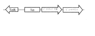
いくつかの実施形態では、発現コンストラクトは更に誘導性プロモーターエレメントを有する。好ましくは、誘導性プロモーターエレメントは、基本転写複合体を形成し転写を開始することのできるタンパク質に結合可能なDNA配列と、Tetリプレッサータンパク質(TetR)が結合可能な複数のTetオペレータ配列とを有する。この結合状態では、厳しい転写抑制が得られる。しかしながら、ドキシサイクリンの存在下では抑制が緩和されるので、プロモーターは完全な転写活性を獲得することができる。このような誘導性プロモーターエレメントは、好ましくは、別のプロモーター、例えばCMVプロモーターの下流に配置される。 In some embodiments, the expression construct further has an inducible promoter element. Preferably, the inducible promoter element comprises a DNA sequence capable of binding to a protein capable of forming a basic transcription complex and initiating transcription, and a plurality of Tet operator sequences capable of binding a Tet repressor protein (TetR). Have. In this combined state, severe transfer suppression is obtained. However, since the repression is reduced in the presence of doxycycline, the promoter can acquire full transcription activity. Such an inducible promoter element is preferably located downstream of another promoter, for example a CMV promoter.
いくつかの実施形態では、細胞は、発現される標的ポリペプチドのレベルを高めるために、標的ポリペプチド発現コンストラクトの複数のコピーを有する。標的ポリペプチドの発現レベルの増大は、細胞培養の時間を長くすることによって達成されてもよい。 In some embodiments, the cells have multiple copies of the target polypeptide expression construct to increase the level of the expressed target polypeptide. Increasing the expression level of the target polypeptide may be achieved by increasing the time of cell culture.
標的ポリペプチド発現コンストラクトは、抗生物質耐性遺伝子、例えばピューロマイシンに対する耐性をコードする遺伝子を含んでもよい。 The target polypeptide expression construct may include an antibiotic resistance gene, for example, a gene encoding resistance to puromycin.
標的ポリペプチドは哺乳類細胞集団にディスプレイされる。細胞は単離細胞であってよく、例えば生きている動物に存在するものではない。哺乳類細胞の例としては、ヒト、マウス、ラット、ハムスター、サル、ウサギ、ロバ、ウマ、ヒツジ、ウシ及び類人猿由来の任意の臓器又は組織由来の細胞がある。好ましくは、細胞はヒト細胞である。細胞は、初代細胞又は不死化細胞であってよい。好適なヒト細胞としては、HEK293細胞、HEK293T細胞、HEK293A細胞、PerC6細胞、911細胞、HeLa細胞及びCOS細胞がある。他の好適な細胞としては、CHO細胞及びVERO細胞がある。最も好ましくは、細胞はCHO細胞である。 The target polypeptide is displayed on a mammalian cell population. The cell may be an isolated cell, for example, not present in a living animal. Examples of mammalian cells include cells from any organ or tissue from humans, mice, rats, hamsters, monkeys, rabbits, donkeys, horses, sheep, cows and apes. Preferably, the cells are human cells. The cells may be primary cells or immortalized cells. Suitable human cells include HEK293 cells, HEK293T cells, HEK293A cells, PerC6 cells, 911 cells, HeLa cells and COS cells. Other suitable cells include CHO cells and VERO cells. Most preferably, the cells are CHO cells.
好ましくは、集団の全て又は実質的に全ての細胞が標的ポリペプチドをディスプレイする。好ましくは、集団の全ての又は実質的に全ての細胞は、10未満又は5未満、より好ましくは1、2又は3、最も好ましくは単一の結合パートナーを発現する。 Preferably, all or substantially all cells of the population display the target polypeptide. Preferably, all or substantially all cells of the population express less than 10 or less than 5, more preferably 1, 2 or 3, most preferably a single binding partner.
本発明の方法では、哺乳類細胞集団において結合パートナーのライブラリが発現される。その目的は、標的ポリペプチドの露出した領域又はドメインに結合する少なくとも1つの特異的結合パートナーを、係る特異的結合パートナーが結合される細胞が識別されることを可能にするような方法で、識別することである。 In the method of the invention, a library of binding partners is expressed in a population of mammalian cells. The purpose is to identify at least one specific binding partner that binds to an exposed region or domain of the target polypeptide in such a way as to allow the cell to which such specific binding partner is bound to be identified. It is to be.
本明細書で用いられるとき、「特異的結合パートナー」という用語は、結合パートナーの、所望の特異度及び/又は親和度で標的ポリペプチドに結合する能力に関する。特異的結合パートナーは、標的ポリペプチドのみに結合するわけではない。好ましくは、特異的結合パートナーは、その標的ポリペプチドに対する自身の親和性が非標的ポリペプチドに対する自身の親和性よりも約5倍大きい場合に、特異的に結合する。理想的には、望ましくない物質との有意な交差反応又は交差結合は存在しない。 As used herein, the term "specific binding partner" relates to the ability of a binding partner to bind to a target polypeptide with a desired specificity and / or affinity. A specific binding partner does not bind only to the target polypeptide. Preferably, a specific binding partner specifically binds when its affinity for its target polypeptide is about 5-fold greater than its affinity for a non-target polypeptide. Ideally, there is no significant cross-reactivity or cross-linking with undesired substances.
特異的結合パートナーの親和性は、例えば、非標的ポリペプチドに対する自身の親和性よりも、標的分子に対しては少なくとも約5倍、例えば10倍、25倍、特に50倍、特に100倍以上大きくてよい。 The affinity of a specific binding partner is, for example, at least about 5-fold, such as 10-fold, 25-fold, especially 50-fold, especially 100-fold or more, greater than its affinity for a non-target polypeptide. May be.
いくつかの実施形態では、特異的結合パートナーと標的ポリペプチドとの結合は、結合親和性が少なくとも106M−1であることを意味する。抗体は、例えば約108M−1〜約109M−1、約109M−1〜約1010M−1、又は約1010M−1〜約1011M−1など、少なくとも約107M−1の親和性で結合してよい。 In some embodiments, binding of the specific binding partner to the target polypeptide means that the binding affinity is at least 10 6 M −1 . The antibody can be at least about 10 8 M −1 to about 10 9 M −1 , about 10 9 M −1 to about 10 10 M −1 , or about 10 10 M −1 to about 10 11 M −1, for example. It may bind with an affinity of 10 7 M −1 .
抗体は、例えば、50nM以下、10nM以下、1nM以下、100pM以下、より好ましくは10pM以下のEC50で結合してよい。本明細書で用いられるとき、「EC50」という用語は、50%の最大応答/効果をもたらす濃度を定量化することにより、化合物の効力に言及することを意図する。EC50はスキャッチャード又はFACSによって決定されてよい。 Antibodies, e.g., 50 nM or less, 10 nM or less, 1 nM or less, 100 pM or less, more preferably attached at the following EC 50 10 pM. As used herein, the term “EC 50 ” is intended to refer to the potency of a compound by quantifying the concentration that produces a 50% maximal response / effect. The EC 50 may be determined by Scatchard or FACS.
各結合パートナーはコアフレームワーク及び複数の可変領域を有し、複数の可変領域のそれぞれは該結合パートナーに標的に対する特異的な結合親和性を与える。コアフレームワークは1又は複数のポリペプチドを有してよい。好ましくは、2〜10個、より好ましくは2〜6個、3〜6個、4〜6個又は5〜6個の可変領域が存在する。結合パートナーは一般にポリペプチドである。これらはグリコシル化されてもされなくてもよい。結合パートナーは、標的ポリペプチドの潜在的な結合パートナー、又は潜在的な特異的結合パートナーと見なされてよい。 Each binding partner has a core framework and a plurality of variable regions, each of the plurality of variable regions conferring the binding partner a specific binding affinity for a target. The core framework may have one or more polypeptides. Preferably, there are 2-10, more preferably 2-6, 3-6, 4-6 or 5-6 variable regions. The binding partner is generally a polypeptide. These may or may not be glycosylated. A binding partner may be considered a potential binding partner of the target polypeptide, or a potential specific binding partner.
結合パートナー(例えば抗体又は抗体模倣物)は、それが産生される細胞によって分泌されるか、又は該細胞から分泌されるか、又は該細胞の外に分泌される。いくつかの実施形態では、結合パートナー(例えば抗体又は抗体模倣物)は、それが産生される細胞の外から細胞周辺の培地に分泌される。別の言い方では、本実施形態において、結合パートナーは細胞から放出される。 The binding partner (eg, antibody or antibody mimetic) is secreted by, secreted from, or extracellular to the cell in which it is produced. In some embodiments, the binding partner (eg, antibody or antibody mimetic) is secreted from outside the cell where it is produced into the pericellular medium. Stated another way, in this embodiment, the binding partner is released from the cell.
他の実施形態では、結合パートナー(例えば抗体又は抗体模倣物)は、それが産生される細胞から分泌される。結合パートナーはその後、細胞周辺の培地に放出されてもされなくてもよい。 In other embodiments, the binding partner (eg, antibody or antibody mimetic) is secreted from the cell in which it is produced. The binding partner may or may not be subsequently released into the pericellular medium.
ポリペプチド結合パートナー(例えば抗体又は抗体模倣物)及び標的ポリペプチドは、哺乳類細胞において粗面小胞体(ER)に付着したリボソームによって合成される。これらのポリペプチドは両方とも、細胞の分泌経路へのポリペプチドの通過を指示するために、シグナルペプチドを有する。合成後、これらのポリペプチドはER内腔に移行し、そこでグリコシル化されることがあり、分子シャペロンがタンパク質の折り畳みを補助する。その後、ポリペプチドを含む小胞はゴルジ体に入る。ゴルジ体では、ポリペプチドの任意のグリコシル化が修飾されることがあり、切断や機能付与などの更なる翻訳後修飾が発生することがある。次にポリペプチドは分泌小胞に移動し、細胞骨格に沿って哺乳類細胞の端まで移動する。分泌小胞において更なる修飾が発生することがある。最終的に、エキソサイトーシスと呼ばれるプロセスにおいて、ポロソームと呼ばれる構造における細胞膜との小胞融合があり、その結果、小胞の内容物が周囲の培地に放出される。膜内タンパク質は、小胞の内容物が流されたときに細胞の細胞膜に保持される。 Polypeptide binding partners (eg, antibodies or antibody mimetics) and target polypeptides are synthesized in mammalian cells by ribosomes attached to rough endoplasmic reticulum (ER). Both of these polypeptides have a signal peptide to direct passage of the polypeptide into the cell's secretory pathway. After synthesis, these polypeptides enter the ER lumen, where they can be glycosylated, and molecular chaperones assist in protein folding. The vesicles containing the polypeptide then enter the Golgi apparatus. In the Golgi apparatus, any glycosylation of the polypeptide may be modified, and additional post-translational modifications, such as truncation or functionalization, may occur. The polypeptide then travels to secretory vesicles and travels along the cytoskeleton to the edge of mammalian cells. Further modifications may occur in secretory vesicles. Eventually, in a process called exocytosis, there is vesicle fusion with the cell membrane in a structure called porosomes, which results in the release of vesicle contents into the surrounding medium. Intramembrane proteins are retained in the cell membrane of cells when the contents of the vesicles are flushed.
ポリペプチド結合パートナー(例えば抗体又は抗体模倣物)と標的ポリペプチドは両方ともこの分泌経路を介して産生されるので、一部の特異的結合パートナーの標的ポリペプチドへの結合がこの経路の進行中に発生する可能性がある。この場合、特異的結合パートナーは細胞から外部の培地に分泌されず、標的ポリペプチドに結合したままになる。したがって、特異的結合パートナーと標的ポリペプチドは(一緒に)細胞の外面に提示される。 Since both a polypeptide binding partner (eg, an antibody or an antibody mimetic) and a target polypeptide are produced through this secretory pathway, the binding of some specific binding partners to the target polypeptide is an ongoing step in this pathway. Can occur. In this case, the specific binding partner is not secreted from the cell into the external medium, but remains bound to the target polypeptide. Thus, the specific binding partner and the target polypeptide are (together) presented on the outer surface of the cell.
いくつかの実施形態では、抗体又は抗体模倣物は、それらのCDR配列が標的ポリペプチドに結合した形で、それらが産生される細胞から分泌される。他の実施形態では、抗体又は抗体模倣物は、それらのCDR配列が標的ポリペプチドに結合していない形で、それらが産生される細胞から分泌される。 In some embodiments, the antibodies or antibody mimetics are secreted from the cell in which they are produced, with their CDR sequences attached to the target polypeptide. In other embodiments, the antibodies or antibody mimetics are secreted from the cell in which they are produced, with their CDR sequences not bound to the target polypeptide.
結合パートナー(例えば抗体又は抗体模倣物)は、直接的又は間接的に、細胞の表面に共有結合しない。結合パートナー(例えば抗体又は抗体模倣物)は、細胞周辺の培地内で自由に拡散する(分泌経路において標的ポリペプチドに結合する結合パートナーは別として)。 The binding partner (eg, antibody or antibody mimetic) does not directly or indirectly covalently bind to the surface of the cell. The binding partner (eg, antibody or antibody mimetic) is free to diffuse in the media around the cell (apart from the binding partner that binds the target polypeptide in the secretory pathway).
本明細書で用いられるとき、「ライブラリ」という用語は、それぞれが異なる結合特異性及び/又は親和性をもつ複数の(潜在的な)結合パートナーを指す。各結合パートナーは、(共通の)コアフレームワークと、複数の異なる可変領域とを有する。好ましくは、「ライブラリ」という用語は、それぞれ異なる結合特異性及び/又は親和性をもつ複数のポリペプチドを指す。複数のポリペプチドは、一般に、複数のポリヌクレオチドによってコードされる。 As used herein, the term "library" refers to a plurality (potential) binding partners, each having a different binding specificity and / or affinity. Each binding partner has a (common) core framework and a plurality of different variable regions. Preferably, the term "library" refers to a plurality of polypeptides, each having a different binding specificity and / or affinity. Multiple polypeptides are generally encoded by multiple polynucleotides.
好ましくは、ライブラリは、哺乳類細胞に感染可能なウイルス(好ましくはレトロウイルス、より好ましくはレンチウイルス)により、哺乳類細胞集団に送達される。 Preferably, the library is delivered to a population of mammalian cells by a virus (preferably a retrovirus, more preferably a lentivirus) capable of infecting mammalian cells.
いくつかの好ましい実施形態では、結合パートナーポリペプチドをコードするポリヌクレオチドが細胞内で発現される。このようなポリヌクレオチドは、(例えばレトロウイルスベクターから)一時的に発現されてよく、又は発現ベクターから発現されてよい。いくつかの実施形態では、発現ベクターは細胞のゲノムに組み込まれる。 In some preferred embodiments, the polynucleotide encoding the binding partner polypeptide is expressed in a cell. Such polynucleotides may be expressed transiently (eg, from a retroviral vector) or may be expressed from an expression vector. In some embodiments, the expression vector is integrated into the genome of the cell.
特定の実施形態では、ポリペプチドのライブラリは、少なくとも106、107、108、109、1010、1011、1012、1013、1014又は1015以上の異なるポリペプチドを含んでよい。 In certain embodiments, the library of polypeptides comprises at least 10 6 , 10 7 , 10 8 , 10 9 , 10 10 , 10 11 , 10 12 , 10 13 , 10 14 or 10 15 or more different polypeptides. Good.
いくつかの実施形態では、ライブラリの異なるポリペプチドは、例えば、単一の動物種(例えばヒト、マウス、ウサギ、ヤギ、ウマ)、組織タイプ、臓器又は細胞タイプに由来する起源を通じて関連する。 In some embodiments, the different polypeptides of the library are related, for example, through origins from a single animal species (eg, human, mouse, rabbit, goat, horse), tissue type, organ or cell type.
他の実施形態では、ライブラリは、天然に存在するポリペプチドのライブラリであり、改良されてよい。更に他の実施形態では、ライブラリは合成ポリペプチドのライブラリである。 In other embodiments, the library is a library of naturally occurring polypeptides and may be improved. In yet other embodiments, the library is a library of synthetic polypeptides.
結合パートナーは、細胞から分泌されることができなければならない(好ましくは細胞の外に)。いくつかの実施形態では、このような分泌は、5’−シグナルポリペプチドを含めることによって補助されてよい。 The binding partner must be able to be secreted from the cell (preferably outside the cell). In some embodiments, such secretion may be assisted by including a 5'-signal polypeptide.
いくつかの好適な実施形態では、結合パートナーは抗体又は抗体模倣物である。本実施形態では、ステップ(a)は、哺乳類細胞集団において抗体又は抗体模倣物のライブラリを発現させるステップであり、それぞれの抗体又は抗体模倣物はそれが産生される細胞から分泌される、ステップ、を含む。 In some preferred embodiments, the binding partner is an antibody or an antibody mimetic. In this embodiment, step (a) is the step of expressing a library of antibodies or antibody mimetics in a population of mammalian cells, wherein each antibody or antibody mimetic is secreted from the cell in which it is produced, including.
本明細書で用いられるとき、「抗体」は多種多様な構造を含み、当業者によって理解されるように、いくつかの実施形態では最低6個のCDRのセットを含み、限定ではないが例として、従来の抗体(モノクローナル抗体など)、ヒト化及び/又はキメラ抗体、抗体フラグメント、改変抗体、多特異性抗体(二重特異性抗体など)、本技術分野で既知の他の類似物がある。 As used herein, an “antibody” includes a wide variety of structures, and as will be appreciated by those of skill in the art, in some embodiments includes a set of at least six CDRs, including, but not limited to, by way of example. There are conventional antibodies (such as monoclonal antibodies), humanized and / or chimeric antibodies, antibody fragments, modified antibodies, multispecific antibodies (such as bispecific antibodies), and other analogs known in the art.
「抗体」は免疫グロブリン分子であり、免疫グロブリン分子の可変領域に位置する少なくとも1つの抗原認識部位を介して、炭水化物、ポリヌクレオチド、脂質、ポリペプチド等の標的に特異的に結合することができる。 An “antibody” is an immunoglobulin molecule that can specifically bind to a target, such as a carbohydrate, polynucleotide, lipid, polypeptide, etc., via at least one antigen recognition site located in a variable region of the immunoglobulin molecule. .
いくつかの実施形態では、抗体は異なる種の混合、例えばキメラ抗体及び/又はヒト化抗体であってよい。すなわち、CDRセットは、それらが元々取得されたもの以外のフレームワーク及び定常領域と共に用いることができる。 In some embodiments, the antibodies may be a mixture of different species, for example, chimeric and / or humanized antibodies. That is, CDR sets can be used with frameworks and constant regions other than those from which they were originally obtained.
一般に、「キメラ抗体」と「ヒト化抗体」は両方とも、複数の種由来の領域を組み合わせる抗体を指す。例えば、「キメラ抗体」は伝統的に、マウス(又は場合によってラット)由来の可変領域と、ヒト由来の定常領域とを有する。「ヒト化抗体」は一般に、可変ドメインフレームワーク領域をヒト抗体に見られる配列に交換した非ヒト抗体を指す。一般に、ヒト化抗体では、CDRを除く抗体全体が、ヒト由来のポリヌクレオチドによってコードされているか、CDR内を除いてそのような抗体と同一である。CDR(一部又は全部が非ヒト生物に由来する核酸によってコードされる)は、ヒト抗体可変領域のベータシートフレームワークに移植されて、抗体を作製する。抗体の特異性は、移植されたCDRによって決定される。 In general, both "chimeric antibodies" and "humanized antibodies" refer to antibodies that combine regions from more than one species. For example, a "chimeric antibody" traditionally has a variable region from a mouse (or rat in some cases) and a constant region from a human. "Humanized antibodies" generally refer to non-human antibodies in which the variable domain framework regions have been replaced by sequences found in human antibodies. Generally, in a humanized antibody, the entire antibody, except for the CDRs, is encoded by a polynucleotide of human origin or is identical to such antibodies except within the CDRs. The CDRs (encoded in part or in whole by nucleic acids from a non-human organism) are grafted into the beta-sheet framework of human antibody variable regions to create antibodies. Antibody specificity is determined by the grafted CDRs.
一実施形態において、抗体は抗体フラグメントである。具体的な抗体フラグメントとしては、これらに限定されないが、(i)VL、VH、CL及びCH1ドメインで構成されるFabフラグメント、(ii)VH及びCH1ドメインで構成されるFdフラグメント、(iii)単一の抗体のVL及びVHドメインで構成されるFvフラグメント、(iv)単一の可変領域で構成されるdAbフラグメント(Ward他、1989、Nature341:544−546)、(v)単離されたCDR領域、(vi)F(ab’)2フラグメント(2つの連結されたFabフラグメントを含む二価フラグメント)、(vii)単鎖Fv分子(scFv)(VHドメインとVLドメインは、2つのドメインが結合して抗原結合部位を形成できるようにするペプチドリンカーによって連結されている)(Bird他、1988、Science242:423−426、Huston他、1988、Proc.Natl.Acad.Sci.U.S.A.85:5879−5883)、(viii)二重特異性単一鎖Fv(例えば国際公開第03/11161号)、(ix)「ダイアボディ(diabody)」又は「トリアボディ(triabody)」(遺伝子融合によって構築された多価又は多特異性フラグメント)(Tomlinson他、2000、Methods Enzymol.326:461−479;国際公開第94/13804号;Holliger他、1993、Proc.Natl.Acad.Sci.U.S.A.90:6444−6448)が挙げられる。 In one embodiment, the antibody is an antibody fragment. Specific antibody fragments include, but are not limited to, (i) a Fab fragment composed of VL, VH, CL and CH1 domains, (ii) an Fd fragment composed of VH and CH1 domains, and (iii) a single fragment. An Fv fragment composed of the VL and VH domains of one antibody, (iv) a dAb fragment composed of a single variable region (Ward et al., 1989, Nature 341: 544-546), (v) an isolated CDR Region, (vi) F (ab ') 2 fragment (a bivalent fragment containing two linked Fab fragments), (vii) single-chain Fv molecule (scFv) (VH domain and VL domain Linked by a peptide linker that allows the formation of an antigen binding site) Ird et al., 1988, Science 242: 423-426, Huston et al., 1988, Proc. Natl. Acad. Sci. USA 85: 5879-5883), (viii) a bispecific single chain Fv (e.g. WO 03/11161), (ix) "diabodies" or "triabodies" (multivalent or multispecific fragments constructed by gene fusion) (Tomlinson et al., 2000, Methods Enzymol). No. 326: 461-479; WO 94/13804; Holliger et al., 1993, Proc. Natl. Acad. Sci. USA 90: 6444-6448).
「抗体」という用語は、ドメイン抗体、ナノボディ及びユニボディも含む。「抗体」という用語は、抗体部分又はフラグメントと抗原認識部位を有する融合タンパク質も含む。最も好ましくは、抗体はscFv抗体である。 The term "antibody" also includes domain antibodies, Nanobodies and Unibodies. The term "antibody" also includes fusion proteins having an antibody portion or fragment and an antigen recognition site. Most preferably, the antibodies are scFv antibodies.
抗体ライブラリは、例えば、特定のタイプ又はクラスの免疫グロブリンポリペプチドを含んでよい。例えば、ライブラリは、抗体μ、γ1、γ2、γ3、γ4、α1、α2、ε若しくはδの重鎖、及び/又は抗体κ若しくはλの軽鎖をコードし得る。抗体アイソタイプは、IgM、IgD、IgG、IgA又はIgEであってよい。好ましくは、抗体はIgG1、IgG2、IgG3又はIgG4である。 An antibody library may include, for example, a particular type or class of immunoglobulin polypeptides. For example, the library may encode the antibody μ, γ1, γ2, γ3, γ4, α1, α2, ε or δ heavy chains, and / or the antibody κ or λ light chains. The antibody isotype may be IgM, IgD, IgG, IgA or IgE. Preferably, the antibody is an IgG1, IgG2, IgG3 or IgG4.
本明細書に記載の任意の1つのライブラリの各メンバーは、同じ重鎖又は軽鎖の定常領域をコードしてよいが、ライブラリは合計で、少なくとも106、107、108、109、1010、1011、1012、1013、1014若しくは1015、又はより多くの異なる可変領域、すなわち共通の定常領域に関連付けられた「複数の」可変領域を含んでよい。 Each member of any one of the libraries described herein may encode the same heavy or light chain constant region, but the library may comprise at least 10 6 , 10 7 , 10 8 , 10 9 , It may include 10 10 , 10 11 , 10 12 , 10 13 , 10 14 or 10 15 , or more different variable regions, ie, “plurality” of variable regions associated with a common constant region.
一実施形態において、細胞集団は、scFVライブラリをコードする複数のレトロウイルス(好ましくはレンチウイルス)粒子に初代(同種)細胞集団を感染させる(例えば一時的に感染させる)ことにより、作製される。scFvライブラリは、異なる結合特異性及び親和性をもつ複数のscFV抗体を含む。 In one embodiment, the cell population is created by infecting (eg, transiently) a primary (allogeneic) cell population with a plurality of retroviral (preferably lentiviral) particles encoding a scFV library. An scFv library contains multiple scFV antibodies with different binding specificities and affinities.
例えば、各レトロウイルス粒子は、プロモーター、シグナルペプチド(scFvの分泌を促進するため)及びscFvコーディング配列を有するレトロウイルス又はレンチウイルスのベクターを有してよい。また、二次抗体による認識を補助するために、ベクターはヘマグルチニンなどの3’タグを含んでもよい。レンチウイルスベクター(発現コンストラクト)は更に、抗アポトーシス因子をコードするポリヌクレオチド配列を含んでよい。 For example, each retroviral particle may have a retroviral or lentiviral vector having a promoter, a signal peptide (to facilitate secretion of the scFv) and a scFv coding sequence. Also, the vector may include a 3 'tag, such as hemagglutinin, to assist recognition by the secondary antibody. Lentiviral vectors (expression constructs) may further include a polynucleotide sequence encoding an anti-apoptotic factor.
本発明の特に好適な実施形態では、抗アポトーシス因子は、標的ポリペプチドをコードする核酸の最後の停止コドンのIRES下流(すなわち3’)の後(すなわち3’)に挿入される。これにより、転写を開始するプロモーターが標的ポリペプチド遺伝子のコード配列の上流(すなわち5’)に存在し、その後にIRESが続き(3’)、その後に抗アポトーシス因子遺伝子のコード領域が続く(3’)構成が提供される。この構成では、標的ポリペプチドと抗アポトーシス因子の両方が同じmRNAによってコードされるが、IRESを介した翻訳の効率は比較的低いので、標的ポリペプチドは抗アポトーシス因子よりも大量に翻訳される。 In a particularly preferred embodiment of the invention, the anti-apoptotic factor is inserted after (ie, 3 ') downstream of (ie, 3') the IRES of the last stop codon of the nucleic acid encoding the target polypeptide. This places the promoter that initiates transcription upstream (ie, 5 ') of the coding sequence of the target polypeptide gene, followed by the IRES (3'), followed by the coding region of the anti-apoptotic factor gene (3 '). ') Configuration provided. In this configuration, both the target polypeptide and the anti-apoptotic factor are encoded by the same mRNA, but the translation efficiency via the IRES is relatively low, so that the target polypeptide is translated in greater quantity than the anti-apoptotic factor.
本発明の方法は、抗体を伴う方法に限定されない。該方法は抗体模倣物を用いて実施されてもよい。多種多様な抗体模倣物技術が当技術分野で知られている。特に、Affibody、DARPin、Anticalin、Avimer、Versabodyなどの技術は、従来の抗体結合を模倣しつつ、異なるメカニズムから生成され、機能する結合構造を採用する。 The method of the present invention is not limited to methods involving antibodies. The method may be performed using an antibody mimetic. A wide variety of antibody mimetic technologies are known in the art. In particular, techniques such as Affibody, DARPin, Anticalin, Avimer, Versabody, etc., employ binding structures that are generated and function from different mechanisms while mimicking conventional antibody binding.
Affibody分子は、ブドウ球菌タンパク質AのIgG結合ドメインのうちの1つに由来する、58アミノ酸残基のタンパク質ドメインに基づいた新しいクラスの親和性タンパク質を表す。この3ヘリックスバンドルドメインは、コンビナトリアル・ファージミド・ライブラリの構築の足場として使用されており、そこから、所望の分子を標的とするAffibodyバリアントをファージディスプレイ技術を用いて選択することができる(Nord K,Gunneriusson E,Ringdahl J,Stahl S,Uhlen M,Nygren PA,Binding proteins selected from combinatorial libraries of an α−helical bacterial receptor domain(αヘリックス細菌受容体ドメインのコンビナトリアルライブラリから選択された結合タンパク質),Nat Biotechnol 1997;15:772−7.Ronmark J,Gronlund H,Uhlen M,Nygren PA,human immunoglobulin A(IgA)−specific ligands from combinatorial engineering of protein A(プロテインAのコンビナトリアルエンジニアリングによるヒト免疫グロブリンA(IgA)特異的リガンド),Eur J Biochem 2002;269:2647−55.)。Affibody及びその製造方法の更なる詳細は、米国特許第5,831,012号を参照することにより得ることができる。 Affibody molecules represent a new class of affinity proteins based on a 58 amino acid residue protein domain derived from one of the IgG binding domains of staphylococcal protein A. This three-helix bundle domain has been used as a scaffold for the construction of combinatorial phagemid libraries from which Affibody variants targeting the desired molecule can be selected using phage display technology (Nord K, Gunneriusson E, Ringdahl J, Stahl S, Uhlen M, Nygren PA, Binding proteins selected from combinatorial library Libraries of the bacterium of the alpha-helical bacteria 15: 772 7. Ronmark J, Gronlund H, Uhlen M, Nygren PA, human immunoglobulin A (IgA) -specific ligands from combinatory engineering. Biochem 2002; 269: 2647-55.). Further details of Affibody and its method of manufacture can be obtained by reference to US Patent No. 5,831,012.
DARPin(Designed Ankyrin Repeat Protein)は、非抗体ポリペプチドの結合能力を活用するために開発された抗体模倣物DRP(Designed Repeat Protein)技術の一例である。アンキリン又はロイシンに富んだ反復タンパク質などの反復タンパク質は、偏在する結合分子であり、抗体とは異なり、細胞内及び細胞外で発生する。その独自のモジュラーアーキテクチャは反復する構造単位(反復)を特徴とし、それらが重なり合って、モジュラーを標的とする可変の結合表面をディスプレイする細長い反復ドメインを形成する。このモジュール性に基づき、高度に多様化された結合特異性をもつポリペプチドのコンビナトリアルライブラリを生成することができる。この戦略は、可変表面残基をディスプレイする自家和合性の反復とその反復ドメインへのランダムな会合のコンセンサス設計を含む。DARPin及びその他のDRP技術に関する追加情報は、米国特許出願公開第2004/0132028号及び国際公開第02/20565号を参照することができる。 DARPin (Designed Ankyrin Repeat Protein) is an example of an antibody mimetic DRP (Designed Repeat Protein) technology developed to exploit the binding ability of non-antibody polypeptides. Repetitive proteins, such as repetitive proteins rich in ankyrin or leucine, are ubiquitous binding molecules and, unlike antibodies, occur intracellularly and extracellularly. Its unique modular architecture features repeating structural units (repeats) that overlap to form elongated, repeating domains that display a variable binding surface that targets the modular. Based on this modularity, combinatorial libraries of polypeptides with highly diversified binding specificities can be generated. This strategy involves a consensus design of self-compatible repeats displaying variable surface residues and random association to their repeat domains. For additional information regarding DARPin and other DRP technologies, reference may be made to U.S. Patent Application Publication No. 2004/0132028 and WO 02/20565.
Anticalinは、更なる抗体模倣物技術である。しかしながら、この場合、結合特異性は、ヒトの組織及び体液で自然に豊富に発現される低分子量タンパク質のファミリーであるリポカリンに由来する。 Anticalin is an additional antibody mimetic technology. However, in this case, the binding specificity derives from lipocalin, a family of low molecular weight proteins that are naturally abundantly expressed in human tissues and body fluids.
リポカリンは、化学的に敏感又は不溶性の化合物の生理学的輸送並びに貯蔵に関連する、生体内での様々な機能を実行するために進化してきた。リポカリンは、タンパク質の一方の末端で4つのループを支持する高度に保存されたβバレルを含む堅牢な固有の構造をもつ。これらのループは結合ポケットへの入り口を形成し、分子のこの部分の立体配座の違いが、個々のリポカリン間の結合特異性の変動の要因となる。 Lipocalins have evolved to perform various functions in vivo related to the physiological transport and storage of chemically sensitive or insoluble compounds. Lipocalins have a robust and unique structure that includes a highly conserved β-barrel that supports four loops at one end of the protein. These loops form the entrance to the binding pocket, and differences in the conformation of this part of the molecule contribute to variations in the binding specificity between individual lipocalins.
保存されたβシートフレームワークによって支持される超可変ループの全体的な構造は免疫グロブリンを連想させるが、リポカリンは、サイズが抗体とはかなり異なり、単一の免疫グロブリンドメインよりもわずかに大きい160〜180アミノ酸の単一ポリペプチド鎖で構成される。 Although the overall structure of the hypervariable loop supported by the conserved β-sheet framework is reminiscent of immunoglobulins, lipocalins differ significantly in size from antibodies and are slightly larger than single immunoglobulin domains 160 Consists of a single polypeptide chain of 180180 amino acids.
リポカリンはクローン化され、そのループはAnticalinを作製するためにエンジニアリングされている。構造的に多様なAnticalinのライブラリが生成されており、Anticalinディスプレイにより、結合機能の選択及びスクリーニングが可能になり、その後、原核生物系又は真核生物系での更なる分析のために可溶性タンパク質の発現と産生が続く。事実上あらゆるヒト標的タンパク質に特異的なAnticalinを開発し単離することができること、ナノモル以上の結合親和性を得ることができることが、研究により成功裏に実証されている。 Lipocalin has been cloned and the loop has been engineered to make Anticalin. A library of structurally diverse Anticalins has been generated, and the Anticalin display allows for the selection and screening of binding functions, followed by the selection of soluble proteins for further analysis in prokaryotic or eukaryotic systems. Expression and production continue. Studies have successfully demonstrated that Anticalin specific to virtually any human target protein can be developed and isolated, and that nanomolar or higher binding affinities can be obtained.
Anticalinは、二重標的化タンパク質、いわゆるDuocalinとしてフォーマットすることもできる。Duocalinは、その2つの結合ドメインの構造配向性に関係なく、標的特異性及び親和性を保持しながら、標準的な製造プロセスを用いて簡単に産生される1つの単量体タンパク質において2つの別々の治療標的に結合する。Anticalinに関する追加情報は、米国特許第7,250,297号及び国際公開第99/16873号を参照することができる。 Anticalins can also be formatted as dual targeting proteins, so-called Duocalins. Duocalin retains target specificity and affinity irrespective of the structural orientation of its two binding domains while retaining two separate proteins in one monomeric protein that is easily produced using standard manufacturing processes. Binds to therapeutic targets. For additional information on Anticalin, see U.S. Patent No. 7,250,297 and WO 99/16873.
本発明の文脈において有用な別の抗体模倣物技術は、Avimerである。Avimerは、インビトロのエクソンシャッフリング及びファージディスプレイにより、ヒト細胞外受容体ドメインの大きなファミリーから進化し、結合特性及び阻害特性をもつマルチドメインタンパク質を生成する。複数の独立した結合ドメインを連結すると、結合活性が生まれることが示されており、従来の単一エピトープ結合タンパク質と比較して親和性及び特異性が向上する。他の潜在的な利点には、大腸菌でのマルチターゲット特異的分子の単純且つ効率的な産生、熱安定性の向上、プロテアーゼ耐性が含まれる。様々な標的に対して、ナノモル以下の親和性をもつAvimerが得られている。Avimerに関する追加情報は、米国特許出願公開第2006/0286603号、米国特許出願公開第2006/0234299号、米国特許出願公開第2006/0223114号,米国特許出願公開第2006/0177831号、米国特許出願公開第2006/0008844号、米国特許出願公開第2005/0221384号、米国特許出願公開第2005/0164301号、米国特許出願公開第2005/0089932号、米国特許出願公開第2005/0053973号、米国特許出願公開第2005/0048512号及び米国特許出願公開第2004/0175756号を参照することができる。 Another antibody mimetic technology useful in the context of the present invention is Avimer. Avimer evolves from a large family of human extracellular receptor domains by in vitro exon shuffling and phage display, producing multidomain proteins with binding and inhibitory properties. Linking multiple independent binding domains has been shown to produce binding activity, which increases affinity and specificity compared to conventional single epitope binding proteins. Other potential advantages include simple and efficient production of multi-target specific molecules in E. coli, improved thermostability, and protease resistance. Avimers with sub-nanomolar affinities for various targets have been obtained. Additional information regarding Avimer can be found in U.S. Patent Application Publication No. 2006/0286603, U.S. Patent Application Publication No. 2006/0234299, U.S. Patent Application Publication No. 2006/0223114, U.S. Patent Application Publication No. 2006/0177831, U.S. Patent Application Publication. No. 2006/0008844; U.S. Patent Application Publication No. 2005/0221384; U.S. Patent Application Publication No. 2005/0164301; U.S. Patent Application Publication No. 2005/0089932; U.S. Patent Application Publication No. 2005/0053973; U.S. Patent Application Publication Reference may be made to US 2005/0048512 and US Patent Application Publication No. 2004/0175756.
Versabodyは、本発明の文脈で使用できる別の抗体模倣物技術である。Versabodyは3〜5kDaの小さなタンパク質で、15%を超えるシステインを含み、高ジスルフィド密度の足場を形成し、典型的なタンパク質が有する疎水性コアを置換する。疎水性コアを含む多数の疎水性アミノ酸を少数のジスルフィドで置換すると、MHCの提示に最も寄与する残基は疎水性であるので、タンパク質がより小さく、親水性が高まり(凝集及び非特異的結合が少なくなり)、プロテアーゼ及び熱に対する耐性が高まり、T細胞エピトープの密度が低くなる。これらの4つの特性は全て免疫原性に影響を与えることがよく知られており、これらが合わさると、免疫原性が大幅に低下することが予想される。 Versabody is another antibody mimetic technology that can be used in the context of the present invention. Versabody is a small protein of 3-5 kDa, containing more than 15% cysteine, forming a high disulfide density scaffold, replacing the hydrophobic core of typical proteins. Replacing a large number of hydrophobic amino acids, including the hydrophobic core, with a small number of disulfides results in smaller, more hydrophilic proteins (aggregation and non-specific binding), since the residues that most contribute to MHC presentation are hydrophobic. ), Increased resistance to proteases and heat, and reduced density of T cell epitopes. It is well known that all four of these properties affect immunogenicity, and when combined, it is expected that immunogenicity will be significantly reduced.
Versabodyの構造を考慮すると、このような抗体模倣物により、多価、多重特異性、多様な半減期メカニズム、組織標的化モジュール、抗体Fc領域の欠如を含む、多目的なフォーマットが提供される。Versabodyに関する追加情報は米国特許出願公開第2007/0191272号を参照することができる。 Given the structure of Versabody, such antibody mimics provide a versatile format, including multivalent, multispecific, diverse half-life mechanisms, tissue targeting modules, and lack of antibody Fc regions. For additional information regarding Versabody, reference may be made to US Patent Application Publication No. 2007/0191272.
他の実施形態では、結合パートナーは抗体又は抗体模倣物ではない。例えば、標的ポリペプチドの所望の特異的結合パートナーは、標的ポリペプチドが結合可能なポリペプチドリガンドであってよい。このような場合、結合パートナーのライブラリは、標的ポリペプチドに結合すると知られているポリペプチドリガンドのアミノ酸配列に基づくアミノ酸配列を有するポリペプチドのライブラリであってよい。例えば、そのようなライブラリのポリペプチドは、既知のポリペプチドリガンドとのアミノ酸配列の同一性が少なくとも60%、70%、80%、90%又は95%であってよい。 In other embodiments, the binding partner is not an antibody or an antibody mimetic. For example, the desired specific binding partner for the target polypeptide may be a polypeptide ligand to which the target polypeptide can bind. In such a case, the library of binding partners may be a library of polypeptides having an amino acid sequence based on the amino acid sequence of a polypeptide ligand known to bind to the target polypeptide. For example, the polypeptides of such a library may have at least 60%, 70%, 80%, 90% or 95% amino acid sequence identity with a known polypeptide ligand.
本発明の方法のステップ(b)は、哺乳類細胞集団のうち特異的結合パートナーが結合される細胞を識別且つ/又は単離することを含む。(特異的結合パートナーは、係る細胞にディスプレイされる標的ポリペプチドに結合する。)特異的結合パートナーが結合される細胞は、標的ポリペプチドの特異的結合パートナーを産生する細胞である。このように、標的ポリペプチドの所望の特異的結合パートナーを識別且つ/又は単離することができる。 Step (b) of the method of the invention involves identifying and / or isolating cells of the mammalian cell population to which the specific binding partner is bound. (The specific binding partner binds to the target polypeptide displayed on such cells.) The cell to which the specific binding partner is bound is a cell that produces a specific binding partner for the target polypeptide. In this way, the desired specific binding partner of the target polypeptide can be identified and / or isolated.
標的ポリペプチドに対する特異的結合パートナー(例えば抗体又は抗体模倣物)の結合は、主として特異的結合パートナーの同一性によって、多くの異なる手段によって検出されてよい。このような手段は当該技術分野で周知であり、標識2次抗体(例えば蛍光標識、ビオチン標識、又は放射性標識)、酵素的手段(例えば比色分析)及び機能ドメイン(例えば特定の活性を促進又は抑制するポリペプチドドメイン)の使用を含む。 Binding of a specific binding partner (eg, an antibody or antibody mimetic) to a target polypeptide may be detected by a number of different means, primarily due to the identity of the specific binding partner. Such means are well known in the art, and include labeled secondary antibodies (eg, fluorescent, biotin, or radioactive labels), enzymatic means (eg, colorimetric) and functional domains (eg, promoting or Inhibitory polypeptide domains).
特異的結合パートナーが抗体又は抗体模倣物である本発明の実施形態では、標的ポリペプチドに対する係る抗体又は抗体模倣物の結合は、標識2次抗体を用いて検出されてよい。例えば、特異的結合パートナーが全長抗体である場合、1次抗体のFc領域に結合する標識2次抗体が用いられてよい。特異的結合パートナーがscFV抗体である場合、scFV抗体上のタグ(例えばHAタグ)に結合する標識2次抗体が用いられてよい。 In embodiments of the invention where the specific binding partner is an antibody or antibody mimetic, binding of such antibody or antibody mimetic to the target polypeptide may be detected using a labeled secondary antibody. For example, if the specific binding partner is a full length antibody, a labeled secondary antibody that binds to the Fc region of the primary antibody may be used. Where the specific binding partner is a scFV antibody, a labeled secondary antibody that binds to a tag (eg, an HA tag) on the scFV antibody may be used.
いくつかの実施形態では、特異的結合パートナー(例えば1次抗体)又はそれに結合する2次抗体は、その存在及び/又は活性を確立且つ/又は定量化することのできる機能ドメインを含む。係る機能ドメインの例としては、細胞増殖を促進又は抑制するドメイン(例えば増殖因子の適切なドメイン)がある。 In some embodiments, a specific binding partner (eg, a primary antibody) or a secondary antibody that binds to it comprises a functional domain whose presence and / or activity can be established and / or quantified. Examples of such functional domains include domains that promote or suppress cell growth (eg, appropriate domains for growth factors).
本発明の方法は、液体培地、半固形培地、固形培地(例えばゲル)で実施されてよく、又は、細胞は完全に又は部分的に固定されてよい。好ましくは、本方法は、液体培地、例えば水性生理培地、例えば細胞培養と潜在的特異的結合パートナーの標的ポリペプチドへの結合とに適した水性生理培地で実施される。本実施形態では、分泌された結合パートナーは溶液中に存在するので、それらの分泌元の細胞だけでなく、他の(例えば隣接する)細胞にも自由に結合することができる。このような場合、細胞集団のうち特異的結合パートナーが結合された第1の細胞が、特異的結合パートナーを産生した細胞となる。経時的に、結合パートナーは、それらの分泌元の細胞以外の細胞に(例えばそれらに向かって対流又は拡散することによって)接触できるようになるが、かなり静的な系では、結合パートナーは最初に、それらの分泌元の細胞に結合する(結合パートナーが標的ポリペプチドに結合可能である場合)。 The method of the invention may be performed in a liquid, semi-solid, solid medium (eg, a gel), or the cells may be completely or partially fixed. Preferably, the method is performed in a liquid medium, eg, an aqueous physiological medium, eg, an aqueous physiological medium suitable for cell culture and binding of a potential specific binding partner to the target polypeptide. In this embodiment, since the secreted binding partner is in solution, it can freely bind not only to the cell from which it was secreted, but also to other (eg, adjacent) cells. In such a case, the first cell to which the specific binding partner is bound in the cell population is a cell that has produced the specific binding partner. Over time, the binding partners become able to contact cells other than their secreting cells (eg, by convection or diffusion toward them), but in fairly static systems, the binding partners initially Bind to their secreting cells (if the binding partner is capable of binding to the target polypeptide).
したがって、本発明の方法が液体培地で実施される場合、内部で産生された特異的結合パートナーに細胞がいつ最初に結合されたかを確定するために、多くの異なる時点(例えば4時間、6時間、24時間、48時間など)で特異的結合パートナーの任意の結合について細胞をアッセイする必要がある場合がある。係る細胞はその後、選別又は単離されてよい(例えば蛍光標識された特異的結合パートナーの場合には、蛍光活性化細胞選別(FACS)によって)。 Thus, when the method of the present invention is performed in a liquid medium, a number of different time points (eg, 4 hours, 6 hours, etc.) can be used to determine when cells were first bound to an internally produced specific binding partner. , 24 hours, 48 hours, etc.), the cells may need to be assayed for any binding of the specific binding partner. Such cells may then be sorted or isolated (eg, in the case of a fluorescently labeled specific binding partner, by fluorescence activated cell sorting (FACS)).
したがって、いくつかの実施形態では、ステップ(b)は、
(b)細胞集団のうち、特異的結合パートナーが最初に結合された細胞を識別又は単離するステップ、
を含む。したがって、他の実施形態では、ステップ(b)は、
(b)細胞集団のうち、特異的結合パートナーが、特異的結合パートナー自身が分泌された細胞上にディスプレイされた標的ポリペプチドのみに結合しうる時点の後に、特異的結合パートナーが結合された細胞を識別又は単離するステップ、
を含む。
Thus, in some embodiments, step (b) comprises:
(B) identifying or isolating cells of the cell population to which the specific binding partner has first bound;
including. Thus, in another embodiment, step (b) comprises:
(B) the cells to which the specific binding partner has been bound after a point in the population of cells where the specific binding partner can only bind to the target polypeptide displayed on the cells from which the specific binding partner itself was secreted; Identifying or isolating
including.
ほとんどの方法では、標的ポリペプチドに結合可能な結合パートナーを分泌する細胞はほとんど存在しないという事実に鑑みて、結合パートナーの他の細胞への対流又は拡散の問題は重大であるとは考えられない。 In most methods, the problem of convection or diffusion of the binding partner to other cells is not considered significant given the fact that few cells secrete binding partners capable of binding the target polypeptide. .
このような拡散の影響を低減する更なる方法は、連続的又は不連続的に液体培地を交換することにより、任意の拡散している結合パートナーを液体培地から除去することである。したがって、他の実施形態では、ステップ(a)は更に、本方法は連続的又は不連続に交換される液体培地において実施される、という特徴を含む。 A further way to reduce the effects of such diffusion is to remove any diffusing binding partner from the liquid medium by changing the liquid medium continuously or discontinuously. Thus, in another embodiment, step (a) further comprises the feature that the method is performed in a continuously or discontinuously exchanged liquid medium.
本発明のいくつかの実施形態では、結合パートナー(例えば抗体又は抗体模倣物)の、それらが産生された細胞から離れる動きを抑制することが望ましい。これにより、非特異的なバックグラウンド結合のレベルの低下、偽陽性細胞の産生の回避が助長される。これを行う1つの方法は、動的粘度が25℃において水の動的粘度よりも大きい液体培地を用いて、本発明の方法を実施することである。このようにして、結合パートナー(例えば抗体)がそれらが産生された細胞から離れる拡散が低減される。水の動的粘度は、25℃で8.9×10⁻⁴Pa.sである。好ましくは、本発明の方法のステップ(a)が実施される液体培地の動的粘度は、25℃で少なくとも10×10−4Pa.sである。 In some embodiments of the invention, it is desirable to suppress the movement of binding partners (eg, antibodies or antibody mimetics) away from the cells in which they were produced. This helps to reduce the level of non-specific background binding and avoid the production of false positive cells. One way to do this is to carry out the method of the invention using a liquid medium whose dynamic viscosity is greater than the dynamic viscosity of water at 25 ° C. In this way, diffusion of binding partners (eg, antibodies) away from the cells in which they were produced is reduced. The dynamic viscosity of water is 8.9 × 10 −4 Pa. s. Preferably, the dynamic viscosity of the liquid medium in which step (a) of the method of the invention is carried out has at 25 ° C. at least 10 × 10 −4 Pa.s. s.
より好ましくは、本発明の方法のステップ(a)が実施される液体培地の動的粘度は、25℃で1×10⁻⁴Pa.s〜10Pa.s、より好ましくは25℃で0.01Pa.s〜1Pa.s、最も好ましくは25℃で0.01Pa.s〜0.1Pa.sである。例えば、液体培地の動的粘度は、中性の増粘剤、例えば砂糖、ポリビニルピロリジン(PVP)、ポリエチレングリコール(PEG、分子量最大20KDa、より好ましくは約8KDa、最大50%vol/vol)、又はポリ[N−(2−ヒドロキシプロピル)メタクリルアミド](好ましくは10−100Kda、最大40%wt/vol)を用いて、増大されてよい。 More preferably, the dynamic viscosity of the liquid medium in which step (a) of the method of the invention is performed has a dynamic viscosity of 1 × 10 −4 Pa.s at 25 ° C. s to 10 Pa. s, more preferably 0.01 Pa. s to 1 Pa. s, most preferably 0.01 Pa.s at 25 ° C. s to 0.1 Pa. s. For example, the dynamic viscosity of the liquid medium may be a neutral thickening agent such as sugar, polyvinylpyrrolidine (PVP), polyethylene glycol (PEG, molecular weight up to 20 KDa, more preferably about 8 KDa, up to 50% vol / vol), or It may be augmented with poly [N- (2-hydroxypropyl) methacrylamide] (preferably 10-100 Kda, up to 40% wt / vol).
結合パートナーがそれらが産生された細胞から離れて拡散するのを防止する更なる方法は、本発明の方法をゲルで実施することである。ゲルは固体のゼリー状の材料であり、柔らかくて弱いものから硬くて頑丈なものまで様々な特性をもつことができる。ゲルは、実質的に希薄な架橋系として定義されており、定常状態では流動性を示さない。重量では、ゲルはほとんど液体であるが、液体内の3次元架橋ネットワークにより、固体のように挙動する。ゲルにその構造(硬度)を与え、粘着性接着力(タック)に寄与するのは、流体内の架橋である。このように、ゲルは固体内の液体の分子の分散であり、固体は連続相であり、液体は不連続相である。好ましくは、ゲルはヒドロゲル、すなわち親和性ポリマー鎖の架橋ネットワークである。 A further way to prevent the binding partners from spreading away from the cells in which they were produced is to carry out the method of the invention on a gel. Gels are solid, jelly-like materials that can have a variety of properties, from soft and weak to hard and strong. Gels are defined as substantially dilute crosslinked systems and do not exhibit fluidity in the steady state. By weight, gels are almost liquids, but behave like solids due to the three-dimensional cross-linking network in the liquid. It is the crosslinks in the fluid that give the gel its structure (hardness) and contribute to the tacky adhesion (tack). Thus, a gel is a dispersion of liquid molecules within a solid, with the solid being a continuous phase and the liquid being a discontinuous phase. Preferably, the gel is a hydrogel, ie a crosslinked network of affinity polymer chains.
いくつかの実施形態では、ヒドロゲルは、ポリビニルアルコール、ポリアクリル酸ナトリウム、アクリレートポリマー、又は親水基を豊富に含むポリマー若しくはコポリマー(ポリ[N(2−ヒドロキシプロピル)メタクリルアミド]ベースのコポリマー、又はポリエチレングリコール若しくはオリゴペプチドベースのブロックコポリマーなど)から形成される。他の実施形態では、ヒドロゲルは、アルギン酸塩、アガロース、メチルセルロース、ヒアルロン酸、又はその他の天然由来のポリマーから形成される。 In some embodiments, the hydrogel is a polyvinyl alcohol, sodium polyacrylate, acrylate polymer, or a polymer or copolymer rich in hydrophilic groups (poly [N (2-hydroxypropyl) methacrylamide] -based copolymer, or polyethylene). Glycol or oligopeptide-based block copolymers). In other embodiments, the hydrogel is formed from alginate, agarose, methylcellulose, hyaluronic acid, or other naturally occurring polymers.
好ましくは、ゲルはアルギン酸ヒドロゲルであり、より好ましくはアルギン酸カルシウムヒドロゲルである。係るゲル内に閉じ込められた細胞は、二価カチオンキレート剤、例えばEDTA又はEGTAを加えると容易に放出することができ、その後「標識」された細胞は通常の方法で単離することができる(例えば流動選別により)。ヒドロゲルはビーズの形態であってよい。 Preferably, the gel is an alginate hydrogel, more preferably a calcium alginate hydrogel. Cells entrapped in such a gel can be readily released upon addition of a divalent cation chelator, such as EDTA or EGTA, after which "labeled" cells can be isolated in the usual manner ( For example by flow sorting). The hydrogel may be in the form of beads.
細胞に対する結合パートナー(例えば抗体又は抗体模倣物)の非特異的結合のバックグラウンドレベルは、ネガティブセレクションステップによって低減することができる。このステップでは、標的ポリペプチドが細胞上にディスプレイされる前に結合パートナーが(非特異的に)結合された細胞が、最初に細胞集団から除去される。したがって、いくつかの実施形態では、ステップ(a)は、
(a1)細胞集団において結合パートナーのライブラリを発現させるステップであり、各結合パートナーはそれが産生された細胞から分泌される、ステップと、
(a2)細胞集団から、結合パートナーが結合した細胞を除去するステップと、その後、
(a3)細胞集団の各細胞の外面上での標的ポリペプチドの発現を(好ましくは誘導性プロモーターから)誘導するステップと、
を含む。
Background levels of non-specific binding of the binding partner (eg, antibody or antibody mimetic) to the cells can be reduced by a negative selection step. In this step, cells to which binding partners have been (non-specifically) bound are first removed from the cell population before the target polypeptide is displayed on the cells. Thus, in some embodiments, step (a) comprises:
(A1) expressing a library of binding partners in a cell population, wherein each binding partner is secreted from the cell in which it was produced;
(A2) removing the cells to which the binding partner has bound from the cell population;
(A3) inducing (preferably from an inducible promoter) expression of the target polypeptide on the outer surface of each cell of the cell population;
including.
好ましくは、誘導性プロモーターは、Tetリプレッサータンパク質(TetR)が結合可能な複数のTetオペレータ配列を含むものである。結合状態では、厳しい転写抑制が得られる。ステップ(a3)は、細胞をドキシサイクリンに接触させるという追加のステップを含んでよい(Tetリプレッサータンパク質を置換し、それにより、プロモーターが完全な転写活性を獲得できるようにする)。 Preferably, the inducible promoter comprises a plurality of Tet operator sequences to which a Tet repressor protein (TetR) can bind. In the coupled state, severe transcription suppression is obtained. Step (a3) may include the additional step of contacting the cells with doxycycline (replace the Tet repressor protein, thereby allowing the promoter to gain full transcriptional activity).
標的ポリペプチドの特異的結合パートナー(例えば抗体又は抗体模倣物)を産生する細胞が識別されると(すなわち特異的結合パートナーが結合する細胞)、その細胞は、任意の適切な手段によって精製されてよい。好ましくは、特異的結合パートナー(例えば抗体)が結合する細胞は、フローサイトメトリー(例えばFACS)によって精製される。 Once a cell that produces a specific binding partner (eg, an antibody or antibody mimetic) of the target polypeptide is identified (ie, the cell to which the specific binding partner binds), the cell can be purified by any suitable means. Good. Preferably, cells to which a specific binding partner (eg, an antibody) binds are purified by flow cytometry (eg, FACS).
いくつかの実施形態では、特異的結合パートナー(例えば抗体)が結合する細胞は、フローサイトメトリー(例えばFACS)により、複数回(すなわち反復して)精製される。 In some embodiments, cells to which a specific binding partner (eg, an antibody) binds are purified multiple times (ie, repeatedly) by flow cytometry (eg, FACS).
精製細胞によって産生された特異的結合パートナー(例えば抗体又は抗体模倣物)をコードするポリヌクレオチドは配列決定されてよく、それにより全部又は一部の特異的結合パートナーのアミノ酸配列が提供される。好ましくは、抗体又は抗体模倣物のCDR配列のうち1又は複数のアミノ酸配列が得られる。 Polynucleotides encoding a specific binding partner (eg, an antibody or antibody mimetic) produced by a purified cell may be sequenced, thereby providing the amino acid sequence of all or a portion of the specific binding partner. Preferably, one or more amino acid sequences of the CDR sequences of the antibody or antibody mimetic are obtained.
そして、標的ポリペプチドに対するより高い親和性又は特異性をもつ特異的結合パートナー(例えば抗体又は抗体模倣物)を産生するために、最も有望な特異的結合パートナーのアミノ酸配列は、例えば、アミノ酸配列の変異誘発により、親和性成熟を受けてよい。 Then, to produce a specific binding partner with a higher affinity or specificity for the target polypeptide (eg, an antibody or antibody mimetic), the amino acid sequence of the most promising specific binding partner is, for example, the amino acid sequence Mutagenesis may undergo affinity maturation.
更なる実施形態では、本発明は、本発明の方法によって識別された特異的結合パートナー(例えば抗体又は抗体模倣物)を提供する。 In a further embodiment, the invention provides a specific binding partner (eg, an antibody or an antibody mimetic) identified by a method of the invention.
別の更なる実施形態では、本発明は、細胞集団を作製するためのプロセスを提供する。本プロセスは、
第2の哺乳類細胞集団を作製するために、
(a)複数の第1の発現コンストラクトであって、分泌性結合パートナー(例えば抗体又は抗体模倣物)のライブラリをコードする複数の第1の発現コンストラクト;および
(b)所望の標的ポリペプチドをコードする第2の発現コンストラクトであって、標的ポリペプチドは膜貫通ドメインを含む、第2の発現コンストラクト、
を用いて第1の哺乳類細胞集団を形質転換させるステップを含み、
第2の哺乳類細胞集団の各細胞は、1又は複数の結合パートナー(例えば抗体又は抗体模倣物)を分泌するか又は分泌可能であり、
第2の哺乳類細胞集団の各細胞は、哺乳類細胞の外面に標的ポリペプチドをディスプレイするか又はディスプレイ可能である。
In another further embodiment, the invention provides a process for generating a cell population. This process is
To create a second population of mammalian cells,
(A) a plurality of first expression constructs, wherein the plurality of first expression constructs encode a library of secretory binding partners (eg, antibodies or antibody mimetics); and (b) a desired target polypeptide. A second expression construct, wherein the target polypeptide comprises a transmembrane domain;
Transforming the first population of mammalian cells with
Each cell of the second mammalian cell population secretes or is capable of secreting one or more binding partners (eg, an antibody or antibody mimetic);
Each cell of the second mammalian cell population displays or is capable of displaying the target polypeptide on the outer surface of the mammalian cell.
本明細書で用いられるとき、「形質転換」という用語は、発現コンストラクトが細胞に挿入される任意のステップを指す。したがって該用語は、とりわけ、エレクトロポレーション、接合、感染(例えばレトロウイルス粒子を介する)又はトランスフェクションの任意の形態を含む。 As used herein, the term "transformation" refers to any step in which an expression construct is inserted into a cell. Thus, the term includes, inter alia, any form of electroporation, conjugation, infection (eg, via retroviral particles) or transfection.
好ましくは、結合パートナーは抗体であり、より好ましくはscFV抗体であり、又は抗体模倣物(好ましくはDARPins)である。第1の発現コンストラクトのライブラリは、好ましくは、本明細書で定義される抗体ライブラリをコードする。 Preferably, the binding partner is an antibody, more preferably a scFV antibody, or an antibody mimetic (preferably DARPins). The first library of expression constructs preferably encodes an antibody library as defined herein.
好ましくは、「複数の第1の発現コンストラクトを用いて細胞集団を形質転換させる」という表現は、scFVライブラリをコードする複数のレトロウイルス粒子(好ましくはレンチウイルス粒子)に細胞集団を感染させることを含み、scFvライブラリは、異なる結合特異性及び/又は親和性をもつ複数のscFV抗体を含む。 Preferably, the expression "transforming a cell population with a plurality of first expression constructs" refers to infecting a cell population with a plurality of retroviral particles (preferably lentiviral particles) encoding a scFV library. The scFv library comprises a plurality of scFV antibodies with different binding specificities and / or affinities.
第2の細胞集団の各細胞(又は実質的にそれぞれの細胞)は、1又は複数の結合パートナー(例えば抗体又は抗体模倣物)を分泌するか、又は分泌可能である。好ましくは、細胞集団の各細胞(又は実質的にそれぞれの細胞)は、1〜3、1〜2、又は最も好ましくはただ1つの結合パートナー(例えば抗体)を分泌するか、又は分泌可能である。 Each cell (or substantially each cell) of the second cell population secretes or is capable of secreting one or more binding partners (eg, an antibody or antibody mimetic). Preferably, each cell (or substantially each cell) of the cell population secretes or is capable of secreting 1-3, 1-2, or most preferably, only one binding partner (eg, an antibody). .
以下の実施例により本発明を更に説明するが、別段の記載がない限り、部及びパーセントは重量によるものであり、温度は摂氏である。なお、これらの実施例は、本発明の好適な実施形態を示しているが、例示のみを目的とする。上記の議論とこれらの実施例から、当業者は本発明の本質的な特徴を確認することができ、その主旨及び範囲から逸脱することなく、本発明の様々な変更及び改変を行って、本発明を様々な用途及び条件に適合させることができる。よって当業者には、本明細書に図示及び記載されているものに加えて、本発明の様々な改変が前述の説明から明らかであろう。係る改変も、添付の特許請求の範囲内に含まれることが意図される。 The invention is further illustrated by the following examples, in which parts and percentages are by weight and temperatures are in degrees Celsius unless otherwise noted. It should be noted that these examples illustrate preferred embodiments of the present invention, but are for illustrative purposes only. From the above discussion and these examples, those skilled in the art can ascertain the essential features of the present invention and make various changes and modifications in the present invention without departing from the spirit and scope thereof. The invention can be adapted to various uses and conditions. Thus, various modifications of the invention, in addition to those shown and described herein, will be apparent to those skilled in the art from the foregoing description. Such modifications are also intended to fall within the scope of the appended claims.
本明細書に記載される各参考文献の開示は、その全体が参照により本明細書に組み込まれる。 The disclosure of each reference mentioned herein is hereby incorporated by reference in its entirety.
実施例1:抗EpCAM抗体を発現する細胞による自己標識の実証 Example 1: Demonstration of self-labeling by cells expressing anti-EpCAM antibody
適切な標的ポリペプチド(ベイト抗原)として上皮細胞接着分子(EpCAM)を選択した。EpCAMは、3つの潜在的なN結合型糖鎖付加部位を含む、グリコシル化された30〜40kDaのI型膜タンパク質である。 An epithelial cell adhesion molecule (EpCAM) was selected as a suitable target polypeptide (bait antigen). EpCAM is a glycosylated 30-40 kDa type I membrane protein containing three potential N-linked glycosylation sites.
HEK293細胞を、分泌されたHAタグ付き抗EpCAM単鎖抗体発現コンストラクトと共に、EpCAM発現コンストラクトでトランスフェクトした(HEK293細胞は通常EpCAM陰性である)。24時間のインキュベーション期間の後、細胞を蛍光標識抗HAタグ抗体で染色し、フローサイトメトリーで分析して、どの細胞がコードされた抗EpCAM抗体で自身の膜EpCAMを自己標識したかを判定した。 HEK293 cells were transfected with an EpCAM expression construct along with a secreted HA-tagged anti-EpCAM single chain antibody expression construct (HEK293 cells are usually EpCAM negative). After a 24-hour incubation period, cells were stained with a fluorescently labeled anti-HA tag antibody and analyzed by flow cytometry to determine which cells self-labeled their membrane EpCAM with the encoded anti-EpCAM antibody. .
結果を図1に示す。「ベイト」抗原(EpCAM)とscFvの両方を発現している細胞は強い蛍光を発している一方、他の細胞(EpCAMのみ又はscFvのみのいずれかを発現している)は全てそうではなかった。これは、scFvを分泌する細胞が自身の表面にある抗原を標識できることを示す。 The results are shown in FIG. Cells expressing both "bait" antigen (EpCAM) and scFv fluoresced strongly, while all other cells (expressing either EpCAM only or scFv only) did not. . This indicates that cells secreting scFv can label antigens on their surface.
実施例2:無関係な細胞の標識を防ぐためのストリンジェンシーの最適化 Example 2: Optimization of stringency to prevent labeling of irrelevant cells
EpCAM発現細胞の2つの集団を作製した。抗EpCAM抗体を発現するものもあれば、代わりに緑色蛍光タンパク質(GFP)を発現するものもあった。細胞を異なる比率で混合し(抗体を発現する細胞を常に少数派とした)、異なる時間インキュベートし、固定された表面結合抗体を赤チャネルで染色することによって視覚化した。抗体を発現している少数の細胞は必ず最初に自身を標識し、フローサイトメトリープロットの右下の象限に細胞が生じた(細胞は赤色だが緑色ではなく、抗体産生細胞は無関係な細胞の前に標識されたことを示す)。 Two populations of EpCAM expressing cells were generated. Some expressed anti-EpCAM antibodies, and some expressed green fluorescent protein (GFP) instead. Cells were mixed at different ratios (cells expressing the antibody were always in a minority), incubated for different times, and visualized by staining the fixed surface-bound antibody with the red channel. A small number of cells expressing antibodies always label themselves first, producing cells in the lower right quadrant of the flow cytometry plot (the cells are red but not green, and the antibody-producing cells are in front of unrelated cells). Indicates that it was labeled).
結果を図2に示す。1:125に希釈しても、かなりの数の細胞が右下の象限に現れる。これは、細胞表面の抗原に結合する抗体を発現している細胞を表す。 The results are shown in FIG. Even at a 1: 125 dilution, a significant number of cells appear in the lower right quadrant. This represents cells expressing antibodies that bind to cell surface antigens.
実施例3:DRD1に対する抗体の産生 Example 3: Production of antibodies against DRD1
本実施例では、標的ポリペプチド(ベイト)はDRD1であり、これはCHO細胞で発現される(概要については図3を参照)。レトロウイルス転送ベクターは、標的ポリペプチド(ベイト)コンストラクト及び抗体ライブラリをCHO細胞にクローン化するために用いられる。 In this example, the target polypeptide (bait) is DRD1, which is expressed in CHO cells (for an overview see FIG. 3). Retroviral transfer vectors are used to clone target polypeptide (bait) constructs and antibody libraries into CHO cells.
標的ポリペプチド(ベイト)細胞株は、レトロウイルス系を用いて、選択可能なマーカーと共に宿主細胞(CHO)ゲノムに標的ポリペプチド(ベイト)の遺伝子を組み込むことによって作製される(図4を参照)。標的ポリペプチドコンストラクトはTetリプレッサー(TetR)の遺伝子も含む。標的ポリペプチド(ベイト)発現はドキシサイクリン誘導性プロモーターによって駆動される。 The target polypeptide (bait) cell line is created using a retroviral system by integrating the gene for the target polypeptide (bait) into the host cell (CHO) genome with a selectable marker (see FIG. 4). . The target polypeptide construct also contains the gene for the Tet repressor (TetR). Target polypeptide (bait) expression is driven by a doxycycline inducible promoter.
ヒト様scFv配列のヒトscFv配列のcDNAベースのライブラリをコードするレトロウイルス粒子のライブラリを用いて、CHO細胞を感染させる。scFvライブラリの場合、レトロウイルス転送ベクターは、構成的プロモーター(SFFV)とscFv抗体サブユニットの隣接領域とを含むように改変される(図5を参照)。各scFVはHAタグも含む。 CHO cells are infected with a library of retroviral particles encoding a cDNA-based library of human scFv sequences of human-like scFv sequences. In the case of a scFv library, the retroviral transfer vector is modified to include a constitutive promoter (SFFV) and a region adjacent to the scFv antibody subunit (see FIG. 5). Each scFV also contains an HA tag.
24時間のインキュベーション期間の後、細胞を蛍光標識抗HAタグ抗体で染色し、フローサイトメトリーで分析して、どの細胞がscFV抗体で自身の膜DRD1を自己標識したかを判定する。 After a 24-hour incubation period, cells are stained with a fluorescently labeled anti-HA tag antibody and analyzed by flow cytometry to determine which cells have self-labeled their membrane DRD1 with the scFV antibody.
Claims (23)
(a)哺乳類細胞集団において抗体又は抗体模倣物のライブラリを発現させるステップであり、それぞれの抗体又は抗体模倣物は、それが産生された細胞から分泌され、前記標的ポリペプチドは、前記哺乳類細胞集団の各細胞の外面にディスプレイされ、前記標的ポリペプチドは内在性膜タンパク質である、ステップと、
(b)前記哺乳類細胞集団のうち、抗体又は抗体模倣物が結合した細胞を単離するステップと、
を含み、
抗体又は抗体模倣物が結合した前記細胞は、前記標的ポリペプチドに結合する抗体又は抗体模倣物を産生する細胞である、
方法。 A method for identifying a cell that produces an antibody or antibody mimetic that binds to a target polypeptide,
(A) expressing a library of antibodies or antibody mimetics in a population of mammalian cells, wherein each antibody or antibody mimetic is secreted from the cell in which it was produced; Displayed on the outer surface of each cell, wherein the target polypeptide is an integral membrane protein;
(B) isolating cells to which the antibody or antibody mimetic has bound from the mammalian cell population;
Including
The cells to which the antibody or antibody mimetic is bound are cells that produce an antibody or antibody mimetic that binds to the target polypeptide,
Method.
を更に含む、請求項1又は請求項2に記載の方法。 (C) sequencing (preferably all or part) said polynucleotide sequence encoding said antibody or antibody mimetic that binds to said target polypeptide in said isolated cells;
The method according to claim 1 or 2, further comprising:
(b)前記細胞集団のうち、前記抗体又は抗体模倣物が最初に結合した細胞を単離するステップ、
を含む、請求項1〜9のいずれか一項に記載の方法。 Step (b)
(B) isolating cells of the cell population to which the antibody or antibody mimetic first bound,
The method according to any one of claims 1 to 9, comprising:
(b)前記抗体又は抗体模倣物を分泌した細胞上にディスプレイされた標的ポリペプチドにのみ前記抗体又は抗体模倣物が結合し得る時点の後に、前記哺乳類細胞集団のうち、抗体又は抗体模倣物が結合した細胞を単離するステップ、
を含む、請求項1〜10のいずれか一項に記載の方法。 Step (b)
(B) after a point in time at which the antibody or antibody mimetic can bind only to the target polypeptide displayed on cells that secreted the antibody or antibody mimetic, the antibody or antibody mimetic of the mammalian cell population Isolating the bound cells,
The method according to claim 1, comprising:
前記方法は連続的又は不連続に交換される液体培地において実施される、
という特徴を含む、請求項1〜11のいずれか一項に記載の方法。 Step (a) further comprises:
The method is performed in a liquid medium that is changed continuously or discontinuously,
The method according to any one of claims 1 to 11, comprising the feature of:
(a1)哺乳類細胞集団において抗体又は抗体模倣物のライブラリを発現させるステップであり、それぞれの抗体又は模倣物は、それが産生される前記細胞から分泌される、ステップと、
(a2)前記哺乳類細胞集団から、抗体又は抗体模倣物が結合する細胞を除去するステップと、次に、
(a3)前記哺乳類細胞集団の各細胞の外面上での前記標的ポリペプチドの発現を(好ましくは誘導性プロモーターから)誘導するステップと、
を含む、請求項1〜14のいずれか一項に記載の方法。 Step (a)
(A1) expressing a library of antibodies or antibody mimetics in a population of mammalian cells, wherein each antibody or mimetic is secreted from the cell in which it is produced;
(A2) removing, from the mammalian cell population, cells to which an antibody or antibody mimetic binds;
(A3) inducing (preferably from an inducible promoter) expression of said target polypeptide on the outer surface of each cell of said mammalian cell population;
The method according to any one of claims 1 to 14, comprising:
第2の哺乳類細胞集団を作製するために、
(a)複数の第1の発現コンストラクトであって、分泌性の抗体又は抗体模倣物のライブラリをコードする複数の第1の発現コンストラクト;および
(b)所望の標的ポリペプチドをコードする第2の発現コンストラクトであって、前記標的ポリペプチドは膜貫通ドメインを含む、第2の発現コンストラクト、
を用いて第1の哺乳類細胞集団を形質転換させるステップを含み、
前記第2の哺乳類細胞集団の各細胞は、1又は複数の抗体又は抗体模倣物を分泌するか又は分泌可能であり、前記第2の哺乳類細胞集団の各細胞は、前記哺乳類細胞の外面に前記標的ポリペプチドをディスプレイするか又はディスプレイ可能である、
プロセス。 A process for producing a population of mammalian cells,
To create a second population of mammalian cells,
(A) a plurality of first expression constructs, the plurality of first expression constructs encoding a library of secretory antibodies or antibody mimetics; and (b) a second plurality of encoding target polypeptides of interest. An expression construct, wherein said target polypeptide comprises a transmembrane domain, a second expression construct,
Transforming the first population of mammalian cells with
Each cell of the second mammalian cell population secretes or is capable of secreting one or more antibodies or antibody mimetics, and each cell of the second mammalian cell population comprises the cell on the outer surface of the mammalian cell. Displaying or displaying the target polypeptide;
process.
The plurality of first expression constructs (and / or the second expression construct) may be infected with a virus (preferably a retrovirus, more preferably a lentivirus) capable of infecting the mammalian cell, 23. The process according to any one of claims 20 to 22, which is delivered to a.
Priority Applications (1)
| Application Number | Priority Date | Filing Date | Title |
|---|---|---|---|
| JP2023159291A JP2024001060A (en) | 2017-03-15 | 2023-09-23 | Method for selecting for antibodies |
Applications Claiming Priority (3)
| Application Number | Priority Date | Filing Date | Title |
|---|---|---|---|
| GB1704115.3 | 2017-03-15 | ||
| GBGB1704115.3A GB201704115D0 (en) | 2017-03-15 | 2017-03-15 | Method of selecting for antibodies |
| PCT/GB2018/050645 WO2018167481A1 (en) | 2017-03-15 | 2018-03-14 | Method of selecting for antibodies |
Related Child Applications (1)
| Application Number | Title | Priority Date | Filing Date |
|---|---|---|---|
| JP2023159291A Division JP2024001060A (en) | 2017-03-15 | 2023-09-23 | Method for selecting for antibodies |
Publications (2)
| Publication Number | Publication Date |
|---|---|
| JP2020510438A true JP2020510438A (en) | 2020-04-09 |
| JP2020510438A5 JP2020510438A5 (en) | 2021-04-22 |
Family
ID=58605467
Family Applications (2)
| Application Number | Title | Priority Date | Filing Date |
|---|---|---|---|
| JP2019548921A Pending JP2020510438A (en) | 2017-03-15 | 2018-03-14 | How to select antibodies |
| JP2023159291A Pending JP2024001060A (en) | 2017-03-15 | 2023-09-23 | Method for selecting for antibodies |
Family Applications After (1)
| Application Number | Title | Priority Date | Filing Date |
|---|---|---|---|
| JP2023159291A Pending JP2024001060A (en) | 2017-03-15 | 2023-09-23 | Method for selecting for antibodies |
Country Status (9)
| Country | Link |
|---|---|
| US (1) | US20200072820A1 (en) |
| EP (1) | EP3596128A1 (en) |
| JP (2) | JP2020510438A (en) |
| KR (1) | KR102499955B1 (en) |
| CN (1) | CN110382548A (en) |
| AU (1) | AU2018234291A1 (en) |
| GB (1) | GB201704115D0 (en) |
| SG (1) | SG11201907925WA (en) |
| WO (1) | WO2018167481A1 (en) |
Families Citing this family (3)
| Publication number | Priority date | Publication date | Assignee | Title |
|---|---|---|---|---|
| KR20210137128A (en) | 2019-03-08 | 2021-11-17 | 옥스포드 제네틱스 리미티드 | Methods for selecting antibodies |
| GB201903233D0 (en) | 2019-03-08 | 2019-04-24 | Oxford Genetics Ltd | Method of selecting for antibodies |
| CN116670507A (en) * | 2020-08-21 | 2023-08-29 | 高济生物医药科技(上海)有限公司 | Functional screening using droplet-based microfluidics |
Citations (5)
| Publication number | Priority date | Publication date | Assignee | Title |
|---|---|---|---|---|
| JPH04234987A (en) * | 1990-12-28 | 1992-08-24 | Mitsubishi Kasei Corp | Dna fragment |
| JP2004520037A (en) * | 2001-01-16 | 2004-07-08 | リジェネロン・ファーマシューティカルズ・インコーポレイテッド | Isolation of cells expressing secreted proteins |
| JP2009268399A (en) * | 2008-05-07 | 2009-11-19 | Tosoh Corp | Protein-expressing myeloma bindable to antibody, cell-fusing method using the same, hybrid cell, and method for screening the same |
| US20120101000A1 (en) * | 2008-11-21 | 2012-04-26 | Chen Zhou | High complexity mammalian display library and methods of screening |
| JP2016511405A (en) * | 2013-03-01 | 2016-04-14 | テクノファージ, インベスティガサン エ デセンボルビメント エム ビオテクノロジア,エスエー | Cell-based methods for the selection and diversification of binding protein interactions and binding molecules |
Family Cites Families (6)
| Publication number | Priority date | Publication date | Assignee | Title |
|---|---|---|---|---|
| US20140072979A1 (en) * | 2001-01-16 | 2014-03-13 | Regeneron Pharmaceuticals, Inc. | Isolating cells expressing secreted proteins |
| JP2005502645A (en) * | 2001-08-08 | 2005-01-27 | ザ ボード オブ リージェンツ,ザ ユニバーシティ オブ テキサス システム | Method for amplifying expression from a cell-specific promoter |
| WO2008118476A2 (en) * | 2007-03-26 | 2008-10-02 | Codon Devices, Inc. | Cell surface display, screening and production of proteins of interest |
| WO2009103753A1 (en) * | 2008-02-20 | 2009-08-27 | Ablynx Nv | Methods for identifying and/or sorting cells by secreted molecule and kits for performing such methods |
| EP2646606B1 (en) * | 2010-12-01 | 2018-10-10 | Merck Sharp&Dohme Corp. | Surface-anchored fc-bait antibody display system |
| EP2888591B1 (en) * | 2012-08-21 | 2018-08-29 | Medetect AB | Method for improved cell identification |
-
2017
- 2017-03-15 GB GBGB1704115.3A patent/GB201704115D0/en not_active Ceased
-
2018
- 2018-03-14 JP JP2019548921A patent/JP2020510438A/en active Pending
- 2018-03-14 WO PCT/GB2018/050645 patent/WO2018167481A1/en unknown
- 2018-03-14 SG SG11201907925WA patent/SG11201907925WA/en unknown
- 2018-03-14 AU AU2018234291A patent/AU2018234291A1/en active Pending
- 2018-03-14 US US16/492,777 patent/US20200072820A1/en not_active Abandoned
- 2018-03-14 CN CN201880016352.2A patent/CN110382548A/en active Pending
- 2018-03-14 EP EP18714021.5A patent/EP3596128A1/en active Pending
- 2018-03-14 KR KR1020197028842A patent/KR102499955B1/en active IP Right Grant
-
2023
- 2023-09-23 JP JP2023159291A patent/JP2024001060A/en active Pending
Patent Citations (5)
| Publication number | Priority date | Publication date | Assignee | Title |
|---|---|---|---|---|
| JPH04234987A (en) * | 1990-12-28 | 1992-08-24 | Mitsubishi Kasei Corp | Dna fragment |
| JP2004520037A (en) * | 2001-01-16 | 2004-07-08 | リジェネロン・ファーマシューティカルズ・インコーポレイテッド | Isolation of cells expressing secreted proteins |
| JP2009268399A (en) * | 2008-05-07 | 2009-11-19 | Tosoh Corp | Protein-expressing myeloma bindable to antibody, cell-fusing method using the same, hybrid cell, and method for screening the same |
| US20120101000A1 (en) * | 2008-11-21 | 2012-04-26 | Chen Zhou | High complexity mammalian display library and methods of screening |
| JP2016511405A (en) * | 2013-03-01 | 2016-04-14 | テクノファージ, インベスティガサン エ デセンボルビメント エム ビオテクノロジア,エスエー | Cell-based methods for the selection and diversification of binding protein interactions and binding molecules |
Also Published As
| Publication number | Publication date |
|---|---|
| KR20190129062A (en) | 2019-11-19 |
| WO2018167481A1 (en) | 2018-09-20 |
| AU2018234291A1 (en) | 2019-10-31 |
| CN110382548A (en) | 2019-10-25 |
| GB201704115D0 (en) | 2017-04-26 |
| EP3596128A1 (en) | 2020-01-22 |
| US20200072820A1 (en) | 2020-03-05 |
| JP2024001060A (en) | 2024-01-09 |
| KR102499955B1 (en) | 2023-02-14 |
| SG11201907925WA (en) | 2019-09-27 |
Similar Documents
| Publication | Publication Date | Title |
|---|---|---|
| CN110305210B (en) | Novel antibody molecules, methods of making and uses thereof | |
| US10618979B2 (en) | Multispecific antibodies | |
| CN106459220B (en) | Novel bispecific formats suitable for high throughput screening | |
| US10774157B2 (en) | Multispecific antibodies | |
| US10829566B2 (en) | Method employing bispecific antibodies | |
| JP2024001060A (en) | Method for selecting for antibodies | |
| JP2018506293A (en) | Binding-induced transcription switch and method of using the same | |
| AU2011334867B2 (en) | Means and methods for producing high affinity antibodies | |
| US20190002558A1 (en) | Compositions and methods for the identification and isolation of cell-membrane protein specific binding moieties | |
| CN111448314A (en) | Selection of polypeptide drugs in eukaryotic cell display systems based on developability | |
| JP5696316B2 (en) | Antibody production against proteins expressed on the cell surface | |
| Li et al. | Monoclonal antibody discovery based on precise selection of single transgenic hybridomas with an on-cell-surface and antigen-specific anchor | |
| TWI698643B (en) | Antibody and antibody fragments, kit and method for detecting miltenberger blood group antigen | |
| JP2023515875A (en) | Anti-CD123 Antibodies or Anti-Idiotypic Antibodies that Bind Chimeric Antigen Receptors (CAR) and Methods of Using The Same | |
| JP7287940B2 (en) | Methods and compositions for inducible extracellular membrane capture of monoclonal immunoglobulins secreted by hybridomas | |
| US20220154174A1 (en) | Method of Selecting for Antibodies | |
| Chang et al. | Direct Antibody Isolation on Cells Using Affinity-Tag-Guided Proximity Selection | |
| CN112062860A (en) | T cell receptor-like antibodies binding to p53-MHC class I complexes | |
| Sinha et al. | A pipeline for facile cloning of antibody Fv domains and their expression, purification, and characterization as recombinant His-tagged IgGs |
Legal Events
| Date | Code | Title | Description |
|---|---|---|---|
| A521 | Request for written amendment filed |
Free format text: JAPANESE INTERMEDIATE CODE: A523 Effective date: 20210309 |
|
| A621 | Written request for application examination |
Free format text: JAPANESE INTERMEDIATE CODE: A621 Effective date: 20210309 |
|
| A131 | Notification of reasons for refusal |
Free format text: JAPANESE INTERMEDIATE CODE: A131 Effective date: 20211130 |
|
| A977 | Report on retrieval |
Free format text: JAPANESE INTERMEDIATE CODE: A971007 Effective date: 20211130 |
|
| A601 | Written request for extension of time |
Free format text: JAPANESE INTERMEDIATE CODE: A601 Effective date: 20220225 |
|
| A601 | Written request for extension of time |
Free format text: JAPANESE INTERMEDIATE CODE: A601 Effective date: 20220428 |
|
| A521 | Request for written amendment filed |
Free format text: JAPANESE INTERMEDIATE CODE: A523 Effective date: 20220531 |
|
| A131 | Notification of reasons for refusal |
Free format text: JAPANESE INTERMEDIATE CODE: A131 Effective date: 20220913 |
|
| A601 | Written request for extension of time |
Free format text: JAPANESE INTERMEDIATE CODE: A601 Effective date: 20221203 |
|
| A02 | Decision of refusal |
Free format text: JAPANESE INTERMEDIATE CODE: A02 Effective date: 20230523 |