JP2019100945A - Method for imaging protein - Google Patents
Method for imaging protein Download PDFInfo
- Publication number
- JP2019100945A JP2019100945A JP2017234181A JP2017234181A JP2019100945A JP 2019100945 A JP2019100945 A JP 2019100945A JP 2017234181 A JP2017234181 A JP 2017234181A JP 2017234181 A JP2017234181 A JP 2017234181A JP 2019100945 A JP2019100945 A JP 2019100945A
- Authority
- JP
- Japan
- Prior art keywords
- imaging
- target protein
- labeled
- agent
- binding affinity
- Prior art date
- Legal status (The legal status is an assumption and is not a legal conclusion. Google has not performed a legal analysis and makes no representation as to the accuracy of the status listed.)
- Granted
Links
Images


Abstract
Description
本発明の実施形態は、タンパク質をイメージングする方法に関する。 Embodiments of the present invention relate to methods of imaging proteins.
肺癌の原因の一つとして上皮成長因子受容体(Epidermal Growth Factor Receptor:EGFR)の変異がある。EGFR変異陽性肺癌の患者に、その分子標的治療薬であるアファチニブやゲフィチニブを数ヶ月〜1年半ほど継続的に投与すると、変異型EGFRが更に変異し、治療薬が効かなくなる症例がある。このような更なる変異の一例として、変異型EGFRのエクソン20における790番目のスレオニンがメチオニンに変異したT790M変異がある。T790M変異は、日本人患者においては、耐性を有する変異型EGFRの変異の約50%を占める。T790M変異に対する治療薬としてオシメルチニブが知られている。オシメルチニブの投与は、T790M変異陽性症例の患者へは認められているが、陰性症例への投与は保険医療では認められていない。そのため、T790M変異を高感度で検出する方法が求められている。T790M変異を検出する方法として、例えば、病巣の組織を採取してT790M変異を検出する組織生検や血液を採取してT790M変異由来の遺伝子を検出するリキッド・バイオプシーなどが行われている。 Mutation of the epidermal growth factor receptor (EGFR) is one of the causes of lung cancer. In patients with EGFR mutation-positive lung cancer, when the molecule-targeted therapeutic agents afatinib and gefitinib are continuously administered for several months to a year and a half, there are cases where the mutant EGFR is further mutated and the therapeutic agent fails. An example of such a further mutation is the T790M mutation in which 790th threonine in exon 20 of the mutant EGFR is mutated to methionine. The T790M mutation accounts for about 50% of resistant mutant EGFR mutations in Japanese patients. Osimertinib is known as a therapeutic agent for the T790M mutation. Although administration of osimerinib has been approved in patients with T790M mutation positive cases, administration to negative cases has not been approved by health insurance. Therefore, a method for detecting T790M mutation with high sensitivity is required. As a method for detecting the T790M mutation, for example, a tissue biopsy for collecting a tissue of a lesion and detecting the T790M mutation, a liquid biopsy for collecting a blood and detecting a gene derived from the T790M mutation, and the like are performed.
一方、癌などの疾患を検出する方法の一つとして、画像診断装置を用いて疾患に関連するタンパク質などのバイオマーカーなどをイメージングする方法がある。イメージングでは、バイオマーカーに結合する、標識した低分子化合物又は抗体などを患者に投与し、低分子化合物又は抗体から得られる信号を画像診断装置によって検出する。イメージングは、例えば、疾患の診断又は層別化治療において重要な技術であり、より正確にバイオマーカーを検出する方法の開発が求められている。 On the other hand, as a method of detecting a disease such as cancer, there is a method of imaging a biomarker such as a protein associated with a disease using an imaging diagnostic apparatus. In imaging, a labeled low molecular weight compound, antibody or the like that binds to a biomarker is administered to a patient, and a signal obtained from the low molecular weight compound or antibody is detected by an imaging diagnostic apparatus. Imaging is an important technology, for example, in the diagnosis or stratification treatment of diseases, and there is a need to develop a method for more accurately detecting a biomarker.
組織生検は、組織採取の際に検査対象のT790M変異が含まれていない部位を採取してしまう可能性や、全ての病巣から組織を採取できない場合があるため、正確にT790M変異を検出することができない。リキッド・バイオプシーについては、対象とするT790M変異由来の遺伝子が必ずしも血中に流出するとは限らず、また遺伝子が採取できたとしてもそれが原発巣由来のものであるのか、転移巣由来のものであるのか区別がつかない。加えて、これらの検査方法の検査対象が進行癌患者である場合に、患者が組織や血液の採取に耐えられないこともある。 The tissue biopsy accurately detects the T790M mutation because the tissue may not be collected from all the lesions, or the tissue may not be collected from all the lesions. I can not do it. As for liquid biopsy, the target T790M mutation-derived gene is not always released into the blood, and even if the gene can be collected, whether it is from the primary lesion or from the metastatic lesion. I can not tell if there is any. In addition, when the test target of these test methods is a patient with advanced cancer, the patient may not tolerate the collection of tissue and blood.
イメージングは、組織や血液を採取することなく一度に全身の病巣を検出できるため、組織生検及びリキッド・バイオプシーの問題点を克服できる。しかしながら、イメージングに用いられる低分子化合物や抗体は、検査対象のバイオマーカーと似た構造を有する検査対象外のバイオマーカーにも結合する場合がほとんどである。そのために、検査結果が偽陽性となり、正確に対象のバイオマーカーを検出することができないことが問題であった。 Imaging overcomes the problems of tissue biopsy and liquid biopsy, as it can detect systemic lesions at once without collecting tissue or blood. However, low molecular weight compounds and antibodies used for imaging in most cases also bind to out-of-test biomarkers having a similar structure to the biomarkers to be tested. Therefore, there is a problem that the test result is false positive and the target biomarker can not be detected accurately.
本発明が解決しようとする課題は、試料中の標的タンパク質を、非侵襲的に、非標的タンパク質と区別してイメージングする方法を提供することである。 The problem to be solved by the present invention is to provide a method for imaging a target protein in a sample in a noninvasive manner in a differentiated manner from non-target proteins.
実施形態に従うタンパク質をイメージングする方法は、試料中の標的タンパク質をイメージングする方法であり、以下の工程を含む。(S1)標的タンパク質と結合親和性を有するイメージング剤を決定すること。(S2)イメージング剤と結合親和性を有する非標的タンパク質を決定すること。(S3)イメージング剤を標識物質で標識した標識イメージング剤を用意すること。(S4)マスキング剤を用意すること。ここで、マスキング剤は、標的タンパク質及び非標的タンパク質と結合親和性を有し、標的タンパク質との結合親和性が、イメージング剤のそれよりも低く、非標的タンパク質との結合親和性が、イメージング剤のそれよりも高い。(S5)試料に、マスキング剤及び標識イメージング剤を投与すること。(S6)標識物質からの信号を検出し、検出の結果に基づいて画像を作成すること。 The method of imaging a protein according to the embodiment is a method of imaging a target protein in a sample, and includes the following steps. (S1) Determining an imaging agent having binding affinity with a target protein. (S2) Determining a non-target protein having a binding affinity with an imaging agent. (S3) Providing a labeled imaging agent in which the imaging agent is labeled with a labeling substance. (S4) Prepare a masking agent. Here, the masking agent has binding affinity to the target protein and non-target protein, and the binding affinity to the target protein is lower than that of the imaging agent, and the binding affinity to the non-target protein is imaging agent Higher than that of (S5) Administration of a masking agent and a labeled imaging agent to the sample. (S6) Detecting a signal from a labeled substance, and creating an image based on the result of the detection.
以下に、図面を参照しながら種々の実施形態について説明する。 Hereinafter, various embodiments will be described with reference to the drawings.
実施形態に従うタンパク質をイメージングする方法は、試料中の標的タンパク質をイメージングする方法である。 The method of imaging a protein according to the embodiment is a method of imaging a target protein in a sample.
試料は、実施形態のイメージング方法による検査を実施される対象であり、イメージングの標的である標的タンパク質を含む可能性のあるものであればよい。試料は、例えば、動物である。動物は、例えば、哺乳類、鳥類、両生類、爬虫類又は魚類に属する生物である。哺乳類は、例えば、サル及びヒト等の霊長類、マウス、ラット及びモルモット等の齧歯類、イヌ、ネコ及びウサギ等の伴侶動物、ウマ、ウシ及びブタ等の家畜動物、或いは展示動物などに属する哺乳動物である。試料は、例えば、上記動物に由来する組織又は培養細胞などであってもよい。 The sample is a target on which the examination by the imaging method of the embodiment is to be performed, and may be a target protein that is a target of imaging. The sample is, for example, an animal. An animal is, for example, an organism belonging to mammals, birds, amphibians, reptiles or fish. Mammals belong to, for example, primates such as monkeys and humans, rodents such as mice, rats and guinea pigs, companion animals such as dogs, cats and rabbits, domestic animals such as horses, cows and pigs, or display animals etc. It is a mammal. The sample may be, for example, a tissue or cultured cell derived from the above animal.
標的タンパク質は、試料中に存在する可能性のある、実施形態のイメージング方法においてイメージングされるべきタンパク質である。標的タンパク質は、例えば、特定の疾患の病巣又は病変などの存在、性質、経過又は薬剤に対する反応などを判断するための指標となるタンパク質であり、特定の疾患の病巣又は病変で、或いはその付近で特異的に発現されるタンパク質である。そのような標的タンパク質は、例えば、バイオマーカーであってもよい。バイオマーカーは、例えば、診断マーカー、予後マーカー、薬力学マーカー、モニタリング用マーカー又は治療マーカーなどであってもよい。例えば、治療マーカーは、特定の疾患の分子標的治療の標的である。 The target protein is a protein to be imaged in the imaging method of the embodiment that may be present in the sample. The target protein is, for example, a protein serving as an index for determining the presence, nature, course, or response to a drug, such as a lesion or lesion of a particular disease, etc., at or near the lesion or lesion of a particular disease. It is a protein specifically expressed. Such target proteins may be, for example, biomarkers. The biomarker may be, for example, a diagnostic marker, a prognostic marker, a pharmacodynamic marker, a monitoring marker or a therapeutic marker. For example, therapeutic markers are targets for molecular targeted therapy of particular diseases.
特定の疾患は、例えば、増殖性疾患、炎症性疾患、自己免疫性疾患、精神疾患などである。増殖性疾患は、例えば、癌又は腫瘍である。癌又は腫瘍は、例えば、結腸直腸癌、結腸癌、大腸癌、胃癌、乳癌、肺癌、小細胞肺癌、非小細胞肺癌、肝臓癌、膵臓癌、甲状腺癌、腎臓癌、脳腫瘍、頚部癌、頭頸部扁平上皮癌、中枢神経系(CNS)癌、膀胱癌、前立腺癌、精巣腫瘍、子宮癌、子宮頸癌、卵巣癌、リンパ腫、神経芽腫、骨髄腫及び/又は白血病などである。
・第1の実施形態
以下、第1の実施形態に従うタンパク質をイメージングする方法について、図1を参照して説明する。図1は、第1の実施形態の方法の一例を示す概略フローチャートである。
Specific diseases are, for example, proliferative diseases, inflammatory diseases, autoimmune diseases, mental diseases and the like. The proliferative disorder is, for example, a cancer or a tumor. Examples of cancer or tumor include colorectal cancer, colon cancer, colon cancer, stomach cancer, breast cancer, lung cancer, small cell lung cancer, non-small cell lung cancer, liver cancer, pancreas cancer, thyroid cancer, kidney cancer, brain cancer, cervical cancer, head and neck Head cancer, central nervous system (CNS) cancer, bladder cancer, prostate cancer, testicular cancer, uterine cancer, cervical cancer, ovarian cancer, lymphoma, neuroblastoma, myeloma and / or leukemia and the like.
First Embodiment Hereinafter, a method of imaging a protein according to the first embodiment will be described with reference to FIG. FIG. 1 is a schematic flowchart showing an example of the method of the first embodiment.
まず、工程(S1)において、イメージング剤を決定する。イメージング剤は、標的タンパク質をイメージングするための物質である。即ち、イメージング剤は標的タンパク質と結合親和性を有する物質であり、標識したイメージング剤を標的タンパク質に結合させることによって標的タンパク質をイメージングすることができる。 First, in step (S1), an imaging agent is determined. An imaging agent is a substance for imaging a target protein. That is, the imaging agent is a substance having binding affinity to the target protein, and the target protein can be imaged by binding the labeled imaging agent to the target protein.
標的タンパク質と「結合親和性を有する」とは、例えば、標的タンパク質と結合することができることをいう。結合親和性を有するかどうかは、例えば、in vitroにおける50%阻害濃度(IC50)値、解離定数又は結合定数などを測定又は算出することによって決定することができる。例えば、イメージング剤として、標的タンパク質との結合のIC50値や解離定数がより小さい物質を用いることが好ましく、結合定数については、値がより大きい物質を用いることが好ましい。つまり、より標的タンパク質に結合しやすい物質を用いることが好ましい。 The term "having binding affinity" for a target protein refers to, for example, being capable of binding to the target protein. Whether it has a binding affinity can be determined, for example, by measuring or calculating an in vitro 50% inhibitory concentration (IC 50 ) value, a dissociation constant or a binding constant, and the like. For example, as the imaging agent, it is preferable to use a substance having a smaller IC 50 value for binding to a target protein or a dissociation constant, and for the binding constant, it is preferable to use a substance having a larger value. That is, it is preferable to use a substance that is more likely to bind to the target protein.
イメージング剤は、標的タンパク質と親和性を有することが予想される物質であってもよい。例えば、標的タンパク質の構造によってイメージング剤を決定してもよいし、実際の薬学的効果又は試験的に行ったイメージングの結果などから標的タンパク質と親和性を有する物質を推測し、それをイメージング剤としてもよい。或いは、標的タンパク質と結合親和性を有することが知られている物質をイメージング剤として用いてもよい。 The imaging agent may be a substance expected to have affinity to the target protein. For example, the imaging agent may be determined according to the structure of the target protein, or a substance having affinity to the target protein may be inferred from actual pharmacological effects or experimental imaging results, and used as the imaging agent. It is also good. Alternatively, a substance known to have binding affinity with a target protein may be used as an imaging agent.
イメージング剤として、例えば、低分子化合物、抗体、ペプチド又はDNAアプタマーを用いることができる。 As an imaging agent, for example, a low molecular weight compound, an antibody, a peptide or a DNA aptamer can be used.
低分子化合物は、例えば、標的タンパク質と結合親和性を有する分子量が約200〜約1000の化合物である。例えば、標的タンパク質が酵素である場合、低分子化合物は酵素阻害剤であってもよい。標的タンパク質が受容体である場合、低分子化合物はリガンドであってもよい。又は、低分子化合物は、標的タンパク質に対する分子標的治療薬として用いられる「低分子医薬品」又は「低分子薬」などと称される化合物であってもよい。そのような低分子化合物は、例えば、チロシンキナーゼ阻害剤、未分化リンパ腫キナーゼ阻害剤、ヤヌスキナーゼ阻害剤、ポリADPリボースポリメラーゼ(PARP)阻害剤、Rafキナーゼ阻害剤、MEK阻害剤、CDK阻害剤又はプロテアソーム阻害剤などであってもよい。 The low molecular weight compound is, for example, a compound having a molecular weight of about 200 to about 1000 having binding affinity to the target protein. For example, when the target protein is an enzyme, the low molecular weight compound may be an enzyme inhibitor. When the target protein is a receptor, the small molecule compound may be a ligand. Alternatively, the low molecular weight compound may be a compound referred to as “small molecular drug” or “small molecular drug” or the like which is used as a molecular targeted therapeutic drug for a target protein. Such low molecular weight compounds are, for example, tyrosine kinase inhibitors, anaplastic lymphoma kinase inhibitors, Janus kinase inhibitors, poly ADP ribose polymerase (PARP) inhibitors, Raf kinase inhibitors, MEK inhibitors, CDK inhibitors or It may be a proteasome inhibitor or the like.
抗体は、例えば、標的タンパク質と結合親和性を有する抗体である。抗体は、標的タンパク質に対する分子標的治療薬として用いられる「抗体医薬品」又は「抗体薬」などと称される抗体であってもよい。抗体は、例えば、モノクローナル抗体である。抗体は、例えば、マウス抗体、キメラ抗体、ヒト化抗体又はヒト型抗体、或いはこれらのフラグメント抗体などであってもよい。 An antibody is, for example, an antibody having binding affinity to a target protein. The antibody may be an antibody referred to as "antibody drug" or "antibody drug" or the like which is used as a molecular target therapeutic drug for a target protein. The antibody is, for example, a monoclonal antibody. The antibody may be, for example, a mouse antibody, a chimeric antibody, a humanized antibody or a humanized antibody, or a fragment antibody thereof.
イメージング剤は、例えば、1種の標的タンパク質に対して1種選択される。 For example, one imaging agent is selected for one target protein.
次に、工程(S2)において非標的タンパク質を決定する。 Next, non-target proteins are determined in step (S2).
非標的タンパク質は、試料中に存在し、イメージング剤と結合親和性を有するタンパク質である。非標的タンパク質は、標的タンパク質をイメージング剤でイメージングしようとする際に、イメージング剤と結合親和性を有するためにイメージング剤が結合してしまい、標的タンパク質の特異的なイメージングを妨げるイメージングの対象外のタンパク質である。即ち、非標的タンパク質は、イメージングの結果が偽陽性となる原因となり得るタンパク質である。 Non-target proteins are proteins present in a sample and having binding affinity with an imaging agent. When nontarget proteins are to be imaged with the imaging agent for targeting proteins, the imaging agents are bound because they have binding affinity with the imaging agent, and they are not targeted for imaging that interferes with specific imaging of the target protein. It is a protein. That is, non-target proteins are proteins that can cause false positive results in imaging.
例えば、非標的タンパク質と標的タンパク質とは、それぞれがイメージング剤と結合できる結合部位を有し得る。例えば、非標的タンパク質と標的タンパク質とは、正常なタンパク質とその変異型とであってもよい。また例えば、非標的タンパク質と標的タンパク質とは、変異型のタンパク質とその更なる変異型のタンパク質とであってもよい。 For example, the non-target protein and the target protein can each have a binding site that can bind to the imaging agent. For example, the non-target protein and the target protein may be a normal protein and a variant thereof. Also, for example, the non-target protein and the target protein may be a mutant protein and a further mutant protein.
非標的タンパク質は、イメージング剤と親和性を有することが予想されるタンパク質であってもよい。例えば、構造によって非標的タンパク質を決定してもよいし、実際の薬学的効果又は試験的に行ったイメージングの結果などからイメージング剤と親和性を有するタンパク質を推測し、それを非標的タンパク質としてもよい。或いは、イメージング剤と親和性を有することが知られている標的タンパク質以外のタンパク質を非標的タンパク質としてもよい。 The non-target protein may be a protein predicted to have affinity with the imaging agent. For example, a non-target protein may be determined by structure, or a protein having affinity with an imaging agent may be inferred from actual pharmacological effects or experimental imaging results, and this may be used as a non-target protein. Good. Alternatively, a protein other than a target protein known to have affinity with the imaging agent may be a non-target protein.
次に、工程(S3)において、標識イメージング剤を用意する。 Next, in step (S3), a labeled imaging agent is prepared.
標識イメージング剤は、イメージング剤を標識物質によって標識したものである。標識物質は、例えば、陽電子(ポジトロン)を放出する放射性同位元素である。陽電子を放射する放射性同位元素は、例えば、11C(炭素11)、13N(窒素13)、15O(酸素15)、18F(フッ素18)などである。陽電子を放出する放射性同位元素は、例えば、陽電子断層撮影法(positron emission tomography、PET)で検出することができる放射性同位元素である。 The labeled imaging agent is an imaging agent labeled with a labeling substance. The labeling substance is, for example, a radioactive isotope that emits a positron (positron). Examples of radioactive isotopes that emit positrons include 11 C (carbon 11), 13 N (nitrogen 13), 15 O (oxygen 15), 18 F (fluorine 18) and the like. The radioactive isotopes that emit positrons are, for example, radioactive isotopes that can be detected by positron emission tomography (PET).
陽電子を放出する放射性同位元素で標識された標識イメージング剤は、例えば、イメージング剤に含まれる何れかの炭素、窒素、酸素などの元素をその放射性同位元素に置き換えたものである。イメージング剤の複数の同じ種類の原子が、同じ種類の放射性同位元素で置換されていてもよい。或いは、イメージング剤の、標的タンパク質との結合親和性に影響しない何れかの位置に、上記の何れかの放射性同位元素を付加したものであってもよい。 The labeled imaging agent labeled with a radioactive isotope that emits a positron is, for example, one obtained by replacing any of the elements such as carbon, nitrogen and oxygen contained in the imaging agent with the radioactive isotope. Multiple atoms of the same type of imaging agent may be substituted with the same type of radioactive isotope. Alternatively, any of the above-mentioned radioactive isotopes may be added to any position of the imaging agent which does not affect the binding affinity to the target protein.
或いは、標識物質は、ガンマ線を放出する金属放射性同位元素を含む化合物であってもよい。このような金属放射性同位元素として、99mTc(テクネチウム99m)、111In(インジウム111)、67Ga(ガリウム67)又は201Tl(タリウム201)などを用いることができる。ガンマ線を放出する金属放射性同位元素は、例えば、単一光子放射段増撮影法(Single poton emission computed tomography:SPECT)によって検出することができる放射性同位元素である。 Alternatively, the labeling substance may be a compound containing a metal radioactive isotope that emits gamma rays. As such a metal radioactive isotope, 99m Tc (technetium 99m), 111 In (indium 111), 67 Ga (gallium 67), 201 Tl (thallium 201) or the like can be used. Metal radioactive isotopes that emit gamma rays are, for example, radioactive isotopes that can be detected by Single photon emission computed tomography (SPECT).
金属放射性同位元素を含む化合物は、例えば、錯体である。錯体は、上記の何れかの金属放射性同位元素を中心金属として有し、試料に投与するものとして適切な構造を有する。例えば、好ましい99mTcの錯体は、オキソコア錯体、モノオキソコア錯体又はN2S2型錯体、N3S型錯体、N4錯体又はジオキソコア錯体などである。 The compound containing a metal radioactive isotope is, for example, a complex. The complex has any of the above-mentioned metal radioisotopes as a central metal and has a suitable structure for administration to a sample. For example, preferred 99m Tc complexes are oxo core complexes, monooxo core complexes or N 2 S 2 type complexes, N 3 S type complexes, N 4 complexes or dioxo core complexes, etc.
上記のような金属放射性同位元素で標識された標識イメージング剤は、例えば、イメージング剤の何れかの位置に、上記の何れかの金属放射性同位元素を含む化合物を付加したものである。標識物質として金属放射性同位元素の錯体を用いる場合、イメージング剤は、抗体であることが好ましい。 The labeled imaging agent labeled with a metal radioactive isotope as described above is, for example, one obtained by adding a compound containing any of the above-mentioned metal radioactive isotopes to any position of the imaging agent. When a complex of a metal radioactive isotope is used as a labeling substance, the imaging agent is preferably an antibody.
或いは、標識物質は、ガンマ線を放出するヨウ素の放射性同位元素であってもよい。ヨウ素の放射性同位元素は、例えば、123I又は131Iなどである。このような標識物質で標識された標識イメージング剤は、例えば、イメージング剤の何れかの位置に、上記の何れかのヨウ素の放射性同位元素を付加したものである。 Alternatively, the labeling substance may be a radioactive isotope of iodine which emits gamma rays. The radioactive isotope of iodine is, for example, 123 I or 131 I and the like. The labeled imaging agent labeled with such a labeling substance is, for example, one obtained by adding the radioactive isotope of any of the above to any position of the imaging agent.
工程(S4)において、マスキング剤を用意する。 In the step (S4), a masking agent is prepared.
マスキング剤は、非標的タンパク質をマスキングするためのタンパク質である。即ち、マスキング剤は、試料中の非標的タンパク質に結合し、標識イメージング剤が非標的タンパク質に結合することを防ぐ物質である。マスキング剤は、非標的タンパク質と結合親和性を有する物質である。また、マスキング剤は、標的タンパク質とも結合親和性を有する。そして、マスキング剤と標的タンパク質との結合親和性は、イメージング剤と標的タンパク質との結合親和性よりも低く、マスキング剤と非標的タンパク質との結合親和性は、イメージング剤と非標的タンパク質との結合親和性よりも低い。この関係性については、詳しくは後述する。 The masking agent is a protein for masking non-target proteins. That is, the masking agent is a substance that binds to non-target proteins in the sample and prevents the labeled imaging agent from binding to non-target proteins. The masking agent is a substance having binding affinity with a non-target protein. The masking agent also has binding affinity with the target protein. And, the binding affinity between the masking agent and the target protein is lower than the binding affinity between the imaging agent and the target protein, and the binding affinity between the masking agent and the non-target protein is the binding between the imaging agent and the non-target protein Less than affinity. Details of this relationship will be described later.
マスキング剤として、例えば、低分子化合物、抗体、ペプチド又はDNAアプタマーを用いることができる。 As a masking agent, for example, a low molecular weight compound, an antibody, a peptide or a DNA aptamer can be used.
低分子化合物は、例えば、非標的タンパク質と結合親和性を有する分子量が約200〜約1000の化合物である。例えば、非標的タンパク質が酵素である場合、低分子化合物は酵素阻害剤であってもよい。非標的タンパク質が受容体である場合、低分子化合物はリガンドであってもよい。又は、低分子化合物は、非標的タンパク質に対する分子標的治療薬として用いられる「低分子医薬品」又は「低分子薬」などと称される化合物であってもよい。そのような低分子化合物は、例えば、チロシンキナーゼ阻害剤、未分化リンパ腫キナーゼ阻害剤、ヤヌスキナーゼ阻害剤、ポリADPリボースポリメラーゼ(PARP)阻害剤、Rafキナーゼ阻害剤、MEK阻害剤、CDK阻害剤又はプロテアソーム阻害剤などであってもよい。 The low molecular weight compound is, for example, a compound having a molecular weight of about 200 to about 1000 having binding affinity to a non-target protein. For example, when the non-target protein is an enzyme, the low molecular weight compound may be an enzyme inhibitor. When the non-target protein is a receptor, the small molecule compound may be a ligand. Alternatively, the low molecular weight compound may be a compound referred to as "small molecular drug" or "small molecular drug" or the like which is used as a molecular target therapeutic drug for non-target proteins. Such low molecular weight compounds are, for example, tyrosine kinase inhibitors, anaplastic lymphoma kinase inhibitors, Janus kinase inhibitors, poly ADP ribose polymerase (PARP) inhibitors, Raf kinase inhibitors, MEK inhibitors, CDK inhibitors or It may be a proteasome inhibitor or the like.
抗体は、例えば、非標的タンパク質と結合親和性を有する抗体である。抗体は、非標的タンパク質に対する分子標的治療薬として用いられる「抗体医薬品」又は「抗体薬」などと称される抗体であってもよい。抗体は、例えば、モノクローナル抗体である。抗体は、例えば、マウス抗体、キメラ抗体、ヒト化抗体又はヒト型抗体、或いはこれらのフラグメント抗体などであってもよい。 An antibody is, for example, an antibody having binding affinity to a non-target protein. The antibody may be an antibody referred to as "antibody drug" or "antibody drug" or the like, which is used as a molecular target therapeutic drug for non-target proteins. The antibody is, for example, a monoclonal antibody. The antibody may be, for example, a mouse antibody, a chimeric antibody, a humanized antibody or a humanized antibody, or a fragment antibody thereof.
マスキング剤は、例えば、1種の非標的タンパク質に対して1種選択される。 The masking agent is, for example, one selected for one non-target protein.
イメージング剤及びマスキング剤は、互いに異なる物質である。しかしながら、同種の物質であってもよい。例えば、イメージング剤及びマスキング剤が両方とも低分子化合物であってもよい。又は、イメージング剤及びマスキング剤は、異なる種類のものであってもよい。例えば、イメージング剤が低分子化合物であり、マスキング剤が抗体であってもよい。 The imaging agent and the masking agent are different substances. However, the same substance may be used. For example, the imaging agent and the masking agent may both be low molecular weight compounds. Alternatively, the imaging agent and the masking agent may be of different types. For example, the imaging agent may be a low molecular weight compound and the masking agent may be an antibody.
以下に、イメージング剤及びマスキング剤の、標的タンパク質及び非標的タンパク質への親和性の関係について詳細に説明する。イメージング剤及びマスキング剤は、それぞれ非標的タンパク質及び標的タンパク質と結合親和性を有する。そして、標的タンパク質とマスキング剤との結合親和性は、標的タンパク質とイメージング剤との結合親和性よりも低く、非標的タンパク質とマスキング剤との結合親和性は、非標的タンパク質とイメージング剤との結合親和性よりも低い。即ち、以下の表1に示すように、標的タンパク質とイメージング剤との結合親和性の度合いを「A」、標的タンパク質とマスキング剤との結合親和性の度合いを「B」、非標的タンパク質とイメージング剤との結合親和性の度合いを「C」、非標的タンパク質とマスキング剤との結合親和性の度合いを「D」とすると、A>B及びC<Dの関係式が成り立つ。 The relationship between the affinity of the imaging agent and the masking agent to target proteins and non-target proteins is described in detail below. The imaging agent and the masking agent have binding affinity with non-target protein and target protein, respectively. And, the binding affinity between the target protein and the masking agent is lower than the binding affinity between the target protein and the imaging agent, and the binding affinity between the non-target protein and the masking agent is that between the non-target protein and the imaging agent Less than affinity. That is, as shown in Table 1 below, the degree of binding affinity between the target protein and the imaging agent is “A”, the degree of binding affinity between the target protein and the masking agent is “B”, imaging with the non-target protein Assuming that the degree of binding affinity to the agent is "C" and the degree of binding affinity of the non-target protein to the masking agent is "D", the relational expressions A> B and C <D hold.

また例えば、in vitroにおけるIC50値については、結合親和性が高いほど低い値を示すため、標的タンパク質への結合のIC50値は、イメージング剤の方がマスキング剤よりも小さく、非標的タンパク質への結合のIC50値は、マスキング剤の方がイメージング剤よりも小さい。即ち、以下の表2に示すように、標的タンパク質とイメージング剤とのIC50値を「a」、標的タンパク質とマスキング剤とのIC50値を「b」、非標的タンパク質とイメージング剤とのIC50値を「c」、非標的タンパク質とマスキング剤とのIC50値を「d」とすると、a<b及びc>dの関係式が成り立つ。 Also, for example, as the in vitro IC 50 value shows a lower value as the binding affinity is higher, the IC 50 value of the binding to the target protein is smaller in the imaging agent than in the masking agent, and to the non-target protein the IC 50 values for binding of the direction of the masking agent is less than the imaging agent. That is, as shown in Table 2 below, IC and an IC 50 value of the target protein and the imaging agent "a", an IC 50 value of the target protein and the masking agent "b", a non-target protein and imaging agents Assuming that the 50 value is “c” and the IC 50 value of the non-target protein and the masking agent is “d”, the relational expressions of a <b and c> d hold.
標識イメージング剤及びマスキング剤は、後述する工程(S5)において試料へ投与するために適切な形態で用意されることが好ましい。例えば、標識イメージング剤とマスキング剤とは、1つの医薬組成物に含まれた形態で用意される。又は標識イメージング剤とマスキング剤とは、別々の医薬組成物に含まれた形態で用意される。医薬組成物は、例えば、薬学的に許容され得る担体、希釈剤及び賦形剤などを含む。医薬組成物は、例えば、静脈注射用の液体の形態などである。 The labeled imaging agent and the masking agent are preferably provided in a form suitable for administration to a sample in step (S5) described later. For example, the labeled imaging agent and the masking agent are provided in the form contained in one pharmaceutical composition. Alternatively, the labeled imaging agent and the masking agent are provided in the form contained in separate pharmaceutical compositions. The pharmaceutical composition contains, for example, a pharmaceutically acceptable carrier, a diluent, an excipient and the like. The pharmaceutical composition is, for example, in the form of a liquid for intravenous injection.
陽電子を放出する放射性同位元素で標識された標識イメージング剤を含む医薬組成物は、例えば、PET用製剤などである。ガンマ線を放出する放射性同位元素で標識された標識イメージング剤を含む医薬組成物は、例えば、SPECT用製剤などである。 A pharmaceutical composition containing a labeled imaging agent labeled with a radioactive isotope that emits positrons is, for example, a preparation for PET. A pharmaceutical composition comprising a labeled imaging agent labeled with a radioactive isotope that emits gamma rays is, for example, a preparation for SPECT.
医薬組成物は、例えば、商業的に入手してもよいし、実施形態のイメージング方法を実施する場所で調製してもよい。陽電子を放出する放射性同位元素で標識された標識イメージング剤を含む医薬組成物は放射性同位元素の半減期が短いため、実施形態のイメージング方法を行う場所で、例えば、医療用サイクロトロンなどを用いて調製することが好ましい。 The pharmaceutical composition may be obtained commercially, for example, or may be prepared at the place where the imaging method of the embodiment is performed. Since the pharmaceutical composition containing a labeled imaging agent labeled with a radioactive isotope emitting positrons has a short half-life of the radioactive isotope, it is prepared using, for example, a medical cyclotron at the place where the imaging method of the embodiment is performed. It is preferable to do.
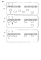
次に、工程(S5)について、それぞれ図2を用いて説明する。まず試料1にマスキング剤2及び標識イメージング剤3を投与する(図2の(a))。標識イメージング剤3は、イメージング剤4を標識物質5で標識したものである。投与は、工程(S4)において用意した医薬組成物の静脈内への注射などによって行うことができる。標識イメージング剤3及びマスキング剤2が別々に用意されている場合、一方を投与した後に連続して他方を投与してもよい。標識イメージング剤3及びマスキング剤2の両方が含まれる医薬組成物を用意した場合、それを投与すればよい。
Next, the step (S5) will be described with reference to FIG. First, the masking
投与された標的タンパク質6に対する結合親和性の度合いは、標識イメージング剤3の方がマスキング剤2よりも高いことから、標識イメージング剤3が標的タンパク質6に優先的に結合する(図2の(b))。また、非標的タンパク質7に対する結合親和性の度合いは、マスキング剤2の方が高いことから、マスキング剤2が非標的タンパク質7に優先的に結合する(図2の(b))。しかしながら、マスキング剤2は、標的タンパク質6とも親和性を有することから、標的タンパク質6にも結合し得る(図2の(b)中の矢印8)。その場合、そのマスキング剤2は、標識イメージング剤3が近づくと標的タンパク質6から解離し、そこに標識イメージング剤3が結合する(図2の(c))。また、標識イメージング剤3は、非標的タンパク質7とも親和性を有することから、非標的タンパク質7にも標識イメージング剤3が結合し得る(図2の(b)中の矢印9)。その場合、その標識イメージング剤3は、マスキング剤2が近づくと解離し、そこにマスキング剤2が結合する(図2の(c))。その後、非標的タンパク質7及び標的タンパク質6に結合していないマスキング剤2及び標識イメージング剤3は流失する(図2の(d))。このようにして、標識イメージング剤3は、非標的タンパク質7に結合することなく、標的タンパク質6に結合することができる。その結果、標的タンパク質6が標識物質5によって標識される。
Since the degree of binding affinity to the administered
例えば、マスキング剤2及び標識イメージング剤3の標的タンパク質6に対する親和性の差、即ち、前記表1に示される「A」及び「B」、又は表2に示される「a」及び「b」の差が大きいほど、標的タンパク質6にマスキング剤2が結合する可能性が低く、また標的タンパク質6に結合したマスキング剤2が標識イメージング剤3に置き換わる可能性が高くなる。例えば、表2の「a」の値は、「b」の値の10分の1以下であることが好ましい。100分の1以下であれば更に好ましい。
For example, the difference in affinity between the masking
また例えば、マスキング剤2及び標識イメージング剤3の非標的タンパク質7に対する親和性の差、即ち、表1に示される「C」及び「D」、又は表2に示される「c」及び「d」の差が大きいほど、非標的タンパク質7に標識イメージング剤3が結合する可能性が低く、また非標的タンパク質7に結合したイメージング剤3がマスキング剤2と入れ替わる可能性が高くなる。例えば、表2の「d」の値は、「c」の値の10分の1以下であることが好ましい。100分の1以下であれば更に好ましい。
Also, for example, the differences in the affinity of masking
次に、工程(S6)において、標識物質からの信号を検出し、検出の結果に基づいて画像を作成する。 Next, in step (S6), a signal from the labeling substance is detected, and an image is created based on the result of the detection.
標識物質が陽電子(ポジトロン)を放射する放射性同位元素である場合、信号は、放射性同位元素から放出された陽電子が近傍の電子と対消滅した結果として生じる、180度反対の方向に放射される2本の消滅放射線(ガンマ線)である。例えば、このような2方向に放射されたガンマ線を同時計数することによって、信号を検出し、画像化することができる。即ち、2本のガンマ線を、試料の周囲にリング状に配置された複数のガンマ線検出器によって同時に検出することによって、ガンマ線が検出されたガンマ線検出器同士を結ぶ線上に放射性同位元素が存在するというデータ、例えば、サイノグラムを得ることができる。得られたデータを画像再構成することによって、放射性同位元素の分布を断層画像として得ることができる。例えば、このような消滅放射線の検出及び画像の作成は、陽電子放出断層撮影(Positoron emmission tomography:PET)装置、PET装置とX線コンピュータ断層撮影(Computed Tomography:CT)装置とを組み合わせたPET−CT装置、又はTOF−PET(time−of−flight PET)装置などの核医学診断装置によって行うことができる。
When the labeling substance is a radioactive isotope that emits a positron (positron), the signal is emitted in the opposite direction 180 ° resulting from the annihilation of a positron emitted from the radioactive isotope with a
標識物質が、ガンマ線を放出する放射性同位元素である場合、信号は、放射性同位元素から放射されるガンマ線である。例えば、このようなガンマ線を、試料側にコリメータ、例えば、平行型コリメータを備えるガンマ線検出器によって検出することによって、ガンマ線が到達して電力パルスが出力されたガンマ線検出器の位置及び電力パルスが検出された回数などのデータを得ることができる。得られたデータから平面画像を作成することができる。ガンマ線検出器で試料の様々な方向からガンマ線を検出して平面画像を収集し、コンピュータで画像再構成することによって放射性同位元素の分布を断層画像として得ることができる。例えば、このようなガンマ線の検出及び画像の作成は、単一光子放射断層撮影(Single poton emission computed tomography:SPECT)装置、又はSPECT装置とCT装置とを組み合わせたSPECT−CT装置などの核医学診断装置を用いて行うことができる。 When the labeling substance is a radioactive isotope emitting gamma rays, the signal is gamma rays emitted from the radioactive isotope. For example, by detecting such gamma rays with a collimator on the sample side, for example, a gamma ray detector provided with a parallel collimator, the position and power pulse of the gamma ray detector where the gamma ray has arrived and the power pulse is output It is possible to obtain data such as the number of times it has been A planar image can be created from the obtained data. The distribution of radioactive isotopes can be obtained as tomographic images by detecting gamma rays from various directions of a sample with a gamma ray detector, collecting planar images, and reconstructing images with a computer. For example, such gamma ray detection and imaging can be performed by nuclear medicine diagnosis such as single photon emission computed tomography (SPECT) apparatus or SPECT-CT apparatus combining SPECT apparatus and CT apparatus. It can be done using the device.
信号の検出及び画像の作成は、試料の全体に亘って行ってもよいし、試料の一部において行ってもよい。 The detection of the signal and the creation of the image may be performed on the whole of the sample or on a part of the sample.
以上に説明したタンパク質をイメージングする方法によれば、イメージング対象外の非標的タンパク質にイメージング剤が結合するのを効率的に防止することができる。その結果、イメージング結果が偽陽性となることが防止され、標的タンパク質を高感度に検出することができる。また、実施形態の方法によれば、組織や血液を採取に耐えられない試料においても、非侵襲的に、試料全体に亘って標的タンパク質の有無又は分布を一度に検出することが可能である。 According to the method of imaging a protein described above, it is possible to efficiently prevent the imaging agent from binding to a non-target protein that is not to be imaged. As a result, the imaging result is prevented from being false positive, and the target protein can be detected with high sensitivity. In addition, according to the method of the embodiment, it is possible to non-invasively detect the presence or absence or distribution of the target protein all over the sample at once even in a sample which can not tolerate collection of tissue or blood.
標的タンパク質が特定の疾患に関連するタンパク質であり、実施形態の方法によって標的タンパク質がイメージングされた場合は、例えば、試料が特定の疾患に罹患していると診断することができる。また、イメージングされた標的タンパク質の位置や量によって、その疾患の発症している器官、転移の有無若しくは浸潤性の有無などの疾患の特性、疾患の重症度、経過及び/又は医薬品に対する効果若しくは副作用などを診断することが可能である。そのような診断は、例えば、コンパニオン診断であってもよい。また、その診断によって試料を層別化することが可能である。実施形態の方法によれば、このような診断が偽陽性となることが無く、より精度よく診断を行うことが可能である。 When the target protein is a protein associated with a specific disease, and the target protein is imaged by the method of the embodiment, for example, it can be diagnosed that the sample suffers from the specific disease. In addition, depending on the position and amount of the target protein imaged, characteristics of the diseased organ such as the presence or absence of metastasis or infiltration, disease characteristics, severity of disease, progress and / or adverse effects on pharmaceuticals or side effects depending on the location or amount of the target protein. It is possible to diagnose etc. Such a diagnosis may be, for example, a companion diagnosis. It is also possible to stratify the sample by its diagnosis. According to the method of the embodiment, such diagnosis does not become false positive, and it is possible to diagnose more accurately.
・第2の実施形態
実施形態のイメージング方法の更なる実施形態を図4に示す。図4は、第2の実施形態の方法の一例を示す概略フローチャートである。
Second Embodiment A further embodiment of the imaging method of the embodiment is shown in FIG. FIG. 4 is a schematic flowchart showing an example of the method of the second embodiment.
まず、工程(S11)においてイメージング剤を決定し、工程(S12)において非標的タンパク質を決定する。工程(S11)及び(S12)はそれぞれ、例えば、上記工程(S1)及び(S2)と同じ方法で行うことができる。 First, an imaging agent is determined in step (S11), and non-target proteins are determined in step (S12). Steps (S11) and (S12) can be performed, for example, in the same manner as steps (S1) and (S2).
次に、工程(S13)において標識イメージング剤を用意し、工程(S14)においてマスキング剤を用意する。工程(S13)及び工程(S14)はそれぞれ、例えば、上記工程(S3)及び(S4)と同じ方法で行うことができる。第2の実施形態において、標識イメージング剤及びマスキング剤は、別々の医薬組成物に含まれた形態で用意される。 Next, a labeled imaging agent is prepared in step (S13), and a masking agent is prepared in step (S14). The steps (S13) and (S14) can be performed, for example, in the same manner as the steps (S3) and (S4). In a second embodiment, the labeled imaging agent and the masking agent are provided in separate pharmaceutical composition forms.
次に、工程(S15)及び工程(S16)について図4及び図5を参照して説明する。 Next, step (S15) and step (S16) will be described with reference to FIG. 4 and FIG.
工程(S15)において、試料1にマスキング剤2を投与する(図4の(a))。投与は、例えば、マスキング剤を含む医薬組成物の静脈内への注射などによって行うことができる。投与方法は、標的タンパク質、非標的タンパク質及びマスキング剤の種類などに依存して選択される。
In step (S15), masking
投与されたマスキング剤2は、非標的タンパク質7に結合する(図4の(b))。例えば、非標的タンパク質7にマスキング剤2が結合するまで、試料を放置することが好ましい。マスキング剤2は標的タンパク質6とも結合親和性を有することから、標的タンパク質6にも結合し得る(図4の(b)中の矢印)。その後、非標的タンパク質7又は標的タンパク質6に結合していないマスキング剤2は流失する(図4(c))。このようにして、マスキング剤2によって非標的タンパク質7がマスキングされる。
The administered
次に、工程(S16)において、試料1に、イメージング剤4を標識物質5で標識した標識イメージング剤3を投与する(図5(d))。投与は、標識イメージング剤3を含む医薬組成物の静脈内への注射などによって行うことができる。投与方法は、標的タンパク質、非標的タンパク質及びマスキング剤の種類に依存して選択される。
Next, in step (S16), the labeled
投与された標識イメージング剤3は、標的タンパク質6に結合する(図5の(e))。例えば、標的タンパク質6に標識イメージング剤3が結合するまで、試料1を放置することが好ましい。標的タンパク質6にマスキング剤2が結合している場合(図5の(e)中の矢印)、標的タンパク質6に対する結合親和性の度合いは、標識イメージング剤3の方が高いことから、標的タンパク質6に結合したマスキング剤2に標識イメージング剤3が近づくと、そのマスキング剤2が解離し、そこに標識イメージング剤3が結合する(図5の(f))。その後、非標的タンパク質7及び標的タンパク質6に結合していないマスキング剤2及び標識イメージング剤3は流失する(図5の(g))。このようにして、標識イメージング剤3は、非標的タンパク質7に結合することなく、標的タンパク質6に結合することができる。その結果、標的タンパク質6が標識物質5によって標識される。
The labeled
例えば、第1の実施形態において説明したように、マスキング剤及び標識イメージング剤の標的タンパク質に対する親和性の差大きいほど、標的タンパク質6にマスキング剤2が結合する可能性が低く、また標的タンパク質6に結合したマスキング剤2が標識イメージング剤3に置き換わる可能性が高くなる。
For example, as described in the first embodiment, the larger the difference in the affinity between the masking agent and the labeled imaging agent for the target protein, the lower the possibility that the masking
次に、工程(S15)において、標識物質からの信号を検出し、検出の結果に基づいて画像を作成する。この工程は、上記工程(S6)と同じ方法で行うことができる。 Next, in step (S15), a signal from the labeling substance is detected, and an image is created based on the result of the detection. This step can be performed by the same method as the above step (S6).
以上に説明した実施形態の方法によれば、非標的タンパク質を、マスキング剤でマスキングした状態で標識イメージング剤を投与するため、標識イメージング剤が非標的タンパク質に結合する可能性がより低くなる。その結果、標的タンパク質を更に高感度に検出することができる。 According to the method of the embodiment described above, since the labeled imaging agent is administered with the non-target protein masked by the masking agent, the possibility of the labeled imaging agent binding to the non-target protein is reduced. As a result, the target protein can be detected with higher sensitivity.
・第3の実施形態
更なる実施形態において、標的タンパク質及びイメージング剤は、それぞれ複数種類であってもよい。そのような例について、図6を参照して説明する。図6は、第3の実施形態の方法の一例を示す概略フローチャートである。
Third Embodiment In a further embodiment, each of the target protein and the imaging agent may be of multiple types. Such an example will be described with reference to FIG. FIG. 6 is a schematic flowchart showing an example of the method of the third embodiment.
この例の場合、実施形態のイメージング方法は、試料中の第1〜第nの標的タンパク質をイメージングする方法であり、図6に示す工程(S21)〜(S26)を含む。ここで、nは2以上の整数である。 In the case of this example, the imaging method of the embodiment is a method of imaging the first to nth target proteins in a sample, and includes steps (S21) to (S26) shown in FIG. Here, n is an integer of 2 or more.
まず、工程(S21)において、第1〜第nの標的タンパク質それぞれと結合親和性を有する第1〜第nのイメージング剤をそれぞれ決定する。この工程は、例えば、第1〜第nの標的タンパク質に対して、上記工程(S1)と同じ方法で、それぞれの標的タンパク質に対応する第1〜第nのイメージング剤を決定することにより行うことができる。1〜nの何れかの値をtとすると、第tの標的タンパク質に対する第tのイメージング剤の親和性は、第tの標的タンパク質に対する第t以外のイメージング剤の親和性の何れよりも高い。 First, in step (S21), first to nth imaging agents having binding affinity with each of the first to nth target proteins are determined. This step is performed, for example, by determining the first to nth imaging agents corresponding to the respective target proteins in the same manner as the above step (S1) for the first to nth target proteins. Can. Assuming that the value of any of 1 to n is t, the affinity of the t-th imaging agent for the t-th target protein is higher than any of the imaging agents other than the t-th target protein for the t-th target protein.
次に、工程(S22)において、第1〜第nのイメージング剤と結合親和性を有する非標的タンパク質を決定する。この工程は、例えば、上記工程(S2)と同じ方法によって第1〜第nのイメージング剤と親和性を有する非標的タンパク質を決定することができる。非標的タンパク質は、第1〜第nのイメージング剤の何れかと親和性を有するタンパク質であってもよいし、或いは、第1〜第nのイメージング剤の全てと結合親和性を有するタンパク質であってもよい。 Next, in step (S22), non-target proteins having binding affinity with the first to n-th imaging agents are determined. In this step, for example, non-target proteins having affinity to the first to n-th imaging agents can be determined by the same method as the above step (S2). The non-target protein may be a protein having an affinity for any of the first to n imaging agents, or a protein having a binding affinity for all of the first to n imaging agents, It is also good.
次に、工程(S23)において、第1〜第nの標識イメージング剤を用意する。第1〜第nの標識イメージング剤は、それぞれ第1〜第nの標識物質により標識された第1〜第nのイメージング剤である。第1〜第nのイメージング剤の第1〜第nの標識物質による標識はそれぞれ、例えば、上記(S3)と同じ方法によって行うことができる。 Next, in step (S23), first to nth labeled imaging agents are prepared. The 1st to n-th labeling imaging agents are the 1st to n-th imaging agents labeled by the 1st to n-th labeling substances, respectively. The labeling with the first to nth labeling substances of the first to nth imaging agents can be performed, for example, by the same method as the above (S3).
第1〜第nの標識物質は互いにエネルギーが異なる標識物質である。それによって、複数種類の標的タンパク質を区別して画像化することができる。しかしながら、第1〜第nの標識物質は全てが陽電子を放出する放射性同位元素であるか、又は全てがガンマ線を放射する放射性同位元素であることが好ましい。それによって一度に同じ装置を用いて標的タンパク質を画像化できる。 The first to nth labeling substances are labeling substances having different energies. Thereby, multiple types of target proteins can be distinguished and imaged. However, it is preferable that all of the first to nth labeling substances be radioactive isotopes emitting positrons, or all be radioactive isotopes emitting gamma rays. Thereby the target protein can be imaged using the same device at one time.
工程(S24)において、マスキング剤を用意する。マスキング剤は、第1〜第nの標的タンパク質及び非標的タンパク質と結合親和性を有する。そして、マスキング剤と第1〜第nの標的タンパク質との結合親和性はそれぞれ、第1〜第nのイメージング剤と第1〜第nの標的タンパク質とのそれぞれの親和性の何れよりも低い。また、マスキング剤と非標的タンパク質との結合親和性は、第1〜第nのイメージング剤と非標的タンパク質との親和性の何れよりも高い。 In the step (S24), a masking agent is prepared. The masking agent has binding affinity to the first to nth target proteins and non-target proteins. And, the binding affinity between the masking agent and the first to nth target proteins is lower than any of the respective affinity between the first to nth imaging agent and the first to nth target proteins. In addition, the binding affinity between the masking agent and the non-target protein is higher than any one of the first to nth imaging agents and the non-target protein.
例えば、2種類の標的タンパク質及び1種類の非標的タンパク質、並びに2種類のイメージング剤及び1種類のマスキング剤を用いる場合の各物質の結合親和性の関係について説明する。例えば、表3に示すように、第1の標的タンパク質と、第1のイメージング剤、第2のイメージング剤及びマスキング剤との結合親和性の度合いをそれぞれ「E」、「F」、「G」とし、第2の標的タンパク質と、第1のイメージング剤、第2のイメージング剤及びマスキング剤との結合親和性の度合いをそれぞれ「H」、「I」、「J」とし、非標的タンパク質と、第1のイメージング剤、第2のイメージング剤及びマスキング剤との結合親和性の度合いをそれぞれ「K」、「L」、「M」とすると、「E>F,G」、「I>H,J」、「M>K,L」の式が成り立つ。 For example, the relationship between the binding affinity of each substance when using two types of target proteins and one type of non-target protein, and two types of imaging agents and one type of masking agent will be described. For example, as shown in Table 3, the degree of binding affinity between the first target protein, the first imaging agent, the second imaging agent and the masking agent is “E”, “F”, “G”, respectively. The degree of binding affinity between the second target protein and the first imaging agent, the second imaging agent and the masking agent is “H”, “I” and “J”, respectively, and the non-target protein, Assuming that the degrees of binding affinity with the first imaging agent, the second imaging agent and the masking agent are “K”, “L” and “M”, respectively, “E> F, G”, “I> H, The expressions J "and" M> K, L "hold.
第1〜第nの標識イメージング剤及びマスキング剤は、後述する工程(S25)で投与されるのに適切な医薬組成物に含まれた形態で用意されることが好ましい。第1〜第nの標識イメージング剤は、全て別々の医薬組成物に含まれていてもよいし、全てが一つの医薬組成物に含まれていてもよいし、何れかの組み合わせで複数の医薬組成物に含まれていてもよい。 The first to nth labeled imaging agents and masking agents are preferably provided in the form contained in a pharmaceutical composition suitable for administration in step (S25) described later. The first to nth labeled imaging agents may be all contained in separate pharmaceutical compositions, or all may be contained in one pharmaceutical composition, or a plurality of drugs in any combination. It may be included in the composition.
工程(S25)において、第1〜第nの標識イメージング剤及びマスキング剤を投与する。第1〜第nの標識イメージング剤が別々の医薬組成物に含まれている場合は、それらの医薬組成物を同時に投与するか又は間隔を空けずに連続して投与してもよい。全ての標識イメージング剤が一つの医薬組成物に含まれている場合、その医薬組成物を投与すればよい。例えば、第1〜第nの標識イメージング剤及びマスキング剤を含む医薬組成物を調製し、それを投与してもよい。投与は、例えば、上記工程(S5)と同じ方法で行うことができる。或いは、マスキング剤を投与した後に、第1〜第nの標識イメージング剤を投与してもよい。 In step (S25), the first to nth labeled imaging agents and masking agents are administered. When the first to nth labeled imaging agents are contained in separate pharmaceutical compositions, those pharmaceutical compositions may be administered simultaneously or sequentially without intervals. When all the labeled imaging agents are contained in one pharmaceutical composition, the pharmaceutical composition may be administered. For example, a pharmaceutical composition comprising the first to nth labeled imaging agents and a masking agent may be prepared and administered. The administration can be performed, for example, in the same manner as the step (S5). Alternatively, after the masking agent is administered, the first to nth labeled imaging agents may be administered.
工程(S26)において、第1〜第nの標識物質からの信号を検出し、検出の結果に基づいて画像を作成する。この工程は、例えば、工程(S6)と同じ方法によって、第1〜第nの標識物質からの信号をそれぞれ検出し、画像を作成することによって行うことができる。この例においては、第1〜第nの標識物質からの信号のエネルギーが異なるため、信号を区別することができる。エネルギーの異なる標識物質から得られた信号それぞれにおいて別々の画像を作成してもよいし、各信号によって得られたデータを統合した画像を作成してもよい。統合する場合は、信号毎に別の色を用いて画像化することによって第1〜第nの標識物質を区別して画像化することができる。 In step (S26), signals from the first to n-th labeling substances are detected, and an image is created based on the detection result. This step can be performed, for example, by detecting the signals from the first to n-th labeling substances and creating an image by the same method as step (S6). In this example, the signals can be distinguished because the energy of the signals from the first to n-th labeling substances are different. A separate image may be created for each of the signals obtained from different labels of energy, or an image may be created that integrates the data obtained with each signal. In the case of integration, it is possible to distinguish and image the first to nth labeling substances by imaging using different colors for each signal.
以上に説明した第3の実施形態の方法によれば、複数種類の標識物質を一度に、区別してイメージングすることができる。 According to the method of the third embodiment described above, it is possible to distinguish and image multiple kinds of labeling substances at one time.
・第4の実施形態
更なる実施形態において、非標的タンパク質及びマスキング剤は、それぞれ複数種類であってもよい。そのような例について、図7を参照して説明する。図7は、第4の実施形態の方法の一例を示す概略フローチャートである。
Fourth Embodiment In a further embodiment, the non-target protein and the masking agent may each be of multiple types. Such an example will be described with reference to FIG. FIG. 7 is a schematic flowchart showing an example of the method of the fourth embodiment.
この例の場合、実施形態のイメージング方法は、試料中の標的タンパク質をイメージングする方法であり、図7に示す工程(S31)〜(S36)を含む。 In the case of this example, the imaging method of the embodiment is a method of imaging a target protein in a sample, and includes steps (S31) to (S36) shown in FIG.
まず、工程(S31)において、標的タンパク質と結合親和性を有するイメージング剤を決定する。この工程は、例えば、上記工程(S1)とおなじ方法によって行うことができる。 First, in step (S31), an imaging agent having binding affinity to a target protein is determined. This step can be performed, for example, by the same method as the above step (S1).
次に、工程(S32)において、イメージング剤と結合親和性を有する第1〜第nの非標的タンパク質を決定する。この工程は、例えば、上記工程(S2)と同じ方法で、イメージング剤と結合親和性を有する・BR>^ンパク質を複数種類選択することによって、行うことができる。 Next, in step (S32), first to nth non-target proteins having binding affinity with the imaging agent are determined. This step can be performed, for example, by selecting a plurality of kinds of proteins having a binding affinity with the imaging agent in the same manner as the step (S2).
工程(S33)において、イメージング剤を標識物質で標識した標識イメージング剤を用意する。標識イメージング剤は、例えば、上記工程(S3)と同じ方法で用意することができる。 In the step (S33), a labeled imaging agent in which the imaging agent is labeled with a labeling substance is prepared. The labeled imaging agent can be prepared, for example, by the same method as the step (S3).
工程(S34)において、第1〜第nのマスキング剤を用意する。第1〜第nのマスキング剤はそれぞれ、標的タンパク質及び第1〜第nの非標的タンパク質と結合親和性をそれぞれ有する。即ち、1〜nの何れかの値をpとすると、第pのマスキング剤は、標的タンパク質及び対応する第pの非標的タンパク質と結合親和性を有する。そして、第pの非標的タンパク質に対する第pのマスキング剤の親和性は、第pの非標的タンパク質に対する第p以外のイメージング剤の親和性の何れよりも高い。 In the step (S34), first to nth masking agents are prepared. The first to nth masking agents have binding affinity to the target protein and the 1st to nth non-target protein, respectively. That is, assuming that any value of 1 to n is p, the p-th masking agent has binding affinity with the target protein and the corresponding p-th non-target protein. And, the affinity of the p-th masking agent for the p-th non-target protein is higher than any affinity of imaging agents other than the p-th non-target protein for the p-th non-target protein.
また、標的タンパク質と第1〜第nのマスキング剤との結合親和性は、標的タンパク質とイメージング剤との結合親和性よりもそれぞれ低く、第1〜第nの非標的タンパク質と、対応する第1〜第nのマスキング剤との結合親和性はそれぞれ、第1〜第nの非標的タンパク質とイメージング剤との親和性よりもそれぞれ高い。 In addition, the binding affinity between the target protein and the first to nth masking agents is lower than the binding affinity between the target protein and the imaging agent, respectively, and the first to nth non-target proteins and the corresponding first The binding affinities with the to n-th masking agents are respectively higher than the affinities of the first to nth non-target proteins and the imaging agent.
例えば、1種類の標的タンパク質及び2種類の非標的タンパク質、並びに1種類のイメージング剤及び2種類のマスキング剤を用いる場合の各物質の結合親和性について説明する。例えば、表4に示すように、標的タンパク質と、イメージング剤、第1のマスキング剤及び第2のマスキング剤との結合親和性の度合いをそれぞれ「O」、「P」、「Q」とし、第1の非標的タンパク質と、イメージング剤、第1のマスキング剤及び第2のマスキング剤との結合親和性の度合いをそれぞれ「R」、「S」、「T」とし、第2の非標的タンパク質と、イメージング剤、第1のマスキング剤及び第2のマスキング剤との結合親和性の度合いをそれぞれ「U」、「V」、「W」とすると、「O>P,Q」、「S>R,T」、「W>U,V」の式が成り立つ。 For example, the binding affinity of each substance when one type of target protein and two types of non-target proteins, and one type of imaging agent and two types of masking agent are used will be described. For example, as shown in Table 4, the degree of binding affinity between the target protein and the imaging agent, the first masking agent and the second masking agent is “O”, “P”, “Q”, respectively. Let “R”, “S” and “T” be the degrees of binding affinity between the non-target protein of 1 and the imaging agent, the first masking agent and the second masking agent, respectively, and the second non-target protein and When the degree of binding affinity with the imaging agent, the first masking agent and the second masking agent is “U”, “V”, “W”, respectively, “O> P, Q”, “S> R , T "and" W> U, V "hold.
標識イメージング剤及び第1〜第nのマスキング剤は、後述する工程(S35)で投与されるのに適切な、医薬組成物に含まれた形態で用意されることが好ましい。第1〜第nのマスキング剤は、全て別々の医薬組成物に含まれていてもよいし、全てが一つの医薬組成物に含まれていてもよいし、何れかの組み合わせで複数の医薬組成物に含まれていてもよい。 The labeled imaging agent and the first to nth masking agents are preferably provided in a form included in a pharmaceutical composition suitable for administration in step (S35) described later. The first to nth masking agents may be all contained in separate pharmaceutical compositions, or all may be contained in one pharmaceutical composition, or a plurality of pharmaceutical compositions in any combination. It may be included in the thing.
工程(S35)において、試料に第1〜第nのマスキング剤及び標識イメージング剤を投与する。第1〜第nのマスキング剤が別々の医薬組成物に含まれている場合は、それらの医薬組成物を同時に投与するか又は間隔を空けずに連続して投与してもよい。全てのマスキング剤が一つの医薬組成物に含まれている場合、その医薬組成物を投与すればよい。例えば、標識イメージング剤及び第1〜第nのマスキング剤を含む医薬組成物を調製し、それを投与してもよい。この工程は、例えば、上記工程(S5)と同じ方法で行うことができる。或いは、第1〜第nのマスキング剤を投与した後に、標識イメージング剤を投与してもよい。 In step (S35), the first to nth masking agents and a labeled imaging agent are administered to the sample. When the first to nth masking agents are contained in separate pharmaceutical compositions, those pharmaceutical compositions may be administered simultaneously or sequentially without intervals. When all the masking agents are contained in one pharmaceutical composition, the pharmaceutical composition may be administered. For example, a pharmaceutical composition comprising a labeled imaging agent and first to nth masking agents may be prepared and administered. This step can be performed, for example, in the same manner as the above step (S5). Alternatively, the labeled imaging agent may be administered after the administration of the first to nth masking agents.
次に、工程(S36)において、標識物質からの信号を検出し、検出の結果に基づいて画像を作成する。この工程は例えば、上記工程(S6)と同じ方法によって行うことができる。 Next, in step (S36), the signal from the labeling substance is detected, and an image is created based on the result of the detection. This step can be performed, for example, by the same method as the above step (S6).
以上に説明した第4の実施形態によれば、複数種類の非標的タンパク質を同時に効率よくマスキングすることができ、検出の精度がより向上する。非標的タンパク質をより多く選択すれば、より標的タンパク質のイメージングの感度が向上する。 According to the fourth embodiment described above, a plurality of types of non-target proteins can be masked simultaneously and efficiently, and the detection accuracy is further improved. Selecting more non-target proteins improves the sensitivity of target protein imaging.
或いは、標的タンパク質及びイメージング剤が複数種類であり、かつ非標的タンパク質及びマスキング剤が複数種類であってもよい。 Alternatively, the target protein and the imaging agent may be of multiple types, and the non-target protein and the masking agent may be of multiple types.
・第5の実施形態
ある実施形態において、標的タンパク質は変異型EGFRのT790M変異型(以下、「変異型EGFR/T790M」と称する)であり、非標的タンパク質は変異型EGFRであり、イメージング剤はオシメルチニブ(Osimertinib)であり、マスキング剤はアファチニブ(Afatinib)である。そのような例について、図8を参照して説明する。図8は、第5の実施形態の方法の一例を示す概略フローチャートである。この例における方法は、試料中の変異型EGFR/T790Mをイメージングする方法であり、図8に示す工程(S41)〜(S44)を含む。
Fifth Embodiment In one embodiment, the target protein is a T790M mutant of mutant EGFR (hereinafter referred to as “mutated EGFR / T790M”), the non-target protein is a mutant EGFR, and the imaging agent is Osimertinib (Osimertinib) and the masking agent is afatinib (Afatinib). Such an example will be described with reference to FIG. FIG. 8 is a schematic flowchart showing an example of the method of the fifth embodiment. The method in this example is a method of imaging mutant EGFR / T790M in a sample, and includes steps (S41) to (S44) shown in FIG.
まず、工程(S41)において、標識オシメルチニブを用意する。オシメルチニブは、下記の化学式であらわされる化合物である。 First, in the step (S41), labeled osimerinib is prepared. Osimertinib is a compound represented by the following chemical formula.
標識オシメルチニブは、標識物質で標識されている。標識物質は、例えば、上述の何れかの陽電子を放射する放射性同位元素であることが好ましい。このような標識物質で標識された標識オシメルチニブの一例の化学式を以下に示す。 Labeled osimertinib is labeled with a labeling substance. The labeling substance is preferably, for example, a radioactive isotope that emits any of the positrons described above. The chemical formula of an example of labeled osimerinib labeled with such a labeling substance is shown below.
この例では、1つの炭素を放射性同位元素の11Cに置き換えることで標識したオシメルチニブを示した。放射性同位元素で置き換えられる炭素の位置は、上記の化学式に示される位置でなくてもよい。また、複数の炭素が11Cに置き換えられていてもよい。また、何れかの窒素が13Nであってもよいし、何れかの酸素が15Oであってもよい。 In this example, we have shown oximertinib labeled by replacing one carbon with the radioactive isotope 11 C. The position of carbon replaced with a radioactive isotope may not be the position shown in the above chemical formula. Also, multiple carbons may be replaced with 11 C. Also, any nitrogen may be 13 N, or any oxygen may be 15 O.
工程(S42)において、アファチニブを用意する。アファチニブは、下記の化学式であらわされる化合物である。 In the step (S42), afatinib is prepared. Afatinib is a compound represented by the following chemical formula.
変異型EGFR及び変異型EGFR/T790Mのアファチニブ及びオシメルチニブとの結合のIC50値は、例えば、表5に示すとおりである。 The IC 50 values of the binding of mutant EGFR and mutant EGFR / T 790 M to afatinib and osimerinib are, for example, as shown in Table 5.
表1に示されるように、オシメルチニブ及びアファチニブはそれぞれ、変異型EGFR/T790M及び変異型EGFRへの結合親和性をそれぞれ有する。そして、アファチニブの変異型EGFR/T790Mに対する結合親和性は、オシメルチニブにおけるよりも低く、アファチニブの変異型EGFRに対する結合親和性は、オシメルチニブにおけるよりも高い。 As shown in Table 1, osimerinib and afatinib have binding affinity to mutant EGFR / T790M and mutant EGFR, respectively. And, the binding affinity of afatinib for mutant EGFR / T 790 M is lower than that of osimerinib, and the binding affinity of afatinib for mutant EGFR is higher than that of osimerinib.
標識オシメルチニブ及びアファチニブは、例えば、工程(S42)において試料に投与されるのに適切な形態で用意されることが好ましい。例えば、標識オシメルチニブ及びアファチニブは、1つの医薬組成物に含まれた形態で用意される。又は標識オシメルチニブ及びアファチニブは、別々の医薬組成物に含まれた形態で用意される。医薬組成物は、例えば、薬学的に許容され得る担体、希釈剤及び賦形剤などを含む。医薬組成物は、例えば、PET用製剤であってもよい。 It is preferred that the labeled ocimerinib and afatinib be provided, for example, in a form suitable for being administered to the sample in step (S42). For example, labeled ocimertinib and afatinib are provided in a form contained in one pharmaceutical composition. Alternatively, labeled ocimerinib and afatinib are provided in separate pharmaceutical compositions. The pharmaceutical composition contains, for example, a pharmaceutically acceptable carrier, a diluent, an excipient and the like. The pharmaceutical composition may be, for example, a formulation for PET.
次に、工程(S43)において、標識オシメルチニブ及びアファチニブを試料に投与する。例えば、標識オシメルチニブ及びアファチニブを含む医薬組成物を投与してもよいし、別々に用意された標識オシメルチニブ及びアファチニブを連続して投与してもよいが、アファチニブを投与した後、標識オシメルチニブを投与することが好ましい。そのような例について、図9及び図10を用いて説明する。 Next, in step (S43), labeled ocimerinib and afatinib are administered to the sample. For example, although a pharmaceutical composition containing labeled osimertinib and afatinib may be administered, or labeled osimartinib and afatinib separately prepared may be administered sequentially, after administration of afatinib, the labeled osimertinib is administered. Is preferred. Such an example will be described using FIG. 9 and FIG.
まず、試料11にアファチニブ12を投与する(図9の(a))。投与は、例えば、静脈内注射によって行われる。投与されたアファチニブ12は、変異型EGFR13に結合する(図9の(b))。例えば、変異型EGFR13にアファチニブ12が結合するまで、試料を放置することが好ましい。アファチニブ12は変異型EGFR/T790M14とも結合親和性を有することから、変異型EGFR/T790M14にも結合し得る(図9の(b)中の矢印)。その後、変異型EGFR13又は変異型EGFR/T790M14に結合していないアファチニブ12は、流失する(図9の(c))。このようにして、アファチニブ12によって変異型EGFR13がマスキングされる。
First,
次に、試料11に標識オシメルチニブ17を投与する(図10の(d))。標識オシメルチニブ17は、11C16によって標識されたオシメルチニブである。投与は、例えば、静脈内注射によって行われる。投与された標識オシメルチニブ17は、変異型EGFR13に結合する(図10の(e))。変異型EGFR/T790M14とアファチニブ12が結合している場合(図10の(e)中の矢印)、そのアファチニブ12は、標識オシメルチニブ17に置き換わり、変異型EGFR/T790M14に標識オシメルチニブ17が結合する(図10の(f))。その後、変異型EGFR13及び変異型EGFR/T790M14に結合していないアファチニブ12及び標識オシメルチニブ17は、流失する(図10の(g))。このようにして、標識オシメルチニブ17は、変異型EGFR13に結合することなく、変異型EGFR/T790M14に結合することができる。その結果、変異型EGFR/T790M14が11C16によって標識される。
Next, labeled
次に、工程(S44)において、標識物質からの信号を検出し、画像を作成する。この例の場合、信号は、標識物質である放射性同位元素から放出された陽電子と、近傍の電子とが対消滅して生じる2本のガンマ線である。ガンマ線の検出及び画像の作成は、例えば、上記工程(S6)と同じ方法によって、例えば、PET装置などを用いて行うことができる。 Next, in step (S44), the signal from the labeling substance is detected to create an image. In this example, the signal is two gamma rays generated by pair annihilation of a positron emitted from a radioactive isotope which is a labeling substance and a nearby electron. The detection of gamma rays and the creation of an image can be performed, for example, by the same method as the above step (S6), for example, using a PET apparatus or the like.
以上に説明した第5の実施形態の方法によれば、変異型EGFRがアファチニブによってマスキングされ、変異型EGFRにオシメルチニブが結合することが防止される。その結果、変異型EGFR/T790Mを変異型EGFRと区別して正確にイメージングすることができる。この方法によれば、組織や血液の採取に耐えられない試料であっても、試料全体における変異型EGFR/T790Mの有無又は分布を非侵襲的に検出することが可能である。 According to the method of the fifth embodiment described above, the mutant EGFR is masked by afatinib, and the binding of osimerinib to the mutant EGFR is prevented. As a result, mutant EGFR / T790M can be distinguished from mutant EGFR and correctly imaged. According to this method, it is possible to non-invasively detect the presence or absence or distribution of the mutant EGFR / T790M in the whole sample even if it is a sample that can not tolerate collection of tissue or blood.
実施形態の方法によって変異型EGFR/T790Mがイメージングされた場合は、例えば、試料が変異型EGFR/T790M陽性肺癌に罹患していると診断することができる。また、イメージングされた変異型EGFR/T790Mの位置や量によって、変異型EGFR/T790Mが発症している器官、原発巣及び転移巣における変異型EGFR/T790Mの有無、肺癌の進行度及び重症度、経過及び/又は医薬品に対する効果などを診断することが可能である。実施形態の方法によれば、変異型EGFR/T790Mの診断が偽陽性となることが無く、より精度よく変異型EGFR/T790Mの診断を行うことが可能である。 When mutant EGFR / T790M is imaged by the method of the embodiment, for example, it can be diagnosed that the sample suffers from mutant EGFR / T790M positive lung cancer. In addition, depending on the position and amount of the mutant EGFR / T790M imaged, presence or absence of the mutant EGFR / T790M in the organ where the mutant EGFR / T790M has developed, in the primary focus and in the metastatic focus, the progression and severity of lung cancer, It is possible to diagnose the progress and / or the effect on the drug and the like. According to the method of the embodiment, it is possible to diagnose mutant EGFR / T790M more accurately without false positive diagnosis of mutant EGFR / T790M.
1…試料 2…マスキング剤 3…標識イメージング剤 4…イメージング剤
5…標識物質 6…標的タンパク質 7…非標的タンパク質
11…試料 12…アファチニブ 13…変異型EGFR
14…変異型EGFR/T790M 15…オシメルチニブ
16…11C 17…標識オシメルチニブ
1 ...
14: Mutant EGFR / T 790 M 15: Ocimurtinib 16: 11 C 17: Labeled Ocimertinib
Claims (8)
(S1)前記標的タンパク質と結合親和性を有するイメージング剤を決定すること、
(S2)前記イメージング剤と結合親和性を有する非標的タンパク質を決定すること、
(S3)前記イメージング剤を標識物質で標識した標識イメージング剤を用意すること、
(S4)前記標的タンパク質及び前記非標的タンパク質と結合親和性を有し、前記標的タンパク質との結合親和性が、前記イメージング剤のそれよりも低く、前記非標的タンパク質との結合親和性が、前記イメージング剤のそれよりも高いマスキング剤を用意すること、
(S5)前記試料に、前記マスキング剤及び前記標識イメージング剤を投与すること、及び
(S6)前記標識物質からの信号を検出し、前記検出の結果に基づいて画像を作成すること
を含むタンパク質をイメージングする方法。 A method of imaging a target protein in a sample, comprising
(S1) determining an imaging agent having binding affinity to the target protein,
(S2) determining a non-target protein having binding affinity to the imaging agent,
(S3) Providing a labeled imaging agent in which the imaging agent is labeled with a labeling substance,
(S4) A binding affinity to the target protein and the non-target protein, the binding affinity to the target protein is lower than that of the imaging agent, and a binding affinity to the non-target protein is the above Providing a masking agent higher than that of the imaging agent,
(S5) administration of the masking agent and the labeling imaging agent to the sample, and (S6) detecting a signal from the labeling substance, and producing an image based on the detection result How to image.
(S21)前記第1〜第nの標的タンパク質とそれぞれ結合親和性を有する第1〜第nのイメージング剤をそれぞれ決定すること、
(S22)前記第1〜第nのイメージング剤とそれぞれ結合親和性を有する非標的タンパク質を決定すること、
(S23)前記第1〜第nのイメージング剤を第1〜第nの標識物質で標識した第1〜第nの標識イメージング剤を用意すること、
(S24)前記第1〜第nの標的タンパク質及び前記非標的タンパク質と結合親和性を有し、前記第1〜第nの標的タンパク質との結合親和性がそれぞれ、前記第1〜第nのイメージング剤のそれよりも低く、前記非標的タンパク質との結合親和性が、前記第1〜第nのイメージング剤のそれよりも高いマスキング剤を用意すること、
(S25)前記試料に、前記マスキング剤及び前記第1〜第nの標識イメージング剤を投与すること、及び
(S26)前記第1〜第nの標識物質からの信号を検出し、前記検出の結果に基づいて画像を作成すること
を含むタンパク質をイメージングする方法。 A method for imaging the first to nth target proteins in a sample, wherein n is an integer of 2 or more,
(S21) determining first to nth imaging agents having binding affinity to the first to nth target proteins,
(S22) determining a non-target protein having binding affinity to each of the first to nth imaging agents,
(S23) preparing the first to n-th labeled imaging agents in which the first to n-th imaging agents are labeled with the first to n-th labeling substances;
(S24) The binding affinity with the first to nth target proteins and the non-target protein, and the binding affinity to the first to nth target proteins is respectively the first to nth imaging Providing a masking agent which is lower than that of the agent and whose binding affinity to the non-target protein is higher than that of the first to nth imaging agents,
(S25) administering the masking agent and the first to nth labeled imaging agents to the sample, and (S26) detecting a signal from the first to nth labeled substances, and detecting the result A method of imaging a protein comprising creating an image based on:
(S31)前記標的タンパク質とそれぞれ結合親和性を有するイメージング剤を決定すること、
(S32)前記イメージング剤とそれぞれ結合親和性を有する第1〜第nの非標的タンパク質を決定すること、
(S33)前記イメージング剤を標識物質で標識した標識イメージング剤を用意すること、
(S34)前記標的タンパク質及び前記第1〜第nの非標的タンパク質と結合親和性を有し、前記標的タンパク質との結合親和性が、前記イメージング剤のそれよりもそれぞれ低く、前記第1〜第nの非標的タンパク質との結合親和性がそれぞれ、前記イメージング剤のそれよりもそれぞれ高い第1〜第nのマスキング剤を用意すること、
(S35)前記試料に、前記第1〜第nのマスキング剤及び前記標識イメージング剤を投与すること、及び
(S36)前記標識物質からの信号を検出し、前記検出の結果に基づいて画像を作成すること
を含むタンパク質をイメージングする方法。 A method of imaging a target protein in a sample, comprising
(S31) determining an imaging agent having binding affinity to the target protein,
(S32) determining first to nth non-target proteins each having binding affinity with the imaging agent,
(S33) Providing a labeled imaging agent in which the imaging agent is labeled with a labeling substance,
(S34) The protein has binding affinity to the target protein and the first to nth non-target proteins, and the binding affinity to the target protein is lower than that of the imaging agent. providing first to nth masking agents, each of which has a binding affinity to n non-target proteins higher than that of the imaging agent;
(S35) administering the first to nth masking agents and the labeled imaging agent to the sample, and (S36) detecting a signal from the labeled substance, and creating an image based on the result of the detection A method of imaging a protein that involves doing.
(S41)オシメルチニブを標識物質で標識した標識オシメルチニブを用意すること、
(S42)アファチニブを用意すること、
(S43)前記試料に、前記アファチニブ及び前記標識オシメルチニブを投与すること、及び
(S44)前記標識物質からの信号を検出し、前記検出の結果に基づいて画像を作成すること
を含むタンパク質をイメージングする方法。 A method of imaging a T790M mutant of mutant EGFR (hereinafter, referred to as "a mutant EGFR / T790M") in a sample,
(S41) Providing a labeled osimeltinib labeled with osimatinib with a labeling substance,
(S42) preparing afatinib,
(S43) A protein comprising: administering to the sample the afatinib and the labeled osimertinib; and (S44) detecting a signal from the labeled substance and generating an image based on the result of the detection Method.
Priority Applications (1)
| Application Number | Priority Date | Filing Date | Title |
|---|---|---|---|
| JP2017234181A JP7262922B2 (en) | 2017-12-06 | 2017-12-06 | Methods for Imaging Proteins |
Applications Claiming Priority (1)
| Application Number | Priority Date | Filing Date | Title |
|---|---|---|---|
| JP2017234181A JP7262922B2 (en) | 2017-12-06 | 2017-12-06 | Methods for Imaging Proteins |
Publications (2)
| Publication Number | Publication Date |
|---|---|
| JP2019100945A true JP2019100945A (en) | 2019-06-24 |
| JP7262922B2 JP7262922B2 (en) | 2023-04-24 |
Family
ID=66976782
Family Applications (1)
| Application Number | Title | Priority Date | Filing Date |
|---|---|---|---|
| JP2017234181A Active JP7262922B2 (en) | 2017-12-06 | 2017-12-06 | Methods for Imaging Proteins |
Country Status (1)
| Country | Link |
|---|---|
| JP (1) | JP7262922B2 (en) |
Citations (6)
| Publication number | Priority date | Publication date | Assignee | Title |
|---|---|---|---|---|
| JP2005017133A (en) * | 2003-06-26 | 2005-01-20 | Kagoshima Univ | Antigen detecting method using immunohistochemical staining method |
| US20120294803A1 (en) * | 2010-01-27 | 2012-11-22 | Emory University | Cxcr4 antagonists for imaging of cancer and inflammatory disorders |
| WO2015146896A1 (en) * | 2014-03-24 | 2015-10-01 | コニカミノルタ株式会社 | Biological-material quantitation method based on multiple-antigen immunostaining |
| WO2016166073A1 (en) * | 2015-04-13 | 2016-10-20 | Ventana Medical Systems, Inc. | Thermochemical-based antibody inactivation methods and systems |
| US20170160171A1 (en) * | 2015-11-20 | 2017-06-08 | Oregon Health And Science University | Multiplex immunohistochemistry image cytometry |
| WO2017148925A1 (en) * | 2016-02-29 | 2017-09-08 | Oncodesign Sa | Radiolabeled macrocyclic egfr inhibitor |
-
2017
- 2017-12-06 JP JP2017234181A patent/JP7262922B2/en active Active
Patent Citations (6)
| Publication number | Priority date | Publication date | Assignee | Title |
|---|---|---|---|---|
| JP2005017133A (en) * | 2003-06-26 | 2005-01-20 | Kagoshima Univ | Antigen detecting method using immunohistochemical staining method |
| US20120294803A1 (en) * | 2010-01-27 | 2012-11-22 | Emory University | Cxcr4 antagonists for imaging of cancer and inflammatory disorders |
| WO2015146896A1 (en) * | 2014-03-24 | 2015-10-01 | コニカミノルタ株式会社 | Biological-material quantitation method based on multiple-antigen immunostaining |
| WO2016166073A1 (en) * | 2015-04-13 | 2016-10-20 | Ventana Medical Systems, Inc. | Thermochemical-based antibody inactivation methods and systems |
| US20170160171A1 (en) * | 2015-11-20 | 2017-06-08 | Oregon Health And Science University | Multiplex immunohistochemistry image cytometry |
| WO2017148925A1 (en) * | 2016-02-29 | 2017-09-08 | Oncodesign Sa | Radiolabeled macrocyclic egfr inhibitor |
Non-Patent Citations (1)
| Title |
|---|
| HIRANO TOSHIYUKI ET AL.: "In vitro modelingto determine mutation specificity of EGFR tyrosine kinase inhibitors againstclinica", ONCOTARGET, vol. 6, no. 36, JPN6021037897, 15 October 2015 (2015-10-15), pages 38789 - 38803, ISSN: 0004600602 * |
Also Published As
| Publication number | Publication date |
|---|---|
| JP7262922B2 (en) | 2023-04-24 |
Similar Documents
| Publication | Publication Date | Title |
|---|---|---|
| Evans et al. | Prostate cancer–specific PET radiotracers: A review on the clinical utility in recurrent disease | |
| Fendler et al. | 68 Ga-PSMA PET/CT: Joint EANM and SNMMI procedure guideline for prostate cancer imaging: version 1.0 | |
| Afshar-Oromieh et al. | Radiation dosimetry of 68 Ga-PSMA-11 (HBED-CC) and preliminary evaluation of optimal imaging timing | |
| Yankeelov et al. | Quantitative multimodality imaging in cancer research and therapy | |
| Nowosielski et al. | An intra-individual comparison of MRI,[18F]-FET and [18F]-FLT PET in patients with high-grade gliomas | |
| Franc et al. | Small-animal SPECT and SPECT/CT: important tools for preclinical investigation | |
| Wahl et al. | The promise and pitfalls of positron emission tomography and single-photon emission computed tomography molecular imaging–guided radiation therapy | |
| Thorek et al. | Positron lymphography: multimodal, high-resolution, dynamic mapping and resection of lymph nodes after intradermal injection of 18F-FDG | |
| Ordonez et al. | Radioiodinated DPA-713 imaging correlates with bactericidal activity of tuberculosis treatments in mice | |
| Soodgupta et al. | Very late antigen-4 (α4β1 integrin) targeted PET imaging of multiple myeloma | |
| Watanabe et al. | Biodistribution and radiation dosimetry of the novel hypoxia PET probe [18 F] DiFA and comparison with [18 F] FMISO | |
| US20150185339A1 (en) | Multiplexable emission tomography | |
| Estorch et al. | Future challenges of multimodality imaging | |
| Dhingra et al. | Emerging clinical applications of PET based molecular imaging in oncology: the promising future potential for evolving personalized cancer care | |
| Das et al. | Positron emission tomography | |
| Schwenck et al. | Advances in PET imaging of cancer | |
| Caobelli et al. | Role of molecular imaging in the management of patients affected by inflammatory bowel disease: state-of-the-art | |
| Klain et al. | Advances in functional imaging of differentiated thyroid cancer | |
| Girard et al. | Optimization of time frame binning for FDOPA uptake quantification in glioma | |
| Takenaka et al. | Prognostic value of [18F] FDG-PET prior to [131I] MIBG treatment for pheochromocytoma and paraganglioma (PPGL) | |
| US9320478B2 (en) | Dual-isotope position emitting tomography for disease evaluation | |
| Delbeke et al. | Metabolic imaging with FDG: a primer | |
| JP7262922B2 (en) | Methods for Imaging Proteins | |
| Nakaichi et al. | Analyzing spatial distribution between 18F-fluorodeoxyglucose and 18F-boronophenylalanine positron emission tomography to investigate selection indicators for boron neutron capture therapy | |
| Kim et al. | Development, Characterization, and Radiation Dosimetry Studies of 18F-BMS-986229, a 18F-Labeled PD-L1 Macrocyclic Peptide PET Tracer |
Legal Events
| Date | Code | Title | Description |
|---|---|---|---|
| A621 | Written request for application examination |
Free format text: JAPANESE INTERMEDIATE CODE: A621 Effective date: 20201029 |
|
| A977 | Report on retrieval |
Free format text: JAPANESE INTERMEDIATE CODE: A971007 Effective date: 20210908 |
|
| A131 | Notification of reasons for refusal |
Free format text: JAPANESE INTERMEDIATE CODE: A131 Effective date: 20210928 |
|
| A521 | Request for written amendment filed |
Free format text: JAPANESE INTERMEDIATE CODE: A523 Effective date: 20211129 |
|
| A131 | Notification of reasons for refusal |
Free format text: JAPANESE INTERMEDIATE CODE: A131 Effective date: 20220111 |
|
| A521 | Request for written amendment filed |
Free format text: JAPANESE INTERMEDIATE CODE: A523 Effective date: 20220309 |
|
| A131 | Notification of reasons for refusal |
Free format text: JAPANESE INTERMEDIATE CODE: A131 Effective date: 20220607 |
|
| A521 | Request for written amendment filed |
Free format text: JAPANESE INTERMEDIATE CODE: A523 Effective date: 20220805 |
|
| A131 | Notification of reasons for refusal |
Free format text: JAPANESE INTERMEDIATE CODE: A131 Effective date: 20221115 |
|
| A521 | Request for written amendment filed |
Free format text: JAPANESE INTERMEDIATE CODE: A523 Effective date: 20230111 |
|
| TRDD | Decision of grant or rejection written | ||
| A01 | Written decision to grant a patent or to grant a registration (utility model) |
Free format text: JAPANESE INTERMEDIATE CODE: A01 Effective date: 20230314 |
|
| A61 | First payment of annual fees (during grant procedure) |
Free format text: JAPANESE INTERMEDIATE CODE: A61 Effective date: 20230412 |
|
| R150 | Certificate of patent or registration of utility model |
Ref document number: 7262922 Country of ref document: JP Free format text: JAPANESE INTERMEDIATE CODE: R150 |