JP2018538032A - Automatic optimization method for quantitative map generation in functional medical imaging - Google Patents
Automatic optimization method for quantitative map generation in functional medical imaging Download PDFInfo
- Publication number
- JP2018538032A JP2018538032A JP2018523420A JP2018523420A JP2018538032A JP 2018538032 A JP2018538032 A JP 2018538032A JP 2018523420 A JP2018523420 A JP 2018523420A JP 2018523420 A JP2018523420 A JP 2018523420A JP 2018538032 A JP2018538032 A JP 2018538032A
- Authority
- JP
- Japan
- Prior art keywords
- noise reduction
- quantitative
- medical imaging
- reduction level
- level
- Prior art date
- Legal status (The legal status is an assumption and is not a legal conclusion. Google has not performed a legal analysis and makes no representation as to the accuracy of the status listed.)
- Pending
Links
- 238000000034 method Methods 0.000 title claims abstract description 52
- 238000005457 optimization Methods 0.000 title claims abstract description 14
- 238000002059 diagnostic imaging Methods 0.000 title claims description 21
- 230000009467 reduction Effects 0.000 claims abstract description 93
- 238000003384 imaging method Methods 0.000 claims description 28
- 238000004422 calculation algorithm Methods 0.000 claims description 13
- 238000011156 evaluation Methods 0.000 claims description 5
- 238000012545 processing Methods 0.000 claims description 5
- 238000002591 computed tomography Methods 0.000 description 21
- 230000005855 radiation Effects 0.000 description 14
- 238000004445 quantitative analysis Methods 0.000 description 13
- 230000008569 process Effects 0.000 description 9
- 238000004458 analytical method Methods 0.000 description 6
- 230000010412 perfusion Effects 0.000 description 6
- 230000006870 function Effects 0.000 description 5
- 210000004185 liver Anatomy 0.000 description 5
- 238000012636 positron electron tomography Methods 0.000 description 5
- 238000009499 grossing Methods 0.000 description 4
- 210000001519 tissue Anatomy 0.000 description 4
- 210000000056 organ Anatomy 0.000 description 3
- 238000011002 quantification Methods 0.000 description 3
- 210000000952 spleen Anatomy 0.000 description 3
- 238000012935 Averaging Methods 0.000 description 2
- FGUUSXIOTUKUDN-IBGZPJMESA-N C1(=CC=CC=C1)N1C2=C(NC([C@H](C1)NC=1OC(=NN=1)C1=CC=CC=C1)=O)C=CC=C2 Chemical compound C1(=CC=CC=C1)N1C2=C(NC([C@H](C1)NC=1OC(=NN=1)C1=CC=CC=C1)=O)C=CC=C2 FGUUSXIOTUKUDN-IBGZPJMESA-N 0.000 description 2
- 230000009471 action Effects 0.000 description 2
- 230000008081 blood perfusion Effects 0.000 description 2
- 230000008859 change Effects 0.000 description 2
- 230000001419 dependent effect Effects 0.000 description 2
- 238000001914 filtration Methods 0.000 description 2
- 230000001788 irregular Effects 0.000 description 2
- 238000012886 linear function Methods 0.000 description 2
- 230000003595 spectral effect Effects 0.000 description 2
- 238000012360 testing method Methods 0.000 description 2
- 230000009466 transformation Effects 0.000 description 2
- ZCYVEMRRCGMTRW-UHFFFAOYSA-N 7553-56-2 Chemical compound [I] ZCYVEMRRCGMTRW-UHFFFAOYSA-N 0.000 description 1
- 206010028980 Neoplasm Diseases 0.000 description 1
- 210000003484 anatomy Anatomy 0.000 description 1
- 230000008901 benefit Effects 0.000 description 1
- 230000031018 biological processes and functions Effects 0.000 description 1
- 230000008512 biological response Effects 0.000 description 1
- 210000004556 brain Anatomy 0.000 description 1
- 238000004364 calculation method Methods 0.000 description 1
- 238000012512 characterization method Methods 0.000 description 1
- 238000006243 chemical reaction Methods 0.000 description 1
- 238000005094 computer simulation Methods 0.000 description 1
- 238000012790 confirmation Methods 0.000 description 1
- 238000012937 correction Methods 0.000 description 1
- 230000007423 decrease Effects 0.000 description 1
- 238000003745 diagnosis Methods 0.000 description 1
- 201000010099 disease Diseases 0.000 description 1
- 208000037265 diseases, disorders, signs and symptoms Diseases 0.000 description 1
- 230000000694 effects Effects 0.000 description 1
- 238000010230 functional analysis Methods 0.000 description 1
- 210000002216 heart Anatomy 0.000 description 1
- 238000010438 heat treatment Methods 0.000 description 1
- 230000006872 improvement Effects 0.000 description 1
- 229910052740 iodine Inorganic materials 0.000 description 1
- 239000011630 iodine Substances 0.000 description 1
- 210000003734 kidney Anatomy 0.000 description 1
- 230000003902 lesion Effects 0.000 description 1
- 238000002595 magnetic resonance imaging Methods 0.000 description 1
- 239000000463 material Substances 0.000 description 1
- 238000007620 mathematical function Methods 0.000 description 1
- 239000000203 mixture Substances 0.000 description 1
- 238000012831 peritoneal equilibrium test Methods 0.000 description 1
- 230000035699 permeability Effects 0.000 description 1
- 238000012877 positron emission topography Methods 0.000 description 1
- 238000012552 review Methods 0.000 description 1
- 230000011218 segmentation Effects 0.000 description 1
- 238000011896 sensitive detection Methods 0.000 description 1
- 238000004088 simulation Methods 0.000 description 1
- 238000002603 single-photon emission computed tomography Methods 0.000 description 1
- 238000012800 visualization Methods 0.000 description 1
Images
Classifications
-
- G—PHYSICS
- G06—COMPUTING; CALCULATING OR COUNTING
- G06T—IMAGE DATA PROCESSING OR GENERATION, IN GENERAL
- G06T5/00—Image enhancement or restoration
- G06T5/70—Denoising; Smoothing
-
- G—PHYSICS
- G06—COMPUTING; CALCULATING OR COUNTING
- G06T—IMAGE DATA PROCESSING OR GENERATION, IN GENERAL
- G06T11/00—2D [Two Dimensional] image generation
- G06T11/003—Reconstruction from projections, e.g. tomography
-
- G—PHYSICS
- G06—COMPUTING; CALCULATING OR COUNTING
- G06T—IMAGE DATA PROCESSING OR GENERATION, IN GENERAL
- G06T5/00—Image enhancement or restoration
- G06T5/50—Image enhancement or restoration using two or more images, e.g. averaging or subtraction
-
- G—PHYSICS
- G06—COMPUTING; CALCULATING OR COUNTING
- G06T—IMAGE DATA PROCESSING OR GENERATION, IN GENERAL
- G06T7/00—Image analysis
- G06T7/0002—Inspection of images, e.g. flaw detection
- G06T7/0012—Biomedical image inspection
-
- G—PHYSICS
- G06—COMPUTING; CALCULATING OR COUNTING
- G06T—IMAGE DATA PROCESSING OR GENERATION, IN GENERAL
- G06T2207/00—Indexing scheme for image analysis or image enhancement
- G06T2207/10—Image acquisition modality
- G06T2207/10072—Tomographic images
- G06T2207/10081—Computed x-ray tomography [CT]
-
- G—PHYSICS
- G06—COMPUTING; CALCULATING OR COUNTING
- G06T—IMAGE DATA PROCESSING OR GENERATION, IN GENERAL
- G06T2207/00—Indexing scheme for image analysis or image enhancement
- G06T2207/30—Subject of image; Context of image processing
- G06T2207/30004—Biomedical image processing
- G06T2207/30056—Liver; Hepatic
Landscapes
- Engineering & Computer Science (AREA)
- Physics & Mathematics (AREA)
- Theoretical Computer Science (AREA)
- General Physics & Mathematics (AREA)
- Medical Informatics (AREA)
- Radiology & Medical Imaging (AREA)
- Quality & Reliability (AREA)
- Computer Vision & Pattern Recognition (AREA)
- Nuclear Medicine, Radiotherapy & Molecular Imaging (AREA)
- Health & Medical Sciences (AREA)
- General Health & Medical Sciences (AREA)
- Apparatus For Radiation Diagnosis (AREA)
- Image Processing (AREA)
Abstract
本出願は、ノイズ低減スキームにおいてノイズ低減強さが徐々に増加され適用される最適化手順に関する。非線形定量的マップが計算され、その後、定量バイアスが推定される。最適化条件が確認され、バイアス差が所定の閾値よりも高ければ、ノイズ低減「強さ」が増加される。The present application relates to an optimization procedure in which the noise reduction strength is gradually increased and applied in a noise reduction scheme. A non-linear quantitative map is calculated and then the quantitative bias is estimated. If the optimization condition is confirmed and the bias difference is higher than a predetermined threshold, the noise reduction “strength” is increased.
Description
本発明は、方法及びデバイスに関し、医用撮像の分野に関する。本発明は、特にコンピュータ断層撮影(CT)に応用される。 The present invention relates to methods and devices, and to the field of medical imaging. The invention is particularly applicable to computed tomography (CT).
高度医用撮像方法の目標は、病気の機能分析、特性評価及び分類、治療に対する生物学的過程及び反応の評価になりつつある。この分野では、CT、MRI又はPET画像といった医用撮像データの関連の数学的な解析の結果として得られる正確な定量的マップを提供することがしばしば重要である。 The goal of advanced medical imaging methods is becoming disease functional analysis, characterization and classification, biological processes and response to treatment. In this field, it is often important to provide an accurate quantitative map that results from the relevant mathematical analysis of medical imaging data such as CT, MRI or PET images.
多くの場合、定量的マップを計算するアルゴリズムは、基本的に、「min」、「max」、「median」、「log」及び他の作用素を含む関数といった非線形数学関数に基づいてる。対立する例として、単純な「mean」は、線形関数であり、また、CTフィルタ逆投影といった標準的なトモグラフィ再構成方法である。非線形変換を使用している際に生じる1つの良く知られている現象は、「ノイズにより誘発されるバイアス効果」である。この場合、元のデータにおけるノイズが、ノイズを計算された非線形変換だけでなく、より大域的な意味で、結果に影響を及ぼす定量バイアスにも拡がる。 In many cases, the algorithm for calculating the quantitative map is basically based on non-linear mathematical functions such as functions including “min”, “max”, “median”, “log” and other operators. As a conflicting example, a simple “mean” is a linear function and a standard tomographic reconstruction method such as CT filter backprojection. One well-known phenomenon that occurs when using non-linear transformation is the “noise-induced bias effect”. In this case, the noise in the original data extends not only to the non-linear transformation that calculated the noise, but also to a quantitative bias that affects the results in a more global sense.
上記問題は、通常は、組織内の血液かん流を計算するために使用されるダイナミックコントラストエンハンストCTにおいて生じる。かん流解析は、基本的に、アルゴリズム全体において追加の計算を使用して、したがって、非線形関数を使用して、時間減衰曲線の「max」を測定することに基づいている。元のCT画像セットにおける画像ノイズは、かん流評価において定量バイアスを引き起こす。このバイアスは、最終かん流マップから簡単には取り除くことができない。実際に、最終マップにおける関心領域の平滑化又は平均化によってバイアスを取り除くことができない。したがって、定量分析を適用する前に、元のCT画像データに徹底的なノイズ低減を適用することが、一般的な解決策である。徹底的なノイズ低減は、通常、信頼性のある正確な診断のためには重要な特徴である空間分解能の犠牲を伴う。したがって、最終的な定量的マップからのバイアス及びノイズ低減と、これらの定量的マップにおける空間分解能及び画像コントラストとを正確且つ自動的に最適化する方法を見つけることが重要である。 The above problem usually arises in dynamic contrast enhanced CT used to calculate blood perfusion in tissue. Perfusion analysis is basically based on measuring the “max” of the time decay curve using additional computations throughout the algorithm and thus using a non-linear function. Image noise in the original CT image set causes a quantitative bias in perfusion evaluation. This bias cannot be easily removed from the final perfusion map. In fact, the bias cannot be removed by smoothing or averaging the region of interest in the final map. Therefore, applying a thorough noise reduction to the original CT image data before applying quantitative analysis is a common solution. Exhaustive noise reduction usually comes at the expense of spatial resolution, an important feature for reliable and accurate diagnosis. It is therefore important to find a way to accurately and automatically optimize bias and noise reduction from the final quantitative maps and the spatial resolution and image contrast in these quantitative maps.
調整されたプリセットといったアドホック解決策は、元のデータが患者、撮像プロトコル及び撮像モダリティに応じて大きく変化するので、非常に問題があり且つ信頼性がない。 Ad hoc solutions such as adjusted presets are very problematic and unreliable because the original data varies greatly depending on the patient, imaging protocol and imaging modality.
国際特許公開WO2014/097124から、被験者又は物体のボリュメトリック画像データからの関心ボクセルに関するボクセル分布の局所重み付きヒストグラムに基づいて不規則マップを生成することが知られている。当該参考文献は更に、画像ノイズを相殺するために不規則マップをスケーリングできる画像ノイズスケーラも開示している。当該文献は、ノイズ除去アルゴリズムを使用して、ノイズレベルに対する構造/テクスチャ識別を最適化することを説明している。 From International Patent Publication WO 2014/097124 it is known to generate an irregular map based on a locally weighted histogram of the voxel distribution for a voxel of interest from the volumetric image data of a subject or object. The reference further discloses an image noise scaler that can scale the irregular map to offset image noise. The document describes using a denoising algorithm to optimize structure / texture discrimination for noise levels.
多くの撮像臨床的応用及び分析が、上記態様に関連している。このような応用には、ダイナミックコントラストエンハンストCT、MRI、PET又はSPECTを使用したかん流及び透過性評価、スペクトルCTを使用したヨウ素定量化又は他のkエッジ材料定量化、スペクトルCTにおける組織組成分析(例えば実効Zマップ)、組織のテクスチャ又は微細構造分析、解剖学的構造セグメンテーション、組織分類及び臓器機能評価(例えば心臓、肝臓、脳、腎臓等)が含まれるが、これらに限定されない。 Many imaging clinical applications and analyzes are related to the above aspects. For such applications, perfusion and permeability assessment using dynamic contrast enhanced CT, MRI, PET or SPECT, iodine or other k-edge material quantification using spectral CT, tissue composition analysis in spectral CT (E.g., effective Z map), tissue texture or fine structure analysis, anatomical structure segmentation, tissue classification and organ function assessment (e.g., heart, liver, brain, kidney, etc.), but are not limited to these.
問題は、機能的CTの分野において特に関連がある。これは、通常、比較的高い画像ノイズを引き起こす低線量CTプロトコルを使用して、信頼性のある機能評価を可能にすることが非常に重要だからである。 The problem is particularly relevant in the field of functional CT. This is because it is very important to enable reliable functional evaluation, usually using a low dose CT protocol that causes relatively high image noise.
TING XIA他による論文「Ultra-low dose CT attenuation correction for PET/CT」(PHYSICS IN MEDICINE AND BIOLOGY, INSTITUTE OF PHYSICS PUBLISHING、ブリストル、イギリス、第57巻、第2号、2011年12月9日、309〜328頁、XP020216224、ISSN:0031−9155、DOI:10.1088/0031−9155/57/2/309)は、様々なマシンセットアップに応じて最適なバイアス結果を得るために様々な平滑化セットアップを使用することについて述べている。作業は専用の既知の構造上で行われる。 TING XIA et al., “Ultra-low dose CT attenuation correction for PET / CT” (PHYSICS IN MEDICINE AND BIOLOGY, INSTITUTE OF PHYSICS PUBLISHING, Bristol, UK, Vol. 57, No. 2, December 9, 2011, 309 ~ 328, XP020216224, ISSN: 0031-9155, DOI: 10.1088 / 0031-9155 / 57/2/309), various smoothing setups to obtain optimal bias results for various machine setups Is about using. Work is done on a dedicated known structure.
ALESSIO ADAM他による論文「Improved quantitation for PET/CT image reconstruction with system modeling and anatomical priors」(MEDICAL PHYSICS, AIP、メルビル、ニューヨーク州、アメリカ、第33巻、第11号、2006年10月17日、4095〜4103頁、XP012091919、ISSN:0094−2405、DOI:10.1118/1.235819)は、PET画像に適用される平滑化のシミュレーションについて述べている。研究において、以前に捕捉された画像に基づいた様々な平滑化セットアップについて触れられている。 LESSIO ADAM et al., “Improved quantitation for PET / CT image reconstruction with system modeling and anatomical priors” (MEDICAL PHYSICS, AIP, Melville, New York, USA, Vol. 33, No. 11, October 17, 2006, 4095) -4103, XP01209919, ISSN: 0094-2405, DOI: 10.1118 / 1.235819) describes a smoothing simulation applied to PET images. In the study, various smoothing setups based on previously captured images are mentioned.
本発明は、上記技術的問題に対処することを目的とし、機能的医用撮像における定量的マップ生成の自動最適化方法に関する。 The present invention is directed to an automatic optimization method for quantitative map generation in functional medical imaging for the purpose of addressing the above technical problems.
上記方法は、
a:定量的マップの最初のセットを生成するように、医用撮像データの最初のセットに、ノイズ低減スキームの最初のノイズ低減レベルを適用するステップと、
b:ノイズ低減スキームの最後のノイズ低減レベルの値よりも高い値に、ノイズ低減スキームの新しいノイズ低減レベルを設定するステップと、
c:定量的マップの新しいセットを生成するように、医用撮像データの最初のセットに、ノイズ低減スキームの新しいノイズ低減レベルを適用するステップと、
d:定量的マップの最近の2つのセットに基づいて、平均定量バイアス差を推定するステップと、
e:推定された平均定量バイアス差が、所与の閾値よりも高ければ、ステップb乃至ステップeを繰り返すステップと、
f:医用撮像データの最初のセットを含む関心の医用撮像データのセットに、ノイズ低減スキームの最後のノイズ低減レベルを適用するステップとを含む。
The above method
applying the initial noise reduction level of the noise reduction scheme to the initial set of medical imaging data so as to generate an initial set of quantitative maps;
b: setting the new noise reduction level of the noise reduction scheme to a value higher than the value of the last noise reduction level of the noise reduction scheme;
c: applying a new noise reduction level of the noise reduction scheme to the first set of medical imaging data so as to generate a new set of quantitative maps;
d: estimating an average quantitative bias difference based on the two recent sets of quantitative maps;
e: repeating step b to step e if the estimated average quantitative bias difference is higher than a given threshold;
f: applying the last noise reduction level of the noise reduction scheme to the set of medical imaging data of interest that includes the first set of medical imaging data.
ステップbにおいて、より高いノイズ低減レベルを適用すると、通常、使用された撮像データにおける画像ノイズがより低くなる。 Applying a higher noise reduction level in step b typically results in lower image noise in the used imaging data.
ステップeでは、平均定量バイアスが十分に低いと見なされるまで、ノイズ低減レベルが増加される。実際に、平均定量バイアスが所与の閾値よりも高い限り、ノイズ低減レベルは再び増加され(ステップb)、当該増加されたノイズ低減レベルに基づいて新しいマップが生成され(ステップc)、新しい平均定量バイアス差が、当該新しいマップ及び前のマップから推定され(ステップd)、所与の閾値と比較される(ステップc)。平均定量バイアス差が最終的に所与の閾値に到達すると、ステップeの条件(推定された平均定量バイアス差は所与の閾値よりも高い)が満たされないことにより、ステップb乃至ステップeは、それ以降は繰り返されず、方法は、ステップfに進む。ステップfでは、最後のノイズ低減レベルを適用する。最後のノイズ低減レベルは、構成によって、所与の閾値よりも低い平均定量バイアス差を得ることを可能にする試される最も低いノイズ低減レベルである。 In step e, the noise reduction level is increased until the average quantitative bias is considered sufficiently low. In fact, as long as the average quantification bias is higher than a given threshold, the noise reduction level is increased again (step b), a new map is generated based on the increased noise reduction level (step c), and the new average A quantitative bias difference is estimated from the new map and the previous map (step d) and compared to a given threshold (step c). When the average quantitative bias difference finally reaches a given threshold, the condition of step e (the estimated average quantitative bias difference is higher than the given threshold) is not met, so steps b to e are After that, it is not repeated and the method proceeds to step f. In step f, the last noise reduction level is applied. The final noise reduction level is the lowest noise reduction level that is tried to allow the configuration to obtain an average quantitative bias difference lower than a given threshold.
なお、説明される方法において、バイアス差が閾値と比較されるのであって、単一の反復の絶対バイアスが比較されるのではない。これは、単一のマップ結果において、真の信号の割合がどれくらいであるか、また、アーチファクトバイアス成分の割合がどれくらいであるかを推定することは非常に問題があるからである。 Note that in the described method, the bias difference is compared to a threshold, not the absolute bias of a single iteration. This is because it is very problematic to estimate what proportion of true signals and what proportion of artifact bias components is in a single map result.
しかし、ある状況下では、例えば既知の臓器モデルが利用可能である場合は、機能的マップバイアスは、2つの連続の反復の差からではなく、単一の反復から推定されてもよい。 However, under certain circumstances, for example when a known organ model is available, the functional map bias may be estimated from a single iteration rather than from the difference between two successive iterations.
平均バイアス差は、最適化評価に使用することができる。このような最適化は、例えばバイアスと、ノイズと、コントラスト分解能と、空間分解能との最適な妥協を決定することに相当する。 The average bias difference can be used for optimization evaluation. Such optimization corresponds to determining an optimal compromise between bias, noise, contrast resolution and spatial resolution, for example.
関心の医用撮像データのセットは、好適には、3D画像セット若しくは4D画像セット、又は、トモグラフィ再構成の以前のステップからのシノグラムのうちから選択される。 The set of medical imaging data of interest is preferably selected from a 3D image set or a 4D image set, or a sinogram from a previous step of tomographic reconstruction.
ノイズ低減レベルは、ノイズ低減の強さレベル又はノイズ低減の強度レベルである。 The noise reduction level is a noise reduction strength level or a noise reduction strength level.
本発明による方法は、ステップcとステップdとの間に、少なくとももう1つの画像処理ステップを含んでよい。当該ステップは、ステップc’と番号付けされ、平均定量バイアス差のより良い推定を可能にする。ステップc’は、好適には、画像再構成ステップである。ステップc’は、ステップeの条件が満たされる場合は、ステップb乃至ステップeと共に繰り返される。 The method according to the invention may comprise at least one further image processing step between step c and step d. This step is numbered as step c 'and allows a better estimation of the average quantitative bias difference. Step c 'is preferably an image reconstruction step. Step c 'is repeated with step b to step e if the condition of step e is met.
平均定量バイアスが比較される閾値は、別のパラメータの関数であってよく、好適には、当該パラメータの百分率である。又は、別のオプションとして、所定の一定値である。 The threshold against which the average quantitative bias is compared can be a function of another parameter, preferably a percentage of that parameter. Or as another option, it is a predetermined constant value.
興味深いことに、最初のノイズ低減レベルは、撮像条件及び/又は臨床的条件による所定リストから選択されてよい。実際に、患者、臓器及び医用撮像デバイスに依存して、ノイズ低減が決定される区間を推定することができる。したがって、本発明による方法において使用されるすべてのノイズ低減レベル値は、実際には、そのような区間から選択される。具体的には、ステップbにおいてノイズ低減レベルが設定される方法が、本発明による方法の実行前にモニタリングされることが可能である。ステップbにおいて設定されるノイズ低減レベルの値は、撮像条件及び/又は臨床的条件に依存してよい。 Interestingly, the initial noise reduction level may be selected from a predetermined list depending on imaging conditions and / or clinical conditions. Indeed, depending on the patient, the organ and the medical imaging device, it is possible to estimate the interval in which the noise reduction is determined. Thus, all noise reduction level values used in the method according to the invention are actually selected from such intervals. In particular, the way in which the noise reduction level is set in step b can be monitored before the execution of the method according to the invention. The value of the noise reduction level set in step b may depend on imaging conditions and / or clinical conditions.
ノイズ低減スキームの新しいノイズ低減レベルの値と最後のノイズ低減レベルの値との差は、ステップbが行われる度に同じであってよい。つまり、(ステップeの条件が検証されることによって)ステップbが反復される度に、ノイズ低減レベルは、同じ一定値だけ増加される。別のオプションは、この値を、前の値に依存させること、又は、最後に推定された平均定量バイアスと所与の閾値とのギャップ内にすることである。より一般的には、ノイズ低減スキームの新しいノイズ低減レベルの値と最後のノイズ低減レベルの値との差は、ステップbが行われる度に、所定のアルゴリズムに従って選択されてよい。 The difference between the new noise reduction level value and the last noise reduction level value of the noise reduction scheme may be the same each time step b is performed. That is, each time step b is repeated (by verifying the condition of step e), the noise reduction level is increased by the same constant value. Another option is to make this value dependent on the previous value or within the gap between the last estimated average quantitative bias and a given threshold. More generally, the difference between the new noise reduction level value and the last noise reduction level value of the noise reduction scheme may be selected according to a predetermined algorithm each time step b is performed.
医用データの最初のセットは、自動的に選択された関心領域に対応してよい。別のオプションは、関心領域を手動で選択することである。 The first set of medical data may correspond to the automatically selected region of interest. Another option is to manually select the region of interest.
本発明は更に、本発明による方法を実施する医用撮像デバイスに関する。 The invention further relates to a medical imaging device implementing the method according to the invention.
本発明は更に、プロセッサによって実行されると、当該プロセッサに、本発明による方法を行わせるコンピュータ可読命令で符号化されているコンピュータ可読記憶媒体に関する。 The invention further relates to a computer readable storage medium encoded with computer readable instructions which, when executed by a processor, causes the processor to perform the method according to the invention.
本発明は、本発明の実施形態の以下の詳細な説明を読むことによって、また、添付図面を検討することによって、より理解できるであろう。 The present invention may be better understood by reading the following detailed description of embodiments of the invention and by examining the accompanying drawings.
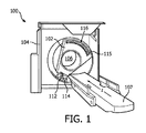
図1は、コンピュータ断層撮影(CT)スキャナといった例示的な撮像システム100を概略的に示す。撮像システム100は、回転ガントリ102と固定ガントリ104とを含む。回転ガントリ102は、固定ガントリ104によって回転可能に支持される。回転ガントリ102は、長手軸、即ち、Z軸について検査領域106の周りを回転する。撮像システム100は更に、スキャン前、スキャン中及び/又はスキャン後に検査領域106内に被験者又は物体を支える被験者支持体107を含む。被験者支持体107は、被験者又は物体を、検査領域106内へとロードする及び/又は検査領域106からアンロードするためにも使用される。撮像システム100は更に、回転ガントリ102によって回転可能に支持されるX線管といった放射線源112を含む。放射線源112は、回転ガントリ102と共に検査領域106の周りを回転し、検査領域106を横断する放射線を生成して放出する。撮像システム100は更に、放射線源コントローラ114を含む。放射線源コントローラ114は、生成された放射線の束を変調する。例えば放射線コントローラ114は、放射線源112のカソード加熱電流を選択的に変更し、放射線源112の電子フローを抑制するように電荷を適用し、放出された放射線等をフィルタリングして、束を変調することができる。
FIG. 1 schematically illustrates an
撮像システム100は更に、放射線感応検出ピクセル116の1次元又は2次元アレイ115を含む。ピクセル116は、放射線源112の反対側で、検査領域106の向こう側に配置され、検査領域106を横断する放射線を検出し、放射線を示す電気信号(投影データ)を生成する。図示される例では、ピクセル116は、直接変換光子計数検出器ピクセルを含む。このようなピクセルを用いると、生成された信号は、検出された光子のエネルギーを示すピーク振幅、即ち、ピーク高さを有する電流又は電圧を含む。
The
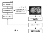
図2は、本発明による方法の主なステップを示す。本発明による方法の主な入力は、医用撮像データ又は画像10と、関心の応用に関連する定量分析アルゴリズムと、関連のノイズ低減スキームのセット又はパラメータ空間とである。第1のステップ11において、例えば所与の設定における最低ノイズ低減強さを使用して、最初のノイズ低減スキームが撮像データ又はサブセットに適用される。最初のノイズ低減スキームの適用後、定量的マップの最初のセット12が生成される。
FIG. 2 shows the main steps of the method according to the invention. The main inputs of the method according to the present invention are medical imaging data or
定量的マップ生成の最適作用点を見つける処理を開始するために、ノイズ低減強さレベルが増加され、ノイズ低減が撮像データ10に再び適用される。最近に更新された強さレベルを用いたノイズ低減の適用後、定量的マップの追加のセット13が生成される。定量分析マップの前のセット12と定量分析マップの最近のセット13とに基づいて、専用のアルゴリズム的手順14によって、平均定量バイアス差が推定される。推定されたバイアス差、また、任意選択的に、追加の条件は、所定の基準に照らして確認される。基準を満たさない場合、ノイズ低減スキームの更に強められた強さを試すために反復が繰り返される。
In order to begin the process of finding the optimal point of action for quantitative map generation, the noise reduction strength level is increased and noise reduction is again applied to the
例えば幾つかの反復後、基準を最終的に満たした後、ノイズ低減スキームは、最近に試された強さレベルで、撮像データボリューム全体に適用され、当該撮像ボリューム全体について最終的な非線形定量分析マップ15が計算され、最適化された定量分析が与えられる。
For example, after several iterations, after finally meeting the criteria, the noise reduction scheme is applied to the entire imaging data volume at the recently tried intensity level, and the final nonlinear quantitative analysis for the entire imaging volume. A
図3は、高品質血液かん流マップを提供するように、定量分析の前にダイナミックコントラストエンハンストCTデータセットに適用されるノイズ低減レベルが最適化される提案方法の一例を示す。ここでは、ノイズによって誘発されたバイアスが肝実質上に現れる。グラフは、ノイズ低減レベルによってどのように選択された関心領域(ROI)における平均値が減少するのかを示す。定量的マップ31、32、33及び34は、ノイズ低減レベルの4つの異なる値に対応する。この例では、4%よりも小さい値バイアス変化が、最適作用点を選択するための閾値Tとして使用される。これは、定量的マップ上のハイライトされたゾーンから反映しているので、小さ過ぎるノイズ低減レベルでは、非線形分析関数によって定量的マップに平均値の高いバイアスがある。当該バイアスは、最終マップをフィルタリングしても低減することができない。 FIG. 3 shows an example of a proposed method in which the noise reduction level applied to the dynamic contrast enhanced CT data set is optimized prior to quantitative analysis to provide a high quality blood perfusion map. Here, a noise-induced bias appears in the liver parenchyma. The graph shows how the average value in the selected region of interest (ROI) decreases with the noise reduction level. The quantitative maps 31, 32, 33 and 34 correspond to four different values of noise reduction level. In this example, a value bias change of less than 4% is used as the threshold T for selecting the optimal action point. This reflects from the highlighted zone on the quantitative map, so at a noise reduction level that is too small, the nonlinear map has a high average bias in the quantitative map. The bias cannot be reduced by filtering the final map.
図4は、様々なノイズ低減レベル間のバイアス差の自動評価処理技術の一例を示す。図4は、ROIを選択する必要のない一例である。図の上部にある定量的マップ41、42及び43は、撮像データの同じセット、即ち、図の下部に示され、3つの異なるレベルのノイズフィルタリングがそれぞれ適用されているCTかん流スキャン44に対応する。画像45は、マップ41とマップ42との差を表す。画像45は、主に肝臓及び脾臓領域において、高い平均バイアスを示す。画像46は、マップ41とマップ43との差を表す。画像46は、肝臓及び脾臓領域において、中程度の平均バイアスを示す。画像46の強いノイズ低減設定が、どのようにマップの空間分解能を低下させ始めるかを指摘することが興味深い。撮像ボリューム全体又は関連のサブボリュームに対して自動的に計算される平均バイアス差は、必要な最適化評価に使用することができる。
FIG. 4 shows an example of a technique for automatically evaluating bias differences between various noise reduction levels. FIG. 4 is an example in which it is not necessary to select an ROI.
図5は、本発明による方法のフローチャートである。ステップ51は、方法の主な入力を詳述する。主な入力は、a)通常は、3D又は4D画像セットである医用撮像データ(しかし、トモグラフィ再構成の前のステップからのサイノグラムといった他のタイプのより予備的なデータも使用することができる)、b)関心の応用に関連する定量分析アルゴリズム、c)特定のタイプの撮像データに関連するノイズ低減スキームのセットである。スキームは、ノイズ低減レベルの強さ又は強度に応じて順序付けられる。別のオプションでは、様々なレベルのステップ又は順序は、アルゴリズム反復中に適応的に決定されてもよい。最初のステップ52の間に、所与の設定における最低強さを使用して、ノイズ低減スキームが撮像データに適用される。これは、事前に指定されたボリューム又は撮像ボリューム全体に対して自動的に又は手動で行われる。1つのオプションとして、再構成ステップ、位置合わせステップ又は任意の他の画像処理アルゴリズムが、ノイズ低減を適用した後、次のステップにおいて定量分析を適用する前に、適用されてもよい。ステップ53において、最低強さのノイズ低減を適用した後、定量的マップの第1のセットが生成される。次に、ステップ54において、入力されたスキームに応じて、ノイズ低減強さレベルが増加され、最適作用点を見つける処理を開始するために、ノイズ低減が撮像データに再び適用される。ステップ55において、ステップ54の強さレベルのノイズ低減を適用した後、定量的マップの追加セットが生成される。ステップ56において、定量分析マップの前のセットと定量分析マップの最近のセットとに基づいて、平均定量バイアス差が推定される。このアルゴリズム的処理は、図6に更に詳述される。次に、推定されたバイアス差は、所定条件を満たすかどうか確認される。所定条件は、例えば最小百分率閾値、絶対閾値又は別の基準に基づいていてよい。基準を満たさない場合、ノイズ低減の更に強められた強さについて試すためにステップ54が繰り返される。基準を満たすと、アルゴリズムは、ステップ58に進む。ステップ58では、最近に試された強さレベルでノイズ低減スキームを撮像データボリューム全体に適用する。最終的な非線形定量分析マップが、最終的に、撮像ボリューム全体に対して計算され、最適化された定量分析が与えられる。
FIG. 5 is a flowchart of the method according to the invention.
最初のノイズ低減設定は、撮像条件及び臨床的応用による所定リストから選択されてよい。例えばCTかん流では、5mmのスライス厚さを用いる肝臓分析の場合の設定は、3mmのスライス厚さを用いる脾臓撮像の場合の設定とは異なる。反復間のノイズ低減パラメータインクリメントも、特定のスキャン又は応用に依存する。 The initial noise reduction setting may be selected from a predetermined list depending on imaging conditions and clinical application. For example, in CT perfusion, the setting for liver analysis using a 5 mm slice thickness is different from the setting for spleen imaging using a 3 mm slice thickness. The noise reduction parameter increment between iterations also depends on the particular scan or application.
上記フローチャートにおける反復は、ノイズ低減強さレベルの単調変化を有するものとして説明された。しかし、本発明の別の実施形態では、ノイズ低減強さレベルは、最適化の効率を向上させるために、違うスキーム又はシーケンスで変更可能である。これは、例えばノイズ低減スキームのパラメータ空間に大域最小化アルゴリズムの既知の技術を適用することによって、また、適切な最小化関数を使用する間に行われる。 The iterations in the above flowchart have been described as having a monotonic change in the noise reduction intensity level. However, in another embodiment of the present invention, the noise reduction strength level can be changed with different schemes or sequences to improve the efficiency of the optimization. This is done, for example, by applying known techniques of global minimization algorithms to the parameter space of the noise reduction scheme and while using an appropriate minimization function.
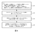
図6は、図5のステップ56における平均定量バイアス差を推定するために使用されるアルゴリズムを詳述する。この処理は、図4における例と同様である。この処理は、実際に、ROIを手動で選択する必要なく全自動的に行われることが可能である。しかし、関連のROIを選択することも、最適化の更なる精度を提供するために、依然として1つのオプションである。アルゴリズムは、定量的マップの2つのセットを減算して、図4に示されるような差マップセット(45及び46)を得る。差マップセットは、次に、任意の種類の適切なフィルタを使用して平滑化される。平滑化された差マップセットを平均化することによって、平均定量バイアスを推定することができる。
FIG. 6 details the algorithm used to estimate the average quantitative bias difference in
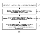
図7は、本発明の幾つかの実施形態では興味深い定量的マップの空間分解能に関する更なる最適化条件を含む上記最適化処理を示す。第1のステップ71として、上記されたように、関連の臨床的データ及び入力を使用して、定量的マップ生成最適化の自動処理が行われる。図5の処理に基づき、最適化された定量的マップが利用可能である場合、ユーザは、手動で又は半若しくは全自動ツールを用いて、機能的及び/又は解剖学的画像の例えば腫瘍病巣である関連の1つ以上の領域をセグメント化する(ステップ72)。ステップ73において、セグメント化された領域の寸法及び形状に基づいて、自動計算によって、セグメント化された領域の正しい定量値を得るために必要な最小空間分解能が決定される。
FIG. 7 illustrates the above optimization process including further optimization conditions regarding the spatial resolution of the quantitative map that is of interest in some embodiments of the present invention. As a
1つのオプションとして、必要な分解能のこのような決定は、画像の全自動分析によって、又は、事前情報若しくはユーザパラメータ選択を使用することによって行われる。 As one option, such a determination of the required resolution is made by a fully automatic analysis of the image or by using prior information or user parameter selection.
元のデータに適用される最適ノイズ低減強さレベルの選択は、マップにおける必要最小空間分解能を維持しつつ、マップにおける定量バイアスを可能な限り低減するために、更に最適化される(ステップ74)。 The selection of the optimal noise reduction strength level applied to the original data is further optimized to reduce the quantitative bias in the map as much as possible while maintaining the required minimum spatial resolution in the map (step 74). .
必要空間分解能の基準の確認は、第2の改良処理として行われてよいこと、つまり、図5のフローチャートを最初に行い、図6のフローチャートを次に行うことは注目に値する。又は、図5のフローチャート及び図6のフローチャートは、単一のアルゴリズム的処理内で組み合わされてもよい。 It is worth noting that the confirmation of the required spatial resolution criterion may be performed as the second improvement process, that is, the flowchart of FIG. 5 is performed first and the flowchart of FIG. 6 is performed next. Alternatively, the flowchart of FIG. 5 and the flowchart of FIG. 6 may be combined within a single algorithmic process.
空間分解能条件に加えて、低コントラスト分解能又は画像視覚化状況に基づく他の条件も実施されてもよい。 In addition to spatial resolution conditions, other conditions based on low contrast resolution or image visualization situations may also be implemented.
本発明は、図面及び上記説明において詳細に例示及び説明されたが、当該例示及び説明は、例示であって、限定と解釈されるべきではない。本発明は、開示された実施形態に限定されない。 Although the invention has been illustrated and described in detail in the drawings and foregoing description, the illustration and description are exemplary and should not be construed as limiting. The invention is not limited to the disclosed embodiments.
開示された実施形態の変形態様は、図面、開示内容及び添付の請求項の検討から、請求項に係る発明を実施する当業者によって理解され、実施される。請求項において、「含む」との用語は、他の要素又はステップを除外するものではなく、また、「a」又は「an」との不定冠詞も、複数形を除外するものではない。単一のプロセッサ又は他のユニットが、請求項に記載される幾つかのアイテムの機能を果たしてもよい。特定の手段が相互に異なる従属請求項に記載されることだけで、これらの手段の組み合わせを有利に使用することができないことを示すものではない。請求項における任意の参照符号は、範囲を限定するものと解釈されるべきではない。 Variations of the disclosed embodiments will be understood and implemented by those skilled in the art practicing the claimed invention upon review of the drawings, the disclosure, and the appended claims. In the claims, the term “comprising” does not exclude other elements or steps, and the indefinite article “a” or “an” does not exclude a plurality. A single processor or other unit may fulfill the functions of several items recited in the claims. The mere fact that certain measures are recited in mutually different dependent claims does not indicate that a combination of these measured cannot be used to advantage. Any reference signs in the claims should not be construed as limiting the scope.
Claims (15)
a:定量的マップの最初のセットを生成するように、医用撮像データの最初のセットに、ノイズ低減スキームの最初のノイズ低減レベルを適用するステップと、
b:前記ノイズ低減スキームの最後のノイズ低減レベルの値よりも高い値に、前記ノイズ低減スキームの新しいノイズ低減レベルを設定するステップと、
c:定量的マップの新しいセットを生成するように、医用撮像データの前記最初のセットに、前記ノイズ低減スキームの前記新しいノイズ低減レベルを適用するステップと、
d:定量的マップの最近の2つのセットに基づいて、平均定量バイアス差を推定するステップと、
e:推定された前記平均定量バイアス差が、所与の閾値よりも高い場合、ステップb乃至ステップeを繰り返すステップと、
f:医用撮像データの前記最初のセットを含む関心の医用撮像データのセットに、前記ノイズ低減スキームの前記最後のノイズ低減レベルを適用するステップと、
を含む、方法。 An automatic optimization method for quantitative map generation in functional medical imaging,
applying the initial noise reduction level of the noise reduction scheme to the initial set of medical imaging data so as to generate an initial set of quantitative maps;
b: setting the new noise reduction level of the noise reduction scheme to a value higher than the value of the last noise reduction level of the noise reduction scheme;
c: applying the new noise reduction level of the noise reduction scheme to the first set of medical imaging data so as to generate a new set of quantitative maps;
d: estimating an average quantitative bias difference based on the two recent sets of quantitative maps;
e: repeating step b to step e if the estimated average quantitative bias difference is higher than a given threshold;
f: applying the last noise reduction level of the noise reduction scheme to a set of medical imaging data of interest including the first set of medical imaging data;
Including a method.
Applications Claiming Priority (3)
| Application Number | Priority Date | Filing Date | Title |
|---|---|---|---|
| EP15193896 | 2015-11-10 | ||
| EP15193896.6 | 2015-11-10 | ||
| PCT/EP2016/076052 WO2017080847A1 (en) | 2015-11-10 | 2016-10-28 | Method for automatic optimization of quantitative map generation in functional medical imaging |
Publications (2)
| Publication Number | Publication Date |
|---|---|
| JP2018538032A true JP2018538032A (en) | 2018-12-27 |
| JP2018538032A5 JP2018538032A5 (en) | 2019-12-05 |
Family
ID=54540910
Family Applications (1)
| Application Number | Title | Priority Date | Filing Date |
|---|---|---|---|
| JP2018523420A Pending JP2018538032A (en) | 2015-11-10 | 2016-10-28 | Automatic optimization method for quantitative map generation in functional medical imaging |
Country Status (5)
| Country | Link |
|---|---|
| US (1) | US10789683B2 (en) |
| EP (1) | EP3374962A1 (en) |
| JP (1) | JP2018538032A (en) |
| CN (1) | CN108292430A (en) |
| WO (1) | WO2017080847A1 (en) |
Families Citing this family (9)
| Publication number | Priority date | Publication date | Assignee | Title |
|---|---|---|---|---|
| US11885442B2 (en) | 2017-12-15 | 2024-01-30 | Viant As&O Holdings, Llc | Mechanical joining of nitinol tubes |
| US10891720B2 (en) | 2018-04-04 | 2021-01-12 | AlgoMedica, Inc. | Cross directional bilateral filter for CT radiation dose reduction |
| US11080898B2 (en) | 2018-04-06 | 2021-08-03 | AlgoMedica, Inc. | Adaptive processing of medical images to reduce noise magnitude |
| DE102018214325A1 (en) * | 2018-08-24 | 2020-02-27 | Siemens Healthcare Gmbh | Method and provision unit for the provision of a virtual tomographic stroke follow-up examination image |
| CN111256712B (en) * | 2020-02-24 | 2021-10-29 | 深圳市优必选科技股份有限公司 | Map optimization method and device and robot |
| CN112017256B (en) * | 2020-08-31 | 2023-09-15 | 南京安科医疗科技有限公司 | On-line CT image quality free customization method and computer readable storage medium |
| CN112837244B (en) * | 2021-03-11 | 2022-07-22 | 太原科技大学 | A low-dose CT image noise reduction and artifact removal method based on progressive generative adversarial network |
| CN113128463B (en) * | 2021-05-07 | 2022-08-26 | 支付宝(杭州)信息技术有限公司 | Image recognition method and system |
| US12076173B2 (en) * | 2021-10-27 | 2024-09-03 | Wisconsin Alumni Research Foundation | System and method for controlling errors in computed tomography number |
Family Cites Families (14)
| Publication number | Priority date | Publication date | Assignee | Title |
|---|---|---|---|---|
| JP4558645B2 (en) | 2003-04-04 | 2010-10-06 | 株式会社日立メディコ | Image display method and apparatus |
| CA2534701A1 (en) | 2006-01-24 | 2007-07-24 | The University Of North Carolina At Chapel Hill | High energy soft tissue imaging using diffraction enhanced imaging |
| US7844096B2 (en) | 2007-01-22 | 2010-11-30 | Siemens Medical Solutions Usa, Inc. | Spatially localized noise adaptive smoothing of emission tomography images |
| US7782056B2 (en) * | 2007-12-13 | 2010-08-24 | Isis Innovation Ltd. | Systems and methods for correction of inhomogeneities in magnetic resonance images |
| US20110208039A1 (en) * | 2010-02-22 | 2011-08-25 | Siemens Corporation | Direct and Indirect Surface Coil Correction for Cardiac Perfusion MRI |
| US8938105B2 (en) * | 2010-10-28 | 2015-01-20 | Kabushiki Kaisha Toshiba | Denoising method and system for preserving clinically significant structures in reconstructed images using adaptively weighted anisotropic diffusion filter |
| US9095273B2 (en) * | 2011-09-26 | 2015-08-04 | Sunnybrook Research Institute | Systems and methods for automated dynamic contrast enhancement imaging |
| US9245334B2 (en) | 2012-02-28 | 2016-01-26 | Albert Einstein College Of Medicine, Inc. | Methods for quantitative assessment of volumetric image from a subject and uses therof |
| US9135695B2 (en) * | 2012-04-04 | 2015-09-15 | Siemens Aktiengesellschaft | Method for creating attenuation correction maps for PET image reconstruction |
| EP2936430B1 (en) | 2012-12-20 | 2018-10-31 | Koninklijke Philips N.V. | Quantitative imaging |
| US9760991B2 (en) * | 2013-04-22 | 2017-09-12 | General Electric Company | System and method for image intensity bias estimation and tissue segmentation |
| CN104217398B (en) * | 2013-05-29 | 2017-07-14 | 东芝医疗系统株式会社 | Image processing apparatus, image processing method and medical image equipment |
| JP2016523154A (en) | 2013-06-28 | 2016-08-08 | コーニンクレッカ フィリップス エヌ ヴェKoninklijke Philips N.V. | How to use image noise information |
| US10898143B2 (en) * | 2015-11-10 | 2021-01-26 | Baycrest Centre | Quantitative mapping of cerebrovascular reactivity using resting-state functional magnetic resonance imaging |
-
2016
- 2016-10-28 WO PCT/EP2016/076052 patent/WO2017080847A1/en active Application Filing
- 2016-10-28 JP JP2018523420A patent/JP2018538032A/en active Pending
- 2016-10-28 CN CN201680065694.4A patent/CN108292430A/en active Pending
- 2016-10-28 US US15/770,775 patent/US10789683B2/en active Active
- 2016-10-28 EP EP16790557.9A patent/EP3374962A1/en not_active Withdrawn
Also Published As
| Publication number | Publication date |
|---|---|
| EP3374962A1 (en) | 2018-09-19 |
| WO2017080847A1 (en) | 2017-05-18 |
| CN108292430A (en) | 2018-07-17 |
| US10789683B2 (en) | 2020-09-29 |
| US20180322617A1 (en) | 2018-11-08 |
Similar Documents
| Publication | Publication Date | Title |
|---|---|---|
| US10789683B2 (en) | Method for automatic optimization of quantitative map generation in functional medical imaging | |
| US11756164B2 (en) | System and method for image correction | |
| US8938110B2 (en) | Enhanced image data/dose reduction | |
| JP6208271B2 (en) | Medical image processing device | |
| US20110150309A1 (en) | Method and system for managing imaging data, and associated devices and compounds | |
| US20140301624A1 (en) | Method for interactive threshold segmentation of medical images | |
| JP6353463B2 (en) | Quantitative imaging | |
| US11361478B2 (en) | Partial volume correction in multi-modality emission tomography | |
| JP6345881B2 (en) | Texture analysis map for image data | |
| JP7346418B2 (en) | System and method for evaluating lung images | |
| Potesil et al. | Automated tumor delineation using joint PET/CT information | |
| JP2008525142A (en) | Apparatus and method for x-ray projection artifact correction | |
| US10993688B2 (en) | Method of data processing for computed tomography | |
| JP5632920B2 (en) | System and method for determining blur characteristics in a blurred image | |
| CN101410870B (en) | Automatic cardiac band detection on breast MRI | |
| JP2010507438A (en) | Improved segmentation | |
| Tang et al. | Noise Reduction for Cone-Beam Micro-CT by Fuzzy-Logic Based Non-Linear Filters | |
| Hajiesmaeili et al. | Analysis of partial volume effects on accurate measurement of the hippocampus volume |
Legal Events
| Date | Code | Title | Description |
|---|---|---|---|
| A521 | Request for written amendment filed |
Free format text: JAPANESE INTERMEDIATE CODE: A523 Effective date: 20191024 |
|
| A621 | Written request for application examination |
Free format text: JAPANESE INTERMEDIATE CODE: A621 Effective date: 20191024 |
|
| A977 | Report on retrieval |
Free format text: JAPANESE INTERMEDIATE CODE: A971007 Effective date: 20200925 |
|
| A131 | Notification of reasons for refusal |
Free format text: JAPANESE INTERMEDIATE CODE: A131 Effective date: 20201006 |
|
| A601 | Written request for extension of time |
Free format text: JAPANESE INTERMEDIATE CODE: A601 Effective date: 20201221 |
|
| A02 | Decision of refusal |
Free format text: JAPANESE INTERMEDIATE CODE: A02 Effective date: 20210609 |


