JP2018516594A - Method for distinguishing between HIV-1 and a lentiviral vector - Google Patents
Method for distinguishing between HIV-1 and a lentiviral vector Download PDFInfo
- Publication number
- JP2018516594A JP2018516594A JP2018512815A JP2018512815A JP2018516594A JP 2018516594 A JP2018516594 A JP 2018516594A JP 2018512815 A JP2018512815 A JP 2018512815A JP 2018512815 A JP2018512815 A JP 2018512815A JP 2018516594 A JP2018516594 A JP 2018516594A
- Authority
- JP
- Japan
- Prior art keywords
- nucleic acid
- nucleotide sequence
- hiv
- sequence
- seq
- Prior art date
- Legal status (The legal status is an assumption and is not a legal conclusion. Google has not performed a legal analysis and makes no representation as to the accuracy of the status listed.)
- Pending
Links
Images


Classifications
-
- C—CHEMISTRY; METALLURGY
- C12—BIOCHEMISTRY; BEER; SPIRITS; WINE; VINEGAR; MICROBIOLOGY; ENZYMOLOGY; MUTATION OR GENETIC ENGINEERING
- C12Q—MEASURING OR TESTING PROCESSES INVOLVING ENZYMES, NUCLEIC ACIDS OR MICROORGANISMS; COMPOSITIONS OR TEST PAPERS THEREFOR; PROCESSES OF PREPARING SUCH COMPOSITIONS; CONDITION-RESPONSIVE CONTROL IN MICROBIOLOGICAL OR ENZYMOLOGICAL PROCESSES
- C12Q1/00—Measuring or testing processes involving enzymes, nucleic acids or microorganisms; Compositions therefor; Processes of preparing such compositions
- C12Q1/70—Measuring or testing processes involving enzymes, nucleic acids or microorganisms; Compositions therefor; Processes of preparing such compositions involving virus or bacteriophage
- C12Q1/701—Specific hybridization probes
- C12Q1/702—Specific hybridization probes for retroviruses
- C12Q1/703—Viruses associated with AIDS
-
- A—HUMAN NECESSITIES
- A61—MEDICAL OR VETERINARY SCIENCE; HYGIENE
- A61P—SPECIFIC THERAPEUTIC ACTIVITY OF CHEMICAL COMPOUNDS OR MEDICINAL PREPARATIONS
- A61P31/00—Antiinfectives, i.e. antibiotics, antiseptics, chemotherapeutics
- A61P31/12—Antivirals
- A61P31/14—Antivirals for RNA viruses
- A61P31/18—Antivirals for RNA viruses for HIV
-
- C—CHEMISTRY; METALLURGY
- C12—BIOCHEMISTRY; BEER; SPIRITS; WINE; VINEGAR; MICROBIOLOGY; ENZYMOLOGY; MUTATION OR GENETIC ENGINEERING
- C12N—MICROORGANISMS OR ENZYMES; COMPOSITIONS THEREOF; PROPAGATING, PRESERVING, OR MAINTAINING MICROORGANISMS; MUTATION OR GENETIC ENGINEERING; CULTURE MEDIA
- C12N7/00—Viruses; Bacteriophages; Compositions thereof; Preparation or purification thereof
-
- C—CHEMISTRY; METALLURGY
- C12—BIOCHEMISTRY; BEER; SPIRITS; WINE; VINEGAR; MICROBIOLOGY; ENZYMOLOGY; MUTATION OR GENETIC ENGINEERING
- C12N—MICROORGANISMS OR ENZYMES; COMPOSITIONS THEREOF; PROPAGATING, PRESERVING, OR MAINTAINING MICROORGANISMS; MUTATION OR GENETIC ENGINEERING; CULTURE MEDIA
- C12N2740/00—Reverse transcribing RNA viruses
- C12N2740/00011—Details
- C12N2740/10011—Retroviridae
- C12N2740/16011—Human Immunodeficiency Virus, HIV
- C12N2740/16021—Viruses as such, e.g. new isolates, mutants or their genomic sequences
-
- C—CHEMISTRY; METALLURGY
- C12—BIOCHEMISTRY; BEER; SPIRITS; WINE; VINEGAR; MICROBIOLOGY; ENZYMOLOGY; MUTATION OR GENETIC ENGINEERING
- C12N—MICROORGANISMS OR ENZYMES; COMPOSITIONS THEREOF; PROPAGATING, PRESERVING, OR MAINTAINING MICROORGANISMS; MUTATION OR GENETIC ENGINEERING; CULTURE MEDIA
- C12N2740/00—Reverse transcribing RNA viruses
- C12N2740/00011—Details
- C12N2740/10011—Retroviridae
- C12N2740/16011—Human Immunodeficiency Virus, HIV
- C12N2740/16041—Use of virus, viral particle or viral elements as a vector
- C12N2740/16043—Use of virus, viral particle or viral elements as a vector viral genome or elements thereof as genetic vector
Abstract
本開示は、試料中に存在する核酸、たとえばHIVまたはレンチウイルスベクターに由来する核酸を検出および/または定量化するために特に有用である組成物(すなわち増幅プライマーおよびプローブ)、方法およびキットを提供する。

Description
関連出願の相互参照
本出願は、開示内容が参照により全体として本明細書に組み入れられる、2015年5月18日に出願された米国特許仮出願第62/163,327号の出願日の恩典を主張する。
This application claims the benefit of the filing date of US Provisional Application No. 62 / 163,327, filed May 18, 2015, the disclosure of which is incorporated herein by reference in its entirety. .
開示の分野
本開示は概して分子生物学およびウイルス学の分野に関する。特に、本開示は、HIV-1とレンチウイルスベクターとを識別する方法に関する。
FIELD OF DISCLOSURE The present disclosure relates generally to the fields of molecular biology and virology. In particular, the present disclosure relates to a method for distinguishing between HIV-1 and a lentiviral vector.
産業上の利用可能性の声明
本開示は、遺伝子治療および医療診断の分野に産業上の利用可能性を有する。
Industrial Applicability Statement The present disclosure has industrial applicability in the fields of gene therapy and medical diagnostics.
開示の背景
HIV-1は、全世界で3000万人ほどが感染している後天性免疫不全症候群(AIDS)の原因因子である。HIVは、免疫系を機能不全にし、日和見感染症による死の確率を高める。HIV感染症は、世界保健機関によるパンデミック指定によって証明されるように、重大な全世界的健康問題である。特に途上国におけるHIV感染者の大部分は最終的にAIDSを発症し、それが毎年100万を超える人々の命を奪っている。
Disclosure background
HIV-1 is the causative factor of acquired immune deficiency syndrome (AIDS), which affects approximately 30 million people worldwide. HIV makes the immune system dysfunctional and increases the probability of death from opportunistic infections. HIV infection is a serious global health problem as evidenced by a pandemic designation by the World Health Organization. Most people living with HIV, especially in developing countries, eventually develop AIDS, which kills more than 1 million people each year.
HIV-1はレトロウイルス科に属し、ゲノムが2つの一本鎖RNA分子(ssRNA)からなるエンベロープウイルスである。HIV-1の第1の標的はCD4+発現細胞、たとえばCD4+T細胞である。HIV-1ウイルスの糖タンパク質が標的細胞のCD4分子および標的細胞の表面上のケモカイン補助受容体CCRSまたはCXCR4と相互作用する。標的細胞への融合および進入ののち、ウイルスゲノムを含むヌクレオカプシドが分離して、ssRNAを含むウイルスの内容物を細胞質中に放出する。HIV-1の逆転写酵素(RT)がssRNAゲノムからウイルス二本鎖DNA(dsDNA)を合成する。二本鎖HIV-1 DNA分子の合成ののち、HIV-1 DNAは宿主ゲノムに組み込まれる。 HIV-1 is an enveloped virus that belongs to the Retroviridae family and whose genome consists of two single-stranded RNA molecules (ssRNA). The primary target of HIV-1 is CD4 + expressing cells, such as CD4 + T cells. HIV-1 viral glycoproteins interact with the target cell CD4 molecule and the chemokine co-receptor CCRS or CXCR4 on the surface of the target cell. After fusion and entry into the target cell, the nucleocapsid containing the viral genome separates and releases the viral contents, including ssRNA, into the cytoplasm. HIV-1 reverse transcriptase (RT) synthesizes viral double-stranded DNA (dsDNA) from the ssRNA genome. After synthesis of the double-stranded HIV-1 DNA molecule, the HIV-1 DNA is integrated into the host genome.
組み込まれたHIV-1 DNAは、同一の5'および3'末端反復配列(LTR)を両脇に有し、その配列から、HIV-1は組み込まれたHIV-1ゲノムの転写を開始することができる。ウイルスDNAの転写は、活性化T細胞中で上方制御される、NF-kBのような転写因子を必要とする。その結果、ウイルス転写は、T細胞の活性化ののち、たとえば感染中、T細胞中で非常に活動的である。組み込まれたHIV-1ゲノムの転写から生じるウイルスRNAはその後、翻訳され、ウイルス粒子中にパッケージングされたのち、そのウイルス粒子が細胞を出て感染性ウイルスになる。 The integrated HIV-1 DNA has identical 5 ′ and 3 ′ terminal repeats (LTRs) on either side, from which HIV-1 initiates transcription of the integrated HIV-1 genome. Can do. Transcription of viral DNA requires a transcription factor such as NF-kB that is upregulated in activated T cells. As a result, viral transcription is very active in T cells after T cell activation, eg during infection. Viral RNA resulting from transcription of the integrated HIV-1 genome is then translated and packaged into a viral particle, which then exits the cell to become an infectious virus.
HIV-1感染症の治療は多剤併用抗レトロウイルス療法(cART)を含む。ヌクレオシドアナログ逆転写酵素阻害剤、プロテアーゼ阻害剤、非ヌクレオシド逆転写酵素阻害剤、インテグラーゼおよび融合阻害剤の併用を含むcARTはHIV進行を遅らせる。これが、結果として、この療法が利用可能である世界の地域においてHIV/AIDSからの罹患率および死亡率を劇的に低下させる。しかし、cARTはHIV/AIDSのすべての症候を治癒または完全に消失させるわけではない。また、cART療法は、薬物耐性突然変異によって効果が損なわれることがあり、また、深刻になり得かつ累積するように思われる一定範囲の副作用を有する。さらに、cART療法の中断は、ほぼ決まって、検出可能なウイルス複製の再出現およびAIDSへの進行をもたらし、また、死亡率および深刻な非AIDS事象のすべての原因の発生率の増加と関連することが示されている。これらの理由ならびにcARTの高額なコストおよび厳格な遵守の必要性のため、このような療法は多数の患者にとって相対的に無効である場合もある。 Treatment of HIV-1 infection includes combination antiretroviral therapy (cART). CART containing a combination of nucleoside analog reverse transcriptase inhibitors, protease inhibitors, non-nucleoside reverse transcriptase inhibitors, integrase and fusion inhibitors delays HIV progression. This results in a dramatic reduction in morbidity and mortality from HIV / AIDS in regions of the world where this therapy is available. However, cART does not cure or completely eliminate all symptoms of HIV / AIDS. Also, cART therapy can be compromised by drug resistance mutations and has a range of side effects that can be serious and appear to be cumulative. In addition, interruption of cART therapy almost always results in the reappearance of detectable viral replication and progression to AIDS, and is associated with increased mortality and the incidence of all causes of serious non-AIDS events It has been shown. For these reasons and the high cost of cART and the need for strict adherence, such therapies may be relatively ineffective for many patients.
HIVベースのレンチウイルスベクターが、急速に、研究用および臨床用遺伝子導入用途に選択されるレトロウイルスベクター系となりつつある。静止幹細胞および非分裂最終分化細胞の両方を形質導入するレンチウイルスベクターの高められた能力が、広い範囲の治療用遺伝子送達ベクターならびに有望な研究ツール、たとえば最終分化細胞における多分化能の誘導のためのショートヘアピンRNA(shRNA)遺伝子ノックダウンライブラリーおよびベクターの開発につながった。初期のガンマレトロウイルス臨床遺伝子治療ベクターは、X連鎖重症複合免疫不全症(SCID-X1)の患者において免疫機能を回復させたが、このベクターは、その後、がん原遺伝子のトランス活性化を介して増殖性障害を生じさせることがわかった。より新規なレトロウイルスベクター設計がそのリスクを有意に低減し得、予測される安全性の最終確認のための臨床試験を待っている。現場は依然として流動的であり、臨床試験の結果は予測不可能である。 HIV-based lentiviral vectors are rapidly becoming retroviral vector systems that are selected for research and clinical gene transfer applications. The increased ability of lentiviral vectors to transduce both quiescent stem cells and non-dividing terminally differentiated cells is due to the induction of pluripotency in a wide range of therapeutic gene delivery vectors and promising research tools such as terminally differentiated cells Led to the development of short hairpin RNA (shRNA) gene knockdown libraries and vectors. Early gamma retroviral clinical gene therapy vectors restored immune function in patients with X-linked severe combined immunodeficiency (SCID-X1), but this vector was subsequently mediated by transactivation of proto-oncogenes. Have been found to cause proliferative disorders. Newer retroviral vector designs can significantly reduce that risk and are awaiting clinical trials for final confirmation of the expected safety. The field is still fluid and the results of clinical trials are unpredictable.
抗HIV-1レンチウイルスベースの遺伝子療法(たとえば二剤併用抗HIV-1レンチウイルスベクター(Cal-1、LVsh5/C46))を使用してHIV-1複製を阻害するとき、Cal-1 DNAを含む細胞および野生型HIV DNAを含む細胞を定量化することが不可欠である。これは、HIV-1感染患者から得られた細胞の場合に特に当てはまる。本発明者らは、DNAレベルでHIV-1とCal-1とを識別する困難さに直面する。現在の市販のPCRベースのアッセイは、HIV-1組み込みDNAとCal-1導入遺伝子組み込みDNAとを識別することができない。pol領域に対するPCRに基づいてHIV-1 DNAを同定するための、プライマーを使用して、レンチウイルスベクター内に存在しないHIV-1配列を検出するアッセイが報告されている(Burke BP, et al: Mol Ther Nucleic Acids 2015, 4:e236(非特許文献1))。また、HIV-1内に存在しないC46プライマー領域を使用してCal-1導入遺伝子DNAを同定するための可能なアッセイが報告されている(Wolstein O, et al: Mol Ther Methods & Clinical Development 2014, 1, 11(非特許文献2))。 When inhibiting HIV-1 replication using anti-HIV-1 lentiviral based gene therapy (eg dual anti-HIV-1 lentiviral vectors (Cal-1, LVsh5 / C46)), Cal-1 DNA It is essential to quantify cells containing and cells containing wild type HIV DNA. This is especially true for cells obtained from HIV-1 infected patients. We face difficulties in distinguishing between HIV-1 and Cal-1 at the DNA level. Current commercial PCR-based assays cannot distinguish between HIV-1 integrated DNA and Cal-1 transgene integrated DNA. An assay has been reported that uses primers to detect HIV-1 sequences that are not present in a lentiviral vector to identify HIV-1 DNA based on PCR against the pol region (Burke BP, et al: Mol Ther Nucleic Acids 2015, 4: e236 (Non-Patent Document 1)). A possible assay for identifying Cal-1 transgene DNA using a C46 primer region not present in HIV-1 has also been reported (Wolstein O, et al: Mol Ther Methods & Clinical Development 2014, 1, 11 (non-patent document 2)).
開示の概要
本開示の1つの局面は、(a)SEQ ID NO: 14のヌクレオチド配列と少なくとも80%の同一性を有するヌクレオチド配列を含む、レポーター部分にコンジュゲートされたプローブ;(b)SEQ ID NO: 2のヌクレオチド配列と少なくとも90%の同一性を有するヌクレオチド配列を含むフォワードプライマー;および(c)SEQ ID NO: 6のヌクレオチド配列と少なくとも80%の同一性を有するヌクレオチド配列を含むリバースプライマーを含む、組成物であって、フォワードプライマーおよびリバースプライマーのそれぞれが、標的配列を増幅するために標的配列にアニーリングすることができる、組成物である。
SUMMARY OF THE DISCLOSURE One aspect of the disclosure includes (a) a probe conjugated to a reporter moiety comprising a nucleotide sequence having at least 80% identity to the nucleotide sequence of SEQ ID NO: 14; (b) SEQ ID A forward primer comprising a nucleotide sequence having at least 90% identity to the nucleotide sequence of NO: 2; and (c) a reverse primer comprising a nucleotide sequence having at least 80% identity to the nucleotide sequence of SEQ ID NO: 6 A composition comprising: a forward primer and a reverse primer, each capable of annealing to a target sequence to amplify the target sequence.
本開示のもう1つの局面は、(a)SEQ ID NO: 10のヌクレオチド配列と少なくとも80%の同一性を有するヌクレオチド配列を含む、レポーター部分にコンジュゲートされたプローブ;(b)SEQ ID NO: 2のヌクレオチド配列と少なくとも90%の同一性を有するヌクレオチド配列を含むフォワードプライマー;および(c)SEQ ID NO: 6のヌクレオチド配列と少なくとも80%の同一性を有するヌクレオチド配列を含むリバースプライマーを含む、組成物であって、フォワードプライマーおよびリバースプライマーのそれぞれが、標的配列を増幅するために標的配列にアニーリングすることができる、組成物である。 Another aspect of the disclosure includes (a) a probe conjugated to a reporter moiety comprising a nucleotide sequence having at least 80% identity to the nucleotide sequence of SEQ ID NO: 10; (b) SEQ ID NO: A forward primer comprising a nucleotide sequence having at least 90% identity with two nucleotide sequences; and (c) a reverse primer comprising a nucleotide sequence having at least 80% identity with the nucleotide sequence of SEQ ID NO: 6; A composition, wherein each of the forward primer and the reverse primer can be annealed to the target sequence to amplify the target sequence.
本開示のもう1つの局面は、(a)SEQ ID NO: 8のヌクレオチド配列と少なくとも80%の同一性を有するヌクレオチド配列を含む、レポーター部分にコンジュゲートされたプローブ;(b)SEQ ID NO: 4のヌクレオチド配列と少なくとも90%の同一性を有するヌクレオチド配列を含むフォワードプライマー;および(c)SEQ ID NO: 6のヌクレオチド配列と少なくとも80%の同一性を有するヌクレオチド配列を含むリバースプライマーを含む、組成物であって、フォワードプライマーおよびリバースプライマーのそれぞれが、標的配列を増幅するために標的配列にアニーリングすることができる、組成物である。 Another aspect of the disclosure includes (a) a probe conjugated to a reporter moiety comprising a nucleotide sequence having at least 80% identity to the nucleotide sequence of SEQ ID NO: 8; (b) SEQ ID NO: A forward primer comprising a nucleotide sequence having at least 90% identity to the nucleotide sequence of 4; and (c) a reverse primer comprising a nucleotide sequence having at least 80% identity to the nucleotide sequence of SEQ ID NO: 6; A composition, wherein each of the forward primer and the reverse primer can be annealed to the target sequence to amplify the target sequence.
本開示のもう1つの局面は、第1の組成物および第2の組成物を含むキットであって、
第1の組成物が、(a)SEQ ID NO: 14のヌクレオチド配列と少なくとも80%の同一性を有するヌクレオチド配列を含む、レポーター部分にコンジュゲートされたプローブ;(b)SEQ ID NO: 2のヌクレオチド配列と少なくとも90%の同一性を有するヌクレオチド配列を含むフォワードプライマー;および(c)SEQ ID NO: 6のヌクレオチド配列と少なくとも80%の同一性を有するヌクレオチド配列を含むリバースプライマーを含み、フォワードプライマーおよびリバースプライマーのそれぞれが、標的配列を増幅するために標的配列にアニーリングすることができ;
第2の組成物が、
(i)(a)SEQ ID NO: 10のヌクレオチド配列と少なくとも80%の同一性を有するヌクレオチド配列を含む、レポーター部分にコンジュゲートされたプローブ;(b)SEQ ID NO: 2のヌクレオチド配列と少なくとも90%の同一性を有するヌクレオチド配列を含むフォワードプライマー;および(c)SEQ ID NO: 6のヌクレオチド配列と少なくとも80%の同一性を有するヌクレオチド配列を含むリバースプライマーを含む組成物であって、フォワードプライマーおよびリバースプライマーのそれぞれが、標的配列を増幅するために標的配列にアニーリングすることができる、組成物;または
(ii)(a)SEQ ID NO: 8のヌクレオチド配列と少なくとも80%の同一性を有するヌクレオチド配列を含む、レポーター部分にコンジュゲートされたプローブ;(b)SEQ ID NO: 4のヌクレオチド配列と少なくとも90%の同一性を有するヌクレオチド配列を含むフォワードプライマー;および(c)SEQ ID NO: 6のヌクレオチド配列と少なくとも80%の同一性を有するヌクレオチド配列を含むリバースプライマーを含む組成物であって、フォワードプライマーおよびリバースプライマーのそれぞれが、標的配列を増幅するために標的配列にアニーリングすることができる、組成物
の1つを含む、キットである。
Another aspect of the present disclosure is a kit comprising a first composition and a second composition comprising:
A first composition comprising: (a) a probe conjugated to a reporter moiety comprising a nucleotide sequence having at least 80% identity to the nucleotide sequence of SEQ ID NO: 14; (b) of SEQ ID NO: 2 A forward primer comprising a nucleotide sequence having at least 90% identity to the nucleotide sequence; and (c) a reverse primer comprising a nucleotide sequence having at least 80% identity to the nucleotide sequence of SEQ ID NO: 6 And each of the reverse primers can be annealed to the target sequence to amplify the target sequence;
The second composition is
(I) (a) a probe conjugated to a reporter moiety comprising a nucleotide sequence having at least 80% identity to the nucleotide sequence of SEQ ID NO: 10; (b) at least a nucleotide sequence of SEQ ID NO: 2 A forward primer comprising a nucleotide sequence having 90% identity; and (c) a reverse primer comprising a nucleotide sequence having at least 80% identity to the nucleotide sequence of SEQ ID NO: 6 A composition wherein each of the primer and reverse primer can be annealed to the target sequence to amplify the target sequence; or (ii) (a) at least 80% identity to the nucleotide sequence of SEQ ID NO: 8 A probe conjugated to a reporter moiety comprising a nucleotide sequence having; (b) a nucleotide of SEQ ID NO: 4 A composition comprising a forward primer comprising a nucleotide sequence having at least 90% identity to the tide sequence; and (c) a reverse primer comprising a nucleotide sequence having at least 80% identity to the nucleotide sequence of SEQ ID NO: 6 Wherein the forward primer and the reverse primer each comprise one of the compositions that can be annealed to the target sequence to amplify the target sequence.
本開示のもう1つの局面は、第1の標的配列を定量化する方法であって、第1の標的配列を含む第1の試料を、(a)SEQ ID NO: 14のヌクレオチド配列と少なくとも80%の同一性を有するヌクレオチド配列を含む、レポーター部分にコンジュゲートされたプローブ;(b)SEQ ID NO: 2のヌクレオチド配列と少なくとも90%の同一性を有するヌクレオチド配列を含むフォワードプライマー;および(c)SEQ ID NO: 6のヌクレオチド配列と少なくとも80%の同一性を有するヌクレオチド配列を含むリバースプライマーを含む組成物と接触させる工程;第1の標的配列を鋳型として使用してリアルタイムポリメラーゼ連鎖反応を実施する工程;および産生された第1のアンプリコンの量を定量化する工程を含む、方法である。 Another aspect of the present disclosure is a method for quantifying a first target sequence, wherein a first sample comprising the first target sequence is (a) a nucleotide sequence of SEQ ID NO: 14 and at least 80 A probe conjugated to a reporter moiety comprising a nucleotide sequence having% identity; (b) a forward primer comprising a nucleotide sequence having at least 90% identity to the nucleotide sequence of SEQ ID NO: 2; and (c ) Contacting with a composition comprising a reverse primer comprising a nucleotide sequence having at least 80% identity to the nucleotide sequence of SEQ ID NO: 6; performing a real-time polymerase chain reaction using the first target sequence as a template And quantifying the amount of first amplicon produced.
いくつかの態様において、方法はさらに、第2の試料内の第2の標的配列を定量化する工程を含む。いくつかの態様において、第2の標的配列は、(a)SEQ ID NO: 10のヌクレオチド配列と少なくとも80%の同一性を有するヌクレオチド配列を含む、レポーター部分にコンジュゲートされたプローブ;(b)SEQ ID NO: 2のヌクレオチド配列と少なくとも90%の同一性を有するヌクレオチド配列を含むフォワードプライマー;および(c)SEQ ID NO: 6のヌクレオチド配列と少なくとも80%の同一性を有するヌクレオチド配列を含むリバースプライマーを含む組成物であって、フォワードプライマーおよびリバースプライマーのそれぞれが、標的配列を増幅するために標的配列にアニーリングすることができる、組成物を使用して検出される。他の態様において、第2の標的配列は、(a)SEQ ID NO: 8のヌクレオチド配列と少なくとも80%の同一性を有するヌクレオチド配列を含む、レポーター部分にコンジュゲートされたプローブ;(b)SEQ ID NO: 4のヌクレオチド配列と少なくとも90%の同一性を有するヌクレオチド配列を含むフォワードプライマー;および(c)SEQ ID NO: 6のヌクレオチド配列と少なくとも80%の同一性を有するヌクレオチド配列を含むリバースプライマーを含む組成物であって、フォワードプライマーおよびリバースプライマーのそれぞれが、標的配列を増幅するために標的配列にアニーリングすることができる、組成物を使用して定量化される。 In some embodiments, the method further comprises quantifying the second target sequence in the second sample. In some embodiments, the second target sequence is (a) a probe conjugated to a reporter moiety comprising a nucleotide sequence having at least 80% identity to the nucleotide sequence of SEQ ID NO: 10; (b) A forward primer comprising a nucleotide sequence having at least 90% identity to the nucleotide sequence of SEQ ID NO: 2; and (c) a reverse comprising a nucleotide sequence having at least 80% identity to the nucleotide sequence of SEQ ID NO: 6 A composition comprising a primer, wherein each forward primer and reverse primer is detected using a composition that can be annealed to a target sequence to amplify the target sequence. In other embodiments, the second target sequence is (a) a probe conjugated to a reporter moiety comprising a nucleotide sequence having at least 80% identity to the nucleotide sequence of SEQ ID NO: 8; (b) SEQ A forward primer comprising a nucleotide sequence having at least 90% identity to the nucleotide sequence of ID NO: 4; and (c) a reverse primer comprising a nucleotide sequence having at least 80% identity to the nucleotide sequence of SEQ ID NO: 6 Wherein each of the forward primer and the reverse primer is quantified using the composition that can be annealed to the target sequence to amplify the target sequence.
いくつかの態様において、第1および第2の試料は同じ供給源に由来し、かつ第1および第2の標的配列の定量化は1つの反応チャンバ中で実施される。いくつかの態様において、第1および第2の試料は同じ供給源に由来し、かつ第1および第2の標的配列の定量化は別々の反応チャンバ中で実施される。いくつかの態様において、第1の標的配列はレンチウイルス核酸配列であり、かつ第2の標的配列はHIV核酸配列である。いくつかの態様において、産生された第1のアンプリコンの量を定量化する工程は、第1のレポーター部分からのシグナルを検出する工程を含み;産生された第2のアンプリコンの量を定量化する工程は、第2のレポーター部分からのシグナルを検出する工程を含み、第1のレポーター部分と第2のレポーター部分とは異なる。いくつかの態様において、方法はさらに、(i)第1の時点における、産生された第2のアンプリコンの定量化量に対する産生された第1のアンプリコンの定量化量の第1の比率を;(ii)第2の時点における、産生された第2のアンプリコンの定量化量に対する産生された第1のアンプリコンの定量化量の第2の比率と、比較することによってレンチウイルスベクターからの遺伝子導入の効果を評価する工程を含む。いくつかの態様において、HIV核酸に対するレンチウイルス核酸の比率増大が治療効果を示す。 In some embodiments, the first and second samples are from the same source, and quantification of the first and second target sequences is performed in one reaction chamber. In some embodiments, the first and second samples are from the same source, and the quantification of the first and second target sequences is performed in separate reaction chambers. In some embodiments, the first target sequence is a lentiviral nucleic acid sequence and the second target sequence is an HIV nucleic acid sequence. In some embodiments, quantifying the amount of first amplicon produced comprises detecting a signal from the first reporter moiety; quantifying the amount of second amplicon produced The step of converting comprises detecting a signal from the second reporter moiety, wherein the first reporter moiety and the second reporter moiety are different. In some embodiments, the method further comprises: (i) a first ratio of the quantified amount of the first amplicon produced to the quantified amount of the second amplicon produced at the first time point. (Ii) from the lentiviral vector by comparing with a second ratio of the quantified amount of the first amplicon produced to the quantified amount of the second amplicon produced at the second time point; A step of evaluating the effect of gene transfer. In some embodiments, an increased ratio of lentiviral nucleic acid to HIV nucleic acid indicates a therapeutic effect.
本開示のもう1つの局面は、試料中のレンチウイルス核酸および/またはHIV核酸を検出する方法であって、
(a)
(i)SEQ ID NO: 2のヌクレオチド配列と少なくとも90%の同一性を有するヌクレオチド配列を有する第1のフォワードプライマー、
SEQ ID NO: 6のヌクレオチド配列と少なくとも90%の同一性を有するヌクレオチド配列を有する第1のリバースプライマー、および
SEQ ID NO: 14のヌクレオチド配列と少なくとも90%の同一性を有するヌクレオチド配列を有し、第1のレポーター部分を有する第1のプローブ;
(ii)SEQ ID NO: 2のヌクレオチド配列と少なくとも90%の同一性を有するヌクレオチド配列を有する第2のフォワードプライマー、
SEQ ID NO: 6の配列と少なくとも90%の同一性を有するヌクレオチド配列を有する第2のリバースプライマー、および
SEQ ID NO: 10のヌクレオチド配列と少なくとも90%の同一性を有するヌクレオチド配列を有し、第2のレポーター部分を有する第2のプローブ
を使用して、試料中のレンチウイルス核酸鋳型およびHIV核酸鋳型を用いるマルチプレックスリアルタイムPCRを実施する工程であって、第1のレポーター部分と第2のレポーター部分とが異なる、工程;
(b)
(i)第1のフォワードおよびリバースプライマー、および(ii)第2のフォワードおよびリバースプライマーによって産生されたアンプリコンを検出する工程
を含み、検出する工程が、第1および第2のレポーター部分からの第1および第2のシグナルを検出する工程を含む、方法である。
Another aspect of the present disclosure is a method for detecting lentiviral nucleic acid and / or HIV nucleic acid in a sample comprising:
(A)
(I) a first forward primer having a nucleotide sequence having at least 90% identity to the nucleotide sequence of SEQ ID NO: 2;
A first reverse primer having a nucleotide sequence having at least 90% identity to the nucleotide sequence of SEQ ID NO: 6; and
A first probe having a nucleotide sequence having at least 90% identity to the nucleotide sequence of SEQ ID NO: 14 and having a first reporter moiety;
(Ii) a second forward primer having a nucleotide sequence having at least 90% identity with the nucleotide sequence of SEQ ID NO: 2;
A second reverse primer having a nucleotide sequence having at least 90% identity with the sequence of SEQ ID NO: 6, and
Lentiviral and HIV nucleic acid templates in a sample using a second probe having a nucleotide sequence having at least 90% identity to the nucleotide sequence of SEQ ID NO: 10 and having a second reporter moiety Performing a multiplex real-time PCR using the first reporter moiety and the second reporter moiety being different;
(B)
Detecting amplicons produced by (i) a first forward and reverse primer, and (ii) a second forward and reverse primer, wherein the step of detecting from the first and second reporter moieties A method comprising detecting first and second signals.
本開示のもう1つの局面は、試料中のレンチウイルス核酸および/またはHIV核酸を検出する方法であって、
(a)
(i)SEQ ID NO: 2のヌクレオチド配列と少なくとも90%の同一性を有するヌクレオチド配列を有する第1のフォワードプライマー、
SEQ ID NO: 6のヌクレオチド配列と少なくとも90%の同一性を有するヌクレオチド配列を有する第1のリバースプライマー、および
SEQ ID NO: 14のヌクレオチド配列と少なくとも90%の同一性を有するヌクレオチド配列を有する、第1のレポーター部分を有する第1のプローブ;
(ii)SEQ ID NO: 4のヌクレオチド配列と少なくとも90%の同一性を有するヌクレオチド配列を有する第2のフォワードプライマー、
SEQ ID NO: 6のヌクレオチド配列と少なくとも90%の同一性を有するヌクレオチド配列を有する第2のリバースプライマー、および
SEQ ID NO: 8のヌクレオチド配列と少なくとも90%の同一性を有するヌクレオチド配列を有し、第2のレポーター部分を有する第2のプローブ
を使用して、試料中のレンチウイルス核酸鋳型およびHIV核酸鋳型を用いるマルチプレックスリアルタイムPCRを実施する工程であって、第1のレポーター部分と第2のレポーター部分とが異なる、工程;
(b)
(i)第1のフォワードおよびリバースプライマー、および(ii)第2のフォワードおよびリバースプライマーによって産生されたアンプリコンを検出する工程
を含み、検出する工程が、第1および第2のレポーター部分からの第1および第2のシグナルを検出する工程を含む、方法である。
Another aspect of the present disclosure is a method for detecting lentiviral nucleic acid and / or HIV nucleic acid in a sample comprising:
(A)
(I) a first forward primer having a nucleotide sequence having at least 90% identity to the nucleotide sequence of SEQ ID NO: 2;
A first reverse primer having a nucleotide sequence having at least 90% identity to the nucleotide sequence of SEQ ID NO: 6; and
A first probe having a first reporter moiety having a nucleotide sequence having at least 90% identity to the nucleotide sequence of SEQ ID NO: 14;
(Ii) a second forward primer having a nucleotide sequence having at least 90% identity with the nucleotide sequence of SEQ ID NO: 4;
A second reverse primer having a nucleotide sequence having at least 90% identity to the nucleotide sequence of SEQ ID NO: 6; and
A lentiviral nucleic acid template and an HIV nucleic acid template in a sample using a second probe having a nucleotide sequence having at least 90% identity to the nucleotide sequence of SEQ ID NO: 8 and having a second reporter moiety Performing a multiplex real-time PCR using the first reporter moiety and the second reporter moiety being different;
(B)
Detecting amplicons produced by (i) a first forward and reverse primer, and (ii) a second forward and reverse primer, wherein the step of detecting from the first and second reporter moieties A method comprising detecting first and second signals.
もう1つの局面は、試料中のレンチウイルス核酸の量を検出する方法であって、
(a)試料を第1のフォワードプライマーおよび第1のリバースプライマーと接触させる工程;
(b)試料を、レンチウイルス核酸の3'LTR内のジャンクション部位に特異的なジャンクションプローブと接触させる工程であって、ジャンクションプローブが、レンチウイルス核酸3'LTRのU3領域内の配列の少なくとも一部分にハイブリダイズすることができる第1の部分およびレンチウイルス核酸3'LTRのR領域内の配列の少なくとも一部分にハイブリダイズすることができる第2の部分を含み、ジャンクションプローブが第1の検出可能な部分を含む、工程;ならびに
第1の検出可能な部分からのシグナルを検出する工程
を含む、方法である。いくつかの態様において、ジャンクションプローブの第1の部分は、SEQ ID NO: 12のヌクレオチド配列と少なくとも80%の同一性を有するヌクレオチド配列にハイブリダイズする。いくつかの態様において、ジャンクションプローブの第2の部分はSEQ ID NO: 13のヌクレオチド配列にハイブリダイズする。いくつかの態様において、ジャンクションプローブは、SEQ ID NO: 14のヌクレオチド配列と少なくとも80%の同一性を有するヌクレオチド配列を含む。いくつかの態様において、ジャンクションプローブは、SEQ ID NO: 14のヌクレオチド配列と少なくとも90%の同一性を有するヌクレオチド配列を含む。
Another aspect is a method for detecting the amount of lentiviral nucleic acid in a sample comprising:
(A) contacting the sample with a first forward primer and a first reverse primer;
(B) contacting the sample with a junction probe specific for a junction site in the 3′LTR of the lentiviral nucleic acid, wherein the junction probe is at least a portion of the sequence in the U3 region of the lentiviral nucleic acid 3′LTR. A first portion capable of hybridizing to and a second portion capable of hybridizing to at least a portion of a sequence within the R region of the lentiviral nucleic acid 3′LTR, wherein the junction probe is first detectable A method comprising: a step comprising: a step; and detecting a signal from a first detectable portion. In some embodiments, the first portion of the junction probe hybridizes to a nucleotide sequence having at least 80% identity with the nucleotide sequence of SEQ ID NO: 12. In some embodiments, the second portion of the junction probe hybridizes to the nucleotide sequence of SEQ ID NO: 13. In some embodiments, the junction probe comprises a nucleotide sequence having at least 80% identity with the nucleotide sequence of SEQ ID NO: 14. In some embodiments, the junction probe comprises a nucleotide sequence having at least 90% identity with the nucleotide sequence of SEQ ID NO: 14.
いくつかの態様において、方法はさらに、試料中のHIV核酸の量を検出する工程を含む。いくつかの態様において、試料中のレンチウイルス核酸の量およびHIV核酸の量の検出は同じ反応チューブ中で実施される。いくつかの態様において、HIV核酸の量の検出は、試料を、HIV核酸配列の3'LTR内のTATAボックス配列に特異的な第2のプローブと接触させる工程であって、第2のプローブが第2の検出可能な部分にコンジュゲートされている、工程;および第2の検出可能な部分からのシグナルを検出する工程を含む。いくつかの態様において、第2のプローブは、SEQ ID NO: 10のヌクレオチド配列と少なくとも90%の同一性を有するヌクレオチド配列を有する。いくつかの態様において、第1のフォワードプライマーはNuAfプライマーであり、第1のリバースプライマーはLTR-revプライマーである。いくつかの態様において、NuAfプライマーはSEQ ID NO: 2の配列を有する。いくつかの態様において、LTR-revプライマーはSEQ ID NO: 6の配列を有する。 In some embodiments, the method further comprises detecting the amount of HIV nucleic acid in the sample. In some embodiments, detection of the amount of lentiviral nucleic acid and HIV nucleic acid in a sample is performed in the same reaction tube. In some embodiments, detecting the amount of HIV nucleic acid comprises contacting the sample with a second probe specific for a TATA box sequence within the 3 ′ LTR of the HIV nucleic acid sequence, wherein the second probe is Conjugated to a second detectable moiety; and detecting a signal from the second detectable moiety. In some embodiments, the second probe has a nucleotide sequence that has at least 90% identity to the nucleotide sequence of SEQ ID NO: 10. In some embodiments, the first forward primer is a NuAf primer and the first reverse primer is an LTR-rev primer. In some embodiments, the NuAf primer has the sequence of SEQ ID NO: 2. In some embodiments, the LTR-rev primer has the sequence of SEQ ID NO: 6.
いくつかの態様において、試料中のレンチウイルス核酸の量およびHIV核酸の量の検出は異なる反応チューブ中で実施される。いくつかの態様において、HIV核酸の量の検出は、試料を、第2のフォワードプライマー、第2のリバースプライマー、および、SEQ ID NO: 8のヌクレオチド配列と少なくとも90%の同一性を有するヌクレオチド配列を有し、第2の検出可能な部分を含む第2のプローブと接触させる工程;ならびに第2の検出可能な部分からのシグナルを検出する工程を含む。いくつかの態様において、第2のフォワードプライマーはSEQ ID NO: 3のヌクレオチド配列にハイブリダイズする。いくつかの態様において、第2のフォワードプライマーは、SEQ ID NO: 4のヌクレオチド配列と少なくとも80%の同一性を有するヌクレオチド配列を含む。いくつかの態様において、第2のリバースプライマーはSEQ ID NO: 6の配列を含む。 In some embodiments, detection of the amount of lentiviral nucleic acid and HIV nucleic acid in a sample is performed in different reaction tubes. In some embodiments, the detection of the amount of HIV nucleic acid comprises subjecting the sample to a second forward primer, a second reverse primer, and a nucleotide sequence having at least 90% identity to the nucleotide sequence of SEQ ID NO: 8. And contacting with a second probe comprising a second detectable moiety; and detecting a signal from the second detectable moiety. In some embodiments, the second forward primer hybridizes to the nucleotide sequence of SEQ ID NO: 3. In some embodiments, the second forward primer comprises a nucleotide sequence having at least 80% identity with the nucleotide sequence of SEQ ID NO: 4. In some embodiments, the second reverse primer comprises the sequence of SEQ ID NO: 6.
本開示のもう1つの局面は、試料中のレンチウイルス核酸を検出する方法であって、
(a)試料を第1のフォワードプライマーおよび第1のリバースプライマーと接触させる工程;
(b)試料を、レンチウイルス核酸の3'LTR内のジャンクション部位に特異的なジャンクションプローブと接触させる工程であって、レンチウイルス核酸の3'LTRがTATAボックス配列を含まず、ジャンクション部位が、レンチウイルス核酸3'LTRのU3領域の一部分およびレンチウイルス核酸3'LTRのR領域の一部分に及び、ジャンクションプローブの少なくとも一部分がSEQ ID NO: 13のヌクレオチド配列にハイブリダイズする、工程
を含む、方法である。いくつかの態様において、方法はさらに、試料を、HIV核酸配列の3'LTR内のTATAボックス配列に特異的な第2のプローブと接触させる工程であって、第2のプローブが第2の検出可能な部分を有し、第1の検出可能な部分と第2の検出可能な部分とが異なる、工程、および第2の検出可能な部分からのシグナルを検出する工程を含む。いくつかの態様において、方法はさらに、試料を、第2のフォワードプライマー、第2のリバースプライマー、および、SEQ ID NO: 8のヌクレオチド配列と少なくとも80%の同一性を有するヌクレオチド配列を有し、第2の検出可能な部分を含む第2のプローブと接触させる工程であって、第1の検出可能な部分と第2の検出可能な部分とが異なる、工程;ならびに第2の検出可能な部分からのシグナルを検出する工程を含む。
Another aspect of the present disclosure is a method for detecting lentiviral nucleic acid in a sample comprising:
(A) contacting the sample with a first forward primer and a first reverse primer;
(B) contacting the sample with a junction probe specific for the junction site in the 3′LTR of the lentiviral nucleic acid, wherein the 3′LTR of the lentiviral nucleic acid does not contain a TATA box sequence, Spanning a portion of the U3 region of the lentiviral nucleic acid 3′LTR and a portion of the R region of the lentiviral nucleic acid 3′LTR and at least a portion of the junction probe hybridize to the nucleotide sequence of SEQ ID NO: 13. It is. In some embodiments, the method further comprises contacting the sample with a second probe specific for a TATA box sequence within the 3 ′ LTR of the HIV nucleic acid sequence, wherein the second probe is a second detection. And having a possible portion, wherein the first detectable portion and the second detectable portion are different, and detecting a signal from the second detectable portion. In some embodiments, the method further comprises having the sample have a second forward primer, a second reverse primer, and a nucleotide sequence having at least 80% identity with the nucleotide sequence of SEQ ID NO: 8; Contacting a second probe comprising a second detectable moiety, wherein the first detectable moiety and the second detectable moiety are different; and a second detectable moiety Detecting a signal from.
本開示のもう1つの局面は、レンチウイルス核酸およびHIV核酸が異なる3'LTRを含む、試料中のレンチウイルス核酸の量およびHIV核酸の量を定量化する方法であって、レンチウイルス核酸およびHIV核酸の両方を、レンチウイルス核酸の3'LTRおよびHIV核酸の3'LTRの双方の内部の配列にハイブリダイズするフォワードプライマーならびにレンチウイルス核酸の3'LTRおよびHIV核酸の3'LTRの双方の内部の配列にハイブリダイズするリバースプライマーによって増幅する工程を含む、レンチウイルス核酸およびHIV核酸の両方の増幅が1つの反応チューブ中で実施される、方法である。いくつかの態様において、レンチウイルス核酸の3'LTRは、HIV核酸の3'LTRよりも少ない、少なくとも50ヌクレオチドを含む。いくつかの態様において、レンチウイルス核酸の3'LTRはTATAボックス配列を含まない。いくつかの態様において、増幅は、第1のサイズを有するレンチウイルス核酸アンプリコンおよび第2のサイズを有するHIV核酸アンプリコンを産生し、レンチウイルス核酸のアンプリコンはHIV核酸のアンプリコンよりも小さい。いくつかの態様において、電気泳動分離を使用してレンチウイルス核酸アンプリコンとHIV核酸アンプリコンとを分離する。いくつかの態様において、レンチウイルス核酸3'LTRは、SEQ ID NO: 15のヌクレオチド配列を有するU3領域を含む。いくつかの態様において、HIV核酸3'LTRは、SEQ ID NO: 16のヌクレオチド配列を有するU3領域を含む。 Another aspect of the present disclosure is a method for quantifying the amount of lentiviral nucleic acid and HIV nucleic acid in a sample comprising a 3 ′ LTR in which the lentiviral nucleic acid and HIV nucleic acid are different, wherein the lentiviral nucleic acid and HIV Forward primers that hybridize both nucleic acids to sequences within both the 3'LTR of lentiviral nucleic acids and the 3'LTR of HIV nucleic acids and the interior of both the 3'LTR of lentiviral nucleic acids and the 3'LTR of HIV nucleic acids A method in which amplification of both lentiviral and HIV nucleic acids is performed in one reaction tube comprising amplifying with a reverse primer that hybridizes to a sequence of In some embodiments, the 3 ′ LTR of the lentiviral nucleic acid comprises at least 50 nucleotides, less than the 3 ′ LTR of the HIV nucleic acid. In some embodiments, the 3 ′ LTR of the lentiviral nucleic acid does not include a TATA box sequence. In some embodiments, the amplification produces a lentiviral nucleic acid amplicon having a first size and a HIV nucleic acid amplicon having a second size, wherein the amplicon of the lentiviral nucleic acid is smaller than the amplicon of the HIV nucleic acid. . In some embodiments, electrophoretic separation is used to separate lentiviral nucleic acid amplicons from HIV nucleic acid amplicons. In some embodiments, the lentiviral nucleic acid 3′LTR comprises a U3 region having the nucleotide sequence of SEQ ID NO: 15. In some embodiments, the HIV nucleic acid 3′LTR comprises a U3 region having the nucleotide sequence of SEQ ID NO: 16.
本開示のもう1つの局面は、レンチウイルス核酸含有試料から、一対のプライマーである、SEQ ID NO: 2を有するプライマーおよびSEQ ID NO: 6を有するプライマーを用いる増幅によって得られるアンプリコンであって、TATAボックス配列を含まない3'LTRを含むアンプリコンである。 Another aspect of the present disclosure is an amplicon obtained by amplification from a lentiviral nucleic acid-containing sample using a pair of primers, a primer having SEQ ID NO: 2 and a primer having SEQ ID NO: 6. An amplicon containing a 3'LTR that does not contain a TATA box sequence.
本開示のもう1つの局面は、SEQ ID NO: 14のヌクレオチド配列と少なくとも90%の同一性を有するヌクレオチド配列を含む、単離された核酸配列である。 Another aspect of the present disclosure is an isolated nucleic acid sequence comprising a nucleotide sequence having at least 90% identity with the nucleotide sequence of SEQ ID NO: 14.
本開示のもう1つの局面は、SEQ ID NO: 15のヌクレオチド配列と少なくとも90%の同一性を有するヌクレオチド配列を含む、単離された核酸配列である。 Another aspect of the present disclosure is an isolated nucleic acid sequence comprising a nucleotide sequence having at least 90% identity with the nucleotide sequence of SEQ ID NO: 15.
本開示のもう1つの局面は、SEQ ID NO: 14のヌクレオチド配列と少なくとも70%の同一性を有するヌクレオチド配列を含み、SEQ ID NO: 15のヌクレオチド配列のフラグメントにハイブリダイズすることができる、単離された核酸配列である。 Another aspect of the disclosure includes a nucleotide sequence having at least 70% identity to the nucleotide sequence of SEQ ID NO: 14, and is capable of hybridizing to a fragment of the nucleotide sequence of SEQ ID NO: 15. A separated nucleic acid sequence.
本開示のもう1つの局面は、SEQ ID NO: 12のヌクレオチド配列と少なくとも70%の同一性を有するヌクレオチド配列にハイブリダイズすることができる第1の部分、およびSEQ ID NO: 13のヌクレオチド配列にハイブリダイズすることができる第2の部分を有する、単離された核酸配列である。 Another aspect of the disclosure includes a first portion capable of hybridizing to a nucleotide sequence having at least 70% identity to the nucleotide sequence of SEQ ID NO: 12, and the nucleotide sequence of SEQ ID NO: 13 An isolated nucleic acid sequence having a second portion capable of hybridizing.
本開示のもう1つの局面は、レンチウイルス核酸が、野生型3'LTRと比べたとき、3'LTR中に欠失を有する、試料中のレンチウイルス核酸の量を定量化する方法であって、レンチウイルス核酸を、レンチウイルス核酸の3'LTR中の欠失に特異的なプローブを使用して増幅する工程を含む方法である。いくつかの態様において、レンチウイルス核酸の3'LTR中の欠失に特異的なプローブは、レンチウイルス核酸の3'LTRのU3領域内の配列にハイブリダイズする第1の部分およびレンチウイルス核酸の3'LTRのR領域内の配列にハイブリダイズする第2の部分を含む。いくつかの態様において、レンチウイルス核酸の3'LTRのU3領域内の配列は、(i)SEQ ID NO: 12の配列と少なくとも90%の同一性を有する配列;および(ii)SEQ ID NO: 12からなる群より選択される配列を含む。いくつかの態様において、レンチウイルスベクターの3'LTRのR領域内の配列はSEQ ID NO: 13の配列を含む。いくつかの態様において、レンチウイルス核酸の3'LTR中の欠失に特異的なプローブは、(i)SEQ ID NO: 14の配列と少なくとも90%の同一性を有する配列;および(ii)SEQ ID NO: 14からなる群より選択される配列を含む。 Another aspect of the present disclosure is a method for quantifying the amount of lentiviral nucleic acid in a sample, wherein the lentiviral nucleic acid has a deletion in the 3'LTR when compared to the wild-type 3'LTR. A method comprising amplifying a lentiviral nucleic acid using a probe specific for a deletion in the 3 ′ LTR of the lentiviral nucleic acid. In some embodiments, a probe specific for a deletion in the 3 ′ LTR of a lentiviral nucleic acid comprises a first portion that hybridizes to a sequence in the U3 region of the 3 ′ LTR of the lentiviral nucleic acid and the lentiviral nucleic acid. It includes a second portion that hybridizes to a sequence within the R region of the 3 ′ LTR. In some embodiments, the sequence in the U3 region of the 3′LTR of the lentiviral nucleic acid is (i) a sequence having at least 90% identity with the sequence of SEQ ID NO: 12; and (ii) SEQ ID NO: A sequence selected from the group consisting of 12 is included. In some embodiments, the sequence in the R region of the 3 ′ LTR of the lentiviral vector comprises the sequence of SEQ ID NO: 13. In some embodiments, a probe specific for a deletion in the 3 ′ LTR of a lentiviral nucleic acid is (i) a sequence having at least 90% identity with the sequence of SEQ ID NO: 14; and (ii) SEQ A sequence selected from the group consisting of ID NO: 14 is included.
いくつかの態様において、レンチウイルスベクター核酸を増幅する工程はさらに、レンチウイルスベクター核酸の3'LTR内の配列に特異的なフォワードおよびリバースプライマーの導入を含む。いくつかの態様において、フォワードプライマーはSEQ ID NO: 2の配列を含む。いくつかの態様において、リバースプライマーはSEQ ID NO: 4の配列を含む。いくつかの態様において、方法はさらに、試料中に存在する野生型HIV核酸の量を定量化する工程を含む。いくつかの態様において、試料中に存在するHIV核酸の量の定量化は、野生型HIV核酸を増幅する工程を含み、野生型HIV核酸の増幅は、レンチウイルス核酸の増幅と同じ反応チューブ中で実施される。いくつかの態様において、HIV核酸の増幅は、野生型HIV核酸3'LTRのU3領域内のTATAボックス配列に特異的なプローブを利用する。いくつかの態様において、HIV核酸の増幅は、レンチウイルスベクター核酸の増幅に使用されるものと同じフォワードおよびリバースプライマーを含む。いくつかの態様において、レンチウイルスベクター核酸の3'LTRに特異的なプローブおよび野生型HIV核酸のU3領域内のTATAボックス配列に特異的なプローブはそれぞれ異なる検出可能な部分にコンジュゲートされている。 In some embodiments, the step of amplifying the lentiviral vector nucleic acid further comprises the introduction of forward and reverse primers specific for the sequence within the 3 ′ LTR of the lentiviral vector nucleic acid. In some embodiments, the forward primer comprises the sequence of SEQ ID NO: 2. In some embodiments, the reverse primer comprises the sequence of SEQ ID NO: 4. In some embodiments, the method further comprises quantifying the amount of wild type HIV nucleic acid present in the sample. In some embodiments, quantifying the amount of HIV nucleic acid present in the sample comprises amplifying the wild type HIV nucleic acid, wherein the amplification of the wild type HIV nucleic acid is in the same reaction tube as the amplification of the lentiviral nucleic acid. To be implemented. In some embodiments, amplification of HIV nucleic acid utilizes a probe specific for the TATA box sequence within the U3 region of the wild-type HIV nucleic acid 3 ′ LTR. In some embodiments, amplification of HIV nucleic acid comprises the same forward and reverse primers used for amplification of lentiviral vector nucleic acids. In some embodiments, the probe specific for the 3 ′ LTR of the lentiviral vector nucleic acid and the probe specific for the TATA box sequence within the U3 region of the wild type HIV nucleic acid are each conjugated to a different detectable moiety. .
いくつかの態様において、試料中に存在する野生型HIV核酸の増幅は、レンチウイルスベクター核酸の増幅とは異なる反応チューブ中で実施される。いくつかの態様において、野生型HIV核酸の増幅は、野生型HIV核酸の3'LTRのR領域内の配列に特異的なプローブを利用する。いくつかの態様において、HIV核酸の増幅は、レンチウイルスベクター核酸の増幅に使用されるものと同じリバースプライマーを含む。いくつかの態様において、野生型HIV核酸の増幅は、野生型HIV核酸の3'LTRのU3領域内のTATAボックス配列に特異的なフォワードプライマーを含む。いくつかの態様において、レンチウイルスベクター核酸の3'LTRに特異的なプローブおよび野生型HIV核酸のR領域内の配列に特異的なプローブはそれぞれ異なる検出可能な部分にコンジュゲートされている。 In some embodiments, amplification of wild type HIV nucleic acid present in the sample is performed in a different reaction tube than amplification of lentiviral vector nucleic acid. In some embodiments, amplification of wild type HIV nucleic acid utilizes a probe specific for a sequence within the R region of the 3 ′ LTR of the wild type HIV nucleic acid. In some embodiments, the amplification of the HIV nucleic acid comprises the same reverse primer as used for the amplification of the lentiviral vector nucleic acid. In some embodiments, the amplification of wild type HIV nucleic acid comprises a forward primer specific for the TATA box sequence within the U3 region of the 3 ′ LTR of the wild type HIV nucleic acid. In some embodiments, the probe specific for the 3 ′ LTR of the lentiviral vector nucleic acid and the probe specific for the sequence within the R region of the wild type HIV nucleic acid are each conjugated to a different detectable moiety.
本開示のもう1つの局面は、レンチウイルス核酸が、TATAボックス配列を含まないU3領域を有する3'LTRを含む、試料中に存在するレンチウイルス核酸とHIV核酸とを識別する方法であって、レンチウイルス核酸を、レンチウイルス核酸の3'LTR内の配列に特異的な第1のプローブによって増幅する工程、およびHIV核酸を、HIV核酸の3'LTR内の配列に特異的な第2のプローブによって増幅する工程を含む、方法である。 Another aspect of the present disclosure is a method for distinguishing between lentiviral nucleic acid and HIV nucleic acid present in a sample, wherein the lentiviral nucleic acid comprises a 3 ′ LTR having a U3 region that does not comprise a TATA box sequence, Amplifying the lentiviral nucleic acid with a first probe specific for a sequence within the 3 ′ LTR of the lentiviral nucleic acid, and a second probe specific for the sequence within the 3 ′ LTR of the HIV nucleic acid; A method comprising the step of amplifying by
詳細な説明
概して、本開示は、試料中に存在する核酸、たとえばHIVまたはレンチウイルスベクターに由来する核酸を検出および/または定量化するのに特に有用である組成物(すなわち増幅プライマーおよびプローブ)、方法およびキットを提供する。
DETAILED DESCRIPTION In general, the present disclosure provides compositions (ie, amplification primers and probes) that are particularly useful for detecting and / or quantifying nucleic acids present in a sample, eg, nucleic acids derived from HIV or lentiviral vectors, Methods and kits are provided.
定義
本明細書の中で使用される単数形冠詞「1つの」、「ある」、および「その」は、文脈が明らかに別段指示しない限り、複数の指示対象を含む。同様に、語「または」は、文脈が明らかに別段指示しない限り、「および」を含むことを意図する。
Definitions As used herein, the singular articles “a”, “an”, and “that” include plural referents unless the context clearly dictates otherwise. Similarly, the word “or” is intended to include “and” unless the context clearly indicates otherwise.
本明細書の中で使用される「アンプリコン」とは、核酸増幅技術によって産生された任意の核酸分子と定義される。特に、アンプリコンは、プライマーと接触するとそのプライマーとハイブリダイズする配列であって、かつ分子全体であることもできるし、その一部分であることもできる配列を含む。 As used herein, an “amplicon” is defined as any nucleic acid molecule produced by a nucleic acid amplification technique. In particular, an amplicon includes a sequence that hybridizes to a primer when contacted with the primer and can be the entire molecule or a portion thereof.
標的核酸配列「増幅」とは、標的配列がコピーされて、さらなる増幅サイクルのための鋳型として働くアンプリコンを産生する、インビトロ標的増幅技術をいう。 Target nucleic acid sequence “amplification” refers to an in vitro target amplification technique in which a target sequence is copied to produce an amplicon that serves as a template for further amplification cycles.
本明細書の中で使用される「Cal-1」とは、ショートヘアピンRNA CCR5およびC46融合阻害剤を含むレンチウイルスベクターをいう。Cal-1に関するさらなる詳細は、開示内容が参照により全体として本明細書に組み入れられる、米国特許公開公報第US2012/0201794号として公開された同時係属出願に記載されている。 As used herein, “Cal-1” refers to a lentiviral vector comprising a short hairpin RNA CCR5 and a C46 fusion inhibitor. Further details regarding Cal-1 are described in a co-pending application published as US 2012/0201794, the disclosure of which is incorporated herein by reference in its entirety.
用語「含み(comprising)」、「含み(including)」、「有し」などは互換可能に使用され、同じ意味を有する。同様に、「含む(comprise)」、「含む(include)」、「有する」なども互換可能に使用され、同じ意味を有する。具体的に、各用語は、一般的な米国特許法の「comprising」の定義と合致するものと定義され、したがって、「少なくとも以下」を意味する開放型の用語であると解釈され、また、さらなる特徴、限定、局面などを除外しないものと解釈される。したがって、たとえば、「部品a、bおよびcを有する装置」は、その装置が少なくとも部品a、bおよびcを含むことを意味する。同様に、語句「工程a、bおよびcを含む方法」は、その方法が少なくとも工程a、bおよびcを含むことを意味する。そのうえ、工程およびプロセスは本明細書の中で特定の順序で概説され得るが、当業者は、工程およびプロセスの順序が異なり得ることを理解するであろう。 The terms “comprising”, “including”, “having” and the like are used interchangeably and have the same meaning. Similarly, “comprise”, “include”, “having” and the like are used interchangeably and have the same meaning. Specifically, each term is defined to be consistent with the definition of “comprising” in general U.S. patent law, and is therefore to be interpreted as an open term meaning “at least below” and further It is intended that features, limitations, aspects, etc. are not excluded. Thus, for example, “an apparatus having parts a, b, and c” means that the apparatus includes at least parts a, b, and c. Similarly, the phrase “method comprising steps a, b and c” means that the method comprises at least steps a, b and c. Moreover, although steps and processes may be outlined in a particular order herein, one of ordinary skill in the art will understand that the order of steps and processes may vary.
本明細書の中で使用される用語「ヒト免疫不全症ウイルス」(HIV)とは、実験室株、野生型株、突然変異株および少なくとも1つのHIVウイルスを含む任意の生物学的試料、たとえば臨床HIV単離物を含む任意のHIVをいう。本方法と適合するHIV株は、哺乳動物、特にヒトに感染することができる任意のそのような株である。例はHIV-1、HIV-2およびSIVである。 As used herein, the term “human immunodeficiency virus” (HIV) refers to any biological sample containing laboratory strains, wild-type strains, mutant strains and at least one HIV virus, such as Any HIV, including clinical HIV isolates. An HIV strain that is compatible with the present method is any such strain capable of infecting a mammal, particularly a human. Examples are HIV-1, HIV-2 and SIV.
本明細書の中で使用される用語「レンチウイルスベクター」は、遺伝子物質を形質導入によって細胞の中に移入するために使用される、レンチウイルス由来の任意の形態の核酸を指すために使用される。この用語は、レンチウイルスベクター核酸、たとえばDNAおよびRNA、これらの核酸の封入形態およびウイルスベクター核酸がパッケージングされているウイルス粒子を含む。 As used herein, the term “lentiviral vector” is used to refer to any form of nucleic acid derived from a lentivirus that is used to transfer genetic material into a cell by transduction. The The term includes lentiviral vector nucleic acids such as DNA and RNA, encapsulated forms of these nucleic acids and viral particles in which the viral vector nucleic acid is packaged.
本明細書の中で使用される用語「末端反復配列」(LTR)は、レトロウイルスDNAの端部に位置する塩基対のドメインを指して使用される。これらのLTRは数百塩基対の長さであり得る。LTRは、多くの場合、大部分の真核生物遺伝子の発現に不可欠な機能(たとえば、促進、開始および転写物のポリアデニル化)を提供する。概して、LTRは、遺伝子発現のためのエンハンサーおよびプロモーター領域(U3)ならびにRNA開始部位および非翻訳RNA配列(R/U5)、たとえばゲノム反復およびポリアデニル化部位を含む。 As used herein, the term “terminal repeat” (LTR) is used to refer to a base-paired domain located at the end of retroviral DNA. These LTRs can be several hundred base pairs long. LTRs often provide functions essential for the expression of most eukaryotic genes (eg, facilitation, initiation and transcript polyadenylation). In general, the LTR includes an enhancer and promoter region (U3) for gene expression and an RNA start site and untranslated RNA sequence (R / U5), such as genomic repeats and polyadenylation sites.
本明細書の中で使用される用語「プライマー」とは、(i)増幅条件下、増幅されるDNAまたはRNA配列の適当な部分にアニーリングし、(ii)ポリメラーゼ媒介合成を開始し、そのポリメラーゼ媒介合成によって自らが物理的に伸長する、DNAまたはDNA含有核酸分子の短いセグメントをいう。 The term “primer” as used herein refers to (i) annealing to an appropriate portion of the amplified DNA or RNA sequence under amplification conditions, and (ii) initiating polymerase-mediated synthesis, A short segment of DNA or a DNA-containing nucleic acid molecule that physically extends itself by mediated synthesis.
本明細書の中で使用される用語「プローブ」とは、関心対象となる別のオリゴヌクレオチドにハイブリダイズすることができる、精製された制限消化物中のような天然起源のものか、合成されたものかを問わないオリゴヌクレオチド(すなわち、ヌクレオチドの配列)をいう。プローブは、特定の遺伝子配列の検出、同定および単離に有用である。本開示に使用される任意のプローブは、任意の検出系、たとえば非限定的に酵素系(たとえばELISAおよび酵素ベースの組織化学的アッセイ)、蛍光系、放射性系、比色系および発光系において検出可能であるように「レポーター分子」または「検出可能な部分」によって標識されると考えられる。 As used herein, the term “probe” is of natural origin, such as in a purified restriction digest, which is capable of hybridizing to another oligonucleotide of interest, or synthesized. It refers to an oligonucleotide (ie, a sequence of nucleotides) regardless of whether or not it is a normal one. Probes are useful for the detection, identification and isolation of specific gene sequences. Any probe used in the present disclosure can be detected in any detection system, including but not limited to enzyme systems (eg, ELISA and enzyme-based histochemical assays), fluorescent systems, radioactive systems, colorimetric systems, and luminescent systems. It will be labeled as “reporter molecule” or “detectable moiety” as possible.
本明細書の中で使用される用語「TAR」とは、LTRのR領域に位置する「トランス活性化応答」遺伝子要素をいう。この要素は、ウイルストランス活性化因子tatに物理的に結合することにより、tatの作用を媒介する。 The term “TAR” as used herein refers to a “transactivation response” genetic element located in the R region of the LTR. This element mediates the action of tat by physically binding to the viral transactivator tat.
本明細書の中で使用される語句「標的配列」または「標的核酸」とは、それぞれ、増幅、検出または他のやり方で分析される核酸の領域をいう。 As used herein, the phrase “target sequence” or “target nucleic acid” refers to a region of a nucleic acid that is amplified, detected, or otherwise analyzed, respectively.
本明細書の中で使用される「Tat」とは、伸長因子として機能する、ウイルスコード化されたトランス活性化タンパク質をいう。Tatは、HIV遺伝子発現を高めるための重要なウイルス要素として、ウイルス複製には不可欠である。 As used herein, “Tat” refers to a virus-encoded transactivating protein that functions as an elongation factor. Tat is an essential viral element for enhancing HIV gene expression and is essential for viral replication.
本明細書の中で使用される用語「TATAボックス」は、真核生物構造遺伝子の開始点から約19〜27塩基対上流に位置する、RNAポリメラーゼが結合するDNAのセグメントを参照して使用される。TATAボックスは約7塩基対の長さであり、多くの場合、「TATAAAA」の配列を含む。TATAボックスはまた、「ホグネスボックス」と呼ばれることもある。 As used herein, the term “TATA box” is used to refer to a segment of DNA to which RNA polymerase binds that is located approximately 19-27 base pairs upstream from the start of a eukaryotic structural gene. The A TATA box is approximately 7 base pairs in length and often includes the sequence “TATAAAA”. TATA boxes are also sometimes referred to as “hogness boxes”.
本明細書の中で使用される用語「形質導入する」または「形質導入」とは、ウイルスまたはレトロウイルスベクターを使用する、トランスフェクションではなく感染による遺伝子の送達をいう。たとえば、レトロウイルスベクター(細胞への核酸の導入のためにベクターとして使用される修飾されたレトロウイルス)によって運ばれる抗HIV遺伝子は、感染およびプロウイルス組み込みによって細胞に形質導入されることができる。したがって、「形質導入遺伝子」とは、レンチウイルスまたはベクター感染およびプロウイルス組み込みによって細胞に導入された遺伝子である。ウイルスベクター(たとえば「形質導入ベクター」)は、遺伝子を「標的細胞」または宿主細胞に形質導入する。 The term “transducing” or “transduction” as used herein refers to the delivery of a gene by infection rather than transfection using a viral or retroviral vector. For example, anti-HIV genes carried by retroviral vectors (modified retroviruses used as vectors for introduction of nucleic acids into cells) can be transduced into cells by infection and proviral integration. Thus, a “transgene” is a gene that has been introduced into a cell by lentiviral or vector infection and proviral integration. Viral vectors (eg, “transduction vectors”) transduce genes into “target cells” or host cells.
本明細書の中で使用される用語「導入遺伝子」とは、レンチウイルスベクターによって形質導入される細胞中に通常は存在しない、レンチウイルスベクター内の核酸配列である。レンチウイルスベクターは、この配列を形質導入細胞に導入するように働く。 The term “transgene” as used herein is a nucleic acid sequence within a lentiviral vector that is not normally present in a cell transduced by the lentiviral vector. The lentiviral vector serves to introduce this sequence into the transduced cell.
本明細書の中で使用される用語「ベクター」は、核酸(たとえばDNA)セグメントを1つの細胞から別の細胞に移入する核酸分子を参照して使用される。 As used herein, the term “vector” is used in reference to nucleic acid molecules that transfer a nucleic acid (eg, DNA) segment from one cell to another.
本明細書の中で使用される用語「野生型」とは、集団中でもっとも頻繁に見られる遺伝子または核酸配列をいい、したがって、任意に設計された、遺伝子または核酸配列の「通常」または「野生型」形態である。 As used herein, the term “wild type” refers to a gene or nucleic acid sequence that is most frequently found in a population and, therefore, an arbitrarily designed gene or nucleic acid sequence “normal” or “ The “wild type” form.
レンチウイルスベクター
いくつかの態様において、レンチウイルスベクターは、不活性化または自己不活性化3'LTRを含む。「自己不活性化3'LTR」とは、LTR配列が下流側遺伝子の発現を誘導することを防ぐ突然変異、置換または欠失を含む3'LTRである。3'LTRからのU3領域のコピーが、組み込まれたプロウイルス中でLTRの産生のための鋳型として働くと考えられる。したがって、不活性化欠失または突然変異を有する3'LTRがプロウイルスの5'LTRとして組み込むとき、5'LTRからの転写は不可能である。これが、ウイルスエンハンサー/プロモーターと任意の内部エンハンサー/プロモーターとの競合を排除する。自己不活性化3'LTRは、たとえば、Zufferey et al., J. Virol., Vol. 72:9873-9880, 1998;Miyoshi et at, J. Virol., Vol. 72:8150-8157, 1998;およびIwakuma et al., Virology, Vol. 261:120-132, 1999に記載されている。3'LTRは、当技術分野において公知の任意の方法によって自己不活性化され得る。1つの態様において、3'LTRのU3要素または領域は、そのエンハンサー配列、好ましくはTATAボックス、SplおよびNF-kappa B部位の欠失を含む。自己不活性化3'LTRの結果として、宿主細胞ゲノムに組み込まれるプロウイルスは不活性化5'LTRを含む。本開示のウイルス発現ベクターは、好ましくは、産生細胞におけるベクター産生を阻害しない。
Lentiviral vector In some embodiments, the lentiviral vector comprises an inactivated or self-inactivating 3 ′ LTR. A “self-inactivating 3′LTR” is a 3′LTR that includes a mutation, substitution, or deletion that prevents the LTR sequence from inducing expression of a downstream gene. A copy of the U3 region from the 3 ′ LTR appears to serve as a template for the production of LTR in the integrated provirus. Thus, when a 3 ′ LTR with an inactivated deletion or mutation is incorporated as a
概して、本開示の自己不活性化組み換えレンチウイルスベクター(SIN)は、U3領域内の配列の欠失のおかげで実質的に転写的に不活化された3'LTRを含む。いくつかの態様において、レンチウイルスベクターは、TATAボックス配列の除去を含む、3'LTRのU3領域中の欠失を含む(たとえば、SEQ ID NO: 11の配列がレンチウイルスベクターの3'LTRのU3領域からなくなっている)。HIVベースのレンチウイルスベクターの場合、そのようなベクターは、ベクター力価を有意に低下することなく、LTR TATAボックスの除去を含む、有意なU3欠失を許容する。したがって、いくつかの態様において、レンチウイルスベクターは、野生型U3 3'LTR領域と比べ、3'LTRのU3領域からの約100個〜約160個のヌクレオチドの除去を含む。他の態様において、レンチウイルスベクターは、野生型U3 3'LTR領域と比べ、3'LTRのU3領域からの約120個〜約140個のヌクレオチドの除去を含む。いくつかの態様において、レンチウイルスベクターは、野生型U3 3'LTR領域と比べ、3'LTRのU3領域からの約132ヌクレオチドの除去を含む。 In general, the self-inactivating recombinant lentiviral vector (SIN) of the present disclosure comprises a 3 ′ LTR that is substantially transcriptionally inactivated due to deletion of sequences within the U3 region. In some embodiments, the lentiviral vector comprises a deletion in the U3 region of the 3 ′ LTR, including removal of the TATA box sequence (eg, the sequence of SEQ ID NO: 11 is of the 3 ′ LTR of the lentiviral vector). Is missing from the U3 area). In the case of HIV-based lentiviral vectors, such vectors tolerate significant U3 deletions, including removal of the LTR TATA box, without significantly reducing vector titer. Thus, in some embodiments, the lentiviral vector comprises a removal of about 100 to about 160 nucleotides from the U3 region of the 3′LTR as compared to the wild type U3 3′LTR region. In other embodiments, the lentiviral vector comprises removal of about 120 to about 140 nucleotides from the U3 region of the 3′LTR as compared to the wild type U3 3′LTR region. In some embodiments, the lentiviral vector comprises a removal of about 132 nucleotides from the U3 region of the 3′LTR as compared to the wild type U3 3′LTR region.
実例として、SEQ ID NO: 16は、野生型U3 3'LTR領域(本明細書の中では「野生型U3領域」または「野生型HIV」とも呼ばれる)を提供し;一方で、SEQ ID NO. 15は、レンチウイルスベクター中に見られるような修飾U3領域を提供する。SEQ ID NO: 15とSEQ ID NO: 16とを比較すると、当業者は、TATAボックスの除去を含め、約132ヌクレオチドがレンチウイルスベクター3'LTRのU3領域中で欠失していることを理解するであろう。いくつかの態様において、3'LTRのR領域は変化していない、すなわち野生型R領域である(たとえばSEQ ID NO: 17を参照)。 Illustratively, SEQ ID NO: 16 provides the wild type U3 3 ′ LTR region (also referred to herein as “wild type U3 region” or “wild type HIV”); 15 provides a modified U3 region as found in lentiviral vectors. When comparing SEQ ID NO: 15 and SEQ ID NO: 16, one skilled in the art understands that approximately 132 nucleotides are deleted in the U3 region of the lentiviral vector 3'LTR, including removal of the TATA box. Will do. In some embodiments, the R region of the 3 ′ LTR is unchanged, ie, a wild type R region (see, eg, SEQ ID NO: 17).
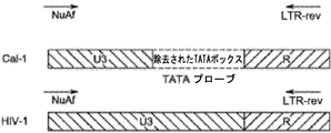
TATAボックスを欠くU3領域を有するレンチウイルスベクターの一例が、図1Aに示された「Cal-1」(LVsh5/C46)レンチウイルスベクターである。図1Bおよび1D〜1Iに示すように、Cal-1は、ヌクレオチド423から556に及ぶ3'LTRのU3領域内に欠失を含み、そのような欠失はTATAボックス(すなわち、SEQ ID NO: 11の配列)中に延びている。Cal-1と比べ、HIVの3'LTRの野生型U3領域は無傷のTATAボックスを含む(たとえば、図1Bおよび1D〜1Iを参照)。事実、野生型HIVのU3領域は、細胞活性化に応答して、感染細胞中のHIVゲノムの基礎的および誘導的発現を変調するエンハンサーおよびプロモーター要素を含む。当然、当業者は、Cal-1は1つのレンチウイルスベクターを例示したものに過ぎないこと、他のベクターは異なる3'LTR領域を有し得、たとえば野生型U3領域と比べて異なる配列を有する、または異なるヌクレオチド欠失を含むU3領域を含み、これらの異なるレンチウイルスベクターが本明細書中に開示される方法にしたがって検出され得ることを理解するであろう。 An example of a lentiviral vector having a U3 region lacking a TATA box is the “Cal-1” (LVsh5 / C46) lentiviral vector shown in FIG. 1A. As shown in FIGS. 1B and 1D-1I, Cal-1 contains a deletion in the U3 region of the 3′LTR spanning nucleotides 423 to 556, and such a deletion contains a TATA box (ie, SEQ ID NO: 11 arrays). Compared to Cal-1, the wild-type U3 region of the 3′LTR of HIV contains an intact TATA box (see, eg, FIGS. 1B and 1D-1I). In fact, the U3 region of wild-type HIV contains enhancer and promoter elements that modulate basal and inducible expression of the HIV genome in infected cells in response to cell activation. Of course, those skilled in the art will appreciate that Cal-1 is only an example of one lentiviral vector, other vectors may have different 3 ′ LTR regions, eg, different sequences compared to the wild type U3 region. It will be appreciated that these different lentiviral vectors can be detected according to the methods disclosed herein, including U3 regions containing different nucleotide deletions.
方法の概説
本明細書に開示されるアッセイ法は、レンチウイルスベクターおよび野生型HIVの3'LTRのU3領域の差異を利用し、したがって、本明細書に記載されるように、特異的に設計されたプライマーおよび/またはプローブが、レンチウイルスベクターにはハイブリダイズするが、野生型HIVにはハイブリダイズしない、およびその反対であることを可能にする。
Method Overview The assay method disclosed herein takes advantage of the differences in the U3 region of the 3′LTR of lentiviral vectors and wild type HIV and is therefore specifically designed as described herein The primers and / or probes that are made hybridize to the lentiviral vector but not to wild-type HIV, and vice versa.
プライマー
当業者は、レンチウイルスベクターおよびHIVの3'LTRの構造に基づいて適当なハイブリダイゼーション特性を有するプライマー配列を設計することができることを理解するであろう。たとえば、当業者は、レンチウイルスベクターの3'LTRの配列に基づいて適当なハイブリダイゼーション特性を有するプライマーを設計し得ることを理解するであろう。本明細書に開示されるプライマーは、特に、本明細書に記載された標的特異的プライマーからいくつかのアンプリコン種を産生することができるマルチプレックス増幅反応の成分として企図される。
Primers The skilled artisan will understand that primer sequences with appropriate hybridization properties can be designed based on the lentiviral vector and the structure of the 3 ′ LTR of HIV. For example, one of skill in the art will understand that primers with appropriate hybridization properties can be designed based on the sequence of the 3 ′ LTR of a lentiviral vector. The primers disclosed herein are specifically contemplated as components of a multiplex amplification reaction that can produce several amplicon species from the target-specific primers described herein.
いくつかの態様において、レンチウイルスベクターおよび/またはHIVの3'LTRのU3領域内の配列にハイブリダイズするようなフォワードプライマーが選択される。いくつかの態様において、本明細書の中で以下さらに詳細に説明されるように、レンチウイルスベクターおよびHIVの両方の増幅に同じフォワードプライマーが使用される(図2および4を参照)。他の態様において、レンチウイルスベクターおよびHIVを増幅するために異なるフォワードプライマーが使用される(図3を参照)。 In some embodiments, a forward primer is selected that hybridizes to a lentiviral vector and / or a sequence within the U3 region of the HIV 3 ′ LTR. In some embodiments, the same forward primer is used for amplification of both lentiviral vectors and HIV, as described in further detail herein below (see FIGS. 2 and 4). In other embodiments, different forward primers are used to amplify the lentiviral vector and HIV (see FIG. 3).
いくつかの態様において、3'LTRのU3領域内の、SEQ ID NO: 1の配列と少なくとも85%の同一性を有する配列にハイブリダイズするようなフォワードプライマーが選択される。他の態様において、3'LTRのU3領域内の、SEQ ID NO: 1の配列と少なくとも90%の同一性を有する配列にハイブリダイズするようなフォワードプライマーが選択される。さらに他の態様において、3'LTRのU3領域内の、SEQ ID NO: 1の配列と少なくとも95%の同一性を有する配列にハイブリダイズするようなフォワードプライマーが選択される。 In some embodiments, a forward primer is selected that hybridizes to a sequence having at least 85% identity to the sequence of SEQ ID NO: 1 within the U3 region of the 3 ′ LTR. In other embodiments, a forward primer is selected that hybridizes to a sequence having at least 90% identity to the sequence of SEQ ID NO: 1 within the U3 region of the 3 ′ LTR. In yet other embodiments, a forward primer is selected that hybridizes to a sequence having at least 95% identity with the sequence of SEQ ID NO: 1 within the U3 region of the 3 ′ LTR.
いくつかの態様において、3'LTRのU3領域内の、SEQ ID NO: 3の配列と少なくとも85%の同一性を有する配列にハイブリダイズするようなフォワードプライマーが選択される。他の態様において、3'LTRのU3領域内の、SEQ ID NO: 3の配列と少なくとも90%の同一性を有する配列にハイブリダイズするようなフォワードプライマーが選択される。さらに他の態様において、3'LTRのU3領域内の、SEQ ID NO: 3の配列と少なくとも95%の同一性を有する配列にハイブリダイズするようなフォワードプライマーが選択される。 In some embodiments, a forward primer is selected that hybridizes to a sequence having at least 85% identity to the sequence of SEQ ID NO: 3 within the U3 region of the 3 ′ LTR. In other embodiments, a forward primer is selected that hybridizes to a sequence having at least 90% identity with the sequence of SEQ ID NO: 3 within the U3 region of the 3 ′ LTR. In yet another embodiment, a forward primer is selected that hybridizes to a sequence having at least 95% identity with the sequence of SEQ ID NO: 3 within the U3 region of the 3 ′ LTR.
本明細書に記載されるアッセイでの使用に適した1つのフォワードプライマーがNuAfプライマーである。当業者は、レンチウイルスベクターおよびHIVのU3配列に依存して、NuAfプライマーがレンチウイルスベクターおよびHIVの両方にハイブリダイズすることができることを理解するであろう。いくつかの態様において、NuAfプライマーは、SEQ ID NO: 2の配列またはSEQ ID NO: 2の配列と少なくとも90%の同一性を有する配列を含む(図2および4を参照)。 One forward primer suitable for use in the assays described herein is the NuAf primer. One skilled in the art will appreciate that depending on the lentiviral vector and the U3 sequence of HIV, NuAf primers can hybridize to both the lentiviral vector and HIV. In some embodiments, the NuAf primer comprises the sequence of SEQ ID NO: 2 or a sequence having at least 90% identity with the sequence of SEQ ID NO: 2 (see FIGS. 2 and 4).
本明細書に記載されるアッセイでの使用に適したもう1つのフォワードプライマーがTATAプライマーである。TATAプライマーは、HIVにはハイブリダイズすることができるが、3'LTRのU3領域中のTATAボックス配列がないかまたは欠失しているレンチウイルスベクターにはハイブリダイズすることができない。TATAプライマーは、SEQ ID NO: 4の配列またはSEQ ID NO: 4の配列と少なくとも90%の同一性を有する配列を含む(図3を参照)。 Another forward primer suitable for use in the assays described herein is a TATA primer. TATA primers can hybridize to HIV but cannot hybridize to lentiviral vectors that lack or lack the TATA box sequence in the U3 region of the 3 ′ LTR. The TATA primer comprises the sequence of SEQ ID NO: 4 or a sequence having at least 90% identity with the sequence of SEQ ID NO: 4 (see FIG. 3).
いくつかの態様において、3'LTRのR領域内の配列にハイブリダイズするようなリバースプライマーが選択される。他の態様において、3'LTRのR領域の5'末端の配列にハイブリダイズするようなリバースプライマーが選択される。本明細書の中でさらに説明されるように、いくつかの態様において、レンチウイルスベクターおよび野生型HIVの両方の増幅に同じリバースプライマーが使用される。すなわち、リバースプライマーは、レンチウイルスベクターおよび野生型HIVの両方のR領域内の共通の配列に対して設計されている。 In some embodiments, a reverse primer is selected that hybridizes to a sequence within the R region of the 3 ′ LTR. In other embodiments, a reverse primer is selected that hybridizes to a sequence at the 5 ′ end of the R region of the 3 ′ LTR. As further described herein, in some embodiments, the same reverse primer is used for amplification of both lentiviral vectors and wild type HIV. That is, the reverse primer is designed for a common sequence in the R region of both the lentiviral vector and wild type HIV.
いくつかの態様において、3'LTRのR領域内の、SEQ ID NO: 5の配列と少なくとも85%の同一性を有する配列にハイブリダイズするようなリバースプライマーが選択される。他の態様において、3'LTRのR領域内の、SEQ ID NO: 5の配列と少なくとも90%の同一性を有する配列にハイブリダイズするようなリバースプライマーが選択される。さらに他の態様において、3'LTRのR領域内の、SEQ ID NO: 5の配列と少なくとも95%の同一性を有する配列にハイブリダイズするようなリバースプライマーが選択される。 In some embodiments, a reverse primer is selected that hybridizes to a sequence having at least 85% identity with the sequence of SEQ ID NO: 5 within the R region of the 3 ′ LTR. In other embodiments, a reverse primer is selected that hybridizes to a sequence having at least 90% identity with the sequence of SEQ ID NO: 5 within the R region of the 3 ′ LTR. In yet another embodiment, a reverse primer is selected that hybridizes to a sequence having at least 95% identity with the sequence of SEQ ID NO: 5 within the R region of the 3 ′ LTR.
開示されるアッセイでの使用に適した1つのリバースプライマーがLTR-revプライマーである。LTR-revプライマーはSEQ ID NO: 6の配列を含む。LTR-revプライマーは、レンチウイルスベクターおよびHIVの両方にハイブリダイズすることができる。 One reverse primer suitable for use in the disclosed assay is the LTR-rev primer. The LTR-rev primer comprises the sequence of SEQ ID NO: 6. LTR-rev primers can hybridize to both lentiviral vectors and HIV.
プローブ
概して、本明細書に開示される方法に利用されるプローブは、「FRETプローブ」(Forsterまたは蛍光共鳴エネルギー移動)と呼ばれるプローブ、すなわち蛍光レポーター・クエンチャー対を含むプローブのクラスに属する。いくつかの態様において、本明細書に記載される方法に利用されるプローブはTAQMAN(登録商標)プローブである。TAQMAN(登録商標)プローブ(Heid et al., 1996)は、Taqポリメラーゼの蛍光を発生させる5'エキソヌクレアーゼ活性を使用して、核酸試料中の標的配列の量を計測する。TAQMAN(登録商標)プローブは、通常は5'塩基またはその近くに蛍光色素を含み、かつ一般に3'塩基またはその近くにクエンチング部分を含むオリゴヌクレオチドである。クエンチャー部分は、TAMRAのような色素であってもよいし、または4-(4-ジメチルアミノフェニルアゾ)安息香酸(DABCYL)のような非蛍光分子であってもよい。照射を受けると、励起した蛍光色素は、蛍光を発するのではなく、エネルギーを近くのクエンチング色素分子に移動させる(上記FRET)。したがって、プローブが無傷であるとき、レポーターとクエンチャーとの近接が任意の蛍光の放出を防ぐ。TAQMAN(登録商標)プローブは、PCR産物の内部領域にアニーリングするように設計されている。ポリメラーゼが、TAQMAN(登録商標)プローブが結合している鋳型を複製するとき、その5'エキソヌクレアーゼ活性がプローブを切断する。これがクエンチャーの活性を終了させ(FRETなし)、レポーター色素が蛍光を発し始め、その蛍光が、プローブ切断の速度に比例して各サイクルで増大する。PCR産物の蓄積は、レポーター色素の蛍光の増加をモニターすることによって検出される(このプロセスにおいては、プローブだけがFRET標識され、プライマーは標識されない)。TAQMAN(登録商標)アッセイは、普遍的な熱サイクルパラメータおよびPCR反応条件を使用する。プローブが標的にハイブリダイズする場合のみ切断が起こるため、検出された蛍光は特異的な増幅に由来する。ハイブリダイゼーションおよび切断のプロセスは産物の指数蓄積を妨害しない。
Probes Generally, the probes utilized in the methods disclosed herein belong to a class of probes called “FRET probes” (Forster or Fluorescence Resonance Energy Transfer), ie, probes that contain a fluorescent reporter quencher pair. In some embodiments, the probe utilized in the methods described herein is a TAQMAN® probe. The TAQMAN® probe (Heid et al., 1996) measures the amount of target sequence in a nucleic acid sample using 5 ′ exonuclease activity that generates Taq polymerase fluorescence. TAQMAN® probes are oligonucleotides that typically include a fluorescent dye at or near the 5 ′ base and generally include a quenching moiety at or near the 3 ′ base. The quencher moiety may be a dye such as TAMRA or a non-fluorescent molecule such as 4- (4-dimethylaminophenylazo) benzoic acid (DABCYL). Upon irradiation, the excited fluorescent dye does not fluoresce but transfers energy to nearby quenching dye molecules (FRET above). Thus, when the probe is intact, the proximity of the reporter and quencher prevents the emission of any fluorescence. TAQMAN® probes are designed to anneal to internal regions of PCR products. When the polymerase replicates the template to which the TAQMAN® probe is bound, its 5 ′ exonuclease activity cleaves the probe. This terminates the activity of the quencher (no FRET) and the reporter dye begins to fluoresce, which increases with each cycle in proportion to the rate of probe cleavage. Accumulation of the PCR product is detected by monitoring the increase in fluorescence of the reporter dye (in this process, only the probe is FRET labeled and the primer is not labeled). The TAQMAN® assay uses universal thermal cycling parameters and PCR reaction conditions. Since cleavage occurs only when the probe hybridizes to the target, the detected fluorescence comes from specific amplification. The hybridization and cleavage process does not interfere with the exponential accumulation of product.
他の態様において、開示される方法に利用されるプローブは分子ビーコンである。分子ビーコンは、細胞内に存在する特定のヌクレオチド配列の同定のためのプローブである(Tyagi et al., (1998) Nature Biotechnology 16:49-53)。分子ビーコンは、DNAもしくはRNAのような核酸だけで構成されることもできるし、またはペプチド核酸(PNA)コンジュゲートで構成されることもできる。特定のヌクレオチド配列への分子ビーコンの結合は、インビトロまたはインビボのいずれかでそれらの配列の存在の同定を可能にする。分子ビーコンは、コンジュゲート(たとえば、量子ドット標識ビーズのような構造)、プローブ、蛍光体およびクエンチング部分を含む。プローブは、親水性結合基が一本鎖オリゴヌクレオチドの一端に結合し、クエンチング部分がその一本鎖オリゴヌクレオチドの他端に結合しているステム・ループ構造を含む、一本鎖オリゴヌクレオチドである。蛍光体は、任意の蛍光有機色素であることができるし、または発光が量子ドット標識ビーズの発光と重ならないような単一量子ドットであることができる。クエンチング部分は、望ましくは、蛍光体の発光をクエンチングする。蛍光体の発光をクエンチングする任意の適当なクエンチング部分を上記コンジュゲートにおいて使用することができる。 In other embodiments, the probe utilized in the disclosed method is a molecular beacon. Molecular beacons are probes for the identification of specific nucleotide sequences present in cells (Tyagi et al., (1998) Nature Biotechnology 16: 49-53). Molecular beacons can be composed solely of nucleic acids such as DNA or RNA, or can be composed of peptide nucleic acid (PNA) conjugates. Binding of molecular beacons to specific nucleotide sequences allows identification of the presence of those sequences either in vitro or in vivo. A molecular beacon includes a conjugate (eg, a structure like a quantum dot labeled bead), a probe, a fluorophore, and a quenching moiety. A probe is a single-stranded oligonucleotide that includes a stem-and-loop structure in which a hydrophilic binding group is attached to one end of a single-stranded oligonucleotide and a quenching moiety is attached to the other end of the single-stranded oligonucleotide. is there. The phosphor can be any fluorescent organic dye or can be a single quantum dot such that the emission does not overlap with the emission of the quantum dot labeled beads. The quenching moiety desirably quenches the phosphor emission. Any suitable quenching moiety that quenches the emission of the phosphor can be used in the conjugate.
さらに他の態様において、開示される方法に利用されるプローブはデュアルハイブリダイゼーションプローブである。概して、デュアルハイブリダイゼーションプローブは、2つの配列特異的DNAプライマーに加えて、2つの配列特異的オリゴヌクレオチドプローブを使用する。2つのプローブは、標的中の隣接する配列に結合するように設計されている。デュアルハイブリダイゼーションプローブは、FRETを示す一対の色素で標識されている。ドナー色素が第1のプローブの3'末端に結合し、一方で、アクセプター色素が第2のプローブの5'末端に結合している。 In yet other embodiments, the probe utilized in the disclosed method is a dual hybridization probe. In general, dual hybridization probes use two sequence-specific oligonucleotide probes in addition to two sequence-specific DNA primers. The two probes are designed to bind to adjacent sequences in the target. The dual hybridization probe is labeled with a pair of dyes indicating FRET. A donor dye is attached to the 3 ′ end of the first probe, while an acceptor dye is attached to the 5 ′ end of the second probe.
リアルタイムPCRの間、ドナー色素に特異的な波長で励起が実施され、アクセプター色素の発光波長で反応がモニターされる。アニーリング工程において、プローブは、それらの標的配列と頭−尾配置でハイブリダイズする。このアニーリングがドナー色素とアクセプター色素とを近接させて、FRETを生じさせ、その結果、アクセプターによる蛍光発光を生じさせる。アクセプター蛍光の増加量は、存在するPCR産物の量に比例する。 During real-time PCR, excitation is performed at a wavelength specific for the donor dye and the reaction is monitored at the emission wavelength of the acceptor dye. In the annealing step, the probes hybridize with their target sequence in a head-to-tail configuration. This annealing brings the donor dye and acceptor dye close together to produce FRET, resulting in fluorescence emission by the acceptor. The amount of increase in acceptor fluorescence is proportional to the amount of PCR product present.
当業者は、レンチウイルスベクターおよび野生型HIVの構造および配列に基づいて適当なハイブリダイゼーション特性を有するプローブを設計することができることを理解するであろう。たとえば、当業者は、レンチウイルスベクターまたはHIVに固有である3'LTRのU3およびR領域の配列に基づいてプローブを設計し得ることを理解するであろう。たとえば、野生型U3 3'LTR領域を除外してレンチウイルスベクターU3領域にハイブリダイズするプローブを設計し得るし、その逆のプローブも設計し得る。本明細書に記されるように、特定のレンチウイルスベクターは、野生型U3領域と比べ、U3配列内に特定の欠失を含むU3領域を含み、当業者は、これらのレンチウイルスベクターU3領域にはハイブリダイズするが、野生型U3領域にはハイブリダイズしないプローブを設計することができる。 One skilled in the art will appreciate that probes having appropriate hybridization properties can be designed based on the structure and sequence of lentiviral vectors and wild type HIV. For example, one skilled in the art will appreciate that probes can be designed based on the sequences of the U3 and R regions of the 3 ′ LTR that are unique to lentiviral vectors or HIV. For example, a probe can be designed that hybridizes to the lentiviral vector U3 region, excluding the wild type U3 3 ′ LTR region, and vice versa. As noted herein, certain lentiviral vectors contain U3 regions that contain certain deletions within the U3 sequence compared to the wild-type U3 region, and those skilled in the art will recognize these lentiviral vector U3 regions. A probe can be designed that hybridizes to, but not to the wild-type U3 region.
いくつかの態様において、本明細書に開示される方法は、レンチウイルスベクターの3'LTRのU3領域およびR領域(すなわち「ジャンクション部位」)に及ぶ配列にアニーリングするジャンクションプローブを利用する。図1Cは、野生型U3領域と比べて特定の欠失を含むU3領域を含む、レンチウイルスベクターのジャンクション部位を示す。適切に設計されたジャンクションプローブは、このジャンクション部位またはこのジャンクション部位の一部分もしくはフラグメントにハイブリダイズする部分を含み得る。いくつかの態様において、ジャンクションプローブは第1の部分および第2の部分を含み、第1の部分は、レンチウイルスベクターの3'LTRのU3領域内のヌクレオチド配列の一部分またはフラグメントにハイブリダイズするように設計されており;第2の部分は、3'LTRのR領域内のヌクレオチド配列の一部分またはフラグメントにハイブリダイズするように設計されている。 In some embodiments, the methods disclosed herein utilize junction probes that anneal to sequences spanning the U3 and R regions (ie, “junction sites”) of the 3 ′ LTR of a lentiviral vector. FIG. 1C shows the junction site of a lentiviral vector containing a U3 region containing a specific deletion compared to the wild type U3 region. A properly designed junction probe may include a portion that hybridizes to the junction site or a portion or fragment of the junction site. In some embodiments, the junction probe comprises a first portion and a second portion, such that the first portion hybridizes to a portion or fragment of a nucleotide sequence within the U3 region of the 3 ′ LTR of a lentiviral vector. The second portion is designed to hybridize to a portion or fragment of a nucleotide sequence within the R region of the 3 ′ LTR.
いくつかの態様において、ジャンクションプローブがハイブリダイズする3'LTRのU3領域は、SEQ ID NO: 12の配列と少なくとも80%の同一性を有する配列を含む。他の態様において、ジャンクションプローブがハイブリダイズするレンチウイルスベクターの3'LTRのU3領域は、SEQ ID NO: 12の配列と少なくとも90%の同一性を有する配列を含む。さらなる態様において、ジャンクションプローブがハイブリダイズするレンチウイルスベクターの3'LTRのU3領域は、SEQ ID NO: 12の配列と少なくとも95%の同一性を有する配列を含む。なおさらなる態様において、ジャンクションプローブがハイブリダイズするレンチウイルスベクターの3'LTRのU3領域はSEQ ID NO: 12の配列を含む。 In some embodiments, the U3 region of the 3 ′ LTR to which the junction probe hybridizes comprises a sequence having at least 80% identity with the sequence of SEQ ID NO: 12. In other embodiments, the U3 region of the 3′LTR of the lentiviral vector to which the junction probe hybridizes comprises a sequence having at least 90% identity with the sequence of SEQ ID NO: 12. In a further embodiment, the U3 region of the 3′LTR of the lentiviral vector to which the junction probe hybridizes comprises a sequence having at least 95% identity with the sequence of SEQ ID NO: 12. In yet a further embodiment, the U3 region of the 3′LTR of the lentiviral vector to which the junction probe hybridizes comprises the sequence of SEQ ID NO: 12.
いくつかの態様において、ジャンクションプローブがハイブリダイズするレンチウイルスベクターの3'LTRのR領域は、SEQ ID NO: 13の配列と少なくとも90%の同一性を有する配列を含む。他の態様において、ジャンクションプローブがハイブリダイズするレンチウイルスベクターの3'LTRのR領域はSEQ ID NO: 13の配列を含む。 In some embodiments, the R region of the 3 ′ LTR of the lentiviral vector to which the junction probe hybridizes comprises a sequence having at least 90% identity with the sequence of SEQ ID NO: 13. In other embodiments, the R region of the 3′LTR of the lentiviral vector to which the junction probe hybridizes comprises the sequence of SEQ ID NO: 13.
当業者は、任意のレンチウイルスベクターの異なるU3領域およびR領域を受け入れるための任意のジャンクションプローブを設計し得ることを理解するであろう。いくつかの態様において、ジャンクションプローブは、SEQ ID NO: 14の配列またはSEQ ID NO: 14の配列と少なくとも90%の同一性を有する配列を含む。いくつかの態様において、ジャンクションプローブは、検出可能な部分、たとえば蛍光レポーターにコンジュゲートされている。いくつかの態様において、蛍光レポーターは、Tex-615、Tye-563、Tye-665、Joe、Cy3、Max、Rox、Tet、Texas Red-X、Tamara、およびYakima Yellowからなる群より選択される。他の態様において、蛍光レポーターはシアニン色素、たとえばインドジカルボシアニン(Cy5TM)を含む。 One skilled in the art will appreciate that any junction probe can be designed to accept the different U3 and R regions of any lentiviral vector. In some embodiments, the junction probe comprises the sequence of SEQ ID NO: 14 or a sequence having at least 90% identity with the sequence of SEQ ID NO: 14. In some embodiments, the junction probe is conjugated to a detectable moiety, such as a fluorescent reporter. In some embodiments, the fluorescent reporter is selected from the group consisting of Tex-615, Tye-563, Tye-665, Joe, Cy3, Max, Rox, Tet, Texas Red-X, Tamara, and Yakima Yellow. In other embodiments, the fluorescent reporter comprises a cyanine dye, such as indodicarbocyanine (Cy5TM).
開示されるアッセイでの使用に適したもう1つのプローブが、TARプローブ、すなわち、HIVの3'LTR中のTAR要素に特異的なプローブである。いくつかの態様において、TARプローブは、本明細書に記載されるようなTaqMan(登録商標)プローブである。いくつかの態様において、SEQ ID NO: 7の配列と少なくとも85%の同一性を有する3'LTR内の配列にハイブリダイズするようなプローブが選択される。他の態様において、SEQ ID NO: 7の配列と少なくとも90%の同一性を有する3'LTR内の配列にハイブリダイズするようなTARプローブが選択される。さらに他の態様において、SEQ ID NO: 7の配列と少なくとも95%の同一性を有する3'LTR内の配列にハイブリダイズするようなTARプローブが選択される。いくつかの態様において、TARプローブはSEQ ID NO: 8の配列を含む。いくつかの態様において、TARプローブは検出可能な部分、たとえば蛍光レポーターにコンジュゲートされている。いくつかの態様において、蛍光レポーターは、Tex-615、Tye-563、Tye-665、Joe、Cy3、Max、Rox、Tet、Texas Red-X、Tamara、およびYakima Yellowからなる群より選択される。他の態様において、TARプローブは蛍光レポーターおよびクエンチャーを含む。いくつかの態様において、蛍光レポーターはフルオレセイン(CAS 2321-07-5)である。 Another probe suitable for use in the disclosed assay is a TAR probe, ie, a probe specific for the TAR element in the 3 ′ LTR of HIV. In some embodiments, the TAR probe is a TaqMan® probe as described herein. In some embodiments, a probe is selected that hybridizes to a sequence in the 3 ′ LTR that has at least 85% identity to the sequence of SEQ ID NO: 7. In other embodiments, a TAR probe is selected that hybridizes to a sequence in the 3 ′ LTR that has at least 90% identity to the sequence of SEQ ID NO: 7. In yet other embodiments, a TAR probe is selected that hybridizes to a sequence in the 3 ′ LTR that has at least 95% identity to the sequence of SEQ ID NO: 7. In some embodiments, the TAR probe comprises the sequence of SEQ ID NO: 8. In some embodiments, the TAR probe is conjugated to a detectable moiety, such as a fluorescent reporter. In some embodiments, the fluorescent reporter is selected from the group consisting of Tex-615, Tye-563, Tye-665, Joe, Cy3, Max, Rox, Tet, Texas Red-X, Tamara, and Yakima Yellow. In other embodiments, the TAR probe comprises a fluorescent reporter and a quencher. In some embodiments, the fluorescent reporter is fluorescein (CAS 2321-07-5).
開示されるアッセイでの使用に適したもう1つのプローブが、TATAプローブ、すなわち、野生型3'LTRのU3領域中のTATAボックスに特異的なプローブである。いくつかの態様において、TATAプローブはTaqMan(登録商標)プローブである。いくつかの態様において、TATAプローブは3'LTRのU3配列にハイブリダイズする(アンチセンス鎖を標的とする)。いくつかの態様において、SEQ ID NO: 9の配列と少なくとも85%の同一性を有する3'LTR内の配列にハイブリダイズするようなTATAプローブが選択される。他の態様において、SEQ ID NO: 9の配列と少なくとも90%の同一性を有する3'LTRのU3領域内の配列にハイブリダイズするようなTATAプローブが選択される。さらに他の態様において、SEQ ID NO: 9の配列と少なくとも95%の同一性を有する3'LTRのU3領域内の配列にハイブリダイズするようなTATAプローブが選択される。いくつかの態様において、TATAプローブはSEQ ID NO: 10の配列を含む。いくつかの態様において、TATAプローブは蛍光レポーターおよびクエンチャーを含む。いくつかの態様において、蛍光レポーターは、Tex-615、Tye-563、Tye-665、Joe、Cy3、Max、Rox、Tet、Texas Red-X、Tamara、およびYakima Yellowからなる群より選択される。他の態様において、TARプローブは蛍光レポーターおよびクエンチャーを含む。いくつかの態様において、蛍光レポーターはフルオレセイン(CAS 2321-07-5)である。 Another probe suitable for use in the disclosed assay is a TATA probe, ie, a probe specific for the TATA box in the U3 region of the wild type 3 ′ LTR. In some embodiments, the TATA probe is a TaqMan® probe. In some embodiments, the TATA probe hybridizes to the U3 sequence of the 3 ′ LTR (targets the antisense strand). In some embodiments, a TATA probe is selected that hybridizes to a sequence in the 3 ′ LTR that has at least 85% identity to the sequence of SEQ ID NO: 9. In other embodiments, a TATA probe is selected that hybridizes to a sequence within the U3 region of the 3′LTR that has at least 90% identity to the sequence of SEQ ID NO: 9. In yet another embodiment, a TATA probe is selected that hybridizes to a sequence within the U3 region of the 3′LTR that has at least 95% identity to the sequence of SEQ ID NO: 9. In some embodiments, the TATA probe comprises the sequence of SEQ ID NO: 10. In some embodiments, the TATA probe includes a fluorescent reporter and a quencher. In some embodiments, the fluorescent reporter is selected from the group consisting of Tex-615, Tye-563, Tye-665, Joe, Cy3, Max, Rox, Tet, Texas Red-X, Tamara, and Yakima Yellow. In other embodiments, the TAR probe comprises a fluorescent reporter and a quencher. In some embodiments, the fluorescent reporter is fluorescein (CAS 2321-07-5).
アッセイ法
試料は、市販され、かつ当業者に公知の標準的なDNA抽出キットを使用して回収され得る。いくつかの態様において、DNA抽出キットは、BioLine DNA抽出キット(BioLine, Taunton, MAから市販)である。いくつかの態様において、試料の非限定的な供給源は、血液試料、血漿試料、組織試料、生検試料、またはフローサイトメトリーによる選別後に創製された試料を含む。
Assay samples are commercially available and can be collected using standard DNA extraction kits known to those skilled in the art. In some embodiments, the DNA extraction kit is a BioLine DNA extraction kit (commercially available from BioLine, Taunton, MA). In some embodiments, non-limiting sources of samples include blood samples, plasma samples, tissue samples, biopsy samples, or samples created after sorting by flow cytometry.
DNA、たとえばプロウイルスDNAまたは導入遺伝子DNAは、当業者に公知の方法、たとえば開示内容が参照により全体として本明細書に組み入れられるManiatis et al., Molecular cloning: A laboratory manual, (Cold Spring Harbor Laboratory, Cold Spring Harbor, NY), 1982によって記載されている手順を使用して試料から抽出され得る。手順は、細胞溶解物を調製したのち、プロテイナーゼKによって消化し、多段階フェノール抽出、エタノール沈殿およびリボヌクレアーゼ消化によってDNA精製を得る工程を含む。 DNA, such as proviral DNA or transgene DNA, is obtained by methods known to those skilled in the art, such as Maniatis et al., Molecular cloning: A laboratory manual, (Cold Spring Harbor Laboratory, the disclosure of which is incorporated herein by reference in its entirety. , Cold Spring Harbor, NY), 1982. The procedure involves preparing a cell lysate and then digesting with proteinase K to obtain DNA purification by multi-step phenol extraction, ethanol precipitation and ribonuclease digestion.
DNAの代わりに、RNAを、本明細書に記載されるアッセイ法に使用してもよい。RNAを使用する場合、適当な逆転写酵素による相補的DNA(cDNA)への逆転写が必要である。したがって、DNAの増幅およびその後の分析を記載する以下の方法がRNA分析に応用可能である。いくつかの態様において、ウイルスRNAは、公知の方法、たとえばBoom, R. et al. (J. Clin. Microbiol. 28(3): 495-503 (1990);参照により本明細書に組み入れられる)に記載されている方法または他の従来の方法、たとえば酸フェノール法(たとえば酸グアニジン・フェノール・クロロホルム(AGPC)法)、たとえばChomczynski and Sacchiの方法(Anal. Biochem. 162, 156-159 (1987))を用いるグアニジンイソチオシアネート法を使用して単離し得る。いくつかの態様において、cDNAは、SENSISCRIPT(登録商標)RT(Qiagen)を用いて合成し得る。いくつかの態様において、1段階逆転写酵素およびTaqポリメラーゼキットが利用される。他の態様において、cDNAを合成したのち、リアルタイムDNA PCR法を実施する。 Instead of DNA, RNA may be used in the assay methods described herein. When RNA is used, reverse transcription to complementary DNA (cDNA) with an appropriate reverse transcriptase is required. Thus, the following method describing DNA amplification and subsequent analysis is applicable to RNA analysis. In some embodiments, the viral RNA is obtained by known methods, such as Boom, R. et al. (J. Clin. Microbiol. 28 (3): 495-503 (1990); incorporated herein by reference) Or other conventional methods, such as the acid phenol method (eg, acid guanidine-phenol-chloroform (AGPC) method), such as the method of Chomczynski and Sacchi (Anal. Biochem. 162, 156-159 (1987) ) And guanidine isothiocyanate method. In some embodiments, the cDNA can be synthesized using SENSISCRIPT® RT (Qiagen). In some embodiments, a one-step reverse transcriptase and Taq polymerase kit is utilized. In another embodiment, real-time DNA PCR is performed after cDNA is synthesized.
増幅
核酸配列の増幅のための技術は当業者に公知である。標的配列を増幅する1つの方法は、ポリメラーゼ連鎖反応(PCR)と呼ばれるポリメラーゼ媒介技術を用いる方法である。概して、PCRは、クローニングまたは精製することなく、ゲノムDNAの混合物中の標的配列のセグメントの濃度を高めるための方法である。標的配列を増幅するこの方法は、所望の標的配列を含むDNA混合物に大過剰量の2つのオリゴヌクレオチドプライマーを導入し、続いてDNAポリメラーゼの存在下、正確な一連の熱サイクルに供するすることからなる。2つのプライマーは二本鎖標的配列のそれぞれの鎖に相補的である。増幅を実施するために、混合物を変性させたのち、プライマーを標的分子内のそれらの相補的配列にアニーリングさせる。アニーリングののち、プライマーをポリメラーゼ(たとえばDNAポリメラーゼ)によって伸長させて新たな相補鎖の対を形成する。変性、プライマーアニーリング、およびポリメラーゼ伸長の工程を何度も繰り返して(すなわち、変性、アニーリング、および伸長は1つの「サイクル」を構成し;多数の「サイクル」が存在しうる)、所望の標的配列の、高濃度に増幅されたセグメント(アンプリコン)を得ることができる。所望の標的配列の増幅されたセグメントの長さは、プライマーの互いに対する相対位置によって決まり、したがって、この長さは制御可能なパラメータである。ポリメラーゼ連鎖反応(「PCR」)は、たとえば、米国特許第4,683,202号;米国特許第4,683,195号;米国特許第4,000,159号;米国特許第4,965,188号;米国特許第5,176,995号に記載されている。それぞれの開示内容が参照により全体として本明細書に組み入れられる。
Techniques for amplification of amplified nucleic acid sequences are known to those skilled in the art. One method for amplifying a target sequence is to use a polymerase-mediated technique called polymerase chain reaction (PCR). In general, PCR is a method for increasing the concentration of a segment of a target sequence in a mixture of genomic DNA without cloning or purification. This method of amplifying a target sequence involves introducing a large excess of two oligonucleotide primers into a DNA mixture containing the desired target sequence, followed by a precise series of thermal cycles in the presence of DNA polymerase. Become. The two primers are complementary to their respective strands of the double stranded target sequence. To perform the amplification, the mixture is denatured and then the primers are annealed to their complementary sequences in the target molecule. After annealing, the primer is extended with a polymerase (eg, DNA polymerase) to form a new pair of complementary strands. The steps of denaturation, primer annealing, and polymerase extension are repeated many times (ie, denaturation, annealing, and extension constitute one “cycle”; there can be multiple “cycles”) and the desired target sequence A segment (amplicon) amplified at a high concentration can be obtained. The length of the amplified segment of the desired target sequence is determined by the relative positions of the primers with respect to each other, and thus this length is a controllable parameter. Polymerase chain reaction ("PCR") is described, for example, in US Patent No. 4,683,202; US Patent No. 4,683,195; US Patent No. 4,000,159; US Patent No. 4,965,188; US Patent No. 5,176,995. The disclosure of each is hereby incorporated by reference in its entirety.
PCRは、定性的または定量的に使用し得る。1つの公知の定量的増幅技術が「リアルタイムPCR」である。本明細書の中で使用される用語「リアルタイムPCR」とは、反応中、PCRアッセイから放出されるシグナルが、各PCR増幅サイクル中に、すなわち、PCR反応の終点でアッセイシグナルが検出される従来のPCR法とは対照的に「リアルタイム」で、アンプリコン産生の指標としてモニターされることをいう。リアルタイムPCRは概して、本明細書に記載されるような蛍光レポーターの検出および定量に基づく。レポーターのシグナルは、反応中のPCR産物の量に正比例して増大する。したがって、各サイクルでの蛍光発光の量を記録することにより、PCR産物の量の最初の有意な増加が標的鋳型の初期量と相関する指数増殖期中のPCR反応をモニターすることが可能である。「リアルタイムPCR」の概説に関しては、Dehee et al. J. Virol. Meth. 102:37-51 (2002);およびAldea et al. J. Clin. Microbiol., 40:1060-1062 (2002)を参照すること。これらの開示内容は参照により全体として本明細書に組み入れられる。 PCR can be used qualitatively or quantitatively. One known quantitative amplification technique is “real-time PCR”. As used herein, the term “real-time PCR” refers to the conventional in which the signal released from the PCR assay during the reaction is detected during each PCR amplification cycle, ie at the end of the PCR reaction. In contrast to the PCR method, it is “real time” and is monitored as an indicator of amplicon production. Real-time PCR is generally based on the detection and quantification of fluorescent reporters as described herein. The reporter signal increases in direct proportion to the amount of PCR product in the reaction. Thus, by recording the amount of fluorescence emission at each cycle, it is possible to monitor the PCR reaction during the exponential growth phase, where the first significant increase in the amount of PCR product correlates with the initial amount of target template. For a review of “real-time PCR” see Dehee et al. J. Virol. Meth. 102: 37-51 (2002); and Aldea et al. J. Clin. Microbiol., 40: 1060-1062 (2002). To do. The disclosures of which are incorporated herein by reference in their entirety.
逆転写酵素−ポリメラーゼ連鎖反応とは、RNA鋳型から相補的DNAを産生し、さらにその相補的DNAを、PCRを実施するための鋳型として使用して、上記のようにDNAを複製し、増幅することをいう。逆転写酵素反応において、反応混合物は、RNA鋳型の全部または一部分に相補的なDNA分子を合成するのに十分な温度でインキュベートされる。逆転写反応ののち、反応は、合成されたDNA分子を増幅するのに十分な温度でインキュベートされる。 Reverse transcriptase-polymerase chain reaction is to produce complementary DNA from an RNA template, and then use that complementary DNA as a template for performing PCR to replicate and amplify DNA as described above. That means. In a reverse transcriptase reaction, the reaction mixture is incubated at a temperature sufficient to synthesize DNA molecules that are complementary to all or part of the RNA template. After the reverse transcription reaction, the reaction is incubated at a temperature sufficient to amplify the synthesized DNA molecule.
レンチウイルス核酸の増幅
本開示の1つの局面は、レンチウイルス核酸、たとえばレンチウイルス導入遺伝子を増幅する方法である。いくつかの態様において、レンチウイルス核酸の増幅は、3'LTRのU3領域の一部分にハイブリダイズするフォワードプライマーおよび3'LTRのR領域の一部分にハイブリダイズするリバースプライマーを利用する。いくつかの態様において、レンチウイルス核酸の増幅はNuAfフォワードプライマーおよびLTR-revリバースプライマーを利用する。いくつかの態様において、増幅から産生されるアンプリコンは約100個〜約600個の核酸を含む。いくつかの態様において、産生されたアンプリコンの量が定量化される。
Amplification of Lentiviral Nucleic Acid One aspect of the present disclosure is a method of amplifying a lentiviral nucleic acid, such as a lentiviral transgene. In some embodiments, amplification of a lentiviral nucleic acid utilizes a forward primer that hybridizes to a portion of the U3 region of the 3′LTR and a reverse primer that hybridizes to a portion of the R region of the 3′LTR. In some embodiments, amplification of lentiviral nucleic acid utilizes a NuAf forward primer and an LTR-rev reverse primer. In some embodiments, the amplicon produced from the amplification comprises about 100 to about 600 nucleic acids. In some embodiments, the amount of amplicon produced is quantified.
いくつかの態様において、レンチウイルス核酸の増幅は、レンチウイルスベクターの3'LTR内の配列に特異的なプローブの使用を含む。すなわち、プローブは、レンチウイルスベクターの3'LTRだけにハイブリダイズし、野生型3'LTR、たとえばHIVの3'LTRにはハイブリダイズしない。いくつかの態様において、プローブは、そのような欠失を含まない野生型3'LTRと比べ、レンチウイルスベクターの3'LTRのU3領域中の欠失に最適化されている。いくつかの態様において、欠失は(ここでもまた、野生型U3領域と比べて)TATAボックス配列の欠失を含む。いくつかの態様において、3'LTRのU3領域中の欠失に最適化されたプローブは、レンチウイルスベクターの3'LTRのU3領域の一部分およびレンチウイルスベクターの3'LTRのR領域の一部分に及ぶように設計されている。いくつかの態様において、3'LTRのU3領域中の欠失に最適化されたプローブはジャンクションプローブである。いくつかの態様において、ジャンクションプローブは、配列番号14の配列と少なくとも90%の同一性を有する配列を含む。いくつかの態様において、レンチウイルス核酸またはレンチウイル導入遺伝子の増幅は、たとえばプローブとコンジュゲートした蛍光レポーターの量を検出することにより、増幅された核酸の量を定量化する工程を含む。 In some embodiments, amplification of a lentiviral nucleic acid comprises the use of a probe specific for a sequence within the 3 ′ LTR of the lentiviral vector. That is, the probe hybridizes only to the 3 ′ LTR of the lentiviral vector and not to the wild type 3 ′ LTR, eg, the 3 ′ LTR of HIV. In some embodiments, the probe is optimized for a deletion in the U3 region of the 3′LTR of the lentiviral vector compared to a wild type 3′LTR that does not contain such a deletion. In some embodiments, the deletion comprises a deletion of the TATA box sequence (again compared to the wild type U3 region). In some embodiments, probes optimized for deletions in the U3 region of the 3′LTR are located in a portion of the U3 region of the 3′LTR of the lentiviral vector and a portion of the R region of the 3′LTR of the lentiviral vector. Designed to range. In some embodiments, the probe optimized for the deletion in the U3 region of the 3 ′ LTR is a junction probe. In some embodiments, the junction probe comprises a sequence having at least 90% identity with the sequence of SEQ ID NO: 14. In some embodiments, amplification of a lentiviral nucleic acid or lentiviral transgene comprises quantifying the amount of amplified nucleic acid, for example, by detecting the amount of fluorescent reporter conjugated to a probe.
いくつかの態様において、レンチウイルス核酸の増幅は、フォワードプライマー、リバースプライマーおよびジャンクションプローブを試料に導入し、当業者に公知の手順にしたがってPCRを実施する工程を含む。いくつかの態様において、フォワードプライマーはNuAfであり、リバースプライマーはLTR-revであり、ジャンクションプローブはSEQ ID NO: 14の配列を含む。いくつかの態様において、45〜50サイクルのPCRが実施される。いくつかの態様において、PCR法はリアルタイムPCRまたは定量的リアルタイムPCRである。いくつかの態様において、核酸はRNAであり、RNAはまずcDNAに変換される。いくつかの態様において、核酸はRNAであり、逆転写酵素PCRまたは定量的逆転写酵素PCRのいずれかが利用される。 In some embodiments, the amplification of lentiviral nucleic acid comprises introducing a forward primer, a reverse primer and a junction probe into a sample and performing PCR according to procedures known to those skilled in the art. In some embodiments, the forward primer is NuAf, the reverse primer is LTR-rev, and the junction probe comprises the sequence of SEQ ID NO: 14. In some embodiments, 45-50 cycles of PCR are performed. In some embodiments, the PCR method is real-time PCR or quantitative real-time PCR. In some embodiments, the nucleic acid is RNA, and the RNA is first converted to cDNA. In some embodiments, the nucleic acid is RNA and either reverse transcriptase PCR or quantitative reverse transcriptase PCR is utilized.
一般的なマルチプレックスアッセイ法
本開示のもう1つの局面は、試料中のレンチウイルス核酸およびHIV核酸を検出および/または定量化するマルチプレックス法である。いくつかの態様において、マルチプレックス法は、1つの反応システムまたはチャンバ(本明細書中、以下「反応チューブ」)中で実施される。他の態様において、マルチプレックス法は別々の反応チューブ中で実施される。いくつかの態様において、レンチウイルス核酸はレンチウイルス導入遺伝子である。いくつかの態様において、HIV核酸はプロウイルスDNAである。いくつかの態様において、レンチウイルス核酸および/またはHIV核酸はRNAであり、RNAは、増幅の前に、(たとえば逆転写プロセスによって)まずcDNAに変換される。
General Multiplex Assay Method Another aspect of the present disclosure is a multiplex method for detecting and / or quantifying lentiviral and HIV nucleic acids in a sample. In some embodiments, the multiplex method is performed in a single reaction system or chamber (hereinafter “reaction tube”). In other embodiments, the multiplex method is performed in separate reaction tubes. In some embodiments, the lentiviral nucleic acid is a lentiviral transgene. In some embodiments, the HIV nucleic acid is proviral DNA. In some embodiments, the lentiviral nucleic acid and / or HIV nucleic acid is RNA, and the RNA is first converted to cDNA (eg, by a reverse transcription process) prior to amplification.
いくつかの態様において、マルチプレックス法は、レンチウイルスベクターおよび/またはHIVの3'LTRのU3領域内の配列にハイブリダイズすることができるフォワードプライマーを利用する。いくつかの様態において、マルチプレックス法は、レンチウイルスベクターおよび/またはHIVの3'LTRのR領域内の配列にハイブリダイズすることができるリバースプライマーを利用する。いくつかの様態において、マルチプレックス法は、レンチウイルス核酸の増幅およびHIV核酸の増幅の両方に同じフォワードおよびリバースプライマーを利用する。これらの場合、当業者は、野生型HIV核酸およびレンチウイルスベクター核酸の両方のU3領域内の同じ配列にハイブリダイズすることができるフォワードプライマーを選択することができるであろう。他の態様において、マルチプレックス法は、異なるフォワードプライマーと、同じリバースプライマーとを利用する。いくつかの態様において、フォワードプライマーはNuAfプライマーおよびTATAプライマーから選択される。いくつかの態様において、リバースプライマーはLTR-revプライマーである。 In some embodiments, the multiplex method utilizes a lentiviral vector and / or a forward primer that can hybridize to a sequence within the U3 region of the HIV 3 ′ LTR. In some embodiments, the multiplex method utilizes a lentiviral vector and / or a reverse primer that can hybridize to a sequence within the R region of the 3 ′ LTR of HIV. In some embodiments, the multiplex method utilizes the same forward and reverse primers for both lentiviral nucleic acid amplification and HIV nucleic acid amplification. In these cases, one of skill in the art would be able to select a forward primer that can hybridize to the same sequence within the U3 region of both the wild type HIV nucleic acid and the lentiviral vector nucleic acid. In other embodiments, the multiplex method utilizes different forward primers and the same reverse primer. In some embodiments, the forward primer is selected from a NuAf primer and a TATA primer. In some embodiments, the reverse primer is an LTR-rev primer.
いくつかの態様において、レンチウイルス核酸の量を増幅および/または定量化するためにレンチウイルス核酸に特異的な第1のプローブがPCR法(たとえばリアルタイムPCR法)に用いられ、HIV核酸の量を増幅および/または定量化するためにHIVに特異的な第2のプローブがPCR法に用いられ、第1および第2のプローブは異なる検出可能な部分を含む。いくつかの態様において、第1のプローブは、レンチウイルスベクターの3'LTR内の配列にはハイブリダイズすることができるが、野生型HIVの3'LTR内の配列にはハイブリダイズすることができないプローブである。いくつかの態様において、第1のプローブは、野生型3'LTR配列と比べ、レンチウイルス核酸の3'LTR中の配列欠失に最適化されている。 In some embodiments, a first probe specific for a lentiviral nucleic acid is used in a PCR method (eg, real-time PCR) to amplify and / or quantify the amount of lentiviral nucleic acid, and to determine the amount of HIV nucleic acid. A second probe specific for HIV is used in the PCR method for amplification and / or quantification, and the first and second probes contain different detectable moieties. In some embodiments, the first probe can hybridize to a sequence within the 3 ′ LTR of a lentiviral vector, but cannot hybridize to a sequence within the 3 ′ LTR of wild type HIV. It is a probe. In some embodiments, the first probe is optimized for a sequence deletion in the 3 ′ LTR of the lentiviral nucleic acid relative to the wild type 3 ′ LTR sequence.
いくつかの態様において、第1のプローブは、レンチウイルスベクターの3'LTR、たとえば3'LTR内のジャンクション部位に特異的なジャンクションプローブである。いくつかの態様において、ジャンクションプローブは、レンチウイルスベクターのU3領域内の配列の少なくとも一部分にハイブリダイズすることができ、レンチウイルスベクターのR領域内の配列の少なくとも一部分にハイブリダイズすることできるプローブである。いくつかの態様において、ジャンクションプローブは、SEQ ID NO: 12の配列と少なくとも90%の同一性を有する第1の配列にハイブリダイズすることができる第1の部分;およびSEQ ID NO: 13の配列にハイブリダイズすることができる第2の部分を含む。 In some embodiments, the first probe is a junction probe specific for a junction site within the 3 ′ LTR, eg, the 3 ′ LTR, of a lentiviral vector. In some embodiments, the junction probe is a probe that can hybridize to at least a portion of the sequence in the U3 region of the lentiviral vector and hybridize to at least a portion of the sequence in the R region of the lentiviral vector. is there. In some embodiments, the junction probe is a first portion capable of hybridizing to a first sequence having at least 90% identity with the sequence of SEQ ID NO: 12; and the sequence of SEQ ID NO: 13 A second portion capable of hybridizing to.
いくつかの態様において、第2のプローブはTATAプローブまたはTARプローブから選択される。 In some embodiments, the second probe is selected from a TATA probe or a TAR probe.
いくつかの態様において、レンチウイルス核酸およびHIV核酸の増幅は、たとえば用いられるプローブにコンジュゲートされた異なる蛍光レポーターに対応するシグナルを検出することにより、増幅された核酸の量を定量化する工程を含む。他の態様において、レンチウイルス核酸アンプリコンおよびHIV核酸アンプリコンの電気泳動分離ののち、レンチウイルス核酸アンプリコンの量およびHIV核酸アンプリコンの量が定量化される。 In some embodiments, amplification of lentiviral nucleic acid and HIV nucleic acid comprises quantifying the amount of amplified nucleic acid, for example by detecting signals corresponding to different fluorescent reporters conjugated to the probe used. Including. In other embodiments, after electrophoretic separation of lentiviral nucleic acid amplicons and HIV nucleic acid amplicons, the amount of lentiviral nucleic acid amplicons and HIV nucleic acid amplicons are quantified.
いくつかの態様において、マルチプレックス法は、試料中に存在するHIV核酸の量に対するレンチウイルス核酸の量の比率を決定する工程を含む。いくつかの態様において、決定された比率は、たとえばレンチウイルスベクターを形質導入した幹細胞を用いる治療または遺伝子療法の効果を評価するために使用される。いくつかの態様において、レンチウイルスの量およびHIV核酸の量は、治療効果を評価するために、治療の過程にわたって、すなわちいくつかの異なる時点において測定され得る(たとえば、第1の評価時点と第2の評価時点とを比較し得る場合、第1の評価時点から第2の評価時点までで、HIV核酸と比べて増加したレンチウイルス核酸の量が治療効率を示す場合)。いくつかの態様において、提供された評価に依存してさらなる治療が施される。 In some embodiments, the multiplex method includes determining the ratio of the amount of lentiviral nucleic acid to the amount of HIV nucleic acid present in the sample. In some embodiments, the determined ratio is used to assess the effect of treatment or gene therapy using, for example, stem cells transduced with a lentiviral vector. In some embodiments, the amount of lentivirus and the amount of HIV nucleic acid can be measured over the course of treatment, i.e., at several different time points (e.g., at the first evaluation time point and the first evaluation time point) to evaluate the therapeutic effect. When the two evaluation time points can be compared, the amount of lentiviral nucleic acid increased from the first evaluation time point to the second evaluation time point compared to HIV nucleic acid indicates therapeutic efficiency). In some embodiments, additional treatment is administered depending on the provided assessment.
マルチプレックス「方法1」
図2を参照すると、本開示の1つの局面は、試料中のレンチウイルス核酸およびHIV核酸を同じ反応チューブ中で検出および/または定量化するマルチプレックス法(「方法1」)である。いくつかの態様において、方法はまず、試料中に存在する核酸、すなわちDNAまたはRNAを増幅に備えて準備する工程を含む。RNAが試料内の出発原料であるならば、当業者に公知の手段にしたがってRNAをcNDAに変換する。
Multiplex "
Referring to FIG. 2, one aspect of the present disclosure is a multiplex method (“
ひとたび核酸を準備したならば、フォワードおよびリバースプライマーを試料に導入する。この特定の方法においては、レンチウイルス核酸およびHIV核酸の両方の増幅のために同じフォワードおよびリバースプライマーを使用する(図2を参照)。いくつかの態様において、フォワードプライマーはNuAfプライマーである。いくつかの態様において、フォワードプライマーはSEQ ID NO: 2の配列を有する。いくつかの態様において、リバースプライマーは、レンチウイルス核酸およびHIV核酸の両方の3'LTRのR領域内の配列にハイブリダイズする。いくつかの態様において、リバースプライマーはLTR-revプライマーである。いくつかの態様において、リバースプライマーはSEQ ID NO: 6の配列を有する。任意の特定の理論によって拘束されることを望まないが、この特定のアッセイ法の特異性はフォワードプライマーに基づくと考えられる。ここでもまた、任意の特定の理論によって拘束されることを望まないが、1つのプライマーセットのおかげで、レンチウイルス核酸の増幅効率とHIV核酸の増幅効率とは同じであると考えられる。 Once the nucleic acid is prepared, forward and reverse primers are introduced into the sample. In this particular method, the same forward and reverse primers are used for amplification of both lentiviral and HIV nucleic acids (see Figure 2). In some embodiments, the forward primer is a NuAf primer. In some embodiments, the forward primer has the sequence of SEQ ID NO: 2. In some embodiments, the reverse primer hybridizes to a sequence within the R region of the 3 ′ LTR of both lentiviral and HIV nucleic acids. In some embodiments, the reverse primer is an LTR-rev primer. In some embodiments, the reverse primer has the sequence of SEQ ID NO: 6. While not wishing to be bound by any particular theory, it is believed that the specificity of this particular assay is based on the forward primer. Again, without wishing to be bound by any particular theory, it is believed that the amplification efficiency of the lentiviral nucleic acid and that of the HIV nucleic acid are the same thanks to one primer set.
試料へのプライマーの導入ののち、2つの異なるプローブが導入される。第1の検出可能な部分を含む第1のプローブはレンチウイルス核酸に特異的であり、第2の検出可能な部分を含む第2のプローブはHIV核酸に特異的である(図2を参照)。いくつかの態様において、レンチウイルス核酸に特異的な第1のプローブは、(第1のプローブが、レンチウイルス核酸にはハイブリダイズし得るが、HIV核酸にはハイブリダイズし得ないように、)野生型HIV核酸と比べ、レンチウイルスベクターの3'LTRのU3領域内の欠失に最適化されている。いくつかの態様において、プローブは、レンチウイルス核酸の3'LTR内のジャンクション部位、たとえば3'LTRのU3およびR領域に及ぶジャンクション部位に特異的なジャンクションプローブである。いくつかの態様において、ジャンクションプローブは、3'LTR内の第1の領域および3'LTR内の第2の領域にハイブリダイズすることができる。いくつかの態様において、ジャンクションプローブは、3'LTRのU3領域内のヌクレオチド配列の一部分またはフラグメント;および3'LTRのR領域内のヌクレオチド配列の一部分またはフラグメントにハイブリダイズすることができる。いくつかの態様において、ジャンクションプローブは、SEQ ID NO: 12の配列と少なくとも90%の同一性を有する第1の配列にハイブリダイズすることができる第1の部分;およびSEQ ID NO: 13の配列にハイブリダイズすることができる第2の部分を含む。いくつかの態様において、ジャンクションプローブは、SEQ ID NO: 14の配列と少なくとも80%の同一性を有する配列を含む。他の態様において、ジャンクションプローブは、SEQ ID NO: 14の配列と少なくとも90%の同一性を有する配列を含む。さらに他の態様において、ジャンクションプローブは、SEQ ID NO: 14の配列と少なくとも95%の同一性を有する配列を含む。 After introduction of the primer into the sample, two different probes are introduced. The first probe containing the first detectable moiety is specific for lentiviral nucleic acid and the second probe containing the second detectable moiety is specific for HIV nucleic acid (see Figure 2) . In some embodiments, the first probe specific for the lentiviral nucleic acid is (so that the first probe can hybridize to the lentiviral nucleic acid but not to the HIV nucleic acid). Compared to wild-type HIV nucleic acid, it is optimized for deletion in the U3 region of the 3 ′ LTR of lentiviral vectors. In some embodiments, the probe is a junction probe specific for a junction site within the 3 ′ LTR of a lentiviral nucleic acid, eg, a junction site spanning the U3 and R regions of the 3 ′ LTR. In some embodiments, the junction probe can hybridize to a first region in the 3′LTR and a second region in the 3′LTR. In some embodiments, the junction probe can hybridize to a portion or fragment of a nucleotide sequence within the U3 region of the 3′LTR; and a portion or fragment of a nucleotide sequence within the R region of the 3′LTR. In some embodiments, the junction probe is a first portion capable of hybridizing to a first sequence having at least 90% identity with the sequence of SEQ ID NO: 12; and the sequence of SEQ ID NO: 13 A second portion capable of hybridizing to. In some embodiments, the junction probe comprises a sequence having at least 80% identity with the sequence of SEQ ID NO: 14. In other embodiments, the junction probe comprises a sequence having at least 90% identity with the sequence of SEQ ID NO: 14. In yet other embodiments, the junction probe comprises a sequence having at least 95% identity with the sequence of SEQ ID NO: 14.
いくつかの態様において、HIV核酸に特異的な第2のプローブはTATAプローブである。いくつかの態様において、第2のプローブは、SEQ ID NO: 10の配列またはSEQ ID NO: 10の配列と少なくとも90%の同一性を有する配列を有する。いくつかの態様において、第1のプローブはCy5で標識されている。いくつかの態様において、第2のプローブはFAMで標識されている。 In some embodiments, the second probe specific for HIV nucleic acid is a TATA probe. In some embodiments, the second probe has the sequence of SEQ ID NO: 10 or a sequence having at least 90% identity with the sequence of SEQ ID NO: 10. In some embodiments, the first probe is labeled with Cy5. In some embodiments, the second probe is labeled with FAM.
プローブの導入ののち、標準的なプロトコルにしたがって増幅を実施することができる。いくつかの態様において、約45〜約50のPCRサイクルを実施することができる。いくつかの態様において、PCRはリアルタイムPCRである。RNAが出発原料である場合、はじめにRNAをcDNAに変換する代わりに、逆転写酵素PCRまたはリアルタイム逆転写酵素を利用してもよい。 After introduction of the probe, amplification can be performed according to standard protocols. In some embodiments, about 45 to about 50 PCR cycles can be performed. In some embodiments, the PCR is real-time PCR. If RNA is the starting material, reverse transcriptase PCR or real-time reverse transcriptase may be used instead of first converting the RNA to cDNA.
いくつかの態様において、レンチウイルス核酸の量およびHIV核酸の量は、たとえば異なるプローブにコンジュゲートされた異なる検出可能な部分からのシグナルを検出することによって定量化される。いくつかの態様において、HIV核酸の量に対するレンチウイルス核酸の量の比率。いくつかの態様において、決定された比率は、たとえばレンチウイルスベクターを形質導入した幹細胞を用いる治療または遺伝子療法の効果を評価するために使用される。いくつかの態様において、レンチウイルスの量およびHIV核酸の量は、治療効果を評価するために、治療の過程にわたって、すなわちいくつかの異なる時点において測定され得る(たとえば、第1の評価時点と第2の評価時点とを比較し得る場合、第1の評価時点から第2の評価時点までで、HIV核酸と比べて増加したレンチウイルス核酸の量が治療効率を示す)。いくつかの態様において、提供された評価に依存してさらなる治療が施される。 In some embodiments, the amount of lentiviral nucleic acid and HIV nucleic acid is quantified, for example, by detecting signals from different detectable moieties conjugated to different probes. In some embodiments, the ratio of the amount of lentiviral nucleic acid to the amount of HIV nucleic acid. In some embodiments, the determined ratio is used to assess the effect of treatment or gene therapy using, for example, stem cells transduced with a lentiviral vector. In some embodiments, the amount of lentivirus and the amount of HIV nucleic acid can be measured over the course of treatment, i.e., at several different time points (e.g., at the first evaluation time point and the first evaluation time point) to evaluate the therapeutic effect. If the two evaluation time points can be compared, the amount of lentiviral nucleic acid increased compared to HIV nucleic acid from the first evaluation time point to the second evaluation time point indicates the therapeutic efficiency). In some embodiments, additional treatment is administered depending on the provided assessment.
デュアルチューブ「方法2」
図3を参照すると、本開示の1つの局面は、試料中のレンチウイルス核酸およびHIV核酸を検出および/または定量化する、2つの別々の反応チューブ中で実施されるマルチプレックス法である(「方法2」)。第1の反応チューブは、レンチウイルス核酸の増幅に必要なすべての成分を含む。第2の反応チューブは、HIV核酸の増幅に必要なすべての成分を含む。上記「方法1」と同様に、このアッセイもまた、3'LTRレンチウイルス核酸構造と野生型3'LTR HIV核酸構造との差異を利用し、特に、HIV核酸の増幅開始部位としてTATAボックスを利用する。
Dual tube "
Referring to FIG. 3, one aspect of the present disclosure is a multiplex method performed in two separate reaction tubes that detects and / or quantifies lentiviral and HIV nucleic acids in a sample (“
前記方法と同様に、方法はまず、試料中に存在する核酸、すなわちDNAまたはRNAを増幅に備えて準備する工程を含む。RNAが試料内の出発原料であるならば、RNAをcNDAに変換する。ひとたび核酸を準備したならば、フォワードおよびリバースプライマーを導入する。この特定の方法においては、レンチウイルス核酸の増幅とHIV核酸の増幅とで異なるフォワードを使用する(図3を参照)。レンチウイルス核酸の増幅の場合、フォワードプライマーはNuAfプライマーである。いくつかの態様において、フォワードプライマーはSEQ ID NO: 2の配列を有する。HIV核酸の増幅の場合、フォワードプライマーはTATAプライマーである。いくつかの態様において、フォワードプライマーはSEQ ID NO: 4の配列を有する。 Similar to the above method, the method first comprises the step of preparing the nucleic acid present in the sample, ie DNA or RNA, for amplification. If RNA is the starting material in the sample, convert RNA to cNDA. Once the nucleic acid is prepared, forward and reverse primers are introduced. In this particular method, different forwards are used for lentiviral nucleic acid amplification and HIV nucleic acid amplification (see FIG. 3). For lentiviral nucleic acid amplification, the forward primer is a NuAf primer. In some embodiments, the forward primer has the sequence of SEQ ID NO: 2. For the amplification of HIV nucleic acid, the forward primer is a TATA primer. In some embodiments, the forward primer has the sequence of SEQ ID NO: 4.
いくつかの態様において、レンチウイルス核酸の増幅とHIV核酸の増幅との両方で同じリバースプライマーを使用する。いくつかの態様において、リバースプライマーは、レンチウイルス核酸およびHIV核酸の両方の3'LTRのR領域内の配列にハイブリダイズする。いくつかの態様において、リバースプライマーはLTR-revプライマーである。いくつかの態様において、リバースプライマーはSEQ ID NO: 6の配列を有する。任意の特定の理論によって拘束されることを望まないが、この特定のアッセイ法の特異性はフォワードプライマーに基づくと考えられる。ここでもまた、任意の特定の理論によって拘束されることを望まないが、1つのプライマーセットのおかげで、レンチウイルス核酸の増幅効率とHIV核酸の増幅効率との両方は同じであると考えられる。 In some embodiments, the same reverse primer is used for both lentiviral nucleic acid amplification and HIV nucleic acid amplification. In some embodiments, the reverse primer hybridizes to a sequence within the R region of the 3 ′ LTR of both lentiviral and HIV nucleic acids. In some embodiments, the reverse primer is an LTR-rev primer. In some embodiments, the reverse primer has the sequence of SEQ ID NO: 6. While not wishing to be bound by any particular theory, it is believed that the specificity of this particular assay is based on the forward primer. Again, while not wishing to be bound by any particular theory, it is believed that both lentiviral and HIV nucleic acid amplification efficiencies are the same, thanks to a single primer set.
任意の特定の理論によって拘束されることを望まないが、第1の反応チューブ中の反応(レンチウイルスベクター核酸の増幅)の特異性は、利用される特異性プローブに基づくと考えられる。任意の特定の理論によって拘束されることを望まないが、第2の反応チューブ中の反応(HIV核酸の増幅)の特異性は、利用されるフォワードプライマーに基づくと考えられる。ここでもまた、任意の特定の理論によって拘束されることを望まないが、これらのアッセイは共通のリバースプライマー(たとえばLTR-rev)に基づき、フォワードプライマーの位置は数百塩基の範囲で分離しているため、増幅効率は相対的に同一であり、アッセイは、LV核酸およびHIV核酸検出および/または定量化の両方において信頼し得る比較データを提供し得ると考えられる。 Without wishing to be bound by any particular theory, it is believed that the specificity of the reaction in the first reaction tube (amplification of the lentiviral vector nucleic acid) is based on the specificity probe utilized. Without wishing to be bound by any particular theory, it is believed that the specificity of the reaction (HIV nucleic acid amplification) in the second reaction tube is based on the forward primer utilized. Again, although not wishing to be bound by any particular theory, these assays are based on a common reverse primer (eg LTR-rev) and the position of the forward primer is separated by a few hundred bases. As such, the amplification efficiencies are relatively identical, and it is believed that the assay can provide reliable comparative data in both LV and HIV nucleic acid detection and / or quantification.
試料へのプライマーの導入ののち、2つの異なるプローブが導入される。第1の検出可能な部分を含む第1のプローブはレンチウイルス核酸に特異的であり、第2の検出可能な部分を含む第2のプローブはHIV核酸に特異的である(図3を参照)。いくつかの態様において、レンチウイルス核酸に特異的な第1のプローブは、(第1のプローブが、レンチウイルス核酸にはハイブリダイズし得るが、HIV核酸にはハイブリダイズし得ないように、)野生型HIV核酸と比べ、レンチウイルスベクターの3'LTRのU3領域内の欠失に最適化されている。いくつかの態様において、プローブは、レンチウイルス核酸の3'LTR内のジャンクション部位、たとえば3'LTRのU3およびR領域に及ぶジャンクション部位に特異的なジャンクションプローブである。いくつかの態様において、ジャンクションプローブは、3'LTR内の第1の領域および3'LTR内の第2の領域にハイブリダイズすることができる。いくつかの態様において、ジャンクションプローブは、3'LTRのU3領域内のヌクレオチド配列の一部分またはフラグメント;および3'LTRのR領域内のヌクレオチド配列の一部分またはフラグメントにハイブリダイズすることができる。いくつかの態様において、ジャンクションプローブは、SEQ ID NO: 12の配列と少なくとも90%の同一性を有する第1の配列にハイブリダイズすることができる第1の部分;およびSEQ ID NO: 13の配列にハイブリダイズすることができる第2の部分を含む。いくつかの態様において、ジャンクションプローブは、SEQ ID NO: 14の配列と少なくとも80%の同一性を有する配列を含む。他の態様において、ジャンクションプローブは、SEQ ID NO: 14の配列と少なくとも90%の同一性を有する配列を含む。さらに他の態様において、ジャンクションプローブは、SEQ ID NO: 14の配列と少なくとも95%の同一性を有する配列を含む。 After introduction of the primer into the sample, two different probes are introduced. The first probe containing the first detectable moiety is specific for lentiviral nucleic acids and the second probe containing the second detectable moiety is specific for HIV nucleic acids (see Figure 3) . In some embodiments, the first probe specific for the lentiviral nucleic acid is (so that the first probe can hybridize to the lentiviral nucleic acid but not to the HIV nucleic acid). Compared to wild-type HIV nucleic acid, it is optimized for deletion in the U3 region of the 3 ′ LTR of lentiviral vectors. In some embodiments, the probe is a junction probe specific for a junction site within the 3 ′ LTR of a lentiviral nucleic acid, eg, a junction site spanning the U3 and R regions of the 3 ′ LTR. In some embodiments, the junction probe can hybridize to a first region in the 3′LTR and a second region in the 3′LTR. In some embodiments, the junction probe can hybridize to a portion or fragment of a nucleotide sequence within the U3 region of the 3′LTR; and a portion or fragment of a nucleotide sequence within the R region of the 3′LTR. In some embodiments, the junction probe is a first portion capable of hybridizing to a first sequence having at least 90% identity with the sequence of SEQ ID NO: 12; and the sequence of SEQ ID NO: 13 A second portion capable of hybridizing to. In some embodiments, the junction probe comprises a sequence having at least 80% identity with the sequence of SEQ ID NO: 14. In other embodiments, the junction probe comprises a sequence having at least 90% identity with the sequence of SEQ ID NO: 14. In yet other embodiments, the junction probe comprises a sequence having at least 95% identity with the sequence of SEQ ID NO: 14.
いくつかの態様において、HIV核酸に特異的な第2のプローブはTARプローブである。いくつかの態様において、第2のプローブは、SEQ ID NO: 8の配列またはSEQ ID NO: 8の配列と少なくとも90%の同一性を有する。いくつかの態様において、第1のプローブはCy5で標識されている。いくつかの態様において、第2のプローブはFAMで標識されている。 In some embodiments, the second probe specific for HIV nucleic acid is a TAR probe. In some embodiments, the second probe has at least 90% identity with the sequence of SEQ ID NO: 8 or the sequence of SEQ ID NO: 8. In some embodiments, the first probe is labeled with Cy5. In some embodiments, the second probe is labeled with FAM.
プローブの導入ののち、標準的なプロトコルにしたがって増幅を実施することができる。いくつかの態様において、約45〜約50のPCRサイクルを実施することができる。いくつかの態様において、PCRはリアルタイムPCRである。RNAが出発原料である場合、はじめにRNAをcDNAに変換する代わりに、逆転写酵素PCRまたはリアルタイム逆転写酵素を利用してもよい。 After introduction of the probe, amplification can be performed according to standard protocols. In some embodiments, about 45 to about 50 PCR cycles can be performed. In some embodiments, the PCR is real-time PCR. If RNA is the starting material, reverse transcriptase PCR or real-time reverse transcriptase may be used instead of first converting the RNA to cDNA.
いくつかの態様において、レンチウイルス核酸の量およびHIV核酸の量は、たとえば異なるプローブにコンジュゲートされた異なる検出可能な部分からのシグナルを検出することによって定量化される。いくつかの態様において、HIV核酸の量に対するレンチウイルス核酸の量の比率。いくつかの態様において、決定された比率は、たとえばレンチウイルスベクターを形質導入した幹細胞を用いる治療または遺伝子療法の効果を評価するために使用される。いくつかの態様において、レンチウイルスの量およびHIV核酸の量は、治療効果を評価するために、治療の過程にわたって、すなわちいくつかの異なる時点において測定され得る(たとえば、第1の評価時点と第2の評価時点とを比較し得る場合、第1の評価時点から第2の評価時点までで、HIV核酸と比べて増加したレンチウイルス核酸の量が治療効率を示す)。いくつかの態様において、提供された評価に依存してさらなる治療が施される。 In some embodiments, the amount of lentiviral nucleic acid and HIV nucleic acid is quantified, for example, by detecting signals from different detectable moieties conjugated to different probes. In some embodiments, the ratio of the amount of lentiviral nucleic acid to the amount of HIV nucleic acid. In some embodiments, the determined ratio is used to assess the effect of treatment or gene therapy using, for example, stem cells transduced with a lentiviral vector. In some embodiments, the amount of lentivirus and the amount of HIV nucleic acid can be measured over the course of treatment, i.e., at several different time points (e.g., at the first evaluation time point and the first evaluation time point) to evaluate the therapeutic effect. If the two evaluation time points can be compared, the amount of lentiviral nucleic acid increased compared to HIV nucleic acid from the first evaluation time point to the second evaluation time point indicates the therapeutic efficiency). In some embodiments, additional treatment is administered depending on the provided assessment.
マルチプレックス「方法3」
図4を参照すると、本開示の1つの局面は、試料中のレンチウイルス核酸およびHIV核酸を検出および/または定量化する、1つの反応チューブ中で実施されるマルチプレックス法である(「方法3」)。いくつかの態様において、方法はまず、試料中に存在する核酸、すなわちDNAまたはRNAを増幅に備えて準備する工程を含む。RNAが試料内の出発原料であるならば、RNAをcNDAに変換する。
Multiplex "Method 3"
Referring to FIG. 4, one aspect of the present disclosure is a multiplex method performed in one reaction tube that detects and / or quantifies lentiviral and HIV nucleic acids in a sample (“Method 3” "). In some embodiments, the method first comprises preparing a nucleic acid present in the sample, ie DNA or RNA, for amplification. If RNA is the starting material in the sample, convert RNA to cNDA.
ひとたび核酸を準備したならば、フォワードおよびリバースプライマーを導入する。この特定の方法においては、レンチウイルス核酸の増幅とHIV核酸の増幅との両方で同じフォワードおよびリバースプライマーを使用する(図4を参照)。いくつかの態様において、フォワードプライマーはNuAfプライマーである。いくつかの態様において、フォワードプライマーはSEQ ID NO: 2の配列を有する。いくつかの態様において、リバースプライマーは、レンチウイルス核酸およびHIV核酸の両方の3'LTRのR領域内の配列にハイブリダイズする。いくつかの態様において、リバースプライマーはLTR-revプライマーである。いくつかの態様において、リバースプライマーはSEQ ID NO: 6の配列を有する。この特定の方法においては、本明細書の方法1および方法2と比べ、プローブは利用されない。任意の特定の理論によって拘束されることを望まないが、1つのプライマーセットのおかげで、LV核酸の増幅効率とHIV核酸の増幅効率との両方は同じであると考えられる。
Once the nucleic acid is prepared, forward and reverse primers are introduced. In this particular method, the same forward and reverse primers are used for both lentiviral and HIV nucleic acid amplification (see FIG. 4). In some embodiments, the forward primer is a NuAf primer. In some embodiments, the forward primer has the sequence of SEQ ID NO: 2. In some embodiments, the reverse primer hybridizes to a sequence within the R region of the 3 ′ LTR of both lentiviral and HIV nucleic acids. In some embodiments, the reverse primer is an LTR-rev primer. In some embodiments, the reverse primer has the sequence of SEQ ID NO: 6. In this particular method, no probe is utilized as compared to
この方法において、増幅は、異なるサイズを有するレンチウイルス核酸アンプリコンおよびHIV核酸アンプリコンを産生する(本明細書の実施例1からの抽出されたDNAからのアンプリコンの相対的サイズを示す図7を参照)。いくつかの態様において、2つのアンプリコンの間のサイズ差は約100〜約560塩基対の範囲であり、レンチウイルスベクターアンプリコンがHIVアンプリコンよりも短い(すなわち、比較的少ない塩基またはより低い分子量を有する)。他の態様において、2つのアンプリコンの間のサイズ差は約100〜約550塩基対の範囲であり、レンチウイルスベクターアンプリコンがHIVアンプリコンよりも短い(すなわち、比較的少ない塩基またはより低い分子量を有する)。異なるサイズのアンプリコンは、任意の標準的な電気泳動分離法(たとえば、アガロースゲルをDNA染色法、たとえばSybr-green、EvaGreenとともに用いる電気泳動分離)を使用することにより、互いに区別されおよび/または分離することができる。そして、分離したアンプリコンを定量化すると、試料中のレンチウイルスベクター核酸およびHIV核酸の量を導出し得る。 In this method, amplification produces lentiviral nucleic acid amplicons and HIV nucleic acid amplicons having different sizes (FIG. 7 showing the relative size of amplicons from extracted DNA from Example 1 herein. See). In some embodiments, the size difference between the two amplicons ranges from about 100 to about 560 base pairs, and the lentiviral vector amplicon is shorter (ie, relatively fewer bases or lower) than the HIV amplicon. Having a molecular weight). In other embodiments, the size difference between the two amplicons ranges from about 100 to about 550 base pairs, and the lentiviral vector amplicon is shorter than the HIV amplicon (ie, relatively few bases or a lower molecular weight). Have). Amplicons of different sizes can be distinguished from each other by using any standard electrophoretic separation method (eg, electrophoretic separation using an agarose gel with a DNA staining method such as Sybr-green, EvaGreen) and / or Can be separated. Then, when the separated amplicons are quantified, the amount of lentiviral vector nucleic acid and HIV nucleic acid in the sample can be derived.
いくつかの態様において、Droplet Digital PCR(BioRad)を使用して、正確な定量検出によって即座にEvaGreen色素のバンドのサイズを分離すると同時に、簡単なPCRプライマーセットによってLV核酸およびHIV核酸のコピー数の絶対量の定量を可能にし得る。 In some embodiments, Droplet Digital PCR (BioRad) is used to immediately separate the size of the EvaGreen dye band by accurate quantitative detection, while at the same time simplifying the copy number of LV and HIV nucleic acids with a simple PCR primer set. Absolute amounts can be quantified.
Droplet Digital PCR技術は、水−油エマルジョン液滴系を利用するデジタルPCR法である。水−油エマルジョン中に液滴が形成されて、鋳型DNA分子を分離する仕切りを形成する。液滴は、非常に小さいスケールではあるが、PCR反応が起こるプレート中の個々の試験管またはウェルと本質的に同じ機能を果たす。任意の特定の理論によって拘束されることを望まないが、Droplet Digital PCRシステムは核酸試料を数千個のナノリットルサイズの液滴に分割し、各液滴内でPCR増幅が実施されると考えられる。 The Droplet Digital PCR technique is a digital PCR method that utilizes a water-oil emulsion droplet system. Droplets are formed in the water-oil emulsion to form partitions that separate the template DNA molecules. Although the droplets are on a very small scale, they serve essentially the same function as the individual tubes or wells in the plate where the PCR reaction takes place. Although not wishing to be bound by any particular theory, the Droplet Digital PCR system believes that a nucleic acid sample is divided into thousands of nanoliter droplets and PCR amplification is performed within each droplet. It is done.
液滴は、それらが含有する鋳型分子のPCR増幅を支持し、二本鎖DNAのEvaGreen染色に基づく従来のリアルタイムPCR法で使用されるものと同様な試薬およびワークフローを使用する。PCRののち、フローサイトメータと同様の概念で各液滴を分析して、または読み取って、元の試料中のPCR陽性液滴の割合を決定する。本発明者らが図4に記載された方法で使用した場合、2つのアンプリコンのサイドはLVアンプリコンとHIV-1アンプリコンとで異なる。インターカレート色素EvaGreenは二本鎖DNAに結合する。HIV-1の場合のより長い二本鎖DNAアンプリコンは、Cal-1の場合のより短い二本鎖DNAアンプリコンから生成される強度よりもずっと高い強度を生じさせる。そして、ポアソン統計を使用してこれらのデータを分析して、元の試料中の標的DNA鋳型濃度を測定する。この方法により、本発明者らは、元の試料中のHIV-1およびCal-1アンプリコンのコピー数を特定することができた。 The droplets support PCR amplification of the template molecules they contain and use reagents and workflows similar to those used in conventional real-time PCR methods based on EvaGreen staining of double-stranded DNA. After PCR, each drop is analyzed or read in the same concept as a flow cytometer to determine the percentage of PCR positive drops in the original sample. When used by the inventors in the method described in FIG. 4, the sides of the two amplicons are different for the LV amplicon and the HIV-1 amplicon. The intercalating dye EvaGreen binds to double-stranded DNA. Longer double stranded DNA amplicons in the case of HIV-1 give much higher intensity than those generated from shorter double stranded DNA amplicons in the case of Cal-1. These data are then analyzed using Poisson statistics to determine the target DNA template concentration in the original sample. This method allowed us to identify the copy number of HIV-1 and Cal-1 amplicons in the original sample.
キット
本開示のもう1つの局面は、請求項に係る方法を実施するためのキットである。いくつかの態様において、本開示のキットは、少なくとも1つのフォワードプライマー、少なくとも1つのリバースプライマー、および少なくとも1つのプローブを提供する。いくつかの態様において、本開示のキットは、NuAfプライマー、LTR-revプライマー、およびレンチウイルスベクターに特異的なプローブを含む。他の態様において、本開示のキットは、NuAfプライマー、LTR-revプライマー、およびSEQ ID NO: 14の配列を有するプローブを含む。さらに他の態様において、本開示のキットは、NuAfプライマー、LTR-revプライマー、レンチウイルスベクターに特異的なプローブ、およびTATAプローブを含む。さらなる態様において、本開示のキットは、NuAfプライマー、LTR-revプライマー、SEQ ID NO: 14の配列を有するプローブ、およびTATAプローブを含む。
Kit Another aspect of the present disclosure is a kit for performing the claimed method. In some embodiments, the kit of the present disclosure provides at least one forward primer, at least one reverse primer, and at least one probe. In some embodiments, a kit of the present disclosure includes a NuAf primer, an LTR-rev primer, and a probe specific for a lentiviral vector. In other embodiments, a kit of the present disclosure includes a NuAf primer, an LTR-rev primer, and a probe having the sequence of SEQ ID NO: 14. In still other embodiments, the kit of the present disclosure includes a NuAf primer, an LTR-rev primer, a probe specific for a lentiviral vector, and a TATA probe. In a further embodiment, the kit of the present disclosure includes a NuAf primer, an LTR-rev primer, a probe having the sequence of SEQ ID NO: 14, and a TATA probe.
他の態様において、本開示のキットは、NuAfプライマー、TATAプライマー、LTR-revプライマー、レンチウイルスベクターに特異的なプローブ、およびTARプローブを含む。他の態様において、本開示のキットは、NuAfプライマー、TATAプライマー、LTR-revプライマー、SEQ ID NO: 14を有するプローブ、およびTARプローブを含む。 In other embodiments, the kit of the present disclosure includes a NuAf primer, a TATA primer, an LTR-rev primer, a probe specific for a lentiviral vector, and a TAR probe. In other embodiments, a kit of the present disclosure includes a NuAf primer, a TATA primer, an LTR-rev primer, a probe having SEQ ID NO: 14, and a TAR probe.
本開示のキットはさらに、反応チューブ、取り扱い指示、バッファー、試薬、およびオリゴヌクレオチドを含む他の構成要素を含み得る。 Kits of the present disclosure may further include other components including reaction tubes, handling instructions, buffers, reagents, and oligonucleotides.
本開示の特定の態様をさらに説明する一連の非限定的な実施例が本明細書に開示される。 Disclosed herein is a series of non-limiting examples that further illustrate certain aspects of the present disclosure.
実施例1
MOLT-4細胞に基づく3セットの試料を調製した:MOLT-4細胞に基づく初期インビトロ実験。
a)Cal-1レンチウイルスを形質導入していない、ただのMOLT-4細胞;
b)MOLT-4細胞80%とCal-1レンチウイルスを形質導入したMOLT-4細胞20%。形質導入細胞の程度は、C46発現のフローサイトメトリー分析によって決定した。この実験設定は、元のMOLT-4細胞(80%)とCal-1形質導入MOLT-4細胞(20%)との混合集団の導入遺伝子効果を判定するためのものであった;
c)MOLT4 Cal-1(100%)。フローサイトメトリーでのC46発現によって判定されるように細胞100%にCal-1を形質導入した。
Example 1
Three sets of samples based on MOLT-4 cells were prepared: initial in vitro experiments based on MOLT-4 cells.
a) Just MOLT-4 cells not transduced with Cal-1 lentivirus;
b) 80% MOLT-4 cells and 20% MOLT-4 cells transduced with Cal-1 lentivirus. The extent of transduced cells was determined by flow cytometric analysis of C46 expression. This experimental setup was to determine the transgene effect of a mixed population of original MOLT-4 cells (80%) and Cal-1 transduced MOLT-4 cells (20%);
c) MOLT4 Cal-1 (100%). 100% of cells were transduced with Cal-1 as judged by C46 expression on flow cytometry.
これらの細胞約50万個にHIV-1 BaLを感染させた。これらの細胞を、25cm2培養フラスコ中、10%FBSを1×GlutaMAXサプリメントとともに含有する標準的なRPMI-1640ベースの培地10mLを使用してCO2インキュベータ中で培養した。4、7、10、14日目に、RTアッセイ(図5A)および逆転写酵素リアルタイム(RT)-PCRアッセイ(図5BおよびC)のために培養上清試料(1mL)を取り出した。また、4、7、10、14日目に、DNA(図6AB)およびRNA(図6C)の細胞内分析のために培養細胞試料を調製した。各時点において、DNA分析のための培養細胞懸濁液0.6mLおよびRNA分析のための培養細胞懸濁液0.4mLを標準的なエッペンドルフチューブ2.0mLに移した。これらのチューブを900gで3分間遠心分離した。次いで、上清を取り出した。PBS 1mLを各チューブに加えたのち、900gで3分間遠心分離した。再び上清を取り出した。細胞ペレットをRNAおよびDNA分析に使用した。
Approximately 500,000 of these cells were infected with HIV-1 BaL. These cells were cultured in a CO 2 incubator using 10 mL of standard RPMI-1640-based medium containing 10% FBS with 1 × GlutaMAX supplement in 25
逆転写酵素アッセイ(RTアッセイ)(図5A)を使用して、感染したMOLT-4から放出された培養上清10μL中のレンチウイルスの量を計測した。RTアッセイは、培養上清中のHIV-1およびレンチウイルスベクターを検出することができた(Suzuki K, et al: Poly A-linked non-isotopic microtiter plate reverse transcriptase assay for sensitive detection of clinical human immunodeficiency virus isolates. J Virol Methods 1995, 55:347-356を参照。この開示内容は参照により全体として本明細書に組み入れられる)。 A reverse transcriptase assay (RT assay) (FIG. 5A) was used to measure the amount of lentivirus in 10 μL of culture supernatant released from infected MOLT-4. RT assay was able to detect HIV-1 and lentiviral vectors in the culture supernatant (Suzuki K, et al: Poly A-linked non-isotopic microtiter plate reverse transcriptase assay for sensitive detection of clinical human immunodeficiency virus See J Virol Methods 1995, 55: 347-356, the disclosure of which is hereby incorporated by reference in its entirety).
MOLT-4(●)は、10日目、14日目に大きく上昇したRTレベルを示し、培養日数とともに感染したMOLT-4から放出されるHIV-1の高い産生を示唆した。このデータは、このセットの感染実験が非常にうまく行くことを示した;
MOLT4 Cal20%(▲)は、14日目に、感染したMOLT-4から放出されるHIV-1の良好な減少(10ホールド)を示した;
MOLT4 Cal-1(■)は、感染した細胞から放出されるHIV-1の大きな減少を示した;
RTアッセイはHIV-1のRT活性を検出することができ、RTアッセイはまた、MOLT-4形質導入の過程からのキャリーオーバであると考えられるCal-1レンチウイルスを検出することができた。4日目の分析で、MOLT4 Cal-1(■)のRT活性はMOLT4(●)のRT活性よりも高かった。MOLT4 Cal20%(▲)のRT活性もまた、MOLT4(●)のRT活性よりもわずかに高かった。
MOLT-4 (●) showed greatly increased RT levels on
MOLT4 Cal20% (▲) showed a good reduction (10 hold) of HIV-1 released from infected MOLT-4 on
MOLT4 Cal-1 (■) showed a large decrease in HIV-1 released from infected cells;
The RT assay was able to detect HIV-1 RT activity, and the RT assay was also able to detect Cal-1 lentivirus, which seems to be a carryover from the process of MOLT-4 transduction. In the analysis on the fourth day, the RT activity of MOLT4 Cal-1 (■) was higher than that of MOLT4 (●). The RT activity of MOLT4 Cal20% (▲) was also slightly higher than that of MOLT4 (●).
本明細書に記載されるマルチプレックス化方法1にしたがってシングルチューブアッセイを使用するアッセイ法にしたがってシングルチューブアッセイに基づくHIV-1特異的RNA検出(図5B)を実施した。自動抽出システム(EasyMag, bioMerieux)を溶出量設定60μLで使用して、培養上清500μLからRNAを抽出した。
HIV-1 specific RNA detection based on a single tube assay (FIG. 5B) was performed according to an assay using a single tube assay according to
注記:一工程逆転写酵素リアルタイム(RT)−PCR分析には、LightCycler 480(Roche)およびLightCycler-480用の白色96ウェルプレートを使用した。 Note: White 96 well plates for LightCycler 480 (Roche) and LightCycler-480 were used for one-step reverse transcriptase real-time (RT) -PCR analysis.
8つ標準および試料分析のためにプロトコルごとにマスターミックスを作製した。 A master mix was made for each protocol for 8 standards and sample analysis.
マスターミックス:HIV-1 RNAおよびCal-1 RNAを検出するための二重検出用 Master mix: for double detection to detect HIV-1 RNA and Cal-1 RNA
(表1A)HIV-1 RNAおよびCal-1 RNAの二重検出用のマスターミックス(ミックスの全量は34マイクロリットルであった)
以下のアッセイフォーマットにおけるように、マスターミックス34μLのアリコートを、設計された配置の96ウェルプレート(図15Aを参照)の各ウェルの中に小分けした。設計された配置の各ウェルにHIV-1およびCal-1標準の両方ならびに試験試料の6μLを添加したのち、以下の条件を使用してRT-PCRを実施した:45℃で20分、95℃で2分、(94℃で7秒、60℃で30秒)の45サイクル。 As in the following assay format, a 34 μL aliquot of the master mix was aliquoted into each well of a 96-well plate in a designed arrangement (see FIG. 15A). After adding 6 μL of both HIV-1 and Cal-1 standards and test samples to each well in the designed arrangement, RT-PCR was performed using the following conditions: 20 minutes at 45 ° C., 95 ° C. 45 cycles of 2 minutes at (94 ° C for 7 seconds, 60 ° C for 30 seconds).
試薬
SensiFAST Probe One step kit(BioLine #BIO-76005)
reagent
SensiFAST Probe One step kit (BioLine # BIO-76005)
標準
HIV-1標準:0、4、4×10、4×10²、4×10³、4×10⁴、4×10⁵、4×10⁶コピー/μL
Cal-1標準:0、2、2×10、2×10²、2×10³、2×10⁴、2×10⁵、2×10⁶コピー/μL
standard
HIV-1 standards: 0, 4, 4 × 10, 4 × 10², 4 × 10³, 4 × 10⁴, 4 × 10⁵, 4 × 10⁶ copies / μL
Cal-1 standard: 0, 2, 2 × 10, 2 × 10², 2 × 10³, 2 × 10⁴, 2 × 10⁵, 2 × 10⁶ copies / μL
HIV-1特異的RNA検出データ(図5B)は、4日目に、3つの実験すべてが、方法1にしたがってシングルチューブアッセイを使用して上清から抽出されたRNA中に同じレベルのHIV-1を示すことを明らかにした。10日目および14日目に、MOLT4 Cal-1(■)中のHIV-1の量は、感染したMOLT-4(●)から放出されたHIV-1の1/1000未満に減少した。14日目に、MOLT4 Cal20%(▲)は、感染したMOLT-4から放出されたHIV-1の約1/10に減少し、これはRTアッセイデータと同一であった。
HIV-1 specific RNA detection data (Figure 5B) shows that, on
方法1にしたがってシングルチューブアッセイを使用したCal-1特異的RNA検出(図5C)は、Cal-1形質導入MOLT4細胞(●)において培養時間とともにCal-1 RNAレベルの低下が認められることを示した。Cal-1の量は、おそらくは、0日目にMOLT-4細胞にCal-1を形質導入したときキャリーオーバされたレンチウイルスの結果であった。非形質導入MOLT-4細胞中にCal-1 RNAは検出されなかった。4日目、MOLT4 Cal20%(▲)群から少量のCal-1 RNAが4日目に検出されただけであった。
Cal-1 specific RNA detection using a single tube assay according to Method 1 (Figure 5C) shows that Cal-1 RNA levels decrease with culture time in Cal-1 transduced MOLT4 cells (●) It was. The amount of Cal-1 was probably the result of lentivirus carried over when Cal-1 was transduced into MOLT-4 cells on
4日目に、RTデータは、MOLT4細胞のみ(●)と比べてわずかに上昇したMOLT4 Cal-1(■)のRTレベルを示した。これは、レンチウイルスキャリーオーバに起因したものであろう。また、MOLT-4感染と比べ、MOLT4 Cal20%(▲)において非常に小さなRTレベル上昇が認められた。このわずかに上昇したRTは、レンチウイルスキャリーオーバからの低レベルの寄与を示したものであろう。14日目の試料の上清中のCal-1 RNAのレベルは、培養上清中のHIV-1 RNAレベルの約1/1000であり、きわめて低いレベルのCal-1 RNAキャリーオーバを示した。
On
実施例2A
方法1にしたがって、本明細書中さらに記載されるように、シングルチューブアッセイに基づいてHIV-1組み込みDNA(図6A)およびCal-1組み込みDNA(図6B)を産生した。4、7、10、14日目に、PurLink Genomic DNA kits(ThermoFisher)を溶出量60μLで使用して、調製された細胞ペレットからDNAを抽出した。
Example 2A
According to
注記:この一工程リアルタイムPCR分析には、LightCycler 480(Roche)およびLightCycler-480用の白色96ウェルプレートを使用した。 Note: White 96-well plates for LightCycler 480 (Roche) and LightCycler-480 were used for this one-step real-time PCR analysis.
8つ標準および試料分析のためにプロトコルごとに2つのマスターミックスを作製した。 Two master mixes were made per protocol for 8 standards and sample analysis.
i)マスターミックス1は、HIV-1 DNAおよびCal-1 DNAを検出するための二重検出用である。
ii)マスターミックス2はアクチン検出用である。
i)
ii)
(表2)PCRマスター1ミックス:マスターミックスは、HIV-1 DNAおよびCal-1 DNAを検出するための二重検出用である。
(表3)PCRマスターミックス2はアクチン検出用である。
以下のアッセイフォーマットにおけるように、マスターミックス1(HIV-1およびCal-1検出)34μLのアリコートを、設計された配置の96ウェルプレートの各ウェルの中に小分けし、以下のアッセイフォーマットにおけるように、マスター2ミックス(アクチン検出)16μLのアリコートを別個の96ウェルプレートの各ウェルの中に小分けした(図15Bを参照)。
As in the following assay format, a 34 μL aliquot of Master Mix 1 (HIV-1 and Cal-1 detection) is aliquoted into each well of a 96-well plate in the designed configuration and as in the following assay format: A 16 μL aliquot of the
設計された配置の各ウェルにHIV-1およびCal-1標準の両方ならびに試験試料の6μLを添加し、設計された配置の各ウェルにアクチン標準および試験試料の3μLを添加したのち、以下の条件でPCRを開始した:95℃で2分、(94℃で15秒、60℃で30秒)の45サイクル。 Add 6 μL of both HIV-1 and Cal-1 standards and test sample to each well of the designed arrangement, and add 3 μL of actin standard and test sample to each well of the designed arrangement, then: PCR started at: 45 cycles of 95 ° C for 2 minutes (94 ° C for 15 seconds, 60 ° C for 30 seconds).
試薬
ACCUPRIME TAQ(Life-Technology #12339016)
reagent
ACCUPRIME TAQ (Life-Technology # 12339016)
標準
HIV-1標準:0、4、4×10、4×10²、4×10³、4×10⁴、4×10⁵、4×10⁶コピー/μL
Cal-1標準:0、2、2×10、2×10²、2×10³、2×10⁴、2×10⁵、2×10⁶コピー/μL
アクチン標準:0、2、2×10、2×10²、2×10³、2×10⁴、2×10⁵、2×10⁶コピー/μL
standard
HIV-1 standards: 0, 4, 4 × 10, 4 × 10², 4 × 10³, 4 × 10⁴, 4 × 10⁵, 4 × 10⁶ copies / μL
Cal-1 standard: 0, 2, 2 × 10, 2 × 10², 2 × 10³, 2 × 10⁴, 2 × 10⁵, 2 × 10⁶ copies / μL
Actin standards: 0, 2, 2 × 10, 2 × 10², 2 × 10³, 2 × 10⁴, 2 × 10⁵, 2 × 10⁶ copies / μL
HIV-1組み込みDNAデータを106コピーのGAPDHで正規化した(図6A)。培養期間中、MOLT4細胞においてHIV-1 DNAレベルの有意な上昇が認められた。これは、MOLT4細胞中のHIV-1組み込みの高いレベルを示す。 HIV-1 integrated DNA data was normalized with 10 6 copies of GAPDH (FIG. 6A). During the culture period, a significant increase in HIV-1 DNA levels was observed in MOLT4 cells. This indicates a high level of HIV-1 incorporation in MOLT4 cells.
10日目および14日目に、MOLT4 Cal-1-20%は、HIV-1 DNAが検出可能であることを示した。しかし、HIV-1 DNAのレベルは、MOLT4細胞のみと比べ、約10〜50倍の低さであることが認められた。HIVチャレンジCal-1 100%形質導入MOLT4細胞中にHIV-1 DNAは検出されなかった。形質導入されたCal-1ベクターがMOLT-4細胞をHIV-1感染から保護したと考えられる。
On
Cal-1組み込みDNAデータを106コピーのGAPDHで正規化した(図6B)。培養期間中、Cal-1 100%形質導入MOLT4細胞においてCal-1 DNAレベルの有意な上昇が認められた。これは、MOLT4細胞中のHIV-1組み込みの高いレベルを示すものであった。 Cal-1 integrated DNA data was normalized with 106 copies of GAPDH (FIG. 6B). During the culture period, a significant increase in Cal-1 DNA levels was observed in Cal-1 100% transduced MOLT4 cells. This indicated a high level of HIV-1 incorporation in MOLT4 cells.
4日目および7日目に、MOLT4 Cal-1-20%は、HIV-1 DNAが検出されたことを示した。しかし、Cal-1 DNAのレベルは、Cal-1 100%形質導入MOLT4細胞と比べ、20〜60倍の低さであることが認められた。10日目および14日目に、MOLT4 Cal-1-20%においてCal-1 DNAのレベルの低下が認められた。これはHIV-1 DNAデータと相関していた。この実験設定でのCal-1 DNAの消失のせいで、10日目および14日目にHIV-1 DNAレベルは上昇した(図6A)。MOLT4細胞のみの中にCal-1 DNAは検出されなかった。Cal-1
On
実施例2B
図6Cは、方法1を使用する、HIV-1 RNAコピー数の特定を可能にする実験の結果を示すグラフを提供する。HIV-1細胞内RNAレベルを検出するために、HIV-1特異的LTR mRNAを分析した(図6C)。4、7、10、14日目に、ReliaPrep RNA Miniprep system(Promega)を溶出量60uLで使用して、調製された細胞ペレットからRNAを抽出した。
Example 2B
FIG. 6C provides a graph showing the results of an
注記:この一工程逆転写酵素リアルタイム(RT)−PCR分析には、LightCycler 480(Roche)およびLightCycler-480用の白色96ウェルプレートを使用した。 Note: White 96 well plates for LightCycler 480 (Roche) and LightCycler-480 were used for this one-step reverse transcriptase real-time (RT) -PCR analysis.
8つ標準および試料分析のためにプロトコルごとに2つのマスターミックスを作製した。 Two master mixes were made per protocol for 8 standards and sample analysis.
PCRマスター1ミックス:マスターミックスはHIV-1 mRNA検出用である。
PCRマスター2ミックス:マスターミックスはGAPDH mRNA検出用である。
(表4)PCRマスター1ミックスはHIV-1 mRNA検出用である。
(表5)マスターミックス2はGAPDH mRNA検出用である
以下のアッセイフォーマットにおけるように、マスターミックス1(HIV-1検出)34μLのアリコートを、設計された配置の96ウェルプレートの各ウェルの中に小分けし、以下のアッセイフォーマットにおけるように、マスター2ミックス(GAPDH検出)16μLのアリコートを別個の96ウェルプレートの各ウェルの中に小分けした(図15Cを参照)。
As in the following assay format, a 34 μL aliquot of Master Mix 1 (HIV-1 detection) is aliquoted into each well of a 96-well plate in the designed layout and
設計された配置の各ウェルにHIV-1標準および試験試料の6μLを添加し、設計された配置の各ウェルにGAPDH標準および試験試料の3μLを添加したのち。以下の条件でRT-PCRを開始した:45℃で20分、95℃で2分、(94℃で7秒、60℃で30秒)の45サイクル。 After adding 6 μL of the HIV-1 standard and test sample to each well in the designed configuration and adding 3 μL of the GAPDH standard and test sample to each well in the designed configuration. RT-PCR was started under the following conditions: 45 cycles at 45 ° C for 20 minutes, 95 ° C for 2 minutes (94 ° C for 7 seconds, 60 ° C for 30 seconds).
試薬
SensiFAST Probe One step kit(BioLine #BIO-76005)
reagent
SensiFAST Probe One step kit (BioLine # BIO-76005)
標準
HIV-1標準:0、4、4×10、4×10²、4×10³、4×10⁴、4×10⁵、4×10⁶コピー/μL
GAPDH標準:0、2、2×10、2×10²、2×10³、2×10⁴、2×10⁵、2×10⁶コピー/μL
standard
HIV-1 standards: 0, 4, 4 × 10, 4 × 10², 4 × 10³, 4 × 10⁴, 4 × 10⁵, 4 × 10⁶ copies / μL
GAPDH standard: 0, 2, 2 × 10, 2 × 10², 2 × 10³, 2 × 10⁴, 2 × 10⁵, 2 × 10⁶ copies / μL
HIV-1 mRNA分析データを106コピーのGAPDHのmRNA発現で正規化した(図6C)。培養期間中、非形質導入MOLT4細胞(●)中に高レベルのHIV-1特異的細胞会合HIV-1 LTR mRNAが検出された。 HIV-1 mRNA analysis data was normalized with 106 copies of GAPDH mRNA expression (FIG. 6C). During the culture period, high levels of HIV-1 specific cell associated HIV-1 LTR mRNA were detected in non-transduced MOLT4 cells (●).
他方、10日目および14日目に、Cal-1 100%形質導入MOLT4細胞(■)においてHIV-1細胞会合HIV-1 LTR mRNAの有意な減少(約1/1000)が認められた。10日目に、HIV-1細胞会合HIV-1 LTR mRNA(▲)におけるMOLT4 Cal-1-20%実験群のレベル低下(1/10〜1/100)は、HIV感染からの部分的保護を示すものであった。これらの観察結果は、Cal-1がMOLT4細胞におけるHIV感染の1/1000未満への減少を誘発することができることを示唆した。
On the other hand, on
Cal-1形質導入MOLT4細胞中にHIV DNAレベルが検出されないにもかかわらず、図6Cに示すように、HIV-1細胞会合LTR-mRNAはなおも検出可能であった(■)。これは、おそらくはMOLT-4細胞中のHIV-1組み込みDNAの非常に小さな集団から非常に低いレベルのRNA転写が起こっているという指示であった。 Despite the absence of HIV DNA levels detected in Cal-1 transduced MOLT4 cells, HIV-1 cell associated LTR-mRNA was still detectable as shown in FIG. 6C (■). This was probably an indication that very low levels of RNA transcription were occurring from a very small population of HIV-1 integrated DNA in MOLT-4 cells.
各PCR反応に加えられるDNAの量は、1〜2×10e5細胞の推定細胞当量を有するものであった。したがって、このアッセイ法におけるHIV-1検出感度は1〜2×10e5細胞あたり2コピーであった。HIV-1組み込み度数が1〜2×10e5細胞あたり2コピー未満であったならば、アッセイは、このアッセイ法を使用してHIV-1 DNAを検出することができなかった。 The amount of DNA added to each PCR reaction had an estimated cell equivalent of 1-2 × 10e5 cells. Therefore, the HIV-1 detection sensitivity in this assay was 2 copies per 1-2 × 10e5 cells. If the HIV-1 integration frequency was less than 2 copies per 1-2 × 10e5 cells, the assay could not detect HIV-1 DNA using this assay.
認められたHIV-1 LTR-mRNAレベルは、Cal-1を形質導入することができなかったMOLT4細胞の非常に小さな割合の結果であると考えることができた。 The observed HIV-1 LTR-mRNA levels could be considered to be the result of a very small percentage of MOLT4 cells that were unable to transduce Cal-1.
実施例3
実施例3は、本明細書の実施例1から得られたDNAを使用して方法を比較した、図2(方法1)および図3(方法2)に示す2つの異なるアッセイ法の結果を示す。表6の結果は、1つの反応チューブの中で実施されるマルチプレックスアッセイ法(方法1)を利用し;一方で、表7および8の結果は、2つの別々のチューブの中で実施されるマルチプレックス法(方法2)を利用する。表6、7および8のデータに基づくと、両アッセイは同様な性能を示す。すなわち、両方法から得られた定量データは類似していた。したがって、本明細書に開示される両方法は、レンチウイルス核酸およびHIV核酸を増幅し、増幅されたレンチウイルス核酸および増幅されたHIV核酸のレベルを定量化することができる。
Example 3
Example 3 shows the results of two different assays shown in FIG. 2 (Method 1) and FIG. 3 (Method 2), comparing the methods using the DNA obtained from Example 1 herein. . The results in Table 6 utilize a multiplex assay (Method 1) performed in one reaction tube; whereas the results in Tables 7 and 8 are performed in two separate tubes Use the multiplex method (Method 2). Based on the data in Tables 6, 7 and 8, both assays show similar performance. That is, the quantitative data obtained from both methods were similar. Thus, both methods disclosed herein can amplify lentiviral nucleic acids and HIV nucleic acids and quantify the levels of amplified lentiviral nucleic acids and amplified HIV nucleic acids.
(表6)Cy5標識Cal-1およびFAM標識HIV-1特異的プローブの両方を有する1つのプライマーセットを1つのPCRチューブ中で使用するアッセイ
(表7)Cy5標識Cal-1特異的プローブを有するプライマーセットの1つチューブの使用
(表8)FAM標識プローブを有するHIV-1特異的プライマーセットのもう1つのチューブを使用する反応
実施例4
新たに開発されたLTRベースのアッセイのHIV-1検出感度を評価するために、HIV-1臨床試料を試験した。概して、試験は、開発されたHIV-1アッセイの検出感度を評価し、HIV-1 DNAに対する任意の交差反応性を確認するために実施し、臨床HIV-1患者試料を使用した。HIV-1検出感度を評価するために、14のHIV-1臨床試料を試験した。QIAamp DNA Blood Mini kit(Qiagen Cat No. 51104)を60μLの溶出量で用いて患者の血液試料1mLからDNAを抽出した。実施例5で提供されたプロトコルのようにしてPCR分析を実施した。
Example 4
To evaluate the HIV-1 detection sensitivity of a newly developed LTR-based assay, HIV-1 clinical samples were tested. In general, tests were performed to evaluate the sensitivity of the developed HIV-1 assay, to confirm any cross-reactivity to HIV-1 DNA, and to use clinical HIV-1 patient samples. To assess HIV-1 detection sensitivity, 14 HIV-1 clinical samples were tested. DNA was extracted from 1 mL of patient blood sample using QIAamp DNA Blood Mini kit (Qiagen Cat No. 51104) in an elution volume of 60 μL. PCR analysis was performed as in the protocol provided in Example 5.
目的
1)HIV-1アッセイが、方法2のアッセイでHIV-1陽性患者から抽出されたHIV-1 DNAを検出するのに十分な感度を有するかどうかを判定する。
2)Cal-1アッセイが、HIV-1陽性患者から抽出されたDNAに対して任意の交差反応を起こすことがないかどうかを判定する。
the purpose
1) Determine if the HIV-1 assay is sensitive enough to detect HIV-1 DNA extracted from HIV-1 positive patients in
2) Determine if the Cal-1 assay does not cause any cross-reactivity to DNA extracted from HIV-1 positive patients.
試料の準備
14のHIV-1陽性試料を分析に使用した。HIV-1およびCal-1検出の分析のために、血液1mL(約0.3〜0.7×10e6 CD4+細胞)からDNAを抽出した。
Sample preparation
Fourteen HIV-1 positive samples were used for analysis. DNA was extracted from 1 mL of blood (approximately 0.3-0.7 × 10e6 CD4 + cells) for analysis of HIV-1 and Cal-1 detection.
結果
HIV-1およびCal-1コピー数を標準によって計算し、1,000,000コピーのアクチンDNAによって正規化した。両標準とも直線を生成した(図9を参照)。HIV-1およびCal-1アッセイはいずれも有効であった。図9に示すように、HIV-1 DNAアッセイは14の試料(試料A〜N)中14の試料を検出することができた。Cal-1 DNAアッセイは14の試料のいずれでも検出されなかった。Cal-1アッセイは、HIV-1陽性患者から抽出されたDNAに対して任意の交差反応性を有しなかった。臨床試料中のHIV-1 DNAアッセイのレベルは、2および20個のU1細胞スパイクデータで得られたものと同様のレベルであり、本明細書の実施例4の実験設定が実際の臨床設定と同様のレベルであることを示した。
result
HIV-1 and Cal-1 copy numbers were calculated by standard and normalized by 1,000,000 copies of actin DNA. Both standards generated straight lines (see Figure 9). Both HIV-1 and Cal-1 assays were effective. As shown in FIG. 9, the HIV-1 DNA assay was able to detect 14 samples out of 14 samples (Samples A to N). Cal-1 DNA assay was not detected in any of the 14 samples. The Cal-1 assay did not have any cross-reactivity with DNA extracted from HIV-1 positive patients. The level of HIV-1 DNA assay in the clinical sample is similar to that obtained with 2 and 20 U1 cell spike data, and the experimental setup in Example 4 herein is compared to the actual clinical setup. It showed that it was the same level.
実施例5
方法2にしたがってHIV-1およびCal-1 DNAコピー数の特定を2つの別々のチューブアッセイで可能にする実験の結果を示す、実施例5に基づくもう1つの実験を実施した
Example 5
Another experiment based on Example 5 was performed, showing the results of an experiment that allows identification of HIV-1 and Cal-1 DNA copy numbers in two separate tube assays according to
一人のドナーから得られたエクスビボCD4+T細胞試料に基づく3セットの試料を調製した:
a)非形質導入CD4+T細胞
b)Cal-1レンチをMOI2(細胞5×10⁶個)で形質導入したCD4
c)Cal-1レンチをMOI10(細胞5×10⁶個)で形質導入したCD4。
Three sets of samples were prepared based on ex vivo CD4 + T cell samples obtained from a single donor:
a) Non-transduced CD4 + T cells
b) CD4 transduced with Cal-1 wrench with MOI2 (5x10⁶ cells)
c) CD4 transduced with Cal-1 wrench at MOI10 (5 × 10 6 cells).
設定a)は実験の負の対照である。設定b)はMOI2のCal-1形質導入CD4+T細胞である。設定c)はMOI10のCal-1形質導入CD4+T細胞である。 Setting a) is the negative control of the experiment. Setting b) is MOI2 Cal-1 transduced CD4 + T cells. Setting c) is a MOI10 Cal-1 transduced CD4 + T cell.
標準的なCal-1形質導入プロトコルで、CD4+T細胞へのCal-1レンチの形質導入から14日後にこれらの試料を得た。14日目、非形質導入および形質導入CD4+T細胞の3つのソースの5百万個の細胞を液体窒素の気相中で凍結保存した。
These samples were obtained 14 days after Cal-1 lenti transduction of CD4 + T cells with standard Cal-1 transduction protocol. On
U1細胞を計数した。U1細胞2、20、200、2000個のアリコートを、細胞1×10⁶個を含むCD4細胞中にスパイクした。CD4細胞1×10⁶個のもう1つのアリコートをスパイク対照なしで使用した(形質導入CD4の細胞5×10⁶個を、アリコートあたり細胞1×10⁶個を含む等しい量の5つのアリコートに分割したことに留意されたい)。PurLink Genomic DNA kit(ThermoFisher)を60μLの溶出量で用いてDNAを抽出した。基本的に、本発明者らは、U1細胞を上記3つの群のCD4+T細胞中にスパイクすることによって模擬臨床試料を創製した。U1細胞は、1つの細胞中に組み込みHIV-1ゲノムの2つのコピーを運ぶという理由で、これを選択した。実験設定は以下のとおりである:
a)非形質導入CD4+T細胞1×10⁶個あたりU1細胞0、2、20、200および2000個
b)Cal-1レンチをMOI2で形質導入したCD4細胞1×10⁶個あたりU1細胞0、2、20、200および2000個
c)Cal-1レンチをMOI10で形質導入したCD4細胞1×10⁶個あたりU1細胞0、2、20、200および2000個。
U1 cells were counted. Aliquots of 2, 20, 200, 2000 U1 cells were spiked into CD4 cells containing 1 × 10 6 cells. Another aliquot of 1 × 10 6 CD4 cells was used without spike control (5 × 10 6 cells of transduced CD4 were split into 5 equal aliquots containing 1 × 10 6 cells per aliquot Please note.) DNA was extracted using PurLink Genomic DNA kit (ThermoFisher) in an elution amount of 60 μL. Basically, we created a simulated clinical sample by spiking U1 cells into the three groups of CD4 + T cells. U1 cells chose this because they carried two copies of the integrated HIV-1 genome into one cell. The experimental setup is as follows:
a) 0, 2, 20, 200 and 2000 U1 cells per 1 × 10 6 non-transduced CD4 + T cells
b) 0, 2, 20, 200 and 2000 U1 cells per 1 × 10 6 CD4 cells transduced with MOI2 with Cal-1 wrench
c) 0, 2, 20, 200 and 2000 U1 cells per 1 × 10 6 CD4 cells transduced with MOI10 Cal-1 wrench.
方法2にしたがって2つの別々のチューブアッセイを使用してHIV-1組み込みDNA(図8A)およびCal-1組み込みDNA(図8B)データを得た。
HIV-1 integrated DNA (Figure 8A) and Cal-1 integrated DNA (Figure 8B) data were obtained using two separate tube assays according to
8つの標準および試料分析のためにプロトコルごとに3つのマスターミックスを作製した。 Three master mixes were made per protocol for 8 standards and sample analysis.
i)マスターミックス1はHIV-1 DNA検出用である(表9)。
ii)マスターミックス2はCal-1 DNA検出用である(表10)。
iii)マスターミックス3はアクチン検出用である(表11)。
i)
ii)
iii) Master mix 3 is for actin detection (Table 11).
(表9)HIV-1 DNAを検出するためのPCRマスターミックス1(全量は34マイクロリットルであった)。
(表10)Cal-1 DNAを検出するためのPCRマスターミックス2(全量は34マイクロリットルであった)。
(表11)アクチンを検出するためのPCRマスター3ミックス3(全量は17マイクロリットルであった)
以下のアッセイフォーマットにおけるように、マスターミックス1(HIV-1検出)およびマスターミックス2(Cal-1検出)の34μLのアリコートを、設計された配置の96ウェルプレートの各ウェルの中に小分けし、以下のアッセイフォーマットにおけるように、マスター3ミックス(アクチン検出)16μLのアリコートを別個の96ウェルプレートの各ウェルの中に小分けした(図15Dおよび15Eを参照)。 As in the following assay format, 34 μL aliquots of Master Mix 1 (HIV-1 detection) and Master Mix 2 (Cal-1 detection) are subdivided into each well of a 96-well plate in the designed arrangement, As in the following assay format, a 16 μL aliquot of the master 3 mix (actin detection) was aliquoted into each well of a separate 96-well plate (see FIGS. 15D and 15E).
設計された配置の各ウェルにHIV-1およびCal-1標準ならびに試験試料の6μLを添加し、設計された配置の各ウェルにアクチン標準および試験試料の3μLを添加したのち。以下の条件を使用してPCRを実施した:95℃で2分、(94℃で7秒、60℃で30秒)の45サイクル。 After adding 6 μL of HIV-1 and Cal-1 standards and test sample to each well in the designed configuration, and adding 3 μL of actin standard and test sample to each well in the designed configuration. PCR was performed using the following conditions: 45 cycles of 95 ° C for 2 minutes (94 ° C for 7 seconds, 60 ° C for 30 seconds).
試薬
SentiFast Probe kit(Line Cat No. BIO-86005)
reagent
SentiFast Probe kit (Line Cat No. BIO-86005)
標準
HIV-1標準:0、4、4×10、4×10²、4×10³、4×10⁴、4×10⁵、4×10⁶コピー/μL
Cal-1標準:0、2、2×10、2×10²、2×10³、2×10⁴、2×10⁵、2×10⁶コピー/μL
アクチン標準:0、2、2×10、2×10²、2×10³、2×10⁴、2×10⁵、2×10⁶コピー/μL
standard
HIV-1 standards: 0, 4, 4 × 10, 4 × 10², 4 × 10³, 4 × 10⁴, 4 × 10⁵, 4 × 10⁶ copies / μL
Cal-1 standard: 0, 2, 2 × 10, 2 × 10², 2 × 10³, 2 × 10⁴, 2 × 10⁵, 2 × 10⁶ copies / μL
Actin standards: 0, 2, 2 × 10, 2 × 10², 2 × 10³, 2 × 10⁴, 2 × 10⁵, 2 × 10⁶ copies / μL
HIV-1組み込みDNAデータを106コピーのアクチンで正規化した(図8A)。 HIV-1 integration DNA data was normalized with 106 copies of actin (FIG. 8A).
分析
非形質導入CD4細胞の分析は、1×10e6個のCD4細胞中にスパイクされた2つのU1細胞がこのアッセイによって検出されたこと、ならびに20、200および2000個のU1細胞が検出されることを示した。線形に増加したHIV-1 DNAコピー数が、U1細胞のスパイク量の割合に検出された。
Analysis of non-transduced CD4 cells showed that 2 U1 cells spiked into 1 x 10e6 CD4 cells were detected by this assay, and 20, 200 and 2000 U1 cells were detected showed that. A linearly increased HIV-1 DNA copy number was detected in the percentage of spiked U1 cells.
Cal-1形質導入CD4細胞(MOI2)の分析において、結果は、スパイクされた2、20、200、2000個のU1細胞が線形に検出されたことを示した。 In the analysis of Cal-1 transduced CD4 cells (MOI2), the results showed that 2, 20, 200, 2000 spiked U1 cells were detected linearly.
Cal-1形質導入CD4細胞(MOI10)の分析において、結果は、スパイクされた2、20、200、2000個のU1細胞もまた線形に検出されたことを示した。 In the analysis of Cal-1 transduced CD4 cells (MOI10), the results showed that 2, 20, 200, 2000 U1 cells spiked were also detected linearly.
2、20、200、2000個のU1細胞の分析におけるHIV-1検出値は、3つの群にわたって等しいデータを示し、これは組み込まれたCal-1 DNAがHIV-1検出を妨げないことを示唆した(図8A)。 HIV-1 detection values in the analysis of 2, 20, 200, 2000 U1 cells show equal data across the three groups, suggesting that the integrated Cal-1 DNA does not interfere with HIV-1 detection (FIG. 8A).
Cal-1の組み込みDNAデータを106コピーのアクチンによって正規化した(図8B)。 Cal-1 integration DNA data was normalized with 106 copies of actin (FIG. 8B).
Cal-1 DNAアッセイは、CD4細胞中へのU1細胞のスパイクにもかかわらず、非形質導入CD4細胞中にCal-1 DNAを検出しなかった。 The Cal-1 DNA assay did not detect Cal-1 DNA in non-transduced CD4 cells despite the spike of U1 cells into CD4 cells.
Cal-1形質導入CD4細胞(MOI2)の分析の場合、Cal-1 DNAレベルの一貫した検出が認められた。 In the case of analysis of Cal-1 transduced CD4 cells (MOI2), consistent detection of Cal-1 DNA levels was observed.
「Cal-1形質導入CD4細胞(MOI10)」の分析の場合にも、Cal-1 DNAレベルの一貫した検出が認められた。MOI=10でのCal-1検出値は、MOI=2でのCal-1検出値の約50倍の高さであり、CD4+T細胞へのより多くのCal-1 DNAの組み込みを示した。 Consistent detection of Cal-1 DNA levels was also observed in the analysis of “Cal-1 transduced CD4 cells (MOI10)”. The Cal-1 detection value at MOI = 10 was approximately 50 times higher than the Cal-1 detection value at MOI = 2, indicating more Cal-1 DNA incorporation into CD4 + T cells. .
結論
1)HIV-1アッセイは、患者からの血液1mLから得られたHIV-1 DNAを検出するのに十分な感度を有する(14のHIV-1陽性試料のうち14)。
2)Cal-11アッセイは、HIV-1から抽出されたDNAに対して任意の交差反応性を有しなかった。
Conclusion
1) The HIV-1 assay is sensitive enough to detect HIV-1 DNA obtained from 1 mL of blood from the patient (14 of 14 HIV-1 positive samples).
2) The Cal-11 assay did not have any cross-reactivity with DNA extracted from HIV-1.
実施例6
3つの試料を試験した:
1.非形質導入CD4細胞
2.Cal-1レンチをMOI2で形質導入したCD4
3.Cal-1レンチをMOI10で形質導入したCD4。
Example 6
Three samples were tested:
1. Non-transduced CD4 cells
2. CD4 transduced with Cal-1 wrench at MOI2
3. CD4 transduced with a Cal-1 wrench at MOI10.
これらの試料は同じドナーから調製した。 These samples were prepared from the same donor.
試料1は実験の負の対照であり、試料2は、MOI2のCal-1形質導入CD4+細胞であり、試料3は、MOI10のCal-1形質導入CD4+細胞である。これらの試料は、Cal-1レンチをCD4細胞に形質導入してから14日後に得られた。
目的
A)LTR領域に基づく新規なアッセイの評価は、実際のドナー試料を使用してCAL-1組み込みDNAを検出することができる。
B)より高いMOIの形質導入細胞でCAL-1組み込みDNAレベルのコピー数増加を確認する。
C)HIV-1アッセイの交差反応性の確認:HIV-1 LTRアッセイは、CAL-1組み込みDNAのみを有するこれらの臨床試料において任意のHIVレベルを検出するはずがない。理由は、これらの試料はHIV-1感染を有しないからである。
D)社内対照性能のチェック:実際の試料分析のアッセイ妥当化を評価するには、社内対照の確立が重要である。
the purpose
A) Evaluation of a novel assay based on the LTR region can detect CAL-1 incorporated DNA using actual donor samples.
B) Confirm increased copy number of CAL-1 integrated DNA levels in higher MOI transduced cells.
C) Confirmation of cross-reactivity of the HIV-1 assay: The HIV-1 LTR assay should not detect any HIV levels in these clinical samples with only CAL-1 integrated DNA. The reason is that these samples do not have HIV-1 infection.
D) In-house control performance check: Establishing in-house controls is important to assess assay validation for actual sample analysis.
3つの社内対照:
No. 1<MOLT4とCal-1>MOLT4細胞にCal-1を形質導入した。
No. 2<80%MOLT4、20%MOLT4−Cal-1>形質導入した細胞の程度をC46発現のフロー分析によって決定した。この実験設定は、元のMOLT-4細胞(80%)およびCal-1形質導入MOLT-4細胞(20%)の混合集団の導入遺伝子効果のための設定である。
No.3<MOLT4(Cal-1なし)>Cal-1を形質導入されていない、ただのMOLT4細胞。
Three internal controls:
No. 1 <MOLT4 and Cal-1> Cal-1 was transduced into MOLT4 cells.
No. 2 <80% MOLT4, 20% MOLT4-Cal-1> The extent of transduced cells was determined by flow analysis of C46 expression. This experimental setup is for the transgene effect of a mixed population of original MOLT-4 cells (80%) and Cal-1 transduced MOLT-4 cells (20%).
No. 3 <MOLT4 (without Cal-1)> Just MOLT4 cells not transduced with Cal-1.
これらの3つの群の試料にBaL HIV-1を感染させた。MOLT-4の感染から14日後にDNA試料を調製した。 These three groups of samples were infected with BaL HIV-1. DNA samples were prepared 14 days after MOLT-4 infection.
2つの別々のチューブアッセイ(方法2)を使用してHIV-1およびCal-1 DNAコピー数の特定を可能にする実験の結果を示す結果(表12、13および14)。 Results showing the results of experiments that allow the identification of HIV-1 and Cal-1 DNA copy numbers using two separate tube assays (Method 2) (Tables 12, 13, and 14).
HIV-1アッセイ
HIV標準は4〜4,000,000コピー/μLからのHIV-1 DNAの良好な線形検出を示した
HIV-1 assay
HIV standards showed good linear detection of HIV-1 DNA from 4 to 4,000,000 copies / μL
同じドナーの3つの試料はHIV-1 DNA検出を示さなかった:(a)対照No.1<MOLT4とCal-1>はHIVを示さなかった;(b)対照No.2<80%MOLT4、20%MOLT4−Cal-1>はいくらかのレベルのHIV検出を示した;(c)対照No.3<Cal-1なしのMOLT4>は非常に高いHIV検出レベルを示した。 Three samples of the same donor showed no HIV-1 DNA detection: (a) Control No. 1 <MOLT4 and Cal-1> showed no HIV; (b) Control No. 2 <80% MOLT4, 20% MOLT4-Cal-1> showed some level of HIV detection; (c) Control No. 3 <MOLT4 without Cal-1> showed a very high HIV detection level.
Cal-1アッセイ
Cal-1標準は、2〜2,000,000コピー/μLからのCal-1 DNAの良好な線形検出を示した:(a)JE#1〜3はCal-1組み込みを示さない;(b)MOI=2で形質導入されたJE#1〜3は70.5コピーのCal-1組み込みを示す;(c)MOI=10で形質導入されたJE#1〜3は、MOI2の場合の値の2倍である160コピーのCal-1組み込みを示す;(d)対照No.1<MOLT4とCAL-1>はCAL-1 DNA773コピーの高いレベルを示した;(d)対照No.2<80%MOLT4、20%MOLT4−Cal-1>は非常に低いレベルのCAL-1 DNAコピーを示した;(e)対照No.3<Cal-1なしのMOLT4>はCal-1検出のレベルを示さなかった。
Cal-1 assay
The Cal-1 standard showed good linear detection of Cal-1 DNA from 2 to 2,000,000 copies / μL: (a) JE # 1-3 showed no Cal-1 incorporation; (b) MOI = 2 JE # 1-3 transduced with 7 shows 70.5 copies of Cal-1 incorporation; (c) JE # 1-3 transduced with MOI = 10 is twice the value for MOI2 160 (D) Control No. 1 <MOLT4 and CAL-1> showed high levels of CAL-1 DNA773 copies; (d) Control No. 2 <80% MOLT4, 20% MOLT4-Cal-1> showed very low levels of CAL-1 DNA copies; (e) Control No. 3 <MOLT4 without Cal-1> showed no level of Cal-1 detection.
結論
A)LTR領域に基づく新規なアッセイは、同じドナーの実際の臨床試料を使用してCAL-1組み込みDNAを検出することができる。
B)新規なアッセイは、MOI2でのデータとMOI10でのデータとを比較することにより、より高いMOIの形質導入細胞を有するCAL-1組み込みDNAレベルの増加したコピー数を検出することができる。
C)HIV-1 LTR領域に基づく新規なアッセイは、試料へのCAL-1組み込みDNAの任意の交差反応性を示さない。
D)この新規なアッセイを評価するために、3つの新たな社内対照が非常にうまく作用している。これらの3つの対照は、Cal-1およびHIV-1分析の実施のたびに有用である。
Conclusion
A) A novel assay based on the LTR region can detect CAL-1 integrated DNA using actual clinical samples from the same donor.
B) The novel assay can detect an increased copy number of CAL-1 integrated DNA levels with higher MOI transduced cells by comparing the data at MOI2 with the data at MOI10.
C) Novel assays based on the HIV-1 LTR region do not show any cross-reactivity of CAL-1 integrated DNA to the sample.
D) Three new in-house controls are working very well to evaluate this new assay. These three controls are useful each time a Cal-1 and HIV-1 analysis is performed.
HIV-1アッセイの場合:(a)試料中のHIV-1コピー数はHIV-1標準によって計算され;(b)HIV-1コピー数は同じ抽出DNA中のアクチンDNAコピー数によって正規化され、正規化HIVコピー数は1000コピーのアクチンによって計算され、それが最後の列の青い文字で強調されている。 For the HIV-1 assay: (a) HIV-1 copy number in the sample is calculated by the HIV-1 standard; (b) HIV-1 copy number is normalized by the actin DNA copy number in the same extracted DNA; Normalized HIV copy number is calculated by 1000 copies of actin, which is highlighted by the blue letters in the last column.
Cal-1アッセイの場合:(a)試料中のCal-1コピー数はCal-1標準によって計算され;(b)Cal-1コピー数は同じ抽出DNA中のアクチンDNAコピー数で正規化され、正規化Cal-1コピー数は1000コピーのアクチンによって計算され、それが最後の列の青い文字で強調されている。 For Cal-1 assay: (a) Cal-1 copy number in the sample is calculated by Cal-1 standard; (b) Cal-1 copy number is normalized by the actin DNA copy number in the same extracted DNA, The normalized Cal-1 copy number is calculated by 1000 copies of actin, highlighted by the blue letters in the last column.
アクチンアッセイの場合:(a)試料中のアクチンコピー数はアクチン標準によって計算され;(b)これらのコピー数を試料中のHIV-1およびCal-1コピー数の正規化に使用した。 For the actin assay: (a) The actin copy number in the sample was calculated by the actin standard; (b) these copy numbers were used to normalize the HIV-1 and Cal-1 copy numbers in the sample.
表12は、HIV-1標準によって計算された試料中のHIV-1コピー数を提供し;HIV-1コピー数は、同じ抽出されたDNA中のアクチンDNAコピー数によって正規化された。正規化されたHIVコピー数は、最後の列に記載されている1000コピーのアクチンによって計算された。注記:cp(交差点−PCRサイクル)値は、蛍光が規定のしきい値を達成したサイクルである。HIV標準は4〜4,000,000コピー/μLからHIV-1 DNAの良好な線形検出を示した。同じドナーの3つの試料(非形質導入、MOI2で形質導入、MOI10で形質導入)はHIV-1 DNA検出を示さなかった。HIV感染試料の社内対照No. 1<Cal1ありのMOLT4>はHIVを示さなかった。HIV感染試料の社内対照No. 2<80%MOLT4、20%MOLT4-Cal1>はいくらかのレベルのHIV検出を示した。HIV感染試料の社内対照No. 3<Cal1なしのMOLT4>は非常に高いレベルのHIV検出を示した。 Table 12 provides the HIV-1 copy number in the sample calculated by the HIV-1 standard; HIV-1 copy number was normalized by the actin DNA copy number in the same extracted DNA. Normalized HIV copy number was calculated by 1000 copies of actin listed in the last column. Note: The cp (intersection-PCR cycle) value is the cycle at which fluorescence achieved a defined threshold. The HIV standard showed good linear detection of HIV-1 DNA from 4 to 4,000,000 copies / μL. Three samples of the same donor (non-transduced, transduced with MOI2, transduced with MOI10) did not show HIV-1 DNA detection. In-house control No. 1 <MOLT4 with Cal1> of HIV-infected samples did not show HIV. The in-house control No. 2 <80% MOLT4, 20% MOLT4-Cal1> of HIV-infected samples showed some level of HIV detection. In-house control No. 3 <MOLT4 without Cal1> of HIV-infected samples showed a very high level of HIV detection.
表13はCal-1分析データを提供する。試料中のCal-1コピー数はCal-1標準によって計算された。Cal-1コピー数は、同じ抽出されたDNA中のアクチンDNAコピー数によって正規化された。正規化されたCAl-1コピー数は、最後の列に記載されている1000コピーのアクチンによって計算された。Cal-1標準は2〜2,000,000コピー/μLからCal-1 DNAの良好な線形検出を示した。非形質導入JE#1〜3はCAL1の組み込みを示さなかった。MOI=2で形質導入されたJE#1〜3は70.5コピーのCAL1組み込みを示した。MOI=10で形質導入されたJE#1〜3は160コピーのCAL1組み込みを示し、これはMOI2の場合の値の2倍である。HIV感染試料の社内対照No. 1<Cal1ありのMOLT4>は高レベルのCAL1 DNA 773コピーを示した。HIV感染試料の社内対照No. 2<80%MOLT4、20%MOLT4−Cal1>は非常に低いレベルのCAL1 DNAコピーを示した。HIV感染試料の社内対照No. 3<Cal1なしのMOLT4>はCal-1検出のレベルを示さなかった。 Table 13 provides Cal-1 analysis data. The Cal-1 copy number in the sample was calculated according to the Cal-1 standard. Cal-1 copy number was normalized by actin DNA copy number in the same extracted DNA. Normalized CA1-1 copy number was calculated by 1000 copies of actin listed in the last column. The Cal-1 standard showed good linear detection of Cal-1 DNA from 2 to 2,000,000 copies / μL. Non-transduced JE # 1-3 showed no CAL1 integration. JE # 1-3 transduced with MOI = 2 showed 70.5 copies of CAL1 incorporation. JE # 1-3 transduced with MOI = 10 show 160 copies of CAL1 incorporation, which is twice that for MOI2. The in-house control No. 1 <MOLT4 with Cal1> of HIV-infected samples showed high levels of 773 copies of CAL1 DNA. In-house control No. 2 <80% MOLT4, 20% MOLT4-Cal1> of HIV infected samples showed very low levels of CAL1 DNA copy. In-house control No. 3 <MOLT4 without Cal1> of HIV-infected samples showed no level of Cal-1 detection.
表14はアクチン分析データを提供する。試料中のアクチンコピー数はアクチン標準によって計算された。これらのコピー数を試料中のHIV-1およびCal-1コピー数の正規化に使用した。 Table 14 provides actin analysis data. The actin copy number in the sample was calculated by the actin standard. These copy numbers were used to normalize HIV-1 and Cal-1 copy numbers in the samples.
(表12)HIV CALアクチンアッセイ
(表13)HIV CALアクチンアッセイ
(表14)HIV CALアクチンアッセイ
実施例7
MOLT-4感染モデルに基づく3セットの試料を調製した。図10は、MOLT-4細胞における形質導入効率(図10A)を示すグラフ、ならびにHIV-1感染MOLT-4細胞から放出されたHIV-1ウイルスの量をRTアッセイ(図10B、C)および一工程逆転写酵素リアルタイム(RT)−PCR分析(図10D)によって検出するための培養上清分析を示すグラフを提供する。
Example 7
Three sets of samples based on MOLT-4 infection model were prepared. FIG. 10 is a graph showing transduction efficiency in MOLT-4 cells (FIG. 10A), and the amount of HIV-1 virus released from HIV-1-infected MOLT-4 cells by RT assay (FIGS. 10B, C) and FIG. 6 provides a graph showing culture supernatant analysis for detection by process reverse transcriptase real-time (RT) -PCR analysis (FIG. 10D).
HIV-1感染からのCal-1保護の影響を見るために、MOLT-4細胞に基づく3セットの試料を調製した。 To see the impact of Cal-1 protection from HIV-1 infection, three sets of samples based on MOLT-4 cells were prepared.
A)Cal-1レンチベクターを形質導入されていない、ただのMOLT-4細胞;
B)レンチccr5ベクターを形質導入したMOLT-4細胞;
C)Cal-1レンチベクターを形質導入したMOLT-4細胞。
A) Just MOLT-4 cells not transduced with Cal-1 lentivector;
B) MOLT-4 cells transduced with a lenti ccr5 vector;
C) MOLT-4 cells transduced with Cal-1 lentivector.
MOLT-4細胞にレンチsh5およびレンチCal-1をMOI2.5で形質導入した。48時間のインキュベーションののち、MOLT-4細胞に対して2F5およびccr5染色を利用して、フローサイトメトリー分析によって形質導入を判定した。 MOLT-4 cells were transduced with lenti sh5 and lenti Cal-1 at MOI 2.5. After 48 hours incubation, transduction was determined by flow cytometric analysis using 2F5 and ccr5 staining on MOLT-4 cells.
図10Aは、レンチccr5およびCal-1(ccr5およびC46)を有するMOLT-4細胞のフロー分析を提供する。レンチccr5だけを形質導入したMOLT-4細胞はCCR5発現の約75%の減少を示した。レンチCal-1を形質導入したMOLT-4細胞のデータは、細胞の約89%がC46を発現し、CCR5発現の約68%の減少が認められることを明らかにした。 FIG. 10A provides a flow analysis of MOLT-4 cells with wrench ccr5 and Cal-1 (ccr5 and C46). MOLT-4 cells transduced with lenticular ccr5 alone showed an approximately 75% reduction in CCR5 expression. Data from MOLT-4 cells transduced with lentiCal-1 revealed that about 89% of the cells expressed C46, with about 68% reduction in CCR5 expression.
図10Bは、方法2にしたがって2つの別々のチューブアッセイを使用した、HIV-1感染後、レンチccr5を有するMOLT4およびCal-1を有するMOLT4の逆転写酵素アッセイを提供する。形質導入から48時間後、MOLT4細胞にBaLをMOI 0.2で感染させた。形質導入から7日後、培養上清における逆転写酵素活性を試験した。CCR5とC46融合阻害剤(Cal-1)との組み合わせは、レンチccr5形質導入MOLT4細胞および非形質導入MOLT4細胞の逆転写酵素活性と比べ、有意に抑えられた逆転写酵素活性を示した。
FIG. 10B provides a reverse transcriptase assay for MOLT4 with lenti ccr5 and MOLT4 with Cal-1 after HIV-1 infection using two separate tube assays according to
本発明者らは、この反復実験によって以前のデータを確認するために、実施例1に記載された実験設定と同一である、MOLT-4細胞に基づいて調製されたさらに3セットの試料をセットアップした。 We set up an additional 3 sets of samples prepared on the basis of MOLT-4 cells, identical to the experimental setup described in Example 1, to confirm previous data by this replicate. did.
a.Cal-1レンチウイルスを形質導入されていない、ただのMOLT-4細胞;
b.MOLT-4細胞80%と、Cal-1レンチウイルスを形質導入したMOLT-4細胞20%。形質導入細胞の程度は、C46発現のフローサイトメトリー分析によって決定した。この実験設定は、元のMOLT-4細胞(80%)とCal-1形質導入MOLT-4細胞(20%)との混合集団の導入遺伝子効果のための設定である;
c.MOLT4 Cal-1(100%)。フローサイトメトリーでのC46発現によって判定されるように細胞100%にCal-1を形質導入した。
a. Just MOLT-4 cells not transduced with Cal-1 lentivirus;
b. 80% MOLT-4 cells and 20% MOLT-4 cells transduced with Cal-1 lentivirus. The extent of transduced cells was determined by flow cytometric analysis of C46 expression. This experimental setup is for the transgene effect of a mixed population of original MOLT-4 cells (80%) and Cal-1 transduced MOLT-4 cells (20%);
c.MOLT4 Cal-1 (100%). 100% of cells were transduced with Cal-1 as judged by C46 expression on flow cytometry.
実験手順は本明細書の実施例1と同じであった。簡潔にいうと、これらの細胞約50万個にHIV-1 BaLを感染させた。これらの細胞を、25cm2培養フラスコ中、10%FBSを1×GlutaMAXサプリメントとともに含有する標準的なRPMI-1640ベースの培地10mLを使用してCO2インキュベータ中で培養した。4、7、10、14日目に、RTアッセイ(図10C)および逆転写酵素リアルタイム(RT)-PCRアッセイ(図10D)のために培養上清試料(1mL)を取り出した。また、4、7、10、14日目に、DNA(図11AB)およびRNA(図12AB)両方の細胞内分析のために培養細胞試料を調製した。各時点において、DNA分析のための培養細胞懸濁液0.6mLおよびRNA分析のための培養細胞懸濁液0.4mLを標準的なエッペンドルフチューブ2.0mLに移した。これらのチューブを900gで3分間遠心分離した。上清を取り出した。PBS 1mLを各チューブに加えたのち、900gで3分間遠心分離した。上清を取り出した。細胞ペレットをRNAおよびDNA分析に使用した。
The experimental procedure was the same as Example 1 herein. Briefly, about 500,000 of these cells were infected with HIV-1 BaL. These cells were cultured in a CO2 incubator using 10 mL of standard RPMI-1640-based medium containing 10% FBS with 1 × GlutaMAX supplement in 25 cm 2 culture flasks. On
図10Cは、3つの条件:(a)MOLT4にCal-1を形質導入した場合;(b)80%非形質導入MOLT4と20%Cal-1形質導入MOLT4とを混合した場合;および(c)モック対照の、逆転写酵素アッセイの時間経過データを提供する。データは、14日目に20%Cal-1形質導入MOLT4細胞と80%非形質導入MOLT4細胞との混合培養物および非形質導入MOLT4細胞(モック対照)と比べ、14日目にCal-1形質導入MOLT4細胞において、逆転写酵素活性の対数値で2を超える減少が認められたことを明らかにする。
FIG. 10C shows three conditions: (a) when MOLT4 is transduced with Cal-1; (b) when 80% non-transduced MOLT4 and 20% Cal-1 transduced MOLT4 are mixed; and (c) Provides time course data for the reverse transcriptase assay of the mock control. Data are shown on
逆転写酵素アッセイ(RTアッセイ)(図10C)を使用して、感染したMOLT-4から放出された培養上清10μL中のHIV-1の量を計測した(Suzuki K, et al: Poly A-linked non-isotopic microtiter plate reverse transcriptase assay for sensitive detection of clinical human immunodeficiency virus isolates. J Virol Methods 1995, 55:347-356)。 A reverse transcriptase assay (RT assay) (FIG. 10C) was used to measure the amount of HIV-1 in 10 μL of culture supernatant released from infected MOLT-4 (Suzuki K, et al: Poly A- linked non-isotopic microtiter plate reverse transcriptase assay for sensitive detection of clinical human immunodeficiency virus isolates. J Virol Methods 1995, 55: 347-356).
図10Dは、培養上清500μLを使用した、方法2によるHIV-1 RNAコピー数の特定を可能にする実験の結果を示すグラフを提供する。自動抽出システム(EasyMag, bioMerieux)を溶出量設定60μLで使用して、培養上清からRNAを抽出した。
FIG. 10D provides a graph showing the results of an experiment that allows the identification of HIV-1 RNA copy number by
一工程逆転写酵素リアルタイムPCR分析には、LightCycler-480(Roche)およびLightCycler-480用の白色96ウェルプレートを使用した。 For one-step reverse transcriptase real-time PCR analysis, white 96-well plates for LightCycler-480 (Roche) and LightCycler-480 were used.
8つ標準および試料分析のためにプロトコルごとにマスターミックスを作製した(表15を参照)。 A master mix was made for each protocol for 8 standards and sample analysis (see Table 15).
(表15)HIV-1 mRNA検出用のPCRマスターミックス(ミックスの全量は34マイクロリットルであった)。
マスターミックス34μLのアリコートを96ウェルプレートの各ウェルの中に小分けした。 An aliquot of 34 μL of the master mix was aliquoted into each well of a 96 well plate.
指定された配置の各ウェルに標準および試料6μLを添加したのち。 After adding 6 μL of standard and sample to each well in the specified configuration.
以下の条件を使用してRT-PCRを開始した:45℃で20分、95℃で2分、(94℃で7秒、60℃で30秒)の45サイクル。 RT-PCR was started using the following conditions: 45 cycles of 20 minutes at 45 ° C, 2 minutes at 95 ° C (7 seconds at 94 ° C, 30 seconds at 60 ° C).
試薬
SensiFAST Probe One step kit(BioLine #BIO-76005)
reagent
SensiFAST Probe One step kit (BioLine # BIO-76005)
標準
HIV-1標準:0、4、4×10、4×10²、4×10³、4×10⁴、4×10⁵、4×10⁶コピー/μL
standard
HIV-1 standards: 0, 4, 4 × 10, 4 × 10², 4 × 10³, 4 × 10⁴, 4 × 10⁵, 4 × 10⁶ copies / μL
図10Dは、一工程逆転写酵素リアルタイム(RT)−PCR分析の時間経過データを提供する。このTaqManベースのリアルタイムPCRアッセイによって以前のRTデータを確認した。図は、14日目に、細胞20%Cal-1形質導入MOLT4と80%非形質導入MOLT4細胞との混合培養物および非形質導入MOLT4細胞(モック対照)からのデータと比べ、Cal-1形質導入MOLT4細胞の培養上清から抽出されたHIV-1 RNAにおいて対数値で2を超える減少が認められたことを明らかにする。
FIG. 10D provides time course data for one-step reverse transcriptase real-time (RT) -PCR analysis. This TaqMan-based real-time PCR assay confirmed previous RT data. The figure shows that on
注記:これらの試料に関してはCal-1アッセイを実施しなかった。これらの試料に関してはHIV-1特異的検出だけを実施した。 Note: The Cal-1 assay was not performed on these samples. Only HIV-1 specific detection was performed on these samples.
実施例8
図11Aおよび11Bは、方法2にしたがって2つの別々のチューブアッセイを使用した、HIV-1 DNAおよびCal-1 DNAコピー数の特定を可能にする実験の結果を示すグラフを提供する。
Example 8
FIGS. 11A and 11B provide graphs showing the results of experiments that allow identification of HIV-1 DNA and Cal-1 DNA copy numbers using two separate tube assays according to
4、7、10、14日目に、PurLink Genomic DNA kit(ThermoFisher)を溶出量60uLで使用して、調製された細胞ペレットからDNAを抽出した。
On
注記:この一工程リアルタイムPCR分析には、LightCycler 480(Roche)およびLightCycler-480用の白色96ウェルプレートを使用した。 Note: White 96-well plates for LightCycler 480 (Roche) and LightCycler-480 were used for this one-step real-time PCR analysis.
8つ標準および試料分析のためにプロトコルごとに3つのマスターミックスを作製した(表16、17および18を参照)。 Three master mixes were made per protocol for 8 standards and sample analysis (see Tables 16, 17 and 18).
i)マスターミックス1はHIV-1 DNA検出用である。
ii)マスターミックス2はCal-1 DNA検出用である。
iii)マスターミックス3はアクチン検出用である。
i)
ii)
iii) Master mix 3 is for actin detection.
(表16)HIV-1 DNA検出用のPCRマスター1ミックス(ミックスの全量は34マイクロリットルであった)。
(表17)Cal-1 DNA検出用のPCRマスターミックス2(ミックスの全量は34マイクロリットルであった)。
(表18)アクチン検出用のPCRマスターミックス3(ミックスの全量は17マイクロリットルであった)。
マスター1ミックス34μLのアリコートを96ウェルプレートの各ウェルの中にウェルの設計された配置で小分けした。マスター2ミックス34μLのアリコートを96ウェルプレートの各ウェルの中に小分けし、マスター3ミックス17μLのアリコートを96ウェルプレートの各ウェルの中に小分けした。
An aliquot of 34 μL of
マスター1ミックスおよびマスター2の各ウェルに標準および試料の6μLを添加し、マスター3ミックスの各ウェルに標準および試料の3μLを添加したのち
Add 6 μL of standard and sample to each well of
以下の条件でPCRを開始した:95℃で2分、(94℃で7秒、60℃で30秒)の45サイクル。 PCR was started under the following conditions: 45 cycles of 95 ° C for 2 minutes (94 ° C for 7 seconds, 60 ° C for 30 seconds).
試薬
SentiFast Probe kit(Line Cat No. BIO-86005)
reagent
SentiFast Probe kit (Line Cat No. BIO-86005)
標準
HIV-1標準:0、4、4×10、4×10²、4×10³、4×10⁴、4×10⁵、4×10⁶コピー/μL
Cal-1標準:0、2、2×10、2×10²、2×10³、2×10⁴、2×10⁵、2×10⁶コピー/μL
アクチン標準:0、2、2×10、2×10²、2×10³、2×10⁴、2×10⁵、2×10⁶コピー/μL
standard
HIV-1 standards: 0, 4, 4 × 10, 4 × 10², 4 × 10³, 4 × 10⁴, 4 × 10⁵, 4 × 10⁶ copies / μL
Cal-1 standard: 0, 2, 2 × 10, 2 × 10², 2 × 10³, 2 × 10⁴, 2 × 10⁵, 2 × 10⁶ copies / μL
Actin standards: 0, 2, 2 × 10, 2 × 10², 2 × 10³, 2 × 10⁴, 2 × 10⁵, 2 × 10⁶ copies / μL
Cal-1組み込みDNAおよびHIV-1組み込みDNAデータを1000コピーのアクチンで正規化した(図11AB)。 Cal-1 integration DNA and HIV-1 integration DNA data were normalized with 1000 copies of actin (FIG. 11AB).
図11Aは、本開示の態様によるCal-1検出の結果を示す。本明細書に開示される、図3に示すアッセイ法に基づくTaqManベースのDNA PCR法によってCal-1組み込みDNAを増幅した。3セットのMOLT4ベースの感染実験から抽出されたDNAを用いてリアルタイムDNA PCRを実施した。データは、4日目から14日目までの実験を通して、組み込みCal-1 DNAレベル(アクチンによって正規化)の一貫した存在がCal-1形質導入MOLT4細胞中に検出されたことを示す。MOLT4細胞(モック対照)中にCal-1 DNAは検出されなかった。4日目および7日目に明白なように、20%Cal-1形質導入MOLT4細胞と80%非形質導入MOLT4細胞との混合培養物中にCal-1 DNAの組み込みレベルの有意な低下が認められた。10日目および14日目までに、これらのレベルは検出不可能なレベルまでさらに低下した。1000コピーのアクチンDNAによってCal-1コピー数を正規化した。
FIG. 11A shows the results of Cal-1 detection according to an embodiment of the present disclosure. Cal-1 integration DNA was amplified by TaqMan-based DNA PCR method based on the assay shown in FIG. 3 disclosed herein. Real-time DNA PCR was performed using DNA extracted from three sets of MOLT4-based infection experiments. The data show that throughout the experiment from
図11Bは本開示の態様のHIV-1検出の結果を示す。HIV-1感染後4日目から14日目に、非形質導入MOLT4細胞と比べ、Cal-1形質導入MOLT4細胞において、HIV-1 DNAの組み込みレベルにおける対数値で3を超える減少が認められた。このデータは、Cal-1レンチウイルスベクターの形質導入ののち、HIV-1感染からのMOLT4細胞の保護を確認する。4日目および7日目と比べ、10日目および14日目における、20%Cal-1形質導入MOLT4細胞と80%非形質導入MOLT4細胞との混合培養物中のHIV-1 DNAの高められたレベル。10日目および14日目の、20%Cal-1形質導入MOLT4細胞と80%非形質導入MOLT4細胞との混合培養物中のCal-1 DNAデータは、これらの時点における組み込みCal-1 DNAの消失を示唆した。したがって、10日目および14日目に、混合培養物中のHIV-1 DNAレベルの上昇が認められた。これらのデータもまた、HIV-1感染から保護するCal-1導入遺伝子の機能を示唆する。1000コピーのアクチンDNAによってHIV-1コピー数を正規化した。
FIG. 11B shows the results of HIV-1 detection of embodiments of the present disclosure. From 4 to 14 days after HIV-1 infection, there was a log-significant decrease of more than 3 in the integration level of HIV-1 DNA in Cal-1 transduced MOLT4 cells compared to untransduced MOLT4 cells . This data confirms the protection of MOLT4 cells from HIV-1 infection after transduction with a Cal-1 lentiviral vector. Increased HIV-1 DNA in mixed cultures of 20% Cal-1 transduced MOLT4 cells and 80% non-transduced MOLT4 cells on
実施例9
図12Aは、方法2を用いた、HIV-1 RNAコピー数の特定を可能にする実験の結果を示すグラフを提供する。HIV-1細胞内RNAレベルを検出するために、HIV-1特異的LTR mRNAを分析した(図12A)。4、7、10、14日目に、ReliaPrep RNA Miniprep system(Promega)を溶出量60uLで使用して、調製された細胞ペレットからRNAを抽出した。
Example 9
FIG. 12A provides a graph showing the results of an experiment that allows the identification of HIV-1 RNA copy
注記:この一工程逆転写酵素リアルタイム(RT)−PCR分析には、LightCycler 480およびLightCycler-480用の白色96ウェルプレートを使用した。
Note: White 96-well plates for
8つ標準および試料分析のためにプロトコルごとに2マスターミックスを作製した(表19および20を参照)。 Two master mixes were made per protocol for 8 standards and sample analysis (see Tables 19 and 20).
PCRマスター1ミックス:マスターミックスはHIV-1 mRNA検出用である。
PCRマスター2ミックス:マスターミックスはGAPDH mRNA検出用である。
(表19)HIV mRNA検出用PCRマスター1ミックス(ミックスは34マイクロリットルの全量を有した)。
(表20)GAPDH mRNA検出用PCRマスター2ミックス(ミックスは17マイクロリットルの全量を有した)。
マスター1ミックス34μLのアリコートを96ウェルプレートの各ウェルの中に小分けし、マスター2ミックス17μLのアリコートを、96ウェルプレートの各ウェルの中に、ウェルの設計された配置で小分けした。
A 34 μL aliquot of the
マスター1ミックスの各ウェルに標準および試料の6μLを添加し、マスター2ミックスの各ウェルに標準および試料の3μLを添加したのち、以下の条件でPCRを開始した:45℃で20分、95℃で2分、(94℃で7秒、60℃で30秒)の45サイクル。
After adding 6 μL of standard and sample to each well of
試薬
SensiFAST Probe One step kit(BioLine #BIO-76005)
reagent
SensiFAST Probe One step kit (BioLine # BIO-76005)
標準
HIV-1標準:0、4、4×10、4×10²、4×10³、4×10⁴、4×10⁵、4×10⁶コピー/μL
GAPDH標準:0、2、2×10、2×10²、2×10³、2×10⁴、2×10⁵、2×10⁶コピー/μL
standard
HIV-1 standards: 0, 4, 4 × 10, 4 × 10², 4 × 10³, 4 × 10⁴, 4 × 10⁵, 4 × 10⁶ copies / μL
GAPDH standard: 0, 2, 2 × 10, 2 × 10², 2 × 10³, 2 × 10⁴, 2 × 10⁵, 2 × 10⁶ copies / μL
HIV-1 mRNA分析データをGAPDHのmRNA発現の106コピーで正規化した(図12A)。 The HIV-1 mRNA analysis data were normalized at 106 copies of mRNA expression of GAPDH (Fig. 12A).
実施例10
PBMC感染モデルに基づく2セットの試料を調製した。HIV-1感染PBMCから放出されたHIV-1ウイルスの量をRTアッセイ(図13A)および一工程逆転写酵素リアルタイム(RT)−PCR分析(図13B)によって検出するための培養上清分析。
Example 10
Two sets of samples based on the PBMC infection model were prepared. Culture supernatant analysis to detect the amount of HIV-1 virus released from HIV-1-infected PBMC by RT assay (Figure 13A) and one-step reverse transcriptase real-time (RT) -PCR analysis (Figure 13B).
HIV-1感染からのCal-1保護の影響を見るために、PBMCに基づく2セットの試料を調製した。 To see the impact of Cal-1 protection from HIV-1 infection, two sets of samples based on PBMC were prepared.
A)Cal-1レンチウイルスベクターを形質導入していない、ただのPBMC。
B)Cal-1レンチウイルスベクターを形質導入したPBMC。
A) Just PBMC not transduced with Cal-1 lentiviral vector.
B) PBMC transduced with Cal-1 lentiviral vector.
一人の健康なドナーによってPBMCを調製した。培養前3日間、標準的なPHA刺激法を使用してPNMCを刺激したのち、レンチCal-1をMOI 2.5で形質導入した。 PBMC were prepared by one healthy donor. After stimulation of PNMC using standard PHA stimulation for 3 days prior to culture, lentiCal-1 was transduced at MOI 2.5.
実験手順は以下のとおりであった。形質導入されたPBMCおよび形質導入なしのただのPBMCのそれぞれ300万個にHIV-1 BaLに感染させた。これらの細胞を、6ウェル培養プレート中、10%FBSを1×GlutaMAXサプリメントとともに含有する標準的なRPMI-1640ベースの培地4mLを使用してCO2インキュベータ中で培養した。4、7、10、14日目に、RTアッセイ(図13A)および逆転写酵素リアルタイム(RT)−PCRアッセイ(図13B)両方の分析のために培養上清試料(1mL)を取り出した。また、4、7、10、14日目に、DNA(図14AB)およびRNA(図14C)両方の細胞内分析の分析ために培養細胞試料を調製した。各時点において、DNA分析のための培養細胞懸濁液0.6mLおよびRNA分析のための培養細胞懸濁液0.4mLを標準的なエッペンドルフチューブ2.0mLに移した。これらのチューブを900gで3分間遠心分離した。上清を取り出した。PBS 1mLを各チューブに加えたのち、900gで3分間遠心分離した。上清を取り出した。細胞ペレットをRNAおよびDNA分析に使用した。
The experimental procedure was as follows. Three million each of transduced and untransduced PBMC were infected with HIV-1 BaL. These cells were cultured in 6 well culture plates in a CO2 incubator using 4 mL of standard RPMI-1640 based medium containing 10% FBS with 1 × GlutaMAX supplement. On
図13Bは、方法2にしたがって培養された上清中のRNAに対する逆転写酵素リアルタイムPCR分析を示す。培養上清から抽出されたRNAを使用して、TaqManベースのリアルタイム逆転写酵素PCRアッセイによって図14aの逆転写酵素データを確認した。7日目および10日目に、非形質導入PBMC(モック対照)と比べ、Cal-1形質導入PBMCの培養上清からのHIV-1 RNAにおいて1/5に減少した。
FIG. 13B shows reverse transcriptase real-time PCR analysis for RNA in the supernatant cultured according to
逆転写酵素アッセイ(RTアッセイ)(図13A)を使用して、感染したPBMCから放出された培養上清10μL中のHIV-1の量を計測した(Suzuki K, et al: Poly A-linked non-isotopic micro titer plate reverse transcriptase assay for sensitive detection of clinical human immunodeficiency virus isolates. J Virol Methods 1995, 55:347-356)。 A reverse transcriptase assay (RT assay) (FIG. 13A) was used to measure the amount of HIV-1 in 10 μL of culture supernatant released from infected PBMC (Suzuki K, et al: Poly A-linked non -isotopic micro titer plate reverse transcriptase assay for sensitive detection of clinical human immunodeficiency virus isolates. J Virol Methods 1995, 55: 347-356).
図13Aは、健康なドナーからのPBMCに基づく逆転写酵素アッセイデータを示す。HIV-1感染後4日目および7日目に、非形質導入PBMCと比べ、Cal-1形質導入PBMCにおいて逆転写酵素活性のレベルが1/5未満に低下した。 FIG. 13A shows PBMC-based reverse transcriptase assay data from healthy donors. On the 4th and 7th day after HIV-1 infection, the level of reverse transcriptase activity was reduced to less than 1/5 in Cal-1 transduced PBMC compared to untransduced PBMC.
本明細書に挙げられたすべての刊行物が参照により全体として本明細書に組み入れられる。広義に記載された開示の精神または範囲を逸脱することなく、特定の態様において示されたような開示に対して数多くの変更および/または修飾を加え得ることが当業者によって理解されよう。したがって、本態様は、あらゆる点において限定的ではなく例示的なものとみなされなければならない。 All publications mentioned in this specification are herein incorporated by reference in their entirety. It will be appreciated by those skilled in the art that many changes and / or modifications can be made to the disclosure as set forth in the specific embodiments without departing from the spirit or scope of the broadly described disclosure. Accordingly, this aspect is to be considered in all respects as illustrative and not restrictive.
特定の態様を参照しながら本明細書の開示を説明したが、これらの態様は単に本開示の原理および用途を例示するものであることが理解されよう。したがって、添付の特許請求の範囲によって画定される本開示の精神および範囲を逸脱することなく、例示的な態様に対して数多くの修飾を加え得、他の構成を考案し得ることが理解されなければならない。 Although the disclosure herein has been described with reference to particular embodiments, it will be understood that these embodiments are merely illustrative of the principles and applications of the present disclosure. Accordingly, it should be understood that numerous modifications can be made to the exemplary embodiments and other configurations can be devised without departing from the spirit and scope of the present disclosure as defined by the appended claims. I must.
Claims (63)
を含む、該第2の標的配列を定量化する工程をさらに含む、請求項8記載の方法。 Contacting a second sample comprising a second target sequence with the probe and primer of the composition of any one of claims 2 or 3; and using the second target sequence as a template. 9. The method of claim 8, further comprising quantifying the second target sequence, comprising: performing a real-time polymerase chain reaction; and quantifying the amount of the second amplicon produced. .
(a)
(i)SEQ ID NO: 2のヌクレオチド配列と少なくとも80%の同一性を有するヌクレオチド配列を有する第1のフォワードプライマー、
SEQ ID NO: 6の配列と少なくとも80%の同一性を有するヌクレオチド配列を有する第1のリバースプライマー、および
SEQ ID NO: 14のヌクレオチド配列と少なくとも80%の同一性を有するヌクレオチド配列を有し、第1のレポーター部分を有する第1のプローブ;
(ii)SEQ ID NO: 2のヌクレオチド配列と少なくとも80%の同一性を有するヌクレオチド配列を有する第2のフォワードプライマー、
SEQ ID NO: 6のヌクレオチド配列と少なくとも80%の同一性を有するヌクレオチド配列を有する第2のリバースプライマー、および
SEQ ID NO: 10のヌクレオチド配列と少なくとも80%の同一性を有するヌクレオチド配列を有し、第2のレポーター部分を有する第2のプローブ
を使用して、該試料中のレンチウイルス核酸鋳型およびHIV核酸鋳型を用いるマルチプレックスリアルタイムPCRを実施する工程であって、該第1のレポーター部分と該第2のレポーター部分とが異なる、工程;
(b)
(i)第1のフォワードおよびリバースプライマー、ならびに(ii)第2のフォワードおよびリバースプライマーによって産生されたアンプリコンを検出する工程
を含み、検出する工程が、該第1および第2のレポーター部分からの第1および第2のシグナルを検出する工程を含む、方法。 A method for detecting lentiviral nucleic acid and / or HIV nucleic acid in a sample comprising:
(A)
(I) a first forward primer having a nucleotide sequence having at least 80% identity with the nucleotide sequence of SEQ ID NO: 2;
A first reverse primer having a nucleotide sequence with at least 80% identity to the sequence of SEQ ID NO: 6; and
A first probe having a nucleotide sequence having at least 80% identity to the nucleotide sequence of SEQ ID NO: 14 and having a first reporter moiety;
(Ii) a second forward primer having a nucleotide sequence having at least 80% identity with the nucleotide sequence of SEQ ID NO: 2;
A second reverse primer having a nucleotide sequence having at least 80% identity with the nucleotide sequence of SEQ ID NO: 6, and
A lentiviral nucleic acid template and HIV nucleic acid in the sample using a second probe having a nucleotide sequence having at least 80% identity to the nucleotide sequence of SEQ ID NO: 10 and having a second reporter moiety Performing multiplex real-time PCR using a template, wherein the first reporter moiety and the second reporter moiety are different;
(B)
Detecting amplicons produced by (i) a first forward and reverse primer, and (ii) a second forward and reverse primer, wherein the detecting step comprises from said first and second reporter moieties Detecting the first and second signals of the method.
(a)
(i)SEQ ID NO: 2のヌクレオチド配列と少なくとも80%の同一性を有するヌクレオチド配列を有する第1のフォワードプライマー、
SEQ ID NO: 6のヌクレオチド配列と少なくとも80%の同一性を有するヌクレオチド配列を有する第1のリバースプライマー、および
SEQ ID NO: 14のヌクレオチド配列と少なくとも80%の同一性を有するヌクレオチド配列を有し、第1のレポーター部分を有する第1のプローブ;
(ii)SEQ ID NO: 4のヌクレオチド配列と少なくとも80%の同一性を有するヌクレオチド配列を有する第2のフォワードプライマー、
SEQ ID NO: 6のヌクレオチド配列と少なくとも80%の同一性を有するヌクレオチド配列を有する第2のリバースプライマー、および
SEQ ID NO: 8のヌクレオチド配列と少なくとも80%の同一性を有するヌクレオチド配列を有し、第2のレポーター部分を有する第2のプローブ
を使用して、該試料中のレンチウイルス核酸鋳型およびHIV核酸鋳型を用いるマルチプレックスリアルタイムPCRを実施する工程であって、該第1のレポーター部分と該第2のレポーター部分とが異なる、工程;
(b)
(i)第1のフォワードおよびリバースプライマー、ならびに(ii)第2のフォワードおよびリバースプライマーによって産生されたアンプリコンを検出する工程
を含み、検出する工程が、該第1および第2のレポーター部分からの第1および第2のシグナルを検出する工程を含む、方法。 A method for detecting lentiviral nucleic acid and / or HIV nucleic acid in a sample comprising:
(A)
(I) a first forward primer having a nucleotide sequence having at least 80% identity with the nucleotide sequence of SEQ ID NO: 2;
A first reverse primer having a nucleotide sequence having at least 80% identity with the nucleotide sequence of SEQ ID NO: 6; and
A first probe having a nucleotide sequence having at least 80% identity to the nucleotide sequence of SEQ ID NO: 14 and having a first reporter moiety;
(Ii) a second forward primer having a nucleotide sequence having at least 80% identity with the nucleotide sequence of SEQ ID NO: 4;
A second reverse primer having a nucleotide sequence having at least 80% identity with the nucleotide sequence of SEQ ID NO: 6, and
A lentiviral nucleic acid template and HIV nucleic acid in the sample using a second probe having a nucleotide sequence having at least 80% identity to the nucleotide sequence of SEQ ID NO: 8 and having a second reporter moiety Performing multiplex real-time PCR using a template, wherein the first reporter moiety and the second reporter moiety are different;
(B)
Detecting amplicons produced by (i) a first forward and reverse primer, and (ii) a second forward and reverse primer, wherein the detecting step comprises from said first and second reporter moieties Detecting the first and second signals of the method.
(a)該試料を第1のフォワードプライマーおよび第1のリバースプライマーと接触させる工程;
(b)該試料を、該レンチウイルス核酸の3'LTR内のジャンクション部位に特異的なジャンクションプローブと接触させる工程であって、該ジャンクションプローブが、該レンチウイルス核酸3'LTRのU3領域内の配列の少なくとも一部分にハイブリダイズすることができる第1の部分および該レンチウイルス核酸3'LTRのR領域内の配列の少なくとも一部分にハイブリダイズすることができる第2の部分を含み、かつ該ジャンクションプローブが第1の検出可能な部分を含む、工程;ならびに
該第1の検出可能な部分からのシグナルを検出する工程
を含む、方法。 A method for detecting the amount of lentiviral nucleic acid in a sample, comprising:
(A) contacting the sample with a first forward primer and a first reverse primer;
(B) contacting the sample with a junction probe specific for a junction site in the 3′LTR of the lentiviral nucleic acid, wherein the junction probe is in the U3 region of the lentiviral nucleic acid 3′LTR. A first portion capable of hybridizing to at least a portion of a sequence and a second portion capable of hybridizing to at least a portion of a sequence within the R region of the lentiviral nucleic acid 3′LTR, and the junction probe Comprising a first detectable moiety; and detecting a signal from the first detectable moiety.
試料を、HIV核酸配列の3'LTR内のTATAボックス配列に特異的な第2のプローブと接触させる工程であって、第2のプローブが第2の検出可能な部分にコンジュゲートされている、工程;および
該第2の検出可能な部分からのシグナルを検出する工程
を含む、請求項24記載の方法。 Detection of the amount of HIV nucleic acid
Contacting the sample with a second probe specific for a TATA box sequence within the 3 ′ LTR of the HIV nucleic acid sequence, wherein the second probe is conjugated to a second detectable moiety; 25. The method of claim 24, comprising the steps of: and detecting a signal from the second detectable moiety.
(a)該試料を第1のフォワードプライマーおよび第1のリバースプライマーと接触させる工程;
(b)該試料を、該レンチウイルス核酸の3'LTR内のジャンクション部位に特異的なジャンクションプローブと接触させる工程
を含み、該レンチウイルス核酸の3'LTRがTATAボックス配列を含まず、該ジャンクション部位が、該レンチウイルス核酸3'LTRのU3領域の一部分および該レンチウイルス核酸3'LTRのR領域の一部分に及び、かつ該ジャンクションプローブの少なくとも一部分がSEQ ID NO: 13のヌクレオチド配列にハイブリダイズする、方法。 A method for detecting lentiviral nucleic acid in a sample, comprising:
(A) contacting the sample with a first forward primer and a first reverse primer;
(B) contacting the sample with a junction probe specific for a junction site within the 3 ′ LTR of the lentiviral nucleic acid, wherein the 3 ′ LTR of the lentiviral nucleic acid does not contain a TATA box sequence, A site spans a portion of the U3 region of the lentiviral nucleic acid 3′LTR and a portion of the R region of the lentiviral nucleic acid 3′LTR, and at least a portion of the junction probe hybridizes to the nucleotide sequence of SEQ ID NO: 13 how to.
(i)該患者から細胞を得る工程;
(ii)得られた細胞を、該細胞中へのCal-1の取り込みに好都合な条件下で、Cal-1レンチウイルスベクターで処理する工程;
(iii)処理された細胞を、遺伝子修飾の存在についてアッセイする工程;
(iv)該処理された細胞が該遺伝子修飾の存在について陽性とアッセイされる場合、該処理された細胞を該患者に投与する工程
を含む、方法。 A method for treating HIV positive patients,
(I) obtaining cells from the patient;
(Ii) treating the obtained cells with a Cal-1 lentiviral vector under conditions favorable for the uptake of Cal-1 into the cells;
(Iii) assaying the treated cells for the presence of genetic modifications;
(Iv) If the treated cell is assayed positive for the presence of the genetic modification, the method comprises administering the treated cell to the patient.
(i)該患者から細胞を得る工程;
(ii)得られた細胞を、該得られた細胞中へのCal-1の取り込みに好都合な条件下で、Cal-1レンチウイルスベクターで処理する工程;
(iii)第1の時点において、得られた細胞中に存在する野生型HIV核酸のレベルをアッセイする工程;
(iv)第2の時点において、該得られた細胞中に存在する該野生型HIV核酸のレベルをアッセイする工程;
(v)第1の時点において、該得られた細胞中に存在するCal-1核酸のレベルをアッセイする工程;
(vi)第2の時点において、該得られた細胞中に存在する該Cal-1核酸のレベルをアッセイする工程であって、該Cal-1核酸および該野生型HIV核酸のレベルのアッセイが、請求項16および17のいずれか1項に記載のように実施される、工程
を含み、第2の時点における該野生型HIV核酸のレベルの低下および該Cal-1核酸のレベルの上昇がCal-1の治療効果を示す、方法。 A method for determining the therapeutic effect of an HIV-based recombinant vector in a patient comprising:
(I) obtaining cells from the patient;
(Ii) treating the obtained cells with a Cal-1 lentiviral vector under conditions favorable for incorporation of Cal-1 into the obtained cells;
(Iii) assaying the level of wild type HIV nucleic acid present in the resulting cells at a first time point;
(Iv) assaying the level of the wild type HIV nucleic acid present in the resulting cells at a second time point;
(V) assaying the level of Cal-1 nucleic acid present in the resulting cells at a first time point;
(Vi) assaying the level of the Cal-1 nucleic acid present in the resulting cell at a second time point, comprising assaying the level of the Cal-1 nucleic acid and the wild-type HIV nucleic acid; A decrease in the level of the wild-type HIV nucleic acid and an increase in the level of the Cal-1 nucleic acid at a second time point, comprising the steps, carried out as defined in any one of claims 16 and 17. Method of showing the therapeutic effect of 1.
Applications Claiming Priority (3)
| Application Number | Priority Date | Filing Date | Title |
|---|---|---|---|
| US201562163327P | 2015-05-18 | 2015-05-18 | |
| US62/163,327 | 2015-05-18 | ||
| PCT/US2016/032767 WO2016187151A1 (en) | 2015-05-18 | 2016-05-16 | Methods of discriminating between hiv-1 and lentiviral vectors |
Publications (2)
| Publication Number | Publication Date |
|---|---|
| JP2018516594A true JP2018516594A (en) | 2018-06-28 |
| JP2018516594A5 JP2018516594A5 (en) | 2019-06-13 |
Family
ID=56121165
Family Applications (1)
| Application Number | Title | Priority Date | Filing Date |
|---|---|---|---|
| JP2018512815A Pending JP2018516594A (en) | 2015-05-18 | 2016-05-16 | Method for distinguishing between HIV-1 and a lentiviral vector |
Country Status (10)
| Country | Link |
|---|---|
| US (2) | US10240214B2 (en) |
| EP (1) | EP3283658B1 (en) |
| JP (1) | JP2018516594A (en) |
| CN (1) | CN108026593A (en) |
| AU (1) | AU2016265839A1 (en) |
| CA (1) | CA2986133A1 (en) |
| DK (1) | DK3283658T3 (en) |
| ES (1) | ES2784351T3 (en) |
| HK (1) | HK1247965A1 (en) |
| WO (1) | WO2016187151A1 (en) |
Families Citing this family (7)
| Publication number | Priority date | Publication date | Assignee | Title |
|---|---|---|---|---|
| US20210180106A1 (en) * | 2016-02-12 | 2021-06-17 | Curevac Ag | Method for analyzing rna |
| US11920174B2 (en) | 2016-03-03 | 2024-03-05 | CureVac SE | RNA analysis by total hydrolysis and quantification of released nucleosides |
| WO2021180665A1 (en) * | 2020-03-09 | 2021-09-16 | Janssen Pharmaceutica Nv | Compositions and methods for quantifying integration of recombinant vector nucleic acid |
| CN111961760A (en) * | 2020-09-02 | 2020-11-20 | 苏州市第五人民医院 | Primer group, probe group, kit and application for automatically detecting HIV-1 based on ddPCR |
| CN114134251A (en) * | 2020-09-04 | 2022-03-04 | 西比曼生物科技(香港)有限公司 | Method for rapidly detecting titer of lentiviral vector |
| CN112210625A (en) * | 2020-11-02 | 2021-01-12 | 中山大学达安基因股份有限公司 | Kit and method for quantitatively detecting human immunodeficiency virus type 1DNA (deoxyribonucleic acid) load |
| CN113151587A (en) * | 2021-01-28 | 2021-07-23 | 云舟生物科技(广州)有限公司 | Primer, kit and detection method for detecting titer of lentivirus |
Citations (1)
| Publication number | Priority date | Publication date | Assignee | Title |
|---|---|---|---|---|
| WO2014028061A1 (en) * | 2012-08-16 | 2014-02-20 | NVS Technologies, Inc. | Assay methods and systems |
Family Cites Families (15)
| Publication number | Priority date | Publication date | Assignee | Title |
|---|---|---|---|---|
| US4000159A (en) | 1974-06-28 | 1976-12-28 | Phillips Petroleum Company | Preparation of n,n-disubstituted thioamides |
| US4683195A (en) | 1986-01-30 | 1987-07-28 | Cetus Corporation | Process for amplifying, detecting, and/or-cloning nucleic acid sequences |
| US4965188A (en) | 1986-08-22 | 1990-10-23 | Cetus Corporation | Process for amplifying, detecting, and/or cloning nucleic acid sequences using a thermostable enzyme |
| US5176995A (en) | 1985-03-28 | 1993-01-05 | Hoffmann-La Roche Inc. | Detection of viruses by amplification and hybridization |
| EP0731175A3 (en) * | 1989-07-11 | 2004-05-26 | Gen-Probe Incorporated | Oligonucleotide probes for HIV nucleic acid |
| WO1992020813A1 (en) * | 1991-05-17 | 1992-11-26 | Dana Farber Cancer Institute | Assays for factors affecting circularization of dna, assays for factors affecting dna integration, factors, and uses thereof |
| US5578444A (en) * | 1991-06-27 | 1996-11-26 | Genelabs Technologies, Inc. | Sequence-directed DNA-binding molecules compositions and methods |
| US5576176A (en) * | 1994-03-03 | 1996-11-19 | The Regents Of The University Of California | Marker and an assay for detection and monitoring of human immunodeficiency virus latency and activation |
| US20030104576A1 (en) * | 1994-10-07 | 2003-06-05 | Jonathan W. Nyce | Dna construct, composition, formulations & methods for making the construct & for modulating expression |
| DE19907485B4 (en) * | 1999-02-12 | 2008-01-17 | Strathmann Ag & Co. | Virus vaccine |
| US8889844B2 (en) * | 2005-08-16 | 2014-11-18 | Takara Bio Inc. | Nucleic acid for treatment or prevention of immunodeficiency virus infection |
| EP2949208B1 (en) * | 2009-07-15 | 2017-11-15 | Calimmune Inc. | Dual vector for inhibition of human immunodeficiency virus |
| CN102482329B (en) | 2009-07-24 | 2017-11-14 | 免疫设计公司 | With the slow virus carrier of sindbis alphavirus envelope glycoprotein pseudotyping |
| MX350795B (en) * | 2011-04-08 | 2017-09-19 | Inmune Design Corp | Immunogenic compositions and methods of using the compositions for inducing humoral and cellular immune responses. |
| CN103882062A (en) * | 2014-03-13 | 2014-06-25 | 山西大学 | Lentivirus interference vector pll3.7-Neo and construction method thereof |
-
2016
- 2016-05-16 CN CN201680042304.1A patent/CN108026593A/en active Pending
- 2016-05-16 CA CA2986133A patent/CA2986133A1/en not_active Abandoned
- 2016-05-16 JP JP2018512815A patent/JP2018516594A/en active Pending
- 2016-05-16 EP EP16729099.8A patent/EP3283658B1/en active Active
- 2016-05-16 ES ES16729099T patent/ES2784351T3/en active Active
- 2016-05-16 DK DK16729099.8T patent/DK3283658T3/en active
- 2016-05-16 AU AU2016265839A patent/AU2016265839A1/en not_active Abandoned
- 2016-05-16 WO PCT/US2016/032767 patent/WO2016187151A1/en unknown
-
2017
- 2017-11-13 US US15/810,254 patent/US10240214B2/en not_active Expired - Fee Related
-
2018
- 2018-06-06 HK HK18107366.1A patent/HK1247965A1/en unknown
-
2019
- 2019-02-05 US US16/267,404 patent/US11008630B2/en active Active
Patent Citations (1)
| Publication number | Priority date | Publication date | Assignee | Title |
|---|---|---|---|---|
| WO2014028061A1 (en) * | 2012-08-16 | 2014-02-20 | NVS Technologies, Inc. | Assay methods and systems |
Non-Patent Citations (6)
| Title |
|---|
| BIOTECHNIQUES, 2014年, vol. 56, no. 6, JPN6020008937, pages 319 - 325, ISSN: 0004439833 * |
| CLINICAL AND DIAGNOSTIC LABORATORY IMMUNOLOGY, 2001年, vol. 8, no. 5, JPN6020008935, pages 965 - 971, ISSN: 0004439832 * |
| J. BIOL. CHEM., 2008年, vol. 283, no. 34, JPN6020008938, pages 23353 - 23363, ISSN: 0004439834 * |
| MOLECULAR THERAPY-METHODS & CLINICAL DEVELOPMENT, 2014年, vol. 1, JPN6020008932, pages 1 - 14, ISSN: 0004439830 * |
| MOLECULAR THERAPY-NUCLEIC ACIDS, vol. 4, JPN6020008933, 14 April 2015 (2015-04-14), pages 236 - 1, ISSN: 0004439831 * |
| WORLD J. GASTROENTEROL., vol. 21, no. 16, JPN6021003063, 28 April 2015 (2015-04-28), pages 5039 - 5048, ISSN: 0004439835 * |
Also Published As
| Publication number | Publication date |
|---|---|
| CN108026593A (en) | 2018-05-11 |
| EP3283658A1 (en) | 2018-02-21 |
| EP3283658B1 (en) | 2020-03-11 |
| US20180066325A1 (en) | 2018-03-08 |
| CA2986133A1 (en) | 2016-11-24 |
| AU2016265839A1 (en) | 2017-12-07 |
| HK1247965A1 (en) | 2018-10-05 |
| US11008630B2 (en) | 2021-05-18 |
| US20190153551A1 (en) | 2019-05-23 |
| DK3283658T3 (en) | 2020-05-18 |
| ES2784351T3 (en) | 2020-09-24 |
| US10240214B2 (en) | 2019-03-26 |
| WO2016187151A1 (en) | 2016-11-24 |
Similar Documents
| Publication | Publication Date | Title |
|---|---|---|
| US11008630B2 (en) | Methods for discriminating between HIV-1 and lentiviral vectors | |
| CA2295920C (en) | Oligonucleotide reverse transcription primers for efficient detection of hiv-1 and hiv-2 and methods of use thereof | |
| EP1960554B1 (en) | Methods, plasmid vectors and primers for assessing hiv viral fitness | |
| CN101611155B (en) | Diagnostic sequences for shrimp pathogens | |
| US8575324B2 (en) | Methods and reagents for molecular detection of HIV-1 groups M, N and O | |
| JP2019530443A (en) | How to detect lentivirus | |
| JP5553323B2 (en) | HIV detection kit and HIV detection method | |
| US5576176A (en) | Marker and an assay for detection and monitoring of human immunodeficiency virus latency and activation | |
| CN111154923A (en) | Primer for amplifying nucleotide fragment of novel coronavirus (SARS-CoV-2) gene and application thereof | |
| Ruelle et al. | Quantitative real-time PCR on Lightcycler® for the detection of human immunodeficiency virus type 2 (HIV-2) | |
| Shuck-Lee et al. | Single-nucleotide changes in the HIV Rev-response element mediate resistance to compounds that inhibit Rev function | |
| Laham-Karam et al. | Transduction of human immunodeficiency virus type 1 vectors lacking encapsidation and dimerization signals | |
| JP2007135469A (en) | Method for amplifying nucleic acid | |
| Pamba et al. | Detection of bovine retrospumavirus by the polymerase chain reaction | |
| Roux et al. | Measuring Human Immunodeficiency Virus Reservoirs: Do We Need to Choose Between Quantity and Quality? | |
| KR20230062112A (en) | CRISPR-Cas12a-based detection of severe fever with thrombocytopenia syndrome virus | |
| WO2016120396A1 (en) | Combined plasmodium-rna virus detection method | |
| Sundgren | Development and validation of a novel quantitative PCR analysis method for HIV-1 | |
| RU2234535C2 (en) | Substrate for determination of human immunodeficiency virus integrase activity | |
| JP2006320237A (en) | Method for amplifying nucleic acid | |
| EP1349959A2 (en) | Assay to detect integration of a retrovirus polynucleotide into a target nucleic acid | |
| Socha | Differential Expression of an Endogenous Retrovirus in MAIDS Susceptible (C57BL/6) Versus Resistant (BALB/c) Mice | |
| WO2005061735A2 (en) | Strand-specific amplification |
Legal Events
| Date | Code | Title | Description |
|---|---|---|---|
| A521 | Request for written amendment filed |
Free format text: JAPANESE INTERMEDIATE CODE: A523 Effective date: 20180329 |
|
| A521 | Request for written amendment filed |
Free format text: JAPANESE INTERMEDIATE CODE: A523 Effective date: 20190509 |
|
| A621 | Written request for application examination |
Free format text: JAPANESE INTERMEDIATE CODE: A621 Effective date: 20190509 |
|
| A977 | Report on retrieval |
Free format text: JAPANESE INTERMEDIATE CODE: A971007 Effective date: 20200228 |
|
| A131 | Notification of reasons for refusal |
Free format text: JAPANESE INTERMEDIATE CODE: A131 Effective date: 20200309 |
|
| A601 | Written request for extension of time |
Free format text: JAPANESE INTERMEDIATE CODE: A601 Effective date: 20200608 |
|
| A521 | Request for written amendment filed |
Free format text: JAPANESE INTERMEDIATE CODE: A523 Effective date: 20200907 |
|
| A02 | Decision of refusal |
Free format text: JAPANESE INTERMEDIATE CODE: A02 Effective date: 20210204 |