JP2018507738A - 単一自由度の心腔セグメント化による心臓性能の超音波診断 - Google Patents
単一自由度の心腔セグメント化による心臓性能の超音波診断 Download PDFInfo
- Publication number
- JP2018507738A JP2018507738A JP2017547114A JP2017547114A JP2018507738A JP 2018507738 A JP2018507738 A JP 2018507738A JP 2017547114 A JP2017547114 A JP 2017547114A JP 2017547114 A JP2017547114 A JP 2017547114A JP 2018507738 A JP2018507738 A JP 2018507738A
- Authority
- JP
- Japan
- Prior art keywords
- boundary
- user
- imaging system
- diagnostic imaging
- cardiac
- Prior art date
- Legal status (The legal status is an assumption and is not a legal conclusion. Google has not performed a legal analysis and makes no representation as to the accuracy of the status listed.)
- Pending
Links
- 238000002604 ultrasonography Methods 0.000 title claims abstract description 34
- 210000005242 cardiac chamber Anatomy 0.000 title claims abstract description 27
- 230000000747 cardiac effect Effects 0.000 title claims description 33
- 230000011218 segmentation Effects 0.000 title description 3
- 238000003745 diagnosis Methods 0.000 title 1
- 238000002059 diagnostic imaging Methods 0.000 claims abstract description 19
- 210000002216 heart Anatomy 0.000 claims description 36
- 210000005240 left ventricle Anatomy 0.000 claims description 26
- 210000004165 myocardium Anatomy 0.000 claims description 26
- 230000002107 myocardial effect Effects 0.000 claims description 21
- 238000001514 detection method Methods 0.000 claims description 13
- 239000008280 blood Substances 0.000 claims description 5
- 238000000034 method Methods 0.000 description 12
- 239000002872 contrast media Substances 0.000 description 10
- 210000004115 mitral valve Anatomy 0.000 description 7
- 230000005540 biological transmission Effects 0.000 description 6
- 210000004369 blood Anatomy 0.000 description 4
- 238000002592 echocardiography Methods 0.000 description 4
- 238000005259 measurement Methods 0.000 description 4
- 238000005070 sampling Methods 0.000 description 4
- 210000001519 tissue Anatomy 0.000 description 4
- 210000003484 anatomy Anatomy 0.000 description 3
- 238000013459 approach Methods 0.000 description 3
- 238000010586 diagram Methods 0.000 description 3
- 210000001174 endocardium Anatomy 0.000 description 3
- 238000012545 processing Methods 0.000 description 3
- 239000000523 sample Substances 0.000 description 3
- 230000009466 transformation Effects 0.000 description 3
- 230000002861 ventricular Effects 0.000 description 3
- PXFBZOLANLWPMH-UHFFFAOYSA-N 16-Epiaffinine Natural products C1C(C2=CC=CC=C2N2)=C2C(=O)CC2C(=CC)CN(C)C1C2CO PXFBZOLANLWPMH-UHFFFAOYSA-N 0.000 description 2
- 238000004364 calculation method Methods 0.000 description 2
- 230000001427 coherent effect Effects 0.000 description 2
- 238000010191 image analysis Methods 0.000 description 2
- 238000003384 imaging method Methods 0.000 description 2
- 238000010801 machine learning Methods 0.000 description 2
- 230000010412 perfusion Effects 0.000 description 2
- 230000008569 process Effects 0.000 description 2
- 230000001360 synchronised effect Effects 0.000 description 2
- 238000012549 training Methods 0.000 description 2
- 206010007559 Cardiac failure congestive Diseases 0.000 description 1
- 206010019280 Heart failures Diseases 0.000 description 1
- 238000004458 analytical method Methods 0.000 description 1
- 230000008901 benefit Effects 0.000 description 1
- 230000008602 contraction Effects 0.000 description 1
- 230000003111 delayed effect Effects 0.000 description 1
- 238000010894 electron beam technology Methods 0.000 description 1
- 230000003511 endothelial effect Effects 0.000 description 1
- 230000006870 function Effects 0.000 description 1
- 210000005003 heart tissue Anatomy 0.000 description 1
- 239000011796 hollow space material Substances 0.000 description 1
- 230000003993 interaction Effects 0.000 description 1
- 230000001788 irregular Effects 0.000 description 1
- 239000003550 marker Substances 0.000 description 1
- 239000011159 matrix material Substances 0.000 description 1
- 210000000056 organ Anatomy 0.000 description 1
- 238000005192 partition Methods 0.000 description 1
- 238000011002 quantification Methods 0.000 description 1
- 238000009877 rendering Methods 0.000 description 1
- 230000004044 response Effects 0.000 description 1
- 238000000844 transformation Methods 0.000 description 1
- 238000013519 translation Methods 0.000 description 1
- 238000012285 ultrasound imaging Methods 0.000 description 1
- 230000002792 vascular Effects 0.000 description 1
- 230000000007 visual effect Effects 0.000 description 1
Images


Classifications
-
- A—HUMAN NECESSITIES
- A61—MEDICAL OR VETERINARY SCIENCE; HYGIENE
- A61B—DIAGNOSIS; SURGERY; IDENTIFICATION
- A61B8/00—Diagnosis using ultrasonic, sonic or infrasonic waves
- A61B8/46—Ultrasonic, sonic or infrasonic diagnostic devices with special arrangements for interfacing with the operator or the patient
- A61B8/461—Displaying means of special interest
- A61B8/465—Displaying means of special interest adapted to display user selection data, e.g. icons or menus
-
- A—HUMAN NECESSITIES
- A61—MEDICAL OR VETERINARY SCIENCE; HYGIENE
- A61B—DIAGNOSIS; SURGERY; IDENTIFICATION
- A61B8/00—Diagnosis using ultrasonic, sonic or infrasonic waves
- A61B8/08—Detecting organic movements or changes, e.g. tumours, cysts, swellings
- A61B8/0883—Detecting organic movements or changes, e.g. tumours, cysts, swellings for diagnosis of the heart
-
- A—HUMAN NECESSITIES
- A61—MEDICAL OR VETERINARY SCIENCE; HYGIENE
- A61B—DIAGNOSIS; SURGERY; IDENTIFICATION
- A61B8/00—Diagnosis using ultrasonic, sonic or infrasonic waves
- A61B8/46—Ultrasonic, sonic or infrasonic diagnostic devices with special arrangements for interfacing with the operator or the patient
- A61B8/461—Displaying means of special interest
- A61B8/463—Displaying means of special interest characterised by displaying multiple images or images and diagnostic data on one display
-
- A—HUMAN NECESSITIES
- A61—MEDICAL OR VETERINARY SCIENCE; HYGIENE
- A61B—DIAGNOSIS; SURGERY; IDENTIFICATION
- A61B8/00—Diagnosis using ultrasonic, sonic or infrasonic waves
- A61B8/52—Devices using data or image processing specially adapted for diagnosis using ultrasonic, sonic or infrasonic waves
- A61B8/5215—Devices using data or image processing specially adapted for diagnosis using ultrasonic, sonic or infrasonic waves involving processing of medical diagnostic data
- A61B8/5223—Devices using data or image processing specially adapted for diagnosis using ultrasonic, sonic or infrasonic waves involving processing of medical diagnostic data for extracting a diagnostic or physiological parameter from medical diagnostic data
-
- G—PHYSICS
- G06—COMPUTING; CALCULATING OR COUNTING
- G06T—IMAGE DATA PROCESSING OR GENERATION, IN GENERAL
- G06T7/00—Image analysis
- G06T7/0002—Inspection of images, e.g. flaw detection
- G06T7/0012—Biomedical image inspection
-
- G—PHYSICS
- G06—COMPUTING; CALCULATING OR COUNTING
- G06T—IMAGE DATA PROCESSING OR GENERATION, IN GENERAL
- G06T7/00—Image analysis
- G06T7/0002—Inspection of images, e.g. flaw detection
- G06T7/0012—Biomedical image inspection
- G06T7/0014—Biomedical image inspection using an image reference approach
- G06T7/0016—Biomedical image inspection using an image reference approach involving temporal comparison
-
- G—PHYSICS
- G06—COMPUTING; CALCULATING OR COUNTING
- G06T—IMAGE DATA PROCESSING OR GENERATION, IN GENERAL
- G06T7/00—Image analysis
- G06T7/10—Segmentation; Edge detection
- G06T7/12—Edge-based segmentation
-
- G—PHYSICS
- G06—COMPUTING; CALCULATING OR COUNTING
- G06T—IMAGE DATA PROCESSING OR GENERATION, IN GENERAL
- G06T7/00—Image analysis
- G06T7/10—Segmentation; Edge detection
- G06T7/149—Segmentation; Edge detection involving deformable models, e.g. active contour models
-
- G—PHYSICS
- G16—INFORMATION AND COMMUNICATION TECHNOLOGY [ICT] SPECIALLY ADAPTED FOR SPECIFIC APPLICATION FIELDS
- G16H—HEALTHCARE INFORMATICS, i.e. INFORMATION AND COMMUNICATION TECHNOLOGY [ICT] SPECIALLY ADAPTED FOR THE HANDLING OR PROCESSING OF MEDICAL OR HEALTHCARE DATA
- G16H50/00—ICT specially adapted for medical diagnosis, medical simulation or medical data mining; ICT specially adapted for detecting, monitoring or modelling epidemics or pandemics
- G16H50/30—ICT specially adapted for medical diagnosis, medical simulation or medical data mining; ICT specially adapted for detecting, monitoring or modelling epidemics or pandemics for calculating health indices; for individual health risk assessment
-
- A—HUMAN NECESSITIES
- A61—MEDICAL OR VETERINARY SCIENCE; HYGIENE
- A61B—DIAGNOSIS; SURGERY; IDENTIFICATION
- A61B8/00—Diagnosis using ultrasonic, sonic or infrasonic waves
- A61B8/48—Diagnostic techniques
- A61B8/483—Diagnostic techniques involving the acquisition of a 3D volume of data
-
- A—HUMAN NECESSITIES
- A61—MEDICAL OR VETERINARY SCIENCE; HYGIENE
- A61B—DIAGNOSIS; SURGERY; IDENTIFICATION
- A61B8/00—Diagnosis using ultrasonic, sonic or infrasonic waves
- A61B8/54—Control of the diagnostic device
- A61B8/543—Control of the diagnostic device involving acquisition triggered by a physiological signal
-
- G—PHYSICS
- G06—COMPUTING; CALCULATING OR COUNTING
- G06T—IMAGE DATA PROCESSING OR GENERATION, IN GENERAL
- G06T2207/00—Indexing scheme for image analysis or image enhancement
- G06T2207/10—Image acquisition modality
- G06T2207/10016—Video; Image sequence
-
- G—PHYSICS
- G06—COMPUTING; CALCULATING OR COUNTING
- G06T—IMAGE DATA PROCESSING OR GENERATION, IN GENERAL
- G06T2207/00—Indexing scheme for image analysis or image enhancement
- G06T2207/10—Image acquisition modality
- G06T2207/10132—Ultrasound image
-
- G—PHYSICS
- G06—COMPUTING; CALCULATING OR COUNTING
- G06T—IMAGE DATA PROCESSING OR GENERATION, IN GENERAL
- G06T2207/00—Indexing scheme for image analysis or image enhancement
- G06T2207/10—Image acquisition modality
- G06T2207/10132—Ultrasound image
- G06T2207/10136—3D ultrasound image
-
- G—PHYSICS
- G06—COMPUTING; CALCULATING OR COUNTING
- G06T—IMAGE DATA PROCESSING OR GENERATION, IN GENERAL
- G06T2207/00—Indexing scheme for image analysis or image enhancement
- G06T2207/20—Special algorithmic details
- G06T2207/20112—Image segmentation details
-
- G—PHYSICS
- G06—COMPUTING; CALCULATING OR COUNTING
- G06T—IMAGE DATA PROCESSING OR GENERATION, IN GENERAL
- G06T2207/00—Indexing scheme for image analysis or image enhancement
- G06T2207/30—Subject of image; Context of image processing
- G06T2207/30004—Biomedical image processing
- G06T2207/30048—Heart; Cardiac
Abstract
Description
Claims (15)
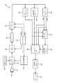
- 超音波画像において心臓の内腔の境界を決定する超音波診断イメージングシステムであって、
心臓画像データのソースと、
前記心臓画像データに応答する境界検出プロセッサであって、前記心臓画像データにおいて心筋の少なくとも内側境界及び外側境界を識別する境界検出プロセッサと、
ユーザが、前記内側境界及び前記外側境界に対しユーザ規定される心腔境界を示すことを可能にするユーザ制御器と、
前記ユーザ制御器及び前記境界検出プロセッサに結合される心腔境界描出器であって、前記心臓画像データにおいて、前記境界検出プロセッサによって識別された前記内側境界及び前記外側境界の少なくとも一方に対し、前記ユーザ規定される心腔境界を位置付ける、心腔境界描出器と、
を有する、超音波診断イメージングシステム。 - 前記ユーザ制御器は更に、前記ユーザ規定される前記心腔境界のロケーションについて可変である単一自由度を調整するよう構成される、請求項1に記載の超音波診断イメージングシステム。
- 前記内側境界が、心内膜、又は心筋−血液プールのインタフェースを更に有し、
前記外側境界が、心外膜、又は肉柱化した心筋と緻密化した心筋との間のインタフェース、を更に有する、請求項2に記載の超音波診断イメージングシステム。 - 前記ユーザ制御器が、スライダ、ノブ、スイッチ、トラックボール、ロッカー制御、トグルボタン、リストボックス又は数値入力ボックスを更に有する、請求項2に記載の超音波診断イメージングシステム。
- 前記ユーザ制御器は、ソフトキー制御又は物理的な制御を更に有する、請求項4に記載の超音波診断イメージングシステム。
- 前記可変の単一自由度は、パーセンテージ又はミリメートルの一方で較正され、前記パーセンテージは、前記内側境界及び前記外側境界に対する距離に関連する、請求項2に記載の超音波診断イメージングシステム。
- 前記心臓画像データのソースが、2次元心臓画像をもつメモリ装置を更に有する、請求項1に記載の超音波診断イメージングシステム。
- 前記心臓画像データが左心室のビューを含む、請求項7に記載の超音波診断イメージングシステム。
- 前記境界検出プロセッサが、半自動の心臓境界画像プロセッサを更に有する、請求項1に記載の超音波診断イメージングシステム。
- 前記半自動の心臓境界画像プロセッサが、前記ユーザ制御器に結合され、更に、心臓画像におけるランドマークを規定するユーザ入力に応答する、請求項9に記載の超音波診断イメージングシステム。
- 前記境界検出プロセッサは更に、自動の心臓境界画像プロセッサを更に有する、請求項1に記載の超音波診断イメージングシステム。
- 前記自動の心臓境界画像プロセッサは、ユーザ入力なしで、前記心臓画像データにおける心筋の境界を識別するよう動作可能である、請求項1に記載の超音波診断イメージングシステム。
- 前記境界検出プロセッサに結合され、内側及び外側の心筋境界の表示トレースを生成するグラフィクス生成器と、
前記心臓画像データの前記ソースに及び前記グラフィクス生成器に結合され、前記内側及び外側の心筋境界の前記生成された表示トレースと共に心臓画像を表示するディスプレイと、
を更に有する、請求項1に記載の超音波診断イメージングシステム。 - 前記心腔境界描出器に結合され、ユーザ規定される心腔境界の表示トレースを生成するグラフィクス生成器と、
前記心臓画像データの前記ソースに及び前記グラフィクス生成器に結合され、前記心腔境界の表示トレースと共に心臓画像を表示するディスプレイと、
を更に有する、請求項1に記載の超音波診断イメージングシステム。 - 単一自由度のパーセンテージのレンジが、0%より小さいか、100%より大きいか、それら両方である、請求項6に記載の超音波診断イメージングシステム。
Applications Claiming Priority (7)
| Application Number | Priority Date | Filing Date | Title |
|---|---|---|---|
| US201562130805P | 2015-03-10 | 2015-03-10 | |
| US201562130787P | 2015-03-10 | 2015-03-10 | |
| US62/130,787 | 2015-03-10 | ||
| US62/130,805 | 2015-03-10 | ||
| EP15161565 | 2015-03-30 | ||
| EP15161565.5 | 2015-03-30 | ||
| PCT/EP2016/054001 WO2016142183A1 (en) | 2015-03-10 | 2016-02-25 | Ultrasonic diagnosis of cardiac performance by single degree of freedom chamber segmentation |
Related Child Applications (1)
| Application Number | Title | Priority Date | Filing Date |
|---|---|---|---|
| JP2020200685A Division JP7132996B2 (ja) | 2015-03-10 | 2020-12-03 | 単一自由度の心腔セグメント化による心臓性能の超音波診断 |
Publications (2)
| Publication Number | Publication Date |
|---|---|
| JP2018507738A true JP2018507738A (ja) | 2018-03-22 |
| JP2018507738A5 JP2018507738A5 (ja) | 2019-04-11 |
Family
ID=52875503
Family Applications (1)
| Application Number | Title | Priority Date | Filing Date |
|---|---|---|---|
| JP2017547114A Pending JP2018507738A (ja) | 2015-03-10 | 2016-02-25 | 単一自由度の心腔セグメント化による心臓性能の超音波診断 |
Country Status (6)
| Country | Link |
|---|---|
| US (1) | US20180049718A1 (ja) |
| EP (2) | EP3267896B1 (ja) |
| JP (1) | JP2018507738A (ja) |
| CN (2) | CN117122349A (ja) |
| RU (1) | RU2708317C2 (ja) |
| WO (1) | WO2016142183A1 (ja) |
Cited By (1)
| Publication number | Priority date | Publication date | Assignee | Title |
|---|---|---|---|---|
| WO2021199962A1 (ja) * | 2020-03-30 | 2021-10-07 | テルモ株式会社 | プログラム、情報処理方法および情報処理装置 |
Families Citing this family (1)
| Publication number | Priority date | Publication date | Assignee | Title |
|---|---|---|---|---|
| US11900524B2 (en) | 2022-03-03 | 2024-02-13 | Biosense Webster (Israel) Ltd. | Constructing topography of lumen wall in 4D ultrasound image with virtual ellipsoid or polyhedron |
Citations (2)
| Publication number | Priority date | Publication date | Assignee | Title |
|---|---|---|---|---|
| JP2009530008A (ja) * | 2006-03-20 | 2009-08-27 | コーニンクレッカ フィリップス エレクトロニクス エヌ ヴィ | 心筋の性能の定量化による超音波診断 |
| JP2010259656A (ja) * | 2009-05-08 | 2010-11-18 | Toshiba Corp | 医用画像処理装置、超音波診断装置および医用画像診断装置 |
Family Cites Families (17)
| Publication number | Priority date | Publication date | Assignee | Title |
|---|---|---|---|---|
| US5601084A (en) * | 1993-06-23 | 1997-02-11 | University Of Washington | Determining cardiac wall thickness and motion by imaging and three-dimensional modeling |
| US5474073A (en) | 1994-11-22 | 1995-12-12 | Advanced Technology Laboratories, Inc. | Ultrasonic diagnostic scanning for three dimensional display |
| US5485842A (en) | 1994-11-30 | 1996-01-23 | Advanced Technology Laboratories, Inc. | Ultrasonic diagnostic scan conversion for three dimensional display processing |
| US5833613A (en) | 1996-09-27 | 1998-11-10 | Advanced Technology Laboratories, Inc. | Ultrasonic diagnostic imaging with contrast agents |
| US5720291A (en) | 1996-03-22 | 1998-02-24 | Advanced Technology Laboratories, Inc. | Three dimensional medical ultrasonic diagnostic image of tissue texture and vasculature |
| US6013032A (en) | 1998-03-13 | 2000-01-11 | Hewlett-Packard Company | Beamforming methods and apparatus for three-dimensional ultrasound imaging using two-dimensional transducer array |
| US6186950B1 (en) | 1999-11-04 | 2001-02-13 | Atl Ultrasound | Ultrasonic pulse inversion harmonic separation with reduced motional effects |
| US6468216B1 (en) | 2000-08-24 | 2002-10-22 | Kininklijke Philips Electronics N.V. | Ultrasonic diagnostic imaging of the coronary arteries |
| US6491636B2 (en) | 2000-12-07 | 2002-12-10 | Koninklijke Philips Electronics N.V. | Automated border detection in ultrasonic diagnostic images |
| US20050075567A1 (en) | 2001-12-18 | 2005-04-07 | Koninklijke Philips Electronics N.V. | Ultrasonic diagnostic imaging system with assisted border tracing |
| US6692438B2 (en) | 2001-12-18 | 2004-02-17 | Koninklijke Philips Electronics Nv | Ultrasonic imaging system and method for displaying tissue perfusion and other parameters varying with time |
| US7347821B2 (en) * | 2003-06-26 | 2008-03-25 | Koninklijke Philips Electronics N.V. | Adaptive processing of contrast enhanced ultrasonic diagnostic images |
| JP4682149B2 (ja) | 2003-12-03 | 2011-05-11 | コーニンクレッカ フィリップス エレクトロニクス エヌ ヴィ | 血流及び潅流パラメータを同時に表示するための超音波イメージングシステムおよび方法 |
| JP5764058B2 (ja) * | 2008-07-10 | 2015-08-12 | コーニンクレッカ フィリップス エヌ ヴェ | 心臓の同時性及び生存能力の超音波評価 |
| US10321892B2 (en) | 2010-09-27 | 2019-06-18 | Siemens Medical Solutions Usa, Inc. | Computerized characterization of cardiac motion in medical diagnostic ultrasound |
| RU2525510C1 (ru) * | 2013-02-21 | 2014-08-20 | Федеральное государственное бюджетное учреждение "Научно-исследовательский институт кардиологии" Сибирского отделения Российской академии медицинских наук | Способ диагностики ишемической болезни сердца методом стресс-эхокардиографии с комбинированной изометрической и психоэмоциональной нагрузкой |
| US9589379B2 (en) * | 2014-06-24 | 2017-03-07 | Siemens Healthcare Gmbh | System and method for visualization of cardiac changes under various pacing conditions |
-
2016
- 2016-02-25 US US15/556,319 patent/US20180049718A1/en active Pending
- 2016-02-25 WO PCT/EP2016/054001 patent/WO2016142183A1/en active Application Filing
- 2016-02-25 JP JP2017547114A patent/JP2018507738A/ja active Pending
- 2016-02-25 CN CN202311095615.2A patent/CN117122349A/zh active Pending
- 2016-02-25 EP EP16709721.1A patent/EP3267896B1/en active Active
- 2016-02-25 RU RU2017131509A patent/RU2708317C2/ru active
- 2016-02-25 CN CN201680014504.6A patent/CN107427282A/zh active Pending
- 2016-02-25 EP EP23207853.5A patent/EP4309585A1/en active Pending
Patent Citations (2)
| Publication number | Priority date | Publication date | Assignee | Title |
|---|---|---|---|---|
| JP2009530008A (ja) * | 2006-03-20 | 2009-08-27 | コーニンクレッカ フィリップス エレクトロニクス エヌ ヴィ | 心筋の性能の定量化による超音波診断 |
| JP2010259656A (ja) * | 2009-05-08 | 2010-11-18 | Toshiba Corp | 医用画像処理装置、超音波診断装置および医用画像診断装置 |
Cited By (1)
| Publication number | Priority date | Publication date | Assignee | Title |
|---|---|---|---|---|
| WO2021199962A1 (ja) * | 2020-03-30 | 2021-10-07 | テルモ株式会社 | プログラム、情報処理方法および情報処理装置 |
Also Published As
| Publication number | Publication date |
|---|---|
| RU2017131509A (ru) | 2019-04-10 |
| EP3267896B1 (en) | 2024-04-10 |
| US20180049718A1 (en) | 2018-02-22 |
| RU2017131509A3 (ja) | 2019-07-17 |
| WO2016142183A1 (en) | 2016-09-15 |
| CN117122349A (zh) | 2023-11-28 |
| EP3267896A1 (en) | 2018-01-17 |
| EP4309585A1 (en) | 2024-01-24 |
| CN107427282A (zh) | 2017-12-01 |
| RU2708317C2 (ru) | 2019-12-05 |
Similar Documents
| Publication | Publication Date | Title |
|---|---|---|
| JP6987207B2 (ja) | ユーザ制御による心臓モデル心室セグメンテーションを用いた心臓機能の超音波診断 | |
| JP4719680B2 (ja) | 超音波による心臓ボリューム数量化方法 | |
| US8144956B2 (en) | Ultrasonic diagnosis by quantification of myocardial performance | |
| US6491636B2 (en) | Automated border detection in ultrasonic diagnostic images | |
| US7450746B2 (en) | System and method for cardiac imaging | |
| US20060058675A1 (en) | Three dimensional atrium-ventricle plane detection | |
| JP5670324B2 (ja) | 医用画像診断装置 | |
| US20040249282A1 (en) | System and method for extracting information based on ultrasound-located landmarks | |
| WO2010116965A1 (ja) | 医用画像診断装置、関心領域設定方法、医用画像処理装置、及び関心領域設定プログラム | |
| JPH1142227A (ja) | 組織の動き追尾方法及び超音波画像処理装置 | |
| JP7375140B2 (ja) | 超音波診断装置、医用画像診断装置、医用画像処理装置及び医用画像処理プログラム | |
| US20170265843A1 (en) | Ultrasonic diagnostic apparatus, image processing apparatus, and image processing method | |
| WO2017006236A1 (en) | Ultrasound systems and methods for automatic determination of heart chamber characteristics | |
| EP3267896B1 (en) | Ultrasonic diagnosis of cardiac performance by single degree of freedom chamber segmentation | |
| JP7132996B2 (ja) | 単一自由度の心腔セグメント化による心臓性能の超音波診断 |
Legal Events
| Date | Code | Title | Description |
|---|---|---|---|
| A521 | Request for written amendment filed |
Free format text: JAPANESE INTERMEDIATE CODE: A523 Effective date: 20190222 |
|
| A621 | Written request for application examination |
Free format text: JAPANESE INTERMEDIATE CODE: A621 Effective date: 20190222 |
|
| A131 | Notification of reasons for refusal |
Free format text: JAPANESE INTERMEDIATE CODE: A131 Effective date: 20191217 |
|
| A977 | Report on retrieval |
Free format text: JAPANESE INTERMEDIATE CODE: A971007 Effective date: 20191218 |
|
| A601 | Written request for extension of time |
Free format text: JAPANESE INTERMEDIATE CODE: A601 Effective date: 20200309 |
|
| A02 | Decision of refusal |
Free format text: JAPANESE INTERMEDIATE CODE: A02 Effective date: 20200804 |
|
| A521 | Request for written amendment filed |
Free format text: JAPANESE INTERMEDIATE CODE: A523 Effective date: 20201203 |
|
| C60 | Trial request (containing other claim documents, opposition documents) |
Free format text: JAPANESE INTERMEDIATE CODE: C60 Effective date: 20201203 |
|
| A911 | Transfer to examiner for re-examination before appeal (zenchi) |
Free format text: JAPANESE INTERMEDIATE CODE: A911 Effective date: 20201211 |
|
| C21 | Notice of transfer of a case for reconsideration by examiners before appeal proceedings |
Free format text: JAPANESE INTERMEDIATE CODE: C21 Effective date: 20201215 |
|
| A912 | Re-examination (zenchi) completed and case transferred to appeal board |
Free format text: JAPANESE INTERMEDIATE CODE: A912 Effective date: 20210129 |
|
| C211 | Notice of termination of reconsideration by examiners before appeal proceedings |
Free format text: JAPANESE INTERMEDIATE CODE: C211 Effective date: 20210202 |
|
| C22 | Notice of designation (change) of administrative judge |
Free format text: JAPANESE INTERMEDIATE CODE: C22 Effective date: 20210218 |
|
| C23 | Notice of termination of proceedings |
Free format text: JAPANESE INTERMEDIATE CODE: C23 Effective date: 20210629 |
|
| C22 | Notice of designation (change) of administrative judge |
Free format text: JAPANESE INTERMEDIATE CODE: C22 Effective date: 20210706 |
|
| C03 | Trial/appeal decision taken |
Free format text: JAPANESE INTERMEDIATE CODE: C03 Effective date: 20210729 |
|
| C30A | Notification sent |
Free format text: JAPANESE INTERMEDIATE CODE: C3012 Effective date: 20210729 |