JP2017533191A - Treatment of cardiovascular disease using ActRII ligand trap - Google Patents
Treatment of cardiovascular disease using ActRII ligand trap Download PDFInfo
- Publication number
- JP2017533191A JP2017533191A JP2017518975A JP2017518975A JP2017533191A JP 2017533191 A JP2017533191 A JP 2017533191A JP 2017518975 A JP2017518975 A JP 2017518975A JP 2017518975 A JP2017518975 A JP 2017518975A JP 2017533191 A JP2017533191 A JP 2017533191A
- Authority
- JP
- Japan
- Prior art keywords
- levels
- level
- reference population
- compared
- subject
- Prior art date
- Legal status (The legal status is an assumption and is not a legal conclusion. Google has not performed a legal analysis and makes no representation as to the accuracy of the status listed.)
- Pending
Links
- 0 C*(CI)**N=O Chemical compound C*(CI)**N=O 0.000 description 1
Images


Classifications
-
- A—HUMAN NECESSITIES
- A61—MEDICAL OR VETERINARY SCIENCE; HYGIENE
- A61K—PREPARATIONS FOR MEDICAL, DENTAL OR TOILETRY PURPOSES
- A61K38/00—Medicinal preparations containing peptides
- A61K38/16—Peptides having more than 20 amino acids; Gastrins; Somatostatins; Melanotropins; Derivatives thereof
- A61K38/17—Peptides having more than 20 amino acids; Gastrins; Somatostatins; Melanotropins; Derivatives thereof from animals; from humans
- A61K38/177—Receptors; Cell surface antigens; Cell surface determinants
- A61K38/179—Receptors; Cell surface antigens; Cell surface determinants for growth factors; for growth regulators
-
- A—HUMAN NECESSITIES
- A61—MEDICAL OR VETERINARY SCIENCE; HYGIENE
- A61K—PREPARATIONS FOR MEDICAL, DENTAL OR TOILETRY PURPOSES
- A61K38/00—Medicinal preparations containing peptides
-
- A—HUMAN NECESSITIES
- A61—MEDICAL OR VETERINARY SCIENCE; HYGIENE
- A61K—PREPARATIONS FOR MEDICAL, DENTAL OR TOILETRY PURPOSES
- A61K9/00—Medicinal preparations characterised by special physical form
- A61K9/0012—Galenical forms characterised by the site of application
- A61K9/0019—Injectable compositions; Intramuscular, intravenous, arterial, subcutaneous administration; Compositions to be administered through the skin in an invasive manner
-
- A—HUMAN NECESSITIES
- A61—MEDICAL OR VETERINARY SCIENCE; HYGIENE
- A61P—SPECIFIC THERAPEUTIC ACTIVITY OF CHEMICAL COMPOUNDS OR MEDICINAL PREPARATIONS
- A61P19/00—Drugs for skeletal disorders
- A61P19/08—Drugs for skeletal disorders for bone diseases, e.g. rachitism, Paget's disease
- A61P19/10—Drugs for skeletal disorders for bone diseases, e.g. rachitism, Paget's disease for osteoporosis
-
- A—HUMAN NECESSITIES
- A61—MEDICAL OR VETERINARY SCIENCE; HYGIENE
- A61P—SPECIFIC THERAPEUTIC ACTIVITY OF CHEMICAL COMPOUNDS OR MEDICINAL PREPARATIONS
- A61P9/00—Drugs for disorders of the cardiovascular system
-
- A—HUMAN NECESSITIES
- A61—MEDICAL OR VETERINARY SCIENCE; HYGIENE
- A61P—SPECIFIC THERAPEUTIC ACTIVITY OF CHEMICAL COMPOUNDS OR MEDICINAL PREPARATIONS
- A61P9/00—Drugs for disorders of the cardiovascular system
- A61P9/10—Drugs for disorders of the cardiovascular system for treating ischaemic or atherosclerotic diseases, e.g. antianginal drugs, coronary vasodilators, drugs for myocardial infarction, retinopathy, cerebrovascula insufficiency, renal arteriosclerosis
-
- C—CHEMISTRY; METALLURGY
- C07—ORGANIC CHEMISTRY
- C07K—PEPTIDES
- C07K14/00—Peptides having more than 20 amino acids; Gastrins; Somatostatins; Melanotropins; Derivatives thereof
- C07K14/435—Peptides having more than 20 amino acids; Gastrins; Somatostatins; Melanotropins; Derivatives thereof from animals; from humans
- C07K14/705—Receptors; Cell surface antigens; Cell surface determinants
- C07K14/71—Receptors; Cell surface antigens; Cell surface determinants for growth factors; for growth regulators
-
- C—CHEMISTRY; METALLURGY
- C07—ORGANIC CHEMISTRY
- C07K—PEPTIDES
- C07K2319/00—Fusion polypeptide
- C07K2319/30—Non-immunoglobulin-derived peptide or protein having an immunoglobulin constant or Fc region, or a fragment thereof, attached thereto
Abstract
本明細書に提供されるのは、バイオマーカー、特に、snailホモログ1(Snai1)、ホスホsmad2、ホスホsmad3、尿タンパク質、dickkopfホモログ1(Dkk1)、コラーゲン1型α1(Col1a1)、アクチビン(すなわち、遊離アクチビン)、runt関連転写因子2(Runx2)、アルカリホスファターゼ(Alp)、骨特異的アルカリホスファターゼ(BSAP)、C末端1型コラーゲンテロペプチド(CTX)、osterix、Klotho、α-平滑筋アクチン(α-SMA)、ミオカルディン(JVIYOCD)、アクチビン受容体2A型(ActRIIA)、軸阻害タンパク質2(Axin2)、及び/又は平滑筋タンパク質22-α(Sm22-α)のレベルを、治療に対する対象の応答性、治療の効力、又はアクチビンII型受容体シグナル伝達インヒビターによる治療のための適切な投薬量の指標として用いることにより、対象における血管石灰化及び/もしくは心血管疾患と関連する疾患並びに/又は骨吸収を治療する方法である。【選択図】図4AProvided herein are biomarkers, particularly snail homolog 1 (Snai1), phospho smad2, phospho smad3, urine protein, dickkopf homolog 1 (Dkk1), collagen type 1 α1 (Col1a1), activin (i.e. Free activin), runt-related transcription factor 2 (Runx2), alkaline phosphatase (Alp), bone-specific alkaline phosphatase (BSAP), C-terminal type 1 collagen telopeptide (CTX), osterix, Klotho, α-smooth muscle actin (α -SMA), myocardin (JVIYOCD), activin receptor type 2A (ActRIIA), axial inhibitory protein 2 (Axin2), and / or smooth muscle protein 22-α (Sm22-α) levels, subject response to treatment , Disease efficacy associated with vascular calcification and / or cardiovascular disease in a subject by using as an indicator of the efficacy of treatment, or an appropriate dosage for treatment with an activin type II receptor signaling inhibitor / Or a method of treating a bone absorption. [Selection] Figure 4A
Description
(1.関連出願の相互参照)
本出願は、その各々の内容全体が引用によりかつあらゆる目的のために本明細書中に組み込まれる、2014年10月9日に出願された米国仮特許出願第62/062,021号; 2014年11月11日に出願された米国仮特許出願第62/078,321号; 2015年1月14日に出願された米国仮特許出願第62/103,515号; 2015年5月27日に出願された米国仮特許出願第62/167,052号;及び2015年6月2日に出願された米国仮特許出願第62/170,015号の優先権の恩典を主張する。
(1. Cross-reference of related applications)
This application is a US Provisional Patent Application No. 62 / 062,021 filed Oct. 9, 2014; the entire contents of each of which are incorporated herein by reference and for all purposes; November 2014 US Provisional Patent Application No. 62 / 078,321 filed on 11th; US Provisional Patent Application No. 62 / 103,515 filed on 14th January 2015; US Provisional Patent Application filed on 27th May 2015 No. 62 / 167,052; and US Provisional Patent Application No. 62 / 170,015 filed June 2, 2015, alleging the benefit of priority.
(2.政府ライセンス権)
本発明は、国立衛生研究所により与えられた助成金番号DK070790及びDK089137の下で政府の支援によりなされた。政府は、本発明における一定の権利を有する。
(2.Government license rights)
This invention was made with government support under grant numbers DK070790 and DK089137 awarded by the National Institutes of Health. The government has certain rights in the invention.
(3.配列表)
本出願は、2015年10月6日に作成された、ファイル名12827_934_228_SeqListing.txtとして提出された208キロバイトのサイズの配列表とともに出願されている。この配列表は、その全体が引用によりかつあらゆる目的のために本明細書中に組み込まれている。
(3. Sequence Listing)
This application has been filed with a sequence table created on October 6, 2015, filed as file name 12827_934_228_SeqListing.txt with a size of 208 kilobytes. This sequence listing is incorporated herein by reference in its entirety and for all purposes.
(4.分野)
本明細書に提供されるのは、対象における心血管疾患、血管石灰化、血管石灰化と関連し及び/もしくは血管石灰化に起因する心血管疾患、並びに/又は腎疾患と関連し及び/もしくは腎疾患に起因する心血管疾患を治療及び/又は予防する方法であって、該対象に、アクチビンII型受容体シグナル伝達インヒビター(ActRIIシグナル伝達インヒビター、例えば、アクチビンリガンドトラップ)を投与することを含む、方法である。より具体的には、本明細書に提供されるのは、1以上のバイオマーカー、特に、snailホモログ1(Snai1)、ホスホsmad2、ホスホsmad3、尿タンパク質、dickkopfホモログ1(Dkk1)、コラーゲン1型α1(Col1a1)、アクチビン(例えば、遊離アクチビン)、runt関連転写因子2(Runx2)、アルカリホスファターゼ(Alp)、骨特異的アルカリホスファターゼ(BSAP)、Osterix、C末端1型コラーゲンテロペプチド(CTX)、Klotho、α-平滑筋アクチン(α-SMA)、ミオカルディン(MYOCD)、軸阻害タンパク質2(Axin2)、及び/又は平滑筋タンパク質22-α(Sm22-α)のレベル及び/又は活性を、治療に対する対象の応答性、治療の効力、又はアクチビンII型受容体(ActRII)シグナル伝達インヒビターによる治療のための適切な投薬量の指標として用いることにより、心血管疾患、血管石灰化、高いレベルの動脈壁硬化、左心室肥大、血管石灰化と関連し及び/もしくは血管石灰化に起因する心血管疾患、高いレベルの動脈壁硬化と関連し及び/もしくは高いレベルの動脈壁硬化に起因する心血管疾患、左心室肥大と関連し及び/もしくは左心室肥大に起因する心血管疾患、並びに/又は腎疾患と関連し及び/又は腎疾患に起因する心血管疾患の治療のための対象を選択する方法である。本明細書に提供されるのは、対象における骨吸収を低下させる方法であって、該対象に、ActRIIシグナル伝達インヒビターを投与することを含む、方法である。より具体的には、本明細書に提供されるのは、1以上のバイオマーカー、特に、Snai1、ホスホsmad2、ホスホsmad3、尿タンパク質、Dkk1、col1a1、アクチビン(例えば、遊離アクチビン)、Runx2、Alp、BSAP、Osterix、CTX、Klotho、α-SMA、MYOCD、及び/又はSm22-αのレベル及び/又は活性を、治療に対する対象の応答性、治療の効力、又はActRIIシグナル伝達インヒビターによる治療のための適切な投薬量の指標として用いることにより、骨吸収の低下のための対象を選択する方法である。
(4. Field)
Provided herein are associated with cardiovascular disease, vascular calcification, vascular calcification and / or cardiovascular disease resulting from and / or renal disease in a subject and / or A method of treating and / or preventing cardiovascular disease resulting from renal disease, comprising administering to said subject an activin type II receptor signaling inhibitor (ActRII signaling inhibitor, such as activin ligand trap) Is the way. More specifically, provided herein are one or more biomarkers, particularly snail homolog 1 (Snai1), phospho smad2, phospho smad3, urine protein, dickkopf homolog 1 (Dkk1),
(5.背景)
貧血を伴う腎疾患のよく見られる合併症は血管石灰化であり、これは、多くの場合、心血管疾患をもたらす。腎疾患対象(renal subject)において、アテローム性動脈硬化症及び結果として生じる心血管疾患は、腎疾患それ自体と関連する死亡率とは別に、著しい死亡率をもたらし得る。したがって、腎疾患対象における心血管疾患のための新しい薬物並びに治療及び/又は予防方法の発見及び開発が引き続き必要である。さらに、腎疾患対象は貧血に苦しむことが多いので、貧血及びそれに付随する心血管疾患を、エリスロポエチンなどの現在使用されている赤血球生成刺激剤(ESA)が保持しない能力を備えた、単一の治療薬を用いて治療することは有益であろう。
(5. Background)
A common complication of kidney disease with anemia is vascular calcification, which often results in cardiovascular disease. In a renal subject, atherosclerosis and the resulting cardiovascular disease can lead to significant mortality, apart from the mortality associated with kidney disease itself. Accordingly, there is a continuing need to discover and develop new drugs and treatment and / or prevention methods for cardiovascular disease in kidney disease subjects. In addition, because kidney disease subjects often suffer from anemia, a single, erythropoiesis stimulator (ESA) such as erythropoietin does not retain anemia and its associated cardiovascular disease. It may be beneficial to treat with a therapeutic agent.
2つの関連するII型受容体、ActRIIA及びActRIIBが、アクチビンのII型受容体として同定されている(Mathews及びValeの文献、1991, Cell 65:973-982; Attisanoらの文献、1992, Cell 68: 97-108)。アクチビンの他に、ActRIIA及びActRIIBは、BMP7、Nodal、GDF8、及びGDF11を含む、いくつかの他のTGF-βファミリータンパク質と生化学的に相互作用することができる(Yamashitaらの文献、1995, J. Cell Biol. 130:217-226; Lee及びMcPherronの文献、2001, Proc. Natl. Acad. Sci. 98:9306-9311; Yeo及びWhitmanの文献、2001, Mol. Cell 7: 949-957; Ohらの文献、2002, Genes Dev. 16:2749-54)。ALK4は、アクチビン、特に、アクチビンAの主なI型受容体であり、ALK-7は、同様に、アクチビン、特に、アクチビンBの受容体としての役割を果たし得る。 Two related type II receptors, ActRIIA and ActRIIB, have been identified as type II receptors for activin (Mathews and Vale, 1991, Cell 65: 973-982; Attisano et al., 1992, Cell 68 : 97-108). In addition to activin, ActRIIA and ActRIIB can interact biochemically with several other TGF-β family proteins, including BMP7, Nodal, GDF8, and GDF11 (Yamashita et al., 1995, J. Cell Biol. 130: 217-226; Lee and McPherron, 2001, Proc. Natl. Acad. Sci. 98: 9306-9311; Yeo and Whitman, 2001, Mol. Cell 7: 949-957; Oh et al., 2002, Genes Dev. 16: 2749-54). ALK4 is the main type I receptor for activin, in particular activin A, and ALK-7 can likewise serve as a receptor for activin, in particular activin B.
アクチビン受容体IIA型(ActRIIA)の細胞外ドメイン及びヒトIgG1 Fcからなるヒト化融合タンパク質(本明細書では、ActRIIA-hFc又は「ソタテルセプト」と呼ばれる;配列番号7)からなるアクチビンリガンドトラップは、現在、末期腎疾患(ESRD)と関連する貧血及び骨障害を有する対象並びにβ-サラセミアを有するこれらの対象の治療のための第II相臨床試験で評価されているところである。閉経後の健常女性では、ActRIIA-hFcが、ヘマトクリット(Hct)及びヘモグロビン(Hgb)並びに骨ミネラル密度を顕著に増大させることが示された。 An activin ligand trap consisting of an extracellular domain of activin receptor type IIA (ActRIIA) and a humanized fusion protein consisting of human IgG1 Fc (referred to herein as ActRIIA-hFc or “sotatercept”; SEQ ID NO: 7) is currently , Being evaluated in Phase II clinical trials for the treatment of subjects with anemia and bone disorders associated with end stage renal disease (ESRD) and those subjects with β-thalassemia. In healthy postmenopausal women, ActRIIA-hFc has been shown to significantly increase hematocrit (Hct) and hemoglobin (Hgb) and bone mineral density.
(6.概要)
本明細書に提供されるのは、対象における血管石灰化を治療及び/又は予防する方法であって、該対象に、アクチビン受容体II型(ActRII)シグナル伝達インヒビター(例えば、アクチビンリガンドトラップ)の医薬有効用量を投与することを含み、ここで、該対象が:(a)参照集団におけるRunx2のレベルと比較して上昇したRunx2のレベル;(b)参照集団におけるAlpのレベルと比較して上昇したAlpのレベル;(c)参照集団におけるSnai1のレベルと比較して上昇したSnai1のレベル;(d)参照集団におけるホスホsmad2のレベルと比較して上昇したホスホsmad2のレベル;(e)参照集団におけるDkk1のレベルと比較して上昇したDkk1のレベル;(f)参照集団におけるcol1a1のレベルと比較して上昇したcol1a1のレベル;(g)参照集団におけるアクチビンのレベルと比較して上昇したアクチビン(例えば、遊離アクチビン)のレベル;(h)参照集団におけるBSAPのレベルと比較して上昇したBSAPのレベル;(i)参照集団におけるOsterixのレベルと比較して上昇したOsterixのレベル;(j)参照集団におけるCTXのレベルと比較して上昇したCTXのレベル;(k)参照集団におけるKlothoのレベルと比較して減少したKlothoのレベル;(l)参照集団におけるα-SMAのレベルと比較して減少したα-SMAのレベル;(m)参照集団におけるMYOCDのレベルと比較して減少したMYOCDのレベル;(n)参照集団におけるSm22-αのレベルと比較して減少したSm22-αのレベル;(o)参照集団におけるホスホsmad3のレベルと比較して上昇したホスホsmad3のレベル;(p)参照集団における尿タンパク質のレベルと比較して上昇した尿タンパク質のレベル;及び/又は(q)参照集団におけるActRIIAのレベルと比較して減少したアクチビン受容体2A型(ActRIIA)のレベルを有する、方法である。参照集団の説明については、例えば、第8.6節を参照されたい。
(6. Overview)
Provided herein is a method of treating and / or preventing vascular calcification in a subject, wherein the subject is treated with an activin receptor type II (ActRII) signaling inhibitor (eg, activin ligand trap). Administering a pharmaceutically effective dose, wherein the subject: (a) increased Runx2 level compared to the level of Runx2 in the reference population; (b) increased compared to the level of Alp in the reference population Alp levels; (c) elevated Snai1 levels compared to Snai1 levels in the reference population; (d) elevated phosphosmad2 levels compared to phosphosmad2 levels in the reference population; (e) reference populations Elevated Dkk1 levels compared to Dkk1 levels in (f) elevated col1a1 levels compared to col1a1 levels in the reference population; (g) elevated activins compared to activin levels in the reference population ( example (H) level of BSAP increased compared to the level of BSAP in the reference population; (i) level of Osterix increased compared to the level of Osterix in the reference population; (j) reference population CTX levels increased compared to CTX levels in (k) decreased Klotho levels compared to Klotho levels in the reference population; (l) decreased compared to α-SMA levels in the reference population α-SMA levels; (m) decreased MYOCD levels compared to MYOCD levels in the reference population; (n) decreased Sm22-α levels compared to Sm22-α levels in the reference population; (o ) Increased levels of phosphosmad3 compared to levels of phosphosmad3 in the reference population; (p) increased levels of urine protein compared to levels of urine protein in the reference population; and / or (q) ActRIIA in the reference population Activin Receptor Reduced Compared with Levels A method having a level of type 2A (ActRIIA). See, for example, Section 8.6 for an explanation of the reference population.
本明細書に提供されるのは、対象における心血管疾患を治療及び/又は予防する方法であって、該対象に、アクチビン受容体II型(ActRII)シグナル伝達インヒビター(例えば、アクチビンリガンドトラップ)の医薬有効用量を投与することを含み、ここで、該対象が:(a)参照集団におけるRunx2のレベルと比較して上昇したRunx2のレベル;(b)参照集団におけるAlpのレベルと比較して上昇したAlpのレベル;(c)参照集団におけるSnai1のレベルと比較して上昇したSnai1のレベル;(d)参照集団におけるホスホsmad2のレベルと比較して上昇したホスホsmad2のレベル;(e)参照集団におけるDkk1のレベルと比較して上昇したDkk1のレベル;(f)参照集団におけるcol1a1のレベルと比較して上昇したcol1a1のレベル;(g)参照集団におけるアクチビンのレベルと比較して上昇したアクチビン(例えば、遊離アクチビン)のレベル;(h)参照集団におけるBSAPのレベルと比較して上昇したBSAPのレベル;(i)参照集団におけるOsterixのレベルと比較して上昇したOsterixのレベル;(j)参照集団におけるCTXのレベルと比較して上昇したCTXのレベル;(k)参照集団におけるKlothoのレベルと比較して減少したKlothoのレベル;(l)参照集団におけるα-SMAのレベルと比較して減少したα-SMAのレベル;(m)参照集団におけるMYOCDのレベルと比較して減少したMYOCDのレベル;(n)参照集団におけるSm22-αのレベルと比較して減少したSm22-αのレベル;(o)参照集団におけるホスホsmad3のレベルと比較して上昇したホスホsmad3のレベル;(p)参照集団における尿タンパク質のレベルと比較して上昇した尿タンパク質のレベル;及び/又は(q)参照集団におけるActRIIAのレベルと比較して減少したActRIIAのレベルを有する、方法である。 Provided herein is a method of treating and / or preventing cardiovascular disease in a subject, wherein the subject is treated with an activin receptor type II (ActRII) signaling inhibitor (eg, activin ligand trap). Administering a pharmaceutically effective dose, wherein the subject: (a) increased Runx2 level compared to the level of Runx2 in the reference population; (b) increased compared to the level of Alp in the reference population Alp levels; (c) elevated Snai1 levels compared to Snai1 levels in the reference population; (d) elevated phosphosmad2 levels compared to phosphosmad2 levels in the reference population; (e) reference populations Elevated Dkk1 levels compared to Dkk1 levels in (f) elevated col1a1 levels compared to col1a1 levels in the reference population; (g) elevated activins compared to activin levels in the reference population ( example (H) level of BSAP increased compared to the level of BSAP in the reference population; (i) level of Osterix increased compared to the level of Osterix in the reference population; (j) reference population CTX levels increased compared to CTX levels in (k) decreased Klotho levels compared to Klotho levels in the reference population; (l) decreased compared to α-SMA levels in the reference population α-SMA levels; (m) decreased MYOCD levels compared to MYOCD levels in the reference population; (n) decreased Sm22-α levels compared to Sm22-α levels in the reference population; (o ) Increased levels of phosphosmad3 compared to levels of phosphosmad3 in the reference population; (p) increased levels of urine protein compared to levels of urine protein in the reference population; and / or (q) ActRIIA in the reference population Reduced ActRIIA levels compared to levels To, is a method.
本明細書に提供されるのは、対象における心血管疾患を治療及び/又は予防する方法であって、該対象に、アクチビン受容体II型(ActRII)シグナル伝達インヒビター(例えば、アクチビンリガンドトラップ)の医薬有効用量を投与することを含み、ここで、該対象が:(a)参照集団におけるRunx2のレベルと比較して上昇したRunx2のレベル;(b)参照集団におけるAlpのレベルと比較して上昇したAlpのレベル;(c)参照集団におけるSnai1のレベルと比較して上昇したSnai1のレベル;(d)参照集団におけるホスホsmad2のレベルと比較して上昇したホスホsmad2のレベル;(e)参照集団におけるDkk1のレベルと比較して上昇したDkk1のレベル;(f)参照集団におけるcol1a1のレベルと比較して上昇したcol1a1のレベル;(g)参照集団におけるアクチビンのレベルと比較して上昇したアクチビン(例えば、遊離アクチビン)のレベル;(h)参照集団におけるBSAPのレベルと比較して上昇したBSAPのレベル;(i)参照集団におけるOsterixのレベルと比較して上昇したOsterixのレベル;(j)参照集団におけるCTXのレベルと比較して上昇したCTXのレベル;(k)参照集団におけるKlothoのレベルと比較して減少したKlothoのレベル;(l)参照集団におけるα-SMAのレベルと比較して減少したα-SMAのレベル;(m)参照集団におけるMYOCDのレベルと比較して減少したMYOCDのレベル;(n)参照集団におけるSm22-αのレベルと比較して減少したSm22-αのレベル;(o)参照集団におけるホスホsmad3のレベルと比較して上昇したホスホsmad3のレベル;(p)参照集団における尿タンパク質のレベルと比較して上昇した尿タンパク質のレベル;及び/又は(q)参照集団におけるActRIIAのレベルと比較して減少したActRIIAのレベルを有し、かつ該心血管疾患が、血管石灰化と関連し及び/又は血管石灰化に起因する、方法である。 Provided herein is a method of treating and / or preventing cardiovascular disease in a subject, wherein the subject is treated with an activin receptor type II (ActRII) signaling inhibitor (eg, activin ligand trap). Administering a pharmaceutically effective dose, wherein the subject: (a) increased Runx2 level compared to the level of Runx2 in the reference population; (b) increased compared to the level of Alp in the reference population Alp levels; (c) elevated Snai1 levels compared to Snai1 levels in the reference population; (d) elevated phosphosmad2 levels compared to phosphosmad2 levels in the reference population; (e) reference populations Elevated Dkk1 levels compared to Dkk1 levels in (f) elevated col1a1 levels compared to col1a1 levels in the reference population; (g) elevated activins compared to activin levels in the reference population ( example (H) level of BSAP increased compared to the level of BSAP in the reference population; (i) level of Osterix increased compared to the level of Osterix in the reference population; (j) reference population CTX levels increased compared to CTX levels in (k) decreased Klotho levels compared to Klotho levels in the reference population; (l) decreased compared to α-SMA levels in the reference population α-SMA levels; (m) decreased MYOCD levels compared to MYOCD levels in the reference population; (n) decreased Sm22-α levels compared to Sm22-α levels in the reference population; (o ) Increased levels of phosphosmad3 compared to levels of phosphosmad3 in the reference population; (p) increased levels of urine protein compared to levels of urine protein in the reference population; and / or (q) ActRIIA in the reference population Reduced ActRIIA levels compared to levels And, and said cardiac vascular disease is due to vascular calcification and related and / or vascular calcification, is a method.
本明細書に提供されるのは、対象における心血管疾患を治療及び/又は予防する方法であって、該対象に、アクチビン受容体II型(ActRII)シグナル伝達インヒビター(例えば、アクチビンリガンドトラップ)の医薬有効用量を投与することを含み、ここで、該対象が:(a)参照集団におけるRunx2のレベルと比較して上昇したRunx2のレベル;(b)参照集団におけるAlpのレベルと比較して上昇したAlpのレベル;(c)参照集団におけるSnai1のレベルと比較して上昇したSnai1のレベル;(d)参照集団におけるホスホsmad2のレベルと比較して上昇したホスホsmad2のレベル;(e)参照集団におけるDkk1のレベルと比較して上昇したDkk1のレベル;(f)参照集団におけるcol1a1のレベルと比較して上昇したcol1a1のレベル;(g)参照集団におけるアクチビンのレベルと比較して上昇したアクチビン(例えば、遊離アクチビン)のレベル;(h)参照集団におけるBSAPのレベルと比較して上昇したBSAPのレベル;(i)参照集団におけるOsterixのレベルと比較して上昇したOsterixのレベル;(j)参照集団におけるCTXのレベルと比較して上昇したCTXのレベル;(k)参照集団におけるKlothoのレベルと比較して減少したKlothoのレベル;(l)参照集団におけるα-SMAのレベルと比較して減少したα-SMAのレベル;(m)参照集団におけるMYOCDのレベルと比較して減少したMYOCDのレベル;(n)参照集団におけるSm22-αのレベルと比較して減少したSm22-αのレベル;(o)参照集団におけるホスホsmad3のレベルと比較して上昇したホスホsmad3のレベル;(p)参照集団における尿タンパク質のレベルと比較して上昇した尿タンパク質のレベル;及び/又は(q)参照集団におけるActRIIAのレベルと比較して減少したActRIIAのレベルを有し、かつ該心血管疾患が、腎疾患と関連し及び/又は腎疾患に起因する、方法である。 Provided herein is a method of treating and / or preventing cardiovascular disease in a subject, wherein the subject is treated with an activin receptor type II (ActRII) signaling inhibitor (eg, activin ligand trap). Administering a pharmaceutically effective dose, wherein the subject: (a) increased Runx2 level compared to the level of Runx2 in the reference population; (b) increased compared to the level of Alp in the reference population Alp levels; (c) elevated Snai1 levels compared to Snai1 levels in the reference population; (d) elevated phosphosmad2 levels compared to phosphosmad2 levels in the reference population; (e) reference populations Elevated Dkk1 levels compared to Dkk1 levels in (f) elevated col1a1 levels compared to col1a1 levels in the reference population; (g) elevated activins compared to activin levels in the reference population ( example (H) level of BSAP increased compared to the level of BSAP in the reference population; (i) level of Osterix increased compared to the level of Osterix in the reference population; (j) reference population CTX levels increased compared to CTX levels in (k) decreased Klotho levels compared to Klotho levels in the reference population; (l) decreased compared to α-SMA levels in the reference population α-SMA levels; (m) decreased MYOCD levels compared to MYOCD levels in the reference population; (n) decreased Sm22-α levels compared to Sm22-α levels in the reference population; (o ) Increased levels of phosphosmad3 compared to levels of phosphosmad3 in the reference population; (p) increased levels of urine protein compared to levels of urine protein in the reference population; and / or (q) ActRIIA in the reference population Reduced ActRIIA levels compared to levels And, and said cardiac vascular disease is due to the associated and / or kidney disease and renal disease, is a method.
本明細書に提供されるのは、対象における骨吸収を低下させる方法であって、該対象に、アクチビン受容体II型(ActRII)シグナル伝達インヒビター(例えば、アクチビンリガンドトラップ)の医薬有効用量を投与することを含み、ここで、該対象が:(a)参照集団におけるRunx2のレベルと比較して上昇したRunx2のレベル;(b)参照集団におけるAlpのレベルと比較して上昇したAlpのレベル;(c)参照集団におけるSnai1のレベルと比較して上昇したSnai1のレベル;(d)参照集団におけるホスホsmad2のレベルと比較して上昇したホスホsmad2のレベル;(e)参照集団におけるDkk1のレベルと比較して上昇したDkk1のレベル;(f)参照集団におけるcol1a1のレベルと比較して上昇したcol1a1のレベル;(g)参照集団におけるアクチビンのレベルと比較して上昇したアクチビン(例えば、遊離アクチビン)のレベル;(h)参照集団におけるBSAPのレベルと比較して上昇したBSAPのレベル;(i)参照集団におけるOsterixのレベルと比較して上昇したOsterixのレベル;(j)参照集団におけるCTXのレベルと比較して上昇したCTXのレベル;(k)参照集団におけるKlothoのレベルと比較して減少したKlothoのレベル;(l)参照集団におけるα-SMAのレベルと比較して減少したα-SMAのレベル;(m)参照集団におけるMYOCDのレベルと比較して減少したMYOCDのレベル;(n)参照集団におけるSm22-αのレベルと比較して減少したSm22-αのレベル;(o)参照集団におけるホスホsmad3のレベルと比較して上昇したホスホsmad3のレベル;(p)参照集団における尿タンパク質のレベルと比較して上昇した尿タンパク質のレベル;及び/又は(q)参照集団におけるActRIIAのレベルと比較して減少したActRIIAのレベルを有する、方法である。 Provided herein is a method of reducing bone resorption in a subject, wherein the subject is administered a pharmaceutically effective dose of an activin receptor type II (ActRII) signaling inhibitor (eg, activin ligand trap) Wherein the subject has: (a) an increased level of Runx2 compared to the level of Runx2 in the reference population; (b) an increased level of Alp compared to the level of Alp in the reference population; (c) elevated Snai1 level compared to the level of Snai1 in the reference population; (d) elevated phosphosmad2 level compared to the level of phosphosmad2 in the reference population; (e) the level of Dkk1 in the reference population Elevated levels of Dkk1; (f) elevated levels of col1a1 compared to col1a1 levels in the reference population; (g) elevated activins compared to activin levels in the reference population (e.g., free activins) (H) BSAP level increased compared to BSAP level in reference population; (i) Osterix level increased compared to Osterix level in reference population; (j) CTX in reference population Increased levels of CTX compared to levels of (k) decreased levels of Klotho compared to levels of Klotho in the reference population; (l) decreased α- levels compared to levels of α-SMA in the reference population SMA levels; (m) decreased MYOCD levels compared to MYOCD levels in the reference population; (n) reduced Sm22-α levels compared to Sm22-α levels in the reference population; see (o) Elevated phosphosmad3 levels compared to phosphosmad3 levels in the population; (p) elevated urine protein levels compared to urine protein levels in the reference population; and / or (q) ActRIIA levels in the reference population A method having a reduced ActRIIA level compared to The
本明細書に提供されるのは、対象における動脈壁硬化を治療及び/又は予防する方法であって、該対象に、アクチビン受容体II型(ActRII)シグナル伝達インヒビター(例えば、アクチビンリガンドトラップ)の医薬有効用量を投与することを含み、ここで、該対象が:(a)参照集団におけるRunx2のレベルと比較して上昇したRunx2のレベル;(b)参照集団におけるAlpのレベルと比較して上昇したAlpのレベル;(c)参照集団におけるSnai1のレベルと比較して上昇したSnai1のレベル;(d)参照集団におけるホスホsmad2のレベルと比較して上昇したホスホsmad2のレベル;(e)参照集団におけるDkk1のレベルと比較して上昇したDkk1のレベル;(f)参照集団におけるcol1a1のレベルと比較して上昇したcol1a1のレベル;(g)参照集団におけるアクチビンのレベルと比較して上昇したアクチビン(例えば、遊離アクチビン)のレベル;(h)参照集団におけるBSAPのレベルと比較して上昇したBSAPのレベル;(i)参照集団におけるOsterixのレベルと比較して上昇したOsterixのレベル;(j)参照集団におけるCTXのレベルと比較して上昇したCTXのレベル;(k)参照集団におけるKlothoのレベルと比較して減少したKlothoのレベル;(l)参照集団におけるα-SMAのレベルと比較して減少したα-SMAのレベル;(m)参照集団におけるMYOCDのレベルと比較して減少したMYOCDのレベル;(n)参照集団におけるSm22-αのレベルと比較して減少したSm22-αのレベル;(o)参照集団におけるホスホsmad3のレベルと比較して上昇したホスホsmad3のレベル;(p)参照集団における尿タンパク質のレベルと比較して上昇した尿タンパク質のレベル;及び/又は(q)参照集団におけるActRIIAのレベルと比較して減少したActRIIAのレベルを有する、方法である。 Provided herein are methods for treating and / or preventing arterial wall stiffness in a subject, wherein the subject is treated with an activin receptor type II (ActRII) signaling inhibitor (eg, activin ligand trap). Administering a pharmaceutically effective dose, wherein the subject: (a) increased Runx2 level compared to the level of Runx2 in the reference population; (b) increased compared to the level of Alp in the reference population Alp levels; (c) elevated Snai1 levels compared to Snai1 levels in the reference population; (d) elevated phosphosmad2 levels compared to phosphosmad2 levels in the reference population; (e) reference populations Elevated Dkk1 levels compared to Dkk1 levels in (f) elevated col1a1 levels compared to col1a1 levels in the reference population; (g) elevated activins compared to activin levels in the reference population ( example (H) level of BSAP increased compared to the level of BSAP in the reference population; (i) level of Osterix increased compared to the level of Osterix in the reference population; (j) reference population CTX levels increased compared to CTX levels in (k) decreased Klotho levels compared to Klotho levels in the reference population; (l) decreased compared to α-SMA levels in the reference population α-SMA levels; (m) decreased MYOCD levels compared to MYOCD levels in the reference population; (n) decreased Sm22-α levels compared to Sm22-α levels in the reference population; (o ) Increased levels of phosphosmad3 compared to levels of phosphosmad3 in the reference population; (p) increased levels of urine protein compared to levels of urine protein in the reference population; and / or (q) ActRIIA in the reference population Reduced ActRIIA levels compared to levels To, is a method.
本明細書に提供されるのは、対象における左心室肥大を治療及び/又は予防する方法であって、該対象に、アクチビン受容体II型(ActRII)シグナル伝達インヒビター(例えば、アクチビンリガンドトラップ)の医薬有効用量を投与することを含み、ここで、該対象が:(a)参照集団におけるRunx2のレベルと比較して上昇したRunx2のレベル;(b)参照集団におけるAlpのレベルと比較して上昇したAlpのレベル;(c)参照集団におけるSnai1のレベルと比較して上昇したSnai1のレベル;(d)参照集団におけるホスホsmad2のレベルと比較して上昇したホスホsmad2のレベル;(e)参照集団におけるDkk1のレベルと比較して上昇したDkk1のレベル;(f)参照集団におけるcol1a1のレベルと比較して上昇したcol1a1のレベル;(g)参照集団におけるアクチビンのレベルと比較して上昇したアクチビン(例えば、遊離アクチビン)のレベル;(h)参照集団におけるBSAPのレベルと比較して上昇したBSAPのレベル;(i)参照集団におけるOsterixのレベルと比較して上昇したOsterixのレベル;(j)参照集団におけるCTXのレベルと比較して上昇したCTXのレベル;(k)参照集団におけるKlothoのレベルと比較して減少したKlothoのレベル;(l)参照集団におけるα-SMAのレベルと比較して減少したα-SMAのレベル;(m)参照集団におけるMYOCDのレベルと比較して減少したMYOCDのレベル;(n)参照集団におけるSm22-αのレベルと比較して減少したSm22-αのレベル;(o)参照集団におけるホスホsmad3のレベルと比較して上昇したホスホsmad3のレベル;(p)参照集団における尿タンパク質のレベルと比較して上昇した尿タンパク質のレベル;及び/又は(q)参照集団におけるActRIIAのレベルと比較して減少したActRIIAのレベルを有する、方法である。 Provided herein is a method of treating and / or preventing left ventricular hypertrophy in a subject, wherein the subject is treated with an activin receptor type II (ActRII) signaling inhibitor (e.g., activin ligand trap). Administering a pharmaceutically effective dose, wherein the subject: (a) increased Runx2 level compared to the level of Runx2 in the reference population; (b) increased compared to the level of Alp in the reference population Alp levels; (c) elevated Snai1 levels compared to Snai1 levels in the reference population; (d) elevated phosphosmad2 levels compared to phosphosmad2 levels in the reference population; (e) reference populations Elevated Dkk1 levels compared to Dkk1 levels in (f) elevated col1a1 levels compared to col1a1 levels in the reference population; (g) elevated activins compared to activin levels in the reference population ( example (H) level of BSAP increased compared to the level of BSAP in the reference population; (i) level of Osterix increased compared to the level of Osterix in the reference population; (j) reference population CTX levels increased compared to CTX levels in (k) decreased Klotho levels compared to Klotho levels in the reference population; (l) decreased compared to α-SMA levels in the reference population α-SMA levels; (m) decreased MYOCD levels compared to MYOCD levels in the reference population; (n) decreased Sm22-α levels compared to Sm22-α levels in the reference population; (o ) Increased levels of phosphosmad3 compared to levels of phosphosmad3 in the reference population; (p) increased levels of urine protein compared to levels of urine protein in the reference population; and / or (q) ActRIIA in the reference population Reduced ActRIIA levels compared to levels To, is a method.
ある実施態様において、ActRIIシグナル伝達インヒビターの医薬有効用量は、約15mg、約30mg、約45mg、約60mg、約75mg、約90mg、もしくは約1g、又は約0.1mg/kg、約0.13mg/kg、約0.2mg/kg、約0.26mg/kg、約0.3mg/kg、約0.4mg/kg、約0.5mg/kg、約0.6mg/kg、約0.7mg/kg、約0.8mg/kg、約0.9mg/kg、約1.0mg/kg、約1.1mg/kg、約1.2mg/kg、約1.3mg/kg、約1.4mg/kg、もしくは約1.5mg/kgである。ある実施態様において、ActRIIシグナル伝達インヒビターの医薬有効用量は、約0.1mg/kgである。ある実施態様において、ActRIIシグナル伝達インヒビターの医薬有効用量は、約0.3mg/kgである。ある実施態様において、ActRIIシグナル伝達インヒビターの医薬有効用量は、約0.5mg/kgである。ある実施態様において、ActRIIシグナル伝達インヒビターの医薬有効用量は、約0.7mg/kgである。 In certain embodiments, a pharmaceutically effective dose of ActRII signaling inhibitor is about 15 mg, about 30 mg, about 45 mg, about 60 mg, about 75 mg, about 90 mg, or about 1 g, or about 0.1 mg / kg, about 0.13 mg / kg, About 0.2 mg / kg, about 0.26 mg / kg, about 0.3 mg / kg, about 0.4 mg / kg, about 0.5 mg / kg, about 0.6 mg / kg, about 0.7 mg / kg, about 0.8 mg / kg, about 0.9 mg / kg, about 1.0 mg / kg, about 1.1 mg / kg, about 1.2 mg / kg, about 1.3 mg / kg, about 1.4 mg / kg, or about 1.5 mg / kg. In certain embodiments, the pharmaceutically effective dose of ActRII signaling inhibitor is about 0.1 mg / kg. In certain embodiments, the pharmaceutically effective dose of ActRII signaling inhibitor is about 0.3 mg / kg. In certain embodiments, the pharmaceutically effective dose of ActRII signaling inhibitor is about 0.5 mg / kg. In certain embodiments, the pharmaceutically effective dose of ActRII signaling inhibitor is about 0.7 mg / kg.
ある実施態様において、該医薬有効用量は、注射によって投与される。ある実施態様において、該医薬有効用量は、(i)28日に1回;又は(ii)42日に1回投与される。ある実施態様において、該医薬有効用量は、14日に1回投与される。ある実施態様において、該医薬有効用量は、21日に1回投与される。ある実施態様において、該医薬有効用量は、連続的に及び/又は無制限に投与される。 In certain embodiments, the pharmaceutically effective dose is administered by injection. In certain embodiments, the pharmaceutically effective dose is administered (i) once every 28 days; or (ii) once every 42 days. In certain embodiments, the pharmaceutically effective dose is administered once every 14 days. In certain embodiments, the pharmaceutically effective dose is administered once every 21 days. In certain embodiments, the pharmaceutically effective dose is administered continuously and / or without limitation.
ある実施態様において、上昇したSnai1、ホスホsmad2、Dkk1、col1a1、アクチビン(例えば、遊離アクチビン)、Runx2、Alp、BSAP、CTX、ホスホsmad3、尿タンパク質、及び/又はOsterixのレベルは、それぞれ、参照集団におけるSnai1、ホスホsmad2、Dkk1、col1a1、アクチビン(例えば、遊離アクチビン)、Runx2、Alp、BSAP、CTX、ホスホsmad3、尿タンパク質、及び/又はOsterixのレベルよりも約10%、20%、25%、30%、40%、50%、60%、70%、75%、80%、90%、100%、200%、又は500%大きい。 In certain embodiments, elevated levels of Snai1, phospho smad2, Dkk1, col1a1, activin (e.g., free activin), Runx2, Alp, BSAP, CTX, phospho smad3, urine protein, and / or Osterix are each a reference population. Snai1, phospho smad2, Dkk1, col1a1, activin (e.g., free activin), Runx2, Alp, BSAP, CTX, phospho smad3, urinary protein, and / or about 10%, 20%, 25% above the level of Osterix, 30%, 40%, 50%, 60%, 70%, 75%, 80%, 90%, 100%, 200%, or 500% larger.
ある実施態様において、上昇したSnai1、ホスホsmad2、Dkk1、col1a1、アクチビン(例えば、遊離アクチビン)、Runx2、Alp、BSAP、CTX、ホスホsmad3、尿タンパク質、及び/又はOsterixのレベルは、それぞれ、参照集団の上位10%、上位5%、上位4%、上位3%、上位2%、もしくは上位1%におけるSnai1、ホスホsmad2、Dkk1、col1a1、アクチビン(例えば、遊離アクチビン)、Runx2、Alp、BSAP、CTX、ホスホsmad3、尿タンパク質、及び/もしくはOsterixのレベルと等しいか、又はそれよりも約10%、20%、25%、30%、40%、50%、60%、70%、75%、80%、90%、100%、200%、もしくは500%大きい。 In certain embodiments, elevated levels of Snai1, phospho smad2, Dkk1, col1a1, activin (e.g., free activin), Runx2, Alp, BSAP, CTX, phospho smad3, urine protein, and / or Osterix are each a reference population. Snai1, phospho smad2, Dkk1, col1a1, activin (eg, free activin), Runx2, Alp, BSAP, CTX in the top 10%, top 5%, top 4%, top 3%, top 2%, or top 1% About 10%, 20%, 25%, 30%, 40%, 50%, 60%, 70%, 75%, 80, equal to or higher than the levels of phosphosmad3, urine protein, and / or Osterix %, 90%, 100%, 200%, or 500% larger.
ある実施態様において、減少したKlotho、α-SMA、MYOCD、ActRIIA、Axin2、及び/又はSm22-αのレベルは、それぞれ、参照集団におけるKlotho、α-SMA、MYOCD、ActRIIA、Axin2、及び/又はSm22-αのレベルよりも約10%、20%、25%、30%、40%、50%、60%、70%、75%、80%、90%、又は100%小さい。 In certain embodiments, the decreased Klotho, α-SMA, MYOCD, ActRIIA, Axin2, and / or Sm22-α levels are Klotho, α-SMA, MYOCD, ActRIIA, Axin2, and / or Sm22, respectively, in the reference population. -About 10%, 20%, 25%, 30%, 40%, 50%, 60%, 70%, 75%, 80%, 90%, or 100% less than the α level.
ある実施態様において、減少したKlotho、α-SMA、MYOCD、ActRIIA、Axin2、及び/又はSm22-αのレベルは、それぞれ、参照集団の下位10%、下位5%、下位4%、下位3%、下位2%、もしくは下位1%におけるKlotho、α-SMA、MYOCD、ActRIIA、Axin2、及び/もしくはSm22-αのレベルと等しいか、又はそれよりも約10%、20%、25%、30%、40%、50%、60%、70%、75%、80%、90%、もしくは100%小さい。 In certain embodiments, the decreased Klotho, α-SMA, MYOCD, ActRIIA, Axin2, and / or Sm22-α levels are respectively lower 10%, lower 5%, lower 4%, lower 3% of the reference population, About 10%, 20%, 25%, 30% equal to or less than the level of Klotho, α-SMA, MYOCD, ActRIIA, Axin2, and / or Sm22-α in the lower 2% or lower 1%, 40%, 50%, 60%, 70%, 75%, 80%, 90%, or 100% smaller.
本明細書に提供されるのは、対象における血管石灰化を治療及び/又は予防する方法であって:(a)該対象に、該対象へのActRIIシグナル伝達インヒビターの初回用量を投与すること;(b)対象におけるSnai1、ホスホsmad2、Dkk1、col1a1、アクチビン(例えば、遊離アクチビン)、Runx2、Alp、BSAP、CTX、Osterix、Klotho、α-SMA、MYOCD、ホスホsmad3、尿タンパク質、ActRIIA、Axin2、及び/又はSm22-αのレベルの1回目の測定を行うこと;(c)しばらくして、対象におけるSnai1、ホスホsmad2、Dkk1、col1a1、アクチビン(例えば、遊離アクチビン)、Runx2、Alp、BSAP、CTX、Osterix、Klotho、α-SMA、MYOCD、ホスホsmad3、尿タンパク質、ActRIIA、Axin2、及び/又はSm22-αのレベルの2回目の測定を行うこと;並びに(d)該対象に、該アクチビン受容体II型シグナル伝達インヒビターの調整用量を投与することを含む、方法である。ある実施態様において、調整用量は、1回目の測定と2回目の測定の間の検出された変化に基づく。ある実施態様において、該用量は、第8.3.4節に記載されているように調整される。 Provided herein is a method of treating and / or preventing vascular calcification in a subject comprising: (a) administering to the subject an initial dose of an ActRII signaling inhibitor to the subject; (b) Snai1, phospho smad2, Dkk1, col1a1, activin (e.g., free activin), Runx2, Alp, BSAP, CTX, Osterix, Klotho, α-SMA, MYOCD, phospho smad3, urinary protein, ActRIIA, Axin2, in the subject And / or make a first measurement of the level of Sm22-α; (c) After a while, Snai1, phosphosmad2, Dkk1, col1a1, activin (e.g., free activin), Runx2, Alp, BSAP, CTX in the subject Taking a second measurement of the level of Osterix, Klotho, α-SMA, MYOCD, phospho smad3, urine protein, ActRIIA, Axin2, and / or Sm22-α; and (d) the activin receptor in the subject Including administering a adjusted dose of a type II signaling inhibitor Is the way. In certain embodiments, the adjusted dose is based on the detected change between the first measurement and the second measurement. In certain embodiments, the dose is adjusted as described in Section 8.3.4.
本明細書に提供されるのは、対象における心血管疾患を治療及び/又は予防する方法であって:(a)該対象に、該対象へのActRIIシグナル伝達インヒビターの初回用量を投与すること;(b)対象におけるSnai1、ホスホsmad2、Dkk1、col1a1、アクチビン(例えば、遊離アクチビン)、Runx2、Alp、BSAP、CTX、Osterix、Klotho、α-SMA、MYOCD、ホスホsmad3、尿タンパク質、ActRIIA、Axin2、及び/又はSm22-αのレベルの1回目の測定を行うこと;(c)しばらくして、対象におけるSnai1、ホスホsmad2、Dkk1、col1a1、アクチビン(例えば、遊離アクチビン)、Runx2、Alp、BSAP、CTX、Osterix、Klotho、α-SMA、MYOCD、ホスホsmad3、尿タンパク質、ActRIIA、Axin2、及び/又はSm22-αのレベルの2回目の測定を行うこと;並びに(d)該対象に、該アクチビン受容体II型シグナル伝達インヒビターの調整用量を投与することを含む、方法である。ある実施態様において、調整用量は、1回目の測定と2回目の測定の間の検出された変化に基づく。ある実施態様において、該用量は、第8.3.4節に記載されているように調整される。 Provided herein is a method of treating and / or preventing cardiovascular disease in a subject comprising: (a) administering to the subject an initial dose of an ActRII signaling inhibitor to the subject; (b) Snai1, phospho smad2, Dkk1, col1a1, activin (e.g., free activin), Runx2, Alp, BSAP, CTX, Osterix, Klotho, α-SMA, MYOCD, phospho smad3, urinary protein, ActRIIA, Axin2, in the subject And / or make a first measurement of the level of Sm22-α; (c) After a while, Snai1, phosphosmad2, Dkk1, col1a1, activin (e.g., free activin), Runx2, Alp, BSAP, CTX in the subject Taking a second measurement of the level of Osterix, Klotho, α-SMA, MYOCD, phospho smad3, urine protein, ActRIIA, Axin2, and / or Sm22-α; and (d) the activin receptor in the subject Including administering a adjusted dose of a type II signaling inhibitor Is the way. In certain embodiments, the adjusted dose is based on the detected change between the first measurement and the second measurement. In certain embodiments, the dose is adjusted as described in Section 8.3.4.
本明細書に提供されるのは、対象における心血管疾患を治療及び/又は予防する方法であって:(a)該対象に、該対象へのActRIIシグナル伝達インヒビターの初回用量を投与すること;(b)対象におけるSnai1、ホスホsmad2、Dkk1、col1a1、アクチビン(例えば、遊離アクチビン)、Runx2、Alp、BSAP、CTX、Osterix、Klotho、α-SMA、MYOCD、ホスホsmad3、尿タンパク質、ActRIIA、Axin2、及び/又はSm22-αのレベルの1回目の測定を行うこと;(c)しばらくして、対象におけるSnai1、ホスホsmad2、Dkk1、col1a1、アクチビン(例えば、遊離アクチビン)、Runx2、Alp、BSAP、CTX、Osterix、Klotho、α-SMA、MYOCD、ホスホsmad3、尿タンパク質、ActRIIA、Axin2、及び/又はSm22-αのレベルの2回目の測定を行うこと;並びに(d)該対象に、該アクチビン受容体II型シグナル伝達インヒビターの調整用量を投与することを含み;かつ該心血管疾患が、血管石灰化と関連し及び/又は血管石灰化に起因する、方法である。ある実施態様において、調整用量は、1回目の測定と2回目の測定の間の検出された変化に基づく。ある実施態様において、該用量は、第8.3.4節に記載されているように調整される。 Provided herein is a method of treating and / or preventing cardiovascular disease in a subject comprising: (a) administering to the subject an initial dose of an ActRII signaling inhibitor to the subject; (b) Snai1, phospho smad2, Dkk1, col1a1, activin (e.g., free activin), Runx2, Alp, BSAP, CTX, Osterix, Klotho, α-SMA, MYOCD, phospho smad3, urinary protein, ActRIIA, Axin2, in the subject And / or make a first measurement of the level of Sm22-α; (c) After a while, Snai1, phosphosmad2, Dkk1, col1a1, activin (e.g., free activin), Runx2, Alp, BSAP, CTX in the subject Taking a second measurement of the level of Osterix, Klotho, α-SMA, MYOCD, phospho smad3, urine protein, ActRIIA, Axin2, and / or Sm22-α; and (d) the activin receptor in the subject Including administering a adjusted dose of a type II signaling inhibitor And the cardiovascular disease is associated with and / or caused by vascular calcification. In certain embodiments, the adjusted dose is based on the detected change between the first measurement and the second measurement. In certain embodiments, the dose is adjusted as described in Section 8.3.4.
本明細書に提供されるのは、対象における心血管疾患を治療及び/又は予防する方法であって:(a)該対象に、該対象へのActRIIシグナル伝達インヒビターの初回用量を投与すること;(b)対象におけるSnai1、ホスホsmad2、Dkk1、col1a1、アクチビン(例えば、遊離アクチビン)、Runx2、Alp、BSAP、CTX、Osterix、Klotho、α-SMA、MYOCD、ホスホsmad3、尿タンパク質、ActRIIA、Axin2、及び/又はSm22-αのレベルの1回目の測定を行うこと;(c)しばらくして、対象におけるSnai1、ホスホsmad2、Dkk1、col1a1、アクチビン(例えば、遊離アクチビン)、Runx2、Alp、BSAP、CTX、Osterix、Klotho、α-SMA、MYOCD、ホスホsmad3、尿タンパク質、ActRIIA、Axin2、及び/又はSm22-αのレベルの2回目の測定を行うこと;並びに(d)該対象に、該アクチビン受容体II型シグナル伝達インヒビターの調整用量を投与することを含み;かつ該心血管疾患が、腎疾患と関連し及び/又は腎疾患に起因する、方法である。ある実施態様において、調整用量は、1回目の測定と2回目の測定の間の検出された変化に基づく。ある実施態様において、該用量は、第8.3.4節に記載されているように調整される。 Provided herein is a method of treating and / or preventing cardiovascular disease in a subject comprising: (a) administering to the subject an initial dose of an ActRII signaling inhibitor to the subject; (b) Snai1, phospho smad2, Dkk1, col1a1, activin (e.g., free activin), Runx2, Alp, BSAP, CTX, Osterix, Klotho, α-SMA, MYOCD, phospho smad3, urinary protein, ActRIIA, Axin2, in the subject And / or make a first measurement of the level of Sm22-α; (c) After a while, Snai1, phosphosmad2, Dkk1, col1a1, activin (e.g., free activin), Runx2, Alp, BSAP, CTX in the subject Taking a second measurement of the level of Osterix, Klotho, α-SMA, MYOCD, phospho smad3, urine protein, ActRIIA, Axin2, and / or Sm22-α; and (d) the activin receptor in the subject Including administering a adjusted dose of a type II signaling inhibitor And the cardiovascular disease is associated with and / or caused by renal disease. In certain embodiments, the adjusted dose is based on the detected change between the first measurement and the second measurement. In certain embodiments, the dose is adjusted as described in Section 8.3.4.
本明細書に提供されるのは、対象における骨吸収を低下させる方法であって:(a)該対象に、該対象へのActRIIシグナル伝達インヒビターの初回用量を投与すること;(b)対象におけるSnai1、ホスホsmad2、Dkk1、col1a1、アクチビン(例えば、遊離アクチビン)、Runx2、Alp、BSAP、CTX、Osterix、Klotho、α-SMA、MYOCD、ホスホsmad3、尿タンパク質、ActRIIA、Axin2、及び/又はSm22-αのレベルの1回目の測定を行うこと;(c)しばらくして、対象におけるSnai1、ホスホsmad2、Dkk1、col1a1、アクチビン(例えば、遊離アクチビン)、Runx2、Alp、BSAP、CTX、Osterix、Klotho、α-SMA、MYOCD、ホスホsmad3、尿タンパク質、ActRIIA、Axin2、及び/又はSm22-αのレベルの2回目の測定を行うこと;並びに(d)該対象に、該アクチビン受容体II型シグナル伝達インヒビターの調整用量を投与することを含む、方法である。ある実施態様において、骨吸収は、第8.6節に記載されているように評価される。ある実施態様において、調整用量は、1回目の測定と2回目の測定の間の検出された変化に基づく。ある実施態様において、該用量は、第8.3.4節に記載されているように調整される。 Provided herein is a method of reducing bone resorption in a subject comprising: (a) administering to the subject an initial dose of an ActRII signaling inhibitor to the subject; (b) in the subject Snai1, phospho smad2, Dkk1, col1a1, activin (e.g., free activin), Runx2, Alp, BSAP, CTX, Osterix, Klotho, α-SMA, MYOCD, phospho smad3, urine protein, ActRIIA, Axin2, and / or Sm22- (c) After a while, Snai1, phospho smad2, Dkk1, col1a1, activin (e.g., free activin), Runx2, Alp, BSAP, CTX, Osterix, Klotho, a second measurement of the level of α-SMA, MYOCD, phospho smad3, urine protein, ActRIIA, Axin2, and / or Sm22-α; and (d) the activin receptor type II signaling inhibitor in the subject Of administering a modified dose of In certain embodiments, bone resorption is assessed as described in Section 8.6. In certain embodiments, the adjusted dose is based on the detected change between the first measurement and the second measurement. In certain embodiments, the dose is adjusted as described in Section 8.3.4.
本明細書に提供されるのは、対象における動脈壁硬化を治療及び/又は予防する方法であって:(a)該対象に、該対象へのActRIIシグナル伝達インヒビターの初回用量を投与すること;(b)対象におけるSnai1、ホスホsmad2、Dkk1、col1a1、アクチビン(例えば、遊離アクチビン)、Runx2、Alp、BSAP、CTX、Osterix、Klotho、α-SMA、MYOCD、ホスホsmad3、尿タンパク質、ActRIIA、Axin2、及び/又はSm22-αのレベルの1回目の測定を行うこと;(c)しばらくして、対象におけるSnai1、ホスホsmad2、Dkk1、col1a1、アクチビン(例えば、遊離アクチビン)、Runx2、Alp、BSAP、CTX、Osterix、Klotho、α-SMA、MYOCD、ホスホsmad3、尿タンパク質、ActRIIA、Axin2、及び/又はSm22-αのレベルの2回目の測定を行うこと;並びに(d)該対象に、該アクチビン受容体II型シグナル伝達インヒビターの調整用量を投与することを含む、方法である。ある実施態様において、動脈壁硬化は、第8.6節に記載されているように評価される。ある実施態様において、調整用量は、1回目の測定と2回目の測定の間の検出された変化に基づく。ある実施態様において、該用量は、第8.3.4節に記載されているように調整される。 Provided herein is a method of treating and / or preventing arteriosclerosis in a subject comprising: (a) administering to the subject an initial dose of an ActRII signaling inhibitor to the subject; (b) Snai1, phospho smad2, Dkk1, col1a1, activin (e.g., free activin), Runx2, Alp, BSAP, CTX, Osterix, Klotho, α-SMA, MYOCD, phospho smad3, urinary protein, ActRIIA, Axin2, in the subject And / or make a first measurement of the level of Sm22-α; (c) After a while, Snai1, phosphosmad2, Dkk1, col1a1, activin (e.g., free activin), Runx2, Alp, BSAP, CTX in the subject Taking a second measurement of the level of Osterix, Klotho, α-SMA, MYOCD, phospho smad3, urine protein, ActRIIA, Axin2, and / or Sm22-α; and (d) the activin receptor in the subject Including administering a adjusted dose of a type II signaling inhibitor Is the way. In certain embodiments, arterial wall stiffness is assessed as described in Section 8.6. In certain embodiments, the adjusted dose is based on the detected change between the first measurement and the second measurement. In certain embodiments, the dose is adjusted as described in Section 8.3.4.
本明細書に提供されるのは、対象における左心室肥大を治療及び/又は予防する方法であって:(a)該対象に、該対象へのActRIIシグナル伝達インヒビターの初回用量を投与すること;(b)対象におけるSnai1、ホスホsmad2、Dkk1、col1a1、アクチビン(例えば、遊離アクチビン)、Runx2、Alp、BSAP、CTX、Osterix、Klotho、α-SMA、MYOCD、ホスホsmad3、尿タンパク質、ActRIIA、Axin2、及び/又はSm22-αのレベルの1回目の測定を行うこと;(c)しばらくして、対象におけるSnai1、ホスホsmad2、Dkk1、col1a1、アクチビン(例えば、遊離アクチビン)、Runx2、Alp、BSAP、CTX、Osterix、Klotho、α-SMA、MYOCD、ホスホsmad3、尿タンパク質、ActRIIA、Axin2、及び/又はSm22-αのレベルの2回目の測定を行うこと;並びに(d)該対象に、該アクチビン受容体II型シグナル伝達インヒビターの調整用量を投与することを含む、方法である。ある実施態様において、調整用量は、1回目の測定と2回目の測定の間の検出された変化に基づく。ある実施態様において、該用量は、第8.3.4節に記載されているように調整される。 Provided herein is a method of treating and / or preventing left ventricular hypertrophy in a subject comprising: (a) administering to the subject an initial dose of an ActRII signaling inhibitor to the subject; (b) Snai1, phospho smad2, Dkk1, col1a1, activin (e.g., free activin), Runx2, Alp, BSAP, CTX, Osterix, Klotho, α-SMA, MYOCD, phospho smad3, urinary protein, ActRIIA, Axin2, in the subject And / or make a first measurement of the level of Sm22-α; (c) After a while, Snai1, phosphosmad2, Dkk1, col1a1, activin (e.g., free activin), Runx2, Alp, BSAP, CTX in the subject Taking a second measurement of the level of Osterix, Klotho, α-SMA, MYOCD, phospho smad3, urine protein, ActRIIA, Axin2, and / or Sm22-α; and (d) the activin receptor in the subject Including administering a adjusted dose of a type II signaling inhibitor Is the way. In certain embodiments, the adjusted dose is based on the detected change between the first measurement and the second measurement. In certain embodiments, the dose is adjusted as described in Section 8.3.4.
ある実施態様において、ActRIIシグナル伝達インヒビターの初回用量は、約15mg、約30mg、約45mg、約60mg、約75mg、約90mg、もしくは約1g、又は約0.1mg/kg、約0.13mg/kg、約0.2mg/kg、約0.26mg/kg、約0.3mg/kg、約0.4mg/kg、約0.5mg/kg、約0.6mg/kg、約0.7mg/kg、約0.8mg/kg、約0.9mg/kg、約1.0mg/kg、約1.1mg/kg、約1.2mg/kg、約1.3mg/kg、約1.4mg/kg、もしくは約1.5mg/kgである。ある実施態様において、ActRIIシグナル伝達インヒビターの初回用量は、約0.1mg/kgである。ある実施態様において、ActRIIシグナル伝達インヒビターの初回用量は、約0.3mg/kgである。ある実施態様において、ActRIIシグナル伝達インヒビターの初回用量は、約0.5mg/kgである。ある実施態様において、ActRIIシグナル伝達インヒビターの初回用量は、約0.7mg/kgである。 In certain embodiments, the initial dose of ActRII signaling inhibitor is about 15 mg, about 30 mg, about 45 mg, about 60 mg, about 75 mg, about 90 mg, or about 1 g, or about 0.1 mg / kg, about 0.13 mg / kg, about 0.2 mg / kg, about 0.26 mg / kg, about 0.3 mg / kg, about 0.4 mg / kg, about 0.5 mg / kg, about 0.6 mg / kg, about 0.7 mg / kg, about 0.8 mg / kg, about 0.9 mg / kg, about 1.0 mg / kg, about 1.1 mg / kg, about 1.2 mg / kg, about 1.3 mg / kg, about 1.4 mg / kg, or about 1.5 mg / kg. In certain embodiments, the initial dose of ActRII signaling inhibitor is about 0.1 mg / kg. In certain embodiments, the initial dose of ActRII signaling inhibitor is about 0.3 mg / kg. In certain embodiments, the initial dose of ActRII signaling inhibitor is about 0.5 mg / kg. In certain embodiments, the initial dose of ActRII signaling inhibitor is about 0.7 mg / kg.
ある実施態様において、初回用量は、注射によって投与される。ある実施態様において、初回用量は、(i)28日に1回;又は(ii)42日に1回投与される。ある実施態様において、初回用量は、14日に1回投与される。ある実施態様において、初回用量は、21日に1回投与される。 In certain embodiments, the initial dose is administered by injection. In certain embodiments, the initial dose is administered (i) once every 28 days; or (ii) once every 42 days. In certain embodiments, the initial dose is administered once every 14 days. In certain embodiments, the initial dose is administered once every 21 days.
ある実施態様において、ActRIIシグナル伝達インヒビターの調整用量は:(a)Runx2のレベルが参照集団におけるRunx2のレベルと比較して上昇しており;(b)Alpのレベルが参照集団におけるAlpのレベルと比較して上昇しており;(c)Snai1のレベルが参照集団におけるSnai1のレベルと比較して上昇しており;(d)ホスホsmad2のレベルが参照集団におけるホスホsmad2のレベルと比較して上昇しており;(e)Dkk1のレベルが参照集団におけるDkk1のレベルと比較して上昇しており;(f)Col1a1のレベルが参照集団におけるCol1a1のレベルと比較して上昇しており;(g)アクチビンのレベルが参照集団におけるアクチビンのレベルと比較して上昇しており;(h)BSAPのレベルが参照集団におけるBSAPのレベルと比較して上昇しており;(i)CTXのレベルが参照集団におけるCTXのレベルと比較して上昇しており;(j)Osterixのレベルが参照集団におけるOsterixのレベルと比較して上昇しており;(k)Klothoのレベルが参照集団におけるKlothoのレベルと比較して減少しており;(l)α-SMAのレベルが参照集団におけるα-SMAのレベルと比較して減少しており;(m)MYOCDのレベルが参照集団におけるMYOCDのレベルと比較して減少しており;(n)Sm22-αのレベルが参照集団におけるSm22-αのレベルと比較して減少しており;(o)ホスホsmad3のレベルが参照集団におけるホスホsmad3のレベルと比較して上昇しており;(p)尿タンパク質のレベルが参照集団における尿タンパク質のレベルと比較して上昇しており;(q)ActRIIAのレベルが参照集団におけるActRIIAのレベルと比較して減少しており;及び/又は(r)Axin2のレベルが参照集団におけるAxin2のレベルと比較して減少している場合、初回用量よりも多い。 In certain embodiments, the adjusted dose of ActRII signaling inhibitor is: (a) the level of Runx2 is increased compared to the level of Runx2 in the reference population; (b) the level of Alp is compared to the level of Alp in the reference population. Increased compared; (c) Snai1 level increased compared to Snai1 level in reference population; (d) Phospho smad2 level increased compared to reference population phosphosmad2 level (E) the level of Dkk1 is increased compared to the level of Dkk1 in the reference population; (f) the level of Col1a1 is increased compared to the level of Col1a1 in the reference population; (g ) Activin levels are increased compared to activin levels in the reference population; (h) BSAP levels are increased compared to BSAP levels in the reference population; (i) CTX levels are referenced Increased compared to the level of CTX in the population. (J) Osterix levels are increased compared to Osterix levels in the reference population; (k) Klotho levels are decreased compared to Klotho levels in the reference population; (l) α -SMA levels are decreased compared to α-SMA levels in the reference population; (m) MYOCD levels are decreased compared to MYOCD levels in the reference population; (n) Sm22-α Levels are decreased compared to Sm22-α levels in the reference population; (o) phospho smad3 levels are increased compared to phospho smad3 levels in the reference population; (p) urine protein levels The level is increased compared to the level of urine protein in the reference population; (q) the level of ActRIIA is decreased compared to the level of ActRIIA in the reference population; and / or (r) the level of Axin2 is If it is reduced compared to the level of Axin2 in the reference population, it is higher than the initial dose.
ある実施態様において、調整用量は、初回用量よりも約2.5mg、約5mg、約10mg、約15mg、約20mg、もしくは約35mg多いか、又は初回用量よりも約0.05mg/kg、約0.1mg/kg、約0.15mg/kg、約0.25mg/kg、約0.3mg/kg、約0.35mg/kg、約0.4mg/kg、もしくは約0.5mg/kg多い。ある実施態様において、調整用量は、初回用量よりも高い頻度で投与される。ある実施態様において、調整用量は、5、10、15、20、25、28、30、35、又は40日毎に投与される。 In certain embodiments, the adjusted dose is about 2.5 mg, about 5 mg, about 10 mg, about 15 mg, about 20 mg, or about 35 mg more than the initial dose, or about 0.05 mg / kg, about 0.1 mg / about the initial dose. More than kg, about 0.15 mg / kg, about 0.25 mg / kg, about 0.3 mg / kg, about 0.35 mg / kg, about 0.4 mg / kg, or about 0.5 mg / kg. In certain embodiments, the adjusted dose is administered more frequently than the initial dose. In certain embodiments, the adjusted dose is administered every 5, 10, 15, 20, 25, 28, 30, 35, or 40 days.
ある実施態様において、ActRIIシグナル伝達インヒビターの調整用量は:(a)Runx2のレベルが参照集団におけるRunx2のレベルと比較して減少しており;(b)Alpのレベルが参照集団におけるAlpのレベルと比較して減少しており;(c)BSAPのレベルが参照集団におけるBSAPのレベルと比較して減少しており;(d)Snai1のレベルが参照集団におけるSnai1のレベルと比較して減少しており;(e)ホスホsmad2のレベルが参照集団におけるホスホsmad2のレベルと比較して減少しており;(f)Dkk1のレベルが参照集団におけるDkk1のレベルと比較して減少しており;(g)col1a1のレベルが参照集団におけるcol1a1のレベルと比較して減少しており;(h)アクチビン(例えば、遊離アクチビン)のレベルが参照集団におけるアクチビン(例えば、遊離アクチビン)のレベルと比較して減少しており;(i)CTXのレベルが参照集団におけるCTXのレベルと比較して減少しており;(j)Osterixのレベルが参照集団におけるOsterixのレベルと比較して減少しており;(k)Klothoのレベルが参照集団におけるKlothoのレベルと比較して上昇しており;(l)α-SMAのレベルが参照集団におけるα-SMAのレベルと比較して上昇しており;(m)MYOCDのレベルが参照集団におけるMYOCDのレベルと比較して上昇しており;(n)Sm22-αのレベルが参照集団におけるSm22-αのレベルと比較して上昇しており;(o)ホスホsmad3のレベルが参照集団におけるホスホsmad3のレベルと比較して減少しており;(p)尿タンパク質のレベルが参照集団における尿タンパク質のレベルと比較して減少しており;(q)ActRIIAのレベルが参照集団におけるActRIIAのレベルと比較して上昇しており;及び/又は(r)Axin2のレベルが参照集団におけるAxin2のレベルと比較して上昇している場合、初回用量よりも少ない。 In certain embodiments, the adjusted dose of ActRII signaling inhibitor is: (a) the level of Runx2 is reduced compared to the level of Runx2 in the reference population; (b) the level of Alp is different from the level of Alp in the reference population. Decreased (c) BSAP levels decreased compared to BSAP levels in the reference population; (d) Snai1 levels decreased compared to Snai1 levels in the reference population (E) the level of phosphosmad2 is decreased compared to the level of phosphosmad2 in the reference population; (f) the level of Dkk1 is decreased compared to the level of Dkk1 in the reference population; (g ) the level of col1a1 is reduced compared to the level of col1a1 in the reference population; (h) the level of activin (eg free activin) is reduced compared to the level of activin (eg free activin) in the reference population (I) CTX level Decreased compared to the level of CTX in the reference population; (j) the level of Osterix is decreased compared to the level of Osterix in the reference population; (k) the level of Klotho is the level of Klotho in the reference population (L) The level of α-SMA is increased compared to the level of α-SMA in the reference population; (m) The level of MYOCD is compared with the level of MYOCD in the reference population (N) the level of Sm22-α is increased compared to the level of Sm22-α in the reference population; (o) the level of phospho smad3 is compared to the level of phospho smad3 in the reference population (P) the level of urine protein is decreased compared to the level of urine protein in the reference population; (q) the level of ActRIIA is increased compared to the level of ActRIIA in the reference population And / or (r) the level of Axin2 is the same as the level of Axin2 in the reference population If you have increased in compare, less than the initial dose.
ある実施態様において、調整用量は、初回用量よりも約2.5mg、約5mg、約10mg、約15mg、約20mg、もしくは約35mg少ないか、又は初回用量よりも約0.05mg/kg、約0.1mg/kg、約0.15mg/kg、約0.25mg/kg、約0.3mg/kg、約0.35mg/kg、約0.4mg/kg、もしくは約0.5mg/kg少ない。ある実施態様において、調整用量は、初回用量よりも低い頻度で投与される。ある実施態様において、調整用量は、30、35、40、42、50、60、70、80、又は90日毎に投与される。ある実施態様において、調整用量は、連続的に及び/又は無制限に投与される。 In certain embodiments, the adjusted dose is about 2.5 mg, about 5 mg, about 10 mg, about 15 mg, about 20 mg, or about 35 mg less than the initial dose, or about 0.05 mg / kg, about 0.1 mg / kg of the initial dose. kg, about 0.15 mg / kg, about 0.25 mg / kg, about 0.3 mg / kg, about 0.35 mg / kg, about 0.4 mg / kg, or about 0.5 mg / kg less. In certain embodiments, the adjusted dose is administered less frequently than the initial dose. In certain embodiments, the adjusted dose is administered every 30, 35, 40, 42, 50, 60, 70, 80, or 90 days. In certain embodiments, the adjusted dose is administered continuously and / or without limitation.
ある実施態様において、1回目の測定は、治療の開始前に行われる。ある実施態様において、1回目の測定は、治療の開始直後又は最大でもその1日、2日、3日、4日、5日、6日、もしくは1週間、2週間、3週間、4週間、もしくは2カ月以内に行われる。ある実施態様において、2回目の測定は、治療の開始直後又は最大でもその1日、2日、3日、4日、5日、6日、もしくは1週間、2週間、1カ月、2カ月、3カ月、4カ月、5カ月、6カ月、7カ月、8カ月、9カ月、10カ月、11カ月、もしくは12カ月以内に行われる。 In certain embodiments, the first measurement is performed before the start of treatment. In certain embodiments, the first measurement is immediately after initiation of treatment or at most 1 day, 2 days, 3 days, 4 days, 5 days, 6 days, or 1 week, 2 weeks, 3 weeks, 4 weeks, Or within 2 months. In certain embodiments, the second measurement is immediately after the start of treatment or at most 1 day, 2 days, 3 days, 4 days, 5 days, 6 days, or 1 week, 2 weeks, 1 month, 2 months, It takes place within 3, 4, 5, 6, 7, 8, 9, 10, 11, or 12 months.
ある実施態様において、(a)上昇したSnai1、ホスホsmad2、Dkk1、col1a1、アクチビン(例えば、遊離アクチビン)、Runx2、Alp、BSAP、CTX、ホスホsmad3、尿タンパク質、及び/もしくはOsterixのレベルは、それぞれ、参照集団におけるSnai1、ホスホsmad2、Dkk1、col1a1、アクチビン(例えば、遊離アクチビン)、Runx2、Alp、BSAP、CTX、ホスホsmad3、尿タンパク質、及び/もしくはOsterixのレベルよりも約10%、20%、25%、30%、40%、50%、60%、70%、75%、80%、90%、100%、200%、もしくは500%大きく;並びに/又は(b)減少したKlotho、α-SMA、MYOCD、ActRIIA、Axin2、及び/もしくはSm22-αのレベルは、それぞれ、参照集団におけるKlotho、α-SMA、MYOCD、ActRIIA、Axin2、及び/もしくはSm22-αのレベルよりも約10%、20%、25%、30%、40%、50%、60%、70%、75%、80%、90%、もしくは100%小さい。 In certain embodiments, (a) elevated levels of Snai1, phospho smad2, Dkk1, col1a1, activin (e.g., free activin), Runx2, Alp, BSAP, CTX, phospho smad3, urine protein, and / or Osterix are each About 10%, 20% above the level of Snai1, phospho smad2, Dkk1, col1a1, activin (e.g., free activin), Runx2, Alp, BSAP, CTX, phospho smad3, urine protein, and / or Osterix in the reference population, 25%, 30%, 40%, 50%, 60%, 70%, 75%, 80%, 90%, 100%, 200%, or 500% larger; and / or (b) reduced Klotho, α- The level of SMA, MYOCD, ActRIIA, Axin2, and / or Sm22-α is about 10%, 20 than the level of Klotho, α-SMA, MYOCD, ActRIIA, Axin2, and / or Sm22-α, respectively, in the reference population. %, 25%, 30%, 40%, 50%, 60%, 70%, 75%, 80%, 90%, or 100% smaller .
ある実施態様において、(a)上昇したSnai1、ホスホsmad2、Dkk1、col1a1、アクチビン(例えば、遊離アクチビン)、Runx2、Alp、BSAP、CTX、ホスホsmad3、尿タンパク質、及び/もしくはOsterixのレベルは、それぞれ、参照集団の上位10%、上位5%、上位4%、上位3%、上位2%、もしくは上位1%におけるSnai1、ホスホsmad2、Dkk1、col1a1、アクチビン(例えば、遊離アクチビン)、Runx2、Alp、BSAP、CTX、ホスホsmad3、尿タンパク質、及び/もしくはOsterixのレベルと等しいか、又はそれよりも約10%、20%、25%、30%、40%、50%、60%、70%、75%、80%、90%、100%、200%、もしくは500%大きく;並びに/或いは(b)減少したKlotho、α-SMA、MYOCD、ActRIIA、Axin2、及び/もしくはSm22-αのレベルは、それぞれ、参照集団の下位10%、下位5%、下位4%、下位3%、下位2%、もしくは下位1%におけるKlotho、α-SMA、MYOCD、ActRIIA、Axin2、及び/もしくはSm22-αのレベルと等しいか、又はそれよりも約10%、20%、25%、30%、40%、50%、60%、70%、75%、80%、90%、もしくは100%小さい。 In certain embodiments, (a) elevated levels of Snai1, phospho smad2, Dkk1, col1a1, activin (e.g., free activin), Runx2, Alp, BSAP, CTX, phospho smad3, urine protein, and / or Osterix are each , Snai1, phospho smad2, Dkk1, col1a1, activin (eg, free activin), Runx2, Alp, in the top 10%, top 5%, top 4%, top 3%, top 2%, or top 1% of the reference population About 10%, 20%, 25%, 30%, 40%, 50%, 60%, 70%, 75 equal to or greater than the level of BSAP, CTX, phospho smad3, urine protein, and / or Osterix %, 80%, 90%, 100%, 200%, or 500% greater; and / or (b) decreased Klotho, α-SMA, MYOCD, ActRIIA, Axin2, and / or Sm22-α levels, respectively , In the bottom 10%, bottom 5%, bottom 4%, bottom 3%, bottom 2%, or bottom 1% of the reference group Klotho, α-SMA, MYOCD, ActRIIA, Axin2, and / or Sm22-α levels equal to or greater than about 10%, 20%, 25%, 30%, 40%, 50%, 60% 70%, 75%, 80%, 90%, or 100% smaller.
ある実施態様において、(a)上昇したKlotho、α-SMA、MYOCD、ActRIIA、Axin2、及び/もしくはSm22-αのレベルは、それぞれ、参照集団におけるKlotho、α-SMA、MYOCD、ActRIIA、Axin2、及び/又はSm22-αのレベルよりも、約10%、20%、25%、30%、40%、50%、60%、70%、75%、80%、90%、100%、200%、もしくは500%大きく;並びに/又は(b)減少したSnai1、ホスホsmad2、Dkk1、col1a1、アクチビン(例えば、遊離アクチビン)、Runx2、Alp、BSAP、CTX、ホスホsmad3、尿タンパク質、及び/もしくはOsterixのレベルは、それぞれ、参照集団におけるSnai1、ホスホsmad2、Dkk1、col1a1、アクチビン(例えば、遊離アクチビン)、Runx2、Alp、BSAP、CTX、ホスホsmad3、尿タンパク質、及び/もしくはOsterixのレベルよりも約10%、20%、25%、30%、40%、50%、60%、70%、75%、80%、90%、もしくは100%小さい。 In certain embodiments, (a) elevated Klotho, α-SMA, MYOCD, ActRIIA, Axin2, and / or Sm22-α levels are Klotho, α-SMA, MYOCD, ActRIIA, Axin2, and Axin2, respectively, in the reference population. About 10%, 20%, 25%, 30%, 40%, 50%, 60%, 70%, 75%, 80%, 90%, 100%, 200%, than the level of Sm22-α Or 500% greater; and / or (b) reduced levels of Snai1, phospho smad2, Dkk1, col1a1, activin (eg, free activin), Runx2, Alp, BSAP, CTX, phospho smad3, urine protein, and / or Osterix Is about 10% above the level of Snai1, phospho smad2, Dkk1, col1a1, activin (e.g., free activin), Runx2, Alp, BSAP, CTX, phospho smad3, urine protein, and / or Osterix in the reference population, respectively. 20%, 25%, 30%, 40%, 50%, 60%, 70%, 75%, 80%, 90%, or 100% smaller.
ある実施態様において、(a)上昇したKlotho、α-SMA、MYOCD、ActRIIA、Axin2、及び/もしくはSm22-αのレベルは、それぞれ、参照集団の上位10%、上位5%、上位4%、上位3%、上位2%、もしくは上位1%におけるKlotho、α-SMA、MYOCD、ActRIIA、Axin2、及び/もしくはSm22-αのレベルと等しいか、又はそれよりも約10%、20%、25%、30%、40%、50%、60%、70%、75%、80%、90%、100%、200%、もしくは500%大きく;並びに/或いは(b)減少したSnai1、ホスホsmad2、Dkk1、col1a1、アクチビン(例えば、遊離アクチビン)、Runx2、Alp、BSAP、CTX、ホスホsmad3、尿タンパク質、及び/もしくはOsterixのレベルは、それぞれ、参照集団の下位10%、下位5%、下位4%、下位3%、下位2%、もしくは下位1%におけるSnai1、ホスホsmad2、Dkk1、col1a1、アクチビン(例えば、遊離アクチビン)、Runx2、Alp、BSAP、CTX、ホスホsmad3、尿タンパク質、及び/もしくはOsterixのレベルと等しいか、又はそれよりも約10%、20%、25%、30%、40%、50%、60%、70%、75%、80%、90%、もしくは100%小さい。 In certain embodiments, (a) elevated Klotho, α-SMA, MYOCD, ActRIIA, Axin2, and / or Sm22-α levels are top 10%, top 5%, top 4%, top, respectively, of the reference population About 10%, 20%, 25%, equal to or greater than the level of Klotho, α-SMA, MYOCD, ActRIIA, Axin2, and / or Sm22-α in the top 3%, top 2%, or top 1% 30%, 40%, 50%, 60%, 70%, 75%, 80%, 90%, 100%, 200%, or 500% larger; and / or (b) reduced Snai1, phospho smad2, Dkk1, The levels of col1a1, activin (eg, free activin), Runx2, Alp, BSAP, CTX, phospho smad3, urine protein, and / or Osterix are the bottom 10%, bottom 5%, bottom 4%, bottom of the reference population, respectively. Snai1, phospho smad2, Dkk1, col1a1, activin (eg, free activin), Runx2, Alp, B in 3%, lower 2%, or lower 1% Approximately 10%, 20%, 25%, 30%, 40%, 50%, 60%, 70%, 75 equal to or greater than the level of SAP, CTX, phospho smad3, urine protein, and / or Osterix %, 80%, 90%, or 100% smaller.
ある実施態様において、Snai1、ホスホsmad2、Dkk1、col1a1、アクチビン(例えば、遊離アクチビン)、Runx2、Alp、BSAP、CTX、Osterix、Klotho、α-SMA、MYOCD、ホスホsmad3、尿タンパク質、ActRIIA、Axin2、及び/又はSm22-αのレベルは、それぞれ、Snai1、ホスホsmad2、Dkk1、col1a1、アクチビン(例えば、遊離アクチビン)、Runx2、Alp、BSAP、CTX、Osterix、Klotho、α-SMA、MYOCD、ホスホsmad3、尿タンパク質、ActRIIA、Axin2、及び/又はSm22-αのタンパク質レベルである。ある実施態様において、該タンパク質レベルは、酵素結合免疫吸着アッセイ(ELISA)によって決定される。ある実施態様において、ELISAは、(a)Runx2レベルを決定するためのRunx2特異的抗体SC-390715(Santa Cruz);(b)Alpレベルを決定するためのAlp特異的抗体SC-98652(Santa Cruz);(c)Snai1レベルを決定するためのSnai1特異的抗体sc-393172(Santa Cruz);(d)ホスホsmad2レベルを決定するためのホスホsmad2特異的抗体sc-101801(Santa Cruz);(e)Dkk1レベルを決定するためのDkk1特異的抗体sc-374574(Santa Cruz);(f)col1a1レベルを決定するためのCol1a1特異的抗体sc-8784(Santa Cruz);(g)アクチビンレベルを決定するためのアクチビン特異的抗体A1594(Sigma Aldrich);(h)BSAPレベルを決定するためのBSAP特異的抗体SC-98652(Santa Cruz);(i)CTXレベルを決定するためのCTX特異的抗体ABIN1173415(Antibodies Online);(j)Osterixレベルを決定するためのOsterix特異的抗体SC-22538(Santa Cruz);(k)Klothoレベルを決定するためのKlotho特異的抗体SC-22218(Santa Cruz);(l)α-SMAレベルを決定するためのα-SMA特異的抗体SC-53142(Santa Cruz);(m)MYOCDレベルを決定するためのMYOCD特異的抗体SC-21561(Santa Cruz);(n)Sm22-αレベルを決定するためのSm22-α特異的抗体SC-271719(Santa Cruz);(o)ホスホsmad3レベルを決定するためのホスホsmad3特異的抗体sc-11769(Santa Cruz);及び/又は(p)ActRIIAレベルを決定するためのActRIIA特異的抗体ab 135634(Abcam)を用いて行われる。 In certain embodiments, Snai1, phospho smad2, Dkk1, col1a1, activin (e.g., free activin), Runx2, Alp, BSAP, CTX, Osterix, Klotho, α-SMA, MYOCD, phospho smad3, urine protein, ActRIIA, Axin2, And / or Sm22-α levels are Snai1, phospho smad2, Dkk1, col1a1, activin (e.g., free activin), Runx2, Alp, BSAP, CTX, Osterix, Klotho, α-SMA, MYOCD, phospho smad3, Protein levels of urine protein, ActRIIA, Axin2, and / or Sm22-α. In certain embodiments, the protein level is determined by an enzyme linked immunosorbent assay (ELISA). In certain embodiments, the ELISA comprises: (a) Runx2-specific antibody SC-390715 (Santa Cruz) for determining Runx2 levels; (b) Alp-specific antibody SC-98652 (Santa Cruz) for determining Alp levels. (c) Snai1-specific antibody sc-393172 (Santa Cruz) for determining Snai1 levels; (d) Phospho-smad2-specific antibody sc-101801 (Santa Cruz) for determining phospho-smad2 levels; ) Dkk1-specific antibody sc-374574 (Santa Cruz) to determine Dkk1 levels; (f) Col1a1-specific antibody sc-8784 (Santa Cruz) to determine col1a1 levels; (g) determine activin levels Activin-specific antibody A1594 for (Sigma Aldrich); (h) BSAP-specific antibody SC-98652 for determining BSAP levels (Santa Cruz); (i) CTX-specific antibody ABIN1173415 for determining CTX levels ( Antibodies Online); (j) Osterix-specific antibody SC-22538 (Santa Cruz) for determining Osterix levels; (k) Klotho-specific antibody SC-22218 (Santa Cruz) for determining Klotho levels; (l α-S Α-SMA specific antibody SC-53142 (Santa Cruz) to determine MA levels; (m) MYOCD specific antibody SC-21561 (Santa Cruz) to determine MYOCD levels; (n) Sm22-α levels Sm22-α specific antibody SC-271719 (Santa Cruz) to determine (o) phospho smad3 specific antibody sc-11769 (Santa Cruz) to determine phospho smad3 levels; and / or (p) ActRIIA This is done using the ActRIIA specific antibody ab 135634 (Abcam) to determine the level.
ある実施態様において、Snai1、Dkk1、col1a1、アクチビン、Runx2、Alp、BSAP、CTX、Osterix、Klotho、α-SMA、MYOCD、ホスホsmad3、ホスホsmad3、尿タンパク質、ActRIIA、Axin2、及び/又はSm22-αのレベルは、それぞれ、Snai1、Dkk1、col1a1、アクチビン、Runx2、Alp、BSAP、CTX、Osterix、Klotho、α-SMA、MYOCD、ホスホsmad3、ホスホsmad3、尿タンパク質、ActRIIA、Axin2、及び/又はSm22-αのmRNAレベルである。ある実施態様において、該mRNAレベルは、定量的逆転写ポリメラーゼ連鎖反応(qRT-PCR)によって決定される。ある実施態様において、qRT-PCRは、(a)Runx2レベルを決定するためのRunx2特異的プライマー(配列番号48及び49);(b)Alpレベルを決定するためのAlp特異的プライマー(配列番号50及び51);(c)Snai1レベルを決定するためのSnai1特異的プライマー(配列番号78及び79);(d)Dkk1レベルを決定するためのDkk1特異的プライマー(配列番号80及び81);(e)col1a1レベルを決定するためのcol1a1特異的プライマー(配列番号82及び83);(f)アクチビンレベルを決定するためのアクチビン特異的プライマー(配列番号84及び85);(g)Osterixレベルを決定するためのOsterix特異的プライマー(配列番号52及び53);(h)Klothoレベルを決定するためのKlotho特異的プライマー(配列番号54及び55);及び/又は(i)Sm22-αレベルを決定するためのSm22-α特異的プライマー(配列番号56及び57)を用いて行われる。 In certain embodiments, Snai1, Dkk1, col1a1, Activin, Runx2, Alp, BSAP, CTX, Osterix, Klotho, α-SMA, MYOCD, Phosphosmad3, Phosphosmad3, Urine protein, ActRIIA, Axin2, and / or Sm22-α Levels of Snai1, Dkk1, col1a1, Activin, Runx2, Alp, BSAP, CTX, Osterix, Klotho, α-SMA, MYOCD, Phosphosmad3, Phosphosmad3, Urine protein, ActRIIA, Axin2, and / or Sm22- α mRNA level. In certain embodiments, the mRNA level is determined by quantitative reverse transcription polymerase chain reaction (qRT-PCR). In certain embodiments, qRT-PCR comprises (a) Runx2-specific primers (SEQ ID NOs: 48 and 49) for determining Runx2 levels; (b) Alp-specific primers (SEQ ID NO: 50 for determining Alp levels). (51); (c) Snai1-specific primers for determining Snai1 levels (SEQ ID NOs: 78 and 79); (d) Dkk1-specific primers for determining Dkk1 levels (SEQ ID NOs: 80 and 81); ) col1a1 specific primers for determining col1a1 levels (SEQ ID NO: 82 and 83); (f) activin specific primers for determining activin levels (SEQ ID NO: 84 and 85); (g) determining Osterix levels Osterix-specific primers for (SEQ ID NOs: 52 and 53); (h) Klotho-specific primers for determining Klotho levels (SEQ ID NOs: 54 and 55); and / or (i) for determining Sm22-α levels Using Sm22-α specific primers (SEQ ID NOs: 56 and 57).
ある実施態様において、Snai1、ホスホsmad2、Dkk1、col1a1、アクチビン(例えば、遊離アクチビン)、Runx2、Alp、BSAP、CTX、Osterix、Klotho、Sm22-α、MYOCD、ホスホsmad3、尿タンパク質、ActRIIA、Axin2、ActRIIA、Axin2、及び/又はα-SMAのレベルは、組織におけるものである。ある実施態様において、組織は大動脈である。ある実施態様において、組織は腎臓である。ある実施態様において、組織は骨である。ある実施態様において、組織は血清である。好ましい実施態様において、Runx2、Dkk1、Alp、osterix、sm22-α、Klotho、α-SMA、MYOCD、ActRIIA、Axin2のレベル及び/又は活性は、大動脈におけるものである。好ましい実施態様において、ホスホsmad3、尿タンパク質、ホスホsmad2、ActRIIA、Axin2、及びcol1a1のレベル及び/又は活性は、腎臓におけるものである。好ましい実施態様において、アクチビンのレベル及び/又は活性は、血清におけるものである。 In certain embodiments, Snai1, phospho smad2, Dkk1, col1a1, activin (e.g., free activin), Runx2, Alp, BSAP, CTX, Osterix, Klotho, Sm22-α, MYOCD, phospho smad3, urinary protein, ActRIIA, Axin2, ActRIIA, Axin2, and / or α-SMA levels are in the organization. In certain embodiments, the tissue is an aorta. In certain embodiments, the tissue is the kidney. In certain embodiments, the tissue is bone. In certain embodiments, the tissue is serum. In a preferred embodiment, the level and / or activity of Runx2, Dkk1, Alp, osterix, sm22-α, Klotho, α-SMA, MYOCD, ActRIIA, Axin2 is in the aorta. In a preferred embodiment, the level and / or activity of phospho smad3, urine protein, phospho smad2, ActRIIA, Axin2, and col1a1 is in the kidney. In a preferred embodiment, the activin level and / or activity is in serum.
ある実施態様において、Snai1、ホスホsmad2、Dkk1、col1a1、アクチビン(例えば、遊離アクチビン)、Runx2、Alp、BSAP、CTX、Osterix、Klotho、Sm22-α、MYOCD、ActRIIA、Axin2、及び/又はα-SMAのレベルは、大動脈におけるものである。 In certain embodiments, Snai1, phospho smad2, Dkk1, col1a1, activin (e.g., free activin), Runx2, Alp, BSAP, CTX, Osterix, Klotho, Sm22-α, MYOCD, ActRIIA, Axin2, and / or α-SMA Levels are in the aorta.
ある実施態様において、血管石灰化は、石灰化アテローム性動脈硬化症、石灰化中膜血管障害(別名、メンケベルグ中膜石灰化硬化症)、中膜石灰化、弾性石灰沈着症、石灰沈着性尿毒症性動脈症、石灰化大動脈弁狭窄症、又は門脈石灰化である。 In certain embodiments, vascular calcification is calcified atherosclerosis, calcified medial vascular disorder (aka Menkeberg medial calcification sclerosis), medial calcification, elastic calcification, calcific uremic toxins. Arteriopathies, calcified aortic stenosis, or portal vein calcification.
ある実施態様において、心血管疾患は、血管石灰化と関連する疾患、例えば、アテローム性動脈硬化症、高脂血症、骨粗鬆症、高血圧、炎症、2型真性糖尿病、末期腎疾患、切断を要する状態(required amputation)、弾力線維性仮性黄色腫、先天性二尖弁、リウマチ性心疾患、門脈高血圧、又は肝疾患である。
In certain embodiments, the cardiovascular disease is a disease associated with vascular calcification, such as atherosclerosis, hyperlipidemia, osteoporosis, hypertension, inflammation,
ある実施態様において、心血管疾患は、慢性腎疾患に続発する。ある実施態様において、慢性腎疾患は、ステージ3、4、又は5の慢性腎疾患である。ある実施態様において、慢性腎疾患は、慢性腎疾患に伴う骨ミネラル代謝異常である。
In certain embodiments, the cardiovascular disease is secondary to chronic kidney disease. In certain embodiments, the chronic kidney disease is
ある実施態様において、対象は、高代謝回転骨疾患、例えば、高代謝回転腎性骨ジストロフィー(ROD)を有する。 In certain embodiments, the subject has a high turnover bone disease, eg, high turnover renal bone dystrophy (ROD).
また本明細書に提供されるのは、高代謝回転骨疾患、例えば、高代謝回転RODを治療する方法であって、ActRIIシグナル伝達インヒビターの医薬有効量を対象に投与することを含む、方法である。 Also provided herein is a method of treating high turnover bone disease, eg, high turnover ROD, comprising administering to a subject a pharmaceutically effective amount of an ActRII signaling inhibitor. is there.
ある実施態様において、ActRIIシグナル伝達インヒビターは:(a)配列番号2と90%同一のもの;(b)配列番号2と95%同一のもの;(c)配列番号2と98%同一のもの;(d)配列番号2;(e)配列番号3と90%同一のもの;(f)配列番号3と95%同一のもの;(g)配列番号3と98%同一のもの;(h)配列番号3;(i)配列番号6と90%同一のもの;(j)配列番号6と95%同一のもの;(k)配列番号6と98%同一のもの;(l)配列番号6;(m)配列番号7と90%同一のもの;(n)配列番号7と95%同一のもの;(o)配列番号7と98%同一のもの;(p)配列番号7;(q)配列番号12と90%同一のもの;(r)配列番号12と95%同一のもの;(s)配列番号12と98%同一のもの;(t)配列番号12;(u)配列番号17と90%同一のもの;(v)配列番号17と95%同一のもの;(w)配列番号17と98%同一のもの;(x)配列番号17;(y)配列番号20と90%同一のもの;(z)配列番号20と95%同一のもの;(aa)配列番号20と98%同一のもの;(bb)配列番号20;(cc)配列番号21と90%同一のもの;(dd)配列番号21と95%同一のもの;(ee)配列番号21と98%同一のもの;及び(ff)配列番号21からなる群から選択されるアミノ酸配列を含むポリペプチドである。ある実施態様において、ActRIIシグナル伝達インヒビターは、配列番号7のアミノ酸配列を含むポリペプチドである。ある実施態様において、ActRIIシグナル伝達インヒビターは、ActRIIAの細胞外ドメイン及びヒトIgG1 Fcドメインからなるヒト化融合タンパク質である。 In certain embodiments, the ActRII signaling inhibitor is: (a) 90% identical to SEQ ID NO: 2; (b) 95% identical to SEQ ID NO: 2; (c) 98% identical to SEQ ID NO: 2; (d) SEQ ID NO: 2; (e) 90% identical to SEQ ID NO: 3; (f) 95% identical to SEQ ID NO: 3; (g) 98% identical to SEQ ID NO: 3; (h) sequence No. 3; (i) 90% identical to SEQ ID NO: 6; (j) 95% identical to SEQ ID NO: 6; (k) 98% identical to SEQ ID NO: 6; (l) SEQ ID NO: 6; m) 90% identical to SEQ ID NO: 7; (n) 95% identical to SEQ ID NO: 7; (o) 98% identical to SEQ ID NO: 7; (p) SEQ ID NO: 7; (q) SEQ ID NO: 90% identical to 12; (r) 95% identical to SEQ ID NO: 12; (s) 98% identical to SEQ ID NO: 12; (t) SEQ ID NO: 12; (u) 90% identical to SEQ ID NO: 12 (V) 95% identical to SEQ ID NO: 17; (w) 98% identical to SEQ ID NO: 17; (x) SEQ ID NO: 17; (y) 90% identical to SEQ ID NO: 20; (z) 95% identical to SEQ ID NO: 20; (aa) arrangement (Bb) SEQ ID NO: 20; (cc) 90% identical to SEQ ID NO: 21; (dd) 95% identical to SEQ ID NO: 21; (ee) SEQ ID NOs: 21 and 98 % (%) Identical; and (ff) a polypeptide comprising an amino acid sequence selected from the group consisting of SEQ ID NO: 21. In certain embodiments, the ActRII signaling inhibitor is a polypeptide comprising the amino acid sequence of SEQ ID NO: 7. In certain embodiments, the ActRII signaling inhibitor is a humanized fusion protein consisting of the extracellular domain of ActRIIA and the human IgG1 Fc domain.
ある実施態様において、ActRIIシグナル伝達インヒビターは、ソタテルセプト(配列番号7;例えば、第9節参照)である。ソタテルセプトは、本明細書に記載の方法において有用であるアクチビンリガンドトラップである(第8節参照)。 In certain embodiments, the ActRII signaling inhibitor is sotatercept (SEQ ID NO: 7; see, eg, Section 9). Sotatercept is an activin ligand trap that is useful in the methods described herein (see Section 8).
ある実施態様において、対象はヒトである。 In certain embodiments, the subject is a human.
(7.図面の簡単な説明)
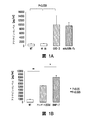
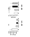
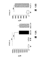
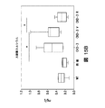
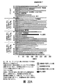
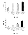
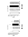
図4Fは、ビヒクルのみ(CKD3+ビヒクル)又は模擬で処置したCKD3と比較したときのActRIIシグナル伝達のインヒビターで処置したCKD-3のモデル(CKD3+ActRII-Fc)におけるアクチンα-平滑筋、Runx2、Klotho、MYOCD、及びα-チューブリンのタンパク質レベルを示している。 FIG. 4F shows actin α-smooth muscle, Runx2, Klotho, in a model of CKD-3 treated with an inhibitor of ActRII signaling (CKD3 + ActRII-Fc) as compared to CKD3 treated with vehicle alone (CKD3 + vehicle) or mock. The protein levels of MYOCD and α-tubulin are shown.
(8.詳細な説明)
(8.1 概説)
本明細書に提供されるのは、一態様において、心血管疾患、血管石灰化、血管石灰化と関連し及び/もしくは血管石灰化に起因する心血管疾患、並びに/又は腎疾患と関連し及び/もしくは腎疾患に起因する心血管疾患、高いレベルの動脈壁硬化及び/もしくは左心室肥大(LVH)と関連し並びに/又は高いレベルの動脈壁硬化及び/もしくはLVHに起因する心血管疾患を含む高いレベルの動脈壁硬化(例えば、血管コンプライアンスの減少によって示されるもの)及び/又はLVHの治療及び/又は予防方法であって、ActRIIシグナル伝達のインヒビター(例えば、アクチビンリガンドトラップ)を、その治療及び/又は予防を必要としている対象に投与することを含む、方法である。ある実施態様において、該対象は、腎疾患対象である。ActRIIシグナル伝達のインヒビターは、ActRIIAシグナル伝達及び/又はActRIIBシグナル伝達のインヒビターであることができる。
(8. Detailed explanation)
(8.1 Overview)
Provided herein, in one aspect, is associated with cardiovascular disease, vascular calcification, vascular calcification and / or caused by vascular calcification, and / or renal disease and Including cardiovascular disease due to renal disease, high levels of arterial stiffness and / or left ventricular hypertrophy (LVH) and / or cardiovascular disease due to high levels of arterial stiffness and / or LVH A method of treating and / or preventing high levels of arterial stiffness (e.g., as indicated by reduced vascular compliance) and / or LVH, wherein an inhibitor of ActRII signaling (e.g., activin ligand trap) is used to treat and A method comprising administering to a subject in need of prevention. In certain embodiments, the subject is a renal disease subject. Inhibitors of ActRII signaling can be inhibitors of ActRIIA signaling and / or ActRIIB signaling.
特に、本明細書に提供されるのは、Snai1、ホスホsmad2、Dkk1、col1a1、アクチビン(例えば、遊離アクチビン)、Runx2、Alp、BSAP、CTX、Osterix、Klotho、α-SMA、MYOCD、ホスホsmad3、尿タンパク質、ActRIIA、Axin2、及び/又はSm22-αのレベル及び/又は活性を、患者集団の指標として、ActRIIシグナル伝達インヒビターによる治療及び/もしくは予防に対する対象の応答性の指標として、ActRIIシグナル伝達インヒビターによる治療の効力の指標として、又はActRIIシグナル伝達インヒビターによる治療のための適切な投薬量の指標として用いることにより、心血管疾患、血管石灰化、血管石灰化と関連し及び/もしくは血管石灰化に起因する心血管疾患、腎疾患と関連し及び/もしくは腎疾患に起因する心血管疾患、高いレベルの動脈壁硬化、並びに/又は左心室肥大(LVH)を治療及び/又は予防する方法である。Snai1、ホスホsmad2、Dkk1、col1a1、アクチビン(例えば、遊離アクチビン)、Runx2、Alp、BSAP、CTX、Osterix、Klotho、α-SMA、MYOCD、ホスホsmad3、尿タンパク質、ActRIIA、Axin2、及び/又はSm22-αのレベル及び/又は活性を用いて、ActRIIシグナル伝達インヒビターによる治療及び/又は予防のための疾患及び/又は状態を特定することもできる。本明細書に記載の方法で使用されるActRIIシグナル伝達インヒビターは、ActRIIAシグナル伝達及び/又はActRIIBシグナル伝達のインヒビター、例えば、本明細書に記載の又は当技術分野で公知のインヒビターのいずれかであることができる。好ましい実施態様において、ActRIIシグナル伝達インヒビターは、ActRIIAの細胞外ドメイン及びヒトIgG1 Fcドメインからなるヒト化融合タンパク質(「ActRIIA-Fc」、例えば、配列番号7)である。 In particular, provided herein are Snai1, phospho smad2, Dkk1, col1a1, activin (e.g., free activin), Runx2, Alp, BSAP, CTX, Osterix, Klotho, α-SMA, MYOCD, phospho smad3, ActRII signaling inhibitors as indicators of urinary protein, ActRIIA, Axin2, and / or Sm22-α levels and / or activity as an indicator of patient population, and as a measure of a subject's responsiveness to treatment and / or prevention with ActRII signaling inhibitors Used as an indicator of the efficacy of treatment with or as an indicator of appropriate dosage for treatment with an ActRII signaling inhibitor, associated with cardiovascular disease, vascular calcification, vascular calcification and / or in vascular calcification Cures cardiovascular disease caused by, and / or associated with kidney disease, high levels of arterial stiffness, and / or left ventricular hypertrophy (LVH) And a / or preventing method. Snai1, phospho smad2, Dkk1, col1a1, activin (e.g., free activin), Runx2, Alp, BSAP, CTX, Osterix, Klotho, α-SMA, MYOCD, phospho smad3, urine protein, ActRIIA, Axin2, and / or Sm22- The level and / or activity of α can also be used to identify diseases and / or conditions for treatment and / or prevention with ActRII signaling inhibitors. The ActRII signaling inhibitor used in the methods described herein is an inhibitor of ActRIIA signaling and / or ActRIIB signaling, eg, any of those described herein or known in the art be able to. In a preferred embodiment, the ActRII signaling inhibitor is a humanized fusion protein consisting of the extracellular domain of ActRIIA and a human IgG1 Fc domain (“ActRIIA-Fc”, eg, SEQ ID NO: 7).
本明細書に提供される方法は、野生型マウスと比較したとき、慢性腎疾患誘導性血管石灰化のマウスモデルでは、Snai1、ホスホsmad2、Dkk1、col1a1、アクチビン(例えば、遊離アクチビン)、Runx2、Alp、BSAP、CTX、ホスホsmad3、尿タンパク質、及びOsterixのレベルが上昇しており、Klotho、α-SMA、MYOCD、ActRIIA、及びSm22-αのレベルが減少しているという発見に一部基づいている。さらに、理論によって制限されるものではないが、mActRIIA-Fcによるリガンドトラッピングは、慢性腎疾患マウスモデル(ldlr-/-マウス、高脂肪食)で血管石灰化を低下させる。本明細書に提示される実施例(第9節参照)で示されるように、mActRIIA-Fcによる慢性腎疾患マウスの処置は、血管石灰化を低下させ、大動脈カルシウムレベルを低下させ、ホスホsmad3レベルを低下させ、尿タンパク質レベルを低下させ、ホスホsmad2レベルを低下させ、Dkk1レベルを低下させ、Runx2レベルを低下させ、Alpレベルを低下させ、BSAPレベルを低下させ、CTXレベルを低下させ、Osterixレベルを低下させ、Klothoレベルを上昇させ、α-SMAレベルを上昇させ、Axin2レベルを上昇させ、Sm22-αレベルを上昇させた。さらに、本明細書に提供される方法は、mActRIIA-Fcで処置したときの慢性腎疾患マウスにおける血管石灰化の減少が、Dkk1、ホスホsmad2、Runx2、Alp、BSAP、CTX、ホスホsmad3、尿タンパク質、及び/又はOsterixレベルの減少、並びにKlotho、α-SMA、Axin2、及び/又はSm22-αレベルの増大と相関しているという発見に一部基づいている(実施例、第9節を参照されたい)。まとめると、本明細書に提示されるデータは、Snai1、ホスホsmad2、Dkk1、col1a1、アクチビン(例えば、遊離アクチビン)、Runx2、Alp、BSAP、CTX、Osterix、Klotho、α-SMA、MYOCD、ホスホsmad3、尿タンパク質、ActRIIA、Axin2、及び/もしくはSm22-αのレベルによって、どの対象がActRIIA-Fcに応答し得るかということを特定することができ、並びに/又は該レベルを用いて、ActRIIシグナル伝達インヒビターに対する臨床応答をモニタリングし及び/もしくは標的患者集団を選択することができることを示している。本明細書に提示されるデータは、ActRIIA-Fc(例えば、mActRIIA-Fc、又はActRIIA-hFc、例えば、配列番号7)が、慢性腎疾患と関連する血管石灰化を治療するのに有用であることも示している。さらに、本明細書に提示されるデータは、ActRIIA-Fc(例えば、mActRIIA-Fc、又はActRIIA-hFc、例えば、配列番号7)が、上昇したSnai1、ホスホsmad2、Dkk1、col1a1、アクチビン(例えば、遊離アクチビン)、Runx2、Alp、BSAP、CTX、ホスホsmad3、尿タンパク質、及び/もしくはOsterixのレベルと関連する疾患、又は減少したKlotho、α-SMA、MYOCD、ActRIIA、及び/もしくはSm22-αのレベルと関連する疾患を治療するのに有用であることも示している。最後に、本明細書に提示されるデータは、ActRIIA-Fc(例えば、mActRIIA-Fc、又はhActRIIA-hFc、例えば、配列番号7)が、大動脈アテロームにおけるカルシウム沈着を減少させ、大動脈カルシウムレベルを低下させ、CKD誘導性の心重量の増大を無効化し、腎線維化を減少させるのに有用であり、したがって、心血管疾患を治療するのに有用であることも示している。 The methods provided herein show that Snai1, phosphosmad2, Dkk1, col1a1, activin (e.g., free activin), Runx2, in a mouse model of chronic kidney disease-induced vascular calcification when compared to wild-type mice. Based in part on the discovery that levels of Alp, BSAP, CTX, phospho smad3, urine protein, and Osterix are elevated and levels of Klotho, α-SMA, MYOCD, ActRIIA, and Sm22-α are decreased Yes. Furthermore, without being limited by theory, ligand trapping with mActRIIA-Fc reduces vascular calcification in a mouse model of chronic kidney disease (ldlr − / − mice, high fat diet). As shown in the examples presented herein (see Section 9), treatment of chronic kidney disease mice with mActRIIA-Fc reduces vascular calcification, reduces aortic calcium levels, and phosphosmad3 levels. Reduce urine protein levels, reduce phospho smad2 levels, reduce Dkk1 levels, reduce Runx2 levels, reduce Alp levels, reduce BSAP levels, reduce CTX levels, and Osterix levels , Increased Klotho level, increased α-SMA level, increased Axin2 level, increased Sm22-α level. Further, the methods provided herein show that vascular calcification in chronic kidney disease mice when treated with mActRIIA-Fc is reduced by Dkk1, phospho smad2, Runx2, Alp, BSAP, CTX, phospho smad3, urine protein And / or Osterix levels, and in part, based on the finding that it correlates with increased Klotho, α-SMA, Axin2, and / or Sm22-α levels (see Example, Section 9). Wanna). In summary, the data presented herein includes Snai1, phospho smad2, Dkk1, col1a1, activin (e.g., free activin), Runx2, Alp, BSAP, CTX, Osterix, Klotho, α-SMA, MYOCD, phospho smad3 The level of urine protein, ActRIIA, Axin2, and / or Sm22-α can identify which subjects can respond to ActRIIA-Fc and / or can be used to determine ActRII signaling It shows that clinical response to inhibitors can be monitored and / or target patient population can be selected. The data presented herein provides that ActRIIA-Fc (e.g., mActRIIA-Fc, or ActRIIA-hFc, e.g., SEQ ID NO: 7) is useful for treating vascular calcification associated with chronic kidney disease It also shows that. In addition, the data presented herein includes ActRIIA-Fc (e.g., mActRIIA-Fc, or ActRIIA-hFc, e.g., SEQ ID NO: 7), which is elevated Snai1, phosphosmad2, Dkk1, col1a1, activin (e.g. Disease associated with levels of free activin), Runx2, Alp, BSAP, CTX, phosphosmad3, urine protein, and / or Osterix, or decreased levels of Klotho, α-SMA, MYOCD, ActRIIA, and / or Sm22-α It is also useful for treating diseases associated with. Finally, the data presented herein show that ActRIIA-Fc (e.g., mActRIIA-Fc, or hActRIIA-hFc, e.g., SEQ ID NO: 7) reduces calcification in aortic atheroma and reduces aortic calcium levels It has also been shown to be useful in negating CKD-induced increase in heart weight and reducing renal fibrosis and thus useful in treating cardiovascular disease.
実施例(第9節参照)に例示されている、本明細書に記載の研究結果は、Snai1、ホスホsmad2、Dkk1、col1a1、アクチビン(例えば、遊離アクチビン)、Runx2、Alp、BSAP、CTX、Osterix、Klotho、α-SMA、MYOCD、ホスホsmad3、尿タンパク質、ActRIIA、Axin2、又はSm22-αのレベル及び/又は活性の検出を、(i)対象における血管石灰化の度合いのマーカー(例えば、血清バイオマーカー)として、(ii)ActRIIシグナル伝達インヒビター(例えば、ActRIIA-Fc)に対する対象の応答を測定するためのマーカーとして、又は(iii)治療後の対象におけるActRIIシグナル伝達インヒビター(例えば、ActRIIA-Fc)の薬力学的効果を評価するために使用することができることを示しており、ここで、対象は、心血管疾患、血管石灰化、血管石灰化と関連し及び/もしくは血管石灰化に起因する心血管疾患、並びに/又は腎疾患と関連し及び/もしくは腎疾患に起因する心血管疾患を有する対象である。したがって、ある実施態様において、Snai1、ホスホsmad2、Dkk1、col1a1、アクチビン(例えば、遊離アクチビン)、Runx2、Alp、BSAP、CTX、Osterix、Klotho、α-SMA、MYOCD、ホスホsmad3、尿タンパク質、Axin2、及び/又はSm22-αを、ActRIIA-Fc(例えば、ActRIIA-hFc、例えば、配列番号7)治療の効力の指標として及び/又はActRIIA-Fc(例えば、ActRIIA-hFc、例えば、配列番号7)による治療に対する応答の欠如の指標として、本明細書に記載の方法で使用することができる。さらに、本明細書に記載されているように、Snai1、ホスホsmad2、Dkk1、col1a1、アクチビン(例えば、遊離アクチビン)、Runx2、Alp、BSAP、CTX、Osterix、Klotho、α-SMA、MYOCD、ホスホsmad3、尿タンパク質、ActRIIA、Axin2、及び/又はSm22-αを、ActRIIA-Fc(例えば、ActRIIA-hFc、例えば、配列番号7)の経時的治療効力を評価するための信頼できる分子マーカーとして使用することができる。さらに、具体的な実施態様において、本明細書に提供されるのは、血液中のSnai1、ホスホsmad2、Dkk1、col1a1、アクチビン(例えば、遊離アクチビン)、Runx2、Alp、BSAP、CTX、Osterix、Klotho、α-SMA、MYOCD、ホスホsmad3、尿タンパク質、ActRIIA、Axin2、及び/又はSm22-αのレベル及び/又は活性の検出(例えば、血管平滑筋細胞機能の障害と関連する疾患における異常発現の検出)、並びにSnai1、ホスホsmad2、Dkk1、col1a1、アクチビン(例えば、遊離アクチビン)、Runx2、Alp、BSAP、CTX、Osterix、Klotho、α-SMA、MYOCD、ホスホsmad3、尿タンパク質、ActRIIA、Axin2、及び/又はSm22-αのレベル及び/又は活性に依存する用量でのActRIIシグナル伝達インヒビター、例えば、ActRIIA-Fcの投与を含む方法である。 The results of the studies described herein, exemplified in the Examples (see Section 9), are: Snai1, phospho smad2, Dkk1, col1a1, activin (e.g., free activin), Runx2, Alp, BSAP, CTX, Osterix Detection of the level and / or activity of Klotho, α-SMA, MYOCD, phospho smad3, urine protein, ActRIIA, Axin2, or Sm22-α, (i) a marker of the degree of vascular calcification in a subject (e.g., serum bio (Ii) as a marker for measuring a subject's response to an ActRII signaling inhibitor (e.g., ActRIIA-Fc), or (iii) an ActRII signaling inhibitor (e.g., ActRIIA-Fc) in a post-treatment subject In which the subject is associated with cardiovascular disease, vascular calcification, vascular calcification and / or cardiac caused by vascular calcification. Vascular disease and / Is a subject with a cardiovascular disease caused by related and / or renal disease and renal disease. Thus, in certain embodiments, Snai1, phospho smad2, Dkk1, col1a1, activin (e.g., free activin), Runx2, Alp, BSAP, CTX, Osterix, Klotho, α-SMA, MYOCD, phospho smad3, urinary protein, Axin2, And / or Sm22-α as ActRIIA-Fc (e.g., ActRIIA-hFc, e.g. SEQ ID NO: 7) as an indicator of therapeutic efficacy and / or ActRIIA-Fc (e.g., ActRIIA-hFc, e.g., SEQ ID NO: 7) As an indicator of lack of response to treatment, it can be used in the methods described herein. In addition, as described herein, Snai1, phospho smad2, Dkk1, col1a1, activin (e.g., free activin), Runx2, Alp, BSAP, CTX, Osterix, Klotho, α-SMA, MYOCD, phospho smad3 Urinary protein, ActRIIA, Axin2, and / or Sm22-α as a reliable molecular marker for assessing the therapeutic efficacy over time of ActRIIA-Fc (eg, ActRIIA-hFc, eg, SEQ ID NO: 7) Can do. Further, in a specific embodiment, provided herein are Snai1, phospho smad2, Dkk1, col1a1, activin (e.g., free activin), Runx2, Alp, BSAP, CTX, Osterix, Klotho in blood. Detection of the level and / or activity of, α-SMA, MYOCD, phospho smad3, urinary protein, ActRIIA, Axin2, and / or Sm22-α (e.g. detection of abnormal expression in diseases associated with impaired vascular smooth muscle cell function) ), And Snai1, phospho smad2, Dkk1, col1a1, activin (e.g., free activin), Runx2, Alp, BSAP, CTX, Osterix, Klotho, α-SMA, MYOCD, phospho smad3, urine protein, ActRIIA, Axin2, and / or Or a method comprising administration of an ActRII signaling inhibitor, eg, ActRIIA-Fc, at a dose depending on the level and / or activity of Sm22-α.
ある実施態様において、ActRIIシグナル伝達インヒビターは、ソタテルセプト(配列番号7;例えば、第9節参照)である。ソタテルセプトは、本明細書に記載の方法で有用であるアクチビンリガンドトラップである(第8節参照)。 In certain embodiments, the ActRII signaling inhibitor is sotatercept (SEQ ID NO: 7; see, eg, Section 9). Sotatercept is an activin ligand trap that is useful in the methods described herein (see Section 8).
(8.2 略語)
本明細書で使用されるように、「Snai 1」又は「Snai1」は、snailホモログ1を指す。例えば、Twigg及びWilkieの文献、1999, Hum Genet. 105(4):320-326を参照されたい。GenBank(商標)アクセッション番号NM_005985.3は、例示的なヒトSnai1核酸配列を提供する。GenBank(商標)アクセッション番号NP_005976.2は、例示的なヒトSnai1アミノ酸配列を提供する。
(8.2 Abbreviations)
As used herein, “
本明細書で使用されるように、「ホスホsmad3」は、デカペンタプレジック(decapentaplegic)ホモログ3に対するリン酸化母体を指す。例えば、Matsuzakiの文献、2013, Cytokine Growth Factor Rev, 24(4):385-399を参照されたい。GenBank(商標)アクセッション番号NM_005902.3は、例示的なヒトホスホsmad3核酸配列を提供する。GenBank(商標)アクセッション番号NP_005893.1は、例示的なヒトホスホsmad3アミノ酸配列を提供する。
As used herein, “phospho smad3” refers to a phosphorylated matrix against
本明細書で使用されるように、「ホスホsmad2」は、デカペンタプレジックホモログ2に対するリン酸化母体を指す。例えば、Matsuzakiの文献、2013, Cytokine Growth Factor Rev, 24(4):385-399を参照されたい。GenBank(商標)アクセッション番号NM_001003652.3は、例示的なヒトホスホsmad2核酸配列を提供する。GenBank(商標)アクセッション番号NP_001003652.1は、例示的なヒトホスホsmad2アミノ酸配列を提供する。
As used herein, “phospho smad2” refers to a phosphorylated matrix against
本明細書で使用されるように、「Dkk1」は、dickkopf関連タンパク1を指す。例えば、Fang, Y.らの文献、2014. J Am Soc Nephrol 25:1760-1763を参照されたい。GenBank(商標)アクセッション番号NM_012242.2は、例示的なヒトDkk1核酸配列を提供する。GenBank(商標)アクセッション番号NP_036374.1は、例示的なヒトDkk1アミノ酸配列を提供する。
As used herein, “Dkk1” refers to dickkopf associated
本明細書で使用されるように、「col1a1」は、コラーゲン1型、α1を指す。例えば、Korkkoらの文献、1998, Am. J. Hum. Genet., 62:98-110を参照されたい。GenBank(商標)アクセッション番号XM_005257059.2は、例示的なヒトcol1a1核酸配列を提供する。GenBank(商標)アクセッション番号XP_005257116.2は、例示的なヒトcol1a1アミノ酸配列を提供する。
As used herein, “col1a1” refers to
本明細書で使用されるように、「Runx2」は、runt関連転写因子2を指す。例えば、Komoriの文献、2010, Adv Exp Med Biol, 658:43-49を参照されたい。GenBank(商標)アクセッション番号NM_001145920.2は、例示的なヒトRunx2核酸配列を提供する。GenBank(商標)アクセッション番号NP_001139392.1は、例示的なヒトRunx2アミノ酸配列を提供する。
As used herein, “Runx2” refers to runt-related
本明細書で使用されるように、「Alp」又は「Alpl」は、アルカリホスファターゼを指す。本明細書で使用されるように、「BSAP」は、骨特異的アルカリホスファターゼを指す。例えば、Martinsらの文献、2013, Bone, 56(2):390-397を参照されたい。GenBank(商標)アクセッション番号NM_000478.4は、例示的なヒトAlp核酸配列を提供する。GenBank(商標)アクセッション番号NP_000469.3は、例示的なヒトAlpアミノ酸配列を提供する。 As used herein, “Alp” or “Alpl” refers to alkaline phosphatase. As used herein, “BSAP” refers to bone-specific alkaline phosphatase. See, for example, Martins et al., 2013, Bone, 56 (2): 390-397. GenBank ™ accession number NM — 000478.4 provides an exemplary human Alp nucleic acid sequence. GenBank ™ accession number NP — 000469.3 provides an exemplary human Alp amino acid sequence.
本明細書で使用されるように、「CTX」は、C末端テロペプチドI型コラーゲンを指す。例えば、Rosenらの文献、2000, Calcif Tissue Int. 66(2): 100-103を参照されたい。 As used herein, “CTX” refers to C-terminal telopeptide type I collagen. See, for example, Rosen et al., 2000, Calcif Tissue Int. 66 (2): 100-103.
本明細書で使用されるように、「Osterix」は、Sp7転写因子としても知られる、Osterixを指す。例えば、Caoらの文献、2005, Cancer Res. 65(4):1124-1128を参照されたい。GenBank(商標)アクセッション番号NM_001173467.2は、例示的なヒトOsterix核酸配列を提供する。GenBank(商標)アクセッション番号NP_001166938.1は、例示的なヒトOsterixアミノ酸配列を提供する。 As used herein, “Osterix” refers to Osterix, also known as Sp7 transcription factor. See, for example, Cao et al., 2005, Cancer Res. 65 (4): 1124-1128. GenBank ™ accession number NM — 001173467.2 provides an exemplary human Osterix nucleic acid sequence. GenBank ™ accession number NP — 001166938.1 provides an exemplary human Osterix amino acid sequence.
本明細書で使用されるように、「Klotho」は、Klothoを指す。例えば、Matsumuraらの文献、1998, Biochem Biophys Res Commun, 242:626-630を参照されたい。GenBank(商標)アクセッション番号NM_004795.3は、例示的なヒトKlotho核酸配列を提供する。GenBank(商標)アクセッション番号NP_004786.2は、例示的なヒトKlothoアミノ酸配列を提供する。 As used herein, “Klotho” refers to Klotho. See, for example, Matsumura et al., 1998, Biochem Biophys Res Commun, 242: 626-630. GenBank ™ accession number NM — 004795.3 provides an exemplary human Klotho nucleic acid sequence. GenBank ™ accession number NP — 004786.2 provides an exemplary human Klotho amino acid sequence.
本明細書で使用されるように、「α-SMA(alpha-SMA)」又は「αSMA」又は「α-SMA」は、α平滑筋アクチンを指す。例えば、Nowakらの文献、1999, Nat. Genet. 23:208-212を参照されたい。GenBank(商標)アクセッション番号NM_001100.3は、例示的なヒトα-SMA核酸配列を提供する。GenBank(商標)アクセッション番号NP_001091.1は、例示的なヒトα-SMAアミノ酸配列を提供する。 As used herein, “α-SMA (alpha-SMA)” or “αSMA” or “α-SMA” refers to α smooth muscle actin. See, for example, Nowak et al., 1999, Nat. Genet. 23: 208-212. GenBank ™ accession number NM — 001100.3 provides an exemplary human α-SMA nucleic acid sequence. GenBank ™ accession number NP — 001091.1 provides an exemplary human α-SMA amino acid sequence.
本明細書で使用されるように、「MYOCD」は、ミオカルディンを指す。例えば、Imamuraらの文献、2010, Gene, 464:1-10を参照されたい。GenBank(商標)アクセッション番号NM_001146312.2は、例示的なヒトMYOCD核酸配列を提供する。GenBank(商標)アクセッション番号NP_001139784.1は、例示的なヒトMYOCDアミノ酸配列を提供する。 As used herein, “MYOCD” refers to myocardin. See, for example, Imamura et al., 2010, Gene, 464: 1-10. GenBank ™ accession number NM — 001146312.2 provides an exemplary human MYOCD nucleic acid sequence. GenBank ™ accession number NP — 001139784.1 provides an exemplary human MYOCD amino acid sequence.
本明細書で使用されるように、「Sm22-α(Sm22-alpha)」又は「Sm22α」又は「Sm22-α」は、「トランスゲリン」、「Tagln」、又は「Tagln1」としても知られる、平滑筋タンパク質22αを指す。例えば、Camoretti-Mercadoの文献、1998, Genomics, 49:452-457を参照されたい。GenBank(商標)アクセッション番号NM_001001522.1は、例示的なヒトSm22-α核酸配列を提供する。GenBank(商標)アクセッション番号NP_001001522.1は、例示的なヒトSm22-αアミノ酸配列を提供する。 As used herein, “Sm22-α (Sm22-alpha)” or “Sm22α” or “Sm22-α” is also known as “transgelin”, “Tagln”, or “Tagln1”. Refers to smooth muscle protein 22α. See, for example, Camoretti-Mercado, 1998, Genomics, 49: 452-457. GenBank ™ accession number NM — 001001522.1 provides an exemplary human Sm22-α nucleic acid sequence. GenBank ™ accession number NP — 001001522.1 provides an exemplary human Sm22-α amino acid sequence.
本明細書で使用されるように、「ActRII」は、アクチビン受容体II型を指す。本明細書で使用されるように、「ActRIIA」は、アクチビン受容体IIA型を指す。例えば、Mathews及びValeの文献、1991, Cell 65:973-982を参照されたい。GenBank(商標)アクセッション番号NM_001278579.1は、例示的なヒトActRIIA核酸配列を提供する。GenBank(商標)アクセッション番号NP_001265508.1は、例示的なヒトActRIIAアミノ酸配列を提供する。本明細書で使用されるように、「ActRIIB」は、アクチビン受容体IIB型を指す。例えば、Attisanoらの文献、1992, Cell 68: 97-108を参照されたい。GenBank(商標)アクセッション番号NM_001106.3は、例示的なヒトActRIIB核酸配列を提供する。GenBank(商標)アクセッション番号NP_001097.2は、例示的なヒトActRIIBアミノ酸配列を提供する。 As used herein, “ActRII” refers to activin receptor type II. As used herein, “ActRIIA” refers to activin receptor type IIA. See, for example, Mathews and Vale, 1991, Cell 65: 973-982. GenBank ™ accession number NM — 001278579.1 provides an exemplary human ActRIIA nucleic acid sequence. GenBank ™ accession number NP — 001265508.1 provides an exemplary human ActRIIA amino acid sequence. As used herein, “ActRIIB” refers to activin receptor type IIB. See, for example, Attisano et al., 1992, Cell 68: 97-108. GenBank ™ accession number NM — 001106.3 provides an exemplary human ActRIIB nucleic acid sequence. GenBank ™ accession number NP — 001097.2 provides an exemplary human ActRIIB amino acid sequence.
本明細書で使用されるように、「Axin2」は、軸インヒビター2を指す。GenBank(商標)アクセッション番号NM_004655.3は、例示的なヒトAxin2核酸配列を提供する。GenBank(商標)アクセッション番号NP_004646.3は、例示的なヒトアミノ酸配列を提供する。
As used herein, “Axin2” refers to
本明細書で使用されるように、「mActRIIA-Fc」又は「ActRIIA-mFc」は、マウスアクチビンIIA型受容体-IgG1融合タンパク質を指す。例えば、米国特許第8,173,601号を参照されたい。本明細書で使用されるように、「mActRIIB-Fc」又は「ActRIIB-mFc」は、マウスアクチビンIIB型受容体-IgG1融合タンパク質を指す。例えば、米国特許第8,173,601号を参照されたい。本明細書で使用されるように、「hActRIIA-Fc」又は「ActRIIA-hFc」は、ヒトアクチビンIIA型受容体-IgG1融合タンパク質を指す。例えば、米国特許第8,173,601号を参照されたい。本明細書で使用されるように、「hActRIIB-Fc」又は「ActRIIB-hFc」は、ヒトアクチビンIIB型受容体-IgG1融合タンパク質を指す。例えば、米国特許第8,173,601号を参照されたい。 As used herein, “mActRIIA-Fc” or “ActRIIA-mFc” refers to a mouse activin type IIA receptor-IgG1 fusion protein. See, for example, US Pat. No. 8,173,601. As used herein, “mActRIIB-Fc” or “ActRIIB-mFc” refers to a mouse activin type IIB receptor-IgG1 fusion protein. See, for example, US Pat. No. 8,173,601. As used herein, “hActRIIA-Fc” or “ActRIIA-hFc” refers to a human activin type IIA receptor-IgG1 fusion protein. See, for example, US Pat. No. 8,173,601. As used herein, “hActRIIB-Fc” or “ActRIIB-hFc” refers to human activin type IIB receptor-IgG1 fusion protein. See, for example, US Pat. No. 8,173,601.
本明細書で使用されるように、「LVH」は、左心室肥大を指す。 As used herein, “LVH” refers to left ventricular hypertrophy.
(8.3 治療及び/又は予防方法)
(8.3.1 心血管疾患及び/又は血管石灰化)
ある実施態様において、本明細書に提供されるのは、対象における心血管疾患、血管石灰化、血管石灰化と関連し及び/もしくは血管石灰化に起因する心血管疾患、高いレベルの動脈壁硬化と関連し及び/もしくは高いレベルの動脈壁硬化に起因する心血管疾患、高いレベルの動脈壁硬化、左心室肥大、左心室肥大と関連し及び/もしくは左心室肥大に起因する心血管疾患、並びに/又は腎疾患と関連し及び/もしくは腎疾患に起因する心血管疾患を治療及び/又は予防する方法であって、該対象に、ActRIIシグナル伝達インヒビター(例えば、アクチビンリガンドトラップ)の医薬有効用量を投与することを含む、方法である。ある実施態様において、対象は、腎疾患対象である。ある他の実施態様において、対象は、慢性腎疾患に伴う骨ミネラル代謝異常を有すると診断されている。ある他の実施態様において、対象は、慢性腎疾患に伴う骨ミネラル代謝異常を有すると診断されていない。
(8.3 Treatment and / or prevention methods)
(8.3.1 Cardiovascular disease and / or vascular calcification)
In certain embodiments, provided herein are cardiovascular disease, vascular calcification, cardiovascular disease associated with and / or due to vascular calcification in a subject, high levels of arterial stiffness. Cardiovascular disease associated with and / or high levels of arterial stiffness, high levels of arterial stiffness, left ventricular hypertrophy, left ventricular hypertrophy and / or left ventricular hypertrophy, and A method of treating and / or preventing cardiovascular disease associated with and / or resulting from renal disease, wherein the subject is administered a pharmaceutically effective dose of an ActRII signaling inhibitor (e.g., activin ligand trap) A method comprising administering. In certain embodiments, the subject is a renal disease subject. In certain other embodiments, the subject has been diagnosed with bone mineral metabolism abnormalities associated with chronic kidney disease. In certain other embodiments, the subject has not been diagnosed with bone mineral metabolism abnormalities associated with chronic kidney disease.
ある実施態様において、対象は、参照集団におけるRunx2のレベル及び/もしくは活性と比較して上昇したRunx2のレベル及び/もしくは活性、参照集団におけるAlpのレベル及び/もしくは活性と比較して上昇したAlpのレベル及び/もしくは活性、参照集団におけるSnai1のレベル及び/もしくは活性と比較して上昇したSnai1のレベル及び/もしくは活性、参照集団におけるホスホsmad2のレベル及び/もしくは活性と比較して上昇したホスホsmad2のレベル及び/もしくは活性、参照集団におけるホスホsmad3のレベル及び/もしくは活性と比較して上昇したホスホsmad3のレベル及び/もしくは活性、参照集団における尿タンパク質のレベル及び/もしくは活性と比較して上昇した尿タンパク質のレベル及び/もしくは活性、参照集団におけるDkk1のレベル及び/もしくは活性と比較して上昇したDkk1のレベル及び/もしくは活性、参照集団におけるアクチビン(例えば、遊離アクチビン)のレベル及び/もしくは活性と比較して上昇したアクチビン(例えば、遊離アクチビン)のレベル及び/もしくは活性、参照集団におけるcol1a1のレベル及び/もしくは活性と比較して上昇したcol1a1のレベル及び/もしくは活性、参照集団におけるBSAPのレベル及び/もしくは活性と比較して上昇したBSAPのレベル及び/もしくは活性、参照集団におけるCTXのレベル及び/もしくは活性と比較して上昇したCTXのレベル及び/もしくは活性、参照集団におけるOsterixのレベル及び/もしくは活性と比較して上昇したOsterixのレベル及び/もしくは活性、参照集団におけるKlothoのレベル及び/もしくは活性と比較して減少したKlothoのレベル及び/もしくは活性、参照集団におけるα-SMAのレベル及び/もしくは活性と比較して減少したα-SMAのレベル及び/もしくは活性、参照集団におけるMYOCDのレベル及び/もしくは活性と比較して減少したMYOCDのレベル及び/もしくは活性、参照集団におけるSm22-αのレベル及び/もしくは活性と比較して減少したSm22-αのレベル及び/もしくは活性;並びに/又は参照集団におけるActRIIAのレベル及び/もしくは活性と比較して減少したActRIIAのレベル及び/もしくは活性を有する。 In certain embodiments, the subject has an increased level and / or activity of Runx2 compared to the level and / or activity of Runx2 in the reference population, an Alp level increased relative to the level and / or activity of Alp in the reference population. Level and / or activity, increased level and / or activity of Snai1 compared to the level and / or activity of Snai1 in the reference population, level of phosphosmad2 increased compared to the level and / or activity of phosphosmad2 in the reference population Levels and / or activities, increased levels and / or activities of phosphosmad3 compared to phosphosmad3 levels and / or activity in the reference population, increased urine compared to levels and / or activities of urine protein in the reference population Protein level and / or activity, Dkk1 level and / or activity in the reference population Increased Dkk1 level and / or activity, activin (eg, free activin) level and / or activity compared to activin (eg, free activin) level and / or activity in a reference population, see Elevated col1a1 level and / or activity compared to col1a1 level and / or activity in the population, elevated BSAP level and / or activity compared to BSAP level and / or activity in the reference population, in reference population CTX levels and / or activities increased compared to CTX levels and / or activities, Osterix levels and / or activities increased compared to Osterix levels and / or activities in the reference population, Klotho's levels in the reference population Decreased Klotho level and / or activity compared to level and / or activity, reference population Reduced α-SMA level and / or activity compared to α-SMA level and / or activity, reduced MYOCD level and / or activity compared to MYOCD level and / or activity in the reference population, Reduced level and / or activity of Sm22-α compared to the level and / or activity of Sm22-α in the reference population; and / or reduced level of ActRIIA compared to the level and / or activity of ActRIIA in the reference population And / or active.
ある実施態様において、対象は、対象における事前のRunx2のレベル及び/もしくは活性と比較して上昇したRunx2のレベル及び/もしくは活性、対象における事前のAlpのレベル及び/もしくは活性と比較して上昇したAlpのレベル及び/もしくは活性、対象における事前のSnai1のレベル及び/もしくは活性と比較して上昇したSnai1のレベル及び/もしくは活性、対象における事前のホスホsmad2のレベル及び/もしくは活性と比較して上昇したホスホsmad2のレベル及び/もしくは活性、対象における事前のホスホsmad3のレベル及び/もしくは活性と比較して上昇したホスホsmad3のレベル及び/もしくは活性、対象における事前の尿タンパク質のレベル及び/もしくは活性と比較して上昇した尿タンパク質のレベル及び/もしくは活性、対象における事前のDkk1のレベル及び/もしくは活性と比較して上昇したDkk1のレベル及び/もしくは活性、対象における事前のアクチビン(例えば、遊離アクチビン)のレベル及び/もしくは活性と比較して上昇したアクチビン(例えば、遊離アクチビン)のレベル及び/もしくは活性、対象における事前のcol1a1のレベル及び/もしくは活性と比較して上昇したcol1a1のレベル及び/もしくは活性、対象における事前のBSAPのレベル及び/もしくは活性と比較して上昇したBSAPのレベル及び/もしくは活性、対象における事前のCTXのレベル及び/もしくは活性と比較して上昇したCTXのレベル及び/もしくは活性、対象における事前のOsterixのレベル及び/もしくは活性と比較して上昇したOsterixのレベル及び/もしくは活性、対象における事前のKlothoのレベル及び/もしくは活性と比較して減少したKlothoのレベル及び/もしくは活性、対象における事前のα-SMAのレベル及び/もしくは活性と比較して減少したα-SMAのレベル及び/もしくは活性、対象における事前のMYOCDのレベル及び/もしくは活性と比較して減少したMYOCDのレベル及び/もしくは活性、対象における事前のActRIIAのレベル及び/もしくは活性と比較して減少したActRIIAのレベル及び/もしくは活性、並びに/又は対象における事前のSm22-αのレベル及び/もしくは活性と比較して減少したSm22-αのレベル及び/もしくは活性を有する。ある実施態様において、対象における事前のRunx2のレベル及び/もしくは活性、対象における事前のAlpのレベル及び/もしくは活性、対象における事前のSnai1のレベル及び/もしくは活性、対象における事前のホスホsmad2のレベル及び/もしくは活性、対象における事前のホスホsmad3のレベル及び/もしくは活性、対象における事前の尿タンパク質のレベル及び/もしくは活性、対象における事前のDkk1のレベル及び/もしくは活性、対象における事前のアクチビン(例えば、遊離アクチビン)のレベル及び/もしくは活性、対象における事前のcol1a1のレベル及び/もしくは活性、対象における事前のBSAPのレベル及び/もしくは活性、対象における事前のCTXのレベル及び/もしくは活性、対象における事前のOsterixのレベル及び/もしくは活性、対象における事前のKlothoのレベル及び/もしくは活性、対象における事前のα-SMAのレベル及び/もしくは活性、対象における事前のMYOCDのレベル及び/もしくは活性、対象における事前のActRIIAのレベル及び/もしくは活性、並びに/又は対象における事前のSm22-αのレベル及び/もしくは活性は、(i)心血管疾患;(ii)血管石灰化;(iii)血管石灰化と関連し及び/もしくは血管石灰化に起因する心血管疾患;(iv)腎疾患と関連し及び/もしくは腎疾患に起因する心血管疾患;(v)高いレベルの動脈壁硬化;並びに/又は左心室肥大(LVH)の症状の発症又はこれら疾患の診断の1、2、3、4、5、6、8、10、12、18、24、又は48カ月前のそれぞれのレベルである。 In certain embodiments, the subject has increased Runx2 level and / or activity compared to prior Runx2 level and / or activity in the subject, increased compared to prior Alp level and / or activity in the subject. Alp level and / or activity, elevated Snai1 level and / or activity compared to prior Snai1 level and / or activity in the subject, elevated compared to prior phospho smad2 level and / or activity in the subject Phospho-smad2 level and / or activity, elevated phospho-smad3 level and / or activity compared to prior phospho-smad3 level and / or activity in the subject, prior urine protein level and / or activity in the subject Elevated urinary protein level and / or activity, prior Dkk1 level in the subject and / or Or increased Dkk1 level and / or activity relative to activity, prior activin (e.g., free activin) level and / or elevated activin (e.g., free activin) level and / or activity in a subject / Or activity, elevated col1a1 level and / or activity compared to previous col1a1 level and / or activity in the subject, elevated BSAP level and / or activity compared to previous BSAP level and / or activity in the subject / Or activity, elevated CTX level and / or activity compared to prior CTX level and / or activity in the subject, elevated Osterix level and / or activity compared to prior Osterix level and / or activity in the subject / Or activity, prior Klotho level in the subject and / or reduced Klotho level compared to activity Decreased and / or activity, decreased α-SMA level and / or activity compared to previous α-SMA level and / or activity in the subject, decreased compared to previous MYOCD level and / or activity in the subject MYOCD level and / or activity, reduced ActRIIA level and / or activity compared to prior ActRIIA level and / or activity in the subject, and / or prior Sm22-α level and / or activity in the subject Have decreased Sm22-α levels and / or activity. In certain embodiments, the prior Runx2 level and / or activity in the subject, the prior Alp level and / or activity in the subject, the prior Snai1 level and / or activity in the subject, the prior phospho smad2 level in the subject, and / Or activity, prior phospho smad3 level and / or activity in the subject, prior urine protein level and / or activity in the subject, prior Dkk1 level and / or activity in the subject, prior activin in the subject (e.g., Free activin) level and / or activity, prior col1a1 level and / or activity in the subject, prior BSAP level and / or activity in the subject, prior CTX level and / or activity in the subject, prior in the subject Osterix level and / or activity, prior Klo in subject tho level and / or activity, prior α-SMA level and / or activity in the subject, prior MYOCD level and / or activity in the subject, prior ActRIIA level and / or activity in the subject, and / or subject Prior Sm22-α levels and / or activity in (i) cardiovascular disease; (ii) vascular calcification; (iii) cardiovascular disease associated with and / or resulting from vascular calcification; (iv) cardiovascular disease associated with and / or resulting from renal disease; (v) high level of arterial stiffness; and / or onset of symptoms of left ventricular hypertrophy (LVH) or diagnosis of these diseases 1 , 2, 3, 4, 5, 6, 8, 10, 12, 18, 24, or 48 months ago.
ある実施態様において、対象は、第8.3.4節に記載されているような方法に従って治療される。ある実施態様において、対象における血管石灰化は、第8.6節に記載されているようなアガットストンスコアを測定することにより解析される。 In certain embodiments, the subject is treated according to a method as described in Section 8.3.4. In certain embodiments, vascular calcification in a subject is analyzed by measuring an Agatstone score as described in Section 8.6.
ある実施態様において、本明細書に提供される方法で治療されるべき対象は、第8.6節に記載されているようなSnai1、ホスホsmad2、Dkk1、col1a1、アクチビン(例えば、遊離アクチビン)、Runx2、Alp、BSAP、CTX、Osterix、Klotho、α-SMA、MYOCD、ホスホsmad3、尿タンパク質、ActRIIA、Axin2、及び/又はSm22-αのレベル及び/又は活性を有する。したがって、ある具体的な実施態様において、本明細書に提供される方法は、(i)第8.6節に記載されているようなSnai1、ホスホsmad2、Dkk1、col1a1、アクチビン(例えば、遊離アクチビン)、Runx2、Alp、BSAP、CTX、Osterix、Klotho、α-SMA、MYOCD、ホスホsmad3、尿タンパク質、ActRIIA、及び/又はSm22-αのレベル及び/又は活性に基づいて対象を選択すること;並びに(ii)ActRIIシグナル伝達インヒビター(例えば、アクチビンリガンドトラップ)の医薬有効用量を投与することを含む。具体的な実施態様において、対象は、心血管疾患を有する。具体的な実施態様において、対象は、血管石灰化を有する。具体的な実施態様において、対象は、血管石灰化と関連し及び/又は血管石灰化に起因する心血管疾患を有する。具体的な実施態様において、対象は、腎疾患と関連し及び/又は腎疾患に起因する心血管疾患を有する。具体的な実施態様において、対象は、腎疾患を有する。ある他の実施態様において、対象は、慢性腎疾患に伴う骨ミネラル代謝異常を有すると診断されている。ある他の実施態様において、対象は、慢性腎疾患に伴う骨ミネラル代謝異常を有すると診断されていない。ある実施態様において、対象における血管石灰化は、第8.6節に記載されているようなアガットストンスコアを測定することにより解析される。具体的な実施態様において、対象は、高いレベルの動脈壁硬化と診断されている。具体的な実施態様において、対象は、LVHと診断されている。具体的な実施態様において、対象は、高いレベルの動脈壁硬化と関連し及び/又は高いレベルの動脈壁硬化に起因する心血管疾患を有する。具体的な実施態様において、対象は、LVHと関連し及び/又はLVHに起因する心血管疾患を有する。 In certain embodiments, the subject to be treated with the methods provided herein is Snai1, phospho smad2, Dkk1, col1a1, activin (e.g., free activin), Runx2, as described in Section 8.6. Alp, BSAP, CTX, Osterix, Klotho, α-SMA, MYOCD, phospho smad3, urinary protein, ActRIIA, Axin2, and / or Sm22-α levels and / or activities. Thus, in certain specific embodiments, the methods provided herein include (i) Snai1, phosphosmad2, Dkk1, col1a1, activin (eg, free activin), as described in Section 8.6, Selecting subjects based on the level and / or activity of Runx2, Alp, BSAP, CTX, Osterix, Klotho, α-SMA, MYOCD, phospho smad3, urinary protein, ActRIIA, and / or Sm22-α; and (ii ) Administering a pharmaceutically effective dose of an ActRII signaling inhibitor (eg, activin ligand trap). In a specific embodiment, the subject has cardiovascular disease. In a specific embodiment, the subject has vascular calcification. In a specific embodiment, the subject has a cardiovascular disease associated with and / or resulting from vascular calcification. In a specific embodiment, the subject has cardiovascular disease associated with and / or resulting from renal disease. In a specific embodiment, the subject has renal disease. In certain other embodiments, the subject has been diagnosed with bone mineral metabolism abnormalities associated with chronic kidney disease. In certain other embodiments, the subject has not been diagnosed with bone mineral metabolism abnormalities associated with chronic kidney disease. In certain embodiments, vascular calcification in a subject is analyzed by measuring an Agatstone score as described in Section 8.6. In a specific embodiment, the subject has been diagnosed with high levels of arterial stiffness. In a specific embodiment, the subject has been diagnosed with LVH. In a specific embodiment, the subject has a cardiovascular disease associated with and / or resulting from high levels of arterial stiffness. In a specific embodiment, the subject has a cardiovascular disease associated with and / or caused by LVH.
ある実施態様において、Snai1、ホスホsmad2、Dkk1、col1a1、アクチビン(例えば、遊離アクチビン)、Runx2、Alp、BSAP、CTX、Osterix、Klotho、α-SMA、MYOCD、ホスホsmad3、尿タンパク質、ActRIIA、Axin2、及び/又はSm22-αのレベル及び/又は活性は、第8.6節に記載されているように決定される。ある実施態様において、Snai1、ホスホsmad2、Dkk1、col1a1、アクチビン(例えば、遊離アクチビン)、Runx2、Alp、BSAP、CTX、Osterix、Klotho、α-SMA、MYOCD、ホスホsmad3、尿タンパク質、ActRIIA、Axin2、及び/又はSm22-αのレベル及び/又は活性は、それぞれ、Snai1、ホスホsmad2、Dkk1、col1a1、アクチビン(例えば、遊離アクチビン)、Runx2、Alp、BSAP、CTX、Osterix、Klotho、α-SMA、MYOCD、ホスホsmad3、尿タンパク質、ActRIIA、Axin2、及び/又はSm22-αのタンパク質レベル及び/又は活性である。ある実施態様において、Snai1、ホスホsmad2、Dkk1、col1a1、アクチビン(例えば、遊離アクチビン)、Runx2、Alp、BSAP、CTX、Osterix、Klotho、α-SMA、MYOCD、ホスホsmad3、尿タンパク質、ActRIIA、Axin2、及び/又はSm22-αのレベル及び/又は活性は、それぞれ、Snai1、ホスホsmad2、Dkk1、col1a1、アクチビン(例えば、遊離アクチビン)、Runx2、Alp、BSAP、CTX、Osterix、Klotho、α-SMA、MYOCD、ホスホsmad3、尿タンパク質、ActRIIA、Axin2、及び/又はSm22-αのmRNAレベル及び/又は活性である。ある実施態様において、Snai1、ホスホsmad2、Dkk1、col1a1、アクチビン(例えば、遊離アクチビン)、Runx2、Alp、BSAP、CTX、Osterix、Klotho、Sm22-α、MYOCD、及び/又はActRIIA、Axin2、α-SMAのレベルは、組織におけるものである。ある実施態様において、組織は大動脈である。ある実施態様において、組織は腎臓である。ある実施態様において、組織は骨である。ある実施態様において、組織は血清である。好ましい実施態様において、Runx2、Dkk1、Alp、osterix、sm22-α、Klotho、α-SMA、MYOCD、ActRIIA、Axin2のレベル及び/又は活性は、大動脈におけるものである。好ましい実施態様において、ホスホsmad2、ホスホsmad3、尿タンパク質、ActRIIA、Axin2、及びcol1a1のレベル及び/又は活性は、腎臓におけるものである。好ましい実施態様において、アクチビンのレベル及び/又は活性は、血清におけるものである。ある実施態様において、アクチビンのレベルは、参照集団に対して対象で上昇している。ある実施態様において、対象におけるフォリスタチンのレベルは、参照集団におけるフォリスタチンのレベルとほぼ等しい。 In certain embodiments, Snai1, phospho smad2, Dkk1, col1a1, activin (e.g., free activin), Runx2, Alp, BSAP, CTX, Osterix, Klotho, α-SMA, MYOCD, phospho smad3, urine protein, ActRIIA, Axin2, And / or the level and / or activity of Sm22-α is determined as described in Section 8.6. In certain embodiments, Snai1, phospho smad2, Dkk1, col1a1, activin (e.g., free activin), Runx2, Alp, BSAP, CTX, Osterix, Klotho, α-SMA, MYOCD, phospho smad3, urine protein, ActRIIA, Axin2, And / or Sm22-α levels and / or activities are Snai1, phospho smad2, Dkk1, col1a1, activin (e.g., free activin), Runx2, Alp, BSAP, CTX, Osterix, Klotho, α-SMA, MYOCD, respectively. Protein levels and / or activity of phosphosmad3, urinary protein, ActRIIA, Axin2, and / or Sm22-α. In certain embodiments, Snai1, phospho smad2, Dkk1, col1a1, activin (e.g., free activin), Runx2, Alp, BSAP, CTX, Osterix, Klotho, α-SMA, MYOCD, phospho smad3, urine protein, ActRIIA, Axin2, And / or Sm22-α levels and / or activities are Snai1, phospho smad2, Dkk1, col1a1, activin (e.g., free activin), Runx2, Alp, BSAP, CTX, Osterix, Klotho, α-SMA, MYOCD, respectively. MRNA levels and / or activities of phosphosmad3, urinary protein, ActRIIA, Axin2, and / or Sm22-α. In certain embodiments, Snai1, phospho smad2, Dkk1, col1a1, activin (e.g., free activin), Runx2, Alp, BSAP, CTX, Osterix, Klotho, Sm22-α, MYOCD, and / or ActRIIA, Axin2, α-SMA Level is in the organization. In certain embodiments, the tissue is an aorta. In certain embodiments, the tissue is the kidney. In certain embodiments, the tissue is bone. In certain embodiments, the tissue is serum. In a preferred embodiment, the level and / or activity of Runx2, Dkk1, Alp, osterix, sm22-α, Klotho, α-SMA, MYOCD, ActRIIA, Axin2 is in the aorta. In a preferred embodiment, the level and / or activity of phospho smad2, phospho smad3, urine protein, ActRIIA, Axin2, and col1a1 is in the kidney. In a preferred embodiment, the activin level and / or activity is in serum. In certain embodiments, the level of activin is elevated in the subject relative to the reference population. In certain embodiments, the level of follistatin in the subject is approximately equal to the level of follistatin in the reference population.
ある実施態様において、参照集団は、第8.6節に記載されているような集団である。ある実施態様において、対象は、第8.4節に記載されているような対象である。 In certain embodiments, the reference population is a population as described in Section 8.6. In certain embodiments, the subject is a subject as described in Section 8.4.
ある実施態様において、ActRIIシグナル伝達インヒビターは、ActRIIA-Fc、例えば、ActRIIA-hFc(例えば、配列番号7)である。ある実施態様において、ActRIIシグナル伝達インヒビターは、第8.5節に記載されているようなActRIIシグナル伝達インヒビターである。 In certain embodiments, the ActRII signaling inhibitor is ActRIIA-Fc, eg, ActRIIA-hFc (eg, SEQ ID NO: 7). In certain embodiments, the ActRII signaling inhibitor is an ActRII signaling inhibitor as described in Section 8.5.
ある実施態様において、医薬有効用量は、第8.7節に記載されているような用量である。ある実施態様において、医薬有効用量は、初回用量である。ある実施態様において、医薬有効用量は、第8.7節に記載されているような頻度で投与される。ある実施態様において、医薬有効用量は、第8.7節に記載されているように投与される。 In certain embodiments, the pharmaceutically effective dose is a dose as described in Section 8.7. In certain embodiments, the pharmaceutically effective dose is an initial dose. In certain embodiments, the pharmaceutically effective dose is administered at a frequency as described in Section 8.7. In certain embodiments, a pharmaceutically effective dose is administered as described in Section 8.7.
ある実施態様において、本明細書に提供される方法は、第8.8節に記載されているような、第2の医薬活性剤と組み合わせて利用される。 In certain embodiments, the methods provided herein are utilized in combination with a second pharmaceutically active agent, as described in Section 8.8.
ある実施態様において、心血管疾患又は慢性腎疾患との関連における「治療する(treat)」、「治療」、又は「治療する(treating)」は、それぞれ、心血管疾患又は慢性腎疾患の少なくとも1つの症状の改善を含む。 In certain embodiments, `` treat, '' `` treatment, '' or `` treating '' in the context of cardiovascular disease or chronic kidney disease is at least one of cardiovascular disease or chronic kidney disease, respectively. Includes improvement of one symptom.
当業者によって認識されているように、Runx2、Alp、Snai1、ホスホsmad2、Dkk1、col1a1、アクチビン、BSAP、CTX、osterix、Klotho、α-SMA、MYOCD、ActRIIA、Axin2、Sm22-α、ホスホsmad3、及び尿タンパク質のうちの1つ又は複数のレベル及び/又は活性を、それぞれ、対応する参照集団における該レベル及び/又は活性と比較することができる。したがって、ある実施態様において、Runx2、Alp、Snai1、ホスホsmad2、Dkk1、col1a1、アクチビン、BSAP、CTX、osterix、Klotho、α-SMA、MYOCD、Sm22-α、ホスホsmad3、ActRIIA、Axin2、及び尿タンパク質のうちの1つのレベル及び/又は活性を対応する参照集団におけるそのレベル及び/又は活性と比較する。ある実施態様において、Runx2、Alp、Snai1、ホスホsmad2、Dkk1、col1a1、アクチビン、BSAP、CTX、osterix、Klotho、α-SMA、MYOCD、Sm22-α、ホスホsmad3、ActRIIA、Axin2、及び尿タンパク質のうちの2つのレベル及び/又は活性を対応する参照集団におけるそのレベル及び/又は活性と比較する。ある実施態様において、Runx2、Alp、Snai1、ホスホsmad2、Dkk1、col1a1、アクチビン、BSAP、CTX、osterix、Klotho、α-SMA、MYOCD、Sm22-α、ホスホsmad3、ActRIIA、Axin2、及び尿タンパク質のうちの3つのレベル及び/又は活性を対応する参照集団におけるそのレベル及び/又は活性と比較する。ある実施態様において、Runx2、Alp、Snai1、ホスホsmad2、Dkk1、col1a1、アクチビン、BSAP、CTX、osterix、Klotho、α-SMA、MYOCD、Sm22-α、ホスホsmad3、ActRIIA、Axin2、及び尿タンパク質のうちの3つのレベル及び/又は活性を対応する参照集団におけるそのレベル及び/又は活性と比較する。ある実施態様において、Runx2、Alp、Snai1、ホスホsmad2、Dkk1、col1a1、アクチビン、BSAP、CTX、osterix、Klotho、α-SMA、MYOCD、Sm22-α、ホスホsmad3、ActRIIA、Axin2、及び尿タンパク質のうちの4つのレベル及び/又は活性を対応する参照集団におけるそのレベル及び/又は活性と比較する。ある実施態様において、Runx2、Alp、Snai1、ホスホsmad2、Dkk1、col1a1、アクチビン、BSAP、CTX、osterix、Klotho、α-SMA、MYOCD、Sm22-α、ホスホsmad3、ActRIIA、Axin2、及び尿タンパク質のうちの5つのレベル及び/又は活性を対応する参照集団におけるそのレベル及び/又は活性と比較する。ある実施態様において、Runx2、Alp、Snai1、ホスホsmad2、Dkk1、col1a1、アクチビン、BSAP、CTX、osterix、Klotho、α-SMA、MYOCD、Sm22-α、ホスホsmad3、ActRIIA、Axin2、及び尿タンパク質のうちの6つのレベル及び/又は活性を対応する参照集団におけるそのレベル及び/又は活性と比較する。ある実施態様において、Runx2、Alp、Snai1、ホスホsmad2、Dkk1、col1a1、アクチビン、BSAP、CTX、osterix、Klotho、α-SMA、MYOCD、Sm22-α、ホスホsmad3、ActRIIA、Axin2、及び尿タンパク質のうちの7つのレベル及び/又は活性を対応する参照集団におけるそのレベル及び/又は活性と比較する。ある実施態様において、Runx2、Alp、Snai1、ホスホsmad2、Dkk1、col1a1、アクチビン、BSAP、CTX、osterix、Klotho、α-SMA、MYOCD、Sm22-α、ホスホsmad3、ActRIIA、Axin2、及び尿タンパク質のうちの8つのレベル及び/又は活性を対応する参照集団におけるそのレベル及び/又は活性と比較する。ある実施態様において、Runx2、Alp、Snai1、ホスホsmad2、Dkk1、col1a1、アクチビン、BSAP、CTX、osterix、Klotho、α-SMA、MYOCD、Sm22-α、ホスホsmad3、ActRIIA、Axin2、及び尿タンパク質のうちの9つのレベル及び/又は活性を対応する参照集団におけるそのレベル及び/又は活性と比較する。ある実施態様において、Runx2、Alp、Snai1、ホスホsmad2、Dkk1、col1a1、アクチビン、BSAP、CTX、osterix、Klotho、α-SMA、MYOCD、Sm22-α、ホスホsmad3、ActRIIA、Axin2、及び尿タンパク質のうちの10のレベル及び/又は活性を対応する参照集団におけるそのレベル及び/又は活性と比較する。ある実施態様において、Runx2、Alp、Snai1、ホスホsmad2、Dkk1、col1a1、アクチビン、BSAP、CTX、osterix、Klotho、α-SMA、MYOCD、Sm22-α、ホスホsmad3、ActRIIA、Axin2、及び尿タンパク質のうちの11のレベル及び/又は活性を対応する参照集団におけるそのレベル及び/又は活性と比較する。ある実施態様において、Runx2、Alp、Snai1、ホスホsmad2、Dkk1、col1a1、アクチビン、BSAP、CTX、osterix、Klotho、α-SMA、MYOCD、Sm22-α、ホスホsmad3、ActRIIA、Axin2、及び尿タンパク質のうちの12のレベル及び/又は活性を対応する参照集団におけるそのレベル及び/又は活性と比較する。ある実施態様において、Runx2、Alp、Snai1、ホスホsmad2、Dkk1、col1a1、アクチビン、BSAP、CTX、osterix、Klotho、α-SMA、MYOCD、Sm22-α、ホスホsmad3、ActRIIA、Axin2、及び尿タンパク質のうちの13のレベル及び/又は活性を対応する参照集団におけるそのレベル及び/又は活性と比較する。ある実施態様において、Runx2、Alp、Snai1、ホスホsmad2、Dkk1、col1a1、アクチビン、BSAP、CTX、osterix、Klotho、α-SMA、MYOCD、Sm22-α、ホスホsmad3、ActRIIA、Axin2、及び尿タンパク質のうちの14のレベル及び/又は活性を対応する参照集団におけるそのレベル及び/又は活性と比較する。ある実施態様において、Runx2、Alp、Snai1、ホスホsmad2、Dkk1、col1a1、アクチビン、BSAP、CTX、osterix、Klotho、α-SMA、MYOCD、Sm22-α、ホスホsmad3、ActRIIA、Axin2、及び尿タンパク質のうちの15のレベル及び/又は活性を対応する参照集団におけるそのレベル及び/又は活性と比較する。ある実施態様において、Runx2、Alp、Snai1、ホスホsmad2、Dkk1、col1a1、アクチビン、BSAP、CTX、osterix、Klotho、α-SMA、MYOCD、Sm22-α、ホスホsmad3、ActRIIA、Axin2、及び尿タンパク質のうちの16のレベル及び/又は活性を対応する参照集団におけるそのレベル及び/又は活性と比較する。ある実施態様において、Runx2、Alp、Snai1、ホスホsmad2、Dkk1、col1a1、アクチビン、BSAP、CTX、osterix、Klotho、α-SMA、MYOCD、Sm22-α、ホスホsmad3、ActRIIA、Axin2、及び尿タンパク質のうちの17のレベル及び/又は活性を対応する参照集団におけるそのレベル及び/又は活性と比較する。ある実施態様において、Runx2、Alp、Snai1、ホスホsmad2、Dkk1、col1a1、アクチビン、BSAP、CTX、osterix、Klotho、α-SMA、MYOCD、Sm22-α、ホスホsmad3、ActRIIA、Axin2、及び尿タンパク質の各々のレベル及び/又は活性を対応する参照集団におけるそのレベル及び/又は活性と比較する。 As recognized by those skilled in the art, Runx2, Alp, Snai1, phospho smad2, Dkk1, col1a1, activin, BSAP, CTX, osterix, Klotho, α-SMA, MYOCD, ActRIIA, Axin2, Sm22-α, phospho smad3, And the level and / or activity of one or more of the urine proteins can be compared to the level and / or activity in the corresponding reference population, respectively. Thus, in certain embodiments, Runx2, Alp, Snai1, phospho smad2, Dkk1, col1a1, activin, BSAP, CTX, osterix, Klotho, α-SMA, MYOCD, Sm22-α, phospho smad3, ActRIIA, Axin2, and urine protein The level and / or activity of one of the is compared to that level and / or activity in the corresponding reference population. In certain embodiments, of Runx2, Alp, Snai1, phospho smad2, Dkk1, col1a1, activin, BSAP, CTX, osterix, Klotho, α-SMA, MYOCD, Sm22-α, phospho smad3, ActRIIA, Axin2, and urine protein Is compared to its level and / or activity in the corresponding reference population. In certain embodiments, of Runx2, Alp, Snai1, phospho smad2, Dkk1, col1a1, activin, BSAP, CTX, osterix, Klotho, α-SMA, MYOCD, Sm22-α, phospho smad3, ActRIIA, Axin2, and urine protein Are compared with their levels and / or activity in the corresponding reference population. In certain embodiments, of Runx2, Alp, Snai1, phospho smad2, Dkk1, col1a1, activin, BSAP, CTX, osterix, Klotho, α-SMA, MYOCD, Sm22-α, phospho smad3, ActRIIA, Axin2, and urine protein Are compared with their levels and / or activity in the corresponding reference population. In certain embodiments, of Runx2, Alp, Snai1, phospho smad2, Dkk1, col1a1, activin, BSAP, CTX, osterix, Klotho, α-SMA, MYOCD, Sm22-α, phospho smad3, ActRIIA, Axin2, and urine protein Is compared to its level and / or activity in the corresponding reference population. In certain embodiments, of Runx2, Alp, Snai1, phospho smad2, Dkk1, col1a1, activin, BSAP, CTX, osterix, Klotho, α-SMA, MYOCD, Sm22-α, phospho smad3, ActRIIA, Axin2, and urine protein Is compared to its level and / or activity in the corresponding reference population. In certain embodiments, of Runx2, Alp, Snai1, phospho smad2, Dkk1, col1a1, activin, BSAP, CTX, osterix, Klotho, α-SMA, MYOCD, Sm22-α, phospho smad3, ActRIIA, Axin2, and urine protein Is compared to its level and / or activity in the corresponding reference population. In certain embodiments, of Runx2, Alp, Snai1, phospho smad2, Dkk1, col1a1, activin, BSAP, CTX, osterix, Klotho, α-SMA, MYOCD, Sm22-α, phospho smad3, ActRIIA, Axin2, and urine protein The seven levels and / or activities of are compared with their levels and / or activities in the corresponding reference population. In certain embodiments, of Runx2, Alp, Snai1, phospho smad2, Dkk1, col1a1, activin, BSAP, CTX, osterix, Klotho, α-SMA, MYOCD, Sm22-α, phospho smad3, ActRIIA, Axin2, and urine protein Is compared to its level and / or activity in the corresponding reference population. In certain embodiments, of Runx2, Alp, Snai1, phospho smad2, Dkk1, col1a1, activin, BSAP, CTX, osterix, Klotho, α-SMA, MYOCD, Sm22-α, phospho smad3, ActRIIA, Axin2, and urine protein Nine levels and / or activities of are compared to their levels and / or activities in the corresponding reference population. In certain embodiments, of Runx2, Alp, Snai1, phospho smad2, Dkk1, col1a1, activin, BSAP, CTX, osterix, Klotho, α-SMA, MYOCD, Sm22-α, phospho smad3, ActRIIA, Axin2, and urine protein Is compared to its level and / or activity in the corresponding reference population. In certain embodiments, of Runx2, Alp, Snai1, phospho smad2, Dkk1, col1a1, activin, BSAP, CTX, osterix, Klotho, α-SMA, MYOCD, Sm22-α, phospho smad3, ActRIIA, Axin2, and urine protein The 11 levels and / or activity of is compared to its level and / or activity in the corresponding reference population. In certain embodiments, of Runx2, Alp, Snai1, phospho smad2, Dkk1, col1a1, activin, BSAP, CTX, osterix, Klotho, α-SMA, MYOCD, Sm22-α, phospho smad3, ActRIIA, Axin2, and urine protein Is compared to its level and / or activity in the corresponding reference population. In certain embodiments, of Runx2, Alp, Snai1, phospho smad2, Dkk1, col1a1, activin, BSAP, CTX, osterix, Klotho, α-SMA, MYOCD, Sm22-α, phospho smad3, ActRIIA, Axin2, and urine protein The thirteen levels and / or activities are compared with their levels and / or activities in the corresponding reference population. In certain embodiments, of Runx2, Alp, Snai1, phospho smad2, Dkk1, col1a1, activin, BSAP, CTX, osterix, Klotho, α-SMA, MYOCD, Sm22-α, phospho smad3, ActRIIA, Axin2, and urine protein The 14 levels and / or activities are compared to their levels and / or activities in the corresponding reference population. In certain embodiments, of Runx2, Alp, Snai1, phospho smad2, Dkk1, col1a1, activin, BSAP, CTX, osterix, Klotho, α-SMA, MYOCD, Sm22-α, phospho smad3, ActRIIA, Axin2, and urine protein Is compared to its level and / or activity in the corresponding reference population. In certain embodiments, of Runx2, Alp, Snai1, phospho smad2, Dkk1, col1a1, activin, BSAP, CTX, osterix, Klotho, α-SMA, MYOCD, Sm22-α, phospho smad3, ActRIIA, Axin2, and urine protein The 16 levels and / or activity of is compared to its level and / or activity in the corresponding reference population. In certain embodiments, of Runx2, Alp, Snai1, phospho smad2, Dkk1, col1a1, activin, BSAP, CTX, osterix, Klotho, α-SMA, MYOCD, Sm22-α, phospho smad3, ActRIIA, Axin2, and urine protein Is compared to its level and / or activity in the corresponding reference population. In certain embodiments, each of Runx2, Alp, Snai1, phospho smad2, Dkk1, col1a1, activin, BSAP, CTX, osterix, Klotho, α-SMA, MYOCD, Sm22-α, phospho smad3, ActRIIA, Axin2, and each urine protein Is compared with its level and / or activity in the corresponding reference population.
(8.3.2 Snai1、ホスホsmad2、Dkk1、col1a1、アクチビン(例えば、遊離アクチビン)、Runx2、Alp、BSAP、CTX、Osterix、Klotho、α-SMA、MYOCD、ホスホsmad3、尿タンパク質、ActRIIA、Axin2、及び/又はSm22-αと関連する疾患)
ある実施態様において、本明細書に提供されるのは、Snai1、ホスホsmad2、Dkk1、col1a1、アクチビン(例えば、遊離アクチビン)、Runx2、Alp、BSAP、CTX、Osterix、Klotho、α-SMA、MYOCD、ホスホsmad3、尿タンパク質、ActRIIA、Axin2、及び/又はSm22-αと関連する1以上の疾患を治療及び/又は予防する方法であって、対象に、ActRIIシグナル伝達インヒビター(例えば、アクチビンリガンドトラップ)の医薬有効用量を投与することを含む、方法である。
(8.3.2 Snai1, phospho smad2, Dkk1, col1a1, activin (e.g., free activin), Runx2, Alp, BSAP, CTX, Osterix, Klotho, α-SMA, MYOCD, phospho smad3, urine protein, ActRIIA, Axin2, and (Or diseases associated with Sm22-α)
In certain embodiments, provided herein are Snai1, phospho smad2, Dkk1, col1a1, activin (e.g., free activin), Runx2, Alp, BSAP, CTX, Osterix, Klotho, α-SMA, MYOCD, A method of treating and / or preventing one or more diseases associated with phospho smad3, urine protein, ActRIIA, Axin2, and / or Sm22-α, wherein the subject is treated with an ActRII signaling inhibitor (e.g., activin ligand trap). Administering a pharmaceutically effective dose.
ある実施態様において、対象は、第8.4節に記載されているような対象である。 In certain embodiments, the subject is a subject as described in Section 8.4.
ある実施態様において、ActRIIシグナル伝達インヒビターは、ActRIIA-Fc、例えば、ActRIIA-hFc(例えば、配列番号7)である。ある実施態様において、ActRIIシグナル伝達インヒビターは、第8.5節に記載されているようなActRIIシグナル伝達インヒビターである。 In certain embodiments, the ActRII signaling inhibitor is ActRIIA-Fc, eg, ActRIIA-hFc (eg, SEQ ID NO: 7). In certain embodiments, the ActRII signaling inhibitor is an ActRII signaling inhibitor as described in Section 8.5.
ある実施態様において、医薬有効用量は、第8.7節に記載されているような用量である。ある実施態様において、医薬有効用量は、初回用量である。ある実施態様において、医薬有効用量は、第8.7節に記載されているような頻度で投与される。ある実施態様において、医薬有効用量は、第8.7節に記載されているように投与される。ある実施態様において、医薬有効用量は、本明細書に記載されているような特定のバイオマーカーのレベル及び/又は活性に応じて調整される。 In certain embodiments, the pharmaceutically effective dose is a dose as described in Section 8.7. In certain embodiments, the pharmaceutically effective dose is an initial dose. In certain embodiments, the pharmaceutically effective dose is administered at a frequency as described in Section 8.7. In certain embodiments, a pharmaceutically effective dose is administered as described in Section 8.7. In certain embodiments, the pharmaceutically effective dose is adjusted according to the level and / or activity of a particular biomarker as described herein.
ある実施態様において、本明細書に提供される方法は、第8.8節に記載されているような、第2の医薬活性剤と組み合わせて利用される。 In certain embodiments, the methods provided herein are utilized in combination with a second pharmaceutically active agent, as described in Section 8.8.
(8.3.3 慢性腎疾患に伴う骨ミネラル代謝異常)
ある実施態様において、本明細書に提供されるのは、対象における慢性腎疾患に伴う骨ミネラル代謝異常を治療する方法であって、該対象に、ActRIIシグナル伝達インヒビター(例えば、アクチビンリガンドトラップ)の医薬有効用量を投与することを含む、方法である。ある実施態様において、本明細書に提供されるのは、対象における骨吸収を低下させる方法であって、該対象に、ActRIIシグナル伝達インヒビターの医薬有効用量を投与することを含む、方法である。ある実施態様において、対象は、心血管疾患を有する。ある実施態様において、対象は、血管石灰化を有する。ある実施態様において、対象は、血管石灰化と関連し及び/又は血管石灰化に起因する心血管疾患を有する。ある実施態様において、対象は、腎疾患と関連し及び/又は腎疾患に起因する心血管疾患を有する。ある実施態様において、対象は、腎疾患対象である。ある他の実施態様において、対象は、慢性腎疾患に伴う骨ミネラル代謝異常を有すると診断されている。ある他の実施態様において、対象は、慢性腎疾患に伴う骨ミネラル代謝異常を有すると診断されていない。
(8.3.3 Bone mineral metabolism abnormalities associated with chronic kidney disease)
In certain embodiments, provided herein is a method of treating a bone mineral metabolism disorder associated with chronic kidney disease in a subject, wherein the subject is treated with an ActRII signaling inhibitor (e.g., activin ligand trap). Administering a pharmaceutically effective dose. In certain embodiments, provided herein is a method of reducing bone resorption in a subject, comprising administering to the subject a pharmaceutically effective dose of an ActRII signaling inhibitor. In certain embodiments, the subject has cardiovascular disease. In certain embodiments, the subject has vascular calcification. In certain embodiments, the subject has a cardiovascular disease associated with and / or resulting from vascular calcification. In certain embodiments, the subject has cardiovascular disease associated with and / or resulting from renal disease. In certain embodiments, the subject is a renal disease subject. In certain other embodiments, the subject has been diagnosed with bone mineral metabolism abnormalities associated with chronic kidney disease. In certain other embodiments, the subject has not been diagnosed with bone mineral metabolism abnormalities associated with chronic kidney disease.
ある実施態様において、対象は、参照集団におけるRunx2のレベル及び/もしくは活性と比較して上昇したRunx2のレベル及び/もしくは活性、参照集団におけるAlpのレベル及び/もしくは活性と比較して上昇したAlpのレベル及び/もしくは活性、参照集団におけるSnai1のレベル及び/もしくは活性と比較して上昇したSnai1のレベル及び/もしくは活性、参照集団におけるホスホsmad2のレベル及び/もしくは活性と比較して上昇したホスホsmad2のレベル及び/もしくは活性、参照集団におけるホスホsmad3のレベル及び/もしくは活性と比較して上昇したホスホsmad3のレベル及び/もしくは活性、参照集団における尿タンパク質のレベル及び/もしくは活性と比較して上昇した尿タンパク質のレベル及び/もしくは活性、参照集団におけるDkk1のレベル及び/もしくは活性と比較して上昇したDkk1のレベル及び/もしくは活性、参照集団におけるcol1a1のレベル及び/もしくは活性と比較して上昇したcol1a1のレベル及び/もしくは活性、参照集団におけるアクチビン(例えば、遊離アクチビン)のレベル及び/もしくは活性と比較して上昇したアクチビン(例えば、遊離アクチビン)のレベル及び/もしくは活性、参照集団におけるBSAPのレベル及び/もしくは活性と比較して上昇したBSAPのレベル及び/もしくは活性、参照集団におけるCTXのレベル及び/もしくは活性と比較して上昇したCTXのレベル及び/もしくは活性、参照集団におけるOsterixのレベル及び/もしくは活性と比較して上昇したOsterixのレベル及び/もしくは活性、参照集団におけるKlothoのレベル及び/もしくは活性と比較して減少したKlothoのレベル及び/もしくは活性、参照集団におけるα-SMAのレベル及び/もしくは活性と比較して減少したα-SMAのレベル及び/もしくは活性、参照集団におけるMYOCDのレベル及び/もしくは活性と比較して減少したMYOCDのレベル及び/もしくは活性、参照集団におけるActRIIAのレベル及び/もしくは活性と比較して減少したActRIIAのレベル及び/もしくは活性、並びに/又は参照集団におけるSm22-αのレベル及び/もしくは活性と比較して減少したSm22-αのレベル及び/もしくは活性を有する。ある実施態様において、Snai1、ホスホsmad2、Dkk1、col1a1、アクチビン(例えば、遊離アクチビン)、Runx2、Alp、BSAP、CTX、Osterix、Klotho、Sm22-α、MYOCD、ホスホsmad3、尿タンパク質、ActRIIA、及び/又はα-SMAのレベルは、組織におけるものである。ある実施態様において、組織は大動脈である。ある実施態様において、組織は腎臓である。ある実施態様において、組織は骨である。ある実施態様において、組織は血清である。好ましい実施態様において、Runx2、Dkk1、Alp、osterix、sm22-α、Klotho、α-SMA、MYOCD、及び/又はActRIIAのレベル及び/又は活性は、大動脈におけるものである。好ましい実施態様において、ホスホsmad2、ホスホsmad3、尿タンパク質、及びcol1a1、並びに/又はActRIIAのレベル及び/又は活性は、腎臓におけるものである。好ましい実施態様において、アクチビンのレベル及び/又は活性は、血清におけるものである。ある実施態様において、Snai1、ホスホsmad2、Dkk1、col1a1、アクチビン(例えば、遊離アクチビン)、Runx2、Alp、BSAP、CTX、Osterix、Klotho、Sm22-α、MYOCD、ActRIIA、及び/又はα-SMAのレベルは、大動脈におけるものである。 In certain embodiments, the subject has an increased level and / or activity of Runx2 compared to the level and / or activity of Runx2 in the reference population, an Alp level increased relative to the level and / or activity of Alp in the reference population. Level and / or activity, increased level and / or activity of Snai1 compared to the level and / or activity of Snai1 in the reference population, level of phosphosmad2 increased compared to the level and / or activity of phosphosmad2 in the reference population Levels and / or activities, increased levels and / or activities of phosphosmad3 compared to phosphosmad3 levels and / or activity in the reference population, increased urine compared to levels and / or activities of urine protein in the reference population Protein level and / or activity, Dkk1 level and / or activity in the reference population Elevated Dkk1 level and / or activity, elevated col1a1 level and / or activity compared to the reference population, activin (e.g., free activin) level in the reference population and Increased level and / or activity of activin (e.g., free activin) compared to activity, increased BSAP level and / or activity compared to BSAP level and / or activity in the reference population, in reference population CTX levels and / or activities increased compared to CTX levels and / or activities, Osterix levels and / or activities increased compared to Osterix levels and / or activities in the reference population, Klotho's levels in the reference population Decreased Klotho level and / or activity compared to level and / or activity, reference population Reduced α-SMA level and / or activity compared to α-SMA level and / or activity, reduced MYOCD level and / or activity compared to MYOCD level and / or activity in the reference population, Reduced ActRIIA level and / or activity compared to ActRIIA level and / or activity in the reference population and / or reduced Sm22-α level and / or activity compared to Sm22-α level and / or activity in the reference population And / or active. In certain embodiments, Snai1, phospho smad2, Dkk1, col1a1, activin (e.g., free activin), Runx2, Alp, BSAP, CTX, Osterix, Klotho, Sm22-α, MYOCD, phospho smad3, urinary protein, ActRIIA, and / or Or the level of α-SMA is in the tissue. In certain embodiments, the tissue is an aorta. In certain embodiments, the tissue is the kidney. In certain embodiments, the tissue is bone. In certain embodiments, the tissue is serum. In a preferred embodiment, the level and / or activity of Runx2, Dkk1, Alp, osterix, sm22-α, Klotho, α-SMA, MYOCD, and / or ActRIIA is in the aorta. In a preferred embodiment, the level and / or activity of phosphosmad2, phosphosmad3, urine protein, and col1a1, and / or ActRIIA is in the kidney. In a preferred embodiment, the activin level and / or activity is in serum. In certain embodiments, the levels of Snai1, phospho smad2, Dkk1, col1a1, activin (e.g., free activin), Runx2, Alp, BSAP, CTX, Osterix, Klotho, Sm22-α, MYOCD, ActRIIA, and / or α-SMA Is in the aorta.
ある実施態様において、対象は、対象における事前のRunx2のレベル及び/もしくは活性と比較して上昇したRunx2のレベル及び/もしくは活性、対象における事前のAlpのレベル及び/もしくは活性と比較して上昇したAlpのレベル及び/もしくは活性、対象における事前のSnai1のレベル及び/もしくは活性と比較して上昇したSnai1のレベル及び/もしくは活性、対象における事前のホスホsmad2のレベル及び/もしくは活性と比較して上昇したホスホsmad2のレベル及び/もしくは活性、対象における事前のホスホsmad3のレベル及び/もしくは活性と比較して上昇したホスホsmad3のレベル及び/もしくは活性、対象における事前の尿タンパク質のレベル及び/もしくは活性と比較して上昇した尿タンパク質のレベル及び/もしくは活性、対象における事前のDkk1のレベル及び/もしくは活性と比較して上昇したDkk1のレベル及び/もしくは活性、対象における事前のアクチビン(例えば、遊離アクチビン)のレベル及び/もしくは活性と比較して上昇したアクチビン(例えば、遊離アクチビン)のレベル及び/もしくは活性、対象における事前のcol1a1のレベル及び/もしくは活性と比較して上昇したcol1a1のレベル及び/もしくは活性、対象における事前のBSAPのレベル及び/もしくは活性と比較して上昇したBSAPのレベル及び/もしくは活性、対象における事前のCTXのレベル及び/もしくは活性と比較して上昇したCTXのレベル及び/もしくは活性、対象における事前のOsterixのレベル及び/もしくは活性と比較して上昇したOsterixのレベル及び/もしくは活性、対象における事前のKlothoのレベル及び/もしくは活性と比較して減少したKlothoのレベル及び/もしくは活性、対象における事前のα-SMAのレベル及び/もしくは活性と比較して減少したα-SMAのレベル及び/もしくは活性、対象における事前のMYOCDのレベル及び/もしくは活性と比較して減少したMYOCDのレベル及び/もしくは活性、対象における事前のActRIIAのレベル及び/もしくは活性と比較して減少したActRIIAのレベル及び/もしくは活性、並びに/又は対象における事前のSm22-αのレベル及び/もしくは活性と比較して減少したSm22-αのレベル及び/もしくは活性を有する。ある実施態様において、対象における事前のRunx2のレベル及び/もしくは活性、対象における事前のAlpのレベル及び/もしくは活性、対象における事前のSnai1のレベル及び/もしくは活性、対象における事前のホスホsmad2のレベル及び/もしくは活性、対象における事前のホスホsmad3のレベル及び/もしくは活性、対象における事前の尿タンパク質のレベル及び/もしくは活性、対象における事前のDkk1のレベル及び/もしくは活性、対象における事前のアクチビン(例えば、遊離アクチビン)のレベル及び/もしくは活性、対象における事前のcol1a1のレベル及び/もしくは活性、対象における事前のBSAPのレベル及び/もしくは活性、対象における事前のCTXのレベル及び/もしくは活性、対象における事前のOsterixのレベル及び/もしくは活性、対象における事前のKlothoのレベル及び/もしくは活性、対象における事前のα-SMAのレベル及び/もしくは活性、対象における事前のMYOCDのレベル及び/もしくは活性、対象における事前のActRIIAのレベル及び/もしくは活性、並びに/又は対象における事前のSm22-αのレベル及び/もしくは活性は、CKD-MBDの症状の発症又はCKD-MBDの診断の1、2、3、4、5、6、8、10、12、18、24、又は48カ月前のそれぞれのレベルである。 In certain embodiments, the subject has increased Runx2 level and / or activity compared to prior Runx2 level and / or activity in the subject, increased compared to prior Alp level and / or activity in the subject. Alp level and / or activity, elevated Snai1 level and / or activity compared to prior Snai1 level and / or activity in the subject, elevated compared to prior phospho smad2 level and / or activity in the subject Phospho-smad2 level and / or activity, elevated phospho-smad3 level and / or activity compared to prior phospho-smad3 level and / or activity in the subject, prior urine protein level and / or activity in the subject Elevated urinary protein level and / or activity, prior Dkk1 level in the subject and / or Or increased Dkk1 level and / or activity relative to activity, prior activin (e.g., free activin) level and / or elevated activin (e.g., free activin) level and / or activity in a subject / Or activity, elevated col1a1 level and / or activity compared to previous col1a1 level and / or activity in the subject, elevated BSAP level and / or activity compared to previous BSAP level and / or activity in the subject / Or activity, elevated CTX level and / or activity compared to prior CTX level and / or activity in the subject, elevated Osterix level and / or activity compared to prior Osterix level and / or activity in the subject / Or activity, prior Klotho level in the subject and / or reduced Klotho level compared to activity Decreased and / or activity, decreased α-SMA level and / or activity compared to previous α-SMA level and / or activity in the subject, decreased compared to previous MYOCD level and / or activity in the subject MYOCD level and / or activity, reduced ActRIIA level and / or activity compared to prior ActRIIA level and / or activity in the subject, and / or prior Sm22-α level and / or activity in the subject Have decreased Sm22-α levels and / or activity. In certain embodiments, the prior Runx2 level and / or activity in the subject, the prior Alp level and / or activity in the subject, the prior Snai1 level and / or activity in the subject, the prior phospho smad2 level in the subject, and / Or activity, prior phospho smad3 level and / or activity in the subject, prior urine protein level and / or activity in the subject, prior Dkk1 level and / or activity in the subject, prior activin in the subject (e.g., Free activin) level and / or activity, prior col1a1 level and / or activity in the subject, prior BSAP level and / or activity in the subject, prior CTX level and / or activity in the subject, prior in the subject Osterix level and / or activity, prior Klo in subject tho level and / or activity, prior α-SMA level and / or activity in the subject, prior MYOCD level and / or activity in the subject, prior ActRIIA level and / or activity in the subject, and / or subject Prior Sm22-α level and / or activity in 1, 2, 3, 4, 5, 6, 8, 10, 12, 18, 24, onset of symptoms of CKD-MBD or diagnosis of CKD-MBD Or each level 48 months ago.
ある実施態様において、対象は、第8.3.4節に記載されているような方法に従って治療される。ある実施態様において、対象における血管石灰化は、第8.6節に記載されているようなアガットストンスコアを測定することにより解析される。 In certain embodiments, the subject is treated according to a method as described in Section 8.3.4. In certain embodiments, vascular calcification in a subject is analyzed by measuring an Agatstone score as described in Section 8.6.
ある実施態様において、本明細書に提供される方法で治療されるべき対象は、第8.6節に記載されているようなSnai1、ホスホsmad2、Dkk1、col1a1、アクチビン(例えば、遊離アクチビン)、Runx2、Alp、BSAP、CTX、Osterix、Klotho、α-SMA、MYOCD、ホスホsmad3、尿タンパク質、ActRIIA、Axin2、及び/又はSm22-αのレベル及び/又は活性を有する。したがって、ある具体的な実施態様において、本明細書に提供される方法は、(i)第8.6節に記載されているようなSnai1、ホスホsmad2、Dkk1、col1a1、アクチビン(例えば、遊離アクチビン)、Runx2、Alp、BSAP、CTX、Osterix、Klotho、α-SMA、MYOCD、ホスホsmad3、尿タンパク質、ActRIIA、及び/又はSm22-αのレベル及び/又は活性に基づいて対象を選択すること;並びに(ii)ActRIIシグナル伝達インヒビター(例えば、アクチビンリガンドトラップ)の医薬有効用量を投与することを含む。具体的な実施態様において、対象は、心血管疾患を有する。具体的な実施態様において、対象は、血管石灰化を有する。具体的な実施態様において、対象は、血管石灰化と関連し及び/又は血管石灰化に起因する心血管疾患を有する。具体的な実施態様において、対象は、腎疾患と関連し及び/又は腎疾患に起因する心血管疾患を有する。具体的な実施態様において、対象は、腎疾患を有する。ある他の実施態様において、対象は、慢性腎疾患に伴う骨ミネラル代謝異常を有すると診断されている。ある他の実施態様において、対象は、慢性腎疾患に伴う骨ミネラル代謝異常を有すると診断されていない。具体的な実施態様において、対象は、高いレベルの動脈壁硬化を有する。具体的な実施態様において、対象は、高いレベルの動脈壁硬化と関連し及び/又は高いレベルの動脈壁硬化に起因する心血管疾患を有する。具体的な実施態様において、対象は、LVHを有する。具体的な実施態様において、対象は、LVHと関連し及び/又はLVHに起因する心血管疾患を有する。 In certain embodiments, the subject to be treated with the methods provided herein is Snai1, phospho smad2, Dkk1, col1a1, activin (e.g., free activin), Runx2, as described in Section 8.6. Alp, BSAP, CTX, Osterix, Klotho, α-SMA, MYOCD, phospho smad3, urinary protein, ActRIIA, Axin2, and / or Sm22-α levels and / or activities. Thus, in certain specific embodiments, the methods provided herein include (i) Snai1, phosphosmad2, Dkk1, col1a1, activin (eg, free activin), as described in Section 8.6, Selecting subjects based on the level and / or activity of Runx2, Alp, BSAP, CTX, Osterix, Klotho, α-SMA, MYOCD, phospho smad3, urinary protein, ActRIIA, and / or Sm22-α; and (ii ) Administering a pharmaceutically effective dose of an ActRII signaling inhibitor (eg, activin ligand trap). In a specific embodiment, the subject has cardiovascular disease. In a specific embodiment, the subject has vascular calcification. In a specific embodiment, the subject has a cardiovascular disease associated with and / or resulting from vascular calcification. In a specific embodiment, the subject has cardiovascular disease associated with and / or resulting from renal disease. In a specific embodiment, the subject has renal disease. In certain other embodiments, the subject has been diagnosed with bone mineral metabolism abnormalities associated with chronic kidney disease. In certain other embodiments, the subject has not been diagnosed with bone mineral metabolism abnormalities associated with chronic kidney disease. In a specific embodiment, the subject has a high level of arterial stiffness. In a specific embodiment, the subject has a cardiovascular disease associated with and / or resulting from high levels of arterial stiffness. In a specific embodiment, the subject has LVH. In a specific embodiment, the subject has a cardiovascular disease associated with and / or caused by LVH.
ある実施態様において、Snai1、ホスホsmad2、Dkk1、col1a1、アクチビン(例えば、遊離アクチビン)、Runx2、Alp、BSAP、CTX、Osterix、Klotho、α-SMA、MYOCD、ホスホsmad3、尿タンパク質、ActRIIA、Axin2、及び/又はSm22-αのレベル及び/又は活性は、第8.6節に記載されているように決定される。ある実施態様において、上昇したSnai1、ホスホsmad2、Dkk1、col1a1、アクチビン(例えば、遊離アクチビン)、Runx2、Alp、BSAP、CTX、ホスホsmad3、尿タンパク質、及び/又はOsterixのレベルは、第8.6節に記載されている通りである。ある実施態様において、減少したKlotho、α-SMA、MYOCD、ActRIIA、及び/又はSm22-αのレベルは、第8.6節に記載されている通りである。ある実施態様において、Snai1、ホスホsmad2、Dkk1、col1a1、アクチビン(例えば、遊離アクチビン)、Runx2、Alp、BSAP、CTX、Osterix、Klotho、Sm22-α、MYOCD、ホスホsmad3、尿タンパク質、ActRIIA、及び/又はα-SMAのレベルは、組織におけるものである。ある実施態様において、組織は大動脈である。ある実施態様において、組織は腎臓である。ある実施態様において、組織は骨である。ある実施態様において、組織は血清である。好ましい実施態様において、Runx2、Dkk1、Alp、osterix、sm22-α、Klotho、α-SMA、ActRIIA、Axin2、及びMYOCDのレベル及び/又は活性は、大動脈におけるものである。好ましい実施態様において、ホスホsmad2、ホスホsmad3、尿タンパク質、ActRIIA、及びcol1a1のレベル及び/又は活性は、腎臓におけるものである。好ましい実施態様において、アクチビンのレベル及び/又は活性は、血清におけるものである。ある実施態様において、Snai1、ホスホsmad2、Dkk1、col1a1、アクチビン(例えば、遊離アクチビン)、Runx2、Alp、BSAP、CTX、Osterix、Klotho、α-SMA、MYOCD、ホスホsmad3、尿タンパク質、ActRIIA、及び/又はSm22-αのレベル及び/又は活性は、それぞれ、Snai1、ホスホsmad2、Dkk1、col1a1、アクチビン(例えば、遊離アクチビン)、Runx2、Alp、BSAP、CTX、Osterix、Klotho、α-SMA、MYOCD、ホスホsmad3、尿タンパク質、ActRIIA、及び/又はSm22-αのタンパク質レベル及び/又は活性である。ある実施態様において、Snai1、ホスホsmad2、Dkk1、col1a1、アクチビン(例えば、遊離アクチビン)、Runx2、Alp、BSAP、CTX、Osterix、Klotho、α-SMA、MYOCD、ホスホsmad3、尿タンパク質、ActRIIA、及び/又はSm22-αのレベル及び/又は活性は、それぞれ、Snai1、ホスホsmad2、Dkk1、col1a1、アクチビン(例えば、遊離アクチビン)、Runx2、Alp、BSAP、CTX、Osterix、Klotho、α-SMA、MYOCD、ホスホsmad3、尿タンパク質、ActRIIA、及び/又はSm22-αのmRNAレベル及び/又は活性である。ある実施態様において、アクチビンのレベルは、参照集団に対して対象で上昇している。ある実施態様において、対象におけるフォリスタチンのレベルは、参照集団におけるフォリスタチンのレベルとほぼ等しい。 In certain embodiments, Snai1, phospho smad2, Dkk1, col1a1, activin (e.g., free activin), Runx2, Alp, BSAP, CTX, Osterix, Klotho, α-SMA, MYOCD, phospho smad3, urine protein, ActRIIA, Axin2, And / or the level and / or activity of Sm22-α is determined as described in Section 8.6. In certain embodiments, elevated levels of Snai1, phospho smad2, Dkk1, col1a1, activin (e.g., free activin), Runx2, Alp, BSAP, CTX, phospho smad3, urinary protein, and / or Osterix are in Section 8.6. As described. In certain embodiments, the decreased Klotho, α-SMA, MYOCD, ActRIIA, and / or Sm22-α levels are as described in Section 8.6. In certain embodiments, Snai1, phospho smad2, Dkk1, col1a1, activin (e.g., free activin), Runx2, Alp, BSAP, CTX, Osterix, Klotho, Sm22-α, MYOCD, phospho smad3, urinary protein, ActRIIA, and / or Or the level of α-SMA is in the tissue. In certain embodiments, the tissue is an aorta. In certain embodiments, the tissue is the kidney. In certain embodiments, the tissue is bone. In certain embodiments, the tissue is serum. In a preferred embodiment, the level and / or activity of Runx2, Dkk1, Alp, osterix, sm22-α, Klotho, α-SMA, ActRIIA, Axin2, and MYOCD are in the aorta. In a preferred embodiment, the level and / or activity of phospho smad2, phospho smad3, urine protein, ActRIIA, and col1a1 is in the kidney. In a preferred embodiment, the activin level and / or activity is in serum. In certain embodiments, Snai1, phospho smad2, Dkk1, col1a1, activin (e.g., free activin), Runx2, Alp, BSAP, CTX, Osterix, Klotho, α-SMA, MYOCD, phospho smad3, urine protein, ActRIIA, and / or Or Sm22-α levels and / or activities are Snai1, phospho smad2, Dkk1, col1a1, activin (e.g., free activin), Runx2, Alp, BSAP, CTX, Osterix, Klotho, α-SMA, MYOCD, phospho, respectively. Protein levels and / or activity of smad3, urine protein, ActRIIA, and / or Sm22-α. In certain embodiments, Snai1, phospho smad2, Dkk1, col1a1, activin (e.g., free activin), Runx2, Alp, BSAP, CTX, Osterix, Klotho, α-SMA, MYOCD, phospho smad3, urine protein, ActRIIA, and / or Or Sm22-α levels and / or activities are Snai1, phospho smad2, Dkk1, col1a1, activin (e.g., free activin), Runx2, Alp, BSAP, CTX, Osterix, Klotho, α-SMA, MYOCD, phospho, respectively. mRNA levels and / or activities of smad3, urine protein, ActRIIA, and / or Sm22-α. In certain embodiments, the level of activin is elevated in the subject relative to the reference population. In certain embodiments, the level of follistatin in the subject is approximately equal to the level of follistatin in the reference population.
ある実施態様において、参照集団は、第8.6節に記載されているような集団である。ある実施態様において、対象は、第8.4節に記載されているような対象である。 In certain embodiments, the reference population is a population as described in Section 8.6. In certain embodiments, the subject is a subject as described in Section 8.4.
ある実施態様において、ActRIIシグナル伝達インヒビターは、ActRIIA-Fc、例えば、ActRIIA-hFc(例えば、配列番号7)である。ある実施態様において、ActRIIシグナル伝達インヒビターは、第8.5節に記載されているようなActRIIシグナル伝達インヒビターである。 In certain embodiments, the ActRII signaling inhibitor is ActRIIA-Fc, eg, ActRIIA-hFc (eg, SEQ ID NO: 7). In certain embodiments, the ActRII signaling inhibitor is an ActRII signaling inhibitor as described in Section 8.5.
ある実施態様において、医薬有効用量は、第8.7節に記載されているような用量である。ある実施態様において、医薬有効用量は、初回用量である。ある実施態様において、医薬有効用量は、第8.7節に記載されているような頻度で投与される。ある実施態様において、医薬有効用量は、第8.7節に記載されているように投与される。 In certain embodiments, the pharmaceutically effective dose is a dose as described in Section 8.7. In certain embodiments, the pharmaceutically effective dose is an initial dose. In certain embodiments, the pharmaceutically effective dose is administered at a frequency as described in Section 8.7. In certain embodiments, a pharmaceutically effective dose is administered as described in Section 8.7.
ある実施態様において、本明細書に提供される方法は、第8.8節に記載されているような、第2の医薬活性剤と組み合わせて利用される。 In certain embodiments, the methods provided herein are utilized in combination with a second pharmaceutically active agent, as described in Section 8.8.
ある実施態様において、本明細書に提供されるのは、対象における末期腎疾患を治療する方法であって、該対象に、ActRIIシグナル伝達インヒビター(例えば、アクチビンリガンドトラップ)の医薬有効用量を投与することを含む、方法であり、ここで、該対象は、血液透析を受けており、かつ該対象は、エリスロポエチン刺激剤を以前に投与されたことがある。ある実施態様において、本明細書に提供されるのは、対象における末期腎疾患を治療する方法であって、該対象に、0.13mg/kgのActRIIシグナル伝達インヒビター(例えば、アクチビンリガンドトラップ)を14日に1回の間隔で皮下投与することを含む、方法であり、ここで、該対象は、血液透析を受けており、かつ該対象は、エリスロポエチン刺激剤を以前に投与されたことがある。ある実施態様において、本明細書に提供されるのは、対象における末期腎疾患を治療する方法であって、該対象に、0.26mg/kgのActRIIシグナル伝達インヒビター(例えば、アクチビンリガンドトラップ)を14日に1回の間隔で皮下投与することを含む、方法であり、ここで、該対象は、血液透析を受けており、かつ該対象は、エリスロポエチン刺激剤を以前に投与されたことがある。ある実施態様において、本明細書に提供されるのは、対象における末期腎疾患を治療する方法であって、該対象に、0.3mg/kgのActRIIシグナル伝達インヒビター(例えば、アクチビンリガンドトラップ)を28日に1回の間隔で皮下投与することを含む、方法であり、ここで、該対象は、血液透析を受けており、かつ該対象は、エリスロポエチン刺激剤を以前に投与されたことがある。ある実施態様において、本明細書に提供されるのは、対象における末期腎疾患を治療する方法であって、該対象に、0.5mg/kgのActRIIシグナル伝達インヒビター(例えば、アクチビンリガンドトラップ)を28日に1回の間隔で皮下投与することを含む、方法であり、ここで、該対象は、血液透析を受けており、かつ該対象は、エリスロポエチン刺激剤を以前に投与されたことがある。ある実施態様において、本明細書に提供されるのは、対象における末期腎疾患を治療する方法であって、該対象に、0.7mg/kgのActRIIシグナル伝達インヒビター(例えば、アクチビンリガンドトラップ)を28日に1回の間隔で皮下投与することを含む、方法であり、ここで、該対象は、血液透析を受けており、かつ該対象は、エリスロポエチン刺激剤を以前に投与されたことがある。ある実施態様において、本明細書に提供されるのは、対象における末期腎疾患を治療する方法であって、該対象に、0.1mg/kgのActRIIシグナル伝達インヒビター(例えば、アクチビンリガンドトラップ)を14日に1回の間隔で静脈内投与することを含む、方法であり、ここで、該対象は、血液透析を受けており、かつ該対象は、エリスロポエチン刺激剤を以前に投与されたことがある。ある実施態様において、本明細書に提供されるのは、対象における末期腎疾患を治療する方法であって、該対象に、0.2mg/kgのActRIIシグナル伝達インヒビター(例えば、アクチビンリガンドトラップ)を14日に1回の間隔で皮下投与することを含む、方法であり、ここで、該対象は、血液透析を受けており、かつ該対象は、エリスロポエチン刺激剤を以前に投与されたことがある。ある実施態様において、ActRIIシグナル伝達インヒビターは、ActRIIA-hFc(配列番号7)である。 In certain embodiments, provided herein is a method of treating end stage renal disease in a subject, wherein the subject is administered a pharmaceutically effective dose of an ActRII signaling inhibitor (eg, activin ligand trap). Wherein the subject has undergone hemodialysis and the subject has previously been administered an erythropoietin stimulant. In certain embodiments, provided herein is a method of treating end-stage renal disease in a subject, wherein the subject is administered with 0.13 mg / kg ActRII signaling inhibitor (eg, activin ligand trap). A method comprising administering subcutaneously at a once-daily interval, wherein the subject has undergone hemodialysis and the subject has previously been administered an erythropoietin stimulant. In certain embodiments, provided herein is a method of treating end stage renal disease in a subject, wherein the subject is treated with 0.26 mg / kg ActRII signaling inhibitor (e.g., activin ligand trap). A method comprising administering subcutaneously at a once-daily interval, wherein the subject has undergone hemodialysis and the subject has previously been administered an erythropoietin stimulant. In certain embodiments, provided herein is a method of treating end stage renal disease in a subject comprising administering 0.3 mg / kg ActRII signaling inhibitor (eg, activin ligand trap) to the subject. A method comprising administering subcutaneously at a once-daily interval, wherein the subject has undergone hemodialysis and the subject has previously been administered an erythropoietin stimulant. In certain embodiments, provided herein is a method of treating end stage renal disease in a subject comprising administering 0.5 mg / kg ActRII signaling inhibitor (eg, activin ligand trap) to the subject. A method comprising administering subcutaneously at a once-daily interval, wherein the subject has undergone hemodialysis and the subject has previously been administered an erythropoietin stimulant. In certain embodiments, provided herein is a method of treating end stage renal disease in a subject, wherein the subject is treated with 0.7 mg / kg ActRII signaling inhibitor (e.g., activin ligand trap). A method comprising administering subcutaneously at a once-daily interval, wherein the subject has undergone hemodialysis and the subject has previously been administered an erythropoietin stimulant. In certain embodiments, provided herein is a method of treating end stage renal disease in a subject, wherein the subject is administered 14 mg of ActRII signaling inhibitor (e.g., activin ligand trap). A method comprising intravenous administration at once-daily intervals, wherein the subject has undergone hemodialysis and the subject has previously been administered an erythropoietin stimulant . In certain embodiments, provided herein is a method of treating end stage renal disease in a subject, wherein the subject is administered 14 mg of ActRII signaling inhibitor (e.g., activin ligand trap). A method comprising administering subcutaneously at a once-daily interval, wherein the subject has undergone hemodialysis and the subject has previously been administered an erythropoietin stimulant. In certain embodiments, the ActRII signaling inhibitor is ActRIIA-hFc (SEQ ID NO: 7).
(8.3.4 調整された投与量)
ある実施態様において、Snai1、ホスホsmad2、Dkk1、col1a1、アクチビン(例えば、遊離アクチビン)、Runx2、Alp、BSAP、CTX、Osterix、Klotho、α-SMA、MYOCD、ホスホsmad3、尿タンパク質、ActRIIA、Axin2、及び/又はSm22-αのレベル及び/又は活性をさらに用いて、(i)対象にとっての適切な投与量を評価し(ここで、該対象は、治療を受けるべき候補であるか、もしくはActRIIシグナル伝達インヒビター(例えば、アクチビンリガンドトラップ)による治療を受けている);(ii)治療中にActRIIシグナル伝達インヒビターの投薬量を調整するかどうかを評価し;及び/又は(iii)ActRIIシグナル伝達インヒビターの適切な維持用量を評価することができる。Snai1、ホスホsmad2、Dkk1、col1a1、アクチビン(例えば、遊離アクチビン)、Runx2、Alp、BSAP、CTX、Osterix、Klotho、α-SMA、MYOCD、ホスホsmad3、尿タンパク質、ActRIIA、Axin2、及び/又はSm22-αのレベル及び/又は活性が、参照集団における該レベル及び/又は活性よりも大きいか又はそれよりも小さい場合、ActRIIシグナル伝達インヒビターの投与を、それぞれ、Snai1、ホスホsmad2、Dkk1、col1a1、アクチビン(例えば、遊離アクチビン)、Runx2、Alp、BSAP、CTX、Osterix、Klotho、α-SMA、MYOCD、ホスホsmad3、尿タンパク質、ActRIIA、Axin2、及び/又はSm22-αのレベル及び/又は活性に応じて、開始し、増大させ、低下させ、遅延させ、又は中止することができる。ある実施態様において、本明細書に提供されるのは、対象における心血管疾患、血管石灰化、血管石灰化と関連し及び/もしくは血管石灰化に起因する心血管疾患、腎疾患と関連し及び/もしくは腎疾患に起因する心血管疾患、CKD-MBD、高いレベルの動脈壁硬化、LVH、高いレベルの動脈壁硬化と関連し及び/もしくは高いレベルの動脈壁硬化に起因する心血管疾患、並びに/又はLVHと関連し及び/もしくはLVHに起因する心血管疾患を治療及び/又は予防する方法であって、(i)該対象に、ActRIIシグナル伝達インヒビターの初回用量を投与すること;(ii)対象におけるSnai1、ホスホsmad2、Dkk1、col1a1、アクチビン(例えば、遊離アクチビン)、Runx2、Alp、BSAP、CTX、Osterix、Klotho、α-SMA、MYOCD、ホスホsmad3、尿タンパク質、ActRIIA、Axin2、及び/又はSm22-αのレベル及び/又は活性の1回目の測定を行うこと;(iii)しばらくして、対象におけるSnai1、ホスホsmad2、Dkk1、col1a1、アクチビン(例えば、遊離アクチビン)、Runx2、Alp、BSAP、CTX、Osterix、Klotho、α-SMA、MYOCD、ホスホsmad3、尿タンパク質、ActRIIA、Axin2、及び/又はSm22-αのレベル及び/又は活性の2回目の測定を行うこと;並びに(iv)該対象に、ActRIIシグナル伝達インヒビターの調整用量を投与することを含む。ある実施態様において、ActRIIシグナル伝達インヒビターは、ActRIIA-Fc、例えば、ActRIIA-hFc(例えば、配列番号7)である。ある実施態様において、ActRIIシグナル伝達インヒビターは、第8.5節に記載されているようなActRIIシグナル伝達インヒビターである。ある他の実施態様において、対象は、慢性腎疾患に伴う骨ミネラル代謝異常を有すると診断されている。ある他の実施態様において、対象は、慢性腎疾患に伴う骨ミネラル代謝異常を有すると診断されていない。ある実施態様において、対象における血管石灰化は、第8.6.6節に記載されているようなアガットストンスコアを測定することにより解析される。ある実施態様において、調整用量は、1回目の測定と2回目の測定の間の検出された変化に基づく。
(8.3.4 Adjusted dose)
In certain embodiments, Snai1, phospho smad2, Dkk1, col1a1, activin (e.g., free activin), Runx2, Alp, BSAP, CTX, Osterix, Klotho, α-SMA, MYOCD, phospho smad3, urine protein, ActRIIA, Axin2, And / or the level and / or activity of Sm22-α is further used to evaluate (i) an appropriate dose for the subject, wherein the subject is a candidate to be treated or is an ActRII signal (Ii) evaluating whether to adjust the dosage of an ActRII signaling inhibitor during treatment; and / or (iii) of an ActRII signaling inhibitor Appropriate maintenance doses can be evaluated. Snai1, phospho smad2, Dkk1, col1a1, activin (e.g., free activin), Runx2, Alp, BSAP, CTX, Osterix, Klotho, α-SMA, MYOCD, phospho smad3, urine protein, ActRIIA, Axin2, and / or Sm22- If the level and / or activity of α is greater than or less than the level and / or activity in the reference population, administration of an ActRII signaling inhibitor is performed using Snai1, phosphosmad2, Dkk1, col1a1, activin ( For example, depending on the level and / or activity of free activin), Runx2, Alp, BSAP, CTX, Osterix, Klotho, α-SMA, MYOCD, phospho smad3, urine protein, ActRIIA, Axin2, and / or Sm22-α, It can be started, increased, decreased, delayed or stopped. In certain embodiments, provided herein are associated with cardiovascular disease, vascular calcification, vascular calcification in and / or caused by vascular calcification in a subject, and Cardiovascular disease resulting from renal disease, CKD-MBD, high level arteriosclerosis, LVH, cardiovascular disease associated with and / or due to high level arterial stiffness, and A method of treating and / or preventing a cardiovascular disease associated with and / or caused by LVH, comprising: (i) administering an initial dose of an ActRII signaling inhibitor to the subject; (ii) Snai1, phospho smad2, Dkk1, col1a1, activin (e.g., free activin), Runx2, Alp, BSAP, CTX, Osterix, Klotho, α-SMA, MYOCD, phospho smad3, urine protein, ActRIIA, Axin2, and / or in the subject Sm22-α level and / or (Iii) After a while, Snai1, phospho smad2, Dkk1, col1a1, activin (e.g., free activin), Runx2, Alp, BSAP, CTX, Osterix, Klotho, α- Taking a second measurement of the level and / or activity of SMA, MYOCD, phospho smad3, urine protein, ActRIIA, Axin2, and / or Sm22-α; and (iv) a adjusted dose of an ActRII signaling inhibitor in the subject Administration. In certain embodiments, the ActRII signaling inhibitor is ActRIIA-Fc, eg, ActRIIA-hFc (eg, SEQ ID NO: 7). In certain embodiments, the ActRII signaling inhibitor is an ActRII signaling inhibitor as described in Section 8.5. In certain other embodiments, the subject has been diagnosed with bone mineral metabolism abnormalities associated with chronic kidney disease. In certain other embodiments, the subject has not been diagnosed with bone mineral metabolism abnormalities associated with chronic kidney disease. In certain embodiments, vascular calcification in a subject is analyzed by measuring an Agatstone score as described in Section 8.6.6. In certain embodiments, the adjusted dose is based on the detected change between the first measurement and the second measurement.
ある実施態様において、Snai1、ホスホsmad2、Dkk1、col1a1、アクチビン(例えば、遊離アクチビン)、Runx2、Alp、BSAP、CTX、Osterix、Klotho、α-SMA、MYOCD、ホスホsmad3、尿タンパク質、ActRIIA、Axin2、及び/又はSm22-αのレベル及び/又は活性は、第8.6節に記載されているように決定される。ある実施態様において、Snai1、ホスホsmad2、Dkk1、col1a1、アクチビン(例えば、遊離アクチビン)、Runx2、Alp、BSAP、CTX、Osterix、Klotho、α-SMA、MYOCD、ホスホsmad3、尿タンパク質、ActRIIA、Axin2、及び/又はSm22-αのレベル及び/又は活性は、それぞれ、Snai1、ホスホsmad2、Dkk1、col1a1、アクチビン(例えば、遊離アクチビン)、Runx2、Alp、BSAP、CTX、Osterix、Klotho、α-SMA、MYOCD、ホスホsmad3、尿タンパク質、及び/又はSm22-αのタンパク質レベル及び/又は活性である。ある実施態様において、Snai1、ホスホsmad2、Dkk1、col1a1、アクチビン(例えば、遊離アクチビン)、Runx2、Alp、BSAP、CTX、Osterix、Klotho、α-SMA、MYOCD、ホスホsmad3、尿タンパク質、ActRIIA、Axin2、及び/又はSm22-αのレベル及び/又は活性は、それぞれ、Snai1、ホスホsmad2、Dkk1、col1a1、アクチビン(例えば、遊離アクチビン)、Runx2、Alp、BSAP、CTX、Osterix、Klotho、α-SMA、MYOCD、ホスホsmad3、尿タンパク質、ActRIIA、Axin2、及び/又はSm22-αのmRNAレベル及び/又は活性である。ある実施態様において、Snai1、ホスホsmad2、Dkk1、col1a1、アクチビン(例えば、遊離アクチビン)、Runx2、Alp、BSAP、CTX、Osterix、Klotho、Sm22-α、MYOCD、ホスホsmad3、尿タンパク質、ActRIIA、Axin2、及び/又はα-SMAのレベルは、組織におけるものである。ある実施態様において、組織は大動脈である。ある実施態様において、組織は腎臓である。ある実施態様において、組織は骨である。ある実施態様において、組織は血清である。好ましい実施態様において、Runx2、Dkk1、Alp、osterix、sm22-α、Klotho、α-SMA、MYOCD、ActRIIA、又はAxin2のレベル及び/又は活性は、大動脈におけるものである。好ましい実施態様において、ホスホsmad2、ホスホsmad3、尿タンパク質、ActRIIA、Axin2、及びcol1a1のレベル及び/又は活性は、腎臓におけるものである。好ましい実施態様において、アクチビンのレベル及び/又は活性は、血清におけるものである。ある実施態様において、アクチビンのレベルは、参照集団に対して対象で上昇している。ある実施態様において、フォリスタチンのレベルは、参照集団に対して対象で正常である。ある実施態様において、対象における血管石灰化は、第8.6節に記載されているようなアガットストンスコアを測定することにより解析される。 In certain embodiments, Snai1, phospho smad2, Dkk1, col1a1, activin (e.g., free activin), Runx2, Alp, BSAP, CTX, Osterix, Klotho, α-SMA, MYOCD, phospho smad3, urine protein, ActRIIA, Axin2, And / or the level and / or activity of Sm22-α is determined as described in Section 8.6. In certain embodiments, Snai1, phospho smad2, Dkk1, col1a1, activin (e.g., free activin), Runx2, Alp, BSAP, CTX, Osterix, Klotho, α-SMA, MYOCD, phospho smad3, urine protein, ActRIIA, Axin2, And / or Sm22-α levels and / or activities are Snai1, phospho smad2, Dkk1, col1a1, activin (e.g., free activin), Runx2, Alp, BSAP, CTX, Osterix, Klotho, α-SMA, MYOCD, respectively. Protein levels and / or activities of phosphosmad3, urine protein, and / or Sm22-α. In certain embodiments, Snai1, phospho smad2, Dkk1, col1a1, activin (e.g., free activin), Runx2, Alp, BSAP, CTX, Osterix, Klotho, α-SMA, MYOCD, phospho smad3, urine protein, ActRIIA, Axin2, And / or Sm22-α levels and / or activities are Snai1, phospho smad2, Dkk1, col1a1, activin (e.g., free activin), Runx2, Alp, BSAP, CTX, Osterix, Klotho, α-SMA, MYOCD, respectively. MRNA levels and / or activities of phosphosmad3, urinary protein, ActRIIA, Axin2, and / or Sm22-α. In certain embodiments, Snai1, phospho smad2, Dkk1, col1a1, activin (e.g., free activin), Runx2, Alp, BSAP, CTX, Osterix, Klotho, Sm22-α, MYOCD, phospho smad3, urinary protein, ActRIIA, Axin2, And / or the level of α-SMA is in the tissue. In certain embodiments, the tissue is an aorta. In certain embodiments, the tissue is the kidney. In certain embodiments, the tissue is bone. In certain embodiments, the tissue is serum. In preferred embodiments, the level and / or activity of Runx2, Dkk1, Alp, osterix, sm22-α, Klotho, α-SMA, MYOCD, ActRIIA, or Axin2 is in the aorta. In a preferred embodiment, the level and / or activity of phospho smad2, phospho smad3, urine protein, ActRIIA, Axin2, and col1a1 is in the kidney. In a preferred embodiment, the activin level and / or activity is in serum. In certain embodiments, the level of activin is elevated in the subject relative to the reference population. In certain embodiments, the level of follistatin is normal in the subject relative to the reference population. In certain embodiments, vascular calcification in a subject is analyzed by measuring an Agatstone score as described in Section 8.6.
ある実施態様において、参照集団は、第8.6節に記載されているような集団である。ある実施態様において、対象は、第8.4節に記載されているような対象である。 In certain embodiments, the reference population is a population as described in Section 8.6. In certain embodiments, the subject is a subject as described in Section 8.4.
ある実施態様において、初回用量は、第8.7節に記載されているような用量である。ある実施態様において、初回用量は、第8.7節に記載されているような頻度で投与される。ある実施態様において、初回用量は、第8.7節に記載されているように投与される。 In certain embodiments, the initial dose is a dose as described in Section 8.7. In certain embodiments, the initial dose is administered at a frequency as described in Section 8.7. In certain embodiments, the initial dose is administered as described in Section 8.7.
ある実施態様において、1回目の測定及び/又は2回目の測定は、第8.6節に記載されているように行われる。ある実施態様において、1回目の測定は、治療の開始前に行われる。ある実施態様において、1回目の測定は、治療の開始後又は最大でもその1日、2日、3日、4日、5日、6日、もしくは1週間、2週間、3週間、4週間、もしくは2カ月以内に行われる。ある実施態様において、2回目の測定は、治療の開始直後又は最大でもその1日、2日、3日、4日、5日、6日、もしくは1週間、2週間、1カ月、2カ月、3カ月、4カ月、5カ月、6カ月、7カ月、8カ月、9カ月、10カ月、11カ月、又は12カ月以内に行われる。 In certain embodiments, the first measurement and / or the second measurement are performed as described in Section 8.6. In certain embodiments, the first measurement is performed before the start of treatment. In certain embodiments, the first measurement is made after the start of treatment or at most 1 day, 2 days, 3 days, 4 days, 5 days, 6 days, or 1 week, 2 weeks, 3 weeks, 4 weeks, Or within 2 months. In certain embodiments, the second measurement is immediately after the start of treatment or at most 1 day, 2 days, 3 days, 4 days, 5 days, 6 days, or 1 week, 2 weeks, 1 month, 2 months, It takes place within 3 months, 4 months, 5 months, 6 months, 7 months, 8 months, 9 months, 10 months, 11 months, or 12 months.
ある実施態様において、ActRIIシグナル伝達インヒビターの調整用量は、Runx2のレベル及び/もしくは活性が参照集団におけるRunx2のレベル及び/もしくは活性と比較して上昇しており、Alpのレベル及び/もしくは活性が参照集団におけるAlpのレベル及び/もしくは活性と比較して上昇しており、Snai1のレベル及び/もしくは活性が参照集団におけるSnai1のレベル及び/もしくは活性と比較して上昇しており、ホスホsmad2のレベル及び/もしくは活性が参照集団におけるホスホsmad2のレベル及び/もしくは活性と比較して上昇しており、ホスホsmad3のレベル及び/もしくは活性が参照集団におけるホスホsmad3のレベル及び/もしくは活性と比較して上昇しており、尿タンパク質のレベル及び/もしくは活性が参照集団における尿タンパク質のレベル及び/もしくは活性と比較して上昇しており、Dkk1のレベル及び/もしくは活性が参照集団におけるDkk1のレベル及び/もしくは活性と比較して上昇しており、col1a1のレベル及び/もしくは活性が参照集団におけるcol1a1のレベル及び/もしくは活性と比較して上昇しており、アクチビン(例えば、遊離アクチビン)のレベル及び/もしくは活性が参照集団におけるアクチビン(例えば、遊離アクチビン)のレベル及び/もしくは活性と比較して上昇しており、BSAPのレベル及び/もしくは活性が参照集団におけるBSAPのレベル及び/もしくは活性と比較して上昇しており、CTXのレベル及び/もしくは活性が参照集団におけるCTXのレベル及び/もしくは活性と比較して上昇しており、Osterixのレベル及び/もしくは活性が参照集団におけるOsterixのレベル及び/もしくは活性と比較して上昇しており、Klothoのレベル及び/もしくは活性が参照集団におけるKlothoのレベル及び/もしくは活性と比較して減少しており、α-SMAのレベル及び/もしくは活性が参照集団におけるα-SMAのレベル及び/もしくは活性と比較して減少しており、MYOCDのレベル及び/もしくは活性が参照集団におけるMYOCDのレベル及び/もしくは活性と比較して減少しており、ActRIIAのレベル及び/もしくは活性が参照集団におけるActRIIAのレベル及び/もしくは活性と比較して減少しており、Axin2のレベル及び/もしくは活性が参照集団におけるAxin2のレベル及び/もしくは活性と比較して減少しており、並びに/又はSm22-αのレベル及び/もしくは活性が参照集団におけるSm22-αのレベル及び/もしくは活性と比較して減少している場合、初回用量よりも多い。ある実施態様において、上昇したSnai1、ホスホsmad2、Dkk1、col1a1、アクチビン(例えば、遊離アクチビン)、Runx2、Alp、BSAP、CTX、ホスホsmad3、尿タンパク質、及び/又はOsterixのレベルは、第8.6節に記載されている通りである。ある実施態様において、減少したKlotho、α-SMA、ALP、ActRIIA、Axin2、及び/又はSm22-αのレベルは、第8.6節に記載されている通りである。ある実施態様において、Snai1、ホスホsmad2、Dkk1、col1a1、アクチビン(例えば、遊離アクチビン)、Runx2、Alp、BSAP、CTX、Osterix、Klotho、Sm22-α、MYOCD、ホスホsmad3、尿タンパク質、ActRIIA、Axin2、及び/又はα-SMAのレベルは、組織におけるものである。ある実施態様において、組織は大動脈である。ある実施態様において、組織は腎臓である。ある実施態様において、組織は骨である。ある実施態様において、組織は血清である。好ましい実施態様において、Runx2、Dkk1、Alp、osterix、sm22-α、Klotho、α-SMA、ActRIIA、Axin2、及びMYOCDのレベル及び/又は活性は、大動脈におけるものである。好ましい実施態様において、ホスホsmad2、ホスホsmad3、尿タンパク質、ActRIIA、Axin2、及びcol1a1のレベル及び/又は活性は、腎臓におけるものである。好ましい実施態様において、アクチビンのレベル及び/又は活性は、血清におけるものである。 In certain embodiments, the adjusted dose of an ActRII signaling inhibitor has an increased level and / or activity of Runx2 compared to the level and / or activity of Runx2 in a reference population, and the reference is the level and / or activity of Alp. The level and / or activity of Alp in the population is increased, the level and / or activity of Snai1 is increased compared to the level and / or activity of Snai1 in the reference population, the level of phosphosmad2 and And / or activity is increased compared to the level and / or activity of phosphosmad2 in the reference population, and the level and / or activity of phosphosmad3 is increased compared to the level and / or activity of phosphosmad3 in the reference population Urinary protein level and / or activity in the reference population The level and / or activity of Dkk1 is increased compared to the level and / or activity of Dkk1 in the reference population, and the level and / or activity of col1a1 is the level of col1a1 in the reference population And / or increased relative to activity, and the level and / or activity of activin (e.g., free activin) increased relative to the level and / or activity of activin (e.g., free activin) in a reference population The level and / or activity of BSAP is increased compared to the level and / or activity of BSAP in the reference population, and the level and / or activity of CTX is compared to the level and / or activity of CTX in the reference population. Osterix levels and / or activities are increased compared to Osterix levels and / or activities in the reference population. The level and / or activity of Klotho is reduced compared to the level and / or activity of Klotho in the reference population, and the level and / or activity of α-SMA is Decreased compared to activity, MYOCD level and / or activity decreased compared to MYOCD level and / or activity in the reference population, and ActRIIA level and / or activity decreased in the reference population The Axin2 level and / or activity is decreased compared to the Axin2 level and / or activity in the reference population and / or the Sm22-α level and / or activity If the activity is decreased compared to the level and / or activity of Sm22-α in the reference population, it is greater than the initial dose. In certain embodiments, elevated levels of Snai1, phospho smad2, Dkk1, col1a1, activin (e.g., free activin), Runx2, Alp, BSAP, CTX, phospho smad3, urinary protein, and / or Osterix are in Section 8.6. As described. In certain embodiments, the decreased Klotho, α-SMA, ALP, ActRIIA, Axin2, and / or Sm22-α levels are as described in Section 8.6. In certain embodiments, Snai1, phospho smad2, Dkk1, col1a1, activin (e.g., free activin), Runx2, Alp, BSAP, CTX, Osterix, Klotho, Sm22-α, MYOCD, phospho smad3, urinary protein, ActRIIA, Axin2, And / or the level of α-SMA is in the tissue. In certain embodiments, the tissue is an aorta. In certain embodiments, the tissue is the kidney. In certain embodiments, the tissue is bone. In certain embodiments, the tissue is serum. In a preferred embodiment, the level and / or activity of Runx2, Dkk1, Alp, osterix, sm22-α, Klotho, α-SMA, ActRIIA, Axin2, and MYOCD are in the aorta. In a preferred embodiment, the level and / or activity of phospho smad2, phospho smad3, urine protein, ActRIIA, Axin2, and col1a1 is in the kidney. In a preferred embodiment, the activin level and / or activity is in serum.
ある実施態様において、調整用量は、第8.7節に記載されているような用量である。ある実施態様において、調整用量は、第8.7節に記載されているような頻度で投与される。ある実施態様において、調整用量は、第8.7節に記載されているように投与される。 In certain embodiments, the adjusted dose is a dose as described in Section 8.7. In certain embodiments, the adjusted dose is administered at a frequency as described in Section 8.7. In certain embodiments, the adjusted dose is administered as described in Section 8.7.
ある実施態様において、対象は、対象における事前のRunx2のレベル及び/もしくは活性と比較して上昇したRunx2のレベル及び/もしくは活性、対象における事前のAlpのレベル及び/もしくは活性と比較して上昇したAlpのレベル及び/もしくは活性、対象における事前のSnai1のレベル及び/もしくは活性と比較して上昇したSnai1のレベル及び/もしくは活性、対象における事前のホスホsmad2のレベル及び/もしくは活性と比較して上昇したホスホsmad2のレベル及び/もしくは活性、対象における事前のホスホsmad3のレベル及び/もしくは活性と比較して上昇したホスホsmad3のレベル及び/もしくは活性、対象における事前の尿タンパク質のレベル及び/もしくは活性と比較して上昇した尿タンパク質のレベル及び/もしくは活性、対象における事前のDkk1のレベル及び/もしくは活性と比較して上昇したDkk1のレベル及び/もしくは活性、対象における事前のアクチビン(例えば、遊離アクチビン)のレベル及び/もしくは活性と比較して上昇したアクチビン(例えば、遊離アクチビン)のレベル及び/もしくは活性、対象における事前のcol1a1のレベル及び/もしくは活性と比較して上昇したcol1a1のレベル及び/もしくは活性、対象における事前のBSAPのレベル及び/もしくは活性と比較して上昇したBSAPのレベル及び/もしくは活性、対象における事前のCTXのレベル及び/もしくは活性と比較して上昇したCTXのレベル及び/もしくは活性、対象における事前のOsterixのレベル及び/もしくは活性と比較して上昇したOsterixのレベル及び/もしくは活性、対象における事前のKlothoのレベル及び/もしくは活性と比較して減少したKlothoのレベル及び/もしくは活性、対象における事前のα-SMAのレベル及び/もしくは活性と比較して減少したα-SMAのレベル及び/もしくは活性、対象における事前のMYOCDのレベル及び/もしくは活性と比較して減少したMYOCDのレベル及び/もしくは活性、対象における事前のActRIIAのレベル及び/もしくは活性と比較して減少したActRIIAのレベル及び/もしくは活性、対象における事前のAxin2のレベル及び/もしくは活性と比較して減少したAxin2のレベル及び/もしくは活性、並びに/又は対象における事前のSm22-αのレベル及び/もしくは活性と比較して減少したSm22-αのレベル及び/もしくは活性を有する。ある実施態様において、対象における事前のRunx2のレベル及び/もしくは活性、対象における事前のAlpのレベル及び/もしくは活性、対象における事前のSnai1のレベル及び/もしくは活性、対象における事前のホスホsmad2のレベル及び/もしくは活性、対象における事前のホスホsmad3のレベル及び/もしくは活性、対象における事前の尿タンパク質のレベル及び/もしくは活性、対象における事前のDkk1のレベル及び/もしくは活性、対象における事前のアクチビン(例えば、遊離アクチビン)のレベル及び/もしくは活性、対象における事前のcol1a1のレベル及び/もしくは活性、対象における事前のBSAPのレベル及び/もしくは活性、対象における事前のCTXのレベル及び/もしくは活性、対象における事前のOsterixのレベル及び/もしくは活性、対象における事前のKlothoのレベル及び/もしくは活性、対象における事前のα-SMAのレベル及び/もしくは活性、対象における事前のMYOCDのレベル及び/もしくは活性、対象における事前のActRIIAのレベル及び/もしくは活性、対象における事前のActRIIAのレベル及び/もしくは活性、対象における事前のAxin2のレベル及び/もしくは活性、対象における事前のAxin2のレベル及び/もしくは活性、並びに/又は対象における事前のSm22-αのレベル及び/もしくは活性は、(i)心血管疾患;(ii)血管石灰化;(iii)血管石灰化と関連し及び/もしくは血管石灰化に起因する心血管疾患;(iv)腎疾患と関連し及び/もしくは腎疾患に起因する心血管疾患;(v)CKD-MBD;(vi)高いレベルの動脈壁硬化;(vii)LVH;(viii)高いレベルの動脈壁硬化と関連し及び/もしくは高いレベルの動脈壁硬化に起因する心血管疾患;並びに/又は(ix)LVHと関連し及び/もしくはLVHに起因する心血管疾患の症状の発症又はこれらの疾患の診断の1、2、3、4、5、6、8、10、12、18、24、又は48カ月前のそれぞれのレベルである。 In certain embodiments, the subject has increased Runx2 level and / or activity compared to prior Runx2 level and / or activity in the subject, increased compared to prior Alp level and / or activity in the subject. Alp level and / or activity, elevated Snai1 level and / or activity compared to prior Snai1 level and / or activity in the subject, elevated compared to prior phospho smad2 level and / or activity in the subject Phospho-smad2 level and / or activity, elevated phospho-smad3 level and / or activity compared to prior phospho-smad3 level and / or activity in the subject, prior urine protein level and / or activity in the subject Elevated urinary protein level and / or activity, prior Dkk1 level in the subject and / or Or increased Dkk1 level and / or activity relative to activity, prior activin (e.g., free activin) level and / or elevated activin (e.g., free activin) level and / or activity in a subject / Or activity, elevated col1a1 level and / or activity compared to previous col1a1 level and / or activity in the subject, elevated BSAP level and / or activity compared to previous BSAP level and / or activity in the subject / Or activity, elevated CTX level and / or activity compared to prior CTX level and / or activity in the subject, elevated Osterix level and / or activity compared to prior Osterix level and / or activity in the subject / Or activity, prior Klotho level in the subject and / or reduced Klotho level compared to activity Decreased and / or activity, decreased α-SMA level and / or activity compared to previous α-SMA level and / or activity in the subject, decreased compared to previous MYOCD level and / or activity in the subject Decreased MYOCD level and / or activity, decreased ActRIIA level and / or activity compared to previous ActRIIA level and / or activity in the subject, decreased compared to previous Axin2 level and / or activity in the subject Axin2 level and / or activity, and / or decreased Sm22-α level and / or activity compared to prior Sm22-α level and / or activity in the subject. In certain embodiments, the prior Runx2 level and / or activity in the subject, the prior Alp level and / or activity in the subject, the prior Snai1 level and / or activity in the subject, the prior phospho smad2 level in the subject, and / Or activity, prior phospho smad3 level and / or activity in the subject, prior urine protein level and / or activity in the subject, prior Dkk1 level and / or activity in the subject, prior activin in the subject (e.g., Free activin) level and / or activity, prior col1a1 level and / or activity in the subject, prior BSAP level and / or activity in the subject, prior CTX level and / or activity in the subject, prior in the subject Osterix level and / or activity, prior Klo in subject tho level and / or activity, prior α-SMA level and / or activity in the subject, prior MYOCD level and / or activity in the subject, prior ActRIIA level and / or activity in the subject, prior in the subject ActRIIA level and / or activity, prior Axin2 level and / or activity in the subject, prior Axin2 level and / or activity in the subject, and / or prior Sm22-α level and / or activity in the subject, (i) cardiovascular disease; (ii) vascular calcification; (iii) cardiovascular disease associated with and / or caused by vascular calcification; (iv) associated with kidney disease and / or Caused cardiovascular disease; (v) CKD-MBD; (vi) high levels of arterial stiffness; (vii) LVH; (viii) associated with high levels of arterial stiffness and / or high levels of arterial stiffness Resulting cardiovascular disease; and / or (ix) 1,2,3,4,5,6,8,10,12,18,24, onset of symptoms of cardiovascular disease associated with and / or caused by LVH, or diagnosis of these diseases Or each level 48 months ago.
ある実施態様において、ActRIIシグナル伝達インヒビターの調整用量(例えば、アクチビンリガンドトラップ)は、Runx2のレベル及び/もしくは活性が参照集団におけるRunx2のレベル及び/もしくは活性と比較して減少しており、Alpのレベル及び/もしくは活性が参照集団におけるAlpのレベル及び/もしくは活性と比較して減少しており、Snai1のレベル及び/もしくは活性が参照集団におけるSnai1のレベル及び/もしくは活性と比較して減少しており、ホスホsmad2のレベル及び/もしくは活性が参照集団におけるホスホsmad2のレベル及び/もしくは活性と比較して減少しており、ホスホsmad3のレベル及び/もしくは活性が参照集団におけるホスホsmad3のレベル及び/もしくは活性と比較して減少しており、尿タンパク質のレベル及び/もしくは活性が参照集団における尿タンパク質のレベル及び/もしくは活性と比較して減少しており、Dkk1のレベル及び/もしくは活性が参照集団におけるDkk1のレベル及び/もしくは活性と比較して減少しており、col1a1のレベル及び/もしくは活性が参照集団におけるcol1a1のレベル及び/もしくは活性と比較して減少しており、アクチビン(例えば、遊離アクチビン)のレベル及び/もしくは活性が参照集団におけるアクチビン(例えば、遊離アクチビン)のレベル及び/もしくは活性と比較して減少しており、BSAPのレベル及び/もしくは活性が参照集団におけるBSAPのレベル及び/もしくは活性と比較して減少しており、CTXのレベル及び/もしくは活性が参照集団におけるCTXのレベル及び/もしくは活性と比較して減少しており、Osterixのレベル及び/もしくは活性が参照集団におけるOsterixのレベル及び/もしくは活性と比較して減少しており、Klothoのレベル及び/もしくは活性が参照集団におけるKlothoのレベル及び/もしくは活性と比較して上昇しており、α-SMAのレベル及び/もしくは活性が参照集団におけるα-SMAのレベル及び/もしくは活性と比較して上昇しており、MYOCDのレベル及び/もしくは活性が参照集団におけるMYOCDのレベル及び/もしくは活性と比較して上昇しており、ActRIIAのレベル及び/もしくは活性が参照集団におけるActRIIAのレベル及び/もしくは活性と比較して上昇しており、Axin2のレベル及び/もしくは活性が参照集団におけるAxin2のレベル及び/もしくは活性と比較して上昇しており、並びに/又はSm22-αのレベル及び/もしくは活性が参照集団におけるSm22-αのレベル及び/もしくは活性と比較して上昇している場合、初回用量よりも少ない。ある実施態様において、上昇したKlotho、α-SMA、MYOCD、ActRIIA、Axin2、及び/又はSm22-αのレベルは、第8.6節に記載されている通りである。ある実施態様において、減少したSnai1、ホスホsmad2、Dkk1、col1a1、アクチビン(例えば、遊離アクチビン)、Runx2、Alp、BSAP、CTX、ホスホsmad3、尿タンパク質、及び/又はOsterixのレベルは、第8.6節に記載されている通りである。ある実施態様において、Snai1、ホスホsmad2、Dkk1、col1a1、アクチビン(例えば、遊離アクチビン)、Runx2、Alp、BSAP、CTX、Osterix、Klotho、Sm22-α、MYOCD、ホスホsmad3、尿タンパク質、ActRIIA、Axin2、及び/又はα-SMAのレベルは、組織におけるものである。ある実施態様において、組織は大動脈である。ある実施態様において、組織は腎臓である。ある実施態様において、組織は骨である。ある実施態様において、組織は血清である。好ましい実施態様において、Runx2、Dkk1、Alp、osterix、sm22-α、Klotho、α-SMA、ActRIIA、Axin2、及びMYOCDのレベル及び/又は活性は、大動脈におけるものである。好ましい実施態様において、ホスホsmad2、ホスホsmad3、尿タンパク質、ActRIIA、Axin2、及びcol1a1のレベル及び/又は活性は、腎臓におけるものである。好ましい実施態様において、アクチビンのレベル及び/又は活性は、血清におけるものである。ある実施態様において、Snai1、ホスホsmad2、Dkk1、col1a1、アクチビン(例えば、遊離アクチビン)、Runx2、Alp、BSAP、CTX、Osterix、Klotho、Sm22-α、MYOCD、ActRIIA、Axin2、及び/又はα-SMAのレベルは、大動脈におけるものである。 In certain embodiments, the adjusted dose of ActRII signaling inhibitor (e.g., activin ligand trap) has a reduced Runx2 level and / or activity compared to the Runx2 level and / or activity in the reference population, and Alp Level and / or activity is reduced compared to Alp level and / or activity in the reference population, and Snai1 level and / or activity is reduced compared to Snai1 level and / or activity in the reference population The level and / or activity of phospho-smad2 is reduced compared to the level and / or activity of phospho-smad2 in the reference population, and the level and / or activity of phospho-smad3 is reduced and / or the level of phospho-smad3 in the reference population Decreased compared to activity, urinary protein levels and / or activity in the reference population The level and / or activity of the protein is decreased and the level and / or activity of Dkk1 is decreased compared to the level and / or activity of Dkk1 in the reference population, and the level and / or activity of col1a1 Is reduced compared to the level and / or activity of col1a1 in the reference population, and the level and / or activity of activin (eg, free activin) is reduced and the level and / or activity of activin (eg, free activin) in the reference population The BSAP level and / or activity is decreased compared to the BSAP level and / or activity in the reference population, and the CTX level and / or activity is in the reference population. And / or decreased compared to activity, and the level of Osterix and / or activity is the level of Osterix in the reference population And / or activity, Klotho levels and / or activities are increased compared to Klotho levels and / or activities in the reference population, and α-SMA levels and / or activities are increased. The level and / or activity of α-SMA in the reference population is increased, the level and / or activity of MYOCD is increased compared to the level and / or activity of MYOCD in the reference population, and ActRIIA Levels and / or activities are increased relative to ActRIIA levels and / or activities in the reference population, and Axin2 levels and / or activities are increased relative to Axin2 levels and / or activities in the reference population And / or if the level and / or activity of Sm22-α is elevated compared to the level and / or activity of Sm22-α in the reference population There. In certain embodiments, elevated Klotho, α-SMA, MYOCD, ActRIIA, Axin2, and / or Sm22-α levels are as described in Section 8.6. In certain embodiments, decreased Snai1, phospho smad2, Dkk1, col1a1, activin (e.g., free activin), Runx2, Alp, BSAP, CTX, phospho smad3, urine protein, and / or Osterix levels are in Section 8.6. As described. In certain embodiments, Snai1, phospho smad2, Dkk1, col1a1, activin (e.g., free activin), Runx2, Alp, BSAP, CTX, Osterix, Klotho, Sm22-α, MYOCD, phospho smad3, urinary protein, ActRIIA, Axin2, And / or the level of α-SMA is in the tissue. In certain embodiments, the tissue is an aorta. In certain embodiments, the tissue is the kidney. In certain embodiments, the tissue is bone. In certain embodiments, the tissue is serum. In a preferred embodiment, the level and / or activity of Runx2, Dkk1, Alp, osterix, sm22-α, Klotho, α-SMA, ActRIIA, Axin2, and MYOCD are in the aorta. In a preferred embodiment, the level and / or activity of phospho smad2, phospho smad3, urine protein, ActRIIA, Axin2, and col1a1 is in the kidney. In a preferred embodiment, the activin level and / or activity is in serum. In certain embodiments, Snai1, phospho smad2, Dkk1, col1a1, activin (e.g., free activin), Runx2, Alp, BSAP, CTX, Osterix, Klotho, Sm22-α, MYOCD, ActRIIA, Axin2, and / or α-SMA Levels are in the aorta.
ある実施態様において、参照集団は、第8.6節に記載されているような集団である。ある実施態様において、調整用量は、第8.7節に記載されているような用量である。ある実施態様において、調整用量は、第8.7節に記載されているような頻度で投与される。ある実施態様において、調整用量は、第8.7節に記載されているように投与される。 In certain embodiments, the reference population is a population as described in Section 8.6. In certain embodiments, the adjusted dose is a dose as described in Section 8.7. In certain embodiments, the adjusted dose is administered at a frequency as described in Section 8.7. In certain embodiments, the adjusted dose is administered as described in Section 8.7.
ある実施態様において、本明細書に提供される方法は、第8.8節に記載されているような、第2の医薬活性剤と組み合わせて利用される。 In certain embodiments, the methods provided herein are utilized in combination with a second pharmaceutically active agent, as described in Section 8.8.
当業者によって認識されているように、Runx2、Alp、Snai1、ホスホsmad2、Dkk1、col1a1、アクチビン、BSAP、CTX、osterix、Klotho、α-SMA、MYOCD、ActRIIA、Axin2、Sm22-α、ホスホsmad3、及び尿タンパク質のうちの1つ又は複数のレベル及び/又は活性を、独立にかつそれぞれ、対応する参照集団における該レベル及び/又は活性、例えば、上の第8.3.1節に記載のものと比較することができる。 As recognized by those skilled in the art, Runx2, Alp, Snai1, phospho smad2, Dkk1, col1a1, activin, BSAP, CTX, osterix, Klotho, α-SMA, MYOCD, ActRIIA, Axin2, Sm22-α, phospho smad3, And the level and / or activity of one or more of the urinary proteins independently and each, respectively, with the level and / or activity in the corresponding reference population, eg those described in Section 8.3.1 above can do.
(8.4 患者集団)
本明細書に記載の方法に従って治療される対象は、任意の哺乳動物、例えば、齧歯類及び霊長類、及び好ましい実施態様においては、ヒトであることができる。ある実施態様において、本明細書に記載の方法を用いて、任意の哺乳動物、例えば、齧歯類及び霊長類、及び好ましい実施態様においては、ヒト対象において、心血管疾患、血管石灰化、血管石灰化と関連し及び/もしくは血管石灰化に起因する心血管疾患、腎疾患と関連し及び/もしくは腎疾患に起因する心血管疾患、高代謝回転ROD、高いレベルの動脈壁硬化、高いレベルの動脈壁硬化と関連し及び/もしくは高いレベルの動脈壁硬化に起因する心血管疾患、LVH、並びに/又はLVHと関連し及び/もしくはLVHに起因する心血管疾患を治療し;或いは血管石灰化、動脈(例えば、血管)硬化、LVH、大動脈カルシウムレベル、内皮間葉転換、及び/もしくは骨形成をモニタリングし及び/もしくは減少させ、並びに/又は骨ミネラル密度及び/もしくは血管平滑筋細胞機能を増大させることができる。
(8.4 Patient population)
The subject to be treated according to the methods described herein can be any mammal, such as rodents and primates, and in preferred embodiments humans. In certain embodiments, using the methods described herein, in any mammal, for example, rodents and primates, and in preferred embodiments, human subjects, cardiovascular disease, vascular calcification, blood vessels Cardiovascular disease associated with and / or due to vascular calcification, cardiovascular disease associated with and / or due to renal disease, high turnover ROD, high levels of arterial stiffness, high levels Treating cardiovascular disease associated with arteriosclerosis and / or high level arteriosclerosis, LVH, and / or cardiovascular disease associated with and / or due to LVH; or vascular calcification, Monitor and / or reduce arterial (eg, vascular) sclerosis, LVH, aortic calcium levels, endothelial mesenchymal transition, and / or bone formation and / or bone mineral density and / or vascular smooth muscle It is possible to increase the cells function.
ある実施態様において、本明細書に記載の方法に従って治療される対象は、参照集団におけるアクチビンのレベルと比較して上昇したアクチビンのレベルを有する。ある実施態様において、アクチビンレベルは、腎臓、血漿、又は大動脈のアクチビンレベルである。ある実施態様において、アクチビンは、アクチビンAである。ある実施態様において、本明細書に記載の方法に従って治療される対象は、参照集団におけるフォリスタチンのレベルと比較してほぼ等しいフォリスタチンのレベルを有する。ある実施態様において、本明細書に記載の方法に従って治療される対象は、参照集団におけるフォリスタチンのレベルと比較したときのフォリスタチンのレベルと比較して上昇したフォリスタチンのレベルを有しない。ある実施態様において、本明細書に記載の方法に従って治療される対象は、参照集団におけるフォリスタチンのレベルと比較したときのフォリスタチンのレベルと比較して減少したフォリスタチンのレベルを有しない。ある実施態様において、本明細書に記載の方法に従って治療される対象は、参照集団におけるフォリスタチン様3のレベルと比較してほぼ等しいフォリスタチン様3のレベルを有する。ある実施態様において、本明細書に記載の方法に従って治療される対象は、参照集団におけるフォリスタチン様3のレベルと比較したときのフォリスタチン様3のレベルと比較して上昇したフォリスタチン様3のレベルを有しない。ある実施態様において、本明細書に記載の方法に従って治療される対象は、参照集団におけるフォリスタチン様3のレベルと比較したときのフォリスタチン様3のレベルと比較して減少したフォリスタチン様3のレベルを有しない。ある実施態様において、本明細書に記載の方法に従って治療される対象は、参照集団におけるインヒビンのレベルと比較してほぼ等しいインヒビンのレベルを有する。ある実施態様において、本明細書に記載の方法に従って治療される対象は、参照集団におけるインヒビンのレベルと比較したときのインヒビンのレベルと比較して上昇したインヒビンのレベルを有しない。ある実施態様において、本明細書に記載の方法に従って治療される対象は、参照集団におけるインヒビンのレベルと比較したときのインヒビンのレベルと比較して減少したインヒビンのレベルを有しない。ある実施態様において、対象における遊離アクチビンのレベルは、アクチビンレベルに対するインヒビン、フォリスタチン、及びフォリスタチン様3レベルの化学量論的組成によって決定される。したがって、ある実施態様において、本明細書に記載の方法に従って治療される対象は、増大した遊離アクチビンのレベルを有する。 In certain embodiments, the subject treated according to the methods described herein has an elevated activin level compared to the activin level in the reference population. In certain embodiments, the activin level is a kidney, plasma, or aortic activin level. In certain embodiments, the activin is activin A. In certain embodiments, the subject treated according to the methods described herein has a level of follistatin that is approximately equal to the level of follistatin in the reference population. In certain embodiments, the subject treated according to the methods described herein does not have elevated follistatin levels compared to follistatin levels as compared to follistatin levels in a reference population. In certain embodiments, the subject treated according to the methods described herein does not have a decreased follistatin level compared to the follistatin level when compared to the follistatin level in the reference population. In certain embodiments, the subject treated according to the methods described herein has a follistatin-like 3 level approximately equal to the follistatin-like 3 level in the reference population. In certain embodiments, a subject treated according to the methods described herein has a follistatin-like 3 elevated level compared to a follistatin-like 3 level when compared to a follistatin-like 3 level in a reference population. Has no level. In certain embodiments, a subject treated according to the methods described herein has a decreased follistatin-like 3 level compared to a follistatin-like 3 level as compared to a follistatin-like 3 level in a reference population. Has no level. In certain embodiments, the subject treated according to the methods described herein has a level of inhibin that is approximately equal to the level of inhibin in the reference population. In certain embodiments, the subject treated according to the methods described herein does not have an elevated level of inhibin compared to the level of inhibin as compared to the level of inhibin in the reference population. In certain embodiments, the subject treated according to the methods described herein does not have a decreased level of inhibin compared to the level of inhibin as compared to the level of inhibin in the reference population. In certain embodiments, the level of free activin in the subject is determined by the stoichiometric composition of inhibin, follistatin, and follistatin-like 3 levels relative to activin levels. Accordingly, in certain embodiments, a subject treated according to the methods described herein has an increased level of free activin.
ある実施態様において、本明細書に記載の方法に従って治療される対象は、第8.6節に記載されているような参照集団と比較して、 ホスホerk 1/2が増大していない。ある実施態様において、ホスホerk 1/2は、大動脈のホスホerk 1/2である。
In certain embodiments, the subject treated according to the methods described herein has no increase in
ある実施態様において、本明細書に記載の方法に従って治療される対象は、参照集団における内皮間葉転換(EnMT)と比較して増大したEnMTを有する。ある実施態様において、本明細書に記載の方法に従って治療される対象は、例えば、Snai1などの、EnMTと関連する転写因子の増大した発現を有する。ある実施態様において、対象における増大したEnMTは、参照集団における骨芽細胞転換と比較して、対象における増大した骨芽細胞転換をもたらす。ある実施態様において、対象における増大したEnMTは、参照集団における血管硬化と比較して、対象における増大した血管硬化をもたらす。ある実施態様において、対象における増大したEnMTは、参照集団における血管石灰化と比較して、対象における増大した血管石灰化をもたらす。ある実施態様において、対象における増大したEnMTは、参照集団における血管平滑筋機能と比較して、対象における減少した血管平滑筋機能をもたらす。ある実施態様において、対象における増大したEnMTは、参照集団におけるLVHと比較して、対象における増大したLVHをもたらす。 In certain embodiments, the subject treated according to the methods described herein has an increased EnMT as compared to endothelial mesenchymal transition (EnMT) in a reference population. In certain embodiments, a subject treated according to the methods described herein has increased expression of a transcription factor associated with EnMT, such as, for example, Snai1. In certain embodiments, increased EnMT in the subject results in increased osteoblast conversion in the subject as compared to osteoblast conversion in the reference population. In certain embodiments, increased EnMT in a subject results in increased vascular stiffness in the subject as compared to vascular sclerosis in the reference population. In certain embodiments, increased EnMT in the subject results in increased vascular calcification in the subject as compared to vascular calcification in the reference population. In certain embodiments, increased EnMT in the subject results in decreased vascular smooth muscle function in the subject as compared to vascular smooth muscle function in the reference population. In certain embodiments, increased EnMT in a subject results in increased LVH in the subject as compared to LVH in the reference population.
ある実施態様において、本明細書に提供される方法に従って治療される対象は、腎線維化を有する。ある実施態様において、本明細書に提供される方法に従って治療される対象は、糸球体硬化症を有する。 In certain embodiments, the subject treated according to the methods provided herein has renal fibrosis. In certain embodiments, the subject treated according to the methods provided herein has glomerulosclerosis.
ある実施態様において、本明細書に記載の方法に従って治療される対象は、それぞれ、参照集団におけるSnai1、ホスホsmad2、Dkk1、col1a1、アクチビン(例えば、遊離アクチビン)、Runx2、Alp、BSAP、CTX、ホスホsmad3、尿タンパク質、Osterix、Klotho、α-SMA、MYOCD、ActRIIA、及び/又はSm22-αのレベルと比較して、上昇したSnai1、ホスホsmad2、Dkk1、col1a1、アクチビン(例えば、遊離アクチビン)、Runx2、Alp、BSAP、CTX、ホスホsmad3、尿タンパク質、及び/もしくはOsterixのレベル及び/もしくは活性、並びに/又は減少したKlotho、α-SMA、MYOCD、ActRIIA、及び/もしくはSm22-αのレベル及び/もしくは活性を有する。ある実施態様において、本明細書に記載の方法に従って治療される対象は、(i)血管石灰化;並びに(ii)それぞれ、参照集団におけるSnai1、ホスホsmad2、ホスホsmad3、尿タンパク質、Dkk1、col1a1、アクチビン(例えば、遊離アクチビン)、Runx2、Alp、BSAP、CTX、Osterix、Klotho、α-SMA、MYOCD、ActRIIA、Axin2、及び/又はSm22-αのレベルと比較して、上昇したSnai1、ホスホsmad2、ホスホsmad3、尿タンパク質、Dkk1、col1a1、アクチビン(例えば、遊離アクチビン)、Runx2、Alp、BSAP、CTX、及び/もしくはOsterixのレベル及び/もしくは活性、並びに/又は減少したKlotho、α-SMA、MYOCD、ActRIIA、及び/もしくはSm22-αのレベル及び/もしくは活性を有する。ある実施態様において、本明細書に記載の方法に従って治療される対象は、(i)心血管疾患;並びに(ii)それぞれ、参照集団におけるSnai1、ホスホsmad2、ホスホsmad3、尿タンパク質、Dkk1、col1a1、アクチビン(例えば、遊離アクチビン)、Runx2、Alp、BSAP、CTX、Osterix、Klotho、α-SMA、MYOCD、ActRIIA、及び/又はSm22-αのレベルと比較して、上昇したSnai1、ホスホsmad2、ホスホsmad3、尿タンパク質、Dkk1、col1a1、アクチビン(例えば、遊離アクチビン)、Runx2、Alp、BSAP、CTX、及び/もしくはOsterixのレベル及び/もしくは活性、並びに/又は減少したKlotho、α-SMA、MYOCD、ActRIIA、及び/もしくはSm22-αのレベル及び/もしくは活性を有する。ある実施態様において、本明細書に記載の方法に従って治療される対象は、(i)血管石灰化と関連し及び/又は血管石灰化に起因する心血管疾患;並びに(ii)それぞれ、参照集団におけるSnai1、ホスホsmad2、ホスホsmad3、尿タンパク質、Dkk1、col1a1、アクチビン(例えば、遊離アクチビン)、Runx2、Alp、BSAP、CTX、Osterix、Klotho、α-SMA、MYOCD、ActRIIA、及び/又はSm22-αのレベルと比較して、上昇したSnai1、ホスホsmad2、ホスホsmad3、尿タンパク質、Dkk1、col1a1、アクチビン(例えば、遊離アクチビン)、Runx2、Alp、BSAP、CTX、及び/もしくはOsterixのレベル及び/もしくは活性、並びに/又は減少したKlotho、α-SMA、MYOCD、ActRIIA、及び/もしくはSm22-αのレベル及び/もしくは活性を有する。ある実施態様において、本明細書に記載の方法に従って治療される対象は、(i)腎疾患と関連し及び/又は腎疾患に起因する心血管疾患;並びに(ii)それぞれ、参照集団におけるSnai1、ホスホsmad2、ホスホsmad3、尿タンパク質、Dkk1、col1a1、アクチビン(例えば、遊離アクチビン)、Runx2、Alp、BSAP、CTX、Osterix、Klotho、α-SMA、MYOCD、ActRIIA、Axin2、及び/又はSm22-αのレベルと比較して、上昇したSnai1、ホスホsmad2、ホスホsmad3、尿タンパク質、Dkk1、col1a1、アクチビン(例えば、遊離アクチビン)、Runx2、Alp、BSAP、CTX、及び/もしくはOsterixのレベル及び/もしくは活性、並びに/又は減少したKlotho、α-SMA、MYOCD、ActRIIA、及び/もしくはSm22-αのレベル及び/もしくは活性を有する。ある実施態様において、本明細書に記載の方法に従って治療される対象は、(i)CKD-MBD;並びに(ii)それぞれ、参照集団におけるSnai1、ホスホsmad2、ホスホsmad3、尿タンパク質、Dkk1、col1a1、アクチビン(例えば、遊離アクチビン)、Runx2、Alp、BSAP、CTX、Osterix、Klotho、α-SMA、MYOCD、ActRIIA、及び/又はSm22-αのレベルと比較して、上昇したSnai1、ホスホsmad2、ホスホsmad3、尿タンパク質、Dkk1、col1a1、アクチビン(例えば、遊離アクチビン)、Runx2、Alp、BSAP、CTX、及び/もしくはOsterixのレベル及び/もしくは活性、並びに/又は減少したKlotho、α-SMA、MYOCD、ActRIIA、及び/もしくはSm22-αのレベル及び/もしくは活性を有する。ある実施態様において、本明細書に記載の方法に従って治療される対象は、(i)高いレベルの動脈壁硬化;並びに(ii)それぞれ、参照集団におけるSnai1、ホスホsmad2、ホスホsmad3、尿タンパク質、Dkk1、col1a1、アクチビン(例えば、遊離アクチビン)、Runx2、Alp、BSAP、CTX、Osterix、Klotho、α-SMA、MYOCD、ActRIIA、及び/又はSm22-αのレベルと比較して、上昇したSnai1、ホスホsmad2、ホスホsmad3、尿タンパク質、Dkk1、col1a1、アクチビン(例えば、遊離アクチビン)、Runx2、Alp、BSAP、CTX、及び/もしくはOsterixのレベル及び/もしくは活性、並びに/又は減少したKlotho、α-SMA、MYOCD、ActRIIA、及び/もしくはSm22-αのレベル及び/もしくは活性を有する。ある実施態様において、本明細書に記載の方法に従って治療される対象は、(i)高いレベルの動脈壁硬化と関連し及び/又は高いレベルの動脈壁硬化に起因する心血管疾患;並びに(ii)それぞれ、参照集団におけるSnai1、ホスホsmad2、ホスホsmad3、尿タンパク質、Dkk1、col1a1、アクチビン(例えば、遊離アクチビン)、Runx2、Alp、BSAP、CTX、Osterix、Klotho、α-SMA、MYOCD、ActRIIA、及び/又はSm22-αのレベルと比較して、上昇したSnai1、ホスホsmad2、ホスホsmad3、尿タンパク質、Dkk1、col1a1、アクチビン(例えば、遊離アクチビン)、Runx2、Alp、BSAP、CTX、及び/もしくはOsterixのレベル及び/もしくは活性、並びに/又は減少したKlotho、α-SMA、MYOCD、ActRIIA、及び/もしくはSm22-αのレベル及び/もしくは活性を有する。ある実施態様において、本明細書に記載の方法に従って治療される対象は、(i)LVH;並びに(ii)それぞれ、参照集団におけるSnai1、ホスホsmad2、ホスホsmad3、尿タンパク質、Dkk1、col1a1、アクチビン(例えば、遊離アクチビン)、Runx2、Alp、BSAP、CTX、Osterix、Klotho、α-SMA、MYOCD、ActRIIA、及び/又はSm22-αのレベルと比較して、上昇したSnai1、ホスホsmad2、ホスホsmad3、尿タンパク質、Dkk1、col1a1、アクチビン(例えば、遊離アクチビン)、Runx2、Alp、BSAP、CTX、及び/もしくはOsterixのレベル及び/もしくは活性、並びに/又は減少したKlotho、α-SMA、MYOCD、ActRIIA、及び/もしくはSm22-αのレベル及び/もしくは活性を有する。ある実施態様において、本明細書に記載の方法に従って治療される対象は、(i)LVHと関連し及び/又はLVHに起因する心血管疾患;並びに(ii)それぞれ、参照集団におけるSnai1、ホスホsmad2、ホスホsmad3、尿タンパク質、Dkk1、col1a1、アクチビン(例えば、遊離アクチビン)、Runx2、Alp、BSAP、CTX、Osterix、Klotho、α-SMA、MYOCD、ActRIIA、及び/又はSm22-αのレベルと比較して、上昇したSnai1、ホスホsmad2、ホスホsmad3、尿タンパク質、Dkk1、col1a1、アクチビン(例えば、遊離アクチビン)、Runx2、Alp、BSAP、CTX、及び/もしくはOsterixのレベル及び/もしくは活性、並びに/又は減少したKlotho、α-SMA、MYOCD、ActRIIA、及び/もしくはSm22-αのレベル及び/もしくは活性を有する。 In certain embodiments, subjects treated according to the methods described herein are Snai1, phosphosmad2, Dkk1, col1a1, activin (eg, free activin), Runx2, Alp, BSAP, CTX, phospho, respectively, in the reference population. Snai1, phosphosmad2, Dkk1, col1a1, activin (e.g., free activin), Runx2 compared to the levels of smad3, urine protein, Osterix, Klotho, α-SMA, MYOCD, ActRIIA, and / or Sm22-α , Alp, BSAP, CTX, phospho smad3, urine protein, and / or Osterix levels and / or activity, and / or decreased Klotho, α-SMA, MYOCD, ActRIIA, and / or Sm22-α levels and / or Has activity. In certain embodiments, the subject treated according to the methods described herein is (i) vascular calcification; and (ii) Snai1, phosphosmad2, phosphosmad3, urine protein, Dkk1, col1a1, Activin (e.g., free activin), Runx2, Alp, BSAP, CTX, Osterix, Klotho, α-SMA, MYOCD, ActRIIA, Axin2, and / or elevated Snai1, phospho smad2, Phospho smad3, urine protein, Dkk1, col1a1, activin (e.g., free activin), Runx2, Alp, BSAP, CTX, and / or Osterix levels and / or activity, and / or decreased Klotho, α-SMA, MYOCD, Has ActRIIA and / or Sm22-α levels and / or activity. In certain embodiments, the subject treated according to the methods described herein is (i) a cardiovascular disease; and (ii) Snai1, phosphosmad2, phosphosmad3, urine protein, Dkk1, col1a1, Increased Snai1, phospho smad2, phospho smad3 compared to levels of activin (e.g., free activin), Runx2, Alp, BSAP, CTX, Osterix, Klotho, α-SMA, MYOCD, ActRIIA, and / or Sm22-α Urine protein, Dkk1, col1a1, activin (e.g., free activin), Runx2, Alp, BSAP, CTX, and / or Osterix levels and / or activity, and / or decreased Klotho, α-SMA, MYOCD, ActRIIA, And / or having Sm22-α levels and / or activity. In certain embodiments, the subject treated according to the methods described herein is (i) a cardiovascular disease associated with and / or caused by vascular calcification; and (ii) each in the reference population Snai1, phospho smad2, phospho smad3, urine protein, Dkk1, col1a1, activin (e.g., free activin), Runx2, Alp, BSAP, CTX, Osterix, Klotho, α-SMA, MYOCD, ActRIIA, and / or Sm22-α Levels and / or activities of elevated Snai1, phospho smad2, phospho smad3, urine protein, Dkk1, col1a1, activin (e.g., free activin), Runx2, Alp, BSAP, CTX, and / or Osterix compared to levels And / or have decreased Klotho, α-SMA, MYOCD, ActRIIA, and / or Sm22-α levels and / or activity. In certain embodiments, the subject treated according to the methods described herein is (i) a cardiovascular disease associated with and / or resulting from a renal disease; and (ii) Snai1, Phospho smad2, phospho smad3, urine protein, Dkk1, col1a1, activin (e.g., free activin), Runx2, Alp, BSAP, CTX, Osterix, Klotho, α-SMA, MYOCD, ActRIIA, Axin2, and / or Sm22-α Levels and / or activities of elevated Snai1, phospho smad2, phospho smad3, urine protein, Dkk1, col1a1, activin (e.g., free activin), Runx2, Alp, BSAP, CTX, and / or Osterix compared to levels And / or have decreased Klotho, α-SMA, MYOCD, ActRIIA, and / or Sm22-α levels and / or activity. In certain embodiments, a subject treated according to the methods described herein is (i) CKD-MBD; and (ii) Snai1, phospho smad2, phospho smad3, urine protein, Dkk1, col1a1, Increased Snai1, phospho smad2, phospho smad3 compared to levels of activin (e.g., free activin), Runx2, Alp, BSAP, CTX, Osterix, Klotho, α-SMA, MYOCD, ActRIIA, and / or Sm22-α Urine protein, Dkk1, col1a1, activin (e.g., free activin), Runx2, Alp, BSAP, CTX, and / or Osterix levels and / or activity, and / or decreased Klotho, α-SMA, MYOCD, ActRIIA, And / or having Sm22-α levels and / or activity. In certain embodiments, the subject treated according to the methods described herein is (i) high levels of arterial stiffness; and (ii) Snai1, phosphosmad2, phosphosmad3, urinary protein, Dkk1 in the reference population, respectively. , Col1a1, activin (e.g., free activin), Runx2, Alp, BSAP, CTX, Osterix, Klotho, α-SMA, MYOCD, ActRIIA, and / or elevated Snai1, phospho smad2 compared to Sm22-α levels , Phospho smad3, urine protein, Dkk1, col1a1, activin (e.g., free activin), Runx2, Alp, BSAP, CTX, and / or Osterix levels and / or activity, and / or decreased Klotho, α-SMA, MYOCD , ActRIIA, and / or Sm22-α levels and / or activity. In certain embodiments, the subject treated according to the methods described herein is (i) a cardiovascular disease associated with and / or resulting from high levels of arterial stiffness; and (ii) ) Snai1, phospho smad2, phospho smad3, urine protein, Dkk1, col1a1, activin (e.g., free activin), Runx2, Alp, BSAP, CTX, Osterix, Klotho, α-SMA, MYOCD, ActRIIA, and Compared to the level of Sm22-α, elevated Snai1, phospho smad2, phospho smad3, urine protein, Dkk1, col1a1, activin (e.g., free activin), Runx2, Alp, BSAP, CTX, and / or Osterix Have levels and / or activity and / or decreased levels and / or activity of Klotho, α-SMA, MYOCD, ActRIIA, and / or Sm22-α. In certain embodiments, a subject treated according to the methods described herein is (i) LVH; and (ii) Snai1, phosphosmad2, phosphosmad3, urinary protein, Dkk1, col1a1, activin ( (E.g., free activin), Runx2, Alp, BSAP, CTX, Osterix, Klotho, α-SMA, MYOCD, ActRIIA, and / or elevated Snai1, phospho smad2, phospho smad3, urine compared to levels of Sm22-α Protein, Dkk1, col1a1, activin (e.g., free activin), Runx2, Alp, BSAP, CTX, and / or Osterix levels and / or activity, and / or decreased Klotho, α-SMA, MYOCD, ActRIIA, and / or Alternatively, it has Sm22-α levels and / or activity. In certain embodiments, the subject treated according to the methods described herein is (i) a cardiovascular disease associated with and / or caused by LVH; and (ii) Snai1, phosphosmad2 in the reference population, respectively. Compared with levels of phosphosmad3, urine protein, Dkk1, col1a1, activin (e.g., free activin), Runx2, Alp, BSAP, CTX, Osterix, Klotho, α-SMA, MYOCD, ActRIIA, and / or Sm22-α Elevated Snai1, phospho smad2, phospho smad3, urinary protein, Dkk1, col1a1, activin (e.g., free activin), Runx2, Alp, BSAP, CTX, and / or Osterix levels and / or activity and / or decrease Klotho, α-SMA, MYOCD, ActRIIA, and / or Sm22-α levels and / or activities.
ある実施態様において、本明細書に記載の方法に従って治療される対象は、それぞれ、(i)心血管疾患;(ii)血管石灰化;(iii)血管石灰化と関連し及び/もしくは血管石灰化に起因する心血管疾患;(iv)腎疾患と関連し及び/もしくは腎疾患に起因する心血管疾患;(v)CKD-MBD;(vi)高いレベルの動脈壁硬化;(vii)LVH;(viii)高いレベルの動脈壁硬化と関連し及び/もしくは高いレベルの動脈壁硬化に起因する心血管疾患;並びに/又は(ix)LVHと関連し及び/もしくはLVHに起因する心血管疾患の症状の発症又はこれらの疾患の診断の1、2、3、4、5、6、8、10、12、18、24、又は48カ月前の対象における事前のSnai1、ホスホsmad2、ホスホsmad3、尿タンパク質、Dkk1、col1a1、アクチビン(例えば、遊離アクチビン)、Runx2、Alp、BSAP、CTX、Osterix、Klotho、α-SMA、MYOCD、ActRIIA、及び/又はSm22-αのレベルと比較して、上昇したSnai1、ホスホsmad2、ホスホsmad3、尿タンパク質、Dkk1、col1a1、アクチビン(例えば、遊離アクチビン)、Runx2、Alp、BSAP、CTX、及び/もしくはOsterixのレベル及び/もしくは活性、並びに/又は減少したKlotho、α-SMA、MYOCD、ActRIIA、及び/もしくはSm22-αのレベル及び/もしくは活性を有する。
In certain embodiments, the subject treated according to the methods described herein, respectively, is (i) cardiovascular disease; (ii) vascular calcification; (iii) vascular calcification and / or vascular calcification. (Iv) cardiovascular disease associated with and / or caused by renal disease; (v) CKD-MBD; (vi) high levels of arterial stiffness; (vii) LVH; ( viii) cardiovascular disease associated with and / or due to high levels of arterial stiffness; and / or (ix) cardiovascular disease symptoms associated with and / or caused by LVH Advance Snai1, phospho smad2, phospho smad3, urine protein in
ある他の実施態様において、対象は、慢性腎疾患に伴う骨ミネラル代謝異常を有すると診断されている。ある他の実施態様において、対象は、慢性腎疾患に伴う骨ミネラル代謝異常を有すると診断されていない。ある実施態様において、参照集団は、第8.6節に記載されているような集団である。ある実施態様において、上昇したSnai1、ホスホsmad2、ホスホsmad3、尿タンパク質、Dkk1、col1a1、アクチビン(例えば、遊離アクチビン)、Runx2、Alp、BSAP、CTX、及び/又はOsterixのレベルは、第8.6節に記載されている通りである。ある実施態様において、減少したKlotho、α-SMA、MYOCD、ActRIIA、及び/又はSm22-αのレベルは、第8.6節に記載されている通りである。 In certain other embodiments, the subject has been diagnosed with bone mineral metabolism abnormalities associated with chronic kidney disease. In certain other embodiments, the subject has not been diagnosed with bone mineral metabolism abnormalities associated with chronic kidney disease. In certain embodiments, the reference population is a population as described in Section 8.6. In certain embodiments, elevated Snai1, phospho smad2, phospho smad3, urinary protein, Dkk1, col1a1, activin (e.g., free activin), Runx2, Alp, BSAP, CTX, and / or Osterix levels are in Section 8.6. As described. In certain embodiments, the decreased Klotho, α-SMA, MYOCD, ActRIIA, and / or Sm22-α levels are as described in Section 8.6.
ある実施態様において、本明細書に提供される方法に従って治療される対象は、上昇したSnai1レベルと関連する疾患を有する。ある実施態様において、本明細書に提供される方法に従って治療される対象は、上昇したホスホsmad2レベルと関連する疾患を有する。ある実施態様において、本明細書に提供される方法に従って治療される対象は、上昇したホスホsmad3レベルと関連する疾患を有する。ある実施態様において、本明細書に提供される方法に従って治療される対象は、上昇した尿タンパク質レベルと関連する疾患を有する。ある実施態様において、本明細書に提供される方法に従って治療される対象は、上昇したDkk1レベルと関連する疾患を有する。ある実施態様において、本明細書に提供される方法に従って治療される対象は、上昇したCol1a1レベルと関連する疾患を有する。ある実施態様において、本明細書に提供される方法に従って治療される対象は、上昇したRunx2レベルと関連する疾患を有する。ある実施態様において、本明細書に提供される方法に従って治療される対象は、上昇したAlpレベルと関連する疾患を有する。ある実施態様において、本明細書に提供される方法に従って治療される対象は、上昇したBSAPレベルと関連する疾患を有する。ある実施態様において、本明細書に提供される方法に従って治療される対象は、上昇したCTXレベルと関連する疾患を有する。ある実施態様において、本明細書に提供される方法に従って治療される対象は、上昇したOsterixレベルと関連する疾患を有する。ある実施態様において、本明細書に提供される方法に従って治療される対象は、上昇したActRIIAレベルと関連する疾患を有する。ある実施態様において、本明細書に提供される方法に従って治療される対象は、減少したKlothoレベルと関連する疾患を有する。ある実施態様において、本明細書に提供される方法に従って治療される対象は、減少したα-SMAレベルと関連する疾患を有する。ある実施態様において、本明細書に提供される方法に従って治療される対象は、減少したMYOCDレベルと関連する疾患を有する。ある実施態様において、本明細書に提供される方法に従って治療される対象は、減少したSm22-αレベルと関連する疾患を有する。ある実施態様において、本明細書に提供される方法に従って治療される対象は、減少したActRIIAレベルと関連する疾患を有する。 In certain embodiments, the subject treated according to the methods provided herein has a disease associated with elevated Snai1 levels. In certain embodiments, the subject treated according to the methods provided herein has a disease associated with elevated phosphosmad2 levels. In certain embodiments, the subject treated according to the methods provided herein has a disease associated with elevated phosphosmad3 levels. In certain embodiments, the subject treated according to the methods provided herein has a disease associated with elevated urine protein levels. In certain embodiments, the subject treated according to the methods provided herein has a disease associated with elevated Dkk1 levels. In certain embodiments, the subject treated according to the methods provided herein has a disease associated with elevated Col1a1 levels. In certain embodiments, the subject treated according to the methods provided herein has a disease associated with elevated Runx2 levels. In certain embodiments, the subject treated according to the methods provided herein has a disease associated with elevated Alp levels. In certain embodiments, the subject treated according to the methods provided herein has a disease associated with elevated BSAP levels. In certain embodiments, the subject treated according to the methods provided herein has a disease associated with elevated CTX levels. In certain embodiments, the subject treated according to the methods provided herein has a disease associated with elevated Osterix levels. In certain embodiments, the subject treated according to the methods provided herein has a disease associated with elevated ActRIIA levels. In certain embodiments, the subject treated according to the methods provided herein has a disease associated with decreased Klotho levels. In certain embodiments, the subject treated according to the methods provided herein has a disease associated with decreased α-SMA levels. In certain embodiments, the subject treated according to the methods provided herein has a disease associated with decreased MYOCD levels. In certain embodiments, the subject treated according to the methods provided herein has a disease associated with decreased Sm22-α levels. In certain embodiments, the subject treated according to the methods provided herein has a disease associated with decreased ActRIIA levels.
ある実施態様において、本明細書に提供される方法に従って治療される対象は心血管疾患を有する。ある実施態様において、本明細書に提供される方法に従って治療される対象は血管石灰化を有する。ある実施態様において、本明細書に提供される方法に従って治療される対象は、血管石灰化と関連し及び/又は血管石灰化に起因する心血管疾患を有する。ある実施態様において、血管石灰化は、第8.6節に記載されているように決定される。ある実施態様において、血管石灰化は、アガットストンスコアによって決定される。ある実施態様において、血管石灰化は、石灰化アテローム性動脈硬化症、石灰化中膜血管障害(別名、メンケベルグ中膜石灰化硬化症)、中膜石灰化、弾性石灰沈着症、石灰沈着性尿毒症性動脈症、石灰化大動脈弁狭窄症、及び/又は門脈石灰化である。ある実施態様において、石灰化アテローム性動脈硬化症と関連する疾患は、アテローム性動脈硬化症、高脂血症、骨粗鬆症、高血圧、炎症、2型真性糖尿病、末期腎疾患、切断を要する状態、弾力線維性仮性黄色腫、末期腎疾患、高脂血症、先天性二尖弁、リウマチ性心疾患、及び/又は肝疾患である。ある実施態様において、本明細書に提供される方法に従って治療される対象は、CKD誘導性の増大した心重量を有する。ある実施態様において、対象は、心肥大を有する。ある実施態様において、対象は、筋細胞肥大を有する。ある実施態様において、対象は、増大した動脈及び/又は血管硬化を有する。ある実施態様において、対象はLVHを有する。
In certain embodiments, the subject treated according to the methods provided herein has cardiovascular disease. In certain embodiments, the subject treated according to the methods provided herein has vascular calcification. In certain embodiments, a subject treated according to the methods provided herein has a cardiovascular disease associated with and / or resulting from vascular calcification. In certain embodiments, vascular calcification is determined as described in Section 8.6. In certain embodiments, vascular calcification is determined by an Agutstone score. In certain embodiments, vascular calcification is calcified atherosclerosis, calcified medial vascular disorder (aka Menkeberg medial calcification sclerosis), medial calcification, elastic calcification, calcific uremic toxins. Arteriopathies, calcified aortic stenosis, and / or portal vein calcification. In certain embodiments, the disease associated with calcified atherosclerosis is atherosclerosis, hyperlipidemia, osteoporosis, hypertension, inflammation,
ある実施態様において、本明細書に記載の方法に従って治療される対象は、第8.6節に記載されているような参照集団と比較して、所望しないほど高いレベルの大動脈カルシウム、血管石灰化、高いレベルの動脈壁硬化、及び/又は骨芽細胞数を有する。ある実施態様において、本明細書に記載の方法に従って治療される対象、例えば、慢性腎疾患を有する対象は、第8.6節に記載されているような参照集団と比較して、所望しないほど高いレベルの大動脈カルシウム、血管石灰化、高いレベルの動脈壁硬化、及び/又は骨芽細胞数を生じるリスクに曝されている。ある実施態様において、本明細書に提供される方法に従って治療される対象は、第8.6節に記載されているような参照集団と比較して、大動脈アテロームにおけるカルシウム沈着を有する。ある実施態様において、本明細書に提供される方法に従って治療される対象は、第8.6節に記載されているような参照集団と比較して、大動脈硬化の形態の高いレベルの動脈壁硬化を有する。ある実施態様において、本明細書に提供される方法に従って治療される対象は、第8.6節に記載されているような参照集団と比較して、高血圧を増大させるレベルの高いレベルの動脈壁硬化を有する。 In certain embodiments, a subject treated according to the methods described herein has an undesirably high level of aortic calcium, vascular calcification, elevated compared to a reference population as described in Section 8.6. Has a level of arterial stiffness and / or osteoblast count. In certain embodiments, a subject treated according to the methods described herein, eg, a subject with chronic kidney disease, has an undesirably high level compared to a reference population as described in Section 8.6. At risk of developing aortic calcium, vascular calcification, high levels of arterial stiffness, and / or osteoblast count. In certain embodiments, the subject treated according to the methods provided herein has calcification in the aortic atheroma as compared to a reference population as described in Section 8.6. In certain embodiments, the subject treated according to the methods provided herein has a high level of arterial stiffness in the form of aortic stiffness compared to a reference population as described in Section 8.6. . In certain embodiments, a subject treated according to the methods provided herein has a high level of arterial stiffness that increases hypertension compared to a reference population as described in Section 8.6. Have.
ある実施態様において、本明細書に提供される方法に従って治療される対象は、腎疾患と関連し及び/又は腎疾患に起因する心血管疾患を有する。ある実施態様において、腎疾患は慢性腎疾患である。ある実施態様において、本明細書に提供される方法に従って治療される対象は慢性腎疾患を有する。ある実施態様において、対象は、慢性腎疾患に続発する心血管疾患を有する。ある実施態様において、慢性腎疾患は、ステージ3、ステージ4、ステージ5、又はステージ5Dに達している。具体的な実施態様において、腎疾患は末期腎疾患である。ある実施態様において、対象は、成人における60ml/分/1.73m2未満又は小児対象における89ml/分/1.73m2未満の糸球体濾過量を有する。Moeらの文献、2006, Kidney International 69:1945-1953を参照されたい。ある実施態様において、対象は成人であり、ここで、該対象は、50ml/分/1.73m2、40ml/分/1.73m2、30ml/分/1.73m2、20ml/分/1.73m2未満、又は10ml/分/1.73m2未満の糸球体濾過量を有する。ある実施態様において、対象は小児対象であり、ここで、該小児対象は、80ml/分/1.73m2、70ml/分/1.73m2、60ml/分/1.73m2、50ml/分/1.73m2、40ml/分/1.73m2、30ml/分/1.73m2、20ml/分/1.73m2未満、又は10ml/分/1.73m2未満の糸球体濾過量を有する。理論に束縛されるものではないが、成人対象における60ml/分/1.73m2未満の糸球体濾過量及び小児対象における89ml/分/1.73m2未満の糸球体濾過量は、カルシウムレベル、リンレベル、PTHレベル、及びビタミンD代謝の検出可能な異常をもたらし;かつこれらのマーカーの異常なレベルは、骨疾患をもたらす。ある実施態様において、対象は、参照集団、例えば、第8.6節に記載されているような参照集団と比較して、低下したイヌリンクリアランスを有する。ある実施態様において、対象は、参照集団、例えば、第8.6節に記載されているような参照集団と比較して、低下した血液尿素窒素レベルを有する。ある実施態様において、対象は、高リン血症である。ある実施態様において、対象は、腎線維化を有する。
In certain embodiments, the subject treated according to the methods provided herein has a cardiovascular disease associated with and / or resulting from renal disease. In certain embodiments, the kidney disease is chronic kidney disease. In certain embodiments, the subject treated according to the methods provided herein has chronic kidney disease. In certain embodiments, the subject has cardiovascular disease secondary to chronic kidney disease. In certain embodiments, the chronic kidney disease has reached
ある実施態様において、対象は、アルポート症候群を有する。ある実施態様において、対象は、アルポート症候群と関連するCOL4A5遺伝子の1以上の体細胞突然変異を有する。 In certain embodiments, the subject has Alport syndrome. In certain embodiments, the subject has one or more somatic mutations in the COL4A5 gene associated with Alport syndrome.
ある実施態様において、本明細書に提供される方法に従って治療される対象は、慢性腎疾患、すなわち、CKD-ミネラル/骨障害(CKD-MBD)と関連する骨病変を有する。Moeらの文献、2006, Kidney International 69:1945-1953を参照されたい。ある実施態様において、対象は、参照集団、例えば、第8.6節に記載されているような参照集団における骨梁量と比較して、減少した骨梁量を有する。ある実施態様において、対象は、参照集団、例えば、第8.6節に記載されているような参照集団における骨梁幅と比較して、減少した骨梁幅を有する。ある実施態様において、対象は、参照集団、例えば、第8.6節に記載されているような参照集団における浸食面対骨面比と比較して、増大した浸食面対骨面比を有する。ある実施態様において、対象は、参照集団、例えば、第8.6節に記載されているような参照集団における破骨細胞レベルと比較して、増大した破骨細胞レベルを有する。ある実施態様において、対象は、参照集団、例えば、第8.6節に記載されているような参照集団における骨芽細胞面対骨面比と比較して、増大した骨芽細胞面対骨面比を有する。ある実施態様において、対象は、参照集団、例えば、第8.6節に記載されているような参照集団における骨芽細胞レベルと比較して、増大した骨芽細胞レベルを有する。ある実施態様において、対象は、参照集団、例えば、第8.6節に記載されているような参照集団における骨形成速度と比較して、低下した骨形成速度を有する。ある実施態様において、CKD-MBDは、腎性骨ジストロフィー及び血管石灰化からなる。ある実施態様において、CKD-MBDは、腎性骨ジストロフィーからなる。ある実施態様において、CKD-MBDは、低代謝回転CKD-MBDである。ある実施態様において、CKD-MBDは、高レベルの動脈壁硬化及び/又はLVHからなる。低代謝回転CKD-MBDは、下の表1に示す組織学的特徴によって診断されることができる。米国腎臓財団(National Kidney Foundation)のウェブサイト上の米国腎臓財団、腎疾患予後改善対策ガイドライン(Kidney Disease Outcomes Quality Initiative Guidelines)を参照されたい。ある実施態様において、対象は、高代謝回転RODを有する。 In certain embodiments, the subject treated according to the methods provided herein has a bone disease associated with chronic kidney disease, ie, CKD-mineral / bone disorder (CKD-MBD). See Moe et al., 2006, Kidney International 69: 1945-1953. In certain embodiments, the subject has a reduced trabecular mass compared to a trabecular mass in a reference population, eg, a reference population as described in Section 8.6. In certain embodiments, the subject has a reduced trabecular width compared to a trabecular width in a reference population, eg, a reference population as described in Section 8.6. In certain embodiments, the subject has an increased erosion surface to bone surface ratio compared to an erosion surface to bone surface ratio in a reference population, eg, a reference population as described in Section 8.6. In certain embodiments, the subject has an increased osteoclast level compared to an osteoclast level in a reference population, eg, a reference population as described in Section 8.6. In certain embodiments, the subject has an increased osteoblast surface to bone surface ratio compared to an osteoblast surface to bone surface ratio in a reference population, for example, a reference population as described in Section 8.6. Have. In certain embodiments, the subject has increased osteoblast levels as compared to osteoblast levels in a reference population, eg, a reference population as described in Section 8.6. In certain embodiments, the subject has a reduced bone formation rate as compared to a bone formation rate in a reference population, eg, a reference population as described in Section 8.6. In certain embodiments, the CKD-MBD consists of renal osteodystrophy and vascular calcification. In certain embodiments, the CKD-MBD consists of renal osteodystrophy. In certain embodiments, the CKD-MBD is a low turnover CKD-MBD. In certain embodiments, the CKD-MBD consists of high levels of arterial stiffness and / or LVH. Low turnover CKD-MBD can be diagnosed by histological features shown in Table 1 below. See the US Kidney Foundation, Kidney Disease Outcomes Quality Initiative Guidelines on the National Kidney Foundation website. In certain embodiments, the subject has a high turnover ROD.
表1.低代謝回転CKD-MBDの組織学的特徴

ある実施態様において、対象は、血液透析を受けている。 In certain embodiments, the subject is undergoing hemodialysis.
ある実施態様において、本明細書に記載の方法に従って治療される対象は末期腎疾患を有する。ある実施態様において、本明細書に記載の方法に従って治療される対象は、透析を受けている。 In certain embodiments, the subject treated according to the methods described herein has end stage renal disease. In certain embodiments, the subject treated according to the methods described herein is undergoing dialysis.
ある実施態様において、本明細書に記載の方法に従って治療される対象は、参照集団における骨吸収と比較して、増大した骨吸収を有する。ある実施態様において、増大した骨吸収は、CTXのレベルによって決定される。ある実施態様において、CTXのレベルは、第8.6節に記載されているように決定される。ある実施態様において、骨吸収は、第8.6節に記載されているように評価される。 In certain embodiments, the subject treated according to the methods described herein has increased bone resorption compared to bone resorption in the reference population. In certain embodiments, increased bone resorption is determined by the level of CTX. In certain embodiments, the level of CTX is determined as described in Section 8.6. In certain embodiments, bone resorption is assessed as described in Section 8.6.
ある実施態様において、本明細書に記載の方法に従って治療される対象は、任意の年齢であることができる。ある実施態様において、本明細書に記載の方法に従って治療される対象は、18歳未満である。具体的な実施態様において、本明細書に記載の方法に従って治療される対象は、13歳未満である。別の具体的な実施態様において、本明細書に記載の方法に従って治療される対象は、12歳未満、11歳未満、10歳未満、9歳未満、8歳未満、7歳未満、6歳未満、又は5歳未満である。別の具体的な実施態様において、本明細書に記載の方法に従って治療される対象は、1〜3歳、3〜5歳、5〜7歳、7〜9歳、9〜11歳、11〜13歳、13〜15歳、15〜20歳、20〜25歳、25〜30歳、又は30歳超である。別の具体的な実施態様において、本明細書に記載の方法に従って治療される対象は、30〜35歳、35〜40歳、40〜45歳、45〜50歳、50〜55歳、55〜60歳、又は60歳超である。別の具体的な実施態様において、本明細書に記載の方法に従って治療される対象は、60〜65歳、65〜70歳、70〜75歳、75〜80歳、又は80歳超である。 In certain embodiments, the subject treated according to the methods described herein can be of any age. In certain embodiments, the subject treated according to the methods described herein is less than 18 years old. In a specific embodiment, a subject treated according to the methods described herein is younger than 13 years. In another specific embodiment, a subject to be treated according to the methods described herein is under 12 years old, under 11 years old, under 10 years old, under 9 years old, under 8 years old, under 7 years old, under 6 years old Or under 5 years old. In another specific embodiment, a subject to be treated according to the methods described herein is 1 to 3 years old, 3 to 5 years old, 5 to 7 years old, 7 to 9 years old, 9 to 11 years old, 11 to 11 years old 13 years old, 13-15 years old, 15-20 years old, 20-25 years old, 25-30 years old, or over 30 years old. In another specific embodiment, a subject treated according to the methods described herein is 30-35, 35-40, 40-45, 45-50, 50-55, 55- 60 years old or over 60 years old. In another specific embodiment, a subject to be treated according to the methods described herein is 60-65 years old, 65-70 years old, 70-75 years old, 75-80 years old, or over 80 years old.
ある実施態様において、本明細書に提供される方法に従って治療される対象は、末期腎疾患を有する。ある実施態様において、本明細書に提供される方法に従って治療される対象は、血液透析を受けている。ある実施態様において、本明細書に提供される方法に従って治療される対象は、エリスロポエチン刺激剤を以前に投与されたことがある。ある実施態様において、本明細書に提供される方法に従って治療される対象は、末期腎疾患を有し、血液透析を受けており、かつエリスロポエチン刺激剤を以前に投与されたことがある。 In certain embodiments, the subject treated according to the methods provided herein has end stage renal disease. In certain embodiments, the subject being treated according to the methods provided herein is undergoing hemodialysis. In certain embodiments, a subject to be treated according to the methods provided herein has previously been administered an erythropoietin stimulant. In certain embodiments, the subject to be treated according to the methods provided herein has end stage renal disease, has undergone hemodialysis, and has previously been administered an erythropoietin stimulant.
当業者によって認識されているように、Runx2、Alp、Snai1、ホスホsmad2、Dkk1、col1a1、アクチビン、BSAP、CTX、osterix、Klotho、α-SMA、MYOCD、ActRIIA、Axin2、Sm22-α、ホスホsmad3、及び尿タンパク質のうちの1つ又は複数のレベル及び/又は活性を、独立にかつそれぞれ、例えば、上の第8.3.1節に記載されているような、対応する参照集団における該レベル及び/又は活性と比較することができる。 As recognized by those skilled in the art, Runx2, Alp, Snai1, phospho smad2, Dkk1, col1a1, activin, BSAP, CTX, osterix, Klotho, α-SMA, MYOCD, ActRIIA, Axin2, Sm22-α, phospho smad3, And / or the level and / or activity of one or more of the urine proteins independently and each, for example, in the corresponding reference population, as described in Section 8.3.1 above, and / or It can be compared with activity.
(8.5 ActRIIシグナル伝達のインヒビター)
本節に記載の及び当技術分野で公知のActRIIシグナル伝達インヒビターを本明細書に提供される方法で使用することができる。ある実施態様において、本節に記載のActRIIシグナル伝達インヒビターを本明細書に提供される方法で使用することができる(第8.3節参照)。
(8.5 Inhibitors of ActRII signaling)
ActRII signaling inhibitors described in this section and known in the art can be used in the methods provided herein. In certain embodiments, ActRII signaling inhibitors described in this section can be used in the methods provided herein (see Section 8.3).
本明細書に包含されるActRIIシグナル伝達受容体のインヒビターとしては、ActRIIAシグナル伝達インヒビター及びActRIIBシグナル伝達インヒビターが挙げられる(下記参照)。ある実施態様において、ActRIIシグナル伝達インヒビターは、ActRIIAシグナル伝達に特異的である。他の実施態様において、ActRIIシグナル伝達インヒビターは、ActRIIBシグナル伝達に特異的である。ある実施態様において、ActRIIシグナル伝達インヒビターは、ActRIIAシグナル伝達を優先的に阻害する。他の実施態様において、ActRIIシグナル伝達インヒビターは、ActRIIBシグナル伝達を優先的に阻害する。ある実施態様において、ActRIIシグナル伝達インヒビターは、ActRIIAシグナル伝達とActRIIBシグナル伝達の両方を阻害する。 Inhibitors of ActRII signaling receptors encompassed herein include ActRIIA signaling inhibitors and ActRIIB signaling inhibitors (see below). In certain embodiments, the ActRII signaling inhibitor is specific for ActRIIA signaling. In other embodiments, the ActRII signaling inhibitor is specific for ActRIIB signaling. In certain embodiments, the ActRII signaling inhibitor preferentially inhibits ActRIIA signaling. In other embodiments, the ActRII signaling inhibitor preferentially inhibits ActRIIB signaling. In certain embodiments, the ActRII signaling inhibitor inhibits both ActRIIA signaling and ActRIIB signaling.
ある実施態様において、ActRIIシグナル伝達のインヒビターは、ActRIIのアクチビン結合ドメインを含むポリペプチドであることができる。理論に束縛されるものではないが、そのようなアクチビン結合ドメインを含むポリペプチドは、アクチビンを捕捉し、それにより、アクチビンシグナル伝達を妨げる。これらのアクチビン結合ドメインを含むポリペプチドは、ActRIIの細胞外ドメインの全て又は一部(すなわち、ActRIIAの細胞外ドメインの全てもしくは一部又はActRIIBの細胞外ドメインの細胞外ドメインの全てもしくは一部)を含み得る。具体的な実施態様において、ActRIIの細胞外ドメインは可溶性である。 In certain embodiments, the inhibitor of ActRII signaling can be a polypeptide comprising the activin binding domain of ActRII. Without being bound by theory, a polypeptide comprising such an activin binding domain captures activin, thereby preventing activin signaling. Polypeptides containing these activin binding domains are all or part of the extracellular domain of ActRII (ie all or part of the extracellular domain of ActRIIA or the extracellular domain of the extracellular domain of ActRIIB) Can be included. In a specific embodiment, the extracellular domain of ActRII is soluble.
ある実施態様において、アクチビン結合ドメインを含むポリペプチドを抗体のFc部分に連結させる(例えば、ActRII受容体のアクチビン結合ドメインを含むポリペプチドと抗体のFc部分とを含むコンジュゲートを作製する)。理論に束縛されるものではないが、抗体部分は、増大した安定性をコンジュゲートに付与する。ある実施態様において、アクチビン結合ドメインを、リンカー、例えば、ペプチドリンカーを介して、抗体のFc部分に連結させる。 In certain embodiments, a polypeptide comprising an activin binding domain is linked to an Fc portion of an antibody (eg, a conjugate comprising a polypeptide comprising an actRII receptor activin binding domain and the Fc portion of an antibody is made). Without being bound by theory, the antibody moiety imparts increased stability to the conjugate. In certain embodiments, the activin binding domain is linked to the Fc portion of the antibody via a linker, eg, a peptide linker.
本明細書に記載の組成物及び方法で使用されるActRIIシグナル伝達のインヒビターは、細胞外又は細胞内のいずれかで、直接的又は間接的に、ActRIIAシグナル伝達及び/又はActRIIBシグナル伝達を阻害する分子を含む。いくつかの実施態様において、本明細書に記載の組成物及び方法で使用されるActRIIAシグナル伝達及び/又はActRIIBシグナル伝達のインヒビターは、受容体そのものとの相互作用を介して、ActRIIAシグナル伝達及び/又はActRIIBシグナル伝達を阻害する。他の実施態様において、本明細書に記載の組成物及び方法で使用されるActRIIAシグナル伝達及び/又はActRIIBシグナル伝達のインヒビターは、ActRIIA及び/又はActRIIBリガンド、例えば、アクチビンとの相互作用を介して、ActRIIAシグナル伝達及び/又はActRIIBシグナル伝達を阻害する。 Inhibitors of ActRII signaling used in the compositions and methods described herein inhibit ActRIIA signaling and / or ActRIIB signaling either directly or indirectly, either extracellularly or intracellularly. Contains molecules. In some embodiments, the ActRIIA signaling and / or the inhibitor of ActRIIB signaling used in the compositions and methods described herein, can interact with the ActRIIA signaling and / or via interaction with the receptor itself. Alternatively, it inhibits ActRIIB signaling. In other embodiments, the inhibitors of ActRIIA signaling and / or ActRIIB signaling used in the compositions and methods described herein are via interaction with ActRIIA and / or ActRIIB ligands, such as activin. Inhibits ActRIIA signaling and / or ActRIIB signaling.
(8.5.1 ActRIIAシグナル伝達のインヒビター)
本明細書で使用されるように、「ActRIIA」という用語は、任意の種由来のアクチビン受容体IIA型(ActRIIA)タンパク質のファミリー及びそのようなActRIIAタンパク質から突然変異生成又は他の修飾によって得られた変異体を指す。本明細書中でのActRIIAに対する言及は、現在同定されている形態のいずれか1つに対する言及であると理解される。ActRIIAファミリーのメンバーは、一般に、システインに富む領域を含むリガンド結合細胞外ドメイン、膜貫通ドメイン、及び予想上のセリン/トレオニンキナーゼ活性を有する細胞質ドメインから構成される、膜貫通タンパク質である。
(8.5.1 Inhibitors of ActRIIA signaling)
As used herein, the term “ActRIIA” is derived by mutagenesis or other modification from a family of activin receptor type IIA (ActRIIA) proteins from any species and from such ActRIIA proteins. Refers to mutants. Reference herein to ActRIIA is understood to be a reference to any one of the currently identified forms. Members of the ActRIIA family are transmembrane proteins that are generally composed of a ligand-binding extracellular domain that contains a cysteine-rich region, a transmembrane domain, and a cytoplasmic domain that has the expected serine / threonine kinase activity.
本明細書に記載の組成物及び方法で使用されるActRIIAシグナル伝達インヒビターとしては、限定するものではないが、アクチビン結合可溶性ActRIIAポリペプチド;アクチビン(特に、βA又はβBとも呼ばれる、アクチビンA又はBサブユニット)に結合し、ActRIIA結合を破壊する抗体; ActRIIAに結合し、アクチビン結合を破壊する抗体;アクチビン又はActRIIA結合について選択された非抗体タンパク質(そのようなタンパク質並びにその設計及び選択方法の例については、例えば、その各々が引用により完全に本明細書中に組み込まれる、WO/2002/088171号、WO/2006/055689号、WO/2002/032925号、WO/2005/037989号、US 2003/0133939号、及びUS 2005/0238646号を参照);並びにFcドメインにコンジュゲートすることができる、アクチビン又はActRIIA結合について選択されたランダム化ペプチドが挙げられる。 The ActRIIA signaling inhibitor for use in the compositions and methods described herein include, but are not limited to, activin-binding soluble ActRIIA polypeptide; activin (particularly, also called beta A or beta B, activin A or Antibodies that bind to the B subunit) and disrupt ActRIIA binding; antibodies that bind to ActRIIA and disrupt activin binding; non-antibody proteins selected for activin or ActRIIA binding (such proteins and their design and selection methods) For examples, for example, WO / 2002/088171, WO / 2006/055689, WO / 2002/032925, WO / 2005/037989, US, each of which is fully incorporated herein by reference. 2003/0133939 and US 2005/0238646); and randomized peptides selected for activin or ActRIIA binding that can be conjugated to the Fc domain And the like.
ある実施態様において、アクチビン又はActRIIA結合活性を有する2以上の異なるタンパク質(又は他の部分)、特に、それぞれI型(例えば、可溶性I型アクチビン受容体)及びII型(例えば、可溶性II型アクチビン受容体)結合部位を遮断するアクチビンバインダーを1つに連結させて、ActRIIAシグナル伝達を阻害し、したがって、本明細書に記載の組成物及び方法で使用することができる、二機能性又は多機能性結合分子を作出することができる。ある実施態様において、ActRIIAシグナル伝達を阻害するアクチビン-ActRIIAシグナル伝達軸アンタゴニストとしては、核酸アプタマー、小分子、並びに本明細書に記載の組成物及び方法で使用される他の薬剤が挙げられる。 In certain embodiments, two or more different proteins (or other moieties) having activin or ActRIIA binding activity, particularly type I (e.g., soluble type I activin receptor) and type II (e.g., soluble type II activin receptor), respectively. Bifunctional or multifunctional, linked to one activin binder that blocks the binding site to inhibit ActRIIA signaling and thus can be used in the compositions and methods described herein Binding molecules can be created. In certain embodiments, activin-ActRIIA signaling axis antagonists that inhibit ActRIIA signaling include nucleic acid aptamers, small molecules, and other agents used in the compositions and methods described herein.
(8.5.1.1 ActRIIAポリペプチドを含むActRIIAシグナル伝達インヒビター)
「ActRIIAポリペプチド」という用語には、ActRIIAファミリーメンバーの任意の天然のポリペプチド並びに有用な活性を保持するその任意の変異体(突然変異体、断片、融合体、及びペプチド模倣形態を含む)を含むポリペプチドが含まれる。例えば、ActRIIAポリペプチドには、ActRIIAポリペプチドの配列と少なくとも約80%同一な、及び任意に、少なくとも85%、90%、95%、97%、98%、99%、又はそれを上回って同一な配列を有する任意の公知のActRIIAの配列に由来するポリペプチドが含まれる。例えば、ActRIIAポリペプチドは、ActRIIAタンパク質及び/又はアクチビンに結合し、それらの機能を阻害することができる。ActRIIBポリペプチドは、骨成長及び骨石灰化を促進するその能力について選択することができる。ActRIIAポリペプチドの例としては、ヒトActRIIA前駆ポリペプチド(配列番号1)並びに可溶性ヒトActRIIAポリペプチド(例えば、配列番号2、3、7、及び12)が挙げられる。そのアミノ酸配列が配列番号1に示されているActRIIA前駆ポリペプチドに関して、ヒトActRIIA前駆ポリペプチドのシグナルペプチドは、アミノ酸位置1〜20に位置し;細胞外ドメインは、アミノ酸位置21〜135に位置し、ヒトActRIIA前駆ポリペプチド(配列番号1)のN結合型グリコシル化部位は、配列番号1のアミノ酸位置43及び56に位置する。配列番号1のヒトActRIIB前駆ポリペプチドをコードする核酸配列は、配列番号4(GenbankエントリーNM_001616のヌクレオチド164〜1705)として開示されている。配列番号2の可溶性ヒトActRIIAポリペプチドをコードする核酸配列は、配列番号5として開示されている。配列の説明については、表21を参照されたい。
(8.5.1.1 ActRIIA signaling inhibitors containing ActRIIA polypeptides)
The term `` ActRIIA polypeptide '' includes any natural polypeptide of an ActRIIA family member and any variants thereof that retain useful activity, including mutants, fragments, fusions, and peptidomimetic forms. Polypeptides comprising are included. For example, an ActRIIA polypeptide is at least about 80% identical to an ActRIIA polypeptide sequence, and optionally at least 85%, 90%, 95%, 97%, 98%, 99%, or more identical Polypeptides derived from any known ActRIIA sequence having a unique sequence are included. For example, ActRIIA polypeptides can bind to ActRIIA proteins and / or activins and inhibit their function. ActRIIB polypeptides can be selected for their ability to promote bone growth and bone mineralization. Examples of ActRIIA polypeptides include human ActRIIA precursor polypeptide (SEQ ID NO: 1) and soluble human ActRIIA polypeptides (eg, SEQ ID NOs: 2, 3, 7, and 12). With respect to the ActRIIA precursor polypeptide whose amino acid sequence is shown in SEQ ID NO: 1, the signal peptide of the human ActRIIA precursor polypeptide is located at amino acid positions 1-20; the extracellular domain is located at amino acid positions 21-135. The N-linked glycosylation site of the human ActRIIA precursor polypeptide (SEQ ID NO: 1) is located at amino acid positions 43 and 56 of SEQ ID NO: 1. The nucleic acid sequence encoding the human ActRIIB precursor polypeptide of SEQ ID NO: 1 is disclosed as SEQ ID NO: 4 (nucleotides 164-1705 of Genbank entry NM_001616). The nucleic acid sequence encoding the soluble human ActRIIA polypeptide of SEQ ID NO: 2 is disclosed as SEQ ID NO: 5. See Table 21 for a description of the sequences.
具体的な実施態様において、本明細書に記載の組成物及び方法で使用されるActRIIAポリペプチドは、可溶性ActRIIAポリペプチドである。ActRIIAタンパク質の細胞外ドメインは、アクチビンに結合することができ、かつ通常、可溶性であり、したがって、可溶性アクチビン結合ActRIIAポリペプチドと呼ぶことができる。したがって、本明細書で使用される場合、「可溶性ActRIIAポリペプチド」という用語は、通常、ActRIIAタンパク質の任意の天然の細胞外ドメインを含む、ActRIIAタンパク質の細胞外ドメインを含むポリペプチド並びにその任意の変異体(突然変異体、断片、及びペプチド模倣形態を含む)を指す。可溶性ActRIIAポリペプチドは、アクチビンに結合することができる;しかしながら、野生型ActRIIAタンパク質は、アクチビンへの結合に関してGDF8/11と比べて顕著な選択性を示さない。天然又は改変ActRIIAタンパク質を第二のアクチビン選択的結合剤とカップリングさせることにより、該タンパク質に、アクチビンに対する特異性の付加を与えることができる。可溶性アクチビン結合ActRIIAポリペプチドの例としては、配列番号2、3、7、12、及び13に示される可溶性ポリペプチドが挙げられる。可溶性アクチビン結合ActRIIAポリペプチド他の例は、ActRIIAタンパク質の細胞外ドメインに加えて、シグナル配列、例えば、ミツバチメリチンリーダー配列(配列番号8)、組織プラスミノーゲン活性化因子(TPA)リーダー(配列番号9)、又は天然のActRIIAリーダー(配列番号10)を含む。配列番号13に示されるActRIIA-hFcポリペプチドでは、TPAリーダーが使用されている。 In a specific embodiment, the ActRIIA polypeptide used in the compositions and methods described herein is a soluble ActRIIA polypeptide. The extracellular domain of ActRIIA protein can bind to activin and is usually soluble and can therefore be referred to as a soluble activin-binding ActRIIA polypeptide. Thus, as used herein, the term “soluble ActRIIA polypeptide” usually includes a polypeptide comprising an extracellular domain of an ActRIIA protein, as well as any of its natural extracellular domains, as well as any of its natural domains. Refers to variants (including mutants, fragments, and peptidomimetic forms). Soluble ActRIIA polypeptides can bind to activin; however, wild-type ActRIIA protein does not exhibit significant selectivity compared to GDF8 / 11 for binding to activin. By coupling a native or modified ActRIIA protein with a second activin selective binding agent, the protein can be given specificity for activin. Examples of soluble activin binding ActRIIA polypeptides include the soluble polypeptides shown in SEQ ID NOs: 2, 3, 7, 12, and 13. Other examples of soluble activin-binding ActRIIA polypeptides include signal sequences such as the honeybee melittin leader sequence (SEQ ID NO: 8), tissue plasminogen activator (TPA) leader (SEQ ID NO: 8) in addition to the extracellular domain of ActRIIA protein. 9) or the natural ActRIIA leader (SEQ ID NO: 10). In the ActRIIA-hFc polypeptide shown in SEQ ID NO: 13, a TPA leader is used.
ある実施態様において、本明細書に記載の組成物及び方法で使用されるActRIIAシグナル伝達のインヒビターは、抗体のFc部分に連結されたActRIIAのアクチビン結合ドメインを含むコンジュゲート/融合タンパク質を含む。ある実施態様において、該アクチビン結合ドメインは、リンカー、例えば、ペプチドリンカーを介して、抗体のFc部分に連結される。任意に、Fcドメインは、Asp-265、リジン322、及びAsn-434などの残基に1以上の突然変異を有する。ある場合には、これらの突然変異のうちの1つ又は複数(例えば、Asp-265突然変異)を有する突然変異体Fcドメインは、野生型Fcドメインと比べてFcγ受容体に結合する能力が低下している。他の場合には、これらの突然変異のうちの1つ又は複数(例えば、Asn-434突然変異)を有する突然変異体Fcドメインは、野生型Fcドメイン比べてMHCクラスI関連Fc受容体(FcRN)に結合する能力が増大している。Fcドメインに融合したActRIIAの可溶性細胞外ドメインを含む例示的な融合タンパク質は、配列番号6、7、12、及び13に示されている。 In certain embodiments, an inhibitor of ActRIIA signaling used in the compositions and methods described herein comprises a conjugate / fusion protein comprising an ActRIIA activin binding domain linked to the Fc portion of an antibody. In certain embodiments, the activin binding domain is linked to the Fc portion of an antibody via a linker, eg, a peptide linker. Optionally, the Fc domain has one or more mutations in residues such as Asp-265, lysine 322, and Asn-434. In some cases, a mutant Fc domain with one or more of these mutations (eg, the Asp-265 mutation) has a reduced ability to bind to the Fcγ receptor compared to the wild-type Fc domain. doing. In other cases, a mutant Fc domain having one or more of these mutations (e.g., Asn-434 mutation) has an MHC class I related Fc receptor (FcRN) compared to the wild-type Fc domain. ) Has increased ability to bind. Exemplary fusion proteins comprising a soluble extracellular domain of ActRIIA fused to the Fc domain are shown in SEQ ID NOs: 6, 7, 12, and 13.
具体的な実施態様において、本明細書に記載の組成物及び方法で使用されるActRIIAシグナル伝達インヒビターは、抗体のFc部分に連結されたActRIIAの細胞外ドメイン、又はその一部を含み、ここで、該ActRIIAシグナル伝達インヒビターは、配列番号6、7、12、及び13から選択されるアミノ酸配列と少なくとも75%同一であるアミノ酸配列を含む。別の具体的な実施態様において、本明細書に記載の組成物及び方法で使用されるActRIIAシグナル伝達インヒビターは、抗体のFc部分に連結されたActRIIAの細胞外ドメイン、又はその一部を含み、ここで、該ActRIIAシグナル伝達インヒビターは、配列番号6、7、12、及び13から選択されるアミノ酸配列と少なくとも80%、85%、90%、95%、96%、97%、98%、又は99%同一であるアミノ酸配列を含む。 In a specific embodiment, the ActRIIA signaling inhibitor used in the compositions and methods described herein comprises an extracellular domain of ActRIIA, or a portion thereof, linked to the Fc portion of an antibody, wherein The ActRIIA signaling inhibitor comprises an amino acid sequence that is at least 75% identical to an amino acid sequence selected from SEQ ID NOs: 6, 7, 12, and 13. In another specific embodiment, an ActRIIA signaling inhibitor used in the compositions and methods described herein comprises an extracellular domain of ActRIIA, or a portion thereof, linked to the Fc portion of an antibody, Wherein the ActRIIA signaling inhibitor is an amino acid sequence selected from SEQ ID NOs: 6, 7, 12, and 13 and at least 80%, 85%, 90%, 95%, 96%, 97%, 98%, or Contains amino acid sequences that are 99% identical.
ある実施態様において、本明細書に記載の組成物及び方法で使用されるActRIIAシグナル伝達のインヒビターは、ActRIIAの細胞外ドメインの切断形態を含む。切断は、ActRIIAポリペプチドのカルボキシ末端及び/又はアミノ末端にあることができる。ある実施態様において、切断は、成熟したActRIIBポリペプチドの細胞外ドメインと比べて、1、2、3、4、5、6、7、8、9、10、11、12、13、14、15、16、17、18、19、20、21、22、23、24、又は25アミノ酸長いものであることができる。ある実施態様において、切断は、成熟したActRIIAポリペプチドの細胞外ドメインの1、2、3、4、5、6、7、8、9、10、11、12、13、14、15、16、17、18、19、20、21、22、23、24、又は25個のN末端アミノ酸であることができる。ある実施態様において、切断は、成熟したActRIIAポリペプチドの細胞外ドメインの1、2、3、4、5、6、7、8、9、10、11、12、13、14、15、16、17、18、19、20、21、22、23、24、又は25個のC末端アミノ酸であることができる。例えば、ActRIIAの切断形態としては、アミノ酸20〜119; 20〜128; 20〜129; 20〜130; 20〜131; 20〜132; 20〜133; 20〜134; 20〜131; 21〜131; 22〜131; 23〜131; 24〜131;及び25〜131を有するポリペプチドが挙げられ、ここで、該アミノ酸位置は、配列番号1におけるアミノ酸位置を指す。 In certain embodiments, inhibitors of ActRIIA signaling used in the compositions and methods described herein include truncated forms of the extracellular domain of ActRIIA. The cleavage can be at the carboxy terminus and / or the amino terminus of the ActRIIA polypeptide. In certain embodiments, the cleavage is 1, 2, 3, 4, 5, 6, 7, 8, 9, 10, 11, 12, 13, 14, 15 compared to the extracellular domain of the mature ActRIIB polypeptide. 16, 17, 18, 19, 20, 21, 22, 23, 24, or 25 amino acids long. In certain embodiments, the cleavage is 1, 2, 3, 4, 5, 6, 7, 8, 9, 10, 11, 12, 13, 14, 15, 16, of the extracellular domain of a mature ActRIIA polypeptide. It can be 17, 18, 19, 20, 21, 22, 23, 24, or 25 N-terminal amino acids. In certain embodiments, the cleavage is 1, 2, 3, 4, 5, 6, 7, 8, 9, 10, 11, 12, 13, 14, 15, 16, of the extracellular domain of a mature ActRIIA polypeptide. It can be 17, 18, 19, 20, 21, 22, 23, 24, or 25 C-terminal amino acids. For example, truncated forms of ActRIIA include amino acids 20-119; 20-128; 20-129; 20-130; 20-131; 20-132; 20-133; 20-134; 20-131; 21-131; Polypeptides having 22-131; 23-131; 24-131; and 25-131, where the amino acid position refers to the amino acid position in SEQ ID NO: 1.
ある実施態様において、本明細書に記載の組成物及び方法で使用されるActRIIAシグナル伝達のインヒビターは、1以上のアミノ酸置換を有するActRIIAの細胞外ドメインを含む。ある実施態様において、本明細書に記載の組成物及び方法で使用されるActRIIAシグナル伝達のインヒビターは、アミノ酸置換も保有するActRIIA細胞外ドメインの切断形態を含む。 In certain embodiments, an inhibitor of ActRIIA signaling used in the compositions and methods described herein comprises an extracellular domain of ActRIIA having one or more amino acid substitutions. In certain embodiments, inhibitors of ActRIIA signaling used in the compositions and methods described herein include truncated forms of ActRIIA extracellular domain that also carry amino acid substitutions.
具体的な実施態様において、本明細書に記載の組成物及び方法で使用されるActRIIAシグナル伝達インヒビターは、ヒトActRIIA受容体の細胞外ドメインとIgG1のFc部分の融合タンパク質である。別の具体的な実施態様において、本明細書に記載の組成物及び方法で使用されるActRIIAシグナル伝達インヒビターは、ヒトActRIIA受容体の切断された細胞外ドメインとIgG1のFc部分の融合タンパク質である。別の具体的な実施態様において、本明細書に記載の組成物及び方法で使用されるActRIIAシグナル伝達インヒビターは、ヒトActRIIA受容体の切断された細胞外ドメインとIgG1のFc部分の融合タンパク質であり、ここで、該ヒトActRIIA受容体の切断された細胞外ドメインは、1以上のアミノ酸置換を保有する。 In a specific embodiment, the ActRIIA signaling inhibitor used in the compositions and methods described herein is a fusion protein of the extracellular domain of the human ActRIIA receptor and the Fc portion of IgG1. In another specific embodiment, the ActRIIA signaling inhibitor used in the compositions and methods described herein is a fusion protein of the truncated extracellular domain of the human ActRIIA receptor and the Fc portion of IgG1 . In another specific embodiment, the ActRIIA signaling inhibitor used in the compositions and methods described herein is a fusion protein of the truncated extracellular domain of the human ActRIIA receptor and the Fc portion of IgG1. Where the cleaved extracellular domain of the human ActRIIA receptor carries one or more amino acid substitutions.
ActRIIAポリペプチドの機能的に活性のある断片は、例えば、ActRIIAポリペプチドをコードする核酸の対応する断片から組換え産生されたポリペプチドをスクリーニングすることにより得ることができる。さらに、断片を、従来のメリフィールド固相f-Moc又はt-Boc化学などの当技術分野で公知の技術を用いて化学合成することができる。該断片を(組換えによるか又は化学合成によって)産生し、試験して、ActRIIAタンパク質又はアクチビンによって媒介されるシグナル伝達のアンタゴニスト(インヒビター)として機能し得るペプチジル断片を同定することができる。 A functionally active fragment of an ActRIIA polypeptide can be obtained, for example, by screening a polypeptide produced recombinantly from a corresponding fragment of a nucleic acid encoding an ActRIIA polypeptide. In addition, fragments can be chemically synthesized using techniques known in the art such as conventional Merrifield solid phase f-Moc or t-Boc chemistry. The fragments can be produced (recombinantly or by chemical synthesis) and tested to identify peptidyl fragments that can function as antagonists (inhibitors) of ActRIIA protein or activin-mediated signaling.
さらに、ActRIIAポリペプチドの機能的に活性のある変異体は、例えば、ActRIIAポリペプチドをコードする対応する突然変異核酸から組換え産生された修飾ポリペプチドのライブラリーをスクリーニングすることにより得ることができる。該変異体を産生し、試験して、ActRIIAタンパク質又はアクチビンによって媒介されるシグナル伝達のアンタゴニスト(インヒビター)として機能し得るものを同定することができる。ある実施態様において、ActRIIAポリペプチドの機能的変異体は、配列番号2又は3から選択されるアミノ酸配列と少なくとも75%同一であるアミノ酸配列を含む。ある場合には、該機能的変異体は、配列番号2又は3から選択されるアミノ酸配列と少なくとも80%、85%、90%、95%、97%、98%、99%、又は100%同一なアミノ酸配列を有する。 Furthermore, a functionally active variant of an ActRIIA polypeptide can be obtained, for example, by screening a library of modified polypeptides recombinantly produced from the corresponding mutant nucleic acid encoding an ActRIIA polypeptide. . The mutants can be produced and tested to identify those that can function as antagonists (inhibitors) of ActRIIA protein or activin-mediated signaling. In certain embodiments, the functional variant of the ActRIIA polypeptide comprises an amino acid sequence that is at least 75% identical to an amino acid sequence selected from SEQ ID NO: 2 or 3. In some cases, the functional variant is at least 80%, 85%, 90%, 95%, 97%, 98%, 99%, or 100% identical to an amino acid sequence selected from SEQ ID NO: 2 or 3 It has a unique amino acid sequence.
機能的変異体は、例えば、ActRIIAポリペプチドの構造を、治療効力、又は安定性(例えば、エクスビボでの保存期間及びインビボでのタンパク質分解に対する抵抗性)を増強するような目的で修飾することにより作製することができる。アクチビン結合を保持するように選択された場合のそのような修飾ActRIIAポリペプチドは、天然のActRIIAポリペプチドの機能的等価物とみなすことができる。修飾ActRIIAポリペプチドは、例えば、アミノ酸置換、欠失、又は付加によって産生することもできる。例えば、ロイシンとイソロイシンもしくはバリンとの、アスパラギン酸とグルタミン酸との、トレオニンとセリンとの単独置換、又はあるアミノ酸と構造的に関連するアミノ酸との同様の置換(例えば、保存的突然変異)によって、得られる分子の生物活性が大きく影響を受けることはないであろうと予想するのは合理的である。保存的置換は、その側鎖において関連があるアミノ酸のファミリー内で起こる置換である。ActRIIAポリペプチドのアミノ酸配列の変化によって機能的ホモログが生じるかどうかは、野生型ActRIIAポリペプチドと同様の様式で細胞内の応答を引き起こす変異体ActRIIAポリペプチドの能力を評価することにより容易に決定することができる。 Functional variants can be modified, for example, by modifying the structure of the ActRIIA polypeptide to enhance therapeutic efficacy, or stability (e.g., ex vivo shelf life and resistance to proteolysis in vivo). Can be produced. Such modified ActRIIA polypeptides when selected to retain activin binding can be considered functional equivalents of native ActRIIA polypeptides. Modified ActRIIA polypeptides can also be produced, for example, by amino acid substitution, deletion, or addition. For example, by leucine and isoleucine or valine, aspartic acid and glutamic acid, single substitution of threonine and serine, or similar substitution of certain amino acids with structurally related amino acids (e.g., conservative mutations), It is reasonable to expect that the biological activity of the resulting molecule will not be significantly affected. Conservative substitutions are those that take place within a family of amino acids that are related in their side chains. Whether a change in the amino acid sequence of an ActRIIA polypeptide results in a functional homolog is easily determined by assessing the ability of the mutant ActRIIA polypeptide to elicit an intracellular response in a manner similar to the wild-type ActRIIA polypeptide. be able to.
ある実施態様において、本明細書に提供される本明細書に記載の組成物及び方法で使用されるActRIIAシグナル伝達インヒビターは、該ポリペプチドのグリコシル化を改変することができる1以上の特定の突然変異を有するActRIIAポリペプチドを含むことができる。そのような突然変異は、1以上のグリコシル化部位、例えば、O結合型又はN結合型グリコシル化部位を導入し又は消失させることができる。アスパラギン結合型グリコシル化認識部位は、通常、適当な細胞グリコシル化酵素によって特異的に認識されるアスパラギン-X-トレオニン(又はアスパラギン-X-セリン)(ここで、「X」は、任意のアミノ酸である)というトリペプチド配列を含む。改変は、野生型ActRIIAポリペプチドの配列への1以上のセリンもしくはトレオニン残基の付加、又は該残基による置換によって行うこともできる(O結合型グリコシル化部位の場合)。グリコシル化認識部位の1番目もしくは3番目のアミノ酸位置のうちの一方もしくは両方における種々のアミノ酸置換もしくは欠失(及び/又は2番目の位置におけるアミノ酸欠失)は、修飾されたトリペプチド配列中での非グリコシル化をもたらす。ActRIIAポリペプチド上の炭水化物部分の数を増加させる別の手段は、ActRIIAポリペプチドへのグリコシドの化学的又は酵素的カップリングによるものである。使用されるカップリング様式に応じて、糖(複数可)を、(a)アルギニン及びヒスチジン;(b)遊離カルボキシル基;(c)遊離スルフヒドリル基、例えば、システインの遊離スルフヒドリル基;(d)遊離ヒドロキシル基、例えば、セリン、トレオニン、もしくはヒドロキシプロリンの遊離ヒドロキシル基;(e)芳香族残基、例えば、フェニルアラニン、チロシン、もしくはトリプトファンの芳香族残基;又は(f)グルタミンのアミド基に結合させることができる。これらの方法は、引用により本明細書中に組み込まれる、1987年9月11日に公開されたWO 87/05330号、及びAplin及びWristonの文献(1981) CRC Crit. Rev. Biochem., pp. 259-306に記載されている。ActRIIAポリペプチド上に存在する1以上の炭水化物部分の除去は、化学的に及び/又は酵素的に達成することができる。化学的脱グリコシル化は、例えば、化合物トリフルオロメタンスルホン酸、又は同等の化合物へのActRIIAポリペプチドの暴露を含み得る。この処理は、結合糖(N-アセチルグルコサミン又はN-アセチルガラクトサミン)を除くほとんど又は全ての糖の切断をもたらすが、アミノ酸配列は無傷のまま残す。化学的脱グリコシル化は、Hakimuddinらの文献(1987) Arch. Biochem. Biophys. 259:52及びEdgeらの文献(1981) Anal. Biochem. 118:131によりさらに記載されている。ActRIIAポリペプチド上の炭水化物部分の酵素的切断は、Thotakuraらの文献(1987) Meth. Enzymol. 138:350により記載されているような種々のエンドグリコシダーゼ及びエキソグリコシダーゼの使用によって達成することができる。ActRIIAポリペプチドの配列は、適切な場合、使用される発現系の種類に応じて調整することができるが、それは、哺乳動物、酵母、昆虫、及び植物の細胞が全て、該ペプチドのアミノ酸配列によって影響され得る異なるグリコシル化パターンを導入することができるためである。一般に、ヒトで使用されるActRIIAタンパク質は、適切なグリコシル化を提供する哺乳動物細胞株、例えば、HEK293又はCHO細胞株で発現させることができるが、他の発現系、例えば、他の哺乳動物発現細胞株、グリコシル化酵素が改変されている酵母細胞株、及び昆虫細胞も同様に有用であると考えられる。 In certain embodiments, the ActRIIA signaling inhibitor used in the compositions and methods provided herein provided is one or more specific mutations that can alter glycosylation of the polypeptide. ActRIIA polypeptides with mutations can be included. Such mutations can introduce or eliminate one or more glycosylation sites, eg, O-linked or N-linked glycosylation sites. The asparagine-linked glycosylation recognition site is typically an asparagine-X-threonine (or asparagine-X-serine) that is specifically recognized by the appropriate cellular glycosylation enzyme (where `` X '' is any amino acid). The tripeptide sequence. Modifications can also be made by the addition of one or more serine or threonine residues to the sequence of the wild-type ActRIIA polypeptide, or substitution by such residues (in the case of O-linked glycosylation sites). Various amino acid substitutions or deletions (and / or amino acid deletions at the second position) at one or both of the first or third amino acid positions of the glycosylation recognition site may occur in the modified tripeptide sequence. Resulting in non-glycosylation of Another means of increasing the number of carbohydrate moieties on the ActRIIA polypeptide is by chemical or enzymatic coupling of glycosides to the ActRIIA polypeptide. Depending on the coupling mode used, the sugar (s) are (a) arginine and histidine; (b) free carboxyl groups; (c) free sulfhydryl groups, e.g., free sulfhydryl groups of cysteine; (d) free Bonded to a hydroxyl group, such as a free hydroxyl group of serine, threonine, or hydroxyproline; (e) an aromatic residue, such as an aromatic residue of phenylalanine, tyrosine, or tryptophan; or (f) an amide group of glutamine be able to. These methods are described in WO 87/05330 published 11 September 1987, and Aplin and Wriston (1981) CRC Crit. Rev. Biochem., Pp. 259-306. Removal of one or more carbohydrate moieties present on an ActRIIA polypeptide can be accomplished chemically and / or enzymatically. Chemical deglycosylation can include, for example, exposure of an ActRIIA polypeptide to the compound trifluoromethanesulfonic acid, or an equivalent compound. This treatment results in cleavage of most or all sugars except the linking sugar (N-acetylglucosamine or N-acetylgalactosamine), but leaves the amino acid sequence intact. Chemical deglycosylation is further described by Hakimuddin et al. (1987) Arch. Biochem. Biophys. 259: 52 and Edge et al. (1981) Anal. Biochem. 118: 131. Enzymatic cleavage of carbohydrate moieties on ActRIIA polypeptides can be achieved by the use of various endoglycosidases and exoglycosidases as described by Thotakura et al. (1987) Meth. Enzymol. 138: 350. The sequence of an ActRIIA polypeptide can be tailored, where appropriate, depending on the type of expression system used, which means that mammalian, yeast, insect, and plant cells all depend on the amino acid sequence of the peptide. This is because different glycosylation patterns can be introduced that can be affected. In general, ActRIIA proteins used in humans can be expressed in mammalian cell lines that provide appropriate glycosylation, such as HEK293 or CHO cell lines, but other expression systems such as other mammalian expression Cell lines, yeast cell lines with modified glycosylation enzymes, and insect cells may be useful as well.
本明細書にさらに提供されるのは、突然変異体、特に、ActRIIAポリペプチドのコンビナトリアル突然変異体のセット、及び切断突然変異体を作製する方法であり;コンビナトリアル突然変異体のプールは、機能的変異体配列を同定するのに特に有用である。そのようなコンビナトリアルライブラリーをスクリーニングする目的は、例えば、アゴニストもしくはアンタゴニストのいずれかとして作用することができるか、又はその代わりに、新規の活性を全て併せ持つ、ActRIIAポリペプチド変異体を作製することであり得る。種々のスクリーニングアッセイは以下に提供されており、そのようなアッセイを用いて、変異体を評価することができる。例えば、ActRIIAポリペプチド変異体を、ActRIIAリガンドに結合する能力、ActRIIAリガンドのActRIIAポリペプチドへの結合を妨げる能力、又はActRIIAリガンドによって引き起こされるシグナル伝達に干渉する能力についてスクリーニングすることができる。 Further provided herein are methods for generating mutants, particularly combinatorial mutants of ActRIIA polypeptides, and truncation mutants; a pool of combinatorial mutants is functional It is particularly useful for identifying variant sequences. The purpose of screening such combinatorial libraries is, for example, by creating ActRIIA polypeptide variants that can act as either agonists or antagonists, or alternatively, combine all new activities. possible. Various screening assays are provided below, and such assays can be used to evaluate variants. For example, ActRIIA polypeptide variants can be screened for the ability to bind to an ActRIIA ligand, to prevent the binding of an ActRIIA ligand to an ActRIIA polypeptide, or to interfere with signaling caused by an ActRIIA ligand.
天然のActRIIAポリペプチドと比べて選択的な又は全般的に増大した効力を有する、コンビナトリアル由来の変異体を作製することができる。同様に、突然変異生成によって、対応する野生型ActRIIAポリペプチドとは劇的に異なる細胞内半減期を有する変異体を生じさせることができる。例えば、改変タンパク質を、タンパク質分解、又は天然のActRIIAポリペプチドの破壊、さもなければ、不活化をもたらす他の細胞プロセスに対してより安定な状態又はあまり安定でない状態にすることができる。そのような変異体、及びそれらをコードする遺伝子を用いて、ActRIIAポリペプチドの半減期を調節することにより、ActRIIAポリペプチドレベルを改変することができる。例えば、短い半減期は、より一時的な生物学的効果を生じることができ、対象における組換えActRIIAポリペプチドレベルのより厳しい制御を可能にすることができる。Fc融合タンパク質では、突然変異をリンカー(存在する場合)及び/又はFc部分中で生成させて、タンパク質の半減期を変化させることができる。 Combinatorially derived variants can be made that have selective or overall increased potency relative to native ActRIIA polypeptides. Similarly, mutagenesis can generate variants with intracellular half-lives that are dramatically different from the corresponding wild-type ActRIIA polypeptide. For example, the modified protein can be made more stable or less stable to other cellular processes that result in proteolysis or destruction of the native ActRIIA polypeptide or otherwise inactivation. Such variants, and the genes that encode them, can be used to alter ActRIIA polypeptide levels by modulating the half-life of ActRIIA polypeptides. For example, a short half-life can produce a more transient biological effect and can allow for tighter control of recombinant ActRIIA polypeptide levels in a subject. In Fc fusion proteins, mutations can be made in the linker (if present) and / or in the Fc portion to change the half-life of the protein.
コンビナトリアルライブラリーは、各々が潜在的ActRIIAポリペプチド配列の少なくとも一部を含むポリペプチドのライブラリーをコードする遺伝子の縮重ライブラリーによって生成させることができる。例えば、潜在的ActRIIAポリペプチドヌクレオチド配列の縮重セットが、個々のポリペプチドとして、又はその代わりに、より大きな融合タンパク質のセット(例えば、ファージディスプレイの場合)として発現可能となるように、合成オリゴヌクレオチドの混合物を遺伝子配列に酵素的に連結することができる。 Combinatorial libraries can be generated by degenerate libraries of genes that each encode a library of polypeptides each containing at least a portion of a potential ActRIIA polypeptide sequence. For example, synthetic oligos so that a degenerate set of potential ActRIIA polypeptide nucleotide sequences can be expressed as individual polypeptides or alternatively as a larger set of fusion proteins (e.g., in the case of phage display). A mixture of nucleotides can be enzymatically linked to a gene sequence.
潜在的ホモログのライブラリーを縮重オリゴヌクレオチド配列から作製することができる多くの方法がある。縮重遺伝子配列の化学合成を自動DNA合成装置で実行することができ、その後、合成遺伝子を発現用の適当なベクターに連結することができる。縮重オリゴヌクレオチドの合成は、当技術分野で周知である(例えば、Narang, S Aの文献(1983) Tetrahedron 39:3; Itakuraらの文献(1981) Recombinant DNA, Proc. 3rd Cleveland Sympos. Macromolecules, AG Walton編, Amsterdam: Elsevier pp 273-289; Itakuraらの文献(1984) Annu. Rev. Biochem. 53:323; Itakuraらの文献(1984) Science 198:1056; Ikeらの文献(1983) Nucleic Acid Res. 11:477を参照されたい)。そのような技術は、他のタンパク質の定方向進化において利用されている(例えば、Scottらの文献(1990) Science 249:386-390; Robertsらの文献(1992) PNAS USA 89:2429-2433; Devlinらの文献(1990) Science 249: 404-406; Cwirlaらの文献(1990) PNAS USA 87: 6378-6382;並びに米国特許第5,223,409号、第5,198,346号、及び第5,096,815号を参照されたい)。 There are many ways in which a library of potential homologs can be generated from a degenerate oligonucleotide sequence. Chemical synthesis of degenerate gene sequences can be performed with an automated DNA synthesizer, and then the synthetic gene can be ligated to a suitable vector for expression. The synthesis of degenerate oligonucleotides is well known in the art (e.g., Narang, SA (1983) Tetrahedron 39: 3; Itakura et al. (1981) Recombinant DNA, Proc. 3rd Cleveland Sympos. Macromolecules, AG Walton, Amsterdam: Elsevier pp 273-289; Itakura et al. (1984) Annu. Rev. Biochem. 53: 323; Itakura et al. (1984) Science 198: 1056; Ike et al. (1983) Nucleic Acid Res. 11: 477). Such techniques are utilized in the directed evolution of other proteins (e.g., Scott et al. (1990) Science 249: 386-390; Roberts et al. (1992) PNAS USA 89: 2429-2433; Devlin et al. (1990) Science 249: 404-406; Cwirla et al. (1990) PNAS USA 87: 6378-6382; and U.S. Pat. Nos. 5,223,409, 5,198,346, and 5,096,815).
或いは、他の形態の突然変異生成を用いて、コンビナトリアルライブラリーを作製することができる。例えば、ActRIIAポリペプチド変異体を、例えば、アラニンスキャニング突然変異生成などを用いるスクリーニングによって(Rufらの文献(1994) Biochemistry 33:1565-1572; Wangらの文献(1994) J. Biol. Chem. 269:3095-3099; Balintらの文献(1993) Gene 137:109-118; Grodbergらの文献(1993) Eur. J. Biochem. 218:597-601; Nagashimaらの文献(1993) J. Biol. Chem. 268:2888-2892; Lowmanらの文献(1991) Biochemistry 30:10832-10838;及びCunninghamらの文献(1989) Science 244:1081-1085)、リンカースキャニング突然変異生成によって(Gustinらの文献(1993) Virology 193:653-660; Brownらの文献(1992) Mol. Cell Biol. 12:2644-2652; McKnightらの文献(1982) Science 232:316);飽和突然変異生成によって(Meyersらの文献(1986)Science 232:613); PCR突然変異生成によって(Leungらの文献(1989) Method Cell Mol Biol 1:11-19);又は化学的突然変異生成などを含むランダム突然変異生成によって(Millerらの文献(1992) 細菌遺伝学の短期講座(A Short Course in Bacterial Genetics)、CSHL Press, Cold Spring Harbor, N.Y.;及びGreenerらの文献(1994) Strategies in Mol Biol 7:32-34)、ライブラリーから作製し、単離することができる。特にコンビナトリアル設定でのリンカースキャニング突然変異生成は、ActRIIAポリペプチドの切断(生物活性)形態を同定する魅力的な方法である。 Alternatively, other forms of mutagenesis can be used to create combinatorial libraries. For example, ActRIIA polypeptide variants can be obtained by screening using, for example, alanine scanning mutagenesis (Ruf et al. (1994) Biochemistry 33: 1565-1572; Wang et al. (1994) J. Biol. Chem. 269 : 3095-3099; Balint et al. (1993) Gene 137: 109-118; Grodberg et al. (1993) Eur. J. Biochem. 218: 597-601; Nagashima et al. (1993) J. Biol. Chem. 268: 2888-2892; Lowman et al. (1991) Biochemistry 30: 10832-10838; and Cunningham et al. (1989) Science 244: 1081-1085), by linker scanning mutagenesis (Gustin et al. (1993). Virology 193: 653-660; Brown et al. (1992) Mol. Cell Biol. 12: 2644-2652; McKnight et al. (1982) Science 232: 316); by saturation mutagenesis (Meyers et al. ( 1986) Science 232: 613); by PCR mutagenesis (Leung et al. (1989) Method Cell Mol Biol 1: 11-19); or by random mutagenesis including chemical mutagenesis etc. (Miller et al. (1992) A Short Course in Bacterial Genetics, CSHL Press, Cold Spring Harbor, NY; and Greener et al. (1994) Strategies in Mol Biol 7: 32-34). Can be made from a library and isolated. Linker scanning mutagenesis, particularly in a combinatorial setting, is an attractive way to identify truncated (bioactive) forms of ActRIIA polypeptides.
点突然変異及び切断によって作製されたコンビナトリアルライブラリーの遺伝子産物をスクリーニングするための、さらに言うなら、特定の性質を有する遺伝子産物のcDNAライブラリーをスクリーニングするための、広範な技術が当技術分野で公知である。そのような技術は、一般に、ActRIIAポリペプチドのコンビナトリアル突然変異生成によって作製される遺伝子ライブラリーの迅速スクリーニングに適用可能である。大きな遺伝子ライブラリーのスクリーニングに最も広く使用されている技術は、通常、遺伝子ライブラリーを複製可能な発現ベクターにクローニングすること、適当な細胞を得られたベクターのライブラリーで形質転換すること、及びコンビナトリアル遺伝子を、所望の活性の検出によって、その産物が検出された遺伝子をコードするベクターの比較的簡単な単離が容易になる条件下で発現させることを含む。好ましいアッセイとしては、アクチビン結合アッセイ及びアクチビン媒介性細胞シグナル伝達アッセイが挙げられる。 A wide range of techniques are available in the art for screening gene products of combinatorial libraries generated by point mutations and truncations, and more specifically for screening cDNA libraries of gene products with specific properties. It is known. Such techniques are generally applicable to rapid screening of gene libraries generated by combinatorial mutagenesis of ActRIIA polypeptides. The most widely used techniques for screening large gene libraries usually involve cloning gene libraries into replicable expression vectors, transforming appropriate cells with a library of obtained vectors, and Combinatorial genes are expressed under conditions that facilitate detection of the desired activity to facilitate relatively simple isolation of the vector encoding the gene from which the product was detected. Preferred assays include activin binding assays and activin mediated cell signaling assays.
ある実施態様において、本明細書に記載の方法及び組成物のインヒビターで使用されるActRIIAポリペプチドは、該ActRIIAポリペプチドに天然に存在する任意の修飾に加えて、翻訳後修飾をさらに含み得る。そのような修飾としては、アセチル化、カルボキシル化、グリコシル化、リン酸化、脂質化、及びアシル化を挙げることができるが、これらに限定されない。結果として、修飾されたActRIIAポリペプチドは、非アミノ酸要素、例えば、ポリエチレングリコール、脂質、多糖又は単糖、及びホスフェートを含有し得る。ActRIIAポリペプチドの機能性に対するそのような非アミノ酸要素の効果は、当業者に公知の任意の方法によって試験することができる。ActRIIAポリペプチドが、該ActRIIAポリペプチドの新生形態を切断することによって細胞内で産生される場合、翻訳後プロセシングもまた、該タンパク質の正確なフォールディング及び/又は機能に重要であり得る。様々な細胞(例えば、CHO、HeLa、MDCK、293、W138、NIH-3T3、又はHEK293)は、そのような翻訳後活性のための特異的細胞装置及び特徴的機構を有しており、該細胞を、ActRIIAポリペプチドの正確な修飾及びプロセシングを保証するように選択することができる。 In certain embodiments, the ActRIIA polypeptides used in the inhibitors of the methods and compositions described herein can further include post-translational modifications in addition to any modifications that are naturally present in the ActRIIA polypeptides. Such modifications can include, but are not limited to, acetylation, carboxylation, glycosylation, phosphorylation, lipidation, and acylation. As a result, a modified ActRIIA polypeptide can contain non-amino acid elements such as polyethylene glycol, lipids, polysaccharides or monosaccharides, and phosphates. The effect of such non-amino acid elements on the functionality of an ActRIIA polypeptide can be tested by any method known to those skilled in the art. If an ActRIIA polypeptide is produced intracellularly by cleaving a nascent form of the ActRIIA polypeptide, post-translational processing may also be important for the correct folding and / or function of the protein. Various cells (e.g., CHO, HeLa, MDCK, 293, W138, NIH-3T3, or HEK293) have specific cellular equipment and characteristic mechanisms for such post-translational activity, and the cells Can be selected to ensure correct modification and processing of the ActRIIA polypeptide.
ある態様において、本明細書に記載の方法及び組成物のインヒビターで使用されるActRIIAポリペプチドの機能的変異体又は修飾形態には、ActRIIAポリペプチドの少なくとも一部及び1以上の融合ドメインを有する融合タンパク質が含まれる。そのような融合ドメインのよく知られた例としては、ポリヒスチジン、Glu-Glu、グルタチオンSトランスフェラーゼ(GST)、チオレドキシン、プロテインA、プロテインG、免疫グロブリン重鎖定常領域(Fc)、マルトース結合タンパク質(MBP)、又はヒト血清アルブミンが挙げられるが、これらに限定されない。融合ドメインを、所望の特性を付与するように選択することができる。例えば、いくつかの融合ドメインは、親和性クロマトグラフィーによる融合タンパク質の単離に特に有用である。親和性精製の目的で、親和性クロマトグラフィー用の関連するマトリックス、例えば、グルタチオン、アミラーゼ、及びニッケル又はコバルトコンジュゲート樹脂が使用される。そのようなマトリックスの多くは、「キット」形態で入手可能であり、これには、例えば、Pharmacia GST精製システム及び(HIS6)融合パートナーと合わせて有用な、QIAexpress(商標)システム(Qiagen)がある。別の例として、融合ドメインを、ActRIIAポリペプチドの検出を容易にするように選択することができる。そのような検出ドメインの例としては、様々な蛍光タンパク質(例えば、GFP)及び通常、特異的抗体が利用可能な短いペプチド配列である「エピトープタグ」が挙げられる。特異的モノクローナル抗体がすぐに利用可能である周知のエピトープタグとしては、FLAG、インフルエンザウイルスヘマグルチニン(HA)、及びc-mycタグが挙げられる。場合により、融合ドメインは、例えば、第Xa因子又はトロンビン用のプロテアーゼ切断部位を有し、該部位は、関連するプロテアーゼが融合タンパク質を部分消化し、それにより、組換えタンパク質をそれから遊離させることを可能にする。その後、遊離したタンパク質を、後続のクロマトグラフィー分離によって融合ドメインから単離することができる。ある好ましい実施態様において、ActRIIAポリペプチドを、インビボでActRIIAポリペプチドを安定化するドメイン(「スタビライザー」ドメイン)と融合させる。「安定化する」とは、血清半減期を延長する全てのことを意味し、これは、破壊の減少によるものか、腎臓によるクリアランスの減少によるものか、又は他の薬物動態作用によるものかを問わない。免疫グロブリンのFc部分との融合は、広範なタンパク質に望ましい薬物動態特性を付与することが知られている。同様に、ヒト血清アルブミンへの融合は、望ましい特性を付与することができる。選択され得る他のタイプの融合ドメインとしては、多量体化(例えば、二量体化、四量体化)ドメイン及び(望ましい場合、追加の生物学的機能、例えば、骨成長又は筋肉成長のさらなる刺激を付与する)機能ドメインが挙げられる。 In certain embodiments, a functional variant or modified form of an ActRIIA polypeptide used in an inhibitor of the methods and compositions described herein includes a fusion having at least a portion of the ActRIIA polypeptide and one or more fusion domains. Contains protein. Well-known examples of such fusion domains include polyhistidine, Glu-Glu, glutathione S transferase (GST), thioredoxin, protein A, protein G, immunoglobulin heavy chain constant region (Fc), maltose binding protein ( MBP), or human serum albumin, but is not limited thereto. The fusion domain can be selected to confer desired properties. For example, some fusion domains are particularly useful for isolating fusion proteins by affinity chromatography. For affinity purification purposes, relevant matrices for affinity chromatography are used, such as glutathione, amylase, and nickel or cobalt conjugate resins. Many such matrices are available in “kit” form, including, for example, the QIAexpress ™ system (Qiagen), useful in conjunction with Pharmacia GST purification systems and (HIS 6 ) fusion partners. is there. As another example, a fusion domain can be selected to facilitate detection of an ActRIIA polypeptide. Examples of such detection domains include various fluorescent proteins (eg, GFP) and “epitope tags”, which are usually short peptide sequences for which specific antibodies are available. Well known epitope tags for which specific monoclonal antibodies are readily available include FLAG, influenza virus hemagglutinin (HA), and c-myc tags. In some cases, the fusion domain has a protease cleavage site, for example, for factor Xa or thrombin, which allows the relevant protease to partially digest the fusion protein, thereby releasing the recombinant protein therefrom. to enable. The released protein can then be isolated from the fusion domain by subsequent chromatographic separation. In certain preferred embodiments, the ActRIIA polypeptide is fused to a domain that stabilizes the ActRIIA polypeptide in vivo (a “stabilizer” domain). “Stabilize” means anything that increases the serum half-life, whether this is due to reduced destruction, reduced clearance by the kidney, or due to other pharmacokinetic effects. It doesn't matter. Fusion with the Fc portion of an immunoglobulin is known to confer desirable pharmacokinetic properties to a wide range of proteins. Similarly, fusion to human serum albumin can confer desirable properties. Other types of fusion domains that may be selected include multimerization (e.g., dimerization, tetramerization) domains and (if desired, additional biological functions, e.g. additional bone growth or muscle growth. A functional domain that imparts stimulation).
融合タンパク質の様々なエレメントを、所望の機能と一致する任意の方法で配置し得ることが理解される。例えば、ActRIIAポリペプチドを異種ドメインのC末端に配置することができ、又はその代わりに、異種ドメインをActRIIAポリペプチドのC末端に配置することができる。ActRIIAポリペプチドドメイン及び異種ドメインは、融合タンパク質中で隣接している必要がなく、追加のドメイン又はアミノ酸配列を、どちらかのドメインのC末端もしくはN末端、又はこれらのドメインの間に含めることができる。 It will be appreciated that the various elements of the fusion protein may be arranged in any manner consistent with the desired function. For example, the ActRIIA polypeptide can be located at the C-terminus of the heterologous domain, or alternatively, the heterologous domain can be located at the C-terminus of the ActRIIA polypeptide. ActRIIA polypeptide domains and heterologous domains need not be contiguous in the fusion protein, and additional domains or amino acid sequences may be included in either domain C-terminal or N-terminal, or between these domains it can.
ある実施態様において、本明細書に記載の方法及び組成物のインヒビターで使用されるActRIIAポリペプチドは、ActRIIAポリペプチドを安定化することができる1以上の修飾を含むことができる。例えば、そのような修飾は、ActRIIAポリペプチドのインビトロ半減期を向上させるか、ActRIIAポリペプチドの循環半減期を向上させるか、又はActRIIAポリペプチドのタンパク質分解を低下させることができる。そのような安定化修飾としては、融合タンパク質(例えば、ActRIIAポリペプチド及びスタビライザードメインを含む融合タンパク質を含む)、グリコシル化部位の修飾(例えば、ActRIIAポリペプチドへのグリコシル化部位の付加を含む)、並びに炭水化物部分の修飾(例えば、ActRIIAポリペプチドからの炭水化物部分の除去を含む)を挙げることができるが、これらに限定されない。融合タンパク質の場合、ActRIIAポリペプチドを、スタビライザードメイン、例えば、IgG分子(例えば、Fcドメイン)に融合させる。本明細書で使用されるように、「スタビライザードメイン」という用語は、融合タンパク質の場合に見られる融合ドメイン(例えば、Fc)を指すだけでなく、炭水化物部分などの非タンパク質性修飾、又はポリエチレングリコールなどの非タンパク質性ポリマーも含む。 In certain embodiments, an ActRIIA polypeptide used in an inhibitor of the methods and compositions described herein can include one or more modifications that can stabilize the ActRIIA polypeptide. For example, such modifications can increase the in vitro half-life of an ActRIIA polypeptide, increase the circulating half-life of an ActRIIA polypeptide, or reduce the proteolysis of an ActRIIA polypeptide. Such stabilizing modifications include fusion proteins (e.g., including fusion proteins comprising an ActRIIA polypeptide and a stabilizer domain), modifications of glycosylation sites (e.g., including the addition of glycosylation sites to ActRIIA polypeptides), As well as, but not limited to, modification of carbohydrate moieties (eg, including removal of carbohydrate moieties from ActRIIA polypeptides). In the case of a fusion protein, the ActRIIA polypeptide is fused to a stabilizer domain, eg, an IgG molecule (eg, an Fc domain). As used herein, the term “stabilizer domain” refers not only to the fusion domain (eg, Fc) found in the case of fusion proteins, but also to non-proteinaceous modifications such as carbohydrate moieties, or polyethylene glycol Non-proteinaceous polymers such as
ある実施態様において、他のタンパク質から単離されているか、又は別の方法で他のタンパク質を実質的に含まない、単離及び/又は精製された形態のActRIIAポリペプチドを、本明細書に記載の方法及び組成物とともに使用することができる。ActRIIAポリペプチドは、通常、組換え核酸からの発現によって産生することができる。 In certain embodiments, an ActRIIA polypeptide in isolated and / or purified form, isolated from other proteins or otherwise free of other proteins, is described herein. Can be used with the methods and compositions. ActRIIA polypeptides can usually be produced by expression from recombinant nucleic acids.
ある態様において、本明細書に記載の組成物及び方法で使用されるActRIIAポリペプチドは、本明細書に開示される断片、機能的変異体、及び融合タンパク質を含む、ActRIIAポリペプチド(例えば、可溶性ActRIIAポリペプチド)のいずれかをコードする単離された及び/又は組換え核酸を用いて作製される。例えば、配列番号4は、天然のヒトActRIIA前駆ポリペプチドをコードし、一方、配列番号5は、ActRIIAのプロセシングされた細胞外ドメインをコードする。そのような核酸は、一本鎖又は二本鎖であり得る。そのような核酸は、DNA又はRNA分子であり得る。これらの核酸を、例えば、ActRIIAポリペプチドを作製する方法で、又は直接的な治療剤として(例えば、遺伝子療法の手法で)使用することができる。 In certain embodiments, the ActRIIA polypeptides used in the compositions and methods described herein include ActRIIA polypeptides (e.g., soluble, including fragments, functional variants, and fusion proteins disclosed herein). An isolated and / or recombinant nucleic acid encoding any of the ActRIIA polypeptides). For example, SEQ ID NO: 4 encodes the native human ActRIIA precursor polypeptide, while SEQ ID NO: 5 encodes the processed extracellular domain of ActRIIA. Such nucleic acids can be single stranded or double stranded. Such nucleic acids can be DNA or RNA molecules. These nucleic acids can be used, for example, in methods of making ActRIIA polypeptides or as direct therapeutic agents (eg, in gene therapy procedures).
ある態様において、ActRIIAポリペプチドをコードする核酸には、配列番号4又は5の変異体である核酸が含まれ得る。変異体ヌクレオチド配列には、1以上のヌクレオチド置換、付加、又は欠失によって異なる配列、例えば、アレル変異体が含まれる。 In certain embodiments, a nucleic acid encoding an ActRIIA polypeptide can include a nucleic acid that is a variant of SEQ ID NO: 4 or 5. Variant nucleotide sequences include sequences that differ by one or more nucleotide substitutions, additions, or deletions, eg, allelic variants.
ある実施態様において、ActRIIAポリペプチドをコードする単離された又は組換え核酸配列は、配列番号4又は5と少なくとも80%、85%、90%、95%、97%、98%、99%、又は100%同一であり得る。当業者は、配列番号4又は5に相補的な核酸配列、及び配列番号4又は5の変異体を、本明細書に記載の方法及び組成物で使用するのに好適なActRIIAポリペプチドの産生に使用し得ることを理解するであろう。さらなる実施態様において、そのような核酸配列は、単離されたもの、組換え体、及び/もしくは異種ヌクレオチド配列に融合されたもの、又はDNAライブラリーに由来するものであることができる。 In certain embodiments, an isolated or recombinant nucleic acid sequence encoding an ActRIIA polypeptide is at least 80%, 85%, 90%, 95%, 97%, 98%, 99%, SEQ ID NO: 4 or 5. Or it may be 100% identical. Those skilled in the art will be able to produce nucleic acid sequences complementary to SEQ ID NO: 4 or 5 and variants of SEQ ID NO: 4 or 5 to produce ActRIIA polypeptides suitable for use in the methods and compositions described herein. It will be understood that it can be used. In further embodiments, such nucleic acid sequences can be isolated, recombinant, and / or fused to heterologous nucleotide sequences, or derived from a DNA library.
他の実施態様において、本明細書に記載の方法及び組成物で使用するのに好適なActRIIAポリペプチドの産生で使用される核酸は、極めてストリンジェントな条件下で、配列番号4もしくは5に表記されたヌクレオチド配列、配列番号4もしくは5の相補配列、又はそれらの断片にハイブリダイズするヌクレオチド配列も含み得る。当業者は、DNAハイブリダイゼーションを促進する適切なストリンジェンシー条件を変化させることができることを理解するであろう。例えば、ハイブリダイゼーションを、約45℃での6.0×塩化ナトリウム/クエン酸ナトリウム(SSC)、次いで、50℃での2.0×SSCの洗浄で実施することができる。例えば、洗浄工程での塩濃度を、50℃で約2.0×SSCの低いストリンジェンシーから50℃で約0.2×SSCの高いストリンジェンシーまで選択することができる。さらに、洗浄工程での温度を、室温、約22℃での低いストリンジェンシー条件から約65℃での高いストリンジェンシー条件へと上昇させることができる。温度と塩の両方を変化させることができ、又は他の変数を変化させながら、温度もしくは塩濃度を一定に保つことができる。一実施態様において、室温での6×SSC、次に、室温での2×SSCでの洗浄の低いストリンジェンシー条件下でハイブリダイズする核酸を、本明細書に記載の方法及び組成物とともに使用することができる。 In other embodiments, the nucleic acid used in the production of an ActRIIA polypeptide suitable for use in the methods and compositions described herein is represented by SEQ ID NO: 4 or 5 under highly stringent conditions. Or a nucleotide sequence that hybridizes to the complementary sequence of SEQ ID NO: 4 or 5, or a fragment thereof. One skilled in the art will appreciate that appropriate stringency conditions that promote DNA hybridization can be varied. For example, hybridization can be performed with a 6.0 × sodium chloride / sodium citrate (SSC) wash at about 45 ° C. followed by a 2.0 × SSC wash at 50 ° C. For example, the salt concentration in the washing step can be selected from a low stringency of about 2.0 × SSC at 50 ° C. to a high stringency of about 0.2 × SSC at 50 ° C. Furthermore, the temperature in the washing step can be increased from low stringency conditions at room temperature, about 22 ° C., to high stringency conditions at about 65 ° C. Both temperature and salt can be changed, or the temperature or salt concentration can be kept constant while changing other variables. In one embodiment, nucleic acids that hybridize under low stringency conditions of washing with 6 × SSC at room temperature followed by 2 × SSC at room temperature are used with the methods and compositions described herein. be able to.
遺伝暗号の縮重のために配列番号4又は5に示される核酸とは異なる単離された核酸も、本明細書に記載の方法及び組成物で使用するのに好適なActRIIAポリペプチドの産生で使用することができる。例えば、いくつかのアミノ酸は、複数のトリプレットによって指定される。同じアミノ酸を特定するコドン、又はシノニム(例えば、CAUとCACは、ヒスチジンについてのシノニムである)は、タンパク質のアミノ酸配列に影響を及ぼさない「サイレント突然変異」を生じさせることができる。しかしながら、対象タンパク質のアミノ酸配列の変化を実際にもたらすDNA配列多型が哺乳動物細胞の間に存在すると考えられる。当業者は、特定のタンパク質をコードする核酸の1以上のヌクレオチドにおけるこれらの変異(variation)(ヌクレオチドの最大約3〜5%)が、天然のアレル変異(variation)が原因で、所与の種の個体間に存在し得ることを理解するであろう。 An isolated nucleic acid that differs from the nucleic acid set forth in SEQ ID NO: 4 or 5 due to the degeneracy of the genetic code can also be used to produce an ActRIIA polypeptide suitable for use in the methods and compositions described herein. Can be used. For example, some amino acids are specified by multiple triplets. Codons that specify the same amino acid, or synonyms (eg, CAU and CAC are synonyms for histidine) can produce “silent mutations” that do not affect the amino acid sequence of the protein. However, DNA sequence polymorphisms that actually cause changes in the amino acid sequence of the protein of interest are thought to exist among mammalian cells. Those skilled in the art will recognize that these variations (up to about 3-5% of the nucleotides) in one or more nucleotides of a nucleic acid encoding a particular protein are due to natural allelic variations due to the given species. It will be understood that it can exist among individuals.
ある実施態様において、組換え核酸を、発現コンストラクト中の1以上の調節ヌクレオチド配列に機能的に連結することができる。調節ヌクレオチド配列は、通常、発現に使用される宿主細胞に適したものである。種々の宿主細胞について、数多くの種類の適切な発現ベクター及び好適な調節配列が当技術分野で公知である。一般に、該1以上の調節ヌクレオチド配列としては、プロモーター配列、リーダー又はシグナル配列、リボソーム結合部位、転写開始及び終結配列、翻訳開始及び終結配列、並びにエンハンサー又はアクチベーター配列が挙げられるが、これらに限定されない。当技術分野で公知の構成的又は誘導性プロモーターが本明細書で企図される。プロモーターは、天然プロモーター、又は複数のプロモーターのエレメントを組み合わせるハイブリッドプロモーターのどちらかであり得る。発現コンストラクトは、細胞内でプラスミドなどのエピソーム上に存在してもよく、又は発現コンストラクトは、染色体に挿入されてもよい。好ましい実施態様において、発現ベクターは、形質転換した宿主細胞の選択を可能にする選択可能マーカー遺伝子を含む。選択可能マーカー遺伝子は当技術分野で周知であり、使用される宿主細胞によって異なる。 In certain embodiments, the recombinant nucleic acid can be operably linked to one or more regulatory nucleotide sequences in the expression construct. Regulatory nucleotide sequences are usually those suitable for the host cell used for expression. Numerous types of appropriate expression vectors and suitable regulatory sequences are known in the art for a variety of host cells. In general, the one or more regulatory nucleotide sequences include, but are not limited to, promoter sequences, leader or signal sequences, ribosome binding sites, transcription initiation and termination sequences, translation initiation and termination sequences, and enhancer or activator sequences. Not. Constitutive or inducible promoters known in the art are contemplated herein. The promoter can be either a natural promoter or a hybrid promoter that combines elements of multiple promoters. The expression construct may be present on the episome, such as a plasmid, in the cell, or the expression construct may be inserted into the chromosome. In a preferred embodiment, the expression vector contains a selectable marker gene that allows selection of transformed host cells. Selectable marker genes are well known in the art and will vary with the host cell used.
ある態様において、本明細書に記載の方法及び組成物で使用するのに好適なActRIIAポリペプチドの産生で使用される核酸は、ActRIIAポリペプチドをコードし、少なくとも1つの調節配列に機能的に連結されているヌクレオチド配列を含む発現ベクター中に提供することができる。調節配列は当技術分野で認められており、ActRIIAポリペプチドの発現を導くように選択される。したがって、調節配列という用語は、プロモーター、エンハンサー、及び他の発現制御エレメントを含む。例示的な調節配列は、Goeddelの文献;遺伝子発現技術:酵素学の方法(Gene Expression Technology: Methods in Enzymology), Academic Press, San Diego, Calif.(1990)に記載されている。例えば、それに機能的に連結されたときDNA配列の発現を制御する多種多様な発現制御配列のいずれかをこれらのベクター中で用いて、ActRIIAポリペプチドをコードするDNA配列を発現させることができる。そのような有用な発現制御配列としては、例えば、SV40の初期及び後期プロモーター、tetプロモーター、アデノウイルス又はサイトメガロウイルス前初期プロモーター、RSVプロモーター、lacシステム、trpシステム、TAC又はTRCシステム、その発現がT7 RNAポリメラーゼによって誘導されるT7プロモーター、ラムダファージの主要オペレーター及びプロモーター領域、fdコートタンパク質の制御領域、3-ホスホグリセレートキナーゼ又は他の解糖系酵素のプロモーター、酸ホスファターゼ、例えば、Pho5のプロモーター、酵母のα接合因子のプロモーター、バキュロウイルス系のポリヘドロンプロモーター、並びに原核もしくは真核細胞又はそれらのウイルスの遺伝子の発現を制御することが知られている他の配列、並びにこれらの様々な組合せが挙げられる。発現ベクターの設計は、形質転換される宿主細胞の選択及び/又は発現が望まれるタンパク質の種類のような因子によって決まることが理解されるべきである。さらに、ベクターのコピー数、そのコピー数を制御する能力、及び該ベクターによってコードされる任意の他のタンパク質、例えば、抗生物質マーカーの発現も考慮されるべきである。 In certain embodiments, the nucleic acid used in the production of an ActRIIA polypeptide suitable for use in the methods and compositions described herein encodes an ActRIIA polypeptide and is operably linked to at least one regulatory sequence. Can be provided in an expression vector comprising the nucleotide sequence being rendered. Regulatory sequences are recognized in the art and are selected to direct expression of the ActRIIA polypeptide. Thus, the term regulatory sequence includes promoters, enhancers, and other expression control elements. Exemplary regulatory sequences are described in Goeddel, Gene Expression Technology: Methods in Enzymology, Academic Press, San Diego, Calif. (1990). For example, any of a wide variety of expression control sequences that control the expression of a DNA sequence when operably linked thereto can be used in these vectors to express a DNA sequence encoding an ActRIIA polypeptide. Such useful expression control sequences include, for example, SV40 early and late promoters, tet promoter, adenovirus or cytomegalovirus immediate early promoter, RSV promoter, lac system, trp system, TAC or TRC system, expression thereof T7 promoter induced by T7 RNA polymerase, major operator and promoter region of lambda phage, regulatory region of fd coat protein, promoter of 3-phosphoglycerate kinase or other glycolytic enzymes, acid phosphatase, eg, Pho5 promoter , Yeast alpha mating factor promoters, baculovirus-based polyhedron promoters, and other sequences known to control gene expression in prokaryotic or eukaryotic cells or their viruses, and various combinations thereof Mentioned It is. It should be understood that the design of the expression vector will depend on factors such as the choice of the host cell to be transformed and / or the type of protein desired to be expressed. In addition, the copy number of the vector, the ability to control that copy number, and the expression of any other protein encoded by the vector, such as an antibiotic marker, should also be considered.
本明細書に記載の方法及び組成物で使用するのに好適なActRIIAポリペプチドの産生で使用される組換え核酸は、クローニングされた遺伝子、又はその一部を、原核細胞、真核細胞(酵母、鳥類、昆虫、もしくは哺乳動物)、又はその両方における発現に好適なベクター中に連結することにより産生することができる。組換えActRIIAポリペプチドの産生用の発現ビヒクルとしては、プラスミド及び他のベクターが挙げられる。例えば、好適なベクターとしては、大腸菌(E. coli)などの原核細胞における発現用の、以下のタイプのプラスミド: pBR322由来プラスミド、pEMBL由来プラスミド、pEX由来プラスミド、pBTac由来プラスミド、及びpUC由来プラスミドが挙げられる。 Recombinant nucleic acids used in the production of ActRIIA polypeptides suitable for use in the methods and compositions described herein include cloned genes, or portions thereof, prokaryotic cells, eukaryotic cells (yeasts). , Birds, insects, or mammals), or both, can be produced by ligation into a vector suitable for expression. Expression vehicles for the production of recombinant ActRIIA polypeptides include plasmids and other vectors. For example, suitable vectors include the following types of plasmids for expression in prokaryotic cells such as E. coli: pBR322 derived plasmids, pEMBL derived plasmids, pEX derived plasmids, pBTac derived plasmids, and pUC derived plasmids. Can be mentioned.
いくつかの哺乳動物発現ベクターは、細菌におけるベクターの増殖を促進する原核生物配列と、真核細胞で発現される1以上の真核生物転写ユニットの両方を含む。pcDNAI/amp、pcDNAI/neo、pRc/CMV、pSV2gpt、pSV2neo、pSV2-dhfr、pTk2、pRSVneo、pMSG、pSVT7、pko-neo 及びpHyg由来ベクターは、真核細胞のトランスフェクションに好適な哺乳動物発現ベクターの例である。これらのベクターのうちのいくつかは、原核細胞と真核細胞の両方における複製及び薬物耐性選択を容易にするために、pBR322などの細菌プラスミド由来の配列で修飾されている。或いは、ウシパピローマウイルス(BPV-1)、又はエプスタイン-バーウイルス(pHEBo、pREP由来、及びp205)などのウイルスの派生物を、真核細胞におけるタンパク質の一過性発現に使用することができる。(レトロウイルスを含む)他のウイルス発現系の例は、以下の遺伝子療法送達系の記載において見出すことができる。プラスミドの調製及び宿主生物の形質転換に利用される様々な方法は、当技術分野で周知である。原核細胞と真核細胞の両方に関する他の好適な発現系、及び一般的な組換え手順については、分子クローニング 実験マニュアル(Molecular Cloning A Laboratory Manual)、第3版、Sambrook、Fritsch、及びManiatis編(Cold Spring Harbor Laboratory Press, 2001)を参照されたい。いくつかの例において、バキュロウイルス発現系の使用によって組換えポリペプチドを発現させることが望ましい場合がある。そのようなバキュロウイルス発現系の例としては、pVL由来ベクター(例えば、pVL1392、pVL1393、及びpVL941)、pAcUW由来ベクター(例えば、pAcUW1)、並びにpBlueBac由来ベクター(例えば、β-gal含有pBlueBac III)が挙げられる。 Some mammalian expression vectors contain both prokaryotic sequences that facilitate the propagation of the vector in bacteria and one or more eukaryotic transcription units that are expressed in eukaryotic cells. pcDNAI / amp, pcDNAI / neo, pRc / CMV, pSV2gpt, pSV2neo, pSV2-dhfr, pTk2, pRSVneo, pMSG, pSVT7, pko-neo and pHyg-derived vectors are suitable for transfection of eukaryotic cells It is an example. Some of these vectors have been modified with sequences from bacterial plasmids such as pBR322 to facilitate replication and drug resistance selection in both prokaryotic and eukaryotic cells. Alternatively, viral derivatives such as bovine papilloma virus (BPV-1), or Epstein-Barr virus (pHEBo, derived from pREP, and p205) can be used for transient expression of proteins in eukaryotic cells. Examples of other viral expression systems (including retroviruses) can be found in the description of gene therapy delivery systems below. The various methods utilized for plasmid preparation and host organism transformation are well known in the art. For other suitable expression systems for both prokaryotic and eukaryotic cells, and general recombination procedures, see Molecular Cloning A Laboratory Manual, 3rd edition, edited by Sambrook, Fritsch, and Maniatis ( See Cold Spring Harbor Laboratory Press, 2001). In some instances, it may be desirable to express a recombinant polypeptide by use of a baculovirus expression system. Examples of such baculovirus expression systems include pVL-derived vectors (for example, pVL1392, pVL1393, and pVL941), pAcUW-derived vectors (for example, pAcUW1), and pBlueBac-derived vectors (for example, β-gal-containing pBlueBac III). Can be mentioned.
Pcmv-Scriptベクター(Stratagene, La Jolla, Calif.)、pcDNA4ベクター(Invitrogen, Carlsbad, Calif.)、及びpCI-neoベクター(Promega, Madison, Wis.)などのベクターを、CHO細胞内での対象ActRIIAポリペプチドの産生用に設計することができる。明らかになるように、対象遺伝子コンストラクトを用いて、例えば、精製用の、融合タンパク質又は変異体タンパク質を含む、タンパク質を産生するために、培養で増殖した細胞内での対象ActRIIAポリペプチドの発現を生じさせることができる。 Vectors such as Pcmv-Script vector (Stratagene, La Jolla, Calif.), PcDNA4 vector (Invitrogen, Carlsbad, Calif.), And pCI-neo vector (Promega, Madison, Wis.) Are targeted to ActRIIA in CHO cells. Can be designed for production of polypeptides. As will become apparent, the subject gene construct is used to express the subject ActRIIA polypeptide in cells grown in culture, for example, to produce a protein, including a fusion protein or a mutant protein, for purification. Can be generated.
1以上の該対象ActRIIAポリペプチドのコード配列(例えば、配列番号4又は5)を含む組換え遺伝子でトランスフェクトされた宿主細胞を、本明細書に記載の方法及び組成物で使用するのに好適なActRIIAポリペプチドの産生で使用することができる。宿主細胞は、任意の原核又は真核細胞であり得る。例えば、本明細書に提供されるActRIIAポリペプチドを、大腸菌などの細菌細胞、昆虫細胞(例えば、バキュロウイルス発現系を用いる)、酵母、又は哺乳動物細胞で発現させることができる。他の好適な宿主細胞は、当業者に公知である。 Suitable for use in the methods and compositions described herein, host cells transfected with a recombinant gene comprising a coding sequence of one or more of the subject ActRIIA polypeptides (e.g., SEQ ID NO: 4 or 5). Can be used in the production of any ActRIIA polypeptide. The host cell can be any prokaryotic or eukaryotic cell. For example, the ActRIIA polypeptides provided herein can be expressed in bacterial cells such as E. coli, insect cells (eg, using a baculovirus expression system), yeast, or mammalian cells. Other suitable host cells are known to those skilled in the art.
したがって、本明細書に提供されるのは、ActRIIAポリペプチドを産生する方法である。例えば、ActRIIAポリペプチドをコードする発現ベクターでトランスフェクトされた宿主細胞を、ActRIIAポリペプチドの発現が生じるのを可能にする適当な条件下で培養することができる。ActRIIAポリペプチドを分泌させ、細胞とActRIIAポリペプチドを含む培地の混合物から単離することができる。或いは、ActRIIAポリペプチドを細胞質内又は膜画分に保持し、細胞を回収し、溶解させ、タンパク質を単離することができる。細胞培養物には、宿主細胞、培地、及び他の副産物が含まれる。細胞培養用の好適な培地は、当技術分野で周知である。対象ActRIIAポリペプチドは、イオン交換クロマトグラフィー、ゲル濾過クロマトグラフィー、限外濾過、電気泳動、ActRIIAポリペプチドの特定のエピトープに特異的な抗体による免疫親和性精製、及びActRIIAポリペプチドに融合したドメインに結合する物質による親和性精製(例えば、プロテインAカラムを用いて、ActRIIA-Fc融合体を精製することができる)を含む、タンパク質を精製するための当技術分野で公知の技術を用いて、細胞培養培地、宿主細胞、又はその両方から単離することができる。好ましい実施態様において、ActRIIAポリペプチドは、その精製を容易にするドメインを含む融合タンパク質である。一実施態様において、精製は、例えば、以下のもの:プロテインAクロマトグラフィー、Qセファロースクロマトグラフィー、フェニルセファロースクロマトグラフィー、サイズ排除クロマトグラフィー、及び陽イオン交換クロマトグラフィーのうちの3つ以上を、任意の順序で含む、一連のカラムクロマトグラフィー工程によって達成される。精製は、ウイルス濾過及びバッファー交換で終了させることができる。本明細書で実証されるように、ActRIIA-hFcタンパク質は、サイズ排除クロマトグラフィーにより決定して98%を超える純度、SDS PAGEにより決定して95%を超える純度に精製された。この精製レベルは、マウスの骨に対する望ましい効果並びにマウス、ラット、及び非ヒト霊長類における許容し得る安全性プロファイルを達成するのに十分であった。 Accordingly, provided herein are methods for producing ActRIIA polypeptides. For example, host cells transfected with an expression vector encoding an ActRIIA polypeptide can be cultured under appropriate conditions that allow expression of the ActRIIA polypeptide to occur. ActRIIA polypeptides can be secreted and isolated from a mixture of cells and media containing ActRIIA polypeptides. Alternatively, the ActRIIA polypeptide can be retained in the cytoplasm or in the membrane fraction, and the cells can be collected, lysed and the protein isolated. Cell culture includes host cells, media, and other byproducts. Suitable media for cell culture are well known in the art. The target ActRIIA polypeptide is a domain fused to the ActRIIA polypeptide, ion exchange chromatography, gel filtration chromatography, ultrafiltration, electrophoresis, immunoaffinity purification with an antibody specific for a specific epitope of ActRIIA polypeptide, and Cells can be purified using techniques known in the art for purifying proteins, including affinity purification by binding substances (e.g., a Protein A column can be used to purify ActRIIA-Fc fusions). It can be isolated from culture medium, host cells, or both. In a preferred embodiment, the ActRIIA polypeptide is a fusion protein that includes a domain that facilitates its purification. In one embodiment, the purification may be performed by any of three or more of any of the following: protein A chromatography, Q sepharose chromatography, phenyl sepharose chromatography, size exclusion chromatography, and cation exchange chromatography. This is accomplished by a series of column chromatography steps, including in order. Purification can be terminated by virus filtration and buffer exchange. As demonstrated herein, ActRIIA-hFc protein was purified to> 98% purity as determined by size exclusion chromatography and> 95% purity as determined by SDS PAGE. This level of purification was sufficient to achieve the desired effect on mouse bone and an acceptable safety profile in mice, rats, and non-human primates.
別の実施態様において、組換えActRIIAポリペプチドの所望の部分のN末端におけるポリ-(His)/エンテロキナーゼ切断部位配列などの精製リーダー配列をコードする融合遺伝子は、Ni2+金属樹脂を用いる親和性クロマトグラフィーによる発現された融合タンパク質の精製を可能にすることができる。次いで、精製リーダー配列をエンテロキナーゼによる処理によって後から除去し、精製ActRIIAポリペプチドを提供することができる(例えば、Hochuliらの文献(1987) J. Chromatography 411:177;及びJanknechtらの文献、PNAS USA 88:8972を参照されたい)。 In another embodiment, the fusion gene encoding a purified leader sequence, such as a poly- (His) / enterokinase cleavage site sequence at the N-terminus of the desired portion of the recombinant ActRIIA polypeptide, has affinity for using a Ni2 + metal resin. Purification of the expressed fusion protein by sex chromatography can be enabled. The purified leader sequence can then be subsequently removed by treatment with enterokinase to provide a purified ActRIIA polypeptide (eg, Hochuli et al. (1987) J. Chromatography 411: 177; and Janknecht et al., PNAS USA 88: 8972).
融合遺伝子を作製する技術は周知である。本質的に、異なるポリペプチド配列をコードする様々なDNA断片の接続は、ライゲーションのための平滑末端又は互い違い末端、適切な末端を提供するための制限酵素消化、必要な場合の付着末端の充填(filling-in)、望ましくない接続を避けるためのアルカリホスファターゼ処理、及び酵素的ライゲーションを利用する従来の技術に従って実施される。別の実施態様において、融合遺伝子は、自動化DNA合成装置を含む従来の技術によって合成することができる。或いは、後からアニーリングさせてキメラ遺伝子配列を生成させることができる2つの連続する遺伝子断片間の相補的突出を生じさせるアンカープライマーを用いて、遺伝子断片のPCR増幅を実施することができる(例えば、分子生物学の最新プロトコル(Current Protocols in Molecular Biology)、Ausubelら編、John Wiley & Sons: 1992を参照されたい)。 Techniques for producing fusion genes are well known. In essence, the joining of various DNA fragments encoding different polypeptide sequences consists of blunt or staggered ends for ligation, restriction enzyme digestion to provide the appropriate ends, filling sticky ends if necessary ( filling-in), alkaline phosphatase treatment to avoid undesired connections, and conventional techniques utilizing enzymatic ligation. In another embodiment, the fusion gene can be synthesized by conventional techniques including automated DNA synthesizers. Alternatively, PCR amplification of gene fragments can be performed using anchor primers that generate complementary overhangs between two consecutive gene fragments that can be subsequently annealed to produce a chimeric gene sequence (e.g., See Current Protocols in Molecular Biology, edited by Ausubel et al., John Wiley & Sons: 1992).
ActRIIA-Fc融合タンパク質は、安定にトランスフェクトされたCHO-DUKX Bl 1細胞で、配列番号9の組織プラスミノーゲンリーダー配列を用いて、pAID4ベクター(SV40 ori/エンハンサー、CMVプロモーター)から発現させることができる。Fc部分は、配列番号7に示されるヒトIgGl Fc配列である。ある実施態様において、発現させたとき、含まれるタンパク質は、ActRIIA-Fc融合タンパク質1分子当たり、平均で約1.5〜2.5モルのシアル酸を有する。
ActRIIA-Fc fusion protein is expressed in a stably transfected CHO-
ある実施態様において、ActRIIA-Fc融合体の長い血清半減期は、ヒト対象で25〜32日であることができる。さらに、CHO細胞で発現される物質は、ヒト293細胞で発現されるActRIIA-hFc融合タンパク質について報告されたものよりも高いアクチビンBリガンドに対する親和性を有することができる(del Reらの文献、J Biol Chem. 2004 Dec 17;279(51):53126-35)。さらに、理論に束縛されるものではないが、TPAリーダー配列の使用は、他のリーダー配列よりも大きな産生をもたらし、天然のリーダーで発現されるActRIIA-Fcとは異なり、極めて純粋なN末端配列を提供することができる。天然のリーダー配列の使用は、各々異なるN末端配列を有する2つの主要な種のActRIIA-Fcを生じさせることができる。
In certain embodiments, the long serum half-life of an ActRIIA-Fc fusion can be 25-32 days in a human subject. In addition, substances expressed in CHO cells can have a higher affinity for activin B ligands than reported for ActRIIA-hFc fusion proteins expressed in human 293 cells (del Re et al., J. Biol Chem. 2004
(8.5.2 ActRIIBシグナル伝達のインヒビター)
本明細書で使用されるように、「ActRIIB」という用語は、任意の種由来のアクチビン受容体IIB型(ActRIIB)タンパク質のファミリー及び突然変異生成又は他の修飾によってそのようなActRIIBタンパク質から得られた変異体を指す。本明細書中でのActRIIBに対する言及は、該受容体の現在同定されている形態のいずれか1つに対する言及であると理解される。ActRIIBファミリーのメンバーは、一般に、システインに富む領域を含むリガンド結合細胞外ドメイン、膜貫通ドメイン、及び予想上のセリン/トレオニンキナーゼ活性を有する細胞質ドメインから構成される、膜貫通タンパク質である。
(8.5.2 Inhibitors of ActRIIB signaling)
As used herein, the term “ActRIIB” is derived from such ActRIIB proteins by a family of activin receptor type IIB (ActRIIB) proteins from any species and by mutagenesis or other modifications. Refers to mutants. Reference herein to ActRIIB is understood to be a reference to any one of the currently identified forms of the receptor. Members of the ActRIIB family are transmembrane proteins that are generally composed of a ligand-binding extracellular domain containing a cysteine-rich region, a transmembrane domain, and a cytoplasmic domain with the expected serine / threonine kinase activity.
本明細書に記載の組成物及び方法で使用されるActRIIBシグナル伝達インヒビターとしては、限定するものではないが、アクチビン結合可溶性ActRIIBポリペプチド;アクチビン(特に、βA又はβBとも呼ばれる、アクチビンA又はBサブユニット)に結合し、ActRIIB結合を破壊する抗体; ActRIIBに結合し、アクチビン結合を破壊する抗体;アクチビン又はActRIIB結合について選択された非抗体タンパク質;及びFcドメインにコンジュゲートすることができる、アクチビン又はActRIIB結合について選択されたランダム化ペプチドが挙げられる。 The ActRIIB signaling inhibitor for use in the compositions and methods described herein include, but are not limited to, activin-binding soluble ActRIIB polypeptide; activin (particularly, also called beta A or beta B, activin A or An antibody that binds to the B subunit) and disrupts ActRIIB binding; an antibody that binds to ActRIIB and disrupts activin binding; a non-antibody protein selected for activin or ActRIIB binding; and can be conjugated to the Fc domain; Randomized peptides selected for activin or ActRIIB binding.
ある実施態様において、アクチビン又はActRIIB結合活性を有する2以上の異なるタンパク質(又は他の部分)、特に、それぞれI型(例えば、可溶性I型アクチビン受容体)及びII型(例えば、可溶性II型アクチビン受容体)結合部位を遮断するアクチビンバインダーを1つに連結させて、ActRIIBを阻害し、したがって、本明細書に記載の組成物及び方法で使用することができる、二機能性又は多機能性結合分子を作出することができる。ある実施態様において、ActRIIBを阻害するアクチビン-ActRIIBシグナル伝達軸アンタゴニストとしては、核酸アプタマー、小分子、並びに本明細書に記載の組成物及び方法で使用される他の薬剤が挙げられる。 In certain embodiments, two or more different proteins (or other moieties) having activin or ActRIIB binding activity, particularly type I (e.g., soluble type I activin receptor) and type II (e.g., soluble type II activin receptor), respectively. Bifunctional or multifunctional binding molecules that link activin binders that block binding sites to inhibit ActRIIB and can therefore be used in the compositions and methods described herein. Can be created. In certain embodiments, activin-ActRIIB signaling axis antagonists that inhibit ActRIIB include nucleic acid aptamers, small molecules, and other agents used in the compositions and methods described herein.
(8.5.2.1 ActRIIBポリペプチドを含むActRIIBシグナル伝達インヒビター)
本明細書で使用されるように、「ActRIIBポリペプチド」という用語は、ActRIIBファミリーメンバーの任意の天然のポリペプチド並びに有用な活性を保持するその任意の変異体(突然変異体、断片、融合体、及びペプチド模倣形態を含む)を含むポリペプチドを指す。例えば、ActRIIBポリペプチドには、ActRIIBポリペプチドの配列と少なくとも約80%同一な、及び任意に、少なくとも85%、90%、95%、96%、97%、98%、99%、又はそれを上回って同一な配列を有する任意の公知のActRIIB受容体の配列に由来するポリペプチドが含まれる。例えば、ActRIIBポリペプチドは、ActRIIBタンパク質及び/又はアクチビンに結合し、それらの機能を阻害することができる。ActRIIBポリペプチドの例としては、ヒトActRIIB前駆ポリペプチド(配列番号16又は配列番号28)が挙げられる。そのアミノ酸配列が配列番号16又は配列番号28に示されているActRIIB前駆ポリペプチド(すなわち、ヒトActRIIB前駆ポリペプチド)に関して、該ActRIIB前駆ポリペプチドのシグナルペプチドは、アミノ酸1〜18に位置し;細胞外ドメインは、アミノ酸19〜134に位置し、潜在的N結合型グリコシル化部位は、アミノ酸位置42及び65に位置する。配列番号16のヒトActRIIB前駆ポリペプチドをコードする核酸配列は、配列番号19として開示されている(配列番号19は、アミノ酸位置64に対応するコドンでアラニンを提供するが、その代わりにアミノ酸位置64に対応するコドンでアルギニンを提供するように、当技術分野で公知の方法を用いて、当業者によって、容易に修飾されることができる)。配列の説明については、表21を参照されたい。
(8.5.2.1 ActRIIB signaling inhibitors containing ActRIIB polypeptides)
As used herein, the term “ActRIIB polypeptide” refers to any natural polypeptide of an ActRIIB family member, as well as any variant thereof (mutant, fragment, fusion) that retains useful activity. And including peptidomimetic forms). For example, an ActRIIB polypeptide includes at least about 80% identical to an ActRIIB polypeptide sequence, and optionally at least 85%, 90%, 95%, 96%, 97%, 98%, 99%, or Polypeptides derived from any known ActRIIB receptor sequence having more than identical sequences are included. For example, ActRIIB polypeptides can bind to ActRIIB proteins and / or activins and inhibit their function. Examples of ActRIIB polypeptides include human ActRIIB precursor polypeptides (SEQ ID NO: 16 or SEQ ID NO: 28). For an ActRIIB precursor polypeptide whose amino acid sequence is shown in SEQ ID NO: 16 or SEQ ID NO: 28 (i.e., a human ActRIIB precursor polypeptide), the signal peptide of the ActRIIB precursor polypeptide is located at amino acids 1-18; cell The outer domain is located at amino acids 19-134, and potential N-linked glycosylation sites are located at amino acid positions 42 and 65. The nucleic acid sequence encoding the human ActRIIB precursor polypeptide of SEQ ID NO: 16 is disclosed as SEQ ID NO: 19 (SEQ ID NO: 19 provides alanine at the codon corresponding to amino acid position 64, but instead amino acid position 64 Can be readily modified by those skilled in the art using methods known in the art to provide arginine with codons corresponding to See Table 21 for a description of the sequences.
別途、具体的に表記されない限り、本明細書に記載の全てのActRIIB関連ポリペプチドに関するアミノ酸の付番は、配列番号16及び配列番号28(これらは、位置64で発現されるアミノ酸だけが異なる)に関するアミノ酸付番に基づく。例えば、ActRIIBポリペプチドがアミノ酸位置79で置換/突然変異を有すると記載されている場合、位置79は、ActRIIBポリペプチドが由来する配列番号16又は配列番号28中の79番目のアミノ酸を指すものと理解されるべきである。同様に、ActRIIBポリペプチドがアミノ酸位置64でアラニン又はアルギニンを有すると記載されている場合、位置64は、ActRIIBポリペプチドが由来する配列番号16又は配列番号28中の64番目のアミノ酸を指すものと理解されるべきである。 Unless indicated otherwise, the amino acid numbering for all ActRIIB related polypeptides described herein is SEQ ID NO: 16 and SEQ ID NO: 28 (which differ only in the amino acid expressed at position 64) Based on amino acid numbering. For example, if the ActRIIB polypeptide is described as having a substitution / mutation at amino acid position 79, position 79 refers to the 79th amino acid in SEQ ID NO: 16 or SEQ ID NO: 28 from which the ActRIIB polypeptide is derived. Should be understood. Similarly, if the ActRIIB polypeptide is described as having alanine or arginine at amino acid position 64, position 64 refers to the 64th amino acid in SEQ ID NO: 16 or SEQ ID NO: 28 from which the ActRIIB polypeptide is derived. Should be understood.
ある実施態様において、本明細書に記載の組成物及び方法で使用されるActRIIBシグナル伝達のインヒビターは、ActRIIBのアクチビン結合ドメインを含むポリペプチドを含む。いくつかの実施態様において、ActRIIBのアクチビン結合ドメインは、ActRIIBの細胞外ドメイン、又はその部分を含む。具体的な実施態様において、ActRIIBの細胞外ドメイン又はその部分は可溶性である。ActRIIBポリペプチドの例示的な修飾形態は、その開示が引用により完全に本明細書中に組み込まれる、米国特許出願公開第20090005308号及び第20100068215号に開示されている。 In certain embodiments, an inhibitor of ActRIIB signaling used in the compositions and methods described herein comprises a polypeptide comprising the actRIIB activin binding domain. In some embodiments, the actRIIB activin binding domain comprises the extracellular domain of ActRIIB, or a portion thereof. In a specific embodiment, the extracellular domain of ActRIIB or a portion thereof is soluble. Exemplary modified forms of ActRIIB polypeptides are disclosed in US Patent Application Publication Nos. 20090005308 and 20100068215, the disclosures of which are fully incorporated herein by reference.
具体的な実施態様において、本明細書に記載の組成物及び方法で使用されるActRIIBポリペプチドは、可溶性ActRIIBポリペプチドである。「可溶性ActRIIBポリペプチド」という用語は、通常、ActRIIBタンパク質の任意の天然の細胞外ドメインを含む、ActRIIBタンパク質の細胞外ドメインを含むポリペプチド並びにその任意の変異体(突然変異体、断片、及びペプチド模倣形態を含む)を指す。可溶性ActRIIBポリペプチドは、アクチビンに結合することができる;しかしながら、野生型ActRIIBタンパク質は、アクチビンへの結合に関してGDF8/11と比べて顕著な選択性を示さない。ある実施態様において、様々な結合特性を有するActRIIBの改変形態を本明細書に提供される方法で使用することができる。そのような改変形態は、例えば、その開示が引用により完全に本明細書中に組み込まれる、国際特許出願公開第WO 2006/012627号及びWO 2010/019261号に開示されている。天然又は改変ActRIIBタンパク質を第二のアクチビン選択的結合剤とカップリングさせることにより、該タンパク質に、アクチビンに対する特異性の付加を与えることができる。例示的な可溶性ActRIIBポリペプチドとしては、ヒトActRIIBポリペプチドの細胞外ドメイン(例えば、配列番号17、18、23、26、27、29、30、31、32、33、36、37、42、及び43)が挙げられる。 In a specific embodiment, the ActRIIB polypeptide used in the compositions and methods described herein is a soluble ActRIIB polypeptide. The term “soluble ActRIIB polypeptide” refers to a polypeptide comprising an extracellular domain of an ActRIIB protein, including any natural extracellular domain of an ActRIIB protein, and any variants thereof (mutants, fragments and peptides). Including imitation forms). Soluble ActRIIB polypeptides can bind to activin; however, wild-type ActRIIB protein does not exhibit significant selectivity compared to GDF8 / 11 for binding to activin. In certain embodiments, modified forms of ActRIIB having various binding properties can be used in the methods provided herein. Such modifications are disclosed, for example, in International Patent Application Publication Nos. WO 2006/012627 and WO 2010/019261, the disclosures of which are fully incorporated herein by reference. By coupling a native or modified ActRIIB protein with a second activin selective binding agent, the protein can be given specificity for activin. Exemplary soluble ActRIIB polypeptides include the extracellular domains of human ActRIIB polypeptides (e.g., SEQ ID NOs: 17, 18, 23, 26, 27, 29, 30, 31, 32, 33, 36, 37, 42, and 43).
ActRIIB前駆アミノ酸配列、すなわち、配列番号16のアミノ酸64に対応する位置のアラニン(本明細書中、「A64」と呼ばれる)を有する、Hildenらの文献(Blood, 1994, 83(8):2163-70)によって開示されたActRIIB細胞外配列を有するFc融合タンパク質は、アクチビン及びGDF-11に対する比較的低い親和性を保有することが示されている。対照的に、ActRIIB前駆アミノ酸配列の位置64のアルギニン(本明細書中、「R64」と呼ばれる)を有するFc融合タンパク質は、低ナノモル濃度から高ピコモル濃度の範囲のアクチビン及びGDF-11に対する親和性を有する(例えば、その開示が完全に本明細書中に組み込まれる、米国特許出願公開第20100068215号を参照されたい)。位置64のアルギニンを有するActRIIB前駆アミノ酸配列は、配列番号28に示されている。したがって、ある実施態様において、本明細書に記載の組成物及び方法に従って使用されるActRIIBポリペプチドは、(i)ActRIIB前駆アミノ酸配列、すなわち、配列番号16のアミノ酸64に対応する位置のアラニン;又は(ii)ActRIIB前駆アミノ酸配列、すなわち、配列番号28の位置64のアルギニンのどちらかを含み得る。他の実施態様において、本明細書に記載の組成物及び方法に従って使用されるActRIIBポリペプチドは、ActRIIB前駆アミノ酸配列、すなわち、配列番号16又は配列番号28のアミノ酸64に対応する位置にアラニンでもアルギニンでもないアミノ酸を含み得る。 ActRIIB precursor amino acid sequence, i.e., Hilden et al. (Blood, 1994, 83 (8): 2163- having an alanine at a position corresponding to amino acid 64 of SEQ ID NO: 16 (referred to herein as `` A64 ''). The Fc fusion protein with ActRIIB extracellular sequence disclosed by 70) has been shown to possess a relatively low affinity for activin and GDF-11. In contrast, an Fc fusion protein with arginine at position 64 of the ActRIIB precursor amino acid sequence (referred to herein as “R64”) has an affinity for activins and GDF-11 ranging from low nanomolar to high picomolar concentrations. (See, eg, US Patent Application Publication No. 20100068215, the disclosure of which is fully incorporated herein by reference). The ActRIIB precursor amino acid sequence with arginine at position 64 is shown in SEQ ID NO: 28. Thus, in certain embodiments, an ActRIIB polypeptide used according to the compositions and methods described herein comprises (i) an ActRIIB precursor amino acid sequence, i.e., an alanine at a position corresponding to amino acid 64 of SEQ ID NO: 16; or (ii) may contain either the ActRIIB precursor amino acid sequence, ie, the arginine at position 64 of SEQ ID NO: 28. In other embodiments, the ActRIIB polypeptide used in accordance with the compositions and methods described herein is an ActRIIB precursor amino acid sequence, i.e., alanine or arginine at a position corresponding to amino acid 64 of SEQ ID NO: 16 or SEQ ID NO: 28. But may also contain non-amino acids.
ActRIIBの細胞外ドメインのC末端におけるプロリンノットの欠失は、アクチビンに対する受容体の親和性を低下させることが示されている(例えば、Attisanoらの文献、Cell, 1992, 68(1):97-108を参照されたい)。配列番号28のアミノ酸20〜119を含むActRIIB-Fc融合タンパク質(すなわち、配列番号32)の「ActRIIB(20-119)-Fc」は、プロリンノット領域及び完全な膜近傍領域を含む、配列番号28のアミノ酸20〜134を含むActRIIB-Fc融合タンパク質(すなわち、配列番号31)の「ActRIIB(20-134)-Fc」と比べて、GDF-11及びアクチビンに対する結合が低下している。しかしながら、配列番号28のアミノ酸20〜129を含むActRIIB-Fc融合タンパク質の「ActRIIB(20-129)-Fc」は、プロリンノット領域が破壊されているにもかかわらず、ActRIIBの非切断型細胞外ドメインと比べて、同様の、しかし、若干低下した活性を保持している。したがって、配列番号28(又は配列番号16)のアミノ酸134、133、132、131、130、及び129で終結する細胞外ドメインを含むActRIIBポリペプチドは全て活性を有すると考えられるが、アミノ酸134又は133で終結するコンストラクトが最も高い活性を有し得る。同様に、配列番号28のP129及びP130の突然変異がリガンド結合をそれほど減少させないという事実によって示されるように、残基129〜134のいずれかでの突然変異は、リガンド結合親和性を大幅に変化させるとは考えられない。したがって、本明細書に記載の方法及び組成物に従って使用されるActRIIBポリペプチドは、配列番号28(又は配列番号16)のアミノ酸109(すなわち、最後のシステイン)で早くも終わり得るが、配列番号28(又は配列番号16)のアミノ酸位置109と119又はその間で終わる形態は、低下したリガンド結合能を有すると考えられる。
Deletion of a proline knot at the C-terminus of the extracellular domain of ActRIIB has been shown to reduce the affinity of the receptor for activin (e.g., Attisano et al., Cell, 1992, 68 (1): 97. -108). An ActRIIB-Fc fusion protein comprising amino acids 20-119 of SEQ ID NO: 28 (i.e., SEQ ID NO: 32), `` ActRIIB (20-119) -Fc '' comprises a proline knot region and a complete near-membrane region, SEQ ID NO: 28 As compared with “ActRIIB (20-134) -Fc” of the ActRIIB-Fc fusion protein containing the
配列番号16及び配列番号28のアミノ酸29は、ActRIIB前駆体配列中の最初のシステインに相当する。配列番号16もしくは配列番号28のN末端のアミノ酸29、又はこれらのアミノ酸位置の前から始まるActRIIBポリペプチドは、リガンド結合活性を保持していると考えられる。配列番号16又は配列番号28の位置24におけるアラニンからアスパラギンへの突然変異は、リガンド結合にそれほど影響を及ぼすことなく、N結合型グリコシル化配列を導入する。これは、配列番号16又は配列番号28のアミノ酸20〜29に対応するシグナル切断ペプチドとシステイン架橋領域の間の領域中の突然変異が良好に許容されることを裏付けるものである。特に、配列番号16又は配列番号28のアミノ酸位置20、21、22、23、及び24から始まるActRIIBポリペプチドは活性を保持しており、配列番号16又は配列番号28のアミノ酸位置25、26、27、28、及び29から始まるActRIIBポリペプチドも活性を保持すると考えられる。配列番号16又は配列番号28のアミノ酸位置22、23、24、又は25から始まるActRIIBポリペプチドは最も大きい活性を有する。
まとめると、本明細書に記載の方法及び組成物に従って使用されるActRIIB前駆タンパク質(すなわち、配列番号16又は配列番号28)の活性部分(すなわち、ActRIIBポリペプチド)は、通常、配列番号16又は配列番号28のアミノ酸29〜109を含み、そのようなActRIIBポリペプチドは、例えば、配列番号16又は配列番号28のアミノ酸19〜29のいずれか1つに対応する残基から始まり、配列番号16又は配列番号28のアミノ酸109〜134のいずれか1つに対応する位置で終わることができる。本明細書に包含されるActRIIBポリペプチドの具体的な例としては、配列番号16又は配列番号28の19〜29、20〜29、又は21〜29のアミノ酸位置から始まり、配列番号16又は配列番号28の119〜134、119〜133、又は129〜134、129〜133のアミノ酸位置で終わるものが挙げられる。本明細書に包含されるActRIIBポリペプチドの他の具体的な例としては、配列番号16又は配列番号28の20〜24(又は21〜24、又は22〜25)のアミノ酸位置から始まり、配列番号16又は配列番号28の109〜134(もしくは109〜133)、119〜134(もしくは119〜133)、又は129〜134(もしくは129〜133)のアミノ酸位置で終わるものが挙げられる。これらの範囲に含まれる変異体ActRIIBポリペプチド、特に、配列番号16又は配列番号28の対応する部分との少なくとも80%、85%、90%、91%、92%、93%、94%、95%、96%、97%、98%、又は99%の配列同一性又は配列相同性を有するものも企図される。
In summary, the active portion (i.e., ActRIIB polypeptide) of the ActRIIB precursor protein (i.e., SEQ ID NO: 16 or SEQ ID NO: 28) used in accordance with the methods and compositions described herein is typically SEQ ID NO: 16 or sequence Wherein the ActRIIB polypeptide includes, for example, a residue corresponding to any one of SEQ ID NO: 16 or amino acids 19-29 of SEQ ID NO: 28, and SEQ ID NO: 16 or SEQ ID NO: It can end at a position corresponding to any one of amino acids 109-134 of
ある実施態様において、本明細書に記載の組成物及び方法で使用されるActRIIBシグナル伝達のインヒビターは、ActRIIBの細胞外ドメインの切断形態を含む。切断は、ActRIIBポリペプチドのカルボキシ末端及び/又はアミノ末端にあることができる。ある実施態様において、切断は、成熟したActRIIBポリペプチド細胞外ドメインと比べて、1、2、3、4、5、6、7、8、9、10、11、12、13、14、15、16、17、18、19、20、21、22、23、24、又は25アミノ酸長いものであることができる。ある実施態様において、切断は、成熟したActRIIBポリペプチド細胞外ドメインの1、2、3、4、5、6、7、8、9、10、11、12、13、14、15、16、17、18、19、20、21、22、23、24、又は25個のN末端アミノ酸であることができる。ある実施態様において、切断は、成熟したActRIIBポリペプチド細胞外ドメインの1、2、3、4、5、6、7、8、9、10、11、12、13、14、15、16、17、18、19、20、21、22、23、24、又は25個のC末端アミノ酸であることができる。例えば、ActRIIBの切断形態としては、アミノ酸20〜119; 20〜128; 20〜129; 20〜130; 20〜131; 20〜132; 20〜133; 20〜134; 20〜131; 21〜131; 22〜131; 23〜131; 24〜131;及び25〜131を有するポリペプチドが挙げられ、ここで、該アミノ酸位置は、配列番号16又は配列番号28におけるアミノ酸位置を指す。 In certain embodiments, inhibitors of ActRIIB signaling used in the compositions and methods described herein include truncated forms of the extracellular domain of ActRIIB. The cleavage can be at the carboxy terminus and / or the amino terminus of the ActRIIB polypeptide. In certain embodiments, the cleavage is 1, 2, 3, 4, 5, 6, 7, 8, 9, 10, 11, 12, 13, 14, 15, compared to a mature ActRIIB polypeptide extracellular domain. It can be 16, 17, 18, 19, 20, 21, 22, 23, 24, or 25 amino acids long. In certain embodiments, the cleavage is 1, 2, 3, 4, 5, 6, 7, 8, 9, 10, 11, 12, 13, 14, 15, 16, 17 of the mature ActRIIB polypeptide extracellular domain. , 18, 19, 20, 21, 22, 23, 24, or 25 N-terminal amino acids. In certain embodiments, the cleavage is 1, 2, 3, 4, 5, 6, 7, 8, 9, 10, 11, 12, 13, 14, 15, 16, 17 of the mature ActRIIB polypeptide extracellular domain. , 18, 19, 20, 21, 22, 23, 24, or 25 C-terminal amino acids. For example, cleavage forms of ActRIIB include amino acids 20-119; 20-128; 20-129; 20-130; 20-131; 20-132; 20-133; 20-134; 20-131; 21-131; Polypeptides having 22-131; 23-131; 24-131; and 25-131, where the amino acid position refers to the amino acid position in SEQ ID NO: 16 or SEQ ID NO: 28.
ActRIIBのさらなる例示的な切断形態としては、(i)配列番号16又は配列番号28のアミノ酸21〜29のいずれかのアミノ酸から始まり(任意に、配列番号16又は配列番号28の22〜25から始まり)、配列番号16又は配列番号28のアミノ酸109〜134のいずれかで終わるポリペプチド;(ii)配列番号16又は配列番号28のアミノ酸20〜29のアミノ酸のいずれかから始まり(任意に、配列番号16又は配列番号28の22〜25から始まり)、配列番号16又は配列番号28のアミノ酸109〜133のいずれかで終わるポリペプチド;(iii)配列番号16又は配列番号28のアミノ酸20〜24のアミノ酸のいずれかから始まり(任意に、配列番号16又は配列番号28の22〜25から始まり)、配列番号16又は配列番号28のアミノ酸109〜133のいずれかで終わるポリペプチド;(iv)配列番号16又は配列番号28のアミノ酸21〜24のいずれかから始まり、配列番号16又は配列番号28のアミノ酸109〜134のいずれかで終わるポリペプチド;(v)配列番号16又は配列番号28のアミノ酸20〜24のいずれかから始まり、配列番号16又は配列番号28のアミノ酸118〜133のいずれかで終わるポリペプチド;(vi)配列番号16又は配列番号28のアミノ酸21〜24のいずれかから始まり、配列番号16又は配列番号28のアミノ酸118〜134のいずれかで終わるポリペプチド;(vii)配列番号16又は配列番号28のアミノ酸20〜24のいずれかから始まり、配列番号16又は配列番号28のアミノ酸128〜133のいずれかで終わるポリペプチド;(viii)配列番号16又は配列番号28のアミノ酸20〜24のいずれかから始まり、配列番号16又は配列番号28のアミノ酸128〜133のいずれかで終わるポリペプチド;(ix)配列番号16又は配列番号28のアミノ酸21〜29のいずれかから始まり、配列番号16又は配列番号28のアミノ酸118〜134のいずれかで終わるポリペプチド;(x)配列番号16又は配列番号28のアミノ酸20〜29のいずれかから始まり、配列番号16又は配列番号28のアミノ酸118〜133のいずれかで終わるポリペプチド;(xi)配列番号16又は配列番号28のアミノ酸21〜29のいずれかから始まり、配列番号16又は配列番号28のアミノ酸128〜134のいずれかで終わるポリペプチド;及び(xii)配列番号16又は配列番号28のアミノ酸20〜29のいずれかから始まり、配列番号16又は配列番号28のアミノ酸128〜133のいずれかで終わるポリペプチドが挙げられる。具体的な実施態様において、ActRIIBポリペプチドは、配列番号16もしくは配列番号28のアミノ酸位置25から始まり、配列番号16もしくは配列番号28のアミノ酸位置131で終わるアミノ酸配列を含むか、それらから本質的になるか、又はそれらからなる。別の具体的な実施態様において、ActRIIBポリペプチドは、配列番号17、18、23、26、27、29、30、31、32、33、36、37、42、もしくは43のアミノ酸配列からなるか、又はそれらから本質的になる。
Further exemplary truncated forms of ActRIIB include: (i) starting from amino acids 21-29 of SEQ ID NO: 16 or SEQ ID NO: 28 (optionally starting from 22-25 of SEQ ID NO: 16 or SEQ ID NO: 28 ), A polypeptide ending with either amino acids 109-134 of SEQ ID NO: 16 or SEQ ID NO: 28; (ii) starting from any of the amino acids 20-29 of SEQ ID NO: 16 or SEQ ID NO: 28 (optionally SEQ ID NO: 16 or a polypeptide starting from 22-25 of SEQ ID NO: 28) and ending with any of amino acids 109-133 of SEQ ID NO: 16 or SEQ ID NO: 28; (iii) amino acids 20-24 of SEQ ID NO: 16 or SEQ ID NO: 28 (Optionally starting from 22-25 of SEQ ID NO: 16 or SEQ ID NO: 28) and ending with any of amino acids 109-133 of SEQ ID NO: 16 or SEQ ID NO: 28; (iv) SEQ ID NO: 16 Or any one of amino acids 21 to 24 of SEQ ID NO: 28 I.e., a polypeptide ending with any of amino acids 109-134 of SEQ ID NO: 16 or SEQ ID NO: 28; (v) starting from any of amino acids 20-24 of SEQ ID NO: 16 or SEQ ID NO: 28; A polypeptide that ends with any of amino acids 118-133 of (i); (vi) any one of amino acids 118-134 of SEQ ID NO: 16 or SEQ ID NO: 28, beginning with any of amino acids 21-24 of SEQ ID NO: 16 or SEQ ID NO: 28; A polypeptide that ends; (vii) a polypeptide that begins with any of amino acids 20-24 of SEQ ID NO: 16 or SEQ ID NO: 28 and ends with any of amino acids 128-133 of SEQ ID NO: 16 or SEQ ID NO: 28; (viii) SEQ ID NO: 16 or a polypeptide beginning with any of amino acids 20-24 of SEQ ID NO: 28 and ending with any of amino acids 128-133 of SEQ ID NO: 16 or SEQ ID NO: 28; (ix) amino acids 21- of SEQ ID NO: 16 or SEQ ID NO: 28 Start with one of 29 A polypeptide ending with either amino acid 118-134 of SEQ ID NO: 16 or SEQ ID NO: 28; (x) starting from any of amino acids 20-29 of SEQ ID NO: 16 or SEQ ID NO: 28, and amino acid 118 of SEQ ID NO: 16 or SEQ ID NO: 28 A polypeptide ending with any of -133; (xi) a polypeptide beginning with any of amino acids 21-29 of SEQ ID NO: 16 or SEQ ID NO: 28 and ending with any of amino acids 128-134 of SEQ ID NO: 16 or SEQ ID NO: 28 And (xii) a polypeptide that starts with any of amino acids 20-29 of SEQ ID NO: 16 or SEQ ID NO: 28 and ends with any of amino acids 128-133 of SEQ ID NO: 16 or SEQ ID NO: 28. In a specific embodiment, the ActRIIB polypeptide comprises or consists essentially of an amino acid sequence beginning at
本明細書に記載の組成物及び方法で使用されるActRIIBポリペプチドはいずれも、ホモ二量体として産生することができる。本明細書に記載の組成物及び方法で使用されるActRIIBポリペプチドはいずれも、IgG重鎖由来の定常領域、例えば、Fcドメインを含む異種部分を有する融合タンパク質として製剤化することができる。本明細書に記載の組成物及び方法で使用されるActRIIBポリペプチドはいずれも、任意に、配列番号16又は配列番号28に対する1以上の追加のアミノ酸置換、欠失、又は挿入と組み合わせて、配列番号16又は配列番号28の位置79に対応する位置に酸性アミノ酸を含むことができる。 Any ActRIIB polypeptide used in the compositions and methods described herein can be produced as a homodimer. Any ActRIIB polypeptide used in the compositions and methods described herein can be formulated as a fusion protein having a constant region derived from an IgG heavy chain, eg, a heterologous portion comprising an Fc domain. Any of the ActRIIB polypeptides used in the compositions and methods described herein are optionally in combination with one or more additional amino acid substitutions, deletions, or insertions relative to SEQ ID NO: 16 or SEQ ID NO: 28. An acidic amino acid can be included at a position corresponding to position 16 of SEQ ID NO: 16 or SEQ ID NO: 28.
具体的な実施態様において、本明細書に記載の組成物及び方法で使用されるActRIIBシグナル伝達のインヒビターは、1以上のアミノ酸置換/突然変異を有するActRIIBの細胞外ドメインを含む。そのようなアミノ酸置換/突然変異は、例えば、配列番号16又は配列番号28のアミノ酸位置79のロイシンからアスパラギン酸又はグルタミン酸などの酸性アミノ酸への交換であり得る。例えば、配列番号16又は配列番号28の位置L79をActRIIB細胞外ドメインポリペプチド中で改変して、改変されたアクチビン-ミオスタチン(GDF-11)結合特性を付与することができる。L79A及びL79P突然変異は、アクチビン結合よりも大きい程度にGDF-11結合を低下させる。L79E及びL79D突然変異はGDF-11結合を保持する一方で、大きく低下したアクチビン結合を示す。 In a specific embodiment, an inhibitor of ActRIIB signaling used in the compositions and methods described herein comprises an extracellular domain of ActRIIB having one or more amino acid substitutions / mutations. Such an amino acid substitution / mutation can be, for example, an exchange from a leucine at amino acid position 79 of SEQ ID NO: 16 or SEQ ID NO: 28 to an acidic amino acid such as aspartic acid or glutamic acid. For example, position L79 of SEQ ID NO: 16 or SEQ ID NO: 28 can be modified in an ActRIIB extracellular domain polypeptide to confer modified activin-myostatin (GDF-11) binding properties. L79A and L79P mutations reduce GDF-11 binding to a greater extent than activin binding. The L79E and L79D mutations retain GDF-11 binding while exhibiting greatly reduced activin binding.
ある実施態様において、本明細書に記載の組成物及び方法で使用されるActRIIBシグナル伝達のインヒビターは、アミノ酸置換、例えば、配列番号16又は配列番号28のアミノ酸位置79のロイシンからアスパラギン酸又はグルタミン酸などの酸性アミノ酸への交換も保有するActRIIB細胞外ドメインの切断形態を含む。具体的な実施態様において、本明細書に記載の組成物及び方法で使用されるアミノ酸置換も保有するActRIIBポリペプチドの細胞外ドメインの切断形態は、配列番号23である。切断され及び/又は1以上のアミノ酸置換を保有するActRIIBの形態は、上で論じられているような抗体のFcドメインに連結させることができる。 In certain embodiments, the inhibitor of ActRIIB signaling used in the compositions and methods described herein is an amino acid substitution, such as leucine to aspartic acid or glutamic acid at amino acid position 79 of SEQ ID NO: 16 or SEQ ID NO: 28, etc. This includes a truncated form of the ActRIIB extracellular domain that also carries an exchange for acidic amino acids. In a specific embodiment, a truncated form of the extracellular domain of an ActRIIB polypeptide that also possesses an amino acid substitution used in the compositions and methods described herein is SEQ ID NO: 23. Forms of ActRIIB that are cleaved and / or carry one or more amino acid substitutions can be linked to the Fc domain of an antibody as discussed above.
ActRIIBポリペプチドの機能的に活性のある断片は、例えば、ActRIIBポリペプチドをコードする核酸の対応する断片から組換え産生されたポリペプチドをスクリーニングすることにより得ることができる。さらに、断片を、従来のメリフィールド固相f-Moc又はt-Boc化学などの当技術分野で公知の技術を用いて化学合成することができる。当業者によって認識されているように、例えば、ペプチド合成用の様々な樹脂及び溶液相系を含む他の技術を用いて、本明細書に記載のペプチジル断片を合成することができる。該断片を(組換えによるか又は化学合成によって)産生し、試験して、ActRIIBタンパク質又はアクチビンによって媒介されるシグナル伝達のアンタゴニスト(インヒビター)として機能し得るペプチジル断片を同定することができる。 A functionally active fragment of an ActRIIB polypeptide can be obtained, for example, by screening a polypeptide produced recombinantly from a corresponding fragment of a nucleic acid encoding an ActRIIB polypeptide. In addition, fragments can be chemically synthesized using techniques known in the art such as conventional Merrifield solid phase f-Moc or t-Boc chemistry. As recognized by those skilled in the art, peptidyl fragments described herein can be synthesized using other techniques including, for example, various resins for peptide synthesis and solution phase systems. The fragments can be produced (recombinantly or by chemical synthesis) and tested to identify peptidyl fragments that can function as antagonists (inhibitors) of ActRIIB protein or activin-mediated signaling.
さらに、ActRIIBポリペプチドの機能的に活性のある変異体は、例えば、ActRIIBポリペプチドをコードする対応する突然変異核酸から組換え産生された修飾ポリペプチドのライブラリーをスクリーニングすることにより得ることができる。該変異体を産生し、試験して、ActRIIBタンパク質又はアクチビンによって媒介されるシグナル伝達のアンタゴニスト(インヒビター)として機能し得るものを同定することができる。ある実施態様において、ActRIIBポリペプチドの機能的変異体は、配列番号17、18、23、26、27、29、30、31、32、33、36、37、42、及び43から選択されるアミノ酸配列と少なくとも75%同一であるアミノ酸配列を含む。ある実施態様において、該機能的変異体は、配列番号17、18、23、26、27、29、30、31、32、33、36、37、42、及び43から選択されるアミノ酸配列と少なくとも80%、85%、90%、95%、96%、97%、98%、又は99%同一なアミノ酸配列を有する。 Furthermore, a functionally active variant of an ActRIIB polypeptide can be obtained, for example, by screening a library of modified polypeptides recombinantly produced from the corresponding mutant nucleic acid encoding an ActRIIB polypeptide. . The mutants can be produced and tested to identify those that can function as antagonists (inhibitors) of signal transduction mediated by the ActRIIB protein or activin. In certain embodiments, the functional variant of the ActRIIB polypeptide is an amino acid selected from SEQ ID NOs: 17, 18, 23, 26, 27, 29, 30, 31, 32, 33, 36, 37, 42, and 43. Contains an amino acid sequence that is at least 75% identical to the sequence. In certain embodiments, the functional variant comprises at least an amino acid sequence selected from SEQ ID NOs: 17, 18, 23, 26, 27, 29, 30, 31, 32, 33, 36, 37, 42, and 43. Has an amino acid sequence that is 80%, 85%, 90%, 95%, 96%, 97%, 98%, or 99% identical.
機能的変異体は、例えば、ActRIIBポリペプチドの構造を、治療効力、又は安定性(例えば、エクスビボでの保存期間及びインビボでのタンパク質分解に対する抵抗性)を増強するような目的で修飾することにより作製することができる。アクチビン結合を保持するように選択された場合のそのような修飾ActRIIBポリペプチドは、天然のActRIIBポリペプチドの機能的等価物とみなすことができる。修飾ActRIIBポリペプチドは、例えば、アミノ酸置換、欠失、又は付加によって産生することもできる。例えば、ロイシンとイソロイシンもしくはバリンとの、アスパラギン酸とグルタミン酸との、トレオニンとセリンとの単独置換、又はあるアミノ酸と構造的に関連するアミノ酸との同様の置換(例えば、保存的突然変異)によって、得られる分子の生物活性が大きく影響を受けることはないであろうと予想するのは合理的である。保存的置換は、その側鎖において関連があるアミノ酸のファミリー内で起こる置換である。ActRIIBポリペプチドのアミノ酸配列の変化によって機能的ホモログが生じるかどうかは、野生型ActRIIBポリペプチドと同様の様式で細胞内の応答を引き起こす変異体ActRIIBポリペプチドの能力を評価することにより容易に決定することができる。 A functional variant is, for example, by modifying the structure of an ActRIIB polypeptide to enhance therapeutic efficacy, or stability (e.g., ex vivo shelf life and resistance to proteolysis in vivo). Can be produced. Such modified ActRIIB polypeptides when selected to retain activin binding can be considered functional equivalents of native ActRIIB polypeptides. Modified ActRIIB polypeptides can also be produced, for example, by amino acid substitution, deletion, or addition. For example, by leucine and isoleucine or valine, aspartic acid and glutamic acid, single substitution of threonine and serine, or similar substitution of certain amino acids with structurally related amino acids (e.g., conservative mutations), It is reasonable to expect that the biological activity of the resulting molecule will not be significantly affected. Conservative substitutions are those that take place within a family of amino acids that are related in their side chains. Whether a change in the amino acid sequence of an ActRIIB polypeptide results in a functional homolog is easily determined by assessing the ability of the mutant ActRIIB polypeptide to elicit an intracellular response in a manner similar to the wild-type ActRIIB polypeptide. be able to.
ActRIIBポリペプチド突然変異体、特に、ActRIIBポリペプチドのコンビナトリアル突然変異体のセット、及び切断突然変異体;コンビナトリアル突然変異体のプールは、機能的変異体配列を同定するのに特に有用であり、本明細書に記載の方法及び組成物で使用することができる。そのようなコンビナトリアルライブラリーをスクリーニングする目的は、例えば、アゴニストもしくはアンタゴニストのいずれかとして作用することができるか、又はその代わりに、新規の活性を全て併せ持つ、ActRIIBポリペプチド変異体を作製することであり得る。 ActRIIB polypeptide mutants, in particular a set of combinatorial mutants of ActRIIB polypeptides, and truncation mutants; combinatorial mutant pools are particularly useful for identifying functional variant sequences. It can be used in the methods and compositions described in the specification. The purpose of screening such combinatorial libraries is, for example, by creating ActRIIB polypeptide variants that can act as either agonists or antagonists, or instead have all the novel activities combined. possible.
ActRIIBのリガンド結合ポケットは、配列番号16又は配列番号28の残基Y31、N33、N35、L38〜T41、E47、E50、Q53〜K55、L57、H58、Y60、S62、K74、W78〜N83、Y85、R87、A92、及びE94〜F101によって規定されることが示されている。これらの位置では、保存的突然変異は許容されるが、K74A突然変異は、R40A、K55A、F82A、及び位置L79での突然変異がそうであるように、良好に許容されると考えられる。R40は、ゼノパス(Xenopus)ではKであり、この位置の塩基性アミノ酸が許容されることを示している。Q53は、ウシActRIIBではR、及びゼノパスActRIIBではKであり、それゆえ、R、K、Q、N、及びHを含むアミノ酸がこの位置で許容される。したがって、本明細書に記載の方法及び組成物で使用されるActRIIBポリペプチドの一般式は、配列番号16又は配列番号28のアミノ酸29〜109を含むが、任意に、配列番号16又は配列番号28の20〜24又は22〜25の範囲のアミノ酸位置から始まり、配列番号16又は配列番号28の129〜134の範囲のアミノ酸位置で終わり、リガンド結合ポケット内に1、2、5、又は15以下の保存的アミノ酸変化を含み、かつリガンド結合ポケット内の配列番号16又は配列番号28のアミノ酸位置40、53、55、74、79、及び/又は82に0、1、又はそれより多くの非保存的変化を含むものである。そのようなActRIIBポリペプチドは、配列番号16又は配列番号28のアミノ酸29〜109の配列との80%、90%、95%、又は99%を超える配列同一性又は配列相同性を保持し得る。ばらつきが特に良好に許容され得る結合ポケットの外側の部位としては、ActRIIBの細胞外ドメインのアミノ末端及びカルボキシ末端、並びに位置42〜46及び65〜73が挙げられる。配列番号16又は配列番号28の位置65におけるアスパラギンからアラニンへの変化(N65A)は、A64バックグラウンドでのリガンド結合を実際に改善し、したがって、R64バックグラウンドでのリガンド結合に悪影響を及ぼさないと考えられる。この変化は、おそらくは、A64バックグラウンドでのN65におけるグリコシル化を消失させ、したがって、この領域での顕著な変化が許容される可能性が高いことを示している。R64A変化はあまり許容されないが、R64Kは良好に許容され、したがって、Hなどの別の塩基性残基は、位置64で許容され得る。
The ligand binding pocket of ActRIIB is residues Y31, N33, N35, L38 to T41, E47, E50, Q53 to K55, L57, H58, Y60, S62, K74, W78 to N83, Y85 of SEQ ID NO: 16 or SEQ ID NO: 28. , R87, A92, and E94-F101. At these positions, conservative mutations are tolerated, but the K74A mutation appears to be well tolerated, as are mutations at R40A, K55A, F82A, and position L79. R40 is K in Xenopus, indicating that a basic amino acid at this position is allowed. Q53 is R for bovine ActRIIB and K for Xenopus ActRIIB, and therefore amino acids including R, K, Q, N, and H are allowed at this position. Thus, the general formula for ActRIIB polypeptides used in the methods and compositions described herein comprises amino acids 29-109 of SEQ ID NO: 16 or SEQ ID NO: 28, but optionally SEQ ID NO: 16 or SEQ ID NO: 28. Starting at amino acid positions ranging from 20-24 or 22-25, ending at amino acid positions ranging from 129-134 of SEQ ID NO: 16 or SEQ ID NO: 28, and having no more than 1, 2, 5, or 15 within the
リガンド結合ドメイン内に突然変異を有するActRIIBポリペプチドの具体的な例として、変異体ActRIIBポリペプチドが、アクチビンではなく、GDF8に優先的に結合するように、ActRIIBのリガンド結合ドメインの正電荷を有するアミノ酸残基Asp(D80)を異なるアミノ酸残基に突然変異させることができる。具体的な実施態様において、D80残基を、非荷電アミノ酸残基、負電荷を有する(negative)アミノ酸残基、及び疎水性アミノ酸残基:からなる群から選択されるアミノ酸残基に変化させる。さらなる具体的な例として、疎水性残基L79を酸性アミノ酸のアスパラギン酸又はグルタミン酸に改変して、GDF11結合を保持したまま、アクチビン結合を大きく低下させることができる。当業者によって認識されているように、記載された突然変異、変異体、又は修飾の大部分は、核酸レベルで、又は場合によっては、翻訳後修飾もしくは化学合成によって生成させることができる。そのような技術は、当技術分野で周知である。 As a specific example of an ActRIIB polypeptide having a mutation in the ligand binding domain, the mutant ActRIIB polypeptide has a positive charge in the ligand binding domain of ActRIIB so that it binds preferentially to GDF8 but not to activin Amino acid residue Asp (D80) can be mutated to a different amino acid residue. In a specific embodiment, the D80 residue is changed to an amino acid residue selected from the group consisting of an uncharged amino acid residue, a negatively charged amino acid residue, and a hydrophobic amino acid residue. As a further specific example, the hydrophobic residue L79 can be modified with the acidic amino acids aspartic acid or glutamic acid to significantly reduce activin binding while retaining GDF11 binding. As will be appreciated by those skilled in the art, most of the described mutations, variants, or modifications can be generated at the nucleic acid level, or in some cases, by post-translational modifications or chemical synthesis. Such techniques are well known in the art.
具体的な実施態様において、本明細書に記載の組成物及び方法で使用されるActRIIBシグナル伝達のインヒビターは、抗体のFc部分に連結したActRIIB受容体の細胞外ドメイン(例えば、アクチビン結合ドメイン)を含むコンジュゲート/融合タンパク質を含む。そのようなコンジュゲート/融合タンパク質は、本明細書に開示されるActRIIBポリペプチドのいずれか(例えば、配列番号17、18、23、26、27、29、30、31、32、33、36、37、42、もしくは43のいずれか)、当技術分野で公知の任意のActRIIBポリペプチド、又は当技術分野で公知の及び/もしくは本明細書に提供される方法を用いて作製される任意のActRIIBポリペプチドを含み得る。 In a specific embodiment, the ActRIIB signaling inhibitor used in the compositions and methods described herein comprises an ActRIIB receptor extracellular domain (e.g., an activin binding domain) linked to the Fc portion of an antibody. Containing conjugate / fusion protein. Such a conjugate / fusion protein can be any of the ActRIIB polypeptides disclosed herein (e.g., SEQ ID NOs: 17, 18, 23, 26, 27, 29, 30, 31, 32, 33, 36, 37, 42, or 43), any ActRIIB polypeptide known in the art, or any ActRIIB made using methods known in the art and / or provided herein. Polypeptides can be included.
ある実施態様において、該細胞外ドメインは、リンカー、例えば、ペプチドリンカーを介して、抗体のFc部分に連結される。例示的なリンカーとしては、短いポリペプチド配列、例えば、2〜10個、2〜5個、2〜4個、2〜3個のアミノ酸残基(例えば、グリシン残基)、例えば、Gly-Gly-Glyリンカーなどが挙げられる。具体的な実施態様において、該リンカーは、アミノ酸配列Gly-Gly-Gly(GGG)を含む。別の具体的な実施態様において、該リンカーは、アミノ酸配列Thr-Gly-Gly-Gly(TGGG)を含む。任意に、Fcドメインは、Asp-265、リジン322、及びAsn-434などの残基に1以上の突然変異を有する。ある場合には、これらの突然変異のうちの1つ又は複数(例えば、Asp-265突然変異)を有する突然変異体Fcドメインは、野生型Fcドメインと比べて、Fcγ受容体に結合する能力が低下している。他の場合には、これらの突然変異のうちの1つ又は複数(例えば、Asn-434突然変異)を有する突然変異体Fcドメインは、野生型Fcドメインと比べて、MHCクラスI関連Fc-受容体(FcRN)に結合する能力が増大している。Fcドメインに融合したActRIIBの可溶性細胞外ドメインを含む例示的な融合タンパク質は、配列番号20、21、24、25、34、35、38、39、40、41、44、46、及び47に示されている。 In certain embodiments, the extracellular domain is linked to the Fc portion of the antibody via a linker, eg, a peptide linker. Exemplary linkers include short polypeptide sequences, e.g. 2-10, 2-5, 2-4, 2-3 amino acid residues (e.g. glycine residues), e.g. Gly-Gly -Gly linker etc. are mentioned. In a specific embodiment, the linker comprises the amino acid sequence Gly-Gly-Gly (GGG). In another specific embodiment, the linker comprises the amino acid sequence Thr-Gly-Gly-Gly (TGGG). Optionally, the Fc domain has one or more mutations in residues such as Asp-265, lysine 322, and Asn-434. In some cases, a mutant Fc domain having one or more of these mutations (e.g., Asp-265 mutation) is capable of binding to an Fcγ receptor compared to a wild type Fc domain. It is falling. In other cases, a mutant Fc domain having one or more of these mutations (e.g., Asn-434 mutation) has an MHC class I associated Fc-receptor compared to a wild-type Fc domain. Increased ability to bind to the body (FcRN). Exemplary fusion proteins comprising a soluble extracellular domain of ActRIIB fused to an Fc domain are shown in SEQ ID NOs: 20, 21, 24, 25, 34, 35, 38, 39, 40, 41, 44, 46, and 47. Has been.
具体的な実施態様において、本明細書に記載の組成物及び方法で使用されるActRIIBシグナル伝達インヒビターは、抗体のFc部分に連結されたActRIIBの細胞外ドメイン、又はその部分を含み、ここで、該ActRIIBシグナル伝達インヒビターは、配列番号20、21、24、25、34、35、38、39、40、41、44、46、及び47から選択されるアミノ酸配列と少なくとも75%同一であるアミノ酸配列を含む。別の具体的な実施態様において、本明細書に記載の組成物及び方法で使用されるActRIIBシグナル伝達インヒビターは、抗体のFc部分に連結されたActRIIBの細胞外ドメイン、又はその部分を含み、ここで、該ActRIIBシグナル伝達インヒビターは、配列番号20、21、24、25、34、35、38、39、40、41、44、46、及び47から選択されるアミノ酸配列と少なくとも80%、85%、90%、95%、96%、97%、98%、又は99%同一であるアミノ酸配列を含む。 In a specific embodiment, the ActRIIB signaling inhibitor used in the compositions and methods described herein comprises the extracellular domain of ActRIIB, or portion thereof, linked to the Fc portion of an antibody, wherein The ActRIIB signaling inhibitor is an amino acid sequence that is at least 75% identical to an amino acid sequence selected from SEQ ID NOs: 20, 21, 24, 25, 34, 35, 38, 39, 40, 41, 44, 46, and 47 including. In another specific embodiment, the ActRIIB signaling inhibitor used in the compositions and methods described herein comprises the extracellular domain of ActRIIB, or portion thereof, linked to the Fc portion of an antibody, wherein Wherein the ActRIIB signaling inhibitor is at least 80%, 85% with an amino acid sequence selected from SEQ ID NOs: 20, 21, 24, 25, 34, 35, 38, 39, 40, 41, 44, 46, and 47 , 90%, 95%, 96%, 97%, 98%, or 99% identical amino acid sequences.
具体的な実施態様において、本明細書に記載の組成物及び方法で使用されるActRIIBシグナル伝達インヒビターは、ヒトActRIIB受容体の細胞外ドメインとIgG1のFc部分の融合タンパク質である。別の具体的な実施態様において、本明細書に記載の組成物及び方法で使用されるActRIIBシグナル伝達インヒビターは、ヒトActRIIB受容体の切断された細胞外ドメインとIgG1のFc部分の融合タンパク質である。別の具体的な実施態様において、本明細書に記載の組成物及び方法で使用されるActRIIBシグナル伝達インヒビターは、ヒトActRIIB受容体の切断された細胞外ドメインとIgG1のFc部分の融合タンパク質であり、ここで、該ヒトActRIIB受容体の切断された細胞外ドメインは、配列番号16又は配列番号28のアミノ酸79に対応するアミノ酸位置にアミノ酸置換を保有する。一実施態様において、配列番号16又は配列番号28のアミノ酸79に対応するアミノ酸位置でのアミノ酸置換は、ロイシンからアスパラギン酸への置換(すなわち、L79D突然変異)である。 In a specific embodiment, the ActRIIB signaling inhibitor used in the compositions and methods described herein is a fusion protein of the extracellular domain of the human ActRIIB receptor and the Fc portion of IgG1. In another specific embodiment, the ActRIIB signaling inhibitor used in the compositions and methods described herein is a fusion protein of the truncated extracellular domain of the human ActRIIB receptor and the Fc portion of IgG1 . In another specific embodiment, the ActRIIB signaling inhibitor used in the compositions and methods described herein is a fusion protein of the truncated extracellular domain of the human ActRIIB receptor and the Fc portion of IgG1. Here, the cleaved extracellular domain of the human ActRIIB receptor carries an amino acid substitution at the amino acid position corresponding to amino acid 79 of SEQ ID NO: 16 or SEQ ID NO: 28. In one embodiment, the amino acid substitution at the amino acid position corresponding to amino acid 79 of SEQ ID NO: 16 or SEQ ID NO: 28 is a leucine to aspartic acid substitution (ie, L79D mutation).
具体的な実施態様において、本明細書に記載の組成物及び方法で使用されるActRIIBシグナル伝達インヒビターは、配列番号24又は25であり、これらは、ヒトActRIIB受容体の細胞外ドメインとIgG1のFc部分の融合タンパク質を表し、ここで、該ActRIIB細胞外ドメインは、配列番号28のアミノ酸25〜131を含み、L79D突然変異を有する。配列番号24のActRIIB-Fc融合タンパク質をコードする核酸配列は、配列番号45に示されている。 In a specific embodiment, the ActRIIB signaling inhibitor used in the compositions and methods described herein is SEQ ID NO: 24 or 25, which includes the extracellular domain of the human ActRIIB receptor and the IgG1 Fc Represents a partial fusion protein, wherein the ActRIIB extracellular domain comprises amino acids 25-131 of SEQ ID NO: 28 and has an L79D mutation. The nucleic acid sequence encoding the ActRIIB-Fc fusion protein of SEQ ID NO: 24 is shown in SEQ ID NO: 45.
別の具体的な実施態様において、本明細書に記載の組成物及び方法で使用されるActRIIBシグナル伝達インヒビターは、配列番号34又は35であり、これらは、ヒトActRIIB受容体の細胞外ドメインとIgG1のFc部分の融合タンパク質を表し、ここで、該ActRIIB細胞外ドメインは、配列番号16のアミノ酸25〜131を含み、L79D突然変異を有する。 In another specific embodiment, the ActRIIB signaling inhibitor used in the compositions and methods described herein is SEQ ID NO: 34 or 35, which includes the extracellular domain of human ActRIIB receptor and IgG1. Wherein the ActRIIB extracellular domain comprises amino acids 25-131 of SEQ ID NO: 16 and has the L79D mutation.
アスパラギン結合型グリコシル化認識部位は、通常、適当な細胞グリコシル化酵素によって特異的に認識されるアスパラギン-X-トレオニン(又はアスパラギン-X-セリン)(ここで、「X」は、任意のアミノ酸である)というトリペプチド配列を含む。改変は、野生型ActRIIBポリペプチドの配列への1以上のセリンもしくはトレオニン残基の付加、又はそれらによる置換によって行うこともできる(O結合型グリコシル化部位の場合)。グリコシル化認識部位の1番目もしくは3番目のアミノ酸位置のうちの一方もしくは両方における種々のアミノ酸置換もしくは欠失(及び/又は2番目の位置におけるアミノ酸欠失)は、修飾されたトリペプチド配列中での非グリコシル化をもたらす。ActRIIBポリペプチド上の炭水化物部分の数を増加させる別の手段は、ActRIIBポリペプチドへのグリコシドの化学的又は酵素的カップリングによるものである。使用されるカップリング様式に応じて、糖(複数可)を、(a)アルギニン及びヒスチジン;(b)遊離カルボキシル基;(c)遊離スルフヒドリル基、例えば、システインの遊離スルフヒドリル基;(d)遊離ヒドロキシル基、例えば、セリン、トレオニン、もしくはヒドロキシプロリンの遊離ヒドロキシル基;(e)芳香族残基、例えば、フェニルアラニン、チロシン、もしくはトリプトファンの芳香族残基;又は(f)グルタミンのアミド基に結合させることができる。これらの方法は、引用により本明細書中に組み込まれる、1987年9月11日に公開された国際特許出願第WO 87/05330号、並びにAplin及びWristonの文献(1981) CRC Crit. Rev. Biochem., pp. 259-306に記載されている。ActRIIBポリペプチド上に存在する1以上の炭水化物部分の除去は、化学的に及び/又は酵素的に達成することができる。化学的脱グリコシル化は、例えば、化合物トリフルオロメタンスルホン酸、又は同等の化合物へのActRIIBポリペプチドの暴露を含み得る。この処理は、結合糖(N-アセチルグルコサミン又はN-アセチルガラクトサミン)を除くほとんど又は全ての糖の切断をもたらすが、アミノ酸配列は無傷のまま残す。化学的脱グリコシル化はさらに、Hakimuddinらの文献(1987) Arch. Biochem. Biophys. 259:52により、及びEdgeらの文献(1981) Anal. Biochem. 118:131により記載されている。ActRIIBポリペプチド上の炭水化物部分の酵素的切断は、Thotakuraらの文献(1987) Meth. Enzymol. 138:350により記載されているような種々のエンドグリコシダーゼ及びエキソグリコシダーゼの使用によって達成することができる。ActRIIBポリペプチドの配列は、適切な場合、使用される発現系の種類に応じて調整することができるが、それは、哺乳動物、酵母、昆虫、及び植物の細胞が全て、該ペプチドのアミノ酸配列によって影響され得る異なるグリコシル化パターンを導入することができるためである。一般に、ヒトで使用されるActRIIBタンパク質は、適切なグリコシル化を提供する哺乳動物細胞株、例えば、HEK293又はCHO細胞株で発現されるが、他の発現系、例えば、他の哺乳動物発現細胞株、グリコシル化酵素が改変されている酵母細胞株、及び昆虫細胞も同様に有用であると考えられる。 The asparagine-linked glycosylation recognition site is typically an asparagine-X-threonine (or asparagine-X-serine) that is specifically recognized by the appropriate cellular glycosylation enzyme (where `` X '' is any amino acid). The tripeptide sequence. Modifications can also be made by the addition or substitution of one or more serine or threonine residues to the sequence of the wild-type ActRIIB polypeptide (for O-linked glycosylation sites). Various amino acid substitutions or deletions (and / or amino acid deletions at the second position) at one or both of the first or third amino acid positions of the glycosylation recognition site may occur in the modified tripeptide sequence. Resulting in non-glycosylation of Another means of increasing the number of carbohydrate moieties on the ActRIIB polypeptide is by chemical or enzymatic coupling of glycosides to the ActRIIB polypeptide. Depending on the coupling mode used, the sugar (s) are (a) arginine and histidine; (b) free carboxyl groups; (c) free sulfhydryl groups, e.g., free sulfhydryl groups of cysteine; (d) free Bonded to a hydroxyl group, such as a free hydroxyl group of serine, threonine, or hydroxyproline; (e) an aromatic residue, such as an aromatic residue of phenylalanine, tyrosine, or tryptophan; or (f) an amide group of glutamine be able to. These methods are described in International Patent Application WO 87/05330 published September 11, 1987, and Aplin and Wriston (1981) CRC Crit. Rev. Biochem, incorporated herein by reference. , pp. 259-306. Removal of one or more carbohydrate moieties present on an ActRIIB polypeptide can be accomplished chemically and / or enzymatically. Chemical deglycosylation can include, for example, exposure of the ActRIIB polypeptide to the compound trifluoromethanesulfonic acid, or an equivalent compound. This treatment results in cleavage of most or all sugars except the linking sugar (N-acetylglucosamine or N-acetylgalactosamine), but leaves the amino acid sequence intact. Chemical deglycosylation is further described by Hakimuddin et al. (1987) Arch. Biochem. Biophys. 259: 52 and Edge et al. (1981) Anal. Biochem. 118: 131. Enzymatic cleavage of carbohydrate moieties on ActRIIB polypeptides can be accomplished by the use of various endoglycosidases and exoglycosidases as described by Thotakura et al. (1987) Meth. Enzymol. 138: 350. The sequence of an ActRIIB polypeptide can be tailored, where appropriate, depending on the type of expression system used, but all mammalian, yeast, insect, and plant cells depend on the amino acid sequence of the peptide. This is because different glycosylation patterns can be introduced that can be affected. In general, ActRIIB proteins used in humans are expressed in mammalian cell lines that provide appropriate glycosylation, such as HEK293 or CHO cell lines, but other expression systems such as other mammalian expression cell lines Yeast cell lines in which glycosylation enzymes have been modified, and insect cells may be useful as well.
具体的な実施態様において、ActRIIB(R64)-Fc形態と比べてActRIIB-Fc融合タンパク質の血清半減期を延長するさらなるN結合型グリコシル化部位(N-X-S/T)の付加を含む突然変異ActRIIBポリペプチドを本明細書に記載の方法及び組成物で使用することできる。具体的な実施態様において、配列番号16又は配列番号28の位置24におけるアスパラギン(A24N)の導入は、より長い半減期を付与するNXT配列の生成をもたらす。他のNX(T/S)配列は、42〜44(NQS)及び65〜67(NSS)に見出すことができるが、後者は、位置64のRでは(すなわち、R64ポリペプチド中では)効率的にグリコシル化することができない。N-X-S/T配列は、通常、上で詳述されている、ActRIIBのリガンド結合ポケットの外側の位置で導入することができる。非内在性N-X-S/T配列の導入のための特に好ましい部位としては、配列番号16又は配列番号28のアミノ酸20〜29、20〜24、22〜25、109〜134、120〜134、又は129〜134が挙げられる。N-X-S/T配列は、ActRIIB配列とFc又は他の融合体成分との間のリンカーに導入することもできる。そのような部位は、既存のSもしくはTに対して正しい位置にNを導入することによるか、又は既存のNに対応する位置にSもしくはTを導入することにより、最小限の労力で導入することができる。したがって、N結合型グリコシル化部位を生成させる望ましい改変は: A24N、R64N、S67N(おそらくは、N65A改変と併存する)、E106N、R112N、G120N、E123N、P129N、A132N、R112S、及びR112Tである(これらの位置に対応する全てのアミノ酸位置について、それらを配列番号16又は配列番号28に見出すことができる)。グリコシル化によって保護が生じるので、グリコシル化されると予測される任意のSを、免疫原性部位を生成させることなく、Tに改変することができる。同様に、グリコシル化されると予測される任意のTをSに改変することができる。したがって、改変S67T及びS44Tは本明細書に包含される。同様に、A24N変異体において、S26T改変を使用することができる。したがって、ActRIIBポリペプチドは、1以上の追加の非内在性N結合型グリコシル化コンセンサス配列を含むことができる。
In a specific embodiment, a mutant ActRIIB polypeptide comprising the addition of an additional N-linked glycosylation site (NXS / T) that extends the serum half-life of the ActRIIB-Fc fusion protein relative to the ActRIIB (R64) -Fc form Can be used in the methods and compositions described herein. In a specific embodiment, introduction of asparagine (A24N) at
種々のスクリーニングアッセイを用いて、ActRIIBポリペプチド変異体を評価することができる。例えば、ActRIIBポリペプチド変異体を、ActRIIBリガンドに結合する能力、ActRIIBリガンドのActRIIBポリペプチドへの結合を妨げる能力、又はActRIIBリガンドによって生じるシグナル伝達に干渉する能力についてスクリーニングすることができる。ActRIIBポリペプチド又はその変異体の活性を、細胞ベースのアッセイ又はインビボアッセイで試験することもできる。 A variety of screening assays can be used to evaluate ActRIIB polypeptide variants. For example, ActRIIB polypeptide variants can be screened for the ability to bind to an ActRIIB ligand, to prevent the binding of an ActRIIB ligand to an ActRIIB polypeptide, or to interfere with signaling produced by an ActRIIB ligand. The activity of ActRIIB polypeptides or variants thereof can also be tested in cell-based assays or in vivo assays.
天然のActRIIBポリペプチドと比べて選択的な又は全般的に増大した効力を有する、コンビナトリアル由来の変異体を作製することができる。同様に、突然変異生成によって、対応する野生型ActRIIBポリペプチドとは劇的に異なる細胞内半減期を有する変異体を生じさせることができる。例えば、改変タンパク質を、タンパク質分解、又は天然のActRIIBポリペプチドの破壊、さもなければ、不活化をもたらす他の細胞プロセスに対してより安定な状態又はあまり安定でない状態にするにすることができる。そのような変異体、及びそれらをコードする遺伝子を用いて、ActRIIBポリペプチドの半減期を調節することにより、ActRIIBポリペプチドレベルを改変することができる。例えば、短い半減期は、より一時的な生物学的効果を生じることができ、対象における組換えActRIIBポリペプチドレベルのより厳しい制御を可能にすることができる。Fc融合タンパク質では、突然変異をリンカー(存在する場合)及び/又はFc部分中で生成させて、タンパク質の半減期を改変することができる。 Combinatorially derived variants can be made that have selective or overall increased potency relative to native ActRIIB polypeptides. Similarly, mutagenesis can generate variants with intracellular half-lives that are dramatically different from the corresponding wild-type ActRIIB polypeptide. For example, the modified protein can be made more stable or less stable to other cellular processes that result in proteolysis or destruction of the native ActRIIB polypeptide or otherwise inactivation. Such variants, and the genes that encode them, can be used to modify ActRIIB polypeptide levels by modulating the half-life of ActRIIB polypeptides. For example, a short half-life can produce a more transient biological effect and can allow for tighter control of recombinant ActRIIB polypeptide levels in a subject. In Fc fusion proteins, mutations can be generated in the linker (if present) and / or in the Fc portion to alter the half-life of the protein.
コンビナトリアルライブラリーは、各々の潜在的ActRIIBポリペプチド配列の少なくとも一部を含むポリペプチドのライブラリーをコードする遺伝子の縮重ライブラリーによって生成させることができる。例えば、潜在的ActRIIBポリペプチドヌクレオチド配列の縮重セットが、個々のポリペプチドとして、又はその代わりに、より大きな融合タンパク質のセット(例えば、ファージディスプレイの場合)として発現可能となるように、合成オリゴヌクレオチドの混合物を酵素的に連結して遺伝子配列にすることができる。 Combinatorial libraries can be generated by degenerate libraries of genes that encode libraries of polypeptides that include at least a portion of each potential ActRIIB polypeptide sequence. For example, synthetic oligos so that a degenerate set of potential ActRIIB polypeptide nucleotide sequences can be expressed as individual polypeptides or alternatively as a larger set of fusion proteins (e.g., in the case of phage display). A mixture of nucleotides can be enzymatically linked into a gene sequence.
潜在的ホモログのライブラリーを縮重オリゴヌクレオチド配列から作製することができる多くの方法がある。縮重遺伝子配列の化学合成を自動DNA合成装置で実行することができ、その後、合成遺伝子を発現用の適当なベクターに連結することができる。縮重オリゴヌクレオチドの合成は、当技術分野で周知である(例えば、Narang, S Aの文献(1983) Tetrahedron 39:3; Itakuraらの文献(1981) Recombinant DNA, Proc. 3rd Cleveland Sympos. Macromolecules, AG Walton編, Amsterdam: Elsevier pp 273-289; Itakuraらの文献(1984) Annu. Rev. Biochem. 53:323; Itakuraらの文献(1984) Science 198:1056; Ikeらの文献(1983) Nucleic Acid Res. 11:477を参照されたい)。そのような技術は、他のタンパク質の定方向進化において利用されている(例えば、Scottらの文献(1990) Science 249:386-390; Robertsらの文献(1992) PNAS USA 89:2429-2433; Devlinらの文献(1990) Science 249: 404-406; Cwirlaらの文献(1990) PNAS USA 87: 6378-6382;並びに米国特許第5,223,409号、第5,198,346号、及び第5,096,815号を参照されたい)。 There are many ways in which a library of potential homologs can be generated from a degenerate oligonucleotide sequence. Chemical synthesis of degenerate gene sequences can be performed with an automated DNA synthesizer, and then the synthetic gene can be ligated to a suitable vector for expression. The synthesis of degenerate oligonucleotides is well known in the art (e.g., Narang, SA (1983) Tetrahedron 39: 3; Itakura et al. (1981) Recombinant DNA, Proc. 3rd Cleveland Sympos. Macromolecules, AG Walton, Amsterdam: Elsevier pp 273-289; Itakura et al. (1984) Annu. Rev. Biochem. 53: 323; Itakura et al. (1984) Science 198: 1056; Ike et al. (1983) Nucleic Acid Res. 11: 477). Such techniques are utilized in the directed evolution of other proteins (e.g., Scott et al. (1990) Science 249: 386-390; Roberts et al. (1992) PNAS USA 89: 2429-2433; Devlin et al. (1990) Science 249: 404-406; Cwirla et al. (1990) PNAS USA 87: 6378-6382; and U.S. Pat. Nos. 5,223,409, 5,198,346, and 5,096,815).
或いは、他の形態の突然変異生成を用いて、コンビナトリアルライブラリーを作製することができる。例えば、ActRIIBポリペプチド変異体を、例えば、アラニンスキャニング突然変異生成などを用いるスクリーニングによって(Rufらの文献(1994) Biochemistry 33:1565-1572; Wangらの文献(1994) J. Biol. Chem. 269:3095-3099; Balintらの文献(1993) Gene 137:109-118; Grodbergらの文献(1993) Eur. J. Biochem. 218:597-601; Nagashimaらの文献(1993) J. Biol. Chem. 268:2888-2892; Lowmanらの文献(1991) Biochemistry 30:10832-10838;及びCunninghamらの文献(1989) Science 244:1081-1085)、リンカースキャニング突然変異生成によって(Gustinらの文献(1993) Virology 193:653-660; Brownらの文献(1992) Mol. Cell Biol. 12:2644-2652; McKnightらの文献(1982) Science 232:316);飽和突然変異生成によって(Meyersらの文献(1986)Science 232:613); PCR突然変異生成によって(Leungらの文献(1989) Method Cell Mol Biol 1:11-19);又は化学的突然変異生成などを含むランダム突然変異生成によって(Millerらの文献(1992) 細菌遺伝学の短期講座(A Short Course in Bacterial Genetics)、CSHL Press, Cold Spring Harbor, N.Y.;及びGreenerらの文献(1994) Strategies in Mol Biol 7:32-34)、ライブラリーから作製し、単離することができる。特にコンビナトリアル設定でのリンカースキャニング突然変異生成は、ActRIIBポリペプチドの切断(生物活性)形態を同定する魅力的な方法である。 Alternatively, other forms of mutagenesis can be used to create combinatorial libraries. For example, ActRIIB polypeptide variants can be obtained by screening using, for example, alanine scanning mutagenesis (Ruf et al. (1994) Biochemistry 33: 1565-1572; Wang et al. (1994) J. Biol. Chem. 269 : 3095-3099; Balint et al. (1993) Gene 137: 109-118; Grodberg et al. (1993) Eur. J. Biochem. 218: 597-601; Nagashima et al. (1993) J. Biol. Chem. 268: 2888-2892; Lowman et al. (1991) Biochemistry 30: 10832-10838; and Cunningham et al. (1989) Science 244: 1081-1085), by linker scanning mutagenesis (Gustin et al. (1993). Virology 193: 653-660; Brown et al. (1992) Mol. Cell Biol. 12: 2644-2652; McKnight et al. (1982) Science 232: 316); by saturation mutagenesis (Meyers et al. ( 1986) Science 232: 613); by PCR mutagenesis (Leung et al. (1989) Method Cell Mol Biol 1: 11-19); or by random mutagenesis including chemical mutagenesis etc. (Miller et al. (1992) A Short Course in Bacterial Genetics, CSHL Press, Cold Spring Harbor, NY; and Greener et al. (1994) Strategies in Mol Biol 7: 32-34). Can be made from a library and isolated. Linker scanning mutagenesis, particularly in a combinatorial setting, is an attractive method for identifying truncated (bioactive) forms of ActRIIB polypeptides.
点突然変異及び切断によって作製されたコンビナトリアルライブラリーの遺伝子産物をスクリーニングするための、さらに言うなら、特定の性質を有する遺伝子産物のcDNAライブラリーをスクリーニングするための、広範な技術が当技術分野で公知である。そのような技術は、一般に、ActRIIBポリペプチドのコンビナトリアル突然変異生成によって作製される遺伝子ライブラリーの迅速スクリーニングに適用可能である。大きな遺伝子ライブラリーのスクリーニングに最も広く使用されている技術は、通常、遺伝子ライブラリーを複製可能な発現ベクターにクローニングすること、適当な細胞を得られたベクターのライブラリーで形質転換すること、及びコンビナトリアル遺伝子を、所望の活性の検出によって、その産物が検出された遺伝子をコードするベクターの比較的簡単な単離が容易になる条件下で発現させることを含む。好ましいアッセイとしては、アクチビン結合アッセイ及びアクチビン媒介性細胞シグナル伝達アッセイが挙げられる。 A wide range of techniques are available in the art for screening gene products of combinatorial libraries generated by point mutations and truncations, and more specifically for screening cDNA libraries of gene products with specific properties. It is known. Such techniques are generally applicable to rapid screening of gene libraries generated by combinatorial mutagenesis of ActRIIB polypeptides. The most widely used techniques for screening large gene libraries usually involve cloning gene libraries into replicable expression vectors, transforming appropriate cells with a library of obtained vectors, and Combinatorial genes are expressed under conditions that facilitate detection of the desired activity to facilitate relatively simple isolation of the vector encoding the gene from which the product was detected. Preferred assays include activin binding assays and activin mediated cell signaling assays.
ある実施態様において、本明細書に記載の方法及び組成物で使用されるActRIIBポリペプチドは、ActRIIBポリペプチドに天然に存在する任意の修飾に加えて、翻訳後修飾をさらに含み得る。そのような修飾としては、アセチル化、カルボキシル化、グリコシル化、リン酸化、脂質化、及びアシル化が挙げられるが、これらに限定されない。結果として、修飾されたActRIIBポリペプチドは、非アミノ酸要素、例えば、ポリエチレングリコール、脂質、多糖又は単糖、及びホスフェートを含有し得る。ActRIIBポリペプチドの機能性に対するそのような非アミノ酸要素の効果は、当業者に公知の任意の方法によって試験することができる。ActRIIBポリペプチドが、ActRIIBポリペプチドの新生形態を切断することによって細胞内で産生される場合、翻訳後プロセシングもまた、タンパク質の正確なフォールディング及び/又は機能に重要であり得る。様々な細胞(例えば、CHO、HeLa、MDCK、293、W138、NIH-3T3、又はHEK293)は、そのような翻訳後活性のための特異的細胞装置及び特徴的機構を有しており、該細胞を、ActRIIBポリペプチドの正確な修飾及びプロセシングを保証するために選択することができる。 In certain embodiments, ActRIIB polypeptides used in the methods and compositions described herein can further include post-translational modifications in addition to any modifications that are naturally present in ActRIIB polypeptides. Such modifications include, but are not limited to, acetylation, carboxylation, glycosylation, phosphorylation, lipidation, and acylation. As a result, a modified ActRIIB polypeptide can contain non-amino acid elements such as polyethylene glycol, lipids, polysaccharides or monosaccharides, and phosphates. The effect of such non-amino acid elements on the functionality of ActRIIB polypeptides can be tested by any method known to those skilled in the art. If ActRIIB polypeptides are produced intracellularly by cleaving a nascent form of ActRIIB polypeptide, post-translational processing may also be important for the correct folding and / or function of the protein. Various cells (e.g., CHO, HeLa, MDCK, 293, W138, NIH-3T3, or HEK293) have specific cellular equipment and characteristic mechanisms for such post-translational activity, and the cells Can be selected to ensure correct modification and processing of the ActRIIB polypeptide.
ある態様において、ActRIIBポリペプチドの機能的変異体又は修飾形態には、ActRIIBポリペプチドの少なくとも一部及び1以上の融合ドメインを有する融合タンパク質が含まれる。そのような融合ドメインのよく知られた例としては、ポリヒスチジン、Glu-Glu、グルタチオンSトランスフェラーゼ(GST)、チオレドキシン、プロテインA、プロテインG、免疫グロブリン重鎖定常領域(Fc)、マルトース結合タンパク質(MBP)、又はヒト血清アルブミンが挙げられるが、これらに限定されない。融合ドメインを、所望の特性を付与するように選択することができる。例えば、いくつかの融合ドメインは、親和性クロマトグラフィーによる融合タンパク質の単離に特に有用である。親和性精製の目的で、親和性クロマトグラフィー用の関連するマトリックス、例えば、グルタチオン、アミラーゼ、及びニッケル又はコバルトコンジュゲート樹脂が使用される。そのようなマトリックスの多くは、「キット」形態で入手可能であり、これには、例えば、Pharmacia GST精製システム及び(HIS6)融合パートナーと合わせて有用な、QIAexpress(商標)システム(Qiagen)がある。別の例として、融合ドメインを、ActRIIBポリペプチドの検出を容易にするように選択することができる。そのような検出ドメインの例としては、様々な蛍光タンパク質(例えば、GFP)及び通常、特異的抗体が利用可能な短いペプチド配列である「エピトープタグ」が挙げられる。特異的モノクローナル抗体がすぐに利用可能である周知のエピトープタグとしては、FLAG、インフルエンザウイルスヘマグルチニン(HA)、及びc-mycタグが挙げられる。場合により、融合ドメインは、例えば、第Xa因子又はトロンビン用のプロテアーゼ切断部位、を有し、該部位は、関連するプロテアーゼが融合タンパク質を部分消化し、それにより、組換えタンパク質をそれから遊離させることを可能にする。その後、遊離したタンパク質を、後続のクロマトグラフィー分離によって融合ドメインから単離することができる。ある好ましい実施態様において、ActRIIBポリペプチドを、インビボでActRIIBポリペプチドを安定化するドメイン(「スタビライザー」ドメイン)と融合させる。「安定化する」とは、血清半減期を延長する全てのことを意味し、これは、破壊の減少によるものか、腎臓によるクリアランスの減少によるものか、又は他の薬物動態作用によるものかを問わない。免疫グロブリンのFc部分との融合は、広範なタンパク質に望ましい薬物動態特性を付与することが知られている。同様に、ヒト血清アルブミンへの融合は、望ましい特性を付与することができる。選択され得る他のタイプの融合ドメインとしては、多量体化(例えば、二量体化、四量体化)ドメイン及び(望ましい場合、追加の生物学的機能、例えば、骨成長又は筋肉成長のさらなる刺激を付与する)機能ドメインが挙げられる。 In certain embodiments, a functional variant or modified form of an ActRIIB polypeptide includes a fusion protein having at least a portion of the ActRIIB polypeptide and one or more fusion domains. Well-known examples of such fusion domains include polyhistidine, Glu-Glu, glutathione S transferase (GST), thioredoxin, protein A, protein G, immunoglobulin heavy chain constant region (Fc), maltose binding protein ( MBP), or human serum albumin, but is not limited thereto. The fusion domain can be selected to confer desired properties. For example, some fusion domains are particularly useful for isolating fusion proteins by affinity chromatography. For affinity purification purposes, relevant matrices for affinity chromatography are used, such as glutathione, amylase, and nickel or cobalt conjugate resins. Many such matrices are available in “kit” form, including the QIAexpress ™ system (Qiagen), useful in conjunction with, for example, Pharmacia GST purification systems and (HIS6) fusion partners. . As another example, a fusion domain can be selected to facilitate detection of an ActRIIB polypeptide. Examples of such detection domains include various fluorescent proteins (eg, GFP) and “epitope tags”, which are usually short peptide sequences for which specific antibodies are available. Well known epitope tags for which specific monoclonal antibodies are readily available include FLAG, influenza virus hemagglutinin (HA), and c-myc tags. Optionally, the fusion domain has, for example, a protease cleavage site for Factor Xa or thrombin, where the relevant protease partially digests the fusion protein, thereby releasing the recombinant protein therefrom. Enable. The released protein can then be isolated from the fusion domain by subsequent chromatographic separation. In certain preferred embodiments, the ActRIIB polypeptide is fused to a domain that stabilizes the ActRIIB polypeptide in vivo (a “stabilizer” domain). “Stabilize” means anything that increases the serum half-life, whether this is due to reduced destruction, reduced clearance by the kidney, or due to other pharmacokinetic effects. It doesn't matter. Fusion with the Fc portion of an immunoglobulin is known to confer desirable pharmacokinetic properties to a wide range of proteins. Similarly, fusion to human serum albumin can confer desirable properties. Other types of fusion domains that may be selected include multimerization (e.g., dimerization, tetramerization) domains and (if desired, additional biological functions, e.g. additional bone growth or muscle growth. A functional domain that imparts stimulation).
融合タンパク質の様々なエレメントを、所望の機能と一致する任意の方法で配置し得ることが理解される。例えば、ActRIIBポリペプチドを異種ドメインのC末端に配置することができ、又はその代わりに、異種ドメインをActRIIBポリペプチドのC末端に配置することができる。ActRIIBポリペプチドドメイン及び異種ドメインは、融合タンパク質中で隣接している必要がなく、追加のドメイン又はアミノ酸配列を、どちらかのドメインのC末端もしくはN末端、又はこれらのドメインの間に含めることができる。 It will be appreciated that the various elements of the fusion protein may be arranged in any manner consistent with the desired function. For example, the ActRIIB polypeptide can be placed at the C-terminus of the heterologous domain, or alternatively, the heterologous domain can be placed at the C-terminus of the ActRIIB polypeptide. ActRIIB polypeptide domains and heterologous domains need not be contiguous in the fusion protein, and additional domains or amino acid sequences may be included in either domain C-terminal or N-terminal, or between these domains it can.
ある実施態様において、本明細書に記載の方法及び組成物で使用されるActRIIBポリペプチドは、ActRIIBポリペプチドを安定化することができる1以上の修飾を含む。例えば、そのような修飾は、ActRIIBポリペプチドのインビトロ半減期を向上させるか、ActRIIBポリペプチドの循環半減期を向上させるか、又はActRIIBポリペプチドのタンパク質分解を低下させる。そのような安定化修飾としては、融合タンパク質(例えば、ActRIIBポリペプチド及びスタビライザードメインを含む融合タンパク質を含む)、グリコシル化部位の修飾(例えば、ActRIIBポリペプチドへのグリコシル化部位の付加を含む)、並びに炭水化物部分の修飾(例えば、ActRIIBポリペプチドからの炭水化物部分の除去を含む)が挙げられるが、これらに限定されない。融合タンパク質の場合、ActRIIBポリペプチドを、スタビライザードメイン、例えば、IgG分子(例えば、Fcドメイン)に融合させる。本明細書で使用されるように、「スタビライザードメイン」という用語は、融合タンパク質の場合に見られる融合ドメイン(例えば、Fc)を指すだけでなく、炭水化物部分などの非タンパク質性修飾、又はポリエチレングリコールなどの非タンパク質性ポリマーも含む。 In certain embodiments, an ActRIIB polypeptide used in the methods and compositions described herein includes one or more modifications that can stabilize the ActRIIB polypeptide. For example, such modifications increase the in vitro half-life of the ActRIIB polypeptide, increase the circulating half-life of the ActRIIB polypeptide, or decrease the proteolysis of the ActRIIB polypeptide. Such stabilizing modifications include fusion proteins (e.g., including fusion proteins comprising an ActRIIB polypeptide and a stabilizer domain), modifications of glycosylation sites (e.g., including the addition of glycosylation sites to an ActRIIB polypeptide), As well as modifications of carbohydrate moieties, including, but not limited to, removal of carbohydrate moieties from ActRIIB polypeptides. In the case of a fusion protein, the ActRIIB polypeptide is fused to a stabilizer domain, eg, an IgG molecule (eg, an Fc domain). As used herein, the term “stabilizer domain” refers not only to the fusion domain (eg, Fc) found in the case of fusion proteins, but also to non-proteinaceous modifications such as carbohydrate moieties, or polyethylene glycol Non-proteinaceous polymers such as
ある実施態様において、本明細書に記載の方法及び組成物は、単離及び/又は精製されたActRIIBポリペプチドを使用する、すなわち、他のタンパク質から単離されているか、又は別の方法で他のタンパク質を実質的に含まない、ActRIIBポリペプチドを本明細書に記載の方法及び組成物とともに使用することができる。ActRIIBポリペプチドは、通常、組換え核酸からの発現によって産生される。 In certain embodiments, the methods and compositions described herein use an ActRIIB polypeptide that is isolated and / or purified, i.e., isolated from other proteins, or otherwise. ActRIIB polypeptides substantially free of these proteins can be used with the methods and compositions described herein. ActRIIB polypeptides are usually produced by expression from recombinant nucleic acids.
ある態様において、本明細書に記載の方法及び組成物で使用されるActRIIBポリペプチドは、本明細書に開示される断片、機能的変異体、及び融合タンパク質を含む、単離された及び/又は組換え核酸によってコードされる。例えば、配列番号19は、天然のヒトActRIIB前駆ポリペプチドをコードする。対象核酸は、一本鎖又は二本鎖であり得る。そのような核酸は、DNA又はRNA分子であり得る。これらの核酸を、例えば、ActRIIBポリペプチドを作製する方法で、又は直接的な治療剤として(例えば、遺伝子療法の手法で)使用することができる。 In certain embodiments, the ActRIIB polypeptides used in the methods and compositions described herein are isolated and / or comprising fragments, functional variants, and fusion proteins disclosed herein. Encoded by recombinant nucleic acid. For example, SEQ ID NO: 19 encodes a native human ActRIIB precursor polypeptide. The subject nucleic acid can be single-stranded or double-stranded. Such nucleic acids can be DNA or RNA molecules. These nucleic acids can be used, for example, in methods of making ActRIIB polypeptides or as direct therapeutic agents (eg, in gene therapy procedures).
ある態様において、本明細書に記載の方法及び組成物で使用するのに好適なActRIIBポリペプチドを産生するために使用することができる核酸は、配列番号19の変異体並びに可溶性ActRIIBポリペプチドをコードする核酸配列(例えば、配列番号17、18、23、26、27、29、30、31、32、33、36、37、42、及び43をコードする核酸)の変異体である核酸を含むことがさらに理解される。変異体ヌクレオチド配列には、1以上のヌクレオチド置換、付加、又は欠失によって異なる配列、例えば、アレル変異体が含まれる。 In certain embodiments, a nucleic acid that can be used to produce an ActRIIB polypeptide suitable for use in the methods and compositions described herein encodes a variant of SEQ ID NO: 19 as well as a soluble ActRIIB polypeptide. A nucleic acid that is a variant of a nucleic acid sequence (e.g., a nucleic acid encoding SEQ ID NO: 17, 18, 23, 26, 27, 29, 30, 31, 32, 33, 36, 37, 42, and 43). Is further understood. Variant nucleotide sequences include sequences that differ by one or more nucleotide substitutions, additions, or deletions, eg, allelic variants.
ある実施態様において、本明細書に記載の方法及び組成物で使用するのに好適なActRIIBポリペプチドを産生するために使用することができる単離された又は組換え核酸配列は、配列番号19又は可溶性ActRIIBポリペプチドをコードする核酸配列(例えば、配列番号17、18、23、26、27、29、30、31、32、33、36、37、42、及び43をコードする核酸)と少なくとも80%、85%、90%、95%、97%、98%、99%、又は100%同一である。当業者は、配列番号19又は可溶性ActRIIBポリペプチドをコードする核酸配列(例えば、配列番号17、18、23、26、27、29、30、31、32、33、36、37、42、及び43をコードする核酸)に相補的な核酸配列、並びに配列番号19又は可溶性ActRIIBポリペプチドをコードする核酸配列(例えば、配列番号17、18、23、26、27、29、30、31、32、33、36、37、42、及び43をコードする核酸)の変異体を、本明細書に記載の方法及び組成物とともに使用することができることを理解するであろう。さらなる実施態様において、該核酸配列は、単離されたもの、組換え体、及び/もしくは異種ヌクレオチド配列と融合したもの、又はDNAライブラリー中のものであることができる。 In certain embodiments, an isolated or recombinant nucleic acid sequence that can be used to produce an ActRIIB polypeptide suitable for use in the methods and compositions described herein is SEQ ID NO: 19 or A nucleic acid sequence encoding a soluble ActRIIB polypeptide (e.g., a nucleic acid encoding SEQ ID NOs: 17, 18, 23, 26, 27, 29, 30, 31, 32, 33, 36, 37, 42, and 43) and at least 80 %, 85%, 90%, 95%, 97%, 98%, 99%, or 100% identical. One skilled in the art will recognize the nucleic acid sequence encoding SEQ ID NO: 19 or soluble ActRIIB polypeptide (e.g., SEQ ID NO: 17, 18, 23, 26, 27, 29, 30, 31, 32, 33, 36, 37, 42, and 43 As well as nucleic acid sequences encoding SEQ ID NO: 19 or soluble ActRIIB polypeptide (e.g., SEQ ID NOs: 17, 18, 23, 26, 27, 29, 30, 31, 32, 33). , 36, 37, 42, and 43) can be used with the methods and compositions described herein. In further embodiments, the nucleic acid sequence can be isolated, recombinant, and / or fused to a heterologous nucleotide sequence, or in a DNA library.
他の実施態様において、本明細書に記載の方法及び組成物で使用するのに好適なActRIIBポリペプチドを産生するために使用することができる核酸は、配列番号19に表記されるヌクレオチド配列もしくは可溶性ActRIIBポリペプチドをコードする核酸配列(例えば、配列番号17、18、23、26、27、29、30、31、32、33、36、37、42、及び43をコードする核酸)、配列番号19もしくは可溶性ActRIIBポリペプチドをコードする核酸配列(例えば、配列番号17、18、23、26、27、29、30、31、32、33、36、37、42、及び43をコードする核酸)の相補的配列、又はそれらの断片に高いストリンジェンシー条件下でハイブリダイズするヌクレオチド配列を含む。当業者は、DNAハイブリダイゼーションを促進する適切なストリンジェンシー条件を変化させることができることを理解するであろう。例えば、ハイブリダイゼーションを、約45℃での6.0×塩化ナトリウム/クエン酸ナトリウム(SSC)、次いで、50℃での2.0×SSCの洗浄で実施することができる。例えば、洗浄工程での塩濃度を、50℃で約2.0×SSCの低いストリンジェンシーから50℃で約0.2×SSCの高いストリンジェンシーまで選択することができる。さらに、洗浄工程での温度を、室温、約22℃での低いストリンジェンシー条件から約65℃での高いストリンジェンシー条件へと上昇させることができる。温度と塩の両方を変化させることができ、又は他の変数を変化させながら、温度もしくは塩濃度を一定に保つことができる。一実施態様において、室温での6×SSC、次に、室温での2×SSCでの洗浄の低いストリンジェンシー条件下でハイブリダイズする核酸を、本明細書に記載の方法及び組成物とともに使用することができる。 In other embodiments, the nucleic acid that can be used to produce an ActRIIB polypeptide suitable for use in the methods and compositions described herein comprises a nucleotide sequence as set forth in SEQ ID NO: 19 or soluble Nucleic acid sequence encoding ActRIIB polypeptide (e.g., nucleic acid encoding SEQ ID NO: 17, 18, 23, 26, 27, 29, 30, 31, 32, 33, 36, 37, 42, and 43), SEQ ID NO: 19 Or complement of a nucleic acid sequence encoding a soluble ActRIIB polypeptide (e.g., nucleic acids encoding SEQ ID NOs: 17, 18, 23, 26, 27, 29, 30, 31, 32, 33, 36, 37, 42, and 43) A nucleotide sequence that hybridizes under high stringency conditions to a target sequence, or a fragment thereof. One skilled in the art will appreciate that appropriate stringency conditions that promote DNA hybridization can be varied. For example, hybridization can be performed with a 6.0 × sodium chloride / sodium citrate (SSC) wash at about 45 ° C. followed by a 2.0 × SSC wash at 50 ° C. For example, the salt concentration in the washing step can be selected from a low stringency of about 2.0 × SSC at 50 ° C. to a high stringency of about 0.2 × SSC at 50 ° C. Furthermore, the temperature in the washing step can be increased from low stringency conditions at room temperature, about 22 ° C., to high stringency conditions at about 65 ° C. Both temperature and salt can be changed, or the temperature or salt concentration can be kept constant while changing other variables. In one embodiment, nucleic acids that hybridize under low stringency conditions of washing with 6 × SSC at room temperature followed by 2 × SSC at room temperature are used with the methods and compositions described herein. be able to.
遺伝暗号の縮重のために配列番号19又は可溶性ActRIIBポリペプチドをコードする核酸配列(例えば、配列番号17、18、23、26、27、29、30、31、32、33、36、37、42、及び43をコードする核酸)に示される核酸とは異なる単離された核酸を用いて、本明細書に記載の方法及び組成物で使用するのに好適なActRIIBポリペプチドを産生することもできる。例えば、いくつかのアミノ酸は、複数のトリプレットによって指定される。同じアミノ酸を特定するコドン、又はシノニム(例えば、CAUとCACは、ヒスチジンについてのシノニムである)は、タンパク質のアミノ酸配列に影響を及ぼさない「サイレント突然変異」を生じさせることができる。しかしながら、対象タンパク質のアミノ酸配列の変化を実際にもたらすDNA配列多型が哺乳動物細胞の間に存在すると考えられる。当業者は、特定のタンパク質をコードする核酸の1以上のヌクレオチドにおけるこれらの変異(variation)(ヌクレオチドの最大約3〜5%)が、天然のアレル変異(variation)が原因で、所与の種の個体間に存在し得ることを理解するであろう。ありとあらゆるそのようなヌクレオチド変異(variation)及び結果として生じるアミノ酸多型を本明細書に記載の方法及び組成物とともに使用することができる。 Due to the degeneracy of the genetic code, the nucleic acid sequence encoding SEQ ID NO: 19 or soluble ActRIIB polypeptide (e.g., SEQ ID NO: 17, 18, 23, 26, 27, 29, 30, 31, 32, 33, 36, 37, Isolated nucleic acids different from the nucleic acids shown in (42) and (43) can be used to produce ActRIIB polypeptides suitable for use in the methods and compositions described herein. it can. For example, some amino acids are specified by multiple triplets. Codons that specify the same amino acid, or synonyms (eg, CAU and CAC are synonyms for histidine) can produce “silent mutations” that do not affect the amino acid sequence of the protein. However, DNA sequence polymorphisms that actually cause changes in the amino acid sequence of the protein of interest are thought to exist among mammalian cells. Those skilled in the art will recognize that these variations (up to about 3-5% of the nucleotides) in one or more nucleotides of a nucleic acid encoding a particular protein are due to natural allelic variations due to the given species. It will be understood that it can exist among individuals. Any and all such nucleotide variations and resulting amino acid polymorphisms can be used with the methods and compositions described herein.
ある実施態様において、本明細書に記載の方法及び組成物で使用するのに好適なActRIIBポリペプチドを産生するために使用することができる組換え核酸を、発現コンストラクト中の1以上の調節ヌクレオチド配列に機能的に連結することができる。調節ヌクレオチド配列は、通常、発現に使用される宿主細胞に適したものである。種々の宿主細胞について、数多くの種類の適切な発現ベクター及び好適な調節配列が当技術分野で公知である。一般に、該1以上の調節ヌクレオチド配列としては、プロモーター配列、リーダー又はシグナル配列、リボソーム結合部位、転写開始及び終結配列、翻訳開始及び終結配列、並びにエンハンサー又はアクチベーター配列が挙げられるが、これらに限定されない。当技術分野で公知の構成的又は誘導性プロモーターを本明細書に記載の方法及び組成物とともに使用することができる。プロモーターは、天然プロモーター、又は複数のプロモーターのエレメントを組み合わせるハイブリッドプロモーターのどちらかであり得る。発現コンストラクトは、細胞内でプラスミドなどのエピソーム上に存在してもよく、又は発現コンストラクトは、染色体に挿入されてもよい。好ましい実施態様において、発現ベクターは、形質転換した宿主細胞の選択を可能にする選択可能マーカー遺伝子を含む。選択可能マーカー遺伝子は当技術分野で周知であり、使用される宿主細胞によって異なる。 In certain embodiments, a recombinant nucleic acid that can be used to produce an ActRIIB polypeptide suitable for use in the methods and compositions described herein comprises one or more regulatory nucleotide sequences in an expression construct. Can be functionally linked to. Regulatory nucleotide sequences are usually those suitable for the host cell used for expression. Numerous types of appropriate expression vectors and suitable regulatory sequences are known in the art for a variety of host cells. In general, the one or more regulatory nucleotide sequences include, but are not limited to, promoter sequences, leader or signal sequences, ribosome binding sites, transcription initiation and termination sequences, translation initiation and termination sequences, and enhancer or activator sequences. Not. Constitutive or inducible promoters known in the art can be used with the methods and compositions described herein. The promoter can be either a natural promoter or a hybrid promoter that combines elements of multiple promoters. The expression construct may be present on the episome, such as a plasmid, in the cell, or the expression construct may be inserted into the chromosome. In a preferred embodiment, the expression vector contains a selectable marker gene that allows selection of transformed host cells. Selectable marker genes are well known in the art and will vary with the host cell used.
ある態様において、本明細書に記載の方法及び組成物で使用するのに好適なActRIIBポリペプチドを産生するために使用することができる核酸は、ActRIIBポリペプチドをコードし、少なくとも1つの調節配列に機能的に連結されているヌクレオチド配列を含む発現ベクター中に提供される。調節配列は当技術分野で認められており、ActRIIBポリペプチドの発現を導くように選択される。したがって、調節配列という用語は、プロモーター、エンハンサー、及び他の発現制御エレメントを含む。例示的な調節配列は、Goeddelの文献;遺伝子発現技術:酵素学の方法(Gene Expression Technology: Methods in Enzymology), Academic Press, San Diego, Calif.(1990)に記載されている。例えば、それに機能的に連結されたときDNA配列の発現を制御する多種多様な発現制御配列のいずれかをこれらのベクター中で用いて、ActRIIBポリペプチドをコードするDNA配列を発現させることができる。そのような有用な発現制御配列としては、例えば、SV40の初期及び後期プロモーター、tetプロモーター、アデノウイルス又はサイトメガロウイルス前初期プロモーター、RSVプロモーター、lacシステム、trpシステム、TAC又はTRCシステム、その発現がT7 RNAポリメラーゼによって誘導されるT7プロモーター、ラムダファージの主要オペレーター及びプロモーター領域、fdコートタンパク質の制御領域、3-ホスホグリセレートキナーゼ又は他の解糖系酵素のプロモーター、酸ホスファターゼ、例えば、Pho5のプロモーター、酵母のα接合因子のプロモーター、バキュロウイルス系のポリヘドロンプロモーター、並びに原核もしくは真核細胞又はそれらのウイルスの遺伝子の発現を制御することが知られている他の配列、並びにこれらの様々な組合せが挙げられる。発現ベクターの設計は、形質転換される宿主細胞の選択及び/又は発現が望まれるタンパク質の種類のような因子によって決まることが理解されるべきである。さらに、ベクターのコピー数、そのコピー数を制御する能力、及び該ベクターによってコードされる任意の他のタンパク質、例えば、抗生物質マーカーの発現も考慮されるべきである。 In certain embodiments, a nucleic acid that can be used to produce an ActRIIB polypeptide suitable for use in the methods and compositions described herein encodes an ActRIIB polypeptide and contains at least one regulatory sequence. Provided in an expression vector comprising a nucleotide sequence operably linked. Regulatory sequences are recognized in the art and are selected to direct expression of the ActRIIB polypeptide. Thus, the term regulatory sequence includes promoters, enhancers, and other expression control elements. Exemplary regulatory sequences are described in Goeddel, Gene Expression Technology: Methods in Enzymology, Academic Press, San Diego, Calif. (1990). For example, any of a wide variety of expression control sequences that control the expression of a DNA sequence when operably linked thereto can be used in these vectors to express a DNA sequence encoding an ActRIIB polypeptide. Such useful expression control sequences include, for example, SV40 early and late promoters, tet promoter, adenovirus or cytomegalovirus immediate early promoter, RSV promoter, lac system, trp system, TAC or TRC system, expression thereof T7 promoter induced by T7 RNA polymerase, major operator and promoter region of lambda phage, regulatory region of fd coat protein, promoter of 3-phosphoglycerate kinase or other glycolytic enzymes, acid phosphatase, eg, Pho5 promoter , Yeast alpha mating factor promoters, baculovirus-based polyhedron promoters, and other sequences known to control gene expression in prokaryotic or eukaryotic cells or their viruses, and various combinations thereof Mentioned It is. It should be understood that the design of the expression vector will depend on factors such as the choice of the host cell to be transformed and / or the type of protein desired to be expressed. In addition, the copy number of the vector, the ability to control that copy number, and the expression of any other protein encoded by the vector, such as an antibiotic marker, should also be considered.
組換え核酸は、クローニングされた遺伝子、又はその一部を、原核細胞、真核細胞(酵母、鳥類、昆虫、もしくは哺乳動物)、又はその両方における発現に好適なベクター中に連結することにより産生することができる。組換えActRIIBポリペプチドの産生用の発現ビヒクルとしては、プラスミド及び他のベクターが挙げられる。例えば、好適なベクターとしては、大腸菌などの原核細胞における発現用の、以下のタイプのプラスミド: pBR322由来プラスミド、pEMBL由来プラスミド、pEX由来プラスミド、pBTac由来プラスミド、及びpUC由来プラスミドが挙げられる。 Recombinant nucleic acids are produced by ligating a cloned gene, or a portion thereof, into a vector suitable for expression in prokaryotic cells, eukaryotic cells (yeast, birds, insects, or mammals), or both. can do. Expression vehicles for the production of recombinant ActRIIB polypeptides include plasmids and other vectors. For example, suitable vectors include the following types of plasmids for expression in prokaryotic cells such as E. coli: pBR322-derived plasmids, pEMBL-derived plasmids, pEX-derived plasmids, pBTac-derived plasmids, and pUC-derived plasmids.
いくつかの哺乳動物発現ベクターは、細菌におけるベクターの増殖を促進する原核生物配列と、真核細胞で発現される1以上の真核生物転写ユニットの両方を含む。pcDNAI/amp、pcDNAI/neo、pRc/CMV、pSV2gpt、pSV2neo、pSV2-dhfr、pTk2、pRSVneo、pMSG、pSVT7、pko-neo 及びpHyg由来ベクターは、真核細胞のトランスフェクションに好適な哺乳動物発現ベクターの例である。これらのベクターのうちのいくつかは、原核細胞と真核細胞の両方における複製及び薬物耐性選択を容易にするために、pBR322などの細菌プラスミド由来の配列で修飾されている。或いは、ウシパピローマウイルス(BPV-1)、又はエプスタイン-バーウイルス(pHEBo、pREP由来、及びp205)などのウイルスの派生物を、真核細胞におけるタンパク質の一過性発現に使用することができる。(レトロウイルスを含む)他のウイルス発現系の例は、以下の遺伝子療法送達系の記載において見出すことができる。プラスミドの増殖及び宿主生物の形質転換に利用される様々な方法は、当技術分野で周知である。原核細胞と真核細胞の両方に関する他の好適な発現系、及び一般的な組換え手順については、分子クローニング 実験マニュアル(Molecular Cloning A Laboratory Manual)、第3版、Sambrook、Fritsch、及びManiatis編(Cold Spring Harbor Laboratory Press, 2001)を参照されたい。いくつかの例において、バキュロウイルス発現系の使用によって組換えポリペプチドを発現させることが望ましい場合がある。そのようなバキュロウイルス発現系の例としては、pVL由来ベクター(例えば、pVL1392、pVL1393、及びpVL941)、pAcUW由来ベクター(例えば、pAcUW1)、並びにpBlueBac由来ベクター(例えば、β-gal含有pBlueBac III)が挙げられる。 Some mammalian expression vectors contain both prokaryotic sequences that facilitate the propagation of the vector in bacteria and one or more eukaryotic transcription units that are expressed in eukaryotic cells. pcDNAI / amp, pcDNAI / neo, pRc / CMV, pSV2gpt, pSV2neo, pSV2-dhfr, pTk2, pRSVneo, pMSG, pSVT7, pko-neo and pHyg-derived vectors are suitable for transfection of eukaryotic cells It is an example. Some of these vectors have been modified with sequences from bacterial plasmids such as pBR322 to facilitate replication and drug resistance selection in both prokaryotic and eukaryotic cells. Alternatively, viral derivatives such as bovine papilloma virus (BPV-1), or Epstein-Barr virus (pHEBo, derived from pREP, and p205) can be used for transient expression of proteins in eukaryotic cells. Examples of other viral expression systems (including retroviruses) can be found in the description of gene therapy delivery systems below. The various methods utilized for plasmid propagation and host organism transformation are well known in the art. For other suitable expression systems for both prokaryotic and eukaryotic cells, and general recombination procedures, see Molecular Cloning A Laboratory Manual, 3rd edition, edited by Sambrook, Fritsch, and Maniatis ( See Cold Spring Harbor Laboratory Press, 2001). In some instances, it may be desirable to express a recombinant polypeptide by use of a baculovirus expression system. Examples of such baculovirus expression systems include pVL-derived vectors (for example, pVL1392, pVL1393, and pVL941), pAcUW-derived vectors (for example, pAcUW1), and pBlueBac-derived vectors (for example, β-gal-containing pBlueBac III). Can be mentioned.
一実施態様において、Pcmv-Scriptベクター(Stratagene, La Jolla, Calif.)、pcDNA4ベクター(Invitrogen, Carlsbad, Calif.)、及びpCI-neoベクター(Promega, Madison, Wis.)などのベクターを、CHO細胞内での本明細書に記載の方法及び組成物で使用されるActRIIBポリペプチドの産生用に設計することができる。明らかになるように、対象遺伝子コンストラクトを用いて、例えば、精製用の、融合タンパク質又は変異体タンパク質を含む、タンパク質を産生するために、培養で増殖した細胞内での対象ActRIIBポリペプチドの発現を生じさせることができる。 In one embodiment, vectors such as Pcmv-Script vectors (Stratagene, La Jolla, Calif.), PcDNA4 vectors (Invitrogen, Carlsbad, Calif.), And pCI-neo vectors (Promega, Madison, Wis.) Are selected from CHO cells. Can be designed for the production of ActRIIB polypeptides for use in the methods and compositions described herein. As will become apparent, the subject gene construct is used to express the subject ActRIIB polypeptide in cells grown in culture, for example, to produce a protein, including a fusion protein or mutant protein, for purification. Can be generated.
1以上の該対象ActRIIBポリペプチドのコード配列(例えば、配列番号19又は可溶性ActRIIBポリペプチドをコードする核酸配列(例えば、配列番号17、18、23、26、27、29、30、31、32、33、36、37、42、及び43をコードする核酸))を含む組換え遺伝子でトランスフェクトされた宿主細胞を用いて、本明細書に記載の方法及び組成物で使用するのに好適なActRIIBポリペプチドを産生することができる。宿主細胞は、任意の原核又は真核細胞であり得る。例えば、ActRIIBポリペプチドを、大腸菌などの細菌細胞、昆虫細胞(例えば、バキュロウイルス発現系を用いる)、酵母、又は哺乳動物細胞で発現させることができる。他の好適な宿主細胞は、当業者に公知である。 One or more of the subject ActRIIB polypeptide coding sequences (e.g., SEQ ID NO: 19 or nucleic acid sequences encoding soluble ActRIIB polypeptides (e.g., SEQ ID NOs: 17, 18, 23, 26, 27, 29, 30, 31, 32, ActRIIB suitable for use in the methods and compositions described herein using host cells transfected with a recombinant gene comprising nucleic acids encoding 33, 36, 37, 42, and 43)) Polypeptides can be produced. The host cell can be any prokaryotic or eukaryotic cell. For example, an ActRIIB polypeptide can be expressed in bacterial cells such as E. coli, insect cells (eg, using a baculovirus expression system), yeast, or mammalian cells. Other suitable host cells are known to those skilled in the art.
したがって、本明細書に提供されるのは、本明細書に記載の方法及び組成物で使用されるActRIIBポリペプチドを産生する方法である。例えば、ActRIIBポリペプチドをコードする発現ベクターでトランスフェクトされた宿主細胞を、ActRIIBポリペプチドの発現が生じるのを可能にする適当な条件下で培養することができる。ActRIIBポリペプチドを分泌させ、細胞とActRIIBポリペプチドを含む培地の混合物から単離することができる。或いは、ActRIIBポリペプチドを細胞質内又は膜画分に保持し、細胞を回収し、溶解させ、タンパク質を単離することができる。細胞培養物には、宿主細胞、培地、及び他の副産物が含まれる。細胞培養用の好適な培地は、当技術分野で周知である。対象ActRIIBポリペプチドを、イオン交換クロマトグラフィー、ゲル濾過クロマトグラフィー、限外濾過、電気泳動、ActRIIBポリペプチドの特定のエピトープに特異的な抗体による免疫親和性精製、及びActRIIBポリペプチドに融合しているドメインに結合する物質による親和性精製(例えば、プロテインAカラムを用いて、ActRIIB-Fc融合体を精製することができる)を含む、タンパク質を精製するための当技術分野で公知の技術を用いて、細胞培養培地、宿主細胞、又はその両方から単離することができる。好ましい実施態様において、ActRIIBポリペプチドは、その精製を容易にするドメインを含む融合タンパク質である。好ましい実施態様において、精製は、例えば、以下のもの:プロテインAクロマトグラフィー、Qセファロースクロマトグラフィー、フェニルセファロースクロマトグラフィー、サイズ排除クロマトグラフィー、及び陽イオン交換クロマトグラフィーのうちの3つ以上を、任意の順序で含む、一連のカラムクロマトグラフィー工程によって達成される。精製は、ウイルス濾過及びバッファー交換で終了させることができる。本明細書で実証されるように、ActRIIB-hFcタンパク質は、サイズ排除クロマトグラフィーにより決定して98%を超える純度、SDS PAGEにより決定して95%を超える純度に精製された。この精製レベルは、マウスの骨に対する望ましい効果並びにマウス、ラット、及び非ヒト霊長類における許容し得る安全性プロファイルを達成するのに十分であった。 Accordingly, provided herein are methods for producing ActRIIB polypeptides for use in the methods and compositions described herein. For example, host cells transfected with an expression vector encoding an ActRIIB polypeptide can be cultured under appropriate conditions that allow expression of the ActRIIB polypeptide to occur. ActRIIB polypeptide can be secreted and isolated from a mixture of cells and media containing ActRIIB polypeptide. Alternatively, ActRIIB polypeptide can be retained in the cytoplasm or in the membrane fraction, and the cells can be collected, lysed, and the protein isolated. Cell culture includes host cells, media, and other byproducts. Suitable media for cell culture are well known in the art. The target ActRIIB polypeptide is fused to ion exchange chromatography, gel filtration chromatography, ultrafiltration, electrophoresis, immunoaffinity purification with an antibody specific for a specific epitope of ActRIIB polypeptide, and ActRIIB polypeptide Using techniques known in the art for purifying proteins, including affinity purification with substances that bind to the domain (e.g., a Protein A column can be used to purify the ActRIIB-Fc fusion). Isolated from cell culture media, host cells, or both. In a preferred embodiment, the ActRIIB polypeptide is a fusion protein that includes a domain that facilitates its purification. In a preferred embodiment, the purification comprises, for example, three or more of the following: protein A chromatography, Q sepharose chromatography, phenyl sepharose chromatography, size exclusion chromatography, and cation exchange chromatography. This is accomplished by a series of column chromatography steps, including in order. Purification can be terminated by virus filtration and buffer exchange. As demonstrated herein, ActRIIB-hFc protein was purified to> 98% purity as determined by size exclusion chromatography and> 95% purity as determined by SDS PAGE. This level of purification was sufficient to achieve the desired effect on mouse bone and an acceptable safety profile in mice, rats, and non-human primates.
別の実施態様において、組換えActRIIBポリペプチドの所望の部分のN末端におけるポリ-(His)/エンテロキナーゼ切断部位配列などの精製リーダー配列をコードする融合遺伝子は、Ni2+金属樹脂を用いる親和性クロマトグラフィーによる発現された融合タンパク質の精製を可能にすることができる。次いで、精製リーダー配列をエンテロキナーゼによる処理によって後から除去し、精製ActRIIBポリペプチドを提供することができる(例えば、Hochuliらの文献(1987) J. Chromatography 411:177;及びJanknechtらの文献、PNAS USA 88:8972を参照されたい)。 In another embodiment, the fusion gene encoding a purified leader sequence, such as a poly- (His) / enterokinase cleavage site sequence at the N-terminus of the desired portion of the recombinant ActRIIB polypeptide, is affinity chromatographed using a Ni2 + metal resin. It may be possible to purify the expressed fusion protein by graphy. The purified leader sequence can then be subsequently removed by treatment with enterokinase to provide a purified ActRIIB polypeptide (eg, Hochuli et al. (1987) J. Chromatography 411: 177; and Janknecht et al., PNAS USA 88: 8972).
融合遺伝子を作製する技術は周知である。本質的に、異なるポリペプチド配列をコードする様々なDNA断片の接続は、ライゲーションのための平滑末端又は互い違い末端、適切な末端を提供するための制限酵素消化、必要な場合の付着末端の充填(filling-in)、望ましくない接続を避けるためのアルカリホスファターゼ処理、及び酵素的ライゲーションを利用する従来の技術に従って実施される。別の実施態様において、融合遺伝子は、自動化DNA合成装置を含む従来の技術によって合成することができる。或いは、後からアニーリングさせてキメラ遺伝子配列を生成させることができる2つの連続する遺伝子断片間の相補的突出を生じさせるアンカープライマーを用いて、遺伝子断片のPCR増幅を実施することができる(例えば、分子生物学の最新プロトコル(Current Protocols in Molecular Biology)、Ausubelら編、John Wiley & Sons: 1992を参照されたい)。 Techniques for producing fusion genes are well known. In essence, the joining of various DNA fragments encoding different polypeptide sequences consists of blunt or staggered ends for ligation, restriction enzyme digestion to provide the appropriate ends, filling sticky ends if necessary ( filling-in), alkaline phosphatase treatment to avoid undesired connections, and conventional techniques utilizing enzymatic ligation. In another embodiment, the fusion gene can be synthesized by conventional techniques including automated DNA synthesizers. Alternatively, PCR amplification of gene fragments can be performed using anchor primers that generate complementary overhangs between two consecutive gene fragments that can be subsequently annealed to produce a chimeric gene sequence (e.g., See Current Protocols in Molecular Biology, edited by Ausubel et al., John Wiley & Sons: 1992).
ActRIIB-Fc融合タンパク質は、安定にトランスフェクトされたCHO-DUKX Bl 1細胞で、配列番号8の組織プラスミノーゲンリーダー配列を用いて、pAID4ベクター(SV40 ori/エンハンサー、CMVプロモーター)から発現させることができる。Fc部分は、配列番号7に示されるヒトIgGl Fc配列を含むことができる。ある実施態様において、発現させたとき、含まれるタンパク質は、ActRIIB-Fc融合タンパク質1分子当たり、平均で約1.5〜2.5モルのシアル酸を有する。
ActRIIB-Fc fusion protein is expressed in a stably transfected CHO-
ある実施態様において、ActRIIB-Fc融合体の長い血清半減期は、ヒト対象で25〜32日であることができる。さらに、CHO細胞で発現される物質は、ヒト293細胞で発現されるActRIIB-hFc融合タンパク質について報告されたものよりも高いアクチビンBリガンドに対する親和性を有することができる(del Reらの文献、J Biol Chem. 2004 Dec 17;279(51):53126-35)。さらに、理論に束縛されるものではないが、TPAリーダー配列の使用は、他のリーダー配列よりも大きな産生をもたらし、天然のリーダーで発現されるActRIIB-Fcとは異なり、極めて純粋なN末端配列を提供することができる。天然のリーダー配列の使用は、各々異なるN末端配列を有する2つの主要な種のActRIIB-Fcを生じさせることができる。
In certain embodiments, the long serum half-life of an ActRIIB-Fc fusion can be 25-32 days in a human subject. In addition, substances expressed in CHO cells can have a higher affinity for activin B ligand than reported for ActRIIB-hFc fusion proteins expressed in human 293 cells (del Re et al., J. Biol Chem. 2004
(8.5.3 他のActRII受容体シグナル伝達インヒビター)
ある実施態様において、本明細書に記載の組成物及び方法で使用されるActRIIシグナル伝達のインヒビターは、核酸化合物である。
(8.5.3 Other ActRII receptor signaling inhibitors)
In certain embodiments, the inhibitor of ActRII signaling used in the compositions and methods described herein is a nucleic acid compound.
ActRII受容体を阻害する核酸化合物のカテゴリーの例としては、アンチセンス核酸、siRNA又はRNAiコンストラクト、及び触媒核酸コンストラクトが挙げられる。核酸化合物は、一本鎖又は二本鎖であり得る。二本鎖化合物は、鎖のどちらか一方が一本鎖である突出又は非相補性領域を含むこともできる。一本鎖化合物は、自己相補性領域を含むことができ、これは、該化合物が、二本鎖らせん構造の領域を含む、いわゆる「ヘアピン」又は「ステムループ」構造を形成し得ることを意味する。 Examples of categories of nucleic acid compounds that inhibit the ActRII receptor include antisense nucleic acids, siRNA or RNAi constructs, and catalytic nucleic acid constructs. The nucleic acid compound can be single-stranded or double-stranded. Double-stranded compounds can also contain overhanging or non-complementary regions where either one of the strands is single stranded. Single-stranded compounds can include self-complementary regions, which means that they can form so-called “hairpin” or “stem-loop” structures that include regions of double-stranded helical structure. To do.
ある実施態様において、ActRII受容体を阻害する核酸化合物は、1000以下、500以下、250以下、100以下、又は50、35、30、25、22、20、もしくは18以下のヌクレオチドの全長ActRII受容体核酸配列又はアクチビン核酸配列(例えば、βA又はβBとも呼ばれる、アクチビンA又はアクチビンBサブユニットの核酸配列)からなる領域に相補的であるヌクレオチド配列を含み得る。具体的な実施態様において、相補性領域は、少なくとも8ヌクレオチド、任意に、少なくとも10又は少なくとも15ヌクレオチド、任意に、15〜25ヌクレオチドである。相補性領域は、標的転写物のイントロン、コード配列、又は非コード配列、例えば、コード配列部分に含まれ得る。一般に、ActRII受容体を阻害する核酸化合物は、約8〜約500ヌクレオチド又は塩基対長の長さを有し、任意に、長さは、約14〜約50ヌクレオチドである。ActRII受容体を阻害する核酸化合物は、DNA(特にアンチセンスとして使用される)、RNA、又はRNA:DNAハイブリッドであり得る。どの1つの鎖も、DNAとRNAの混合物、及びDNA又はRNAのどちらかに容易に分類することができない修飾形態を含み得る。同様に、二本鎖核酸化合物は、DNA:DNA、DNA:RNA、又はRNA:RNAであり得、どの1つの鎖も、DNAとRNAの混合物、及びDNA又はRNAのどちらかに容易に分類することができない修飾形態も含み得る。 In certain embodiments, a nucleic acid compound that inhibits an ActRII receptor is 1000 or less, 500 or less, 250 or less, 100 or less, or 50, 35, 30, 25, 22, 20, or 18 or less full-length ActRII receptor It may comprise a nucleotide sequence that is complementary to a region consisting of a nucleic acid sequence or an activin nucleic acid sequence (eg, a nucleic acid sequence of an activin A or activin B subunit, also referred to as β A or β B ). In a specific embodiment, the complementarity region is at least 8 nucleotides, optionally at least 10 or at least 15 nucleotides, optionally 15-25 nucleotides. Complementary regions can be included in introns, coding sequences, or non-coding sequences, eg, coding sequence portions, of the target transcript. In general, nucleic acid compounds that inhibit the ActRII receptor have a length of about 8 to about 500 nucleotides or base pairs in length, and optionally the length is about 14 to about 50 nucleotides. Nucleic acid compounds that inhibit the ActRII receptor can be DNA (especially used as antisense), RNA, or RNA: DNA hybrids. Any one strand may contain a mixture of DNA and RNA, and modified forms that cannot easily be classified as either DNA or RNA. Similarly, a double-stranded nucleic acid compound can be DNA: DNA, DNA: RNA, or RNA: RNA, and any single strand is easily classified as a mixture of DNA and RNA, and either DNA or RNA It may also include modified forms that cannot.
ActRII受容体を阻害する核酸化合物は、骨格(ヌクレオチド間結合を含む、天然の核酸の糖-リン酸部分)又は塩基部分(天然の核酸のプリンもしくはピリミジン部分)に対する1以上の修飾を含む、種々の修飾のいずれかを含み得る。ある実施態様において、アンチセンス核酸化合物は、約15〜約30ヌクレオチドの長さを有し、多くの場合、特定の特徴、例えば、血清中での安定性、細胞内での安定性、又は例えば、経口送達化合物の場合は胃、及び吸入化合物の場合は肺などの、該化合物が送達される可能性が高い場所での安定性を改善する1以上の修飾を含む。RNAiコンストラクトの場合、標的転写物に相補的な鎖は、通常、RNA又はその修飾物である。他の鎖は、RNA、DNA、又は任意の他の変形物であり得る。二本鎖又は一本鎖「ヘアピン」RNAiコンストラクトの二重鎖部分は、ある実施態様において、それがDicer基質としての役割を果たす限り、18〜40ヌクレオチド長、及び任意に、約21〜23ヌクレオチド長の長さを有する。触媒的又は酵素的核酸は、リボザイム又はDNA酵素であり得、修飾形態も含み得る。ある実施態様において、ActRII受容体を阻害する核酸化合物は、生理的条件下で及び非センス又はセンス対照がほとんど又は全く効果がない濃度で、その標的の発現を約50%、60%、70%、75%、80%、85%、90%、95%、99%、又はそれより大きく阻害し得る。核酸化合物の効果を試験するための濃度としては、1、5、10マイクロモル濃度、又はそれを上回る濃度が挙げられる。 Nucleic acid compounds that inhibit the ActRII receptor include a variety of one or more modifications to the backbone (sugar-phosphate portion of natural nucleic acids, including internucleotide linkages) or base portions (purine or pyrimidine portions of natural nucleic acids). Any of the modifications may be included. In certain embodiments, the antisense nucleic acid compound has a length of about 15 to about 30 nucleotides and often has certain characteristics, such as stability in serum, stability in cells, or such as It includes one or more modifications that improve stability where the compound is likely to be delivered, such as the stomach for oral delivery compounds and the lung for inhalation compounds. In the case of RNAi constructs, the strand complementary to the target transcript is usually RNA or a modification thereof. The other strand can be RNA, DNA, or any other variation. The duplex portion of a double-stranded or single-stranded “hairpin” RNAi construct is, in one embodiment, 18-40 nucleotides long, and optionally about 21-23 nucleotides, so long as it serves as a Dicer substrate. Has a long length. Catalytic or enzymatic nucleic acids can be ribozymes or DNA enzymes and can also include modified forms. In certain embodiments, a nucleic acid compound that inhibits the ActRII receptor has about 50%, 60%, 70% expression of its target under physiological conditions and at a concentration at which a nonsense or sense control has little or no effect. , 75%, 80%, 85%, 90%, 95%, 99%, or more. Concentrations for testing the effect of nucleic acid compounds include concentrations of 1, 5, 10 micromolar or higher.
他の実施態様において、本明細書に記載の組成物及び方法で使用されるActRIIシグナル伝達のインヒビターは、抗体である。そのような抗体としては、アクチビン(特に、βA又はβBとも呼ばれる、アクチビンA又はBサブユニット)に結合し、ActRII受容体結合を破壊する抗体;及びActRII受容体ポリペプチド(例えば、可溶性ActRIIA又は可溶性ActRIIBポリペプチド)に結合し、アクチビン結合を破壊する抗体が挙げられる。 In other embodiments, the inhibitor of ActRII signaling used in the compositions and methods described herein is an antibody. Such antibodies include antibodies that bind to activin (particularly activin A or B subunit, also referred to as βA or βB) and disrupt ActRII receptor binding; and ActRII receptor polypeptide (e.g., soluble ActRIIA or soluble An antibody that binds to (actRIIB polypeptide) and disrupts activin binding.
ActRII受容体ポリペプチド又はアクチビンポリペプチドに由来する免疫原を使用することにより、抗タンパク質/抗ペプチド抗血清又はモノクローナル抗体を標準的なプロトコルによって作製することができる(例えば、抗体:実験マニュアル(Antibodies: A Laboratory Manual)、Harlow及びLane編(Cold Spring Harbor Press: 1988)を参照されたい)。哺乳動物、例えば、マウス、ハムスター、又はウサギを、ActRII受容体ポリペプチドの免疫原性形態、抗体応答を誘発することができる抗原性断片、又は融合タンパク質で免疫することができる。タンパク質又はペプチドに免疫原性を付与する技術には、担体へのコンジュゲーション又は当技術分野で周知の他の技術が含まれる。ActRII受容体又はアクチビンポリペプチドの免疫原性部分は、アジュバントの存在下で投与することができる。免疫の進捗は、血漿又は血清中の抗体力価の検出によってモニタリングすることができる。標準的なELISA又は他の免疫アッセイを抗原としての免疫原とともに用いて、抗体のレベルを評価することができる。 By using an immunogen derived from an ActRII receptor polypeptide or activin polypeptide, anti-protein / anti-peptide antisera or monoclonal antibodies can be made by standard protocols (e.g., antibodies: laboratory manuals (Antibodies : A Laboratory Manual), edited by Harlow and Lane (Cold Spring Harbor Press: 1988)). A mammal, such as a mouse, hamster, or rabbit can be immunized with an immunogenic form of an ActRII receptor polypeptide, an antigenic fragment capable of eliciting an antibody response, or a fusion protein. Techniques for conferring immunogenicity on a protein or peptide include conjugation to a carrier or other techniques well known in the art. The immunogenic portion of the ActRII receptor or activin polypeptide can be administered in the presence of an adjuvant. The progress of immunization can be monitored by detection of antibody titers in plasma or serum. Standard ELISA or other immunoassays can be used with the immunogen as the antigen to assess antibody levels.
動物をActRII受容体ポリペプチドの抗原性調製物で免疫した後、抗血清を得ることができ、望ましい場合、ポリクローナル抗体を血清から単離することができる。モノクローナル抗体を産生するために、抗体産生細胞(リンパ球)を免疫動物から回収し、標準的な体細胞融合法によって骨髄腫細胞などの不死化細胞と融合させて、ハイブリドーマ細胞を得ることができる。そのような技術は当技術分野で周知であり、例えば、ハイブリドーマ技術(Kohler及びMilstein(1975) Nature, 256: 495-497により最初に開発されたもの)、ヒトB細胞ハイブリドーマ技術(Kozbarらの文献(1983) Immunology Today, 4: 72)、及びヒトモノクローナル抗体を産生するためのEBVハイブリドーマ技術(Coleらの文献(1985) モノクローナル抗体及び癌療法(Monoclonal Antibodies and Cancer Therapy)、Alan R. Liss, Inc. pp. 77-96)を含む。ハイブリドーマ細胞を、ActRII受容体ポリペプチドと特異的に反応する抗体の産生について免疫化学的にスクリーニングし、モノクローナル抗体を、そのようなハイブリドーマ細胞を含む培養物から単離することができる。 After immunizing an animal with an antigenic preparation of ActRII receptor polypeptide, antisera can be obtained and, if desired, polyclonal antibodies can be isolated from the serum. To produce monoclonal antibodies, antibody-producing cells (lymphocytes) can be recovered from the immunized animal and fused with immortalized cells such as myeloma cells by standard somatic cell fusion methods to obtain hybridoma cells. . Such techniques are well known in the art, for example, hybridoma technology (first developed by Kohler and Milstein (1975) Nature, 256: 495-497), human B cell hybridoma technology (Kozbar et al. (1983) Immunology Today, 4: 72), and EBV hybridoma technology to produce human monoclonal antibodies (Cole et al. (1985) Monoclonal Antibodies and Cancer Therapy, Alan R. Liss, Inc. pp. 77-96). Hybridoma cells can be screened immunochemically for production of antibodies that specifically react with ActRII receptor polypeptides, and monoclonal antibodies can be isolated from cultures containing such hybridoma cells.
本明細書で使用される「抗体」という用語は、その断片を含むことが意図され、該断片もまた、対象ポリペプチドと特異的に反応する。抗体を従来の技術を用いて断片化し、該断片を、全抗体について上で記載されているのと同じ方法で、有用性についてスクリーニングすることができる。例えば、F(ab)2断片は、抗体をペプシンで処理することにより作製することができる。得られたF(ab)2断片を処理して、ジスルフィド架橋を還元し、Fab断片を産生することができる。抗体は、抗体の少なくとも1つのCDR領域によって付与されるActRII受容体又はアクチビンポリペプチドに対する親和性を有する二重特異性、単鎖、キメラ、ヒト化、及び完全ヒト分子を含むことがさらに意図される。抗体は、それに付着し、検出されることができる標識をさらに含むことができる(例えば、該標識は、放射性同位体、蛍光化合物、酵素、又は酵素補因子であることができる)。 As used herein, the term “antibody” is intended to include fragments thereof, which also react specifically with the polypeptide of interest. The antibody can be fragmented using conventional techniques, and the fragment screened for utility in the same manner as described above for whole antibodies. For example, F (ab) 2 fragments can be produced by treating an antibody with pepsin. The resulting F (ab) 2 fragment can be treated to reduce disulfide bridges and produce Fab fragments. The antibodies are further intended to include bispecific, single chain, chimeric, humanized, and fully human molecules with affinity for the ActRII receptor or activin polypeptide conferred by at least one CDR region of the antibody. The The antibody can further comprise a label that can be attached to and detected by the antibody (eg, the label can be a radioisotope, a fluorescent compound, an enzyme, or an enzyme cofactor).
ある実施態様において、抗体は組換え抗体であり、この用語は、一部は分子生物学の技術によって作製される任意の抗体を包含し、これには、CDR移植又はキメラ抗体、ライブラリー選択抗体ドメインから組み立てられたヒト抗体又は他の抗体、単鎖抗体、並びに単ドメイン抗体(例えば、ヒトVHタンパク質又はラクダ科VHHタンパク質)が含まれる。ある実施態様において、抗体はモノクローナル抗体であることができ、及びある実施態様において。例えば、ActRII受容体ポリペプチド又はアクチビンポリペプチドに特異的に結合するモノクローナル抗体を作製する方法は、検出可能な免疫応答を刺激するのに有効な量の抗原ポリペプチドを含む免疫原性組成物をマウスに投与し、該マウスから抗体産生細胞(例えば、脾臓由来の細胞)を取得し、該抗体産生細胞を骨髄腫細胞と融合させて、抗体産生ハイブリドーマを取得し、該抗体産生ハイブリドーマを試験して、該抗原に特異的に結合するモノクローナル抗体を産生するハイブリドーマを同定することを含み得る。ひとたび取得されれば、ハイブリドーマを、細胞培養物中で、及び任意に、ハイブリドーマ由来細胞が抗原に特異的に結合するモノクローナル抗体を産生する培養条件で増殖させることができる。モノクローナル抗体は、該細胞培養物から精製してもよい。 In certain embodiments, the antibody is a recombinant antibody, which term encompasses any antibody made in part by molecular biology techniques, including CDR grafted or chimeric antibodies, library selection antibodies Human or other antibodies assembled from domains, single chain antibodies, and single domain antibodies (eg, human V H protein or camelid V HH protein) are included. In certain embodiments, the antibody can be a monoclonal antibody, and in certain embodiments. For example, a method of making a monoclonal antibody that specifically binds to an ActRII receptor polypeptide or activin polypeptide comprises an immunogenic composition comprising an amount of an antigen polypeptide effective to stimulate a detectable immune response. Administered to a mouse, obtaining antibody-producing cells (for example, cells derived from the spleen) from the mouse, fusing the antibody-producing cells with myeloma cells, obtaining an antibody-producing hybridoma, and testing the antibody-producing hybridoma Identifying a hybridoma producing a monoclonal antibody that specifically binds to the antigen. Once obtained, the hybridoma can be grown in cell culture, and optionally in culture conditions that produce a monoclonal antibody in which the hybridoma-derived cells specifically bind to the antigen. Monoclonal antibodies may be purified from the cell culture.
抗体に関して使用される「と特異的に反応する」という形容詞は、当技術分野で一般に理解されているように、抗体が特定のタイプの生物学的試料中の関心対象の抗原の存在を最小限検出するのに有用であるほど、抗体が関心対象の抗原(例えば、ActRII受容体ポリペプチド)と関心対象ではない他の抗原との間で十分に選択的であることを意味することが意図される。治療への応用などの、抗体を利用する特定の方法において、より高度の結合特異性が望ましい場合がある。モノクローナル抗体は、通常、所望の抗原と交差反応性ポリペプチドとを効果的に識別する傾向が(ポリクローナル抗体と比較して)より高い。抗体:抗原相互作用の特異性に影響を及ぼす1つの特徴は、抗原に対する抗体の親和性である。所望の特異性は様々な異なる親和性を伴って達成され得るが、通常、好ましい抗体は、約10-6、10-7、10-8、10-9、又はそれ未満の親和性(解離定数)を有する。アクチビンとActRII受容体の異常に強力な結合を考慮すると、中和抗アクチビン又は抗ActRII受容体抗体は、通常、10-10以下の解離定数を有すると考えられる。 The adjective “reacts specifically with” as used with respect to antibodies, as generally understood in the art, minimizes the presence of an antigen of interest in a particular type of biological sample. The more useful it is to detect, it is intended to mean that the antibody is sufficiently selective between the antigen of interest (e.g., ActRII receptor polypeptide) and other antigens of no interest. The In certain methods that utilize antibodies, such as therapeutic applications, a higher degree of binding specificity may be desirable. Monoclonal antibodies usually have a higher tendency (as compared to polyclonal antibodies) to effectively distinguish between the desired antigen and the cross-reactive polypeptide. One characteristic that affects the specificity of an antibody: antigen interaction is the affinity of the antibody for the antigen. While the desired specificity can be achieved with a variety of different affinities, typically preferred antibodies have affinities (dissociation constants) of about 10 −6 , 10 −7 , 10 −8 , 10 −9 , or less. ). In view of the abnormally strong binding between activin and ActRII receptor, neutralizing anti-activin or anti-ActRII receptor antibodies are usually considered to have a dissociation constant of 10 −10 or less.
さらに、望ましい抗体を同定するために抗体をスクリーニングするために使用される技術は、得られる抗体の特性に影響を及ぼし得る。例えば、抗体を溶液中の抗原に結合させるのに使用する場合、溶液結合を試験することが望ましい場合がある。抗体と抗原の相互作用を試験して、特に望ましい抗体を同定するために、種々の異なる技術が利用可能である。そのような技術としては、ELISA、表面プラズモン共鳴結合アッセイ(例えば、Biacore(商標)結合アッセイ、Biacore AB, Uppsala, Sweden)、サンドイッチアッセイ(例えば、IGEN International社, Gaithersburg, Md.の常磁性ビーズシステム)、ウェスタンブロット、免疫沈降アッセイ、及び免疫組織化学が挙げられる。 Furthermore, the technique used to screen antibodies to identify the desired antibody can affect the properties of the resulting antibody. For example, when using antibodies to bind antigens in solution, it may be desirable to test solution binding. A variety of different techniques are available for testing antibody-antigen interactions to identify particularly desirable antibodies. Such techniques include ELISA, surface plasmon resonance binding assays (e.g., Biacore (TM) binding assay, Biacore AB, Uppsala, Sweden), sandwich assays (e.g., IGEN International, Gaithersburg, Md. ), Western blot, immunoprecipitation assay, and immunohistochemistry.
ある実施態様において、本明細書に記載の組成物及び方法で使用されるActRIIシグナル伝達インヒビターには、別の形態のアクチビン、特に、I型受容体結合ドメイン中に改変を有し、II型受容体に結合することができ、かつ活性のある三重複合体を形成しないものが含まれる。ある実施態様において、アクチビンA、B、C、もしくはE、又は特に、ActRII受容体発現を阻害する核酸、例えば、アンチセンス分子、siRNA、又はリボザイムを、本明細書に記載の組成物及び方法で使用することができる。ある実施態様において、本明細書に記載の組成物及び方法で使用されるActRIIシグナル伝達インヒビターは、TGF-βファミリーの他のメンバーと比べて、特に、GDF8及びアクチビンに対して、GDF11媒介シグナル伝達を阻害する選択性を示す。 In certain embodiments, ActRII signaling inhibitors used in the compositions and methods described herein have another form of activin, particularly a modification in the type I receptor binding domain, and type II receptor Those that can bind to the body and do not form an active triple complex. In certain embodiments, a nucleic acid, such as an antisense molecule, siRNA, or ribozyme that inhibits activin A, B, C, or E, or in particular ActRII receptor expression, in the compositions and methods described herein. Can be used. In certain embodiments, ActRII signaling inhibitors used in the compositions and methods described herein are GDF11-mediated signaling, particularly for GDF8 and activin, compared to other members of the TGF-β family. Selectivity that inhibits.
他の実施態様において、本明細書に記載の組成物及び方法で使用されるActRIIシグナル伝達のインヒビターは、ActRII受容体アンタゴニスト活性を有する非抗体タンパク質であり、これには、インヒビン(すなわち、インヒビンαサブユニット)、フォリスタチン(例えば、フォリスタチン-288及びフォリスタチン-315)、ケルベロス(Cerberus)、フォリスタチン関連タンパク質(「FSRP」)、エンドグリン、アクチビンC、α(2)-マクログロブリン、並びにM108A(位置108におけるメチオニンからアラニンへの変化)突然変異体アクチビンAが含まれる。 In other embodiments, the inhibitor of ActRII signaling used in the compositions and methods described herein is a non-antibody protein having ActRII receptor antagonist activity, including inhibin (ie, inhibin α Subunits), follistatin (e.g., follistatin-288 and follistatin-315), Cerberus, follistatin related protein ("FSRP"), endoglin, activin C, α (2) -macroglobulin, and M108A (change from methionine to alanine at position 108) mutant activin A is included.
具体的な実施態様において、本明細書に記載の組成物及び方法で使用されるActRIIシグナル伝達インヒビターは、アクチビン生物活性に拮抗し、及び/又はアクチビンに結合するフォリスタチンポリペプチドである。「フォリスタチンポリペプチド」という用語には、フォリスタチンの任意の天然のポリペプチド並びに有用な活性を保持するその任意の変異体(突然変異体、断片、融合体、及びペプチド模倣形態を含む)を含むポリペプチドが含まれ、フォリスタチンの任意の機能的単量体又は多量体がさらに含まれる。アクチビン結合特性を保持するフォリスタチンポリペプチドの変異体は、フォリスタチンとアクチビンの相互作用に関する以前の研究に基づいて同定することができる。例えば、引用により完全に本明細書中に含まれるWO2008/030367号には、アクチビン結合に重要であることが示されている具体的なフォリスタチンドメイン(「FSD」)が開示されている。フォリスタチンポリペプチドには、フォリスタチンポリペプチドの配列と少なくとも約80%同一な、及び任意に、少なくとも85%、90%、95%、96%、97%、98%、99%、又はそれを上回って同一な配列を有する任意の公知のフォリスタチンの配列に由来するポリペプチドが含まれる。フォリスタチンポリペプチドの例としては、成熟フォリスタチンポリペプチドもしくはより短いアイソフォーム、又は例えば、引用により完全に本明細書中に含まれるWO2005/025601号に記載されているヒトフォリスタチン前駆ポリペプチドの他の変異体が挙げられる。 In a specific embodiment, the ActRII signaling inhibitor used in the compositions and methods described herein is a follistatin polypeptide that antagonizes activin biological activity and / or binds to activin. The term “follistatin polypeptide” includes any naturally occurring polypeptide of follistatin and any variants thereof that retain useful activity, including mutants, fragments, fusions, and peptidomimetic forms. Polypeptides are included, and further include any functional monomer or multimer of follistatin. Variants of follistatin polypeptides that retain activin binding properties can be identified based on previous studies on follistatin-activin interactions. For example, WO 2008/030367, which is fully incorporated herein by reference, discloses a specific follistatin domain (“FSD”) that has been shown to be important for activin binding. The follistatin polypeptide is at least about 80% identical to the follistatin polypeptide sequence, and optionally at least 85%, 90%, 95%, 96%, 97%, 98%, 99%, or Polypeptides derived from the sequence of any known follistatin having an overly identical sequence are included. Examples of follistatin polypeptides include mature follistatin polypeptides or shorter isoforms, or human follistatin precursor polypeptides described, for example, in WO2005 / 025601, which is fully incorporated herein by reference. Other variants are mentioned.
具体的な実施態様において、本明細書に記載の組成物及び方法で使用されるActRIIシグナル伝達インヒビターは、アクチビン生物活性に拮抗し、及び/又はアクチビンに結合するフォリスタチン様関連遺伝子(FLRG)である。「FLRGポリペプチド」という用語には、FLRGの任意の天然のポリペプチド並びに有用な活性を保持するその任意の変異体(突然変異体、断片、融合体、及びペプチド模倣形態を含む)を含むポリペプチドが含まれる。アクチビン結合特性を保持するFLRGポリペプチドの変異体は、FLRGとアクチビンの相互作用をアッセイするルーチンの方法を用いて同定することができる。例えば、引用により完全に本明細書中に含まれる米国特許第6,537,966号を参照されたい。FLRGポリペプチドには、FLRGポリペプチドの配列と少なくとも約80%同一な、及び任意に、少なくとも85%、90%、95%、96%、97%、98%、99%、又はそれを上回って同一な配列を有する任意の公知のFLRGの配列に由来するポリペプチドが含まれる。 In a specific embodiment, the ActRII signaling inhibitor used in the compositions and methods described herein is a follistatin-like related gene (FLRG) that antagonizes activin bioactivity and / or binds to activin. is there. The term `` FLRG polypeptide '' includes any natural polypeptide of FLRG and any variant thereof that retains useful activity, including mutants, fragments, fusions, and peptidomimetic forms. Peptides are included. Variants of FLRG polypeptides that retain activin binding properties can be identified using routine methods that assay the interaction of FLRG with activin. See, for example, US Pat. No. 6,537,966, which is fully incorporated herein by reference. The FLRG polypeptide includes at least about 80% identical to the sequence of the FLRG polypeptide, and optionally at least 85%, 90%, 95%, 96%, 97%, 98%, 99%, or more. Polypeptides derived from any known FLRG sequence having the same sequence are included.
ある実施態様において、フォリスタチンポリペプチド及びFLRGポリペプチドの機能的変異体又は修飾形態には、フォリスタチンポリペプチド又はFLRGポリペプチドの少なくとも一部と、例えば、ポリペプチドの単離、検出、安定化、又は多量体化を容易にするドメインなどの1以上の融合ドメインとを有する融合タンパク質が含まれる。好適な融合ドメインは、ActRIIA及びActRIIBポリペプチドに関して上で詳細に論じられている。一実施態様において、ActRIIシグナル伝達インヒビターは、Fcドメインに融合したフォリスタチンポリペプチドのアクチビン結合部分を含む融合タンパク質である。別の実施態様において、ActRIIシグナル伝達インヒビターは、Fcドメインに融合したFLRGポリペプチドのアクチビン結合部分を含む融合タンパク質である。 In certain embodiments, functional variants or modified forms of follistatin polypeptides and FLRG polypeptides include at least a portion of the follistatin polypeptide or FLRG polypeptide, eg, isolation, detection, stabilization of the polypeptide. Or a fusion protein having one or more fusion domains, such as domains that facilitate multimerization. Suitable fusion domains are discussed in detail above with respect to ActRIIA and ActRIIB polypeptides. In one embodiment, the ActRII signaling inhibitor is a fusion protein comprising an activin binding portion of a follistatin polypeptide fused to an Fc domain. In another embodiment, the ActRII signaling inhibitor is a fusion protein comprising an activin binding portion of a FLRG polypeptide fused to an Fc domain.
(8.6 アッセイ)
様々なActRIIポリペプチド変異体、又は可溶性ActRIIポリペプチド変異体を、ActRIIを阻害するその能力について試験することができる。さらに、化合物を、ActRIIを阻害するその能力について試験することができる。ひとたびActRIIシグナル伝達活性のインヒビターが確認されれば、これらの化合物を本明細書に提供される方法とともに使用することができる。ActRIIは、ActRIIA又はActRIIBであることができる。下記のアッセイは、ActRIIAについて記載されているが、ActRIIBについても同様に実施することができる。
(8.6 Assay)
Various ActRII polypeptide variants, or soluble ActRII polypeptide variants can be tested for their ability to inhibit ActRII. In addition, compounds can be tested for their ability to inhibit ActRII. Once inhibitors of ActRII signaling activity are identified, these compounds can be used with the methods provided herein. ActRII can be ActRIIA or ActRIIB. The assay described below is described for ActRIIA, but can be performed similarly for ActRIIB.
(8.6.1 Snai1、ホスホsmad2、ホスホsmad3、尿タンパク質、Dkk1、col1a1、アクチビン(例えば、遊離アクチビン)、Runx2、Alp、BSAP、CTX、Osterix、Klotho、α-SMA、MYOCD、ActRIIA、Axin2、及び/又はSm22-αのレベル及び/又は活性の評価)
Snai1、ホスホsmad2、ホスホsmad3、尿タンパク質、Dkk1、col1a1、アクチビン(例えば、遊離アクチビン)、Runx2、Alp、BSAP、CTX、Osterix、Klotho、α-SMA、MYOCD、ActRIIA、Axin2、及び/又はSm22-αのレベル及び/又は活性を、当技術分野で公知の又は本明細書に記載の任意の方法によって決定することができる。例えば、組織試料中のSnai1、尿タンパク質、Dkk1、col1a1、アクチビン、Runx2、Alp、BSAP、CTX、Osterix、Klotho、α-SMA、MYOCD、ActRIIA、Axin2、及び/又はSm22-αのレベルを、例えば、ノーザンブロッティング、PCR解析、リアルタイムPCR解析、又は当技術分野で公知のもしくは本明細書に記載の任意の他の技術を用いて、それぞれ、試料中のSnai1、尿タンパク質、Dkk1、col1a1、アクチビン、Runx2、Osterix、Alp、BSAP、CTX、Klotho、α-SMA、MYOCD、ActRIIA、Axin2、及び/又はSm22-αの転写されたRNAを評価する(例えば、定量する)ことにより決定することができる。Runx2、Alp、Snai1、ホスホsmad2、Dkk1、col1a1、アクチビン、BSAP、CTX、osterix、Klotho、α-SMA、MYOCD、ActRIIA、Axin2、Sm22-α、ホスホsmad3、及び尿タンパク質のうちの1つ又は複数のレベル及び/又は活性を、独立にかつそれぞれ、例えば、第8.3.1節に記載されているような、対応する参照集団における該レベル及び/又は活性と比較することができることが当業者によって認識されているであろう。一実施態様において、組織試料中のSnai1、尿タンパク質、Dkk1、col1a1、アクチビン、Runx2、Alp、BSAP、CTX、Osterix、Klotho、α-SMA、MYOCD、ActRIIA、Axin2、及び/又はSm22-αのレベルを、それぞれ、試料中のSnai1、尿タンパク質、Dkk1、col1a1、アクチビン、Runx2、Alp、BSAP、CTX、Osterix、Klotho、α-SMA、MYOCD、ActRIIA、Axin2、及び/又はSm22-αのmRNAを評価する(例えば、定量する)ことにより決定することができる。具体的な実施態様において、Snai1、尿タンパク質、Dkk1、col1a1、アクチビン、Runx2、Alp、BSAP、CTX、Osterix、Klotho、α-SMA、MYOCD、ActRIIA、Axin2、及び/又はSm22-αのmRNAレベルは、定量的逆転写酵素ポリメラーゼ連鎖反応(qRT-PCR)によって決定される。具体的な実施態様において、Runx2 mRNAの核酸配列は、配列番号58の核酸配列を含む。具体的な実施態様において、Alp mRNAの核酸配列は、配列番号59の核酸配列を含む。具体的な実施態様において、Snai1 mRNAの核酸配列は、配列番号74の核酸配列を含む。具体的な実施態様において、Dkk1 mRNAの核酸配列は、配列番号75の核酸配列を含む。具体的な実施態様において、col1a1 mRNAの核酸配列は、配列番号76の核酸配列を含む。具体的な実施態様において、アクチビン mRNAの核酸配列は、配列番号77の核酸配列を含む。具体的な実施態様において、Osterix mRNAの核酸配列は、配列番号60の核酸配列を含む。具体的な実施態様において、Klotho mRNAの核酸配列は、配列番号61の核酸配列を含む。具体的な実施態様において、α-SMA mRNAの核酸配列は、配列番号70の核酸配列を含む。具体的な実施態様において、MYOCD mRNAの核酸配列は、配列番号71の核酸配列を含む。具体的な実施態様において、Sm22-α mRNAの核酸配列は、配列番号62の核酸配列を含む。具体的な実施態様において、qRT-PCRは、Runx2レベルを決定するためのRunx2特異的プライマー(配列番号48及び49)を用いて行われる。具体的な実施態様において、qRT-PCRは、Alpレベルを決定するためのAlp特異的プライマー(配列番号50及び51)を用いて行われる。具体的な実施態様において、qRT-PCRは、Snai1レベルを決定するためのSnai1特異的プライマー(配列番号78及び79)を用いて行われる。具体的な実施態様において、qRT-PCRは、Dkk1レベルを決定するためのDkk1特異的プライマー(配列番号80及び81)を用いて行われる。具体的な実施態様において、qRT-PCRは、col1a1レベルを決定するためのcol1a1特異的プライマー(配列番号82及び83)を用いて行われる。具体的な実施態様において、qRT-PCRは、アクチビンレベルを決定するためのアクチビン特異的プライマー(配列番号84及び85)を用いて行われる。具体的な実施態様において、qRT-PCRは、Osterixレベルを決定するためのOsterix特異的プライマー(配列番号52及び53)を用いて行われる。具体的な実施態様において、qRT-PCRは、Klothoレベルを決定するためのKlotho特異的プライマー(配列番号54及び55)を用いて行われる。具体的な実施態様において、qRT-PCRは、Sm22-αレベルを決定するためのSm22-α特異的プライマー(配列番号56及び57)を用いて行われる。
(8.6.1 Snai1, phospho smad2, phospho smad3, urine protein, Dkk1, col1a1, activin (e.g., free activin), Runx2, Alp, BSAP, CTX, Osterix, Klotho, α-SMA, MYOCD, ActRIIA, Axin2, and (Or evaluation of the level and / or activity of Sm22-α)
Snai1, phospho smad2, phospho smad3, urine protein, Dkk1, col1a1, activin (e.g., free activin), Runx2, Alp, BSAP, CTX, Osterix, Klotho, α-SMA, MYOCD, ActRIIA, Axin2, and / or Sm22- The level and / or activity of α can be determined by any method known in the art or described herein. For example, the level of Snai1, urine protein, Dkk1, col1a1, activin, Runx2, Alp, BSAP, CTX, Osterix, Klotho, α-SMA, MYOCD, ActRIIA, Axin2, and / or Sm22-α in a tissue sample, for example , Northern blotting, PCR analysis, real-time PCR analysis, or any other technique known in the art or described herein, respectively, Snai1, urine protein, Dkk1, col1a1, activin in the sample, It can be determined by evaluating (eg, quantifying) the transcribed RNA of Runx2, Osterix, Alp, BSAP, CTX, Klotho, α-SMA, MYOCD, ActRIIA, Axin2, and / or Sm22-α. One or more of Runx2, Alp, Snai1, phospho smad2, Dkk1, col1a1, activin, BSAP, CTX, osterix, Klotho, α-SMA, MYOCD, ActRIIA, Axin2, Sm22-α, phospho smad3, and urine protein One skilled in the art will recognize that the level and / or activity of each can be independently and each compared to that level and / or activity in the corresponding reference population, eg, as described in Section 8.3.1. Would have been. In one embodiment, the level of Snai1, urine protein, Dkk1, col1a1, activin, Runx2, Alp, BSAP, CTX, Osterix, Klotho, α-SMA, MYOCD, ActRIIA, Axin2, and / or Sm22-α in a tissue sample Sna1, Urine protein, Dkk1, col1a1, Activin, Runx2, Alp, BSAP, CTX, Osterix, Klotho, α-SMA, MYOCD, ActRIIA, Axin2, and / or Sm22-α mRNA in the sample, respectively (For example, by quantifying). In a specific embodiment, the mRNA levels of Snai1, urine protein, Dkk1, col1a1, activin, Runx2, Alp, BSAP, CTX, Osterix, Klotho, α-SMA, MYOCD, ActRIIA, Axin2, and / or Sm22-α are Determined by quantitative reverse transcriptase polymerase chain reaction (qRT-PCR). In a specific embodiment, the nucleic acid sequence of Runx2 mRNA comprises the nucleic acid sequence of SEQ ID NO: 58. In a specific embodiment, the Alp mRNA nucleic acid sequence comprises the nucleic acid sequence of SEQ ID NO: 59. In a specific embodiment, the nucleic acid sequence of Snai1 mRNA comprises the nucleic acid sequence of SEQ ID NO: 74. In a specific embodiment, the nucleic acid sequence of Dkk1 mRNA comprises the nucleic acid sequence of SEQ ID NO: 75. In a specific embodiment, the nucleic acid sequence of col1a1 mRNA comprises the nucleic acid sequence of SEQ ID NO: 76. In a specific embodiment, the nucleic acid sequence of activin mRNA comprises the nucleic acid sequence of SEQ ID NO: 77. In a specific embodiment, the nucleic acid sequence of Osterix mRNA comprises the nucleic acid sequence of SEQ ID NO: 60. In a specific embodiment, the nucleic acid sequence of Klotho mRNA comprises the nucleic acid sequence of SEQ ID NO: 61. In a specific embodiment, the nucleic acid sequence of α-SMA mRNA comprises the nucleic acid sequence of SEQ ID NO: 70. In a specific embodiment, the nucleic acid sequence of MYOCD mRNA comprises the nucleic acid sequence of SEQ ID NO: 71. In a specific embodiment, the nucleic acid sequence of Sm22-α mRNA comprises the nucleic acid sequence of SEQ ID NO: 62. In a specific embodiment, qRT-PCR is performed using Runx2-specific primers (SEQ ID NOs: 48 and 49) to determine Runx2 levels. In a specific embodiment, qRT-PCR is performed using Alp-specific primers (SEQ ID NOs: 50 and 51) to determine Alp levels. In a specific embodiment, qRT-PCR is performed using Snai1-specific primers (SEQ ID NOs: 78 and 79) to determine Snai1 levels. In a specific embodiment, qRT-PCR is performed using Dkk1-specific primers (SEQ ID NOs: 80 and 81) to determine Dkk1 levels. In a specific embodiment, qRT-PCR is performed using col1a1 specific primers (SEQ ID NOs: 82 and 83) to determine col1a1 levels. In a specific embodiment, qRT-PCR is performed using activin specific primers (SEQ ID NOs: 84 and 85) to determine activin levels. In a specific embodiment, qRT-PCR is performed using Osterix specific primers (SEQ ID NOs: 52 and 53) to determine Osterix levels. In a specific embodiment, qRT-PCR is performed using Klotho specific primers (SEQ ID NOs: 54 and 55) to determine Klotho levels. In a specific embodiment, qRT-PCR is performed using Sm22-α specific primers (SEQ ID NOs: 56 and 57) to determine Sm22-α levels.
組織試料中のSnai1、ホスホsmad2、ホスホsmad3、尿タンパク質、Dkk1、col1a1、アクチビン(例えば、遊離アクチビン)、Runx2、Alp、BSAP、CTX、Osterix、Klotho、α-SMA、MYOCD、ActRIIA、Axin2、及び/又はSm22-αのレベルを、例えば、免疫組織化学的解析、ウェスタンブロッティング、ELISA、免疫沈降、フローサイトメトリー解析、又は当技術分野で公知のもしくは本明細書に記載の任意の他の技術を用いて、それぞれ、試料中のSnai1、ホスホsmad2、ホスホsmad3、尿タンパク質、Dkk1、col1a1、アクチビン(例えば、遊離アクチビン)、Runx2、Alp、BSAP、CTX、Osterix、Klotho、α-SMA、MYOCD、ActRIIA、Axin2、及び/又はSm22-αのタンパク質発現のレベルを評価する(例えば、定量する)ことにより決定することもできる。具体的な実施態様において、Runx2タンパク質のアミノ酸配列は、配列番号63のアミノ酸配列を含む。具体的な実施態様において、Alpタンパク質のアミノ酸配列は、配列番号64のアミノ酸配列を含む。具体的な実施態様において、Snai1タンパク質のアミノ酸配列は、配列番号86のアミノ酸配列を含む。具体的な実施態様において、Dkk1タンパク質のアミノ酸配列は、配列番号87のアミノ酸配列を含む。具体的な実施態様において、col1a1タンパク質のアミノ酸配列は、配列番号88のアミノ酸配列を含む。具体的な実施態様において、アクチビン(例えば、遊離アクチビン)タンパク質のアミノ酸配列は、配列番号89のアミノ酸配列を含む。具体的な実施態様において、BSAPタンパク質のアミノ酸配列は、配列番号72のアミノ酸配列を含む。具体的な実施態様において、Osterixタンパク質のアミノ酸配列は、配列番号65のアミノ酸配列を含む。具体的な実施態様において、Klothoタンパク質のアミノ酸配列は、配列番号66のアミノ酸配列を含む。具体的な実施態様において、α-SMAタンパク質のアミノ酸配列は、配列番号68のアミノ酸配列を含む。具体的な実施態様において、MYOCDのアミノ酸配列は、配列番号69のアミノ酸配列を含む。具体的な実施態様において、Sm22-αタンパク質のアミノ酸配列は、配列番号67のアミノ酸配列を含む。具体的な実施態様において、ActRIIAタンパク質のアミノ酸配列は、配列番号1のアミノ酸配列を含む。具体的な実施態様において、ActRIIAタンパク質のアミノ酸配列は、配列番号2のアミノ酸配列を含む。具体的な実施態様において、ActRIIAタンパク質のアミノ酸配列は、配列番号3のアミノ酸配列を含む。具体的な実施態様において、ActRIIAタンパク質のアミノ酸配列は、配列番号4のアミノ酸配列を含む。具体的な実施態様において、ActRIIAタンパク質のアミノ酸配列は、配列番号5のアミノ酸配列を含む。具体的な実施態様において、Axin2タンパク質のアミノ酸配列は、配列番号87のアミノ酸配列を含む。特定の実施態様において、Snai1、ホスホsmad2、ホスホsmad3、尿タンパク質、Dkk1、col1a1、アクチビン(例えば、遊離アクチビン)、Runx2、Alp、BSAP、CTX、Osterix、Klotho、α-SMA、MYOCD、ActRIIA、Axin2、及び/又はSm22-αのレベル及び/又は活性は、ウェスタンブロットによって決定される。特定の実施態様において、Snai1、ホスホsmad2、ホスホsmad3、尿タンパク質、Dkk1、col1a1、アクチビン(例えば、遊離アクチビン)、Runx2、Alp、BSAP、CTX、Osterix、Klotho、α-SMA、MYOCD、ActRIIA、Axin2、及び/又はSm22-αの活性は、Snai1、ホスホsmad2、ホスホsmad3、尿タンパク質、Dkk1、col1a1、アクチビン(例えば、遊離アクチビン)、Runx2、Alp、BSAP、CTX、Osterix、Klotho、α-SMA、MYOCD、ActRIIA、Axin2、又はSM22-αによって調節されるタンパク質のウェスタンブロット解析によって決定される。特定の実施態様において、Snai1、ホスホsmad2、ホスホsmad3、尿タンパク質、Dkk1、col1a1、アクチビン(例えば、遊離アクチビン)、Runx2、Alp、BSAP、CTX、Osterix、Klotho、α-SMA、MYOCD、ActRIIA、Axin2、及び/又はSm22-αのレベルは、それぞれ、対象の組織試料中(例えば、ヒト血清中)に存在するSnai1、ホスホsmad2、ホスホsmad3、尿タンパク質、Dkk1、col1a1、アクチビン(例えば、遊離アクチビン)、Runx2、Alp、BSAP、CTX、Osterix、Klotho、α-SMA、MYOCD、ActRIIA、Axin2、及び/もしくはSm22-αの量を定量することができる、並びに/又はそれぞれ、ActRIIシグナル伝達インヒビターによる処理の後のSnai1、ホスホsmad2、ホスホsmad3、尿タンパク質、Dkk1、col1a1、アクチビン(例えば、遊離アクチビン)、Runx2、Alp、BSAP、CTX、Osterix、Klotho、α-SMA、MYOCD、ActRIIA、Axin2、及び/もしくはSm22-αのレベルの改善を検出することができる方法によって決定される。一実施態様において、組織試料中のSnai1、ホスホsmad2、ホスホsmad3、尿タンパク質、Dkk1、col1a1、アクチビン(例えば、遊離アクチビン)、Runx2、Alp、BSAP、CTX、Osterix、Klotho、α-SMA、MYOCD、ActRIIA、Axin2、及び/又はSm22-αのレベルは、ELISAを用いて、それぞれ、試料中のSnai1、ホスホsmad2、ホスホsmad3、尿タンパク質、Dkk1、col1a1、アクチビン(例えば、遊離アクチビン)、Runx2、Alp、BSAP、CTX、Osterix、Klotho、α-SMA、MYOCD、ActRIIA、Axin2、及び/又はSm22-αのタンパク質発現を評価する(例えば、定量する)ことにより決定される。例えば、Snai1、ホスホsmad2、ホスホsmad3、尿タンパク質、Dkk1、col1a1、アクチビン(例えば、遊離アクチビン)、Runx2、Alp、BSAP、CTX、Osterix、Klotho、α-SMA、MYOCD、ActRIIA、Axin2、及び/又はSm22-αを、ELISA法を用いて、ヒト血清中で同定及び定量することができる。組織試料中のSnai1、ホスホsmad2、ホスホsmad3、尿タンパク質、Dkk1、col1a1、アクチビン(例えば、遊離アクチビン)、Runx2、Alp、BSAP、CTX、Osterix、Klotho、α-SMA、MYOCD、ActRIIA、Axin2、及び/又はSm22-αのレベルを決定するために使用されるELISA法は、ELISAプレートを組織試料(例えば、ヒト血清)でコーティングすること、並びにそれぞれ、Snai1、ホスホsmad2、ホスホsmad3、尿タンパク質、Dkk1、col1a1、アクチビン(例えば、遊離アクチビン)、Runx2、Alp、BSAP、CTX、Osterix、Klotho、α-SMA、MYOCD、ActRIIA、Axin2、及び/又はSm22-αに特異的な抗体に結合する、それぞれ、組織試料(例えば、ヒト血清)中の、Snai1、ホスホsmad2、ホスホsmad3、尿タンパク質、Dkk1、col1a1、アクチビン(例えば、遊離アクチビン)、Runx2、Alp、BSAP、CTX、Osterix、Klotho、α-SMA、MYOCD、ActRIIA、Axin2、及び/又はSm22-αを検出することを含むことができる。いくつかの実施態様において、本明細書に記載されているようなSnai1、ホスホsmad2、ホスホsmad3、尿タンパク質、Dkk1、col1a1、アクチビン(例えば、遊離アクチビン)、Runx2、Alp、BSAP、CTX、Osterix、Klotho、α-SMA、MYOCD、ActRIIA、Axin2、及び/又はSm22-αのレベル及び/又は活性を決定する際に使用される方法(例えば、ELISA)は、100pg/mlのSnai1、ホスホsmad2、ホスホsmad3、尿タンパク質、Dkk1、col1a1、アクチビン(例えば、遊離アクチビン)、Runx2、Alp、BSAP、CTX、Osterix、Klotho、α-SMA、MYOCD、ActRIIA、Axin2、及び/又はSm22-αから検出することができる。具体的な実施態様において、ELISAは、Runx2レベルを決定するためのRunx2特異的抗体SC-390715(Santa Cruz)を用いて行われる。具体的な実施態様において、ELISAは、Alpレベルを決定するためのAlp特異的抗体SC-98652(Santa Cruz)を用いて行われる。具体的な実施態様において、ELISAは、Snai1レベルを決定するためのSnai1特異的抗体sc-393172(Santa Cruz)を用いて行われる。具体的な実施態様において、ELISAは、ホスホsmad2レベルを決定するためのホスホsmad2特異的抗体sc-101801(Santa Cruz)を用いて行われる。具体的な実施態様において、ELISAは、ホスホsmad2レベルを決定するためのホスホsmad3特異的抗体sc-130218(Santa Cruz)を用いて行われる。具体的な実施態様において、ELISAは、Dkk1レベルを決定するためのDkk1特異的抗体sc-374574(Santa Cruz)を用いて行われる。具体的な実施態様において、ELISAは、col1a1レベルを決定するためのcol1a1特異的抗体sc-8784(Santa Cruz)を用いて行われる。具体的な実施態様において、ELISAは、アクチビン(例えば、遊離アクチビン)レベルを決定するためのアクチビン特異的抗体A1594(Sigma Aldrich)を用いて行われる。具体的な実施態様において、ELISAは、CTXレベルを決定するためのBSAP特異的抗体SC-98652(Santa Cruz)を用いて行われる。具体的な実施態様において、ELISAは、CTXレベルを決定するためのCTX特異的抗体ABIN1173415(Antibodies Online)を用いて行われる。具体的な実施態様において、ELISAは、Osterixレベルを決定するためのOsterix特異的抗体SC-22538(Santa Cruz)を用いて行われる。具体的な実施態様において、ELISAは、Klothoレベルを決定するためのKlotho特異的抗体SC-22218(Santa Cruz)を用いて行われる。具体的な実施態様において、ELISAは、α-SMAレベルを決定するためのα-SMA特異的抗体SC-53142(Santa Cruz)を用いて行われる。具体的な実施態様において、ELISAは、MYOCDレベルを決定するためのMYOCD特異的抗体SC-21561(Santa Cruz)を用いて行われる。具体的な実施態様において、ELISAは、Sm22-αレベルを決定するためのSm22-α特異的抗体SC-271719(Santa Cruz)を用いて行われる。具体的な実施態様において、ELISAは、ActRIIAレベルを決定するためのActRIIA特異的抗体ab 135634(Abcam)を用いて行われる。 Snai1, phospho smad2, phospho smad3, urinary protein, Dkk1, col1a1, activin (e.g., free activin), Runx2, Alp, BSAP, CTX, Osterix, Klotho, α-SMA, MYOCD, ActRIIA, Axin2, and The level of Sm22-α can be determined by, for example, immunohistochemical analysis, Western blotting, ELISA, immunoprecipitation, flow cytometry analysis, or any other technique known in the art or described herein. Using, Snai1, phospho smad2, phospho smad3, urine protein, Dkk1, col1a1, activin (e.g., free activin), Runx2, Alp, BSAP, CTX, Osterix, Klotho, α-SMA, MYOCD, ActRIIA, respectively, in the sample , Axin2, and / or Sm22-α can also be determined by assessing (eg, quantifying) the level of protein expression. In a specific embodiment, the amino acid sequence of Runx2 protein comprises the amino acid sequence of SEQ ID NO: 63. In a specific embodiment, the amino acid sequence of the Alp protein comprises the amino acid sequence of SEQ ID NO: 64. In a specific embodiment, the amino acid sequence of the Snai1 protein comprises the amino acid sequence of SEQ ID NO: 86. In a specific embodiment, the amino acid sequence of the Dkk1 protein comprises the amino acid sequence of SEQ ID NO: 87. In a specific embodiment, the amino acid sequence of the col1a1 protein comprises the amino acid sequence of SEQ ID NO: 88. In a specific embodiment, the amino acid sequence of the activin (eg, free activin) protein comprises the amino acid sequence of SEQ ID NO: 89. In a specific embodiment, the amino acid sequence of the BSAP protein comprises the amino acid sequence of SEQ ID NO: 72. In a specific embodiment, the amino acid sequence of the Osterix protein comprises the amino acid sequence of SEQ ID NO: 65. In a specific embodiment, the amino acid sequence of the Klotho protein comprises the amino acid sequence of SEQ ID NO: 66. In a specific embodiment, the amino acid sequence of the α-SMA protein comprises the amino acid sequence of SEQ ID NO: 68. In a specific embodiment, the amino acid sequence of MYOCD comprises the amino acid sequence of SEQ ID NO: 69. In a specific embodiment, the amino acid sequence of the Sm22-α protein comprises the amino acid sequence of SEQ ID NO: 67. In a specific embodiment, the amino acid sequence of the ActRIIA protein comprises the amino acid sequence of SEQ ID NO: 1. In a specific embodiment, the amino acid sequence of the ActRIIA protein comprises the amino acid sequence of SEQ ID NO: 2. In a specific embodiment, the amino acid sequence of the ActRIIA protein comprises the amino acid sequence of SEQ ID NO: 3. In a specific embodiment, the amino acid sequence of the ActRIIA protein comprises the amino acid sequence of SEQ ID NO: 4. In a specific embodiment, the amino acid sequence of the ActRIIA protein comprises the amino acid sequence of SEQ ID NO: 5. In a specific embodiment, the amino acid sequence of the Axin2 protein comprises the amino acid sequence of SEQ ID NO: 87. In certain embodiments, Snai1, phospho smad2, phospho smad3, urine protein, Dkk1, col1a1, activin (e.g., free activin), Runx2, Alp, BSAP, CTX, Osterix, Klotho, α-SMA, MYOCD, ActRIIA, Axin2 And / or the level and / or activity of Sm22-α is determined by Western blot. In certain embodiments, Snai1, phospho smad2, phospho smad3, urine protein, Dkk1, col1a1, activin (e.g., free activin), Runx2, Alp, BSAP, CTX, Osterix, Klotho, α-SMA, MYOCD, ActRIIA, Axin2 And / or Sm22-α activity is Snai1, phospho smad2, phospho smad3, urine protein, Dkk1, col1a1, activin (e.g., free activin), Runx2, Alp, BSAP, CTX, Osterix, Klotho, α-SMA, Determined by Western blot analysis of proteins regulated by MYOCD, ActRIIA, Axin2, or SM22-α. In certain embodiments, Snai1, phospho smad2, phospho smad3, urine protein, Dkk1, col1a1, activin (e.g., free activin), Runx2, Alp, BSAP, CTX, Osterix, Klotho, α-SMA, MYOCD, ActRIIA, Axin2 And / or Sm22-α levels are present in the subject tissue sample (e.g., human serum), respectively, Snai1, phospho smad2, phospho smad3, urine protein, Dkk1, col1a1, activin (e.g., free activin) , Runx2, Alp, BSAP, CTX, Osterix, Klotho, α-SMA, MYOCD, ActRIIA, Axin2, and / or Sm22-α can be quantified and / or treated with an ActRII signaling inhibitor, respectively Later Snai1, phospho smad2, phospho smad3, urine protein, Dkk1, col1a1, activin (e.g., free activin), Runx2, Alp, BSAP, CTX, Osterix, Klotho, α-SMA, MYOCD, ActRIIA, Axin2, and / or Sm22-α level It is determined by the method capable of detecting improvements. In one embodiment, Snai1, phosphosmad2, phosphosmad3, urinary protein, Dkk1, col1a1, activin (e.g., free activin), Runx2, Alp, BSAP, CTX, Osterix, Klotho, α-SMA, MYOCD, in a tissue sample The levels of ActRIIA, Axin2, and / or Sm22-α are determined using ELISA, Snai1, phosphosmad2, phosphosmad3, urine protein, Dkk1, col1a1, activin (e.g., free activin), Runx2, Alp, respectively, in the sample. , BSAP, CTX, Osterix, Klotho, α-SMA, MYOCD, ActRIIA, Axin2, and / or Sm22-α protein expression is determined (eg, quantified). For example, Snai1, phospho smad2, phospho smad3, urine protein, Dkk1, col1a1, activin (e.g., free activin), Runx2, Alp, BSAP, CTX, Osterix, Klotho, α-SMA, MYOCD, ActRIIA, Axin2, and / or Sm22-α can be identified and quantified in human serum using an ELISA method. Snai1, phospho smad2, phospho smad3, urinary protein, Dkk1, col1a1, activin (e.g., free activin), Runx2, Alp, BSAP, CTX, Osterix, Klotho, α-SMA, MYOCD, ActRIIA, Axin2, and ELISA methods used to determine the level of Sm22-α include coating an ELISA plate with a tissue sample (e.g., human serum), and Snai1, phospho smad2, phospho smad3, urine protein, Dkk1, respectively. , Col1a1, activin (e.g., free activin), Runx2, Alp, BSAP, CTX, Osterix, Klotho, α-SMA, MYOCD, ActRIIA, Axin2, and / or bind to Sm22-α specific antibodies, respectively, Snai1, phospho smad2, phospho smad3, urine protein, Dkk1, col1a1, activin (e.g., free activin), Runx2, Alp, BSAP, CTX, Osterix, Klotho, α-SMA, in tissue samples (e.g., human serum) MYOCD, ActRIIA, Axin2, and / or Sm22- It may include detecting a. In some embodiments, Snai1, phospho smad2, phospho smad3, urine protein, Dkk1, col1a1, activin (e.g., free activin), Runx2, Alp, BSAP, CTX, Osterix, as described herein. The method used to determine the level and / or activity of Klotho, α-SMA, MYOCD, ActRIIA, Axin2, and / or Sm22-α (e.g., ELISA) is 100 pg / ml Snai1, phospho smad2, phospho Detectable from smad3, urine protein, Dkk1, col1a1, activin (e.g., free activin), Runx2, Alp, BSAP, CTX, Osterix, Klotho, α-SMA, MYOCD, ActRIIA, Axin2, and / or Sm22-α it can. In a specific embodiment, the ELISA is performed using Runx2-specific antibody SC-390715 (Santa Cruz) to determine Runx2 levels. In a specific embodiment, the ELISA is performed using Alp-specific antibody SC-98652 (Santa Cruz) to determine Alp levels. In a specific embodiment, the ELISA is performed using Snai1-specific antibody sc-393172 (Santa Cruz) to determine Snai1 levels. In a specific embodiment, the ELISA is performed using a phospho smad2 specific antibody sc-101801 (Santa Cruz) to determine phospho smad2 levels. In a specific embodiment, the ELISA is performed using a phospho smad3 specific antibody sc-130218 (Santa Cruz) to determine phospho smad2 levels. In a specific embodiment, the ELISA is performed using the Dkk1-specific antibody sc-374574 (Santa Cruz) to determine Dkk1 levels. In a specific embodiment, the ELISA is performed using a col1a1 specific antibody sc-8784 (Santa Cruz) to determine col1a1 levels. In a specific embodiment, the ELISA is performed using activin specific antibody A1594 (Sigma Aldrich) to determine activin (eg, free activin) levels. In a specific embodiment, the ELISA is performed using the BSAP specific antibody SC-98652 (Santa Cruz) to determine CTX levels. In a specific embodiment, the ELISA is performed using the CTX specific antibody ABIN1173415 (Antibodies Online) to determine CTX levels. In a specific embodiment, the ELISA is performed using the Osterix specific antibody SC-22538 (Santa Cruz) to determine Osterix levels. In a specific embodiment, the ELISA is performed using the Klotho-specific antibody SC-22218 (Santa Cruz) to determine Klotho levels. In a specific embodiment, the ELISA is performed using the α-SMA specific antibody SC-53142 (Santa Cruz) to determine α-SMA levels. In a specific embodiment, the ELISA is performed using the MYOCD specific antibody SC-21561 (Santa Cruz) to determine MYOCD levels. In a specific embodiment, the ELISA is performed using the Sm22-α specific antibody SC-271719 (Santa Cruz) to determine Sm22-α levels. In a specific embodiment, the ELISA is performed using the ActRIIA specific antibody ab 135634 (Abcam) to determine ActRIIA levels.
本明細書に記載のElisaアッセイに記載されているRunx2、Alp、Snai1、ホスホsmad2、Dkk1、col1a1、アクチビン、BSAP、CTX、osterix、Klotho、α-SMA、MYOCD、ActRIIA、Axin2、Sm22-α、ホスホsmad3、及び尿タンパク質のうちの1つ又は複数のレベルを、独立にかつそれぞれ、例えば、第8.3.1節に記載されているような、対応する参照集団における該レベル及び/又は活性と比較することができる。 Runx2, Alp, Snai1, phospho smad2, Dkk1, col1a1, activin, BSAP, CTX, osterix, Klotho, α-SMA, MYOCD, ActRIIA, Axin2, Sm22-α, described in the Elisa assay described herein Compare the level or levels of one or more of the phospho smad3 and urine protein with the level and / or activity in the corresponding reference population, independently and each, eg, as described in Section 8.3.1 can do.
試料中(例えば、組織試料、例えば、大動脈、血液、血清、血漿、肝臓、脾臓、及び/又は骨髄の試料中)のSnai1、ホスホsmad2、ホスホsmad3、尿タンパク質、Dkk1、col1a1、アクチビン(例えば、遊離アクチビン)、Runx2、Alp、BSAP、CTX、Osterix、Klotho、α-SMA、MYOCD、ActRIIA、Axin2、及び/又はSm22-αのレベルを測定するアッセイで使用するための抗体は当技術分野で公知であるか、又は当業者に公知の手法を用いて容易に開発することができる。試料中のRunx2のレベルを測定するアッセイで使用することができるモノクローナル抗体の例としては、LifeSpan Biosciences社、Seattle, WA製の、カタログ番号LS-B4293、LS-B4294、LS-B4296の抗体; Santa Cruz Biotechnology社、Santa Cruz, CAから入手可能な、カタログ番号sc-390715の抗体;及びSigma-Aldrich Co. LLCから入手可能な、製品番号: WH0000860M1の抗体が挙げられる。試料中のAlpのレベルを測定するアッセイで使用することができるモノクローナル抗体の例としては、LifeSpan Biosciences社、Seattle, WA製の、カタログ番号LS-B2877、LS-B1844、LS-C169212の抗体;Santa Cruz Biotechnology社、Santa Cruz, CAから入手可能な、カタログ番号: sc-98652の抗体;及びSigma-Aldrich Co. LLCから入手可能な、製品番号: SAB1405449の抗体が挙げられる。試料中のSnai1のレベルを測定するアッセイで使用することができるモノクローナル抗体の例としては、LifeSpan Biosciences社、Seattle, WA製の、カタログ番号LS-C161335、LS-C198229、及びLS-C169298の抗体;Santa Cruz Biotechnology社、Santa Cruz, CAから入手可能な、カタログ番号: sc-393172の抗体;並びにSigma-Aldrich Co. LLCから入手可能な、製品番号: SAB1404386の抗体が挙げられる。試料中のホスホsmad2のレベルを測定するアッセイで使用することができる抗体の例としては、Santa Cruz Biotechnology社、Santa Cruz, CA製の、カタログ番号: sc-101801の抗体;並びにSigma-Aldrich Co. LLCから入手可能な、製品番号: SAB4200251及びSAB4300252の抗体が挙げられる。試料中のホスホsmad3のレベルを測定するアッセイで使用することができる抗体の例としては、Santa Cruz Biotechnology社、Santa Cruz, CA製の、カタログ番号: sc-130218の抗体;並びにSigma-Aldrich Co. LLCから入手可能な、製品番号: SAB4300253及びSAB4504210の抗体が挙げられる。試料中のDkk1のレベルを測定するアッセイで使用することができるモノクローナル抗体の例としては、LifeSpan Biosciences社、Seattle, WA製の、カタログ番号LS-C108793、LS-C105116、及びLS-C105117の抗体;Santa Cruz Biotechnology社、Santa Cruz, CAから入手可能な、カタログ番号: sc-374574の抗体;並びにSigma-Aldrich Co. LLCから入手可能な、製品番号: WH0022943M1の抗体が挙げられる。試料中のcol1a1のレベルを測定するアッセイで使用することができるモノクローナル抗体の例としては、LifeSpan Biosciences社、Seattle, WA製の、カタログ番号体LS-B5932の抗体;Santa Cruz Biotechnology社、Santa Cruz, CAから入手可能な、カタログ番号: sc-8784の抗体;及びSigma-Aldrich Co. LLCから入手可能な、製品番号: C2456の抗体が挙げられる。試料中のアクチビン(例えば、遊離アクチビン)のレベルを測定するアッセイで使用することができるモノクローナル抗体の例としては、LifeSpan Biosciences社、Seattle, WA製の、カタログ番号LS-C308491の抗体;及びSigma-Aldrich Co. LLCから入手可能な、製品番号: A1719の抗体が挙げられる。試料中のBSAPのレベルを測定するアッセイで使用することができるモノクローナル抗体の例としては、LifeSpan Biosciences社、Seattle, WA製の、カタログ番号LS-B2877、LS-B1844、LS-C169212の抗体;Santa Cruz Biotechnology社、Santa Cruz, CAから入手可能な、カタログ番号: sc-98652の抗体;及びSigma-Aldrich Co. LLCから入手可能な、製品番号: SAB1405449の抗体が挙げられる。試料中のCTXのレベルを測定するアッセイで使用することができるモノクローナル抗体の例としては、ABIN1173415(Antibodies Online)が挙げられる。試料中のOsterixのレベルを測定するアッセイで使用することができるモノクローナル抗体の例としては、LifeSpan Biosciences社、Seattle, WA製の、カタログ番号LS-C139215、LS-C132610、LS-B6531の抗体;Santa Cruz Biotechnology社、Santa Cruz, CAから入手可能な、カタログ番号: sc-133871の抗体;及びSigma-Aldrich Co. LLCから入手可能な、製品番号: WH0121340M1の抗体が挙げられる。試料中のKlothoのレベルを測定するアッセイで使用することができるモノクローナル抗体の例としては、LifeSpan Biosciences社、Seattle, WA製の、カタログ番号LS-C165587、LS-C145689、LS-C8376の抗体;Santa Cruz Biotechnology社、Santa Cruz, CAから入手可能な、カタログ番号: sc-22218の抗体;及びSigma-Aldrich Co. LLCから入手可能な、製品番号: SAB1306662の抗体が挙げられる。試料中のα-SMAのレベルを測定するアッセイで使用することができる抗体の例としては、LifeSpan Biosciences社、Seattle, WA製の、カタログ番号LS-B6000、LS-B5966、LS-B2161の抗体;Santa Cruz Biotechnology社、Santa Cruz, CAから入手可能な、カタログ番号: SC-21561の抗体;及びSigma-Aldrich Co. LLCから入手可能な、製品番号: A5228の抗体が挙げられる。試料中のMYOCDのレベルを測定するアッセイで使用することができる抗体の例としては、LifeSpan Biosciences社、Seattle, WA製の、カタログ番号LS-C37407、LS-C153495、LS-c137255の抗体;Santa Cruz Biotechnology社、Santa Cruz, CAから入手可能な、カタログ番号: SC-53142の抗体;及びSigma-Aldrich Co. LLCから入手可能な、製品番号: M8948の抗体が挙げられる。試料中のSm22-αのレベルを測定するアッセイで使用することができるモノクローナル抗体の例としては、LifeSpan Biosciences社、Seattle, WA製の、カタログ番号LS-B2563、LS-C139114、LS-C210979の抗体;Santa Cruz Biotechnology社、Santa Cruz, CAから入手可能な、カタログ番号: sc-271719の抗体;及びSigma-Aldrich Co. LLCから入手可能な、製品番号: SAB2501014の抗体が挙げられる。試料中のActRIIAのレベルを測定するアッセイで使用することができるモノクローナル抗体の例としては、Abcam製の、製品コード: ab135634の抗体が挙げられる。 Snai1, phospho smad2, phospho smad3, urine protein, Dkk1, col1a1, activin (e.g., tissue sample, e.g., aorta, blood, serum, plasma, liver, spleen, and / or bone marrow sample) Antibodies for use in assays measuring levels of free activin), Runx2, Alp, BSAP, CTX, Osterix, Klotho, α-SMA, MYOCD, ActRIIA, Axin2, and / or Sm22-α are known in the art Or can be easily developed using techniques known to those skilled in the art. Examples of monoclonal antibodies that can be used in assays that measure the level of Runx2 in a sample include antibodies from LifeSpan Biosciences, Seattle, WA, catalog numbers LS-B4293, LS-B4294, LS-B4296; Santa An antibody with catalog number sc-390715 available from Cruz Biotechnology, Santa Cruz, CA; and an antibody with product number WH0000860M1 available from Sigma-Aldrich Co. LLC. Examples of monoclonal antibodies that can be used in an assay to determine the level of Alp in a sample include antibodies from LifeSpan Biosciences, Seattle, WA, catalog numbers LS-B2877, LS-B1844, LS-C169212; Santa An antibody of catalog number: sc-98652 available from Cruz Biotechnology, Santa Cruz, CA; and an antibody of product number: SAB1405449 available from Sigma-Aldrich Co. LLC. Examples of monoclonal antibodies that can be used in an assay to measure the level of Snai1 in a sample include antibodies of catalog numbers LS-C161335, LS-C198229, and LS-C169298, manufactured by LifeSpan Biosciences, Seattle, WA; An antibody of catalog number: sc-393172 available from Santa Cruz Biotechnology, Santa Cruz, CA; and an antibody of product number SAB1404386 available from Sigma-Aldrich Co. LLC. Examples of antibodies that can be used in an assay to measure the level of phosphosmad2 in a sample include the antibody of catalog number: sc-101801, manufactured by Santa Cruz Biotechnology, Santa Cruz, CA; and Sigma-Aldrich Co. And antibodies of product numbers SAB4200251 and SAB4300252 available from LLC. Examples of antibodies that can be used in an assay to measure the level of phospho smad3 in a sample include the antibody of catalog number: sc-130218, manufactured by Santa Cruz Biotechnology, Santa Cruz, CA; and Sigma-Aldrich Co. And antibodies from SAB4300253 and SAB4504210 available from LLC. Examples of monoclonal antibodies that can be used in an assay to measure the level of Dkk1 in a sample include antibodies from LifeSpan Biosciences, Seattle, WA, catalog numbers LS-C108793, LS-C105116, and LS-C105117; And the antibody of catalog number: sc-374574 available from Santa Cruz Biotechnology, Santa Cruz, CA; and the antibody of product number: WH0022943M1, available from Sigma-Aldrich Co. LLC. Examples of monoclonal antibodies that can be used in assays that measure the level of col1a1 in a sample include antibodies of catalog number LS-B5932 from LifeSpan Biosciences, Seattle, WA; Santa Cruz Biotechnology, Santa Cruz, An antibody of catalog number: sc-8784 available from CA; and an antibody of product number: C2456 available from Sigma-Aldrich Co. LLC. Examples of monoclonal antibodies that can be used in assays that measure the level of activin (eg, free activin) in a sample include the antibodies of catalog number LS-C308491, manufactured by LifeSpan Biosciences, Seattle, WA; and Sigma- An antibody of Product No. A1719, available from Aldrich Co. LLC. Examples of monoclonal antibodies that can be used in assays to measure the level of BSAP in a sample include antibodies from LifeSpan Biosciences, Seattle, WA, catalog numbers LS-B2877, LS-B1844, LS-C169212; Santa An antibody of catalog number: sc-98652 available from Cruz Biotechnology, Santa Cruz, CA; and an antibody of product number: SAB1405449 available from Sigma-Aldrich Co. LLC. An example of a monoclonal antibody that can be used in an assay to measure the level of CTX in a sample is ABIN1173415 (Antibodies Online). Examples of monoclonal antibodies that can be used in assays to measure the level of Osterix in a sample include antibodies from LifeSpan Biosciences, Seattle, WA, catalog numbers LS-C139215, LS-C132610, LS-B6531; Santa An antibody of catalog number: sc-133871 available from Cruz Biotechnology, Santa Cruz, CA; and an antibody of product number: WH0121340M1, available from Sigma-Aldrich Co. LLC. Examples of monoclonal antibodies that can be used in an assay to measure the level of Klotho in a sample include antibodies from LifeSpan Biosciences, Seattle, WA, catalog numbers LS-C165587, LS-C145689, LS-C8376; Santa An antibody of catalog number: sc-22218, available from Cruz Biotechnology, Santa Cruz, CA; and an antibody of product number: SAB1306662, available from Sigma-Aldrich Co. LLC. Examples of antibodies that can be used in an assay to measure the level of α-SMA in a sample include antibodies from LifeSpan Biosciences, Seattle, WA, catalog number LS-B6000, LS-B5966, LS-B2161; An antibody of catalog number SC-21561 available from Santa Cruz Biotechnology, Santa Cruz, CA; and an antibody of product number A5228 available from Sigma-Aldrich Co. LLC. Examples of antibodies that can be used in an assay to measure the level of MYOCD in a sample include antibodies from LifeSpan Biosciences, Seattle, WA, catalog numbers LS-C37407, LS-C153495, LS-c137255; Santa Cruz An antibody of catalog number SC-53142 available from Biotechnology, Santa Cruz, CA; and an antibody of product number M8948 available from Sigma-Aldrich Co. LLC. Examples of monoclonal antibodies that can be used in an assay to measure the level of Sm22-α in a sample include antibodies from LifeSpan Biosciences, Seattle, WA, catalog numbers LS-B2563, LS-C139114, LS-C210979 An antibody of catalog number: sc-271719, available from Santa Cruz Biotechnology, Santa Cruz, CA; and an antibody of product number: SAB2501014, available from Sigma-Aldrich Co. LLC. An example of a monoclonal antibody that can be used in an assay to measure the level of ActRIIA in a sample is an antibody from Abcam, product code: ab135634.
Snai1、ホスホsmad2、ホスホsmad3、尿タンパク質、Dkk1、col1a1、アクチビン(例えば、遊離アクチビン)、Runx2、Alp、BSAP、CTX、Osterix、Klotho、α-SMA、MYOCD、ActRIIA、Axin2、及び/又はSm22-αの活性を、限定するものではないが、レポーター遺伝子アッセイ(例えば、それぞれ、Snai1、ホスホsmad2、ホスホsmad3、尿タンパク質、Dkk1、col1a1、アクチビン、Runx2、Alp、BSAP、CTX、Osterix、Klotho、α-SMA、MYOCD、ActRIIA、Axin2、もしくはSm22-α応答性レポーター遺伝子コンストラクトを含む)又は任意の他の生物活性アッセイを含む、当技術分野で公知の任意のアッセイによって測定することができる。 Snai1, phospho smad2, phospho smad3, urine protein, Dkk1, col1a1, activin (e.g., free activin), Runx2, Alp, BSAP, CTX, Osterix, Klotho, α-SMA, MYOCD, ActRIIA, Axin2, and / or Sm22- The activity of α is not limited, but a reporter gene assay (e.g., Snai1, phospho smad2, phospho smad3, urine protein, Dkk1, col1a1, activin, Runx2, Alp, BSAP, CTX, Osterix, Klotho, α Can be measured by any assay known in the art, including -SMA, MYOCD, ActRIIA, Axin2, or Sm22-α responsive reporter gene constructs) or any other bioactivity assay.
Snai1、ホスホsmad2、ホスホsmad3、尿タンパク質、Dkk1、col1a1、アクチビン(例えば、遊離アクチビン)、Runx2、Alp、BSAP、CTX、Osterix、Klotho、α-SMA、MYOCD、ActRIIA、Axin2、及び/又はSm22-αのレベル及び/又は活性を、本明細書に記載の方法に従って治療される対象から得られた任意の組織試料中で評価することができる。ある実施態様において、Snai1、ホスホsmad2、ホスホsmad3、尿タンパク質、Dkk1、col1a1、アクチビン(例えば、遊離アクチビン)、Runx2、Alp、BSAP、CTX、Osterix、Klotho、α-SMA、MYOCD、ActRIIA、Axin2、及び/又はSm22-αのレベル及び/又は活性は、本明細書に記載の方法に従って治療される対象の大動脈、血清、肝臓、脾臓、又は骨髄から得られた試料中で評価される。一実施態様において、Snai1、ホスホsmad2、ホスホsmad3、尿タンパク質、Dkk1、col1a1、アクチビン(例えば、遊離アクチビン)、Runx2、Alp、BSAP、CTX、Osterix、Klotho、α-SMA、MYOCD、ActRIIA、Axin2、及び/又はSm22-αのレベル及び/又は活性は、本明細書に記載の方法に従って治療される対象の血清から得られた試料中で評価される。別の実施態様において、Snai1、ホスホsmad2、ホスホsmad3、尿タンパク質、Dkk1、col1a1、アクチビン(例えば、遊離アクチビン)、Runx2、Alp、BSAP、CTX、Osterix、Klotho、α-SMA、MYOCD、ActRIIA、Axin2、及び/又はSm22-αのレベル及び/又は活性は、本明細書に記載の方法に従って治療される対象の大動脈、脾臓から得られた試料中で評価される。また別の実施態様において、Snai1、ホスホsmad2、ホスホsmad3、尿タンパク質、Dkk1、col1a1、アクチビン(例えば、遊離アクチビン)、Runx2、Alp、BSAP、CTX、Osterix、Klotho、α-SMA、MYOCD、ActRIIA、Axin2、及び/又はSm22-αのレベル及び/又は活性は、本明細書に記載の方法に従って治療される対象の骨髄から得られた試料中で評価される。 Snai1, phospho smad2, phospho smad3, urine protein, Dkk1, col1a1, activin (e.g., free activin), Runx2, Alp, BSAP, CTX, Osterix, Klotho, α-SMA, MYOCD, ActRIIA, Axin2, and / or Sm22- Alpha levels and / or activity can be assessed in any tissue sample obtained from a subject being treated according to the methods described herein. In certain embodiments, Snai1, phospho smad2, phospho smad3, urine protein, Dkk1, col1a1, activin (e.g., free activin), Runx2, Alp, BSAP, CTX, Osterix, Klotho, α-SMA, MYOCD, ActRIIA, Axin2, And / or Sm22-α levels and / or activity are assessed in samples obtained from the aorta, serum, liver, spleen, or bone marrow of a subject to be treated according to the methods described herein. In one embodiment, Snai1, phospho smad2, phospho smad3, urine protein, Dkk1, col1a1, activin (e.g., free activin), Runx2, Alp, BSAP, CTX, Osterix, Klotho, α-SMA, MYOCD, ActRIIA, Axin2, And / or the level and / or activity of Sm22-α is assessed in a sample obtained from the serum of a subject to be treated according to the methods described herein. In another embodiment, Snai1, phospho smad2, phospho smad3, urine protein, Dkk1, col1a1, activin (e.g., free activin), Runx2, Alp, BSAP, CTX, Osterix, Klotho, α-SMA, MYOCD, ActRIIA, Axin2 And / or Sm22-α levels and / or activity are assessed in samples obtained from the aorta, spleen of a subject to be treated according to the methods described herein. In yet another embodiment, Snai1, phospho smad2, phospho smad3, urine protein, Dkk1, col1a1, activin (e.g., free activin), Runx2, Alp, BSAP, CTX, Osterix, Klotho, α-SMA, MYOCD, ActRIIA, Axin2 and / or Sm22-α levels and / or activity are assessed in samples obtained from bone marrow of a subject to be treated according to the methods described herein.
当技術分野で公知の又は本明細書に記載のアッセイを用いて決定されるRunx2、Alp、Snai1、ホスホsmad2、Dkk1、col1a1、アクチビン、BSAP、CTX、osterix、Klotho、α-SMA、MYOCD、ActRIIA、Axin2、Sm22-α、ホスホsmad3、及び尿タンパク質のうちの1つ又は複数の活性を、それぞれ、例えば、第8.3.1節に記載されているような、対応する参照集団における該レベル及び/又は活性と比較することができることが当業者によって認識されているであろう。 Runx2, Alp, Snai1, phospho smad2, Dkk1, col1a1, activin, BSAP, CTX, osterix, Klotho, α-SMA, MYOCD, ActRIIA, known in the art or determined using assays described herein , Axin2, Sm22-α, phospho smad3, and urine protein, each of which is associated with the level and / or level in the corresponding reference population, eg, as described in Section 8.3.1. Alternatively, it will be appreciated by those skilled in the art that it can be compared to activity.
いくつかの実施態様において、対象の組織試料中のSnai1、ホスホsmad2、ホスホsmad3、尿タンパク質、Dkk1、col1a1、アクチビン(例えば、遊離アクチビン)、Runx2、Alp、BSAP、CTX、Osterix、Klotho、α-SMA、MYOCD、ActRIIA、Axin2、及び/又はSm22-αのレベル及び/又は活性を、それぞれ、第8.6節に記載されているような参照集団の組織試料中(例えば、同じ組織由来の試料中)のSnai1、ホスホsmad2、ホスホsmad3、尿タンパク質、Dkk1、col1a1、アクチビン(例えば、遊離アクチビン)、Runx2、Alp、BSAP、CTX、Osterix、Klotho、α-SMA、MYOCD、ActRIIA、Axin2、及び/又はSm22-αのレベル及び/又は活性と比較する。いくつかの実施態様において、対象の組織試料中のSnai1、ホスホsmad2、ホスホsmad3、尿タンパク質、Dkk1、col1a1、アクチビン(例えば、遊離アクチビン)、Runx2、Alp、BSAP、CTX、Osterix、Klotho、α-SMA、MYOCD、ActRIIA、Axin2、及び/又はSm22-αのレベル及び/又は活性を、それぞれ、より早い時点(例えば、疾患の発症前、治療の開始前、又は治療中)の対象の組織試料中(例えば、同じ組織由来の試料中)のSnai1、ホスホsmad2、ホスホsmad3、尿タンパク質、Dkk1、col1a1、アクチビン(例えば、遊離アクチビン)、Runx2、Alp、BSAP、CTX、Osterix、Klotho、α-SMA、MYOCD、ActRIIA、Axin2、及び/又はSm22-αのレベル及び/又は活性と比較する。いくつかの実施態様において、対象の組織試料(例えば、大動脈、血清、脾臓、肝臓、又は血液骨髄)中のSnai1、ホスホsmad2、ホスホsmad3、尿タンパク質、Dkk1、col1a1、アクチビン(例えば、遊離アクチビン)、Runx2、Alp、BSAP、CTX、Osterix、Klotho、α-SMA、MYOCD、ActRIIA、Axin2、及び/又はSm22-αのレベル及び/又は活性を、それぞれ、対象の別の組織試料中のSnai1、ホスホsmad2、ホスホsmad3、尿タンパク質、Dkk1、col1a1、アクチビン(例えば、遊離アクチビン)、Runx2、Alp、BSAP、CTX、Osterix、Klotho、及び/又はSm22-αのレベル及び/又は活性と比較する。いくつかの実施態様において、対象の組織試料中のSnai1、ホスホsmad2、ホスホsmad3、尿タンパク質、Dkk1、col1a1、アクチビン(例えば、遊離アクチビン)、Runx2、Alp、BSAP、CTX、Osterix、Klotho、α-SMA、MYOCD、ActRIIA、Axin2、及び/又はSm22-αのレベル及び/又は活性を、対象の組織試料中の別の遺伝子産物(例えば、b-アクチン、アクチビンA、アクチビンB)のレベル及び/又は活性と比較する。 In some embodiments, Snai1, phosphosmad2, phosphosmad3, urine protein, Dkk1, col1a1, activin (e.g., free activin), Runx2, Alp, BSAP, CTX, Osterix, Klotho, α- in a tissue sample of the subject. SMA, MYOCD, ActRIIA, Axin2, and / or Sm22-α levels and / or activities, respectively, in a tissue sample of a reference population as described in Section 8.6 (e.g., in a sample from the same tissue) Snai1, phospho smad2, phospho smad3, urine protein, Dkk1, col1a1, activin (e.g., free activin), Runx2, Alp, BSAP, CTX, Osterix, Klotho, α-SMA, MYOCD, ActRIIA, Axin2, and / or Sm22 -Compare to α level and / or activity. In some embodiments, Snai1, phosphosmad2, phosphosmad3, urine protein, Dkk1, col1a1, activin (e.g., free activin), Runx2, Alp, BSAP, CTX, Osterix, Klotho, α- in a tissue sample of the subject. SMA, MYOCD, ActRIIA, Axin2, and / or Sm22-α levels and / or activity, respectively, in a tissue sample of a subject at an earlier time point (e.g., before onset of disease, before initiation of treatment, or during treatment) Snai1, phospho smad2, phospho smad3, urinary protein, Dkk1, col1a1, activin (e.g., free activin), Runx2, Alp, BSAP, CTX, Osterix, Klotho, α-SMA, (for example, in a sample from the same tissue) Compare with levels and / or activity of MYOCD, ActRIIA, Axin2, and / or Sm22-α. In some embodiments, Snai1, phospho smad2, phospho smad3, urine protein, Dkk1, col1a1, activin (e.g., free activin) in a tissue sample (e.g., aorta, serum, spleen, liver, or blood bone marrow) of a subject. Runx2, Alp, BSAP, CTX, Osterix, Klotho, α-SMA, MYOCD, ActRIIA, Axin2, and / or Sm22-α levels and / or activity, respectively, in another subject tissue sample, Snai1, phospho Compare with the level and / or activity of smad2, phospho smad3, urinary protein, Dkk1, col1a1, activin (eg, free activin), Runx2, Alp, BSAP, CTX, Osterix, Klotho, and / or Sm22-α. In some embodiments, Snai1, phosphosmad2, phosphosmad3, urine protein, Dkk1, col1a1, activin (e.g., free activin), Runx2, Alp, BSAP, CTX, Osterix, Klotho, α- in a tissue sample of the subject. The level and / or activity of SMA, MYOCD, ActRIIA, Axin2, and / or Sm22-α, and the level and / or level of another gene product (e.g., b-actin, activin A, activin B) in the subject tissue sample Compare with activity.
いくつかの実施態様において、対象の組織試料中のSnai1、ホスホsmad2、ホスホsmad3、尿タンパク質、Dkk1、col1a1、アクチビン(例えば、遊離アクチビン)、Runx2、Alp、BSAP、CTX、Osterix、Klotho、α-SMA、MYOCD、ActRIIA、Axin2、及び/又はSm22-αのレベル及び/又は活性を、それぞれ、参照集団の組織試料中のSnai1、ホスホsmad2、ホスホsmad3、尿タンパク質、Dkk1、col1a1、アクチビン(例えば、遊離アクチビン)、Runx2、Alp、BSAP、CTX、Osterix、Klotho、α-SMA、MYOCD、ActRIIA、Axin2、及び/又はSm22-αのレベル及び/又は活性と比較する。いくつかの実施態様において、それぞれ、参照集団におけるSnai1、ホスホsmad2、ホスホsmad3、尿タンパク質、Dkk1、col1a1、アクチビン(例えば、遊離アクチビン)、Runx2、Alp、BSAP、CTX、Osterix、Klotho、α-SMA、MYOCD、ActRIIA、Axin2、及び/又はSm22-αのレベル及び/又は活性と比較して上昇したSnai1、ホスホsmad2、ホスホsmad3、尿タンパク質、Dkk1、col1a1、アクチビン(例えば、遊離アクチビン)、Runx2、Alp、BSAP、CTX、Osterix、Klotho、α-SMA、MYOCD、ActRIIA、Axin2、及び/又はSm22-αのレベル及び/又は活性の検出の後に、アクチビン受容体シグナル伝達インヒビター(例えば、本明細書に記載の1以上のアクチビン受容体シグナル伝達インヒビター)を投与する。いくつかの実施態様において、アクチビン受容体シグナル伝達インヒビター(例えば、本明細書に記載の1以上のアクチビン受容体シグナル伝達インヒビター)の投与の後に、Snai1、ホスホsmad2、ホスホsmad3、尿タンパク質、Dkk1、col1a1、アクチビン(例えば、遊離アクチビン)、Runx2、Alp、CTX、Osterix、Klotho、α-SMA、MYOCD、ActRIIA、Axin2、及び/又はSm22-αのレベル及び/又は活性をモニタリングし、任意に、Snai1、ホスホsmad2、ホスホsmad3、尿タンパク質、Dkk1、col1a1、アクチビン(例えば、遊離アクチビン)、Runx2、Alp、BSAP、CTX、Osterix、Klotho、α-SMA、MYOCD、ActRIIA、Axin2、及び/又はSm22-αのレベル及び/又は活性を、それぞれ、参照集団におけるSnai1、ホスホsmad2、ホスホsmad3、尿タンパク質、Dkk1、col1a1、アクチビン(例えば、遊離アクチビン)、Runx2、Alp、BSAP、CTX、Osterix、Klotho、α-SMA、MYOCD、ActRIIA、Axin2、及び/又はSm22-αのレベル及び/又は活性と比較する。いくつかの実施態様において、ActRIIシグナル伝達インヒビター(例えば、ActRIIA-hFc、例えば、配列番号7)の第1の用量の投与の後に、Snai1、ホスホsmad2、ホスホsmad3、尿タンパク質、Dkk1、col1a1、アクチビン(例えば、遊離アクチビン)、Runx2、Alp、BSAP、CTX、Osterix、Klotho、α-SMA、MYOCD、ActRIIA、Axin2、及び/又はSm22-αのレベル及び/又は活性を決定し、Snai1、ホスホsmad2、ホスホsmad3、尿タンパク質、Dkk1、col1a1、アクチビン(例えば、遊離アクチビン)、Runx2、Alp、BSAP、CTX、及び/もしくはOsterixのレベル及び/もしくは活性が、それぞれ、参照集団におけるSnai1、ホスホsmad2、ホスホsmad3、尿タンパク質、Dkk1、col1a1、アクチビン(例えば、遊離アクチビン)、Runx2、Alp、BSAP、CTX、及び/もしくはOsterixのレベル及び/もしくは活性と比較して上昇している場合、並びに/又はKlotho、α-SMA、MYOCD、ActRIIA、Axin2、及び/もしくはSm22-αのレベル及び/もしくは活性が、それぞれ、参照集団におけるKlotho、α-SMA、MYOCD、ActRIIA、Axin2、及び/もしくはSm22-αのレベル及び/もしくは活性と比較して減少している場合、該第1の用量よりも多い(例えば、1.25、1.5、1.75、2、2.5、3、4、5、6、7、8、9、又は10倍多い)、ActRIIシグナル伝達インヒビターの第2の用量を投与し、Snai1、ホスホsmad2、ホスホsmad3、尿タンパク質、Dkk1、col1a1、アクチビン(例えば、遊離アクチビン)、Runx2、Alp、BSAP、CTX、及び/もしくはOsterixのレベル及び/もしくは活性が、それぞれ、参照集団におけるSnai1、ホスホsmad2、ホスホsmad3、尿タンパク質、Dkk1、col1a1、アクチビン(例えば、遊離アクチビン)、Runx2、Alp、BSAP、CTX、及び/もしくはOsterixのレベル及び/もしくは活性と比較して減少している場合、並びに/又はKlotho、α-SMA、MYOCD、ActRIIA、Axin2、及び/もしくはSm22-αのレベル及び/もしくは活性が、それぞれ、参照集団におけるKlotho、α-SMA、MYOCD、ActRIIA、Axin2、及び/もしくはSm22-αのレベル及び/もしくは活性と比較して上昇している場合、該第1の用量よりも少ない(例えば、1.25、1.5、1.75、2、2.5、3、4、5、6、7、8、9、又は10倍少ない)、ActRIIシグナル伝達インヒビターの第2の用量を投与する。ある実施態様において、参照集団は、第8.6節に記載されているような集団である。 In some embodiments, Snai1, phosphosmad2, phosphosmad3, urine protein, Dkk1, col1a1, activin (e.g., free activin), Runx2, Alp, BSAP, CTX, Osterix, Klotho, α- in a tissue sample of the subject. SMA, MYOCD, ActRIIA, Axin2, and / or Sm22-α levels and / or activities, respectively, in the reference population tissue sample Snai1, phospho smad2, phospho smad3, urine protein, Dkk1, col1a1, activin (e.g. Free activin), Runx2, Alp, BSAP, CTX, Osterix, Klotho, α-SMA, MYOCD, ActRIIA, Axin2, and / or Sm22-α levels and / or activities. In some embodiments, Snai1, phosphosmad2, phosphosmad3, urinary protein, Dkk1, col1a1, activin (e.g., free activin), Runx2, Alp, BSAP, CTX, Osterix, Klotho, α-SMA, respectively, in the reference population Snai1, phospho smad2, phospho smad3, urinary protein, Dkk1, col1a1, activin (e.g., free activin), Runx2, compared to the level and / or activity of MYOCD, ActRIIA, Axin2, and / or Sm22-α Following detection of Alp, BSAP, CTX, Osterix, Klotho, α-SMA, MYOCD, ActRIIA, Axin2, and / or Sm22-α levels and / or activities, activin receptor signaling inhibitors (e.g., One or more activin receptor signaling inhibitors as described) is administered. In some embodiments, after administration of an activin receptor signaling inhibitor (e.g., one or more activin receptor signaling inhibitors described herein), Snai1, phospho smad2, phospho smad3, urine protein, Dkk1, Monitor the level and / or activity of col1a1, activin (e.g., free activin), Runx2, Alp, CTX, Osterix, Klotho, α-SMA, MYOCD, ActRIIA, Axin2, and / or Sm22-α, and optionally, Snai1 , Phospho smad2, phospho smad3, urine protein, Dkk1, col1a1, activin (e.g., free activin), Runx2, Alp, BSAP, CTX, Osterix, Klotho, α-SMA, MYOCD, ActRIIA, Axin2, and / or Sm22-α Levels and / or activities of Snai1, phosphosmad2, phosphosmad3, urinary protein, Dkk1, col1a1, activin (e.g., free activin), Runx2, Alp, BSAP, CTX, Oster, respectively, in the reference population Compare with levels and / or activity of ix, Klotho, α-SMA, MYOCD, ActRIIA, Axin2, and / or Sm22-α. In some embodiments, after administration of a first dose of an ActRII signaling inhibitor (e.g., ActRIIA-hFc, e.g., SEQ ID NO: 7), Snai1, phospho smad2, phospho smad3, urine protein, Dkk1, col1a1, activin (E.g., free activin), Runx2, Alp, BSAP, CTX, Osterix, Klotho, α-SMA, MYOCD, ActRIIA, Axin2, and / or Sm22-α levels and / or activity, Snai1, phospho smad2, Levels and / or activities of phospho smad3, urine protein, Dkk1, col1a1, activin (e.g., free activin), Runx2, Alp, BSAP, CTX, and / or Osterix, respectively, are Snai1, phospho smad2, phospho smad3 in the reference population Urinary protein, Dkk1, col1a1, activin (eg, free activin), Runx2, Alp, BSAP, CTX, and / or elevated relative to Osterix and / or activity and / or Klotho, α-SMA, MYOCD, ActRIIA, Axin2, and / or Sm22-α levels and / or activities of Klotho, α-SMA, MYOCD, ActRIIA, Axin2, and / or Sm22-α, respectively, in the reference population Greater than the first dose if decreased compared to level and / or activity (e.g., 1.25, 1.5, 1.75, 2, 2.5, 3, 4, 5, 6, 7, 8, 9, (Or 10 times more), administered a second dose of ActRII signaling inhibitor, Snai1, phospho smad2, phospho smad3, urine protein, Dkk1, col1a1, activin (e.g., free activin), Runx2, Alp, BSAP, CTX, And / or the level and / or activity of Osterix is Snai1, phosphosmad2, phosphosmad3, urine protein, Dkk1, col1a1, activin (e.g., free activin), Runx2, Alp, BSAP, CTX, and / or, respectively, in the reference population Or compared to Osterix levels and / or activity And / or the level and / or activity of Klotho, α-SMA, MYOCD, ActRIIA, Axin2, and / or Sm22-α, respectively, in the reference population, Klotho, α-SMA, MYOCD, ActRIIA, Less than the first dose (e.g., 1.25, 1.5, 1.75, 2, 2.5, 3, 4, 5 if it is elevated compared to the level and / or activity of Axin2, and / or Sm22-α , 6, 7, 8, 9, or 10 times less), a second dose of ActRII signaling inhibitor is administered. In certain embodiments, the reference population is a population as described in Section 8.6.
いくつかの実施態様において、治療された対象の組織試料中のSnai1、ホスホsmad2、ホスホsmad3、尿タンパク質、Dkk1、col1a1、アクチビン(例えば、遊離アクチビン)、Runx2、Alp、BSAP、CTX、Osterix、Klotho、α-SMA、MYOCD、ActRIIA、Axin2、及び/又はSm22-αのレベル及び/又は活性を、それぞれ、1人以上の健常対象の同じ組織由来の試料中のSnai1、ホスホsmad2、ホスホsmad3、尿タンパク質、Dkk1、col1a1、アクチビン(例えば、遊離アクチビン)、Runx2、Alp、BSAP、CTX、Osterix、Klotho、α-SMA、MYOCD、ActRIIA、Axin2、及び/又はSm22-αのレベル及び/又は活性と比較する。いくつかの実施態様において、Snai1、ホスホsmad2、ホスホsmad3、尿タンパク質、Dkk1、col1a1、アクチビン(例えば、遊離アクチビン)、Runx2、Alp、BSAP、CTX、Osterix、Klotho、α-SMA、MYOCD、ActRIIA、Axin2、及び/又はSm22-αのレベル及び/又は活性が評価される組織は、大動脈、血液、血清、骨髄、肝臓、又は脾臓である。一実施態様において、Snai1、ホスホsmad2、ホスホsmad3、尿タンパク質、Dkk1、col1a1、アクチビン(例えば、遊離アクチビン)、Runx2、Alp、BSAP、CTX、Osterix、Klotho、α-SMA、MYOCD、ActRIIA、Axin2、及び/又はSm22-αのレベル及び/又は活性が評価される組織は、血清である。 In some embodiments, Snai1, phospho smad2, phospho smad3, urine protein, Dkk1, col1a1, activin (e.g., free activin), Runx2, Alp, BSAP, CTX, Osterix, Klotho in a tissue sample of the treated subject , Α-SMA, MYOCD, ActRIIA, Axin2, and / or Sm22-α levels and / or activities, Snai1, phospho smad2, phospho smad3, urine in samples from the same tissue of one or more healthy subjects, respectively Compare with levels and / or activities of protein, Dkk1, col1a1, activin (e.g., free activin), Runx2, Alp, BSAP, CTX, Osterix, Klotho, α-SMA, MYOCD, ActRIIA, Axin2, and / or Sm22-α To do. In some embodiments, Snai1, phospho smad2, phospho smad3, urine protein, Dkk1, col1a1, activin (e.g., free activin), Runx2, Alp, BSAP, CTX, Osterix, Klotho, α-SMA, MYOCD, ActRIIA, The tissue for which the level and / or activity of Axin2 and / or Sm22-α is evaluated is the aorta, blood, serum, bone marrow, liver, or spleen. In one embodiment, Snai1, phospho smad2, phospho smad3, urine protein, Dkk1, col1a1, activin (e.g., free activin), Runx2, Alp, BSAP, CTX, Osterix, Klotho, α-SMA, MYOCD, ActRIIA, Axin2, The tissue in which the level and / or activity of Sm22-α is evaluated is serum.
ある実施態様において、上昇したSnai1、ホスホsmad2、ホスホsmad3、尿タンパク質、Dkk1、col1a1、アクチビン(例えば、遊離アクチビン)、Runx2、Alp、BSAP、CTX、及び/もしくはOsterixのレベル及び/もしくは活性は、それぞれ、参照集団におけるSnai1、ホスホsmad2、ホスホsmad3、尿タンパク質、Dkk1、col1a1、アクチビン(例えば、遊離アクチビン)、Runx2、Alp、BSAP、CTX、及び/もしくはOsterixのレベル及び/もしくは活性よりも約10%、20%、25%、30%、40%、50%、60%、70%、75%、80%、90%、100%、200%、もしくは500%大きく;並びに/又は減少したKlotho、α-SMA、MYOCD、ActRIIA、Axin2、及び/もしくはSm22-αのレベルは、それぞれ、参照集団におけるKlotho、α-SMA、MYOCD、ActRIIA、Axin2、及び/又はSm22-αのレベル及び/もしくは活性よりも約10%、20%、25%、30%、40%、50%、60%、70%、75%、80%、90%、もしくは100%小さい。ある実施態様において、上昇したSnai1、ホスホsmad2、ホスホsmad3、尿タンパク質、Dkk1、col1a1、アクチビン(例えば、遊離アクチビン)、Runx2、Alp、BSAP、CTX、及び/又はOsterixのレベル及び/又は活性は、それぞれ、参照集団の上位10%、上位5%、上位4%、上位3%、上位2%、もしくは上位1%におけるSnai1、ホスホsmad2、ホスホsmad3、尿タンパク質、Dkk1、col1a1、アクチビン(例えば、遊離アクチビン)、Runx2、Alp、BSAP、CTX、及び/もしくはOsterixのレベル及び/もしくは活性と等しいか、又はそれよりも約10%、20%、25%、30%、40%、50%、60%、70%、75%、80%、90%、100%、200%、もしくは500%大きく;並びに/或いは減少したKlotho、α-SMA、MYOCD、ActRIIA、Axin2、及び/又はSm22-αのレベルは、それぞれ、参照集団の下位10%、下位5%、下位4%、下位3%、下位2%、もしくは下位1%におけるKlotho、α-SMA、MYOCD、ActRIIA、Axin2、及び/もしくはSm22-αのレベル及び/もしくは活性と等しいか、又はそれよりも約10%、20%、25%、30%、40%、50%、60%、70%、75%、80%、90%、もしくは100%小さい。ある実施態様において、参照集団は、第8.6節に記載されている通りである。 In certain embodiments, elevated Snai1, phospho smad2, phospho smad3, urine protein, Dkk1, col1a1, activin (e.g., free activin), Runx2, Alp, BSAP, CTX, and / or Osterix levels and / or activity are: Approximately 10 more than the level and / or activity of Snai1, phospho smad2, phospho smad3, urinary protein, Dkk1, col1a1, activin (e.g., free activin), Runx2, Alp, BSAP, CTX, and / or Osterix in the reference population, respectively. %, 20%, 25%, 30%, 40%, 50%, 60%, 70%, 75%, 80%, 90%, 100%, 200%, or 500% larger; and / or reduced Klotho, The levels of α-SMA, MYOCD, ActRIIA, Axin2, and / or Sm22-α are from the levels and / or activities of Klotho, α-SMA, MYOCD, ActRIIA, Axin2, and / or Sm22-α, respectively, in the reference population Also about 10%, 20%, 25%, 30%, 40%, 50%, 60 , 70%, 75%, 80%, 90%, or 100% less. In certain embodiments, elevated Snai1, phospho smad2, phospho smad3, urine protein, Dkk1, col1a1, activin (e.g., free activin), Runx2, Alp, BSAP, CTX, and / or Osterix levels and / or activity are: Snai1, phospho smad2, phospho smad3, urine protein, Dkk1, col1a1, activin (eg, free) in the top 10%, top 5%, top 4%, top 3%, top 2%, or top 1% of the reference population, respectively Activin), Runx2, Alp, BSAP, CTX, and / or Osterix level and / or activity equal to or more than about 10%, 20%, 25%, 30%, 40%, 50%, 60% 70%, 75%, 80%, 90%, 100%, 200%, or 500% greater; and / or decreased Klotho, α-SMA, MYOCD, ActRIIA, Axin2, and / or Sm22-α levels , Lower 10% of reference population, lower 5%, lower 4%, lower 3%, lower 2%, Or approximately 10%, 20%, 25%, 30% less than or equal to the level and / or activity of Klotho, α-SMA, MYOCD, ActRIIA, Axin2, and / or Sm22-α in the lower 1% 40%, 50%, 60%, 70%, 75%, 80%, 90%, or 100% smaller. In certain embodiments, the reference population is as described in Section 8.6.
ある実施態様において、上昇したKlotho、α-SMA、MYOCD、ActRIIA、Axin2、及び/もしくはSm22-αのレベル及び/もしくは活性は、それぞれ、参照集団におけるKlotho、α-SMA、MYOCD、ActRIIA、Axin2、及び/もしくはSm22-αのレベル及び/もしくは活性よりも約10%、20%、25%、30%、40%、50%、60%、70%、75%、80%、90%、100%、200%、もしくは500%大きく;並びに/又は減少したSnai1、ホスホsmad2、ホスホsmad3、尿タンパク質、Dkk1、col1a1、アクチビン(例えば、遊離アクチビン)、Runx2、Alp、BSAP、CTX、及び/もしくはOsterixのレベル及び/もしくは活性は、それぞれ、参照集団におけるSnai1、ホスホsmad2、ホスホsmad3、尿タンパク質、Dkk1、col1a1、アクチビン(例えば、遊離アクチビン)、Runx2、Alp、BSAP、CTX、及び/もしくはOsterixのレベル及び/もしくは活性よりも約10%、20%、25%、30%、40%、50%、60%、70%、75%、80%、90%、もしくは100%小さい。ある実施態様において、上昇したKlotho、α-SMA、MYOCD、ActRIIA、Axin2、及び/又はSm22-αのレベル及び/又は活性は、参照集団の上位10%、上位5%、上位4%、上位3%、上位2%、もしくは上位1%におけるKlotho、α-SMA、MYOCD、ActRIIA、Axin2、及び/もしくはSm22-αのレベル及び/もしくは活性と等しいか、又はそれよりも約10%、20%、25%、30%、40%、50%、60%、70%、75%、80%、90%、100%、200%、もしくは500%大きく;並びに/或いは減少したSnai1、ホスホsmad2、ホスホsmad3、尿タンパク質、Dkk1、col1a1、アクチビン(例えば、遊離アクチビン)、Runx2、Alp、BSAP、CTX、及び/又はOsterixのレベル及び/又は活性は、参照集団の下位10%、下位5%、下位4%、下位3%、下位2%、もしくは下位1%におけるSnai1、ホスホsmad2、ホスホsmad3、尿タンパク質、Dkk1、col1a1、アクチビン(例えば、遊離アクチビン)、Runx2、Alp、BSAP、CTX、及び/もしくはOsterixのレベル及び/もしくは活性と等しいか、又はそれよりも約10%、20%、25%、30%、40%、50%、60%、70%、75%、80%、90%、もしくは100%小さい。ある実施態様において、参照集団は、第8.6節に記載されている通りである。
In certain embodiments, elevated levels and / or activities of Klotho, α-SMA, MYOCD, ActRIIA, Axin2, and / or Sm22-α are Klotho, α-SMA, MYOCD, ActRIIA, Axin2, And / or about 10%, 20%, 25%, 30%, 40%, 50%, 60%, 70%, 75%, 80%, 90%, 100% above the level and / or activity of Sm22-α , 200%, or 500% greater; and / or reduced Snai1, phospho smad2, phospho smad3, urine protein, Dkk1, col1a1, activin (eg, free activin), Runx2, Alp, BSAP, CTX, and / or Osterix Levels and / or activities are the levels of Snai1, phospho smad2, phospho smad3, urine protein, Dkk1, col1a1, activin (e.g., free activin), Runx2, Alp, BSAP, CTX, and / or Osterix in the reference population, respectively. 10%, 20%, 25% than
ある実施態様において、アクチビンのレベルは、例えば、フォリスタチンとも、フォリスタチン様3とも、インヒビンとも関連していない、アクチビンなどの、遊離アクチビンのレベルである。遊離アクチビンのレベルは、例えば、(i)試料中、例えば、血漿中のアクチビンの濃度を定量すること;(ii)試料中の、例えば、フォリスタチン、フォリスタチン様3、及びインヒビンなどの、アクチビンと関連するタンパク質の濃度を定量すること;並びに(iii)アクチビン濃度とアクチビン関連タンパク質の濃度の化学量論的組成比を計算することにより決定することができる。 In certain embodiments, the level of activin is the level of free activin, such as, for example, activin, which is not associated with follistatin, follistatin-like 3, or inhibin. The level of free activin is determined, for example, by (i) quantifying the concentration of activin in the sample, e.g. plasma; (ii) activin, e.g. follistatin, follistatin-like 3, and inhibin in the sample And (iii) calculating the stoichiometric composition ratio of activin concentration and activin-related protein concentration.
本明細書に記載のアッセイを用いて、他のタンパク質及び/又は転写物、例えば、FGF23、フォリスタチン、フォリスタチン様3、インヒビン、内皮間葉転換に関与するタンパク質及び/又は転写物などのレベル及び/又は活性を決定することもできる。 Using the assays described herein, levels of other proteins and / or transcripts, such as FGF23, follistatin, follistatin-like 3, inhibin, proteins and / or transcripts involved in endothelial mesenchymal transition And / or activity can also be determined.
ある実施態様において、Snai1、ホスホsmad2、ホスホsmad3、尿タンパク質、Dkk1、col1a1、アクチビン(例えば、遊離アクチビン)、Runx2、Alp、BSAP、CTX、Osterix、Klotho、α-SMA、MYOCD、ActRIIA、Axin2、及び/又はSm22-αのレベル及び/又は活性は、組織(例えば、組織試料)におけるものである。ある実施態様において、組織は大動脈である。ある実施態様において、組織は腎臓である。ある実施態様において、組織は骨である。ある実施態様において、組織は血清である。好ましい実施態様において、Runx2、Dkk1、Alp、osterix、sm22-α、Klotho、α-SMA、ActRIIA、Axin2、及びMYOCDのレベル及び/又は活性は、大動脈におけるものである。好ましい実施態様において、ホスホsmad2、ホスホsmad3、尿タンパク質、ActRIIA、Axin2、及びcol1a1のレベル及び/又は活性は、腎臓におけるものである。好ましい実施態様において、アクチビンのレベル及び/又は活性は、血清におけるものである。ある実施態様において、上昇したアクチビンレベルは、尿細管周囲筋線維芽細胞におけるものである。ある実施態様において、上昇したアクチビンレベルは、腎上皮におけるものではない。ある実施態様において、上昇したアクチビンレベルは、尿細管周囲筋線維芽細胞におけるものであり、かつ上昇したアクチビンレベルは、腎上皮におけるものではない。 In certain embodiments, Snai1, phospho smad2, phospho smad3, urine protein, Dkk1, col1a1, activin (e.g., free activin), Runx2, Alp, BSAP, CTX, Osterix, Klotho, α-SMA, MYOCD, ActRIIA, Axin2, And / or the level and / or activity of Sm22-α is in a tissue (eg, a tissue sample). In certain embodiments, the tissue is an aorta. In certain embodiments, the tissue is the kidney. In certain embodiments, the tissue is bone. In certain embodiments, the tissue is serum. In a preferred embodiment, the level and / or activity of Runx2, Dkk1, Alp, osterix, sm22-α, Klotho, α-SMA, ActRIIA, Axin2, and MYOCD are in the aorta. In a preferred embodiment, the level and / or activity of phospho smad2, phospho smad3, urine protein, ActRIIA, Axin2, and col1a1 is in the kidney. In a preferred embodiment, the activin level and / or activity is in serum. In certain embodiments, the elevated activin level is in peritubular myofibroblasts. In certain embodiments, the elevated activin level is not in the renal epithelium. In certain embodiments, the elevated activin level is in peritubular myofibroblasts and the elevated activin level is not in the renal epithelium.
Runx2、Alp、Snai1、ホスホsmad2、Dkk1、col1a1、アクチビン、BSAP、CTX、osterix、Klotho、α-SMA、MYOCD、ActRIIA、Axin2、Sm22-α、ホスホsmad3、及び尿タンパク質のうちの1つ又は複数のレベル及び/又は活性を、独立にかつそれぞれ、例えば、第8.3.1節に記載されているような、対応する参照集団における該レベル及び/又は活性と比較することができることが当業者によって認識されているであろう。 One or more of Runx2, Alp, Snai1, phospho smad2, Dkk1, col1a1, activin, BSAP, CTX, osterix, Klotho, α-SMA, MYOCD, ActRIIA, Axin2, Sm22-α, phospho smad3, and urine protein One skilled in the art will recognize that the level and / or activity of each can be independently and each compared to the level and / or activity in the corresponding reference population, eg, as described in Section 8.3.1. Would have been.
(8.6.2 参照集団)
ある実施態様において、本明細書に記載の参照集団から得られるデータ(例えば、バイオマーカーレベル又は臨床症状)を用いて、本明細書に提供される方法に従って治療される又は治療されるべき対象から得られる類似のデータが病理学的に高い(例えば、増大している)か、又は低い(例えば、減少している)かを決定する。
(8.6.2 Reference group)
In certain embodiments, data obtained from a reference population described herein (e.g., biomarker levels or clinical symptoms) is used to determine whether a subject is or is to be treated according to the methods provided herein. Determine if the similar data obtained is pathologically high (eg, increasing) or low (eg, decreasing).
ある実施態様において、参照集団の大きさは、1、5、10、25、50、75、100、200、250、300、400、500、又は1000個体であることができる。ある実施態様において、参照集団は、ランダムボランティアからなる。ある実施態様において、参照集団は、健常人からなる。ある実施態様において、参照集団は、第8.4節に記載されているような患者集団と同じ年齢、体重、及び/又は性別の人からなる。ある実施態様において、参照集団は、心血管疾患を有しない人からなる。ある実施態様において、参照集団は、血管石灰化を有しない人からなる。ある実施態様において、参照集団は、心血管疾患を有しない人からなる。ある実施態様において、参照集団は、血管石灰化と関連し及び/又は血管石灰化に起因する心血管疾患を有しない人からなる。ある実施態様において、参照集団は、腎疾患を有しない人からなる。ある実施態様において、参照集団は、慢性腎疾患を有しない人からなる。ある実施態様において、参照集団は、病理学的に高いレベルの動脈壁硬化を有しない人からなる。ある実施態様において、参照集団は、高いレベルの動脈壁硬化と関連し及び/又は高いレベルの動脈壁硬化に起因する心血管疾患を有しない人からなる。ある実施態様において、参照集団は、LVHを有しない人からなる。ある実施態様において、参照集団は、LVHと関連し及び/又はLVHに起因する心血管疾患を有しない人からなる。 In certain embodiments, the size of the reference population can be 1, 5, 10, 25, 50, 75, 100, 200, 250, 300, 400, 500, or 1000 individuals. In certain embodiments, the reference population consists of random volunteers. In certain embodiments, the reference population consists of healthy individuals. In certain embodiments, the reference population consists of persons of the same age, weight, and / or gender as the patient population as described in Section 8.4. In certain embodiments, the reference population consists of people who do not have cardiovascular disease. In certain embodiments, the reference population consists of people who do not have vascular calcification. In certain embodiments, the reference population consists of people who do not have cardiovascular disease. In certain embodiments, the reference population consists of persons who are not associated with vascular calcification and / or do not have cardiovascular disease resulting from vascular calcification. In certain embodiments, the reference population consists of people who do not have kidney disease. In certain embodiments, the reference population consists of people who do not have chronic kidney disease. In certain embodiments, the reference population consists of persons who do not have a pathologically high level of arterial stiffness. In certain embodiments, the reference population consists of persons who are not associated with high levels of arterial stiffness and / or do not have cardiovascular disease resulting from high levels of arterial stiffness. In certain embodiments, the reference population consists of people who do not have LVH. In certain embodiments, the reference population consists of persons who do not have cardiovascular disease associated with and / or caused by LVH.
ある実施態様において、参照集団は、心血管疾患の1以上の症状の発症の前に、本明細書に提供される方法に従って治療される対象を指す。ある実施態様において、参照集団は、血管石灰化の1以上の症状の発症の前に、本明細書に提供される方法に従って治療される対象を指す。ある実施態様において、参照集団は、慢性腎疾患の1以上の症状の発症の前に、本明細書に提供される方法に従って治療される対象を指す。ある実施態様において、参照集団は、高いレベルの動脈壁硬化の1以上の症状の発症の前に、本明細書に提供される方法に従って治療される対象を指す。ある実施態様において、参照集団は、LVHの1以上の症状の発症の前に、本明細書に提供される方法に従って治療される対象を指す。ある実施態様において、参照集団は、対象における心血管疾患の診断の前に、本明細書に提供される方法に従って治療される対象を指す。ある実施態様において、参照集団は、対象における血管石灰化の診断の前に、本明細書に提供される方法に従って治療される対象を指す。ある実施態様において、参照集団は、対象における慢性腎疾患の診断の前に、本明細書に提供される方法に従って治療される対象を指す。ある実施態様において、参照集団は、ステージ1の慢性腎疾患の診断の前に、本明細書に提供される方法に従って治療される対象を指す。ある実施態様において、参照集団は、ステージ2の慢性腎疾患の診断の前に、本明細書に提供される方法に従って治療される対象を指す。ある実施態様において、参照集団は、ステージ3の慢性腎疾患の診断の前に、本明細書に提供される方法に従って治療される対象を指す。ある実施態様において、参照集団は、ステージ4の慢性腎疾患の診断の前に、本明細書に提供される方法に従って治療される対象を指す。ある実施態様において、参照集団は、ステージ5の慢性腎疾患の診断の前に、本明細書に提供される方法に従って治療される対象を指す。ある実施態様において、参照集団は、対象における高いレベルの動脈壁硬化の診断の前に、本明細書に提供される方法に従って治療される対象を指す。ある実施態様において、参照集団は、対象におけるLVHの診断の前に、本明細書に提供される方法に従って治療される対象を指す。ある実施態様において、参照集団は、本明細書に提供される方法に従って治療されるか又は本明細書に記載の疾患であると診断される前に、動脈壁硬化の増大を示している人からなる。ある実施態様において、参照集団は、本明細書に提供される方法に従って治療されるか又は本明細書に記載の疾患であると診断される前に、概要(第6節参照)に列挙されたマーカーのうちの1つの上昇傾向を示している人からなる。ある実施態様において、参照集団は、本明細書に提供される方法に従って治療されるか又は本明細書に記載の疾患であると診断される前に、Runx2、Alp、Snai1、ホスホsmad2、Dkk1、col1a1、アクチビン、BSAP、CTX、Osterix、ホスホsmad3、及び/又は尿タンパク質のレベルの上昇を示している人からなる。ある実施態様において、参照集団は、本明細書に提供される方法に従って治療されるか又は本明細書に記載の疾患であると診断される前に、概要(第6節参照)に列挙されたマーカーのうちの1つの下落傾向を示している人からなる。ある実施態様において、参照集団は、本明細書に提供される方法に従って治療されるか又は本明細書に記載の疾患であると診断される前に、α-SMA、MYOCD、Sm22-α、又はActRIIAのレベルの減少を示している人からなる。
In certain embodiments, a reference population refers to subjects treated according to the methods provided herein prior to the onset of one or more symptoms of cardiovascular disease. In certain embodiments, a reference population refers to subjects treated according to the methods provided herein prior to the onset of one or more symptoms of vascular calcification. In certain embodiments, a reference population refers to subjects treated according to the methods provided herein prior to the onset of one or more symptoms of chronic kidney disease. In certain embodiments, a reference population refers to a subject treated according to the methods provided herein prior to the onset of one or more symptoms of high level arterial stiffness. In certain embodiments, a reference population refers to subjects treated according to the methods provided herein prior to the onset of one or more symptoms of LVH. In certain embodiments, a reference population refers to a subject treated according to the methods provided herein prior to diagnosis of cardiovascular disease in the subject. In certain embodiments, a reference population refers to a subject treated according to the methods provided herein prior to diagnosis of vascular calcification in the subject. In certain embodiments, a reference population refers to a subject treated according to the methods provided herein prior to diagnosis of chronic kidney disease in the subject. In certain embodiments, a reference population refers to subjects treated according to the methods provided herein prior to diagnosis of
(8.6.3 内皮間葉転換)
ある実施態様において、本明細書に提供される方法に従って治療される対象におけるEnMTを、細胞集団の運命を決定するための系譜追跡アッセイによってモニタリングすることができる。ある実施態様において、本明細書に提供される方法に従って治療される対象におけるEnMTを、例えば、Snai1などの、EnMTと関連する1以上のタンパク質又は転写物のレベル及び/又は活性を定量することによりモニタリングすることができる。
(8.6.3 Endothelial mesenchymal transition)
In certain embodiments, EnMT in a subject treated according to the methods provided herein can be monitored by a lineage tracking assay to determine the fate of a cell population. In certain embodiments, by quantifying EnMT in a subject treated according to the methods provided herein, for example, the level and / or activity of one or more proteins or transcripts associated with EnMT, such as Snai1. Can be monitored.
(8.6.4 骨代謝回転)
骨代謝回転の様々な循環マーカーを用いて、低い骨代謝回転などの骨障害を診断することができる。骨代謝回転の循環マーカーは、骨特異的アルカリホスファターゼ(bAP)、オステオカルシン、プロコラーゲンI型C末端プロペプチド(PICP)、及びインスリン様成長因子-1(IGF-1)などの、骨形成のマーカーであり、一部は、ピリジノリン、デオキシピリジノリン、酒石酸耐性酸ホスファターゼ(TRAP)、TRAP 5b型、ピリジノリン、デオキシピリジノリン及びプロコラーゲンI型C末端テロペプチド(ICTP)、血清又は尿コラーゲン架橋(N-テロペプチド又はC-テロペプチド)、並びに25ヒドロキシビタミンDなどの骨吸収のマーカーである。完全な副甲状腺ホルモン(PTH)分子を測定するためのアッセイを使用することもできる。当業者は、骨ミネラル密度(BMD)、骨量、骨梁量、及び骨梁幅の評価を可能にするイメージング法を認識している。例えば、Tilman B. Drueke及びSharon M. Moeの文献、慢性腎疾患における骨ミネラル代謝の障害:診断及び治療を改善するための国際的なイニシアチブ(Disturbances of bone and mineral metabolism in chronic kidney disease: an international initiative to improve diagnosis and treatment)、Nephrol Dial Transplant(2004) 19: 534-536; Okuno S、Inaba M.の文献、骨代謝回転の生化学的マーカー。新しい局面。透析と骨代謝マーカー(Biochemical markers of bone turnover. New aspect. Dialysis and bone metabolic marker)、Clin Calcium. 2009 Aug;19(8):1084-91; Herberth J、Monier-Faugere MC、Mawad HW、Branscum AJ、Herberth Z、Wang G、Cantor T、Malluche HHの文献、5つのよく使用されるインタクト副甲状腺ホルモンアッセイは、CKD-5対象における骨代謝回転異常のスクリーニングには有用であるが、その診断には有用でない(The five most commonly used intact parathyroid hormone assays are useful for screening but not for diagnosing bone turnover abnormalities in CKD-5 subjects)、Clin Nephrol. 2009 Jul;72(1):5-14; Lehmann G、Ott U、Kaemmerer D、Schuetze J、Wolf G.の文献、慢性腎疾患ステージ3〜5の対象における骨代謝回転の骨組織形態計測及び生化学マーカー(Bone histomorphometry and biochemical markers of bone turnover in subjects with chronic kidney disease Stages 3 - 5)、Clin Nephrol. 2008 Oct;70(4):296-305; Drueke TB.の文献、副甲状腺ホルモン測定は腎性骨疾患の診断に有用か?(Is parathyroid hormone measurement useful for the diagnosis of renal bone disease?)、Kidney Int. 2008 Mar;73(6):674-6; Yamada S、Inaba M、Kurajoh M、Shidara K、Imanishi Y、Ishimura E、Nishizawa Y.の文献、慢性腎疾患の対象における骨吸収マーカーとしての血清酒石酸耐性酸ホスファターゼ(TRACP5b)の有用性:腎機能障害からの独立性(Utility of serum tartrate-resistant acid phosphatase (TRACP5b) as a bone resorption marker in subjects with chronic kidney disease: independence from renal dysfunction.)、Clin Endocrinol(Oxf). 2008 Aug;69(2):189-96. Epub 2008 Jan 23を参照されたい。Paul D. Millerの文献、慢性腎疾患における骨粗鬆症の診断及び治療(Diagnosis and Treatment of Osteoporosis in Chronic Renal Disease), 2009も参照されたい。
(8.6.4 Bone turnover)
Bone disorders such as low bone turnover can be diagnosed using various circulating markers of bone turnover. Circulating markers of bone turnover are bone formation markers such as bone-specific alkaline phosphatase (bAP), osteocalcin, procollagen type I C-terminal propeptide (PICP), and insulin-like growth factor-1 (IGF-1) And some are pyridinoline, deoxypyridinoline, tartrate-resistant acid phosphatase (TRAP), TRAP type 5b, pyridinoline, deoxypyridinoline and procollagen type I C-terminal telopeptide (ICTP), serum or urine collagen crosslinks (N-telopeptide or C-telopeptide), as well as bone resorption markers such as 25 hydroxyvitamin D. Assays for measuring complete parathyroid hormone (PTH) molecules can also be used. Those skilled in the art are aware of imaging methods that allow assessment of bone mineral density (BMD), bone mass, trabecular mass, and trabecular width. See, for example, Tilman B. Drueke and Sharon M. Moe, Disorders of bone and mineral metabolism in chronic kidney disease: an international initiative to improve diagnosis and treatment. initiative to improve diagnosis and treatment), Nephrol Dial Transplant (2004) 19: 534-536; Okuno S, Inaba M., biochemical marker of bone turnover. New aspect. Biochemical markers of bone turnover. New aspect. Dialysis and bone metabolic marker, Clin Calcium. 2009 Aug; 19 (8): 1084-91; Herberth J, Monier-Faugere MC, Mawad HW, Branscum AJ , Herberth Z, Wang G, Cantor T, Malluche HH, and five commonly used intact parathyroid hormone assays are useful for screening bone turnover abnormalities in CKD-5 subjects. The five most commonly used intact parathyroid hormone assays are useful for screening but not for diagnosing bone turnover abnormalities in CKD-5 subjects), Clin Nephrol. 2009 Jul; 72 (1): 5-14; Lehmann G, Ott U , Kaemmerer D, Schuetze J, Wolf G., Bone histomorphometry and biochemical markers of bone turnover in subjects with chronic kidney disease. Stages 3-5), Clin Nephrol. 20 08 Oct; 70 (4): 296-305; Drueke TB., Is parathyroid hormone measurement useful for the diagnosis of renal bone disease ?, Kidney Int. 2008 Mar; 73 (6): 674-6; Yamada S, Inaba M, Kurajoh M, Shidara K, Imanishi Y, Ishimura E, Nishizawa Y., Serum as a bone resorption marker in subjects with chronic kidney disease Utility of serum tartrate-resistant acid phosphatase (TRACP5b) as a bone resorption marker in subjects with chronic kidney disease: independence from renal dysfunction.), Clin Endocrinol (Oxf). 2008 Aug; 69 (2): 189-96. See Epub 2008
軽度の腎機能障害を有するCKD対象における骨吸収をモニタリングするための別のマーカーは、I型コラーゲンN-テロペプチド(S-NTX)の血清濃度である。例えば、Hamano T、Fujii N、Nagasawa Y、Isaka Y、Moriyama T、Okada N、Imai E、Horio M、Ito T.の文献、血清NTXは、グルココルチコイド治療を受けた慢性腎疾患の対象に対する抗吸収療法を評価するための実用的マーカーである(Serum NTX is a practical marker for assessing antiresorptive therapy for glucocorticoid treated subjects with chronic kidney disease.)、Bone. 2006 Nov;39(5):1067-72. Epub 2006 Jun 16を参照されたい。 Another marker for monitoring bone resorption in CKD subjects with mild renal dysfunction is serum concentration of type I collagen N-telopeptide (S-NTX). For example, Hamano T, Fujii N, Nagasawa Y, Isaka Y, Moriyama T, Okada N, Imai E, Horio M, Ito T., Serum NTX, anti-absorption for subjects with chronic kidney disease treated with glucocorticoids Serum NTX is a practical marker for assessing antiresorptive therapy for glucocorticoid treated subjects with chronic kidney disease., Bone. 2006 Nov; 39 (5): 1067-72. Epub 2006 Jun Please refer to 16.
定量的コンピュータ断層撮影(QCT)を用いて、骨代謝回転を決定することもできる。 Quantitative computed tomography (QCT) can also be used to determine bone turnover.
対象における骨芽細胞転換をモニタリングするために、例えば、Runx2及びAlpなどのマーカーを評価することができる。血管平滑筋機能及び分化した血管平滑筋細胞のレベルをモニタリングするために、例えば、Sm22-αなどのマーカーを評価することができる。 To monitor osteoblast conversion in a subject, markers such as Runx2 and Alp can be evaluated, for example. In order to monitor vascular smooth muscle function and the level of differentiated vascular smooth muscle cells, for example, markers such as Sm22-α can be evaluated.
(8.6.5 カルシウムレベル)
カルシウムレベルを、例えば、カルシウムイオン選択的電極などの、当業者に公知の方法によってアッセイすることができる。ある実施態様において、全カルシウムレベルを血清、血液、大動脈、又は尿中で測定する。
(8.6.5 Calcium level)
Calcium levels can be assayed by methods known to those skilled in the art, such as, for example, calcium ion selective electrodes. In certain embodiments, total calcium levels are measured in serum, blood, aorta, or urine.
(8.6.6 血管石灰化)
冠動脈石灰化(CAC)の程度のイメージング用の非造影コンピュータ断層撮影(CT)及び非侵襲的冠動脈造影(CTA)用の造影CTは、閉塞性冠動脈疾患を診断するために一般に使用される現像法(development)である。診断的及び予後予測的な心臓評価のための放射性核種ストレス試験、冠動脈カルシウムスキャニング、及び非侵襲的冠動脈造影を使用することもできる。Berman DS、Shaw LJ、Hachamovitch R、Friedman JD、Polk DM、Hayes SW、Thomson LE、Germano G、Wong ND、Kang X、Rozanski A.の文献、診断的及び予後予測的な心臓評価のための放射性核種ストレス試験、冠動脈カルシウムスキャニング、及び非侵襲的冠動脈造影の比較使用(Comparative use of radionuclide stress testing, coronary artery calcium scanning, and noninvasive coronary angiography for diagnostic and prognostic cardiac assessment)、Semin Nucl Med. 2007 Jan;37(1):2-16を参照されたい。
(8.6.6 Vascular calcification)
Non-contrast computed tomography (CT) for imaging the extent of coronary artery calcification (CAC) and contrast-enhanced CT for non-invasive coronary angiography (CTA) are commonly used development methods to diagnose obstructive coronary artery disease (development). Radionuclide stress testing, coronary calcium scanning, and non-invasive coronary angiography for diagnostic and prognostic heart assessments can also be used. Radionuclide for Berman DS, Shaw LJ, Hachamovitch R, Friedman JD, Polk DM, Hayes SW, Thomson LE, Germano G, Wong ND, Kang X, Rozanski A. literature, diagnostic and prognostic heart assessment Comparative use of radionuclide stress testing, coronary artery calcium scanning, and noninvasive coronary angiography for diagnostic and prognostic cardiac assessment), Seminclcl Med. 2007 Jan; 37 ( Refer to 1): 2-16.
無症状対象の冠動脈カルシウムスクリーニング結果を比較として使用することができる。例えば、血管石灰化が腎疾患と関連する場合、腎疾患の発症前に得られたカルシウムスクリーニング結果を比較として使用することができる。 The coronary artery calcium screening results of asymptomatic subjects can be used as a comparison. For example, when vascular calcification is associated with kidney disease, calcium screening results obtained before the onset of kidney disease can be used as a comparison.
冠動脈石灰化(CAC)の考えられる検出及び定量方法としては、x線コンピュータ断層撮影及び心筋灌流単一光子放射型コンピュータ断層撮影(SPECT)が挙げられるが、これらに限定されない。Moser KW、O'Keefe JH Jr、Bateman TM、McGhie IA.の文献、リスク因子修飾及びストレス心筋灌流イメージングの指針としての無症状対象における冠動脈カルシウムスクリーニング(Coronary calcium screening in asymptomatic subjects as a guide to risk factor modification and stress myocardial perfusion imaging)、J Nucl Cardiol. 2003 Nov-Dec;10(6):590-8。多列検出器コンピュータ断層撮影(MDCT)を用いて、血管石灰化を検出することもできる(例えば、Burrillらの文献、2007, Postgrad. Med. J. 83(985):698-704を参照されたい)。 Possible detection and quantification methods for coronary artery calcification (CAC) include, but are not limited to, x-ray computed tomography and myocardial perfusion single photon emission computed tomography (SPECT). Moser KW, O'Keefe JH Jr, Bateman TM, McGhie IA. modification and stress myocardial perfusion imaging), J Nucl Cardiol. 2003 Nov-Dec; 10 (6): 590-8. Multi-row detector computed tomography (MDCT) can also be used to detect vascular calcification (see, eg, Burrill et al., 2007, Postgrad. Med. J. 83 (985): 698-704). Wanna).
血管石灰化の別の診断方法は、陽電子放出断層撮影(PET)/コンピュータ断層撮影(CT)併用時の胸部大動脈壁へのフッ素18フルオロデオキシグルコース(FDG)取込みである。Tatsumi M、Cohade C、Nakamoto Y、Wahl RL.の文献、PET/CT時の大動脈壁へのフルオロデオキシグルコース取込み:活動性アテローム性動脈硬化症の発見の可能性(Fluorodeoxyglucose uptake in the aortic wall at PET/CT: possible finding for active atherosclerosis)、Radiology. 2003 Dec;229(3):831-7. Epub 2003 Oct 30を参照されたい。
Another diagnostic method for vascular calcification is
さらに別の実施態様において、超高速CTを用いて、アテローム性動脈硬化性冠動脈疾患の存在を検出することができる。例えば、Breen JF、Sheedy PF 2nd、Schwartz RS、Stanson AW、Kaufmann RB、Moll PP、Rumberger JAの文献、冠動脈疾患の兆候としての超高速CTで検出される冠動脈石灰化(Coronary artery calcification detected with ultrafast CT as an indication of coronary artery disease)、Radiology. 1992 Nov;185(2):435-9を参照されたい。 In yet another embodiment, ultrafast CT can be used to detect the presence of atherosclerotic coronary artery disease. For example, Breeen JF, Sheedy PF 2nd, Schwartz RS, Stanson AW, Kaufmann RB, Moll PP, Rumberger JA, coronary artery calcification detected with ultrafast CT as an indication of coronary artery disease), Radiology. 1992 Nov; 185 (2): 435-9.
電子ビームコンピュータ断層撮影スキャニングを用いて、冠動脈疾患を診断することもできる。Schmermund A、Baumgart D、Sack S、Mohlenkamp S、Gronemeyer D、Seibel R、Erbel R.の文献、症状のある対象における正常、異常、又は多義運動負荷試験を用いた電子ビームコンピュータ断層撮影による冠動脈石灰化の評価(Assessment of coronary calcification by electron-beam computed tomography in symptomatic subjects with normal, abnormal or equivocal exercise stress test)、Eur Heart J. 2000 Oct;21(20):1674-82を参照されたい。 Electron beam computed tomography scanning can also be used to diagnose coronary artery disease. Coronary artery calcification by electron beam computed tomography using normal, abnormal, or ambiguous exercise testing in symptomatic subjects, literature from Schmermund A, Baumgart D, Sack S, Mohlenkamp S, Gronemeyer D, Seibel R, Erbel R. (Assessment of coronary calcification by electron-beam computed tomography in symptomatic subjects with normal, abnormal or equivocal exercise stress test), Eur Heart J. 2000 Oct; 21 (20): 1674-82.
血管石灰化の別の試験では、多因性及び血栓塞栓性肺高血圧症におけるプラーク組成が考慮される。慢性血栓塞栓性肺高血圧症は、グリコフォリンに富んだ粥状コアを有するアテローム性動脈硬化プラークと関連し、多因性肺高血圧症は線維性プラークと関連する。血栓塞栓性物質は、赤血球膜由来グリコフォリンがその主要成分である粥状コアの形成において極めて重要な役割を果たしている。したがって、慢性血栓塞栓性及び多因性肺高血圧症(一次性及び二次性(アイゼンメルゲル症候群))が検討される。Arbustini E、Morbini P、D'Armini AM、Repetto A、Minzioni G、Piovella F、Vigano M、Tavazzi Lの文献、多因性及び血栓塞栓性肺高血圧症におけるプラーク組成:粥状コアの形成における血栓塞栓性物質の極めて重要な役割(Plaque composition in plexogenic and thromboembolic pulmonary hypertension: the critical role of thrombotic material in pultaceous core formation)、Heart. 2002 Aug;88(2):177-82を参照されたい。 Another trial of vascular calcification considers plaque composition in multifactorial and thromboembolic pulmonary hypertension. Chronic thromboembolic pulmonary hypertension is associated with atherosclerotic plaques having a glycophorin-rich rod-like core, and multifactorial pulmonary hypertension is associated with fibrotic plaques. The thromboembolic substance plays an extremely important role in the formation of a saddle-shaped core, in which erythrocyte membrane-derived glycophorin is a main component. Therefore, chronic thromboembolic and multifactorial pulmonary hypertension (primary and secondary (Eisengelgel syndrome)) are considered. Arbustini E, Morbini P, D'Armini AM, Repetto A, Minzioni G, Piovella F, Vigano M, Tavazzi L literature, plaque composition in multifactorial and thromboembolic pulmonary hypertension: thromboembolism in the formation of rod-shaped cores See Plaque composition in plexogenic and thromboembolic pulmonary hypertension: the critical role of thrombotic material in pultaceous core formation, Heart. 2002 Aug; 88 (2): 177-82.
沈着したカルシウムプラークの密度測定に基づくカルシウムスコアリングシステムであるアガットストンスコアリングを用いて、血管石灰化を定量することができる。このシステムでは、血管石灰化のレベルを多列検出器コンピュータ断層撮影(MDCT)によって測定することができ、アガットストンスコアにおける進行速度の減衰を評価することができる(例えば、Sharmaらの文献、2010, Vasc. Health Risk Manag. 6:603-611を参照されたい)。 Vascular calcification can be quantified using Agatstone scoring, a calcium scoring system based on density measurements of deposited calcium plaques. In this system, the level of vascular calcification can be measured by multi-row detector computed tomography (MDCT) and the rate of progression in Agatston scores can be assessed (eg, Sharma et al., 2010 , Vasc. Health Risk Manag. 6: 603-611).
さらに、血管石灰化は、Adragaoらの文献、2004, Nephrol. Dial. Transplant 19:1480-1488に記載の方法を用いて評価することができる。 Furthermore, vascular calcification can be assessed using the method described in Adragao et al., 2004, Nephrol. Dial. Transplant 19: 1480-1488.
対象における血管石灰化を定量する際に使用される別のアッセイは、病変特異的カルシウムスコアであり、これは、冠動脈石灰化のCT検査の結果としてもたらされるカルシウム測定法を含む。この方法は、例えば、Akram及びVorosの文献、2008, Int. J. cardiovac. Imaging 14:743-749に記載されている。 Another assay used in quantifying vascular calcification in a subject is a lesion-specific calcium score, which includes calcium measurements that result from CT examination of coronary artery calcification. This method is described, for example, in Akram and Voros, 2008, Int. J. cardiovac. Imaging 14: 743-749.
(8.6.7 心臓のサイズ及び心肥大)
心臓のサイズ及び心肥大は、当業者に公知の任意の方法、例えば、磁気共鳴イメージング、心電図検査、心エコー検査、及び非造影増強心臓コンピュータ断層撮影法などによって決定することができる。
(8.6.7 Heart size and hypertrophy)
Heart size and cardiac hypertrophy can be determined by any method known to those skilled in the art, such as magnetic resonance imaging, electrocardiography, echocardiography, and non-contrast enhanced cardiac computed tomography.
(8.6.8 動脈壁硬化)
動脈壁硬化のレベルは、当業者に公知の任意の方法、例えば、超音波ドップラー検査、磁気共鳴動脈撮影法を含む磁気共鳴イメージング、CT血管造影法を含むコンピュータ断層撮影法(CT)、及び当技術分野で公知の他の形態の血管造影法などによって決定することができる。
(8.6.8 Arterial stiffness)
The level of arterial stiffness can be determined by any method known to those skilled in the art, such as ultrasound Doppler examination, magnetic resonance imaging including magnetic resonance arteriography, computed tomography (CT) including CT angiography, and the like. It can be determined by other forms of angiography known in the art.
(8.6.9 腎疾患)
糸球体濾過量、イヌリンクリアランス、高リン血症、及びBUNレベルは、腎疾患を決定するための当業者に公知の任意の方法によって決定することができる。腎線維化及び/又は糸球体硬化は、当業者に公知の任意の方法、例えば、腎臓組織の生検及び瘢痕化についての組織の検査などによって診断及び/又はモニタリングすることができる。腎線維化及び/又は糸球体硬化は、例えば、糸球体濾過量を測定し、及び/又は腎臓の超音波測定を実施することにより診断及び/又はモニタリングすることもできる。米国腎臓財団のウェブサイトを参照されたい。
(8.6.9 Kidney disease)
Glomerular filtration rate, inulin clearance, hyperphosphatemia, and BUN levels can be determined by any method known to those skilled in the art for determining renal disease. Renal fibrosis and / or glomerulosclerosis can be diagnosed and / or monitored by any method known to those skilled in the art, for example, biopsy of kidney tissue and examination of tissue for scarring. Renal fibrosis and / or glomerulosclerosis can be diagnosed and / or monitored, for example, by measuring glomerular filtration rate and / or performing renal ultrasound measurements. See the US Kidney Foundation website.
(8.6.10 動物モデル)
アテローム性動脈硬化症の低密度リポタンパク質受容体欠損(ldlr-/-)雄(C57Bl/6Jバックグラウンド)をJackson Laboratoriesから購入し、高脂肪食(42%のカロリーが脂肪由来)(Teklad #)を12週齢から給餌することができる。マウスは、22週齢で肥満、インスリン耐性となり、28週齢で糖尿病となり、かつ高コレステロール血症となる。
(8.6.10 Animal model)
Atherosclerotic low density lipoprotein receptor deficient (ldlr-/-) male (C57Bl / 6J background) purchased from Jackson Laboratories, high fat diet (42% calories from fat) (Teklad #) Can be fed from 12 weeks of age. Mice become obese and insulin resistant at 22 weeks of age, become diabetic and hypercholesterolemia at 28 weeks of age.
2段階の処置を用いて、以前に記載されているように、慢性腎疾患を引き起こすことができる(Davies, M.R.らの文献、2003. J Am Soc Nephrol 14:1559-1567; Davies, M.R.らの文献、2005. J Am Soc Nephrol 16:917-928.)。出生後12週で、2cmの側腹切開を介して、電気焼灼を右腎臓に適用し、その後、14週齢で左腎全摘出を行うことができる。焼灼の強度を変化させて、中等度(CKD-3)の腎損傷を生じさせ、これを20週齢でのイヌリンクリアランスによって確認する。マウスの対照群である野生型C57Bl/6Jマウスに通常食を給餌する。この対照群は、標準的対照値に使用される正常腎機能及び食餌群である。第2の群は、高脂肪食を給餌され、模擬手術されるldlr-/-マウスであり、この群は、正常な腎機能を有し、かつ腎疾患の効果を決定するための対照群としての役割を果たす。第3の群は、ヒトCKDのステージ3(CKD-3)と同等の低下したGFRを有し、高脂肪食を給餌され、22週で安楽死させられる、ベースライン血管石灰化群のldlr-/-マウス(CKD-3)である。第4の群は、22週から始めて28週で安楽死させられるまで、週2回、ビヒクルの皮下注射を受けるCKD-3のldlr-/-マウス(CKD-3 V)である。第5の群は、22週から始めて28週で安楽死させられるまで、週2回、10mg/kgのmActRIIA-Fc(Celgene, Summit, NJ)の皮下注射を受けるCKD-3のldlr-/-マウス(CKD-3 mActRIIA-Fc)である。使用された用量は、骨形成の刺激のための有効用量であることがPK/PD研究で以前に示された(Lotinun, S.らの文献、2010. Bone 46:1082-1088.)。 A two-step procedure can be used to cause chronic kidney disease as previously described (Davies, MR et al., 2003. J Am Soc Nephrol 14: 1559-1567; Davies, MR et al. Literature, 2005. J Am Soc Nephrol 16: 917-928.). At 12 weeks after birth, electrocautery can be applied to the right kidney through a 2 cm flank incision, followed by total left nephrectomy at 14 weeks of age. The intensity of the ablation is changed to cause moderate (CKD-3) kidney damage, which is confirmed by inulin clearance at 20 weeks of age. Wild-type C57Bl / 6J mice, a control group of mice, are fed a normal diet. This control group is the normal renal function and diet group used for standard control values. The second group is ldlr − / − mice fed with a high fat diet and sham operated, and this group has normal kidney function and serves as a control group to determine the effects of kidney disease To play a role. The third group has a reduced GFR comparable to human CKD stage 3 (CKD-3), fed a high fat diet and euthanized at 22 weeks, ldlr- in the baseline vascular calcification group /-Mouse (CKD-3). The fourth group is CKD-3 ldlr − / − mice (CKD-3 V) that receive subcutaneous injections of vehicle twice weekly starting at 22 weeks and euthanized at 28 weeks. The fifth group started with CKD-3 ldlr-/-receiving subcutaneous injections of 10 mg / kg mActRIIA-Fc (Celgene, Summit, NJ) twice a week, starting at 22 weeks and euthanized at 28 weeks. It is a mouse (CKD-3 mActRIIA-Fc). The dose used was previously shown in PK / PD studies to be an effective dose for stimulation of bone formation (Lotinun, S. et al. 2010. Bone 46: 1082-1088.).
使用されたCKDの第2のモデルは、IV型コラーゲンのα5鎖の遺伝子、COL4A5の欠損症であるX連鎖アルポート症候群のマウスホモログである(Rheault, M.N.らの文献、2004. Journal of the American Society of Nephrology 15:1466-1474.)。これは、自然発症腎疾患のモデルである。交配ペアをJackson Laboratoriesから購入し、実験用に交配させることができる。ヘミ接合体の雄は、生後200日で、ヒトCKDのステージ3〜4に相当する腎疾患を自然発症する。 The second model of CKD used was the mouse homologue of X-linked Alport syndrome, a deficiency of the α5 chain gene of type IV collagen, COL4A5 (Rheault, MN et al., 2004. Journal of the American Society. of Nephrology 15: 1466-1474.). This is a model for spontaneous kidney disease. Mating pairs can be purchased from Jackson Laboratories and mated for experimentation. Hemizygous males spontaneously develop renal disease equivalent to human CKD stages 3-4 at 200 days of age.
CKDの第3のモデルは、細胞系譜追跡に使用されるトランスジェニックマウス系統のGNZマウスにおける、ldlr-/-プロトコルと同様の、腎切除である。GNZレポーター雌マウス(Stoller, J.Z.らの文献、2008. Genesis(New York, N.Y. 2000) 46:200-204.)及びTek-Creトランスジェニック雄マウス(Koni, P.A.らの文献、2001. The Journal of Experimental Medicine 193:741-754.)をJackson Laboratoriesから購入し、交配させて、実験用のGNZ/Tek-Cre+マウスを生ませることができる。GNZ/Tek-Cre-同腹子は、陰性対照としての役割を果たす。製造元によって推奨されるGNZ及びTek-Creマウス系統用の特異的プライマーを用いることにより、マウスのジェノタイピングを行うことができる。 The third model of CKD is nephrectomy, similar to the ldlr − / − protocol, in GNZ mice of the transgenic mouse line used for cell lineage tracking. GNZ reporter female mice (Stoller, JZ et al., 2008. Genesis (New York, NY 2000) 46: 200-204.) And Tek-Cre transgenic male mice (Koni, PA et al., 2001. The Journal of Experimental Medicine 193: 741-754.) Can be purchased from Jackson Laboratories and mated to give birth to experimental GNZ / Tek-Cre + mice. GNZ / Tek-Cre-littermates serve as negative controls. Mouse genotyping can be performed by using specific primers for GNZ and Tek-Cre mouse strains recommended by the manufacturer.
(8.6.11 転写応答アッセイ)
ある実施態様において、転写応答アッセイを用いて、ActRIIシグナル伝達インヒビター、又はSnai1、ホスホsmad2、ホスホsmad3、尿タンパク質、Dkk1、col1a1、アクチビン(例えば、遊離アクチビン)、Runx2、Alp、BSAP、CTX、Osterix、Klotho、ActRIIA、Axin2、及び/もしくはSm22-αの活性を試験することができる。ActRII、Snai1、ホスホsmad2、ホスホsmad3、尿タンパク質、Dkk1、col1a1、アクチビン(例えば、遊離アクチビン)、Runx2、Osterix、Alp、BSAP、CTX、Klotho、ActRIIA、Axin2、及び/又はSm22-αシグナル伝達によって、特定の遺伝子の転写が上方調節又は下方調節される。使用される細胞培養系と転写応答とを(例えば、RT-PCRによって)測定することができる。転写応答に対する薬剤の効果は、その応答性又は活性の尺度である。ある実施態様において、ActRII、Snai1、ホスホsmad2、ホスホsmad3、尿タンパク質、Dkk1、col1a1、アクチビン(例えば、遊離アクチビン)、Runx2、Osterix、Alp、BSAP、CTX、Klotho、ActRIIA、Axin2、及び/又はSm22-αシグナル伝達に応答することが知られているプロモーター領域をレポーター遺伝子の上流にクローニングすることができる。このように、レポーター遺伝子の活性だけがアッセイされる必要があるように、このアッセイを簡易化することができる。
(8.6.11 Transcriptional response assay)
In certain embodiments, transcription response assays are used to determine ActRII signaling inhibitors, or Snai1, phospho smad2, phospho smad3, urine protein, Dkk1, col1a1, activin (e.g., free activin), Runx2, Alp, BSAP, CTX, Osterix , Klotho, ActRIIA, Axin2, and / or Sm22-α activity can be tested. ActRII, Snai1, phospho smad2, phospho smad3, urine protein, Dkk1, col1a1, activin (e.g., free activin), Runx2, Osterix, Alp, BSAP, CTX, Klotho, ActRIIA, Axin2, and / or Sm22-α signaling The transcription of a particular gene is upregulated or downregulated. The cell culture system used and the transcriptional response can be measured (eg by RT-PCR). The effect of a drug on a transcriptional response is a measure of its responsiveness or activity. In certain embodiments, ActRII, Snai1, phospho smad2, phospho smad3, urine protein, Dkk1, col1a1, activin (e.g., free activin), Runx2, Osterix, Alp, BSAP, CTX, Klotho, ActRIIA, Axin2, and / or Sm22 A promoter region known to respond to -α signaling can be cloned upstream of the reporter gene. In this way, this assay can be simplified so that only the activity of the reporter gene needs to be assayed.
(8.6.12 スクリーニングアッセイ)
様々なActRIIポリペプチド変異体、又は可溶性ActRIIポリペプチド変異体を、ActRIIを阻害するその能力について試験することができる。さらに、化合物を、ActRIIを阻害するその能力について試験することができる。ひとたびActRIIシグナル伝達活性のインヒビターが確認されれば、これらの化合物を本明細書に提供される方法とともに使用することができる。ActRIIは、ActRIIA又はActRIIBであることができる。下記のアッセイは、ActRIIAについて記載されているが、ActRIIBについても同様に実施することができる。
(8.6.12 Screening assay)
Various ActRII polypeptide variants, or soluble ActRII polypeptide variants can be tested for their ability to inhibit ActRII. In addition, compounds can be tested for their ability to inhibit ActRII. Once inhibitors of ActRII signaling activity are identified, these compounds can be used with the methods provided herein. ActRII can be ActRIIA or ActRIIB. The assay described below is described for ActRIIA, but can be performed similarly for ActRIIB.
例えば、骨産生又は骨破壊に関与する遺伝子の発現に対するActRIIAポリペプチド変異体の効果を評価することができる。これは、必要な場合、1以上の組換えActRIIAリガンドタンパク質(例えば、アクチビン)の存在下で実施することができ、細胞を、ActRIIAポリペプチド及び/又はその変異体、並びに任意にActRIIAリガンドを産生するようにトランスフェクトすることができる。同様に、ActRIIAポリペプチドをマウス又は他の動物に投与することができ、1以上の骨特性、例えば、密度又は容積を評価することができる。骨折の治癒速度を評価することもできる。二重エネルギーx線吸収法(DEXA)は、動物の骨密度を評価するための十分に確立された非侵襲的な定量技術である。ヒトにおいて、中枢DEXAシステムを用いて、脊椎及び骨盤の骨密度を評価することができる。これらは、全骨密度の最良の予測因子である。末梢DEXAシステムを用いて、例えば、手、手首、足首、及び足の骨を含む、末梢骨の骨密度を評価することができる。CATスキャンを含む、従来のx線イメージングシステムを用いて、骨成長及び骨折治癒を評価することができる。さらに、定量的コンピュータ断層撮影(qCT)を用いて、骨密度を測定することができる。骨の機械的強度を評価することもできる。 For example, the effect of an ActRIIA polypeptide variant on the expression of genes involved in bone production or bone destruction can be evaluated. This can be performed in the presence of one or more recombinant ActRIIA ligand proteins (eg, activin), if necessary, to produce cells that produce ActRIIA polypeptides and / or variants thereof, and optionally ActRIIA ligands. Can be transfected as Similarly, ActRIIA polypeptides can be administered to mice or other animals and one or more bone properties, such as density or volume, can be assessed. The healing rate of fractures can also be evaluated. Dual energy x-ray absorption (DEXA) is a well-established non-invasive quantitative technique for assessing animal bone density. In humans, the central DEXA system can be used to assess bone density in the spine and pelvis. These are the best predictors of total bone density. The peripheral DEXA system can be used to assess the bone density of peripheral bone, including, for example, hand, wrist, ankle, and foot bones. Conventional x-ray imaging systems, including CAT scans, can be used to assess bone growth and fracture healing. In addition, bone density can be measured using quantitative computed tomography (qCT). The mechanical strength of the bone can also be evaluated.
ある態様において、本明細書に提供されるのは、アクチビン-ActRIIAシグナル伝達経路のアゴニスト又はアンタゴニストとなる化合物(薬剤)を同定するためのActRIIAポリペプチド(例えば、可溶性ActRIIAポリペプチド)及びアクチビンポリペプチドの使用である。このスクリーニングを通じて同定された化合物を試験して、骨成長又は骨石灰化をインビトロで調節するその能力を評価することができる。任意に、これらの化合物を動物モデルでさらに試験して、インビボで組織成長を調節するその能力を評価することができる。 In certain embodiments, provided herein are ActRIIA polypeptides (e.g., soluble ActRIIA polypeptides) and activin polypeptides for identifying compounds (agents) that are agonists or antagonists of the activin-ActRIIA signaling pathway. Is the use of. Compounds identified throughout this screen can be tested to assess their ability to modulate bone growth or bone mineralization in vitro. Optionally, these compounds can be further tested in animal models to assess their ability to modulate tissue growth in vivo.
アクチビン及びActRIIAポリペプチドを標的とすることによって組織成長を調節する治療剤をスクリーニングする数多くの手法がある。ある実施態様において、化合物のハイスループットスクリーニングを実施して、骨に対するアクチビン又はActRIIA媒介性効果を撹乱する薬剤を同定することができる。ある実施態様において、アッセイを実施して、アクチビンに対するActRIIAポリペプチドの結合を特異的に阻害し又は低下させる化合物をスクリーニング及び同定する。或いは、アッセイを用いて、アクチビンに対するActRIIAポリペプチドの結合を増強する化合物を同定することができる。さらなる実施態様において、化合物を、アクチビン又はActRIIAポリペプチドと相互作用するその能力によって同定することができる。 There are numerous approaches to screening for therapeutic agents that modulate tissue growth by targeting activin and ActRIIA polypeptides. In certain embodiments, high-throughput screening of compounds can be performed to identify agents that disrupt activin or ActRIIA-mediated effects on bone. In certain embodiments, an assay is performed to screen and identify compounds that specifically inhibit or reduce the binding of an ActRIIA polypeptide to activin. Alternatively, an assay can be used to identify compounds that enhance the binding of an ActRIIA polypeptide to activin. In a further embodiment, the compound can be identified by its ability to interact with activin or ActRIIA polypeptide.
種々のアッセイ形式が十分であり、本開示を考慮すれば、本明細書に明示的に記載されていないものが、それにもかかわらず、当業者によって理解されるであろう。本明細書に記載されているように、本明細書で使用される試験化合物(薬剤)は、任意のコンビナトリアル化学法によって作出することができる。或いは、対象化合物は、インビトロ又はインビボで合成された天然の生体分子であることができる。組織成長のモジュレーターとして作用するその能力について試験される化合物(薬剤)は、例えば、細菌、酵母、植物、もしくは他の生物によって産生されるか(例えば、天然物)、化学的に産生されるか(例えば、ペプチド模倣体を含む小分子)、又は組換えで産生されることができる。本明細書で企図される試験化合物としては、非ペプチド有機分子、ペプチド、ポリペプチド、ペプチド模倣体、糖、ホルモン、及び核酸分子が挙げられる。具体的な実施態様において、試験薬剤は、約2,000ダルトン未満の分子量を有する小有機分子である。 Various assay formats are sufficient and, in view of the present disclosure, those not explicitly described herein will nevertheless be understood by those skilled in the art. As described herein, test compounds (agents) used herein can be made by any combinatorial chemical method. Alternatively, the subject compound can be a natural biomolecule synthesized in vitro or in vivo. Whether a compound (drug) to be tested for its ability to act as a modulator of tissue growth is produced, for example, by bacteria, yeast, plants, or other organisms (e.g. natural products) or chemically produced (Eg, a small molecule comprising a peptidomimetic) or can be produced recombinantly. Test compounds contemplated herein include non-peptide organic molecules, peptides, polypeptides, peptidomimetics, sugars, hormones, and nucleic acid molecules. In a specific embodiment, the test agent is a small organic molecule having a molecular weight of less than about 2,000 daltons.
試験化合物は、単一の別個の実体として提供するか、又は例えば、コンビナトリアル化学によって、複雑性のより高いライブラリー中に提供することができる。これらのライブラリーは、例えば、アルコール、ハロゲン化アルキル、アミン、アミド、エステル、アルデヒド、エーテル、及び他のクラスの有機化合物を含むことができる。試験系に対する試験化合物の提示は、特に、最初のスクリーニング工程において、単離された形態か、又は化合物の混合物としてかのいずれかであり得る。任意に、該化合物は、他の化合物で誘導体化され、該化合物の単離を容易にする誘導体化基を有することができる。誘導体化基の非限定的な例としては、ビオチン、フルオレセイン、ジゴキシゲニン、緑色蛍光タンパク質、同位体、ポリヒスチジン、磁気ビーズ、グルタチオンSトランスフェラーゼ(GST)、光活性化クロスリンカー、又はこれらの任意の組合せが挙げられる。 Test compounds can be provided as a single separate entity or provided in a more complex library, eg, by combinatorial chemistry. These libraries can include, for example, alcohols, alkyl halides, amines, amides, esters, aldehydes, ethers, and other classes of organic compounds. Presentation of the test compound to the test system can be either in isolated form or as a mixture of compounds, particularly in the initial screening step. Optionally, the compound can be derivatized with other compounds and have a derivatizing group that facilitates isolation of the compound. Non-limiting examples of derivatizing groups include biotin, fluorescein, digoxigenin, green fluorescent protein, isotopes, polyhistidine, magnetic beads, glutathione S transferase (GST), photoactivated crosslinkers, or any combination thereof Is mentioned.
化合物及び天然抽出物のライブラリーを試験する多くの薬物スクリーニングプログラムにおいて、所与の期間に調査される化合物の数を最大にするためには、ハイスループットアッセイが望ましい。精製又は半精製されたタンパク質を用いて得ることができるような、無細胞系で実施されるアッセイは、試験化合物によって媒介される分子標的の変化の迅速な発生及び比較的容易な検出を可能にするように作製することができるという点で、多くの場合、「一次」スクリーニングとして好ましい。さらに、試験化合物の細胞毒性又はバイオアベイラビリティの効果は、通常、インビトロ系では無視することができ、その代わりに、このアッセイは、主として、ActRIIAポリペプチドとアクチビンの間の結合親和性の変化として現われ得るような分子標的に対する薬物の効果に焦点を当てている。 In many drug screening programs that test libraries of compounds and natural extracts, a high-throughput assay is desirable to maximize the number of compounds investigated in a given period. Assays performed in cell-free systems, such as can be obtained using purified or semi-purified proteins, allow for rapid generation and relatively easy detection of changes in molecular targets mediated by test compounds In many cases, it is preferred as “primary” screening in that it can be made. Furthermore, the cytotoxicity or bioavailability effects of test compounds are usually negligible in in vitro systems; instead, this assay appears primarily as a change in binding affinity between ActRIIA polypeptide and activin. The focus is on the effect of drugs on such molecular targets.
単に例証するために、例示的なスクリーニングアッセイにおいて、関心対象の化合物を、通常アクチビンに結合することができる単離及び精製されたActRIIAポリペプチドと接触させる。その後、化合物とActRIIAポリペプチドの混合物に、ActRIIAリガンドを含む組成物を添加する。ActRIIA/アクチビン複合体の検出及び定量は、ActRIIAポリペプチドとアクチビンの間の複合体形成の阻害(又は強化)における化合物の効力を決定するための手段を提供する。化合物の効力は、様々な濃度の試験化合物を用いて得られたデータから用量応答曲線を作成することにより評価することができる。さらに、対照アッセイを実施して、比較用のベースラインを提供することもできる。例えば、対照アッセイでは、単離及び精製されたアクチビンを、ActRIIAポリペプチドを含む組成物に添加し、ActRIIA/アクチビン複合体の形成を試験化合物の非存在下で定量する。一般に、反応物を混合し得る順序を変えることができ、また、反応物を同時に混合することができることが理解されるであろう。さらに、精製タンパク質の代わりに、細胞の抽出物及び溶解物を用いて、好適な無細胞アッセイ系を提供することができる。 By way of example only, in an exemplary screening assay, a compound of interest is contacted with an isolated and purified ActRIIA polypeptide that can normally bind to activin. Thereafter, the composition containing the ActRIIA ligand is added to the mixture of the compound and the ActRIIA polypeptide. Detection and quantification of ActRIIA / activin complexes provides a means for determining the efficacy of a compound in inhibiting (or enhancing) complex formation between an ActRIIA polypeptide and activin. Compound potency can be assessed by generating dose response curves from data obtained with various concentrations of test compound. In addition, a control assay can be performed to provide a baseline for comparison. For example, in a control assay, isolated and purified activin is added to a composition comprising an ActRIIA polypeptide and the formation of an ActRIIA / activin complex is quantified in the absence of the test compound. In general, it will be appreciated that the order in which the reactants can be mixed can be varied and the reactants can be mixed simultaneously. Furthermore, cell extracts and lysates can be used in place of purified protein to provide a suitable cell-free assay system.
ActRIIAポリペプチドとアクチビンの間の複合体形成は、種々の技術によって検出することができる。例えば、複合体の形成の調節は、例えば、検出可能に標識されたタンパク質、例えば、放射性標識された(例えば、32P、35S、14C、もしくは3H)、蛍光標識された(例えば、FITC)、又は酵素標識されたActRIIAポリペプチド又はアクチビンを用いて、免疫アッセイによるか、又はクロマトグラフィー検出によって定量することができる。 Complex formation between ActRIIA polypeptide and activin can be detected by various techniques. For example, modulation of complex formation can be, for example, detectably labeled protein, e.g., radiolabeled (e.g., 32 P, 35 S, 14 C, or 3 H), fluorescently labeled (e.g., FITC), or enzyme-labeled ActRIIA polypeptide or activin can be used to quantify by immunoassay or by chromatographic detection.
ある実施態様において、本明細書で企図されるのは、直接的に又は間接的に、ActRIIAポリペプチドとその結合タンパク質との間の相互作用の程度を測定する際の蛍光偏光アッセイ及び蛍光共鳴エネルギー遷移(FRET)アッセイの使用である。さらに、他の検出様式、例えば、光導波路(PCT公開WO 96/26432号及び米国特許第5,677,196号)、表面プラズモン共鳴(SPR)、表面電荷センサー、並びに表面力センサーに基づく検出様式は、本発明に記載の多くの実施態様と適合する。 In certain embodiments, contemplated herein are fluorescence polarization assays and fluorescence resonance energies in measuring the extent of interaction between an ActRIIA polypeptide and its binding protein, either directly or indirectly. Use of a transition (FRET) assay. In addition, other detection modalities, such as detection modes based on optical waveguides (PCT Publication WO 96/26432 and U.S. Patent No. 5,677,196), surface plasmon resonance (SPR), surface charge sensors, and surface force sensors, Is compatible with many embodiments described in.
さらに、「ツーハイブリッドアッセイ」としても知られる相互作用トラップアッセイを、ActRIIAポリペプチドとその結合タンパク質との間の相互作用を撹乱又は強化する因子の同定のために使用することができる。例えば、米国特許第5,283,317号; Zervosらの文献(1993) Cell 72:223-232; Maduraらの文献(1993) J Biol Chem 268:12046-12054; Bartelらの文献(1993) Biotechniques 14:920-924;及びIwabuchiらの文献(1993) Oncogene 8:1693-1696)を参照されたい。具体的な実施態様において、本明細書で企図されるのは、ActRIIAポリペプチドとその結合タンパク質との間の相互作用を断ち切る化合物(例えば、小分子又はペプチド)を同定するためのリバースツーハイブリッドシステムの使用である。例えば、Vidal及びLegrainの文献(1999) Nucleic Acids Res 27:919-29; Vidal及びLegrainの文献(1999) Trends Biotechnol 17:374-81;並びに米国特許第5,525,490号;第5,955,280号;及び第5,965,368号を参照されたい。 In addition, an interaction trap assay, also known as a “two-hybrid assay,” can be used to identify factors that disrupt or enhance the interaction between an ActRIIA polypeptide and its binding protein. For example, U.S. Pat.No. 5,283,317; Zervos et al. (1993) Cell 72: 223-232; Madura et al. (1993) J Biol Chem 268: 12046-12054; Bartel et al. (1993) Biotechniques 14: 920- 924; and Iwabuchi et al. (1993) Oncogene 8: 1693-1696). In specific embodiments, contemplated herein are reverse-to-hybrid systems for identifying compounds (eg, small molecules or peptides) that break the interaction between an ActRIIA polypeptide and its binding protein. Is the use of. For example, Vidal and Legrain (1999) Nucleic Acids Res 27: 919-29; Vidal and Legrain (1999) Trends Biotechnol 17: 374-81; and U.S. Patent Nos. 5,525,490; 5,955,280; and 5,965,368. Please refer to.
ある実施態様において、対象化合物は、ActRIIA又はアクチビンポリペプチドと相互作用するその能力によって同定される。該化合物とActRIIA又はアクチビンポリペプチドとの間の相互作用は、共有結合性であっても非共有結合性であってもよい。例えば、そのような相互作用は、光架橋、放射性標識リガンド結合、及び親和性クロマトグラフィーを含む、インビトロの生化学的方法を用いて、タンパク質レベルで同定することができる(Jakoby W Bらの文献、1974, Methods in Enzymology 46: 1)。ある場合には、該化合物を、メカニズムに基づくアッセイ、例えば、アクチビン又はActRIIAポリペプチドに結合する化合物を検出するアッセイでスクリーニングすることができる。これは、固相又は流体相の結合事象を含み得る。或いは、アクチビン又はActRIIAポリペプチドをコードする遺伝子を、レポーターシステム(例えば、β-ガラクトシダーゼ、ルシフェラーゼ、又は緑色蛍光タンパク質)とともに細胞内にトランスフェクトし、好ましくは、ハイスループットスクリーニングによるか又はライブラリーの個々のメンバーを用いて、ライブラリーに対してスクリーニングすることができる。メカニズムに基づく他の結合アッセイ、例えば、自由エネルギーの変化を検出する結合アッセイを使用することができる。結合アッセイは、ウェル、ビーズ、もしくはチップに固定されているか、又は固定された抗体によって捕捉されているか、又はキャピラリー電気泳動によって分解される標的を用いて実施することができる。結合した化合物は、通常、比色又は蛍光又は表面プラズモン共鳴を用いて検出することができる。 In certain embodiments, the subject compounds are identified by their ability to interact with ActRIIA or activin polypeptides. The interaction between the compound and ActRIIA or activin polypeptide may be covalent or non-covalent. For example, such interactions can be identified at the protein level using in vitro biochemical methods including photocrosslinking, radiolabeled ligand binding, and affinity chromatography (Jakoby WB et al., 1974, Methods in Enzymology 46: 1). In some cases, the compounds can be screened in mechanism-based assays, such as assays that detect compounds that bind to activin or ActRIIA polypeptides. This may include solid phase or fluid phase binding events. Alternatively, a gene encoding activin or ActRIIA polypeptide is transfected into the cell together with a reporter system (e.g., β-galactosidase, luciferase, or green fluorescent protein), preferably by high-throughput screening or individual library Can be screened against the library. Other binding assays based on mechanisms can be used, such as binding assays that detect changes in free energy. Binding assays can be performed with a target that is immobilized on a well, bead, or chip, or captured by an immobilized antibody, or degraded by capillary electrophoresis. Bound compounds can usually be detected using colorimetric or fluorescent or surface plasmon resonance.
ある態様において、本明細書に提供されるのは、骨形成を調節(刺激又は阻害)し、及び骨量を増加させるための方法及び薬剤である。それゆえ、同定された任意の化合物を、全細胞又は組織中で、インビトロ又はインビボで試験して、骨の成長又は石化を調節するその能力を確認することができる。当技術分野で公知の様々な方法をこの目的のために使用することができる。特に、該化合物を骨代謝回転を増加させるその能力について試験することができる。 In certain embodiments, provided herein are methods and agents for modulating (stimulating or inhibiting) bone formation and increasing bone mass. Thus, any identified compound can be tested in whole cells or tissues in vitro or in vivo to confirm its ability to modulate bone growth or calcification. Various methods known in the art can be used for this purpose. In particular, the compounds can be tested for their ability to increase bone turnover.
例えば、骨又は軟骨の成長に対するActRIIA又はアクチビンポリペプチド又は試験化合物の効果は、Msx2の誘導又は骨前駆細胞から骨芽細胞への分化を細胞ベースのアッセイで測定することにより決定することができる(例えば、Daluiskiらの文献、Nat Genet. 2001, 27(1):84-8; Hinoらの文献、Front Biosci. 2004, 9:1520-9を参照されたい)。細胞ベースのアッセイの別の例は、間葉前駆細胞及び骨芽細胞における対象ActRIIA又はアクチビンポリペプチド及び試験化合物の骨形成活性を解析することを含む。例証するために、アクチビン又はActRIIAポリペプチドを発現する組換えアデノウイルスを構築して、多能性間質前駆細胞C3H10T1/2細胞、前骨芽C2Cl2細胞、及び骨芽TE-85細胞に感染させることができる。その後、骨形成活性は、アルカリホスファターゼ、オステオカルシン、及び基質石灰化の誘導を測定することにより決定される(例えば、Chengらの文献、J bone Joint Surg Am. 2003, 85-A(8): 1544-52を参照されたい)。 For example, the effect of ActRIIA or activin polypeptide or test compound on bone or cartilage growth can be determined by measuring the induction of Msx2 or differentiation of osteoprogenitor cells to osteoblasts in a cell-based assay ( See, for example, Daluiski et al., Nat Genet. 2001, 27 (1): 84-8; Hino et al., Front Biosci. 2004, 9: 1520-9). Another example of a cell-based assay involves analyzing the osteogenic activity of a subject ActRIIA or activin polypeptide and a test compound in mesenchymal progenitor cells and osteoblasts. To illustrate, a recombinant adenovirus expressing activin or ActRIIA polypeptide is constructed to infect pluripotent stromal progenitor cells C3H10T1 / 2 cells, preosteoblast C2Cl2 cells, and osteoblast TE-85 cells be able to. Osteogenic activity is then determined by measuring the induction of alkaline phosphatase, osteocalcin, and matrix mineralization (eg, Cheng et al., J bone Joint Surg Am. 2003, 85-A (8): 1544). See -52).
また本明細書に提供されるのは、骨又は軟骨の成長を測定するためのインビトロアッセイである。例えば、Namkung-Matthaiらの文献、Bone, 28:80-86(2001)には、骨折後の初期における骨修復を研究するラット骨粗鬆症モデルが開示されている。また、Kuboらの文献、Steroid Biochemistry & Molecular Biology, 68:197-202(1999)には、骨折後の後期における骨修復を研究するラットの骨粗鬆症モデルが開示されている。Anderssonらの文献、J. Endocrinol. 170:529-537には、マウス骨粗鬆症モデルが記載されている。このモデルでは、マウスから卵巣が摘出され、それにより、マウスは相当な骨ミネラル含有量及び骨ミネラル密度を失い、骨梁は骨ミネラル密度の約50%を失う。骨密度は、副甲状腺ホルモンなどの因子の投与によって、卵巣摘出マウスで増加させることができる。ある態様において、当技術分野で公知の骨折治癒アッセイを使用することができる。これらのアッセイは、骨折法、組織学的解析、及び生体力学解析を含み、これらは、例えば、骨折を引き起こし、及びその程度を測定するための実験プロトコル、並びに修復過程のその開示について引用により完全に本明細書中に組み込まれる、米国特許第6,521,750号に記載されている。 Also provided herein is an in vitro assay for measuring bone or cartilage growth. For example, Namkung-Matthai et al., Bone, 28: 80-86 (2001), discloses a rat osteoporosis model that studies bone repair in the early stages after fracture. In addition, Kubo et al., Steroid Biochemistry & Molecular Biology, 68: 197-202 (1999) discloses a rat osteoporosis model for studying bone repair at a later stage after fracture. In Andersson et al., J. Endocrinol. 170: 529-537, a mouse osteoporosis model is described. In this model, the ovaries are removed from the mouse so that the mouse loses significant bone mineral content and bone mineral density and the trabecular bone loses about 50% of the bone mineral density. Bone density can be increased in ovariectomized mice by administration of factors such as parathyroid hormone. In certain embodiments, fracture healing assays known in the art can be used. These assays include fracture methods, histological analysis, and biomechanical analysis, which are fully incorporated by reference with respect to, for example, experimental protocols for causing and measuring the extent of fractures and their disclosure of the repair process. US Pat. No. 6,521,750, which is incorporated herein by reference.
(8.7 アクチビン受容体II型シグナル伝達インヒビターの用量)
ある実施態様において、ActRIIシグナル伝達インヒビターは、第8.5.1節に示されているようなActRIIAシグナル伝達のインヒビターである。他の実施態様において、ActRIIインヒビターは、第8.5.2節に示されているようなActRIIBシグナル伝達のインヒビターである。ある実施態様において、ActRIIシグナル伝達インヒビターは、ActRIIAシグナル伝達インヒビターとActRIIBシグナル伝達インヒビターの組合せである。
(8.7 Dose of activin receptor type II signaling inhibitor)
In certain embodiments, the ActRII signaling inhibitor is an inhibitor of ActRIIA signaling as set forth in Section 8.5.1. In other embodiments, the ActRII inhibitor is an inhibitor of ActRIIB signaling as set forth in Section 8.5.2. In certain embodiments, the ActRII signaling inhibitor is a combination of an ActRIIA signaling inhibitor and an ActRIIB signaling inhibitor.
ある実施態様において、ActRIIシグナル伝達インヒビターは、0.2マイクログラム/kg以上の血清濃度を達成するのに十分な間隔及び量で投与され、1マイクログラム/kg又は2マイクログラム/kg又はそれを上回る血清レベルは、骨の密度及び強度に対する顕著な効果を達成するのに望ましい。投与レジメンは、0.2〜15マイクログラム/kg、任意に、1〜5マイクログラム/kgの血清濃度に達するように設計することができる。ヒトにおいて、0.2マイクログラム/kgの血清レベルは、0.1mg/kg以上の単一用量で達成することができ、1マイクログラム/kgの血清レベルは、0.3mg/kg以上の単一用量で達成することができる。分子の認められる血清半減期は、ほとんどのFc融合タンパク質よりも大幅に長い約20〜30日であり、したがって、持続される有効血清レベルを、例えば、0.2〜0.4mg/kgを週1回もしくは週2回の頻度で投与することにより達成することができるか、又はより高い用量をより長い投与間隔で使用することができる。例えば、1〜3mg/kgの用量を月1回又は月2回の頻度で使用することができ、骨に対する効果は十分に持続的であり得るので、投与は、3、4、5、6、9、12カ月、又はそれよりも長い期間に1回しか必要でない。ActRIIシグナル伝達インヒビターの血清レベルは、当業者に公知の任意の手段によって測定することができる。例えば、ActRIIシグナル伝達インヒビターに対する抗体を用いて、例えば、ELISAを用いて、ActRIIシグナル伝達インヒビターの血清レベルを決定することができる。 In certain embodiments, the ActRII signaling inhibitor is administered at an interval and in an amount sufficient to achieve a serum concentration of 0.2 microgram / kg or greater, and serum of 1 microgram / kg or 2 microgram / kg or greater. Levels are desirable to achieve a significant effect on bone density and strength. The dosing regimen can be designed to reach a serum concentration of 0.2-15 microgram / kg, optionally 1-5 microgram / kg. In humans, a serum level of 0.2 microgram / kg can be achieved with a single dose of 0.1 mg / kg or higher, and a serum level of 1 microgram / kg is achieved with a single dose of 0.3 mg / kg or higher can do. The observed serum half-life of the molecule is about 20-30 days, which is significantly longer than most Fc fusion proteins, thus maintaining sustained effective serum levels, for example, 0.2-0.4 mg / kg once a week or It can be achieved by administering twice a week or higher doses can be used at longer dosing intervals. For example, a dose of 1-3 mg / kg can be used with a frequency of once or twice a month, and the effect on bone can be sufficiently sustained, so administration is 3, 4, 5, 6, Only required once every 9, 12 months or longer. Serum levels of ActRII signaling inhibitors can be measured by any means known to those skilled in the art. For example, serum levels of ActRII signaling inhibitors can be determined using antibodies against ActRII signaling inhibitors, eg, using ELISA.
ある実施態様において、ActRIIシグナル伝達インヒビターの用量は、静脈内に0.01〜3.0mg/kg又は皮下に0.03〜0.1mg/kgの範囲である。ある実施態様において、ActRIIシグナル伝達インヒビターの用量は、約0.01mg/kg、約0.1mg/kg、約0.13mg/kg、約0.2mg/kg、約0.26mg/kg、約0.3mg/kg、約0.4mg/kg、約0.5mg/kg、約0.6mg/kg、約0.7mg/kg、約0.8mg/kg、約0.9mg/kg、約1.0mg/kg、約1.5mg/kg、約2.0mg/kg、約2.5mg/kg、約3.0mg/kg、約3.5mg/kg、約4.0mg/kg、約4.5mg/kg、又は約5.0mg/kgである。ある実施態様において、ActRIIシグナル伝達インヒビターの用量は、約10.0mg/kg、約15.0mg/kg、約20.0mg/kg、約25.0mg/kg、又は約30.0mg/kgである。ある実施態様において、ActRIIシグナル伝達インヒビターの用量は、0.01mg/kg〜0.1mg/kg、0.1mg/kg〜0.3mg/kg、0.3mg/kg〜0.5mg/kg、0.3mg/kg〜0.8mg/kg、0.5mg/kg〜1.0mg/kg、1.0mg/kg〜2.0mg/kg、1.0mg/kg〜3.0mg/kg、2.0mg/kg〜3.0mg/kg、2.0mg/kg〜4.0mg/kg、3.0mg/kg〜5.0mg/kg、5.0mg/kg〜10.0mg/kg、10.0mg/kg〜15.0mg/kg、10.0mg/kg〜20.0mg/kg、15.0mg/kg〜20.0mg/kg、又は20.0mg/kg〜30.0mg/kgである。ある実施態様において、該用量は、約15mg、約30mg、約45mg、約60mg、約75mg、約90mg、又は約1gである。ある実施態様において、該用量は、約0.1mg/kgである。ある実施態様において、該用量は、約0.3mg/kgである。ある実施態様において、該用量は、約0.5mg/kgである。ある実施態様において、該用量は、約0.7mg/kgである。 In certain embodiments, the dose of ActRII signaling inhibitor ranges from 0.01 to 3.0 mg / kg intravenously or from 0.03 to 0.1 mg / kg subcutaneously. In certain embodiments, the dose of ActRII signaling inhibitor is about 0.01 mg / kg, about 0.1 mg / kg, about 0.13 mg / kg, about 0.2 mg / kg, about 0.26 mg / kg, about 0.3 mg / kg, about 0.4 mg / kg, about 0.5 mg / kg, about 0.6 mg / kg, about 0.7 mg / kg, about 0.8 mg / kg, about 0.9 mg / kg, about 1.0 mg / kg, about 1.5 mg / kg, about 2.0 mg / kg, about 2.5 mg / kg, about 3.0 mg / kg, about 3.5 mg / kg, about 4.0 mg / kg, about 4.5 mg / kg, or about 5.0 mg / kg. In certain embodiments, the dose of ActRII signaling inhibitor is about 10.0 mg / kg, about 15.0 mg / kg, about 20.0 mg / kg, about 25.0 mg / kg, or about 30.0 mg / kg. In certain embodiments, the dose of ActRII signaling inhibitor is 0.01 mg / kg to 0.1 mg / kg, 0.1 mg / kg to 0.3 mg / kg, 0.3 mg / kg to 0.5 mg / kg, 0.3 mg / kg to 0.8 mg. / kg, 0.5 mg / kg to 1.0 mg / kg, 1.0 mg / kg to 2.0 mg / kg, 1.0 mg / kg to 3.0 mg / kg, 2.0 mg / kg to 3.0 mg / kg, 2.0 mg / kg to 4.0 mg / kg, 3.0 mg / kg to 5.0 mg / kg, 5.0 mg / kg to 10.0 mg / kg, 10.0 mg / kg to 15.0 mg / kg, 10.0 mg / kg to 20.0 mg / kg, 15.0 mg / kg to 20.0 mg / kg, or 20.0 mg / kg to 30.0 mg / kg. In certain embodiments, the dose is about 15 mg, about 30 mg, about 45 mg, about 60 mg, about 75 mg, about 90 mg, or about 1 g. In certain embodiments, the dose is about 0.1 mg / kg. In certain embodiments, the dose is about 0.3 mg / kg. In certain embodiments, the dose is about 0.5 mg / kg. In certain embodiments, the dose is about 0.7 mg / kg.
ある実施態様において、該用量は、医薬有効用量である。ある実施態様において、医薬有効用量は、約15mg、約30mg、約45mg、約60mg、約75mg、約90mg、もしくは1g、又は約0.1mg/kg、約0.13mg/kg、約0.2mg/kg、約0.26mg/kg、約0.3mg/kg、約0.4mg/kg、約0.5mg/kg、約0.6mg/kg、約0.7mg/kg、約0.8mg/kg、約0.9mg/kg、約1.0mg/kg、約1.1mg/kg、約1.2mg/kg、約1.3mg/kg、約1.4mg/kg、もしくは約1.5mg/kgである。ある実施態様において、医薬有効用量は、約0.1mg/kgである。ある実施態様において、医薬有効用量は、約0.3mg/kgである。ある実施態様において、医薬有効用量は、約0.5mg/kgである。ある実施態様において、医薬有効用量は、約0.7mg/kgである。 In certain embodiments, the dose is a pharmaceutically effective dose. In certain embodiments, the pharmaceutically effective dose is about 15 mg, about 30 mg, about 45 mg, about 60 mg, about 75 mg, about 90 mg, or 1 g, or about 0.1 mg / kg, about 0.13 mg / kg, about 0.2 mg / kg, About 0.26 mg / kg, about 0.3 mg / kg, about 0.4 mg / kg, about 0.5 mg / kg, about 0.6 mg / kg, about 0.7 mg / kg, about 0.8 mg / kg, about 0.9 mg / kg, about 1.0 mg / kg, about 1.1 mg / kg, about 1.2 mg / kg, about 1.3 mg / kg, about 1.4 mg / kg, or about 1.5 mg / kg. In certain embodiments, the pharmaceutically effective dose is about 0.1 mg / kg. In certain embodiments, the pharmaceutically effective dose is about 0.3 mg / kg. In certain embodiments, the pharmaceutically effective dose is about 0.5 mg / kg. In certain embodiments, the pharmaceutically effective dose is about 0.7 mg / kg.
ある実施態様において、該用量は、初回用量である。ある実施態様において、初回用量は、約15mg、約30mg、約45mg、約60mg、約75mg、約90mg、もしくは約1g、又は約0.1mg/kg、約0.13mg/kg、約0.2mg/kg、約0.26mg/kg、約0.3mg/kg、約0.4mg/kg、約0.5mg/kg、約0.6mg/kg、約0.7mg/kg、約0.8mg/kg、約0.9mg/kg、約1.0mg/kg、約1.1mg/kg、約1.2mg/kg、約1.3mg/kg、約1.4mg/kg、もしくは約1.5mg/kgである。ある実施態様において、医薬有効用量は、約0.1mg/kgである。ある実施態様において、初回用量は、約0.3mg/kgである。ある実施態様において、初回用量は、約0.5mg/kgである。ある実施態様において、初回用量は、約0.7mg/kgである。ある実施態様において、初回用量は、(i)28日に1回;又は(ii)42日に1回投与される。ある実施態様において、初回用量は、14日に1回投与される。ある実施態様において、初回用量は、21日に1回投与される。 In certain embodiments, the dose is an initial dose. In certain embodiments, the initial dose is about 15 mg, about 30 mg, about 45 mg, about 60 mg, about 75 mg, about 90 mg, or about 1 g, or about 0.1 mg / kg, about 0.13 mg / kg, about 0.2 mg / kg, About 0.26 mg / kg, about 0.3 mg / kg, about 0.4 mg / kg, about 0.5 mg / kg, about 0.6 mg / kg, about 0.7 mg / kg, about 0.8 mg / kg, about 0.9 mg / kg, about 1.0 mg / kg, about 1.1 mg / kg, about 1.2 mg / kg, about 1.3 mg / kg, about 1.4 mg / kg, or about 1.5 mg / kg. In certain embodiments, the pharmaceutically effective dose is about 0.1 mg / kg. In certain embodiments, the initial dose is about 0.3 mg / kg. In certain embodiments, the initial dose is about 0.5 mg / kg. In certain embodiments, the initial dose is about 0.7 mg / kg. In certain embodiments, the initial dose is administered (i) once every 28 days; or (ii) once every 42 days. In certain embodiments, the initial dose is administered once every 14 days. In certain embodiments, the initial dose is administered once every 21 days.
ある実施態様において、初回用量は、1、2、3、4、5、6、7、8、9、10、11、12、13、14、15、16、17、18、19、20、21、22、23、24、25、26、27、又は28日に1回投与される。ある実施態様において、初回用量は、1、2、3、4、5、又は6週間に1回投与される。ある実施態様において、初回用量は、2週間に1回投与される。ある実施態様において、初回用量は、約0.1mg/kg、約0.13mg/kg、約0.2mg/kg、約0.26mg/kg、約0.3mg/kg、約0.4mg/kg、約0.5mg/kg、約0.6mg/kg、約0.7mg/kg、約0.8mg/kg、約0.9mg/kg、約1.0mg/kg、約1.1mg/kg、約1.2mg/kg、約1.3mg/kg、約1.4mg/kg、又は約1.5mg/kgであり、1、2、3、4、5、又は6週間に1回投与される。ある実施態様において、初回用量は、約0.3〜約0.8mg/kgであり、2週間に1回投与される。ある実施態様において、初回用量は、約0.1mg/kg、約0.13mg/kg、約0.3mg/kg、約0.4mg/kg、約0.5mg/kg、約0.6mg/kg、約0.7mg/kg、約0.8mg/kg、約0.9mg/kg、約1.0mg/kg、約1.1mg/kg、約1.2mg/kg、約1.3mg/kg、約1.4mg/kg、又は約1.5mg/kgであり、1、2、3、4、5、又は6週間に1回投与される。ある実施態様において、初回用量は、約0.3mg/kg、約0.4mg/kg、約0.5mg/kg、約0.6mg/kg、約0.7mg/kg、約0.8mg/kg、約0.9mg/kg、約1.0mg/kg、約1.1mg/kg、約1.2mg/kg、約1.3mg/kg、約1.4mg/kg、又は約1.5mg/kgであり、2週間に1回投与される。ある実施態様において、初回用量は、約0.3mg/kgであり、2週間に1回投与される。ある実施態様において、初回用量は、約0.5mg/kgであり、2週間に1回投与される。ある実施態様において、初回用量は、約0.7mg/kgであり、2週間に1回投与される。ある実施態様において、初回用量は、約0.8mg/kgであり、2週間に1回投与される。 In certain embodiments, the initial dose is 1, 2, 3, 4, 5, 6, 7, 8, 9, 10, 11, 12, 13, 14, 15, 16, 17, 18, 19, 20, 21 , 22, 23, 24, 25, 26, 27, or 28 days. In certain embodiments, the initial dose is administered once every 1, 2, 3, 4, 5, or 6 weeks. In certain embodiments, the initial dose is administered once every two weeks. In certain embodiments, the initial dose is about 0.1 mg / kg, about 0.13 mg / kg, about 0.2 mg / kg, about 0.26 mg / kg, about 0.3 mg / kg, about 0.4 mg / kg, about 0.5 mg / kg. , About 0.6 mg / kg, about 0.7 mg / kg, about 0.8 mg / kg, about 0.9 mg / kg, about 1.0 mg / kg, about 1.1 mg / kg, about 1.2 mg / kg, about 1.3 mg / kg, about 1.4 mg / kg, or about 1.5 mg / kg, administered once every 1, 2, 3, 4, 5, or 6 weeks. In certain embodiments, the initial dose is about 0.3 to about 0.8 mg / kg, administered once every two weeks. In certain embodiments, the initial dose is about 0.1 mg / kg, about 0.13 mg / kg, about 0.3 mg / kg, about 0.4 mg / kg, about 0.5 mg / kg, about 0.6 mg / kg, about 0.7 mg / kg. About 0.8 mg / kg, about 0.9 mg / kg, about 1.0 mg / kg, about 1.1 mg / kg, about 1.2 mg / kg, about 1.3 mg / kg, about 1.4 mg / kg, or about 1.5 mg / kg Yes, once every 1, 2, 3, 4, 5, or 6 weeks. In certain embodiments, the initial dose is about 0.3 mg / kg, about 0.4 mg / kg, about 0.5 mg / kg, about 0.6 mg / kg, about 0.7 mg / kg, about 0.8 mg / kg, about 0.9 mg / kg. About 1.0 mg / kg, about 1.1 mg / kg, about 1.2 mg / kg, about 1.3 mg / kg, about 1.4 mg / kg, or about 1.5 mg / kg, administered once every two weeks. In certain embodiments, the initial dose is about 0.3 mg / kg and is administered once every two weeks. In certain embodiments, the initial dose is about 0.5 mg / kg and is administered once every two weeks. In certain embodiments, the initial dose is about 0.7 mg / kg and is administered once every two weeks. In certain embodiments, the initial dose is about 0.8 mg / kg and is administered once every two weeks.
ある実施態様において、該用量は、調整用量である。ある実施態様において、調整用量は、初回用量よりも多い。ある実施態様において、調整用量は、初回用量よりも約2.5mg、約5mg、約10mg、約15mg、約20mg、もしくは約35mg多いか、又は初回用量よりも約0.05mg/kg、約0.1mg/kg、約0.15mg/kg、約0.25mg/kg、約0.3mg/kg、約0.35mg/kg、約0.4mg/kg、もしくは約0.5mg/kg多い。ある実施態様において、調整用量は、初回用量よりも高い頻度で投与される。ある実施態様において、調整用量は、5、10、15、20、25、28、30、35、又は40日毎に投与される。 In certain embodiments, the dose is a adjusted dose. In certain embodiments, the adjusted dose is greater than the initial dose. In certain embodiments, the adjusted dose is about 2.5 mg, about 5 mg, about 10 mg, about 15 mg, about 20 mg, or about 35 mg more than the initial dose, or about 0.05 mg / kg, about 0.1 mg / about the initial dose. More than kg, about 0.15 mg / kg, about 0.25 mg / kg, about 0.3 mg / kg, about 0.35 mg / kg, about 0.4 mg / kg, or about 0.5 mg / kg. In certain embodiments, the adjusted dose is administered more frequently than the initial dose. In certain embodiments, the adjusted dose is administered every 5, 10, 15, 20, 25, 28, 30, 35, or 40 days.
ある実施態様において、調整用量は、初回用量よりも少ない。ある実施態様において、調整用量は、初回用量よりも約2.5mg、約5mg、約10mg、約15mg、約20mg、もしく約35mg少ないか、又は初回用量よりも約0.05mg/kg、約0.1mg/kg、約0.15mg/kg、約0.25mg/kg、約0.3mg/kg、約0.35mg/kg、約0.4mg/kg、もしくは約0.5mg/kg少ない。ある実施態様において、調整用量は、初回用量よりも低い頻度で投与される。ある実施態様において、調整用量は、30、35、40、42、50、60、70、80、又は90日毎に投与される。 In certain embodiments, the adjusted dose is less than the initial dose. In certain embodiments, the adjusted dose is about 2.5 mg, about 5 mg, about 10 mg, about 15 mg, about 20 mg, or about 35 mg less than the initial dose, or about 0.05 mg / kg, about 0.1 mg less than the initial dose. / kg, about 0.15 mg / kg, about 0.25 mg / kg, about 0.3 mg / kg, about 0.35 mg / kg, about 0.4 mg / kg, or about 0.5 mg / kg. In certain embodiments, the adjusted dose is administered less frequently than the initial dose. In certain embodiments, the adjusted dose is administered every 30, 35, 40, 42, 50, 60, 70, 80, or 90 days.
ある実施態様において、用量は、注射によって投与される。ある実施態様において、用量は、28日に1回又は42日に1回投与される。ある実施態様において、用量は、連続的に及び/又は無制限に投与される。 In certain embodiments, the dose is administered by injection. In certain embodiments, the dose is administered once every 28 days or once every 42 days. In certain embodiments, the dose is administered continuously and / or without limitation.
ある実施態様において、本明細書に提供される方法に従って対象に投与されるActRIIシグナル伝達インヒビターの用量は、該対象におけるアクチビンのレベル及び/又は活性を、参照集団(例えば、第8.6節に記載されているような参照集団)におけるアクチビンのレベル及び/又は活性と比較して、少なくとも5%、10%、15%、20%、25%、30%、35%、40%、45%、50%、55%、60%、65%、70%、75%、80%、85%、90%、95%、もしくは少なくとも100%、又は最大でも5%、10%、15%、20%、25%、30%、35%、40%、45%、50%、55%、60%、65%、70%、75%、80%、85%、90%、95%、もしくは最大でも100%減少させるのに十分である。ある実施態様において、アクチビンは、遊離アクチビン、例えば、フォリスタチンとも、フォリスタチン様3とも、インヒビンとも関連していないアクチビンである。ある実施態様において、アクチビンは、アクチビンAである。ある実施態様において、アクチビンのレベル及び/又は活性は、第8.6節に記載されているようなアッセイによって決定される。 In certain embodiments, the dose of ActRII signaling inhibitor administered to a subject according to the methods provided herein is determined by determining the activin level and / or activity in the subject as described in a reference population (e.g., Section 8.6). At least 5%, 10%, 15%, 20%, 25%, 30%, 35%, 40%, 45%, 50% compared to activin levels and / or activity in a reference population) 55%, 60%, 65%, 70%, 75%, 80%, 85%, 90%, 95%, or at least 100%, or at most 5%, 10%, 15%, 20%, 25% , 30%, 35%, 40%, 45%, 50%, 55%, 60%, 65%, 70%, 75%, 80%, 85%, 90%, 95%, or at most 100% reduced Enough. In certain embodiments, the activin is a free activin, eg, activin that is not associated with follistatin, follistatin-like 3, or inhibin. In certain embodiments, the activin is activin A. In certain embodiments, activin levels and / or activity are determined by an assay as described in Section 8.6.
ある実施態様において、本明細書に提供される方法に従って対象に投与されるActRIIシグナル伝達インヒビターの用量は、該対象におけるSmad依存的シグナル伝達を、参照集団(例えば、第8.6節に記載されているような参照集団)におけるSmad依存的シグナル伝達と比較して、少なくとも5%、10%、15%、20%、25%、30%、35%、40%、45%、50%、55%、60%、65%、70%、75%、80%、85%、90%、95%、もしくは少なくとも100%、又は最大でも5%、10%、15%、20%、25%、30%、35%、40%、45%、50%、55%、60%、65%、70%、75%、80%、85%、90%、95%、もしくは最大でも100%減少させるのに十分である。ある実施態様において、Smad依存的シグナル伝達は、第8.6節に記載されているようなアッセイによって決定される。
In certain embodiments, the dose of ActRII signaling inhibitor administered to a subject in accordance with the methods provided herein provides Smad-dependent signaling in the subject, as described in a reference population (e.g., Section 8.6). At least 5%, 10%, 15%, 20%, 25%, 30%, 35%, 40%, 45%, 50%, 55%, compared to Smad-
ある実施態様において、本明細書に提供される方法に従って対象に投与されるActRIIシグナル伝達インヒビターの用量は、該対象におけるRunx2のレベル及び/又は活性を、参照集団(例えば、第8.6節に記載されているような参照集団)におけるRunx2のレベル及び/又は活性と比較して、少なくとも5%、10%、15%、20%、25%、30%、35%、40%、45%、50%、55%、60%、65%、70%、75%、80%、85%、90%、95%、もしくは少なくとも100%、又は最大でも5%、10%、15%、20%、25%、30%、35%、40%、45%、50%、55%、60%、65%、70%、75%、80%、85%、90%、95%、もしくは最大でも100%減少させるのに十分である。ある実施態様において、Runx2のレベル及び/又は活性は、第8.6節に記載されているようなアッセイによって決定される。 In certain embodiments, the dose of ActRII signaling inhibitor administered to a subject according to the methods provided herein is determined by determining the level and / or activity of Runx2 in the subject as described in a reference population (e.g., Section 8.6). At least 5%, 10%, 15%, 20%, 25%, 30%, 35%, 40%, 45%, 50% compared to the level and / or activity of Runx2 in the reference population) 55%, 60%, 65%, 70%, 75%, 80%, 85%, 90%, 95%, or at least 100%, or at most 5%, 10%, 15%, 20%, 25% , 30%, 35%, 40%, 45%, 50%, 55%, 60%, 65%, 70%, 75%, 80%, 85%, 90%, 95%, or at most 100% reduced Enough. In certain embodiments, the level and / or activity of Runx2 is determined by an assay as described in Section 8.6.
ある実施態様において、本明細書に提供される方法に従って対象に投与されるActRIIシグナル伝達インヒビターの用量は、該対象におけるAlpのレベル及び/又は活性を、参照集団(例えば、第8.6節に記載されているような参照集団)におけるAlpのレベル及び/又は活性と比較して、少なくとも5%、10%、15%、20%、25%、30%、35%、40%、45%、50%、55%、60%、65%、70%、75%、80%、85%、90%、95%、もしくは少なくとも100%、又は最大でも5%、10%、15%、20%、25%、30%、35%、40%、45%、50%、55%、60%、65%、70%、75%、80%、85%、90%、95%、もしくは最大でも100%減少させるのに十分である。ある実施態様において、Alpのレベル及び/又は活性は、第8.6節に記載されているようなアッセイによって決定される。 In certain embodiments, the dose of ActRII signaling inhibitor administered to a subject according to the methods provided herein is determined by determining the level and / or activity of Alp in the subject as described in a reference population (e.g. At least 5%, 10%, 15%, 20%, 25%, 30%, 35%, 40%, 45%, 50% compared to Alp levels and / or activity in the reference population) 55%, 60%, 65%, 70%, 75%, 80%, 85%, 90%, 95%, or at least 100%, or at most 5%, 10%, 15%, 20%, 25% , 30%, 35%, 40%, 45%, 50%, 55%, 60%, 65%, 70%, 75%, 80%, 85%, 90%, 95%, or at most 100% reduced Enough. In certain embodiments, the level and / or activity of Alp is determined by an assay as described in Section 8.6.
ある実施態様において、本明細書に提供される方法に従って対象に投与されるActRIIシグナル伝達インヒビターの用量は、該対象におけるSnai1のレベル及び/又は活性を、参照集団(例えば、第8.6節に記載されているような参照集団)におけるSnai1のレベル及び/又は活性と比較して、少なくとも5%、10%、15%、20%、25%、30%、35%、40%、45%、50%、55%、60%、65%、70%、75%、80%、85%、90%、95%、もしくは少なくとも100%、又は最大でも5%、10%、15%、20%、25%、30%、35%、40%、45%、50%、55%、60%、65%、70%、75%、80%、85%、90%、95%、もしくは最大でも100%減少させるのに十分である。ある実施態様において、Snai1のレベル及び/又は活性は、第8.6節に記載されているようなアッセイによって決定される。
In certain embodiments, the dose of ActRII signaling inhibitor administered to a subject according to the methods provided herein is determined by determining the level and / or activity of Snai1 in the subject as described in a reference population (e.g., Section 8.6). At least 5%, 10%, 15%, 20%, 25%, 30%, 35%, 40%, 45%, 50% compared to the level and / or activity of
ある実施態様において、本明細書に提供される方法に従って対象に投与されるActRIIシグナル伝達インヒビターの用量は、該対象におけるホスホsmad2のレベル及び/又は活性を、参照集団(例えば、第8.6節に記載されているような参照集団)におけるホスホsmad2のレベル及び/又は活性と比較して、少なくとも5%、10%、15%、20%、25%、30%、35%、40%、45%、50%、55%、60%、65%、70%、75%、80%、85%、90%、95%、もしくは少なくとも100%、又は最大でも5%、10%、15%、20%、25%、30%、35%、40%、45%、50%、55%、60%、65%、70%、75%、80%、85%、90%、95%、もしくは最大でも100%減少させるのに十分である。ある実施態様において、ホスホsmad2のレベル及び/又は活性は、第8.6節に記載されているようなアッセイによって決定される。 In certain embodiments, the dose of ActRII signaling inhibitor administered to a subject according to the methods provided herein is determined by determining the level and / or activity of phospho smad2 in the subject in a reference population (e.g., as described in Section 8.6). At least 5%, 10%, 15%, 20%, 25%, 30%, 35%, 40%, 45%, compared to the level and / or activity of phosphosmad2 in the reference population) 50%, 55%, 60%, 65%, 70%, 75%, 80%, 85%, 90%, 95%, or at least 100%, or at most 5%, 10%, 15%, 20%, 25%, 30%, 35%, 40%, 45%, 50%, 55%, 60%, 65%, 70%, 75%, 80%, 85%, 90%, 95%, or at most 100% Enough to reduce. In certain embodiments, the level and / or activity of phospho smad2 is determined by an assay as described in Section 8.6.
ある実施態様において、本明細書に提供される方法に従って対象に投与されるActRIIシグナル伝達インヒビターの用量は、該対象におけるホスホsmad3のレベル及び/又は活性を、参照集団(例えば、第8.6節に記載されているような参照集団)におけるホスホsmad3のレベル及び/又は活性と比較して、少なくとも5%、10%、15%、20%、25%、30%、35%、40%、45%、50%、55%、60%、65%、70%、75%、80%、85%、90%、95%、もしくは少なくとも100%、又は最大でも5%、10%、15%、20%、25%、30%、35%、40%、45%、50%、55%、60%、65%、70%、75%、80%、85%、90%、95%、もしくは最大でも100%減少させるのに十分である。ある実施態様において、ホスホsmad3のレベル及び/又は活性は、第8.6節に記載されているようなアッセイによって決定される。 In certain embodiments, the dose of ActRII signaling inhibitor administered to a subject according to the methods provided herein is determined by determining the level and / or activity of phospho smad3 in the subject in a reference population (e.g., described in Section 8.6). At least 5%, 10%, 15%, 20%, 25%, 30%, 35%, 40%, 45%, compared to the level and / or activity of phosphosmad3 in the reference population) 50%, 55%, 60%, 65%, 70%, 75%, 80%, 85%, 90%, 95%, or at least 100%, or at most 5%, 10%, 15%, 20%, 25%, 30%, 35%, 40%, 45%, 50%, 55%, 60%, 65%, 70%, 75%, 80%, 85%, 90%, 95%, or at most 100% Enough to reduce. In certain embodiments, the level and / or activity of phospho smad3 is determined by an assay as described in Section 8.6.
ある実施態様において、本明細書に提供される方法に従って対象に投与されるActRIIシグナル伝達インヒビターの用量は、該対象における尿タンパク質のレベル及び/又は活性を、参照集団(例えば、第8.6節に記載されているような参照集団)における尿タンパク質のレベル及び/又は活性と比較して、少なくとも5%、10%、15%、20%、25%、30%、35%、40%、45%、50%、55%、60%、65%、70%、75%、80%、85%、90%、95%、もしくは少なくとも100%、又は最大でも5%、10%、15%、20%、25%、30%、35%、40%、45%、50%、55%、60%、65%、70%、75%、80%、85%、90%、95%、もしくは最大でも100%減少させるのに十分である。ある実施態様において、尿タンパク質のレベル及び/又は活性は、第8.6節に記載されているようなアッセイによって決定される。 In certain embodiments, the dose of ActRII signaling inhibitor administered to a subject according to the methods provided herein is determined by determining the level and / or activity of urinary protein in the subject in a reference population (e.g., as described in Section 8.6). At least 5%, 10%, 15%, 20%, 25%, 30%, 35%, 40%, 45%, compared to the level and / or activity of urine protein in the reference population) 50%, 55%, 60%, 65%, 70%, 75%, 80%, 85%, 90%, 95%, or at least 100%, or at most 5%, 10%, 15%, 20%, 25%, 30%, 35%, 40%, 45%, 50%, 55%, 60%, 65%, 70%, 75%, 80%, 85%, 90%, 95%, or at most 100% Enough to reduce. In certain embodiments, the level and / or activity of urine protein is determined by an assay as described in Section 8.6.
ある実施態様において、本明細書に提供される方法に従って対象に投与されるActRIIシグナル伝達インヒビターの用量は、該対象におけるDkk1のレベル及び/又は活性を、参照集団(例えば、第8.6節に記載されているような参照集団)におけるDkk1のレベル及び/又は活性と比較して、少なくとも5%、10%、15%、20%、25%、30%、35%、40%、45%、50%、55%、60%、65%、70%、75%、80%、85%、90%、95%、もしくは少なくとも100%、又は最大でも5%、10%、15%、20%、25%、30%、35%、40%、45%、50%、55%、60%、65%、70%、75%、80%、85%、90%、95%、もしくは最大でも100%減少させるのに十分である。ある実施態様において、Dkk1のレベル及び/又は活性は、第8.6節に記載されているようなアッセイによって決定される。 In certain embodiments, the dose of ActRII signaling inhibitor administered to a subject according to the methods provided herein is determined by determining the level and / or activity of Dkk1 in the subject in a reference population (e.g., described in Section 8.6). At least 5%, 10%, 15%, 20%, 25%, 30%, 35%, 40%, 45%, 50% compared to the level and / or activity of Dkk1 in a reference population) 55%, 60%, 65%, 70%, 75%, 80%, 85%, 90%, 95%, or at least 100%, or at most 5%, 10%, 15%, 20%, 25% , 30%, 35%, 40%, 45%, 50%, 55%, 60%, 65%, 70%, 75%, 80%, 85%, 90%, 95%, or at most 100% reduced Enough. In certain embodiments, the level and / or activity of Dkk1 is determined by an assay as described in Section 8.6.
ある実施態様において、本明細書に提供される方法に従って対象に投与されるActRIIシグナル伝達インヒビターの用量は、該対象におけるcol1a1のレベル及び/又は活性を、参照集団(例えば、第8.6節に記載されているような参照集団)におけるcol1a1のレベル及び/又は活性と比較して、少なくとも5%、10%、15%、20%、25%、30%、35%、40%、45%、50%、55%、60%、65%、70%、75%、80%、85%、90%、95%、もしくは少なくとも100%、又は最大でも5%、10%、15%、20%、25%、30%、35%、40%、45%、50%、55%、60%、65%、70%、75%、80%、85%、90%、95%、もしくは最大でも100%減少させるのに十分である。ある実施態様において、col1a1のレベル及び/又は活性は、第8.6節に記載されているようなアッセイによって決定される。 In certain embodiments, the dose of ActRII signaling inhibitor administered to a subject according to the methods provided herein is the level and / or activity of col1a1 in said subject, as described in a reference population (e.g., described in Section 8.6). At least 5%, 10%, 15%, 20%, 25%, 30%, 35%, 40%, 45%, 50% compared to the level and / or activity of col1a1 in the reference population) 55%, 60%, 65%, 70%, 75%, 80%, 85%, 90%, 95%, or at least 100%, or at most 5%, 10%, 15%, 20%, 25% , 30%, 35%, 40%, 45%, 50%, 55%, 60%, 65%, 70%, 75%, 80%, 85%, 90%, 95%, or at most 100% reduced Enough. In certain embodiments, the level and / or activity of col1a1 is determined by an assay as described in Section 8.6.
ある実施態様において、本明細書に提供される方法に従って対象に投与されるActRIIシグナル伝達インヒビターの用量は、該対象におけるBSAPのレベル及び/又は活性を、参照集団(例えば、第8.6節に記載されているような参照集団)におけるBSAPのレベル及び/又は活性と比較して、少なくとも5%、10%、15%、20%、25%、30%、35%、40%、45%、50%、55%、60%、65%、70%、75%、80%、85%、90%、95%、もしくは少なくとも100%、又は最大でも5%、10%、15%、20%、25%、30%、35%、40%、45%、50%、55%、60%、65%、70%、75%、80%、85%、90%、95%、もしくは最大でも100%減少させるのに十分である。ある実施態様において、BSAPのレベル及び/又は活性は、第8.6節に記載されているようなアッセイによって決定される。 In certain embodiments, the dose of ActRII signaling inhibitor administered to a subject according to the methods provided herein is determined by determining the level and / or activity of BSAP in the subject in a reference population (e.g., described in Section 8.6). At least 5%, 10%, 15%, 20%, 25%, 30%, 35%, 40%, 45%, 50% compared to the level and / or activity of BSAP in the reference population) 55%, 60%, 65%, 70%, 75%, 80%, 85%, 90%, 95%, or at least 100%, or at most 5%, 10%, 15%, 20%, 25% , 30%, 35%, 40%, 45%, 50%, 55%, 60%, 65%, 70%, 75%, 80%, 85%, 90%, 95%, or at most 100% reduced Enough. In certain embodiments, the level and / or activity of BSAP is determined by an assay as described in Section 8.6.
ある実施態様において、本明細書に提供される方法に従って対象に投与されるActRIIシグナル伝達インヒビターの用量は、該対象におけるCTXのレベル及び/又は活性を、参照集団(例えば、第8.6節に記載されているような参照集団)におけるCTXのレベル及び/又は活性と比較して、少なくとも5%、10%、15%、20%、25%、30%、35%、40%、45%、50%、55%、60%、65%、70%、75%、80%、85%、90%、95%、もしくは少なくとも100%、又は最大でも5%、10%、15%、20%、25%、30%、35%、40%、45%、50%、55%、60%、65%、70%、75%、80%、85%、90%、95%、もしくは最大でも100%減少させるのに十分である。ある実施態様において、CTXのレベル及び/又は活性は、第8.6節に記載されているようなアッセイによって決定される。 In certain embodiments, the dose of ActRII signaling inhibitor administered to a subject according to the methods provided herein is determined by determining the level and / or activity of CTX in the subject in a reference population (e.g., described in Section 8.6). At least 5%, 10%, 15%, 20%, 25%, 30%, 35%, 40%, 45%, 50% compared to the level and / or activity of CTX in the reference population) 55%, 60%, 65%, 70%, 75%, 80%, 85%, 90%, 95%, or at least 100%, or at most 5%, 10%, 15%, 20%, 25% , 30%, 35%, 40%, 45%, 50%, 55%, 60%, 65%, 70%, 75%, 80%, 85%, 90%, 95%, or at most 100% reduced Enough. In certain embodiments, the level and / or activity of CTX is determined by an assay as described in Section 8.6.
ある実施態様において、本明細書に提供される方法に従って対象に投与されるActRIIシグナル伝達インヒビターの用量は、該対象におけるOsterixのレベル及び/又は活性を、参照集団(例えば、第8.6節に記載されているような参照集団)におけるOsterixのレベル及び/又は活性と比較して、少なくとも5%、10%、15%、20%、25%、30%、35%、40%、45%、50%、55%、60%、65%、70%、75%、80%、85%、90%、95%、もしくは少なくとも100%、又は最大でも5%、10%、15%、20%、25%、30%、35%、40%、45%、50%、55%、60%、65%、70%、75%、80%、85%、90%、95%、もしくは最大でも100%減少させるのに十分である。ある実施態様において、Osterixのレベル及び/又は活性は、第8.6節に記載されているようなアッセイによって決定される。 In certain embodiments, the dose of ActRII signaling inhibitor administered to a subject according to the methods provided herein is determined by determining the level and / or activity of Osterix in the subject in a reference population (e.g., described in Section 8.6). At least 5%, 10%, 15%, 20%, 25%, 30%, 35%, 40%, 45%, 50% compared to the level and / or activity of Osterix in the reference population) 55%, 60%, 65%, 70%, 75%, 80%, 85%, 90%, 95%, or at least 100%, or at most 5%, 10%, 15%, 20%, 25% , 30%, 35%, 40%, 45%, 50%, 55%, 60%, 65%, 70%, 75%, 80%, 85%, 90%, 95%, or at most 100% reduced Enough. In certain embodiments, the level and / or activity of Osterix is determined by an assay as described in Section 8.6.
ある実施態様において、本明細書に提供される方法に従って対象に投与されるActRIIシグナル伝達インヒビターの用量は、該対象におけるActRIIAのレベル及び/又は活性を、参照集団(例えば、第8.6節に記載されているような参照集団)におけるActRIIAのレベル及び/又は活性と比較して、少なくとも5%、10%、15%、20%、25%、30%、35%、40%、45%、50%、55%、60%、65%、70%、75%、80%、85%、90%、95%、もしくは少なくとも100%、又は最大でも5%、10%、15%、20%、25%、30%、35%、40%、45%、50%、55%、60%、65%、70%、75%、80%、85%、90%、95%、もしくは最大でも100%減少させるのに十分である。ある実施態様において、ActRIIAのレベル及び/又は活性は、第8.6節に記載されているようなアッセイによって決定される。 In certain embodiments, the dose of ActRII signaling inhibitor administered to a subject according to the methods provided herein is determined by determining the level and / or activity of ActRIIA in the subject as described in a reference population (e.g., described in Section 8.6 At least 5%, 10%, 15%, 20%, 25%, 30%, 35%, 40%, 45%, 50% compared to the level and / or activity of ActRIIA in the reference population) 55%, 60%, 65%, 70%, 75%, 80%, 85%, 90%, 95%, or at least 100%, or at most 5%, 10%, 15%, 20%, 25% , 30%, 35%, 40%, 45%, 50%, 55%, 60%, 65%, 70%, 75%, 80%, 85%, 90%, 95%, or at most 100% reduced Enough. In certain embodiments, the level and / or activity of ActRIIA is determined by an assay as described in Section 8.6.
ある実施態様において、本明細書に提供される方法に従って対象に投与されるActRIIシグナル伝達インヒビターの用量は、該対象におけるKlothoのレベル及び/又は活性を、参照集団(例えば、第8.6節に記載されているような参照集団)におけるKlothoのレベル及び/又は活性と比較して、少なくとも5%、10%、15%、20%、25%、30%、35%、40%、45%、50%、55%、60%、65%、70%、75%、80%、85%、90%、95%、100%、125%、150%、175%、200%、250%、300%、350%、400%、450%、もしくは少なくとも500%、又は最大でも5%、10%、15%、20%、25%、30%、35%、40%、45%、50%、55%、60%、65%、70%、75%、80%、85%、90%、95%、100%、125%、150%、175%、200%、250%、300%、350%、400%、450%、もしくは最大でも500%増大させるのに十分である。ある実施態様において、Klothoのレベル及び/又は活性は、第8.6節に記載されているようなアッセイによって決定される。 In certain embodiments, the dose of ActRII signaling inhibitor administered to a subject according to the methods provided herein is determined by determining the level and / or activity of Klotho in the subject as described in a reference population (e.g., Section 8.6). At least 5%, 10%, 15%, 20%, 25%, 30%, 35%, 40%, 45%, 50% compared to Klotho levels and / or activity in the reference population) 55%, 60%, 65%, 70%, 75%, 80%, 85%, 90%, 95%, 100%, 125%, 150%, 175%, 200%, 250%, 300%, 350 %, 400%, 450%, or at least 500%, or at most 5%, 10%, 15%, 20%, 25%, 30%, 35%, 40%, 45%, 50%, 55%, 60 %, 65%, 70%, 75%, 80%, 85%, 90%, 95%, 100%, 125%, 150%, 175%, 200%, 250%, 300%, 350%, 400%, It is enough to increase by 450%, or at most 500%. In certain embodiments, the level and / or activity of Klotho is determined by an assay as described in Section 8.6.
ある実施態様において、本明細書に提供される方法に従って対象に投与されるActRIIシグナル伝達インヒビターの用量は、該対象におけるα-SMAのレベル及び/又は活性を、参照集団(例えば、第8.6節に記載されているような参照集団)におけるα-SMAのレベル及び/又は活性と比較して、少なくとも5%、10%、15%、20%、25%、30%、35%、40%、45%、50%、55%、60%、65%、70%、75%、80%、85%、90%、95%、100%、125%、150%、175%、200%、250%、300%、350%、400%、450%、もしくは少なくとも500%、又は最大でも5%、10%、15%、20%、25%、30%、35%、40%、45%、50%、55%、60%、65%、70%、75%、80%、85%、90%、95%、100%、125%、150%、175%、200%、250%、300%、350%、400%、450%、もしくは最大でも500%増大させるのに十分である。ある実施態様において、α-SMAのレベル及び/又は活性は、第8.6節に記載されているようなアッセイによって決定される。 In certain embodiments, the dose of ActRII signaling inhibitor administered to a subject according to the methods provided herein is determined by determining the level and / or activity of α-SMA in the subject in a reference population (e.g., in Section 8.6). At least 5%, 10%, 15%, 20%, 25%, 30%, 35%, 40%, 45 compared to the level and / or activity of α-SMA in the reference population as described) %, 50%, 55%, 60%, 65%, 70%, 75%, 80%, 85%, 90%, 95%, 100%, 125%, 150%, 175%, 200%, 250%, 300%, 350%, 400%, 450%, or at least 500%, or at most 5%, 10%, 15%, 20%, 25%, 30%, 35%, 40%, 45%, 50%, 55%, 60%, 65%, 70%, 75%, 80%, 85%, 90%, 95%, 100%, 125%, 150%, 175%, 200%, 250%, 300%, 350% , 400%, 450%, or even at most 500% increase. In certain embodiments, the level and / or activity of α-SMA is determined by an assay as described in Section 8.6.
ある実施態様において、本明細書に提供される方法に従って対象に投与されるActRIIシグナル伝達インヒビターの用量は、該対象におけるMYOCDのレベル及び/又は活性を、参照集団(例えば、第8.6節に記載されているような参照集団)におけるMYOCDのレベル及び/又は活性と比較して、少なくとも5%、10%、15%、20%、25%、30%、35%、40%、45%、50%、55%、60%、65%、70%、75%、80%、85%、90%、95%、100%、125%、150%、175%、200%、250%、300%、350%、400%、450%、もしくは少なくとも500%、又は最大でも5%、10%、15%、20%、25%、30%、35%、40%、45%、50%、55%、60%、65%、70%、75%、80%、85%、90%、95%、100%、125%、150%、175%、200%、250%、300%、350%、400%、450%、もしくは最大でも500%増大させるのに十分である。ある実施態様において、MYOCDのレベル及び/又は活性は、第8.6節に記載されているようなアッセイによって決定される。 In certain embodiments, the dose of ActRII signaling inhibitor administered to a subject according to the methods provided herein is determined by determining the level and / or activity of MYOCD in the subject as described in a reference population (e.g., Section 8.6). At least 5%, 10%, 15%, 20%, 25%, 30%, 35%, 40%, 45%, 50% compared to the level and / or activity of MYOCD in a reference population) 55%, 60%, 65%, 70%, 75%, 80%, 85%, 90%, 95%, 100%, 125%, 150%, 175%, 200%, 250%, 300%, 350 %, 400%, 450%, or at least 500%, or at most 5%, 10%, 15%, 20%, 25%, 30%, 35%, 40%, 45%, 50%, 55%, 60 %, 65%, 70%, 75%, 80%, 85%, 90%, 95%, 100%, 125%, 150%, 175%, 200%, 250%, 300%, 350%, 400%, It is enough to increase by 450%, or at most 500%. In certain embodiments, the level and / or activity of MYOCD is determined by an assay as described in Section 8.6.
ある実施態様において、本明細書に提供される方法に従って対象に投与されるActRIIシグナル伝達インヒビターの用量は、該対象におけるAxin2のレベル及び/又は活性を、参照集団(例えば、第8.6節に記載されているような参照集団)におけるAxin2のレベル及び/又は活性と比較して、少なくとも5%、10%、15%、20%、25%、30%、35%、40%、45%、50%、55%、60%、65%、70%、75%、80%、85%、90%、95%、100%、125%、150%、175%、200%、250%、300%、350%、400%、450%、もしくは少なくとも500%、又は最大でも5%、10%、15%、20%、25%、30%、35%、40%、45%、50%、55%、60%、65%、70%、75%、80%、85%、90%、95%、100%、125%、150%、175%、200%、250%、300%、350%、400%、450%、もしくは最大でも500%増大させるのに十分である。ある実施態様において、Axin2のレベル及び/又は活性は、第8.6節に記載されているようなアッセイによって決定される。 In certain embodiments, the dose of ActRII signaling inhibitor administered to a subject according to the methods provided herein is determined by determining the level and / or activity of Axin2 in the subject in a reference population (e.g., described in Section 8.6). At least 5%, 10%, 15%, 20%, 25%, 30%, 35%, 40%, 45%, 50% compared to Axin2 level and / or activity in a reference population) 55%, 60%, 65%, 70%, 75%, 80%, 85%, 90%, 95%, 100%, 125%, 150%, 175%, 200%, 250%, 300%, 350 %, 400%, 450%, or at least 500%, or at most 5%, 10%, 15%, 20%, 25%, 30%, 35%, 40%, 45%, 50%, 55%, 60 %, 65%, 70%, 75%, 80%, 85%, 90%, 95%, 100%, 125%, 150%, 175%, 200%, 250%, 300%, 350%, 400%, It is enough to increase by 450%, or at most 500%. In certain embodiments, the level and / or activity of Axin2 is determined by an assay as described in Section 8.6.
ある実施態様において、本明細書に提供される方法に従って対象に投与されるActRIIシグナル伝達インヒビターの用量は、該対象における、例えば、Sm22-αなどの血管平滑筋タンパク質のレベルを、参照集団(例えば、第8.6節に記載されているような参照集団)における血管平滑筋タンパク質レベルと比較して増大させるのに十分である。ある実施態様において、血管平滑筋タンパク質のレベル及び/又は活性は、第8.6節に記載されているようなアッセイによって決定される。ある実施態様において、本明細書に提供される方法に従って対象に投与されるActRIIシグナル伝達インヒビターの用量は、該対象におけるSm22-αのレベル及び/又は活性を、参照集団(例えば、第8.6節に記載されているような参照集団)におけるSm22-αのレベル及び/又は活性と比較して、少なくとも5%、10%、15%、20%、25%、30%、35%、40%、45%、50%、55%、60%、65%、70%、75%、80%、85%、90%、95%、100%、125%、150%、175%、200%、250%、300%、350%、400%、450%、もしくは少なくとも500%、又は最大でも5%、10%、15%、20%、25%、30%、35%、40%、45%、50%、55%、60%、65%、70%、75%、80%、85%、90%、95%、100%、125%、150%、175%、200%、250%、300%、350%、400%、450%、もしくは最大でも500%増大させるのに十分である。ある実施態様において、Sm22-αのレベル及び/又は活性は、第8.6節に記載されているようなアッセイによって決定される。 In certain embodiments, the dose of ActRII signaling inhibitor administered to a subject according to the methods provided herein is determined by determining the level of vascular smooth muscle protein, such as Sm22-α, in the subject, for example, Sufficient to increase vascular smooth muscle protein levels in a reference population as described in Section 8.6). In certain embodiments, the level and / or activity of vascular smooth muscle protein is determined by an assay as described in Section 8.6. In certain embodiments, the dose of ActRII signaling inhibitor administered to a subject according to the methods provided herein is determined by determining the level and / or activity of Sm22-α in the subject in a reference population (e.g., in Section 8.6). At least 5%, 10%, 15%, 20%, 25%, 30%, 35%, 40%, 45 compared to the level and / or activity of Sm22-α in the reference population as described) %, 50%, 55%, 60%, 65%, 70%, 75%, 80%, 85%, 90%, 95%, 100%, 125%, 150%, 175%, 200%, 250%, 300%, 350%, 400%, 450%, or at least 500%, or at most 5%, 10%, 15%, 20%, 25%, 30%, 35%, 40%, 45%, 50%, 55%, 60%, 65%, 70%, 75%, 80%, 85%, 90%, 95%, 100%, 125%, 150%, 175%, 200%, 250%, 300%, 350% , 400%, 450%, or even at most 500% increase. In certain embodiments, the level and / or activity of Sm22-α is determined by an assay as described in Section 8.6.
ある実施態様において、本明細書に提供される方法に従って対象に投与されるActRIIシグナル伝達インヒビターの用量は、該対象における骨量を、参照集団(例えば、第8.6節に記載されているような参照集団)における骨量と比較して、少なくとも5%、10%、15%、20%、25%、30%、35%、40%、45%、50%、55%、60%、65%、70%、75%、80%、85%、90%、95%、もしくは少なくとも100%、又は最大でも5%、10%、15%、20%、25%、30%、35%、40%、45%、50%、55%、60%、65%、70%、75%、80%、85%、90%、95%、もしくは最大でも100%増大させるのに十分である。ある実施態様において、骨量は、第8.6節に記載されているようなアッセイによって決定される。 In certain embodiments, the dose of ActRII signaling inhibitor administered to a subject according to the methods provided herein is determined by determining the bone mass in the subject by reference population (e.g., reference as described in Section 8.6). At least 5%, 10%, 15%, 20%, 25%, 30%, 35%, 40%, 45%, 50%, 55%, 60%, 65% 70%, 75%, 80%, 85%, 90%, 95%, or at least 100%, or at most 5%, 10%, 15%, 20%, 25%, 30%, 35%, 40%, It is enough to increase 45%, 50%, 55%, 60%, 65%, 70%, 75%, 80%, 85%, 90%, 95%, or at most 100%. In certain embodiments, bone mass is determined by an assay as described in Section 8.6.
ある実施態様において、本明細書に提供される方法に従って対象に投与されるActRIIシグナル伝達インヒビターの用量は、該対象における破骨細胞ピット面を、参照集団(例えば、第8.6節に記載されているような参照集団)における破骨細胞ピット面と比較して、少なくとも5%、10%、15%、20%、25%、30%、35%、40%、45%、50%、55%、60%、65%、70%、75%、80%、85%、90%、95%、もしくは少なくとも100%、又は最大でも5%、10%、15%、20%、25%、30%、35%、40%、45%、50%、55%、60%、65%、70%、75%、80%、85%、90%、95%、もしくは最大でも100%減少させるのに十分である。ある実施態様において、破骨細胞ピット面は、第8.6節に記載されているようなアッセイによって決定される。 In certain embodiments, the dose of an ActRII signaling inhibitor administered to a subject according to the methods provided herein is determined from the osteoclast pit surface in the subject, as described in a reference population (e.g., Section 8.6). At least 5%, 10%, 15%, 20%, 25%, 30%, 35%, 40%, 45%, 50%, 55%, compared to the osteoclast pit surface) 60%, 65%, 70%, 75%, 80%, 85%, 90%, 95%, or at least 100%, or at most 5%, 10%, 15%, 20%, 25%, 30%, Enough to reduce 35%, 40%, 45%, 50%, 55%, 60%, 65%, 70%, 75%, 80%, 85%, 90%, 95%, or at most 100% is there. In certain embodiments, the osteoclast pit surface is determined by an assay as described in Section 8.6.
ある実施態様において、本明細書に提供される方法に従って対象に投与されるActRIIシグナル伝達インヒビターの用量は、該対象における骨形成速度を維持するか、又は該対象における骨形成速度を、参照集団(例えば、第8.6節に記載されているような参照集団)における骨形成速度と比較して、最小限、例えば、最大でも1%、2.5%、5%、10%、又は15%増大もしくは減少させるのに十分である。ある実施態様において、骨形成速度は、第8.6節に記載されているようなアッセイによって決定される。 In certain embodiments, the dose of ActRII signaling inhibitor administered to a subject according to the methods provided herein maintains the rate of bone formation in the subject or the rate of bone formation in the subject is determined by reference population ( Increase or decrease minimally, eg, at most 1%, 2.5%, 5%, 10%, or 15% compared to bone formation rate (e.g. in a reference population as described in Section 8.6) Enough. In certain embodiments, the rate of bone formation is determined by an assay as described in Section 8.6.
ある実施態様において、本明細書に提供される方法に従って対象に投与されるActRIIシグナル伝達インヒビターの用量は、該対象における骨芽細胞面を維持するか、又は該対象における骨芽細胞面を、参照集団(例えば、第8.6節に記載されているような参照集団)における骨芽細胞面と比較して、最小限、例えば、最大でも1%、2.5%、5%、10%、又は15%増大もしくは減少させるのに十分である。ある実施態様において、骨芽細胞面は、第8.6節に記載されているようなアッセイによって決定される。 In certain embodiments, the dose of an ActRII signaling inhibitor administered to a subject according to the methods provided herein maintains an osteoblastic surface in the subject or refers to an osteoblastic surface in the subject. Minimal, e.g., 1%, 2.5%, 5%, 10%, or 15% increase compared to the osteoblast surface in the population (e.g., a reference population as described in Section 8.6) Or enough to reduce. In certain embodiments, the osteoblast surface is determined by an assay as described in Section 8.6.
ある実施態様において、本明細書に提供される方法に従って対象に投与されるActRIIシグナル伝達インヒビターの用量は、該対象における大動脈骨芽細胞転換を、参照集団(例えば、第8.6節に記載されているような参照集団)における大動脈骨芽細胞転換と比較して、少なくとも5%、10%、15%、20%、25%、30%、35%、40%、45%、50%、55%、60%、65%、70%、75%、80%、85%、90%、95%、もしくは少なくとも100%、又は最大でも5%、10%、15%、20%、25%、30%、35%、40%、45%、50%、55%、60%、65%、70%、75%、80%、85%、90%、95%、もしくは最大でも100%減少させるのに十分である。ある実施態様において、大動脈骨芽細胞転換は、第8.6節に記載されているようなアッセイによって決定される。 In certain embodiments, the dose of an ActRII signaling inhibitor administered to a subject according to the methods provided herein is a ratio of aortic osteoblast conversion in the subject to a reference population (e.g., described in Section 8.6). At least 5%, 10%, 15%, 20%, 25%, 30%, 35%, 40%, 45%, 50%, 55%, compared to aortic osteoblast conversion in the reference population) 60%, 65%, 70%, 75%, 80%, 85%, 90%, 95%, or at least 100%, or at most 5%, 10%, 15%, 20%, 25%, 30%, Enough to reduce 35%, 40%, 45%, 50%, 55%, 60%, 65%, 70%, 75%, 80%, 85%, 90%, 95%, or at most 100% is there. In certain embodiments, aortic osteoblast conversion is determined by an assay as described in Section 8.6.
ある実施態様において、本明細書に提供される方法に従って対象に投与されるActRIIシグナル伝達インヒビターの用量は、該対象における骨芽細胞数を、参照集団(例えば、第8.6節に記載されているような参照集団)における骨芽細胞数と比較して、少なくとも5%、10%、15%、20%、25%、30%、35%、40%、45%、50%、55%、60%、65%、70%、75%、80%、85%、90%、95%、もしくは少なくとも100%、又は最大でも5%、10%、15%、20%、25%、30%、35%、40%、45%、50%、55%、60%、65%、70%、75%、80%、85%、90%、95%、もしくは最大でも100%減少させるのに十分である。ある実施態様において、骨芽細胞数は、第8.6節に記載されているようなアッセイによって決定される。 In certain embodiments, the dose of ActRII signaling inhibitor administered to a subject according to the methods provided herein is determined by determining the osteoblast count in the subject by reference population (e.g., as described in Section 8.6). At least 5%, 10%, 15%, 20%, 25%, 30%, 35%, 40%, 45%, 50%, 55%, 60% compared to the number of osteoblasts 65%, 70%, 75%, 80%, 85%, 90%, 95%, or at least 100%, or at most 5%, 10%, 15%, 20%, 25%, 30%, 35% , 40%, 45%, 50%, 55%, 60%, 65%, 70%, 75%, 80%, 85%, 90%, 95%, or at most 100%. In certain embodiments, the osteoblast number is determined by an assay as described in Section 8.6.
ある実施態様において、本明細書に提供される方法に従って対象に投与されるActRIIシグナル伝達インヒビターの用量は、該対象における骨芽細胞面対骨面比を、参照集団(例えば、第8.6節に記載されているような参照集団)における骨芽細胞面対骨面比と比較して、少なくとも5%、10%、15%、20%、25%、30%、35%、40%、45%、50%、55%、60%、65%、70%、75%、80%、85%、90%、95%、もしくは少なくとも100%、又は最大でも5%、10%、15%、20%、25%、30%、35%、40%、45%、50%、55%、60%、65%、70%、75%、80%、85%、90%、95%、もしくは最大でも100%減少させるのに十分である。ある実施態様において、骨芽細胞面対骨面比は、第8.6節に記載されているようなアッセイによって決定される。 In certain embodiments, the dose of an ActRII signaling inhibitor administered to a subject according to the methods provided herein is a ratio of osteoblast surface to bone surface in the subject, as described in a reference population (e.g., described in Section 8.6). At least 5%, 10%, 15%, 20%, 25%, 30%, 35%, 40%, 45%, compared to the osteoblast to bone surface ratio in the reference population) 50%, 55%, 60%, 65%, 70%, 75%, 80%, 85%, 90%, 95%, or at least 100%, or at most 5%, 10%, 15%, 20%, 25%, 30%, 35%, 40%, 45%, 50%, 55%, 60%, 65%, 70%, 75%, 80%, 85%, 90%, 95%, or at most 100% Enough to reduce. In certain embodiments, the osteoblast surface to bone surface ratio is determined by an assay as described in Section 8.6.
ある実施態様において、本明細書に提供される方法に従って対象に投与されるActRIIシグナル伝達インヒビターの用量は、該対象における破骨細胞数を、参照集団(例えば、第8.6節に記載されているような参照集団)における破骨細胞数と比較して、少なくとも5%、10%、15%、20%、25%、30%、35%、40%、45%、50%、55%、60%、65%、70%、75%、80%、85%、90%、95%、もしくは少なくとも100%、又は最大でも5%、10%、15%、20%、25%、30%、35%、40%、45%、50%、55%、60%、65%、70%、75%、80%、85%、90%、95%、もしくは最大でも100%減少させるのに十分である。ある実施態様において、破骨細胞数は、第8.6節に記載されているようなアッセイによって決定される。 In certain embodiments, the dose of ActRII signaling inhibitor administered to a subject according to the methods provided herein is determined by determining the osteoclast count in the subject, as described in a reference population (e.g., as described in Section 8.6). At least 5%, 10%, 15%, 20%, 25%, 30%, 35%, 40%, 45%, 50%, 55%, 60% compared to the number of osteoclasts 65%, 70%, 75%, 80%, 85%, 90%, 95%, or at least 100%, or at most 5%, 10%, 15%, 20%, 25%, 30%, 35% , 40%, 45%, 50%, 55%, 60%, 65%, 70%, 75%, 80%, 85%, 90%, 95%, or at most 100%. In certain embodiments, the number of osteoclasts is determined by an assay as described in Section 8.6.
ある実施態様において、本明細書に提供される方法に従って対象に投与されるActRIIシグナル伝達インヒビターの用量は、該対象における破骨細胞面対骨面比を、参照集団(例えば、第8.6節に記載されているような参照集団)における破骨細胞面対骨面比と比較して、少なくとも5%、10%、15%、20%、25%、30%、35%、40%、45%、50%、55%、60%、65%、70%、75%、80%、85%、90%、95%、もしくは少なくとも100%、又は最大でも5%、10%、15%、20%、25%、30%、35%、40%、45%、50%、55%、60%、65%、70%、75%、80%、85%、90%、95%、もしくは最大でも100%減少させるのに十分である。ある実施態様において、破骨細胞面対骨面比は、第8.6節に記載されているようなアッセイによって決定される。 In certain embodiments, the dose of ActRII signaling inhibitor administered to a subject according to the methods provided herein is a ratio of osteoclast surface to bone surface ratio in the subject, as described in a reference population (e.g., described in Section 8.6). At least 5%, 10%, 15%, 20%, 25%, 30%, 35%, 40%, 45%, compared to the osteoclast to bone ratio in the reference population) 50%, 55%, 60%, 65%, 70%, 75%, 80%, 85%, 90%, 95%, or at least 100%, or at most 5%, 10%, 15%, 20%, 25%, 30%, 35%, 40%, 45%, 50%, 55%, 60%, 65%, 70%, 75%, 80%, 85%, 90%, 95%, or at most 100% Enough to reduce. In certain embodiments, the osteoclast surface to bone surface ratio is determined by an assay as described in Section 8.6.
ある実施態様において、本明細書に提供される方法に従って対象に投与されるActRIIシグナル伝達インヒビターの用量は、該対象における骨梁量を、参照集団(例えば、第8.6節に記載されているような参照集団)における骨梁量と比較して、少なくとも5%、10%、15%、20%、25%、30%、35%、40%、45%、50%、55%、60%、65%、70%、75%、80%、85%、90%、95%、100%、125%、150%、175%、200%、250%、300%、350%、400%、450%、もしくは少なくとも500%、又は最大でも5%、10%、15%、20%、25%、30%、35%、40%、45%、50%、55%、60%、65%、70%、75%、80%、85%、90%、95%、100%、125%、150%、175%、200%、250%、300%、350%、400%、450%、もしくは最大でも500%増大させるのに十分である。ある実施態様において、骨梁量は、第8.6節に記載されているようなアッセイによって決定される。 In certain embodiments, the dose of ActRII signaling inhibitor administered to a subject according to the methods provided herein is determined by determining the trabecular mass in the subject by reference bone (e.g., as described in Section 8.6). At least 5%, 10%, 15%, 20%, 25%, 30%, 35%, 40%, 45%, 50%, 55%, 60%, 65 compared to trabecular bone in the reference population) %, 70%, 75%, 80%, 85%, 90%, 95%, 100%, 125%, 150%, 175%, 200%, 250%, 300%, 350%, 400%, 450%, Or at least 500%, or at most 5%, 10%, 15%, 20%, 25%, 30%, 35%, 40%, 45%, 50%, 55%, 60%, 65%, 70%, 75%, 80%, 85%, 90%, 95%, 100%, 125%, 150%, 175%, 200%, 250%, 300%, 350%, 400%, 450%, or at most 500% Enough to increase. In certain embodiments, trabecular mass is determined by an assay as described in Section 8.6.
ある実施態様において、本明細書に提供される方法に従って対象に投与されるActRIIシグナル伝達インヒビターの用量は、該対象における骨梁幅を、参照集団(例えば、第8.6節に記載されているような参照集団)における骨梁幅と比較して、少なくとも5%、10%、15%、20%、25%、30%、35%、40%、45%、50%、55%、60%、65%、70%、75%、80%、85%、90%、95%、100%、125%、150%、175%、200%、250%、300%、350%、400%、450%、もしくは少なくとも500%、又は最大でも5%、10%、15%、20%、25%、30%、35%、40%、45%、50%、55%、60%、65%、70%、75%、80%、85%、90%、95%、100%、125%、150%、175%、200%、250%、300%、350%、400%、450%、もしくは最大でも500%増大させるのに十分である。ある実施態様において、骨梁幅は、第8.6節に記載されているようなアッセイによって決定される。 In certain embodiments, the dose of ActRII signaling inhibitor administered to a subject according to the methods provided herein can be determined by determining the trabecular width in the subject by reference bone (e.g., as described in Section 8.6). At least 5%, 10%, 15%, 20%, 25%, 30%, 35%, 40%, 45%, 50%, 55%, 60%, 65 compared to trabecular width in the reference population) %, 70%, 75%, 80%, 85%, 90%, 95%, 100%, 125%, 150%, 175%, 200%, 250%, 300%, 350%, 400%, 450%, Or at least 500%, or at most 5%, 10%, 15%, 20%, 25%, 30%, 35%, 40%, 45%, 50%, 55%, 60%, 65%, 70%, 75%, 80%, 85%, 90%, 95%, 100%, 125%, 150%, 175%, 200%, 250%, 300%, 350%, 400%, 450%, or at most 500% Enough to increase. In certain embodiments, trabecular width is determined by an assay as described in Section 8.6.
ある実施態様において、本明細書に提供される方法に従って対象に投与されるActRIIシグナル伝達インヒビターの用量は、該対象における血管平滑筋機能を、参照集団(例えば、第8.6節に記載されているような参照集団)における血管平滑筋機能と比較して、少なくとも5%、10%、15%、20%、25%、30%、35%、40%、45%、50%、55%、60%、65%、70%、75%、80%、85%、90%、95%、100%、125%、150%、175%、200%、250%、300%、350%、400%、450%、もしくは少なくとも500%、又は最大でも5%、10%、15%、20%、25%、30%、35%、40%、45%、50%、55%、60%、65%、70%、75%、80%、85%、90%、95%、100%、125%、150%、175%、200%、250%、300%、350%、400%、450%、もしくは最大でも500%増大させるのに十分である。ある実施態様において、血管平滑筋機能は、第8.6節に記載されているようなアッセイによって決定される。 In certain embodiments, the dose of ActRII signaling inhibitor administered to a subject in accordance with the methods provided herein is a measure of vascular smooth muscle function in the subject, as described in a reference population (e.g., as described in Section 8.6). At least 5%, 10%, 15%, 20%, 25%, 30%, 35%, 40%, 45%, 50%, 55%, 60% compared to vascular smooth muscle function , 65%, 70%, 75%, 80%, 85%, 90%, 95%, 100%, 125%, 150%, 175%, 200%, 250%, 300%, 350%, 400%, 450 %, Or at least 500%, or at most 5%, 10%, 15%, 20%, 25%, 30%, 35%, 40%, 45%, 50%, 55%, 60%, 65%, 70 %, 75%, 80%, 85%, 90%, 95%, 100%, 125%, 150%, 175%, 200%, 250%, 300%, 350%, 400%, 450%, or at most Enough to increase by 500%. In certain embodiments, vascular smooth muscle function is determined by an assay as described in Section 8.6.
ある実施態様において、本明細書に提供される方法に従って対象に投与されるActRIIシグナル伝達インヒビターの用量は、該対象における血管石灰化を、参照集団(例えば、第8.6節に記載されているような参照集団)における血管石灰化と比較して、少なくとも5%、10%、15%、20%、25%、30%、35%、40%、45%、50%、55%、60%、65%、70%、75%、80%、85%、90%、95%、もしくは少なくとも100%、又は最大でも5%、10%、15%、20%、25%、30%、35%、40%、45%、50%、55%、60%、65%、70%、75%、80%、85%、90%、95%、もしくは最大でも100%減少させるのに十分である。ある実施態様において、血管石灰化は、第8.6節に記載されているようなアッセイによって決定される。ある実施態様において、本明細書に提供される方法に従って投与されるActRIIシグナル伝達インヒビターの用量は、血管カルシウムレベルを、参照集団(例えば、第8.6節に記載されているような参照集団)における血管カルシウムレベルと比較して、少なくとも5%、10%、15%、20%、25%、30%、35%、40%、45%、50%、55%、60%、65%、70%、75%、80%、85%、90%、95%、もしくは少なくとも100%、又は最大でも5%、10%、15%、20%、25%、30%、35%、40%、45%、50%、55%、60%、65%、70%、75%、80%、85%、90%、95%、もしくは最大でも100%減少させるのに十分である。ある実施態様において、血管カルシウムレベルは、第8.6節に記載されているようなアッセイによって決定される。 In certain embodiments, the dose of ActRII signaling inhibitor administered to a subject in accordance with the methods provided herein is a measure of vascular calcification in the subject, such as described in Section 8.6. At least 5%, 10%, 15%, 20%, 25%, 30%, 35%, 40%, 45%, 50%, 55%, 60%, 65 compared to vascular calcification in the reference population) %, 70%, 75%, 80%, 85%, 90%, 95%, or at least 100%, or at most 5%, 10%, 15%, 20%, 25%, 30%, 35%, 40 %, 45%, 50%, 55%, 60%, 65%, 70%, 75%, 80%, 85%, 90%, 95%, or at most 100%. In certain embodiments, vascular calcification is determined by an assay as described in Section 8.6. In certain embodiments, the dose of an ActRII signaling inhibitor administered according to the methods provided herein is a measure of vascular calcium levels, vascularity in a reference population (e.g., a reference population as described in Section 8.6). Compared to calcium level, at least 5%, 10%, 15%, 20%, 25%, 30%, 35%, 40%, 45%, 50%, 55%, 60%, 65%, 70%, 75%, 80%, 85%, 90%, 95%, or at least 100%, or at most 5%, 10%, 15%, 20%, 25%, 30%, 35%, 40%, 45%, It is sufficient to reduce by 50%, 55%, 60%, 65%, 70%, 75%, 80%, 85%, 90%, 95%, or at most 100%. In certain embodiments, vascular calcium levels are determined by an assay as described in Section 8.6.
ある実施態様において、本明細書に提供される方法に従って対象に投与されるActRIIシグナル伝達インヒビターの用量は、該対象における大動脈カルシウムレベルを、参照集団(例えば、第8.6節に記載されているような参照集団)における大動脈カルシウムレベルと比較して、少なくとも5%、10%、15%、20%、25%、30%、35%、40%、45%、50%、55%、60%、65%、70%、75%、80%、85%、90%、95%、もしくは少なくとも100%、又は最大でも5%、10%、15%、20%、25%、30%、35%、40%、45%、50%、55%、60%、65%、70%、75%、80%、85%、90%、95%、もしくは最大でも100%減少させるのに十分である。ある実施態様において、大動脈カルシウムレベルは、第8.6節に記載されているようなアッセイによって決定される。ある実施態様において、本明細書に提供される方法に従って対象に投与されるActRIIシグナル伝達インヒビターの用量は、該対象の大動脈アテロームにおけるカルシウム沈着を、参照集団(例えば、第8.6節に記載されているような参照集団)の大動脈アテロームにおけるカルシウム沈着と比較して、少なくとも5%、10%、15%、20%、25%、30%、35%、40%、45%、50%、55%、60%、65%、70%、75%、80%、85%、90%、95%、もしくは少なくとも100%、又は最大でも5%、10%、15%、20%、25%、30%、35%、40%、45%、50%、55%、60%、65%、70%、75%、80%、85%、90%、95%、もしくは最大でも100%減少させるのに十分である。ある実施態様において、大動脈アテロームにおけるカルシウム沈着は、第8.6節に記載されているようなアッセイによって決定される。
In certain embodiments, the dose of ActRII signaling inhibitor administered to a subject according to the methods provided herein is determined by determining the aortic calcium level in the subject by reference population (e.g., as described in Section 8.6). At least 5%, 10%, 15%, 20%, 25%, 30%, 35%, 40%, 45%, 50%, 55%, 60%, 65 compared to aortic calcium levels in the reference population) %, 70%, 75%, 80%, 85%, 90%, 95%, or at least 100%, or at most 5%, 10%, 15%, 20%, 25%, 30%, 35%, 40 %, 45%, 50%, 55%, 60%, 65%, 70%, 75%, 80%, 85%, 90%, 95%, or at most 100%. In certain embodiments, aortic calcium levels are determined by an assay as described in Section 8.6. In certain embodiments, the dose of ActRII signaling inhibitor administered to a subject according to the methods provided herein is a calcification in the aortic atheroma of the subject, as described in a reference population (e.g., Section 8.6). At least 5%, 10%, 15%, 20%, 25%, 30%, 35%, 40%, 45%, 50%, 55%, compared to calcification in
ある実施態様において、本明細書に提供される方法に従って対象に投与されるActRIIシグナル伝達インヒビターの用量は、該対象におけるCKD誘導性内皮間葉転換(EnMT)を、参照集団(例えば、第8.6節に記載されているような参照集団)におけるEnMTと比較して、少なくとも5%、10%、15%、20%、25%、30%、35%、40%、45%、50%、55%、60%、65%、70%、75%、80%、85%、90%、95%、もしくは少なくとも100%、又は最大でも5%、10%、15%、20%、25%、30%、35%、40%、45%、50%、55%、60%、65%、70%、75%、80%、85%、90%、95%、もしくは最大でも100%減少させるのに十分である。ある実施態様において、EnMTは、第8.6節に記載されているようなアッセイによって決定される。 In certain embodiments, the dose of ActRII signaling inhibitor administered to a subject according to the methods provided herein is a CKD-induced endothelial mesenchymal transition (EnMT) in the subject, such as a reference population (e.g., Section 8.6). At least 5%, 10%, 15%, 20%, 25%, 30%, 35%, 40%, 45%, 50%, 55% compared to EnMT in the reference population) , 60%, 65%, 70%, 75%, 80%, 85%, 90%, 95%, or at least 100%, or at most 5%, 10%, 15%, 20%, 25%, 30% , 35%, 40%, 45%, 50%, 55%, 60%, 65%, 70%, 75%, 80%, 85%, 90%, 95%, or enough to reduce by up to 100% It is. In certain embodiments, EnMT is determined by an assay as described in Section 8.6.
ある実施態様において、本明細書に提供される方法に従って対象に投与されるActRIIシグナル伝達インヒビターの用量は、該対象における心臓のサイズ(例えば、心重量)を、参照集団(例えば、第8.6節に記載されているような参照集団)における心臓のサイズ(例えば、心重量)と比較して、少なくとも1%、2%、3%、4%、5%、6%、7%、8%、9%、もしくは少なくとも10%、又は最大でも1%、2%、3%、4%、5%、6%、7%、8%、9%、もしくは少なくとも10%減少させるのに十分である。ある実施態様において、心臓のサイズは、第8.6節に記載されているようなアッセイによって決定される。 In certain embodiments, the dose of ActRII signaling inhibitor administered to a subject according to the methods provided herein is determined by determining the size of the heart (e.g., heart weight) in the subject by reference population (e.g., in Section 8.6 At least 1%, 2%, 3%, 4%, 5%, 6%, 7%, 8%, 9 compared to the size of the heart (e.g. heart weight) in the reference population as described) %, Or at least 10%, or at most 1%, 2%, 3%, 4%, 5%, 6%, 7%, 8%, 9%, or at least 10%. In certain embodiments, the size of the heart is determined by an assay as described in Section 8.6.
ある実施態様において、本明細書に提供される方法に従って対象に投与されるActRIIシグナル伝達インヒビターの用量は、該対象における分化した血管平滑筋細胞のレベルを、参照集団(例えば、第8.6節に記載されているような参照集団)における分化した血管平滑筋細胞のレベルと比較して、少なくとも5%、10%、15%、20%、25%、30%、35%、40%、45%、50%、55%、60%、65%、70%、75%、80%、85%、90%、95%、100%、125%、150%、175%、200%、250%、300%、350%、400%、450%、もしくは少なくとも500%、又は最大でも5%、10%、15%、20%、25%、30%、35%、40%、45%、50%、55%、60%、65%、70%、75%、80%、85%、90%、95%、100%、125%、150%、175%、200%、250%、300%、350%、400%、450%、もしくは最大でも500%増大させるのに十分である。ある実施態様において、分化した血管平滑筋細胞のレベルは、第8.6節に記載されているようなアッセイによって決定される。 In certain embodiments, the dose of ActRII signaling inhibitor administered to a subject according to the methods provided herein is determined by determining the level of differentiated vascular smooth muscle cells in the subject in a reference population (e.g., described in Section 8.6). At least 5%, 10%, 15%, 20%, 25%, 30%, 35%, 40%, 45%, compared to the level of differentiated vascular smooth muscle cells in the reference population) 50%, 55%, 60%, 65%, 70%, 75%, 80%, 85%, 90%, 95%, 100%, 125%, 150%, 175%, 200%, 250%, 300% 350%, 400%, 450%, or at least 500%, or at most 5%, 10%, 15%, 20%, 25%, 30%, 35%, 40%, 45%, 50%, 55% 60%, 65%, 70%, 75%, 80%, 85%, 90%, 95%, 100%, 125%, 150%, 175%, 200%, 250%, 300%, 350%, 400 %, 450%, or up to 500% increase is there. In certain embodiments, the level of differentiated vascular smooth muscle cells is determined by an assay as described in Section 8.6.
ある実施態様において、本明細書に提供される方法に従って対象に投与されるActRIIシグナル伝達インヒビターの用量は、該対象における高いレベルの動脈壁硬化を、参照集団(例えば、第8.6節に記載されているような参照集団)における動脈壁硬化と比較して、少なくとも5%、10%、15%、20%、25%、30%、35%、40%、45%、50%、55%、60%、65%、70%、75%、80%、85%、90%、95%、もしくは少なくとも100%、又は最大でも5%、10%、15%、20%、25%、30%、35%、40%、45%、50%、55%、60%、65%、70%、75%、80%、85%、90%、95%、もしくは最大でも100%減少させるのに十分である。ある実施態様において、動脈壁硬化は、第8.6節に記載されているようなアッセイによって決定される。ある実施態様において、本明細書に提供される方法に従って投与されるActRIIシグナル伝達インヒビターの用量は、該対象における高いレベルの動脈壁硬化を、参照集団(例えば、第8.6節に記載されているような参照集団)における動脈壁硬化と比較して、少なくとも5%、10%、15%、20%、25%、30%、35%、40%、45%、50%、55%、60%、65%、70%、75%、80%、85%、90%、95%、もしくは少なくとも100%、又は最大でも5%、10%、15%、20%、25%、30%、35%、40%、45%、50%、55%、60%、65%、70%、75%、80%、85%、90%、95%、もしくは最大でも100%減少させるのに十分である。ある実施態様において、動脈壁硬化は、第8.6節に記載されているようなアッセイによって決定される。 In certain embodiments, the dose of an ActRII signaling inhibitor administered to a subject according to the methods provided herein provides a high level of arterial stiffness in the subject, as described in a reference population (e.g., described in Section 8.6). At least 5%, 10%, 15%, 20%, 25%, 30%, 35%, 40%, 45%, 50%, 55%, 60% compared to atherosclerosis in the reference population) %, 65%, 70%, 75%, 80%, 85%, 90%, 95%, or at least 100%, or at most 5%, 10%, 15%, 20%, 25%, 30%, 35 %, 40%, 45%, 50%, 55%, 60%, 65%, 70%, 75%, 80%, 85%, 90%, 95%, or even at most 100% reduction . In certain embodiments, arterial stiffness is determined by an assay as described in Section 8.6. In certain embodiments, the dose of ActRII signaling inhibitor administered according to the methods provided herein results in a high level of arterial stiffness in the subject (e.g., as described in Section 8.6). At least 5%, 10%, 15%, 20%, 25%, 30%, 35%, 40%, 45%, 50%, 55%, 60% 65%, 70%, 75%, 80%, 85%, 90%, 95%, or at least 100%, or at most 5%, 10%, 15%, 20%, 25%, 30%, 35%, It is enough to reduce by 40%, 45%, 50%, 55%, 60%, 65%, 70%, 75%, 80%, 85%, 90%, 95%, or at most 100%. In certain embodiments, arterial stiffness is determined by an assay as described in Section 8.6.
ある実施態様において、本明細書に提供される方法に従って対象に投与されるActRIIシグナル伝達インヒビターの用量は、該対象における骨石灰化速度を維持するか、又は該対象における骨石灰化速度を、参照集団(例えば、第8.6節に記載されているような参照集団)における骨石灰化速度と比較して、最小限、例えば、最大でも1%、2.5%、5%、10%、又は15%増大もしくは減少させるのに十分である。ある実施態様において、骨石灰化速度は、第8.6節に記載されているようなアッセイによって決定される。 In certain embodiments, the dose of an ActRII signaling inhibitor administered to a subject according to the methods provided herein maintains a bone mineralization rate in the subject or refers to a bone mineralization rate in the subject. Minimal, e.g., at most 1%, 2.5%, 5%, 10%, or 15% increase compared to bone mineralization rate in a population (e.g., a reference population as described in Section 8.6) Or enough to reduce. In certain embodiments, the bone mineralization rate is determined by an assay as described in Section 8.6.
ある実施態様において、本明細書に提供される方法に従って対象に投与されるActRIIシグナル伝達インヒビターの用量は、該対象における高リン血症を維持するか、又は該対象における高リン血症を、参照集団(例えば、第8.6節に記載されているような参照集団)における高リン血症と比較して、最小限、例えば、最大でも1%、2.5%、5%、10%、又は15%増大もしくは減少させるのに十分である。ある実施態様において、高リン血症は、第8.6節に記載されているようなアッセイによって決定される。 In certain embodiments, the dose of ActRII signaling inhibitor administered to a subject according to the methods provided herein maintains hyperphosphatemia in the subject or see hyperphosphatemia in the subject. Minimal, eg, 1%, 2.5%, 5%, 10%, or 15% increase compared to hyperphosphatemia in a population (eg, a reference population as described in Section 8.6) Or enough to reduce. In certain embodiments, hyperphosphatemia is determined by an assay as described in Section 8.6.
ある実施態様において、本明細書に提供される方法に従って対象に投与されるActRIIシグナル伝達インヒビターの用量は、該対象におけるFGF23のレベルを維持するか、又は該対象におけるFGF23のレベルを、参照集団(例えば、第8.6節に記載されているような参照集団)におけるFGF23レベルと比較して、最小限、例えば、最大でも1%、2.5%、5%、10%、又は15%増大もしくは減少させるのに十分である。ある実施態様において、FGFレベルは、第8.6節に記載されているようなアッセイによって決定される。 In certain embodiments, the dose of ActRII signaling inhibitor administered to a subject according to the methods provided herein maintains the level of FGF23 in the subject or the level of FGF23 in the subject (E.g., a reference population as described in Section 8.6) is increased or decreased by a minimum, e.g., a maximum of 1%, 2.5%, 5%, 10%, or 15% Enough. In certain embodiments, FGF levels are determined by an assay as described in Section 8.6.
ある実施態様において、本明細書に提供される方法に従って対象に投与されるActRIIシグナル伝達インヒビターの用量は、該対象における腎線維化を、参照集団(例えば、第8.6節に記載されているような参照集団)における腎線維化と比較して、少なくとも5%、10%、15%、20%、25%、30%、35%、40%、45%、50%、55%、60%、65%、70%、75%、80%、85%、90%、95%、もしくは少なくとも100%、又は最大でも5%、10%、15%、20%、25%、30%、35%、40%、45%、50%、55%、60%、65%、70%、75%、80%、85%、90%、95%、もしくは最大でも100%低下させるのに十分である。ある実施態様において、腎線維化は、第8.6節に記載されているようなアッセイによって決定される。ある実施態様において、本明細書に提供される方法に従って対象に投与されるActRIIシグナル伝達インヒビターの用量は、該対象における糸球体硬化症を、参照集団(例えば、第8.6節に記載されているような参照集団)における糸球体硬化症と比較して、少なくとも5%、10%、15%、20%、25%、30%、35%、40%、45%、50%、55%、60%、65%、70%、75%、80%、85%、90%、95%、もしくは少なくとも100%、又は最大でも5%、10%、15%、20%、25%、30%、35%、40%、45%、50%、55%、60%、65%、70%、75%、80%、85%、90%、95%、もしくは最大でも100%低下させるのに十分である。ある実施態様において、糸球体硬化症は、第8.6節に記載されているようなアッセイによって決定される。 In certain embodiments, the dose of ActRII signaling inhibitor administered to a subject in accordance with the methods provided herein is such that renal fibrosis in the subject is determined according to a reference population (e.g., as described in Section 8.6). At least 5%, 10%, 15%, 20%, 25%, 30%, 35%, 40%, 45%, 50%, 55%, 60%, 65 compared to renal fibrosis in the reference population) %, 70%, 75%, 80%, 85%, 90%, 95%, or at least 100%, or at most 5%, 10%, 15%, 20%, 25%, 30%, 35%, 40 %, 45%, 50%, 55%, 60%, 65%, 70%, 75%, 80%, 85%, 90%, 95%, or up to 100%. In certain embodiments, renal fibrosis is determined by an assay as described in Section 8.6. In certain embodiments, the dose of ActRII signaling inhibitor administered to a subject according to the methods provided herein is a level of glomerulosclerosis in the subject, such as described in Section 8.6. At least 5%, 10%, 15%, 20%, 25%, 30%, 35%, 40%, 45%, 50%, 55%, 60% compared to glomerulosclerosis 65%, 70%, 75%, 80%, 85%, 90%, 95%, or at least 100%, or at most 5%, 10%, 15%, 20%, 25%, 30%, 35% , 40%, 45%, 50%, 55%, 60%, 65%, 70%, 75%, 80%, 85%, 90%, 95%, or at most 100%. In certain embodiments, glomerulosclerosis is determined by an assay as described in Section 8.6.
ある実施態様において、本明細書に提供される方法に従って対象に投与されるActRIIシグナル伝達インヒビターの用量は、本明細書に提供されるバイオマーカー(例えば、Runx2、Alp、Snai1、ホスホsmad2、Dkk1、col1a1、アクチビン、BSAP、CTX、Osterix、Klotho、α-SMA、MYOCD、Sm22-α、ホスホsmad3、尿タンパク質、及び/又はActRIIA)のうちの1つ又は複数のレベルを正常化させるのに十分である。例えば、ある実施態様において、本明細書に提供される方法に従って対象に投与されるActRIIシグナル伝達インヒビターの用量は、本明細書に提供されるバイオマーカー(例えば、Runx2、Alp、Snai1、ホスホsmad2、Dkk1、col1a1、アクチビン、BSAP、CTX、Osterix、Klotho、α-SMA、MYOCD、Sm22-α、ホスホsmad3、尿タンパク質、及び/又はActRIIA)のうちの1つ又は複数のレベルを、参照集団(例えば、第8.6節に記載されているような参照集団)におけるそれぞれのバイオマーカーのレベルにまで増大又は減少させるのに十分である。 In certain embodiments, the dose of ActRII signaling inhibitor administered to a subject according to the methods provided herein is a biomarker provided herein (e.g., Runx2, Alp, Snai1, phospho smad2, Dkk1, col1a1, activin, BSAP, CTX, Osterix, Klotho, α-SMA, MYOCD, Sm22-α, phospho smad3, urine protein, and / or ActRIIA) is sufficient to normalize the level is there. For example, in certain embodiments, the dose of ActRII signaling inhibitor administered to a subject according to the methods provided herein is a biomarker provided herein (e.g., Runx2, Alp, Snai1, phospho smad2, One or more levels of Dkk1, col1a1, activin, BSAP, CTX, Osterix, Klotho, α-SMA, MYOCD, Sm22-α, phospho smad3, urine protein, and / or ActRIIA), a reference population (e.g., The reference population as described in Section 8.6) is sufficient to increase or decrease to the level of the respective biomarker.
ある実施態様において、本明細書に提供される方法に従って対象に投与されるActRIIシグナル伝達インヒビターの用量は、該対象における心肥大を治療及び/又は予防するのに十分である。ある実施態様において、心肥大は、第8.6節に記載されているようなアッセイによって決定される。ある実施態様において、本明細書に提供される方法に従って対象に投与されるActRIIシグナル伝達インヒビターの用量は、該対象におけるLVHを治療及び/又は予防するのに十分である。 In certain embodiments, the dose of ActRII signaling inhibitor administered to a subject according to the methods provided herein is sufficient to treat and / or prevent cardiac hypertrophy in the subject. In certain embodiments, cardiac hypertrophy is determined by an assay as described in Section 8.6. In certain embodiments, the dose of ActRII signaling inhibitor administered to the subject according to the methods provided herein is sufficient to treat and / or prevent LVH in the subject.
本明細書に提供される用量(例えば、ActRIIシグナル伝達インヒビターの用量又は第2の活性剤の用量)とともに使用されるとき、「約」という語は、言及された数字の1、5、又は10%以内の任意の数字を指す。
When used in conjunction with a dose provided herein (e.g., a dose of ActRII signaling inhibitor or a second active agent), the term `` about '' refers to the
ある実施態様において、本明細書に記載のActRIIシグナル伝達インヒビターは、本明細書に提供される方法に従って、対象に、皮下又は静脈内投与される。 In certain embodiments, an ActRII signaling inhibitor described herein is administered subcutaneously or intravenously to a subject according to the methods provided herein.
ある実施態様において、0.13mg/kgのActRIIA-hFc(配列番号7)は、提供される方法に従って治療される対象に、14日に1回の間隔で皮下投与される。ある実施態様において、0.26mg/kgのActRIIA-hFc(配列番号7)は、提供される方法に従って治療される対象に、14日に1回の間隔で皮下投与される。ある実施態様において、0.1mg/kgのActRIIA-hFc(配列番号7)は、提供される方法に従って治療される対象に、14日に1回の間隔で静脈内投与される。ある実施態様において、0.2mg/kgのActRIIA-hFc(配列番号7)は、提供される方法に従って治療される対象に、14日に1回の間隔で静脈内投与される。ある実施態様において、0.3mg/kgのActRIIA-hFc(配列番号7)は、提供される方法に従って治療される対象に、28日に1回の間隔で皮下投与される。ある実施態様において、0.5mg/kgのActRIIA-hFc(配列番号7)は、提供される方法に従って治療される対象に、28日に1回の間隔で皮下投与される。ある実施態様において、0.7mg/kgのActRIIA-hFc(配列番号7)は、提供される方法に従って治療される対象に、28日に1回の間隔で皮下投与される。 In certain embodiments, 0.13 mg / kg ActRIIA-hFc (SEQ ID NO: 7) is administered subcutaneously at an interval of once every 14 days to a subject to be treated according to the provided methods. In certain embodiments, 0.26 mg / kg ActRIIA-hFc (SEQ ID NO: 7) is administered subcutaneously at an interval of once every 14 days to a subject to be treated according to the provided methods. In certain embodiments, 0.1 mg / kg ActRIIA-hFc (SEQ ID NO: 7) is administered intravenously at an interval of once every 14 days to a subject to be treated according to the provided methods. In certain embodiments, 0.2 mg / kg ActRIIA-hFc (SEQ ID NO: 7) is administered intravenously at an interval of once every 14 days to a subject to be treated according to the provided methods. In certain embodiments, 0.3 mg / kg ActRIIA-hFc (SEQ ID NO: 7) is administered subcutaneously at an interval of once every 28 days to a subject to be treated according to the provided methods. In certain embodiments, 0.5 mg / kg ActRIIA-hFc (SEQ ID NO: 7) is administered subcutaneously at an interval of once every 28 days to a subject to be treated according to the provided methods. In certain embodiments, 0.7 mg / kg ActRIIA-hFc (SEQ ID NO: 7) is administered subcutaneously at an interval of once every 28 days to a subject to be treated according to the provided methods.
(8.8 組合せ療法)
ある実施態様において、本明細書に提供される方法は、第2の医薬活性剤と組み合わせて実施される。そのような組合せ療法は、治療の個々の成分の同時的、連続的、又は個別的な投与によって達成することができる。さらに、そのような組合せ療法の成分として投与されるとき、ActRIIシグナル伝達インヒビター及び第2の医薬活性剤は相乗的であり得、その結果、該成分のいずれか又は両方の日用量を、通常は単剤療法として投与されるどちらかの成分の用量と比較して低下させることができる。或いは、そのような組合せ療法の成分として投与されるとき、本明細書に提供されるActRIIシグナル伝達インヒビター及び第2の医薬活性剤は相加的であり得、その結果、該成分の各々の日用量は、通常は単剤療法として投与されるどちらかの成分の用量と同様又は同一となる。
(8.8 Combination therapy)
In certain embodiments, the methods provided herein are performed in combination with a second pharmaceutically active agent. Such combination therapy can be accomplished by simultaneous, sequential or separate administration of the individual components of the treatment. Further, when administered as a component of such combination therapy, the ActRII signaling inhibitor and the second pharmaceutically active agent can be synergistic, so that daily doses of either or both of the components are usually The dose can be reduced compared to the dose of either component administered as monotherapy. Alternatively, when administered as a component of such combination therapy, the ActRII signaling inhibitor and the second pharmaceutically active agent provided herein can be additive, so that each day of the component The dose will be similar or identical to the dose of either component normally administered as monotherapy.
ある実施態様において、本明細書に提供されるActRIIシグナル伝達インヒビターは、第2の医薬活性剤と同じ日に投与される。ある実施態様において、ActRIIシグナル伝達インヒビターは、第2の医薬活性剤の1日前、2日前、3日前、又はそれより前に投与される。ある実施態様において、ActRIIシグナル伝達インヒビターは、第2の医薬活性剤の1日後、2日後、3日後、又はそれより後に投与される。ある実施態様において、ActRIIシグナル伝達インヒビターは、第2の医薬活性剤から1週間、2週間、3週間、又はそれより長い週数以内に投与される。 In certain embodiments, the ActRII signaling inhibitor provided herein is administered on the same day as the second pharmaceutically active agent. In certain embodiments, the ActRII signaling inhibitor is administered 1 day, 2 days, 3 days, or prior to the second pharmaceutically active agent. In certain embodiments, the ActRII signaling inhibitor is administered 1 day, 2 days, 3 days or more after the second pharmaceutically active agent. In certain embodiments, the ActRII signaling inhibitor is administered within a week, two weeks, three weeks, or longer from the second pharmaceutically active agent.
ある実施態様において、第2の医薬活性剤は、Snai1、ホスホsmad2、ホスホsmad3、尿タンパク質、Dkk1、col1a1、アクチビン(例えば、遊離アクチビン)、Runx2、Alp、BSAP、CTX、及び/又はOsterixのアンタゴニスト、例えば、抗体又はその断片、小分子シグナル伝達インヒビター、アンチセンス核酸、低分子干渉核酸、ドミナントネガティブタンパク質又はその断片である。ある実施態様において、第2の医薬活性剤は、Klotho、α-SMA、MYOCD、Axin2、及び/又はSm22-αのアゴニスト、例えば、抗体もしくはその断片又は小分子である。 In certain embodiments, the second pharmaceutically active agent is an antagonist of Snai1, phospho smad2, phospho smad3, urine protein, Dkk1, col1a1, activin (eg, free activin), Runx2, Alp, BSAP, CTX, and / or Osterix For example, antibodies or fragments thereof, small molecule signaling inhibitors, antisense nucleic acids, small interfering nucleic acids, dominant negative proteins or fragments thereof. In certain embodiments, the second pharmaceutically active agent is an agonist of Klotho, α-SMA, MYOCD, Axin2, and / or Sm22-α, eg, an antibody or fragment thereof or a small molecule.
ある実施態様において、第2の医薬活性剤は、心血管疾患、血管石灰化、血管石灰化と関連し及び/もしくは血管石灰化に起因する心血管疾患、並びに/又は腎疾患と関連し及び/もしくは腎疾患に起因する心血管疾患を治療するために使用される活性剤、例えば、アルドステロンシグナル伝達インヒビター、アンジオテンシンII受容体ブロッカー、β-ブロッカー、カルシウムチャネルブロッカー、コレステロール降下薬、ジゴキシン、利尿薬、強心薬療法、カリウムもしくはマグネシウム、血管拡張薬、及び/又はワルファリンなどである。 In certain embodiments, the second pharmaceutically active agent is associated with cardiovascular disease, vascular calcification, vascular calcification and / or cardiovascular disease resulting from vascular calcification, and / or renal disease and / or Or active agents used to treat cardiovascular diseases resulting from kidney disease, such as aldosterone signaling inhibitors, angiotensin II receptor blockers, β-blockers, calcium channel blockers, cholesterol-lowering drugs, digoxin, diuretics, Such as cardiotonic therapy, potassium or magnesium, vasodilators, and / or warfarin.
ある実施態様において、第2の医薬活性剤は、慢性腎疾患を治療するために使用される活性剤、例えば、アンジオテンシンII受容体ブロッカー、β-ブロッカー、カルシウムチャネルブロッカー、直接的レニンシグナル伝達インヒビター、利尿薬、血管拡張薬、エリスロポエチン療法、鉄補充療法、及び/又はビタミンDなどである。 In certain embodiments, the second pharmaceutically active agent is an active agent used to treat chronic kidney disease, such as an angiotensin II receptor blocker, β-blocker, calcium channel blocker, direct renin signaling inhibitor, Such as diuretics, vasodilators, erythropoietin therapy, iron replacement therapy, and / or vitamin D.
ある実施態様において、本明細書に提供される方法は、心血管疾患、血管石灰化、血管石灰化と関連し及び/もしくは血管石灰化に起因する心血管疾患、並びに/又は腎疾患と関連し及び/もしくは腎疾患に起因する心血管疾患、及び/又は慢性腎疾患を治療又は改善する方法と組み合わせて実施される。 In certain embodiments, the methods provided herein are associated with cardiovascular disease, vascular calcification, vascular calcification and / or cardiovascular disease resulting from vascular calcification, and / or renal disease. And / or in combination with a method for treating or ameliorating cardiovascular disease and / or chronic kidney disease resulting from kidney disease.
(8.9 医薬組成物)
ある実施態様において、アクチビン-ActRIIアンタゴニスト(例えば、ActRIIポリペプチド)は、本明細書に記載の方法とともに使用される医薬として許容し得る担体とともに製剤化される。例えば、ActRIIポリペプチドは、単独で又は医薬製剤(治療的組成物)の成分として投与することができる。対象化合物は、ヒト又は動物用医薬品で使用される任意の好都合な方法での投与のために製剤化することができる。ActRIIは、ActRIIA又はActRIIBであることができる。
(8.9 Pharmaceutical composition)
In certain embodiments, the activin-ActRII antagonist (eg, ActRII polypeptide) is formulated with a pharmaceutically acceptable carrier for use with the methods described herein. For example, an ActRII polypeptide can be administered alone or as a component of a pharmaceutical formulation (therapeutic composition). The subject compounds can be formulated for administration in any convenient manner used in human or veterinary medicine. ActRII can be ActRIIA or ActRIIB.
ある実施態様において、本明細書に提供される治療法は、(ActRIIシグナル伝達インヒビターを含む)組成物を全身に又はインプラントもしくは装置として局所に投与することを含む。投与されるとき、本明細書に提供される使用のための治療的組成物は、パイロジェンフリーの生理的に許容される形態にある。上記の組成物中に任意に含めることもできるActRIIアンタゴニスト以外の治療的に有用な薬剤は、対象化合物(例えば、ActRIIポリペプチド、例えば、ActRIIA及び/又はActRIIBポリペプチド(第8.5節参照))と同時に又は連続的に投与することができる。 In certain embodiments, the therapeutic methods provided herein comprise administering the composition (including an ActRII signaling inhibitor) systemically or locally as an implant or device. When administered, the therapeutic composition for use provided herein is in a pyrogen-free physiologically acceptable form. A therapeutically useful agent other than an ActRII antagonist that may optionally be included in the above composition includes a subject compound (e.g., an ActRII polypeptide, e.g., an ActRIIA and / or ActRIIB polypeptide (see Section 8.5)). It can be administered simultaneously or sequentially.
通常、ActRIIアンタゴニストは非経口投与される。非経口投与に好適な医薬組成物は、1以上のActRIIポリペプチドを、1以上の医薬として許容し得る滅菌等張性水性もしくは非水性溶液、分散液、懸濁液、もしくはエマルジョン、又は使用直前に滅菌注射溶液もしくは分散液へと再構成され得る滅菌粉末と組み合わせて含むことができ、これらは、抗酸化剤、緩衝剤、静菌薬、製剤を意図されるレシピエントの血液と等張にする溶質、又は懸濁化剤もしくは増粘剤を含有することができる。本明細書に記載の方法で使用するための医薬組成物中で利用され得る好適な水性及び非水性担体の例としては、水、エタノール、ポリオール(例えば、グリセロール、プロピレングリコール、ポリエチレングリコールなど)、及びこれらの好適な混合物、植物油、例えば、オリーブ油、並びに注射可能な有機エステル、例えば、オレイン酸エチルが挙げられる。適切な流動性は、例えば、レシチンなどのコーティング材料の使用によって、分散液の場合、必要とされる粒径の維持によって、及び界面活性剤の使用によって維持することができる。 Usually, ActRII antagonists are administered parenterally. Pharmaceutical compositions suitable for parenteral administration include one or more ActRII polypeptides and one or more pharmaceutically acceptable sterile isotonic aqueous or non-aqueous solutions, dispersions, suspensions or emulsions, or just prior to use. In combination with sterile powders that can be reconstituted into sterile injectable solutions or dispersions, which contain antioxidants, buffers, bacteriostats, formulations that are isotonic with the blood of the intended recipient. Solutes, or suspending agents or thickeners. Examples of suitable aqueous and non-aqueous carriers that may be utilized in pharmaceutical compositions for use in the methods described herein include water, ethanol, polyols (e.g., glycerol, propylene glycol, polyethylene glycol, etc.), And suitable mixtures thereof, vegetable oils such as olive oil, and injectable organic esters such as ethyl oleate. The proper fluidity can be maintained, for example, by the use of a coating material such as lecithin, by the maintenance of the required particle size in the case of dispersion and by the use of surfactants.
さらに、該組成物は、標的組織部位(例えば、骨)に送達するための形態で封入又は注射することができる。ある実施態様において、本明細書に記載の方法で使用するための組成物は、1以上の治療的化合物(例えば、ActRIIAポリペプチド)を標的組織部位(例えば、骨)に送達し、成長する組織に構造を提供し、最適には体内に再吸収されることができるマトリックスを含むことができる。例えば、該マトリックスは、ActRIIAポリペプチドの低速放出を提供することができる。そのようなマトリックスは、他のインプラント型医療用途に現在使用されている材料から形成されてもよい。 Further, the composition can be encapsulated or injected in a form for delivery to a target tissue site (eg, bone). In certain embodiments, compositions for use in the methods described herein deliver one or more therapeutic compounds (e.g., ActRIIA polypeptides) to a target tissue site (e.g., bone) and grow tissue. It can include a matrix that can provide structure and optimally be resorbed into the body. For example, the matrix can provide a slow release of ActRIIA polypeptide. Such a matrix may be formed from materials currently used in other implantable medical applications.
マトリックス材料の選択は、生体適合性、生体分解性、機械的特性、美容上の外観、及び界面特性に基づく。対象組成物の特定の用途によって、適切な製剤が規定される。該組成物用の潜在的マトリックスは、生体分解性でかつ化学的に規定されている硫酸カルシウム、リン酸三カルシウム、ハイドロキシアパタイト、ポリ乳酸、及びポリ無水物であってもよい。他の潜在的材料は、骨又は皮膚コラーゲンなどの生体分解性でかつ生物学的に十分に規定されているものである。さらなるマトリックスは、純粋なタンパク質又は細胞外マトリックス成分から構成される。他の潜在的マトリックスは、焼成ハイドロキシアパタイト、バイオガラス、アルミン酸塩、又は他のセラミックスなどの、非生体分解性でかつ化学的に規定されているものである。マトリックスは、上述のタイプの材料のいずれかの組合せ、例えば、ポリ乳酸とハイドロキシアパタイト又はコラーゲンとリン酸三カルシウムから構成されていてもよい。バイオセラミックスは、例えば、カルシウム-アルミネート-ホスフェート中の組成を変化させ、並びに加工して、孔径、粒径、粒子形状、及び生体分解性を変化させることができる。 The choice of matrix material is based on biocompatibility, biodegradability, mechanical properties, cosmetic appearance, and interface properties. The specific formulation of the subject composition defines the appropriate formulation. The potential matrix for the composition may be biodegradable and chemically defined calcium sulfate, tricalcium phosphate, hydroxyapatite, polylactic acid, and polyanhydrides. Other potential materials are those that are biodegradable and biologically well defined, such as bone or skin collagen. The further matrix is composed of pure protein or extracellular matrix components. Other potential matrices are non-biodegradable and chemically defined, such as calcined hydroxyapatite, bioglass, aluminate, or other ceramics. The matrix may be composed of any combination of the above types of materials, for example, polylactic acid and hydroxyapatite or collagen and tricalcium phosphate. Bioceramics, for example, can change the composition in calcium-aluminate-phosphate and can be processed to change the pore size, particle size, particle shape, and biodegradability.
ある実施態様において、本明細書に記載の方法で使用するための(ActRIIシグナル伝達インヒビターを含む)組成物は、各々所定の量の薬剤を活性成分として含む、例えば、カプセル剤、カシェ剤、丸剤、錠剤、ロゼンジ剤(フレーバー付きの主成分、通常、スクロース及びアラビアゴムもしくはトラガカントを使用)、散剤、顆粒剤の形態で、又は水性もしくは非水性液中の液剤もしくは懸濁剤として、又は水中油型もしくは油中水型液体エマルジョンとして、又はエリキシル剤もしくはシロップ剤として、又はトローチ剤(不活性基剤、例えば、ゼラチン及びグリセリン、もしくはスクロース及びアラビアゴムを使用)として、並びに/或いは洗口液などとして、経口投与することができる。薬剤は、ボーラス剤、舐剤、又はペースト剤として投与することもできる。 In certain embodiments, compositions (including ActRII signaling inhibitors) for use in the methods described herein each include a predetermined amount of the drug as an active ingredient, e.g., capsules, cachets, rounds Tablets, lozenges (with flavored main ingredients, usually using sucrose and gum arabic or tragacanth), powders, granules, or as a solution or suspension in an aqueous or non-aqueous liquid, or in water As an oil or water-in-oil liquid emulsion, as an elixir or syrup, or as a troche (using inert bases such as gelatin and glycerin or sucrose and gum arabic) and / or mouthwash As such, it can be administered orally. The drug can also be administered as a bolus, electuary or paste.
経口投与用の固体剤形(カプセル剤、錠剤、丸剤、糖衣錠、散剤、顆粒剤など)中で、本明細書に記載の1以上の治療的化合物を、1以上の医薬として許容し得る担体、例えば、クエン酸ナトリウムもしくは第二リン酸カルシウム、並びに/又は以下のもの:(1)充填剤もしくは増量剤、例えば、デンプン、ラクトース、スクロース、グルコース、マンニトール、及び/もしくはケイ酸;(2)結合剤、例えば、カルボキシメチルセルロース、アルギネート、ゼラチン、ポリビニルピロリドン、スクロース、及び/もしくはアラビアゴムなど;(3)保湿剤、例えば、グリセロール;(4)崩壊剤、例えば、寒天-寒天、炭酸カルシウム、ジャガイモもしくはタピオカデンプン、アルギン酸、特定のケイ酸塩、及び炭酸ナトリウム;(5)溶解遅延剤、例えば、パラフィン;(6)吸収促進剤、例えば、四級アンモニウム化合物;(7)湿潤剤、例えば、セチルアルコール及びモノステアリン酸グリセロールなど;(8)吸収剤、例えば、カオリン及びベントナイト粘土;(9)滑沢剤、例えば、タルク、ステアリン酸カルシウム、ステアリン酸マグネシウム、固体ポリエチレングリコール、ラウリル硫酸ナトリウム、及びこれらの混合物;並びに(10)着色剤のうちのいずれかと混合することができる。カプセル剤、錠剤、及び丸剤の場合、医薬組成物は、緩衝化剤を含むこともできる。同様のタイプの固体組成物を、ラクトース又は乳糖、及び高分子量ポリエチレングリコールなどような賦形剤を用いる軟及び硬充填ゼラチンカプセル中の充填剤として利用することもできる。 One or more pharmaceutically acceptable carriers for one or more therapeutic compounds described herein in solid dosage forms (capsules, tablets, pills, dragees, powders, granules, etc.) for oral administration E.g. sodium citrate or dicalcium phosphate and / or the following: (1) fillers or extenders such as starch, lactose, sucrose, glucose, mannitol and / or silicic acid; (2) binders E.g. carboxymethylcellulose, alginate, gelatin, polyvinylpyrrolidone, sucrose, and / or gum arabic; (3) moisturizer, e.g. glycerol; (4) disintegrant, e.g. agar-agar, calcium carbonate, potato or tapioca Starch, alginic acid, certain silicates, and sodium carbonate; (5) dissolution retardants such as paraffin; (6) absorption Accelerators such as quaternary ammonium compounds; (7) wetting agents such as cetyl alcohol and glycerol monostearate; (8) absorbents such as kaolin and bentonite clays; and (9) lubricants such as talc. , Calcium stearate, magnesium stearate, solid polyethylene glycol, sodium lauryl sulfate, and mixtures thereof; and (10) any of the colorants. In the case of capsules, tablets, and pills, the pharmaceutical composition may also contain a buffering agent. Similar types of solid compositions can also be utilized as fillers in soft and hard-filled gelatin capsules using excipients such as lactose or lactose, and high molecular weight polyethylene glycols.
経口投与用の液体剤形としては、医薬として許容し得るエマルジョン、マイクロエマルジョン、液剤、懸濁剤、シロップ剤、及びエリキシル剤が挙げられる。活性成分に加えて、該液体剤形は、当技術分野で一般に使用される不活性希釈剤、例えば、水又は他の溶媒、可溶化剤及び乳化剤、例えば、エチルアルコール、イソプロピルアルコール、炭酸エチル、酢酸エチル、ベンジルアルコール、安息香酸ベンジル、プロピレングリコール、1,3-ブチレングリコール、油(特に、綿実油、ピーナッツ油、トウモロコシ油、胚種油、オリーブ油、ヒマシ油、及びゴマ油)、グリセロール、テトラヒドロフリルアルコール、ポリエチレングリコール、並びにソルビタンの脂肪酸エステル、並びにこれらの混合物を含むことができる。不活性希釈剤の他に、経口組成物は、補助剤、例えば、湿潤剤、乳化剤及び懸濁化剤、甘味剤、香味剤、着色剤、芳香剤、及び防腐剤を含むこともできる。 Liquid dosage forms for oral administration include pharmaceutically acceptable emulsions, microemulsions, solutions, suspensions, syrups, and elixirs. In addition to the active ingredient, the liquid dosage form contains inert diluents commonly used in the art such as water or other solvents, solubilizers and emulsifiers such as ethyl alcohol, isopropyl alcohol, ethyl carbonate, Ethyl acetate, benzyl alcohol, benzyl benzoate, propylene glycol, 1,3-butylene glycol, oil (especially cottonseed oil, peanut oil, corn oil, germ oil, olive oil, castor oil, and sesame oil), glycerol, tetrahydrofuryl alcohol , Polyethylene glycols, and fatty acid esters of sorbitan, as well as mixtures thereof. In addition to inert diluents, oral compositions can also include adjuvants such as wetting agents, emulsifying and suspending agents, sweetening, flavoring, coloring, flavoring, and preservatives.
懸濁剤は、活性化合物に加えて、懸濁化剤、例えば、エトキシ化イソステアリルアルコール、ポリオキシエチレンソルビトール、及びソルビタンエステル、微結晶性セルロース、アルミニウムメタヒドロキシド、ベントナイト、寒天-寒天、及びトラガカント、並びにこれらの混合物を含むことができる。 Suspending agents include, in addition to the active compound, suspending agents such as ethoxylated isostearyl alcohol, polyoxyethylene sorbitol, and sorbitan esters, microcrystalline cellulose, aluminum metahydroxide, bentonite, agar-agar, and Tragacanth, as well as mixtures thereof, can be included.
本明細書に記載の組成物は、補助剤、例えば、防腐剤、湿潤剤、乳化剤、及び分散剤を含むこともできる。微生物の作用の防止は、様々な抗菌剤及び抗真菌剤、例えば、パラベン、クロロブタノール、フェノールソルビン酸などを含めることにより確保することができる。等張剤、例えば、糖、塩化ナトリウムなどを該組成物中に含めることが望ましい場合もある。さらに、注射用医薬形態の持続的吸収は、吸収を遅延させる薬剤、例えば、モノステアリン酸アルミニウム及びゼラチンを含めることによりもたらすことができる。 The compositions described herein can also contain adjuvants such as preservatives, wetting agents, emulsifying agents, and dispersing agents. Prevention of the action of microorganisms can be ensured by including various antibacterial and antifungal agents, for example, parabens, chlorobutanol, phenol sorbic acid, and the like. It may be desirable to include isotonic agents, for example, sugars, sodium chloride, and the like in the composition. In addition, sustained absorption of the injectable pharmaceutical form may be brought about by the inclusion of agents that delay absorption such as aluminum monostearate and gelatin.
投薬レジメンは、本明細書に記載の化合物(例えば、ActRIIポリペプチド、例えば、ActRIIA及び/又はActRIIBポリペプチド(第8.5節参照))の作用を修飾する様々な因子を考慮して、担当医により決定されるということが理解される。様々な因子としては、形成されることが望まれる骨重量の量、骨密度低下の程度、骨損傷の部位、損傷した骨の状態、対象の年齢、性別、及び食習慣、骨量減少に寄与し得る任意の疾患の重症度、投与の時間、並びに他の臨床的因子が挙げられるが、これらに限定されない。任意に、投薬量は、再構成で使用されるマトリックスのタイプ及び組成物中の化合物のタイプによって異なり得る。最終組成物への他の既知の成長因子の添加も、投薬量に影響を及ぼし得る。進捗は、例えば、X線(DEXAを含む)、組織形態計測的測定、及びテトラサイクリン標識による骨成長及び/又は修復の定期的評価によってモニタリングすることができる。 Dosage regimens are determined by the attending physician in view of various factors that modify the action of the compounds described herein (e.g., ActRII polypeptides, e.g., ActRIIA and / or ActRIIB polypeptides (see Section 8.5)). It is understood that it is determined. Various factors may contribute to the amount of bone weight that is desired to form, the extent of bone density loss, the site of bone damage, the condition of the damaged bone, the subject's age, sex, and eating habits, and bone loss The severity of any disease that can be done, the time of administration, and other clinical factors include, but are not limited to. Optionally, the dosage can vary depending on the type of matrix used in the reconstitution and the type of compound in the composition. The addition of other known growth factors to the final composition can also affect the dosage. Progress can be monitored, for example, by x-ray (including DEXA), histomorphometric measurements, and periodic assessment of bone growth and / or repair with tetracycline labeling.
ある実施態様において、本明細書に提供されるのは、ActRIIポリペプチドのインビボ産生のための遺伝子療法を含む。そのような療法は、上記の障害を有する細胞又は組織へのActRIIポリヌクレオチド配列の導入によって、その治療効果を達成する。ActRIIポリヌクレオチド配列の送達は、キメラウイルスなどの組換え発現ベクター、又はコロイド分散系を用いて達成することができる。ActRIIポリヌクレオチド配列の治療的送達に好ましいのは、標的化リポソームの使用である。ActRIIポリペプチドは、ActRIIA及び/又はActRIIBポリペプチド(第8.5節参照))であることができる。 In certain embodiments, provided herein includes gene therapy for in vivo production of ActRII polypeptides. Such therapies achieve their therapeutic effects by introducing ActRII polynucleotide sequences into cells or tissues having the above-mentioned disorders. Delivery of ActRII polynucleotide sequences can be accomplished using a recombinant expression vector such as a chimeric virus, or a colloidal dispersion system. Preferred for therapeutic delivery of ActRII polynucleotide sequences is the use of targeted liposomes. The ActRII polypeptide can be an ActRIIA and / or ActRIIB polypeptide (see Section 8.5)).
本明細書に教示される遺伝子療法に利用することができる様々なウイルスベクターとしては、アデノウイルス、ヘルペスウイルス、ワクシニア、又は好ましくは、レトロウイルスなどのRNAウイルスが挙げられる。好ましくは、レトロウイルスベクターは、マウス又はトリレトロウイルスの派生物である。単一の異種遺伝子を挿入することができるレトロウイルスベクターの例としては:モロニーマウス白血病ウイルス(MoMuLV)、ハーベイマウス肉腫ウイルス(HaMuSV)、マウス乳癌ウイルス(MuMTV)、及びラウス肉腫ウイルス(RSV)が挙げられるが、これらに限定されない。いくつかのさらなるレトロウイルスベクターは、複数の遺伝子を組み込むことができる。これらのベクターは全て、遺伝子導入された細胞を同定及び作製することができるように、選択可能マーカーの遺伝子を転移し又は組み込むことができる。レトロウイルスベクターは、例えば、糖、糖脂質、又はタンパク質を付着させることによって標的特異的にすることができる。好ましい標的化は、抗体を用いて達成される。当業者は、特異的ポリヌクレオチド配列をレトロウイルスゲノムに挿入するか、又はウイルスエンベロープに付着させて、ActRIIポリヌクレオチドを含むレトロウイルスベクターの標的特異的送達を可能にすることができることを認識しているであろう。好ましい実施態様において、該ベクターは、骨又は軟骨に標的化される。 Various viral vectors that can be utilized for gene therapy as taught herein include adenoviruses, herpesviruses, vaccinia, or preferably RNA viruses such as retroviruses. Preferably, the retroviral vector is a mouse or avian retroviral derivative. Examples of retroviral vectors that can insert a single heterologous gene include: Moloney murine leukemia virus (MoMuLV), Harvey murine sarcoma virus (HaMuSV), mouse mammary tumor virus (MuMTV), and Rous sarcoma virus (RSV). For example, but not limited to. Some additional retroviral vectors can incorporate multiple genes. All of these vectors can transfer or incorporate a gene for a selectable marker so that the transfected cells can be identified and generated. Retroviral vectors can be made target specific, for example, by attaching sugars, glycolipids, or proteins. Preferred targeting is achieved using antibodies. One skilled in the art will recognize that specific polynucleotide sequences can be inserted into the retroviral genome or attached to the viral envelope to allow target-specific delivery of retroviral vectors containing ActRII polynucleotides. There will be. In a preferred embodiment, the vector is targeted to bone or cartilage.
或いは、組織培養細胞を、従来のリン酸カルシウムトランスフェクションによって、レトロウイルス構造遺伝子gag、pol、及びenvをコードするプラスミドで直接トランスフェクトすることができる。その後、これらの細胞を、関心対象の遺伝子を含むベクタープラスミドでトランスフェクトする。得られた細胞は、レトロウイルスベクターを培養培地中に放出する。 Alternatively, tissue culture cells can be directly transfected with plasmids encoding the retroviral structural genes gag, pol, and env by conventional calcium phosphate transfection. These cells are then transfected with a vector plasmid containing the gene of interest. The resulting cells release the retroviral vector into the culture medium.
ActRIIポリヌクレオチドの別の標的化送達系は、コロイド分散系である。コロイド分散系としては、高分子複合体、ナノカプセル、ミクロスフェア、ビーズ、並びに水中油型エマルジョン、ミセル、混合ミセル、及びリポソームを含む脂質ベースの系が挙げられる。本明細書に記載の方法で使用するための好ましいコロイド系は、リポソームである。リポソームは、インビトロ及びインビボでの送達ビヒクルとして有用である人工膜小胞である。RNA、DNA、及び無傷のビリオンを水性内部に封入し、生物活性形態で細胞に送達することができる(例えば、Fraley,らの文献、Trends Biochem. Sci., 6:77, 1981を参照されたい)。リポソームビヒクルを用いる効率的な遺伝子導入法は当技術分野で公知であり、例えば、Manninoらの文献、Biotechniques, 6:682, 1988を参照されたい。リポソームの組成物は、通常、リン脂質の組合せであり、これは、通常、ステロイド、特にコレステロールと組み合わされている。他のリン脂質又は他の脂質を使用することもできる。リポソームの物理学的特徴は、pH、イオン強度、及び二価カチオンの存在によって決まる。 Another targeted delivery system for ActRII polynucleotides is a colloidal dispersion system. Colloidal dispersions include polymer complexes, nanocapsules, microspheres, beads, and lipid-based systems including oil-in-water emulsions, micelles, mixed micelles, and liposomes. A preferred colloidal system for use in the methods described herein is a liposome. Liposomes are artificial membrane vesicles that are useful as delivery vehicles in vitro and in vivo. RNA, DNA, and intact virions can be encapsulated within an aqueous interior and delivered to cells in a bioactive form (see, eg, Fraley, et al., Trends Biochem. Sci., 6:77, 1981). ). Efficient gene transfer methods using liposomal vehicles are known in the art, see, for example, Mannino et al., Biotechniques, 6: 682, 1988. The composition of the liposome is usually a combination of phospholipids, which are usually combined with steroids, especially cholesterol. Other phospholipids or other lipids can also be used. The physical characteristics of liposomes depend on pH, ionic strength, and the presence of divalent cations.
リポソーム産生において有用な脂質の例としては、ホスファチジル化合物、例えば、ホスファチジルグリセロール、ホスファチジルコリン、ホスファチジルセリン、ホスファチジルエタノールアミン、スフィンゴ脂質、セレブロシド、及びガングリオシドが挙げられる。例示的なリン脂質としては、卵ホスファチジルコリン、ジパルミトイルホスファチジルコリン、及びジステアロイルホスファチジルコリンが挙げられる。リポソームの標的化も、例えば、臓器特異性、細胞特異性、及びオルガネラ特異性に基づいて可能であり、当技術分野で公知である。 Examples of lipids useful in liposome production include phosphatidyl compounds such as phosphatidylglycerol, phosphatidylcholine, phosphatidylserine, phosphatidylethanolamine, sphingolipids, cerebrosides, and gangliosides. Exemplary phospholipids include egg phosphatidylcholine, dipalmitoyl phosphatidylcholine, and distearoyl phosphatidylcholine. Liposomes can also be targeted based on, for example, organ specificity, cell specificity, and organelle specificity, and are known in the art.
ある実施態様において、ActRIIシグナル伝達インヒビターは、医薬組成物中で実質的に純粋である。具体的には、医薬組成物中の化合物の高々20%、10%、5%、2.5%、1%、0.1%、又は高々0.05%が、ActRIIシグナル伝達インヒビター及び医薬として許容し得る担体以外の化合物である。 In certain embodiments, the ActRII signaling inhibitor is substantially pure in the pharmaceutical composition. Specifically, at most 20%, 10%, 5%, 2.5%, 1%, 0.1%, or at most 0.05% of the compounds in the pharmaceutical composition are other than ActRII signaling inhibitors and pharmaceutically acceptable carriers. A compound.
(8.10 キット)
本明細書に提供されるのは、本明細書に記載の対象から得られる試料中の本明細書に記載の1以上のバイオマーカー(例えば、Runx2、Alp、Snai1、ホスホsmad2、Dkk1、col1a1、アクチビン、BSAP、CTX、osterix、Klotho、α-SMA、MYOCD、Sm22-α、ホスホsmad3、ActRIIA、Axin2、及び尿タンパク質)のレベルを決定するための1以上の試薬が充填された1以上の容器を含むキットである。ある実施態様において、キットは、本明細書に記載の対象から得られる試料中の本明細書に記載の1つのバイオマーカー(例えば、Runx2、Alp、Snai1、ホスホsmad2、Dkk1、col1a1、アクチビン、BSAP、CTX、osterix、Klotho、α-SMA、MYOCD、Sm22-α、ホスホsmad3、ActRIIA、Axin2、及び尿タンパク質)のレベルを決定するための1以上の試薬を含む。ある実施態様において、キットは、本明細書に記載の対象から得られる試料中のRunx2のレベルを決定するための1以上の試薬を含む。ある実施態様において、キットは、本明細書に記載の対象から得られる試料中のAlpのレベルを決定するための1以上の試薬を含む。ある実施態様において、キットは、本明細書に記載の対象から得られる試料中のSnai1のレベルを決定するための1以上の試薬を含む。ある実施態様において、キットは、本明細書に記載の対象から得られる試料中のホスホsmad2のレベルを決定するための1以上の試薬を含む。ある実施態様において、キットは、本明細書に記載の対象から得られる試料中のDkk1のレベルを決定するための1以上の試薬を含む。ある実施態様において、キットは、本明細書に記載の対象から得られる試料中のcol1a1のレベルを決定するための1以上の試薬を含む。ある実施態様において、キットは、本明細書に記載の対象から得られる試料中のアクチビンのレベルを決定するための1以上の試薬を含む。ある実施態様において、キットは、本明細書に記載の対象から得られる試料中のBSAPのレベルを決定するための1以上の試薬を含む。ある実施態様において、キットは、本明細書に記載の対象から得られる試料中のCTXのレベルを決定するための1以上の試薬を含む。ある実施態様において、キットは、本明細書に記載の対象から得られる試料中のosterixのレベルを決定するための1以上の試薬を含む。ある実施態様において、キットは、本明細書に記載の対象から得られる試料中のKlothoのレベルを決定するための1以上の試薬を含む。ある実施態様において、キットは、本明細書に記載の対象から得られる試料中のα-SMAのレベルを決定するための1以上の試薬を含む。ある実施態様において、キットは、本明細書に記載の対象から得られる試料中のMYOCDのレベルを決定するための1以上の試薬を含む。ある実施態様において、キットは、本明細書に記載の対象から得られる試料中のSm22-αのレベルを決定するための1以上の試薬を含む。ある実施態様において、キットは、本明細書に記載の対象から得られる試料中のホスホsmad3のレベルを決定するための1以上の試薬を含む。ある実施態様において、キットは、本明細書に記載の対象から得られる試料中の尿タンパク質のレベルを決定するための1以上の試薬を含む。ある実施態様において、キットは、本明細書に記載の対象から得られる試料中のActRIIAのレベルを決定するための1以上の試薬を含む。ある実施態様において、キットは、本明細書に記載の対象から得られる試料中のAxin2のレベルを決定するための1以上の試薬を含む。
(8.10 kit)
Provided herein are one or more biomarkers described herein (e.g., Runx2, Alp, Snai1, phospho smad2, Dkk1, col1a1, in a sample obtained from a subject described herein. One or more containers filled with one or more reagents to determine the levels of activin, BSAP, CTX, osterix, Klotho, α-SMA, MYOCD, Sm22-α, phospho smad3, ActRIIA, Axin2, and urine protein) It is a kit containing. In certain embodiments, the kit comprises one biomarker described herein (e.g., Runx2, Alp, Snai1, phospho smad2, Dkk1, col1a1, activin, BSAP) in a sample obtained from a subject described herein. , CTX, osterix, Klotho, α-SMA, MYOCD, Sm22-α, phospho smad3, ActRIIA, Axin2, and urine proteins). In certain embodiments, the kit comprises one or more reagents for determining the level of Runx2 in a sample obtained from a subject described herein. In certain embodiments, the kit includes one or more reagents for determining the level of Alp in a sample obtained from a subject described herein. In certain embodiments, the kit comprises one or more reagents for determining the level of Snai1 in a sample obtained from a subject described herein. In certain embodiments, the kit includes one or more reagents for determining the level of phospho smad2 in a sample obtained from a subject described herein. In certain embodiments, the kit comprises one or more reagents for determining the level of Dkk1 in a sample obtained from a subject described herein. In certain embodiments, the kit comprises one or more reagents for determining the level of col1a1 in a sample obtained from a subject described herein. In certain embodiments, the kit includes one or more reagents for determining the level of activin in a sample obtained from a subject described herein. In certain embodiments, the kit includes one or more reagents for determining the level of BSAP in a sample obtained from a subject described herein. In certain embodiments, the kit includes one or more reagents for determining the level of CTX in a sample obtained from a subject described herein. In certain embodiments, the kit includes one or more reagents for determining the level of osterix in a sample obtained from a subject described herein. In certain embodiments, the kit comprises one or more reagents for determining the level of Klotho in a sample obtained from a subject described herein. In certain embodiments, the kit includes one or more reagents for determining the level of α-SMA in a sample obtained from a subject described herein. In certain embodiments, the kit includes one or more reagents for determining the level of MYOCD in a sample obtained from a subject described herein. In certain embodiments, the kit comprises one or more reagents for determining the level of Sm22-α in a sample obtained from a subject described herein. In certain embodiments, the kit comprises one or more reagents for determining the level of phospho smad3 in a sample obtained from a subject described herein. In certain embodiments, the kit includes one or more reagents for determining the level of urine protein in a sample obtained from a subject described herein. In certain embodiments, the kit includes one or more reagents for determining the level of ActRIIA in a sample obtained from a subject described herein. In certain embodiments, the kit comprises one or more reagents for determining the level of Axin2 in a sample obtained from a subject described herein.
ある実施態様において、キットは、本明細書に記載の対象から得られる試料中の本明細書に記載の2つのバイオマーカー(例えば、Runx2、Alp、Snai1、ホスホsmad2、Dkk1、col1a1、アクチビン、BSAP、CTX、osterix、Klotho、α-SMA、MYOCD、Sm22-α、ホスホsmad3、ActRIIA、Axin2、及び尿タンパク質からなる群から選択される2つのバイオマーカー)のレベルを決定するための1以上の試薬を含む。ある実施態様において、キットは、本明細書に記載の対象から得られる試料中の本明細書に記載の3つのバイオマーカー(例えば、Runx2、Alp、Snai1、ホスホsmad2、Dkk1、col1a1、アクチビン、BSAP、CTX、osterix、Klotho、α-SMA、MYOCD、Sm22-α、ホスホsmad3、ActRIIA、Axin2、及び尿タンパク質からなる群から選択される3つのバイオマーカー)のレベルを決定するための1以上の試薬を含む。ある実施態様において、キットは、本明細書に記載の対象から得られる試料中の本明細書に記載の4つのバイオマーカー(例えば、Runx2、Alp、Snai1、ホスホsmad2、Dkk1、col1a1、アクチビン、BSAP、CTX、osterix、Klotho、α-SMA、MYOCD、Sm22-α、ホスホsmad3、ActRIIA、Axin2、及び尿タンパク質からなる群から選択される4つのバイオマーカー)のレベルを決定するための1以上の試薬を含む。ある実施態様において、キットは、本明細書に記載の対象から得られる試料中の本明細書に記載の5つのバイオマーカー(例えば、Runx2、Alp、Snai1、ホスホsmad2、Dkk1、col1a1、アクチビン、BSAP、CTX、osterix、Klotho、α-SMA、MYOCD、Sm22-α、ホスホsmad3、ActRIIA、Axin2、及び尿タンパク質からなる群から選択される5つのバイオマーカー)のレベルを決定するための1以上の試薬を含む。ある実施態様において、キットは、本明細書に記載の対象から得られる試料中の本明細書に記載の6つのバイオマーカー(例えば、Runx2、Alp、Snai1、ホスホsmad2、Dkk1、col1a1、アクチビン、BSAP、CTX、osterix、Klotho、α-SMA、MYOCD、Sm22-α、ホスホsmad3、ActRIIA、Axin2、及び尿タンパク質からなる群から選択される6つのバイオマーカー)のレベルを決定するための1以上の試薬を含む。ある実施態様において、キットは、本明細書に記載の対象から得られる試料中の本明細書に記載の7つのバイオマーカー(例えば、Runx2、Alp、Snai1、ホスホsmad2、Dkk1、col1a1、アクチビン、BSAP、CTX、osterix、Klotho、α-SMA、MYOCD、Sm22-α、ホスホsmad3、ActRIIA、Axin2、及び尿タンパク質からなる群から選択される7つのバイオマーカー)のレベルを決定するための1以上の試薬を含む。ある実施態様において、キットは、本明細書に記載の対象から得られる試料中の本明細書に記載の8つのバイオマーカー(例えば、Runx2、Alp、Snai1、ホスホsmad2、Dkk1、col1a1、アクチビン、BSAP、CTX、osterix、Klotho、α-SMA、MYOCD、Sm22-α、ホスホsmad3、ActRIIA、Axin2、及び尿タンパク質からなる群から選択される8つのバイオマーカー)のレベルを決定するための1以上の試薬を含む。ある実施態様において、キットは、本明細書に記載の対象から得られる試料中の本明細書に記載の9つのバイオマーカー(例えば、Runx2、Alp、Snai1、ホスホsmad2、Dkk1、col1a1、アクチビン、BSAP、CTX、osterix、Klotho、α-SMA、MYOCD、Sm22-α、ホスホsmad3、ActRIIA、Axin2、及び尿タンパク質からなる群から選択される9つのバイオマーカー)のレベルを決定するための1以上の試薬を含む。ある実施態様において、キットは、本明細書に記載の対象から得られる試料中の本明細書に記載の10のバイオマーカー(例えば、Runx2、Alp、Snai1、ホスホsmad2、Dkk1、col1a1、アクチビン、BSAP、CTX、osterix、Klotho、α-SMA、MYOCD、Sm22-α、ホスホsmad3、ActRIIA、Axin2、及び尿タンパク質からなる群から選択される10のバイオマーカー)のレベルを決定するための1以上の試薬を含む。ある実施態様において、キットは、本明細書に記載の対象から得られる試料中の本明細書に記載の11のバイオマーカー(例えば、Runx2、Alp、Snai1、ホスホsmad2、Dkk1、col1a1、アクチビン、BSAP、CTX、osterix、Klotho、α-SMA、MYOCD、Sm22-α、ホスホsmad3、ActRIIA、Axin2、及び尿タンパク質からなる群から選択される11のバイオマーカー)のレベルを決定するための1以上の試薬を含む。ある実施態様において、キットは、本明細書に記載の対象から得られる試料中の本明細書に記載の12のバイオマーカー(例えば、Runx2、Alp、Snai1、ホスホsmad2、Dkk1、col1a1、アクチビン、BSAP、CTX、osterix、Klotho、α-SMA、MYOCD、Sm22-α、ホスホsmad3、ActRIIA、Axin2、及び尿タンパク質からなる群から選択される12のバイオマーカー)のレベルを決定するための1以上の試薬を含む。ある実施態様において、キットは、本明細書に記載の対象から得られる試料中の本明細書に記載の13のバイオマーカー(例えば、Runx2、Alp、Snai1、ホスホsmad2、Dkk1、col1a1、アクチビン、BSAP、CTX、osterix、Klotho、α-SMA、MYOCD、Sm22-α、ホスホsmad3、ActRIIA、Axin2、及び尿タンパク質からなる群から選択される13のバイオマーカー)のレベルを決定するための1以上の試薬を含む。ある実施態様において、キットは、本明細書に記載の対象から得られる試料中の本明細書に記載の14のバイオマーカー(例えば、Runx2、Alp、Snai1、ホスホsmad2、Dkk1、col1a1、アクチビン、BSAP、CTX、osterix、Klotho、α-SMA、MYOCD、Sm22-α、ホスホsmad3、ActRIIA、Axin2、及び尿タンパク質からなる群から選択される14のバイオマーカー)のレベルを決定するための1以上の試薬を含む。ある実施態様において、キットは、本明細書に記載の対象から得られる試料中の本明細書に記載の15のバイオマーカー(例えば、Runx2、Alp、Snai1、ホスホsmad2、Dkk1、col1a1、アクチビン、BSAP、CTX、osterix、Klotho、α-SMA、MYOCD、Sm22-α、ホスホsmad3、ActRIIA、Axin2、及び尿タンパク質からなる群から選択される15のバイオマーカー)のレベルを決定するための1以上の試薬を含む。ある実施態様において、キットは、本明細書に記載の対象から得られる試料中の本明細書に記載の16のバイオマーカー(例えば、Runx2、Alp、Snai1、ホスホsmad2、Dkk1、col1a1、アクチビン、BSAP、CTX、osterix、Klotho、α-SMA、MYOCD、Sm22-α、ホスホsmad3、ActRIIA、Axin2、及び尿タンパク質からなる群から選択される16のバイオマーカー)のレベルを決定するための1以上の試薬を含む。ある実施態様において、キットは、本明細書に記載の対象から得られる試料中の本明細書に記載の17のバイオマーカー(例えば、Runx2、Alp、Snai1、ホスホsmad2、Dkk1、col1a1、アクチビン、BSAP、CTX、osterix、Klotho、α-SMA、MYOCD、Sm22-α、ホスホsmad3、ActRIIA、Axin2、及び尿タンパク質からなる群から選択される17のバイオマーカー)のレベルを決定するための1以上の試薬を含む。ある実施態様において、キットは、本明細書に記載の対象から得られる試料中の本明細書に記載の18のバイオマーカー(例えば、Runx2、Alp、Snai1、ホスホsmad2、Dkk1、col1a1、アクチビン、BSAP、CTX、osterix、Klotho、α-SMA、MYOCD、Sm22-α、ホスホsmad3、ActRIIA、Axin2、及び尿タンパク質からなる群から選択される18のバイオマーカー)のレベルを決定するための1以上の試薬を含む。ある実施態様において、該バイオマーカーのレベルを決定するための1以上の試薬は、第8.6.1節に記載されている通りである。ある実施態様において、キットは、ActRIIA-hFc(配列番号7)をさらに含む。ある実施態様において、キットは、ActRIIA-hFc(配列番号7)を含む第2の容器をさらに含む。 In certain embodiments, the kit comprises two biomarkers described herein (e.g., Runx2, Alp, Snai1, phospho smad2, Dkk1, col1a1, activin, BSAP) in a sample obtained from a subject described herein. One or more reagents for determining the level of (two biomarkers selected from the group consisting of CTX, osterix, Klotho, α-SMA, MYOCD, Sm22-α, phospho smad3, ActRIIA, Axin2, and urine protein) including. In certain embodiments, the kit comprises three biomarkers described herein (e.g., Runx2, Alp, Snai1, phospho smad2, Dkk1, col1a1, activin, BSAP) in a sample obtained from a subject described herein. One or more reagents for determining the levels of (3 biomarkers selected from the group consisting of CTX, osterix, Klotho, α-SMA, MYOCD, Sm22-α, phospho smad3, ActRIIA, Axin2, and urine protein) including. In certain embodiments, the kit comprises four biomarkers described herein (e.g., Runx2, Alp, Snai1, phospho smad2, Dkk1, col1a1, activin, BSAP) in a sample obtained from a subject described herein. One or more reagents for determining the levels of (four biomarkers selected from the group consisting of CTX, osterix, Klotho, α-SMA, MYOCD, Sm22-α, phospho smad3, ActRIIA, Axin2, and urine protein) including. In certain embodiments, the kit comprises five biomarkers described herein (e.g., Runx2, Alp, Snai1, phospho smad2, Dkk1, col1a1, activin, BSAP) in a sample obtained from a subject described herein. One or more reagents for determining the levels of (5 biomarkers selected from the group consisting of CTX, osterix, Klotho, α-SMA, MYOCD, Sm22-α, phospho smad3, ActRIIA, Axin2, and urine protein) including. In certain embodiments, the kit comprises six biomarkers described herein (e.g., Runx2, Alp, Snai1, phospho smad2, Dkk1, col1a1, activin, BSAP) in a sample obtained from a subject described herein. One or more reagents for determining the levels of (6 biomarkers selected from the group consisting of CTX, osterix, Klotho, α-SMA, MYOCD, Sm22-α, phospho smad3, ActRIIA, Axin2, and urine protein) including. In certain embodiments, the kit comprises seven biomarkers described herein (e.g., Runx2, Alp, Snai1, phospho smad2, Dkk1, col1a1, activin, BSAP) in a sample obtained from a subject described herein. One or more reagents for determining the levels of (7 biomarkers selected from the group consisting of CTX, osterix, Klotho, α-SMA, MYOCD, Sm22-α, phospho smad3, ActRIIA, Axin2, and urine protein) including. In certain embodiments, the kit comprises eight biomarkers described herein (e.g., Runx2, Alp, Snai1, phospho smad2, Dkk1, col1a1, activin, BSAP) in a sample obtained from a subject described herein. One or more reagents for determining the levels of (eight biomarkers selected from the group consisting of CTX, osterix, Klotho, α-SMA, MYOCD, Sm22-α, phospho smad3, ActRIIA, Axin2, and urine protein) including. In certain embodiments, the kit comprises nine biomarkers described herein (e.g., Runx2, Alp, Snai1, phospho smad2, Dkk1, col1a1, activin, BSAP) in a sample obtained from a subject described herein. Nine biomarkers selected from the group consisting of CTX, osterix, Klotho, α-SMA, MYOCD, Sm22-α, phospho smad3, ActRIIA, Axin2, and urine protein) including. In certain embodiments, the kit comprises 10 biomarkers described herein (e.g., Runx2, Alp, Snai1, phospho smad2, Dkk1, col1a1, activin, BSAP) in a sample obtained from a subject described herein. One or more reagents for determining the level of (10 biomarkers selected from the group consisting of CTX, osterix, Klotho, α-SMA, MYOCD, Sm22-α, phospho smad3, ActRIIA, Axin2, and urine protein) including. In certain embodiments, the kit comprises 11 biomarkers described herein (e.g., Runx2, Alp, Snai1, phospho smad2, Dkk1, col1a1, activin, BSAP) in a sample obtained from a subject described herein. One or more reagents for determining the level of (11 biomarkers selected from the group consisting of CTX, osterix, Klotho, α-SMA, MYOCD, Sm22-α, phospho smad3, ActRIIA, Axin2, and urine protein) including. In certain embodiments, the kit comprises 12 biomarkers described herein (e.g., Runx2, Alp, Snai1, phospho smad2, Dkk1, col1a1, activin, BSAP) in a sample obtained from a subject described herein. One or more reagents for determining the levels of (12 biomarkers selected from the group consisting of CTX, osterix, Klotho, α-SMA, MYOCD, Sm22-α, phospho smad3, ActRIIA, Axin2, and urine protein) including. In certain embodiments, the kit comprises thirteen biomarkers described herein (e.g. Runx2, Alp, Snai1, phospho smad2, Dkk1, col1a1, activin, BSAP) in a sample obtained from a subject described herein. One or more reagents for determining the level of (13 biomarkers selected from the group consisting of CTX, osterix, Klotho, α-SMA, MYOCD, Sm22-α, phospho smad3, ActRIIA, Axin2, and urine protein) including. In certain embodiments, the kit comprises 14 biomarkers described herein (e.g., Runx2, Alp, Snai1, phospho smad2, Dkk1, col1a1, activin, BSAP) in a sample obtained from a subject described herein. 14 or more biomarkers selected from the group consisting of CTX, osterix, Klotho, α-SMA, MYOCD, Sm22-α, phospho smad3, ActRIIA, Axin2, and urine protein) including. In certain embodiments, the kit comprises 15 biomarkers described herein (e.g., Runx2, Alp, Snai1, phospho smad2, Dkk1, col1a1, activin, BSAP) in a sample obtained from a subject described herein. 15 or more biomarkers selected from the group consisting of CTX, osterix, Klotho, α-SMA, MYOCD, Sm22-α, phospho smad3, ActRIIA, Axin2, and urine protein) including. In certain embodiments, the kit comprises 16 biomarkers described herein (e.g., Runx2, Alp, Snai1, phospho smad2, Dkk1, col1a1, activin, BSAP) in a sample obtained from a subject described herein. 16 or more biomarkers selected from the group consisting of CTX, osterix, Klotho, α-SMA, MYOCD, Sm22-α, phospho smad3, ActRIIA, Axin2, and urinary protein) including. In certain embodiments, the kit comprises 17 biomarkers described herein (e.g., Runx2, Alp, Snai1, phospho smad2, Dkk1, col1a1, activin, BSAP) in a sample obtained from a subject described herein. One or more reagents for determining the levels of (17 biomarkers selected from the group consisting of CTX, osterix, Klotho, α-SMA, MYOCD, Sm22-α, phospho smad3, ActRIIA, Axin2, and urine protein) including. In certain embodiments, the kit comprises 18 biomarkers described herein (e.g., Runx2, Alp, Snai1, phospho smad2, Dkk1, col1a1, activin, BSAP) in a sample obtained from a subject described herein. 18 or more biomarkers selected from the group consisting of CTX, osterix, Klotho, α-SMA, MYOCD, Sm22-α, phospho smad3, ActRIIA, Axin2, and urine protein) including. In certain embodiments, the one or more reagents for determining the level of the biomarker are as described in Section 8.6.1. In certain embodiments, the kit further comprises ActRIIA-hFc (SEQ ID NO: 7). In certain embodiments, the kit further comprises a second container comprising ActRIIA-hFc (SEQ ID NO: 7).
(9.実施例)
本明細書に提示される実施例は、慢性腎疾患の特定の形態において、Runx2、Alp、BSAP、CTX、及びOsterixのmRNAレベルが上昇し、Klotho、α-SMA、MYOCD、ActRIIA、Axin2、及びSm22-αのmRNAレベルが減少し得ることを示す。これらの実施例は、アクチビンリガンドトラップによる治療が、Runx2、Alp、CTX、ActRIIA、及び/又はOsterixのmRNAレベルを減少させ、Klotho、α-SMA、MYOCD、Axin2、及び/又はSm22-αのmRNAレベルを増大させ、これが血管石灰化の減少と関連することをさらに示す。したがって、本明細書に提供される実施例は、Runx2、Alp、CTX、Osterix、Klotho、α-SMA、MYOCD、ActRIIA、Axin2、及び/又はSm22-αを、心血管疾患、血管石灰化、血管石灰化と関連し及び/もしくは血管石灰化に起因する心血管疾患、腎疾患と関連し及び/もしくは腎疾患に起因する心血管疾患、高いレベルの動脈壁硬化、高いレベルの動脈壁硬化と関連し及び/もしくは高いレベルの動脈壁硬化に起因する心血管疾患、LVH、並びに/又はLVHと関連し及び/もしくはLVHに起因する心血管疾患を治療するためのバイオマーカーとして使用することができることを示す。
(9.Example)
Examples presented herein show that in certain forms of chronic kidney disease, Runx2, Alp, BSAP, CTX, and Osterix mRNA levels are elevated, and Klotho, α-SMA, MYOCD, ActRIIA, Axin2, and FIG. 3 shows that mRNA levels of Sm22-α can be reduced. These examples show that treatment with activin ligand trap reduces Runx2, Alp, CTX, ActRIIA, and / or Osterix mRNA levels, and Klotho, α-SMA, MYOCD, Axin2, and / or Sm22-α mRNA. It further shows that the level is increased and this is associated with a decrease in vascular calcification. Thus, examples provided herein include Runx2, Alp, CTX, Osterix, Klotho, α-SMA, MYOCD, ActRIIA, Axin2, and / or Sm22-α, cardiovascular disease, vascular calcification, vascular Cardiovascular disease associated with calcification and / or due to vascular calcification, cardiovascular disease associated with and / or due to renal disease, high levels of arterial stiffness, associated with high levels of arterial stiffness And / or can be used as a biomarker to treat cardiovascular disease, LVH, and / or cardiovascular disease associated with and / or due to high levels of arterial stiffness Show.
(9.1 実施例1.アクチビン受容体2A型のリガンドトラップによるCKD-MBDの処置)
CKD-MBDは、その初めから、骨形成異常である血管石灰化と、骨格系骨細胞によるFGF23分泌の刺激とを含むことができ、高リン血症がその後期に発症して、血管石灰化をさらに刺激する。本実施例は、CKDによって誘導される、アクチビンIIA型受容体(ActRIIA)を介するTGFβスーパーファミリーシグナル伝達のメンバーのActRIIAシグナル伝達の阻害が、血管石灰化を阻害し、心肥大を抑制することを示す。
(9.1 Example 1. Treatment of CKD-MBD with activin receptor type 2A ligand trap)
From the beginning, CKD-MBD can include vascular calcification, which is an abnormal bone formation, and stimulation of FGF23 secretion by skeletal bone cells. Hyperphosphatemia develops later, and vascular calcification To stimulate further. This example demonstrates that inhibition of ActRIIA signaling, a member of TGFβ superfamily signaling via activin type IIA receptor (ActRIIA), induced by CKD, inhibits vascular calcification and suppresses cardiac hypertrophy Show.
(9.1.1 方法)
高リン血症とGFRの60%低下とを有するCKD(CKD-3)を、血管石灰化の高脂肪食給餌モデルである2型糖尿病ldlr-/-マウスにおいて、14週齢で誘導した。一部のCKDマウスを、22週齢から週1回IP注射されるアクチビン受容体2A型(mActRIIA)リガンドトラップmActRIIA-Fcで処置し、28週で調べた。大動脈Ca2+レベル、骨芽細胞及び血管平滑筋タンパク質の発現、骨格組織形態測定及びマイクロCTイメージング、血清化学検査、並びにFGF23及びPTHレベルを測定した。アクチビン、フォリスタチン、及びインヒビンのレベルを、ELISA、RT-PCR、及びウェスタンブロットによって測定した。
(9.1.1 Method)
CKD (CKD-3) with hyperphosphatemia and a 60% reduction in GFR was induced at 14 weeks of age in
(9.1.2 結果)
循環アクチビンレベルは、2つの異なる腎疾患モデル:間質線維症及びX連鎖アルポートで増大した(図1A及び図1B)。さらに、アクチビンAのmRNAレベルは、CKDモデルの大動脈及び腎臓で増大した(図2)。アクチビンレベルは、血管系及び循環中で増大したが、フォリスタチンは変化しなかった(図2A、図2B、及び図2C)。CKDは、血管石灰化を刺激し、これは、アクチビン受容体2A型(mActRIIA)リガンドトラップ(mActRIIA-Fc;例えば、その開示が引用により完全に本明細書中に組み込まれる、米国特許第8,173,601号を参照)による処置によって22週のレベル未満に低下し(図3A、図3B、図3C、図3D、図5A、及び図5B)、心肥大が抑制された。CKDは、大動脈Runx2、osterix、及びAlpのメッセージ及びタンパク質の発現を誘導し、これらは、アクチビン受容体2A型(mActRIIA)リガンドトラップmActRIIA-Fcによる処置によって無効化された(Runx2及びAlp mRNAについては、それぞれ、図4A及び図4B; Runx2タンパク質については、図4F)。CKDは、Klotho及びSm22-αのメッセージ及びタンパク質を低下させ、アクチビン受容体2A型(mActRIIA)リガンドトラップmActRIIA-Fcによる処置は、Klotho及びSm22-αの発現を正常化させた(Klotho及びSm22-αのmRNAについては、それぞれ、図4C及び図4E;タンパク質については、図4F)。さらに、CKDは、MYOCDのメッセージを低下させた(図4D)。さらに、CKDは、α-平滑筋アクチン(アクチンα-平滑筋)タンパク質を低下させた(図4F)。さらに、CKDは、カルシウムのレベルの増大を刺激し、これは、mActRIIA-Fcによる処置によって低下した(図6A)。骨量は、アクチビン受容体2A型(mActRIIA)リガンドトラップmActRIIA-Fcによる処置によって増大し、破骨細胞ピット面は減少したが、骨形成速度及び骨芽細胞面は影響を受けなかった(図6B、図6C、及び図6D)。高リン血症及びFGF23レベルは、アクチビン受容体2A型(mActRIIA)リガンドトラップmActRIIA-Fcによる処置によって変化しなかった。
(9.1.2 Result)
Circulating activin levels were increased in two different renal disease models: interstitial fibrosis and X-linked alport (FIGS. 1A and 1B). In addition, activin A mRNA levels were increased in the aorta and kidney of the CKD model (FIG. 2). Activin levels increased in the vasculature and circulation, but follistatin did not change (FIGS. 2A, 2B, and 2C). CKD stimulates vascular calcification, which is known as activin receptor type 2A (mActRIIA) ligand trap (mActRIIA-Fc; for example, U.S. Pat.No. 8,173,601, the disclosure of which is fully incorporated herein by reference. (See Fig. 3A, Fig. 3B, Fig. 3C, Fig. 3D, Fig. 5A, and Fig. 5B), and cardiac hypertrophy was suppressed. CKD induced expression of aortic Runx2, osterix, and Alp messages and proteins that were abolished by treatment with the activin receptor type 2A (mActRIIA) ligand trap mActRIIA-Fc (for Runx2 and Alp mRNA) 4A and 4B, respectively, for the Runx2 protein, FIG. 4F). CKD reduced Klotho and Sm22-α messages and proteins, and treatment with the activin receptor type 2A (mActRIIA) ligand trap mActRIIA-Fc normalized expression of Klotho and Sm22-α (Klotho and Sm22-α). For α mRNA, FIG. 4C and FIG. 4E, respectively; for protein, FIG. 4F). In addition, CKD reduced MYOCD's message (Figure 4D). Furthermore, CKD reduced α-smooth muscle actin (actin α-smooth muscle) protein (FIG. 4F). Furthermore, CKD stimulated increased levels of calcium, which was reduced by treatment with mActRIIA-Fc (FIG. 6A). Bone mass was increased by treatment with the activin receptor type 2A (mActRIIA) ligand trap mActRIIA-Fc, and the osteoclast pit surface decreased, but the bone formation rate and osteoblast surface were not affected (Figure 6B). 6C and 6D). Hyperphosphatemia and FGF23 levels were not altered by treatment with activin receptor type 2A (mActRIIA) ligand trap mActRIIA-Fc.
アクチビン受容体2A型(mActRIIA)リガンドトラップmActRIIA-Fcによる処置は骨吸収を阻害し、骨量を増大させた。アクチビン受容体2A型(mActRIIA)リガンドトラップmActRIIA-Fcによる処置は、Smad依存的シグナル伝達を阻害し、大動脈骨芽細胞転換を遮断し、血管平滑筋タンパク質レベルを増大させ、CKD刺激性の血管石灰化及び心肥大を減少させた。 Treatment with the activin receptor type 2A (mActRIIA) ligand trap mActRIIA-Fc inhibited bone resorption and increased bone mass. Treatment with activin receptor type 2A (mActRIIA) ligand trap mActRIIA-Fc inhibits Smad-dependent signaling, blocks aortic osteoblast transformation, increases vascular smooth muscle protein levels, and CKD-stimulated vascular lime And reduced cardiac hypertrophy.
(9.2 実施例2.慢性腎疾患(CKD)は、血管石灰化を引き起こし、かつアクチビンリガンドトラップによって阻害される、大動脈内皮間葉転換(EnMT)を刺激する)
慢性腎疾患における血管石灰化の根底にある分子メカニズムは、完全には理解されていない。しかしながら、アクチビンレベルは、CKD刺激性血管石灰化で増大している。CKD刺激性血管石灰化におけるアクチビンの役割を評価するために、いくつかのTGF-βスーパーファミリーリガンドに結合する組換え融合タンパク質を、アテローム性動脈硬化症及び2型糖尿病のモデルの慢性腎疾患のマウスモデルで使用した。ActRIIA-Fc融合タンパク質は、アクチビン受容体IIA(ActRIIA)の細胞外ドメインが免疫グロブリン1(IgG1) Fcドメインに連結したものからなり、このタンパク質は、アクチビンA、アクチビンB、成長分化因子-11(GDF11)、及び骨形成タンパク質-10(BMP-10)のようなTGF-βファミリーメンバーに対するリガンドトラップとして作用する。
9.2 Example 2. Chronic kidney disease (CKD) stimulates aortic endothelial mesenchymal transition (EnMT) that causes vascular calcification and is inhibited by activin ligand traps
The molecular mechanism underlying vascular calcification in chronic kidney disease is not fully understood. However, activin levels are increased with CKD-stimulated vascular calcification. To evaluate the role of activin in CKD-stimulated vascular calcification, recombinant fusion proteins that bind to several TGF-β superfamily ligands have been used in chronic kidney disease in models of atherosclerosis and
腎疾患が、腎修復期に全身性のWnt阻害を生じさせることによって、血管石灰化を引き起こすことが以前に示されている。本実施例は、CKD刺激性Wnt阻害の血管効果が血管アクチビンの刺激及びSmad依存的EnMTの誘導であること、並びにアクチビン受容体リガンドトラップがEnMT及び血管石灰化を阻害することを示す。 It has previously been shown that kidney disease causes vascular calcification by causing systemic Wnt inhibition during the renal repair phase. This example shows that the vascular effect of CKD-stimulated Wnt inhibition is stimulation of vascular activin and induction of Smad-dependent EnMT, and that the activin receptor ligand trap inhibits EnMT and vascular calcification.
(9.2.1 方法)
Wntインヒビターが上昇したCKDを、系譜追跡及び血管石灰化のマウスモデルで誘導した。アクチビン、フォリスタチン、及びインヒビンのレベルを、ELISA、RT PCR、及びウェスタンブロットによって測定した。細胞系譜追跡を、内皮特異的Tie2-Creマウスと交配させたGNZマウス(Stollerらの文献、Genesis, 2008)で行った。GNZのノックインを有するマウスは、Cre媒介性組換えの後、核GFP及びlacZを発現する。
(9.2.1 Method)
CKD with elevated Wnt inhibitors was induced in a mouse model of lineage tracking and vascular calcification. Activin, follistatin, and inhibin levels were measured by ELISA, RT PCR, and Western blot. Cell lineage tracking was performed in GNZ mice (Stoller et al., Genesis, 2008) mated with endothelium-specific Tie2-Cre mice. Mice with GNZ knock-in express nuclear GFP and lacZ after Cre-mediated recombination.
(9.2.2 結果)
アクチビンレベルは、腎疾患及びCKD刺激性血管石灰化のマウスモデルにおいて、血管系及び循環中で増大したが、フォリスタチンレベルは変化しなかった。CKDは、正常な腎機能を有するGNZ;Tie2-Creマウスと比較して、GNZ;Tie2-Cre CKDマウスの外膜及び中膜の細胞でGFP及びlacZの発現を誘導し、その場合、GFP及びlacZは、大動脈内皮に限定されていた。Tie2は内皮系統特異的受容体であり、これは、CKDが大動脈EnMTを誘導することを示している(図7A、図7B、図7C、及び図7D)。CKDは、大動脈EnMTを刺激し、血管平滑筋機能の減少、骨芽細胞転換、及び石灰化をもたらした。アクチビン受容体2A型(mActRIIA)リガンドトラップ(mActRIIA-Fc;例えば、その開示が引用により完全に本明細書中に組み込まれる、米国特許第8,173,601号を参照)による処置は、Smad依存的シグナル伝達を阻害し、大動脈骨芽細胞転換を遮断し、血管平滑筋機能を増大させ、CKD刺激性血管石灰化を減少させた。このように、CKDは、血管アクチビン及びEnMTを誘導し、アクチビン受容体2A型(mActRIIA)リガンドトラップによる処置は、アクチビンシグナル伝達を減少させ、血管脱分化、骨芽細胞転換、及び血管石灰化を阻害した。
(9.2.2 Result)
Activin levels increased in the vasculature and circulation in mouse models of renal disease and CKD-stimulated vascular calcification, but follistatin levels were unchanged. CKD induces the expression of GFP and lacZ in the outer and medial cells of GNZ; Tie2-Cre CKD mice compared to GNZ; Tie2-Cre mice with normal renal function, in which case GFP and lacZ was restricted to the aortic endothelium. Tie2 is an endothelial lineage specific receptor, indicating that CKD induces aortic EnMT (FIGS. 7A, 7B, 7C, and 7D). CKD stimulated aortic EnMT, resulting in decreased vascular smooth muscle function, osteoblast conversion, and calcification. Treatment with activin receptor type 2A (mActRIIA) ligand trap (mActRIIA-Fc; see, e.g., U.S. Pat.No. 8,173,601, the disclosure of which is fully incorporated herein by reference) reduces Smad-dependent signaling. Inhibits, blocks aortic osteoblast conversion, increases vascular smooth muscle function and decreases CKD-stimulated vascular calcification. Thus, CKD induces vascular activin and EnMT, and treatment with activin receptor type 2A (mActRIIA) ligand trap reduces activin signaling, vascular dedifferentiation, osteoblast conversion, and vascular calcification. Inhibited.
(9.3 実施例3.漸増用量で治療した血液透析対象における骨量及び血管石灰化についての初期シグナル探索型定量的コンピュータ断層撮影の結果)
高代謝回転腎性骨ジストロフィー(ROD)は、皮質骨多孔性の増大、骨梁量の増加、及び骨折リスクの増大を特徴とする。血液透析(HD)対象における貧血の改善用に研究中であるアクチビンA RIIA-IgG1融合タンパク質リガンドトラップのActRIIA-Fcは、アクチビンAシグナル伝達を遮断し、破骨細胞生成を低下させ、骨内での骨芽細胞成熟を促進し得る。HD対象における現在の解析では、定量的コンピュータ断層撮影(QCT)を用いて、骨ミネラル密度(BMD)及び血管石灰化に対するActRIIA-Fcの効果を評価した。
(9.3 Example 3. Results of initial signal seeking quantitative computed tomography on bone mass and vascular calcification in hemodialysis subjects treated with increasing doses)
High turnover renal dystrophy (ROD) is characterized by increased cortical bone porosity, increased trabecular mass, and increased fracture risk. ActRIIA-Fc, an activin A RIIA-IgG1 fusion protein ligand trap under investigation for the improvement of anemia in hemodialysis (HD) subjects, blocks activin A signaling, reduces osteoclastogenesis, May promote osteoblast maturation. In the current analysis of HD subjects, quantitative computed tomography (QCT) was used to assess the effect of ActRIIA-Fc on bone mineral density (BMD) and vascular calcification.
(9.3.1 方法)
ActRIIAシグナル伝達インヒビターで治療したエリスロポエチン刺激剤(ESA)応答性の血液透析対象を、ヘモグロビン(Hb)が<10g/dLになるまで、ESA効果からウォッシュアウトし、その後、最大8回の投与サイクルの、28日毎の、皮下への、以下の用量: 0.3mg/kg(n=9)、0.5mg/kg(n=8)、0.7mg/kg(n=6)のActRIIAシグナル伝達インヒビター、又はプラセボ(PBO; n=7)よる治療に無作為に割り付けた。対象を、Hb、骨ミネラル密度(BMD)、及び骨代謝回転のバイオマーカーに対する効果について評価した。治療失敗(Hb<9g/dL)は、ESA及び/又は赤血球輸血で救済した。股関節(hip)及び腰椎の定量的コンピュータ断層撮影(QCT)をベースライン時と225日の治療期の後に取得した。バイオマーカー、BSAP、及びCTXをベースライン時と投与サイクル3、5、及び7の後に測定する。
(9.3.1 Method)
Wash erythropoietin-stimulated (ESA) -responsive hemodialysis subjects treated with ActRIIA signaling inhibitors from ESA effects until hemoglobin (Hb) <10 g / dL, then up to 8 dosing cycles , Subcutaneously every 28 days: 0.3 mg / kg (n = 9), 0.5 mg / kg (n = 8), 0.7 mg / kg (n = 6) ActRIIA signaling inhibitor, or placebo (PBO; n = 7) randomly assigned to treatment. Subjects were evaluated for effects on biomarkers of Hb, bone mineral density (BMD), and bone turnover. Treatment failure (Hb <9 g / dL) was rescued with ESA and / or erythrocyte transfusion. Quantitative computed tomography (QCT) of hip and lumbar spine was obtained at baseline and after 225 days of treatment. Biomarkers, BSAP, and CTX are measured at baseline and after dosing cycles 3, 5, and 7.
股関節、腰椎、及び腹部大動脈のQCTをベースライン時と225日の治療期の後に取得した。対象をMindways校正用ファントム(モデル3; Mindways Software社、Austin, TX)に対して背臥位に位置付けた。2.5mmのスライス幅及び512×512行列を標準的な軟組織カーネルを用いて再構築する。Mindways解析ソフトウェア(バージョン5.0.3)を用いて、容積BMD(vBMD)を評価した。骨梁vBMD(mg/cm3)をL1〜4(通常、L1〜2)の2本の椎骨について決定した。左近位大腿骨を、全股関節、大腿骨頚部、及び転子領域の皮質骨、骨梁、及び統合骨コンパートメントのvBMDについて解析した。
Hip, lumbar, and abdominal aortic QCT were obtained at baseline and after 225 days of treatment. The subject was positioned in a supine position relative to the Mindways calibration phantom (
腹部大動脈の血管石灰化を、L1の上部からL4の下部にかけて隣接する領域内の石灰化の面積及び容積を半自動的に分割するソフトウェアを用いて評価した。スライスの数及び位置を対象1人当たりの通院を通して維持した。アガットストンスコア及び平方根変換容積スコアを、Agatstonらの文献、J Am Coll Cardiol. 1990; 15:827-832及びHokansonらの文献、AJR AM J Roentgenol. 2004;182:1327-1332に記載されている通りに決定した。より低い合計アガットストンスコア及び平方根変換合計容積スコア(mm3)は、より低い血管石灰化(VC)のレベルを示している。 Vascular calcification in the abdominal aorta was assessed using software that semi-automatically divided the area and volume of calcification in the adjacent region from the top of L1 to the bottom of L4. The number and location of slices were maintained throughout the visit per subject. Agatston and square root transformation volume scores are described in Agatston et al., J Am Coll Cardiol. 1990; 15: 827-832 and Hokanson et al., AJR AM J Roentgenol. 2004; 182: 1327-1332 Decided on the street. A lower total Agatston score and a square root transformed total volume score (mm3) indicate a lower level of vascular calcification (VC).
画像品質管理及び盲検解析は全て、PAREXELイメージング(PAREXEL International Corp. Waltham, MA)により一元的に行われる。 All image quality control and blind analysis are performed centrally by PAREXEL imaging (PAREXEL International Corp. Waltham, MA).
骨特異的アルカリホスファターゼ(BSAP)、プロ-コラーゲン1型N末端プロペプチド(P1NP)、及びC末端1型コラーゲンテロペプチド(CTX)を含むバイオマーカーを、ベースライン時と投与サイクル1、3、5、及び7の後に測定した。
Biomarkers, including bone-specific alkaline phosphatase (BSAP), pro-collagen type 1 N-terminal propeptide (P1NP), and C-
(9.3.2 結果)
計31人の対象が無作為に割り付けられ、複数回の治験薬の投与を受けた。
(9.3.2 Result)
A total of 31 subjects were randomly assigned and received multiple doses of study drug.
表2.無作為に割り付けられた対象及びQCT解析サブセット
ベースライン時と225日目のペアのQCT測定を行った対象を含む、対象の内訳が図8に記載されている。 A breakdown of the subjects, including those for which baseline and day 225 paired QCT measurements were made, is shown in FIG.
ほとんどの対象は、救済を必要とする治療失敗(通常、Hb<9g/dLが理由)の後に治験を中止した;有害事象(AE)のために治療を中止した対象はいない。 Most subjects discontinued the study after treatment failure requiring remediation (usually because of Hb <9 g / dL); no subjects discontinued treatment due to adverse events (AEs).
ペアのQCT測定を行った16人の対象のうち、9人が治療期に救援治療を必要とし、そのうちの8人は、最初の3回の投与サイクルのうちに救済を必要とした。 Of the 16 subjects who performed paired QCT measurements, 9 needed rescue treatment during the treatment period, 8 of which needed relief during the first 3 dose cycles.
ペアのQCT測定を行った対象の間で、ベースライン時の人口統計学的及び臨床的特徴は、治療群全体で概ね類似していたが(表3);プラセボ群では、透析を受けた時間がかなり長く、この群は、最も若い群でもあった。群間で、ベースラインバイオマーカー及びアガットストンスコアにも違いがあった(表4)。 Among subjects taking paired QCT measurements, demographic and clinical characteristics at baseline were generally similar across treatment groups (Table 3); in placebo groups, time spent on dialysis However, this group was also the youngest group. There were also differences in baseline biomarkers and Agatston scores between groups (Table 4).
表3.ペアのQCT測定を行った対象のベースラインの人口統計学的及び臨床的特徴
表4.ペアのQCT測定を行った対象の平均のベースラインバイオマーカー、領域BMD、及びアガットストンスコア
表5は、股関節統合体(hip integral)、大腿骨頚部皮質、股関節皮質、及び腰椎の領域BMD測定値のベースラインからの変化率を提供する。 Table 5 provides the percent change from baseline in area BMD measurements for hip integral, femoral neck cortex, hip cortex, and lumbar spine.
表5.股関節統合体、大腿骨頚部皮質、及び腰椎の領域BMDのベースライン及びベースラインからの変化率
皮質骨の2%増加は、骨折のリスクを低下させ得る。ACTRIIAシグナル伝達インヒビター治療は、大腿骨頚部皮質骨が2%以上増加した対象の割合の用量依存的増加を伴った(表5及び図9A)。 A 2% increase in cortical bone can reduce the risk of fracture. ACTRIIA signaling inhibitor treatment was accompanied by a dose-dependent increase in the proportion of subjects with more than 2% increase in femoral neck cortical bone (Table 5 and FIG. 9A).
高代謝回転RODでは、骨梁量増加(すなわち、より質の悪い骨)が腰椎BMDによって測定されたが、ESKDでは、一般集団と比較して脊椎骨折率が低下しなかった。ACTRIIAによる治療は、腰椎における骨梁量の増加を抑制した(表5及び図9B)。 In high turnover ROD, increased trabecular bone mass (ie, poorer quality bone) was measured by lumbar spine BMD, whereas ESKD did not reduce vertebral fracture rates compared to the general population. Treatment with ACTRIIA suppressed the increase in trabecular mass in the lumbar spine (Table 5 and FIG. 9B).
ベースラインからの腹部大動脈合計アガットストンスコアの変化を表6に提供する。合計アガットストンスコア及び平方根変換合計容積スコアの変化のカテゴリーを図9及び表7に示す。 The change in abdominal aorta total Agatston score from baseline is provided in Table 6. The categories of change in total Agatston score and square root transformed total volume score are shown in FIG.
表6.腹部大動脈合計アガットストンスコア及び平方根変換合計容積スコアのベースライン及びベースラインからの変化
11人の対象は、10から10,000の間で分布するベースラインアガットストンスコアを有していた。5人の対象は、ベースライン時に外れ値とみなすことができる(低外れ値: 0.0、1.1、及び1.4のアガットストンスコア;高外れ値: 21,033及び44,356のアガットストンスコア)。したがって、アガットストンスコアを外れ値を除いて解析した(表7及び図10)。 Eleven subjects had baseline agatestone scores distributed between 10 and 10,000. Five subjects can be considered outliers at baseline (low outliers: Agatston scores of 0.0, 1.1, and 1.4; high outliers: Agatstone scores of 21,033 and 44,356). Therefore, the Agatston score was analyzed excluding outliers (Table 7 and FIG. 10).
表7. 5つの外れ値を除いた合計アガットストンスコアのベースライン及び変化
対象をActRIIAシグナル伝達インヒビターへの曝露期間別に解析した。対象を曝露状態別に解析した:用量レベルに関わらず、最大3回の投与を受けたActRIIA-Fc対象(n=6)、及び用量レベルに関わらず、3回よりも多い投与を受けたActRIIA-Fc対象(n=7)。プラセボ群(n=3)は不変であった。性別とアガットストンスコア以外、重要なベースライン特徴は、3回以下のActRIIA-Fc投与を受けた者と3回よりも多い投与を受けた者で概ね類似していた(表8)。VCに対する曝露効果は、全てのActRIIA-Fc対象で(表9)、及び外れ値を除いた場合(表10)、もとのままであった。 Subjects were analyzed by duration of exposure to ActRIIA signaling inhibitors. Subjects were analyzed by exposure status: ActRIIA-Fc subjects who received up to 3 doses regardless of dose level (n = 6) and ActRIIA- that received more than 3 doses regardless of dose level Fc target (n = 7). The placebo group (n = 3) was unchanged. Other than gender and Agatston scores, important baseline characteristics were generally similar between those who received 3 or fewer ActRIIA-Fc doses and those who received more than 3 doses (Table 8). The effects of exposure to VC remained the same for all ActRIIA-Fc subjects (Table 9) and when outliers were excluded (Table 10).
表8.ActRIIA-Fc曝露別の、対象のベースラインの人口統計学的及び臨床的特徴
表9.ActRIIA-Fc曝露別の、合計アガットストンスコア及び平方根変換合計容積スコアのベースライン及びベースラインからの変化*
表10.ActRIIA-Fc曝露別の、5つの外れ値を除く、合計アガットストンスコアのベースライン及びベースラインからの変化*
骨代謝回転バイオマーカーを評価した。プラセボ群は、ベースライン時に最も高いPTH、BSAP、P1NP、及びCTXレベルを有していた(表4)。プラセボ群では、PTHが全ての時点でベースライン時よりも少なかった(-15.2〜-100.7ng/L)ことを除き、リン、PTH、FGF-23、又はスクレロスチンレベルのベースラインからの一貫した変化は認められなかった。骨バイオマーカーについては、形成マーカーのBSAP及びP1NPの変化率に明確な変化は見られなかった;吸収マーカーのCTXの変化率を図11に示す。
0.5mg/kgのACTRIIAシグナル伝達インヒビターを受けたより多くの対象では、吸収マーカーCTXの大きな持続的減少が見られた。
Bone turnover biomarkers were evaluated. The placebo group had the highest PTH, BSAP, P1NP, and CTX levels at baseline (Table 4). In the placebo group, consistent changes from baseline in phosphorus, PTH, FGF-23, or sclerostin levels, except that PTH was less than baseline (-15.2 to -100.7 ng / L) at all time points Was not recognized. For bone biomarkers, there was no clear change in the rate of change of the formation markers BSAP and P1NP;
More subjects who received 0.5 mg / kg ACTRIIA signaling inhibitor showed a large sustained decrease in the absorption marker CTX.
(9.3.3 結論)
無作為に割り付けられた30人の対象のうち、13人がペアのQCT評価を行った(PBO、0.3mg/kg、及び0.5mg/kgについて、それぞれ、n=3、6、及び4)。PBO、0.3mg/kg、及び0.5mg/kgについてのベースラインからの相対的BMD変化は、それぞれ、大腿骨頚部皮質の-0.9%、-1.4%、及び+1.9%、並びに腰椎の+12.6%、+8.0%、及び-1.9%であった。0.5mg/kgのバイオマーカー変化は、有益な抗吸収効果を示唆している。
(9.3.3 Conclusion)
Of 30 randomly assigned subjects, 13 performed pairwise QCT assessments (n = 3, 6, and 4 for PBO, 0.3 mg / kg, and 0.5 mg / kg, respectively). The relative BMD changes from baseline for PBO, 0.3 mg / kg, and 0.5 mg / kg were -0.9%, -1.4%, and + 1.9% for the femoral neck cortex and + 12.6% for the lumbar spine, respectively. + 8.0% and -1.9%. A 0.5 mg / kg biomarker change suggests a beneficial anti-absorption effect.
これらのデータは、0.5mg/kgのActRIIA-Fcによる新しい用量効果を示しており、皮質骨及び海綿骨に対する高代謝回転RODの効果を無効化するように見える。PBOの結果は予想した通りである。 These data show a new dose effect with 0.5 mg / kg ActRIIA-Fc and appear to negate the effects of high turnover ROD on cortical and cancellous bone. PBO results are as expected.
(9.4 実施例4.腎疾患によって産生される循環腎修復因子は心血管疾患を引き起こす)
腎疾患は、極端に高い死亡率と関連があり、これは、その心血管疾患の生成に関連している(Sarnak, M.J.らの文献、2003. Circulation 108:2154-2169.)。現在進行中のREGARDSプロジェクトの補助的研究によって、冠動脈イベント(心虚血又は非致死性心筋梗塞)を持続している患者では、中央値の4年という追跡調査にわたる第2の心血管イベントの発生率が、高リスク群(喫煙者、糖尿病、高コレステロール血症)の19%と比較して、腎疾患の患者で35%であること、及び高リスク群における全死因死亡の発生が慢性腎疾患(CKD)群における全死因死亡の発生の半分であることが示された(Baber, U.らの文献、2013. Am Heart J 166:373-380.)。腎疾患と関連する心血管リスクの増大の原因は、2006年に命名された症候群−慢性腎疾患に伴う骨ミネラル代謝異常(CKD-MBD)にある(Moe, S.らの文献、2006. Kidney Int 69:1945-1953.)。CKD-MBDでは、3つの新規の心血管リスク因子が発見されており(Block, G.A.らの文献、1998. Am J Kidney Dis 31:607-617; Blacher, J.らの文献、2001. Hypertension 38:938-942; Gutierrez, O.M.らの文献、2008. New Engl J Med 359:584-592.)、それらのリスク因子状態が一般集団で確認された(Dhingra, R.らの文献、2007. Arch Intern Med 167:879-885; Matsushita, K.らの文献、2014. Journal of the American Society of Nephrology; Dalal, M.らの文献、2011. European Journal of Endocrinology 165:797-803.)。これらは、高リン血症、血管石灰化、及び線維芽細胞成長因子23(FGF23)レベルの上昇である。CKD-MBDは、血管脱分化/石灰化、骨形成異常、Klothoの消失、及びFGF23分泌の増大(Fang, Y.らの文献、2014. Kidney Int 85:142-150.)からなるCKDの初期段階(ステージ2)で始まるが(Fang, Y.らの文献、2014. Kidney Int 85:142-150; Pereira, R.C.らの文献、2009. Bone 45:1161-1168; Fang, Y.らの文献、2009. J Am Soc Nephrol 20:36A; Hu, M.C.らの文献、2011. J Am Soc Nephrol 22:124-136.)、初期腎不全におけるCKD-MBDの原因に関する進展があるものの(Hu, M.C.らの文献、2011. J Am Soc Nephrol 22:124-136; Fang, Y.らの文献、2014. J Am Soc Nephrol 25:1760-1763; de Oliveira, R.B.らの文献、2013. Nephrology Dialysis Transplantation 28:2510-2517; Sabbagh, Y. 2012. J. Bone Miner. Res. 27:1757-1772.)、それらは、ほとんど分かっていない。
(9.4 Example 4. Circulating kidney repair factor produced by kidney disease causes cardiovascular disease)
Renal disease is associated with extremely high mortality, which is associated with the generation of its cardiovascular disease (Sarnak, MJ et al. 2003. Circulation 108: 2154-2169.). Incidence of a second cardiovascular event over a median follow-up of 4 years in patients with ongoing coronary events (cardiac ischemia or non-fatal myocardial infarction), as supported by an ongoing REGARDS project However, compared with 19% in the high-risk group (smokers, diabetes, hypercholesterolemia), it is 35% in patients with kidney disease and the incidence of all-cause mortality in the high-risk group is chronic kidney disease ( It was shown to be half the incidence of all-cause mortality in the (CKD) group (Baber, U. et al., 2013. Am Heart J 166: 373-380.). The cause of the increased cardiovascular risk associated with kidney disease is the syndrome named 2006-Bone Mineral Metabolism (CKD-MBD) associated with chronic kidney disease (Moe, S. et al., 2006. Kidney Int 69: 1945-1953.). In CKD-MBD, three new cardiovascular risk factors have been discovered (Block, GA et al., 1998. Am J Kidney Dis 31: 607-617; Blacher, J. et al., 2001. Hypertension 38 : 938-942; Gutierrez, OM et al., 2008. New Engl J Med 359: 584-592.), Their risk factor status was confirmed in the general population (Dhingra, R. et al., 2007. Arch. Intern Med 167: 879-885; Matsushita, K. et al., 2014. Journal of the American Society of Nephrology; Dalal, M. et al., 2011. European Journal of Endocrinology 165: 797-803.). These are hyperphosphatemia, vascular calcification, and elevated fibroblast growth factor 23 (FGF23) levels. CKD-MBD is an early CKD that consists of vascular dedifferentiation / calcification, bone dysplasia, loss of Klotho, and increased FGF23 secretion (Fang, Y. et al., 2014. Kidney Int 85: 142-150.) Beginning in stage (Stage 2) (Fang, Y. et al., 2014. Kidney Int 85: 142-150; Pereira, RC et al., 2009. Bone 45: 1161-1168; Fang, Y. et al. 2009, J Am Soc Nephrol 20: 36A; Hu, MC et al., 2011. J Am Soc Nephrol 22: 124-136.), Although there is progress on the cause of CKD-MBD in early renal failure (Hu, MC 2011. J Am Soc Nephrol 22: 124-136; Fang, Y. et al., 2014. J Am Soc Nephrol 25: 1760-1763; de Oliveira, RB et al. 2013. Nephrology Dialysis Transplantation 28 : 2510-2517; Sabbagh, Y. 2012. J. Bone Miner. Res. 27: 1757-1772.), They are largely unknown.
疾患によって刺激される腎修復の間、腎疾患が腎形成に関与する発生プログラムを再活性化することが示されている(Surendran, K.らの文献、2002. Am J Physiol Renal Physiol 282:F431-F441; Surendran, K.らの文献、2005. J Am Soc Nephrol 16:2373-2384; Maeshima, A.らの文献、2001. Cytokine & Growth Factor Reviews 12:289-298; Terada, Y.らの文献、2003. Journal of the American Society of Nephrology 14:1223-1233; Kawakami, T.らの文献、2013. J Pathol 229:221-231.)。腎修復において再活性化される腎形成性因子の中で、Wnt(WinglessとIntegratedの混成語)ファミリーは、尿細管上皮再構築に極めて重要である(Terada, Y.らの文献、2003. Journal of the American Society of Nephrology 14:1223-1233; Kawakami, T.らの文献、2013. J Pathol 229:221-231; Rinkevich, Y.らの文献、2014. Cell Reports 7:1270-1283.)。Wnt機能の制御下で、標準的シグナル伝達は、Wnt阻害タンパク質のファミリーの発現を転写誘導し、このWnt阻害タンパク質は、Wnt刺激から自己分泌又は傍分泌因子までの距離を制限する役割を果たす分泌タンパク質である(Niida, A.らの文献、2004. Oncogene 23:8520-8526; Niehrs, C.の文献、2006. Oncogene 25:7469-7481; Reya, T.らの文献、2003. Nature 423:409-414; Chamorro, M.N.らの文献、2004. The EMBO Journal 24:73-84; Gonzalez-Sancho, J.M.の文献、2004. Oncogene 24:1098-1103.)。Wntインヒビターは循環因子であり、Wntインヒビターファミリーには、Dickkopfs(Dkk)、分泌型frizzled関連タンパク質(Sfrps)、スクレロスチン、sostDoc 1、crescent、Wntインヒビター因子1(Wif1)、及びIcatが含まれる(Niehrs, C.の文献、2006. Oncogene 25:7469-7481)。様々な形態の腎疾患がWntインヒビターの腎発現を増大させ、循環中のそのレベルを増大させる(Fang, Y.らの文献、2014. J Am Soc Nephrol 25:1760-1763; Surendran, K.らの文献、2005. J Am Soc Nephrol 16:2373-2384.)。
During disease-stimulated renal repair, kidney disease has been shown to reactivate developmental programs involved in nephrogenesis (Surendran, K. et al., 2002. Am J Physiol Renal Physiol 282: F431 -F441; Surendran, K. et al., 2005. J Am Soc Nephrol 16: 2373-2384; Maeshima, A. et al., 2001. Cytokine & Growth Factor Reviews 12: 289-298; Terada, Y. et al. Literature, 2003. Journal of the American Society of Nephrology 14: 1223-1233; Kawakami, T. et al., 2013. J Pathol 229: 221-231.). Among the nephrogenic factors that are reactivated in renal repair, the Wnt (mixed Wingless and Integrated) family is extremely important for tubular epithelial reconstruction (Terada, Y. et al., 2003. Journal of the American Society of Nephrology 14: 1223-1233; Kawakami, T. et al., 2013. J Pathol 229: 221-231; Rinkevich, Y. et al., 2014. Cell Reports 7: 1270-1283.). Under the control of Wnt function, standard signaling transcribes the expression of a family of Wnt inhibitory proteins that secrete the role of limiting the distance from Wnt stimulation to autocrine or paracrine factors. Proteins (Niida, A. et al., 2004. Oncogene 23: 8520-8526; Niehrs, C., 2006. Oncogene 25: 7469-7481; Reya, T. et al., 2003. Nature 423: 409-414; Chamorro, MN et al., 2004. The EMBO Journal 24: 73-84; Gonzalez-Sancho, JM, 2004. Oncogene 24: 1098-1103.). Wnt inhibitors are circulating factors and the Wnt inhibitor family includes Dickkopfs (Dkk), secreted frizzled related proteins (Sfrps), sclerostin,
CKDの循環中で上昇する重要なWntインヒビターであるDkk1の中和は、CKD誘導性の血管脱分化、血管石灰化、及び腎性骨ジストロフィーを阻害する(Fang, Y.らの文献、2014. J Am Soc Nephrol 25:1760-1763.)。この効果は、驚くべきことである。なぜなら、血管平滑筋におけるWntシグナル伝達は、骨芽細胞転換及び血管石灰化を刺激するのに関係があるとされているからである(Al-Aly, Z.らの文献、2007. Arteriosclerosis, Thrombosis, and Vascular Biology 27:2589-2596; Shao, J.S.らの文献、2005. Journal of Clinical Investigation 115:1210-1220.)。しかしながら、最近の研究によって、Dkk1媒介性の大動脈Wnt7bの阻害がsmad媒介性の大動脈内皮間葉転換(EnMT)及び血管石灰化を刺激することが示されている(Cheng, S.-L.らの文献、2013. Arteriosclerosis, Thrombosis, and Vascular Biology 33:1679-1689.)。EnMTは、心臓弁、心中隔、及び大動脈基部の発生に関与する発生生理学的プロセスであり(Eisenberg, L.M.及びMarkwald, R.R.の文献、1995. Circulation Research 77:1-6; Camenisch, T.D.らの文献、2002. Developmental Biology 248:170-181.)、これが、様々な成人疾患状態における心筋線維化の一因となる場合もあるし(Zeisberg, E.M.らの文献、2007. Nat Med 13:952-961.)、その一因とならない場合もある(Moore-Morris, T.らの文献、2014. The Journal of Clinical Investigation 124:2921-2934.)。こうした要因のため、腎疾患期の腎修復の試みに関与する他の因子がTGFβスーパーファミリーに由来し、CKD期の循環中で増大しているかどうかを調べることにする。 Neutralization of Dkk1, an important Wnt inhibitor that rises in the circulation of CKD, inhibits CKD-induced vascular dedifferentiation, vascular calcification, and renal osteodystrophy (Fang, Y. et al., 2014. J Am Soc Nephrol 25: 1760-1763.). This effect is surprising. This is because Wnt signaling in vascular smooth muscle has been implicated in stimulating osteoblast conversion and vascular calcification (Al-Aly, Z. et al., 2007. Arteriosclerosis, Thrombosis , and Vascular Biology 27: 2589-2596; Shao, JS et al., 2005. Journal of Clinical Investigation 115: 1210-1220.). However, recent studies have shown that Dkk1-mediated inhibition of aorta Wnt7b stimulates smad-mediated aortic endothelial mesenchymal transition (EnMT) and vascular calcification (Cheng, S.-L. et al. 2013. Arteriosclerosis, Thrombosis, and Vascular Biology 33: 1679-1689.). EnMT is a developmental physiological process involved in the development of the heart valve, septal heart, and aortic root (Eisenberg, LM and Markwald, RR, 1995. Circulation Research 77: 1-6; Camenisch, TD et al. 2002, Developmental Biology 248: 170-181.), Which may contribute to myocardial fibrosis in various adult disease states (Zeisberg, EM et al., 2007. Nat Med 13: 952-961 May not contribute to this (Moore-Morris, T. et al., 2014. The Journal of Clinical Investigation 124: 2921-2934.). Because of these factors, we will investigate whether other factors involved in renal repair attempts during renal disease are derived from the TGFβ superfamily and are increasing in the CKD circulation.
(9.4.1 方法)
(9.4.1.1 動物モデルの作成)
アテローム性動脈硬化症の低密度リポタンパク質受容体欠損(ldlr-/-)雄(C57Bl/6Jバックグラウンド)をJackson Laboratoriesから購入し、12週齢から高脂肪食(42%のカロリーが脂肪由来)(Teklad #)を給餌した。このマウスは、22週齢で肥満、インスリン抵抗性となり、28週齢で糖尿病となり、高コレステロール血症となる。
(9.4.1 Method)
(9.4.1.1 Creating an animal model)
Atherosclerosis low density lipoprotein receptor deficient (ldlr-/-) male (C57Bl / 6J background) purchased from Jackson Laboratories, high fat diet from 12 weeks of age (42% calories from fat) (Teklad #) was fed. This mouse becomes obese and insulin resistant at 22 weeks of age, becomes diabetic and hypercholesterolemia at 28 weeks of age.
2段階の処置を用いて、以前に記載されているように、慢性腎疾患を引き起こした(Davies, M.R.らの文献、2003. J Am Soc Nephrol 14:1559-1567; Davies, M.R.らの文献、2005. J Am Soc Nephrol 16:917-928.)。出生後12週で、2cmの側腹切開を介して、電気焼灼を右腎臓に適用し、その後、14週齢で左腎全摘出を行った。焼灼の強度を変化させて、中等度(CKD-3)の腎損傷を生じさせ、これを20週齢でのイヌリンクリアランスによって確認した(図12A)。対照動物は模擬手術を受けた。この手術では、適当な腎臓を露出させて移動させたが、他には何も処置しなかった。5つのマウスの群をこの研究に使用した(図15B)。第1の群は、通常食を給餌される野生型C57Bl/6Jマウス(WT)であった。これは、標準的対照値に使用される正常腎機能及び食餌群であった。第2の群は、高脂肪食を給餌され、模擬手術されるldlr-/-マウス(模擬)であった。この群は、正常な腎機能を有し、これは、腎疾患の効果を決定するための対照群としての役割を果たした。第3の群は、ヒトCKDのステージ3(CKD-3)と同等の低下したGFRを有し、高脂肪食を給餌され、22週で安楽死させられる、ベースライン血管石灰化群のldlr-/-マウス(CKD-3)であった。第4の群は、22週から始めて28週で安楽死させられるまで、週2回、ビヒクルの皮下注射を受けるCKD-3のldlr-/-マウス(CKD-3 V)であった。第5の群は、22週から始めて28週で安楽死させられるまで、週2回、10mg/kgのmActRIIA-Fc(Celgene, Summit, NJ)の皮下注射を受けるCKD-3のldlr-/-マウス(CKD-3 mActRIIA-Fc)であった。 Two-step treatment was used to cause chronic kidney disease as previously described (Davies, MR et al., 2003. J Am Soc Nephrol 14: 1559-1567; Davies, MR et al., 2005. J Am Soc Nephrol 16: 917-928.). At 12 weeks after birth, electrocautery was applied to the right kidney through a 2 cm flank incision, followed by total left nephrectomy at 14 weeks of age. The intensity of the cautery was changed to cause moderate (CKD-3) kidney damage, which was confirmed by inulin clearance at 20 weeks of age (FIG. 12A). Control animals underwent simulated surgery. In this operation, the appropriate kidney was exposed and moved, but nothing else was done. Groups of 5 mice were used for this study (Figure 15B). The first group were wild type C57Bl / 6J mice (WT) fed with a normal diet. This was the normal renal function and diet group used for standard control values. The second group was ldlr − / − mice (mock) fed with a high fat diet and simulated. This group had normal renal function, which served as a control group to determine the effects of kidney disease. The third group has a reduced GFR comparable to human CKD stage 3 (CKD-3), fed a high fat diet and euthanized at 22 weeks, ldlr- in the baseline vascular calcification group /-Mice (CKD-3). The fourth group was CKD-3 ldlr − / − mice (CKD-3 V) receiving subcutaneous injections of vehicle twice weekly starting at 22 weeks and euthanized at 28 weeks. The fifth group started with CKD-3 ldlr-/-receiving subcutaneous injections of 10 mg / kg mActRIIA-Fc (Celgene, Summit, NJ) twice a week, starting at 22 weeks and euthanized at 28 weeks. It was a mouse (CKD-3 mActRIIA-Fc).
使用されたCKDの第2のモデルは、IV型コラーゲンのα5鎖の遺伝子、COL4A5の欠損症であるX連鎖アルポート症候群のマウスホモログであった(Rheault, M.N.らの文献、2004. Journal of the American Society of Nephrology 15:1466-1474.)。これは、自然発症腎疾患のモデルである。これを、腎切除誘導性CKDの効果を確認するために、本結果の全体を通して使用した。交配ペアをJackson Laboratoriesから購入し、実験用に交配させた。ヘミ接合体の雄は、生後200日で、ヒトCKDのステージ3〜4に相当する腎疾患を自然発症した。 The second model of CKD used was a mouse homologue of X-linked Alport syndrome, which is deficient in the α5 chain gene of type IV collagen, COL4A5 (Rheault, MN et al., 2004. Journal of the American Society of Nephrology 15: 1466-1474.). This is a model for spontaneous kidney disease. This was used throughout the results to confirm the effect of nephrectomy-induced CKD. A mating pair was purchased from Jackson Laboratories and mated for experimentation. A hemizygous male spontaneously developed renal disease equivalent to human CKD stages 3-4 at 200 days of age.
CKDの第3のモデルは、細胞系譜追跡に使用されるトランスジェニックマウス系統のGNZマウスにおける、ldlr-/-プロトコルと同様の、腎切除であった(図14)。GNZレポーター雌マウス(Stoller, J.Z.らの文献、2008. Genesis(New York, N.Y. 2000) 46:200-204.)及びTek-Creトランスジェニック雄マウス(Koni, P.A.らの文献、2001. The Journal of Experimental Medicine 193:741-754.)をJackson Laboratoriesから購入し、交配させて、実験用のGNZ/Tek-Cre+マウスを生ませた。GNZ/Tek-Cre-同腹子は、陰性対照としての役割を果たした。製造元によって推奨されるGNZ及びTek-Creマウス系統用の特異的プライマーを用いることにより、マウスのジェノタイピングを行った。いずれの場合も、安楽死を麻酔下で行った。腹腔内麻酔(キシラジン13mg/kg及びケタミン87mg/kg)を全ての処置に使用した。伏在静脈血試料を2回目の手術の1週間後に採取して、ベースラインの手術後腎機能を評価した。屠殺時に、血液を心内穿刺で採取し、心臓及び大動脈をひとまとめにして解剖した。 The third model of CKD was nephrectomy similar to the ldlr − / − protocol in GNZ mice of the transgenic mouse line used for cell lineage tracking (FIG. 14). GNZ reporter female mice (Stoller, JZ et al., 2008. Genesis (New York, NY 2000) 46: 200-204.) And Tek-Cre transgenic male mice (Koni, PA et al., 2001. The Journal of Experimental Medicine 193: 741-754.) Was purchased from Jackson Laboratories and bred to give birth to experimental GNZ / Tek-Cre + mice. GNZ / Tek-Cre-littermates served as negative controls. Mouse genotyping was performed by using specific primers for the GNZ and Tek-Cre mouse lines recommended by the manufacturer. In all cases, euthanasia was performed under anesthesia. Intraperitoneal anesthesia (xylazine 13 mg / kg and ketamine 87 mg / kg) was used for all treatments. Saphenous venous blood samples were collected one week after the second surgery to assess baseline postoperative renal function. At sacrifice, blood was collected by intracardiac puncture and the heart and aorta were dissected together.
(9.4.1.2 イヌリンクリアランス)
安楽死が28週で行われる場合、20週又は26週でのイヌリンクリアランスを製造元の指示(BioPal社、Worcester, MA)に従って行った。
(9.4.1.2 Inulin clearance)
When euthanasia was performed at 28 weeks, inulin clearance at 20 or 26 weeks was performed according to manufacturer's instructions (BioPal, Worcester, Mass.).
(9.4.1.3 化学的石灰化定量)
大動脈及び心臓を屠殺時に解剖し、全ての無関係な組織を解剖顕微鏡下での鈍的切開によって除去した。組織を60℃で20〜24時間乾燥させ、計量し、すり鉢とすりこぎで砕いて粉末にした。カルシウムを、1N HCL中、4℃で24時間溶出させた。溶出液のカルシウム含有量を、製造元の指示に従って、クレゾールフタレインコンプレキソン法(Sigma, St Louis)を用いてアッセイし、結果を乾燥組織重量に対して補正した。
(9.4.1.3 Chemical calcification quantification)
The aorta and heart were dissected at sacrifice and all extraneous tissue was removed by blunt dissection under a dissecting microscope. The tissue was dried at 60 ° C. for 20-24 hours, weighed and crushed with a mortar and pestle to a powder. Calcium was eluted in 1N HCL at 4 ° C. for 24 hours. The calcium content of the eluate was assayed using the cresolphthalein complexone method (Sigma, St Louis) according to the manufacturer's instructions and the results were corrected for dry tissue weight.
(9.4.1.4 血液検査)
血清を、採血日に、血液尿素窒素(BUN)、カルシウム、及びリン酸塩について、標準的な自動解析実験室法によって解析した。血漿アクチビン(Fitzgerald Industries, Acton, MA)、フォリスタチン(MYBIOSOURCE社、San Diego, CA)、及びフォリスタチン様3(MYBIOSOURCE社)のレベルをELISAアッセイで決定した。血清Dkk1レベルをELISA(R&D Systems, Minn., MN)で解析した。ELISAアッセイのために、血液を、安楽死時に、伏在静脈穿刺又は心穿刺によって採取した。血液試料は全て、回収時に氷上に置いた。血小板の少ないEDTA血漿試料を、6000rpmで5分間及び14000rpmで2分間、いずれも4℃での、2段階の遠心分離によって作製した。試料を使用するまで-20℃以下で凍結保存した。
(9.4.1.4 Blood test)
Serum was analyzed by standard automated analytical laboratory methods for blood urea nitrogen (BUN), calcium, and phosphate on the day of blood collection. Plasma activin (Fitzgerald Industries, Acton, Mass.), Follistatin (MYBIOSOURCE, San Diego, CA), and follistatin-like 3 (MYBIOSOURCE) levels were determined by ELISA assay. Serum Dkk1 levels were analyzed by ELISA (R & D Systems, Minn., MN). For ELISA assays, blood was collected by saphenous vein puncture or cardiac puncture at euthanasia. All blood samples were placed on ice at the time of collection. Platelet-poor EDTA plasma samples were prepared by two-stage centrifugation at 6000 rpm for 5 minutes and 14000 rpm for 2 minutes, both at 4 ° C. Samples were stored frozen at -20 ° C or lower until use.
(9.4.1.5 組織学及び免疫組織化学)
大動脈、腎臓、及び心臓の組織を10%中性緩衝化ホルマリン中で一晩固定し、その後、4℃の70%エタノールに移し、パラフィン中に包埋し、5ミクロン切片を調製した。スライドをキシレン中で脱パラフィン処理し、段階的エタノール系列中で脱水し、その後、再水和した。Masonの三重染色を用いて、腎臓及び心臓の線維化を検出し、Alizarin red染色を用いて、標準的なプロトコル(Gregory, C.A.らの文献、2004. Analytical Biochemistry 329:77-84.)に従って、石灰化を検出した。免疫組織化学染色のために、内在性ペルオキシダーゼを、メタノール中、15分間、3%HOでブロッキングした。非特異的結合をアビジン及びビオチンブロッキング剤(Vector laboratories, Burlingame, CA, USA)で30分間ブロッキングし、その後、3%正常ロバ血清で20分間ブロッキングした。PBSで洗浄した後、スライドを一次抗体とともに4℃で一晩インキュベートし、その後、ビオチン化二次抗体(Vector laboratories)とともに室温で1時間インキュベートし、その後、DABキット(SK-4100, Vector laboratories)を用いて、ストレプトアビジンコンジュゲートペリオキシダーゼ(perioxidase)染色を行った。二重免疫蛍光染色のために、切片を20%ヤギ血清中でブロッキングし、製造元の指示に従って、ヤギ抗ウサギAlexa 488及びAlexa 568二次抗体1:400(Life Technologies, A11008及びA11011)又はシグナル増幅用のTSAキット(Life Technologies, T 20932)を用いて、第1の一次抗体及び二次抗体並びに第2の一次抗体及び二次抗体で連続的に染色した。同じ種由来の一次抗体を二重染色に使用する場合は、2回目の染色の前に、スライドを、マイクロ波にて、クエン酸バッファー中で5分間加熱した(Toth及びMezeyの文献、2007)。本研究では、一次抗体を免疫染色用に使用した:ウサギポリクローナル抗ActRIIA抗体1:250(Abcam, ab 135634)、ウサギポリクローナル抗インヒビンβA抗体1:100(Santa Cruz, sc-50288)、ウサギポリクローナル抗CD31抗体1:50(Abcam, ab28364)、及びウサギポリクローナル抗GFP抗体1:1000(Abcam, ab6556)。
(9.4.1.5 Histology and immunohistochemistry)
Aorta, kidney, and heart tissues were fixed overnight in 10% neutral buffered formalin, then transferred to 70% ethanol at 4 ° C. and embedded in paraffin to prepare 5 micron sections. Slides were deparaffinized in xylene, dehydrated in a graded ethanol series, and then rehydrated. Mason's triple staining was used to detect kidney and heart fibrosis and Alizarin red staining was used according to standard protocols (Gregory, CA et al., 2004. Analytical Biochemistry 329: 77-84.) Calcification was detected. Endogenous peroxidase was blocked with 3% HO in methanol for 15 minutes for immunohistochemical staining. Nonspecific binding was blocked with avidin and biotin blocking agent (Vector laboratories, Burlingame, Calif., USA) for 30 minutes, followed by blocking with 3% normal donkey serum for 20 minutes. After washing with PBS, slides were incubated overnight with primary antibody at 4 ° C, followed by incubation with biotinylated secondary antibody (Vector laboratories) for 1 hour at room temperature, then DAB kit (SK-4100, Vector laboratories) Was used to perform streptavidin-conjugated perioxidase staining. For double immunofluorescence staining, sections are blocked in 20% goat serum and goat anti-rabbit Alexa 488 and Alexa 568 secondary antibody 1: 400 (Life Technologies, A11008 and A11011) or signal amplification according to manufacturer's instructions Using the TSA kit (Life Technologies, T 20932) for the above, the first primary antibody and the secondary antibody and the second primary antibody and the secondary antibody were successively stained. If primary antibodies from the same species are used for double staining, slides were heated in the citrate buffer for 5 minutes in the microwave before the second staining (Toth and Mezey, 2007) . In this study, primary antibodies were used for immunostaining: rabbit polyclonal anti-ActRIIA antibody 1: 250 (Abcam, ab 135634), rabbit polyclonal anti-inhibin βA antibody 1: 100 (Santa Cruz, sc-50288), rabbit polyclonal anti-antibody CD31 antibody 1:50 (Abcam, ab28364), and rabbit polyclonal anti-GFP antibody 1: 1000 (Abcam, ab6556).
(9.4.1.6 RT-PCR)
RNeasyミニキット(Qiagen, Valencia, CA)を用いて、RNAを大動脈及び細胞培養物から抽出した。1μgの全RNAをDNアーゼ処理し、製造元の指示に従って、Bio-Rad(Hercules, Ca)製のiScript cDNA合成キットを用いて逆転写した。Vector NTI(Invitrogen, Grand Island, NY)又はPrimer Expressソフトウェアを用いて、プライマーを設計した。Perkin-Elmer DNAサーマルサイクラーを用いて、反応を行った。上記のように逆転写を行った後、StepOne PlusリアルタイムPCR装置(AB)、Sigma製のSYBR Green(St. Louis)、及びInvitrogen製のPCRキットを用いて、リアルタイムを行った。各反応を、3連で、95℃で45秒、60℃で30秒、及び72℃で60秒で、40サイクル行った。この後、60℃から95℃までの段階的な温度上昇からなる融解サイクルを行った。単一の顕著なピークが各遺伝子の解離曲線中に認められ、PCR産物の特異性を裏付けた。Ct数(閾値)をPCRの指数関数期内に設定し、これを用いて、対象となる遺伝子の発現レベルを計算した。B2mを内部標準として使用し、これを用いて値を正規化した。Ctと対数cDNA希釈(対数コピー数に対応する)からなる標準曲線を、未知量のアンプリコンに対応するcDNAの連続希釈物を増幅することにより作成した。ゲノムDNAが増幅されないことを保証するために、RT-PCRの前に5分間煮沸して逆転写酵素を不活化することにより、陰性対照実験を行った。
(9.4.1.6 RT-PCR)
RNA was extracted from aorta and cell cultures using the RNeasy mini kit (Qiagen, Valencia, CA). 1 μg of total RNA was DNase treated and reverse transcribed using an iScript cDNA synthesis kit from Bio-Rad (Hercules, Ca) according to the manufacturer's instructions. Primers were designed using Vector NTI (Invitrogen, Grand Island, NY) or Primer Express software. The reaction was performed using a Perkin-Elmer DNA thermal cycler. After reverse transcription as described above, real-time was performed using a StepOne Plus real-time PCR apparatus (AB), SYBR Green (St. Louis) manufactured by Sigma, and a PCR kit manufactured by Invitrogen. Each reaction was performed in triplicate for 40 cycles at 95 ° C for 45 seconds, 60 ° C for 30 seconds, and 72 ° C for 60 seconds. This was followed by a melting cycle consisting of a stepwise temperature increase from 60 ° C to 95 ° C. A single prominent peak was seen in the dissociation curve of each gene, confirming the specificity of the PCR product. The Ct number (threshold value) was set within the exponential period of PCR, and this was used to calculate the expression level of the gene of interest. B2m was used as an internal standard and was used to normalize the values. A standard curve consisting of Ct and log cDNA dilution (corresponding to log copy number) was generated by amplifying serial dilutions of cDNA corresponding to unknown amounts of amplicons. To ensure that genomic DNA was not amplified, negative control experiments were performed by inactivating the reverse transcriptase by boiling for 5 minutes prior to RT-PCR.
(9.4.1.7 免疫ブロッティング(ウェスタン解析))
全細胞溶解物タンパク質を、プロテアーゼインヒビターカクテル(Santa Cruz)を含むRIPA溶解バッファー(Thermo Scientific)によって、マウスの腎臓及び大動脈から調製した。溶解物(20μg)を8-12%SDS-PAGEゲルに充填し、インヒビンβ-A(Santa Cruz)、α-チューブリン(Santa Cruz)、snai 1(Cell Signaling)、α-SMA(Sigma)、Runx2(Cell Signaling)、MYOCD(Santa Cruz)、ACVRL1(Origene)、ACVR1(Cell Signaling)、Erk1/2(Cell Signaling)、ホスホErk1/2(Cell Signaling)、ACVR1B(Origene)、Col1a1(Santa Cruz)、Smad2/3(Cell Signaling)、又はホスホSmad2/3(Cell Signaling)に対する抗体で免疫ブロッティングした。免疫沈降(IP)アッセイでは、同じ全細胞溶解物タンパク質を使用した。非特異的結合を減らすために、試料を、予め洗浄しておいたプロテインAアガロースビーズ(Cell Signaling)を用いて、プレクリアー処理した。プレクリアー処理した試料をホスホセリン抗体(Abcam)とともに一晩インキュベートした。その後、IP-抗体複合体をプロテインAアガロースビーズ上で捕捉し、タンパク質を免疫ブロッティング解析によって検出した。
(9.4.1.7 Immunoblotting (Western analysis))
Whole cell lysate protein was prepared from mouse kidney and aorta with RIPA lysis buffer (Thermo Scientific) containing protease inhibitor cocktail (Santa Cruz). Lysate (20 μg) was loaded onto an 8-12% SDS-PAGE gel and inhibin β-A (Santa Cruz), α-tubulin (Santa Cruz), snai 1 (Cell Signaling), α-SMA (Sigma), Runx2 (Cell Signaling), MYOCD (Santa Cruz), ACVRL1 (Origene), ACVR1 (Cell Signaling), Erk1 / 2 (Cell Signaling), PhosphoErk1 / 2 (Cell Signaling), ACVR1B (Origene), Col1a1 (Santa Cruz) , Smad2 / 3 (Cell Signaling), or phospho Smad2 / 3 (Cell Signaling). The same whole cell lysate protein was used in the immunoprecipitation (IP) assay. In order to reduce non-specific binding, samples were precleared using pre-washed protein A agarose beads (Cell Signaling). Precleared samples were incubated overnight with phosphoserine antibody (Abcam). The IP-antibody complex was then captured on protein A agarose beads and the protein was detected by immunoblotting analysis.
(9.4.1.8 ヒト試験)
ヘルシンキ宣言(Declaration of Helsinki)からのガイドラインに従って書面による同意を得た後、対象を登録した。対象が18歳より上で、ステージ3のCKDを有していた場合、適格とした(腎疾患における食事の改良試験(Modification of Diet in Renal Disease study)の式を用いて、推定GFR 30〜59ml/分/1.73m2(Levey, A.S.らの文献、1999. Ann Intern Med 130:461-470.)。除外基準には、妊娠、骨疾患、心筋梗塞、鬱血性心不全、拡張機能障害、又は重度高血圧が含まれた。研究チームのメンバーは、選択基準を満しかつ試験に関心を持った臨床患者を特定した。医療記録を再検討し、登録前に最終的な適格性を決定した。血液試料を、ベースライン診察時と治療12カ月後に、各対象から取得した。血液試料を取得する日の時間には対象間変動があった。血漿のアリコートを各試料から作製し、すぐに生化学検査に使用するか又は80℃で凍結させた。アクチビンの血漿レベルを、製造元の指示(R&D Systems, Minneapolis, MN)に従って、市販のELISAキットを用いて、2連で測定した。
(9.4.1.8 Human test)
The subject was registered after obtaining written consent in accordance with the guidelines from the Declaration of Helsinki. Eligible if subject was older than 18 years and had
(9.4.1.9 統計)
ANOVAを用いて統計解析を行った。別途、図の脚注で指定されない限り、データは全て、平均±SDとして表されている。Fisher LSD法を用いて、群間差を事後評価し、p<0.05で有意とみなした。Sigma Stat統計ソフトウェア(Point Richmond, CA)を用いて、解析を行った。全ての群のデータは、7〜15の「n」を表す。リアルタイムPCR解析のために、最低3つの試料を各実験群で使用した。対応のないt-検定を用いて、mActRIIA-Fcで処置したCKD-3マウスとビヒクル処置したCKD-3 Vマウス又は模擬手術したマウス(模擬)を比較した。図6において、四角は、中央値及び四分位数範囲(25〜75パーセンタイル)を表し、エラーバーは、25パーセンタイル未満及び75パーセンタイル超の四分位数範囲の1.5倍を示す。平均は、多重比較用のANOVA Holm-Sidak法を用いて比較し、p<0.05を有意差の臨界値とした。
(9.4.1.9 Statistics)
Statistical analysis was performed using ANOVA. Unless otherwise specified in the footnotes in the figure, all data are expressed as mean ± SD. Fisher LSD method was used to post-evaluate differences between groups and considered significant at p <0.05. Analysis was performed using Sigma Stat statistical software (Point Richmond, CA). All groups of data represent 7-15 “n”. A minimum of 3 samples were used in each experimental group for real-time PCR analysis. An unpaired t-test was used to compare CKD-3 mice treated with mActRIIA-Fc with vehicle treated CKD-3 V mice or mock operated mice (mock). In FIG. 6, the square represents the median and quartile range (25-75th percentile), and the error bars represent 1.5 times the quartile range below the 25th percentile and above the 75th percentile. The average was compared using the ANOVA Holm-Sidak method for multiple comparisons, and p <0.05 was defined as the critical value of significance.
(9.4.2 結果)
(9.4.2.1 CKDのモデルにおける腎機能)
ヒトのステージ3のCKD(CKD-3)に類似したCKDを3つの実験モデルで誘導した。第1のモデルは、CKD刺激性アテローム性動脈硬化性血管石灰化のモデル: ldlr-/-高脂肪食給餌マウスであった(Fang, Y.らの文献、2014. Kidney Int 85:142-150; Fang, Y.らの文献、2014. J Am Soc Nephrol 25:1760-1763; Davies, M.R.らの文献、2003. J Am Soc Nephrol 14:1559-1567; Davies, M.R.らの文献、2005. J Am Soc Nephrol 16:917-928; Lund, R.J.らの文献、2004. J Am Soc Nephrol 15:359-369; Mathew, S.らの文献、2008. J Am Soc Nephrol 19:1092-1105. PMCID:PMC2396927; Mathew, S.らの文献、2007. J Am Soc Nephrol 18:122-130; Mathew, S.らの文献、2008. J Am Soc Nephrol 19:1509-1519. PMCID:PMC2488263.)。イヌリンクリアランスとBUNレベルとによって測定される腎機能は、第9.4.1.1節に記載されているように、ldlr-/-高脂肪食給餌マウスでヒトのステージ3のCKDのレベルにまで低下した。以後、これらのマウスをCKD-3と呼ぶ(図12A及び図12B)。さらに、CKD-3マウスは高リン血症であった(表11)。これは、ヒトのステージ3b〜4のCKDと一致している。CKDの第2のモデルは、IV型コラーゲンのα5鎖(Col4α5)に欠損のあるマウス及びヒトで発症する自然発症CKD(アルポート症候群)であった(Rheault, M.N.らの文献、2004. Journal of the American Society of Nephrology 15:1466-1474.)。200日齢のCol4α5欠損マウスは、ヒトのステージ3〜4のCKDと同等のイヌリンクリアランスの低下及びBUN上昇を有する(図12C及び図12D)。第3のモデルであるGNZマウスにおけるCKD-3は、第9.4.1.1節及び以下に記載されている。
(9.4.2 Result)
(9.4.2.1 Renal function in CKD model)
CKD similar to
表11.様々な群の動物における血清生化学パラメータ
(9.4.2.2 アクチビンレベル及び大動脈アクチビン受容体IIA型(ActRIIA))
CKDにおける腎修復がTGFβスーパーファミリーメンバーの循環レベルを刺激する能力を調べた。循環アクチビンレベルは、CKDのモデルで増大していた(図13A及び図13B)。腎臓及び大動脈の組織をアクチビンA(インヒビンβA(Inhba)のホモ二量体)のmRNAについて解析した。Inhba mRNAは、CKDによって腎臓及び大動脈で誘導され(図13C)、タンパク質レベルは、腎臓で増大していた(図13D)。CKD-3を有するマウス由来の大動脈をTGFβスーパーファミリーII型受容体の誘導について解析した。このTGFβスーパーファミリーII型受容体は、II型及びI型(ALK)受容体から構成されるスーパーファミリー受容体ヘテロ多量体のリガンド結合成分である。ldlr-/-高脂肪食給餌マウスでもたらされたCKD-3は、大動脈血管平滑筋におけるアクチビンII型受容体A(ActRIIA)の上方調節を誘導した(図13E)。アクチビンは、調節性阻害因子のフォリスタチン及びフォリスタチン様3(fstL3)(Welt, C.らの文献、2002. Experimental Biology and Medicine 227:724-752.)と会合する。これらの循環レベル(図13F及び図13G)並びに組織レベルは、CKD-3による影響を受けなかった。循環中のアクチビンAレベル(>5000pg/ml)に対するフォリスタチン、fstL3、及びインヒビン(620pg/ml+400pg/mlの未測定のインヒビンの合計(Sharpe, R.M.らの文献、1999. Journal of Andrology 20:94-101.))の化学量論的組成は、CKDが、アクチビンAをCKDにおける循環因子にする病理学的事象である顕著な遊離アクチビンレベルを生じさせることを示唆している。
(9.4.2.2 Activin Levels and Aorta Activin Receptor Type IIA (ActRIIA))
The ability of renal repair in CKD to stimulate circulating levels of TGFβ superfamily members was investigated. Circulating activin levels were increased in the CKD model (FIGS. 13A and 13B). Kidney and aortic tissues were analyzed for activin A (inhibin βA (Inhba) homodimer) mRNA. Inhba mRNA was induced in the kidney and aorta by CKD (FIG. 13C) and protein levels were increased in the kidney (FIG. 13D). The aorta from mice with CKD-3 was analyzed for the induction of TGFβ superfamily type II receptor. This TGFβ superfamily type II receptor is a ligand binding component of a superfamily receptor heteromultimer composed of type II and type I (ALK) receptors. CKD-3 produced in mice fed ldlr − / − high fat diet induced upregulation of activin type II receptor A (ActRIIA) in aortic vascular smooth muscle (FIG. 13E). Activin associates with the regulatory inhibitors follistatin and follistatin-like 3 (fstL3) (Welt, C. et al., 2002. Experimental Biology and Medicine 227: 724-752.). These circulating levels (FIGS. 13F and 13G) and tissue levels were not affected by CKD-3. The sum of follistatin, fstL3, and inhibin (620 pg / ml + 400 pg / ml undetermined inhibin against circulating activin A levels (> 5000 pg / ml) (Sharpe, RM et al., 1999. Journal of Andrology 20: 94- The stoichiometric composition of 101.)) suggests that CKD produces significant free activin levels that are pathological events that make activin A a circulating factor in CKD.
(9.4.2.3 CKDにおける大動脈内皮間葉転換)
CKDの第3の実験モデルをこれらの試験で利用した。GNZマウスは、細胞系譜追跡に使用され(Stoller, J.Z.らの文献、2008. Genesis(New York, N.Y.: 2000) 46:200-204.)、GNZマウスは、loxPに隣接する上流の終止(STOP)配列が除去されると、核局在化緑色蛍光タンパク質及びβガラクトシダーゼ(GFP/LacZ)を発現する(図14A)。GNZマウスのloxP部位を除去するために、このマウスを、チロシンキナーゼTek(Tie2)プロモーター/エンハンサーの指示下でCreリコンビナーゼを発現するTek-Creマウスと交配させた。Tie2は、内皮系統特異的アンジオポエチン受容体であるので、GNZ/Tek-Cre+マウスは、内皮系統細胞で核及び細胞質GFPを発現する(図14A)(Moore-Morris, T.らの文献、2014. The Journal of Clinical Investigation 124:2921-2934.)。GNZ/Tek-Cre+マウスでは、核DAPI染色と共局在する大動脈GFPは、CD31バイオマーカーによって特徴付けられる内皮細胞に限定されている(図14B、矢印参照)。CKD-3をGNZ/Tek-Cre+マウスで生じさせると、内皮細胞に加えて、大動脈中膜及び外膜の細胞がGFP染色を示し、これらの細胞が内皮間葉転換(EnMT)によって内皮系統から誘導されることを示した(図14C、14D、矢印参照)。CKDがEnMTを刺激するかどうかをさらに解析するために、EnMTに関与する転写因子Snai 1の発現を、自然発症CKDのモデルであるアルポート症候群マウスで調べた。Snai 1は、CKDの経過の初期(75doa)に上方調節され(図14E)、最大アクチビンレベルのタイミングと一致した(下記参照)。
(9.4.2.3 Aortic endothelial mesenchymal transition in CKD)
CKD's third experimental model was used in these tests. GNZ mice are used for cell lineage tracking (Stoller, JZ et al., 2008. Genesis (New York, NY: 2000) 46: 200-204.), And GNZ mice stop upstream upstream of loxP (STOP ) When the sequence is removed, it expresses nuclear localized green fluorescent protein and β-galactosidase (GFP / LacZ) (FIG. 14A). To remove the loxP site of GNZ mice, the mice were bred with Tek-Cre mice expressing Cre recombinase under the direction of the tyrosine kinase Tek (Tie2) promoter / enhancer. Since Tie2 is an endothelial lineage specific angiopoietin receptor, GNZ / Tek-Cre + mice express nuclear and cytoplasmic GFP in endothelial lineage cells (Figure 14A) (Moore-Morris, T. et al., 2014. The Journal of Clinical Investigation 124: 2921-2934.). In GNZ / Tek-Cre + mice, aortic GFP co-localized with nuclear DAPI staining is restricted to endothelial cells characterized by the CD31 biomarker (see FIG. 14B, arrow). When CKD-3 is generated in GNZ / Tek-Cre + mice, in addition to endothelial cells, aortic media and adventitia cells show GFP staining, and these cells are released from the endothelial lineage by endothelial mesenchymal transition (EnMT). It was shown to be induced (see FIGS. 14C and 14D, arrows). To further analyze whether CKD stimulates EnMT, the expression of the
(9.4.2.4 CKDにおけるActRIIAシグナル伝達を阻害することの血管効果)
ActRIIAの細胞外ドメインがIgG1のFcドメインに融合したものから構成されるリガンドトラップ(図23A)(本明細書では、mActRIIA-Fcと呼ばれる)を利用した。図23Bは、CKD-3によって刺激される血管石灰化を8週間発症させた後、mActRIIA-Fcを開始する処置プロトコルを用いる血管石灰化モデルでmActRIIA-Fcを利用するための実験設計を示している。このプロトコルを用いて、ActRIIAリガンドトラップによるCKD-3マウスの処置は、ldlr-/-高脂肪食給餌CKD-3マウスにおけるCKD-3刺激性の大動脈Runx2発現及び大動脈アルカリホスファターゼ(ALP)発現を減少させた(図25A)。Runx2発現とALP発現はどちらも、ActRIIAシグナル伝達の潜在的な下流効果及びmActRIIA-Fc処置によって無効化される大動脈における骨芽細胞転換のバイオマーカーを表す。分化した血管平滑筋細胞のバイオマーカーである大動脈平滑筋22α(Sm22-α)は、CKD-3によって減少し、mActRIIA-Fcによって刺激された。CKD-3は、血管平滑筋細胞特異的転写因子である大動脈ミオカルディン(MYOCD)発現の減少を引き起こしたが、ミオカルディンはmActRIIA-Fc処置による影響を受けなかった。大動脈Klotho発現は血管石灰化と関連付けられているが(Lim, K.らの文献、2012. Circulation 125:2243-2255.)、これは、腎臓と比較して非常に低かった(データは示さない)。図25AのmRNAの大動脈タンパク質レベルに対するCKD-3及びmActRIIA-Fc処置の効果に関して、CKDは大動脈Runx2レベルを増大させ、mActRIIA-Fcは大動脈Runx2レベルを正常化させた(図25B)。CKDは、分化した血管平滑筋細胞の別のバイオマーカーである大動脈のα平滑筋アクチン(αSMA)のレベルを減少させ、mActRIIA-Fc処置は、大動脈のαSMAのレベルを増大させた(図25B)。ミオカルディンレベルは、CKDによっても、mActRIIA-Fc処置によっても変化しなかった。
(9.4.2.4 Vascular effects of inhibiting ActRIIA signaling in CKD)
A ligand trap (FIG. 23A) (referred to herein as mActRIIA-Fc) composed of an ActRIIA extracellular domain fused to the Fc domain of IgG1 was utilized. FIG. 23B shows an experimental design for utilizing mActRIIA-Fc in a vascular calcification model using a treatment protocol that initiates mActRIIA-Fc after onset of vascular calcification stimulated by CKD-3 for 8 weeks Yes. Using this protocol, treatment of CKD-3 mice with ActRIIA ligand trap reduced CKD-3 stimulated aortic Runx2 expression and aortic alkaline phosphatase (ALP) expression in ldlr-/-high fat diet fed CKD-3 mice (FIG. 25A). Both Runx2 expression and ALP expression represent potential downstream effects of ActRIIA signaling and biomarkers of osteoblast conversion in the aorta that are abolished by mActRIIA-Fc treatment. Aortic smooth muscle 22α (Sm22-α), a biomarker of differentiated vascular smooth muscle cells, was reduced by CKD-3 and stimulated by mActRIIA-Fc. CKD-3 caused a decrease in the expression of aortic myocardin (MYOCD), a vascular smooth muscle cell-specific transcription factor, but myocardin was not affected by mActRIIA-Fc treatment. Aortic Klotho expression is associated with vascular calcification (Lim, K. et al., 2012. Circulation 125: 2243-2255.), Which was very low compared to the kidney (data not shown) ). Regarding the effect of CKD-3 and mActRIIA-Fc treatment on the aortic protein levels of the mRNA of FIG. 25A, CKD increased aortic Runx2 levels and mActRIIA-Fc normalized aortic Runx2 levels (FIG. 25B). CKD decreased the level of aortic α-smooth muscle actin (αSMA), another biomarker of differentiated vascular smooth muscle cells, and mActRIIA-Fc treatment increased the level of αSMA in the aorta (Figure 25B). . Myocardin levels were not altered by CKD or by mActRIIA-Fc treatment.
(9.4.2.5 血管石灰化に対するActRIIAシグナル伝達の減少の効果)
血管石灰化のCKD刺激を、上記のように、切除性CKDを有するldlr-/-高脂肪食給餌マウスであるアテローム性動脈硬化症及び2型糖尿病のモデルで調べた(Al-Aly, Z.らの文献、2007. Arteriosclerosis, Thrombosis, and Vascular Biology 27:2589-2596; Towler, D.A.らの文献、1998. Journal of Biological Chemistry 273:30427-30434.)。CKD-3は、CKD-3ビヒクル処置マウス(CKD-3 V)の大動脈アテロームでカルシウム沈着の蓄積を引き起こし(図15A)、これは、mActRIIA-Fcで処置したCKD-3マウス(CKD-3 R)では存在せず、大動脈組織カルシウム含有量は、野生型マウスで認められるレベルにまで低下し、mActRIIA-Fc処置の開始時点のCKD-3で存在するカルシウム含有量を有意に下回った(図15B)。
(9.4.2.5 Effect of reduced ActRIIA signaling on vascular calcification)
CKD stimulation of vascular calcification was examined in a model of atherosclerosis and
(9.4.2.6 CKD刺激性心疾患に対するActRIIAシグナル伝達の減少の効果)
心疾患のCKD刺激を血管石灰化モデルで検討した。CKD-3 Vマウスは、増大した心重量を有しており、これは、mActRIIA-Fc処置によって無効化されたが(図16A)、炭酸ランタン処置(CKD-3 L(Fang, Y.らの文献、2014. J Am Soc Nephrol 25:1760-1763.))によっては無効化されず、CKD-3 Vは、軽度の心肥大を誘導した(図16B)。しかしながら、心筋線維化の兆候がなかった一方(図16C)、筋細胞肥大の兆候があり、これは、mActRIIA-Fc処置によって無効化された。
(9.4.2.6 Effect of decreased ActRIIA signaling on CKD-stimulated heart disease)
CKD stimulation of heart disease was examined with a vascular calcification model. CKD-3 V mice have increased heart weight, which was abolished by mActRIIA-Fc treatment (FIG. 16A), but lanthanum carbonate treatment (CKD-3 L (Fang, Y. et al. Reference, 2014. J Am Soc Nephrol 25: 1760-1763.)), CKD-3 V induced mild cardiac hypertrophy (FIG. 16B). However, while there was no sign of myocardial fibrosis (FIG. 16C), there was a sign of myocyte hypertrophy that was nullified by mActRIIA-Fc treatment.
CKDにおける血管石灰化と心肥大の関連の1つのメカニズムは、大きな動脈壁硬化によるものであるので、以前に報告された方法(Wagenseil, J.E.らの文献、2009. Circulation Research 104:1217-1224; Wagenseil, J.E.らの文献、2005. AJP-Heart and Circulatory Physiology 289:H1209-H1217.)を用いて、血管硬化及び血圧を測定した。ldlr-/-高脂肪食給餌アテローム性動脈硬化症マウスにおけるCKD-3は、頸動脈又は大動脈の血管硬化(図19A及び19B)も、血圧上昇(表12)ももたらさなかった。さらに、Dkk1に対する中和抗体で処置したCKD-3マウスにおけるDkk1中和(Fang, Y.らの文献、2014. J Am Soc Nephrol 25:1760-1763.)は、硬化の増大がないとき、大動脈硬化に対する効果を有しなかった。 One mechanism of association between vascular calcification and cardiac hypertrophy in CKD is due to large arterial stiffness, so previously reported methods (Wagenseil, JE et al. 2009. Circulation Research 104: 1217-1224; Using Vagenseil, JE et al., 2005. AJP-Heart and Circulatory Physiology 289: H1209-H1217.), Vascular sclerosis and blood pressure were measured. CKD-3 in ldlr − / − high fat diet fed atherosclerotic mice did not cause carotid or aortic vascular sclerosis (FIGS. 19A and 19B) nor increased blood pressure (Table 12). Furthermore, Dkk1 neutralization (Fang, Y. et al., 2014. J Am Soc Nephrol 25: 1760-1763.) In CKD-3 mice treated with neutralizing antibodies against Dkk1 was observed in the aorta when there was no increase in sclerosis. Has no effect on curing.
表12. 28週齢マウスの生理的心血管パラメータ
(9.4.2.7 大動脈及び腎臓及び腎線維症におけるActRIIAシグナル伝達)
TGFβスーパーファミリーによる標準的なシグナル伝達は、そのセリン/トレオニンキナーゼ活性を活性化し、かつI型受容体であるAlkキナーゼの会合及びリン酸化を刺激する、II型受容体へのリガンド結合を伴う(図20の図表示を参照)。TGFβスーパーファミリーによって利用される7つのAlkキナーゼがあり、Alk4(ActRIB)は、ほとんどの場合、アクチビン/ActRIIAシグナル伝達と関連するI型受容体である(Abe Yらの文献、2004. Growth Factors(Chur, Switzerland) 22:105-110.)。CKD-3マウスモデル由来の腎臓ホモジネートでは、以前の研究(Antsiferova, M.及びWerner, S.の文献、2012. Journal of Cell Science 125:3929-3937.)と一致して、Alk4リン酸化が増大しなかった(図18A)。対照的に、ホスホsmad 2はCKD-3によって増大し、mActRIIA-Fcによって減少し(図18A)、CKDの腎臓アクチビンシグナル伝達における別のAlk受容体の役割を示した。間質性腎炎を有する罹患したCKD-3の腎臓では、Col1A1が上方調節され、mActRIIA-FcがCol1a1レベルを減少させ(図18A)、mActRIIA-Fcによって誘導されるホスホsmad 2の減少と一致した。さらに、mActRIIA-Fcによって改善されるCKD-3 Vマウスで誘導される腎臓Klotho発現は大きく減少した(図18B)。結果として、アクチビンがCKD-3 Vで発現される腎コンパートメントを解析し、尿細管周囲筋線維芽細胞における大量発現が確認されたが、上皮では確認されなかった。
(9.4.2.7 ActRIIA signaling in aorta and kidney and renal fibrosis)
Standard signaling by the TGFβ superfamily involves ligand binding to the type II receptor, which activates its serine / threonine kinase activity and stimulates association and phosphorylation of the type I receptor Alk kinase ( (See graphic display in Figure 20). There are seven Alk kinases utilized by the TGFβ superfamily, and Alk4 (ActRIB) is most often a type I receptor associated with activin / ActRIIA signaling (Abe Y et al., 2004. Growth Factors ( Chur, Switzerland) 22: 105-110.). Increased Alk4 phosphorylation in kidney homogenates from the CKD-3 mouse model, consistent with previous studies (Antsiferova, M. and Werner, S., 2012. Journal of Cell Science 125: 3929-3937.) (Figure 18A). In contrast,
大動脈平滑筋では、Alk4及びAlk1は検出されたが(図18A)、Alk5もAlk2も検出されなかった。Alk4及びAlk1のレベルは、CKD-3による影響を受けなかった。しかしながら、CKD-3が大動脈ホスホsmad 2/3レベルを増大させることも、mActRIIA-Fcがそれらを減少させることもなかった(図18A)。しかしながら、非標準的ActRIIAシグナル伝達の解析(図20)によって、mapキナーゼ(ホスホErk1/2)が、CKDによって活性化されることも、mActRIIA-Fcによって阻害されることもないこと、並びにp38及びJNKの血管平滑筋レベルが非常に低いことが示された。大動脈Actin2レベルも非常に低く、かつCKD-3によって刺激されなかった。Axin2レベルは、標準的Wntシグナル伝達の標準的バイオマーカーである。しかしながら、Dkk1が標準的Wntシグナル伝達の転写標的であるので、腎臓及び大動脈におけるDkk1レベルをWnt活性のバイオマーカーとして解析し、mActRIIA-Fc処置がCKD-3マウスにおける腎臓Dkk1レベルを減少させ(図18C)、血漿Dkk1レベルの有意な低下をもたらすことを示した(図18C)。さらに、大動脈Dkk1レベルはCKD-3によって増大し、mActRIIA-Fcによって阻害された(図18C)。これは、腎臓及び血管平滑筋Wntシグナル伝達並びにDkk1の全身放出がアクチビン抑制によって阻害されることを示した。したがって、CKDにおける循環アクチビンの上昇は、血管ActRIIAの増大を通じてシグナルを送り、Wntシグナル伝達を活性化し、アテローム性動脈硬化性石灰化を刺激する。 In aortic smooth muscle, Alk4 and Alk1 were detected (FIG. 18A), but neither Alk5 nor Alk2 was detected. Alk4 and Alk1 levels were not affected by CKD-3. However, CKD-3 did not increase aortic phospho smad 2/3 levels and mActRIIA-Fc did not decrease them (FIG. 18A). However, analysis of non-standard ActRIIA signaling (Figure 20) shows that map kinase (phosphoErk1 / 2) is not activated by CKD or inhibited by mActRIIA-Fc, and p38 and JNK vascular smooth muscle levels were shown to be very low. Aortic Actin2 levels were also very low and not stimulated by CKD-3. Axin2 level is a standard biomarker of standard Wnt signaling. However, since Dkk1 is a transcriptional target for canonical Wnt signaling, Dkk1 levels in kidney and aorta were analyzed as biomarkers of Wnt activity, and mActRIIA-Fc treatment decreased kidney Dkk1 levels in CKD-3 mice (Fig. 18C), which showed a significant decrease in plasma Dkk1 levels (FIG. 18C). Furthermore, aortic Dkk1 levels were increased by CKD-3 and inhibited by mActRIIA-Fc (FIG. 18C). This indicated that renal and vascular smooth muscle Wnt signaling and systemic release of Dkk1 were inhibited by activin suppression. Thus, elevated circulating activin in CKD signals through an increase in vascular ActRIIA, activates Wnt signaling, and stimulates atherosclerotic calcification.
(9.4.2.8 前臨床研究からヒト病理生物学及び臨床への変換)
図19に示すように、血清アクチビンレベルは、健常対照と比較して、CKDステージ3の患者のコホートで増大していた。
(9.4.2.8 Conversion from preclinical research to human pathobiology and clinical practice)
As shown in FIG. 19, serum activin levels were increased in a cohort of
(9.4.3 結論)
本実施例は、アクチビンが、CKD期の循環中で増大する腎修復に関与する第2の因子であることを示している。理論に束縛されるものではないが、この結果は、アクチビン発現が罹患腎臓で増大し、循環中に放出され、内皮間葉転換が一因となる大動脈血管平滑筋脱分化及び骨芽細胞転換を刺激し、これらの効果が最後にはCKD刺激性のアテローム性動脈硬化性石灰化及び心肥大に至ることを示している。CKDは、筋線維芽細胞における腎臓アクチビン発現を上方調節し、腎線維化の増大を通じて腎疾患の進行を刺激する。ActRIIAシグナル伝達の阻害は、大動脈における骨芽細胞転換及びアテローム性動脈硬化性石灰化を阻害する。ActRIIAシグナル伝達の阻害は、心肥大及び腎線維化を阻害する。初期CKD-MBD症候群に対するActRIIAリガンドトラップのmActRIIA-Fcの効果は、CKD刺激性血管石灰化におけるDkk1に対するモノクローナル抗体の効果に似ている(Fang, Y.らの文献、2014. J Am Soc Nephrol 25:1760-1763.)。これらのデータは、罹患腎臓におけるActRIIAシグナル伝達の阻害がWnt活性化及び循環Wntインヒビターを減少させたことを示している。さらに、血管平滑筋において、アクチビンはWnt活性化を刺激し、これは、mActRIIA-Fcによって阻害される。このように、2つの腎臓発生因子であるWntとアクチビンの再活性化は、CKDにおいて循環Wntインヒビター及びアクチビンを産生し、血管疾患及び心疾患を引き起こす。これは、腎疾患が心血管疾患を直接引き起こすという証明であり、腎疾患過程の検討を腎修復及びその結果として生じる心血管疾患の刺激から切り離すことができないという証明である。
(9.4.3 Conclusion)
This example shows that activin is the second factor involved in renal repair that increases in circulation during the CKD phase. Without being bound by theory, this result suggests that aortic vascular smooth muscle dedifferentiation and osteoblast transformation, where activin expression is increased in the affected kidney, released into the circulation and contributed to endothelial mesenchymal transition. Stimulates and shows that these effects ultimately lead to CKD-stimulated atherosclerotic calcification and cardiac hypertrophy. CKD upregulates renal activin expression in myofibroblasts and stimulates renal disease progression through increased renal fibrosis. Inhibition of ActRIIA signaling inhibits osteoblast conversion and atherosclerotic calcification in the aorta. Inhibition of ActRIIA signaling inhibits cardiac hypertrophy and renal fibrosis. The effect of the ActRIIA ligand trap mActRIIA-Fc on early CKD-MBD syndrome is similar to that of a monoclonal antibody against Dkk1 in CKD-stimulated vascular calcification (Fang, Y. et al., 2014. J Am Soc Nephrol 25 : 1760-1763.). These data indicate that inhibition of ActRIIA signaling in affected kidneys reduced Wnt activation and circulating Wnt inhibitors. Furthermore, in vascular smooth muscle, activin stimulates Wnt activation, which is inhibited by mActRIIA-Fc. Thus, reactivation of two renal development factors, Wnt and activin, produces circulating Wnt inhibitors and activins in CKD, causing vascular disease and heart disease. This is proof that kidney disease directly causes cardiovascular disease, and that examination of the renal disease process cannot be separated from renal repair and the resulting stimulation of cardiovascular disease.
さらに、本実施例は、腎臓アクチビン発現が慢性腎疾患の3つのモデルで増大し、循環中のアクチビンインヒビターのレベルを上回る循環アクチビンの増大をもたらすことを示している。循環アクチビンは、ActRIIA受容体を介したシグナル伝達を刺激し、大動脈血管平滑筋脱分化、骨芽細胞転換、及びアテローム性動脈硬化性プラークの石灰化をもたらした。これらの効果は、CKDによって大動脈アクチビンmRNAの増大が誘導されたにもかかわらず、大動脈でアクチビンタンパク質レベルの増大がなかったことから裏付けられるように、循環アクチビンによるものであった(図13B)。理論に束縛されるものではないが、これらのデータは、血管アクチビンシグナル伝達が腎臓による内分泌アクチビンの産生を通じてCKDによって刺激されることを示している。 In addition, this example shows that renal activin expression is increased in three models of chronic kidney disease, resulting in an increase in circulating activin that exceeds the level of circulating activin inhibitors. Circulating activin stimulated signaling through ActRIIA receptors, leading to aortic vascular smooth muscle dedifferentiation, osteoblast transformation, and atherosclerotic plaque calcification. These effects were attributed to circulating activins, as evidenced by the absence of increased activin protein levels in the aorta despite CKD induced increases in aortic activin mRNA (FIG. 13B). Without being bound by theory, these data indicate that vascular activin signaling is stimulated by CKD through the production of endocrine activin by the kidney.
EnMTは、TGFβ、骨形成タンパク質(BMP)、アクチビン/インヒビン、並びに成長及び分化因子(GDF)を含むTGFβスーパーファミリーのメンバーによって活性化される(Piek, E.らの文献、1999. The FASEB Journal 13:2105-2124.)、Smad媒介性プロセスであるので(Cooley, B.C.らの文献、2014. Science Translational Medicine 6:227ra234.)、CKD-3を有する血管石灰化モデルの大動脈を、受容体のリガンド結合成分であるTGFβスーパーファミリーのII型受容体のレベルの変化について解析した。アクチビン受容体II型A(ActRIIA)は、大動脈血管平滑筋で上方調節された。リガンドトラップ(mActRIIA-Fc)としてのIgG1のFcドメインとの融合タンパク質中のActRIIAの細胞外ドメインの投与は、ActRIIAシグナル伝達の阻害が、大動脈平滑筋細胞における骨芽細胞転換の阻害、血管平滑筋細胞遺伝子発現の刺激、及びCKD刺激性アテローム性動脈硬化症モデルにおける大動脈石灰化の無効化をもたらすことを示した。さらに、CKDは、系譜追跡マウスモデルでEnMTを刺激した。しかしながら、アクチビン機能の主要な部位は、ActRIIA発現が増大している血管平滑筋にあるように思われた。 EnMT is activated by members of the TGFβ superfamily, including TGFβ, bone morphogenetic protein (BMP), activin / inhibin, and growth and differentiation factors (GDF) (Piek, E. et al., 1999. The FASEB Journal 13: 2105-2124.) Because it is a Smad-mediated process (Cooley, BC et al., 2014. Science Translational Medicine 6: 227ra234.), The aorta of the vascular calcification model with CKD-3 is We analyzed changes in the level of type II receptors in the TGFβ superfamily, which are ligand binding components. Activin receptor type II A (ActRIIA) was upregulated in aortic vascular smooth muscle. Administration of the extracellular domain of ActRIIA in a fusion protein with the Fc domain of IgG1 as a ligand trap (mActRIIA-Fc) inhibits ActRIIA signaling, but inhibits osteoblast conversion in aortic smooth muscle cells, vascular smooth muscle We have shown that stimulation of cellular gene expression and abolition of aortic calcification in a CKD-stimulated atherosclerosis model. In addition, CKD stimulated EnMT in a lineage tracking mouse model. However, the major site of activin function appeared to be in vascular smooth muscle with increased ActRIIA expression.
とりわけ、左心室の心肥大は、CKDで非常によく見られる(Moran, A.らの文献、2008. American Journal of Kidney Diseases 52:839-848; Park, M.らの文献、2012. Journal of the American Society of Nephrology 23:1725-1734.)。さらに、アテローム性動脈硬化性血管石灰化モデルは、心肥大を特徴としていた(図16)。しかしながら、アテローム性動脈硬化症CKD-3マウス(ldlr-/-高脂肪食給餌糖尿病マウス)が血管硬化も高血圧も示さなかったので、このモデルにおける心肥大の発生は、CKD刺激性血管硬化(Townsend, R.R.の文献、2015. Current Opinion in Nephrology and Hypertension 24:47-53 10.1097/MNH.0000000000000086; Merx, M.W.らの文献、2005. Journal of the American Society of Nephrology 16:3357-3364.)、高血圧、及び心臓リモデリングの一般的なパラダイムとは機構的に異なっている。mActRIIA-Fcは、FGF23レベルの変化がないときに心肥大を改善したので(図19)、CKD-3マウスにおける心肥大がFGF23によるものであった可能性は低い(Faul, C.らの文献、2011. J Clin Invest 121:4393-4408.)。 Notably, left ventricular hypertrophy is very common in CKD (Moran, A. et al., 2008. American Journal of Kidney Diseases 52: 839-848; Park, M. et al., 2012. Journal of the American Society of Nephrology 23: 1725-1734.). Furthermore, the atherosclerotic vascular calcification model was characterized by cardiac hypertrophy (FIG. 16). However, because atherosclerosis CKD-3 mice (ldlr-/-high-fat diet-fed diabetic mice) did not show vascular sclerosis or hypertension, the development of cardiac hypertrophy in this model is not CKD-stimulated vascular sclerosis (Townsend , RR, 2015. Current Opinion in Nephrology and Hypertension 24: 47-53 10.1097 / MNH.0000000000000086; Merx, MW et al., 2005. Journal of the American Society of Nephrology 16: 3357-3364.), Hypertension, And it is mechanistically different from the general paradigm of cardiac remodeling. Since mActRIIA-Fc improved cardiac hypertrophy in the absence of changes in FGF23 levels (Figure 19), it is unlikely that cardiac hypertrophy in CKD-3 mice was due to FGF23 (Faul, C. et al. 2011. J Clin Invest 121: 4393-4408.).
結論として、理論に束縛されるものではないが、本実施例は、CKD期の循環中で増大し、かつ心血管疾患を引き起こす、腎修復に関与する第2の因子を示している。理論に束縛されるものではないが、本実施例は、アクチビン発現が罹患腎臓で増大し、循環中に放出され、内皮間葉転換が一因となる大動脈血管平滑筋脱分化及び骨芽細胞転換を刺激し、これらの効果が最後にはCKD刺激性のアテローム性動脈硬化性石灰化及び心肥大に至ることを示している。CKDは、筋線維芽細胞における腎臓アクチビン発現を上方調節し、腎線維化の増大を通じて腎疾患の進行を刺激する。ActRIIAシグナル伝達の阻害は、大動脈における骨芽細胞転換及びアテローム性動脈硬化性石灰化を阻害する。ActRIIAシグナル伝達の阻害は、心肥大及び腎線維化を阻害する。初期CKD-MBD症候群に対するActRIIAリガンドトラップのmActRIIA-Fcの効果は、罹患腎臓におけるActRIIAシグナル伝達の阻害がWnt活性化及び循環Wntインヒビターを減少させたことを示している。したがって、理論に束縛されるものではないが、2つの腎臓発生因子であるWntとアクチビンの再活性化は、CKDにおいて循環Wntインヒビター及びアクチビンを産生し、血管疾患及び心疾患を引き起こし;したがって、腎疾患は心血管疾患を引き起こす。 In conclusion, without being bound by theory, this example demonstrates a second factor involved in renal repair that increases in circulation during CKD and causes cardiovascular disease. While not being bound by theory, this example demonstrates that aortic vascular smooth muscle dedifferentiation and osteoblast conversion is enhanced by activin expression in the affected kidney, released into the circulation and contributed to endothelial mesenchymal transition. These effects have been shown to ultimately lead to CKD-stimulated atherosclerotic calcification and cardiac hypertrophy. CKD upregulates renal activin expression in myofibroblasts and stimulates renal disease progression through increased renal fibrosis. Inhibition of ActRIIA signaling inhibits osteoblast conversion and atherosclerotic calcification in the aorta. Inhibition of ActRIIA signaling inhibits cardiac hypertrophy and renal fibrosis. The effect of ActRIIA ligand trap mActRIIA-Fc on early CKD-MBD syndrome indicates that inhibition of ActRIIA signaling in affected kidneys reduced Wnt activation and circulating Wnt inhibitors. Thus, without being bound by theory, reactivation of two renal development factors, Wnt and activin, produces circulating Wnt inhibitors and activins in CKD, causing vascular and heart disease; The disease causes cardiovascular disease.
(9.5 実施例5:腎損傷/修復は全身性Wnt阻害及びアクチビンを通じて血管疾患を刺激する)
(9.5.1 背景)
本実施例に提示される結果は、実施例4(第9.4節)に提示された結果に関する。
(9.5 Example 5: Renal injury / repair stimulates vascular disease through systemic Wnt inhibition and activin)
(9.5.1 Background)
The results presented in this example relate to the results presented in Example 4 (Section 9.4).
腎疾患は、腎修復期に全身性Wnt阻害及びアクチビン分泌を生じさせることによって、アテローム性動脈硬化性血管石灰化を引き起こす。慢性腎疾患(CKD)の血管作用は、Wnt阻害とアクチビン受容体機能のアクチビン誘導性調節の相互作用である。 Renal disease causes atherosclerotic vascular calcification by causing systemic Wnt inhibition and activin secretion during renal repair. The vascular action of chronic kidney disease (CKD) is the interaction of Wnt inhibition and activin-induced regulation of activin receptor function.
(9.5.2 方法)
Wntインヒビター、とりわけ、Dkk1、及びアクチビンが上昇しているCKDを系譜追跡及び血管石灰化のマウスモデルで誘導し、アルポートのマウスで自然発症させた。アクチビン、Dkk1、アクチビン受容体2A型(ActRIIA)、ホスホsmad 2/3、及びコラーゲンのレベルを、ELISA、RT-PCR、及びウェスタンブロットによって測定した。血管平滑筋機能を圧力誘導性動脈拡張によって測定した。細胞系譜追跡を内皮特異的Tie2-Creマウスに交配させたRosa-tdTマウスで実施した。Rosa-tdTを有するマウスは、Creリコンビナーゼを有する細胞でtomato redを発現する。mActRIIA-Fcは、アクチビンAリガンドトラップを表す(例えば、米国特許第8,173,601号及びCarrancioらの文献、2014, British Journal of Haematology, 165:870-882を参照されたい)。
(9.5.2 Method)
Cnts with elevated Wnt inhibitors, notably Dkk1 and activin, were induced in a mouse model of lineage tracing and vascular calcification and spontaneously developed in Alport mice. Activin, Dkk1, activin receptor type 2A (ActRIIA),
(9.5.3 結果)
CKDは、循環Dkk1及びアクチビンレベルを増大させた。罹患腎臓では、アクチビンが筋線維芽細胞で発現し、ホスホsmad 3を介するActRIIAシグナル伝達が増大した。罹患腎臓では、ActRIIAリガンドトラップのmActRIIA-Fcが、腎臓psmad 3、Col1A1発現、尿タンパク質レベル、及びDkk1レベルを阻害し、Klothoレベルを増大させた。血管系では、ActRIIAレベル及びシグナル伝達が、血管平滑筋の分化及び機能の減少を伴って、CKDによって障害された。CKDマウス血管系では、mActRIIA-Fcが、血管平滑筋細胞分化を増大させ、骨芽細胞転換及び血管石灰化を阻害した。循環では、mActRIIA-FcがDkk1レベルを減少させた。CKDは、正常な腎臓機能を有するTek-Cre/Rosa-tdTマウスと比べて、Tek-Cre/Rosa-tdT CKDマウスの損傷大腿動脈の外膜の細胞でtomato redの発現を誘導し、その場合、tomato redは大腿動脈内皮に限定されており、CKDが血管損傷期にTie2陽性細胞を誘導することを示した。
(9.5.3 Result)
CKD increased circulating Dkk1 and activin levels. In affected kidneys, activin was expressed in myofibroblasts and increased ActRIIA signaling through
(9.5.4 結論)
CKDは、血管平滑筋機能を減少させ、骨芽細胞転換及び血管石灰化を刺激した。mActRIIA-Fcを用いてCKDにおけるアクチビン上昇の効果を減少させると、Smad依存的腎線維化が阻害され、大動脈骨芽細胞転換が遮断され、血管平滑筋分化が増大し、血管石灰化が減少した。
(9.5.4 Conclusion)
CKD reduced vascular smooth muscle function and stimulated osteoblast conversion and vascular calcification. Decreasing the effects of activin elevation in CKD using mActRIIA-Fc inhibited Smad-dependent renal fibrosis, blocked aortic osteoblast transformation, increased vascular smooth muscle differentiation, and decreased vascular calcification .
(9.6 実施例6: CKD-MBDの腎性骨ジストロフィーの発病におけるアクチビンシグナル伝達の役割)
(9.6.1.1 序論)
慢性腎疾患に伴う骨ミネラル代謝異常(CKD-MBD)には、血管石灰化及び骨形成異常が含まれる。CKDは、循環アクチビン(TGFβスーパーファミリーのリガンド)及びアクチビンシグナル伝達を増大させる(図21A)。アクチビンIIA型受容体(ActRIIA)リガンドトラップ(mActRIIA-Fc)によるActRIIAシグナル伝達の阻害は、血管石灰化を阻害し、心肥大を抑制する。本実施例は、腎性骨ジストロフィーの発病におけるアクチビンシグナル伝達の役割を示す。
(9.6 Example 6: Role of activin signaling in the pathogenesis of renal osteodystrophy of CKD-MBD)
(9.6.1.1 Introduction)
Bone mineral metabolism abnormalities (CKD-MBD) associated with chronic kidney disease include vascular calcification and bone dysplasia. CKD increases circulating activin (TGFβ superfamily of ligands) and activin signaling (FIG. 21A). Inhibition of ActRIIA signaling by activin type IIA receptor (ActRIIA) ligand trap (mActRIIA-Fc) inhibits vascular calcification and suppresses cardiac hypertrophy. This example demonstrates the role of activin signaling in the pathogenesis of renal osteodystrophy.
(9.6.1.2 方法)
高脂肪食を給餌した模擬手術したldlr-/-マウス(模擬; n=12)は、糖尿病及び高コレステロール血症を発症する。高リン血症、FGF-23の上昇、及び糸球体濾過量の60%低下を伴うCKD(CKD-3)を、14週齢での5/6腎摘出術によって、ldlr-/-高脂肪食給餌マウスで誘導した。これは、アテローム性動脈硬化性血管石灰化のモデルである。CKD-3マウスをmActRIIA-Fc 10mg/kg(mActRIIA-Fc; n=15)又はビヒクル(VEH; n=13)で処置し、22週齢から、週1回、腹腔内注射し、28週で、骨格組織形態測定及びマイクロコンピュータ断層撮影法によって調べた。CKD-3の結果を野生型(WT; n=5)マウス及び模擬と比較した。
(9.6.1.2 Method)
Simulated surgical ldlr − / − mice (mock; n = 12) fed a high fat diet develop diabetes and hypercholesterolemia. CKD (CKD-3) with hyperphosphatemia, elevated FGF-23, and 60% reduction in glomerular filtration rate, ldlr-/-high fat diet by 5/6 nephrectomy at 14 weeks of age Induced in fed mice. This is a model for atherosclerotic vascular calcification. CKD-3 mice were treated with mActRIIA-
(9.6.2 結果)
WT(海綿骨量/組織量(BV/TV): 12.90%)と比べて、模擬マウスは、無形成骨疾患と関連する低下したBV/TV(10.92%)を示した。CKD-3の誘導は、VEHマウスにおける高代謝回転骨疾患とより低いBV/TV(11.22%)とをもたらし;これは、6週間のmActRIIA-Fc処置によって無効化された(13.28%)。CKD-3の誘導は、骨梁幅の低下をもたらし、これは、6週間のmActRIIA-Fc処置によって無効化された。CKD-3 VEH処置マウスは、模擬(1.05%及び33.40/100mm)と比較して、より大きい浸食面/骨面及びより大きい破骨細胞数/100mm骨長(それぞれ、1.83%及び62.32/100mm)を示し、これは、mActRIIA-Fcによって軽減された(1.23%及び38.37/100mm)。CKD-3 VEH処置マウスはまた、WT又は模擬と比較したとき、より大きい骨芽細胞面/骨面及びより大きい骨芽細胞数/100mm骨長(それぞれ、1.58%及び110.63/100mm; P<0.05)を示した(それぞれ、図21B及び図21C)。mActRIIA-Fcは、VEHと比較して、骨芽細胞面/骨面と骨芽細胞数/100mm骨長の両方を有意に低下させた(0.68%及び43.18/100mm; P<0.05)。VEHと比べた骨芽細胞数の有意な低下にもかかわらず、mActRIIA-Fc処置での骨石灰速度は維持され(それぞれ、0.42及び0.40μm3/日)、有意に高い骨形成速度/骨芽細胞を有し(それぞれ、0.17対0.48μm3/100細胞/年; VEHと比べて、P<0.05)、これは、WT(0.42μm3/100細胞/年)と類似していた(図21D)。
(9.6.2 Result)
Compared to WT (cancellous bone mass / tissue mass (BV / TV): 12.90%), the simulated mice showed reduced BV / TV (10.92%) associated with aplastic bone disease. Induction of CKD-3 resulted in high turnover bone disease and lower BV / TV (11.22%) in VEH mice; this was abolished by 6 weeks of mActRIIA-Fc treatment (13.28%). Induction of CKD-3 resulted in a reduction in trabecular width, which was abolished by 6 weeks of mActRIIA-Fc treatment. CKD-3 VEH treated mice have larger erosion / bone surfaces and greater osteoclast count / 100 mm bone length (1.83% and 62.32 / 100 mm, respectively) compared to mock (1.05% and 33.40 / 100 mm) Which was alleviated by mActRIIA-Fc (1.23% and 38.37 / 100 mm). CKD-3 VEH treated mice also had larger osteoblast surface / bone surface and larger osteoblast count / 100 mm bone length (1.58% and 110.63 / 100 mm, respectively) when compared to WT or mock; P <0.05 ) (FIGS. 21B and 21C, respectively). mActRIIA-Fc significantly reduced both osteoblast surface / bone surface and osteoblast number / 100 mm bone length compared to VEH (0.68% and 43.18 / 100 mm; P <0.05). Despite a significant decrease in osteoblast number compared to VEH, bone calcification rate with mActRIIA-Fc treatment is maintained (0.42 and 0.40 μm3 / day, respectively), with significantly higher bone formation rate / osteoblasts (0.17 vs. 0.48 μm3 / 100 cells / year, respectively, P <0.05 compared to VEH), which was similar to WT (0.42 μm3 / 100 cells / year) (FIG. 21D).
表13.組織形態計測の結果(平均+SEM)
mActRIIA-Fcは、高リン血症及びFGF-23レベルに影響を及ぼさなかった。 mActRIIA-Fc had no effect on hyperphosphatemia and FGF-23 levels.
(9.6.3 結論)
循環アクチビンの増大は、CKD-3と関連する高代謝回転骨形成異常の一因である。ActRIIAリガンドトラップのmActRIIA-Fcによるアクチビンシグナル伝達阻害は、骨吸収を阻害し、骨石灰化速度及び骨形成速度/骨芽細胞を正常化させることにより、CKD-3における骨量を増大させ、CKDの負の効果を和らげた。
(9.6.3 Conclusion)
Increased circulating activin contributes to the high turnover bone dysplasia associated with CKD-3. Inhibition of activin signaling by mActRIIA-Fc, an ActRIIA ligand trap, inhibits bone resorption, increases bone mass in CKD-3 by normalizing bone mineralization rate and bone formation rate / osteoblast, and CKD Relieved the negative effects of
(9.7 実施例7:漸増用量レベルのhActRIIA-Fcで治療した血液透析対象における骨量及び腹部大動脈血管石灰化の定量的コンピュータ断層撮影の結果)
(9.7.1 序論)
高代謝回転腎性骨ジストロフィーは、皮質骨量が減少し、骨梁量が増加し、結果として骨折リスクの増大をもたらすことを特徴とする。インビトロのデータに基づくと、アクチビンII型受容体-IgG1融合タンパク質のhActRIIA-Fcは、アクチビンAシグナル伝達を遮断し、破骨細胞生成を低下させ、骨内での骨芽細胞成熟を促進し得る。血液透析(HD)を受けている対象における現在の解析では、定量的コンピュータ断層撮影(QCT)を用いて、骨ミネラル密度(BMD)及び腹部大動脈血管石灰化におけるhActRIIA-Fcの効果を評価した。
(9.7 Example 7: Results of quantitative computed tomography of bone mass and abdominal aortic vascular calcification in hemodialysis subjects treated with increasing dose levels of hActRIIA-Fc)
(9.7.1 Introduction)
High turnover renal dystrophy is characterized by decreased cortical bone mass and increased trabecular bone mass resulting in increased fracture risk. Based on in vitro data, activin type II receptor-IgG1 fusion protein hActRIIA-Fc can block activin A signaling, reduce osteoclastogenesis, and promote osteoblast maturation in bone . In the current analysis in subjects undergoing hemodialysis (HD), quantitative computed tomography (QCT) was used to assess the effect of hActRIIA-Fc on bone mineral density (BMD) and abdominal aortic vascular calcification.
(9.7.2 方法)
貧血の改善のためのHDを受けている対象におけるhActRIIA-Fcの研究では、エリスロポエチン刺激剤(ESA)応答性の対象を、ヘモグロビン(Hb)が<10g/dLになるまで、そのESA効果からウォッシュアウトし、その後、最大8回の投与サイクルの間、28日毎に皮下に投与される0.3mg/kg(n=9)、0.5mg/kg(n=8)、0.7mg/kg(n=9; 7件は終了、2件は実行中)のhActRIIA-Fc、又はプラセボ(PBO; n=9)に無作為に割り付けた。14日の投与サイクルを利用する用量群は、現在登録中である。対象を、Hb、安全性パラメータ、BMD、血管石灰化、及び骨代謝回転のバイオマーカーに対する効果について評価した。治療失敗(Hb<9g/dL)は、ESA/輸血で救済した。股関節、腰椎、及び腹部大動脈のQCTスキャンをベースライン時と225日の治療期の後に取得した。
(9.7.2 Method)
The hActRIIA-Fc study in subjects receiving HD to improve anemia found that erythropoietin stimulant (ESA) responsive subjects were washed from their ESA effects until hemoglobin (Hb) was <10 g / dL. 0.3 mg / kg (n = 9), 0.5 mg / kg (n = 8), 0.7 mg / kg (n = 9) administered subcutaneously every 28 days for up to 8 dosing cycles Randomly assigned to hActRIIA-Fc or placebo (PBO; n = 9); Dose groups utilizing a 14 day dosing cycle are currently enrolled. Subjects were evaluated for effects on biomarkers of Hb, safety parameters, BMD, vascular calcification, and bone turnover. Treatment failure (Hb <9 g / dL) was rescued with ESA / transfusion. QCT scans of hip, lumbar spine, and abdominal aorta were taken at baseline and after 225 days of treatment.
(9.7.3 結果)
本研究で無作為に割り付けられた35人の対象のうち、20人がペアのQCT評価を行った(PBO: n=3; 0.3mg/kg: n=6; 0.5mg/kg: n=5;及び0.7mg/kg: n=6)。ペアのQCTを有する何人かの対象は、治療失敗のために、hActRIIA-Fcへの曝露が制限された。表14は、PBO及び各hActRIIA-Fc用量群のBMD及び血管石灰化の結果を示している。hActRIIA-Fcは、PBOと比較して、皮質及び骨梁BMDに対する高代謝回転腎性骨ジストロフィーの効果を軽減し、血管石灰化の進行を減速させるように思われた。
(9.7.3 Result)
Of the 35 subjects randomly assigned in this study, 20 performed paired QCT assessments (PBO: n = 3; 0.3 mg / kg: n = 6; 0.5 mg / kg: n = 5) And 0.7 mg / kg: n = 6). Some subjects with paired QCTs were restricted from exposure to hActRIIA-Fc due to treatment failure. Table 14 shows BMD and vascular calcification results for PBO and each hActRIIA-Fc dose group. hActRIIA-Fc appeared to reduce the effect of hyperturned renal dystrophy on cortex and trabecular BMD and slow the progression of vascular calcification compared to PBO.
表14
(9.7.4 結論)
これらのデータは、hActRIIA-Fcが、用量依存的な様式で、皮質骨及び骨梁に対する高代謝回転腎性骨ジストロフィーの効果を無効化し、血管石灰化の進行を減速させることを示す。
(9.7.4 Conclusion)
These data indicate that hActRIIA-Fc abolishes the effects of hyperturned renal dystrophy on cortical bone and trabecular bone and slows the progression of vascular calcification in a dose-dependent manner.
(9.8 実施例8.漸増用量レベルのActRIIA-hFc(配列番号7;「ソタテルセプト」)で治療した血液透析対象における骨量及び腹部大動脈血管石灰化の定量的コンピュータ断層撮影の結果:)
(9.8.1 序論)
実施例6(第9.7節)の序論(第9.7.1節)及び方法(第9.7.2節)を参照されたい。本実施例は、後日、本研究で得られた、実施例6(第9.7節)で行われた研究からの追加データを示している。
(9.8 Example 8. Quantitative computed tomography results of bone mass and abdominal aortic vascular calcification in hemodialysis subjects treated with increasing dose levels of ActRIIA-hFc (SEQ ID NO: 7; “sotatercept”) :)
(9.8.1 Introduction)
See the introduction (Section 9.7.1) and method (Section 9.7.2) and Example 6 (Section 9.7). This example shows additional data from the study conducted in Example 6 (Section 9.7) that was obtained later in this study.
(9.8.2 結果)
計35人の対象が無作為に割り付けられ、表15に示されるような、少なくとも1回のプラセボ又はActRIIA-hFc(配列番号7;「ソタテルセプト」)の投与を受けた。
(9.8.2 Result)
A total of 35 subjects were randomly assigned and received at least one placebo or ActRIIA-hFc (SEQ ID NO: 7; “sotatercept”) as shown in Table 15.
表15.無作為に割り付けられた対象及びQCT解析サブセット
ベースライン時と225日目のペアのQCT測定を行った対象を含む、対象の内訳を図22Aに示す。治験を中止したほとんどの対象は、通常、ヘモグロビン濃度が9g/dL未満であるという理由で、治療に失敗し、救済を必要とした。治験を中止したほとんどの対象は、プラセボ又は0.3mg/kgのソタテルセプトを受けた。有害事象が理由で治療を中止した対象はいなかった。さらに、ペアのQCT測定を行った21人の対象のうち、12人は、治療期にヘモグロビン治療失敗による救援治療を必要とし、そのうちの9人は、最初の3回の投与サイクルのうちに救済を必要とした。 FIG. 22A shows a breakdown of subjects, including those for which baseline and day 225 paired QCT measurements were made. Most subjects who withdrew from the trial failed treatment and needed help, usually because the hemoglobin concentration was less than 9 g / dL. Most subjects who discontinued the trial received placebo or 0.3 mg / kg sotatercept. None of the subjects discontinued treatment due to adverse events. In addition, of the 21 subjects who performed paired QCT measurements, 12 needed relief treatment due to failure of hemoglobin treatment during the treatment period, 9 of which were rescued during the first 3 dose cycles Needed.
ペアのQCT測定を行った対象の間で、ベースライン時の人口統計学的及び臨床的特徴は、治療群全体で概ね類似していた(表16)。しかしながら、プラセボ群では、透析を受けた時間がかなり長く、この群は、最も若い群でもあった。群間で、ベースラインバイオマーカー及びアガットストンスコアにも違いがあった(表17)。 Among subjects with paired QCT measurements, baseline demographic and clinical characteristics were generally similar across treatment groups (Table 16). However, in the placebo group, the time spent on dialysis was quite long, and this group was also the youngest group. There were also differences in baseline biomarkers and Agatston scores between groups (Table 17).
表16.ペアのQCT測定を行った対象のベースライン時の人口統計学的及び臨床的特徴
表17.ペアのQCT測定を行った対象の平均ベースラインバイオマーカー、容積BMD、及びアガットストンスコア
a骨特異的アルカリホスファターゼ(BSAP)参照範囲:男性、6〜30;女性(閉経前)、3〜19;女性(閉経後)、6〜26。
bプロコラーゲン1型N-プロペプチド(P1NP)参照範囲:男性、30〜110;女性20〜108
cC末端テロペプチド(CTX)参照範囲:男性、0〜854;女性(閉経前)、26〜573;女性(閉経後)、104〜1,008
dより低いアガットストンスコアは、より低いレベルの血管石灰化を示す。
Table 17. Mean baseline biomarker, volumetric BMD, and agatestone scores for subjects with paired QCT measurements
a Bone specific alkaline phosphatase (BSAP) reference range: male, 6-30; female (premenopausal), 3-19; female (postmenopausal), 6-26.
b pro collagen type 1 N- propeptide (P1NP) reference range: Male, 30 to 110; Female 20-108
c C-terminal telopeptide (CTX) reference range: male, 0-854; female (premenopausal), 26-573; female (postmenopausal), 104-1,008
An Agatston score lower than d indicates a lower level of vascular calcification.
重篤な有害事象は、通常は、ソタテルセプトと無関係であるとみなされ、中止をもたらすことはなく、治療の継続で解消した。ソタテルセプト治療群で死亡は報告されなかった。有害事象は、重症度が主に軽度又は中等度であり、治験薬とは無関係で、治療群間で比較的よく似ており、対象の医療歴と概ね一致していた。最初の28日の投与サイクルで及び225日の治療期に、家庭血圧測定は、どの治療群の対象の間でもベースラインからの一貫した又は用量依存的な変化を示さなかった。 Serious adverse events were usually considered unrelated to sotatercept, did not cause discontinuation and resolved with continued treatment. No deaths were reported in the sotatercept treatment group. Adverse events were primarily mild or moderate in severity, independent of study drug, relatively similar between treatment groups, and generally consistent with the subject's medical history. During the first 28-day dosing cycle and during the 225-day treatment period, home blood pressure measurements did not show consistent or dose-dependent changes from baseline among subjects in any treatment group.
ベースラインからの腹部大動脈合計アガットストンスコアの変化を表18に提供する。<15%の腹部大動脈合計アガットストンスコアの進行を有する対象の比率を図22Bに示す。 The change in abdominal aorta total Agatston score from baseline is provided in Table 18. The proportion of subjects with <15% abdominal aortic total Agatston score progression is shown in FIG. 22B.
表18.腹部大動脈合計アガットストンスコア及び平方根変換合計容積スコアのベースライン及びベースラインからの変化*
さらに、表19は、大腿骨頚部皮質及び平均腰椎骨梁の容積BMD測定値のベースラインからの変化率を提供している。大腿骨頚部皮質BMDの>2%上昇を有する対象の比率を解析し、ソタテルセプトによる治療が大腿骨頚部皮質骨の≧2%増大を有する対象の比率の増加と関連することが明らかになった(表19及び図22Cを参照されたい)。Mallucheらの文献、2014, Clin. J. Am. Soc. Nephrol., 9:1254-1262も参照されたい。さらに、高代謝回転RODでは、腰椎骨梁BMDによって測定したとき、一般集団と比較してESKDにおける脊椎骨折率の低下を伴わずに、骨梁量が増大する(Leonard MBの文献、2009, Semin. Nephrol., 29:133-143、及びDuanらの文献、1999, J. Clin. Endocrinol. Metab. 84:718-722を参照されたい)。この状況では、増大した骨梁量は質が悪い(Mallucheらの文献、2012, J. Am. Soc. Nephrol. 23:525-532を参照されたい)。ソタテルセプトによる治療は、腰椎における骨梁量の増大を減速させる(表19)。 In addition, Table 19 provides the percent change from baseline in volumetric BMD measurements of femoral neck cortex and mean lumbar trabecular bone. Analysis of the proportion of subjects with> 2% increase in femoral neck cortical BMD revealed that treatment with sotatercept was associated with an increase in the proportion of subjects with> 2% increase in femoral neck cortical bone ( (See Table 19 and Figure 22C). See also Malluche et al., 2014, Clin. J. Am. Soc. Nephrol., 9: 1254-1262. Furthermore, with high turnover ROD, trabecular mass increases without a decrease in vertebral fracture rate in ESKD as measured by lumbar trabecular BMD (Leonard MB, 2009, Semin Nephrol., 29: 133-143, and Duan et al., 1999, J. Clin. Endocrinol. Metab. 84: 718-722). In this situation, the increased trabecular mass is poor in quality (see Malluche et al., 2012, J. Am. Soc. Nephrol. 23: 525-532). Treatment with sotatercept slows down the increase in trabecular mass in the lumbar spine (Table 19).
表19.大腿骨頚部皮質及び平均腰椎(L1、L2)骨梁BMDのベースライン及びベースラインからの変化率
(9.8.3 結論)
ベースラインバイオマーカーデータに基づくと、本実施例の対象は、高代謝回転RODを有する傾向にあった。高代謝回転RODについて予想されていた通り、プラセボ群は、他の大規模なESKD研究と同様の割合で起こる減少した皮質骨量、増大した骨梁量、及び増大した血管石灰化を有していた(Raggiらの文献、2011, Nephrol. Dial. Transplant. 26:1327-1339)。
(9.8.3 Conclusion)
Based on baseline biomarker data, subjects of this example tended to have high turnover ROD. As expected for high turnover ROD, the placebo group had reduced cortical bone mass, increased trabecular bone mass, and increased vascular calcification that occurred at a rate similar to other large ESKD studies. (Raggi et al., 2011, Nephrol. Dial. Transplant. 26: 1327-1339).
ベースライン時及び225日目(これは、最大8回の28日投与サイクルの後である)に測定されたQCTデータは、ソタテルセプトによる高代謝回転RODの治療が、血管石灰化の進行の減速、大腿骨頚部皮質骨量の増大、及び腰椎骨量の増大の減速を含む、CKD-MBDの多数のパラメータに対する効果をもたらすことを示した。血管石灰化の減少及び骨量の増大は、ldlr-/-高脂肪食給餌、5/6腎摘出という血管石灰化のマウスモデルにおける組織学的研究結果と一致している(Fangらの文献、2014, Abstract, ASN Kidney Week 2014, November 11-16, 2014, Philadelphia, PA)。 QCT data measured at baseline and on day 225 (after up to 8 28-day dosing cycles) showed that high-turnover ROD treatment with sotatercept reduced the progression of vascular calcification, It has been shown to have effects on a number of parameters of CKD-MBD, including increasing femoral neck cortical bone mass and slowing the increase in lumbar bone mass. Reduced vascular calcification and increased bone mass are consistent with histological studies in a mouse model of vascular calcification with ldlr-/-high fat diet, 5/6 nephrectomy (Fang et al., 2014, Abstract, ASN Kidney Week 2014, November 11-16, 2014, Philadelphia, PA).
(9.9 実施例9: ActRIIA-hFc(配列番号7、「ソタテルセプト」)は末期腎疾患の多くの症状に影響を及ぼす)
(9.9.1 背景)
本実施例は、臨床試験からのデータを示している。実施例7及び8(第9.7節及び第9.8節)は、本研究からのデータも示している。
(9.9 Example 9: ActRIIA-hFc (SEQ ID NO: 7, “sotatercept”) affects many symptoms of end-stage renal disease)
(9.9.1 Background)
This example shows data from clinical trials. Examples 7 and 8 (Sections 9.7 and 9.8) also show data from this study.
この現在進行中の無作為化単盲検プラセボ対照試験では、血液透析対象の貧血を改善するためのActRIIA-IgG1融合タンパク質リガンドトラップであるActRIIA-hFc(配列番号7;「ソタテルセプト」)の薬物動態、安全性、及びヘモグロビン効果を評価し、血管石灰化及び骨ミネラル密度に対するその効果を、定量的コンピュータ断層撮影を用いて検討した。本実施例は、0.3、0.5、及び0.7mg/kg用量群についての中間結果を提供する。 In this ongoing randomized, single-blind, placebo-controlled trial, the pharmacokinetics of ActRIIA-hFc (SEQ ID NO: 7; “sotatercept”), an ActRIIA-IgG1 fusion protein ligand trap, to improve anemia in hemodialysis subjects Safety, and hemoglobin effects were evaluated, and their effects on vascular calcification and bone mineral density were examined using quantitative computed tomography. This example provides intermediate results for the 0.3, 0.5, and 0.7 mg / kg dose groups.
(9.9.2 方法)
エリスロポエチン刺激剤(ESA)応答性の対象を、ヘモグロビンが<10g/dLになるまで、ESA効果からウォッシュアウトし、8回以下の投与サイクルの間、28日毎に皮下に投与されるプラセボ又はソタテルセプトに無作為に割り付けた。治療失敗(ヘモグロビン<9g/dL)は、ESA又は輸血で救済し;対象内用量漸増は認めなかった。股関節、腰椎、及び腹部大動脈の定量的コンピュータ断層撮影スキャンをベースライン時と225日の治療期の後に取得した。報告されているのは、薬物動態、安全性、家庭血圧、ヘモグロビン、血管石灰化、及び骨ミネラル密度効果についての中間結果である。
(9.9.2 Method)
Wash erythropoietin-stimulating agent (ESA) responsive subjects with placebo or sotertelcept administered subcutaneously every 28 days for up to 8 dose cycles, washing out from the ESA effect until hemoglobin <10 g / dL. Randomly assigned. Treatment failure (hemoglobin <9 g / dL) was rescued with ESA or transfusion; no intra-subject dose escalation was observed. Quantitative computed tomography scans of the hip, lumbar, and abdominal aorta were taken at baseline and after the 225 day treatment period. Reported are intermediate results on pharmacokinetics, safety, home blood pressure, hemoglobin, vascular calcification, and bone mineral density effects.
(9.9.3 結果)
プラセボ(n=9)又は0.3mg/kg(n=9)、0.5mg/kg(n=8)、及び0.7mg/kg(n=9)のソタテルセプトで治療された対象の間で、有害事象は、主に軽度/中等度であり、治験薬とは無関係であり、群間でタイプ/重症度が比較的よく似ており、対象の医療歴と概ね一致していた。2件の死亡がプラセボ群で発生した。家庭血圧の用量依存的な変化はなかった。225日の治療期において、ヘモグロビンは、それぞれ、プラセボ又は0.3、0.5、もしくは0.7mg/kgのソタテルセプトで治療した対象の33%、33%、63%、及び78%において>10g/dLであった。それぞれ、プラセボ又は0.3、0.5、もしくは0.7mg/kgのソタテルセプトで治療された4人、6人、5人、及び6人の対象で得られたペアの定量的コンピュータ断層撮影は、それぞれ、33%、80%、80%、及び100%における血管石灰化の<15%進行、並びに0%、20%、40%、及び75%における大腿骨頚部皮質骨ミネラル密度の>2%増大を示した。
(9.9.3 Result)
Adverse events among subjects treated with placebo (n = 9) or 0.3 mg / kg (n = 9), 0.5 mg / kg (n = 8), and 0.7 mg / kg (n = 9) sotatercept Was predominantly mild / moderate, independent of study drug, relatively similar in type / severity between groups, and generally consistent with the subject's medical history. Two deaths occurred in the placebo group. There were no dose-dependent changes in home blood pressure. During the 225 day treatment period, hemoglobin was> 10 g / dL in 33%, 33%, 63%, and 78% of subjects treated with placebo or 0.3, 0.5, or 0.7 mg / kg sotatercept, respectively. . Quantitative computed tomography of pairs obtained with placebo or 4, 6, 5, and 6 subjects treated with 0.3, 0.5, or 0.7 mg / kg sotatercept, respectively, was 33% Showed <15% progression of vascular calcification at 80%, 80%, and 100%, and> 2% increase in femoral neck cortical bone mineral density at 0%, 20%, 40%, and 75%.
(9.9.4 結論)
ソタテルセプトは、家庭血圧が上昇することなく、血液透析における許容できる安全性プロファイルを伴って忍容された。ソタテルセプトに対する用量依存的応答が、ヘモグロビン、血管石灰化、及び骨ミネラル密度において見られた。
(9.9.4 Conclusion)
Sotatercept was tolerated with an acceptable safety profile in hemodialysis without increasing home blood pressure. A dose-dependent response to sotatercept was seen in hemoglobin, vascular calcification, and bone mineral density.
(9.10 実施例10: CKDにおけるActRIIAシグナル伝達の障害はアテローム性動脈硬化性石灰化及び腎線維化の一因である)
本実施例は、実施例4を含む、本明細書に記載の他の実施例において記載され、かつ実施された実験に関する。
(9.10 Example 10: Impaired ActRIIA signaling in CKD contributes to atherosclerotic calcification and renal fibrosis)
This example relates to experiments described and performed in other examples described herein, including Example 4.
(9.10.1 方法)
(9.10.1.1 動物モデルの作成)
アテローム性動脈硬化症の低密度リポタンパク質受容体欠損(ldlr-/-)雄(C57Bl/6Jバックグラウンド)をJackson Laboratoriesから購入し、高脂肪食(42%のカロリーが脂肪由来)を12週齢から給餌した。マウスは、22週齢で肥満、インスリン耐性となり、28週齢で糖尿病となり、高コレステロール血症となる。
(9.10.1 Method)
(9.10.1.1 Creation of animal model)
Atherosclerosis low density lipoprotein receptor deficient (ldlr-/-) male (C57Bl / 6J background) purchased from Jackson Laboratories, high fat diet (42% calories derived from fat) 12 weeks old Fed from. Mice become obese and insulin resistant at 22 weeks of age, become diabetic and hypercholesterolemia at 28 weeks of age.
2段階の処置を用いて、以前に記載されているように、慢性腎疾患を引き起こした(Davies MRらの文献、J Am Soc Nephrol 2003; 14: 1559-1567; Davies MRらの文献、J Am Soc Nephrol 2005; 16: 917-928)。出生後12週で、2cmの側腹切開を介して、電気焼灼を右腎臓に適用し、その後、14週齢で左腎全摘出を行った。焼灼の強度を変化させて、中等度(CKD-3)の腎損傷を生じさせ、これを20週齢でのイヌリンクリアランスによって確認した(図33A、B)。対照動物は模擬手術を受けさせた。この手術では、適当な腎臓を露出させて移動させたが、他には何も処置しなかった。5つのマウスの群をこの研究に使用した(図23B)。第1の群は、通常食を給餌される野生型C57Bl/6Jマウス(WT)であった。これは、標準的対照値に使用される正常腎機能及び食餌群であった。第2の群は、高脂肪食を給餌され、模擬手術されるldlr-/-マウス(模擬)であった。この群は、腎疾患の効果を決定するための対照群としての役割を果たした。第3の群は、ヒトCKDのステージ3(CKD-3)と同等の低下したGFRを有し、高脂肪食を給餌され、22週で安楽死させられる、図25A、B、Cで使用されるベースライン血管石灰化群のldlr-/-マウス(CKD-3)であった。第4の群は、22週から始めて28週で安楽死させられるまで、週2回、ビヒクル(リン酸緩衝生理食塩水)の皮下注射を受けるCKD-3のldlr-/-マウス(CKD-3 V)であった。第5の群は、22週から始めて28週で安楽死させられるまで、週2回、10mg/kgのmActRIIA-Fc(Celgene, Summit, NJ)の皮下注射を受けるCKD-3のldlr-/-マウス(CKD-3 R)であった。使用された用量は、骨形成の刺激のために有効な用量であることがPK/PD研究で以前に示された。 Two-step treatment was used to cause chronic kidney disease as previously described (Davies MR et al., J Am Soc Nephrol 2003; 14: 1559-1567; Davies MR et al., J Am Soc Nephrol 2005; 16: 917-928). At 12 weeks after birth, electrocautery was applied to the right kidney through a 2 cm flank incision, followed by total left nephrectomy at 14 weeks of age. The intensity of the cautery was changed to cause moderate (CKD-3) kidney damage, which was confirmed by inulin clearance at 20 weeks of age (FIGS. 33A, B). Control animals received sham surgery. In this operation, the appropriate kidney was exposed and moved, but nothing else was done. Groups of 5 mice were used for this study (Figure 23B). The first group were wild type C57Bl / 6J mice (WT) fed with a normal diet. This was the normal renal function and diet group used for standard control values. The second group was ldlr − / − mice (mock) fed with a high fat diet and simulated. This group served as a control group to determine the effects of kidney disease. The third group has a reduced GFR comparable to human CKD stage 3 (CKD-3), is fed a high fat diet and is euthanized at 22 weeks, used in FIGS. 25A, B, C There were ldlr − / − mice (CKD-3) in the baseline vascular calcification group. The fourth group consists of CKD-3 ldlr-/-mice (CKD-3) receiving subcutaneous injections of vehicle (phosphate buffered saline) twice weekly starting at 22 weeks and euthanized at 28 weeks. V). The fifth group started with CKD-3 ldlr-/-receiving subcutaneous injections of 10 mg / kg mActRIIA-Fc (Celgene, Summit, NJ) twice a week starting at 22 weeks and euthanized at 28 weeks It was a mouse (CKD-3 R). Previously, PK / PD studies have shown that the dose used is an effective dose for stimulating bone formation.
使用されたCKDの第2のモデルは、IV型コラーゲンのα5鎖の遺伝子、COL4A5の欠損症であるX連鎖アルポート症候群のマウスホモログであった(Rheault MNらの文献、J. the Am. Soc. Neph. 2004; 15: 1466-1474)。これは、自然発症腎疾患のモデルである。これを、腎切除誘導性CKDの効果を確認するために、本結果の全体を通じて使用した。交配ペアをJackson Laboratoriesから購入し、実験用に交配させた。ヘミ接合体の雄は、生後150日で、ヒトCKDのステージ3〜4に相当する腎疾患を自然発症し、75日齢までに血尿を発症した。 The second model of CKD used was a mouse homologue of X-linked Alport syndrome, a deficiency of the type IV collagen α5 chain gene, COL4A5 (Rheault MN et al., J. the Am. Soc. Neph. 2004; 15: 1466-1474). This is a model for spontaneous kidney disease. This was used throughout the results to confirm the effect of nephrectomy-induced CKD. A mating pair was purchased from Jackson Laboratories and mated for experimentation. Hemizygous males spontaneously developed renal disease equivalent to human CKD stages 3-4 at 150 days of age and developed hematuria by 75 days of age.
いずれの場合も、安楽死を麻酔下で行った。腹腔内麻酔(キシラジン13mg/kg及びケタミン87mg/kg)を全ての処置に使用した。伏在静脈血試料を2回目の手術の1週間後に採取して、ベースラインの手術後腎機能を評価した。屠殺時に、血液を心内穿刺で採取し、心臓及び大動脈をひとまとめにして解剖した。 In all cases, euthanasia was performed under anesthesia. Intraperitoneal anesthesia (xylazine 13 mg / kg and ketamine 87 mg / kg) was used for all treatments. Saphenous venous blood samples were collected one week after the second surgery to assess baseline postoperative renal function. At sacrifice, blood was collected by intracardiac puncture and the heart and aorta were dissected together.
(9.10.1.2 イヌリンクリアランス)
安楽死が28週で行われる場合、20週又は26週でのイヌリンクリアランスを製造元の指示(BioPal社、Worcester, MA)に従って行った。
(9.10.1.2 Inulin Clearance)
When euthanasia was performed at 28 weeks, inulin clearance at 20 or 26 weeks was performed according to manufacturer's instructions (BioPal, Worcester, Mass.).
(9.10.1.3 化学的石灰化定量)
大動脈を屠殺時に解剖し、全ての無関係な組織を解剖顕微鏡下での鈍的切開によって除去した。組織を60℃で20〜24時間乾燥させ、計量し、すり鉢とすりこぎで砕いて粉末にした。カルシウムを、1N HCL中、4℃で24時間溶出させた。溶出液のカルシウム含有量を、製造元の指示に従って、クレゾールフタレインコンプレキソン法(Sigma, St Louis, MO)を用いてアッセイし、結果を乾燥組織重量に対して補正した。
(9.10.1.3 Chemical calcification quantification)
The aorta was dissected at sacrifice and all extraneous tissue was removed by blunt dissection under a dissecting microscope. The tissue was dried at 60 ° C. for 20-24 hours, weighed and crushed with a mortar and pestle to a powder. Calcium was eluted in 1N HCL at 4 ° C. for 24 hours. The calcium content of the eluate was assayed using the cresolphthalein complexone method (Sigma, St Louis, MO) according to the manufacturer's instructions and the results were corrected for dry tissue weight.
(9.10.1.4 血液検査)
血清を、採血日に、血液尿素窒素(BUN)、カルシウム、及びリン酸塩について、動物施設で実施される標準的な自動解析実験室法によって解析した。血漿アクチビン(Fitzgerald Industries, Acton, MA)、フォリスタチン(MYBIOSOURCE社、San Diego, CA)、及びフォリスタチン様3(MYBIOSOURCE社)のレベルをELISAアッセイで決定した。血清Dkk1レベルをELISA(R&D Systems, Minn., MN)で解析した。Elisaアッセイのために、血液を、安楽死時に、伏在静脈穿刺又は心穿刺によって採取した。血液試料は全て、回収時に氷上に置いた。血小板の少ないEDTA血漿試料を、6000rpmで5分間及び14000rpmで2分間、いずれも4℃での、2段階の遠心分離によって作製した。試料を使用するまで-20℃以下で凍結保存した。
(9.10.1.4 Blood test)
Serum was analyzed on the day of blood collection for blood urea nitrogen (BUN), calcium, and phosphate by standard automated analytical laboratory methods performed at animal facilities. Plasma activin (Fitzgerald Industries, Acton, Mass.), Follistatin (MYBIOSOURCE, San Diego, CA), and follistatin-like 3 (MYBIOSOURCE) levels were determined by ELISA assay. Serum Dkk1 levels were analyzed by ELISA (R & D Systems, Minn., MN). For the Elisa assay, blood was collected by saphenous vein puncture or cardiac puncture at euthanasia. All blood samples were placed on ice at the time of collection. Platelet-poor EDTA plasma samples were prepared by two-stage centrifugation at 6000 rpm for 5 minutes and 14000 rpm for 2 minutes, both at 4 ° C. Samples were stored frozen at -20 ° C or lower until use.
(9.10.1.5 組織学及び免疫組織化学)
大動脈及び腎臓の組織を10%中性緩衝化ホルマリン中で一晩固定し、その後、4℃の70%エタノールに移し、パラフィン中に包埋し、5ミクロン切片を調製した。スライドをキシレン中で脱パラフィン処理し、段階的エタノール系列中で脱水し、その後、再水和した。Masonの三重染色を用いて、腎臓及び心臓の線維化を検出し、Alizarin red染色を用いて、標準的なプロトコルに従って、石灰化を検出した(Gregory CAらの文献、Analytical Biochem. 2004; 329: 77-84)。免疫染色のために、スライドを20%正常ヤギ血清中で20分間ブロッキングし、一次抗体とともに4℃で一晩インキュベートした。スライドをPBS中で洗浄し、20%正常ヤギ血清中で20分間ブロッキングした後、ヤギ抗ウサギAlexa 488二次抗体1:400(Life Technologies, A11008)とともに室温で30分間インキュベートするか、又は製造元の指示に従って、シグナル増幅用のTSAキット(Life Technologies, T 20932)を使用した。二重免疫蛍光染色のために、切片を20%ヤギ血清中でブロッキングし、製造元の指示に従って、ヤギ抗ウサギAlexa 488及びAlexa 568二次抗体1:400(Life Technologies, A11008及びA11011)又はシグナル増幅用のTSAキット(Life Technologies, T20932及びT20934)を用いて、第1の一次抗体及び二次抗体並びに第2の一次抗体及び二次抗体で連続的に染色した。同じ種由来の一次抗体を二重染色に使用する場合は、2回目の染色の前に、スライドを、マイクロ波にて、クエン酸バッファー中で5分間加熱した(Toth ZEの文献、J. HistoChem. & CytoChem. 2007; 55: 545-554)。本研究で免疫染色に使用した一次抗体:ウサギポリクローナル抗ActRIIA抗体1:250(Abcam, ab 135634)、ウサギポリクローナル抗インヒビンβA抗体1:100(Santa Cruz, sc-50288)、ウサギポリクローナル抗CD31抗体1:50(Abcam, ab28364)、及びウサギポリクローナル抗β-カテニン抗体1:500(Abcam, ab32572)。
(9.10.1.5 Histology and immunohistochemistry)
Aortic and renal tissues were fixed overnight in 10% neutral buffered formalin, then transferred to 70C ethanol at 4 ° C and embedded in paraffin to prepare 5 micron sections. Slides were deparaffinized in xylene, dehydrated in a graded ethanol series, and then rehydrated. Mason's triple staining was used to detect renal and heart fibrosis, and Alizarin red staining was used to detect calcification according to standard protocols (Gregory CA et al., Analytical Biochem. 2004; 329: 77-84). For immunostaining, slides were blocked in 20% normal goat serum for 20 minutes and incubated with primary antibody at 4 ° C. overnight. Slides are washed in PBS and blocked in 20% normal goat serum for 20 minutes before incubating with goat anti-rabbit Alexa 488 secondary antibody 1: 400 (Life Technologies, A11008) for 30 minutes at room temperature or According to the instructions, a TSA kit for signal amplification (Life Technologies, T 20932) was used. For double immunofluorescence staining, sections are blocked in 20% goat serum and goat anti-rabbit Alexa 488 and Alexa 568 secondary antibody 1: 400 (Life Technologies, A11008 and A11011) or signal amplification according to manufacturer's instructions Using the TSA kit (Life Technologies, T20932 and T20934) for the above, the first primary antibody and secondary antibody and the second primary antibody and secondary antibody were successively stained. If a primary antibody from the same species is used for double staining, the slides were heated in the citrate buffer for 5 minutes in the microwave before the second staining (Toth ZE, J. HistoChem & CytoChem. 2007; 55: 545-554). Primary antibodies used for immunostaining in this study: rabbit polyclonal anti-ActRIIA antibody 1: 250 (Abcam, ab 135634), rabbit polyclonal anti-inhibin βA antibody 1: 100 (Santa Cruz, sc-50288), rabbit polyclonal anti-CD31 antibody 1 : 50 (Abcam, ab28364), and rabbit polyclonal anti-β-catenin antibody 1: 500 (Abcam, ab32572).
(9.10.1.6 RT-PCR)
RNeasyミニキット(Qiagen, Valencia, CA)を用いて、RNAを大動脈及び腎臓から抽出した。1μgの全RNAをDNアーゼ処理し、製造元の指示に従って、Bio-Rad(Hercules, Ca)製のiScript cDNA合成キットを用いて、Veriti Termal Cycler(Applied Biosystems)で逆転写した。Vector NTI(Invitrogen, Grand Island, NY)又はPrimer Express(Life Technologies, Grand Island, NY)ソフトウェアを用いて、プライマーを設計した。逆転写の後、StepOne PlusリアルタイムPCR装置(Applied Biosystems)、Sigma製のSYBR Green(St. Louis)、及びInvitrogen製のPCRキットを用いて、リアルタイムを行った。各反応を、3連で、95℃で45秒及び60℃で30秒及び72℃で60秒で、40サイクル行った。この後、60℃から95℃までの段階的な温度上昇からなる融解サイクルを行った。単一の顕著なピークが各遺伝子の解離曲線中に認められ、PCR産物の特異性を裏付けた。Ct数(閾値)をPCRの指数関数期内に設定し、これを用いて、対象となる遺伝子の発現レベルを計算した。B2mを内部標準として使用し、値を正規化した。CTと対数cDNA希釈(対数コピー数に対応する)からなる標準曲線を、未知量のアンプリコンに対応するcDNAの連続希釈物を増幅することにより作成した。ゲノムDNAが増幅されないことを保証するために、RT-PCRの前に5分間煮沸して逆転写酵素を不活化することにより、陰性対照実験を行った。
(9.10.1.6 RT-PCR)
RNA was extracted from the aorta and kidney using the RNeasy mini kit (Qiagen, Valencia, CA). 1 μg of total RNA was treated with DNase and reverse transcribed with Veriti Termal Cycler (Applied Biosystems) using iScript cDNA synthesis kit from Bio-Rad (Hercules, Ca) according to the manufacturer's instructions. Primers were designed using Vector NTI (Invitrogen, Grand Island, NY) or Primer Express (Life Technologies, Grand Island, NY) software. After reverse transcription, real-time was performed using a StepOne Plus real-time PCR apparatus (Applied Biosystems), SYBR Green (St. Louis) manufactured by Sigma, and a PCR kit manufactured by Invitrogen. Each reaction was performed in triplicate for 40 cycles at 95 ° C. for 45 seconds and 60 ° C. for 30 seconds and 72 ° C. for 60 seconds. This was followed by a melting cycle consisting of a stepwise temperature increase from 60 ° C to 95 ° C. A single prominent peak was seen in the dissociation curve of each gene, confirming the specificity of the PCR product. The Ct number (threshold value) was set within the exponential period of PCR, and this was used to calculate the expression level of the gene of interest. B2m was used as an internal standard and values were normalized. A standard curve consisting of C T and logarithmic cDNA dilution (corresponding to the number of log copies) were prepared by amplifying serial dilutions of cDNA corresponding to the unknown amount of amplicon. To ensure that genomic DNA was not amplified, negative control experiments were performed by inactivating the reverse transcriptase by boiling for 5 minutes prior to RT-PCR.
(9.10.1.7 免疫ブロッティング(ウェスタン解析))
全細胞溶解物タンパク質を、プロテアーゼインヒビターカクテル(Santa Cruz)を含むRIPA溶解バッファー(Thermo Scientific)によって、マウスの腎臓及び大動脈から調製した。溶解物(20μg)を8-12%SDS-PAGEゲルに充填し、インヒビンβ-A(Santa Cruz)、α-チューブリン(Santa Cruz)、アクチン-α平滑筋(Sigma)、Runx2(Cell Signaling)、Myocd(Santa Cruz)、ACVRL1(Origene)、ACVR1(Cell Signaling)、Erk1/2(Cell Signaling)、ホスホErk1/2(Cell Signaling)、ACVR1B(Origene)、Col1a1(Santa Cruz)、Smad2/3(Cell Signaling)、又はホスホSmad2/3(Cell Signaling)に対する抗体で免疫ブロッティングした。免疫沈降(IP)アッセイでは、同じ全細胞溶解物タンパク質を使用した。非特異的結合を減らすために、試料を、予め洗浄しておいたプロテインAアガロースビーズ(Cell Signaling)を用いて、プレクリーン処理した。プレクリーン処理した試料をホスホセリン抗体(Abcam)とともに一晩インキュベートした。その後、IP-抗体複合体をプロテインAアガロースビーズ上で捕捉し、タンパク質を免疫ブロッティング解析によって検出した。
(9.10.1.7 Immunoblotting (Western analysis))
Whole cell lysate protein was prepared from mouse kidney and aorta with RIPA lysis buffer (Thermo Scientific) containing protease inhibitor cocktail (Santa Cruz). Lysate (20 μg) was loaded onto 8-12% SDS-PAGE gel, inhibin β-A (Santa Cruz), α-tubulin (Santa Cruz), actin-α smooth muscle (Sigma), Runx2 (Cell Signaling) , Myocd (Santa Cruz), ACVRL1 (Origene), ACVR1 (Cell Signaling), Erk1 / 2 (Cell Signaling), PhosphoErk1 / 2 (Cell Signaling), ACVR1B (Origene), Col1a1 (Santa Cruz), Smad2 / 3 ( Cell blotting), or immunoblotting with antibodies against phospho Smad2 / 3 (Cell Signaling). The same whole cell lysate protein was used in the immunoprecipitation (IP) assay. To reduce non-specific binding, samples were precleaned using pre-washed protein A agarose beads (Cell Signaling). Precleaned samples were incubated overnight with phosphoserine antibody (Abcam). The IP-antibody complex was then captured on protein A agarose beads and the protein was detected by immunoblotting analysis.
ANOVAを用いて統計解析を行った。別途、図の脚注で指定されない限り、データは全て、平均±SDとして表されている。Fisher LSD法を用いて、群間差を事後評価し、p<0.05で有意とみなした。全ての群のデータは、7〜15の「n」を表す。リアルタイムPCR解析のために、最低3つの試料を各実験群で使用した。図26Cの血管石灰化データについて、四角は、中央値及び四分位数範囲(25〜75パーセンタイル)を表し、エラーバーは、25パーセンタイル未満及び75パーセンタイル超の四分位数範囲の1.5倍を示す。中央値は、多重比較用のANOVA Holm-Sidak法を用いて比較し、p<0.05を有意差の臨界値とした。 Statistical analysis was performed using ANOVA. Unless otherwise specified in the footnotes in the figure, all data are expressed as mean ± SD. Fisher LSD method was used to post-evaluate differences between groups and considered significant at p <0.05. All groups of data represent 7-15 “n”. A minimum of 3 samples were used in each experimental group for real-time PCR analysis. For the vascular calcification data in FIG. 26C, the square represents the median and quartile range (25-75th percentile) and the error bar represents 1.5 times the quartile range below the 25th percentile and above the 75th percentile. Show. The median was compared using the ANOVA Holm-Sidak method for multiple comparison, and p <0.05 was taken as the critical value of significance.
(9.10.2 結果)
(9.10.2.1 実験設計及びCKDのモデルにおける腎機能)
高脂肪食給餌ldlr-/-マウスは、アテローム性動脈硬化性血管石灰化を生じさせるために食餌と遺伝子型の両方を必要とするアテローム性動脈硬化性血管石灰化のモデルである(Davies MRらの文献、J Am Soc Nephrol 2003; 14: 1559-1567; Towler DAらの文献、J. Biological Chem. 1998; 273: 30427-30434)。腎機能は、腎皮質損傷及び対側性腎摘出によって、ヒトのステージ3のCKD(CKD-3)と同様、ldlr-/-高脂肪食給餌マウスで低下していた(図33A、B)。CKD-3マウスは高リン血症であり(表20)、ヒトのステージ3b〜4のCKDでの高リン血症の発症と一致していた(Isakova Tらの文献、Kidney Int 2011; 79: 1370-1378)。
(9.10.2 Result)
(9.10.2.1 Experimental design and renal function in CKD model)
High-fat diet fed ldlr-/-mice are a model of atherosclerotic vascular calcification that requires both diet and genotype to produce atherosclerotic vascular calcification (Davies MR et al. J Am Soc Nephrol 2003; 14: 1559-1567; Towler DA et al. J. Biological Chem. 1998; 273: 30427-30434). Renal function was reduced in ldlr − / − high fat diet-fed mice, similar to
表20:試験した動物における血清生化学パラメータ
(9.10.2.2 CKDにおけるアクチビン受容体IIA型(ActRIIA)レベル)
CKD-3を有する高脂肪食給餌ldlr-/-マウス由来の大動脈をTGFβスーパーファミリーII型受容体について解析した。このTGFβスーパーファミリーII型受容体は、II型及びI型(ALK)受容体から構成されるスーパーファミリー受容体ヘテロ多量体のリガンド結合成分である。アクチビンII型受容体A(ActRIIA)は、大動脈血管平滑筋細胞(VSMC)で発現していた(図24A、B)。ldlr-/-高脂肪食給餌マウスでもたらされたCKD-3は、大動脈におけるActRIIAの下方調節を誘導した(図24A)。任意の特定の理論に束縛されるものではないが、これは、他の組織で報告された高い循環リガンドレベルによってもたらされたActRIIAの内在化及び分解と一致する(Simone NDらの文献、Endocrinology 1998; 139: 1147-1155; Liu ZHらの文献、J. Endocrinology 2006; 189: 409-421)。内皮細胞ActRIIAは、免疫化学的検出及び免疫蛍光検出によって検出されなかった(図24B)。
(9.10.2.2 Activin Receptor Type IIA (ActRIIA) Level in CKD)
The aorta from ldlr-/-mice fed high fat diet with CKD-3 was analyzed for TGFβ superfamily type II receptor. This TGFβ superfamily type II receptor is a ligand binding component of a superfamily receptor heteromultimer composed of type II and type I (ALK) receptors. Activin type II receptor A (ActRIIA) was expressed in aortic vascular smooth muscle cells (VSMC) (FIGS. 24A and B). CKD-3 produced in mice fed ldlr − / − high fat diet induced down regulation of ActRIIA in the aorta (FIG. 24A). While not being bound by any particular theory, this is consistent with the internalization and degradation of ActRIIA caused by high circulating ligand levels reported in other tissues (Simone ND et al., Endocrinology 1998; 139: 1147-1155; Liu ZH et al., J. Endocrinology 2006; 189: 409-421). Endothelial cells ActRIIA were not detected by immunochemical detection and immunofluorescence detection (FIG. 24B).
(9.10.2.3 CKDにおけるActRIIAリガンドトラップの血管効果)
CKD誘導性のActRIIAレベルの抑制の効果を、ビヒクル又はActRIIAリガンドトラップ(mActRIIA-Fc)で処置したCKD-3マウス由来の大動脈ホモジネートで解析した(図25A、B)。CKD-3刺激性の骨芽細胞転換を、ldlr-/-高脂肪食給餌マウスの大動脈におけるRunx2及びアルカリホスファターゼ(Alpl)のmRNAの発現によって評価した。CKD-3は、それらの発現を刺激し、mActRIIA-Fc処置は、CKDの効果を無効化した(図25A)。Runx2発現とAlpl発現はどちらも、mActRIIA-Fc処置によって無効化される大動脈における骨芽細胞転換のバイオマーカーを表す。分化した血管平滑筋細胞のバイオマーカーである平滑筋22α又はトランスゲリン(Tagln)(Li L, Mianoらの文献、Circulation Res. 1996; 78: 188-195)の大動脈mRNAは、CKD-3によって減少し、mActRIIA-Fcによって刺激された。CKD-3は、血管平滑筋細胞特異的転写因子である、大動脈ミオカルディン(Myocd) mRNA発現の減少も引き起こしたが、ミオカルディンは、mActRIIA-Fc処置による影響を受けなかった。図25Aで検討されているそれぞれのmRNAの大動脈タンパク質レベルに対するCKD-3及びmActRIIA-Fc処置の効果に関して、CKDは、大動脈Runx2及びAlplレベルを増大させ、mActRIIA-Fcはそれらを正常化させた(図25B、Alplのデータは示さない)。CKDは、Tagln及び分化した血管平滑筋細胞の別のバイオマーカーであるα平滑筋アクチン(αSMA)の大動脈レベルを減少させ、mActRIIA-Fc処置はそれらを増大させた(図25B、Taglnのデータは示さない)。ミオカルディンレベルは、CKDによっても、mActRIIA-Fc処置によっても変化しなかった。
(9.10.2.3 Vascular effect of ActRIIA ligand trap in CKD)
The effect of CKD-induced suppression of ActRIIA levels was analyzed with aortic homogenates from CKD-3 mice treated with vehicle or ActRIIA ligand trap (mActRIIA-Fc) (FIGS. 25A, B). CKD-3 stimulated osteoblast transformation was assessed by expression of Runx2 and alkaline phosphatase (Alpl) mRNA in the aorta of mice fed ldlr − / − high fat diet. CKD-3 stimulated their expression and mActRIIA-Fc treatment abolished the effect of CKD (FIG. 25A). Both Runx2 expression and Alpl expression represent biomarkers of osteoblast conversion in the aorta that are abolished by mActRIIA-Fc treatment. The aortic mRNA of smooth muscle 22α or transgelin (Tagln) (Li L, Miano et al., Circulation Res. 1996; 78: 188-195), a biomarker of differentiated vascular smooth muscle cells, is reduced by CKD-3 And stimulated by mActRIIA-Fc. CKD-3 also caused a decrease in aortic myocardin (Myocd) mRNA expression, a vascular smooth muscle cell-specific transcription factor, but myocardin was not affected by mActRIIA-Fc treatment. Regarding the effect of CKD-3 and mActRIIA-Fc treatment on the aortic protein levels of each mRNA studied in FIG.25A, CKD increased aortic Runx2 and Alpl levels, and mActRIIA-Fc normalized them ( FIG. 25B, Alpl data not shown). CKD decreased the aortic levels of Tagln and another biomarker of differentiated vascular smooth muscle cells, α-smooth muscle actin (αSMA), and mActRIIA-Fc treatment increased them (Figure 25B, Tagln data Not shown). Myocardin levels were not altered by CKD or by mActRIIA-Fc treatment.
ldlr-/-高脂肪食給餌マウスは、アテローム性動脈硬化症、アテローム性動脈硬化性石灰化、及び2型糖尿病のモデルである(Al-Aly Zらの文献、Arteriosclerosis, Thrombosis, and Vascular Biol. 2007; 27: 2589-2596; Towler DAらの文献、J. Biological Chem. 1998; 273: 30427-30434)。確認されたのは、特に、CKD-3が、CKD-3ビヒクル処置マウス(CKD-3 V)の大動脈アテロームにおけるカルシウム沈着の蓄積を引き起こし(図26A)、全体の組織カルシウムレベルを増大させた(図26B)ことである。目に見えるカルシウム沈着は、mActRIIA-Fcで処置したCKD-3マウス(CKD-3 mActRIIA-Fc)で存在しなかった。さらに、mActRIIA-Fcは、大動脈組織カルシウム含有量を、野生型及び模擬マウスで認められるレベルと同様のレベル、並びにCKD-3群におけるmActRIIA-Fc処置の開始時点で存在するレベルを顕著に下回るレベルにまで減少させた(図26B)。
Mice fed ldlr-/-high fat diet are models of atherosclerosis, atherosclerotic calcification, and
(9.10.2.4 大動脈におけるActRIIAシグナル伝達)
TGFβスーパーファミリーによる標準的なシグナル伝達は、そのセリン/トレオニンキナーゼ活性を活性化し、かつI型受容体であるAlkキナーゼの会合及びリン酸化を刺激する、II型受容体へのリガンド結合を伴う(図34の図表示を参照)。TGFβスーパーファミリーによって利用される7つのAlkキナーゼがあり、Alk4(ActRIB)は、ほとんどの場合、ActRIIAシグナル伝達と関連するI型受容体である(Abe Y MTらの文献、2004. Growth Factors(Chur, Switzerland) 2004; 22: 105-110)。ActRIIAレベルは、図24で明らかなように、CKD-3マウスから単離された大動脈ホモジネートでは減少せず、Alk4及びAlk1の組織レベルの減少と関連しなかった(図27A)。ActRIIAシグナル伝達と関連する他の1型受容体であるAlk5及びAlk2は検出できなかった。ActRIIA活性を、受容体ヘテロ多量体化の効果(例えば、調節性Smadのリン酸化)を測定することにより評価した。CKD-3は大動脈ホスホsmad 2/3レベル(活性化smad 2/3)を減少させ、mActRIIA-Fcは、CKD-3Vと比較して、それらを増大させた(図27A、B)。非標準的ActRIIAシグナル伝達(図34)も解析した:(1)mapキナーゼ(ホスホErk1/2)は、CKDによって減少するが、mActRIIA-Fcによってさらに影響を受けることはなく(図27A)、(2)p38及びJNKの血管平滑筋レベルは非常に低かった。また、mActRIIA-Fc p-AKTレベルは影響を受けないままであり、大動脈AKT/PI3キナーゼがActRIIAシグナル伝達による影響を受けなかったことを示している。大動脈ActRIIAシグナル伝達(ホスホsmad 3)はCKDによって減少し、ActRIIAリガンドトラップによるアテローム性動脈硬化性大動脈での骨芽細胞転換の抑制と関連するActRIIAリガンドトラップによって刺激された(図25A、B、C)。
(9.10.2.4 ActRIIA signaling in the aorta)
Standard signaling by the TGFβ superfamily involves ligand binding to the type II receptor, which activates its serine / threonine kinase activity and stimulates association and phosphorylation of the type I receptor Alk kinase ( (See graphic representation in Figure 34). There are seven Alk kinases utilized by the TGFβ superfamily, and Alk4 (ActRIB) is most often a type I receptor associated with ActRIIA signaling (Abe Y MT et al., 2004. Growth Factors (Chur , Switzerland) 2004; 22: 105-110). ActRIIA levels were not decreased in aortic homogenates isolated from CKD-3 mice, as is evident in FIG. 24, and were not associated with decreased tissue levels of Alk4 and Alk1 (FIG. 27A).
別の非標準的ActRIIAシグナル伝達経路であるWnt経路を調べた(図34)。Wntシグナル伝達は、大動脈内皮とVSMCで示差的に調節されるようであった。主要な標準的Wnt誘導性転写因子である、大動脈β-カテニンは、免疫蛍光によって内皮細胞に局在化し、VSMCでは検出できなかった(図28A、B)。Dkk1レベルを、Wnt活性のバイオマーカーとして、VSMCで解析した。CKDはVSMC Dkk1レベルを増大させ、mActRIIA-Fc処置は、CKD-3マウスでDkk1レベルを減少させた(図28D)。mActRIIA-Fcによる大動脈Dkk1レベルの減少は、血管平滑筋Wntシグナル伝達がActRIIAリガンドトラップによって阻害されたことを示している。さらに、以下に示されるような腎臓Wnt活性を減少させるActRIIAリガンドトラップの効果は、循環Dkk1レベルの大きな減少をもたらす(図28E)。全身性Dkk1の減少は、内皮に影響を及ぼす可能性が高かった。これは、内皮におけるWntシグナル伝達の増大をもたらした。内皮では、β-カテニンが、示されているように、大動脈Axin2レベルの増大によって発現される(図28C)。Axin2は、β-カテニンによって刺激され、Wntシグナル伝達の評価に使用されることが多い最初期遺伝子である(Mao J、Wang J、Liu Bらの文献、Mol. Cell 2001; 7: 801-809)。 Another non-standard ActRIIA signaling pathway, the Wnt pathway, was examined (Figure 34). Wnt signaling appeared to be differentially regulated in aortic endothelium and VSMC. The major standard Wnt-inducible transcription factor, aortic β-catenin, was localized to endothelial cells by immunofluorescence and could not be detected by VSMC (FIGS. 28A, B). Dkk1 levels were analyzed with VSMC as a biomarker of Wnt activity. CKD increased VSMC Dkk1 levels, and mActRIIA-Fc treatment decreased Dkk1 levels in CKD-3 mice (FIG. 28D). Reduction of aortic Dkk1 levels by mActRIIA-Fc indicates that vascular smooth muscle Wnt signaling was inhibited by ActRIIA ligand trap. Furthermore, the effect of ActRIIA ligand trap to reduce renal Wnt activity as shown below results in a large decrease in circulating Dkk1 levels (FIG. 28E). A decrease in systemic Dkk1 was likely to affect the endothelium. This resulted in increased Wnt signaling in the endothelium. In the endothelium, β-catenin is expressed by increasing aortic Axin2 levels, as shown (FIG. 28C). Axin2 is an early gene that is stimulated by β-catenin and is often used to assess Wnt signaling (Mao J, Wang J, Liu B et al., Mol. Cell 2001; 7: 801-809 ).
(9.10.2.5 αklotho及び腎臓ActRIIAシグナル伝達に対するActRIIAリガンドトラップの効果)
腎臓αklothoレベル並びにCKD-3及びmActRIIA-Fc処置の効果を調べた。図29Aに示すように、腎臓αklothoレベルの有意な増大がmActRIIA-Fc処置によって誘導された。腎臓ActRIIAレベル及びActRIIAシグナル伝達を調べた。腎臓ActRIIAレベルは、CKD-3による影響を受けなかった(図29B)。主なActRIIAリガンドであるアクチビンAは、CKD-3マウスで強く誘導され、mActRIIA-Fc処置によって抑制された(図31A〜D)。利用可能なホスホAlk抗体がないために、I型受容体活性化の検出が妨げられ、代わりに、ホスホセリン抗体が免疫沈降のために必要になった。沈降物の免疫ブロットを抗Alk抗体を用いて行った。Alk4リン酸化は、CKD-3モデルの罹患腎臓でも、mActRIIA-Fc処置によっても、顕著には変化しなかった(図29B)。しかしながら、腎臓ホスホsmad 2/3はCKD-3によって増大し、mActRIIA-Fcによって減少し、ActRIIAによって媒介されるCKDにおける腎臓Smad 2/3活性化の成分には、Alk4とは異なるAlkが関与することを示した(図29C)。
(9.10.2.5 Effect of ActRIIA ligand trap on αklotho and renal ActRIIA signaling)
Kidney αklotho levels and the effects of CKD-3 and mActRIIA-Fc treatment were investigated. As shown in FIG. 29A, a significant increase in kidney αklotho levels was induced by mActRIIA-Fc treatment. Renal ActRIIA levels and ActRIIA signaling were examined. Renal ActRIIA levels were not affected by CKD-3 (FIG. 29B). Activin A, the main ActRIIA ligand, was strongly induced in CKD-3 mice and suppressed by mActRIIA-Fc treatment (FIGS. 31A-D). The lack of available phospho-Alk antibodies prevented detection of type I receptor activation; instead, phosphoserine antibodies were required for immunoprecipitation. Immunoprecipitation of the precipitate was performed using anti-Alk antibody. Alk4 phosphorylation did not change significantly in diseased kidneys of the CKD-3 model or by mActRIIA-Fc treatment (FIG. 29B). However,
ActRIIAリガンドトラップの効果を腎線維化に対して調べた。図30A〜Dは、CKD-3V処置マウス(図30A及び図30B)と比較したときの、CKD-3 mActRIIA-Fc処置マウス(図30C及び図30D)由来の三重染色された腎臓皮質切片における腎線維化の減少を示している。図35A〜Dは、図30A〜Dの高倍率切片を取得した場所を特定する矢印及び瘢痕反応を電気焼灼腎臓損傷から線引きする矢尻とともに低倍率の冠状腎臓切片を示している。mActRIIA-Fcは、創傷治癒におけるアクチビンの役割と一致して、瘢痕反応を減少させた。さらに、mActRIIA-Fcは、mActRIIA-Fc処置によって誘導される腎臓ホスホsmad 2/3及び線維化の減少と一致して、CKD-3によって刺激されるタンパク尿を減少させた(図30E)。
The effect of ActRIIA ligand trap was examined on renal fibrosis. Figures 30A-D show kidneys in triple stained kidney cortex sections from CKD-3 mActRIIA-Fc treated mice (Figures 30C and 30D) when compared to CKD-3V treated mice (Figures 30A and 30B). It shows a decrease in fibrosis. FIGS. 35A-D show low-magnification coronary kidney sections with arrows identifying where the high-magnification sections of FIGS. 30A-D were obtained and arrowheads delineating scar reactions from electrocautery kidney injury. mActRIIA-Fc reduced scar response consistent with the role of activin in wound healing. Furthermore, mActRIIA-Fc reduced proteinuria stimulated by CKD-3, consistent with the decrease in
(9.10.2.6 CKDにおける潜在的ActRIIAリガンド)
多数の潜在的ActRIIAリガンドが存在し、これには、アクチビンA及びB、成長及び分化因子11(GDF11)、骨形成タンパク質9及び10(BMP9及び10)、並びに該受容体に対するより低い親和性を有する他のBMP、例えば、BMP7が含まれる。しかし、主なActRIIAリガンドは、アクチビンである。CKDの2つのモデルを用いて、全身循環アクチビンAレベルがldlr-/-切除性CKDモデルで10倍上昇し、Col4A5アルポート症候群マウスモデルで5倍上昇することが確認された(図31A、B)。アクチビンB、GDF11、及びBMP9レベルは、これらのモデルでCKDによる影響を受けなかった。生理学的には、循環中の遊離アクチビンは少量であると考えられている。任意の特定の理論に束縛されるものではないが、これは、インヒビターのレベルがアクチビンレベルと化学量論的に等しいためであり得る。アクチビンは、循環阻害因子であるフォリスタチン及びフォリスタチン様3(fstL3)(Welt Cらの文献、Experimental Biol. and Med. 2002; 227: 724-752)並びにインヒビンと会合するが、これらの循環レベル(図36A、B)及び組織レベルは、CKD-3によって影響を受けることも、減少することもなかった。循環中のアクチビンAレベル(>5000pg/ml)に対するフォリスタチン、fstL3、及びインヒビン(620pg/ml+400pg/mlの未測定のインヒビンの合計(Sharpe RMらの文献、J. Andrology 1999; 20: 94-101))の化学量論的組成は、CKDが、アクチビンAをCKDにおける活性循環因子にする病理学的事象である顕著な遊離アクチビンレベルを生じさせることを示唆している。このデータは、腎疾患が血管ActRIIAを下方調節し得る1以上の循環ActRIIAリガンドを生じさせることを示している。
(9.10.2.6 Potential ActRIIA ligand in CKD)
There are a number of potential ActRIIA ligands, including activins A and B, growth and differentiation factor 11 (GDF11), bone morphogenetic proteins 9 and 10 (BMP9 and 10), and lower affinity for the receptor. Other BMPs, such as BMP7, are included. However, the main ActRIIA ligand is activin. Using two models of CKD, systemic circulating activin A levels were confirmed to increase 10-fold in the ldlr-/-resectable CKD model and 5-fold in the Col4A5 Alport syndrome mouse model (FIGS. 31A, B). . Activin B, GDF11, and BMP9 levels were not affected by CKD in these models. Physiologically, it is thought that the amount of free activin in the circulation is small. Without being bound to any particular theory, this may be because the level of inhibitor is stoichiometrically equal to the activin level. Activin associates with the circulation inhibitors follistatin and follistatin-like 3 (fstL3) (Welt C et al., Experimental Biol. And Med. 2002; 227: 724-752) and inhibin at these circulating levels (FIGS. 36A, B) and tissue levels were not affected or decreased by CKD-3. The sum of follistatin, fstL3 and inhibin (620 pg / ml + 400 pg / ml unmeasured inhibin against circulating activin A levels (> 5000 pg / ml) (Sharpe RM et al., J. Andrology 1999; 20: 94-101 The stoichiometric composition of)) suggests that CKD produces significant free activin levels, a pathological event that makes activin A an active circulating factor in CKD. This data shows that renal disease produces one or more circulating ActRIIA ligands that can down regulate vascular ActRIIA.
増大した循環アクチビンの起源を特定するために、アクチビンA(インヒビンβA(Inhba)のホモ二量体)のldlr-/-アテローム性動脈硬化性石灰化モデル由来の腎臓組織を解析した。Inhba mRNAがCKDによって腎臓で増大しており(図31C)、アクチビン(インヒビンβ-A)タンパク質レベルが増大していた(図31D)。腎臓アクチビン発現がCKD-3マウスの尿細管周囲筋線維芽細胞に局在化していた(図32A、B)。 To identify the origin of increased circulating activin, kidney tissue from an ldlr − / − atherosclerotic calcification model of activin A (a homodimer of inhibin βA (Inhba)) was analyzed. Inhba mRNA was increased in the kidney by CKD (FIG. 31C) and activin (inhibin β-A) protein levels were increased (FIG. 31D). Renal activin expression was localized to peritubular myofibroblasts in CKD-3 mice (FIGS. 32A, B).
(9.10.3 結論)
本実施例は、mActRIIA-Fcがリン酸化Smad2/3のレベルによって評価される大動脈ActRIIAシグナル伝達を増大させたことを示している。さらに、mActRIIA-Rc処置は、CKD誘導性の血管平滑筋脱分化、骨芽細胞転換、及び新生内膜プラーク石灰化を無効化した。罹患腎臓において、mActRIIA-Fcは、ActRIIAシグナル伝達を刺激するのではなく、阻害し、腎線維化及びタンパク尿を減少させた。mActRIIA-Fc処置は、腎臓及び循環Dkk1レベルを減少させ、Wnt活性化がActRIIAの下流であることを示した。本実施例は、CKDにおけるActRIIAシグナル伝達の障害がCKD-MBD及び腎線維化の一因であり、ActRIIAシグナル伝達をCKDにおける治療標的として特定することを示し、かつアクチビンリガンドトラップ(例えば、mActRIIA-Fc)をCKDの治療に利用することができることを示している。
(9.10.3 Conclusion)
This example shows that mActRIIA-Fc increased aortic ActRIIA signaling as assessed by the level of phosphorylated Smad2 / 3. Furthermore, mActRIIA-Rc treatment abolished CKD-induced vascular smooth muscle dedifferentiation, osteoblast conversion, and neointimal plaque calcification. In affected kidneys, mActRIIA-Fc did not stimulate ActRIIA signaling, but inhibited it, reducing renal fibrosis and proteinuria. mActRIIA-Fc treatment decreased kidney and circulating Dkk1 levels, indicating that Wnt activation is downstream of ActRIIA. This example demonstrates that impaired ActRIIA signaling in CKD contributes to CKD-MBD and renal fibrosis, identifies ActRIIA signaling as a therapeutic target in CKD, and activin ligand traps (e.g., mActRIIA- It shows that Fc) can be used for the treatment of CKD.
(10.配列の説明)
表21:配列情報
Table 21: Sequence information
(11.等価物)
本発明は、その具体的な実施態様に関して詳細に記載されているが、機能的に等価であるバリエーションが本発明の範囲内にあることが理解されるであろう。実際、本明細書に示され、記載されたものに加えた本発明の様々な変更は、前述の説明及び付随する図面から当業者に明らかになるであろう。そのような変更は、添付の特許請求の範囲の範囲内に含まれることが意図される。当業者は、本明細書に記載の本発明の具体的な実施態様の多くの等価物を認識するか、又はルーチンの実験だけを用いて、それらを確認することができるであろう。そのような等価物は、以下の特許請求の範囲によって包含されることが意図される。
(11. Equivalent)
Although the invention has been described in detail with respect to specific embodiments thereof, it will be understood that variations that are functionally equivalent are within the scope of the invention. Indeed, various modifications of the invention in addition to those shown and described herein will become apparent to those skilled in the art from the foregoing description and accompanying drawings. Such modifications are intended to fall within the scope of the appended claims. Those skilled in the art will recognize many equivalents to the specific embodiments of the invention described herein or may be able to ascertain them using only routine experimentation. Such equivalents are intended to be encompassed by the following claims.
本明細書に言及された全ての刊行物、特許、及び特許出願は、各々の個々の刊行物、特許、又は特許出願が、その全体として引用により具体的かつ個別に組み込まれることが示される場合と同じ程度に、引用により本明細書中に組み込まれる。 All publications, patents, and patent applications mentioned in this specification are intended to indicate that each individual publication, patent, or patent application is specifically and individually incorporated by reference in its entirety. Are incorporated herein by reference to the same extent as.
Claims (80)
(a)参照集団におけるrunt関連転写因子2(Runx2)のレベルと比較して上昇したRunx2のレベル;
(b)参照集団におけるアルカリホスファターゼ(Alp)のレベルと比較して上昇したAlpのレベル;
(c)参照集団におけるsnailホモログ1(Snai1)のレベルと比較して上昇したSnai1のレベル;
(d)参照集団におけるホスホsmad2のレベルと比較して上昇したホスホsmad2のレベル;
(e)参照集団におけるdickkopfホモログ1(Dkk1)のレベルと比較して上昇したDkk1のレベル;
(f)参照集団におけるコラーゲン1型α1(col1a1)のレベルと比較して上昇したcol1a1のレベル;
(g)参照集団におけるアクチビンのレベルと比較して上昇したアクチビンのレベル;
(h)参照集団における骨特異的アルカリホスファターゼ(BSAP)のレベルと比較して上昇したBSAPのレベル;
(i)参照集団におけるCTXのレベルと比較して上昇したCTXのレベル;
(j)参照集団におけるOsterixのレベルと比較して上昇したOsterixのレベル;
(k)参照集団におけるKlothoのレベルと比較して減少したKlothoのレベル;
(l)参照集団におけるα平滑筋アクチン(α-SMA)のレベルと比較して減少したα-SMAのレベル;
(m)参照集団におけるミオカルディン(MYOCD)のレベルと比較して減少したMYOCDのレベル;
(n)参照集団における平滑筋タンパク質22-α(Sm22-α)のレベルと比較して減少したSm22-αのレベル
(o)参照集団におけるホスホsmad3のレベルと比較して上昇したホスホsmad3のレベル;
(p)参照集団における尿タンパク質のレベルと比較して上昇した尿タンパク質のレベル;及び/又は
(q)参照集団におけるActRIIAのレベルと比較して減少したActRIIAのレベル
を有する、前記方法。 A method of treating and / or preventing cardiovascular disease in a subject comprising administering to the subject a pharmaceutically effective dose of an activin receptor type II (ActRII) signaling inhibitor, wherein the subject:
(a) the level of Runx2 increased compared to the level of runt-related transcription factor 2 (Runx2) in the reference population;
(b) elevated Alp levels compared to alkaline phosphatase (Alp) levels in the reference population;
(c) elevated level of Snai1 compared to the level of snail homolog 1 (Snai1) in the reference population;
(d) elevated levels of phosphosmad2 compared to the level of phosphosmad2 in the reference population;
(e) elevated levels of Dkk1 compared to the levels of dickkopf homolog 1 (Dkk1) in the reference population;
(f) elevated levels of col1a1 compared to the level of collagen type 1 α1 (col1a1) in the reference population;
(g) elevated activin levels compared to activin levels in the reference population;
(h) elevated levels of BSAP compared to levels of bone-specific alkaline phosphatase (BSAP) in the reference population;
(i) elevated CTX levels compared to CTX levels in the reference population;
(j) Osterix levels increased compared to Osterix levels in the reference population;
(k) reduced Klotho levels compared to Klotho levels in the reference population;
(l) a decreased level of α-SMA compared to the level of α smooth muscle actin (α-SMA) in the reference population;
(m) MYOCD levels decreased compared to myocardin (MYOCD) levels in the reference population;
(n) Reduced level of Sm22-α compared to the level of smooth muscle protein 22-α (Sm22-α) in the reference population
(o) elevated levels of phosphosmad3 compared to levels of phosphosmad3 in the reference population;
(p) increased urinary protein levels compared to urinary protein levels in the reference population; and / or
(q) The method having a reduced level of ActRIIA compared to the level of ActRIIA in the reference population.
(a)参照集団におけるRunx2のレベルと比較して上昇したRunx2のレベル;
(b)参照集団におけるAlpのレベルと比較して上昇したAlpのレベル;
(c)参照集団におけるSnai1のレベルと比較して上昇したSnai1のレベル;
(d)参照集団におけるホスホsmad2のレベルと比較して上昇したホスホsmad2のレベル;
(e)参照集団におけるDkk1のレベルと比較して上昇したDkk1のレベル;
(f)参照集団におけるcol1a1のレベルと比較して上昇したcol1a1のレベル;
(g)参照集団におけるアクチビンのレベルと比較して上昇したアクチビンのレベル;
(h)参照集団におけるBSAPのレベルと比較して上昇したBSAPのレベル;
(i)参照集団におけるCTXのレベルと比較して上昇したCTXのレベル;
(j)参照集団におけるOsterixのレベルと比較して上昇したOsterixのレベル;
(k)参照集団におけるKlothoのレベルと比較して減少したKlothoのレベル;
(l)参照集団におけるα-SMAのレベルと比較して減少したα-SMAのレベル;
(m)参照集団におけるMYOCDのレベルと比較して減少したMYOCDのレベル;
(n)参照集団におけるSm22-αのレベルと比較して減少したSm22-αのレベル;
(o)参照集団におけるホスホsmad3のレベルと比較して上昇したホスホsmad3のレベル;
(p)参照集団におけるホスホsmad2のレベルと比較して上昇したホスホsmad2のレベル;及び/又は
(q)参照集団におけるActRIIAのレベルと比較して減少したActRIIAのレベル
を有する、前記方法。 A method of treating and / or preventing vascular calcification in a subject comprising administering to the subject a pharmaceutically effective dose of an activin receptor type II (ActRII) signaling inhibitor, wherein the subject:
(a) increased Runx2 levels compared to Runx2 levels in the reference population;
(b) elevated Alp levels compared to Alp levels in the reference population;
(c) elevated Snai1 levels compared to Snai1 levels in the reference population;
(d) elevated levels of phosphosmad2 compared to the level of phosphosmad2 in the reference population;
(e) an elevated level of Dkk1 compared to the level of Dkk1 in the reference population;
(f) elevated col1a1 levels compared to col1a1 levels in the reference population;
(g) elevated activin levels compared to activin levels in the reference population;
(h) an elevated level of BSAP compared to the level of BSAP in the reference population;
(i) elevated CTX levels compared to CTX levels in the reference population;
(j) Osterix levels increased compared to Osterix levels in the reference population;
(k) reduced Klotho levels compared to Klotho levels in the reference population;
(l) a reduced level of α-SMA compared to the level of α-SMA in the reference population;
(m) MYOCD levels decreased compared to MYOCD levels in the reference population;
(n) a reduced level of Sm22-α compared to the level of Sm22-α in the reference population;
(o) elevated levels of phosphosmad3 compared to levels of phosphosmad3 in the reference population;
(p) elevated levels of phosphosmad2 compared to the level of phosphosmad2 in the reference population; and / or
(q) The method having a reduced level of ActRIIA compared to the level of ActRIIA in the reference population.
(a)参照集団におけるRunx2のレベルと比較して上昇したRunx2のレベル;
(b)参照集団におけるAlpのレベルと比較して上昇したAlpのレベル;
(c)参照集団におけるSnai1のレベルと比較して上昇したSnai1のレベル;
(d)参照集団におけるホスホsmad2のレベルと比較して上昇したホスホsmad2のレベル;
(e)参照集団におけるDkk1のレベルと比較して上昇したDkk1のレベル;
(f)参照集団におけるcol1a1のレベルと比較して上昇したcol1a1のレベル;
(g)参照集団におけるアクチビンのレベルと比較して上昇したアクチビンのレベル;
(h)参照集団におけるBSAPのレベルと比較して上昇したBSAPのレベル;
(i)参照集団におけるCTXのレベルと比較して上昇したCTXのレベル;
(j)参照集団におけるOsterixのレベルと比較して上昇したOsterixのレベル;
(k)参照集団におけるKlothoのレベルと比較して減少したKlothoのレベル;
(l)参照集団におけるα-SMAのレベルと比較して減少したα-SMAのレベル;
(m)参照集団におけるMYOCDのレベルと比較して減少したMYOCDのレベル;及び/又は
(n)参照集団におけるSm22-αのレベルと比較して減少したSm22-αのレベル;
(o)参照集団におけるホスホsmad3のレベルと比較して上昇したホスホsmad3のレベル
(p)参照集団における尿タンパク質のレベルと比較して上昇した尿タンパク質のレベル;及び/又は
(q)参照集団におけるActRIIAのレベルと比較して減少したActRIIAのレベル;
を有し、該心血管疾患が、血管石灰化と関連し及び/又は血管石灰化に起因する、前記方法。 A method of treating and / or preventing cardiovascular disease in a subject comprising administering to the subject a pharmaceutically effective dose of an ActRII signaling inhibitor, wherein the subject:
(a) increased Runx2 levels compared to Runx2 levels in the reference population;
(b) elevated Alp levels compared to Alp levels in the reference population;
(c) elevated Snai1 levels compared to Snai1 levels in the reference population;
(d) elevated levels of phosphosmad2 compared to the level of phosphosmad2 in the reference population;
(e) an elevated level of Dkk1 compared to the level of Dkk1 in the reference population;
(f) elevated col1a1 levels compared to col1a1 levels in the reference population;
(g) elevated activin levels compared to activin levels in the reference population;
(h) an elevated level of BSAP compared to the level of BSAP in the reference population;
(i) elevated CTX levels compared to CTX levels in the reference population;
(j) Osterix levels increased compared to Osterix levels in the reference population;
(k) reduced Klotho levels compared to Klotho levels in the reference population;
(l) a reduced level of α-SMA compared to the level of α-SMA in the reference population;
(m) a reduced level of MYOCD compared to the level of MYOCD in the reference population; and / or
(n) a reduced level of Sm22-α compared to the level of Sm22-α in the reference population;
(o) Increased levels of phosphosmad3 compared to levels of phosphosmad3 in the reference population
(p) increased urinary protein levels compared to urinary protein levels in the reference population; and / or
(q) reduced levels of ActRIIA compared to levels of ActRIIA in the reference population;
Wherein said cardiovascular disease is associated with and / or caused by vascular calcification.
(a)参照集団におけるRunx2のレベルと比較して上昇したRunx2のレベル;
(b)参照集団におけるAlpのレベルと比較して上昇したAlpのレベル;
(c)参照集団におけるSnai1のレベルと比較して上昇したSnai1のレベル;
(d)参照集団におけるホスホsmad2のレベルと比較して上昇したホスホsmad2のレベル;
(e)参照集団におけるDkk1のレベルと比較して上昇したDkk1のレベル;
(f)参照集団におけるcol1a1のレベルと比較して上昇したcol1a1のレベル;
(g)参照集団におけるアクチビンのレベルと比較して上昇したアクチビンのレベル;
(h)参照集団におけるBSAPのレベルと比較して上昇したBSAPのレベル;
(i)参照集団におけるCTXのレベルと比較して上昇したCTXのレベル;
(j)参照集団におけるOsterixのレベルと比較して上昇したOsterixのレベル;
(k)参照集団におけるKlothoのレベルと比較して減少したKlothoのレベル;
(l)参照集団におけるα-SMAのレベルと比較して減少したα-SMAのレベル;
(m)参照集団におけるMYOCDのレベルと比較して減少したMYOCDのレベル;及び/又は
(n)参照集団におけるSm22-αのレベルと比較して減少したSm22-αのレベル;
(o)参照集団におけるホスホsmad3のレベルと比較して上昇したホスホsmad3のレベル;
(p)参照集団における尿タンパク質のレベルと比較して上昇した尿タンパク質のレベル;及び/又は
(q)参照集団におけるActRIIAのレベルと比較して減少したActRIIAのレベル
を有する、前記方法。 A method of treating and / or preventing high levels of arterial stiffness in a subject comprising administering to the subject a pharmaceutically effective dose of an ActRII signaling inhibitor, wherein the subject:
(a) increased Runx2 levels compared to Runx2 levels in the reference population;
(b) elevated Alp levels compared to Alp levels in the reference population;
(c) elevated Snai1 levels compared to Snai1 levels in the reference population;
(d) elevated levels of phosphosmad2 compared to the level of phosphosmad2 in the reference population;
(e) an elevated level of Dkk1 compared to the level of Dkk1 in the reference population;
(f) elevated col1a1 levels compared to col1a1 levels in the reference population;
(g) elevated activin levels compared to activin levels in the reference population;
(h) an elevated level of BSAP compared to the level of BSAP in the reference population;
(i) elevated CTX levels compared to CTX levels in the reference population;
(j) Osterix levels increased compared to Osterix levels in the reference population;
(k) reduced Klotho levels compared to Klotho levels in the reference population;
(l) a reduced level of α-SMA compared to the level of α-SMA in the reference population;
(m) a reduced level of MYOCD compared to the level of MYOCD in the reference population; and / or
(n) a reduced level of Sm22-α compared to the level of Sm22-α in the reference population;
(o) elevated levels of phosphosmad3 compared to levels of phosphosmad3 in the reference population;
(p) increased urinary protein levels compared to urinary protein levels in the reference population; and / or
(q) The method having a reduced level of ActRIIA compared to the level of ActRIIA in the reference population.
(a)参照集団におけるRunx2のレベルと比較して上昇したRunx2のレベル;
(b)参照集団におけるAlpのレベルと比較して上昇したAlpのレベル;
(c)参照集団におけるSnai1のレベルと比較して上昇したSnai1のレベル;
(d)参照集団におけるホスホsmad2のレベルと比較して上昇したホスホsmad2のレベル;
(e)参照集団におけるDkk1のレベルと比較して上昇したDkk1のレベル;
(f)参照集団におけるcol1a1のレベルと比較して上昇したcol1a1のレベル;
(g)参照集団におけるアクチビンのレベルと比較して上昇したアクチビンのレベル;
(h)参照集団におけるBSAPのレベルと比較して上昇したBSAPのレベル;
(i)参照集団におけるCTXのレベルと比較して上昇したCTXのレベル;
(j)参照集団におけるOsterixのレベルと比較して上昇したOsterixのレベル;
(k)参照集団におけるKlothoのレベルと比較して減少したKlothoのレベル;
(l)参照集団におけるα-SMAのレベルと比較して減少したα-SMAのレベル;
(m)参照集団におけるMYOCDのレベルと比較して減少したMYOCDのレベル;及び/又は
(n)参照集団におけるSm22-αのレベルと比較して減少したSm22-αのレベル;
(o)参照集団におけるホスホsmad3のレベルと比較して上昇したホスホsmad3のレベル;
(p)参照集団における尿タンパク質のレベルと比較して上昇した尿タンパク質のレベル;及び/又は
(q)参照集団におけるActRIIAのレベルと比較して減少したActRIIAのレベル
を有し、該心血管疾患が、高いレベルの動脈壁硬化と関連し及び/又は高いレベルの動脈壁硬化に起因する、前記方法。 A method of treating and / or preventing cardiovascular disease in a subject comprising administering to the subject a pharmaceutically effective dose of an ActRII signaling inhibitor, wherein the subject:
(a) increased Runx2 levels compared to Runx2 levels in the reference population;
(b) elevated Alp levels compared to Alp levels in the reference population;
(c) elevated Snai1 levels compared to Snai1 levels in the reference population;
(d) elevated levels of phosphosmad2 compared to the level of phosphosmad2 in the reference population;
(e) an elevated level of Dkk1 compared to the level of Dkk1 in the reference population;
(f) elevated col1a1 levels compared to col1a1 levels in the reference population;
(g) elevated activin levels compared to activin levels in the reference population;
(h) an elevated level of BSAP compared to the level of BSAP in the reference population;
(i) elevated CTX levels compared to CTX levels in the reference population;
(j) Osterix levels increased compared to Osterix levels in the reference population;
(k) reduced Klotho levels compared to Klotho levels in the reference population;
(l) a reduced level of α-SMA compared to the level of α-SMA in the reference population;
(m) a reduced level of MYOCD compared to the level of MYOCD in the reference population; and / or
(n) a reduced level of Sm22-α compared to the level of Sm22-α in the reference population;
(o) elevated levels of phosphosmad3 compared to levels of phosphosmad3 in the reference population;
(p) increased urinary protein levels compared to urinary protein levels in the reference population; and / or
(q) having a reduced level of ActRIIA compared to the level of ActRIIA in the reference population, wherein the cardiovascular disease is associated with and / or due to a high level of arterial stiffness. Said method.
(a)参照集団におけるRunx2のレベルと比較して上昇したRunx2のレベル;
(b)参照集団におけるAlpのレベルと比較して上昇したAlpのレベル;
(c)参照集団におけるSnai1のレベルと比較して上昇したSnai1のレベル;
(d)参照集団におけるホスホsmad2のレベルと比較して上昇したホスホsmad2のレベル;
(e)参照集団におけるDkk1のレベルと比較して上昇したDkk1のレベル;
(f)参照集団におけるcol1a1のレベルと比較して上昇したcol1a1のレベル;
(g)参照集団におけるアクチビンのレベルと比較して上昇したアクチビンのレベル;
(h)参照集団におけるBSAPのレベルと比較して上昇したBSAPのレベル;
(i)参照集団におけるCTXのレベルと比較して上昇したCTXのレベル;
(j)参照集団におけるOsterixのレベルと比較して上昇したOsterixのレベル;
(k)参照集団におけるKlothoのレベルと比較して減少したKlothoのレベル;
(l)参照集団におけるα-SMAのレベルと比較して減少したα-SMAのレベル;
(m)参照集団におけるMYOCDのレベルと比較して減少したMYOCDのレベル;及び/又は
(n)参照集団におけるSm22-αのレベルと比較して減少したSm22-αのレベル;
(o)参照集団におけるホスホsmad3のレベルと比較して上昇したホスホsmad3のレベル;
(p)参照集団における尿タンパク質のレベルと比較して上昇した尿タンパク質のレベル;及び/又は
(q)参照集団におけるActRIIAのレベルと比較して減少したActRIIAのレベル
を有する、前記方法。 A method of treating and / or preventing left ventricular hypertrophy (LVH) in a subject comprising administering to the subject a pharmaceutically effective dose of an ActRII signaling inhibitor, wherein the subject:
(a) increased Runx2 levels compared to Runx2 levels in the reference population;
(b) elevated Alp levels compared to Alp levels in the reference population;
(c) elevated Snai1 levels compared to Snai1 levels in the reference population;
(d) elevated levels of phosphosmad2 compared to the level of phosphosmad2 in the reference population;
(e) an elevated level of Dkk1 compared to the level of Dkk1 in the reference population;
(f) elevated col1a1 levels compared to col1a1 levels in the reference population;
(g) elevated activin levels compared to activin levels in the reference population;
(h) an elevated level of BSAP compared to the level of BSAP in the reference population;
(i) elevated CTX levels compared to CTX levels in the reference population;
(j) Osterix levels increased compared to Osterix levels in the reference population;
(k) reduced Klotho levels compared to Klotho levels in the reference population;
(l) a reduced level of α-SMA compared to the level of α-SMA in the reference population;
(m) a reduced level of MYOCD compared to the level of MYOCD in the reference population; and / or
(n) a reduced level of Sm22-α compared to the level of Sm22-α in the reference population;
(o) elevated levels of phosphosmad3 compared to levels of phosphosmad3 in the reference population;
(p) increased urinary protein levels compared to urinary protein levels in the reference population; and / or
(q) The method having a reduced level of ActRIIA compared to the level of ActRIIA in the reference population.
(a)参照集団におけるRunx2のレベルと比較して上昇したRunx2のレベル;
(b)参照集団におけるAlpのレベルと比較して上昇したAlpのレベル;
(c)参照集団におけるSnai1のレベルと比較して上昇したSnai1のレベル;
(d)参照集団におけるホスホsmad2のレベルと比較して上昇したホスホsmad2のレベル;
(e)参照集団におけるDkk1のレベルと比較して上昇したDkk1のレベル;
(f)参照集団におけるcol1a1のレベルと比較して上昇したcol1a1のレベル;
(g)参照集団におけるアクチビンのレベルと比較して上昇したアクチビンのレベル;
(h)参照集団におけるBSAPのレベルと比較して上昇したBSAPのレベル;
(i)参照集団におけるCTXのレベルと比較して上昇したCTXのレベル;
(j)参照集団におけるOsterixのレベルと比較して上昇したOsterixのレベル;
(k)参照集団におけるKlothoのレベルと比較して減少したKlothoのレベル;
(l)参照集団におけるα-SMAのレベルと比較して減少したα-SMAのレベル;
(m)参照集団におけるMYOCDのレベルと比較して減少したMYOCDのレベル;及び/又は
(n)参照集団におけるSm22-αのレベルと比較して減少したSm22-αのレベル;
(o)参照集団におけるホスホsmad3のレベルと比較して上昇したホスホsmad3のレベル;
(p)参照集団における尿タンパク質のレベルと比較して上昇した尿タンパク質のレベル;及び/又は
(q)参照集団におけるActRIIAのレベルと比較して減少したActRIIAのレベル;
を有し、該心血管疾患が、左心室肥大と関連し及び/又は左心室肥大に起因する、前記方法。 A method of treating and / or preventing cardiovascular disease in a subject comprising administering to the subject a pharmaceutically effective dose of an ActRII signaling inhibitor, wherein the subject:
(a) increased Runx2 levels compared to Runx2 levels in the reference population;
(b) elevated Alp levels compared to Alp levels in the reference population;
(c) elevated Snai1 levels compared to Snai1 levels in the reference population;
(d) elevated levels of phosphosmad2 compared to the level of phosphosmad2 in the reference population;
(e) an elevated level of Dkk1 compared to the level of Dkk1 in the reference population;
(f) elevated col1a1 levels compared to col1a1 levels in the reference population;
(g) elevated activin levels compared to activin levels in the reference population;
(h) an elevated level of BSAP compared to the level of BSAP in the reference population;
(i) elevated CTX levels compared to CTX levels in the reference population;
(j) Osterix levels increased compared to Osterix levels in the reference population;
(k) reduced Klotho levels compared to Klotho levels in the reference population;
(l) a reduced level of α-SMA compared to the level of α-SMA in the reference population;
(m) a reduced level of MYOCD compared to the level of MYOCD in the reference population; and / or
(n) a reduced level of Sm22-α compared to the level of Sm22-α in the reference population;
(o) elevated levels of phosphosmad3 compared to levels of phosphosmad3 in the reference population;
(p) increased urinary protein levels compared to urinary protein levels in the reference population; and / or
(q) reduced levels of ActRIIA compared to levels of ActRIIA in the reference population;
Wherein the cardiovascular disease is associated with and / or caused by left ventricular hypertrophy.
(a)参照集団におけるRunx2のレベルと比較して上昇したRunx2のレベル;
(b)参照集団におけるAlpのレベルと比較して上昇したAlpのレベル;
(c)参照集団におけるSnai1のレベルと比較して上昇したSnai1のレベル;
(d)参照集団におけるホスホsmad2のレベルと比較して上昇したホスホsmad2のレベル;
(e)参照集団におけるDkk1のレベルと比較して上昇したDkk1のレベル;
(f)参照集団におけるcol1a1のレベルと比較して上昇したcol1a1のレベル;
(g)参照集団におけるアクチビンのレベルと比較して上昇したアクチビンのレベル;
(h)参照集団におけるBSAPのレベルと比較して上昇したBSAPのレベル;
(i)参照集団におけるCTXのレベルと比較して上昇したCTXのレベル;
(j)参照集団におけるOsterixのレベルと比較して上昇したOsterixのレベル;
(k)参照集団におけるKlothoのレベルと比較して減少したKlothoのレベル;
(l)参照集団におけるα-SMAのレベルと比較して減少したα-SMAのレベル;
(m)参照集団におけるMYOCDのレベルと比較して減少したMYOCDのレベル;及び/又は
(n)参照集団におけるSm22-αのレベルと比較して減少したSm22-αのレベル;
(o)参照集団におけるホスホsmad3のレベルと比較して上昇したホスホsmad3のレベル;
(p)参照集団における尿タンパク質のレベルと比較して上昇した尿タンパク質のレベル;及び/又は
(q)参照集団におけるActRIIAのレベルと比較して減少したActRIIAのレベル;
を有し、該心血管疾患が、腎疾患と関連し及び/又は腎疾患に起因する、前記方法。 A method of treating and / or preventing cardiovascular disease in a subject comprising administering to the subject a pharmaceutically effective dose of an ActRII signaling inhibitor, wherein the subject:
(a) increased Runx2 levels compared to Runx2 levels in the reference population;
(b) elevated Alp levels compared to Alp levels in the reference population;
(c) elevated Snai1 levels compared to Snai1 levels in the reference population;
(d) elevated levels of phosphosmad2 compared to the level of phosphosmad2 in the reference population;
(e) an elevated level of Dkk1 compared to the level of Dkk1 in the reference population;
(f) elevated col1a1 levels compared to col1a1 levels in the reference population;
(g) elevated activin levels compared to activin levels in the reference population;
(h) an elevated level of BSAP compared to the level of BSAP in the reference population;
(i) elevated CTX levels compared to CTX levels in the reference population;
(j) Osterix levels increased compared to Osterix levels in the reference population;
(k) reduced Klotho levels compared to Klotho levels in the reference population;
(l) a reduced level of α-SMA compared to the level of α-SMA in the reference population;
(m) a reduced level of MYOCD compared to the level of MYOCD in the reference population; and / or
(n) a reduced level of Sm22-α compared to the level of Sm22-α in the reference population;
(o) elevated levels of phosphosmad3 compared to levels of phosphosmad3 in the reference population;
(p) increased urinary protein levels compared to urinary protein levels in the reference population; and / or
(q) reduced levels of ActRIIA compared to levels of ActRIIA in the reference population;
Wherein said cardiovascular disease is associated with and / or caused by renal disease.
(a)参照集団におけるRunx2のレベルと比較して上昇したRunx2のレベル;
(b)参照集団におけるAlpのレベルと比較して上昇したAlpのレベル;
(c)参照集団におけるSnai1のレベルと比較して上昇したSnai1のレベル;
(d)参照集団におけるホスホsmad2のレベルと比較して上昇したホスホsmad2のレベル;
(e)参照集団におけるDkk1のレベルと比較して上昇したDkk1のレベル;
(f)参照集団におけるcol1a1のレベルと比較して上昇したcol1a1のレベル;
(g)参照集団におけるアクチビンのレベルと比較して上昇したアクチビンのレベル;
(h)参照集団におけるBSAPのレベルと比較して上昇したBSAPのレベル;
(i)参照集団におけるCTXのレベルと比較して上昇したCTXのレベル;
(j)参照集団におけるOsterixのレベルと比較して上昇したOsterixのレベル;
(k)参照集団におけるKlothoのレベルと比較して減少したKlothoのレベル;
(l)参照集団におけるα-SMAのレベルと比較して減少したα-SMAのレベル;
(m)参照集団におけるMYOCDのレベルと比較して減少したMYOCDのレベル;
(n)参照集団におけるSm22-αのレベルと比較して減少したSm22-αのレベル
(o)参照集団におけるホスホsmad3のレベルと比較して上昇したホスホsmad3のレベル;
(p)参照集団における尿タンパク質のレベルと比較して上昇した尿タンパク質のレベル;及び/又は
(q)参照集団におけるActRIIAのレベルと比較して減少したActRIIAのレベル
を有する、前記方法。 A method of reducing bone resorption in a subject comprising administering to the subject a pharmaceutically effective dose of an ActRII signaling inhibitor, wherein the subject:
(a) increased Runx2 levels compared to Runx2 levels in the reference population;
(b) elevated Alp levels compared to Alp levels in the reference population;
(c) elevated Snai1 levels compared to Snai1 levels in the reference population;
(d) elevated levels of phosphosmad2 compared to the level of phosphosmad2 in the reference population;
(e) an elevated level of Dkk1 compared to the level of Dkk1 in the reference population;
(f) elevated col1a1 levels compared to col1a1 levels in the reference population;
(g) elevated activin levels compared to activin levels in the reference population;
(h) an elevated level of BSAP compared to the level of BSAP in the reference population;
(i) elevated CTX levels compared to CTX levels in the reference population;
(j) Osterix levels increased compared to Osterix levels in the reference population;
(k) reduced Klotho levels compared to Klotho levels in the reference population;
(l) a reduced level of α-SMA compared to the level of α-SMA in the reference population;
(m) MYOCD levels decreased compared to MYOCD levels in the reference population;
(n) Sm22-α levels decreased compared to Sm22-α levels in the reference population
(o) elevated levels of phosphosmad3 compared to levels of phosphosmad3 in the reference population;
(p) increased urinary protein levels compared to urinary protein levels in the reference population; and / or
(q) The method having a reduced level of ActRIIA compared to the level of ActRIIA in the reference population.
(a)該対象に、該対象へのActRIIシグナル伝達インヒビターの初回用量を投与すること;
(b)該対象におけるSnai1、ホスホsmad2、ホスホsmad3、尿タンパク質、Dkk1、col1a1、アクチビン、Runx2、Alp、BSAP、CTX、Osterix、Klotho、α-SMA、MYOCD、ActRIIA、Axin2、及び/又はSm22-αのレベルの1回目の測定を行うこと;
(c)しばらくして、該対象におけるSnai1、ホスホsmad2、ホスホsmad3、尿タンパク質、Dkk1、col1a1、アクチビン、Runx2、Alp、BSAP、CTX、Osterix、Klotho、α-SMA、MYOCD、ActRIIA、Axin2、及び/又はSm22-αのレベルの2回目の測定を行うこと;並びに
(d)該対象に、該アクチビン受容体II型シグナル伝達インヒビターの調整用量を投与すること
を含み、ここで、該1回目の測定が、治療の開始前又は最大でもその1日、2日、3日、4日、5日、6日、もしくは1週間、2週間、3週間、4週間、もしくは2カ月以内に行われ、該調整用量が、該1回目の測定と該2回目の測定の間の検出された変化に基づき、それにより、該血管石灰化を治療及び/又は予防する、前記方法。 A method of treating and / or preventing vascular calcification in a subject comprising:
(a) administering to the subject an initial dose of an ActRII signaling inhibitor to the subject;
(b) Snai1, phospho smad2, phospho smad3, urinary protein, Dkk1, col1a1, activin, Runx2, Alp, BSAP, CTX, Osterix, Klotho, α-SMA, MYOCD, ActRIIA, Axin2, and / or Sm22- in the subject taking a first measurement of the level of α;
(c) After a while, Snai1, phospho smad2, phospho smad3, urine protein, Dkk1, col1a1, activin, Runx2, Alp, BSAP, CTX, Osterix, Klotho, α-SMA, MYOCD, ActRIIA, Axin2, and Taking a second measurement of the level of Sm22-α; and
(d) administering to the subject a adjusted dose of the activin receptor type II signaling inhibitor, wherein the first measurement is prior to initiation of treatment or at most of the first, second, 3 days, 4 days, 5 days, 6 days, or within 1 week, 2 weeks, 3 weeks, 4 weeks, or 2 months, and the adjusted dose of the first measurement and the second measurement Said method based on detected changes during, thereby treating and / or preventing said vascular calcification.
(a)該対象に、該対象へのActRIIシグナル伝達インヒビターの初回用量を投与すること;
(b)該対象におけるSnai1、ホスホsmad2、ホスホsmad3、尿タンパク質、Dkk1、col1a1、アクチビン、Runx2、Alp、BSAP、CTX、Osterix、Klotho、α-SMA、MYOCD、ActRIIA、Axin2、及び/又はSm22-αのレベルの1回目の測定を行うこと;
(c)しばらくして、該対象におけるSnai1、ホスホsmad2、ホスホsmad3、尿タンパク質、Dkk1、col1a1、アクチビン、Runx2、Alp、BSAP、CTX、Osterix、Klotho、α-SMA、MYOCD、ActRIIA、Axin2、及び/又はSm22-αのレベルの2回目の測定を行うこと;並びに
(d)該対象に、該アクチビン受容体II型シグナル伝達インヒビターの調整用量を投与すること
を含み、ここで、該1回目の測定が、治療の開始前又は最大でもその1日、2日、3日、4日、5日、6日、もしくは1週間、2週間、3週間、4週間、もしくは2カ月以内に行われ、該調整用量が、該1回目の測定と該2回目の測定の間の検出された変化に基づき、それにより、該心血管疾患を治療及び/又は予防する、前記方法。 A method of treating and / or preventing cardiovascular disease in a subject comprising:
(a) administering to the subject an initial dose of an ActRII signaling inhibitor to the subject;
(b) Snai1, phospho smad2, phospho smad3, urinary protein, Dkk1, col1a1, activin, Runx2, Alp, BSAP, CTX, Osterix, Klotho, α-SMA, MYOCD, ActRIIA, Axin2, and / or Sm22- in the subject taking a first measurement of the level of α;
(c) After a while, Snai1, phospho smad2, phospho smad3, urine protein, Dkk1, col1a1, activin, Runx2, Alp, BSAP, CTX, Osterix, Klotho, α-SMA, MYOCD, ActRIIA, Axin2, and Taking a second measurement of the level of Sm22-α; and
(d) administering to the subject a adjusted dose of the activin receptor type II signaling inhibitor, wherein the first measurement is prior to initiation of treatment or at most of the first, second, 3 days, 4 days, 5 days, 6 days, or within 1 week, 2 weeks, 3 weeks, 4 weeks, or 2 months, and the adjusted dose of the first measurement and the second measurement Said method, based on detected changes during, thereby treating and / or preventing said cardiovascular disease.
(a)該対象に、該対象へのActRIIシグナル伝達インヒビターの初回用量を投与すること;
(b)該対象におけるSnai1、ホスホsmad2、ホスホsmad3、尿タンパク質、Dkk1、col1a1、アクチビン、Runx2、Alp、BSAP、CTX、Osterix、Klotho、α-SMA、MYOCD、ActRIIA、Axin2、及び/又はSm22-αのレベルの1回目の測定を行うこと;
(c)しばらくして、該対象におけるSnai1、ホスホsmad2、ホスホsmad3、尿タンパク質、Dkk1、col1a1、アクチビン、Runx2、Alp、BSAP、CTX、Osterix、Klotho、α-SMA、MYOCD、ActRIIA、Axin2、及び/又はSm22-αのレベルの2回目の測定を行うこと;並びに
(d)該対象に、該アクチビン受容体II型シグナル伝達インヒビターの調整用量を投与すること
を含み、ここで、該心血管疾患が、血管石灰化と関連し及び/又は血管石灰化に起因し、該1回目の測定が、治療の開始前又は最大でもその1日、2日、3日、4日、5日、6日、もしくは1週間、2週間、3週間、4週間、もしくは2カ月以内に行われ、該調整用量が、該1回目の測定と該2回目の測定の間の検出された変化に基づき、それにより、該心血管疾患を治療及び/又は予防する、前記方法。 A method of treating and / or preventing cardiovascular disease in a subject comprising:
(a) administering to the subject an initial dose of an ActRII signaling inhibitor to the subject;
(b) Snai1, phospho smad2, phospho smad3, urinary protein, Dkk1, col1a1, activin, Runx2, Alp, BSAP, CTX, Osterix, Klotho, α-SMA, MYOCD, ActRIIA, Axin2, and / or Sm22- in the subject taking a first measurement of the level of α;
(c) After a while, Snai1, phospho smad2, phospho smad3, urine protein, Dkk1, col1a1, activin, Runx2, Alp, BSAP, CTX, Osterix, Klotho, α-SMA, MYOCD, ActRIIA, Axin2, and Taking a second measurement of the level of Sm22-α; and
(d) administering to the subject a adjusted dose of the activin receptor type II signaling inhibitor, wherein the cardiovascular disease is associated with and / or due to vascular calcification. The first measurement may be taken before the start of treatment or at the most, 1 day, 2 days, 3 days, 4 days, 5 days, 6 days, or 1 week, 2 weeks, 3 weeks, 4 weeks, or 2 months Wherein the adjusted dose is based on the detected change between the first measurement and the second measurement, thereby treating and / or preventing the cardiovascular disease.
(a)該対象に、該対象へのActRIIシグナル伝達インヒビターの初回用量を投与すること;
(b)該対象におけるSnai1、ホスホsmad2、ホスホsmad3、尿タンパク質、Dkk1、col1a1、アクチビン、Runx2、Alp、BSAP、CTX、Osterix、Klotho、α-SMA、MYOCD、ActRIIA、Axin2、及び/又はSm22-αのレベルの1回目の測定を行うこと;
(c)しばらくして、該対象におけるSnai1、ホスホsmad2、ホスホsmad3、尿タンパク質、Dkk1、col1a1、アクチビン、Runx2、Alp、BSAP、CTX、Osterix、Klotho、α-SMA、MYOCD、ActRIIA、Axin2、及び/又はSm22-αのレベルの2回目の測定を行うこと;並びに
(d)該対象に、該アクチビン受容体II型シグナル伝達インヒビターの調整用量を投与すること
を含み、ここで、該心血管疾患が、腎疾患と関連し及び/又は腎疾患に起因し;該1回目の測定が、治療の開始前又は最大でもその1日、2日、3日、4日、5日、6日、もしくは1週間、2週間、3週間、4週間、もしくは2カ月以内に行われ、該調整用量が、該1回目の測定と該2回目の測定の間の検出された変化に基づき、それにより、該心血管疾患を治療及び/又は予防する、前記方法。 A method of treating and / or preventing cardiovascular disease in a subject comprising:
(a) administering to the subject an initial dose of an ActRII signaling inhibitor to the subject;
(b) Snai1, phospho smad2, phospho smad3, urinary protein, Dkk1, col1a1, activin, Runx2, Alp, BSAP, CTX, Osterix, Klotho, α-SMA, MYOCD, ActRIIA, Axin2, and / or Sm22- in the subject taking a first measurement of the level of α;
(c) After a while, Snai1, phospho smad2, phospho smad3, urine protein, Dkk1, col1a1, activin, Runx2, Alp, BSAP, CTX, Osterix, Klotho, α-SMA, MYOCD, ActRIIA, Axin2, and Taking a second measurement of the level of Sm22-α; and
(d) administering to the subject a adjusted dose of the activin receptor type II signaling inhibitor, wherein the cardiovascular disease is associated with and / or caused by renal disease; The first measurement is before the start of treatment or at most within 1, 2, 3, 4, 5, 6, or 1 week, 2 weeks, 3 weeks, 4 weeks, or 2 months Said method wherein the adjusted dose is based on a detected change between the first measurement and the second measurement, thereby treating and / or preventing the cardiovascular disease.
(a)該対象に、該対象へのActRIIシグナル伝達インヒビターの初回用量を投与すること;
(b)該対象におけるSnai1、ホスホsmad2、ホスホsmad3、尿タンパク質、Dkk1、col1a1、アクチビン、Runx2、Alp、BSAP、CTX、Osterix、Klotho、α-SMA、MYOCD、ActRIIA、Axin2、及び/又はSm22-αのレベルの1回目の測定を行うこと;
(c)しばらくして、該対象におけるSnai1、ホスホsmad2、ホスホsmad3、尿タンパク質、Dkk1、col1a1、アクチビン、Runx2、Alp、BSAP、CTX、Osterix、Klotho、α-SMA、MYOCD、ActRIIA、Axin2、及び/又はSm22-αのレベルの2回目の測定を行うこと;並びに
(d)該対象に、該アクチビン受容体II型シグナル伝達インヒビターの調整用量を投与すること
を含み、ここで、該心血管疾患が、高いレベルの動脈壁硬化と関連し及び/又は高いレベルの動脈壁硬化に起因し;該1回目の測定が、治療の開始前又は最大でもその1日、2日、3日、4日、5日、6日、もしくは1週間、2週間、3週間、4週間、もしくは2カ月以内に行われ、該調整用量が、該1回目の測定と該2回目の測定の間の検出された変化に基づき、それにより、該心血管疾患を治療及び/又は予防する、前記方法。 A method of treating and / or preventing cardiovascular disease in a subject comprising:
(a) administering to the subject an initial dose of an ActRII signaling inhibitor to the subject;
(b) Snai1, phospho smad2, phospho smad3, urinary protein, Dkk1, col1a1, activin, Runx2, Alp, BSAP, CTX, Osterix, Klotho, α-SMA, MYOCD, ActRIIA, Axin2, and / or Sm22- in the subject taking a first measurement of the level of α;
(c) After a while, Snai1, phospho smad2, phospho smad3, urine protein, Dkk1, col1a1, activin, Runx2, Alp, BSAP, CTX, Osterix, Klotho, α-SMA, MYOCD, ActRIIA, Axin2, and Taking a second measurement of the level of Sm22-α; and
(d) administering to the subject a adjusted dose of the activin receptor type II signaling inhibitor, wherein the cardiovascular disease is associated with high levels of arterial stiffness and / or high levels Due to arterial stiffness; the first measurement may be prior to the start of treatment or at most 1 day, 2 days, 3 days, 4 days, 5 days, 6 days, or 1 week, 2 weeks, 3 weeks, Within 4 weeks or 2 months, the adjusted dose is based on the detected change between the first measurement and the second measurement, thereby treating and / or preventing the cardiovascular disease Said method.
(a)該対象に、該対象へのActRIIシグナル伝達インヒビターの初回用量を投与すること;
(b)該対象におけるSnai1、ホスホsmad2、ホスホsmad3、尿タンパク質、Dkk1、col1a1、アクチビン、Runx2、Alp、BSAP、CTX、Osterix、Klotho、α-SMA、MYOCD、ActRIIA、Axin2、及び/又はSm22-αのレベルの1回目の測定を行うこと;
(c)しばらくして、該対象におけるSnai1、ホスホsmad2、ホスホsmad3、尿タンパク質、Dkk1、col1a1、アクチビン、Runx2、Alp、BSAP、CTX、Osterix、Klotho、α-SMA、MYOCD、ActRIIA、Axin2、及び/又はSm22-αのレベルの2回目の測定を行うこと;並びに
(d)該対象に、該アクチビン受容体II型シグナル伝達インヒビターの調整用量を投与すること
を含み、ここで、該心血管疾患が、左心室肥大と関連し及び/又は左心室肥大に起因し;該1回目の測定が、治療の開始前又は最大でもその1日、2日、3日、4日、5日、6日、もしくは1週間、2週間、3週間、4週間、もしくは2カ月以内に行われ、該調整用量が、該1回目の測定と該2回目の測定の間の検出された変化に基づき、それにより、該心血管疾患を治療及び/又は予防する、前記方法。 A method of treating and / or preventing cardiovascular disease in a subject comprising:
(a) administering to the subject an initial dose of an ActRII signaling inhibitor to the subject;
(b) Snai1, phospho smad2, phospho smad3, urinary protein, Dkk1, col1a1, activin, Runx2, Alp, BSAP, CTX, Osterix, Klotho, α-SMA, MYOCD, ActRIIA, Axin2, and / or Sm22- in the subject taking a first measurement of the level of α;
(c) After a while, Snai1, phospho smad2, phospho smad3, urine protein, Dkk1, col1a1, activin, Runx2, Alp, BSAP, CTX, Osterix, Klotho, α-SMA, MYOCD, ActRIIA, Axin2, and Taking a second measurement of the level of Sm22-α; and
(d) administering to the subject a adjusted dose of the activin receptor type II signaling inhibitor, wherein the cardiovascular disease is associated with and / or caused by left ventricular hypertrophy ; The first measurement is before the start of treatment or at the maximum, 1st, 2nd, 3rd, 4th, 5th, 6th, or 1 week, 2 weeks, 3 weeks, 4 weeks, or 2 months Wherein the adjusted dose is based on the detected change between the first measurement and the second measurement, thereby treating and / or preventing the cardiovascular disease.
(a)該対象に、該対象へのActRIIシグナル伝達インヒビターの初回用量を投与すること;
(b)該対象におけるSnai1、ホスホsmad2、ホスホsmad3、尿タンパク質、Dkk1、col1a1、アクチビン、Runx2、Alp、BSAP、CTX、Osterix、Klotho、α-SMA、MYOCD、ActRIIA、Axin2、及び/又はSm22-αのレベルの1回目の測定を行うこと;
(c)しばらくして、該対象におけるSnai1、ホスホsmad2、ホスホsmad3、尿タンパク質、Dkk1、col1a1、アクチビン、Runx2、Alp、BSAP、CTX、Osterix、Klotho、α-SMA、MYOCD、ActRIIA、Axin2、及び/又はSm22-αのレベルの2回目の測定を行うこと;並びに
(d)該対象に、該アクチビン受容体II型シグナル伝達インヒビターの調整用量を投与すること
を含み、ここで、該1回目の測定が、治療の開始前又は最大でもその1日、2日、3日、4日、5日、6日、もしくは1週間、2週間、3週間、4週間、もしくは2カ月以内に行われ、該調整用量が、該1回目の測定と該2回目の測定の間の検出された変化に基づき、それにより、該骨吸収を低下させる、前記方法。 A method of reducing bone resorption in a subject comprising:
(a) administering to the subject an initial dose of an ActRII signaling inhibitor to the subject;
(b) Snai1, phospho smad2, phospho smad3, urinary protein, Dkk1, col1a1, activin, Runx2, Alp, BSAP, CTX, Osterix, Klotho, α-SMA, MYOCD, ActRIIA, Axin2, and / or Sm22- in the subject taking a first measurement of the level of α;
(c) After a while, Snai1, phospho smad2, phospho smad3, urine protein, Dkk1, col1a1, activin, Runx2, Alp, BSAP, CTX, Osterix, Klotho, α-SMA, MYOCD, ActRIIA, Axin2, and Taking a second measurement of the level of Sm22-α; and
(d) administering to the subject a adjusted dose of the activin receptor type II signaling inhibitor, wherein the first measurement is prior to initiation of treatment or at most of the first, second, 3 days, 4 days, 5 days, 6 days, or within 1 week, 2 weeks, 3 weeks, 4 weeks, or 2 months, and the adjusted dose of the first measurement and the second measurement Said method based on detected changes during, thereby reducing the bone resorption.
(a)該対象に、該対象へのActRIIシグナル伝達インヒビターの初回用量を投与すること;
(b)該対象におけるSnai1、ホスホsmad2、ホスホsmad3、尿タンパク質、Dkk1、col1a1、アクチビン、Runx2、Alp、BSAP、CTX、Osterix、Klotho、α-SMA、MYOCD、ActRIIA、Axin2、及び/又はSm22-αのレベルの1回目の測定を行うこと;
(c)しばらくして、該対象におけるSnai1、ホスホsmad2、ホスホsmad3、尿タンパク質、Dkk1、col1a1、アクチビン、Runx2、Alp、BSAP、CTX、Osterix、Klotho、α-SMA、MYOCD、ActRIIA、Axin2、及び/又はSm22-αのレベルの2回目の測定を行うこと;並びに
(d)該対象に、該アクチビン受容体II型シグナル伝達インヒビターの調整用量を投与すること
を含み、該調整用量が、該1回目の測定と該2回目の測定の間の検出された変化に基づき、それにより、該血管石灰化を治療及び/又は予防する、前記方法。 A method of treating and / or preventing vascular calcification in a subject comprising:
(a) administering to the subject an initial dose of an ActRII signaling inhibitor to the subject;
(b) Snai1, phospho smad2, phospho smad3, urinary protein, Dkk1, col1a1, activin, Runx2, Alp, BSAP, CTX, Osterix, Klotho, α-SMA, MYOCD, ActRIIA, Axin2, and / or Sm22- in the subject taking a first measurement of the level of α;
(c) After a while, Snai1, phospho smad2, phospho smad3, urine protein, Dkk1, col1a1, activin, Runx2, Alp, BSAP, CTX, Osterix, Klotho, α-SMA, MYOCD, ActRIIA, Axin2, and Taking a second measurement of the level of Sm22-α; and
(d) administering to the subject a adjusted dose of the activin receptor type II signaling inhibitor, wherein the adjusted dose is detected in a change detected between the first measurement and the second measurement. Said method, whereby the vascular calcification is treated and / or prevented.
(a)該対象に、該対象へのActRIIシグナル伝達インヒビターの初回用量を投与すること;
(b)該対象におけるSnai1、ホスホsmad2、ホスホsmad3、尿タンパク質、Dkk1、col1a1、アクチビン、Runx2、Alp、BSAP、CTX、Osterix、Klotho、α-SMA、MYOCD、ActRIIA、Axin2、及び/又はSm22-αのレベルの1回目の測定を行うこと;
(c)しばらくして、該対象におけるSnai1、ホスホsmad2、ホスホsmad3、尿タンパク質、Dkk1、col1a1、アクチビン、Runx2、Alp、BSAP、CTX、Osterix、Klotho、α-SMA、MYOCD、ActRIIA、Axin2、及び/又はSm22-αのレベルの2回目の測定を行うこと;並びに
(d)該対象に、該アクチビン受容体II型シグナル伝達インヒビターの調整用量を投与すること
を含み、該調整用量が、該1回目の測定と該2回目の測定の間の検出された変化に基づき、それにより、該高いレベルの動脈壁硬化を治療及び/又は予防する、前記方法。 A method for treating and / or preventing high levels of arterial stiffness in a subject comprising:
(a) administering to the subject an initial dose of an ActRII signaling inhibitor to the subject;
(b) Snai1, phospho smad2, phospho smad3, urinary protein, Dkk1, col1a1, activin, Runx2, Alp, BSAP, CTX, Osterix, Klotho, α-SMA, MYOCD, ActRIIA, Axin2, and / or Sm22- in the subject taking a first measurement of the level of α;
(c) After a while, Snai1, phospho smad2, phospho smad3, urine protein, Dkk1, col1a1, activin, Runx2, Alp, BSAP, CTX, Osterix, Klotho, α-SMA, MYOCD, ActRIIA, Axin2, and Taking a second measurement of the level of Sm22-α; and
(d) administering to the subject a adjusted dose of the activin receptor type II signaling inhibitor, wherein the adjusted dose is detected in a change detected between the first measurement and the second measurement. Said method, whereby the high level of arterial stiffness is treated and / or prevented.
(a)該対象に、該対象へのActRIIシグナル伝達インヒビターの初回用量を投与すること;
(b)該対象におけるSnai1、ホスホsmad2、ホスホsmad3、尿タンパク質、Dkk1、col1a1、アクチビン、Runx2、Alp、BSAP、CTX、Osterix、Klotho、α-SMA、MYOCD、ActRIIA、Axin2、及び/又はSm22-αのレベルの1回目の測定を行うこと;
(c)しばらくして、該対象におけるSnai1、ホスホsmad2、ホスホsmad3、尿タンパク質、Dkk1、col1a1、アクチビン、Runx2、Alp、BSAP、CTX、Osterix、Klotho、α-SMA、MYOCD、ActRIIA、Axin2、及び/又はSm22-αのレベルの2回目の測定を行うこと;並びに
(d)該対象に、該アクチビン受容体II型シグナル伝達インヒビターの調整用量を投与すること
を含み、ここで、該心血管疾患が、高いレベルの動脈壁硬化と関連し及び/又は高いレベルの動脈壁硬化に起因し、該調整用量が、該1回目の測定と該2回目の測定の間の検出された変化に基づき、それにより、該心血管疾患を治療及び/又は予防する、前記方法。 A method of treating and / or preventing cardiovascular disease in a subject comprising:
(a) administering to the subject an initial dose of an ActRII signaling inhibitor to the subject;
(b) Snai1, phospho smad2, phospho smad3, urinary protein, Dkk1, col1a1, activin, Runx2, Alp, BSAP, CTX, Osterix, Klotho, α-SMA, MYOCD, ActRIIA, Axin2, and / or Sm22- in the subject taking a first measurement of the level of α;
(c) After a while, Snai1, phospho smad2, phospho smad3, urine protein, Dkk1, col1a1, activin, Runx2, Alp, BSAP, CTX, Osterix, Klotho, α-SMA, MYOCD, ActRIIA, Axin2, and Taking a second measurement of the level of Sm22-α; and
(d) administering to the subject a adjusted dose of the activin receptor type II signaling inhibitor, wherein the cardiovascular disease is associated with high levels of arterial stiffness and / or high levels The method, wherein due to arteriosclerosis, the adjusted dose is based on a detected change between the first measurement and the second measurement, thereby treating and / or preventing the cardiovascular disease .
(a)該対象に、該対象へのActRIIシグナル伝達インヒビターの初回用量を投与すること;
(b)該対象におけるSnai1、ホスホsmad2、ホスホsmad3、尿タンパク質、Dkk1、col1a1、アクチビン、Runx2、Alp、BSAP、CTX、Osterix、Klotho、α-SMA、MYOCD、ActRIIA、Axin2、及び/又はSm22-αのレベルの1回目の測定を行うこと;
(c)しばらくして、該対象におけるSnai1、ホスホsmad2、ホスホsmad3、尿タンパク質、Dkk1、col1a1、アクチビン、Runx2、Alp、BSAP、CTX、Osterix、Klotho、α-SMA、MYOCD、ActRIIA、Axin2、及び/又はSm22-αのレベルの2回目の測定を行うこと;並びに
(d)該対象に、該アクチビン受容体II型シグナル伝達インヒビターの調整用量を投与すること
を含み、ここで、該調整用量が、該1回目の測定と該2回目の測定の間の検出された変化に基づき、それにより、該LVHを治療及び/又は予防する、前記方法。 A method of treating and / or preventing left ventricular hypertrophy (LVH) in a subject comprising:
(a) administering to the subject an initial dose of an ActRII signaling inhibitor to the subject;
(b) Snai1, phospho smad2, phospho smad3, urinary protein, Dkk1, col1a1, activin, Runx2, Alp, BSAP, CTX, Osterix, Klotho, α-SMA, MYOCD, ActRIIA, Axin2, and / or Sm22- in the subject taking a first measurement of the level of α;
(c) After a while, Snai1, phospho smad2, phospho smad3, urine protein, Dkk1, col1a1, activin, Runx2, Alp, BSAP, CTX, Osterix, Klotho, α-SMA, MYOCD, ActRIIA, Axin2, and Taking a second measurement of the level of Sm22-α; and
(d) administering to the subject a adjusted dose of the activin receptor type II signaling inhibitor, wherein the adjusted dose is detected between the first measurement and the second measurement. Said method, based on the change, thereby treating and / or preventing said LVH.
(a)該対象に、該対象へのActRIIシグナル伝達インヒビターの初回用量を投与すること;
(b)該対象におけるSnai1、ホスホsmad2、ホスホsmad3、尿タンパク質、Dkk1、col1a1、アクチビン、Runx2、Alp、BSAP、CTX、Osterix、Klotho、α-SMA、MYOCD、ActRIIA、Axin2、及び/又はSm22-αのレベルの1回目の測定を行うこと;
(c)しばらくして、該対象におけるSnai1、ホスホsmad2、ホスホsmad3、尿タンパク質、Dkk1、col1a1、アクチビン、Runx2、Alp、BSAP、CTX、Osterix、Klotho、α-SMA、MYOCD、ActRIIA、Axin2、及び/又はSm22-αのレベルの2回目の測定を行うこと;並びに
(d)該対象に、該アクチビン受容体II型シグナル伝達インヒビターの調整用量を投与すること
を含み、ここで、該心血管疾患が、左心室肥大と関連し及び/又は左心室肥大に起因し、該調整用量が、該1回目の測定と該2回目の測定の間の検出された変化に基づき、それにより、該心血管疾患を治療及び/又は予防する、前記方法。 A method of treating and / or preventing cardiovascular disease in a subject comprising:
(a) administering to the subject an initial dose of an ActRII signaling inhibitor to the subject;
(b) Snai1, phospho smad2, phospho smad3, urinary protein, Dkk1, col1a1, activin, Runx2, Alp, BSAP, CTX, Osterix, Klotho, α-SMA, MYOCD, ActRIIA, Axin2, and / or Sm22- in the subject taking a first measurement of the level of α;
(c) After a while, Snai1, phospho smad2, phospho smad3, urine protein, Dkk1, col1a1, activin, Runx2, Alp, BSAP, CTX, Osterix, Klotho, α-SMA, MYOCD, ActRIIA, Axin2, and Taking a second measurement of the level of Sm22-α; and
(d) administering to the subject a adjusted dose of the activin receptor type II signaling inhibitor, wherein the cardiovascular disease is associated with and / or caused by left ventricular hypertrophy The method, wherein the adjusted dose is based on a detected change between the first measurement and the second measurement, thereby treating and / or preventing the cardiovascular disease.
(a)該対象に、該対象へのActRIIシグナル伝達インヒビターの初回用量を投与すること;
(b)該対象におけるSnai1、ホスホsmad2、ホスホsmad3、尿タンパク質、Dkk1、col1a1、アクチビン、Runx2、Alp、BSAP、CTX、Osterix、Klotho、α-SMA、MYOCD、ActRIIA、Axin2、及び/又はSm22-αのレベルの1回目の測定を行うこと;
(c)しばらくして、該対象におけるSnai1、ホスホsmad2、ホスホsmad3、尿タンパク質、Dkk1、col1a1、アクチビン、Runx2、Alp、BSAP、CTX、Osterix、Klotho、α-SMA、MYOCD、ActRIIA、Axin2、及び/又はSm22-αのレベルの2回目の測定を行うこと;並びに
(d)該対象に、該アクチビン受容体II型シグナル伝達インヒビターの調整用量を投与すること
を含み、ここで、該調整用量が、該1回目の測定と該2回目の測定の間の検出された変化に基づき、それにより、該心血管疾患を治療及び/又は予防する、前記方法。 A method of treating and / or preventing cardiovascular disease in a subject comprising:
(a) administering to the subject an initial dose of an ActRII signaling inhibitor to the subject;
(b) Snai1, phospho smad2, phospho smad3, urinary protein, Dkk1, col1a1, activin, Runx2, Alp, BSAP, CTX, Osterix, Klotho, α-SMA, MYOCD, ActRIIA, Axin2, and / or Sm22- in the subject taking a first measurement of the level of α;
(c) After a while, Snai1, phospho smad2, phospho smad3, urine protein, Dkk1, col1a1, activin, Runx2, Alp, BSAP, CTX, Osterix, Klotho, α-SMA, MYOCD, ActRIIA, Axin2, and Taking a second measurement of the level of Sm22-α; and
(d) administering to the subject a adjusted dose of the activin receptor type II signaling inhibitor, wherein the adjusted dose is detected between the first measurement and the second measurement. Said method based on the said change, thereby treating and / or preventing said cardiovascular disease.
(a)該対象に、該対象へのActRIIシグナル伝達インヒビターの初回用量を投与すること;
(b)該対象におけるSnai1、ホスホsmad2、ホスホsmad3、尿タンパク質、Dkk1、col1a1、アクチビン、Runx2、Alp、BSAP、CTX、Osterix、Klotho、α-SMA、MYOCD、ActRIIA、Axin2、及び/又はSm22-αのレベルの1回目の測定を行うこと;
(c)しばらくして、該対象におけるSnai1、ホスホsmad2、ホスホsmad3、尿タンパク質、Dkk1、col1a1、アクチビン、Runx2、Alp、BSAP、CTX、Osterix、Klotho、α-SMA、MYOCD、ActRIIA、Axin2、及び/又はSm22-αのレベルの2回目の測定を行うこと;並びに
(d)該対象に、該アクチビン受容体II型シグナル伝達インヒビターの調整用量を投与すること;
を含み、
ここで、該心血管疾患が、血管石灰化と関連し及び/又は血管石灰化に起因し、該調整用量が、該1回目の測定と該2回目の測定の間の検出された変化に基づき、それにより、該心血管疾患を治療及び/又は予防する、前記方法。 A method of treating and / or preventing cardiovascular disease in a subject comprising:
(a) administering to the subject an initial dose of an ActRII signaling inhibitor to the subject;
(b) Snai1, phospho smad2, phospho smad3, urinary protein, Dkk1, col1a1, activin, Runx2, Alp, BSAP, CTX, Osterix, Klotho, α-SMA, MYOCD, ActRIIA, Axin2, and / or Sm22- in the subject taking a first measurement of the level of α;
(c) After a while, Snai1, phospho smad2, phospho smad3, urine protein, Dkk1, col1a1, activin, Runx2, Alp, BSAP, CTX, Osterix, Klotho, α-SMA, MYOCD, ActRIIA, Axin2, and Taking a second measurement of the level of Sm22-α; and
(d) administering to the subject a adjusted dose of the activin receptor type II signaling inhibitor;
Including
Wherein the cardiovascular disease is associated with and / or due to vascular calcification and the adjusted dose is based on the detected change between the first measurement and the second measurement , Thereby treating and / or preventing said cardiovascular disease.
(a)該対象に、該対象へのActRIIシグナル伝達インヒビターの初回用量を投与すること;
(b)該対象におけるSnai1、ホスホsmad2、ホスホsmad3、尿タンパク質、Dkk1、col1a1、アクチビン、Runx2、Alp、BSAP、CTX、Osterix、Klotho、α-SMA、MYOCD、ActRIIA、Axin2、及び/又はSm22-αのレベルの1回目の測定を行うこと;
(c)しばらくして、該対象におけるSnai1、ホスホsmad2、ホスホsmad3、尿タンパク質、Dkk1、col1a1、アクチビン、Runx2、Alp、BSAP、CTX、Osterix、Klotho、α-SMA、MYOCD、ActRIIA、Axin2、及び/又はSm22-αのレベルの2回目の測定を行うこと;並びに
(d)該対象に、該アクチビン受容体II型シグナル伝達インヒビターの調整用量を投与すること;
を含み、
ここで、該心血管疾患が、腎疾患と関連し及び/又は腎疾患に起因し、該調整用量が、該1回目の測定と該2回目の測定の間の検出された変化に基づき、それにより、該心血管疾患を治療及び/又は予防する、前記方法。 A method of treating and / or preventing cardiovascular disease in a subject comprising:
(a) administering to the subject an initial dose of an ActRII signaling inhibitor to the subject;
(b) Snai1, phospho smad2, phospho smad3, urinary protein, Dkk1, col1a1, activin, Runx2, Alp, BSAP, CTX, Osterix, Klotho, α-SMA, MYOCD, ActRIIA, Axin2, and / or Sm22- in the subject taking a first measurement of the level of α;
(c) After a while, Snai1, phospho smad2, phospho smad3, urine protein, Dkk1, col1a1, activin, Runx2, Alp, BSAP, CTX, Osterix, Klotho, α-SMA, MYOCD, ActRIIA, Axin2, and Taking a second measurement of the level of Sm22-α; and
(d) administering to the subject a adjusted dose of the activin receptor type II signaling inhibitor;
Including
Wherein the cardiovascular disease is associated with and / or caused by renal disease, and the adjusted dose is determined based on the detected change between the first measurement and the second measurement; To treat and / or prevent the cardiovascular disease.
(a)該対象に、該対象へのActRIIシグナル伝達インヒビターの初回用量を投与すること;
(b)該対象におけるSnai1、ホスホsmad2、ホスホsmad3、尿タンパク質、Dkk1、col1a1、アクチビン、Runx2、Alp、BSAP、CTX、Osterix、Klotho、α-SMA、MYOCD、ActRIIA、Axin2、及び/又はSm22-αのレベルの1回目の測定を行うこと;
(c)しばらくして、該対象におけるSnai1、ホスホsmad2、ホスホsmad3、尿タンパク質、Dkk1、col1a1、アクチビン、Runx2、Alp、BSAP、CTX、Osterix、Klotho、α-SMA、MYOCD、ActRIIA、Axin2、及び/又はSm22-αのレベルの2回目の測定を行うこと;並びに
(d)該対象に、該アクチビン受容体II型シグナル伝達インヒビターの調整用量を投与すること
を含み、ここで、該調整用量が、該1回目の測定と該2回目の測定の間の検出された変化に基づき、それにより、該骨吸収を低下させる、前記方法。 A method of reducing bone resorption in a subject comprising:
(a) administering to the subject an initial dose of an ActRII signaling inhibitor to the subject;
(b) Snai1, phospho smad2, phospho smad3, urinary protein, Dkk1, col1a1, activin, Runx2, Alp, BSAP, CTX, Osterix, Klotho, α-SMA, MYOCD, ActRIIA, Axin2, and / or Sm22- in the subject taking a first measurement of the level of α;
(c) After a while, Snai1, phospho smad2, phospho smad3, urine protein, Dkk1, col1a1, activin, Runx2, Alp, BSAP, CTX, Osterix, Klotho, α-SMA, MYOCD, ActRIIA, Axin2, and Taking a second measurement of the level of Sm22-α; and
(d) administering to the subject a adjusted dose of the activin receptor type II signaling inhibitor, wherein the adjusted dose is detected between the first measurement and the second measurement. The method, based on the changes made, thereby reducing the bone resorption.
(a)Runx2のレベルが参照集団におけるRunx2のレベルと比較して上昇しており;
(b)Alpのレベルが参照集団におけるAlpのレベルと比較して上昇しており;
(c)Snai1のレベルが参照集団におけるSnai1のレベルと比較して上昇しており;
(d)ホスホsmad2のレベルが参照集団におけるホスホsmad2のレベルと比較して上昇しており;
(e)Dkk1のレベルが参照集団におけるDkk1のレベルと比較して上昇しており;
(f)col1a1のレベルが参照集団におけるcol1a1のレベルと比較して上昇しており;
(g)アクチビンのレベルが参照集団におけるアクチビンのレベルと比較して上昇しており;
(h)BSAPのレベルが参照集団におけるBSAPのレベルと比較して上昇しており;
(i)CTXのレベルが参照集団におけるCTXのレベルと比較して上昇しており;
(j)Osterixのレベルが参照集団におけるOsterixのレベルと比較して上昇しており;
(k)Klothoのレベルが参照集団におけるKlothoのレベルと比較して減少しており;
(l)α-SMAのレベルが参照集団におけるα-SMAのレベルと比較して減少しており;
(m)MYOCDのレベルが参照集団におけるMYOCDのレベルと比較して減少しており;
(n)Sm22-αのレベルが参照集団におけるSm22-αのレベルと比較して減少しており;
(o)ホスホsmad3のレベルが参照集団におけるホスホsmad3のレベルと比較して上昇しており;
(p)尿タンパク質のレベルが参照集団における尿タンパク質のレベルと比較して上昇しており;
(q)ActRIIAのレベルが参照集団におけるActRIIAのレベルと比較して減少しており;及び/又は
(r)Axin2のレベルが参照集団におけるAxin2のレベルと比較して減少している場合、前記初回用量よりも多い、請求項18〜35のいずれか一項記載の方法。 The adjusted dose of the ActRII signaling inhibitor is:
(a) the level of Runx2 is increased compared to the level of Runx2 in the reference population;
(b) the level of Alp is increased compared to the level of Alp in the reference population;
(c) the level of Snai1 is increased compared to the level of Snai1 in the reference population;
(d) the level of phosphosmad2 is increased compared to the level of phosphosmad2 in the reference population;
(e) the level of Dkk1 is increased compared to the level of Dkk1 in the reference population;
(f) the level of col1a1 is increased compared to the level of col1a1 in the reference population;
(g) the level of activin is increased compared to the level of activin in the reference population;
(h) the level of BSAP is increased compared to the level of BSAP in the reference population;
(i) the level of CTX is increased compared to the level of CTX in the reference population;
(j) the level of Osterix is increased compared to the level of Osterix in the reference population;
(k) Klotho levels are reduced compared to Klotho levels in the reference population;
(l) the level of α-SMA is reduced compared to the level of α-SMA in the reference population;
(m) the level of MYOCD is reduced compared to the level of MYOCD in the reference population;
(n) the level of Sm22-α is reduced compared to the level of Sm22-α in the reference population;
(o) the level of phosphosmad3 is increased compared to the level of phosphosmad3 in the reference population;
(p) the level of urine protein is increased compared to the level of urine protein in the reference population;
(q) the level of ActRIIA is reduced compared to the level of ActRIIA in the reference population; and / or
36. The method of any one of claims 18-35, wherein (r) the level of Axin2 is greater than the initial dose if it is reduced compared to the level of Axin2 in the reference population.
(a)Runx2のレベルが参照集団におけるRunx2のレベルと比較して減少しており;
(b)Alpのレベルが参照集団におけるAlpのレベルと比較して減少しており;
(c)Snai1のレベルが参照集団におけるSnai1のレベルと比較して減少しており;
(d)ホスホsmad2のレベルが参照集団におけるホスホsmad2のレベルと比較して減少しており;
(e)Dkk1のレベルが参照集団におけるDkk1のレベルと比較して減少しており;
(f)col1a1のレベルが参照集団におけるcol1a1のレベルと比較して減少しており;
(g)アクチビンのレベルが参照集団におけるアクチビンのレベルと比較して減少しており;
(h)BSAPのレベルが参照集団におけるBSAPのレベルと比較して減少しており;
(i)CTXのレベルが参照集団におけるCTXのレベルと比較して減少しており;
(j)Osterixのレベルが参照集団におけるOsterixのレベルと比較して減少しており;
(k)Klothoのレベルが参照集団におけるKlothoのレベルと比較して上昇しており;
(l)α-SMAのレベルが参照集団におけるα-SMAのレベルと比較して上昇しており;
(m)Klothoのレベルが参照集団におけるMYOCDのレベルと比較して上昇しており;
(n)Sm22-αのレベルが参照集団におけるSm22-αのレベルと比較して上昇しており;
(o)ホスホsmad3のレベルが参照集団におけるホスホsmad3のレベルと比較して減少しており;及び/又は
(p)尿タンパク質のレベルが参照集団における尿タンパク質のレベルと比較して減少しており
(q)ActRIIAのレベルが参照集団におけるActRIIAのレベルと比較して上昇しており;及び/又は
(r)Axin2のレベルが参照集団におけるAxin2のレベルと比較して上昇している場合、前記初回用量よりも少ない、請求項18〜33のいずれか一項記載の方法。 The adjusted dose of the ActRII signaling inhibitor is:
(a) The level of Runx2 is reduced compared to the level of Runx2 in the reference population;
(b) the level of Alp is reduced compared to the level of Alp in the reference population;
(c) the level of Snai1 is reduced compared to the level of Snai1 in the reference population;
(d) the level of phospho-smad2 is reduced compared to the level of phospho-smad2 in the reference population;
(e) the level of Dkk1 is reduced compared to the level of Dkk1 in the reference population;
(f) the level of col1a1 is reduced compared to the level of col1a1 in the reference population;
(g) the level of activin is reduced compared to the level of activin in the reference population;
(h) the level of BSAP is reduced compared to the level of BSAP in the reference population;
(i) the level of CTX is reduced compared to the level of CTX in the reference population;
(j) the level of Osterix is reduced compared to the level of Osterix in the reference population;
(k) Klotho levels are increased compared to Klotho levels in the reference population;
(l) the level of α-SMA is increased compared to the level of α-SMA in the reference population;
(m) Klotho levels are increased compared to MYOCD levels in the reference population;
(n) the level of Sm22-α is increased compared to the level of Sm22-α in the reference population;
(o) the level of phosphosmad3 is reduced compared to the level of phosphosmad3 in the reference population; and / or
(p) the level of urine protein is decreased compared to the level of urine protein in the reference population
(q) the level of ActRIIA is increased compared to the level of ActRIIA in the reference population; and / or
34. The method of any one of claims 18-33, wherein (r) the level of Axin2 is less than the initial dose if it is elevated compared to the level of Axin2 in the reference population.
(b)前記減少したKlotho、α-SMA、MYOCD、ActRIIA、Axin2、及び/又はSm22-αのレベルが、それぞれ、該参照集団におけるKlotho、α-SMA、MYOCD、ActRIIA、Axin2、及び/又はSm22-αのレベルよりも約10%、20%、25%、30%、40%、50%、60%、70%、75%、80%、90%、又は100%小さい
請求項37記載の方法。 (a) levels of the elevated Snai1, phospho smad2, phospho smad3, urine protein, Dkk1, col1a1, activin, Runx2, Alp, BSAP, CTX, and / or Osterix, respectively, Snai1, phospho smad2, About 10%, 20%, 25%, 30%, 40%, 50%, 60% than the level of phospho smad3, urine protein, Dkk1, col1a1, activin, Runx2, Alp, BSAP, CTX, and / or Osterix 70%, 75%, 80%, 90%, 100%, 200%, or 500% larger; and / or
(b) the decreased Klotho, α-SMA, MYOCD, ActRIIA, Axin2, and / or Sm22-α levels are Klotho, α-SMA, MYOCD, ActRIIA, Axin2, and / or Sm22, respectively, in the reference population. 38. The method of claim 37, wherein the level is about 10%, 20%, 25%, 30%, 40%, 50%, 60%, 70%, 75%, 80%, 90%, or 100% less than the level of -α. .
(b)前記減少したKlotho、α-SMA、MYOCD、ActRIIA、Axin2、及び/又はSm22-αのレベルが、該参照集団の下位10%、下位5%、下位4%、下位3%、下位2%、もしくは下位1%におけるKlotho、α-SMA、MYOCD、ActRIIA、Axin2、及び/もしくはSm22-αのレベルと等しいか、又はそれよりも約10%、20%、25%、30%、40%、50%、60%、70%、75%、80%、90%、もしくは100%小さい
請求項49記載の方法。 (a) The elevated Snai1, phospho smad2, phospho smad3, urinary protein, Dkk1, col1a1, activin, Runx2, Alp, BSAP, CTX, and / or Osterix levels are top 10%, top 5% of the reference population , Snai1, phospho smad2, phospho smad3, urinary protein, Dkk1, col1a1, activin, Runx2, Alp, BSAP, CTX, and / or Osterix in the top 4%, top 3%, top 2%, or top 1% Equal to or greater than about 10%, 20%, 25%, 30%, 40%, 50%, 60%, 70%, 75%, 80%, 90%, 100%, 200%, or 500% Large; and / or
(b) the decreased Klotho, α-SMA, MYOCD, ActRIIA, Axin2, and / or Sm22-α levels are lower 10%, lower 5%, lower 4%, lower 3%, lower 2 of the reference population %, Or about 10%, 20%, 25%, 30%, 40% equal to or less than the level of Klotho, α-SMA, MYOCD, ActRIIA, Axin2, and / or Sm22-α in the lower 1% 50. The method of claim 49, 50%, 60%, 70%, 75%, 80%, 90%, or 100% smaller.
(b)前記減少したSnai1、ホスホsmad2、ホスホsmad3、尿タンパク質、Dkk1、col1a1、アクチビン、Runx2、Alp、BSAP、CTX、及び/もしくはOsterixのレベルが、それぞれ、参照集団におけるSnai1、ホスホsmad2、ホスホsmad3、尿タンパク質、Dkk1、col1a1、アクチビン、Runx2、Alp、BSAP、CTX、及び/もしくはOsterixのレベルよりも約10%、20%、25%、30%、40%、50%、60%、70%、75%、80%、90%、もしくは100%小さい
請求項41記載の方法。 (a) the elevated Klotho, α-SMA, MYOCD, ActRIIA, Axin2, and / or Sm22-α levels are Klotho, α-SMA, MYOCD, ActRIIA, Axin2, and / or Sm22 in the reference population, respectively. -About 10%, 20%, 25%, 30%, 40%, 50%, 60%, 70%, 75%, 80%, 90%, 100%, 200%, or 500% greater than the α level And / or
(b) the decreased levels of Snai1, phospho smad2, phospho smad3, urine protein, Dkk1, col1a1, activin, Runx2, Alp, BSAP, CTX, and / or Osterix, respectively, in the reference population, Snai1, phospho smad2, phospho About 10%, 20%, 25%, 30%, 40%, 50%, 60%, 70 than levels of smad3, urine protein, Dkk1, col1a1, activin, Runx2, Alp, BSAP, CTX, and / or Osterix 42. The method of claim 41, wherein the method is%, 75%, 80%, 90%, or 100% smaller.
(b)前記減少したSnai1、ホスホsmad2、ホスホsmad3、尿タンパク質、Dkk1、col1a1、アクチビン、Runx2、Alp、BSAP、CTX、及び/もしくはOsterixのレベルが、該参照集団の下位10%、下位5%、下位4%、下位3%、下位2%、もしくは下位1%におけるSnai1、ホスホsmad2、ホスホsmad3、尿タンパク質、Dkk1、col1a1、アクチビン、Runx2、Alp、BSAP、CTX、及び/もしくはOsterixのレベルと等しいか、又はそれよりも約10%、20%、25%、30%、40%、50%、60%、70%、75%、80%、90%、もしくは100%小さい
請求項51記載の方法。 (a) The elevated Klotho, α-SMA, MYOCD, ActRIIA, Axin2, and / or Sm22-α levels are the top 10%, top 5%, top 4%, top 3%, top 2 of the reference population %, Or about 10%, 20%, 25%, 30%, 40% equal to or higher than the level of Klotho, α-SMA, MYOCD, ActRIIA, Axin2, and / or Sm22-α in the top 1% 50%, 60%, 70%, 75%, 80%, 90%, 100%, 200%, or 500% larger; and / or
(b) The decreased Snai1, phospho smad2, phospho smad3, urine protein, Dkk1, col1a1, activin, Runx2, Alp, BSAP, CTX, and / or Osterix levels are lower 10%, lower 5% of the reference population , Snai1, phospho smad2, phospho smad3, urinary protein, Dkk1, col1a1, activin, Runx2, Alp, BSAP, CTX, and / or Osterix in the lower 4%, lower 3%, lower 2%, or lower 1% 52. The equal or less than about 10%, 20%, 25%, 30%, 40%, 50%, 60%, 70%, 75%, 80%, 90%, or 100% less than that Method.
(a)Runx2レベルを決定するためのRunx2特異的抗体SC-390715(Santa Cruz);
(b)Alpレベルを決定するためのAlp特異的抗体SC-98652(Santa Cruz);
(c)Snai1レベルを決定するためのSnai1特異的抗体sc-393172(Santa Cruz);
(d)ホスホsmad2レベルを決定するためのホスホsmad2特異的抗体sc-101801(Santa Cruz);
(e)ホスホsmad2レベルを決定するためのホスホsmad3特異的抗体sc-130218(Santa Cruz);
(f)Dkk1レベルを決定するためのDkk1特異的抗体sc-374574(Santa Cruz);
(g)col1a1レベルを決定するためのCol1a1特異的抗体sc-8784(Santa Cruz);
(h)アクチビンレベルを決定するためのアクチビン特異的抗体A1594(Sigma Aldrich);
(i)BSAPレベルを決定するためのBSAP特異的抗体SC-98652(Santa Cruz);
(j)CTXレベルを決定するためのCTX特異的抗体ABIN1173415(Antibodies Online);
(k)Osterixレベルを決定するためのOsterix特異的抗体SC-22538(Santa Cruz);
(l)Klothoレベルを決定するためのKlotho特異的抗体SC-22218(Santa Cruz);
(m)α-SMAレベルを決定するためのα-SMA特異的抗体SC-53142(Santa Cruz);
(n)MYOCDレベルを決定するためのMYOCD特異的抗体SC-21561(Santa Cruz);及び/又は
(o)Sm22-αレベルを決定するためのSm22-α特異的抗体SC-271719(Santa Cruz)
(p)ActRIIAレベルを決定するためのActRIIA特異的抗体ab135634(Abcam)
を用いて行われる、請求項55記載の方法。 The ELISA is
(a) Runx2-specific antibody SC-390715 (Santa Cruz) for determining Runx2 levels;
(b) Alp-specific antibody SC-98652 (Santa Cruz) for determining Alp levels;
(c) Snai1-specific antibody sc-393172 (Santa Cruz) for determining Snai1 levels;
(d) a phospho smad2 specific antibody sc-101801 (Santa Cruz) for determining phospho smad2 levels;
(e) a phospho-smad3-specific antibody sc-130218 (Santa Cruz) for determining phospho-smad2 levels;
(f) Dkk1-specific antibody sc-374574 (Santa Cruz) for determining Dkk1 levels;
(g) Col1a1 specific antibody sc-8784 (Santa Cruz) for determining col1a1 levels;
(h) Activin-specific antibody A1594 (Sigma Aldrich) for determining activin levels;
(i) BSAP-specific antibody SC-98652 (Santa Cruz) for determining BSAP levels;
(j) CTX specific antibody ABIN1173415 (Antibodies Online) for determining CTX levels;
(k) Osterix-specific antibody SC-22538 (Santa Cruz) for determining Osterix levels;
(l) Klotho-specific antibody SC-22218 (Santa Cruz) for determining Klotho levels;
(m) α-SMA specific antibody SC-53142 (Santa Cruz) for determining α-SMA levels;
(n) a MYOCD specific antibody SC-21561 (Santa Cruz) for determining MYOCD levels; and / or
(o) Sm22-α specific antibody SC-271719 (Santa Cruz) for determining Sm22-α levels
(p) ActRIIA specific antibody ab135634 (Abcam) for determining ActRIIA levels
56. The method of claim 55, wherein the method is performed using
(a)Runx2レベルを決定するためのRunx2特異的プライマー(配列番号48及び49);
(b)Alpレベルを決定するためのAlp特異的プライマー(配列番号50及び51);
(c)col1a1レベルを決定するためのCol1a1特異的プライマー(配列番号82及び83);
(d)Snai1レベルを決定するためのSnai1特異的プライマー(配列番号78及び79);
(e)Dkk1レベルを決定するためのDkk1特異的プライマー(配列番号80及び81);
(f)アクチビンレベルを決定するためのアクチビン特異的プライマー(配列番号84及び85);
(g)Osterixレベルを決定するためのOsterix特異的プライマー(配列番号52及び53);
(h)Klothoレベルを決定するためのKlotho特異的プライマー(配列番号54及び55);及び/又は
(i)Sm22-αレベルを決定するためのSm22-α特異的プライマー(配列番号56及び57)
を用いて行われる、請求項59記載の方法。 QRT-PCR
(a) Runx2-specific primers (SEQ ID NOs: 48 and 49) for determining Runx2 levels;
(b) Alp-specific primers for determining Alp levels (SEQ ID NOs: 50 and 51);
(c) Col1a1 specific primers for determining col1a1 levels (SEQ ID NOs: 82 and 83);
(d) Snai1-specific primers (SEQ ID NOs: 78 and 79) for determining Snai1 levels;
(e) Dkk1-specific primers for determining Dkk1 levels (SEQ ID NOs: 80 and 81);
(f) Activin specific primers for determining activin levels (SEQ ID NOs: 84 and 85);
(g) Osterix-specific primers (SEQ ID NOs: 52 and 53) for determining Osterix levels;
(h) Klotho-specific primers (SEQ ID NOs: 54 and 55) for determining Klotho levels; and / or
(i) Sm22-α specific primers for determining Sm22-α levels (SEQ ID NOs: 56 and 57)
60. The method of claim 59, wherein the method is performed using
(a)配列番号2と90%同一のもの;
(b)配列番号2と95%同一のもの;
(c)配列番号2と98%同一のもの;
(d)配列番号2;
(e)配列番号3と90%同一のもの;
(f)配列番号3と95%同一のもの;
(g)配列番号3と98%同一のもの;
(h)配列番号3;
(i)配列番号6と90%同一のもの;
(j)配列番号6と95%同一のもの;
(k)配列番号6と98%同一のもの;
(l)配列番号6;
(m)配列番号7と90%同一のもの;
(n)配列番号7と95%同一のもの;
(o)配列番号7と98%同一のもの;
(p)配列番号7;
(q)配列番号12と90%同一のもの;
(r)配列番号12と95%同一のもの;
(s)配列番号12と98%同一のもの;
(t)配列番号12;
(u)配列番号17と90%同一のもの;
(v)配列番号17と95%同一のもの;
(w)配列番号17と98%同一のもの;
(x)配列番号17;
(y)配列番号20と90%同一のもの;
(z)配列番号20と95%同一のもの;
(aa)配列番号20と98%同一のもの;
(bb)配列番号20;
(cc)配列番号21と90%同一のもの;
(dd)配列番号21と95%同一のもの;
(ee)配列番号21と98%同一のもの;及び
(ff)配列番号21
からなる群から選択されるアミノ酸配列を含むポリペプチドである、請求項1〜67のいずれか一項記載の方法。 The ActRII signaling inhibitor is:
(a) 90% identical to SEQ ID NO: 2;
(b) 95% identical to SEQ ID NO: 2;
(c) 98% identical to SEQ ID NO: 2;
(d) SEQ ID NO: 2;
(e) 90% identical to SEQ ID NO: 3;
(f) 95% identical to SEQ ID NO: 3;
(g) 98% identical to SEQ ID NO: 3;
(h) SEQ ID NO: 3;
(i) 90% identical to SEQ ID NO: 6;
(j) 95% identical to SEQ ID NO: 6;
(k) 98% identical to SEQ ID NO: 6;
(l) SEQ ID NO: 6;
(m) 90% identical to SEQ ID NO: 7;
(n) 95% identical to SEQ ID NO: 7;
(o) 98% identical to SEQ ID NO: 7;
(p) SEQ ID NO: 7;
(q) 90% identical to SEQ ID NO: 12;
(r) 95% identical to SEQ ID NO: 12;
(s) 98% identical to SEQ ID NO: 12;
(t) SEQ ID NO: 12;
(u) 90% identical to SEQ ID NO: 17;
(v) 95% identical to SEQ ID NO: 17;
(w) 98% identical to SEQ ID NO: 17;
(x) SEQ ID NO: 17;
(y) 90% identical to SEQ ID NO: 20;
(z) 95% identical to SEQ ID NO: 20;
(aa) 98% identical to SEQ ID NO: 20;
(bb) SEQ ID NO: 20;
(cc) 90% identical to SEQ ID NO: 21;
(dd) 95% identical to SEQ ID NO: 21;
(ee) 98% identical to SEQ ID NO: 21; and
(ff) SEQ ID NO: 21
68. The method according to any one of claims 1 to 67, which is a polypeptide comprising an amino acid sequence selected from the group consisting of.
Priority Applications (1)
| Application Number | Priority Date | Filing Date | Title |
|---|---|---|---|
| JP2021110451A JP2021169464A (en) | 2014-10-09 | 2021-07-02 | TREATMENT OF CARDIOVASCULAR DISEASE USING ActRII LIGAND TRAPS |
Applications Claiming Priority (11)
| Application Number | Priority Date | Filing Date | Title |
|---|---|---|---|
| US201462062021P | 2014-10-09 | 2014-10-09 | |
| US62/062,021 | 2014-10-09 | ||
| US201462078321P | 2014-11-11 | 2014-11-11 | |
| US62/078,321 | 2014-11-11 | ||
| US201562103515P | 2015-01-14 | 2015-01-14 | |
| US62/103,515 | 2015-01-14 | ||
| US201562167052P | 2015-05-27 | 2015-05-27 | |
| US62/167,052 | 2015-05-27 | ||
| US201562170015P | 2015-06-02 | 2015-06-02 | |
| US62/170,015 | 2015-06-02 | ||
| PCT/US2015/054674 WO2016069234A1 (en) | 2014-10-09 | 2015-10-08 | Treatment of cardiovascular disease using actrii ligand traps |
Related Child Applications (1)
| Application Number | Title | Priority Date | Filing Date |
|---|---|---|---|
| JP2021110451A Division JP2021169464A (en) | 2014-10-09 | 2021-07-02 | TREATMENT OF CARDIOVASCULAR DISEASE USING ActRII LIGAND TRAPS |
Publications (2)
| Publication Number | Publication Date |
|---|---|
| JP2017533191A true JP2017533191A (en) | 2017-11-09 |
| JP2017533191A5 JP2017533191A5 (en) | 2018-11-29 |
Family
ID=55858162
Family Applications (2)
| Application Number | Title | Priority Date | Filing Date |
|---|---|---|---|
| JP2017518975A Pending JP2017533191A (en) | 2014-10-09 | 2015-10-08 | Treatment of cardiovascular disease using ActRII ligand trap |
| JP2021110451A Pending JP2021169464A (en) | 2014-10-09 | 2021-07-02 | TREATMENT OF CARDIOVASCULAR DISEASE USING ActRII LIGAND TRAPS |
Family Applications After (1)
| Application Number | Title | Priority Date | Filing Date |
|---|---|---|---|
| JP2021110451A Pending JP2021169464A (en) | 2014-10-09 | 2021-07-02 | TREATMENT OF CARDIOVASCULAR DISEASE USING ActRII LIGAND TRAPS |
Country Status (14)
| Country | Link |
|---|---|
| US (1) | US11813308B2 (en) |
| EP (1) | EP3204118B8 (en) |
| JP (2) | JP2017533191A (en) |
| KR (1) | KR20170088836A (en) |
| CN (1) | CN107206255A (en) |
| AU (2) | AU2015339786B2 (en) |
| CA (1) | CA2964129A1 (en) |
| HK (1) | HK1244741A1 (en) |
| IL (1) | IL251649B (en) |
| MA (1) | MA41052A (en) |
| MX (1) | MX2017004675A (en) |
| RU (1) | RU2017115720A (en) |
| TW (2) | TW202300170A (en) |
| WO (1) | WO2016069234A1 (en) |
Families Citing this family (12)
| Publication number | Priority date | Publication date | Assignee | Title |
|---|---|---|---|---|
| ES2884095T3 (en) | 2012-11-02 | 2021-12-10 | Celgene Corp | Activin-actrii antagonists and uses for treating bone and other disorders |
| MA41052A (en) | 2014-10-09 | 2017-08-15 | Celgene Corp | TREATMENT OF CARDIOVASCULAR DISEASE USING ACTRII LIGAND TRAPS |
| MD4801C1 (en) | 2014-12-03 | 2022-10-31 | Celgene Corporation | Activin-ActRII antagonists and uses for treating myelodysplastic syndromes |
| CN108350057A (en) | 2015-05-20 | 2018-07-31 | 细胞基因公司 | Use the external cell culture method for β-thalassemia of II type activin acceptor ligand traps |
| WO2018067740A1 (en) * | 2016-10-05 | 2018-04-12 | Acceleron Pharma, Inc. | Compositions and method for treating kidney disease |
| US11161887B2 (en) | 2017-02-28 | 2021-11-02 | Charité Universitätsmedizin Berlin | Uromodulin for use in prevention and therapy of pathological crystallization |
| AU2018240117A1 (en) | 2017-03-24 | 2019-09-19 | Beth Israel Deaconess Medical Center, Inc. | Methods for preventing and treating heart disease |
| WO2019187362A1 (en) * | 2018-03-30 | 2019-10-03 | 日信工業株式会社 | Electric parking brake device |
| JP7405772B2 (en) * | 2018-05-09 | 2023-12-26 | ケロス セラピューティクス インコーポレイテッド | Activin type IIA receptor mutants and pharmaceutical compositions containing the same mutants |
| BR112022003884A2 (en) | 2019-09-03 | 2022-05-31 | Novartis Ag | Treatment of a liver disease or disorder comprising actii receptor antagonists |
| CN111474369A (en) * | 2020-04-22 | 2020-07-31 | 河北医科大学 | Application of smooth muscle protein SM22 α as cardiovascular disease detection marker |
| CN111870685B (en) * | 2020-07-16 | 2022-11-29 | 上海市同仁医院 | Application of movement-related polypeptide in preparation of medicine for preventing and treating ischemic heart disease |
Citations (3)
| Publication number | Priority date | Publication date | Assignee | Title |
|---|---|---|---|---|
| JP2009512710A (en) * | 2005-10-21 | 2009-03-26 | アムジエン・インコーポレーテツド | Method for suppressing vascular calcification using IL-1 inhibitor |
| WO2014066486A2 (en) * | 2012-10-24 | 2014-05-01 | Celgene Corporation | Biomarker for use in treating anemia |
| WO2014071158A1 (en) * | 2012-11-02 | 2014-05-08 | Celgene Corporation | Activin-actrii antagonists and uses for treating bone and other disorders |
Family Cites Families (190)
| Publication number | Priority date | Publication date | Assignee | Title |
|---|---|---|---|---|
| JPH0637520B2 (en) | 1985-07-03 | 1994-05-18 | 味の素株式会社 | Polypeptide |
| US4973577A (en) | 1986-04-04 | 1990-11-27 | The Salk Institute For Biological Studies | FSH-releasing peptides |
| US5080891A (en) | 1987-08-03 | 1992-01-14 | Ddi Pharmaceuticals, Inc. | Conjugates of superoxide dismutase coupled to high molecular weight polyalkylene glycols |
| US5223409A (en) | 1988-09-02 | 1993-06-29 | Protein Engineering Corp. | Directed evolution of novel binding proteins |
| US5198346A (en) | 1989-01-06 | 1993-03-30 | Protein Engineering Corp. | Generation and selection of novel DNA-binding proteins and polypeptides |
| US5096815A (en) | 1989-01-06 | 1992-03-17 | Protein Engineering Corporation | Generation and selection of novel dna-binding proteins and polypeptides |
| EP0548276A4 (en) | 1990-09-13 | 1993-12-29 | Children's Hospital Medical Center Of Northern California | Method for increasing red blood cell production by treatment with activin or activin-related peptides |
| US5118667A (en) | 1991-05-03 | 1992-06-02 | Celtrix Pharmaceuticals, Inc. | Bone growth factors and inhibitors of bone resorption for promoting bone formation |
| US5885794A (en) | 1991-05-10 | 1999-03-23 | The Salk Institute For Biological Studies | Recombinant production of vertebrate activin receptor polypeptides and identification of receptor DNAs in the activin/TGF-β superfamily |
| AU654724B2 (en) | 1991-05-10 | 1994-11-17 | Salk Institute For Biological Studies, The | Cloning and recombinant production of receptor(s) of the activin/TGF-beta superfamily |
| US20050186593A1 (en) | 1991-05-10 | 2005-08-25 | The Salk Institute For Biological Studies | Cloning and recombinant production of CRF receptor(s) |
| US6162896A (en) | 1991-05-10 | 2000-12-19 | The Salk Institute For Biological Studies | Recombinant vertebrate activin receptors |
| DK0592562T3 (en) | 1991-06-25 | 1999-08-30 | Genetics Inst | BMP-9 compositions |
| US6287816B1 (en) | 1991-06-25 | 2001-09-11 | Genetics Institute, Inc. | BMP-9 compositions |
| US6692925B1 (en) | 1992-11-17 | 2004-02-17 | Ludwig Institute For Cancer Research | Proteins having serine/threonine kinase domains, corresponding nucleic acid molecules, and their use |
| EP0679163A4 (en) | 1993-01-12 | 1997-07-16 | Univ Johns Hopkins Med | Growth differentiation factor-3. |
| AU677849B2 (en) | 1993-05-12 | 1997-05-08 | Genetics Institute, Llc | BMP-10 compositions |
| US5637480A (en) | 1993-05-12 | 1997-06-10 | Genetics Institute, Inc. | DNA molecules encoding bone morphogenetic protein-10 |
| US5677196A (en) | 1993-05-18 | 1997-10-14 | University Of Utah Research Foundation | Apparatus and methods for multi-analyte homogeneous fluoro-immunoassays |
| US5831050A (en) | 1993-06-07 | 1998-11-03 | Creative Biomolecules, Inc. | Morphogen cell surface receptor |
| ATE355369T1 (en) | 1993-10-14 | 2006-03-15 | Harvard College | METHOD FOR INDUCING AND MAINTAINING NEURONAL CELLS |
| US5525490A (en) | 1994-03-29 | 1996-06-11 | Onyx Pharmaceuticals, Inc. | Reverse two-hybrid method |
| US5658876A (en) | 1994-04-28 | 1997-08-19 | The General Hospital Corporation | Activin antagonists as novel contraceptives |
| US5760010A (en) | 1995-01-01 | 1998-06-02 | Klein; Ira | Method of treating liver disorders with a macrolide antibiotic |
| US5814565A (en) | 1995-02-23 | 1998-09-29 | University Of Utah Research Foundation | Integrated optic waveguide immunosensor |
| ATE352638T1 (en) | 1995-04-11 | 2007-02-15 | Gen Hospital Corp | REVERSE TWO-HYBRID SYSTEMS |
| EP0771873A3 (en) | 1995-10-27 | 1998-03-04 | Takeda Chemical Industries, Ltd. | Neuronal cell-specific receptor protein |
| GB9526131D0 (en) | 1995-12-21 | 1996-02-21 | Celltech Therapeutics Ltd | Recombinant chimeric receptors |
| US20050244867A1 (en) | 1996-03-26 | 2005-11-03 | Human Genome Sciences, Inc. | Growth factor HTTER36 |
| US6004780A (en) | 1996-03-26 | 1999-12-21 | Human Genome Sciences, Inc. | Growth factor HTTER36 |
| AU721196B2 (en) | 1996-10-25 | 2000-06-29 | G.D. Searle & Co. | Circularly permuted erythropoietin receptor agonists |
| US6605699B1 (en) | 1997-01-21 | 2003-08-12 | Human Genome Sciences, Inc. | Galectin-11 polypeptides |
| US6231880B1 (en) | 1997-05-30 | 2001-05-15 | Susan P. Perrine | Compositions and administration of compositions for the treatment of blood disorders |
| AU8666398A (en) | 1997-08-01 | 1999-02-22 | Johns Hopkins University School Of Medicine, The | Methods to identify growth differentiation factor (gdf) receptors |
| US6891082B2 (en) | 1997-08-01 | 2005-05-10 | The Johns Hopkins University School Of Medicine | Transgenic non-human animals expressing a truncated activintype II receptor |
| US6696260B1 (en) | 1997-08-01 | 2004-02-24 | The Johns Hopkins University School Of Medicine | Methods to identify growth differentiation factor (GDF) binding proteins |
| US6656475B1 (en) | 1997-08-01 | 2003-12-02 | The Johns Hopkins University School Of Medicine | Growth differentiation factor receptors, agonists and antagonists thereof, and methods of using same |
| JP2001513982A (en) | 1997-08-29 | 2001-09-11 | ヒューマン ジノーム サイエンシーズ, インコーポレイテッド | Follistatin-3 |
| US6953662B2 (en) | 1997-08-29 | 2005-10-11 | Human Genome Sciences, Inc. | Follistatin-3 |
| US6696411B1 (en) | 1998-04-22 | 2004-02-24 | Cornell Research Foundation, Inc. | Canine erythropoietin gene and recombinant protein |
| BR9913857A (en) | 1998-09-17 | 2001-06-12 | Lilly Co Eli | Protein formulations |
| AU5502799A (en) | 1998-09-22 | 2000-04-10 | Long Yu | New human growth differentiation factor encoding sequence and polypeptide encoded by such dna sequence and producing method thereof |
| US6472179B2 (en) | 1998-09-25 | 2002-10-29 | Regeneron Pharmaceuticals, Inc. | Receptor based antagonists and methods of making and using |
| US6548634B1 (en) | 1998-09-30 | 2003-04-15 | Chiron Corporation | Synthetic peptides having FGF receptor affinity |
| US6777205B1 (en) | 1998-11-06 | 2004-08-17 | Sterrenbeld Biotechnologie North America, Inc. | Host cells expressing recombinant human erythropoietin |
| US20040009535A1 (en) | 1998-11-27 | 2004-01-15 | Celltech R&D, Inc. | Compositions and methods for increasing bone mineralization |
| NZ513642A (en) | 1999-01-21 | 2004-02-27 | Metamorphix Inc | Growth differentiation factor inhibitors and uses therefor |
| CA2365449A1 (en) | 1999-04-19 | 2000-10-26 | Kyowa Hakko Kogyo Co., Ltd. | Inhibitor of the growth of androgen-independent tumor |
| US6468543B1 (en) | 1999-05-03 | 2002-10-22 | Zymogenetics, Inc. | Methods for promoting growth of bone using ZVEGF4 |
| AU6688700A (en) | 1999-07-04 | 2001-01-22 | Cardiovascular Research Institute Maastricht (Carim) | Diagnostic assay for human matrix gla-protein and its use as a biomarker |
| WO2001036001A2 (en) | 1999-11-12 | 2001-05-25 | Maxygen Holdings Ltd | Interferon gamma conjugates |
| NZ519546A (en) | 1999-12-15 | 2005-07-29 | Res Dev Foundation | Betaglycan as an inhibin receptor and uses thereof |
| JP4487376B2 (en) | 2000-03-31 | 2010-06-23 | 味の素株式会社 | Kidney disease treatment |
| US6627424B1 (en) | 2000-05-26 | 2003-09-30 | Mj Bioworks, Inc. | Nucleic acid modifying enzymes |
| WO2002008277A2 (en) | 2000-07-19 | 2002-01-31 | Eli Lilly And Company | Nucleic acids, vectors, host cells, polypeptides, and uses thereof |
| US6632180B1 (en) | 2000-09-07 | 2003-10-14 | John H. Laragh | Method for evaluating and treating hypertension |
| DE10045591A1 (en) | 2000-09-15 | 2002-04-04 | Klaus Pfizenmaier | Site-specific, antibody-mediated activation of proapoptotic cytokines - AMAIZe (Antibody-Mediated Apoptosis Inducing Cytokines) |
| EP2141243A3 (en) | 2000-10-16 | 2010-01-27 | Brystol-Myers Squibb Company | Protein scaffolds for antibody mimics and other binding proteins |
| US7087224B2 (en) | 2000-10-31 | 2006-08-08 | Amgen Inc. | Method of treating anemia by administering IL-1ra |
| US7048949B2 (en) | 2000-11-20 | 2006-05-23 | The Board Of Trustees Of The University Of Illinois | Membrane scaffold proteins |
| US20030082233A1 (en) | 2000-12-01 | 2003-05-01 | Lyons Karen M. | Method and composition for modulating bone growth |
| EP1370287A2 (en) | 2000-12-01 | 2003-12-17 | Wyeth | Method and composition for modulating bone growth |
| US20030133939A1 (en) | 2001-01-17 | 2003-07-17 | Genecraft, Inc. | Binding domain-immunoglobulin fusion proteins |
| US7754208B2 (en) | 2001-01-17 | 2010-07-13 | Trubion Pharmaceuticals, Inc. | Binding domain-immunoglobulin fusion proteins |
| TWI329129B (en) | 2001-02-08 | 2010-08-21 | Wyeth Corp | Modified and stabilized gdf propeptides and uses thereof |
| US20040132675A1 (en) | 2002-02-08 | 2004-07-08 | Calvin Kuo | Method for treating cancer and increasing hematocrit levels |
| US7294472B2 (en) | 2001-03-14 | 2007-11-13 | Caden Biosciences | Method for identifying modulators of G protein coupled receptor signaling |
| WO2002074340A1 (en) | 2001-03-16 | 2002-09-26 | Takeda Chemical Industries, Ltd. | Process for producing sustained release preparation |
| CA2444854A1 (en) | 2001-04-26 | 2002-11-07 | Avidia Research Institute | Combinatorial libraries of monomer domains |
| SI1436003T1 (en) | 2001-05-24 | 2010-02-26 | Zymogenetics Inc | Taci-immunoglobulin fusion proteins |
| JP2005508141A (en) | 2001-05-25 | 2005-03-31 | セローノ ジェネティクス インスティテュート ソシエテ アニニム | Human cDNAs and proteins and their use |
| AUPR638101A0 (en) | 2001-07-13 | 2001-08-09 | Bioa Pty Limited | Composition and method for treatment of disease |
| US7396642B2 (en) | 2001-07-17 | 2008-07-08 | Teijin Limited | Methods of screening for a compound that enhances thermogenesis |
| US6855344B2 (en) | 2001-07-17 | 2005-02-15 | Integrated Chinese Medicine Holdings, Ltd. | Compositions and methods for prostate and kidney health and disorders, an herbal preparation |
| KR100453877B1 (en) | 2001-07-26 | 2004-10-20 | 메덱스젠 주식회사 | METHOD OF MANUFACTURING Ig-FUSION PROTEINS BY CONCATAMERIZATION, TNFR/Fc FUSION PROTEINS MANUFACTURED BY THE METHOD, DNA CODING THE PROTEINS, VECTORS INCLUDING THE DNA, AND CELLS TRANSFORMED BY THE VECTOR |
| US7320789B2 (en) | 2001-09-26 | 2008-01-22 | Wyeth | Antibody inhibitors of GDF-8 and uses thereof |
| US6784154B2 (en) | 2001-11-01 | 2004-08-31 | University Of Utah Research Foundation | Method of use of erythropoietin to treat ischemic acute renal failure |
| US20030144203A1 (en) | 2001-12-19 | 2003-07-31 | Voyager Pharmaceutical Corporation | Methods for slowing senescence and treating and preventing diseases associated with senescence |
| US6998118B2 (en) | 2001-12-21 | 2006-02-14 | The Salk Institute For Biological Studies | Targeted retrograde gene delivery for neuronal protection |
| EP1573002A4 (en) | 2002-02-11 | 2008-07-16 | Genentech Inc | Antibody variants with faster antigen association rates |
| IL163525A0 (en) | 2002-02-21 | 2005-12-18 | Wyeth Corp | A follistatin domain containing protein |
| US20030219846A1 (en) | 2002-02-28 | 2003-11-27 | Pfizer Inc. | Assay for activity of the ActRIIB kinase |
| AU2003232485A1 (en) | 2002-04-18 | 2003-10-27 | Mtm Laboratories Ag | Neopeptides and methods useful for detection and treatment of cancer |
| BR0313729A (en) | 2002-08-16 | 2005-06-21 | Wyeth Corp | Estrogen responsive element bmp-2 and methods of using it |
| AR047392A1 (en) | 2002-10-22 | 2006-01-18 | Wyeth Corp | NEUTRALIZATION OF ANTIBODIES AGAINST GDF 8 AND ITS USE FOR SUCH PURPOSES |
| US20040223966A1 (en) | 2002-10-25 | 2004-11-11 | Wolfman Neil M. | ActRIIB fusion polypeptides and uses therefor |
| AU2002953327A0 (en) | 2002-12-12 | 2003-01-09 | Monash University | Methods of diagnosing prognosing and treating activin associated diseases and conditions |
| EA009939B1 (en) | 2003-02-07 | 2008-04-28 | Прометик Байосайенсиз, Инк. | Medium-chain length fatty acids, glycerides and analogues as stimulators of erythropoiesis |
| US20040197828A1 (en) | 2003-03-26 | 2004-10-07 | Gaddy Dana P. | Method for diagnosis and treatment of bone turnover |
| US20070184052A1 (en) | 2003-05-09 | 2007-08-09 | Lin Herbert Y | Soluble tgf-b type III receptor fusion proteins |
| WO2004108157A2 (en) | 2003-06-02 | 2004-12-16 | Wyeth | Use of myostatin (gdf8) inhibitors in conjunction with corticosteroids for treating neuromuscular disorders |
| NZ544618A (en) | 2003-06-16 | 2009-02-28 | Celltech R & D Inc | Antibodies specific for sclerostin and methods for increasing bone mineralization |
| US7611465B2 (en) | 2003-07-15 | 2009-11-03 | Board Of Regents, The University Of Texas System | Rapid and accurate detection of bone quality using ultrasound critical angle reflectometry |
| WO2005009460A2 (en) | 2003-07-25 | 2005-02-03 | Medexis, S.A. | Pharmaceutical composition comprising activin a, alk-4 or derivatives thereof for the treatment of ophthalmic disorders or cancer |
| DE10335211A1 (en) | 2003-08-01 | 2005-02-17 | Robert Bosch Gmbh | Fuel injection device for an internal combustion engine |
| US20070135336A1 (en) | 2003-09-15 | 2007-06-14 | De Kretser David | Follistatin isoforms and uses thereof |
| US8895540B2 (en) | 2003-11-26 | 2014-11-25 | DePuy Synthes Products, LLC | Local intraosseous administration of bone forming agents and anti-resorptive agents, and devices therefor |
| WO2005070967A2 (en) | 2004-01-22 | 2005-08-04 | Merck Patent Gmbh | Anti-cancer antibodies with reduced complement fixation |
| US20050197292A1 (en) | 2004-01-30 | 2005-09-08 | Glennda Smithson | Compositions and methods for treating T-cell mediated pathological conditions |
| CA2561317A1 (en) | 2004-03-26 | 2005-10-13 | Acceleron Pharma Inc. | Bmp-3 propeptides and related methods |
| US7459527B2 (en) | 2004-03-31 | 2008-12-02 | Xencor, Inc. | BMP-7 variants with improved properties |
| WO2005113590A2 (en) | 2004-05-12 | 2005-12-01 | Acceleron Pharma Inc. | Bmp10 propeptides and related methods |
| AU2005258286A1 (en) | 2004-06-24 | 2006-01-05 | Acceleron Pharma Inc. | GDF3 propeptides and related methods |
| ES2561048T3 (en) * | 2004-07-23 | 2016-02-24 | Acceleron Pharma Inc. | ActRII receptor polypeptides |
| BRPI0514253A (en) | 2004-08-12 | 2008-06-03 | Wyeth Corp | combination therapy for diabetes, obesity and cardiovascular disease using compositions containing gdf-8 inhibitors |
| CA2581896C (en) | 2004-09-29 | 2015-11-10 | Mount Sinai School Of Medicine Of New York University | Fsh and fsh receptor modulator compositions and methods for inhibiting osteoclastic bone resorption and bone loss in osteoporosis |
| AU2005307789A1 (en) | 2004-11-16 | 2006-05-26 | Avidia Research Institute | Protein scaffolds and uses thereof |
| NZ538097A (en) | 2005-02-07 | 2006-07-28 | Ovita Ltd | Method and compositions for improving wound healing |
| ES2547866T3 (en) | 2005-02-16 | 2015-10-09 | The General Hospital Corporation | Use of hemojuvelin fusion proteins to regulate iron metabolism mediated by hepcidin |
| JPWO2006115274A1 (en) | 2005-04-26 | 2008-12-18 | 味の素株式会社 | Bone marrow erythroid progenitor cell differentiation promoter |
| JP2007099764A (en) | 2005-09-09 | 2007-04-19 | Ajinomoto Co Inc | Hypoglycaemic agent |
| US20070238658A1 (en) | 2005-09-28 | 2007-10-11 | Levin Steven D | IL-17A and IL-17F Antagonists and Methods of Using the Same |
| US8067562B2 (en) | 2005-11-01 | 2011-11-29 | Amgen Inc. | Isolated nucleic acid molecule comprising the amino acid sequence of SEQ ID NO:1 |
| US8128933B2 (en) | 2005-11-23 | 2012-03-06 | Acceleron Pharma, Inc. | Method of promoting bone growth by an anti-activin B antibody |
| KR20160137665A (en) | 2005-11-23 | 2016-11-30 | 악셀레론 파마 인코포레이티드 | Activin-actrπa antagonists and uses for promoting bone growth |
| EP1968621A2 (en) | 2005-12-06 | 2008-09-17 | Amgen Inc. | Uses of myostatin antagonists |
| WO2007071023A1 (en) | 2005-12-20 | 2007-06-28 | Merck Frosst Canada Ltd. | Heteroaromatic compounds as inhibitors of stearoyl-coenzyme a delta-9 desaturase |
| MX2008008340A (en) | 2005-12-21 | 2008-09-03 | Schering Corp | Treatment of nonalcoholic fatty liver disease using cholesterol lowering agents and h3 receptor antagonist/inverse agonist. |
| EP1973909A2 (en) | 2005-12-22 | 2008-10-01 | Biogen Idec MA Inc. | Transforming growth factor modulators |
| EP2016423B1 (en) | 2006-01-20 | 2011-07-13 | Beckman Coulter, Inc. | Methods of detection of iron deficiency |
| JP2009531280A (en) | 2006-01-25 | 2009-09-03 | ウェルスタット セラピューティクス コーポレイション | Compounds for treating metabolic disorders |
| KR20080098615A (en) | 2006-02-28 | 2008-11-11 | 웰스태트 테러퓨틱스 코포레이션 | Compounds for the treatment of metabolic disorders |
| US8008074B2 (en) | 2006-03-20 | 2011-08-30 | The Uab Research Foundation | Compositions and methods for improving bone mass through modulation of receptors of PTH and fragments thereof |
| CA2650131A1 (en) | 2006-04-14 | 2007-10-25 | Amgen Inc. | Agonist erythropoietin receptor antibodies |
| AU2007249827A1 (en) | 2006-05-09 | 2007-11-22 | Colorado State University Research Foundation | Methods for treating blood disorders |
| NZ574331A (en) | 2006-07-21 | 2011-01-28 | Lyne Lab | Liquid compositions of calcium acetate |
| GB0615129D0 (en) | 2006-07-29 | 2006-09-06 | Univ Cardiff | Anti-cancer activity of BMP-9 and BMP-10 and their use in cancer therapies |
| WO2008030367A2 (en) | 2006-09-01 | 2008-03-13 | The General Hospital Corporation | Selective myostatin inhibitors |
| CL2007002567A1 (en) | 2006-09-08 | 2008-02-01 | Amgen Inc | ISOLATED PROTEINS FROM LINK TO ACTIVINE TO HUMAN. |
| US7547781B2 (en) | 2006-09-11 | 2009-06-16 | Curis, Inc. | Quinazoline based EGFR inhibitors containing a zinc binding moiety |
| WO2008060139A1 (en) | 2006-11-17 | 2008-05-22 | Erasmus University Medical Center Rotterdam | Methods for controlling mineralization of extracellular matrix, therapeutic methods based thereon and medicaments for use therein |
| WO2008073292A2 (en) | 2006-12-08 | 2008-06-19 | Caritas St. Elizabeth's Medical Center Of Boston, Inc. | Method for protecting renal tubular epithelial cells from radiocontrast nephro parhy (rcn) |
| EP2103628A4 (en) | 2006-12-14 | 2012-02-22 | Forerunner Pharma Res Co Ltd | Anti-claudin-3 monoclonal antibody, and treatment and diagnosis of cancer using the same |
| NZ707292A (en) | 2006-12-18 | 2017-06-30 | Acceleron Pharma Inc | Activin-actrii antagonists and uses for increasing red blood cell levels |
| US20100028332A1 (en) | 2006-12-18 | 2010-02-04 | Acceleron Pharma Inc. | Antagonists of actriib and uses for increasing red blood cell levels |
| US8895016B2 (en) * | 2006-12-18 | 2014-11-25 | Acceleron Pharma, Inc. | Antagonists of activin-actriia and uses for increasing red blood cell levels |
| AU2008211007B2 (en) | 2007-02-01 | 2013-09-19 | Acceleron Pharma Inc. | Activin-ActRIIa antagonists and uses for treating or preventing breast cancer |
| TW202021980A (en) | 2007-02-02 | 2020-06-16 | 美商艾瑟勒朗法瑪公司 | Variants derived from actriib and uses therefor |
| CA3039330C (en) | 2007-02-09 | 2021-11-09 | Acceleron Pharma Inc. | Activin-actriia antagonists and uses for promoting bone growth in cancer patients |
| US7947646B2 (en) * | 2007-03-06 | 2011-05-24 | Amgen Inc. | Variant activin receptor polypeptides |
| CA2685306A1 (en) | 2007-06-01 | 2008-12-11 | Wyeth | Methods and compositions for modulating bmp-10 activity |
| WO2009009059A1 (en) | 2007-07-09 | 2009-01-15 | Biogen Idec Ma Inc. | Spiro compounds as antagonists of tgf-beta |
| GB0715087D0 (en) | 2007-08-03 | 2007-09-12 | Summit Corp Plc | Drug combinations for the treatment of duchenne muscular dystrophy |
| RS55735B1 (en) | 2007-08-03 | 2017-07-31 | Summit (Oxford) Ltd | Drug combinations for the treatment of duchenne muscular dystrophy |
| GB0715938D0 (en) | 2007-08-15 | 2007-09-26 | Vastox Plc | Method of treatment of duchenne muscular dystrophy |
| WO2009025651A1 (en) | 2007-08-17 | 2009-02-26 | University Of Maine System Board Of Trustees | Biologically active peptide and method of using the same |
| CN107412734A (en) | 2007-09-18 | 2017-12-01 | 阿塞勒隆制药公司 | Activin A CTRIIA antagonists and the purposes for reducing or suppressing FSH secretions |
| PE20091163A1 (en) | 2007-11-01 | 2009-08-09 | Wyeth Corp | ANTIBODIES FOR GDF8 |
| WO2009070243A2 (en) | 2007-11-21 | 2009-06-04 | Amgen Inc. | Wise binding agents and epitopes |
| WO2009114180A1 (en) | 2008-03-13 | 2009-09-17 | The General Hospital Corporation | Inhibitors of the bmp signaling pathway |
| WO2009137075A1 (en) | 2008-05-06 | 2009-11-12 | Acceleron Pharma Inc. | Anti-activin antibodies and uses for promoting bone growth |
| EP2283119B1 (en) | 2008-05-06 | 2015-01-07 | Joslin Diabetes Center, Inc. | Methods and compositions for inducing brown adipogenesis |
| EP3620795A1 (en) | 2008-06-26 | 2020-03-11 | Acceleron Pharma Inc. | Antagonists of soluble activin-actriia and uses for increasing red blood cell levels |
| EP3363453A1 (en) | 2008-06-26 | 2018-08-22 | Acceleron Pharma Inc. | Soluble actriia as activin-actriia antagonist for use in treating anemia or bone-related disorders |
| US8637611B2 (en) | 2008-08-13 | 2014-01-28 | Designer Molecules, Inc. | Amide-extended crosslinking compounds and methods for use thereof |
| JP5922928B2 (en) | 2008-08-14 | 2016-05-24 | アクセルロン ファーマ, インコーポレイテッド | Use of GDF traps to increase red blood cell levels |
| US8216997B2 (en) | 2008-08-14 | 2012-07-10 | Acceleron Pharma, Inc. | Methods for increasing red blood cell levels and treating anemia using a combination of GDF traps and erythropoietin receptor activators |
| PE20120206A1 (en) * | 2008-11-26 | 2012-03-09 | Amgen Inc | ACTIVIN IIB RECEPTOR POLYPEPTIDE VARIANTS |
| EP2387412A4 (en) | 2009-01-13 | 2013-04-03 | Acceleron Pharma Inc | Methods for increasing adiponectin |
| JP2012516879A (en) | 2009-02-05 | 2012-07-26 | ディグナ ビオテク、エセ エレ | Pharmaceutical formulation of TGF-β1 inhibitor peptide |
| JP5766179B2 (en) | 2009-04-27 | 2015-08-19 | ノバルティス アーゲー | Compositions and methods for increasing muscle growth |
| EP2440576A4 (en) | 2009-06-08 | 2013-11-20 | Acceleron Pharma Inc | Methods for increasing thermogenic adipocytes |
| AU2010263182B2 (en) | 2009-06-12 | 2016-05-12 | Acceleron Pharma Inc. | Truncated ActRIIB-Fc fusion proteins |
| MX2012001916A (en) | 2009-08-13 | 2012-05-22 | Acceleron Pharma Inc | Combined use of gdf traps and erythropoietin receptor activators to increase red blood cell levels. |
| AU2010292203A1 (en) | 2009-09-09 | 2012-04-12 | Acceleron Pharma Inc. | ActRIIb antagonists and dosing and uses thereof |
| US20110129469A1 (en) | 2009-11-03 | 2011-06-02 | Acceleron Pharma Inc. | Methods for treating fatty liver disease |
| CA2781152A1 (en) | 2009-11-17 | 2011-05-26 | Acceleron Pharma Inc. | Actriib proteins and variants and uses therefore relating to utrophin induction for muscular dystrophy therapy |
| RU2421126C1 (en) | 2010-01-25 | 2011-06-20 | ФГУ ВПО Кубанский государственный медицинский университет Росздрава (КГМУ) | Method of predicting efficiency of planned standard therapy of acute nephritis |
| WO2012027065A2 (en) | 2010-08-27 | 2012-03-01 | Celgene Corporation | Combination therapy for treatment of disease |
| CN107837390A (en) | 2011-10-17 | 2018-03-27 | 阿塞勒隆制药公司 | For treating the method and composition of ineffectivity RBC acceptor garland rate |
| KR102143476B1 (en) | 2012-07-02 | 2020-08-12 | 쿄와 기린 가부시키가이샤 | Therapeutic agent for anemia including renal anemia and cancer-induced anemia which contains anti-bmp9 antibody as active ingredient |
| CN112933223A (en) | 2012-10-24 | 2021-06-11 | 细胞基因公司 | Methods for treating anemia |
| ES2845650T3 (en) | 2014-04-18 | 2021-07-27 | Acceleron Pharma Inc | Procedures to increase red blood cell levels and treat sickle cell disease |
| BR122023023170A2 (en) | 2014-06-13 | 2024-02-20 | Acceleron Pharma Inc. | USE OF AN ACTRII ANTAGONIST IN THE TREATMENT OR PREVENTION OF SKIN ULCERS ASSOCIATED WITH BETA-THALASSEMIA |
| MA41052A (en) | 2014-10-09 | 2017-08-15 | Celgene Corp | TREATMENT OF CARDIOVASCULAR DISEASE USING ACTRII LIGAND TRAPS |
| MA41119A (en) | 2014-12-03 | 2017-10-10 | Acceleron Pharma Inc | METHODS OF TREATMENT OF MYELODYSPLASIC SYNDROMES AND SIDEROBLASTIC ANEMIA |
| MD4801C1 (en) | 2014-12-03 | 2022-10-31 | Celgene Corporation | Activin-ActRII antagonists and uses for treating myelodysplastic syndromes |
| KR101759725B1 (en) * | 2015-04-27 | 2017-08-01 | 동우당제약(주) | Composition containing Dihydrophaseic acid 3'-O-β-D-glucopyranoside isolated from lycium root bark extracts for the prevention and treatment of osteoporosis |
| CN107847562A (en) | 2015-05-13 | 2018-03-27 | 细胞基因公司 | Using ACTRII with bulk trap treatment beta Thalassemia |
| CN108350057A (en) | 2015-05-20 | 2018-07-31 | 细胞基因公司 | Use the external cell culture method for β-thalassemia of II type activin acceptor ligand traps |
| WO2017079591A2 (en) | 2015-11-04 | 2017-05-11 | Acceleron Pharma Inc. | Methods for increasing red blood cell levels and treating ineffective erythropoiesis |
| KR20180096645A (en) | 2015-11-23 | 2018-08-29 | 악셀레론 파마 인코포레이티드 | How to treat eye diseases |
| BR112018008641B1 (en) | 2016-05-27 | 2022-12-06 | Novelis Inc | ALUMINUM ALLOY, METHOD FOR PRODUCING AN ALUMINUM ALLOY, AND, ARTICLE |
| ES2875905T3 (en) | 2016-07-15 | 2021-11-11 | Acceleron Pharma Inc | Compositions comprising ActRIIA polypeptides for use in the treatment of pulmonary hypertension |
| BR112019001615A2 (en) | 2016-07-27 | 2019-04-30 | Acceleron Pharma Inc. | methods and compositions for treating myelofibrosis |
| US11267865B2 (en) | 2016-10-05 | 2022-03-08 | Acceleron Pharma Inc. | Variant ActRIIB proteins and uses thereof |
| WO2018231905A1 (en) | 2017-06-14 | 2018-12-20 | Celgene Corporation | Methods for treating myeloproliferative neoplasm-associated myelofibrosis and anemia |
| JP2022509525A (en) | 2018-10-31 | 2022-01-20 | セルジーン コーポレイション | Treatment of anemia due to very low-risk, low-risk, or moderate-risk myelodysplastic syndrome in subjects with ring-shaped sideroblasts with activin-ACTRII ligand trap |
| US20230181690A1 (en) | 2020-04-13 | 2023-06-15 | Celgene Corporation | Methods for treating anemia using an actriib ligand trap and fedratinib |
| KR20230010641A (en) | 2020-05-15 | 2023-01-19 | 셀진 코포레이션 | Methods and compositions for treating anemia using ACTRIIB ligand traps and mTOR inhibitors |
-
2015
- 2015-10-07 MA MA041052A patent/MA41052A/en unknown
- 2015-10-08 MX MX2017004675A patent/MX2017004675A/en unknown
- 2015-10-08 AU AU2015339786A patent/AU2015339786B2/en not_active Ceased
- 2015-10-08 KR KR1020177012406A patent/KR20170088836A/en not_active Application Discontinuation
- 2015-10-08 RU RU2017115720A patent/RU2017115720A/en unknown
- 2015-10-08 CA CA2964129A patent/CA2964129A1/en active Pending
- 2015-10-08 TW TW111117000A patent/TW202300170A/en unknown
- 2015-10-08 CN CN201580067355.5A patent/CN107206255A/en active Pending
- 2015-10-08 TW TW104133276A patent/TW201625288A/en unknown
- 2015-10-08 US US15/517,782 patent/US11813308B2/en active Active
- 2015-10-08 EP EP15855379.2A patent/EP3204118B8/en active Active
- 2015-10-08 JP JP2017518975A patent/JP2017533191A/en active Pending
- 2015-10-08 WO PCT/US2015/054674 patent/WO2016069234A1/en active Application Filing
-
2017
- 2017-04-07 IL IL251649A patent/IL251649B/en unknown
-
2018
- 2018-03-26 HK HK18104109.0A patent/HK1244741A1/en unknown
-
2021
- 2021-06-01 AU AU2021203571A patent/AU2021203571A1/en not_active Abandoned
- 2021-07-02 JP JP2021110451A patent/JP2021169464A/en active Pending
Patent Citations (3)
| Publication number | Priority date | Publication date | Assignee | Title |
|---|---|---|---|---|
| JP2009512710A (en) * | 2005-10-21 | 2009-03-26 | アムジエン・インコーポレーテツド | Method for suppressing vascular calcification using IL-1 inhibitor |
| WO2014066486A2 (en) * | 2012-10-24 | 2014-05-01 | Celgene Corporation | Biomarker for use in treating anemia |
| WO2014071158A1 (en) * | 2012-11-02 | 2014-05-08 | Celgene Corporation | Activin-actrii antagonists and uses for treating bone and other disorders |
Non-Patent Citations (2)
| Title |
|---|
| KIDNEY INTERNATIONAL, vol. 85(1), JPN6019032455, January 2014 (2014-01-01), pages 142 - 150, ISSN: 0004100512 * |
| PLOS ONE, vol. Volume 7 Issue5, JPN6020025611, 2012, pages 34824, ISSN: 0004453028 * |
Also Published As
| Publication number | Publication date |
|---|---|
| EP3204118A1 (en) | 2017-08-16 |
| IL251649B (en) | 2022-02-01 |
| WO2016069234A1 (en) | 2016-05-06 |
| IL251649A0 (en) | 2017-06-29 |
| US20170304397A1 (en) | 2017-10-26 |
| US11813308B2 (en) | 2023-11-14 |
| MX2017004675A (en) | 2018-06-12 |
| RU2017115720A3 (en) | 2019-05-08 |
| EP3204118A4 (en) | 2018-10-03 |
| EP3204118B8 (en) | 2024-04-10 |
| TW202300170A (en) | 2023-01-01 |
| MA41052A (en) | 2017-08-15 |
| AU2015339786A1 (en) | 2017-05-25 |
| AU2015339786B2 (en) | 2021-03-04 |
| EP3204118B1 (en) | 2024-03-06 |
| AU2021203571A1 (en) | 2021-07-01 |
| RU2017115720A (en) | 2018-11-15 |
| KR20170088836A (en) | 2017-08-02 |
| JP2021169464A (en) | 2021-10-28 |
| CA2964129A1 (en) | 2016-05-06 |
| TW201625288A (en) | 2016-07-16 |
| CN107206255A (en) | 2017-09-26 |
| HK1244741A1 (en) | 2018-08-17 |
Similar Documents
| Publication | Publication Date | Title |
|---|---|---|
| JP2021169464A (en) | TREATMENT OF CARDIOVASCULAR DISEASE USING ActRII LIGAND TRAPS | |
| JP6714646B2 (en) | Activin-ActRII antagonists and uses for the treatment of bone disorders and other disorders | |
| JP7146850B2 (en) | Biomarkers for use in treating anemia | |
| JP6132861B2 (en) | Activin-ACTRIIA antagonist and use thereof for promoting bone growth in cancer patients | |
| CN107135646B (en) | Methods and compositions for treating ulcers | |
| JP2018501307A (en) | Activin-ActRII antagonist and use for treating anemia | |
| US20130202594A1 (en) | ALK1 Antagonists and Their Uses in Treating Renal Cell Carcinoma | |
| NZ747422B2 (en) | Activin-actrii antagonists and uses for treating bone and other disorders |
Legal Events
| Date | Code | Title | Description |
|---|---|---|---|
| A521 | Request for written amendment filed |
Free format text: JAPANESE INTERMEDIATE CODE: A523 Effective date: 20170626 |
|
| A521 | Request for written amendment filed |
Free format text: JAPANESE INTERMEDIATE CODE: A523 Effective date: 20171010 |
|
| A521 | Request for written amendment filed |
Free format text: JAPANESE INTERMEDIATE CODE: A523 Effective date: 20181005 |
|
| A621 | Written request for application examination |
Free format text: JAPANESE INTERMEDIATE CODE: A621 Effective date: 20181005 |
|
| A131 | Notification of reasons for refusal |
Free format text: JAPANESE INTERMEDIATE CODE: A131 Effective date: 20190827 |
|
| A601 | Written request for extension of time |
Free format text: JAPANESE INTERMEDIATE CODE: A601 Effective date: 20191122 |
|
| A521 | Request for written amendment filed |
Free format text: JAPANESE INTERMEDIATE CODE: A523 Effective date: 20200227 |
|
| A131 | Notification of reasons for refusal |
Free format text: JAPANESE INTERMEDIATE CODE: A131 Effective date: 20200721 |
|
| A601 | Written request for extension of time |
Free format text: JAPANESE INTERMEDIATE CODE: A601 Effective date: 20201016 |
|
| A521 | Request for written amendment filed |
Free format text: JAPANESE INTERMEDIATE CODE: A523 Effective date: 20210120 |
|
| A02 | Decision of refusal |
Free format text: JAPANESE INTERMEDIATE CODE: A02 Effective date: 20210302 |
|
| A521 | Request for written amendment filed |
Free format text: JAPANESE INTERMEDIATE CODE: A523 Effective date: 20210702 |
|
| C60 | Trial request (containing other claim documents, opposition documents) |
Free format text: JAPANESE INTERMEDIATE CODE: C60 Effective date: 20210702 |
|
| C11 | Written invitation by the commissioner to file amendments |
Free format text: JAPANESE INTERMEDIATE CODE: C11 Effective date: 20210713 |
|
| C27A | Decision to dismiss |
Free format text: JAPANESE INTERMEDIATE CODE: C2711 Effective date: 20210831 |
|
| C30A | Notification sent |
Free format text: JAPANESE INTERMEDIATE CODE: C3012 Effective date: 20210921 |