JP2017532971A - Design and manufacture of microwells to generate cell culture assemblies - Google Patents
Design and manufacture of microwells to generate cell culture assemblies Download PDFInfo
- Publication number
- JP2017532971A JP2017532971A JP2017523445A JP2017523445A JP2017532971A JP 2017532971 A JP2017532971 A JP 2017532971A JP 2017523445 A JP2017523445 A JP 2017523445A JP 2017523445 A JP2017523445 A JP 2017523445A JP 2017532971 A JP2017532971 A JP 2017532971A
- Authority
- JP
- Japan
- Prior art keywords
- well
- cell culture
- culture device
- thickness
- micrometers
- Prior art date
- Legal status (The legal status is an assumption and is not a legal conclusion. Google has not performed a legal analysis and makes no representation as to the accuracy of the status listed.)
- Pending
Links
Images


Classifications
-
- C—CHEMISTRY; METALLURGY
- C12—BIOCHEMISTRY; BEER; SPIRITS; WINE; VINEGAR; MICROBIOLOGY; ENZYMOLOGY; MUTATION OR GENETIC ENGINEERING
- C12M—APPARATUS FOR ENZYMOLOGY OR MICROBIOLOGY; APPARATUS FOR CULTURING MICROORGANISMS FOR PRODUCING BIOMASS, FOR GROWING CELLS OR FOR OBTAINING FERMENTATION OR METABOLIC PRODUCTS, i.e. BIOREACTORS OR FERMENTERS
- C12M23/00—Constructional details, e.g. recesses, hinges
- C12M23/02—Form or structure of the vessel
- C12M23/12—Well or multiwell plates
-
- C—CHEMISTRY; METALLURGY
- C12—BIOCHEMISTRY; BEER; SPIRITS; WINE; VINEGAR; MICROBIOLOGY; ENZYMOLOGY; MUTATION OR GENETIC ENGINEERING
- C12M—APPARATUS FOR ENZYMOLOGY OR MICROBIOLOGY; APPARATUS FOR CULTURING MICROORGANISMS FOR PRODUCING BIOMASS, FOR GROWING CELLS OR FOR OBTAINING FERMENTATION OR METABOLIC PRODUCTS, i.e. BIOREACTORS OR FERMENTERS
- C12M41/00—Means for regulation, monitoring, measurement or control, e.g. flow regulation
- C12M41/30—Means for regulation, monitoring, measurement or control, e.g. flow regulation of concentration
- C12M41/36—Means for regulation, monitoring, measurement or control, e.g. flow regulation of concentration of biomass, e.g. colony counters or by turbidity measurements
-
- C—CHEMISTRY; METALLURGY
- C12—BIOCHEMISTRY; BEER; SPIRITS; WINE; VINEGAR; MICROBIOLOGY; ENZYMOLOGY; MUTATION OR GENETIC ENGINEERING
- C12N—MICROORGANISMS OR ENZYMES; COMPOSITIONS THEREOF; PROPAGATING, PRESERVING, OR MAINTAINING MICROORGANISMS; MUTATION OR GENETIC ENGINEERING; CULTURE MEDIA
- C12N5/00—Undifferentiated human, animal or plant cells, e.g. cell lines; Tissues; Cultivation or maintenance thereof; Culture media therefor
- C12N5/0062—General methods for three-dimensional culture
Abstract
細胞培養装置は、ウェルを画成する基体を備えることがある。そのウェルは、内面、外面、上部開口および天底を画成することがある。その基体は、上部開口に近接した厚さ以上の、天底に近接した厚さを有する、内面と外面との間の厚さを画成することがある。The cell culture device may comprise a substrate that defines a well. The well may define an inner surface, an outer surface, a top opening, and a nadir. The substrate may define a thickness between the inner and outer surfaces having a thickness close to the nadir that is greater than or equal to a thickness close to the top opening.
Description
本出願は、その開示がここに全て引用される、2014年10月29日に出願された米国仮特許出願第62/072019号に優先権を主張するものである。 This application claims priority to US Provisional Patent Application No. 62/072019, filed Oct. 29, 2014, the entire disclosure of which is hereby incorporated by reference.
本開示は、細胞を培養するための装置、システムおよび方法に関する。 The present disclosure relates to devices, systems, and methods for culturing cells.
3D集合体またはスフェロイドの形成を促進する細胞培養技術が、用途の数の増加により、従来の単層培養技術を上回って、強く支持されている。 Cell culture techniques that promote the formation of 3D aggregates or spheroids are strongly supported over traditional monolayer culture techniques due to the increased number of applications.
しかしながら、スフェロイドの形成に現在使用されているある種の従来の細胞培養装置は、撮像技術を難しくしている。 However, certain conventional cell culture devices currently used to form spheroids make imaging techniques difficult.
本開示の様々な実施の形態によれば、スフェロイドの形成を促進するために細胞培養に使用するウェルを有する装置がここに記載されている。ここに記載された装置の実施の形態は、スフェロイドの培養に使用される従来の装置に生じ得る光の歪みを最小にし、ウェル内で増殖するスフェロイドの撮像品質を改善することのできるウェル形状を有する。 In accordance with various embodiments of the present disclosure, an apparatus having a well used for cell culture to promote spheroid formation is described herein. Embodiments of the device described herein have a well shape that minimizes the light distortion that can occur in conventional devices used for spheroid culture and can improve the imaging quality of spheroids growing in the well. Have.
様々な実施の形態において、本開示は、ウェルを画成する基体を備えた細胞培養装置を記載する。このウェルは、内面、外面、上部開口および天底(nadir)を画成する。この基体は、内面と外面の間の厚さを規定する。天底に近接した基体の厚さは、上部開口に近接した基体の厚さ以上である。 In various embodiments, the present disclosure describes a cell culture device with a substrate that defines a well. The well defines an inner surface, an outer surface, a top opening, and a nadir. The substrate defines a thickness between the inner surface and the outer surface. The thickness of the substrate close to the nadir is equal to or greater than the thickness of the substrate close to the top opening.
様々な実施の形態において、本開示は、底部および囲い側壁を有する貯留部を備えた細胞培養装置を記載する。この底部は複数のウェルにより画成される。複数のウェルの各ウェルは、内面、外面、上部開口および天底を画成する。そのウェルは、内面と外面の間の厚さを規定する。天底に近接したウェルの厚さは、上部開口に近接したウェルの厚さ以上である。 In various embodiments, the present disclosure describes a cell culture device with a reservoir having a bottom and an enclosed side wall. This bottom is defined by a plurality of wells. Each well of the plurality of wells defines an inner surface, an outer surface, an upper opening, and a nadir. The well defines a thickness between the inner surface and the outer surface. The thickness of the well near the nadir is equal to or greater than the thickness of the well near the top opening.
様々な実施の形態において、本開示は、ウェルを画成する基体を備えた細胞培養装置を記載する。このウェルは、内面、外面、上部開口および天底を画成する。この基体は、内面と外面の間の厚さを規定する。その厚さは、ウェルが水性組成物を収容するときに、内面に入り外面から出る光の屈折を補正するように構成されている。実施の形態において、この水性組成物は、細胞培養または細胞試験に使用される組成物である。例えば、水性組成物は、細胞培養培地、緩衝剤、もしくは細胞試験に使用される他の溶液または混合物を含み得る。 In various embodiments, the present disclosure describes a cell culture device with a substrate that defines a well. The well defines an inner surface, an outer surface, a top opening, and a nadir. The substrate defines a thickness between the inner surface and the outer surface. Its thickness is configured to correct the refraction of light entering the inner surface and exiting the outer surface when the well contains the aqueous composition. In embodiments, the aqueous composition is a composition used for cell culture or cell testing. For example, the aqueous composition can include cell culture media, buffers, or other solutions or mixtures used for cell testing.
様々な実施の形態において、本開示は、ウェルを画成する基体を備えた細胞培養装置を記載する。このウェルは、内面、外面、上部開口および天底を画成する。その外面の形状は、内面に入り外面から出る光の屈折を補正するように構成されている。 In various embodiments, the present disclosure describes a cell culture device with a substrate that defines a well. The well defines an inner surface, an outer surface, a top opening, and a nadir. The shape of the outer surface is configured to correct the refraction of light that enters the inner surface and exits the outer surface.
いくつかの実施の形態において、ウェルを画成する基体を備えた細胞培養装置であって、そのウェルは、内面、外面、上部開口および天底を画成し、その基体は、内面と外面の間の厚さを規定し、天底に近接した基体の厚さは、上部開口に近接した基体の厚さ以上である、細胞培養装置がここに提供される。いくつかの実施の形態において、天底に近接した基体の厚さは、上部開口に近接した基体の厚さより大きい。いくつかの実施の形態において、基体の厚さは、上部開口に近接したところから天底に近接したところまで連続的に増加する。いくつかの実施の形態において、天底に近接した基体の厚さは、上部開口に近接した基体の厚さと等しい。いくつかの実施の形態において、基体の厚さは、上部開口に近接したところから天底に近接したところまで一定のままである。 In some embodiments, a cell culture device comprising a substrate defining a well, the well defining an inner surface, an outer surface, a top opening and a nadir, the substrate comprising an inner surface and an outer surface. Provided herein is a cell culture device that defines a thickness in between, wherein the thickness of the substrate proximate the nadir is greater than or equal to the thickness of the substrate proximate the top opening. In some embodiments, the thickness of the substrate proximate the nadir is greater than the thickness of the substrate proximate the top opening. In some embodiments, the thickness of the substrate increases continuously from close to the top opening to close to the nadir. In some embodiments, the thickness of the substrate proximate the nadir is equal to the thickness of the substrate proximate the top opening. In some embodiments, the thickness of the substrate remains constant from close to the top opening to close to the nadir.
いくつかの実施の形態において、前記ウェルは、天底と上部開口の中心との間に軸を規定し、このウェルはその軸の周りに回転対称である。 In some embodiments, the well defines an axis between the nadir and the center of the top opening, the well being rotationally symmetric about the axis.
いくつかの実施の形態において、前記上部開口は、この上部開口に亘る距離を規定し、その上部開口に亘る距離は、100マイクロメートルから3000マイクロメートルの範囲にある。 In some embodiments, the top opening defines a distance across the top opening, and the distance across the top opening is in the range of 100 micrometers to 3000 micrometers.
いくつかの実施の形態において、上部開口に近接したところから天底に近接したところまでのどの位置の基体の厚さも、10マイクロメートルから1000マイクロメートルの範囲にある。 In some embodiments, the thickness of the substrate anywhere from close to the top opening to close to the nadir is in the range of 10 micrometers to 1000 micrometers.
いくつかの実施の形態において、前記内面は半球形状により画成され、その半球形状は、50マイクロメートルから1500マイクロメートルの範囲の半径を規定する。 In some embodiments, the inner surface is defined by a hemispherical shape that defines a radius in the range of 50 micrometers to 1500 micrometers.
いくつかの実施の形態において、前記外面は、撮像システムの開口数よりも小さい発散角で光を透過させるように構成されている。例えば、開口数が0.1の4倍のプラン・アクロマート拡大対物レンズの場合、光は、ウェルが細胞培養培地を収容するときにその光が内面で受光される方向に対して実質的に平行に(すなわち、5.7°以下)に通過するはずである。一般に、細胞培養培地を収容するウェルを透過する光の最大発散角は、対物レンズの受光コーンを超えるべきではない。 In some embodiments, the outer surface is configured to transmit light at a divergence angle that is less than the numerical aperture of the imaging system. For example, in a plan achromatic magnifying objective with a numerical aperture of 4 times 0.1, the light is substantially parallel to the direction in which the light is received on the inner surface when the well contains the cell culture medium. (Ie, 5.7 ° or less). In general, the maximum divergence angle of light transmitted through a well containing a cell culture medium should not exceed the light receiving cone of the objective lens.
いくつかの実施の形態において、前記内面の形状および前記外面の形状は、ウェルが細胞培養培地を収容するときに、それらの間を通過する光の屈折を最小にするように構成されている。 In some embodiments, the shape of the inner surface and the shape of the outer surface are configured to minimize the refraction of light passing between the wells when receiving the cell culture medium.
いくつかの実施の形態において、前記ウェルは細胞に対して非接着性である。 In some embodiments, the well is non-adherent to cells.
いくつかの実施の形態において、前記内面は、その中で培養される細胞がスフェロイドを形成するように構成されている。 In some embodiments, the inner surface is configured such that cells cultured therein form spheroids.
底部および囲い側壁を有する貯留部を備えた細胞培養装置であって、その底部は複数のウェルにより画成され、複数のウェルの各ウェルは、内面、外面、上部開口および天底を画成し、そのウェルは、内面と外面の間の厚さを規定し、天底に近接したウェルの厚さは、上部開口に近接したウェルの厚さ以上である、細胞培養装置もここに提供される。 A cell culture device comprising a reservoir having a bottom and a surrounding side wall, the bottom being defined by a plurality of wells, each well of the plurality of wells defining an inner surface, an outer surface, a top opening and a nadir. Also provided herein is a cell culture device, wherein the well defines a thickness between the inner surface and the outer surface, and the thickness of the well proximate the nadir is greater than or equal to the thickness of the well proximate the top opening. .
ウェルを画成する基体を備えた細胞培養装置であって、このウェルは、内面、外面、上部開口および天底を画成し、この基体は、内面と外面の間の厚さを規定し、その厚さは、ウェルが水性組成物を収容するときに、内面に入り外面から出る光の屈折を補正するように構成されている、細胞培養装置がさらにここに提供される。 A cell culture device comprising a substrate defining a well, the well defining an inner surface, an outer surface, a top opening and a nadir, the substrate defining a thickness between the inner surface and the outer surface; Further provided herein is a cell culture device whose thickness is configured to correct the refraction of light that enters the inner surface and exits the outer surface when the well contains an aqueous composition.
ウェルを画成する基体を備えた細胞培養装置であって、このウェルは、内面、外面、上部開口および天底を画成し、その外面の形状は、内面に入り外面から出る光の屈折を補正するように構成されている、細胞培養装置がさらにここに提供される。 A cell culture device comprising a substrate defining a well, the well defining an inner surface, an outer surface, a top opening and a nadir, the shape of the outer surface refracting light that enters the inner surface and exits the outer surface. Further provided herein is a cell culture device that is configured to compensate.
細胞(例えば、スフェロイド)の増殖および/または撮像または評価のために上述したいずれかの使用が、さらにここに提供される。 Further provided herein is any of the uses described above for the growth and / or imaging or evaluation of cells (eg, spheroids).
本開示の主題の追加の特徴および利点は、以下の詳細な説明に述べられており、一部は、その説明から当業者に容易に明白となるか、または以下の詳細な説明、特許請求の範囲、並びに添付図面を含む、ここに記載された本開示の主題を実施することにより認識されるであろう。 Additional features and advantages of the presently disclosed subject matter are set forth in the following detailed description, and some will be readily apparent to those skilled in the art from that description, or may be set forth in the following detailed description, claims, It will be appreciated by implementing the subject matter of the present disclosure described herein, including the scope as well as the accompanying drawings.
先の一般的な説明および以下の詳細な説明の両方とも、本開示の主題の実施の形態を提示しており、特許請求の範囲に記載されたような本開示の主題の性質および特徴を理解するための概要または骨子を提供することが意図されているのが理解されよう。添付図面は、本開示の主題のさらなる理解を与えるために含まれ、本明細書に包含され、その一部を構成する。図面は、本開示の主題の様々な実施の形態を示しており、説明と共に、本開示の主題の原理および作動を説明する働きをする。その上、その図面および説明は、単なる例示であることを意味し、請求項の範囲をいかようにも限定することは意図されていない。 Both the foregoing general description and the following detailed description present embodiments of the disclosed subject matter, and understand the nature and features of the disclosed subject matter as set forth in the claims. It will be understood that it is intended to provide an overview or outline for doing so. The accompanying drawings are included to provide a further understanding of the subject matter of this disclosure, and are incorporated in and constitute a part of this specification. The drawings illustrate various embodiments of the disclosed subject matter and, together with the description, serve to explain the principles and operation of the disclosed subject matter. Moreover, the drawings and descriptions are meant to be examples only and are not intended to limit the scope of the claims in any way.
本開示の特定の実施の形態の以下の詳細な説明は、同様の構造が同様の参照番号により示されている、以下の図面と共に読まれたときに、もっともよく理解できる。 The following detailed description of specific embodiments of the present disclosure is best understood when read in conjunction with the following drawings, wherein like structure is indicated by like reference numerals and in which:
ここで、そのいくつかの実施の形態が添付図面に示されている、本開示の主題の様々な実施の形態を詳しく参照する。図面に使用される同様の番号は、同様の構成要素、工程などを指す。しかしながら、所定の図面においてある構成要素を指すためのある番号の使用は、同じ番号がふられた別の図面の構成要素を制限することは意図されていないことを理解すべきである。その上、構成要素を指すための異なる番号の使用は、異なる番号がふられた構成要素が他の番号がふられた構成要素と同じまたは類似であり得ないことを示すことは意図されていない。 Reference will now be made in detail to various embodiments of the present subject matter, some of which are illustrated in the accompanying drawings. Like numbers used in the drawings refer to like components, steps, and the like. However, it should be understood that the use of a number to refer to a component in a given drawing is not intended to limit the components of another drawing with the same number. Moreover, the use of different numbers to refer to components is not intended to indicate that a component numbered differently may not be the same or similar to another numbered component. .
本開示は、とりわけ、複数のウェルまたはマイクロウェルの形状を画成する構造化底面を有する細胞培養装置を記載する。いくつかの実施の形態において、ウェルを形成する基体は、その装置の外面を画成する外面を備え得る。その外面の形状は、ここに記載された様々な実施の形態によりウェル内の細胞の撮像を容易にするように制御することができる。 The present disclosure describes, inter alia, a cell culture device having a structured bottom surface that defines the shape of a plurality of wells or microwells. In some embodiments, the substrate forming the well may comprise an outer surface that defines the outer surface of the device. The shape of the outer surface can be controlled to facilitate imaging of cells in the wells according to various embodiments described herein.
いくつかの実施の形態において、前記ウェルは、そのウェル内で培養される細胞がスフェロイドを形成するように構成されることがある。例えば、そのウェルは、ウェル内の細胞が互いに結合し、球体を形成するように、細胞に対して非接着性であることがある。そのスフェロイドは、細胞の形状により課せられるサイズ限界まで拡大することがある。いくつかの実施の形態において、そのウェルは、ウェルを細胞に対して非接着性にするために超低結合材料で被覆されることがある。 In some embodiments, the well may be configured such that cells cultured in the well form spheroids. For example, the well may be non-adherent to the cells so that the cells in the well bind to each other and form a sphere. The spheroids may expand to the size limit imposed by the cell shape. In some embodiments, the well may be coated with an ultra-low binding material to make the well non-adherent to the cells.
いくつかの実施の形態において、前記ウェルの内面は細胞に対して非接着性であることがある。そのウェルは、非接着性ウェルを形成するために、非接着性材料から形成されても、または非接着性材料で被覆されてもよい。いくつかの実施の形態において、その非接着性材料は、超低接着材料と記載されることがある。非接着性材料の例としては、過フッ素化高分子、オレフィン、または同様の高分子もしくはそれらの混合物が挙げられる。他の例には、アガロース、ポリアクリルアミドなどの非イオン性ハイドロゲル、または同様の材料もしくはそれらの混合物があるであろう。例えば、非接着性ウェル、ウェル形状、および重力の組合せにより、ウェル内で培養される細胞のスフェロイドへの自己集合が誘発されることがある。 In some embodiments, the inner surface of the well may be non-adherent to the cells. The well may be formed from a non-adhesive material or coated with a non-adhesive material to form a non-adhesive well. In some embodiments, the non-adhesive material may be described as an ultra-low adhesion material. Examples of non-adhesive materials include perfluorinated polymers, olefins, or similar polymers or mixtures thereof. Other examples would be non-ionic hydrogels such as agarose, polyacrylamide, or similar materials or mixtures thereof. For example, a combination of non-adherent wells, well shapes, and gravity may induce self-assembly of cells cultured in the wells into spheroids.
しかしながら、スフェロイドを培養するのに有用であり得るウェル形状は、各個々のウェルによるレンズ様効果により導入される光歪みのために、従来の顕微鏡技術により、手動または自動過程のいずれでも、撮像するのが難しいことがあり得る。 However, well shapes that may be useful for culturing spheroids are imaged, either manually or automatically, by conventional microscopic techniques due to light distortion introduced by lens-like effects by each individual well. Can be difficult.
ここに記載されたウェルまたはウェルアレイ設計は、インビトロの3次元スフェロイド系試験またはスフェロイド産成の画像解析を可能にするか、またはより実行可能にするであろう。個々のウェルの断面プロファイルは、撮像能力、例えば、顕微鏡撮像能力の品質に影響するであろう。詳しくは、ウェルの厚さおよびウェルの外形を制御することは、撮像品質を改善するために撮像中の光路偏移を補正するのに役立つであろうし、細胞培養システムをハイコンテント撮像スクリーニングに適合させるであろう。その結果、ウェルの厚さおよびウェルの外形は、レンズ形状のために光学的に活性なウェルをもたらすであろう。言い換えると、ウェルは、単光源または様々な光源を利用できるであろうし、それでもウェル内の細胞の均一な照明を生じるであろう。いくつかの実施の形態において、改善された照明により、より短い焦点距離が可能になり、それにより、その光学系の開口数が上昇し、より高い倍率での画像収集が可能になるであろう。 The well or well array design described herein will allow or be more feasible for in vitro three-dimensional spheroid-based testing or spheroid production image analysis. The cross-sectional profile of an individual well will affect the quality of the imaging capability, eg, the microscope imaging capability. Specifically, controlling well thickness and well geometry will help correct optical path shifts during imaging to improve imaging quality and adapt cell culture systems for high content imaging screening Will let you. As a result, the thickness of the well and the outer shape of the well will result in an optically active well due to the lens shape. In other words, the well will be able to utilize a single light source or a variety of light sources and still produce uniform illumination of the cells within the well. In some embodiments, improved illumination will allow for shorter focal lengths, thereby increasing the numerical aperture of the optical system and allowing for higher magnification image acquisition. .
様々なウェルの特徴が、撮像品質に重大な影響を及ぼすであろう。例えば、ウェルの内面の寸法と形状、ウェルの外面の寸法と形状、ウェルを画成する材料の光学的性質、ウェルを画成する材料の厚さプロファイルなどが全て、高品質の顕微鏡撮像に役割を果たし得る。その上、材料の屈折率が、反射型および透過型顕微鏡用途の両方における撮像品質に重大な影響を及ぼすであろう。例えば、多くの細胞培養撮像用途の場合、ウェルの内面と接触するもっとも一般的な材料は、1.33の屈折率を有する水溶液であり、ウェルを製造するためのもっとも一般的な材料は、1.59の屈折率を有するポリスチレンである。これら2つの材料の屈折率の差により、入射光線が偏向/反射することがあり、顕微鏡画像の品質に悪影響が及ぼされることがある。 Various well features will have a significant impact on imaging quality. For example, the dimensions and shape of the inner surface of the well, the dimensions and shape of the outer surface of the well, the optical properties of the material that defines the well, the thickness profile of the material that defines the well, all play a role in high-quality microscopic imaging Can fulfill. Moreover, the refractive index of the material will have a significant impact on imaging quality in both reflective and transmission microscope applications. For example, for many cell culture imaging applications, the most common material in contact with the inner surface of the well is an aqueous solution having a refractive index of 1.33, and the most common material for manufacturing a well is 1 Polystyrene having a refractive index of .59. Due to the difference in refractive index of these two materials, the incident light may be deflected / reflected, which may adversely affect the quality of the microscopic image.
細胞培養画像の品質を改善するための方法の1つは、光歪みを補正することであろう。この光歪みは、上述したウェルの特徴、詳しくは、ウェルの内面と外面の寸法と形状、およびウェルを画成する材料の厚さプロファイルを制御し、変えることにより補正されるであろう。以前に公表された製造方法は、ウェルの内面、特に、3D細胞集合体の寸法を規定する内面の寸法と形状に焦点を当ててきた。しかしながら、互いに関連してこれらの特徴のいずれかを調節すると、撮像(例えば、顕微鏡法など)中に生じるかもしれないどのような光歪みも補正するのに役立つであろう。より詳しくは、ここに記載されるように、光歪みは、ウェルの外面の形状と寸法を制御することにより補正されるであろう。言い換えると、外面の形状と寸法を変える能力を使用することは、入射光が外面に入射する角度を制御するのに役立つであろう。 One way to improve cell culture image quality would be to correct for light distortion. This optical distortion will be corrected by controlling and changing the characteristics of the well described above, specifically, the dimensions and shape of the inner and outer surfaces of the well, and the thickness profile of the material defining the well. Previously published manufacturing methods have focused on the dimensions and shape of the inner surface of the well, particularly the inner surface that defines the dimensions of the 3D cell aggregate. However, adjusting any of these features in relation to each other will help to correct any optical distortion that may occur during imaging (eg, microscopy, etc.). More particularly, as described herein, light distortion will be corrected by controlling the shape and dimensions of the outer surface of the well. In other words, using the ability to change the shape and dimensions of the outer surface will help control the angle at which incident light is incident on the outer surface.
複数のウェル115を有する細胞培養装置100が図1に示されている。複数のウェル115は、基体110、例えば、高分子材料により画成されるであろう。各ウェル115は、内面120、外面114、上部開口118、天底116、および上縁121を画成するであろう。基体110は、内面120と外面114の間に厚さ111を規定するであろう。ウェル115は、天底116から上部開口118までの高さにより規定される深さdを有するであろう。ウェル115は、上部開口118により規定される、ウェル115に亘る直径、幅などの直径寸法wも有するであろう。
A
いくつかの実施の形態において、ここに記載されたウェル115は、例えば、100マイクロメートル以上、300マイクロメートル以上、500マイクロメートル以上、800マイクロメートル以上、1200マイクロメートル以上など、または3000マイクロメートル以下、2600マイクロメートル以下、2200マイクロメートル以下、1800マイクロメートル以下、1500マイクロメートル以下などと、先の値のいずれかの間の範囲を含む直径寸法wを規定することがある。そのような直径寸法は、スフェロイドの内部の細胞が健康状態に維持されるようにその中で増殖するスフェロイドのサイズを制御することができる。いくつかの実施の形態において、ウェル115は、例として、100マイクロメートル以上、300マイクロメートル以上、500マイクロメートル以上、800マイクロメートル以上、1200マイクロメートル以上など、または3000マイクロメートル以下、2600マイクロメートル以下、2200マイクロメートル以下、1800マイクロメートル以下、1500マイクロメートル以下などと、先の値のいずれかの間の範囲を含む深さdを規定することがある。もちろん、他の適切な寸法も使用してよい。 In some embodiments, the well 115 described herein can be, for example, 100 micrometers or more, 300 micrometers or more, 500 micrometers or more, 800 micrometers or more, 1200 micrometers or more, or 3000 micrometers or less. The diameter dimension w may include a range between any of the previous values, such as 2600 micrometers or less, 2200 micrometers or less, 1800 micrometers or less, 1500 micrometers or less, and the like. Such diameter dimensions can control the size of the spheroids that grow therein so that the cells inside the spheroids are maintained in a healthy state. In some embodiments, the well 115 is, by way of example, 100 micrometers or more, 300 micrometers or more, 500 micrometers or more, 800 micrometers or more, 1200 micrometers or more, etc., or 3000 micrometers or less, 2600 micrometers. Hereinafter, the depth d including a range between any of the above values, such as 2200 micrometers or less, 1800 micrometers or less, 1500 micrometers or less, or the like may be defined. Of course, other suitable dimensions may be used.
前記ウェルの外面は、様々な形状であってよい。例えば、外面の形状は、ウェルの内面に入りウェルの外面から出る、またはその逆の光の屈折を補正するように構成されることがある。言い換えると、ウェルの外面から出る光は、内面に入る光に対して実質的に平行である、および/または外面の形状は、内面と外面の間を通過する光の屈折を最小にするように構成されることがある、および/または外面は、光を、ウェルの内面がその光を受光する方向に対して実質的に平行に光を透過させるように構成されることがある。いくつかの実施の形態において、ウェルは細胞培養培地を収容し、外面の形状が屈折を補正する。 The outer surface of the well may have various shapes. For example, the shape of the outer surface may be configured to correct the refraction of light that enters the inner surface of the well and exits the outer surface of the well, or vice versa. In other words, the light emerging from the outer surface of the well is substantially parallel to the light entering the inner surface, and / or the shape of the outer surface is such that the refraction of light passing between the inner surface and the outer surface is minimized. The outer surface may be configured and / or configured to transmit light substantially parallel to a direction in which the inner surface of the well receives the light. In some embodiments, the well contains a cell culture medium and the outer shape corrects refraction.
ウェルの内面とウェルの外面の間の基体の厚さは、様々であってよい。例えば、基体の厚さは、ウェルの内面に入りウェルの外面から出る、またはその逆の光の屈折を補正するように構成されることがある。言い換えると、ウェルの外面から出る光は、内面に入る光に対して実質的に平行である、または基体の厚さは、それらの間を通過する光の屈折を最小にするように構成されることがある。いくつかの実施の形態において、基体の厚さが屈折を補正する場合、ウェルは細胞培養培地を収容する。 The thickness of the substrate between the inner surface of the well and the outer surface of the well can vary. For example, the thickness of the substrate may be configured to correct the refraction of light entering and exiting the inner surface of the well or vice versa. In other words, the light exiting the outer surface of the well is substantially parallel to the light entering the inner surface, or the thickness of the substrate is configured to minimize the refraction of light passing between them. Sometimes. In some embodiments, the well contains a cell culture medium if the thickness of the substrate corrects for refraction.
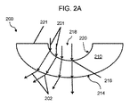
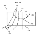
屈折を補正しない外面214を画成する2つのウェル200の断面が、図2Aおよび2Bに示されている。言い換えると、ウェル200の内面220に入る光201は、ウェル200の外面214を出る光202と平行ではない。
A cross section of two
図2Aに示されるように、天底216に近接する基体210の厚さは、ウェル200の上縁221に近接する基体210の厚さより小さい。ウェル200の上縁221に近接する基体210の厚さは、上部開口218と同じ平面にある内面220と外面214の間の厚さと定義してもよい。図2Bに示されるように、ウェル200の外面214は、ウェル200の平らな外面を形成する基体210の矩形底部を画成する。
As shown in FIG. 2A, the thickness of the
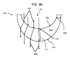
いくつかの実施の形態において、ウェルを画成する基体、例えば、高分子材料の厚さと形状は、ウェルの内面に入りウェルの外面を出る光の屈折を補正するように構成されることがある。屈折を補正する外面114を画成する2つのウェル115の断面が、図3Aおよび3Bに示されている。言い換えると、ウェル115の内面120に入る光201は、ウェル115の外面114を出る光202と平行である。さらに言い換えると、ウェル115の内面120の形状およびウェル115の外面114の形状は、それらの間を通過する光の屈折の効果を最小にするように構成されている。
In some embodiments, the thickness and shape of the substrate that defines the well, eg, the polymeric material, may be configured to correct the refraction of light that enters the inner surface of the well and exits the outer surface of the well. . A cross section of two
図3Aおよび3Bに示されるように、天底116に近接する基体110の厚さ111は、上部開口118に近接する基体110の厚さ109以上であることがある。天底116に近接する基体110の厚さは、ウェル115の最低点での内面120と外面114の間の距離として定義してもよい。上部開口118に近接する基体110の厚さは、上部開口118と同じ平面にある内面120と外面114の間の距離として定義してもよい。
As shown in FIGS. 3A and 3B, the
詳しくは、図3Aに示されるように、基体110の厚さは、上部開口118に近接するところから天底116に近接するところまで一定のままであり、図3Bに示されるように、天底116に近接する基体110の厚さ111は、上部開口118に近接する基体110の厚さ109より大きい。また、図3Aに示されるように、天底116に近接する基体110の厚さ111は、上部開口118に近接する基体110の厚さ109と等しくてもよい。図3Aおよび3Bに示される基体厚さにより、内面120に入る入射光201を、外面114を出る出射光202に対して実質的に平行にすることができる。
Specifically, as shown in FIG. 3A, the thickness of the
他の実施の形態において、基体厚さは、上部開口に近接するところから天底に近接するところまで連続的に増加すると記載されることがある(例えば、図3B)。上部開口から天底のどの位置に近接する基体の厚さも、例えば、5マイクロメートル以上、10マイクロメートル以上、20マイクロメートル以上、40マイクロメートル以上、60マイクロメートル以上など、または100マイクロメートル以下、90マイクロメートル以下、80マイクロメートル以下、65マイクロメートル以下、50マイクロメートル以下などと、先の値のいずれかの間の範囲を含む厚さにより規定されるであろう。いくつかの実施の形態において、その厚さは約1000マイクロメートル以下である。いくつかの実施の形態において、その厚さは10マイクロメートルから1000マイクロメートルの範囲にある。 In other embodiments, the substrate thickness may be described as increasing continuously from close to the top opening to close to the nadir (eg, FIG. 3B). The thickness of the substrate adjacent to any position of the nadir from the top opening is, for example, 5 micrometers or more, 10 micrometers or more, 20 micrometers or more, 40 micrometers or more, 60 micrometers or more, or 100 micrometers or less, It will be defined by a thickness including a range between any of the previous values, such as 90 micrometers or less, 80 micrometers or less, 65 micrometers or less, 50 micrometers or less, and the like. In some embodiments, the thickness is about 1000 micrometers or less. In some embodiments, the thickness is in the range of 10 micrometers to 1000 micrometers.
いくつかの実施の形態において、前記ウェルは、天底と上部開口の中心の間に軸105を画成することがあり、そのウェルは軸105の周りで回転対称であることがある(例えば、図1参照)。例えば、半球形状がウェルを画成することがある。その半球形状は、例えば、50マイクロメートル以上、150マイクロメートル以上、250マイクロメートル以上、400マイクロメートル以上、600マイクロメートル以上など、または1500マイクロメートル以下、1300マイクロメートル以下、1100マイクロメートル以下、900マイクロメートル以下、750マイクロメートル以下などの半径により規定されることがある。 In some embodiments, the well may define an axis 105 between the nadir and the center of the top opening, and the well may be rotationally symmetric about the axis 105 (e.g., (See FIG. 1). For example, a hemispherical shape may define a well. The hemispherical shape is, for example, 50 micrometers or more, 150 micrometers or more, 250 micrometers or more, 400 micrometers or more, 600 micrometers or more, or the like, or 1500 micrometers or less, 1300 micrometers or less, 1100 micrometers or less, 900 It may be defined by a radius such as less than micrometer, 750 micrometers or less.
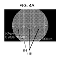
図3Aに概して示されたようなウェルのX線コンピュータ断層撮影画像の3Dデータセットの直交図が、図4A〜4Dに示されている。これらの画像は、図3Aに示されたような凸状外面114を画成するウェル115を示している。図4Aは、図4Cに示された、水平線117に沿った3つの完全なウェル115の断面図を示している。図4Cは、ウェル115のアレイを有する細胞培養装置の一部の上面図である。図4Bは、図4Cに示された垂直線119に沿ったウェル115の断面図である。図4Dは、ウェル115のアレイを有する細胞培養装置の一部の再構成3D画像である。
Orthogonal views of a 3D data set of X-ray computed tomography images of wells as generally shown in FIG. 3A are shown in FIGS. These images show a well 115 that defines a convex
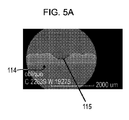
図2Bに概して示されたようなウェルのX線コンピュータ断層撮影画像の3Dデータセットの直交図が、図5A〜5Dに示されている。これらの画像は、図2Bに示されたような平らな外面114を画成するウェル115を示している。図5Aは、図5Cに示された、水平線117に沿ったウェル115の断面図を示している。図5Cは、ウェル115のアレイを有する細胞培養装置の一部の上面図である。図5Bは、図5Cに示された垂直線119に沿ったウェル115の断面図である。図5Dは、ウェル115のアレイを有する細胞培養装置の一部の再構成3D画像である。
Orthogonal views of a 3D data set of X-ray computed tomography images of wells as generally shown in FIG. 2B are shown in FIGS. These images show a well 115 that defines a flat
概して図3Aおよび2Bによる形状を有するウェル115の明視野顕微鏡画像が、それぞれ、図6Aおよび6Bに示されている。図6Aの顕微鏡画像は、図3Aに示されたような形状を有するウェルを通過した光を示している。図6Bの顕微鏡画像は、図2Bに示されたような形状を有するウェルを通過した光を示している。図3Aに示されたようなウェルの形状は、実質的に反射/偏向せず、それゆえ、図2Bに示されたような形状を有するウェルを横切る信号と比べて、全てのウェルに亘り比較的均一な信号を生じた。図6Bの顕微鏡画像に示されるように、図2Bに示されたような形状を有するウェルよりも、図3Aに示されたような形状を有するウェルについて、より多くの光が顕微鏡のカメラにより受光された(図6A参照)。言い換えると、図6Bのウェルの顕微鏡画像は、黒い環により示されるように、図6Aのウェルの顕微鏡画像よりも、より多くの光が散乱したことを示している。 Bright field microscopic images of a well 115 having a shape generally according to FIGS. 3A and 2B are shown in FIGS. 6A and 6B, respectively. The microscopic image of FIG. 6A shows light that has passed through a well having a shape as shown in FIG. 3A. The microscopic image of FIG. 6B shows light that has passed through a well having a shape as shown in FIG. 2B. The shape of the well as shown in FIG. 3A is substantially non-reflective / deflected and is therefore compared across all wells compared to the signal across the well having the shape as shown in FIG. 2B. A uniform signal. As shown in the microscopic image of FIG. 6B, more light is received by the microscope camera for the well having the shape shown in FIG. 3A than for the well having the shape shown in FIG. 2B. (See FIG. 6A). In other words, the microscopic image of the well of FIG. 6B shows that more light was scattered than the microscopic image of the well of FIG. 6A, as indicated by the black ring.
図7に示されるように、細胞培養装置700は貯留部725を備えることがある。この貯留部は、底部705および囲い側壁720を有することがある。底部705は複数のウェル715により画成されることがある。各ウェル715は、ここに記載されたウェルと同様の特徴を有することがある(図1、3A、および3Bを参照のこと)。
As shown in FIG. 7, the
いくつかの実施の形態において、ウェルの外面は、高解像度顕微鏡(例えば、明視野、蛍光、共焦点、または他の顕微鏡撮像方式)で見た場合、回折限界撮像性能に関するレイトレーシングにより最適化される。例えば、図2Aおよび2Bを参照すると、外面214は、レイトレーシングにより最適化される。
In some embodiments, the outer surface of the well is optimized by ray tracing for diffraction-limited imaging performance when viewed with a high resolution microscope (eg, bright field, fluorescence, confocal, or other microscopy imaging schemes). The For example, referring to FIGS. 2A and 2B, the
この手法を説明するために、ポリスチレン製ウェルの内面は、半径が500マイクロメートルであり、中心の厚さが150マイクロメートルである半球であることがある。スフェロイドの直径は300マイクロメートルであることがあり、対物レンズの開口数が0.4である20倍の顕微鏡が使用される。スフェロイドの位置に亘り数多くの画像点、例えば、中心、中心から50マイクロメートル、中心から100マイクロメートル、および中心から150マイクロメートルがある。そのような場合、撮影されたほとんどの画像は最適以下になる。スポット図を、異なる照射野の位置から生成することができ、画像品質を評価するために、画像面で回折限界エアリーサークルと比較することができる。外面が、図2Bに示されるように平らである場合、照射野を横切るスポット直径は、回折限界よりも数倍大きく、不十分な画像品質を示す。ウェルが均一な厚さを有する場合、画像品質は、先の場合よりも著しく良好である。しかしながら、回折限界撮像性能は、中心の50マイクロメートルの半径内でほとんど達成されない。この視野の外では、非点収差により画像品質が劣化してしまう。しかしながら、外面の曲率半径を0.518mmに最適化することにより、画像品質は、全スフェロイド直径に亘り回折限界性能を達成できるが、少量の歪みおよび非点収差がまだ存在する。画像品質をさらに最適化するために、非球面外面が使用される。曲率半径がR=0.682mmであり、コーニック定数がK=−3.09である場合、関心のある全視野に亘り残留収差および歪みが除去される。コーニック表面は以下により与えられる: To illustrate this approach, the inner surface of a polystyrene well may be a hemisphere with a radius of 500 micrometers and a center thickness of 150 micrometers. The diameter of the spheroids can be 300 micrometers and a 20 × microscope with an objective lens numerical aperture of 0.4 is used. There are a number of image points across the location of the spheroids, such as the center, 50 micrometers from the center, 100 micrometers from the center, and 150 micrometers from the center. In such a case, most captured images will be suboptimal. Spot diagrams can be generated from different field locations and compared to diffraction-limited Airy circles in the image plane to assess image quality. If the outer surface is flat as shown in FIG. 2B, the spot diameter across the field is several times larger than the diffraction limit, indicating poor image quality. If the well has a uniform thickness, the image quality is significantly better than before. However, diffraction limited imaging performance is hardly achieved within a central 50 micron radius. Outside this field of view, image quality deteriorates due to astigmatism. However, by optimizing the radius of curvature of the outer surface to 0.518 mm, the image quality can achieve diffraction limited performance over the entire spheroid diameter, but there is still a small amount of distortion and astigmatism. An aspheric outer surface is used to further optimize the image quality. If the radius of curvature is R = 0.682 mm and the conic constant is K = −3.09, residual aberrations and distortion are removed over the entire field of interest. The conic surface is given by:
![]()
![]()
回折限界性能は、スフェロイドの全体積においても維持される。これにより、スフェロイド内のどの位置においても高解像度共焦点撮像が可能になる。実際の倍率は、表面の屈折効果のために、21.5倍である。 Diffraction limited performance is also maintained in the entire spheroid volume. This enables high resolution confocal imaging at any position within the spheroid. The actual magnification is 21.5 times due to surface refraction effects.
いくつかの実施の形態において、入れ子式ウェルが使用され、それにより、第1のウェルまたはウェルの層が、第2のウェルまたはウェルの層の上に存在する。各ウェルのウェル側壁は、ウェルの2つ以上の層を通過する光が元の光に対して実質的に平行なままであるように選択される。 In some embodiments, nested wells are used so that the first well or layer of wells is on top of the second well or layer of wells. The well sidewalls of each well are selected such that light passing through two or more layers of the well remains substantially parallel to the original light.
ここに記載されたようなウェルを有する細胞培養装置を製造するために、どの適切なプロセスを使用しても差し支えない。例えば、基体を成形して、ウェルまたは構造化表面を形成することができたり、基体フイルムにエンボス加工して、ウェルまたは構造化表面を形成することができたりする。いくつかの実施の形態において、変形プロセスを使用して、ここに記載されたようなウェルを製造する。 Any suitable process can be used to produce a cell culture device having wells as described herein. For example, the substrate can be molded to form a well or structured surface, or the substrate film can be embossed to form a well or structured surface. In some embodiments, a deformation process is used to produce wells as described herein.
例えば、図8を参照すると、ウェルを製造するための変形プロセスの概略側面図が示されている。例えば、図8は、薄壁ウェルを製造するための高温エンボス加工およびフイルム変形プロセス800を示している。このプロセスは、薄いフイルム820を使用し、成形型830に入れるために薄いフイルム820に熱および圧力810を印加する。薄いフイルム820は、ウェルの異なる部分に帰属する所定の厚さとなる特定の厚さを有することがある。例えば、高温エンボス加工およびフイルム変形のプロセスを経た後の70マイクロメートルの薄いフイルムは、ウェルの底部とウェルの上部で25マイクロメートルの均一な厚さを有することがある。この結果は、光屈折を十分に補正できる、図3Aに示されたウェルと似ている。その結果、高温エンボス加工およびフイルム変形プロセスをウェルの製造中に能動的に制御して、図3A〜3Bのものと十分に似た、光屈折を補正するウェルが形成されるであろう。このウェル製造プロセスを、平面構造で、またはロール・ツー・ロールプロセスとして実施してもよい。
For example, referring to FIG. 8, a schematic side view of a deformation process for manufacturing a well is shown. For example, FIG. 8 shows a high temperature embossing and
ここに記載されたようなウェルまたは構造化表面を有する細胞培養装置は、どのような適切な材料から形成しても差し支えない。細胞または培地と接触することが意図されている材料が、その細胞および培地に適合していることが好ましい。典型的に、細胞培養構成要素(例えば、壁)は、高分子材料から形成される。適切な高分子材料の例としては、ポリスチレン、ポリメタクリル酸メチル、ポリ塩化ビニル、ポリカーボネート、ポリスルホン、ポリスチレン共重合体、フルオロポリマー、ポリエステル、ポリアミド、ポリスチレンブタジエン共重合体、完全に水素化されたスチレン重合体、ポリカーボネートPDMS共重合体、並びにポリエチレン、ポリプロピレン、ポリメチルペンテン、ポリプロピレン共重合体および環状オレフィン共重合体などのポリオレフィン等が挙げられる。 A cell culture device having wells or structured surfaces as described herein can be formed from any suitable material. It is preferred that the material intended to contact the cell or medium is compatible with the cell and medium. Typically, cell culture components (eg, walls) are formed from polymeric materials. Examples of suitable polymeric materials include polystyrene, polymethyl methacrylate, polyvinyl chloride, polycarbonate, polysulfone, polystyrene copolymers, fluoropolymers, polyesters, polyamides, polystyrene butadiene copolymers, fully hydrogenated styrene. Polymers, polycarbonate PDMS copolymers, and polyolefins such as polyethylene, polypropylene, polymethylpentene, polypropylene copolymers, and cyclic olefin copolymers.
スフェロイドなどの三次元で培養された細胞は、単層などの二次元で培養されたそれらの対応物よりも、インビボ様機能性を示すことができる。二次元の細胞培養システムにおいて、細胞は、それらが培養される基体に接着し得る。しかしながら、スフェロイドなどの細胞が三次元で増殖する場合、それらの細胞は、基体に接着するよりもむしろ、互いに相互作用する。三次元で培養される細胞は、細胞間のやり取りおよび細胞外基質の開発の観点から、インビボ組織により密接に似ている。それゆえ、スフェロイドは、細胞移動、分化、生存、および増殖に関する優れたモデルを提供し、したがって、研究、診断、並びに薬効、薬理学、および毒性試験のためのより良好なシステムを提供する。 Cells cultured in three dimensions, such as spheroids, can exhibit in vivo-like functionality over their counterparts cultured in two dimensions, such as a monolayer. In a two-dimensional cell culture system, cells can adhere to the substrate on which they are cultured. However, when cells such as spheroids grow in three dimensions, they interact with each other rather than adhere to the substrate. Cells cultured in three dimensions more closely resemble in vivo tissues in terms of cell-to-cell interactions and extracellular matrix development. Therefore, spheroids provide an excellent model for cell migration, differentiation, survival, and proliferation, thus providing a better system for research, diagnosis, and drug efficacy, pharmacology, and toxicity testing.
いくつかの実施の形態において、前記装置は、その装置内で培養される細胞がスフェロイドを形成するように構成される。例えば、中で細胞が増殖するウェルは、細胞に対して非接着性であり、ウェル内の細胞を互いに結合させ、球体を形成させることができる。そのスフェロイドは、ウェルの形状により課せられるサイズ限界まで拡大する。いくつかの実施の形態において、それらのウェルは、そのウェルを細胞に対して非接着性にするために超低結合材料で被覆される。 In some embodiments, the device is configured such that cells cultured in the device form spheroids. For example, a well in which cells grow is non-adherent to the cells, and the cells in the well can bind to each other to form a sphere. The spheroids expand to the size limit imposed by the well shape. In some embodiments, the wells are coated with an ultra-low binding material to make the wells non-adherent to the cells.
非接着性材料の例としては、過フッ素化高分子、オレフィン、または同様の高分子、もしくはそれらの混合物が挙げられる。他の例には、アガロース、ポリアクリルアミドなどの非イオン性ハイドロゲル、ポリエチレンオキシドなどのポリエーテル、およびポリビニルアルコールなどのポリオール、または同様の材料もしくはそれらの混合物がある。例えば、非接着性ウェル、ウェル形状(例えば、サイズと形)、および/または重力の組合せにより、ウェル内で培養される細胞のスフェロイドへの自己集合が誘発される。あるスフェロイドは、単層中で増殖した細胞に対して、よりインビボ様応答を示す、分化細胞機能を維持する。間葉間質細胞などの他の細胞種類は、スフェロイドとして培養された場合、それらの多能性を維持する。 Examples of non-adhesive materials include perfluorinated polymers, olefins, or similar polymers, or mixtures thereof. Other examples include agarose, nonionic hydrogels such as polyacrylamide, polyethers such as polyethylene oxide, and polyols such as polyvinyl alcohol, or similar materials or mixtures thereof. For example, a combination of non-adherent wells, well shape (eg, size and shape), and / or gravity induces self-assembly of cells cultured in the well into spheroids. Some spheroids maintain differentiated cell function that exhibits a more in vivo-like response to cells grown in monolayers. Other cell types such as mesenchymal stromal cells maintain their pluripotency when cultured as spheroids.
いくつかの実施の形態において、ここに記載されたシステム、装置、および方法は、1種類以上の細胞を含む。いくつかの実施の形態において、その細胞は凍結保存されている。いくつかの実施の形態において、その細胞は三次元培養される。そのようないくつかの実施の形態において、前記システム、装置、および方法は、1種類以上のスフェロイドを含む。いくつかの実施の形態において、その細胞の1種類以上は積極的に分割している。いくつかの実施の形態において、前記システム、装置、および方法は、培地(例えば、栄養素(例えば、タンパク質、ペプチド、アミノ酸)、エネルギー(例えば、炭水化物)、必須金属およびミネラル(例えば、カルシウム、マグネシウム、鉄、リン酸塩、硫酸塩)、緩衝剤(例えば、リン酸塩、酢酸塩)、pH変化の指示薬(例えば、フェノールレッド、ブロモクレゾールパープル)、選択剤(例えば、化学薬品、抗菌剤)など)を含む。いくつかの実施の形態において、1種類以上の試験化合物(例えば、薬剤)が、そのシステム、装置、および方法に含まれる。 In some embodiments, the systems, devices, and methods described herein include one or more types of cells. In some embodiments, the cells are stored frozen. In some embodiments, the cells are cultured in three dimensions. In some such embodiments, the systems, devices, and methods include one or more spheroids. In some embodiments, one or more of the cells are actively dividing. In some embodiments, the systems, devices, and methods include media (eg, nutrients (eg, proteins, peptides, amino acids), energy (eg, carbohydrates), essential metals and minerals (eg, calcium, magnesium, Iron, phosphate, sulfate), buffers (eg, phosphate, acetate), pH change indicators (eg, phenol red, bromocresol purple), selective agents (eg, chemicals, antibacterial agents), etc. )including. In some embodiments, one or more test compounds (eg, drugs) are included in the systems, devices, and methods.
多種多様な細胞種類が培養されるであろう。いくつかの実施の形態において、スフェロイドは、ただ1つの細胞種類を含有する。いくつかの実施の形態において、スフェロイドは、複数の細胞種類を含有する。いくつかの実施の形態において、複数の細胞種類を増殖させる場合、各スフェロイドは同じ種類のものであり、一方で、他の実施の形態において、2種類以上の異なる種類のスフェロイドを増殖させる。スフェロイド中で増殖する細胞は、天然細胞または変性細胞(例えば、1つ以上の非天然遺伝子変化を含む細胞)であってよい。いくつかの実施の形態において、細胞は体細胞である。いくつかの実施の形態において、細胞は、任意の所望の分化状態(例えば、多能性、多分化能、運命決定、不死化など)にある幹細胞または前駆細胞(例えば、胚幹細胞、誘導多能性幹細胞)である。いくつかの実施の形態において、細胞は、疾患細胞または疾患モデル細胞である。例えば、いくつかの実施の形態において、スフェロイドは、1種類以上の癌細胞または高増殖状態に誘起できる細胞(例えば、形質転換細胞)を含む。細胞は、以下に限られないが、副腎、膀胱、血管、骨、骨髄、脳、軟骨、頚部、角膜、子宮内膜、食道、胃腸、免疫系(例えば、Tリンパ球、Bリンパ球、白血球、マクロファージ、および樹枝状細胞)、肝臓、肺、リンパ管、筋肉(例えば、心筋)、神経、卵巣、膵臓(例えば、島細胞)、下垂体、前立腺、腎臓、唾液、皮膚、腱、精巣、および甲状腺を含む、どのような所望の組織または器官種類からのものであっても、もしくはそれに由来してもよい。いくつかの実施の形態において、細胞は哺乳類細胞(例えば、ヒト、マウス、ラット、ウサギ、イヌ、ネコ、ウシ、ブタ、ニワトリ、ヤギ、ウマなど)である。 A wide variety of cell types will be cultured. In some embodiments, the spheroid contains only one cell type. In some embodiments, the spheroid contains multiple cell types. In some embodiments, when growing multiple cell types, each spheroid is of the same type, while in other embodiments, two or more different types of spheroids are grown. A cell that grows in a spheroid may be a natural cell or a denatured cell (eg, a cell containing one or more non-native genetic alterations). In some embodiments, the cell is a somatic cell. In some embodiments, the cells are stem cells or progenitor cells (eg, embryonic stem cells, induced pluripotency) in any desired differentiation state (eg, pluripotency, pluripotency, fate determination, immortalization, etc.). Sex stem cells). In some embodiments, the cell is a disease cell or a disease model cell. For example, in some embodiments, spheroids include one or more types of cancer cells or cells that can be induced to a hyperproliferative state (eg, transformed cells). Cells include, but are not limited to, adrenal gland, bladder, blood vessels, bone, bone marrow, brain, cartilage, cervix, cornea, endometrium, esophagus, gastrointestinal tract, immune system (eg, T lymphocytes, B lymphocytes, leukocytes) , Macrophages, and dendritic cells), liver, lungs, lymphatic vessels, muscles (eg, myocardium), nerves, ovaries, pancreas (eg, islet cells), pituitary gland, prostate, kidney, saliva, skin, tendon, testis, And from any desired tissue or organ type, including and thyroid. In some embodiments, the cell is a mammalian cell (eg, human, mouse, rat, rabbit, dog, cat, cow, pig, chicken, goat, horse, etc.).
培養された細胞には、多種多様の研究、診断、薬剤スクリーニングおよび試験、治療、および工業的利用に用途が見出されている。 Cultured cells find use in a wide variety of research, diagnostic, drug screening and testing, therapeutic, and industrial applications.
いくつかの実施の形態において、前記細胞は、タンパク質またはウイルスの産生のために使用される。多数のスフェロイドを並行して培養するシステム、装置、および方法は、タンパク質産生にとって特に効果的である。三次元培養により、細胞密度を増加させ、細胞増殖表面積の平方センチメートル当たりのタンパク質収量を高めることができる。ワクチン生産のためのどの所望のタンパク質またはウイルスを、細胞中で増殖させ、要求通りの使用のために単離または精製してもよい。いくつかの実施の形態において、そのタンパク質はその細胞に対して天然タンパク質である。いくつかの実施の形態において、そのタンパク質は非天然である。いくつかの実施の形態において、そのタンパク質は組換え的に発現される。そのタンパク質が非天然プロモーターを使用して過剰発現されることが好ましい。そのタンパク質は融合タンパク質として発現されることがある。いくつかの実施の形態において、精製または検出タグが、その精製および/または検出を容易にするために、関心のあるタンパク質に対する融合パートナーとして発現される。いくつかの実施の形態において、精製後に融合パートナーの分離を可能にするために、融合は、開裂可能なリンカーと共に発現される。 In some embodiments, the cells are used for protein or virus production. Systems, devices, and methods for culturing large numbers of spheroids in parallel are particularly effective for protein production. Three-dimensional culture can increase cell density and increase protein yield per square centimeter of cell growth surface area. Any desired protein or virus for vaccine production may be grown in cells and isolated or purified for use as required. In some embodiments, the protein is a natural protein for the cell. In some embodiments, the protein is non-natural. In some embodiments, the protein is expressed recombinantly. It is preferred that the protein is overexpressed using a non-native promoter. The protein may be expressed as a fusion protein. In some embodiments, the purification or detection tag is expressed as a fusion partner to the protein of interest to facilitate its purification and / or detection. In some embodiments, the fusion is expressed with a cleavable linker to allow separation of the fusion partner after purification.
いくつかの実施の形態において、前記タンパク質は治療用タンパク質である。そのようなタンパク質としては、以下に限られないが、欠損または異常(例えば、インスリン)である、既存の経路(例えば、阻害剤または作動物質)を増強する、新規の機能または活性を提供する、分子または微生物を妨げる、もしくは他の化合物またはタンパク質(例えば、放射性核種、細胞毒性薬、エフェクター・タンパク質など)を送達するタンパク質を置き換えるタンパク質およびペプチドが挙げられる。いくつかの実施の形態において、そのタンパク質は、任意の種類(例えば、ヒト化、二重特異性、多特異的、など)の抗体(例えば、モノクローナル抗体)などのイムノグロブリンである。治療用タンパク質の部類としては、以下に限られないが、抗体に基づいた薬剤、Fc融合タンパク質、抗凝固剤、抗原、血液因子、骨形態形成タンパク質、改変タンパク質の骨組み、酵素、成長因子、ホルモン、インターフェロン、インターロイキン、および血栓溶解剤が挙げられる。治療用タンパク質を使用して、癌、免疫疾患、代謝異常、継承された遺伝性疾患、感染、および他の疾病および症状を予防するまたは治療してもよい。 In some embodiments, the protein is a therapeutic protein. Such proteins provide, but are not limited to, new functions or activities that enhance existing pathways (eg, inhibitors or agonists) that are deficient or abnormal (eg, insulin), Proteins and peptides that replace proteins that interfere with molecules or microorganisms or deliver other compounds or proteins (eg, radionuclides, cytotoxic drugs, effector proteins, etc.). In some embodiments, the protein is an immunoglobulin, such as an antibody (eg, monoclonal antibody) of any type (eg, humanized, bispecific, multispecific, etc.). The class of therapeutic proteins includes, but is not limited to, antibody-based drugs, Fc fusion proteins, anticoagulants, antigens, blood factors, bone morphogenetic proteins, modified protein frameworks, enzymes, growth factors, hormones , Interferons, interleukins, and thrombolytic agents. Therapeutic proteins may be used to prevent or treat cancer, immune diseases, metabolic disorders, inherited genetic diseases, infections, and other diseases and conditions.
いくつかの実施の形態において、前記タンパク質は診断用タンパク質である。診断用タンパク質としては、以下に限られないが、抗体、親和結合パートナー(例えば、受容体結合リガンド)、阻害剤、拮抗薬などが挙げられる。いくつかの実施の形態において、その診断用タンパク質は、検出可能な部分(例えば、蛍光部分、発光部分(例えば、ルシフェラーゼ)、発色部分など)と共に発現されるか、または検出可能な部分である。 In some embodiments, the protein is a diagnostic protein. Diagnostic proteins include, but are not limited to, antibodies, affinity binding partners (eg, receptor binding ligands), inhibitors, antagonists, and the like. In some embodiments, the diagnostic protein is expressed with a detectable moiety (eg, a fluorescent moiety, a luminescent moiety (eg, luciferase), a chromogenic moiety, etc.) or is a detectable moiety.
いくつかの実施の形態において、前記タンパク質は工業用タンパク質である。工業用タンパク質としては、以下に限られないが、食品成分、工業用酵素、農業用タンパク質、分析用酵素などが挙げられる。 In some embodiments, the protein is an industrial protein. Industrial proteins include, but are not limited to, food ingredients, industrial enzymes, agricultural proteins, analytical enzymes, and the like.
いくつかの実施の形態において、前記細胞は、創薬、特徴付け、有効性試験、および毒性試験に使用される。そのような試験としては、以下に限られないが、薬理的効果の評価、発癌性評価、医用造影剤特徴評価、半減期評価、放射線安全性評価、遺伝毒性試験、免疫毒性試験、生殖および発達試験、薬物間相互作用評価、線量評価、吸着評価、処分評価、代謝評価、排出研究などが挙げられる。特異的細胞種類を特定の試験(例えば、肝臓毒性に関する肝細胞、腎臓毒性に関する腎近位尿細管上皮細胞、血管毒性に関する血管内皮細胞、神経毒性に関する神経細胞およびグリア細胞、心臓毒性に関する心筋細胞、横紋筋融解症に関する骨格筋細胞など)に使用してよい。治療された細胞を、以下に限られないが、膜完全性、細胞内代謝物含有量、ミトコンドリア機能、リソソーム機能、アポトーシス、遺伝子変化、遺伝子発現差などを含むいくつの所望のパラメータについて評価してもよい。 In some embodiments, the cells are used for drug discovery, characterization, efficacy testing, and toxicity testing. Such tests include, but are not limited to: pharmacological effects assessment, carcinogenicity assessment, medical contrast agent characterization assessment, half-life assessment, radiation safety assessment, genotoxicity testing, immunotoxicity testing, reproduction and development Examples include testing, drug interaction evaluation, dose evaluation, adsorption evaluation, disposal evaluation, metabolism evaluation, and emission studies. Specific cell types for specific tests (eg, hepatocytes for liver toxicity, renal proximal tubular epithelial cells for kidney toxicity, vascular endothelial cells for vascular toxicity, neurons and glial cells for neurotoxicity, cardiomyocytes for cardiotoxicity, It may be used for skeletal muscle cells related to rhabdomyolysis. Treated cells can be evaluated for a number of desired parameters including, but not limited to, membrane integrity, intracellular metabolite content, mitochondrial function, lysosomal function, apoptosis, gene change, gene expression differential, etc. Also good.
いくつかの実施の形態において、前記細胞培養装置は、より大型のシステムの構成部材である。いくつかの実施の形態において、そのシステムは、そのような細胞培養装置を複数(例えば、2、3、4、5、・・・、10、・・・、20、・・・、50、・・・、100、・・・、1000など)備えている。いくつかの実施の形態において、そのシステムは、最適培養条件(例えば、温度、気圧、湿度など)で培養装置を維持するためのインキュベータを備えている。いくつかの実施の形態において、そのシステムは、細胞を撮像するまたは他の様式で分析するための検出器を備えている。そのような検出器としては、以下に限られないが、蛍光光度計、照度計、カメラ、顕微鏡、プレート・リーダー(例えば、PERKIN ELMER ENVISIONプレート・リーダー;PERKIN ELMER VIEWLUXプレート・リーダー)、細胞分析装置(例えば、GE IN Cell Analyzer 2000および2200;THERMO/CELLOMICS CELLNSIGHT High Content Screening Platform)、および共焦点撮像システム(例えば、PERKIN ELMER OPERAPHENIXハイスループットコンテントスクリーニングシステム; GE INCELL 6000 Cell Imaging System)が挙げられる。いくつかの実施の形態において、そのシステムは、培地または他の成分を、培養される細胞に供給する、補給する、循環させるためのかん流システムまたは他の構成要素を備えている。いくつかの実施の形態において、そのシステムは、培養装置の取扱い、使用、および/または分析を自動化するためのロボット構成要素(例えば、ピペット、アーム、プレート・ムーバーなど)を備えている。
In some embodiments, the cell culture device is a component of a larger system. In some embodiments, the system includes a plurality of such cell culture devices (eg, 2, 3, 4, 5,..., 10,..., 20,..., 50,...). .., 100,..., 1000, etc.) In some embodiments, the system includes an incubator for maintaining the culture device at optimal culture conditions (eg, temperature, pressure, humidity, etc.). In some embodiments, the system comprises a detector for imaging or otherwise analyzing the cells. Such detectors include, but are not limited to, fluorimeters, luminometers, cameras, microscopes, plate readers (eg, PERKIN ELMER ENVISION plate reader; PERKIN ELMER VIEWLUX plate reader), cell analyzers (Eg, GE IN
ここに使用した全ての科学用語および技術用語は、特に明記のない限り、当該技術分野で一般に使用される意味を持つ。ここに与えられた定義は、ここに頻繁に使用される特定の用語の理解を促進するためであり、本開示の範囲を限定する意図はない。 All scientific and technical terms used herein have meanings commonly used in the art unless otherwise specified. The definitions given herein are to facilitate understanding of certain terms frequently used herein and are not intended to limit the scope of the present disclosure.
ここに用いたように、名詞は、特に明記のない限り、複数の対象を指す。それゆえ、例えば、「構造化底面」は、特に明記のない限り、そのような「構造化底面」を2つ以上有する例を含む。 As used herein, a noun refers to a plurality of objects unless otherwise specified. Thus, for example, “a structured bottom surface” includes examples having two or more such “structured bottom surfaces” unless otherwise specified.
本明細書および付随する特許請求の範囲に使用されているように、「または」という用語は、特に明記のない限り、「および/または」を含む意味で一般に使用されている。「および/または」という用語は、列挙された要素の内の1つまたは全て、もしくは列挙された要素のいずれか2つ以上の組合せを意味する。 As used herein and in the appended claims, the term “or” is generally used in its sense including “and / or” unless stated otherwise. The term “and / or” means one or all of the listed elements or a combination of any two or more of the listed elements.
ここに用いたように、「持つ」、「有する」、「含む」、「備える」などは、制約のない包括的な意味で使用され、概して、「含むが、それに限られない」ことを意味する。 As used herein, “having”, “having”, “including”, “comprising”, etc. are used in a comprehensive sense without restrictions, and generally mean “including but not limited to”. To do.
「随意的な」または「必要に応じて」は、その後に記載された事象、状況、または構成要素が、生じ得るまたは生じ得ないことを意味し、その記載が、その事象、状況、または構成要素が生じる場合と、生じない場合を含むことを意味する。 “Optional” or “as needed” means that an event, situation, or component described thereafter may or may not occur, and that description is that event, situation, or configuration It means to include cases where elements occur and cases where elements do not occur.
「好ましい」および「好ましくは」という単語は、特定の状況下で特定の恩恵を提供するであろう、本開示の実施の形態を指す。しかしながら、同じまたは他の状況下で、他の実施の形態が好ましいこともある。さらに、1つ以上の好ましい実施の形態の列挙は、他の実施の形態が有用ではないことを意味せず、本発明の技術の範囲から他の実施の形態を排除する意図はない。 The words “preferred” and “preferably” refer to embodiments of the present disclosure that will provide certain benefits under certain circumstances. However, other embodiments may be preferred under the same or other circumstances. Furthermore, the recitation of one or more preferred embodiments does not imply that other embodiments are not useful and is not intended to exclude other embodiments from the scope of the technology of the present invention.
範囲は、「約」1つの特定の値から、および/または「約」別の特定の値まで、とここに表現され得る。そのような範囲が表現された場合、例は、その1つの特定の値から、および/または他方の特定の値まで、を含む。同様に、値が、先行詞「約」の使用により、近似として表現される場合、特定の値は別の態様を形成することが理解されよう。範囲の各端点は、他の端点に関してと、他の端点とは関係なくの両方で有意であることがさらに理解されよう。 A range may be expressed herein as “about” one particular value and / or “about” another particular value. When such a range is expressed, an example includes from that one particular value and / or to the other particular value. Similarly, when values are expressed as approximations, by use of the antecedent “about,” it will be understood that the particular value forms another aspect. It will be further understood that each endpoint of the range is significant both with respect to the other endpoint and independent of the other endpoint.
端点による数値範囲の列挙は、その範囲内に含まれる全ての数字を含む(例えば、1から5は、1,1.5、2、2.75、3、3.80、4、5など)。値の範囲が、たとえば、特定の値「より大きい」、「未満」などである場合、その値は、その範囲に含まれる。 The recitation of numerical ranges by endpoints includes all numbers subsumed within that range (eg, 1 to 5 is 1,1.5, 2, 2.75, 3, 3.80, 4, 5, etc.) . When a range of values is, for example, a specific value “greater than”, “less than”, the value is included in the range.
「天」、「底」、「左」、「右」、「上部」、「下部」、「上」、「下」並びに他の方向および向きなどのここに称されるどの方向も、図面に関する明瞭さのためにここに記載され、実際の装置またはシステム、若しくはその装置またはシステムの使用を制限するものではない。ここに記載された装置、物品またはシステムの多くは、多数の方向および向きで使用してよい。細胞培養装置に関してここに記載される方向の記述語は、しばしば、その装置が、装置内で細胞を培養する目的のために向けられたときの方向を指す。 Any direction referred to herein, such as “top”, “bottom”, “left”, “right”, “top”, “bottom”, “top”, “bottom” and other directions and orientations, also relates to the drawing It is described herein for clarity and is not intended to limit the actual apparatus or system or use of the apparatus or system. Many of the devices, articles or systems described herein may be used in a number of directions and orientations. The direction descriptive words described herein with respect to a cell culture device often refer to the direction when the device is oriented for the purpose of culturing cells within the device.
特に明記のない限り、ここに述べられたどの方法も、その工程が特定の順序で行われることを必要とすると解釈されることは決して意図されていない。したがって、方法の請求項が、その工程が従うべき順序を実際に列挙していない、または工程が特定の順序に限定されることが請求項または記載に他に具体的に述べられていない場合、どの特定の順序も推測されることは決して意図されていない。いずれか1つの請求項においてどの列挙された1つまたは多数の特徴または態様も、どの他の請求項におけるどの他の列挙された特徴または態様と組み合わせても、並べ替えられても差し支えない。 Unless otherwise stated, any method described herein is in no way intended to be construed as requiring that the steps be performed in a specific order. Thus, if a method claim does not actually list the order in which the steps are to follow, or otherwise specifically stated in the claim or description that the steps are limited to a particular order, Any particular order is never intended to be inferred. Any recited one or many features or aspects in any one claim may be combined or rearranged with any other recited features or aspects in any other claim.
ここでの列挙は、特定の様式で機能するように「構成」または「適用」されている構成要素を指すことも留意のこと。この点に関して、そのような構成要素は、そのような列挙が、目的とする使用の列挙とは異なり、構造的列挙である場合、特定の性質を具体化する、または特定の様式で機能するように「構成」または「適用」されている。より詳しくは、構成要素が「構成」または「適用」される様式のここでの列挙は、その構成要素の既存の物理的条件を示し、それゆえ、その構成要素の構造的特徴の明白な列挙として解釈すべきである。 Note also that this list refers to components that are “configured” or “applied” to function in a particular manner. In this regard, such components are intended to embody certain properties or to function in a particular manner when such enumerations are structural enumerations, unlike enumerations of intended use. Is "configured" or "applied". More specifically, an enumeration herein of the manner in which a component is “configured” or “applied” indicates an existing physical condition of the component, and therefore an explicit enumeration of the structural features of the component. Should be interpreted as
特定の実施の形態の様々な特徴、要素または工程が、移行句「含む」を使用して開示されることがあるが、移行句「からなる」または「から実質的になる」を使用して記載されることのあるものを含む代わりの実施の形態が暗示されることを理解すべきである。それゆえ、例えば、構造化底面、1つ以上の側壁、上部およびポートを含む細胞培養装置に対して暗示される代わりの実施の形態は、細胞培養装置が構造化底面、1つ以上の側壁、上部およびポートからなる実施の形態、並びに細胞培養装置が構造化底面、1つ以上の側壁、上部およびポートから実質的になる実施の形態を含む。 Various features, elements or steps of a particular embodiment may be disclosed using the transitional phrase “comprising”, but using the transitional phrase “consisting of” or “consisting essentially of” It should be understood that alternative embodiments are implied, including what may be described. Thus, for example, an alternative embodiment implied for a cell culture device that includes a structured bottom surface, one or more sidewalls, a top portion and a port is that the cell culture device is structured bottom surface, one or more sidewalls, Embodiments comprising an upper portion and a port, and embodiments in which the cell culture device consists essentially of a structured bottom surface, one or more sidewalls, an upper portion and a port.
本開示の精神および範囲から逸脱せずに、本発明の技術に対して様々な改変および変更が行えることが、当業者に明白であろう。本発明の技術の精神および実体を含む開示の実施の形態の改変、組合せ、下位の組合せ、および変更が、当業者に想起されるであろうから、本発明の技術は、付随の特許請求の範囲およびその同等物の範囲に全てを含むと解釈されるべきである。 It will be apparent to those skilled in the art that various modifications and variations can be made to the technology of the present invention without departing from the spirit or scope of the disclosure. Since modifications, combinations, subcombinations, and alterations of the disclosed embodiments, including the spirit and substance of the technology of the present invention, will occur to those skilled in the art, the technology of the present invention includes the appended claims. It should be construed to include all within the scope and range of equivalents thereof.
以下、本発明の好ましい実施形態を項分け記載する。 Hereinafter, preferable embodiments of the present invention will be described in terms of items.
実施形態1
細胞培養装置において、
ウェルを画成する基体であって、該ウェルが、内面、外面、上部開口および天底を画成し、該基体は、前記内面と前記外面の間の厚さを規定し、前記天底に近接した該基体の厚さは、前記上部開口に近接した該基体の厚さ以上である、基体、
を備えた細胞培養装置。
Embodiment 1
In a cell culture device,
A substrate defining a well, the well defining an inner surface, an outer surface, a top opening, and a nadir, the substrate defining a thickness between the inner surface and the outer surface, and A thickness of the substrate in proximity is greater than or equal to a thickness of the substrate in proximity to the upper opening;
A cell culture apparatus comprising:
実施形態2
前記天底に近接した前記基体の厚さが、前記上部開口に近接した該基体の厚さより大きい、実施形態1に記載の細胞培養装置。
Embodiment 2
The cell culture device according to embodiment 1, wherein the thickness of the substrate close to the nadir is larger than the thickness of the substrate close to the upper opening.
実施形態3
前記基体の厚さが、前記上部開口に近接したところから前記天底に近接したところまで連続的に増加する、実施形態2に記載の細胞培養装置。
Embodiment 3
The cell culture device according to embodiment 2, wherein the thickness of the substrate continuously increases from a position close to the upper opening to a position close to the nadir.
実施形態4
前記天底に近接した前記基体の厚さが、前記上部開口に近接した該基体の厚さと等しい、実施形態1に記載の細胞培養装置。
Embodiment 4
The cell culture device according to embodiment 1, wherein the thickness of the substrate close to the nadir is equal to the thickness of the substrate close to the upper opening.
実施形態5
前記基体の厚さが、前記上部開口に近接したところから前記天底に近接したところまで一定のままである、実施形態4に記載の細胞培養装置。
Embodiment 5
The cell culture device according to embodiment 4, wherein the thickness of the substrate remains constant from a position close to the upper opening to a position close to the nadir.
実施形態6
前記ウェルが前記天底と前記上部開口の中心との間に軸を規定し、該ウェルが該軸の周りに回転対称である、実施形態1から5いずれか1つに記載の細胞培養装置。
Embodiment 6
Embodiment 6. The cell culture device according to any one of embodiments 1 to 5, wherein the well defines an axis between the nadir and the center of the upper opening, and the well is rotationally symmetric about the axis.
実施形態7
前記上部開口が該上部開口に亘る距離を規定し、該上部開口に亘る距離が100マイクロメートルから3000マイクロメートルの範囲にある、実施形態1から6いずれか1つに記載の細胞培養装置。
Embodiment 7
The cell culture device according to any one of embodiments 1 to 6, wherein the upper opening defines a distance over the upper opening, and the distance over the upper opening is in the range of 100 micrometers to 3000 micrometers.
実施形態8
前記上部開口に近接したところから前記天底に近接したところまでのどの位置の前記基体の厚さも、10マイクロメートルから1000マイクロメートルの範囲にある、実施形態1から7いずれか1つに記載の細胞培養装置。
Embodiment 8
The thickness of the substrate at any position from a position close to the top opening to a position close to the nadir is in the range of 10 micrometers to 1000 micrometers, according to any one of embodiments 1 to 7. Cell culture device.
実施形態9
前記内面が半球形状により画成され、該半球形状が、50マイクロメートルから1500マイクロメートルの範囲の半径を規定する、実施形態1から8いずれか1つに記載の細胞培養装置。
Embodiment 9
The cell culture device according to any one of embodiments 1 to 8, wherein the inner surface is defined by a hemispherical shape, the hemispherical shape defining a radius in the range of 50 micrometers to 1500 micrometers.
実施形態10
前記基体がポリスチレンから作られている、実施形態1から9いずれか1つに記載の細胞培養装置。
Embodiment 10
The cell culture device according to any one of embodiments 1 to 9, wherein the substrate is made of polystyrene.
実施形態11
前記外面が光を、前記ウェルが細胞培養培地を収容するときに前記内面により前記光が受光される方向に対して実質的に平行に透過させるように構成されている、実施形態1から10いずれか1つに記載の細胞培養装置。
Embodiment 11
Any of Embodiments 1 to 10, wherein the outer surface is configured to transmit light substantially parallel to the direction in which the light is received by the inner surface when the well contains a cell culture medium. The cell culture device according to any one of the above.
実施形態12
前記内面の形状および前記外面の形状が、前記ウェルが細胞培養培地を収容するときに、それらの間を通過する光の屈折を最小にするように構成されている、実施形態1から11いずれか1つに記載の細胞培養装置。
Embodiment 12
Embodiments 1-11 wherein the shape of the inner surface and the shape of the outer surface are configured to minimize the refraction of light passing between them when the well contains a cell culture medium. The cell culture apparatus according to one.
実施形態13
前記ウェルが細胞に対して非接着性である、実施形態1から12いずれか1つに記載の細胞培養装置。
Embodiment 13
The cell culture device according to any one of Embodiments 1 to 12, wherein the well is non-adhesive to cells.
実施形態14
前記内面が、その中で培養される細胞がスフェロイドを形成するように構成されている、実施形態1から13いずれか1つに記載の細胞培養装置。
Embodiment 14
The cell culture device according to any one of embodiments 1 to 13, wherein the inner surface is configured such that cells cultured therein form spheroids.
実施形態15
細胞培養装置において、
底部および囲い側壁を有する貯留部であって、該底部は複数のウェルにより画成され、該複数のウェルの各ウェルは、内面、外面、上部開口および天底を画成し、該ウェルは、前記内面と前記外面の間の厚さを規定し、前記天底に近接した該ウェルの厚さは、前記上部開口に近接した該ウェルの厚さ以上である、貯留部、
を備えた細胞培養装置。
Embodiment 15
In a cell culture device,
A reservoir having a bottom and a surrounding sidewall, the bottom defined by a plurality of wells, each well of the plurality of wells defining an inner surface, an outer surface, a top opening, and a nadir, the wells comprising: A reservoir that defines a thickness between the inner surface and the outer surface, the thickness of the well proximate to the nadir being equal to or greater than the thickness of the well proximate to the upper opening;
A cell culture apparatus comprising:
実施形態16
細胞培養装置において、
ウェルを画成する基体であって、該ウェルは、内面、外面、上部開口および天底を画成し、該基体は、前記内面と前記外面の間の厚さを規定し、該厚さは、前記ウェルが水性組成物を収容するときに、該内面に入り該外面から出る光の屈折を補正するように構成されている、基体、
を備えた細胞培養装置。
Embodiment 16
In a cell culture device,
A substrate defining a well, the well defining an inner surface, an outer surface, a top opening and a nadir, wherein the substrate defines a thickness between the inner surface and the outer surface, the thickness being A substrate configured to correct refraction of light entering the inner surface and exiting the outer surface when the well contains an aqueous composition;
A cell culture apparatus comprising:
実施形態17
細胞培養装置において、
ウェルを画成する基体であって、該ウェルは、内面、外面、上部開口および天底を画成し、前記外面の形状は、前記内面に入り該外面から出る光の屈折を補正するように構成されている、基体、
を備えた細胞培養装置。
Embodiment 17
In a cell culture device,
A substrate defining a well, wherein the well defines an inner surface, an outer surface, a top opening and a nadir, and the shape of the outer surface corrects refraction of light entering and exiting the inner surface Composed of a substrate,
A cell culture apparatus comprising:
実施形態18
前記外面が、前記内面の半径より大きい曲率半径を有する、実施形態1から17いずれか1つに記載の細胞培養装置。
Embodiment 18
The cell culture device according to any one of embodiments 1 to 17, wherein the outer surface has a radius of curvature larger than the radius of the inner surface.
実施形態19
前記外面が非球面外面を有する、実施形態1から18いずれか1つに記載の細胞培養装置。
Embodiment 19
The cell culture device according to any one of Embodiments 1 to 18, wherein the outer surface has an aspheric outer surface.
実施形態20
スフェロイドを増殖させるための、実施形態1から19いずれか1つに記載の細胞培養装置の使用。
Embodiment 20.
Use of the cell culture device according to any one of embodiments 1 to 19 for growing spheroids.
実施形態21
前記ウェル内で細胞を撮像するための、実施形態1から19いずれか1つに記載の細胞培養装置の使用。
Embodiment 21.
Use of the cell culture device according to any one of embodiments 1 to 19 for imaging cells in the well.
実施形態22
前記細胞がスフェロイド内にある、実施形態21に記載の使用。
Embodiment 22
The use according to embodiment 21, wherein the cells are in spheroids.
100、700 細胞培養装置
110、210 基体
114、214 外面
115、200、715 ウェル
116、216 天底
118、218 上部開口
120、220 内面
121、221 上縁
201 内面に入射する光
202 外面から出射する光
705 底部
720 囲い側壁
725 貯留部
800 高温エンボス加工およびフイルム変形プロセス
810 熱および圧力
820 フイルム
830 成形型
100, 700
Claims (17)
ウェルを画成する基体であって、該ウェルが、半球形状を持つ内面、外面、上部開口および天底を画成する、基体、
を備え、
前記基体は、前記内面と前記外面の間の厚さを規定し、
前記厚さが、前記上部開口に近接したところから前記天底に近接したところまで連続的に増加する、細胞培養装置。 In a cell culture device,
A substrate defining a well, wherein the well defines a hemispherical inner surface, outer surface, top opening and nadir;
With
The substrate defines a thickness between the inner surface and the outer surface;
The cell culture device, wherein the thickness continuously increases from a position close to the upper opening to a position close to the nadir.
底部および囲い側壁を有する貯留部であって、該底部は複数のウェルにより画成され、該複数のウェルの各ウェルは、内面、外面、上部開口および天底を画成し、該ウェルは、前記内面と前記外面の間の厚さを規定し、該ウェルの厚さが、前記上部開口に近接したところから前記天底に近接したところまで連続的に増加する、貯留部、
を備えた細胞培養装置。 In a cell culture device,
A reservoir having a bottom and a surrounding sidewall, the bottom defined by a plurality of wells, each well of the plurality of wells defining an inner surface, an outer surface, a top opening, and a nadir, the wells comprising: Defining a thickness between the inner surface and the outer surface, the thickness of the well continuously increasing from a location close to the top opening to a location close to the nadir,
A cell culture apparatus comprising:
Applications Claiming Priority (3)
| Application Number | Priority Date | Filing Date | Title |
|---|---|---|---|
| US201462072019P | 2014-10-29 | 2014-10-29 | |
| US62/072,019 | 2014-10-29 | ||
| PCT/US2015/058123 WO2016069930A1 (en) | 2014-10-29 | 2015-10-29 | Microwell design and fabrication for generation of cell culture aggregates |
Related Child Applications (1)
| Application Number | Title | Priority Date | Filing Date |
|---|---|---|---|
| JP2021047002A Division JP2021090472A (en) | 2014-10-29 | 2021-03-22 | Microwell design and fabrication for generation of cell culture aggregates |
Publications (1)
| Publication Number | Publication Date |
|---|---|
| JP2017532971A true JP2017532971A (en) | 2017-11-09 |
Family
ID=54477383
Family Applications (3)
| Application Number | Title | Priority Date | Filing Date |
|---|---|---|---|
| JP2017523445A Pending JP2017532971A (en) | 2014-10-29 | 2015-10-29 | Design and manufacture of microwells to generate cell culture assemblies |
| JP2021047002A Pending JP2021090472A (en) | 2014-10-29 | 2021-03-22 | Microwell design and fabrication for generation of cell culture aggregates |
| JP2023028245A Pending JP2023054350A (en) | 2014-10-29 | 2023-02-27 | Microwell designing and manufacturing for generating cell culture aggregate |
Family Applications After (2)
| Application Number | Title | Priority Date | Filing Date |
|---|---|---|---|
| JP2021047002A Pending JP2021090472A (en) | 2014-10-29 | 2021-03-22 | Microwell design and fabrication for generation of cell culture aggregates |
| JP2023028245A Pending JP2023054350A (en) | 2014-10-29 | 2023-02-27 | Microwell designing and manufacturing for generating cell culture aggregate |
Country Status (4)
| Country | Link |
|---|---|
| US (2) | US20170226455A1 (en) |
| EP (1) | EP3212761A1 (en) |
| JP (3) | JP2017532971A (en) |
| WO (1) | WO2016069930A1 (en) |
Cited By (2)
| Publication number | Priority date | Publication date | Assignee | Title |
|---|---|---|---|---|
| WO2020008723A1 (en) | 2018-07-04 | 2020-01-09 | 横河電機株式会社 | Method for producing cell structure, carrier, and method for producing carrier |
| WO2020138581A1 (en) * | 2018-12-28 | 2020-07-02 | 포항공과대학교 산학협력단 | Porous microwell, and membrane comprising same and manufacturing method therefor |
Families Citing this family (10)
| Publication number | Priority date | Publication date | Assignee | Title |
|---|---|---|---|---|
| US9790465B2 (en) | 2013-04-30 | 2017-10-17 | Corning Incorporated | Spheroid cell culture well article and methods thereof |
| CN107109339B (en) | 2014-10-29 | 2021-07-02 | 康宁股份有限公司 | Perfusion bioreactor platform |
| US11857970B2 (en) | 2017-07-14 | 2024-01-02 | Corning Incorporated | Cell culture vessel |
| US11584906B2 (en) | 2017-07-14 | 2023-02-21 | Corning Incorporated | Cell culture vessel for 3D culture and methods of culturing 3D cells |
| EP3652290B1 (en) | 2017-07-14 | 2022-05-04 | Corning Incorporated | 3d cell culture vessels for manual or automatic media exchange |
| PL3652291T3 (en) | 2017-07-14 | 2022-03-28 | Corning Incorporated | Cell culture vessel |
| WO2020013851A1 (en) | 2018-07-13 | 2020-01-16 | Corning Incorporated | Fluidic devices including microplates with interconnected wells |
| JP7353263B2 (en) | 2018-07-13 | 2023-09-29 | コーニング インコーポレイテッド | Cell culture vessel with stabilization device |
| WO2020013847A1 (en) | 2018-07-13 | 2020-01-16 | Corning Incorporated | Microcavity dishes with sidewall including liquid medium delivery surface |
| US20220372421A1 (en) | 2019-10-03 | 2022-11-24 | Corning Incorporated | Kit and method for preparation of digestible spheroid stabilizing hydrogels |
Citations (4)
| Publication number | Priority date | Publication date | Assignee | Title |
|---|---|---|---|---|
| US5171994A (en) * | 1986-08-13 | 1992-12-15 | Northrop Corporation | Infrared staring imaging array |
| US5171995A (en) * | 1990-09-28 | 1992-12-15 | Bruker Analytische Mebtechnik Gmbh | Sample holder for optical spectrometer and method for taking a spectrum |
| WO2013042360A1 (en) * | 2011-09-20 | 2013-03-28 | 株式会社クラレ | Adherent cell culture method |
| WO2014140181A1 (en) * | 2013-03-15 | 2014-09-18 | Unisense Fertilitech A/S | A tray, a system and a method for monitoring and culturing of a cell culture |
Family Cites Families (2)
| Publication number | Priority date | Publication date | Assignee | Title |
|---|---|---|---|---|
| CA2764307C (en) * | 2001-06-29 | 2015-03-03 | Meso Scale Technologies, Llc. | Assay plates, reader systems and methods for luminescence test measurements |
| US20130122580A1 (en) * | 2010-01-08 | 2013-05-16 | Sumitomo Bakelite Co., Ltd. | Culture vessel for forming aggregated cell mass |
-
2015
- 2015-10-29 EP EP15791194.2A patent/EP3212761A1/en active Pending
- 2015-10-29 JP JP2017523445A patent/JP2017532971A/en active Pending
- 2015-10-29 WO PCT/US2015/058123 patent/WO2016069930A1/en active Application Filing
-
2017
- 2017-04-27 US US15/499,370 patent/US20170226455A1/en not_active Abandoned
-
2021
- 2021-03-22 JP JP2021047002A patent/JP2021090472A/en active Pending
- 2021-11-22 US US17/532,196 patent/US20220081661A1/en active Pending
-
2023
- 2023-02-27 JP JP2023028245A patent/JP2023054350A/en active Pending
Patent Citations (4)
| Publication number | Priority date | Publication date | Assignee | Title |
|---|---|---|---|---|
| US5171994A (en) * | 1986-08-13 | 1992-12-15 | Northrop Corporation | Infrared staring imaging array |
| US5171995A (en) * | 1990-09-28 | 1992-12-15 | Bruker Analytische Mebtechnik Gmbh | Sample holder for optical spectrometer and method for taking a spectrum |
| WO2013042360A1 (en) * | 2011-09-20 | 2013-03-28 | 株式会社クラレ | Adherent cell culture method |
| WO2014140181A1 (en) * | 2013-03-15 | 2014-09-18 | Unisense Fertilitech A/S | A tray, a system and a method for monitoring and culturing of a cell culture |
Cited By (4)
| Publication number | Priority date | Publication date | Assignee | Title |
|---|---|---|---|---|
| WO2020008723A1 (en) | 2018-07-04 | 2020-01-09 | 横河電機株式会社 | Method for producing cell structure, carrier, and method for producing carrier |
| WO2020138581A1 (en) * | 2018-12-28 | 2020-07-02 | 포항공과대학교 산학협력단 | Porous microwell, and membrane comprising same and manufacturing method therefor |
| KR20200081853A (en) * | 2018-12-28 | 2020-07-08 | 포항공과대학교 산학협력단 | Porous microwell and membrane with the porous microwell, and method for manufacturing the same |
| KR102224065B1 (en) | 2018-12-28 | 2021-03-05 | 포항공과대학교 산학협력단 | Porous microwell and membrane with the porous microwell, and method for manufacturing the same |
Also Published As
| Publication number | Publication date |
|---|---|
| EP3212761A1 (en) | 2017-09-06 |
| JP2021090472A (en) | 2021-06-17 |
| JP2023054350A (en) | 2023-04-13 |
| WO2016069930A1 (en) | 2016-05-06 |
| US20220081661A1 (en) | 2022-03-17 |
| US20170226455A1 (en) | 2017-08-10 |
Similar Documents
| Publication | Publication Date | Title |
|---|---|---|
| JP2021090472A (en) | Microwell design and fabrication for generation of cell culture aggregates | |
| JP6832042B2 (en) | Insert for spheroid capture | |
| JP7071553B2 (en) | Equipment and methods for the generation and culture of 3D cell aggregates | |
| Baptista et al. | Overlooked? Underestimated? Effects of substrate curvature on cell behavior | |
| Czerniecki et al. | High-throughput screening enhances kidney organoid differentiation from human pluripotent stem cells and enables automated multidimensional phenotyping | |
| Truong et al. | Breast cancer cell invasion into a three dimensional tumor-stroma microenvironment | |
| Horváth et al. | Engineering an in vitro air-blood barrier by 3D bioprinting | |
| KR102460969B1 (en) | Cell culture insert | |
| Pampaloni et al. | The third dimension bridges the gap between cell culture and live tissue | |
| TW200811295A (en) | Systems and methods for efficient collection of single cells and colonies of cells and fast generation of stable transfectants | |
| KR20170073676A (en) | Perfusion bioreactor platform | |
| Montague et al. | Imaging Platelet Processes and Function—Current and Emerging Approaches for Imaging in vitro and in vivo | |
| Zhou et al. | The significance of membrane fluidity of feeder cell-derived substrates for maintenance of iPS cell stemness | |
| WO2009095666A1 (en) | Microtrench and tumour proliferation assay | |
| Rychecký et al. | Spheroid cultivation of HT-29 carcinoma cell line in liquid marbles | |
| Yoon | Tissue Engineering: A Primer with Laboratory Demonstrations | |
| Starnes et al. | Imaging podosome dynamics and matrix degradation | |
| Watanabe et al. | Generation of Neurosphere‐Derived Organoid‐Like‐Aggregates (NEDAS) from Neural Stem Cells | |
| Yamagishi et al. | Live-cell imaging technique to visualize DAMPs release during regulated cell death | |
| Hoh et al. | Restricted exchange microenvironments for cell culture | |
| Yoon et al. | Cell Culture | |
| Cambra et al. | Triple‐Decker Sandwich Cultures of Intestinal Organoids for Long‐Term Live Imaging, Uniform Perturbation, and Statistical Sampling | |
| He et al. | Mammalian cell division in 3D matrices via quantitative confocal reflection microscopy | |
| Shrestha et al. | Selective expansion of target cells using the Enrich TROVO platform | |
| Ishii et al. | Interplay between medial nuclear stalling and lateral cellular flow underlies cochlear duct spiral morphogenesis |
Legal Events
| Date | Code | Title | Description |
|---|---|---|---|
| A621 | Written request for application examination |
Free format text: JAPANESE INTERMEDIATE CODE: A621 Effective date: 20181029 |
|
| A977 | Report on retrieval |
Free format text: JAPANESE INTERMEDIATE CODE: A971007 Effective date: 20190830 |
|
| A131 | Notification of reasons for refusal |
Free format text: JAPANESE INTERMEDIATE CODE: A131 Effective date: 20190904 |
|
| A521 | Request for written amendment filed |
Free format text: JAPANESE INTERMEDIATE CODE: A523 Effective date: 20191127 |
|
| A131 | Notification of reasons for refusal |
Free format text: JAPANESE INTERMEDIATE CODE: A131 Effective date: 20200430 |
|
| A02 | Decision of refusal |
Free format text: JAPANESE INTERMEDIATE CODE: A02 Effective date: 20201202 |