JP2017522153A - Biomarker detection / identification system and device - Google Patents
Biomarker detection / identification system and device Download PDFInfo
- Publication number
- JP2017522153A JP2017522153A JP2017519466A JP2017519466A JP2017522153A JP 2017522153 A JP2017522153 A JP 2017522153A JP 2017519466 A JP2017519466 A JP 2017519466A JP 2017519466 A JP2017519466 A JP 2017519466A JP 2017522153 A JP2017522153 A JP 2017522153A
- Authority
- JP
- Japan
- Prior art keywords
- needle
- coating
- detection device
- biomarker detection
- spinal
- Prior art date
- Legal status (The legal status is an assumption and is not a legal conclusion. Google has not performed a legal analysis and makes no representation as to the accuracy of the status listed.)
- Pending
Links
- 238000001514 detection method Methods 0.000 title claims abstract description 52
- 239000000090 biomarker Substances 0.000 title claims abstract description 38
- 238000000576 coating method Methods 0.000 claims abstract description 34
- 239000011248 coating agent Substances 0.000 claims abstract description 32
- 239000012620 biological material Substances 0.000 claims abstract description 19
- 230000007246 mechanism Effects 0.000 claims description 9
- 239000000126 substance Substances 0.000 claims 1
- 210000001519 tissue Anatomy 0.000 abstract description 42
- 210000001124 body fluid Anatomy 0.000 abstract description 12
- 239000010839 body fluid Substances 0.000 abstract description 11
- 210000001175 cerebrospinal fluid Anatomy 0.000 abstract description 9
- 210000000944 nerve tissue Anatomy 0.000 abstract description 4
- 210000000278 spinal cord Anatomy 0.000 description 20
- 239000012530 fluid Substances 0.000 description 18
- 238000002347 injection Methods 0.000 description 17
- 239000007924 injection Substances 0.000 description 17
- 150000003431 steroids Chemical class 0.000 description 16
- 239000003814 drug Substances 0.000 description 14
- 229940079593 drug Drugs 0.000 description 13
- 238000000034 method Methods 0.000 description 12
- 238000001356 surgical procedure Methods 0.000 description 12
- 210000001367 artery Anatomy 0.000 description 11
- 210000002330 subarachnoid space Anatomy 0.000 description 10
- 238000011282 treatment Methods 0.000 description 10
- 230000000694 effects Effects 0.000 description 9
- 230000002792 vascular Effects 0.000 description 9
- 239000007788 liquid Substances 0.000 description 8
- 208000014674 injury Diseases 0.000 description 7
- 210000003169 central nervous system Anatomy 0.000 description 6
- 210000005036 nerve Anatomy 0.000 description 6
- 208000027418 Wounds and injury Diseases 0.000 description 5
- 210000004369 blood Anatomy 0.000 description 5
- 239000008280 blood Substances 0.000 description 5
- 230000006378 damage Effects 0.000 description 5
- 239000012528 membrane Substances 0.000 description 5
- 208000020431 spinal cord injury Diseases 0.000 description 5
- 230000000202 analgesic effect Effects 0.000 description 4
- 230000008901 benefit Effects 0.000 description 4
- 210000004262 dental pulp cavity Anatomy 0.000 description 4
- 238000007913 intrathecal administration Methods 0.000 description 4
- 230000035515 penetration Effects 0.000 description 4
- 210000000130 stem cell Anatomy 0.000 description 4
- 238000012549 training Methods 0.000 description 4
- 206010033799 Paralysis Diseases 0.000 description 3
- 230000001154 acute effect Effects 0.000 description 3
- 239000003146 anticoagulant agent Substances 0.000 description 3
- 229940127219 anticoagulant drug Drugs 0.000 description 3
- 230000010100 anticoagulation Effects 0.000 description 3
- 229960003957 dexamethasone Drugs 0.000 description 3
- UREBDLICKHMUKA-CXSFZGCWSA-N dexamethasone Chemical compound C1CC2=CC(=O)C=C[C@]2(C)[C@]2(F)[C@@H]1[C@@H]1C[C@@H](C)[C@@](C(=O)CO)(O)[C@@]1(C)C[C@@H]2O UREBDLICKHMUKA-CXSFZGCWSA-N 0.000 description 3
- 208000037265 diseases, disorders, signs and symptoms Diseases 0.000 description 3
- 230000006870 function Effects 0.000 description 3
- 239000010410 layer Substances 0.000 description 3
- 210000003041 ligament Anatomy 0.000 description 3
- 230000008929 regeneration Effects 0.000 description 3
- 238000011069 regeneration method Methods 0.000 description 3
- 231100000241 scar Toxicity 0.000 description 3
- 230000001225 therapeutic effect Effects 0.000 description 3
- 238000002560 therapeutic procedure Methods 0.000 description 3
- 210000005166 vasculature Anatomy 0.000 description 3
- 229940035676 analgesics Drugs 0.000 description 2
- 210000003484 anatomy Anatomy 0.000 description 2
- 239000000730 antalgic agent Substances 0.000 description 2
- 238000013459 approach Methods 0.000 description 2
- 210000000576 arachnoid Anatomy 0.000 description 2
- 210000000988 bone and bone Anatomy 0.000 description 2
- 238000012790 confirmation Methods 0.000 description 2
- 201000010099 disease Diseases 0.000 description 2
- 238000009552 doppler ultrasonography Methods 0.000 description 2
- 210000001951 dura mater Anatomy 0.000 description 2
- LNEPOXFFQSENCJ-UHFFFAOYSA-N haloperidol Chemical compound C1CC(O)(C=2C=CC(Cl)=CC=2)CCN1CCCC(=O)C1=CC=C(F)C=C1 LNEPOXFFQSENCJ-UHFFFAOYSA-N 0.000 description 2
- 208000015181 infectious disease Diseases 0.000 description 2
- 239000011229 interlayer Substances 0.000 description 2
- 238000001990 intravenous administration Methods 0.000 description 2
- 230000000302 ischemic effect Effects 0.000 description 2
- 210000004705 lumbosacral region Anatomy 0.000 description 2
- 238000002595 magnetic resonance imaging Methods 0.000 description 2
- 239000000463 material Substances 0.000 description 2
- 238000002324 minimally invasive surgery Methods 0.000 description 2
- 239000004081 narcotic agent Substances 0.000 description 2
- 230000001537 neural effect Effects 0.000 description 2
- 210000000056 organ Anatomy 0.000 description 2
- 238000007674 radiofrequency ablation Methods 0.000 description 2
- 230000005236 sound signal Effects 0.000 description 2
- 210000001032 spinal nerve Anatomy 0.000 description 2
- 238000009168 stem cell therapy Methods 0.000 description 2
- 238000009580 stem-cell therapy Methods 0.000 description 2
- 238000006467 substitution reaction Methods 0.000 description 2
- 230000009885 systemic effect Effects 0.000 description 2
- 230000008733 trauma Effects 0.000 description 2
- JYEUMXHLPRZUAT-UHFFFAOYSA-N 1,2,3-triazine Chemical compound C1=CN=NN=C1 JYEUMXHLPRZUAT-UHFFFAOYSA-N 0.000 description 1
- 206010014418 Electrolyte imbalance Diseases 0.000 description 1
- 206010018341 Gliosis Diseases 0.000 description 1
- 206010018852 Haematoma Diseases 0.000 description 1
- 206010062016 Immunosuppression Diseases 0.000 description 1
- 206010061218 Inflammation Diseases 0.000 description 1
- 208000034693 Laceration Diseases 0.000 description 1
- 206010024612 Lipoma Diseases 0.000 description 1
- WHXSMMKQMYFTQS-UHFFFAOYSA-N Lithium Chemical compound [Li] WHXSMMKQMYFTQS-UHFFFAOYSA-N 0.000 description 1
- 241001465754 Metazoa Species 0.000 description 1
- 208000003926 Myelitis Diseases 0.000 description 1
- 208000012902 Nervous system disease Diseases 0.000 description 1
- 208000025966 Neurological disease Diseases 0.000 description 1
- 208000002193 Pain Diseases 0.000 description 1
- 206010036376 Postherpetic Neuralgia Diseases 0.000 description 1
- 208000028017 Psychotic disease Diseases 0.000 description 1
- 206010072005 Spinal pain Diseases 0.000 description 1
- 208000006011 Stroke Diseases 0.000 description 1
- 208000007536 Thrombosis Diseases 0.000 description 1
- 230000004913 activation Effects 0.000 description 1
- 230000004931 aggregating effect Effects 0.000 description 1
- 230000036592 analgesia Effects 0.000 description 1
- 229940121363 anti-inflammatory agent Drugs 0.000 description 1
- 239000002260 anti-inflammatory agent Substances 0.000 description 1
- 230000006907 apoptotic process Effects 0.000 description 1
- 230000000712 assembly Effects 0.000 description 1
- 238000000429 assembly Methods 0.000 description 1
- 238000001574 biopsy Methods 0.000 description 1
- 210000004204 blood vessel Anatomy 0.000 description 1
- 201000011510 cancer Diseases 0.000 description 1
- 210000000845 cartilage Anatomy 0.000 description 1
- 210000004027 cell Anatomy 0.000 description 1
- 238000006243 chemical reaction Methods 0.000 description 1
- 239000004020 conductor Substances 0.000 description 1
- 239000002872 contrast media Substances 0.000 description 1
- 230000002498 deadly effect Effects 0.000 description 1
- 230000034994 death Effects 0.000 description 1
- 230000006837 decompression Effects 0.000 description 1
- 230000006735 deficit Effects 0.000 description 1
- 230000003412 degenerative effect Effects 0.000 description 1
- 230000010339 dilation Effects 0.000 description 1
- 208000035475 disorder Diseases 0.000 description 1
- 238000001647 drug administration Methods 0.000 description 1
- 238000002692 epidural anesthesia Methods 0.000 description 1
- 210000003743 erythrocyte Anatomy 0.000 description 1
- 239000000835 fiber Substances 0.000 description 1
- 238000002594 fluoroscopy Methods 0.000 description 1
- 230000014509 gene expression Effects 0.000 description 1
- 229960003878 haloperidol Drugs 0.000 description 1
- 238000003384 imaging method Methods 0.000 description 1
- 230000001506 immunosuppresive effect Effects 0.000 description 1
- 230000001771 impaired effect Effects 0.000 description 1
- 230000006872 improvement Effects 0.000 description 1
- 230000004054 inflammatory process Effects 0.000 description 1
- 238000001802 infusion Methods 0.000 description 1
- 238000003780 insertion Methods 0.000 description 1
- 230000037431 insertion Effects 0.000 description 1
- 230000003993 interaction Effects 0.000 description 1
- 208000028867 ischemia Diseases 0.000 description 1
- 210000004749 ligamentum flavum Anatomy 0.000 description 1
- 230000003859 lipid peroxidation Effects 0.000 description 1
- 229910052744 lithium Inorganic materials 0.000 description 1
- 201000006417 multiple sclerosis Diseases 0.000 description 1
- 230000003533 narcotic effect Effects 0.000 description 1
- 230000036407 pain Effects 0.000 description 1
- 238000002559 palpation Methods 0.000 description 1
- 230000007170 pathology Effects 0.000 description 1
- 230000008289 pathophysiological mechanism Effects 0.000 description 1
- 239000002831 pharmacologic agent Substances 0.000 description 1
- 210000003446 pia mater Anatomy 0.000 description 1
- 230000003389 potentiating effect Effects 0.000 description 1
- 230000002265 prevention Effects 0.000 description 1
- 229940001470 psychoactive drug Drugs 0.000 description 1
- 239000004089 psychotropic agent Substances 0.000 description 1
- 150000003254 radicals Chemical class 0.000 description 1
- 230000001105 regulatory effect Effects 0.000 description 1
- 238000011160 research Methods 0.000 description 1
- 238000012552 review Methods 0.000 description 1
- 230000035807 sensation Effects 0.000 description 1
- 210000004872 soft tissue Anatomy 0.000 description 1
- 210000000273 spinal nerve root Anatomy 0.000 description 1
- 238000011272 standard treatment Methods 0.000 description 1
- 230000000638 stimulation Effects 0.000 description 1
- 210000000701 subdural space Anatomy 0.000 description 1
- 208000024891 symptom Diseases 0.000 description 1
- 210000000115 thoracic cavity Anatomy 0.000 description 1
- 238000002604 ultrasonography Methods 0.000 description 1
- 230000009790 vascular invasion Effects 0.000 description 1
- 230000000007 visual effect Effects 0.000 description 1
- 238000012800 visualization Methods 0.000 description 1
Images


Classifications
-
- A—HUMAN NECESSITIES
- A61—MEDICAL OR VETERINARY SCIENCE; HYGIENE
- A61B—DIAGNOSIS; SURGERY; IDENTIFICATION
- A61B5/00—Measuring for diagnostic purposes; Identification of persons
- A61B5/68—Arrangements of detecting, measuring or recording means, e.g. sensors, in relation to patient
- A61B5/6846—Arrangements of detecting, measuring or recording means, e.g. sensors, in relation to patient specially adapted to be brought in contact with an internal body part, i.e. invasive
- A61B5/6847—Arrangements of detecting, measuring or recording means, e.g. sensors, in relation to patient specially adapted to be brought in contact with an internal body part, i.e. invasive mounted on an invasive device
- A61B5/6848—Needles
-
- A—HUMAN NECESSITIES
- A61—MEDICAL OR VETERINARY SCIENCE; HYGIENE
- A61B—DIAGNOSIS; SURGERY; IDENTIFICATION
- A61B5/00—Measuring for diagnostic purposes; Identification of persons
- A61B5/68—Arrangements of detecting, measuring or recording means, e.g. sensors, in relation to patient
- A61B5/6846—Arrangements of detecting, measuring or recording means, e.g. sensors, in relation to patient specially adapted to be brought in contact with an internal body part, i.e. invasive
- A61B5/6847—Arrangements of detecting, measuring or recording means, e.g. sensors, in relation to patient specially adapted to be brought in contact with an internal body part, i.e. invasive mounted on an invasive device
- A61B5/6852—Catheters
-
- A—HUMAN NECESSITIES
- A61—MEDICAL OR VETERINARY SCIENCE; HYGIENE
- A61B—DIAGNOSIS; SURGERY; IDENTIFICATION
- A61B5/00—Measuring for diagnostic purposes; Identification of persons
- A61B5/68—Arrangements of detecting, measuring or recording means, e.g. sensors, in relation to patient
- A61B5/6846—Arrangements of detecting, measuring or recording means, e.g. sensors, in relation to patient specially adapted to be brought in contact with an internal body part, i.e. invasive
- A61B5/6847—Arrangements of detecting, measuring or recording means, e.g. sensors, in relation to patient specially adapted to be brought in contact with an internal body part, i.e. invasive mounted on an invasive device
- A61B5/686—Permanently implanted devices, e.g. pacemakers, other stimulators, biochips
-
- A—HUMAN NECESSITIES
- A61—MEDICAL OR VETERINARY SCIENCE; HYGIENE
- A61B—DIAGNOSIS; SURGERY; IDENTIFICATION
- A61B5/00—Measuring for diagnostic purposes; Identification of persons
- A61B5/74—Details of notification to user or communication with user or patient ; user input means
- A61B5/7405—Details of notification to user or communication with user or patient ; user input means using sound
-
- A—HUMAN NECESSITIES
- A61—MEDICAL OR VETERINARY SCIENCE; HYGIENE
- A61B—DIAGNOSIS; SURGERY; IDENTIFICATION
- A61B2505/00—Evaluating, monitoring or diagnosing in the context of a particular type of medical care
- A61B2505/05—Surgical care
-
- A—HUMAN NECESSITIES
- A61—MEDICAL OR VETERINARY SCIENCE; HYGIENE
- A61B—DIAGNOSIS; SURGERY; IDENTIFICATION
- A61B5/00—Measuring for diagnostic purposes; Identification of persons
- A61B5/02—Detecting, measuring or recording pulse, heart rate, blood pressure or blood flow; Combined pulse/heart-rate/blood pressure determination; Evaluating a cardiovascular condition not otherwise provided for, e.g. using combinations of techniques provided for in this group with electrocardiography or electroauscultation; Heart catheters for measuring blood pressure
- A61B5/02042—Determining blood loss or bleeding, e.g. during a surgical procedure
-
- A—HUMAN NECESSITIES
- A61—MEDICAL OR VETERINARY SCIENCE; HYGIENE
- A61B—DIAGNOSIS; SURGERY; IDENTIFICATION
- A61B5/00—Measuring for diagnostic purposes; Identification of persons
- A61B5/68—Arrangements of detecting, measuring or recording means, e.g. sensors, in relation to patient
- A61B5/6846—Arrangements of detecting, measuring or recording means, e.g. sensors, in relation to patient specially adapted to be brought in contact with an internal body part, i.e. invasive
- A61B5/6847—Arrangements of detecting, measuring or recording means, e.g. sensors, in relation to patient specially adapted to be brought in contact with an internal body part, i.e. invasive mounted on an invasive device
- A61B5/6865—Access ports
Landscapes
- Life Sciences & Earth Sciences (AREA)
- Health & Medical Sciences (AREA)
- Medical Informatics (AREA)
- Biophysics (AREA)
- Pathology (AREA)
- Engineering & Computer Science (AREA)
- Biomedical Technology (AREA)
- Heart & Thoracic Surgery (AREA)
- Physics & Mathematics (AREA)
- Molecular Biology (AREA)
- Surgery (AREA)
- Animal Behavior & Ethology (AREA)
- General Health & Medical Sciences (AREA)
- Public Health (AREA)
- Veterinary Medicine (AREA)
- Infusion, Injection, And Reservoir Apparatuses (AREA)
- Investigating Or Analysing Biological Materials (AREA)
- Measurement Of The Respiration, Hearing Ability, Form, And Blood Characteristics Of Living Organisms (AREA)
- Materials For Medical Uses (AREA)
Abstract
本願で開示したバイオマーカー検出装置は、生体物質の検出を外科器具の使用者に通知するために使用される。より具体的には、本願のバイオマーカー検出装置は、外科器具上に塗布されたコーティングを有し、このコーティングによって、前記外科器具が、脳脊髄液等の重要な体液、または神経組織等の重要な体組織に到達したことを検知し、前記外科器具の使用者に通知し、前記使用者がこの通知に従って対応し、医療上の障害を回避することができるようにする。The biomarker detection device disclosed in the present application is used to notify a user of a surgical instrument of detection of a biological material. More specifically, the biomarker detection device of the present application has a coating applied on a surgical instrument, by which the surgical instrument is important for important body fluids such as cerebrospinal fluid or nerve tissue. Detecting that the body tissue has been reached and notifying the user of the surgical instrument so that the user can respond according to the notification and avoid medical obstacles.
Description
<関連出願の相互参照>
本出願は、2014年6月19日に出願された、「BIOMARKER DETECTION AND IDENTIFICATION SYSTEM AND APPARATUS(バイオマーカー検出・特定システムおよび装置)」という名称の、米国仮出願62/014,513号の優先権を主張する。
<Cross-reference of related applications>
This application claims priority from US Provisional Application No. 62 / 014,513, filed June 19, 2014, entitled “BIOMARKER DETECTION AND IDENTIFICATION SYSTEM AND APPARATUS” To do.
<技術分野>
本願発明は、生体物質を検出および特定するために使用するバイオマーカー検出コーティングに関する。より具体的には、前記バイオマーカー検出コーティングは、針に被覆され、この被覆された針を患者に刺したときに該針の使用者が体液を検出することができるようにする。
<Technical field>
The present invention relates to biomarker detection coatings used to detect and identify biological materials. More specifically, the biomarker detection coating is applied to a needle, and enables the user of the needle to detect body fluid when the covered needle is pierced into a patient.
手術の成果およびコスト構造を改善する努力により、特に脊髄手術の場合に、低侵襲処置の使用する場合が増加している。これらの処置では、蛍光透視法、CT、神経刺激器、及び、最近ではドップラー超音波検査等の画像誘導法を使用することが増えてきた。外科手術よりもリスクが低いことが多いが、低侵襲脊椎処置、疼痛管理処置、神経ブロック、超音波誘導介入、生検、および経皮的配置または開放術中配置は、効果の出ない結果に終わるリスク、および、医療傷害のリスクをはらんでいる。この医療傷害には、感染症、脳卒中、麻痺、および様々な生体構造、例えば、臓器、軟組織、血管構造、および、致命的な脊髄のような神経組織への貫通に起因する死亡、等があるが、これらに限定されない。医療傷害は執刀医の経験に関係なく起こり得る。この理由は、外科用器具は、脊椎管の所望の場所に到達するまでに、身体組織および体液のいくつかの層を通り抜けなければならいからである。 Efforts to improve surgical outcomes and cost structures are increasing the use of minimally invasive procedures, especially in spinal surgery. These procedures have increasingly used image guidance methods such as fluoroscopy, CT, neurostimulators, and more recently Doppler ultrasonography. Although often less risky than surgery, minimally invasive spinal procedures, pain management procedures, nerve blocks, ultrasound-guided interventions, biopsies, and percutaneous or open intraoperative placement results in ineffective results Risk and medical injury risk. This medical injury includes infections, strokes, paralysis, and death due to penetration of various anatomy such as organs, soft tissues, vascular structures, and deadly spinal nerve tissue However, it is not limited to these. Medical injuries can occur regardless of the surgeon's experience. This is because the surgical instrument must pass through several layers of body tissue and fluid before reaching the desired location in the spinal canal.
例えば、多くの薬物が投与される脊髄領域のくも膜下腔内(またはくも膜下)の空間は、神経根および脳脊髄液(以降「CSF」、CerebroSpinal Fluid)を収容し、中枢神経系を囲む三つの膜のうちの二つの膜の間にある。中枢神経系の最も外側の膜は硬膜であり、第2の膜は、くも膜であり、第3の最も内側の膜は軟膜である。クモ膜下腔は、くも膜と軟膜との間にある。この領域に到達するためには、外科用器具は、まず皮膚層、脂肪層、棘間靭帯、黄色靭帯、硬膜外腔、硬膜、硬膜下腔、および、くも膜下腔内を通過しなければならない。さらに、薬剤投与に使用される針の場合、針の開口部全体を、くも膜下腔内に入れる必要がある。 For example, the intrathecal (or subarachnoid) space of the spinal cord region where many drugs are administered contains nerve roots and cerebrospinal fluid (hereinafter “CSF”, CerebroSpinal Fluid) and surrounds the central nervous system. Between two of the two membranes. The outermost membrane of the central nervous system is the dura mater, the second membrane is the arachnoid membrane, and the third innermost membrane is the buffy coat. The subarachnoid space is between the arachnoid and pia mater. To reach this area, the surgical instrument must first pass through the skin layer, fat layer, interspinous ligament, yellow ligament, epidural space, dura mater, subdural space, and subarachnoid space. There must be. Furthermore, in the case of needles used for drug administration, the entire needle opening must be placed in the subarachnoid space.
外科用器具のくも膜下腔への挿入に伴う複雑さ故に発生する、脊髄および神経組織への貫通が、低侵襲性の脊椎処置および脊椎手術の合併障害としてよく知られている。さらに、いくつかの処置では、より大きな外科用器具の使用が必要となる。例えば、脊髄刺激は、最小侵襲脊椎医療の一形態であるが、小さなワイヤ・リードを脊髄硬膜外腔に挿入し、刺激器のリードを縫合するために14ゲージの針を硬膜外腔内に挿入する必要がある。このゲージの針は、技術的に制御が難しく、障害率が高いというリスクがある。合併障害には、硬膜裂傷、脊髄液漏れ、後で血腫が起こる硬膜外静脈破裂、および、結果的に麻痺が残る脊髄または神経への直接貫通が含まれる。これらおよびその他の高リスクな状況は、例えば、脊椎介入および高周波焼灼療法等で、執刀医が重要な解剖学的構造における前記針または外科用器具の先端の位置を検出できない場合に発生する。 The penetration into the spinal cord and nerve tissue, which occurs due to the complexity associated with the insertion of surgical instruments into the subarachnoid space, is well known as a combined disorder of minimally invasive spinal procedures and spinal surgery. In addition, some procedures require the use of larger surgical instruments. For example, spinal cord stimulation is a form of minimally invasive spinal medicine, but a 14-gauge needle is inserted into the epidural space to insert a small wire lead into the spinal epidural space and suture the stimulator lead. Need to be inserted into. This gauge needle is technically difficult to control and has the risk of high failure rates. Comorbidities include dural lacerations, spinal fluid leakage, epidural venous rupture, which later causes hematoma, and direct penetration into the spinal cord or nerves that result in paralysis. These and other high-risk situations occur when, for example, spinal interventions and radiofrequency ablation therapy, the surgeon is unable to detect the position of the needle or surgical instrument tip in critical anatomy.
現在、上記のような構造の検出は医療技術者に依存する。医療技術者は触覚や、造影剤、解剖学的目印の触診および画像誘導法に基づく視覚化を利用する。患者の安全は、触覚による感知能力および画像解釈における執刀医の受けた訓練および経験に左右される。追加の訓練と経験が執刀医の熟練に資する可能性はあるが、医療傷害は、解剖学的な多様性があるので、執刀医の経験および技能とは関係なく起こる可能性があり、自然にまたは瘢痕組織の形態での反復処置から生じる可能性がある。高周波焼灼療法等のいくつかの施術でのフェローシップ・トレーニングは、厳密性が十分ではなく、技能の確保を保障できるものではない。訓練においてさえ、処置の結果はかなり変動する。硬膜外注射および脊髄手術の場合、黄色靭帯の厚さの変動、並びに、硬膜外腔、硬膜拡張、硬膜外脂肪腫、硬膜中隔、および、瘢痕組織の幅の多様性は、経験の豊富な医療技術者にとっては、従来の確認方法に対する見直しを必要とする。さらに、神経再生時に繰り返し行われる高周波処置は、再生後の神経の分布によって解剖学的多様性が増えるため、1年または数年後には、有効性が低く、かつ実施がより困難になる。 Currently, detection of such structures depends on the medical technician. Medical technicians use tactile sensation, contrast agents, palpation of anatomical landmarks and visualization based on image guidance. Patient safety depends on the ability to sense by touch and the training and experience of the surgeon in image interpretation. Although additional training and experience can contribute to the surgeon's skill, medical injuries can occur independently of the surgeon's experience and skill because of the anatomical diversity and naturally Or it may result from repeated treatment in the form of scar tissue. Fellowship training in some treatments such as radiofrequency ablation is not rigorous enough to guarantee the skills. Even in training, the outcome of treatment varies considerably. For epidural injection and spinal surgery, variations in the thickness of the ligamentum flavum and the diversity of the width of the epidural space, dural dilation, epidural lipoma, dural septum, and scar tissue For experienced medical technicians, it is necessary to review the conventional confirmation method. Furthermore, high-frequency treatments that are repeatedly performed during nerve regeneration increase in anatomical diversity due to the distribution of nerves after regeneration, and thus become less effective and more difficult to implement after one or several years.
重要な組織および体液に関して、客観的で、信頼性が高く、一貫性のある、リアルタイムのフィードバックを提供する装置は存在しない。さらに、装置による客観的なフィードバックという考えさえ、何百万という脊髄手術が、世界中で毎年、標準的な治療として行われていても、医師には受け入れられていない。 There are no devices that provide objective, reliable, consistent, real-time feedback on critical tissues and fluids. Moreover, even the idea of objective feedback from the device is not accepted by physicians, even if millions of spinal surgeries are performed as standard treatments every year worldwide.
本願が開示するものは、体液または体組織等の少なくとも一つの生体物質を検出および特定することができるバイオマーカー検出装置である。いくつかの実施形態では、このバイオマーカー検出装置は、二つ以上の生体物質を検出し、どの生体物質が検出されているかを正確に示すことができる。前記検出装置は、人手または機械によって操作することができ、人間または動物に対して使用することができ、連続的または間欠的に検出を行うことができる。例えば、バイオマーカー検出装置用コーティングで被覆された針およびスタイレットは、実際の施術時に、血液、脳脊髄液等の体液(これに限定されない)、および神経組織を検出することができる。 What is disclosed by the present application is a biomarker detection apparatus capable of detecting and specifying at least one biological material such as body fluid or body tissue. In some embodiments, the biomarker detection device can detect more than one biological material and accurately indicate which biological material is being detected. The detection device can be operated manually or by machine, can be used on humans or animals, and can detect continuously or intermittently. For example, a needle and a stylet coated with a coating for a biomarker detection device can detect blood, body fluids such as cerebrospinal fluid (but not limited to this), and nerve tissue during actual surgery.
本明細書は、体液および体組織などの生体物質を検出するために使用されるバイオマーカー検出装置に関する。バイオマーカー検出装置の様々な実施形態を、図面を参照して詳細に説明する。いくつかの図を通して、同一の参照番号は、同一の部品およびアセンブリを示す。様々な実施形態に関する説明は、本明細書に開示したバイオマーカー検出装置の請求範囲を限定しない。さらに、本明細書で説明する例は、どれも、バイオマーカー検出装置に関して限定することを意図したものではなく、単に、バイオマーカー検出装置の多くの可能な実施形態のいくつかを説明するだけである。理解されるであろうが、様々な省略および均等物の置換が、状況が好都合だと示唆または指示する場合には考慮されているが、これらの省略および置換は、本願発明の思想または請求範囲から逸脱しない範囲で、複数の応用や実施形態を含むように意図されたものである。また、理解されるであろうが、本明細書で使用される表現および用語は、説明のためのものであり、請求範囲を限定すると見なされるべきではない。 The present specification relates to a biomarker detection device used for detecting biological materials such as body fluids and body tissues. Various embodiments of a biomarker detection device will be described in detail with reference to the drawings. Throughout the several views, the same reference numerals indicate the same parts and assemblies. The description of the various embodiments does not limit the scope of the biomarker detection device disclosed herein. Furthermore, none of the examples described herein are intended to be limiting with respect to a biomarker detection device, but merely describe some of the many possible embodiments of a biomarker detection device. is there. As will be understood, various abbreviations and equivalent substitutions are contemplated in the context of suggesting or indicating that the situation is convenient; however, these omissions and substitutions are contemplated by the spirit or claims of the present invention. It is intended to include a plurality of applications and embodiments without departing from the scope of the invention. Also, as will be appreciated, the expressions and terms used herein are for the purpose of description and should not be construed as limiting the claims.
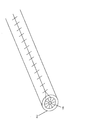
一実施形態では、バイオマーカー検出装置は、手術用または治療用器具又は装置に被覆されるコーティングである。手術用または治療用器具には、針、メス、BOVIE(登録商標)装置、ペースメーカ、電極、血管内カテーテル、管腔内カテーテル、ポート、シース、および埋め込み型ポンプ等があるが、これに限定されない。図3は、バイオマーカー検出装置のコーティングで被覆された被覆表示器304内のスタイレット302を示し、表示器306は医療技術者から視ることができる。図4Aおよび図4Bは、被覆針およびスタイレットを示し、図4Aは、表示器404を覆う、取り外し可能なダイヤフラムカバー402、および露出した切断面406を備えた針を示し、図4Bは、ダイヤフラムカバーを取り外し、表示器404が露出した針を示す。
In one embodiment, the biomarker detection device is a coating that is applied to a surgical or therapeutic instrument or device. Surgical or therapeutic instruments include, but are not limited to, needles, scalpels, BOVIE® devices, pacemakers, electrodes, intravascular catheters, intraluminal catheters, ports, sheaths, and implantable pumps. . FIG. 3 shows a
一実施形態では、医療器具は、使用中に継続的または断続的に露出される、該医療器具の全体、一部、または先端のみにバイオマーカー検出装置用コーティングを施す。前記バイオマーカー検出装置は、例えば、視覚信号または聴覚信号(これに限定されない)のような識別信号を送出することによって生体物質の検出を示すことができる。 In one embodiment, the medical device is coated with a biomarker detection device coating on all, a portion, or only the tip of the medical device that is continuously or intermittently exposed during use. The biomarker detection device can indicate the detection of a biological material by sending an identification signal such as, but not limited to, a visual signal or an auditory signal.
一実施形態では、針のコーティングは化学的感度の高い表面を有する。前記表面は、様々な体液および体組織に対して高感度にすることができる。前記表面は、リトマス紙がいろいろなpHレベルに対して多様に反応するのと同じように、これらの液体および組織に反応することができる。 In one embodiment, the needle coating has a chemically sensitive surface. The surface can be sensitive to various body fluids and tissues. The surface can react to these fluids and tissues in the same way that litmus paper reacts to various pH levels.
例えば、前記バイオマーカー検出装置用コーティングをした針は、超音波と合わせて使用することができ、前記針または別個のデバイスの色は前記液体または組織によって変化することができる。例えば、前記針または別個の装置は、MRIで見ることができ、前記針が脊椎に近づくにつれて暗くなるようにできる。これが起こる理由は、前記コーティングとそれが接触する前記液体および組織との間の相互作用がいろいろな種類または強度の反応を引き起こすことができるからである。前記コーティングは、前記脊椎に近い液体および組織と反応して、より暗い色を出力し、脊椎から離れた液体および組織と反応して、明るい色を出力することができる。または、その逆にすることができる。したがって、硬膜外腔と接触する針または別個の装置は、MRIスキャン時、くも膜下腔と接触する針よりも明るく見える。前記信号は、前記装置が貫通した側から、内腔、チャネル、導管、電気伝導体、またはコーティングを通って前記装置の未露出の側に送ることができる。 For example, the needle coated with the biomarker detector can be used in conjunction with ultrasound, and the color of the needle or separate device can vary with the liquid or tissue. For example, the needle or a separate device can be viewed with MRI and darken as the needle approaches the spine. This occurs because the interaction between the coating and the fluid and tissue it contacts can cause various types or strengths of reaction. The coating can react with fluids and tissues close to the spine to output a darker color and react with fluids and tissues away from the spine to output a light color. Or vice versa. Thus, a needle or separate device that contacts the epidural space appears brighter than a needle that contacts the subarachnoid space during an MRI scan. The signal can be sent from the side through which the device passes through the lumen, channel, conduit, electrical conductor, or coating to the unexposed side of the device.
別の実施形態では、バイオマーカー検出装置は、前記針と共に送られる別個の装置とすることができる。いくつかの実施形態では、前記バイオマーカー検出装置は、前記針の外面に取り付けることができる。別の実施形態では、前記バイオマーカー検出装置は、カテーテルの内部に運ばれ、使用者の作業によって液体および組織に露出される。さらに別の実施形態では、前記針に取り付けないで、前記針によってガイドされる別個の構成要素とすることができる。 In another embodiment, the biomarker detection device can be a separate device delivered with the needle. In some embodiments, the biomarker detection device can be attached to the outer surface of the needle. In another embodiment, the biomarker detection device is carried inside a catheter and exposed to fluid and tissue by the user's work. In yet another embodiment, it can be a separate component guided by the needle without being attached to the needle.
一実施形態では、一つの針または別個の装置への前記コーティングは、一つの種類の体液または組織のみを識別する識別信号を伝達する。従って、使用者が識別したい脊椎の領域に応じて、異なるコーティングを使用することができる。例えば、使用者が、くも膜下腔へのアクセスを望む場合、該使用者は、くも膜下腔へ到達したときにのみ信号を発し、外側の硬膜外腔を通って進むときには信号を発しないコーティングを必要とする。 In one embodiment, the coating on a needle or separate device transmits an identification signal that identifies only one type of bodily fluid or tissue. Thus, different coatings can be used depending on the area of the spine that the user wishes to identify. For example, if a user desires access to the subarachnoid space, the user will only emit a signal when reaching the subarachnoid space and not a signal when traveling through the outer epidural space Need.
別の実施形態では、一つの針または別個の装置に対する同一のコーティングが、該針または別個の装置が相互作用している様々な種類の体液または体組織に関する識別信号を伝達することができる。例えば、前記針または別個の装置が、ビープ音のようなオーディオ信号機能を備えていれば、前記針または別個の装置が様々な体液および体組織と相互作用するときに一つのオーディオ信号を出すことができる。変化する組織や液体を区別するために、前記針または別個の装置は、露出されている体液や体組織が脊椎に接近している場合、ビープ音を鳴らす間隔を次第に短くして行くことができる。したがって、硬膜外腔に到達したときのビープ音間の時間間隔は、くも膜下腔に達するときのビープ音間の時間間隔よりも長くなる。別の例では、被覆針または被覆された別個のデバイスは、前記コーティングが特定の種類の組織または液体に反応するときに活性化される発光機構を持つことができる。 In another embodiment, the same coating on one needle or separate device can convey identification signals for various types of body fluids or tissues with which the needle or separate device interacts. For example, if the needle or the separate device has an audio signal function such as a beep sound, the needle or the separate device emits one audio signal when interacting with various body fluids and tissues. Can do. To distinguish between changing tissue and fluid, the needle or separate device can gradually shorten the beeping interval when exposed body fluid or tissue is approaching the spine. . Therefore, the time interval between beeps when reaching the epidural space is longer than the time interval between beeps when reaching the subarachnoid space. In another example, a coated needle or a separate coated device can have a light emitting mechanism that is activated when the coating reacts to a particular type of tissue or fluid.
別の実施形態では、前記針または別個の装置への前記コーティングは、一つのコーティングで、前記組織または液体が光と反応する形態に基づいて、多くの種類の組織および液体を検出するようにできる。例えば、前記針または別個のデバイスは、一つのコーティングまたはコーティングされた先端を持ち、前記コーティングされた先端が反応するまたは読み取る発光機構を持つことができる。前記発光機構は、使用者の起動操作で起動させることができる。また、前記発光機構は、閃光装置によって発光する前記針自体にある光源、あるいは、体内の調整された場所に挿入される、個別の補助器具とすることができる。前記先端は、前記光が周囲の液体または組織から反射されるときに前記光に反応、または、前記光を解読することができる。前記組織または液体から前記コーティングされた先端部へ反射されるときの光の波長に応じて、前記検出装置は、前記先端がどの種類の組織または液体と接触しているかまたは近接しているかを判定することができる。前記光が検出されると、前記先端は、使用者読み取り用にコンピューティング・ディスプレイ・システムに読み取り値を電子的に送ることができる。前記コンピューティング・ディスプレイ・システムは、別個の装置であってもよく、または前記針自体の中に組み込むこともできる。 In another embodiment, the coating on the needle or separate device can be a single coating that detects many types of tissue and fluid based on the manner in which the tissue or fluid reacts with light. . For example, the needle or separate device can have a single coating or coated tip and the light emitting mechanism with which the coated tip reacts or reads. The light emitting mechanism can be activated by a user activation operation. Further, the light emitting mechanism can be a light source in the needle itself that emits light by a flash device, or an individual auxiliary instrument that is inserted into a regulated place in the body. The tip can react to or decipher the light as it is reflected from the surrounding liquid or tissue. Depending on the wavelength of light as it is reflected from the tissue or liquid to the coated tip, the detection device determines what type of tissue or liquid the tip is in contact with or in proximity to. can do. When the light is detected, the tip can electronically send readings to a computing display system for user reading. The computing display system may be a separate device or may be incorporated within the needle itself.
同様に、別の実施形態では、針または別個の装置は、周囲の構造体で反射する音波を出すことができる。周囲の組織または液体の種類に基づいて、前記音波のプロファイルが変化し、前記検出装置は、前記音波のプロファイルに基づいて、前記周囲の組織または液体の性質を識別することができる。前記針または別個の装置が様々な組織および液体の中を進むにつれて、前記音波プロファイルが変化する。コンピューティングシステムは、前記音波のプロファイルを特定の組織または液体と対応づけられた既存のプロファイルと照合することができ、前記組織または液体が何であるかを使用者に示すことができる。 Similarly, in another embodiment, a needle or a separate device can emit sound waves that are reflected by surrounding structures. Based on the type of surrounding tissue or liquid, the profile of the acoustic wave changes, and the detection device can identify the nature of the surrounding tissue or liquid based on the profile of the acoustic wave. As the needle or separate device advances through various tissues and fluids, the sonic profile changes. The computing system can match the acoustic wave profile with an existing profile associated with a particular tissue or fluid and can indicate to the user what the tissue or fluid is.
所望の組織または液体の検出は、医療技術者に対する割り込みを最小限にするか、または割り込み無しで行うことができ、上述の器具および他の装置を用いて日常的に行われる医療処置の安全性プロファイルを大きく改善する。前記検出装置は、脊髄に対する低侵襲の施術に対して、ならびに他の複数の医学分野においても使用することができる。 The detection of the desired tissue or fluid can be performed with minimal or no interruption to the medical technician, and the safety of medical procedures routinely performed using the instruments and other devices described above Greatly improve your profile. The detection device can be used for minimally invasive procedures on the spinal cord as well as in other medical fields.
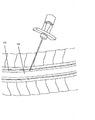
前記検出装置は、一つまたは複数の体液および体組織の種類を確認して治療上の便益を提供し、医療障害の低減およびより正確な位置の認識に役立つことができる。例えば、執刀医が、患者の背骨に対して層間注射等の処置を行っているとき、図1に示すように、針等の器具はCSF102を横断してから、脊髄104に到達する。針およびスタイレット器具、または閉塞器具を備えたカテーテルが、バイオマーカー検出用コーティングで被覆され、前記バイオマーカー検出用コーティングがこの検出結果の通知を行うことができる場合、CSF102が、前記針およびスタイレット器具、または閉塞器具を備えたカテーテルのいずれかの器具を介して検出されると、前記通知によって、執刀医は、脊髄に近づいていることを知ることができ、医療傷害を減らすことができる。また、図7および図8は、前記バイオマーカー検出装置用コーティングで被覆することができる、脊椎内または脊髄の近傍に注射するために使用される典型的な針を示す。
The detection device can identify one or more body fluids and body tissue types to provide therapeutic benefits, and can help reduce medical impairment and more accurate location recognition. For example, when the surgeon is performing a procedure such as an interlayer injection on the patient's spine, an instrument such as a needle crosses the
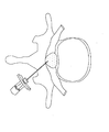
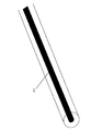
前記バイオマーカー検出装置は、事故防止警報システムとして機能し、表示システムを用いて特定の生体物質の存在を表示することによって、前記機器の実時間位置を外科医、医師、または機械に通知することができる。前記表示システムは、図2に示すように、前記機器が経椎間腔注入に使用される場合、前記機器が、CSF、血液、神経または他の組織に接触したとき、執刀医に警告することができる。この警告は、患者および施術者に対する安全余裕を等しく増やす。前記生体物質が骨、血液、神経、軟骨、靱帯、悪性腫瘍、または臓器のどれであるかにかかわらず、前記装置のフィードバック機能によって、位置決め精度および安全性の向上に資することができる。図5は、本願で開示した装置の一実施形態を示し、スタイレット502が表示器として働く。図6は、本願で開示した装置の一実施形態を示し、表示器602が取外し可能な隔膜上にある、また別の形態として、前記隔膜は、使用者が確認を希望するまで表示器602を保護する。
The biomarker detection device functions as an accident prevention alarm system and can notify a surgeon, doctor, or machine of the real-time position of the device by displaying the presence of a specific biological material using a display system. it can. The display system alerts the surgeon when the device contacts CSF, blood, nerves or other tissues when the device is used for transvertebral space injection, as shown in FIG. Can do. This warning equally increases the safety margin for the patient and practitioner. Regardless of whether the biological material is bone, blood, nerve, cartilage, ligament, malignant tumor, or organ, the feedback function of the device can contribute to the improvement of positioning accuracy and safety. FIG. 5 shows one embodiment of the device disclosed herein, with the
一実施形態では、前記検出システムは、人間によって監視されるロボットシステム、または、その他の自動化システムである。別の実施形態では、前記検出システムは人手操作され、装置の動作中機械が関与しない。いくつかの実施形態では、前記信号は、音声であろうと画像であろうと、相対的または絶対的な信号とすることができる。例えば、前記信号は、前記コーティングが囲まれている液体または組織の種類に関して取得した測定値の絶対値を出力することができる。あるいは、前記信号は、前記コーティングが、ある種類の液体または組織から別の種類の液体または組織に移動するときに、前の値とは異なる、前の値より大きい、または小さい、と言うような、値を示すことができる。 In one embodiment, the detection system is a human-monitored robotic system or other automated system. In another embodiment, the detection system is manually operated and does not involve a machine during operation of the device. In some embodiments, the signal can be a relative or absolute signal, whether audio or image. For example, the signal can output the absolute value of the measured value obtained for the type of fluid or tissue in which the coating is surrounded. Alternatively, the signal may say that when the coating moves from one type of liquid or tissue to another type of liquid or tissue, it is different from, greater than, or less than the previous value. , Value can be shown.
幹細胞療法および投薬療法のための意識的な脊髄血管アクセスは、これまで全く追求されてこなかった。さらに、何十年もの間、抗凝固療法中の注射は絶対禁忌であった。抗凝固療法の継続が合併症のリスクが低く、血栓症からの保護になるという考えを裏付ける新しい証拠が存在する。この証拠によって、上記療法のための意識的な脊髄血管アクセスへの扉を開けることができる。現在、ステロイドの腰椎硬膜外への経椎間腔注射実施中における抗凝固剤の使用は、各患者固有のリスクに応じて、ケースバイケースで実施される。幸いにも、ステロイドが脊髄血管系に不注意に注入された場合でも、抗凝固療法を継続していることにより、脊髄動脈閉塞のリスクが低下する。デキサメタゾンは、直径0.5マイクロメートルの非凝集性非粒子状ステロイドであるが、赤血球よりも数桁のレベルで小さく、脊髄動脈閉塞の可能性も低下させる。本願開示の発明は、意識的な脊椎血管注射のさらなる進歩の助けとなることができる。 Conscious spinal vascular access for stem cell therapy and medication has never been pursued. In addition, for decades, injections during anticoagulation were absolutely contraindicated. There is new evidence to support the idea that continuation of anticoagulation therapy has a low risk of complications and protection from thrombosis. This evidence can open the door to conscious spinal vascular access for the therapy. Currently, the use of anticoagulants during transvertebral injections of steroid epidurally into the lumbar spine is performed on a case-by-case basis, depending on each patient's specific risks. Fortunately, even if steroids are inadvertently infused into the spinal vasculature, continuing anticoagulation reduces the risk of spinal artery occlusion. Dexamethasone is a non-aggregating non-particulate steroid with a diameter of 0.5 micrometers, but is several orders of magnitude smaller than red blood cells and reduces the likelihood of spinal artery occlusion. The disclosed invention can help further advance conscious spinal vascular injection.
脊髄動脈閉塞の懸念および致命的な副作用に陥る恐れのある麻痺を考慮すると、ステロイドのくも膜下腔内動脈への直接注入は禁忌であり、意識的には行われていない。しかし、ステロイドのくも膜下腔内への投与は行われており、ヘルペス後神経痛のような深刻な痛みを伴う症状に使用される。さらに、くも膜下腔内への麻薬投与も許容されている。麻薬には、ステロイドと同様に、材料ごとに異なる親油性がある。脊髄痛に対する鎮痛薬としての麻薬の相対的効力は、投与する場所が脊髄に接近するほど指数関数的に増加する。経口投与、硬膜外投与、および、くも膜下腔内投与は、鎮痛効力がそれぞれ1、10、および100の桁で異なる。したがって、この傾向を脊髄静脈管構造に外挿すると、経口投与よりも1000倍高い効力を示すことになる。さらに、技術的には手術を終了させる理由がありながらも介入型脊椎手術において行われる血液摂取は、顕著な鎮痛効果を有すると記録された安全性プロファイルを持つデキサメタゾンを用いて行われる。 Given the concerns of spinal artery occlusion and the paralysis that can lead to fatal side effects, direct injection of steroids into the intrathecal artery is contraindicated and not consciously performed. However, administration of steroids into the subarachnoid space has been performed and is used for severe painful symptoms such as postherpetic neuralgia. In addition, administration of narcotic drugs into the subarachnoid space is allowed. Narcotics, like steroids, have different lipophilic properties depending on the material. The relative efficacy of narcotics as an analgesic for spinal pain increases exponentially as the location of administration approaches the spinal cord. Oral administration, epidural administration, and intrathecal administration differ in analgesic efficacy on the order of 1, 10, and 100, respectively. Thus, extrapolating this trend to the spinal venous vasculature will be 1000 times more potent than oral administration. In addition, blood intake in interventional spinal surgery, although technically the reason for ending the surgery, is performed using dexamethasone with a safety profile recorded as having a significant analgesic effect.
患者に抗凝固剤処置をしている間の脊髄循環系への薬理学的薬剤の直接投与は、新たな証拠に基づく鎮痛法の新しい時代を拓く。この証拠としては、(a)硬膜外ステロイド注射のために抗凝固剤処置を続けることが許容される、(b)デキサメタゾンは動脈循環を閉塞させないことが示されている、(c)親油性薬物の相対的効力は、前記薬物の脊髄との接触が密接になるにつれて、指数関数的に増大する。直接的な脊髄動脈注射によってもたらされる、1000倍高い効力のお陰で、微量の薬物の投薬で済むようにできる。 Direct administration of pharmacological agents to the spinal circulatory system during anticoagulant treatment in patients opens a new era of new evidence-based analgesia. Evidence for this is (a) allowed to continue anticoagulant treatment for epidural steroid injections, (b) dexamethasone has been shown not to occlude arterial circulation, (c) lipophilic The relative potency of the drug increases exponentially as the drug comes into close contact with the spinal cord. Thanks to the 1000-fold higher potency provided by direct spinal arterial injection, a small amount of drug can be dispensed.
微量の薬物の投薬には、利点がいくつかある。何十年もの間、再生の障害になる神経膠瘢痕を産生する、炎症反応、虚血、脂質過酸化、アポトーシス、体液および電解質異常、並びにフリー・ラジカル発生等の、病態生理学的機構の二次的連鎖を軽減するために、経口ステロイドおよび静脈内ステロイドが急性脊髄損傷患者に投与されている。しかし、前記経口ステロイドおよび静脈内ステロイドの効力は、硬膜外投与、くも膜下腔内投与、または根管動脈投与と比較して、非常に小さく、また、小児集団の免疫抑制に起因する感染リスク等のより重大な副作用を伴う。脊髄動脈への直接注射は、使用する投薬量を軽減することができ、より大きな効力があり、かつ、副作用は小さい。 There are several advantages to administering a trace amount of drug. Secondary of pathophysiological mechanisms, such as inflammatory reactions, ischemia, lipid peroxidation, apoptosis, fluid and electrolyte abnormalities, and free radical generation that produce glial scars that impede regeneration for decades Oral steroids and intravenous steroids have been administered to patients with acute spinal cord injury to reduce the physical linkage. However, the efficacy of oral steroids and intravenous steroids is very small compared to epidural, intrathecal, or root canal administration, and the risk of infection due to immunosuppression in the pediatric population With more serious side effects such as Direct injection into the spinal artery can reduce the dosage used, has greater efficacy, and has fewer side effects.
脊髄への介入、注射、および手術は、多くのリスクが伴うが、本願で開示したバイオマーカー検出装置を用いた神経および骨組織の識別によって、上記の処置法に対する有効性を増すことができる。腰椎椎間板の線維環および髄核組織を区別することにより、腰椎椎間板造影法、経皮的腰椎椎間板減圧、および、椎間板内幹細胞治療の精度を向上させることができる。 Although spinal cord interventions, injections, and surgery involve many risks, identification of nerve and bone tissue using the biomarker detection devices disclosed herein can increase the effectiveness of the above treatment methods. By distinguishing between the lumbar intervertebral fiber ring and nucleus pulposus tissue, the accuracy of lumbar disc imaging, percutaneous lumbar disc decompression, and intradiscal stem cell therapy can be improved.
被覆針およびスタイレットと接触する種々の組織および体液の種類の検出は、血液およびCSF等の重要な構造に前記針が接触したことを施術者および/または装置に知らせることによって、致命的な合併症の発生可能性を低くし、実時間のフィードバックを得ること、及び、さらに前記針を前に進める前に休止することを可能にする。同様の安全性に対する効果は、頸部および胸部層間硬膜外ステロイド注射、硬膜外麻酔およびカテーテル取付け、および脊椎穿刺にも適用できる。 Detection of various tissue and body fluid types that come into contact with coated needles and stylets is a fatal complication by notifying the practitioner and / or device that the needle has contacted critical structures such as blood and CSF. It is possible to reduce the likelihood of illness, get real-time feedback, and further pause before moving the needle forward. Similar safety effects can be applied to cervical and thoracic interlayer epidural steroid injections, epidural anesthesia and catheter attachment, and spinal taps.
多くの医療状況が、本願の新しい技術から利益を得ることができる。例えば、脊髄の炎症または外傷を伴う疾患に、この新しい技術が適用できることの意味は大きい。くも膜下腔内または脊髄根幹動脈への投与ではないとしても、少なくとも硬膜外腔へのステロイドの投与は、脊髄損傷による二次的病態を軽減させる大きな可能性がある。 Many medical situations can benefit from the new technology of the present application. For example, it is significant that this new technology can be applied to diseases involving spinal cord inflammation or trauma. Administration of steroids, at least in the epidural space, even if not intrathecally or into the spinal root arteries, has great potential to reduce secondary pathologies due to spinal cord injury.
さらに、多発性硬化症等の中枢神経系の変性神経疾患に罹患している患者も、急激に悪化する場合、同様に研究の対象となる。この理由は、脊髄ステロイド投与の有効性の程度によっては、急激な悪化を止め、緩和状態にある病気が悪化するペースを下げる働きがあるからである。抗炎症剤の脊髄内投与は、副作用プロファイルの改善、より広い範囲での適合性、そして、再述するが、急性の進行を止める働きがある。 Furthermore, patients suffering from degenerative neurological diseases of the central nervous system such as multiple sclerosis are also the subject of research if they deteriorate rapidly. This is because, depending on the effectiveness of spinal steroid administration, it can stop abrupt worsening and lower the rate at which the illness is aggravated. Intraspinal administration of anti-inflammatory agents has an improved side effect profile, broader compatibility, and, again, serves to stop acute progression.
さらに、精神病に罹患していて、服薬規則遵守が困難な患者は、脊髄投与の効力の改善のおかげで関連する薬物の硬膜外注射によって利益を享受することができる。硬膜外注射は、また、全身性の副作用を最小限に抑える。特に、重度の急性症例では、経口投与する種々の向精神薬の場合、錐体外路系特性のような望ましくない副作用を起こし、服薬遵守能力が低下する可能性がある。脊髄投与は、比較的微量の投薬で病気の治療に役立つので、リチウム、トリアジン、およびハロペリドール等の薬剤による全身性の副作用を最小限に抑えることができる。 In addition, patients with psychosis who have difficulty complying with medication regulations can benefit from epidural injections of related drugs thanks to the improved efficacy of spinal administration. Epidural injection also minimizes systemic side effects. In particular, in severe acute cases, various psychotropic drugs administered orally can cause undesirable side effects such as extrapyramidal characteristics and reduce compliance. Spinal cord administration can help treat disease with relatively small doses, thus minimizing systemic side effects from drugs such as lithium, triazine, and haloperidol.
最後に、脊椎管への幹細胞の注入は現在実行することができるが、外科手術が必要であり、この外科手術は、瘢痕組織を誘起し、かつ、移植用組織を注入するために何百万個もの細胞が必要である。脊髄投与は、侵襲性が低く、更新領域を脊髄の虚血領域に限定することができるため、より効果的で、危険性を低くできる可能性がある。完全な脊髄損傷では、脊髄損傷部位より下の部位への根管動脈からの幹細胞の注入は、より近位レベルでの外傷がすでに発生しているため、リスクがほとんどない。アダムキーヴィッツ動脈は、直径1.2mmの狭い標的であるが、脊髄へ血管から直接アクセスする余裕はある。第1仙椎(SI)の椎間孔が両側に存在し、偶生的な血管侵入率が腰椎の他の領域のほぼ2倍の21.3%である。一実施形態では、針及びスタイレット、又は、閉塞具を備えたマイクロ・カテーテルは、根管動脈を識別し、血管侵入率を高めることができる。開示された技術、ならびにドップラー超音波検査を使用して、執刀医は、ステロイド、幹細胞、または微量の濃縮された鎮痛剤または薬物を注入するために、標的となる、いろいろな動脈に安定的に到達することができる。最小直径が230ミクロンあれば、前記前部脊髄動脈は、直接血管注射を十分収容できる直径があり、これによって、上述のように、更新領域の限定を脊髄の虚血領域のみへ改善することができる。 Finally, injection of stem cells into the spinal canal can now be performed, but surgery is required, which induces scar tissue and millions to inject transplant tissue. Individual cells are required. Spinal cord administration is less invasive and can be more effective and less risky because the renewal area can be limited to the ischemic area of the spinal cord. With complete spinal cord injury, infusion of stem cells from the root canal artery into sites below the site of spinal cord injury has little risk because trauma at a more proximal level has already occurred. The Adam Kivitz artery is a narrow target with a diameter of 1.2 mm, but it affords direct access to the spinal cord from the blood vessel. The intervertebral foramen of the first sacral vertebra (SI) is present on both sides, and the incidental vascular invasion rate is 21.3%, almost twice that of other areas of the lumbar spine. In one embodiment, a microcatheter with a needle and stylet or obturator can identify the root canal artery and increase the vascular penetration rate. Using the disclosed techniques, as well as Doppler ultrasonography, surgeons can stably target various arteries to target steroids, stem cells, or trace amounts of concentrated analgesics or drugs. Can be reached. With a minimum diameter of 230 microns, the anterior spinal artery is large enough to accommodate direct vascular injection, which can improve the limited renewal area to only the ischemic area of the spinal cord, as described above. it can.
神経組織および動脈循環系を含む生体物質を検出することができる針およびスタイレット、並びに、閉塞器を有するカテーテルは、意識的な根管動脈アクセスを容易にし、介入術士がステロイド、幹細胞、薬物、ナノ・プローブまたは他の鎮痛剤を直接的に脊髄循環内に投与することができるようにする。これは、介入術師、または機械に依存せず、脊髄循環系の障害のある部位レベルに物質を直接注入することができる。神経構造および血管構造を検出することができる装置の働きで、脊髄血管アクセスが容易になる。もっと旧式の術式の場合、単純な、血管構造の検出により、薬物の意図しない血管投与が防止でき、致命的な副作用の発生可能性を低減する。 A needle and stylet capable of detecting biological material including neural tissue and arterial circulatory system, and a catheter with an occluder facilitates conscious root canal access, allowing an interventionist to use steroids, stem cells, drugs, Allow nanoprobes or other analgesics to be administered directly into the spinal cord circulation. This is independent of the interventionist or machine and can inject material directly into the impaired site level of the spinal circulatory system. The device capable of detecting neural and vascular structures facilitates spinal vascular access. For older modalities, simple detection of vasculature can prevent unintentional vascular administration of drugs and reduce the likelihood of fatal side effects.
開示された技術は、脊髄の処置に伴う致命的な合併症を軽減し、前記脊髄への処置に伴う脊髄損傷の重篤度を軽減し、多発性硬化症などの変性CNS(Central Nervous System, 中枢神経系)病態に対する新しい治療を提供し、鎮痛効果を高めることができる。さらに、本警告システムによって、機械化された外科手術システムが脊椎への処置を正確に行うことができるようになる。 The disclosed technology reduces fatal complications associated with the treatment of the spinal cord, reduces the severity of spinal cord injury associated with the treatment of the spinal cord, degenerate CNS (Central Nervous System, It can provide new treatments for the central nervous system) and enhance analgesic effects. In addition, the alert system allows a mechanized surgical system to accurately perform a spinal procedure.
Claims (9)
少なくとも一つの生体物質を検出する前記医療器具上のコーティングと、
表示システムと、を有し、
前記表示システムは、前記コーティングが前記少なくとも一つの生体物質を検出した後に、前記少なくとも一つの生体物質の存在を示す
ことを特徴とする、バイオマーカー検出装置。 Medical instruments,
A coating on the medical device for detecting at least one biological material;
A display system,
The biomarker detection apparatus, wherein the display system indicates the presence of the at least one biological material after the coating detects the at least one biological material.
前記コーティングは、前記スタイレット上に位置し、
前記表示システムは、前記スタイレットの第1の端部に位置し、前記医療器具が患者に挿入されたときに前記バイオマーカー検出装置の使用者が目視することができ、前記コーティングが前記少なくとも一つの生体物質と接触すると起動される発光機構である
ことを特徴とする、請求項1に記載のバイオマーカー検出装置。 The medical device is a needle and a stylet;
The coating is located on the stylet;
The display system is located at a first end of the stylet and is visible to a user of the biomarker detection device when the medical device is inserted into a patient, and the coating is the at least one 2. The biomarker detection device according to claim 1, wherein the biomarker detection device is a light emission mechanism that is activated upon contact with two biological substances.
ことを特徴とする、請求項2に記載のバイオマーカー検出装置。 3. The biomarker detection device according to claim 2, wherein the coating is limited to a part of the second end of the stylet.
前記コーティングは、前記針の第1の端部に位置し、
前記表示システムは、前記針の第2の端部に位置し、前記コーティングが前記少なくとも一つの生体物質と接触すると起動される発光機構である
ことを特徴とする、請求項1に記載のバイオマーカー検出装置。 The medical device is a needle and a stylet;
The coating is located at a first end of the needle;
The biomarker of claim 1, wherein the display system is a light emitting mechanism that is located at a second end of the needle and is activated when the coating contacts the at least one biological material. Detection device.
前記コーティングは、前記スタイレット上に位置し、
前記表示システムは、前記コーティングが前記少なくとも一つの生体物質と接触すると起動される発光機構、を有する装置の第2の部分に位置する
ことを特徴とする、請求項1に記載のバイオマーカー検出装置。 The medical device is a needle and a stylet;
The coating is located on the stylet;
The biomarker detection device according to claim 1, wherein the display system is located in a second part of the device having a light emitting mechanism activated when the coating contacts the at least one biological material. .
前記コーティングは、前記カテーテルの第1の端部に位置し、
前記表示システムは、前記カテーテルの第2の端部に位置し、かつ、前記コーティングが前記少なくとも一つの生体物質と接触すると起動される発光機構である
ことを特徴とする、請求項1に記載のバイオマーカー検出装置。 The medical device is a catheter with an obturator;
The coating is located at a first end of the catheter;
2. The light emitting mechanism of claim 1, wherein the display system is a light emitting mechanism that is located at a second end of the catheter and that is activated when the coating contacts the at least one biological material. Biomarker detection device.
ことを特徴とする、請求項1に記載のバイオマーカー検出装置。 2. The biomarker detection device according to claim 1, wherein the display system visually indicates the presence of the at least one biological material.
ことを特徴とする、請求項1に記載のバイオマーカー検出装置。 2. The biomarker detection apparatus according to claim 1, wherein the display system audibly indicates the presence of the at least one biological material.
ことを特徴とする、請求項1に記載のバイオマーカー検出装置。 2. The biomarker detection device according to claim 1, wherein the display system indicates a type of biological material detected by the detection device.
Applications Claiming Priority (3)
| Application Number | Priority Date | Filing Date | Title |
|---|---|---|---|
| US201462014513P | 2014-06-19 | 2014-06-19 | |
| US62/014,513 | 2014-06-19 | ||
| PCT/US2015/036830 WO2015196172A2 (en) | 2014-06-19 | 2015-06-19 | Biomarker detection and identification system and apparatus |
Publications (2)
| Publication Number | Publication Date |
|---|---|
| JP2017522153A true JP2017522153A (en) | 2017-08-10 |
| JP2017522153A5 JP2017522153A5 (en) | 2019-05-09 |
Family
ID=54936257
Family Applications (1)
| Application Number | Title | Priority Date | Filing Date |
|---|---|---|---|
| JP2017519466A Pending JP2017522153A (en) | 2014-06-19 | 2015-06-19 | Biomarker detection / identification system and device |
Country Status (7)
| Country | Link |
|---|---|
| US (1) | US10470712B2 (en) |
| EP (1) | EP3158052A4 (en) |
| JP (1) | JP2017522153A (en) |
| CN (1) | CN206887114U (en) |
| AU (1) | AU2015276826B2 (en) |
| CA (1) | CA2952962C (en) |
| WO (1) | WO2015196172A2 (en) |
Families Citing this family (5)
| Publication number | Priority date | Publication date | Assignee | Title |
|---|---|---|---|---|
| DE102017114077A1 (en) * | 2017-06-26 | 2018-12-27 | Otto-Von-Guericke-Universität Magdeburg | Minimally invasive examination device |
| AU2018295533B2 (en) * | 2017-07-07 | 2021-07-22 | Neuroderm Ltd | Device for subcutaneous delivery of fluid medicament |
| US20230123806A1 (en) | 2017-07-07 | 2023-04-20 | Neuroderm, Ltd. | Device for subcutaneous delivery of fluid medicament |
| WO2021133533A1 (en) * | 2019-12-27 | 2021-07-01 | Sipple Medical, Llc | Real time fluorescent detection systems for medical devices |
| WO2022245969A1 (en) * | 2021-05-19 | 2022-11-24 | Acies Medical Llc | Iron ion detecting aspirating dental cartridge system |
Citations (2)
| Publication number | Priority date | Publication date | Assignee | Title |
|---|---|---|---|---|
| US5938595A (en) * | 1996-05-24 | 1999-08-17 | The Regents Of The University Of California | Fiber optic D dimer biosensor |
| US20040010204A1 (en) * | 2002-03-28 | 2004-01-15 | Pearl Technology Holdings, Llc | Electronic/fiberoptic tissue differentiation instrumentation |
Family Cites Families (49)
| Publication number | Priority date | Publication date | Assignee | Title |
|---|---|---|---|---|
| US4435037A (en) | 1981-11-06 | 1984-03-06 | E. I. Du Pont De Nemours And Company | Fiber optic connector with deformable mounting post |
| US4564011A (en) | 1982-03-22 | 1986-01-14 | Leon Goldman | Laser optic device and method |
| US4588256A (en) | 1982-09-07 | 1986-05-13 | Minnesota Mining And Manufacturing Company | Optical fiber connector |
| US4681398A (en) | 1983-01-03 | 1987-07-21 | Switchcraft, Inc. | Fiber optic connector and method of assembly |
| US4754328A (en) | 1984-01-03 | 1988-06-28 | Medical Dynamics, Inc. | Laser endoscope |
| US4589404A (en) | 1984-01-03 | 1986-05-20 | Medical Dynamics, Inc. | Laser endoscope |
| US5178153A (en) | 1984-03-08 | 1993-01-12 | Einzig Robert E | Fluid flow sensing apparatus for in vivo and industrial applications employing novel differential optical fiber pressure sensors |
| US4682848A (en) | 1984-10-03 | 1987-07-28 | Lockheed Corporation | Underwater-mateable optical fiber connector |
| US6564087B1 (en) | 1991-04-29 | 2003-05-13 | Massachusetts Institute Of Technology | Fiber optic needle probes for optical coherence tomography imaging |
| US5251276A (en) | 1992-07-16 | 1993-10-05 | Corning Incorporated | Environmentally robust fiber optic coupler and method |
| US5312374A (en) * | 1993-03-31 | 1994-05-17 | Simon Gurmarnik | Device for administration of epidural anesthesia |
| US5991650A (en) | 1993-10-15 | 1999-11-23 | Ep Technologies, Inc. | Surface coatings for catheters, direct contacting diagnostic and therapeutic devices |
| US6092394A (en) | 1995-09-29 | 2000-07-25 | Corning Incorporated | Apparatus for automatically coupling two optical fibers in a tube |
| US5913853A (en) | 1997-01-30 | 1999-06-22 | Cardiodyne, Inc. | Laser energy device and procedure for forming a channel within tissue |
| JPH11221229A (en) | 1997-09-24 | 1999-08-17 | Eclipse Surgical Technol Inc | Catheter |
| US6554794B1 (en) | 1997-09-24 | 2003-04-29 | Richard L. Mueller | Non-deforming deflectable multi-lumen catheter |
| US5997531A (en) | 1998-01-29 | 1999-12-07 | Cardiodyne, Inc. | User actuated laser energy device and procedure for forming a channel within tissue |
| US6224566B1 (en) | 1999-05-04 | 2001-05-01 | Cardiodyne, Inc. | Method and devices for creating a trap for confining therapeutic drugs and/or genes in the myocardium |
| US6514277B1 (en) | 1999-06-11 | 2003-02-04 | Photonics Research Ontario | Fiber optic multitasking probe |
| US6277082B1 (en) * | 1999-07-22 | 2001-08-21 | C. R. Bard, Inc. | Ischemia detection system |
| WO2001069302A2 (en) | 2000-03-13 | 2001-09-20 | Genospectra, Inc. | Fiber optic scanner |
| US20020115922A1 (en) | 2001-02-12 | 2002-08-22 | Milton Waner | Infrared assisted monitoring of a catheter |
| US7022109B1 (en) * | 2001-07-09 | 2006-04-04 | Ditto Deborah L | Pain abatement catheter system |
| US6908460B2 (en) | 2001-08-28 | 2005-06-21 | Joseph Distefano | Apparatus for conveying a light source to an intravenous needle to kill blood pathogens |
| US6993376B2 (en) | 2002-01-28 | 2006-01-31 | Testardi Louis R | Radiation measurement within the human body |
| US7232689B2 (en) | 2002-03-11 | 2007-06-19 | Pawliszyn Janusz B | Calibration procedure for investigating biological systems |
| US7524316B2 (en) | 2002-10-31 | 2009-04-28 | Cooltouch, Inc. | Endovenous closure of varicose veins with mid infrared laser |
| US6892006B2 (en) | 2002-11-05 | 2005-05-10 | Hutchinson Technology Incorporated | Fiber optic light mixer |
| WO2004066824A2 (en) | 2003-01-24 | 2004-08-12 | The General Hospital Corporation | System and method for identifying tissue using low-coherence interferometry |
| ES2523964T3 (en) | 2003-07-28 | 2014-12-03 | Synergetics, Inc. | Lighting and laser source |
| US20080021527A1 (en) | 2003-10-30 | 2008-01-24 | Cooltouch Incorporated | Endovenous laser treatment generating reduced blood coagulation |
| JP2008529669A (en) | 2005-02-10 | 2008-08-07 | ライトラボ・イメージング・インコーポレーテッド | Apparatus and method for optical coherence tomography |
| US20060200121A1 (en) * | 2005-03-03 | 2006-09-07 | Mowery Thomas M | Navigable, multi-positional and variable tissue ablation apparatus and methods |
| CN101309709A (en) * | 2005-09-21 | 2008-11-19 | 苏尔莫迪克斯公司 | Coatings and articles including natural biodegradable polysaccharides |
| CA2626913C (en) | 2005-10-31 | 2016-08-16 | Alcon, Inc. | Extending small-gauge illuminator |
| US20070270647A1 (en) | 2006-05-19 | 2007-11-22 | Ams Research Corporation | Handle for Multifunction Endoscope |
| US8666479B2 (en) | 2006-07-10 | 2014-03-04 | Boston Scientific Scimed, Inc. | Optical spectroscopic injection needle |
| US20080097378A1 (en) | 2006-08-02 | 2008-04-24 | Zuckerman Stephen D | Optical device for needle placement into a joint |
| US8444566B2 (en) | 2007-05-14 | 2013-05-21 | Arnon AGMON | Guide for placement of catheter into brain and a method of utilizing the same |
| US7682089B2 (en) | 2007-08-15 | 2010-03-23 | Rohlen Brooks H | System and method for positioning a probe |
| CN201361376Y (en) | 2009-01-06 | 2009-12-16 | 华南师范大学 | Plug-and-play medical optical needle |
| TWI402501B (en) * | 2009-12-09 | 2013-07-21 | Nat Univ Tsing Hua | Immunodetection probe and method of immunodection using the same |
| CN101710074B (en) | 2009-12-25 | 2011-08-31 | 武汉理工大学 | Micro optical fiber biosensor for detecting nitric oxide concentration in organism |
| US8267895B2 (en) * | 2010-01-26 | 2012-09-18 | Warsaw Orthopedic, Inc. | Needle guide system |
| US8886270B2 (en) | 2010-04-20 | 2014-11-11 | Polytechnic Institute Of New York University | Syringe-based whispering gallery mode microresonator microfluidic biochem sensor |
| CN102445411B (en) | 2011-10-17 | 2013-04-10 | 中国石油化工股份有限公司 | Testing device for simulating equipment corrosion of continuous distillation industrial device |
| EP2830495A1 (en) | 2012-03-30 | 2015-02-04 | Koninklijke Philips N.V. | Medical needle |
| US9370321B2 (en) | 2012-06-25 | 2016-06-21 | Empire Technology Development Llc | Ultrasound based antigen binding detection |
| WO2014210066A1 (en) * | 2013-06-24 | 2014-12-31 | Senatore Thomas A | Apparatus for hanging drop detection of epidural space penetration |
-
2015
- 2015-06-19 WO PCT/US2015/036830 patent/WO2015196172A2/en active Application Filing
- 2015-06-19 CA CA2952962A patent/CA2952962C/en active Active
- 2015-06-19 AU AU2015276826A patent/AU2015276826B2/en active Active
- 2015-06-19 CN CN201590000733.3U patent/CN206887114U/en active Active
- 2015-06-19 US US15/319,770 patent/US10470712B2/en active Active
- 2015-06-19 EP EP15810409.1A patent/EP3158052A4/en not_active Withdrawn
- 2015-06-19 JP JP2017519466A patent/JP2017522153A/en active Pending
Patent Citations (2)
| Publication number | Priority date | Publication date | Assignee | Title |
|---|---|---|---|---|
| US5938595A (en) * | 1996-05-24 | 1999-08-17 | The Regents Of The University Of California | Fiber optic D dimer biosensor |
| US20040010204A1 (en) * | 2002-03-28 | 2004-01-15 | Pearl Technology Holdings, Llc | Electronic/fiberoptic tissue differentiation instrumentation |
Also Published As
| Publication number | Publication date |
|---|---|
| US10470712B2 (en) | 2019-11-12 |
| CA2952962C (en) | 2022-08-23 |
| WO2015196172A3 (en) | 2016-04-14 |
| AU2015276826B2 (en) | 2020-04-30 |
| CN206887114U (en) | 2018-01-16 |
| AU2015276826A1 (en) | 2017-01-19 |
| CA2952962A1 (en) | 2015-12-23 |
| EP3158052A2 (en) | 2017-04-26 |
| EP3158052A4 (en) | 2018-02-21 |
| WO2015196172A2 (en) | 2015-12-23 |
| US20170172507A1 (en) | 2017-06-22 |
Similar Documents
| Publication | Publication Date | Title |
|---|---|---|
| Raabe et al. | Continuous dynamic mapping of the corticospinal tract during surgery of motor eloquent brain tumors: evaluation of a new method | |
| US10470712B2 (en) | Biomarker detection and identification system and apparatus | |
| US20170056625A1 (en) | Devices for transvascular retrograde access placement | |
| US9844644B2 (en) | Intravascular sheath with mapping capabilities to deliver therapeutic devices to a targeted location within a blood vessel | |
| Mirza et al. | Ultrasound-guided regional anesthesia for procedures of the upper extremity | |
| US20120136247A1 (en) | Methods of Transvascular Retrograde Access Placement and Devices for Facilitating the Placement | |
| JP2004534590A (en) | Method and device for guiding into the subarachnoid space | |
| Nagel et al. | Intrathecal therapeutics: device design, access methods, and complication mitigation | |
| Antes et al. | An operative technique combining endoscopic third ventriculostomy and long-term ICP monitoring | |
| CN109906096B (en) | Needle for syringe, syringe and corresponding control system | |
| Kanazawa et al. | Familiarization with lumboperitoneal shunt using some technical resources | |
| Hayashi et al. | Usefulness of Ultrasonography With a Burr-hole Transducer During Surgery Through a Burr hole—Technical Note— | |
| Bömers et al. | Pre-chiasmatic, single injection of autologous blood to induce experimental subarachnoid hemorrhage in a rat model | |
| Aydın et al. | Investigation of effectiveness of pulsed radiofrequency with multifunctional epidural electrode for low back pain | |
| WO2022246248A1 (en) | Blood detection with side-opening needle | |
| Mohan et al. | Percutaneous microballoon compression for trigeminal neuralgia | |
| US20220265312A1 (en) | Devices for safe and reliable access to sub arachnoid and subdural space | |
| Assumpcao de Monaco et al. | Intracerebroventricular drug infusion system implant: surgical technique | |
| RU2200590C1 (en) | Method for combined spinal-epidural anesthesia | |
| de Monaco et al. | Intracerebroventricular Drug Infusion System Implant: Surgical Technique | |
| Igarashi et al. | Epiduroscopy | |
| Nakajima et al. | Radiofrequency thermocoagulation of the thoracic nerve root guided by high-speed real-time computed tomography fluoroscopy. | |
| Li et al. | Chest wall-parallel vs. conventional subclavian venous catheterization in cancer chemotherapy: A comparison of complication rates | |
| Mihaylov et al. | Drugs, equipment, and basic principles of spinal interventions | |
| Lee | Clinical aspects of epidural adhesiolysis |
Legal Events
| Date | Code | Title | Description |
|---|---|---|---|
| A621 | Written request for application examination |
Free format text: JAPANESE INTERMEDIATE CODE: A621 Effective date: 20180612 |
|
| A521 | Request for written amendment filed |
Free format text: JAPANESE INTERMEDIATE CODE: A523 Effective date: 20190319 |
|
| A977 | Report on retrieval |
Free format text: JAPANESE INTERMEDIATE CODE: A971007 Effective date: 20190315 |
|
| A131 | Notification of reasons for refusal |
Free format text: JAPANESE INTERMEDIATE CODE: A131 Effective date: 20190514 |
|
| A601 | Written request for extension of time |
Free format text: JAPANESE INTERMEDIATE CODE: A601 Effective date: 20190809 |
|
| A521 | Request for written amendment filed |
Free format text: JAPANESE INTERMEDIATE CODE: A523 Effective date: 20191011 |
|
| A02 | Decision of refusal |
Free format text: JAPANESE INTERMEDIATE CODE: A02 Effective date: 20191029 |