JP2017513473A - Hydrogel composition for promoting tubule formation - Google Patents
Hydrogel composition for promoting tubule formation Download PDFInfo
- Publication number
- JP2017513473A JP2017513473A JP2016561787A JP2016561787A JP2017513473A JP 2017513473 A JP2017513473 A JP 2017513473A JP 2016561787 A JP2016561787 A JP 2016561787A JP 2016561787 A JP2016561787 A JP 2016561787A JP 2017513473 A JP2017513473 A JP 2017513473A
- Authority
- JP
- Japan
- Prior art keywords
- seq
- hydrogel
- cells
- cell
- derived
- Prior art date
- Legal status (The legal status is an assumption and is not a legal conclusion. Google has not performed a legal analysis and makes no representation as to the accuracy of the status listed.)
- Pending
Links
Images


Classifications
-
- A—HUMAN NECESSITIES
- A61—MEDICAL OR VETERINARY SCIENCE; HYGIENE
- A61L—METHODS OR APPARATUS FOR STERILISING MATERIALS OR OBJECTS IN GENERAL; DISINFECTION, STERILISATION OR DEODORISATION OF AIR; CHEMICAL ASPECTS OF BANDAGES, DRESSINGS, ABSORBENT PADS OR SURGICAL ARTICLES; MATERIALS FOR BANDAGES, DRESSINGS, ABSORBENT PADS OR SURGICAL ARTICLES
- A61L31/00—Materials for other surgical articles, e.g. stents, stent-grafts, shunts, surgical drapes, guide wires, materials for adhesion prevention, occluding devices, surgical gloves, tissue fixation devices
- A61L31/14—Materials characterised by their function or physical properties, e.g. injectable or lubricating compositions, shape-memory materials, surface modified materials
- A61L31/145—Hydrogels or hydrocolloids
-
- A—HUMAN NECESSITIES
- A61—MEDICAL OR VETERINARY SCIENCE; HYGIENE
- A61K—PREPARATIONS FOR MEDICAL, DENTAL OR TOILETRY PURPOSES
- A61K47/00—Medicinal preparations characterised by the non-active ingredients used, e.g. carriers or inert additives; Targeting or modifying agents chemically bound to the active ingredient
- A61K47/30—Macromolecular organic or inorganic compounds, e.g. inorganic polyphosphates
- A61K47/42—Proteins; Polypeptides; Degradation products thereof; Derivatives thereof, e.g. albumin, gelatin or zein
-
- A—HUMAN NECESSITIES
- A61—MEDICAL OR VETERINARY SCIENCE; HYGIENE
- A61L—METHODS OR APPARATUS FOR STERILISING MATERIALS OR OBJECTS IN GENERAL; DISINFECTION, STERILISATION OR DEODORISATION OF AIR; CHEMICAL ASPECTS OF BANDAGES, DRESSINGS, ABSORBENT PADS OR SURGICAL ARTICLES; MATERIALS FOR BANDAGES, DRESSINGS, ABSORBENT PADS OR SURGICAL ARTICLES
- A61L29/00—Materials for catheters, medical tubing, cannulae, or endoscopes or for coating catheters
- A61L29/14—Materials characterised by their function or physical properties, e.g. lubricating compositions
- A61L29/16—Biologically active materials, e.g. therapeutic substances
-
- C—CHEMISTRY; METALLURGY
- C12—BIOCHEMISTRY; BEER; SPIRITS; WINE; VINEGAR; MICROBIOLOGY; ENZYMOLOGY; MUTATION OR GENETIC ENGINEERING
- C12N—MICROORGANISMS OR ENZYMES; COMPOSITIONS THEREOF; PROPAGATING, PRESERVING, OR MAINTAINING MICROORGANISMS; MUTATION OR GENETIC ENGINEERING; CULTURE MEDIA
- C12N5/00—Undifferentiated human, animal or plant cells, e.g. cell lines; Tissues; Cultivation or maintenance thereof; Culture media therefor
- C12N5/06—Animal cells or tissues; Human cells or tissues
- C12N5/0602—Vertebrate cells
- C12N5/0603—Embryonic cells ; Embryoid bodies
- C12N5/0606—Pluripotent embryonic cells, e.g. embryonic stem cells [ES]
-
- G—PHYSICS
- G01—MEASURING; TESTING
- G01N—INVESTIGATING OR ANALYSING MATERIALS BY DETERMINING THEIR CHEMICAL OR PHYSICAL PROPERTIES
- G01N33/00—Investigating or analysing materials by specific methods not covered by groups G01N1/00 - G01N31/00
- G01N33/48—Biological material, e.g. blood, urine; Haemocytometers
- G01N33/483—Physical analysis of biological material
- G01N33/4833—Physical analysis of biological material of solid biological material, e.g. tissue samples, cell cultures
-
- G—PHYSICS
- G01—MEASURING; TESTING
- G01N—INVESTIGATING OR ANALYSING MATERIALS BY DETERMINING THEIR CHEMICAL OR PHYSICAL PROPERTIES
- G01N33/00—Investigating or analysing materials by specific methods not covered by groups G01N1/00 - G01N31/00
- G01N33/48—Biological material, e.g. blood, urine; Haemocytometers
- G01N33/50—Chemical analysis of biological material, e.g. blood, urine; Testing involving biospecific ligand binding methods; Immunological testing
- G01N33/5005—Chemical analysis of biological material, e.g. blood, urine; Testing involving biospecific ligand binding methods; Immunological testing involving human or animal cells
-
- A—HUMAN NECESSITIES
- A61—MEDICAL OR VETERINARY SCIENCE; HYGIENE
- A61L—METHODS OR APPARATUS FOR STERILISING MATERIALS OR OBJECTS IN GENERAL; DISINFECTION, STERILISATION OR DEODORISATION OF AIR; CHEMICAL ASPECTS OF BANDAGES, DRESSINGS, ABSORBENT PADS OR SURGICAL ARTICLES; MATERIALS FOR BANDAGES, DRESSINGS, ABSORBENT PADS OR SURGICAL ARTICLES
- A61L2300/00—Biologically active materials used in bandages, wound dressings, absorbent pads or medical devices
- A61L2300/80—Biologically active materials used in bandages, wound dressings, absorbent pads or medical devices characterised by a special chemical form
-
- B—PERFORMING OPERATIONS; TRANSPORTING
- B01—PHYSICAL OR CHEMICAL PROCESSES OR APPARATUS IN GENERAL
- B01J—CHEMICAL OR PHYSICAL PROCESSES, e.g. CATALYSIS OR COLLOID CHEMISTRY; THEIR RELEVANT APPARATUS
- B01J19/00—Chemical, physical or physico-chemical processes in general; Their relevant apparatus
- B01J19/0046—Sequential or parallel reactions, e.g. for the synthesis of polypeptides or polynucleotides; Apparatus and devices for combinatorial chemistry or for making molecular arrays
-
- B—PERFORMING OPERATIONS; TRANSPORTING
- B01—PHYSICAL OR CHEMICAL PROCESSES OR APPARATUS IN GENERAL
- B01J—CHEMICAL OR PHYSICAL PROCESSES, e.g. CATALYSIS OR COLLOID CHEMISTRY; THEIR RELEVANT APPARATUS
- B01J2219/00—Chemical, physical or physico-chemical processes in general; Their relevant apparatus
- B01J2219/00274—Sequential or parallel reactions; Apparatus and devices for combinatorial chemistry or for making arrays; Chemical library technology
- B01J2219/00583—Features relative to the processes being carried out
- B01J2219/00596—Solid-phase processes
-
- C—CHEMISTRY; METALLURGY
- C12—BIOCHEMISTRY; BEER; SPIRITS; WINE; VINEGAR; MICROBIOLOGY; ENZYMOLOGY; MUTATION OR GENETIC ENGINEERING
- C12N—MICROORGANISMS OR ENZYMES; COMPOSITIONS THEREOF; PROPAGATING, PRESERVING, OR MAINTAINING MICROORGANISMS; MUTATION OR GENETIC ENGINEERING; CULTURE MEDIA
- C12N2533/00—Supports or coatings for cell culture, characterised by material
- C12N2533/20—Small organic molecules
-
- C—CHEMISTRY; METALLURGY
- C12—BIOCHEMISTRY; BEER; SPIRITS; WINE; VINEGAR; MICROBIOLOGY; ENZYMOLOGY; MUTATION OR GENETIC ENGINEERING
- C12N—MICROORGANISMS OR ENZYMES; COMPOSITIONS THEREOF; PROPAGATING, PRESERVING, OR MAINTAINING MICROORGANISMS; MUTATION OR GENETIC ENGINEERING; CULTURE MEDIA
- C12N2533/00—Supports or coatings for cell culture, characterised by material
- C12N2533/30—Synthetic polymers
-
- C—CHEMISTRY; METALLURGY
- C12—BIOCHEMISTRY; BEER; SPIRITS; WINE; VINEGAR; MICROBIOLOGY; ENZYMOLOGY; MUTATION OR GENETIC ENGINEERING
- C12N—MICROORGANISMS OR ENZYMES; COMPOSITIONS THEREOF; PROPAGATING, PRESERVING, OR MAINTAINING MICROORGANISMS; MUTATION OR GENETIC ENGINEERING; CULTURE MEDIA
- C12N2537/00—Supports and/or coatings for cell culture characterised by physical or chemical treatment
- C12N2537/10—Cross-linking
-
- G—PHYSICS
- G01—MEASURING; TESTING
- G01N—INVESTIGATING OR ANALYSING MATERIALS BY DETERMINING THEIR CHEMICAL OR PHYSICAL PROPERTIES
- G01N21/00—Investigating or analysing materials by the use of optical means, i.e. using sub-millimetre waves, infrared, visible or ultraviolet light
- G01N21/62—Systems in which the material investigated is excited whereby it emits light or causes a change in wavelength of the incident light
- G01N21/63—Systems in which the material investigated is excited whereby it emits light or causes a change in wavelength of the incident light optically excited
- G01N21/64—Fluorescence; Phosphorescence
- G01N21/645—Specially adapted constructive features of fluorimeters
- G01N21/6452—Individual samples arranged in a regular 2D-array, e.g. multiwell plates
-
- G—PHYSICS
- G01—MEASURING; TESTING
- G01N—INVESTIGATING OR ANALYSING MATERIALS BY DETERMINING THEIR CHEMICAL OR PHYSICAL PROPERTIES
- G01N2610/00—Assays involving self-assembled monolayers [SAMs]
Abstract
ヒドロゲル組成物およびヒドロゲル組成物の使用方法を開示する。ヒドロゲル組成物が、細胞接着、拡散、増殖、移動および分化に影響を及ぼすための基質成分を迅速にスクリーニングする能力を提供することが有利である。特に適切な実施態様において、本発明のヒドロゲル組成物は、内皮細胞の細管形成を促進するために用いることができる。Disclosed are hydrogel compositions and methods of using the hydrogel compositions. Advantageously, the hydrogel composition provides the ability to rapidly screen substrate components for affecting cell adhesion, diffusion, proliferation, migration and differentiation. In a particularly suitable embodiment, the hydrogel composition of the present invention can be used to promote tubule formation of endothelial cells.
Description
配列表を提出するための支持的記載
配列表の紙コピーおよびサイズが11,539バイト(MICROSOFT WINDOWS(登録商標)EXPLORER内で測定)である「P140314WO01_ST25.txt」と称するファイルを含む配列表のコンピューターに読み込み可能な形式は、本明細書において提供され、参照することによって本明細書に援用される。配列表は、配列番号:1-47からなる。
関連出願の相互参照
本出願は、2014年4月10日出願の米国仮出願第61/978,032号に対する優先権を主張する。上記出願の開示の全体を参照することにより本出願に含める。
連邦政府資金による研究開発の記載
本発明は、本発明は、米国政府の支援を受け、国立衛生研究所によって与えられた契約番号HL093282のもとで行われた。米国政府は本発明に関して権利を有する。
Supporting description for submitting the sequence listing A paper copy of the sequence listing and reading into the sequence listing computer containing a file called `` P140314WO01_ST25.txt '' whose size is 11,539 bytes (measured in MICROSOFT WINDOWS® EXPLORER) Possible formats are provided herein and are incorporated herein by reference. The sequence listing consists of SEQ ID NO: 1-47.
CROSS-REFERENCE TO RELATED APPLICATIONS This application claims priority to US Provisional Application No. 61 / 978,032, filed Apr. 10, 2014. The entire disclosure of the above application is incorporated herein by reference.
Described the invention of research and development by the federal government funds, the present invention is, with the support of the US government, was carried out under the contract number HL093282 given by the National Institutes of Health. The US government has rights in this invention.
本開示は、一般に、生体材料組成物の製造方法および生体材料組成物の使用方法に関する。さらに詳しくは、本開示は、ヒドロゲル組成物、特に、ヒドロゲルアレイ、ヒドロゲル組成物を用いて細胞基質相互作用をスクリーニングする方法、およびヒドロゲル組成物を用いて細管形成を促進する方法に関する。 The present disclosure relates generally to a method of making a biomaterial composition and a method of using the biomaterial composition. More particularly, the present disclosure relates to hydrogel compositions, particularly hydrogel arrays, methods of screening cell matrix interactions using hydrogel compositions, and methods of promoting tubule formation using hydrogel compositions.
大部分の組織のタイプの発達は、成熟した組織特異的な細胞型への制御された前駆細胞の分化をもたらす複数の信号の複雑な相互作用を含む。たとえば、間葉系幹細胞(MSC)は、さまざまな増殖因子に曝露されることによって、骨芽細胞、軟骨細胞、筋芽細胞、脂肪細胞、神経細胞および内皮細胞にインビトロで分化することができる。増殖因子への曝露は、細胞が培養される培地および基質によって制御されうる。実質的進歩は、規定培地の開発において行なわれているが、近年、細胞生長における基質および細胞-基質接着の役割が研究されている。 The development of most tissue types involves a complex interaction of multiple signals resulting in controlled progenitor cell differentiation into mature tissue-specific cell types. For example, mesenchymal stem cells (MSCs) can be differentiated in vitro into osteoblasts, chondrocytes, myoblasts, adipocytes, neurons and endothelial cells by exposure to various growth factors. Exposure to growth factors can be controlled by the medium and substrate on which the cells are cultured. Substantial progress has been made in the development of defined media, but in recent years the role of substrate and cell-substrate adhesion in cell growth has been studied.
規定培地を決定するための研究に基づいて、基質が、細胞増殖および組織発生にとって重要であることが明らかにされている。たとえば、ヒト胚性幹細胞による基質への接着が、細胞が未分化のままであるか、または分化するかどうかという変化性に寄与しうることが実証されている。したがって、細胞培養を成功させるための細胞培養培地を同定することのみならず、規定基質を同定することも重要である。 Based on studies to determine a defined medium, the substrate has been shown to be important for cell growth and tissue development. For example, it has been demonstrated that adhesion to a substrate by human embryonic stem cells can contribute to variability in whether cells remain undifferentiated or differentiate. Therefore, it is important not only to identify a cell culture medium for successful cell culture, but also to identify a defined substrate.
アレイ形式において明確に定義された表面をスクリーニングすることは、細胞接着、細胞拡散、増殖、移動および分化を促進する特異的分子、ならびに細胞挙動を調節する分子の迅速な同定を可能にする。アレイに置かれた細胞へのリガンドを提供するアレイ形式の自己組織化単分子膜(SAM)(すなわち、SAMアレイ)などの生体材料アレイが構築されている。SAMは、分子の一方の端が、基質に対して特異的かつ可逆的親和性を示し、他方の端が官能基を有する両親媒性分子の組織された層である。SAMアレイを形成するために分極されるので、親水性「ヘッド基」は、基質上で集合し、疎水性テール基は、基質から離れて集合する。最密充填分子の領域は、核を形成し、基質が単一の単分子膜で覆われるまで増殖する。 Screening well-defined surfaces in an array format allows for the rapid identification of specific molecules that promote cell adhesion, cell spreading, proliferation, migration and differentiation, as well as molecules that modulate cell behavior. Biomaterial arrays have been constructed such as self-assembled monolayers (SAMs) (ie, SAM arrays) in an array format that provides ligands for cells placed on the array. SAM is an organized layer of amphiphilic molecules where one end of the molecule has a specific and reversible affinity for the substrate and the other end has a functional group. As it is polarized to form a SAM array, hydrophilic “head groups” assemble on the substrate and hydrophobic tail groups assemble away from the substrate. The area of the closest packed molecule forms a nucleus and grows until the substrate is covered with a single monolayer.
SAMアレイを構築するためのアルカンチオールの使用は、再現性SAMアレイおよび表面の形成を可能にする。SAMアレイを用いて、細胞接着、拡散、増殖、移動および分化を促進する特異的リガンドまたはエピトープを同定することができる。さらに、SAMアレイは、アレイの規定領域においてリガンドが細胞に提供されるようにパターニングされてもよい。 The use of alkanethiols to construct SAM arrays allows the formation of reproducible SAM arrays and surfaces. SAM arrays can be used to identify specific ligands or epitopes that promote cell adhesion, diffusion, proliferation, migration and differentiation. Furthermore, the SAM array may be patterned so that ligands are provided to the cells in defined areas of the array.
SAMアレイなどの生体材料アレイは、細胞挙動における固定化リガンドの効果を研究するための優れたモデル基質を提供し、より少ない労働集約型の過程を用いてSAMアレイプラットホームを製造することが、SAMアレイの用途をより広範囲にするために必要である。したがって、培養物中での細胞の生存および増殖をサポートし、細胞接着、細胞拡散、増殖、移動、分化を促進し、細胞挙動を調節する特異的分子の迅速な同定を可能にする表面を同定するためのパターニングされた生体材料アレイを製造するための別の方法に対する必要性が存在する。 Biomaterial arrays, such as SAM arrays, provide an excellent model substrate for studying the effects of immobilized ligands on cell behavior, making it possible to produce SAM array platforms using fewer labor intensive processes. Necessary for a wider range of array applications. Thus, identifying surfaces that support cell survival and growth in culture, promote cell adhesion, cell spreading, proliferation, migration, differentiation, and enable rapid identification of specific molecules that regulate cell behavior There is a need for alternative methods for manufacturing patterned biomaterial arrays to do so.
本開示の簡単な記載
本開示は、一般に、生体材料組成物および生体材料組成物の使用方法に関する。さらに詳しくは、本開示は、ヒドロゲル組成物、特に、ヒドロゲルアレイ、ヒドロゲル組成物を用いて細胞基質相互作用をスクリーニングする方法、およびヒドロゲル組成物を用いて細管形成を促進する方法に関する。
BRIEF DESCRIPTION OF THE DISCLOSURE The present disclosure generally relates to biomaterial compositions and methods of using biomaterial compositions. More particularly, the present disclosure relates to hydrogel compositions, particularly hydrogel arrays, methods of screening cell matrix interactions using hydrogel compositions, and methods of promoting tubule formation using hydrogel compositions.
本開示にしたがって、培養物中での細胞の生存および増殖をサポートし、細胞接着、細胞拡散、増殖、移動、分化を促進し、細胞挙動を調節する特異的分子の迅速な同定を可能にするヒドロゲル組成物の製造方法が見い出されている。本開示のヒドロゲル組成物は、二次元(2D)および三次元(3D)細胞培養のために用いることもできる。本開示のヒドロゲル組成物は、さらに、たとえば、可溶性因子結合剤を用いる細胞表面への生体分子の富化などの、生体分子の二次元および三次元富化のために用いることもできる。本開示のヒドロゲル組成物は、細胞および可溶性因子放出微粒子を封入することによる可溶性因子源として用いることもでき、血管新生を促進し、細管形成を促進し、形態形成過程を促進し、および薬物毒性などをスクリーニングするために用いることができる。さらに、本開示のヒドロゲル組成物は、たとえば、リガンド−標的相互作用、抗体−抗原相互作用、タンパク質−タンパク質相互作用、増殖因子−結合リガンド相互作用、受容体−リガンド相互作用などの分子−分子相互作用を分析するために用いることもできる。分子−分子相互作用を分析するための本開示のヒドロゲル組成物の使用は、結合の特異性、結合の親和性などを決定することを可能にする。 In accordance with the present disclosure, supports cell survival and proliferation in culture, facilitates cell adhesion, cell spreading, proliferation, migration, differentiation, and enables rapid identification of specific molecules that regulate cell behavior A method for producing a hydrogel composition has been found. The hydrogel compositions of the present disclosure can also be used for two-dimensional (2D) and three-dimensional (3D) cell culture. The hydrogel compositions of the present disclosure can also be used for two-dimensional and three-dimensional enrichment of biomolecules, for example, enrichment of biomolecules to the cell surface using soluble factor binders. The hydrogel compositions of the present disclosure can also be used as a source of soluble factor by encapsulating cells and soluble factor releasing microparticles, promote angiogenesis, promote tubule formation, promote morphogenesis process, and drug toxicity Etc. can be used for screening. In addition, the hydrogel compositions of the present disclosure may include molecule-molecule interactions such as, for example, ligand-target interactions, antibody-antigen interactions, protein-protein interactions, growth factor-binding ligand interactions, receptor-ligand interactions. It can also be used to analyze the action. The use of the hydrogel composition of the present disclosure for analyzing molecule-molecule interactions makes it possible to determine binding specificity, binding affinity, and the like.
1つの態様において、本開示は、細管形成促進剤(pro-tubulogenic agent)および抗細管形成剤(anti-tubulogenic agent)のスクリーニング方法に関する。方法は、ヒドロゲル組成物(ここで、ヒドロゲル組成物は、ノルボルネンで官能化されたポリエチレングリコール、架橋ペプチド、細胞接着ペプチドおよび可溶性因子結合剤を包含する)を製造すること;細管形成を促進または減少させると思われる作用剤を提供すること;細胞を、ヒドロゲル組成物および作用剤に接触させること;および細胞を分析すること;を含む。 In one embodiment, the present disclosure relates to screening methods for pro-tubulogenic agents and anti-tubulogenic agents. The method produces a hydrogel composition, wherein the hydrogel composition includes a polyethylene glycol functionalized with norbornene, a cross-linked peptide, a cell adhesion peptide and a soluble factor binder; promotes or reduces tubule formation Providing an agent that is believed to cause; contacting the cell with the hydrogel composition and the agent; and analyzing the cell.
もう1つの態様において、本開示は、細管形成を促進する方法に関する。方法は、ヒドロゲル組成物(ここで、ヒドロゲル組成物は、ノルボルネンで官能化されたポリエチレングリコール、架橋ペプチド、細胞接着ペプチドおよび可溶性因子結合剤を包含する)を製造すること;ヒドロゲル組成物に接触している培養培地を提供すること;ヒドロゲル組成物に接触している培養培地に、細胞を接触させること;および細胞を分析すること;を含む。 In another embodiment, the present disclosure is directed to a method of promoting tubule formation. The method comprises producing a hydrogel composition, wherein the hydrogel composition comprises norbornene functionalized polyethylene glycol, a cross-linked peptide, a cell adhesion peptide and a soluble factor binder; Contacting the cells with the culture medium in contact with the hydrogel composition; and analyzing the cells.
さらに別の態様において、本開示は、ノルボルネンで官能化されたポリエチレングリコール、架橋ペプチド、細胞接着ペプチドおよび可溶性因子結合剤を含むヒドロゲル組成物に関する。 In yet another aspect, the present disclosure relates to a hydrogel composition comprising a norbornene functionalized polyethylene glycol, a cross-linked peptide, a cell adhesion peptide and a soluble factor binder.
後述の詳細な記載を考慮することにより、本開示はより十分に理解され、かつ上記以外の特徴、態様および利点が明らかとなるであろう。このような詳細な記載は、以下の図面に言及する。 In view of the following detailed description, the present disclosure will be more fully understood, and other features, aspects, and advantages will become apparent. Such detailed description refers to the following drawings.
本開示は、さまざまな修正および代替形態が可能であるが、その特定の実施態様は、図面において例示を目的として示されるものであり、本明細書で詳細に説明される。しかしながら、特定の実施態様の記載は、添付の特許請求の範囲によって定義される本開示の真の趣旨および範囲に入るすべての変更、等価物および代替物をカバーする本開示を限定することを意図するものではないことを理解すべきである。 While the disclosure is susceptible to various modifications and alternative forms, specific embodiments thereof are shown by way of illustration in the drawings and are described in detail herein. However, the description of specific embodiments is intended to limit the disclosure covering all modifications, equivalents and alternatives falling within the true spirit and scope of the disclosure as defined by the appended claims. It should be understood that it does not.
本開示の詳細な記載
他に特記しない限り、本明細書で用いるすべての技術および科学用語は、本開示が属する当業者によって通常理解されている意味と同じ意味を有する。本命最初に記載の方法および材料と類似または等価な任意の方法および材料を、本開示の実施または試験において用いることができるが、好ましい方法および材料は、以下に記載される。
DETAILED DESCRIPTION OF THE DISCLOSURE Unless otherwise stated, all technical and scientific terms used herein have the same meaning as commonly understood by one of ordinary skill in the art to which this disclosure belongs. Although any methods and materials similar or equivalent to those initially described herein can be used in the practice or testing of the present disclosure, the preferred methods and materials are described below.
本開示にしたがって、分子−分子相互作用をスクリーニングし、細管形成を促進するための生体材料組成物の製造方法が見い出されている。さらに詳しくは、本開示は、ヒドロゲル組成物に関する。1つの態様において、ヒドロゲル組成物は、個別に制御されたヒドロゲルスポット弾性率、ヒドロゲルスポットポリマー密度、ヒドロゲルスポットリガンドアイデンティティおよびヒドロゲルスポットリガンド密度を有するヒドロゲルアレイとして製造されうる。本開示は、ヒドロゲルアレイの製造方法に関する。もう1つの態様において、ヒドロゲル組成物を、細胞培養プレートの表面上で使用するためなどのコーティングとして製造することができる。さらにもう1つの態様において、ヒドロゲル組成物を、懸濁培養におけるマイクロキャリアとして製造することができる。本開示のヒドロゲル組成物は、生体分子で機能化することができ、細胞培養に適合し、生体適合性がある。本開示のヒドロゲル組成物を、細胞機能を変更する(たとえば、増強する、抑制する、および変化させる)ために用いることもできる。ヒドロゲルによって囲まれて、ヒドロゲルが存在しない(欠けている)領域を形成する領域を包含するようにヒドロゲル組成物を製造することもできる。 In accordance with the present disclosure, methods of producing biomaterial compositions for screening molecule-molecule interactions and promoting tubule formation have been found. More particularly, the present disclosure relates to hydrogel compositions. In one embodiment, the hydrogel composition may be manufactured as a hydrogel array having individually controlled hydrogel spot modulus, hydrogel spot polymer density, hydrogel spot ligand identity and hydrogel spot ligand density. The present disclosure relates to a method of manufacturing a hydrogel array. In another embodiment, the hydrogel composition can be manufactured as a coating, such as for use on the surface of a cell culture plate. In yet another embodiment, the hydrogel composition can be manufactured as a microcarrier in suspension culture. The hydrogel compositions of the present disclosure can be functionalized with biomolecules, are compatible with cell culture, and are biocompatible. The hydrogel compositions of the present disclosure can also be used to alter (eg, enhance, suppress, and change) cell function. The hydrogel composition can also be made to include regions that are surrounded by the hydrogel to form regions where the hydrogel is absent (devoid).
当業者に公知のように、ヒドロゲル組成物は、ポリマー材料および水が平衡状態にある親水性のポリマー鎖のネットワークである。ヒドロゲル組成物は、非重合の出発物質を用いて形成される。ポリマー材料は、たとえば、天然ポリマー材料、合成ポリマー材料およびその組合せでありうる。 As is known to those skilled in the art, a hydrogel composition is a network of hydrophilic polymer chains in which the polymeric material and water are in equilibrium. The hydrogel composition is formed using non-polymerized starting materials. The polymeric material can be, for example, a natural polymeric material, a synthetic polymeric material, and combinations thereof.
有利なことに、本開示のヒドロゲル組成物の製造方法は、合成中にアミノ酸配列にシステインを含むことにより、重合中にヒドロゲルネットワークにペプチドを直接組み込むことを可能にし、合成後修飾の必要性を排除することを可能にする。この方法では、ペプチを、両端にシステインを含むことによって架橋剤として用いるか、またはペンダント基として組み込むことができ、ポリマー主鎖に予めカップリングさせ、さまざまな組合せで混合するか、または簡単にするために重合中に組み込むことができる。 Advantageously, the method of making the hydrogel composition of the present disclosure allows for the incorporation of peptides directly into the hydrogel network during polymerization by including cysteine in the amino acid sequence during synthesis, thereby eliminating the need for post-synthesis modifications. Makes it possible to eliminate. In this method, peptides can be used as crosslinkers by including cysteines at both ends, or incorporated as pendant groups, pre-coupled to the polymer backbone, mixed in various combinations, or simplified Can be incorporated during the polymerization.
ヒドロゲル組成物およびヒドロゲル組成物の製造方法
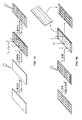
本開示は、ヒドロゲル組成物の製造方法および得られる組成物の使用に関する。ヒドロゲルアレイを製造するために用いられる場合、製造方法は、一般に、基質にヒドロゲル前駆体溶液を接触させること(ここで、基質は疎水性領域と親水性領域を含む);ヒドロゲル前駆体溶液が基質と表面修飾基質との間に位置するように、表面修飾基質をヒドロゲル前駆体溶液上に置くこと;ヒドロゲル前駆体溶液を重合させること;および基質から表面修飾基質を分離して、ヒドロゲルアレイを得ること;を含む(図1A-1B参照)。このように、ポリマーヒドロゲル前駆体溶液を基質と表面修飾基質との間で重合させ、得られるヒドロゲルを表面修飾基質がヒドロゲルアレイを含むように表面修飾基質とともに移す。1つの実施態様において、以下に詳述するように、ヒドロゲルを含まないバックグラウンドによって囲まれるヒドロゲルスポットのアレイを含むように、ヒドロゲルアレイをパターニングすることができる。もう1つの実施態様において、以下に詳述するように、ヒドロゲルバックグラウンド内にヒドロゲルを含まないスポット(またはプール)のアレイが形成されるように、ヒドロゲルアレイをパターニングすることができる。
The present disclosure relates to a method for producing a hydrogel composition and the use of the resulting composition. When used to manufacture a hydrogel array, the manufacturing method generally involves contacting a hydrogel precursor solution with a substrate (where the substrate comprises a hydrophobic region and a hydrophilic region); the hydrogel precursor solution is a substrate. Placing the surface modified substrate on the hydrogel precursor solution so as to be located between the substrate and the surface modified substrate; polymerizing the hydrogel precursor solution; and separating the surface modified substrate from the substrate to obtain a hydrogel array (See FIGS. 1A-1B). Thus, the polymer hydrogel precursor solution is polymerized between the substrate and the surface modified substrate, and the resulting hydrogel is transferred with the surface modified substrate such that the surface modified substrate comprises a hydrogel array. In one embodiment, as detailed below, the hydrogel array can be patterned to include an array of hydrogel spots surrounded by a background that does not include a hydrogel. In another embodiment, the hydrogel array can be patterned to form an array of spots (or pools) that do not contain a hydrogel within the hydrogel background, as detailed below.
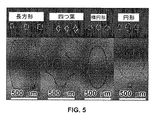
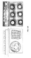
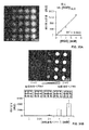
ヒドロゲルスポットを有するヒドロゲルアレイにおいては、各ヒドロゲルスポットの表面形状を定義してアレイの各ヒドロゲルスポットの含量を限定するため、ならびに、近隣のスポットとの関連におけるアレイ内の各ヒドロゲルスポットの空間パターンを定義するための、差動湿潤(differential wettability)が得られるように、得られるヒドロゲルアレイをパターニングすることができる。これは、一般的マルチウェルプレートのサイズおよび寸法(たとえば、96ウェルプレート、384ウェルプレートなど)に適合する、さまざまなサイズおよび寸法の一般的なマイクロアレイアドオンとともに用いるためのヒドロゲルアレイを製造するために特に有用である。これは、強化スループット細胞培養、培地交換などのためのマルチチャンネルピペットでの使用にも有用である。アレイの個々のヒドロゲルスポットは、所望の形状を有することができる(たとえば、図5を参照)。たとえば、形状は、円形(circular)、円形(round)、楕円形、四つ葉、長方形、三角形、星形、菱形、その組合せなどでありうる。ヒドロゲルスポットのパターンは、列、渦巻き、円、正方形、長方形、その組合せなどに作られてもよい。個々のヒドロゲルスポットの形状は、パターニングされる基質のパターニング中のエッチングのために用いられるステンシルのパターンを変えることによって変えることができる。 For hydrogel arrays with hydrogel spots, define the surface shape of each hydrogel spot to limit the content of each hydrogel spot in the array, as well as the spatial pattern of each hydrogel spot in the array in relation to neighboring spots. The resulting hydrogel array can be patterned to provide differential wettability to define. This is to produce hydrogel arrays for use with common microarray add-ons of various sizes and dimensions that fit the size and dimensions of common multiwell plates (e.g. 96 well plates, 384 well plates, etc.) It is particularly useful. It is also useful for use with multichannel pipettes for enhanced throughput cell culture, media exchange, and the like. The individual hydrogel spots of the array can have a desired shape (see, eg, FIG. 5). For example, the shape can be circular, round, oval, four-leaf, rectangular, triangular, star-shaped, diamond-shaped, combinations thereof, and the like. The pattern of hydrogel spots may be made in rows, spirals, circles, squares, rectangles, combinations thereof, and the like. The shape of the individual hydrogel spots can be changed by changing the pattern of the stencil used for etching during the patterning of the substrate to be patterned.
ヒドロゲルを含まないスポットを有するヒドロゲルアレイにおいては、個々のヒドロゲルを含まないスポットは、所望の形状を有することができる(たとえば、図5を参照)。たとえば、形状は、円形(circular)、円形(round)、楕円形、四つ葉、長方形、三角形、星形、菱形、その組合せなどでありうる。ヒドロゲルを含まないスポットのパターンは、列、渦巻き、円、正方形、長方形、その組合せなどに作られてもよい。個々のヒドロゲルを含まないスポットの形状は、パターニングされる基質のパターニング中のエッチングのために用いられるステンシルのパターンを変えることによって変えることができる。 In a hydrogel array having spots that do not include a hydrogel, spots that do not include an individual hydrogel can have a desired shape (see, eg, FIG. 5). For example, the shape can be circular, round, oval, four-leaf, rectangular, triangular, star-shaped, diamond-shaped, combinations thereof, and the like. The pattern of spots without hydrogel may be made in rows, spirals, circles, squares, rectangles, combinations thereof, and the like. The shape of the spots without individual hydrogels can be changed by changing the pattern of the stencil used for etching during the patterning of the substrate to be patterned.
ヒドロゲルアレイのサイズの上限は、パターニングされた基質の寸法および/または表面修飾基質の寸法に応じて変化する。得られるヒドロゲルアレイをパターニングして、所望のサイズを有すうる個々のヒドロゲルスポットおよびヒドロゲルを含まないスポットを得ることもできる。個々のヒドロゲルスポットおよびヒドロゲルを含まないスポットのサイズおよび形状は、パターニングされる基質のパターニング中のエッチングのために用いられるステンシルのパターンを変えることによって変えることができる。ヒドロゲルアレイの適当な個々のヒドロゲルスポットサイズは、単一の細胞を適応させるのに十分小さくありうるが、たとえば、多くの細胞を適応させるのに十分大きくもありうる。このように、ヒドロゲルアレイの個々のヒドロゲルスポットサイズは、任意の所望の直径でありうる。ヒドロゲルアレイの特に適当な個々のヒドロゲルスポットサイズは、約10 μmおよびそれ以上でありうる。 The upper size limit of the hydrogel array varies depending on the dimensions of the patterned substrate and / or the surface modification substrate. The resulting hydrogel array can also be patterned to obtain individual hydrogel spots that may have the desired size and spots that do not include the hydrogel. The size and shape of individual hydrogel spots and non-hydrogel spots can be varied by changing the pattern of the stencil used for etching during patterning of the substrate to be patterned. A suitable individual hydrogel spot size for a hydrogel array can be small enough to accommodate a single cell, but can be large enough to accommodate many cells, for example. Thus, the individual hydrogel spot size of the hydrogel array can be any desired diameter. Particularly suitable individual hydrogel spot sizes for hydrogel arrays can be about 10 μm and above.
2014年7月24日出願の米国特許出願第14/339,938号(参照することによって本明細書に援用される)などに記載の自己組織化単分子膜(SAM)によって形成される疎水性領域および親水性領域を作成することによって、パターニングされた基質を製造することができる。自己組織化単分子膜を形成するための適当な基質は、当業者に公知であり、たとえば、Love et al.、Chem. Rev. 2005、105:1103-1169(参照することによって本明細書に援用される)に記載のような、金属コーティングされた基質、シリコン基質、ダイアモンド基質、ポリジメチルシロキサン(PDMS)基質などでありうる。たとえば、基質を過フッ素化アルカンチオール溶液に浸漬して、過フッ素化アルカンチオール自己組織化単分子膜(フッ素SAM)が形成されうるようにすることにより、基質上に差動湿潤を伴う領域を形成することによって、パターニングされた基質を製造することができる。親水性領域を形成するために、フッ素SAM金属コーティングされた基質領域をプラズマエッチングから保護するために、フッ素SAM金属コーティングされた基質上にステンシルを置くことができる。次いで、フッ素SAM基質の露出領域を、酸素プラズマ処理によってエッチングして、基質中にエッチングされたフッ素SAMを形成することができる。次いで、基質を水酸基末端アルカンチオール溶液に浸漬して、基質のエッチングされた領域中に、親水性アルカンチオールSAM(EG3SAM)を形成する。得られるパターニングされた基質は、疎水性SAMおよび親水性SAMに基づく差動湿潤を有する。 Hydrophobic regions formed by self-assembled monolayers (SAMs) such as those described in U.S. Patent Application No. 14 / 339,938 filed July 24, 2014 (incorporated herein by reference) and By creating hydrophilic regions, a patterned substrate can be manufactured. Suitable substrates for forming self-assembled monolayers are known to those skilled in the art, for example, Love et al., Chem. Rev. 2005, 105: 1103-1169 (hereby incorporated by reference). Metal-coated substrates, silicon substrates, diamond substrates, polydimethylsiloxane (PDMS) substrates, etc., as described in US Pat. For example, by immersing the substrate in a perfluorinated alkanethiol solution so that a perfluorinated alkanethiol self-assembled monolayer (fluorine SAM) can be formed, an area with differential wetting is formed on the substrate. By forming, a patterned substrate can be produced. To form the hydrophilic region, a stencil can be placed on the fluorine SAM metal coated substrate to protect the fluorine SAM metal coated substrate region from plasma etching. The exposed area of the fluorine SAM substrate can then be etched by oxygen plasma treatment to form etched fluorine SAM in the substrate. The substrate is then immersed in a hydroxyl-terminated alkanethiol solution to form hydrophilic alkanethiol SAM (EG3SAM) in the etched region of the substrate. The resulting patterned substrate has differential wetting based on hydrophobic and hydrophilic SAMs.
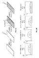
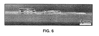
方法は、さらに、パターニングされた基質と表面修飾基質との間にスペーサーを置くことを包含する。方法の実行中にパターニングされた基質上に置かれたスペーサーは、ヒドロゲルアレイを形成するヒドロゲルの高さ(または厚さ)を定義するように機能する。より高い(すなわち、より厚い)ヒドロゲルアレイを製造する場合に、スペーサーは特に望ましい。このように、ヒドロゲルアレイは、任意の所望の高さを有することができる(たとえば、図6を参照)。しかしながら、ヒドロゲルアレイの適当な高さは、約20マイクロメーター(μm)〜約1ミリメーターであり、ヒドロゲルアレイは、必要に応じて、より高くされうる。スペーサーは、ヒドロゲルの形成中の、パターニングされた基質および表面修飾基質の表面間の直接接触を予防するようにも機能する。本発明方法に用いるスペーサーは、当業者に公知の任意の適当な材料でありうる。特に適当なスペーサーは、たとえば、ポリジメチルシロキサン(PDMS)である。ヒドロゲルアレイの高さは、たとえば、ヒドロゲルの頂点から基質に到るまで焦点を合わせるための顕微鏡を用い、基質からヒドロゲルの頂点に至るまで焦点を合わせるための顕微鏡を用い、そして、原子間力顕微鏡法によって決定されるヒドロゲルアレイの表面粗さを測定することによって決定されうる(たとえば、図4を参照)。 The method further includes placing a spacer between the patterned substrate and the surface modified substrate. Spacers placed on the patterned substrate during the performance of the method function to define the height (or thickness) of the hydrogel that forms the hydrogel array. Spacers are particularly desirable when producing higher (ie, thicker) hydrogel arrays. Thus, the hydrogel array can have any desired height (see, eg, FIG. 6). However, suitable heights for hydrogel arrays are from about 20 micrometers (μm) to about 1 millimeter, and hydrogel arrays can be made higher if desired. The spacer also functions to prevent direct contact between the surface of the patterned substrate and the surface modified substrate during formation of the hydrogel. The spacer used in the method of the present invention can be any suitable material known to those skilled in the art. A particularly suitable spacer is, for example, polydimethylsiloxane (PDMS). The height of the hydrogel array can be determined using, for example, a microscope for focusing from the top of the hydrogel to the substrate, a microscope for focusing from the substrate to the top of the hydrogel, and an atomic force microscope. It can be determined by measuring the surface roughness of the hydrogel array determined by the method (see, eg, FIG. 4).
製造方法はさらに、パターニングされた基質に、ヒドロゲル前駆体溶液を接触させることを包含する。さらに詳しくは、パターニングされた基質の親水性領域に、ヒドロゲル前駆体溶液を接触させる。パターニングされた基質の疎水性領域は、隣接する親水性領の間のバリヤーとして働き、各親水性領域の単離を可能にする。ヒドロゲル前駆体溶液は、たとえば、ポリマーと多官能性ポリマー架橋剤の組合せでありうる。 The manufacturing method further includes contacting the hydrogel precursor solution with the patterned substrate. More specifically, the hydrogel precursor solution is brought into contact with the hydrophilic region of the patterned substrate. The hydrophobic region of the patterned substrate acts as a barrier between adjacent hydrophilic regions, allowing the isolation of each hydrophilic region. The hydrogel precursor solution can be, for example, a combination of a polymer and a multifunctional polymer crosslinker.
ヒドロゲルコーティング組成物として用いられる場合、製造方法は、一般に、コーティングされるべき基質(たとえば、細胞培養プレートの表面)に、ヒドロゲル前駆体溶液を接触させることを包含する。 When used as a hydrogel coating composition, the method of manufacture generally involves contacting the hydrogel precursor solution with a substrate to be coated (eg, the surface of a cell culture plate).
ヒドロゲル前駆体溶液のための適当なポリマーは、当業者には公知であり、たとえば、ポリ(エチレングリコール)、ヒアルロン酸、ゼラチン、コラーゲン、マトリゲル(MATRIGEL(登録商標))、ジチオールポリマー(たとえば、アクリルアミド)、クリックベースの複合ヒドロゲル(Polizzotti et al. Biomacromolecules 2008、9:1084-1087(参照することによって本明細書に援用される)で論じられたもの)、ポリ(エチレングリコール)−ジアクリレート、ポリ(エチレングリコール)ビニルスルホンなどが挙げられる。特に適当なポリマーは、たとえば、ポリ(エチレングリコール)でありうる。特に適当なポリマーは、たとえば、機能性ポリマーでありうる。ポリマーの機能化は、たとえば、1H核磁気共鳴分光学、質量分析、エルマン試薬、紫外可視分光法、赤外分光法およびその他の当業者に既知の方法で確認されうる。 Suitable polymers for hydrogel precursor solutions are known to those skilled in the art and include, for example, poly (ethylene glycol), hyaluronic acid, gelatin, collagen, Matrigel (MATRIGEL®), dithiol polymers (e.g., acrylamide). ), Click-based composite hydrogels (discussed in Polizzotti et al. Biomacromolecules 2008, 9: 1084-1087, incorporated herein by reference), poly (ethylene glycol) -diacrylate, poly (Ethylene glycol) vinyl sulfone and the like. A particularly suitable polymer can be, for example, poly (ethylene glycol). A particularly suitable polymer can be, for example, a functional polymer. The functionalization of the polymer can be confirmed, for example, by 1 H nuclear magnetic resonance spectroscopy, mass spectrometry, Ellman's reagent, UV-visible spectroscopy, infrared spectroscopy and other methods known to those skilled in the art.
特に適当な機能性ポリマーは、たとえば、ノルボルネンで官能化されている末端ヒドロキシル(-OH)基を有する8-アームポリ(エチレングリコール)(JenKem Technology USA、テキサス州、アレンから市販されている)でありうる。Fairbanks et al.(Adv. Mater. 2009、21:5005-5010)に記載のように、8-アームポリ(エチレングリコール)は、ノルボルネンで官能化されうる。 A particularly suitable functional polymer is, for example, 8-arm poly (ethylene glycol) with terminal hydroxyl (-OH) groups functionalized with norbornene (commercially available from JenKem Technology USA, Allen, Texas). sell. As described in Fairbanks et al. (Adv. Mater. 2009, 21: 5005-5010), 8-arm poly (ethylene glycol) can be functionalized with norbornene.
他の特に適当なポリマーは、クリックケミストリーを用いて機能化されてもよいポリ(エチレングリコール)である。「クリック」ケミストリーは、化学的に生体分子を結合させるための非常に多彩な方法であり、アルキンとアジド官能基間の[3+2]型の付加環化を述べるために用いられる。アジドとアルキンは、生体分子および水性環境に対して非常に不活性であり、フイスゲン1,3-双極性付加環化を用いて、酸化または還元することが非常に困難である安定なトリアゾールを得ることを可能にする。銅(I)触媒および銅を含まないひずみアルキン変異体反応は両者とも、穏やかで、非常に効率的である。これらの反応は、少量の水溶液中で行うこともでき、酸素および水に対して非感受性であり、ペプチド上の官能基に対して強固である。クリックケミストリーは、たとえば、オリゴヌクレオチドおよびタンパク質などの生体サンプル中の抱合反応における選択性を可能にする。クリックケミストリーに特に適した試薬は、Laysan Bio Inc.(アラバマ州、アラブ)から市販されている。
Another particularly suitable polymer is poly (ethylene glycol), which may be functionalized using click chemistry. “Click” chemistry is a very versatile method for chemically attaching biomolecules and is used to describe a [3 + 2] type cycloaddition between alkyne and azide functional groups. Azides and alkynes are very inert to biomolecules and aqueous environments, and use
一般に、ヒドロゲル前駆体溶液は、約36 mg/mL〜約70 mg/mLのポリマーの濃度を含有する。 Generally, the hydrogel precursor solution contains a concentration of polymer from about 36 mg / mL to about 70 mg / mL.
ヒドロゲル前駆体溶液中にて用いるための適当な多官能性ポリマー架橋剤は、当業者には公知である。特に、多官能性架橋剤は、たとえば、二官能性ポリマー架橋剤および多官能性ポリマー架橋剤(n>=2)であり、末端は、ヒドロゲル前駆体溶液のポリマーとの共有結合を形成しうる官能基でありうる。特に、適当な二官能性ポリマー架橋剤および多官能性ポリマー架橋剤は、たとえば、ポリエチレングリコールジチオール(PEG-DT)、プロテアーゼ分解性架橋剤およびチオール末端マルチアームポリ(エチレングリコール)(たとえば、チオール末端4-アームPEG)でありうる。適当なプロテアーゼ分解性架橋剤は、たとえば、Nagase and Fields (Biopolymers 1996、40:399-416;参照することによって本明細書に援用される)に記載のマトリックスメタロプロテイナーゼ(MMP)分解性架橋剤でありうる。さらに詳しくは、ヒドロゲル前駆体溶液中にて用いるための適当なMMP分解性架橋ペプチドとして、KCGGPQGIWGQGCK(配列番号:27)およびKCGGPQGIAGQGCK(配列番号:28)が挙げられる。 Suitable multifunctional polymeric crosslinkers for use in the hydrogel precursor solution are known to those skilled in the art. In particular, the polyfunctional crosslinkers are, for example, bifunctional polymer crosslinkers and polyfunctional polymer crosslinkers (n> = 2), and the ends can form covalent bonds with the polymer of the hydrogel precursor solution. It can be a functional group. In particular, suitable bifunctional and multifunctional polymer crosslinkers include, for example, polyethylene glycol dithiol (PEG-DT), protease-degradable crosslinkers, and thiol-terminated multi-arm poly (ethylene glycol) (eg, thiol-terminated). 4-arm PEG). Suitable protease-degradable crosslinkers are, for example, matrix metalloproteinase (MMP) degradable crosslinkers described in Nagase and Fields (Biopolymers 1996, 40: 399-416; incorporated herein by reference). It is possible. More specifically, suitable MMP-degradable cross-linking peptides for use in the hydrogel precursor solution include KCGGPQGIWGQGCK (SEQ ID NO: 27) and KCGGPQGIAGQGCK (SEQ ID NO: 28).
ヒドロゲル前駆体溶液は、さらに、開始剤を含むことができる。当業者には公知であるように、ヒドロゲル重合は、開始剤の不在下でも起こりうる。しかしながら、開始剤は、重合を誘発し、および/または、重合速度を低下させる。適当な開始剤は、当業者に公知であり、たとえば、化学開始剤および光開始剤でありうる。特に適当な光開始剤は、たとえば、IRGACURE 2959 光開始剤(Ciba/BASF、ドイツ、ルートウィヒスハーフェンから市販されている)であり得、ヒドロゲルを形成するためのエオシンY重合は、温度変化によっても行われうる。 The hydrogel precursor solution can further include an initiator. As is known to those skilled in the art, hydrogel polymerization can occur even in the absence of an initiator. However, the initiator induces polymerization and / or reduces the polymerization rate. Suitable initiators are known to those skilled in the art and can be, for example, chemical initiators and photoinitiators. A particularly suitable photoinitiator may be, for example, the IRGACURE 2959 photoinitiator (commercially available from Ciba / BASF, Ludwigshafen, Germany), and eosin Y polymerization to form hydrogels may also be caused by temperature changes. Can be done.
もう1つの態様において、ヒドロゲル前駆体溶液は、細胞接着ペプチドを含みうる。本明細書で用いる「細胞接着ペプチド」は、細胞が受容体−リガンド相互作用を介して結合する接着タンパク質から得られるアミノ酸配列を意味する。溶液中の細胞接着ペプチドおよびその濃度を変化させることは、得られるヒドロゲル組成物への細胞接着の安定性を制御する能力を可能にする。適当な細胞接着ペプチドとして、たとえば、RGD、RGDS(配列番号:1)、CRGDS(配列番号:2)、CRGDSP(配列番号:3)、PHSRN(配列番号:4)、GWGGRGDSP(配列番号:5)、SIDQVEPYSSTAQ(配列番号:6)、GRNIAEIIKDI(配列番号:7)、DITYVRLKF(配列番号:8)、DITVTLNRL(配列番号:9)、GRYVVLPR(配列番号:10)、GNRWHSIYITRFG(配列番号:11)、GASIKVAVSADR(配列番号:12)、GTTVKYIFR(配列番号:13)、GSIKIRGTYS(配列番号:14)、GSINNNR(配列番号:15)、SDPGYIGSR(配列番号:16)、YIGSR(配列番号:17)、GTPGPQGIAGQGVV(配列番号:18)、GTPGPQGIAGQRVV(配列番号:19)、MNYYSNS(配列番号:20)、KKQRFRHRNRKG(配列番号:21)、CRGDGGGGGGGGGGGGGPHSRN(配列番号:29)、CPHSRNSGSGSGSGSGRGD(配列番号:30)、アセチル化GCYGRGDSPG(配列番号:31)、CRDGS(配列番号:32)、環状RGD[Fd]C(配列番号:33)、RKRLQVQLSIRT(配列番号:37)、IKVAV(配列番号:38)、YIGSR(配列番号:39)、KRTGQYKL(配列番号:40)、TYRSRKY(配列番号:41)、KRTGQYKLGSKTGPGQK(配列番号:42)、QAKHKQRKRLKSSC(配列番号:43)、SPKHHSQRARKKKNKNC(配列番号:44)、XBBXBX(ここで、B=塩基性残基であり、X=疎水性親水性残基である)(配列番号:45)、XBBBXXBX(ここで、B=塩基性残基であり、X=疎水性親水性残基である)(配列番号:46)、およびRGDSP(配列番号:47)が挙げられる。 In another embodiment, the hydrogel precursor solution can include a cell adhesion peptide. As used herein, “cell adhesion peptide” refers to an amino acid sequence obtained from an adhesion protein to which a cell binds via a receptor-ligand interaction. Changing the cell adhesion peptide and its concentration in solution allows the ability to control the stability of cell adhesion to the resulting hydrogel composition. Suitable cell adhesion peptides include, for example, RGD, RGDS (SEQ ID NO: 1), CRGDS (SEQ ID NO: 2), CRGDSP (SEQ ID NO: 3), PHSRN (SEQ ID NO: 4), GWGGRGDSP (SEQ ID NO: 5). , SIDQVEPYSSTAQ (SEQ ID NO: 6), GRNIAEIIKDI (SEQ ID NO: 7), DITYVRLKF (SEQ ID NO: 8), DITVTLNRL (SEQ ID NO: 9), GRYVVLPR (SEQ ID NO: 10), GNRWHSIYITRFG (SEQ ID NO: 11), GASIKVAVSADR (SEQ ID NO: 12), GTTVKYIFR (SEQ ID NO: 13), GSIKIRGTYS (SEQ ID NO: 14), GSINNNR (SEQ ID NO: 15), SDPGYIGSR (SEQ ID NO: 16), YIGSR (SEQ ID NO: 17), GTPGPQGIAGQGVV (sequence) Number: 18), GTPGPQGIAGQRVV (SEQ ID NO: 19), MNYYSNS (SEQ ID NO: 20), KKQRFRHRNRKG (SEQ ID NO: 21), CRGDGGGGGGGGGGGGGPHSRN (SEQ ID NO: 29), CPHSRNGSGSGSGSGRGD (SEQ ID NO: 30), acetylated GCYGRGDSPG (sequence) No. 31), CRDGS (SEQ ID NO: 32), cyclic RGD [Fd] C (SEQ ID NO: 33), RKRLQVQLSIRT (SEQ ID NO: 37), IKVAV (SEQ ID NO: 38), YIGSR (SEQ ID NO: 39), KRTGQYK L (SEQ ID NO: 40), TYRSRKY (SEQ ID NO: 41), KRTGQYKLGSKTGPGQK (SEQ ID NO: 42), QAKHKQRKRLKSSC (SEQ ID NO: 43), SPKHHSQRARKKKNKNC (SEQ ID NO: 44), XBBXBX (where B = basic residue) Group, X = hydrophobic hydrophilic residue) (SEQ ID NO: 45), XBBBXXBX (where B = basic residue, X = hydrophobic hydrophilic residue) (SEQ ID NO: : 46), and RGDSP (SEQ ID NO: 47).
ヒドロゲル前駆体溶液中の細胞接着ペプチドの濃度は、用いられる特定の細胞接着ペプチドならびにヒドロゲル前駆体溶液中のその他の成分に応じて変わる。しかしながら、典型的には、ヒドロゲル前駆体溶液は、約0.25 mM〜約2 mMの細胞接着ペプチドなどの約0.125 mM〜約4 mMの細胞接着ペプチドを含む。1つの適当な実施態様において、細胞接着ペプチドは、CRGDS(配列番号:2)であり、ヒドロゲル前駆体溶液は、約0.25 mM〜約4 mMのCRGDS(配列番号:2)を含む。もう1つの適当な実施態様において、細胞接着ペプチドは、環状RGDであり、ヒドロゲル前駆体溶液は、約0.125 mM〜約2 mMの環状RGD、特に、環状RGD[Fd]C(配列番号:33)を含む。 The concentration of cell adhesion peptide in the hydrogel precursor solution will vary depending on the particular cell adhesion peptide used and other components in the hydrogel precursor solution. Typically, however, the hydrogel precursor solution comprises about 0.125 mM to about 4 mM cell adhesion peptide, such as about 0.25 mM to about 2 mM cell adhesion peptide. In one suitable embodiment, the cell adhesion peptide is CRGDS (SEQ ID NO: 2) and the hydrogel precursor solution comprises about 0.25 mM to about 4 mM CRGDS (SEQ ID NO: 2). In another suitable embodiment, the cell adhesion peptide is cyclic RGD and the hydrogel precursor solution is about 0.125 mM to about 2 mM cyclic RGD, in particular cyclic RGD [Fd] C (SEQ ID NO: 33). including.
もう1つの態様において、ヒドロゲル前駆体溶液は、可溶性因子結合剤を含みうる。1つの態様においては、細胞培養培地に含まれる可溶性因子を結合するためのペプチドが、ヒドロゲル前駆体溶液に含まれる。ヒドロゲル組成物中の可溶性因子結合剤の密度(濃度)は、ヒドロゲル前駆体溶液中の可溶性因子結合剤の濃度を変えることによって制御されうる。特に適当な可溶性因子結合剤の例を下記第1表において提供する。 In another embodiment, the hydrogel precursor solution can include a soluble factor binder. In one embodiment, a peptide for binding soluble factors contained in the cell culture medium is included in the hydrogel precursor solution. The density (concentration) of the soluble factor binder in the hydrogel composition can be controlled by changing the concentration of the soluble factor binder in the hydrogel precursor solution. Examples of particularly suitable soluble factor binders are provided in Table 1 below.
第1表:ヒドロゲル組成物のための可溶性因子結合剤ペプチド配列

ヒドロゲル前駆体溶液中の可溶性因子結合剤の濃度は、用いられる特定の可溶性因子結合剤ならびにヒドロゲル前駆体溶液中のその他の成分に応じて変わる。しかしながら、典型的には、ヒドロゲル前駆体溶液は、約0.03 mM〜約0.3 mMの可溶性因子結合剤などの約0 mM〜約0.3 mMの可溶性因子結合剤を含む。 The concentration of the soluble factor binder in the hydrogel precursor solution will vary depending on the particular soluble factor binder used and the other components in the hydrogel precursor solution. Typically, however, the hydrogel precursor solution comprises about 0 mM to about 0.3 mM soluble factor binder, such as about 0.03 mM to about 0.3 mM soluble factor binder.
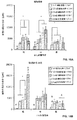
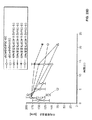
もう1つの態様において、ヒドロゲルアレイは、可変弾性率を有するヒドロゲルスポットを含むように製造されうる。ヒドロゲルアレイは、広範な弾性率(本明細書において、基質弾性率として表される)を有することができる。異なる弾性率を有するヒドロゲルスポットを有するヒドロゲルアレイは、ヒドロゲル前駆体溶液中のポリマーの濃度を変化させること、および/または、多官能性ポリマー(たとえば、チオール-ポリエチレングリコール-チオール(SH-PEG-SH)):ポリマー比の理論混合比を変化させることによって製造されうる(たとえば、図8参照)。適当な比は、約1:1〜約4:1(モル比)でありうる。たとえば、PEG-NBポリマーおよびジチオール架橋剤を用いるヒドロゲルのヒドロゲル弾性率は、たとえば、PEG-NB 重量%を一定に固定することによりPEG-NB:ジチオール架橋剤の比を変更し、次いで、たとえば、約25%〜100%架橋などのPEG-NBアームの一部のみを架橋する比で架橋剤を加えることによって制御されうる。 In another embodiment, the hydrogel array can be fabricated to include hydrogel spots with variable elastic modulus. The hydrogel array can have a wide range of elastic moduli (expressed herein as matrix elastic moduli). Hydrogel arrays with hydrogel spots having different moduli of elasticity can vary the concentration of the polymer in the hydrogel precursor solution, and / or be multifunctional polymers (eg, thiol-polyethylene glycol-thiol (SH-PEG-SH )): It can be produced by changing the theoretical mixing ratio of the polymer ratio (see, for example, FIG. 8). A suitable ratio can be from about 1: 1 to about 4: 1 (molar ratio). For example, the hydrogel modulus of a hydrogel using a PEG-NB polymer and a dithiol crosslinker can change the ratio of PEG-NB: dithiol crosslinker by, for example, fixing PEG-NB wt% constant, then, for example, It can be controlled by adding a crosslinker in a ratio that only crosslinks a portion of the PEG-NB arm, such as about 25% to 100% crosslink.
もう1つの態様において、ヒドロゲル前駆体溶液は、さらに、細胞を包含する。適当な細胞は、当業者に公知であり、たとえば、胚性幹細胞、胚性幹細胞由来の神経細胞、胚性幹細胞由来の神経前駆細胞、胚性幹細胞由来の星状膠細胞、胚性幹細胞由来の小膠細胞、胚性幹細胞由来の内皮細胞、胚性幹細胞由来の網膜色素上皮細胞、誘導多能性幹細胞、誘導多能性幹細胞由来の神経前駆細胞、誘導多能性幹細胞由来の星状膠細胞、誘導多能性幹細胞由来の小膠細胞、誘導多能性幹細胞由来の内皮細胞、誘導多能性幹細胞由来の網膜色素上皮細胞、間葉系幹細胞、臍帯静脈内皮細胞、NIH3T3線維芽細胞、皮膚線維芽細胞、線維肉腫細胞、弁間質細胞、心筋細胞、誘導多能性幹細胞由来の心筋細胞、内皮前駆細胞、循環脈管形成細胞、神経細胞、周皮細胞、癌細胞、肝細胞、膵臓β細胞、膵島細胞、およびその組合せなどが挙げられる。 In another embodiment, the hydrogel precursor solution further includes cells. Suitable cells are known to those skilled in the art, for example, embryonic stem cells, embryonic stem cell-derived neural cells, embryonic stem cell-derived neural progenitor cells, embryonic stem cell-derived astrocytes, embryonic stem cell-derived Microglial cells, endothelial cells derived from embryonic stem cells, retinal pigment epithelial cells derived from embryonic stem cells, induced pluripotent stem cells, neural progenitor cells derived from induced pluripotent stem cells, astrocytes derived from induced pluripotent stem cells Induced pluripotent stem cell-derived microglia, induced pluripotent stem cell-derived endothelial cells, induced pluripotent stem cell-derived retinal pigment epithelial cells, mesenchymal stem cells, umbilical vein endothelial cells, NIH3T3 fibroblasts, skin Fibroblasts, fibrosarcoma cells, valve stromal cells, cardiomyocytes, cardiomyocytes derived from induced pluripotent stem cells, endothelial progenitor cells, circulating angiogenic cells, neurons, pericytes, cancer cells, hepatocytes, pancreas include beta cells, islet cells, and combinations thereof It is.
もう1つの態様において、ヒドロゲル前駆体溶液は、マイクロスフェア担体(すなわち、マイクロ担体)を含みうる。マイクロスフェア担体は、たとえば、細胞、生体分子、染料および当業者に公知のその他の分子などの分子を含みうる。マイクロスフェアは、溶解または分解してマイクロスフェアの内容物を放出する分解性マイクロスフェアでありうる。 In another embodiment, the hydrogel precursor solution can include a microsphere carrier (ie, a microcarrier). Microsphere carriers can include molecules such as, for example, cells, biomolecules, dyes and other molecules known to those skilled in the art. A microsphere can be a degradable microsphere that dissolves or degrades to release the contents of the microsphere.
調製後、基質(たとえば、パターニングされた表面修飾基質、細胞培養プレートの表面など)に、ヒドロゲル前駆体溶液を接触させる。 After preparation, the hydrogel precursor solution is contacted with a substrate (eg, a patterned surface modification substrate, cell culture plate surface, etc.).
パターニングされた表面修飾基質上で用いられる場合、表面修飾基質は、たとえば、雲母、ガラス、シリコン、ダイヤモンドおよび金属酸化物表面でありうる。表面修飾基質は、たとえば、シラン単層を有するガラスカバースリップなどの表面を官能化することによって製造されうる。特に適当な表面修飾基質は、たとえば、ガラススライドでありうる。基質を官能化するための特に適当な方法は、たとえば、シラン処理でありうる。基質は、酸素プラズマ処理中に表面の両側を活性化することによって表面修飾されうる。酸素プラズマ処理は、基質の表面上の活性化ヒドロキシル基の数を増加させうる。当業者には公知のように、シラン単層は、たとえば、トルエンなどの無水有機溶媒に溶解されるアルコキシシランで製造されうる。他の適当なアルコキシシランは、たとえば、アミノシラン、グリシドキシシランおよびメルカプトシランでありうる。特に適当なアミノシランは、たとえば、(3-アミノプロピル)-トリエトキシシラン、(3-アミノプロピル)-ジエトキシ-メチルシラン、(3-アミノプロピル)ジメチルエトキシシランおよび(3-アミノプロピル)-トリメトキシシランでありうる。特に適当なグリシドキシシランは、たとえば、(3-グリシドキシプロピル)ジメチル-エトキシシランでありうる。特に適当なメルカプトシランは、たとえば、(3-メルカプトプロピル)-トリメトキシシランおよび(3-メルカプトプロピル)-メチル-ジメトキシシランでありうる。他の適当なシランは、市販されている(Sigma Aldrich、ミズーリ州、セントルイス)。表面修飾シラン基質の製造は、本明細書に記載のとおり、クリックケミストリーに参加しうる末端官能基を有する任意のシランを用いて行なわれうる。たとえば、メルカプトシランは、PEG-ノルボルネンのノルボルネンと反応しうる末端チオールを含む。他の適当な官能性表面修飾シラン基質は、たとえば、アクリレートおよびメタクリレートでありうる。基質の表面修飾に続いて、表面修飾基質上に、非接着自己組織化単分子膜が形成される。 When used on a patterned surface modification substrate, the surface modification substrate can be, for example, mica, glass, silicon, diamond and metal oxide surfaces. Surface modified substrates can be made by functionalizing a surface such as, for example, a glass cover slip with a silane monolayer. A particularly suitable surface modification substrate can be, for example, a glass slide. A particularly suitable method for functionalizing the substrate can be, for example, silane treatment. The substrate can be surface modified by activating both sides of the surface during oxygen plasma treatment. Oxygen plasma treatment can increase the number of activated hydroxyl groups on the surface of the substrate. As is known to those skilled in the art, a silane monolayer can be made of an alkoxysilane that is dissolved in an anhydrous organic solvent such as, for example, toluene. Other suitable alkoxysilanes can be, for example, aminosilanes, glycidoxysilanes, and mercaptosilanes. Particularly suitable aminosilanes are, for example, (3-aminopropyl) -triethoxysilane, (3-aminopropyl) -diethoxy-methylsilane, (3-aminopropyl) dimethylethoxysilane and (3-aminopropyl) -trimethoxysilane It can be. A particularly suitable glycidoxysilane can be, for example, (3-glycidoxypropyl) dimethyl-ethoxysilane. Particularly suitable mercaptosilanes can be, for example, (3-mercaptopropyl) -trimethoxysilane and (3-mercaptopropyl) -methyl-dimethoxysilane. Other suitable silanes are commercially available (Sigma Aldrich, St. Louis, MO). The production of the surface-modified silane substrate can be performed using any silane having a terminal functional group that can participate in click chemistry, as described herein. For example, mercaptosilane contains a terminal thiol that can react with norbornene of PEG-norbornene. Other suitable functional surface-modified silane substrates can be, for example, acrylates and methacrylates. Subsequent to surface modification of the substrate, a non-adhering self-assembled monolayer is formed on the surface-modified substrate.
ヒドロゲル前駆体溶液に基質を接触させた後、方法は、重合ヒドロゲルが基質に接着する(すなわち、結合する)ようにヒドロゲル前駆体溶液を重合することを含む。 After contacting the substrate with the hydrogel precursor solution, the method includes polymerizing the hydrogel precursor solution such that the polymerized hydrogel adheres (ie, bonds) to the substrate.
ヒドロゲル前駆体溶液に基質を接触させた後、方法は、重合ヒドロゲルが基質に接着する(すなわち、結合する)ようにヒドロゲル前駆体溶液を重合することを含む。 After contacting the substrate with the hydrogel precursor solution, the method includes polymerizing the hydrogel precursor solution such that the polymerized hydrogel adheres (ie, bonds) to the substrate.
1つの実施態様において、方法は、実質的にヒドロゲルを含まないか、または完全に含まない(「ヒドロゲルを含まない」)バックグラウンドによって囲まれるヒドロゲルの「スポット」または「島」(本明細書において、「ヒドロゲルスポット」と称する)を有するアレイを形成するために用いられうる。この実施態様において、ヒドロゲルを含まないバックグラウンドは、パターニングされた基質の疎水性領域に対応し、ヒドロゲルスポットは、パターニングされた基質の親水性領域に対応する。図1に関して、本実施態様では、円形は、ヒドロゲルを含まない領域によって囲まれるヒドロゲルスポットを表す。 In one embodiment, the method comprises a “spot” or “island” (as defined herein) of hydrogel surrounded by a background that is substantially free or completely free of hydrogel (“free of hydrogel”). , Referred to as “hydrogel spots”). In this embodiment, the background free of hydrogel corresponds to the hydrophobic region of the patterned substrate and the hydrogel spot corresponds to the hydrophilic region of the patterned substrate. With reference to FIG. 1, in this embodiment, a circle represents a hydrogel spot surrounded by a region that does not contain a hydrogel.
もう1つの実施態様において、方法は、ヒドロゲルのバックグラウンド(本明細書において、「ヒドロゲルバックグラウンド」と称する)によって囲まれたヒドロゲルを含まないプールを有するアレイを形成するために用いられうる。図1に関して、本実施態様では、円形は、ヒドロゲルを含まないバックグラウンドによって囲まれるヒドロゲルを含まないプールを表す。 In another embodiment, the method may be used to form an array having a hydrogel free pool surrounded by a hydrogel background (referred to herein as a “hydrogel background”). With reference to FIG. 1, in this embodiment, the circle represents a pool free of hydrogel surrounded by a background free of hydrogel.
もう1つの態様において、本開示は、可変弾性率、可変せん断弾性率、可変リガンドアイデンティティ、可変リガンド密度およびその組合せを有するヒドロゲルスポットを含むパターニングされたヒドロゲルアレイに関する。可変弾性率、可変せん断弾性率、可変リガンドアイデンティティ、可変リガンド密度およびその組合せを有するヒドロゲルスポットを含むパターニングされたヒドロゲルアレイは、上述の本発明方法にしたがって製造されうる。 In another aspect, the present disclosure relates to a patterned hydrogel array comprising hydrogel spots having variable modulus, variable shear modulus, variable ligand identity, variable ligand density, and combinations thereof. Patterned hydrogel arrays comprising hydrogel spots with variable modulus, variable shear modulus, variable ligand identity, variable ligand density and combinations thereof can be manufactured according to the method of the present invention described above.
適当なリガンドは、当業者に公知であり、たとえば、任意のシステインを含む、および/または、チオールで官能化された生体分子でありうる。リガンドのチオール官能化は、市販のキット(たとえば、トラウト試薬(2-イミノチオラン・HCl)、Thermo Fischer Scientific、イリノイ州、ロックフォード)を用いて行われうる。適当なリガンドは、たとえば、タンパク質、ペプチド、核酸、多糖類、脂質、生体機能模倣材料およびその他の分子、ならびにその組合せでありうる。特に適当なタンパク質は、たとえば、接着タンパク質でありうる。特に適当な接着タンパク質は、たとえば、フィブロネクチン、カドヘリンおよびその組合せでありうる。特に適当なペプチドは、たとえば、上述の細胞接着ペプチドおよび/または可溶性因子結合剤でありうる。 Suitable ligands are known to those skilled in the art and can be, for example, any cysteine-containing and / or thiol-functionalized biomolecule. Thiol functionalization of the ligand can be performed using commercially available kits (eg, trout reagent (2-iminothiolane.HCl), Thermo Fischer Scientific, Rockford, Ill.). Suitable ligands can be, for example, proteins, peptides, nucleic acids, polysaccharides, lipids, biofunctional mimetic materials and other molecules, and combinations thereof. A particularly suitable protein can be, for example, an adhesion protein. Particularly suitable adhesion proteins can be, for example, fibronectin, cadherin and combinations thereof. Particularly suitable peptides can be, for example, the cell adhesion peptides and / or soluble factor binders described above.
本開示のヒドロゲル組成物が、細胞と結合または相互作用して、細胞接着、拡散、移動、増殖、分化および細胞構造(たとえば、細管など)の形成に影響を及ぼすと思われる細胞接着ペプチドおよび可溶性因子結合剤の組合せを含むのが適当である。この態様は、たとえば、細胞接着、拡散、移動、増殖、分化および細胞構造の形成などの細胞における効果について可溶性因子結合剤を特にスクリーニングするためにヒドロゲル組成物を用いることを可能にする。さらに、未知の機能をもつ可溶性因子結合剤は、細胞接着、拡散、移動、増殖、分化および細胞構造の形成における変化についてスクリーニングするために細胞接着ペプチドと組み合わせて固定されうる。 The hydrogel composition of the present disclosure binds or interacts with cells to affect cell adhesion, diffusion, migration, proliferation, differentiation and formation of cell structures (eg, tubules) and soluble Suitably a combination of factor binders is included. This embodiment allows the hydrogel composition to be used to specifically screen soluble factor binders for effects in cells such as cell adhesion, diffusion, migration, proliferation, differentiation and cell structure formation. In addition, soluble factor binders with unknown function can be immobilized in combination with cell adhesion peptides to screen for changes in cell adhesion, diffusion, migration, proliferation, differentiation and cell structure formation.
ヒドロゲル組成物は、さらに、可変弾性率を含んでもよい。ヒドロゲル組成物は、広範な剛性(本明細書において基質弾性率と称する)を有しうる。たとえば、異なる弾性率を有するヒドロゲルは、ヒドロゲル前駆体溶液中のポリマーの濃度を変化させること、および/または、多官能性ポリマー(たとえば、チオール-ポリエチレングリコール-チオール(SH-PEG-SH)):ポリマー比の理論混合比を変化させることによって製造されうる(たとえば、図8参照)。適当な比は、約1:1〜約4:1(モル比)でありうる。 The hydrogel composition may further include a variable modulus. The hydrogel composition can have a wide range of stiffness (referred to herein as matrix modulus). For example, hydrogels with different moduli can change the concentration of the polymer in the hydrogel precursor solution and / or multifunctional polymers (eg, thiol-polyethylene glycol-thiol (SH-PEG-SH)): It can be produced by changing the theoretical mixing ratio of the polymer ratio (see, for example, FIG. 8). A suitable ratio can be from about 1: 1 to about 4: 1 (molar ratio).
もう1つの態様において、パターニングされたヒドロゲルアレイは、さらに、それによってパターニングされたヒドロゲルアレイが、任意のサイズのアドオンを適合させるための寸法で製造される、マイクロアレイアドオンとともに組み立てられうる。適当なマイクロアレイアドオンは、市販されている(Grace Bio Labs、オレゴン州、ベンド)。マイクロアレイアドオンは、可溶性因子のプレゼンテーションが制御されうるように、ヒドロゲルアレイの個々のヒドロゲルスポットおよびヒドロゲルを含まないプールを単離することを可能にする。マイクロアレイアドオンは、各ヒドロゲルスポットおよびヒドロゲルを含まないプールが、可溶性因子プレゼンテーションで独立して調べられうるように、ヒドロゲルアレイの個々のヒドロゲルスポットおよびヒドロゲルを含まないプールとの数と同じ数の開口を含みうる。あるいは、マイクロアレイアドオンは、1つ以上の個々のヒドロゲルスポットおよび1つ以上の個々のヒドロゲルを含まないプールを適合させうる、より大きい開口を有しうる。たとえば、は、単一のヒドロゲルスポットまたは単一のヒドロゲルを含まないプールを適合させるのに十分に大きい開口を有しうる。 In another embodiment, the patterned hydrogel array can be further assembled with a microarray add-on, whereby the patterned hydrogel array is manufactured with dimensions to accommodate any size add-on. Suitable microarray add-ons are commercially available (Grace Bio Labs, Bend, Oregon). The microarray add-on makes it possible to isolate individual hydrogel spots and hydrogel-free pools of the hydrogel array so that the presentation of soluble factors can be controlled. The microarray add-on has the same number of openings as the number of individual hydrogel spots and hydrogel-free pools in the hydrogel array so that each hydrogel spot and hydrogel-free pool can be examined independently in the soluble factor presentation. May be included. Alternatively, the microarray add-on may have a larger opening that can fit one or more individual hydrogel spots and a pool that does not include one or more individual hydrogels. For example, may have an opening large enough to fit a single hydrogel spot or a pool that does not contain a single hydrogel.
ヒドロゲル組成物の使用方法
さらにもう1つの態様において、本開示は、分子−分子相互作用をスクリーニングする方法に関する。方法は、ヒドロゲル組成物を製造すること(ここで、ヒドロゲル組成物は、少なくとも1つの可溶性因子結合剤を含む);少なくとも1つの可溶性因子結合剤と相互作用することが知られているか、または相互作用すると思われる分子に、ヒドロゲル組成物を接触させること;およびヒドロゲル組成物を分析すること;を含む。
Methods of using hydrogel compositions In yet another embodiment, the present disclosure relates to methods of screening for molecule-molecule interactions. The method is known to produce a hydrogel composition, wherein the hydrogel composition comprises at least one soluble factor binder; it is known to interact with or interact with at least one soluble factor binder Contacting the hydrogel composition with a molecule that appears to act; and analyzing the hydrogel composition.
ヒドロゲル組成物は、本明細書に記載のように製造されうる。 The hydrogel composition can be manufactured as described herein.
ヒドロゲル組成物は、当業者に公知の方法を用いて分析されうる。たとえば、ヒドロゲル組成物は、蛍光、顕微鏡法などを用いて分析されうる。 The hydrogel composition can be analyzed using methods known to those skilled in the art. For example, the hydrogel composition can be analyzed using fluorescence, microscopy, and the like.
1つの態様において、本開示は、可変密度(弾性率)、可変リガンド固有性(identities)、可変リガンド密度およびその組合せを有するヒドロゲルスポットを含むように本明細書にしたがって製造されたヒドロゲルアレイを用いて、細胞-表面相互作用をスクリーニングする方法に関する。本開示のヒドロゲルアレイを用いてスクリーニングされるべきリガンドは、細胞と結合または相互作用することが知られているか、または思われるリガンドでありうる。方法は、さらに、個々のヒドロゲルスポットが可溶性因子で調べられうるように、本明細書に記載のパターニングされたヒドロゲルアレイをマイクロアレイアドオンとともに組み立てることを含みうる。 In one embodiment, the present disclosure uses a hydrogel array manufactured according to the present specification to include hydrogel spots having variable density (elastic modulus), variable ligand identities, variable ligand density, and combinations thereof. And a method for screening cell-surface interactions. The ligand to be screened using the hydrogel array of the present disclosure can be a ligand known or suspected of binding or interacting with the cell. The method can further include assembling the patterned hydrogel array described herein with a microarray add-on such that individual hydrogel spots can be probed with soluble factors.
方法は、さらに、パターニングされたヒドロゲルアレイに、細胞を接触させることを含む。本明細書で用いる「細胞を接触させる」は、細胞およびヒドロゲルアレイを分析するために、パターニングされたヒドロゲルアレイ上に細胞を播種することを意味する。当業者には公知のように、細胞懸濁液を基質に移し、細胞が基質に接着するのに十分な時間を付与するのが一般的である。 The method further includes contacting the cells with the patterned hydrogel array. As used herein, “contacting cells” means seeding cells on the patterned hydrogel array to analyze the cells and hydrogel array. As is known to those skilled in the art, it is common to transfer the cell suspension to the substrate and allow sufficient time for the cells to adhere to the substrate.
もう1つの実施態様において、細胞は、ポリマー、架橋剤、細胞接着ペプチドおよび細胞を含むヒドロゲル前駆体溶液を用いて、パターニングされたヒドロゲルアレイのヒドロゲルに組み込まれうる。 In another embodiment, the cells can be incorporated into a hydrogel of a patterned hydrogel array using a hydrogel precursor solution comprising a polymer, a cross-linking agent, a cell adhesion peptide and cells.
次いで、細胞は、たとえば、約1時間〜約30日間などの所望の期間培養される。所望の期間の後、細胞は、たとえば、免疫蛍光顕微鏡法、位相差顕微鏡法、光学顕微鏡法、電子顕微鏡法およびその組合せなどの顕微鏡法によって分析されうる。細胞は、細胞接着、細胞拡散、細胞形態、細胞増殖、細胞移動、細胞分化、タンパク質発現およびその組合せについて分析されうる。 The cells are then cultured for a desired period of time, for example, from about 1 hour to about 30 days. After a desired period of time, the cells can be analyzed by microscopy such as, for example, immunofluorescence microscopy, phase contrast microscopy, light microscopy, electron microscopy, and combinations thereof. Cells can be analyzed for cell adhesion, cell spreading, cell morphology, cell proliferation, cell migration, cell differentiation, protein expression and combinations thereof.
適当な細胞は、当業者に公知の任意の細胞でありうる。特に適当な細胞は、たとえば、胚性幹細胞、胚性幹細胞由来の神経細胞、胚性幹細胞由来の神経前駆細胞、胚性幹細胞由来の星状膠細胞、胚性幹細胞由来の小膠細胞、胚性幹細胞由来の内皮細胞、胚性幹細胞由来の網膜色素上皮細胞、誘導多能性幹細胞、誘導多能性幹細胞由来の神経前駆細胞、誘導多能性幹細胞由来の星状膠細胞、誘導多能性幹細胞由来の小膠細胞、誘導多能性幹細胞由来の内皮細胞、誘導多能性幹細胞由来の網膜色素上皮細胞、間葉系幹細胞、臍帯静脈内皮細胞、NIH 3T3線維芽細胞、皮膚線維芽細胞、線維肉腫細胞、弁間質細胞、心筋細胞、誘導多能性幹細胞由来の心筋細胞、内皮前駆細胞、循環脈管形成細胞、神経細胞、周皮細胞、癌細胞、肝細胞、膵臓β細胞、膵島細胞およびその組合せでありうる。 Suitable cells can be any cells known to those skilled in the art. Particularly suitable cells are, for example, embryonic stem cells, neural cells derived from embryonic stem cells, neural progenitor cells derived from embryonic stem cells, astrocytes derived from embryonic stem cells, microglia cells derived from embryonic stem cells, embryonic Endothelial cells derived from stem cells, retinal pigment epithelial cells derived from embryonic stem cells, induced pluripotent stem cells, neural progenitor cells derived from induced pluripotent stem cells, astrocytes derived from induced pluripotent stem cells, induced pluripotent stem cells Derived microglia, induced pluripotent stem cell-derived endothelial cells, induced pluripotent stem cell-derived retinal pigment epithelial cells, mesenchymal stem cells, umbilical vein endothelial cells, NIH 3T3 fibroblasts, skin fibroblasts, fibers Sarcoma cells, valve stromal cells, cardiomyocytes, induced pluripotent stem cell-derived cardiomyocytes, endothelial progenitor cells, circulating angiogenic cells, neurons, pericytes, cancer cells, hepatocytes, pancreatic β cells, islet cells And combinations thereof.
方法は、さらに、パターニングされたヒドロゲルアレイのヒドロゲルスポット上の細胞が培養される培養培地に可溶性分子を含めることによって、可溶性分子に細胞を接触させることを含んでもよい。特に適当な可溶性分子は、増殖因子およびプロテオグリカンでありうる。適当な増殖因子は、たとえば、形質転換増殖因子βスーパーファミリーからのタンパク質、増殖因子の線維芽細胞増殖因子ファミリー、増殖因子の血小板由来増殖因子ファミリーおよびその組合せでありうる。特に適当な増殖因子は、たとえば、血管内皮増殖因子、骨形態形成タンパク質、線維芽細胞増殖因子、インスリン様増殖因子およびその組合せでありうる。適当なプロテオグリカンは、たとえば、ヘパリン、ヘパリン硫酸、および/またはコンドロイチングリコサミノグリカン側鎖を有するプロテオグリカンでありうる。 The method may further comprise contacting the cells with soluble molecules by including the soluble molecules in a culture medium in which cells on the hydrogel spots of the patterned hydrogel array are cultured. Particularly suitable soluble molecules can be growth factors and proteoglycans. Suitable growth factors can be, for example, proteins from the transforming growth factor beta superfamily, the fibroblast growth factor family of growth factors, the platelet derived growth factor family of growth factors and combinations thereof. Particularly suitable growth factors can be, for example, vascular endothelial growth factor, bone morphogenetic protein, fibroblast growth factor, insulin-like growth factor and combinations thereof. Suitable proteoglycans can be, for example, proteoglycans having heparin, heparin sulfate, and / or chondroitin glycosaminoglycan side chains.
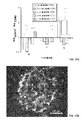
1つの特に適当な実施態様において、本開示は、ヒドロゲル組成物を用いる細管形成促進剤および/または抗細管形成剤に関する。本明細書で用いる「細管形成」は、接着、拡散、出芽、移動、細胞結合形成、細管ネットワーク形成および維持(たとえば、内皮細胞細管ネットワーク形成)、脈管形成および/または血管新生する細胞の能力、およびその組合せを意味する。一般に、方法は、以下を含む:本開示のヒドロゲル組成物を製造すること;細管形成を促進または減少させると思われる作用剤を提供すること;ヒドロゲル組成物および作用剤に細胞を接触させること;および細胞を分析すること。ヒドロゲル組成物は、一般に、本明細書に記載のノルボルネンで官能化されたポリエチレングリコール、架橋ペプチド、細胞接着ペプチドおよび可溶性因子結合剤を含む。本明細書で用いる「細管形成を減少させる」は、細管形成を最小化、低減化または排除することをも意味する。 In one particularly suitable embodiment, the present disclosure relates to tubule formation promoters and / or antitubule formation agents that use hydrogel compositions. As used herein, “tubule formation” refers to the ability of a cell to adhere, diffuse, sprouting, migrate, form cell connections, form and maintain a tubular network (eg, endothelial cell tubular network formation), vasculogenesis and / or angiogenesis , And combinations thereof. In general, the method includes: producing a hydrogel composition of the present disclosure; providing an agent that appears to promote or reduce tubule formation; contacting the cell with the hydrogel composition and the agent; And analyzing the cells. The hydrogel composition generally includes a norbornene-functionalized polyethylene glycol, a cross-linked peptide, a cell adhesion peptide and a soluble factor binder as described herein. As used herein, “reduce tubule formation” also means minimizing, reducing or eliminating tubule formation.
もう1つの適当な実施態様において、本開示は、細管形成を促進する方法に関する。一般に、方法は、以下を含む:本開示のヒドロゲル組成物および可溶性因子を含むヒドロゲル組成物を製造すること;ヒドロゲル組成物に接触した培養培地を提供すること;ヒドロゲル組成物に培養倍地中の細胞を接触させること;および細胞を分析すること。細胞を培養するのに用いるための培養培地は、当業者に公知の任意の適当な培養培地を含む。たとえば、培養培地は、特定の細胞型のための標準増殖培地を含んでもよい(たとえば、 HUVEC用のMedium 199およびEGM-2 BULLETKIT(商標)(Lonza、スイス、バーゼル)、iPSC-ECsおよびhESC-EC用のVASCULIFE(登録商標)およびVEGF LifeFactors (Lifeline Cell Technology、メリーランド州、フレデリック))。1つの特定の実施態様において、方法は、内皮細胞細管ネットワーク形成を促進することを含む。 In another suitable embodiment, the present disclosure relates to a method of promoting tubule formation. In general, the method includes: producing a hydrogel composition comprising a hydrogel composition of the present disclosure and a soluble factor; providing a culture medium in contact with the hydrogel composition; Contacting the cells; and analyzing the cells. The culture medium for use in culturing the cells includes any suitable culture medium known to those skilled in the art. For example, the culture medium may contain standard growth media for specific cell types (e.g. Medium 199 and EGM-2 BULLETKITTM (Lonza, Basel, Switzerland) for HUVEC, iPSC-ECs and hESC- VASCULIFE® and VEGF LifeFactors (Lifeline Cell Technology, Frederick, MD) for EC. In one particular embodiment, the method includes promoting endothelial cell tubule network formation.
本開示の方法およびヒドロゲル組成物は、ヒドロゲルスポット上のリガンドの密度に対する非常に優れた制御、ならびにヒドロゲルスポット上のリガンドのアイデンティティに対する非常に優れた制御を可能にする。ヒドロゲルの剛性も制御することができる。この制御は、細胞培養のための基質の特定のパラメーターのスクリーニングを可能にし、基質および培養環境に対する細胞応答の結果を変更し、影響を及ぼしうる。本開示のパターニングされたヒドロゲルアレイは、リガンドの組合せをスクリーニングすることも可能にする。このように、本開示のパターニングされたヒドロゲルアレイは、基質および培養環境に対する細胞応答の結果を変更し、影響を及ぼしうる基質の特定のパラメーターを同定するための単一表面上のハイスループットな多変数生物学的スクリーニングを行なうツールを提供する。 The methods and hydrogel compositions of the present disclosure allow for very good control over the density of ligands on the hydrogel spot, as well as very good control over the identity of the ligand on the hydrogel spot. The stiffness of the hydrogel can also be controlled. This control allows the screening of specific parameters of the substrate for cell culture and can alter and influence the outcome of the cellular response to the substrate and culture environment. The patterned hydrogel array of the present disclosure also allows screening of ligand combinations. As such, the patterned hydrogel arrays of the present disclosure modify the results of cellular responses to substrates and culture environments and identify high throughput multiple on a single surface to identify specific parameters of the substrate that can be affected. Provides tools for variable biological screening.
本開示は、以下の非限定的実施例を考慮することにより、より完全に理解されるであろう。 The present disclosure will be more fully understood in view of the following non-limiting examples.
実施例
材料および方法
PEG-ノルボルネン合成
末端ヒドロキシル基(-OH)を有し、分子量20 kDaである8-アームポリ(エチレングリコール)(PEG)をJenKem Technology USA(テキサス州、アレン)から購入した。無水ピリジン、4-(ジメチルアミノ)ピリジン(DMAP)、5-ノルボルネン-2-カルボン酸、ジエチルエーテル、および0.03 % v/v テトラメチルシラン(TMS)を含む重水素化クロロホルム(CDCl3、99.8%)をSigma Aldrich(ミズーリ州、セントルイス)から購入した。N,N'-ジシクロヘキシルカルボジイミド(DCC)、無水ジクロロメタン(DCM)をACROS Organics(ベルギー、ヘール)から購入した。3.5K分子量カットオフのスネークスキン(SNAKESKIN)透析チューブをThermo Fisher Scientific(マサチューセッツ州、ウォルサム)から購入した。
Example
Materials and methods
8-arm poly (ethylene glycol) (PEG) having a PEG-norbornene synthetic terminal hydroxyl group (—OH) and a molecular weight of 20 kDa was purchased from JenKem Technology USA (Allen, Texas). Deuterated chloroform (CDCl 3 , 99.8%) containing anhydrous pyridine, 4- (dimethylamino) pyridine (DMAP), 5-norbornene-2-carboxylic acid, diethyl ether, and 0.03% v / v tetramethylsilane (TMS) ) Was purchased from Sigma Aldrich (St. Louis, MO). N, N′-dicyclohexylcarbodiimide (DCC), anhydrous dichloromethane (DCM) were purchased from ACROS Organics (Hale, Belgium). A 3.5K molecular weight cut-off SNAKESKIN dialysis tube was purchased from Thermo Fisher Scientific (Waltham, Mass.).
8-アームPEG-OHをでノルボルネン官能化して、生物活性リガンドの光重合および固定のためにチオール-エン化学を利用した(Fairbanks et al. Adv. Mater. 2009、21:5005-5010;Impellitteri et al. Biomaterials 2012、33:3475-84;Belair and Murphy Acta Biomater. 2013;およびGould et al. Acta Biomater 2012、8:3201-3209に記載)。官能化反応のPEG-ノルボルネン(PEG-NB)生成物を、中型のフリット付きブフナー漏斗を通してろ過し、反応中に形成された塩を除去した。ろ液を900 mL冷ジエチルエーテルおよび100 mLヘキサンで沈殿させた。固体を定性ろ紙で集め、一夜風乾した。残留ノルボルネン酸を除去するために、水で戻したスネークスキン透析チューブを用い、4℃にて72時間、4LのdH2Oに対して透析することによってPEG-NB生成物を精製し、次いで、凍結乾燥した。 8-arm PEG-OH was norbornene-functionalized with thiol-ene chemistry for photopolymerization and immobilization of bioactive ligands (Fairbanks et al. Adv. Mater. 2009, 21: 5005-5010; Impellitteri et al. Biomaterials 2012, 33: 3475-84; Belair and Murphy Acta Biomater. 2013; and Gould et al. Acta Biomater 2012, 8: 3201-3209). The PEG-norbornene (PEG-NB) product of the functionalization reaction was filtered through a medium fritted Buchner funnel to remove salts formed during the reaction. The filtrate was precipitated with 900 mL cold diethyl ether and 100 mL hexane. The solid was collected with qualitative filter paper and air dried overnight. To remove residual norbornene acid, the PEG-NB product was purified by dialysis against 4 L of dH 2 O for 72 hours at 4 ° C. using a snake skin dialysis tube reconstituted with water, then Lyophilized.
1H核磁気共鳴分光法で、>90%のノルボルネン官能化が確認された。TMS内部標準を含む CDCl3中、6 mg/mLにてサンプルを調製した。マディソン国立磁気共鳴施設によって提供される分光分析サービスを用いて、Bruker Instruments Avance III 500iで、400 MHzおよび27℃にて、自由誘導減衰(FID)スペクトルを得た。 > 90% norbornene functionalization was confirmed by 1H nuclear magnetic resonance spectroscopy. Samples were prepared at 6 mg / mL in CDCl 3 containing TMS internal standard. Free induction decay (FID) spectra were obtained on a Bruker Instruments Avance III 500i at 400 MHz and 27 ° C. using a spectroscopic service provided by the Madison National Magnetic Resonance Facility.
ヒドロゲルアレイ形成
これらの実験に用いたヒドロゲルアレイは、シラン化ガラス基質に固定されたヒドロゲルスポットから構成された。ヒドロゲルスポットは、金の、異なる湿潤性をもつことにより、エラストマーステンシルによってパターンが定義される領域を有するようにパターニングされた表面を用いて形成された。ヒドロゲルアレイの製造方法を、以下にさらに記載する。
Hydrogel array formation The hydrogel arrays used in these experiments consisted of hydrogel spots immobilized on a silanized glass substrate. Hydrogel spots were formed using surfaces that were patterned to have regions that were defined by an elastomeric stencil by having different wettability of gold. The method of manufacturing the hydrogel array is further described below.
ガラスシラン化
ガラスカバースリップおよび塩酸(HCl)溶液をThermo Fisher Scientific(マサチューセッツ州、ウォルサム)から購入した。トルエン、メタノール、エタノール、3-メルカプトプロピルトリメトキシシラン(3-MPTS)およびジチオスレイトール(DTT)をSigma Aldrich(ミズーリ州、セントルイス)から購入した。低圧プラズマシステムをDiener Electronic(ドイツ、エブハウゼン)から購入した。
Glass silanized glass coverslips and hydrochloric acid (HCl) solutions were purchased from Thermo Fisher Scientific (Waltham, Mass.). Toluene, methanol, ethanol, 3-mercaptopropyltrimethoxysilane (3-MPTS) and dithiothreitol (DTT) were purchased from Sigma Aldrich (St. Louis, MO). A low-pressure plasma system was purchased from Diener Electronic (Ebhausen, Germany).
3-MPTSでガラスカバースリップをシラン化して、PEG-NBとのチオール-エン反応に酸化することができ、次いで、PEG-NBヒドロゲルの共有固定化を可能にするチオール基を提供する基質を作成した(Seo et al. Colloids Surf B Biointerfaces 2012、98:1-6)。既述されたように、液相シラン化を行なった(Seo et al. Colloids Surf B Biointerfaces 2012、98:1-6;Halliwell et al. Anal Chem 2001、73:2476-2483;and Cras et al. Biosens Bioelectron 1999、14:683-688)。1:1のメタノール:HCl中で45分間、カバースリップを超音波処理して、大量汚染物質を除去した。シラン化の直前に、それぞれの側に5分間、40 sccmおよび50 Wにて酸素プラズマ処理することによって、カバースリップを反応させて、表面上の活性化ヒドロキシル基の数を増加させた。2.5% v/v 3-MPTSを含むコプリンジャー内に活性化されたカバースリップを4時間置いた。トルエン、1:1のエタノール/トルエン、およびエタノールで濯ぐことによって、カバースリップの表面から過剰のシランを除去し、N2ガスで乾燥した。シラン化カバースリップを気密室に置き、N2ガスでパージし、次いで、100℃にて1時間養生して、表面に結合したシランを架橋させ、それらの加水分解に対する感受性を低下させた。使用まで、N2ガスでパージされ、光から保護されたチャンバー内でシラン化カバースリップを保管した。使用前に、37℃にて30分間、PBS中の10 mM DTTでシラン化ガラスカバースリップを処理して、表面上に形成されたジスルフィドを減少させ、表面で利用可能な遊離チオールを増加させた(Vistas et al. Appl Surf Sci 2013、286:314-318)。 A glass cover slip can be silanized with 3-MPTS and oxidized to a thiol-ene reaction with PEG-NB, then creating a substrate that provides a thiol group that allows covalent immobilization of PEG-NB hydrogels (Seo et al. Colloids Surf B Biointerfaces 2012, 98: 1-6). Liquid phase silanization was performed as previously described (Seo et al. Colloids Surf B Biointerfaces 2012, 98: 1-6; Halliwell et al. Anal Chem 2001, 73: 2476-2483; and Cras et al. Biosens Bioelectron 1999, 14: 683-688). The coverslip was sonicated in 1: 1 methanol: HCl for 45 minutes to remove bulk contaminants. Just prior to silanization, the coverslip was reacted to increase the number of activated hydroxyl groups on the surface by oxygen plasma treatment at 40 sccm and 50 W for 5 minutes on each side. The activated cover slip was placed in a copliner containing 2.5% v / v 3-MPTS for 4 hours. Excess silane was removed from the surface of the coverslip by rinsing with toluene, 1: 1 ethanol / toluene, and ethanol and dried with N 2 gas. The silanized coverslips were placed in an airtight chamber, purged with N 2 gas, and then cured at 100 ° C. for 1 hour to crosslink the silane bound to the surface and reduce their sensitivity to hydrolysis. Until use, silanized cover slips were stored in a chamber purged with N 2 gas and protected from light. Prior to use, silanized glass coverslips were treated with 10 mM DTT in PBS for 30 minutes at 37 ° C. to reduce the disulfide formed on the surface and increase the free thiol available on the surface. (Vistas et al. Appl Surf Sci 2013, 286: 314-318).
エラストマーステンシルの製作
シリコンウェハーをWRS Materials(カリフォルニア州、サンノゼ)から購入した。SU-8 100フォトレジストをMicroChem(マサチューセッツ州、ニュートン)から購入した。Sylgard 184シリコンエラストマーキットをDow Corning Corporation(ミシガン州、ミッドランド)から購入した。
Fabrication of elastomer stencils Silicon wafers were purchased from WRS Materials (San Jose, CA). SU-8 100 photoresist was purchased from MicroChem (Newton, Mass.). A Sylgard 184 silicone elastomer kit was purchased from Dow Corning Corporation (Midland, MI).
既述されたように、ソフトリソグラフィーを用いて、ポリジメチルシロキサン(PDMS)エラストマーステンシルを製造した(Jo et al. J Microelectromechanical Syst 2000、9:76-81)。アドビイラストレーターを用いてステンシルのためのレイアウトおよび形状を描き、ImageSetter(ウィスコンシン州、マディソン)によって提供される高解像度の市販のレーザープリントサービスを用いて透明フィルム上に印刷した。従来のフォトリソグラフィー技術と組み合わせた光マスクとして透明フィルムを用いて、シリコンウェハー上にスピンコートしたSU-8ネガ型UVフォトレジストをもつマスターモールドを製造した。PDMSステンシルを製造するために、Sylgardエラストマーキットからの硬化剤およびPDMSプレポリマー溶液を、1:10 重量比で完全に混合し、マスターモールド上に広げ、80℃にて6時間硬化させた。硬化後、マスターモールドからステンシルをはがし、エタノールで簡単に洗浄し、N2ガスで乾燥した。
As previously described, soft lithography was used to produce polydimethylsiloxane (PDMS) elastomer stencils (Jo et al.
疎水性/親水性パターニング
金コートテストスライド(25 mm x 75 mm x 1 mmのガラス上の50 Åのチタン金属薄膜上に1,000 Åの金)をEvaporated Metal Films(ニューヨーク州、イサカ)から購入した。過フッ素化アルカンチオール(HS-(CH2)11-O-(CH2)2-(CF2)5-CF3)をProChimia Surfaces(ポーランド、ソポト)から購入した。既述されたように、ヒドロキシル末端アルカンチオール(HS-C11-(O-CH2-CH2)3-OH)を合成した(Prime and Whitesides J. Am. Chem. Soc. 1993、115:10714-10721)。
Hydrophobic / hydrophilic patterning gold-coated test slides (1,000 gold on 50 mm titanium metal film on 25 mm x 75 mm x 1 mm glass) were purchased from Evaporated Metal Films (Ithaca, NY). Perfluorinated alkane thiol (HS- (CH 2) 11 -O- (CH 2) 2 - (CF 2) 5 -CF 3) were purchased from ProChimia Surfaces (Poland, Sopot). As described above, hydroxyl-terminated alkane thiol (HS-C 11 - (O -CH 2 -CH 2) 3 -OH) was synthesized (Prime and Whitesides J. Am Chem Soc 1993,115:... 10714 -10721).
金コートスライドを、アルカンチオレートの疎水性および親水性自己組織化単分子膜(SAM)でパターニングして、差動湿潤を有する領域を形成した。差動湿潤パターニングは、2つの目的を同時に果たした:1)ヒドロゲルスポットの形状を定義した;および2)アレイ内の各ヒドロゲルスポットの含量を限定した。金コートスライドをエタノールに浸漬し、〜2分間超音波処理し、エタノールで濯ぎ、N2ガスで乾燥して、汚染物質および酸化金層を除去した。金コートスライドを、過フッ素化アルカンチオールの1 mMエタノール溶液に≧2時間浸漬して、フッ素化アルカンチオレートSAM(フッ素SAM)を形成させた。フッ素SAM形成後、フッ素SAM金コートスライドをエタノールで洗浄し、N2ガスで乾燥した。基質上の親水性領域を定義するために、PDMSステンシルをフッ素SAM金コートスライド上に置いて、プラズマエッチングからスライドの領域を選択的に保護した。PDMSステンシルのパターンによって、フッ素SAM金コートスライド上の露出領域の空間的および形状的パターニングが決定され、次いで、アレイが含みうるヒドロゲルスポットの形状および空間的パターニングが決定された。1分間、40 sccmおよび50 Wでの酸素プラズマ処理によって、フッ素SAM金コートスライドの露出領域をエッチングした。エッチングされた金コートスライドを、エタノールで洗浄し、N2ガスで乾燥し、金コートスライドの選択的にエッチングされた領域に、親水性アルカンチオレートSAM(EG3SAM)が形成されるように、ヒドロキシル末端アルカンチオールの0.1 mMエタノール溶液に≧2時間浸漬した。エタノールで得られる金コートスライドを洗浄し、ヒドロゲル形成前にN2ガスで乾燥した。 Gold coated slides were patterned with alkanethiolate hydrophobic and hydrophilic self-assembled monolayers (SAM) to form regions with differential wetting. Differential wet patterning served two purposes simultaneously: 1) defined the shape of the hydrogel spots; and 2) limited the content of each hydrogel spot in the array. Gold coated slides were immersed in ethanol, sonicated for ˜2 minutes, rinsed with ethanol, and dried with N 2 gas to remove contaminants and gold oxide layers. The gold-coated slide was immersed in a 1 mM ethanol solution of perfluorinated alkanethiol for ≧ 2 hours to form fluorinated alkanethiolate SAM (fluorine SAM). After the formation of the fluorine SAM, the fluorine SAM gold-coated slide was washed with ethanol and dried with N 2 gas. To define the hydrophilic area on the substrate, a PDMS stencil was placed on a fluorine SAM gold coated slide to selectively protect the area of the slide from plasma etching. The pattern of the PDMS stencil determined the spatial and geometric patterning of the exposed areas on the fluorine SAM gold-coated slide, and then the shape and spatial patterning of the hydrogel spots that the array could contain. The exposed areas of the fluorine SAM gold-coated slide were etched by oxygen plasma treatment at 40 sccm and 50 W for 1 minute. Etched gold-coated slides are washed with ethanol and dried with N 2 gas to form hydrophilic alkanethiolate SAM (EG 3 SAM) in selectively etched areas of the gold-coated slide. Then, it was immersed in a 0.1 mM ethanol solution of hydroxyl-terminated alkanethiol for ≧ 2 hours. Gold coated slides obtained with ethanol were washed and dried with N 2 gas before hydrogel formation.
接触角測定によって、金コートスライド上の疎水性および親水性SAM形成を確認した(図2B参照)。接触角ゴニオメーター(DataPhysics Contact Angle System OCA、カリフォルニア州、サンノゼ)を用い、室温にて静的接触角を測定した。表面上に蒸留水の液滴(3 μL)を置き、3つの異なるサンプル上の異なる5つの部位において、各サンプルについて静的接触角を測定した。 Contact angle measurements confirmed hydrophobic and hydrophilic SAM formation on the gold coated slides (see FIG. 2B). The static contact angle was measured at room temperature using a contact angle goniometer (DataPhysics Contact Angle System OCA, San Jose, Calif.). A drop of distilled water (3 μL) was placed on the surface and the static contact angle was measured for each sample at five different sites on three different samples.
ヒドロゲルスポット重合および固定化
上述のように、PEG-NBを官能化した。二官能性PEGジチオール(PEG-DT)架橋剤(3.4 kDa)をLaysan Bio(アラバマ州、アラブ)から購入した。IRGACURE 2959光開始剤をCiba/BASF(ドイツ、ルートヴィヒスハーフェン)から購入した。システイン末端ペプチドをGenScript USA(ニュージャージー州、ピスカタウェイ、NJ)から購入した。オムニキュアシリーズ1000 UVスポット硬化ランプ(波長365 nm)、ライトガイドおよびコリメートアダプタをLumen Dynamics Group(カナダ、オンタリオ)から購入した。上記と同じ手順を用い、所望のヒドロゲルスポット高さに対応する厚さ寸法をもつPDMSスペーサーを製造した。
Hydrogel spot polymerization and immobilization PEG-NB was functionalized as described above. Bifunctional PEG dithiol (PEG-DT) crosslinker (3.4 kDa) was purchased from Laysan Bio (Arab, Alabama). IRGACURE 2959 photoinitiator was purchased from Ciba / BASF (Ludwigshafen, Germany). Cysteine terminal peptides were purchased from GenScript USA (Piscataway, NJ).
PEG-NB、PEG-DT、ペプチドおよび光開始剤を組み合わせることによってヒドロゲル前駆体溶液を製造し、ヒドロゲルスポット形成の直前に、リン酸緩衝生理食塩水(PBS)で所望の濃度に希釈した。各ヒドロゲルアレイを形成するために、パターニングされた金コートスライドをエタノールで濯ぎ、N2ガスで乾燥し、PDMSスペーサーをスライドの疎水性領域に置き、ヒドロゲル前駆体溶液を親水性領域に配置した。DTT処理シラン化ガラスカバースリップを用いて、カバースリップとスライドの間にヒドロゲル前駆体溶液を挟んだ。ガラスカバースリップを貫通する光を用いる90 mW/cm2にて2秒間のUV開始光架橋によってヒドロゲル前駆体溶液を重合させた。得られる重合ヒドロゲルスポットをカバースリップ上に共有的に結合させ、固定した。シラン化手順が、ヒドロゲル前駆体溶液重合に用いるチオール-エン反応に参加する能力があるチオール末端シランで官能化されるガラスカバースリップを製造し、ヒドロゲルネットワークを表面結合シランに効率的に架橋することを想起されたい。カバースリップから金コートスライドを分離し、ガラス固定化ヒドロゲルスポットが、金コートスライドから容易に切り離されるのを可能にした。「ヒドロゲルアレイ」と総称される、得られるガラス固定化ヒドロゲルスポットを、70%エタノール中で1時間滅菌し、PBSで洗浄して、残留未反応成分を除去した。 A hydrogel precursor solution was prepared by combining PEG-NB, PEG-DT, peptide and photoinitiator and diluted to the desired concentration with phosphate buffered saline (PBS) just prior to hydrogel spot formation. To form each hydrogel array, the patterned gold-coated slide was rinsed with ethanol, dried with N 2 gas, a PDMS spacer was placed on the hydrophobic area of the slide, and the hydrogel precursor solution was placed on the hydrophilic area. A DTT-treated silanized glass cover slip was used to sandwich the hydrogel precursor solution between the cover slip and the slide. The hydrogel precursor solution was polymerized by UV-initiated photocrosslinking for 2 seconds at 90 mW / cm 2 using light penetrating the glass cover slip. The resulting polymerized hydrogel spot was covalently bound and immobilized on a coverslip. A silanization procedure produces a glass cover slip functionalized with a thiol-terminated silane capable of participating in the thiol-ene reaction used in hydrogel precursor solution polymerization and efficiently cross-links the hydrogel network to the surface-bound silane I want to recall. The gold coated slide was separated from the cover slip, allowing the glass-fixed hydrogel spot to be easily detached from the gold coated slide. The resulting glass-immobilized hydrogel spots, collectively referred to as “hydrogel arrays”, were sterilized in 70% ethanol for 1 hour and washed with PBS to remove residual unreacted components.
それに組み込まれたペプチドのアイデンティティおよび濃度によって、アレイ中の各ヒドロゲルスポットの生物活性を決定した。この検討に用いたペプチドは、CRGDS(配列番号:2)、CRGD-(G)13-PHSRN(配列番号:29)、CRGD-(SG)5-PHSRN(配列番号:30)、アセチル化CRGDSP(配列番号:31)、環状(RGD[Fd]C)(配列番号:33)、および非生物活性スクランブルペプチド CRDGS(配列番号:32)であった。各ヒドロゲルスポットの生物活性を調節するために、ヒドロゲル前駆体溶液に異なるペプチドを加え、UV開始架橋を行った後、得られる重合ヒドロゲルネットワークは、それぞれ、異なる固定化ペプチドを現した。すべてのアレイに対して、ヒドロゲルネットワークに合計4 mMのペプチドを組み込んだ。ペプチドアイデンティティおよび濃度の制御を介してヒドロゲルスポットの生物活性を同時に変化させるために、選択された生物活性ペプチドの所望の濃度を決定し、CRDGS(配列番号:32)ペプチドを補足して、ヒドロゲル前駆体溶液中の総ペプチド濃度4 mMを維持した。 The bioactivity of each hydrogel spot in the array was determined by the identity and concentration of the peptide incorporated into it. Peptides used in this study were CRGDS (SEQ ID NO: 2), CRGD- (G) 13 -PHSRN (SEQ ID NO: 29), CRGD- (SG) 5 -PHSRN (SEQ ID NO: 30), acetylated CRGDSP ( SEQ ID NO: 31), cyclic (RGD [Fd] C) (SEQ ID NO: 33), and abiotically active scrambled peptide CRDGS (SEQ ID NO: 32). To modulate the biological activity of each hydrogel spot, after adding different peptides to the hydrogel precursor solution and performing UV-initiated crosslinking, the resulting polymerized hydrogel networks each exhibited a different immobilized peptide. For all arrays, a total of 4 mM peptides were incorporated into the hydrogel network. To simultaneously change the biological activity of the hydrogel spot via control of peptide identity and concentration, the desired concentration of the selected bioactive peptide is determined and supplemented with the CRDGS (SEQ ID NO: 32) peptide to form the hydrogel precursor A total peptide concentration of 4 mM in body solution was maintained.
架橋ヒドロゲルネットワーク中のPEGの総濃度によって、ヒドロゲルアレイ中の各ヒドロゲルスポットの弾性率を決定した。ヒドロゲル前駆体溶液中のPEG-NBの濃度の増加により、ネットワーク重合に、より多量のPEG架橋がもたらされ、圧縮弾性率が増加した(図8参照)。 The modulus of each hydrogel spot in the hydrogel array was determined by the total concentration of PEG in the crosslinked hydrogel network. Increasing the concentration of PEG-NB in the hydrogel precursor solution resulted in a greater amount of PEG crosslinking in the network polymerization and increased compression modulus (see FIG. 8).
本実施例では、ガラス基質上に固定されたヒドロゲルアレイを調製した。
パターニングされたアルカンチオレート自己組織化単分子膜(SAM)で、金基質を修飾して、周囲の疎水性領域によって分離される単離された親水性領域を提供した (図1A-1Bに示す)。図2A(図1Aにも)に示すように、金コートスライド上の疎水性および親水性SAM形成を、接触角測定によって確認した。図2Bは、疎水性パターニング前のステップにおける金基質 100;フッ素SAMを有する基質 110;エッチング後の基質 120;および親水性パターニング後の基質 130;のパターニング中の端面図を提供する。
In this example, a hydrogel array immobilized on a glass substrate was prepared.
With a patterned alkanethiolate self-assembled monolayer (SAM), the gold substrate was modified to provide isolated hydrophilic regions separated by surrounding hydrophobic regions (shown in Figures 1A-1B) ). As shown in FIG. 2A (also in FIG. 1A), hydrophobic and hydrophilic SAM formation on the gold-coated slide was confirmed by contact angle measurement. FIG. 2B provides an end view during patterning of the
重合反応に必要なすべての成分を含むヒドロゲル前駆体溶液を、パターニングされた基質の親水性SAM領域上に沈着させた(図1B参照)。親水性領域は、各領域上に沈着した溶液の含量を限定するように、および得られる重合ヒドロゲルの形状を定義するように機能した。パターニングされたスライドの疎水性領域上に、エラストマースペーサー(所望のヒドロゲルアレイ高さに相当する厚さ寸法をもつ)を置いた。重合反応に参加する能力がある末端官能基をもつSAMを有するようにシラン化によって修飾されたガラス基質を用いて、ヒドロゲル前駆体溶液を挟んだ。UV重合中に、ヒドロゲル前駆体溶液の成分は、架橋ネットワークを形成し、ガラス基質上の末端官能基との共有結合を形成した。重合ヒドロゲルを、パターニングされた金基質からきれいに除去して、ガラス基質上に固定されたヒドロゲルアレイを製造した(図3参照)。 A hydrogel precursor solution containing all the components necessary for the polymerization reaction was deposited on the hydrophilic SAM region of the patterned substrate (see FIG. 1B). The hydrophilic regions functioned to limit the content of the solution deposited on each region and to define the shape of the resulting polymerized hydrogel. An elastomeric spacer (with a thickness dimension corresponding to the desired hydrogel array height) was placed on the hydrophobic area of the patterned slide. The hydrogel precursor solution was sandwiched with a glass substrate that was modified by silanization to have a SAM with terminal functional groups capable of participating in the polymerization reaction. During UV polymerization, the components of the hydrogel precursor solution formed a cross-linked network and formed covalent bonds with terminal functional groups on the glass substrate. The polymerized hydrogel was cleanly removed from the patterned gold substrate to produce a hydrogel array immobilized on a glass substrate (see FIG. 3).
本実施例では、ヒドロゲルアレイを用いて、初期の幹細胞接着における基質特性の効果を決定した。
上述のように(図1A-1B参照)、金基質上のパターニングされた疎水性/親水性自己組織化単分子膜を用いて、アレイにおける各ヒドロゲルスポットの形状を定義し、含量を限定するように、ポリ(エチレングリコール)(PEG)ヒドロゲルアレイを形成した。UV開始チオール-エン架橋は、同時にヒドロゲルを重合させ、ヒドロゲルスポットをガラス上に固定して、ヒドロゲルアレイをもたらした。図9に示すように、ヒドロゲルアレイを、64ウェルマイクロアレイアドオン(オーランド州、ベンドのGrace Bio-Labsから市販)に適合する寸法で製造することができた。
In this example, a hydrogel array was used to determine the effect of substrate properties on early stem cell adhesion.
As described above (see FIGS. 1A-1B), a patterned hydrophobic / hydrophilic self-assembled monolayer on a gold substrate is used to define the shape and limit content of each hydrogel spot in the array A poly (ethylene glycol) (PEG) hydrogel array was formed. UV-initiated thiol-ene crosslinking simultaneously polymerized the hydrogel and fixed the hydrogel spot on the glass, resulting in a hydrogel array. As shown in FIG. 9, hydrogel arrays could be manufactured with dimensions compatible with a 64-well microarray add-on (commercially available from Grace Bio-Labs, Bend, Orlando).
フィブロネクチン由来ペプチド、蛍光マイクロスフェアおよびジチオール架橋剤を含むヒドロゲル溶液をSAMに沈着させ、シラン化ガラススライドで挟んだ。図7に示すように、ヒドロゲルアレイの個々のヒドロゲルスポットを、さまざまな量の蛍光タグ付きペプチドならびにさまざまな量の蛍光マイクロスフェアを含むように製造することができた。また、架橋前に、さまざまなPEGまたは架橋剤濃度をもつヒドロゲル溶液を製造して、剛性、ペプチドアイデンティティまたはペプチド濃度を変化させた(図8)。得られるアレイ(図3参照)は、直径2.4 mm、高さ150 umの柱(post)を含んだ。ヒト間葉幹細胞(hMSC)を、さまさまなPEG濃度(4重量%、6重量% and 8重量%)をもつ柱上で培養して剛性を変化させ、初期細胞接着および拡散における変化をモニターした。ヒト胚性幹細胞(hESC)を、さまざまなペプチドアイデンティティ(ブランク、RDGS、RGDS(配列番号:1)、RGD-PHSRN(配列番号:34)、RGDSP(配列番号:47)および環状RGD)をもつ柱上で培養し、初期細胞接着および拡散における変化をモニターした。 A hydrogel solution containing fibronectin-derived peptide, fluorescent microspheres and dithiol crosslinker was deposited on SAM and sandwiched between silanized glass slides. As shown in FIG. 7, individual hydrogel spots of the hydrogel array could be fabricated to contain varying amounts of fluorescently tagged peptides as well as varying amounts of fluorescent microspheres. Also, prior to cross-linking, hydrogel solutions with various PEG or cross-linker concentrations were made to change stiffness, peptide identity or peptide concentration (Figure 8). The resulting array (see FIG. 3) contained posts with a diameter of 2.4 mm and a height of 150 um. Human mesenchymal stem cells (hMSC) were cultured on columns with varying PEG concentrations (4%, 6% and 8% by weight) to change stiffness and monitor changes in initial cell adhesion and diffusion. . Human embryonic stem cells (hESC) with columns with various peptide identities (blank, RDGS, RGDS (SEQ ID NO: 1), RGD-PHSRN (SEQ ID NO: 34), RGDSP (SEQ ID NO: 47) and cyclic RGD) Cultured above, monitored changes in initial cell adhesion and diffusion.
図10A-10Cに示すように、hMSCの2D培養は、公開された見解(Engler et al.細胞126:677(2006)を参照)と一致した、弾性率の変化に応答した細胞拡散依存性を実証した。化学的に規定され、アルブミンを含まない培地でのhMSCの2D培養は、細胞接着が、ペプチド提示スポットに非常に特異的であることを実証した。hESC細胞接着と拡散との両方が、固定化ペプチドへのインテグリン受容体の結合親和性に依存性であった(図11参照)。アレイは、ヒドロゲルスポット形状、ヒドロゲルスポット高さ(パターニングされたヒドロゲルスポット形状を変更すること、またはスペーサーを付加することによる)、ヒドロゲルスポット剛性およびヒドロゲルスポットペプチド濃度における変更を可能にし、2Dおよび3D細胞培養に対して適合可能であった。 As shown in FIGS. 10A-10C, 2D culture of hMSCs showed cell diffusion dependence in response to changes in elastic modulus, consistent with published observations (see Engler et al. Cells 126: 677 (2006)). Demonstrated. 2D culture of hMSCs in chemically defined medium without albumin demonstrated that cell adhesion was very specific for peptide presentation spots. Both hESC cell adhesion and diffusion were dependent on the integrin receptor binding affinity to the immobilized peptide (see Figure 11). The array allows for changes in hydrogel spot shape, hydrogel spot height (by changing the patterned hydrogel spot shape, or adding a spacer), hydrogel spot stiffness and hydrogel spot peptide concentration, 2D and 3D cells Adaptable to culture.
これらの結果は、本明細書に記載のヒドロゲルアレイの製造方法が、剛性、固定化リガンドアイデンティティおよびリガンド濃度、ならびに可溶性増殖因子提示を制御する能力を提供することを実証する。本開示のヒドロゲルアレイは、細胞接着および生存をサポートすることができ、複雑な細胞-環境相互作用をスクリーニングすることを可能にする。 These results demonstrate that the method of making a hydrogel array described herein provides the ability to control stiffness, immobilized ligand identity and ligand concentration, and soluble growth factor presentation. The hydrogel arrays of the present disclosure can support cell adhesion and survival and allow complex cell-environment interactions to be screened.
本実施例では、パターニングされたヒドロゲルアレイを用いて、インビトロでの内皮細胞細管ネットワーク形成(すなわち、「細管形成」)を研究した。
詳しくは、パターニングされたヒドロゲルアレイは、ノルボルネンで官能化された8-アーム,20 kDaのポリ(エチレングリコール)から構成された。パターニングされたヒドロゲルアレイは、30-60 mg/mLの、MMP分解架橋ペプチド(KCGGPQGIWGQGCK(配列番号:27)またはKCGGPQGIAGQGCK(配列番号:28))で30-70% 架橋しているPEG、および0.25-2 mMの細胞接着性ペプチド(CRGDS(配列番号:2))を含有するヒドロゲルスポットを含んだ。パターニングされたヒドロゲルアレイスポットに、各細胞型用の標準増殖培地(HUVEC用のMedium 199およびEGM-2 BULLETKIT(商標)(Lonza、スイス、バーゼル)、iPSC-ECおよびhESC-EC用のVASCULIFE(登録商標)およびVEGF LifeFactors (Lifeline Cell Technology、メリーランド州、フレデリック))を含有する培養培地中のヒト臍帯静脈内皮細胞(HUVEC)、ヒト人工多能性幹細胞由来の内皮細胞(iPSC-EC)およびヒト胚性幹細胞由来の内皮細胞(hESC-EC)を播種した。
In this example, patterned hydrogel arrays were used to study endothelial cell tubule network formation (ie, “tubule formation”) in vitro.
Specifically, the patterned hydrogel array consisted of 8-arm, 20 kDa poly (ethylene glycol) functionalized with norbornene. Patterned hydrogel arrays are 30-60 mg / mL of MMP-degraded cross-linked peptide (KCGGPQGIWGQGCK (SEQ ID NO: 27) or KCGGPQGIAGQGCK (SEQ ID NO: 28)) 30-70% PEG, and 0.25- A hydrogel spot containing 2 mM cell adhesion peptide (CRGDS (SEQ ID NO: 2)) was included. Patterned hydrogel array spots with standard growth media for each cell type (Medium 199 for HUVEC and EGM-2 BULLETKITTM (Lonza, Basel, Switzerland), VASCULIFE for iPSC-EC and hESC-EC (registered) ) And VEGF LifeFactors (Lifeline Cell Technology, Frederick, Md.) In human umbilical vein endothelial cells (HUVEC), human induced pluripotent stem cell-derived endothelial cells (iPSC-EC) and human Embryonic stem cell-derived endothelial cells (hESC-EC) were seeded.
36 mg/mL未満のPEG、30%未満のMMP分解性架橋ペプチドおよび0.25 mM未満の細胞接着ペプチドを有するヒドロゲルスポットは、細管を形成しなかった(図12A)。30-60 mg/mLの、MMP分解架橋ペプチドで30-70% 架橋しているPEG、および0.25-2 mMの細胞接着性ペプチドを含有するヒドロゲルスポットは、EC細管形成を支援した(図12B)。ネットワークは、最初、HUVECでは4時間、iPSC-ECでは24時間にて観察され、ネットワークは、HUVECでは最大4時間、iPSC-ECでは最大21日間、およびhESC-ECでは最大16日間安定であった。さらに、細管ネットワークの持続性は、血管内皮増殖因子(VEGF)に対する生化学的隔離ペプチドをヒドロゲルアレイに組み込むことによるか、または支持細胞(たとえば、iPS由来のFib-2 間葉系間質細胞、ヒト脳由来の周皮細胞、ヒト間葉系幹細胞)とともに3D共培養することによって強化された。 Hydrogel spots with less than 36 mg / mL PEG, less than 30% MMP degradable cross-linked peptide and less than 0.25 mM cell adhesion peptide did not form tubules (FIG. 12A). Hydrogel spots containing 30-60 mg / mL PEG 30-70% cross-linked with MMP-degraded cross-linked peptide and 0.25-2 mM cell adhesion peptide supported EC tubule formation (Figure 12B) . The network was initially observed at 4 hours for HUVEC and 24 hours for iPSC-EC, and the network was stable for up to 4 hours for HUVEC, up to 21 days for iPSC-EC, and up to 16 days for hESC-EC. . In addition, the persistence of the tubule network is due to the incorporation of biochemical sequestering peptides against vascular endothelial growth factor (VEGF) into the hydrogel array or support cells (e.g., Fib-2 mesenchymal stromal cells derived from iPS, It was enhanced by 3D co-culture with human brain-derived pericytes and human mesenchymal stem cells).
HUVEC(2Dヒドロゲル上および3Dヒドロゲル中)およびiPSC-EC(3D中)のネットワーク形成を促進するヒドロゲルアレイは、MMP分解性ペプチドおよび2mMの細胞接着ペプチド(CRGDS(配列番号:2))による50%架橋を含み、hESC-EC(3D中)のネットワーク形成は、2mMの細胞接着ペプチド(CRGDS(配列番号:2))による45-50%架橋を含む条件において促進された。図12Cは、本実施例で記載する細管形成を調査するためのヒドロゲルアレイの略図を示す。 Hydrogel arrays that promote HUVEC (on 2D and in 3D hydrogels) and iPSC-EC (in 3D) network formation are 50% with MMP degradable peptides and 2 mM cell adhesion peptides (CRGDS (SEQ ID NO: 2)) Including cross-linking, hESC-EC (in 3D) network formation was promoted in conditions including 45-50% cross-linking with 2 mM cell adhesion peptide (CRGDS (SEQ ID NO: 2)). FIG. 12C shows a schematic diagram of a hydrogel array for investigating tubule formation as described in this example.
本実施例では、PEGヒドロゲルのアレイを用いて、モデル細胞型としてカプセル封入されたヒト臍帯静脈内皮細胞(HUVEC)を用い、血管新生促進性細胞挙動における接着リガンド密度、弾性率およびVEGFR2シグナル伝達の組合せ効果をスクリーニングした。 In this example, using an array of PEG hydrogels, human umbilical vein endothelial cells (HUVEC) encapsulated as a model cell type, and the adhesion ligand density, elastic modulus and VEGFR2 signaling in pro-angiogenic cell behavior The combination effect was screened.
細胞接着、ヒドロゲル弾性率およびVEGFR2媒介性シグナル伝達が、HUVECの生存能力、増殖および細管形成を調節することが仮定された。さらに、VEGFR2インヒビターが、周囲のECM状況に応じて別々に、生存能力、増殖および細管形成を調節すると考えられた。したがって、PEGヒドロゲルにおけるインヒビターの効果を、インビトロで血管形成薬をスクリーニングするための標準的プラットホームであるMATRIGEL(登録商標)における効果に対して比較した。 It was hypothesized that cell adhesion, hydrogel modulus and VEGFR2-mediated signaling regulate HUVEC viability, proliferation and tubule formation. Furthermore, VEGFR2 inhibitors were thought to regulate viability, proliferation and tubule formation separately depending on the surrounding ECM situation. Therefore, the effect of inhibitors on PEG hydrogels was compared to the effect on MATRIGEL®, a standard platform for screening angiogenic drugs in vitro.
材料および方法
細胞培養
ヒト臍帯静脈内皮細胞(HUVEC)をLonza(メリーランド州、ウォーカーズビル)から購入し、EGM-2 Bulletkit(Lonza)を補足したMedium 199(M199)(Mediatech Inc.、バージニア州、マナッサス)中で培養した。培地補足剤は、2%ウシ血清アルブミン ならびにヒドロコルチゾン、hFGF-B、VEGF、R3-IGF-l、アスコルビン酸、ヘパリン、FBS、hEGFおよびGA-1000を含有した。単純化のために、EGM-2を補足したM199を「増殖培地」と称する。増殖培地を、1日おきに交換し、細胞を4-5日ごとに継代した。0.05% トリプシン溶液(HyClone、Lagan、lIT)を用いて細胞継代を行ない、剥離した細胞を、10%宇宙の子牛血清(HyClone)を補足したM199中に回収した。すべての培地に100 U/mLペニシリン/l00 μg/mLストレプトマイシン(HyClone)を補足した。5% CO2、37℃の加湿したインキュベーター内で細胞を維持し、すべての実験において7〜16の集団倍加を用いた。
Materials and methods
Cell culture Human Umbilical Vein Endothelial Cells (HUVEC) purchased from Lonza (Walkersville, MD) and medium 199 (M199) (Mediatech Inc., Manassas, VA) supplemented with EGM-2 Bulletkit (Lonza) In culture. Medium supplements contained 2% bovine serum albumin and hydrocortisone, hFGF-B, VEGF, R3-IGF-l, ascorbic acid, heparin, FBS, hEGF and GA-1000. For simplicity, M199 supplemented with EGM-2 is referred to as “growth medium”. Growth medium was changed every other day and cells were passaged every 4-5 days. Cell passage was performed using 0.05% trypsin solution (HyClone, Lagan, lIT), and detached cells were collected in M199 supplemented with 10% space calf serum (HyClone). All media was supplemented with 100 U / mL penicillin / 100 μg / mL streptomycin (HyClone). Cells were maintained in a humidified incubator with 5% CO 2 , 37 ° C., and 7-16 population doublings were used in all experiments.
ノルボルネンによるポリ(エチレングリコール)(PEG)官能化
PEG-ノルボルネン(PEGNB)を以下のとおり合成した。無水ジクロロメタン(Fisher Scientific、マサチューセッツ州、ウォルサム)に、固体8-アームPEG-OH(分子量20 kDa、トリペンタエリスリトールコア、Jenkem USA、テキサス州、アレン)、ジメチルアミノピリジンおよびピリジン(Sigma Aldrich、ミズーリ州、セントルイス)を溶解した。別の反応容器にて、無水ジクロロメタンに、N,N'-ジシクロヘキシルカルボジイミド(Thermo Scientific、マサチューセッツ州、ウォルサム)およびノルボルネンカルボン酸(Sigma Aldrich)を溶解した。PEG溶液とノルボルネン溶液を合わせ、無水条件下で反応混合物を一夜攪拌することによってカルボキシル基を介して、PEG-OHに、ノルボルネンカルボン酸を共有的に結合させた。ガラスフリット付き漏斗を用いて反応混合物から尿素を除去し、冷ジエチルエーテル(Fisher)中でろ液を沈殿させた。沈殿したPEGNBを集め、ブフナー漏斗で一夜乾燥した。不純物を除去するために、クロロホルム(Sigma Aldrich)に、PEGNBを溶解し、ジエチルエーテル中で沈殿させ、ブフナー漏斗で二回目の乾燥をした。過剰のノルボルネンカルボン酸を除去するために、PEGNBを脱イオン水に溶解し、脱イオン水中で1週間透析し、孔径0.4 μmのシリンジフィルターでろ過した。水性PEGNB溶液を、液体窒素を用いて凍結させ、凍結乾燥した。プロトン核磁気共鳴分光法(NMR)を用いて、ノルボルネン基によるPEGの官能化(図13A)を定量して、6.8-7.2 PPMに位置する結合アルケン基を検出した。これらの実験において、PEG-OHアームへのノルボルネンカップリングの官能化効率は、すべてのPEGNBについて、88%を超えた。
Poly (ethylene glycol) (PEG) functionalization with norbornene
PEG-norbornene (PEGNB) was synthesized as follows. Anhydrous dichloromethane (Fisher Scientific, Waltham, Mass.), Solid 8-arm PEG-OH (
PEGNBへの接着ペプチドのプレカップリング
凍結乾燥PEGNBを、10 mM リン酸緩衝生理食塩水(1 x PBS)に、10 mMの濃度(80 mMノルボルネン基)の濃度で溶解し、0.05% w/vのIRGACURE 2959光開始剤(I2959)(Ciba Specialty Chemicals、ニューヨーク州、タリタウン)ならびに2 x モル過剰のアミド化Cys-Arg-Gly-Asp-Ser(CRGDS)接着ペプチド(配列番号:2)またはアミド化Cys-Arg-Asp-Gly-Ser(CRDGS)(配列番号:32)スクランブル非官能性ペプチド(Genscript、ニュージャージー州、ピスカタウェイ)のいずれかと合わせた。混合物を365 nm UV光下、線量率4.5 mW/cm2で3分間反応させて、チオレン反応を介して、チオール-エン反応を介して、ペプチドをノルボルネン基(図13A)に共有的に結合させた。装飾されたPEGNBから緩衝塩および未反応ペプチドを除去するために、反応混合物を脱イオン水で2日間透析した。透析された溶液を液体窒素で凍結させ、凍結乾燥した。プロトンNMRを用いて、ペプチドへのPEGNBのカップリング効率を定量して、ペプチドのノルボルネン基への共有結合によって引き起こされた6.8-7.2 PPMに位置する結合アルケンプロトンの消失を検出した。単純化のために、予めカップリングさせたPEGNB分子をPEGNB-CRGDS(配列番号:2)およびPEGNB-CRDGS(配列番号:32)と称する。
Pre-coupling of adhesion peptide to PEGNB Lyophilized PEGNB was dissolved in 10 mM phosphate buffered saline (1 x PBS) at a concentration of 10 mM (80 mM norbornene group), 0.05% w / v IRGACURE 2959 photoinitiator (I2959) (Ciba Specialty Chemicals, Taritown, NY) and 2 x molar excess of amidated Cys-Arg-Gly-Asp-Ser (CRGDS) adhesion peptide (SEQ ID NO: 2) or amidation Cys-Arg-Asp-Gly-Ser (CRDGS) (SEQ ID NO: 32) was combined with any of the scrambled non-functional peptides (Genscript, Piscataway, NJ). The mixture was reacted under 365 nm UV light for 3 minutes at a dose rate of 4.5 mW / cm 2 to covalently attach the peptide to the norbornene group (Figure 13A) via the thiolene reaction, via the thiol-ene reaction. It was. In order to remove buffer salts and unreacted peptides from the decorated PEGNB, the reaction mixture was dialyzed against deionized water for 2 days. The dialyzed solution was frozen with liquid nitrogen and lyophilized. Proton NMR was used to quantify the coupling efficiency of PEGNB to the peptide to detect the disappearance of the bound alkene proton located at 6.8-7.2 PPM caused by the covalent bond of the peptide to the norbornene group. For simplicity, the pre-coupled PEGNB molecules are referred to as PEGNB-CRGDS (SEQ ID NO: 2) and PEGNB-CRDGS (SEQ ID NO: 32).
PEGヒドロゲルの形成
2つの別のヒドロゲルからヒドロゲルアレイ構築物を形成した:3.4 kDa PEG-ジチオール(PEG-DT)(図13A)架橋分子(Laysan Bio、アラバマ州、アラブ)を用いて架橋される不活性ヒドロゲル「バックグラウンド」(図13B)、および接着ペプチドで装飾され、MMP分解性 KCGGPQGIWGQGCKペプチド(配列番号:27)(図13A)(Genscript)を用いて架橋される「ヒドロゲルスポット」(図13C)。すべてのヒドロゲル溶液は、無血清M199中で作成され、PEGNB、0.05% w/v I2959およびPEGNBに対して2xモル過剰の架橋分子から構成されて、50%架橋密度(図13)を達成した。ヒドロゲルへの細胞接着を変化させるために、予めカップリングさせたPEGNB-CRGDS(配列番号:2)およびPEGNB-CRDGS(配列番号:32)分子を溶液に加えて、すべての溶液に包含されれる合計2 mMのペンダントペプチドによる所望の接着ペプチド濃度を達成した。バックグラウンドヒドロゲルの弾性率を変化させるために、PEGNBとPEG-DTを合わせた重量%を4、6または8% w/vに変化させた。ヒドロゲルスポットの弾性率を変化させるために、PEGNB、分解性架橋分子および接着ペプチドを合わせた重量%を4.2、5および7% w/vに変化させた。
Formation of PEG hydrogel
A hydrogel array construct was formed from two separate hydrogels: a 3.4 kDa PEG-dithiol (PEG-DT) (FIG. 13A) inert hydrogel “background” cross-linked using a cross-linking molecule (Laysan Bio, Alabama, Arab) (FIG. 13B), and “hydrogel spots” (FIG. 13C) decorated with adhesion peptides and cross-linked using the MMP-degradable KCGGPQGIWGQGCK peptide (SEQ ID NO: 27) (FIG. 13A) (Genscript). All hydrogel solutions were made in serum free M199 and consisted of PEGNB, 0.05% w / v I2959 and a 2x molar excess of cross-linked molecules over PEGNB to achieve 50% cross-link density (Figure 13). To change cell adhesion to the hydrogel, pre-coupled PEGNB-CRGDS (SEQ ID NO: 2) and PEGNB-CRDGS (SEQ ID NO: 32) molecules are added to the solution, and the total contained in all solutions The desired adhesion peptide concentration with 2 mM pendant peptide was achieved. In order to change the elastic modulus of the background hydrogel, the combined weight% of PEGNB and PEG-DT was changed to 4, 6 or 8% w / v. In order to change the elastic modulus of the hydrogel spot, the combined weight% of PEGNB, degradable cross-linking molecule and adhesive peptide was changed to 4.2, 5 and 7% w / v.
PEGヒドロゲルの機械的特性
バックグラウンドヒドロゲルサンプルおよびヒドロゲルスポットのバルクサンプルにおいて質量平衡膨潤比およびせん断弾性率を測定した。質量平衡膨潤比(Q)を測定するために、20 μL液滴のヒドロゲル溶液を平らなテフロン表面上に分注し、365 nm UV光下、線量率90 mW/cm2で2秒間架橋させた。無血清M199中で24時間サンプルを膨潤させ、膨潤重量(Ws)を秤量した。その後、サンプルを脱イオン水で一夜洗浄して、ヒドロゲルからM199成分を除去し、-80℃にて2時間凍結させ、凍結乾燥した。乾燥したポリマーの乾燥重量(WD)を測定し、下記方程式により質量平衡膨潤比を計算した:
質量平衡膨潤比 Q=:WS/WD
Mechanical properties of PEG hydrogels Mass equilibrium swelling ratio and shear modulus were measured in background hydrogel samples and bulk samples of hydrogel spots. To measure the mass equilibrium swelling ratio (Q), 20 μL droplets of hydrogel solution were dispensed onto a flat Teflon surface and crosslinked for 2 seconds under a 365 nm UV light at a dose rate of 90 mW / cm 2 . The sample was swollen in serum-free M199 for 24 hours, and the swelling weight (Ws) was weighed. The sample was then washed overnight with deionized water to remove the M199 component from the hydrogel, frozen at −80 ° C. for 2 hours, and lyophilized. The dry weight (W D ) of the dried polymer was measured and the mass equilibrium swelling ratio was calculated according to the following equation:
Mass equilibrium swelling ratio Q =: W S / W D
ヒドロゲルのせん断弾性率を測定するために、660 μLの上記溶液を、直径2.1 cmのテフロンウェルに分注した。得られるヒドロゲルを1 x PBSで24時間膨潤させた後、条件当り3反復で、穴あけ器を用いて直径8 mmのテストサンプルを回収した。を用いてサンプルをテストした。Ares-LS2レオメータ(TA Instruments、デラウェア州ニューキャッスル)を用いてサンプルをテストした。20グラム重量をサンプルに適用し、10 Hz固定周波数で、0.1〜20%ひずみにて、ひずみスイープ試験を行った。各サンプルの複合せん断弾性率は、10 Hz、1-10%ひずみにおける測定値の平均であった。 In order to determine the shear modulus of the hydrogel, 660 μL of the above solution was dispensed into a 2.1 cm diameter Teflon well. After the resulting hydrogel was swollen with 1 × PBS for 24 hours, a test sample with a diameter of 8 mm was collected using a punch, 3 times per condition. The samples were tested using Samples were tested using an Ares-LS2 rheometer (TA Instruments, Newcastle, Del.). A 20 gram weight was applied to the sample and a strain sweep test was performed at 10 Hz fixed frequency and 0.1-20% strain. The composite shear modulus of each sample was the average of the measured values at 10 Hz and 1-10% strain.
ヒドロゲルアレイステンシル
従来のフォトリソグラフィー技術を用いて、ヒドロゲルアレイステンシルを製作し、2つの別のエラストマーパーツから形成した:マイクロウェルの厚さ200 μmのシートのおよび厚さ1 mmのベース。簡単に述べると、シリコンウェハー(University Wafer、マサチューセッツ州、ボストン)上への200 μm層のSU-8 100(Microchem、マサチューセッツ州、ニュートン)のスピンコーティングによってシリコンマスターモールドを製作した。配置された1 mm径柱のフォトレジストを、フォトマスク(Imagesetter、ウィスコンシン州、マディソン)を用いて決定した。Sylgard PDMS溶液と架橋溶液(Dow Corning、Midland、MI)を、体積比10:1にて組み合わせることによって、ポリ(ジメチルシロキサン(PDMS)を製造した。溶液を真空下で45分間脱気し、シリコンマスターモールドに注ぎいれ、ホットプレート上で85℃にて4時間架橋させ、シートの全厚さを貫通するマイクロウェルの厚さ200 μmのシートを形成した。ヒドロゲルアレイステンシルのベースを形成するために、ガラススライドの間にPDMS溶液を注ぎいれて、厚さ1 mmのシートを形成し、ホットプレート上で85℃にて4時間硬化させた。両方のステンシル構成要素を、ソックスレー抽出器によってヘキサン(Fisher)で洗浄し、真空下で残留溶媒を除去した。完成PDMSステンシルを、厚さ200 μmのシートを厚さ1 mmのベースの上に置くことによって形成した。
Hydrogel array stencil Using conventional photolithographic techniques, a hydrogel array stencil was fabricated and formed from two separate elastomer parts: a microwell 200 μm thick sheet and a 1 mm thick base. Briefly, a silicon master mold was fabricated by spin coating of a 200 μm layer of SU-8 100 (Microchem, Newton, Mass.) Onto a silicon wafer (University Wafer, Boston, Mass.). The placed 1 mm diameter pillar photoresist was determined using a photomask (Imagesetter, Madison, Wis.). Poly (dimethylsiloxane (PDMS) was prepared by combining Sylgard PDMS solution and cross-linking solution (Dow Corning, Midland, MI) at a volume ratio of 10: 1. Poured into a master mold and cross-linked on a hot plate at 85 ° C for 4 hours to form a 200 μm thick microwell sheet that penetrates the entire thickness of the sheet.To form a hydrogel array stencil base The PDMS solution was poured between glass slides to form a 1 mm thick sheet and cured on a hot plate for 4 hours at 85 ° C. Both stencil components were hexane ( The residual solvent was removed under vacuum and a finished PDMS stencil was formed by placing a 200 μm thick sheet on a 1 mm thick base.
PEGヒドロゲルアレイの形成
PDMSステンシルウェルに、0.4 μl液滴としてヒドロゲルスポット溶液を加えた(図14)。乾燥前にヒドロゲルスポットを固化させるために、5滴ずつパターニングした後に、365 nm UV光下、線量率90 mW/cm2で2秒間、液滴を架橋させた。フォトマスクを用いて、すでに硬化したスポットへの多重UV曝露を防止した。
Formation of PEG hydrogel arrays
To the PDMS stencil well, the hydrogel spot solution was added as 0.4 μl droplets (FIG. 14). In order to solidify the hydrogel spots before drying, the droplets were cross-linked for 2 seconds under a 365 nm UV light at a dose rate of 90 mW / cm 2 after patterning 5 drops. A photomask was used to prevent multiple UV exposure to already cured spots.
いったんすべてのスポットがUV光下で架橋されたら、365 nm UV光下、線量率90 mW/cm2で2秒間、230 μlバックグラウンドヒドロゲル溶液を硬化させることによって、平らな厚さ1 mmのPDMSシートと1”x 1”ガラススライドの間に、厚さ1 mmのバックグラウンドヒドロゲルスラブを形成した。PDMSシートのみを除去した後、さらなる30 μlバックグラウンドヒドロゲル溶液をヒドロゲルスラブの上に分注して、架橋により、スポットをバックグラウンドスラブに固定した。依然としてガラススライドに結合したままのバックグラウンドスラブを、硬化ヒドロゲルスポットの上に置き、アレイ全体を、365 nm UV光下、線量率90 mW/cm2でさらに2秒間硬化させた。PDMSステンシルからヒドロゲルアレイを除去し、6ウェル細胞培養プレート上の培地に浸漬した。磁石を用いることによって、完成アレイをウェルの底に固定して、ガラススライドを適切な位置で支えた。 Once all spots are cross-linked under UV light, flatten 1 mm thick PDMS by curing 230 μl background hydrogel solution under 365 nm UV light for 2 seconds at a dose rate of 90 mW / cm 2 A 1 mm thick background hydrogel slab was formed between the sheet and the 1 "x 1" glass slide. After removing only the PDMS sheet, an additional 30 μl background hydrogel solution was dispensed onto the hydrogel slab and the spots were fixed to the background slab by crosslinking. The background slab, still attached to the glass slide, was placed on the cured hydrogel spot and the entire array was cured for 2 more seconds at a dose rate of 90 mW / cm 2 under 365 nm UV light. The hydrogel array was removed from the PDMS stencil and immersed in the medium on a 6-well cell culture plate. The completed array was secured to the bottom of the well by using a magnet to hold the glass slide in place.
ヒドロゲルアレイスポットへのペプチド組み込み
ヒドロゲルアレイスポットへの制御可能なペプチド組み込みを検証するために、上記手順を用いて濃度0、0.01、0.1、1、2 mMのCRGDS(配列番号:2)がアレイにパターニングされるようなPEGNB、PEGNBに対して2xモル過剰の3.4 kDa PEGジチオールおよびPEGNB-CRGDS(配列番号:2)からなる12% w/v 総ポリマーのヒドロゲル溶液。バックグラウンドヒドロゲルは、スポットと組成的に同一であったが、 CRGDS(配列番号:2)を欠いた。ペプチドのN末端をフルオレセインで標識することによって、CRGDS(配列番号:2)濃度を検証した。簡単に述べると、PBS中のフルオレセイン複合スルホジクロロフェノールエステル(Invitrogen、ニューヨーク州、グランドアイランド)の3 μM溶液でアレイを処理し、一夜インキュベートし、次いで、新しいPBS中で24時間濯いだ。Nikon TI Eclipse顕微鏡を用いて、蛍光標識されたスポットを写真撮影し、ImageJソフトウェアを用いて蛍光強度を定量した。
Incorporation of peptides into hydrogel array spots To verify controllable peptide incorporation into hydrogel array spots, 0, 0.01, 0.1, 1, and 2 mM concentrations of CRGDS (SEQ ID NO: 2) were added to the array using the above procedure Hydrogel solution of 12% w / v total polymer consisting of PEGNB to be patterned, 2x molar excess of 3.4 kDa PEG dithiol and PEGNB-CRGDS (SEQ ID NO: 2) over PEGNB. The background hydrogel was compositionally identical to the spot but lacked CRGDS (SEQ ID NO: 2). The CRGDS (SEQ ID NO: 2) concentration was verified by labeling the N-terminus of the peptide with fluorescein. Briefly, arrays were treated with a 3 μM solution of fluorescein-conjugated sulfodichlorophenol ester (Invitrogen, Grand Island, NY) in PBS, incubated overnight, and then rinsed in fresh PBS for 24 hours. Fluorescently labeled spots were photographed using a Nikon TI Eclipse microscope and the fluorescence intensity was quantified using ImageJ software.
3Dヒドロゲルアレイ中のHUVECの生存能力、細管形成および増殖
ヒドロゲルアレイ製作において、ヒドロゲルスポットは、2 x 10⁷細胞/mLの密度にてHUVECを包含した。PEGNB-CRGDS(配列番号:2)を添加して、CRGDS接着ペプチド(配列番号:2)の濃度を、0、0.25、0.5、1.0および2.0 mMに調節し、PEGNB-CRDGS(配列番号:2)を添加して、すべてのヒドロゲルスポット中の総ペンダントペプチド濃度を2 mMに維持した。総ポリマー重量%は、ヒドロゲルスポット中、4.2、5および7% w/vに変化させたが、バックグラウンド中、4、6および8% w/vに変化させ、低重量%ヒドロゲルスポットは低重量%バックグラウンドに対応し、高重量%ヒドロゲルスポットは高重量%バックグラウンドに対応した。生存能力実験において、封入された細胞のアレイを、増殖培地単独中で、または公知のVEGFR2シグナル伝達インヒビターである10 μM SU5416(Sigma Aldrich)を含む増殖培中で、48時間培養した。封入の24時間後に培地を交換した。48時間培養した後、アレイを無血清M199で洗浄し、M199中、5 μMのセルトラッカーグリーン(Invitrogen)で45分間染色した。15分間染色した後、染色溶液にヘキスト核染料(Invitrogen)を補足して、最終濃度10 μg/mLを達成した。染色後、アレイを無血清M199で洗浄し、2 μMのエチジウムホモダイマー(Invitrogen)を含む増殖培地中で30分間インキュベートした。次いで、アレイを1X PBSで洗浄し、10%中緩衝ホルマリン(Fisher)中で30分間固定した。アレイを1X PBS中に一夜浸漬し、48時間の固定中に、Nikon TE300蛍光顕微鏡を用いて写真撮影した。生細胞核の数を、柱内の核の総数で割ることによって生存能力を定量した。
In viability, tubule formation and growth hydrogel array fabrication in 3D hydrogel arrays , hydrogel spots included HUVECs at a density of 2 × 10 7 cells / mL. PEGNB-CRGDS (SEQ ID NO: 2) was added to adjust the concentration of CRGDS adhesion peptide (SEQ ID NO: 2) to 0, 0.25, 0.5, 1.0 and 2.0 mM, and PEGNB-CRDGS (SEQ ID NO: 2) Was added to maintain the total pendant peptide concentration in all hydrogel spots at 2 mM. The total polymer weight% was changed to 4.2, 5 and 7% w / v in the hydrogel spot, but was changed to 4, 6 and 8% w / v in the background, and the low weight% hydrogel spot was low weight. Corresponding to% background, the high weight% hydrogel spots corresponded to high weight% background. In viability experiments, encapsulated cell arrays were cultured for 48 hours in growth medium alone or in growth medium containing 10 μM SU5416 (Sigma Aldrich), a known VEGFR2 signaling inhibitor. The medium was changed 24 hours after encapsulation. After 48 hours of incubation, the arrays were washed with serum-free M199 and stained with 5 μM Cell Tracker Green (Invitrogen) in M199 for 45 minutes. After staining for 15 minutes, the staining solution was supplemented with Hoechst nuclear dye (Invitrogen) to achieve a final concentration of 10 μg / mL. After staining, the array was washed with serum-free M199 and incubated in growth medium containing 2 μM ethidium homodimer (Invitrogen) for 30 minutes. The array was then washed with 1X PBS and fixed in 10% buffered formalin (Fisher) for 30 minutes. The array was soaked overnight in 1X PBS and photographed using a Nikon TE300 fluorescence microscope during 48 hours of fixation. Viability was quantified by dividing the number of viable cell nuclei by the total number of nuclei in the column.
増殖および細管形成実験において、封入された細胞のアレイを、増殖培地単独中で、または10 μM SU5416を含む増殖培中で、24時間のみ培養した。その後、細胞を、増殖マーカーとしての20 μM 5-エチニル-2'-デオキシウリジンン(EdU)(Invitrogen)、および適切な場合には10 μM SU5416とともに増殖培地中で5時間インキュベートした。その後、アレイを、生存能力アッセイと同様にセルトラッカーグリーンで染色したが、ヘキスト核染料またはエチジウムホモダイマーは用いなかった。アレイを1X PBSで洗浄し、10%緩衝ホルマリン中で30分間固定し、 Click-iT EdU 594増殖キット(Invitrogen)を用いて染色した。染色手順を、製造者の使用説明書とは僅かに変更し、アレクサフルオロ(登録商標)594を推奨濃度の半分に希釈した。アレイを1X PBSに一夜浸漬し、Nikon TE300蛍光顕微鏡を用いて写真撮影した。EdU-ポジティブ細胞の数を計数し、柱内の核の総数で割ることによって、増殖を定量した。セルトラッカーグリーンで標識された各柱における全体的毛細管様構造(CLS)長さを手作業で測定することによって、細管形成を定量した。共焦点顕微鏡画像を得るために、Prolong Gold退色防止溶液(Invitrogen)中に、ヒドロゲルアレイ置き、Nikon A1R-Si共焦点顕微鏡で写真撮影した。 In growth and tubule formation experiments, the array of encapsulated cells was cultured for 24 hours only in growth medium alone or in growth medium containing 10 μM SU5416. Cells were then incubated for 5 hours in growth medium with 20 μM 5-ethynyl-2′-deoxyuridine (EdU) (Invitrogen) as a growth marker and, where appropriate, 10 μM SU5416. The array was then stained with Cell Tracker Green as in the viability assay, but no Hoechst nuclear dye or ethidium homodimer was used. The array was washed with 1X PBS, fixed in 10% buffered formalin for 30 minutes, and stained using the Click-iT EdU 594 proliferation kit (Invitrogen). The staining procedure was slightly modified from the manufacturer's instructions, and Alexafluoro® 594 was diluted to half the recommended concentration. The array was immersed in 1X PBS overnight and photographed using a Nikon TE300 fluorescence microscope. Proliferation was quantified by counting the number of EdU-positive cells and dividing by the total number of nuclei in the column. Tubule formation was quantified by manually measuring the overall capillary-like structure (CLS) length in each column labeled with Cell Tracker Green. To obtain confocal microscope images, hydrogel arrays were placed in Prolong Gold antifade solution (Invitrogen) and photographed with a Nikon A1R-Si confocal microscope.
VEGFR2阻害によるHUVEC増殖
HUVECを、5.0 x 10⁴細胞/cm2の密度で組織培養ポリスチレン(TCPS)24-ウェルプレートに置いた。細胞を、増殖培地単独中で、または10 μM SU5416を含む増殖培中で、24時間増殖させた。その後、培地を、10 μM SU5416および20 μM EdUを含むか、または含まない新鮮な増殖培地と交換したClick-iT EdU 594増殖キット(Invitrogen)。5時間のインキュベーション後、細胞を、10%緩衝ホルマリン中で30分間固定し、Click-iT EdU 488増殖キット(Invitrogen)を用いて染色した。染色手順を、製造者の使用説明書とは僅かに変更し、アレクサフルオロ(登録商標)488を推奨濃度の半分に希釈した。細胞を、Nikon TE300蛍光顕微鏡を用いて写真撮影し、EdUに対する核染色ポジティブを計数し、核の総数に対して正規化することによって、増殖を定量した。
HUVEC proliferation by VEGFR2 inhibition
HUVECs were placed in tissue culture polystyrene (TCPS) 24-well plates at a density of 5.0 × 10 4 cells / cm 2 . Cells were grown for 24 hours in growth medium alone or in growth medium containing 10 μM SU5416. The click-iT EdU 594 growth kit (Invitrogen) was then replaced with fresh growth medium with or without 10 μM SU5416 and 20 μM EdU. After 5 hours of incubation, cells were fixed in 10% buffered formalin for 30 minutes and stained using the Click-iT EdU 488 proliferation kit (Invitrogen). The staining procedure was slightly modified from the manufacturer's instructions and Alexafluoro® 488 was diluted to half the recommended concentration. Cells were photographed using a Nikon TE300 fluorescence microscope, proliferation was quantified by counting nuclear staining positive for EdU and normalizing to the total number of nuclei.
マトリゲル(登録商標)中のHUVEC細管形成
HUVECを、2 x 10⁷細胞/mLの密度で増殖因子低減(growth factor-reduced)マトリゲル(登録商標)(BD Biosciences、カリフォルニア州、サンノゼ)中に懸濁させた。厚さ200 μmのマイクロウェルのPDMSシートをガラススライドの上に置き、マトリゲル(登録商標)-細胞懸濁液を、0.4 μlの液滴としてマイクロウェルに分注した。マトリゲル(登録商標)「スポット」のアレイを、37℃にて30分間インキュベートし、増殖培地単独中、または10 μM SU5416を含む増殖培地中に浸漬した。48時間の培養後、アレイを、PEGヒドロゲル中での生存能力アッセイと同様にセルトラッカーグリーンで染色したが、ヘキスト核染料またはエチジウムホモダイマーは用いなかった。アレイを、1X PBSで洗浄し、10%緩衝ホルマリン中で30分間固定した。封入の48時間後に、Nikon TI Eclipse顕微鏡を用いて、各スポットの緑色蛍光および位相差zスタック画像を撮影した。個々のスポット内の総CLS長さを手作業で定量した。
HUVEC tubule formation in Matrigel®
HUVECs were suspended in growth factor-reduced Matrigel® (BD Biosciences, San Jose, Calif.) At a density of 2 × 10 7 cells / mL. A 200 μm thick microwell PDMS sheet was placed on a glass slide and the Matrigel®-cell suspension was dispensed as 0.4 μl droplets into the microwell. Matrigel® “spot” arrays were incubated for 30 minutes at 37 ° C. and immersed in growth medium alone or in growth medium containing 10 μM SU5416. After 48 hours of incubation, the array was stained with Cell Tracker Green as in the viability assay in PEG hydrogel, but no Hoechst nuclear dye or ethidium homodimer was used. The array was washed with 1X PBS and fixed in 10% buffered formalin for 30 minutes. 48 hours after encapsulation, a green fluorescence and phase contrast z-stack image of each spot was taken using a Nikon TI Eclipse microscope. The total CLS length in each spot was quantified manually.
拘束されたヒドロゲル中のHUVEC細管形成
10 μl容のヒドロゲル中のHUVEC細管形成を定性的に評価して、CLS形成におけるヒドロゲル閉じ込めの効果を決定した。ここで、ヒドロゲルは、4.2% w/vの総ポリマー、PEGNBに対して2Xモル過剰の細胞分解性化合物共ペプチドおよびPEGNB-CRGDS(配列番号:2)を含み、2 mMのCRGDS(配列番号:2)の濃度を確立した。これらのヒドロゲルにおいて用いたHUVECを、トリプシン処理前に、1 μMのセルトラッカーグリーンで処理した。簡単に述べると、細胞を無血清M199で洗浄し、Ml99中、セルトラッカーグリーンで45分間染色した。染色後、細胞を無血清M199で洗浄し、増殖培地中で30分間インキュベートした。トリプシン処理後、2 x 10⁷細胞/mLの密度で細胞をPEGヒドロゲル溶液に懸濁させた。
HUVEC tubule formation in constrained hydrogels
HUVEC tubule formation in a 10 μl hydrogel was qualitatively evaluated to determine the effect of hydrogel confinement on CLS formation. Here, the hydrogel contains 4.2% w / v total polymer, a 2 × molar excess of the cytolytic compound co-peptide and PEGNB-CRGDS (SEQ ID NO: 2) relative to PEGNB, and 2 mM CRGDS (SEQ ID NO: 2). The concentration of 2) was established. HUVECs used in these hydrogels were treated with 1 μM Cell Tracker Green prior to trypsinization. Briefly, cells were washed with serum free M199 and stained with Cell Tracker Green in Ml99 for 45 minutes. After staining, the cells were washed with serum free M199 and incubated in growth medium for 30 minutes. After trypsinization, cells were suspended in PEG hydrogel solution at a density of 2 × 10 7 cells / mL.
拘束されたヒドロゲル中の細管形成を観察するために、細胞含有ヒドロゲル溶液を10 μlの液滴として48ウェルTCPSプレート上に分注した。液滴を、365 nm UV光下、線量率90 mW/cm2で2秒間架橋させた。実験期間中を通して液滴が確実に固定されたままであるように、固化したヒドロゲルの周囲に90 μlの8% w/vバックグラウンドヒドロゲル溶液を加え、365 nm UV光下、線量率90 mW/cm2で2秒間架橋させた。封入された細胞を、10 μM SU5416を含む増殖培地中で24時間インキュベートした。封入の24時間後に、Nikon TI Eclipse顕微鏡を用いて、各サンプルの緑色蛍光および位相差zスタック画像を撮影した。 To observe tubule formation in the constrained hydrogel, the cell-containing hydrogel solution was dispensed as 10 μl droplets onto a 48-well TCPS plate. The droplets were crosslinked for 2 seconds at a dose rate of 90 mW / cm 2 under 365 nm UV light. To ensure that the droplets remain fixed throughout the experiment, add 90 μl of 8% w / v background hydrogel solution around the solidified hydrogel and dose rate 90 mW / cm under 365 nm UV light. 2 was crosslinked for 2 seconds. Encapsulated cells were incubated for 24 hours in growth medium containing 10 μM SU5416. Twenty-four hours after encapsulation, green fluorescence and phase contrast z-stack images of each sample were taken using a Nikon TI Eclipse microscope.
拘束されたヒドロゲル中の細管形成を観察するために、細胞含有ヒドロゲル溶液を10 μlの液滴として平面PDMSシート上に分注し、365 nm UV光下、線量率90 mW/cm2で2秒間架橋させた。得られるヒドロゲルを、10 μM SU5416を含む増殖培地を入れた24ウェルTCPSプレートに移した。24時間のインキュベーション後、24ウェル培養インサート(Becton Dickinson、ニュージャージ州、フランクリンレイクス)を用いてゲルを固定して、写真撮影時にゲルを静止させた。 To observe tubule formation in the constrained hydrogel, the cell-containing hydrogel solution was dispensed as 10 μl droplets onto a planar PDMS sheet and under a 365 nm UV light for 2 seconds at a dose rate of 90 mW / cm 2 Cross-linked. The resulting hydrogel was transferred to a 24-well TCPS plate containing growth medium containing 10 μM SU5416. After 24 hours of incubation, the gel was fixed using a 24-well culture insert (Becton Dickinson, Franklin Lakes, NJ), and the gel was allowed to rest during photography.
統計分析
等分散を仮定する、両側スチューデントT検定を用いて、統計的差異を計算した。統計的有意さをp<0.05として示した。
Statistical differences were calculated using a two-sided Student's T test assuming equal variances of statistical analysis . Statistical significance was shown as p <0.05.
結果
ヒドロゲル平衡膨潤比および複合せん断弾性率
分解性ヒドロゲルスポットおよび不活バックグラウンドヒドロゲルの性膨潤特性および弾性率を、製剤に含まれるポリマーの重量%を調節することによって制御した。4.2、5および7% w/vのポリマーを含有するヒドロゲルスポット製剤の質量平衡膨潤比は、それぞれ、42.1±2.1、28.7±2.9および21.6±0.4であった。4、6および8% w/vのポリマーを含有するバックグラウンドヒドロゲルの平衡膨潤比は、それぞれ、34.9±0.9、23.9±0.9および21.3±1.1であった(図15A)。培養中にスポットを係留するための、より安定な基質を提供するために、バックグラウンドヒドロゲルは、ヒドロゲルスポットと類似しているが、わずかに低い膨潤比を持つように設計された。4.2、5および7% w/vのポリマーを含有するヒドロゲルスポット製剤の弾性率は、それぞれ、260±140 Pa、980±210 Paおよび3220±610 Paであった。したがって、データの表示を明確にするために、実験期間中、4.2、5および7% w/vのヒドロゲルを「低」、「中」および「高」弾性率ヒドロゲルと称する。4、6 and 8% w/vのポリマーを含有するバックグラウンドヒドロゲルの弾性率は、それぞれ、1040±100 Pa、3100±220 Paおよび4160±350 Paであった(図15B)。実施例用に選ばれた弾性率の範囲(〜260-3220 Pa)は、機械的特性ならびに血管新生の程度において異なる組織の2つの例である、声帯ヒダ基底膜などの軟組織、ならびに正常乳房組織およびガン性乳房組織などの広範囲の組織に及ぶ。
result
The hydrogel equilibrium swelling ratio and the sex swelling properties and elastic modulus of the composite shear modulus degradable hydrogel spots and inactive background hydrogel were controlled by adjusting the weight percent of the polymer contained in the formulation. The mass equilibrium swelling ratios of hydrogel spot formulations containing 4.2, 5 and 7% w / v polymer were 42.1 ± 2.1, 28.7 ± 2.9 and 21.6 ± 0.4, respectively. The equilibrium swelling ratios of background hydrogels containing 4, 6 and 8% w / v polymer were 34.9 ± 0.9, 23.9 ± 0.9 and 21.3 ± 1.1, respectively (FIG. 15A). In order to provide a more stable substrate for anchoring the spots during culture, the background hydrogel was designed to be similar to the hydrogel spots but with a slightly lower swelling ratio. The elastic moduli of hydrogel spot formulations containing 4.2, 5 and 7% w / v polymer were 260 ± 140 Pa, 980 ± 210 Pa and 3220 ± 610 Pa, respectively. Therefore, for clarity of data presentation, 4.2, 5 and 7% w / v hydrogels are referred to as “low”, “medium” and “high” modulus hydrogels throughout the experiment. The elastic moduli of background hydrogels containing 4, 6 and 8% w / v polymers were 1040 ± 100 Pa, 3100 ± 220 Pa and 4160 ± 350 Pa, respectively (FIG. 15B). The elastic modulus range chosen for the examples (~ 260-3220 Pa) is two examples of tissues that differ in mechanical properties as well as the degree of angiogenesis, soft tissues such as vocal fold basement membrane, and normal breast tissue And a wide range of tissues such as cancerous breast tissue.
ヒドロゲルアレイ製作およびペプチド組み込み
本実施例におけるヒドロゲル構築物は、制御された濃度のCRGDS(配列番号:2)を含有する配置されたPEGヒドロゲルスポットからなった。CRGDS(配列番号:2)またはCRDGS(配列番号:32)によるPEGNBの官能化効率を、NMRを用いて確認した。接着ペプチドCRGDS(配列番号:2)およびCRDGS(配列番号:32)を、2Xモル過剰にてPEGNBに反応させて、細胞接着ペプチドで、平均してPEGNB分子の8アームの2つを装飾した。PEGNBに対して2xモル過剰のCRGDS(配列番号:2)の存在は、PEG分子上に存在するアルケンプロトンの24.8±4.1 %の減少をもたらし、PEGNBに対して2xモル過剰のCRDGS(配列番号:32)の存在は、アルケンプロトンの25.7±4.9%の減少をもたらした(図15C)。これは、予想どおり、所定のPEGNB分子上の8つの利用可能なノルボルネン基のうちのおよそ2つが、接着ペプチドに結合したことを示す。
Hydrogel Array Fabrication and Peptide Incorporation The hydrogel construct in this example consisted of placed PEG hydrogel spots containing a controlled concentration of CRGDS (SEQ ID NO: 2). The functionalization efficiency of PEGNB with CRGDS (SEQ ID NO: 2) or CRDGS (SEQ ID NO: 32) was confirmed using NMR. The adhesion peptides CRGDS (SEQ ID NO: 2) and CRDGS (SEQ ID NO: 32) were reacted with PEGNB in a 2X molar excess to decorate two of the 8 arms of the PEGNB molecule on average with the cell adhesion peptide. The presence of a 2 × molar excess of CRGDS (SEQ ID NO: 2) relative to PEGNB results in a 24.8 ± 4.1% reduction in alkene protons present on the PEG molecule, and a 2 × molar excess of CRDGS (SEQ ID NO: SEQ ID NO: PEGNB). The presence of 32) resulted in a 25.7 ± 4.9% decrease in alkene protons (FIG. 15C). This indicates that, as expected, approximately two of the eight available norbornene groups on a given PEGNB molecule were bound to the adhesion peptide.
ヒドロゲルアレイへのペプチド装飾PEGマクロマーの組み込みもまた、スルホジクロロフェノールエステル結合を介する蛍光染色を用いて視覚化した。アレイからの蛍光シグナルは、配置されたヒドロゲルスポットに加えたペプチドの量に直接比例した。さらに、スポット間ではバックグラウンド蛍光のみが検出され、ペプチドがスポットのみに存在することが示された(図15D)。これらの結果は、PEGヒドロゲルを用いて、チオール含有リガンド、この場合、細胞接着ペプチドCRGDS(配列番号:2)の組み込みにおいて、合成制御を提供することができることを実証した。 Incorporation of peptide-decorated PEG macromers into hydrogel arrays was also visualized using fluorescent staining via sulfodichlorophenol ester linkages. The fluorescent signal from the array was directly proportional to the amount of peptide added to the placed hydrogel spot. Furthermore, only background fluorescence was detected between spots, indicating that peptides were present only in the spots (FIG. 15D). These results demonstrated that PEG hydrogels can be used to provide synthetic control in the incorporation of thiol-containing ligands, in this case the cell adhesion peptide CRGDS (SEQ ID NO: 2).
PEGヒドロゲルアレイにおける三次元細胞生存能力
次いで、封入されたHUVECの生存能力を定量して、細胞が、封入およびアレイパターニング過程を持ちこたえることを検証し、細胞生存を維持することにおける接着リガンド密度および剛性の効果を評価した。細胞生存能力は、一般に、CRGDS(配列番号:2)を増加させると増加し、高弾性率条件は、生存能力を抑制した。すべての条件において、HUVECは、総封入細胞の40%以上の生存能力レベルを提示し、最低の生存能力レベルは、0 mMのCRGDS(配列番号:2)を含有するスポットにおいて観察された。 CRGDS(配列番号:2)濃度の増加は、すべての弾性率条件において生存能力を増加させ、最大の生存能力は、0.5および1.0 mMのCRGDS(配列番号:2)において観察された。これらのCRGDS(配列番号:2)濃度において、低弾性率ヒドロゲルは、高弾性率条件での等価なCRGDS(配列番号:2)濃度と比較して、最高の生存能力レベルを促進した。しかしながら、CRGDS(配列番号:2)濃度が1.0から2.0 mMに増加した場合、低および中弾性率ヒドロゲル中での生存能力は減少した。この減少は、0 mMのCRGDS(配列番号:2)濃度で観察されたレベルより下へは生存能力を低下させず、2.0 mMのCRGDS(配列番号:2)濃度が、有害ではないが、非接着条件と比較して、HUVECの生存能力にとって準最適であることが示された。高弾性率条件では、1.0 mMのCRGDS(配列番号:2)と比較した場合、2.0 mMではHUVECの生存能力において有意な減少はなく、高濃度のCRGDS(配列番号:2)の存在下で生存能力を維持することにおける剛性の役割が示唆された(図16A)。
Three-dimensional cell viability in PEG hydrogel arrays Then, the viability of the encapsulated HUVEC was quantified to verify that the cells survived the encapsulation and array patterning process, and the adhesion ligand density in maintaining cell viability and The effect of stiffness was evaluated. Cell viability generally increased with increasing CRGDS (SEQ ID NO: 2), and high modulus conditions suppressed viability. In all conditions, HUVEC exhibited a viability level of over 40% of total encapsulated cells, with the lowest viability level observed in spots containing 0 mM CRGDS (SEQ ID NO: 2). Increasing CRGDS (SEQ ID NO: 2) concentration increased viability in all modulus conditions, with maximum viability observed at 0.5 and 1.0 mM CRGDS (SEQ ID NO: 2). At these CRGDS (SEQ ID NO: 2) concentrations, low modulus hydrogels promoted the highest viability levels compared to equivalent CRGDS (SEQ ID NO: 2) concentrations at high modulus conditions. However, when the CRGDS (SEQ ID NO: 2) concentration was increased from 1.0 to 2.0 mM, viability in low and medium modulus hydrogels decreased. This decrease does not reduce viability below the level observed at 0 mM CRGDS (SEQ ID NO: 2) concentration, while 2.0 mM CRGDS (SEQ ID NO: 2) concentration is not harmful, but non- It was shown to be suboptimal for the viability of HUVEC compared to the adhesion conditions. Under high modulus conditions, there was no significant decrease in HUVEC viability at 2.0 mM compared to 1.0 mM CRGDS (SEQ ID NO: 2), and survival in the presence of high concentrations of CRGDS (SEQ ID NO: 2) The role of stiffness in maintaining capacity was suggested (Figure 16A).
PEGヒドロゲルアレイにおける三次元細胞増殖
S期の封入されたHUVECの核を標識し、定量することによって、増殖における細胞接着および剛性の効果を決定した。すべての弾性率条件において、ヒドロゲルへのCRGDS(配列番号:2)の添加は、CRGDS(配列番号:2)を欠いているスポットを大きく越えて細胞増殖を増加させた(図17A)。増殖は、CRGDS(配列番号:2)の増加に関しては単調な傾向に従わず、高弾性率ヒドロゲルは、低および中弾性率条件と比較して、増殖を抑制する傾向にあった。特に、低および中弾性率条件における増殖は、増加するCRGDS(配列番号:2)に対して二相性の応答を示した。増殖は、低弾性率条件において、0.25および2.0 mMのCRGDS(配列番号:2)と比較して、0.5 mMのCRGDS(配列番号:2)の方がより低く、中弾性率条件において、2.0 mMのCRGDS(配列番号:2)と比較して、0.5および1.0 mMのCRGDS(配列番号:2)の方がより低かった。高弾性率条件において、全体的な増殖率は、低および中弾性率条件における増殖率よりも有意に低く、CRGDS(配列番号:2)を含む条件間で有意な差異は存在しなかった。増殖におけるECM効果に加えて、大部分の増殖細胞が、多細胞構造と共局在したことが定性的に注目される(図17D)。
Three-dimensional cell growth on PEG hydrogel arrays
The effects of cell adhesion and stiffness on proliferation were determined by labeling and quantifying encapsulated HUVEC nuclei in S phase. In all modulus conditions, addition of CRGDS (SEQ ID NO: 2) to the hydrogel increased cell proliferation well beyond the spot lacking CRGDS (SEQ ID NO: 2) (FIG. 17A). Proliferation did not follow a monotonic trend for an increase in CRGDS (SEQ ID NO: 2), and high modulus hydrogels tended to inhibit proliferation compared to low and medium modulus conditions. In particular, growth in low and medium modulus conditions showed a biphasic response to increasing CRGDS (SEQ ID NO: 2). Proliferation is lower for 0.5 mM CRGDS (SEQ ID NO: 2) in low modulus conditions compared to 0.25 and 2.0 mM CRGDS (SEQ ID NO: 2), and 2.0 mM in medium modulus conditions Compared to CRGDS of (SEQ ID NO: 2), 0.5 and 1.0 mM CRGDS (SEQ ID NO: 2) were lower. In high modulus conditions, the overall growth rate was significantly lower than in low and medium modulus conditions, and there was no significant difference between the conditions including CRGDS (SEQ ID NO: 2). In addition to the ECM effect on proliferation, it is qualitatively noted that most proliferating cells co-localize with multicellular structures (FIG. 17D).
PEGヒドロゲルアレイにおける三次元細管形成
細胞接着およびヒドロゲル剛性は、ヒドロゲルスポットにおける総毛細管様構造(CLS)長さに有意に影響を及ぼし、CRGDS(配列番号:2)濃度および弾性率の最適レベルは、試験した条件の範囲におけるCLS形成を最大化した。すべての弾性率条件において、CLS形成は、CRGDS(配列番号:2)の不在下では稀であった。低弾性率条件において、CLS形成は、1.0 mMの濃度までCRGDS(配列番号:2)の増加とともに増加し、2.0 mMのCRGDS(配列番号:2)において減少した。この傾向は、中弾性率条件においては観察されず、形成は、2.0 mMのCRGDS(配列番号:2)まで上昇したままであった(図18A)。高弾性率条件において、CLS形成は、CRGDS(配列番号:2)を欠いている条件と比較して、0.25、0.5および1.0 mMのCRGDS(配列番号:2)にて有意に増加したが、この上昇は、2.0 mMのCRGDS(配列番号:2)においてはもはや有意ではなかった。0.5 mMのCRGDS(配列番号:2)でのCLS形成は、高弾性率条件において、低弾性率条件と比較して有意に低く、1.0および2.0 mMのCRGDS(配列番号:2)でのCLS形成は、高弾性率条件において、中弾性率条件と比較して有意に低く、剛性の高さが、これらのヒドロゲルにおけるCLS形成を妨げた。総合すれば、これらの結果は、細管形成が、CRGDS(配列番号:2)の増加とともに増加するが、最も有意な増加は、過度に柔軟または剛性でない最適中弾性率条件において観察されることを示唆する。
Three-dimensional tubule-forming cell adhesion and hydrogel stiffness in PEG hydrogel arrays significantly affects total capillary-like structure (CLS) length in hydrogel spots, and the optimal level of CRGDS (SEQ ID NO: 2) concentration and modulus is CLS formation in the range of conditions tested was maximized. Under all modulus conditions, CLS formation was rare in the absence of CRGDS (SEQ ID NO: 2). Under low modulus conditions, CLS formation increased with increasing CRGDS (SEQ ID NO: 2) to a concentration of 1.0 mM and decreased at 2.0 mM CRGDS (SEQ ID NO: 2). This trend was not observed under medium modulus conditions and formation remained elevated to 2.0 mM CRGDS (SEQ ID NO: 2) (FIG. 18A). Under high modulus conditions, CLS formation was significantly increased at 0.25, 0.5 and 1.0 mM CRGDS (SEQ ID NO: 2) compared to conditions lacking CRGDS (SEQ ID NO: 2). The increase was no longer significant at 2.0 mM CRGDS (SEQ ID NO: 2). CLS formation with 0.5 mM CRGDS (SEQ ID NO: 2) is significantly lower in high modulus conditions compared to low modulus conditions, and CLS formation with 1.0 and 2.0 mM CRGDS (SEQ ID NO: 2) Was significantly lower in the high modulus condition compared to the medium modulus condition, and the high stiffness prevented CLS formation in these hydrogels. Taken together, these results show that tubule formation increases with increasing CRGDS (SEQ ID NO: 2), but the most significant increase is observed in optimal medium modulus conditions that are not overly flexible or rigid. Suggest.
VEGFR2阻害に伴うHUVEC生存能力、増殖および細管形成
SU5416は、VEGFR2リン酸化に対するインヒビターであり、増殖培地にSU5416を添加することによってVEGFシグナル伝達を阻害することが、従来の細胞培養系においてHUVEC増殖および細管形成を低下させることが確認された。特に、HUVECを組織培養ポリスチレン(TCPS)表面上に播種し、増殖についてアッセイした場合、VEGFR2阻害は、増殖培地コントロールと比較して、増殖において20%の減少をもたらした(図19A)。HUVECを増殖因子低減マトリゲル(登録商標)中に封入し、CLS形成についてアッセイした場合、VEGFR2阻害は、増殖培地のみでインキュベートされたHUVECと比較して、総細管長さにおいて50%の減少をもたらした(図19Bおよび19C)。
HUVEC viability, proliferation and tubule formation associated with VEGFR2 inhibition
SU5416 is an inhibitor of VEGFR2 phosphorylation and inhibiting VEGF signaling by adding SU5416 to the growth medium was confirmed to reduce HUVEC proliferation and tubule formation in conventional cell culture systems. In particular, when HUVECs were seeded on tissue culture polystyrene (TCPS) surfaces and assayed for proliferation, VEGFR2 inhibition resulted in a 20% reduction in proliferation compared to growth media controls (FIG. 19A). When HUVECs were encapsulated in growth factor-reduced Matrigel® and assayed for CLS formation, VEGFR2 inhibition resulted in a 50% reduction in total tubule length compared to HUVECs incubated with growth medium alone (FIGS. 19B and 19C).
合成環境におけるVEGFR2シグナル伝達、制御された接着リガンド密度および剛性の組合せによる役割を調査するために、HUVECをSU5416で処理したヒドロゲルアレイスポットに封入し、生存能力、増殖および細管形成についてアッセイした。VEGFR2阻害は、CRGDS細胞接着ペプチド(配列番号:2)を含まない条件において、細胞生存能力を有意に低下させた(図16B)。すべての弾性率条件において、生存能力は、0.5 mMのCRGDS(配列番号:2)にて頭打ちであり、VEGFR2が阻害された場合の細胞生存能力を維持することにおけるCRGDS(配列番号:2)の役割の限界を示す。
これは、HUVEC生存能力との関連において、VEGFR2とインテグリン媒介性細胞接着との間の相乗的相互作用も示唆する。しかしながら、正常なVEGFR2は、通常、阻害されたVEGFR2と比較して生存能力レベルを増加させたが、正常なVEGFR2条件と阻害された条件との間の差異は、CRGDS(配列番号:2)濃度が増加すると減少した。すべての弾性率条件において、VEGFR2阻害による生存能力の低下は、2 mMのCRGDS(配列番号:2)を含むスポットにおいては有意ではなく、増加したCRGDS(配列番号:2)の存在下での生存能力を調節することにおけるVEGFR2の役割が弱いことを示す。さらに、生存能力におけるVEGFR2阻害の効果は、CRGDS(配列番号:2)濃度が0.5 mM以上である場合、高弾性率条件において有意ではなく(図16C)、VEGFR2の役割が、低弾性率ヒドロゲルと比較して、高弾性率において実質的ではなかったことを示す。
To investigate the role of a combination of VEGFR2 signaling, controlled adhesion ligand density and stiffness in the synthetic environment, HUVECs were encapsulated in SU5416 treated hydrogel array spots and assayed for viability, proliferation and tubule formation. VEGFR2 inhibition significantly reduced cell viability in the condition without CRGDS cell adhesion peptide (SEQ ID NO: 2) (FIG. 16B). In all elastic modulus conditions, viability peaked at 0.5 mM CRGDS (SEQ ID NO: 2), and CRGDS (SEQ ID NO: 2) in maintaining cell viability when VEGFR2 was inhibited. Show the limits of the role.
This also suggests a synergistic interaction between VEGFR2 and integrin-mediated cell adhesion in the context of HUVEC viability. However, normal VEGFR2 usually increased viability levels compared to inhibited VEGFR2, but the difference between normal and inhibited conditions is the CRGDS (SEQ ID NO: 2) concentration. Decreased with increasing. Under all elastic modulus conditions, the decrease in viability due to VEGFR2 inhibition is not significant in spots containing 2 mM CRGDS (SEQ ID NO: 2), and survival in the presence of increased CRGDS (SEQ ID NO: 2) Shows the weak role of VEGFR2 in regulating ability. Furthermore, the effect of VEGFR2 inhibition on viability is not significant under high modulus conditions when the CRGDS (SEQ ID NO: 2) concentration is 0.5 mM or higher (FIG. 16C), and the role of VEGFR2 is In comparison, it shows that it was not substantial at high modulus.
ヒドロゲル条件の大部分において、VEGFR2阻害によってHUVEC増殖が減少した。低および中弾性率条件において、CRGDS(配列番号:2)を含有するすべてのスポットは、0 mMのCRGDS(配列番号:2)条件より上昇した細胞増殖レベルを有したが、増殖レベルは、高弾性率条件において、CRGDS(配列番号:2)による増加はなかった(図17B)。低弾性率条件において、VEGFR2阻害は、0、0.25および1 mMのCRGDS(配列番号:2)条件にて増殖レベルの有意な減少を引き起こした。興味深いことに、低弾性率において、2 mMのCRGDS(配列番号:2)条件における増殖レベルは、VEGFR2阻害により有意に増加した。中弾性率条件において、VEGFR2阻害は、 0.5および2 mMのCRGDS(配列番号:2)条件において、有意な増殖減少を引き起こし、増殖における増加は観察されなかった。高弾性率条件において、VEGFR2阻害による増殖の有意な減少は、0 mMのCRGDS(配列番号:2)条件以外のすべてのCRGDS(配列番号:2)濃度において観察された。CRGDS(配列番号:2)の不在下では増殖の有意な増加はないが、増殖レベルは、他の弾性率条件においてCRGDS(配列番号:2)により観察された増殖レベルよりも低かった(図17C)。まとめると、合成的ヒドロゲル条件の周囲の状況は、細胞増殖によって測定されるように、VEGFR2阻害に対するHUVEC応答を劇的に変える。 In most of the hydrogel conditions, VUV2 inhibition reduced HUVEC proliferation. In low and medium modulus conditions, all spots containing CRGDS (SEQ ID NO: 2) had an increased cell proliferation level over 0 mM CRGDS (SEQ ID NO: 2) conditions, but the proliferation level was high There was no increase due to CRGDS (SEQ ID NO: 2) in the elastic modulus condition (FIG. 17B). In low modulus conditions, VEGFR2 inhibition caused a significant decrease in proliferation levels at 0, 0.25 and 1 mM CRGDS (SEQ ID NO: 2) conditions. Interestingly, at low elastic modulus, the level of proliferation in the 2 mM CRGDS (SEQ ID NO: 2) condition was significantly increased by VEGFR2 inhibition. In medium modulus conditions, VEGFR2 inhibition caused a significant decrease in proliferation in 0.5 and 2 mM CRGDS (SEQ ID NO: 2) conditions, and no increase in proliferation was observed. Under high modulus conditions, a significant decrease in proliferation due to VEGFR2 inhibition was observed at all CRGDS (SEQ ID NO: 2) concentrations except the 0 mM CRGDS (SEQ ID NO: 2) condition. Although there was no significant increase in proliferation in the absence of CRGDS (SEQ ID NO: 2), the level of proliferation was lower than that observed by CRGDS (SEQ ID NO: 2) at other modulus conditions (FIG. 17C). ). In summary, the circumstances surrounding synthetic hydrogel conditions dramatically change the HUVEC response to VEGFR2 inhibition as measured by cell proliferation.
SU5416によるVEGFR2の阻害もまた、試験したすべてのせん断弾性率条件において、細管形成におけるCRGDS(配列番号:2)依存性の傾向を有意に変更した。低せん断弾性率条件において、CLS長さは、CRGDS(配列番号:2)濃度により単調に増加し、2.0 mMのCRGDSにて劇的に増加した(図18B-D)。中弾性率条件において、CRGDS(配列番号:2)を含むスポットにおけるCLS長さは、CRGDS(配列番号:2)の不在下において観察された長さよりも有意に大きかった。しかしながら、中弾性率では、VEGFR2阻害により、CRGDS(配列番号:2)濃度では、CLS長さはもはや変化しなかった(図18B)。高弾性率条件において、CRGDS(配列番号:2)を含むすべてのスポットにおけるCLS長さは、CRGDS(配列番号:2)の不在下で観察される長さよりも有意に長かった。ここでも、CRGDS(配列番号:2)濃度を変化させても、CLS長さは変化せず、すべてのCRGDS(配列番号:2)濃度におけるCLS長さは、中弾性率条件におけるCLS長さよりも低かった。CLS傾向におけるこれらの変化にもかかわらず、増殖培地コントロールと比較した場合、ほとんどのヒドロゲル条件において、VEGFR2阻害は、CLS長さに対する有意な変化を引き起こさなかった(図18C)。3つのヒドロゲル条件のみが、VEGFR2阻害による有意な効果を示した:低弾性率、2 mMのCRGDS(配列番号:2)スポットにおけるCLS長さの増加;中弾性率、0 mMのCRGDS(配列番号:2)スポットにおけるCLS長さの減少;および高弾性率、1 mMのCRGDS(配列番号:2)スポットにおけるCLS長さの減少。これらのデータは、マトリゲル(登録商標)におけるVEGFR2阻害とは全く対照的であり、CLS長さの明らかな減少をもたらした。まとめると、データは、ヒドロゲル条件の周囲の状況は、HUVEC生存能力、増殖および細管形成によって測定されるように、VEGFR2阻害に対するHUVEC応答を劇的に変えたことを実証する。 Inhibition of VEGFR2 by SU5416 also significantly altered the trend of CRGDS (SEQ ID NO: 2) dependence in tubule formation in all shear modulus conditions tested. Under low shear modulus conditions, CLS length increased monotonically with CRGDS (SEQ ID NO: 2) concentration and dramatically increased with 2.0 mM CRGDS (FIGS. 18B-D). Under medium modulus conditions, the CLS length in spots containing CRGDS (SEQ ID NO: 2) was significantly greater than the length observed in the absence of CRGDS (SEQ ID NO: 2). However, at medium modulus, CLS length no longer changed at CRGDS (SEQ ID NO: 2) concentrations due to VEGFR2 inhibition (FIG. 18B). Under high modulus conditions, the CLS length in all spots containing CRGDS (SEQ ID NO: 2) was significantly longer than the length observed in the absence of CRGDS (SEQ ID NO: 2). Again, changing the CRGDS (SEQ ID NO: 2) concentration did not change the CLS length, and the CLS length at all CRGDS (SEQ ID NO: 2) concentrations was higher than the CLS length under medium modulus conditions It was low. Despite these changes in CLS trends, VEGFR2 inhibition did not cause significant changes to CLS length in most hydrogel conditions when compared to growth media controls (FIG. 18C). Only three hydrogel conditions showed significant effects due to VEGFR2 inhibition: low modulus, increased CLS length in 2 mM CRGDS (SEQ ID NO: 2) spot; medium modulus, 0 mM CRGDS (SEQ ID NO: : 2) CLS length reduction in spots; and high modulus, CLS length reduction in 1 mM CRGDS (SEQ ID NO: 2) spots. These data were in stark contrast to VEGFR2 inhibition in Matrigel®, resulting in a clear decrease in CLS length. Taken together, the data demonstrate that the circumstances surrounding hydrogel conditions have dramatically altered the HUVEC response to VEGFR2 inhibition as measured by HUVEC viability, proliferation and tubule formation.
拘束および非拘束ヒドロゲルにおける細管形成
多くの先行研究と比較した場合に、本実施例にて提供されるヒドロゲルアレイの1つの特徴は、ヒドロゲルが物理的に拘束される度合いである。たとえば、標準的プラスチックウェルプレートまたはエラスロマーデバイスにて形成されたヒドロゲルは、典型的に、高度に拘束されるが、本開示のアレイプラットホームにおけるヒドロゲルスポットは、バックグラウンドヒドロゲルと共同して膨潤する。堅い基質に拘束されたヒドロゲル中のCLS形成と、非拘束ヒドロゲルとの比較をさらに理解するために、48ウェルプレートに拘束されるか、または基質から離脱したヒドロゲルにおけるCLS形成空間分布を定性的に観察し、培地中で自由に膨潤させた。拘束されたヒドロゲルは、膨潤中に物理的な「座屈」の影響を受けやすく、ヒドロゲルの真中に、焦点外領域をもたらした(図20A)。座屈は、座屈したヒドロゲル領域の端の周りを形成する構造の大部分において、CLS形成中に不均質を引き起こした。対照的に、非拘束ヒドロゲルにおけるCLS形成は、本実施例のヒドロゲルアレイスポットにおける観察と同様に、ヒドロゲルの体積全体に均質に起こった(図20B)。これらの観察は、拘束されたヒドロゲルにおける細胞封入が、3D新血管新生実験の結果に有意に影響を及ぼす、膨潤における潜在的な変数を導入しうることを示唆する。
Tubular formation in constrained and unconstrained hydrogels One feature of the hydrogel array provided in this example when compared to many previous studies is the degree to which the hydrogel is physically constrained. For example, hydrogels formed with standard plastic well plates or elastomeric devices are typically highly constrained, but the hydrogel spots in the array platform of the present disclosure swell in conjunction with the background hydrogel. To better understand the comparison of CLS formation in a rigid substrate-constrained hydrogel with an unconstrained hydrogel, qualitatively determine the spatial distribution of CLS formation in a hydrogel bound to a 48-well plate or detached from the substrate. Observed and allowed to swell freely in the medium. The constrained hydrogel was susceptible to physical “buckling” during swelling, resulting in an out-of-focus region in the middle of the hydrogel (FIG. 20A). Buckling caused heterogeneity during CLS formation in most of the structures that formed around the edges of the buckled hydrogel region. In contrast, CLS formation in the unconstrained hydrogel occurred uniformly throughout the volume of the hydrogel, similar to the observation in the hydrogel array spot of this example (FIG. 20B). These observations suggest that cell encapsulation in constrained hydrogels can introduce potential variables in swelling that significantly affect the results of 3D neovascularization experiments.
本実施例では、PEG-ヒドロゲルアレイを分析し、変化させて、ヒト内皮細胞を用いて血管ネットワーク形成を促進し、最適化した。チオール-エン光重合を用いてポリ(エチレングリコール)(PEG)ヒドロゲルアレイを形成して、チオール含有ペプチドを、上述のようにノルボルネン基で官能化されたマルチアームPEG分子に結合させた。3D細胞培養のために、マトリックスメタロプロテアーゼ(MMP)分解性架橋でヒドロゲルアレイを形成して、たんぱく質分解性リモデリングおよびペンダントRGD含有ペプチドが接着を促進するのを可能にした(アレイに用いたペプチドについては図21を参照)。 In this example, PEG-hydrogel arrays were analyzed and varied to promote and optimize vascular network formation using human endothelial cells. Poly (ethylene glycol) (PEG) hydrogel arrays were formed using thiol-ene photopolymerization to attach thiol-containing peptides to multi-arm PEG molecules functionalized with norbornene groups as described above. For 3D cell culture, matrix metalloprotease (MMP) degradable crosslinks formed hydrogel arrays to allow proteolytic remodeling and pendant RGD-containing peptides to promote adhesion (the peptides used in the array (See Figure 21).
RGD濃度を変化させたPEGヒドロゲルに封入されたヒト臍帯静脈内皮細胞(HUVEC)または誘導多能性幹細胞由来内皮細胞(iPSC-EC)についてのネットワーク形成および出芽を分析して、接着およびMMP架橋密度を調整し、機械的特性を変化させた。HUVECおよびiPSC-ECを、異なる細胞密度:(1)ネットワーク組み立てをモニターするための5-40 x 106細胞/mL;または(2)高密度クラスターを形成し、次いで、第二のヒドロゲル層で囲んで出芽を調査するための40 x 106細胞/mL;でPEGヒドロゲルに封入した。
Analyzing network formation and budding for human umbilical vein endothelial cells (HUVEC) or induced pluripotent stem cell-derived endothelial cells (iPSC-EC) encapsulated in PEG hydrogels with varying RGD concentrations to determine adhesion and MMP crosslink density Was adjusted to change the mechanical properties. HUVEC and iPSC-EC can have different cell densities: (1) 5-40
図22に示すように、PEGヒドロゲルアレイ中に分散細胞懸濁液として封入される場合、細胞は組織されたネットワークに組み立てられ、高密度クラスターが無細胞PEGマトリックスに囲まれる場合、出芽に一致した管様構造が観察された。ネットワークは、少なくとも第16日まで存在した。ネットワーク形成の程度は、接着および架橋密度(剛性)に依存し、得られる血管構造の安定性は、マトリックス特性(たとえば、細胞接着ペプチド 濃度(ヒドロゲルアレイ安定性/接着)、架橋密度(ヒドロゲルアレイ剛性)、培養培地(たとえば、ネットワークへの周皮細胞の組み込み)およびサポートする細胞の存在(図23A-23C参照)など)に依存した。 As shown in Figure 22, when encapsulated as a dispersed cell suspension in a PEG hydrogel array, the cells were assembled into an organized network, consistent with budding when dense clusters were surrounded by a cell-free PEG matrix A tube-like structure was observed. The network existed until at least the 16th day. The degree of network formation depends on adhesion and crosslink density (rigidity), and the resulting vascular structure stability depends on matrix properties (e.g. cell adhesion peptide concentration (hydrogel array stability / adhesion), crosslink density (hydrogel array stiffness ), Culture medium (eg, pericyte integration into the network) and the presence of supporting cells (see FIGS. 23A-23C).
本実施例では、制御された剛性およびCRGDS(配列番号:2)を有するPEG-ヒドロゲルアレイ製剤を形成し、細胞播種後24時間以内の細管形成をサポートする能力について分析した。 In this example, PEG-hydrogel array formulations with controlled stiffness and CRGDS (SEQ ID NO: 2) were formed and analyzed for their ability to support tubule formation within 24 hours after cell seeding.
リン酸緩衝生理食塩水中、45または62 mg/mLの、0〜2 mMのCRGDS(配列番号:2)接着ペプチドを有する20 kDaの8-アームノルボルネン官能化PEG、3 mMの総ペンダントペプチド濃度を維持するための追加CRDGS(配列番号:32)スクランブル接着ペプチド、PEGに対して2xモル過剰濃度のKCGPQGIWGQCK(配列番号:27)架橋ペプチドおよび0.05% IRGACURE 2959光開始剤を組み合わせることによって、ヒドロゲル溶液を作成した。溶液を、UV光(90 mW/cm2、365 nm、3秒)下、重合させてヒドロゲルにした。HUVECを、8.5 x 10⁴細胞/cm2の密度でヒドロゲルアレイ上に播種し、EGM2増殖サプリメントを補足したM199中でインキュベートし、播種の1時間後および1時間毎に24時間まで写真撮影した。 20 kDa 8-arm norbornene-functionalized PEG with 0-2 mM CRGDS (SEQ ID NO: 2) adhesion peptide, 45 or 62 mg / mL in phosphate buffered saline, 3 mM total pendant peptide concentration Hydrogel solution by combining additional CRDGS (SEQ ID NO: 32) scrambled adhesion peptide, 2x molar excess of KCGPQGIWGQCK (SEQ ID NO: 27) cross-linking peptide and 0.05% IRGACURE 2959 photoinitiator to maintain PEG Created. The solution was polymerized into a hydrogel under UV light (90 mW / cm 2 , 365 nm, 3 seconds). HUVECs were seeded on hydrogel arrays at a density of 8.5 × 10 4 cells / cm 2 , incubated in M199 supplemented with EGM2 growth supplements, and photographed 1 hour after seeding and every hour up to 24 hours.
1〜12時間および12〜24時間の、非接着、単層形成およびネットワーク形成として、細管形成を採点し、すべての試行は、ヒドロゲル剛性当り3回の別の実験にわたって蓄積された。図24Aおよび24Bに示すように、4つの条件は、細管ネットワークを一貫して生成した:45 mg/mL、0.25 mMのCRGDS(配列番号:2)、12時間より前;62 mg/mL、0.25 CRGDS(配列番号:2)、12時間より後;45 mg/mL、1 mMのCRGDS(配列番号:2)、12時間より後;および45 mg/mL、2 mMのCRGDS(配列番号:2)、12時間より後。さらに、図25A-25Cは、本実施例で形成された細管ネットワークを図示する。 Tubular formation was scored as non-adherent, monolayered, and networked for 1-12 hours and 12-24 hours, and all trials were accumulated over three separate experiments per hydrogel stiffness. As shown in FIGS. 24A and 24B, the four conditions consistently produced a capillary network: 45 mg / mL, 0.25 mM CRGDS (SEQ ID NO: 2), prior to 12 hours; 62 mg / mL, 0.25 CRGDS (SEQ ID NO: 2), after 12 hours; 45 mg / mL, 1 mM CRGDS (SEQ ID NO: 2), after 12 hours; and 45 mg / mL, 2 mM CRGDS (SEQ ID NO: 2) After 12 hours. Further, FIGS. 25A-25C illustrate the capillary network formed in this example.
本実施例では、制御された剛性および環状RGDを有するPEG-ヒドロゲルアレイ製剤を形成し、細胞播種後48時間にわたって細管形成をサポートする能力について分析した。 In this example, PEG-hydrogel array formulations with controlled stiffness and cyclic RGD were formed and analyzed for their ability to support tubule formation for 48 hours after cell seeding.
リン酸緩衝生理食塩水中、40、50または70 mg/mLの、0〜1 mMの環状RGD[Fd]C(配列番号:33)接着ペプチドを有する20 kDaの8-アームノルボルネン官能化PEG、1 mMの総ペンダントペプチド濃度を維持するための追加CRDGS(配列番号:32)スクランブル接着ペプチド、0.3 mMのCE[Fd][Ad][Yd][Ld]IDFNWEYPASK(配列番号:35)VEGF結合ペプチドまたはスクランブルバージョンCD[Ad]PYN[Fd]EFAWE[Yd]VIS[Ld]K(配列番号:36)のいずれか、PEGに対して1.2または2xモル過剰濃度のKCGGPQGIWGQCGK(配列番号:27)架橋ペプチドおよび0.05% IRGACURE 2959光開始剤を組み合わせることによって、ヒドロゲルアレイ溶液を作成した。溶液を、UV光(90 mW/cm2、365 nm、3秒)下、重合させてヒドロゲルにした。HUVECを、8.5 x 10⁴細胞/cm2の密度でヒドロゲルアレイ上に播種し、EGM2増殖サプリメントを補足したM199中でインキュベートし、播種の24時間後に写真撮影した。非接着、単一細胞/コロニー接着、単層形成およびネットワーク形成として、細管形成を採点した。結果を図26Aおよび26Bに示す。特に、図26Bに示すように、0または0.3 mMのいずれかのVBPを含む、0.25 mMの環状RGDを含む製剤Bは、細管を一貫して生成した。 20 kDa 8-arm norbornene functionalized PEG with 40, 50 or 70 mg / mL, 0-1 mM cyclic RGD [Fd] C (SEQ ID NO: 33) adhesion peptide in phosphate buffered saline, 1 Additional CRDGS (SEQ ID NO: 32) scrambled adhesion peptide to maintain mM total pendant peptide concentration, 0.3 mM CE [Fd] [Ad] [Yd] [Ld] IDFNWEYPASK (SEQ ID NO: 35) VEGF binding peptide or One of the scrambled versions of CD [Ad] PYN [Fd] EFAWE [Yd] VIS [Ld] K (SEQ ID NO: 36), 1.2 or 2 × molar excess of KCGGPQGIWGQCGK (SEQ ID NO: 27) cross-linked peptide and PEG A hydrogel array solution was made by combining 0.05% IRGACURE 2959 photoinitiator. The solution was polymerized into a hydrogel under UV light (90 mW / cm 2 , 365 nm, 3 seconds). HUVECs were seeded on hydrogel arrays at a density of 8.5 × 10 4 cells / cm 2 , incubated in M199 supplemented with EGM2 growth supplement, and photographed 24 hours after seeding. Tubule formation was scored as non-adherent, single cell / colony adhesion, monolayer formation and network formation. The results are shown in FIGS. 26A and 26B. In particular, as shown in FIG. 26B, Formulation B containing 0.25 mM cyclic RGD containing either 0 or 0.3 mM VBP consistently produced tubules.
本実施例では、制御された剛性および環状RGDを有するPEG-ヒドロゲルアレイ製剤を形成し、細胞播種後の48時間にわたって細管形成をサポートする能力について分析した。 In this example, PEG-hydrogel array formulations with controlled stiffness and cyclic RGD were formed and analyzed for their ability to support tubule formation for 48 hours after cell seeding.
リン酸緩衝生理食塩水中、40 mg/mLの、0.25 mMの環状RGD[Fd]C(配列番号:33)接着ペプチドを有する20 kDaの8-アームノルボルネン官能化PEG、0〜0.3 mMのCE[Fd][Ad][Yd][Ld]IDFNWEYPASK(配列番号:35)VEGF結合ペプチドまたはスクランブルバージョンCD[Ad]PYN[Fd]EFAWE[Yd]VIS[Ld]K(配列番号:36)のいずれか、PEGに対して2xモル過剰濃度のKCGGPQGIWGQCGK(配列番号:27)架橋ペプチドおよび0.05% IRGACURE 2959光開始剤を組み合わせることによって、ヒドロゲルアレイ溶液を作成した。溶液を、UV光(4.5 mW/cm2、365 nm、2分)下、重合させてヒドロゲルにした。HUVECを、1.2 x 10⁵細胞/cm2の密度でヒドロゲルアレイ上に播種し、EGM2増殖サプリメントを補足したM199中でインキュベートした。ここで、EGM2中のVEGF濃度は、5 ng/mLで定義された。播種の24時間および48時間後に写真撮影した。結果を図27A-27Cに示す。 20 kDa 8-arm norbornene functionalized PEG with 0.25 mM cyclic RGD [Fd] C (SEQ ID NO: 33) adhesion peptide, 0-0.3 mM CE [40 mg / mL in phosphate buffered saline] Fd] [Ad] [Yd] [Ld] IDFNWEYPASK (SEQ ID NO: 35) VEGF binding peptide or scrambled version CD [Ad] PYN [Fd] EFAWE [Yd] VIS [Ld] K (SEQ ID NO: 36) A hydrogel array solution was made by combining 2 × molar excess concentration of KCGGPQGIWGQCGK (SEQ ID NO: 27) cross-linked peptide and 0.05% IRGACURE 2959 photoinitiator with respect to PEG. The solution was polymerized into a hydrogel under UV light (4.5 mW / cm 2 , 365 nm, 2 minutes). HUVECs were seeded on hydrogel arrays at a density of 1.2 × 10 5 cells / cm 2 and incubated in M199 supplemented with EGM2 growth supplements. Here, the VEGF concentration in EGM2 was defined as 5 ng / mL. Photographs were taken 24 and 48 hours after sowing. The results are shown in FIGS. 27A-27C.
本実施例では、制御された剛性およびCRGDS(配列番号:2)を有するPEG-ヒドロゲルアレイ製剤を形成し、細胞播種後24時間〜48時間、封入されたHUVECによる細管形成をサポートする能力について分析した。 In this example, a PEG-hydrogel array formulation with controlled stiffness and CRGDS (SEQ ID NO: 2) is formed and analyzed for the ability to support tubule formation with encapsulated HUVEC for 24-48 hours after cell seeding did.
リン酸緩衝生理食塩水中、36、42または60 mg/mLの、0〜2 mMのCRGDS(配列番号:2)接着ペプチドを有する20 kDaの8-アームノルボルネン官能化PEG、2 mMの総接着ペプチド濃度を維持するための追加CRDGS(配列番号:32)スクランブル接着ペプチド、PEGに対して2xモル過剰濃度のKCGPQGIWGQCK(配列番号:27)架橋ペプチドおよび0.05% IRGACURE 2959光開始剤を組み合わせることによって、ヒドロゲル溶液を作成した。溶液を、UV光(90 mW/cm2、365 nm、2秒)下、重合させてヒドロゲルにした。HUVECを、2.0 x 10⁷細胞/cm2の密度でヒドロゲルに封入し、EGM2増殖サプリメントを補足したM199中でインキュベートした。細胞を、セルトラッカーグリーンCMFDAで染色し、播種の24時間後に写真撮影し、スポット中の総細管長さについて採点した。結果を図18、28および29に示す。特に、図28Aおよび28Bに示すように、SU5416 VEGFインヒビターの存在下、1または2 mMのCRDGS(配列番号:2)を含む中弾性率ゲル、または2 mMのCRGDS(配列番号:2)を含む低弾性率ゲルは、細管を一貫して生成した。 20 kDa 8-arm norbornene-functionalized PEG with 0-2 mM CRGDS (SEQ ID NO: 2) adhesion peptide, 2 mM total adhesion peptide, 36, 42 or 60 mg / mL in phosphate buffered saline Hydrogel by combining additional CRDGS (SEQ ID NO: 32) scrambled adhesion peptide to maintain concentration, 2x molar excess concentration of KCGPQGIWGQCK (SEQ ID NO: 27) cross-linked peptide and 0.05% IRGACURE 2959 photoinitiator to PEG A solution was made. The solution was polymerized into a hydrogel under UV light (90 mW / cm 2 , 365 nm, 2 seconds). HUVECs were encapsulated in hydrogels at a density of 2.0 × 10 7 cells / cm 2 and incubated in M199 supplemented with EGM2 growth supplements. Cells were stained with Cell Tracker Green CMFDA, photographed 24 hours after seeding, and scored for total tubule length in the spot. The results are shown in FIGS. In particular, as shown in FIGS. 28A and 28B, a medium elastic modulus gel containing 1 or 2 mM CRDGS (SEQ ID NO: 2) or 2 mM CRGDS (SEQ ID NO: 2) in the presence of SU5416 VEGF inhibitor The low modulus gel consistently produced tubules.
実施例9と同様に、本実施例では、制御された剛性およびCRGDS(配列番号:2)を有するPEG-ヒドロゲルアレイ製剤を形成し、細胞播種後24、48および72時間まで、封入されたHUVECによる細管形成をサポートする能力について分析した。 Similar to Example 9, this example formed a PEG-hydrogel array formulation with controlled stiffness and CRGDS (SEQ ID NO: 2) and encapsulated HUVECs up to 24, 48 and 72 hours after cell seeding. The ability to support tubule formation was analyzed.
リン酸緩衝生理食塩水中、40 mg/mLの、2 mMのCRGDS(配列番号:2)接着ペプチドを有する20 kDaの8-アームノルボルネン官能化PEG、0.3 mMの総結合ペプチド濃度を維持するための0または0.07 mMのCE[Fd][Ad][Yd][Ld]IDFNWEYPASK(配列番号:35)VEGF結合ペプチド、さらなるCD[Ad]PYN[Fd]EFAWE[Yd]VIS[Ld]K(配列番号:36)スクランブルVEGF結合ペプチド、PEGに対して2xモル過剰濃度のKCGPQGIWGQCK(配列番号:27)架橋ペプチドおよび0.05% IRGACURE 2959光開始剤を組み合わせることによって、ヒドロゲル溶液を作成した。溶液を、HUVECとともに、UV光(90 mW/cm2、365 nm、3秒)下、重合させてヒドロゲルにした。HUVECを、2.0 x 10⁷細胞/cm2の密度でヒドロゲルに封入し、10 ng/mLで定義されたVEGF濃度を有するEGM2増殖サプリメントを補足したM199中でインキュベートした。細胞を、セルトラッカーグリーンCMFDAで染色し、播種の5、24、48および72時間後に写真撮影し、スポット中の総細管長さについて採点した。図30Aおよび30Bに示すように、細管ネットワークは、培養中72時間まで安定であった。
20 kDa 8-arm norbornene-functionalized PEG with 2 mM CRGDS (SEQ ID NO: 2) adhesion peptide, 40 mg / mL in phosphate buffered saline, to maintain a total bound peptide concentration of 0.3
本実施例では、制御された剛性およびCRGDS(配列番号:2)を有するPEG-ヒドロゲルアレイ製剤を形成し、細胞播種後48時間を越えて、封入されたiPSC由来内皮細胞による細管形成をサポートする能力について分析した。 In this example, a PEG-hydrogel array formulation with controlled stiffness and CRGDS (SEQ ID NO: 2) is formed to support tubule formation by encapsulated iPSC-derived endothelial cells over 48 hours after cell seeding The ability was analyzed.
リン酸緩衝生理食塩水中、40 mg/mLの、2 mMのCRGDS(配列番号:2)接着ペプチドを有する20 kDaの8-アームノルボルネン官能化PEG、PEGに対して2xモル過剰濃度のKCGPQGIWGQCK(配列番号:27)架橋ペプチドおよび0.05% IRGACURE 2959光開始剤を組み合わせることによって、ヒドロゲル溶液を作成した。溶液を、PDMSマイクロチャネル内で、iPSC由来内皮細胞とともに、UV光(90 mW/cm2、365 nm、3秒)下、重合させてヒドロゲルにした。iPSC由来内皮細胞を、1.0および8.5 x 10⁷細胞/cm2の密度でヒドロゲルに封入し、VASCULIFE基礎培地、Cellular Dynamics International(CDI) (ウィスコンシン州、マディソン)からの内皮増殖サプリメントおよび200-1000 ng/mL VEGF中でインキュベートした。細胞を、細胞封入から8および9日後に写真撮影した。図31Aおよび31Bに示すように、製剤中に封入されたiPSC由来内皮細胞は、少なくとも9日間まで亜安定な細管ネットワークを生成した。 20 kDa 8-arm norbornene functionalized PEG with 2 mM CRGDS (SEQ ID NO: 2) adhesion peptide in phosphate buffered saline, 40 mg / mL, 2x molar excess of KCGPQGIWGQCK (sequence Number: 27) A hydrogel solution was made by combining cross-linked peptide and 0.05% IRGACURE 2959 photoinitiator. The solution was polymerized into a hydrogel in a PDMS microchannel with iPSC-derived endothelial cells under UV light (90 mW / cm 2 , 365 nm, 3 seconds). iPSC-derived endothelial cells were encapsulated in hydrogels at densities of 1.0 and 8.5 x 10 7 cells / cm 2 and endothelial growth supplements and 200-1000 ng from VASCULIFE basal medium, Cellular Dynamics International (CDI) (Madison, Wis.) Incubated in / mL VEGF. Cells were photographed 8 and 9 days after cell encapsulation. As shown in FIGS. 31A and 31B, iPSC-derived endothelial cells encapsulated in the formulation produced substable tubule networks for at least 9 days.
実施例11と同様に、本実施例では、制御された剛性およびCRGDS(配列番号:2)を有するPEG-ヒドロゲルアレイ製剤を形成し、細胞播種後48時間を越えて、封入されたiPSC由来内皮細胞による細管形成をサポートする能力について分析した。 Similar to Example 11, this example formed a PEG-hydrogel array formulation with controlled stiffness and CRGDS (SEQ ID NO: 2) and encapsulated iPSC-derived endothelium over 48 hours after cell seeding. The ability to support tubule formation by cells was analyzed.
リン酸緩衝生理食塩水中、40 mg/mLの、2 mMのCRGDS(配列番号:2)接着ペプチドを有する20 kDaの8-アームノルボルネン官能化PEG、PEGに対して2xモル過剰濃度のKCGPQGIWGQCK(配列番号:27)架橋ペプチドおよび0.05% IRGACURE 2959光開始剤を組み合わせることによって、ヒドロゲル溶液を作成した。溶液およびiPSC由来内皮細胞を、UV光(5-10 mW/cm2、365 nm、2分)下、重合させてヒドロゲルにした。iPSC由来内皮細胞を、5.0 x 10⁶細胞/cm2の密度でヒドロゲルに封入し、VASCULIFE基礎培地およびCDIからの内皮増殖サプリメント中でインキュベートした。細胞を、細胞封入から1、2、3および5日後に写真撮影した。図32Aおよび32Bに示すように、製剤中に封入されたiPSC由来内皮細胞は、少なくとも5日間まで亜安定な細管ネットワークを生成した。 20 kDa 8-arm norbornene functionalized PEG with 2 mM CRGDS (SEQ ID NO: 2) adhesion peptide in phosphate buffered saline, 40 mg / mL, 2x molar excess of KCGPQGIWGQCK (sequence Number: 27) A hydrogel solution was made by combining cross-linked peptide and 0.05% IRGACURE 2959 photoinitiator. Solutions and iPSC-derived endothelial cells were polymerized into hydrogels under UV light (5-10 mW / cm 2 , 365 nm, 2 minutes). iPSC-derived endothelial cells were encapsulated in hydrogels at a density of 5.0 × 10 6 cells / cm 2 and incubated in endothelial growth supplements from VASCULIFE basal medium and CDI. Cells were photographed 1, 2, 3 and 5 days after cell encapsulation. As shown in FIGS. 32A and 32B, iPSC-derived endothelial cells encapsulated in the formulation produced substable tubule networks for at least 5 days.
さらに、封入の24時間後、リン酸緩衝生理食塩水中、40 mg/mLの、接着ペプチドなしの20 kDaの8-アームノルボルネン官能化PEG、PEGに対して2.4xモル過剰濃度のKCGPQGIWGQCK(配列番号:27)架橋ペプチドおよび0.05% IRGACURE 2959光開始剤からなる第二のヒドロゲル溶液を最初のヒドロゲルアレイの周囲に分注した。補強ヒドロゲルを、UV光(5-10 mW/cm2、365 nm、2分)下で硬化させた。細胞を、VASCULIFE基礎培地およびCDIからの内皮増殖サプリメント中でインキュベートし、ヒドロゲル強化の14日後に写真撮影した。結果を図33A-33Dに示す。
In addition, 24 hours after encapsulation, 40 mg /
本実施例では、制御された剛性およびCRGDS(配列番号:2)を有するPEG-ヒドロゲルアレイ製剤を形成し、細胞播種後48時間を越えて、内皮細胞源からの細管血管新生出芽をサポートする能力について分析した。 In this example, the ability to form a PEG-hydrogel array formulation with controlled stiffness and CRGDS (SEQ ID NO: 2) to support tubule neovascularization from endothelial cell sources beyond 48 hours after cell seeding Was analyzed.
iPSC由来内皮細胞を、4.0 x 10⁷細胞/mLの密度で、リン酸緩衝生理食塩水中の、40 mg/mLの、2〜4mMのCRGDS(配列番号:2)接着ペプチドを有する20 kDaの8-アームノルボルネン官能化PEG、PEGに対して1.8〜2.2xモル過剰濃度のKCGPQGIWGQCK(配列番号:27)架橋ペプチドおよび0.05% IRGACURE 2959光開始剤からなる0.5 μLのヒドロゲルスフェアに封入した。UV光(175 mW/cm2、365 nm、1.2秒)下、ピペットチップの末端でスフェアを硬化させた。スフェアを、VASCULIFE基礎培地およびCDIからの内皮増殖サプリメント中、一夜インキュベートした後、最初のスフェアの周囲に、10 μLの第二の等しいヒドロゲル製剤を供給した。UV光(4.5 mW/cm2、365 nm、30秒)下、第二のヒドロゲルアレイを硬化させ、細胞を、VASCULIFE基礎培地およびCDIからの内皮増殖サプリメント中、さらに3日間インキュベートした。図34Aおよび34Bに示すように、CRGDS(配列番号:2)を含むヒドロゲルアレイ製剤は、局在化された細胞源からの内皮毛細管出芽をサポートした。
iPSC-derived endothelial cells at a density of 4.0 x 10 7 cells / mL in 20
本実施例では、2セットのヒドロゲルアレイを形成し、その中にペプチドを組み込む能力について分析した。 In this example, two sets of hydrogel arrays were formed and analyzed for the ability to incorporate peptides therein.
リン酸緩衝生理食塩水中、80 mg/mLの、0〜4 mMのCRGDS(配列番号:2)接着ペプチドを有する20 kDaの8-アームノルボルネン官能化PEG、8-アームPEGに対して1:1モル比の3.4 kDaのジチオール化PEG架橋剤および0.05% IRGACURE 2959光開始剤を用いて、第一のヒドロゲルアレイを製造した。リン酸緩衝生理食塩水中、90 mg/mLの、0〜2 mMのCRGDS(配列番号:2)接着ペプチドを有する20 kDaの8-アームノルボルネン官能化PEG、8-アームPEGに対して2xモル過剰濃度の3.4 kDaのジチオール化PEG架橋剤および0.05% IRGACURE 2959光開始剤を用いて、第二のヒドロゲルアレイを製造した。スルホジクロロフェノールエステル媒介性反応は、ペンダントペプチドのN末端をフルオレセインで標識し、落射蛍光顕微鏡下でスポットを写真撮影した。図35Aおよび35Bに示すように、蛍光強度は、初期ペプチド濃度に比例して増加し、ヒドロゲルアレイへの制御されたペプチド組み込みを示す。 20 kDa 8-arm norbornene-functionalized PEG with 80 mg / mL 0-4 mM CRGDS (SEQ ID NO: 2) adhesion peptide in phosphate buffered saline, 1: 1 to 8-arm PEG A first hydrogel array was prepared using a molar ratio of 3.4 kDa dithiolated PEG crosslinker and 0.05% IRGACURE 2959 photoinitiator. 20 kDa 8-arm norbornene-functionalized PEG with 90 mg / mL 0-2 mM CRGDS (SEQ ID NO: 2) adhesion peptide in phosphate buffered saline, 2x molar excess over 8-arm PEG A second hydrogel array was prepared using a concentration of 3.4 kDa dithiolated PEG crosslinker and 0.05% IRGACURE 2959 photoinitiator. In the sulfodichlorophenol ester-mediated reaction, the N-terminus of the pendant peptide was labeled with fluorescein, and the spots were photographed under an epifluorescence microscope. As shown in FIGS. 35A and 35B, the fluorescence intensity increases in proportion to the initial peptide concentration, indicating controlled peptide incorporation into the hydrogel array.
本実施例では、ノルボルネン官能化20 kDaの8-アーム分子を、CRGDS(配列番号:2)ペプチドに反応させて、チオール-エン反応を介してPEGにペプチドを結合させる効率を評価した。 In this example, norbornene-functionalized 20 kDa 8-arm molecule was reacted with CRGDS (SEQ ID NO: 2) peptide to evaluate the efficiency of conjugating the peptide to PEG via thiol-ene reaction.
リン酸緩衝生理食塩水中、PEGを、0、1または2xモル過剰のCRGDS(配列番号:2)および0.05% IRGACURE 2959光開始剤と組み合わせた。溶液を、UV光(4.5 mW/cm2、365 nm、3分)で反応させ、脱イオン水で透析した。生成物を凍結乾燥し、NMR分析のためにCDCl3に溶解した。図36A-36Cに示すように、PEGへのペンダントペプチドの結合効率は、未反応PEGNBと比較した5.8-6.2 ppm領域におけるピーク減少を計算することによって観察されうる。さらにシグナル減少は、モル過剰ペプチドに比例して増加した。 PEG was combined with 0, 1 or 2 × molar excess of CRGDS (SEQ ID NO: 2) and 0.05% IRGACURE 2959 photoinitiator in phosphate buffered saline. The solution was reacted with UV light (4.5 mW / cm 2 , 365 nm, 3 minutes) and dialyzed against deionized water. The product was lyophilized and dissolved in CDCl 3 for NMR analysis. As shown in FIGS. 36A-36C, the efficiency of pendant peptide binding to PEG can be observed by calculating the peak reduction in the 5.8-6.2 ppm region compared to unreacted PEGNB. Furthermore, the signal decrease increased in proportion to the molar excess peptide.
本実施例では、さまざまな濃度のPEGおよび/またはさまざまな濃度の架橋分子を用いてヒドロゲルアレイを形成し、せん断弾性率について分析した。 In this example, hydrogel arrays were formed using various concentrations of PEG and / or various concentrations of cross-linking molecules and analyzed for shear modulus.
リン酸緩衝生理食塩水中、20〜100 mg/mLの、0.27 mMのVEGF結合ペプチドまたはスクランブル等価物を有する20 kDaの8-アームノルボルネン官能化PEG、PEGに対して1-4xモル過剰濃度の3.4 kDaのジチオール化PEG架橋分子またはKCGGPQGIWGQCGK(配列番号:27)架橋ペプチドおよび0.05% IRGACURE 2959光開始剤を組み合わせることによって、ヒドロゲル溶液を作成した。溶液を、UV光(365 nm)下、重合させてヒドロゲルにした。リン酸緩衝生理食塩水中での24時間の膨潤後、サンプルを直径8 mmのディスクに切断し、平行板レオメータ下、せん断弾性率について試験した。図37A-37Dに示すように、せん断弾性率は、PEG濃度および架橋密度とともに増加した。 20-kDa 8-arm norbornene-functionalized PEG with 0.27 mM VEGF-binding peptide or scrambled equivalent in phosphate-buffered saline, 3.4x molar excess of 3.4 to PEG Hydrogel solutions were made by combining kDa dithiolated PEG cross-linked molecules or KCGGPQGIWGQCGK (SEQ ID NO: 27) cross-linked peptide and 0.05% IRGACURE 2959 photoinitiator. The solution was polymerized into a hydrogel under UV light (365 nm). After 24 hours of swelling in phosphate buffered saline, the samples were cut into 8 mm diameter disks and tested for shear modulus under a parallel plate rheometer. As shown in FIGS. 37A-37D, shear modulus increased with PEG concentration and crosslink density.
本実施例では、PEG-ヒドロゲル表面を分析して、CRGDS(配列番号:2)または環状RGD接着ペプチドのいずれかをヒドロゲルが提供し、0-10 ng/mLのVEGFが内皮増殖培地に添加される場合の、該表面が、播種の24時間および48時間後におけるHUVECおよびIPSC由来内皮細胞による細管ネットワーク形成をサポートする能力を決定した。 In this example, the PEG-hydrogel surface was analyzed and the hydrogel provided either CRGDS (SEQ ID NO: 2) or cyclic RGD adhesion peptide and 0-10 ng / mL VEGF was added to the endothelial growth medium. The ability of the surface to support tubule network formation by HUVEC and IPSC-derived endothelial cells at 24 and 48 hours after seeding was determined.
リン酸緩衝生理食塩水中、40、50または70 mg/mLの、0〜1 mMの環状RGD[Fd]C(配列番号:33)接着ペプチドまたは0〜4 mMのCRGDS(配列番号:2)接着ペプチドのいずれかを有する20 kDaの8-アームノルボルネン官能化PEG、1または4 mMのそれぞれの総接着ペプチド濃度を維持するためのさらなるCRDGS(配列番号:32)スクランブル接着ペプチド、PEGに対して2xモル過剰濃度のKCGGPQGIWGQCGK(配列番号:27)架橋ペプチドおよび0.05% IRGACURE 2959光開始剤を組み合わせることによって、ヒドロゲル前駆体溶液を製造した。溶液を、UV光(4.5 mW/cm2、365 nm、4分)下、重合させてヒドロゲルにした。HUVECまたはIPSC誘導ECのいずれかを8.5 x 10⁴細胞/cm2の密度でヒドロゲルアレイ上に播種し、EGM2増殖サプリメント(HUVECS)を補足したMedia 199またはCDIからの内皮増殖サプリメント(IPSC由来EC)を補足したVASCULIFE基礎培地のいずれか中でインキュベートした。特定の条件下で、培地に5または10 ng/mLのVEGFを補足した。播種の24および48時間後に写真撮影し、非接着、低細胞接着、単層形成およびネットワーク形成として採点した。結果を図38-41に示す。
40, 50 or 70 mg / mL, 0-1 mM cyclic RGD [Fd] C (SEQ ID NO: 33) adhesion peptide or 0-4 mM CRGDS (SEQ ID NO: 2) adhesion in phosphate buffered
Claims (20)
ヒドロゲル組成物(ここで、ヒドロゲル組成物は、ノルボルネンで官能化されたポリエチレングリコール、架橋ペプチド、細胞接着ペプチドおよび可溶性因子結合剤を含む)を製造すること;
細管形成を促進または減少させると思われる作用剤を提供すること;
細胞を、ヒドロゲル組成物および作用剤に接触させること;および
細胞を分析すること;を含む方法。 A method for screening a tubule formation accelerator and an antitubule formation agent, comprising:
Producing a hydrogel composition, wherein the hydrogel composition comprises a polyethylene glycol functionalized with norbornene, a cross-linked peptide, a cell adhesion peptide and a soluble factor binder;
Providing an agent that appears to promote or reduce tubule formation;
Contacting the cell with a hydrogel composition and an agent; and analyzing the cell.
ヒドロゲル組成物(ここで、ヒドロゲル組成物は、ノルボルネンで官能化されたポリエチレングリコール、架橋ペプチド、細胞接着ペプチドおよび可溶性因子結合剤を含む)を製造すること;
ヒドロゲル組成物に接触している培養培地を提供すること;
ヒドロゲル組成物に接触している培養培地に、細胞を接触させること;および
細胞を分析すること;を含む方法。 A method of promoting tubule formation,
Producing a hydrogel composition, wherein the hydrogel composition comprises a polyethylene glycol functionalized with norbornene, a cross-linked peptide, a cell adhesion peptide and a soluble factor binder;
Providing a culture medium in contact with the hydrogel composition;
Contacting the cells with a culture medium in contact with the hydrogel composition; and analyzing the cells.
Applications Claiming Priority (3)
| Application Number | Priority Date | Filing Date | Title |
|---|---|---|---|
| US201461978032P | 2014-04-10 | 2014-04-10 | |
| US61/978,032 | 2014-04-10 | ||
| PCT/US2015/025473 WO2015157732A1 (en) | 2014-04-10 | 2015-04-10 | Hydrogel compositions for use in promoting tubulogenesis |
Related Child Applications (1)
| Application Number | Title | Priority Date | Filing Date |
|---|---|---|---|
| JP2020002977A Division JP2020078309A (en) | 2014-04-10 | 2020-01-10 | Hydrogel compositions for use in promoting tubulogenesis |
Publications (2)
| Publication Number | Publication Date |
|---|---|
| JP2017513473A true JP2017513473A (en) | 2017-06-01 |
| JP2017513473A5 JP2017513473A5 (en) | 2018-05-17 |
Family
ID=54264594
Family Applications (3)
| Application Number | Title | Priority Date | Filing Date |
|---|---|---|---|
| JP2016561787A Pending JP2017513473A (en) | 2014-04-10 | 2015-04-10 | Hydrogel composition for promoting tubule formation |
| JP2016561792A Pending JP2017513474A (en) | 2014-04-10 | 2015-04-10 | Hydrogel compositions for use in cell elongation and differentiation |
| JP2020002977A Ceased JP2020078309A (en) | 2014-04-10 | 2020-01-10 | Hydrogel compositions for use in promoting tubulogenesis |
Family Applications After (2)
| Application Number | Title | Priority Date | Filing Date |
|---|---|---|---|
| JP2016561792A Pending JP2017513474A (en) | 2014-04-10 | 2015-04-10 | Hydrogel compositions for use in cell elongation and differentiation |
| JP2020002977A Ceased JP2020078309A (en) | 2014-04-10 | 2020-01-10 | Hydrogel compositions for use in promoting tubulogenesis |
Country Status (4)
| Country | Link |
|---|---|
| US (4) | US10195313B2 (en) |
| EP (2) | EP3129465B1 (en) |
| JP (3) | JP2017513473A (en) |
| WO (2) | WO2015157732A1 (en) |
Families Citing this family (23)
| Publication number | Priority date | Publication date | Assignee | Title |
|---|---|---|---|---|
| EP3080246A4 (en) * | 2013-12-11 | 2017-05-24 | The Regents of The University of California | Compositions and methods for producing and administering brown adipocytes |
| IL282614B2 (en) | 2015-02-06 | 2024-02-01 | Cell Idx Inc | Antigen-coupled immunoreagents |
| WO2017036533A1 (en) | 2015-09-03 | 2017-03-09 | Ecole Polytechnique Federale De Lausanne (Epfl) | Three-dimensional hydrogels for culturing adult epithelial stem cells and organoids |
| WO2017087693A2 (en) | 2015-11-18 | 2017-05-26 | Massachusetts Institute Of Technology | Customizable hydrogel microwell array |
| JP6954121B2 (en) * | 2015-12-28 | 2021-10-27 | 日本ゼオン株式会社 | Method for promoting differentiation of cultured cells and agent for promoting differentiation of cultured cells |
| JP7130919B2 (en) * | 2016-03-11 | 2022-09-06 | 三菱瓦斯化学株式会社 | Biological material immobilization method |
| WO2017210484A1 (en) * | 2016-06-01 | 2017-12-07 | The Texas A&M University System | Sequential click reactions for the synthesis and functionalization of hydrogel microspheres and substrates |
| CN116515950A (en) * | 2016-07-18 | 2023-08-01 | 赛尔伊迪克斯公司 | Antigen-coupled hybridization reagents |
| KR102350656B1 (en) | 2017-02-17 | 2022-01-12 | (주)옵토레인 | Immunodiagnosis cartridge |
| JPWO2018168955A1 (en) * | 2017-03-15 | 2020-01-16 | メカノジェニック株式会社 | Evaluation of cell response reflecting the characteristics of cells in vivo |
| WO2018222611A1 (en) | 2017-05-30 | 2018-12-06 | Stem Pharm, Incorporated | Visible light polymerization of polyethylene glycol (peg) hydrogels |
| US10927148B2 (en) * | 2017-11-06 | 2021-02-23 | The Government Of The United States Of America, As Represented By The Secretary Of The Navy | Wet adhesive peptide materials and their use |
| US11298441B2 (en) * | 2017-11-08 | 2022-04-12 | The Regents Of The University Of Colorado, A Body Corporate | Biodegradable biomimetics of growth plate cartilage for the treatment of physeal injuries |
| JP2021506319A (en) * | 2017-12-22 | 2021-02-22 | アンスティテュ・パストゥール | Hydrogel-type biofunctional chip microfluidic device |
| US10875891B2 (en) * | 2018-05-13 | 2020-12-29 | Mackay Memorial Hospital | Short synthetic peptide and uses thereof |
| US11090651B2 (en) | 2018-05-31 | 2021-08-17 | University Of Washington | Fluidic patterning of hydrogel partitions |
| WO2020066396A1 (en) * | 2018-09-28 | 2020-04-02 | 日本ゼオン株式会社 | Cardiotoxicity assessment method |
| CN109593704B (en) * | 2019-01-31 | 2022-02-11 | 北京华龛生物科技有限公司 | Method for adsorbing and culturing three-dimensional microcarrier cells |
| US20200318075A1 (en) * | 2019-04-02 | 2020-10-08 | Wisconsin Alumni Research Foundation | Human pluripotent stem cell-derived brain organoids for cancer modeling and drug screening |
| US11426959B2 (en) | 2019-11-06 | 2022-08-30 | Innovega, Inc. | Apparatuses and methods for multistage molding of lenses |
| WO2021247749A1 (en) * | 2020-06-02 | 2021-12-09 | Cornell University | Methods for expanding hematopoietic stem cells |
| CN115043969B (en) * | 2021-03-08 | 2024-03-29 | 中国科学院理化技术研究所 | Gel with surface patterning, preparation method and application |
| WO2022240755A1 (en) * | 2021-05-10 | 2022-11-17 | Trustees Of Boston University | Printed biogel nanosensors |
Citations (1)
| Publication number | Priority date | Publication date | Assignee | Title |
|---|---|---|---|---|
| US20130296177A1 (en) * | 2012-05-07 | 2013-11-07 | Wisconsin Alumni Research Foundation | Chemically-defined arrays for screening cell-substrate interactions |
Family Cites Families (23)
| Publication number | Priority date | Publication date | Assignee | Title |
|---|---|---|---|---|
| US7902121B2 (en) * | 2001-07-02 | 2011-03-08 | The Board Of Trustees Of The Leland Stanford Junior University | MHC-antigen arrays for detection and characterization of immune responses |
| WO2006062989A1 (en) * | 2004-12-07 | 2006-06-15 | Bacterin International, Inc. | Three-dimensional cell culsture system |
| US8062890B2 (en) * | 2005-08-15 | 2011-11-22 | Wisconsin Alumni Research Foundation | Defined surfaces of self-assembled monolayers and stem cells |
| US20100015709A1 (en) * | 2006-02-10 | 2010-01-21 | Trustees Of The University Of Pennsylvania | Regulating Stem Cell Differentiation By Controlling 2D and 3D Matrix Elasticity |
| US20070217019A1 (en) * | 2006-03-16 | 2007-09-20 | Wen-Kuei Huang | Optical components array device, microlens array and process of fabricating thereof |
| US9297005B2 (en) * | 2009-04-13 | 2016-03-29 | President And Fellows Of Harvard College | Harnessing cell dynamics to engineer materials |
| US20100291045A1 (en) * | 2009-05-15 | 2010-11-18 | University Of Delaware | Dynamic vibrational method and device for vocal fold tissue growth |
| EP2504432B1 (en) | 2009-11-10 | 2018-02-14 | The Johns Hopkins University | Hydrogel-based vascular lineage cell growth media and uses thereof |
| KR101067827B1 (en) * | 2010-03-19 | 2011-09-27 | 포항공과대학교 산학협력단 | Three-dimensional scaffold and fabrication method for the same |
| US20110269231A1 (en) * | 2010-04-22 | 2011-11-03 | Wisconsin Alumni Research Foundation | Proteoglycan-binding peptides that modulate stem cell behavior |
| US8778869B2 (en) * | 2010-06-09 | 2014-07-15 | Wisconsin Alumni Research Foundation | Tissue regeneration system |
| WO2011156686A2 (en) | 2010-06-11 | 2011-12-15 | The Regents Of The University Of Colorado, A Body Corporate | Method for synthesizing a cyclic multivalent peptide using a thiol-mediated reaction |
| JP5937070B2 (en) | 2010-06-22 | 2016-06-22 | ウニヴェルシテ ド ルアンUniversite De Rouen | Improved cross-linked hyaluronan hydrogel for use in 3D cell culture |
| US20130210147A1 (en) | 2010-10-08 | 2013-08-15 | Solvay Sa | Bioactive amino acid sequence and use therefrom |
| US9234171B2 (en) | 2010-12-08 | 2016-01-12 | Rutgers, The State University Of New Jersey | Stem cell differentiation using novel light-responsive hydrogels |
| CN103146572B (en) * | 2011-12-07 | 2016-01-06 | 清华大学 | A kind of apparatus and method realizing the growth of cell colony uniformity |
| EP2666486A1 (en) * | 2012-05-25 | 2013-11-27 | Université de Mons | Micro-engineered hydrogels |
| EP2684601B1 (en) * | 2012-07-13 | 2019-11-20 | Karlsruher Institut für Technologie | Formation of droplet or hydrogel arrays using hydrophilic-hydrophobic patterned surfaces for high-throughput screening applications |
| US9402710B2 (en) | 2012-07-16 | 2016-08-02 | The Board Of Trustees For The Leland Stanford Junior University | Macroporous 3-D scaffolds for tissue engineering |
| US10478520B2 (en) * | 2012-08-17 | 2019-11-19 | Amsilk Gmbh | Use of self-assembling polypeptides as tissue adhesives |
| KR101311325B1 (en) * | 2012-09-13 | 2013-09-30 | 이상재 | Synthetically designed 3d microenvironment structure |
| JP6444307B2 (en) * | 2012-09-25 | 2018-12-26 | イエール ユニバーシティ | Differentiation of human IPS cells into human alveolar type II via definitive endoderm |
| CN103864900A (en) * | 2014-03-28 | 2014-06-18 | 甘少磊 | Induced osteogenesis short chain polypeptide as well as preparation method and application thereof |
-
2014
- 2014-07-24 US US14/339,938 patent/US10195313B2/en active Active
-
2015
- 2015-04-10 JP JP2016561787A patent/JP2017513473A/en active Pending
- 2015-04-10 EP EP15777338.3A patent/EP3129465B1/en active Active
- 2015-04-10 WO PCT/US2015/025473 patent/WO2015157732A1/en active Application Filing
- 2015-04-10 EP EP15775947.3A patent/EP3129494B1/en active Active
- 2015-04-10 US US14/684,062 patent/US9688957B2/en active Active
- 2015-04-10 US US14/684,120 patent/US9683213B2/en active Active
- 2015-04-10 WO PCT/US2015/025479 patent/WO2015157737A1/en active Application Filing
- 2015-04-10 JP JP2016561792A patent/JP2017513474A/en active Pending
-
2017
- 2017-05-16 US US15/596,529 patent/US20170246357A1/en not_active Abandoned
-
2020
- 2020-01-10 JP JP2020002977A patent/JP2020078309A/en not_active Ceased
Patent Citations (1)
| Publication number | Priority date | Publication date | Assignee | Title |
|---|---|---|---|---|
| US20130296177A1 (en) * | 2012-05-07 | 2013-11-07 | Wisconsin Alumni Research Foundation | Chemically-defined arrays for screening cell-substrate interactions |
Non-Patent Citations (2)
| Title |
|---|
| BIOMACROMOLECULES, vol. Vol. 12, No. 7, JPN6019002843, 6 June 2011 (2011-06-06), pages pp. 2715-2722 * |
| BIOMATERIALS, vol. Vol. 35, No. 7, JPN6019002840, 11 December 2013 (2013-12-11), pages pp. 2149-2161 * |
Also Published As
| Publication number | Publication date |
|---|---|
| EP3129494B1 (en) | 2019-09-18 |
| EP3129465B1 (en) | 2019-09-04 |
| US20150291929A1 (en) | 2015-10-15 |
| EP3129465A4 (en) | 2017-04-19 |
| EP3129494A1 (en) | 2017-02-15 |
| EP3129494A4 (en) | 2017-04-12 |
| US20170246357A1 (en) | 2017-08-31 |
| US9688957B2 (en) | 2017-06-27 |
| JP2017513474A (en) | 2017-06-01 |
| US9683213B2 (en) | 2017-06-20 |
| EP3129465A1 (en) | 2017-02-15 |
| JP2020078309A (en) | 2020-05-28 |
| US20150291930A1 (en) | 2015-10-15 |
| US10195313B2 (en) | 2019-02-05 |
| US20150293073A1 (en) | 2015-10-15 |
| WO2015157732A1 (en) | 2015-10-15 |
| WO2015157737A1 (en) | 2015-10-15 |
Similar Documents
| Publication | Publication Date | Title |
|---|---|---|
| JP2020078309A (en) | Hydrogel compositions for use in promoting tubulogenesis | |
| Revzin et al. | Designing a hepatocellular microenvironment with protein microarraying and poly (ethylene glycol) photolithography | |
| Xia et al. | A review of gradient stiffness hydrogels used in tissue engineering and regenerative medicine | |
| Hou et al. | Photo-cross-linked PDMSstar-PEG hydrogels: synthesis, characterization, and potential application for tissue engineering scaffolds | |
| Aydin et al. | Polymeric substrates with tunable elasticity and nanoscopically controlled biomolecule presentation | |
| Shaikh Mohammed et al. | Micropatterning of nanoengineered surfaces to study neuronal cell attachment in vitro | |
| Le et al. | Hydrogel arrays formed via differential wettability patterning enable combinatorial screening of stem cell behavior | |
| You et al. | Multilayered heparin hydrogel microwells for cultivation of primary hepatocytes | |
| US9694338B2 (en) | Covalently-immobilized hydrogel arrays in multi-well plates | |
| JP6056111B2 (en) | Photodegradable cross-linking agent, photodegradable gel, cell culture instrument, cell arrangement / sorting device, cell arrangement method, cell sorting method, tissue body forming method, and tissue body | |
| Haridas et al. | Lysine-appended polydiacetylene scaffolds for human mesenchymal stem cells | |
| US10352924B2 (en) | Nanofibrous photoclickable hydrogel microarrays | |
| Xiao et al. | Recent advances in peptide engineering of PEG hydrogels: strategies, functional regulation, and biomedical applications | |
| Shaikh Mohammed et al. | Fabrication of interdigitated micropatterns of self-assembled polymer nanofilms containing cell-adhesive materials | |
| Cėpla et al. | Photografting and Patterning of Poly (ethylene glycol) Methacrylate Hydrogel on Glass for Biochip Applications | |
| Kim et al. | Response of human epithelial cells to culture surfaces with varied roughnesses prepared by immobilizing dendrimers with/without d-glucose display | |
| Jayasinghe et al. | Formation of stem cell aggregates and their differentiation on surface-patterned hydrogels based on poly (2-hydroxyethyl methacrylate) | |
| Erkoc‐Biradli et al. | Bioinspired hydrogel surfaces to augment corneal endothelial cell monolayer formation | |
| EP2756096B1 (en) | Method for preparing topographically structured microarrays | |
| Pandala et al. | Screen printing tissue models using chemically cross-linked hydrogel systems: a simple approach to efficiently make highly tunable matrices | |
| US20200017823A1 (en) | Hydrogel compositions for use in neural cell expansion and differentiation | |
| Meng et al. | Poly (ethylene oxide)/Gelatin-Based Biphasic Photocrosslinkable Hydrogels of Tunable Morphology for Hepatic Progenitor Cell Encapsulation | |
| Aware et al. | Polymethylmethacrylate Copolymer-Based Microcarriers for Culturing Mammalian Cells | |
| Schwartz | c12) United States Patent | |
| Shi | Substrate nanotopography and stiffness modulation of cell behaviors for disease detection and modeling |
Legal Events
| Date | Code | Title | Description |
|---|---|---|---|
| A521 | Request for written amendment filed |
Free format text: JAPANESE INTERMEDIATE CODE: A523 Effective date: 20180330 |
|
| A621 | Written request for application examination |
Free format text: JAPANESE INTERMEDIATE CODE: A621 Effective date: 20180330 |
|
| A977 | Report on retrieval |
Free format text: JAPANESE INTERMEDIATE CODE: A971007 Effective date: 20190118 |
|
| A131 | Notification of reasons for refusal |
Free format text: JAPANESE INTERMEDIATE CODE: A131 Effective date: 20190205 |
|
| A521 | Request for written amendment filed |
Free format text: JAPANESE INTERMEDIATE CODE: A523 Effective date: 20190426 |
|
| A02 | Decision of refusal |
Free format text: JAPANESE INTERMEDIATE CODE: A02 Effective date: 20190910 |