JP2017506944A - Confocal detection for minimizing excessive capsular incision during dynamic running on the lens fistula surface - Google Patents
Confocal detection for minimizing excessive capsular incision during dynamic running on the lens fistula surface Download PDFInfo
- Publication number
- JP2017506944A JP2017506944A JP2016550187A JP2016550187A JP2017506944A JP 2017506944 A JP2017506944 A JP 2017506944A JP 2016550187 A JP2016550187 A JP 2016550187A JP 2016550187 A JP2016550187 A JP 2016550187A JP 2017506944 A JP2017506944 A JP 2017506944A
- Authority
- JP
- Japan
- Prior art keywords
- depth
- focus
- intensity signal
- eye
- lens
- Prior art date
- Legal status (The legal status is an assumption and is not a legal conclusion. Google has not performed a legal analysis and makes no representation as to the accuracy of the status listed.)
- Pending
Links
- 206010016717 Fistula Diseases 0.000 title claims description 63
- 230000003890 fistula Effects 0.000 title claims description 63
- 238000001514 detection method Methods 0.000 title abstract description 40
- 238000003384 imaging method Methods 0.000 claims abstract description 30
- 238000000034 method Methods 0.000 claims description 67
- 230000005670 electromagnetic radiation Effects 0.000 claims description 38
- 230000003287 optical effect Effects 0.000 claims description 25
- 238000003860 storage Methods 0.000 claims description 19
- 239000002775 capsule Substances 0.000 claims description 9
- 210000000744 eyelid Anatomy 0.000 claims description 8
- 230000004044 response Effects 0.000 claims description 8
- 238000007667 floating Methods 0.000 abstract description 20
- 230000007246 mechanism Effects 0.000 abstract description 20
- 238000012423 maintenance Methods 0.000 abstract 1
- 210000000695 crystalline len Anatomy 0.000 description 145
- 210000001519 tissue Anatomy 0.000 description 24
- 230000033001 locomotion Effects 0.000 description 21
- 206010052428 Wound Diseases 0.000 description 19
- 208000027418 Wounds and injury Diseases 0.000 description 19
- 238000001356 surgical procedure Methods 0.000 description 17
- 238000010586 diagram Methods 0.000 description 16
- 230000008859 change Effects 0.000 description 15
- 210000004087 cornea Anatomy 0.000 description 9
- 210000001742 aqueous humor Anatomy 0.000 description 8
- 230000015654 memory Effects 0.000 description 8
- 230000010287 polarization Effects 0.000 description 8
- 238000012545 processing Methods 0.000 description 7
- 208000002177 Cataract Diseases 0.000 description 6
- 230000004424 eye movement Effects 0.000 description 6
- 238000004891 communication Methods 0.000 description 5
- 238000012986 modification Methods 0.000 description 5
- 230000004048 modification Effects 0.000 description 5
- 238000005286 illumination Methods 0.000 description 4
- 230000008901 benefit Effects 0.000 description 3
- 238000011156 evaluation Methods 0.000 description 3
- 230000001902 propagating effect Effects 0.000 description 3
- 238000002679 ablation Methods 0.000 description 2
- 230000003750 conditioning effect Effects 0.000 description 2
- 238000012937 correction Methods 0.000 description 2
- 238000005516 engineering process Methods 0.000 description 2
- 239000000463 material Substances 0.000 description 2
- 238000005259 measurement Methods 0.000 description 2
- 206010020675 Hypermetropia Diseases 0.000 description 1
- 229910052779 Neodymium Inorganic materials 0.000 description 1
- 230000003044 adaptive effect Effects 0.000 description 1
- JNDMLEXHDPKVFC-UHFFFAOYSA-N aluminum;oxygen(2-);yttrium(3+) Chemical compound [O-2].[O-2].[O-2].[Al+3].[Y+3] JNDMLEXHDPKVFC-UHFFFAOYSA-N 0.000 description 1
- 238000003491 array Methods 0.000 description 1
- 230000000712 assembly Effects 0.000 description 1
- 238000000429 assembly Methods 0.000 description 1
- 230000002238 attenuated effect Effects 0.000 description 1
- 230000005540 biological transmission Effects 0.000 description 1
- 238000004364 calculation method Methods 0.000 description 1
- 238000012790 confirmation Methods 0.000 description 1
- 238000010276 construction Methods 0.000 description 1
- 230000000694 effects Effects 0.000 description 1
- 210000003038 endothelium Anatomy 0.000 description 1
- 210000003560 epithelium corneal Anatomy 0.000 description 1
- 230000004305 hyperopia Effects 0.000 description 1
- 201000006318 hyperopia Diseases 0.000 description 1
- 238000002513 implantation Methods 0.000 description 1
- 238000011065 in-situ storage Methods 0.000 description 1
- 210000000554 iris Anatomy 0.000 description 1
- 238000002430 laser surgery Methods 0.000 description 1
- 239000007788 liquid Substances 0.000 description 1
- 238000007726 management method Methods 0.000 description 1
- 239000011159 matrix material Substances 0.000 description 1
- 208000001491 myopia Diseases 0.000 description 1
- 230000004379 myopia Effects 0.000 description 1
- QEFYFXOXNSNQGX-UHFFFAOYSA-N neodymium atom Chemical compound [Nd] QEFYFXOXNSNQGX-UHFFFAOYSA-N 0.000 description 1
- 230000002085 persistent effect Effects 0.000 description 1
- 230000008569 process Effects 0.000 description 1
- 230000000644 propagated effect Effects 0.000 description 1
- 230000005855 radiation Effects 0.000 description 1
- 239000007787 solid Substances 0.000 description 1
- 238000006467 substitution reaction Methods 0.000 description 1
- 230000001960 triggered effect Effects 0.000 description 1
- 210000001215 vagina Anatomy 0.000 description 1
- 229910052727 yttrium Inorganic materials 0.000 description 1
- 229910019901 yttrium aluminum garnet Inorganic materials 0.000 description 1
Images
Classifications
-
- A—HUMAN NECESSITIES
- A61—MEDICAL OR VETERINARY SCIENCE; HYGIENE
- A61F—FILTERS IMPLANTABLE INTO BLOOD VESSELS; PROSTHESES; DEVICES PROVIDING PATENCY TO, OR PREVENTING COLLAPSING OF, TUBULAR STRUCTURES OF THE BODY, e.g. STENTS; ORTHOPAEDIC, NURSING OR CONTRACEPTIVE DEVICES; FOMENTATION; TREATMENT OR PROTECTION OF EYES OR EARS; BANDAGES, DRESSINGS OR ABSORBENT PADS; FIRST-AID KITS
- A61F9/00—Methods or devices for treatment of the eyes; Devices for putting-in contact lenses; Devices to correct squinting; Apparatus to guide the blind; Protective devices for the eyes, carried on the body or in the hand
- A61F9/007—Methods or devices for eye surgery
- A61F9/008—Methods or devices for eye surgery using laser
- A61F9/00802—Methods or devices for eye surgery using laser for photoablation
- A61F9/00804—Refractive treatments
-
- A—HUMAN NECESSITIES
- A61—MEDICAL OR VETERINARY SCIENCE; HYGIENE
- A61F—FILTERS IMPLANTABLE INTO BLOOD VESSELS; PROSTHESES; DEVICES PROVIDING PATENCY TO, OR PREVENTING COLLAPSING OF, TUBULAR STRUCTURES OF THE BODY, e.g. STENTS; ORTHOPAEDIC, NURSING OR CONTRACEPTIVE DEVICES; FOMENTATION; TREATMENT OR PROTECTION OF EYES OR EARS; BANDAGES, DRESSINGS OR ABSORBENT PADS; FIRST-AID KITS
- A61F9/00—Methods or devices for treatment of the eyes; Devices for putting-in contact lenses; Devices to correct squinting; Apparatus to guide the blind; Protective devices for the eyes, carried on the body or in the hand
- A61F9/007—Methods or devices for eye surgery
- A61F9/008—Methods or devices for eye surgery using laser
- A61F9/00825—Methods or devices for eye surgery using laser for photodisruption
- A61F9/00834—Inlays; Onlays; Intraocular lenses [IOL]
-
- A—HUMAN NECESSITIES
- A61—MEDICAL OR VETERINARY SCIENCE; HYGIENE
- A61F—FILTERS IMPLANTABLE INTO BLOOD VESSELS; PROSTHESES; DEVICES PROVIDING PATENCY TO, OR PREVENTING COLLAPSING OF, TUBULAR STRUCTURES OF THE BODY, e.g. STENTS; ORTHOPAEDIC, NURSING OR CONTRACEPTIVE DEVICES; FOMENTATION; TREATMENT OR PROTECTION OF EYES OR EARS; BANDAGES, DRESSINGS OR ABSORBENT PADS; FIRST-AID KITS
- A61F9/00—Methods or devices for treatment of the eyes; Devices for putting-in contact lenses; Devices to correct squinting; Apparatus to guide the blind; Protective devices for the eyes, carried on the body or in the hand
- A61F9/007—Methods or devices for eye surgery
- A61F9/013—Instruments for compensation of ocular refraction ; Instruments for use in cornea removal, for reshaping or performing incisions in the cornea
-
- A—HUMAN NECESSITIES
- A61—MEDICAL OR VETERINARY SCIENCE; HYGIENE
- A61F—FILTERS IMPLANTABLE INTO BLOOD VESSELS; PROSTHESES; DEVICES PROVIDING PATENCY TO, OR PREVENTING COLLAPSING OF, TUBULAR STRUCTURES OF THE BODY, e.g. STENTS; ORTHOPAEDIC, NURSING OR CONTRACEPTIVE DEVICES; FOMENTATION; TREATMENT OR PROTECTION OF EYES OR EARS; BANDAGES, DRESSINGS OR ABSORBENT PADS; FIRST-AID KITS
- A61F9/00—Methods or devices for treatment of the eyes; Devices for putting-in contact lenses; Devices to correct squinting; Apparatus to guide the blind; Protective devices for the eyes, carried on the body or in the hand
- A61F9/007—Methods or devices for eye surgery
- A61F9/008—Methods or devices for eye surgery using laser
- A61F2009/00861—Methods or devices for eye surgery using laser adapted for treatment at a particular location
- A61F2009/0087—Lens
-
- A—HUMAN NECESSITIES
- A61—MEDICAL OR VETERINARY SCIENCE; HYGIENE
- A61F—FILTERS IMPLANTABLE INTO BLOOD VESSELS; PROSTHESES; DEVICES PROVIDING PATENCY TO, OR PREVENTING COLLAPSING OF, TUBULAR STRUCTURES OF THE BODY, e.g. STENTS; ORTHOPAEDIC, NURSING OR CONTRACEPTIVE DEVICES; FOMENTATION; TREATMENT OR PROTECTION OF EYES OR EARS; BANDAGES, DRESSINGS OR ABSORBENT PADS; FIRST-AID KITS
- A61F9/00—Methods or devices for treatment of the eyes; Devices for putting-in contact lenses; Devices to correct squinting; Apparatus to guide the blind; Protective devices for the eyes, carried on the body or in the hand
- A61F9/007—Methods or devices for eye surgery
- A61F9/008—Methods or devices for eye surgery using laser
- A61F2009/00861—Methods or devices for eye surgery using laser adapted for treatment at a particular location
- A61F2009/00872—Cornea
-
- A—HUMAN NECESSITIES
- A61—MEDICAL OR VETERINARY SCIENCE; HYGIENE
- A61F—FILTERS IMPLANTABLE INTO BLOOD VESSELS; PROSTHESES; DEVICES PROVIDING PATENCY TO, OR PREVENTING COLLAPSING OF, TUBULAR STRUCTURES OF THE BODY, e.g. STENTS; ORTHOPAEDIC, NURSING OR CONTRACEPTIVE DEVICES; FOMENTATION; TREATMENT OR PROTECTION OF EYES OR EARS; BANDAGES, DRESSINGS OR ABSORBENT PADS; FIRST-AID KITS
- A61F9/00—Methods or devices for treatment of the eyes; Devices for putting-in contact lenses; Devices to correct squinting; Apparatus to guide the blind; Protective devices for the eyes, carried on the body or in the hand
- A61F9/007—Methods or devices for eye surgery
- A61F9/008—Methods or devices for eye surgery using laser
- A61F2009/00878—Planning
-
- A—HUMAN NECESSITIES
- A61—MEDICAL OR VETERINARY SCIENCE; HYGIENE
- A61F—FILTERS IMPLANTABLE INTO BLOOD VESSELS; PROSTHESES; DEVICES PROVIDING PATENCY TO, OR PREVENTING COLLAPSING OF, TUBULAR STRUCTURES OF THE BODY, e.g. STENTS; ORTHOPAEDIC, NURSING OR CONTRACEPTIVE DEVICES; FOMENTATION; TREATMENT OR PROTECTION OF EYES OR EARS; BANDAGES, DRESSINGS OR ABSORBENT PADS; FIRST-AID KITS
- A61F9/00—Methods or devices for treatment of the eyes; Devices for putting-in contact lenses; Devices to correct squinting; Apparatus to guide the blind; Protective devices for the eyes, carried on the body or in the hand
- A61F9/007—Methods or devices for eye surgery
- A61F9/008—Methods or devices for eye surgery using laser
- A61F2009/00885—Methods or devices for eye surgery using laser for treating a particular disease
- A61F2009/00887—Cataract
- A61F2009/00889—Capsulotomy
-
- A—HUMAN NECESSITIES
- A61—MEDICAL OR VETERINARY SCIENCE; HYGIENE
- A61F—FILTERS IMPLANTABLE INTO BLOOD VESSELS; PROSTHESES; DEVICES PROVIDING PATENCY TO, OR PREVENTING COLLAPSING OF, TUBULAR STRUCTURES OF THE BODY, e.g. STENTS; ORTHOPAEDIC, NURSING OR CONTRACEPTIVE DEVICES; FOMENTATION; TREATMENT OR PROTECTION OF EYES OR EARS; BANDAGES, DRESSINGS OR ABSORBENT PADS; FIRST-AID KITS
- A61F9/00—Methods or devices for treatment of the eyes; Devices for putting-in contact lenses; Devices to correct squinting; Apparatus to guide the blind; Protective devices for the eyes, carried on the body or in the hand
- A61F9/007—Methods or devices for eye surgery
- A61F9/008—Methods or devices for eye surgery using laser
- A61F2009/00897—Scanning mechanisms or algorithms
Landscapes
- Health & Medical Sciences (AREA)
- Ophthalmology & Optometry (AREA)
- Heart & Thoracic Surgery (AREA)
- Vascular Medicine (AREA)
- Veterinary Medicine (AREA)
- Surgery (AREA)
- Engineering & Computer Science (AREA)
- Biomedical Technology (AREA)
- Public Health (AREA)
- Nuclear Medicine, Radiotherapy & Molecular Imaging (AREA)
- Life Sciences & Earth Sciences (AREA)
- Animal Behavior & Ethology (AREA)
- General Health & Medical Sciences (AREA)
- Physics & Mathematics (AREA)
- Optics & Photonics (AREA)
- Prostheses (AREA)
Abstract
本発明の実施形態は、画像化システムを開示する。当該画像化システムは、眼インターフェース装置、スキャン組立体、ビーム源、自由浮動機構体、及び、検出組立体を含む。ビーム源は、電磁線ビームを発生させる。検出組立体は、焦点場所から反射された電磁線ビームの一部分の強度を表す信号を発生させる。電磁線ビームの次の焦点が、測定された強度信号毎に調整され得る。幾つかの実施形態では、下方閾値より下方である強度信号は、次の焦点のための深さ増大を示唆し得る。上方閾値より上方である強度信号は、次の焦点のための深さ低減を示唆し得る。下方閾値と上方閾値との間の強度信号は、次の焦点のための深さ維持を示唆し得る。焦点は、各パルスの後、あるいは、複数のパルスの後、調整され得る。Embodiments of the present invention disclose an imaging system. The imaging system includes an eye interface device, a scan assembly, a beam source, a free floating mechanism, and a detection assembly. The beam source generates an electromagnetic beam. The detection assembly generates a signal representative of the intensity of the portion of the electromagnetic beam reflected from the focal location. The next focus of the electromagnetic beam can be adjusted for each measured intensity signal. In some embodiments, an intensity signal that is below the lower threshold may indicate an increase in depth for the next focus. An intensity signal that is above the upper threshold may indicate a depth reduction for the next focus. An intensity signal between the lower and upper thresholds may indicate depth maintenance for the next focus. The focus can be adjusted after each pulse or after multiple pulses.
Description
〔関連出願の説明〕
本願は、2014年2月4日に出願された米国特許仮出願第61/935,478号及び2014年3月10日に出願された米国特許仮出願第61/950,416号の優先権主張出願であり、これらの米国特許仮出願の各々を参照により引用し、その記載内容を本明細書の一部とする。ここにパリ条約上の優先権全体を明示的に保持する。
[Description of related applications]
This application claims priority to US provisional application 61 / 935,478 filed February 4, 2014 and US provisional application 61 / 950,416 filed March 10, 2014. Each of which is incorporated herein by reference, the contents of which are incorporated herein by reference. This expressly retains all the priorities under the Paris Convention.
何年にも亘って、眼科手術において、レーザ眼手術システムが手動の外科ツールに取って代わってきた。実際に、様々な異なる手術の適用で、レーザ眼手術システムは、眼科外科手術において広く普及してきた。 Over the years, laser eye surgery systems have replaced manual surgical tools in ophthalmic surgery. Indeed, with a variety of different surgical applications, laser eye surgery systems have become widespread in ophthalmic surgery.
例えば、レーシック(レーザ支援のその場での角膜切開術)のような周知の手術では、紫外線放射を用いるレーザ眼手術システムが、近視や遠視のような屈折状態を矯正するために、角膜の前面をアブレーションして(焼いて)作り直すために用いられている。更に、レーシック中のアブレーションの前に、角膜は、非紫外線であって超短波パルスのレーザビームを用いたレーザ眼手術システムで切開され、弁状体(flap)を作って当該角膜の下に位置する部分を露出させ、当該下に位置する部分を紫外線レーザビームでアブレーションして作り直すことができるようになっている。その後、処置された部分が、弁状体で覆われる。 For example, in known surgeries such as LASIK (laser-assisted in situ corneal incision), laser eye surgery systems that use ultraviolet radiation are used to correct the refractive state such as myopia and hyperopia. It is used to ablate (burn) and recreate. In addition, prior to ablation during LASIK, the cornea is dissected with a laser eye surgery system using a laser beam of non-ultraviolet and ultrashort pulses to create a flap that lies beneath the cornea It is possible to expose the portion and recreate the portion located below by ablation with an ultraviolet laser beam. Thereafter, the treated part is covered with a valve-like body.
より最近のレーザ眼手術システムは、白内障手術のために発展している。これらのシステムは、例えば、様々な白内障関連の手術のため、(1)角膜又は角膜縁に1つ以上の切開創を作って角膜を作り直すこと、(2)角膜に1つ以上の切開創を作って白内障手術器械のための接近部(アクセス)を提供すると共に/或いは眼内レンズの植え込みのための接近部(アクセス)を提供すること、(3)前水晶体曩(包)を切開して(前水晶体曩切開)白内障になっている水晶体の除去のための接近部(アクセス)を提供すること、(4)白内障になっている水晶体を切開ないし小片化すること、及び/または、(5)後水晶体曩を切開すること(後水晶体曩切開)、を含む様々な外科手術のために利用され得る。 More recent laser eye surgery systems have been developed for cataract surgery. These systems can be used, for example, for various cataract-related operations: (1) making one or more incisions in the cornea or limbus, and (2) creating one or more incisions in the cornea. Making and providing access for cataract surgical instruments and / or providing access for implantation of an intraocular lens, (3) incising the anterior lens capsule (capsule) (Anterior lens fistula incision) providing an access (access) for removal of the cataractous lens, (4) incising or fragmenting the cataractous lens, and / or (5 ) Can be utilized for a variety of surgical procedures, including incising the posterior lens fold (posterior lens fold incision).
曩(嚢)切開において、外科医は、水晶体曩に環状開口部を形成する。水晶体曩とは、水晶体を保持するセロファン状の袋である。この環状開口部を形成するための切開は、白内障手術の最も重要な工程の1つである。なぜなら、その大きさ、形状、センタリングは、白内障になっていた水晶体の除去に続く人工眼内レンズ(IOL)の効果的な位置決めに影響を与えるからである。人工レンズが僅かな程度であってもセンタリングが逸れたり前後にシフト移動したりすると、その性能が低減され得て、屈折率エラーを導いてしまう。白内障手術のためのレーザ眼手術システムは、従って、しばしば、より正確な切開ないし切開術の位置決めのため、画像化システムを含む。 In a heel (sac) incision, the surgeon creates an annular opening in the lens capsule. The lens bag is a cellophane-shaped bag that holds the lens. The incision to form this annular opening is one of the most important steps in cataract surgery. This is because the size, shape, and centering affect the effective positioning of the artificial intraocular lens (IOL) following removal of the cataractous lens. Even if the artificial lens is a slight degree, if the centering is deviated or shifted back and forth, its performance can be reduced, leading to a refractive index error. Laser eye surgery systems for cataract surgery therefore often include an imaging system for more accurate incision or incision positioning.
時々、しかしながら、不完全な切開の可能性を低減するべく、切開術は、特にスキャンパターンが深さの次元において実質的に水晶体曩より長い時、理想的であるよりも実施時間がより長くなり得る。更に、僅かな眼の運動及び/または水晶体曩の運動が、不十分な曩(嚢)切開の可能性を増大させ得る。従って、眼内標的の特定及び切開のために特性が改良されたレーザ手術システム、及び関連方法は、利益をもたらすであろう。 Sometimes, however, in order to reduce the possibility of an incomplete incision, the incision takes longer to perform than ideal, especially when the scan pattern is substantially longer than the lens barrel in the depth dimension. obtain. Furthermore, slight eye movements and / or lens eyelid movements can increase the possibility of inadequate eyelid (sac) incision. Accordingly, laser surgical systems and related methods with improved characteristics for intraocular target identification and incision would benefit.
<0001>
従って、本発明は、関連技術の制限や欠点のための1以上の問題を実質的に解消する、例えばレーザ眼手術システムのような好適なレーザ手術システムにおいて用いられ得る画像化及び/または治療システム、及び関連方法、を提供する。レーザ形成の曩(嚢)切開の特殊なケースでは、実際の標的の深さ要求は最小である。なぜなら、人体の前水晶体曩はほんの約7ミクロンの厚みであり、後水晶体曩はほんの約3ミクロンの厚みであるからである。従来の白内障レーザシステムで用いられる工学系は、約100ミクロンから30ミクロンまでの単一レーザパルスの被写界深度を有する。原理的に、及び、理想的なケースでは、人は、水晶体曩(嚢)を切開するために1回のレーザの通過のみを要求する。なぜなら、レーザシステムの深さ効果よりも、材料が顕著に薄いからである。組織の運動、並びに、レーザシステムの特に制限されたアライメント及び較正は、しかしながら、完全な切開の実現を確実にするべく、レーザが活性的に深さ方向に数百ミクロンを切断することを要求する。幾つかの状況では、治療レーザ焦点の限定された数のスキャンによって眼内標的を治療することが有利であり得る。これは、スキャン中またはスキャンの間、眼や眼内標的の不注意による運動を低減し得るし、患者の眼に供給されるエネルギーの量を低減し得る。例えば、50以下、20以下、10以下、5以下、及び、幾つかのケースでは3または2以下、の治療ビームのスキャン回数で曩(嚢)切開を提供することが好適であり得る。かくして、幾つかの方法は、水晶体曩表面に亘って焦点がスキャンされる時、焦点位置を調整し得る。幾つかの状況では、焦点深さは、各パルスの後に調整され得る。他の状況では、焦点深さは、複数のパルスの後で調整され得る。治療レーザの焦点は、眼内標的に位置決めされるべく連続的に調整され得るので、標的を改質及び/または治療するために、より少ないスキャンが要求され得る。
<0002>
本発明の幾つかの実施形態では、眼内標的を改質する方法が提供される。例えば、眼内標的は、眼の水晶体曩であり、当該水晶体曩は、治療ビームで組織を切り欠くことで改質される。本方法は、治療ビームを前記眼内の第1位置で焦点に合焦させて、前記治療ビームに応じて前記第1位置から反射される電磁線の強度信号を測定するステップを備え得る。前記焦点の第2位置は、前記第1位置から反射される前記電磁線の前記測定された強度信号を用いて前記眼内で特定され得る。治療ビームの焦点は、その後、前記第2位置に向けてスキャンされ得て、当該第2位置の組織が変造され得る。
<0003>
幾つかの実施形態では、本方法は、前記眼内で画像化ビームの焦点をスキャンし、前記画像化ビームに応じて焦点位置から反射される電磁線の強度信号を測定して前記眼内標的、例えば前記眼の水晶体曩、の位置を特定するステップを備え得る。その後、前記治療ビームは、前記位置特定された水晶体曩と整列され得る。選択的に、当該方法は、同一の電磁線ビーム源を用いて、前記治療ビーム及び前記画像化ビームを生成するステップを備え得る。
<0004>
幾つかの実施形態では、前記強度信号は、共焦点検出器によって測定され得る。更に、前記治療ビームの前記焦点は、前記眼内の複数の異なる位置へスキャンされ得る。幾つかの実施形態では、前記治療ビームの前記焦点の深さは、ディザー処理(dither)される。幾つかの実施形態では、前記焦点は、後方深さから前方深さに向けてスキャンされ得る。更に、前記治療ビームの前記焦点は、ビーム伝搬の方向を横切るxy方向にスキャンされ得る。
<0005>
選択的に、前記第2位置は、部分的に前記強度信号の位相情報に基づいて特定され得る。幾つかの実施形態では、前記第2位置は、前記測定された強度信号を、上方閾値及び下方閾値に対して比較することで、特定され得る。前記特定される第2位置は、前記第1位置から反射される前記電磁線の前記測定される強度信号が前記上方閾値よりも上方にある時、前記第1位置の深さよりも小さい深さを有し得る。前記特定される第2位置は、前記第1位置から反射される前記電磁線の前記測定される強度信号が前記下方閾値よりも下方にある時、前記第1位置の深さよりも大きい深さを有し得る。更に、幾つかの実施形態では、前記特定される第2位置は、前記第1位置から反射される前記電磁線の前記測定される強度信号が前記下方閾値よりも上方にあって前記上方閾値よりも下方にある時、前記第1位置に等しい深さを有する。
<0006>
本発明の幾つかの観点では、眼の眼内標的を改質するためのコンピュータ実行可能な指令のセットを有する、一時的でない(non-transitory)コンピュータ可読の記憶媒体が提供される。コンピュータプロセッサによる前記指令の実施は、当該プロセッサをして、以下のステップの1以上を実施させ得る。幾つかの実施形態では、前記焦点の第2位置は、前記第1位置の前記焦点から反射される前記電磁線の前記受容された強度信号に部分的に基づくフィードバックループを用いて、決定され得る。
<0007>
本発明の幾つかの観点では、眼の眼内標的を改質するためのシステムが提供される。当該システムは、治療ビーム及び/または画像化ビームを生成するように構成された電磁ビーム源を備え得る。更に、当該システムは、前記治療ビーム及び/または画像化ビームを焦点に合焦させ、当該焦点を前記眼内の複数の位置へスキャンするように構成された光学系を含み得る。検出器が、前記治療ビームの前記焦点から反射される電磁線を受容して、当該反射された電磁線の強度信号を測定するように構成され得る。更に、制御部が、前記ビームスキャナ及び前記検出器に結合され得て、前記焦点から反射される前記電磁線の前記測定される強度信号に部分的に基づくフィードバックループを用いて前記治療ビームの前記焦点の引き続いての位置を特定するように構成され得る。
<0001>
Accordingly, the present invention provides an imaging and / or treatment system that can be used in a suitable laser surgical system, such as, for example, a laser eye surgical system, that substantially eliminates one or more problems due to limitations and disadvantages of the related art. , And related methods. In the special case of a laser-formed sac incision, the actual target depth requirement is minimal. This is because the anterior lens barrel of the human body is only about 7 microns thick and the posterior lens barrel is only about 3 microns thick. Engineering systems used in conventional cataract laser systems have a single laser pulse depth of field of about 100 to 30 microns. In principle and in the ideal case, a person requires only one laser pass to incise the lens capsule (sac). This is because the material is significantly thinner than the depth effect of the laser system. Tissue movement, and particularly limited alignment and calibration of the laser system, however, requires the laser to actively cut hundreds of microns in depth to ensure complete incision. . In some situations, it may be advantageous to treat an intraocular target with a limited number of scans of the treatment laser focus. This may reduce inadvertent movement of the eye or intraocular target during or during the scan and may reduce the amount of energy delivered to the patient's eye. For example, it may be preferred to provide a capsular incision with a treatment beam scan number of 50 or less, 20 or less, 10 or less, 5 or less, and in some
<0002>
In some embodiments of the invention, a method for modifying an intraocular target is provided. For example, the intraocular target is the eye lens eyelid, which is modified by cutting away tissue with a treatment beam. The method may comprise the step of focusing a treatment beam at a first position in the eye and measuring an intensity signal of electromagnetic radiation reflected from the first position in response to the treatment beam. The second position of the focal point can be identified in the eye using the measured intensity signal of the electromagnetic radiation reflected from the first position. The focus of the treatment beam can then be scanned towards the second location, and the tissue at the second location can be altered.
<0003>
In some embodiments, the method scans the focus of the imaging beam within the eye and measures an intensity signal of electromagnetic radiation reflected from a focal position in response to the imaging beam to measure the intraocular target. For example, the method may include a step of specifying a position of the lens of the eye. The treatment beam can then be aligned with the localized lens fistula. Optionally, the method may comprise generating the treatment beam and the imaging beam using the same electromagnetic beam source.
<0004>
In some embodiments, the intensity signal can be measured by a confocal detector. Further, the focus of the treatment beam can be scanned to a plurality of different locations within the eye. In some embodiments, the depth of focus of the treatment beam is dithered. In some embodiments, the focus may be scanned from a rear depth to a front depth. Furthermore, the focus of the treatment beam can be scanned in the xy direction across the direction of beam propagation.
<0005>
Optionally, the second position may be identified based in part on phase information of the intensity signal. In some embodiments, the second position can be identified by comparing the measured intensity signal against an upper threshold and a lower threshold. The specified second position has a depth smaller than the depth of the first position when the measured intensity signal of the electromagnetic radiation reflected from the first position is above the upper threshold. Can have. The specified second position has a depth greater than the depth of the first position when the measured intensity signal of the electromagnetic radiation reflected from the first position is below the lower threshold. Can have. Further, in some embodiments, the identified second position is greater than the upper threshold when the measured intensity signal of the electromagnetic radiation reflected from the first position is above the lower threshold. Also has a depth equal to the first position.
<0006>
In some aspects of the invention, a non-transitory computer-readable storage medium is provided having a set of computer-executable instructions for modifying an intraocular target of the eye. Implementation of the instructions by the computer processor may cause the processor to perform one or more of the following steps. In some embodiments, the second position of the focus may be determined using a feedback loop based in part on the received intensity signal of the electromagnetic radiation reflected from the focus at the first position. .
<0007>
In some aspects of the invention, a system for modifying an intraocular target of the eye is provided. The system may comprise an electromagnetic beam source configured to generate a treatment beam and / or an imaging beam. Further, the system may include an optical system configured to focus the treatment beam and / or imaging beam to a focal point and scan the focal point to a plurality of positions in the eye. A detector may be configured to receive electromagnetic radiation reflected from the focal point of the treatment beam and measure an intensity signal of the reflected electromagnetic radiation. Further, a controller may be coupled to the beam scanner and the detector, and using a feedback loop based in part on the measured intensity signal of the electromagnetic radiation reflected from the focus, the treatment beam of the treatment beam. It may be configured to identify a subsequent position of the focus.
選択的に、前記検出器は、共焦点検出器であり得る。幾つかの実施形態では、前記制御部は、前記治療ビームの前記焦点の深さを、ディザー処理(dither)するように構成され得る。前記制御部は、改質された組織を通してのビーム移動を回避するべく、前記治療ビームの前記焦点を、後方深さから前方深さに向けてスキャンし得る。幾つかの実施形態では、前記フィードバックループは、部分的に前記測定される強度信号の位相情報に基づき得る。 Optionally, the detector may be a confocal detector. In some embodiments, the controller may be configured to dither the depth of focus of the treatment beam. The controller may scan the focus of the treatment beam from a posterior depth to an anterior depth to avoid beam movement through the modified tissue. In some embodiments, the feedback loop may be based in part on phase information of the measured intensity signal.
幾つかの実施形態では、前記制御部は、前記治療ビームの前記引き続いての位置を、前記測定された強度信号を上方閾値及び下方閾値に対して比較することで、特定し得る。 前記特定される引き続いての位置は、前記最初の焦点位置から反射される前記電磁線の前記測定される強度信号が前記上方閾値よりも上方にある時、前記焦点位置の深さよりも小さい深さを有し得る。前記特定される引き続いての位置は、前記最初の焦点位置から反射される前記電磁線の前記測定される強度信号が前記下方閾値よりも下方にある時、前記焦点位置の深さよりも大きい深さを有し得る。幾つかの実施形態では、前記システム制御部は、前記焦点位置から反射される前記電磁線の前記測定される強度信号が前記下方閾値よりも上方にあって前記上方閾値よりも下方にある時、前記治療ビーム焦点の深さを維持し得る。 In some embodiments, the controller may identify the subsequent position of the treatment beam by comparing the measured intensity signal against an upper threshold and a lower threshold. The identified subsequent position is a depth less than the depth of the focal position when the measured intensity signal of the electromagnetic radiation reflected from the initial focal position is above the upper threshold. Can have. The identified subsequent position is a depth greater than the depth of the focal position when the measured intensity signal of the electromagnetic radiation reflected from the initial focal position is below the lower threshold. Can have. In some embodiments, the system controller is configured such that when the measured intensity signal of the electromagnetic radiation reflected from the focal position is above the lower threshold and below the upper threshold, The depth of the treatment beam focus can be maintained.
この発明の要旨及び以下の詳細な説明は、単なる例示であり、図式的であり、説明的であり、特許請求の範囲に規定された本発明の更なる説明を提供するものであって、本発明を限定することは意図されていない。本発明の実施形態の付加的な特徴、観点、目的、利点は、詳細な説明、図面及び特許請求の範囲に規定されていて、部分的には、図面及び詳細な説明から自明であるし、また、実践によって学ばれ得る(習得され得る)。特許請求の範囲は、ここでの引用によって、本明細書に組み込まれる。 This summary and the following detailed description are exemplary only, are exemplary, schematic and explanatory and provide the further description of the invention as defined in the claims. It is not intended to limit the invention. Additional features, aspects, objects, and advantages of embodiments of the present invention are defined in the detailed description, drawings, and claims, and in part are obvious from the drawings and detailed description, It can also be learned (can be learned) through practice. The claims are hereby incorporated by reference herein.
本発明の新規な特徴は、添付の特許請求の範囲に具体的に記載されている。本発明の特徴及び利点の良好な理解は、本発明の原理を利用した例示的な実施形態を説明している以下の詳細な説明及び添付の図面を参照することで得られるであろう。 The novel features of the invention are set forth with particularity in the appended claims. A better understanding of the features and advantages of the present invention will be obtained by reference to the following detailed description that sets forth illustrative embodiments, in which the principles of the invention are utilized, and the accompanying drawings of which:
以下の詳細な説明は、本発明の種々の実施形態を説明する。説明の目的上、特定の形態及び細部は、実施形態の完全な理解を提供するために記載されている。しかしながら、本発明は当該特定の細部なしで実施できる、ということが、当業者には明らかであろう。さらに、説明対象の実施形態の内容を分かりにくくすることを回避するべく、様々な周知の特徴は、省かれて又は単純化されている場合がある。 The following detailed description describes various embodiments of the invention. For purposes of explanation, specific forms and details are set forth in order to provide a thorough understanding of the embodiments. However, it will be apparent to those skilled in the art that the present invention may be practiced without the specific details. Moreover, various well-known features may be omitted or simplified in order to avoid obscuring the contents of the described embodiments.
患者の眼を画像化する(眼の画像を取得する)及び/または治療するシステムが提供される。多くの実施形態では、自由浮動機構体が、眼内に位置する焦点から反射された電磁ビームの一部分を光路長不敏感性の画像化組立体、例えば共焦点検出組立体、に方向づける手段としての可変光路を提供する。多くの実施形態で、自由浮動機構体は、電磁線ビームと患者の眼とのアライメントを維持しながら患者の眼の動きを許容するように構成される。電磁線ビームは、(1)眼を画像化するために構成でき、(2)眼を治療するために構成でき、及び/または、(3)眼を画像化すると共に治療するために構成できる。 A system for imaging (acquiring an image of an eye) and / or treating a patient's eye is provided. In many embodiments, the free-floating mechanism is as a means of directing a portion of the electromagnetic beam reflected from a focal point located in the eye to an optical path length insensitive imaging assembly, such as a confocal detection assembly. Provide a variable optical path. In many embodiments, the free-floating mechanism is configured to allow movement of the patient's eye while maintaining alignment of the electromagnetic beam with the patient's eye. The electromagnetic beam can be (1) configured to image the eye, (2) configured to treat the eye, and / or (3) configured to image and treat the eye.
今、図面を参照すると、図中において、同一の参照符号は、同一の要素を示している。図1は、多くの実施形態によるレーザ手術システム10を概略的に示している。レーザ手術システム10は、レーザ組立体12、共焦点検出組立体14、自由浮動機構体16、スキャン組立体18、対物レンズ組立体20、患者インターフェース装置22を含み得る。患者インターフェース装置22は、患者24とインターフェースする(interface)よう構成され得る。患者インターフェース装置22は、対物レンズ組立体20によって支持され得て、当該対物レンズ組立体20は、スキャン組立体18によって支持され得る。スキャン組立体18は、自由浮動機構体16によって順に支持され得る。自由浮動機構体16の一部は、レーザ組立体12及び共焦点検出組立体14に対して固定された位置及び配向状態(向き)を有する一部分を含み得る。
Referring now to the drawings, in which the same reference numbers indicate the same elements. FIG. 1 schematically illustrates a laser
幾つかの実施形態では、患者インターフェース装置22は、患者24の眼とインターフェースするよう構成され得る。例えば、患者インターフェース装置22は、例えば2013年10月31日に出願された同時係属米国特許出願第14/068,994号明細書(発明の名称:Liquid Optical Interface for Laser Eye Surgery System)に記載されているように、患者24の眼に真空吸引を介して結合されるように構成され得る。当該出願の全体の開示内容が、ここでの引用によって、本明細書に組み込まれる。レーザ手術システム10は、オプションとして、定位置に固定でき又は再位置決め可能なベース組立体26を更に含み得る。例えば、ベース組立体26は、支持リンケージによって支持され得て、この支持リンケージは、患者に対するベース組立体26の選択的な再位置決めを可能にすると共にベース組立体26を患者に対して選択された固定位置に固定するよう構成される。かかる支持リンケージは、任意適当な仕方で、例えば固定状態の支持ベースによって又は患者に隣接する適当な場所に再位置決め可能な可動カートによって、支持され得る。多くの実施形態では、支持リンケージは、セットアップ継手を含み、各セットアップ継手は、当該セットアップ継手の選択的な関節運動を可能にするよう構成されていて、支持リンケージは、セットアップ継手の偶発的な関節運動を阻止するべく選択的にロック可能であり、それにより、セットアップ継手がロックされるとき、ベース組立体26を患者に対して選択された固定位置に固定する。
In some embodiments, the
多くの実施形態で、レーザ組立体12は、電磁線ビーム28を放出するよう構成され得る。ビーム28は、任意適当なエネルギーレベル、持続時間、及び繰り返し率の一連のレーザパルスを含むのが良い。
In many embodiments, the
多くの実施形態で、レーザ組立体12は、フェムト秒(FS)レーザ技術を含む。フェムト秒レーザ技術を用いることによって、短い持続時間(例えば、持続時間が約10-13秒)のレーザパルス(エネルギーレベルがマイクロジュール範囲内である)を厳密に合焦された点に向かって送り出して組織を壊すことができ、それにより長い持続時間を有するレーザパルスと比較して、眼内標的を画像化する及び/または改変するのに必要なエネルギーレベルを実質的に低下させることができる。
In many embodiments, the
レーザ組立体12は、組織を治療する及び/または画像化するのに適した波長を有するレーザパルスを生じさせることができる。例えば、レーザ組立体12は、例えば2013年10月31日に出願された同時係属米国特許出願第14/069,044号明細書(発明の名称:Laser Eye Surgery System)、2011年1月7日に出願された米国特許出願第12/987,069号明細書(発明の名称:Method and System For Modifying Eye Tissue and Intraocular Lenses)に記載されたレーザ手術システムのうちの任意のものによって放出されるような電磁線ビーム28を放出するよう構成され得る。これら2つの出願の全体の開示内容が、ここでの引用によって、本明細書に組み込まれる。例えば、レーザ組立体12は、1020nm〜1050nmの範囲内の波長を有するレーザパルスを生じさせることができる。例えば、レーザ組立体12は、中心波長が1030(±5)nmのダイオード励起固体形態を有し得る。別の実施例として、レーザ組立体12は、320nm〜430nmの範囲内の波長を有するレーザパルスを生じさせ得る。例えば、レーザ組立体12は、第3調和波(355nm)で動作して50ピコ秒〜15ナノ秒のパルス持続時間を有するパルスを生じさせるNd:YAG(ネオジム添加イットリウムアルミニウムガーネット)レーザ源を有し得る。スポットサイズに応じて、使用される代表的なパルスエネルギーは、ナノジュールからマイクロジュールまでの範囲にあり得る。レーザ組立体12は、任意適当な形態の2つ又は3つ以上のレーザを更に含み得る。
The
レーザ組立体12は、制御及び状態調節コンポーネントを含み得る。例えば、かかる制御コンポーネントは、例えばレーザパルスのエネルギー及びパルス列の平均パワーを制御するビーム減衰器、レーザパルスを含むビームの断面空間広がりを制御する固定アパーチュア、ビーム列の束及び繰り返し率及びかくしてレーザパルスのエネルギーをモニタリングする1つ又は2つ以上のパワーモニタ、及び、レーザパルスの伝送を許容/遮断するシャッタ、のようなコンポーネントを含み得る。かかる状態調節コンポーネントは、レーザパルスを或る距離にわたって伝送する一方で、レーザパルスビームの位置的及び/又は方向的可変性を許容し、それによりコンポーネントのばらつきについて増大した許容度をもたらす、という調節可能なズーム組立体及び固定光リレーを含み得る。
多くの実施形態で、レーザ組立体12及び共焦点検出組立体14は、ベース組立体26に対して固定された位置を有し得る。レーザ組立体12によって放出されたビーム28は、固定された経路に沿って共焦点検出組立体14を通って伝搬し得て、自由浮動機構体16に至る。ビーム28は、自由浮動機構体16を通って可変光路30に沿って伝搬し得て、この可変光路は、ビーム28をスキャン組立体18に送り得る。多くの実施形態では、レーザ組立体12によって放出されたビーム28はコリメートされ得て、ビーム28がレーザ組立体12とスキャナ16との間の光路の長さに関する患者の動きにより誘起される変化によって影響を受けることがないようにする。スキャン組立体18は、ビーム28を(例えば、ビーム28の制御可変偏向により)少なくとも1つの寸法方向でスキャンするよう動作可能であり得る。多くの実施形態では、スキャン組立体18は、ビームをビーム28の伝搬方向に対して横切る2次元内で(2つの寸法方向で)スキャンするよう動作でき、このスキャン組立体は、更に、ビーム28の伝搬方向においてビーム28の焦点の位置(存在場所)をスキャンするよう動作可能であり得る。スキャンされるビームは、スキャン組立体18から放出され得て、対物レンズ組立体20を通り、インターフェース装置22を通って伝搬して患者24に至る。
In many embodiments, the
自由浮動機構体16は、1つ又は2つ以上の方向においてレーザ組立体12及び共焦点検出組立体14に対する患者の或る範囲の動きを許容する一方で、スキャン組立体18によって放出されるビーム28と患者24の位置合わせ状態を維持するよう構成され得る。例えば、多くの実施形態では、自由浮動機構体16は、単位直交方向(X、Y、及びZ)の任意の組み合わせによって定められる任意の方向における患者24の或る範囲の動きを許容するよう構成され得る。
The free-floating
自由浮動機構体16は、スキャン組立体18を支持し得て、この自由浮動機構体16は、患者24の動きに応答して変化し得る可変光路30を提供し得る。患者インターフェース装置22が患者24とインターフェースさせられ得るので、患者24の動きの結果として、これに対応した患者インターフェース装置22、対物レンズ組立体20、及びスキャン組立体18の動きが生じ得る。自由浮動機構体16は、例えば、スキャン組立体18と例えば共焦点検出組立体24との間の相対運動を許容するリンケージと、可変光路30を形成するよう前記リンケージに適切に結びつけられた光コンポーネントと、の任意適当な組み合わせを含むことができる。選択的に、自由浮動機構体16は、2013年3月13日に出願された米国特許出願第61/780,736号明細書(発明の名称:Laser Surgery System)に記載されたものとして、構成され得る。当該出願の全体の開示内容が、ここでの引用によって、本明細書に組み込まれる。
The free floating
焦点のところで眼組織から反射され得る電磁線(電磁放射線)ビーム28の一部分は、伝搬して共焦点検出組立体14に戻り得る。具体的に言えば、電磁線ビーム28の反射部分は、患者インターフェース装置22を通って戻り、対物レンズ組立体20を通って戻り、スキャン組立体18を通って戻り(そして、スキャン組立体18によってデスキャン(de-scan)され)、自由浮動機構体16を通って戻り(可変光路30に沿って)、そして共焦点検出組立体14に至り得る。多くの実施形態では、共焦点検出組立体14に戻った電磁線ビームの反射部分は、センサに入射するよう方向づけられ得て、このセンサは、電磁線ビームの入射部分の強度を表す強度信号を発生させる。この強度信号は、眼内の焦点の関連のスキャンと結合された状態で、スキャンのパラメータと関連して処理されるのが良く、それにより例えば、眼の構造、例えば角膜の前面、角膜の後面、虹彩、水晶体曩の前面、及び水晶体曩の後面、について画像化/存在場所突き止めを行うことができる。多くの実施形態では、共焦点検出組立体14に伝わった反射電磁線ビームの量は、患者の動きに起因する可変光路30の長さの予想変化とは実質的に無関係であり得て、それにより強度信号を処理して眼の構造の画像化/存在場所突き止めを行う場合に患者の動きを無視することができる。
A portion of the electromagnetic (electromagnetic radiation) beam 28 that may be reflected from the eye tissue at the focal point may propagate back to the
図2は、レーザ手術システム10の一実施形態の細部を概略的に示している。具体的に説明すると、例示の形態は、レーザ組立体12、共焦点検出組立体14、及びスキャン組立体18について概略的に示されている。図示の実施形態に示されているように、レーザ組立体12は、超高速(UF)レーザ32(例えば、フェムト秒レーザ)、アライメントミラー34,36、ビームエキスパンダ(ビーム拡大器)38、二分の一波長(半波長)板40、偏光子・ビームダンプ装置42、出力ピックオフ・モニタ44、及びシステム制御式シャッタ46を含むのが良い。レーザ32によって出力された電磁線ビーム28は、アライメントミラー34,36によって偏向され得る。多くの実施形態では、アライメントミラー34,36は、ビーム28を下流側の光学コンポーネントを通る下流側光路に位置合わせすることができるようにするよう、位置及び/又は配向が調節可能であり得る。次に、ビーム28は、ビームエキスパンダ38を通り得て、ビームエキスパンダ38は、ビーム28の直径を増大させ得る。次に、拡径したビーム28は、二分の一波長板40を通り得て、その後偏光子を通り得る。レーザを出たビームは、直線偏光され得る。二分の一波長板40は、この偏光を回転させることができる。偏光子を通る光の量は、直線偏光の回転角度で決まる。したがって、偏光子を有する二分の一波長板40は、ビーム28の減衰器として作用し得る。この減衰からはねられた光は、ビームダンプ中に方向づけられ得る。次に、減衰状態のビーム28は、出力ピックオフ・モニタ44を通り得て、次にシステム制御式シャッタ46を通り得る。システム制御式シャッタ46を出力ピックオフ・モニタ44の下流側に配置することによって、ビーム28のパワーをチェックすることができ、その後システム制御式シャッタ46を開く。
FIG. 2 schematically illustrates details of one embodiment of the laser
図示の実施形態に示されているように、共焦点検出組立体14は、偏光敏感性装置、例えば偏光又は非偏光ビームスプリッタ48、フィルタ50、集束(合焦)レンズ51、ピンホールアパーチュア52、及び検出センサ54、を含み得る。四分の一波長板56が偏光ビームスプリッタ48の下流側に配置され得る。レーザ組立体12から受け取られたビーム28は、偏光ビームスプリッタ48を通過するよう偏光され得る。次に、ビーム28は、四分の一波長板56を通り得て、それによりビーム28の偏光軸線を回転させ得る。四分の一回転が、好ましい回転量であり得る。眼内の焦点からの反射後、ビーム28の戻り反射部分は、四分の一波長板56を通って戻り得て、それによりビーム28の戻り反射部分の偏光軸線を一段と回転させ得る。四分の一波長板56を通って戻った後、ビームの戻り反射部分は、90°の全偏光回転を生じ得て、その結果、眼からの反射光は、偏光ビームスプリッタ48によって完全に反射され得る。例えば画像化構造がレンズである場合、角膜の複屈折も又、考慮に入れるのが良い。かかる場合、四分の一波長板56は、四分の一波長板56の二重のパス並びに角膜の二重のパスが合計で90°の偏光回転になるよう調節される(構成される)のが良い。角膜の複屈折は患者ごとに異なる場合があるので、四分の一波長板56の構成/調節は、検出センサ54に戻る信号を最適化するよう動的に行われるのが良い。幾つかの実施形態では、波長板56がある角度回転され得る。したがって、ビーム28の戻り反射部分は、偏光ビームスプリッタ48によって少なくとも部分的に反射されるよう偏光され得て、それにより、この戻り反射部分がフィルタ50を通り、レンズ51を通り、そしてピンホールアパーチュア52に至るよう、方向づけられ得る。フィルタ50は、問題の波長以外の波長を遮断するように構成され得る。ピンホールアパーチュア52は、焦点以外の場所から反射されたビーム28の戻り反射部分があれば、これが検出センサ54に達するのを阻止し得る。検出センサ54に達するビーム28の戻り反射部分の量は、ビーム28の焦点のところの組織の性状で決まるので、検出センサ54により生じる信号は、焦点の関連場所に関するデータと組み合わせて処理されるのが良く、それにより、眼の構造に関する画像/存在場所データが得られる。
As shown in the illustrated embodiment, the
図示の実施形態に示されているように、スキャン組立体18は、z‐スキャン装置58及びxy‐スキャン装置60を含むのが良い。z‐スキャン装置58は、ビーム28の収束角/発散角を変化させ、それによりビーム28の伝搬方向における焦点の存在場所を変更するよう、動作可能であり得る。例えば、z‐スキャン装置58は、ビーム28の収束角/発散角を変化させるようビーム28の伝搬方向に制御可能に動くことができる1つ又は2つ以上のレンズを有するのが良い。xy‐スキャン装置60は、ビーム28の伝搬方向を横切る2次元内で(2つの寸法方向に)ビーム28を偏向させるよう、動作可能であり得る。例えば、xy‐スキャン装置60は、ビーム28の伝搬方向を横切る2次元内で(2つの寸法方向において)ビーム28をスキャンするよう制御可能に偏向可能な1つ又は2つ以上のミラーを有するのが良い。したがって、z‐スキャン装置58とxy‐スキャン装置60との組み合わせは、例えば患者の眼内において、焦点を3次元で(3つの寸法方向に)制御可能にスキャンするよう、動作可能である。
As shown in the illustrated embodiment, the
図示の実施形態に示されているように、カメラ62及び関連ビデオ照明装置64をスキャン組立体18に一体化するのが良い。カメラ62とビーム28は、対物レンズ組立体20を通って眼に至る共通光路を共有し得る。ビデオダイクロイック(ダイクロイックミラー)66が、ビーム28をカメラによって用いられる照明波長と組み合わせるため、または、ビーム28をカメラによって用いられる照明波長から分離するために、用いられ得る。例えば、ビーム28は、約355nmの波長を有するのが良く、ビデオ照明装置64は、450nmを超える波長を有する照明光を放出するよう構成されるのが良い。したがって、ビデオダイクロイック66は、355nm波長を反射する一方で450nmを超える波長を伝えるよう構成されるのが良い。
As shown in the illustrated embodiment, the
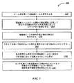
図3は、眼を画像化する多くの実施形態としての方法200の行為の単純化ブロック図である。例えば本明細書において説明された任意適当な装置、組立体、及び/又はシステムを用いて方法200を実施することができる。方法200は、ビーム源を用いて電磁線ビームを発生させるステップ(行為202)を含み得る。
FIG. 3 is a simplified block diagram of the acts of many embodiments of
方法200は、電磁線ビームをビーム源から眼の動き(眼球運動)に応答して変化する光路長を有する可変光路に沿ってスキャナまで伝搬させるステップ(行為204)を含み得る。行為200は、電磁線ビームを眼内の或る場所の焦点に集束させる(合焦させる)ステップ(行為206)を含み得る。方法200は、スキャナを用いて眼内の異なる場所に焦点をスキャンするステップ(行為208)を含み得る。方法200は、焦点場所から反射された電磁線ビームの一部分を可変光路に沿って伝搬させてセンサに戻すステップ(行為210)を含み得る。方法200は、センサを用いて、焦点場所から反射されて当該センサまで伝搬された電磁線ビームの一部分の強度を表す強度信号を発生させるステップ(行為212)を含み得る。
The
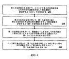
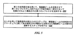
図4、図5及び図6は、方法200の一部として達成できるオプションとしての工程ないし行為の単純化ブロック図である。例えば、方法200は、第1の支持組立体を用いて、スキャナと第1の支持組立体との間の相対運動を許容して眼の動きを許容するようスキャナを支持するステップ(行為214)を含み得る。方法200は、第2の支持組立体を用いて、第1の支持組立体と第2の支持組立体の間の相対運動を許容して眼の動きを許容するよう第1の支持組立体を支持するステップ(行為216)を含み得る。方法200は、第1の支持組立体を用いて、電磁線ビームを反射して可変光路の一部分に沿ってスキャナまで伝搬させるよう構成された第1のレフレクタを支持するステップ(行為218)を含み得る。方法200は、ベース組立体を用いて、第2の支持組立体とベース組立体との間の相対運動を許容して眼の動きを許容するよう第2の支持組立体を支持するステップ(行為220)を含み得る。行為200は、第2の支持組立体を用いて、電磁線ビームを反射させて第1のレフレクタに入射するよう可変光路の一部分に沿って伝搬させるよう構成された第2のレフレクタを支持するステップ(行為222)を含み得る。方法200のセンサを用いて強度信号を発生させるステップは、電磁線ビームの反射部分をアパーチュアに通して焦点場所以外の場所から反射された電磁線ビームの部分を遮断するステップ(行為224)を含み得る。方法200は、電磁線ビームを偏光に敏感な装置に通すステップ(行為226)を含み得る。方法200は、電磁線ビーム及び焦点存在場所から反射された電磁線ビームの一部分のうちの少なくとも一方の偏光状態を改変するステップ(行為228)を含み得る。方法200は、偏光敏感性装置を用いて焦点存在場所から反射された電磁線ビームの一部分を反射させてセンサに入射させるステップ(行為230)を含み得る。
4, 5, and 6 are simplified block diagrams of optional steps or actions that can be accomplished as part of the
図7は、多くの実施形態におけるレーザ手術システム300を概略的に示している。レーザ手術システム300は、レーザ組立体12、共焦点検出組立体14、自由浮動機構体16、スキャン組立体18、対物レンズ組立体20、患者インターフェース22、通信経路302、制御エレクトロニクス304、制御パネル/グラフィカルユーザインターフェース(GUI)306、及び、ユーザインターフェース装置308を含む。制御エレクトロニクス304は、プロセッサ310を含み、プロセッサ310は、メモリ312を含む。患者インターフェース22は、患者24とインターフェースするよう構成されている。制御エレクトロニクス304は、通信経路302を介して、レーザ組立体12、共焦点検出組立体14、自由浮動機構体16、スキャン組立体18、制御パネル/GUI306、及びユーザインターフェース装置308に作動的に結合されている。
FIG. 7 schematically illustrates a laser
スキャン組立体18は、z‐スキャン装置及びxy‐スキャン装置を含み得る。レーザ手術システム300は、電磁線ビーム28を3次元内で(3つの寸法方向で)スキャンされる焦点に集束させ又は合焦させるよう構成されている。z‐スキャン装置は、ビーム28の伝搬方向において焦点の存在場所を変化させるよう作動可能である。xy‐スキャン装置は、ビーム28の伝搬方向を横切る2次元内で(2つの寸法方向で)焦点の配置場所をスキャンするよう作動可能である。したがって、z‐スキャン装置とxy‐スキャン装置の組み合わせは、患者24の組織内、例えば患者24の眼組織内、において3次元で(3つの寸法方向で)ビームの焦点を制御可能にスキャンするよう作動可能である。スキャン組立体18は、自由浮動機構体16によって支持され得て、それにより、3次元(3つの寸法方向)におけるレーザ組立体12及び共焦点検出組立体14に対するスキャン組立体18の患者の動きにより誘起される運動が許容され得る。
患者インターフェース22は、患者インターフェース22、対物レンズ組立体20、及びスキャン組立体18が患者24と関連して動く、というように患者24に結合されている。例えば、多くの実施形態では、患者インターフェース22は、患者24の眼に真空取り付けされた吸引リングを採用している。吸引リングは、例えば吸引リングを患者インターフェース22に固定する真空を用いて、患者インターフェース22に結合され得る。
制御エレクトロニクス304は、通信経路302を介して、レーザ組立体12、共焦点検出組立体14、自由浮動機構体16、スキャン組立体18、患者インターフェース22、制御パネル/GUI306、及びユーザインターフェース装置308の動作を制御すると共に/或いはこれらからの入力を受け取ることができる。通信経路302は、任意適当な形態で具体化でき、かかる形態としては、制御エレクトロニクス304とそれぞれのシステムコンポーネントとの間の任意適当な共有又は専用通信経路が挙げられる。
Control electronics 304 communicates
制御エレクトロニクス304は、任意適当なコンポーネント、例えば1つ又は2つ以上のプロセッサ、1つ又は2つ以上の書き換え可能ゲートアレイ(FPGA)、及び、1つ又は2つ以上の記憶装置、を含むことができる。多くの実施形態では、制御エレクトロニクス304は、ユーザにより特定される治療パラメータに従って手技前計画立案を可能にすると共にレーザ眼手術手技に対するユーザ管理を提供するべく、制御パネル/GUI306を制御する。
The control electronics 304 includes any suitable components such as one or more processors, one or more rewritable gate arrays (FPGAs), and one or more storage devices. Can do. In many embodiments, the control electronics 304 controls the control panel /
制御エレクトロニクス304は、システム動作に関連付けられた計算を実行すると共に制御信号を種々のシステム要素に提供するべく用いられるプロセッサ/コントローラ310を含み得る。コンピュータ可読媒体312がプロセッサ310に結合されており、その目的は、プロセッサ及び他のシステム要素によって用いられるデータを記憶することにある。プロセッサ310は、本明細書全体を通じて説明したように、システムの他のコンポーネントと相互作用する。一実施形態では、メモリ312は、レーザシステム手術システム300の1つ又は2つ以上のコンポーネントを制御するために利用できるルックアップテーブルを含み得る。
The control electronics 304 may include a processor / controller 310 that is used to perform calculations associated with system operation and to provide control signals to various system elements. A computer readable medium 312 is coupled to the processor 310, the purpose of which is to store data used by the processor and other system elements. The processor 310 interacts with other components of the system as described throughout this specification. In one embodiment, the memory 312 may include a look-up table that can be used to control one or more components of the laser system
プロセッサ310は、命令及びデータを実行するよう構成された汎用マイクロプロセッサ、例えば、カリフォルニア州サンタクララ所在のインテル・コーポレーション(Intel Corporation )により製造されたPentium プロセッサであり得る。プロセッサは、本発明の実施形態による方法をソフトウェア、ファームウェア、及び/又はハードウェアで実行する命令の少なくとも一部を具体化する特定用途向け集積回路(ASIC)であり得る。一例として、かかるプロセッサとしては、専用回路、ASIC、組み合わせ論理、他のプログラム可能プロセッサ、これらの任意の組み合わせ、等が挙げられる。 The processor 310 may be a general purpose microprocessor configured to execute instructions and data, such as a Pentium processor manufactured by Intel Corporation of Santa Clara, California. The processor may be an application specific integrated circuit (ASIC) that embodies at least a portion of instructions that perform the method according to embodiments of the present invention in software, firmware, and / or hardware. By way of example, such processors include dedicated circuits, ASICs, combinational logic, other programmable processors, any combination thereof, and the like.
メモリ312は、該当する場合、特定の用途に対してローカルであっても良く又は分散されていても良い。メモリ312は、プログラム実行中に命令及びデータを記憶するための主読取り書込み記憶装置(RAM)及び一定の命令が記憶された読取り専用記憶装置(ROM)を含む多くのメモリを含み得る。かくして、メモリ312は、プログラム及びデータファイルのためのパーシステント(不揮発性)記憶装置をもたらし、メモリは、関連リムーバブルメディアと一緒のハードディスクドライブ、フラッシュメモリ、フロッピディスクドライブ、コンパクトディスク読取り専用記憶装置(CD‐ROM)ドライブ、光学ドライブ、リムーバブルメディアカートリッジ、及び、他の同様な記憶媒体、を含み得る。 Memory 312 may be local to a particular application or distributed, as applicable. The memory 312 may include a number of memories including a main read / write storage device (RAM) for storing instructions and data during program execution and a read only storage device (ROM) in which certain instructions are stored. Thus, the memory 312 provides persistent storage for programs and data files, and the memory can be a hard disk drive, flash memory, floppy disk drive, compact disk read-only storage (with associated removable media). CD-ROM) drives, optical drives, removable media cartridges, and other similar storage media.
ユーザインターフェース装置308は、ユーザ入力を制御エレクトロニクス304に提供するのに適した任意適当なユーザ入力装置を含み得る。例えば、ユーザインターフェース装置308は、例えばタッチスクリーンディスプレイ/入力装置、キーボード、フットスイッチ、キーパッド、患者インターフェース無線周波識別(RFID)リーダ、非常時停止ボタン、及び、キースイッチ、のような装置を含み得る。 User interface device 308 may include any suitable user input device suitable for providing user input to control electronics 304. For example, the user interface device 308 includes devices such as touch screen display / input devices, keyboards, foot switches, keypads, patient interface radio frequency identification (RFID) readers, emergency stop buttons, and key switches. obtain.
焦点スキャン制御Focus scan control
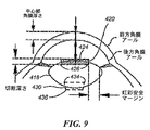
レーザ手術システム10は、特定の領域内で電磁線ビームの焦点をスキャンすることによって眼内標的を画像化する、及び/または、改変するよう構成され得る。例えば、図8及び図9を参照すると、レーザ手術システム10は、水晶体曩418の前方部分内に、前方水晶体曩切開術創及び/又は後方水晶体曩切開術創を形成するために使用できる。電磁線ビームの焦点をスキャンすると、水晶体曩418の前方部分を横切する、前方水晶体曩切開術による閉鎖された切開創境界面420を形成することができる。同様に、電磁線ビームの焦点をスキャンすると、水晶体曩418の後方部分を横切する後方水晶体曩切開術による閉鎖された切開創境界面430を形成することができる。
The laser
前方及び/または後方閉鎖切開創境界面420,430は、任意適当な方式を用いて設計できる。例えば、カメラ62を用いて患者の眼の平面図を得ることができる。水晶体曩切開術創指示子422が、患者の眼に対する計画された水晶体曩切開術創の寸法、場所、及び形状を示すために、患者の眼の平面図上に重ね合わされた状態で配置され示されるのが良い。水晶体曩切開術創指示子422は、レーザ手術システム10のオペレータによって手動で定めることができるのが良く、及び/または、レーザ手術システム10は、オペレータによる確認及び/又は変更のための初期水晶体曩切開術創指示子422を生成するよう構成されるのが良い。
The anterior and / or posterior closed incision interfaces 420, 430 can be designed using any suitable scheme. For example, a plan view of the patient's eye can be obtained using the
前方水晶体曩切開術(による)閉鎖切開創境界面420は、水晶体曩切開術創指示子422の投影像上に定めるのが良い。その結果、水晶体曩切開術創指示子422の投影像に対する水晶体曩418の前方部分の存在場所の予想されるばらつきにもかかわらず、水晶体曩切開術(による)閉鎖切開創境界面420は、当該前方水晶体曩切開術(による)切開創境界面420の周りの全ての場所で水晶体曩418の前方部分を横切する。例えば、水晶体曩切開術創指示子422に対応した曲線を投影させて水晶体曩418の前方部分に関する最小予想深さ形態を定める最小深さ数学的表面モデル(例えば、球面)との交線を定めることができる。結果として得られる交線は、前方水晶体曩切開術(による)閉鎖切開創境界面420のための上側境界を定める前方水晶体曩切開術創の上側閉鎖曲線424である。同様に、水晶体曩切開術創指示子422に対応した曲線を投影させて水晶体曩418の前方部分に関する最大予想深さ形態を定める最大深さ数学的表面モデル(例えば、球面)との交線を定めることができる。結果として得られる交線は、前方水晶体曩切開術(による)閉鎖切開創境界面420のための下側境界を定める前方水晶体曩切開術創の下側閉鎖曲線426である。変形例として、画像化だけの低いパワーレベル(例えば、眼内標的を改変すること無く共焦点検出組立体14の検出センサ50によって出力される信号の処理により眼内標的の画像化を行うのに十分なパワーレベル)を用いて焦点を水晶体曩切開術創指示子422の投影像に沿ってスキャンし、焦点の深さを変化させて水晶体曩切開術創指示子422の投影像の周りの十分な数の場所で前水晶体曩の深さを求めても良い。例えば図10は、焦点の深さの変化につれて生じる検出センサ54によって出力された信号の強度の変化を示しており、強度の最大ピークは、水晶体の前方部分の深さに対応している。そして、前水晶体の測定深さを用いると、前方水晶体曩切開術による閉鎖された切開創境界面420の適当な前方水晶体曩切開術創の上側及び下側曲線424,426を求めることができる。
An anterior lens fistula incisional
同様な仕方で、後方水晶体曩切開術(による)閉鎖切開創境界面430は、水晶体曩切開術創指示子422の投影像上に定めるのが良い。その結果、水晶体曩切開術創指示子422の投影像に対する水晶体曩418の後方部分の存在場所の予想されるばらつきにもかかわらず、後方水晶体曩切開術(による)閉鎖切開創境界面430は、当該後方水晶体曩切開術(による)切開創境界面430の周りの全ての場所で水晶体曩418の後方部分を横切する。例えば、水晶体曩切開術創指示子422に対応した曲線を投影させて水晶体曩418の後方部分に関する最小予想深さ形態を定める最小深さ数学的表面モデル(例えば、球面)との交線を定めることができる。結果として得られる交線は、後方水晶体曩切開術(による)閉鎖切開創境界面430のための上側境界を定める後方水晶体曩切開術創の上側閉鎖曲線434である。同様に、水晶体曩切開術創指示子422に対応した曲線を投影させて水晶体曩418の後方部分に関する最大予想深さ形態を定める最大深さ数学的表面モデル(例えば、球面)との交線を定めることができる。結果として得られる交線は、後方水晶体曩切開術(による)閉鎖切開創境界面430のための下側境界を定める後方水晶体曩切開術創の下側閉鎖曲線436である。変形例として、画像化だけの低いパワーレベル(例えば、眼内標的を改変すること無く共焦点検出組立体14の検出センサ54によって出力される信号の処理により眼内標的の画像化を行うのに十分なパワーレベル)を用いて焦点を水晶体曩切開術創指示子422の投影像に沿ってスキャンし、焦点の深さを変化させて水晶体曩切開術創指示子422の投影像の周りの十分な数の場所で後水晶体曩の深さを求めても良い。そして、後水晶体曩の測定深さを用いると、後方水晶体曩切開術による閉鎖された切開創境界面430の適当な後方水晶体曩切開術創の上側及び下側曲線434,436を求めることができる。
In a similar manner, the posterior
水晶体曩切開術創指示子422の任意適当な投影像を用いて前方及び/又は後方水晶体曩切開術切開創境界面420,430を定めることができるが、多くの実施形態では、水晶体曩切開術創指示子422の逆円錐型の投影像が、対物レンズ組立体20から焦点まで伝搬しながら焦点に集束する電磁線ビームと虹彩の縁との間の適当な安全マージン距離を維持するように、用いられる。したがって、多くの実施形態では、後方水晶体曩切開術は、例えば図示のように水晶体曩切開術創指示子422が与えられた場合、対応の前方水晶体曩切開術よりも小さい直径を有する。
Although any suitable projection image of the
レーザ手術システム10は、任意適当に形作られる水晶体曩切開術創を形成するよう使用できる。例えば、前方及び後方水晶体曩切開術創は、図示の実施形態では円形であるが、任意の他の適当な形状を形成していても良く、かかる形状としては、楕円形、長方形、及び多角形が挙げられるが、これらには限定されない。さらに、前方及び/又は後方水晶体曩切開術創は、これに対応して任意適当に形作られる眼内レンズ(IOL)を受け入れるよう形作られ得る。
The laser
同時の画像化及び適応組織治療Simultaneous imaging and adaptive tissue treatment
レーザ手術システム10は、組織治療と同時に画像データを生成させるよう構成され得る。例えば、電磁線ビームの焦点は、眼内標的(例えば、眼組織、IOL)を改変するのに十分な強度を有するのが良く、焦点から反射されて共焦点検出組立体14の検出センサ54に戻される電磁線ビームの結果的に生じる一部分が、焦点存在場所に対応する画像データを生成するべく処理される信号を発生させために用いられるのが良い。
The laser
眼内標的の境界と交差するパターンで焦点をスキャンすることによって、検出センサ54は、それと同時に、交差境界の存在場所を突き止めるよう処理可能な信号を発生させるために用いられ得る。例えば、図10は、焦点の深さの変化につれて生じる検出センサ54によって出力される信号の強度の変化を示しており、強度の最大ピークは、水晶体の前方部分の深さに対応している。交差境界の存在場所を用いると、治療される組織の量を減少させるよう、焦点の次のスキャンを制御することができる。例えば、前方水晶体曩切開術創を水晶体曩に形成する際、焦点は、先行するスキャンパターン中に生成された検出センサ54からの信号を処理することによって求められる水晶体の前方部分の存在場所に少なくとも部分的に基づくスキャンパターンで、スキャンし得る。
By scanning the focus in a pattern that intersects the boundary of the intraocular target, the
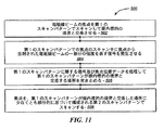
図11は、多くの実施形態に従って眼内標的の境界に対して電磁線ビームの焦点を適応可能にスキャンする方法500の行為の単純化ブロック図である。方法500は、例えば、本明細書において説明されている任意適当なレーザ手術システム、例えばレーザ手術システム10、を含む任意適当なシステムを用いて達成できる。
FIG. 11 is a simplified block diagram of the acts of a
方法500は、電磁線ビームの焦点を第1のスキャンパターンでスキャンして眼内標的の境界と交差させるステップ(行為502)を含む。多くの実施形態では、スキャンパターンは、焦点を、電磁線ビームの伝搬方向に対して横方向に且つ/或いはこれに平行に動かす。交差境界を有する眼内標的は、任意適当な眼内標的であり得て、かかる標的としては、例えば、前方水晶体曩、後方水晶体曩、水晶体、角膜、虹彩、眼内レンズ、及び角膜縁(limbus)が挙げられる。複数のスキャンパターンを適用して切開創表面(例えば、図8及び図9に示されている閉鎖切開創境界面420)を作る場合、スキャンパターンは、改変されていない眼組織及び/又はIOL材料を通って焦点まで電磁線ビームが伝搬するよう構成され得る。例えば、スキャンパターンは、改変が全体として深い方から浅い方へ起こるよう、構成され達成されるのが良い(例えば、後方から前方へ)。
The
方法500は、第1のスキャンパターンでの焦点のスキャン中に焦点から反射された電磁線ビームの一部分の強度を表す信号を発生させるステップ(行為504)を更に含む。例えば、第1のスキャンパターンは眼内標的の境界と交差するので、検出センサにより出力された信号(例えば、図10に示されている信号)及び第1のスキャンパターンに関する焦点位置データは、例えば該当する境界と一致した信号変化を識別することによって、交差境界の存在場所を突き止める(決定する)よう処理され得る(行為506)。
第1のスキャンパターンが眼内標的の境界と交差した場所を突き止めたら、焦点を、第1のスキャンパターンが眼内標的の境界と交差した場所に少なくとも部分的に基づいて構成される第2のスキャンパターンでスキャンするのが良い(行為508)。例えば、第2のスキャンパターンは、当該第2のスキャンパターンが眼内標的の境界と交差するであろうと予想される場所を越えて、第1のスキャンパターンが眼内標的の境界と交差した場所の知見を考慮して第2のスキャンパターンが境界と交差するであろう推定場所の考えられるばらつきを考慮に入れて選択される所定の量だけ延びるよう、構成されるのが良い。多くの実施形態では、第2のスキャンパターンは、第1のスキャンパターンとオーバーラップしていない場合にこれにすぐ隣接して位置することになり、それにより第1のスキャンパターンが境界と交差した計測された場所と第2のスキャンパターンが境界と交差するであろう推定場所との間の考えられるばらつきは減少する。切開創表面が作られる多くの実施形態では、一連の次のスキャンパターンを達成することができ、かかる次のスキャンパターンでは、1つ又は2つ以上の先行するスキャンパターンが眼内レンズの境界と交差した場所を用いて次のスキャンパターンのうちの少なくとも1つを構成し、それにより例えば、改変される組織及び/又は物体を最小限に抑えると共に/或いは改変される組織及び/又は物体がどれかに関する精度を高めることができる。 Once the location where the first scan pattern intersects the boundary of the intraocular target is determined, the second is configured based at least in part on the location where the first scan pattern intersects the boundary of the intraocular target. It is better to scan with a scan pattern (act 508). For example, the second scan pattern is where the first scan pattern crosses the intraocular target boundary beyond where it is expected that the second scan pattern will cross the intraocular target boundary. The second scan pattern may be configured to extend by a predetermined amount selected taking into account possible variations in the estimated location where the second scan pattern will intersect the boundary. In many embodiments, the second scan pattern will be located immediately adjacent if it does not overlap the first scan pattern, thereby causing the first scan pattern to intersect the boundary. Possible variability between the measured location and the estimated location where the second scan pattern will intersect the boundary is reduced. In many embodiments where an incision surface is created, a series of subsequent scan patterns can be achieved, in which one or more previous scan patterns are associated with the intraocular lens boundary. The intersected location is used to construct at least one of the following scan patterns, thereby minimizing and / or modifying tissue and / or objects to be modified, for example It is possible to increase the accuracy with respect to.
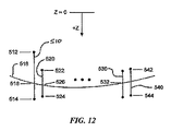
図12は、次のスキャンパターンを構成するための、焦点に関するスキャンパターンが眼内標的の境界と交差した場所の使用の繰り返しの仕方を概略的に示している。図12は、z寸法方向(即ち、電磁線ビームの伝搬方向に平行な方向)に対する焦点の存在場所の変化を有するスキャンパターンを用いているが、図示の技術的思想は、例えば電磁線ビームの伝搬方向に対して横の方向並びにこれに対して横の且つ平行な方向に対する焦点の存在場所の変化(例えば、x方向の変化、y方向の変化、及び/又はz方向の変化)を有するような、任意適当なスキャンパターンに適応可能である。最初のスキャンパターン510は、境界516の存在場所の予想されるばらつきにもかかわらず当該最初のスキャンパターン510が眼内標的の境界516と交差するよう選択される2つの場所512,514相互間に延びるよう、構成されるのが良い。最初のスキャンパターン510中に検出センサ54により出力される信号を最初のスキャンパターン510に関する焦点存在場所データと共に処理することによって、最初のスキャンパターン510が境界516と交差する場所518を識別し又は突き止めることができる。
FIG. 12 schematically shows how to repeat the use of the location where the focus scan pattern intersects the boundary of the intraocular target to construct the next scan pattern. FIG. 12 uses a scan pattern having a change in the location of the focal point with respect to the z-dimension direction (that is, the direction parallel to the propagation direction of the electromagnetic beam). To have a change in the location of the focal point in a direction transverse to the direction of propagation and in a direction parallel to and parallel to it (eg, a change in the x direction, a change in the y direction, and / or a change in the z direction). Any suitable scan pattern can be applied. The
この場合、第2のスキャンパターン520が少なくとも部分的に当該場所518に基づいて構成されるのが良い。例えば、第2のスキャンパターン520に関する端点522,524は、例えば第2のスキャンパターンの長さを実質的に最小限に抑えて処置される組織及び/又は物体の量を最小限に抑えるよう、当該場所518に基づいて選択されるのが良い。第2のスキャンパターン520中に検出センサ54により出力される信号を第2のスキャンパターン520に関する焦点存在場所データと共に処理することによって、第2のスキャンパターン520が境界516と交差する場所526を識別し又は突き止めることができる。
In this case, the
任意適当な次のスキャンパターンを、同様な仕方で構成することができる。例えば、スキャンパターン530中に検出センサにより出力される信号を当該スキャンパターン530に関する焦点存在場所データと共に処理することによって、スキャンパターン530が境界516と交差する場所532を識別し又は突き止めることができる。次のスキャンパターン540に関する端点542,544は、例えばスキャンパターン540の長さを実質的に最小限に抑えて処置される組織及び/又は物体の量を最小限に抑えるよう、当該場所532に基づいて選択されるのが良い。したがって、1つ又は2つ以上の先行するスキャンパターンから生成された眼内標的に関する境界場所データを利用して、一連のスキャンパターンを適応可能に構成すると共に適用することができる。
Any suitable next scan pattern can be constructed in a similar manner. For example, by processing the signal output by the detection sensor during
図13は、眼内標的の境界552を横切する表面を切開するために用いることができる一連のスキャンパターン550を示す略図である。図示の実施形態では、スキャンパターン550は、例えば図12及び方法500を参照して上述したような、一連のスキャンパターン550のうちの1つ又は2つ以上の先行するスキャンパターンから生成された境界場所データを用いて、適応可能に構成されている。したがって、一連のスキャンパターン550は、一般に、境界552の両側を越えて実質的に一定の距離だけ延びていて、それにより境界552の全体形状を辿るよう構成され得る。
FIG. 13 is a schematic diagram illustrating a series of
図14及び図15は、一連のスキャンパターン550を切開するために用いることができるスキャン方向554,556を示している。任意適当なスキャン方向を用いることができるが、図示の方向554,556は、焦点に達する前に先に処置された組織/物体を通って電磁線ビームが伝搬してしまうことを回避するために、使用され得る。
14 and 15 show
幾つかの実施形態では、水晶体曩切開術のスキャンは、不完全な切開の可能性を低減するべくスキャンパターンがz深さの次元において実質的に水晶体曩より長いために、理想的であるよりも実施時間がより長くなり得る。例えば、幾つかの実施形態では、スキャンパターンは、眼の水晶体曩の切開を確実にするべく、300μmの重複を提供し得る。更に、幾つかの実施形態では、レーザスポットがより小さいことにより、連続または略連続の切開を提供するためにより長い時間を要求し得る。従って、幾つかの方法及びシステムは、1回または数回のスキャンを用いて切開を完了するべく切開中に電磁線ビームの焦点を水晶体曩の縁に維持することによって、水晶体曩切開術の切開をするための時間を低減し得る。 In some embodiments, a lens fistula scan is more than ideal because the scan pattern is substantially longer than the lens fistula in the z-depth dimension to reduce the possibility of incomplete incisions. Also, the implementation time can be longer. For example, in some embodiments, the scan pattern may provide a 300 μm overlap to ensure incision of the eye lens fistula. Further, in some embodiments, the smaller laser spot may require longer times to provide a continuous or nearly continuous incision. Thus, some methods and systems use a single or several scans to maintain the focus of the electromagnetic beam at the edge of the lens fistula during the incision to complete the incision, thereby dissecting the lens fistula. The time to do this can be reduced.
図16は、水晶体曩を切開しつつ重複を減らすように電磁線ビームの焦点のスキャンを制御する例示的な方法100を図示している。ステップ102で、電磁線ビームの焦点を整列させるために画像化プレスキャンが実施され得る。ステップ104で、水晶体表面の位置が特定され得る。ステップ106で、電磁線ビームは、特定された位置で焦点に合焦され得る。ステップ108で、ステップ106に応じて、電磁線(電磁放射線)が焦点から反射され得る。反射された電磁線は、ステップ108で測定され得て、信号強度を生成する。測定された信号強度は、ソフトウェア内の予測アルゴリズムブロックに供給され得る。予測ブロック110は、3つの条件の1つを特定して、適切なコマンドを発行する。測定された信号強度が、上方閾値より上方にある時、予測器110は、次の(引き続いての)電磁線焦点の深さを低減するコマンド112を発行する。測定された信号強度が、下方閾値より下方にある時、予測器110は、次の(引き続いての)電磁線焦点の深さを増大するコマンド112を発行する。測定された信号強度が、下方閾値より上方であって上方閾値より下方にある時、予測器110は、次の(引き続いての)電磁線焦点の深さを維持するコマンド112を発行する。その後、方法100はステップ118に進み、電磁線ビームは次の焦点に合焦される。ステップ118の後、方法100はステップ108に戻り得て、焦点からの電磁線の信号強度が測定される。ステップ108乃至118は、計画された水晶体曩切開術が完了されるまで、繰り返され得る。
FIG. 16 illustrates an
方法100は、水晶体曩切開術に関して説明されているが、当該方法は、他の眼内標的の切開に適用可能であり得る。これらの例において、他の標的組織は、角膜の上皮、ボウマン層、基質内層、内皮のような、角膜表面や角膜内参照面であり得る。
Although the
方法100は、前述のシステム及び装置によって実施され得る。例えば、ステップ102及び104は、共焦点画像(化)を用いて実施され得る。前述の通り、画像化と切開とは、同一の電磁線ビームを用いて同時に実施され得る。従って、そのようなシステムは、治療ビームが水晶体曩を切開する時、水晶体曩表面上に焦点深さを調整するべく利用され得る。幾つかの実施形態では、水晶体曩切開術は、1回のスキャンだけで達成され得る。他の実施形態では、切開は、2回以上のスキャンで実施される。これは、300μmの重複にとって好適であり得る。なぜなら、水晶体曩切開術は、1回または数回のスキャンでずっと高速に実施され得て、スキャン中またはスキャンの間、治療システムに対する眼や水晶体の不注意による運動の可能性を低減し得るからである。更に、2〜3回程度の治療ビームのスキャンによれば、電磁線ビームの焦点をスキャンすることによって眼に供給されるエネルギーも、低減され得る。
プレスキャン102は、レーザを水晶体曩と整列させるために、組織の切開に先立って水晶体の評価位置を提供し得る。電磁線ビームの焦点は、画像化用のみの低い電力レベルを用いてスキャンされ得る(例えば、眼内標的を改質することなく、眼内標的の画像化を提供するのに十分な電力レベル)。それは、水晶体曩切開術の切開設計器422のような眼内切開設計器の投影部に沿って、スキャンされ得る。焦点の深さは、前水晶体曩104の深さを決定するために、変更され得る。一方で、反射された信号の強度は、図10で視認できるように、約30μmのzスキャン距離に亘って、略ゼロからピーク値へと上昇する。当該ピーク値が、前水晶体の深さに対応し得る。したがって、水晶体曩の表面上にロックすることは、幾らか挑戦的であり得る。
The pre-scan 102 may provide an evaluation position of the lens prior to tissue incision to align the laser with the lens capsule. The focus of the electromagnetic beam can be scanned using a low power level for imaging only (eg, a power level sufficient to provide imaging of the intraocular target without modifying the intraocular target). . It can be scanned along the projection of an intraocular incision designer, such as the lens
工程106で、治療ビーム(例えば、標的改質及び/または切開を提供するのに十分な電力レベルのビーム)が、水晶体曩の特定された前方面で焦点に合焦される。幾つかの実施形態では、単一の治療ビームパルスが当該位置に供給される。あるいは、複数のパルス(例えば100パルス)が、調整に先立って、各深さに発射され得る。これは、各パルスの調整がzスキャン装置にとって挑戦的であり得るという場合に好適であり得る。幾つかの実施形態では、単一のパルスのサイズは、5μm長であって、約1μm幅であり得る。更に、幾つかの実施形態では、除去のために水晶体曩を十分弱めるべく水晶体曩を完全に貫いて切断することは必要ないかもしれない。
At
水晶体曩は、手術中に僅かに移動する可能性があるので、ステップ108と評価/予測ブロック110が用いられ得て、治療ビームの焦点が水晶体曩周りでスキャンされる時、ほぼリアルタイムでz深さへの修正がなされる。これらのステップは、水晶体曩のバッグ(包)に対して焦点を正確に位置決めすることを確実にするのを助け得る。ステップ108で、治療ビームの焦点から反射される電磁線の強度信号が測定される。反射される電磁線の強度信号に基づいて、評価/予測ブロック110が、必要に応じて、焦点位置への調整を行い得る。図10に図示されているように、ピーク強度値が、水晶体曩の後方に位置する焦点に対応し得る。対照的に、低い強度値は、水晶体曩の前方に位置するか眼の房水に位置する焦点に対応し得る。水晶体曩は、ピーク強度値と、眼の房水に位置する焦点の強度値と、の間の強度値を有する。従って、上方閾値と下方閾値とを有する強度範囲が、眼の水晶体曩の位置を特定するために規定され得る。上方閾値よりも上方であると測定される反射電磁線の強度信号は、治療レーザの焦点が眼の水晶体内であって水晶体曩の後方に位置していた、ということを示唆し得る。従って、治療ビームの次の(引き続いての)焦点の深さは、低減されるべきである。下方閾値よりも下方であると測定される反射電磁線の強度信号は、治療レーザの焦点が房水内であって水晶体曩の前方に位置していた、ということを示唆し得る。従って、治療ビームの次の(引き続いての)焦点の深さは、増大されるべきである。測定される反射電磁線の強度信号が下方閾値よりも上方であって上方閾値よりも下方である時、それは、治療レーザの焦点が眼の水晶体曩上か水晶体曩の十分近くに位置していた、ということを示唆し得る。従って、治療ビームの引き続いての焦点の深さは、維持され得る。
Since the lens fistula may move slightly during the surgery, step 108 and evaluation /
図17は、水晶体402、水晶体曩404、及び、房水406、に対して位置特定される複数の焦点プロフィル400を示している。図示の例では、焦点プロフィル410からの反射電磁線の強度信号は、下方閾値及び上方閾値の間であり得て、水晶体曩404に十分近い焦点を示し得る。従って、何らの深さ調整も、次の焦点のために必要でない。焦点プロフィル420からの反射電磁線の強度信号は、上方閾値よりも上方であり得て、大き過ぎる深さであって水晶体402内にある焦点を示し得る。従って、評価器/予測器110が、次の焦点の深さを低減し得る。更に、焦点プロフィル430からの反射電磁線の強度信号は、下方閾値よりも下方であり得て、不十分な深さであって房水406内にある焦点を示し得る。従って、評価器/予測器110が、次の焦点の深さを増大し得る。
FIG. 17 shows a plurality of focus profiles 400 that are located relative to the
前述のように、幾つかの実施形態では、複数のパルスが、次のパルスの深さを測定及び修正するのに先立って、単一の深さに発射され得る。幾つかの実施形態では、例えば、2〜200パルスが、測定ステップ108と評価器/予測器110による修正の前に、ある深さに発射され得る。更に、幾つかの実施形態では、z深さが、ディザー処理され得て、切開を少なくとも部分的に水晶体曩の内部に配置するための適切な深さを示すべく、強度信号の位相情報を提供し得る。そのような実施形態では、改質された組織を通ってビームを伝送することを回避するべく、z深さが前方位置から後方位置へとディザー処理され得る。更に、幾つかの実施形態では、切開の開始時に、治療ビームが規定された水晶体曩切開術の内方へと始動され得る。初期の切開は、組織を膨張ないし変形させ得て、水晶体曩はアライメントから外れ得る。従って、幾つかの実施形態では、治療ビームは、水晶体内で合焦され得て、焦点深さは、焦点が水晶体曩上か水晶体曩の実質的な近くに位置決めされるまで、低減され得る。その後、焦点は、計画された水晶体曩切開術の投影像に沿ってスキャンされ得て、深さは、前述の方法によって調整され得る。
As described above, in some embodiments, multiple pulses may be fired to a single depth prior to measuring and modifying the depth of the next pulse. In some embodiments, for example, 2 to 200 pulses may be fired to a depth prior to correction by measurement step 108 and evaluator /
図18は、露出された(unwrapped)眼の例示的なスキャン600を示している。スキャン600は、水晶体602、水晶体曩604及び房水606を含み得る。水晶体曩604は、約8μmの厚みであり得る。破線610は、幾つかの切開に利用され得る300μmの許容範囲を示している。そのような切開は、大きな重複のため、より長い時間がかかるかもしれない。切開620は、前述の方法の1つ、例えば方法100、によって実施される切開を図示している。幾つかの実施形態では、切開620は、各パルス間の深さ調整を伴って実施され得る。他の実施形態では、切開620は、複数のパルス間での深さ調整を伴って実施され得る。幾つかの実施形態では、切開620は、z方向にディザー処理されたパルスを含み得る。理解され得るように、このようなスキャン620は、計画された水晶体曩切開術に沿って、1回または数回のスキャンで実施され得る。
FIG. 18 shows an
本発明の説明との関連において英文明細書における用語“a”、“an”、“the”及び類似の用語の使用は、本明細書において別段の指定がなければ又は文脈上明白に矛盾しなければ、単数と複数の両方を含むものと解されるべきである。英文明細書における用語“comprising”(「〜を有する」と訳されている場合が多い)、“having”(「〜を有する」と訳されている場合が多い)、“including”(「〜を含む」と訳されている場合が多い)及び“containing”(「〜を含む」と訳されている場合が多い)は、別段の規定がなければ、非限定的な用語(即ち、“including, but not limited to,”(「〜を含むが、〜には限定されない」)を意味している)として解されるべきである。用語“connected ”(「連結され」)は、何らかの介在が存在している場合であっても、部分的又は全体的に収納され、取り付けられ、又は互いに接合されると解されるべきである。本明細書における値の範囲についての記載は、別段の指定がなければ、その範囲に含まれる別々の各値を個別的に言及している手短な方法としての役目を果たすに過ぎず、別々の各値は、これが個別的に本明細書に記載されているかのように明細書に含まれる。本明細書において説明した方法は全て、別段の指定がなければ又は文脈上明確に矛盾しなければ、任意適当な順序で実施できる。本明細書において提供される任意の及び全ての実施例又は例示の用語(例えば、“such as ”(「例えば」と訳されている場合が多い))の使用は、本発明の実施形態を良好に示すに過ぎず、別段のクレーム請求がなければ、本発明の範囲に限定をもたらすものではない。明細書中、クレーム請求されていない何らかの要素を本発明の実施に必要不可欠なものとして指示するもの、と解されるべき用語は存在しない。 The use of the terms “a”, “an”, “the” and similar terms in the English specification in the context of the description of the present invention shall be clearly contradicted unless otherwise specified herein. Should be construed as including both singular and plural. The terms “comprising” (often translated as “having”), “having” (often translated as “having”), “including” (“ "Containing" and "containing" (often translated as "contains") are non-limiting terms (ie "including," unless otherwise specified). but not limited to, "(meaning" includes but is not limited to ")). The term “connected” should be understood to be partly or wholly contained, attached, or joined together, even in the presence of any intervention. The description of a range of values in this specification, unless otherwise specified, serves only as a short method of referring individually to each of the different values within that range. Each value is included in the specification as if it were individually described herein. All methods described herein can be performed in any suitable order unless otherwise indicated herein or otherwise clearly contradicted by context. The use of any and all examples or example terms provided herein (eg, “such as” (often translated as “for example”)) However, it is not intended to limit the scope of the present invention unless otherwise claimed. There is no term in the specification to be construed as indicating any non-claimed element as essential to the practice of the invention.
本発明の所定の図示の実施形態が、ある程度の特殊性(具体性)を伴う例示的な形態で示され説明されてきたが、かかる実施形態は、例示として提供されているに過ぎず、本発明の精神または範囲から逸脱することなく様々な変形がなされ得ることを、当業者は理解するであろう。従って、本発明は、特許請求の範囲の各請求項によって表現された本発明の精神及び範囲に属する、全ての改造例、代替構成例、変形例、置換例及び変更例、並びに、部分、構造及び工程の組み合わせ例や配置例、並びに、それらの均等例、を含むということが意図されている。 While certain illustrated embodiments of the invention have been shown and described in exemplary form with a certain degree of specificity, such embodiments are provided by way of example only, and Those skilled in the art will appreciate that various modifications can be made without departing from the spirit or scope of the invention. Accordingly, this invention includes all modifications, alternative constructions, variations, substitutions, and modifications, and parts, structures, which fall within the spirit and scope of the invention as expressed by the claims. In addition, it is intended to include combination examples and arrangement examples of processes, and equivalent examples thereof.
Claims (30)
治療ビームを前記眼内の第1位置で焦点に合焦させて、当該第1位置の組織を治療的に変造させるステップと、
前記治療ビームに応じて前記第1位置から反射される電磁線の強度信号を測定するステップと、
前記第1位置から反射される前記電磁線の前記測定された強度信号を用いて、前記眼内の第2位置を特定するステップと、
前記第2位置に向けて前記焦点をスキャンして、当該第2位置の組織を前記治療ビームで変造させるステップと、
を備えたことを特徴とする方法。 A method for modifying an intraocular target of an eye, comprising:
Focusing a treatment beam at a first position in the eye to therapeutically alter tissue at the first position;
Measuring an intensity signal of electromagnetic radiation reflected from the first position in response to the treatment beam;
Identifying a second position in the eye using the measured intensity signal of the electromagnetic radiation reflected from the first position;
Scanning the focal point toward the second position to alter the tissue at the second position with the treatment beam;
A method characterized by comprising:
前記水晶体曩は、前記眼の水晶体を取り囲んでおり、
前記方法は、更に、
前記眼内で画像化ビームの焦点をスキャンするステップと、
前記画像化ビームに応じて焦点位置から反射される電磁線の強度信号を測定して前記眼の水晶体曩の位置を特定するステップと、
前記治療ビームを前記水晶体曩と整列させるステップと、
を備えたことを特徴とする請求項1に記載の方法。 The intraocular target is the lens fistula of the eye;
The lens barrel surrounds the lens of the eye;
The method further comprises:
Scanning the focus of the imaging beam within the eye;
Measuring the intensity signal of electromagnetic radiation reflected from a focal position in accordance with the imaging beam to determine the position of the eye lens eyelid;
Aligning the treatment beam with the lens capsule;
The method of claim 1, comprising:
を更に備えたことを特徴とする請求項2に記載の方法。 The method of claim 2, further comprising generating the treatment beam and the imaging beam with a single electromagnetic beam source.
ことを特徴とする請求項1に記載の方法。 The method of claim 1, wherein the intensity signal is measured by a confocal detector.
を更に備えたことを特徴とする請求項1に記載の方法。 The method of claim 1, further comprising scanning the focus of the treatment beam to a plurality of different locations within the eye.
ことを特徴とする請求項5に記載の方法。 The method of claim 5, wherein the depth of focus of the treatment beam is dithered.
ことを特徴とする請求項6に記載の方法。 The method of claim 6, wherein the focus is scanned from a rear depth to a front depth.
ことを特徴とする請求項6に記載の方法。 The method of claim 6, wherein the focus of the treatment beam is scanned in the xy direction.
ことを特徴とする請求項5に記載の方法。 6. The method of claim 5, wherein the second position in the eye is identified based in part on phase information of the measured intensity signal.
前記特定される第2位置は、前記第1位置から反射される前記電磁線の前記測定される強度信号が前記上方閾値よりも上方にある時、前記第1位置の深さよりも小さい深さを有し、
前記特定される第2位置は、前記第1位置から反射される前記電磁線の前記測定される強度信号が前記下方閾値よりも下方にある時、前記第1位置の深さよりも大きい深さを有する
ことを特徴とする請求項1に記載の方法。 The second position is identified by comparing the measured intensity signal against an upper threshold and a lower threshold;
The specified second position has a depth smaller than the depth of the first position when the measured intensity signal of the electromagnetic radiation reflected from the first position is above the upper threshold. Have
The specified second position has a depth greater than the depth of the first position when the measured intensity signal of the electromagnetic radiation reflected from the first position is below the lower threshold. The method of claim 1, comprising:
ことを特徴とする請求項10に記載の方法。 The specified second position is the first position when the measured intensity signal of the electromagnetic radiation reflected from the first position is above the lower threshold and below the upper threshold. The method according to claim 10, wherein the method has a depth equal to the position.
コンピュータプロセッサによる前記指令の実施は、当該プロセッサをして、以下のステップ、すなわち、
治療ビームを前記眼内の第1位置で焦点に合焦させるステップと、
前記治療ビームを合焦させるステップに応じて前記第1位置において前記治療ビームから反射される電磁線の強度信号を受容するステップと、
前記第1位置の前記焦点から反射される前記電磁線の前記受容された強度信号に部分的に基づくフィードバックループを用いて、前記焦点の第2位置を決定するステップと、
を実施させる
ことを特徴とする一時的でないコンピュータ可読の記憶媒体。 A non-transitory non-transitory computer readable storage medium having a set of computer executable instructions for modifying an intraocular target of an eye,
Implementation of the instructions by the computer processor causes the processor to perform the following steps:
Focusing a treatment beam at a first position in the eye;
Receiving an intensity signal of electromagnetic radiation reflected from the treatment beam at the first position in response to focusing the treatment beam;
Determining a second position of the focus using a feedback loop based in part on the received intensity signal of the electromagnetic radiation reflected from the focus at the first position;
A non-transitory computer-readable storage medium characterized in that
前記水晶体曩は、前記眼の水晶体を取り囲んでおり、
前記コンピュータプロセッサによる前記指令の実施は、当該プロセッサをして、更に、
前記眼内で画像化ビームの焦点をスキャンするステップと、
前記画像化ビームに応じて前記焦点位置から反射される電磁線の強度信号を受容して前記眼の水晶体曩の位置を特定するステップと、
前記治療ビームを前記水晶体曩と整列させるステップと、
を実施させる
ことを特徴とする請求項12に記載の一時的でないコンピュータ可読の記憶媒体。 The intraocular target is the lens fistula of the eye;
The lens barrel surrounds the lens of the eye;
The execution of the instructions by the computer processor is performed by the processor,
Scanning the focus of the imaging beam within the eye;
Receiving an intensity signal of electromagnetic radiation reflected from the focal position in response to the imaging beam to determine the position of the eyelid lens eyelid;
Aligning the treatment beam with the lens capsule;
The non-transitory computer-readable storage medium of claim 12, wherein:
単一の電磁線ビーム源によって、前記治療ビーム及び前記画像化ビームを生成するステップ
を実施させる
ことを特徴とする請求項13に記載の一時的でないコンピュータ可読の記憶媒体。 The execution of the instructions by the computer processor is performed by the processor,
The non-transitory computer readable storage medium of claim 13, wherein the step of generating the treatment beam and the imaging beam is performed by a single electromagnetic beam source.
ことを特徴とする請求項12に記載の一時的でないコンピュータ可読の記憶媒体。 The non-transitory computer readable storage medium of claim 12, wherein the received intensity signal is measured by a confocal detector.
前記眼内の複数の異なる位置へ前記治療ビームの焦点をスキャンするステップ
を実施させる
ことを特徴とする請求項12に記載の一時的でないコンピュータ可読の記憶媒体。 The execution of the instructions by the computer processor is performed by the processor,
13. The non-transitory computer readable storage medium of claim 12, causing the step of scanning the focus of the treatment beam to a plurality of different locations within the eye.
ことを特徴とする請求項16に記載の一時的でないコンピュータ可読の記憶媒体。 The non-transitory computer-readable storage medium of claim 16, wherein the depth of focus of the treatment beam is dithered.
ことを特徴とする請求項17に記載の一時的でないコンピュータ可読の記憶媒体。 The non-transitory computer readable storage medium of claim 17, wherein the focus is scanned from a rear depth to a front depth.
ことを特徴とする請求項17に記載の一時的でないコンピュータ可読の記憶媒体。 The non-transitory computer readable storage medium of claim 17, wherein the focus of the treatment beam is scanned in an xy direction across the propagation direction of the treatment beam.
ことを特徴とする請求項16に記載の一時的でないコンピュータ可読の記憶媒体。 The non-transitory computer readable storage medium of claim 16, wherein the second position in the eye is determined based in part on phase information of the intensity signal.
前記特定される第2位置は、前記第1位置から反射される前記電磁線の前記測定される強度信号が前記上方閾値よりも上方にある時、前記第1位置の深さよりも小さい深さを有し、
前記特定される第2位置は、前記第1位置から反射される前記電磁線の前記測定される強度信号が前記下方閾値よりも下方にある時、前記第1位置の深さよりも大きい深さを有する
ことを特徴とする請求項12に記載の一時的でないコンピュータ可読の記憶媒体。 The second position is determined by comparing the measured intensity signal against an upper threshold and a lower threshold;
The specified second position has a depth smaller than the depth of the first position when the measured intensity signal of the electromagnetic radiation reflected from the first position is above the upper threshold. Have
The specified second position has a depth greater than the depth of the first position when the measured intensity signal of the electromagnetic radiation reflected from the first position is below the lower threshold. The non-transitory computer-readable storage medium of claim 12, comprising:
ことを特徴とする請求項21に記載の一時的でないコンピュータ可読の記憶媒体。 The specified second position is the first position when the measured intensity signal of the electromagnetic radiation reflected from the first position is above the lower threshold and below the upper threshold. The non-transitory computer readable storage medium of claim 21, having a depth equal to the depth of the position.
治療ビームを生成するように構成された電磁ビーム源と、
前記治療ビームを焦点に合焦させ、当該焦点を前記眼内の複数の位置へスキャンするように構成された光学系と、
前記治療ビーム焦点の位置から反射される電磁線を受容して、当該反射された電磁線の強度信号を測定するように構成された検出器と、
前記光学系及び前記検出器に結合され、前記焦点位置から反射される前記電磁線の前記測定される強度信号に部分的に基づくフィードバックループを用いて前記治療ビームの前記焦点の引き続いての位置を特定するように構成された制御部と、
を備えたことを特徴とするシステム。 A system for modifying an intraocular target of an eye,
An electromagnetic beam source configured to generate a treatment beam;
An optical system configured to focus the treatment beam at a focal point and scan the focal point to a plurality of positions in the eye;
A detector configured to receive electromagnetic radiation reflected from the location of the treatment beam focus and measure an intensity signal of the reflected electromagnetic radiation;
A feedback loop coupled to the optical system and the detector and reflected in part from the measured intensity signal of the electromagnetic radiation reflected from the focal position is used to determine the subsequent position of the focal point of the treatment beam. A controller configured to identify;
A system characterized by comprising:
ことを特徴とする請求項23に記載のシステム。 24. The system of claim 23, wherein the detector comprises a confocal detector.
ことを特徴とする請求項23に記載のシステム。 The system of claim 23, wherein the depth of focus of the treatment beam is dithered by the controller.
ことを特徴とする請求項25に記載のシステム。 26. The system of claim 25, wherein the controller scans the focus of the treatment beam from a rear depth to a front depth.
ことを特徴とする請求項23に記載のシステム。 The system of claim 23, wherein the feedback loop is based in part on phase information of the measured intensity signal.
前記特定される引き続いての位置は、前記焦点位置から反射される前記電磁線の前記測定される強度信号が前記上方閾値よりも上方にある時、前記焦点位置の深さよりも小さい深さを有し、
前記特定される引き続いての位置は、前記焦点位置から反射される前記電磁線の前記測定される強度信号が前記下方閾値よりも下方にある時、前記焦点位置の深さよりも大きい深さを有する
ことを特徴とする請求項23に記載のシステム。 The controller identifies the subsequent position of the treatment beam by comparing the measured intensity signal against an upper threshold and a lower threshold;
The identified subsequent position has a depth less than the depth of the focal position when the measured intensity signal of the electromagnetic radiation reflected from the focal position is above the upper threshold. And
The identified subsequent position has a depth greater than the depth of the focal position when the measured intensity signal of the electromagnetic radiation reflected from the focal position is below the lower threshold. 24. The system of claim 23.
ことを特徴とする請求項23に記載のシステム。 The identified subsequent position is the focus position when the measured intensity signal of the electromagnetic radiation reflected from the focus position is above the lower threshold and below the upper threshold. 24. The system of claim 23, having a depth equal to the depth of the system.
前記光学系は、更に、前記画像化ビームを前記眼内の複数の位置に合焦してスキャンするように構成されている
ことを特徴とする請求項23に記載のシステム。 The electromagnetic beam source is further configured to generate an imaging beam;
The system of claim 23, wherein the optical system is further configured to focus and scan the imaging beam at a plurality of positions in the eye.
Applications Claiming Priority (5)
| Application Number | Priority Date | Filing Date | Title |
|---|---|---|---|
| US201461935478P | 2014-02-04 | 2014-02-04 | |
| US61/935,478 | 2014-02-04 | ||
| US201461950416P | 2014-03-10 | 2014-03-10 | |
| US61/950,416 | 2014-03-10 | ||
| PCT/US2015/014086 WO2015119888A1 (en) | 2014-02-04 | 2015-02-02 | Confocal detection to minimize capsulotomy overcut while dynamically running on the capsular surface |
Related Child Applications (1)
| Application Number | Title | Priority Date | Filing Date |
|---|---|---|---|
| JP2019185837A Division JP6830989B2 (en) | 2014-02-04 | 2019-10-09 | Systems for modifying intraocular targets, how the systems operate, and computer-readable storage media |
Publications (2)
| Publication Number | Publication Date |
|---|---|
| JP2017506944A true JP2017506944A (en) | 2017-03-16 |
| JP2017506944A5 JP2017506944A5 (en) | 2018-03-08 |
Family
ID=52464621
Family Applications (2)
| Application Number | Title | Priority Date | Filing Date |
|---|---|---|---|
| JP2016550187A Pending JP2017506944A (en) | 2014-02-04 | 2015-02-02 | Confocal detection for minimizing excessive capsular incision during dynamic running on the lens fistula surface |
| JP2019185837A Active JP6830989B2 (en) | 2014-02-04 | 2019-10-09 | Systems for modifying intraocular targets, how the systems operate, and computer-readable storage media |
Family Applications After (1)
| Application Number | Title | Priority Date | Filing Date |
|---|---|---|---|
| JP2019185837A Active JP6830989B2 (en) | 2014-02-04 | 2019-10-09 | Systems for modifying intraocular targets, how the systems operate, and computer-readable storage media |
Country Status (6)
| Country | Link |
|---|---|
| US (1) | US11406536B2 (en) |
| EP (1) | EP3102167B1 (en) |
| JP (2) | JP2017506944A (en) |
| AU (1) | AU2015214443B2 (en) |
| CA (1) | CA2938862C (en) |
| WO (1) | WO2015119888A1 (en) |
Families Citing this family (3)
| Publication number | Priority date | Publication date | Assignee | Title |
|---|---|---|---|---|
| DE102015219277A1 (en) | 2015-10-06 | 2017-04-06 | Schaeffler Technologies AG & Co. KG | Cage segment of a rolling bearing |
| WO2018049414A1 (en) * | 2016-09-12 | 2018-03-15 | Lensar, Inc. | Laser methods and systems for the aligned insertion of devices into a structure of the eye |
| EP4221650A1 (en) * | 2020-09-30 | 2023-08-09 | AMO Development, LLC | Re-treatable corneal lenticular incisions with re-treatment options using a femtosecond ophthalmic laser system |
Citations (4)
| Publication number | Priority date | Publication date | Assignee | Title |
|---|---|---|---|---|
| WO2012135073A2 (en) * | 2011-03-25 | 2012-10-04 | Board Of Trustees Of Michigan State University | Adaptive laser system for ophthalmic use |
| JP2013500086A (en) * | 2009-07-24 | 2013-01-07 | レンサー, インク. | System and method for performing a procedure using LADAR on an eye lens |
| JP2013516294A (en) * | 2010-01-08 | 2013-05-13 | オプティメディカ・コーポレイション | Eye tissue and artificial lens modification system |
| US20130274725A1 (en) * | 2010-12-23 | 2013-10-17 | Christian Rathjen | Device for processing material of a workpiece and method for calibrating such a device |
Family Cites Families (23)
| Publication number | Priority date | Publication date | Assignee | Title |
|---|---|---|---|---|
| US5984916A (en) | 1993-04-20 | 1999-11-16 | Lai; Shui T. | Ophthalmic surgical laser and method |
| US5743902A (en) | 1995-01-23 | 1998-04-28 | Coherent, Inc. | Hand-held laser scanner |
| US6454761B1 (en) | 1995-01-30 | 2002-09-24 | Philip D. Freedman | Laser surgery device and method |
| US5720894A (en) | 1996-01-11 | 1998-02-24 | The Regents Of The University Of California | Ultrashort pulse high repetition rate laser system for biological tissue processing |
| US7655002B2 (en) | 1996-03-21 | 2010-02-02 | Second Sight Laser Technologies, Inc. | Lenticular refractive surgery of presbyopia, other refractive errors, and cataract retardation |
| US6019472A (en) | 1997-05-12 | 2000-02-01 | Koester; Charles J. | Contact lens element for examination or treatment of ocular tissues |
| JP4708050B2 (en) | 2005-03-02 | 2011-06-22 | 株式会社ニデック | Cornea surgery device |
| US8262646B2 (en) | 2006-01-20 | 2012-09-11 | Lensar, Inc. | System and method for providing the shaped structural weakening of the human lens with a laser |
| US20110319875A1 (en) | 2007-01-19 | 2011-12-29 | Frieder Loesel | Apparatus and Method for Morphing a Three-Dimensional Target Surface into a Two-Dimensional Image for Use in Guiding a Laser Beam in Ocular Surgery |
| US20090012507A1 (en) * | 2007-03-13 | 2009-01-08 | William Culbertson | Method for patterned plasma-mediated modification of the crystalline lens |
| DE102007028042B3 (en) | 2007-06-14 | 2008-08-07 | Universität Zu Lübeck | Using laser to make bubbles or cavities in transparent materials by focused, non-linear pulse absorption, operates at specified wavelength and pulse duration with controlled, uniform intensity |
| US7717907B2 (en) | 2007-12-17 | 2010-05-18 | Technolas Perfect Vision Gmbh | Method for intrastromal refractive surgery |
| DE102008027358A1 (en) * | 2008-06-05 | 2009-12-10 | Carl Zeiss Meditec Ag | Ophthalmic laser system and operating procedures |
| US8852175B2 (en) * | 2008-11-21 | 2014-10-07 | Amo Development Llc | Apparatus, system and method for precision depth measurement |
| US8382745B2 (en) | 2009-07-24 | 2013-02-26 | Lensar, Inc. | Laser system and method for astigmatic corrections in association with cataract treatment |
| US20110184395A1 (en) * | 2009-12-23 | 2011-07-28 | Optimedica Corporation | Method for laser capsulotomy and lens conditioning |
| US9278028B2 (en) * | 2010-02-08 | 2016-03-08 | Optimedica Corporation | System and method for plasma-mediated modification of tissue |
| US8414564B2 (en) | 2010-02-18 | 2013-04-09 | Alcon Lensx, Inc. | Optical coherence tomographic system for ophthalmic surgery |
| US8845624B2 (en) | 2010-06-25 | 2014-09-30 | Alcon LexSx, Inc. | Adaptive patient interface |
| DE102011109058A1 (en) * | 2011-07-29 | 2013-01-31 | Carl Zeiss Meditec Ag | "Ophthalmic Laser Device and Method for the Prevention and Treatment of After-Star" |
| JP5997450B2 (en) * | 2012-02-08 | 2016-09-28 | キヤノン株式会社 | Aberration correction method and aberration correction apparatus |
| US9987165B2 (en) | 2012-11-02 | 2018-06-05 | Optimedica Corporation | Liquid optical interface for laser eye surgery system |
| CN105338931B (en) * | 2013-03-13 | 2018-08-03 | 光学医疗公司 | laser eye surgery system |
-
2015
- 2015-02-02 CA CA2938862A patent/CA2938862C/en active Active
- 2015-02-02 JP JP2016550187A patent/JP2017506944A/en active Pending
- 2015-02-02 AU AU2015214443A patent/AU2015214443B2/en not_active Ceased
- 2015-02-02 EP EP15703716.9A patent/EP3102167B1/en active Active
- 2015-02-02 WO PCT/US2015/014086 patent/WO2015119888A1/en active Application Filing
-
2019
- 2019-07-25 US US16/522,404 patent/US11406536B2/en active Active
- 2019-10-09 JP JP2019185837A patent/JP6830989B2/en active Active
Patent Citations (4)
| Publication number | Priority date | Publication date | Assignee | Title |
|---|---|---|---|---|
| JP2013500086A (en) * | 2009-07-24 | 2013-01-07 | レンサー, インク. | System and method for performing a procedure using LADAR on an eye lens |
| JP2013516294A (en) * | 2010-01-08 | 2013-05-13 | オプティメディカ・コーポレイション | Eye tissue and artificial lens modification system |
| US20130274725A1 (en) * | 2010-12-23 | 2013-10-17 | Christian Rathjen | Device for processing material of a workpiece and method for calibrating such a device |
| WO2012135073A2 (en) * | 2011-03-25 | 2012-10-04 | Board Of Trustees Of Michigan State University | Adaptive laser system for ophthalmic use |
Also Published As
| Publication number | Publication date |
|---|---|
| JP6830989B2 (en) | 2021-02-17 |
| AU2015214443B2 (en) | 2019-11-28 |
| WO2015119888A1 (en) | 2015-08-13 |
| EP3102167A1 (en) | 2016-12-14 |
| US11406536B2 (en) | 2022-08-09 |
| AU2015214443A1 (en) | 2016-08-18 |
| US20190343684A1 (en) | 2019-11-14 |
| JP2020072848A (en) | 2020-05-14 |
| CA2938862A1 (en) | 2015-08-13 |
| CA2938862C (en) | 2022-07-05 |
| EP3102167B1 (en) | 2024-10-23 |
Similar Documents
| Publication | Publication Date | Title |
|---|---|---|
| JP6549284B2 (en) | Laser surgery system | |
| JP6743111B2 (en) | Confocal laser eye surgery system | |
| JP6830989B2 (en) | Systems for modifying intraocular targets, how the systems operate, and computer-readable storage media | |
| JP2017513556A (en) | Automatic calibration of laser and tomography systems using fluorescence imaging of scanning patterns | |
| US10363173B2 (en) | Confocal detection to minimize capsulotomy overcut while dynamically running on the capsular surface |
Legal Events
| Date | Code | Title | Description |
|---|---|---|---|
| RD04 | Notification of resignation of power of attorney |
Free format text: JAPANESE INTERMEDIATE CODE: A7424 Effective date: 20170425 |
|
| A521 | Request for written amendment filed |
Free format text: JAPANESE INTERMEDIATE CODE: A523 Effective date: 20180123 |
|
| A621 | Written request for application examination |
Free format text: JAPANESE INTERMEDIATE CODE: A621 Effective date: 20180123 |
|
| A521 | Request for written amendment filed |
Free format text: JAPANESE INTERMEDIATE CODE: A821 Effective date: 20180330 |
|
| RD02 | Notification of acceptance of power of attorney |
Free format text: JAPANESE INTERMEDIATE CODE: A7422 Effective date: 20180330 |
|
| RD04 | Notification of resignation of power of attorney |
Free format text: JAPANESE INTERMEDIATE CODE: A7424 Effective date: 20180405 |
|
| A977 | Report on retrieval |
Free format text: JAPANESE INTERMEDIATE CODE: A971007 Effective date: 20181019 |
|
| A131 | Notification of reasons for refusal |
Free format text: JAPANESE INTERMEDIATE CODE: A131 Effective date: 20181113 |
|
| A521 | Request for written amendment filed |
Free format text: JAPANESE INTERMEDIATE CODE: A523 Effective date: 20190130 |
|
| A02 | Decision of refusal |
Free format text: JAPANESE INTERMEDIATE CODE: A02 Effective date: 20190709 |


