JP2017502023A - Compositions and methods for treating eye diseases - Google Patents
Compositions and methods for treating eye diseases Download PDFInfo
- Publication number
- JP2017502023A JP2017502023A JP2016540993A JP2016540993A JP2017502023A JP 2017502023 A JP2017502023 A JP 2017502023A JP 2016540993 A JP2016540993 A JP 2016540993A JP 2016540993 A JP2016540993 A JP 2016540993A JP 2017502023 A JP2017502023 A JP 2017502023A
- Authority
- JP
- Japan
- Prior art keywords
- antibody
- antigen
- binding fragment
- subject
- administering
- Prior art date
- Legal status (The legal status is an assumption and is not a legal conclusion. Google has not performed a legal analysis and makes no representation as to the accuracy of the status listed.)
- Pending
Links
- 238000000034 method Methods 0.000 title claims abstract description 117
- 208000030533 eye disease Diseases 0.000 title claims description 19
- 239000000203 mixture Substances 0.000 title description 9
- 230000004054 inflammatory process Effects 0.000 claims abstract description 95
- 206010061218 Inflammation Diseases 0.000 claims abstract description 92
- 206010016654 Fibrosis Diseases 0.000 claims abstract description 81
- 230000004761 fibrosis Effects 0.000 claims abstract description 80
- 230000000740 bleeding effect Effects 0.000 claims abstract description 64
- 208000032843 Hemorrhage Diseases 0.000 claims abstract description 60
- 208000034158 bleeding Diseases 0.000 claims abstract description 54
- 238000011282 treatment Methods 0.000 claims abstract description 51
- 230000024203 complement activation Effects 0.000 claims abstract description 41
- 230000000295 complement effect Effects 0.000 claims abstract description 21
- 239000012634 fragment Substances 0.000 claims description 110
- 239000000427 antigen Substances 0.000 claims description 107
- 102000036639 antigens Human genes 0.000 claims description 107
- 108091007433 antigens Proteins 0.000 claims description 107
- 102100022133 Complement C3 Human genes 0.000 claims description 90
- 238000001356 surgical procedure Methods 0.000 claims description 81
- 208000005590 Choroidal Neovascularization Diseases 0.000 claims description 69
- 206010060823 Choroidal neovascularisation Diseases 0.000 claims description 69
- 230000037361 pathway Effects 0.000 claims description 69
- 108010073929 Vascular Endothelial Growth Factor A Proteins 0.000 claims description 67
- 102000005789 Vascular Endothelial Growth Factors Human genes 0.000 claims description 67
- 108010019530 Vascular Endothelial Growth Factors Proteins 0.000 claims description 67
- 230000033115 angiogenesis Effects 0.000 claims description 63
- 102100031506 Complement C5 Human genes 0.000 claims description 61
- 101000941598 Homo sapiens Complement C5 Proteins 0.000 claims description 61
- 108010005642 Properdin Proteins 0.000 claims description 59
- 102100038567 Properdin Human genes 0.000 claims description 59
- 230000004913 activation Effects 0.000 claims description 55
- 239000003795 chemical substances by application Substances 0.000 claims description 54
- 230000015572 biosynthetic process Effects 0.000 claims description 52
- 210000004027 cell Anatomy 0.000 claims description 46
- 208000002780 macular degeneration Diseases 0.000 claims description 38
- 206010064930 age-related macular degeneration Diseases 0.000 claims description 37
- 208000037265 diseases, disorders, signs and symptoms Diseases 0.000 claims description 33
- 230000002401 inhibitory effect Effects 0.000 claims description 27
- 208000031472 Retinal fibrosis Diseases 0.000 claims description 24
- 230000007850 degeneration Effects 0.000 claims description 24
- 108010034753 Complement Membrane Attack Complex Proteins 0.000 claims description 23
- 102000004169 proteins and genes Human genes 0.000 claims description 23
- 108090000623 proteins and genes Proteins 0.000 claims description 23
- 201000010099 disease Diseases 0.000 claims description 21
- 230000002207 retinal effect Effects 0.000 claims description 21
- 206010012689 Diabetic retinopathy Diseases 0.000 claims description 20
- 206010003694 Atrophy Diseases 0.000 claims description 19
- 230000037444 atrophy Effects 0.000 claims description 19
- 208000014674 injury Diseases 0.000 claims description 19
- 208000000208 Wet Macular Degeneration Diseases 0.000 claims description 17
- 208000017442 Retinal disease Diseases 0.000 claims description 15
- 206010038923 Retinopathy Diseases 0.000 claims description 15
- 230000006378 damage Effects 0.000 claims description 15
- 206010046851 Uveitis Diseases 0.000 claims description 14
- 208000004644 retinal vein occlusion Diseases 0.000 claims description 14
- 208000022873 Ocular disease Diseases 0.000 claims description 13
- 238000011161 development Methods 0.000 claims description 13
- 230000008733 trauma Effects 0.000 claims description 12
- 208000002691 Choroiditis Diseases 0.000 claims description 11
- 208000037312 Familial drusen Diseases 0.000 claims description 11
- 208000003971 Posterior uveitis Diseases 0.000 claims description 11
- 201000005667 central retinal vein occlusion Diseases 0.000 claims description 11
- 230000030833 cell death Effects 0.000 claims description 10
- 239000012636 effector Substances 0.000 claims description 10
- 201000001353 Doyne honeycomb retinal dystrophy Diseases 0.000 claims description 9
- 206010038933 Retinopathy of prematurity Diseases 0.000 claims description 9
- 230000002829 reductive effect Effects 0.000 claims description 9
- 210000001525 retina Anatomy 0.000 claims description 9
- 206010028980 Neoplasm Diseases 0.000 claims description 8
- 206010038926 Retinopathy hypertensive Diseases 0.000 claims description 8
- 208000027418 Wounds and injury Diseases 0.000 claims description 8
- 201000001948 hypertensive retinopathy Diseases 0.000 claims description 8
- 229960003876 ranibizumab Drugs 0.000 claims description 8
- 239000003112 inhibitor Substances 0.000 claims description 7
- FWMNVWWHGCHHJJ-SKKKGAJSSA-N 4-amino-1-[(2r)-6-amino-2-[[(2r)-2-[[(2r)-2-[[(2r)-2-amino-3-phenylpropanoyl]amino]-3-phenylpropanoyl]amino]-4-methylpentanoyl]amino]hexanoyl]piperidine-4-carboxylic acid Chemical group C([C@H](C(=O)N[C@H](CC(C)C)C(=O)N[C@H](CCCCN)C(=O)N1CCC(N)(CC1)C(O)=O)NC(=O)[C@H](N)CC=1C=CC=CC=1)C1=CC=CC=C1 FWMNVWWHGCHHJJ-SKKKGAJSSA-N 0.000 claims description 6
- 102000016574 Complement C3-C5 Convertases Human genes 0.000 claims description 6
- 108010067641 Complement C3-C5 Convertases Proteins 0.000 claims description 6
- 208000016113 North Carolina macular dystrophy Diseases 0.000 claims description 6
- 230000006698 induction Effects 0.000 claims description 6
- 208000001344 Macular Edema Diseases 0.000 claims description 5
- 230000002159 abnormal effect Effects 0.000 claims description 5
- 108090000765 processed proteins & peptides Proteins 0.000 claims description 5
- 230000008728 vascular permeability Effects 0.000 claims description 5
- 206010002329 Aneurysm Diseases 0.000 claims description 4
- 206010025415 Macular oedema Diseases 0.000 claims description 4
- 206010038848 Retinal detachment Diseases 0.000 claims description 4
- 210000002565 arteriole Anatomy 0.000 claims description 4
- 206010012601 diabetes mellitus Diseases 0.000 claims description 4
- 208000011325 dry age related macular degeneration Diseases 0.000 claims description 4
- 201000010230 macular retinal edema Diseases 0.000 claims description 4
- 210000000608 photoreceptor cell Anatomy 0.000 claims description 4
- 108091008695 photoreceptors Proteins 0.000 claims description 4
- 230000000306 recurrent effect Effects 0.000 claims description 4
- 208000031104 Arterial Occlusive disease Diseases 0.000 claims description 3
- 208000008069 Geographic Atrophy Diseases 0.000 claims description 3
- 208000034698 Vitreous haemorrhage Diseases 0.000 claims description 3
- 102000001708 Protein Isoforms Human genes 0.000 claims description 2
- 108010029485 Protein Isoforms Proteins 0.000 claims description 2
- 229960000397 bevacizumab Drugs 0.000 claims description 2
- 230000009826 neoplastic cell growth Effects 0.000 claims description 2
- 150000003384 small molecules Chemical class 0.000 claims 8
- 102000015225 Connective Tissue Growth Factor Human genes 0.000 claims 4
- 108010039419 Connective Tissue Growth Factor Proteins 0.000 claims 4
- 108010038512 Platelet-Derived Growth Factor Proteins 0.000 claims 4
- 102000010780 Platelet-Derived Growth Factor Human genes 0.000 claims 4
- 230000004887 epithelial permeability Effects 0.000 claims 4
- 102000009075 Angiopoietin-2 Human genes 0.000 claims 3
- 108010048036 Angiopoietin-2 Proteins 0.000 claims 3
- 108050007372 Fibroblast Growth Factor Proteins 0.000 claims 3
- 102000018233 Fibroblast Growth Factor Human genes 0.000 claims 3
- 229940126864 fibroblast growth factor Drugs 0.000 claims 3
- VEEGZPWAAPPXRB-BJMVGYQFSA-N (3e)-3-(1h-imidazol-5-ylmethylidene)-1h-indol-2-one Chemical compound O=C1NC2=CC=CC=C2\C1=C/C1=CN=CN1 VEEGZPWAAPPXRB-BJMVGYQFSA-N 0.000 claims 2
- 206010038934 Retinopathy proliferative Diseases 0.000 claims 2
- 229940121369 angiogenesis inhibitor Drugs 0.000 claims 2
- 239000004037 angiogenesis inhibitor Substances 0.000 claims 2
- WLCZTRVUXYALDD-IBGZPJMESA-N 7-[[(2s)-2,6-bis(2-methoxyethoxycarbonylamino)hexanoyl]amino]heptoxy-methylphosphinic acid Chemical compound COCCOC(=O)NCCCC[C@H](NC(=O)OCCOC)C(=O)NCCCCCCCOP(C)(O)=O WLCZTRVUXYALDD-IBGZPJMESA-N 0.000 claims 1
- NEZONWMXZKDMKF-JTQLQIEISA-N Alkannin Chemical compound C1=CC(O)=C2C(=O)C([C@@H](O)CC=C(C)C)=CC(=O)C2=C1O NEZONWMXZKDMKF-JTQLQIEISA-N 0.000 claims 1
- MLDQJTXFUGDVEO-UHFFFAOYSA-N BAY-43-9006 Chemical compound C1=NC(C(=O)NC)=CC(OC=2C=CC(NC(=O)NC=3C=C(C(Cl)=CC=3)C(F)(F)F)=CC=2)=C1 MLDQJTXFUGDVEO-UHFFFAOYSA-N 0.000 claims 1
- 229940110394 C5a inhibitor Drugs 0.000 claims 1
- 208000021089 Coats disease Diseases 0.000 claims 1
- 206010058202 Cystoid macular oedema Diseases 0.000 claims 1
- BGXFQDFSVDZUIW-UHFFFAOYSA-N Decursinol Natural products O1C(=O)C=CC2=C1C=C1OC(C)(C)C(O)CC1=C2 BGXFQDFSVDZUIW-UHFFFAOYSA-N 0.000 claims 1
- 206010054044 Diabetic microangiopathy Diseases 0.000 claims 1
- 206010015901 Exudative retinopathy Diseases 0.000 claims 1
- WDXRGPWQVHZTQJ-AUKWTSKRSA-N Guggulsterone Natural products C1CC2=CC(=O)CC[C@]2(C)[C@@H]2[C@@H]1[C@@H]1CC(=O)/C(=C/C)[C@@]1(C)CC2 WDXRGPWQVHZTQJ-AUKWTSKRSA-N 0.000 claims 1
- WDXRGPWQVHZTQJ-NRJJLHBYSA-N Guggulsterone E Chemical compound C1CC2=CC(=O)CC[C@]2(C)[C@@H]2[C@@H]1[C@@H]1CC(=O)C(=CC)[C@@]1(C)CC2 WDXRGPWQVHZTQJ-NRJJLHBYSA-N 0.000 claims 1
- 201000002563 Histoplasmosis Diseases 0.000 claims 1
- 239000005511 L01XE05 - Sorafenib Substances 0.000 claims 1
- 239000003798 L01XE11 - Pazopanib Substances 0.000 claims 1
- 206010059239 Leukaemic retinopathy Diseases 0.000 claims 1
- 241001071917 Lithospermum Species 0.000 claims 1
- 208000031471 Macular fibrosis Diseases 0.000 claims 1
- 208000002678 Mucopolysaccharidoses Diseases 0.000 claims 1
- 206010073286 Pathologic myopia Diseases 0.000 claims 1
- 208000018262 Peripheral vascular disease Diseases 0.000 claims 1
- 206010063381 Polypoidal choroidal vasculopathy Diseases 0.000 claims 1
- 208000034461 Progressive cone dystrophy Diseases 0.000 claims 1
- 208000002158 Proliferative Vitreoretinopathy Diseases 0.000 claims 1
- 206010038895 Retinal scar Diseases 0.000 claims 1
- 206010038899 Retinal telangiectasia Diseases 0.000 claims 1
- 208000007014 Retinitis pigmentosa Diseases 0.000 claims 1
- 206010038935 Retinopathy sickle cell Diseases 0.000 claims 1
- 229940124639 Selective inhibitor Drugs 0.000 claims 1
- MXANJRGHSFELEJ-MRXNPFEDSA-N [(1r)-1-(5,8-dihydroxy-1,4-dioxonaphthalen-2-yl)-4-methylpent-3-enyl] 3-hydroxy-3-methylbutanoate Chemical compound C1=CC(O)=C2C(=O)C([C@H](OC(=O)CC(C)(C)O)CC=C(C)C)=CC(=O)C2=C1O MXANJRGHSFELEJ-MRXNPFEDSA-N 0.000 claims 1
- 229960002833 aflibercept Drugs 0.000 claims 1
- 108010081667 aflibercept Proteins 0.000 claims 1
- UNNKKUDWEASWDN-UHFFFAOYSA-N alkannin Natural products CC(=CCC(O)c1cc(O)c2C(=O)C=CC(=O)c2c1O)C UNNKKUDWEASWDN-UHFFFAOYSA-N 0.000 claims 1
- 210000002159 anterior chamber Anatomy 0.000 claims 1
- MXANJRGHSFELEJ-UHFFFAOYSA-N beta:-hydroxy isovaleryl shikonin Natural products C1=CC(O)=C2C(=O)C(C(OC(=O)CC(C)(C)O)CC=C(C)C)=CC(=O)C2=C1O MXANJRGHSFELEJ-UHFFFAOYSA-N 0.000 claims 1
- 208000035269 cancer or benign tumor Diseases 0.000 claims 1
- 230000002612 cardiopulmonary effect Effects 0.000 claims 1
- 210000003161 choroid Anatomy 0.000 claims 1
- 108010073240 complement C5a-inhibitors Proteins 0.000 claims 1
- 201000008615 cone dystrophy Diseases 0.000 claims 1
- 201000006754 cone-rod dystrophy Diseases 0.000 claims 1
- 201000010206 cystoid macular edema Diseases 0.000 claims 1
- BGXFQDFSVDZUIW-LBPRGKRZSA-N decursinol Chemical compound O1C(=O)C=CC2=C1C=C1OC(C)(C)[C@@H](O)CC1=C2 BGXFQDFSVDZUIW-LBPRGKRZSA-N 0.000 claims 1
- 208000001309 degenerative myopia Diseases 0.000 claims 1
- 230000004340 degenerative myopia Effects 0.000 claims 1
- 201000009101 diabetic angiopathy Diseases 0.000 claims 1
- AUZONCFQVSMFAP-UHFFFAOYSA-N disulfiram Chemical compound CCN(CC)C(=S)SSC(=S)N(CC)CC AUZONCFQVSMFAP-UHFFFAOYSA-N 0.000 claims 1
- 150000002066 eicosanoids Chemical class 0.000 claims 1
- 206010014801 endophthalmitis Diseases 0.000 claims 1
- YJGVMLPVUAXIQN-UHFFFAOYSA-N epipodophyllotoxin Natural products COC1=C(OC)C(OC)=CC(C2C3=CC=4OCOC=4C=C3C(O)C3C2C(OC3)=O)=C1 YJGVMLPVUAXIQN-UHFFFAOYSA-N 0.000 claims 1
- 108020001507 fusion proteins Proteins 0.000 claims 1
- 102000037865 fusion proteins Human genes 0.000 claims 1
- PFJKOHUKELZMLE-VEUXDRLPSA-N ganglioside GM3 Chemical compound O[C@@H]1[C@@H](O)[C@H](OC[C@@H]([C@H](O)/C=C/CCCCCCCCCCCCC)NC(=O)CCCCCCCCCCCCC\C=C/CCCCCCCC)O[C@H](CO)[C@H]1O[C@H]1[C@H](O)[C@@H](O[C@]2(O[C@H]([C@H](NC(C)=O)[C@@H](O)C2)[C@H](O)[C@H](O)CO)C(O)=O)[C@@H](O)[C@@H](CO)O1 PFJKOHUKELZMLE-VEUXDRLPSA-N 0.000 claims 1
- 230000007277 glial cell activation Effects 0.000 claims 1
- 229950000700 guggulsterone Drugs 0.000 claims 1
- 230000000302 ischemic effect Effects 0.000 claims 1
- IXAQOQZEOGMIQS-SSQFXEBMSA-N lipoxin A4 Chemical compound CCCCC[C@H](O)\C=C\C=C/C=C/C=C/[C@@H](O)[C@@H](O)CCCC(O)=O IXAQOQZEOGMIQS-SSQFXEBMSA-N 0.000 claims 1
- 210000004088 microvessel Anatomy 0.000 claims 1
- 206010028093 mucopolysaccharidosis Diseases 0.000 claims 1
- 208000021971 neovascular inflammatory vitreoretinopathy Diseases 0.000 claims 1
- 230000002988 nephrogenic effect Effects 0.000 claims 1
- 229960000639 pazopanib Drugs 0.000 claims 1
- CUIHSIWYWATEQL-UHFFFAOYSA-N pazopanib Chemical compound C1=CC2=C(C)N(C)N=C2C=C1N(C)C(N=1)=CC=NC=1NC1=CC=C(C)C(S(N)(=O)=O)=C1 CUIHSIWYWATEQL-UHFFFAOYSA-N 0.000 claims 1
- 229960003407 pegaptanib Drugs 0.000 claims 1
- 210000003668 pericyte Anatomy 0.000 claims 1
- 230000002093 peripheral effect Effects 0.000 claims 1
- YJGVMLPVUAXIQN-HAEOHBJNSA-N picropodophyllotoxin Chemical compound COC1=C(OC)C(OC)=CC([C@@H]2C3=CC=4OCOC=4C=C3[C@H](O)[C@@H]3[C@H]2C(OC3)=O)=C1 YJGVMLPVUAXIQN-HAEOHBJNSA-N 0.000 claims 1
- 201000009015 preretinal fibrosis Diseases 0.000 claims 1
- 230000006785 proliferative vitreoretinopathy Effects 0.000 claims 1
- 230000005855 radiation Effects 0.000 claims 1
- 230000004264 retinal detachment Effects 0.000 claims 1
- 210000000844 retinal pigment epithelial cell Anatomy 0.000 claims 1
- 210000001210 retinal vessel Anatomy 0.000 claims 1
- 208000019793 rhegmatogenous retinal detachment Diseases 0.000 claims 1
- 210000004911 serous fluid Anatomy 0.000 claims 1
- 229960003787 sorafenib Drugs 0.000 claims 1
- WDXRGPWQVHZTQJ-UHFFFAOYSA-N trans-guggulsterone Natural products C1CC2=CC(=O)CCC2(C)C2C1C1CC(=O)C(=CC)C1(C)CC2 WDXRGPWQVHZTQJ-UHFFFAOYSA-N 0.000 claims 1
- YCOYDOIWSSHVCK-UHFFFAOYSA-N vatalanib Chemical compound C1=CC(Cl)=CC=C1NC(C1=CC=CC=C11)=NN=C1CC1=CC=NC=C1 YCOYDOIWSSHVCK-UHFFFAOYSA-N 0.000 claims 1
- 229950000578 vatalanib Drugs 0.000 claims 1
- 230000001404 mediated effect Effects 0.000 abstract description 37
- 230000001575 pathological effect Effects 0.000 abstract description 11
- 101000901154 Homo sapiens Complement C3 Proteins 0.000 description 78
- 210000001508 eye Anatomy 0.000 description 63
- 108010047041 Complementarity Determining Regions Proteins 0.000 description 44
- 230000002757 inflammatory effect Effects 0.000 description 39
- 210000001519 tissue Anatomy 0.000 description 28
- 238000004519 manufacturing process Methods 0.000 description 27
- 102000004127 Cytokines Human genes 0.000 description 25
- 108090000695 Cytokines Proteins 0.000 description 25
- 210000003583 retinal pigment epithelium Anatomy 0.000 description 23
- 239000003102 growth factor Substances 0.000 description 22
- 230000002792 vascular Effects 0.000 description 22
- 201000004569 Blindness Diseases 0.000 description 21
- 235000018102 proteins Nutrition 0.000 description 21
- 208000037111 Retinal Hemorrhage Diseases 0.000 description 19
- 230000004393 visual impairment Effects 0.000 description 19
- 230000006870 function Effects 0.000 description 18
- 206010030113 Oedema Diseases 0.000 description 17
- 235000001014 amino acid Nutrition 0.000 description 17
- 230000005764 inhibitory process Effects 0.000 description 17
- 230000017423 tissue regeneration Effects 0.000 description 17
- 125000003275 alpha amino acid group Chemical group 0.000 description 16
- 230000007170 pathology Effects 0.000 description 16
- 230000029663 wound healing Effects 0.000 description 16
- 108090000056 Complement factor B Proteins 0.000 description 15
- 102000003712 Complement factor B Human genes 0.000 description 15
- 102100040247 Tumor necrosis factor Human genes 0.000 description 15
- 230000000694 effects Effects 0.000 description 15
- 241000282560 Macaca mulatta Species 0.000 description 14
- 229940024606 amino acid Drugs 0.000 description 14
- 150000001413 amino acids Chemical class 0.000 description 14
- 210000004204 blood vessel Anatomy 0.000 description 14
- 230000008569 process Effects 0.000 description 14
- MZOFCQQQCNRIBI-VMXHOPILSA-N (3s)-4-[[(2s)-1-[[(2s)-1-[[(1s)-1-carboxy-2-hydroxyethyl]amino]-4-methyl-1-oxopentan-2-yl]amino]-5-(diaminomethylideneamino)-1-oxopentan-2-yl]amino]-3-[[2-[[(2s)-2,6-diaminohexanoyl]amino]acetyl]amino]-4-oxobutanoic acid Chemical compound OC[C@@H](C(O)=O)NC(=O)[C@H](CC(C)C)NC(=O)[C@H](CCCN=C(N)N)NC(=O)[C@H](CC(O)=O)NC(=O)CNC(=O)[C@@H](N)CCCCN MZOFCQQQCNRIBI-VMXHOPILSA-N 0.000 description 13
- 102000003855 L-lactate dehydrogenase Human genes 0.000 description 13
- 108700023483 L-lactate dehydrogenases Proteins 0.000 description 13
- 108060008682 Tumor Necrosis Factor Proteins 0.000 description 13
- 208000037816 tissue injury Diseases 0.000 description 13
- 208000010412 Glaucoma Diseases 0.000 description 12
- 230000020411 cell activation Effects 0.000 description 12
- 230000018109 developmental process Effects 0.000 description 12
- 208000035475 disorder Diseases 0.000 description 12
- 230000002980 postoperative effect Effects 0.000 description 12
- 241001465754 Metazoa Species 0.000 description 11
- 239000003814 drug Substances 0.000 description 11
- 210000000987 immune system Anatomy 0.000 description 11
- 230000004048 modification Effects 0.000 description 11
- 238000012986 modification Methods 0.000 description 11
- 201000001320 Atherosclerosis Diseases 0.000 description 10
- 208000023275 Autoimmune disease Diseases 0.000 description 10
- 208000027073 Stargardt disease Diseases 0.000 description 10
- 210000004369 blood Anatomy 0.000 description 10
- 239000008280 blood Substances 0.000 description 10
- 230000001684 chronic effect Effects 0.000 description 10
- 230000008482 dysregulation Effects 0.000 description 10
- 238000002474 experimental method Methods 0.000 description 10
- 229940076783 lucentis Drugs 0.000 description 10
- 201000003142 neovascular glaucoma Diseases 0.000 description 10
- 230000036542 oxidative stress Effects 0.000 description 10
- 208000021331 vascular occlusion disease Diseases 0.000 description 10
- 208000003343 Antiphospholipid Syndrome Diseases 0.000 description 9
- 101100112922 Candida albicans CDR3 gene Proteins 0.000 description 9
- 230000004154 complement system Effects 0.000 description 9
- 229940079593 drug Drugs 0.000 description 9
- 230000012010 growth Effects 0.000 description 9
- 230000003902 lesion Effects 0.000 description 9
- 238000002560 therapeutic procedure Methods 0.000 description 9
- 230000006711 vascular endothelial growth factor production Effects 0.000 description 9
- 208000009137 Behcet syndrome Diseases 0.000 description 8
- 206010053648 Vascular occlusion Diseases 0.000 description 8
- 230000021917 activation of membrane attack complex Effects 0.000 description 8
- 125000000539 amino acid group Chemical group 0.000 description 8
- 201000004982 autoimmune uveitis Diseases 0.000 description 8
- 230000035876 healing Effects 0.000 description 8
- 239000007924 injection Substances 0.000 description 8
- 238000002347 injection Methods 0.000 description 8
- 230000001225 therapeutic effect Effects 0.000 description 8
- -1 Factor Bb Proteins 0.000 description 7
- 206010018910 Haemolysis Diseases 0.000 description 7
- 201000007527 Retinal artery occlusion Diseases 0.000 description 7
- 230000008901 benefit Effects 0.000 description 7
- 230000006037 cell lysis Effects 0.000 description 7
- 230000008021 deposition Effects 0.000 description 7
- 210000004969 inflammatory cell Anatomy 0.000 description 7
- 210000001616 monocyte Anatomy 0.000 description 7
- 231100000241 scar Toxicity 0.000 description 7
- 210000002950 fibroblast Anatomy 0.000 description 6
- 230000004968 inflammatory condition Effects 0.000 description 6
- 230000003993 interaction Effects 0.000 description 6
- 238000003199 nucleic acid amplification method Methods 0.000 description 6
- 230000002265 prevention Effects 0.000 description 6
- 238000012360 testing method Methods 0.000 description 6
- 108010089414 Anaphylatoxins Proteins 0.000 description 5
- 208000028506 Familial Exudative Vitreoretinopathies Diseases 0.000 description 5
- 108010021625 Immunoglobulin Fragments Proteins 0.000 description 5
- 102000008394 Immunoglobulin Fragments Human genes 0.000 description 5
- 108010002352 Interleukin-1 Proteins 0.000 description 5
- 102000000589 Interleukin-1 Human genes 0.000 description 5
- 206010022941 Iridocyclitis Diseases 0.000 description 5
- 206010065630 Iris neovascularisation Diseases 0.000 description 5
- 208000005764 Peripheral Arterial Disease Diseases 0.000 description 5
- 208000030831 Peripheral arterial occlusive disease Diseases 0.000 description 5
- 241000282485 Vulpes vulpes Species 0.000 description 5
- 230000003321 amplification Effects 0.000 description 5
- 201000004612 anterior uveitis Diseases 0.000 description 5
- 210000001772 blood platelet Anatomy 0.000 description 5
- 201000004709 chorioretinitis Diseases 0.000 description 5
- 230000006020 chronic inflammation Effects 0.000 description 5
- 230000001886 ciliary effect Effects 0.000 description 5
- 238000003776 cleavage reaction Methods 0.000 description 5
- 150000001875 compounds Chemical class 0.000 description 5
- 238000007796 conventional method Methods 0.000 description 5
- 230000001419 dependent effect Effects 0.000 description 5
- 238000000502 dialysis Methods 0.000 description 5
- 210000002969 egg yolk Anatomy 0.000 description 5
- 230000003511 endothelial effect Effects 0.000 description 5
- 201000006902 exudative vitreoretinopathy Diseases 0.000 description 5
- 230000008588 hemolysis Effects 0.000 description 5
- 230000006872 improvement Effects 0.000 description 5
- 238000010874 in vitro model Methods 0.000 description 5
- 238000011534 incubation Methods 0.000 description 5
- 201000004614 iritis Diseases 0.000 description 5
- 210000000440 neutrophil Anatomy 0.000 description 5
- 230000000414 obstructive effect Effects 0.000 description 5
- 201000007914 proliferative diabetic retinopathy Diseases 0.000 description 5
- 238000011002 quantification Methods 0.000 description 5
- 230000004233 retinal vasculature Effects 0.000 description 5
- 230000007017 scission Effects 0.000 description 5
- 208000027653 severe early-childhood-onset retinal dystrophy Diseases 0.000 description 5
- 201000007790 vitelliform macular dystrophy Diseases 0.000 description 5
- 101150073986 C3AR1 gene Proteins 0.000 description 4
- 208000032544 Cicatrix Diseases 0.000 description 4
- 208000019878 Eales disease Diseases 0.000 description 4
- 206010015943 Eye inflammation Diseases 0.000 description 4
- 208000032578 Inherited retinal disease Diseases 0.000 description 4
- 102000004856 Lectins Human genes 0.000 description 4
- 108090001090 Lectins Proteins 0.000 description 4
- 206010029113 Neovascularisation Diseases 0.000 description 4
- 206010069385 Ocular ischaemic syndrome Diseases 0.000 description 4
- 208000032430 Retinal dystrophy Diseases 0.000 description 4
- 102000012479 Serine Proteases Human genes 0.000 description 4
- 108010022999 Serine Proteases Proteins 0.000 description 4
- 210000001744 T-lymphocyte Anatomy 0.000 description 4
- 230000004378 blood-retinal barrier Effects 0.000 description 4
- 210000004155 blood-retinal barrier Anatomy 0.000 description 4
- 230000003197 catalytic effect Effects 0.000 description 4
- 208000037976 chronic inflammation Diseases 0.000 description 4
- 238000002648 combination therapy Methods 0.000 description 4
- 230000009089 cytolysis Effects 0.000 description 4
- 210000002919 epithelial cell Anatomy 0.000 description 4
- 201000006321 fundus dystrophy Diseases 0.000 description 4
- 210000003630 histaminocyte Anatomy 0.000 description 4
- 208000015181 infectious disease Diseases 0.000 description 4
- 230000028709 inflammatory response Effects 0.000 description 4
- 208000017532 inherited retinal dystrophy Diseases 0.000 description 4
- 239000002523 lectin Substances 0.000 description 4
- 210000002540 macrophage Anatomy 0.000 description 4
- 238000002703 mutagenesis Methods 0.000 description 4
- 231100000350 mutagenesis Toxicity 0.000 description 4
- 230000035772 mutation Effects 0.000 description 4
- 229920001184 polypeptide Polymers 0.000 description 4
- 102000004196 processed proteins & peptides Human genes 0.000 description 4
- 238000011321 prophylaxis Methods 0.000 description 4
- 230000037387 scars Effects 0.000 description 4
- 238000006467 substitution reaction Methods 0.000 description 4
- 208000037663 Best vitelliform macular dystrophy Diseases 0.000 description 3
- 206010010356 Congenital anomaly Diseases 0.000 description 3
- 206010061818 Disease progression Diseases 0.000 description 3
- 208000031969 Eye Hemorrhage Diseases 0.000 description 3
- 206010052139 Eye oedema Diseases 0.000 description 3
- 102000009109 Fc receptors Human genes 0.000 description 3
- 108010087819 Fc receptors Proteins 0.000 description 3
- 241000282412 Homo Species 0.000 description 3
- 101000795624 Homo sapiens Pre-rRNA-processing protein TSR1 homolog Proteins 0.000 description 3
- 101000741544 Homo sapiens Properdin Proteins 0.000 description 3
- 206010020772 Hypertension Diseases 0.000 description 3
- 241000283973 Oryctolagus cuniculus Species 0.000 description 3
- 102100031564 Pre-rRNA-processing protein TSR1 homolog Human genes 0.000 description 3
- 206010039705 Scleritis Diseases 0.000 description 3
- 108010003723 Single-Domain Antibodies Proteins 0.000 description 3
- 238000009825 accumulation Methods 0.000 description 3
- 230000002491 angiogenic effect Effects 0.000 description 3
- 210000001775 bruch membrane Anatomy 0.000 description 3
- 230000005779 cell damage Effects 0.000 description 3
- 208000037887 cell injury Diseases 0.000 description 3
- 230000001413 cellular effect Effects 0.000 description 3
- 210000002808 connective tissue Anatomy 0.000 description 3
- 230000001276 controlling effect Effects 0.000 description 3
- 230000037430 deletion Effects 0.000 description 3
- 238000012217 deletion Methods 0.000 description 3
- 238000010586 diagram Methods 0.000 description 3
- 230000005750 disease progression Effects 0.000 description 3
- 210000003743 erythrocyte Anatomy 0.000 description 3
- 238000009472 formulation Methods 0.000 description 3
- 230000000670 limiting effect Effects 0.000 description 3
- 239000000463 material Substances 0.000 description 3
- 230000007246 mechanism Effects 0.000 description 3
- 239000002773 nucleotide Substances 0.000 description 3
- 125000003729 nucleotide group Chemical group 0.000 description 3
- 230000008397 ocular pathology Effects 0.000 description 3
- 238000006384 oligomerization reaction Methods 0.000 description 3
- 210000000056 organ Anatomy 0.000 description 3
- 230000008506 pathogenesis Effects 0.000 description 3
- 239000000546 pharmaceutical excipient Substances 0.000 description 3
- 230000003389 potentiating effect Effects 0.000 description 3
- 108020003175 receptors Proteins 0.000 description 3
- 102000005962 receptors Human genes 0.000 description 3
- 208000032253 retinal ischemia Diseases 0.000 description 3
- 208000011580 syndromic disease Diseases 0.000 description 3
- 230000008685 targeting Effects 0.000 description 3
- 230000000451 tissue damage Effects 0.000 description 3
- 231100000827 tissue damage Toxicity 0.000 description 3
- 230000008354 tissue degradation Effects 0.000 description 3
- 238000011144 upstream manufacturing Methods 0.000 description 3
- 210000005166 vasculature Anatomy 0.000 description 3
- 238000012800 visualization Methods 0.000 description 3
- 208000020938 vitelliform macular dystrophy 2 Diseases 0.000 description 3
- 206010008786 Choroidal haemorrhage Diseases 0.000 description 2
- 206010055665 Corneal neovascularisation Diseases 0.000 description 2
- 206010015871 Extravascular haemolysis Diseases 0.000 description 2
- 208000002111 Eye Abnormalities Diseases 0.000 description 2
- DHMQDGOQFOQNFH-UHFFFAOYSA-N Glycine Chemical compound NCC(O)=O DHMQDGOQFOQNFH-UHFFFAOYSA-N 0.000 description 2
- 241000228402 Histoplasma Species 0.000 description 2
- 108010054477 Immunoglobulin Fab Fragments Proteins 0.000 description 2
- 102000001706 Immunoglobulin Fab Fragments Human genes 0.000 description 2
- 108010067060 Immunoglobulin Variable Region Proteins 0.000 description 2
- 102000017727 Immunoglobulin Variable Region Human genes 0.000 description 2
- 108090001005 Interleukin-6 Proteins 0.000 description 2
- 108090001007 Interleukin-8 Proteins 0.000 description 2
- HNDVDQJCIGZPNO-YFKPBYRVSA-N L-histidine Chemical compound OC(=O)[C@@H](N)CC1=CN=CN1 HNDVDQJCIGZPNO-YFKPBYRVSA-N 0.000 description 2
- AGPKZVBTJJNPAG-WHFBIAKZSA-N L-isoleucine Chemical compound CC[C@H](C)[C@H](N)C(O)=O AGPKZVBTJJNPAG-WHFBIAKZSA-N 0.000 description 2
- COLNVLDHVKWLRT-QMMMGPOBSA-N L-phenylalanine Chemical compound OC(=O)[C@@H](N)CC1=CC=CC=C1 COLNVLDHVKWLRT-QMMMGPOBSA-N 0.000 description 2
- AYFVYJQAPQTCCC-GBXIJSLDSA-N L-threonine Chemical compound C[C@@H](O)[C@H](N)C(O)=O AYFVYJQAPQTCCC-GBXIJSLDSA-N 0.000 description 2
- QIVBCDIJIAJPQS-VIFPVBQESA-N L-tryptophane Chemical compound C1=CC=C2C(C[C@H](N)C(O)=O)=CNC2=C1 QIVBCDIJIAJPQS-VIFPVBQESA-N 0.000 description 2
- OUYCCCASQSFEME-QMMMGPOBSA-N L-tyrosine Chemical compound OC(=O)[C@@H](N)CC1=CC=C(O)C=C1 OUYCCCASQSFEME-QMMMGPOBSA-N 0.000 description 2
- KZSNJWFQEVHDMF-BYPYZUCNSA-N L-valine Chemical compound CC(C)[C@H](N)C(O)=O KZSNJWFQEVHDMF-BYPYZUCNSA-N 0.000 description 2
- 206010025412 Macular dystrophy congenital Diseases 0.000 description 2
- 208000035965 Postoperative Complications Diseases 0.000 description 2
- 241000288906 Primates Species 0.000 description 2
- 102000007056 Recombinant Fusion Proteins Human genes 0.000 description 2
- 108010008281 Recombinant Fusion Proteins Proteins 0.000 description 2
- 201000001949 Retinal Vasculitis Diseases 0.000 description 2
- 101100154704 Saccharomyces cerevisiae (strain ATCC 204508 / S288c) TSR4 gene Proteins 0.000 description 2
- AYFVYJQAPQTCCC-UHFFFAOYSA-N Threonine Natural products CC(O)C(N)C(O)=O AYFVYJQAPQTCCC-UHFFFAOYSA-N 0.000 description 2
- 239000004473 Threonine Substances 0.000 description 2
- QIVBCDIJIAJPQS-UHFFFAOYSA-N Tryptophan Natural products C1=CC=C2C(CC(N)C(O)=O)=CNC2=C1 QIVBCDIJIAJPQS-UHFFFAOYSA-N 0.000 description 2
- KZSNJWFQEVHDMF-UHFFFAOYSA-N Valine Natural products CC(C)C(N)C(O)=O KZSNJWFQEVHDMF-UHFFFAOYSA-N 0.000 description 2
- 208000027276 Von Willebrand disease Diseases 0.000 description 2
- 238000007792 addition Methods 0.000 description 2
- 238000010171 animal model Methods 0.000 description 2
- 230000010056 antibody-dependent cellular cytotoxicity Effects 0.000 description 2
- 208000021328 arterial occlusion Diseases 0.000 description 2
- 208000037979 autoimmune inflammatory disease Diseases 0.000 description 2
- 201000007917 background diabetic retinopathy Diseases 0.000 description 2
- 230000002146 bilateral effect Effects 0.000 description 2
- 210000000170 cell membrane Anatomy 0.000 description 2
- 230000003833 cell viability Effects 0.000 description 2
- 201000000159 corneal neovascularization Diseases 0.000 description 2
- 230000007423 decrease Effects 0.000 description 2
- 238000009795 derivation Methods 0.000 description 2
- 239000002552 dosage form Substances 0.000 description 2
- 239000003937 drug carrier Substances 0.000 description 2
- 210000002889 endothelial cell Anatomy 0.000 description 2
- 210000000981 epithelium Anatomy 0.000 description 2
- 238000002825 functional assay Methods 0.000 description 2
- HNDVDQJCIGZPNO-UHFFFAOYSA-N histidine Natural products OC(=O)C(N)CC1=CN=CN1 HNDVDQJCIGZPNO-UHFFFAOYSA-N 0.000 description 2
- 210000002865 immune cell Anatomy 0.000 description 2
- 208000027866 inflammatory disease Diseases 0.000 description 2
- AGPKZVBTJJNPAG-UHFFFAOYSA-N isoleucine Natural products CCC(C)C(N)C(O)=O AGPKZVBTJJNPAG-UHFFFAOYSA-N 0.000 description 2
- 229960000310 isoleucine Drugs 0.000 description 2
- 210000000265 leukocyte Anatomy 0.000 description 2
- 208000018769 loss of vision Diseases 0.000 description 2
- 231100000864 loss of vision Toxicity 0.000 description 2
- 239000003550 marker Substances 0.000 description 2
- 239000000178 monomer Substances 0.000 description 2
- 108020004707 nucleic acids Proteins 0.000 description 2
- 102000039446 nucleic acids Human genes 0.000 description 2
- 150000007523 nucleic acids Chemical group 0.000 description 2
- 238000012261 overproduction Methods 0.000 description 2
- 244000052769 pathogen Species 0.000 description 2
- 230000006320 pegylation Effects 0.000 description 2
- COLNVLDHVKWLRT-UHFFFAOYSA-N phenylalanine Natural products OC(=O)C(N)CC1=CC=CC=C1 COLNVLDHVKWLRT-UHFFFAOYSA-N 0.000 description 2
- 239000000049 pigment Substances 0.000 description 2
- 238000002360 preparation method Methods 0.000 description 2
- 238000004321 preservation Methods 0.000 description 2
- 230000035755 proliferation Effects 0.000 description 2
- 238000011084 recovery Methods 0.000 description 2
- 230000004044 response Effects 0.000 description 2
- 230000000717 retained effect Effects 0.000 description 2
- 230000036573 scar formation Effects 0.000 description 2
- 230000037390 scarring Effects 0.000 description 2
- 210000003786 sclera Anatomy 0.000 description 2
- 238000002741 site-directed mutagenesis Methods 0.000 description 2
- 239000007787 solid Substances 0.000 description 2
- 239000000243 solution Substances 0.000 description 2
- 230000035882 stress Effects 0.000 description 2
- 210000002301 subretinal fluid Anatomy 0.000 description 2
- 239000000126 substance Substances 0.000 description 2
- 230000004614 tumor growth Effects 0.000 description 2
- OUYCCCASQSFEME-UHFFFAOYSA-N tyrosine Natural products OC(=O)C(N)CC1=CC=C(O)C=C1 OUYCCCASQSFEME-UHFFFAOYSA-N 0.000 description 2
- 239000004474 valine Substances 0.000 description 2
- 208000012137 von Willebrand disease (hereditary or acquired) Diseases 0.000 description 2
- MTCFGRXMJLQNBG-REOHCLBHSA-N (2S)-2-Amino-3-hydroxypropansäure Chemical compound OC[C@H](N)C(O)=O MTCFGRXMJLQNBG-REOHCLBHSA-N 0.000 description 1
- 102100031548 18S rRNA aminocarboxypropyltransferase Human genes 0.000 description 1
- 239000004475 Arginine Substances 0.000 description 1
- DCXYFEDJOCDNAF-UHFFFAOYSA-N Asparagine Natural products OC(=O)C(N)CC(N)=O DCXYFEDJOCDNAF-UHFFFAOYSA-N 0.000 description 1
- 230000003844 B-cell-activation Effects 0.000 description 1
- 241000894006 Bacteria Species 0.000 description 1
- 102100022002 CD59 glycoprotein Human genes 0.000 description 1
- 208000002177 Cataract Diseases 0.000 description 1
- 108010001857 Cell Surface Receptors Proteins 0.000 description 1
- 102000000844 Cell Surface Receptors Human genes 0.000 description 1
- 102100034929 Cell division cycle protein 27 homolog Human genes 0.000 description 1
- 241000282693 Cercopithecidae Species 0.000 description 1
- 102000000989 Complement System Proteins Human genes 0.000 description 1
- 108010069112 Complement System Proteins Proteins 0.000 description 1
- 102000003706 Complement factor D Human genes 0.000 description 1
- 108090000059 Complement factor D Proteins 0.000 description 1
- 229940124073 Complement inhibitor Drugs 0.000 description 1
- 208000032170 Congenital Abnormalities Diseases 0.000 description 1
- 208000029767 Congenital, Hereditary, and Neonatal Diseases and Abnormalities Diseases 0.000 description 1
- 206010011033 Corneal oedema Diseases 0.000 description 1
- 102100031673 Corneodesmosin Human genes 0.000 description 1
- 101710139375 Corneodesmosin Proteins 0.000 description 1
- 208000016192 Demyelinating disease Diseases 0.000 description 1
- 206010012305 Demyelination Diseases 0.000 description 1
- 108010041308 Endothelial Growth Factors Proteins 0.000 description 1
- 102000002090 Fibronectin type III Human genes 0.000 description 1
- 108050009401 Fibronectin type III Proteins 0.000 description 1
- WHUUTDBJXJRKMK-UHFFFAOYSA-N Glutamic acid Natural products OC(=O)C(N)CCC(O)=O WHUUTDBJXJRKMK-UHFFFAOYSA-N 0.000 description 1
- 239000004471 Glycine Substances 0.000 description 1
- 241000228404 Histoplasma capsulatum Species 0.000 description 1
- 101000795618 Homo sapiens 18S rRNA aminocarboxypropyltransferase Proteins 0.000 description 1
- 101000690301 Homo sapiens Aldo-keto reductase family 1 member C4 Proteins 0.000 description 1
- 101000897400 Homo sapiens CD59 glycoprotein Proteins 0.000 description 1
- 101000795631 Homo sapiens Pre-rRNA-processing protein TSR2 homolog Proteins 0.000 description 1
- 101001116548 Homo sapiens Protein CBFA2T1 Proteins 0.000 description 1
- 101000611183 Homo sapiens Tumor necrosis factor Proteins 0.000 description 1
- 108091058560 IL8 Proteins 0.000 description 1
- 108050003558 Interleukin-17 Proteins 0.000 description 1
- 102000004889 Interleukin-6 Human genes 0.000 description 1
- 102000004890 Interleukin-8 Human genes 0.000 description 1
- XUJNEKJLAYXESH-REOHCLBHSA-N L-Cysteine Chemical compound SC[C@H](N)C(O)=O XUJNEKJLAYXESH-REOHCLBHSA-N 0.000 description 1
- ONIBWKKTOPOVIA-BYPYZUCNSA-N L-Proline Chemical compound OC(=O)[C@@H]1CCCN1 ONIBWKKTOPOVIA-BYPYZUCNSA-N 0.000 description 1
- QNAYBMKLOCPYGJ-REOHCLBHSA-N L-alanine Chemical compound C[C@H](N)C(O)=O QNAYBMKLOCPYGJ-REOHCLBHSA-N 0.000 description 1
- ODKSFYDXXFIFQN-BYPYZUCNSA-P L-argininium(2+) Chemical compound NC(=[NH2+])NCCC[C@H]([NH3+])C(O)=O ODKSFYDXXFIFQN-BYPYZUCNSA-P 0.000 description 1
- DCXYFEDJOCDNAF-REOHCLBHSA-N L-asparagine Chemical compound OC(=O)[C@@H](N)CC(N)=O DCXYFEDJOCDNAF-REOHCLBHSA-N 0.000 description 1
- CKLJMWTZIZZHCS-REOHCLBHSA-N L-aspartic acid Chemical compound OC(=O)[C@@H](N)CC(O)=O CKLJMWTZIZZHCS-REOHCLBHSA-N 0.000 description 1
- WHUUTDBJXJRKMK-VKHMYHEASA-N L-glutamic acid Chemical compound OC(=O)[C@@H](N)CCC(O)=O WHUUTDBJXJRKMK-VKHMYHEASA-N 0.000 description 1
- ZDXPYRJPNDTMRX-VKHMYHEASA-N L-glutamine Chemical compound OC(=O)[C@@H](N)CCC(N)=O ZDXPYRJPNDTMRX-VKHMYHEASA-N 0.000 description 1
- ROHFNLRQFUQHCH-YFKPBYRVSA-N L-leucine Chemical compound CC(C)C[C@H](N)C(O)=O ROHFNLRQFUQHCH-YFKPBYRVSA-N 0.000 description 1
- KDXKERNSBIXSRK-YFKPBYRVSA-N L-lysine Chemical compound NCCCC[C@H](N)C(O)=O KDXKERNSBIXSRK-YFKPBYRVSA-N 0.000 description 1
- FFEARJCKVFRZRR-BYPYZUCNSA-N L-methionine Chemical compound CSCC[C@H](N)C(O)=O FFEARJCKVFRZRR-BYPYZUCNSA-N 0.000 description 1
- ROHFNLRQFUQHCH-UHFFFAOYSA-N Leucine Natural products CC(C)CC(N)C(O)=O ROHFNLRQFUQHCH-UHFFFAOYSA-N 0.000 description 1
- 108010028275 Leukocyte Elastase Proteins 0.000 description 1
- 102000016799 Leukocyte elastase Human genes 0.000 description 1
- 239000000232 Lipid Bilayer Substances 0.000 description 1
- KDXKERNSBIXSRK-UHFFFAOYSA-N Lysine Natural products NCCCCC(N)C(O)=O KDXKERNSBIXSRK-UHFFFAOYSA-N 0.000 description 1
- 239000004472 Lysine Substances 0.000 description 1
- 241000124008 Mammalia Species 0.000 description 1
- 206010027146 Melanoderma Diseases 0.000 description 1
- 241000699666 Mus <mouse, genus> Species 0.000 description 1
- 241000699670 Mus sp. Species 0.000 description 1
- 102000035195 Peptidases Human genes 0.000 description 1
- 108091005804 Peptidases Proteins 0.000 description 1
- 206010057249 Phagocytosis Diseases 0.000 description 1
- 102100031557 Pre-rRNA-processing protein TSR2 homolog Human genes 0.000 description 1
- 208000005107 Premature Birth Diseases 0.000 description 1
- ONIBWKKTOPOVIA-UHFFFAOYSA-N Proline Natural products OC(=O)C1CCCN1 ONIBWKKTOPOVIA-UHFFFAOYSA-N 0.000 description 1
- 239000004365 Protease Substances 0.000 description 1
- 108010076504 Protein Sorting Signals Proteins 0.000 description 1
- 241000700159 Rattus Species 0.000 description 1
- 208000007135 Retinal Neovascularization Diseases 0.000 description 1
- 201000007737 Retinal degeneration Diseases 0.000 description 1
- 206010057430 Retinal injury Diseases 0.000 description 1
- 108091006455 SLC25A25 Proteins 0.000 description 1
- MTCFGRXMJLQNBG-UHFFFAOYSA-N Serine Natural products OCC(N)C(O)=O MTCFGRXMJLQNBG-UHFFFAOYSA-N 0.000 description 1
- FAPWRFPIFSIZLT-UHFFFAOYSA-M Sodium chloride Chemical compound [Na+].[Cl-] FAPWRFPIFSIZLT-UHFFFAOYSA-M 0.000 description 1
- 208000036038 Subretinal fibrosis Diseases 0.000 description 1
- 108060008245 Thrombospondin Proteins 0.000 description 1
- 102000002938 Thrombospondin Human genes 0.000 description 1
- 108091008605 VEGF receptors Proteins 0.000 description 1
- 102000009484 Vascular Endothelial Growth Factor Receptors Human genes 0.000 description 1
- 206010047115 Vasculitis Diseases 0.000 description 1
- 206010052428 Wound Diseases 0.000 description 1
- 230000005856 abnormality Effects 0.000 description 1
- 230000002378 acidificating effect Effects 0.000 description 1
- 230000001154 acute effect Effects 0.000 description 1
- 239000000443 aerosol Substances 0.000 description 1
- 235000004279 alanine Nutrition 0.000 description 1
- 230000006229 amino acid addition Effects 0.000 description 1
- 238000004458 analytical method Methods 0.000 description 1
- 238000002583 angiography Methods 0.000 description 1
- 230000003302 anti-idiotype Effects 0.000 description 1
- 230000009830 antibody antigen interaction Effects 0.000 description 1
- 238000013459 approach Methods 0.000 description 1
- ODKSFYDXXFIFQN-UHFFFAOYSA-N arginine Natural products OC(=O)C(N)CCCNC(N)=N ODKSFYDXXFIFQN-UHFFFAOYSA-N 0.000 description 1
- 125000003118 aryl group Chemical group 0.000 description 1
- 235000009582 asparagine Nutrition 0.000 description 1
- 229960001230 asparagine Drugs 0.000 description 1
- 235000003704 aspartic acid Nutrition 0.000 description 1
- 230000002238 attenuated effect Effects 0.000 description 1
- 230000001363 autoimmune Effects 0.000 description 1
- 230000005784 autoimmunity Effects 0.000 description 1
- 230000004888 barrier function Effects 0.000 description 1
- 210000003651 basophil Anatomy 0.000 description 1
- OQFSQFPPLPISGP-UHFFFAOYSA-N beta-carboxyaspartic acid Natural products OC(=O)C(N)C(C(O)=O)C(O)=O OQFSQFPPLPISGP-UHFFFAOYSA-N 0.000 description 1
- 230000004071 biological effect Effects 0.000 description 1
- 230000000903 blocking effect Effects 0.000 description 1
- 239000006227 byproduct Substances 0.000 description 1
- 239000002775 capsule Substances 0.000 description 1
- 125000003178 carboxy group Chemical group [H]OC(*)=O 0.000 description 1
- 230000015556 catabolic process Effects 0.000 description 1
- 230000006727 cell loss Effects 0.000 description 1
- 230000004663 cell proliferation Effects 0.000 description 1
- 235000019993 champagne Nutrition 0.000 description 1
- 230000008859 change Effects 0.000 description 1
- 239000004074 complement inhibitor Substances 0.000 description 1
- 230000004540 complement-dependent cytotoxicity Effects 0.000 description 1
- 239000013256 coordination polymer Substances 0.000 description 1
- 210000004087 cornea Anatomy 0.000 description 1
- 201000004778 corneal edema Diseases 0.000 description 1
- 230000002596 correlated effect Effects 0.000 description 1
- 230000000875 corresponding effect Effects 0.000 description 1
- 239000002537 cosmetic Substances 0.000 description 1
- 235000018417 cysteine Nutrition 0.000 description 1
- XUJNEKJLAYXESH-UHFFFAOYSA-N cysteine Natural products SCC(N)C(O)=O XUJNEKJLAYXESH-UHFFFAOYSA-N 0.000 description 1
- 230000003013 cytotoxicity Effects 0.000 description 1
- 231100000135 cytotoxicity Toxicity 0.000 description 1
- 230000002354 daily effect Effects 0.000 description 1
- 238000007405 data analysis Methods 0.000 description 1
- 230000003412 degenerative effect Effects 0.000 description 1
- 238000006731 degradation reaction Methods 0.000 description 1
- 230000000779 depleting effect Effects 0.000 description 1
- 239000003085 diluting agent Substances 0.000 description 1
- LOKCTEFSRHRXRJ-UHFFFAOYSA-I dipotassium trisodium dihydrogen phosphate hydrogen phosphate dichloride Chemical compound P(=O)(O)(O)[O-].[K+].P(=O)(O)([O-])[O-].[Na+].[Na+].[Cl-].[K+].[Cl-].[Na+] LOKCTEFSRHRXRJ-UHFFFAOYSA-I 0.000 description 1
- 208000037765 diseases and disorders Diseases 0.000 description 1
- 230000003828 downregulation Effects 0.000 description 1
- 210000000871 endothelium corneal Anatomy 0.000 description 1
- 210000005081 epithelial layer Anatomy 0.000 description 1
- DEFVIWRASFVYLL-UHFFFAOYSA-N ethylene glycol bis(2-aminoethyl)tetraacetic acid Chemical compound OC(=O)CN(CC(O)=O)CCOCCOCCN(CC(O)=O)CC(O)=O DEFVIWRASFVYLL-UHFFFAOYSA-N 0.000 description 1
- 238000011156 evaluation Methods 0.000 description 1
- 230000003203 everyday effect Effects 0.000 description 1
- 239000013604 expression vector Substances 0.000 description 1
- 210000000416 exudates and transudate Anatomy 0.000 description 1
- 231100000040 eye damage Toxicity 0.000 description 1
- 208000024519 eye neoplasm Diseases 0.000 description 1
- 235000013922 glutamic acid Nutrition 0.000 description 1
- 239000004220 glutamic acid Substances 0.000 description 1
- ZDXPYRJPNDTMRX-UHFFFAOYSA-N glutamine Natural products OC(=O)C(N)CCC(N)=O ZDXPYRJPNDTMRX-UHFFFAOYSA-N 0.000 description 1
- 150000004676 glycans Chemical class 0.000 description 1
- 239000008187 granular material Substances 0.000 description 1
- 230000036541 health Effects 0.000 description 1
- 208000013210 hematogenous Diseases 0.000 description 1
- 208000031169 hemorrhagic disease Diseases 0.000 description 1
- 102000054751 human RUNX1T1 Human genes 0.000 description 1
- 239000012729 immediate-release (IR) formulation Substances 0.000 description 1
- 238000000099 in vitro assay Methods 0.000 description 1
- 238000001727 in vivo Methods 0.000 description 1
- 238000005462 in vivo assay Methods 0.000 description 1
- 230000008595 infiltration Effects 0.000 description 1
- 238000001764 infiltration Methods 0.000 description 1
- 229960000598 infliximab Drugs 0.000 description 1
- 230000000977 initiatory effect Effects 0.000 description 1
- 210000005007 innate immune system Anatomy 0.000 description 1
- 230000010354 integration Effects 0.000 description 1
- 230000003834 intracellular effect Effects 0.000 description 1
- 238000007918 intramuscular administration Methods 0.000 description 1
- 238000007912 intraperitoneal administration Methods 0.000 description 1
- 238000001990 intravenous administration Methods 0.000 description 1
- 238000010253 intravenous injection Methods 0.000 description 1
- 230000002427 irreversible effect Effects 0.000 description 1
- 230000000366 juvenile effect Effects 0.000 description 1
- 150000002632 lipids Chemical class 0.000 description 1
- 239000007788 liquid Substances 0.000 description 1
- 230000005923 long-lasting effect Effects 0.000 description 1
- 230000007774 longterm Effects 0.000 description 1
- 206010025135 lupus erythematosus Diseases 0.000 description 1
- 229920002521 macromolecule Polymers 0.000 description 1
- 230000010534 mechanism of action Effects 0.000 description 1
- 230000002503 metabolic effect Effects 0.000 description 1
- 229930182817 methionine Natural products 0.000 description 1
- 210000000274 microglia Anatomy 0.000 description 1
- 230000002025 microglial effect Effects 0.000 description 1
- 230000003278 mimic effect Effects 0.000 description 1
- 210000003205 muscle Anatomy 0.000 description 1
- 230000001338 necrotic effect Effects 0.000 description 1
- 208000015122 neurodegenerative disease Diseases 0.000 description 1
- 239000002674 ointment Substances 0.000 description 1
- 230000014207 opsonization Effects 0.000 description 1
- 238000012014 optical coherence tomography Methods 0.000 description 1
- 201000003045 paroxysmal nocturnal hemoglobinuria Diseases 0.000 description 1
- 239000002245 particle Substances 0.000 description 1
- 230000001717 pathogenic effect Effects 0.000 description 1
- 150000002978 peroxides Chemical class 0.000 description 1
- 230000008782 phagocytosis Effects 0.000 description 1
- 239000008194 pharmaceutical composition Substances 0.000 description 1
- 239000002953 phosphate buffered saline Substances 0.000 description 1
- 208000024335 physical disease Diseases 0.000 description 1
- 230000035790 physiological processes and functions Effects 0.000 description 1
- 230000010118 platelet activation Effects 0.000 description 1
- 208000030761 polycystic kidney disease Diseases 0.000 description 1
- 201000004849 posterior scleritis Diseases 0.000 description 1
- 239000000843 powder Substances 0.000 description 1
- 230000002028 premature Effects 0.000 description 1
- 125000002924 primary amino group Chemical group [H]N([H])* 0.000 description 1
- 239000000047 product Substances 0.000 description 1
- 230000002685 pulmonary effect Effects 0.000 description 1
- 238000011158 quantitative evaluation Methods 0.000 description 1
- 230000007115 recruitment Effects 0.000 description 1
- 230000009467 reduction Effects 0.000 description 1
- 239000013643 reference control Substances 0.000 description 1
- 239000013558 reference substance Substances 0.000 description 1
- 229940116176 remicade Drugs 0.000 description 1
- 206010039073 rheumatoid arthritis Diseases 0.000 description 1
- 230000028327 secretion Effects 0.000 description 1
- 210000002966 serum Anatomy 0.000 description 1
- 239000011780 sodium chloride Substances 0.000 description 1
- 230000002269 spontaneous effect Effects 0.000 description 1
- 238000010561 standard procedure Methods 0.000 description 1
- 238000011272 standard treatment Methods 0.000 description 1
- 239000007858 starting material Substances 0.000 description 1
- 239000008223 sterile water Substances 0.000 description 1
- 238000007920 subcutaneous administration Methods 0.000 description 1
- 238000010254 subcutaneous injection Methods 0.000 description 1
- 239000007929 subcutaneous injection Substances 0.000 description 1
- 239000000829 suppository Substances 0.000 description 1
- 208000024891 symptom Diseases 0.000 description 1
- 229940124597 therapeutic agent Drugs 0.000 description 1
- 230000019432 tissue death Effects 0.000 description 1
- 230000014616 translation Effects 0.000 description 1
- 230000001960 triggered effect Effects 0.000 description 1
- 239000013638 trimer Substances 0.000 description 1
- 229940046728 tumor necrosis factor alpha inhibitor Drugs 0.000 description 1
- 239000002452 tumor necrosis factor alpha inhibitor Substances 0.000 description 1
- 230000006433 tumor necrosis factor production Effects 0.000 description 1
- 230000005760 tumorsuppression Effects 0.000 description 1
- 230000004222 uncontrolled growth Effects 0.000 description 1
- 230000006492 vascular dysfunction Effects 0.000 description 1
- 230000006444 vascular growth Effects 0.000 description 1
- 230000006439 vascular pathology Effects 0.000 description 1
- 239000002525 vasculotropin inhibitor Substances 0.000 description 1
- 230000007998 vessel formation Effects 0.000 description 1
- 238000011179 visual inspection Methods 0.000 description 1
- XLYOFNOQVPJJNP-UHFFFAOYSA-N water Chemical compound O XLYOFNOQVPJJNP-UHFFFAOYSA-N 0.000 description 1
- 230000003442 weekly effect Effects 0.000 description 1
- 238000009736 wetting Methods 0.000 description 1
Images


Classifications
-
- C—CHEMISTRY; METALLURGY
- C07—ORGANIC CHEMISTRY
- C07K—PEPTIDES
- C07K16/00—Immunoglobulins [IGs], e.g. monoclonal or polyclonal antibodies
- C07K16/18—Immunoglobulins [IGs], e.g. monoclonal or polyclonal antibodies against material from animals or humans
-
- A—HUMAN NECESSITIES
- A61—MEDICAL OR VETERINARY SCIENCE; HYGIENE
- A61K—PREPARATIONS FOR MEDICAL, DENTAL OR TOILETRY PURPOSES
- A61K39/00—Medicinal preparations containing antigens or antibodies
- A61K39/395—Antibodies; Immunoglobulins; Immune serum, e.g. antilymphocytic serum
- A61K39/39533—Antibodies; Immunoglobulins; Immune serum, e.g. antilymphocytic serum against materials from animals
- A61K39/3955—Antibodies; Immunoglobulins; Immune serum, e.g. antilymphocytic serum against materials from animals against proteinaceous materials, e.g. enzymes, hormones, lymphokines
-
- A—HUMAN NECESSITIES
- A61—MEDICAL OR VETERINARY SCIENCE; HYGIENE
- A61K—PREPARATIONS FOR MEDICAL, DENTAL OR TOILETRY PURPOSES
- A61K45/00—Medicinal preparations containing active ingredients not provided for in groups A61K31/00 - A61K41/00
- A61K45/06—Mixtures of active ingredients without chemical characterisation, e.g. antiphlogistics and cardiaca
-
- A—HUMAN NECESSITIES
- A61—MEDICAL OR VETERINARY SCIENCE; HYGIENE
- A61P—SPECIFIC THERAPEUTIC ACTIVITY OF CHEMICAL COMPOUNDS OR MEDICINAL PREPARATIONS
- A61P27/00—Drugs for disorders of the senses
- A61P27/02—Ophthalmic agents
-
- C—CHEMISTRY; METALLURGY
- C07—ORGANIC CHEMISTRY
- C07K—PEPTIDES
- C07K16/00—Immunoglobulins [IGs], e.g. monoclonal or polyclonal antibodies
- C07K16/18—Immunoglobulins [IGs], e.g. monoclonal or polyclonal antibodies against material from animals or humans
- C07K16/22—Immunoglobulins [IGs], e.g. monoclonal or polyclonal antibodies against material from animals or humans against growth factors ; against growth regulators
-
- A—HUMAN NECESSITIES
- A61—MEDICAL OR VETERINARY SCIENCE; HYGIENE
- A61K—PREPARATIONS FOR MEDICAL, DENTAL OR TOILETRY PURPOSES
- A61K39/00—Medicinal preparations containing antigens or antibodies
- A61K2039/505—Medicinal preparations containing antigens or antibodies comprising antibodies
-
- C—CHEMISTRY; METALLURGY
- C07—ORGANIC CHEMISTRY
- C07K—PEPTIDES
- C07K2317/00—Immunoglobulins specific features
- C07K2317/20—Immunoglobulins specific features characterized by taxonomic origin
- C07K2317/24—Immunoglobulins specific features characterized by taxonomic origin containing regions, domains or residues from different species, e.g. chimeric, humanized or veneered
-
- C—CHEMISTRY; METALLURGY
- C07—ORGANIC CHEMISTRY
- C07K—PEPTIDES
- C07K2317/00—Immunoglobulins specific features
- C07K2317/50—Immunoglobulins specific features characterized by immunoglobulin fragments
- C07K2317/55—Fab or Fab'
-
- C—CHEMISTRY; METALLURGY
- C07—ORGANIC CHEMISTRY
- C07K—PEPTIDES
- C07K2317/00—Immunoglobulins specific features
- C07K2317/50—Immunoglobulins specific features characterized by immunoglobulin fragments
- C07K2317/56—Immunoglobulins specific features characterized by immunoglobulin fragments variable (Fv) region, i.e. VH and/or VL
- C07K2317/565—Complementarity determining region [CDR]
-
- C—CHEMISTRY; METALLURGY
- C07—ORGANIC CHEMISTRY
- C07K—PEPTIDES
- C07K2317/00—Immunoglobulins specific features
- C07K2317/70—Immunoglobulins specific features characterized by effect upon binding to a cell or to an antigen
- C07K2317/71—Decreased effector function due to an Fc-modification
-
- C—CHEMISTRY; METALLURGY
- C07—ORGANIC CHEMISTRY
- C07K—PEPTIDES
- C07K2317/00—Immunoglobulins specific features
- C07K2317/70—Immunoglobulins specific features characterized by effect upon binding to a cell or to an antigen
- C07K2317/76—Antagonist effect on antigen, e.g. neutralization or inhibition of binding
Abstract
治療の必要な被験体において、補体媒介性眼炎症、出血および線維症、およびその病理学的帰結を治療する方法であって、方法は、被験体に、治療的有効量の代替補体経路を阻害する抗体を投与することを含み、投与される抗体は、補体媒介性眼炎症、出血および線維症、およびその病理学的帰結を阻害するのに有効である、方法。【選択図】図1A method of treating complement-mediated ocular inflammation, hemorrhage and fibrosis, and pathological consequences thereof in a subject in need of treatment, the method comprising administering to the subject a therapeutically effective amount of an alternative complement pathway Administering an antibody that inhibits, wherein the administered antibody is effective to inhibit complement-mediated ocular inflammation, bleeding and fibrosis, and pathological consequences thereof. [Selection] Figure 1
Description
関連出願
本出願は、2013年12月24に出願された米国仮特許出願第61/920,541号(その内容は参照により本明細書に組み込まれる)の優先権の恩典を主張する。
RELATED APPLICATION This application claims the benefit of priority of US Provisional Patent Application No. 61 / 920,541, filed December 24, 2013, the contents of which are incorporated herein by reference.
発明の分野
本発明は、補体活性化および補体活性化副産物により引き起こされ、またはこれらと関連し得る、補体媒介性眼部疾患の防止および治療に関し、特定的には、眼内空間における線維症、出血、炎症、血管新生および脈絡膜血管新生を治療および防止するために、補体活性化の阻害剤を使用する組成物および方法に関する。
FIELD OF THE INVENTION This invention relates to the prevention and treatment of complement-mediated ocular diseases that can be caused by or associated with complement activation and complement activation by-products, particularly in the intraocular space. It relates to compositions and methods that use inhibitors of complement activation to treat and prevent fibrosis, bleeding, inflammation, angiogenesis and choroidal neovascularization.
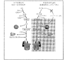
補体系は眼疾患において活性化される。図1は、補体系の2つの主要経路;古典経路および代替経路の概略図を提供する。代替経路(AP)は病原体および異質なまたは異常な表面により活性化され(例えばドルーゼン)、急激な自己増幅が可能である。APの過剰なおよび/または長期の活性化は、多くの炎症性および非炎症性病態および障害の主因であると考えられる。加齢性黄斑変性(AMD)、糖尿病性網膜症、ぶどう膜炎、網膜線維症、出血、および多くの眼病態および障害における炎症は、AP活性化および調節不全により引き起こされる。 The complement system is activated in eye diseases. FIG. 1 provides a schematic diagram of two major pathways of the complement system: the classical pathway and the alternative pathway. Alternative pathways (AP) are activated by pathogens and foreign or abnormal surfaces (eg drusen) and are capable of rapid self-amplification. Excessive and / or long-term activation of AP is believed to be a major cause of many inflammatory and non-inflammatory conditions and disorders. Inflammation in age-related macular degeneration (AMD), diabetic retinopathy, uveitis, retinal fibrosis, bleeding, and many eye conditions and disorders are caused by AP activation and dysregulation.
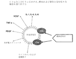
APは、補体因子B、D、およびP(プロパージン)から構成される。図2は、補体系活性化の結果として産生されるサイトカインおよび増殖因子の概略図を提供する。補体因子C3aおよびC5aは免疫系細胞、および他の型の細胞を活性化して、TNF−α、VEGF、サイトカイン、増殖因子、および他の炎症性メディエータを生成させる。AP活性化時に、C3bはプロパージンおよびB因子に結合し、複合体PC3bBを形成する。D因子はその後、複合体内のB因子を切断する。B因子のこの切断により活性コンバターゼが生成し、これがさらにC3bおよびC3aへとC3を切断し、これにより、追加のC3コンバターゼの形成を永続化する。追加の分子のC3bおよびBbがPC3bBbと結合して活性C5コンバターゼを形成し、これがC5の分子をC5bおよびC5aへと切断する。C5bはその後、C6、C7、C8およびC9因子と結合して溶解性巨大分子C5b−9(膜侵襲複合体、または「MAC」としても知られている)を形成する。MACは、細胞膜に穴を開けることにより細胞を溶解する。これは、知られているプロセスの1つであり、これにより、AMDおよび他の眼障害との関連で、RPE細胞、桿体および錐体が補体系活性化の結果として損傷される。 AP is composed of complement factors B, D, and P (properdin). FIG. 2 provides a schematic diagram of cytokines and growth factors produced as a result of complement system activation. Complement factors C3a and C5a activate immune system cells and other types of cells to produce TNF-α, VEGF, cytokines, growth factors, and other inflammatory mediators. Upon AP activation, C3b binds to properdin and factor B to form the complex PC3bB. Factor D then cleaves factor B in the complex. This cleavage of factor B produces an active convertase that further cleaves C3 into C3b and C3a, thereby perpetuating the formation of additional C3 convertase. Additional molecules C3b and Bb combine with PC3bBb to form an active C5 convertase, which cleaves the C5 molecule into C5b and C5a. C5b then combines with factors C6, C7, C8 and C9 to form the soluble macromolecule C5b-9 (also known as the membrane attack complex, or “MAC”). MAC lyses cells by making holes in the cell membrane. This is one of the known processes, which damages RPE cells, rods and cones as a result of complement system activation in the context of AMD and other eye disorders.
本明細書で記載される実施形態は、それを必要とする被験体において、線維症、血管出血、炎症、および細胞死により媒介される眼病態を治療および/または防止する組成物および方法に関する。方法は、被験体に代替経路(AP)阻害剤の治療的有効量を投与して、被験体において線維症、血管出血、炎症、および/または細胞死を阻害することを含み得る。 Embodiments described herein relate to compositions and methods for treating and / or preventing ophthalmic conditions mediated by fibrosis, vascular bleeding, inflammation, and cell death in a subject in need thereof. The method can include administering to the subject a therapeutically effective amount of an alternative pathway (AP) inhibitor to inhibit fibrosis, vascular bleeding, inflammation, and / or cell death in the subject.
いくつかの実施形態では、方法は、代替経路を選択的にブロックするがレクチンまたは古典経路に何の効果も有さない、代替経路阻害抗体またはその抗原結合性フラグメントの治療的有効量を被験体に投与することにより、それを必要とする被験体の脈絡膜血管新生、網膜萎縮症、網膜線維症、血管出血、および眼障害と関連する炎症を治療、防止および/または阻害することを含み得る。例えば、方法は、プロパージンに高親和性で結合し(例えば、1pM〜1000pMのKD)プロパージンのC3bおよびC5bへの結合をブロックする抗体による、代替経路の選択的阻害を含んでよい。そのような抗体は、TSR0、TSR1、TSR4、TSR5、およびTSR6からなる群より選択される6つのTSRのうちの少なくとも1つに結合できる。網膜線維症および出血を阻害するための抗プロパージン抗体の使用は、どの補体阻害剤も網膜線維症および網膜出血の阻害を示していないので新規である。 In some embodiments, the method subjects a therapeutically effective amount of an alternative pathway inhibitory antibody or antigen-binding fragment thereof that selectively blocks the alternative pathway but has no effect on the lectin or classical pathway. Administration may include treating, preventing and / or inhibiting inflammation associated with choroidal neovascularization, retinal atrophy, retinal fibrosis, vascular bleeding, and ocular disorders in a subject in need thereof. For example, the method may include selective inhibition of an alternative pathway with an antibody that binds with high affinity (eg, a K D of 1 pM to 1000 pM) and blocks the binding of properdin to C3b and C5b. Such an antibody can bind to at least one of six TSRs selected from the group consisting of TSR0, TSR1, TSR4, TSR5, and TSR6. The use of anti-properdin antibodies to inhibit retinal fibrosis and bleeding is novel because no complement inhibitor has shown inhibition of retinal fibrosis and retinal bleeding.
他の実施形態では、方法は、選択的に代替経路をブロックするがレクチンまたは古典経路に何の効果も有さない代替経路阻害抗体(すなわち、抗AP抗体)またはその抗原結合性フラグメントの治療的有効量を被験体に投与することにより、それを必要とする被験体の眼障害と関連するVEGF形成、C5b−9形成およびサイトカイン形成を治療、防止および/または阻害することを含み得る。いくつかの実施形態では、抗AP抗体は、プロパージンに結合しC3b/C5bとのプロパージン相互作用をブロックし、炎症、組織傷害、および血管新生を阻害する抗プロパージン抗体を含んでよい。抗AP抗体の投与は、代替経路によるC3a/C5aの形成を防止し、C3a/C5aによる網膜上皮細胞を含む炎症細胞の活性化、血管新生を促進する内皮増殖因子(例えば、VEGF)の放出を防止し、活性化された細胞からの炎症性メディエータ(例えば、サイトカイン)の産生を防止し、組織傷害(例えば、MAC)および網膜上皮細胞、桿体および錐体の傷害の原因となるC5b−9の産生を防止できる。目の損傷された細胞は、組織死のマーカーであるLDHを産生する。抗AP抗体はまた、乳酸デヒドロゲナーゼ(LDH)形成を防止でき、眼細胞死を阻害するために使用できる。 In other embodiments, the method selectively treats an alternative pathway inhibitory antibody (ie, an anti-AP antibody) or antigen-binding fragment thereof that selectively blocks the alternative pathway but has no effect on the lectin or classical pathway. Administering an effective amount to a subject can include treating, preventing and / or inhibiting VEGF formation, C5b-9 formation and cytokine formation associated with an eye disorder in a subject in need thereof. In some embodiments, the anti-AP antibody may comprise an anti-properdin antibody that binds to properdin and blocks properdin interaction with C3b / C5b and inhibits inflammation, tissue injury, and angiogenesis. Administration of anti-AP antibody prevents the formation of C3a / C5a by alternative pathways, activates inflammatory cells including retinal epithelial cells by C3a / C5a, and releases endothelial growth factor (eg, VEGF) that promotes angiogenesis C5b-9 prevents and prevents the production of inflammatory mediators (eg cytokines) from activated cells and causes tissue injury (eg MAC) and injury of retinal epithelial cells, rods and cones Production can be prevented. Damaged cells of the eye produce LDH, a marker of tissue death. Anti-AP antibodies can also prevent lactate dehydrogenase (LDH) formation and can be used to inhibit ocular cell death.
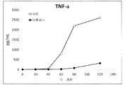
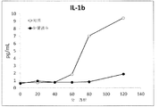
代替経路により産生されるC3a/C5aはどちらも、眼細胞、例えば、限定はされないが、RPE細胞、桿体および錐体細胞を活性化し、血管新生を促進する炎症性メディエータおよびVEGF増殖因子のその後の放出を引き起こす。C3a/C5aの阻害はVEGF形成および炎症性メディエータを制御でき、よって炎症が制御される。代替経路の活性化により産生されることが知られている追加の分子は下記である;TNF−α、IL−1、IL−6、IL−8、IL−17、VEGF、および/またはPDGF。抗AP抗体は、眼障害におけるそのような活性化を制御できる。 Both C3a / C5a produced by alternative pathways activate ocular cells such as, but not limited to, inflammatory mediators and VEGF growth factors that activate RPE cells, rods and cones and promote angiogenesis. Cause the release of. Inhibition of C3a / C5a can regulate VEGF formation and inflammatory mediators, thus controlling inflammation. Additional molecules known to be produced by activation of alternative pathways are: TNF-α, IL-1, IL-6, IL-8, IL-17, VEGF, and / or PDGF. Anti-AP antibodies can control such activation in ocular disorders.
血管の形成は組織修復にとって重要であり、これは疾患におけるVEGF形成により媒介される。代替経路により生成されるC3a/C5aはどちらも、眼細胞、例えば、限定はされないが、RPE細胞、桿体および錐体細胞を活性化し、血管新生を促進するVEGFおよびPDGF増殖因子のその後の放出を引き起こす。そのため、C3a/C5aの阻害は、病態を引き起こすVEGF形成を制御し組織修復に関与するVEGFは制御しないことが期待される。いくつかの実施形態では、本明細書で記載される抗AP抗体は、処置後に補体媒介性眼障害を発症するリスクがあると同定されている、眼科学的処置を受けている被験体のための予防的治療において使用できる。いくつかの実施形態では、抗AP抗体は、眼部処置と関連する線維症、出血および炎症を阻害できる。さらに他の実施形態では、抗AP抗体は、眼科学的処置後に組織修復を阻害することなく血管新生を防止できる。 Blood vessel formation is important for tissue repair, which is mediated by VEGF formation in the disease. Both C3a / C5a produced by alternative pathways activate ocular cells, such as, but not limited to, RPE cells, rods and cones, and subsequent release of VEGF and PDGF growth factors that promote angiogenesis cause. Therefore, inhibition of C3a / C5a is expected to control VEGF formation that causes pathology and not VEGF involved in tissue repair. In some embodiments, an anti-AP antibody described herein is for a subject undergoing ophthalmological treatment that has been identified as being at risk of developing a complement-mediated ocular disorder after treatment. Can be used in prophylactic treatment. In some embodiments, anti-AP antibodies can inhibit fibrosis, bleeding and inflammation associated with ocular treatment. In still other embodiments, the anti-AP antibody can prevent angiogenesis without inhibiting tissue repair after ophthalmological treatment.
別の実施形態では、眼部外科的処置(または他の物理的眼部外傷)を受けている被験体における正常な組織修復による線維症および出血を阻害するためのプロセスは、創傷治癒を促進するために、被験体に抗AP抗体を投与することを含んでよい。1つの実施形態では、AP媒介性眼病態を治療するためのプロセスは、眼部外科的処置中に起きてよく、この場合、処置を受けている被験体は、視力喪失に至ることがある/至らないこともある網膜出血または炎症により特徴付けられる病状に苦しんでいる。この実施形態は、外科的処置直前、処置中または処置後のいずれかで抗AP抗体を投与する工程を含む。 In another embodiment, the process for inhibiting fibrosis and bleeding due to normal tissue repair in a subject undergoing ocular surgical treatment (or other physical eye trauma) promotes wound healing Therefore, it may comprise administering an anti-AP antibody to the subject. In one embodiment, the process for treating an AP-mediated ophthalmic condition may occur during an ocular surgical procedure, in which case the subject undergoing treatment may lead to vision loss / Suffering from a medical condition characterized by retinal hemorrhage or inflammation that may not occur. This embodiment includes administering the anti-AP antibody either immediately before, during or after the surgical procedure.
他の実施形態では、抗AP抗体は、下記を含むが、それらに限定されないぶどう膜炎の病態に続発する線維症および出血を防止するために使用できる;虹彩炎、毛様体扁平部炎、脈絡膜炎、脈絡網膜炎、前部ぶどう膜炎、後部ぶどう膜炎、強膜炎、眼部血管新生、アテローム性動脈硬化、抗リン脂質症候群に続発する網膜動脈閉塞症、血管新生緑内障、虹彩血管新生、プルチェル網膜症、ソースビー眼底変性症、ドイン蜂巣状網膜ジストロフィー、マラッティア・レベンティネーズ(Malattia Leventinese)、家族性優性ドルーゼン、ノースカロライナ黄斑ジストロフィー、若年性黄斑変性症、シュタルガルト病、卵黄様黄斑変性症、成人発症中心窩黄斑卵黄状ジストロフィー(AOFVD)、ソースビー眼底変性症、およびベスト病。 In other embodiments, anti-AP antibodies can be used to prevent fibrosis and bleeding secondary to uveitis conditions including, but not limited to: iritis, ciliary planitis, Choroiditis, chorioretinitis, anterior uveitis, posterior uveitis, scleritis, ocular neovascularization, atherosclerosis, retinal artery occlusion secondary to antiphospholipid syndrome, neovascular glaucoma, iris vessel Neoplasia, Purcher's retinopathy, Sourceby fundus degeneration, Doin's honeycomb retinal dystrophy, Malattia Leventines, familial dominant drusen, North Carolina macular dystrophy, juvenile macular degeneration, Stargardt's disease, yolk-like macular degeneration , Adult-onset foveal macular yolk dystrophy (AOFVD), sourceby fundus degeneration, And Best's disease.
他の実施形態では、1つ以上の特許請求される抗AP抗体は、虹彩炎、毛様体扁平部炎、脈絡膜炎、脈絡網膜炎、前部ぶどう膜炎、後部ぶどう膜炎、または強膜炎、眼部血管新生、糖尿病性網膜症または糖尿病と関連する目の他の炎症性障害を含むが、それらに限定されないぶどう膜炎の病態に続発する炎症、血管新生、細胞萎縮、組織分解、LDHの放出、線維症および/または出血を防止する、高血圧性網膜症を防止する、自己免疫性ぶどう膜炎または自己免疫障害に続発するぶどう膜炎、ベーチェット病、イールズ病、または目の他の自己免疫性炎症疾患、アテローム性動脈硬化、抗リン脂質症候群に続発する網膜動脈閉塞症、血管新生緑内障、虹彩血管新生、プルチェル網膜症、AMD、ソースビー眼底変性症、ドイン蜂巣状網膜ジストロフィー、マラッティア・レベンティネーズ(Malattia Leventinese)、家族性優性ドルーゼン、ノースカロライナ黄斑ジストロフィー、若年性黄斑変性症、シュタルガルト病、卵黄様黄斑変性症、成人発症中心窩黄斑卵黄状ジストロフィー(AOFVD)、ソースビー眼底変性症、またはベスト病、血管閉塞、例えば、限定はされないが;網膜中心静脈閉塞症(CRVO)、閉塞性末梢動脈疾患、アテローム性動脈硬化に続発する眼虚血性症候群、抗リン脂質症候群に続発する網膜動脈閉塞症、血管新生緑内障、虹彩血管新生、またはプルチェル網膜症、未熟児網膜症または家族性滲出性硝子体網膜症、目の前眼部で起こる眼病態、およびフックス角膜内皮変性症を防止するために使用できる。 In other embodiments, one or more of the claimed anti-AP antibodies is iritis, ciliary planitis, choroiditis, chorioretinitis, anterior uveitis, posterior uveitis, or sclera Inflammation, angiogenesis, cellular atrophy, tissue degradation, including but not limited to inflammation, ocular neovascularization, diabetic retinopathy or other inflammatory disorders of the eye associated with diabetes, Prevent LDH release, fibrosis and / or hemorrhage, prevent hypertensive retinopathy, autoimmune uveitis or uveitis secondary to autoimmune disorders, Behcet's disease, Eal's disease, or other in the eye Autoimmune inflammatory disease, atherosclerosis, retinal arterial occlusion secondary to antiphospholipid syndrome, neovascular glaucoma, iris neovascularization, Purcher's retinopathy, AMD, sourceby fundus degeneration, doin's honeycomb retinopathy Trophy, Malattia Leventinese, familial dominant drusen, North Carolina macular dystrophy, juvenile macular degeneration, Stargardt disease, yolk-like macular degeneration, adult-onset foveal macular yolk dystrophy (AOFVD), source bee Fundus degeneration, or Best disease, vascular occlusion, including but not limited to: central retinal vein occlusion (CRVO), obstructive peripheral arterial disease, ocular ischemic syndrome secondary to atherosclerosis, antiphospholipid syndrome Secondary retinal artery occlusion, neovascular glaucoma, iris neovascularization, or Purcell retinopathy, retinopathy of prematurity or familial exudative vitreoretinopathy, ocular pathology occurring in the anterior segment of the eye, and Fuchs corneal endothelial degeneration Can be used to prevent.
いくつかの実施形態では、抗AP抗体は、血管閉塞により特徴付けられる病態に続発する線維症および/または出血を防止するために使用される。血管閉塞と関連するそのような病態としては、下記が挙げられる:網膜中心静脈閉塞症(CRVO)、閉塞性末梢動脈疾患、アテローム性動脈硬化に続発する眼虚血性症候群、抗リン脂質症候群に続発する網膜動脈閉塞症、血管新生緑内障、虹彩血管新生、およびプルチェル網膜症。 In some embodiments, anti-AP antibodies are used to prevent fibrosis and / or bleeding secondary to a pathological condition characterized by vascular occlusion. Such pathologies associated with vascular occlusion include: central retinal vein occlusion (CRVO), obstructive peripheral arterial disease, ocular ischemic syndrome secondary to atherosclerosis, secondary to antiphospholipid syndrome Retinal artery occlusion, neovascular glaucoma, iris neovascularization, and Purcell retinopathy.
1つの実施形態では、抗AP抗体は、糖尿病性網膜症に続発する線維症および出血を防止するために使用される。別の実施形態では、抗P、抗C3b、または抗Bb抗体は、糖尿病患者における網膜線維症または出血、高血圧性網膜症、自己免疫障害、自己免疫性ぶどう膜炎またはぶどう膜炎、ベーチェット病、イールズ病、または目の他の自己免疫疾患と関連する、任意のまたは全てのAP媒介性病態を治療するために使用される。 In one embodiment, anti-AP antibodies are used to prevent fibrosis and bleeding secondary to diabetic retinopathy. In another embodiment, the anti-P, anti-C3b, or anti-Bb antibody is retinal fibrosis or bleeding, hypertensive retinopathy, autoimmune disorder, autoimmune uveitis or uveitis, Behcet's disease, Used to treat any or all AP-mediated conditions associated with Ealz disease or other autoimmune diseases of the eye.
いくつかの実施形態では、抗AP抗体は、未熟児網膜症または家族性滲出性硝子体網膜症、目の前眼部で起こる眼病態、フックス角膜内皮変性症、血管新生の防止のための抗VEGF剤による反復治療に続発する線維症および/または出血を防止するために使用される。 In some embodiments, the anti-AP antibody is an anti-premature for retinopathy of prematurity or familial exudative vitreoretinopathy, an ocular condition that occurs in the anterior segment of the eye, Fuchs corneal endothelial degeneration, angiogenesis prevention. Used to prevent fibrosis and / or bleeding secondary to repeated treatment with VEGF agents.
いくつかの実施形態では、眼出血および/または線維症を治療するために使用される抗AP抗体は、Ba、Bb、C3b、D、C5、C6、C7またはC8を含む補体因子の群の1つに結合するものである。発明の他の実施形態では、眼出血および/または線維症を治療するために使用される抗AP抗体は、古典またはレクチン経路をも阻害するものである。 In some embodiments, the anti-AP antibody used to treat ocular hemorrhage and / or fibrosis is a group of complement factors comprising Ba, Bb, C3b, D, C5, C6, C7 or C8. It will be combined into one. In other embodiments of the invention, the anti-AP antibody used to treat ocular bleeding and / or fibrosis is one that also inhibits the classical or lectin pathway.
いくつかの実施形態では、抗AP抗体は、抗プロパージンまたは抗P抗体であってよい。抗プロパージン抗体は、血管新生を阻害でき、一方、眼炎症、眼浮腫、網膜線維症および出血も阻害できる。本発明は、AP媒介性の血管新生、眼炎症、眼浮腫、眼組織萎縮、血管透過性、線維症、出血ならびに他の炎症性および自己免疫が引き起こす病状および病態を含む、疾患および障害を防止または治療するためのプロセスを提供する。 In some embodiments, the anti-AP antibody may be an anti-properdin or anti-P antibody. Anti-properdin antibodies can inhibit angiogenesis while also inhibiting eye inflammation, eye edema, retinal fibrosis and bleeding. The present invention prevents diseases and disorders, including AP-mediated angiogenesis, ocular inflammation, ocular edema, ocular tissue atrophy, vascular permeability, fibrosis, hemorrhage and other inflammatory and autoimmune pathologies and conditions Or provide a process to treat.
別の実施形態では、本明細書で記載される抗P抗体の1つ以上は、ウェット型AMDを治療するために使用できる。本明細書で記載される抗P抗体の1つ以上は、前に抗VEGF剤により治療された被験体を治療するために使用され得る。本明細書で記載される、特許請求される抗P抗体の1つ以上は、抗VEGF剤が禁忌である血管新生のための治療を必要とする被験体を治療するために使用され得る。別の実施形態では、1つ以上の抗P抗体は、抗VEGF剤による治療後のドライ型AMDの発症を防止するために使用できる。 In another embodiment, one or more of the anti-P antibodies described herein can be used to treat wet AMD. One or more of the anti-P antibodies described herein can be used to treat a subject previously treated with an anti-VEGF agent. One or more of the claimed anti-P antibodies described herein can be used to treat a subject in need of treatment for angiogenesis where an anti-VEGF agent is contraindicated. In another embodiment, one or more anti-P antibodies can be used to prevent the development of dry AMD after treatment with an anti-VEGF agent.
本明細書で記載される実施形態はまた、下記の少なくとも1つが疾患過程の一部である眼病態を制御および防止するために抗P抗体を使用するプロセスに関する;炎症、血管新生、細胞萎縮、組織分解、LDHの放出、線維症および/または出血。 Embodiments described herein also relate to processes that use anti-P antibodies to control and prevent ophthalmic conditions where at least one of the following is part of a disease process; inflammation, angiogenesis, cell atrophy, Tissue degradation, LDH release, fibrosis and / or bleeding.
別の実施形態では、眼部外科的処置中または処置後に起こるAP活性化と関連するAP媒介性眼病態を治療するためのプロセスは、1つ以上の抗P抗体を被験体に、眼部処置直前、処置中または処置直後のいずれかで投与することを含んでよい。 In another embodiment, the process for treating an AP-mediated ophthalmic condition associated with AP activation that occurs during or after an ocular surgical procedure involves injecting one or more anti-P antibodies to a subject, ocular treatment. Administration may be included either immediately before, during or immediately after treatment.
別の実施形態は、過剰なまたは制御されない補体活性化と関連する障害の治療のための医薬または組成物の調製のための、これらの抗AP抗体の使用に関する。それらは、眼障害、特に、血管新生、組織傷害、組織破壊、地図状萎縮が疾患を特徴付ける障害中の補体活性化を含む。 Another embodiment relates to the use of these anti-AP antibodies for the preparation of a medicament or composition for the treatment of disorders associated with excessive or uncontrolled complement activation. They include complement activation during ocular disorders, particularly those in which angiogenesis, tissue injury, tissue destruction, and geographic atrophy characterize the disease.
定義
本明細書で特定的に規定されない限り、この文書で使用される全ての用語は、本発明の当業者により理解されるであろうものと同じ意味を有する。下記定義は明確にするために、本発明を説明するために明細書および特許請求の範囲で使用されるそれらの意図される意味を規定するために、提供される。
Definitions Unless otherwise specified herein, all terms used in this document have the same meaning as would be understood by one of ordinary skill in the art of the invention. The following definitions are provided for the sake of clarity to define their intended meaning used in the specification and claims to describe the present invention.
補体経路
「古典経路」は、抗原−抗体複合体により引き起こされ/活性化される補体系経路を指す。
Complement pathway The “classical pathway” refers to the complement system pathway triggered / activated by an antigen-antibody complex.
「代替経路」(「AP」または「the AP」と省略される)は、異質な表面として認識される人工のまたは死んだ細胞表面(または細胞−表面様材料)により引き起こされ/活性化される補体系経路を指す。 An “alternate pathway” (abbreviated as “AP” or “the AP”) is caused / activated by an artificial or dead cell surface (or cell-surface-like material) that is recognized as a foreign surface. Refers to the complement system pathway.
「代替経路特異的タンパク質」は、B因子、Bb因子、D因子、プロパージン、および/またはAPC3コンバターゼにより特異的に産生されるAP媒介性C3bを指す。 “Alternative pathway specific protein” refers to AP-mediated C3b produced specifically by Factor B, Factor Bb, Factor D, properdin, and / or APC3 convertase.
「抗AP抗体」は、AP特異タンパク質を標的にする全ての抗体、ならびにAP媒介性C5、C6、C7、C8、およびC9を標的にする(に結合する)任意の抗体を含む抗体の群を指す。AP抗体はまた、例えばDAF、MIRLなど、C3、C3b、P、B、D、C5、C6、C7、C8、C9、またはSタンパク質のAP媒介性産生を調節する任意のタンパク質または分子に結合する抗体を含む。 “Anti-AP antibodies” refers to a group of antibodies, including all antibodies that target AP-specific proteins, and any antibody that targets (binds to) AP-mediated C5, C6, C7, C8, and C9. Point to. AP antibodies also bind to any protein or molecule that modulates AP-mediated production of C3, C3b, P, B, D, C5, C6, C7, C8, C9, or S proteins, such as DAF, MIRL, etc. Contains antibodies.
「抗P抗体」はヒトプロパージンまたは動物プロパージンに対して産生された抗体である。抗P抗体は、ヒトまたは動物プロパージンに結合しAPを阻害する全ての抗体を含む。 An “anti-P antibody” is an antibody produced against human properdin or animal properdin. Anti-P antibodies include all antibodies that bind to human or animal properdin and inhibit AP.
「C3a依存性細胞活性化」は、APにより産生されたC3aがC3a細胞受容体に結合した時に起こる、好中球、単球、血小板、Tリンパ球、内皮細胞、マスト細胞、血小板、網膜上皮細胞、桿体、錐体、および他の細胞の活性化を説明する。 “C3a-dependent cell activation” occurs when C3a produced by AP binds to C3a cell receptor, neutrophils, monocytes, platelets, T lymphocytes, endothelial cells, mast cells, platelets, retinal epithelium Describe the activation of cells, rods, cones, and other cells.
「C5a依存性細胞活性化」は、APにより産生されたC5aが、C5a細胞受容体に結合した時に起こる、好中球、単球、血小板、Tリンパ球、内皮細胞、マスト細胞、血小板、網膜上皮細胞、桿体、錐体、および他の細胞の活性化を説明する。 “C5a-dependent cell activation” occurs when C5a produced by AP binds to C5a cell receptor, neutrophils, monocytes, platelets, T lymphocytes, endothelial cells, mast cells, platelets, retina Describe the activation of epithelial cells, rods, cones, and other cells.
「C5b−9/MAC媒介性/依存性組織傷害/細胞損傷」は、sC5b−9および/またはC5b−9(膜侵襲複合体、または「MAC」としても知られている)の形成により引き起こされる細胞損傷を説明する。そのような組織傷害は、補体媒介性MAC産生および沈着が疾患病態の一部である眼疾患で起こる。 “C5b-9 / MAC-mediated / dependent tissue injury / cell damage” is caused by the formation of sC5b-9 and / or C5b-9 (also known as membrane attack complex, or “MAC”) Explain cell damage. Such tissue injury occurs in eye diseases where complement-mediated MAC production and deposition is part of the disease pathology.
「AP媒介性炎症」は、APにより媒介され、炎症を引き起こす、および/または永続化させる、多数の生理的プロセス、細胞活性化、およびタンパク質産生を指す。AP媒介性炎症は、AP依存性C3a、C3b、C5a、C5b、C5b−9、および/またはsC5b−9の連続した、または増加した形成、および/または放出、およびその予想される結果全てにより測定できる。C3a、C5a、C5b−9は細胞を活性化し、炎症性メディエータ、例えば、限定はされないが;TNF−α、IL−1、PDGF、VEGF、好中球エラスターゼ、ならびに他のサイトカインおよび炎症性メディエータを放出させる。 “AP-mediated inflammation” refers to a number of physiological processes, cell activation, and protein production that are mediated by AP and cause and / or perpetuate inflammation. AP-mediated inflammation is measured by AP-dependent C3a, C3b, C5a, C5b, C5b-9, and / or sC5b-9 continuous or increased formation and / or release and all its expected results it can. C3a, C5a, C5b-9 activates cells and induces inflammatory mediators such as, but not limited to; TNF-α, IL-1, PDGF, VEGF, neutrophil elastase, and other cytokines and inflammatory mediators Release.
「抗体」は、本明細書では、ジスルフィド結合により相互連結される2つの重(H)鎖および2つの軽(L)鎖を含む。各重鎖は、重鎖可変領域(本明細書では、VHと省略される)および重鎖定常領域から構成される。重鎖定常領域は、3つのドメインから構成される;CH1、CH2およびCH3。各軽鎖は、軽鎖可変領域(本明細書では、VLと省略される)および軽鎖定常領域から構成される。軽鎖定常領域は、1つのドメイン、CLから構成される。VHおよびVL領域はさらに、フレームワーク領域(FR)と呼ばれるより保存的な領域が散在される、相補性決定領域(CDR)と呼ばれる超可変性の領域に細分され得る。各VHおよびVLは、アミノ末端からカルボキシル末端へと下記の順で配列される3つのCDRおよび4つのフレームワークから構成される:FR1、CDR1、FR2、CDR2、FR3、CDR3、FR4。重および軽鎖の可変領域は、抗原と相互作用する結合ドメインを含む。「抗体」という用語は、任意の抗体産生哺乳類(例えば、マウス、ラット、ウサギ、およびヒトを含む霊長類)に由来する全抗体およびその抗体フラグメントを包含する。例示的な抗体としては、ポリクローナル、モノクローナルおよび組換え抗体;多重特異性抗体(例えば、二重特異性抗体);ヒト化抗体;マウス抗体;キメラの、マウス−ヒト、マウス−霊長類、霊長類−ヒトモノクローナル抗体;および抗イディオタイプ抗体が挙げられ、任意の無傷の分子またはそのフラグメントであってもよい。 An “antibody” as used herein comprises two heavy (H) chains and two light (L) chains interconnected by disulfide bonds. Each heavy chain is comprised of a heavy chain variable region (abbreviated herein as VH) and a heavy chain constant region. The heavy chain constant region is comprised of three domains; CH1, CH2 and CH3. Each light chain is comprised of a light chain variable region (abbreviated herein as VL) and a light chain constant region. The light chain constant region is comprised of one domain, CL. The VH and VL regions can be further subdivided into hypervariable regions called complementarity determining regions (CDRs) interspersed with more conserved regions called framework regions (FR). Each VH and VL is composed of three CDRs and four frameworks arranged in the following order from the amino terminus to the carboxyl terminus: FR1, CDR1, FR2, CDR2, FR3, CDR3, FR4. The variable region of the heavy and light chains contains a binding domain that interacts with an antigen. The term “antibody” includes all antibodies and antibody fragments thereof derived from any antibody-producing mammal (eg, primates including mice, rats, rabbits, and humans). Exemplary antibodies include polyclonal, monoclonal and recombinant antibodies; multispecific antibodies (eg, bispecific antibodies); humanized antibodies; mouse antibodies; chimeric, mouse-human, mouse-primate, primate -Human monoclonal antibodies; and anti-idiotype antibodies, which may be any intact molecule or fragment thereof.
「抗原結合性フラグメント」は、それが由来する全抗体と同じ標的に結合する全抗体のフラグメントを指す。抗体フラグメントの型としては、ナノボディ、ダイアボディ、線形抗体、単鎖抗体分子および抗体フラグメントから形成される多重特異性抗体が挙げられる。抗体フラグメントの例としては、Fab、Fab’、F(ab)2、F(ab’)2およびFvフラグメント、またはscFvフラグメント(および上記のいずれかの任意のペグ化バリエーション)が挙げられる。抗原結合性フラグメントの例としては下記が挙げられるが、それらに限定されない:
「Fab」フラグメント(VHおよびVLを有する単鎖可変領域);
「一価フラグメント」(VL、VH、CLおよびCH1ドメインから構成される抗体フラグメント);
「F(ab’)2」フラグメント(ジスルフィド架橋によりヒンジ領域で連結される2つのFabフラグメントを含む二価フラグメント);
「Fd」フラグメント(抗体のVHおよびCH1ドメインから構成される);
「Fv」フラグメント(抗体のシングルアームのVLおよびVHドメインから構成される);
シングルドメイン抗体(「dAb」)、VHドメインまたはVLドメインから構成される;
単離された相補性決定領域(「CDR」、以下で規定される)。
An “antigen-binding fragment” refers to a fragment of a whole antibody that binds to the same target as the whole antibody from which it is derived. Antibody fragment types include nanobodies, diabodies, linear antibodies, single chain antibody molecules and multispecific antibodies formed from antibody fragments. Examples of antibody fragments include Fab, Fab ′, F (ab) 2, F (ab ′) 2 and Fv fragments, or scFv fragments (and any pegylation variation of any of the above). Examples of antigen binding fragments include, but are not limited to:
“Fab” fragment (single chain variable region with VH and VL);
“Monovalent fragment” (an antibody fragment composed of VL, VH, CL and CH1 domains);
An “F (ab ′) 2” fragment (a divalent fragment comprising two Fab fragments joined at the hinge region by a disulfide bridge);
“Fd” fragment (consisting of the VH and CH1 domains of an antibody);
"Fv" fragment (consisting of the single arm VL and VH domains of the antibody);
Composed of a single domain antibody ("dAb"), VH domain or VL domain;
Isolated complementarity determining region ("CDR", defined below).
「相補性決定領域(CDR)」は、抗体の主要結合領域である。全長抗体では、典型的には、3つのCDRが、2つの重および軽鎖可変領域の各々の可変領域内で見出される。 The “complementarity determining region (CDR)” is the main binding region of an antibody. In full-length antibodies, typically three CDRs are found within the variable region of each of the two heavy and light chain variable regions.
「キメラ抗体」は、非ヒト種の動物由来の抗体由来の可変ドメインおよびCDRを含むが、抗体分子の残りはヒト抗体由来である組換えタンパク質である。 A “chimeric antibody” is a recombinant protein comprising variable domains and CDRs from an antibody derived from an animal of a non-human species, with the remainder of the antibody molecule being derived from a human antibody.
「ヒト化抗体」は、非ヒトCDRおよびヒト化フレームワーク領域から構成される抗体である。ヒト化抗体は、典型的には、抗体相補性決定領域のみが非ヒト起源である組換えタンパク質である。 A “humanized antibody” is an antibody composed of non-human CDRs and a humanized framework region. Humanized antibodies are typically recombinant proteins in which only the antibody complementarity determining regions are of non-human origin.
抗体の「機能誘導体」は、抗体の機能領域から取り出された、または抗体の機能領域をそれ自体内に組み入れる任意の化合物である。抗体の機能誘導体としては下記が挙げられるが、それらに限定されない:抗原結合性フラグメント(前に規定されている)、CDR、キメラ抗体、モノクローナル抗体、組換え抗体、および単鎖抗体。CDR(前に規定されている)はまた、抗原結合性フラグメントとして、シングルドメイン抗体、マキシボディ、ミニボディ、細胞内抗体、ダイアボディ、トリアボディ、テトラボディ、v−NARおよびbis−scFvに組む込むことができる。抗体の抗原結合性フラグメントは、フィブロネクチンIII型(Fn3)などのポリペプチドに基づく足場にグラフトさせることができる。抗原結合性フラグメントは、相補的軽鎖ポリペプチドと共に、一対の抗原結合領域を形成する、一対のタンデムFvセグメント(VH−CH1−VH−CH1)を含む単鎖分子に組む込むことができる。 An “functional derivative” of an antibody is any compound that has been removed from, or that incorporates, a functional region of an antibody within itself. Functional derivatives of antibodies include, but are not limited to, antigen-binding fragments (defined above), CDRs, chimeric antibodies, monoclonal antibodies, recombinant antibodies, and single chain antibodies. CDRs (as previously defined) also assemble as antigen-binding fragments into single domain antibodies, maxibodies, minibodies, intracellular antibodies, diabodies, triabodies, tetrabodies, v-NAR and bis-scFv. Can be included. Antigen-binding fragments of the antibody can be grafted onto a scaffold based on a polypeptide such as fibronectin type III (Fn3). The antigen-binding fragment can be incorporated into a single chain molecule comprising a pair of tandem Fv segments (VH-CH1-VH-CH1) that together with a complementary light chain polypeptide forms a pair of antigen-binding regions.
「Fc領域」は、エフェクター機能(以下で規定される)を含む抗体の領域を指す。 “Fc region” refers to the region of an antibody that contains effector functions (defined below).
「エフェクター機能」は、抗体の天然Fc領域に起因するそれらの生物活性を指し、抗体アイソタイプの間で変化する。抗体エフェクター機能の例としては下記が挙げられる:C1q結合および補体依存性細胞傷害;Fc受容体結合;抗体依存性細胞媒介性細胞傷害(ADCC);食作用;細胞表面受容体(例えば、B細胞受容体)の下方制御;Fc受容体を発現する血小板の活性化の欠如;およびB細胞活性化。治療抗体の副作用を最小に抑える、または排除するために、エフェクター機能を最小に抑える、または排除することが好ましいことがある。 “Effector function” refers to those biological activities attributable to the native Fc region of an antibody, and varies between antibody isotypes. Examples of antibody effector functions include: C1q binding and complement dependent cytotoxicity; Fc receptor binding; antibody dependent cell mediated cytotoxicity (ADCC); phagocytosis; cell surface receptors (eg, B Downregulation of cellular receptors); lack of activation of platelets expressing Fc receptors; and B cell activation. In order to minimize or eliminate the side effects of therapeutic antibodies, it may be preferable to minimize or eliminate effector function.
「低減されたFcエフェクター機能(複数可)」は、抗体が、抗体のFc領域を認識する抗原に対して作用しない、抗体の機能(複数可)を指す。低減されたFcエフェクター機能の例としては下記が挙げられるが、それらに限定されない:低減された抗原へのFc結合、抗原に対するFc活性化の欠如、正常なFcエフェクター機能を防止する突然変異を含むFc領域、または血小板およびFc受容体を有する他の細胞の活性化の防止。 “Reduced Fc effector function (s)” refers to antibody function (s) in which the antibody does not act on an antigen that recognizes the Fc region of the antibody. Examples of reduced Fc effector function include, but are not limited to: reduced Fc binding to antigen, lack of Fc activation to antigen, mutations that prevent normal Fc effector function Prevention of activation of the Fc region or other cells with platelets and Fc receptors.
2つ以上の核酸またはポリペプチド配列との関連での「同一」は、同じである2つ以上の配列または部分配列を指す。2つの配列が、比較窓にわたって最大対応に対して比較および整列された場合、同じであるアミノ酸残基またはヌクレオチドの特定のパーセンテージを有する(すなわち、特定の領域にわたって、または、特定されない場合、全配列にわたって、60%同一性、任意で65%、70%、75%、80%、85%、90%、95%、または99%同一性)、または下記配列比較アルゴリズムの1つを使用して、または手動整列化および目視検査により測定される指定領域を有する場合、2つの配列は「実質的に同一」である。任意で、同一性は、少なくとも約50ヌクレオチド(または10アミノ酸)の長さである領域にわたって、またはより好ましくは100〜500または1000またはそれを超えるヌクレオチド(または20、50、200またはそれを超えるアミノ酸)の長さである領域にわたって存在する。2つのアミノ酸配列間のパーセント同一性はまた、MeyersおよびMillerのアルゴリズムを用いて決定できる。 “Identical” in the context of two or more nucleic acid or polypeptide sequences refers to two or more sequences or subsequences that are the same. Two sequences have a certain percentage of amino acid residues or nucleotides that are the same when compared and aligned for maximal correspondence over the comparison window (ie, the entire sequence if over a particular region or not specified) Over 60% identity, optionally 65%, 70%, 75%, 80%, 85%, 90%, 95%, or 99% identity), or using one of the following sequence comparison algorithms: Or two sequences are “substantially identical” if they have designated areas measured by manual alignment and visual inspection. Optionally, identity is over a region that is at least about 50 nucleotides (or 10 amino acids) in length, or more preferably 100-500 or 1000 or more nucleotides (or 20, 50, 200 or more amino acids). ) Over a region that is the length of The percent identity between two amino acid sequences can also be determined using the Meyers and Miller algorithm.
補体媒介性眼炎症、出血および線維症の治療、およびその病理学的帰結
本明細書で記載される実施形態は、罹患被験体に有効量の抗AP抗体を投与することにより、網膜線維症、出血および/または眼炎症と関連する任意の眼病状を患う被験体を治療するための組成物および方法に関する。投与される抗AP抗体(例えば、抗プロパージン抗体または抗P抗体)は、APのPC3bB複合体を標的にする、任意のAP特異タンパク質、および/または任意の抗体を標的にしてもよい。加えて、本明細書で記載される実施形態は、罹患被験体に有効量の抗AP抗体を投与することにより、(限定はされないが)AMDおよびAMD様病状を含む補体媒介性変性眼障害を治療するための方法を含む。
Treatment of Complement-Mediated Ocular Inflammation, Bleeding and Fibrosis, and Pathological Outcomes thereof Embodiments described herein provide retinal fibrosis by administering an effective amount of an anti-AP antibody to an affected subject. , Compositions and methods for treating a subject suffering from any eye condition associated with bleeding and / or eye inflammation. The administered anti-AP antibody (eg, anti-properdin antibody or anti-P antibody) may target any AP-specific protein and / or any antibody that targets the PC3bB complex of AP. In addition, embodiments described herein provide complement-mediated degenerative ocular disorders, including (but not limited to) AMD and AMD-like pathologies, by administering to an affected subject an effective amount of an anti-AP antibody. Including a method for treating.
AP−媒介性眼病態の特異的メカニズム
AMDおよびドルーゼン
リポフスチンの沈着および蓄積を含むAMDおよび他の障害を有する患者においては、ドルーゼンは目のブルッフ膜と網膜色素上皮(RPE)の間で集積する可能性がある。この材料の集積はブルッフ膜の断裂を引き起こし、血液網膜関門の破壊に至る。血液網膜関門の破壊は、脈絡膜血管新生(CNV)、網膜下出血、漿液性網膜(または網膜色素上皮)剥離、脂質滲出液、および/または血管結合組織瘢痕をもたらす。これらの病態は全て1つ以上の眼疾患の一部である。ドルーゼン沈着物および損傷された組織は、代替補体経路を活性化すると知られており、これは、最終的に炎症、組織傷害、最終的な視力喪失をもたらす。
Specific mechanism of AP-mediated ocular pathology AMD and drusen In patients with AMD and other disorders, including deposition and accumulation of lipofuscin, drusen can accumulate between the Bruch's membrane of the eye and the retinal pigment epithelium (RPE) There is sex. This accumulation of material causes Bruch's membrane rupture, leading to the destruction of the blood-retinal barrier. The disruption of the blood-retinal barrier results in choroidal neovascularization (CNV), subretinal hemorrhage, serous retinal (or retinal pigment epithelium) detachment, lipid exudates, and / or vascular connective tissue scars. All of these conditions are part of one or more eye diseases. Drusen deposits and damaged tissue are known to activate the alternative complement pathway, which ultimately leads to inflammation, tissue injury, and ultimately vision loss.
ドルーゼンおよび代替経路
AMDとの関連で、いくつかの炎症性サイトカイン、活性化されたミクログリア細胞、マクロファージおよび他の炎症細胞がドルーゼンと関連することが見出されている。活性化された細胞、活性化された補体経路の成分、および炎症性メディエータの存在は、病態の現場での代替経路の活性化を示唆する。慢性炎症、およびその帰結は網膜色素上皮の完全性を脅かす可能性があり、これはひいては、光受容器萎縮および脈絡膜血管新生(CNV)を引き起こす。最近の所見により、AMD発病における免疫プロセス(それにより炎症細胞および分子メディエータがブルッフ膜の内側または周囲の炎症を永続化させるプロセスを含む)の役割が確認された。補体活性化の調節不全はアナフィラトキシンの過剰産生を引き起こし、これは様々な眼組織細胞を活性化する。AMDとの関連で、活性化されたRPE細胞はVEGFを放出し、線維芽細胞およびマクロファージを傷害部位に動員し、網膜線維症および炎症を引き起こす。慢性補体活性化は慢性眼炎症の主因および駆動力であり、これが眼障害および病態の根本的原因である。
Drusen and alternative pathways In the context of AMD, several inflammatory cytokines, activated microglial cells, macrophages and other inflammatory cells have been found to be associated with drusen. The presence of activated cells, activated complement pathway components, and inflammatory mediators suggest activation of alternative pathways in the field of the pathology. Chronic inflammation, and its consequences, can threaten the integrity of the retinal pigment epithelium, which in turn causes photoreceptor atrophy and choroidal neovascularization (CNV). Recent findings confirm the role of immune processes in AMD pathogenesis, including the process by which inflammatory cells and molecular mediators perpetuate inflammation inside or around Bruch's membrane. Dysregulation of complement activation causes overproduction of anaphylatoxin, which activates various ocular tissue cells. In the context of AMD, activated RPE cells release VEGF, recruit fibroblasts and macrophages to the site of injury, causing retinal fibrosis and inflammation. Chronic complement activation is the main cause and driving force of chronic eye inflammation, which is the root cause of eye damage and pathology.
代替経路および炎症
慢性局所炎症および補体活性化は、眼病態に密接に関与する。我々のアプローチは、目の慢性炎症プロセスを弱めるまたは阻害するような様式で、補体経路における特定の分子構成物を標的にすることである。本明細書で記載される抗AP抗体は、AMDならびに関連する眼障害および病態において炎症、出血、線維症、および血管新生を引き起こす、炎症プロセスを阻害する治療の方法において使用できる。本明細書で記載される抗AP抗体は、眼外科手術と関連する、または眼外科手術の可能性のある術後合併症である、炎症、出血、線維症、および血管新生を阻害するために使用できる。
Alternative pathways and inflammation Chronic local inflammation and complement activation are closely involved in ocular conditions. Our approach is to target specific molecular components in the complement pathway in such a way as to attenuate or inhibit the chronic inflammatory process of the eye. The anti-AP antibodies described herein can be used in therapeutic methods that inhibit inflammatory processes that cause inflammation, bleeding, fibrosis, and angiogenesis in AMD and related eye disorders and conditions. The anti-AP antibodies described herein are intended to inhibit inflammation, bleeding, fibrosis, and angiogenesis that are associated with, or possibly post-operative complications of, eye surgery Can be used.
自然免疫系のAPの活性化によりC5aおよびC3aが産生され、これらは強力なアナフィラトキシンである。補体活性化の調節不全により、C5aおよびC3aの過剰産生が起こる。C5aおよびC3aは、網膜および免疫系細胞を活性化し、これにより補体系活性のカスケードを永続化させ、これが慢性眼炎症を引き起こす。C5aは、RPE細胞の生存率および免疫細胞増殖を抑制する能力を減少させ、線維芽細胞およびマクロファージの過剰増殖および動員を引き起こす。同時に、C5aはまたRPE細胞によるVEGF分泌を増加させ、これはCNVを永続化させる。C3a受容体を有する免疫系細胞、例えば単球、T−リンパ球、マスト細胞、好塩基球、およびミクログリア細胞もまた、C3aにより活性化される。活性化された細胞は、炎症性メディエータ、例えばTNF、IL−1、IL−6、IL8、VEGF、PDGF、FGF、およびいくつかの他のサイトカインを放出する(図2を参照されたい)。これらの炎症性メディエータの全てを阻害するために、本発明の作用物質はAP上流を、プロパージンがC3bに結合する時点で、標的にする。 Activation of the AP of the innate immune system produces C5a and C3a, which are potent anaphylatoxins. Dysregulation of complement activation results in overproduction of C5a and C3a. C5a and C3a activate the retina and immune system cells, thereby perpetuating a cascade of complement system activity, which causes chronic ocular inflammation. C5a decreases RPE cell viability and the ability to suppress immune cell proliferation, causing hyperproliferation and mobilization of fibroblasts and macrophages. At the same time, C5a also increases VEGF secretion by RPE cells, which makes CNV permanent. Immune system cells with C3a receptors, such as monocytes, T-lymphocytes, mast cells, basophils, and microglia cells are also activated by C3a. Activated cells release inflammatory mediators such as TNF, IL-1, IL-6, IL8, VEGF, PDGF, FGF, and some other cytokines (see FIG. 2). In order to inhibit all of these inflammatory mediators, the agents of the invention target the AP upstream when the properdin binds to C3b.
代替経路および血管内皮増殖因子(VEGF)
前に記載されるように、C5aはRPE細胞を活性化してVEGFを産生させる。APはまた、VEGFの産生を媒介できる他の細胞を活性化する。APが調節不全の病態では、VEGFの産生は過剰になり得る。よって、AP阻害は、C3aおよびC5a産生を阻害し、よってVEGFの産生を阻害することが予想される。VEGFの阻害は、CNVおよび血管機能不全に関連する他の眼病態を効果的に阻害するのに重要である。補体媒介性VEGF産生のAP上流を標的にすることにより、本明細書で記載される抗AP抗体は、組織修復を脅かすことなく、VEGFの形成を阻害する治療方法を提供できる。
Alternative pathways and vascular endothelial growth factor (VEGF)
As previously described, C5a activates RPE cells to produce VEGF. AP also activates other cells that can mediate the production of VEGF. In conditions where AP is dysregulated, VEGF production can be excessive. Thus, AP inhibition is expected to inhibit C3a and C5a production and thus inhibit VEGF production. Inhibition of VEGF is important to effectively inhibit CNV and other ocular conditions associated with vascular dysfunction. By targeting the AP upstream of complement-mediated VEGF production, the anti-AP antibodies described herein can provide therapeutic methods that inhibit VEGF formation without threatening tissue repair.
VEGFおよび血管新生
正常な状況下では、VEGFの産生は新しい血管の増殖を開始させ、傷害組織の治癒を促進する。いくつかの病態では、過剰な、慢性の、または別様に制御されないVEGF産生は、異常なおよび/または過剰な血管の増殖を引き起こす。制御されないVEGFの産生が脈絡膜血管新生(CNV)において重要な役割を果たすことは、当技術分野内でよく知られている。ドルーゼンまたはいくつかの他の網膜傷害源の存在が起きた場合、炎症プロセスはVEGF産生の増殖を引き起こす。健康な組織では、このVEGFが放出されて傷害部位での正常な新しい血管の増殖を促進する。病態下では、制御されないVEGFにより、異常な血管の制御されない増殖(CNVとしても知られている)が起こる。CNVは傷害を悪化させ、視力喪失を直接引き起こし得る。
VEGF and angiogenesis Under normal circumstances, the production of VEGF initiates the growth of new blood vessels and promotes healing of damaged tissue. In some pathologies, excessive, chronic, or otherwise unregulated VEGF production causes abnormal and / or excessive blood vessel growth. It is well known in the art that unregulated VEGF production plays an important role in choroidal neovascularization (CNV). When the presence of drusen or some other source of retinal injury occurs, the inflammatory process causes proliferation of VEGF production. In healthy tissue, this VEGF is released to promote the growth of normal new blood vessels at the site of injury. Under pathological conditions, uncontrolled VEGF results in uncontrolled growth of abnormal blood vessels (also known as CNV). CNV exacerbates injury and can directly cause vision loss.
VEGFはCNVの主因であるが、これは新しい健康な血管の形成を促進する。新しい脈管構造の正常な形成は創傷治癒のために必要とされる。よって、理想的な治療は、眼病態に重要な病的VEGFの形成を防止するだけでなければならない。不運なことに、抗VEGF治療は、VEGFを完全に枯渇させ、CNVを防止するのに有効であることが証明されているが、組織修復も防止する。同様に、血管新生全てを完全に阻害するいずれの治療も、組織修復を阻害する。この理由のために、VEGFを直接中和する作用物質は、眼外科手術を受けている患者には適応されない。眼部処置の正常な過程において形成される病変は、疾患と本質的に関連するものと共に、正常な組織修復のためにVEGFを必要とする。その結果として、眼外科手術を受ける患者は抗VEGF剤で処置することができない。 VEGF is a major cause of CNV, which promotes the formation of new healthy blood vessels. Normal formation of new vasculature is required for wound healing. Thus, an ideal treatment should only prevent the formation of pathological VEGF that is important for ocular conditions. Unfortunately, anti-VEGF treatment has proven effective in completely depleting VEGF and preventing CNV, but also prevents tissue repair. Similarly, any treatment that completely inhibits all angiogenesis will inhibit tissue repair. For this reason, agents that directly neutralize VEGF are not indicated for patients undergoing eye surgery. Lesions that form during the normal course of ocular treatment require VEGF for normal tissue repair, along with those that are inherently associated with the disease. As a result, patients undergoing eye surgery cannot be treated with anti-VEGF agents.
血管新生およびウェット型AMD
血管新生AMDは「ウェット型AMD」または「滲出性AMD」とも呼ばれ、補体系の不十分な制御と関連する。VEGFの調節不全は異常な脈管構造の制御されない増殖を引き起こす。進行例では、血管新生AMDは色素上皮剥離および網膜下出血と関連する。現在のところ利用可能な薬物は、VEGFを中和し、CNVを治療できる。これらの療法は抗VEGF剤および抗体(PCT/US12/20855号)、Genentech(EP070871431)、およびXoma(PCT/US08/080531号)を含む。ルセンティス(抗VEGF抗体Fab)およびアイリーア(VEGF受容体)は、先行技術の2つの抗VEGF剤である。これらのVEGF標的剤は、ウェット型AMD患者に、血管新生の治療および防止のために提供される。これらの薬物はCNVを防止するには有効であるが、VEGFがなくても疾患進行を永続化させる根底にある眼炎症、線維症、および出血を含む血管漏出を防止しない。VEGFを標的にする作用物質は、APまたはAP誘導性炎症もしくは組織傷害を直接阻害しない。その結果として、そのような作用物質は、補体媒介性炎症、MAC形成を介するRPE破壊、またはAP調節不全の任意の他の病理学的徴候を防止しない。その上、組織修復に対するそれらの阻害効果のために、最近の研究は、そのような作用物質が最終的に、地図状萎縮および永久的視力喪失を引き起こすことを示している。よって、網膜線維症、血管出血、およびCNVを防止する作用物質は要求を満たさない。そのような作用物質は、VEGF治療後のさらなる損傷を制御するのに必要とされる。
Angiogenesis and wet AMD
Angiogenic AMD, also called “wet AMD” or “wetting AMD”, is associated with inadequate control of the complement system. Dysregulation of VEGF causes uncontrolled proliferation of abnormal vasculature. In advanced cases, neovascular AMD is associated with pigment epithelial detachment and subretinal hemorrhage. Currently available drugs can neutralize VEGF and treat CNV. These therapies include anti-VEGF agents and antibodies (PCT / US12 / 20855), Genentech (EP070874311), and Xoma (PCT / US08 / 080531). Lucentis (anti-VEGF antibody Fab) and Eire (VEGF receptor) are two prior art anti-VEGF agents. These VEGF targeting agents are provided to wet AMD patients for the treatment and prevention of angiogenesis. While these drugs are effective in preventing CNV, they do not prevent vascular leakage including underlying ocular inflammation, fibrosis, and bleeding that perpetuate disease progression without VEGF. Agents that target VEGF do not directly inhibit AP or AP-induced inflammation or tissue injury. Consequently, such agents do not prevent complement-mediated inflammation, RPE disruption via MAC formation, or any other pathological signs of AP dysregulation. Moreover, because of their inhibitory effects on tissue repair, recent studies have shown that such agents ultimately cause geographic atrophy and permanent vision loss. Thus, agents that prevent retinal fibrosis, vascular bleeding, and CNV are not sufficient. Such agents are required to control further damage after VEGF treatment.
ウェット型AMDの2匹の別々の動物モデルで、本明細書で記載される抗AP抗体がCNV、炎症、線維症、および出血ならびに創傷治癒を防止できることが見出された。本質的に創傷であり、炎症、線維症、および出血に至る、レーザー誘導性CNVを生成させた。アカゲザルにおけるCNVモデルで、本明細書で記載される抗AP抗体(例えば、抗プロパージン抗体Fab)はCNVを阻害した。同じ研究で、CNVは、ルセンティス(抗VEGF抗体Fab)で治療したそれらのサルにおいて、より完全に阻害された。しかしながら、ルセンティス治療された動物は、抗AP抗体で治療した動物よりも、より多くの出血およびより多くの線維症(瘢痕)を経験した。 In two separate animal models of wet AMD, it was found that the anti-AP antibodies described herein can prevent CNV, inflammation, fibrosis, and bleeding and wound healing. Laser-induced CNV was generated that was essentially a wound and resulted in inflammation, fibrosis, and bleeding. In the CNV model in rhesus monkeys, the anti-AP antibodies described herein (eg, anti-properdin antibody Fab) inhibited CNV. In the same study, CNV was more completely inhibited in those monkeys treated with Lucentis (anti-VEGF antibody Fab). However, Lucentis treated animals experienced more bleeding and more fibrosis (scarring) than animals treated with anti-AP antibodies.
本明細書で記載される他の実施形態は、AMDのための併用治療に関し、これはCNVの治療における抗VEGF治療の効率を利用し、これはまた抗AP抗体を利用する。抗VEGF剤の短期使用、続いて(または同時に)抗AP抗体の使用は、CNVに対して即効を提供できる一方、炎症、組織萎縮、再発CNV、出血およびCNV後患者における出血にも対処し、さらに視力喪失を低減させる。 Other embodiments described herein relate to combination therapy for AMD, which utilizes the efficiency of anti-VEGF therapy in the treatment of CNV, which also utilizes anti-AP antibodies. While the short-term use of anti-VEGF agents followed by (or simultaneously) the use of anti-AP antibodies can provide immediate effect on CNV, it also addresses inflammation, tissue atrophy, recurrent CNV, bleeding and bleeding in patients after CNV, It further reduces vision loss.
代替経路の活性化は、どちらもAMD病態に関与するVEGFおよびPDGFなどの複数の増殖因子を生成させる。VEGF単独またはVEGFおよびPDGFの両方を直接標的にする薬物は、ウェット型AMDにおいて効力を示した。しかしながら、両方の因子を標的にする治療がより有効である。フォビスタは、そのどちらもが血管新生の原因となる既存の増殖因子を中和する抗VEGFおよび抗PDGFによる併用療法である。いくつかの実施形態では、抗AP抗体は、血管新生、線維症、および炎症の原因となる複数の増殖因子の形成を防止するために使用できる。現在のところ、網膜線維症のための治療はない。本明細書で記載される抗AP抗体はVEGFおよびPDGFの両方の産生をブロックできる。 Activation of alternative pathways generates multiple growth factors such as VEGF and PDGF, both of which are involved in AMD pathology. Drugs that directly target VEGF alone or both VEGF and PDGF have shown efficacy in wet AMD. However, treatments that target both factors are more effective. Fovista is a combination therapy with anti-VEGF and anti-PDGF, both of which neutralize existing growth factors that cause angiogenesis. In some embodiments, anti-AP antibodies can be used to prevent the formation of multiple growth factors that cause angiogenesis, fibrosis, and inflammation. There is currently no cure for retinal fibrosis. The anti-AP antibodies described herein can block the production of both VEGF and PDGF.
代替経路および組織傷害
補体経路活性化により、MAC(C5b−9)の形成が引き起こされる。前に記載されるように、ならびに、図1および図2において概略的に提示されるように、いったんAPが活性化されると、炎症細胞活性化およびMAC形成に至るであろう。MAC−媒介性RPE活性化は、AMDおよび他の眼病態における視力喪失に関し重要である。破壊されたRPEの断片がドルーゼンにおいて、補体系タンパク質、例えばMACと共に見出された。研究被験体がAPを介して機能的MACを形成できないノックアウト研究は、組織損傷、脱髄、低減された炎症細胞浸潤物、および改善された機能回復におけるAPの役割の証拠を提供する。
Alternative pathways and tissue injury Activation of the complement pathway causes the formation of MAC (C5b-9). As previously described, and as presented schematically in FIGS. 1 and 2, once AP is activated, it will lead to inflammatory cell activation and MAC formation. MAC-mediated RPE activation is important for vision loss in AMD and other eye conditions. Fragmented RPE fragments were found in drusen with complement system proteins such as MAC. Knockout studies in which study subjects are unable to form a functional MAC via AP provide evidence of a role for AP in tissue damage, demyelination, reduced inflammatory cell infiltrates, and improved functional recovery.
組織傷害および地図状萎縮
地図状萎縮(GA)は、ドライ型AMDの進行型である。AMD疾患進行の経過において、ドルーゼンが拡大し補体駆動性炎症が制御不能に増殖するので、眼組織は物理的および生理的損傷を患う。ドルーゼンはこれらの細胞に物理的圧力を与え酸化ストレスを引き起こす可能性があり、一方、補体駆動MAC形成は細胞の溶解および死を引き起こす。さらに状況を悪化させることに、炎症性メディエータの放出は血管透過性を引き起こし、これは浮腫、出血および他の血管病態を引き起こす。RPE細胞に対するこれらの物理的および生理的攻撃の極致および蓄積は、最終的に網膜上皮組織の不可逆的損失をもたらす。網膜色素上皮は光受容細胞の下に存在し、目の光覚細胞に対して重要な代謝サポートを提供する。RPE組織の損失は光受容細胞の損失および視力喪失を引き起こす。これらのイベントの組み合わせは様々な眼病態をもたらす。
Tissue injury and map-like atrophy Map-like atrophy (GA) is an advanced form of dry AMD. In the course of AMD disease progression, ocular tissue suffers physical and physiological damage as drusen expands and complement-driven inflammation grows out of control. Drusen can exert physical pressure on these cells and cause oxidative stress, while complement-driven MAC formation causes cell lysis and death. To further exacerbate the situation, the release of inflammatory mediators causes vascular permeability, which causes edema, bleeding and other vascular conditions. The culmination and accumulation of these physical and physiological attacks on RPE cells ultimately leads to irreversible loss of retinal epithelial tissue. The retinal pigment epithelium resides below the photoreceptor cells and provides important metabolic support for the photoreceptors of the eye. Loss of RPE tissue causes photoreceptor cell loss and vision loss. The combination of these events results in various eye conditions.
現在のところ、多くの眼病態に対するFDAに認可された有効な治療はない。ランパリズマブは抗D因子抗体であり、GA患者における第II相試験において肯定的データを示した。この薬物はGAに対してのみ適応されており、CNV、網膜線維症、または出血を制御しない。本明細書で記載される抗AP抗体は、同じクラスの薬物であるにも関わらず、ランパリズマブができるものを超えて、網膜線維症、血管出血、および炎症を阻害できる。本明細書で記載される抗AP抗体は、不活性なC3複合体PC3bBの形成を防止しないランパリズマブに比べ、C3コンバターゼの不活性な複合体の形成を防止する。よって、発明は、ランパリズマブの作用メカニズムの上流であるAPの工程で作用する。 At present, there are no effective treatments approved by the FDA for many eye conditions. Lamparizumab is an anti-factor D antibody and has shown positive data in a phase II trial in GA patients. This drug is only indicated for GA and does not control CNV, retinal fibrosis, or bleeding. The anti-AP antibodies described herein can inhibit retinal fibrosis, vascular hemorrhage, and inflammation beyond what lamparizumab can do, despite being the same class of drugs. The anti-AP antibodies described herein prevent the formation of an inactive complex of C3 convertase compared to lamparizumab which does not prevent the formation of inactive C3 complex PC3bB. Thus, the invention operates in the AP process, upstream of the mechanism of action of lamparizumab.
代替経路および網膜線維症
線維症は、目内の陳旧性または慢性病変と関連する。ウェット型AMDを有する被験体はラニビズマブで治療されると、彼等は、線維症、続いて、近年同定された予想外の視力喪失を最終的に発症することが示されている。CNV、組織萎縮、および線維症は黄斑の変性をひきおこす。ベバシズマブで治療した目を、線維症、組織萎縮、網膜下出血、CNVの程度、および色素上皮剥離(PED)について評価した。蛍光眼底造影でのCNV漏出のサイズおよび段階分けもまた評価した。遡及研究では、網膜中心PED、および網膜下液(SRF)厚さを、光干渉断層撮影を用いて評価した。大きな病変を予想する他の因子は、中心窩下および両側性病変ならびにベースラインでの出血の存在であった。大きな両側性病変およびベースラインでの出血は、追跡での網膜下線維症と相関した。
Alternative pathways and retinal fibrosis Fibrosis is associated with old or chronic lesions in the eye. When subjects with wet AMD are treated with ranibizumab, they have been shown to eventually develop fibrosis followed by an unexpected loss of vision recently identified. CNV, tissue atrophy, and fibrosis cause macular degeneration. Eyes treated with bevacizumab were evaluated for fibrosis, tissue atrophy, subretinal hemorrhage, degree of CNV, and pigment epithelial detachment (PED). The size and staging of CNV leakage on fluorescent fundus angiography was also evaluated. In a retrospective study, retinal center PED and subretinal fluid (SRF) thickness were evaluated using optical coherence tomography. Other factors that predicted large lesions were the presence of subfoveal and bilateral lesions and bleeding at baseline. Large bilateral lesions and baseline bleeding correlated with subretinal fibrosis at follow-up.
網膜線維症はまた、目組織への傷害および外傷に起因することがある。傷害または外傷は多くの起源に由来する可能性がある。その上、慢性炎症が網膜線維症を引き起こすこともある。初期組織損傷の部位では、しばしば炎症細胞の浸潤があり、これは最終的に瘢痕組織へと発展することが示されている。 Retinal fibrosis can also result from injury and trauma to eye tissue. Injuries or trauma can come from many sources. In addition, chronic inflammation can cause retinal fibrosis. At the site of early tissue damage, there is often inflammatory cell infiltration, which has been shown to eventually develop into scar tissue.
線維症の1つの可能性のあるメカニズムはサイトカインの産生であり、これは線維芽細胞を活性化し動員する。活性化された線維芽細胞はより多くのサイトカインおよび他の炎症性メディエータを産生し、免疫細胞、例えばマクロファージの動員および活性化を助け、これらは線維症および炎症で重要な役割を果たす。線維芽細胞はまた、VEGFの産生を媒介する。よって、線維性瘢痕は、本質的に壊死性であるが、また血管形成され得る。線維症は複合的プロセスであり、抗VEGF療法は線維症または血管漏出を制御できない。我々は、本明細書で記載される抗AP抗体が、抗VEGF治療により治療されていない線維性瘢痕の形成および遺残を防止できるという驚くべき発見を提示する。 One possible mechanism of fibrosis is the production of cytokines, which activates and mobilizes fibroblasts. Activated fibroblasts produce more cytokines and other inflammatory mediators, helping to recruit and activate immune cells such as macrophages, which play an important role in fibrosis and inflammation. Fibroblasts also mediate VEGF production. Thus, fibrous scars are necrotic in nature but can also be vascularized. Fibrosis is a complex process and anti-VEGF therapy cannot control fibrosis or vascular leakage. We present the surprising discovery that the anti-AP antibodies described herein can prevent the formation and remnant of fibrous scars that have not been treated with anti-VEGF treatment.
代替経路および糖尿病性網膜症
研究は、補体媒介性炎症性サイトカインが糖尿病性網膜症の進行において重要な役割を果たすことを示した。特に、VEGFは、血液網膜関門の崩壊および結果として生じる黄斑浮腫において重大な役割を果たすことが示されている。抗VEGF(VEGF標的)剤は、糖尿病性網膜症の進行を治療および防止するのに有効であることが証明されている。本明細書で記載される抗AP抗体の投与は、炎症性サイトカインの形成を防止し、VEGFの形成を阻害できる。したがって、発明は、糖尿病性網膜症を効果的に治療できる。
Alternative pathways and diabetic retinopathy Studies have shown that complement-mediated inflammatory cytokines play an important role in the progression of diabetic retinopathy. In particular, VEGF has been shown to play a critical role in blood retinal barrier disruption and the resulting macular edema. Anti-VEGF (VEGF target) agents have proven effective in treating and preventing the progression of diabetic retinopathy. Administration of the anti-AP antibodies described herein can prevent the formation of inflammatory cytokines and inhibit the formation of VEGF. Therefore, the invention can effectively treat diabetic retinopathy.
代替経路および他の眼疾患
目内でのAPの調節不全は無数の眼病態を引き起こし、AMDおよび糖尿病性網膜症はその中の2つにすぎない。他の器官および組織におけるAPの調節不全もまた、別の範囲の病態を引き起こす。先進国における失明の主因は、視力喪失に至る共通の経路としての病的眼炎症および血管新生を有するいくつかの障害に起因する。これらの障害としては、滲出性加齢黄斑変性(AMD)、糖尿病性網膜症(DR)、未熟児網膜症(ROP)、網膜静脈閉塞症(RVO)および眼腫瘍が挙げられる。目内のAPの調節不全は、無数の眼病態を引き起こす。他の器官および組織でのAPの調節不全もまた、眼内空間で別の範囲の病態を引き起こす。目との関連で、APの調節不全は下記に関与し得る:a)AMD様疾患、例えば先天性および家族性ドルーゼン;b)自己免疫性ぶどう膜炎および自己免疫障害に続発するぶどう膜炎、例えば黄斑浮腫;c)網膜虚血および網膜虚血と関連する、またはこれにより引き起こされる眼病態、例えば高血圧性網膜症および未熟児網膜症;ならびに、d)目の前眼部の炎症性病態。
Alternative pathways and other eye diseases In-eye AP dysregulation causes a myriad of eye conditions, AMD and diabetic retinopathy are only two of them. Dysregulation of AP in other organs and tissues also causes another range of pathologies. The leading cause of blindness in developed countries is due to several disorders with pathological ocular inflammation and angiogenesis as common pathways leading to vision loss. These disorders include exudative age-related macular degeneration (AMD), diabetic retinopathy (DR), retinopathy of prematurity (ROP), retinal vein occlusion (RVO) and eye tumors. Dysregulation of AP in the eye causes a myriad of eye conditions. Dysregulation of AP in other organs and tissues also causes another range of pathology in the intraocular space. In the context of the eye, AP dysregulation may be involved in: a) AMD-like diseases such as congenital and familial drusen; b) autoimmune uveitis and uveitis secondary to autoimmune disorders, E.g. macular edema; c) ophthalmic conditions associated with or caused by retinal ischemia and retinal ischemia such as hypertensive retinopathy and retinopathy of prematurity; and d) inflammatory conditions of the anterior segment of the eye.
ドルーゼンおよびAMD様疾患
いくつかの先天性および家族性障害は、ドルーゼンの沈着およびリポフスチン沈着物により特徴付けられる。そのような障害としては、ソースビー眼底変性症、ドイン蜂巣状網膜ジストロフィー、マラッティア・レベンティネーズ(Malattia Leventinese)、家族性優性ドルーゼン、ノースカロライナ黄斑ジストロフィー、若年性黄斑変性症、シュタルガルト病、卵黄様黄斑変性症、成人発症中心窩黄斑卵黄状ジストロフィー(AOFVD)、ソースビー眼底変性症、およびベスト病が挙げられる。ドルーゼンの過剰な沈着により特徴付けられる障害および病態は、AMDとほぼ同じように、黄斑変性症およびCNVを引き起こす。ドルーゼンおよびリポフスチン沈着物は炎症反応を開始させ、酸化ストレスを眼組織に与える。炎症性メディエータおよびサイトカインは、調節不全のAP活性化の結果として増殖する。VEGFの産生は血管新生、浮腫および出血を引き起こす。MACの形成は組織損傷および萎縮を引き起こす。線維芽細胞の動員は線維症に至る。
Drusen and AMD-like diseases Several congenital and familial disorders are characterized by drusen deposits and lipofuscin deposits. Such disorders include: Sourceby fundus degeneration, Doin's honeycomb retinal dystrophy, Malattia Leventinese, familial dominant drusen, North Carolina macular dystrophy, juvenile macular degeneration, Stargardt's disease, yolk-like macular Degeneration, adult-onset foveal macular yolk dystrophy (AOFVD), sourceby fundus degeneration, and best disease. Disorders and conditions characterized by excessive drusen deposition cause macular degeneration and CNV, much like AMD. Drusen and lipofuscin deposits initiate an inflammatory response and impart oxidative stress to ocular tissues. Inflammatory mediators and cytokines proliferate as a result of dysregulated AP activation. The production of VEGF causes angiogenesis, edema and bleeding. The formation of MAC causes tissue damage and atrophy. Fibroblast recruitment leads to fibrosis.
抗VEGF剤は、AOFVDおよびマラッティア・レベンティネーズ(Malattia Leventinese)(蜂巣状網膜ジストロフィー)と関連する血管新生に対して有効であることが証明されている。症例報告では、抗VEGF剤はドルーゼンの沈着と関連する血管新生を治療する能力を証明している。発明は、炎症性サイトカインの形成を阻害し、VEGFの形成を阻害する。したがって、発明は、ドルーゼンの沈着と関連する血管新生を効果的に治療できる。 Anti-VEGF agents have been shown to be effective against angiogenesis associated with AOFVD and Malattia Leventines (honeycomb retinal dystrophy). Case reports demonstrate the ability of anti-VEGF agents to treat angiogenesis associated with drusen deposition. The invention inhibits the formation of inflammatory cytokines and inhibits the formation of VEGF. Thus, the invention can effectively treat angiogenesis associated with drusen deposition.
ドルーゼンの沈着は、眼炎症およびAP活性化、炎症性サイトカインの放出、および関連する炎症性病態を引き起こすことが知られている。本明細書で記載される抗AP抗体はAPを阻害でき、よって炎症性サイトカインの放出および関連する炎症性病態を阻害できる。 Drusen deposition is known to cause ocular inflammation and AP activation, release of inflammatory cytokines, and related inflammatory conditions. The anti-AP antibodies described herein can inhibit AP and thus inhibit the release of inflammatory cytokines and associated inflammatory conditions.
自己免疫性ぶどう膜炎、および自己免疫障害に続発するぶどう膜炎
自己免疫性ぶどう膜炎は、目における調節不全の炎症の結果である。これらの病状としては、虹彩炎、毛様体扁平部炎、脈絡膜炎、脈絡網膜炎、前部ぶどう膜炎、後部ぶどう膜炎、および強膜炎が挙げられるが、それらに限定されない。いくつかの自己免疫障害は、主として身体の他の器官に影響し、必ずしもAMDと関連しない、いくつかの障害を含み、炎症に基づく眼合併症を有することもあり、PNH、多嚢胞性腎臓疾患、関節リウマチ、ループス、などを含む。
Autoimmune uveitis and uveitis secondary to autoimmune disorders Autoimmune uveitis is the result of dysregulated inflammation in the eye. These medical conditions include, but are not limited to, iritis, ciliary planitis, choroiditis, chorioretinitis, anterior uveitis, posterior uveitis, and scleritis. Some autoimmune disorders primarily affect other organs of the body, including some disorders that are not necessarily associated with AMD, and may have ocular complications based on inflammation, PNH, polycystic kidney disease Including rheumatoid arthritis, lupus, etc.
例えば、ぶどう膜炎は、小血管脈管構造に影響を与える自己免疫障害であるベーチェット病を患う患者における一般的な症状である。ベーチェット病患者は、TNF−α阻害剤である、インフリキシマブ(レミケードとして商標登録)による治療から恩恵を受ける。本明細書で記載される抗AP抗体は、AP−媒介性TNF−αの形成を防止でき、ぶどう膜炎を患っているベーチェット病患者に同様の利益を提供できる。 For example, uveitis is a common symptom in patients with Behcet's disease, an autoimmune disorder that affects small vessel vasculature. Patients with Behcet's disease will benefit from treatment with infliximab (registered trademark as Remicade), a TNF-α inhibitor. The anti-AP antibodies described herein can prevent the formation of AP-mediated TNF-α and can provide similar benefits to patients with Behcet's disease suffering from uveitis.
イールズ病(若年性再発性網膜硝子体出血、網膜静脈周囲炎、および/または原発網膜血管周囲炎としても知られている)は、網膜脈管構造閉塞および眼炎症により特徴付けられる特発性自己免疫障害である。血管新生ならびに再発性網膜および硝子体出血は、イールズ病の特徴である。抗VEGF剤による治療は、血管閉塞に起因する酸化ストレスに対する炎症反応と関連する血管新生を治療するのに有効である。したがって、発明は、VEGFの産生を阻害することにより利益を提供するであろう。加えて、本明細書で記載される抗AP抗体は、いくつかの他の炎症性サイトカインおよびメディエータ、例えばTNF−αの産生を阻害することにより、さらなる利益を提供できる。 Eales disease (also known as juvenile recurrent retinal vitreous hemorrhage, retinal venous inflammation, and / or primary peritoneal vasculitis) is an idiopathic autoimmunity characterized by retinal vasculature occlusion and ocular inflammation It is an obstacle. Angiogenesis and recurrent retinal and vitreous hemorrhage are characteristic of Eales disease. Treatment with anti-VEGF agents is effective in treating angiogenesis associated with an inflammatory response to oxidative stress resulting from vascular occlusion. Thus, the invention will provide benefits by inhibiting the production of VEGF. In addition, the anti-AP antibodies described herein can provide additional benefits by inhibiting the production of several other inflammatory cytokines and mediators such as TNF-α.
網膜虚血(および関連する眼病態)
血管閉塞障害の場合、酸化ストレスは、網膜への十分な循環の欠如により引き起こされる。この酸化ストレスは代替経路活性化を引き起こし、これにより、ぶどう膜炎、眼浮腫および血管新生が生じる。血管閉塞障害は、炎症性眼病態を引き起こし、これとしては下記が挙げられるが、それらに限定されない:網膜中心静脈閉塞症(CRVO)、閉塞性末梢動脈疾患、アテローム性動脈硬化に続発する眼虚血性症候群、抗リン脂質症候群に続発する網膜動脈閉塞症、血管新生緑内障、虹彩血管新生、およびプルチェル網膜症。これらは全て、血管病態および/または血管閉塞が、眼組織への酸化ストレスおよび結果として生じる炎症反応のために、CNVおよび/または眼組織変性を引き起こす可能性を示す病状である。抗VEGF剤による治療は、様々な血管閉塞病態に起因する酸化ストレスに対する炎症反応と関連する血管新生を治療するのに有効である。
Retinal ischemia (and related eye conditions)
In the case of vascular occlusion disorders, oxidative stress is caused by a lack of sufficient circulation to the retina. This oxidative stress causes alternative pathway activation, resulting in uveitis, eye edema and angiogenesis. Vascular occlusion disorders cause inflammatory eye conditions, including but not limited to: central retinal vein occlusion (CRVO), occlusive peripheral arterial disease, atherosclerosis secondary to atherosclerosis Hematogenous syndrome, retinal artery occlusion secondary to antiphospholipid syndrome, neovascular glaucoma, iris angiogenesis, and Purcell retinopathy. These are all medical conditions that indicate that vascular pathology and / or vascular occlusion can cause CNV and / or ocular tissue degeneration due to oxidative stress on the ocular tissue and the resulting inflammatory response. Treatment with anti-VEGF agents is effective in treating angiogenesis associated with an inflammatory response to oxidative stress resulting from various vaso-occlusive conditions.
高血圧性網膜症は、目の微小血管に対する慢性高血圧の病理学的効果により引き起こされる網膜組織の酸化ストレス、炎症、および血管新生により引き起こされる。慢性高血圧はまた、網膜細動脈瘤を引き起こすことが知られている。網膜細動脈瘤もまた抗VEGF治療で治療できる。本明細書で記載される抗AP抗体は、眼炎症およびVEGF媒介性血管新生を阻害でき、高血圧性網膜症および/または慢性高血圧と関連する網膜細動脈瘤を有する患者に治療効果を提供することが予想され得る。 Hypertensive retinopathy is caused by retinal tissue oxidative stress, inflammation, and angiogenesis caused by the pathological effects of chronic hypertension on the microvasculature of the eye. Chronic hypertension is also known to cause retinal arteriole aneurysms. Retinal arteriole aneurysms can also be treated with anti-VEGF therapy. The anti-AP antibodies described herein can inhibit ocular inflammation and VEGF-mediated angiogenesis and provide a therapeutic effect for patients with retinal arterioles associated with hypertensive retinopathy and / or chronic hypertension Can be expected.
酸化ストレスはまた、早期網膜脈管構造により引き起こされ得る。未熟児網膜症は、網膜の脈管構造が十分発達する時間を有する前に未熟児が生まれた場合の病状である。同様に、家族性滲出性硝子体網膜症は、早産と関連しない、網膜の脈管構造が完全には成熟していない障害である。これらの病状のどちらにおいても、完全に成熟した網膜脈管構造の欠如が網膜の酸化ストレスを引き起こし、AMDと関連して見られるものと同様の炎症および血管新生が続いて起こる。 Oxidative stress can also be caused by early retinal vasculature. Retinopathy of prematurity is a condition when a premature infant is born before the retinal vasculature has sufficient time to develop. Similarly, familial exudative vitreoretinopathy is a disorder in which the retinal vasculature is not fully mature, not associated with preterm birth. In both of these conditions, the lack of fully mature retinal vasculature causes retinal oxidative stress, followed by inflammation and angiogenesis similar to those seen in association with AMD.
目の前眼部の炎症性病態
AP媒介性炎症はまた、目の前眼部の発病、例えば前部自己免疫性ぶどう膜炎に関与する。血管新生緑内障および角膜血管新生は、前部目の炎症性病態である。抗VEGF剤は、目の前部領域において血管新生緑内障および角膜血管新生を治療する能力を証明した。したがって、発明は、VEGFの産生を阻害することにより利益を提供するであろう。加えて、本明細書で記載される抗AP抗体は、眼炎症を阻害する、MAC形成を阻害する、および他の炎症性サイトカインおよびメディエータの産生を阻害することにより、さらなる利益を提供できる。
Inflammatory pathology of the anterior segment of the eye AP-mediated inflammation is also implicated in the pathogenesis of the anterior segment of the eye, such as anterior autoimmune uveitis. Angiogenic glaucoma and corneal neovascularization are inflammatory conditions in the anterior eye. Anti-VEGF agents have demonstrated the ability to treat neovascular glaucoma and corneal neovascularization in the anterior region of the eye. Thus, the invention will provide benefits by inhibiting the production of VEGF. In addition, the anti-AP antibodies described herein can provide additional benefits by inhibiting ocular inflammation, inhibiting MAC formation, and inhibiting the production of other inflammatory cytokines and mediators.
フックス角膜内皮変性症(FCED)は角膜内皮の変性疾患であり、最終的に角膜浮腫および視力喪失に至る。FCEDの発病は部分的にしか理解されていないままであるが、炎症および浮腫が疾患進行において重要な役割を果たすことが知られている。その上、最近の研究は補体系活性化の役割を支持する。本明細書で記載される抗AP抗体は、角膜における補体駆動炎症および浮腫を阻害することによりFCEDを治療できる。 Fuchs corneal endothelial degeneration (FCED) is a degenerative disease of the corneal endothelium that ultimately leads to corneal edema and vision loss. Although the pathogenesis of FCED remains only partially understood, it is known that inflammation and edema play an important role in disease progression. Moreover, recent studies support the role of complement system activation. The anti-AP antibodies described herein can treat FCED by inhibiting complement-driven inflammation and edema in the cornea.
AMDにおける治療の限界−要求される新しい治療を考慮して
抗VEGF治療は血管新生を制御するだけであり、ぶどう膜炎、浮腫、出血および線維症を引き起こす根底にある炎症プロセスには何の効果も有さない。新しい血管群のそれらの無差別の阻害のために、抗VEGFは、組織修復では、とりわけ患者が外科的処置を受けている場合には禁忌である。健康な新しい血管の増殖が、組織修復のために必要とされる。残念なことに、抗VEGF剤は全ての新しい血管の増殖を阻害し、よって組織修復を阻害する。
The limitations of treatment in AMD-in view of the required new treatment Anti-VEGF treatment only controls angiogenesis and what effect on the underlying inflammatory process causing uveitis, edema, bleeding and fibrosis I do not have. Because of their indiscriminate inhibition of new blood vessel groups, anti-VEGF is contraindicated in tissue repair, especially when patients are undergoing surgical procedures. Healthy new blood vessel growth is required for tissue repair. Unfortunately, anti-VEGF agents inhibit the growth of all new blood vessels and thus inhibit tissue repair.
AP媒介性の炎症、線維症、および網膜出血の制御
2つの強力なアナフィラトキシン、C3aおよびC5aが、AP活性化の結果、産生される。補体活性化により、C3aおよびC5aの両方の形成が引き起こされる。どちらのアナフィラトキシンも炎症細胞を活性化し、様々な炎症性メディエータを放出させる。TNFおよびIL−1は2つのそのような炎症性メディエータであり、そのどちらも、炎症の強力な制御因子であることが示されている。VEGFおよびPDGFは2つの血管新生分子であり、これらはまた、炎症およびAP活性化に応じて眼細胞から放出される。C3a分子は、C3a受容体(C3aR)に対し高い親和性を有する。C3aRは、好中球、単球、血小板、マスト細胞、Tリンパ球、網膜上皮細胞、および他の細胞上に存在する。活性化されると、これらの細胞は炎症性メディエータ、増殖因子、過酸化物およびプロテアーゼを産生し、これらは疾患病態を引き起こす、および/または悪化させる。C3aと同様に、C5aもまた、いくつかの眼病態および関連眼疾患に関係する炎症性メディエータの放出を引き起こす。C5aは、様々な眼および非眼細胞を活性化することが知られている。例えば、C5aはRPEを活性化してVEGFを産生/放出させることが知られており、これはCNVにおいてよく確立された役割を有する。よって、C3aおよびC5a産生の阻害は、炎症、線維症、および網膜出血と関連する全ての眼病態の治療にとって必須であり、ここでこれらの炎症性メディエータは影響を受ける眼組織および細胞に対し壊滅的な帰結(例えばCNV、組織変性、および視力喪失)を有する。
Control of AP-mediated inflammation, fibrosis, and retinal hemorrhage Two potent anaphylatoxins, C3a and C5a, are produced as a result of AP activation. Complement activation causes the formation of both C3a and C5a. Both anaphylatoxins activate inflammatory cells and release various inflammatory mediators. TNF and IL-1 are two such inflammatory mediators, both of which have been shown to be potent regulators of inflammation. VEGF and PDGF are two angiogenic molecules that are also released from ocular cells in response to inflammation and AP activation. The C3a molecule has a high affinity for the C3a receptor (C3aR). C3aR is present on neutrophils, monocytes, platelets, mast cells, T lymphocytes, retinal epithelial cells, and other cells. When activated, these cells produce inflammatory mediators, growth factors, peroxides and proteases that cause and / or exacerbate disease pathology. Like C3a, C5a also causes the release of inflammatory mediators associated with several eye conditions and related eye diseases. C5a is known to activate various ocular and non-ocular cells. For example, C5a is known to activate RPE to produce / release VEGF, which has a well established role in CNV. Thus, inhibition of C3a and C5a production is essential for the treatment of all ophthalmic conditions associated with inflammation, fibrosis, and retinal hemorrhage, where these inflammatory mediators are devastating to affected ocular tissues and cells With consequential consequences (eg CNV, tissue degeneration, and vision loss).
C5a産生はC5b産生と同時である(C5はC5aおよびC5bへと切断されるため)。C5b分子は、細胞の脂質二重層に挿入され、C5b−9またはsC5b−9の形成(MAC)が開始される。C5b−9は、細胞表面上で形成され、組織傷害を引き起こす複合体である。C5b−9の細胞膜への統合により、細胞の溶解、および死が引き起こされ、組織傷害および分解をもたらす。AMDとの関連で、MAC形成は、RPE細胞の溶解ならびに目の桿体および錐体の損傷をもたらす。この組織破壊は、治療しないまま放置すると、通常、軽度〜重度の視力喪失および潜在的に失明につながる。本明細書で記載される抗AP抗体は、C3a、C3b、C5a、C5b、およびC5b−9の形成を阻害することにより、MAC形成を介するAP依存性細胞溶解を防止できる。 C5a production is concurrent with C5b production (since C5 is cleaved into C5a and C5b). The C5b molecule is inserted into the lipid bilayer of the cell and formation of C5b-9 or sC5b-9 (MAC) is initiated. C5b-9 is a complex that forms on the cell surface and causes tissue injury. Integration of C5b-9 into the cell membrane causes cell lysis and death, leading to tissue injury and degradation. In the context of AMD, MAC formation results in RPE cell lysis and eye rod and cone damage. This tissue destruction usually leads to mild to severe vision loss and potentially blindness if left untreated. The anti-AP antibodies described herein can prevent AP-dependent cell lysis through MAC formation by inhibiting the formation of C3a, C3b, C5a, C5b, and C5b-9.
本明細書で記載される抗AP抗体は、AP活性を防止し、C3aおよびC5b産生、炎症、VEGF産生、およびMAC形成を防止できる。よって、抗P抗体を含む、抗AP抗体は、炎症、線維症、網膜出血、CNV、および組織変性と関連する眼病態を治療するために使用できる。 The anti-AP antibodies described herein can prevent AP activity and prevent C3a and C5b production, inflammation, VEGF production, and MAC formation. Thus, anti-AP antibodies, including anti-P antibodies, can be used to treat ophthalmic conditions associated with inflammation, fibrosis, retinal hemorrhage, CNV, and tissue degeneration.
抗AP抗体
抗AP抗体は、AP特異タンパク質またはタンパク質複合体を標的にし、代替補体AP活性化を阻害する、任意の抗体またはその抗原結合性フラグメントを含んでよい。いくつかの実施形態では、抗AP抗体またはそのフラグメントは、古典経路補体活性化を阻害するまたはこれに影響を与えることなく代替補体経路活性化を阻害できる。本明細書で記載される抗AP抗体は、眼炎症、線維症、網膜出血、CNV、および組織変性の治療のために使用できる。
Anti-AP Antibodies Anti-AP antibodies may include any antibody or antigen-binding fragment thereof that targets an AP-specific protein or protein complex and inhibits alternative complement AP activation. In some embodiments, the anti-AP antibody or fragment thereof can inhibit alternative complement pathway activation without inhibiting or affecting classical pathway complement activation. The anti-AP antibodies described herein can be used for the treatment of ocular inflammation, fibrosis, retinal hemorrhage, CNV, and tissue degeneration.
抗C3b抗体
1つの実施形態では、本明細書で記載される抗AP抗体は抗C3b抗体またはその抗原結合性フラグメントであってよい。C3bは大きなタンパク質であり、そのため複数の抗体がこのタンパク質の様々なセグメントに対して生成され得る。抗体が結合し様々な様式でタンパク質の活性を阻害する、複数の部位が存在する。抗体がC3bに結合する様式および場所によって、その抗体の効果は、重要でない〜完全な阻害の範囲であり得る。
Anti-C3b Antibody In one embodiment, the anti-AP antibody described herein may be an anti-C3b antibody or an antigen-binding fragment thereof. C3b is a large protein, so multiple antibodies can be generated against various segments of this protein. There are multiple sites where the antibody binds and inhibits the activity of the protein in various ways. Depending on the mode and location of the antibody binding to C3b, the effect of the antibody can range from insignificant to complete inhibition.
抗C3b抗体は、C3bのB因子との相互作用を防止するようにC3bに結合するものを含んでよい。これらの抗体の効果は、必然的に代替経路に分離される。なぜなら、そのような相互作用は古典経路には存在しないからである。これらの抗体は、疾患開始および進行を引き起こす病理学的転帰に重要な、C3a/C3b、C5a/C5b、およびC5b−9/sC5b−9の形成を防止する。代替経路によるこれらの分子の各々の形成の阻害は、重要な生理的帰結を有する。代替経路により産生されるC3b(本明細書では、「aC3b」と呼ばれる)の阻害は、赤血球の血管外溶血に影響する。古典経路により産生されるC3bはこれらの抗体により阻害されず、よって、CPにより生成されるC3b(本明細書では、「cC3b」と呼ばれる)でコートされる異質粒子/細菌のオプソニン化のために必要とされる。C3a形成の阻害は、単球活性化および炎症の有効なマーカーであるTNF−αの産生に対し直接的効果を有する。 Anti-C3b antibodies may include those that bind to C3b so as to prevent interaction of C3b with factor B. The effects of these antibodies are necessarily separated into alternative pathways. Because such an interaction does not exist in the classical pathway. These antibodies prevent the formation of C3a / C3b, C5a / C5b, and C5b-9 / sC5b-9, which are important for pathological outcomes that cause disease initiation and progression. Inhibition of the formation of each of these molecules by alternative pathways has important physiological consequences. Inhibition of C3b produced by the alternative pathway (referred to herein as “aC3b”) affects erythrocyte extravascular hemolysis. C3b produced by the classical pathway is not inhibited by these antibodies, and therefore due to foreign particles / bacteria opsonization coated with C3b produced by CP (referred to herein as “cC3b”) Needed. Inhibition of C3a formation has a direct effect on the production of TNF-α, an effective marker of monocyte activation and inflammation.
抗プロパージン抗体
別の実施形態では、本明細書で記載される抗AP抗体は抗プロパージン抗体またはその抗原結合性フラグメントであってよい。C3bの場合と同様に、プロパージンは、抗体が結合できる多くの可能性のある部位を有する、大きなタンパク質である。異なる様式でおよび/またはプロパージンタンパク質の異なる部位で結合する異なる抗体は、古典経路または代替経路の増幅ループのいずれかを阻害する。プロパージンは、古典経路の増幅ループの一部であることが知られている。よって、古典経路活性化は、増幅ループを阻害する特異的抗プロパージン抗体の使用により弱めることができる(例えば、米国特許第6,333,034号)。いくつかの抗体は、古典経路内でのプロパージンのC3bへの相互作用が、古典経路増幅にとって重要となる、古典経路活性化を阻害できる。
Anti-properdin antibody In another embodiment, the anti-AP antibody described herein may be an anti-properdin antibody or an antigen-binding fragment thereof. As with C3b, properdin is a large protein with many potential sites to which antibodies can bind. Different antibodies that bind in different ways and / or at different sites of the properdin protein inhibit either the classical or alternative pathway amplification loops. Properdin is known to be part of the classical loop amplification loop. Thus, classical pathway activation can be attenuated by the use of specific anti-properdin antibodies that inhibit the amplification loop (eg, US Pat. No. 6,333,034). Some antibodies can inhibit classical pathway activation, where the interaction of properdin to C3b within the classical pathway is important for classical pathway amplification.
プロパージンは、それ自体に結合し、凝集物を生成する。凝集物の構造によって、プロパージンを結合する抗体は単、二、三および四量体を結合でき、それぞれが、異なる応答を生成させる。よって、抗体対プロパージン比は、1:1、1:2、1:3、および1:4であってよい。これは、抗体がいずれの構造でも結合できることを意味する。 Properdin binds to itself and produces aggregates. Depending on the structure of the aggregates, antibodies that bind properdin can bind mono-, di-, tri- and tetramers, each producing a different response. Thus, antibody to properdin ratios can be 1: 1, 1: 2, 1: 3, and 1: 4. This means that the antibody can bind to any structure.
プロパージンは直接的にAP活性化に関与するが、インビボでの増幅ループを介する古典経路活性化に間接的に関与する。また、プロパージンはC3bおよびC5bの両方を結合する。プロパージンのC3bとの相互作用を妨害する抗体は、プロパージンのC5bとの相互作用を妨害してもよく、またはしなくてもよい(逆の場合も同じ)。1つまたは両方を防止する抗体は、識別可能な臨床的意義を有し得る。 Properdin is directly involved in AP activation, but indirectly in classical pathway activation via the amplification loop in vivo. Properdin also binds both C3b and C5b. An antibody that interferes with the interaction of properdin with C3b may or may not interfere with the interaction of properdin with C5b (and vice versa). Antibodies that prevent one or both can have discernable clinical significance.
よって、プロパージンを標的にするいくつかの抗体は、a)古典経路および代替経路の両方を阻害する、またはb)代替経路のみを阻害する。 Thus, some antibodies that target properdin a) inhibit both the classical and alternative pathways, or b) inhibit only the alternative pathway.
いくつかの実施形態では、抗AP抗体は抗プロパージン抗体であってよく、これはプロパージン機能を制御するのに関与するプロパージン上のドメインに向けられるか特異的に結合する。抗プロパージン抗体またはそのフラグメントは、古典補体活性化を阻害するまたは古典補体活性化に影響することなく代替補体経路活性化を阻害でき、ならびにプロパージン(オリゴマ/モノマ)のC3bへの結合を阻害でき、プロパージンのB因子への結合を阻害でき、プロパージンのC3bB複合体への結合を阻害でき、B因子のD因子切断を阻害でき、C3コンバターゼの半減期を低減でき、プロパージンのN末端(TSR6と結合してオリゴマを生成する)をブロックすることによりプロパージンモノマのオリゴマ化を防止でき、プロパージンのC5またはC5bへの結合を阻害でき、膜侵襲複合体C5b−9の形成を低減でき、アナフィラトキシン、例えばC3aおよび/またはC5aの形成を低減でき、C3bの形成を低減でき、好中球、単球および血小板の活性化を低減でき、および/または白血球凝集物形成を低減できる。 In some embodiments, the anti-AP antibody may be an anti-properdin antibody that is directed to or specifically binds to a domain on properdin that is involved in controlling properdin function. An anti-properdin antibody or fragment thereof can inhibit classical complement activation or inhibit alternative complement pathway activation without affecting classical complement activation, as well as properdin (oligomers / monomers) to C3b Can inhibit binding, inhibits binding of properdin to factor B, inhibits binding of properdin to the C3bB complex, can inhibit factor D cleavage of factor B, can reduce the half-life of C3 convertase, Blocking the N-terminus of gin (which binds to TSR6 to form an oligomer) prevents oligomerization of properdin monomer, inhibits the binding of properdin to C5 or C5b, and membrane attack complex C5b-9 The formation of anaphylatoxins such as C3a and / or C5a, and the formation of C3b can be reduced. Neutrophils can reduce the activation of monocytes and platelets, and / or reduce the leukocyte aggregate formation.
哺乳類プロパージンのアミノ酸配列はわかっている。例えば、ヒトプロパージンのアミノ酸配列は、GenBankデータベースにおいて受入番号AAA36489で開示される。ヒトプロパージンは469アミノ酸のタンパク質であり、これはシグナルペプチド(アミノ酸1−28)、および6つの、下記の通りの、それぞれ、約60アミノ酸の非同一トロンボスポンジン1型反復配列(TSR)を含む:アミノ酸80−134(TSR1)、アミノ酸139−191(TSR2)、アミノ酸196−255(TSR3)、アミノ酸260−313(TSR4)、アミノ酸318−377(TSR5)、およびアミノ酸382−462(TSR6)。
The amino acid sequence of mammalian properdin is known. For example, the amino acid sequence of human properdin is disclosed in the GenBank database with accession number AAA36489. Human properdin is a 469 amino acid protein that contains a signal peptide (amino acids 1-28) and six
いくつかの実施形態では、抗プロパージン抗体は、プロパージン上に位置する特異的エピトープに結合してAP活性化を阻害できる。1つの例では、抗プロパージン剤は、プロパージンのN末端ドメインに結合してプロパージンのC3bへの結合を阻害できる。他の実施形態では、抗プロパージン抗体は、プロパージンのTSR1に及ぶN末端セグメントに結合できる。 In some embodiments, the anti-properdin antibody can bind to a specific epitope located on properdin and inhibit AP activation. In one example, the anti-properdin agent can bind to the N-terminal domain of properdin and inhibit properdin binding to C3b. In other embodiments, the anti-properdin antibody can bind to the N-terminal segment spanning TSR1 of properdin.
抗プロパージン抗体はまた、哺乳類宿主において、機能的プロパージンのレベルを阻害できる。機能的プロパージンとは代替経路を活性化できるプロパージン分子を意味する。 Anti-properdin antibodies can also inhibit the level of functional properdin in a mammalian host. Functional properdin means a properdin molecule that can activate an alternative pathway.
別の態様では、抗プロパージン抗体は、プロパージンを高い親和性で結合でき、プロパージンのオリゴマ化を阻害でき、C3bB複合体におけるB因子のD因子媒介性切断を阻害でき、古典補体経路を阻害せず、代替補体経路活性化を防止でき、C3a、C5a、およびC5b−9形成を阻害でき、好中球、単球および血小板活性化を阻害できる。白血球血小板コンジュゲート形成を阻害する。 In another aspect, the anti-properdin antibody is capable of binding properdin with high affinity, inhibiting properdin oligomerization, inhibiting factor D-mediated cleavage of factor B in the C3bB complex, and classical complement pathway. Can prevent alternative complement pathway activation, can inhibit C3a, C5a, and C5b-9 formation, and can inhibit neutrophil, monocyte and platelet activation. Inhibits leukocyte platelet conjugate formation.
抗Ba抗体
他の実施形態では、本明細書で記載される抗AP抗体は、抗Ba抗体またはその抗原結合性フラグメントであってよい。タンパク質Ba(B因子から切断される)は、およそ33,000の分子量を有する大きなタンパク質である。よって、プロパージンおよびC3bのように、多数の抗体のいずれかが、タンパク質の様々なタンパク質モチーフおよびその上の位置に対して生成され得る。このタンパク質を用いると、APの他のタンパク質のように、抗Ba抗体はC3a、C3b、C5a、C5b、およびC5b−9の形成を阻害するように、タンパク質に結合する。
Anti-Ba Antibodies In other embodiments, the anti-AP antibodies described herein can be anti-Ba antibodies or antigen-binding fragments thereof. Protein Ba (cleaved from factor B) is a large protein with a molecular weight of approximately 33,000. Thus, any of a number of antibodies, such as properdin and C3b, can be generated against various protein motifs and positions thereon. With this protein, like other proteins of AP, anti-Ba antibodies bind to the protein to inhibit the formation of C3a, C3b, C5a, C5b, and C5b-9.
抗Bb抗体
さらに他の実施形態では、本明細書で記載される抗AP抗体は、抗Bb抗体またはその抗原結合性フラグメントであってよい。タンパク質Bb(B因子の切断生成物)は、およそ64,000の分子量を有する大きなタンパク質である。抗Bb抗体は、標的抗原として、BbおよびB因子を結合するが、Baは結合しない。いくつかの実施形態では、抗Bb因子抗体はBbフラグメントを結合し、Baフラグメントは結合せず、C3bに結合するB因子を阻害せず、C3b産生を阻害し、C3a、C5a、およびC5b−9形成を阻害し、ウサギ赤血球の溶解を阻害する。抗Bb抗体は、古典経路と共有されない接合点で、代替経路によるC3a、C3b、C5a、C5b、およびC5b−9の形成を防止できる。これらの分子の各々の形成の阻害は生理的帰結を有する。抗Bb抗体はまた、C3bのオリゴマ化を阻害できる。天然C3の分子量はほぼ190kDaであり、コンバターゼにより切断されると、C3はC3a(10kDa)およびC3b(180kDa)に変換される。このC3b分子はプロパージンオリゴマに対し高親和性を有し、その結果、プロパージン三量体に付着される3つのC3b分子を含む複合体を形成する。本明細書で記載される抗Bb抗体は、C3bの追加の分子の形成を防止でき、よって、C3bオリゴマを形成しない複合体が得られる。C3b形成が完全に防止された場合、プロパージンはC3b付着なしで、単独で浮遊するであろう。プロパージンはC3またはC3bのアイソフォームを結合しない。
Anti-Bb Antibodies In yet other embodiments, the anti-AP antibodies described herein can be anti-Bb antibodies or antigen-binding fragments thereof. Protein Bb (Factor B cleavage product) is a large protein with a molecular weight of approximately 64,000. The anti-Bb antibody binds Bb and factor B as target antigens but does not bind Ba. In some embodiments, the anti-Bb antibody binds the Bb fragment, the Ba fragment does not bind, does not inhibit factor B binding to C3b, inhibits C3b production, C3a, C5a, and C5b-9 Inhibits formation and inhibits lysis of rabbit erythrocytes. Anti-Bb antibodies can prevent the formation of C3a, C3b, C5a, C5b, and C5b-9 by alternative pathways at junctions that are not shared with the classical pathway. Inhibition of the formation of each of these molecules has a physiological consequence. Anti-Bb antibodies can also inhibit oligomerization of C3b. The molecular weight of native C3 is approximately 190 kDa, and when cleaved by convertase, C3 is converted to C3a (10 kDa) and C3b (180 kDa). This C3b molecule has a high affinity for properdin oligomers, resulting in the formation of a complex containing three C3b molecules attached to the properdin trimer. The anti-Bb antibodies described herein can prevent the formation of additional molecules of C3b, thus resulting in a complex that does not form C3b oligomers. If C3b formation is completely prevented, properdin will float alone without C3b attachment. Properdin does not bind C3 or C3b isoforms.
C3b(aC3b)の阻害は、血管外溶血に影響するであろう。C3aおよびC5aの阻害は、細胞活性化およびその後の炎症性メディエータの放出に影響するであろう。炎症性メディエータは、過剰産生されると、ヒトにおいて様々な疾患病態を引き起こす可能性がある。その際、これらの抗体はまた、炎症のよく知られたマーカー、例えばTNF−αおよびIL−1の形成を防止する。 Inhibition of C3b (aC3b) will affect extravascular hemolysis. Inhibition of C3a and C5a will affect cell activation and subsequent release of inflammatory mediators. Inflammatory mediators, when overproduced, can cause a variety of disease states in humans. In doing so, these antibodies also prevent the formation of well known markers of inflammation, such as TNF-α and IL-1.
いくつかの実施形態では、抗Bb抗体は、セリンプロテアーゼドメイン、特に、Bb領域のセリンプロテアーゼの触媒三残基に結合できる。セリンプロテアーゼドメインは無傷のB因子の第3のおよび最後のドメインを形成する。セリンプロテアーゼドメインは触媒部位を有し、これが単独でC3切断に関与する。この触媒部位は無傷のB因子およびBbフラグメントの両方において露出される一方で、それはBaがD因子により切断された後にのみ活性となる。他の実施形態では、抗Bb因子抗体は触媒三残基に結合して、不活性高次構造を所定の位置でロックすることにより、またはD因子がB因子を切断する領域に結合することにより、その活性を防止できる。 In some embodiments, the anti-Bb antibody can bind to the serine protease domain, particularly the catalytic triad of serine proteases in the Bb region. The serine protease domain forms the third and last domain of intact factor B. The serine protease domain has a catalytic site that is solely involved in C3 cleavage. While this catalytic site is exposed in both intact factor B and Bb fragments, it becomes active only after Ba is cleaved by factor D. In other embodiments, the anti-Bb antibody binds to the catalytic triad and locks the inactive conformation in place or by binding to a region where factor D cleaves factor B. , Can prevent its activity.
表1および表2は、本明細書で記載される方法において抗AP抗体として使用できる、抗C3b、抗P、抗Ba、および抗Bb抗体の重および軽鎖可変ドメインまたは領域のアミノ酸配列を列挙する。表は抗体の重鎖および軽鎖CDR1、CDR2およびCDR3ならびにフレームワーク領域を同定する。したがって、本明細書で記載される出願の態様は、下記を含む、単離されたモノクローナル抗体、またはその抗原結合部分に関する:(a)個々の抗体のCDR1、CDR2、およびCDR3を含む重鎖可変領域;ならびに(b)個々の抗体のCDR1、CDR2、およびCDR3を含む軽鎖可変領域。 Tables 1 and 2 list the amino acid sequences of the heavy and light chain variable domains or regions of anti-C3b, anti-P, anti-Ba, and anti-Bb antibodies that can be used as anti-AP antibodies in the methods described herein. To do. The table identifies antibody heavy and light chain CDR1, CDR2 and CDR3 and framework regions. Accordingly, aspects of the application described herein relate to an isolated monoclonal antibody, or antigen-binding portion thereof, comprising: (a) a heavy chain variable comprising CDR1, CDR2, and CDR3 of an individual antibody A light chain variable region comprising CDR1, CDR2 and CDR3 of individual antibodies.
表3は、本明細書で記載される方法で使用できる、抗C3b、抗P、および抗Bb抗体の重および軽鎖の完全アミノ酸配列の例を列挙する。重および軽鎖は表1および2に列挙される重および軽鎖可変領域のCDRを含む。

いくつかの実施形態では、本明細書で記載される抗AP抗体は、本明細書で記載される抗体のアミノ酸配列の重鎖および軽鎖可変領域のCDRに実質的に相同または同一であるアミノ酸配列を含む重および軽鎖可変領域を含んでよく、ここで、抗体は所望の機能特性を保持する。例えば、発明は、重鎖可変領域および軽鎖可変領域のCDRを含む、単離されたモノクローナル抗体、またはその抗原結合部分を提供し、ここで:(a)重鎖可変領域は、個々の抗体について表2に列挙される重鎖可変領域のCDRに少なくとも80%、85%、90%、95%、99%、または100%相同または同一な少なくとも1、2、または3つのCDRを含み;(b)軽鎖可変領域は、個々の抗体について表1に列挙される軽鎖可変領域のアミノ酸配列に少なくとも80%、85%、90%、95%、99%、または100%相同または同一な少なくとも1、2、または3つのCDRを含み;ならびに(c)抗体は個々のタンパク質、C3b、P、Ba、またはBbに特異的に結合する。 In some embodiments, the anti-AP antibodies described herein are amino acids that are substantially homologous or identical to the CDRs of the heavy and light chain variable regions of the amino acid sequences of the antibodies described herein. Heavy and light chain variable regions comprising the sequence may be included, wherein the antibody retains the desired functional properties. For example, the invention provides an isolated monoclonal antibody, or antigen-binding portion thereof, comprising CDRs of a heavy chain variable region and a light chain variable region, wherein: (a) the heavy chain variable region is an individual antibody Comprising at least 1, 2, or 3 CDRs that are at least 80%, 85%, 90%, 95%, 99%, or 100% homologous or identical to the CDRs of the heavy chain variable region listed in Table 2; b) The light chain variable region is at least 80%, 85%, 90%, 95%, 99%, or 100% homologous or identical to the amino acid sequence of the light chain variable region listed in Table 1 for an individual antibody Contains 1, 2, or 3 CDRs; and (c) antibodies specifically bind to individual proteins, C3b, P, Ba, or Bb.
様々な態様では、抗体は、例えば、ヒト抗体、ヒト化抗体またはキメラ抗体であってよい。他の態様では、VHおよび/またはVLアミノ酸配列は、上で明記される配列に85%、90%、95%、96%、97%、98%または99%相同であってもよい。上で明記される配列のVHおよびVL領域に対し高い(すなわち、80%以上)相同性を有するVHおよびVL領域を有する抗体は、アミノ酸配列をコードする核酸分子の変異誘発(例えば、部位特異的またはPCR−媒介性変異誘発)により得ることができ、続いて、本明細書で記載される機能アッセイを使用して、保持された機能について、コードされた変化抗体の試験が実施される。 In various aspects, the antibody may be, for example, a human antibody, a humanized antibody or a chimeric antibody. In other aspects, the VH and / or VL amino acid sequences may be 85%, 90%, 95%, 96%, 97%, 98% or 99% homologous to the sequences specified above. High to V H and V L regions of the sequences specified above (i.e., 80% or more) antibodies having V H and V L regions of homology, mutagenesis of nucleic acid molecules encoding the amino acid sequence (e.g. Site-specific or PCR-mediated mutagenesis) followed by testing of the encoded altered antibody for retained function using the functional assay described herein. Is done.
本明細書では、2つのアミノ酸配列間のパーセント相同性は2つの配列間のパーセント同一性と等価である。2つの配列間のパーセント同一性は、それらの配列により共有される同一位置の数の関数であり(すなわち、%相同性=同一位置の数/位置の総数×100)、2つの配列の最適整列のために導入される必要がある、ギャップの数、および各ギャップの長さが考慮される。配列の比較および2つの配列間のパーセント同一性の決定は、下記の非限定的な例で記載される数学アルゴリズムを使用して達成できる。 As used herein, the percent homology between two amino acid sequences is equivalent to the percent identity between the two sequences. The percent identity between two sequences is a function of the number of identical positions shared by those sequences (ie,% homology = number of identical positions / total number of positions × 100) optimal alignment of the two sequences The number of gaps that need to be introduced for, and the length of each gap is taken into account. Comparison of sequences and determination of percent identity between two sequences can be accomplished using the mathematical algorithms described in the non-limiting examples below.
ある一定の態様では、発明の抗AP抗体は、CDR1、CDR2およびCDR3配列を含む重鎖可変領域およびCDR1、CDR2およびCDR3配列を含む軽鎖可変領域を含んでよく、ここで、これらのCDR配列の1つ以上が、本明細書で記載される好ましい抗体に基づく特定されたアミノ酸配列、またはその保存的改変を含み、抗体は所望の機能特性を保持する。したがって、発明は、CDR1、CDR2、およびCDR3配列を含む重鎖可変領域、ならびにCDR1、CDR2、およびCDR3配列を含む軽鎖可変領域を含む、単離されたモノクローナル抗体、またはその抗原結合部分を提供する。 In certain aspects, an anti-AP antibody of the invention may comprise a heavy chain variable region comprising CDR1, CDR2 and CDR3 sequences and a light chain variable region comprising CDR1, CDR2 and CDR3 sequences, wherein these CDR sequences One or more of the identified amino acid sequences based on the preferred antibodies described herein, or conservative modifications thereof, wherein the antibodies retain the desired functional properties. Accordingly, the invention provides an isolated monoclonal antibody, or antigen-binding portion thereof, comprising a heavy chain variable region comprising CDR1, CDR2, and CDR3 sequences and a light chain variable region comprising CDR1, CDR2, and CDR3 sequences. To do.
本明細書では、「保存的配列改変」という用語は、アミノ酸配列を含む抗体の結合特性に著しく影響しない、または結合特性を変化させないアミノ酸改変を指すことが意図される。そのような保存的改変はアミノ酸置換、付加および欠失を含む。改変は、当技術分野で知られている標準技術、例えば部位特異的突然変異誘発およびPCR−媒介性変異誘発により抗体に導入できる。保存的アミノ酸置換は、アミノ酸残基が、同様の側鎖を有するアミノ酸残基と置き換えられるものである。同様の側鎖を有するアミノ酸残基のファミリーは、当技術分野において規定されている。これらのファミリーは、塩基性側鎖(例えば、リジン、アルギニン、ヒスチジン)、酸性側鎖(例えば、アスパラギン酸、グルタミン酸)、無電荷極性側鎖(例えば、グリシン、アスパラギン、グルタミン、セリン、スレオニン、チロシン、システイン、トリプトファン)、非極性側鎖(例えば、アラニン、バリン、ロイシン、イソロイシン、プロリン、フェニルアラニン、メチオニン)、β−分枝側鎖(例えば、スレオニン、バリン、イソロイシン)および芳香族側鎖(例えば、チロシン、フェニルアラニン、トリプトファン、ヒスチジン)を有するアミノ酸を含む。よって、抗体のCDR領域内の1つ以上のアミノ酸残基は、同じ側鎖ファミリーからの他のアミノ酸残基と置き換えることができ、変化した抗体は、保持される機能(すなわち、上記(c)から(j)で明記される機能)について本明細書で記載される機能アッセイを使用して試験され得る。 As used herein, the term “conservative sequence modification” is intended to refer to an amino acid modification that does not significantly affect or alter the binding properties of an antibody comprising an amino acid sequence. Such conservative modifications include amino acid substitutions, additions and deletions. Modifications can be introduced into antibodies by standard techniques known in the art, such as site-directed mutagenesis and PCR-mediated mutagenesis. A conservative amino acid substitution is one in which the amino acid residue is replaced with an amino acid residue having a similar side chain. A family of amino acid residues having similar side chains has been defined in the art. These families include basic side chains (eg, lysine, arginine, histidine), acidic side chains (eg, aspartic acid, glutamic acid), uncharged polar side chains (eg, glycine, asparagine, glutamine, serine, threonine, tyrosine) Cysteine, tryptophan), non-polar side chains (eg alanine, valine, leucine, isoleucine, proline, phenylalanine, methionine), β-branched side chains (eg threonine, valine, isoleucine) and aromatic side chains (eg , Tyrosine, phenylalanine, tryptophan, histidine). Thus, one or more amino acid residues in the CDR region of an antibody can be replaced with other amino acid residues from the same side chain family, and the altered antibody has a retained function (ie, (c) above). To the function specified in (j)) can be tested using the functional assay described herein.
本明細書で記載される抗AP抗体は、改変抗体を設計するための開始材料として、本明細書で開示されるVHおよび/またはVL配列の1つ以上を有する抗体を使用して調製でき、この改変抗体は開始抗体から変化した特性を有し得る。抗体は、1つまたは両方の可変領域(すなわち、VHおよび/またはVL)内の、例えば1つ以上のCDR領域内のおよび/または1つ以上のフレームワーク領域内の1つ以上の残基を改変することにより設計できる。加えてまたはその代わりに、抗体は、例えば、抗体のエフェクター機能(複数可)を変化させるために、定常領域(複数可)内の残基を改変することにより設計できる。 The anti-AP antibodies described herein are prepared using antibodies having one or more of the VH and / or VL sequences disclosed herein as starting materials for designing modified antibodies. This modified antibody can have altered properties from the starting antibody. An antibody may have one or more residues in one or both variable regions (ie, V H and / or V L ), eg, in one or more CDR regions and / or in one or more framework regions. It can be designed by modifying the group. In addition or alternatively, antibodies can be designed, for example, by modifying residues within the constant region (s) to alter the effector function (s) of the antibody.
実施できる可変領域操作の1つの型は、CDRグラフティングである。抗体は標的抗原と、6つの重および軽鎖相補性決定領域(CDR)に位置するアミノ酸残基を主に介して相互作用する。この理由のために、CDR内のアミノ酸配列は、CDRの外側の配列よりも、個々の抗体間でより多様である。CDR配列はほとんどの抗体−抗原相互作用に関与するので、異なる特性を有する異なる抗体由来のフレームワーク配列上にグラフトした、特異的な天然起源の抗体由来のCDR配列を含む発現ベクターを構築することにより、特異的な天然起源の抗体の特性を模倣する組換え抗体を発現させることが可能である。よって、そのような抗体は、表に記載されるVHおよびVLCDR配列を含むが、これらの抗体由来の異なるフレームワーク配列を含んでもよい。 One type of variable region manipulation that can be performed is CDR grafting. The antibody interacts with the target antigen primarily through amino acid residues located in the six heavy and light chain complementarity determining regions (CDRs). For this reason, the amino acid sequences within CDRs are more diverse between individual antibodies than sequences outside of CDRs. Since CDR sequences are involved in most antibody-antigen interactions, constructing an expression vector containing specific naturally-occurring antibody-derived CDR sequences grafted onto framework sequences from different antibodies with different properties It is possible to express recombinant antibodies that mimic the properties of specific naturally occurring antibodies. Thus, such antibodies include the V H and V L CDR sequences listed in the table, but may also include different framework sequences from these antibodies.
可変領域改変の別の型は、VHおよび/またはVKCDR1、CDR2および/またはCDR3領域内のアミノ酸残基を突然変異させ、よって、対象となる抗体の1つ以上の結合特性(例えば、親和性)を改善するものである。部位特異的突然変異誘発またはPCR−媒介性変異誘発は、突然変異(複数可)および抗体結合への効果、または対象となる他の機能特性を導入するために、実施でき、本明細書で記載され、実施例で提供されるインビトロまたはインビボアッセイで評価できる。保存的改変(上記のように)が導入される。突然変異はアミノ酸置換、付加または欠失であってよいが、好ましくは置換である。その上、典型的には、CDR領域内の1、2、3、4または5以下の残基が変更される。 Another type of variable region modification is to mutate amino acid residues within the V H and / or V K CDR1, CDR2 and / or CDR3 regions, and thus one or more binding properties of the antibody of interest (eg, Affinity) is improved. Site-directed mutagenesis or PCR-mediated mutagenesis can be performed and described herein to introduce effects on mutation (s) and antibody binding, or other functional properties of interest. And can be evaluated in in vitro or in vivo assays provided in the Examples. Conservative modifications (as described above) are introduced. The mutation may be an amino acid substitution, addition or deletion, but is preferably a substitution. Moreover, typically no more than 1, 2, 3, 4 or 5 residues within the CDR regions are altered.
一般に、治療抗体は、選択されるとすぐに、様々な異なる理由のために、様々な様式で操作され、変更され、設計され得る。例えば、選択された抗体の不活性(非結合)部分は、抗体の決定的な機能を全く変更しない無数の方法で変更され、操作され得る。実際、抗体の機能(タンパク質結合部分)は、抗体の残りとは完全に分断され得る。これらの変更は、抗体をより容易に、かつより安価で産生させるのに有用であり得る。または、そのような変更はヒト被験体において、抗体をより化学的に安定にできる。選択された抗体のこれらの操作および誘導は新しい、または別々の発明ではない。したがって、属自体の同じ明確な特性を利用する選択された抗体の属の任意のそのような操作、変更および誘導は、発明の範囲内である。 In general, as soon as a therapeutic antibody is selected, it can be manipulated, modified and designed in a variety of ways for a variety of different reasons. For example, the inactive (non-binding) portion of a selected antibody can be altered and manipulated in a myriad of ways that do not alter the antibody's critical function at all. Indeed, the function of the antibody (protein binding portion) can be completely decoupled from the rest of the antibody. These changes can be useful to make antibodies easier and cheaper to produce. Alternatively, such changes can make the antibody more chemically stable in human subjects. These manipulations and derivations of selected antibodies are not new or separate inventions. Thus, any such manipulation, modification and derivation of a selected antibody genus that utilizes the same distinct characteristics of the genus itself is within the scope of the invention.
発明は、選択された属の抗体の任意の1つまたはいくつかの機能(標的タンパク質結合)成分を構成する化合物、ならびにそのような化合物の治療的使用を含む。これらの化合物としては下記が挙げられるが、それらに限定されない:選択された属の全抗体、選択された属の抗体の抗原結合フラグメント、および選択された属の抗体に由来する任意の抗体または抗体フラグメントのキメラまたはヒト化出現物。発明のそのような誘導物としては下記が挙げられるが、それらに限定されない:切断型、直鎖、単鎖、IgGフラグメント、F(ab)フラグメント、F(ab’)フラグメント、F(ab)2フラグメント、F(ab’)2フラグメント、FvフラグメントまたはscFvフラグメント(選択された属の任意の抗体から顕在化され得る)。 The invention includes compounds that constitute any one or several functional (target protein binding) components of antibodies of the selected genus, as well as therapeutic uses of such compounds. These compounds include, but are not limited to: total antibodies of a selected genus, antigen-binding fragments of antibodies of a selected genus, and any antibodies or antibodies derived from antibodies of a selected genus Chimeric or humanized appearance of the fragment. Such derivatives of the invention include, but are not limited to: truncated, linear, single chain, IgG fragment, F (ab) fragment, F (ab ′) fragment, F (ab) 2 Fragment, F (ab ′) 2 fragment, Fv fragment or scFv fragment (can be revealed from any antibody of the selected genus).
発明は、297位置で突然変異されてグリコシル化抗体を生成する、そのFc領域を有する抗体属の任意のメンバーの結果を含む。発明は、低減されたFc−媒介性エフェクター機能を誘発するように操作される選択された属の任意の抗体の結果を含む。操作方法としては、限定はされないが、アミノ酸突然変異、アミノ酸付加または欠失、グリカン修飾または除去、ペグ化、および/またはトランケーションが挙げられる。 The invention includes the results of any member of the antibody genus having its Fc region that is mutated at position 297 to produce a glycosylated antibody. The invention includes the results of any antibody of the selected genus that is engineered to induce reduced Fc-mediated effector function. Manipulation methods include, but are not limited to, amino acid mutation, amino acid addition or deletion, glycan modification or removal, pegylation, and / or truncation.
いくつかの実施形態では、抗AP抗体またはその抗原結合性フラグメントは、プロパージンに特異的に結合し、代替補体経路活性化を阻害する。抗AP抗体は、SEQ ID NO:44、46−48、51−78または86の重鎖可変ドメインのCDRに対し少なくとも80%、少なくとも90%、または100%配列同一性を有する少なくとも1、2、または3つのCDR(複数可)を有する重鎖を含み、または(ii)単離された抗プロパージン抗体またはその抗原結合性フラグメント、SEQ ID NO:44、46−48、51−78または86の重鎖可変ドメインのCDRに対し少なくとも80%、少なくとも90%、または100%配列同一性を有する少なくとも1、2、3つのCDR(複数可)を有する重鎖可変領域の、プロパージンへの結合を競合的に阻害する。 In some embodiments, the anti-AP antibody or antigen-binding fragment thereof specifically binds to properdin and inhibits alternative complement pathway activation. The anti-AP antibody has at least 80%, at least 90%, or 100% sequence identity to the CDR of the heavy chain variable domain of SEQ ID NO: 44, 46-48, 51-78 or 86, Or comprising a heavy chain with three CDR (s), or (ii) an isolated anti-properdin antibody or antigen-binding fragment thereof, SEQ ID NO: 44, 46-48, 51-78 or 86 Binding of the heavy chain variable region having at least 1, 2, 3 CDR (s) with at least 80%, at least 90%, or 100% sequence identity to the CDRs of the heavy chain variable domain to properdin. Inhibit competitively.
いくつかの実施形態では、プロパージンに特異的に結合し、代替補体経路活性化を阻害する抗プロパージン抗体またはその抗原結合性フラグメントは、SEQ ID NO:2、3、4、7−33、41、42、または43の軽鎖可変ドメインのCDRに対し少なくとも80%、少なくとも90%、または100%配列同一性を有する少なくとも1、2、または3つのCDR(複数可)を有する軽鎖を含み、または(ii)単離された抗プロパージン抗体またはその抗原結合性フラグメント、SEQ ID NO:2、3、4、7−33、41、42、または43の軽鎖可変ドメインのCDRに対し少なくとも80%、少なくとも90%、または100%配列同一性を有する少なくとも1、2、3つのCDR(複数可)を有する重鎖可変領域の、プロパージンへの結合を競合的に阻害する。 In some embodiments, an anti-properdin antibody or antigen-binding fragment thereof that specifically binds to properdin and inhibits alternative complement pathway activation is SEQ ID NO: 2, 3, 4, 7-33. A light chain having at least 1, 2, or 3 CDR (s) with at least 80%, at least 90%, or 100% sequence identity to the CDRs of the light chain variable domain of 41, 42, or 43 Or (ii) against an CDR of a light chain variable domain of an isolated anti-properdin antibody or antigen-binding fragment thereof, SEQ ID NO: 2, 3, 4, 7-33, 41, 42, or 43 Of a heavy chain variable region having at least 1, 2, 3 CDR (s) with at least 80%, at least 90%, or 100% sequence identity. Competitively inhibit the binding of properdin.
他の実施形態では、プロパージンに特異的に結合し、代替補体経路活性化を阻害する抗プロパージン抗体またはその抗原結合性フラグメントは、SEQ ID NO:44、46−48、51−78および86からなる群より選択される重鎖可変ドメインの3つのCDRを含む重鎖を含み、または(ii)単離された抗プロパージン抗体またはその抗原結合性フラグメント(SEQ ID NO:44、46−48、51−78および86からなる群より選択される重鎖可変ドメインの3つのCDRを含む重鎖を含む)のプロパージンへの結合を競合的に阻害する。 In other embodiments, anti-properdin antibodies or antigen-binding fragments thereof that specifically bind properdin and inhibit alternative complement pathway activation are SEQ ID NOs: 44, 46-48, 51-78 and Or (ii) an isolated anti-properdin antibody or antigen-binding fragment thereof (SEQ ID NO: 44, 46-) comprising a heavy chain comprising three CDRs of a heavy chain variable domain selected from the group consisting of 86 Competitively inhibits binding to properdin of a heavy chain variable domain selected from the group consisting of 48, 51-78 and 86 (including a heavy chain comprising three CDRs).
他の実施形態では、プロパージンに特異的に結合し、代替補体経路活性化を阻害する抗プロパージン抗体またはその抗原結合性フラグメントは、SEQ ID NO:2、3、4、7−33、41、42、および43からなる群より選択される軽鎖可変ドメインの3つのCDRを含む軽鎖を含み、または(ii)単離された抗プロパージン抗体またはその抗原結合性フラグメント(SEQ ID NO:2、3、4、7−33、41、42、および43からなる群より選択される軽鎖可変ドメインの3つのCDRを含む軽鎖を含む)の、プロパージンへの結合を競合的に阻害する。 In other embodiments, the anti-properdin antibody or antigen-binding fragment thereof that specifically binds to properdin and inhibits alternative complement pathway activation is SEQ ID NO: 2, 3, 4, 7-33, A light chain comprising three CDRs of a light chain variable domain selected from the group consisting of 41, 42, and 43, or (ii) an isolated anti-properdin antibody or antigen-binding fragment thereof (SEQ ID NO Competitively binding to properdin (including a light chain comprising three CDRs of a light chain variable domain selected from the group consisting of: 2, 3, 4, 7-33, 41, 42, and 43) Inhibit.
任意の抗AP抗体またはその抗原結合性フラグメントは、眼出血および/または線維症を治療するために使用できることが認識されるであろう。これらの抗AP抗体は、例えば下記で開示される抗APを含んでよい:特許出願第US2012/501,165号(Bansal)、PCT/US2012/675,220号(Bansal)、PCT/US2013/034,982号(Bansal)、PCT/2011/026,841号(Bansal)、PCT/US2013/583,879号(Bansal)、US2013/646,286号(Bansal)、US09/138,723号(Bansal、Gliatech)、PCT/US2012/044974号(Song)、PCT/US2008/007270号(Song)、US14/183,213号(Holersら)、US11/057,047号(Holersら)、US13/482,328号(Fungら、Genentech)、US13/135,907号(Campagne、Genentech)、PCT/US2008/065771号(Champagne、Genentech)、PCT/EP2003/007487号(Tedesco)、PCT/IB2012/057394号(Brannetti、Novartis)、PCT/EP2009/060052号(Diefenbach−Streiber、Novartis)、US13/716,526号(Johnsonら、Novartis)、PCT/EP2010/056129号(Etemad−Gilbertsonら、Novartis)、PCT/US2006/043103号(Fungら)、PCT/US2003/027808号(Wang、Alexion)、US11/050,543号(Bell)、および登録特許第8,435,512号(Bansal)。
It will be appreciated that any anti-AP antibody or antigen-binding fragment thereof can be used to treat ocular bleeding and / or fibrosis. These anti-AP antibodies may include, for example, the anti-AP disclosed below: Patent Applications US2012 / 501,165 (Bansal), PCT / US2012 / 675,220 (Bansal), PCT / US2013 / 034. , 982 (Bansal), PCT / 2011 / 026,841 (Bansal), PCT / US2013 / 583,879 (Bansal), US2013 / 646,286 (Bansal), US09 / 138,723 (Bansal, Glitech), PCT / US2012 / 044974 (Song), PCT / US2008 / 007270 (Song), US14 / 183,213 (Hollers et al.), US11 / 057,047 (Hollers et al.), US13 / 482,32. (Fung et al., Genentech), US 13 / 135,907 (Campagene, Genentech), PCT / US2008 / 067771 (Champagne, Genentech), PCT / EP2003 / 007487 (Tedesco), PCT / IB2012 / 0573394 (Bran) , Novartis), PCT / EP2009 / 060052 (Diefenbach-Striber, Novartis), US 13 / 716,526 (Johnson et al., Novartis), PCT / EP2010 / 056129 (Etemad-Gilbertson et al.,
治療方法
本発明の方法は、治療の必要な哺乳類被験体に、有効量または治療的有効量の抗AP抗体を投与して、網膜線維症、出血および/または眼炎症を治療する工程を一般に含んでよい。
Methods of Treatment The methods of the invention generally comprise administering to a mammalian subject in need of treatment an effective or therapeutically effective amount of an anti-AP antibody to treat retinal fibrosis, bleeding and / or eye inflammation. It's okay.
対象抗体の「有効量」または「治療的有効量」は、代替補体経路の活性化後に産生されるポリペプチドの産生および/またはそのレベルを、少なくとも約20%、少なくとも約30%、少なくとも約40%、少なくとも約50%、少なくとも約60%、少なくとも約70%、少なくとも約80%、少なくとも約90%、またはそれより多く低減させるのに有効な量である。 An “effective amount” or “therapeutically effective amount” of an antibody of interest is at least about 20%, at least about 30%, at least about production of a polypeptide produced and / or its level after activation of an alternative complement pathway. An amount effective to reduce by 40%, at least about 50%, at least about 60%, at least about 70%, at least about 80%, at least about 90%, or more.
抗プロパージン抗体は、薬学的に許容される賦形剤(複数可)を有する製剤で個体に投与できる。多種多様の薬学的に許容される賦形剤が当技術分野で知られており、本明細書で詳細に記載する必要はない。 The anti-properdin antibody can be administered to an individual in a formulation having pharmaceutically acceptable excipient (s). A wide variety of pharmaceutically acceptable excipients are known in the art and need not be described in detail herein.
本明細書で記載される方法では、対象抗体は、所望の治療効果を得ることができる任意の好都合な手段を使用して、宿主に投与できる。よって、抗体は、治療的投与のために様々な製剤に組む込むことができる。1つの例では、対象抗体は、適切な、薬学的に許容される担体または希釈剤と組み合わせることにより、医薬組成物に製剤化でき、固体、半固体、液体または気体形態の調製物、例えば錠剤、カプセル、粉末、顆粒、軟膏剤、溶液、坐薬、注射剤、吸入薬およびエアロゾルに製剤化できる。 In the methods described herein, the subject antibodies can be administered to the host using any convenient means that can achieve the desired therapeutic effect. Thus, antibodies can be incorporated into various formulations for therapeutic administration. In one example, a subject antibody can be formulated into a pharmaceutical composition by combining with a suitable pharmaceutically acceptable carrier or diluent, such as a preparation in solid, semi-solid, liquid or gaseous form, such as a tablet. Can be formulated into capsules, powders, granules, ointments, solutions, suppositories, injections, inhalants and aerosols.
そのようなものとして、対象抗体の投与は、様々な投与、例えば経口、頬側、直腸、非経口、腹腔内、皮内、皮下、筋肉内、経皮、鼻腔内、肺、気管内投与などで達成できる。眼疾患の治療のために、現在のところ、好ましい投与方法は、硝子体内(IVT)を介する目中への直接的な注射である。これは、現在のところ使用可能でないが、将来使用可能になるかもしれない代替メカニズムによる可能性のある投与を排除しない。必要なら、抗AP抗体はまた、皮下注射または静脈内注射を介して全身的に与えることができる。本明細書で記載される抗AP抗体の投与、および/またはその任意の機能的誘導は当技術分野で知られている任意の方法によってもよい。投与は急性または慢性(例えば、毎日、毎週、毎月、など)であってよく、あるいは他の作用物質と組み合わせてよい。 As such, administration of the subject antibody can be performed in various ways, such as oral, buccal, rectal, parenteral, intraperitoneal, intradermal, subcutaneous, intramuscular, transdermal, intranasal, pulmonary, intratracheal, etc. Can be achieved. For the treatment of eye diseases, the currently preferred method of administration is direct injection into the eye via the intravitreal (IVT). This does not preclude possible administration by alternative mechanisms that are not currently available but may become available in the future. If necessary, anti-AP antibodies can also be given systemically via subcutaneous injection or intravenous injection. Administration of the anti-AP antibodies described herein and / or any functional induction thereof may be by any method known in the art. Administration may be acute or chronic (eg, daily, weekly, monthly, etc.) or may be combined with other agents.
医薬剤形では、抗AP抗体は、独立してまたは他の薬学的に活性な化合物と適切な関連で、ならびに組み合わせて投与できる。下記の方法および賦形剤は単なる例示であり、決して制限的ではない。 In pharmaceutical dosage forms, the anti-AP antibodies can be administered independently or in appropriate association with other pharmaceutically active compounds, as well as in combination. The following methods and excipients are merely exemplary and are in no way limiting.
注射または静脈内投与のための単位剤形は、抗AP抗体を、組成物中で、滅菌水、生理食塩水または別の薬学的に許容される担体中の溶液として含んでよい。 A unit dosage form for injection or intravenous administration may comprise the anti-AP antibody as a solution in the composition in sterile water, saline or another pharmaceutically acceptable carrier.
抗AP抗体は、ある一定の頻度でおよび所望の治療効果を達成するための期間の間、個体に投与できる。例えば、抗AP抗体は、例えば、1ヶ月に1回、1ヶ月に2回、1ヶ月に3回、1週間おき(qow)、1週間に1回(qw)、1週間に2回(biw)、1週間に3回(tiw)、1週間に4回、1週間に5回、1週間に6回、一日おきに(qod)、毎日(qd)、1日2回(qid)、または1日3回(tid)、あるいは実質的に連続して、または連続して、約1日〜約1週間、約2週間〜約4週間、約1ヶ月〜約2ヶ月、約2ヶ月〜約4ヶ月、約4ヶ月〜約6ヶ月、またはそれより長い範囲の期間にわたって投与できる。 The anti-AP antibody can be administered to the individual at a certain frequency and for a period of time to achieve the desired therapeutic effect. For example, anti-AP antibodies can be administered, for example, once a month, twice a month, three times a month, every other week (qow), once a week (qw), twice a week (biw ) 3 times a week (tiw), 4 times a week, 5 times a week, 6 times a week, every other day (qod), every day (qd), twice a day (qid), Or three times a day (tid), or substantially continuously or continuously, from about 1 day to about 1 week, from about 2 weeks to about 4 weeks, from about 1 month to about 2 months, from about 2 months Administration can be over a period ranging from about 4 months, from about 4 months to about 6 months, or longer.
併用療法
抗AP抗体は、いくつかの実施形態では、第2の治療薬との併用療法において有効量で投与できる。
Combination therapy The anti-AP antibody, in some embodiments, can be administered in an effective amount in combination therapy with a second therapeutic agent.
疾患状態
いくつかの実施形態では、本明細書で記載される抗AP抗体は、処置後に補体媒介性眼障害を発症するリスクがあると同定されている、眼科学的処置を受けている被験体に対する予防的治療において使用できる。いくつかの実施形態では、抗AP抗体は、眼部処置と関連する線維症、出血および炎症を阻害できる。さらに他の実施形態では、抗AP抗体は、組織修復を阻害することなく眼科学的処置後に血管新生を防止できる。
Disease States In some embodiments, the anti-AP antibodies described herein are subject to ophthalmological treatment identified as being at risk of developing complement-mediated eye disorders after treatment. Can be used in prophylactic treatment for the body. In some embodiments, anti-AP antibodies can inhibit fibrosis, bleeding and inflammation associated with ocular treatment. In yet other embodiments, anti-AP antibodies can prevent angiogenesis after ophthalmic treatment without inhibiting tissue repair.
別の実施形態では、発明は、眼部外科的処置(または他の物理的眼部外傷)を受けている被験体において、正常な組織修復を有する、線維症および出血を阻害するためのプロセスを提供し、この場合、本明細書で記載される抗AP抗体が創傷治癒を促進するために投与される。発明の1つの実施形態では、発明は、眼部外科的処置中に起こるAP媒介性眼病態を治療するためのプロセスを提供し、ここで、処置を受けている被験体は、視力喪失に至る可能性がある/ない網膜出血または炎症により特徴付けられる病状を患う。この実施形態は、外科的処置の直前、処置中または処置後のいずれかに適格な抗P抗体を投与する工程を含む。 In another embodiment, the invention provides a process for inhibiting fibrosis and bleeding with normal tissue repair in a subject undergoing ocular surgical procedures (or other physical eye trauma). Provided, wherein an anti-AP antibody described herein is administered to promote wound healing. In one embodiment of the invention, the invention provides a process for treating an AP-mediated ophthalmic condition that occurs during an ocular surgical procedure, wherein the subject undergoing the treatment results in vision loss Suffering from a pathology characterized by retinal bleeding or inflammation that may or may not be This embodiment includes administering a qualified anti-P antibody either immediately before, during or after the surgical procedure.
他の実施形態では、抗AP抗体は、下記を含むが、それらに限定されないぶどう膜炎の病態に続発する線維症および出血を防止するために使用できる;虹彩炎、毛様体扁平部炎、脈絡膜炎、脈絡網膜炎、前部ぶどう膜炎、後部ぶどう膜炎、強膜炎、眼血管新生、アテローム性動脈硬化、抗リン脂質症候群に続発する網膜動脈閉塞症、血管新生緑内障、虹彩血管新生、プルチェル網膜症、ソースビー眼底変性症、ドイン蜂巣状網膜ジストロフィー、マラッティア・レベンティネーズ(Malattia Leventinese)、家族性優性ドルーゼン、ノースカロライナ黄斑ジストロフィー、若年性黄斑変性症、シュタルガルト病、卵黄様黄斑変性症、成人発症中心窩黄斑卵黄状ジストロフィー(AOFVD)、ソースビー眼底変性症、およびベスト病。 In other embodiments, anti-AP antibodies can be used to prevent fibrosis and bleeding secondary to uveitis conditions including, but not limited to: iritis, ciliary planitis, Choroiditis, chorioretinitis, anterior uveitis, posterior uveitis, scleritis, ocular neovascularization, atherosclerosis, retinal artery occlusion secondary to antiphospholipid syndrome, neovascular glaucoma, iris angiogenesis , Purcher's retinopathy, Sourceby fundus degeneration, Doin's honeycomb retinal dystrophy, Marattier Leventines, familial dominant drusen, North Carolina macular dystrophy, juvenile macular degeneration, Stargardt's disease, yolk-like macular degeneration Adult-onset foveal macular yolk dystrophy (AOFVD), sourceby fundus degeneration, Fine Best's disease.
他の実施形態では、特許請求される抗AP抗体の1つ以上は、虹彩炎、毛様体扁平部炎、脈絡膜炎、脈絡網膜炎、前部ぶどう膜炎、後部ぶどう膜炎、または強膜炎、眼部血管新生、糖尿病性網膜症または糖尿病と関連する目の他の炎症性障害を含むが、それらに限定されないぶどう膜炎の病態に続発する炎症、血管新生、細胞萎縮、組織分解、LDHの放出、線維症および/または出血を防止する、高血圧性網膜症を防止する、自己免疫性ぶどう膜炎または自己免疫障害に続発するぶどう膜炎、ベーチェット病、イールズ病、または目の他の自己免疫性炎症疾患、アテローム性動脈硬化、抗リン脂質症候群に続発する網膜動脈閉塞症、血管新生緑内障、虹彩血管新生、プルチェル網膜症、AMD、ソースビー眼底変性症、ドイン蜂巣状網膜ジストロフィー、マラッティア・レベンティネーズ(Malattia Leventinese)、家族性優性ドルーゼン、ノースカロライナ黄斑ジストロフィー、若年性黄斑変性症、シュタルガルト病、卵黄様黄斑変性症、成人発症中心窩黄斑卵黄状ジストロフィー(AOFVD)、ソースビー眼底変性症、またはベスト病、血管閉塞、例えば、限定はされないが;網膜中心静脈閉塞症(CRVO)、閉塞性末梢動脈疾患、アテローム性動脈硬化に続発する眼虚血性症候群、抗リン脂質症候群に続発する網膜動脈閉塞症、血管新生緑内障、虹彩血管新生、またはプルチェル網膜症、未熟児網膜症または家族性滲出性硝子体網膜症、目の前眼部で起こる眼病態、およびフックス角膜内皮変性症を防止するために使用できる。 In other embodiments, one or more of the claimed anti-AP antibodies comprises iritis, ciliary planitis, choroiditis, chorioretinitis, anterior uveitis, posterior uveitis, or sclera Inflammation, angiogenesis, cellular atrophy, tissue degradation, including but not limited to inflammation, ocular neovascularization, diabetic retinopathy or other inflammatory disorders of the eye associated with diabetes, Prevent LDH release, fibrosis and / or hemorrhage, prevent hypertensive retinopathy, autoimmune uveitis or uveitis secondary to autoimmune disorders, Behcet's disease, Eal's disease, or other in the eye Autoimmune inflammatory disease, atherosclerosis, retinal arterial occlusion secondary to antiphospholipid syndrome, neovascular glaucoma, iris neovascularization, Purcher's retinopathy, AMD, sourceby fundus degeneration, doin's honeycomb retinopathy Trophy, Malattia Leventinese, familial dominant drusen, North Carolina macular dystrophy, juvenile macular degeneration, Stargardt disease, yolk-like macular degeneration, adult-onset foveal macular yolk dystrophy (AOFVD), source bee Fundus degeneration, or Best disease, vascular occlusion, including but not limited to: central retinal vein occlusion (CRVO), obstructive peripheral arterial disease, ocular ischemic syndrome secondary to atherosclerosis, antiphospholipid syndrome Secondary retinal artery occlusion, neovascular glaucoma, iris neovascularization, or Purcell retinopathy, retinopathy of prematurity or familial exudative vitreoretinopathy, ocular pathology that occurs in the anterior segment of the eye, and Fuchs corneal endothelial degeneration Can be used to prevent.
いくつかの実施形態では、抗AP抗体は、血管閉塞により特徴付けられる病態に続発する線維症および/または出血を防止するために使用される。血管閉塞と関連するそのような病態は、下記を含み得る:網膜中心静脈閉塞症(CRVO)、閉塞性末梢動脈疾患、アテローム性動脈硬化に続発する眼虚血性症候群、抗リン脂質症候群に続発する網膜動脈閉塞症、血管新生緑内障、虹彩血管新生、およびプルチェル網膜症。 In some embodiments, anti-AP antibodies are used to prevent fibrosis and / or bleeding secondary to a pathological condition characterized by vascular occlusion. Such pathologies associated with vascular occlusion may include: central retinal vein occlusion (CRVO), occlusive peripheral arterial disease, ocular ischemic syndrome secondary to atherosclerosis, secondary to antiphospholipid syndrome Retinal artery occlusion, neovascular glaucoma, iris angiogenesis, and Purcell retinopathy.
1つの実施形態では、抗AP抗体は、糖尿病性網膜症に続発する線維症および出血を防止するために使用される。別の実施形態では、抗P、抗C3b、または抗Bb抗体は、糖尿病患者、高血圧性網膜症、自己免疫障害、自己免疫性ぶどう膜炎またはぶどう膜炎、ベーチェット病、イールズ病、または目の他の自己免疫疾患における、網膜線維症または出血と関連する任意のまたは全てのAP媒介性病態を治療するために使用される。 In one embodiment, anti-AP antibodies are used to prevent fibrosis and bleeding secondary to diabetic retinopathy. In another embodiment, the anti-P, anti-C3b, or anti-Bb antibody is a diabetic patient, hypertensive retinopathy, autoimmune disorder, autoimmune uveitis or uveitis, Behcet's disease, Eales disease, or eye It is used to treat any or all AP-mediated conditions associated with retinal fibrosis or bleeding in other autoimmune diseases.
いくつかの実施形態では、抗AP抗体は、未熟児網膜症または家族性滲出性硝子体網膜症、目の前眼部で起こる眼病態、フックス角膜内皮変性症、血管新生の防止のための抗VEGF剤による反復治療に続発する線維症および/または出血を防止するために使用される。 In some embodiments, the anti-AP antibody is an anti-premature for retinopathy of prematurity or familial exudative vitreoretinopathy, an ocular condition that occurs in the anterior segment of the eye, Fuchs corneal endothelial degeneration, angiogenesis prevention. Used to prevent fibrosis and / or bleeding secondary to repeated treatment with VEGF agents.
いくつかの実施形態では、眼出血および/または線維症を治療するために使用される抗AP抗体は、Ba、Bb、C3b、D、C5、C6、C7またはC8を含む補体因子の群の1つに結合するものである。発明の他の実施形態では、眼出血および/または線維症を治療するために使用される抗AP抗体はまた、古典またはレクチン経路を阻害するものである。 In some embodiments, the anti-AP antibody used to treat ocular hemorrhage and / or fibrosis is a group of complement factors comprising Ba, Bb, C3b, D, C5, C6, C7 or C8. It will be combined into one. In other embodiments of the invention, the anti-AP antibody used to treat ocular hemorrhage and / or fibrosis is also one that inhibits the classical or lectin pathway.
1つの例では、CNVの防止のための抗VEGF剤で治療されている高齢患者が、眼外科手術のために病院に到着する。患者は、外科的処置を受ける前に、抗VEGF治療を中断することが要求される可能性がある。なぜなら抗VEGF剤は手術後の治癒および回復を脅かすからである。外科手術は、外科手術により引き起こされる外傷のために、直ちに炎症および出血を引き起こす。抗VEGF治療なしでは、制御されない炎症により、患者は術後CNVを発症する。抗VEGF剤なしでCNVを防止するために、外科医は、患者にこの発明の抗P抗体を投与できる。この抗P剤は炎症の進行を阻害し、よって、血管新生の原因となるVEGFの形成を阻害する。術後創傷治癒は血管新生、線維症、血管漏出または浮腫の発症なしで起こる。 In one example, an elderly patient being treated with an anti-VEGF agent for prevention of CNV arrives at a hospital for ophthalmic surgery. Patients may be required to discontinue anti-VEGF therapy before undergoing surgical procedures. This is because anti-VEGF agents threaten healing and recovery after surgery. Surgery immediately causes inflammation and bleeding due to trauma caused by surgery. Without anti-VEGF treatment, patients develop postoperative CNV due to uncontrolled inflammation. To prevent CNV without an anti-VEGF agent, the surgeon can administer the anti-P antibody of this invention to the patient. This anti-P agent inhibits the progression of inflammation and thus inhibits the formation of VEGF that causes angiogenesis. Postoperative wound healing occurs without the development of angiogenesis, fibrosis, vascular leakage or edema.
別の例では、患者がCNVまたは血管結合組織PEDに対する治療を求めて保健医療施設に到着し、指示の通り、眼専門家が患者にラニビズマブの注射を与える。患者は、血管結合組織PEDのためのラニビズマブの硝子体内注射後1分未満に起こるRPE断裂を受ける。そのような症例は、ラニビズマブを使用しない。というのは、この薬物は断裂に適応されないからである。しかしながら、本発明の薬物は組織修復をブロックせず、よって、病変を治癒しないというおそれなく、そのような患者に適応されるであろう。 In another example, a patient arrives at a health care facility seeking treatment for CNV or vascular connective tissue PED, and an ophthalmologist gives the patient an injection of ranibizumab as directed. The patient undergoes an RPE rupture that occurs less than 1 minute after intravitreal injection of ranibizumab for vascular connective tissue PED. Such cases do not use ranibizumab. This is because this drug is not adapted for rupture. However, the drugs of the present invention will not block tissue repair and thus will be indicated for such patients without fear of healing the lesion.
さらに別の例では、片目または両目において眼異常を患う患者が、先天性欠損、レンズ変化、白内障、または緑内障と関連する病態の矯正のための眼外科手術のために病院を訪れる。患者は美容の理由で、または天然レンズ置換のために眼外科手術を受ける場合がある。外科手術の結果、または自発的または先天的理由のために、患者は血管新生を発症する。そのような条件下では、ラニビズマブを投与できない。というのは、それは眼外科手術に対し禁忌であるからである。この発明の抗P抗体は、CNVを防止するために使用でき、組織修復は正常に進行する。よって、抗P抗体は、血管新生および外科手術の両方が、疾患過程の一部である眼病状で使用できる。 In yet another example, a patient with an eye abnormality in one or both eyes visits a hospital for ophthalmic surgery to correct a pathology associated with a congenital defect, lens change, cataract, or glaucoma. Patients may undergo eye surgery for cosmetic reasons or for natural lens replacement. The patient develops angiogenesis, either as a result of surgery, or for spontaneous or congenital reasons. Under such conditions, ranibizumab cannot be administered. This is because it is contraindicated for eye surgery. The anti-P antibodies of this invention can be used to prevent CNV and tissue repair proceeds normally. Thus, anti-P antibodies can be used in ophthalmic conditions where both angiogenesis and surgery are part of the disease process.
さらに別の例では、CNVを防止するため毎月または隔月にルセンティスの注射剤を受けた患者は、連続的な病変を有するであろう。というのは、ルセンティスは組織修復を防止するからである。最終的な結果は、瘢痕の形成および線維症および視力喪失である。本発明の抗AP抗体は新しいVEGFの形成を防止し、よってCNVを制御する一方、視力を脅かす出血および線維症も防止する。本発明の抗AP抗体の投与は、抗VEGF剤の反復治療を受けた患者における視力喪失を防止するために使用できる。 In yet another example, patients who receive Lucentis injections monthly or bimonthly to prevent CNV will have continuous lesions. This is because Lucentis prevents tissue repair. The net result is scar formation and fibrosis and loss of vision. The anti-AP antibodies of the present invention prevent the formation of new VEGF, thus controlling CNV while also preventing bleeding and fibrosis that threatens vision. Administration of the anti-AP antibodies of the present invention can be used to prevent vision loss in patients who have received repeated treatment with anti-VEGF agents.
さらに別の例では、高齢患者は、2年の毎月のラニビズマブ療法後に、視力喪失を示す。眼専門家は患者の視力喪失を、色素性異常、萎縮性瘢痕、および漏出の欠如などの、抑制されたCNVと一般に関連する病変特性に帰する。ラニビズマブ療法を受けた患者における将来のVA改善は、CNVを標的にする戦略ではなく、光受容器およびRPE機能の保存を必要とし得る。発明の抗AP抗体の投与は、RPE細胞生存率を脅かす他の炎症性サイトカインおよびメディエータと共に、MACの産生を阻害するであろう。抗AP剤によるRPE細胞の保存は、抗VEGF剤がもはや視力喪失を防止できない場合に視力を保つであろう。 In yet another example, an elderly patient exhibits vision loss after 2 years of monthly ranibizumab therapy. Ophthalmologists attribute patient vision loss to lesion characteristics commonly associated with suppressed CNV, such as pigmented abnormalities, atrophic scars, and lack of leakage. Future VA improvements in patients receiving ranibizumab therapy may require preservation of photoreceptor and RPE function rather than strategies targeting CNV. Administration of the anti-AP antibodies of the invention will inhibit the production of MAC along with other inflammatory cytokines and mediators that threaten RPE cell viability. Preservation of RPE cells with anti-AP agents will preserve vision when anti-VEGF agents can no longer prevent vision loss.
別の例では、腫瘍増殖抑制のために抗VEGF剤で治療された患者は、腫瘍退縮部位での治癒の欠如のために、進行中の病変および出血を示す。抗VEGF剤は、腫瘍退縮に有効であることがよく知られている。抗VEGF剤は腫瘍に対する血管増殖を阻害し、よって、腫瘍増殖を阻害する。しかしながら、抗VEGF剤はまた腫瘍後後退治癒および健康組織再増殖を阻害する。眼専門家は、抗VEGF剤により達成された腫瘍抑制を保存するために発明の抗AP剤を投与でき、一方、創傷治癒および健康組織再増殖のための治療も提供される。本出願の発明は、健康組織再増殖および治癒のために必要とされるVEGFを脅かすことなく、APにより産生されるVEGFを阻害することにより、腫瘍退縮を促進するために使用できる。 In another example, patients treated with anti-VEGF agents to inhibit tumor growth show ongoing lesions and bleeding due to lack of healing at the site of tumor regression. Anti-VEGF agents are well known to be effective in tumor regression. Anti-VEGF agents inhibit vascular growth against tumors and thus inhibit tumor growth. However, anti-VEGF agents also inhibit post-tumor regression healing and healthy tissue regrowth. An ophthalmologist can administer the inventive anti-AP agent to preserve the tumor suppression achieved by the anti-VEGF agent, while providing treatment for wound healing and healthy tissue regrowth. The invention of this application can be used to promote tumor regression by inhibiting VEGF produced by AP without threatening VEGF required for healthy tissue regrowth and healing.
別の例では、片目または両目において眼異常を患う高齢患者が、眼外科手術のために病院を訪れる。目の外科手術は、外傷の結果として、即時炎症および出血を引き起こす。炎症性サイトカインおよびメディエータは、外科手術中および術後直ちに増殖し始める。外科手術後数日および数週間、炎症が続き、増殖因子の放出および免疫系細胞の活性化を引き起こす。眼外科手術の術後合併症としては、線維症、血管新生、血管漏出および浮腫が挙げられる。手術成績を改善するために、外科医は、外科手術の直前に本発明の抗P抗体を投与できる。外科手術前の抗P抗体の投与は、外科手術中の炎症および出血を直ちに低減させ、外科手術後の増殖因子の放出および免疫系細胞の活性化を阻害する。術後創傷治癒は、血管新生、線維症、血管漏出または浮腫の発症なしで起こる。 In another example, an elderly patient with an eye abnormality in one or both eyes visits a hospital for eye surgery. Eye surgery causes immediate inflammation and bleeding as a result of trauma. Inflammatory cytokines and mediators begin to proliferate during and immediately after surgery. In days and weeks after surgery, inflammation continues, causing release of growth factors and activation of immune system cells. Postoperative complications of ophthalmic surgery include fibrosis, angiogenesis, vascular leakage and edema. In order to improve the surgical outcome, the surgeon can administer the anti-P antibody of the present invention just prior to surgery. Administration of anti-P antibodies prior to surgery immediately reduces inflammation and bleeding during surgery, and inhibits post-surgical growth factor release and immune system cell activation. Postoperative wound healing occurs without the development of angiogenesis, fibrosis, vascular leakage or edema.
別の例では、片目または両目において非増殖性糖尿病性網膜症を患う高齢患者が、眼外科手術のために病院を訪れる。炎症性サイトカインおよびメディエータは、外科手術中および術後直ちに増殖し始める。外科手術後数日および数週間、炎症が続き、増殖因子の放出および免疫系細胞の活性化を引き起こす。炎症および増殖因子の放出は患者の罹患血管を異常に増殖させ、血管新生を引き起こす。血管新生の発症は、患者の非増殖性糖尿病性網膜症を増殖性糖尿病性網膜症に進行させ、これは患者の視力を脅かす。手術成績を改善するために、および患者の疾患状態の手術後前進を防止するために、外科医は、外科手術の直前に本発明の抗P抗体を投与できる。外科手術前の抗P抗体の投与は、外科手術中の炎症および出血を直ちに低減させ、外科手術後の増殖因子の放出および免疫系細胞の活性化を阻害する。術後創傷治癒は、血管新生、線維症、血管漏出または浮腫の発症なしで起こる。 In another example, an elderly patient with nonproliferative diabetic retinopathy in one or both eyes visits a hospital for eye surgery. Inflammatory cytokines and mediators begin to proliferate during and immediately after surgery. In days and weeks after surgery, inflammation continues, causing release of growth factors and activation of immune system cells. Inflammation and growth factor release cause the patient's diseased blood vessels to grow abnormally and cause angiogenesis. The onset of angiogenesis progresses the patient's nonproliferative diabetic retinopathy to proliferative diabetic retinopathy, which threatens the patient's vision. To improve surgical performance and to prevent post-surgical advancement of the patient's disease state, the surgeon can administer the anti-P antibody of the present invention immediately prior to surgery. Administration of anti-P antibodies prior to surgery immediately reduces inflammation and bleeding during surgery, and inhibits post-surgical growth factor release and immune system cell activation. Postoperative wound healing occurs without the development of angiogenesis, fibrosis, vascular leakage or edema.
別の例では、片目または両目において増殖性糖尿病性網膜症を患う高齢患者が、眼外科手術のために病院を訪れる。増殖性糖尿病性網膜症を有する患者では、網膜に供給する血管の血管新生が存在する。増殖性糖尿病性網膜症のための標準治療は、抗VEGF剤の投与を含む。抗VEGF剤は眼外科手術には禁忌である。患者は、眼外科手術を受けるために抗VEGF剤の使用を中断することを要求されるであろう。抗VEGF剤なしでは、眼外科手術により引き起こされる炎症は、炎症性メディエータ、サイトカインおよび増殖因子の放出を引き起こし、これらは増殖性糖尿病性網膜症を有する患者において血管新生を引き起こし得る。手術成績を改善するために、および患者の疾患状態の手術後前進を防止するために、外科医は、外科手術の直前に本発明の抗P抗体を投与できる。外科手術前の抗P抗体の投与は、外科手術中の炎症および出血を直ちに低減させ、外科手術後の増殖因子の放出および免疫系細胞の活性化を阻害する。術後創傷治癒は、血管新生、線維症、血管漏出または浮腫の発症なしで起こる。 In another example, an elderly patient with proliferative diabetic retinopathy in one or both eyes visits a hospital for eye surgery. In patients with proliferative diabetic retinopathy, there is angiogenesis of blood vessels supplying the retina. Standard treatment for proliferative diabetic retinopathy includes administration of anti-VEGF agents. Anti-VEGF agents are contraindicated for eye surgery. Patients will be required to discontinue use of anti-VEGF agents to undergo eye surgery. Without anti-VEGF agents, inflammation caused by ocular surgery causes the release of inflammatory mediators, cytokines and growth factors, which can cause angiogenesis in patients with proliferative diabetic retinopathy. To improve surgical performance and to prevent post-surgical advancement of the patient's disease state, the surgeon can administer the anti-P antibody of the present invention immediately prior to surgery. Administration of anti-P antibodies prior to surgery immediately reduces inflammation and bleeding during surgery, and inhibits post-surgical growth factor release and immune system cell activation. Postoperative wound healing occurs without the development of angiogenesis, fibrosis, vascular leakage or edema.
さらに別の例では、閉塞性網膜血管炎と関連する障害、例えばベーチェット病またはイールズ病と診断された患者が、眼外科手術のために病院を訪れる。閉塞性網膜血管炎は、慢性炎症により網膜血管新生がもたらされる可能性のある炎症状態である。眼外科手術は外傷誘導性炎症を引き起こし、これは、外科手術中に直ちに増殖し始める炎症性サイトカインおよびメディエータの即時放出を引き起こす。外科手術後数日および数週間、炎症が続き、増殖因子の放出および免疫系細胞の活性化を引き起こす。手術成績を改善するために、および患者の疾患状態の手術後前進を防止するために、外科医は、外科手術の直前に本発明の抗P抗体を投与できる。外科手術前の抗P抗体の投与は、外科手術中の炎症および出血を直ちに低減させ、外科手術後の増殖因子の放出および免疫系細胞の活性化を阻害する。術後創傷治癒は、血管新生、線維症、血管漏出または浮腫の発症なしで起こる。 In yet another example, a patient diagnosed with a disorder associated with obstructive retinal vasculitis, such as Behcet's disease or Eale's disease, visits a hospital for eye surgery. Obstructive retinal vasculitis is an inflammatory condition in which chronic inflammation can lead to retinal neovascularization. Ophthalmic surgery causes trauma-induced inflammation, which causes an immediate release of inflammatory cytokines and mediators that begin to proliferate immediately during surgery. In days and weeks after surgery, inflammation continues, causing release of growth factors and activation of immune system cells. To improve surgical performance and to prevent post-surgical advancement of the patient's disease state, the surgeon can administer the anti-P antibody of the present invention immediately prior to surgery. Administration of anti-P antibodies prior to surgery immediately reduces inflammation and bleeding during surgery, and inhibits post-surgical growth factor release and immune system cell activation. Postoperative wound healing occurs without the development of angiogenesis, fibrosis, vascular leakage or edema.
さらに別の例では、前にヒストプラズマ・カプスラーツムに曝露された成人患者が、OHSと関係のない眼外科手術のために病院を訪れる。患者は外科手術前にいくつかの眼炎症を経験しているが、まだ、「ヒストスポット」または眼ヒストプラスマ症候群(OHS)の任意の他の顕著な徴候を発症していない。眼外科手術は外傷、炎症および出血を引き起こし、これにより患者は感染に対しより脆弱となる。外科手術後の数週間で、患者はOHSの早期徴候を示し始める。手術成績を改善するために、および外科手術直後の炎症および出血を低減させるために、外科医は、外科手術の直前に本発明の抗P抗体を投与できる。抗P抗体の投与は、感染と闘う患者の能力を脅かさない。術後創傷治癒は、血管新生、線維症、血管漏出または浮腫の発症なしで起こる。 In yet another example, an adult patient previously exposed to histoplasma capsulatum visits a hospital for ophthalmic surgery unrelated to OHS. The patient has experienced some ocular inflammation prior to surgery, but has not yet developed “hist spot” or any other significant sign of ocular histoplasma syndrome (OHS). Ophthalmic surgery causes trauma, inflammation and bleeding, which makes patients more vulnerable to infection. In the weeks after surgery, patients begin to show early signs of OHS. To improve surgical performance and reduce inflammation and bleeding immediately after surgery, the surgeon can administer the anti-P antibody of the present invention immediately prior to surgery. Administration of anti-P antibodies does not threaten the patient's ability to fight infection. Postoperative wound healing occurs without the development of angiogenesis, fibrosis, vascular leakage or edema.
別の例では、前に眼ヒストプラスマ症候群(OHS)と診断された成人患者が、眼外科手術のために病院を訪れる。病原体への曝露により引き起こされる慢性炎症により、何人かのOHS患者は、眼浮腫、血管新生、および線維症を発症する。眼外科手術は外傷、炎症および出血を引き起こし、これにより患者は感染に対しより脆弱になる。手術成績を改善するために、および患者の疾患状態の手術後前進を防止するために、外科医は、感染と闘う患者の能力を脅かさずに、外科手術の直前に本発明の抗P抗体を投与できる。術後創傷治癒は、血管新生、線維症、血管漏出または浮腫の発症なしで起こる。 In another example, an adult patient previously diagnosed with ocular histoplasma syndrome (OHS) visits a hospital for eye surgery. Chronic inflammation caused by pathogen exposure causes some OHS patients to develop eye edema, angiogenesis, and fibrosis. Ophthalmic surgery causes trauma, inflammation and bleeding, which makes the patient more vulnerable to infection. To improve surgical outcome and prevent post-surgical advancement of the patient's disease state, the surgeon administers the anti-P antibody of the present invention immediately prior to surgery without compromising the patient's ability to fight infection it can. Postoperative wound healing occurs without the development of angiogenesis, fibrosis, vascular leakage or edema.
さらに別の例では、過剰なドルーゼンの沈着と関連する先天性障害と診断された患者が、眼外科手術のために病院を訪れる。家族性ドルーゼンを有する患者では、ドルーゼンの大きな沈着物が眼炎症および血液目関門における破壊を引き起こす可能性があり、これは、血管新生および視力喪失をもたらすことがある。眼外科手術は炎症および出血を引き起こし、すでにドルーゼン沈着物からのストレス下にある網膜上皮層にストレスを与える可能性がある。眼外科手術により引き起こされる炎症は、炎症性メディエータおよびサイトカインのさらなる放出を直ちに引き起こす。外科手術後数日および数週間、継続する炎症は増殖因子の放出、MAC産生、および免疫系細胞の活性化を引き起こし、これらは網膜細胞死、血液網膜関門の破壊および血管新生を引き起こす可能性がある。手術成績を改善するために、および患者の疾患状態の手術後前進を防止するために、外科医は、外科手術の直前に本発明の抗P抗体を投与できる。外科手術前の抗P抗体の投与は、外科手術直後の炎症およびサイトカインおよび増殖因子の放出を低減させる。術後創傷治癒は血管新生、線維症、血管漏出または浮腫の発症なしで起こる。 In yet another example, a patient diagnosed with a congenital disorder associated with excessive drusen deposition visits a hospital for eye surgery. In patients with familial drusen, large drusen deposits can cause ocular inflammation and destruction at the blood-eye barrier, which can lead to angiogenesis and vision loss. Ophthalmic surgery causes inflammation and bleeding and can stress the retinal epithelial layer that is already under stress from drusen deposits. Inflammation caused by ophthalmic surgery immediately causes further release of inflammatory mediators and cytokines. Continued inflammation days and weeks after surgery causes growth factor release, MAC production, and activation of immune system cells, which can cause retinal cell death, blood-retinal barrier disruption and angiogenesis is there. To improve surgical performance and to prevent post-surgical advancement of the patient's disease state, the surgeon can administer the anti-P antibody of the present invention immediately prior to surgery. Administration of anti-P antibodies prior to surgery reduces inflammation and cytokine and growth factor release immediately after surgery. Postoperative wound healing occurs without the development of angiogenesis, fibrosis, vascular leakage or edema.
さらに別の例では、外科的処置後に過剰な線維性組織および瘢痕を発症する病歴を有する成人患者が、眼外科手術のために病院を訪れる。眼外科手術は即時外傷および炎症を引き起こす。外科手術後数日および数週間、炎症は増殖因子の放出ならびに瘢痕形成および線維症に関与する細胞の活性化を引き起こす。眼線維症のための予防的治療なしでは、患者は手術部位で過剰な瘢痕組織を発症する。手術成績を改善するために、および手術後線維症および瘢痕を阻害するために、外科医は、外科手術の直前に本発明の抗P抗体を投与できる。外科手術前の抗P抗体の投与は炎症を低減させ、増殖因子の手術後放出および線維症に関与する細胞活性化を阻害する。術後創傷治癒は、血管新生、線維症、血管漏出または浮腫の発症なしで起こる。 In yet another example, an adult patient with a history of developing excess fibrous tissue and scar after a surgical procedure visits a hospital for ophthalmic surgery. Ophthalmic surgery causes immediate trauma and inflammation. In the days and weeks after surgery, inflammation causes the release of growth factors and activation of cells involved in scar formation and fibrosis. Without prophylactic treatment for ocular fibrosis, patients develop excessive scar tissue at the surgical site. To improve surgical outcome and to inhibit post-surgical fibrosis and scarring, the surgeon can administer the anti-P antibody of the invention immediately prior to surgery. Administration of anti-P antibodies prior to surgery reduces inflammation and inhibits cell activation involved in post-surgical release of growth factors and fibrosis. Postoperative wound healing occurs without the development of angiogenesis, fibrosis, vascular leakage or edema.
別の例では、フォン・ヴィルブランド病(VWD)の病歴を有する成人患者が、眼外科手術のために病院を訪れる。眼外科手術は即時外傷、炎症および出血を引き起こす。患者の障害のために、手術部位での出血は過剰で長期である。眼溶血のための予防的治療なしでは、患者は、未解決の溶血のため外科手術からの回復に苦労する。手術成績を改善するために、外科医は、外科手術の直前に本発明の抗P抗体を投与できる。外科手術前の抗P抗体の投与は炎症および溶血を低減させる。溶血の低減は手術部位での治癒を改善する。術後創傷治癒は、血管新生、線維症、血管漏出または浮腫の発症なしで起こる。 In another example, an adult patient with a history of von Willebrand disease (VWD) visits a hospital for eye surgery. Ophthalmic surgery causes immediate trauma, inflammation and bleeding. Due to patient disability, bleeding at the surgical site is excessive and long-lasting. Without prophylactic treatment for ocular hemolysis, patients struggle to recover from surgery due to unresolved hemolysis. In order to improve the surgical outcome, the surgeon can administer the anti-P antibody of the present invention just prior to surgery. Administration of anti-P antibodies prior to surgery reduces inflammation and hemolysis. Reduction of hemolysis improves healing at the surgical site. Postoperative wound healing occurs without the development of angiogenesis, fibrosis, vascular leakage or edema.
実施例
この発明は本明細書で記載される特定の実施形態に制限されないことが理解されるべきである。そのようなものは当然変動するからである。本明細書で使用される専門用語は特定の実施形態を説明するためのもにすぎず、制限することを意図しないこともまた理解されるべきである。というのも、本発明の範囲は添付の特許請求の範囲によってのみ制限されるからである。
EXAMPLES It should be understood that the present invention is not limited to the specific embodiments described herein. This is because such things naturally fluctuate. It should also be understood that the terminology used herein is for the purpose of describing particular embodiments only and is not intended to be limiting. This is because the scope of the present invention is limited only by the appended claims.
実施例1:発明の代替経路阻害抗体は、VEGF、PDGF、TNF−α、およびIL−1bの産生を阻害する
実施例3で記載される抗AP抗体を、透析の体外モデル中にヒト血液に添加した。この場合、補体活性化が起きることがわかっている。37℃でのインキュベーション後、VEGF、PDGF、TNF−α、およびIL−1bのレベルを、従来の方法を用いて測定した。これらの実験からのデータは、抗AP抗体が、そうでなければ補体活性化が起きたであろうヒト血液中でVEGF、PDGF、TNF−α、およびIL−1bの産生を阻害できたことを証明する。これらの実験からのデータのグラフ表示を、図3(VEGF)、図4(PDGF)、図5(TNF−α)、および図6(IL−1b)に示す。
Example 1: Alternative pathway inhibitory antibodies of the invention inhibit the production of VEGF, PDGF, TNF-α, and IL-1b The anti-AP antibody described in Example 3 is applied to human blood in an in vitro model of dialysis. Added. In this case, complement activation is known to occur. After incubation at 37 ° C., VEGF, PDGF, TNF-α, and IL-1b levels were measured using conventional methods. Data from these experiments showed that anti-AP antibodies could inhibit the production of VEGF, PDGF, TNF-α, and IL-1b in human blood that would otherwise have undergone complement activation. Prove that. A graphical representation of the data from these experiments is shown in FIG. 3 (VEGF), FIG. 4 (PDGF), FIG. 5 (TNF-α), and FIG. 6 (IL-1b).
実施例2:抗AP抗体はAPおよびLDHをエクスビボで阻害する
AP阻害のための抗体の活性を試験するために、ウサギ赤血球を、AP活性化のみが起こる10%の正常ヒト血清を含む緩衝液中でインキュベートした。(/Mg2+/EGTA)。これらのrRBCはAPを活性化し、細胞の溶解により、細胞により散乱される光が徐々に減少する。本発明のAP特異抗体をrRBCと37℃で10%NHS中でインキュベートした場合、溶解はこの抗体により防止された。これは、本発明のAP特異抗体がAPをブロックすることを意味する。血漿をLDHにより評価し、AP特異抗体はLDH形成を防止することが証明された。溶解およびLDHはいずれも眼疾患における重大なパラメータであり、前者は細胞傷害を示し、死細胞はLDHを放出するので後者は細胞死を示す。図7で示されるように、細胞傷害/溶解およびLDHの両者は重なる。
Example 2: Anti-AP antibody inhibits AP and LDH ex vivo To test the activity of an antibody for AP inhibition, rabbit erythrocytes are buffered with 10% normal human serum where only AP activation occurs Incubated in. (/ Mg 2+ / EGTA). These rRBCs activate AP, and the light scattered by the cells gradually decreases upon cell lysis. When the AP-specific antibody of the present invention was incubated with rRBC in 10% NHS at 37 ° C., lysis was prevented by this antibody. This means that the AP-specific antibody of the present invention blocks AP. Plasma was evaluated by LDH and it was demonstrated that AP-specific antibodies prevent LDH formation. Lysis and LDH are both critical parameters in ocular diseases, the former indicating cell injury and the dead cells releasing LDH and the latter indicating cell death. As shown in FIG. 7, both cytotoxicity / lysis and LDH overlap.
実施例3:抗P抗体Fabはウェット型AMDのアカゲザルモデルにおいてCNVを阻害する
この研究のために使用されるヒト化抗プロパージン抗体は、SEQ ID NO:12の軽鎖可変ドメイン3CDRおよびSEQ ID NO:55の重鎖可変ドメイン3CDRを有する全長抗体の1つのFabフラグメントである。研究のために使用される材料を25.5mg/mLの濃度で評価した。我々はIgGの代わりに抗P抗体のFabを使用した。なぜなら研究はfab形態の抗VEGF抗体ルセンティス(登録商標)(Genentech)との比較を含んだからである。バイアル中のルセンティスの濃度は10mg/mlであり、体積は300μl/バイアルである。抗P抗体の濃度はリン酸緩衝生理食塩水中25mg/mlであった。被験物質、参照物質および対照物質をそれぞれ、研究割当時に2.9−3.9年齢であった無処置雌アカゲザルにおいて実施した、単回硝子体内注射を介して投与した。被験体は第−1日に3.0−4.8kgであった(n=9)。
Example 3: Anti-P Antibody Fab Inhibits CNV in Wet AMD Rhesus Model The humanized anti-properdin antibody used for this study is the light
ルセンティスを500μg/目(ヒトにおいて使用するための治療量)の用量で投与し、一方、1.25mg/目を抗P抗体に対して使用した(我々の現在の製剤に基づいて許容可能な最高濃度)。注射剤の投与後に、レーザーを使用して、各動物の各目の網膜上に9つの組織傷害のスポットを作製した。これらのレーザーを当てたスポットは、他の動物モデルにおいてCNVを誘導することが当技術分野で知られている。レーザー誘導後、2週間の期間で治癒およびCNV形成させた。2週間の術後期間後、FA画像を、第2週、第3週および第4週の終わりに撮った。カラー画像を、第週2、第3週および第4週の終わりに撮った。FA画像をImageJプログラムの助けにより評価した。 Lucentis was administered at a dose of 500 μg / eye (therapeutic amount for use in humans), while 1.25 mg / eye was used for anti-P antibodies (the highest acceptable based on our current formulation) concentration). After administration of the injection, nine tissue injury spots were created on the retina of each eye of each animal using a laser. These lasered spots are known in the art to induce CNV in other animal models. After laser induction, healing and CNV formation were allowed for a period of 2 weeks. After a 2 week post-operative period, FA images were taken at the end of the second, third and fourth weeks. Color images were taken at the end of the second, third and fourth weeks. FA images were evaluated with the help of the ImageJ program.
FA画像上に現れるCNVを、本技術分野で知られており、許容されるImageJプログラムを用いて定量した。図8は、第2週、第3週、および第4週で撮った画像を示す。第4週画像の過剰暴露のために、意味のあるFAデータ分析は、第2週および第3週の終わりに撮った画像に対してのみ可能であった。ImageJ分析の結果を、図9に示す(第2週に対応する)。この研究の結果が証明するように、抗P抗体はCNVを阻害するのに有効であった。予想通りに、抗VEGF阻害剤は、レーザー誘導後の数週間、血管増殖を防止するのにより有効であった。本出願において前に記載されるように、抗VEGF剤は全ての新しい血管の増殖を阻害する。この発明の目的は、創傷治癒のために必要とされるであろう新しい血管の増殖を阻害することなく、新しい血管の過剰な増殖を阻害する、CNVのための治療を提供することである。 CNV appearing on FA images was quantified using the ImageJ program known and accepted in the art. FIG. 8 shows images taken in the second week, the third week, and the fourth week. Due to overexposure of the 4th week image, meaningful FA data analysis was possible only for images taken at the end of the 2nd and 3rd weeks. The result of ImageJ analysis is shown in FIG. 9 (corresponding to the second week). As the results of this study demonstrated, anti-P antibodies were effective in inhibiting CNV. As expected, anti-VEGF inhibitors were more effective in preventing blood vessel growth for several weeks after laser induction. As previously described in this application, anti-VEGF agents inhibit the growth of all new blood vessels. The object of this invention is to provide a treatment for CNV that inhibits the excessive growth of new blood vessels without inhibiting the growth of new blood vessels that would be required for wound healing.
実施例4:抗P抗体Fabはウェット型AMDのアカゲザルモデルにおいて溶血を阻害する
出血定量化を、元々は上記実施例3で記載されるCNV研究の評価のために撮られたFA画像を使用して実施した。CNVおよびレーザースポットの周りの黒色領域を出血として同定した(図10を参照されたい)。黒色領域は、大部分はCNVの領域の周り、およびCNVの真下に存在した。黒色スポットはまた、CNVのレーザー誘導により生成された円形組織傷害の中心で認められた。これらの領域を、ImageJを用いて、実施例3で記載されるように、CNVの定量化で使用されたものと同様の方法を使用して定量した。第3週後の出血に対する定量的評価の結果を、図11に示す。
Example 4: Anti-P Antibody Fab Inhibits Hemolysis in Wet AMD Rhesus Model Using hemorrhage quantification, FA images originally taken for evaluation of the CNV study described in Example 3 above. Carried out. The black area around the CNV and laser spot was identified as bleeding (see FIG. 10). The black area was mostly around the CNV area and just below the CNV. A black spot was also observed at the center of circular tissue injury produced by laser induction of CNV. These areas were quantified using ImageJ using methods similar to those used in CNV quantification as described in Example 3. The result of quantitative evaluation for bleeding after the third week is shown in FIG.
実施例5:抗P抗体Fabはウェット型AMDのアカゲザルモデルにおいて網膜線維症を阻害する
図12は、上記実施例3から4で記載されるアカゲザル研究から撮ったカラー画像(本出願のためにグレースケールに変換)を示す。これらをCNVの治療および誘導後の網膜線維症の発症について研究被験体を評価するために使用した。網膜線維症はカラー画像中に、レーザースポットを取り囲む、またはそこから延びる、軽く色づいた「筋が多い」または不明瞭にパターンのある領域として現れる。0〜6のスケールを用い、ゼロは線維症の出現がなく6は最も大規模な線維症があるとし、複数の観察者が各画像について線維症の可視化についてフルカラー画像をスコア化した。観察者は、試験、参照または対照群からの画像の同定に対し、盲検とした。可視化された線維症の例および観察者に提供されたグレーディングキーが図13で提供される。図14は、線維症に関する観察者の評価の定量的結果のグラフ表示を提供する。データからわかるように、抗P抗体は、処置した3匹の動物のうち2匹で線維症を実質的に低減させた。線維症のいくらかの改善はルセンティス処置群における3匹の動物のうち2匹でも見られたが、抗P抗体処置動物で見られた線維症の改善はルセンティス処置群でみられたものより著しく大きかった。
Example 5: Anti-P Antibody Fab Inhibits Retinal Fibrosis in Wet AMD Rhesus Monkey Model FIG. 12 is a color image taken from the rhesus monkey study described in Examples 3-4 above (gray for the purposes of this application). To scale). These were used to evaluate study subjects for the development of retinal fibrosis after treatment and induction of CNV. Retinal fibrosis appears in color images as lightly colored “muscle” or unclearly patterned areas surrounding or extending from a laser spot. A scale of 0-6 was used, with zero representing no fibrosis and 6 being the largest fibrosis, and multiple observers scored full color images for fibrosis visualization for each image. The observer was blinded to the identification of images from the test, reference or control group. An example of a visualized fibrosis and a grading key provided to the observer is provided in FIG. FIG. 14 provides a graphical representation of the quantitative results of the observer assessment for fibrosis. As can be seen from the data, the anti-P antibody substantially reduced fibrosis in 2 out of 3 treated animals. Some improvement in fibrosis was seen in 2 out of 3 animals in the Lucentis treated group, but the improvement in fibrosis seen in the anti-P antibody treated animals was significantly greater than that seen in the Lucentis treated group. It was.
発明の上記説明から、当業者であれば、改善、変更および改変を理解するであろう。当技術分野の技術の範囲内のそのような改善、変更および改変は、添付の特許請求の範囲により包含されることが意図される。本出願で引用される全ての参考文献、刊行物、および特許は本明細書でその全体が参照により組み込まれる。 From the above description of the invention, those skilled in the art will perceive improvements, changes and modifications. Such improvements, changes and modifications within the skill of the art are intended to be covered by the appended claims. All references, publications, and patents cited in this application are hereby incorporated by reference in their entirety.
Claims (79)
Applications Claiming Priority (3)
| Application Number | Priority Date | Filing Date | Title |
|---|---|---|---|
| US201361920541P | 2013-12-24 | 2013-12-24 | |
| US61/920,541 | 2013-12-24 | ||
| PCT/US2014/049938 WO2015099838A2 (en) | 2013-12-24 | 2014-08-06 | Compositions and methods of treating ocular diseases |
Publications (2)
| Publication Number | Publication Date |
|---|---|
| JP2017502023A true JP2017502023A (en) | 2017-01-19 |
| JP2017502023A5 JP2017502023A5 (en) | 2017-09-14 |
Family
ID=53479755
Family Applications (1)
| Application Number | Title | Priority Date | Filing Date |
|---|---|---|---|
| JP2016540993A Pending JP2017502023A (en) | 2013-12-24 | 2014-08-06 | Compositions and methods for treating eye diseases |
Country Status (6)
| Country | Link |
|---|---|
| US (2) | US10183989B2 (en) |
| EP (1) | EP3086809A4 (en) |
| JP (1) | JP2017502023A (en) |
| AU (1) | AU2014370404A1 (en) |
| CA (1) | CA2934462A1 (en) |
| WO (1) | WO2015099838A2 (en) |
Families Citing this family (9)
| Publication number | Priority date | Publication date | Assignee | Title |
|---|---|---|---|---|
| JP6479660B2 (en) | 2012-08-24 | 2019-03-06 | ザ リージェンツ オブ ザ ユニバーシティ オブ カリフォルニア | Antibodies and vaccines for use in treating ROR1 cancer and inhibiting metastasis |
| MY191649A (en) | 2016-03-04 | 2022-07-05 | Jn Biosciences Llc | Antibodies to tigit |
| WO2018005519A2 (en) | 2016-06-27 | 2018-01-04 | The Regents Of The University Of California | Cancer treatment combinations |
| US20210283219A1 (en) * | 2017-08-11 | 2021-09-16 | Massachusetts Eye And Ear Infirmary | Inhibition of alternative complement pathway for treatment of retinal ischemic injury and glaucoma |
| WO2019195136A1 (en) | 2018-04-03 | 2019-10-10 | Ngm Biopharmaceuticals, Inc. | C3-binding agents and methods of use thereof |
| EP3586865A1 (en) * | 2018-06-21 | 2020-01-01 | Charité - Universitätsmedizin Berlin | Complement anaphylatoxin binders and their use in treatment of a subject having an ocular wound and/or fibrosis |
| CN109999015A (en) * | 2019-04-24 | 2019-07-12 | 江苏省中医药研究院 | Purposes of the alkannin in preparation prevention or treatment atrial fibrillation drug |
| PE20231078A1 (en) | 2020-06-02 | 2023-07-17 | Arcus Biosciences Inc | ANTI-TIGIT ANTIBODIES |
| IL301762A (en) * | 2020-10-07 | 2023-05-01 | Line 6 Biotechnology Inc | Methods and agents for the treatment of ocular disease |
Citations (3)
| Publication number | Priority date | Publication date | Assignee | Title |
|---|---|---|---|---|
| US20100015139A1 (en) * | 2008-07-10 | 2010-01-21 | Rekha Bansal | METHOD OF INHIBITING COMPLEMENT ACTIVATION WITH FACTOR Ba SPECIFIC ANTIBODIES AND USE THEREOF |
| JP2012525829A (en) * | 2009-05-06 | 2012-10-25 | ノバルティス アーゲー | Compositions and methods of antibodies targeting complement protein C3b |
| US20130039925A1 (en) * | 2007-03-23 | 2013-02-14 | Rekha Bansal | Alternative pathway specific antibodies for treating arthritis |
Family Cites Families (9)
| Publication number | Priority date | Publication date | Assignee | Title |
|---|---|---|---|---|
| CN1582156A (en) * | 2001-11-09 | 2005-02-16 | 眼科技术药物公司 | Methods for treating ocular neovascular diseases |
| DK1931379T3 (en) * | 2005-10-07 | 2013-08-19 | Sec Dep For Health | Proteins with improved solubility as well as methods for producing and using the same |
| KR20170002684A (en) | 2005-11-04 | 2017-01-06 | 제넨테크, 인크. | Use of complement pathway inhibitors to treat ocular diseases |
| KR101513308B1 (en) | 2006-03-08 | 2015-04-28 | 아케믹스 엘엘씨 | -5 complement binding aptamers and anti-c5 agents useful in the treatment of ocular disorders |
| WO2008154251A2 (en) * | 2007-06-07 | 2008-12-18 | Genentech, Inc. | C3b antibodies and methods for the prevention and treatment of complement- associated disorders |
| EP2545075B1 (en) | 2010-03-10 | 2020-07-01 | NovelMed Therapeutics, Inc. | Humanized and chimeric anti-properdin antibodies |
| EP2834271B1 (en) | 2012-04-03 | 2019-01-16 | NovelMed Therapeutics, Inc. | Humanized and chimeric anti-factor bb antibodies and uses thereof |
| CA2869477C (en) | 2012-04-03 | 2020-06-09 | Novelmed Therapeutics, Inc. | Humanized and chimeric anti-factor c3 antibodies and uses thereof |
| US20190202898A9 (en) * | 2012-04-03 | 2019-07-04 | Novelmed Therapeutics, Inc. | AGLYCOSYLATED ANTI-C3b ANTIBODIES AND USES THEREOF |
-
2014
- 2014-08-06 EP EP14874978.1A patent/EP3086809A4/en not_active Withdrawn
- 2014-08-06 JP JP2016540993A patent/JP2017502023A/en active Pending
- 2014-08-06 CA CA2934462A patent/CA2934462A1/en not_active Abandoned
- 2014-08-06 WO PCT/US2014/049938 patent/WO2015099838A2/en active Application Filing
- 2014-08-06 AU AU2014370404A patent/AU2014370404A1/en not_active Abandoned
- 2014-08-06 US US15/106,017 patent/US10183989B2/en active Active
-
2019
- 2019-01-22 US US16/254,173 patent/US10696740B2/en active Active
Patent Citations (3)
| Publication number | Priority date | Publication date | Assignee | Title |
|---|---|---|---|---|
| US20130039925A1 (en) * | 2007-03-23 | 2013-02-14 | Rekha Bansal | Alternative pathway specific antibodies for treating arthritis |
| US20100015139A1 (en) * | 2008-07-10 | 2010-01-21 | Rekha Bansal | METHOD OF INHIBITING COMPLEMENT ACTIVATION WITH FACTOR Ba SPECIFIC ANTIBODIES AND USE THEREOF |
| JP2012525829A (en) * | 2009-05-06 | 2012-10-25 | ノバルティス アーゲー | Compositions and methods of antibodies targeting complement protein C3b |
Non-Patent Citations (1)
| Title |
|---|
| JOURNAL OF BIOLOGICAL CHEMISTRY, vol. 284, no. 16, JPN6018014226, 2009, pages 10473 - 10479, ISSN: 0003934449 * |
Also Published As
| Publication number | Publication date |
|---|---|
| US10183989B2 (en) | 2019-01-22 |
| WO2015099838A3 (en) | 2015-10-29 |
| CA2934462A1 (en) | 2015-07-02 |
| US10696740B2 (en) | 2020-06-30 |
| EP3086809A2 (en) | 2016-11-02 |
| US20190169279A1 (en) | 2019-06-06 |
| EP3086809A4 (en) | 2017-11-29 |
| US20160319002A1 (en) | 2016-11-03 |
| WO2015099838A2 (en) | 2015-07-02 |
| AU2014370404A1 (en) | 2016-07-07 |
Similar Documents
| Publication | Publication Date | Title |
|---|---|---|
| JP2017502023A (en) | Compositions and methods for treating eye diseases | |
| US11738064B2 (en) | Pharmaceutical composition for preventing and treating eye diseases, containing as active ingredient, fusion protein in which tissue-penetrating peptide and anti-vascular endothelial growth factor preparation are fused | |
| JP7107914B2 (en) | Humanized monoclonal antibody targeting VE-PTP (HPTP-β) | |
| KR102366682B1 (en) | Use of semaphorin-4d binding molecules for treatment of atherosclerosis | |
| JP7273204B2 (en) | Treatment of ophthalmic diseases | |
| JP2020515545A5 (en) | ||
| JP2017536414A (en) | Methods and formulations for treating vascular ocular diseases | |
| MX2014004449A (en) | Treatment of ocular disease. | |
| JP2010528047A5 (en) | ||
| US20100015139A1 (en) | METHOD OF INHIBITING COMPLEMENT ACTIVATION WITH FACTOR Ba SPECIFIC ANTIBODIES AND USE THEREOF | |
| US8664362B2 (en) | Humanized and chimeric anti-properdin antibodies | |
| AU2011224224B2 (en) | Humanized and chimeric anti-properdin antibodies | |
| KR20200138290A (en) | C3-binding agents and methods of use thereof | |
| KR20210024003A (en) | MASP-2 inhibitory compositions and methods for the treatment of various thrombotic diseases and disorders | |
| EP2536433B1 (en) | Epithelial membrane protein 2 (emp2) binding reagents and their therapeutic uses in ocular diseases | |
| CN114450068A (en) | Methods and agents for treating acute neuroinflammatory injury | |
| CN113166237A (en) | Use of anti-FAM 19a5 antibodies | |
| JP2023545579A (en) | Methods and drugs for the treatment of eye diseases | |
| JP2018530574A (en) | How to treat age-related macular degeneration in a patient | |
| WO2018195912A1 (en) | Ophthalmic pharmaceutical composition and use thereof | |
| KR20220069076A (en) | Anti-NRP1A antibodies and their use for the treatment of ocular or ocular diseases | |
| JP2020518641A5 (en) | ||
| JP2024056851A (en) | C3 binding agents and methods of use thereof | |
| RU2019132064A (en) | OPTIMIZED ANTIBODY COMPOSITIONS FOR EYE DISEASES TREATMENT | |
| TW200944230A (en) | Anti-VEGF monoclonal antibody |
Legal Events
| Date | Code | Title | Description |
|---|---|---|---|
| A521 | Request for written amendment filed |
Free format text: JAPANESE INTERMEDIATE CODE: A523 Effective date: 20170804 |
|
| A621 | Written request for application examination |
Free format text: JAPANESE INTERMEDIATE CODE: A621 Effective date: 20170804 |
|
| A131 | Notification of reasons for refusal |
Free format text: JAPANESE INTERMEDIATE CODE: A131 Effective date: 20180424 |
|
| A601 | Written request for extension of time |
Free format text: JAPANESE INTERMEDIATE CODE: A601 Effective date: 20180709 |
|
| A521 | Request for written amendment filed |
Free format text: JAPANESE INTERMEDIATE CODE: A523 Effective date: 20180925 |
|
| A02 | Decision of refusal |
Free format text: JAPANESE INTERMEDIATE CODE: A02 Effective date: 20181211 |