JP2017203637A - Tumor cell detection method and tumor cell detection device - Google Patents
Tumor cell detection method and tumor cell detection device Download PDFInfo
- Publication number
- JP2017203637A JP2017203637A JP2016093841A JP2016093841A JP2017203637A JP 2017203637 A JP2017203637 A JP 2017203637A JP 2016093841 A JP2016093841 A JP 2016093841A JP 2016093841 A JP2016093841 A JP 2016093841A JP 2017203637 A JP2017203637 A JP 2017203637A
- Authority
- JP
- Japan
- Prior art keywords
- cell
- tumor cell
- cells
- spectrum
- tumor
- Prior art date
- Legal status (The legal status is an assumption and is not a legal conclusion. Google has not performed a legal analysis and makes no representation as to the accuracy of the status listed.)
- Pending
Links
- 210000004881 tumor cell Anatomy 0.000 title claims abstract description 157
- 238000001514 detection method Methods 0.000 title claims abstract description 122
- 210000004027 cell Anatomy 0.000 claims abstract description 134
- 238000001228 spectrum Methods 0.000 claims abstract description 106
- 238000003909 pattern recognition Methods 0.000 claims abstract description 18
- 238000010801 machine learning Methods 0.000 claims abstract description 17
- 238000000034 method Methods 0.000 claims abstract description 15
- 230000003595 spectral effect Effects 0.000 claims description 63
- 238000004458 analytical method Methods 0.000 claims description 34
- 238000005259 measurement Methods 0.000 claims description 27
- 210000004369 blood Anatomy 0.000 claims description 25
- 239000008280 blood Substances 0.000 claims description 25
- 210000000601 blood cell Anatomy 0.000 claims description 18
- 238000007619 statistical method Methods 0.000 claims description 11
- 230000003287 optical effect Effects 0.000 claims description 6
- 230000001678 irradiating effect Effects 0.000 claims description 4
- 206010028980 Neoplasm Diseases 0.000 description 21
- 201000011510 cancer Diseases 0.000 description 17
- 238000000513 principal component analysis Methods 0.000 description 14
- 102000004169 proteins and genes Human genes 0.000 description 14
- 108090000623 proteins and genes Proteins 0.000 description 14
- 239000003550 marker Substances 0.000 description 12
- 238000000862 absorption spectrum Methods 0.000 description 11
- 239000007788 liquid Substances 0.000 description 11
- 210000003743 erythrocyte Anatomy 0.000 description 10
- 210000004698 lymphocyte Anatomy 0.000 description 10
- 208000005443 Circulating Neoplastic Cells Diseases 0.000 description 7
- 230000006870 function Effects 0.000 description 6
- 238000011160 research Methods 0.000 description 5
- 238000002835 absorbance Methods 0.000 description 4
- 210000002358 circulating endothelial cell Anatomy 0.000 description 4
- 238000009795 derivation Methods 0.000 description 3
- 238000003745 diagnosis Methods 0.000 description 3
- 238000010586 diagram Methods 0.000 description 3
- 238000012706 support-vector machine Methods 0.000 description 3
- 238000000411 transmission spectrum Methods 0.000 description 3
- 230000008859 change Effects 0.000 description 2
- 238000004891 communication Methods 0.000 description 2
- 238000007796 conventional method Methods 0.000 description 2
- 230000018732 detection of tumor cell Effects 0.000 description 2
- 230000000694 effects Effects 0.000 description 2
- 230000003511 endothelial effect Effects 0.000 description 2
- 230000007705 epithelial mesenchymal transition Effects 0.000 description 2
- 229910052736 halogen Inorganic materials 0.000 description 2
- 150000002367 halogens Chemical class 0.000 description 2
- 230000008569 process Effects 0.000 description 2
- 238000012545 processing Methods 0.000 description 2
- 210000001519 tissue Anatomy 0.000 description 2
- 229910000530 Gallium indium arsenide Inorganic materials 0.000 description 1
- 230000008901 benefit Effects 0.000 description 1
- 238000010170 biological method Methods 0.000 description 1
- 229910052793 cadmium Inorganic materials 0.000 description 1
- BDOSMKKIYDKNTQ-UHFFFAOYSA-N cadmium atom Chemical compound [Cd] BDOSMKKIYDKNTQ-UHFFFAOYSA-N 0.000 description 1
- 238000004364 calculation method Methods 0.000 description 1
- 210000005266 circulating tumour cell Anatomy 0.000 description 1
- 210000004748 cultured cell Anatomy 0.000 description 1
- 238000012258 culturing Methods 0.000 description 1
- 210000004292 cytoskeleton Anatomy 0.000 description 1
- 238000011161 development Methods 0.000 description 1
- 238000000556 factor analysis Methods 0.000 description 1
- 238000010438 heat treatment Methods 0.000 description 1
- 230000008105 immune reaction Effects 0.000 description 1
- 238000007689 inspection Methods 0.000 description 1
- 238000002372 labelling Methods 0.000 description 1
- QSHDDOUJBYECFT-UHFFFAOYSA-N mercury Chemical compound [Hg] QSHDDOUJBYECFT-UHFFFAOYSA-N 0.000 description 1
- 229910052753 mercury Inorganic materials 0.000 description 1
- 238000004393 prognosis Methods 0.000 description 1
- 238000011084 recovery Methods 0.000 description 1
- 230000004044 response Effects 0.000 description 1
- 238000000926 separation method Methods 0.000 description 1
- 239000007787 solid Substances 0.000 description 1
- 210000000130 stem cell Anatomy 0.000 description 1
- 239000000126 substance Substances 0.000 description 1
- 230000004083 survival effect Effects 0.000 description 1
- 229910052714 tellurium Inorganic materials 0.000 description 1
- PORWMNRCUJJQNO-UHFFFAOYSA-N tellurium atom Chemical compound [Te] PORWMNRCUJJQNO-UHFFFAOYSA-N 0.000 description 1
- 238000012360 testing method Methods 0.000 description 1
- 230000001225 therapeutic effect Effects 0.000 description 1
Images


Classifications
-
- G—PHYSICS
- G01—MEASURING; TESTING
- G01N—INVESTIGATING OR ANALYSING MATERIALS BY DETERMINING THEIR CHEMICAL OR PHYSICAL PROPERTIES
- G01N21/00—Investigating or analysing materials by the use of optical means, i.e. using sub-millimetre waves, infrared, visible or ultraviolet light
- G01N21/17—Systems in which incident light is modified in accordance with the properties of the material investigated
- G01N21/25—Colour; Spectral properties, i.e. comparison of effect of material on the light at two or more different wavelengths or wavelength bands
- G01N21/31—Investigating relative effect of material at wavelengths characteristic of specific elements or molecules, e.g. atomic absorption spectrometry
- G01N21/35—Investigating relative effect of material at wavelengths characteristic of specific elements or molecules, e.g. atomic absorption spectrometry using infrared light
- G01N21/359—Investigating relative effect of material at wavelengths characteristic of specific elements or molecules, e.g. atomic absorption spectrometry using infrared light using near infrared light
-
- G—PHYSICS
- G01—MEASURING; TESTING
- G01N—INVESTIGATING OR ANALYSING MATERIALS BY DETERMINING THEIR CHEMICAL OR PHYSICAL PROPERTIES
- G01N33/00—Investigating or analysing materials by specific methods not covered by groups G01N1/00 - G01N31/00
- G01N33/48—Biological material, e.g. blood, urine; Haemocytometers
- G01N33/50—Chemical analysis of biological material, e.g. blood, urine; Testing involving biospecific ligand binding methods; Immunological testing
- G01N33/53—Immunoassay; Biospecific binding assay; Materials therefor
- G01N33/574—Immunoassay; Biospecific binding assay; Materials therefor for cancer
-
- G—PHYSICS
- G01—MEASURING; TESTING
- G01N—INVESTIGATING OR ANALYSING MATERIALS BY DETERMINING THEIR CHEMICAL OR PHYSICAL PROPERTIES
- G01N21/00—Investigating or analysing materials by the use of optical means, i.e. using sub-millimetre waves, infrared, visible or ultraviolet light
- G01N21/17—Systems in which incident light is modified in accordance with the properties of the material investigated
- G01N21/25—Colour; Spectral properties, i.e. comparison of effect of material on the light at two or more different wavelengths or wavelength bands
- G01N21/31—Investigating relative effect of material at wavelengths characteristic of specific elements or molecules, e.g. atomic absorption spectrometry
- G01N21/35—Investigating relative effect of material at wavelengths characteristic of specific elements or molecules, e.g. atomic absorption spectrometry using infrared light
- G01N21/3577—Investigating relative effect of material at wavelengths characteristic of specific elements or molecules, e.g. atomic absorption spectrometry using infrared light for analysing liquids, e.g. polluted water
-
- G—PHYSICS
- G01—MEASURING; TESTING
- G01N—INVESTIGATING OR ANALYSING MATERIALS BY DETERMINING THEIR CHEMICAL OR PHYSICAL PROPERTIES
- G01N33/00—Investigating or analysing materials by specific methods not covered by groups G01N1/00 - G01N31/00
- G01N33/48—Biological material, e.g. blood, urine; Haemocytometers
- G01N33/483—Physical analysis of biological material
- G01N33/487—Physical analysis of biological material of liquid biological material
- G01N33/49—Blood
-
- G—PHYSICS
- G06—COMPUTING; CALCULATING OR COUNTING
- G06N—COMPUTING ARRANGEMENTS BASED ON SPECIFIC COMPUTATIONAL MODELS
- G06N20/00—Machine learning
-
- G—PHYSICS
- G01—MEASURING; TESTING
- G01N—INVESTIGATING OR ANALYSING MATERIALS BY DETERMINING THEIR CHEMICAL OR PHYSICAL PROPERTIES
- G01N2201/00—Features of devices classified in G01N21/00
- G01N2201/12—Circuits of general importance; Signal processing
- G01N2201/126—Microprocessor processing
-
- G—PHYSICS
- G01—MEASURING; TESTING
- G01N—INVESTIGATING OR ANALYSING MATERIALS BY DETERMINING THEIR CHEMICAL OR PHYSICAL PROPERTIES
- G01N2201/00—Features of devices classified in G01N21/00
- G01N2201/12—Circuits of general importance; Signal processing
- G01N2201/129—Using chemometrical methods
Landscapes
- Health & Medical Sciences (AREA)
- Life Sciences & Earth Sciences (AREA)
- Engineering & Computer Science (AREA)
- Physics & Mathematics (AREA)
- Chemical & Material Sciences (AREA)
- Immunology (AREA)
- Biomedical Technology (AREA)
- General Physics & Mathematics (AREA)
- Hematology (AREA)
- Biochemistry (AREA)
- Pathology (AREA)
- Analytical Chemistry (AREA)
- General Health & Medical Sciences (AREA)
- Spectroscopy & Molecular Physics (AREA)
- Molecular Biology (AREA)
- Urology & Nephrology (AREA)
- Food Science & Technology (AREA)
- Medicinal Chemistry (AREA)
- Biophysics (AREA)
- Ecology (AREA)
- Biotechnology (AREA)
- Software Systems (AREA)
- Oncology (AREA)
- Microbiology (AREA)
- Cell Biology (AREA)
- Theoretical Computer Science (AREA)
- Hospice & Palliative Care (AREA)
- Data Mining & Analysis (AREA)
- Evolutionary Computation (AREA)
- Medical Informatics (AREA)
- Computer Vision & Pattern Recognition (AREA)
- Computing Systems (AREA)
- General Engineering & Computer Science (AREA)
- Mathematical Physics (AREA)
- Artificial Intelligence (AREA)
- Investigating Or Analysing Materials By Optical Means (AREA)
Abstract
Description
本発明は、腫瘍細胞検出方法及び腫瘍細胞検出装置に関する。 The present invention relates to a tumor cell detection method and a tumor cell detection device.
固形がん患者においては、原発腫瘍組織から腫瘍細胞が遊離し、血液中を循環していることが知られている。この血中循環腫瘍細胞(CTC:Circulating tumor cell)を血液中から分離・回収することで、患者の予後、腫瘍の分子生物学的特徴、および治療前後における腫瘍の性状変化の把握が可能となることがこれまでに数多く報告されている。細胞を光学的に分析する方法としては、例えば、特許文献1,2等が示されている。
In patients with solid cancer, it is known that tumor cells are released from the primary tumor tissue and circulate in the blood. By separating and recovering this circulating tumor cell (CTC) from the blood, it is possible to grasp the prognosis of the patient, the molecular biological characteristics of the tumor, and the change in the properties of the tumor before and after treatment. Many things have been reported so far. As a method for optically analyzing cells, for example,
しかしながら、特許文献1,2等のような既存の検出方式では、顕微鏡下で細胞を観察するために血液中にごくわずか(血液細胞108〜109個に対してCTC1個)しか存在しないCTCを見落としてしまう可能性が高い。また、他の検出手法としてCTCの表面に発現した特定のマーカータンパク質に対する免疫反応を利用する生物学的手法が用いられることがある(特許文献3)。この場合、検出能力がマーカータンパク質の発現に依存するため、マーカータンパク質が発現している腫瘍細胞しか検出できない。また腫瘍細胞の集団は不均一であるために、マーカータンパク質発現量が一定でないことも知られており、検出能力が不安定となっている。さらに、EMT(Epithelial Mesenchymal Transition;上皮間葉転換)を起こした腫瘍細胞ではマーカータンパク質が発現していない、または発現量が少ないため、検出が困難である。さらに回収後の細胞を培養する際の生存率の低下や、DNA解析又はタンパク質解析における精度の劣化が発生することが考えられる。
However, in the existing detection methods such as
本発明は上記を鑑みてなされたものであり、試料に含まれる種々の細胞の中から腫瘍細胞を非接触的に検出することが可能な腫瘍細胞検出方法及び腫瘍細胞検出装置を提供することを目的とする。 The present invention has been made in view of the above, and provides a tumor cell detection method and a tumor cell detection apparatus capable of non-contact detection of tumor cells from various cells contained in a sample. Objective.
本願発明は、
(1)試料に含まれる細胞を測定することにより得られた当該細胞に係る分光スペクトルに基づいて、統計的手法、機械学習又はパターン認識により、当該細胞が腫瘍細胞であるか否かを判定する分析工程を有する腫瘍細胞検出方法。
(2)試料に含まれる細胞に対して測定光を照射する光源部と、
前記光源部からの前記測定光の照射によって出射される前記細胞からの透過光又は反射光を受光することで当該細胞に係る分光スペクトルを取得する検出部と、
前記検出部において取得された前記分光スペクトルに基づいて、統計的手法、機械学習又はパターン認識により、当該細胞が腫瘍細胞であるか否かを判定する分析部と、
を有する腫瘍細胞検出装置、
である。
The present invention is
(1) Based on the spectrum of the cell obtained by measuring the cell contained in the sample, whether or not the cell is a tumor cell is determined by statistical techniques, machine learning, or pattern recognition. A tumor cell detection method comprising an analysis step.
(2) a light source unit that irradiates measurement light to cells contained in the sample;
A detection unit that acquires a spectral spectrum of the cell by receiving transmitted light or reflected light from the cell emitted by irradiation of the measurement light from the light source unit;
Based on the spectral spectrum acquired in the detection unit, by a statistical method, machine learning or pattern recognition, an analysis unit that determines whether the cell is a tumor cell,
A tumor cell detection device,
It is.
本発明によれば、試料に含まれる種々の細胞の中から腫瘍細胞を非接触的に検出することが可能な腫瘍細胞検出方法及び腫瘍細胞検出装置が提供される。 ADVANTAGE OF THE INVENTION According to this invention, the tumor cell detection method and tumor cell detection apparatus which can detect a tumor cell non-contactedly from the various cells contained in a sample are provided.
[本願発明の実施形態の説明]
最初に本願発明の実施態様を列記して説明する。
[Description of Embodiment of Present Invention]
First, embodiments of the present invention will be listed and described.
本願の腫瘍細胞検出方法は、(1)試料に含まれる細胞を測定することにより得られた当該細胞に係る分光スペクトルに基づいて、統計的手法、機械学習又はパターン認識により、当該細胞が腫瘍細胞であるか否かを判定する分析工程を有する。 The tumor cell detection method of the present application is (1) based on the spectral spectrum of the cell obtained by measuring the cell contained in the sample, the cell is a tumor cell by statistical technique, machine learning or pattern recognition. It has the analysis process which determines whether it is.
上記の腫瘍細胞検出方法によれば、統計的手法、機械学習又はパターン認識を用いて、試料に含まれる細胞に係る分光スペクトルから当該細胞が腫瘍細胞であるか否かを判定することができる。したがって、試料に含まれる種々の細胞の中から腫瘍細胞を非接触的に検出することが可能となる。 According to the tumor cell detection method described above, it is possible to determine whether or not the cell is a tumor cell from a spectral spectrum related to the cell included in the sample by using a statistical method, machine learning, or pattern recognition. Therefore, it becomes possible to detect tumor cells in a non-contact manner from various cells contained in the sample.
(2)また、本願発明は上述の(1)に記載の腫瘍細胞検出方法において、前記分光スペクトルは、近赤外光の波長帯域におけるスペクトルである態様とすることができる。 (2) Moreover, this invention can make the said spectrum spectrum into the aspect which is a spectrum in the wavelength band of near-infrared light in the tumor cell detection method as described in said (1).
近赤外光の波長帯域におけるスペクトルを試料に含まれる細胞の分光スペクトルとして使用することで、試料に含まれる細胞が腫瘍細胞であるか否かを好適に判定することができる。 By using the spectrum in the wavelength band of near-infrared light as the spectral spectrum of the cells contained in the sample, it can be suitably determined whether or not the cells contained in the sample are tumor cells.
(3)また、本願発明は上述の(1)、(2)に記載の腫瘍細胞検出方法において、前記細胞は、血液に含まれる細胞であって、前記分析工程において、腫瘍細胞及びその他の血液中の細胞を測定することにより得られた複数の分光スペクトルに基づいて、統計的手法、機械学習又はパターン認識により、腫瘍細胞であるか否かを判定するための境界条件を算出し、前記試料に含まれる細胞を測定することにより得られた分光スペクトルと前記境界条件とに基づいて、前記細胞が腫瘍細胞であるか否かを判定する態様とすることができる。 (3) Moreover, the present invention is the tumor cell detection method according to (1) or (2) above, wherein the cell is a cell contained in blood, and in the analysis step, the tumor cell and other blood Based on a plurality of spectral spectra obtained by measuring the cells in the sample, a boundary condition for determining whether or not it is a tumor cell is calculated by a statistical method, machine learning or pattern recognition, and the sample It can be set as the aspect which determines whether the said cell is a tumor cell based on the spectrum obtained by measuring the cell contained in and the said boundary conditions.
上記のように境界条件を予め求めておき、これに基づいて試料に含まれる細胞が腫瘍細胞であるか否かを判定する構成とすることで、試料に含まれる細胞が腫瘍細胞であるか否かを好適に判定することができる。 Whether or not the cells contained in the sample are tumor cells by determining whether or not the cells contained in the sample are tumor cells based on the boundary conditions obtained in advance as described above It can be suitably determined.
本願の腫瘍細胞検出装置は、(4)試料に含まれる細胞に対して測定光を照射する光源部と、前記光源部からの前記測定光の照射によって出射される前記細胞からの透過光又は反射光を受光することで当該細胞に係る分光スペクトルを取得する検出部と、前記検出部において取得された前記分光スペクトルに基づいて、統計的手法、機械学習又はパターン認識により、当該細胞が腫瘍細胞であるか否かを判定する分析部と、を有する。 The tumor cell detection device of the present application includes (4) a light source unit that irradiates measurement light to cells included in a sample, and transmitted light or reflection from the cell that is emitted by irradiation of the measurement light from the light source unit. A detection unit that obtains a spectral spectrum related to the cell by receiving light, and a statistical method, machine learning, or pattern recognition based on the spectral spectrum acquired in the detection unit, the cell is a tumor cell And an analysis unit for determining whether or not there is.
上記の腫瘍細胞検出装置によれば、統計的手法、機械学習又はパターン認識を用いて、試料に含まれる細胞に係る分光スペクトルから当該細胞が腫瘍細胞であるか否かを判定することができる。したがって、試料に含まれる種々の細胞の中から腫瘍細胞を非接触的に検出することが可能となる。 According to the above-described tumor cell detection apparatus, it is possible to determine whether or not the cell is a tumor cell from a spectral spectrum related to the cell included in the sample by using a statistical method, machine learning, or pattern recognition. Therefore, it becomes possible to detect tumor cells in a non-contact manner from various cells contained in the sample.
(5)また、本願発明は上述の(4)に記載の腫瘍細胞検出装置において、前記検出部は、前記分光スペクトルとして近赤外光の波長帯域におけるスペクトルを取得する態様とすることができる。 (5) Moreover, this invention can be set as the aspect which acquires the spectrum in the wavelength band of near-infrared light as the said spectrum in the tumor cell detection apparatus as described in said (4).
近赤外光の波長帯域におけるスペクトルを試料に含まれる細胞の分光スペクトルとして使用することで、試料に含まれる細胞が腫瘍細胞であるか否かを好適に判定することができる。 By using the spectrum in the wavelength band of near-infrared light as the spectral spectrum of the cells contained in the sample, it can be suitably determined whether or not the cells contained in the sample are tumor cells.
(6)また、本願発明は上述の(4)、(5)に記載の腫瘍細胞検出装置において、前記細胞は、血液に含まれる細胞であって、前記分析部は、腫瘍細胞及びその他の血液中の細胞を測定することにより得られた複数の分光スペクトルに基づいて、統計的手法、機械学習又はパターン認識により、腫瘍細胞であるか否かを判定するための境界条件を算出しておき、前記細胞を測定することにより得られた分光スペクトルと前記境界条件とに基づいて、前記細胞が腫瘍細胞であるか否かを判定する態様とすることができる。 (6) The present invention is the tumor cell detection device according to the above (4) or (5), wherein the cell is a cell contained in blood, and the analysis unit includes a tumor cell and other blood. Based on a plurality of spectral spectra obtained by measuring the cells in, by using a statistical method, machine learning or pattern recognition, to calculate a boundary condition for determining whether it is a tumor cell, It can be set as the aspect which determines whether the said cell is a tumor cell based on the spectrum obtained by measuring the said cell, and the said boundary conditions.
上記のように境界条件を予め求めておき、これに基づいて試料に含まれる細胞が腫瘍細胞であるか否かを判定する構成とすることで、試料に含まれる細胞が腫瘍細胞であるか否かを好適に判定することができる。 Whether or not the cells contained in the sample are tumor cells by determining whether or not the cells contained in the sample are tumor cells based on the boundary conditions obtained in advance as described above It can be suitably determined.
(7)また、本願発明は上述の(4)〜(6)に記載の腫瘍細胞検出装置において、前記検出部は、前記細胞からの透過光又は反射光を分光する分光手段を有し、前記分光手段は、波長選択フィルタ、干渉光学系、回折格子、又はプリズムである態様とすることができる。 (7) Moreover, this invention is a tumor cell detection apparatus as described in said (4)-(6), The said detection part has a spectroscopic means to disperse | transmit the transmitted light or reflected light from the said cell, The spectroscopic unit may be a wavelength selection filter, an interference optical system, a diffraction grating, or a prism.
検出部が波長選択フィルタ、干渉光学系、回折格子、又はプリズムから選ばれる分光手段を有することで、検出部において試料に含まれる細胞からの光を好適に分光した上で分光スペクトルを取得することができるため、光源部に用いることが可能な光源の選択肢を広げることができる。 Since the detection unit has a spectroscopic means selected from a wavelength selection filter, an interference optical system, a diffraction grating, or a prism, the detection unit preferably obtains a spectral spectrum after spectroscopically separating light from the cells contained in the sample. Therefore, the choice of the light source which can be used for a light source part can be expanded.
[本願発明の実施形態の詳細]
本発明に係る腫瘍細胞検出方法及び腫瘍細胞検出装置の具体例を、以下に図面を参照しつつ説明する。なお、本発明はこれらの例示に限定されるものではなく、特許請求の範囲によって示され、特許請求の範囲と均等の意味及び範囲内での全ての変更が含まれることが意図される。
[Details of the embodiment of the present invention]
Specific examples of the tumor cell detection method and the tumor cell detection device according to the present invention will be described below with reference to the drawings. In addition, this invention is not limited to these illustrations, is shown by the claim, and intends that all the changes within the meaning and range equivalent to the claim are included.
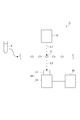
図1は、本発明の一実施形態に係る腫瘍細胞検出装置の概略構成図である。図1に示すように、腫瘍細胞検出装置1は、測定台2上の対象物3たる細胞について、光学測定に基づいて腫瘍細胞であるか否かを判定することを主な目的とした装置である。
FIG. 1 is a schematic configuration diagram of a tumor cell detection device according to an embodiment of the present invention. As shown in FIG. 1, the tumor
本実施形態に係る腫瘍細胞検出装置1は、血液中を循環する腫瘍細胞を検出することを目的としている。したがって、対象物3となるのは血液中に存在する細胞である。また、本実施形態では、検出対象となる腫瘍細胞が血中循環腫瘍細胞(CTC:Circulating tumor cell)である場合について説明するが、循環内皮細胞(CEC:Circulating Endothelial Cell)又は循環内皮前駆細胞(CEP:Circulating Endothelial Progenitor)等の他の細胞を検出対象とすることもできる。なお、血液中の細胞を直接評価する場合には、試料は血液である。また、対象物3となる細胞は、血液中から取り出された状態であってもよいし、血液又血液由来の液体中に対象物3が分散した状態で腫瘍細胞検出装置1による検出が行われてもよい。血液とは異なる液体中に対象物3が分散している場合には、試料は対象物3である細胞が分散している液体となる。図1では、対象物3の細胞が測定台2上に載置されている例を示している。
The tumor
腫瘍細胞検出装置1では、測定光を対象物3に対して照射することにより得られる透過光のスペクトルを測定し、そのスペクトルに基づいて対象物3が腫瘍細胞であるか否かを判定する。このため、腫瘍細胞検出装置1は、光源部10、検出部20、及び分析部30を備える。
The tumor
光源部10は、測定光L1を対象物3が載置される領域に対して照射する。光源部10の光源としては、ハロゲンランプ等を用いることができる。また、種光源及び非線形媒質を備え、種光源から出射される光を非線形媒質に入力し、非線形媒質中における非線形光学効果によりスペクトルを広帯域に広げてスーパーコンティニウム(SC)光として出力するSC光源を光源部10の光源として用いることもできる。SC光源を光源部10の光源として用いた場合、ハロゲンランプと比較してSC光源による加熱が低減されるため、対象物3である細胞への影響を軽減させることができる。さらに、光源部10は強度を変調する機能を有していてもよい。
The
なお、本実施形態において光源部10が照射する測定光L1の波長は特に限定されず、対象物3及び対象物3の周辺の液体等に応じて適宜選択される。測定光L1として、近赤外光を用いることができる。近赤外光とは、波長範囲が800nm〜2500nmの波長帯域の光である。なお、測定光L1として、可視光を用いることもできる。可視光とは、波長範囲が400nm〜800nmの波長帯域の光である。また、近赤外光と可視光とを組み合わせて測定光L1としてもよい。
In the present embodiment, the wavelength of the measurement light L1 irradiated by the
検出部20は、光源部10から照射される測定光L1が対象物3の表面で拡散反射された又は対象物3を透過した後に検出部20の配置される方向へ出力された光を受光し、対象物3に係る分光スペクトルとして検出する。なお、本実施形態における分光スペクトルとは、分光情報から任意の波長における強度値を抽出し、対応する波長と対にした一連のデータのことである。分光スペクトルには、波長と強度値との組み合わせが5つ以上含まれるが、波長と強度値との組み合わせの情報が多い方が、後述の分析の精度が向上する。また、分光情報とは、任意の波長における光強度情報の集合体である。具体的な例としては、反射光強度、透過光強度、吸光度などである。したがって、これを波長順に並べたものが反射光スペクトル、透過光スペクトル、吸光度スペクトルとなる。検出部20では、反射光スペクトル、透過光スペクトル及び吸光度スペクトルのいずれかを分光スペクトルとして取得する。なお、反射光には、拡散反射光、直接反射光等が含まれる。
The
検出部20は、分光器21(分光手段)と検出器22とを含む。検出部20の分光器21は、透過光L2として入射した光を波長後に分光する機能を有する。分光器21としては、例えば、波長選択フィルタ、干渉光学系、回折格子、又はプリズムを用いることができる。分光器21を上記の手段から選択する構成とした場合、検出部20において対象物3からの光を好適に分光した上で分光スペクトルを取得することができるため、光源部10に用いることが可能な光源の選択肢を広げることができる。
The
検出部20の検出器22としては、例えば、水銀、カドミウム及びテルルからなるMCT検出器、InGaAs検出器等を用いることができる。本実施形態では、対象物3からの透過光L2を受光し、分光器21によって分光した後に検出器22において透過光スペクトルを対象物3に係る分光スペクトルとして取得する構成を示している。この場合、検出部20は、対象物3を挟んで光源部10と対向する位置に設けられる。なお、対象物3からの拡散反射光スペクトルを分光スペクトルとして取得する場合には、光源部10及び検出部20は対象物3に対して同じ側に設けられる。検出部20で検出した分光スペクトルの情報は分析部30へ送られる。
As the
また、検出部20は、ハイパースペクトル画像を取得するハイパースペクトルセンサであってもよい。ハイパースペクトル画像とは、一画素がN個の波長データにより構成されている画像であり、画素毎にそれぞれ複数の波長に対応した反射強度データからなるスペクトル情報が含まれている。すなわち、ハイパースペクトル画像は、画像を構成する画素毎に、それぞれ複数波長の強度データを持つという特徴から、画像としての二次元的要素と、スペクトルデータとしての要素をあわせ持った三次元的構成のデータである。なお、本実施形態では、ハイパースペクトル画像とは、1画素あたり少なくとも5つの波長帯域における強度データを保有している画素によって構成された画像のことをいう。
The
なお、本実施形態では、検出部20では、対象物3からの透過光L2を分光した上で分光スペクトルを取得する構成について説明したが、検出部20において分光スペクトルを取得するための構成は上記に限定されない。例えば、光源部10の光源から出射する光の波長が可変である構成としてもよい。この場合、測定光L1の波長が変化するので、検出部20では測定光L1の波長変化に対応して対象物3の細胞から出射される透過光L2を順次検出することで対象物3の細胞に係る分光スペクトルを取得することができる。このように、対象物3の細胞に係る分光スペクトルを取得するための光源部10及び検出部20の構成は適宜変更することができる。
In the present embodiment, the
分析部30は、検出部20から送られる対象物3に係る分光スペクトルの情報を受け取り、演算処理等を行うことで、対象物3が腫瘍細胞であるか否かを判定する機能を有する。分析部30により、吸収スペクトルの導出、測定スペクトルの2階微分スペクトルの導出、吸収スペクトルの2階微分スペクトルの導出等を行ってもよい。
The
分析部30は、CPU(Central Processing Unit)、主記憶装置であるRAM(Random Access Memory)及びROM(Read Only Memory)、検出ユニット等の他の機器との間の通信を行う通信モジュール、並びにハードディスク等の補助記憶装置等のハードウェアを備えるコンピュータとして構成される。そして、これらの構成要素が動作することにより、分析部30としての機能が発揮される。
The
図2は、腫瘍細胞検出装置1の他の構成を説明する図である。図2では、対象物3である細胞が血液又血液由来の液体中に分散した状態で腫瘍細胞検出装置1による検出を行っている。この場合、対象物3である細胞を含む液体(血液又は血液由来の液体)を収容した試料管5から、試料管5に接続された流路51に沿って細胞を含む液体が供給される。流路51においては対象物3である細胞が液体中に分散した状態となるように内径等が調整されることで、流路51に沿って細胞が1つずつ光源部10と検出部20との間に移動する。したがって、腫瘍細胞検出装置1では、液体中の細胞に係る測定を個別に行うことができる。
FIG. 2 is a diagram illustrating another configuration of the tumor
図2に示す構成の場合には、流路51に沿って流れる液体中の対象物3に対して測定光L1を照射することにより対象物3から出射される透過光L2を検出部20で検出する。このように、対象物3に対して測定光L1を照射するための構成及び対象物3からの透過光L2を検出するための構成は適宜変更することができる。また、腫瘍細胞検出装置1では、複数の細胞に係る測定を同時に行う構成とすることもできる。この場合には、検出部20において、複数の細胞に係る分光スペクトルを同時に取得する構成とすることもできる。ただし、対象物3である細胞が腫瘍細胞であるか否かの判定は、細胞単位で行われる。このように、光源部10及び検出部20の構成は適宜変更することができる。
In the case of the configuration shown in FIG. 2, the
次に、腫瘍細胞検出装置1による検査方法について、図3を参照しながら説明する。腫瘍細胞検出装置1では、検査の対象物3である細胞に対して測定光L1を照射することで、当該細胞に係る拡散反射スペクトル又は透過スペクトルを対象物3の分光スペクトル取得する工程(S01)と、取得工程で得られた分光スペクトルに基づいて、対象物3である細胞が腫瘍細胞であるか否かを判定する工程(S02:分析工程)と、判定結果を出力する工程(S03)と、を含む。分光スペクトルを取得する工程(S01)では、光源部10から対象物3に対して、測定光L1が照射される。光源部10から照射された測定光L1は、対象物3へ入射する。対象物3を透過した光のうち、検出部20の方向に進む透過光L2は、検出部20へ到達し、検出部20において、対象物3に係る分光スペクトルとしての透過スペクトルが取得される。そして、腫瘍細胞か否かの判定を行う工程(S02)では、検出部20で得られ分析部30へ送られた分光スペクトル(透過スペクトル)に基づいて、対象物3の細胞に係る処理が行われる。
Next, an inspection method using the tumor
ここで、本実施形態に係る腫瘍細胞検出装置1による腫瘍細胞検出方法では、対象物3である細胞の分光スペクトルに基づいて腫瘍細胞であるか否かを判定する際に、統計的手法、機械学習又はパターン認識を用いることを特徴とする。この点について、具体的に説明する。
Here, in the tumor cell detection method by the tumor
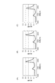
図4は、培養がん細胞(腫瘍細胞)、赤血球、及びリンパ球の近赤外光の波長帯域における分光スペクトル(1000nm〜2200nm)の例である。図4(A)は培養がん細胞の分光スペクトルであり、図4(B)は赤血球の分光スペクトルであり、図4(C)はリンパ球の分光スペクトル(吸光度スペクトル)である。なお、図4(A)〜図4(C)に示す分光スペクトルは、それぞれ、光量100%の基準スペクトル及び光量0%の基準スペクトルを用いて、各波長での吸光度を変換した後にプロットしたスペクトルである。培養がん細胞、赤血球、及びリンパ球は、いずれも血液中に含まれる細胞であるが、図4(A)〜図4(C)に示すように、分光スペクトルの形状や特定の波長での吸光度等に基づいてこれらを分類することは困難である。これに対して、統計的手法、機械学習又はパターン認識を用いることで、腫瘍細胞を他の細胞を分離する。 FIG. 4 is an example of a spectral spectrum (1000 nm to 2200 nm) in the wavelength band of near-infrared light of cultured cancer cells (tumor cells), erythrocytes, and lymphocytes. 4A is a spectral spectrum of cultured cancer cells, FIG. 4B is a spectral spectrum of red blood cells, and FIG. 4C is a spectral spectrum (absorbance spectrum) of lymphocytes. The spectral spectra shown in FIGS. 4A to 4C are spectra plotted after converting the absorbance at each wavelength using a reference spectrum with a light amount of 100% and a reference spectrum with a light amount of 0%, respectively. It is. Cultured cancer cells, erythrocytes, and lymphocytes are all cells contained in blood, but as shown in FIGS. 4 (A) to 4 (C), the shape of a spectral spectrum and a specific wavelength are used. It is difficult to classify them based on absorbance or the like. On the other hand, tumor cells are separated from other cells by using statistical methods, machine learning, or pattern recognition.
統計的手法としては、例えば、主成分分析、因子分析等を用いることができる。また、機械学習としては、例えば、サポートベクトルマシン等を用いることができる。また、パターン認識としては、例えば、MT法等を用いることができる。 As a statistical method, for example, principal component analysis, factor analysis, or the like can be used. As machine learning, for example, a support vector machine or the like can be used. As the pattern recognition, for example, the MT method can be used.
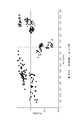
本実施形態では、腫瘍細胞を特定するための解析手法として、主成分分析(PCA:Principal Component Analysis)を用いる場合について説明する。具体的には、図4(A)〜図4(C)に示した3種類の細胞、すなわち、培養がん細胞(腫瘍細胞)、赤血球、及びリンパ球に係る吸光度スペクトルを複数取得し、これらの吸光度スペクトルを用いた主成分分析に基づいて腫瘍細胞を特定することが可能であるかを確認した。 In the present embodiment, a case where principal component analysis (PCA) is used as an analysis method for identifying tumor cells will be described. Specifically, a plurality of absorbance spectra relating to the three types of cells shown in FIGS. 4 (A) to 4 (C), that is, cultured cancer cells (tumor cells), erythrocytes, and lymphocytes, are obtained. It was confirmed whether it was possible to identify tumor cells based on principal component analysis using the absorbance spectrum of.
具体的には、培養がん細胞(腫瘍細胞)及び血液細胞(赤血球及びリンパ球)の吸光度スペクトルに基づいた主成分分析を行うことで、各吸光度スペクトルの特徴に関係する第1主成分及び第2主成分を求めた。さらに、各吸光度スペクトルにおける第1主成分のスコア値及び第2主成分のスコア値を算出し、プロットした。その結果を図5に示す。 Specifically, by performing principal component analysis based on the absorbance spectra of cultured cancer cells (tumor cells) and blood cells (erythrocytes and lymphocytes), the first principal component and the first principal component related to the characteristics of each absorbance spectrum Two main components were determined. Furthermore, the score value of the first principal component and the score value of the second principal component in each absorbance spectrum were calculated and plotted. The result is shown in FIG.
主成分分析に用いた各吸光度スペクトルから求められる第1主成分及び第2主成分のスコア値をプロットすると、図5に示すように、培養がん細胞(PC14)と、赤血球と、リンパ球とは互いに異なる群に分類することができる。なお、赤血球とリンパ球とは一部重複している部分があるが、培養がん細胞(腫瘍細胞)と血液細胞(赤血球及びリンパ球)とは明確に区別することが確認できた。このように腫瘍細胞と血液細胞とを明確に区別することができると、両者を分類するための境界条件を特定することができる。 When the score values of the first principal component and the second principal component obtained from each absorbance spectrum used in the principal component analysis are plotted, as shown in FIG. 5, cultured cancer cells (PC14), red blood cells, lymphocytes, Can be classified into different groups. In addition, although the erythrocyte and the lymphocyte have a part partially overlapping, it was confirmed that the cultured cancer cell (tumor cell) and the blood cell (erythrocyte and lymphocyte) were clearly distinguished. When tumor cells and blood cells can be clearly distinguished in this way, boundary conditions for classifying them can be specified.
したがって、種類が未知の細胞について腫瘍細胞であるか否かの判定を行う際には、上記のように、種類が既知である複数の細胞の分光スペクトルを利用して主成分分析を行い、第1主成分及び第2主成分を特定すると共に腫瘍細胞であるか否かを判定するための境界条件を求めておく。その後、判定の対象となる細胞の分光スペクトルに係る第1主成分と第2主成分に対応するスコア値を求め、境界条件との対比によってその細胞が腫瘍細胞であるか否かの判定を行う。 Therefore, when determining whether or not a cell is an unknown type of cell, the principal component analysis is performed using the spectrum of a plurality of cells of the known type as described above. A boundary condition for determining whether or not the first principal component and the second principal component are tumor cells is obtained. Thereafter, score values corresponding to the first principal component and the second principal component relating to the spectral spectrum of the cell to be determined are obtained, and it is determined whether or not the cell is a tumor cell by comparison with the boundary condition. .
このように、腫瘍細胞検出装置1及び腫瘍細胞検出方法によれば、対象物の細胞が腫瘍細胞であるか否かの判定を非接触的に行うことができる。すなわち、種類が未知の細胞の中から腫瘍細胞を非接触的に検出することが可能となる。
Thus, according to the tumor
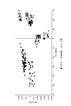
なお、図5では、波長帯域1000nm〜2200nmにおける分光スペクトルを利用してがん細胞と血液細胞とを区別した例について説明したが、分光スペクトルの波長帯域を変更した場合でも、主成分分析を利用してがん細胞と血液細胞とを区別することが可能である。図6〜図8において他の分析例を示す。 In addition, in FIG. 5, although the example which distinguished the cancer cell and the blood cell using the spectrum in wavelength band 1000nm-2200nm was demonstrated, even when the wavelength band of a spectrum is changed, a principal component analysis is utilized. Thus, it is possible to distinguish cancer cells from blood cells. 6 to 8 show other analysis examples.
図6は、波長帯域1200nm〜1500nmにおける分光スペクトルを利用してがん細胞と血液細胞とを区別した例である。また、図7は、波長帯域1500nm〜1800nmにおける分光スペクトルを利用してがん細胞と血液細胞とを区別した例である。また、図6は、波長帯域1800nm〜2100nmにおける分光スペクトルを利用してがん細胞と血液細胞とを区別した例である。図6〜図8に示す分析例で使用した分光スペクトルは図5での分析例と同じであり、分析を行う対象とした波長帯域を変更したものである。いずれの分析例においても、腫瘍細胞と血液細胞(赤血球及びリンパ球)とは明確に区別することが確認できた。このように、主成分分析を行う際に利用する分光スペクトルの波長帯域は特に限定されず、適宜変更することができる。 FIG. 6 is an example in which cancer cells and blood cells are distinguished using a spectral spectrum in a wavelength band of 1200 nm to 1500 nm. FIG. 7 is an example in which cancer cells and blood cells are distinguished using a spectrum in a wavelength band of 1500 nm to 1800 nm. FIG. 6 is an example in which cancer cells and blood cells are distinguished using a spectral spectrum in a wavelength band of 1800 nm to 2100 nm. The spectral spectra used in the analysis examples shown in FIGS. 6 to 8 are the same as those in the analysis example in FIG. 5, and are obtained by changing the wavelength band to be analyzed. In any analysis example, it was confirmed that tumor cells and blood cells (erythrocytes and lymphocytes) were clearly distinguished. As described above, the wavelength band of the spectral spectrum used when performing the principal component analysis is not particularly limited, and can be changed as appropriate.
また、腫瘍細胞検出装置1及び腫瘍細胞検出方法による腫瘍細胞の検出は、腫瘍細胞における特定の波長における吸光度等を利用したものではなく、腫瘍細胞と他の細胞(血液細胞)との分光スペクトルの形状の違いを利用したものである。したがって、上記実施形態では主成分分析を行うことで、腫瘍細胞を検出した例について説明したが、分光スペクトルの形状の違いに基づいた分類を行うことができる他の手法、すなわち、統計的手法、機械学習又はパターン認識を用いても、腫瘍細胞の検出が可能である。
Moreover, the detection of tumor cells by the tumor
例えば、サポートベクトルマシンを用いて血液細胞とそれ以外の細胞の2群に分離する場合には、種類が既知である細胞の分光スペクトルとして、血液細胞及び腫瘍細胞の分光スペクトルを予め取得する。そして、2種類の分光スペクトルを教師データとして、サポートベクトルマシンを用いて、判別関数及び判別閾値を作成する。その後、種類が未知である分析対象の細胞の分光スペクトルを取得し、解析を行う。このとき、分析対象の細胞の分光スペクトルと判別関数を掛け合わせた結果と、判別閾値とを比較することにより、その細胞が腫瘍細胞であるか否かを判定することができる。このように、分光スペクトルを用いて、統計的手法、機械学習又はパターン認識を用いて腫瘍細胞と血液細胞とを分離することができる。 For example, when separating into two groups of blood cells and other cells using a support vector machine, spectral spectra of blood cells and tumor cells are acquired in advance as spectral spectra of cells of known types. Then, a discriminant function and a discriminant threshold are created using a support vector machine using two types of spectral spectra as teacher data. Thereafter, the spectrum of the cell to be analyzed whose type is unknown is acquired and analyzed. At this time, it is possible to determine whether or not the cell is a tumor cell by comparing the result obtained by multiplying the spectral spectrum of the cell to be analyzed and the discrimination function with the discrimination threshold. In this way, the spectroscopic spectrum can be used to separate tumor cells and blood cells using statistical techniques, machine learning or pattern recognition.
さらに、上記では近赤外光の波長域における分光スペクトルを利用した分析について説明したが、その他の波長域においても腫瘍細胞と他の細胞との間で分光スペクトルの形状は変化する。したがって、他の波長域の光に対する分光スペクトルを利用した場合でも、上記の例と同様に腫瘍細胞を検出することが可能である。 Furthermore, although the analysis using the spectral spectrum in the wavelength range of near-infrared light has been described above, the shape of the spectral spectrum changes between tumor cells and other cells also in other wavelength ranges. Therefore, tumor cells can be detected in the same manner as in the above example even when a spectral spectrum for light in other wavelength ranges is used.
上記の腫瘍細胞検出装置1及び腫瘍細胞検出方法による作用効果について説明する。従来は、血液中の種々の細胞から腫瘍細胞を検出するためには、特定のマーカータンパク質により腫瘍細胞を染色する必要があった。しかしながら、上記の腫瘍細胞検出装置1及び腫瘍細胞検出方法においては、主に両者の細胞骨格の違いから生じると考えられる分光スペクトルの差異に着目することでの区別を実現している。すなわち、特定のマーカータンパクに依存しないことより、理論的にはすべての腫瘍細胞を検出することが可能となる。
The effects of the tumor
また、従来の手法では抗体を使うため、対象となる細胞におけるマーカータンパク質の発現量に検出能力が依存する。このため細胞種によっては検出が困難なものがあることが明らかになっていた。これに対して、上記の腫瘍細胞検出装置1及び腫瘍細胞検出方法では、分光スペクトルにおける血液細胞と腫瘍細胞との違いを利用して両者を判別する。したがって、マーカータンパク質の発現が少ない腫瘍細胞も検出することが可能となる。
Moreover, since the conventional technique uses an antibody, the detection ability depends on the expression level of the marker protein in the target cell. For this reason, it has become clear that some cell types are difficult to detect. On the other hand, in the above-described tumor
例えば、上記の図5〜図8の分析例で用いた分光スペクトルの取得に利用した培養がん細胞(PC14)は、培養細胞株の中でも従来から用いられている装置で抗体を使用した検出に利用するマーカータンパク質の発現量が少ない細胞株である。すなわち、従来の手法では検出が困難な細胞である。しかしながら、上記の腫瘍細胞検出装置1及び腫瘍細胞検出方法を用いることで、マーカータンパク質の多寡によらず血液細胞との分離が可能となることが確認された。このように、上記の腫瘍細胞検出装置1及び腫瘍細胞検出方法では、腫瘍細胞の種類によらず、検出精度が向上すると考えられる。
For example, the cultured cancer cells (PC14) used for obtaining the spectrum used in the analysis examples of FIGS. 5 to 8 described above can be used for detection using an antibody with a conventionally used apparatus among cultured cell lines. It is a cell line with low expression level of marker protein to be used. That is, it is a cell that is difficult to detect by conventional techniques. However, it was confirmed that by using the tumor
さらに、上記の腫瘍細胞検出装置1及び腫瘍細胞検出方法では、分光スペクトルを取得するのみで腫瘍細胞を検出できるため、非接触・非侵襲で腫瘍細胞を検出することが可能である。したがって、細胞にマーカータンパク質等の標識物質を付着する必要がないため、以降の検査、解析、研究において損傷の少ない細胞を使用することができる。
Furthermore, in the above-described tumor
腫瘍細胞を非接触に検出することが可能となると、従来から行われている腫瘍細胞数の検出に限定されず、腫瘍細胞を用いたDNA解析やタンパク質解析が大きく促進されることが予想される。また、上記実施形態で説明した手法を用いると、腫瘍細胞に関して、従来よりも信頼性の高いデータを得ることが可能となると考えられる。したがって、実地臨床でのがん診断におけるスタンダードである腫瘍組織を用いた診断の代替診断として、CTC等の血中の腫瘍細胞を用いた診断の臨床応用の可能性が広がることが期待される。また血液中の細胞から腫瘍細胞を検出することが容易になると、回収した腫瘍細胞を用いてのがん基礎研究の発展やその成果としての新規治療標的の同定等も期待される。研究及び医療機器として上記の腫瘍細胞検出装置1及び腫瘍細胞検出方法が実用化された場合には、研究を実施している研究機関やがん診療における基幹病院への導入が期待できることより、大きな経済的効果が期待できると考える。
If it becomes possible to detect tumor cells in a non-contact manner, it is expected that DNA analysis and protein analysis using tumor cells will be greatly promoted without being limited to the conventional detection of the number of tumor cells. . In addition, using the method described in the above embodiment, it is considered possible to obtain more reliable data on tumor cells than before. Therefore, it is expected that the possibility of clinical application of diagnosis using tumor cells in blood such as CTC will be expanded as an alternative diagnosis of tumor tissue which is a standard in cancer diagnosis in clinical practice. In addition, if it becomes easy to detect tumor cells from cells in the blood, the development of basic cancer research using the collected tumor cells and the identification of new therapeutic targets as a result are expected. When the above-described tumor
なお、本発明に係る腫瘍細胞検出装置1及び腫瘍細胞検出方法は上記実施形態に限定されない。例えば、上記実施形態のように腫瘍細胞検出装置1が光源部10、検出部20及び分析部30を備えている構成には限定されず、その構成は適宜変更することができる。また、腫瘍細胞検出方法は、腫瘍細胞検出装置1のように光源部10及び検出部20を備えていない装置においても実施することができる。すなわち、他の装置で取得した分光スペクトルを利用して分析を行う構成であってもよい。
The tumor
1…腫瘍細胞検出装置、2…測定台、3…対象物、5…試料管、10…光源部、20…検出部、30…分析部。
DESCRIPTION OF
Claims (7)
前記分析工程において、腫瘍細胞及びその他の血液中の細胞を測定することにより得られた複数の分光スペクトルに基づいて、統計的手法、機械学習又はパターン認識により、腫瘍細胞であるか否かを判定するための境界条件を算出し、前記細胞を測定することにより得られた分光スペクトルと前記境界条件とに基づいて、前記細胞が腫瘍細胞であるか否かを判定する請求項1又は2に記載の腫瘍細胞検出方法。 The cell is a cell contained in blood,
In the analysis step, based on a plurality of spectral spectra obtained by measuring tumor cells and other blood cells, whether or not they are tumor cells is determined by statistical techniques, machine learning, or pattern recognition. The boundary condition for calculating is determined based on the spectrum obtained by measuring the cell and the boundary condition, and whether or not the cell is a tumor cell is determined. Tumor cell detection method.
前記光源部からの前記測定光の照射によって出射される前記細胞からの透過光又は反射光を受光することで当該細胞に係る分光スペクトルを取得する検出部と、
前記検出部において取得された前記分光スペクトルに基づいて、統計的手法、機械学習又はパターン認識により、当該細胞が腫瘍細胞であるか否かを判定する分析部と、
を有する腫瘍細胞検出装置。 A light source unit for irradiating measurement light to cells contained in the sample;
A detection unit that acquires a spectral spectrum of the cell by receiving transmitted light or reflected light from the cell emitted by irradiation of the measurement light from the light source unit;
Based on the spectral spectrum acquired in the detection unit, by a statistical method, machine learning or pattern recognition, an analysis unit that determines whether the cell is a tumor cell,
A tumor cell detection device comprising:
前記分析部は、腫瘍細胞及びその他の血液中の細胞を測定することにより得られた複数の分光スペクトルに基づいて、統計的手法、機械学習又はパターン認識により、腫瘍細胞であるか否かを判定するための境界条件を算出しておき、前記試料に含まれる細胞を測定することにより得られた分光スペクトルと前記境界条件とに基づいて、前記細胞が腫瘍細胞であるか否かを判定する請求項4又は5に記載の腫瘍細胞検出装置。 The cell is a cell contained in blood,
The analysis unit determines whether the tumor cell is a tumor cell by statistical techniques, machine learning, or pattern recognition based on a plurality of spectral spectra obtained by measuring tumor cells and other blood cells. And determining whether or not the cell is a tumor cell based on a spectral spectrum obtained by measuring a cell contained in the sample and the boundary condition. Item 6. The tumor cell detection device according to Item 4 or 5.
前記分光手段は、波長選択フィルタ、干渉光学系、回折格子、又はプリズムである請求項4〜6のいずれか一項に記載の腫瘍細胞検出装置。 The detection unit has spectroscopic means for spectroscopically analyzing transmitted light or reflected light from the cell,
The tumor cell detection device according to any one of claims 4 to 6, wherein the spectroscopic means is a wavelength selection filter, an interference optical system, a diffraction grating, or a prism.
Priority Applications (5)
| Application Number | Priority Date | Filing Date | Title |
|---|---|---|---|
| JP2016093841A JP2017203637A (en) | 2016-05-09 | 2016-05-09 | Tumor cell detection method and tumor cell detection device |
| EP17796133.1A EP3457116A4 (en) | 2016-05-09 | 2017-05-09 | Tumor cell detection method and tumor cell detection device |
| PCT/JP2017/017529 WO2017195772A1 (en) | 2016-05-09 | 2017-05-09 | Tumor cell detection method and tumor cell detection device |
| CN201780028246.1A CN109073547A (en) | 2016-05-09 | 2017-05-09 | Tumour cell detection method and tumour cell detection device |
| US16/182,644 US20190072484A1 (en) | 2016-05-09 | 2018-11-07 | Tumor cell detection method and tumor cell detection device |
Applications Claiming Priority (1)
| Application Number | Priority Date | Filing Date | Title |
|---|---|---|---|
| JP2016093841A JP2017203637A (en) | 2016-05-09 | 2016-05-09 | Tumor cell detection method and tumor cell detection device |
Publications (1)
| Publication Number | Publication Date |
|---|---|
| JP2017203637A true JP2017203637A (en) | 2017-11-16 |
Family
ID=60267992
Family Applications (1)
| Application Number | Title | Priority Date | Filing Date |
|---|---|---|---|
| JP2016093841A Pending JP2017203637A (en) | 2016-05-09 | 2016-05-09 | Tumor cell detection method and tumor cell detection device |
Country Status (5)
| Country | Link |
|---|---|
| US (1) | US20190072484A1 (en) |
| EP (1) | EP3457116A4 (en) |
| JP (1) | JP2017203637A (en) |
| CN (1) | CN109073547A (en) |
| WO (1) | WO2017195772A1 (en) |
Cited By (5)
| Publication number | Priority date | Publication date | Assignee | Title |
|---|---|---|---|---|
| JP6392476B1 (en) * | 2018-03-19 | 2018-09-19 | 大輝 中矢 | Biological tissue analysis apparatus and biological tissue analysis program |
| JP2020034551A (en) * | 2018-08-24 | 2020-03-05 | 国立大学法人鳥取大学 | Device and method for identifying cells |
| CN111257558A (en) * | 2020-01-19 | 2020-06-09 | 江苏省人民医院(南京医科大学第一附属医院) | Machine learning-based chronic lymphocytic leukemia tumor cell identification method |
| WO2021015604A1 (en) * | 2019-07-25 | 2021-01-28 | 서울바이오시스주식회사 | Light irradiation apparatus |
| WO2021256514A1 (en) * | 2020-06-17 | 2021-12-23 | 大輝 中矢 | Living cell analysis device, living cell analysis system, living cell analysis program, and living cell analysis method |
Families Citing this family (3)
| Publication number | Priority date | Publication date | Assignee | Title |
|---|---|---|---|---|
| US20220270245A1 (en) * | 2019-07-24 | 2022-08-25 | Saitama Medical University | Estimator learning device, estimator learning method, and estimator learning program |
| CN113189040A (en) * | 2021-04-28 | 2021-07-30 | 北京大学第三医院(北京大学第三临床医学院) | Method and system for efficiently and nondestructively detecting number and activity of tumor cells in sample |
| CN115791640B (en) * | 2023-02-06 | 2023-06-02 | 杭州华得森生物技术有限公司 | Tumor cell detection equipment and method based on spectroscopic spectrum |
Family Cites Families (14)
| Publication number | Priority date | Publication date | Assignee | Title |
|---|---|---|---|---|
| JPS50832A (en) | 1973-04-28 | 1975-01-07 | ||
| CA2035603C (en) * | 1991-02-04 | 1998-08-04 | Patrick T.T. Wong | A method of detecting the presence of anomalies exfoliated cells using infrared spectroscopy |
| US5991028A (en) * | 1991-02-22 | 1999-11-23 | Applied Spectral Imaging Ltd. | Spectral bio-imaging methods for cell classification |
| US5596992A (en) * | 1993-06-30 | 1997-01-28 | Sandia Corporation | Multivariate classification of infrared spectra of cell and tissue samples |
| US5733739A (en) * | 1995-06-07 | 1998-03-31 | Inphocyte, Inc. | System and method for diagnosis of disease by infrared analysis of human tissues and cells |
| US6031232A (en) * | 1995-11-13 | 2000-02-29 | Bio-Rad Laboratories, Inc. | Method for the detection of malignant and premalignant stages of cervical cancer |
| US8326404B2 (en) * | 2003-11-28 | 2012-12-04 | British Columbia Cancer Agency Branch | Multimodal detection of tissue abnormalities based on raman and background fluorescence spectroscopy |
| KR20070107743A (en) * | 2005-01-31 | 2007-11-07 | 더 보오드 오브 트러스티스 오브 더 유니버시티 오브 일리노이즈 | Methods and devices for characterizing particles in clear and turbid media |
| WO2008004665A1 (en) * | 2006-07-06 | 2008-01-10 | Fatigue Science Laboratory Inc. | Method of testing, and apparatus therefor, as to cancer, systemic lupus erythematosus (sle) or antiphospholipid antibody syndrome, using near-infrared ray |
| US9025850B2 (en) * | 2010-06-25 | 2015-05-05 | Cireca Theranostics, Llc | Method for analyzing biological specimens by spectral imaging |
| US8941062B2 (en) * | 2010-11-16 | 2015-01-27 | 1087 Systems, Inc. | System for identifying and sorting living cells |
| JP2012022002A (en) | 2011-09-12 | 2012-02-02 | Veridex Llc | Method of predicting progression-free and overall survival of metastatic breast cancer patient at each point of follow-up period using circulating tumor cell |
| JP6183826B2 (en) * | 2013-03-12 | 2017-08-23 | 国立大学法人 筑波大学 | Optical measuring method, optical measuring apparatus and optical recording medium of light scatterer |
| JP2015102542A (en) * | 2013-11-28 | 2015-06-04 | 住友電気工業株式会社 | Biological inspection device |
-
2016
- 2016-05-09 JP JP2016093841A patent/JP2017203637A/en active Pending
-
2017
- 2017-05-09 EP EP17796133.1A patent/EP3457116A4/en not_active Withdrawn
- 2017-05-09 CN CN201780028246.1A patent/CN109073547A/en active Pending
- 2017-05-09 WO PCT/JP2017/017529 patent/WO2017195772A1/en unknown
-
2018
- 2018-11-07 US US16/182,644 patent/US20190072484A1/en not_active Abandoned
Cited By (11)
| Publication number | Priority date | Publication date | Assignee | Title |
|---|---|---|---|---|
| JP6392476B1 (en) * | 2018-03-19 | 2018-09-19 | 大輝 中矢 | Biological tissue analysis apparatus and biological tissue analysis program |
| JP2019163981A (en) * | 2018-03-19 | 2019-09-26 | 大輝 中矢 | Tissue analysis device and biological tissue analysis program |
| WO2019181845A1 (en) | 2018-03-19 | 2019-09-26 | 一般財団法人未来科学研究所 | Biological tissue analyzing device, biological tissue analyzing program, and biological tissue analyzing method |
| US11499958B2 (en) | 2018-03-19 | 2022-11-15 | Daiki NAKAYA | Biological tissue analyzing device, biological tissue analyzing program, and biological tissue analyzing method |
| JP2020034551A (en) * | 2018-08-24 | 2020-03-05 | 国立大学法人鳥取大学 | Device and method for identifying cells |
| JP7204121B2 (en) | 2018-08-24 | 2023-01-16 | 国立大学法人鳥取大学 | CELL IDENTIFICATION DEVICE AND CELL IDENTIFICATION METHOD |
| WO2021015604A1 (en) * | 2019-07-25 | 2021-01-28 | 서울바이오시스주식회사 | Light irradiation apparatus |
| CN111257558A (en) * | 2020-01-19 | 2020-06-09 | 江苏省人民医院(南京医科大学第一附属医院) | Machine learning-based chronic lymphocytic leukemia tumor cell identification method |
| CN111257558B (en) * | 2020-01-19 | 2021-08-24 | 江苏省人民医院(南京医科大学第一附属医院) | Machine learning-based chronic lymphocytic leukemia tumor cell identification method |
| WO2021256514A1 (en) * | 2020-06-17 | 2021-12-23 | 大輝 中矢 | Living cell analysis device, living cell analysis system, living cell analysis program, and living cell analysis method |
| JP7427289B2 (en) | 2020-06-17 | 2024-02-05 | Milk.株式会社 | Living cell analysis device, living cell analysis system, living cell analysis program, and living cell analysis method |
Also Published As
| Publication number | Publication date |
|---|---|
| WO2017195772A1 (en) | 2017-11-16 |
| CN109073547A (en) | 2018-12-21 |
| US20190072484A1 (en) | 2019-03-07 |
| EP3457116A1 (en) | 2019-03-20 |
| EP3457116A4 (en) | 2019-03-20 |
Similar Documents
| Publication | Publication Date | Title |
|---|---|---|
| WO2017195772A1 (en) | Tumor cell detection method and tumor cell detection device | |
| Nissim et al. | Real‐time stain‐free classification of cancer cells and blood cells using interferometric phase microscopy and machine learning | |
| US8241238B2 (en) | Cell selection apparatus | |
| JP4982385B2 (en) | Analysis of blood and cells using an imaging flow cytometer | |
| US3916197A (en) | Method and apparatus for classifying biological cells | |
| US11280720B2 (en) | Cell analysis method, cell analyzer and sample screening method | |
| Agsalda-Garcia et al. | Raman-enhanced spectroscopy (RESpect) probe for childhood non-Hodgkin lymphoma | |
| US20110028808A1 (en) | Method and apparatus for examination of cancer, systemic lupus erythematosus (sle), or antiphospholipid antibody syndrome using near-infrared light | |
| JP3248905B2 (en) | Method for analyzing biological substances having a water content | |
| EP2843410B1 (en) | Sample analyzing method and sample analyzer | |
| WO2016080442A1 (en) | Quality evaluation method and quality evaluation device | |
| US20200300768A1 (en) | Determination device, determination method, and determination program | |
| CN103604737A (en) | Automatic blood cell recognition device and operating method thereof | |
| CN108474691A (en) | Immunoassay system based on Raman and method | |
| US10585033B2 (en) | Microparticle measuring device and microparticle analysis method | |
| CN112204378A (en) | Biological tissue analysis device, biological tissue analysis program, and biological tissue analysis method | |
| KR102500220B1 (en) | Method and apparatus for classifying of cell subtype using three-dimensional refractive index tomogram and machine learning | |
| Happillon et al. | Diagnosis approach of chronic lymphocytic leukemia on unstained blood smears using Raman microspectroscopy and supervised classification | |
| Song et al. | Micro ATR-FTIR spectroscopic imaging of colon biopsies with a large area Ge crystal | |
| Verdonck et al. | Label-free phenotyping of peripheral blood lymphocytes by infrared imaging | |
| Ciobanu et al. | Potential of Raman spectroscopy for blood-based biopsy | |
| Krafft et al. | Micro-Raman spectroscopy in medicine | |
| Akalin et al. | Resolving interobserver discrepancies in lung cancer diagnoses by spectral histopathology | |
| US20230258554A1 (en) | A system and method thereof for real-time automatic label-free holography-activated sorting of cells | |
| JP2015102542A (en) | Biological inspection device |