JP2017193550A - Nanoparticle delivery systems and preparation and uses thereof - Google Patents
Nanoparticle delivery systems and preparation and uses thereof Download PDFInfo
- Publication number
- JP2017193550A JP2017193550A JP2017093099A JP2017093099A JP2017193550A JP 2017193550 A JP2017193550 A JP 2017193550A JP 2017093099 A JP2017093099 A JP 2017093099A JP 2017093099 A JP2017093099 A JP 2017093099A JP 2017193550 A JP2017193550 A JP 2017193550A
- Authority
- JP
- Japan
- Prior art keywords
- nanoparticles
- thermosensitive
- nanoparticle
- liposomes
- liposome
- Prior art date
- Legal status (The legal status is an assumption and is not a legal conclusion. Google has not performed a legal analysis and makes no representation as to the accuracy of the status listed.)
- Pending
Links
Images

Classifications
-
- A—HUMAN NECESSITIES
- A61—MEDICAL OR VETERINARY SCIENCE; HYGIENE
- A61K—PREPARATIONS FOR MEDICAL, DENTAL OR TOILETRY PURPOSES
- A61K9/00—Medicinal preparations characterised by special physical form
- A61K9/10—Dispersions; Emulsions
- A61K9/127—Liposomes
-
- A—HUMAN NECESSITIES
- A61—MEDICAL OR VETERINARY SCIENCE; HYGIENE
- A61K—PREPARATIONS FOR MEDICAL, DENTAL OR TOILETRY PURPOSES
- A61K9/00—Medicinal preparations characterised by special physical form
- A61K9/10—Dispersions; Emulsions
- A61K9/127—Liposomes
- A61K9/1271—Non-conventional liposomes, e.g. PEGylated liposomes, liposomes coated with polymers
-
- A—HUMAN NECESSITIES
- A61—MEDICAL OR VETERINARY SCIENCE; HYGIENE
- A61K—PREPARATIONS FOR MEDICAL, DENTAL OR TOILETRY PURPOSES
- A61K41/00—Medicinal preparations obtained by treating materials with wave energy or particle radiation ; Therapies using these preparations
- A61K41/0028—Disruption, e.g. by heat or ultrasounds, sonophysical or sonochemical activation, e.g. thermosensitive or heat-sensitive liposomes, disruption of calculi with a medicinal preparation and ultrasounds
-
- A—HUMAN NECESSITIES
- A61—MEDICAL OR VETERINARY SCIENCE; HYGIENE
- A61K—PREPARATIONS FOR MEDICAL, DENTAL OR TOILETRY PURPOSES
- A61K49/00—Preparations for testing in vivo
- A61K49/0002—General or multifunctional contrast agents, e.g. chelated agents
-
- A—HUMAN NECESSITIES
- A61—MEDICAL OR VETERINARY SCIENCE; HYGIENE
- A61K—PREPARATIONS FOR MEDICAL, DENTAL OR TOILETRY PURPOSES
- A61K49/00—Preparations for testing in vivo
- A61K49/06—Nuclear magnetic resonance [NMR] contrast preparations; Magnetic resonance imaging [MRI] contrast preparations
- A61K49/18—Nuclear magnetic resonance [NMR] contrast preparations; Magnetic resonance imaging [MRI] contrast preparations characterised by a special physical form, e.g. emulsions, microcapsules, liposomes
- A61K49/1806—Suspensions, emulsions, colloids, dispersions
- A61K49/1812—Suspensions, emulsions, colloids, dispersions liposomes, polymersomes, e.g. immunoliposomes
-
- A—HUMAN NECESSITIES
- A61—MEDICAL OR VETERINARY SCIENCE; HYGIENE
- A61K—PREPARATIONS FOR MEDICAL, DENTAL OR TOILETRY PURPOSES
- A61K9/00—Medicinal preparations characterised by special physical form
- A61K9/48—Preparations in capsules, e.g. of gelatin, of chocolate
- A61K9/50—Microcapsules having a gas, liquid or semi-solid filling; Solid microparticles or pellets surrounded by a distinct coating layer, e.g. coated microspheres, coated drug crystals
- A61K9/51—Nanocapsules; Nanoparticles
-
- A—HUMAN NECESSITIES
- A61—MEDICAL OR VETERINARY SCIENCE; HYGIENE
- A61P—SPECIFIC THERAPEUTIC ACTIVITY OF CHEMICAL COMPOUNDS OR MEDICINAL PREPARATIONS
- A61P35/00—Antineoplastic agents
-
- A—HUMAN NECESSITIES
- A61—MEDICAL OR VETERINARY SCIENCE; HYGIENE
- A61P—SPECIFIC THERAPEUTIC ACTIVITY OF CHEMICAL COMPOUNDS OR MEDICINAL PREPARATIONS
- A61P35/00—Antineoplastic agents
- A61P35/02—Antineoplastic agents specific for leukemia
-
- A—HUMAN NECESSITIES
- A61—MEDICAL OR VETERINARY SCIENCE; HYGIENE
- A61K—PREPARATIONS FOR MEDICAL, DENTAL OR TOILETRY PURPOSES
- A61K9/00—Medicinal preparations characterised by special physical form
- A61K9/48—Preparations in capsules, e.g. of gelatin, of chocolate
- A61K9/50—Microcapsules having a gas, liquid or semi-solid filling; Solid microparticles or pellets surrounded by a distinct coating layer, e.g. coated microspheres, coated drug crystals
- A61K9/51—Nanocapsules; Nanoparticles
- A61K9/5107—Excipients; Inactive ingredients
- A61K9/5115—Inorganic compounds
Abstract
Description
本願は、健康部門、特にヒトの健康において用いることができる、制御放出を可能にするナノ粒子デリバリーシステム、特にTm(ゲル−液晶相転移温度)またはTm以上で破壊される熱感受性リポソームに関する。
本発明の熱感受性リポソームは、生理学的pHの水性媒質中で測定した時に、表面静電気が好都合には−20mV以下または+20mV以上であるナノ粒子を封入する熱感受性脂質膜を含む。該封入されたナノ粒子は治療薬または診断薬として用いることができる。
本発明は、先に記載のナノ粒子デリバリーシステムを含む医薬組成物および診断用組成物、およびその使用にも関する。
The present application relates to nanoparticulate delivery systems that allow controlled release that can be used in the health sector, in particular human health, in particular thermosensitive liposomes that break at Tm (gel-liquid crystal phase transition temperature) or above Tm. .
The thermosensitive liposomes of the present invention comprise a thermosensitive lipid membrane that encapsulates nanoparticles that have a surface electrostatic charge of conveniently −20 mV or less or +20 mV or more when measured in an aqueous medium of physiological pH. The encapsulated nanoparticles can be used as a therapeutic or diagnostic agent.
The present invention also relates to pharmaceutical and diagnostic compositions comprising the nanoparticle delivery system described above, and uses thereof.
治療薬または診断薬を用いる伝統的医学的治療の限界は特異性がないことである。特に、ほとんどの場合、治療薬または診断薬の投与した用量のほんの一部だけが目的とする部位に達するが、残りの薬剤は身体全体に分散する。健康な器官と組織へのこの避けられない分散は、患者に投与することができる薬剤量を制限し、この薬剤が可能な治療または診断効果をあげるのを妨げる。
目的部位に達する薬剤量を増加し、身体の他の健康な部分に送達される量も減少させる部位特異的薬剤送達ビークルの必要性が、特に毒性化学療法剤について永きにわたり認識されてきた。副作用を減少させるかまたは排除することができるそのようなビークルは、該治療を著しくより低毒性かつより有効にするだろう。リポソームは、治療薬または診断薬のためのナノスケールの送達ビークルとして10年におよび臨床的存在であった。
あらゆる薬剤送達ビークルが直面する最大の問題は、疾患部位に特異的に制御可能な速度で該ビークルからの封入した薬剤の完全な放出を可能にすることである。
The limitation of traditional medical treatment with therapeutic or diagnostic agents is the lack of specificity. In particular, in most cases, only a fraction of the administered dose of the therapeutic or diagnostic agent reaches the intended site, while the remaining drug is dispersed throughout the body. This unavoidable dispersion in healthy organs and tissues limits the amount of drug that can be administered to a patient and prevents the drug from providing a possible therapeutic or diagnostic effect.
The need for site-specific drug delivery vehicles that increase the amount of drug reaching the target site and also reduce the amount delivered to other healthy parts of the body has long been recognized, especially for toxic chemotherapeutic agents. Such vehicles that can reduce or eliminate side effects would make the treatment significantly less toxic and more effective. Liposomes have been in clinical existence for 10 years as nanoscale delivery vehicles for therapeutic or diagnostic agents.
The biggest problem faced by any drug delivery vehicle is to allow complete release of the encapsulated drug from the vehicle at a rate that can be controlled specifically at the disease site.
さらに、ナノ粒子のための送達ビークルとしてのリポソームの使用は、特に外部活性型ナノ粒子との関連で、まだ前臨床開発段階である(Al−Jamal W.T. et al. Nanomedicine、2007;2:85−98)。 Furthermore, the use of liposomes as delivery vehicles for nanoparticles is still in the preclinical development phase, particularly in the context of externally active nanoparticles (Al-Jamal WT et al. Nanomedicine, 2007; 2: 85- 98).
エンドソーム膜の不安定化を促進し、量子ドット(QD)細胞質放出に有利に作用する融合性またはpH感受性脂質を含むPEG−脂質を用いて構築するリポソームの製造はin vitroで記載されている(Sigot et al. Bioconjugate Chem. 2010;21:1465−1472)。リポソームからのPEG−脂質の解離は、リポソームからの二重層への数分間以内での移動を促進する短アシル鎖を有する融合性PEG−脂質を組み込むことにより促進することができる。あるいはまた、PEG−脂質は、ある種のエンドソームコンパートメントの酸性環境に暴露するとポリマー部分がリポソーム表面から開裂される、開裂可能なpH感受性PEG類似体を加えることにより細胞内に放出可能である。そのような「自発的」放出は、局所環境にのみ依存するので、(特に遠隔転移の治療に)好都合でありうるが、リポソーム内容の放出は、依然遅くなり得るし、環境が最適でなければ全く起きないかもしれない。したがって、リポソーム内容の放出の正確な制御はそのようなリポソームでは不可能である。 The production of liposomes constructed with PEG-lipids containing fusogenic or pH-sensitive lipids that promotes destabilization of the endosomal membrane and favors quantum dot (QD) cytoplasmic release has been described in vitro ( Sigot et al. Bioconjugate Chem. 2010; 21: 1465-1472). The dissociation of PEG-lipid from liposomes can be facilitated by incorporating fusogenic PEG-lipids with short acyl chains that facilitate migration within a few minutes from the liposome to the bilayer. Alternatively, PEG-lipids can be released intracellularly by adding a cleavable pH sensitive PEG analog, where the polymer moiety is cleaved from the liposome surface upon exposure to the acidic environment of certain endosomal compartments. Such “spontaneous” release can be advantageous (especially for the treatment of distant metastases) since it depends only on the local environment, but release of the liposome content can still be slow and if the environment is not optimal It may not happen at all. Thus, precise control of liposome content release is not possible with such liposomes.
光感作物質を含むリポソームの製造がさらに記載されている(US 2010/0233224)。光感作物質は、脂質鎖の過酸化を介して光および酸素に暴露するとリポソーム膜の不飽和リン脂質を酸化させることができる。リポソームの光酸化は、オンデマンドで外部光刺激を介して急速にその充填物の放出を引き起こすことができる。酸化は、リポソーム膜不全およびそれに続くリポソーム内容の放出に関与する。しかしながら、そのような光供給源は、標的組織が表面的に接近可能な場合にのみ用いることができる。より深い組織に取り込まれたリポソームは、光で刺激することができない。したがって、光感作物質を含むリポソームは、人体の深部器官または構造にナノ粒子を送達するのに用いることができない。 The production of liposomes containing photosensitizers is further described (US 2010/0233224). Photosensitizers can oxidize unsaturated phospholipids of liposome membranes when exposed to light and oxygen through lipid chain peroxidation. The photooxidation of liposomes can cause the release of its packing rapidly via external light stimulation on demand. Oxidation is responsible for liposome membrane failure and subsequent release of the liposome content. However, such a light source can only be used when the target tissue is superficially accessible. Liposomes taken into deeper tissues cannot be stimulated with light. Therefore, liposomes containing photosensitizers cannot be used to deliver nanoparticles to deep organs or structures of the human body.
レーザー活性型中空金属ナノ構造物も、過去に、それが薬剤の選択的放出を可能にするために封入されたリポソーム膜の透過化を引き起こす手段として用いられた(WO 2009/097480)。 Laser activated hollow metal nanostructures have also been used in the past as a means to cause permeabilization of the encapsulated liposome membrane to allow selective release of the drug (WO 2009/097480).
US 2009004258は、常磁性酸化鉄ナノ粒子および薬剤封入熱感受性リポソームを開示しており、この常磁性酸化鉄ナノ粒子は交番磁場による活性下、標的環境中の薬剤の特異的または選択的放出を可能にする。 US 2009004258 discloses paramagnetic iron oxide nanoparticles and drug-encapsulated thermosensitive liposomes, which are capable of specific or selective release of drugs in the target environment under activity by an alternating magnetic field To.
本発明者らは、対象にナノ粒子の安全なin vivo送達、および制御された効率的放出を可能にする好都合な系を提供する。
特に、これらの系は、人体の深部構造中で外部活性型ナノ粒子の送達および放出を可能にする。診断および/または治療的手段として用いることができる効率的活性型ナノ粒子の例は、WO2007/118884、WO2009/147214、およびWO2011/003999に本発明者らが記載した。
We provide a convenient system that allows safe in vivo delivery of nanoparticles and controlled and efficient release to a subject.
In particular, these systems allow the delivery and release of externally active nanoparticles in the deep structure of the human body. Examples of efficient active nanoparticles that can be used as diagnostic and / or therapeutic tools were described by the inventors in WO2007 / 118884, WO2009 / 147214, and WO2011 / 003999.
(発明の要約)
本発明者らは、TmまたはTm以上で破壊される熱感受性リポソームであって、該リポソームがナノ粒子を封入する熱感受性脂質膜を含み、生理学的pH(6〜8)の水性溶媒中で測定したときにナノ粒子の「表面静電気」(本明細書では「荷電」または「表面荷電」ともいう)が−20mV以下または+20mV以上であり、該ナノ粒子が治療薬または診断薬として用いることができる、該リポソームを提供する。
(Summary of the Invention)
The present inventors have found that a heat-sensitive liposomes are destroyed in T m or T m above include heat sensitive lipid membrane the liposome encapsulating the nanoparticles, an aqueous solvent physiological pH (6-8) The “surface static electricity” (also referred to as “charge” or “surface charge” in this specification) of a nanoparticle when measured by the above is −20 mV or less or +20 mV or more, and the nanoparticle is used as a therapeutic agent or a diagnostic agent The liposome is provided.
さらに、本発明者らは、本発明の熱感受性リポソームおよび医薬的に許容される担体を含む治療用および診断用組成物を提供する。 In addition, we provide therapeutic and diagnostic compositions comprising the thermosensitive liposomes of the present invention and a pharmaceutically acceptable carrier.
別の局面において、本発明は、本明細書に記載の生成物のいずれか一つまたはそれ以上、すなわち、熱感受性リポソームおよび組成物を、該生成物を用いるための指示を与える注意書きラベルと共に含むキットを提供する。 In another aspect, the present invention provides any one or more of the products described herein, i.e., thermosensitive liposomes and compositions, with a precautionary label that provides instructions for using the product. Provide kits containing.
本発明の熱感受性リポソームは、生理学的環境、特に、本明細書において細網内皮系(RES)ともいう単核食細胞系による早期捕捉またはオプソニン化からナノ粒子を好都合に保護することができる。 The thermosensitive liposomes of the present invention can advantageously protect the nanoparticles from premature entrapment or opsonization by a mononuclear phagocyte system, also referred to herein as the reticuloendothelial system (RES).
したがって、本発明のリポソームは、熱活性化により完全なナノ粒子を送達および放出することができる(熱活性化は、温度の生理学的増加によるか、または例えば、電離放射線または高密度焦点式超音波を用いる外部活性化により達成することができる。)。目的部位で放出されると、該ナノ粒子は、さらに下記で説明するように、所望により外部活性化を介して治療薬または診断薬として機能することができる。 Thus, the liposomes of the present invention can deliver and release intact nanoparticles upon thermal activation (thermal activation is due to a physiological increase in temperature or, for example, ionizing radiation or high intensity focused ultrasound. Can be achieved by external activation using. Once released at the site of interest, the nanoparticles can function as a therapeutic or diagnostic agent via external activation if desired, as further described below.
本明細書に記載の熱感受性リポソームは、さらに、血管経路を介して対象の身体の目的部位、特に深部部位または構造にナノ粒子を好都合に送達することができる。
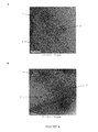
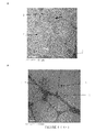
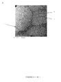
ナノ粒子放出の正確で効率的な制御も(送達に加えて)今や可能である。本明細書に記載の熱感受性リポソームからのナノ粒子の放出は、Tmと等しいかまたはTm以上の温度Trで本発明者らにより証明された。リポソーム膜破壊(膜の物理的崩壊、断裂、または破壊)の原因となるナノ粒子と脂質二重層の相互作用は、この驚くべき結果を説明しうる(図4E、黒矢印参照)。
The thermosensitive liposomes described herein can further conveniently deliver nanoparticles to a target site, particularly a deep site or structure, of a subject's body via a vascular route.
Accurate and efficient control of nanoparticle release is now also possible (in addition to delivery). Release of the nanoparticles from the heat-sensitive liposomes described herein, has been demonstrated by the inventors in T m equal to or T m above the temperature T r. The interaction of nanoparticles and lipid bilayers that cause liposome membrane disruption (physical disruption, rupture, or disruption of the membrane) may explain this surprising result (see FIG. 4E, black arrows).
(本発明の詳細な説明)
本明細書において本発明者らは、Tm(ゲル−液晶相転移温度)またはTm以上で破壊される熱感受性リポソームを提供する。このリポソームは、対象に治療薬または診断薬として用いることができるナノ粒子を封入する熱感受性脂質膜を含む。
(Detailed Description of the Invention)
Here we provide thermosensitive liposomes that break at Tm (gel-liquid crystal phase transition temperature) or above Tm . The liposome includes a heat sensitive lipid membrane that encapsulates nanoparticles that can be used as a therapeutic or diagnostic agent in a subject.
本発明者らは、驚くべきことに、生理学的pH(典型的にはpH6〜pH8)の水性媒質中で測定したときにナノ粒子の表面静電気が−20mV以下または+20mV以上のとき、TmまたはTm以上で熱感受性リポソームの破壊が観察した。このリポソーム膜の破壊は、封入された荷電ナノ粒子の放出を可能にする。 The inventors surprisingly found that when the surface electrostatic charge of the nanoparticles is −20 mV or less or +20 mV or more when measured in an aqueous medium at physiological pH (typically pH 6 to pH 8), the T m or The destruction of thermosensitive liposomes was observed above Tm . This disruption of the liposome membrane allows the release of the encapsulated charged nanoparticles.
本明細書で用いている用語「対象」は、あらゆる生物を意味する。該用語は、対象の一例であるヒトを排他的に表わす必要はなく、動物、特に温血脊椎動物、典型的には哺乳動物、およびさらに細胞培養も表しうる。 As used herein, the term “subject” means any organism. The term need not refer exclusively to an example human subject, but can also refer to animals, particularly warm-blooded vertebrates, typically mammals, and even cell cultures.
リポソーム
用語「リポソーム」は、小胞内媒質を外部媒質から分離する膜を形成する両親媒性分子の少なくとも1の二重層からなる球状小胞を表す。小胞内媒質は、該リポソームの内部水性コアを構成する。親水性分子または成分は、当業者に知られ、さらに本明細書で以下に記載の能動封入法を介してリポソームの内部水性コア内に封入することができる。疎水性分子または成分は、該膜の内側に取り込むことができる。
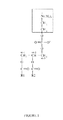
該二重層を構成する両親媒性分子は、脂質、より具体的にはリン脂質である。リン脂質分子の両親媒性特性は、リン酸基およびグリセロール基で構成される親水性頭部と1または2の脂肪酸で構成される疎水性尾部の存在による(図1参照)。
The liposome term "liposome" refers to a spherical vesicle composed of at least one bilayer of amphiphilic molecules that forms a membrane that separates the vesicular medium from the external medium. The intravesicular medium constitutes the inner aqueous core of the liposome. Hydrophilic molecules or components are known to those skilled in the art and can be encapsulated within the inner aqueous core of liposomes via the active encapsulation methods described herein below. Hydrophobic molecules or components can be incorporated inside the membrane.
The amphiphilic molecule constituting the bilayer is a lipid, more specifically a phospholipid. The amphipathic properties of phospholipid molecules are due to the presence of a hydrophilic head composed of phosphate and glycerol groups and a hydrophobic tail composed of 1 or 2 fatty acids (see FIG. 1).
水性媒質において、リン脂質は、脂肪アシル鎖と水の接触を最小限にするために自己会合(self−assemble)する傾向があり、その化学構造にしたがって種々のタイプの会合体(ミセル、ラメラ相など)をとる傾向がある。より具体的には、ホスファチジルコリンは、「自発」屈曲を受け、最終的に小胞を形成する積層二重層からなるラメラ相を形成することが知られている(Lasic D.D. et al. Adv. Colloid. Interf. Sci. 2001;89−90:337−349)。リン脂質ラメラ相は、サーモトロピック液晶を構成する。これは、両親媒性分子の秩序化(ordering)度は、温度に依存することを意味する。実際に、リン脂質二重層は、「ゲル様」ラメラ相Lβから「液状」ラメラ相Lαへの遷移に対応する主相遷移温度Tm(「融解」温度)を示す。「ゲル」相において、脂肪酸の炭酸鎖間の強い疎水性相互作用がリン脂質分子の結晶秩序化を引き起こし、該二重層は小イオンに対してのみ透過性である。「液」相において、疎水性尾部は、リン脂質分子の秩序化の損失を引き起こし、「液晶」相をもたらす熱運動により移動し、該二重層は薬剤などの分子に対して透過性になる。 In aqueous media, phospholipids tend to self-assemble to minimize contact of fatty acyl chains with water, and depending on their chemical structure, various types of aggregates (micelles, lamellar phases). Etc.). More specifically, phosphatidylcholine is known to form a lamellar phase consisting of laminated bilayers that undergo “spontaneous” bending and ultimately form vesicles (Lasic DD et al. Adv. Colloid. Interf. Sci. 2001; 89-90: 337-349). The phospholipid lamellar phase constitutes a thermotropic liquid crystal. This means that the degree of ordering of amphiphilic molecules depends on temperature. Indeed, the phospholipid bilayer exhibits a main phase transition temperature T m (“melting” temperature) corresponding to the transition from the “gel-like” lamellar phase L β to the “liquid” lamellar phase L α . In the “gel” phase, the strong hydrophobic interaction between the carbonic acid chains of the fatty acids causes crystal ordering of the phospholipid molecules, and the bilayer is only permeable to small ions. In the “liquid” phase, the hydrophobic tail causes a loss of ordering of the phospholipid molecules and moves due to thermal motion resulting in a “liquid crystal” phase, making the bilayer permeable to molecules such as drugs.
「ゲル−液晶」相遷移温度Tmは、リン脂質分子の化学構造:炭化水素鎖長、不飽和、不斉、および脂肪酸の分岐、鎖−グリセロール結合の種類(エステル、エーテル、アミド)、グリセロールバックボーンに対する鎖の結合位置(1,2−対1,3−)、および頭部基修飾に依存する。 “Gel-liquid crystal” phase transition temperature T m is the chemical structure of the phospholipid molecule: hydrocarbon chain length, unsaturation, asymmetry, and fatty acid branching, chain-glycerol bond type (ester, ether, amide), glycerol Depends on the chain attachment position to the backbone (1,2-vs. 1,3-) and the head group modification.
ホスファチジルコリンの場合は、脂肪アシル鎖の構造および立体配座は特に関連がある(Koynova et al.、Biochim. Biophys. Acta 1998;1376:91-145)。
脂肪酸の鎖長の増加は、主相遷移温度を増加させる。例えば、炭素数9〜24の範囲の鎖長を有する飽和ジアシルホスファチジルコリンについては、Tmは、1/n(nは、脂肪アシル鎖中の炭素原子数である)に直線的に依存し、Tmは、n=16の41℃からn=24の80℃に増加する。
In the case of phosphatidylcholine, the structure and conformation of the fatty acyl chain is particularly relevant (Koynova et al., Biochim. Biophys. Acta 1998; 1376: 91-145).
Increasing the chain length of the fatty acid increases the main phase transition temperature. For example, for a saturated diacylphosphatidylcholine having a chain length in the range of 9-24 carbons, T m is linearly dependent on 1 / n, where n is the number of carbon atoms in the fatty acyl chain, and T m increases from 41 ° C. at n = 16 to 80 ° C. at n = 24.
主ゲル−液晶相転移温度に対する不飽和の影響は、立体配座(cisまたはtrans型)、脂肪アシル鎖中の位置、および二重結合数に依存する。例えば、18個の炭素を含むホスファチジルコリンのsn−2鎖のみおよび両方の鎖にcis型の単一不飽和部位を導入することで、鎖の融解遷移温度をそれぞれ50℃(54.5℃から3.8℃に)および75℃(54.5℃から−21℃に)低下させる効果がありうる。対照的に、二重鎖がtrans型である場合は、該効果は、かなり小さくなる。さらに、Tmは、cis−二重結合の位置に決定的に依存する。具体的には、Tmは、二重結合が炭化水素鎖の幾何学的中心近くに位置する時に最小化し、二重結合が該鎖のいずれかの末端に向かって移動するにつれて次第に増加する。これらの依存性は、二重結合がホスファチジルコリンのsn−2鎖のみまたは両方の鎖に存在する場合に当てはまる。二重結合数の影響に関して、cis−不飽和数の増加によりTmが低下することが示された。例えば、2または3個のcis−不飽和部位を18個の炭素を含むホスファチジルコリンの両アシル鎖に導入すると、該鎖の融解遷移温度は、それぞれ109℃(54.5℃から−55.1℃に)および116℃(54.5℃から−61.5℃に)大きく低下する(Koynova et al.、Biochim. Biophys. Acta 1998;1376:91-145)。 The effect of unsaturation on the main gel-liquid crystal phase transition temperature depends on the conformation (cis or trans type), the position in the fatty acyl chain, and the number of double bonds. For example, by introducing a single cis-type unsaturation site in both the Sn-2 chain of phosphatidylcholine containing 18 carbons and in both chains, the melting transition temperature of the chain is 50 ° C (from 54.5 ° C to 3.8 ° C, respectively). ) And 75 ° C. (from 54.5 ° C. to −21 ° C.). In contrast, if the duplex is trans, the effect is much less. Furthermore, T m is critically dependent on the position of the cis-double bond. Specifically, T m is minimized when the double bond is located near the geometric center of the hydrocarbon chain, and gradually increases as the double bond moves toward either end of the chain. These dependencies apply when the double bond is present only in the sn-2 chain of phosphatidylcholine or in both chains. With respect to the effect of the number of double bonds, it was shown that T m decreases with increasing number of cis-unsaturations. For example, if 2 or 3 cis-unsaturation sites are introduced into both acyl chains of phosphatidylcholine containing 18 carbons, the melting transition temperatures of the chains are 109 ° C. (from 54.5 ° C. to −55.1 ° C.) and 116 respectively. It is greatly reduced (from 54.5 ° C. to −61.5 ° C.) (Koynova et al., Biochim. Biophys. Acta 1998; 1376: 91-145).
混合鎖ホスファチジルコリンは、sn−1およびsn−2位に異なる炭化水素鎖長が存在する。経験式は、特定構造の関連ホスファチジルコリンの遷移温度の正確な予測を可能にするよう導き出した。正規化鎖−長非等価性パラメータΔC/CLが記載された(ここで、ΔC(=|n1−n2+1.5|)は有効鎖−長差であり、n1およびn2は、それぞれグリセロールバックボーンのsn−1およびsn−2位における炭素数である。CLは、2本の鎖の長い方の有効長である。2本の鎖を構成する総炭素原子が同数であること(n1+n2=一定)を示すホスファチジルコリンについては、鎖の融解温度は、鎖長非等価性パラメータΔC/CLが約0.4に増加するとモノティカルに(monotically)低下する。ΔC/CLが約0.4以上になると、アシル鎖のメチル末端によって生じるパッキング変動(perturbation)は、不斉ホスファチジルコリン分子が混合指状構造(interdigitation)と呼ばれる新パッキング配置をとるように圧倒するようになる。この再配置により鎖長は非対称となり、Tmは増加する。 Mixed chain phosphatidylcholine has different hydrocarbon chain lengths at the sn-1 and sn-2 positions. An empirical formula was derived to allow an accurate prediction of the transition temperature of related phosphatidylcholines of a specific structure. The normalized chain-length inequality parameter ΔC / CL is described (where ΔC (= | n 1 −n 2 +1.5 |) is the effective chain-length difference, and n 1 and n 2 are respectively The number of carbons in the sn-1 and sn-2 positions of the glycerol backbone, CL is the longer effective length of the two chains, and the total number of carbon atoms that make up the two chains is equal (n For phosphatidylcholine showing 1 + n 2 = constant), the melting temperature of the chain decreases monotically as the chain length inequality parameter ΔC / CL increases to about 0.4. The packing perturbation caused by the methyl terminus of the acyl chain then overwhelms the asymmetric phosphatidylcholine molecule to adopt a new packing configuration called a mixed digit structure. Asymmetry increases and T m increases.
薬剤送達のために、ステロール成分は、該リポソームに適切な物理化学的および生物学的挙動をもたらすために含まれうる。そのようなステロール成分は、コレステロールまたはその誘導体、例えば、エルゴステロールまたはコレステロールヘミスクシネートから選ぶことができるが、コレステロールが好ましい。 For drug delivery, sterol components can be included to provide appropriate physicochemical and biological behavior to the liposomes. Such a sterol component can be selected from cholesterol or its derivatives, such as ergosterol or cholesterol hemisuccinate, with cholesterol being preferred.
コレステロールは、リポソームの脂質製剤に用いられることが多いが、それは、一般的には、コレステロールの存在は、その透過性を低下させ、血漿または血清タンパク質の不安定化効果からリポソームを保護すると一般的に認識されているからである。 Cholesterol is often used in liposomal lipid formulations, but it is generally said that the presence of cholesterol reduces its permeability and protects liposomes from plasma or serum protein destabilizing effects. It is because it is recognized.
コレステロール分子は、以下の3つの充分区別される領域を含む:小極性ヒドロキシル基、剛性板状ステロイド環、およびアルキル鎖尾部。コレステロールが膜中に挿入されると、その極性ヒドロキシル基はホスファチジルコリン分子のグリセロールバックボーン領域の中間近くに位置した(Kepczynski M. et al.、Chemistry and Physics of Lipids、2008;155:7−15)。修飾因子のコレステロールとしての脂質二重層への取り込みは、リボソーム膜の構造または物理特性、例えば、その統合、自由体積、厚さ、流動性(粘性)、および極性(疎水性)を大きく変化させる。 Cholesterol molecules contain three well-defined regions: a small polar hydroxyl group, a rigid platy steroid ring, and an alkyl chain tail. When cholesterol was inserted into the membrane, its polar hydroxyl group was located near the middle of the glycerol backbone region of the phosphatidylcholine molecule (Kepczynski M. et al., Chemistry and Physics of Lipids, 2008; 155: 7-15). Incorporation of the modifier into the lipid bilayer as cholesterol greatly changes the structural or physical properties of the ribosome membrane, such as its integration, free volume, thickness, fluidity (viscosity), and polarity (hydrophobicity).
二重層の粘性は、膜の自由体積に影響する該二重層内のコレステロールの位置および温度に依存する。該二重層の微小粘度に対するコレステロールの影響はかなり複雑である。コレステロールが液相にある膜の見かけの微小粘度を増加させることはよく知られている(Cournia et al.、J.Phys.Chem.B、2007;111:1786−1801)。 The bilayer viscosity depends on the location and temperature of cholesterol within the bilayer which affects the free volume of the membrane. The effect of cholesterol on the microviscosity of the bilayer is quite complex. It is well known that cholesterol increases the apparent microviscosity of membranes in the liquid phase (Cournia et al., J. Phys. Chem. B, 2007; 111: 1786-1801).
Papahadjopoulos et al.は、リポソームに対するコレステロールの保護作用が、血清または血漿と接触したときの脂質膜の物理的状態、すなわち、「ゲル」または「液体」に依存することを示した。ゲル状態では、コレステロールの存在は、該二重層内のリン脂質アシル鎖の秩序化パラメータに影響を及ぼし、取り込まれた分子の放出を増強する。液体状態では、コレステロールは、リポソームを安定化し、取り込まれた物質の漏出を防ぐ(Papahadjopoulos et al.、Pharm. Research、1995;12(10):1407−1416)。25モルパーセンテージ(mol%)以上の濃度でコレステロールを加えると、ゲル−液晶脂質−相遷移に顕著な影響がある。液不秩序(液)相と固体秩序(ゲル)相が共存する新規熱力学的安定領域が開示されている:液秩序相(Cournia et al.、J. Phys. Chem. B、2007;111:1786−1801;Polozov et al.、Biophysical Journal、2006;90:2051−2061)。この新規相は、純粋な脂質により形成されるゲル相の流動性と液相の流動性の中間の流動性を特徴とする。近年、コレステロールが飽和高融点脂質、例えばジパルミトイルホスファチジルコリン(DPPC)およびスフィンゴミエリンと結合すると、液秩序相が形成され、モデル膜、いわゆる「脂質−ラフト」における動的複合体が生成されることが提唱された。コレステロールは、モデル膜における相分離を促進し、高コレステロールおよび低コレステロールミクロドメインが形成される(Radhakrishnan et al. Proc. Natl. Acad. Sci.、2000;97:12422−12427;Mc Connell et al. Biochim. Biophys. Acta、2003;1610:159−173)。実際に、Gaber et al.(Pharm. Research、1995;12(10):1407−1416)は、33 mol%のコレステロールジパルミトイルホスファチジルコリン(DPPC)、水素添加ダイズホスファチジルコリン(HSPC)、およびコレステロールをそれぞれ100:50:75および50:50:50のモル比で含む2つの脂質製剤が、示差走査熱量測定法により30℃〜65℃の相遷移温度を示さないことを示した。そのような製剤のリポソームは「非熱感受性」リポソームと呼ばれる。 Papahadjopoulos et al. Have shown that the protective effect of cholesterol on liposomes depends on the physical state of the lipid membrane when contacted with serum or plasma, ie, “gel” or “liquid”. In the gel state, the presence of cholesterol affects the ordering parameters of phospholipid acyl chains in the bilayer and enhances the release of incorporated molecules. In the liquid state, cholesterol stabilizes the liposomes and prevents leakage of incorporated substances (Papahadjopoulos et al., Pharm. Research, 1995; 12 (10): 1407-1416). Addition of cholesterol at concentrations greater than 25 mole percentage (mol%) has a significant effect on the gel-liquid crystal lipid-phase transition. A novel thermodynamic stable region in which a liquid disorder (liquid) phase and a solid order (gel) phase coexist is disclosed: liquid order phase (Cournia et al., J. Phys. Chem. B, 2007; 111: 1786-1801; Polozov et al., Biophysical Journal, 2006; 90: 2051-2061). This new phase is characterized by a fluidity intermediate between the fluidity of the gel phase and the fluidity of the liquid phase formed by pure lipids. In recent years, when cholesterol binds to saturated high melting point lipids such as dipalmitoylphosphatidylcholine (DPPC) and sphingomyelin, a liquid-ordered phase is formed, producing a dynamic complex in the model membrane, the so-called “lipid-raft”. Proposed. Cholesterol promotes phase separation in model membranes, and high cholesterol and low cholesterol microdomains are formed (Radhakrishnan et al. Proc. Natl. Acad. Sci., 2000; 97: 12422-12427; Mc Connell et al. Biochim. Biophys. Acta, 2003; 1610: 159-173). In fact, Gaber et al. (Pharm. Research, 1995; 12 (10): 1407-1416) reported that 33 mol% cholesterol dipalmitoyl phosphatidylcholine (DPPC), hydrogenated soy phosphatidylcholine (HSPC), and cholesterol each 100 Two lipid formulations containing: 50:75 and 50:50:50 molar ratios showed no differential transition calorimetry showing a phase transition temperature of 30 ° C. to 65 ° C. Liposomes of such formulations are referred to as “non-thermosensitive” liposomes.
本発明の文脈において用いることができる典型的「熱感受性」リポソーム(すなわち、典型的には39℃〜55℃、好ましくは39℃〜50℃、さらにより好ましくは39℃〜45℃を含む主相遷移温度Tmを有するリポソーム)は、少なくともホスファチジルコリンを含む。
該ホスファチジルコリンは、ジパルミトイルホスファチジルコリン(DPPC)、ジステアリルホスファチジルコリン(DSPC)、水素添加ダイズホスファチジルコリン(HSPC)、モノパルミトイルホスファチジルコリン(MPPC)、モノステアリルホスファチジルコリン(MSPC)、およびそのあらゆる混合物から選ぶことができる。
Typical “heat sensitive” liposomes that can be used in the context of the present invention (ie, a main phase comprising typically 39 ° C. to 55 ° C., preferably 39 ° C. to 50 ° C., even more preferably 39 ° C. to 45 ° C. liposomes having a transition temperature T m) comprises at least phosphatidylcholine.
The phosphatidylcholine can be selected from dipalmitoyl phosphatidylcholine (DPPC), distearyl phosphatidylcholine (DSPC), hydrogenated soybean phosphatidylcholine (HSPC), monopalmitoylphosphatidylcholine (MPPC), monostearyl phosphatidylcholine (MSPC), and any mixture thereof.
好ましい態様において、熱感受性リポソームは、さらにジステアリル−ホスファチジルエタノールアミン(DSPE)、ジステアリルホスファチジルエタノールアミン(DSPE)−メトキシポリエチレングリコール(PEG)(DSPE−PEG)を含む。
好ましい態様において、コレステロールは、25 mol%より少ないモル比で加える。
In a preferred embodiment, the thermosensitive liposome further comprises distearyl-phosphatidylethanolamine (DSPE), distearyl phosphatidylethanolamine (DSPE) -methoxypolyethylene glycol (PEG) (DSPE-PEG).
In a preferred embodiment, cholesterol is added in a molar ratio of less than 25 mol%.
好ましい熱感受性脂質膜は、ジパルミトイルホスファチジルコリン(DPPC)、水素添加ダイズホスファチジルコリン(HSPC)、コレステロール、およびジステアリルホスファチジルエタノールアミン(DSPE)−メトキシポリエチレングリコール(PEG)、例えばPEG2000(DSPE−PEG2000)を含む。
特定の態様において、前記化合物のモル比は、好ましくは100:50:30:6または100:33:27:7である。
Preferred heat sensitive lipid membranes include dipalmitoyl phosphatidylcholine (DPPC), hydrogenated soy phosphatidylcholine (HSPC), cholesterol, and distearyl phosphatidylethanolamine (DSPE) -methoxypolyethylene glycol (PEG), such as PEG2000 (DSPE-PEG2000). .
In certain embodiments, the molar ratio of the compounds is preferably 100: 50: 30: 6 or 100: 33: 27: 7.
別の好ましい熱感受性脂質膜は、ジパルミトイルホスファチジルコリン(DPPC)、モノパルミトイルホスファチジルコリン(MPPC)、およびジステアリルホスファチジルエタノールアミン(DSPE)−メトキシポリエチレングリコール(PEG)、例えばメトキシポリエチレングリコール−2000(DSPE−PEG2000)を含む。
特定の態様において、前記化合物のモル比は、好ましくは100:12:5である。
Another preferred thermosensitive lipid membrane is dipalmitoyl phosphatidylcholine (DPPC), monopalmitoyl phosphatidylcholine (MPPC), and distearyl phosphatidylethanolamine (DSPE) -methoxypolyethylene glycol (PEG), such as methoxypolyethyleneglycol-2000 (DSPE-PEG2000). )including.
In a particular embodiment, the molar ratio of said compounds is preferably 100: 12: 5.
別の好ましい熱感受性脂質膜は、ジパルミトイルホスファチジルコリン(DPPC)、モノステアリルホスファチジルコリン(MSPC)、およびジステアリルホスファチジルエタノールアミン(DSPE)−メトキシポリエチレングリコール(PEG)、例えばメトキシポリエチレングリコール−2000(DSPE−PEG2000)を含む。
特定の態様において、前記化合物のモル比は、好ましくは100:12:5である。
Another preferred thermosensitive lipid membrane is dipalmitoyl phosphatidylcholine (DPPC), monostearyl phosphatidylcholine (MSPC), and distearyl phosphatidylethanolamine (DSPE) -methoxypolyethylene glycol (PEG), such as methoxypolyethylene glycol-2000 (DSPE-PEG2000 )including.
In a particular embodiment, the molar ratio of said compounds is preferably 100: 12: 5.
製造方法に応じて、該小胞のサイズおよびラメラ度の程度を調整することができる。単層脂質小胞を製造する種々の方法、例えば、逆相蒸発(Szoka et al.、PNAS、1978;75(9):4191−4198)、エタノール注射(Pons et al.、International Journal of Pharmaceutics、1993;95(1−3):51−56)、加熱法(Mozafari et al.、Journal of Biotechnology、2007;129:604−613)が文献に記載されているが、最も簡単なのは脂質フィルム水和法(Bangham et al.、J. Mol. Bio.、1965;13:238−252)である。簡単には、脂質フィルム水和法において、脂質をクロロホルムなどの有機溶媒に可溶化する。該溶液をホモゲナイズした後、有機溶媒を窒素流下で蒸発させる。次に、得られた乾燥脂質フィルムを主相遷移温度Tm以上の温度で水性媒質により水和して100〜800nmのサイズの多層小胞を形成させる(Mills J.K. et al. Methods in Enzymology 2004;387:82−113)。それぞれ溶液を凍結(液体窒素中で)および解凍(Tm以上の温度で)することによる脱水および再水和の周期は、単層小胞を形成することにより水性内部容量の増加をもたらす。次に、小胞サイズの較正方法を適用して均質なサイズ分布を得る。超音波処理により20〜50nmのサイズ範囲の小単層小胞(SUV)を得、フィルター膜による抽出法では、フィルターポアのサイズに応じて50〜500nmのサイズ範囲の大単層小胞(LUV)を得た。超音波処理および抽出法の両方法は、Tm以上の温度で行う必要がある。 Depending on the production method, the size of the vesicles and the degree of lamellarity can be adjusted. Various methods for producing unilamellar lipid vesicles such as reverse phase evaporation (Szoka et al., PNAS, 1978; 75 (9): 4191-4198), ethanol injection (Pons et al., International Journal of Pharmaceutics, 1993; 95 (1-3): 51-56), heating method (Mozafari et al., Journal of Biotechnology, 2007; 129: 604-613) is described in the literature, but the simplest is lipid film hydration (Bangham et al., J. Mol. Bio., 1965; 13: 238-252). Briefly, lipids are solubilized in an organic solvent such as chloroform in a lipid film hydration method. After homogenizing the solution, the organic solvent is evaporated under a stream of nitrogen. . Then, the resulting dry lipid film was hydrated by an aqueous medium in the main phase transition temperature T m above temperature to form multilamellar vesicles size 100~800nm (Mills JK et al Methods in Enzymology 2004; 387: 82-113). The cycle of dehydration and rehydration by freezing (in liquid nitrogen) and thawing (at temperatures above Tm ), respectively, leads to an increase in aqueous internal volume by forming unilamellar vesicles. A vesicle size calibration method is then applied to obtain a uniform size distribution. Ultrasonic treatment yields small unilamellar vesicles (SUVs) in the size range of 20-50 nm, and extraction by filter membranes allows large unilamellar vesicles (LUV) in the size range of 50-500 nm depending on the size of the filter pores. ) Both sonication and extraction methods need to be performed at temperatures above Tm .
本発明の熱感受性リポソームの最大サイズは、典型的には50〜500nm、好ましくは50〜250nm、例えば約50nm〜約150nmを含む。 The maximum size of the thermosensitive liposomes of the present invention typically comprises 50-500 nm, preferably 50-250 nm, such as about 50 nm to about 150 nm.
本発明に用いる熱感受性リポソームは、好ましくは、その生体適合性と特異的生体内分布を保証または改善するための生体適合性コーティングを含む。該生体適合性コーティングは、生体適合性サスペンジョン、例えば生理液(血液、血漿、血清など)、あらゆる等張媒質、または生理媒質、例えば医薬の投与に必要なグルコース(5%)および/またはNaCl(0.9%)を含む媒質中のリポソーム安定性を可能にするか、またはそれに有利に働く。
そのような生体適合性コーティングは、リポソームを表面処理剤で処理することにより得られる。
The thermosensitive liposomes used in the present invention preferably include a biocompatible coating to ensure or improve its biocompatibility and specific biodistribution. The biocompatible coating may be a biocompatible suspension, such as physiological fluids (blood, plasma, serum, etc.), any isotonic medium, or physiological media such as glucose (5%) and / or NaCl (necessary for pharmaceutical administration). Enables or favors liposome stability in a medium containing 0.9%).
Such a biocompatible coating can be obtained by treating the liposome with a surface treatment agent.
安定性は、生体適合性サスペンジョン中のリポソームの動的光散乱測定により確認することができる。
該コーティングは、好都合にはin vivoでのリポソームの完全性を維持し、その任意の機能化を促進する(例えばスペーサー分子、生体適合性ポリマー、ターゲッティング剤、タンパク質などによる)。
Stability can be confirmed by dynamic light scattering measurements of liposomes in a biocompatible suspension.
The coating advantageously maintains the integrity of the liposomes in vivo and facilitates any functionalization thereof (eg, via spacer molecules, biocompatible polymers, targeting agents, proteins, etc.).
該コーティングは、非生分解性または生分解性でありうる。両選択肢を本発明の文脈で用いることができる。
非生分解性コーティングの例には、糖(例えばアガロース)、飽和炭素ポリマー(例えばポリエチレンオキシド)(網状または非網状、修飾または非修飾(例えばポリメタクリレートまたはポリスチレン))、およびその組み合わせからなる群から選ばれる1またはそれ以上の物質もしくは表面処理剤がある。
The coating can be non-biodegradable or biodegradable. Both options can be used in the context of the present invention.
Examples of non-biodegradable coatings include from the group consisting of sugars (eg agarose), saturated carbon polymers (eg polyethylene oxide) (network or non-network, modified or unmodified (eg polymethacrylate or polystyrene)), and combinations thereof There are one or more substances or surface treatment agents selected.
生分解性コーティングの例には、例えば生体分子(修飾または非修飾、天然または非天然)、および生体ポリマー(修飾または非修飾、天然形または非天然形)からなる群から選ばれる1またはそれ以上の物質または表面処理剤がある。生体ポリマーは、糖類、オリゴ糖類、または多糖類(ポリ硫酸化または非ポリ硫酸化、例えばデキストラン)でありうる。 Examples of biodegradable coatings include, for example, one or more selected from the group consisting of biomolecules (modified or unmodified, natural or non-natural) and biopolymers (modified or unmodified, natural or non-natural forms) Or a surface treatment agent. The biopolymer can be a saccharide, oligosaccharide, or polysaccharide (polysulfated or non-polysulfated, such as dextran).
前記物質、化合物、または表面処理剤は、単独または組み合わせて、混合物または会合体、複合物または非複合物、共有(結合)または非共有で(所望により他の化合物と組み合わせて)用いることができる。 The substance, compound, or surface treatment agent can be used alone or in combination, as a mixture or aggregate, composite or non-composite, covalently (coupled) or non-covalently (optionally in combination with other compounds). .
本発明の熱感受性リポソームは、さらに、生物組織または細胞の特異的ターゲッティングを可能にする表面成分を含むことができる。そのような表面成分は、好ましくは、標的生物構造上に存在する認識エレメントとのリポソームの相互作用を可能にするターゲッティング剤である。 The thermosensitive liposomes of the present invention can further comprise a surface component that allows specific targeting of biological tissue or cells. Such surface components are preferably targeting agents that allow liposome interaction with recognition elements present on the target biological structure.
そのようなターゲッティング剤は、リポソームが腫瘍中に蓄積される時にのみ作用しうる。
ターゲッティング剤の立体配座はその標的との相互作用に関与するので、該ターゲッティング剤の密度は、当業者に知られた方法で注意深く調節すべきである。実際に、それが高密度であれば、ターゲッティング剤の立体配座を混乱させ、その結果、標的細胞による認識を混乱させうる(例えば、J A Reddy et al. Gene therapy 2002;9:1542;Ketan B. Ghaghada et al. Journal of Controlled Release 2005;104:113参照)。さらに、高標的剤密度は、血管系を循環中に細網内皮系(RES)によるリポソームクリアランスに有利に働きうる。
Such targeting agents can only act when liposomes accumulate in the tumor.
Since the conformation of the targeting agent is involved in its interaction with its target, the density of the targeting agent should be carefully adjusted by methods known to those skilled in the art. In fact, if it is dense, it can disrupt the conformation of the targeting agent and, as a result, disrupt perception by the target cell (eg, JA Reddy et al. Gene therapy 2002; 9: 1542; Ketan B Ghaghada et al. Journal of Controlled Release 2005; 104: 113). Furthermore, high target agent density can favor liposome clearance by the reticuloendothelial system (RES) during circulation through the vasculature.
該コーティングは、あらゆる目的分子をリポソーム表面に結合させる種々の官能基(またはリンカー部分)、例えば生物組織または細胞の特異的ターゲッティングを可能にする表面抗原も含み得る。 The coating may also include various functional groups (or linker moieties) that attach any molecule of interest to the liposome surface, such as surface antigens that allow specific targeting of biological tissues or cells.
ナノ粒子
本発明の生成物および組成物は、多くの分野、特にヒトの医療および獣医学に用いることができる。
封入ナノ粒子は、熱感受性リポソームから放出されると治療薬または診断薬として用いることができ、その構造はその意図する機能に直接依存するだろう。
Nanoparticles The products and compositions of the present invention can be used in many fields, particularly in human medicine and veterinary medicine.
Encapsulated nanoparticles can be used as therapeutic or diagnostic agents when released from thermosensitive liposomes, and their structure will depend directly on their intended function.
用語「ナノ粒子」は、コア(中心コア)およびコーティングを含み、該コアの最大寸法が約100nm以下である、粒子または粒子の凝集物を表す。典型的には、ナノ粒子のコアの最大寸法は、丸または球形のナノ粒子の直径、または卵または楕円形のナノ粒子の最大長である。 The term “nanoparticle” refers to a particle or an aggregate of particles that includes a core (central core) and a coating, the maximum dimension of which is about 100 nm or less. Typically, the maximum dimension of the core of the nanoparticle is the diameter of a round or spherical nanoparticle, or the maximum length of an egg or elliptical nanoparticle.
本明細書で用いている用語「ナノ粒子のサイズ」および「ナノ粒子の最大サイズ」は、「ナノ粒子のコアの最大寸法」を表す。
「コア」は、単一粒子(クリスタルまたは晶子)または粒子の凝集物(クリスタルまたは晶子の凝集物)を表しうる。
As used herein, the terms “nanoparticle size” and “maximum size of nanoparticle” refer to “the maximum dimension of the core of the nanoparticle”.
A “core” may represent a single particle (crystal or crystallite) or an aggregate of particles (crystal or crystal aggregate).
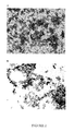
透過型電子顕微鏡(TEM)または低温TEMは、特に該コアが単一粒子からなる場合にナノ粒子のコアのサイズを測定するのに好都合に用いることができる(図2参照)。同様に、動的光散乱(DLS)は、コアが粒子または粒子の凝集物からなる場合に溶液中のナノ粒子のコアの流体力学的直径を測定するのに用いることができる。これら2つの方法は、さらに互いのサイズ測定値を比較し、該サイズを確認するために用いることができる。 Transmission electron microscopy (TEM) or low temperature TEM can be conveniently used to measure the size of the nanoparticle core, especially when the core consists of a single particle (see FIG. 2). Similarly, dynamic light scattering (DLS) can be used to measure the hydrodynamic diameter of the core of nanoparticles in solution when the core consists of particles or aggregates of particles. These two methods can further be used to compare each other's size measurements and confirm the size.
ナノ粒子の中心コアは、典型的には、治療的または診断的物質、好ましくは活性型または興奮性物質から製造される。該物質は、無機物質、有機物質、またはその混合物でありうる。該物質は、好ましくは無機物質である。 The central core of the nanoparticles is typically made from a therapeutic or diagnostic material, preferably an active or excitable material. The material can be an inorganic material, an organic material, or a mixture thereof. The substance is preferably an inorganic substance.
ナノ粒子の電子表面荷電が、ナノ粒子をpH6〜8の水性媒質に懸濁させた0.2〜8g/Lの異なる濃度のナノ粒子サスペンジョンを用いてゼータ電位測定により測定したときに、-15mV以下または+15mV以上、例えば−15mV〜−20mV、または+15mV〜+20mV、典型的には−20mV以下または+20mV以上であるかぎり、あらゆる種類のナノ粒子を本発明の熱感受性リポソームに封入することができる。 When the electronic surface charge of the nanoparticles is measured by zeta potential measurement using nanoparticle suspensions with different concentrations of 0.2-8 g / L suspended in an aqueous medium of pH 6-8, -15 mV or less Any type of nanoparticles can be encapsulated in the thermosensitive liposomes of the present invention so long as they are +15 mV or more, eg, -15 mV to -20 mV, or +15 mV to +20 mV, typically -20 mV or less, or +20 mV or more.
ナノ粒子の形状は、例えば丸、平面、細長、球状、卵形、または楕円形などでありうる。該形状は、製造方法により決定または調節し、目的とする適用に応じて当業者が適合させることができる。
粒子の形状は標的部位に送達されるとその「生体適合性」が影響をうけうるので、全く均質な形状を有する粒子が好ましい。薬物動態的理由により、本質的に球状、丸、または卵形のナノ粒子が好ましい。球状または丸形が特に好ましい。
The shape of the nanoparticles can be, for example, round, flat, elongated, spherical, oval or elliptical. The shape is determined or adjusted by the manufacturing method and can be adapted by a person skilled in the art according to the intended application.
Particles that have a completely homogeneous shape are preferred because the shape of the particles can be affected by their “biocompatibility” when delivered to the target site. For pharmacokinetic reasons, essentially spherical, round, or oval nanoparticles are preferred. A spherical or round shape is particularly preferred.
ナノ粒子の最大サイズ、すなわち、本発明の文脈において用いるナノ粒子のコアの最大寸法は典型的には1〜100nmに含まれる。
ナノ粒子を治療薬として用いる場合は、該サイズは、約5nm〜約100nm、例えば約5nm〜80nm、例えば約10nm〜約80nm、好都合には約10nmまたは20nm〜約70nm、好ましくは約15〜約60nm、または約10nmまたは15nm〜約50nmに含まれる。
ナノ粒子を診断薬として用いる場合は、該サイズは、好都合には、約2nm〜約10nm、例えば約4nm〜約8nmに含まれる。
The maximum size of the nanoparticles, ie the maximum size of the core of the nanoparticles used in the context of the present invention, is typically comprised between 1 and 100 nm.
When nanoparticles are used as therapeutic agents, the size is about 5 nm to about 100 nm, such as about 5 nm to 80 nm, such as about 10 nm to about 80 nm, conveniently about 10 nm or 20 nm to about 70 nm, preferably about 15 to about Included at 60 nm, or about 10 nm or 15 nm to about 50 nm.
When using nanoparticles as a diagnostic agent, the size is conveniently comprised between about 2 nm and about 10 nm, such as between about 4 nm and about 8 nm.
本発明の文脈で用いるナノ粒子は、コアおよびコーティングを含み、該コーティングは、生理学的pHの水性媒質中で測定したときに-20mV以下または+20mV以上の表面静電気の存在に関与する。 Nanoparticles used in the context of the present invention comprise a core and a coating, which is responsible for the presence of surface static electricity of -20 mV or less or +20 mV or more when measured in an aqueous medium at physiological pH.
静電コーティングは、好都合には「フルコーティング」(完全単層)である。これは、ナノ粒子の全表面上に適切な荷電を生じる非常に高密度の生体適合性分子の存在を意味する。そのようなフルコーティングは、TmまたはTm以上で熱感受性リポソームの膜を破壊するのに好都合であろう。 The electrostatic coating is conveniently “full coating” (complete monolayer). This means the presence of a very high density of biocompatible molecules that produce an appropriate charge on the entire surface of the nanoparticle. Such a full coating would be advantageous to break the membrane of heat sensitive liposomes at Tm or above Tm .
コアを構成する無機物質は、磁性物質であり得る。
磁性物質には、例えば、好ましくはオキシド、ヒドロキシド、または金属の形の、鉄、ニッケル、コバルト、ガドリニウム、サマリウム、ネオジミウム、およびそのあらゆる混合物が含まれる。
具体例において、コアを形成する物質は、酸化第一鉄および酸化第二鉄からなる群から選ばれる。本発明の好ましい態様において、オキシドナノ粒子は磁鉄鉱または磁赤鉄鉱でできている。
混合物質を用いて磁場とナノ粒子の相互作用を最適化することもできる。固体溶液形(種々の物質の無作為混合物として当業者に周知である)、例えばCoFe2O4を混合物質として用いることができる。さらに、分離(demixed)相の固体溶液、例えばFe2O3/Coを用いることができる。磁性物質を治療用物質として用いる場合は、強磁性物質が好ましい。
磁性物質を診断用物質として用いる場合は、超常磁性物質が好ましい。
The inorganic material constituting the core can be a magnetic material.
Magnetic materials include, for example, iron, nickel, cobalt, gadolinium, samarium, neodymium, and any mixtures thereof, preferably in oxide, hydroxide, or metal form.
In a specific example, the material forming the core is selected from the group consisting of ferrous oxide and ferric oxide. In a preferred embodiment of the invention, the oxide nanoparticles are made of magnetite or maghemite.
Mixed materials can also be used to optimize the interaction between the magnetic field and the nanoparticles. Solid solution form (well known to those skilled in the art as a random mixture of various substances), for example CoFe 2 O 4 , can be used as a mixed substance. Furthermore, a solid solution in a demixed phase, such as Fe 2 O 3 / Co, can be used. When magnetic substances are used as therapeutic substances, ferromagnetic substances are preferred.
When a magnetic substance is used as a diagnostic substance, a superparamagnetic substance is preferable.
コアを構成する無機物質は、少なくとも50、好ましくは少なくとも60または61、より好ましくは少なくとも65、66、67、または68の原子番号(Z)を有する金属元素により構成される高電子密度物質でありうる。
原子番号(陽子数としても知られる)は、原子核中にみられる陽子の数である。原子番号は、伝統的に記号Zにより表される。原子番号は、化学元素を一意的に同定する。中性荷電の原子において、原子番号は電子数に等しい。
Zは、ナノ粒子の流入放射線吸収能に関与する。
The inorganic material constituting the core is a high electron density material composed of a metal element having an atomic number (Z) of at least 50, preferably at least 60 or 61, more preferably at least 65, 66, 67, or 68 sell.
The atomic number (also known as the number of protons) is the number of protons found in the nucleus. The atomic number is traditionally represented by the symbol Z. The atomic number uniquely identifies the chemical element. In neutrally charged atoms, the atomic number is equal to the number of electrons.
Z is involved in the inflow radiation absorption capacity of the nanoparticles.
コアを構成する無機物質は、以下からなる酸化物でありうる:酸化セリウム(IV)(CeO2)、酸化ネオジニウム(III)(Nd2O3)、酸化サマリウム(III)(Sm2O3)、酸化ユーロピウム(III)(Eu2O3)、酸化ガドリニウム(III)(Gd2O3)、酸化テルビウム(III)(Tb2O3)、酸化ジスプロシウム(III)(Dy2O3)、酸化ホルミウム(Ho2O3)、酸化エルビウム(Er2O3)、酸化ツリウム(III)(Tm2O3)、酸化イッテルビウム(Yb2O3)、酸化ルテチウム(lu2O3)、酸化ハフニウム(IV)(HfO2)、酸化タンタルム(V)(Ta2O5)、酸化レニウム(IV)(ReO2)。
本発明の文脈において、無機酸化物の混合物も可能である。
The inorganic material constituting the core can be an oxide comprising: cerium (IV) oxide (CeO 2 ), neodynium oxide (III) (Nd 2 O 3 ), samarium (III) oxide (Sm 2 O 3 ) , Europium (III) oxide (Eu 2 O 3 ), gadolinium (III) oxide (Gd 2 O 3 ), terbium (III) oxide (Tb 2 O 3 ), dysprosium (III) oxide (Dy 2 O 3 ), oxidation Holmium (Ho 2 O 3 ), erbium oxide (Er 2 O 3 ), thulium (III) oxide (T m2 O 3 ), ytterbium oxide (Yb 2 O 3 ), lutetium oxide (lu 2 O 3 ), hafnium oxide ( IV) (HfO 2 ), tantalum oxide (V) (Ta 2 O 5 ), rhenium oxide (IV) (ReO 2 ).
In the context of the present invention, mixtures of inorganic oxides are also possible.
コアを構成する無機物質は、好ましくは原子数(Z)が少なくとも40または50、好ましくは少なくとも60または70の金属でありうる。
該金属は、以下から選ばれうる:金(Au − Z=79)、銀(Ag − Z=47)、プラチナ(Pt − Z=78)、パラジウム(Pd − Z=46)、スズ(Sn − Z=50)、タンタルム(Ta − Z=73)、イッテルビウム(Yb − Z=70)、ジルコニウム(Zr − Z=40)、ハフニウム(Hf − Z=72)、テルビウム(Tb − Z=65)、ツリウム(Tm − Z=69)、セリウム(Ce − Z=58)、ジスプロシウム(Dy − Z=66)、エルビウム(Er − Z=68)、ユーロピウム(Eu − Z=63)、ホルミウム(Ho − Z=67)、ランタヌム(La − Z=57)、ネオニジウム(Nd − Z=60)、プラセオジニウム(Pr − Z=59)、およびそのあらゆる混合物。
The inorganic material constituting the core may preferably be a metal having an atomic number (Z) of at least 40 or 50, preferably at least 60 or 70.
The metal can be selected from the following: gold (Au-Z = 79), silver (Ag-Z = 47), platinum (Pt-Z = 78), palladium (Pd-Z = 46), tin (Sn--). Z = 50), tantalum (Ta-Z = 73), ytterbium (Yb-Z = 70), zirconium (Zr-Z = 40), hafnium (Hf-Z = 72), terbium (Tb-Z = 65), Thulium ( Tm -Z = 69), Cerium (Ce-Z = 58), Dysprosium (Dy-Z = 66), Erbium (Er-Z = 68), Europium (Eu-Z = 63), Holmium (Ho-- Z = 67), lanthanum (La-Z = 57), neonidium (Nd-Z = 60), praseodynium (Pr-Z = 59), and any mixtures thereof.
本発明の好ましい態様において、ナノ粒子のコアは金で構成される。
本発明の文脈において、ナノ粒子のコアは、無機酸化物および金属の混合物で構成されうる。
生理学的pHの水性媒質中で測定したときに、ナノ粒子の-20mV以下または+20mV以上の表面静電気の存在に関与するコーティングは、無機または有機表面コーティングでありうる。
In a preferred embodiment of the invention, the core of the nanoparticles is composed of gold.
In the context of the present invention, the core of the nanoparticles can be composed of a mixture of inorganic oxides and metals.
Coatings that are responsible for the presence of surface static electricity below -20 mV or above +20 mV of the nanoparticles when measured in an aqueous medium at physiological pH can be inorganic or organic surface coatings.
無機の場合、該コーティングは、オキシド、ヒドロキシド、およびオキシヒドロキシドからなる群から選ばれうる。無機コーティングは、例えばシリシウム、アルミニウム、カルシウム、および/またはマグネシウムを含みうる。
例えば、マグネシウムおよびカルシウムからなる群から選ばれる無機物質は、pH7のナノ粒子表面に正荷電(+20mV以上)をもたらすだろう。
別の態様において、シリシウム基は、pH7のナノ粒子表面に負荷電(−20mV以下)をもたらすのに用いることができる。
If inorganic, the coating may be selected from the group consisting of oxide, hydroxide, and oxyhydroxide. The inorganic coating can include, for example, silicon, aluminum, calcium, and / or magnesium.
For example, an inorganic material selected from the group consisting of magnesium and calcium will provide a positive charge (+20 mV or more) to the pH 7 nanoparticle surface.
In another embodiment, the silicium group can be used to provide a negative charge (−20 mV or less) to the pH 7 nanoparticle surface.
有機の場合、該コーティングは、ナノ粒子表面と共有結合または静電結合により相互作用することができ、該ナノ粒子に表面特性を与えることができる分子を用いて調製される。
表面をコーティングする有機分子は、2つの基RおよびXを有する。Xの機能は、ナノ粒子表面と相互作用することであり、Rの機能はナノ粒子表面にその特異的特性を与えることである。
Xは、例えば、カルボキシレート(R−COO−)、シラン(R−Si(OR)3)、ホスホニック(R−PO(OH)2)、ホスホリック(R−O−PO(OH)2)、ホスフェート(R−PO4 3−)、およびチオール(R−SH)基から選ぶことができる。
Rは、生理学的pHの水性サスペンジョン中のナノ粒子に少なくとも電子表面荷電をもたらす。
Rがナノ粒子表面に正荷電をもたらす場合は、Rはアミン(NH2−X)でありうる。Rがナノ粒子表面に負荷電をもたらす場合は、Rはホスフェート(PO4 3−−X)またはカルボキシレート(COO−−X)でありうる。
In the case of organic, the coating is prepared with molecules that can interact with the nanoparticle surface by covalent or electrostatic bonds and can impart surface properties to the nanoparticle.
The organic molecules that coat the surface have two groups R and X. The function of X is to interact with the nanoparticle surface, and the function of R is to give the nanoparticle surface its specific properties.
X is, for example, carboxylate (R-COO -), silane (R-Si (OR) 3 ), phosphonic (R-PO (OH) 2 ), Hosuhorikku (R-O-PO (OH ) 2), phosphate It can be selected from (R—PO 4 3− ) and thiol (R—SH) groups.
R brings at least an electronic surface charge to the nanoparticles in an aqueous suspension at physiological pH.
If R results in positively charged on the nanoparticle surface, R represents may be an amine (NH 2 -X). If R provides a negative charge on the nanoparticle surface, R can be a phosphate (PO 4 3- X) or a carboxylate (COO - X).
ナノ粒子表面に正荷電(+20mV以上)をもたらす有機コーティングは、例えば、アミノプロピルトリエトキシシラン、ポリリシン、または2-アミノエタンチオールから選ばれうる。
ナノ粒子表面に負荷電(−20mV以下)をもたらす有機コーティングは、例えば、ポリホスフェート、メタホスフェート、ピロホスフェートなどからか、または、例えば、クエン酸またはジカルボン酸(特にコハク酸)から選ばれうる。
また、静電コーティングは好都合に「フルコーティング」である。
The organic coating that provides a positive charge (+20 mV or more) on the nanoparticle surface can be selected from, for example, aminopropyltriethoxysilane, polylysine, or 2-aminoethanethiol.
Organic coatings that provide a negative charge (−20 mV or less) to the nanoparticle surface can be selected, for example, from polyphosphates, metaphosphates, pyrophosphates, etc., or from, for example, citric acid or dicarboxylic acids (particularly succinic acid).
Also, electrostatic coating is conveniently “full coating”.
さらに、この静電コーティング、特にアミノまたはカルボン酸部分を用いて、ナノ粒子表面上のあらゆる基を結合させることができる。例えば、静電コーティングを用いて、例えばカルボジイミドのようなリンカーを用いてナノ粒子表面上に本明細書に記載のターゲッティング基またはカップリング基を結合することができる。 In addition, this electrostatic coating, particularly amino or carboxylic acid moieties, can be used to attach any groups on the nanoparticle surface. For example, an electrostatic coating can be used to attach a targeting group or coupling group described herein on the nanoparticle surface using a linker such as carbodiimide.
所望により、ナノ粒子表面は、ナノ粒子が熱感受性リポソームから放出されて、それと共有結合を形成する場合は、タンパク質と直接相互作用することができる基(「カップリング基」)を用いて機能化することができる。
Rは、タンパク質上に存在するアミン、カルボキシル、またはチオール基と共有結合的に相互作用することができる反応基、例えば、スクシンイミジルエステル基(アミン基と反応する)および/またはマレイミド基(カルボキシル基と反応する)でありうる。
If desired, the nanoparticle surface can be functionalized with a group that can interact directly with the protein (“coupling group”) if the nanoparticle is released from a thermosensitive liposome and forms a covalent bond with it. can do.
R is a reactive group capable of covalently interacting with an amine, carboxyl, or thiol group present on the protein, such as a succinimidyl ester group (reacting with an amine group) and / or a maleimide group (carboxyl React with the group).
所望により、ナノ粒子表面は、特定の生物組織または細胞をターゲッティングすることができる基(「ターゲッティング基」)を用いて機能化することができる。該ターゲッティング基は、人体または動物体に存在する分子に対する親和性を示すあらゆる生物学的構造または化学構造でありうる。
そのようなターゲッティング基は、典型的には、ナノ粒子が標的部位に蓄積し、TmまたはTm以上の温度で熱活性化するとリポソームから放出されると作用する。
If desired, the nanoparticle surface can be functionalized with groups capable of targeting specific biological tissues or cells (“targeting groups”). The targeting group can be any biological or chemical structure that exhibits affinity for molecules present in the human or animal body.
Such targeting groups typically nanoparticles accumulate at the target site, acts to be released from the liposomes when thermally activated T m or T m or higher.
ターゲッティング基は、抗原、スペーサー分子、生体適合性ポリマーから選ぶことができる。ターゲッティング基は、人体または動物体に存在する分子に対して親和性を示すあらゆる生物学的構造または化学構造でありうる。例えば、該ターゲッティング基は、ペプチド、オリゴペプチドもしくはポリペプチド、タンパク質、核酸(DNA、RNA、SiRNA、tRNA、miRNA、etc.)、ホルモン、ビタミン、酵素など、および一般的には、分子のあらゆるリガンド(例えばレセプター、マーカー、抗原など)でありうる。病的細胞により発現される分子のリガンド、特に腫瘍抗原のリガンド、ホルモンレセプター、サイトカインレセプター、または成長因子レセプター。該標的とされる基は、LHRH、EGF、葉酸塩(folate)、抗B−FN抗体、E−セレクチン/P−セレクチン、抗IL−2Rα抗体、GHRHなどからなる群から選ぶことができる。 The targeting group can be selected from antigens, spacer molecules, and biocompatible polymers. A targeting group can be any biological or chemical structure that exhibits affinity for molecules present in the human or animal body. For example, the targeting group can be a peptide, oligopeptide or polypeptide, protein, nucleic acid (DNA, RNA, SiRNA, tRNA, miRNA, etc.), hormone, vitamin, enzyme, etc., and generally any ligand of a molecule (Eg, receptor, marker, antigen, etc.). Ligands of molecules expressed by pathological cells, in particular ligands of tumor antigens, hormone receptors, cytokine receptors, or growth factor receptors. The targeted group can be selected from the group consisting of LHRH, EGF, folate, anti-B-FN antibody, E-selectin / P-selectin, anti-IL-2Rα antibody, GHRH and the like.
本明細書に記載の静電コーティングおよび/またはカップリング基を用いて、ナノ粒子表面上のあらゆる基を結合することができる。例えば、該基は、ナノ粒子表面にターゲッティング基を移植するためのリンカーとして用いることができる。 Any group on the nanoparticle surface can be attached using the electrostatic coating and / or coupling groups described herein. For example, the group can be used as a linker for implanting a targeting group on the nanoparticle surface.
本明細書に記載の具体的目的は、-20mV以下または+20mV以上の表面静電気の存在に関与する物質で共有結合的または静電気的にコートされたナノ粒子封入熱感受性リポソームである。この物質は、好ましくは、2つの基RおよびX(ここで、Rは、アミン、ホスフェート、およびカルボキシレートから選ばれ、Xは、カルボキシレート、シラン、ホスホニック、ホスホリック、およびチオールから選ばれる)を有する有機分子である。
ナノ粒子は、さらに、スクシンイミジルエステルおよびマレイミド基から選ばれるカップリング基、および/またはペプチド、オリゴペプチド、ポリペプチド、タンパク質、核酸、ホルモン、ビタミン、酵素、腫瘍抗原のリガンド、ホルモンレセプター、サイトカインレセプター、および成長因子レセプターから選ばれるターゲッティング基を含むことができる。
A specific object described herein is a nanoparticle-encapsulated thermosensitive liposome that is covalently or electrostatically coated with a substance responsible for the presence of surface static electricity of -20 mV or less or +20 mV or more. This material preferably has two groups R and X, where R is selected from amines, phosphates, and carboxylates, and X is selected from carboxylates, silanes, phosphonics, phosphorics, and thiols. It is an organic molecule.
The nanoparticles may further comprise a coupling group selected from succinimidyl ester and maleimide groups, and / or peptides, oligopeptides, polypeptides, proteins, nucleic acids, hormones, vitamins, enzymes, tumor antigen ligands, hormone receptors, cytokines A targeting group selected from a receptor and a growth factor receptor can be included.
所望により、ナノ粒子表面は、立体保護基を用いて機能化することができる。そのような基は、ポリエチレングリコール(PEG)、ポリエチレンオキシド、ポリビニルアルコール、ポリアクリレート、ポリアクリルアミド(ポリ(N−イソプロピルアクリルアミド))、ポリカルバミド、生体高分子、またはポリサッカライド、例えばデキストラン、キシラン、セルロース、コラーゲン、および両性イオン化合物、例えば、ポリスルホベタインなどから選ぶことができる。 If desired, the nanoparticle surface can be functionalized with a steric protecting group. Such groups include polyethylene glycol (PEG), polyethylene oxide, polyvinyl alcohol, polyacrylate, polyacrylamide (poly (N-isopropylacrylamide)), polycarbamide, biopolymers, or polysaccharides such as dextran, xylan, cellulose. , Collagen, and zwitterionic compounds such as polysulfobetaine.
この立体保護基は、生体適合性サスペンジョン、例えば生理液(血液、血漿、血清など)、あらゆる等張媒質または生理学的媒質、例えばグルコース(5%)および/またはNaCl(0.9%)を含む媒質中のナノ粒子安定性を増大させる。 This steric protecting group can be used in biocompatible suspensions such as physiological fluids (blood, plasma, serum, etc.), any isotonic or physiological medium such as glucose (5%) and / or NaCl (0.9%). Increases nanoparticle stability.
本発明の開示から、当業者は、本発明の精神から逸脱することなくナノ粒子を修飾することができると認識するだろう。 From the disclosure of the present invention, those skilled in the art will recognize that nanoparticles can be modified without departing from the spirit of the present invention.
本発明の文脈において用いることができるナノ粒子またはナノ粒子凝集物の典型的製造方法は、例えば、WO2007/118884、WO2009/147214、US 6,514,481 B1、WO2011/003999、およびLiu et al.、Journal of Magnetism and Magnetic Materials 270(2004) 1-6 「Preparation and characterization of amino-silane modified superparamagnetic silica nanospheres」に記載されている。 Exemplary methods for producing nanoparticles or nanoparticle aggregates that can be used in the context of the present invention are described, for example, in WO2007 / 118884, WO2009 / 147214, US 6,514,481 B1, WO2011 / 003999, and Liu et al., Journal of Magnetism. and Magnetic Materials 270 (2004) 1-6 “Preparation and characterization of amino-silane modified superparamagnetic silica nanospheres”.
治療的使用
ナノ粒子が標的細胞に取り込まれるかまたは接触すると、治療的ナノ粒子の効率は増大する。この目的を達成するために、典型的には、ナノ粒子表面特性を、標的細胞と相互作用するのに有利に働くように修飾する。例えば、ナノ粒子表面の電子荷電を修飾するか、または該表面を本明細書に記載のターゲッティング基またはターゲッティング剤と結合させることができる。
As therapeutic use nanoparticles are taken up or contacted by target cells, the efficiency of the therapeutic nanoparticles increases. To achieve this goal, the nanoparticle surface properties are typically modified to favor the interaction with target cells. For example, the electronic charge of the nanoparticle surface can be modified or the surface can be coupled with a targeting group or targeting agent as described herein.
特異的結合相互作用は、ナノ粒子が細胞膜上の相補的分子またはレセプターと特異的に相互作用するのを可能にする表面リガンド(本明細書ではターゲッティング基という)の相互作用である。この相互作用は、レセプター介在エンドサイトーシスを誘導する。ナノ粒子の表面に結合したターゲッティングリガンドは、レセプター、膜タンパク質、標的細胞上に発現した表面抗原を認識して結合し、エンドサイトーシスおよび細胞内送達を引き起こすことができる。 A specific binding interaction is an interaction of a surface ligand (referred to herein as a targeting group) that allows the nanoparticle to specifically interact with a complementary molecule or receptor on the cell membrane. This interaction induces receptor-mediated endocytosis. Targeting ligands bound to the surface of the nanoparticles can recognize and bind to receptors, membrane proteins, surface antigens expressed on target cells, causing endocytosis and intracellular delivery.
細胞接触および粒子取り込みを促進する非特異的引力は、固有のナノ粒子特性、例えば表面荷電から生じる。
しかしながら、ほとんどの場合、そのような修飾は、ナノ粒子の生体内分布に有害である。
Non-specific attractive forces that promote cell contact and particle uptake arise from intrinsic nanoparticle properties such as surface charge.
In most cases, however, such modifications are detrimental to the biodistribution of the nanoparticles.
本明細書に記載の熱感受性リポソームは、この問題を克服し、先に記載のごとく、対象の身体における治療的ナノ粒子の効率的生体内分布(送達)および制御放出を可能にする。該リポソームは、標的部位上の、ナノ粒子、特に修飾表面特性を示すナノ粒子の濃度に遊離に働く。 The thermosensitive liposomes described herein overcome this problem and allow for efficient biodistribution (delivery) and controlled release of therapeutic nanoparticles in the subject's body, as described above. The liposomes act freely on the concentration of nanoparticles on the target site, in particular nanoparticles exhibiting modified surface properties.
ナノ粒子の制御(空間的および時間的)放出が今や可能であり、該ナノ粒子は、その治療的活性がその細胞との相互作用(ナノ粒子は細胞と接触し、および/または細胞内に取り込まれる)に依存する場合に、それが必要とされる場所および時に送達される。 Controlled (spatial and temporal) release of the nanoparticle is now possible, and the nanoparticle has its therapeutic activity interacting with the cell (the nanoparticle is in contact with and / or taken into the cell) Will be delivered where and when it is needed.
ナノ粒子を治療的手段として用いる場合は、そのコアは、外部活性型物質、例えば外部エネルギー供給源により活性化することができる物質でありうる治療的物質を用いて製造される。特定の態様において、治療的物質は、標的細胞、組織、または器官を機能的に阻害し、変化させ、または破壊することができる。 When nanoparticles are used as a therapeutic tool, the core is manufactured using a therapeutic substance that can be an externally active substance, such as a substance that can be activated by an external energy source. In certain embodiments, the therapeutic agent can functionally inhibit, alter, or destroy a target cell, tissue, or organ.
治療的物質は、先に記載の高電子密度物質および磁性物質から選ぶことができる。 The therapeutic substance can be selected from the high electron density substances and magnetic substances described above.
活性供給源は、コアが高電子密度物質、例えばHfO2またはAuを用いて製造されるナノ粒子のための電離放射線供給源でありうる。
電離放射線は、典型的には5KeV〜約25000KeV、具体的には約5KeV〜約6000KeV(LINAC供給源)、または約5KeV〜約1500KeV(例えばコバルト60供給源)でありうる。X線供給源を用いる特に好ましい電離放射線は、典型的には約50KeV〜約12000KeV、例えば約50KeV〜約6000KeVである。
The active source can be an ionizing radiation source for nanoparticles whose core is made using a high electron density material such as HfO 2 or Au.
The ionizing radiation can typically be from 5 KeV to about 25000 KeV, specifically from about 5 KeV to about 6000 KeV (LINAC source), or from about 5 KeV to about 1500 KeV (eg, cobalt 60 source). Particularly preferred ionizing radiation using an X-ray source is typically from about 50 KeV to about 12000 KeV, such as from about 50 KeV to about 6000 KeV.
電離放射線の必要線量は、in vitro適用では、好ましくは、約0.05Gray(グレイ)〜約16Gray、好ましくは約0.05Gray〜約6Grayを含む線量である。
線量は、特に局所、ex vivoまたはin vivo適用では、約0.05Gray以上および約16もしくは30Gray以下を含む。
総電離放射線は、現在の慣例に従ってヒトにおいて、約1.5Grayから約85Gray以下である。約40Grayのさらなる照射ブーストを現在の慣例に従ってヒトに与えることもできる。
The required dose of ionizing radiation is preferably a dose comprising about 0.05 Gray to about 16 Gray, preferably about 0.05 Gray to about 6 Gray for in vitro applications.
Doses include greater than about 0.05 Gray and less than about 16 or 30 Gray, especially for topical, ex vivo or in vivo applications.
Total ionizing radiation is about 1.5 Gray to about 85 Gray or less in humans according to current practice. A further irradiation boost of about 40 Gray can also be given to humans according to current practice.
供給される放射線の総線量は、種々のスケジュール、例えば単回線量、分割線量、超分割(hyperfractionated)線量などに従って与えることができる。 The total dose of radiation delivered can be given according to various schedules such as single line dose, fractionated dose, hyperfractionated dose, and the like.
一般的におよび非制限的方法で、以下のX線をナノ粒子を活性化するための種々の場合に適用することができる:
−表面標的組織に対して特に有効な50〜150keVのX線;
−6cmの組織圧を透過することができる200〜500keVのX線(慣用電圧);
−1000keV〜25,000keVのX線(メガボルト)。例えば、前立腺癌治療のためのナノ粒子の電離(イオン化)は、エネルギー15,000keVの5焦点X線により行うことができる。
In general and in a non-limiting manner, the following X-rays can be applied in various cases for activating nanoparticles:
-50-150 keV X-rays that are particularly effective against surface target tissues;
200-500 keV X-rays (conventional voltage) capable of transmitting -6 cm tissue pressure;
-1000 keV to 25,000 keV X-rays (megavolts). For example, ionization (ionization) of nanoparticles for prostate cancer treatment can be performed with 5-focus X-rays with an energy of 15,000 keV.
あるいはまた、放射性同位元素を電離放射線供給源として用いることができる(キュリー療法または小線源治療という)。特に、ヨウ素I125(t1/2=60.1日間)、パラジウムPd103(t1/2=17日間)、セシウムCs137、およびイリジウムIr192を好都合に用いることができる。 Alternatively, radioisotopes can be used as a source of ionizing radiation (referred to as Curie therapy or brachytherapy). In particular, iodine I 125 (t 1/2 = 60.1 days), palladium Pd 103 (t 1/2 = 17 days), cesium Cs 137 and iridium Ir 192 can be advantageously used.
免疫放射核種(または免疫放射標識リガンド)は、放射免疫療法の文脈で電離放射線供給源として用いることもできる。放射免疫療法に適した放射核種は、例えば131I、186Re、177Lu、または90Yから選ぶことができる。 Immune radionuclides (or immunoradiolabeled ligands) can also be used as a source of ionizing radiation in the context of radioimmunotherapy. A radionuclide suitable for radioimmunotherapy can be selected from, for example, 131 I, 186 Re, 177 Lu, or 90 Y.
荷電粒子、例えば陽子線、イオン線、例えば炭素、特に高エネルギーイオン線も電離放射線供給源として用いることができる。 Charged particles such as proton beams, ion beams such as carbon, especially high energy ion beams can also be used as a source of ionizing radiation.
電子線も、4MeV〜25Mevを含むエネルギーの電離放射線供給源として用いることができる。 Electron beams can also be used as ionizing radiation sources with energies including 4 MeV to 25 Mev.
特異的単色照射供給源を、ナノ粒子の原子の望むX線吸収エッジに近いかまたはそれに対応するエネルギーのX線を選択的に生じるために用いることができた。 A specific monochromatic irradiation source could be used to selectively generate X-rays of energy close to or corresponding to the desired X-ray absorption edge of the nanoparticle atoms.
優先的に、電離放射線の供給源は、線型粒子加速装置(LINAC)、コバルト60、および小線源治療供給源から選ぶことができる。 Preferentially, the source of ionizing radiation can be selected from a linear particle accelerator (LINAC), cobalt 60, and a brachytherapy source.
量(総照射線量の範囲)およびスケジュール(単回線量、または分割もしくは超分割プロトコールなどの文脈における照射の計画および送達)は、あらゆる疾患/解剖学的部位/病期、患者の状況/患者の年齢(子供、成人、老齢患者)について定義され、あらゆる特定の状況に対する治療の標準を構成する。該照射は、放射線療法の現在利用可能なあらゆる系を用いて、1回またはそれ以上、ナノ粒子の放出後いつでも適用することができる。 Dose (range of total irradiation dose) and schedule (planning and delivery of radiation in contexts such as single line dose or split or hyperfractionation protocols) can be used for any disease / anatomical site / stage, patient status / patient It is defined for age (children, adults, elderly patients) and constitutes a treatment standard for any particular situation. The irradiation can be applied one or more times after release of the nanoparticles using any currently available system of radiation therapy.
特定の態様において、治療的物質は、磁性酸化物(磁鉄鉱または磁赤鉄鉱)、特に強磁性物質であり、活性供給源は磁場供給源である。 In certain embodiments, the therapeutic substance is a magnetic oxide (magnetite or maghemite), in particular a ferromagnetic substance, and the active source is a magnetic field source.
好ましくは非振動または安定である磁場を、あらゆる磁場供給源を用いることにより、1回またはそれ以上、ナノ粒子の放出後絶えず適用することができる。磁場供給源は、好ましくは均一および一方向磁場供給源であり、あらゆる天然磁石、電気磁石、および磁気共鳴画像法(MRI)装置から選ぶことができる。
適切な非振動または安定磁場は、典型的には0.5〜5Teslaの範囲の磁場を有する標準的MRI装置において利用可能である。
磁場に暴露する場合は、暴露の持続時間に応じて、磁性ナノ粒子は、細胞または組織破壊を可能にする(持続時間:数分間、例えば2または5分間〜120分間)。
A magnetic field, preferably non-oscillating or stable, can be applied continuously after the release of the nanoparticles one or more times by using any magnetic field source. The magnetic field source is preferably a uniform and unidirectional magnetic field source and can be selected from any natural magnet, electromagnet, and magnetic resonance imaging (MRI) device.
Appropriate non-oscillating or stable magnetic fields are available in standard MRI machines with magnetic fields typically in the range of 0.5-5 Tesla.
When exposed to a magnetic field, depending on the duration of exposure, the magnetic nanoparticles allow cell or tissue destruction (duration: several minutes, eg 2 or 5 minutes to 120 minutes).
本発明のナノ粒子またはナノ粒子凝集物および組成物は、磁場にかけると癌細胞または癌細胞であろうと疑われる細胞を溶解させるために好都合に用いることができる。 The nanoparticles or nanoparticle aggregates and compositions of the present invention can be advantageously used to lyse cells suspected of being cancer cells or cancer cells when subjected to a magnetic field.
本明細書において本発明者らは、必要とする対象を治療するための医薬組成物を製造するための、本明細書に記載のナノ粒子、または本明細書に記載の同じまたは異なるナノ粒子のポピュレーションの使用を開示する。 As used herein, we describe the use of a nanoparticle described herein, or the same or different nanoparticles described herein, for the manufacture of a pharmaceutical composition for treating a subject in need thereof. Disclose the use of populations.
用語「治療(処置)」は、異常な機能を正し、疾患を予防し、病的兆候を改善し、例えば特に異常組織、特に腫瘍のサイズまたは増殖の減少;異常細胞または組織のサイズもしくは増殖の制御、抑制、または破壊;疾患進行の鈍化;癌進行の遅延による疾患の安定化;転移形成の減少;疾患の退行;または完全寛解(例えば癌の文脈において)などを行うためのあらゆる行為を表す。 The term “treatment” corrects abnormal function, prevents disease, ameliorates pathological signs, eg, reduces the size or growth of abnormal tissues, especially tumors; the size or growth of abnormal cells or tissues. Control, suppression or destruction of disease; slowing disease progression; stabilization of disease by slowing cancer progression; reduction of metastasis formation; regression of disease; or complete remission (eg in the context of cancer) Represent.
医薬組成物は、標的細胞を活性供給源に暴露すると、必要とする対象における標的細胞を障害し、阻害し、変化させ、または破壊するための組成物でありうる。 A pharmaceutical composition can be a composition for damaging, inhibiting, changing or destroying a target cell in a subject in need thereof when the target cell is exposed to an active source.
本発明の目的は、標的細胞を活性供給源に暴露すると、標的細胞を障害し、阻害し、変化させ、または破壊するための、例えば本明細書で先に記載し、および/または本明細書に記載の方法により得ることができる熱感受性リポソームである。 An object of the present invention is, for example, as described hereinabove and / or herein, for exposing a target cell to an active source to damage, inhibit, alter, or destroy the target cell. It is a thermosensitive liposome obtainable by the method described in 1.
本発明の具体的熱感受性リポソームは、対象の癌を予防または治療し、または癌の症状を軽減するためのリポソームである。 The specific heat-sensitive liposome of the present invention is a liposome for preventing or treating a subject's cancer or reducing the symptoms of cancer.
本明細書に記載の具体的方法は、以下を含む対象の細胞の障害、溶解、アポトーシス、または破壊を誘導し、または生じる方法である:a)対象にナノ粒子を含む熱感受性リポソーム(本明細書で先に記載の)を投与し、b)ナノ粒子を局所的に放出させ、次いで細胞、特に標的細胞と相互作用させるために熱感受性リポソームをTmまたはTm以上に加熱し、次いで所望により、c)該細胞を活性供給源、典型的には例えば本明細書に記載の外部活性供給源に暴露し、該暴露がナノ粒子を活性化し、次いで細胞の障害、溶解、アポトーシス、または破壊を誘導し、または引き起こす。 Specific methods described herein are methods that induce or cause damage, lysis, apoptosis, or destruction of a subject's cells, including: a) a thermosensitive liposome comprising nanoparticles in the subject (herein) administered) previously described in writing, b) the nanoparticles were released locally, then the cells, heat-sensitive liposomes heated above T m or T m in particular order to interact with the target cells, then the desired C) exposing the cells to an active source, typically an externally active source as described herein, for example, which activates the nanoparticles and then cell damage, lysis, apoptosis, or destruction Induce or cause.
本発明の熱感受性リポソームは、種々の経路、例えば局所(例えば腫瘍内(IT))、皮下、静脈内(IV)、皮内、動脈内、気道(吸入)、腹腔内、筋肉内、および経口経路(経口)により投与することができる。本発明の熱感受性リポソームは、さらに、腫瘍摘出術後の腫瘍ベッドの仮想空洞(virtual cavity)に投与することができる。好ましい投与経路は静脈内経路である。 The thermosensitive liposomes of the present invention can be used in various routes such as topical (eg, intratumoral (IT)), subcutaneous, intravenous (IV), intradermal, intraarterial, airway (inhalation), intraperitoneal, intramuscular, and oral. It can be administered by the route (orally). The thermosensitive liposomes of the present invention can further be administered to the virtual cavity of the tumor bed after tumor removal. A preferred route of administration is the intravenous route.
熱感受性リポソームを静脈内経路で注射して一定時間後の、透過および保持増強(「EPR」)効果は、腫瘍塊中に熱感受性リポソームが受動的に蓄積するのに関与する。実際に、腫瘍の血管は、正常毛細血管とは全く異なり、その血管の「血管漏出」が正常組織では通常ではないリポソームの選択的血管外遊出を促すことが観察された。有効な腫瘍リンパ廃液の欠如は、浸透リポソームのクリアランスを防ぎ、その蓄積を促す。 The permeation and retention enhancement (“EPR”) effect after a period of time after thermosensitive liposomes are injected intravenously is responsible for passive accumulation of thermosensitive liposomes in the tumor mass. Indeed, it has been observed that tumor blood vessels are quite different from normal capillaries, and that “vascular leakage” of the blood vessels promotes selective extravasation of liposomes that is not normal in normal tissues. The lack of effective tumor lymph effluent prevents clearance of osmotic liposomes and promotes their accumulation.
すなわち、本発明のナノ粒子は、静脈内投与した熱感受性リポソームから放出されると原発腫瘍および転移腫瘍を有効にターゲッティングすることができる。 That is, the nanoparticles of the present invention can effectively target primary tumors and metastatic tumors when released from intravenously administered thermosensitive liposomes.
標的細胞は、あらゆる病的細胞、すなわち、病的メカニズムが関与する細胞、例えば増殖性細胞、例えば腫瘍細胞、狭窄(stenosing)細胞(繊維芽細胞/平滑筋細胞)、または免疫系細胞(病的細胞クローン)でありうる。好ましい適用は、悪性細胞または組織の治療(例えば破壊または機能的変化)に基づく。 The target cell can be any pathological cell, ie a cell in which a pathological mechanism is involved, such as a proliferative cell, such as a tumor cell, a stenosing cell (fibroblast / smooth muscle cell), or an immune system cell (pathological) Cell clone). A preferred application is based on the treatment (eg destruction or functional change) of malignant cells or tissues.
本発明の別の目的は、以下を含む、対象または患者における、疾患、特に癌の予防または治療方法、または疾患の症状の軽減方法に関する:a)疾患に罹患した患者に熱感受性リポソーム、または例えば熱感受性リポソームを含む本明細書に記載の組成物を投与し、b)ナノ粒子を局所放出させ、次いで細胞、特に標的細胞と相互作用させるために熱感受性リポソームをTmまたはTm以上に加熱し、c)次いで、本明細書に記載の活性供給源に対象を暴露することにより対象を治療し、該暴露が患者の異常細胞の変化、障害、または機能的破壊をもたらすことにより、該疾患を予防または治療する。 Another object of the present invention relates to a method for preventing or treating a disease, in particular cancer, or a method for reducing the symptoms of a disease in a subject or patient, including: a) a thermosensitive liposome in a patient suffering from a disease, or for example administering a composition as described herein comprising a heat-sensitive liposomes, b) the nanoparticles were local release heat, then the cells, heat-sensitive liposomes than T m or T m in particular order to interact with the target cell C) then treating the subject by exposing the subject to an active source as described herein, wherein the exposure results in a change, disorder, or functional destruction of the patient's abnormal cells, thereby causing the disease Prevent or treat.
伝統的癌管理は、系統的に、集学的治療(例えば放射線療法および化学療法の組み合わせ)の共同作用を意味する。 Traditional cancer management systematically means the combined action of multidisciplinary treatments (eg, a combination of radiation therapy and chemotherapy).
例えば、放射線療法の文脈において、活性供給源に暴露した本明細書に記載のナノ粒子は、種々の癌療法プロトコールと組み合わせて用いることができる。そのようなプロトコールは、外科、放射線外科、化学療法、細胞増殖抑制剤、細胞毒性薬の投与を含む治療、ターゲッティング療法、ワクチン、および癌を治療するためのあらゆる他の生物学的または無機生成物からなる群から選ぶことができる。本発明の熱感受性リポソームは、本明細書に記載のナノ粒子と共に、目的とするあらゆる治療的分子、特に癌を治療するためのあらゆる既知の生物学的または無機生成物を封入することができる。 For example, in the context of radiation therapy, the nanoparticles described herein exposed to an active source can be used in combination with various cancer therapy protocols. Such protocols include surgery, radiosurgery, chemotherapy, cytostatics, treatments including administration of cytotoxic drugs, targeting therapies, vaccines, and any other biological or inorganic products for treating cancer. You can choose from the group consisting of The thermosensitive liposomes of the present invention can encapsulate any therapeutic molecule of interest, particularly any known biological or inorganic product for treating cancer, along with the nanoparticles described herein.
本明細書に記載のナノ粒子は、さらに放射線療法のみの文脈で用いることができる。観察された治療効果の増大は、一部、本発明の熱感受性リポソームによるその輸送、次いでその放出制御によって可能となる標的部位上の有効なナノ粒子の濃度の増加による。 The nanoparticles described herein can also be used in the context of radiation therapy alone. The observed increase in therapeutic effect is due, in part, to an increase in the concentration of effective nanoparticles on the target site that is enabled by its transport by the thermosensitive liposomes of the present invention and then its controlled release.
本発明を用いて、あらゆる種類の悪性腫瘍、例えば血液学的腫瘍もしくは悪性腫瘍、および特に上皮、神経外胚葉、もしくは間葉起源の固形腫瘍を治療することができる。さらに、本明細書に記載のリポソームを用いて、放射線療法が伝統的に使用および/または適用される前癌状態の病変または特定の良性疾患を治療することができる。 The present invention can be used to treat all types of malignant tumors, such as hematological or malignant tumors, and in particular solid tumors of epithelial, neuroectodermal or mesenchymal origin. Furthermore, the liposomes described herein can be used to treat pre-cancerous lesions or certain benign diseases where radiation therapy is traditionally used and / or applied.
該腫瘍または癌は、放射線療法が伝統的治療である癌でありうる。そのような癌は、特に皮膚癌(AIDS関連悪性新生物、メラノーマを含む);中枢神経系腫瘍(脳、幹脳、小脳、下垂体、脊柱管、眼、および眼窩を含む);頭部および頸部腫瘍;肺癌;乳癌;消化管腫瘍、例えば肝癌および肝胆道癌、結腸癌、直腸癌、および肛門癌、胃癌、膵臓癌、食道癌;男性の泌尿生殖器腫瘍、例えば、前立腺癌、精巣癌、陰茎癌、および尿道癌;婦人科の癌、例えば、子宮頸癌、子宮内膜癌、卵巣癌、卵管癌、膣癌、および外陰癌;副腎腫瘍および後腹膜膿瘍;局在性に関わらず骨および軟部組織の肉腫;リンパ腫;骨髄腫;白血病;および小児腫瘍、例えばウィルムス腫瘍、神経芽細胞腫、中枢神経系腫瘍、ユーイング肉腫などからなる群から選ばれうる。 The tumor or cancer can be a cancer for which radiation therapy is the traditional treatment. Such cancers are particularly skin cancers (including AIDS-related malignant neoplasms, melanoma); central nervous system tumors (including brain, stem brain, cerebellum, pituitary, spinal canal, eye, and orbit); Lung cancer; Breast cancer; Gastrointestinal tumors such as liver and hepatobiliary cancer, colon cancer, rectal cancer, and anal cancer, gastric cancer, pancreatic cancer, esophageal cancer; male urogenital tumors such as prostate cancer, testicular cancer , Penile cancer, and urethral cancer; gynecological cancers such as cervical cancer, endometrial cancer, ovarian cancer, fallopian tube cancer, vaginal cancer, and vulvar cancer; adrenal and retroperitoneal abscesses; Spine and soft tissue sarcomas; lymphoma; myeloma; leukemia; and childhood tumors such as Wilms tumor, neuroblastoma, central nervous system tumor, Ewing sarcoma, and the like.
本発明は、治療の文脈において、原発腫瘍もしくは二次的侵襲、局所もしくは遠隔転移に、また、予防の文脈において、二次的悪性中枢神経系合併症、例えばメラノーマ、肺癌、腎臓癌、乳癌などから観察される侵襲(転移)を避けるために適用することができる。 The invention relates to primary tumors or secondary invasion, local or distant metastases in the therapeutic context and secondary malignant central nervous system complications such as melanoma, lung cancer, kidney cancer, breast cancer, etc. in the prevention context. It can be applied to avoid the invasion (metastasis) observed from.
熱感受性リポソームは、抗癌治療期間を通していつでも用いることができる。熱感受性リポソームは、例えば、ネオアジュバント(癌を切除するための外科的介入前)またはアジュバント(外科手術後)として投与することができる。
熱感受性リポソームは、外科的に除去することができない進行腫瘍に用いることもできる。
Thermosensitive liposomes can be used at any time throughout the anti-cancer treatment period. Thermosensitive liposomes can be administered, for example, as a neoadjuvant (before surgical intervention to remove the cancer) or as an adjuvant (after surgery).
Thermosensitive liposomes can also be used for advanced tumors that cannot be removed surgically.
熱感受性リポソームの反復注射または投与は適宜行うことができる。 Repeated injection or administration of thermosensitive liposomes can be performed as appropriate.
診断的使用
ナノ粒子の制御送達は、in vivo画像化および/または診断目的にも望ましく、今や該ナノ粒子を本発明にしたがって熱感受性リポソームに封入することにより可能である。
Controlled delivery of nanoparticles for diagnostic use is also desirable for in vivo imaging and / or diagnostic purposes and is now possible by encapsulating the nanoparticles in thermosensitive liposomes according to the present invention.
本明細書において本発明者らは、好ましくは対象をエネルギーの外部供給源に暴露したときに、対象中の異常組織もしくは細胞、特に腫瘍細胞の存在を検出するための診断用組成物を製造するための、本発明の熱感受性リポソーム、特に本明細書に記載のナノ粒子または本明細書に記載の同じまたは異なるナノ粒子のポピュレーションを含む熱感受性リポソームの使用を開示する。 As used herein, we manufacture a diagnostic composition for detecting the presence of abnormal tissue or cells, particularly tumor cells, in a subject, preferably when the subject is exposed to an external source of energy. Disclosed is the use of thermosensitive liposomes of the present invention, in particular thermosensitive liposomes comprising a population of nanoparticles as described herein or the same or different nanoparticles as described herein.
本発明の目的は、対象をエネルギーの外部供給源に暴露したときに、対象の異常細胞を検出または可視化するための、例えば本明細書に記載のおよび/または本明細書に記載の方法により得ることができる熱感受性リポソームである。 The object of the present invention is obtained, for example, by the methods described herein and / or described herein for detecting or visualizing abnormal cells in a subject when the subject is exposed to an external source of energy. Thermosensitive liposomes that can.
診断用物質は、先に記載の高電子密度物質および磁性物質から選ぶことができる。
ナノ粒子を異常組織または細胞を検出または可視化するための診断薬として用いる場合は、そのコアは好都合には画像化物質からなる。
そのような画像化物質は、好都合には、あらゆる磁性物質、例えば先に記載の酸化第一鉄および酸化第二鉄から選ぶことができる。例えば、本発明に用いるナノ粒子は、MRIで可視的なγ−Fe2O3(磁赤鉄鉱)またはFe3O4(磁鉄鉱)でできたコアを有する。
画像化物質も、断層像影装置(CTスキャナー)下で可視的なあらゆる高電子密度物質、例えばHfO2またはAuから選ぶことができる。
The diagnostic substance can be selected from the high electron density substances and magnetic substances described above.
When the nanoparticles are used as a diagnostic agent for detecting or visualizing abnormal tissues or cells, the core is conveniently composed of an imaging material.
Such an imaging material can conveniently be selected from any magnetic material, such as the ferrous oxide and ferric oxide described above. For example, the nanoparticles used in the present invention have a core made of γ-Fe 2 O 3 (maghemite) or Fe 3 O 4 (magnetite) visible on MRI.
The imaging material can also be selected from any high electron density material, such as HfO 2 or Au, visible under a tomographic device (CT scanner).
画像化目的では、先に記載のターゲッティング基のナノ粒子への付加が好ましい。 For imaging purposes, addition of the targeting groups described above to the nanoparticles is preferred.
本発明の別の目的は、以下を含む、対象または疾患に罹っていることが疑われる患者において標的細胞を検出または可視化する方法(疾患、特に癌の診断を可能にする)に関する:a)疾患に罹患した患者に熱感受性リポソームまたは該熱感受性リポソームを含む例えば本明細書に記載の組成物を投与し、b)ナノ粒子を局所放出させ、次いで細胞、特に標的細胞との相互作用を可能にするために、目的とする領域をTmまたはTm以上に加熱し、c)次いで、該対象を、本明細書に記載の活性化供給源に暴露し、該暴露は、患者の標的細胞の検出または可視化を可能にする。 Another object of the invention relates to a method for detecting or visualizing a target cell in a subject or a patient suspected of suffering from a disease (allowing the diagnosis of disease, in particular cancer), including: a) disease Administration of a thermosensitive liposome or a composition comprising, for example, the thermosensitive liposome containing the thermosensitive liposome to a patient suffering from b) allowing the nanoparticles to be locally released and then allowed to interact with cells, particularly target cells To heat the region of interest to Tm or above Tm , and c) then subject is exposed to an activated source as described herein, the exposure of the target cell of the patient Allows detection or visualization.
特定の態様において、本明細書に記載の熱感受性リポソームは、細網内皮系(RES)により認識されるのを避けながら、血流中に標的ナノ粒子(特に、腫瘍細胞を特異的に認識することができるナノ粒子)を輸送することができる。 In certain embodiments, the thermosensitive liposomes described herein specifically recognize target nanoparticles (particularly tumor cells) in the bloodstream while avoiding being recognized by the reticuloendothelial system (RES). Nanoparticles) that can be transported.
腫瘍塊が存在する場合は、TmまたはTm以上で熱感受性リポソームから放出されたナノ粒子は、腫瘍に入り、標的癌細胞と相互作用する。ナノ粒子の蓄積は、磁性ナノ粒子についてMRIで可視的なシグナル変動(perturbation)を引き起こすか、または高密度ナノ粒子を用いるとCTスキャナーシグナルが増加する。
腫瘍塊がなければ、ナノ粒子は、腎臓を介して血流から排除され、シグナル増加や変動は検出または可視化されないだろう。
In the presence of a tumor mass, nanoparticles released from thermosensitive liposomes at Tm or above Tm enter the tumor and interact with target cancer cells. Nanoparticle accumulation causes MRI visible signal perturbation for magnetic nanoparticles, or increases CT scanner signal with high density nanoparticles.
Without a tumor mass, the nanoparticles will be cleared from the bloodstream via the kidneys and no signal increase or fluctuation will be detected or visualized.
さらに本明細書において本発明者らは、対象の標的部位、特に腫瘍およびその微小環境から標的部位の表現型検査に用いることができるタンパク質を回収するための診断用組成物を製造するための、本発明の熱感受性リポソーム、特に、本明細書に記載のナノ粒子または本明細書に記載の同じまたは異なるナノ粒子のポピュレーションを含む熱感受性リポソームの使用を開示する。 Furthermore, in the present specification, the present inventors provide a diagnostic composition for recovering a protein that can be used for phenotyping of a target site from a target site of interest, particularly a tumor and its microenvironment. Disclosed is the use of thermosensitive liposomes of the present invention, particularly thermosensitive liposomes comprising a population of nanoparticles as described herein or a population of the same or different nanoparticles as described herein.
本発明の別の目的は、以下を含む、対象または患者における標的部位から(特に該標的部位の表現型検査を可能にする)タンパク質を回収する方法に関する:a)疾患に罹っている患者に熱感受性リポソームまたは該熱感受性リポソームを含む例えば本明細書に記載の組成物を投与し、b)標的部位でのナノ粒子の放出、次いで該標的部位とその相互作用を可能にするために熱感受性リポソームをTmまたはTm以上に加熱し、c)次いで、該標的部位のタンパク質で被われたナノ粒子を回収する。 Another object of the present invention relates to a method of recovering proteins from a target site in a subject or patient (especially enabling phenotyping of the target site) in a subject or patient comprising: a) fever in a patient suffering from a disease Administering a sensitive liposome or a composition as described herein comprising the thermosensitive liposome, for example, b) releasing the nanoparticles at the target site, and then allowing the interaction with the target site the heated above T m or T m, c) then recovered nanoparticles covered with a protein of the target site.
先の方法は、さらに、血中へのナノ粒子の放出を可能にするために熱感受性リポソームをTmまたはTm以上に加熱する工程、および血液タンパク質と標的部位由来のタンパク質を比較するために血液タンパク質で被われたナノ粒子を回収する工程を含むことができる。
このアプローチは、伝統的バイオプシーに比べて好都合な非侵襲性技術を構成する。
Previous method further step of heating the heat-sensitive liposomes than T m or T m in order to allow the release of the nanoparticles into the blood, and to compare the blood protein and the target site derived proteins A step of recovering the nanoparticles covered with blood protein can be included.
This approach constitutes an advantageous non-invasive technique compared to traditional biopsy.
本明細書に記載の熱感受性リポソームは、特定患者の癌病期に関する情報を提供し、癌専門医が、この患者のための最も適切な治療を選択し、特定の治療の効果を追跡するのを助けることができるので、個別療法に用いることができる診断手段である。
本明細書に記載の熱感受性リポソームは、さらに、特定の治療に対する腫瘍の反応を評価し、患者の臨床的結果(進行無しの生存)を予測する情報を提供することができる。
The thermosensitive liposomes described herein provide information about the cancer stage of a particular patient, allowing oncologists to select the most appropriate treatment for this patient and track the effects of that particular treatment. It is a diagnostic tool that can be used for individual therapy because it can help.
The thermosensitive liposomes described herein can further assess tumor response to a particular treatment and provide information that predicts a patient's clinical outcome (survival without progression).
この特定の適用のために、上記のあらゆるナノ粒子を用いることができる。ナノ粒子は、優先的に先に記載の磁性コア(例えばFe2O3またはFe3O4)でできている。 Any of the above nanoparticles can be used for this particular application. The nanoparticles are preferentially made of the magnetic core described above (eg Fe 2 O 3 or Fe 3 O 4 ).
腫瘍を表現型検査するには、カップリング基とともにコーティングを含むナノ粒子を用いるのが好ましい。そのようなカップリング基は、スクシンイミジルエステル、マレイミド、およびそのあらゆる混合物から好都合に選ぶことができる。この基は、タンパク質上に通常存在するアミノ基およびカルボキシル基と共有結合を生じるだろう。 For phenotyping a tumor, it is preferred to use nanoparticles that include a coating with a coupling group. Such a coupling group can be conveniently selected from succinimidyl esters, maleimides, and any mixtures thereof. This group will form a covalent bond with amino and carboxyl groups normally present on proteins.
具体的態様において、本明細書に記載の熱感受性リポソームは、細網内皮系(RES)による認識を避けながら、血液循環中にカップリング基と共にコーティングを含むナノ粒子を輸送することができる。
TmまたはTm以上で熱感受性リポソームから放出されるナノ粒子は、標的部位のタンパク質と相互作用する。該タンパク質のポピュレーションで被われたナノ粒子は、尿中に回収することができる。次に、尿試料を処理してナノ粒子を濃縮し、次いで、タンパク質の酵素的消化およびペプチド断片の分析後に例えば質量分析を用いてタンパク質ポピュレーションを分析することができる。あるいはまた、磁性ナノ粒子を例えば磁気回収装置を用いて標的部位から回収することができる。
In a specific embodiment, the thermosensitive liposomes described herein are capable of transporting nanoparticles comprising a coating with a coupling group during blood circulation while avoiding recognition by the reticuloendothelial system (RES).
Nanoparticles released from thermosensitive liposomes at Tm or above Tm interact with the protein at the target site. Nanoparticles covered by the protein population can be recovered in the urine. The urine sample can then be processed to concentrate the nanoparticles, and then the protein population can be analyzed using, for example, mass spectrometry after enzymatic digestion of the protein and analysis of the peptide fragments. Alternatively, the magnetic nanoparticles can be recovered from the target site using, for example, a magnetic recovery device.
本発明の別の目的は、例えば本明細書で先に記載の、および/または本明細書に記載の方法によりうることができる熱感受性リポソームを、好ましくは医薬的に許容される賦形剤、ビークル、または担体と共に含む治療用または診断用組成物である。 Another object of the present invention is to produce thermosensitive liposomes, preferably pharmaceutically acceptable excipients, which can be obtained, for example, by the methods previously described herein and / or described herein. A therapeutic or diagnostic composition comprising a vehicle or carrier.
診断用組成物は、特に診断と治療を同時に行う場合は、医薬組成物と組み合わせるか、または医薬組成物と同化させることができる。後者の状況では、一般的に、同じナノ粒子を治療薬および診断薬として用いる。 The diagnostic composition can be combined with or assimilated with the pharmaceutical composition, particularly when the diagnosis and treatment are performed simultaneously. In the latter situation, the same nanoparticles are generally used as therapeutic and diagnostic agents.
該組成物は、液体(サスペンジョン中の粒子)、ゲル、ペーストなどの形でありうる。好ましい組成物は、好ましくは液体形の注射可能製剤の形である。 The composition may be in the form of a liquid (particles in suspension), gel, paste, and the like. A preferred composition is preferably in the form of an injectable formulation in liquid form.
用いる賦形剤、ビークル、または担体は、この種の適用のためのあらゆる伝統的支持体、例えば生理食塩水、等張、無菌緩衝溶液などでありうる。組成物は、安定化剤、甘味料、界面活性剤なども含むことができる。組成物は、既知の医薬製剤技術を用いて、例えば、アンプル、ボトル、フラスコとして製剤化することができる。 The excipient, vehicle, or carrier used can be any traditional support for this type of application, such as saline, isotonic, sterile buffered solutions, and the like. The composition may also contain stabilizers, sweeteners, surfactants and the like. The composition can be formulated, for example, as an ampoule, bottle, flask, using known pharmaceutical formulation techniques.
本発明の組成物中のナノ粒子の濃度は、特に、意図する用途、患者対象、標的細胞の性質、および選択した投与経路に応じて当業者が容易に調整するだろう。 The concentration of nanoparticles in the compositions of the present invention will be readily adjusted by those skilled in the art depending on, inter alia, the intended use, the patient subject, the nature of the target cells, and the chosen route of administration.
医薬組成物は、さらに、疾患、例えば癌を治療することも意図したさらなる治療的化合物(本明細書に記載のナノ粒子またはナノ粒子のポピュレーションと異なる)を含むことができる。このさらなる治療的化合物は、ナノ粒子とともにリポソームに封入することができる。 The pharmaceutical composition may further comprise additional therapeutic compounds (different from the nanoparticle or nanoparticle population described herein) that are also intended to treat a disease, such as cancer. This additional therapeutic compound can be encapsulated in the liposome along with the nanoparticles.
本発明は、さらに、あらゆる1またはそれ以上の本明細書に記載の熱感受性リポソームまたは組成物を含むキットを提供する。典型的には、該キットは、本発明の少なくとも1の熱感受性リポソームまたは熱感受性リポソームのポピュレーションを含む。一般的には、該キットは、本発明の医薬用または診断用組成物の1またはそれ以上の成分を充填した1またはそれ以上の容器も含む。そのような容器に付随して、該生成物を用いるための指示を与える注意書きラベルを、本発明の方法に従って熱感受性リポソーム、熱感受性リポソームのポピュレーション、または組成物を用いるために提供することができる。 The present invention further provides kits comprising any one or more of the thermosensitive liposomes or compositions described herein. Typically, the kit comprises at least one thermosensitive liposome or population of thermosensitive liposomes of the present invention. Generally, the kit also includes one or more containers filled with one or more components of the pharmaceutical or diagnostic composition of the present invention. Accompanying such a container, providing a precautionary label that gives instructions for using the product to use a thermosensitive liposome, a population of thermosensitive liposomes, or a composition according to the method of the present invention. Can do.
本発明の他の局面および利点は、例示目的であって限定のためではない下記実施例において明らかになるだろう。 Other aspects and advantages of the present invention will become apparent in the following examples, which are intended to be illustrative and not limiting.
実施例1:5nmサイズのナノ粒子の製造
中心サイズ分布が5nmの酸化鉄ナノ粒子を、第一鉄および第二鉄イオンの共沈殿により合成する(出典:US4329241;Bacri et al.、J. Magn. Magn. Mat.、1986;62:36−46)。
簡単には、イオン強度が制御された反応媒質は、pH12に維持した硝酸ナトリウム3Mの溶液からなる。Fe(III)/Fe(II)のモル比が2に等しい第一鉄および第二鉄イオンの3M硝酸ナトリウム溶液を調製し、混合下で反応媒質に徐々に加えると、急速に黒色になる。次に、全溶液を混合下、周囲温度で一夜熟成させる。
Example 1: Production of nanoparticles of 5 nm size Iron oxide nanoparticles with a center size distribution of 5 nm are synthesized by coprecipitation of ferrous and ferric ions (Source: US4329241; Bacri et al., J. Magn Magn. Mat., 1986; 62: 36-46).
Briefly, the reaction medium with controlled ionic strength consists of a solution of 3M sodium nitrate maintained at pH 12. When a 3M sodium nitrate solution of ferrous and ferric ions with a Fe (III) / Fe (II) molar ratio equal to 2 is prepared and added slowly to the reaction medium under mixing, it rapidly turns black. The whole solution is then aged overnight at ambient temperature with mixing.
次に、フェライトナノ粒子を磁石上で沈殿させ、反応媒質を排除するために上清を除去する。次に、ナノ粒子表面のペプチゼーション(酸性化)および酸化を、活発な混合下、室温で硝酸HNO3 2M溶液中でペレットを希釈することにより行う。 The ferrite nanoparticles are then precipitated on the magnet and the supernatant is removed to exclude the reaction medium. Next, peptization (acidification) and oxidation of the nanoparticle surface is performed by diluting the pellets in HNO 3 2M nitrate solution at room temperature under vigorous mixing.
ナノ粒子のコアの酸化は、活発な混合下、上昇した温度(>90℃)で硝酸第二鉄の溶液中で該ペレットをインキュベーションすることにより行う。次に、ナノ粒子を遠心分離して洗浄する。 The oxidation of the core of the nanoparticles is carried out by incubating the pellet in a solution of ferric nitrate at an elevated temperature (> 90 ° C.) under vigorous mixing. Next, the nanoparticles are washed by centrifugation.
酸化鉄が150g/Lの濃度に達するように、ペレットを最終的に酸性水で希釈する。該溶液を超音波処理してホモゲナイズし、次いでpHをpH2に調整する。 The pellet is finally diluted with acidic water so that the iron oxide reaches a concentration of 150 g / L. The solution is sonicated and homogenized, then the pH is adjusted to pH 2 .
ナノ粒子の形態(サイズおよび形状)を透過型電子顕微鏡で観察した(図2A)。酸化鉄ナノ粒子の結晶構造をX線回折分析により確認した。
実施例2:30nmサイズの酸化鉄ナノ粒子の製造
The morphology (size and shape) of the nanoparticles was observed with a transmission electron microscope (FIG. 2A). The crystal structure of iron oxide nanoparticles was confirmed by X-ray diffraction analysis.
Example 2: Production of 30 nm size iron oxide nanoparticles
中心サイズ分布が30nmの酸化鉄ナノ粒子を、第一鉄イオンの沈殿、次いで沈殿物の酸化により合成する。 Iron oxide nanoparticles with a central size distribution of 30 nm are synthesized by precipitation of ferrous ions and then oxidation of the precipitate.
水性反応媒質を、窒素流の連続的バブリング下、pH8で維持した。塩化第一鉄溶液と水酸化ナトリウム溶液を調製し、反応媒質に同時に加えた。該溶液は緑色になり、顕著に混濁(「白濁」)した。 The aqueous reaction medium was maintained at pH 8 under continuous bubbling with a stream of nitrogen. A ferrous chloride solution and a sodium hydroxide solution were prepared and added simultaneously to the reaction medium. The solution turned green and became significantly turbid ("white turbid").
酸化工程をH2O2溶液を加えて行った。該溶液は黒色になり、フェライト物質の形成を示した。H2O2添加完結後、窒素流を除去した。次に、フェライトナノ粒子を攪拌下、2時間熟成させた。
フェライトナノ粒子を磁石で沈殿させ、上清を除去した。ナノ粒子表面の過塩素酸(HClO4)によるペプチゼーションは、HClO4 1M溶液でペレットを希釈することにより行った。
The oxidation step was performed by adding H 2 O 2 solution. The solution turned black indicating the formation of ferrite material. After the H 2 O 2 addition was complete, the nitrogen stream was removed. Next, the ferrite nanoparticles were aged for 2 hours under stirring.
Ferrite nanoparticles were precipitated with a magnet and the supernatant was removed. Peptization with perchloric acid (HClO 4 ) on the nanoparticle surface was performed by diluting the pellet with HClO 4 1M solution.
最終的に、ペプチゼーションしたナノ粒子を蒸留水に懸濁し、180g/LおよびpH2の磁性液を得た。 Finally, the peptized nanoparticles were suspended in distilled water to obtain a magnetic liquid of 180 g / L and pH2.
得られた酸化鉄ナノ粒子のサイズと形状を透過型電子顕微鏡で観察した(図2B)。酸化鉄ナノ粒子の結晶構造をX線回折分析で確認した。
実施例3:ナノ粒子の表面処理
The size and shape of the obtained iron oxide nanoparticles were observed with a transmission electron microscope (FIG. 2B). The crystal structure of iron oxide nanoparticles was confirmed by X-ray diffraction analysis.
Example 3: Surface treatment of nanoparticles
3.1 ヘキサメタリン酸ナトリウム(HMP)によるナノ粒子の機能化
ヘキサメタリン酸ナトリウムのサスペンジョンを実施例1から得られる酸化鉄ナノ粒子のサスペンジョンに加え(加えたヘキサメタリン酸ナトリウムの量はLD50/5以下である)、該サスペンジョンのpHを6〜8を含むpHに調整する。電子表面荷電(<−20mV)を、6〜8を含むpHの水性媒質中にナノ粒子を懸濁した0.2〜2g/Lの濃度のナノ粒子サスペンジョンを用い、633nm HeNeレーザーを用いるZetasizer NanoZS(Malvern Instruments)によるゼータ電位測定により測定する。
3.1 Functionalization of nanoparticles with sodium hexametaphosphate (HMP) Suspension of sodium hexametaphosphate was added to the suspension of iron oxide nanoparticles obtained from Example 1 (the amount of sodium hexametaphosphate added is less than LD50 / 5), The pH of the suspension is adjusted to a pH containing 6-8. An electronic surface charge (<−20 mV) was applied to a Zetasizer NanoZS (Malvern) using a 633 nm HeNe laser with a nanoparticle suspension at a concentration of 0.2-2 g / L suspended in an aqueous medium with a pH of 6-8. Measure by zeta potential measurement by Instruments).
3.2 シリカによるナノ粒子の機能化
第一シリカ飽和(注入)を粒子溶液にケイ酸ナトリウムを加えて行う(240mL蒸留水中、実施例2由来の粒子1gを780μL)。残ったケイ酸ナトリウムを水に対して遠心分離して除去する。粒子125mgを、0.6mモルのテトラオルトシリケートを含む水/エタノール(1/4)溶液に分散させる。シリカ前駆体の加水分解および凝縮をバルクにアンモニウム溶液を加えて促進する。溶液を一夜インキュベーションし、次いで粒子を蒸留水中で遠心分離して洗浄する。コートした粒子を水中に保つ(pHを約7.4に調整する)。
電子表面荷電(<−20mV)を6〜8を含むpHの水性媒質中にナノ粒子を懸濁した0.2〜2g/Lの濃度のナノ粒子サスペンジョンを用い、633nm HeNeレーザーを用いるZetasizer NanoZS(Malvern Instruments)によるゼータ電位測定により測定する。
3.2 Functionalization of nanoparticles with silica First silica saturation (injection) is carried out by adding sodium silicate to the particle solution (780 g of 1 g of particles from Example 2 in 240 mL distilled water). Residual sodium silicate is removed by centrifugation against water. 125 mg of particles are dispersed in a water / ethanol (1/4) solution containing 0.6 mmol of tetraorthosilicate. Hydrolysis and condensation of the silica precursor is facilitated by adding ammonium solution to the bulk. The solution is incubated overnight and then the particles are washed by centrifuging in distilled water. Keep the coated particles in water (adjust pH to about 7.4).
Zetasizer NanoZS (Malvern Instruments) using a 633 nm HeNe laser with a nanoparticle suspension with a concentration of 0.2-2 g / L suspended in an aqueous medium with a pH of 6-8 containing an electronic surface charge (<-20 mV) ) To measure zeta potential.
3.3 2−[メトキシ(ポリエチレンオキシ)プロピル]トリメトキシシラン;90% 6−9 PE−単位(PEO−シラン)によるナノ粒子の機能化
酸性媒質中で触媒された加水分解−縮合プロセスの結果、PEO−シランを、酸化鉄ナノ粒子表面(実施例1のナノ粒子)上に共有結合グラフトさせる。PEO−シランのサスペンジョンを、実施例1で得られる酸化鉄ナノ粒子に加える。典型的には、PEO−シラン(92%wt)溶液246μLを、酸化鉄、75g/Lのナノ粒子溶液2mLに加える。次に、該溶液のpHを6〜8を含むpHに調整する。
電子表面荷電(+8mV)を6〜8を含むpHの水性媒質中にナノ粒子を懸濁した0.2〜2g/Lの濃度のナノ粒子サスペンジョンを用い、633nm HeNeレーザーを用いるZetasizer NanoZS(Malvern Instruments)によるゼータ電位測定により測定する。
実施例4:ナノ粒子を含むリポソームの製造
3.3 2- [Methoxy (polyethyleneoxy) propyl] trimethoxysilane; 90% 6-9 PE-unit (PEO-silane) functionalized nanoparticle catalyzed by hydrolysis-condensation process catalyzed in acidic medium, PEO -Silane is covalently grafted onto the surface of the iron oxide nanoparticles (the nanoparticles of Example 1). A PEO-silane suspension is added to the iron oxide nanoparticles obtained in Example 1. Typically, 246 μL of PEO-silane (92% wt) solution is added to 2 mL of a nanoparticle solution of iron oxide, 75 g / L. Next, the pH of the solution is adjusted to a pH containing 6-8.
According to Zetasizer NanoZS (Malvern Instruments) using a 633 nm HeNe laser with a nanoparticle suspension with a concentration of 0.2-2 g / L suspended in an aqueous medium with a pH of 6-8 containing an electronic surface charge (+8 mV) Measure by zeta potential measurement.
Example 4: Production of liposomes containing nanoparticles
4.1 荷電が−20mV以下であるナノ粒子を含む「熱感受性リポソーム」(TSL)
ナノ粒子含有リポソームを、脂質フィルム再水和法を用いて製造する(Bangham et al.、J. Mol. Bio.、1965;13:238−252;Martina et al.、J. Am. Chem. Soc.、2005;127:10676−10685):
a) 脂質をクロロホルム中で可溶化する。最後にクロロホルムを窒素流下で蒸発させる。脂質フィルムの再水和を実施例3.1に記載の酸化鉄溶液2mLを用いて55℃で行い、脂質濃度を50mMとする。
以下の脂質組成物を用いる:ジパルミトイルホスファチジルコリン(DPPC)、水素添加ダイズホスファチジルコリン(HSPC)、コレステロール(Chol)、およびペグ化ジステアリルホスファチジルエタノールアミン(DSPE−PEG2000)(モル比100:33:27:7(DPPC:HSPC:Chol:DSPE−PEG2000))。
b) 試料を液体窒素および55℃に調節した水浴に連続的に入れることにより凍結−解凍サイクルを20回行う。
c) サーモバレル押出機(thermobarrel extruder)(LIPEX(登録商標)Extruder、Northern Lipids)を用いて、制御温度および圧下でナノ粒子含有リポソームのサイズを較正した。すべての場合で、押出は、2〜20barの範囲の圧下、55℃で行った。
d) 非封入粒子の分離は、Sephacryl S1000ろ過ゲルを用いるサイズ排除クロマトグラフィにより行う。
e) 溶出プロフィールは、第一鉄イオン/フェナントロリン比色反応(出典:Che et al.、Journal of Chromatography B、1995;669:45−51、およびNigo et al.、Talanta、1981;28:669−674)を介するUV可視分光法(Cary 100 Varian分光計)により磁性ナノ粒子を定量することにより決定する。リポソーム含有分画を回収する(図3、第一ピーク)。リポソーム中の酸化鉄濃度は1〜2.5g/Lの範囲である。
そのような組成物のTmは43℃である。
4.1 `` Thermosensitive liposomes '' (TSL) containing nanoparticles with a charge of -20 mV or less
Nanoparticle-containing liposomes are produced using a lipid film rehydration method (Bangham et al., J. Mol. Bio., 1965; 13: 238-252; Martina et al., J. Am. Chem. Soc 2005; 127: 10676-10685):
a) Solubilize lipids in chloroform. Finally, the chloroform is evaporated under a stream of nitrogen. The lipid film is rehydrated using 2 mL of the iron oxide solution described in Example 3.1 at 55 ° C., and the lipid concentration is 50 mM.
The following lipid composition is used: dipalmitoyl phosphatidylcholine (DPPC), hydrogenated soybean phosphatidylcholine (HSPC), cholesterol (Chol), and pegylated distearyl phosphatidylethanolamine (DSPE-PEG2000) (molar ratio 100: 33: 27: 7 (DPPC: HSPC: Chol: DSPE-PEG2000)).
b) Perform 20 freeze-thaw cycles by placing the sample continuously in liquid nitrogen and a water bath adjusted to 55 ° C.
c) The size of the nanoparticle-containing liposomes was calibrated under controlled temperature and pressure using a thermobarrel extruder (LIPEX® Extruder, Northern Lipids). In all cases, the extrusion was carried out at 55 ° C. under a pressure in the range of 2-20 bar.
d) Separation of unencapsulated particles is performed by size exclusion chromatography using Sephacryl S1000 filtration gel.
e) The elution profile is the ferrous ion / phenanthroline colorimetric reaction (Source: Che et al., Journal of Chromatography B, 1995; 669: 45-51 and Nigo et al., Talanta, 1981; 28: 669- 674) via UV-visible spectroscopy (Cary 100 Varian spectrometer). The liposome-containing fraction is collected (FIG. 3, first peak). The iron oxide concentration in the liposome is in the range of 1 to 2.5 g / L.
Such a composition has a T m of 43 ° C.
4.2 荷電が−20mV以下であるナノ粒子を含む「非熱感受性リポソーム」(NTSL)
工程a)で以下の脂質組成物を用いる以外は先に記載の方法に従った:水素添加ダイズホスファチジルコリン(HSPC)、コレステロール(Chol)、およびペグ化ジステアリルホスファチジルエタノールアミン(DSPE−PEG2000)(モル比100:65:7(HSPC:Chol:DSPE−PEG2000))。
工程a)、b)、およびc)において、脂質フィルムの再水和、解凍サイクル、および押出プロセスは62℃で行う。
4.2 `` Non-thermosensitive liposomes '' (NTSL) containing nanoparticles with a charge of -20 mV or less
The procedure described above was followed except that in step a) the following lipid composition was used: hydrogenated soy phosphatidylcholine (HSPC), cholesterol (Chol), and pegylated distearyl phosphatidylethanolamine (DSPE-PEG2000) (moles) Ratio 100: 65: 7 (HSPC: Chol: DSPE-PEG2000)).
In steps a), b), and c), lipid film rehydration, thawing cycles, and extrusion processes are performed at 62 ° C.
4.3 荷電が15mV以下のナノ粒子を含む「熱感受性リポソーム」(TSL)
実施例3.3から得られるナノ粒子を用いる以外は先に実施例4.1に記載の方法に従った。
実施例5:ナノ粒子の放出
4.3 `` Thermosensitive liposomes '' (TSL) containing nanoparticles with a charge of 15 mV or less
The method described in Example 4.1 was followed, except that the nanoparticles obtained from Example 3.3 were used.
Example 5: Release of nanoparticles
リポソームに封入されたナノ粒子の放出を可視化するため、実施例4.1および4.2で製造したナノ粒子含有リポソームの溶液30μLを水浴中で加熱した。 In order to visualize the release of nanoparticles encapsulated in liposomes, 30 μL of the solution of liposomes containing nanoparticles prepared in Examples 4.1 and 4.2 was heated in a water bath.
次に、低温TEM観察のため、5μLの各溶液を穴開き炭素コーティング銅グリッド上に沈着させ;過剰な液を濾紙で吸い取り、グリッドを液体窒素で冷却した液体エタン浴中に入れた。試料を約−170℃の温度に維持し、観察した。 Next, 5 μL of each solution was deposited on a perforated carbon coated copper grid for low temperature TEM observation; excess liquid was blotted with filter paper and the grid was placed in a liquid ethane bath cooled with liquid nitrogen. The sample was maintained at a temperature of about −170 ° C. and observed.
図4Aおよび4Bは、水浴中で60℃に加熱する前と加熱後の、球状で230nmサイズの酸化鉄ナノ粒子含有「非熱感受性」リポソーム(NTSL)を示す。 FIGS. 4A and 4B show “non-thermosensitive” liposomes (NTSL) containing spherical, 230 nm size iron oxide nanoparticles before and after heating to 60 ° C. in a water bath.
図4Cおよび4Dは、水浴中で43℃(Tm)に加熱する前と加熱後の、球状で200nmサイズの酸化鉄ナノ粒子含有「熱感受性」リポソーム(TSL)を示す。 FIGS. 4C and 4D show “heat sensitive” liposomes (TSL) containing spherical, 200 nm size iron oxide nanoparticles before and after heating to 43 ° C. (T m ) in a water bath.
図4Eは、水浴中で48℃(Tm以上)に加熱後の、球状で200nmサイズの酸化鉄ナノ粒子含有「熱感受性」リポソーム(TSL)を示す。 FIG. 4E shows “heat-sensitive” liposomes (TSL) containing spherical, 200 nm size iron oxide nanoparticles after heating to 48 ° C. ( Tm and above) in a water bath.
図4Fは、室温で熟成後の酸化鉄ナノ粒子(実施例4.3)充填熱感受性リポソームを示す。 FIG. 4F shows heat-sensitive liposomes loaded with iron oxide nanoparticles (Example 4.3) after aging at room temperature.
図4Gは、43℃(Tm)で熟成後の酸化鉄ナノ粒子(実施例4.3)充填熱感受性リポソームを示す。 FIG. 4G shows heat-sensitive liposomes loaded with iron oxide nanoparticles (Example 4.3) after aging at 43 ° C. (T m ).
「非熱感受性」リポソーム(NTSL)では、加熱前後の両方に完全な230nmサイズのリポソームがみられる(図4Aおよび4B)。 In “non-thermosensitive” liposomes (NTSL), complete 230 nm size liposomes are seen both before and after heating (FIGS. 4A and 4B).
「熱感受性」リポソーム(TSL)では、図4Cの加熱前に完全な200nmサイズのリポソームがみられる。 For “thermosensitive” liposomes (TSL), complete 200 nm size liposomes are seen before heating in FIG. 4C.
それに対して、図4DのTr=43℃および図4EのTr=48℃で加熱後、200nmサイズのリポソームは、修飾された形状(図4E、白矢印)、および二重層の破壊を示し;脂質二重層のいくらかの断片もみることができる。いくつかの写真は、加熱後にもはや完全な脂質膜がみられない小胞を示し、酸化鉄ナノ粒子は脂質小胞周囲に局在している(図4D)。遊離酸化鉄ナノ粒子は、しばしばグリッド上にみられる。ナノ粒子の放出は、温度Tr=Tm=43℃または温度Tm以上(48℃)でみられる。荷電ナノ粒子と脂質二重層の相互作用(図4E、黒矢印)は、この驚くべき結果を説明することができた。 In contrast, after heating at T r = 43 ° C in Figure 4D and T r = 48 ° C in Figure 4E, the 200 nm size liposomes show a modified shape (Figure 4E, white arrows), and bilayer destruction. Some fragments of the lipid bilayer can also be seen. Some pictures show vesicles that no longer have a complete lipid membrane after heating, and iron oxide nanoparticles are localized around the lipid vesicles (FIG. 4D). Free iron oxide nanoparticles are often found on the grid. Nanoparticle release is seen at temperature T r = T m = 43 ° C. or above temperature T m (48 ° C.). The interaction between charged nanoparticles and lipid bilayers (FIG. 4E, black arrows) could explain this surprising result.
Claims (14)
各ナノ粒子が、i)最大寸法が約100nm未満の無機コアを含み、ii)該ナノ粒子の表面に−20mV以下または+20mV以上の静電気が存在する原因となる物質で完全にコートされており、該静電気を、0.2〜8g/Lで変動するサスペンジョン中のナノ粒子の濃度についてpH6〜8の水性媒質中でゼータ電位測定により測定する、熱感受性リポソーム。 A thermosensitive liposome that breaks at a gel-liquid crystal phase transition temperature ( Tm ) or above Tm , the liposome comprising a thermosensitive lipid membrane that encapsulates the nanoparticle, the nanoparticle as a therapeutic or diagnostic agent Can be used,
Each nanoparticle is fully coated with a substance that causes i) an inorganic core with a maximum dimension of less than about 100 nm, and ii) less than −20 mV or +20 mV of static electricity on the surface of the nanoparticle, Thermosensitive liposomes, wherein the static electricity is measured by zeta potential measurement in aqueous media at pH 6-8 for the concentration of nanoparticles in the suspension varying from 0.2-8 g / L.
Applications Claiming Priority (4)
| Application Number | Priority Date | Filing Date | Title |
|---|---|---|---|
| US201161437817P | 2011-01-31 | 2011-01-31 | |
| EP11305096 | 2011-01-31 | ||
| US61/437,817 | 2011-01-31 | ||
| EP11305096.7 | 2011-01-31 |
Related Parent Applications (1)
| Application Number | Title | Priority Date | Filing Date |
|---|---|---|---|
| JP2013550905A Division JP6158714B2 (en) | 2011-01-31 | 2012-01-31 | Nanoparticle delivery system, its manufacture and use |
Publications (1)
| Publication Number | Publication Date |
|---|---|
| JP2017193550A true JP2017193550A (en) | 2017-10-26 |
Family
ID=44202131
Family Applications (2)
| Application Number | Title | Priority Date | Filing Date |
|---|---|---|---|
| JP2013550905A Active JP6158714B2 (en) | 2011-01-31 | 2012-01-31 | Nanoparticle delivery system, its manufacture and use |
| JP2017093099A Pending JP2017193550A (en) | 2011-01-31 | 2017-05-09 | Nanoparticle delivery systems and preparation and uses thereof |
Family Applications Before (1)
| Application Number | Title | Priority Date | Filing Date |
|---|---|---|---|
| JP2013550905A Active JP6158714B2 (en) | 2011-01-31 | 2012-01-31 | Nanoparticle delivery system, its manufacture and use |
Country Status (13)
| Country | Link |
|---|---|
| US (1) | US9956175B2 (en) |
| EP (1) | EP2670394B1 (en) |
| JP (2) | JP6158714B2 (en) |
| KR (1) | KR20140053843A (en) |
| CN (1) | CN103429227B (en) |
| AU (1) | AU2012213538A1 (en) |
| CA (1) | CA2825564A1 (en) |
| DK (1) | DK2670394T3 (en) |
| EA (1) | EA024930B1 (en) |
| IL (1) | IL227665A (en) |
| PL (1) | PL2670394T3 (en) |
| WO (1) | WO2012104275A2 (en) |
| ZA (1) | ZA201306464B (en) |
Families Citing this family (21)
| Publication number | Priority date | Publication date | Assignee | Title |
|---|---|---|---|---|
| CA2934401C (en) | 2009-11-02 | 2017-01-10 | Pulse Therapeutics, Inc. | Magnetomotive stator system and methods for wireless control of magnetic rotors |
| KR20140053843A (en) | 2011-01-31 | 2014-05-08 | 나노비오?스 | Nanoparticles delivery systems, preparation and uses thereof |
| EP2670393B8 (en) | 2011-01-31 | 2016-10-05 | Nanobiotix | Method of monitoring the release from liposomes of a product of interest using superparamagnetic nanoparticles. |
| WO2013087920A1 (en) | 2011-12-16 | 2013-06-20 | Nanobiotix | Nanoparticles comprising metallic and hafnium oxide materials, preparation and uses thereof |
| WO2014074475A1 (en) * | 2012-11-07 | 2014-05-15 | Emmetrope Ophthalmics Llc | Magnetic eye shields and methods of treatment and diagnosis using the same |
| WO2014153160A2 (en) * | 2013-03-14 | 2014-09-25 | Peroxyium, Inc., Delaware C Corp. | METHOD OF ENHANCING THE BIODISTRIBUTION AND TISSUE TARGETING PROPERTIES OF THERAPEUTIC CeO2 PARTICLES VIA NANO-ENCAPSULATION AND COATING |
| KR102245421B1 (en) | 2013-05-30 | 2021-04-29 | 큐라디즘 | Pharmaceutical composition, preparation and uses thereof |
| ES2733098T3 (en) | 2014-05-06 | 2019-11-27 | Memorial Sloan Kettering Cancer Center | Magnetoenzymatic carrier system for selective imaging and transport and release of active agents |
| KR20170093860A (en) | 2014-11-25 | 2017-08-16 | 나노비오?스 | Pharmaceutical composition, preparation and uses thereof |
| PL3229776T3 (en) | 2014-11-25 | 2023-11-06 | Curadigm Sas | Pharmaceutical composition combining at least two distinct nanoparticles and a pharmaceutical compound, preparation and uses thereof |
| PT3223796T (en) | 2014-11-25 | 2021-09-28 | Curadigm Sas | Pharmaceutical compositions, preparation and uses thereof |
| CA2968473A1 (en) * | 2014-11-25 | 2016-06-02 | Nanobiotix | Pharmaceutical composition, preparation and uses thereof |
| US20170258937A1 (en) | 2014-11-25 | 2017-09-14 | Nanobiotix | Pharmaceutical composition, preparation and uses thereof |
| WO2016189125A1 (en) | 2015-05-28 | 2016-12-01 | Nanobiotix | Nanoparticles for use as a therapeutic vaccine |
| CN105902400B (en) * | 2016-06-17 | 2019-10-11 | 四川百奥可生物科技有限责任公司 | A kind of particle and its preparation method and application of wrapping biological enzyme |
| WO2019121813A1 (en) | 2017-12-19 | 2019-06-27 | Nanobiotix | Nanoparticles for use in enhancing brain performances or in treating stress |
| IL275408B1 (en) | 2017-12-19 | 2024-02-01 | Nanobiotix | Nanoparticles for use for treating a neuronal disorder |
| US11918315B2 (en) * | 2018-05-03 | 2024-03-05 | Pulse Therapeutics, Inc. | Determination of structure and traversal of occlusions using magnetic particles |
| CN109387641B (en) * | 2018-12-20 | 2022-04-15 | 深圳职业技术学院 | Device and method for detecting cecropin B by using magnetic iron oxide nanoparticles as signal amplifier |
| WO2023186923A1 (en) | 2022-03-30 | 2023-10-05 | Meta Materials Inc. | Implanted agent delivery system |
| CN115558100B (en) * | 2022-09-30 | 2023-12-22 | 桂林理工大学 | Modified transmission electron microscope carrier net and preparation method and application thereof |
Citations (1)
| Publication number | Priority date | Publication date | Assignee | Title |
|---|---|---|---|---|
| US20090004258A1 (en) * | 2007-06-27 | 2009-01-01 | National Health Research Institutes | Drug Release from Thermosensitive Liposomes by Applying an Alternative Magnetic Field |
Family Cites Families (10)
| Publication number | Priority date | Publication date | Assignee | Title |
|---|---|---|---|---|
| US4672040A (en) * | 1983-05-12 | 1987-06-09 | Advanced Magnetics, Inc. | Magnetic particles for use in separations |
| US5225212A (en) * | 1989-10-20 | 1993-07-06 | Liposome Technology, Inc. | Microreservoir liposome composition and method |
| WO1993026019A1 (en) | 1992-06-08 | 1993-12-23 | Molecular Bioquest, Inc. | Preparation of controlled size inorganic particles for use in separations, as magnetic molecular switches, and as inorganic liposomes for medical applications |
| US6726925B1 (en) * | 1998-06-18 | 2004-04-27 | Duke University | Temperature-sensitive liposomal formulation |
| US7769423B2 (en) | 2002-09-11 | 2010-08-03 | Duke University | MRI imageable liposomes for the evaluation of treatment efficacy, thermal distribution, and demonstration of dose painting |
| NO329127B1 (en) | 2006-09-12 | 2010-08-30 | Epitarget As | Traceable particulate material for drug delivery comprising a matrix or membrane material, a drug, and a T1 and a T2 * magnetic resonance contrast agent |
| NO20064315L (en) | 2006-09-22 | 2008-03-24 | Epitarget As | T1 MRI traceable drug delivery particles and use thereof |
| EP2067485A1 (en) | 2007-12-07 | 2009-06-10 | Koninklijke Philips Electronics N.V. | Drug carrier providing MRI contrast enhancement |
| EP2670393B8 (en) | 2011-01-31 | 2016-10-05 | Nanobiotix | Method of monitoring the release from liposomes of a product of interest using superparamagnetic nanoparticles. |
| KR20140053843A (en) | 2011-01-31 | 2014-05-08 | 나노비오?스 | Nanoparticles delivery systems, preparation and uses thereof |
-
2012
- 2012-01-31 KR KR1020137023246A patent/KR20140053843A/en not_active Application Discontinuation
- 2012-01-31 EA EA201391119A patent/EA024930B1/en not_active IP Right Cessation
- 2012-01-31 JP JP2013550905A patent/JP6158714B2/en active Active
- 2012-01-31 WO PCT/EP2012/051507 patent/WO2012104275A2/en active Application Filing
- 2012-01-31 DK DK12702019.6T patent/DK2670394T3/en active
- 2012-01-31 AU AU2012213538A patent/AU2012213538A1/en not_active Abandoned
- 2012-01-31 EP EP12702019.6A patent/EP2670394B1/en active Active
- 2012-01-31 PL PL12702019T patent/PL2670394T3/en unknown
- 2012-01-31 CN CN201280007205.1A patent/CN103429227B/en not_active Expired - Fee Related
- 2012-01-31 CA CA2825564A patent/CA2825564A1/en not_active Abandoned
- 2012-01-31 US US13/981,757 patent/US9956175B2/en active Active
-
2013
- 2013-07-25 IL IL227665A patent/IL227665A/en not_active IP Right Cessation
- 2013-08-28 ZA ZA2013/06464A patent/ZA201306464B/en unknown
-
2017
- 2017-05-09 JP JP2017093099A patent/JP2017193550A/en active Pending
Patent Citations (1)
| Publication number | Priority date | Publication date | Assignee | Title |
|---|---|---|---|---|
| US20090004258A1 (en) * | 2007-06-27 | 2009-01-01 | National Health Research Institutes | Drug Release from Thermosensitive Liposomes by Applying an Alternative Magnetic Field |
Also Published As
| Publication number | Publication date |
|---|---|
| IL227665A0 (en) | 2013-09-30 |
| ZA201306464B (en) | 2014-04-30 |
| EA201391119A1 (en) | 2014-01-30 |
| EP2670394B1 (en) | 2018-11-21 |
| US20140056813A1 (en) | 2014-02-27 |
| CN103429227A (en) | 2013-12-04 |
| EP2670394A2 (en) | 2013-12-11 |
| IL227665A (en) | 2017-03-30 |
| WO2012104275A3 (en) | 2012-11-01 |
| WO2012104275A2 (en) | 2012-08-09 |
| AU2012213538A1 (en) | 2013-08-15 |
| DK2670394T3 (en) | 2019-01-14 |
| US9956175B2 (en) | 2018-05-01 |
| JP6158714B2 (en) | 2017-07-05 |
| KR20140053843A (en) | 2014-05-08 |
| CA2825564A1 (en) | 2012-08-09 |
| PL2670394T3 (en) | 2019-03-29 |
| JP2014510040A (en) | 2014-04-24 |
| CN103429227B (en) | 2017-12-19 |
| EA024930B1 (en) | 2016-11-30 |
| AU2012213538A2 (en) | 2013-09-26 |
Similar Documents
| Publication | Publication Date | Title |
|---|---|---|
| JP6158714B2 (en) | Nanoparticle delivery system, its manufacture and use | |
| JP4672817B2 (en) | Ion carrier carrying weakly basic drugs-Medium liposome | |
| US10064962B2 (en) | Method of monitoring the release from liposomes of a product of interest using superparamagnetic nanoparticles | |
| Bisso et al. | Nanopharmaceuticals: A focus on their clinical translatability | |
| Martina et al. | The effect of magnetic targeting on the uptake of magnetic-fluid-loaded liposomes by human prostatic adenocarcinoma cells | |
| Skouras et al. | Magnetoliposomes with high USPIO entrapping efficiency, stability and magnetic properties | |
| US20090004258A1 (en) | Drug Release from Thermosensitive Liposomes by Applying an Alternative Magnetic Field | |
| JP2004511426A5 (en) | ||
| Jose et al. | Hyaluronic acid modified bubble-generating magnetic liposomes for targeted delivery of doxorubicin | |
| Li et al. | Stepwise targeting and responsive lipid-coated nanoparticles for enhanced tumor cell sensitivity and hepatocellular carcinoma therapy | |
| Millart et al. | Superparamagnetic lipid-based hybrid nanosystems for drug delivery | |
| Saharkhiz et al. | A new theranostic pH-responsive niosome formulation for doxorubicin delivery and bio-imaging against breast cancer | |
| WO2020217231A2 (en) | Magnetic nanosystem and method to produce the nanosystem | |
| Bakandritsos et al. | Magnetoliposomes and their potential in the intelligent drug-delivery field | |
| Lesieur et al. | Multifunctional nanovectors based on magnetic nanoparticles coupled with biological vesicles or synthetic liposomes | |
| Kumbham et al. | Current trends in the development of liposomes for chemotherapeutic drug delivery | |
| Zheng et al. | Thermosensitive magnetoliposome–Novel carrier for targeted delivery and triggered release of coix seed oil | |
| JP5817053B2 (en) | Liposomes with tumor specificity | |
| ES2702561T3 (en) | Nanoparticle supply systems, preparation and uses thereof | |
| Madamsetty et al. | Bioinspired nanoparticles-based drug delivery systems for cancer theranostics | |
| Selianitis et al. | Stimulus-responsive liposomes as smart nanocarriers for drug delivery applications | |
| Rodrigues et al. | Magnetic Lipid-Based Nanosystems for Combined Therapy of Cancer | |
| Hodenius et al. | Absorption of 10-hydroxycamptothecin into the coat of magnetoliposomes | |
| JP2011111417A (en) | Magnetic fine particle-containing dosage form, method for producing the same and magnetic fine particle-containing preparation | |
| JP2012158565A (en) | Magnetically responsive agent |
Legal Events
| Date | Code | Title | Description |
|---|---|---|---|
| A131 | Notification of reasons for refusal |
Free format text: JAPANESE INTERMEDIATE CODE: A131 Effective date: 20180109 |
|
| A521 | Request for written amendment filed |
Free format text: JAPANESE INTERMEDIATE CODE: A523 Effective date: 20180323 |
|
| A521 | Request for written amendment filed |
Free format text: JAPANESE INTERMEDIATE CODE: A821 Effective date: 20180323 |
|
| A02 | Decision of refusal |
Free format text: JAPANESE INTERMEDIATE CODE: A02 Effective date: 20180703 |