JP2016501230A - Monoclonal antibody against activated protein C (aPC) - Google Patents
Monoclonal antibody against activated protein C (aPC) Download PDFInfo
- Publication number
- JP2016501230A JP2016501230A JP2015545438A JP2015545438A JP2016501230A JP 2016501230 A JP2016501230 A JP 2016501230A JP 2015545438 A JP2015545438 A JP 2015545438A JP 2015545438 A JP2015545438 A JP 2015545438A JP 2016501230 A JP2016501230 A JP 2016501230A
- Authority
- JP
- Japan
- Prior art keywords
- amino acid
- seq
- antibody
- acid sequence
- variable region
- Prior art date
- Legal status (The legal status is an assumption and is not a legal conclusion. Google has not performed a legal analysis and makes no representation as to the accuracy of the status listed.)
- Pending
Links
Images


Classifications
-
- C—CHEMISTRY; METALLURGY
- C07—ORGANIC CHEMISTRY
- C07K—PEPTIDES
- C07K16/00—Immunoglobulins [IGs], e.g. monoclonal or polyclonal antibodies
- C07K16/40—Immunoglobulins [IGs], e.g. monoclonal or polyclonal antibodies against enzymes
-
- A—HUMAN NECESSITIES
- A61—MEDICAL OR VETERINARY SCIENCE; HYGIENE
- A61K—PREPARATIONS FOR MEDICAL, DENTAL OR TOILETRY PURPOSES
- A61K39/00—Medicinal preparations containing antigens or antibodies
- A61K39/395—Antibodies; Immunoglobulins; Immune serum, e.g. antilymphocytic serum
- A61K39/39533—Antibodies; Immunoglobulins; Immune serum, e.g. antilymphocytic serum against materials from animals
- A61K39/3955—Antibodies; Immunoglobulins; Immune serum, e.g. antilymphocytic serum against materials from animals against proteinaceous materials, e.g. enzymes, hormones, lymphokines
-
- A—HUMAN NECESSITIES
- A61—MEDICAL OR VETERINARY SCIENCE; HYGIENE
- A61K—PREPARATIONS FOR MEDICAL, DENTAL OR TOILETRY PURPOSES
- A61K45/00—Medicinal preparations containing active ingredients not provided for in groups A61K31/00 - A61K41/00
- A61K45/06—Mixtures of active ingredients without chemical characterisation, e.g. antiphlogistics and cardiaca
-
- A—HUMAN NECESSITIES
- A61—MEDICAL OR VETERINARY SCIENCE; HYGIENE
- A61P—SPECIFIC THERAPEUTIC ACTIVITY OF CHEMICAL COMPOUNDS OR MEDICINAL PREPARATIONS
- A61P43/00—Drugs for specific purposes, not provided for in groups A61P1/00-A61P41/00
-
- A—HUMAN NECESSITIES
- A61—MEDICAL OR VETERINARY SCIENCE; HYGIENE
- A61P—SPECIFIC THERAPEUTIC ACTIVITY OF CHEMICAL COMPOUNDS OR MEDICINAL PREPARATIONS
- A61P7/00—Drugs for disorders of the blood or the extracellular fluid
- A61P7/04—Antihaemorrhagics; Procoagulants; Haemostatic agents; Antifibrinolytic agents
-
- A—HUMAN NECESSITIES
- A61—MEDICAL OR VETERINARY SCIENCE; HYGIENE
- A61K—PREPARATIONS FOR MEDICAL, DENTAL OR TOILETRY PURPOSES
- A61K39/00—Medicinal preparations containing antigens or antibodies
- A61K2039/505—Medicinal preparations containing antigens or antibodies comprising antibodies
-
- C—CHEMISTRY; METALLURGY
- C07—ORGANIC CHEMISTRY
- C07K—PEPTIDES
- C07K2299/00—Coordinates from 3D structures of peptides, e.g. proteins or enzymes
-
- C—CHEMISTRY; METALLURGY
- C07—ORGANIC CHEMISTRY
- C07K—PEPTIDES
- C07K2317/00—Immunoglobulins specific features
- C07K2317/20—Immunoglobulins specific features characterized by taxonomic origin
- C07K2317/21—Immunoglobulins specific features characterized by taxonomic origin from primates, e.g. man
-
- C—CHEMISTRY; METALLURGY
- C07—ORGANIC CHEMISTRY
- C07K—PEPTIDES
- C07K2317/00—Immunoglobulins specific features
- C07K2317/30—Immunoglobulins specific features characterized by aspects of specificity or valency
-
- C—CHEMISTRY; METALLURGY
- C07—ORGANIC CHEMISTRY
- C07K—PEPTIDES
- C07K2317/00—Immunoglobulins specific features
- C07K2317/30—Immunoglobulins specific features characterized by aspects of specificity or valency
- C07K2317/33—Crossreactivity, e.g. for species or epitope, or lack of said crossreactivity
-
- C—CHEMISTRY; METALLURGY
- C07—ORGANIC CHEMISTRY
- C07K—PEPTIDES
- C07K2317/00—Immunoglobulins specific features
- C07K2317/50—Immunoglobulins specific features characterized by immunoglobulin fragments
- C07K2317/52—Constant or Fc region; Isotype
-
- C—CHEMISTRY; METALLURGY
- C07—ORGANIC CHEMISTRY
- C07K—PEPTIDES
- C07K2317/00—Immunoglobulins specific features
- C07K2317/50—Immunoglobulins specific features characterized by immunoglobulin fragments
- C07K2317/55—Fab or Fab'
-
- C—CHEMISTRY; METALLURGY
- C07—ORGANIC CHEMISTRY
- C07K—PEPTIDES
- C07K2317/00—Immunoglobulins specific features
- C07K2317/50—Immunoglobulins specific features characterized by immunoglobulin fragments
- C07K2317/56—Immunoglobulins specific features characterized by immunoglobulin fragments variable (Fv) region, i.e. VH and/or VL
- C07K2317/565—Complementarity determining region [CDR]
-
- C—CHEMISTRY; METALLURGY
- C07—ORGANIC CHEMISTRY
- C07K—PEPTIDES
- C07K2317/00—Immunoglobulins specific features
- C07K2317/70—Immunoglobulins specific features characterized by effect upon binding to a cell or to an antigen
- C07K2317/76—Antagonist effect on antigen, e.g. neutralization or inhibition of binding
-
- C—CHEMISTRY; METALLURGY
- C07—ORGANIC CHEMISTRY
- C07K—PEPTIDES
- C07K2317/00—Immunoglobulins specific features
- C07K2317/90—Immunoglobulins specific features characterized by (pharmaco)kinetic aspects or by stability of the immunoglobulin
- C07K2317/92—Affinity (KD), association rate (Ka), dissociation rate (Kd) or EC50 value
-
- C—CHEMISTRY; METALLURGY
- C07—ORGANIC CHEMISTRY
- C07K—PEPTIDES
- C07K2319/00—Fusion polypeptide
Abstract
本出願により提供されるのは、ヒト活性化プロテインC(aPC)を対象とし、酵素前駆体プロテインC(PC)への最小限の結合性を有する抗体、抗原結合性抗体フラグメント(Fab)、および他のタンパク質スキャフォールドである。さらにこれらのaPC結合性タンパク質は、凝固を誘導するためにaPCの抗凝固活性を強力にブロックし得た。これらの結合剤の治療的使用は、本明細書において特定の抗体をパンニングおよびスクリーニングする方法として記載される。【選択図】図1Provided by the present application is human activated protein C (aPC) directed to an antibody with minimal binding to the enzyme precursor protein C (PC), an antigen-binding antibody fragment (Fab), and Other protein scaffolds. Furthermore, these aPC binding proteins could potently block the anticoagulant activity of aPC to induce clotting. The therapeutic use of these binding agents is described herein as a method of panning and screening for specific antibodies. [Selection] Figure 1
Description
本出願は、2012年11月29日出願の米国仮特許出願第61/731,294号および2013年3月15日出願の米国仮特許出願第61/786,472号の優先権を主張し、その開示内容はその全体が本明細書で参照されることにより本明細書において援用される。
配列表の提出
本出願に関連する配列表は、電子的形態によりEFS‐Webにより提出され、これによりその全体が明細書で参照されることにより本明細書において援用される。
This application claims priority from US Provisional Patent Application No. 61 / 731,294 filed on November 29, 2012 and US Provisional Patent Application No. 61 / 786,472 filed on March 15, 2013, The disclosure of which is incorporated herein by reference in its entirety.
Sequence Listing Submissions Sequence listings relevant to this application are submitted by EFS-Web in electronic form and are hereby incorporated by reference in their entirety.
提供されるのは、ヒトプロテインCの活性型(aPC)に優先的に結合する、単離モノクローナル抗体およびそのフラグメントである。 Provided are isolated monoclonal antibodies and fragments thereof that preferentially bind to the active form of human protein C (aPC).
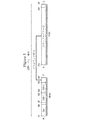
ヒトプロテインC(PC)酵素前駆体は、461個のアミノ酸残基の前駆体として肝臓中で合成され、血液中に分泌される(配列番号1に示す通りである)。分泌に先立って一本鎖のポリペプチド前駆体は、ジペプチド(Lys156‐Arg157)および42個のaa‐残基からなるプレプロリーダーの除去によってヘテロ二量体に変換される。ヘテロ二量体型(417残基)は、ジスルフィド架橋によって連結した軽鎖(155aa、21kDa)および重鎖(262aa、41kDa)から構成される(配列番号2に示す通りである)。PC酵素前駆体は、「活性化ペプチド」の除去および配列番号3に示す活性化PC(aPC)(405残基)へのPCの活性化をもたらす、トロンビン切断部位を含む。図1は、ヒトPCおよびその活性型であるaPCの模式図を提供する。ヒトPCは、9個のGla‐残基およびN‐結合型グリコシル化のための4個の候補サイトを含む。軽鎖は、Glaドメインおよび2個のEGF様ドメインを含む。重鎖は活性型セリンプロテアーゼドメインを内部に持つ。 Human protein C (PC) enzyme precursor is synthesized in the liver as a precursor of 461 amino acid residues and secreted into the blood (as shown in SEQ ID NO: 1). Prior to secretion, the single-chain polypeptide precursor is converted to a heterodimer by removal of a prepro leader consisting of a dipeptide (Lys156-Arg157) and 42 aa-residues. The heterodimeric form (417 residues) is composed of a light chain (155aa, 21 kDa) and a heavy chain (262aa, 41 kDa) linked by disulfide bridges (as shown in SEQ ID NO: 2). The PC enzyme precursor contains a thrombin cleavage site that results in the removal of the “activating peptide” and activation of the PC to activated PC (aPC) (405 residues) as shown in SEQ ID NO: 3. FIG. 1 provides a schematic diagram of human PC and its active form, aPC. Human PC contains 9 Gla-residues and 4 candidate sites for N-linked glycosylation. The light chain contains a Gla domain and two EGF-like domains. The heavy chain has an active serine protease domain inside.
PCは、通常3〜5μg/ml(〜65nM)で健康人の血液中を循環し、その半減期は6〜8時間である。循環するPC酵素前駆体の主要な形態は、ヘテロ二量体型である。PCの軽鎖は、一個のγ‐カルボキシグルタミン酸(Gla)に富むドメイン(45aa)、二個のEFG様ドメイン(46aa)およびリンカー配列を含む。PCの重鎖は、12個のaaで高い極性の「活性化ペプチド」および典型的なセリンプロテアーゼの触媒三残基を有する触媒ドメインを内部に持つ。 PC normally circulates in the blood of healthy people at 3-5 μg / ml (˜65 nM), and its half-life is 6-8 hours. The major form of circulating PC enzyme precursor is the heterodimeric form. The light chain of PC contains one γ-carboxyglutamate (Gla) rich domain (45aa), two EFG-like domains (46aa) and a linker sequence. The heavy chain of PC has an internal catalytic domain with 12 aa highly polar “activating peptides” and the catalytic triad of a typical serine protease.
ヒトPCは、グリコシル化、ビタミンK依存γ‐カルボキシル化およびγ‐ヒドロキシル化(1‐2)を含む、広範囲にわたる翻訳後修飾を受ける。ヒトPCは、炭水化物23%(重量)および4個の潜在的なN‐結合型グリコシル化サイト(一個は軽鎖のAsn97および3個は重鎖のAsn248/313/329)を含む。そのGlaドメインは、9個のGla残基を含み、負に帯電したリン脂質膜へのカルシウム依存性のPCの結合を担う。Glaドメインはまた、PC活性化の間にトロンビンおよびトロンボモジュリンを内皮膜上に整列させる、内皮プロテインCレセプター(EPCR)にも結合し得る。 Human PC undergoes a wide range of post-translational modifications including glycosylation, vitamin K-dependent γ-carboxylation and γ-hydroxylation (1-2). Human PC contains 23% (by weight) carbohydrate and four potential N-linked glycosylation sites (one light chain Asn97 and three heavy chain Asn248 / 313/329). Its Gla domain contains 9 Gla residues and is responsible for calcium-dependent PC binding to negatively charged phospholipid membranes. The Gla domain can also bind to endothelial protein C receptor (EPCR), which aligns thrombin and thrombomodulin on the inner capsule during PC activation.
プロテインC酵素前駆体は、生物学的能力を有するために、典型的にはその活性化酵素---活性化プロテインC(aPC)に変換される。PC経路の活性化は、PCの活性化およびaPCの不活性化の速度によって制御される。PCの活性化は、内皮細胞上で二段階のプロセスにおいて生じる。それには、内皮細胞上のEPCRへの(Glaドメインによる)PCの結合、それに続くトロンビン/トロンボモジュリン複合体によるPCのタンパク質分解的な活性化を必要とする。内皮細胞表面上でトロンビン/トロンボモジュリンによって触媒される、ヒトPC重鎖のArg12での単一の開裂は、12‐aaのAPを遊離させ、酵素前駆体PCをaPC、活性化セリンプロテアーゼに変換する。従って、PCとaPCのアミノ酸配列の間の主な相違点は、APCには存在しないPC中の12‐aaの活性ペプチドの存在である。PCのaPCへの活性化はまた、立体構造の変化も誘導する;その結果として、PCではなくaPCのみが、その酵素活性部位において、ベンズアミジンによってまたはクロロメチルケトン(CMK)ペプチド阻害剤により標識され得る。CMK阻害剤との複合体におけるGlaドメインレスのaPCの結晶構造が最近解明された。ヒト血漿における重要なaPC不活性化剤は、セルピン・スーパーファミリーのメンバーであり、ヒト血漿中に100nMで存在するプロテインC阻害剤(PCI)である。生理学的条件下でaPCは、20〜30分の半減期を有して、ヒト血漿中を非常に低濃度(1〜2ng/mlまたは40pM)で循環する。 Protein C enzyme precursors are typically converted to their activated enzyme--activated protein C (aPC) in order to have biological capacity. Activation of the PC pathway is controlled by the rate of PC activation and aPC inactivation. PC activation occurs in a two-step process on endothelial cells. This requires the binding of PC (by the Gla domain) to EPCR on endothelial cells, followed by proteolytic activation of PC by the thrombin / thrombomodulin complex. Single cleavage of human PC heavy chain with Arg12 catalyzed by thrombin / thrombomodulin on the endothelial cell surface liberates 12-aa AP and converts the proenzyme PC to aPC, an activated serine protease . Thus, the main difference between the amino acid sequences of PC and aPC is the presence of 12-aa active peptide in the PC that is not present in APC. Activation of PC to aPC also induces conformational changes; as a result, only aPC and not PC is labeled at its enzyme active site with benzamidine or with chloromethyl ketone (CMK) peptide inhibitors. obtain. The crystal structure of Gla domain-less aPC in complex with a CMK inhibitor has recently been elucidated. An important aPC inactivator in human plasma is a member of the serpin superfamily and is a protein C inhibitor (PCI) present at 100 nM in human plasma. Under physiological conditions aPC circulates in human plasma at very low concentrations (1-2 ng / ml or 40 pM) with a half-life of 20-30 minutes.
プロテインC経路は、血栓症に対する自然防御機構として貢献する。該経路は、凝固応答が増加するときに、抗凝固応答を増幅し得る即時回答システムであることにおいて他の抗凝固剤と異なる。傷を負った際、凝固のためにトロンビンが産生される。同時にトロンビンはまた、血管の表面上に一列に並んだトロンボモジュリンに結合することによって抗凝固応答も引き起こし、しかもこれがプロテインCの活性化を促進する。従ってaPCの産生は、トロンビン濃度およびPCレベルにおおよそ比例している。 The protein C pathway contributes as a natural defense mechanism against thrombosis. The pathway differs from other anticoagulants in that it is an immediate response system that can amplify the anticoagulant response as the coagulation response increases. When injured, thrombin is produced for clotting. At the same time, thrombin also causes an anticoagulant response by binding to a line of thrombomodulin on the surface of the blood vessel, which promotes protein C activation. Thus, aPC production is roughly proportional to thrombin concentration and PC level.
凝固プロセスの重要な制御因子としてのプロテインC経路の生理学的重要性は、次の3つの臨床所見によって明らかにされる:(a)プロテインC欠乏に伴う重篤な血栓性合併症およびプロテインCの補充による欠陥修正能、(b)プロテインC補足因子(プロテインS)欠乏関連の遺伝性血友病;および(c)aPCによる開裂に対しそれを抵抗性とするその物質(第V因子ライデンR506Q)における遺伝子突然変異と関連する血栓性リスク(Bernard,GR et.al.N Engl J Med 2001,344:699‐709 総説)。 The physiological importance of the protein C pathway as an important regulator of the coagulation process is demonstrated by three clinical findings: (a) severe thrombotic complications associated with protein C deficiency and protein C Ability to correct defects by supplementation, (b) hereditary hemophilia associated with protein C supplementation factor (protein S) deficiency; and (c) the substance that makes it resistant to cleavage by aPC (factor V Leiden R506Q) Thrombotic risk associated with genetic mutations in (Bernard, GR et.al. N Engl J Med 2001,344: 699-709 review).
他のビタミンK依存性凝固因子と対照的にaPCは、二つの凝固因子、第Va因子および第VIIIa因子のタンパク質分解性の不活性化によって抗凝固剤として機能し、それによってトロンビンの生成を抑制する。低下トロンビンレベルの結果として、トロンビンによって誘導される炎症、凝血原および抗線維素溶解性応答は減少する。aPCはまた、プラスミノーゲン活性化因子阻害剤(PAI)との複合体形成によって、促進された線維素溶解性応答に直接的に寄与する。 In contrast to other vitamin K-dependent coagulation factors, aPC functions as an anticoagulant by proteolytic inactivation of two coagulation factors, factor Va and factor VIIIa, thereby inhibiting thrombin generation To do. As a result of the reduced thrombin level, the thrombin-induced inflammation, clotogen and antifibrinolytic response are reduced. aPC also contributes directly to the enhanced fibrinolytic response by complex formation with plasminogen activator inhibitor (PAI).
その抗凝固機能に加えてaPCは、抗炎症および抗アポトーシス活性および内皮バリアー機能を含む、細胞保護効果を誘導する。細胞上でのaPCのこの直接的細胞保護効果は、EPCRおよびGタンパク質共役受容体、プロテアーゼ活性化型受容体‐1(PAR‐1)を必要とする。従ってaPCは、線維素溶解を促進し、血栓病および炎症を抑制する。aPCの抗凝固および細胞保護機能は、分離可能と思われる。細胞保護効果の大部分は、aPCの抗凝固活性から本来独立しており、最小の抗凝固活性および正常な細胞保護活性を有するaPC変異体が作成された。同様に、過剰な抗凝固性であるが非細胞保護的なaPC変異体も報告された。 In addition to its anticoagulant function, aPC induces cytoprotective effects, including anti-inflammatory and anti-apoptotic activity and endothelial barrier function. This direct cytoprotective effect of aPC on cells requires EPCR and a G protein coupled receptor, protease activated receptor-1 (PAR-1). Thus, aPC promotes fibrinolysis and suppresses thrombosis and inflammation. The anticoagulant and cytoprotective functions of aPC appear to be separable. Most of the cytoprotective effects are inherently independent of the anticoagulant activity of aPC, and aPC variants with minimal anticoagulant activity and normal cytoprotective activity were created. Similarly, excessive anticoagulant but non-cytoprotective aPC variants have also been reported.
aPC軽鎖のC末端はまた、プロテアーゼドメインにおける活性部位の反対側上の残基Gly142‐Leu155を含む高帯電領域でもある。E149A‐aPCは、野生型aPCと区別不能なアミド分解活性を示したが、しかしプロテインS補助因子活性に対する増加した感受性のために、活性化部分トロンボンプラスチン時間(aPTT)凝固試験における抗凝固活性おいて3倍を超える上昇を示した。E149A‐aPCは、インビボでの機能亢進性の抗血栓能と同様に、血漿凝固試験において機能亢進性の抗凝固活性を示した。この変異体は、LPS誘導性致死性内毒血病マウスモデルにおいて、細胞保護活性および死亡率低下活性を減少させた。これは、aPCの細胞保護活性は、マウスモデルにおける死亡率を減少させる必要があることを暗示する。対照的にaPCの抗凝固活性は、死亡率の低下にとって必要でも十分でもない。aPCは、凝固亢進および生成された炎症反応と関連した、生命を脅かす病態である敗血症を治療するために使用されてきた。aPC治療の重篤な副作用は、患者の2%において生じる大出血である。この重篤な副作用は、該治療の臨床用途を制限する。 The C-terminus of the aPC light chain is also a highly charged region containing the residue Gly142-Leu155 on the opposite side of the active site in the protease domain. E149A-aPC showed amidolytic activity indistinguishable from wild-type aPC, but due to increased sensitivity to protein S cofactor activity, anticoagulation in the activated partial thromboplastin time (aPTT) clotting test It showed a 3-fold increase in activity. E149A-aPC showed hyperactive anticoagulant activity in plasma coagulation studies, as well as in vivo hyperactive antithrombotic activity. This mutant reduced cytoprotective and mortality-reducing activity in a mouse model of LPS-induced lethal endotoxemia. This implies that the cytoprotective activity of aPC needs to reduce mortality in a mouse model. In contrast, the anticoagulant activity of aPC is neither necessary nor sufficient for reducing mortality. aPC has been used to treat sepsis, a life-threatening condition associated with hypercoagulation and the generated inflammatory response. A serious side effect of aPC treatment is major bleeding that occurs in 2% of patients. This severe side effect limits the clinical use of the treatment.
ヒト活性化プロテインC(aPC)に対するモノクローナル抗体が提供される。少なくとも一の実施形態において、抗‐aPCモノクローナル抗体は、aPCの酵素前駆体であるプロテインCに対し最小限の結合性を示す。 Monoclonal antibodies against human activated protein C (aPC) are provided. In at least one embodiment, the anti-aPC monoclonal antibody exhibits minimal binding to protein C, the enzymatic precursor of aPC.
いくつかの実施形態において、提供されるaPCに対するモノクローナル抗体は、例えば、親和性を増加させるために、機能活性を増加させるためにまたは生殖系列配列からの分岐を減少させるために最適化された。 In some embodiments, provided monoclonal antibodies to aPC have been optimized, for example, to increase affinity, to increase functional activity, or to reduce branching from germline sequences.
同様に提供されるのは、単離モノクローナル抗体によって結合されるヒトaPC上の特異的エピトープである。さらに提供されるのは、該エピトープをコードする単離核酸分子である。 Also provided are specific epitopes on human aPC that are bound by isolated monoclonal antibodies. Further provided is an isolated nucleic acid molecule encoding the epitope.
抗‐aPCモノクローナル抗体を含む医薬組成物、および血友病AおよびB等の凝固における遺伝性および後天性の欠乏症または欠損症の治療法もまた提供される。また提供されるのは、必要とする患者に抗‐aPCモノクローナル抗体を投与することによって出血時間を短縮するための方法である。ヒトaPCに結合するモノクローナル抗体を製造するための方法もまた提供される。
当業者であれば、以下に記載される図面がただ単に説明目的のためだけであることを理解するであろう。図面は本教示内容の範囲を決して制限する意図ではない。
Also provided are pharmaceutical compositions comprising anti-aPC monoclonal antibodies and methods of treating hereditary and acquired deficiencies or deficiencies in coagulation such as hemophilia A and B. Also provided is a method for reducing bleeding time by administering an anti-aPC monoclonal antibody to a patient in need thereof. A method for producing a monoclonal antibody that binds to human aPC is also provided.
Those skilled in the art will appreciate that the drawings described below are for illustrative purposes only. The drawings are not intended to limit the scope of the present teachings in any way.
上記の通り本開示は、ヒトプロテインCの活性型(aPC)に特異的に結合するが、しかしヒトプロテインCの酵素前駆体型(PC)に対して比較的に少ししかまたは全く反応性を示さない、モノクローナル抗体および他の結合性タンパク質を含む、抗体を提供する。 As noted above, the present disclosure specifically binds to the active form of human protein C (aPC), but exhibits relatively little or no reactivity to the proenzyme form of human protein C (PC). Antibodies are provided, including monoclonal antibodies and other binding proteins.
本特許明細書の目的のために、以下に提示する定義により以下の用語を使用する。
定義
For purposes of this patent specification, the following terms are used with the definitions provided below.
Definition
適当と認められる場合は、単数形で使用される用語はまた、複数形も含み逆もまた同様である。以下に示される任意の定義が、本明細書で参照されることにより援用される任意の資料を含む、任意の他の資料におけるその言葉の使用法と一致しない場合には、(例えば、用語が最初に使用されている資料において)反対の意味が明確に意図されなければ、以下に示される定義が、本明細書および付随する請求項を解釈する目的のために常に制御することになる。別段の定めがない限り「または(or)」は、「および/または(and/or)」を意味する。別段の定めがない限りまたは「一または複数」の使用が明らかに不適当である場合、本明細書での「a」の使用は、「一または複数(one or more)」を意味する。「comprise」、「comprises」、「comprising」、「include」、「includes」および「including」の使用は、交換可能であり、限定的でない。例えば、用語「含んでいる(including)」は、「含んでいるが、限定するものではない(including, but not limited to)」を意味するものとする。 Where appropriate, terms used in the singular also include the plural and vice versa. If any definition set forth below is inconsistent with the usage of that term in any other material, including any material incorporated by reference herein (for example, the term Unless the opposite meaning is explicitly intended (in the first used document), the definitions set forth below will always control for the purposes of interpreting the specification and the appended claims. Unless otherwise specified, “or” means “and / or”. Unless otherwise specified or where the use of “one or more” is clearly inappropriate, the use of “a” herein means “one or more”. The use of “comprise”, “comprises”, “comprising”, “include”, “includes” and “including” is interchangeable and is not limiting. For example, the term “including” shall mean “including, but not limited to”.
本明細書で用いる場合、用語「プロテインC」または「PC」とは、細胞により自然に発現され、血漿中に存在し、かつプロテインCの活性型とは異なる、その酵素前駆体型のプロテインCの任意の変異体、アイソフォームおよび/または種ホモログを表す。 As used herein, the term “protein C” or “PC” refers to the progenitor form of protein C that is naturally expressed by cells, is present in plasma, and is different from the active form of protein C. Represents any variant, isoform and / or species homolog.
本明細書で用いる場合、用語「活性化プロテインC」または「aPC」とは、プロテインCに存在する12個のアミノ酸の活性化ペプチドが存在しないことによって特徴づけられる、プロテインCの活性型を表す。 As used herein, the term “activated protein C” or “aPC” refers to an activated form of protein C that is characterized by the absence of an activated peptide of 12 amino acids present in protein C. .
本明細書で用いる場合、「抗体」とは、抗体全体およびその任意の抗原結合性フラグメント(すなわち「抗原‐結合性部分」)または一本鎖を表す。該用語には、自然に生じるまたは通常の免疫グロブリン遺伝子フラグメント再組み合せプロセスによって形成される、完全長免疫グロブリン分子(例えば、IgG抗体)、または特異的な結合活性を保持する抗体フラグメント等の、免疫グロブリン分子の免疫学的に活性な部分が含まれる。構造にかかわらず、抗体フラグメントは、完全長抗体によって認識されるのと同じ抗原と結合する。例えば、抗‐aPCモノクローナル抗体フラグメントは、aPCのエピトープに結合する。抗体の抗原結合機能は、完全長抗体のフラグメントによって遂行され得る。用語、抗体の「抗原結合部分」内に包含される結合性フラグメントの例には、(i)VL、VH、CLおよびCH1ドメインから成る一価フラグメントであるFabフラグメント;(ii)ヒンジ領域でジスルフィド架橋によって連結されている二個のFabフラグメントを含む二価フラグメントであるF(ab‘)2フラグメント;(iii)VHおよびCH1ドメインから成るFdフラグメント;(iv)抗体のシングルアームのVLおよびVHドメインから成るFvフラグメント、(v)VHドメインから成るdAbフラグメント(Ward et al.,(1989)Nature 341:544‐546);(vi)単離相補性決定領域(CDR);(vii)ミニ抗体、二重特異性抗体、三重特異性抗体、四重特異性抗体およびカッパ抗体(例えば、Ill et al.,Protein Eng 1997;10:949‐57を参照されたい);(viii)ラクダIgG;および(ix)IgNARが含まれる。さらに、Fvフラグメントの二個のドメイン、VLとVHは分離した遺伝子によってコードされているが、それらは、組換え法を使用して、その中でVLおよびVH領域が一価の分子を形成するために対合する一本鎖タンパク質として作成されることを可能とする、合成リンカーによって結合し得る(一本鎖Fv(scFv)として知られる;例えば、Bird et al.(1988)Science 242:423‐426;およびHuston et al(1988)Proc.Natl.Acad.Sci.USA 85:5879‐5883を参照されたい)。当該一本鎖抗体もまた、用語、抗体の「抗原‐結合部分」内に包含されるように意図されている。これらの抗体フラグメントは、当業者に既知の従来技術を使用して得られ、フラグメントは有用性に関して完全な抗体と同じ方法により解析される。 As used herein, “antibody” refers to the entire antibody and any antigen-binding fragment thereof (ie, “antigen-binding portion”) or single chain. The term includes immunity, such as a full-length immunoglobulin molecule (eg, an IgG antibody), or an antibody fragment that retains specific binding activity, either naturally occurring or formed by normal immunoglobulin gene fragment recombination processes. Includes an immunologically active portion of a globulin molecule. Regardless of structure, an antibody fragment binds with the same antigen that is recognized by the full-length antibody. For example, an anti-aPC monoclonal antibody fragment binds to an epitope of aPC. The antigen-binding function of an antibody can be performed by fragments of a full-length antibody. Examples of binding fragments encompassed within the term “antigen-binding portion” of an antibody include: (i) a Fab fragment that is a monovalent fragment consisting of VL, VH, CL, and CH1 domains; (ii) disulfides in the hinge region An F (ab ′) 2 fragment that is a bivalent fragment comprising two Fab fragments linked by a bridge; (iii) an Fd fragment consisting of the VH and CH1 domains; (iv) a single arm VL and VH domain of the antibody (V) a dAb fragment consisting of a VH domain (Ward et al., (1989) Nature 341: 544-546); (vi) an isolated complementarity determining region (CDR); (vii) a miniantibody, Bispecific antibodies, trispecific antibodies, tetraspecific antibodies and Kappa antibody (e.g., Ill et al, Protein Eng 1997; 10:. See 949-57); include and (ix) IgNAR; (viii) a camel IgG. In addition, the two domains of the Fv fragment, VL and VH, are encoded by separate genes, which use recombination methods to form molecules in which the VL and VH regions are monovalent. Can be joined by a synthetic linker (known as single-chain Fv (scFv); see, for example, Bird et al. (1988) Science 242: 423). -426; and Huston et al (1988) Proc. Natl. Acad. Sci. USA 85: 5879-5883). Such single chain antibodies are also intended to be encompassed within the term “antigen-binding portion” of an antibody. These antibody fragments are obtained using conventional techniques known to those skilled in the art, and the fragments are analyzed for utility by the same methods as complete antibodies.
さらに、抗原結合フラグメントは、抗体模倣物に包含され得るとも考えられる。本明細書で用いる場合、用語「抗体模倣物」または「模倣物」とは、抗体と同様に結合性を示すがより小さな別の抗体または非抗体タンパク質であるタンパク質を意味する。当該抗体模倣物は、スキャフォールドに含まれ得る。用語「スキャフォールド」とは、目的に合わせた機能および特性を有する新規産物を工学するためのポリペプチドプラットフォームを表す。 It is further contemplated that antigen binding fragments can be included in antibody mimetics. As used herein, the term “antibody mimetic” or “mimetic” refers to a protein that is another antibody or non-antibody protein that displays binding but is similar to an antibody. The antibody mimic can be included in the scaffold. The term “scaffold” refers to a polypeptide platform for engineering new products with tailored function and properties.
本明細書で用いる場合、用語「抗‐aPC抗体」とは、aPCのエピトープに特異的に結合する抗体を表す。インビボでaPCのエピトープに結合すると、本明細書に開示する抗‐aPC抗体は、一または複数の血液凝固カスケードの側面を増強する。 As used herein, the term “anti-aPC antibody” refers to an antibody that specifically binds to an epitope of aPC. Upon binding to an epitope of aPC in vivo, the anti-aPC antibodies disclosed herein enhance one or more aspects of the blood coagulation cascade.
本明細書で用いる場合、用語「結合を阻害する(inhibits binding)」および「結合をブロックする(blocks binding)」(例えば、aPCへのaPC基質の結合の阻害/ブロッキングを表す)は、同じ意味で使用され、例えば、少なくとも約10%、約20%、約30%、約40%、約50%、約60%、約70%、約80%、約90%、約95%、約96%、約97%、約98%、約99%または約100%だけの阻害またはブロッキング等の、タンパク質とその基質との部分的阻害および完全な阻害またはブロッキングの双方を包含する。本明細書で用いる場合、「約」は示された数値の+/−10%を意味する。 As used herein, the terms “inhibits binding” and “blocks binding” (eg, representing inhibition / blocking of aPC substrate binding to aPC) have the same meaning. For example, at least about 10%, about 20%, about 30%, about 40%, about 50%, about 60%, about 70%, about 80%, about 90%, about 95%, about 96% , Including both partial inhibition and complete inhibition or blocking of the protein and its substrate, such as inhibition or blocking of only about 97%, about 98%, about 99% or about 100%. As used herein, “about” means +/− 10% of the indicated numerical value.
aPCへのaPC基質の結合の阻害および/ブロッキングに関して、用語・阻害およびブロッキングはまた、抗‐aPC抗体と接触していないaPCと比較して抗‐aPC抗体と接触したときの、生理学的物質へのaPCの結合親和性における任意の測定し得る低下、例えば、少なくとも約10%、約20%、約30%、約40%、約50%、約60%、約70%、約80%、約90%、約95%、約96%、約97%、約98%、約99%または約100%だけの第Va因子または第VIIIa因子を含むその基質とのaPCの相互作用のブロッキング、も含む。 With respect to inhibition and / or blocking of aPC substrate binding to aPC, the terms inhibition and blocking also refer to physiological substances when contacted with anti-aPC antibody compared to aPC not contacted with anti-aPC antibody. Any measurable decrease in the aPC binding affinity, such as at least about 10%, about 20%, about 30%, about 40%, about 50%, about 60%, about 70%, about 80%, about Blocking of aPC's interaction with 90%, about 95%, about 96%, about 97%, about 98%, about 99% or only about 100% of factor Va or its substrate containing factor VIIIa .
本明細書で用いる場合、用語「モノクローナル抗体」または「モノクローナル抗体組成物」は、単一分子組成物の抗体分子の製剤を表す。モノクローナル抗体組成物は、特定のエピトープに対する単一結合特異性および親和性を示す。同様に、用語「ヒトモノクローナル抗体」は、ヒト生殖系列免疫グロブリン配列由来の可変および定常領域を有する、単一結合特異性を示す抗体を表す。ヒト抗体は、ヒト生殖系列免疫グロブリン配列によってコードされないアミノ酸残基を含み得る(インビトロでのランダムまたは部位特異的突然変異誘導またはインビボでの体細胞突然変異によって誘導される変異体)。 As used herein, the term “monoclonal antibody” or “monoclonal antibody composition” refers to a preparation of antibody molecules of single molecular composition. A monoclonal antibody composition displays a single binding specificity and affinity for a particular epitope. Similarly, the term “human monoclonal antibody” refers to an antibody that exhibits single binding specificity, having variable and constant regions derived from human germline immunoglobulin sequences. Human antibodies may contain amino acid residues that are not encoded by human germline immunoglobulin sequences (variants induced by random or site-directed mutagenesis in vitro or somatic mutation in vivo).
本明細書で用いる場合、「単離抗体」とは、異なる抗原特異性を有する抗体を含む、他の生物学的分子を実質的に含まない抗体を表すことを意図する(例えば、aPCに結合する単離抗体は、aPC以外の抗原に結合する抗体を実質的に含まない)。いくつかの実施形態において、単離抗体は、乾燥重量で少なくとも約75%、約80%、約90%、約95%、約97%、約99%、約99.9%または約100%純粋である。いくつかの実施形態において、純度は、例えば、カラムクロマトグラフィー、ポリアクリルアミドゲル電気泳動またはHPLC分析等の方法によって測定され得る。ヒトaPCのエピトープ、アイソフォームまたは変異体に結合する単離抗体は、しかしながら例えば、他の種由来の他の関連する抗原(例えば、aPC種ホモログ)との交差反応性を有する。さらに単離抗体は、他の細胞物質および/または化学物質を実質的に含まないことが可能である。本明細書で用いる場合「特異的結合」とは、所定の抗原への抗体結合を表す。典型的には「特異的結合」を示す抗体は、少なくとも約105M−1の親和性により抗原に結合し、無関係な抗原(例えば、BSA、カゼイン)に対するその結合親和性よりも高い、例えば少なくとも二倍以上大きい親和性により該抗原に結合する。「抗原を認識する抗体」および「抗原に対し特異的な抗体」との表現は、用語「抗原に特異的に結合する抗体」と共に本明細書において同じ意味で用いられる。 As used herein, “isolated antibody” is intended to refer to an antibody that is substantially free of other biological molecules, including antibodies with different antigen specificities (eg, bound to aPC). Isolated antibody is substantially free of antibodies that bind to antigens other than aPC). In some embodiments, the isolated antibody is at least about 75%, about 80%, about 90%, about 95%, about 97%, about 99%, about 99.9% or about 100% pure by dry weight. It is. In some embodiments, purity can be measured by methods such as, for example, column chromatography, polyacrylamide gel electrophoresis, or HPLC analysis. An isolated antibody that binds to an epitope, isoform, or variant of human aPC, however, has cross-reactivity with, for example, other related antigens (eg, aPC species homologs) from other species. Further, the isolated antibody can be substantially free of other cellular material and / or chemicals. As used herein, “specific binding” refers to antibody binding to a predetermined antigen. An antibody that typically exhibits “specific binding” binds to an antigen with an affinity of at least about 105 M−1 and has a higher, eg, at least two, binding affinity for an irrelevant antigen (eg, BSA, casein). It binds to the antigen with an affinity that is at least twice as great. The expressions “an antibody recognizing an antigen” and “an antibody specific for an antigen” are used interchangeably herein with the term “an antibody that binds specifically to an antigen”.
本明細書で用いる場合、用語「最小の結合性」とは、特定の抗原に対し結合しないおよび/または低い親和性しか示さない抗体を表す。典型的には、抗原に対し最小の結合性を有する抗体は、約102M−1よりも低い親和性を有する抗原に結合し、当該抗体が無関係な抗原に結合するよりも高い親和性を有する所定の抗原には結合しない。 As used herein, the term “minimal binding” refers to an antibody that does not bind to a particular antigen and / or exhibits low affinity. Typically, an antibody with minimal binding to an antigen binds to an antigen having an affinity lower than about 102M-1, and has a predetermined affinity that is higher than that antibody binds to an irrelevant antigen. It does not bind to any antigen.
本明細書で用いる場合、例えば、IgG抗体等の抗体に対する用語「高親和性」とは、少なくとも約107M−1、少なくとも一の実施形態において、少なくとも約108M−1、いくつかの実施形態において、少なくとも約109M−1、1010M−1、1011M−1またはそれ以上、例えば1013M−1までまたはそれ以上の結合親和性を表す。しかしながら、「高親和性」の結合性は、他の抗体アイソタイプに対して変動し得る。例えば、IgMアイソタイプに対する「高親和性」の結合性は、少なくとも107M−1の結合親和性を表す。本明細書で用いる場合、「アイソタイプ」とは重鎖定常領域の遺伝子によってコードされる抗体クラス(例えば、IgMまたはIgG1)を表す。 As used herein, for example, the term “high affinity” for an antibody, such as an IgG antibody, refers to at least about 107 M−1, in at least one embodiment, at least about 108 M−1, in some embodiments, It represents a binding affinity of at least about 109M-1, 1010M-1, 1011M-1, or more, such as up to 1013M-1. However, “high affinity” binding can vary for other antibody isotypes. For example, “high affinity” binding to an IgM isotype represents a binding affinity of at least 107M-1. As used herein, “isotype” refers to the antibody class (eg, IgM or IgG1) that is encoded by heavy chain constant region genes.
「相補性決定領域」または「CDR」とは、抗体分子の重鎖の可変領域または軽鎖の可変領域内の、結合した抗原の三次元構造に相補的であるN末端抗原結合表面を形成する、三つの高度可変領域の一つを表す。重鎖または軽鎖のN末端から進み、これらの相補性決定領域はそれぞれ「CDR1」、「CDR2」および「CDR3」と表示される[Wu TT,Kabat EA,Bilofsky H,Proc Natl Acad Sci USA.1975 Dec;72(12):5107およびWu TT,Kabat EA,J Exp Med.1970 Aug 1;132(2):211]。CDRは、抗原‐抗体結合に関わり、CDR3は、抗原‐抗体結合に特異的なユニークな領域を含む。それ故に抗原結合部位は、各々の重鎖および軽鎖V領域由来のCDR領域を含む六個のCDRを含み得る。
“Complementarity determining region” or “CDR” forms an N-terminal antigen-binding surface that is complementary to the three-dimensional structure of the bound antigen in the variable region of the heavy chain or light chain of an antibody molecule. , Represents one of three highly variable regions. Proceeding from the N-terminus of the heavy or light chain, these complementarity determining regions are labeled “CDR1”, “CDR2” and “CDR3”, respectively [Wu TT, Kabat EA, Bilfsky H, Proc Natl Acad Sci USA. 1975 Dec; 72 (12): 5107 and Wu TT, Kabat EA, J Exp Med. 1970
用語「エピトープ」とは、抗体が特異的に結合または相互作用する抗原の区域または領域を表し、いくつかの実施形態において、それは、抗原が抗体と物理的に接触している所を示す。逆に、用語「パラトープ」とは、抗原が特異的に結合する抗体の区域または領域を表す。競合結合によって特徴づけられるエピトープは、もし対応する抗体の結合が互いに排他的、すなわち一つの抗体の結合がもう一つの抗体の同時の結合を排除するのであるならば、オーバーラップしていると言われる。もし抗原が対応する抗体の双方の同時の結合に適応できるならば、エピトープは別々である(ユニーク)と言われる。 The term “epitope” refers to the area or region of an antigen to which an antibody specifically binds or interacts, and in some embodiments it indicates where the antigen is in physical contact with the antibody. Conversely, the term “paratope” refers to the area or region of an antibody to which an antigen specifically binds. Epitopes characterized by competitive binding are said to overlap if corresponding antibody binding is mutually exclusive, i.e. binding of one antibody excludes the simultaneous binding of another antibody. Is called. An epitope is said to be distinct (unique) if the antigen can accommodate the simultaneous binding of both of the corresponding antibodies.
本明細書で用いる場合、用語「競合する抗体」とは、本明細書記載のaPCに対する抗体として、実質的にまたは本質的に同じまたは同一のエピトープに関して結合する抗体を表す。「競合する抗体」には、オーバーラップエピトープの特異性を有する抗体が含まれる。従って競合する抗体は、aPCへの結合に関して本明細書に記載の抗体と効率的に競合することができる。いくつかの実施形態において、競合する抗体は、本明細書に記載の抗体と同一のエピトープに結合し得る。あるいは競合する抗体は、本明細書に記載の抗体と同じエピトープ特異性を有するともみなされる。 As used herein, the term “competing antibody” refers to an antibody that binds with respect to substantially or essentially the same or the same epitope as an antibody to aPC as described herein. “Competing antibodies” include antibodies having specificity for overlapping epitopes. Accordingly, competing antibodies can efficiently compete with the antibodies described herein for binding to aPC. In some embodiments, competing antibodies can bind to the same epitope as the antibodies described herein. Alternatively, competing antibodies are also considered to have the same epitope specificity as the antibodies described herein.
本明細書で用いる場合、「保守的置換」とは、ポリペプチドの生物学的または生化学的機能の損失をもたらさない、類似する生化学的特性を有するアミノ酸への一または複数のアミノ酸の置換を含むポリペプチドの修飾を表す。「保守的アミノ酸置換」は、アミノ酸残基が類似の側鎖を有するアミノ酸残基に置換されるものである。類似の側鎖を有するアミノ酸残基のファミリーは、当該技術分野において明確にされている。ファミリーには、塩基性側鎖(例えば、リジン、アルギニン、ヒスチジン)、酸性側鎖(例えば、アスパラギン酸、グルタミン酸)、極性無電化側鎖(例えば、グリシン、アスパラギン、グルタミン、セリン、スレオニン、チロシン、システイン)、非極性側鎖(例えば、アラニン、バリン、ロイシン、イソロイシン、プロリン、フェニルアラニン、メチオニン、トリプトファン)、β‐分岐側鎖(例えば、スレオニン、バリン、イソロイシン)および芳香族側鎖(例えば、チロシン、フェニルアラニン、トリプトファン、ヒスチジン)を有するアミノ酸が含まれる。本開示の抗体は、抗原結合活性を保持しつつ一または複数の保守的アミノ酸置換を有し得る。 As used herein, a “conservative substitution” is a substitution of one or more amino acids with an amino acid having similar biochemical properties that does not result in loss of the biological or biochemical function of the polypeptide. Represents a modification of a polypeptide comprising A “conservative amino acid substitution” is one in which the amino acid residue is replaced with an amino acid residue having a similar side chain. A family of amino acid residues having similar side chains has been defined in the art. The family includes basic side chains (eg, lysine, arginine, histidine), acidic side chains (eg, aspartic acid, glutamic acid), polar uncharged side chains (eg, glycine, asparagine, glutamine, serine, threonine, tyrosine, Cysteine), non-polar side chains (eg alanine, valine, leucine, isoleucine, proline, phenylalanine, methionine, tryptophan), β-branched side chains (eg threonine, valine, isoleucine) and aromatic side chains (eg tyrosine) , Phenylalanine, tryptophan, histidine). The antibodies of the present disclosure may have one or more conservative amino acid substitutions while retaining antigen binding activity.
核酸およびポリペプチドに対して用語「実質的相同」とは、二つの核酸または二つのポリペプチド、またはその指定された配列が、適切な核酸またはアミノ酸の挿入または欠失により最適に位置合わせされ、比較されたときに、ヌクレオチドまたはアミノ酸の少なくとも約80%、通常は少なくとも約85%、いくつかの実施形態において、約90%、91%、92%、93%、94%または95%、少なくとも一の実施形態において、核酸またはアミノ酸の少なくとも96%、97%、98%、99%、99.1%、99.2%、99.3%、99.4%または99.5%において同一であることを示す。あるいは、セグメントが鎖の補完のための選択的ハイブリダイゼーション条件下でハイブリダイズするときに、核酸の実質的相同が存在する。同様に含まれるまた、本明細書で列挙される特定の核酸配列およびアミノ酸配列に対し実質的な相同性を有する核酸配列およびポリペプチド配列である。 The term “substantially homologous” to nucleic acids and polypeptides means that two nucleic acids or two polypeptides, or a designated sequence thereof, are optimally aligned by insertion or deletion of appropriate nucleic acids or amino acids, When compared, at least about 80%, usually at least about 85% of nucleotides or amino acids, in some embodiments, about 90%, 91%, 92%, 93%, 94% or 95%, at least one In embodiments, at least 96%, 97%, 98%, 99%, 99.1%, 99.2%, 99.3%, 99.4% or 99.5% of nucleic acids or amino acids are identical. It shows that. Alternatively, there is substantial nucleic acid homology when the segments hybridize under selective hybridization conditions for strand complementation. Also included are nucleic acid and polypeptide sequences that also have substantial homology to the specific nucleic acid and amino acid sequences listed herein.
二つの配列の間のパーセント同一性は、二つの配列の最適な位置合わせに導入される必要のある、ギャップの数、および各々のギャップの長さを考慮したうえで、配列によって共有される同一の位置の数の関数である(すなわち、%相同性=同一位置の数/位置の総数×100)。配列の比較および二つの配列の間のパーセント同一性の決定は、限定するものではないがVectorNTI(登録商標)(Invitrogen Corp.,Carlsbad,CA)のAlignX(登録商標)モジュール等の数学的アルゴリズムを使用して達成され得る。AlignX(登録商標)にとって、多重整列化のデフォルトパラメータは:ギャップ開始ペナルティ:10;ギャップ伸張ペナルティ:0.05;ギャップ分離ペナルティ範囲:8;アライメント遅延に対する同一性%:40である。(http://www.invitrogen.com/site/us/en/home/LINNEA-Online-Guides/LINNEA-Communities/Vector-NTI-Community/Sequence-analysis-and-data-management-software-for-PCs/AlignX-Module-for-Vector-NTI-Advance.reg.us.htmlにおいてさらなる詳細を見出されたい)。 The percent identity between the two sequences is the identity shared by the sequences, taking into account the number of gaps and the length of each gap that need to be introduced for optimal alignment of the two sequences. Is the function of the number of positions (ie,% homology = number of identical positions / total number of positions × 100). Comparison of sequences and determination of percent identity between two sequences can be accomplished using mathematical algorithms such as, but not limited to, the AlignX® module of VectorNTI® (Invitrogen Corp., Carlsbad, Calif.). Can be achieved using. For AlignX®, the default parameters for multiple alignment are: gap opening penalty: 10; gap extension penalty: 0.05; gap separation penalty range: 8;% identity to alignment delay: 40. ( Http://www.invitrogen.com/site/us/en/home/LINNEA-Online-Guides/LINNEA-Communities/Vector-NTI-Community/Sequence-analysis-and-data-management-software-for-PCs Find more details in /AlignX-Module-for-Vector-NTI-Advance.reg.us.html ).
クエリー配列(本開示の配列)と対象配列の間の全体にわたる最良のマッチを決定する、グローバルシーケンスアラインメントとも呼ばれる別の方法は、Higginsら(Computer Applications in the Biosciences(CABIOS),1992,8(2):189‐191)のアルゴリズムに基づく、CLUSTALWコンピュータプログラム(Thompson et al.,Nucleic Acids Research,1994,2(22):4673‐4680)を使用して決定し得る。シーケンスアラインメントにおいてクエリーおよび対象配列は双方ともDNA配列である。前記のグローバルシーケンスアラインメントの結果はパーセント同一性である。ペアワイズアラインメントによりパーセント同一性を計算するためのDNA配列のCLLUSTALWアラインメントにおいて使用されるパラメータは:Matrix=IUB、k‐タプル=1、Top Diagonal数=5、ギャップペナルティ=3、ギャップ開始ペナルティ=10、ギャップ伸張ペナルティ=0.1。多重アラインメントのためには、以下のCLUSTALWパラメータが使用し得る;ギャップ開始ペナルティ=10、ギャップ伸張パラメータ=0.05;ギャップ分離ペナルティ範囲=8;アラインメント遅延のための%同一性=40。 Another method, also called global sequence alignment, that determines the overall best match between a query sequence (sequence of the present disclosure) and a subject sequence is described by Higgins et al. (Computer Applications in the Biosciences (CABIOS), 1992, 8 (2 ): 189-191), and may be determined using the CLUSTALW computer program (Thompson et al., Nucleic Acids Research, 1994, 2 (22): 4673-4680). In the sequence alignment, the query and the target sequence are both DNA sequences. The result of the global sequence alignment is percent identity. The parameters used in DNA sequence CLLUSTALW alignment to calculate percent identity by pairwise alignment are: Matrix = IUB, k-tuple = 1, Top Diagonal number = 5, gap penalty = 3, gap opening penalty = 10, Gap extension penalty = 0.1. For multiple alignments, the following CLUSTALW parameters may be used; gap start penalty = 10, gap extension parameter = 0.05; gap separation penalty range = 8;% identity for alignment delay = 40.
核酸は、細胞全体中に、細胞溶解物中にまたは部分精製された、または実質的に純粋な形態で存在し得る。核酸は、自然の環境においては通常それが随伴している、他の細胞成分から精製されたときに「単離され」または「実質的に純粋に与えられる」。核酸を単離するために、例えば以下の:アルカリ/SDS処理、CsClバンディング、カラムクロマトグラフィー、アガロースゲル電気泳動および当該技術分野において公知の方法等の標準的な技法が使用され得る。
活性化プロテインCに対するモノクローナル抗体
The nucleic acid can be present in whole cells, in cell lysates or in partially purified or substantially pure form. Nucleic acid is “isolated” or “substantially pure” when purified from other cellular components with which it is normally associated in the natural environment. Standard techniques can be used to isolate nucleic acids, such as, for example, the following: alkali / SDS treatment, CsCl banding, column chromatography, agarose gel electrophoresis and methods known in the art.
Monoclonal antibody against activated protein C
aPCはその抗凝固特性で知られる。傷が一時的なホメオスタシスの喪失をもたらす血友病または外傷患者において、ホメオスタシスが調節解除される出血性障害は、aPC阻害剤によって治療され得る。抗体、その抗原結合性フラグメント、および他のaPC‐特異的タンパク質スキャフォールドは、残りを保全する一方で、aPCのタンパク質機能の部分集合を阻害するための標的特異性を提供するために使用され得る。aPC(<4ng/ml)対PC(4ug/ml)の血漿濃度における少なくとも1000倍の差異を仮定すれば、任意の潜在的なaPC阻害剤治療法の向上した特異性は、高速循環する過剰なPCの存在下でのaPCの機能をブロックするために役立つ。 aPC is known for its anticoagulant properties. In hemophilia or trauma patients whose wound results in temporary loss of homeostasis, hemorrhagic disorders in which homeostasis is deregulated can be treated with aPC inhibitors. Antibodies, antigen-binding fragments thereof, and other aPC-specific protein scaffolds can be used to provide target specificity to inhibit a subset of protein functions of aPC while preserving the rest . Given the at least 1000-fold difference in plasma concentrations of aPC (<4 ng / ml) vs. PC (4 ug / ml), the improved specificity of any potential aPC inhibitor treatment is Useful for blocking aPC functionality in the presence of a PC.
aPCの抗凝固機能をブロックするaPC特異的抗体は、例えば、血友病、阻害剤を有する血友病患者、外傷誘導性凝固障害、aPCによる敗血病治療の間の重篤な出血患者、移植、心臓手術、整形外科手術等の待機手術からもたらされる出血または月経過多による過剰出血を含む、出血性障害を有する患者のための治療法として使用し得る。 aPC-specific antibodies that block the anticoagulant function of aPC include, for example, hemophilia, hemophilia patients with inhibitors, trauma-induced coagulopathy, severe bleeding patients during the treatment of septic disease with aPC, It can be used as a treatment for patients with bleeding disorders, including bleeding resulting from elective surgery such as transplantation, cardiac surgery, orthopedic surgery or excessive menstrual bleeding.
長い循環半減期を有する抗‐aPC抗体は、血友病のような慢性疾患を治療するうえで有用であり得る。短い半減期を有する、aPC抗体フラグメントまたはaPC‐結合性タンパク質スキャフォールドは、急性の使用にとってより効果的であり得る(例えば、外傷の治療用)。aPCは多機能タンパク質であるので、増強された親和性および標的特異性を有する抗体、抗原結合性抗体フラグメント、aPC‐特異的タンパク質スキャフォールドを含む、選択的aPC機能ブロッカー(SAFB)は、他のaPC機能に悪影響を及ぼすことなくただ一つのaPC機能を選択的にブロックし得る。 Anti-aPC antibodies with a long circulatory half-life can be useful in treating chronic diseases such as hemophilia. An aPC antibody fragment or aPC-binding protein scaffold with a short half-life may be more effective for acute use (eg for the treatment of trauma). Because aPC is a multifunctional protein, selective aPC function blockers (SAFB), including antibodies with enhanced affinity and target specificity, antigen-binding antibody fragments, aPC-specific protein scaffolds, are A single aPC function can be selectively blocked without adversely affecting the aPC function.
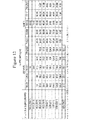
aPC‐結合性抗体は、ヒトaPCに対するヒト抗体ライブラリーをパンニングおよびスクリーニングすることによって同定された。同定された抗体は、ヒトPCへ結合しないかまたは最小の結合性を示した。単離された各々のモノクローナル抗体の重鎖可変領域および軽鎖可変領域を配列決定し、そのCDR領域を同定した。aPC‐特異的モノクローナル抗体の各々の重鎖および軽鎖領域に応答する配列識別番号(「配列番号(SEQ ID NO)」)を、表1に要約する。

一実施形態において提供されるのは、ヒト活性化プロテインC(aPC)に結合し抗凝固活性を阻害するが、しかし非活性化プロテインCに対する最小の結合性しか有さない単離モノクローナル抗体であり、ここで該抗体は、配列番号14〜23から成る群より選択されるアミノ酸配列を含む重鎖可変領域を含む。 Provided in one embodiment is an isolated monoclonal antibody that binds to human activated protein C (aPC) and inhibits anticoagulant activity, but has minimal binding to non-activated protein C. Wherein the antibody comprises a heavy chain variable region comprising an amino acid sequence selected from the group consisting of SEQ ID NOs: 14-23.
他の実施形態において提供されるのは、ヒト活性化プロテインC(aPC)に結合し抗凝固活性を阻害するが、しかし非活性化プロテインCに対する最小の結合性しか有さない単離モノクローナル抗体であり、ここで該抗体は、配列番号4〜13から成る群より選択されるアミノ酸配列を含む軽鎖可変領域を含む。 In other embodiments, an isolated monoclonal antibody that binds to human activated protein C (aPC) and inhibits anticoagulant activity, but has minimal binding to non-activated protein C is provided. Wherein the antibody comprises a light chain variable region comprising an amino acid sequence selected from the group consisting of SEQ ID NOs: 4-13.
他の実施形態において提供されるのは、ヒト活性化プロテインC(aPC)に結合し抗凝固活性を阻害するが、しかし非活性化プロテインCに対する最小の結合性しか有さない単離モノクローナル抗体であり、ここで該抗体は、配列番号14〜23から成る群より選択されるアミノ酸配列を含む重鎖可変領域および配列番号4〜13から成る群より選択されるアミノ酸配列を含む軽鎖可変領域を含む。 In other embodiments, an isolated monoclonal antibody that binds to human activated protein C (aPC) and inhibits anticoagulant activity, but has minimal binding to non-activated protein C is provided. Wherein the antibody comprises a heavy chain variable region comprising an amino acid sequence selected from the group consisting of SEQ ID NOs: 14-23 and a light chain variable region comprising an amino acid sequence selected from the group consisting of SEQ ID NOs: 4-13 Including.
他の実施例において抗体は、以下を含む重鎖および軽鎖可変領域を含む: In other examples, the antibody comprises heavy and light chain variable regions comprising:
配列番号14のアミノ酸配列を含む重鎖可変領域および配列番号4のアミノ酸配列を含む軽鎖可変領域; A heavy chain variable region comprising the amino acid sequence of SEQ ID NO: 14 and a light chain variable region comprising the amino acid sequence of SEQ ID NO: 4;
配列番号15のアミノ酸配列を含む重鎖可変領域および配列番号5のアミノ酸配列を含む軽鎖可変領域; A heavy chain variable region comprising the amino acid sequence of SEQ ID NO: 15 and a light chain variable region comprising the amino acid sequence of SEQ ID NO: 5;
配列番号16のアミノ酸配列を含む重鎖可変領域および配列番号6のアミノ酸配列を含む軽鎖可変領域; A heavy chain variable region comprising the amino acid sequence of SEQ ID NO: 16 and a light chain variable region comprising the amino acid sequence of SEQ ID NO: 6;
配列番号17のアミノ酸配列を含む重鎖可変領域および配列番号7のアミノ酸配列を含む軽鎖可変領域; A heavy chain variable region comprising the amino acid sequence of SEQ ID NO: 17 and a light chain variable region comprising the amino acid sequence of SEQ ID NO: 7;
配列番号18のアミノ酸配列を含む重鎖可変領域および配列番号8のアミノ酸配列を含む軽鎖可変領域; A heavy chain variable region comprising the amino acid sequence of SEQ ID NO: 18 and a light chain variable region comprising the amino acid sequence of SEQ ID NO: 8;
配列番号19のアミノ酸配列を含む重鎖可変領域および配列番号9のアミノ酸配列を含む軽鎖可変領域; A heavy chain variable region comprising the amino acid sequence of SEQ ID NO: 19 and a light chain variable region comprising the amino acid sequence of SEQ ID NO: 9;
配列番号20のアミノ酸配列を含む重鎖可変領域および配列番号10のアミノ酸配列を含む軽鎖可変領域; A heavy chain variable region comprising the amino acid sequence of SEQ ID NO: 20 and a light chain variable region comprising the amino acid sequence of SEQ ID NO: 10;
配列番号21のアミノ酸配列を含む重鎖可変領域および配列番号11のアミノ酸配列を含む軽鎖可変領域; A heavy chain variable region comprising the amino acid sequence of SEQ ID NO: 21 and a light chain variable region comprising the amino acid sequence of SEQ ID NO: 11;
配列番号22のアミノ酸配列を含む重鎖可変領域および配列番号12のアミノ酸配列を含む軽鎖可変領域;および A heavy chain variable region comprising the amino acid sequence of SEQ ID NO: 22 and a light chain variable region comprising the amino acid sequence of SEQ ID NO: 12; and
配列番号23のアミノ酸配列を含む重鎖可変領域および配列番号13のアミノ酸配列を含む軽鎖可変領域。 A heavy chain variable region comprising the amino acid sequence of SEQ ID NO: 23 and a light chain variable region comprising the amino acid sequence of SEQ ID NO: 13.
表2に示すのは、ヒトaPCに結合するモノクローナル抗体の各々の重鎖および軽鎖のCDR領域(「CDR1」、「CDR2」および「CDR3」)に対する配列識別番頭の要約である。
一実施形態において提供されるのは、ヒト活性化プロテインC(aPC)に結合する単離モノクローナル抗体であり、ここで該抗体は配列番号94〜103から成る群より選択されるアミノ酸配列を含むCDR3を含む。これらのCDR3は、パンニングおよびスクリーニングの間に同定される抗体の重鎖から同定される。さらなる実施形態において、この抗体は、(a)配列番号74〜83から成る群より選択されるアミノ酸配列を含むCDR1、(b)配列番号84〜93から成る群より選択されるアミノ酸配列を含むCDR2、または(c)配列番号74〜83から成る群より選択されるアミノ酸配列を含むCDR1および配列番号84〜93から成る群より選択されるアミノ酸配列を含むCDR2の双方、をさらに含む。 Provided in one embodiment is an isolated monoclonal antibody that binds human activated protein C (aPC), wherein the antibody comprises an amino acid sequence selected from the group consisting of SEQ ID NOs: 94-103. including. These CDR3s are identified from the heavy chain of the antibody identified during panning and screening. In a further embodiment, the antibody comprises (a) a CDR1 comprising an amino acid sequence selected from the group consisting of SEQ ID NOs: 74-83, (b) a CDR2 comprising an amino acid sequence selected from the group consisting of SEQ ID NOs: 84-93 Or (c) both a CDR1 comprising an amino acid sequence selected from the group consisting of SEQ ID NOs: 74-83 and a CDR2 comprising an amino acid sequence selected from the group consisting of SEQ ID NOs: 84-93.
他の実施形態において提供されるのは、パンニングおよびスクリーニングの間に同定される抗体の軽鎖の一つ由来のCDR3を共有する抗体である。従って、同様に提供されるのは単離モノクローナル抗体であり、ここで前記の抗体は活性化プロテインCに結合しかつ抗凝固活性を阻害するが、しかし非活性化プロテインCに対する最小の結合性しか有さず、ここで前記の抗体は、配列番号64〜73から成る群より選択されるアミノ酸配列を含むCDR3を含む。さらなる実施形態において、該抗体は、(a)配列番号44〜53から成る群より選択されるアミノ酸配列を含むCDR1、(b)配列番号54〜63から成る群より選択されるアミノ酸配列を含むCDR2、または(c)配列番号44〜53から成る群より選択されるアミノ酸配列を含むCDR1および配列番号54〜63から成る群より選択されるアミノ酸配列を含むCDR2の双方、をさらに含む。 Provided in other embodiments are antibodies that share CDR3 from one of the antibody light chains identified during panning and screening. Accordingly, also provided is an isolated monoclonal antibody, wherein said antibody binds to activated protein C and inhibits anticoagulant activity, but has minimal binding to non-activated protein C. Wherein said antibody comprises a CDR3 comprising an amino acid sequence selected from the group consisting of SEQ ID NOs: 64-73. In a further embodiment, the antibody comprises (a) a CDR1 comprising an amino acid sequence selected from the group consisting of SEQ ID NOs: 44-53, (b) a CDR2 comprising an amino acid sequence selected from the group consisting of SEQ ID NOs: 54-63 Or (c) both a CDR1 comprising an amino acid sequence selected from the group consisting of SEQ ID NOs: 44-53 and a CDR2 comprising an amino acid sequence selected from the group consisting of SEQ ID NOs: 54-63.
他の実施形態において、抗体は、スクリーニングおよびパンニングから同定される抗体の重鎖および軽鎖由来のCDR3を含む。提供されるのは、単離モノクローナル抗体であり、ここで前記の抗体は活性化プロテインCに結合しかつ抗凝固活性を阻害するが、しかし非活性型プロテインCに対し最小の結合性しか有さず、ここで前記の抗体は、配列番号94〜103から成る群より選択されるアミノ酸配列を含むCDR3および配列番号64〜73から成る群より選択されるアミノ酸配列を含むCDR3を含む。さらなる実施形態において、抗体は、(a)配列番号74〜83から成る群より選択されるアミノ酸配列を含むCDR1、(b)配列番号84〜93から成る群より選択されるアミノ酸配列を含むCDR2、(c)配列番号44〜53から成る群より選択されるアミノ酸配列を含むCDR1、および/または(d)配列番号54〜63から成る群より選択されるアミノ酸配列を含むCDR2をさらに含む。 In other embodiments, the antibody comprises CDR3 from the heavy and light chains of the antibody identified from screening and panning. Provided is an isolated monoclonal antibody, wherein said antibody binds to activated protein C and inhibits anticoagulant activity, but has minimal binding to non-activated protein C. Rather, the antibody herein comprises a CDR3 comprising an amino acid sequence selected from the group consisting of SEQ ID NOs: 94-103 and a CDR3 comprising an amino acid sequence selected from the group consisting of SEQ ID NOs: 64-73. In a further embodiment, the antibody comprises (a) a CDR1 comprising an amino acid sequence selected from the group consisting of SEQ ID NOs: 74-83, (b) a CDR2 comprising an amino acid sequence selected from the group consisting of SEQ ID NOs: 84-93, (C) further comprising a CDR1 comprising an amino acid sequence selected from the group consisting of SEQ ID NOs: 44 to 53, and / or (d) a CDR2 comprising an amino acid sequence selected from the group consisting of SEQ ID NOs: 54 to 63.
いくつかの実施形態において、抗体は、以下を含む重鎖および軽鎖可変領域を含む: In some embodiments, the antibody comprises heavy and light chain variable regions comprising:
配列番号44、54および64を含むアミノ酸配列を含む軽鎖可変領域および配列番号74、84および94を含むアミノ酸配列を含む重鎖可変領域; A light chain variable region comprising an amino acid sequence comprising SEQ ID NOs: 44, 54 and 64 and a heavy chain variable region comprising an amino acid sequence comprising SEQ ID NOs: 74, 84 and 94;
配列番号45、55および65を含むアミノ酸配列を含む軽鎖可変領域および配列番号75、85および95を含むアミノ酸配列を含む重鎖可変領域; A light chain variable region comprising an amino acid sequence comprising SEQ ID NOs: 45, 55 and 65 and a heavy chain variable region comprising an amino acid sequence comprising SEQ ID NOs: 75, 85 and 95;
配列番号46、56および66を含むアミノ酸配列を含む軽鎖可変領域および配列番号76、86および96を含むアミノ酸配列を含む重鎖可変領域; A light chain variable region comprising an amino acid sequence comprising SEQ ID NOs: 46, 56 and 66 and a heavy chain variable region comprising an amino acid sequence comprising SEQ ID NOs: 76, 86 and 96;
配列番号47、57および67を含むアミノ酸配列を含む軽鎖可変領域および配列番号77、87および97を含むアミノ酸配列を含む重鎖可変領域; A light chain variable region comprising an amino acid sequence comprising SEQ ID NOs: 47, 57 and 67 and a heavy chain variable region comprising an amino acid sequence comprising SEQ ID NOs: 77, 87 and 97;
配列番号48、58および68を含むアミノ酸配列を含む軽鎖可変領域および配列番号78、88および98を含むアミノ酸配列を含む重鎖可変領域; A light chain variable region comprising an amino acid sequence comprising SEQ ID NOs: 48, 58 and 68 and a heavy chain variable region comprising an amino acid sequence comprising SEQ ID NOs: 78, 88 and 98;
配列番号49、59および69を含むアミノ酸配列を含む軽鎖可変領域および配列番号79、89および99を含むアミノ酸配列を含む重鎖可変領域; A light chain variable region comprising an amino acid sequence comprising SEQ ID NOs: 49, 59 and 69 and a heavy chain variable region comprising an amino acid sequence comprising SEQ ID NOs: 79, 89 and 99;
配列番号50、60および70を含むアミノ酸配列を含む軽鎖可変領域および配列番号80、90および100を含むアミノ酸配列を含む重鎖可変領域; A light chain variable region comprising an amino acid sequence comprising SEQ ID NOs: 50, 60 and 70 and a heavy chain variable region comprising an amino acid sequence comprising SEQ ID NOs: 80, 90 and 100;
配列番号51、61および71を含むアミノ酸配列を含む軽鎖可変領域および配列番号81、91および101を含むアミノ酸配列を含む重鎖可変領域; A light chain variable region comprising amino acid sequences comprising SEQ ID NOs: 51, 61 and 71 and a heavy chain variable region comprising amino acid sequences comprising SEQ ID NOs: 81, 91 and 101;
配列番号52、62および72を含むアミノ酸配列を含む軽鎖可変領域および配列番号82、92および102を含むアミノ酸配列を含む重鎖可変領域; A light chain variable region comprising an amino acid sequence comprising SEQ ID NOs: 52, 62 and 72 and a heavy chain variable region comprising an amino acid sequence comprising SEQ ID NOs: 82, 92 and 102;
配列番号53、63および73を含むアミノ酸配列を含む軽鎖可変領域および配列番号83、93および103を含むアミノ酸配列を含む重鎖可変領域; A light chain variable region comprising an amino acid sequence comprising SEQ ID NOs: 53, 63 and 73 and a heavy chain variable region comprising an amino acid sequence comprising SEQ ID NOs: 83, 93 and 103;
同様に提供されるのは、活性化プロテインCに結合しかつ抗凝固活性を阻害するが、しかし非活性化プロテインCに対する最小の結合性しか有さない単離モノクローナル抗体であり、ここで前記の抗体は、配列番号4〜13に記載のアミノ酸配列から成る群より選択されるアミノ酸配列に対し、少なくとも89%、90%、91%、92%、93%、94%、95%、96%、97%、98%、99%または99.5%の同一性を有するアミノ酸配列を含む。 Also provided are isolated monoclonal antibodies that bind to activated protein C and inhibit anticoagulant activity, but have minimal binding to non-activated protein C, wherein The antibody is at least 89%, 90%, 91%, 92%, 93%, 94%, 95%, 96% to an amino acid sequence selected from the group consisting of the amino acid sequences set forth in SEQ ID NOs: 4-13. Amino acid sequences with 97%, 98%, 99% or 99.5% identity are included.
同様に提供されるのは、活性化プロテインCに結合しかつ抗凝固活性を阻害するが、しかし非活性化プロテインCに対する最小の結合性しか有さない単離モノクローナル抗体であり、ここで前記の抗体は、配列番号14〜23に記載のアミノ酸配列から成る群より選択されるアミノ酸配列に対し、少なくとも89%、90%、91%、92%、93%、94%、95%、96%、97%、98%、99%または99.5%の同一性を有するアミノ酸配列を含む。 Also provided are isolated monoclonal antibodies that bind to activated protein C and inhibit anticoagulant activity, but have minimal binding to non-activated protein C, wherein The antibody is at least 89%, 90%, 91%, 92%, 93%, 94%, 95%, 96% to an amino acid sequence selected from the group consisting of the amino acid sequences set forth in SEQ ID NOs: 14 to 23, Amino acid sequences with 97%, 98%, 99% or 99.5% identity are included.
抗体は、種特異的であり得るか、または複数の種と交差反応し得る。いくつかの実施形態において、抗体は、ヒト、マウス、ラット、ウサギ、モルモット、サル、ブタ、イヌ、ネコまたは他の哺乳類のaPCと特異的に反応し得るかまたは交差反応し得る。 An antibody can be species specific or can cross-react with multiple species. In some embodiments, the antibody can specifically react or cross-react with human, mouse, rat, rabbit, guinea pig, monkey, pig, dog, cat or other mammalian aPC.
抗体は、種々の抗体のクラス、例えば、限定するものではないが、IgG1、IgG2、IgG3、IgG4、IgM、IgA1、IgA2、分泌型IgA、IgDおよびIgE抗体等であり得る。 The antibody can be of various antibody classes, such as, but not limited to, IgG1, IgG2, IgG3, IgG4, IgM, IgA1, IgA2, secretory IgA, IgD and IgE antibodies.
一実施形態において提供されるのは、ヒト活性化プロテインCに対する単離完全ヒトモノクローナル抗体である。
抗‐aPC抗体の最適化変異体
Provided in one embodiment is an isolated fully human monoclonal antibody against human activated protein C.
Optimized variants of anti-aPC antibodies
いくつかの実施形態において、パンニングおよびスクリーニングされた抗体は、例えば、aPCに対する親和性を増加するように、PCに対する任意の親和性をさらに低下させるように、異なる種に対する交差反応性を改善するようにまたはaPCのブロッキング活性を改善するように、最適化され得る。当該最適化は、抗体のCDRまたはCDRに極めて接近したアミノ酸、すなわちCDRに3または4残基隣接するアミノ酸の部位飽和突然変異誘導を利用することによって実施され得る。 In some embodiments, the panned and screened antibody improves cross-reactivity to different species, eg, further reduces any affinity for PC, such as increases affinity for aPC. Or can be optimized to improve the blocking activity of aPC. The optimization can be performed by utilizing site saturation mutagenesis of amino acids that are very close to the CDRs or CDRs of the antibody, ie, amino acids that are 3 or 4 residues adjacent to the CDRs.
同様に提供されるのは、aPCに対する増強されたまたは高い親和性を有するモノクローナル抗体である。いくつかの実施形態において、抗‐aPC抗体は、少なくとも約107M‐1、いくつかの実施形態において、少なくとも約108M‐1、いくつかの実施形態において、少なくとも約109M‐1、1010M‐1、1011M‐1またはそれ以上、例えば、1013M‐1までまたはそれ以上の結合親和性を有する。 Also provided are monoclonal antibodies with enhanced or high affinity for aPC. In some embodiments, the anti-aPC antibody is at least about 107M-1, in some embodiments, at least about 108M-1, in some embodiments, at least about 109M-1, 1010M-1, 1011M. -It has a binding affinity of 1 or more, for example up to 1013 M-1 or more.
いくつかの実施形態において、さらなるアミノ酸修飾が、生殖系配列からの分岐を低下させるために導入され得る。他の実施形態において、アミノ酸修飾は、大規模生産プロセスのための抗体生産を促進するために導入され得る。 In some embodiments, additional amino acid modifications can be introduced to reduce branching from the germline sequence. In other embodiments, amino acid modifications can be introduced to facilitate antibody production for large-scale production processes.
いくつかの実施形態において提供されるのは、特異的にヒト活性化タンパク質Cに結合する抗‐aPCモノクローナル抗体であり、該抗体は一または複数のアミノ酸修飾を含む。いくつかの実施形態において、抗体は、1、2、3、4、5、6、7、8、9、10、11、12、13、14、15、16、17、18、19または20またはそれ以上の修飾を含む。 Provided in some embodiments is an anti-aPC monoclonal antibody that specifically binds human activating protein C, wherein the antibody comprises one or more amino acid modifications. In some embodiments, the antibody is 1, 2, 3, 4, 5, 6, 7, 8, 9, 10, 11, 12, 13, 14, 15, 16, 17, 18, 19 or 20 or Including further modifications.
同様にいくつかの実施形態において提供されるのは、ヒト活性化プロテインCに結合する単離モノクローナル抗体であり、ここで該抗体は配列番号8に示すアミノ酸配列を含む軽鎖を含み、ここで該アミノ酸配列は一または複数のアミノ酸修飾を含む。いくつかの実施形態において、軽鎖の修飾は、置換、挿入または欠失である。いくつかの実施形態において、修飾は、軽鎖のCDR内に位置する。いくつかの実施形態において、修飾は、軽鎖のCDRの外側に位置する。 Also provided in some embodiments is an isolated monoclonal antibody that binds human activated protein C, wherein the antibody comprises a light chain comprising the amino acid sequence set forth in SEQ ID NO: 8, wherein The amino acid sequence includes one or more amino acid modifications. In some embodiments, the light chain modification is a substitution, insertion or deletion. In some embodiments, the modification is located in the CDR of the light chain. In some embodiments, the modification is located outside the light chain CDRs.
いくつかの実施形態において、配列番号8の軽鎖の修飾は、G52、N53、N54、R56、P57、S58、Q91、Y93、S95、S96、L97、S98、G99、S100およびV101から選択される位置にある。修飾は、例えば、以下の置換:G52S、G52Y、G52H、G52F、N53G、N54K、N54R、R56K、P57G、P57W、P57N、S58V、S58F、S58R、Q91R、Q91G、Y93W、S95F、S95Y、S95G、S95W、S95E、S96G、S96A、S96Y、S96W、S96R、L97M、L97G、L97R、L97V、S98L、S98W、S98V、S98R、G99A、G99E、S100A、S100V、V101Y、V101LまたはV101Eの一つであり得る。さらに、いくつかの実施形態において、抗体は、G52S、G52Y、 G52H、G52F、N53G、N54K、N54R、R56K、P57G、P57W、P57N、S58V、S58F、S58R、Q91R、Q91G、Y93W、S95F、S95Y、S95G、S95W、S95E、S96G、S96A、S96Y、S96W、S96R、L97M、L97G、L97R、L97V、S98L、S98W、S98V、S98R、G99A、G99E、S100A、S100V、V101Y、V101LまたはV101Eからの二以上の置換を含み得る。 In some embodiments, the light chain modification of SEQ ID NO: 8 is selected from G52, N53, N54, R56, P57, S58, Q91, Y93, S95, S96, L97, S98, G99, S100 and V101. In position. Modifications include, for example, the following substitutions: G52S, G52Y, G52H, G52F, N53G, N54K, N54R, R56K, P57G, P57W, P57N, S58V, S58F, S58R, Q91R, Q91G, Y93W, S95F, S95Y, S95G, S95W , S95E, S96G, S96A, S96Y, S96W, S96R, L97M, L97G, L97R, L97V, S98L, S98W, S98V, S98R, G99A, G99E, S100A, S100V, V101Y, V101L, or V101E. Further, in some embodiments, the antibody is G52S, G52Y, G52H, G52F, N53G, N54K, N54R, R56K, P57G, P57W, P57N, S58V, S58F, S58R, Q91R, Q91G, Y93W, S95F, S95Y, Two or more from S95G, S95W, S95E, S96G, S96A, S96Y, S96W, S96R, L97M, L97G, L97R, L97V, S98L, S98W, S98V, S98R, G99A, G99E, S100A, S100V, V101Y, V101L or V101E Substitution may be included.
いくつかの実施形態において、配列番号8の軽鎖は、A10、T13、S78、R81およびS82から選択される位置の一または複数に修飾をさらに含む。いくつかの実施形態において、軽鎖の位置A10における修飾はA10Vである。いくつかの実施形態において、軽鎖の位置T13における修飾はT13Aである。いくつかの実施形態において、軽鎖の位置S78における修飾はS78Tである。いくつかの実施形態において、軽鎖の位置R81における修飾はR81Qである。いくつかの実施形態において、軽鎖の位置S82における修飾はS82Aである。いくつかの実施形態において、配列番号8の軽鎖は、修飾A10V、T13A、S78T、R81QおよびS82Aの二以上を含む。いくつかの実施形態において、配列番号8の軽鎖は、すべての修飾A10V、T13A、S78T、R81QおよびS82Aを含む。 In some embodiments, the light chain of SEQ ID NO: 8 further comprises a modification at one or more positions selected from A10, T13, S78, R81, and S82. In some embodiments, the modification at position A10 of the light chain is A10V. In some embodiments, the modification at position T13 of the light chain is T13A. In some embodiments, the modification at position S78 of the light chain is S78T. In some embodiments, the modification at position R81 of the light chain is R81Q. In some embodiments, the modification at position S82 of the light chain is S82A. In some embodiments, the light chain of SEQ ID NO: 8 comprises two or more of the modifications A10V, T13A, S78T, R81Q, and S82A. In some embodiments, the light chain of SEQ ID NO: 8 comprises all modifications A10V, T13A, S78T, R81Q, and S82A.
他の実施形態において提供されるのは、ヒトプロテインCの活性化型に特異的に結合する単離モノクローナル抗体であり、ここで該抗体は配列番号18に示すアミノ酸配列を有する重鎖を含み、ここで該アミノ酸配列は一または複数のアミノ酸修飾を含む。いくつかの実施形態において、軽鎖の修飾は、置換、挿入または欠失である。 In another embodiment, provided is an isolated monoclonal antibody that specifically binds to an activated form of human protein C, wherein the antibody comprises a heavy chain having the amino acid sequence set forth in SEQ ID NO: 18, Here, the amino acid sequence includes one or more amino acid modifications. In some embodiments, the light chain modification is a substitution, insertion or deletion.
いくつかの実施形態において、配列番号18の重鎖は、位置N54またはS56に修飾をさらに含む。いくつかの実施形態において、重鎖の位置N54における修飾はN54G、N54QまたはN54Aである。いくつかの実施形態において、重鎖の位置S56における修飾はS56AまたはS56Gである。 In some embodiments, the heavy chain of SEQ ID NO: 18 further comprises a modification at position N54 or S56. In some embodiments, the modification at position N54 of the heavy chain is N54G, N54Q or N54A. In some embodiments, the modification at position S56 of the heavy chain is S56A or S56G.
いくつかの実施形態において、アミノ酸修飾は、大規模生産プロセスのための抗体生産を促進するために成され得る。例えば、いくつかの実施形態において、修飾は、改善された生物物理学的性質(例えば、最小限の凝集性/粘着性)を目的として、抗体の疎水性表面領域を減少させるために成され得る。いくつかの実施形態において、さらなる修飾が、配列番号8の軽鎖において成される。いくつかの実施形態において、配列番号8の軽鎖の修飾は、位置Y33においてである。いくつかの実施形態において、軽鎖のY33における修飾は、Y33A、Y33KまたはY33Dである。いくつかの実施形態において、さらなる修飾が配列番号18の重鎖において成される。いくつかの実施形態において、配列番号18の重鎖における修飾は、位置Y32、W33、W53またはW110の一または複数である。いくつかの実施形態において、配列番号18の重鎖における修飾は、Y32A、Y32K、Y32D、W33A、W33K、W33D、W53A、W53K、W53D、W110A、W110KまたはW110Dから選択される。 In some embodiments, amino acid modifications can be made to facilitate antibody production for large scale production processes. For example, in some embodiments, modifications can be made to reduce the hydrophobic surface area of an antibody for improved biophysical properties (eg, minimal aggregation / stickiness). . In some embodiments, further modifications are made in the light chain of SEQ ID NO: 8. In some embodiments, the light chain modification of SEQ ID NO: 8 is at position Y33. In some embodiments, the light chain modification at Y33 is Y33A, Y33K or Y33D. In some embodiments, further modifications are made in the heavy chain of SEQ ID NO: 18. In some embodiments, the modification in the heavy chain of SEQ ID NO: 18 is one or more of positions Y32, W33, W53 or W110. In some embodiments, the modification in the heavy chain of SEQ ID NO: 18 is selected from Y32A, Y32K, Y32D, W33A, W33K, W33D, W53A, W53K, W53D, W110A, W110K or W110D.
いくつかの実施形態において提供されるのは、ヒト活性化プロテインCに結合する単離モノクローナル抗体であり、ここで該抗体は配列番号108に示すアミノ酸配列を有する軽鎖を含む。いくつかの実施形態において提供されるのは、ヒト活性化プロテインCに結合する単離モノクローナル抗体であり、ここで該抗体は配列番号110に示すアミノ酸配列を有する軽鎖を含む。いくつかの実施形態において提供されるのは、ヒト活性化プロテインCに結合する単離モノクローナル抗体であり、ここで該抗体は配列番号112に示すアミノ酸配列を有する軽鎖を含む。いくつかの実施形態において提供されるのは、ヒト活性化プロテインCに結合する単離モノクローナル抗体であり、ここで該抗体は配列番号114に示すアミノ酸配列を有する軽鎖を含む。いくつかの実施形態において提供されるのは、ヒト活性化プロテインCに結合する単離モノクローナル抗体であり、ここで該抗体は配列番号116に示すアミノ酸配列を有する軽鎖を含む。いくつかの実施形態において提供されるのは、ヒト活性化プロテインCに結合する単離モノクローナル抗体であり、ここで該抗体は配列番号118に示すアミノ酸配列を有する軽鎖を含む。 Provided in some embodiments is an isolated monoclonal antibody that binds to human activated protein C, wherein the antibody comprises a light chain having the amino acid sequence set forth in SEQ ID NO: 108. Provided in some embodiments is an isolated monoclonal antibody that binds human activated protein C, wherein the antibody comprises a light chain having the amino acid sequence set forth in SEQ ID NO: 110. Provided in some embodiments is an isolated monoclonal antibody that binds to human activated protein C, wherein the antibody comprises a light chain having the amino acid sequence set forth in SEQ ID NO: 112. Provided in some embodiments is an isolated monoclonal antibody that binds to human activated protein C, wherein the antibody comprises a light chain having the amino acid sequence set forth in SEQ ID NO: 114. Provided in some embodiments is an isolated monoclonal antibody that binds to human activated protein C, wherein the antibody comprises a light chain having the amino acid sequence set forth in SEQ ID NO: 116. Provided in some embodiments is an isolated monoclonal antibody that binds to human activated protein C, wherein the antibody comprises a light chain having the amino acid sequence set forth in SEQ ID NO: 118.
いくつかの実施形態において提供されるのは、ヒト活性化プロテインCに結合する単離モノクローナル抗体であり、ここで該抗体は配列番号109に示すアミノ酸配列を有する重鎖を含む。いくつかの実施形態において提供されるのは、ヒト活性化プロテインCに結合する単離モノクローナル抗体であり、ここで該抗体は配列番号111に示すアミノ酸配列を有する重鎖を含む。いくつかの実施形態において提供されるのは、ヒト活性化プロテインCに結合する単離モノクローナル抗体であり、ここで該抗体は配列番号113に示すアミノ酸配列を有する重鎖を含む。いくつかの実施形態において提供されるのは、ヒト活性化プロテインCに結合する単離モノクローナル抗体であり、ここで該抗体は配列番号115に示すアミノ酸配列を有する重鎖を含む。いくつかの実施形態において提供されるのは、ヒト活性化プロテインCに結合する単離モノクローナル抗体であり、ここで該抗体は配列番号117に示すアミノ酸配列を有する重鎖を含む。いくつかの実施形態において提供されるのは、ヒト活性化プロテインCに結合する単離モノクローナル抗体であり、ここで該抗体は配列番号119に示すアミノ酸配列を有する重鎖を含む。 Provided in some embodiments is an isolated monoclonal antibody that binds to human activated protein C, wherein the antibody comprises a heavy chain having the amino acid sequence set forth in SEQ ID NO: 109. Provided in some embodiments is an isolated monoclonal antibody that binds to human activated protein C, wherein the antibody comprises a heavy chain having the amino acid sequence set forth in SEQ ID NO: 111. Provided in some embodiments is an isolated monoclonal antibody that binds to human activated protein C, wherein the antibody comprises a heavy chain having the amino acid sequence set forth in SEQ ID NO: 113. Provided in some embodiments is an isolated monoclonal antibody that binds human activated protein C, wherein the antibody comprises a heavy chain having the amino acid sequence set forth in SEQ ID NO: 115. Provided in some embodiments is an isolated monoclonal antibody that binds to human activated protein C, wherein the antibody comprises a heavy chain having the amino acid sequence set forth in SEQ ID NO: 117. Provided in some embodiments is an isolated monoclonal antibody that binds to human activated protein C, wherein the antibody comprises a heavy chain having the amino acid sequence set forth in SEQ ID NO: 119.
いくつかの実施形態において提供されるのは、ヒト活性化プロテインCに結合する単離モノクローナル抗体であり、ここで該抗体は配列番号12に示すアミノ酸配列を有する軽鎖を含み、ここで該アミノ酸配列は一または複数の修飾を含む。いくつかの実施形態において、軽鎖の修飾は、置換、挿入または欠失である。いくつかの実施形態において、修飾は、軽鎖のCDRに位置する。他の実施形態において、修飾は、軽鎖のCDRの外側に位置する。 Provided in some embodiments is an isolated monoclonal antibody that binds human activated protein C, wherein the antibody comprises a light chain having the amino acid sequence set forth in SEQ ID NO: 12, wherein the amino acid The sequence includes one or more modifications. In some embodiments, the light chain modification is a substitution, insertion or deletion. In some embodiments, the modification is located in the CDR of the light chain. In other embodiments, the modification is located outside the CDR of the light chain.
いくつかの実施形態において、配列番号12の軽鎖の修飾は、T25、D52、N53、N54、N55、D95、N98またはG99から選択される位置にある。修飾は、例えば、以下の置換:T25S、D52Y、D52F、D52L、D52G、N53C、N53K、N53G、N54S、N55K、D95G、N98S、G99H、G99LまたはG99Fの一つであり得る。さらにいくつかの実施形態において、抗体は、T25S、D52Y、D52F、D52L、D52G、N53C、N53K、N53G、N54S、N55K、D95G、N98S、G99H、G99LまたはG99Fからの二以上の置換を含み得る。 In some embodiments, the light chain modification of SEQ ID NO: 12 is at a position selected from T25, D52, N53, N54, N55, D95, N98 or G99. The modification can be, for example, one of the following substitutions: T25S, D52Y, D52F, D52L, D52G, N53C, N53K, N53G, N54S, N55K, D95G, N98S, G99H, G99L or G99F. In some further embodiments, the antibody may comprise two or more substitutions from T25S, D52Y, D52F, D52L, D52G, N53C, N53K, N53G, N54S, N55K, D95G, N98S, G99H, G99L or G99F.
いくつかの実施形態において提供されるのは、ヒトプロテインCの活性型に結合する単離モノクローナル抗体であり、ここで該抗体は配列番号22に示すアミノ酸配列を有する重鎖を含み、ここで該アミノ酸配列は一または複数の修飾を含む。いくつかの実施形態において、軽鎖の修飾は、置換、挿入または欠失である。
エピトープ
Provided in some embodiments is an isolated monoclonal antibody that binds to an active form of human protein C, wherein the antibody comprises a heavy chain having the amino acid sequence set forth in SEQ ID NO: 22, wherein the antibody The amino acid sequence includes one or more modifications. In some embodiments, the light chain modification is a substitution, insertion or deletion.
Epitope
同様に提供されるのは、ヒト活性化プロテインCのエピトープに結合する単離モノクローナル抗体であり、ここで該エピトープは配列番号3に示すヒトaPCの重鎖由来の一または複数の残基を含む。 Also provided is an isolated monoclonal antibody that binds to an epitope of human activated protein C, wherein the epitope comprises one or more residues from the heavy chain of human aPC set forth in SEQ ID NO: 3. .
いくつかの実施形態において、エピトープは、ヒトaPCの活性部位を含み得る。いくつかの実施形態において、活性部位は、ヒトaPCのアミノ酸残基S195を含み得る。 In some embodiments, the epitope can include the active site of human aPC. In some embodiments, the active site can include amino acid residue S195 of human aPC.
いくつかの実施形態において、エピトープは、配列番号3に示すヒト活性化プロテインCのD60、K96、S97、T98、T99、E170、V171、M172、S173、M175、A190、S195、W215、G216、E217、G218およびG218から選択される一または複数の残基を含み得る。 In some embodiments, the epitope is D60, K96, S97, T98, T99, E170, V171, M172, S173, M175, A190, S195, W215, G216, E217 of human activated protein C as shown in SEQ ID NO: 3. , G218 and G218 may include one or more residues.
同様に提供されるのは、ヒト活性化プロテインCに対し結合するために、本明細書に記載の抗体のいずれかと競合しうる抗体である。例えば、当該競合する抗体は、上記の一または複数のエピトープに結合し得る。
核酸、ベクターおよび宿主細胞
Also provided are antibodies that can compete with any of the antibodies described herein for binding to human activated protein C. For example, the competing antibody can bind to one or more of the epitopes described above.
Nucleic acids, vectors and host cells
同様に提供されるのは、上記のモノクローナル抗体のいずれかをコードする単離核酸分子である。 Also provided are isolated nucleic acid molecules that encode any of the monoclonal antibodies described above.
従って提供されるのは、ヒト活性化プロテインCに結合する抗体をコードする単離核酸分子である。 Accordingly, provided are isolated nucleic acid molecules that encode antibodies that bind to human activated protein C.
いくつかの実施形態において提供されるのは、活性化プロテインCに結合しかつ抗凝固活性を阻害するが、しかし非活性化プロテインCに対する最小の結合性しか有さない抗体をコードする単離核酸分子であり、ここで該抗体は配列番号34〜43から成る群より選択される核酸配列を含む重鎖可変領域を含む。 Provided in some embodiments is an isolated nucleic acid encoding an antibody that binds to activated protein C and inhibits anticoagulant activity, but has minimal binding to non-activated protein C. A molecule, wherein the antibody comprises a heavy chain variable region comprising a nucleic acid sequence selected from the group consisting of SEQ ID NOs: 34-43.
いくつかの実施形態において提供されるのは、活性化プロテインCに結合しかつ抗凝固活性を阻害するが、しかし非活性化プロテインCに対する最小の結合性しか有さない抗体をコードする単離核酸分子であり、ここで該抗体は配列番号24〜33から成る群より選択される核酸配列を含む軽鎖可変領域を含む。 Provided in some embodiments is an isolated nucleic acid encoding an antibody that binds to activated protein C and inhibits anticoagulant activity, but has minimal binding to non-activated protein C. A molecule, wherein the antibody comprises a light chain variable region comprising a nucleic acid sequence selected from the group consisting of SEQ ID NOs: 24-33.
いくつかの実施形態において提供されるのは、活性化プロテインCに結合しかつ抗凝固活性を阻害するが、しかし非活性化プロテインCに対する最小の結合性しか有さない抗体をコードする単離核酸分子であり、ここで該抗体は配列番号14〜23から成る群より選択されるアミノ酸配列を含む重鎖可変領域を含む。 Provided in some embodiments is an isolated nucleic acid encoding an antibody that binds to activated protein C and inhibits anticoagulant activity, but has minimal binding to non-activated protein C. A molecule wherein the antibody comprises a heavy chain variable region comprising an amino acid sequence selected from the group consisting of SEQ ID NOs: 14-23.
いくつかの実施形態において提供されるのは、活性化プロテインCに結合しかつ抗凝固活性を阻害するが、しかし非活性化プロテインCに対する最小の結合性しか有さない抗体をコードする単離核酸分子であり、ここで該抗体は配列番号4〜13から成る群より選択されるアミノ酸配列を含む軽鎖可変領域を含む。 Provided in some embodiments is an isolated nucleic acid encoding an antibody that binds to activated protein C and inhibits anticoagulant activity, but has minimal binding to non-activated protein C. A molecule wherein the antibody comprises a light chain variable region comprising an amino acid sequence selected from the group consisting of SEQ ID NOs: 4-13.
いくつかの実施形態において提供されるのは、活性化プロテインCに結合しかつ抗凝固活性を阻害するが、しかし非活性化プロテインCに対する最小の結合性しか有さない抗体をコードする単離核酸分子であり、ここで該抗体は、配列番号14〜23から成る群より選択されるアミノ酸配列を含む重鎖可変領域または配列番号4〜13から成る群より選択されるアミノ酸配列を含む軽鎖可変領域、および重鎖可変領域または軽鎖可変領域に一または複数のアミノ酸修飾を含む。 Provided in some embodiments is an isolated nucleic acid encoding an antibody that binds to activated protein C and inhibits anticoagulant activity, but has minimal binding to non-activated protein C. A molecule wherein the antibody is a heavy chain variable region comprising an amino acid sequence selected from the group consisting of SEQ ID NOs: 14-23 or a light chain variable comprising an amino acid sequence selected from the group consisting of SEQ ID NOs: 4-13 One or more amino acid modifications are included in the region and in the heavy chain variable region or light chain variable region.
さらに同様に提供されるのは、上記のモノクローナル抗体のいずれかをコードする単離核酸分子を含むベクター、および当該ベクターを含む宿主細胞である。
aPCに対する抗体の作成方法
Also provided are vectors containing an isolated nucleic acid molecule encoding any of the above monoclonal antibodies, and host cells containing the vectors.
Method for producing antibody against aPC
モノクローナル抗体は、宿主細胞において本実施形態の一に従い、モノクローナル抗体の可変領域をコードするヌクレオチド配列を発現することによって組換えで作成され得る。発現ベクターを用いて、ヌクレオチド配列を含む核酸は、トランスフェクトされ得て、産生に適した宿主細胞中で発現され得る。同様に提供されるのは、ヒトaPCに結合するモノクローナル抗体を産生するための、以下を含む方法である: A monoclonal antibody can be produced recombinantly in a host cell by expressing a nucleotide sequence encoding the variable region of the monoclonal antibody according to one of the embodiments. Using an expression vector, a nucleic acid comprising a nucleotide sequence can be transfected and expressed in a host cell suitable for production. Also provided are methods for producing monoclonal antibodies that bind to human aPC, including:
(a)モノクローナル抗体をコードする核酸分子を宿主細胞にトランスフェクトすること、 (A) transfecting a host cell with a nucleic acid molecule encoding a monoclonal antibody;
(b)宿主細胞中でモノクローナル抗体を発現するように宿主細胞を培養すること、および任意選択的に産生されたモノクローナル抗体を単離し精製すること(ここで核酸分子はモノクローナル抗体をコードするヌクレオチド配列を含む)。 (B) culturing the host cell to express the monoclonal antibody in the host cell, and optionally isolating and purifying the monoclonal antibody produced (wherein the nucleic acid molecule is a nucleotide sequence encoding the monoclonal antibody) including).
一実施例において、抗体またはその抗体フラグメントを発現するために、標準的な分子生物学の技術によって得られた部分長または完全長の軽鎖および重鎖をコードするDNAは、遺伝子が転写可能および翻訳可能な調節配列に作動可能に連結されるように発現ベクターに挿入される。これに関連して、用語「作動可能に連結される(operatively linked)」とは、ベクター内の転写可能および翻訳可能な制御配列が、抗体遺伝子の転写および翻訳を調節するというその所期の機能を果たすように、抗体遺伝子がベクターに結合されるということを意図する。発現ベクターおよび発現制御配列は、使用される発現宿主と適合するように選択される。抗体軽鎖遺伝子および抗体重鎖遺伝子は、別々のベクターに挿入されうるが、より典型的には両遺伝子は同一の発現ベクターに挿入される。抗体遺伝子は、標準的な方法(例えば、抗体遺伝子フラグメント上の相補的制限部位とベクターのライゲーション、または制限部位が無い場合は平滑末端ライゲーション)によって発現ベクターに挿入される。本明細書に記載の抗体の軽鎖および重鎖可変領域は、ベクター内でVHセグメントが作動可能にCHセグメントに結合されおよびベクター内でVLセグメントが作動可能にCLセグメントに結合されるように、所望のイソタイプの重鎖定常および軽鎖定常領域を既にコードする発現ベクターに当該領域を挿入することによって、任意の抗体アイソタイプの完全長抗体遺伝子を作成するために使用し得る。さらにまたは任意選択的に、組み換え発現ベクターは、宿主細胞由来の抗体鎖の分泌を促進するシグナルペプチドをコードし得る。抗体鎖遺伝子は、シグナルペプチドが抗体鎖遺伝子のアミノ末端にインフレームで結合するように、ベクター内にクローン化され得る。シグナルペプチドは、免疫グロブリンシグナルペプチドまたは異種性シグナルペプチド(すなわち非免疫グロブリンタンパク質由来のシグナルペプチド)であり得る。 In one example, DNA encoding partial or full-length light and heavy chains obtained by standard molecular biology techniques to express an antibody or antibody fragment thereof can be transcribed by the gene and It is inserted into the expression vector so as to be operably linked to a translatable regulatory sequence. In this context, the term “operably linked” means the intended function that the transcribable and translatable control sequences in the vector regulate the transcription and translation of the antibody gene. It is intended that the antibody gene be linked to a vector so that The expression vector and expression control sequences are chosen to be compatible with the expression host used. The antibody light chain gene and the antibody heavy chain gene can be inserted into separate vectors, but more typically both genes are inserted into the same expression vector. The antibody gene is inserted into the expression vector by standard methods (eg, ligation of complementary restriction sites on the antibody gene fragment and vector, or blunt end ligation if no restriction sites are present). The light and heavy chain variable regions of the antibodies described herein are such that the VH segment is operably linked to the CH segment in the vector and the VL segment is operably linked to the CL segment in the vector. It can be used to generate full-length antibody genes of any antibody isotype by inserting the regions into expression vectors that already encode the heavy and light chain constant regions of the desired isotype. Additionally or optionally, the recombinant expression vector may encode a signal peptide that facilitates secretion of the antibody chain from the host cell. The antibody chain gene can be cloned into the vector such that the signal peptide is linked in-frame to the amino terminus of the antibody chain gene. The signal peptide can be an immunoglobulin signal peptide or a heterologous signal peptide (ie, a signal peptide derived from a non-immunoglobulin protein).
遺伝子をコードする抗体鎖に加えて、組換え発現ベクターは、宿主細胞において抗体鎖遺伝子の発現を制御する調節配列を保有する。用語「調節配列」は、抗体鎖遺伝子の転写または翻訳を制御するプロモーター、エンハンサーおよび他の発現制御要素(例えば、ポリアデニル化シグナル)を含むことを意図する。当該調節配列は、例えば、Goeddel著「Gene Expression Tchnology(遺伝子発現技術).Methods in Enzymology 185,Academic Press,San Diego,Calif.(1990)」に記載されている。調節配列の選択を含めて、発現ベクターの設計が形質転換される宿主細胞の選択、所望のタンパク質の発現レベル等のファクターに依存し得ることは、当業者により理解されるであろう。哺乳動物の宿主細胞の発現のための調節配列の例には、哺乳動物細胞における高レベルのタンパク質発現を指図するウイルス要素、例えば、サイトメガロウイルス(CMV)、シミアンウイルス40(SV40)、アデノウイルス(例えば、アデノウイルス主要後期プロモーター(AdMLP))およびポリオーマ由来のプロモーターおよび/またはエンハンサー等が含まれる。代わりに、ユビキチンプロモーターまたはβ‐グロビンプロモーター等の非ウイルス調節配列が使用され得る。 In addition to the antibody chain encoding the gene, the recombinant expression vector carries regulatory sequences that control the expression of the antibody chain gene in a host cell. The term “regulatory sequence” is intended to include promoters, enhancers and other expression control elements (eg, polyadenylation signals) that control the transcription or translation of the antibody chain genes. Such regulatory sequences are described, for example, in “Gene Expression Technology (Gene Expression Technology). Methods in Enzymology 185, Academic Press, San Diego, Calif. (1990)” by Goeddel. It will be appreciated by those skilled in the art that the design of the expression vector, including the selection of regulatory sequences, can depend on factors such as the choice of host cell to be transformed, the level of expression of the desired protein, and the like. Examples of regulatory sequences for expression in mammalian host cells include viral elements that direct high level protein expression in mammalian cells, such as cytomegalovirus (CMV), simian virus 40 (SV40), adenovirus (For example, adenovirus major late promoter (AdMLP)) and polyoma-derived promoters and / or enhancers. Alternatively, non-viral regulatory sequences such as the ubiquitin promoter or β-globin promoter can be used.
抗体鎖遺伝子および調節配列に加えて組換え発現ベクターは、追加の配列、例えば、宿主細胞におけるベクターの複製を調節する配列(例えば、複製起点)および選択マーカー遺伝子等を保有し得る。選択マーカー遺伝子は、ベクターが導入された宿主細胞の選抜を容易にする(例えば、Axelらによる米国特許第4,399,216、4,634,665および5,179,017を参照されたい)。例えば、典型的に選択マーカー遺伝子は、ベクターが導入された宿主細胞に薬剤、例えば、G418、ハイグロマイシンまたはメトトレキサート等に対する耐性を与える。選択マーカー遺伝子の例には、ジヒドロ葉酸レダクターゼ(DHFR)遺伝子(dhfr‐宿主細胞におけるメトトレキサートによる選択/増幅用)およびネオマイシン耐性遺伝子(G418選択用)が含まれる。 In addition to antibody chain genes and regulatory sequences, recombinant expression vectors may carry additional sequences, such as sequences that regulate replication of the vector in host cells (eg, origins of replication) and selectable marker genes. The selectable marker gene facilitates selection of host cells into which the vector has been introduced (see, eg, US Pat. Nos. 4,399,216, 4,634,665 and 5,179,017 by Axel et al.). For example, typically a selectable marker gene confers resistance to drugs, such as G418, hygromycin or methotrexate, on a host cell into which the vector has been introduced. Examples of selectable marker genes include the dihydrofolate reductase (DHFR) gene (for selection / amplification with methotrexate in dhfr-host cells) and the neomycin resistance gene (for G418 selection).
軽鎖および重鎖の発現のために、重鎖および軽鎖をコードする発現ベクターが、標準的技術によって宿主細胞にトランスフェクトされる。用語「トランスフェクション」の種々の表現形式は、原核または真核宿主細胞への外来性DNAの導入のために通常使用される多種多様な技術、例えば、電気穿孔法、リン酸カルシウム共沈殿法、DEAE‐デキストラントランスフェクション法等を包含することを意図する。理論的に抗体を原核または真核宿主細胞において発現することは可能であるが、当該真核細胞、特に哺乳類細胞は、適切に折り畳まれかつ免疫学的に活性な抗体を、原核細胞よりも構築および分泌する可能性が高いとの理由で、哺乳類の宿主細胞を含む真核細胞における抗体の発現が典型的である。 For the expression of light and heavy chains, expression vectors encoding heavy and light chains are transfected into host cells by standard techniques. The various phenotypic forms of the term “transfection” refer to a wide variety of techniques commonly used for introducing foreign DNA into prokaryotic or eukaryotic host cells, such as electroporation, calcium phosphate coprecipitation, DEAE- It is intended to encompass dextran transfection methods and the like. Although it is theoretically possible to express antibodies in prokaryotic or eukaryotic host cells, the eukaryotic cells, particularly mammalian cells, construct appropriately folded and immunologically active antibodies more than prokaryotic cells. And because of the high likelihood of secretion, expression of antibodies in eukaryotic cells, including mammalian host cells, is typical.
組換え抗体を発現するための哺乳類宿主細胞の例には、チャイニーズハムスター卵巣(CHO細胞)(Urlaub and Chasin,(1980)Proc.Natl.Acad.Sci.USA 77:4216‐4220に記載のdhfr‐CHO細胞を含み、例えば、R.J.Kaufman and P.A.Sharp(1982)Mol.Biol.159:601‐621に記載の通りDHFR選択マーカーと共に使用される)、NSO骨髄種細胞、COS細胞、HKB11細胞およびSP2細胞が含まれる。抗体遺伝子をコードする組換え発現ベクターが哺乳類宿主細胞に導入される場合、抗体は、宿主細胞における抗体発現または宿主細胞が増殖される培養液中への抗体分泌を可能とするのに十分な期間、宿主細胞を培養することによって産生される。抗体は、標準的なタンパク質精製法、例えば、限外濾過法、サイズ排除クロマトグラフィー、イオン交換クロマトグラフィーおよび遠心分離法等を使用して培養液から回収され得る。
完全抗体を発現するための抗体部分配列の使用
Examples of mammalian host cells for expressing recombinant antibodies include dhfr- as described in Chinese hamster ovary (CHO cells) (Urlab and Chasin, (1980) Proc. Natl. Acad. Sci. USA 77: 4216-4220. Including CHO cells, eg, used with DHFR selectable markers as described in RJ Kaufman and PA Sharp (1982) Mol. Biol. 159: 601-621), NSO myeloma cells, COS cells HKB11 cells and SP2 cells are included. When a recombinant expression vector encoding an antibody gene is introduced into a mammalian host cell, the antibody is of sufficient duration to allow antibody expression in the host cell or antibody secretion into the culture medium in which the host cell is grown. Produced by culturing host cells. Antibodies can be recovered from the culture using standard protein purification methods such as ultrafiltration, size exclusion chromatography, ion exchange chromatography, and centrifugation.
Use of antibody subsequences to express complete antibodies
抗体は、主に六個の重鎖および軽鎖CDRに位置するアミノ酸残基により標的抗原と相互に作用する。この理由のためにCDR内のアミノ酸配列は、CDRの外側配列よりも個々の抗体の間でより多様である。CDR配列は、ほとんどの抗体‐抗原相互作用の原因であるので、異なる性質を有する異なる抗体由来のフレームワーク配列に融合された、特異的な自然起源の抗体由来のCDR配列を含む発現ベクターを構築することによって、特異的な自然起源の抗体を模倣する組換え抗体を発現することが可能である(例えば、Riechmann,L.et al.,1998,Nature 332:323‐327;Jones,P.et al.,1986,Nature 321:522‐525;and Queen,C.et al.,1989,Proc.Natl.Scad.Sci.U.S.A.86:10029‐10033を参照されたい)。当該フレームワーク配列は、生殖系列抗体遺伝子配列を含む公的なDNAデータベースから得られる。これらの生殖系列配列は、それらがB細胞成熟の間のV(D)J遺伝子再構成によって形成される完全に会合した可変遺伝子を含まないであろうと言う理由から、成熟抗体遺伝子配列と異なるであろう。元の抗体の結合性と同様の結合性を有する完全な組換え抗体再構築するために、特定の抗体のDNA配列全体を獲得する必要はない(WO 99/45962を参照されたい)。CDR領域にまたがる重鎖および軽鎖の部分配列は、一般的にこの目的に十分である。部分配列は、どの生殖系列可変遺伝子セグメントおよびジョイニング遺伝子セグメントが組換え抗体可変遺伝子に寄与したのかを決定するために使用される。生殖系列配列は、さらに可変領域の欠失部分を埋めるために使用される。重鎖および軽鎖リーダー配列は、タンパク質の成熟の間に切断され、最終抗体の性質に寄与しない。この理由のために、発現構築物に対応する生殖系列リーダー配列を使用することが必要である。欠落している配列を付加するためにクローン化されたcDNA配列が、ライゲーションまたはPCR増幅によって合成オリゴヌクレオチドと結合され得る。その代りに全可変領域位が、一組の短くオーバーラップするオリゴヌクレオチドとして合成され得て、合成完全可変領域クローンを作成するためにPCR増幅によって結合される。このプロセスには、特定コドンの除去または包含または特定の制限部位、または最適化等の一定の有利さがある。 Antibodies interact with target antigens primarily through amino acid residues located in the six heavy and light chain CDRs. For this reason, the amino acid sequences within CDRs are more diverse between individual antibodies than the outer sequences of CDRs. Since CDR sequences are responsible for most antibody-antigen interactions, construct an expression vector containing specific naturally-occurring antibody-derived CDR sequences fused to framework sequences from different antibodies with different properties By doing so, it is possible to express recombinant antibodies that mimic specific naturally occurring antibodies (eg, Riechmann, L. et al., 1998, Nature 332: 323-327; Jones, P. et al., 1986, Nature 321: 522-525; and Queen, C. et al., 1989, Proc. Natl. Scad. Sci. USA 86: 10029-10033). The framework sequence is obtained from a public DNA database containing germline antibody gene sequences. These germline sequences differ from mature antibody gene sequences because they will not contain fully associated variable genes formed by V (D) J gene rearrangement during B cell maturation. I will. It is not necessary to obtain the entire DNA sequence of a particular antibody in order to reconstruct a fully recombinant antibody with a binding similar to that of the original antibody (see WO 99/45662). Heavy and light chain subsequences that span the CDR regions are generally sufficient for this purpose. The partial sequence is used to determine which germline variable gene segments and joining gene segments contributed to the recombinant antibody variable genes. The germline sequence is further used to fill in the deleted part of the variable region. Heavy and light chain leader sequences are cleaved during protein maturation and do not contribute to the properties of the final antibody. For this reason, it is necessary to use a germline leader sequence that corresponds to the expression construct. CDNA sequences cloned to add missing sequences can be combined with synthetic oligonucleotides by ligation or PCR amplification. Alternatively, all variable region positions can be synthesized as a set of short overlapping oligonucleotides that are combined by PCR amplification to create a synthetic fully variable region clone. This process has certain advantages, such as removal or inclusion of specific codons or specific restriction sites, or optimization.
重鎖及び軽鎖転写物のヌクレオチド配列が、天然配列と同一のアミノ酸コード能を有する合成V配列を作成するために、オーバーラップする一組の合成オリゴヌクレオチドを設計するために使用される。合成重鎖および軽鎖配列は、天然配列と異なり得る。例えば:反復ヌクレオチド塩基の紐は、オリゴヌクレオチド合成およびPCR増幅を促進するために中断される;至適翻訳開始部位がコザック規則(Kozak,1991,J.Biol.Chem.266:19867‐19870)に従って組み込まれる;ならびに制限部位が翻訳開始部位の上流または下流に遺伝子操作される。 The nucleotide sequences of the heavy and light chain transcripts are used to design a set of overlapping synthetic oligonucleotides to create a synthetic V sequence having the same amino acid coding ability as the native sequence. Synthetic heavy and light chain sequences may differ from the native sequence. For example: repetitive nucleotide base strings are interrupted to promote oligonucleotide synthesis and PCR amplification; the optimal translation start site is according to Kozak rules (Kozak, 1991, J. Biol. Chem. 266: 1867-19870) As well as restriction sites are engineered upstream or downstream of the translation start site.
重鎖および軽鎖可変領域の双方に対して、最適化コード鎖配列および対応する非コード鎖配列は、対応する非コードオリゴヌクレオチドのほぼ中間点において30〜50のヌクレオチド断片に分解される。それ故に各々の鎖に対してオリゴヌクレオチドが、150〜400ヌクレオチドのセグメントにおよぶオーバーラップする二重鎖の組に組み立てられ得る。プールは、次に150〜400ヌクレオチドのPCR増幅産物を産生するために鋳型として使用される。典型的には単一の可変領域オリゴヌクレオチドの組は、オーバーラップする二つのPCR産物を産生するために分離して増幅される二つのプールに分解されるであろう。これらのオーバーラップする産物は、次に完全な可変領域を形成するためにPCR増幅によって結合される。発現ベクター構築物に容易にクローン化され得るフラグメントを作成するために、PCR増幅において重鎖または軽鎖定常領域のオーバーラップするフラグメントを含むことも同様に望ましい。 For both heavy and light chain variable regions, the optimized coding strand sequence and the corresponding non-coding strand sequence are broken down into 30-50 nucleotide fragments at about the midpoint of the corresponding non-coding oligonucleotide. Hence, for each strand, oligonucleotides can be assembled into overlapping duplex sets spanning segments of 150-400 nucleotides. The pool is then used as a template to produce a 150-400 nucleotide PCR amplification product. Typically, a single variable region oligonucleotide set will be broken down into two pools that are separated and amplified to produce two overlapping PCR products. These overlapping products are then combined by PCR amplification to form a complete variable region. It is also desirable to include overlapping fragments of heavy or light chain constant regions in PCR amplification to create fragments that can be easily cloned into expression vector constructs.
再構成された重鎖および軽鎖可変領域は、次に発現ベクター構築物を形成するために、クローン化されたプロモーター、翻訳開始、定常領域、3‘非翻訳領域、ポリアデニル化および転写終結配列と結合される。重鎖および軽鎖発現構築物は、単一ベクターに結合され、宿主細胞にコトランスフェクト、連続的にトランスフェクトまたは分離してトランスフェクトされ得て、該宿主細胞は両鎖を発現する宿主細胞を形成するために次に融合される。 The rearranged heavy and light chain variable regions are then combined with a cloned promoter, translation initiation, constant region, 3 ′ untranslated region, polyadenylation and transcription termination sequence to form an expression vector construct. Is done. The heavy and light chain expression constructs can be ligated into a single vector and co-transfected into a host cell, sequentially transfected or separately transfected so that the host cell expresses a host cell expressing both chains. It is then fused to form.
従って他の態様において、ヒト抗‐aPC抗体の構造的特徴が、aPCに対する結合機能を保持する、構造的に関連するヒト抗‐aPC抗体を作成するために使用される。より具体的には、特に同定されたモノクローナル抗体の重鎖および軽鎖領域の一または複数のCDRが、さらに遺伝子組換え操作されたヒト抗‐aPC抗体を作成するために、既知のヒトフレームワーク領域およびCDRと組換えで結合され得る。
医薬組成物
Thus, in other embodiments, the structural features of a human anti-aPC antibody are used to create a structurally related human anti-aPC antibody that retains the binding function for aPC. More specifically, one or more CDRs of the heavy and light chain regions of a specifically identified monoclonal antibody are known human frameworks to generate further engineered human anti-aPC antibodies. Recombinant with regions and CDRs.
Pharmaceutical composition
同様に提供されるのは、治療的有効量の抗aPCモノクローナル抗体および薬剤的に許容可能な担体を含む医薬組成物である。「薬剤的に許容可能な担体」とは、製剤を処方または安定化するために役立ち、患者に対し顕著で有害な毒性学的影響を惹起しないように活性成分に添加し得る物質である。当該担体の例は、当業者にとって周知であり、水、マルトースまたはショ糖等の糖類、アルブミン、塩化ナトリウム等の塩、等である。他の担体は、例えばE.W.Martin著のRmington‘s Pharmaceutical Sciences(レミントンの薬学の科学)に記載されている。当該組成物は、治療的有効量の少なくとも一つの抗‐TFPIモノクローナル抗体を含むであろう。 Also provided is a pharmaceutical composition comprising a therapeutically effective amount of an anti-aPC monoclonal antibody and a pharmaceutically acceptable carrier. A “pharmaceutically acceptable carrier” is a substance that serves to formulate or stabilize the formulation and can be added to the active ingredient so as not to cause significant toxicological effects to the patient. Examples of such carriers are well known to those skilled in the art and are water, sugars such as maltose or sucrose, salts such as albumin, sodium chloride and the like. Other carriers are e.g. W. Rinton's Pharmaceutical Sciences by Martin, described in Remington's Pharmaceutical Sciences. The composition will comprise a therapeutically effective amount of at least one anti-TFPI monoclonal antibody.
薬剤的に許容可能な担体には、注射用滅菌溶液または分散剤の即時調製のための滅菌水溶液または分散液および滅菌パウダーが含まれる。医薬活性物質に対する当該媒質および薬剤の使用は、当該技術分野において既知である。組成物は、いくつかの実施形態において、非経口用に製剤化される。組成物は、溶液、マイクロエマルジョン、リポソームまたは高い薬剤濃度に適した他の規則構造物として製剤化され得る。担体は、例えば、水、エタノール、ポリオール(例えば、グリセリン、プロピレングリコールおよび液状ポリエチレングリコール等)およびその適当な混合物を含む溶媒または分散媒であり得る。いくつかの場合、担体は、組成中に等張化剤、例えば、糖類、マンニトール、ソルビトール等のポリアルコールまたは塩化ナトリウムを含み得る。 Pharmaceutically acceptable carriers include sterile aqueous solutions or dispersions and sterile powders for the extemporaneous preparation of sterile injectable solutions or dispersion. The use of such media and agents for pharmaceutically active substances is known in the art. The composition is in some embodiments formulated for parenteral use. The composition can be formulated as a solution, microemulsion, liposome, or other ordered structure suitable to high drug concentration. The carrier can be a solvent or dispersion medium containing, for example, water, ethanol, polyol (for example, glycerol, propylene glycol, and liquid polyethylene glycol, and the like), and suitable mixtures thereof. In some cases, the carrier can include isotonic agents, for example, sugars, polyalcohols such as mannitol, sorbitol, or sodium chloride in the composition.
注射用滅菌溶液は、上に列記した必要とされる成分の一つまたは組み合せを含む適切な溶媒中に必要とされる量で活性化合物を包含すること、その後にマイクロフィルター滅菌が続くことによって調製され得る。一般的に分散剤は、基本の分散媒体および上に列記した成分からの必要とされる他の成分を含む滅菌ビヒクルに、活性化合物を包含することによって調製される。注射用滅菌溶液の調製のための滅菌パウダーの場合、いくつかの調製方法は、活性成分およびその予備濾過滅菌溶液由来の任意の追加の所望成分からなるパウダーをもたらす、減圧乾燥および凍結乾燥である。
医薬用途
Sterile solutions for injection are prepared by including the active compound in the required amount in a suitable solvent containing one or a combination of the required ingredients listed above, followed by microfilter sterilization. Can be done. Generally, dispersions are prepared by including the active compound in a sterile vehicle that contains a basic dispersion medium and the required other ingredients from those enumerated above. In the case of sterile powders for the preparation of sterile solutions for injection, several methods of preparation are vacuum drying and lyophilization resulting in a powder consisting of the active ingredient and any additional desired components derived from its pre-filtered sterile solution .
Pharmaceutical use
モノクローナル抗体は、凝固における遺伝性および後天性の欠乏症または欠損症を治療するための治療目的用に使用され得る。例えば、上記の実施形態におけるモノクローナル抗体は、aPCとその基質(第Va因子または第VIIIa因子を含み得る)との相互作用をブロックするために使用し得る。 Monoclonal antibodies can be used for therapeutic purposes to treat hereditary and acquired deficiencies or deficiencies in coagulation. For example, the monoclonal antibodies in the above embodiments can be used to block the interaction between aPC and its substrate (which can include Factor Va or Factor VIIIa).
モノクローナル抗体は、血小板減少症、血小板異常症および出血性障害(血友病A、血友病Bおよび血友病C)等の止血障害の治療における治療的使用を有する。当該障害は、必要とする患者に治療的有効量の抗‐aPCモノクローナル抗体を投与することによって治療され得る。モノクローナル抗体はまた、外傷および出血性卒中等の適応症における制御不能な出血の治療における治療的使用も有する。従って同様に提供されるのは、治療的有効量の抗‐aPCmノクローナル抗体を投与することを含む、出血時間を短縮するための方法である。 Monoclonal antibodies have therapeutic use in the treatment of hemostatic disorders such as thrombocytopenia, thrombocytopenia and hemorrhagic disorders (hemophilia A, hemophilia B and hemophilia C). The disorder can be treated by administering to a patient in need thereof a therapeutically effective amount of an anti-aPC monoclonal antibody. Monoclonal antibodies also have therapeutic use in the treatment of uncontrollable bleeding in indications such as trauma and hemorrhagic stroke. Accordingly, provided as well is a method for reducing bleeding time comprising administering a therapeutically effective amount of an anti-aPCm monoclonal antibody.
他の実施形態において、抗‐aPC抗体は、例えば、敗血症または出血性障害の治療のために使用される場合を含み、aPC治療を受けた患者用の解毒剤として有用であり得る。 In other embodiments, anti-aPC antibodies may be useful as antidote for patients undergoing aPC treatment, including, for example, when used for the treatment of sepsis or bleeding disorders.
抗体は、止血障害に対処するために、単剤治療としてまたは他の治療と併用して使用され得る。例えば第VIIa因子、第VIII因子または第IX因子等の凝固因子との一または複数の抗体の同時投与は、血友病を治療するために有用であると考えられている。一実施形態において提供されるのは、(a)ヒト組織因子経路阻害剤に結合する第一の量のモノクローナル抗体および(b)第二の量の第VIII因子または第IX因子を投与することを含み、ここで前記の第一および第二の量は合わせて前記の欠乏症または欠損症を治療するために有効である、凝固における遺伝性および後天性の欠乏症または欠損症を治療するための方法である。他の実施形態において提供されるのは、(a)ヒト組織因子経路阻害剤に結合する第一の量のモノクローナル抗体および(b)第二の量の第VIII因子または第IX因子を投与することを含み、ここで前記の第一および第二の量は合わせて前記の欠乏症または欠損症を治療するために有効であり、およびさらにここで第VII因子が同時投与されない、凝固における遺伝性および後天性の欠乏症または欠損症を治療するための方法である。同様に含まれるのは、モノクローナル抗体と第VIII因子または第IX因子からなる、治療的有効量の組み合せを含み、ここで組成物が第VII因子を含まない医薬組成物である。「第VII因子」には第VII因子および第VIIa因子が含まれる。これらの併用療法は、必要とされる凝固因子の注入頻度を低減するかもしれない。同時投与または併用療法とは、各々が別々に製剤化されたまたは一つの組成物中に一緒に製剤化された二つの治療薬の投与を意味し、別々に製剤化された場合、ほぼ同時にまたは異なる時間であるが同じ治療期間にわたり投与される。 The antibodies can be used as single agent therapy or in combination with other therapies to combat hemostatic disorders. For example, co-administration of one or more antibodies with a coagulation factor such as Factor VIIa, Factor VIII or Factor IX is believed to be useful for treating hemophilia. In one embodiment, the method comprises administering (a) a first amount of a monoclonal antibody that binds to a human tissue factor pathway inhibitor and (b) a second amount of Factor VIII or Factor IX. Wherein the first and second amounts together are effective for treating the deficiency or deficiency as described above, in a method for treating hereditary and acquired deficiencies or deficiencies in coagulation is there. In other embodiments, administering (a) a first amount of monoclonal antibody that binds to a human tissue factor pathway inhibitor and (b) a second amount of Factor VIII or Factor IX is administered. Wherein the first and second amounts together are effective to treat the deficiency or deficiency, and further wherein the factor VII is not co-administered and is hereditary and later in coagulation A method for treating congenital deficiency or deficiency. Also included are pharmaceutical compositions comprising a combination of therapeutically effective amounts consisting of a monoclonal antibody and Factor VIII or Factor IX, wherein the composition does not include Factor VII. “Factor VII” includes Factor VII and Factor VIIa. These combination therapies may reduce the frequency of clotting factor injection required. Co-administration or combination therapy refers to the administration of two therapeutic agents, each formulated separately or formulated together in one composition, and when formulated separately, It is administered at different times but over the same treatment period.
いくつかの実施形態において、本明細書に記載の一または複数の抗体は、止血障害に対処するために併用して使用され得る。例えば本明細書に記載の二以上の抗体の同時投与は、血友病または他の止血障害を治療するために有用であると考えられている。 In some embodiments, one or more antibodies described herein can be used in combination to address hemostatic disorders. For example, co-administration of two or more antibodies described herein is believed to be useful for treating hemophilia or other hemostatic disorders.
医薬組成物は、血友病AまたはBを患う患者に対し、出血症の重症度により変動し得る、予防治療の場合には患者の凝固欠乏症の重症度により変動し得る、投与量および投与頻度で非経口的に投与され得る。 The pharmaceutical composition may vary depending on the severity of blood production for patients suffering from hemophilia A or B, and in the case of prophylactic treatment, it may vary depending on the severity of coagulation deficiency of the patient. Can be administered parenterally.
組成物は、大量瞬時投与としてまたは連続注入によって患者に投与され得る。例えば、Fabフラグメントとしての本発明の抗体の大量瞬時投与は、0.0025から100mg/kg体重、0.025から0.25mg/kg、0.010から0.10mg/kgまたは0.10〜0.50mg/kgの量であり得る。連続注入のためのFabフラグメントとしての本発明の抗体は、0.001ないし100mg/kg体重/分、0.0125ないし1.25mg/kg/分、0.010ないし0.75mg/kg/分、0.010ないし1.0mg/kg/分または0.10〜0.50mg/kg/分で、1〜24時間、1〜12時間、2〜12時間、6〜12時間、2〜8時間または1〜2時間の期間、投与され得る。完全長抗体(完全定常領域を有する)としての本発明の抗体の投与のための投与量は、約1〜10mg/kg体重、2〜8mg/kgまたは5〜6mg/kgであり得る。当該完全長抗体は、典型的には三十分ないし三時間の期間におよぶ注入によって投与されるであろう。投与頻度は、症状の重症度に依存するであろう。頻度は週当たり3回から二週ないし六月ごとに一回に変動し得る。 The composition may be administered to the patient as a bolus or by continuous infusion. For example, bolus bolus administration of an antibody of the invention as a Fab fragment is 0.0025 to 100 mg / kg body weight, 0.025 to 0.25 mg / kg, 0.010 to 0.10 mg / kg or 0.10 to 0 May be in the amount of 50 mg / kg. The antibodies of the invention as Fab fragments for continuous infusion are 0.001 to 100 mg / kg body weight / min, 0.0125 to 1.25 mg / kg / min, 0.010 to 0.75 mg / kg / min, 0.010 to 1.0 mg / kg / min or 0.10 to 0.50 mg / kg / min at 1 to 24 hours, 1 to 12 hours, 2 to 12 hours, 6 to 12 hours, 2 to 8 hours or It can be administered for a period of 1-2 hours. The dosage for administration of an antibody of the invention as a full length antibody (having a complete constant region) can be about 1-10 mg / kg body weight, 2-8 mg / kg or 5-6 mg / kg. The full length antibody will typically be administered by infusion over a period of thirty minutes to three hours. The frequency of administration will depend on the severity of the symptoms. The frequency can vary from three times per week to once every two weeks or six months.
さらに組成物は、皮下注射により患者に投与され得る。例えば、10ないし100mgの抗‐aPC抗体の薬量が、皮下注射により週ごと、隔週ごとまたは月ごとに患者に投与され得る。 Further, the composition can be administered to the patient by subcutaneous injection. For example, a dose of 10-100 mg of anti-aPC antibody can be administered to a patient weekly, biweekly or monthly by subcutaneous injection.
本明細書で用いる場合、「治療的有効量」とは、インビボでの凝固時間を効率的に増加させるために、ないしは必要とする患者にインビボで測定可能な有利さを引き起こすために必要とされる、抗‐aPCモノクローナル抗体の量または当該抗体と第VIII因子または第IX因子の組み合せの量を意味する。精確な量は、限定されるものではないが、治療用組成物の成分および物理的特性、対象の患者集団、個別の患者について考慮すべき事柄等を含む多数のファクターに依存するであろうが、当御者によって容易に決定され得る。
実施例
As used herein, a “therapeutically effective amount” is required to efficiently increase the clotting time in vivo or to cause a measurable advantage in vivo to the patient in need thereof. The amount of anti-aPC monoclonal antibody or the combination of the antibody and factor VIII or factor IX. The exact amount will depend on a number of factors including, but not limited to, the components and physical characteristics of the therapeutic composition, the subject patient population, things to consider for an individual patient, etc. Can be easily determined by the person.
Example
本開示の態様は、以下の実施例を考慮しさらに理解し得るが、該実施例を決して本教示の範囲を限定するものと解釈してはならない。
実施例1 材料および方法
ヒトaPC‐特異的バインダーのスクリーニング
Aspects of the present disclosure can be further understood in view of the following examples, which should in no way be construed as limiting the scope of the present teachings.
Example 1 Materials and Methods Screening for human aPC-specific binders
マスタープレートの調製:パンニングストラテジー当たり1880個のクローンを、コロニーピッカーQpix2(Genetix,Boston,MA USA)を使用して、増殖培地(2XYT/グルコース1%/カルベニシリン100μg/ml)を含む384ウェルプレートに収集することによってマスタープレートを作成した。プレートを37℃で一晩振盪しながら増殖した。
Master plate preparation: 1880 clones per panning strategy were transferred to a 384 well plate containing growth medium (2XYT /
発現プレートの作成:リキッドハンドラEvolution P3(PerkinElmer,Waltham,MA,USA)を使用して、マスタープレートからの培地5μlを、発現培地(2XYT/グルコース0.1%/カルベニシリン100μg/ml)を含む384ウェルプレートに移行し、30℃でインキュベートした。培養液がOD600で0.5に達するときに、終濃度0.5mMでIPTGを添加する。次にプレートを30℃に戻し一晩増殖する。
Generation of expression plates: using liquid handler Evolution P3 (PerkinElmer, Waltham, MA, USA), 5 μl of medium from the master plate and 384 containing expression medium (2XYT / glucose 0.1% /
一次ELISA:Maxisorp384ウェルプレート(ThermoFisher Scientific,Rochester,NY USA)を、Ca/Mg添加DPBS中1μg/mlの組換えヒトaPCまたはヒトPC(Mol.Innovation)により被覆し、4℃で一晩インキュベートした。被覆したELISAプレートを、DPBST(PBS+ツイーン0.05%)により3回洗浄し、MDPBST(PBS+ツイーン0.05%+乳5%)により室温で1時間ブロックした。ブロックされたプレートを吸引し、発現培地15μlおよびMDPBST30μlを各ウェルに移行した。ELISAプレートを室温で1時間インキュベートし、その後DPBSTにより5回洗浄した。抗‐hFab‐HRP(Jackson ImmunoResearch、DPBST中1:10,000希釈)を各ウェルに添加し、室温で1時間インキュベートした。次にプレートをDPBSTにより5回洗浄した。基質Amplex Red(Invitrogen)を添加し、プレートを485nmの励起波長および595nmの放出波長で読み取った。
Primary ELISA: Maxisorp 384-well plates (ThermoFisher Scientific, Rochester, NY USA) were coated with 1 μg / ml recombinant human aPC or human PC (Mol. Innovation) in Ca / Mg-added DPBS and incubated overnight at 4 ° C. . Coated ELISA plates were washed 3 times with DPBST (PBS + Tween 0.05%) and blocked with MDPBST (PBS + Tween 0.05% +
確認ELISA:コロニーピッカーQpix2を使用して、推定陽性クローンをマスタープレートから増殖培地1mlを含む96ディープウェルプレート(Qiagen)に再配列し、37℃で一晩増殖した。発現プレートをマスタープレートから接種し、培養液がOD600で0.5に達したときに、終濃度0.5mMのIPTGにより誘導した。次にELISAを上で概説した発現培地で実施した。
ビオチン標識aPCによるライブラリーセレクション(溶液中パンニング)
Confirmation ELISA: A colony picker Qpix2 was used to rearrange putative positive clones from the master plate into 96 deep well plates (Qiagen) containing 1 ml of growth medium and grown overnight at 37 ° C. Expression plates were inoculated from the master plate and induced with IPTG at a final concentration of 0.5 mM when the culture reached 0.5 at OD600. The ELISA was then performed on the expression medium outlined above.
Library selection with biotin-labeled aPC (panning in solution)
二つの方法を実施した:PC結合剤欠乏および合計PCおよびaPC結合剤非欠乏。ストレプトアビジン結合Dynabeads M280をビオチン‐TF(組織因子、非欠乏用)100nMまたはビオチン‐PC(欠乏)100nMに共役し、磁気装置によって捕捉した。DPBS/BSA 3%/ツイーン20 0.05%により予めブロックした、1〜7.5x10¹²cfuのFabライブラリーファージを、ビオチン‐TFまたはストレプトアビジンビーズと共役化したビオチン‐PCと回転板上室温で2時間インキュベートした。ビオチン‐TF(非欠乏)またはビオチン‐PC(欠乏)/ストレプトアビジンビーズ捕捉し廃棄した。生じたファージ上清を、DPBS/BSA 3%/ツイーン20 0.05%/CaCl2 1mMの1ml中の100nM(一回目)、50nM(二回目)または10nM(三回目)のビオチン‐aPCとインキュベートした。ストレプトアビジン共役磁気ビーズをファージ‐aPC溶液に添加し、室温で30分間インキュベートした。ファージ‐aPC複合体ビーズを磁気装置で捕獲し、パンニングの回に依存してBSA3%またはツイーン20 0.05%添加のDPBSで異なる回数洗浄した。結合したファージをトリプシン1mg/mlで溶出しアプロチニンにより中和した。溶出されたファージを、次に10mlの対数増殖期の大腸菌HB101F‘を感染するために使用し、次回のセレクションのために増幅した。ファージストックを、同様にCFU滴定でも分析した(パンニング出力)。
固定化aPCによるライブラリーセレクション(固相パンニング)
Two methods were performed: PC binder deficiency and total PC and aPC binder non-deficiency. Streptavidin-conjugated Dynabeads M280 was conjugated to 100 nM biotin-TF (tissue factor, non-depletion) or 100 nM biotin-PC (depletion) and captured by a magnetic device. 1-7.5 × 10¹² cfu Fab library phage, previously blocked with 3% DPBS / BSA / 0.05
Library selection by immobilized aPC (solid phase panning)
Maxi‐sorp 96ウェルプレートの五つのウェルを、DPBS中の組換えaPC400ng/ウェルにより一晩被覆した。インソルーションパンニングと同様にファージライブラリーを、非欠乏用にビオチン‐TFによりまたは欠乏用にビオチン‐PCにより予備処理した。生じたファージを次にaPCを被覆したウェルに添加し、振盪機上室温で1〜2時間インキュベートした。非結合のファージを、パンニング回数に依存してBSA3%またはツイーン20 0.05%添加DPBSで種々の回数洗浄することによって洗い流した。結合したファージをトリプシン1mg/mlで溶出しアプロチニンにより中和した。溶出されたファージを、次に10mlの対数増殖期の大腸菌HB101F‘を感染するために使用し、次回のセレクションのために増幅した。ファージストックを、同様にCFU滴定で分析した(パンニング出力)。
選択ファージプールの増幅:溶出ファージストックを、セレクション2、3および4回について、ヘルパーファージM13K07を使用してHB101F‘中で増幅した。
Five wells of a Maxi-sorp 96 well plate were coated overnight with 400 ng / well of recombinant aPC in DPBS. The phage library was pre-treated with biotin-TF for non-depletion or biotin-PC for depletion as well as insolation panning. The resulting phage was then added to aPC coated wells and incubated for 1-2 hours at room temperature on a shaker. Unbound phage were washed away by various times of washing with DPBS with 3% BSA or 0.05
Amplification of selected phage pools: Eluted phage stocks were amplified in HB101F ′ using helper phage M13K07 for
10ml容量の対数増殖期のHB101F‘を、各回のセレクション由来の溶出ファージにより感染し、37℃、50rpmで45分間インキュベートした。次に細菌を2xYT培地に再懸濁し、カルボシニン100μg/ml、テトラサイクリン15μg/mlおよびグルコース1%を含有する二枚の15cmの寒天プレートに塗布し、その後30℃で一晩インキュベートした。プレートから細菌の菌叢を、合計8mlの2xYT/carb/tetにより収集した。 A 10 ml volume of logarithmic growth phase HB101F ′ was infected with the eluted phage from each round of selection and incubated for 45 minutes at 37 ° C. and 50 rpm. The bacteria were then resuspended in 2xYT medium and spread on two 15 cm agar plates containing 100 μg / ml carbosinin, 15 μg / ml tetracycline and 1% glucose, followed by incubation at 30 ° C. overnight. Bacterial flora was collected from the plates with a total of 8 ml of 2xYT / carb / tet.
約10μlの細胞を、10mlの2xYT/carb/tet(OD600は約0.1〜0.2である)に再懸濁し、OD600が0.5〜0.7に到達するまで37℃でインキュベートした。5x1010cfuのM13K07ヘルパーファージを細胞に添加し、37℃で45分間インキュベートした。感染した細胞を、次に15mlの新鮮な2xYT/carb/カナマイシン(50μg/ml)/tetに再懸濁し、ファージを産生するために30℃で一晩振盪した。ファージ上清を遠心分離および0.45μmのフィルターを通す濾過によって収集した。上清の900μlを次回のセレクションに使用した。
aPC抗体のDNA配列解析
About 10 μl of cells were resuspended in 10
DNA sequence analysis of aPC antibody
プラスミドを、標準的な分子生物学的技術を使用して調製した。以下のプリマ―を、選択された抗体クローンのDNA塩基配列決定のために使用した。
a)プライマーA:5‘GAAACAGCTATGAAATACCTATTGC3’
b)プライマーB:5‘GCCTGAGCAGTGGAAGTCC3’
c)プライマーC:5‘TAGGTATTTCATTATGACTGTCTC3’
d)プライマーD:5‘CCCAGTCACGACGTTGTAAAACG3’
血漿プロテインCの精製
Plasmids were prepared using standard molecular biology techniques. The following primers were used for DNA sequencing of selected antibody clones.
a) Primer A: 5′GAAACAGCTATGAAATACCTATTGC3 ′
b) Primer B: 5′GCCTGAGCAGTGGAAGTCC3 ′
c) Primer C: 5 ′ TAGGTATTTTCATTGACTGCTCC3 ′
d)
Purification of plasma protein C
一リットルのイヌまたはウサギ血漿を、抗凝固剤としてヘパリンを含有する20個の50ml凍結ストックとして購入した(Bioreclamation,Inc.,Westbury,NY)。精製方法は、変更を加えたEsmon‘s lab(12)による記載の通りだった。血漿を4℃で解凍し、プロテインCおよび他のビタミンK依存性タンパク質を捕捉するためにQセファロースに負荷する前に、0.02Mトリス‐HCl、pH7.5、ヘパリン終濃度1U/ml、ベンズアミジンHCl終濃度10mMにより室温で1:1に希釈した。カラムを0.15M緩衝食塩水により洗浄し、プロテインCを0.5M緩衝食塩水により溶出した。溶出物をCa++ 10mMおよびヘパリン100U/mlにより再石灰化し、次にHCP4‐アフィゲル‐10アフィニティーカラムに負荷した。カラムをCa含有緩衝液により洗浄し、EDTA含有緩衝液により溶出した。精製されたPCをPBS緩衝液に一晩透析し、瞬間凍結し、0.5ml分量として−80℃で保存した。精製収量は、一リットルのイヌ血漿から1.75mgであった。精製PCは、SDS‐PAGEおよび分析用SECによって定量されたとき、98%の純度を有した。
Fabの発現および精製
One liter of canine or rabbit plasma was purchased as 20 50 ml frozen stocks containing heparin as an anticoagulant (Bioreclamation, Inc., Westbury, NY). The purification method was as described by Esmon's lab (12) with modifications. Plasma is thawed at 4 ° C. and loaded with Q Sepharose to capture protein C and other vitamin K-dependent proteins, 0.02 M Tris-HCl, pH 7.5, final heparin concentration 1 U / ml, benzamidine Dilute 1: 1 with a final HCl concentration of 10 mM at room temperature. The column was washed with 0.15M buffered saline and protein C was eluted with 0.5M buffered saline. The eluate was remineralized with Ca ++ 10 mM and heparin 100 U / ml and then loaded onto an HCP4-Affigel-10 affinity column. The column was washed with Ca-containing buffer and eluted with EDTA-containing buffer. Purified PC was dialyzed overnight against PBS buffer, snap frozen and stored at -80 ° C as 0.5 ml aliquots. The purified yield was 1.75 mg from 1 liter of canine plasma. The purified PC had a purity of 98% as determined by SDS-PAGE and analytical SEC.
Fab expression and purification
Fab発現のために5・lのsFab大腸菌グリセリンストックを、1mlの増殖培地(LB培地、グルコース1%、アンピシリン100・g/ml)に接種し、培養液を37℃、250rpmで振盪しながら一晩増殖した。一晩培養した培養液500・lを、次に10mlの予備加熱した(37℃)誘導培地(LB培地、グルコース0.1%、アンピシリン100・g/ml)に接種し、37℃、250rpm、OD500で0.6〜0.7まで増殖した。Fab発現のために、IPTGを終濃度0.5mMで培地に添加し、培養液を250rpmで振盪しながら30℃で一晩増殖した。翌日、一晩培養液を、培養液を細胞から分離するために4℃、3、000gで15分間遠心分離した。上清およびペレットの双方をFab精製のために保存した。上清およびペレットの双方におけるFab発現は、抗‐His抗体を使用するウェスタンブロット解析によって確認し得る。
For Fab expression, 5 l of sFab E. coli glycerin stock was inoculated into 1 ml of growth medium (LB medium,
Fab精製のために、プロテインAカラム(MabSure)を、BioInventプロトコルの推奨通りに使用した。残差を除去するために、上清を0.45μmのフィルターにより濾過し、緩衝液で平衡化したプロテインAカラムに負荷する前に、コンプリートプロテアーゼインヒビタータブレット(Roche 11873580001)と混合した。FabをpH2〜3の緩衝液により溶出し、次にpH7.0のPBSに緩衝液交換した。細胞ペレットからFabを遊離させるために、1mlの溶解緩衝液をペレットに添加した。混合液を溶解のためにロッキングプラットホーム上4℃で1時間インキュベートし、次に4℃、3、000gで30分間遠心分離した。透明な上清を新たな試験管に移し、プロテインAカラムに負荷した。溶解緩衝液は、ショ糖冷溶液(ショ糖20%(w/v)、トリス‐HCl 30mM、EDTA 1mM、pH8.0)中で新たに調製されたリゾチーム(Sigma L‐6876)1mg/ml、ベンゾナーゼ2.5U/ml(Sigma E1014)(25 KU/ml、ストック溶液の1/10、000)およびコンプリートプロテアーゼインヒビター1錠(Roche 11873580001)を含む。精製Fabの純度を、SDS‐PAGEおよび分析用サイズ排除クロマトグラフィー(SEC)により確認した。エンドトキシンレベルも同様にモニターした。
PCおよびaPCのウェスタンブロット解析
For Fab purification, a Protein A column (MabSure) was used as recommended by the BioInvent protocol. To remove residuals, the supernatant was filtered through a 0.45 μm filter and mixed with complete protease inhibitor tablet (Roche 11873580001) before loading onto a protein A column equilibrated with buffer. The Fab was eluted with pH 2-3 buffer and then buffer exchanged to pH 7.0 PBS. To release the Fab from the cell pellet, 1 ml of lysis buffer was added to the pellet. The mixture was incubated for 1 hour at 4 ° C. on a rocking platform for dissolution and then centrifuged at 3000 g for 30 minutes at 4 ° C. The clear supernatant was transferred to a new tube and loaded onto a protein A column. Lysis buffer was 1 mg / ml of lysozyme (Sigma L-6876) newly prepared in cold sucrose solution (
Western blot analysis of PC and aPC
精製タンパク質(100ng/レーン)を、DTT添加(還元)またはDTT無添加(非還元)の4xSDS‐PAGEローディング染料と混合し、95℃で5分間加熱し、次に4〜12%のNuGAGEゲルに負荷した。タンパク質を、i‐Blot(Lifetechnologies,Carlsbad,CA)によってニトロセルロース膜に転写した。探索ステップを、SNAP‐id(Millipore)により実施した。5%乳/PBSにより10分間ブロッキングした後、膜を種々の薬剤とインキュベートした(例えば、ビオチン化aPC検出用ストレプトアビジン‐HRP、マウス抗‐ヒトPCモノクローナル抗体HCP‐4およびイヌaPC検出用抗‐PCヤギポリクローナル抗体)。探索は、その後に室温で10分間のHRP二次抗体とのインキュベーションが引き続いた。ツイーン20を0.1%含むPBSによりブロットを洗浄した後、HRPからのシグナルを、化学発光基質(Pierce,Rockford,IL)を使用して検出し、x線フィルムに露光した。
Fab ELISA
Purified protein (100 ng / lane) is mixed with 4x SDS-PAGE loading dye with DTT added (reduced) or without DTT added (non-reduced), heated at 95 ° C. for 5 minutes, then applied to 4-12% NuGAGE gel Loaded. The protein was transferred to a nitrocellulose membrane by i-Blot (Lifetechnologies, Carlsbad, CA). The search step was performed by SNAP-id (Millipore). After blocking with 5% milk / PBS for 10 minutes, the membrane was incubated with various drugs (eg, biotinylated aPC detection streptavidin-HRP, mouse anti-human PC monoclonal antibody HCP-4 and canine aPC detection anti- PC goat polyclonal antibody). The search was followed by a 10 minute incubation with HRP secondary antibody at room temperature. After washing the blot with PBS containing 0.1
Fab ELISA
抗原タンパク質(ヒトPC、ヒトPC、マウスAPC、イヌAPC)を、PBS/Ca緩衝液中(Life technologies)、100ng/100μl/ウェルでELISAプレートに4℃で一晩被覆した。翌日、プレートを3回洗浄し、5%PBS/Ca/BSA/ツイーン20により室温で1時間ブロックした。可溶性Fabを各ウェルに添加し、室温で1時間インキュベートした。抗ヒトラムダ抗体‐HRPを検出抗体として添加した後、プレートを室温で1時間インキュベートし、激しく洗浄し、次にキット製造者の記載の通りに基質Amplex Redを使用して展開した。信号を、蛍光プレートリーダー(SpectraMax 340pc,Molecular Devices,Sunnyvale,CA)を使用して、RFUとして測定した。標準曲線を四パラメータモデルに適合させ、未知の値を曲線から外挿した。
実施例2 ライブラリーからのaPC抗体のパンニング
Antigen proteins (human PC, human PC, mouse APC, canine APC) were coated overnight at 4 ° C. at 100 ng / 100 μl / well in PBS / Ca buffer (Life technologies). The next day, the plates were washed 3 times and blocked with 5% PBS / Ca / BSA /
Example 2 Panning of aPC antibody from library
ヒト活性化プロテインCに対する完全ヒト化Fab抗体ライブラリーのパンニングおよびスクリーニングを、実施例1記載の方法を使用して実施した。DNA塩基配列決定を、10個の特有の抗体配列をもたらした正の抗体クローンに関して実施した。抗体の重鎖および軽鎖の配列比較を図2に示す。同一の重鎖CDR3配列が5個のFab(C717、C7A23、T46J23、C22J13、C25K23)に見出される。 Panning and screening of a fully humanized Fab antibody library against human activated protein C was performed using the method described in Example 1. DNA sequencing was performed on positive antibody clones that resulted in 10 unique antibody sequences. A comparison of antibody heavy and light chain sequences is shown in FIG. The same heavy chain CDR3 sequence is found in 5 Fabs (C717, C7A23, T46J23, C22J13, C25K23).
精製Fabを:a)その結合特異性(aPC対PC);結合親和性(ELISAおよびBiacoreによる);および種間交差反応性(すなわちヒト、イヌおよびマウスを含む異なる種起源のaPCに対する結合性。ウサギaPCもまた後にIgG形態のために使用した);b)他のビタミンK依存性凝固因子(例えば、FIIa、FVIIa、FIXa、FXa)に対するその結合選択性;c)血漿凝固試験aPTTにおけるaPCの抗凝固活性を阻害するその能力;およびd)アミド分解活性試験(小ペプチド基質に関する)および第Va因子不活性化試験(タンパク質基質FVaに関する)を使用する、緩衝液中でのaPCのプロテアーゼ酵素活性に関するその効力、を評価するために機能試験パネルによって特性化した。
実施例3 aPC特異的抗体の結合親和性および異種間の反応性
Purified Fabs: a) its binding specificity (aPC vs. PC); binding affinity (by ELISA and Biacore); and interspecies cross-reactivity (ie binding to aPCs of different species origin including human, dog and mouse). Rabbit aPC was also used later for IgG form); b) its binding selectivity to other vitamin K-dependent clotting factors (eg, FIIa, FVIIa, FIXa, FXa); c) aPC in a plasma clotting study aPTT Its ability to inhibit anticoagulant activity; and d) protease enzymatic activity of aPC in buffer using amidolytic activity test (for small peptide substrate) and factor Va inactivation test (for protein substrate FVa) Was characterized by a functional test panel to evaluate its efficacy.
Example 3 Binding affinity and cross-species reactivity of aPC specific antibodies
これらの精製抗‐aPC Fabの抗原結合活性を、図3に示す直接ELISAによって測定した。抗原をELISAプレート上に直接被覆した。被覆抗原は、PBS/Ca緩衝液中、100ng/ウェルのヒトPC(血漿由来)、ヒトaPC(組換え体)、イヌaPC(血漿由来)およびマウスaPC(組換え体)を含んだ。乳5%/PBSによりプレートをブロックし、プレートをPBS‐ツイーン20により洗浄した後に、可溶性Fab(1ug/ml、20nM)をプレートに添加し、振盪しながら室温で1時間インキュベートした。Fab結合を、抗‐ヒトFab(ラムダ)抗体‐HRPおよび基質としてのAmplex redにより検出した。ELISAのデータは、すべてのFabが特異的にヒトaPCに結合するが、ヒトPCには結合しないことを明らかにした。一つのFab、R41C17はヒトPCに最小限の結合性を示した。対照的にR41C17は、ヒトAPCおよびヒトPCの双方に結合する。同様に図3に示されるのは、ELISAによるFabの異種間の反応性である。8個のaPC‐特異的結合剤の中でその4個(C717、C7A23、C25K23、T46J23)はまた、イヌaPCとの交差反応性も示した。さらに、一つのFab、T46J23はマウスaPCへのある程度の結合性を示した。
The antigen binding activity of these purified anti-aPC Fabs was measured by direct ELISA as shown in FIG. Antigen was coated directly on ELISA plates. Coated antigens included 100 ng / well human PC (plasma derived), human aPC (recombinant), dog aPC (plasma derived) and mouse aPC (recombinant) in PBS / Ca buffer. After blocking the plate with 5% milk / PBS and washing the plate with PBS-
図3に示されるのは、ヒトaPCおよびイヌaPCに対する抗‐aPC抗体のELISAによって測定したEC50である。
実施例4 抗‐aPC Fabの結合選択性
Shown in FIG. 3 is the EC 50 measured by ELISA of anti-aPC antibodies against human and canine aPC.
Example 4 Binding selectivity of anti-aPC Fab
これらのfabの結合選択性を決定するために、酵素前駆体ヒトPcに対する、トロンビン(FIIa)に対する、ならびに活性化第II因子(FIIa、トロンビン)、第VII因子(FVIIa)、第IX因子(FIXa)および第X因子(FXa)に対するfabの結合活性もEISAによって同様に評価した。つまりELISAプレートを、1μg/mlのヒトaPC、10μg/mlのマウスPC、10ug[μg]/mlのイヌPC、5〜10μg/ml他の凝固因子(FIIa、FVIIa、FIXa、FXa)によりプレートを被覆した。抗‐aPCFabをウェルに20nM(1μg/ml)で添加した。結合したFabを二次抗体(抗‐ヒトFab‐HRP)によって、その後にHRPの基質AmplexRedによって検出した。正の対照として、被覆抗原が存在することを証明するために、各抗原に対し特異的な対照抗体を使用した。 In order to determine the binding selectivity of these fabs, the enzyme precursor human Pc, thrombin (FIIa), as well as activated factor II (FIIa, thrombin), factor VII (FVIIa), factor IX (FIXa) ) And the binding activity of the fab to factor X (FXa) were similarly evaluated by EISA. In other words, the ELISA plate was prepared with 1 μg / ml human aPC, 10 μg / ml mouse PC, 10 ug [μg] / ml dog PC, 5 to 10 μg / ml other coagulation factors (FIIa, FVIIa, FIXa, FXa). Covered. Anti-aPCFab was added to the wells at 20 nM (1 μg / ml). Bound Fab was detected by secondary antibody (anti-human Fab-HRP) followed by HRP substrate AmplexRed. As a positive control, a control antibody specific for each antigen was used to prove the presence of the coated antigen.
図4に示す通り20nMの濃度まで、どのFabも第IIa、第VIIa、第IXaまたは第Xa因子に対する結合性を示さなかった。酵素前駆体マウスPCまたはイヌPCに対する結合性もまた検出されなかった。
実施例5 抗‐aPC FabはaPCを阻害し、正常ヒト血漿において血餅形成を誘導する
As shown in FIG. 4, up to a concentration of 20 nM, none of the Fabs showed binding to factor IIa, VIIa, IXa or factor Xa. No binding to the enzyme precursor mouse PC or dog PC was also detected.
Example 5 Anti-aPC Fab Inhibits aPC and Induces Clot Formation in Normal Human Plasma
ヒトaPCは強力な抗凝固剤であり、この機能は図5に示す通り血漿凝固試験(aPTT)によって容易に証明され得る。aPTT試験においてプールされた正常なヒト血漿の50%は、血漿とリン脂質の混合液にCaCl2(イニシエーター)を添加すると52秒内に血餅を形成した。血漿と100、200、400、800または1600ng/mlのヒトaPCとの予備インキュベーションは、用量依存的に凝固時間を延長した。図5に示す通り、血漿由来のaPCおよび組換えaPCに対してほぼ同じ効力が得られた。Stago装置の凝固時間の最大設定は240秒であったので、この機能的試験におけるヒトaPCの抗凝固活性は、最大aPCの800ng/mlにおいて達した。 Human aPC is a powerful anticoagulant and this function can be readily demonstrated by the plasma coagulation test (aPTT) as shown in FIG. 50% of normal human plasma pooled in the aPTT test formed a clot within 52 seconds when CaCl 2 (initiator) was added to the plasma and phospholipid mixture. Preincubation of plasma with 100, 200, 400, 800 or 1600 ng / ml human aPC prolonged clotting time in a dose-dependent manner. As shown in FIG. 5, almost the same efficacy was obtained for plasma-derived aPC and recombinant aPC. Since the maximum setting of the clotting time of the Stago device was 240 seconds, the anticoagulant activity of human aPC in this functional test was reached at a maximum aPC of 800 ng / ml.
aPCの抗凝固活性に関する抗‐aPC Fabの潜在的な阻害効果を評価するために、aPTT試験において良好な試験範囲のために400ng/mlのaPCを使用した(図6)。投与されたaPCの抗凝固活性のために、血漿凝固時間が52秒から180秒に延長した。aPCと0、0.5、1、2、5、10または20μg/mlのツールマウス抗‐ヒトAPC抗体(対照)またはそのFab(対照Fab)またはFab C7A23のインキュベーションは(すなわち、aPCに対しFabが1.5ないし60倍過剰である)、用量依存的に凝固時間を減少させた。Fab C7A23は、ヒトaPCの抗凝固活性を逆転させるうえで対照Fabよりも4〜5倍強力であった。対照的に、負の対照Fab(ヒトFabラムダ)は凝固時間に効果を有さなかった。図6において、完全長の対照抗体(二価)は、aPTT試験において対照Fab(一価)よりも10倍強力であった。この結果は、aPC結合の直接ELISAにおけるそれらのEC50値[対照(0.56nM)に対し対照Fab(6.56nM)]と一致する(データ未記載)。従って抗‐aPC FabがIgG型に変換されるならば、より強力な分子を示唆する。aPTTの結果は、抗‐aPC Fab がaPCの抗凝固活性を顕著に阻害し、凝固時間を短縮することを示唆する。すべての試験Fabを、対照Fabと比較して血漿凝固試験aPTTにおいて評価した(図6)。図6の上のグラフにおいて、非特異的なヒトFabを負の対照として使用したが、予想どおりにそれは凝固時間に効果を有さなかった。正の対照(対照および対照Fab)は、用量依存的に凝固時間を短縮した。 To evaluate the potential inhibitory effect of anti-aPC Fab on the anticoagulant activity of aPC, 400 ng / ml aPC was used for good test coverage in the aPTT test (FIG. 6). Due to the anticoagulant activity of the administered aPC, the plasma clotting time was extended from 52 seconds to 180 seconds. Incubation of aPC with 0, 0.5, 1, 2, 5, 10 or 20 μg / ml tool mouse anti-human APC antibody (control) or its Fab (control Fab) or Fab C7A23 (ie Fab against aPC Is a 1.5 to 60-fold excess), which reduced the clotting time in a dose-dependent manner. Fab C7A23 was 4-5 times more potent than the control Fab in reversing the anticoagulant activity of human aPC. In contrast, the negative control Fab (human Fab lambda) had no effect on clotting time. In FIG. 6, the full length control antibody (bivalent) was 10 times more potent than the control Fab (monovalent) in the aPTT test. This result is consistent with their EC50 values [control Fab (6.56 nM) versus control (0.56 nM)] in direct aPC-binding ELISA (data not shown). Thus, if anti-aPC Fab is converted to IgG type, it suggests a more powerful molecule. The aPTT results suggest that anti-aPC Fab significantly inhibits the anticoagulant activity of aPC and shortens the clotting time. All test Fabs were evaluated in the plasma clotting test aPTT compared to the control Fab (FIG. 6). In the upper graph of FIG. 6, non-specific human Fab was used as a negative control, but as expected it had no effect on clotting time. Positive controls (control and control Fab) shortened clotting time in a dose-dependent manner.
5μg/ml(添加aPCと比較して15倍モル過剰)のFab C7A23、C717、C25K23、T46J23およびT46P19は、ヒトaPC活性の80〜93%阻害を引き起こし、血餅形成を促進した。それらは対照Fabよりも明らかに強力であった。対照的に、Fab R41E3は、同一条件下においてaPC活性のわずか30〜40%阻害を生じただけであった。aPTTにおけるR41E3の活性の弱さは、おそらくELISAおよびBiacoreによって定量された通りaPC結合の親和性の低さに起因した。R41E3 Fab濃度での40μg/ml(aPCと比較して100倍モル過剰)までの増加は、図6の下のグラフに示す通りヒトaPCの実に80%の阻害を引き起こした。同様に、C22J13 Fabの高用量(40μg/ml)はヒトaPCの80%阻害を生じた。Fab C26B9は、本試験において対照Fabよりも強力であった。下のグラフにおいてFab R41C17がaPC活性に効果を有さなかったのは、それがPCおよびaPCの双方に結合し、ヒト血漿中にはaPCよりも1000倍以上豊富なPCが存在するからである。このデータはまた、Fab R41C17が他のFabとは異なる結合エピトープを有することを示す。 Fabs C7A23, C717, C25K23, T46J23 and T46P19 at 5 μg / ml (15-fold molar excess compared to added aPC) caused 80-93% inhibition of human aPC activity and promoted clot formation. They were clearly more powerful than the control Fab. In contrast, Fab R41E3 produced only 30-40% inhibition of aPC activity under the same conditions. The weak activity of R41E3 in aPTT was probably due to the low affinity of aPC binding as quantified by ELISA and Biacore. An increase in R41E3 Fab concentration to 40 μg / ml (100-fold molar excess compared to aPC) caused indeed 80% inhibition of human aPC as shown in the lower graph of FIG. Similarly, a high dose of C22J13 Fab (40 μg / ml) resulted in 80% inhibition of human aPC. Fab C26B9 was more potent than the control Fab in this study. In the graph below, Fab R41C17 had no effect on aPC activity because it binds to both PC and aPC, and there is 1000 times more abundant PC in human plasma than aPC. . This data also indicates that Fab R41C17 has a different binding epitope than other Fabs.
種aPC ELISAのデータによって示される通り、4個のFab(C7A23、C717、C25K23、T46J23)は、ナノモルの親和性でイヌaPCに同様に結合するので、これらのFabを、図7に示す通り50%プールヒト正常血漿に添加されたイヌaPCを使用してaPTTによって評価した。イヌaPCは、aPTTによってヒトaPCと同じ抗凝固活性を示した(データ未記載)。300ng/mlのイヌaPCは、凝固時間を47秒から117秒に増加させた。イヌaPCと0、0.5、1、2、5、10または20μg/mlの対照抗体または対照Fabとのインキュベーションが、凝固時間に効果を有さなかったのは、それらがELISAによりイヌaPCと交差しないからである。しかしながらFab C7A23は、用量依存的に凝固時間を顕著に短縮し、イヌaPC活性を5μg/mlで80%または20μg/mlで85%まで阻害した。さらにC7A23は、aPTT試験においてヒトaPCおよびイヌaPCをブロックする同等の効力を示した。Fab C7A23、C717、C25K23は、イヌaPC活性を用量依存的に明確に阻害した。Fab濃度20μg/mlにおいてこれら3つのFabは、aPCの80〜90%阻害を引き起こし、凝固時間を短縮した。Fab T46J23は、ELISAおよびBiacoreによるC7A23、C717、C25K23(KD=1〜5nM)よりも弱いイヌaPCへの結合性(KD=22nM)と一致して、高用量でもわずか40%阻害だけを与えた。対照的に、Fab T46P19およびR41E3は、予想通りAPTTにおいてイヌaPCに効果を有さなかったのは、それらはELISAによりイヌaPCに結合し得なかったからである。
実施例6 aPCの酵素活性に及ぼす抗‐aPC Fabの効果
As shown by data from the species aPC ELISA, the four Fabs (C7A23, C717, C25K23, T46J23) bind similarly to canine aPC with nanomolar affinity, so these Fabs are 50 as shown in FIG. Assessed by aPTT using canine aPC added to% pooled human normal plasma. Dog aPC showed the same anticoagulant activity as human aPC by aPTT (data not shown). 300 ng / ml canine aPC increased clotting time from 47 seconds to 117 seconds. Incubation of canine aPC with 0, 0.5, 1, 2, 5, 10 or 20 μg / ml of control antibody or control Fab had no effect on clotting time because they were compared with canine aPC by ELISA. This is because they do not intersect. However, Fab C7A23 significantly shortened clotting time in a dose-dependent manner and inhibited canine aPC activity to 80% at 5 μg / ml or 85% at 20 μg / ml. In addition, C7A23 showed equivalent potency to block human and canine aPC in the aPTT test. Fab C7A23, C717, C25K23 clearly inhibited canine aPC activity in a dose-dependent manner. At a Fab concentration of 20 μg / ml, these three Fabs caused 80-90% inhibition of aPC and shortened clotting time. Fab T46J23 gave only 40% inhibition at higher doses, consistent with weaker binding to canine aPC (KD = 22 nM) than C7A23, C717, C25K23 (KD = 1-5 nM) by ELISA and Biacore . In contrast, Fab T46P19 and R41E3 had no effect on canine aPC in APTT as expected because they could not bind to canine aPC by ELISA.
Example 6 Effect of anti-aPC Fab on enzyme activity of aPC
活性化プロテインCはセリンプロテアーゼである。その触媒活性は、二つの方法:a)低分子量ペプチドを使用するアミド分解活性試験、およびb)生理学的タンパク質である物質FVaを使用するFVa分解試験、によって測定され得る。 Activated protein C is a serine protease. Its catalytic activity can be measured by two methods: a) an amidolytic activity test using low molecular weight peptides, and b) an FVa degradation test using the substance FVa, a physiological protein.
ヒトaPCのアミド分解活性を、緩衝液中でaPCの発色性ペプチド基質を使用することによって検討した。10nMの精製aPCタンパク質を、1mMの発色性基質SPECTROZYME Pca(Lys‐Pro‐Arg‐pNA、MW 773.9 Da)と30分間インキュベートした。基質の発色性産物への変換(すなわちaPCの酵素活性)を、5分ごとの動力学的なOD450の読み取りによってモニターした。標準曲線を、ヒト組換えaPCにより作成した。aPCのアミド分解活性に関する抗‐aPC Fabの効果を試験するために(図8)、発色性基質SPECTROZYME Pcaを1mMまで反応液に添加する前に、最初に等量の抗‐aPC Fab (1〜1000nM)とaPCタンパク質(20nM)とを室温で20分間予めインキュベートした。終濃度10nMでのヒトaPCのアミド分解活性を、Fabの存在下で測定した。最終基質濃度1mMでの加水分解速度は、Fabの存在下において部分的に阻害され、最大80%の減少に達した。R41c17を除くすべてのFabは、用量依存的にaPCを阻害した。ELISA結合試験においてIC50がEC50と相関したのは、高親和性結合剤(C717、C7A23、T46P19、T46J23、C25K23)が残りの低親和性結合剤(R41E3、C22J13、C26B9)よりも本試験において速い阻害を示したからである。しかしながら弱い結合剤のFab濃度を増加させると、同様に最大限の阻害を生じた。例えば、3、000nMのR41E3は、aPC活性の約80%阻害を生じ、同程度の阻害は100nMの高親和性結合剤によって達成された。従ってほとんどの結合剤は、そのアミド分解活性の阻害を引き起こしながら、aPCの活性部位と相互作用した。興味深いことに対照抗体は、aPCの部分阻害(40%)を引き起こし、100nMを超える濃度で頭打ち状態に達した。濃度を増加させてR41C17 Fabを使用した時には、阻害効果は認められなかった。ヒトaPCに対するR41C17の結合親和性は、Biacoreによる4.8nMのKD値を有する高親和性結合剤に匹敵するので、これらのデータは、R41C17がaPCの酵素活性部位から遠く離れた結合エピトープを有することを示す。 The deamidating activity of human aPC was examined by using aPC chromogenic peptide substrate in buffer. 10 nM purified aPC protein was incubated with 1 mM chromogenic substrate SPECTROZYME Pca (Lys-Pro-Arg-pNA, MW 773.9 Da) for 30 minutes. Conversion of substrate to chromogenic product (ie, enzymatic activity of aPC) was monitored by kinetic OD450 readings every 5 minutes. A standard curve was generated by human recombinant aPC. In order to test the effect of anti-aPC Fab on the amidolytic activity of aPC (FIG. 8), first add an equal amount of anti-aPC Fab (1- 1) before adding the chromogenic substrate SPECTROZYME Pca to the reaction to 1 mM. 1000 nM) and aPC protein (20 nM) were preincubated for 20 minutes at room temperature. Amidolytic activity of human aPC at a final concentration of 10 nM was measured in the presence of Fab. The hydrolysis rate at a final substrate concentration of 1 mM was partially inhibited in the presence of Fab, reaching a maximum 80% reduction. All Fabs except R41c17 inhibited aPC in a dose-dependent manner. In the ELISA binding test, the IC50 correlated with the EC50 that the high affinity binders (C717, C7A23, T46P19, T46J23, C25K23) were faster in this test than the remaining low affinity binders (R41E3, C22J13, C26B9). This is because inhibition was shown. However, increasing the Fab concentration of the weak binder resulted in maximal inhibition as well. For example, 3,000 nM R41E3 produced about 80% inhibition of aPC activity, with comparable inhibition achieved with 100 nM high affinity binder. Thus, most binders interacted with the active site of aPC, causing inhibition of its amidolytic activity. Interestingly, the control antibody caused partial inhibition (40%) of aPC and reached a peak state at concentrations above 100 nM. When R41C17 Fab was used at increasing concentrations, no inhibitory effect was observed. Since the binding affinity of R41C17 to human aPC is comparable to the high affinity binder with a KD value of 4.8 nM by Biacore, these data show that R41C17 has a binding epitope that is remote from the enzyme active site of aPC. It shows that.
ヒトaPCのFVa不活化活性は、ヒトaPC(180pM)をその生理学的タンパク質異質FVa(1.25nM)とインキュベートすること、次にプロトロンビナーゼ複合体を形成するために該反応混合液にFXaおよびプロトロンビンを添加することによって測定され得る。トロンビンの発色性ペプチド基質を、トロンビンの産生を検出するために添加した(図9)。読み出し情報はトロンビン産生である(FIIa/秒)。精製第Va因子(1.25nM)を、Fabの濃度範囲(1〜500nM)の存在下でaPC(180pM)とインキュベートし、FVa活性をプロトロンビナーゼ/テナーゼ試験において評価した。 The FVa inactivating activity of human aPC is determined by incubating human aPC (180 pM) with its physiological protein heterogeneous FVa (1.25 nM) and then in the reaction mixture to form FXa and It can be measured by adding prothrombin. Thrombin chromogenic peptide substrate was added to detect the production of thrombin (FIG. 9). The readout information is thrombin production (FIIa / sec). Purified factor Va (1.25 nM) was incubated with aPC (180 pM) in the presence of Fab concentration range (1-500 nM) and FVa activity was assessed in the prothrombinase / tenase assay.
生体基質FVaに対してのaPC活性へのFabの影響を、精製FVaを使用してFXa生成およびトロンビン生成試験によって測定した。本試験において、0.16U/ml(1.25nM)のFVaを、抗体の存在下または非存在下において試験緩衝液(トリスHCl 20mM、NaCl 137nM、リン脂質10μg/ml、CaCl2 5mM、BSA 1mg/ml)中でaPC180pMとインキュベートした。30分間のインキュベーション後、混合液25ulをウェルに移した。その後、試験緩衝液50μl中のヒトFXaおよびプロトロンビンを該ウェルに添加し、トロンビン媒介性基質加水分解の動力学を、プレートリーダーを使用して30℃でモニターした。添加Fabの非存在下のaPC活性をベースラインとして、aPCのインキュベーションは、読み出し情報を0.0022nM FIIa/秒から0.0015nM FIIa/秒まで変化させた。
The effect of Fab on aPC activity against biological substrate FVa was measured by FXa production and thrombin generation tests using purified FVa. In this test, 0.16 U / ml (1.25 nM) of FVa was added to test buffer (
反応混合液へのFabの添加は、aPC媒介性FVaのタンパク質分解のほぼ完全な阻害およびトロンビン生成の用量依存的な急速な増加をもたらした。図9に示す通りaPCによるFVaのタンパク質分解の阻害に対するIC50値は、
ノルモルの範囲であり、試験したすべてのFabに匹敵した。ほとんどのFabは、正の対照Fabよりも強力であった。R41E3は、ヒトaPCに対するその弱い結合性のためにより遅い増加を示した。驚いたことにR41C17は、本試験においていくらかの活性を示した。このFabは、低分子量ペプチド基質を使用したとき、aPTTによるaPCの抗凝固活性またはaPCのアミド分解活性への効果を有さなかった。これらのデータは、R4117結合エピトープが他のFabのエピトープと顕著に異なることを示す。
実施例7 抗‐aPC IgGの発現および精製
Addition of Fab to the reaction mixture resulted in almost complete inhibition of aPC-mediated FVa proteolysis and a dose-dependent rapid increase in thrombin generation. The IC50 value for inhibition of FVa proteolysis by aPC as shown in FIG.
Normolar range and comparable to all Fabs tested. Most Fabs were more potent than the positive control Fab. R41E3 showed a slower increase due to its weak binding to human aPC. Surprisingly, R41C17 showed some activity in this study. This Fab had no effect on the anticoagulant activity of aPC by aPTT or the amidolytic activity of aPC when using low molecular weight peptide substrates. These data indicate that the R4117 binding epitope is markedly different from other Fab epitopes.
Example 7 Expression and purification of anti-aPC IgG
すべての10個の抗‐aPC Fab を、Fv配列をヒトIgG1発現ベクターにクローン化することによって、ヒトIgG1に変換した。プラスミドを、一過性発現のためにHEK293細胞にトランスフェクトした。抗体は培養培地中に分泌され、タンパク質Aカラムによって精製された。高収量の一抗体T46J23‐hIgG1は、培養液200ml当たり10.3mgを産生した。いくつかの抗体は、200ml当たり1mg産生しただけであった。同様に内毒素レベルをモニターした(0.01EU/mg未満)。 All 10 anti-aPC Fabs were converted to human IgG1 by cloning the Fv sequence into a human IgG1 expression vector. The plasmid was transfected into HEK293 cells for transient expression. The antibody was secreted into the culture medium and purified by a protein A column. A high yield of one antibody, T46J23-hIgG1, produced 10.3 mg per 200 ml culture. Some antibodies produced only 1 mg per 200 ml. Similarly, endotoxin levels were monitored (less than 0.01 EU / mg).
精製Fabと同様にすべての精製IgGは、a)それらの結合特異性および結合親和性;b)それらの種間交差反応性(ウサギaPCを含む異なる種起源のaPCに対する結合性);c)アミド分解活性試験を使用する種のaPCの酵素活性への効果;およびd)ヒト血漿およびマウス血漿を使用する血漿凝固試験aPTTにおけるaPCの抗凝固活性を阻害する効力、を評価するために機能試験パネルによって特性化した。
実施例8 抗‐aPC IgGの結合特異性および結合親和性
All purified IgGs as well as purified Fabs are: a) their binding specificity and binding affinity; b) their cross-species cross-reactivity (binding to aPC of different species origin including rabbit aPC); c) amide Functional test panel to evaluate the effect on enzyme activity of species aPC using degradation activity test; and d) Plasma coagulation test using human plasma and mouse plasma. The efficacy of inhibiting the anticoagulant activity of aPC in aPTT. Characterized by.
Example 8 Binding Specificity and Binding Affinity of Anti-aPC IgG
図10に示す通りELISAは、ほとんどのIgG抗体がヒトPCと比較して優先的にヒトaPCに結合するので、それらがFabと同じ様にその結合特異性を保持することを明らかにした。これに反してR41C17およびO3E7は、ヒトaPCおよびヒトPCの双方に結合する。驚いたことにT46J23は、そのFabのIgGへの変換後にヒトPCへの結合性を獲得した。ELISAによる滴定実験はまた、一般的にこれらの二価IgG1の結合親和性が、表5に示す通り相当する一価のFabと比較して2〜50倍増加することを明らかにした。特に低親和性Fab R41E3は、Fabの104nMに対しIgGの1.76nMというEC50値を有するFab‐IgG変換後に、ほとんど50倍結合親和性を増加させた。すべてのIgGは、nM以下ないし低ナノモルの範囲のEC50値を有する、ヒトAPCに対する高親和性の結合性を示した。O3E7‐IgGは、EC50 16.9nMを有する最も弱いIgGである。
同様に図10に示す通り、これらのIgGの種間交差反応性を、(a)ヒト、(b)ウサギ、(c)イヌ、(d)マウスaPCおよびPCを使用して、検討した。10個の抗‐ヒトaPC IgGの中で、5個のIgGが、ウサギPCへの検出可能な結合性を有することなく、高親和性(EC50=0.6〜7nM)でウサギaPCに結合する。これら5個はまた、高親和性(EC50=1.7〜10nM)でイヌAPCにも結合し、しかもそれらはイヌPCに結合しなかった。5個のIgGの中の一つの抗体T46J23はまた、6nMのEC50値でマウスaPCに結合する。T46J23はマウスPCに結合しなかった。
実施例9 アミド分解活性試験を使用する緩衝液中の種APCの酵素活性に関する抗‐APC IgGの効果
Similarly, as shown in FIG. 10, the cross-reactivity of these IgGs was examined using (a) human, (b) rabbit, (c) dog, (d) mouse aPC and PC. Of the 10 anti-human aPC IgGs, 5 IgGs bind to rabbit aPC with high affinity (EC 50 = 0.6-7 nM) without having detectable binding to rabbit PC To do. These 5 also bound to canine APC with high affinity (EC 50 = 1.7-10 nM) and they did not bind to canine PC. One antibody, T46J23, out of 5 IgGs also binds to mouse aPC with an EC 50 value of 6 nM. T46J23 did not bind to mouse PC.
Example 9 Effect of Anti-APC IgG on Enzymatic Activity of Species APC in Buffer Using Amidolytic Activity Test
次に種間交差反応性の5個のIgGを、種のAPCのアミド分解活性に関するそれらの効果について評価した(図11)。ヒトaPCアミド分解活性試験において、負の対照IgG(抗‐CTX抗体)は阻害効果を有さなかった。5個のIgGすべてが、用量依存的にヒトaPCを阻害した。それらのIC50値は、T46J23‐IgGに対し18nM;C22J13に対し27nM;C717に対し64nM;C7A23に対し78nMおよびC25K23に対し131nMである。 The interspecies cross-reactive five IgGs were then evaluated for their effect on the amidolytic activity of the species APC (FIG. 11). In the human aPC amidolytic activity test, the negative control IgG (anti-CTX antibody) had no inhibitory effect. All five IgGs inhibited human aPC in a dose-dependent manner. Their IC50 values are 18 nM for T46J23-IgG; 27 nM for C22J13; 64 nM for C717; 78 nM for C7A23 and 131 nM for C25K23.
ウサギaPCアミド分解活性試験において、負の対照IgG(抗‐CTX抗体)は阻害効果を有さなかった。5個のIgGすべてが、用量依存的にウサギaPCを阻害した。それらのIC50値は、T46J23‐IgGに対し17nM;C22J13に対し24nM;C717に対し29nM;C7A23に対し25nMおよびC25K23に対し74nMである。 In the rabbit aPC amidolytic activity test, the negative control IgG (anti-CTX antibody) had no inhibitory effect. All five IgGs inhibited rabbit aPC in a dose-dependent manner. Their IC50 values are 17 nM for T46J23-IgG; 24 nM for C22J13; 29 nM for C717; 25 nM for C7A23 and 74 nM for C25K23.
イヌaPCアミド分解活性試験において、負の対照IgG(抗‐CTX抗体)は阻害効果を有さなかった。5個のIgGは、用量依存的にイヌaPCを弱く阻害した。それらのIC50値は、T46J23‐IgGに対し625nM;C22J13に対し1300nM;C717に対し147nM;C7A23に対し49nMおよびC25K23に対し692nMである。 In the canine aPC amidolytic activity test, the negative control IgG (anti-CTX antibody) had no inhibitory effect. Five IgGs weakly inhibited canine aPC in a dose-dependent manner. Their IC50 values are 625 nM for T46J23-IgG; 1300 nM for C22J13; 147 nM for C717; 49 nM for C7A23 and 692 nM for C25K23.
マウスaPCアミド分解活性試験において、T46J23のみが、高用量(1000nM)を要するがマウスaPCを阻害し得た。C717および他のIgGは、マウスaPCに効果を有さなかった。種のAPC活性に関するこれらの抗体の阻害効果を表6に要約する。
図14(b)に示す通りヒトaPCアミド分解活性試験において、2310‐IgG2および2312‐IgG2と称されるC25K23 IgG1の二個の変異体は、精製系において強力なaPC阻害を示す。C25K23 IgG1は、配列番号108に示す軽鎖および配列番号109に示す重鎖を有する。TPP‐2031は、修飾N54Gを含む重鎖を有する改変されたC25K23 IgGである。変異体2310は、配列番号112に示す修飾A10V、T13A、S78T、R81QおよびS82Aを含む軽鎖および配列番号113に示す修飾N54Qを含む重鎖を有する改変されたC25K23 IgGである。変異体2312は、配列番号116に示す修飾A10V、T13A、S78T、R81QおよびS82Aを含む軽鎖および配列番号117に示す修飾S56Aを含む重鎖を有する改変されたC25K23 IgGである。当該変異体はまた、図14(a)に示す通りaPCに対する高親和性も示す。TPP‐2309は、配列番号110に示す修飾A10V、T13A、S78T、R81QおよびS82Aを含む軽鎖および配列番号111に示す修飾N54Gを含む重鎖を有する改変されたC25K23 IgG1である。
実施例10 抗‐aPC IgGは正常ヒト血漿においてaPCを阻害し血餅形成を誘導する
As shown in FIG. 14 (b), in the human aPC amidolytic activity test, two variants of C25K23 IgG1, designated as 2310-IgG2 and 2312-IgG2, show strong aPC inhibition in the purification system. C25K23 IgG1 has a light chain set forth in SEQ ID NO: 108 and a heavy chain set forth in SEQ ID NO: 109. TPP-2031 is a modified C25K23 IgG with a heavy chain containing a modified N54G.
Example 10 Anti-aPC IgG inhibits aPC and induces clot formation in normal human plasma
aPCの抗凝固活性に関する抗‐aPC IgGの効果を、最初にヒト血漿凝固試験(aPTT)において検討したが、図12に示す。五十パーセント(50%)のヒト血漿が、aPCの非存在下において50〜52秒というベースラインの凝固時間を有した。血漿へのヒトaPCの添加が、予想通り凝固時間を190秒に増加させたのは、aPCが周知の抗凝固剤だからである。負の対照IgG1(抗‐CTX抗体)とのaPCの予備インキュベーションは、凝固時間を変化させなかった。対照的に、抗‐aPC特異的IgGとのaPCの予備インキュベーションは、用量依存的に凝固時間を顕著に短縮した。1:1のモル比において、T46J23‐IgGおよびC717‐IgGの双方は、1μg/mlでaPC(400ng/ml)活性の〜50%を阻害し、凝固時間を190から114秒に短縮した。20μg/mlにおいて、3個の全ての抗体(T46J23、C717、C26B9)は、aPCの抗凝固活性を完全に逆転させ、凝固を正常に回復した。R41E3‐IgGは、aPCの阻害においてこれら3個のIgGよりも強力でなかった。R41E3は凝固時間を75秒に部分的に回復し、163倍のモル過剰においてaPC活性の〜80%を阻害した。 The effect of anti-aPC IgG on the anticoagulant activity of aPC was first examined in a human plasma coagulation test (aPTT) and is shown in FIG. Fifty percent (50%) human plasma had a baseline clotting time of 50-52 seconds in the absence of aPC. The addition of human aPC to plasma increased the clotting time to 190 seconds as expected because aPC is a well-known anticoagulant. Preincubation of aPC with negative control IgG1 (anti-CTX antibody) did not change the clotting time. In contrast, preincubation of aPC with anti-aPC specific IgG significantly shortened the clotting time in a dose-dependent manner. At a 1: 1 molar ratio, both T46J23-IgG and C717-IgG inhibited ˜50% of aPC (400 ng / ml) activity at 1 μg / ml and shortened clotting time from 190 to 114 seconds. At 20 μg / ml, all three antibodies (T46J23, C717, C26B9) completely reversed the anticoagulant activity of aPC and restored coagulation to normal. R41E3-IgG was less potent than these three IgGs in inhibiting aPC. R41E3 partially restored clotting time to 75 seconds and inhibited ˜80% of aPC activity at a 163-fold molar excess.
抗‐aPC IgGの改変変異体の効果をまた、図14(c)に示す通りaPTT試験において検討した。図12の結果と同様に再び、改変抗‐aPC特異的IgGとのaPCの予備インキュベーションは、用量依存的に凝固時間を顕著に短縮した。 The effect of modified variants of anti-aPC IgG was also examined in the aPTT test as shown in FIG. 14 (c). Again, similar to the results in FIG. 12, preincubation of aPC with modified anti-aPC specific IgG significantly shortened the clotting time in a dose-dependent manner.
実施例11。抗‐aPC IgGは、重度の血友病患者血漿においてaPCを阻害し血餅形成を誘導する。aPCの抗凝固活性に関する抗‐aPC IgGの効果を、図13に示す通りトロンビン生成試験(TGA)において血友病患者の血漿を使用してさらに研究した。血管を裏打ちする細胞(内皮細胞)への損傷は、外因性凝固経路として知られるトロンビンの限られた生成量をもたらす、組織因子の暴露という結果を生じる。上皮細胞上のトロンボモジュリンは、aPCの生成およびその抗凝固活性に寄与する。重度の血友病血漿は、わずか〜50nMしか総トロンビンを生成しなかった。血友病血漿への抗‐aPC抗体の添加は、用量依存的にトロンビン生成を増加させた。
実施例12 共結晶研究
抗体調製およびQC
Example 11. Anti-aPC IgG inhibits aPC and induces clot formation in severe hemophilia patient plasma. The effect of anti-aPC IgG on the anticoagulant activity of aPC was further studied using plasma of hemophilia patients in the thrombin generation test (TGA) as shown in FIG. Damage to cells lining the blood vessels (endothelial cells) results in tissue factor exposure resulting in a limited production of thrombin known as the extrinsic coagulation pathway. Thrombomodulin on epithelial cells contributes to the generation of aPC and its anticoagulant activity. Severe hemophilia plasma produced only ~ 50 nM total thrombin. Addition of anti-aPC antibody to hemophilia plasma increased thrombin generation in a dose-dependent manner.
Example 12 Cocrystal Research Antibody Preparation and QC
組換え抗‐aPCヒトFab(C25K23およびT46J23)を、大腸菌において発現し、プロテインAクロマトグラフィーによって均一にまで精製した。精製Fabは、SDS‐PAGEおよび分析用サイズ排除クロマトグラフィーによって、>90%の純度を有し凝集がないことが明らかにされた。それらの機能を、aPC結合試験(ELISA)によって特性化した。C25K23FabおよびT46J23Fabの双方は、ELISAによって測定される2〜4nMの同等のEC50で、完全長ヒトaPCおよびGla‐ドメインレスaPCに結合する。これらのFabの十ミリグラムを作成した。
抗原調製およびQC
Recombinant anti-aPC human Fabs (C25K23 and T46J23) were expressed in E. coli and purified to homogeneity by protein A chromatography. The purified Fab was revealed by SDS-PAGE and analytical size exclusion chromatography to be> 90% pure and free of aggregation. Their function was characterized by aPC binding test (ELISA). Both C25K23 Fab and T46J23 Fab bind to full length human and Gla-domainless aPC with an equivalent EC 50 of 2-4 nM as measured by ELISA. Ten milligrams of these Fabs were made.
Antigen preparation and QC
血漿由来のヒトaPC‐Gla‐ドメインレス(aPC‐GD)をEnzyme Research Labから購入し、それがC25K23FabおよびT46J23Fabの双方により認識され得ることを確認するためにELISAによって特性化した。
複合体形成
Plasma-derived human aPC-Gla-domainless (aPC-GD) was purchased from Enzyme Research Lab and characterized by ELISA to confirm that it can be recognized by both C25K23 Fab and T46J23 Fab.
Complex formation
複合体形成のために、0.9mgのaPC‐GDを1.05mgのC25K23Fabと混合し、反応混合液を4℃で5時間インキュベートした。混合液を、遊離Fabまたは遊離aPC‐GD形態を、aPC‐GD‐Fab複合体から分離するために、ゲル濾過カラムに負荷した。各画分を収集し、非還元条件下においてSDS‐PAGEによって分析した。この工程を三回繰り返し、aPC‐GD‐Fab複合体を含む画分をプールし、10mg/mlに濃縮した。 For complex formation, 0.9 mg aPC-GD was mixed with 1.05 mg C25K23 Fab and the reaction mixture was incubated at 4 ° C. for 5 hours. The mixture was loaded onto a gel filtration column to separate free Fab or free aPC-GD form from the aPC-GD-Fab complex. Each fraction was collected and analyzed by SDS-PAGE under non-reducing conditions. This process was repeated three times, and fractions containing aPC-GD-Fab complex were pooled and concentrated to 10 mg / ml.
種々の結晶成長条件下におけるaPC‐Fab複合体の結晶化を、構造決定に適した結晶を作成するために実施した(最大解像度<3Å)。高処理結晶スクリーニングキットを利用し、2件を同定した:
a)n‐オクチル‐β‐D‐グルコシド0.1%、クエン酸三ナトリウム二水和物0.1M PH5.5、PEG 3350 22%
b)2‐プロパノール18%、クエン酸三ナトリウム二水和物0.1M PH5.5、PEG 4000 20%
データ収集
Crystallization of the aPC-Fab complex under various crystal growth conditions was performed to produce crystals suitable for structure determination (maximum resolution <3Å). Two cases were identified using a high-throughput crystal screening kit:
a) n-octyl-β-D-glucoside 0.1%, trisodium citrate dihydrate 0.1M PH5.5, PEG 3350 22%
b) 2-propanol 18%, trisodium citrate dihydrate 0.1M PH5.5,
Data collection
2.2オングストロームの解像度での構造決定は、モデルとして報告されたaPCおよびFabのX線結晶構造に関する分子置換によるaPC‐GD‐C25K23Fabの結晶回折像、その後のモデル構築および精密化から成功した。図15に示すのは、aPCおよびC25K23 Fab構造の模式図の模式図である。図15に示す通りC25K23は、aPC触媒ドメインと接触するためにその重鎖のCDR3ループを利用する。非常に意義深いことに、図16に示す通りC25K23からの側鎖W104は、以前に報告されたaPC阻害剤(トリペプチド阻害剤PPACK)との立体的な重複を有する、aPCの触媒ポケットの中に挿入する。 The structure determination at 2.2 angstrom resolution was successful from the crystal diffraction image of aPC-GD-C25K23 Fab by molecular replacement on the X-ray crystal structures of aPC and Fab reported as models, followed by model construction and refinement. FIG. 15 shows a schematic diagram of a schematic diagram of the aPC and C25K23 Fab structures. As shown in FIG. 15, C25K23 utilizes the CDR3 loop of its heavy chain to contact the aPC catalytic domain. Significantly, as shown in FIG. 16, the side chain W104 from C25K23 is located in the catalytic pocket of aPC, which has steric overlap with the previously reported aPC inhibitor (tripeptide inhibitor PPACK). Insert into.
この構造から、抗体によって結合されるaPCのエピトープは、aPCの重鎖内にあることが決定された。aPC重鎖およびFabの間の接触残基には、aPC残基のD60、K96、S97、T98、T99、E170、V171、M172、S173、M175、A190、S195、W215、G216、E217、G218およびG218が含まれる。 From this structure, it was determined that the epitope of aPC bound by the antibody is within the heavy chain of aPC. The contact residues between the aPC heavy chain and Fab include the aPC residues D60, K96, S97, T98, T99, E170, V171, M172, S173, M175, A190, S195, W215, G216, E217, G218 and G218 is included.
特にFab C25K23について、パラトープは、配列番号18に示す重鎖の残基S31、Y32、W53、R57、R101、W104、R106、F107、W110および配列番号8に示す軽鎖のK55を含むことが特定された。
実施例13 活性部位結合
In particular for Fab C25K23, the paratope is specified to include residues S31, Y32, W53, R57, R101, W104, R106, F107, W110 of the heavy chain shown in SEQ ID NO: 18 and K55 of the light chain shown in SEQ ID NO: 8. It was done.
Example 13 Active Site Binding
不可逆性活性部位阻害剤、ビオチン‐PPACKを、ヒトaPCの活性部位を占有するために使用した(図16を参照されたい)。ビオチン‐PPACK‐hAPCまたはヒトaPCをmaxisorp96ウェルプレート上に被覆した。抗aPC抗体(FabおよびIgG)を、20nMから0.007nMまで1:3で段階的に希釈し、被覆ウェルに添加し、室温で1時間インキュベートした。結合した抗‐aPC‐Fabまたは抗‐aPC IgGを、HRP‐結合抗ヒトまたは抗マウスFab抗体、その後に続く蛍光シグナル(RFU)を産生するための蛍光発生基質(amplex redおよびH2O2)とのインキュベーションによって検出した。プレートを、Gemini EM蛍光マイクロプレートリーダー(Molecular Devices,Sunnyvale,CA)によって読み取った。20nMの抗体濃度でのRFUを、ウェル三連の平均として(+/−SD)棒グラフにおいて表した。 An irreversible active site inhibitor, biotin-PPACK, was used to occupy the active site of human aPC (see FIG. 16). Biotin-PPACK-hAPC or human aPC were coated on maxisorp 96 well plates. Anti-aPC antibodies (Fab and IgG) were serially diluted 1: 3 from 20 nM to 0.007 nM, added to the coated wells and incubated for 1 hour at room temperature. Incubation of bound anti-aPC-Fab or anti-aPC IgG with an HRP-conjugated anti-human or anti-mouse Fab antibody followed by a fluorogenic substrate (amplex red and H2O2) to produce a fluorescent signal (RFU) Detected by. Plates were read with a Gemini EM fluorescent microplate reader (Molecular Devices, Sunnyvale, CA). RFU at an antibody concentration of 20 nM was expressed in the (+/− SD) bar graph as the average of triplicate wells.
図17に示す通り、少なくとも二つのタイプの抗体をライブラリーから同定した。第一は、T46J23(FabおよびhIgG)およびC25K23(FabおよびhIgG)を含む、もはやビオチン‐PPACK‐hAPC(活性部位遮断hAPC)に結合されない、活性部位を指揮される抗体である。第二は、R41C17を含む、抗‐Glaドメイン抗体であると考えられている、活性部位を指揮されない抗体である。これらのデータは、ヒトaPCに関するT46J23およびC25K23の活性部位結合に対する確証を提供し、これらの抗体の機能的特性、すなわちhAPC活性の完全遮断を説明する。 As shown in FIG. 17, at least two types of antibodies were identified from the library. The first are active site directed antibodies that are no longer bound to biotin-PPACK-hAPC (active site blocking hAPC), including T46J23 (Fab and hIgG) and C25K23 (Fab and hIgG). The second is an antibody that does not direct the active site, which is believed to be an anti-Gla domain antibody, including R41C17. These data provide confirmation for the active site binding of T46J23 and C25K23 for human aPC and explain the functional properties of these antibodies, ie the complete block of hAPC activity.
本実施形態が具体的な実施形態および実施例にも関して記載しているとはいえ、本明細書添付の請求項の精神と範囲から逸脱することなく種々の改変および変更が成され得て、同等物が代替し得ることを理解されたい。明細および実施例は、従って限定する意図より説明的な意図と見なされるべきである。さらに本明細書において参照されたすべての論文、書籍、特許出願および特許は、その全体が参照されることにより本明細書において援用される。 While this embodiment has been described with reference to specific embodiments and examples, various modifications and changes may be made without departing from the spirit and scope of the claims appended hereto. It should be understood that equivalents can be substituted. The specification and examples should therefore be regarded as illustrative rather than limiting. In addition, all articles, books, patent applications and patents referred to herein are hereby incorporated by reference in their entirety.
Claims (45)
a)配列番号14のアミノ酸配列を有する重鎖可変領域および配列番号4のアミノ酸配列を有する軽鎖可変領域;
b)配列番号15のアミノ酸配列を有する重鎖可変領域および配列番号5のアミノ酸配列を有する軽鎖可変領域;
c)配列番号17のアミノ酸配列を有する重鎖可変領域および配列番号7のアミノ酸配列を有する軽鎖可変領域;
d)配列番号18のアミノ酸配列を有する重鎖可変領域および配列番号8のアミノ酸配列を有する軽鎖可変領域;
e)配列番号19のアミノ酸配列を有する重鎖可変領域および配列番号9のアミノ酸配列を有する軽鎖可変領域;
f)配列番号21のアミノ酸配列を有する重鎖可変領域および配列番号11のアミノ酸配列を有する軽鎖可変領域;
g)配列番号22のアミノ酸配列を有する重鎖可変領域および配列番号12のアミノ酸配列を有する軽鎖可変領域;
h)配列番号23のアミノ酸配列を有する重鎖可変領域および配列番号13のアミノ酸配列を有する軽鎖可変領域;
i)配列番号109のアミノ酸配列を有する重鎖可変領域および配列番号108のアミノ酸配列を有する軽鎖可変領域;
j)配列番号111のアミノ酸配列を有する重鎖可変領域および配列番号110のアミノ酸配列を有する軽鎖可変領域;
k)配列番号113のアミノ酸配列を有する重鎖可変領域および配列番号112のアミノ酸配列を有する軽鎖可変領域;
l)配列番号115のアミノ酸配列を有する重鎖可変領域および配列番号114のアミノ酸配列を有する軽鎖可変領域;
m)配列番号117のアミノ酸配列を有する重鎖可変領域および配列番号116のアミノ酸配列を有する軽鎖可変領域;
n)配列番号119のアミノ酸配列を有する重鎖可変領域および配列番号118のアミノ酸配列を有する軽鎖可変領域、
を含む重鎖および軽鎖可変領域を含む、請求項3の単離モノクローナル抗体。 The antibody is:
a) a heavy chain variable region having the amino acid sequence of SEQ ID NO: 14 and a light chain variable region having the amino acid sequence of SEQ ID NO: 4;
b) a heavy chain variable region having the amino acid sequence of SEQ ID NO: 15 and a light chain variable region having the amino acid sequence of SEQ ID NO: 5;
c) a heavy chain variable region having the amino acid sequence of SEQ ID NO: 17 and a light chain variable region having the amino acid sequence of SEQ ID NO: 7;
d) a heavy chain variable region having the amino acid sequence of SEQ ID NO: 18 and a light chain variable region having the amino acid sequence of SEQ ID NO: 8;
e) a heavy chain variable region having the amino acid sequence of SEQ ID NO: 19 and a light chain variable region having the amino acid sequence of SEQ ID NO: 9;
f) a heavy chain variable region having the amino acid sequence of SEQ ID NO: 21 and a light chain variable region having the amino acid sequence of SEQ ID NO: 11;
g) a heavy chain variable region having the amino acid sequence of SEQ ID NO: 22 and a light chain variable region having the amino acid sequence of SEQ ID NO: 12;
h) a heavy chain variable region having the amino acid sequence of SEQ ID NO: 23 and a light chain variable region having the amino acid sequence of SEQ ID NO: 13;
i) a heavy chain variable region having the amino acid sequence of SEQ ID NO: 109 and a light chain variable region having the amino acid sequence of SEQ ID NO: 108;
j) a heavy chain variable region having the amino acid sequence of SEQ ID NO: 111 and a light chain variable region having the amino acid sequence of SEQ ID NO: 110;
k) a heavy chain variable region having the amino acid sequence of SEQ ID NO: 113 and a light chain variable region having the amino acid sequence of SEQ ID NO: 112;
1) a heavy chain variable region having the amino acid sequence of SEQ ID NO: 115 and a light chain variable region having the amino acid sequence of SEQ ID NO: 114;
m) a heavy chain variable region having the amino acid sequence of SEQ ID NO: 117 and a light chain variable region having the amino acid sequence of SEQ ID NO: 116;
n) a heavy chain variable region having the amino acid sequence of SEQ ID NO: 119 and a light chain variable region having the amino acid sequence of SEQ ID NO: 118;
4. The isolated monoclonal antibody of claim 3, comprising a heavy and light chain variable region comprising:
a)配列番号44、54および64を含むアミノ酸配列を含む軽鎖可変領域および配列番号74、84および94を含むアミノ酸配列を含む重鎖可変領域;
b)配列番号45、55および65を含むアミノ酸配列を含む軽鎖可変領域および配列番号75、85および95を含むアミノ酸配列を含む重鎖可変領域;
c)配列番号47、57および67を含むアミノ酸配列を含む軽鎖可変領域および配列番号77、87および97を含むアミノ酸配列を含む重鎖可変領域;
d)配列番号48、58および68を含むアミノ酸配列を含む軽鎖可変領域および配列番号78、88および98を含むアミノ酸配列を含む重鎖可変領域;
e)配列番号49、59および69を含むアミノ酸配列を含む軽鎖可変領域および配列番号79、89および99を含むアミノ酸配列を含む重鎖可変領域;
f)配列番号51、61および71を含むアミノ酸配列を含む軽鎖可変領域および配列番号81、91および101を含むアミノ酸配列を含む重鎖可変領域;
g)配列番号52、62および72を含むアミノ酸配列を含む軽鎖可変領域および配列番号82、92および102を含むアミノ酸配列を含む重鎖可変領域;
h)配列番号53、63および73を含むアミノ酸配列を含む軽鎖可変領域および配列番号83、93および103を含むアミノ酸配列を含む重鎖可変領域、
を含む重鎖および軽鎖可変領域を含む、請求項4の抗体。 The antibody is:
a) a light chain variable region comprising an amino acid sequence comprising SEQ ID NOs: 44, 54 and 64 and a heavy chain variable region comprising an amino acid sequence comprising SEQ ID NOs: 74, 84 and 94;
b) a light chain variable region comprising an amino acid sequence comprising SEQ ID NOs: 45, 55 and 65 and a heavy chain variable region comprising an amino acid sequence comprising SEQ ID NOs: 75, 85 and 95;
c) a light chain variable region comprising an amino acid sequence comprising SEQ ID NOs: 47, 57 and 67 and a heavy chain variable region comprising an amino acid sequence comprising SEQ ID NOs: 77, 87 and 97;
d) a light chain variable region comprising an amino acid sequence comprising SEQ ID NOs: 48, 58 and 68 and a heavy chain variable region comprising an amino acid sequence comprising SEQ ID NOs: 78, 88 and 98;
e) a light chain variable region comprising amino acid sequences comprising SEQ ID NOs: 49, 59 and 69 and a heavy chain variable region comprising amino acid sequences comprising SEQ ID NOs: 79, 89 and 99;
f) a light chain variable region comprising amino acid sequences comprising SEQ ID NOs: 51, 61 and 71 and a heavy chain variable region comprising amino acid sequences comprising SEQ ID NOs: 81, 91 and 101;
g) a light chain variable region comprising an amino acid sequence comprising SEQ ID NOs: 52, 62 and 72 and a heavy chain variable region comprising an amino acid sequence comprising SEQ ID NOs: 82, 92 and 102;
h) a light chain variable region comprising an amino acid sequence comprising SEQ ID NOs: 53, 63 and 73 and a heavy chain variable region comprising an amino acid sequence comprising SEQ ID NOs: 83, 93 and 103;
5. The antibody of claim 4, comprising a heavy and light chain variable region comprising:
Applications Claiming Priority (5)
| Application Number | Priority Date | Filing Date | Title |
|---|---|---|---|
| US201261731294P | 2012-11-29 | 2012-11-29 | |
| US61/731,294 | 2012-11-29 | ||
| US201361786472P | 2013-03-15 | 2013-03-15 | |
| US61/786,472 | 2013-03-15 | ||
| PCT/US2013/072243 WO2014085596A1 (en) | 2012-11-29 | 2013-11-27 | MONOCLONAL ANTIBODIES AGAISNT ACTIVATED PROTEIN C (aPC) |
Publications (2)
| Publication Number | Publication Date |
|---|---|
| JP2016501230A true JP2016501230A (en) | 2016-01-18 |
| JP2016501230A5 JP2016501230A5 (en) | 2017-01-12 |
Family
ID=50828462
Family Applications (1)
| Application Number | Title | Priority Date | Filing Date |
|---|---|---|---|
| JP2015545438A Pending JP2016501230A (en) | 2012-11-29 | 2013-11-27 | Monoclonal antibody against activated protein C (aPC) |
Country Status (18)
| Country | Link |
|---|---|
| US (1) | US20150307625A1 (en) |
| EP (1) | EP2925351A4 (en) |
| JP (1) | JP2016501230A (en) |
| KR (1) | KR20150088869A (en) |
| CN (1) | CN104812402A (en) |
| AR (1) | AR093671A1 (en) |
| AU (1) | AU2013352159A1 (en) |
| BR (1) | BR112015012414A2 (en) |
| CA (1) | CA2892750A1 (en) |
| HK (1) | HK1212896A1 (en) |
| IL (1) | IL238658A0 (en) |
| MX (1) | MX2015006424A (en) |
| RU (1) | RU2015125349A (en) |
| SG (1) | SG11201503719WA (en) |
| TW (1) | TW201429992A (en) |
| UY (1) | UY35154A (en) |
| WO (1) | WO2014085596A1 (en) |
| ZA (1) | ZA201504659B (en) |
Families Citing this family (5)
| Publication number | Priority date | Publication date | Assignee | Title |
|---|---|---|---|---|
| US11077187B2 (en) | 2015-11-17 | 2021-08-03 | Oklahoma Medical Research Foundation | Epitope of optimized humanized monoclonal antibodies against activated protein C and uses thereof |
| EP3831843A1 (en) * | 2019-12-08 | 2021-06-09 | Royal College Of Surgeons In Ireland | A hemostatic agent and uses thereof |
| CN115611986A (en) * | 2021-07-13 | 2023-01-17 | 上海莱士血液制品股份有限公司 | Monoclonal antibody aiming at human activated protein C and preparation and application thereof |
| CN116496394A (en) * | 2022-01-26 | 2023-07-28 | 东莞市朋志生物科技有限公司 | Antibodies against S100 protein, reagents and kits for detecting S100 protein |
| WO2023168429A2 (en) * | 2022-03-04 | 2023-09-07 | Coagulant Therapeutics Corporation | Camelid antibodies against activated protein c and uses thereof |
Citations (2)
| Publication number | Priority date | Publication date | Assignee | Title |
|---|---|---|---|---|
| WO2002029015A1 (en) * | 2000-10-02 | 2002-04-11 | Oklahoma Medical Research Foundation | Assay for rapid detection of human activated protein c and highly specific monoclonal antibody therefor |
| JP2011500843A (en) * | 2007-10-26 | 2011-01-06 | オクラホマ・メディカル・リサーチ・ファウンデーション | Monoclonal antibodies against activated protein C and inactivated protein C |
Family Cites Families (17)
| Publication number | Priority date | Publication date | Assignee | Title |
|---|---|---|---|---|
| WO1993009804A1 (en) * | 1991-11-18 | 1993-05-27 | The Scripps Research Institute | Serine protease derived-polypeptides and anti-peptide antibodies, systems and therapeutic methods for inhibiting coagulation |
| PT1754718E (en) * | 1996-04-24 | 2011-07-13 | Univ Michigan | Inactivation resistant factor viii |
| US20030226155A1 (en) * | 2001-08-30 | 2003-12-04 | Biorexis Pharmaceutical Corporation | Modified transferrin-antibody fusion proteins |
| US20030203355A1 (en) * | 2002-04-24 | 2003-10-30 | Los Alamos National Laboratory | Fluorobodies: binding ligands with intrinsic fluorescence |
| GB0309126D0 (en) * | 2003-04-17 | 2003-05-28 | Neutec Pharma Plc | Clostridium difficile focussed antibodies |
| JP2008515774A (en) * | 2004-08-03 | 2008-05-15 | ダイアックス コーポレイション | hK1 binding protein |
| WO2006067122A2 (en) * | 2004-12-20 | 2006-06-29 | Crucell Holland B.V. | Binding molecules capable of neutralizing west nile virus and uses thereof |
| RS52357B (en) * | 2005-12-13 | 2012-12-31 | Medimmune Limited | Binding proteins specific for insulin-like growth factors and uses thereof |
| US8039597B2 (en) * | 2007-09-07 | 2011-10-18 | Agensys, Inc. | Antibodies and related molecules that bind to 24P4C12 proteins |
| AR068767A1 (en) * | 2007-10-12 | 2009-12-02 | Novartis Ag | ANTIBODIES AGAINST SCLEROSTIN, COMPOSITIONS AND METHODS OF USE OF THESE ANTIBODIES TO TREAT A PATHOLOGICAL DISORDER MEDIATIONED BY SCLEROSTIN |
| GB0903151D0 (en) * | 2009-02-25 | 2009-04-08 | Bioinvent Int Ab | Antibody uses and methods |
| US8926976B2 (en) * | 2009-09-25 | 2015-01-06 | Xoma Technology Ltd. | Modulators |
| GB201011771D0 (en) * | 2010-07-13 | 2010-08-25 | Bioinvent Int Ab | Biological material and particular uses thereof |
| GB201013989D0 (en) * | 2010-08-20 | 2010-10-06 | Univ Southampton | Biological materials and methods of using the same |
| AU2011329067A1 (en) * | 2010-11-16 | 2013-05-30 | Medimmune, Llc | Regimens for treatments using anti-IGF antibodies |
| US8440797B2 (en) * | 2010-12-06 | 2013-05-14 | Dainippon Sumitomo Pharma Co., Ltd. | Human monoclonal antibody |
| GB201020995D0 (en) * | 2010-12-10 | 2011-01-26 | Bioinvent Int Ab | Biological materials and uses thereof |
-
2013
- 2013-11-27 SG SG11201503719WA patent/SG11201503719WA/en unknown
- 2013-11-27 CA CA2892750A patent/CA2892750A1/en not_active Abandoned
- 2013-11-27 WO PCT/US2013/072243 patent/WO2014085596A1/en active Application Filing
- 2013-11-27 BR BR112015012414A patent/BR112015012414A2/en not_active IP Right Cessation
- 2013-11-27 AU AU2013352159A patent/AU2013352159A1/en not_active Abandoned
- 2013-11-27 EP EP13857869.5A patent/EP2925351A4/en not_active Withdrawn
- 2013-11-27 RU RU2015125349A patent/RU2015125349A/en not_active Application Discontinuation
- 2013-11-27 MX MX2015006424A patent/MX2015006424A/en unknown
- 2013-11-27 US US14/443,710 patent/US20150307625A1/en not_active Abandoned
- 2013-11-27 JP JP2015545438A patent/JP2016501230A/en active Pending
- 2013-11-27 CN CN201380062159.XA patent/CN104812402A/en active Pending
- 2013-11-27 KR KR1020157017008A patent/KR20150088869A/en not_active Application Discontinuation
- 2013-11-28 UY UY0001035154A patent/UY35154A/en not_active Application Discontinuation
- 2013-11-28 TW TW102143366A patent/TW201429992A/en unknown
- 2013-11-29 AR ARP130104417A patent/AR093671A1/en unknown
-
2015
- 2015-05-06 IL IL238658A patent/IL238658A0/en unknown
- 2015-06-26 ZA ZA2015/04659A patent/ZA201504659B/en unknown
-
2016
- 2016-01-27 HK HK16100878.9A patent/HK1212896A1/en unknown
Patent Citations (2)
| Publication number | Priority date | Publication date | Assignee | Title |
|---|---|---|---|---|
| WO2002029015A1 (en) * | 2000-10-02 | 2002-04-11 | Oklahoma Medical Research Foundation | Assay for rapid detection of human activated protein c and highly specific monoclonal antibody therefor |
| JP2011500843A (en) * | 2007-10-26 | 2011-01-06 | オクラホマ・メディカル・リサーチ・ファウンデーション | Monoclonal antibodies against activated protein C and inactivated protein C |
Non-Patent Citations (2)
| Title |
|---|
| J. THROMBOSIS AND HAEMOSTASIS, 2002年, vol. Vol.1, JPN6017032729, pages 662 - 670 * |
| THE EMBO J., 1996年, vol. 15, no. 24, JPN6017032730, pages 6822 - 6831 * |
Also Published As
| Publication number | Publication date |
|---|---|
| ZA201504659B (en) | 2017-11-29 |
| EP2925351A1 (en) | 2015-10-07 |
| US20150307625A1 (en) | 2015-10-29 |
| CA2892750A1 (en) | 2014-06-05 |
| IL238658A0 (en) | 2015-06-30 |
| UY35154A (en) | 2014-06-30 |
| TW201429992A (en) | 2014-08-01 |
| KR20150088869A (en) | 2015-08-03 |
| AU2013352159A1 (en) | 2015-06-04 |
| WO2014085596A1 (en) | 2014-06-05 |
| CN104812402A (en) | 2015-07-29 |
| SG11201503719WA (en) | 2015-06-29 |
| AR093671A1 (en) | 2015-06-17 |
| MX2015006424A (en) | 2015-08-14 |
| RU2015125349A (en) | 2017-01-10 |
| HK1212896A1 (en) | 2016-06-24 |
| BR112015012414A2 (en) | 2017-09-12 |
| EP2925351A4 (en) | 2016-08-24 |
Similar Documents
| Publication | Publication Date | Title |
|---|---|---|
| CA3081514C (en) | Monoclonal antibodies against tissue factor pathway inhibitor (tfpi) | |
| JP6684369B2 (en) | Optimized monoclonal antibody against tissue factor pathway inhibitor (TFPI) | |
| JP2016501230A (en) | Monoclonal antibody against activated protein C (aPC) | |
| JP6848016B2 (en) | Monoclonal antibody against tissue factor pathway inhibitor (TFPI) | |
| JP2018038398A (en) | Monoclonal antibodies against tissue factor pathway inhibitor (tfpi) | |
| AU2018271420B2 (en) | Optimized monoclonal antibodies against tissue factor pathway inhibitor (TFPI) | |
| JP6559188B2 (en) | Monoclonal antibody against tissue factor pathway inhibitor (TFPI) | |
| JP2021091720A (en) | Monoclonal antibodies against tissue factor pathway inhibitor (tfpi) | |
| JP6419664B2 (en) | Monoclonal antibody against tissue factor pathway inhibitor (TFPI) | |
| AU2013202752B2 (en) | Monoclonal antibodies against tissue factor pathway inhibitor (TFPI) | |
| JP2018108089A (en) | Monoclonal antibody to tissue factor pathway inhibitor (tfpi) |
Legal Events
| Date | Code | Title | Description |
|---|---|---|---|
| A521 | Request for written amendment filed |
Free format text: JAPANESE INTERMEDIATE CODE: A523 Effective date: 20161124 |
|
| A621 | Written request for application examination |
Free format text: JAPANESE INTERMEDIATE CODE: A621 Effective date: 20161124 |
|
| A131 | Notification of reasons for refusal |
Free format text: JAPANESE INTERMEDIATE CODE: A131 Effective date: 20170829 |
|
| A02 | Decision of refusal |
Free format text: JAPANESE INTERMEDIATE CODE: A02 Effective date: 20180327 |