JP2016136984A - Tubular tissue body forming substrate - Google Patents
Tubular tissue body forming substrate Download PDFInfo
- Publication number
- JP2016136984A JP2016136984A JP2015011992A JP2015011992A JP2016136984A JP 2016136984 A JP2016136984 A JP 2016136984A JP 2015011992 A JP2015011992 A JP 2015011992A JP 2015011992 A JP2015011992 A JP 2015011992A JP 2016136984 A JP2016136984 A JP 2016136984A
- Authority
- JP
- Japan
- Prior art keywords
- tissue
- base material
- tubular
- reinforcing
- base
- Prior art date
- Legal status (The legal status is an assumption and is not a legal conclusion. Google has not performed a legal analysis and makes no representation as to the accuracy of the status listed.)
- Pending
Links
Images


Abstract
Description
本発明は、生体組織材料の存在する環境に設置して基材表面に管状の結合組織体を形成するための管状組織体形成基材に関するものである。 The present invention relates to a tubular tissue-forming base material that is installed in an environment in which a biological tissue material exists to form a tubular connective tissue body on the surface of the base material.
病気や事故で失われた細胞、組織、器官を、人工素材や細胞により再び蘇らせる再生医療の研究が数多くなされている。 There have been many studies on regenerative medicine in which cells, tissues, and organs lost due to illness and accidents are revived by artificial materials and cells.
通常、身体には自己防衛機能があり、体内の浅い位置にトゲ等の異物が侵入した場合には、これを体外へ押し出そうとする。また、体内の深い位置に異物が侵入した場合には、その周りに繊維芽細胞が徐々に集まって、主に繊維芽細胞とコラーゲンからなる結合組織体のカプセルを形成して異物を覆うことにより、体内において異物を隔離することが知られている。この後者の自己防衛反応を利用して生細胞から生体由来組織を形成する技術として、生体内に異物としての基材を埋入して結合組織体を形成する技術が複数報告されている(特許文献1〜3参照)。 Normally, the body has a self-defense function, and when a foreign object such as a thorn enters a shallow position in the body, it tries to push it out of the body. In addition, when a foreign object enters a deep position in the body, fibroblasts gradually gather around it to form a connective tissue capsule mainly composed of fibroblasts and collagen to cover the foreign object It is known to isolate foreign substances in the body. As a technique for forming a living body-derived tissue from living cells using the latter self-defense reaction, a plurality of techniques for forming a connective tissue body by embedding a base material as a foreign substance in the living body have been reported (patent) References 1-3).
さらに、特許文献4は、生体内に埋入した人工物を被覆するように形成される組織からなり、その両端にスポンジ状で円筒状の保形部材を設けて筒形に保ち、血管と容易に縫合できるようにした人工血管を開示している。
Furthermore,
ところが、特許文献4に記載の人工血管は、生体組織で形成しただけでなく、その両端に異物である保形部材を設けたものであるので、このような異物を省略しつつ、移植部位に縫合される端部を補強することがより望ましい。
However, the artificial blood vessel described in
本発明は、異物を設けることなく、端部を補強した管状の結合組織体を形成することのできる管状組織体形成基材の提供を目的とする。 An object of the present invention is to provide a tubular tissue body-forming substrate capable of forming a tubular connective tissue body with reinforced ends without providing foreign matter.
上記目的を達成するために、本発明に係る管状組織体形成基材は、生体組織材料の存在する環境に設置して基材表面に管状の結合組織体を形成可能なものであり、棒状の基材本体と、結合組織体の端部を補強可能な端部補強基材とを備え、その端部補強基材を、組織形成空間を挟んで基材本体の端部の外周面と径方向に対向させたものである。 In order to achieve the above object, the tubular tissue body-forming substrate according to the present invention is capable of forming a tubular connective tissue body on the surface of a substrate by being installed in an environment where a biological tissue material is present. A base body and an end reinforcing base capable of reinforcing the end of the connective tissue body, and the end reinforcing base and the outer peripheral surface of the end of the base body and the radial direction across the tissue formation space It is the one that faced.
上記構成によれば、組織形成空間を挟んで、端部補強基材を基材本体の端部の外周面と径方向に対向させるので、その間隔を適宜設定して、管状組織体の端部を所望の厚さに形成することができる。 According to the above configuration, since the end reinforcing base is opposed to the outer peripheral surface of the end of the base body in the radial direction across the tissue forming space, the end of the tubular tissue body is appropriately set. Can be formed to a desired thickness.
また、基材本体と端部補強基材との間に管状組織体の端部を形成するので、管状組織体の端部を内外から形成して、その結合組織を厚くかつ均一な密度に形成することができる。つまり、単に基材本体によって内側のみから結合組織を形成すると、その結合組織の外面から基材表面までの距離が長くなるので、厚い結合組織を形成しにくく、その密度も不均一になりやすい。これに対し、結合組織を内外から形成することにより、基材表面から最も離れた部位までの距離を短くして、厚くかつ均一な密度の結合組織を形成することができる。 In addition, since the end of the tubular tissue body is formed between the base body and the end reinforcing base material, the end of the tubular tissue body is formed from the inside and outside, and the connective tissue is formed with a thick and uniform density. can do. That is, when the connective tissue is simply formed only from the inside by the base body, the distance from the outer surface of the connective tissue to the base material surface becomes long, so that it is difficult to form a thick connective tissue and the density tends to be uneven. On the other hand, by forming the connective tissue from inside and outside, it is possible to shorten the distance to the part farthest from the surface of the base material and form a thick and uniform connective tissue.
これにより、管状組織体に人工物などの異物を設けることなく、管状組織体の端部を構成する結合組織を増強することができ、移植部位への縫合が容易な管状組織体を形成することができる。 Accordingly, the connective tissue constituting the end portion of the tubular tissue body can be strengthened without providing a foreign body such as an artificial object in the tubular tissue body, and a tubular tissue body that can be easily sutured to a transplant site is formed. Can do.
ここで、「結合組織」とは、通常は、コラーゲンを主成分とする組織であって、生体内に形成される組織のことをいうが、本明細書及び特許請求の範囲の記載においては、生体内に形成される結合組織に相当する組織が生体外の環境下で形成される場合のその組織をも含む概念である。 Here, the “connective tissue” usually refers to a tissue mainly composed of collagen and formed in a living body. In the description of the present specification and claims, This is a concept including a tissue corresponding to a connective tissue formed in a living body when the tissue is formed in an environment outside the living body.
また、「生体組織材料」とは、所望の生体由来組織を形成するうえで必要な物質のことであり、例えば、線維芽細胞、平滑筋細胞、内皮細胞、幹細胞、ES細胞、iPS細胞等の動物細胞、各種たんぱく質類(コラーゲン、エラスチン)、ヒアルロン酸等の糖類、その他、細胞成長因子、サイトカイン等の生体内に存在する各種の生理活性物質が挙げられる。この「生体組織材料」には、ヒト、イヌ、ウシ、ブタ、ヤギ、ヒツジ等の哺乳類動物、鳥類、魚類、その他の動物に由来するもの、又はこれと同等の人工材料が含まれる。 The “biological tissue material” is a substance necessary for forming a desired biological tissue, such as fibroblasts, smooth muscle cells, endothelial cells, stem cells, ES cells, iPS cells, etc. Examples include animal cells, various proteins (collagen, elastin), saccharides such as hyaluronic acid, and other physiologically active substances existing in the body such as cell growth factors and cytokines. The “biological tissue material” includes materials derived from mammals such as humans, dogs, cows, pigs, goats and sheep, birds, fish and other animals, or artificial materials equivalent thereto.
また、「生体組織材料の存在する環境」とは、動物(ヒト、イヌ、ウシ、ブタ、ヤギ、ヒツジ等の哺乳類動物、鳥類、魚類、その他の動物)の生体内(例えば、四肢部、肩部、背部又は腹部などの皮下、もしくは腹腔内への埋入)、又は、動物の生体外において、生体組織材料を含有する人工環境のことをいう。 In addition, the “environment where biological tissue material exists” refers to the living body (for example, limbs, shoulders) Or an artificial environment containing a biological tissue material outside the living body of an animal).
さらに、端部補強基材は、基材本体と一体に形成したものであってもよいが、基材本体とは別体に形成して、基材本体の端部に装着することもできる。 Furthermore, the end portion reinforcing base material may be formed integrally with the base body body, but may be formed separately from the base body body and attached to the end portion of the base body body.
すなわち、本発明は、生体組織材料の存在する環境に設置した棒状の基材本体の外周面に形成される管状の結合組織体の端部を補強可能な端部補強基材であって、基材本体の端部に装着することにより、組織形成空間を挟んで基材本体の端部の外周面と径方向に対向する管状組織体用の端部補強基材を提供する。 That is, the present invention is an end reinforcing base material capable of reinforcing the end of a tubular connective tissue body formed on the outer peripheral surface of a rod-shaped base body body installed in an environment where a biological tissue material exists, By attaching to the end portion of the material main body, an end reinforcing base material for a tubular tissue body is provided that is opposed to the outer peripheral surface of the end portion of the base material main body in the radial direction across the tissue forming space.
この構成によると、端部補強基材を基材本体の端部に装着することにより、上記の管状組織体形成基材を構成することができる。これにより、端部補強基材を装着する基材本体を所望の長さに設定するだけで、所望の長さの管状組織体形成基材を構成することができ、種々の長さの管状組織体形成基材について、端部補強基材の共通化を図り、管状組織体形成基材の全体としての製造を容易にすることができる。 According to this configuration, the tubular tissue-forming substrate can be configured by mounting the end portion reinforcing substrate on the end of the substrate body. As a result, a tubular tissue body-forming substrate having a desired length can be configured simply by setting the substrate body to which the end portion reinforcing substrate is mounted to a desired length, and the tubular tissue having various lengths. About a body formation base material, the end part reinforcement base material can be made common and manufacture as the whole tubular structure body formation base material can be made easy.
さらに、端部補強基材に、組織形成空間と基材外部とを径方向に連通させる侵入口を形成するようにしてもよい。 Furthermore, you may make it form the intrusion port which connects a structure | tissue formation space and the base-material exterior to a radial direction in an edge part reinforcement base material.
この構成によると、侵入口によって組織形成空間と基材外部とを径方向に連通させるので、端部補強基材の端縁における基材本体との隙間、すなわち、組織形成空間の端部開口だけでなく、この組織形成空間の端部開口に加えて侵入口から、あるいは侵入口のみから結合組織を侵入させることができる。これにより、組織形成空間に結合組織体を形成するのに要する時間を短くすることができる。 According to this configuration, since the tissue formation space and the outside of the base material are communicated in the radial direction by the intrusion opening, only the gap between the end of the end portion reinforcing base material and the base material body, that is, the end opening of the tissue formation space. Instead, in addition to the opening at the end of the tissue formation space, the connective tissue can be invaded from the intrusion port or only from the intrusion port. Thereby, the time required to form the connective tissue body in the tissue formation space can be shortened.
さらに、侵入口を基材本体の中心軸と平行なスリットとするようにしてもよい。 Furthermore, the entrance may be a slit parallel to the central axis of the base body.
この構成によると、スリット内に形成された結合組織が、管状組織体の端部から外周側に突出する突条を構成して、管状組織体の端部を補強する補強部として機能し、しかも、その突条が管状組織体の中心軸と平行に形成されて、突条に力が作用することによる管状組織体の捩れを防止する。 According to this configuration, the connective tissue formed in the slit functions as a reinforcing portion that reinforces the end of the tubular tissue body by forming a protrusion that protrudes from the end of the tubular tissue body to the outer peripheral side. The protrusions are formed parallel to the central axis of the tubular tissue body to prevent twisting of the tubular tissue body due to the force acting on the protrusions.
本明細書及び特許請求の範囲の記載において、スリットとは、スリット長さがスリット幅の2倍よりも大きいものをいい、スリット長さがスリット幅の3倍よりも大きいものがより好ましい。なお、スリットは、結合組織を容易に侵入させることのできるスリット幅に設定され、その2倍以上で、好ましくは3倍以上のスリット長さに設定されるが、端部補強基材の大きさや強度によってスリット長さの上限が定まる。 In the description of the present specification and claims, the slit means a slit having a slit length larger than twice the slit width, and more preferably a slit length larger than three times the slit width. The slit is set to a slit width that allows the connective tissue to easily enter, and is set to a slit length that is twice or more, preferably three times or more. The upper limit of the slit length is determined by the strength.
スリットは、そのスリット長さがスリット幅と比較して十分に長いので、スリット幅を狭く設定して、端部補強基材の残りの面積を十分に確保しつつ、周縁部に形成された結合組織によってスリットが早期に閉塞されるのを防止することができ、組織形成空間に結合組織を容易に侵入させることができる。 The slit is long enough compared to the slit width, so the slit width is set narrow, and the remaining part of the end reinforcement base is secured, and the bond formed at the peripheral edge It is possible to prevent the slit from being closed early by the tissue, and the connective tissue can easily enter the tissue forming space.
つまり、スリットに代えて、端部補強基材に例えば円形の小孔を形成することも考えられるが、この場合、円孔がその周縁部に形成された結合組織自体によって全方向から閉塞され、組織形成空間への結合組織の侵入を阻害するおそれがある。これに対して、スリット長さが十分に長いスリットは、その周縁部に形成された結合組織によって全方向から閉塞されることがなく、スリット全体が早期に閉塞されるのを防止することができる。 In other words, instead of the slit, it is also possible to form, for example, a circular small hole in the end reinforcing base material, but in this case, the circular hole is closed from all directions by the connective tissue itself formed in the peripheral portion, There is a risk of inhibiting the invasion of connective tissue into the tissue formation space. In contrast, a slit having a sufficiently long slit length is not blocked from all directions by the connective tissue formed at the peripheral edge thereof, and can prevent the entire slit from being blocked at an early stage. .
さらに、侵入口を端部補強基材のうちの基材本体中央側の端縁に至る形状に形成するようにしてもよい。 Furthermore, you may make it form an entrance in the shape which reaches the edge of the base-material main body center side among edge part reinforcement base materials.
この構成によると、侵入口が基材本体中央側の端縁に至るので、基材本体から端部補強基材を取り外す際、侵入口内に形成された結合組織を破壊することなく、その結合組織を侵入口の端縁開口から抜き出すようにして、基材本体から端部補強基材を取り外すことができる。 According to this configuration, since the intrusion port reaches the edge on the center side of the base material body, the connective tissue is formed without destroying the connective tissue formed in the intrusion port when removing the end reinforcing base material from the base material body. Can be removed from the edge opening of the intrusion opening, and the end reinforcing base can be removed from the base body.
さらに、端部補強基材を、その外端側に、基材本体の端部に外嵌する外嵌部を形成して、基材本体の端部に着脱自在なキャップ状としてもよい。 Furthermore, it is good also considering the edge part reinforcement base material in the outer end side as an outer fitting part formed in the edge part of a base-material main body, and making it a cap shape which can be attached or detached to the edge part of a base-material main body.
この構成によると、外嵌部を基材本体の端部に外嵌するだけで、端部補強基材を基材本体に対して容易に着脱することができ、基材表面に管状組織体を形成した後、基材本体から端部補強基材を取り外して、管状組織体の取り出しをも容易にすることができる。 According to this configuration, the end reinforcing base can be easily attached to and detached from the base body simply by externally fitting the outer fitting part to the end of the base body, and the tubular tissue body is attached to the base surface. After the formation, the end reinforcing base can be removed from the base body to facilitate the removal of the tubular tissue body.
また、本発明は、端部を増強して移植部位への縫合などを容易にした管状組織体を提供する。すなわち、本発明は、生体組織材料の存在する環境に設置された基材の外周面に形成された管状の結合組織体であって、端部の結合組織が中央部よりも増強されたことを特徴とする管状組織体を提供する。 In addition, the present invention provides a tubular tissue body in which the end portion is reinforced to facilitate sutures to a transplant site. That is, the present invention is a tubular connective tissue body formed on the outer peripheral surface of a base material installed in an environment where a biological tissue material exists, and the end connective tissue is strengthened more than the central portion. A featured tubular tissue is provided.
本発明の管状組織体は、移植部位に縫合などされる端部を増強しているので、比較的に高い内圧を受ける人工血管として好適に使用することができる。 Since the tubular tissue body of the present invention reinforces the end portion that is sutured to the transplant site, it can be suitably used as an artificial blood vessel that receives a relatively high internal pressure.
なお、本発明の管状組織体は、管状の組織体であれば、血管以外にも、消化管、気管など、どのようなものに使用してもよい。また、本発明の管状組織体は、移植対象者に対して、自家移植、同種移植、異種移植のいずれでもよいが、拒絶反応を避ける観点からなるべく自家移植か同種移植が好ましい。さらに、異種移植の場合には、拒絶反応を避けるため公知の脱細胞化処理などの免疫源除去処理を施すのが好ましい。 In addition, as long as the tubular tissue body of this invention is a tubular tissue body, you may use for anything, such as a digestive tract and a trachea besides a blood vessel. In addition, the tubular tissue body of the present invention may be any of autotransplantation, allotransplantation, and xenotransplantation for the transplant recipient, but autotransplantation or allotransplantation is preferable from the viewpoint of avoiding rejection. Furthermore, in the case of xenotransplantation, it is preferable to perform an immunogen removal treatment such as a known decellularization treatment in order to avoid rejection.
上記のとおり、本発明によると、組織形成空間を挟んで、棒状の基材本体の端部の外周面と径方向に対向するよう端部補強基材を設けるので、管状組織体の端部を所望の厚さに形成すると共に、その結合組織を均一な密度に形成することができる。これにより、人工物などの異物を設けることなく、管状の結合組織体を形成しつつ、その端部を補強することができ、移植部位への管状組織体の縫合を容易にすることができる。 As described above, according to the present invention, the end reinforcing base material is provided so as to face the outer peripheral surface of the end portion of the rod-shaped base material body in the radial direction across the tissue forming space. While forming to a desired thickness, the connective tissue can be formed to a uniform density. Accordingly, the end of the tubular connective tissue body can be reinforced while forming a tubular connective tissue body without providing a foreign object such as an artificial object, and the tubular tissue body can be easily sutured to the transplant site.
以下、本発明に係る管状組織体形成基材を実施するための形態について、図面を用いて説明する。 Hereinafter, the form for implementing the tubular structure | tissue formation base material which concerns on this invention is demonstrated using drawing.
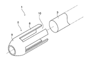
図1に示すように、管状組織体形成基材1は、生体組織材料の存在する環境に設置して、基材表面に管状の結合組織体である管状組織体2を形成するためのものであり、棒状の基材本体3と、管状組織体2の端部4を補強するための端部補強基材5とを備え、その端部補強基材5を、組織形成空間6を挟んで基材本体3の端部の外周面と径方向に対向させたものである。
As shown in FIG. 1, a tubular tissue body-forming
基材本体3は、例えばシリコーン樹脂からなる断面円形の棒状とされ、生体組織材料の存在する環境に設置して、外周面に結合組織7を形成することにより、管状組織体2を形成するようになっている。
The
端部補強基材5は、例えばアクリル樹脂からなるキャップ状で、全体として筒状をなす筒部8の外端側に、基材本体3の端部に外嵌可能な先細形状かつリング状の外嵌部9を形成した構造とされ、基材本体3の端部に着脱自在とされる。この端部補強基材5を基材本体3の端部に装着することにより、筒部8が組織形成空間6を挟んで基材本体3の端部の外周面と径方向に対向する。
The end portion reinforcing
筒部8には、基材本体3の中心軸と平行、かつ基材本体中央側の端縁に至る形状のスリット10が形成され、このスリット10が、組織形成空間6と基材外部とを径方向に連通させて、組織形成空間6に結合組織7を侵入させる侵入口として機能する。
The
なお、管状組織体形成基材1の材料は、生体に埋入した際に大きく変形することが無い強度(硬度)を有しており、化学的安定性があり、滅菌などの負荷に耐性があり、生体を刺激する溶出物が無いまたは少ない樹脂が好ましく、例えば、上記の通り、シリコーン樹脂やアクリル樹脂等が挙げられるがこれに限定されるものではない。
In addition, the material of the tubular tissue
図2に示すように、上記の管状組織体形成基材1を用いて生産した管状組織体2は、例えば、人工血管として使用されるものであり、移植部位に縫合等される端部4は、中央部よりも増厚されると共に、複数の突条11を中心軸と平行に形成され、中央部よりも増強されている。
As shown in FIG. 2, the
次に、上記のような管状組織体形成基材1を用いて管状組織体2を生産する方法について説明する。
Next, a method for producing the
図3に示すように、この生産方法は、管状組織体形成基材1を生体組織材料の存在する環境に設置する「設置工程」と、管状組織体形成基材1の周囲に結合組織7を形成しつつ組織形成空間6に結合組織7を侵入させる「形成工程」と、前記環境から管状組織体形成基材1を取り出す「取出工程」と、結合組織7を管状組織体形成基材1から剥離して管状組織体2として取り出す「分離工程」とからなる。
As shown in FIG. 3, this production method includes an “installation step” in which the tubular tissue body-forming
<設置工程>
基材本体3の端部に外嵌部9を外嵌して、端部補強基材5を基材本体3の端部に装着し、端部補強基材5の筒部8を基材本体3の端部の外周面と径方向に対向させて、基材本体3の端部外周面と筒部8との間に組織形成空間6を構成する(図3(a))。
<Installation process>
The outer
次いで、管状組織体形成基材1を生体組織材料の存在する環境に設置する。生体組織材料の存在する環境とは、動物の生体内(例えば、皮下や腹腔内への埋入)、又は、動物の生体外において生体組織材料が浮遊する溶液中等の人工環境内が挙げられる。生体組織材料としては、ヒト、イヌ、ウシ、ブタ、ヤギ、ウサギ、ヒツジなどの他の哺乳類動物由来のものや、鳥類、魚類、その他の動物由来のもの、又は人工材料を用いることもできる。
Next, the tubular tissue body-forming
管状組織体形成基材1を動物に埋入する場合には、十分な麻酔下で最小限の切開術で行い、埋入後は傷口を縫合する。管状組織体形成基材1の埋入部位としては例えば、管状組織体形成基材1を受け入れる容積を有する腹腔内、あるいは四肢部、肩部又は背部、腹部などの皮下が好ましい。また、埋入には低侵襲な方法で行うことと動物愛護の精神を尊重し、十分な麻酔下で最小限の切開術で行うことが好ましい。
When the tubular tissue body-forming
また、管状組織体形成基材1を生体組織材料の存在する環境下へ置く場合には、種々の培養条件を整えてクリーンな環境下で公知の方法に従って細胞培養を行えばよい。
In addition, when the tubular tissue body-forming
<形成工程>
設置工程の後、所定時間が経過することにより、管状組織体形成基材1の周囲に結合組織7が形成され、さらに、結合組織7がスリット10及び端部開口12から組織形成空間6に侵入して、基材表面に結合組織7が形成される(図3(b))。この形成工程においては、十分な面積のスリット10から結合組織7を侵入させる分、比較的に短時間で組織形成空間6に結合組織7が形成される。結合組織7は、繊維芽細胞とコラーゲンなどの細胞外マトリックスで構成される。
<Formation process>
After a predetermined time has elapsed after the installation step,
<取出工程>
所定時間の形成工程を経て、基材表面に結合組織7が十分に形成された後、管状組織体形成基材1を生体組織材料の存在する環境から取り出す取出工程を行う。
<Removal process>
After the
<分離工程>
取出工程を経た管状組織体形成基材1の端部補強基材5から、その外面に形成された結合組織7を除去し(図3(c))、基材本体3の端部から端部補強基材5を抜き出す。このとき、スリット10が基材本体3の中心軸と平行、かつ基材本体中央側の端縁に至る形状であるので、スリット10の内部に形成された突条11としての結合組織7を破壊することなく、端部補強基材5を抜き出すことができる。
<Separation process>
The
その後、基材本体3の外周面から結合組織7を剥離して取り出すことにより、端部4が増厚されると共に、端部4に複数の突条11が形成された管状組織体2が得られる(図3(d))。
Thereafter, the
なお、生産された管状結合組織体2を異種移植する場合には、移植後の拒絶反応を防ぐため、脱細胞処理、脱水処理、固定処理などの免疫源除去処理を施すのが好ましい。脱細胞処理としては、超音波処理や界面活性剤処理、コラゲナーゼなどの酵素処理によって細胞外マトリックスを溶出させて洗浄する等の方法があり、脱水処理の方法としては、メタノール、エタノール、イソプロピルアルコール等の水溶性有機溶媒で洗浄する方法があり、固定処理する方法としては、グルタアルデヒドやホルムアルデヒドなどのアルデヒド化合物で処理する方法がある。
In addition, when the produced tubular
上記構成によれば、基材本体3の端部にキャップ状の端部補強基材5を装着し、基材本体3の端部外周面と筒部8との間に組織形成空間6を構成している。これにより、管状組織体2の端部4を構成する結合組織7をその内外から形成して、厚くかつ均一な密度に設定することができ、移植部位に縫合等される管状組織体2の端部4を増強することができる。
According to the above configuration, the cap-shaped
ここで、一例として、管状組織体形成基材1をビーグル犬の皮下に埋入して形成した管状組織体2について、その強度等の特性を説明する。
Here, as an example, characteristics such as strength of the
使用した管状組織体形成基材1の断面寸法は、基材本体3の外径が5mm、端部補強基材5の筒部8の内径が7mmで、組織形成空間6の厚さが1mmである。この管状組織体形成基材1をビーグル犬の皮下に埋入し、5週間が経過した後、管状組織体形成基材1を取り出して管状組織体2を得た。さらに、管状組織体2を長さ方向に切断してシート状とし、これを10mmの長さに切断して、各特性を測定した。
The cross-sectional dimensions of the used tubular tissue-forming
管状組織体2の断面寸法を測定したところ、内径は、全長に渡って、基材本体3の外径と同じ5mmであり、厚さは、中央部で100〜200μm、端部4で400〜600μmであった。
When the cross-sectional dimension of the
さらに、弾性率及び破断荷重を測定したところ、弾性率は、中央部で2092±597kPa、端部4で4695±721kPaであり、破断荷重は、中央部で335.4±98.7g、端部4で762.2±271.6gであった。なお、サンプル数は、中央部及び端部4の各部位について、3ずつである。
Further, when the elastic modulus and the breaking load were measured, the elastic modulus was 2092 ± 597 kPa at the center and 4695 ± 721 kPa at the
このように、管状組織体形成基材1を用いて、基材本体3の端部にキャップ状の端部補強基材5を装着することにより、管状組織体2の端部4の厚さを増厚すると共に、端部4の弾性率及び破断荷重を中央部におけるよりも2倍以上にすることができ、管状組織体2の端部4を増強することができた。
In this way, by using the tubular tissue-forming
なお、本発明は、上記の実施の形態に限定されるものではなく、本発明の範囲内において、適宜変更を加えることができる。例えば、前記環境から管状組織体形成基材1を取り出す「取出工程」を設けて管状組織体2を形成する代わりに、図4に示すように、人工血管としての管状組織体2を患者の体内に直接に形成するようにしてもよい。
In addition, this invention is not limited to said embodiment, A change can be suitably added within the scope of the present invention. For example, instead of forming a
具体的には、まず、患者の体内のうち、血管13の損傷部位14の近くに管状組織体形成基材1を埋入する(図4(a))。この状態を所定期間が経過するまで維持して、基材表面に結合組織を形成した後、その結合組織を剥離しつつ患者の体内から管状組織体形成基材1を取り出すことにより、血管13の損傷部位14の近くに、端部4を増強した人工血管としての管状組織体2を形成する(図4(b))。患者の体内に形成した管状組織体2のうち、中央部を移動させることなく、端部4のみを移動させて、損傷部位14を跨ぐように血管13に縫合すると共に、損傷部位14を除去することにより、血管13に人工血管が移植される(図4(c))。
Specifically, first, the tubular tissue
1 管状組織体形成基材
2 管状組織体
3 基材本体
4 端部
5 端部補強基材
6 組織形成空間
7 結合組織
8 筒部
9 外嵌部
10 スリット
11 突条
12 端部開口
13 血管
14 損傷部位
DESCRIPTION OF
Claims (8)
Priority Applications (1)
| Application Number | Priority Date | Filing Date | Title |
|---|---|---|---|
| JP2015011992A JP2016136984A (en) | 2015-01-26 | 2015-01-26 | Tubular tissue body forming substrate |
Applications Claiming Priority (1)
| Application Number | Priority Date | Filing Date | Title |
|---|---|---|---|
| JP2015011992A JP2016136984A (en) | 2015-01-26 | 2015-01-26 | Tubular tissue body forming substrate |
Publications (1)
| Publication Number | Publication Date |
|---|---|
| JP2016136984A true JP2016136984A (en) | 2016-08-04 |
Family
ID=56559626
Family Applications (1)
| Application Number | Title | Priority Date | Filing Date |
|---|---|---|---|
| JP2015011992A Pending JP2016136984A (en) | 2015-01-26 | 2015-01-26 | Tubular tissue body forming substrate |
Country Status (1)
| Country | Link |
|---|---|
| JP (1) | JP2016136984A (en) |
Cited By (2)
| Publication number | Priority date | Publication date | Assignee | Title |
|---|---|---|---|---|
| CN107446818A (en) * | 2016-09-14 | 2017-12-08 | 四川蓝光英诺生物科技股份有限公司 | Printing equipment, Method of printing and the lumen organization's construct of lumen organization's construct |
| JP2018033694A (en) * | 2016-08-31 | 2018-03-08 | 新幹工業株式会社 | Method of forming connective tissue body |
-
2015
- 2015-01-26 JP JP2015011992A patent/JP2016136984A/en active Pending
Cited By (5)
| Publication number | Priority date | Publication date | Assignee | Title |
|---|---|---|---|---|
| JP2018033694A (en) * | 2016-08-31 | 2018-03-08 | 新幹工業株式会社 | Method of forming connective tissue body |
| WO2018043614A1 (en) * | 2016-08-31 | 2018-03-08 | 新幹工業 株式会社 | Method for forming connective tissue body |
| CN109641079A (en) * | 2016-08-31 | 2019-04-16 | 生物管株式会社 | The forming method of connective tissue body |
| CN107446818A (en) * | 2016-09-14 | 2017-12-08 | 四川蓝光英诺生物科技股份有限公司 | Printing equipment, Method of printing and the lumen organization's construct of lumen organization's construct |
| CN107446818B (en) * | 2016-09-14 | 2021-05-07 | 四川蓝光英诺生物科技股份有限公司 | Lumen tissue construct printing device, lumen tissue construct printing method and lumen tissue construct |
Similar Documents
| Publication | Publication Date | Title |
|---|---|---|
| WO2016098877A1 (en) | Substrate for forming artificial valve and artificial valve | |
| JP6010018B2 (en) | Stent with valve, base material for forming stent with valve, and production method of stent with valve | |
| WO2016076416A1 (en) | Connective tissue formation substrate and substrate removal tool | |
| US20190083225A1 (en) | Tissue body formation device | |
| JP2015039515A (en) | Prosthetic valve, base material for forming prosthetic valve, and production procedure of prosthetic valve | |
| JP2012135406A (en) | Tissue-forming base material derived from living body, method for producing tissue derived from living body using the same and tissue derived from living body | |
| JP2016136984A (en) | Tubular tissue body forming substrate | |
| US8685082B2 (en) | Base material for forming valved lumen shape tissue, method for producing valved lumen shape tissue, and valved artificial blood vessel | |
| JP6262470B2 (en) | Substrate for forming connective tissue and method for producing connective tissue | |
| JP2017169778A (en) | Connective tisse body forming substrate | |
| JP2014030598A (en) | Base material for formation of film connective tissue and production method of film connective tissue | |
| JP5127120B2 (en) | Connective tissue-forming substrate and method for producing connective tissue using the same | |
| JP5658008B2 (en) | Artificial blood vessel forming substrate with valve, production method of artificial blood vessel with valve using the same, and artificial blood vessel with valve | |
| JP2017113051A (en) | Membrane-like connective tissue body formation base material | |
| WO2014010323A1 (en) | Substrate for formation of membrane-like connective tissue and production method for membrane-like connective tissue using same | |
| JP5706282B2 (en) | Lumen-shaped tissue forming substrate with valve, lumen-shaped tissue forming substrate, membranous tissue-forming substrate, valved lumen-shaped tissue production method, lumen-shaped tissue production method, and membranous tissue Production method | |
| JP2015039577A (en) | Prosthetic valve, base material for forming prosthetic valve, and production procedure of prosthetic valve | |
| WO2014021073A1 (en) | Artificial cornea, and method for producing artificial cornea | |
| JP2021122277A (en) | Structural assembly for connective tissue formation and method for forming connective tissue | |
| JP2023135411A (en) | Valved graft forming substrate | |
| JP2021122747A (en) | Structure for connective tissue body formation and method for forming connective tissue body |