JP2016106940A - Brain disease diagnosis support system, brain disease diagnosis support method, and program - Google Patents
Brain disease diagnosis support system, brain disease diagnosis support method, and program Download PDFInfo
- Publication number
- JP2016106940A JP2016106940A JP2014248953A JP2014248953A JP2016106940A JP 2016106940 A JP2016106940 A JP 2016106940A JP 2014248953 A JP2014248953 A JP 2014248953A JP 2014248953 A JP2014248953 A JP 2014248953A JP 2016106940 A JP2016106940 A JP 2016106940A
- Authority
- JP
- Japan
- Prior art keywords
- brain
- data
- electroencephalogram
- disease
- subject
- Prior art date
- Legal status (The legal status is an assumption and is not a legal conclusion. Google has not performed a legal analysis and makes no representation as to the accuracy of the status listed.)
- Granted
Links
Images
Classifications
-
- G—PHYSICS
- G16—INFORMATION AND COMMUNICATION TECHNOLOGY [ICT] SPECIALLY ADAPTED FOR SPECIFIC APPLICATION FIELDS
- G16H—HEALTHCARE INFORMATICS, i.e. INFORMATION AND COMMUNICATION TECHNOLOGY [ICT] SPECIALLY ADAPTED FOR THE HANDLING OR PROCESSING OF MEDICAL OR HEALTHCARE DATA
- G16H50/00—ICT specially adapted for medical diagnosis, medical simulation or medical data mining; ICT specially adapted for detecting, monitoring or modelling epidemics or pandemics
- G16H50/20—ICT specially adapted for medical diagnosis, medical simulation or medical data mining; ICT specially adapted for detecting, monitoring or modelling epidemics or pandemics for computer-aided diagnosis, e.g. based on medical expert systems
-
- A—HUMAN NECESSITIES
- A61—MEDICAL OR VETERINARY SCIENCE; HYGIENE
- A61B—DIAGNOSIS; SURGERY; IDENTIFICATION
- A61B10/00—Instruments for taking body samples for diagnostic purposes; Other methods or instruments for diagnosis, e.g. for vaccination diagnosis, sex determination or ovulation-period determination; Throat striking implements
-
- A—HUMAN NECESSITIES
- A61—MEDICAL OR VETERINARY SCIENCE; HYGIENE
- A61B—DIAGNOSIS; SURGERY; IDENTIFICATION
- A61B5/00—Measuring for diagnostic purposes; Identification of persons
- A61B5/24—Detecting, measuring or recording bioelectric or biomagnetic signals of the body or parts thereof
- A61B5/316—Modalities, i.e. specific diagnostic methods
- A61B5/369—Electroencephalography [EEG]
- A61B5/384—Recording apparatus or displays specially adapted therefor
-
- A—HUMAN NECESSITIES
- A61—MEDICAL OR VETERINARY SCIENCE; HYGIENE
- A61B—DIAGNOSIS; SURGERY; IDENTIFICATION
- A61B5/00—Measuring for diagnostic purposes; Identification of persons
- A61B5/40—Detecting, measuring or recording for evaluating the nervous system
- A61B5/4076—Diagnosing or monitoring particular conditions of the nervous system
- A61B5/4088—Diagnosing of monitoring cognitive diseases, e.g. Alzheimer, prion diseases or dementia
-
- G—PHYSICS
- G06—COMPUTING OR CALCULATING; COUNTING
- G06N—COMPUTING ARRANGEMENTS BASED ON SPECIFIC COMPUTATIONAL MODELS
- G06N20/00—Machine learning
-
- G—PHYSICS
- G06—COMPUTING OR CALCULATING; COUNTING
- G06N—COMPUTING ARRANGEMENTS BASED ON SPECIFIC COMPUTATIONAL MODELS
- G06N20/00—Machine learning
- G06N20/10—Machine learning using kernel methods, e.g. support vector machines [SVM]
-
- G—PHYSICS
- G16—INFORMATION AND COMMUNICATION TECHNOLOGY [ICT] SPECIALLY ADAPTED FOR SPECIFIC APPLICATION FIELDS
- G16H—HEALTHCARE INFORMATICS, i.e. INFORMATION AND COMMUNICATION TECHNOLOGY [ICT] SPECIALLY ADAPTED FOR THE HANDLING OR PROCESSING OF MEDICAL OR HEALTHCARE DATA
- G16H50/00—ICT specially adapted for medical diagnosis, medical simulation or medical data mining; ICT specially adapted for detecting, monitoring or modelling epidemics or pandemics
- G16H50/50—ICT specially adapted for medical diagnosis, medical simulation or medical data mining; ICT specially adapted for detecting, monitoring or modelling epidemics or pandemics for simulation or modelling of medical disorders
-
- G—PHYSICS
- G16—INFORMATION AND COMMUNICATION TECHNOLOGY [ICT] SPECIALLY ADAPTED FOR SPECIFIC APPLICATION FIELDS
- G16H—HEALTHCARE INFORMATICS, i.e. INFORMATION AND COMMUNICATION TECHNOLOGY [ICT] SPECIALLY ADAPTED FOR THE HANDLING OR PROCESSING OF MEDICAL OR HEALTHCARE DATA
- G16H50/00—ICT specially adapted for medical diagnosis, medical simulation or medical data mining; ICT specially adapted for detecting, monitoring or modelling epidemics or pandemics
- G16H50/70—ICT specially adapted for medical diagnosis, medical simulation or medical data mining; ICT specially adapted for detecting, monitoring or modelling epidemics or pandemics for mining of medical data, e.g. analysing previous cases of other patients
-
- G—PHYSICS
- G06—COMPUTING OR CALCULATING; COUNTING
- G06F—ELECTRIC DIGITAL DATA PROCESSING
- G06F17/00—Digital computing or data processing equipment or methods, specially adapted for specific functions
Landscapes
- Health & Medical Sciences (AREA)
- Engineering & Computer Science (AREA)
- Medical Informatics (AREA)
- Public Health (AREA)
- Life Sciences & Earth Sciences (AREA)
- Biomedical Technology (AREA)
- General Health & Medical Sciences (AREA)
- Pathology (AREA)
- Data Mining & Analysis (AREA)
- Physics & Mathematics (AREA)
- Software Systems (AREA)
- Theoretical Computer Science (AREA)
- Heart & Thoracic Surgery (AREA)
- Animal Behavior & Ethology (AREA)
- Surgery (AREA)
- Veterinary Medicine (AREA)
- Molecular Biology (AREA)
- Databases & Information Systems (AREA)
- Primary Health Care (AREA)
- Epidemiology (AREA)
- Neurology (AREA)
- Psychiatry (AREA)
- Psychology (AREA)
- Biophysics (AREA)
- Artificial Intelligence (AREA)
- General Physics & Mathematics (AREA)
- General Engineering & Computer Science (AREA)
- Computing Systems (AREA)
- Evolutionary Computation (AREA)
- Computer Vision & Pattern Recognition (AREA)
- Mathematical Physics (AREA)
- Child & Adolescent Psychology (AREA)
- Hospice & Palliative Care (AREA)
- Developmental Disabilities (AREA)
- Neurosurgery (AREA)
- Physiology (AREA)
- Measurement And Recording Of Electrical Phenomena And Electrical Characteristics Of The Living Body (AREA)
- Measuring And Recording Apparatus For Diagnosis (AREA)
Abstract
【課題】複数の脳疾患の判定が可能な脳疾患診断支援システムを提供する。【解決手段】脳疾患診断支援システムは、脳波からその脳波の特徴量を抽出した脳波特徴データに、当該脳波特徴データに対応する脳疾患を示す疾患情報を付した学習データを複数取得し、取得した複数の学習データを、複数のクラスタに分類する。また、分類した各クラスタにおいて学習データに付された疾患情報に基づいて、学習データを疾患情報ごとに分類する分類器を生成する。そして、被験者の脳波特徴データを取得し、その脳波特徴データが分類されるクラスタを特定し、さらに生成した分類器によって、被験者の脳波特徴データが複数の脳疾患の何れであるかを判定する。【選択図】図1A brain disease diagnosis support system capable of determining a plurality of brain diseases is provided. A brain disease diagnosis support system acquires and acquires a plurality of learning data including disease information indicating a brain disease corresponding to the brain wave feature data in the brain wave feature data obtained by extracting a feature amount of the brain wave from the brain wave. The plurality of learned data are classified into a plurality of clusters. Also, a classifier that classifies the learning data for each disease information is generated based on the disease information attached to the learning data in each classified cluster. Then, the brain wave feature data of the subject is acquired, the cluster into which the brain wave feature data is classified is specified, and the generated brain classifier determines which of the brain diseases the subject's brain wave feature data is. [Selection] Figure 1
Description
本発明は、脳疾患診断支援システム、脳疾患診断支援方法及びプログラムに関する。 The present invention relates to a brain disease diagnosis support system, a brain disease diagnosis support method, and a program.
高齢化社会の到来に伴い、脳疾患の一つである認知症への対策が求められている。以降については認知症を例として記載する。認知症は、投薬によって病気の進行を遅らせたり、手術によって改善できる場合がある。これら認知症への効果的な対策を行うためには、認知症の早期発見が重要となる。 With the arrival of an aging society, countermeasures against dementia, one of the brain diseases, are required. In the following, dementia is described as an example. Dementia may be slowed by medication or improved by surgery. In order to take effective measures against these dementias, early detection of dementia is important.
例えば、特許文献1には、脳波の周波数全域において、大脳皮質内のニューロン機能が低下すると、ニューロン活動が不安定になり、この影響が局所的な脳波パワーのゆらぎとして現れることを用いて、脳の活動を測定する装置が開示されている。この技術では、被験者の頭部の複数の位置に取り付けたセンサが検出した脳波を所定の周波数帯域ごとに規格化した値(以下、脳波特徴データという)の平均値や標準偏差を求め、正常者集団について同様にして求めた脳波特徴データの平均値と比較し、被験者の平均値のZスコアを求める。そして、被験者のZスコアに基づいて、被験者の脳機能の低下部分を示すことができる。
For example,
また、特許文献1には、同様の方法を用いて、アルツハイマー型認知症患者の集団についてのZスコアを算出し、これを当該疾患の特徴を示すテンプレートとして、個々の被験者のZスコアと当該テンプレートとの相関係数を求めることで、その類似性を数値的に表示できることが記載されている。
Further,
しかし、特許文献1の方法には、次のような問題点がある。例えば、特許文献1の方法では、Zスコアによって被験者の脳波特徴データと、正常者集団の脳波特徴データ又はアルツハイマー型認知症患者の集団の脳波特徴データとの類似性を判定している。Zスコアによって判定する場合、母集団が正規分布に従うことが前提となる。しかし、正常者集団の脳波特徴データやアルツハイマー型認知症患者の集団の脳波特徴データが、正規分布に従う保証はない。従って、Zスコアを用いるためには、母集団のデータが正規分布に従うように、試行錯誤を行いながら適切なデータを抽出しなければならない可能性がある。さらに、脳波は、国際標準で19ヶ所の測定を行うことになっており、また、例えば特許文献1の方法では、さらに2ヶ所を追加した計21ヶ所で測定を行っているが、何れの場合も脳波特徴データは、多次元となり、複数の多次元データの中から正規分布に従うような適切なデータを抽出することには困難さが伴う。
However, the method of
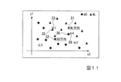
また、被験者の脳波特徴データと、正常者集団の脳波特徴データ又はアルツハイマー型認知症患者の集団の脳波特徴データとの類似性を、脳波特徴データの平均値同士の距離で判定しているが、正常者集団の脳波特徴データの平均値は、必ずしも正常者の脳波特徴データの特性を示す値に収束するとは限らない。同様に、アルツハイマー型認知症患者集団の脳波特徴データの平均値は、必ずしもアルツハイマー型認知症患者の脳波特徴データの特性を示す値に収束するとは限らない。このことを、図11を用いて説明する。説明の便宜上、2つの脳波特徴データを用いる場合を例に説明を行う。縦軸(y1軸)、横軸(y2軸)は、それぞれ脳波特徴データを示している。すると、ある者の脳波特徴データは、2つの脳波特徴データの値を座標値に持つ点で表すことができる。三角で示された点を、正常者(NL)の脳波特徴データとする。丸で示された点をアルツハイマー型認知症患者(AD)と診断された患者の脳波特徴データとする。また、星型で示された点を被験者の脳波特徴データとする。このとき、NLの平均値は、符号31が示す点で表される。ADの平均値は、符号32が示す点で表される。
In addition, the similarity between the brain wave feature data of the subject and the brain wave feature data of the normal population or the brain wave feature data of the population of patients with Alzheimer type dementia is determined by the distance between the average values of the brain wave feature data. The average value of the electroencephalogram feature data of the normal population does not necessarily converge to a value indicating the characteristics of the electroencephalogram feature data of the normal subjects. Similarly, the average value of the electroencephalogram feature data of the Alzheimer type dementia patient population does not necessarily converge to a value indicating the characteristics of the electroencephalogram feature data of the Alzheimer type dementia patient. This will be described with reference to FIG. For convenience of explanation, the case where two electroencephalogram feature data are used will be described as an example. The vertical axis (y1 axis) and the horizontal axis (y2 axis) indicate electroencephalogram feature data, respectively. Then, the brain wave feature data of a certain person can be represented by a point having two brain wave feature data values as coordinate values. The points indicated by triangles are brain wave characteristic data of normal persons (NL). The points indicated by circles are the electroencephalogram feature data of a patient diagnosed as an Alzheimer type dementia patient (AD). A point indicated by a star shape is used as brain wave characteristic data of the subject. At this time, the average value of NL is represented by a point indicated by
ここで、被験者の脳波特徴データを、正常者とアルツハイマー患者それぞれの脳波特徴データの平均値に基づいて、正常者、アルツハイマー患者のどちらかに分類することを考える。被験者の脳波特徴データをNL、ADのどちらに分類する方法の一例として、NLの平均値、ADの平均値をそれぞれNL、ADの代表値とし、被験者の脳波特徴データとそれらの代表値との距離を比較し、距離が近い方に被験者の脳波特徴データを分類するものとする。例えば、被験者1の脳波特徴データ(符号33)は、ADの平均値(符号32)に近い。また、被験者4の脳波特徴データ(符号34)は、NLの平均値(符号31)に近い。しかし、被験者1の脳波特徴データ(符号33)の近くには、一つのNLの脳波特徴データ(符号35)が存在する。また、被験者4の脳波特徴データ(符号34)の近くには、一つのADの脳波特徴データ(符号36)が存在する。特許文献1の方法によると、このような場合でも、被験者1(符号33)を、ADと判定し、被験者4(符号34)は、NLと判定する可能性があるが、脳波特徴データの平均値が、ある症状やある活動状態の脳波特徴データの特性を正しく示す代表値であるとは限らない。また、上述のように脳波特徴データは、多次元であり、そのような多次元データの代表値を平均算出で適切に収束させることは困難であるという問題もある。
Here, it is considered that the electroencephalogram feature data of the subject is classified into either a normal subject or an Alzheimer patient based on the average value of the electroencephalogram feature data of each normal person and Alzheimer patient. As an example of a method for classifying the subject's electroencephalogram feature data into either NL or AD, the average value of NL and the average value of AD are respectively representative values of NL and AD, and the subject's electroencephalogram feature data and their representative values The distances are compared, and the subject's brain wave feature data is classified into those closer to the distance. For example, the electroencephalogram feature data (reference numeral 33) of the
そこでこの発明は、上述の課題を解決することのできる脳疾患診断支援システム、脳疾患診断支援方法及びプログラムを提供することを目的としている。 Accordingly, an object of the present invention is to provide a brain disease diagnosis support system, a brain disease diagnosis support method, and a program that can solve the above-described problems.
本発明の第1の態様は、脳波からその脳波の特徴量を抽出した脳波特徴データに、当該脳波特徴データに対応する脳疾患の状態を示す疾患情報を付した学習データを複数取得し、前記複数の学習データを機械学習することにより、脳疾患の判定を行う評価モデルを算出する評価モデル算出部と、被験者の脳波特徴データを取得し、前記評価モデルに基づいて、前記被験者の脳波特徴データが示す脳疾患の判定を行う判定部と、を備える脳疾患診断支援システムである。 According to a first aspect of the present invention, a plurality of learning data with disease information indicating a brain disease state corresponding to the electroencephalogram feature data is acquired in the electroencephalogram feature data obtained by extracting the electroencephalogram feature amount from the electroencephalogram, An evaluation model calculation unit that calculates an evaluation model for determining a brain disease by machine learning a plurality of learning data, and acquiring the subject's brain wave feature data, and based on the evaluation model, the subject's brain wave feature data A brain disease diagnosis support system comprising: a determination unit that determines a brain disease indicated by:
本発明の第2の態様における脳疾患診断支援システムは、前記被験者の脳波特徴データに付す前記疾患情報の入力を受け付ける入力部、を更に備え、前記入力部は、前記受け付けた疾患情報を前記被験者の脳波特徴データと対応付けた新たな学習データを記憶部に記憶させ、前記評価モデル算出部は、前記新たな学習データを前記複数の学習データに加え、評価モデルの算出を行う。 The brain disease diagnosis support system according to the second aspect of the present invention further includes an input unit that receives an input of the disease information attached to the brain wave characteristic data of the subject, and the input unit receives the received disease information as the subject. New learning data associated with the brain wave feature data is stored in the storage unit, and the evaluation model calculation unit calculates the evaluation model by adding the new learning data to the plurality of learning data.
本発明の第3の態様における前記脳波特徴データには、複数の前記特徴量が含まれ、前記評価モデル算出部は、前記複数の学習データのそれぞれに含まれる脳波特徴データについて、評価モデルの算出に有効な1つ又は複数の特徴量を抽出し、当該抽出した特徴量のみを用いて前記評価モデルを算出する。 The electroencephalogram feature data according to the third aspect of the present invention includes a plurality of feature quantities, and the evaluation model calculation unit calculates an evaluation model for the electroencephalogram feature data included in each of the plurality of learning data. One or a plurality of feature quantities effective for the above are extracted, and the evaluation model is calculated using only the extracted feature quantities.
本発明の第4の態様における前記複数の学習データの疾患情報には、1つ又は複数の種類の脳疾患を示す情報が含まれており、前記評価モデル算出部は、前記複数の学習データを、各学習データに付された疾患情報の種類ごとに分離する境界情報を算出し、前記判定部は、前記境界情報に基づいて、前記被験者の脳波特徴データが示す脳疾患の判定を行う。 The disease information of the plurality of learning data according to the fourth aspect of the present invention includes information indicating one or more types of brain diseases, and the evaluation model calculation unit stores the plurality of learning data Then, boundary information to be separated for each type of disease information attached to each learning data is calculated, and the determination unit determines a brain disease indicated by the brain wave feature data of the subject based on the boundary information.
本発明の第5の態様における前記評価モデル算出部は、前記複数の脳波特徴データを、前記特徴量に基づくクラスタリングによって複数のグループに分類し、前記判定部は、前記被験者の脳波特徴データが、前記評価モデル算出部の分類したグループのうち、どのグループに分類されるかを判定する。 In the fifth aspect of the present invention, the evaluation model calculation unit classifies the plurality of electroencephalogram feature data into a plurality of groups by clustering based on the feature amount, and the determination unit includes the subject's electroencephalogram feature data. It is determined which group is classified among the groups classified by the evaluation model calculation unit.
本発明の第6の態様における前記複数の学習データの疾患情報には、1つ又は複数の種類の脳疾患を示す情報が含まれており、前記評価モデル算出部は、前記判定部によって前記被験者の脳波特徴データが分類されると判定されたグループに含まれる学習データについて、疾患情報の種類ごとに、それぞれの疾患情報が付された学習データの割合を算出し、前記判定部は、前記割合に基づいて前記被験者の脳波特徴データが示す脳疾患の判定を行う。 The disease information of the plurality of learning data according to the sixth aspect of the present invention includes information indicating one or more types of brain diseases, and the evaluation model calculation unit is configured so that the evaluation unit calculates the subject by the determination unit. The learning data included in the group determined to be classified as the brain wave feature data is calculated for each type of disease information by calculating a ratio of learning data to which each disease information is attached, and the determination unit Based on the above, the brain disease indicated by the subject's electroencephalogram feature data is determined.
本発明の第7の態様における前記評価モデル算出部は、前記判定部によって前記被験者の脳波特徴データが分類されると判定されたグループに含まれる学習データを、各学習データに付された疾患情報の種類ごとに分離する境界情報を算出し、前記判定部は、前記境界情報に基づいて、前記被験者の脳波特徴データが示す脳疾患の判定を行う。 In the seventh aspect of the present invention, the evaluation model calculation unit includes the learning data included in the group determined by the determination unit to classify the brain wave feature data of the subject, and the disease information attached to each learning data The boundary information to be separated for each type is calculated, and the determination unit determines the brain disease indicated by the brain wave feature data of the subject based on the boundary information.
本発明の第8の態様における脳疾患症診断支援システムは、前記判定部の判定の結果を出力する表示部、をさらに備え、前記判定部は、前記被験者の脳波特徴データが示す脳疾患を判定するとともに、前記被験者の脳波特徴データがその脳疾患であることを示す確率を算出し、前記表示部は、前記確率を出力する。 The cerebral disease diagnosis support system according to the eighth aspect of the present invention further includes a display unit that outputs a result of determination by the determination unit, and the determination unit determines a brain disease indicated by the electroencephalogram feature data of the subject. In addition, the probability that the brain wave characteristic data of the subject is the brain disease is calculated, and the display unit outputs the probability.
本発明の第9の態様は、脳疾患診断支援システムが、脳波からその脳波の特徴量を抽出した脳波特徴データに、当該脳波特徴データに対応する脳疾患の状態を示す疾患情報を付した学習データを複数取得し、前記複数の学習データを機械学習することにより、脳疾患の判定を行う評価モデルを算出し、被験者の脳波特徴データを取得し、前記評価モデルに基づいて、前記被験者の脳波特徴データが示す脳疾患の判定を行う、脳疾患診断支援方法である。 According to a ninth aspect of the present invention, a brain disease diagnosis support system is a learning method in which brain wave feature data obtained by extracting a feature amount of an electroencephalogram from the electroencephalogram is attached with disease information indicating the state of the brain disease corresponding to the electroencephalogram feature data. By obtaining a plurality of data and machine learning the plurality of learning data, an evaluation model for determining a brain disease is calculated, brain wave feature data of the subject is obtained, and based on the evaluation model, the subject's brain wave This is a brain disease diagnosis support method for determining a brain disease indicated by feature data.
本発明の第10の態様は、脳疾患診断支援システムのコンピュータを、脳波からその脳波の特徴量を抽出した脳波特徴データに、当該脳波特徴データに対応する脳疾患の状態を示す疾患情報を付した学習データを複数取得し、前記複数の学習データを機械学習することにより、脳疾患の判定を行う評価モデルを算出する手段、被験者の脳波特徴データを取得し、前記評価モデルに基づいて、前記被験者の脳波特徴データが示す脳疾患の判定を行う手段、として機能させるためのプログラムである。 In a tenth aspect of the present invention, the computer of the brain disease diagnosis support system attaches disease information indicating the state of the brain disease corresponding to the brain wave feature data to the brain wave feature data obtained by extracting the feature amount of the brain wave from the brain wave. A plurality of learning data obtained, and machine learning the plurality of learning data, thereby calculating an evaluation model for determining brain disease, acquiring brain wave feature data of a subject, and based on the evaluation model, This is a program for functioning as a means for determining a brain disease indicated by brain wave characteristic data of a subject.
本発明によれば、被験者の脳波に基づいて、その被験者が脳疾患を患っているかどうかの判定を、複数の脳疾患について同時に行うことができる。また、脳疾患の判定に用いる評価モデルの作成を自動的に行うことができるので、テンプレート作成の手間を省くことができる。 According to the present invention, it is possible to simultaneously determine whether or not a subject has a brain disease based on the brain waves of the subject for a plurality of brain diseases. In addition, since an evaluation model used for determination of a brain disease can be automatically created, it is possible to save time and effort for creating a template.
<第一実施形態>
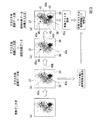
以下、本発明の第一実施形態による脳疾患診断支援装置を図1〜図7を参照して説明する。
図1は、本発明に係る第一実施形態における脳疾患診断支援装置のブロック図である。
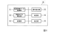
この図において、符号10は、脳疾患診断支援装置を表している。脳疾患診断支援装置10は、被験者の脳波に基づいて被験者が認知症を患っている確率を提示するシステムである。脳疾患診断支援装置10は、この機能を実現するプログラムを実行することができる例えばPCやサーバ装置である。
図1が示すように脳疾患診断支援装置10は、脳波特徴データ取得部11、操作受付部12、評価モデル算出部13、判定部14、記憶部15、表示部16、を備えている。
<First embodiment>
Hereinafter, a brain disease diagnosis support apparatus according to a first embodiment of the present invention will be described with reference to FIGS.
FIG. 1 is a block diagram of a brain disease diagnosis support apparatus according to the first embodiment of the present invention.
In this figure, the code |
As shown in FIG. 1, the brain disease
脳波特徴データ取得部11は、被験者の脳波特徴データを取得する。脳波特徴データとは、被験者の脳波からその脳波の特徴量を抽出したデータである。脳波は、例えば、国際10−20法に基づいて被験者の頭部の所定の位置に配置された電極のうち、耳朶に配置された電極とそれ以外の電極との間の電位差の変動を記録したデータである。また、脳波の特徴量とは、例えば、電極を配置した位置ごとに測定した電位における複数の周波数帯それぞれにおける電位の2乗(sNAT)、隣接する周波数帯間でのsNATの比(脳波の滑らかさ)である。また、脳波特徴データ取得部11は、脳波特徴データにその脳波特徴データがどのような脳疾患を持つ者の脳波特徴データであるかを示す疾患情報を付した学習データを取得する。脳疾患の種類には、例えば、正常(脳疾患を持たない)、アルツハイマー型認知症、血管性認知症、レビー小体型認知症、鬱、統合失調症などがある。
The electroencephalogram feature
操作受付部12は、オペレータの脳疾患診断支援装置10への指示操作を受け付ける。指示操作とは、例えば、脳波特徴データの取り込みを指示する操作や、その脳波特徴データの評価を行うことを指示する操作などである。
The
評価モデル算出部13は、脳波特徴データ取得部11が取得した複数の学習データを機械学習することにより、脳疾患の判定に用いる評価モデルを算出する。例えば、本実施形態では、複数の学習データを、各学習データに付された疾患情報の種類ごとに分離する境界情報を算出する。評価モデル算出部13が用いる機械学習は、例えば、SVM(Support Vector Machine)である。境界情報とは、例えば、正常者の脳波特徴データとアルツハイマー型認知症患者の脳波特徴データを分離する分離境界面を示す関数である。
The evaluation
判定部14は、被験者の脳波特徴データを取得し、評価モデル算出部13が算出した評価モデルに基づいて、被験者の脳波特徴データが示す脳疾患の判定を行う。例えば、本実施形態では、学習データに基づいて評価モデル算出部13が算出した境界情報に基づいて、ある被験者の脳波特徴データを評価する。評価とは、例えば、ある被験者の脳波特徴データが、複数の脳疾患のうち、どの脳疾患であるのかを判定したり、その被験者の脳波特徴データが判定した脳疾患であることを示す確率を算出したりすることである。
記憶部15は、複数の被験者の脳波特徴データや、評価モデル算出部13が算出した境界情報など、種々の情報を記憶する。
表示部16は、判定部14の評価結果を、脳疾患診断支援装置10に接続された表示装置に出力する。
The
The
The
図2〜図3を用いて、本発明に係る第一実施形態における境界情報の算出処理について説明する。
図2は、本発明に係る第一実施形態における脳疾患診断支援装置による境界情報の算出処理の説明に用いる第一の図である。
まず、脳波の特徴量(測定電極位置の脳波周波数別の強さ等)をy個セットにした「脳波特徴データ」(=BF)と、その被験者の疾患状態を示す「疾患情報(Label)」を一組にした「学習データ」をx個準備する。疾患情報は、例えばアルツハイマー型認知症ならAD、正常者であればNLと付する。なお、本実施形態において正常者とは、アルツハイマー型認知症を患っていない者を意味するものとする。
するとx個の被験者の学習データ(LDx)は、例えば、以下のように表される。
LD1=BF{p11,p12,p13・・・・・p1y}、Label"AD"
LD2=BF{p21,p22,p23・・・・・p2y}、Label"NL"
・
・
LDx=BF{px1,px2,px3・・・・・pxy}、Label"AD“
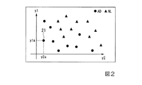
説明の便宜上、ここではy=2の場合を例に説明を行う。図2は、複数のアルツハイマー型認知症患者及び正常者の学習データ(y=2)を2次元空間にマッピングした図である。この図において丸点及び三角点のそれぞれは、ある1人の者の学習データを示している。丸点は、アルツハイマー型認知症患者(AD)の学習データである。三角点は、正常者(NL)の学習データである。縦軸の値は、脳波特徴データを構成する第一の脳波の特徴量、横軸の値は、脳波特徴データを構成する第二の脳波の特徴量を示している。例えば、符号21を付した丸点21は、この者の脳波の第一の特徴量が「y1a」、第二の特徴量が「y2a」、疾患情報が「AD」であることを示している。
評価モデル算出部13は、まず予め準備された複数の被験者の学習データを取り込む。
The boundary information calculation process according to the first embodiment of the present invention will be described with reference to FIGS.
FIG. 2 is a first diagram used for explaining the boundary information calculation processing by the brain disease diagnosis support apparatus according to the first embodiment of the present invention.
First, “electroencephalogram feature data” (= BF) in which the features of the electroencephalogram (strength of the measurement electrode position for each electroencephalogram frequency, etc.) are set, and “disease information (Label)” indicating the disease state of the subject. X pieces of “learning data” are prepared. The disease information is, for example, AD for Alzheimer type dementia and NL for a normal person. In this embodiment, the normal person means a person who does not suffer from Alzheimer's dementia.
Then, learning data (LDx) of x subjects is expressed as follows, for example.
LD1 = BF {p11, p12, p13... P1y}, Label “AD”
LD2 = BF {p21, p22, p23... P2y}, Label “NL”
・
・
LDx = BF {px1, px2, px3... Xy}, Label “AD”
For convenience of explanation, here, a case where y = 2 will be described as an example. FIG. 2 is a diagram in which learning data (y = 2) of a plurality of Alzheimer-type dementia patients and normal persons are mapped in a two-dimensional space. In this figure, each of a round point and a triangular point indicates learning data of a certain person. Circle points are learning data of Alzheimer type dementia patients (AD). A triangular point is learning data of a normal person (NL). The value on the vertical axis indicates the feature quantity of the first electroencephalogram constituting the electroencephalogram feature data, and the value on the horizontal axis indicates the feature quantity of the second electroencephalogram constituting the electroencephalogram feature data. For example, a
The evaluation
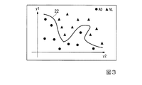
図3は、本発明に係る第一実施形態における脳疾患診断支援装置による境界情報の算出処理の説明に用いる第二の図である。
評価モデル算出部13は、複数の学習データを取り込むと、それぞれの学習データに含まれる脳波特徴データ(BF)を「y(=2)次元のデータ」とみなし、これに機械学習の演算を行うことによって、「ADらしい値」と「NLらしい値」の境界情報を算出する。これは、たとえばサポートベクターマシン(以下、SVM:Support Vector Machine)という機械学習論理によって実現可能である。境界情報は、例えば分離境界面を示す関数として算出される。評価モデル算出部13は、算出した境界情報を記憶部15に書き込んで記憶させる。図3の場合、境界情報は、例えば、境界線22で表される。
評価モデル算出部13が複数の学習データから境界情報を算出し、記憶部15に記録すると、脳疾患診断支援装置10は、疾患情報が付されていない脳波特徴データ(BFx)について、BFxが、AD、NLの何れであるか、また、そのときAD又はNLである確率がどの程度であるかを評価できるようになる。
FIG. 3 is a second diagram used for explaining the boundary information calculation processing by the brain disease diagnosis support apparatus according to the first embodiment of the present invention.
When the evaluation
When the evaluation
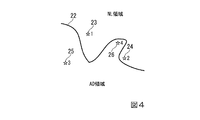
図4は、本発明に係る第一実施形態における脳疾患診断支援装置による評価処理の説明に用いる図である。
評価モデル算出部13が境界情報を算出すると、オペレータが、診断すべき被験者の脳波特徴データ「BFx」を脳疾患診断支援装置10に投入する。脳波特徴データ取得部11は、脳波特徴データ「BFx」を取得し、判定部14に出力する。判定部14は、評価モデル算出部13が算出した境界情報を記憶部15から読み出し、脳波特徴データ「BFx」が、算出された境界のどちらに分類されるかを判定する。また、判定部14は、脳波特徴データ「BFx」が境界からどのくらい遠いかによって、診断すべき被験者の脳波特徴データがAD又はNLである確率を算出することができる。
図4において、被験者1の脳波特徴データBF1(符号23)の場合、BF1はNL領域に存在し、境界線22から遠いため、判定部14は、BF1はNLの確率が大で、その確率は例えば70%と評価する。また、被験者2の脳波特徴データBF2(符号24)の場合、BF2はNL領域に存在し、境界線22から近いため、判定部14は、BF2はNLである確率が高い(例えば60%など)が、誤識別の可能性ありと評価する。同様に被験者3の脳波特徴データBF3(符号25)の場合、BF3はAD領域に存在し、境界線22から遠いため、判定部14は、BF3はADの可能性が大で、その確率は例えば80%と評価する。同様に被験者4の脳波特徴データBF4(符号26)の場合、BF4はAD領域に存在し、境界線22から近いため、判定部14は、BF4はNLの確率が高い(例えば55%など)が、誤識別の可能性ありと評価する。なお、70%などの確率を表す数値は、例えばSVMの手法で算出することが可能である。
FIG. 4 is a diagram used for explaining the evaluation process performed by the brain disease diagnosis support apparatus according to the first embodiment of the present invention.
When the evaluation
In FIG. 4, in the case of the brain wave feature data BF 1 (reference numeral 23) of the subject 1, since BF 1 exists in the NL region and is far from the
このように本実施形態では、ADとNLの境界情報を求め、その境界情報によってADかNLであるかの評価を行う。その為、従来のように「平均的なAD」、「平均的なNL」を定めることによって、ADやNLの特性をならしてしまう可能性があるという問題を回避することができる。また、機械学習の手法で境界情報を算出することができるので、ノウハウの蓄積により試行錯誤で適切なデータを抽出するような煩わしさが無い。また、本実施形態の脳波特徴データは、国際標準の国際10−20法による電極配置に2ヶ所を追加した、21ヶ所で測定を行っており、さらにそれぞれの位置で測定された値についても複数の周波数特性ごとに分割したデータを用いるため、次元数(y)が増加し、例えば、420次元などにもなるが、SVMなどの機械学習の手法を用いれば、多次元における境界面の関数を算出することができる。なお、本実施形態における電極配置の一例については、後に図6を用いて説明する。 As described above, in this embodiment, boundary information between AD and NL is obtained, and whether the AD or NL is determined is determined based on the boundary information. Therefore, it is possible to avoid the problem that there is a possibility that the characteristics of AD and NL may be made uniform by defining “average AD” and “average NL” as in the past. Further, since the boundary information can be calculated by a machine learning method, there is no trouble of extracting appropriate data by trial and error by accumulating know-how. In addition, the electroencephalogram feature data of the present embodiment is measured at 21 locations where two locations are added to the electrode arrangement according to the international standard 10-20 method, and there are a plurality of values measured at each location. Since the number of dimensions (y) is increased by using data divided for each frequency characteristic of, for example, 420 dimensions, etc., if a machine learning method such as SVM is used, a multi-dimensional boundary surface function is obtained. Can be calculated. An example of the electrode arrangement in this embodiment will be described later with reference to FIG.
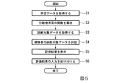
図5は、本発明に係る第一実施形態における脳疾患診断支援装置を用いた脳波特徴データの統計的な評価処理のフローチャートである。
図5を用いて、脳疾患診断支援装置10による境界情報の算出処理、認知症の評価処理について説明を行う。
前提として、脳疾患診断支援装置10に接続された表示装置には、オペレータが操作するためのインタフェース画像が表示されており、インタフェース画像には、「境界情報を算出する」、「診断対象データを評価する」、「ADの疾患情報を付す」、「NLの疾患情報を付す」などのボタンが表示されており、オペレータがこれらのボタンをマウスで押下するなどの操作を行うことにより、脳疾患診断支援装置10に処理の実行を指示することができるものとする。
まず、オペレータが、正常者やアルツハイマー型認知症患者の学習データを脳疾患診断支援装置10に投入する。学習データは、例えば電子ファイルの形式で与えられ、1つの電子ファイルには、複数の正常者やアルツハイマー型認知症患者の学習データが含まれている。それぞれの学習データには、「AD」又は「NL」の疾患情報が付されているものとする。また、それぞれの学習データに含まれる特徴データは、例えば420次元のデータである。投入する学習データは、多い程好ましい。脳波特徴データ取得部11は、学習データを取得し(ステップS1)、取得した学習データを記憶部15へ書き込んで記憶させる。
FIG. 5 is a flowchart of a statistical evaluation process of electroencephalogram feature data using the brain disease diagnosis support apparatus according to the first embodiment of the present invention.
The boundary information calculation processing and dementia evaluation processing performed by the brain disease
As a premise, an interface image to be operated by an operator is displayed on the display device connected to the brain disease
First, the operator inputs learning data of a normal person or an Alzheimer type dementia patient into the brain disease
次にオペレータが、「境界情報を算出する」と表示されたボタンを押下すると、操作受付部12が、その操作を受け付け、評価モデル算出部13に境界情報の算出を指示する。評価モデル算出部13は、記憶部15に書き込まれた学習データを読み出して、例えばSVMの手法によって、読み込んだ学習データをADとNLに分ける分離境界面の関数を算出する(ステップS2)。評価モデル算出部13は、分離境界面を示す関数を算出すると、その関数(境界情報)を記憶部15に書き込んで記憶させる。以上が境界情報の算出処理である。
Next, when the operator presses a button displayed as “Calculate boundary information”, the
次に新たな被験者の脳波特徴データについて評価処理を行う。まず、オペレータが、診断すべき被験者の脳波特徴データ(診断対象データ)を脳疾患診断支援装置10に投入する。診断対象データは、例えば電子ファイルの形式で与えられる。1つの電子ファイルには、例えば1人の被験者の420次元の脳波特徴データが含まれている。脳波特徴データ取得部11は、診断対象データを取得し(ステップS3)、取得した診断対象データを記憶部15へ書き込んで記憶させる。
Next, an evaluation process is performed on the electroencephalogram feature data of the new subject. First, an operator inputs brain wave characteristic data (diagnosis target data) of a subject to be diagnosed into the brain disease
次にオペレータが、「診断対象データを評価する」と表示されたボタンを押下すると、操作受付部12が、その操作を受け付け、判定部14に診断対象データの評価を指示する。判定部14は、記憶部15に書き込まれた境界情報を用いて、例えばSVMの手法によって診断対象データが、ADとNLの何れであるか、また、その確率について評価を行う(ステップS4)。なお、確率とは、SVMにおいては、診断対象データの分離境界面からの距離に応じた値(確信度)である。
判定部14は、評価した結果を表示部16へ出力する。表示部16は、被験者の診断対象データの評価結果を表示装置に表示する(ステップS5)。評価結果には、例えば、「診断対象データはNLである可能性が高く、その確率は70%です」といった内容が表示される。
Next, when the operator presses a button displayed as “evaluate diagnosis target data”, the
The
診断者(医師)は、表示部16が表示した評価結果や、他に問診や検査などを行った結果を用いて総合的に被験者がアルツハイマー型認知症であるかどうかを診断する。脳疾患診断支援装置10によれば、脳波特徴データから見たアルツハイマー型認知症である確率を医師に提示することができるので、医師による、患者がアルツハイマー型認知症であるか否かの診断を支援することができる。判定部14による評価に要する時間は、数分程度であって、予め被験者の脳波特徴データを用意しておけば、医師は脳疾患診断支援装置10による評価結果を速やかに得ることができる。また、脳波の計測装置は、MRIなど認知症の診断に用いられる他の医療装置よりも安価で、また、脳波の取得は、安全で繰り返し取得しても被験者の健康に与える問題が少ない。従って脳波による認知症などの脳疾患の診断を支援する本実施形態の脳疾患診断支援装置10は、比較的小規模の医療機関でも導入しやすく、脳疾患診断支援装置10を含む診断支援システムを導入することで、町の医療機関でも、社会的な問題になっているアルツハイマー型認知症を比較的容易に診断できるようになる。また、アルツハイマー型認知症の場合、他人から認知症と判断できる前に脳波に影響が出ることがわかっており、本実施形態の脳疾患診断支援装置10を用いることにより、症状が出る前にアルツハイマー型認知症となる確率を知ることができるので、アルツハイマー型認知症の早期診断、早期対策にも役立てることができる。また、他人から見て認知症のような症状を発症しているが、それが本当に認知症なのか、あるいは老人性の鬱など認知症以外の症状なのか問診だけでは判断できない場合も多く、脳疾患診断支援装置10は、見分けにくい症状を発症している患者のアルツハイマー型認知症の診断を支援する情報を提供する。
The diagnostician (doctor) comprehensively diagnoses whether or not the subject has Alzheimer's dementia using the evaluation result displayed on the
次に、医師が診断した脳波特徴データを学習データとして取り込んで、境界情報を再算出する処理を行う。医師が、診断対象データに対して診断を行うと、オペレータは、その診断結果を脳疾患診断支援装置10に入力する。例えば、診断結果がADの場合、オペレータは、「ADの疾患情報を付す」ボタンを押下し、診断結果がNLの場合、オペレータは、「NLの疾患情報を付す」ボタンを押下する。
すると、脳波特徴データ取得部11は、そのボタン押下操作を受け付け(ステップS6)、押下されたボタンが示す疾患情報(ADかNLか)と診断対象データとを対応付けて記憶部15に書き込む。これにより、被験者の診断対象データに疾患情報が付されたことになり、新たな学習データが1件増えることになる。次に、オペレータは、「境界情報を生成する」と表示されたボタンを押下する。すると、評価モデル算出部13は、ステップS1で記憶部15に書き込まれた学習データに新たな学習データ1件を加えて、ステップS2の境界情報を生成する処理を再度行う。評価モデル算出部13は、新たに算出した境界情報を記憶部15に書き込んで記憶させる。所与の学習データに被験者の学習データを加えることで、学習データの数を増加させる。これにより、評価モデル算出部13の算出する境界情報の精度・信頼性を高めることができる。
Next, the brain wave feature data diagnosed by the doctor is taken in as learning data, and the boundary information is recalculated. When the doctor makes a diagnosis on the diagnosis target data, the operator inputs the diagnosis result to the brain disease
Then, the electroencephalogram feature
なお、表示部16は、ステップS1において取得した学習データの選択画面を表示し、医師の指示等により、オペレータが学習データを選択できるようにしてもよい。脳波特徴データは、電極の配置位置のわずかな差や、頭部への装着加減によって異なる挙動を示す可能性がある。例えば、自施設で測定した脳波特徴データに基づいて本実施形態による評価を行うと望ましい結果が得られるが、全国的に収集した脳波特徴データを用いて評価を行うと好ましい結果が得られない可能性がある。そのような場合に、基準とする学習データを選択できるようにし、診断に役立つ学習データ群だけを選択することで、医師は、脳疾患診断支援装置10をより有効に認知症の診断に役立てるようにすることができる。
The
また、選択する学習データのパターンを記憶部15に登録することできるようにしてもよい。例えば、同じアルツハイマー型認知症でもいくつかのパターンが存在するようなことを医師が経験的に把握したような場合に、それぞれのパターンを検出するのに適した学習データ群を抽出してそれぞれ別のパターンとして登録し、それら異なるパターンを切り替えて学習データとして用いることで、1人の被験者の診断対象データをさまざまな観点から評価することが可能となり、医師は、より正確にアルツハイマー型認知症の診断を行うことができるようになる。
Further, the pattern of learning data to be selected may be registered in the
また、使用する脳波特徴データについても、例えば420次元の特徴量から任意の特徴量を選択できるようにし、脳波特徴データに含まれる全ての特徴量を用いるだけではなく、次に図6で例示する一部の特徴量だけを用いて境界情報算出処理や評価処理を行うようにしてもよい。また、一連の処理に用いる特徴データのパターンを登録できるようにし、それらのパターンを切り替えて境界情報算出処理などを行うことができるようにしてもよい。これにより、同じ学習データ群を用いて評価する場合であっても、さまざまな観点から評価を行うことができ、医師によるより細やかな診断を支援することができる。
また、分離境界面の関数を算出するステップS2において、主成分分析やRandomForestなどの手法を用いて、ADとNLを分離するのに有効な特徴量を動的に抽出し、抽出した特徴量のみを用いて分離境界面の関数を算出するようにしてもよい。
Also, for the electroencephalogram feature data to be used, for example, an arbitrary feature amount can be selected from 420-dimensional feature amounts, and not only all feature amounts included in the electroencephalogram feature data are used, but also illustrated in FIG. The boundary information calculation process and the evaluation process may be performed using only some of the feature amounts. In addition, it is possible to register feature data patterns used in a series of processing, and to perform boundary information calculation processing by switching those patterns. Thereby, even if it is a case where it evaluates using the same learning data group, it can evaluate from various viewpoints and can support a more detailed diagnosis by a doctor.
Further, in step S2 for calculating the function of the separation boundary surface, a feature amount effective for separating AD and NL is dynamically extracted using a method such as principal component analysis or Random Forest, and only the extracted feature amount is obtained. May be used to calculate the function of the separation boundary surface.
次に、アルツハイマー型認知症の識別に有効と考えられる特徴量の一例について説明する。
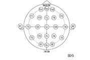
図6は、本発明に係る第一実施形態における脳波を測定する電極の配置の一例を示す図である。
図6のA1,A2、Fp1、Fp2、Fpz、F7、F3、Fz、F4、F8、T3、C3、Cz、C4、T4、T5、P3、Pz、P4、T6、O1、Oz、O2、は被験者の頭部に配置する電極の配置位置である。A1,A2は、基準電極のため、実際にはFp1〜O2の21ヶ所の脳波が得られることになる。これらの電極から得られる脳波の4.68Hz〜20.28Hzをフーリエ変換にて1.58Hz刻みに11個に分解する。また、各周波数の電位を規格化した値を2乗する。これをsNATとする。また、隣接する周波数についてsNATの比を求め、これをvNATとする。ここで、sNATについては、最後の周波数(20.28Hz)を除いた10個の値を用いるとすると、各電極に対して10個のsNATと10個のvNATが得られる。電極の数が21個なので全体で21×10×10=420次元の特徴量を得ることができる。なお、規格化とは、個人間のばらつきを抑えるために、被験者の例えば安静状態における脳波データと測定した脳波データとの差分値を計算することである。また、sNATやvNATは、脳波特徴データを構成する特徴量の一例である。特徴量は、各電極位置で測定して得られる脳波データに基づいた他の物理量であってもよい。
Next, an example of a feature amount considered to be effective for identifying Alzheimer's dementia will be described.
FIG. 6 is a diagram showing an example of the arrangement of electrodes for measuring an electroencephalogram in the first embodiment according to the present invention.
A1, A2, Fp1, Fp2, Fpz, F7, F3, Fz, F4, F8, T3, C3, Cz, C4, T4, T5, P3, Pz, P4, T6, O1, Oz, O2 in FIG. It is the arrangement position of the electrode arrange | positioned at a test subject's head. Since A1 and A2 are reference electrodes, brain waves at 21 locations Fp1 to O2 are actually obtained. 4.68 Hz to 20.28 Hz of the electroencephalogram obtained from these electrodes is decomposed into 11 in 1.58 Hz increments by Fourier transform. Moreover, the value which normalized the electric potential of each frequency is squared. This is sNAT. Further, the ratio of sNAT is obtained for adjacent frequencies, and this is defined as vNAT. Here, for sNAT, if 10 values excluding the last frequency (20.28 Hz) are used, 10 sNATs and 10 vNATs are obtained for each electrode. Since the number of electrodes is 21, a total of 21 × 10 × 10 = 420-dimensional feature values can be obtained. In addition, normalization is calculating the difference value of the brain wave data of a test subject, for example in a resting state, and the measured brain wave data, in order to suppress the dispersion | variation between individuals. Moreover, sNAT and vNAT are examples of feature amounts constituting the electroencephalogram feature data. The feature quantity may be another physical quantity based on brain wave data obtained by measurement at each electrode position.
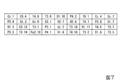
図7は、本発明に係る第一実施形態における脳疾患診断支援装置で用いる特徴量の一例を示す図である。
図7の表に記載した各値は、上述の420次元の特徴量のうち、本実施形態の評価方法を行った結果、評価に有効であることが判明した特徴量である。
図7の表に記載した特徴量は、RandomForestアルゴリズムによって得られた識別に有効な特徴量の一例である。例えば、図7に記載の36次元の特徴量だけを用いて、本実施形態のアルツハイマー型認知症の評価処理を行うと、420次元の特徴量の全てを用いる場合と比較して、良好な結果が得られることがわかった。
なお、図7に記載した各値におけるピリオドの前の記号は、図6の電極の位置を示しており、ピリオドの後の数値は、sNAT又はvNATの値を示している。具体的には、1〜9が低周波数から順番にsNAT値を示し、10〜19が低周波数から順番にvNAT値を示している。例えば、「C3.4」は、「C3」電極における低周波数から4番目のsNAT値を示し、「O1.17」は、「O1」電極における低周波数から7番目のvNAT値を示している。
FIG. 7 is a diagram illustrating an example of feature amounts used in the brain disease diagnosis support apparatus according to the first embodiment of the present invention.
Each value described in the table of FIG. 7 is a feature amount that has been found to be effective for evaluation as a result of performing the evaluation method of the present embodiment, among the 420-dimensional feature amounts described above.
The feature amount described in the table of FIG. 7 is an example of a feature amount effective for identification obtained by the Random Forest algorithm. For example, when only the 36-dimensional feature value shown in FIG. 7 is used to perform the Alzheimer-type dementia evaluation process of the present embodiment, a better result is obtained than when all 420-dimensional feature values are used. Was found to be obtained.
In addition, the symbol before the period in each value described in FIG. 7 indicates the position of the electrode in FIG. 6, and the numerical value after the period indicates the value of sNAT or vNAT. Specifically, 1 to 9 indicate sNAT values in order from low frequency, and 10 to 19 indicate vNAT values in order from low frequency. For example, “C3.4” indicates the fourth sNAT value from the low frequency in the “C3” electrode, and “O1.17” indicates the seventh vNAT value from the low frequency in the “O1” electrode.
また、上記の説明ではアルツハイマー型認知症の診断支援を例に説明を行ったが、他の認知症、及び、鬱や統合失調症などの脳疾患症状に対しても、本実施形態の方法を適用してもよい。つまり、ある脳疾患を示す疾患情報を付した学習データと、その脳疾患を患っていない人の学習データをあらかじめ用意しておき、評価モデル算出部13が、その脳疾患とそれ以外を分離する境界情報を算出し、判定部14が、被験者の脳波特徴データがその脳疾患であるかどうかや、その確率を算出するようにすることも可能である。
In the above description, diagnosis support for Alzheimer-type dementia has been described as an example. However, the method of the present embodiment is also applied to other dementia and brain disease symptoms such as depression and schizophrenia. You may apply. That is, learning data with disease information indicating a certain brain disease and learning data of a person who does not suffer from the brain disease are prepared in advance, and the evaluation
<第二実施形態>
以下、本発明の第二実施形態による脳疾患診断支援装置を図8〜図10を参照して説明する。
第一実施形態は、アルツハイマー型認知症かそれ以外かの疾患情報が付された学習データを用いてアルツハイマー型認知症か否かを評価する評価方法であった。第二実施形態では、被験者の脳波特徴データを類似するグループに分類する処理を行う。これにより、学習データに複数の認知症や認知症に類似する症状を持つ人の脳波の特徴データが混在している場合の評価精度を向上させることができる。
本実施形態における評価モデル算出部13aは、さまざまな脳波特徴データを類似するグループごとに分類したクラスタを生成するクラスタリングを実行する機能を有している。また、判定部14aは、被験者の脳波特徴データが、評価モデル算出部13aが生成したクラスタのうち、どのクラスタに所属するかを判定する処理を行う。
さらに、評価モデル算出部13aは、判定部14aが被験者の脳波特徴データの分類先であると判定したクラスタにおける、ADの疾患情報が付された脳波特徴データが占める割合を算出する。他の構成は、第一実施形態と同様である。
<Second embodiment>
Hereinafter, a brain disease diagnosis support apparatus according to a second embodiment of the present invention will be described with reference to FIGS.
The first embodiment is an evaluation method for evaluating whether or not the patient has Alzheimer's dementia using learning data to which information on the disease of Alzheimer's type or other disease is attached. In the second embodiment, a process of classifying brain wave feature data of a subject into similar groups is performed. Thereby, the evaluation accuracy in the case where characteristic data of brain waves of a person having a plurality of dementia and symptoms similar to dementia are mixed in the learning data can be improved.
The evaluation model calculation unit 13a in the present embodiment has a function of executing clustering that generates clusters in which various types of brain wave feature data are classified into similar groups. In addition, the determination unit 14a performs processing to determine which cluster the subject's brain wave feature data belongs to among the clusters generated by the evaluation model calculation unit 13a.
Furthermore, the evaluation model calculation unit 13a calculates the ratio of the electroencephalogram feature data to which AD disease information is added in the cluster that the determination unit 14a determines to be the classification destination of the subject's electroencephalogram feature data. Other configurations are the same as those in the first embodiment.
図8〜図9を用いて、本発明に係る第二実施形態における評価処理について説明する。
図8は、本発明に係る第二実施形態における脳疾患診断支援装置による評価処理の説明に用いる第一の図である。
図8(a)は、記憶部15に書き込まれた学習データを図2〜図3と同様に2次元上にマッピングした図である。学習データには、AD、NLの他、他の認知症を示す疾患情報が付されていてもよい。次に、評価モデル算出部13aが、複数の学習データを機械学習(クラスタリング)によって、類似するグループ(クラスタ)毎に分類する。クラスタリングの手法としては、例えば、スペクトラルクラスタリングを用いることが可能である。
The evaluation process in the second embodiment according to the present invention will be described with reference to FIGS.
FIG. 8 is a first diagram used for explaining the evaluation process by the brain disease diagnosis support apparatus according to the second embodiment of the present invention.
FIG. 8A is a diagram in which learning data written in the
図8(b)は、評価モデル算出部13aが、学習データをクラスタリングした結果を示している。この図の場合、学習データが、符号37が示すクラスタ37と、符号38が示すクラスタ38と、符号39が示すクラスタ39と、に分類されたことを示している。破線45aは、クラスタ37に分類される学習データとクラスタ38に分類される学習データとを隔てる境界線である。破線45bは、クラスタ37に分類される学習データとクラスタ39に分類される学習データとを隔てる境界線である。破線45cは、クラスタ38に分類される学習データとクラスタ39に分類される学習データとを隔てる境界線である。ここで、評価モデル算出部13aは、各クラスタに含まれている学習データに付された疾患情報を参照し、クラスタ毎に各疾患情報が付された学習データの占める割合を算出する。例えばクラスタ37に10個の学習データが分類されたとして、その10個のうち、ADの疾患情報が付された学習データが4個、NLの疾患情報が付された学習データが3個、レビー小体型認知症の疾患情報が付された学習データが2個、血管性認知症の疾患情報が付された学習データが1個であるとすると、評価モデル算出部13aは、クラスタ37に占めるADの割合(AD率)=40%、NLの割合(NL率)=30%、レビー小体型認知症の割合=20%、血管性認知症の割合=10%を算出する。
FIG. 8B shows the result of the evaluation model calculation unit 13a clustering the learning data. In the case of this figure, it is shown that the learning data is classified into a
図8(c)は、オペレータが、診断すべき被験者の脳波特徴データ(符号40)を脳疾患診断支援装置10に投入した状態を示している。被験者の脳波特徴データ40が投入されると、脳波特徴データ取得部11は、被験者の脳波特徴データを取得し、判定部14aに出力する。判定部14aは、脳波特徴データ40がクラスタ37〜39のうち、どのクラスタに所属するかを、例えばk−近傍法などのクラスタリング手法を用いて判定する。
図8(d)は、判定部14aが所属クラスタの判定を行った結果を示している。判定部14は、判定が完了すると、評価モデル算出部13aが算出した所属先のAD率の値を取得し、その値によって、被験者の脳波特徴データがADであることを示す確率を評価する。例えば、この例の場合、判定部14aは、被験者の脳波特徴データがADであることを示す確率は、40%であると評価する。
FIG. 8C shows a state where the operator has input the brain wave characteristic data (reference numeral 40) of the subject to be diagnosed into the brain disease
FIG. 8D shows the result of the determination unit 14a determining the belonging cluster. When the determination is completed, the
この方法によれば、学習データに付された疾患情報に依存せず、脳波特徴データの類似性によって被験者の脳波特徴データを分類し、その被験者がADである確率を評価することができる。これにより、例えば第一実施形態の評価方法ではNLと判定された場合であっても、上記の方法でAD率が40%と評価されれば、例えば、将来的にその被験者がアルツハイマー型認知症を患う可能性を考える場合の参考とすることができる。また、他の認知症の疾患情報が付された学習データの存在率を同時に表示することで、ADだけでなく、他の認知症を発症する可能性の参考とすることもできる。 According to this method, the subject's brain wave feature data can be classified by the similarity of the brain wave feature data without depending on the disease information attached to the learning data, and the probability that the subject is AD can be evaluated. Thus, for example, even if the evaluation method of the first embodiment is determined to be NL, if the AD rate is evaluated to be 40% by the above method, for example, the subject will have Alzheimer-type dementia in the future. It can be used as a reference when considering the possibility of suffering from. Further, by simultaneously displaying the existence rate of learning data to which other dementia disease information is attached, it is possible to reference not only AD but also the possibility of developing other dementia.
図9は、本発明に係る第二実施形態における脳疾患診断支援装置による評価処理の説明に用いる第二の図である。
図9(a)〜図9(d)は、図8で説明したクラスタリングによる評価方法と第一実施形態による評価方法とを組み合わせた方法である。この方法を用いる場合、評価モデル算出部13aは、本実施形態の機能に加え、第一実施形態の境界情報を算出する機能を併せて有しているものとする。また、判定部14aは、第一実施形態と同様に、その境界情報に基づいて、被験者の脳波特徴データを評価する機能を有していているものとする。
学習データをスペクトラルクラスタリング等の手法によってクラスタリングし、被験者の脳波特徴データの所属クラスタをk−近傍法などによって判定することは、図8で説明した評価方法と同様である。
図9の処理においては、図9(b)において、学習データのクラスタリングが完了すると、各クラスタ37〜39におけるAD率などを求める代わりに、各クラスタにおけるAD、非ADを分離する境界情報を算出する。境界情報は、第一実施形態と同様に、評価モデル算出部13aが、ADの疾患情報が付された学習データと、それ以外の学習データとを用いて、SVMなどの手法により算出する。
FIG. 9 is a second diagram used for explaining the evaluation process performed by the brain disease diagnosis support apparatus according to the second embodiment of the present invention.
FIGS. 9A to 9D are a combination of the clustering evaluation method described in FIG. 8 and the evaluation method according to the first embodiment. When this method is used, it is assumed that the evaluation model calculation unit 13a has a function of calculating boundary information of the first embodiment in addition to the function of the present embodiment. Moreover, the determination part 14a shall have the function to evaluate a test subject's electroencephalogram feature data based on the boundary information similarly to 1st embodiment.
Clustering the learning data using a technique such as spectral clustering and determining the cluster to which the subject's electroencephalogram feature data belongs by the k-neighbor method or the like is the same as the evaluation method described in FIG.
In the process of FIG. 9, when the clustering of the learning data is completed in FIG. 9B, boundary information for separating AD and non-AD in each cluster is calculated instead of obtaining the AD rate in each cluster 37-39. To do. As in the first embodiment, the boundary model is calculated by the evaluation model calculation unit 13a by using a method such as SVM using learning data to which AD disease information is attached and other learning data.
また、図9(d)において、被験者のデータが、例えばクラスタ37に所属すると判定されたとすると、次に判定部14aは、評価モデル算出部13aが算出したクラスタ37における境界情報を記憶部15から読み出して、SVMなどの手法によって、被験者の脳波特徴データがADであるか否かを判定する。図8では、被験者の脳波データの評価結果として、AD率などを表示したが、図9の場合、表示部16は、SVMによって算出される被験者の脳波特徴データがADである確率(確信度)を表示する。
9D, if it is determined that the data of the subject belongs to the
この方法によれば、例えば、クラスタ37、クラスタ38、クラスタ39の何れにおいても例えばAD率が50%であるような学習データが存在した場合であっても、被験者の脳波特徴データの評価精度を向上できる可能性がある。例えば、クラスタ37、クラスタ38、クラスタ39の何れにおいても例えばAD率が50%であるような学習データでは、ADと特徴づけられる脳波特徴データのパターンが3種類存在する可能性がある。その様子を図9(d)に示した。例えば、図9(d)の符号41〜43が示す破線で囲まれた領域に存在する学習データにADの疾患情報が付されているとする。破線41〜43のそれぞれは、各クラスタ37〜39における境界情報である。このとき第一実施形態のように全学習データを対象に境界情報を算出すると、算出した境界情報が適切ではなく、識別精度が低下する可能性がある。適切な境界情報とは、SVMにおけるマージンが最大となるような境界である。図9の方法のように、予め脳波特徴データが類似するクラスタに分類して、その中で境界情報を算出すれば、より適切な境界情報を算出できる可能性があり、評価精度の向上が期待できる。
According to this method, for example, even in the case where learning data having an AD rate of 50% exists in any of the
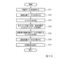
図10は、本発明に係る第二実施形態における脳疾患診断支援装置を用いた脳波特徴データの評価処理のフローチャートである。
まず、オペレータが、正常者やさまざまな患者の学習データを脳疾患診断支援装置10に投入する。学習データは、例えば電子ファイルの形式で与えられ、1つの電子ファイルには、さまざまな疾患情報が付された学習データが含まれている。さまざまな疾患情報とは、例えば、認知症ではないが認知症と間違えやすい症状、アルツハイマー型認知症、レビー小体型認知症、血管性認知症、などである。また、アルツハイマー型認知症患者の学習データについても、重度の患者、軽度の患者、投薬治療中の患者、投薬治療中ではない患者、などの学習データが含まれていてもよい。脳波特徴データ取得部11は、学習データを取得し(ステップS11)、取得した学習データを記憶部15へ書き込んで記憶させる。
次に、オペレータが、診断すべき被験者の診断対象データを脳疾患診断支援装置10に投入する。脳波特徴データ取得部11は、診断対象データを取得し(ステップS12)、取得した診断対象データを記憶部15へ書き込んで記憶させる。このとき、脳波特徴データ取得部11は、診断対象データをステップS11で取得した学習データと区別して記録する。
FIG. 10 is a flowchart of an evaluation process of electroencephalogram feature data using the brain disease diagnosis support apparatus according to the second embodiment of the present invention.
First, an operator inputs learning data of normal persons and various patients into the brain disease
Next, the operator inputs the diagnosis target data of the subject to be diagnosed into the brain disease
次にオペレータが、「診断対象データを評価する」と表示されたボタンを押下すると、操作受付部12が、その操作を受け付け、評価モデル算出部13aにクラスタの生成(クラスタリング)を指示する。クラスタとは、複数の被験者の脳波特徴データを、脳波特徴データに含まれる特徴量の類似度に基づいてグループ化したものである。評価モデル算出部13aは、記憶部15に書き込まれたステップS11で取り込んだ学習データを読み出して、例えばスペクトラルクラスタリングなどの機械学習で用いられるクラスタリング手法によって、読み出したデータから所定の数のクラスタを生成する(ステップS13)。なお、生成するクラスタの数は、オペレータが設定し、予め記憶部15が記憶しているものとする。各クラスタには、類似する脳波特徴データを持つ学習データが分類され、1つのクラスタには、正常者、アルツハイマー型認知症患者、レビー小体型認知症患者などさまざまな疾患情報に対応する学習データが含まれる可能性がある。
Next, when the operator presses a button labeled “Evaluate diagnosis target data”, the
次に、評価モデル算出部13aは、ステップS13において、図8を用いて説明したように、各クラスタにおいて、そのクラスタに含まれる学習データに付された疾患情報の種類ごとの割合を算出する。評価モデル算出部13aは、算出したAD率などの疾患情報ごとの割合をクラスタごとに記憶部15に書き込んで記憶させる。あるいは、評価モデル算出部13aは、図9を用いて説明したように例えばアルツハイマー型認知症患者の疾患情報が付された脳波特徴データと、それ以外の疾患情報が付された脳波特徴データとを分離する境界情報を算出する(ステップS14)。境界情報の算出は、例えば、SVMの手法を用いる。ここで、境界情報の算出には、例えば、420次元のデータ全てを用いてもよいし、420次元の特徴量のうち、ADを識別するのに有効な特徴量だけを用いて境界情報を算出してもよい。なお、有効な特徴量を示す情報は記憶部15が記憶しているものとする。あるいは、後述するように、学習データ群に応じた評価に有効な特徴量をその都度、算出するようにしてもよい。ADの識別に有効な特徴量の一例としては、図7で示したものがある。また、評価モデル算出部13aは、算出したクラスタごとの境界情報をクラスタの識別情報と対応付けて記憶部15に書き込んで記憶させる。なお、評価モデル算出部13aは、疾患情報ごとの割合の算出と、境界情報の算出との両方を行ってもよい。次に、評価モデル算出部13aは、判定部14aに診断対象データの評価を指示する。
Next, in step S13, as described with reference to FIG. 8, the evaluation model calculation unit 13a calculates a ratio for each type of disease information attached to the learning data included in the cluster in each cluster. The evaluation model calculation unit 13a writes and stores the calculated ratio for each disease information such as the AD rate in the
次に、判定部14aは、診断対象データがどのクラスタに所属するかをk−近傍法などの手法によって判定する(ステップS15)。
次に判定部14aは、診断対象データが、ADである確率について評価を行う(ステップS16)。例えば、評価モデル算出部13aがAD率を算出した場合、判定部14aは、診断対象データが分類されると判定したクラスタに対応するAD率を記憶部15から読み出して、診断対象データがADであることを示す確率は、記憶部15から読み出したAD率であると評価する。あるいは、評価モデル算出部13aが境界情報を算出した場合、判定部14aは、診断対象データが分類されると判定したクラスタに対応する境界情報を記憶部15から読み出して、診断対象データが、ADとNLの何れであるか、また、その確率(確信度)について評価を行う。
Next, the determination unit 14a determines which cluster the diagnosis target data belongs to by a method such as a k-neighbor method (step S15).
Next, the determination unit 14a evaluates the probability that the diagnosis target data is AD (step S16). For example, when the evaluation model calculation unit 13a calculates the AD rate, the determination unit 14a reads the AD rate corresponding to the cluster determined to be classified as the diagnosis target data from the
評価を行うと、判定部14aは、評価結果を表示部16へ出力する。表示部16は、被験者の診断対象データの評価結果を表示装置に表示する(ステップS17)。例えば、表示部16は、診断の根拠となった脳波の測定部位、周波数帯、診断対象データが所属するクラスタの識別情報、診断対象データがADを示す確率などを表示する。ADを示す確率とは、上述のAD率や確信度である。また、判定部14aは、ステップS16の評価の際に、パターンマッチング等の手法によって被験者の脳波特徴データと最も類似する学習データに含まれる脳波特徴データを抽出し、表示部16は、判定部14aが抽出した脳波特徴データとそのデータに付された疾患情報を表示するようにしてもよい。
When the evaluation is performed, the determination unit 14 a outputs the evaluation result to the
医師は、表示部16が表示した情報や、その他の検査情報などを基に被験者の症状を診断する。例えば、被験者の脳波特徴データと最も類似する脳波特徴データやその脳波特徴データに付された疾患情報が表示され、その疾患情報がレビー小体認知症である場合、医師は、被験者の脳波特徴データがレビー小体認知症患者の脳波特徴データに類似していることを診断の参考にすることができる。また、被験者の脳波特徴データが分類されるクラスタに含まれるアルツハイマー型以外の認知症の割合が表示されれば、医師は、例えば、被験者がアルツハイマー型以外の認知症を発症する可能性を考えるときの参考にすることができる。
The doctor diagnoses the symptom of the subject based on the information displayed on the
なお、図10の処理フローの変形例として次のような処理としてもよい。まず、ステップS14において、AD率などを算出し、ステップS16の評価において、そのAD率が例えば90%であるような場合は、被験者の脳波特徴データはADであるとみなし、その評価結果を表示部16が出力して処理フローを終了する。また、AD率が例えば50%であるような場合は、さらなる評価処理が必要と判断し、さらにADと非ADとを分離する境界情報を算出し、被験者の脳波特徴データをSVMなどによって評価する。
また、ステップS13において、生成するクラスタの数を変化させて、ステップS13以降の処理を繰り返し行うようにしてもよい。
As a modification of the processing flow in FIG. 10, the following processing may be performed. First, in step S14, an AD rate is calculated, and in the evaluation in step S16, if the AD rate is 90%, for example, the subject's brain wave feature data is regarded as AD, and the evaluation result is displayed. The
In step S13, the number of clusters to be generated may be changed, and the processes in and after step S13 may be repeated.
認知症にも複数の種類がある。代表的なものは、アルツハイマー型認知症、レビー小体型認知症、血管性認知症などである。これらの認知症の種類は、他人から見ただけでは分からないことも多い。また、同じ認知症でも種類が違えば、治療方法も異なってくるので、正確にどのような種類の認知症であるかを診断することが重要である。そこで、脳波特徴データを用いて複数種類の認知症であるかを知ろうとする場合、例えば手作業で各認知症のテンプレートを選択し、選択したテンプレートと被験者の脳波特徴データとを比較して判定することが考えられるが、各認知症に対応する脳波特徴データが均一な特徴を有するとは限らず、手作業でテンプレートを作成するのは困難である。本実施形態によれば、さまざまな種類の認知症患者の脳波特徴データを用いてクラスタリングを行うことで、煩雑な作業を行うことなく、被験者がどの種類の認知症であるかを判定したり、複数の認知症についてそれぞれの認知症を患っている確率を算出したりすることができる。また、同じ種類の認知症患者であっても、重度、軽度、投薬中などさまざまな状態にある患者の脳波特徴データを用意しておくと、患者が認知症を患っている場合、その症状は軽度か、重度かなどの情報を表示することもできる。 There are several types of dementia. Representative examples include Alzheimer's dementia, Lewy body dementia, and vascular dementia. Often these types of dementia are not obvious from others. In addition, since the treatment method varies depending on the type of the same dementia, it is important to diagnose exactly what type of dementia is. So, if you want to know whether you have multiple types of dementia using EEG feature data, for example, select a template for each dementia manually, and compare the selected template with the EEG feature data of the subject Although it is conceivable that the electroencephalogram feature data corresponding to each dementia does not always have uniform features, it is difficult to create a template manually. According to the present embodiment, by performing clustering using the electroencephalogram feature data of various types of dementia patients, it is possible to determine what type of dementia the subject has without performing complicated work, The probability of suffering from each dementia can be calculated for a plurality of dementias. In addition, even if the patient has the same type of dementia, preparing EEG feature data for patients with various conditions such as severe, mild, and medication, if the patient suffers from dementia, the symptoms are Information such as whether it is mild or severe can also be displayed.
なお、本実施形態においても、ステップS11で取得した学習データから、ステップS12以降の処理で用いる学習データを選択できるようにしてもよい。
また、ステップS13のクラスタリングで使用する脳波特徴データについては、全ての特徴量を用いてもよいし、一部の特徴量だけを用いるようにしてもよい。例えば、クラスタを生成するステップS14において、例えば420次元全ての特徴量を用いるとうまくクラスタリングできないような場合に、主成分分析やRandomForestなどの手法を用いて、クラスタリングに有効な特徴量を動的に抽出し、抽出した特徴量のみを用いてクラスタを生成するようにしてもよい。さらに、診断対象データを評価するステップS16においてSVMによって境界情報を算出する場合、第一実施形態と同様、分離に有効な特徴量をRandomForest等によって抽出し、境界情報を算出してもよい。このようにすることでクラスタごとに固有の有効な特徴量を用いることができ、評価精度を高めることができる。
これら使用する学習データや特徴量を変化させることで、さまざまな観点から診断対象データを評価することができる。
In the present embodiment, the learning data used in the processes after step S12 may be selected from the learning data acquired in step S11.
In addition, regarding the electroencephalogram feature data used in the clustering in step S13, all feature amounts may be used, or only a part of feature amounts may be used. For example, in the step S14 of generating a cluster, if clustering cannot be performed successfully using, for example, all 420-dimensional feature quantities, a feature quantity effective for clustering is dynamically determined using a method such as principal component analysis or Random Forest. The cluster may be generated by using only the extracted feature amount. Further, when boundary information is calculated by SVM in step S16 for evaluating diagnosis target data, feature information effective for separation may be extracted by Random Forest and the boundary information may be calculated as in the first embodiment. By doing in this way, the effective characteristic quantity intrinsic | native for every cluster can be used, and evaluation accuracy can be improved.
By changing the learning data and feature quantities to be used, it is possible to evaluate the diagnosis target data from various viewpoints.
なお、本実施形態においてもアルツハイマー型認知症の診断支援を例に説明を行ったが、他の認知症、及び、鬱や統合失調症などの脳疾患症状に対しても、本実施形態を適用することが可能である。また、その場合、さまざまな脳疾患の疾患情報が付された学習データを用いることで、複数の脳疾患に対してそれらの脳疾患のうち1つ又は複数を患っている確率を同時に算出することができる。 In this embodiment, the diagnosis support for Alzheimer-type dementia has been described as an example. However, the present embodiment is also applied to other dementia and brain disease symptoms such as depression and schizophrenia. Is possible. In that case, the probability of suffering from one or more of those brain diseases for a plurality of brain diseases can be calculated at the same time by using learning data to which disease information of various brain diseases is attached. Can do.
なお、上述した脳疾患診断支援装置10における各処理の過程は、プログラムの形式でコンピュータ読み取り可能な記録媒体に記憶されており、このプログラムを脳疾患診断支援装置10のコンピュータが読み出して実行することによって、上記処理が行われる。ここでコンピュータ読み取り可能な記録媒体とは、磁気ディスク、光磁気ディスク、CD−ROM、DVD−ROM、半導体メモリ等をいう。また、このコンピュータプログラムを通信回線によってコンピュータに配信し、この配信を受けたコンピュータが当該プログラムを実行するようにしてもよい。
Each process in the brain disease
また、上記プログラムは、前述した機能の一部を実現するためのものであってもよい。さらに、前述した機能をコンピュータシステムにすでに記録されているプログラムとの組み合わせで実現できるもの、いわゆる差分ファイル(差分プログラム)であってもよい。
また、脳疾患診断支援装置10は、1台のコンピュータで構成されていても良いし、通信可能に接続された複数のコンピュータで構成されていてもよい。
The program may be for realizing a part of the functions described above. Furthermore, what can implement | achieve the function mentioned above in combination with the program already recorded on the computer system, what is called a difference file (difference program) may be sufficient.
Moreover, the brain disease
その他、本発明の趣旨を逸脱しない範囲で、上記した実施の形態における構成要素を周知の構成要素に置き換えることは適宜可能である。また、この発明の技術範囲は上記の実施形態に限られるものではなく、本発明の趣旨を逸脱しない範囲において種々の変更を加えることが可能である。例えば、計算負荷の高い評価モデル算出部13を、機能別に分割し、SVM用の評価モデル算出部13bと、クラスタ生成用の評価モデル算出部13cに分割して、それぞれを異なるPC等に搭載した構成としてもよい。脳疾患診断支援装置10は、脳疾患診断支援システムの一例である。操作受付部12は、入力部の一例である。
In addition, it is possible to appropriately replace the components in the above-described embodiments with known components without departing from the spirit of the present invention. The technical scope of the present invention is not limited to the above-described embodiment, and various modifications can be made without departing from the spirit of the present invention. For example, the evaluation
10・・・脳疾患診断支援装置
11・・・脳波特徴データ取得部
12・・・操作受付部
13・・・評価モデル算出部
14・・・判定部
15・・・記憶部
16・・・表示部
DESCRIPTION OF
本発明の第1の態様は、脳波からその脳波の特徴量を抽出した脳波特徴データに、当該脳波特徴データに対応する脳疾患の状態を示す疾患情報を付した学習データを複数取得し、前記複数の学習データを機械学習することにより、脳疾患の判定を行う評価モデルを算出する評価モデル算出部と、被験者の脳波特徴データを取得し、前記評価モデルに基づいて、前記被験者の脳波特徴データが示す脳疾患の判定を行う判定部と、を備え、前記評価モデル算出部は、前記複数の学習データそれぞれの脳波特徴データに含まれる複数の特徴量のうち、機械学習によって抽出された脳疾患の判定に有効な特徴量のみを用いて前記評価モデルを算出する脳疾患診断支援システムである。 According to a first aspect of the present invention, a plurality of learning data with disease information indicating a brain disease state corresponding to the electroencephalogram feature data is acquired in the electroencephalogram feature data obtained by extracting the electroencephalogram feature amount from the electroencephalogram, An evaluation model calculation unit that calculates an evaluation model for determining a brain disease by machine learning a plurality of learning data, and acquiring the subject's brain wave feature data, and based on the evaluation model, the subject's brain wave feature data Bei give a, a determination unit for determining a brain disease indicated, the evaluation model calculating unit, among the plurality of feature amounts included in the plurality of learning data each EEG feature data, brain extracted by machine learning This is a brain disease diagnosis support system that calculates the evaluation model using only feature quantities effective for disease determination .
本発明の第2の態様における前記評価モデル算出部は、前記複数の学習データに基づいて脳疾患の判定に有効な特徴量を機械学習によって動的に抽出し、前記複数の学習データのそれぞれに含まれる脳波特徴データについて、当該抽出した特徴量のみを用いて前記評価モデルを算出する。 The evaluation model calculation unit according to the second aspect of the present invention dynamically extracts feature quantities effective for brain disease determination by machine learning based on the plurality of learning data, and each of the plurality of learning data For the included electroencephalogram feature data, the evaluation model is calculated using only the extracted feature amount.
本発明の第3の態様における脳疾患診断支援システムは、前記被験者の脳波特徴データに付す前記疾患情報の入力を受け付ける入力部、を更に備え、前記入力部は、前記受け付けた疾患情報を前記被験者の脳波特徴データと対応付けた新たな学習データを記憶部に記憶させ、前記評価モデル算出部は、前記新たな学習データを前記複数の学習データに加え、評価モデルの算出を行う。 The brain disease diagnosis support system according to the third aspect of the present invention further includes an input unit that receives an input of the disease information attached to the brain wave feature data of the subject, and the input unit receives the received disease information as the subject. New learning data associated with the brain wave feature data is stored in the storage unit, and the evaluation model calculation unit calculates the evaluation model by adding the new learning data to the plurality of learning data.
本発明の第9の態様は、脳疾患診断支援システムが、脳波からその脳波の特徴量を抽出した脳波特徴データに、当該脳波特徴データに対応する脳疾患の状態を示す疾患情報を付した学習データを複数取得し、前記複数の学習データを機械学習することにより、脳疾患の判定を行う評価モデルを算出するステップと、被験者の脳波特徴データを取得し、前記評価モデルに基づいて、前記被験者の脳波特徴データが示す脳疾患の判定を行うステップと、を有し、前記評価モデルを算出するステップでは、前記複数の学習データそれぞれの脳波特徴データに含まれる複数の特徴量のうち、機械学習によって抽出された脳疾患の判定に有効な特徴量のみを用いて前記評価モデルを算出する、脳疾患診断支援方法である。 According to a ninth aspect of the present invention, the brain disease diagnosis support system is a learning method in which the brain wave feature data obtained by extracting the feature amount of the brain wave from the brain wave is attached with disease information indicating the state of the brain disease corresponding to the brain wave feature data. Obtaining a plurality of data and machine learning the plurality of learning data to calculate an evaluation model for determining a brain disease; acquiring subject electroencephalogram feature data; and based on the evaluation model, the subject of a performing a determination of brain disease indicated electroencephalogram characteristic data, and the step of calculating the evaluation model, among the plurality of feature amounts included in the plurality of learning data each EEG feature data, machine learning Is a brain disease diagnosis support method that calculates the evaluation model using only the feature amount effective for the determination of the brain disease extracted by .
本発明の第10の態様は、脳疾患診断支援システムのコンピュータを、脳波からその脳波の特徴量を抽出した脳波特徴データに、当該脳波特徴データに対応する脳疾患の状態を示す疾患情報を付した学習データを複数取得し、前記複数の学習データを機械学習することにより、脳疾患の判定を行う評価モデルを算出する手段、被験者の脳波特徴データを取得し、前記評価モデルに基づいて、前記被験者の脳波特徴データが示す脳疾患の判定を行う手段、として機能させ、前記評価モデルを算出する手段では、前記複数の学習データそれぞれの脳波特徴データに含まれる複数の特徴量のうち、機械学習によって抽出された脳疾患の判定に有効な特徴量のみを用いて前記評価モデルを算出するためのプログラムである。 In a tenth aspect of the present invention, the computer of the brain disease diagnosis support system attaches disease information indicating the state of the brain disease corresponding to the brain wave feature data to the brain wave feature data obtained by extracting the feature amount of the brain wave from the brain wave. A plurality of learning data obtained, and machine learning the plurality of learning data, thereby calculating an evaluation model for determining brain disease, acquiring brain wave feature data of a subject, and based on the evaluation model, The means for determining the brain disease indicated by the brain wave feature data of the subject, and the means for calculating the evaluation model include: a plurality of feature quantities included in the brain wave feature data of each of the plurality of learning data. It is a program for calculating the evaluation model using only the feature amount effective for the determination of the brain disease extracted by learning .
Claims (10)
被験者の脳波特徴データを取得し、前記評価モデルに基づいて、前記被験者の脳波特徴データが示す脳疾患の判定を行う判定部と、
を備える脳疾患診断支援システム。 Acquiring a plurality of learning data with disease information indicating a brain disease state corresponding to the electroencephalogram feature data corresponding to the electroencephalogram feature data obtained by extracting the feature quantity of the electroencephalogram from the electroencephalogram, and machine learning the plurality of learning data An evaluation model calculation unit for calculating an evaluation model for determining a brain disease,
A determination unit that acquires brain wave characteristic data of a subject and determines a brain disease indicated by the brain wave characteristic data of the subject based on the evaluation model;
A brain disease diagnosis support system comprising:
を更に備え、
前記入力部は、前記受け付けた疾患情報を前記被験者の脳波特徴データと対応付けた新たな学習データを記憶部に記憶させ、
前記評価モデル算出部は、前記新たな学習データを前記複数の学習データに加え、評価モデルの算出を行う
請求項1に記載の脳疾患診断支援システム。 An input unit for receiving input of the disease information attached to the brain wave characteristic data of the subject;
Further comprising
The input unit causes the storage unit to store new learning data in which the received disease information is associated with the brain wave feature data of the subject.
The brain disease diagnosis support system according to claim 1, wherein the evaluation model calculation unit calculates the evaluation model by adding the new learning data to the plurality of learning data.
前記評価モデル算出部は、前記複数の学習データのそれぞれに含まれる脳波特徴データについて、評価モデルの算出に有効な1つ又は複数の特徴量を抽出し、当該抽出した特徴量のみを用いて前記評価モデルを算出する
請求項1又は請求項2に記載の脳疾患診断支援システム。 The electroencephalogram feature data includes a plurality of the feature quantities,
The evaluation model calculation unit extracts one or a plurality of feature quantities effective for calculation of an evaluation model for the electroencephalogram feature data included in each of the plurality of learning data, and uses only the extracted feature quantities The brain disease diagnosis support system according to claim 1 or 2, wherein an evaluation model is calculated.
前記評価モデル算出部は、前記複数の学習データを、各学習データに付された疾患情報の種類ごとに分離する境界情報を算出し、
前記判定部は、前記境界情報に基づいて、前記被験者の脳波特徴データが示す脳疾患の判定を行う
請求項1から請求項3の何れか1項に記載の脳疾患診断支援システム。 The disease information of the plurality of learning data includes information indicating one or more types of brain diseases,
The evaluation model calculation unit calculates boundary information that separates the plurality of learning data for each type of disease information attached to each learning data,
The brain disease diagnosis support system according to any one of claims 1 to 3, wherein the determination unit determines a brain disease indicated by the brain wave characteristic data of the subject based on the boundary information.
前記判定部は、前記被験者の脳波特徴データが、前記評価モデル算出部の分類したグループのうち、どのグループに分類されるかを判定する
請求項1から請求項3の何れか1項に記載の脳疾患診断支援システム。 The evaluation model calculation unit classifies the plurality of electroencephalogram feature data into a plurality of groups by clustering based on the feature amount,
4. The determination unit according to claim 1, wherein the determination unit determines which group the brain wave characteristic data of the subject is classified among the groups classified by the evaluation model calculation unit. 5. Brain disease diagnosis support system.
前記評価モデル算出部は、前記判定部によって前記被験者の脳波特徴データが分類されると判定されたグループに含まれる学習データについて、疾患情報の種類ごとに、それぞれの疾患情報が付された学習データの割合を算出し、
前記判定部は、前記割合に基づいて前記被験者の脳波特徴データが示す脳疾患の判定を行う
請求項5に記載の脳疾患診断支援システム。 The disease information of the plurality of learning data includes information indicating one or more types of brain diseases,
The evaluation model calculation unit includes learning data in which each disease information is attached to each type of disease information regarding learning data included in a group determined by the determination unit to classify the subject's electroencephalogram feature data. To calculate the percentage of
The brain disease diagnosis support system according to claim 5, wherein the determination unit determines a brain disease indicated by the brain wave characteristic data of the subject based on the ratio.
前記判定部は、前記境界情報に基づいて、前記被験者の脳波特徴データが示す脳疾患の判定を行う
請求項5又は請求項6に記載の脳疾患診断支援システム。 The evaluation model calculation unit separates learning data included in a group determined by the determination unit to classify the electroencephalogram feature data of the subject for each type of disease information attached to each learning data To calculate
The brain disease diagnosis support system according to claim 5 or 6, wherein the determination unit determines a brain disease indicated by the electroencephalogram feature data of the subject based on the boundary information.
をさらに備え、
前記判定部は、前記被験者の脳波特徴データが示す脳疾患を判定するとともに、前記被験者の脳波特徴データがその脳疾患であることを示す確率を算出し、
前記表示部は、前記確率を出力する
請求項1から請求項7の何れか1項に記載の脳疾患診断支援システム。 A display unit for outputting a result of determination by the determination unit;
Further comprising
The determination unit determines a brain disease indicated by the subject's electroencephalogram feature data, calculates a probability that the subject's electroencephalogram feature data indicates the brain disease,
The brain disease diagnosis support system according to any one of claims 1 to 7, wherein the display unit outputs the probability.
脳波からその脳波の特徴量を抽出した脳波特徴データに、当該脳波特徴データに対応する脳疾患の状態を示す疾患情報を付した学習データを複数取得し、前記複数の学習データを機械学習することにより、脳疾患の判定を行う評価モデルを算出し、
被験者の脳波特徴データを取得し、前記評価モデルに基づいて、前記被験者の脳波特徴データが示す脳疾患の判定を行う、
脳疾患診断支援方法。 Brain disease diagnosis support system
Acquiring a plurality of learning data with disease information indicating a brain disease state corresponding to the electroencephalogram feature data corresponding to the electroencephalogram feature data obtained by extracting the feature quantity of the electroencephalogram from the electroencephalogram, and machine learning the plurality of learning data To calculate an evaluation model for determining brain disease,
Obtaining the brain wave characteristic data of the subject, and determining the brain disease indicated by the brain wave characteristic data of the subject based on the evaluation model;
Brain disease diagnosis support method.
脳波からその脳波の特徴量を抽出した脳波特徴データに、当該脳波特徴データに対応する脳疾患の状態を示す疾患情報を付した学習データを複数取得し、前記複数の学習データを機械学習することにより、脳疾患の判定を行う評価モデルを算出する手段、
被験者の脳波特徴データを取得し、前記評価モデルに基づいて、前記被験者の脳波特徴データが示す脳疾患の判定を行う手段、
として機能させるためのプログラム。 Computer for brain disease diagnosis support system,
Acquiring a plurality of learning data with disease information indicating a brain disease state corresponding to the electroencephalogram feature data corresponding to the electroencephalogram feature data obtained by extracting the feature quantity of the electroencephalogram from the electroencephalogram, and machine learning the plurality of learning data By means of calculating an evaluation model for determining brain disease,
Means for acquiring brain wave feature data of a subject and determining a brain disease indicated by the brain wave feature data of the subject based on the evaluation model;
Program to function as.
Priority Applications (4)
| Application Number | Priority Date | Filing Date | Title |
|---|---|---|---|
| JP2014248953A JP6013438B2 (en) | 2014-12-09 | 2014-12-09 | Brain disease diagnosis support system, brain disease diagnosis support method and program |
| EP15867759.1A EP3216392A4 (en) | 2014-12-09 | 2015-11-24 | Brain disease diagnosis assistance system, brain disease diagnosis assistance method, and program |
| PCT/JP2015/082844 WO2016093048A1 (en) | 2014-12-09 | 2015-11-24 | Brain disease diagnosis assistance system, brain disease diagnosis assistance method, and program |
| US15/533,218 US20170340262A1 (en) | 2014-12-09 | 2015-11-24 | Brain disease diagnosis assistance system, brain disease diagnosis assistance method, and program |
Applications Claiming Priority (1)
| Application Number | Priority Date | Filing Date | Title |
|---|---|---|---|
| JP2014248953A JP6013438B2 (en) | 2014-12-09 | 2014-12-09 | Brain disease diagnosis support system, brain disease diagnosis support method and program |
Publications (2)
| Publication Number | Publication Date |
|---|---|
| JP2016106940A true JP2016106940A (en) | 2016-06-20 |
| JP6013438B2 JP6013438B2 (en) | 2016-10-25 |
Family
ID=56107239
Family Applications (1)
| Application Number | Title | Priority Date | Filing Date |
|---|---|---|---|
| JP2014248953A Active JP6013438B2 (en) | 2014-12-09 | 2014-12-09 | Brain disease diagnosis support system, brain disease diagnosis support method and program |
Country Status (4)
| Country | Link |
|---|---|
| US (1) | US20170340262A1 (en) |
| EP (1) | EP3216392A4 (en) |
| JP (1) | JP6013438B2 (en) |
| WO (1) | WO2016093048A1 (en) |
Cited By (23)
| Publication number | Priority date | Publication date | Assignee | Title |
|---|---|---|---|---|
| KR20190021896A (en) * | 2017-08-24 | 2019-03-06 | 주식회사 아리아케어코리아 | System And Method For Detecting And Predicting Brain Disease |
| CN110010245A (en) * | 2019-03-20 | 2019-07-12 | 上海市精神卫生中心(上海市心理咨询培训中心) | A kind of mental disease brain image processing system |
| JP2019180959A (en) * | 2018-04-13 | 2019-10-24 | 公立大学法人埼玉県立大学 | Rehabilitation support system |
| JP2019187966A (en) * | 2018-04-27 | 2019-10-31 | 国立大学法人滋賀医科大学 | Diagnosis support device, machine learning device, diagnosis support method, machine learning method, and machine learning program |
| KR20200017598A (en) * | 2018-08-01 | 2020-02-19 | 가톨릭대학교 산학협력단 | Method and device for classifying data using dual machine learning model |
| KR20200017709A (en) * | 2018-08-09 | 2020-02-19 | 연세대학교 산학협력단 | Bio-signal class classification apparatus and method thereof |
| WO2020054918A1 (en) * | 2018-09-14 | 2020-03-19 | 주식회사 아이메디신 | Method for diagnosing cognitive disorder, and computer program |
| WO2020111347A1 (en) * | 2018-11-30 | 2020-06-04 | 주식회사 아리아케어코리아 | System and method for detecting and predicting brain disease |
| KR20200102416A (en) * | 2017-10-31 | 2020-08-31 | 지이 헬쓰케어 리미티드 | Medical system for diagnosing cognitive disease lesions and/or course |
| KR20200132400A (en) * | 2019-05-17 | 2020-11-25 | 한양대학교 산학협력단 | Method and apparatus for providing and enhanching health information based on automous participating and reward |
| JP2021016693A (en) * | 2019-07-23 | 2021-02-15 | 国立大学法人滋賀医科大学 | Diagnosis support device, machine learning device, diagnosis support method, machine learning method and machine learning program |
| KR102316631B1 (en) * | 2020-11-19 | 2021-10-22 | 주식회사 포엔 | System for elctroceuticals prescription and the control method thereof using machine learcning model |
| CN113808724A (en) * | 2021-11-17 | 2021-12-17 | 北京因数健康科技有限公司 | Data analysis method and device, storage medium and electronic terminal |
| WO2022064708A1 (en) * | 2020-09-28 | 2022-03-31 | 日本電気株式会社 | Diagnosis assisting device, diagnosis assisting method, and computer-readable recording medium |
| WO2022172792A1 (en) * | 2021-02-12 | 2022-08-18 | 学校法人慶應義塾 | Symptom evaluation device and symptom evaluation program |
| JP7267910B2 (en) | 2016-07-19 | 2023-05-02 | アキリ・インタラクティヴ・ラブズ・インコーポレイテッド | A Platform for Implementing Signal Detection Metrics in Adaptive Response Deadline Procedures |
| KR20230081600A (en) * | 2021-11-30 | 2023-06-07 | 주식회사 포엔 | An artificial intelligence system decoding the user's thoughts |
| KR102548478B1 (en) * | 2022-10-25 | 2023-06-29 | 주식회사 아이메디신 | Digital phenotyping method, apparatus and computer program for classifying and predicting reactivity of drug |
| JP2023100829A (en) * | 2016-07-29 | 2023-07-19 | ストライカー ヨーロピアン オペレーションズ リミテッド | Methods and systems for characterizing tissue of subject utilizing machine learning |
| JP2024512433A (en) * | 2021-03-12 | 2024-03-19 | アイメディシンク インコーポレイテッド | Severe cognitive impairment patient classification method, server and computer program through electroencephalogram data analysis |
| WO2024162387A1 (en) * | 2023-01-31 | 2024-08-08 | 国立大学法人筑波大学 | Depression level estimation device, depression level estimation model generation device, depression level measuring meter, depression level estimation system, depression level estimation method, depression level estimation model generation method, and program |
| US12518390B2 (en) | 2015-02-02 | 2026-01-06 | Stryker Corporation | Methods and systems for characterizing tissue of a subject |
| WO2026033673A1 (en) * | 2024-08-07 | 2026-02-12 | 株式会社Nttドコモ | Information provision device and information provision method |
Families Citing this family (11)
| Publication number | Priority date | Publication date | Assignee | Title |
|---|---|---|---|---|
| JP3116964B2 (en) | 1991-03-26 | 2000-12-11 | 亮拿 佐藤 | Engine ignition device |
| US11037070B2 (en) * | 2015-04-29 | 2021-06-15 | Siemens Healthcare Gmbh | Diagnostic test planning using machine learning techniques |
| JP6664812B2 (en) * | 2016-05-10 | 2020-03-13 | 国立研究開発法人情報通信研究機構 | Automatic virtual resource selection system and method |
| US11639936B2 (en) | 2016-09-19 | 2023-05-02 | Hematologics, Inc. | System, method, and article for detecting abnormal cells using multi-dimensional analysis |
| JP7653688B2 (en) * | 2019-04-25 | 2025-03-31 | 国立大学法人大阪大学 | Information processing device, determination method, and determination program |
| CN111714120A (en) * | 2020-05-08 | 2020-09-29 | 广东食品药品职业学院 | Brain-computer interface system capable of evaluating visual positioning ability and its application |
| US20230210441A1 (en) * | 2020-06-01 | 2023-07-06 | Nec Corporation | Brain image analysis apparatus, control method, and computer readable medium |
| US20220151540A1 (en) * | 2020-11-19 | 2022-05-19 | 4N Inc. | Explainable artificial intelligence system for diagnosis of mental diseases and the control method thereof |
| CN114098764B (en) * | 2021-11-05 | 2024-02-20 | 北京大学 | Data processing methods, devices, electronic equipment and storage media |
| US20250107745A1 (en) * | 2022-03-14 | 2025-04-03 | Ramot At Tel-Aviv University Ltd. | Personal universal dendrogramic holographic signature from eeg data analysis for diagnosis of neuro-psychiatric diseases |
| JPWO2023187855A1 (en) * | 2022-03-28 | 2023-10-05 |
Citations (3)
| Publication number | Priority date | Publication date | Assignee | Title |
|---|---|---|---|---|
| JP2004213316A (en) * | 2002-12-27 | 2004-07-29 | Ntt Data Corp | Multivariate data classification rule derivation device and program for causing computer to execute multivariate data classification rule derivation method |
| WO2007098957A1 (en) * | 2006-03-03 | 2007-09-07 | Mentis Cura Ehf. | Method and apparatus of constructing and using a reference tool to generate a discriminatory signal for indicating a medical condition of a subject |
| WO2012165602A1 (en) * | 2011-05-31 | 2012-12-06 | 国立大学法人名古屋工業大学 | Cognitive dysfunction-determining equipment, cognitive dysfunction-determining system, and program |
Family Cites Families (10)
| Publication number | Priority date | Publication date | Assignee | Title |
|---|---|---|---|---|
| JP3313101B2 (en) * | 2000-04-28 | 2002-08-12 | 琢己 生田 | Wavelet / neuro waveform diagnosis method and apparatus |
| JP2005198970A (en) * | 2004-01-19 | 2005-07-28 | Konica Minolta Medical & Graphic Inc | Medical image processor |
| JP4276107B2 (en) * | 2004-02-27 | 2009-06-10 | 学校法人早稲田大学 | Cardiac magnetic measuring device |
| US7647098B2 (en) * | 2005-10-31 | 2010-01-12 | New York University | System and method for prediction of cognitive decline |
| GB0611872D0 (en) * | 2006-06-15 | 2006-07-26 | Hypo Safe As | Analysis of EEG signals to detect hypoglycaemia |
| US9402558B2 (en) * | 2007-04-05 | 2016-08-02 | New York University | System and method for pain detection and computation of a pain quantification index |
| JP2009066186A (en) * | 2007-09-13 | 2009-04-02 | Sony Corp | Brain activity state estimation method and information processing system |
| US8512240B1 (en) * | 2007-11-14 | 2013-08-20 | Medasense Biometrics Ltd. | System and method for pain monitoring using a multidimensional analysis of physiological signals |
| EP2245568A4 (en) * | 2008-02-20 | 2012-12-05 | Univ Mcmaster | EXPERIMENTAL SYSTEM FOR DETERMINING THE RESPONSE OF PATIENTS TO TREATMENT |
| EP2369986A4 (en) * | 2008-12-23 | 2013-08-28 | Neurovista Corp | Brain state analysis based on select seizure onset characteristics and clinical manifestations |
-
2014
- 2014-12-09 JP JP2014248953A patent/JP6013438B2/en active Active
-
2015
- 2015-11-24 EP EP15867759.1A patent/EP3216392A4/en not_active Withdrawn
- 2015-11-24 US US15/533,218 patent/US20170340262A1/en not_active Abandoned
- 2015-11-24 WO PCT/JP2015/082844 patent/WO2016093048A1/en not_active Ceased
Patent Citations (3)
| Publication number | Priority date | Publication date | Assignee | Title |
|---|---|---|---|---|
| JP2004213316A (en) * | 2002-12-27 | 2004-07-29 | Ntt Data Corp | Multivariate data classification rule derivation device and program for causing computer to execute multivariate data classification rule derivation method |
| WO2007098957A1 (en) * | 2006-03-03 | 2007-09-07 | Mentis Cura Ehf. | Method and apparatus of constructing and using a reference tool to generate a discriminatory signal for indicating a medical condition of a subject |
| WO2012165602A1 (en) * | 2011-05-31 | 2012-12-06 | 国立大学法人名古屋工業大学 | Cognitive dysfunction-determining equipment, cognitive dysfunction-determining system, and program |
Non-Patent Citations (1)
| Title |
|---|
| 峰岸達也: "ロジスティック分析でのステップワイズ法と決定木による属性選択法の実データをもちいた比較", ファジィシステムシンポジウム講演論文集, vol. 25, JPN7016000619, 14 July 2009 (2009-07-14), JP, ISSN: 0003276356 * |
Cited By (39)
| Publication number | Priority date | Publication date | Assignee | Title |
|---|---|---|---|---|
| US12518390B2 (en) | 2015-02-02 | 2026-01-06 | Stryker Corporation | Methods and systems for characterizing tissue of a subject |
| JP7267910B2 (en) | 2016-07-19 | 2023-05-02 | アキリ・インタラクティヴ・ラブズ・インコーポレイテッド | A Platform for Implementing Signal Detection Metrics in Adaptive Response Deadline Procedures |
| JP2023100829A (en) * | 2016-07-29 | 2023-07-19 | ストライカー ヨーロピアン オペレーションズ リミテッド | Methods and systems for characterizing tissue of subject utilizing machine learning |
| KR101957456B1 (en) * | 2017-08-24 | 2019-03-12 | 주식회사 아리아케어코리아 | System And Method For Detecting And Predicting Brain Disease |
| KR20190021896A (en) * | 2017-08-24 | 2019-03-06 | 주식회사 아리아케어코리아 | System And Method For Detecting And Predicting Brain Disease |
| US12431243B2 (en) | 2017-10-31 | 2025-09-30 | Ge Healthcare Limited | Medical system for diagnosing cognitive disease pathology and/or outcome |
| JP7696721B2 (en) | 2017-10-31 | 2025-06-23 | ジーイー・ヘルスケア・リミテッド | A healthcare system for diagnosing dementia pathology and/or outcomes |
| KR20200102416A (en) * | 2017-10-31 | 2020-08-31 | 지이 헬쓰케어 리미티드 | Medical system for diagnosing cognitive disease lesions and/or course |
| JP2021501415A (en) * | 2017-10-31 | 2021-01-14 | ジーイー・ヘルスケア・リミテッド | Medical system for diagnosing the pathology and / or outcome of dementia |
| KR102833734B1 (en) * | 2017-10-31 | 2025-07-15 | 지이 헬쓰케어 리미티드 | A medical system for diagnosing cognitive disease lesions and/or progression |
| JP2019180959A (en) * | 2018-04-13 | 2019-10-24 | 公立大学法人埼玉県立大学 | Rehabilitation support system |
| JP2019187966A (en) * | 2018-04-27 | 2019-10-31 | 国立大学法人滋賀医科大学 | Diagnosis support device, machine learning device, diagnosis support method, machine learning method, and machine learning program |
| WO2019208661A1 (en) * | 2018-04-27 | 2019-10-31 | 国立大学法人滋賀医科大学 | Diagnosis support device, machine-learninging device, diagnosis support method, machine-learninging method and machine-learninging program |
| KR102170756B1 (en) * | 2018-08-01 | 2020-10-27 | 가톨릭대학교 산학협력단 | Method and device for classifying data using dual machine learning model |
| KR20200017598A (en) * | 2018-08-01 | 2020-02-19 | 가톨릭대학교 산학협력단 | Method and device for classifying data using dual machine learning model |
| KR20200017709A (en) * | 2018-08-09 | 2020-02-19 | 연세대학교 산학협력단 | Bio-signal class classification apparatus and method thereof |
| KR102174232B1 (en) | 2018-08-09 | 2020-11-04 | 연세대학교 산학협력단 | Bio-signal class classification apparatus and method thereof |
| US11627903B2 (en) | 2018-09-14 | 2023-04-18 | Imedisync, Inc. | Method for diagnosing cognitive disorder, and computer program |
| WO2020054918A1 (en) * | 2018-09-14 | 2020-03-19 | 주식회사 아이메디신 | Method for diagnosing cognitive disorder, and computer program |
| WO2020111347A1 (en) * | 2018-11-30 | 2020-06-04 | 주식회사 아리아케어코리아 | System and method for detecting and predicting brain disease |
| CN110010245A (en) * | 2019-03-20 | 2019-07-12 | 上海市精神卫生中心(上海市心理咨询培训中心) | A kind of mental disease brain image processing system |
| KR102374824B1 (en) * | 2019-05-17 | 2022-03-16 | 한양대학교 산학협력단 | Method and apparatus for providing and enhanching health information based on automous participating and reward |
| KR20200132400A (en) * | 2019-05-17 | 2020-11-25 | 한양대학교 산학협력단 | Method and apparatus for providing and enhanching health information based on automous participating and reward |
| JP2021016693A (en) * | 2019-07-23 | 2021-02-15 | 国立大学法人滋賀医科大学 | Diagnosis support device, machine learning device, diagnosis support method, machine learning method and machine learning program |
| JP7340790B2 (en) | 2019-07-23 | 2023-09-08 | 国立大学法人滋賀医科大学 | Diagnostic support device, machine learning device, diagnostic support method, machine learning method, and machine learning program |
| WO2022064708A1 (en) * | 2020-09-28 | 2022-03-31 | 日本電気株式会社 | Diagnosis assisting device, diagnosis assisting method, and computer-readable recording medium |
| KR102316631B1 (en) * | 2020-11-19 | 2021-10-22 | 주식회사 포엔 | System for elctroceuticals prescription and the control method thereof using machine learcning model |
| JPWO2022172792A1 (en) * | 2021-02-12 | 2022-08-18 | ||
| WO2022172792A1 (en) * | 2021-02-12 | 2022-08-18 | 学校法人慶應義塾 | Symptom evaluation device and symptom evaluation program |
| JP7794767B2 (en) | 2021-02-12 | 2026-01-06 | 慶應義塾 | Symptom determination device and symptom determination program |
| JP2024512433A (en) * | 2021-03-12 | 2024-03-19 | アイメディシンク インコーポレイテッド | Severe cognitive impairment patient classification method, server and computer program through electroencephalogram data analysis |
| JP7586450B2 (en) | 2021-03-12 | 2024-11-19 | アイメディシンク インコーポレイテッド | Method for classifying patients with severe cognitive impairment through electroencephalogram data analysis, server and computer program |
| CN113808724A (en) * | 2021-11-17 | 2021-12-17 | 北京因数健康科技有限公司 | Data analysis method and device, storage medium and electronic terminal |
| KR102610810B1 (en) | 2021-11-30 | 2023-12-13 | 주식회사 포엔 | An artificial intelligence system decoding the user's thoughts |
| KR20230081600A (en) * | 2021-11-30 | 2023-06-07 | 주식회사 포엔 | An artificial intelligence system decoding the user's thoughts |
| KR102548478B1 (en) * | 2022-10-25 | 2023-06-29 | 주식회사 아이메디신 | Digital phenotyping method, apparatus and computer program for classifying and predicting reactivity of drug |
| WO2024090640A1 (en) * | 2022-10-25 | 2024-05-02 | 주식회사 아이메디신 | Digital phenotyping method, device, and computer program for drug response classification and prediction |
| WO2024162387A1 (en) * | 2023-01-31 | 2024-08-08 | 国立大学法人筑波大学 | Depression level estimation device, depression level estimation model generation device, depression level measuring meter, depression level estimation system, depression level estimation method, depression level estimation model generation method, and program |
| WO2026033673A1 (en) * | 2024-08-07 | 2026-02-12 | 株式会社Nttドコモ | Information provision device and information provision method |
Also Published As
| Publication number | Publication date |
|---|---|
| JP6013438B2 (en) | 2016-10-25 |
| EP3216392A1 (en) | 2017-09-13 |
| US20170340262A1 (en) | 2017-11-30 |
| EP3216392A4 (en) | 2018-07-11 |
| WO2016093048A1 (en) | 2016-06-16 |
Similar Documents
| Publication | Publication Date | Title |
|---|---|---|
| JP6013438B2 (en) | Brain disease diagnosis support system, brain disease diagnosis support method and program | |
| US11238588B2 (en) | Medical diagnosis support apparatus, information processing method, medical diagnosis support system, and program | |
| Song et al. | Biomarkers for Alzheimer's disease defined by a novel brain functional network measure | |
| US10832410B2 (en) | Computer system, method, and program for diagnosing subject | |
| US10818393B2 (en) | System and method for clinical decision support | |
| Siuly et al. | EEG signal classification based on simple random sampling technique with least square support vector machine | |
| US11331048B2 (en) | Brain connectivity analysis system and brain connectivity analysis method | |
| US11928817B2 (en) | Method for filtering normal medical image, method for interpreting medical image, and computing device implementing the methods | |
| JP2021133005A (en) | Auscultation system and auscultation method | |
| CN117158938A (en) | A health monitoring method, device and electronic equipment applied to smart watches | |
| CN114343577A (en) | Cognitive function evaluation method, terminal device, and computer-readable storage medium | |
| Martino-IST et al. | Machine learning based analysis of FDG-PET image data for the diagnosis of neurodegenerative diseases | |
| Dias et al. | Artificial intelligence-driven screening system for rapid image-based classification of 12-lead ecg exams: A promising solution for emergency room prioritization | |
| KR20200123618A (en) | Apparatus for the Early Change of Elderly Psychiatric Diseases Using MRI Imaging and method thereof | |
| US10925504B2 (en) | Method for ECG lead placement changes to be accurately accounted for | |
| Chicco et al. | Ten quick tips for electrocardiogram (ECG) signal processing | |
| US20240099638A1 (en) | Electrocardiogram interpretation | |
| Chen et al. | A predictive framework for ECG signal processing using controlled nonlinear transformation | |
| CN101114317A (en) | Exercise Test Interpretation | |
| US20210327579A1 (en) | Method and apparatus for classifying subjects based on time series phenotypic data | |
| Klepachevskyi et al. | Magnetoencephalography-based interpretable automated differential diagnosis in neurodegenerative diseases | |
| JP7479106B1 (en) | PROGRAM, OUTPUT DEVICE, AND OUTPUT METHOD | |
| US20240099640A1 (en) | Electrocardiogram analysis | |
| JP2020113208A (en) | Information processing apparatus, medical image processing apparatus, and program | |
| Kumar et al. | Employing Deep Learning as a Dependable and Precise Electrocardiogram Analysis for Prediction of Heart Diseases |
Legal Events
| Date | Code | Title | Description |
|---|---|---|---|
| A131 | Notification of reasons for refusal |
Free format text: JAPANESE INTERMEDIATE CODE: A131 Effective date: 20160315 |
|
| A521 | Request for written amendment filed |
Free format text: JAPANESE INTERMEDIATE CODE: A523 Effective date: 20160513 |
|
| TRDD | Decision of grant or rejection written | ||
| A01 | Written decision to grant a patent or to grant a registration (utility model) |
Free format text: JAPANESE INTERMEDIATE CODE: A01 Effective date: 20160906 |
|
| A61 | First payment of annual fees (during grant procedure) |
Free format text: JAPANESE INTERMEDIATE CODE: A61 Effective date: 20160921 |
|
| R150 | Certificate of patent or registration of utility model |
Ref document number: 6013438 Country of ref document: JP Free format text: JAPANESE INTERMEDIATE CODE: R150 |
|
| R250 | Receipt of annual fees |
Free format text: JAPANESE INTERMEDIATE CODE: R250 |
|
| R250 | Receipt of annual fees |
Free format text: JAPANESE INTERMEDIATE CODE: R250 |
|
| R250 | Receipt of annual fees |
Free format text: JAPANESE INTERMEDIATE CODE: R250 |
|
| R250 | Receipt of annual fees |
Free format text: JAPANESE INTERMEDIATE CODE: R250 |
|
| R250 | Receipt of annual fees |
Free format text: JAPANESE INTERMEDIATE CODE: R250 |
|
| R250 | Receipt of annual fees |
Free format text: JAPANESE INTERMEDIATE CODE: R250 |
|
| R250 | Receipt of annual fees |
Free format text: JAPANESE INTERMEDIATE CODE: R250 |