JP2016036730A - Encapsulated drug compositions and methods of use thereof - Google Patents
Encapsulated drug compositions and methods of use thereof Download PDFInfo
- Publication number
- JP2016036730A JP2016036730A JP2015153847A JP2015153847A JP2016036730A JP 2016036730 A JP2016036730 A JP 2016036730A JP 2015153847 A JP2015153847 A JP 2015153847A JP 2015153847 A JP2015153847 A JP 2015153847A JP 2016036730 A JP2016036730 A JP 2016036730A
- Authority
- JP
- Japan
- Prior art keywords
- drug
- balloon
- medical device
- excipient
- coating
- Prior art date
- Legal status (The legal status is an assumption and is not a legal conclusion. Google has not performed a legal analysis and makes no representation as to the accuracy of the status listed.)
- Granted
Links
Images


Classifications
-
- A—HUMAN NECESSITIES
- A61—MEDICAL OR VETERINARY SCIENCE; HYGIENE
- A61L—METHODS OR APPARATUS FOR STERILISING MATERIALS OR OBJECTS IN GENERAL; DISINFECTION, STERILISATION OR DEODORISATION OF AIR; CHEMICAL ASPECTS OF BANDAGES, DRESSINGS, ABSORBENT PADS OR SURGICAL ARTICLES; MATERIALS FOR BANDAGES, DRESSINGS, ABSORBENT PADS OR SURGICAL ARTICLES
- A61L29/00—Materials for catheters, medical tubing, cannulae, or endoscopes or for coating catheters
- A61L29/14—Materials characterised by their function or physical properties, e.g. lubricating compositions
- A61L29/16—Biologically active materials, e.g. therapeutic substances
-
- A—HUMAN NECESSITIES
- A61—MEDICAL OR VETERINARY SCIENCE; HYGIENE
- A61K—PREPARATIONS FOR MEDICAL, DENTAL OR TOILETRY PURPOSES
- A61K31/00—Medicinal preparations containing organic active ingredients
- A61K31/33—Heterocyclic compounds
- A61K31/335—Heterocyclic compounds having oxygen as the only ring hetero atom, e.g. fungichromin
- A61K31/337—Heterocyclic compounds having oxygen as the only ring hetero atom, e.g. fungichromin having four-membered rings, e.g. taxol
-
- A—HUMAN NECESSITIES
- A61—MEDICAL OR VETERINARY SCIENCE; HYGIENE
- A61K—PREPARATIONS FOR MEDICAL, DENTAL OR TOILETRY PURPOSES
- A61K31/00—Medicinal preparations containing organic active ingredients
- A61K31/33—Heterocyclic compounds
- A61K31/335—Heterocyclic compounds having oxygen as the only ring hetero atom, e.g. fungichromin
- A61K31/35—Heterocyclic compounds having oxygen as the only ring hetero atom, e.g. fungichromin having six-membered rings with one oxygen as the only ring hetero atom
- A61K31/352—Heterocyclic compounds having oxygen as the only ring hetero atom, e.g. fungichromin having six-membered rings with one oxygen as the only ring hetero atom condensed with carbocyclic rings, e.g. methantheline
- A61K31/353—3,4-Dihydrobenzopyrans, e.g. chroman, catechin
-
- A—HUMAN NECESSITIES
- A61—MEDICAL OR VETERINARY SCIENCE; HYGIENE
- A61L—METHODS OR APPARATUS FOR STERILISING MATERIALS OR OBJECTS IN GENERAL; DISINFECTION, STERILISATION OR DEODORISATION OF AIR; CHEMICAL ASPECTS OF BANDAGES, DRESSINGS, ABSORBENT PADS OR SURGICAL ARTICLES; MATERIALS FOR BANDAGES, DRESSINGS, ABSORBENT PADS OR SURGICAL ARTICLES
- A61L27/00—Materials for grafts or prostheses or for coating grafts or prostheses
- A61L27/28—Materials for coating prostheses
- A61L27/34—Macromolecular materials
-
- A—HUMAN NECESSITIES
- A61—MEDICAL OR VETERINARY SCIENCE; HYGIENE
- A61L—METHODS OR APPARATUS FOR STERILISING MATERIALS OR OBJECTS IN GENERAL; DISINFECTION, STERILISATION OR DEODORISATION OF AIR; CHEMICAL ASPECTS OF BANDAGES, DRESSINGS, ABSORBENT PADS OR SURGICAL ARTICLES; MATERIALS FOR BANDAGES, DRESSINGS, ABSORBENT PADS OR SURGICAL ARTICLES
- A61L27/00—Materials for grafts or prostheses or for coating grafts or prostheses
- A61L27/50—Materials characterised by their function or physical properties, e.g. injectable or lubricating compositions, shape-memory materials, surface modified materials
- A61L27/54—Biologically active materials, e.g. therapeutic substances
-
- A—HUMAN NECESSITIES
- A61—MEDICAL OR VETERINARY SCIENCE; HYGIENE
- A61L—METHODS OR APPARATUS FOR STERILISING MATERIALS OR OBJECTS IN GENERAL; DISINFECTION, STERILISATION OR DEODORISATION OF AIR; CHEMICAL ASPECTS OF BANDAGES, DRESSINGS, ABSORBENT PADS OR SURGICAL ARTICLES; MATERIALS FOR BANDAGES, DRESSINGS, ABSORBENT PADS OR SURGICAL ARTICLES
- A61L29/00—Materials for catheters, medical tubing, cannulae, or endoscopes or for coating catheters
- A61L29/08—Materials for coatings
- A61L29/085—Macromolecular materials
-
- A—HUMAN NECESSITIES
- A61—MEDICAL OR VETERINARY SCIENCE; HYGIENE
- A61L—METHODS OR APPARATUS FOR STERILISING MATERIALS OR OBJECTS IN GENERAL; DISINFECTION, STERILISATION OR DEODORISATION OF AIR; CHEMICAL ASPECTS OF BANDAGES, DRESSINGS, ABSORBENT PADS OR SURGICAL ARTICLES; MATERIALS FOR BANDAGES, DRESSINGS, ABSORBENT PADS OR SURGICAL ARTICLES
- A61L31/00—Materials for other surgical articles, e.g. stents, stent-grafts, shunts, surgical drapes, guide wires, materials for adhesion prevention, occluding devices, surgical gloves, tissue fixation devices
- A61L31/08—Materials for coatings
- A61L31/10—Macromolecular materials
-
- A—HUMAN NECESSITIES
- A61—MEDICAL OR VETERINARY SCIENCE; HYGIENE
- A61L—METHODS OR APPARATUS FOR STERILISING MATERIALS OR OBJECTS IN GENERAL; DISINFECTION, STERILISATION OR DEODORISATION OF AIR; CHEMICAL ASPECTS OF BANDAGES, DRESSINGS, ABSORBENT PADS OR SURGICAL ARTICLES; MATERIALS FOR BANDAGES, DRESSINGS, ABSORBENT PADS OR SURGICAL ARTICLES
- A61L31/00—Materials for other surgical articles, e.g. stents, stent-grafts, shunts, surgical drapes, guide wires, materials for adhesion prevention, occluding devices, surgical gloves, tissue fixation devices
- A61L31/14—Materials characterised by their function or physical properties, e.g. injectable or lubricating compositions, shape-memory materials, surface modified materials
- A61L31/16—Biologically active materials, e.g. therapeutic substances
-
- A—HUMAN NECESSITIES
- A61—MEDICAL OR VETERINARY SCIENCE; HYGIENE
- A61L—METHODS OR APPARATUS FOR STERILISING MATERIALS OR OBJECTS IN GENERAL; DISINFECTION, STERILISATION OR DEODORISATION OF AIR; CHEMICAL ASPECTS OF BANDAGES, DRESSINGS, ABSORBENT PADS OR SURGICAL ARTICLES; MATERIALS FOR BANDAGES, DRESSINGS, ABSORBENT PADS OR SURGICAL ARTICLES
- A61L2300/00—Biologically active materials used in bandages, wound dressings, absorbent pads or medical devices
- A61L2300/40—Biologically active materials used in bandages, wound dressings, absorbent pads or medical devices characterised by a specific therapeutic activity or mode of action
- A61L2300/416—Anti-neoplastic or anti-proliferative or anti-restenosis or anti-angiogenic agents, e.g. paclitaxel, sirolimus
Abstract
Description
本開示は、一般に、賦形剤により封入された薬物を含む組成物、このような組成物を含むデバイス、ならびにこのような組成物およびデバイスの製造方法および使用方法に関する。 The present disclosure relates generally to compositions comprising drugs encapsulated by excipients, devices comprising such compositions, and methods for making and using such compositions and devices.
治療剤の局所送達は、多くの症状の治療に有用となり得る。実例として、脈管内にまたは体内組織の選択された部分に治療剤を局所送達することにより、治療剤を全身送達する必要性をなくすまたは低減することができるため、治療を必要としない身体領域に対して起こり得る治療剤の副作用を最小限に抑えることができる。 Local delivery of therapeutic agents can be useful in the treatment of many conditions. Illustratively, local delivery of a therapeutic agent into a vessel or to a selected portion of body tissue can eliminate or reduce the need for systemic delivery of the therapeutic agent, thereby reducing the need for treatment in a body region that does not require treatment. In contrast, side effects of the therapeutic agent that can occur can be minimized.
バルーン、カテーテルおよびステントなどの低侵襲性埋め込み型医療用デバイスは、治療剤を体内組織に送達するためのプラットフォームとなり得る。例えば、バルーンカテーテルまたはステントを使用して、治療剤を動脈または静脈などの脈管内の標的部位に直接送達することができる。 Minimally invasive implantable medical devices such as balloons, catheters and stents can be platforms for delivering therapeutic agents to body tissues. For example, a balloon catheter or stent can be used to deliver a therapeutic agent directly to a target site in a vessel such as an artery or vein.
バルーンカテーテルを用いて治療剤を局所投与することにより有利に治療できる症状の一例には、血管狭窄部の拡張に使用される方法である経皮経管的冠動脈形成術(PTCA)と組み合わせた治療剤の送達がある。PTCAおよび関連する処置は管腔内の狭窄の軽減に役立つが、多くの場合、このような狭窄または閉塞は再発する可能性がある。再狭窄と称されるこれらの再発性閉鎖症は、身体が外科的処置に反応することによって起こる可能性がある。血管の再狭窄は処置後数ヶ月かけて発症することがあり、治療するために血管形成術が再度必要となることがある、または外科的バイパス手術が必要となることがある。管腔の中膜層から内膜層への平滑筋細胞(SMC)の増殖および遊走は細胞外マトリックス(ECM)の過剰な産生を引き起こし、これは再狭窄発症の主因の1つであると考えられている。組織が大きく肥厚化することにより血管の管腔が狭小化し、血管を通る血流を阻害するまたは遮断する。 An example of a condition that can be advantageously treated by locally administering a therapeutic agent using a balloon catheter is treatment combined with percutaneous transluminal coronary angioplasty (PTCA), which is a method used to dilate a vascular stenosis. There is drug delivery. Although PTCA and related procedures help to reduce intraluminal stenosis, in many cases such stenosis or occlusion can recur. These recurrent atresia, referred to as restenosis, can occur when the body responds to a surgical procedure. Vascular restenosis may develop months after treatment and may require angioplasty again to treat or may require surgical bypass surgery. Proliferation and migration of smooth muscle cells (SMC) from the medial layer to the intimal layer of the lumen causes excessive production of extracellular matrix (ECM), which is considered to be one of the main causes of restenosis. It has been. Large tissue thickening narrows the lumen of the blood vessel and inhibits or blocks blood flow through the blood vessel.
再狭窄を抑制する薬物をPTCA中にカテーテルから、またはPTCA処置後に薬物を放出し続けるように構成されたステントを留置することにより局所的に送達することができる。送達中に耐久性があり、且つ局所治療が必要な部位に埋め込まれたときに薬物を効果的に送達するコーティングを有する必要があるため、前述のおよび他の低侵襲性処置におけるコーティングからの薬物の送達は複雑になり得る。自然の生物環境は水性であるため、水不溶性薬物を含有するコーティングは目的とする送達部位への移動中に十分な耐久性があるが、目的とする部位で薬物を最適に送達することができないということが起こり得る。従って、薬物を治療しようとする部位に局所的に有利に送達することができる、組成物、コーティング、およびコーティングされた埋め込み型医療用デバイスが必要とされている。 Drugs that inhibit restenosis can be delivered locally from the catheter during PTCA or by placing a stent configured to continue to release the drug after PTCA treatment. Drugs from the coatings described above and in other minimally invasive procedures because they need to have a coating that is durable during delivery and that effectively delivers the drug when implanted at a site where local therapy is needed Delivery of can be complicated. Because the natural biological environment is aqueous, coatings containing water-insoluble drugs are sufficiently durable during transfer to the intended delivery site, but cannot deliver the drug optimally at the intended site That can happen. Accordingly, there is a need for compositions, coatings, and coated implantable medical devices that can advantageously deliver a drug locally to the site to be treated.
本発明の一態様は、表面を有する基部構造と、この表面上の薬物および賦形剤を含有するコーティングとを含む医療用デバイスに関する。特定の実施形態では、薬物と賦形剤は、薬物対賦形剤の重量比10:1〜1:10で存在する。他の実施形態では、賦形剤は、テルペノイド、例えば、カロテノイド、ポリフェノールを含むフェノール化合物、イソフラボノイド、クルクミノイド、ならびにフラボン類、フラボノール類、フラバノン類、およびイソフラボン類を含むフラボノイドであってもよい。具体例としては、クリシン、ツチン、ナリンゲニン、および、エピガロカテキンガレート、またはタンニン類、例えば、タンニン酸などの2つ以上のフラバン−3−オール単位からなる化合物のようなガレート含有化合物であってもよいフラバン−3−オール類が挙げられる。 One aspect of the present invention relates to a medical device comprising a base structure having a surface and a coating containing a drug and an excipient on the surface. In certain embodiments, the drug and excipient are present in a drug to excipient weight ratio of 10: 1 to 1:10. In other embodiments, the excipient may be a terpenoid, such as carotenoids, phenolic compounds including polyphenols, isoflavonoids, curcuminoids, and flavonoids including flavones, flavonols, flavanones, and isoflavones. Specific examples include gallate-containing compounds such as chrysin, tutin, naringenin, and epigallocatechin gallate, or tannins, for example, compounds comprising two or more flavan-3-ol units such as tannic acid. And good flavan-3-ols.
幾つかの実施形態では、コーティングは、賦形剤により封入された薬物を含有する複数の粒子を含む。他の実施形態では、賦形剤は、ガレート含有化合物、エピガロカテキンガレート、エピカテキンガレートまたはタンニン酸である。さらに他の実施形態では、コーティングは賦形剤と薬物とから本質的になる。例えば、コーティングはポリマー担体マトリックスまたは非ポリマー担体マトリックスを含まなくてもよい。 In some embodiments, the coating comprises a plurality of particles containing a drug encapsulated by an excipient. In other embodiments, the excipient is a gallate-containing compound, epigallocatechin gallate, epicatechin gallate or tannic acid. In yet other embodiments, the coating consists essentially of excipients and drugs. For example, the coating may not include a polymeric carrier matrix or a non-polymeric carrier matrix.
幾つかの実施形態では、薬物は、免疫抑制剤、増殖抑制剤、微小管安定剤、再狭窄抑制剤または哺乳類ラパマイシン標的阻害剤である。他の実施形態では、薬物はタキサン化合物、例えば、パクリタキセルである。パクリタキセルは、結晶形で、例えば、パクリタキセル二水和物として存在してもよい。医療用デバイスとしては、拡張型デバイス、例えば、バルーンまたは拡張型ステントを挙げることができる。 In some embodiments, the drug is an immunosuppressant, a growth inhibitor, a microtubule stabilizer, a restenosis inhibitor, or a mammalian rapamycin targeted inhibitor. In other embodiments, the drug is a taxane compound, eg, paclitaxel. Paclitaxel may exist in crystalline form, for example, as paclitaxel dihydrate. The medical device can include an expandable device, such as a balloon or an expandable stent.
本発明の別の態様は、一般に、薬物を患者の組織に局所的に送達する方法に関する。一実施形態では、本方法は、本明細書に記載の医療用デバイスの一実施形態を患者の脈管壁に接触させる工程を含む。デバイスは、薬物を患者の組織に送達するのに十分な時間、脈管壁と接触した状態に維持される。 Another aspect of the invention generally relates to a method for locally delivering a drug to a patient's tissue. In one embodiment, the method includes contacting one embodiment of a medical device described herein with a patient's vascular wall. The device is maintained in contact with the vessel wall for a time sufficient to deliver the drug to the patient's tissue.
さらに別の態様は、疾患に罹患しているまたは症状のある患者を治療する方法を提供する。一実施形態では、本方法は、本明細書に記載の医療用デバイスを患者の体内に埋め込む工程と、治療有効量の薬物を患者の組織に送達するのに十分な時間、埋め込み型医療用デバイスを患者の体内に維持する工程とを含む。様々な実施形態で、埋め込み型医療用デバイスは、ステント、脈管用ステント、尿管用ステント、カテーテル、バルーン、バルーンカテーテル、ステントグラフト、ガイドワイヤ、またはカニューレである。 Yet another aspect provides a method of treating a patient suffering from or having symptoms. In one embodiment, the method includes implanting a medical device described herein into a patient and a time sufficient to deliver a therapeutically effective amount of the drug to the patient's tissue. Maintaining in the patient's body. In various embodiments, the implantable medical device is a stent, vascular stent, ureteral stent, catheter, balloon, balloon catheter, stent graft, guidewire, or cannula.
本発明の原理の理解を促進するために実施形態を参照するが、その幾つかを図面に示し、それを説明するために特定の用語を使用する。しかし、本発明の範囲はそれにより限定されるものではないことを理解されたい。本発明が関連する技術分野の当業者が通常想到し得るような、記載する実施形態中の任意の変更およびその他の修正、ならびに本明細書に記載の本発明の原理のその他の任意の応用も考えられる。 Reference will now be made to the embodiments to facilitate an understanding of the principles of the invention, some of which are illustrated in the drawings and specific language will be used to describe them. However, it should be understood that the scope of the invention is not limited thereby. Any variation and other modifications in the described embodiments, as well as any other applications of the principles of the invention described herein, as would normally occur to one skilled in the art to which the invention relates Conceivable.
以下の論述では、薬物、賦形剤、埋め込み型医療用デバイスの構造、または他の態様の幾つかの可能な特徴または選択を開示する。開示するこのような1つまたは複数の特徴をそれぞれ、本明細書で検討する一般化された特徴と組み合わせて、開示する本発明の実施形態を構成できることを理解されたい。 The following discussion discloses some possible features or selections of drugs, excipients, implantable medical device structures, or other aspects. It is to be understood that each such disclosed feature or features can be combined with the generalized features discussed herein to form the disclosed embodiments of the invention.
定義
本明細書で使用する「治療効果」という用語は、ヒト患者もしくは動物患者の病理学的症状、疾患の進行、または、例えば、再狭窄などの障害に関連する生理学的状態、もしくはそれに対する抵抗性の改善をもたらす、向上させる、または他に引き起こす効果を意味する。薬物に関して使用する「治療有効量」という用語は、ヒト患者または動物患者に治療効果を付与する薬物の量を意味する。
Definitions As used herein, the term “therapeutic effect” refers to a pathological condition in a human or animal patient, the progression of a disease, or a physiological condition associated with a disorder such as restenosis, or resistance thereto. Means an effect that improves, improves or otherwise causes sexual improvement. The term “therapeutically effective amount” as used with respect to a drug means the amount of drug that confers a therapeutic effect on a human or animal patient.
本明細書で薬物に使用される「水不溶性」という用語は、25℃の水に対する溶解度が1ml当たり2mg(2mg/ml)未満の治療剤を指す。より好ましくは、水不溶性薬物の25℃の水に対する溶解度は1mg/ml未満、さらにより好ましくは、0.1mg/ml未満、特定の実施形態では、1ml当たり10μg(10μg/ml)未満である。 The term “water insoluble” as used herein for a drug refers to a therapeutic agent having a solubility in water at 25 ° C. of less than 2 mg (2 mg / ml) per ml. More preferably, the solubility of the water-insoluble drug in water at 25 ° C. is less than 1 mg / ml, even more preferably less than 0.1 mg / ml, and in certain embodiments less than 10 μg per ml (10 μg / ml).
組成物
本開示の一態様は、少なくとも1種の薬物と少なくとも1種の賦形剤とを含む組成物に関する。特定の実施形態では、薬物と賦形剤は、薬物対賦形剤の重量比10:1〜1:10で存在する。他の実施形態では、薬物と賦形剤は、薬物対賦形剤の重量比1:2〜1:5、または1:1〜1:5で存在する。さらに他の実施形態では、薬物と賦形剤は、賦形剤が薬物を封入して、封入薬物の微粒子を形成するような重量比で存在する。
Compositions One aspect of the present disclosure relates to a composition comprising at least one drug and at least one excipient. In certain embodiments, the drug and excipient are present in a drug to excipient weight ratio of 10: 1 to 1:10. In other embodiments, the drug and excipient are present in a drug to excipient weight ratio of 1: 2 to 1: 5, or 1: 1 to 1: 5. In yet other embodiments, the drug and excipient are present in a weight ratio such that the excipient encapsulates the drug and forms microparticles of the encapsulated drug.
賦形剤は、エピガロカテキンガレート(EGCG)またはタンニン酸などのガレート含有化合物であってもよいが、これらに限定されるものではない。他の実施形態では、賦形剤は、テルペニオド、例えば、カロテノイド、ポリフェノール類を含むフェノール化合物、イソフラボノイド類、クルクミノイド類、ならびにフラボン類、フラボノール類、フラバノン類、およびイソフラボン類を含むフラボノイド類であってもよい。具体例としては、クリシン、ツチン、ナリンゲニン、および、エピガロカテキンガレート、またはタンニン類、例えば、タンニン酸などの2つ以上のフラバン−3−オール単位からなる化合物のようなガレート含有化合物であってもよいフラバン−3−オール類が挙げられる。 The excipient may be a gallate-containing compound such as, but not limited to, epigallocatechin gallate (EGCG) or tannic acid. In other embodiments, the excipient is a terpeniod, such as carotenoids, phenolic compounds including polyphenols, isoflavonoids, curcuminoids, and flavonoids including flavones, flavonols, flavanones, and isoflavones. May be. Specific examples include gallate-containing compounds such as chrysin, tutin, naringenin, and epigallocatechin gallate, or tannins, for example, compounds comprising two or more flavan-3-ol units such as tannic acid. And good flavan-3-ols.
さらに他の好ましい化合物としては、ガレート、カテキン、タンニン、カテコールの化学的部分、または天然由来化合物を含む類似のポリフェノール部分と、合成的に修飾され、これらの部分に共有結合したポリマーとを含有する化合物が挙げられる。 Still other preferred compounds include gallate, catechin, tannin, catechol chemical moieties, or similar polyphenol moieties including naturally derived compounds, and polymers that are synthetically modified and covalently attached to these moieties. Compounds.
本実施形態の範囲内の薬物としては、増殖抑制剤、免疫抑制剤、再狭窄抑制剤、制癌剤、鎮痛薬/解熱薬、麻酔薬、抗喘息薬、抗生物質、抗うつ薬、抗糖尿病薬、抗真菌剤、降圧剤、抗炎症薬、抗新生物薬、抗不安薬、鎮静薬/催眠薬、抗狭心症薬、硝酸薬、抗精神病薬、抗躁薬、抗不整脈薬、抗関節炎薬、抗痛風薬、血栓溶解剤、血流改善薬、抗痙攣薬、抗ヒスタミン薬、カルシウム調節に有用な薬剤、抗菌剤、抗ウイルス剤、抗微生物剤、抗感染症薬、気管支拡張薬、ステロイド剤およびホルモン剤が挙げられる。 Drugs within the scope of this embodiment include growth inhibitors, immunosuppressants, restenosis inhibitors, anticancer agents, analgesics / antipyretics, anesthetics, anti-asthma drugs, antibiotics, antidepressants, antidiabetics, Antifungal, antihypertensive, anti-inflammatory, antineoplastic, anxiolytic, sedative / hypnotic, antianginal, nitrate, antipsychotic, antiepileptic, antiarrhythmic, antiarthritic , Anti-gout drugs, thrombolytic agents, blood flow improving agents, anticonvulsants, antihistamines, drugs useful for calcium regulation, antibacterial agents, antiviral agents, antimicrobial agents, antiinfectives, bronchodilators, steroids And hormonal agents.
このような薬物の非限定例としては、ドキソルビシン、カンプトテシン、エトポシド、ミトキサントロン、シクロスポリン、エポチロン類、ナフトキノン類、5−フルオロウラシル、メトトレキサート、コルヒチン類、ビンクリスチン、ビンブラスチン、ゲムシタビン、スタチン類(例えば、アトルバスタチン、フルバスタチン、ロバスタチン、ピタバスタチン、プラバスタチン、ロスバスタチンおよびシンバスタチン)、ステロイド剤(例えば、コルチコステロイド、プレドニゾロンおよびデキサメタゾン)マイトマイシン、およびこれらの薬剤の誘導体または類似体が挙げられる。 Non-limiting examples of such drugs include doxorubicin, camptothecin, etoposide, mitoxantrone, cyclosporine, epothilones, naphthoquinones, 5-fluorouracil, methotrexate, colchicines, vincristine, vinblastine, gemcitabine, statins (eg, atorvastatin , Fluvastatin, lovastatin, pitavastatin, pravastatin, rosuvastatin and simvastatin), steroidal agents (eg, corticosteroids, prednisolone and dexamethasone), mitomycin, and derivatives or analogs of these agents.
好ましい薬物は、水不溶性の増殖抑制剤、免疫抑制剤、および再狭窄抑制剤を含む水不溶性薬物を含む。特定の実施形態では、脈管の内壁に適用されると脈管の再狭窄を抑制するのに有効となり得る再狭窄抑制剤となる増殖抑制剤または免疫抑制剤が使用される。これに関して、「再狭窄抑制」は、再狭窄を予防することまたはその程度を低減することを含む。拡張のため、例えば、バルーンカテーテルのバルーンを用いた拡張中に、および/またはステントの拡張により脈管壁が損傷するような処置を行った後、再狭窄の抑制が認められることがある。 Preferred drugs include water insoluble drugs including water insoluble growth inhibitors, immunosuppressants, and restenosis inhibitors. In certain embodiments, a growth inhibitor or immunosuppressive agent is used that is a restenosis inhibitor that can be effective in suppressing vascular restenosis when applied to the inner wall of the vessel. In this regard, “restenosis suppression” includes preventing or reducing the degree of restenosis. For dilatation, for example, suppression of restenosis may be observed during dilatation using a balloon of a balloon catheter and / or after a procedure that causes damage to the vessel wall due to dilatation of the stent.
水不溶性再狭窄抑制剤は、パクリタキセル、パクリタキセル類似体、またはパクリタキセル誘導体または他のタキサン化合物などの微小管安定剤;シロリムス(ラパマイシン)、ピメクロリムス、タクロリムス、エベロリムス、ゾタロリムス、ノボリムス(novolimus)、ミオリムス(myolimus)、テムシロリムス(temsirolimus)、デフォロリムス、またはバイオリムスなどのマクロライド系免疫抑制剤;増殖抑制剤;平滑筋細胞抑制剤;哺乳類ラパマイシン標的阻害剤(mTOR阻害剤);または、これらのいずれかの2種または2種以上の混合物であってもよい。本明細書に明記する各薬剤または薬剤のタイプを含む、前述のまたは他の水不溶性再狭窄抑制剤の25℃の水に対する溶解度は、より好ましくは1mg/ml未満、さらにより好ましくは0.1mg/ml未満、特定の実施形態では10μg/ml未満である。パクリタキセル、シロリムス、ピメクロリムス、タクロリムス、エベロリムス、ゾタロリムス、ノボリムス、ミオリムス、テムシロリムス、デフォロリムス、およびバイオリムスは、本明細書で使用するのに好ましい水不溶性再狭窄抑制剤である(それぞれ、水に対する溶解度が約10μg/ml未満であることが知られている)。 Water insoluble restenosis inhibitors include microtubule stabilizers such as paclitaxel, paclitaxel analogs, or paclitaxel derivatives or other taxane compounds; sirolimus (rapamycin), pimecrolimus, tacrolimus, everolimus, zotarolimus, novolimus, myolimus ), Macrolide immunosuppressants such as temsirolimus, deforolimus, or biolimus; growth inhibitors; smooth muscle cell inhibitors; mammalian rapamycin targeted inhibitors (mTOR inhibitors); or any two of these Or it may be a mixture of two or more. The solubility of the aforementioned or other water-insoluble restenosis-inhibiting agents, including each agent or agent type specified herein, in water at 25 ° C. is more preferably less than 1 mg / ml, even more preferably 0.1 mg / Ml, in certain embodiments less than 10 μg / ml. Paclitaxel, sirolimus, pimecrolimus, tacrolimus, everolimus, zotarolimus, novolimus, miolimus, temsirolimus, deforolimus, and biolimus are preferred water-insoluble restenosis inhibitors for use herein (each having a solubility in water of about 10 μg). Is known to be less than / ml).
特定の実施形態では、薬物は、2種以上の多形体として存在してもよい。例えば、薬物は、1種以上のタキサン剤を含むタキサン剤であってもよい。同じ分子構造を有するタキサン剤分子が異なる固体形態に配列されることもある。タキサン剤分子は、溶媒和した固体形態で存在してもまたは溶媒和していない固体形態で存在してもよく、それらは1つ以上の物理学的特性を特徴とし、それにより区別することができる。 In certain embodiments, the drug may exist as two or more polymorphs. For example, the drug may be a taxane that includes one or more taxanes. Taxane agent molecules having the same molecular structure may be arranged in different solid forms. Taxane agent molecules may exist in a solvated solid form or in an unsolvated solid form, which are characterized by one or more physical properties and can be distinguished thereby. it can.
典型的には、溶媒和した固体形態のタキサン剤は、水性環境中に、例えば、血液中に溶解する速度が、溶媒和していない固体形態よりも遅いが、溶媒和していない固体形態より耐久性が低い。同一の分子構造を有するが、異なる固体形態に由来するタキサン剤分子は、溶解すると溶液中で区別不可能である。 Typically, a solvated solid form of a taxane agent dissolves in an aqueous environment, e.g., in blood, slower than an unsolvated solid form, but rather than an unsolvated solid form. Low durability. Taxane agent molecules that have the same molecular structure but are derived from different solid forms are indistinguishable in solution when dissolved.
一実施形態では、タキサン剤はパクリタキセルである。室温におけるパクリタキセルの固体形態としては、非晶質パクリタキセル(「aPTX」)、結晶性パクリタキセル二水和物(「dPTX」)および無水結晶性パクリタキセルが挙げられる。パクリタキセルのこれらの異なる固体形態は、例えば、(非特許文献1)、または2104年2月4日に発行された(特許文献1)に記載されているような、様々な固体状態分析手段を用いてキャラクタリゼーションおよび同定を行うことができ、これらの文献は参照により本明細書に援用される。例えば、非晶質タキサン固体形態とタキサン二水和物固体形態は、視覚的外観と溶出速度により容易に同定し、区別することができる。タキサン二水和物固体形態は、典型的には不透明な白色であるが、非晶質タキサン固体形態は典型的には澄んだ透明な外観を有する。 In one embodiment, the taxane agent is paclitaxel. Solid forms of paclitaxel at room temperature include amorphous paclitaxel (“aPTX”), crystalline paclitaxel dihydrate (“dPTX”), and anhydrous crystalline paclitaxel. These different solid forms of paclitaxel use various solid state analysis means such as described in (Non-patent Document 1) or (Patent Document 1) issued on Feb. 4, 2104. Characterization and identification, which are hereby incorporated by reference. For example, an amorphous taxane solid form and a taxane dihydrate solid form can be easily identified and distinguished by visual appearance and dissolution rate. Taxane dihydrate solid forms are typically opaque white, while amorphous taxane solid forms typically have a clear, transparent appearance.
一実施形態では、薬物は、結晶(二水和物)の形態のパクリタキセルを含むパクリタキセルである。パクリタキセルを封入して、賦形剤のコーティングにより封入されたパクリタキセル二水和物を含む微粒子を形成することができる。特定の実施形態では、賦形剤は、EGCGまたはタンニン酸またはこれらの組み合わせである。 In one embodiment, the drug is paclitaxel comprising paclitaxel in crystalline (dihydrate) form. Paclitaxel can be encapsulated to form microparticles comprising paclitaxel dihydrate encapsulated by an excipient coating. In certain embodiments, the excipient is EGCG or tannic acid or a combination thereof.
本発明の組成物には、経口、非経口(例えば、筋肉内、腹腔内、静脈内、ICV、経カテーテル動脈化学塞栓療法、槽内注射または注入、皮下注射、または埋め込み)、吸入スプレー、経鼻、経膣、経直腸、舌下、または局所投与経路により投与することができ、各投与経路に適切な、従来の無毒の薬学的に許容される担体、アジュバントおよび溶媒を含有する好適な投与単位製剤として単独でまたは一緒に製剤化され得るものが含まれる。 Compositions of the invention include oral, parenteral (eg, intramuscular, intraperitoneal, intravenous, ICV, transcatheter arterial chemoembolization, intracisternal injection or infusion, subcutaneous injection, or implantation), inhalation spray, trans Suitable administration containing conventional non-toxic pharmaceutically acceptable carriers, adjuvants and solvents, which can be administered by nasal, vaginal, rectal, sublingual, or topical routes of administration and are appropriate for each route of administration Included are unit dosage forms that can be formulated alone or together.
コーティングされた医療用デバイス
本発明の他の態様は、賦形剤により封入された少なくとも1種の薬物を含む粒子状コーティングを含む放出可能な成分を組み込む医療用デバイスに関する。賦形剤は、例えば、以下に限定されるものではないが、EGCGまたはタンニン酸などのガレート含有化合物などの、上記に開示した化合物の1つであってもよい。特定の実施形態では、医療用デバイスは、上記に開示した賦形剤または組成物でコーティングされているか、または他の方法でそれらを含有する。
Coated medical device Another aspect of the invention relates to a medical device incorporating a releasable component comprising a particulate coating comprising at least one drug encapsulated by an excipient. The excipient may be one of the compounds disclosed above, such as, but not limited to, gallate-containing compounds such as EGCG or tannic acid. In certain embodiments, medical devices are coated with or otherwise contain the excipients or compositions disclosed above.
医療用デバイスは、ヒト患者または動物患者に一時的にまたは永久に埋め込まれるようなサイズおよび形状に作られた埋め込み型医療用デバイス構造を有する種々のデバイスのいずれかであってもよい。身体通路内に埋め込み可能な構造を有する医療用デバイスが使用されることが多い。身体通路は、例えば、消化器系、尿生殖器系、胆道系、または心血管系の通路であってもよい。例えば、中を血液が移動する、ヒト患者または動物患者の心血管系の脈管または腔に埋め込み可能なものを含む、心血管系に埋め込み可能なデバイス構造を備える医療用デバイスが好ましい。通路は、例えば、動脈または静脈などの管状の通路であってもよく、または心室もしくは心房などの比較的大きい腔であってもよい。心血管通路間または他の身体通路間にまたがるまたはそれらを架け渡す構造を備える埋め込み型医療用デバイスも考えられる。埋め込み型医療用デバイスは、心血管通路または他の身体通路内に全部埋め込まれるように構成されてもまたは部分的にしか埋め込まれないように構成されてもよい。 The medical device may be any of a variety of devices having an implantable medical device structure sized and shaped to be temporarily or permanently implanted in a human or animal patient. Medical devices having a structure that can be implanted in a body passage are often used. The body passage may be, for example, a digestive system, genitourinary system, biliary system, or cardiovascular system. For example, medical devices comprising device structures that are implantable in the cardiovascular system, including those that can be implanted in the blood vessels or cavities of the cardiovascular system of a human or animal patient, through which blood moves, are preferred. The passage may be, for example, a tubular passage such as an artery or vein, or a relatively large cavity such as a ventricle or atrium. Implantable medical devices with structures that span or span between cardiovascular passages or other body passages are also contemplated. The implantable medical device may be configured to be fully or partially implanted within a cardiovascular passage or other body passage.
放出可能な成分は医療用デバイスの構造に組み込まれていても、および/またはデバイスの1つ以上の面上のコーティング中に存在してもよい。放出可能な成分は、デバイスにより送達される流体試薬の形態で提供されてもよい。 The releasable component may be incorporated into the structure of the medical device and / or present in a coating on one or more surfaces of the device. The releasable component may be provided in the form of a fluid reagent delivered by the device.
例として、医療用デバイスは、カテーテル、ワイヤガイド、ステント、コイル、針、グラフト、フィルタ、バルーン、カッティングバルーン、スコアリングバルーン、滲出(灌流)バルーン、またはこれらの任意の組み合わせであってもまたはそれらを含んでもよい。好適なフィルタとしては、例えば、Cook Medical,Bloomington Indiana,USAから入手可能なCook CELECT(登録商標)フィルタおよびCook Guenther TULIP(登録商標)フィルタおよびCook Gianturco−Roehm Bird’s NEST(登録商標)フィルタなどの大静脈用フィルタが挙げられる。好適なステントとしては、例えば、Cook Medicalから入手可能なCook ZILVER(登録商標)、Cook ZILVER(登録商標)−PTXステントなどの被覆のないものが挙げられる。好適なステントとしてはシース被覆を有するものも挙げられる。好適なコイルとしては塞栓用コイルが挙げられる。好適なワイヤガイドとしては、例えば、従来のワイヤガイド、ならびに、コイルなどの血管管腔内で拡張される拡張可能な構造が取り付けられたワイヤガイドが挙げられ、拡張可能な構造は、任意選択により、本明細書に開示する1つまたは複数のコーティングを担持することができる。特定の好ましい実施形態では、前述のまたは他のインプラントの少なくとも一部は、それらが埋め込まれる通路の壁に接触するように展開中に拡張し、通路内に固定されるように構成されている。これに関して、自己拡張型ステントと強制拡張型(例えば、バルーン拡張型)ステントの両方または他の埋め込み型医療用デバイスは、本発明の実施形態の範囲内であると考えられる。 By way of example, the medical device can be a catheter, wire guide, stent, coil, needle, graft, filter, balloon, cutting balloon, scoring balloon, exudation (perfusion) balloon, or any combination thereof or May be included. Suitable filters include, for example, Cook SELECT (registered trademark) filter and Cook GUENTHER TULIP (registered trademark) filter and Cook Gianturco-Roem Bird (registered trademark) filter available from Cook Medical, Bloomington Indiana, USA. And vena cava filters. Suitable stents include, for example, uncoated ones such as Cook ZILVER®, Cook ZILVER®-PTX stent available from Cook Medical. Suitable stents also include those having a sheath coating. Suitable coils include embolic coils. Suitable wire guides include, for example, conventional wire guides, as well as wire guides attached with an expandable structure that is expanded within a vascular lumen, such as a coil, the expandable structure being optionally One or more coatings disclosed herein can be carried. In certain preferred embodiments, at least some of the aforementioned or other implants are configured to expand during deployment to contact the wall of the passage in which they are implanted and to be secured within the passage. In this regard, both self-expanding and force expandable (eg, balloon expandable) stents or other implantable medical devices are considered within the scope of embodiments of the present invention.
埋め込み型医療用デバイスは、任意の好適な材料または材料の組み合わせから製造することができる。実例として、埋め込み型医療用デバイスは、ステンレス鋼、タンタル、チタン、ニチノール、コバルト、クロム、ニッケル、モリブデン、マンガン、金、白金、inconel、イリジウム、銀、タングステン、elgiloy、これらのいずれかの合金、または別の生体適合性金属などの金属;炭素または炭素繊維;セラミックなどのカルシウム含有無機材料;セラミック成分と金属成分とからなる材料(サーメット);またはポリマー材料を含むことができる。埋め込み型医療用デバイス構造の構成材料は生分解性であっても、または非生分解性であってもよい。使用することができる非生分解性ポリマーとしては、例えば、酢酸セルロース、硝酸セルロース、シリコーン、ポリエチレンテレフタレート、ポリウレタン、ポリアミド、ポリエステル(例えば、ナイロン(Nylon))、ポリオルトエステル、ポリ酸無水物、ポリエーテルスルホン、ポリカーボネート、ポリプロピレン、高分子量ポリエチレン、およびポリテトラフルオロエチレン、またはこれらの混合物が挙げられる。使用することができる生分解性ポリマーとしては、ポリ乳酸(PLA)、ポリグリコール酸(PGA)、乳酸グリコール酸共重合体(PLGA)、ポリ酸無水物、ポリカプロラクトン、ポリヒドロキシブチレートバレレート、またはこれらの混合物が挙げられる。また、例えば、生分解性マグネシウム合金を含む生分解性金属を使用してもよい。 The implantable medical device can be manufactured from any suitable material or combination of materials. Illustratively, implantable medical devices include stainless steel, tantalum, titanium, nitinol, cobalt, chromium, nickel, molybdenum, manganese, gold, platinum, inconel, iridium, silver, tungsten, elgiloy, any of these alloys, Or a metal such as another biocompatible metal; carbon or carbon fiber; a calcium-containing inorganic material such as ceramic; a material composed of a ceramic component and a metal component (cermet); or a polymeric material. The constituent material of the implantable medical device structure may be biodegradable or non-biodegradable. Non-biodegradable polymers that can be used include, for example, cellulose acetate, cellulose nitrate, silicone, polyethylene terephthalate, polyurethane, polyamide, polyester (eg, nylon (Nylon)), polyorthoester, polyanhydride, poly Examples include ether sulfone, polycarbonate, polypropylene, high molecular weight polyethylene, and polytetrafluoroethylene, or mixtures thereof. Biodegradable polymers that can be used include polylactic acid (PLA), polyglycolic acid (PGA), lactic acid glycolic acid copolymer (PLGA), polyanhydrides, polycaprolactone, polyhydroxybutyrate valerate, Or a mixture thereof. Further, for example, a biodegradable metal including a biodegradable magnesium alloy may be used.
本明細書の幾つかの好ましい実施形態では、埋め込み型医療用デバイスは、血管形成術用バルーンカテーテル、滲出または注入バルーン、スコアリングバルーンカテーテル、またはカッティングバルーンカテーテルなどのバルーンカテーテルであるまたはそれを含む。このようなバルーンカテーテルは、カテーテルシャフトに取り付けられた少なくとも1つのバルーンを備えることができ、カテーテルシャフトは、バルーンの内部と流体連通している膨張ルーメンを画定する。カテーテルシャフトは、細長いガイドワイヤまたは他のカテーテル用案内部材を受け入れるためのガイド部材ルーメンを画定することもできる。ガイド部材ルーメンは、バルーンの遠位にある遠位開口部からバルーンの近位にある近位開口部まで延びてもよい。近位ガイド部材ルーメン開口部は、カテーテルシャフトの側壁の、バルーンに最も近く(例えば、バルーンの近位端の近位約10cm以内)、例えば、「ラピッド・エクスチェンジ(rapid−exchange)」バルーンカテーテル構成に見られるように、バルーンカテーテルの使用中に患者の体内に存在するような位置にある部位に設けられてもよく、または、カテーテルシャフト上の、例えば、いわゆる「オーバー・ザ・ワイヤ(over−the−wire)」バルーンカテーテル構成に見られるように、バルーンカテーテルの使用中に患者の体外に存在するような位置にある部位に設けられてもよい。バルーンカテーテルは、カテーテルシャフト上の長手方向で互いに離間した位置に取り付けられた、バルーンを複数、この場合は通常、バルーンを2つだけ備えてもよい。このような場合、バルーンはカテーテルシャフトにより画定される共通の膨張ルーメンを共有してもよく、またはそれぞれが、カテーテルシャフトにより画定される別々の膨張ルーメンを有してもよい。バルーンを2つだけ有するまたは2つ以上有するこのようなバルーンカテーテルでは、前述のラピッド・エクスチェンジ型形態またはオーバー・ザ・ワイヤ型形態のいずれでも、ガイド部材ルーメンの遠位開口部は最遠位のバルーンの遠位方向に設けることができ、ガイド部材ルーメンの近位開口部は最近位のバルーンの近位方向に設けることができる。 In some preferred embodiments herein, the implantable medical device is or includes a balloon catheter, such as an angioplasty balloon catheter, an effusion or infusion balloon, a scoring balloon catheter, or a cutting balloon catheter. . Such a balloon catheter can comprise at least one balloon attached to the catheter shaft, the catheter shaft defining an inflation lumen in fluid communication with the interior of the balloon. The catheter shaft may also define a guide member lumen for receiving an elongate guidewire or other catheter guide member. The guide member lumen may extend from a distal opening distal to the balloon to a proximal opening proximal to the balloon. The proximal guide member lumen opening is closest to the balloon on the side wall of the catheter shaft (eg, within about 10 cm proximal to the proximal end of the balloon), eg, a “rapid-exchange” balloon catheter configuration. Can be provided at a site that is located such that it is present in the patient's body during use of the balloon catheter, or on the catheter shaft, for example, the so-called "over-the-wire" the-wire) ", as seen in balloon catheter configurations, may be provided at a location that is located outside the patient's body during use of the balloon catheter. The balloon catheter may comprise a plurality of balloons, in this case usually only two balloons, mounted at positions spaced apart from each other in the longitudinal direction on the catheter shaft. In such cases, the balloons may share a common inflation lumen defined by the catheter shaft, or each may have a separate inflation lumen defined by the catheter shaft. In such balloon catheters having only two or more balloons, the distal opening of the guide member lumen is the most distal in either the rapid exchange or over-the-wire configuration described above. The proximal opening of the guide member lumen can be provided in the proximal direction of the proximal balloon.
本明細書のバルーンカテーテルのバルーンは血管形成術用に構成されていてもよく、および/または任意の好適なバルーン壁材料、典型的にはポリマーバルーン壁材料で製造されたバルーン壁を有してもよい。ポリマーバルーン壁材料または他のバルーン壁材料は、例示的なシリコーンエラストマー、ラテックスゴムエラストマー、ナイロンエラストマー、またはポリウレタンエラストマーバルーンフィルムの場合のようにエラストマーであってもよく、この場合バルーンは、バルーン壁材料が拡張して薄くなることにより、膨張時に拡張することができる。このようなエラストマーバルーン用途におけるバルーン壁材料のコンプライアンスは、典型的には20%より大きく、より典型的には50%より大きく、および/またはこのようなエラストマーバルーンの破裂圧力は典型的には約1.1〜約2気圧の範囲内である。他の実施形態では、ポリマーバルーン壁材料または他のバルーン壁材料は、ノンコンプライアントバルーンまたはセミコンプライアントバルーン(例えば、血管形成術用バルーンおよび/またはステント送達用バルーンに一般的に使用されるような)の場合のように、非弾性であってもよく、この場合、バルーンは、バルーン壁材料が元の折り畳まれた形態から広がるため、膨張時に拡張することができる。ノンコンプライアントバルーンまたはセミコンプライアントバルーンに好ましいバルーン壁材料としては、ポリアミド(例えば、ナイロンバルーンにおけるような)、ポリエチレンテレフタレート(PET)、またはポリウレタンポリマーが挙げられる。このような細長く、略円筒状の外面の少なくとも一部、場合によっては全部が本明細書に記載の薬物および賦形剤を、略円筒状の外面で担持される唯一のコーティングとして担持しても、または略円筒状の外面で担持される1つ以上の追加のコーティングと組み合わせて担持してもよい。 The balloon catheter balloons herein may be configured for angioplasty and / or have a balloon wall made of any suitable balloon wall material, typically a polymer balloon wall material. Also good. The polymer balloon wall material or other balloon wall material may be an elastomer, as in exemplary silicone elastomers, latex rubber elastomers, nylon elastomers, or polyurethane elastomer balloon films, in which case the balloon is a balloon wall material. By expanding and thinning, it can be expanded at the time of expansion. The compliance of the balloon wall material in such elastomeric balloon applications is typically greater than 20%, more typically greater than 50%, and / or the burst pressure of such elastomeric balloons is typically about Within the range of 1.1 to about 2 atmospheres. In other embodiments, the polymer balloon wall material or other balloon wall material is a non-compliant or semi-compliant balloon (eg, as commonly used for angioplasty balloons and / or stent delivery balloons). ), Which may be inelastic, in which case the balloon can expand upon inflation as the balloon wall material expands from its original folded configuration. Preferred balloon wall materials for non-compliant or semi-compliant balloons include polyamides (such as in nylon balloons), polyethylene terephthalate (PET), or polyurethane polymers. Such an elongated, generally cylindrical outer surface may carry at least a portion, and in some cases all of the drugs and excipients described herein as the only coating supported on the generally cylindrical outer surface. Or in combination with one or more additional coatings carried on the outer surface of a generally cylindrical shape.
本明細書の他の好ましい実施形態では、埋め込み型医療用デバイスはステントであるかまたはそれを含む。このようなステントは、例えば、バルーン拡張型ステントなどの強制拡張型ステント、または自己拡張型ステントであってもよい。ステントは、本明細書に明記するものを含む多数の金属および合金のいずれか1種から製造することができる。ステントの構造は、埋め込み時に脈管壁と接触する外面と、脈管の管腔に面し、外面と概ね反対側となり得る内面とを有する好適な管腔内支持構造を提供するように様々な方法で形成することができる。例えば、ステントは、金網状構造、レーザーカットカニューレ、相互連結された個々の環、または別のパターンもしくは設計から製造することができる。前述の構成または他の構成では、ステントは、それぞれが脈管壁と接触する外面と脈管の管腔に面する内面とを有する複数のストラットを含むことができる。 In other preferred embodiments herein, the implantable medical device is or includes a stent. Such a stent may be, for example, a force expandable stent, such as a balloon expandable stent, or a self-expanding stent. Stents can be made from any one of a number of metals and alloys, including those specified herein. The structure of the stent can be varied to provide a suitable endoluminal support structure having an outer surface that contacts the vessel wall when implanted, and an inner surface that faces the vessel lumen and can be generally opposite the outer surface. Can be formed by a method. For example, a stent can be manufactured from a wire mesh structure, a laser cut cannula, individual interconnected rings, or another pattern or design. In the foregoing or other configurations, the stent can include a plurality of struts each having an outer surface that contacts the vessel wall and an inner surface that faces the lumen of the vessel.
このようなステントは、前述のように、バルーン拡張型などの強制拡張型であってもまたは自己拡張型であってもよい。このタイプの自己拡張型ステントは、弾性金属、好ましくは、超弾性金属合金、例えば、超弾性ニッケル−チタン(Ni−Ti)合金で製造することができ、例えば、Cook Medicalから市販されているZILVER(登録商標)ニチノールステントがある。 Such stents may be force-expandable, such as balloon expandable, or self-expandable, as described above. This type of self-expanding stent can be made of an elastic metal, preferably a superelastic metal alloy, such as a superelastic nickel-titanium (Ni-Ti) alloy, such as ZILVER commercially available from Cook Medical. There is a Nitinol stent.
前述のまたは本明細書の他の箇所に記載されている任意のステントは、本明細書に記載の放出可能な成分を担持するステント表面を、ステント表面で担持される唯一のコーティングとして、または放出可能な成分を含有する層の下および/または上に配置される1つ以上の追加のコーティングと組み合わせて有してもよい。また、放出可能な成分を担持しないステントの表面は、任意選択により裸であってもよく(コーティングされていなくてもよく)、または1つ以上の異なるコーティングを担持してもよい。さらに、ステントを送達用のバルーンカテーテルのバルーンに装着する場合、バルーンの表面は放出可能な成分および場合によっては本明細書に記載の他の層を担持してもよい、および/またはステントの表面は放出可能な成分および場合によっては本明細書に記載の他の層を担持してもよい。前述のおよび他の変形の実施は、本明細書の教示に鑑みて、当業者の技術範囲内となるであろう。 Any of the stents described above or described elsewhere herein may have the stent surface carrying the releasable component described herein as the only coating carried on the stent surface or release. It may have in combination with one or more additional coatings placed under and / or above the layer containing possible ingredients. Also, the surface of a stent that does not carry a releasable component may optionally be bare (uncoated) or may carry one or more different coatings. Further, when the stent is attached to the balloon of a delivery balloon catheter, the surface of the balloon may carry a releasable component and possibly other layers described herein, and / or the surface of the stent. May carry a releasable component and optionally other layers described herein. Implementation of the foregoing and other variations will be within the scope of those skilled in the art in view of the teachings herein.
組成物がデバイスの表面のコーティング中に存在する場合、組成物は、コーティングの50重量パーセント超、75重量パーセント超、90重量パーセント超、95重量パーセント超または99重量パーセント超を構成してもよい。特定の実施形態では、コーティングは、薬物および賦形剤以外の材料を約5重量パーセント未満、2重量パーセント未満、1重量パーセント未満、0.5重量パーセント未満、0.1重量パーセント未満、0.05重量パーセント未満または0.01重量パーセント未満含む。他の実施形態では、コーティングは、埋め込まれた場合のデバイスからの薬物の放出速度を変化させるポリマーまたは他の非ポリマー担体などの材料を約5重量パーセント未満、2重量パーセント未満、1重量パーセント未満、0.5重量パーセント未満、0.1重量パーセント未満、0.05重量パーセント未満または0.01重量パーセント未満含む。 If the composition is present in a coating on the surface of the device, the composition may comprise more than 50 weight percent, more than 75 weight percent, more than 90 weight percent, more than 95 weight percent, or more than 99 weight percent of the coating. . In certain embodiments, the coating comprises less than about 5 percent, less than 2 percent, less than 1 percent, less than 0.5 percent, less than 0.1 percent by weight of materials other than drugs and excipients. Contains less than 05 weight percent or less than 0.01 weight percent. In other embodiments, the coating comprises less than about 5 weight percent, less than 2 weight percent, less than 1 weight percent material such as a polymer or other non-polymeric carrier that alters the release rate of the drug from the device when implanted. Less than 0.5 weight percent, less than 0.1 weight percent, less than 0.05 weight percent, or less than 0.01 weight percent.
埋め込み型医療用デバイスと共に使用される好ましい薬物としては、水不溶性の増殖抑制剤、免疫抑制剤、および再狭窄抑制剤が挙げられる。特に好ましいのは、前述のものなどの水不溶性再狭窄抑制剤である。特定の好ましい実施形態では、パクリタキセルはデバイスと組み合わせて含まれる唯一の薬物である。 Preferred drugs for use with implantable medical devices include water-insoluble growth inhibitors, immunosuppressants, and restenosis inhibitors. Particularly preferred are water-insoluble restenosis inhibitors such as those mentioned above. In certain preferred embodiments, paclitaxel is the only drug included in combination with the device.
薬物は、任意の好適なレベルでデバイスに組み込むことができる。典型的には、ステントまたはバルーンなどのデバイスにコーティングされるとき、薬物は、コーティング面1mm2当たり約0.1〜約1000μg、または1mm2当たり約0.1〜約100μg、特定の好ましい形態では、1mm2当たり約0.1〜約10μg、または1mm2当たり約0.5〜約2μgの範囲のレベルで組み込まれる。2種以上の薬物がコーティングに含まれる場合、前述のレベルは、全ての薬物の合計重量に、または薬物に個々に適用することができる。コーティングは、製造のばらつきまたは意図的な設計基準のため、コーティング部位により薬物のレベルにばらつきがある可能性があることも分かるであろう。従って、本発明では、コーティングで被覆された領域全体にわたり薬物のレベルが実質的に均一なコーティング、または、コーティングの一領域の薬物のレベルがコーティングの別の領域で被覆された別の領域と比較してかなり異なるコーティングが考えられる。特定の好ましい実施形態では、パクリタキセルは、コーティング中の唯一の薬物として、または1種以上の追加の薬物と組み合わせて、1mm2当たり1μg〜1mm2当たり10μgの範囲、または1mm2当たり2μg〜1mm2当たり6μgの範囲、または1mm2当たり0.5μg〜1mm2当たり2μgの範囲のレベルで組み込まれる。本発明の特に有利な埋め込み型医療用デバイスでは、このようなパクリタキセル含有コーティングは、例えば、本明細書に記載の任意のステントを含むステントの表面に、および/または、例えば、本明細書に記載の任意のバルーンカテーテルを含むバルーンカテーテルのバルーンの表面に担持される。 The drug can be incorporated into the device at any suitable level. Typically, when coated on a device such as a stent or balloon, the drug is about 0.1 to about 1000 μg per mm 2 of coating surface, or about 0.1 to about 100 μg per mm 2 , in certain preferred forms. Incorporated at a level ranging from about 0.1 to about 10 μg per mm 2 , or from about 0.5 to about 2 μg per mm 2 . If more than one drug is included in the coating, the aforementioned levels can be applied to the total weight of all drugs or individually to the drugs. It will also be appreciated that the coating may vary in drug level depending on the coating site due to manufacturing variability or intentional design criteria. Thus, in the present invention, a coating in which the level of drug is substantially uniform over the entire area coated with the coating, or the drug level in one area of the coating is compared to another area coated in another area of the coating. Thus, very different coatings are possible. In certain preferred embodiments, paclitaxel, as the only drug in the coating, or in combination with one or more additional drugs, 1 mm 2 per 1Myuji~1mm 2 per range of 10μg or 1 mm 2 per 2Myuji~1mm 2, Incorporated at a level in the range of 6 μg per mm, or 0.5 μg per mm 2 to 2 μg per mm 2 . In particularly advantageous implantable medical devices of the present invention, such paclitaxel-containing coatings are described, for example, on the surface of a stent, including any stent described herein, and / or, for example, as described herein. It is carried on the balloon surface of a balloon catheter including any balloon catheter.
薬物は、典型的には治療有効量でデバイスに組み込まれる。これに関して、薬物が再狭窄抑制剤である場合、再狭窄抑制剤は、埋め込み型医療用デバイスから、デバイスで治療される動脈、静脈または他の脈管もしくは通路の壁に薬物が送達されるように埋め込み型医療用デバイス(例えば、バルーンまたはステント)が展開されるとき再狭窄を抑制するのに有効な量でコーティングに組み込まれることが分かるであろう。治療に有効となる薬物のレベルは、使用される特定の薬物、使用される埋め込み型医療用デバイス、埋め込み部位、治療される症状、薬物を含むコーティングの組成、および他の可能な要因により変わることが分かるであろう。本明細書の開示に鑑みて日常的な実験を行うことにより、薬物の治療有効量の達成は当業者の技術範囲内となるであろう。 The drug is typically incorporated into the device in a therapeutically effective amount. In this regard, if the drug is a restenosis inhibitor, the restenosis inhibitor may cause the drug to be delivered from the implantable medical device to the artery, vein, or other vessel or passage wall treated with the device. It will be appreciated that when an implantable medical device (eg, a balloon or stent) is deployed, it is incorporated into the coating in an amount effective to inhibit restenosis. The level of drug effective for treatment will vary depending on the specific drug used, the implantable medical device used, the site of implantation, the condition being treated, the composition of the coating containing the drug, and other possible factors. You will understand. Through routine experimentation in light of the disclosure herein, achieving a therapeutically effective amount of a drug will be within the skill of the art.
埋め込み型医療用デバイス構造の埋め込み部位におけるデバイスからの薬物の放出速度を増加させるのに有効な量で賦形剤をデバイス中に含むことができる。この能力は、例えば、in vivo試験、または、静的条件下、37℃の温度で、水中、または血清もしくはヘプタキス(2,6−O−メチル)−β−シクロデキストリン(HCD)の0.2重量%水溶液などの水性媒体中での薬物の放出速度を増加させる賦形剤のレベルを観測するin vitro試験で実証することができる。 Excipients can be included in the device in an amount effective to increase the release rate of the drug from the device at the implantation site of the implantable medical device structure. This ability can be achieved, for example, by in vivo testing or under static conditions at a temperature of 37 ° C. in water or 0.2 of serum or heptakis (2,6-O-methyl) -β-cyclodextrin (HCD). It can be demonstrated in an in vitro test observing the level of excipients that increase the release rate of the drug in an aqueous medium such as a weight percent aqueous solution.
埋め込み型医療用デバイスをコーティングする層内または層中に放出可能な成分を含有する実施形態では、賦形剤は、埋め込まれたときに放出される薬物の量を、賦形剤が存在しないこと以外は同一のデバイスと比較して、10パーセント、20パーセント、30パーセント、40パーセント、50パーセント、75パーセント、100パーセント、125パーセント、150パーセント、200パーセント、300パーセントまたは400パーセント増加させることが認められる。 In embodiments containing a releasable component in or in the layer coating the implantable medical device, the excipient determines the amount of drug released when implanted, the absence of the excipient. Except for 10%, 20%, 30%, 40%, 50%, 75%, 100%, 125%, 150%, 200%, 300% or 400% compared to the same device except It is done.
他の特定の実施形態では、賦形剤は、脈管壁を透過して患者の組織に入る薬物の送達を増加させることが認められる。賦形剤を含むデバイスから患者の組織に送達される薬物の量の増加は、組成物が配置されるまたはデバイスが埋め込まれる脈管の性質、ならびに脈管内の環境および埋め込み型デバイスの構成などの幾つかの要因に依存し得る。 In other specific embodiments, the excipient is found to increase the delivery of the drug that penetrates the vessel wall and enters the patient's tissue. Increases in the amount of drug delivered from the device containing the excipient to the patient's tissue may include the nature of the vessel in which the composition is placed or implanted, as well as the intravascular environment and the configuration of the implantable device It can depend on several factors.
しかし、脈管壁を通過して送達される薬物の量の増加は、埋め込み型デバイスを適切な脈管の断片に入れ、緩衝溶液中で一定時間インキュベートされるex vivoアッセイで特徴付けることができる。1つのこのようなアッセイでは、デバイスを、例えば、ブタ尿管の断片に入れ、それをリン酸緩衝液生理食塩水緩衝液中で水和させる。尿管を密閉容器内で一定時間、例えば、5分間、37℃でインキュベートする。インキュベートした後、尿管組織中に存在する薬物を、抽出溶液、例えば、酵素溶液、有機溶媒、有機/水性混合物、または酸性化混合物を用いて抽出する。使用する抽出溶液は、抽出される薬物に依存する。尿管中およびデバイス中に存在する薬物の量は、例えば、適切なHPLC法を用いて測定される。 However, the increased amount of drug delivered through the vessel wall can be characterized in an ex vivo assay in which the implantable device is placed in the appropriate vessel fragment and incubated for a period of time in a buffered solution. In one such assay, the device is placed, for example, in a piece of porcine ureter and it is hydrated in phosphate buffered saline buffer. The ureter is incubated in a sealed container for a certain time, for example, 5 minutes at 37 ° C. After incubation, the drug present in the ureteral tissue is extracted using an extraction solution, such as an enzyme solution, an organic solvent, an organic / aqueous mixture, or an acidified mixture. The extraction solution used depends on the drug to be extracted. The amount of drug present in the ureter and in the device is measured using, for example, a suitable HPLC method.
特定の実施形態では、特許請求されるデバイスは、このアッセイで測定する場合、送達される薬物の量を、賦形剤を含まないこと以外は同一のデバイスから同じ条件下で送達される薬物の量と比較して、5%以上、10%以上、15%以上、20%以上、25%以上、30%以上、40%以上、50%以上、70%以上、100%以上、150%以上または200%以上増加させるのに十分な賦形剤を含む。他の実施形態では、デバイスは、このアッセイで測定する場合、送達される薬物の量を1分間以内、5分間以内、15分間以内、30分間以内、60分間以内、120分間以内、300分間以内、500分間以内、または1000分間以内に5%以上、10%以上、15%以上、20%以上、25%以上、30%以上、40%以上、50%以上、70%以上、100%以上、150%以上または200%以上増加させるのに十分な賦形剤を含む。さらに他の実施形態では、特許請求されるデバイスは、このアッセイで測定する場合、送達される薬物の量を1日間以内、5日間以内、15日間以内、30日間以内、60日間以内または120日間以内に、5%以上、10%以上、15%以上、20%以上、25%以上、30%以上、40%以上、50%以上、70%以上、100%以上、150%以上または200%以上増加させるのに十分な量の賦形剤を含む。
In certain embodiments, the claimed device, when measured in this assay, determines the amount of drug delivered under the same conditions from the same device except that it does not contain excipients. 5% or more, 10% or more, 15% or more, 20% or more, 25% or more, 30% or more, 40% or more, 50% or more, 70% or more, 100% or more, 150% or more as compared with the amount Contains enough excipients to increase by more than 200%. In other embodiments, when the device measures in this assay, the amount of drug delivered is within 1 minute, within 5 minutes, within 15 minutes, within 30 minutes, within 60 minutes, within 120 minutes, within 300
特定の実施形態では、賦形剤は、埋め込み型医療用デバイスの埋め込み後約5分以下の時間で、治療有効量の薬物を患者の組織に送達するのに有効である。より好ましくは、このような時間は約3分以下、さらにより好ましくは約2分以下、最も好ましくは約1分以下、例えば、約20秒〜約1分の範囲である。比較的迅速に送達するように構成されたこのような実施形態は、放出可能な成分が、一時的に埋め込み可能な医療用デバイス構造の表面、例えば、任意のバルーンカテーテルを含むバルーンカテーテルのバルーンの表面により、および本明細書に記載の任意のコーティング構成で担持される場合、とりわけ有利である。 In certain embodiments, the excipient is effective to deliver a therapeutically effective amount of the drug to the patient's tissue in a time of about 5 minutes or less after implantation of the implantable medical device. More preferably, such time ranges from about 3 minutes or less, even more preferably about 2 minutes or less, most preferably about 1 minute or less, such as from about 20 seconds to about 1 minute. Such an embodiment configured for relatively rapid delivery may be used in a balloon catheter balloon where the releasable component comprises a temporarily implantable medical device structure surface, eg, any balloon catheter. It is particularly advantageous when carried by the surface and in any coating configuration described herein.
他の実施形態では、賦形剤は、脈管内の治療箇所に送達する前にデバイスからの薬物の望ましくない尚早な放出が起こらないようにコーティングの耐久性を向上させる。例えば、賦形剤は、バルーンカテーテルのバルーン部分を折り畳んだり広げたりする時に、または拡張型ステントの縮径(crimping)中に尚早に放出される薬物の量を減少させることができる。同様に、賦形剤は、治療部位にバルーンまたはステントなどの医療用デバイスを送達する時の薬物損失量を減少させることができる。 In other embodiments, the excipient enhances the durability of the coating so that undesirable premature release of the drug from the device does not occur prior to delivery to the intravascular treatment site. For example, the excipient can reduce the amount of drug that is prematurely released when the balloon portion of the balloon catheter is folded or unfolded or during crimping of the expandable stent. Similarly, excipients can reduce the amount of drug lost when delivering a medical device such as a balloon or stent to a treatment site.
特定の実施形態では、本明細書に開示するコーティングされたステントは、直径6フレンチ(6F)に縮径しおよび拡張する時、薬物量の損失が約10%未満、より好ましくは約8%未満、約6%未満、約4%未満、約3%未満、約2%未満、約1%未満、または約0.5%未満、最も好ましくは約0.1%未満である。他の実施形態では、本明細書に開示するコーティングされたバルーンは、バルーンを折り畳んだり広げたりする時、薬物量の損失が約10%未満、より好ましくは約8%未満、約6%未満、約4%未満、約3%未満、約2%未満、約1%未満、または約0.5%未満、最も好ましくは約0.1%未満である。 In certain embodiments, the coated stents disclosed herein have a drug amount loss of less than about 10%, more preferably less than about 8% when reduced in diameter and expanded to 6 French (6F) diameter. Less than about 6%, less than about 4%, less than about 3%, less than about 2%, less than about 1%, or less than about 0.5%, most preferably less than about 0.1%. In other embodiments, the coated balloon disclosed herein has a drug amount loss of less than about 10%, more preferably less than about 8%, less than about 6% when the balloon is folded or unfolded, Less than about 4%, less than about 3%, less than about 2%, less than about 1%, or less than about 0.5%, most preferably less than about 0.1%.
放出可能な成分をコーティング層として含有する実施形態では、種々のコーティングパターンのいずれかを使用して医療用デバイスの材料塗膜を構成してもよい。コーティング層は、医療用デバイスの埋め込み可能な構造の表面に直接接着され、埋め込み可能な構造上の最外面となる、および/または埋め込み可能な構造上の全材料塗膜全体を構成することができる。他の実施形態では、医療用デバイスの埋め込み可能な構造に接着した全材料塗膜は、放出可能な成分を含む層の下に配置された1つ以上の異なるコーティング(例えば、医療用デバイスの表面に直接接着した、ポリマーコーティングもしくは他のプライマーコーティング、または異なる薬物コーティングにおけるのと同様の)、放出可能な成分を含む層の上に配置された1つ以上の異なるコーティング(例えば、ポリマーコーティングまたは他の保護コーティングまたは拡散バリアコーティングにおけるのと同様の)、またはその両方を含むことができる。また、放出可能な成分を含む層に隣接する1つ以上の異なるコーティングが存在してもよい、および/または放出可能な成分を含む複数の層が埋め込み型医療用デバイスの互いに異なる位置で担持されてもよい。放出可能な成分を含む層は、埋め込み型医療用デバイスに(例えば、ステントに)画成されたウェル、溝、もしくは穴などの開口部に存在してもよく、または埋め込み型医療用デバイスまたは埋め込み型医療用デバイスの所定の表面(例えば、内面、外面もしくは側面)を部分的にコーティングしてもよく、もしくは完全にコーティングしてもよい。前述のおよび他の全デバイスコーティング構成を使用することができる。 In embodiments containing a releasable component as a coating layer, any of a variety of coating patterns may be used to construct the material coating of the medical device. The coating layer can be directly bonded to the surface of the implantable structure of the medical device to be the outermost surface on the implantable structure and / or constitute the entire material coating on the implantable structure. . In other embodiments, the entire material coating adhered to the implantable structure of the medical device may include one or more different coatings (eg, the surface of the medical device) disposed below the layer containing the releasable component. One or more different coatings (eg, polymer coatings or other) disposed on a layer containing a releasable component, such as in a polymer coating or other primer coating, or a different drug coating, directly adhered to A protective coating or a diffusion barrier coating), or both. There may also be one or more different coatings adjacent to the layer containing the releasable component and / or multiple layers containing the releasable component are carried at different locations on the implantable medical device. May be. The layer comprising the releasable component may be present in an opening, such as a well, groove, or hole defined in the implantable medical device (eg, in a stent), or the implantable medical device or implant A given surface (eg, inner surface, outer surface, or side surface) of the mold medical device may be partially coated or may be completely coated. The aforementioned and all other device coating configurations can be used.
放出可能な成分を含む層は、埋め込み型医療用デバイス構造の任意の好適な表面で担持することができる。放出可能な成分を含む層は、デバイスが埋め込まれたときに患者の組織と接触するように構成された埋め込み型医療用デバイスの一面または複数の面で、幾つかの実施形態では、それだけで担持することができる。例えば、幾つかの実施形態では、放出可能な成分を含む層は、バルーンが(通常は一時的に)埋め込まれたときに、またはステントが(通常は永久的に)埋め込まれたときに脈管壁と接触するように構成されているバルーンカテーテルのバルーンの表面で、またはステントの表面で担持される。特定の実施形態では、前述のように膨張して実質的に円筒状の外面となるバルーンカテーテルのバルーンの場合、放出可能な成分を含む層は、このような実質的に円筒状の外面で担持され、実質的に円筒状の表面を部分的にまたは完全に被覆する。前述のような外面を有するステントの場合、放出可能な成分を含む層は外面で担持され、外面を部分的にまたは完全に被覆することができる。 The layer containing the releasable component can be carried on any suitable surface of the implantable medical device structure. The layer comprising the releasable component is carried on one or more sides of an implantable medical device configured to contact patient tissue when the device is implanted, in some embodiments, alone can do. For example, in some embodiments, the layer comprising a releasable component is vascular when the balloon is implanted (usually temporarily) or when the stent is implanted (usually permanently). It is carried on the balloon surface of a balloon catheter configured to contact the wall or on the surface of the stent. In certain embodiments, in the case of a balloon catheter balloon that expands to a substantially cylindrical outer surface as described above, the layer containing the releasable component is carried on such a substantially cylindrical outer surface. Partially or completely covering a substantially cylindrical surface. In the case of a stent having an outer surface as described above, the layer containing the releasable component can be carried on the outer surface and partially or completely cover the outer surface.
放出可能な成分を含む層と存在する他の任意のコーティング層を、任意の好適な方法により埋め込み型医療用デバイスの一部として組み込むことができる。この層と他の任意のコーティング層を、埋め込み型医療用デバイスの表面に形成することができる。例えば、この層または他のコーティング層は、コーティング成分を含有する媒体を浸漬、スプレー、シャワー、滴下、または他の方法で塗布することを含む方法により形成することができ、任意選択により溶媒などの物質を媒体から除去し、コーティングが埋め込み型医療用デバイスに接着したままになるようにすることができる。スプレーコーティングは、コーティング材を埋め込み型医療用デバイスの表面に塗布する好ましい形態の1つであり、特定の実施形態では、超音波スプレーコーティングを使用する。スプレーコーティングまたは他のコーティング作業中、埋め込み型医療用デバイスをコーティング成分の噴霧器または他の塗布器に対して動かすことができる。これは、埋め込み型医療用デバイスを動かすこと(例えば、デバイスもしくは少なくともコーティングされる部分を回転させることを含む)、噴霧器もしくは他の塗布器を動かすこと、またはその両方により行うことができる。典型的には、マルチパス塗布または複数の塗布ステップを使用して、放出可能な成分を含む層または他のコーティング層の厚みを増加させ、埋め込み型医療用デバイスに塗布される薬物、賦形剤、または他の成分のレベルを制御する。スプレーまたは他の塗布プロセスで、コーティングする必要がある領域に隣接する埋め込み型医療用デバイスの領域を任意選択によりマスキングし、マスキングされた領域にコーティング材が塗布されないようにしてもよく、および/または塗布されたコーティング材の一部を除去し、デバイスの1つまたは複数の所望の部位に放出可能な成分を含む層または他のコーティングを選択的に残すことができる。 The layer containing the releasable component and any other coating layers present can be incorporated as part of the implantable medical device by any suitable method. This layer and other optional coating layers can be formed on the surface of the implantable medical device. For example, this layer or other coating layer can be formed by a method that includes dipping, spraying, showering, dripping, or otherwise applying a medium containing the coating components, optionally with a solvent, etc. The material can be removed from the media so that the coating remains adhered to the implantable medical device. Spray coating is one preferred form of applying a coating material to the surface of an implantable medical device, and in certain embodiments, ultrasonic spray coating is used. During spray coating or other coating operations, the implantable medical device can be moved relative to the sprayer or other applicator of the coating component. This can be done by moving the implantable medical device (eg, including rotating the device or at least the portion to be coated), moving the sprayer or other applicator, or both. Drugs, excipients that are typically applied to implantable medical devices using multi-pass application or multiple application steps to increase the thickness of a layer containing releasable components or other coating layers Or control the level of other ingredients. A spray or other application process may optionally mask the area of the implantable medical device adjacent to the area that needs to be coated so that no coating material is applied to the masked area, and / or A portion of the applied coating material can be removed, leaving selectively a layer or other coating containing the releasable component at one or more desired sites in the device.
放出可能な成分を含む層は、薬物と賦形剤で全部構成されてもよく、または、例えば、生体安定性ポリマーを含んでもよく、この場合、放出可能な成分が放出される時、ポリマーはデバイス構造に付着したままとなる。生体安定性ポリマーの代わりにまたはそれに加えて、この層は生体吸収性ポリマーを含んでもよい。このようなポリマー層は、ポリマーマトリックス、例えば、本明細書に明記する好適なポリマーを使用して製造されるポリマーマトリックスを含むことができ、特定の形態では多孔質層であり、その細孔内に薬物と賦形剤とを含む混合物を放出可能に含有する。 The layer containing the releasable component may consist entirely of the drug and excipients, or may comprise, for example, a biostable polymer, in which case the polymer will be released when the releasable component is released. It remains attached to the device structure. Instead of or in addition to a biostable polymer, this layer may comprise a bioabsorbable polymer. Such a polymer layer can include a polymer matrix, for example, a polymer matrix made using a suitable polymer as specified herein, and in certain forms is a porous layer within its pores. A releasably containing mixture of drug and excipient.
特定の実施形態では、放出可能な成分は、埋め込み型デバイスの構造内に含有される。例えば、ポリマー成分を含むデバイスでは、デバイスの構造の少なくとも一部を形成するために金型内に挿入される混合物中に放出可能な成分を含むことができる。特定の実施形態では、放出可能な成分とポリマーとを含む混合物を押し出してデバイスの構造の少なくとも一部を形成する。他の実施形態では、ポリマー成分の形成後に放出可能な成分をデバイスのポリマー成分の細孔に浸入させる。 In certain embodiments, the releasable component is contained within the structure of the implantable device. For example, a device that includes a polymer component can include a releasable component in a mixture that is inserted into a mold to form at least a portion of the structure of the device. In certain embodiments, a mixture comprising a releasable component and a polymer is extruded to form at least a portion of the device structure. In other embodiments, the releasable component is infiltrated into the pores of the polymer component of the device after formation of the polymer component.
特定の態様では、好ましくは、放出可能な成分を担持するステントおよび/またはバルーンカテーテルを含む本明細書に記載のコーティングされた医療用デバイスは、身体通路の壁組織への薬物の放出を含む方法で任意の好適な身体通路を治療するように構成し、使用することができる。身体通路は、例えば、静脈、動脈、胆管、尿管、消化管の身体通路または部分であってもよい。本明細書に記載のコーティングされた医療用デバイスは、冠動脈、頸動脈、または、例として腎動脈もしくは腎静脈、腸骨動脈もしくは腸骨静脈、大腿動脈もしくは大腿静脈、膝窩動脈もしくは膝窩静脈、鎖骨下動脈もしくは鎖骨下静脈、頭蓋内動脈もしくは頭蓋内静脈を含む末梢動脈もしくは末梢静脈、大動脈、大静脈、または他のものを治療するのに使用することができる。好ましい実施形態では、コーティングされた医療用デバイスは、本明細書に明記するもののいずれかなどの身体通路の狭窄または再狭窄を治療または予防するが、本発明の他の実施形態では他の症状の治療が考えられる。特定の実施形態では、コーティングされた医療用デバイスは、末梢動脈または末梢静脈の狭小化を治療するように構成され、使用される。このような動脈の例としては、大腿動脈、浅大腿動脈(大腿深動脈に向かう分枝の下の動脈)、膝窩動脈および膝窩下動脈が挙げられるが、これらに限定されるものではない。このような静脈の例としては、総腸骨静脈、外腸骨静脈、大腿静脈、膝窩静脈、および小/大伏在静脈が挙げられるが、これらに限定されるものではない。 In certain aspects, preferably the coated medical device described herein comprising a stent and / or balloon catheter carrying a releasable component comprises the release of the drug into the wall tissue of the body passage. Can be configured and used to treat any suitable body passageway. The body passage may be, for example, a body passage or portion of a vein, artery, bile duct, ureter, gastrointestinal tract. The coated medical device described herein can be a coronary artery, a carotid artery or, for example, a renal artery or renal vein, an iliac artery or iliac vein, a femoral artery or femoral vein, a popliteal artery or a popliteal vein Can be used to treat peripheral or peripheral veins, including subclavian or subclavian veins, intracranial or intracranial veins, aorta, vena cava, or others. In preferred embodiments, the coated medical device treats or prevents stenosis or restenosis of body passages, such as any of those specified herein, but in other embodiments of the invention Treatment is considered. In certain embodiments, the coated medical device is configured and used to treat narrowing of peripheral arteries or veins. Examples of such arteries include, but are not limited to, the femoral artery, the superficial femoral artery (the artery under the branch towards the deep femoral artery), the popliteal artery and the subpopliteal artery. . Examples of such veins include, but are not limited to, the common iliac vein, external iliac vein, femoral vein, popliteal vein, and small / large saphenous vein.
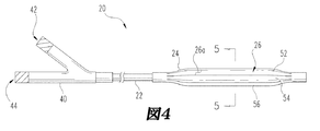
ここで図1〜5を参照すると、本発明の実施形態による薬物送達バルーンカテーテル20の一実施形態が示されている。バルーンカテーテル20は、カテーテルシャフト22と、その上に取り付けられたバルーン24とを備える。本明細書に記載の薬物と賦形剤とを含有する層26aを含む材料塗膜26は、バルーン24で担持される。カテーテルシャフト22は、第1のルーメン28と第2のルーメン30とを備える。ルーメン28はバルーン24が膨張できるように構成されており、ルーメン30は、バルーンカテーテル20と共に使用されるガイドワイヤ32または他のガイド部材を受け入れるように構成されている。バルーン24は、バルーン24を膨張させるための液体または他の流体を受け入れるように設計された内部領域34を備える。バルーン24は、バルーン内部34を画成する内壁36と外壁面38とを備える。層26は、バルーン24の外壁面38に接着している。
Referring now to FIGS. 1-5, one embodiment of a drug
バルーンカテーテル20はまた、シャフト22に取り付けられたカテーテルハブ40も備える。カテーテルハブ40は、バルーン膨張ルーメン28と流体連通する第1の開口部42と、シャフト22により画定されるルーメン30と流体連通する第2の開口部44とを画定する。ハブ40の開口部42とルーメン28は、バルーン24用の膨張流体が通過できるようにバルーン24の内部34への開口部46と連通する。ハブ40の開口部44とカテーテルシャフト22により画定されるルーメン30はルーメン30の遠位開口部48まで延び、遠位開口部48はバルーン24の遠位方向に配置されている。
図5を参照すると、図1〜4に示す特徴と共にさらに、バルーン24は、例えば、従来のバルーンフィルム、典型的には、前述のものの1つなどのポリマー材料製のバルーンフィルムにより画定されるバルーン壁50を備える。図4および図5に示すバルーン壁50は、バルーン24を動脈または静脈などの脈管に挿入する時に有用な折り畳まれた状態になっている。その折り畳まれた状態では、バルーン24は、ひだ52、54、56、58および60を備える。図示するように、ひだ52〜60は螺旋パターンに構成されており、ひだはそれぞれ湾曲した状態で、それらの下にあるカテーテルシャフト22の部分の周囲に延びており、ひだは重なり合い、それによりそれらの長さの少なくとも一部に沿って互いに接触している。この折り畳まれた構成では、ひだ52、54、56、58および60は、外側に露出したひだ表面52a、54a、56a、58a、および60aと、内側の露出していないひだ表面52b、54b、56b、58b、および60bとを備える。それに対応して、図示する実施形態ではコーティング層26aを備える材料塗膜26は、外側に露出したひだ表面52a〜60a上に配置された外側に露出した部分と、内側の露出していないひだ表面52b〜60b上に配置された内側の露出していない部分とを備える。また、この構成では、ひだ52〜60は互いに重なり合うため、材料塗膜26の部位ならびにその薬物および賦形剤26aは、材料塗膜26の他の部位ならびにその薬物および賦形剤26aと接触している。図5を参照すると、図5に示す特徴は説明を目的とするものであり、実際には、バルーン24は典型的には密にひだが形成され、カテーテルシャフト22の周囲に巻き付けられており、従ってひだ52〜60の内部には空間がほとんどまたは全くないことが分かるはずである。
Referring to FIG. 5, in addition to the features shown in FIGS. 1-4, the
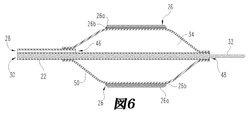
ここで図6を参照すると、材料塗膜26が本明細書に記載の薬物と賦形剤を含有する層である第1のコーティング層26aと、この層とは異なる、コーティング層26aの下に配置された第2のコーティング層26bとを備えること以外、図1〜5のバルーンカテーテル20のものと類似の特徴を有するバルーンカテーテルの別の実施形態が示されている。コーティング層26bは、特定の実施形態では、前述のポリマープライマー層であってもよい。
Referring now to FIG. 6, a
図7を参照すると、材料塗膜26が、バルーン24の外面38に直接接着した本明細書に記載の薬物と賦形剤とを含有する層である第1のコーティング層26aと、コーティング層26aの上に配置された第2のコーティング層26bとを備えること以外、図1〜5のバルーンカテーテル20と類似のバルーンカテーテルの別の実施形態が示されている。図7のコーティング層26bは、特定の実施形態では、ポリマー保護層であっても、および/または、拡散バリア層を通した薬物の放出を制御するように機能し得るポリマー拡散バリア層であってもよい。
Referring to FIG. 7, a
図5aおよび図5bは、材料塗膜26のコーティングパターンが異なること以外、図1〜5に示すものと類似のバルーンカテーテル実施形態を示す。特に図5aでは、薬物と賦形剤とを含有する層26aを含む材料塗膜26は、ひだ52〜60の外側に露出した面52a〜60aだけで担持される。この形態は、例えば、折り畳み時に、外側に露出したひだ表面52a〜60aとして配置されるバルーン24の選択された表面領域を膨張した状態の時にコーティングすることにより、または、外側に露出したひだ表面52a〜60aだけをコーティングする折り畳みおよびコーティング条件下で、バルーン24を折り畳まれた状態の時にコーティングすることにより製造することができる。図5bは、材料塗膜26が内側の露出していないひだ表面52b〜60bだけで担持されているバルーンカテーテル実施形態を開示する。この形態は、例えば、折り畳み時に、内側の露出していないひだ表面52b〜60bとして配置されるバルーン24の選択された表面領域を膨張した状態のときにコーティングすることにより、または、膨張した状態の時にバルーン24の周囲全体にコーティングし、バルーンのひだを形成してバルーンを折り畳んだ後、外側に露出したひだ表面52a〜60aの材料塗膜26部分を、例えば、機械的におよび/または材料塗膜26を取り除く(displace)ことができる溶媒または他の媒体を用いて除去することにより製造することができる。図5aおよび図5bの実施形態の層26aを、本明細書に記載の方法のいずれかを用いることを含む任意の好適な方法で塗布することができる。同様に、図5aおよび図5bの実施形態の材料塗膜26は、他の実施形態では、図6および図7に関連して図示し、記載するものを含む、本明細書に図示し、記載するものなどの多層コーティングであってもよい。
FIGS. 5a and 5b show a balloon catheter embodiment similar to that shown in FIGS. 1-5 except that the coating pattern of the
図8は、本発明の別の実施形態を示す。ステント70は、中心ルーメン74を画定するステント本体72を備える。ステント本体72は、ルーメン74の周囲の周囲通路を画定するストラットを備える長手方向で隣接する複数のセグメント76と、隣接セグメント76を連結するある一定のパターンの連結ストラットセグメント78とを備える。薬物と賦形剤とを含有する層86aを含むコーティング86(展開断面図、右下参照)は、ステント70の表面で担持される。図示する実施形態では、層86aはステント72の表面に唯一のコーティングとして直接接着しているが、他の実施形態では、ステントコーティング86は、図6および図7に関連して図示し、記載するコーティングを含む本明細書に図示し、記載するものなどの多層コーティングであってもよい。ステント72は、患者の動脈壁または静脈壁などの脈管壁と接触するように構成されたストラット外面80を有する。ステント72は、外面80の反対側にあり、ルーメン74に略面するストラット内面82を有する。ステント72はまた、ストラット外面80とストラット内面82との間にストラット側壁面84も有する。ストラット外面80は、ステント72の外面の少なくとも一部で材料塗膜86を担持し、特定の実施形態では、ステント72の外面全体または実質的に外面全体で担持する。ステント72は、望ましくは自己拡張型ステントであり、好ましくは弾性金属製、好ましくは、例えば、Cook Medical,Bloomington,Indiana,USAから市販されているZILVER(登録商標)ニチノールステントに見られるような、超弾性ニッケル−チタン(Ni−Ti)合金などの超弾性金属合金製である。ステント72は、ステントについてまたは他の点で本明細書に開示されている方法および材料を使用して製造することができ、層86aおよびステント上に存在する他の任意のコーティングは本明細書に教示される任意の組成物を有してもよく、本明細書に開示するもののいずれかを含む任意の好適な方法でステント72上に組み込んでもよい。
FIG. 8 shows another embodiment of the present invention.
図9は、本発明の別の実施形態を示す。図9の埋め込み型医療用デバイス20’は、バルーン24上にバルーン拡張型ステント90も取り付けられていること以外、図4に示すものに類似している。ステント90は近位端92と遠位端94とを有する。ステントがバルーン上に取り付けられている前述のまたは他のバルーンカテーテルでは、ステント90、バルーン24、またはこの両方が、薬物と賦形剤とを含有する層をその表面に担持してもよい。図示する実施形態20’では、ステント90は、その外面で担持される薬物と賦形剤とを含有する層96aを含む材料塗膜96を有し、バルーン24は、バルーン24の表面で担持される薬物と賦形剤とを含有する層26aを含む材料塗膜26を有する。ステント90の近位端92の近位方向とステント90の遠位端94の遠位方向に延びるように、コーティング層26aをバルーン24上に担持することができる。このようにして、動脈または静脈などの脈管内でバルーン24を膨張させ、脈管を拡張させて、ステント90を埋め込むとき、薬物と賦形剤とを含有する層の薬物を、ステント90の近位方向と遠位方向に延びる部位で脈管に塗布することができる。薬物が再狭窄抑制剤であるかまたはそれを含む場合、これにより、さもなければステント90の近位端92と遠位端94でまたはその近傍で生じる辺縁効果のために起こり得る再狭窄を抑制することができる。
FIG. 9 shows another embodiment of the present invention. The implantable
デバイス20’のバルーンカテーテルのバルーンおよび他の構成要素、ならびにステント90は、これらについてまたは他の点で本明細書に開示されている方法および材料を使用して製造することができる。材料塗膜26および/または材料塗膜96は、バルーン24またはステント90の表面にそれぞれ直接接着した唯一の層26aまたは96aを含むことができるが、他の実施形態では、バルーン材料塗膜26またはステント材料塗膜96は、図6および図7に関連して図示し、記載するコーティングを含む、本明細書に図示し、記載するものなどの多層コーティングであってもよい。薬物と賦形剤とを含有する層およびデバイス20’のバルーンおよび/またはステント上に存在する他の任意のコーティング層は、本明細書に教示する任意の組成物を有してもよく、本明細書に開示するもののいずれかを含む任意の好適な方法でバルーン24および/またはステント90上に組み込んでもよい。
The balloon and other components of the balloon catheter of device 20 ', as well as
図10および図11は、本発明の別の実施形態を示す。図10は、一実施形態による薬物送達スコアリングバルーンの側面図を記載している。図11は、拡張要素を含む、図10のバルーンの一部の拡大断面図を記載している。より具体的には、薬物送達スコアリングバルーンカテーテル100は、カテーテルシャフト102と、その上に取り付けられたバルーン104とを備える。バルーン104はそれに取り付けられており、好ましくはそのバルーン壁フィルム106と一体形成されており、複数の拡張要素108は、拡張要素108間にまたがるバルーン壁フィルム106に対して外向きに突出している。本明細書に記載の薬物と賦形剤とを含有する層110aを含む材料塗膜110は、バルーン104で担持され、示されている具体的実施形態では、バルーン壁フィルム106と拡張要素108の両方で担持されている。図示する拡張要素108は、トリゾイド(trizoid)形の要素であるが;他の形状も本発明の実施形態に使用するのに好適となり、拡張要素は、バルーン壁フィルムと一体形成されるのではなく、別々に取り付けられるまたは埋め込まれる物品または材料で設けることができる。カテーテルシャフト102は第1のルーメン112と第2のルーメン114とを備える。ルーメン112はバルーン104が膨張できるように構成され、ルーメン114はバルーンカテーテル100と共に使用されるガイドワイヤまたは他のガイド部材を受け入れるように構成されている。図示する実施形態では、薬物と賦形剤を含有する層110aはバルーン104の外壁面に、特にバルーン壁フィルム106および拡張要素108の外面に直接接着している。他の実施形態では、この層は、前述の多層コーティングのいずれかを含む、多層を含む材料塗膜の一部であってもよく、従って、薬物と賦形剤とを含有する層の下または上に他のコーティング層を有してもよいことが分かるであろう。追加で、または代わりに、薬物と賦形剤とを含有する層またはそれを組み込む材料塗膜はバルーン104の周囲全体に延び、拡張要素108と、拡張要素108間にまたがるバルーン壁フィルム106の両方をコーティングしてもよく(図10および図11のように)、またはバルーン104の選択的な部分をコーティングしてもよい。実例として、拡張要素108は、薬物と賦形剤とを含有する層またはそれを含む他の材料塗膜で完全にコーティングされてもまたは部分的にコーティングされてもよいが、拡張要素108間にまたがるバルーン壁フィルム106はコーティングされていなくても、または少なくとも薬物と賦形剤とを含有する層もしくはそれを含む他の材料塗膜を含まなくてもよい;または拡張要素108間にまたがるバルーン壁フィルム106は薬物と賦形剤とを含有する層またはそれを含む他の材料塗膜で完全にコーティングされてもまたは部分的にコーティングされてもよいが、拡張要素108はコーティングされていなくてもよい、または少なくとも薬物と賦形剤とを含有する層もしくはそれを含む他の材料塗膜を含まなくてもよい。前述のおよび他のコーティング構成も本明細書で好適になるであろう。同様に、バルーン104は膨張した状態で示されているが、本明細書の実施形態は、例えば、折り畳まれた状態のもののいずれかを含む折り畳まれた状態のバルーン104、および、前述の、それにより提供される構造特徴も含むことが分かるであろう。
10 and 11 show another embodiment of the present invention. FIG. 10 describes a side view of a drug delivery scoring balloon according to one embodiment. FIG. 11 describes an enlarged cross-sectional view of a portion of the balloon of FIG. 10 including an expansion element. More specifically, the drug delivery scoring
以下の実施例で本発明を説明する。本明細書に記載の実施例および実施形態は本発明を説明する目的で記載されているに過ぎず、本発明の範囲から逸脱することなく、それに鑑みた修正および変更が当業者に示唆される。 The following examples illustrate the invention. The examples and embodiments described herein are provided merely to illustrate the present invention, and modifications and changes in light of this will be suggested to those skilled in the art without departing from the scope of the present invention. .
実施例1:バルーンカテーテルの表面へのパクリタキセルと添加剤とのコーティング
パクリタキセルとタンニン酸をパクリタキセル対タンニン酸の比1:2でバルーンカテーテルにコーティングする。バルーンにコーティングする最終目標用量はバルーン1つ当たり約1,300μg、または約1.5μg/mm2である。
Example 1: Coating of paclitaxel and additives on the surface of a balloon catheter Paclitaxel and tannic acid are coated on a balloon catheter in a 1: 2 ratio of paclitaxel to tannic acid. The final target dose to coat the balloon is about 1,300 μg, or about 1.5 μg / mm 2 per balloon.
バルーンカテーテルコーティング用の作業用保存液を調製する。パクリタキセル66.6mgとタンニン酸133.33mgを22mLガラスバイアル内で秤量する。EtOH10mLを、薬物と添加剤が入ったバイアルに移し入れた後、溶液が均質になるまで音波処理する。典型的には、混合物を約10分間音波処理する。 Prepare a working stock solution for balloon catheter coating. Weigh 66.6 mg of paclitaxel and 133.33 mg of tannic acid in a 22 mL glass vial. After transferring 10 mL of EtOH to a vial containing the drug and additives, sonicate until the solution is homogeneous. Typically, the mixture is sonicated for about 10 minutes.
バルーン(Cook Advance 18LP 7mm×40mm,Cook Medical Incorporated)を、その長軸に沿ってバルーンを回転させることができる装置内で水平に保持する。バルーンが完全に拡張するようにバルーンを膨張させ、モータの電源を入れてバルーンの回転を開始する。100μLピペットを用いて、上記の作業液65μLを測定する。ピペットをバルーンの最近位表面に保持し、溶液をピペットからバルーン上にゆっくり放出する。コーティング中、溶液を絶えず放出しながら、ピペット先端をバルーンの表面に沿ってゆっくり引きずり動かす。コーティングの隙間や重なりが生じないように注意する。最初の65μLをバルーンの近位1/3にコーティングする。バルーンの中央部分および遠位1/3部分も同様にコーティングする。 A balloon (Cook Advance 18LP 7 mm x 40 mm, Cook Medical Incorporated) is held horizontally in an apparatus that can rotate the balloon along its long axis. The balloon is inflated so that the balloon is fully expanded, the motor is turned on and the balloon begins to rotate. Using a 100 μL pipette, measure 65 μL of the above working fluid. Hold the pipette on the proximal surface of the balloon and slowly release the solution from the pipette onto the balloon. During coating, the pipette tip is slowly dragged along the surface of the balloon while constantly releasing the solution. Be careful not to create coating gaps or overlaps. The first 65 μL is coated on the proximal third of the balloon. The central and distal 1/3 portions of the balloon are coated similarly.
上記工程を2回繰り返してバルーンカテーテルの残りの部分をコーティングする。最初の薬物コーティングの後、少量のEtOH液滴を使用して、コーティングを均一にし、溶液バイアルとピペット先端を洗浄して、薬物が全てバルーンにコーティングされたことを確実にすることができる。溶液の全量を表面にコーティングした後、エタノールを5分間以下蒸発させ、この時、コーティングは結晶化して白色になる。バルーンが乾燥した後、それを収縮させ、回転装置から取り出し、折り畳み、被覆する。 The above process is repeated twice to coat the remaining portion of the balloon catheter. After the initial drug coating, a small amount of EtOH droplets can be used to homogenize the coating and wash the solution vial and pipette tip to ensure that all drug is coated on the balloon. After coating the entire surface of the solution, the ethanol is allowed to evaporate for less than 5 minutes, at which time the coating crystallizes and becomes white. After the balloon is dry, it is deflated, removed from the rotating device, folded and covered.
類似のバルーンを膨張させ、スプレーコーティング法を用いてコーティングする。簡潔には、膨張したバルーンの表面にスプレーガンを用いて保存液をスプレーし、溶媒(例えば、エタノール)を蒸発させる。バルーンが乾燥した後、それを収縮させ、回転装置から取り出し、折り畳み、被覆する。 A similar balloon is inflated and coated using a spray coating method. Briefly, the stock solution is sprayed onto the surface of the inflated balloon using a spray gun to evaporate the solvent (eg, ethanol). After the balloon is dry, it is deflated, removed from the rotating device, folded and covered.
実施例2:組織取り込み試験
コーティングされたバルーンからブタ外腸骨内への薬物の取り込みを試験するために、フローループ試験を行う。フローループの準備は以下の通りである。ブタ外腸骨から過剰な脂肪および組織を除去し、それを長さ約6cmに切断する。翌日試験するために外腸骨を冷蔵庫で終夜保存した。
Example 2: Tissue uptake test To test drug uptake from the coated balloon into the porcine external iliac, a flow loop test is performed. The preparation of the flow loop is as follows. Excess fat and tissue is removed from the porcine external iliac and cut into approximately 6 cm lengths. The external iliac was stored in the refrigerator overnight for testing the next day.
フローループ試験回路を以下のように準備する。簡潔には、ブタ腸骨を冷蔵庫から取り出し、秤量する。ブタ腸骨の流入端を37Cのリン酸緩衝生理食塩水(PBS)約150mLが入ったリザーバに接続するのに必要な長さに黒色ネオプレンチューブを切断する。バルーンカテーテルを、チューブを通してブタ腸骨に挿入することができるように三方コネクタがチューブに存在する。 A flow loop test circuit is prepared as follows. Briefly, pig iliac is removed from the refrigerator and weighed. Cut the black neoprene tube to the length necessary to connect the inflow end of the porcine iliac to a reservoir containing approximately 150 mL of 37 C phosphate buffered saline (PBS). A three-way connector is present on the tube so that the balloon catheter can be inserted through the tube into the porcine iliac bone.
ブタ腸骨の流出端をネオプレンチューブで接続し、リザーバへの復路を形成する。例えば、250mlブフナーフラスコにPBSを収容することができる。これにより、PBSを循環させるためのループが完成する。ブタ腸骨の側枝を封止した後、それを、37Cの9%生理食塩水約1500mLが入った組織浴に浸漬する。 Connect the outflow end of the porcine iliac with a neoprene tube to form a return path to the reservoir. For example, PBS can be contained in a 250 ml Buchner flask. Thereby, a loop for circulating PBS is completed. After sealing the side branch of the porcine iliac, it is immersed in a tissue bath containing approximately 1500 mL of 37C 9% saline.
PBSを480ml/分で循環するように蠕動ポンプを設定し、電源を入れて、PBSがスムーズに流れ、ループ内のどこにも、とりわけ組織に漏れがないか試験する。試験を開始するために、ダイレータとflexorシースを、三方コネクタを通して挿入し、ブタ腸骨内の膨張部位に到達させる。ダイレータを抜去する。膨張デバイスに水道水約20mLを充填し、実施例1の手順を用いてコーティングされたバルーンの膨張ルーメンに接続する。バルーンを脱気してシースに入れ、端部まで40秒で移動させる。この時間は、バルーンがin vivoで治療部位に到達するのに通常要する時間を模倣するように選択する。40秒後、flexorを引き戻してバルーンを組織に接触させる。20秒(合計60秒)後、膨張デバイスを用いてバルーンを8atmまで膨張させる。その後、蠕動ポンプの電源を切る。 Set the peristaltic pump to circulate the PBS at 480 ml / min, turn on the power, and test that the PBS flows smoothly and that there is no leaking anywhere in the loop, especially the tissue. To begin the test, a dilator and flexor sheath are inserted through a three-way connector to reach the expansion site in the porcine iliac. Remove the dilator. Fill the inflation device with about 20 mL of tap water and connect to the inflation lumen of the coated balloon using the procedure of Example 1. The balloon is evacuated and placed in the sheath and moved to the end in 40 seconds. This time is chosen to mimic the time normally required for the balloon to reach the treatment site in vivo. After 40 seconds, the flexor is pulled back to bring the balloon into contact with the tissue. After 20 seconds (60 seconds total), the balloon is inflated to 8 atm using an inflation device. Then turn off the peristaltic pump.
8atmに5分間維持しながら、バルーン用と組織投与用にバイアルをEtOH10mLで調製する。8atmで5分後、バルーンをflexorから抜去する。バルーンをカテーテルから切り離し、EtOHの入ったバイアルに入れる。 While maintaining at 8 atm for 5 minutes, vials are prepared with 10 mL EtOH for balloon and tissue administration. After 5 minutes at 8 atm, the balloon is removed from the flexor. The balloon is disconnected from the catheter and placed in a vial containing EtOH.
蠕動ポンプの電源を入れ、1分間流れを監視し、組織に固着していない粒子状物質を全て洗い流す。1分後、ポンプの電源を切り、ブタ腸骨を除去する。腸骨を2cmの断片に切断し、それを調製したバイアルに入れる。循環PBS溶液1mLをピペットで取りHPLCバイアルに入れる。試験終了後、バイアルを37°の振盪インキュベータで4時間処理した後、0.45ミクロンのPVDFフィルタで濾過する。全ての試料(バルーン、PBS、および組織)を、組織抽出終了後にHPLCで分析した。
Turn on the peristaltic pump and monitor the flow for 1 minute to wash away any particulate matter not adhered to the tissue. After 1 minute, the pump is turned off and the porcine iliac is removed. Cut the iliac into 2 cm pieces and place them in prepared vials.
図12は、上記方法を用いて得られたフローループ結果を示す。バルーンは、実施例1に記載のものと類似の方法を用いてパクリタキセルとタンニン酸との混合物でコーティングする。パクリタキセル対タンニン酸の比は1:2または1:3である。コーティングは、滴下コーティング(1:2 ♯1D)またはスプレーコーティング(1:3、1:2♯1S、1:2♯2S)を用いて行う。薬物の組織取り込みを、バルーン上の初期薬物のパーセンテージとして示す。比較のため、市販の薬物コーティングされたバルーン(「OTM DCB」)を用いて得られた取り込み率を示す。 FIG. 12 shows the flow loop results obtained using the above method. The balloon is coated with a mixture of paclitaxel and tannic acid using a method similar to that described in Example 1. The ratio of paclitaxel to tannic acid is 1: 2 or 1: 3. Coating is performed using drop coating (1: 2 # 1D) or spray coating (1: 3, 1: 2 # 1S, 1: 2 # 2S). Drug tissue uptake is shown as a percentage of the initial drug on the balloon. For comparison, the uptake rates obtained using a commercially available drug-coated balloon (“OTM DCB”) are shown.
実施例3:耐久性試験
バルーン送達および展開の様々な段階におけるバルーンカテーテルからの薬物損失量を試験するために耐久性試験を行う。折り畳み中、膨張部位への送達中、および膨張時の薬物損失を試験する。折り畳みによる薬物損失を試験するために、バルーンをコーティングし、自然に収縮させて折り目を再形成し、真空引きし、バルーン上にバルーンカバーを配置する。滴下コーティング法またはスプレーコーティング法を用い、実施例1に記載の一般的な方法に従ってコーティングを行う。約24時間後、バルーンカバーを取り外し、バルーンを膨張させる。次いで、バルーンを容器に入れ、100%エタノールで洗浄する。バルーン上に残存する薬物の量をHPLCで定量する。
Example 3: Durability Test A durability test is performed to test the amount of drug loss from the balloon catheter at various stages of balloon delivery and deployment. Test drug loss during folding, delivery to the inflation site, and during inflation. In order to test drug loss due to folding, the balloon is coated and allowed to contract naturally to reshape the fold, evacuate, and place a balloon cover over the balloon. Coating is performed according to the general method described in Example 1 using a drop coating method or a spray coating method. After about 24 hours, the balloon cover is removed and the balloon is inflated. The balloon is then placed in a container and washed with 100% ethanol. The amount of drug remaining on the balloon is quantified by HPLC.
治療部位へのバルーンの送達中の薬物損失を試験するために、フローループの準備を、ブタ血管の代わりにシリコーンチューブを使用すること以外、実施例2と同様に行う。この実験ではPBS浴を使用しない。流体の流動を開始した後、バルーンをシリコーンチューブの端部まで、チューブから延び出さないように移動させる。in vivoでのバルーンの移動時間(約40秒)と同等の時間をかけてこのプロセスを行う。適切な時間が経過した後、フローループを遮断し、バルーンをチューブから押し出して空気に曝す。この時点で、バルーンカテーテルの端部を切り離し、HPLCで薬物を定量するためにバルーンを100%エタノールのバイアルに入れる。 To test drug loss during delivery of the balloon to the treatment site, the flow loop is prepared as in Example 2, except that a silicone tube is used instead of the porcine blood vessel. No PBS bath is used in this experiment. After initiating fluid flow, the balloon is moved to the end of the silicone tube so that it does not extend out of the tube. This process is performed for a time equivalent to the in vivo balloon travel time (approximately 40 seconds). After an appropriate amount of time has elapsed, the flow loop is interrupted and the balloon is pushed out of the tube and exposed to air. At this point, the end of the balloon catheter is disconnected and the balloon is placed in a 100% ethanol vial for drug quantification by HPLC.
バルーンの膨張中の薬物損失を試験するために、送達中の薬物損失の試験に使用するのと同じ準備を使用する。約40秒後、シリコーンチューブ内でバルーンを膨張させ、流れを遮断する。極短時間後にバルーンを収縮させ、シリコーンチューブの端部から押し出し、バルーンを切り離し、HPLCで薬物を定量するためにバルーンを100%エタノールのバイアルに入れる。 To test drug loss during balloon inflation, the same setup used for testing drug loss during delivery is used. After about 40 seconds, the balloon is inflated in the silicone tube to block the flow. After a very short time, the balloon is deflated, pushed out from the end of the silicone tube, the balloon is detached, and the balloon is placed in a 100% ethanol vial for HPLC quantification of the drug.
図13は、滴下(♯D)コーティングまたはスプレー(♯S)コーティングの耐久性に対するタンニン酸(TA)またはEGCGの効果を示す。1:2(パクリタキセル:タンニン酸またはEGCG)の比の方が、1:25または1:100(パクリタキセル:タンニン酸)のコーティングと比較して耐久性が良好であり、バルーン送達および膨張中の薬物損失が少ない。 FIG. 13 shows the effect of tannic acid (TA) or EGCG on drip (#D) or spray (#S) coating durability. The ratio of 1: 2 (paclitaxel: tannic acid or EGCG) is more durable compared to 1:25 or 1: 100 (paclitaxel: tannic acid) coatings and drugs during balloon delivery and inflation There is little loss.
特定の例示的実施形態を参照して、本発明を説明し、例証してきたが、本発明はこれらの例示的実施形態に限定されるものではない。以下の特許請求の範囲により定義される本発明の真の範囲および精神から逸脱することなく変更および修正を行い得ることが当業者には分かるであろう。従って、添付の特許請求の範囲およびその均等範囲に入る全てのこのような変更および修正は本発明に含まれるものとする。 Although the invention has been described and illustrated with reference to specific exemplary embodiments, the invention is not limited to these exemplary embodiments. Those skilled in the art will recognize that changes and modifications can be made without departing from the true scope and spirit of the invention as defined by the following claims. Accordingly, it is intended to embrace all such changes and modifications that fall within the scope of the appended claims and their equivalents.
Claims (20)
薬物および賦形剤を含む前記表面上のコーティングであって、前記賦形剤により封入された前記薬物を含む複数の粒子を含み、前記賦形剤がガレート含有化合物、エピガロカテキンガレート、タンニン酸およびエピカテキンガレートからなる群から選択されるコーティングと、
を含む医療用デバイス。 A base structure having a surface;
A coating on the surface comprising a drug and an excipient comprising a plurality of particles comprising the drug encapsulated by the excipient, the excipient comprising a gallate-containing compound, epigallocatechin gallate, tannic acid And a coating selected from the group consisting of epicatechin gallate;
Including medical devices.
表面を有する基部構造と、薬物および賦形剤を含む前記表面上のコーティングとを含む医療用デバイスを患者の脈管壁に接触させる工程であって、前記薬物と賦形剤が薬物対賦形剤の重量比10:1〜1:10で存在し、前記賦形剤がガレート含有化合物、エピガロカテキンガレート、タンニン酸およびエピカテキンガレートからなる群から選択される工程と、
前記薬物を前記患者の組織に送達するのに十分な時間、前記デバイスを前記脈管壁と接触した状態に維持する工程と、
を含む方法。 A method of delivering a drug locally to a patient's tissue, comprising:
Contacting a medical device comprising a base structure having a surface and a coating on the surface comprising a drug and an excipient with a vessel wall of a patient, wherein the drug and the excipient are drug-adapted Wherein the excipient is present in a weight ratio of 10: 1 to 1:10 and the excipient is selected from the group consisting of gallate-containing compounds, epigallocatechin gallate, tannic acid and epicatechin gallate;
Maintaining the device in contact with the vessel wall for a time sufficient to deliver the drug to the patient's tissue;
Including methods.
埋め込み型医療用デバイスを前記患者の体内に埋め込む工程であって、前記埋め込み型医療用デバイスが、表面を有する基部構造と、薬物および賦形剤を含む前記表面上のコーティングとを含み、前記薬物と賦形剤が薬物対賦形剤の重量比10:1〜1:10で存在する工程と、
治療有効量の前記薬物を前記患者の組織に送達するのに十分な時間、前記埋め込み型医療用デバイスを前記患者の体内に維持する工程と、
を含む方法。 A method of treating a patient suffering from or having symptoms.
Implanting an implantable medical device into the patient's body, the implantable medical device comprising a base structure having a surface and a coating on the surface comprising a drug and an excipient, the drug And the excipient is present in a drug to excipient weight ratio of 10: 1 to 1:10;
Maintaining the implantable medical device within the patient for a time sufficient to deliver a therapeutically effective amount of the drug to the patient's tissue;
Including methods.
Applications Claiming Priority (2)
| Application Number | Priority Date | Filing Date | Title |
|---|---|---|---|
| US14/454,345 | 2014-08-07 | ||
| US14/454,345 US9655998B2 (en) | 2014-08-07 | 2014-08-07 | Encapsulated drug compositions and methods of use thereof |
Related Child Applications (1)
| Application Number | Title | Priority Date | Filing Date |
|---|---|---|---|
| JP2017111242A Division JP6533253B2 (en) | 2014-08-07 | 2017-06-06 | Encapsulated drug composition and method of using the same |
Publications (2)
| Publication Number | Publication Date |
|---|---|
| JP2016036730A true JP2016036730A (en) | 2016-03-22 |
| JP6157543B2 JP6157543B2 (en) | 2017-07-05 |
Family
ID=53765138
Family Applications (2)
| Application Number | Title | Priority Date | Filing Date |
|---|---|---|---|
| JP2015153847A Active JP6157543B2 (en) | 2014-08-07 | 2015-08-04 | Encapsulated drug composition and method of use thereof |
| JP2017111242A Active JP6533253B2 (en) | 2014-08-07 | 2017-06-06 | Encapsulated drug composition and method of using the same |
Family Applications After (1)
| Application Number | Title | Priority Date | Filing Date |
|---|---|---|---|
| JP2017111242A Active JP6533253B2 (en) | 2014-08-07 | 2017-06-06 | Encapsulated drug composition and method of using the same |
Country Status (3)
| Country | Link |
|---|---|
| US (3) | US9655998B2 (en) |
| EP (1) | EP2982389B1 (en) |
| JP (2) | JP6157543B2 (en) |
Cited By (13)
| Publication number | Priority date | Publication date | Assignee | Title |
|---|---|---|---|---|
| JP2019523032A (en) * | 2017-05-05 | 2019-08-22 | ウロトロニック・インコーポレイテッドUrotronic, Inc. | Drug-coated balloon catheter for body cavity |
| JP2019528973A (en) * | 2016-09-30 | 2019-10-17 | サーモディクス,インコーポレイテッド | Therapeutic balloon system with a deflation guide |
| US10668188B2 (en) | 2012-10-26 | 2020-06-02 | Urotronic, Inc. | Drug coated balloon catheters for nonvascular strictures |
| US10806830B2 (en) | 2012-10-26 | 2020-10-20 | Urotronic, Inc. | Drug-coated balloon catheters for body lumens |
| US10850076B2 (en) | 2012-10-26 | 2020-12-01 | Urotronic, Inc. | Balloon catheters for body lumens |
| US10881839B2 (en) | 2012-10-26 | 2021-01-05 | Urotronic, Inc. | Drug-coated balloon catheters for body lumens |
| US10898700B2 (en) | 2012-10-26 | 2021-01-26 | Urotronic, Inc. | Balloon catheters for body lumens |
| JP2021154169A (en) * | 2016-03-23 | 2021-10-07 | テルモ株式会社 | Balloon catheter and method for manufacturing the same |
| US11484628B2 (en) | 2015-04-24 | 2022-11-01 | Urotronic, Inc. | Drug coated balloon catheters for nonvascular strictures |
| US11504450B2 (en) | 2012-10-26 | 2022-11-22 | Urotronic, Inc. | Drug-coated balloon catheters for body lumens |
| US11730864B2 (en) | 2015-04-24 | 2023-08-22 | Urotronic, Inc. | Drug coated balloon catheters for nonvascular strictures |
| US11938287B2 (en) | 2012-10-26 | 2024-03-26 | Urotronic, Inc. | Drug-coated balloon catheters for body lumens |
| US11957853B2 (en) | 2019-02-22 | 2024-04-16 | Urotronic, Inc. | Drug-coated balloon catheters for body lumens |
Families Citing this family (17)
| Publication number | Priority date | Publication date | Assignee | Title |
|---|---|---|---|---|
| US11241520B2 (en) | 2014-08-07 | 2022-02-08 | Cook Medical Technologies Llc | Compositions and devices incorporating water-insoluble therapeutic agents and methods of the use thereof |
| US9655998B2 (en) | 2014-08-07 | 2017-05-23 | Cook Medical Technologies Llc | Encapsulated drug compositions and methods of use thereof |
| US9180226B1 (en) * | 2014-08-07 | 2015-11-10 | Cook Medical Technologies Llc | Compositions and devices incorporating water-insoluble therapeutic agents and methods of the use thereof |
| JP6754423B2 (en) * | 2016-03-23 | 2020-09-09 | テルモ株式会社 | Balloon catheter and its manufacturing method |
| JP6832339B2 (en) * | 2016-03-23 | 2021-02-24 | テルモ株式会社 | Balloon catheter and its manufacturing method and treatment method |
| US10758381B2 (en) | 2016-03-31 | 2020-09-01 | Vesper Medical, Inc. | Intravascular implants |
| US10849769B2 (en) | 2017-08-23 | 2020-12-01 | Vesper Medical, Inc. | Non-foreshortening stent |
| US10271977B2 (en) | 2017-09-08 | 2019-04-30 | Vesper Medical, Inc. | Hybrid stent |
| US11628076B2 (en) | 2017-09-08 | 2023-04-18 | Vesper Medical, Inc. | Hybrid stent |
| US11357650B2 (en) | 2019-02-28 | 2022-06-14 | Vesper Medical, Inc. | Hybrid stent |
| US11364134B2 (en) | 2018-02-15 | 2022-06-21 | Vesper Medical, Inc. | Tapering stent |
| US10500078B2 (en) | 2018-03-09 | 2019-12-10 | Vesper Medical, Inc. | Implantable stent |
| US20200038560A1 (en) * | 2018-07-31 | 2020-02-06 | Cook Medical Technologies Llc | Limus coatings and methods of use thereof |
| WO2020131227A1 (en) * | 2018-12-19 | 2020-06-25 | Teleflex Life Sciences Limited | Guide extension catheter |
| CN111671982A (en) * | 2020-05-08 | 2020-09-18 | 北京永益润成科技有限公司 | Medicinal coating composition and preparation method thereof |
| KR20210150657A (en) * | 2020-06-03 | 2021-12-13 | 주식회사 제노스 | Balloon catheter of drug-coated type |
| CN115531621B (en) * | 2022-10-09 | 2023-06-27 | 中南大学湘雅二医院 | Pharmaceutical composition containing glycyrrhizic acid for coronary artery drug eluting stent and controlled release system thereof |
Citations (3)
| Publication number | Priority date | Publication date | Assignee | Title |
|---|---|---|---|---|
| JP2008253707A (en) * | 2007-04-05 | 2008-10-23 | Bio Verde:Kk | Drug-eluting stent |
| US20120277719A1 (en) * | 2011-04-27 | 2012-11-01 | Massachusetts Institute Of Technology | Coating compositions, methods and coated devices |
| WO2013053809A1 (en) * | 2011-10-14 | 2013-04-18 | Innora Gmbh | Improved formulations for drug-coated medical devices |
Family Cites Families (44)
| Publication number | Priority date | Publication date | Assignee | Title |
|---|---|---|---|---|
| US5981568A (en) | 1993-01-28 | 1999-11-09 | Neorx Corporation | Therapeutic inhibitor of vascular smooth muscle cells |
| AU1821200A (en) | 1998-12-03 | 2000-06-19 | Scimed Life Systems, Inc. | Stent having drug crystals thereon |
| DE10115740A1 (en) | 2001-03-26 | 2002-10-02 | Ulrich Speck | Preparation for restenosis prophylaxis |
| US20030044474A1 (en) | 2001-08-03 | 2003-03-06 | Shaklee Corporation | High molecular weight, lipophilic, orally ingestible bioactive agents in formulations having improved bioavailability |
| US7153315B2 (en) * | 2002-06-11 | 2006-12-26 | Boston Scientific Scimed, Inc. | Catheter balloon with ultrasonic microscalpel blades |
| US20050019404A1 (en) * | 2003-06-30 | 2005-01-27 | Hsing-Wen Sung | Drug-eluting biodegradable stent |
| US20050163821A1 (en) | 2002-08-02 | 2005-07-28 | Hsing-Wen Sung | Drug-eluting Biodegradable Stent and Delivery Means |
| DE10244847A1 (en) | 2002-09-20 | 2004-04-01 | Ulrich Prof. Dr. Speck | Medical device for drug delivery |
| PL1653943T6 (en) | 2003-06-25 | 2009-02-27 | The Burnham Inst La Jolla | Catechol derivatives for the treatment of cancer |
| US20050064011A1 (en) | 2003-08-11 | 2005-03-24 | Young-Ho Song | Implantable or insertable medical devices containing phenolic compound for inhibition of restenosis |
| US20050037048A1 (en) | 2003-08-11 | 2005-02-17 | Young-Ho Song | Medical devices containing antioxidant and therapeutic agent |
| JP2007526793A (en) | 2003-11-07 | 2007-09-20 | ジーピー メディカル インコーポレイテッド | Biodegradable drug-eluting stent |
| US7262253B2 (en) | 2003-12-02 | 2007-08-28 | Labopharm, Inc. | Process for the preparation of amphiphilic poly (N-vinyl-2-pyrrolidone) block copolymers |
| JP2008509935A (en) * | 2004-08-13 | 2008-04-03 | アンジオテック インターナショナル アーゲー | Compositions and methods using hyaluronic acid and hyaluronic acid inhibitors |
| US7557182B2 (en) | 2005-02-18 | 2009-07-07 | Angiochem Inc. | Molecules for transporting a compound across the blood-brain barrier |
| KR20080008364A (en) | 2005-05-05 | 2008-01-23 | 헤모텍 아게 | All-over coating of vessel stents |
| US20070054868A1 (en) | 2005-06-20 | 2007-03-08 | The Trustees Of Columbia University In The City Of New York | Synergistic polyphenol compounds, compositions thereof, and uses thereof |
| US20070178138A1 (en) | 2006-02-01 | 2007-08-02 | Allergan, Inc. | Biodegradable non-opthalmic implants and related methods |
| US7875284B2 (en) * | 2006-03-10 | 2011-01-25 | Cook Incorporated | Methods of manufacturing and modifying taxane coatings for implantable medical devices |
| US20080103103A1 (en) | 2006-10-30 | 2008-05-01 | Bahram Memarzadeh | Reagents and methods to treat ocular diseases and infection |
| US8425459B2 (en) | 2006-11-20 | 2013-04-23 | Lutonix, Inc. | Medical device rapid drug releasing coatings comprising a therapeutic agent and a contrast agent |
| US8414909B2 (en) * | 2006-11-20 | 2013-04-09 | Lutonix, Inc. | Drug releasing coatings for medical devices |
| EP2224991B1 (en) | 2007-11-30 | 2018-02-21 | Cook Medical Technologies LLC | Method and device for vascular therapy |
| AU2009234283B2 (en) | 2008-04-11 | 2015-04-02 | Board Of Supervisors Of Louisiana State University And Agricultural And Mechanical College | Diterpene glycosides as natural solubilizers |
| US8642063B2 (en) | 2008-08-22 | 2014-02-04 | Cook Medical Technologies Llc | Implantable medical device coatings with biodegradable elastomer and releasable taxane agent |
| WO2010042555A2 (en) * | 2008-10-06 | 2010-04-15 | The Brigham And Women's Hospital, Inc. | Particles with multiple functionalized surface domains |
| EP2337561A4 (en) | 2008-10-23 | 2012-04-04 | Davos Life Science Pte Ltd | Use of tocotrienol composition for the prevention of cancer |
| US20100233228A1 (en) | 2009-03-12 | 2010-09-16 | Invatec Technology Center Gmbh | Drug-Eluting Medical Device |
| US20100261662A1 (en) | 2009-04-09 | 2010-10-14 | Endologix, Inc. | Utilization of mural thrombus for local drug delivery into vascular tissue |
| EP2421571A2 (en) | 2009-04-24 | 2012-02-29 | Boston Scientific Scimed, Inc. | Use of drug polymorphs to achieve controlled drug delivery from a coated medical device |
| US20100285085A1 (en) | 2009-05-07 | 2010-11-11 | Abbott Cardiovascular Systems Inc. | Balloon coating with drug transfer control via coating thickness |
| EP2451496B1 (en) | 2009-07-10 | 2015-07-22 | Boston Scientific Scimed, Inc. | Use of nanocrystals for a drug delivery balloon |
| EP2962707B1 (en) | 2009-07-17 | 2019-07-24 | Boston Scientific Scimed, Inc. | Drug delivery balloons with improved crystal size and density |
| US8753687B2 (en) | 2009-08-11 | 2014-06-17 | Agency For Science, Technology And Research | Particulate hyaluronic acid/flavonoid suspensions for cellular delivery of bioactive agents |
| CA2814237A1 (en) | 2009-10-15 | 2011-04-21 | Board Of Supervisors Of Louisiana State University And Agricultural And Mechanical College | Water soluble drug-solubilizer powders and their uses |
| US20110144577A1 (en) | 2009-12-11 | 2011-06-16 | John Stankus | Hydrophilic coatings with tunable composition for drug coated balloon |
| SG10201501829YA (en) | 2010-03-11 | 2015-05-28 | Agency Science Tech & Res | Anti-cancer agent delivery vehicles capable of improved loading |
| EP2380605A1 (en) * | 2010-04-19 | 2011-10-26 | InnoRa Gmbh | Improved formulations for drug-coated medical devices |
| JP2014516695A (en) * | 2011-05-18 | 2014-07-17 | バトリックス・メディカル・インコーポレイテッド | Coated balloon for vascular stabilization |
| US20120316633A1 (en) | 2011-06-07 | 2012-12-13 | Boston Scientific Scimed, Inc. | Durable Stent Drug Eluting Coating |
| KR20170007780A (en) | 2014-05-09 | 2017-01-20 | 에이전시 포 사이언스, 테크놀로지 앤드 리서치 | A micellar nanocomplex |
| US9180226B1 (en) | 2014-08-07 | 2015-11-10 | Cook Medical Technologies Llc | Compositions and devices incorporating water-insoluble therapeutic agents and methods of the use thereof |
| US9655998B2 (en) | 2014-08-07 | 2017-05-23 | Cook Medical Technologies Llc | Encapsulated drug compositions and methods of use thereof |
| WO2017072545A1 (en) | 2015-10-30 | 2017-05-04 | Acotec Scientific Co. Ltd. | Drug coated balloon catheter arteriovenous shunt |
-
2014
- 2014-08-07 US US14/454,345 patent/US9655998B2/en active Active
-
2015
- 2015-07-30 EP EP15179160.5A patent/EP2982389B1/en active Active
- 2015-08-04 JP JP2015153847A patent/JP6157543B2/en active Active
-
2017
- 2017-04-25 US US15/496,466 patent/US10029033B2/en active Active
- 2017-06-06 JP JP2017111242A patent/JP6533253B2/en active Active
-
2018
- 2018-06-06 US US16/001,468 patent/US20180280583A1/en not_active Abandoned
Patent Citations (3)
| Publication number | Priority date | Publication date | Assignee | Title |
|---|---|---|---|---|
| JP2008253707A (en) * | 2007-04-05 | 2008-10-23 | Bio Verde:Kk | Drug-eluting stent |
| US20120277719A1 (en) * | 2011-04-27 | 2012-11-01 | Massachusetts Institute Of Technology | Coating compositions, methods and coated devices |
| WO2013053809A1 (en) * | 2011-10-14 | 2013-04-18 | Innora Gmbh | Improved formulations for drug-coated medical devices |
Non-Patent Citations (1)
| Title |
|---|
| BIOCHEMICAL AND BIOPHYSICAL RESEARCH COMMUNICATION, vol. Vol. 391, JPN6016030955, 2010, pages p. 716-721 * |
Cited By (26)
| Publication number | Priority date | Publication date | Assignee | Title |
|---|---|---|---|---|
| US11439801B2 (en) | 2012-10-26 | 2022-09-13 | Urotronic, Inc. | Balloon catheters for body lumens |
| US10668188B2 (en) | 2012-10-26 | 2020-06-02 | Urotronic, Inc. | Drug coated balloon catheters for nonvascular strictures |
| US11471655B2 (en) | 2012-10-26 | 2022-10-18 | Urotronic, Inc. | Drug-coated balloon catheters for body lumens |
| US11471656B2 (en) | 2012-10-26 | 2022-10-18 | Urotronic, Inc. | Drug-coated balloon catheters for body lumens |
| US10850076B2 (en) | 2012-10-26 | 2020-12-01 | Urotronic, Inc. | Balloon catheters for body lumens |
| US10881839B2 (en) | 2012-10-26 | 2021-01-05 | Urotronic, Inc. | Drug-coated balloon catheters for body lumens |
| US10898700B2 (en) | 2012-10-26 | 2021-01-26 | Urotronic, Inc. | Balloon catheters for body lumens |
| US10987451B2 (en) | 2012-10-26 | 2021-04-27 | Urotronic, Inc. | Drug-coated balloon catheters for body lumens |
| US10994104B2 (en) | 2012-10-26 | 2021-05-04 | Urotronic, Inc. | Balloon catheters for body lumens |
| US10994103B2 (en) | 2012-10-26 | 2021-05-04 | Urotronic, Inc. | Drug-coated balloon catheters for body lumens |
| US11938287B2 (en) | 2012-10-26 | 2024-03-26 | Urotronic, Inc. | Drug-coated balloon catheters for body lumens |
| US11925729B2 (en) | 2012-10-26 | 2024-03-12 | Urotronic, Inc. | Drug-coated balloon catheters for body lumens |
| US11826532B2 (en) | 2012-10-26 | 2023-11-28 | Urotronic, Inc. | Balloon catheters for body lumens |
| US11826533B2 (en) | 2012-10-26 | 2023-11-28 | Urotronic, Inc. | Balloon catheters for body lumens |
| US10806830B2 (en) | 2012-10-26 | 2020-10-20 | Urotronic, Inc. | Drug-coated balloon catheters for body lumens |
| US11648338B2 (en) | 2012-10-26 | 2023-05-16 | Urotronic, Inc. | Drug-coated balloon catheters for body lumens |
| US11504450B2 (en) | 2012-10-26 | 2022-11-22 | Urotronic, Inc. | Drug-coated balloon catheters for body lumens |
| US11648337B2 (en) | 2012-10-26 | 2023-05-16 | Urotronic, Inc. | Drug-coated balloon catheters for body lumens |
| US11484628B2 (en) | 2015-04-24 | 2022-11-01 | Urotronic, Inc. | Drug coated balloon catheters for nonvascular strictures |
| US11730864B2 (en) | 2015-04-24 | 2023-08-22 | Urotronic, Inc. | Drug coated balloon catheters for nonvascular strictures |
| US11904072B2 (en) | 2015-04-24 | 2024-02-20 | Urotronic, Inc. | Drug coated balloon catheters for nonvascular strictures |
| JP7118222B2 (en) | 2016-03-23 | 2022-08-15 | テルモ株式会社 | Balloon catheter and manufacturing method thereof |
| JP2021154169A (en) * | 2016-03-23 | 2021-10-07 | テルモ株式会社 | Balloon catheter and method for manufacturing the same |
| JP2019528973A (en) * | 2016-09-30 | 2019-10-17 | サーモディクス,インコーポレイテッド | Therapeutic balloon system with a deflation guide |
| JP2019523032A (en) * | 2017-05-05 | 2019-08-22 | ウロトロニック・インコーポレイテッドUrotronic, Inc. | Drug-coated balloon catheter for body cavity |
| US11957853B2 (en) | 2019-02-22 | 2024-04-16 | Urotronic, Inc. | Drug-coated balloon catheters for body lumens |
Also Published As
| Publication number | Publication date |
|---|---|
| US20180280583A1 (en) | 2018-10-04 |
| EP2982389A1 (en) | 2016-02-10 |
| JP6157543B2 (en) | 2017-07-05 |
| US20170224878A1 (en) | 2017-08-10 |
| JP6533253B2 (en) | 2019-06-19 |
| US10029033B2 (en) | 2018-07-24 |
| JP2017170186A (en) | 2017-09-28 |
| EP2982389B1 (en) | 2019-05-29 |
| US9655998B2 (en) | 2017-05-23 |
| US20160038648A1 (en) | 2016-02-11 |
Similar Documents
| Publication | Publication Date | Title |
|---|---|---|
| JP6157543B2 (en) | Encapsulated drug composition and method of use thereof | |
| AU2013259639B2 (en) | Coated medical devices comprising a water - insoluble therapeutic agent and an additive | |
| JP4347044B2 (en) | Device for delivering a therapeutic agent having a variable release profile | |
| US20030033007A1 (en) | Methods and devices for delivery of therapeutic capable agents with variable release profile | |
| JP6573910B2 (en) | Balloon catheter for treatment of peripheral artery disease | |
| US20130302381A1 (en) | Implantable Medical Devices Including a Water-Insoluble Therapeutic Agent | |
| JP5480272B2 (en) | Medical device for drug delivery | |
| US9155648B2 (en) | Stent delivery system and method using the same | |
| WO2004064910A1 (en) | Indwelling stent | |
| US20120283624A1 (en) | Drug Eluting Device and Method of Use Thereof | |
| EP3603688A1 (en) | Limus coatings and methods of use thereof | |
| US11173235B2 (en) | Nitrite eluting devices and methods of use thereof | |
| JP2003093520A (en) | Stent | |
| JP2003320036A (en) | Stent |
Legal Events
| Date | Code | Title | Description |
|---|---|---|---|
| A131 | Notification of reasons for refusal |
Free format text: JAPANESE INTERMEDIATE CODE: A131 Effective date: 20160816 |
|
| A601 | Written request for extension of time |
Free format text: JAPANESE INTERMEDIATE CODE: A601 Effective date: 20161116 |
|
| A521 | Request for written amendment filed |
Free format text: JAPANESE INTERMEDIATE CODE: A523 Effective date: 20170116 |
|
| TRDD | Decision of grant or rejection written | ||
| A01 | Written decision to grant a patent or to grant a registration (utility model) |
Free format text: JAPANESE INTERMEDIATE CODE: A01 Effective date: 20170510 |
|
| A61 | First payment of annual fees (during grant procedure) |
Free format text: JAPANESE INTERMEDIATE CODE: A61 Effective date: 20170606 |
|
| R150 | Certificate of patent or registration of utility model |
Ref document number: 6157543 Country of ref document: JP Free format text: JAPANESE INTERMEDIATE CODE: R150 |
|
| R250 | Receipt of annual fees |
Free format text: JAPANESE INTERMEDIATE CODE: R250 |
|
| R250 | Receipt of annual fees |
Free format text: JAPANESE INTERMEDIATE CODE: R250 |
|
| R250 | Receipt of annual fees |
Free format text: JAPANESE INTERMEDIATE CODE: R250 |
|
| R250 | Receipt of annual fees |
Free format text: JAPANESE INTERMEDIATE CODE: R250 |