JP2015525890A - Matrix and system for storing biological specimens for qualitative and quantitative analysis - Google Patents
Matrix and system for storing biological specimens for qualitative and quantitative analysis Download PDFInfo
- Publication number
- JP2015525890A JP2015525890A JP2015526642A JP2015526642A JP2015525890A JP 2015525890 A JP2015525890 A JP 2015525890A JP 2015526642 A JP2015526642 A JP 2015526642A JP 2015526642 A JP2015526642 A JP 2015526642A JP 2015525890 A JP2015525890 A JP 2015525890A
- Authority
- JP
- Japan
- Prior art keywords
- matrix
- vivest
- samples
- sample
- plasma
- Prior art date
- Legal status (The legal status is an assumption and is not a legal conclusion. Google has not performed a legal analysis and makes no representation as to the accuracy of the status listed.)
- Pending
Links
Images


Classifications
-
- C—CHEMISTRY; METALLURGY
- C12—BIOCHEMISTRY; BEER; SPIRITS; WINE; VINEGAR; MICROBIOLOGY; ENZYMOLOGY; MUTATION OR GENETIC ENGINEERING
- C12Q—MEASURING OR TESTING PROCESSES INVOLVING ENZYMES, NUCLEIC ACIDS OR MICROORGANISMS; COMPOSITIONS OR TEST PAPERS THEREFOR; PROCESSES OF PREPARING SUCH COMPOSITIONS; CONDITION-RESPONSIVE CONTROL IN MICROBIOLOGICAL OR ENZYMOLOGICAL PROCESSES
- C12Q1/00—Measuring or testing processes involving enzymes, nucleic acids or microorganisms; Compositions therefor; Processes of preparing such compositions
- C12Q1/68—Measuring or testing processes involving enzymes, nucleic acids or microorganisms; Compositions therefor; Processes of preparing such compositions involving nucleic acids
- C12Q1/6806—Preparing nucleic acids for analysis, e.g. for polymerase chain reaction [PCR] assay
-
- A—HUMAN NECESSITIES
- A61—MEDICAL OR VETERINARY SCIENCE; HYGIENE
- A61B—DIAGNOSIS; SURGERY; IDENTIFICATION
- A61B10/00—Other methods or instruments for diagnosis, e.g. instruments for taking a cell sample, for biopsy, for vaccination diagnosis; Sex determination; Ovulation-period determination; Throat striking implements
- A61B10/0096—Casings for storing test samples
-
- G—PHYSICS
- G01—MEASURING; TESTING
- G01N—INVESTIGATING OR ANALYSING MATERIALS BY DETERMINING THEIR CHEMICAL OR PHYSICAL PROPERTIES
- G01N1/00—Sampling; Preparing specimens for investigation
- G01N1/28—Preparing specimens for investigation including physical details of (bio-)chemical methods covered elsewhere, e.g. G01N33/50, C12Q
- G01N1/40—Concentrating samples
- G01N1/4022—Concentrating samples by thermal techniques; Phase changes
- G01N2001/4027—Concentrating samples by thermal techniques; Phase changes evaporation leaving a concentrated sample
Abstract
【課題】本発明は、吸収性疎水性ポリオレフィンマトリックスを含むデバイス、システム、および使用方法、ならびに目的の検体を乾燥状態で含有する生体標本の液体懸濁液を貯蔵、保存、および回収するためのその使用方法を提供する。【解決手段】ポリオレフィンマトリックス上に吸収された目的の検体を含有する乾燥生体標本は、分子グレード水などで再構成され、ポリオレフィンマトリックスを圧縮することにより放出される。再構成された生体検体は、ウイルス核酸の定性および定量分析、そのようなウイルスの負荷試験、遺伝子型決定、および配列決定などの後次分析に適している。指示書付きのキット、および本発明の圧縮デバイスを使用して目的の検体を含有する生体標本を貯蔵、保存、および回収するためのその使用方法も提供される。【選択図】図8The present invention relates to a device, system, and method of use comprising an absorbent hydrophobic polyolefin matrix, and to store, preserve, and retrieve a liquid suspension of a biological specimen containing the analyte of interest in a dry state. Provide how to use it. A dry biological specimen containing an analyte of interest absorbed on a polyolefin matrix is reconstituted with molecular grade water or the like and released by compressing the polyolefin matrix. The reconstituted biological specimen is suitable for subsequent analysis such as qualitative and quantitative analysis of viral nucleic acids, such viral challenge, genotyping, and sequencing. Also provided are kits with instructions and methods of use thereof for storing, preserving, and retrieving biological specimens containing a specimen of interest using the compression device of the present invention. [Selection] Figure 8
Description
本発明は、概して、マトリックスを保存する生体標本、ならびにそれとともに使用するためのシステム、デバイス、および方法に関する。より具体的には、本発明は、ウイルス負荷、遺伝子型決定、および抗ウイルス薬耐性試験などの後次の定性および定量実験室分析のためのウイルスDNAおよびRNA標本などの核酸の収集、貯蔵、および回収のためのマトリックスおよびシステムに関する。 The present invention generally relates to biological specimens that store a matrix, and systems, devices, and methods for use therewith. More specifically, the present invention relates to the collection, storage, and storage of nucleic acids such as viral DNA and RNA specimens for subsequent qualitative and quantitative laboratory analysis such as viral load, genotyping, and antiviral drug resistance testing. And a matrix and system for recovery.
関連出願の相互参照
本出願は、2012年8月6日に出願された米国仮出願第61/680,193号の優先権を主張し、その全体は、参照により本明細書に組み込まれる。
CROSS REFERENCE TO RELATED APPLICATIONS This application claims priority to US Provisional Application No. 61 / 680,193, filed Aug. 6, 2012, which is hereby incorporated by reference in its entirety.
生体標本は、多くの場合、その中に含有される様々な検体のレベルおよび濃度の分析のために収集、輸送、および保管される。従来、生体標本の液体懸濁液は、冷蔵下で密封した気密管中に保管される。液体試料の収集、処理、輸送、および貯蔵は、それと関連付けられる多くの問題、例えば、遠隔収集センターにおける冷蔵(通常、ドライアイスによる)費用、試料の喪失を引き起こす容器破損または漏出のリスクおよび感染の危険性、出荷および貯蔵中の試料の不安定性、輸送キャリアによる液体バイオハザード出荷の受諾拒否、ならびに後次定性および定量分析の実験室方法に適合する量を保証するために適正な試料体積の収集を有する。上記問題に対処する費用は莫大である。 Biological specimens are often collected, transported, and stored for analysis of the level and concentration of the various analytes contained therein. Conventionally, a liquid suspension of a biological specimen is stored in an airtight tube sealed under refrigeration. The collection, processing, transport, and storage of liquid samples is associated with many of the problems associated with them, such as the cost of refrigeration (usually with dry ice) at remote collection centers, risk of container breakage or leakage causing sample loss and infection Collection of the correct sample volume to ensure risk, sample instability during shipment and storage, refusal to accept liquid biohazard shipments by transport carriers, and quantities compatible with post-qualitative and quantitative laboratory methods Have The cost of dealing with the above problems is enormous.
濾紙上の乾燥血斑(DBS)および乾燥血漿斑(DPS)試料採取は、液体試料採取手順の代替方法であり、世界中で使用されてある程度の成功をもたらしている。1980年代以降、Schleicher and Schuell Corp.、Bio−Rad、Boehringer Mannheim Corp.、およびWhatman,Inc.などの製造者は、DBSおよびDPS試料採取のための濾紙を生産している。これらの市販の生体試料採取濾紙システムを使用する際に、血斑または血漿斑を濾紙の1つ以上の指定領域に置き、乾燥させ、次いで試験申請書を添えて実験室に郵送する。Whatman 3MM、GF/CM30、GF/QA30、S&S 903、GB002、GB003、またはGB004などの一般に使用される濾紙は、当業者に既知である。例えば、S&S 903セルロース(木材または木綿由来)濾紙およびWhatmanガラス繊維濾紙といった血液標本収集のためのブロッティング物質のいくつかのカテゴリーが使用可能である。しかしながら、ある不利益がこれらの市販の濾紙と関連付けられた。具体的に、これらの市販の一般に使用されるある材料は、ある定性および定量バイオアッセイを実行するために好ましい精密な値および精度を提供する特徴を欠く。 Dry blood spot (DBS) and dry plasma spot (DPS) sampling on filter paper are alternative methods for liquid sampling procedures and have been used with some success around the world. Since the 1980s, Schleicher and Schuell Corp. Bio-Rad, Boehringer Mannheim Corp. , And Whatman, Inc. Manufacturers produce filter paper for DBS and DPS sampling. In using these commercially available biological sampling filter paper systems, blood spots or plasma spots are placed in one or more designated areas of the filter paper, allowed to dry, and then mailed to the laboratory with a test application. Commonly used filter papers such as Whatman 3MM, GF / CM30, GF / QA30, S & S 903, GB002, GB003, or GB004 are known to those skilled in the art. Several categories of blotting materials for blood specimen collection can be used, for example, S & S 903 cellulose (from wood or cotton) filter paper and Whatman glass fiber filter paper. However, certain disadvantages have been associated with these commercially available filter papers. Specifically, certain of these commercially available materials lack features that provide precise values and precision that are preferred for performing certain qualitative and quantitative bioassays.
遺伝物質は、遺伝子分析に使用するために十分な量の先行技術DBSから抽出および単離することができる。例えば、DBSは、ポリメラーゼ連鎖反応(PCR)による出生前ヒト免疫不全ウイルス(HIV)感染の検出に使用されている(Cassol,et al.,J.Clin Microbiol.30(12):3039−42,1992)。DPSおよびDBSは、HIV RNA検出および定量化(Cassol,et al.,J.Clin.Microbiol.35:2795−2801,1997;Fiscus,et al.,J.Clin.Microbiol.36:258−60,1998;O′Shea,et al.,AIDS 13:630−1,1999;Biggar,et al.,J.Infec.Dis.180 1838−43,1999;Brambilla,et al.,J.Clin.Microbiol.41(5):1888−93,2003)、HIV DNA検出および定量化(Panteleefe,et al.,J.Clin.Microbiol.37:350−3,1999;Nyambi,et al.,J.Clin.Microbiol.32:2858−60,1994)、およびHIV抗体検出(Evengard,et al.,AIDS 3:591−5,1989;Gwinn,et al.,JAMA 265:1704−08,1991)にも使用され、ある程度の成功をもたらしている。HCV RNA検出および遺伝子型決定もDBSを使用して報告される(Solmone et al.,J.Clin.Microbio.40(9):3512−14,2002)。これらの研究は、DPSまたはDBSを使用するとき、従来の液体血漿試料と比較して、力価との良好な相関が得られることを提供するが、室温貯蔵後にウイルス価の喪失が発生し得る(Cassol,et al.,J.Clin.Microbiol.35:2795−2801,1997;Fiscus,et al.,J.Clin.Microbiol.36:258−60,1998)。DBSおよびDPS試料は、液体試料よりも明らかに安価で輸送に対する危険性が低い。 Genetic material can be extracted and isolated from a sufficient amount of prior art DBS for use in genetic analysis. For example, DBS has been used to detect prenatal human immunodeficiency virus (HIV) infection by polymerase chain reaction (PCR) (Cassol, et al., J. Clin Microbiol. 30 (12): 3039-42. 1992). DPS and DBS are used for HIV RNA detection and quantification (Cassol, et al., J. Clin. Microbiol. 35: 2795-2801, 1997; Fiscus, et al., J. Clin. Microbiol. 36: 258-60, 1998; O'Shea, et al., AIDS 13: 630-1, 1999; Biggar, et al., J. Infec.Dis. 180 1838-43, 1999; Brambilla, et al., J. Clin. Microbiol. 41 (5): 1888-93, 2003), HIV DNA detection and quantification (Pantelefe, et al., J. Clin. Microbiol. 37: 350-3, 1999; Nyambi, et al., J. Clin. Microbiol. 32: 2858-60, 1994) and HIV antibody detection (Evengard, et al., AIDS 3: 591-5, 1989; Gwinn, et al., JAMA 265: 1704-08, 1991). And has brought some success. HCV RNA detection and genotyping is also reported using DBS (Solmone et al., J. Clin. Microbio. 40 (9): 3512-14, 2002). These studies provide that when using DPS or DBS, a good correlation with titer is obtained compared to conventional liquid plasma samples, but loss of virus titer can occur after room temperature storage (Cassol, et al., J. Clin. Microbiol. 35: 2795-2801, 1997; Fiscus, et al., J. Clin. Microbiol. 36: 258-60, 1998). DBS and DPS samples are clearly cheaper and less dangerous to transport than liquid samples.
しかしながら、濾紙上のDBSおよびDPSからの検体微量抽出の手順は、多数の不利益を被る。例えば、濾紙からの十分なDNAまたはRNAの微量抽出は、例えば、渦流および遠心分離といった目的の遺伝子検体を損傷するある活発な手順の下で液体媒質中の再構成を必要とする。さらに、フィルターの繊維および他の成分は、再構成溶液の中に移動し、さらなる遠心分離を必要とし、および/または遺伝物質を単離する能力を、例えば、遺伝物質が分離カラムに接着するのをブロックすることにより妨げることができる。こうした以前の微量抽出手順は、高標準の技術支援を必要とし、それでも所望のレベルの感受性、再現性、定量化、および特異性を持つ結果を一貫して提供しない。 However, the procedure for analyte microextraction from DBS and DPS on filter paper suffers from a number of disadvantages. For example, sufficient micro-extraction of DNA or RNA from filter paper requires reconstitution in a liquid medium under certain active procedures that damage the gene sample of interest, such as vortexing and centrifugation. In addition, the fibers and other components of the filter may move into the reconstitution solution, require further centrifugation, and / or have the ability to isolate the genetic material, eg, the genetic material adheres to the separation column. Can be blocked by blocking. These previous microextraction procedures require high standards of technical assistance, yet do not consistently provide results with the desired level of sensitivity, reproducibility, quantification, and specificity.
さらに、濾紙上のDBSおよびDPSに使用される試料体積は、通常50〜200μLスポットに限定され、特に所望の検体物質の濃度が試料中で低いとき、検体検出ならびに正確な定量化および再現性において相当の困難に遭遇する可能性がある。先行技術においても、その中に含有される遺伝物質などの検体を分解する酵素および化学物質の意図的阻害が欠失している。静菌剤の存在下であっても、遺伝物質の酵素的、非酵素的、および自己溶解性破壊を許す条件が存在する。さらに、濾紙上のDBSまたはDPSからの遺伝物質の微量抽出は、高分子量DNAまたはRNAの吸収が必要である場合、相当に困難である。新たな物質および輸送方法の導入は、試料が処理される方法を継続的に改善するが、後次分析に使用可能な試料の量および質は、以前として研究者および医師にとって同様に大きな関心事である。 Furthermore, the sample volume used for DBS and DPS on filter paper is usually limited to 50-200 μL spots, especially in analyte detection and accurate quantification and reproducibility, when the concentration of the desired analyte is low in the sample. You can encounter considerable difficulties. The prior art also lacks the intentional inhibition of enzymes and chemicals that degrade analytes such as genetic material contained therein. There are conditions that allow enzymatic, non-enzymatic, and autolytic destruction of genetic material, even in the presence of bacteriostatic agents. Furthermore, trace extraction of genetic material from DBS or DPS on filter paper is considerably difficult when high molecular weight DNA or RNA absorption is required. Although the introduction of new materials and transport methods continually improves the way in which samples are processed, the quantity and quality of samples that can be used for subsequent analysis is of equally great interest to researchers and physicians. It is.
米国特許第7,638,099号は、生体試料の収集、貯蔵、および輸送に有益な代替システムを提供する。参照文献は、吸収性マトリックス物質に有益であるとして、酢酸セルロース繊維および親水性ポリマー繊維の使用を示唆する。しかしながら、ある状況に対し、例えば試料中のウイルス負荷のより正確で再現可能な定量化を達成するようにさらなる改善が望まれる。 U.S. Patent No. 7,638,099 provides an alternative system that is beneficial for the collection, storage, and transport of biological samples. The reference suggests the use of cellulose acetate fibers and hydrophilic polymer fibers as beneficial for the absorbent matrix material. However, further improvements are desired for certain situations, for example to achieve more accurate and reproducible quantification of viral load in the sample.
したがって、特に大領域研究において、および発展途上国ではよくあることだが、収集、遠心分離、貯蔵、および出荷が困難であり得る環境での適用のために、乾燥状態の目的の検体を含有する生体標本の液体懸濁液の収集、貯蔵、および輸送のための改善されたデバイスの必要性がある。さらに、その中に含有される目的の検体の検出、再現性、および定量化の精密な値および正確性を提供する後次分析のために、ウイルス標本の改善された回収の必要性がある。 Thus, in large area studies and as is often the case in developing countries, living organisms containing analytes of interest in a dry state for application in environments where collection, centrifugation, storage, and shipping can be difficult. There is a need for improved devices for the collection, storage, and transport of specimen liquid suspensions. Furthermore, there is a need for improved recovery of viral specimens for subsequent analysis that provides precise values and accuracy of detection, reproducibility, and quantification of the analyte of interest contained therein.
本発明は、安全、便宜的、および簡素なデバイスならびに目的の検体を含有する生体標本の保存、貯蔵、および輸送のための方法を提供する必要性を部分的に満たす。本発明は、より望ましい検出の感度および特異性を提供する後次分析のために、目的の検体を含有する生体標本を回収する必要性も部分的に満たす。より具体的に、本発明は、患者におけるウイルス負荷の正確および再現可能な定量化のためのデバイス、システム、および方法として使用するために、疎水性ポリオレフィンポリマーを含む改善されたマトリックス貯蔵物質を提供する。本発明は、乾燥状態の生体標本の液体懸濁液を保存、貯蔵、および輸送するため、ならびに研究および施設確認される臨床試験における使用のための生体標本に含有される目的の検体をさらに再構成するための新規のデバイスおよび方法を提供する。 The present invention partially satisfies the need to provide a safe, convenient and simple device and method for the storage, storage and transport of biological specimens containing the analyte of interest. The present invention also partially satisfies the need to recover a biological specimen containing the analyte of interest for subsequent analysis that provides more desirable detection sensitivity and specificity. More specifically, the present invention provides an improved matrix storage material comprising a hydrophobic polyolefin polymer for use as a device, system, and method for accurate and reproducible quantification of viral load in a patient To do. The present invention further reconstitutes the analyte of interest contained in a biological specimen for storage, storage, and transport of a liquid suspension of the dried biological specimen, and for use in research and institutional clinical trials. Novel devices and methods for configuring are provided.
ある実施形態において、吸収性ポリオレフィンマトリックスは、ポリエチレンを含む疎水性ポリマーを含む。ある実施形態において、吸収性ポリオレフィン繊維マトリックスは、疎水性ポリエチレン表面コーティングを含む。ある実施形態において、マトリックスは、複数のポリオレフィン繊維ストランドを含み、吸収性ポリオレフィン繊維マトリックス内の各個別の繊維ストランドは、コアおよび外鞘で構成される。ある実施形態において、各繊維のコアは、ポリプロピレンを含み、各繊維の外側コーティング鞘は、ポリエチレンを含む。ある実施形態において、ポリオレフィン繊維マトリックス内の各個別の繊維ストランドは、約50%ポリプロピレンを含む各ストランドのコア、および約50%ポリエチレンを含む各ストランドのコアを取り囲む疎水性外鞘で構成される。 In certain embodiments, the absorbent polyolefin matrix comprises a hydrophobic polymer comprising polyethylene. In certain embodiments, the absorbent polyolefin fiber matrix includes a hydrophobic polyethylene surface coating. In certain embodiments, the matrix includes a plurality of polyolefin fiber strands, and each individual fiber strand within the absorbent polyolefin fiber matrix is comprised of a core and an outer sheath. In certain embodiments, the core of each fiber comprises polypropylene and the outer coating sheath of each fiber comprises polyethylene. In certain embodiments, each individual fiber strand within the polyolefin fiber matrix is comprised of a core of each strand comprising about 50% polypropylene and a hydrophobic outer sheath surrounding the core of each strand comprising about 50% polyethylene.
提示されたデータに基づき、本発明は、疎水性ポリオレフィン繊維マトリックスが、定量化および定性化のための核酸の吸収、保存、安定化、および後次回収のための以前の乾燥回収デバイスと比較して優れていることを提供する。理論に束縛されるものではないが、これらの驚くべき結果は、ポリオレフィンマトリックス内の埋め込まれた疎水性間隙、またはポケットの特製に起因すると考えられる。これらのポケットは、検体を存在させる容器を提供し、例えば、核酸といった検体から水を排除する一方、貯蔵中に安定した環境を提供する。改善された疎水性ポリオレフィンマトリックスは、さらに極性溶媒がより一貫して効率的に蒸発するのを許す。したがって、改善されたポリオレフィンマトリックスは、例えば、セルロースマトリックスよりも良好に検体を保持し、マトリックス内の粒子を懸濁する。マトリックス中の親水性ポリマー表面がより望ましいという先行技術における教示に反して、マトリックス中の疎水性ポリオレフィン表面は驚くほど有益であることが発見された。したがって、例えば、実質的に清浄なウイルス核酸は、優れた効率性とともに再構成されたマトリックスから溶出することができ、生体試料中のウイルス負荷の驚くほど正確な程度の定量化および定性化を許す。 Based on the presented data, the present invention compares the hydrophobic polyolefin fiber matrix with previous dry recovery devices for nucleic acid absorption, storage, stabilization, and subsequent recovery for quantification and qualification. To provide superiority. Without being bound by theory, it is believed that these surprising results are due to the speciality of embedded hydrophobic gaps, or pockets, within the polyolefin matrix. These pockets provide a container for the presence of the analyte, for example, to remove water from the analyte, such as nucleic acids, while providing a stable environment during storage. The improved hydrophobic polyolefin matrix further allows polar solvents to evaporate more consistently and efficiently. Thus, the improved polyolefin matrix retains analyte better than, for example, a cellulose matrix and suspends particles within the matrix. Contrary to the teaching in the prior art that hydrophilic polymer surfaces in the matrix are more desirable, it has been found that hydrophobic polyolefin surfaces in the matrix are surprisingly beneficial. Thus, for example, substantially clean viral nucleic acids can be eluted from a reconstituted matrix with excellent efficiency, allowing a surprisingly accurate degree of quantification and qualification of viral load in a biological sample. .
本発明のポリオレフィン繊維マトリックスは、その上に吸収および乾燥された生体標本の0.05mL以上の液体懸濁液を吸収する。ある実施形態において、ポリオレフィン繊維マトリックスは、少なくとも0.1mLまたは0.5mLの液体懸濁液を吸収する。さらに他の実施形態において、ポリオレフィン繊維マトリックスは、少なくとも1mL、1.5mL、2.0mL、2.5mL、3.0mL、またはそれ以上の生体標本の液体懸濁液を吸収する。 The polyolefin fiber matrix of the present invention absorbs 0.05 mL or more liquid suspension of the biological specimen absorbed and dried thereon. In certain embodiments, the polyolefin fiber matrix absorbs at least 0.1 mL or 0.5 mL of liquid suspension. In yet other embodiments, the polyolefin fiber matrix absorbs a liquid suspension of at least 1 mL, 1.5 mL, 2.0 mL, 2.5 mL, 3.0 mL, or more biological specimen.
本発明は、吸収性ポリオレフィン繊維マトリックスが、多数の疎水性ポケットを内包する一方、マトリックスの体積の少なくとも10%だけマトリックスに対して力を印加することにより圧縮され、その中に貯蔵される再懸濁された生体標本の一部分を放出できることを提供する。他の実施形態において、マトリックスは、該マトリックスの体積の少なくとも20%、50%、75%、80%、85%、90%、もしくは95%、またはそれ以上だけ圧縮され、マトリックス中に貯蔵される生体標本の液体懸濁液の一部分を放出することができる。言い換えれば、マトリックスは、少なくとも10%多孔性であるか、またはその中での生体標本の貯蔵のために、ポリオレフィン繊維マトリックス内に多数の疎水性ポケットを含む少なくとも10%の使用可能な空間を画定する。 The present invention provides a resuspended structure in which the absorbent polyolefin fiber matrix is compressed by applying a force against the matrix by at least 10% of the volume of the matrix while enclosing multiple hydrophobic pockets and stored therein. It is provided that a part of a turbid biological specimen can be released. In other embodiments, the matrix is compressed and stored in the matrix by at least 20%, 50%, 75%, 80%, 85%, 90%, or 95% or more of the volume of the matrix. A portion of the liquid suspension of the biological specimen can be released. In other words, the matrix is at least 10% porous or defines at least 10% usable space including multiple hydrophobic pockets within the polyolefin fiber matrix for storage of biological specimens therein. To do.
ある実施形態において、ポリオレフィン繊維マトリックスは、種々の異なる形状の3次元であり、限定されないが、柱体、円盤、立方体、球体、角錐体、円錐体、凹形、ジグザグ形、陥入形、または他の形状、ならびに吸収のためおよび容器内側に適合するのに適した表面性状が挙げられる。ある実施形態において、マトリックスは、長さ約18mm〜24mm、または21mmおよび直径5mm〜15mm、または9mmの柱体形状であり、約0.01g/cc〜0.1g/cc、または約0.077g/ccの密度を持つ。ある実施形態において、ポリオレフィン繊維サイズの大半は、約1〜100ミクロン、10〜50ミクロン、または20〜25ミクロンの範囲であり、多数の疎水性ポケットを含有する。 In certain embodiments, the polyolefin fiber matrix is three-dimensional in a variety of different shapes, including but not limited to pillars, disks, cubes, spheres, pyramids, cones, concaves, zigzags, intrusions, or Other shapes and surface properties suitable for absorption and to fit inside the container are mentioned. In certain embodiments, the matrix is columnar shaped with a length of about 18 mm to 24 mm, or 21 mm and a diameter of 5 mm to 15 mm, or 9 mm, and about 0.01 g / cc to 0.1 g / cc, or about 0.077 g. / Cc density. In certain embodiments, the majority of polyolefin fiber sizes range from about 1-100 microns, 10-50 microns, or 20-25 microns and contain a large number of hydrophobic pockets.
本発明は、冷蔵または冷凍出荷および貯蔵の必要なく、空気乾燥させた体液試料の生体試験を可能にするデバイスおよび方法を提供する。本発明のデバイスおよび方法は、感染物質を世界中に出荷する費用、特に大規模臨床試験と関連付けられる費用を著しく低減する能力を提供する。さらに、生体標本を保存するための本発明のデバイスおよび方法が適用可能であり、定性および定量核酸分析を含む広範の難解な標準臨床試験を含む。 The present invention provides devices and methods that allow biological testing of air-dried body fluid samples without the need for refrigerated or frozen shipping and storage. The devices and methods of the present invention provide the ability to significantly reduce the cost of shipping infectious agents worldwide, especially the costs associated with large-scale clinical trials. In addition, the devices and methods of the present invention for storing biological specimens are applicable and include a wide variety of esoteric standard clinical trials including qualitative and quantitative nucleic acid analysis.
ある実施形態において、本発明は、目的の検体を含有する生体標本を保存および回収するためのデバイス、およびその使用方法を提供する。より具体的に、デバイスは、側壁と、底部と、開閉可能で密封可能な蓋またはキャップとを有する内部空間を画定する第1の密閉容器を備える。ある実施形態において、第1の密閉容器は、密封可能なキャップを有する管であり、吸収性3次元ポリオレフィン繊維マトリックスがその中に装着される。ある実施形態において、管またはキャップの内部は、内面延長を有し、吸収性3次元マトリックスがその上に着脱可能に装着される。 In certain embodiments, the present invention provides a device for storing and retrieving a biological specimen containing a specimen of interest, and a method for using the same. More specifically, the device comprises a first sealed container that defines an interior space having a sidewall, a bottom, and an openable and sealable lid or cap. In certain embodiments, the first sealed container is a tube having a sealable cap, and an absorbent three-dimensional polyolefin fiber matrix is mounted therein. In certain embodiments, the interior of the tube or cap has an internal extension and an absorbent three-dimensional matrix is removably mounted thereon.
ある実施形態において、本発明は、目的の検体、例えば、無傷のウイルスRNAまたはDNAの再構成、圧縮、および放出のために、シリンジ筒形状またはマトリックスをその中に受容するための任意の他の好適な形状を持つ第2の密閉圧縮容器をさらに含む。ある実施形態において、マトリックスからの目的の検体の貯蔵、輸送、再構成、および放出のために、たった1つの容器が必要とされる。 In certain embodiments, the present invention provides a syringe barrel or any other for receiving a syringe barrel shape or matrix therein for reconstitution, compression, and release of an analyte of interest, eg, intact viral RNA or DNA. It further includes a second hermetic compressed container having a suitable shape. In certain embodiments, only one container is required for storage, transport, reconstitution, and release of the analyte of interest from the matrix.
ある実施形態において、デバイスは、任意に、マトリックスの乾燥状態ならびにそれがマトリックス上に含有する目的の生体標本および検体の完全性を維持するようにマトリックスと蒸気連通する密閉容器内に乾燥剤を含んでよい。例示の好適な乾燥剤として、限定されないが、モンモリロナイト粘土、塩化リチウム、活性アルミナ、アルカリアルミノケイ酸塩、DQ11ブリケット、シリカゲル、分子篩、硫酸カルシウム、または酸化カルシウムが挙げられる。ある実施形態において、乾燥剤は、比色分析手段によってその水分含有量を示す。他の実施形態において、溶媒が効率的に放出されない親水性酢酸セルロースマトリックスとは異なり、本発明の疎水性ポリオレフィン繊維マトリックスは、溶媒がより一貫して効率的に蒸発するのを許すため、乾燥剤は必須ではない。 In certain embodiments, the device optionally includes a desiccant in a closed container that is in vapor communication with the matrix so as to maintain the dry state of the matrix and the integrity of the intended biological specimen and analyte it contains on the matrix. It's okay. Exemplary suitable desiccants include, but are not limited to, montmorillonite clay, lithium chloride, activated alumina, alkali aluminosilicate, DQ11 briquette, silica gel, molecular sieve, calcium sulfate, or calcium oxide. In certain embodiments, the desiccant exhibits its moisture content by colorimetric means. In other embodiments, unlike the hydrophilic cellulose acetate matrix, where the solvent is not efficiently released, the hydrophobic polyolefin fiber matrix of the present invention allows the solvent to evaporate more consistently and efficiently. Is not required.
本発明に従い、目的の検体として、限定されないが、核酸、タンパク質、炭水化物、脂質、全細胞、細胞断片、全ウイルス、またはウイルス断片が挙げられる。ある実施形態において、目的の検体は、DNAおよびRNA分子のいずれかまたは両方を含む核酸である。本発明は、具体的に、生体標本または対象におけるウイルス負荷を決定し、遺伝子型決定するために、RNA、例えば、全ウイルスの検出および定量化のための改善されたシステムおよび方法を提供する。 In accordance with the present invention, analytes of interest include, but are not limited to, nucleic acids, proteins, carbohydrates, lipids, whole cells, cell fragments, whole viruses, or virus fragments. In certain embodiments, the analyte of interest is a nucleic acid comprising either or both DNA and RNA molecules. The present invention specifically provides improved systems and methods for the detection and quantification of RNA, eg, whole virus, to determine and genotype viral load in a biological specimen or subject.
ある実施形態において、目的の核酸は、HCVまたは他の一本鎖RNAウイルスである。ある実施形態において、目的の核酸は、HIVまたは他のレトロウイルスである。ある実施形態において、目的の核酸は、HBVまたは他の二本鎖DNAウイルスである。ある実施形態において、目的の核酸は、インフルエンザまたは他の二本鎖RNAウイルスである。ある実施形態において、目的の核酸は、パルボウイルスB19または他の一本鎖DNAウイルスである。ある実施形態において、目的の核酸は、HCVゲノムまたは他の一本鎖RNAウイルスのゲノム内に含有される。ある実施形態において、目的の核酸は、HIVゲノムまたは他のレトロウイルスのゲノム内に含有される。ある実施形態において、目的の核酸は、HBVゲノムまたは他の二本鎖DNAウイルスのゲノムである。ある実施形態において、目的の核酸は、インフルエンザゲノムまたは他の二本鎖RNAウイルスのゲノムである。ある実施形態において、目的の核酸は、パルボウイルスB19または他の一本鎖DNAウイルスのゲノムである。 In certain embodiments, the nucleic acid of interest is HCV or other single stranded RNA virus. In certain embodiments, the nucleic acid of interest is HIV or other retrovirus. In certain embodiments, the nucleic acid of interest is HBV or other double stranded DNA virus. In certain embodiments, the nucleic acid of interest is influenza or other double stranded RNA virus. In certain embodiments, the nucleic acid of interest is parvovirus B19 or other single stranded DNA virus. In certain embodiments, the nucleic acid of interest is contained within the genome of an HCV genome or other single stranded RNA virus. In certain embodiments, the nucleic acid of interest is contained within the HIV genome or other retroviral genome. In certain embodiments, the nucleic acid of interest is the HBV genome or the genome of another double-stranded DNA virus. In certain embodiments, the nucleic acid of interest is the influenza genome or the genome of another double-stranded RNA virus. In certain embodiments, the nucleic acid of interest is the genome of parvovirus B19 or other single stranded DNA virus.
本発明に従い、生体標本として、限定されないが、全血、血漿、尿、唾液、痰、精液、膣洗浄液、骨髄、脳脊髄液、他の生理学的体液、もしくは病理学的体液、またはこれらの組み合わせのいずれかが挙げられる。ある実施形態において、生体標本は、DNAおよびRNA分子のいずれかまたは両方を含む、核酸などの目的の検体を含有する全血などのヒト体液である。ある実施形態において、目的の検体は核酸であり、生体標本は、DNAまたはRNA分子のいずれかまたは両方を少なくとも5ng〜1μg含む。さらに他の実施形態において、生体標本は、液体懸濁液中に含有される。本発明に従い、液体懸濁液として、限定されないが、細胞懸濁液、液体抽出物、組織ホモジネート、DNAまたはRNA合成からの媒質、生理食塩水、または任意のこれらの組み合わせが挙げられる。 In accordance with the present invention, biological specimens include, but are not limited to, whole blood, plasma, urine, saliva, sputum, semen, vaginal lavage fluid, bone marrow, cerebrospinal fluid, other physiological fluids, or pathological fluids, or combinations thereof Any of these may be mentioned. In certain embodiments, the biological specimen is a human body fluid, such as whole blood, that contains a sample of interest, such as a nucleic acid, that contains either or both DNA and RNA molecules. In certain embodiments, the analyte of interest is a nucleic acid and the biological specimen contains at least 5 ng to 1 μg of either or both DNA or RNA molecules. In yet other embodiments, the biological specimen is contained in a liquid suspension. In accordance with the present invention, liquid suspensions include, but are not limited to, cell suspensions, liquid extracts, tissue homogenates, media from DNA or RNA synthesis, saline, or any combination thereof.
本発明は、本発明により提供されるデバイス中のマトリックスから、RNAなどの目的の検体を含有する生体標本を保存および回収するためのシステムおよび方法をさらに提供する。ある実施形態において、該方法は、疎水性ポリオレフィン繊維で構成される吸収性マトリックスを含むデバイスを提供する以下のステップを含み、ある実施形態において、各繊維のストランドは、コアおよび外鞘表面で構成され、各ストランドの該コアは、ポリプロピレンを含み、各ストランドの該外鞘表面は、ポリエチレンを含む。該方法において、マトリックスは、液体および目的の検体を含有する生体標本を含む少なくとも0.05mLの体積の蒸発させた液体懸濁液から得られる、その上に含有される乾燥した生体標本とともに提供され得る。該方法は、制御量の再構成媒質でマトリックス上に生体標本を再構成することと、例えば、マトリックスを圧縮することによりマトリックスから生体標本を取り出すことと、をさらに含む。 The present invention further provides systems and methods for storing and retrieving biological specimens containing analytes of interest, such as RNA, from a matrix in a device provided by the present invention. In certain embodiments, the method includes the following steps of providing a device comprising an absorbent matrix composed of hydrophobic polyolefin fibers, and in certain embodiments, each fiber strand is comprised of a core and an outer sheath surface. And the core of each strand comprises polypropylene and the outer sheath surface of each strand comprises polyethylene. In the method, the matrix is provided with a dried biological specimen contained thereon obtained from an evaporated liquid suspension of at least 0.05 mL volume comprising a biological specimen containing the liquid and the analyte of interest. obtain. The method further includes reconstructing the biological specimen on the matrix with a controlled amount of reconstruction medium and removing the biological specimen from the matrix, for example, by compressing the matrix.
ある実施形態において、再構成溶液は水媒質である。他の実施形態において、再構成緩衝液は、1Xリン酸緩衝生理食塩水(PBS)、または任意にアジ化ナトリウムもしくは他の抗菌剤を含む無ヌクレアーゼ水を含む。さらに他の実施形態において、再構成緩衝液は、「溶解」緩衝液である。再構成緩衝液は、任意の数または組み合わせの使用可能な生物学的保存剤または血液抗凝固剤を含んでもよく、限定されないが、エチレンジアミン四酢酸(EDTA)、クエン酸ナトリウム、およびヘパリンが挙げられる。 In certain embodiments, the reconstitution solution is an aqueous medium. In other embodiments, the reconstitution buffer comprises 1X phosphate buffered saline (PBS), or nuclease-free water, optionally containing sodium azide or other antimicrobial agent. In yet other embodiments, the reconstitution buffer is a “lysis” buffer. The reconstitution buffer may include any number or combination of available biological preservatives or blood anticoagulants, including but not limited to ethylenediaminetetraacetic acid (EDTA), sodium citrate, and heparin. .
一実施形態において、該方法は、第2の容器、例えば、シリンジ筒中のマトリックスを圧縮する前に、容器からマトリックスを取り出すことを含むことができる。さらに別の実施形態において、マトリックスの圧縮は、同じ容器内の疎水性ポリオレフィンマトリックスに対して力を印加して目的の検体を放出することにより達成される。本発明に従い、圧縮デバイス中の疎水性ポリオレフィンマトリックスは、マトリックスの体積の少なくとも10%、20%、25%、50%、55%、60%、65%、70%、75%、80%、85%、90%、またはそれ以上だけ圧縮して、マトリックス中に懸濁される生体標本の一部分を放出することができる。 In one embodiment, the method can include removing the matrix from the container prior to compressing the matrix in a second container, eg, a syringe barrel. In yet another embodiment, the compression of the matrix is achieved by applying a force against the hydrophobic polyolefin matrix within the same container to release the analyte of interest. In accordance with the present invention, the hydrophobic polyolefin matrix in the compression device is at least 10%, 20%, 25%, 50%, 55%, 60%, 65%, 70%, 75%, 80%, 85% of the matrix volume. %, 90%, or more can be compressed to release a portion of the biological specimen suspended in the matrix.
本発明は、目的の検体を含有する生体標本の液体懸濁液を保存するため、ならびにフォローアップ回収および分析のためのキットをさらに提供する。ある実施形態において、該キットは、本発明により提供される圧縮デバイスと、目的の検体を含有する生体標本を保存するための指示書と、を含む。該キットは、検体の分解を阻害する安定化溶液をさらに含むことができる。該キットは、生体標本中に含有される目的の検体を回収するための再構成媒質、圧縮デバイス、およびさらなる指示書をさらに含むことができる。ある実施形態において、圧縮デバイスは、カップが取り付けられたプランジャーを含有するシリンジ筒形状を持つ管を備え、マトリックスが永久に付着され、該マトリックスの圧縮がプランジャーに力を印加することにより達成されるようにし、マトリックスの体積の少なくとも10%〜90%、またはそれ以上が圧縮され、結合された生体標本の一部分を放出する。 The present invention further provides a kit for storing a liquid suspension of a biological specimen containing a specimen of interest and for follow-up collection and analysis. In certain embodiments, the kit includes a compression device provided by the present invention and instructions for storing a biological specimen containing a specimen of interest. The kit can further include a stabilizing solution that inhibits degradation of the analyte. The kit can further include a reconstitution medium, a compression device, and additional instructions for recovering the analyte of interest contained in the biological specimen. In certain embodiments, the compression device comprises a tube having a syringe barrel shape containing a plunger with a cup attached thereto, wherein the matrix is permanently attached and compression of the matrix is achieved by applying a force to the plunger. As such, at least 10% to 90% or more of the volume of the matrix is compressed to release a portion of the bound biological specimen.
本発明は、目的の検体を含有する回収された生体標本を使用する後次分析をさらに提供する。ある実施形態において、目的の検体は、当該技術分野において既知の分析および診断方法を使用して検出または分析されるRNA分子である。ある実施形態において、目的の検体は、HCVまたはHIVなどの無傷のウイルスであり、デバイスから回収された生体標本は、再現性、正確性、および精度の評価および分析的測定に使用される。 The present invention further provides a subsequent analysis using the collected biological specimen containing the analyte of interest. In certain embodiments, the analyte of interest is an RNA molecule that is detected or analyzed using analytical and diagnostic methods known in the art. In certain embodiments, the analyte of interest is an intact virus such as HCV or HIV, and the biological specimen collected from the device is used for reproducibility, accuracy, and accuracy assessment and analytical measurements.
本発明は、以下の本発明の好ましい実施形態の詳細な説明および本明細書に含まれる実施例への参照によって、より容易に理解することができる。しかしながら、本デバイス、物質、および方法を開示および説明する前に、該デバイス、物質および方法の特定の実施形態が、当然ながら多様であり得、その多数の変更および変化形が当業者には明らかであろうため、本発明はこれらに限定されないことを理解されたい。本明細書に使用される用語は、特定の実施形態を説明することのみを目的とし、制限することを意図するものではないこともまた、理解されたい。 The present invention can be understood more readily by reference to the following detailed description of preferred embodiments of the invention and the examples contained herein. However, before the present devices, materials, and methods are disclosed and described, the specific embodiments of the devices, materials, and methods can, of course, vary, and numerous modifications and variations will be apparent to those skilled in the art. As such, it should be understood that the invention is not limited thereto. It is also to be understood that the terminology used herein is for the purpose of describing particular embodiments only and is not intended to be limiting.
本発明は、目的の検体を含有する生体標本の液体懸濁液の回収、貯蔵、および輸送のためのデバイスおよび方法を提供する。より具体的に、本発明は、使用するのに便宜的で簡素な乾燥状態の生体標本を含有する液体懸濁液の回収、貯蔵、および輸送のためのデバイスおよび方法を提供する。本明細書において使用される場合、「1つの(aまたはan)」という用語は、それらが使用される文脈に応じて1つまたは複数を意味する。例えば、試料中の「1つの検体」は、特定型の目的の検体を指し(例えば、無傷のHCVまたはHIV RNAなど)、試料中に多数の複製が存在し得る。試料が検体を含有すると称される場合、試料は、多くの他の型の目的の検体も含有し得ることを理解されたい。 The present invention provides devices and methods for the collection, storage, and transport of a liquid suspension of a biological specimen containing a specimen of interest. More specifically, the present invention provides devices and methods for the recovery, storage, and transport of liquid suspensions containing dry biological specimens that are convenient and simple to use. As used herein, the term “a” or “an” means one or more depending on the context in which they are used. For example, “one analyte” in a sample refers to a specific type of analyte of interest (eg, intact HCV or HIV RNA), and there can be multiple copies in the sample. If a sample is referred to as containing an analyte, it should be understood that the sample may also contain many other types of analytes of interest.
本発明に従い、生体標本が保存され得る期間は、生体標本の試料を回収源から後次分析が行われる場所に移すのに必要な時間と同じだけ短くてよい。したがって、本発明は、そのような保存が数分、数時間、数日、数ヶ月、またはそれ以上の期間発生し得ることを提供する。生体標本が本発明により提供されるデバイス中に貯蔵され得る温度条件は限定されない。通常、試料は、周囲温度または室温、例えば、約15℃〜約40℃、好ましくは約15℃〜25℃で出荷および/または貯蔵される。別の実施形態において、試料は、低温環境に貯蔵されてよい。例えば、短期間貯蔵において、試料は、約2℃〜約10℃で冷蔵され得る。さらに別の実施例において、試料は、約4℃〜約8℃で冷蔵されてよい。別の実施例において、長期貯蔵において、試料は、約−80℃〜約−10℃で冷凍され得る。さらに別の実施形態において、試料は、約−60℃〜約−20℃で冷凍され得る。さらに、デバイスは、好ましくは、乾式もしくは乾燥した条件または不活性雰囲気下で貯蔵され得るが、必ずしもそうとは限らない。 In accordance with the present invention, the time period during which the biological specimen can be stored may be as short as the time required to move the biological specimen sample from the collection source to a location where subsequent analysis is performed. Thus, the present invention provides that such storage can occur for periods of minutes, hours, days, months, or longer. The temperature conditions under which a biological specimen can be stored in the device provided by the present invention are not limited. Typically, the sample is shipped and / or stored at ambient or room temperature, for example from about 15 ° C to about 40 ° C, preferably from about 15 ° C to 25 ° C. In another embodiment, the sample may be stored in a cold environment. For example, in short term storage, the sample can be refrigerated at about 2 ° C to about 10 ° C. In yet another example, the sample may be refrigerated at about 4 ° C to about 8 ° C. In another example, the sample may be frozen at about −80 ° C. to about −10 ° C. for long term storage. In yet another embodiment, the sample can be frozen at about −60 ° C. to about −20 ° C. Further, the device can preferably be stored in dry or dry conditions or under an inert atmosphere, but this is not necessarily so.
ある実施形態において、本発明は、側壁と、底部と、開閉可能で密封可能な蓋またはキャップとを有する内部空間を画定する第1の密閉容器を備え、吸収性3次元疎水性ポリオレフィン繊維マトリックスが第1の密閉容器内に配置されたデバイスを提供する。本発明は、シリンジ筒形状または任意の他の好適な形状を持つ第2の容器と、それとともに含有されるプランジャーと、をさらに備えることができ、マトリックスは、目的の検体の圧縮および放出のためにその中に置かれ得る。ある実施形態において、マトリックスは、生体標本とともに負荷して乾燥することができ、保護的な乾式輸送容器として機能し、目的の検体の放出のための再構成マトリックスの圧縮のために構成された単一容器中に置かれ得る。 In certain embodiments, the invention comprises a first sealed container defining an interior space having a side wall, a bottom, and an openable and sealable lid or cap, wherein the absorbent three-dimensional hydrophobic polyolefin fiber matrix is A device disposed in a first sealed container is provided. The present invention can further comprise a second container having a syringe barrel shape or any other suitable shape, and a plunger contained therewith, wherein the matrix is for compressing and releasing the analyte of interest. Can be placed in it for. In certain embodiments, the matrix can be loaded and dried with the biological specimen, functions as a protective dry transport container, and is configured for compression of the reconstituted matrix for release of the analyte of interest. Can be placed in one container.
第1または第2の容器の形状は限定されないが、例えば、柱状、長方形、または管状であり得る。容器の構成のための物質は限定されないが、例えば、プラスチック、金属箔、金属箔を含む積層体、金属化フィルム、ガラス、酸化シリコンでコーティングされたフィルム、酸化アルミニウムでコーティングされたフィルム、液晶ポリマー層、およびナノコンポジットの層、金属または合金、アクリル、および非晶質炭素であり得る。ある実施形態において、本発明は、ねじ込みスクリューキャップを有する第1の密閉容器を提供する。他の実施形態において、蓋またはキャップは、フリップトップ様式などで第1の密閉容器に取り付けられたままであり得る。さらに他の実施形態において、蓋またはキャップは、コーク様または任意の他の開閉可能な構成であってもよい。蓋またはキャップは、第1の密閉容器が閉じられるときに気密密封を提供することもできる。 The shape of the first or second container is not limited, and may be, for example, a columnar shape, a rectangular shape, or a tubular shape. Materials for container construction are not limited, for example, plastic, metal foil, laminate including metal foil, metallized film, glass, film coated with silicon oxide, film coated with aluminum oxide, liquid crystal polymer Layers, and nanocomposite layers, metals or alloys, acrylic, and amorphous carbon. In certain embodiments, the present invention provides a first sealed container having a screwed screw cap. In other embodiments, the lid or cap may remain attached to the first sealed container, such as in a flip top manner. In still other embodiments, the lid or cap may be coke-like or any other openable configuration. The lid or cap can also provide a hermetic seal when the first hermetic container is closed.
デバイスは、生体標本を保持し、その中で目的の検体を乾燥し、検体を再構成および放出するための疎水性ポリオレフィン繊維マトリックスも含む。ある実施形態において、疎水性マトリックスは、製造中に品質制御され得るポリオレフィン繊維から作製される。本明細書において使用される場合、「ポリオレフィン繊維マトリックス」という用語は、モノマーとして簡素なオレフィン(一般式CnH2nを持つアルケンとも呼ばれる)から生産される少なくとも1種のポリオレフィンポリマーで作製された繊維マトリックスを指す。「疎水性」ポリオレフィン表面という用語は、例えば、最小または実質的に存在しないポリオレフィン表面と水分子との間の水素結合または他の化学結合相互作用に起因するように、一般に水をはじくか、または湿潤に耐えるポリオレフィン表面を説明するために使用される。疎水性ポリオレフィン表面は、一般に、極性溶媒、特に水、または他の極性基と相互作用する分子実体または置換基を欠失する。一態様において、ポリオレフィン表面の疎水性は、接触角、θCによって定量化することができ、これはポリオレフィン表面と接触点、つまり、水/空気(または水/蒸気)界面がポリオレフィン表面に触れる場所における水表面の接線との間の角度である。例えば、水接触角が約85°を上回る場合、ポリオレフィン表面は「疎水性」と見なすことができる。別の態様において、水接触角が約90°を上回る;あるいは約95°を上回る;あるいは約100°を上回る;あるいは約105°を上回る;あるいは約110°を上回る;あるいは約115°を上回る、あるいは約120°を上回る場合、ポリオレフィン表面は「疎水性」と見なすことができる。 The device also includes a hydrophobic polyolefin fiber matrix for holding the biological specimen, drying the specimen of interest therein, and reconstituting and releasing the specimen. In certain embodiments, the hydrophobic matrix is made from polyolefin fibers that can be quality controlled during manufacture. As used herein, the term “polyolefin fiber matrix” was made of at least one polyolefin polymer produced from simple olefins (also called alkenes with the general formula C n H 2n ) as monomers. Refers to the fiber matrix. The term “hydrophobic” polyolefin surface generally repels water, eg, due to hydrogen bonds or other chemical bond interactions between the polyolefin surface and water molecules that are minimal or substantially absent, or Used to describe a polyolefin surface that resists wetting. Hydrophobic polyolefin surfaces generally lack molecular entities or substituents that interact with polar solvents, especially water, or other polar groups. In one aspect, the hydrophobicity of the polyolefin surface can be quantified by the contact angle, θ C , which is the point of contact with the polyolefin surface, ie where the water / air (or water / vapor) interface touches the polyolefin surface. Is the angle between the tangent to the water surface at For example, a polyolefin surface can be considered “hydrophobic” if the water contact angle is greater than about 85 °. In another embodiment, the water contact angle is greater than about 90 °; alternatively greater than about 95 °; alternatively greater than about 100 °; alternatively greater than about 105 °; alternatively greater than about 110 °; alternatively greater than about 115 °. Alternatively, if greater than about 120 °, the polyolefin surface can be considered “hydrophobic”.
ある実施形態において、疎水性ポリオレフィン繊維マトリックスは、ポリエチレン表面などの疎水性の第1のポリオレフィンを有する繊維を含む。ある実施形態において、表面は、ポリプロピレンなどの第2のポリオレフィンのコア上に実質的に配置されるコーティングまたは鞘であり得る。各ポリマーの相対量は、10重量%〜90重量%ポリエチレンおよび10重量%〜90重量%ポリプロピレンの範囲、いくつかの実施形態では、約50重量%ポリエチレンおよび約50重量%ポリプロピレンであり得る。疎水性ポリマー繊維は、当該技術分野において既知であるように、一緒に結合されて成形され、Filtrona Porous Technologiesなどから市販されており、2ミクロン〜100ミクロンの範囲の孔サイズを持つ。 In certain embodiments, the hydrophobic polyolefin fiber matrix includes fibers having a hydrophobic first polyolefin, such as a polyethylene surface. In certain embodiments, the surface may be a coating or sheath substantially disposed on a core of a second polyolefin, such as polypropylene. The relative amount of each polymer can range from 10 wt% to 90 wt% polyethylene and 10 wt% to 90 wt% polypropylene, and in some embodiments, about 50 wt% polyethylene and about 50 wt% polypropylene. Hydrophobic polymer fibers are bonded and molded together, as is known in the art, and are commercially available from Filtrona Porous Technologies, etc., and have pore sizes in the range of 2 microns to 100 microns.
ある実施形態において、本発明の疎水性ポリオレフィンマトリックスは、目的の検体を含有する生体標本の液体懸濁液が保持され、それに適用される目的の検体の貯蔵または後次再構成および分析のために溶媒(例えば、水または他の流体)の蒸発を阻害しない吸収性物質である。本発明のマトリックスは、該マトリックス中の液体懸濁液の同伴を提供するように多孔性性質の疎水性ポリオレフィン表面を含む。本明細書において使用される場合、「同伴する」という用語およびその派生語は、極性溶媒および検体の液体懸濁液が、化学的および/または物理的相互作用に実質的な依存なしに一時的にマトリックスの間隙または孔内に封入され得、水のような極性溶媒が蒸発し、懸濁された検体をマトリックス中に残すことができるようにする。 In certain embodiments, the hydrophobic polyolefin matrix of the present invention retains a liquid suspension of a biological specimen containing the analyte of interest and is applied to it for storage or subsequent reconstitution and analysis of the analyte of interest. An absorbent material that does not inhibit the evaporation of the solvent (eg, water or other fluid). The matrix of the present invention includes a hydrophobic polyolefin surface of porous nature to provide entrainment of a liquid suspension in the matrix. As used herein, the term “entrained” and its derivatives refer to polar solvents and liquid suspensions of analytes transiently without substantial dependence on chemical and / or physical interactions. Can be enclosed within the matrix gaps or pores, allowing the polar solvent, such as water, to evaporate, leaving the suspended analyte in the matrix.
この目的に好適なマトリックスとして、限定されないが、疎水性ポリオレフィンホモポリマーおよびコポリマーを含むか、またはそれらで構成されるマトリックスが挙げられる。エチレン単独のポリマー、α−オレフィンポリマーと合わされるか、または共重合化されたポリマーが特に好適である。α−オレフィンポリマーの例として、限定されないが、プロピレン、1−ブテン、2−ブテン、3−メチル−1−ブテン、イソブチレン、1−ペンテン、2−ペンテン、3−メチル−1−ペンテン、4−メチル−1−ペンテン、1−ヘキセン、2−ヘキセン、3−ヘキセン、3−エチル−1−ヘキセン、1−ヘプテン、2−ヘプテン、3−ヘプテン、4つの正常オクテン、4つの正常ノネン、または5つの正常デセンが挙げられる。別の態様において、α−オレフィンポリマーは、1−ブテン、1−ペンテン、1−ヘキセン、1−オクテン、1−デセン、またはスチレンから選択されてよい。ある実施形態において、親水性オレフィンポリマーは、コアを形成し、ポリエチレンで作製されるような疎水性ポリマーは、本発明のポリオレフィン繊維マトリックスの各ストランドの外鞘表面を形成する。 Suitable matrices for this purpose include, but are not limited to, matrices that include or consist of hydrophobic polyolefin homopolymers and copolymers. Particularly suitable are polymers of ethylene alone, polymers combined with or copolymerized with α-olefin polymers. Examples of α-olefin polymers include, but are not limited to, propylene, 1-butene, 2-butene, 3-methyl-1-butene, isobutylene, 1-pentene, 2-pentene, 3-methyl-1-pentene, 4- Methyl-1-pentene, 1-hexene, 2-hexene, 3-hexene, 3-ethyl-1-hexene, 1-heptene, 2-heptene, 3-heptene, 4 normal octenes, 4 normal nonenes, or 5 There are two normal decenes. In another embodiment, the α-olefin polymer may be selected from 1-butene, 1-pentene, 1-hexene, 1-octene, 1-decene, or styrene. In certain embodiments, the hydrophilic olefin polymer forms the core and the hydrophobic polymer, such as made of polyethylene, forms the outer sheath surface of each strand of the polyolefin fiber matrix of the present invention.
任意の比率のポリマーを用いて、本明細書における使用に好適なポリオレフィンポリマーマトリックスを調製することができる。例えば、エチレンは、各ストランドの外鞘表面に対して約5〜約95モルパーセントで使用され、好適なモノマーのいずれかは、各ストランドのコアに対してαオレフィンのモルパーセントの平衡を構成することができる。したがって、エチレンを約5、10、15、20、25、30、35、40、45、50、55、60、65、70、75、80、85、90、または95モルパーセントで使用して、好適な物質を調製することができ、それらの好適なモノマーのいずれかを用いて、αオレフィンのモルパーセントの平衡を構成することができる。あるいは、エチレンを約5〜約95モルパーセント、約15〜約85モルパーセント、または約25〜約75、約35〜約65、または約45〜約55モルパーセントで使用することができ、好適なモノマーのいずれかを用いてαオレフィンのモルパーセントの均衡を形成する。ある実施形態において、ポリエチレンは、本発明のポリオレフィン繊維マトリックス内の各ストランドの外鞘表面に使用され、ポリプロピレンはコアに使用される。ポリオレフィンポリマーは、該ポリマーが開示されるデバイスおよび方法を調製および使用するために使用される方法に耐え得る限り、低密度または高密度、高度に分岐または実質的に非分岐などであり得る。ある実施形態において、本発明の得られるポリオレフィン繊維マトリックスの密度は、約0.077グラム/ccである。 Any ratio of polymer can be used to prepare a polyolefin polymer matrix suitable for use herein. For example, ethylene is used at about 5 to about 95 mole percent with respect to the outer sheath surface of each strand, and any suitable monomer constitutes a mole percent equilibrium of alpha olefin with respect to the core of each strand. be able to. Thus, using ethylene at about 5, 10, 15, 20, 25, 30, 35, 40, 45, 50, 55, 60, 65, 70, 75, 80, 85, 90, or 95 mole percent, Suitable materials can be prepared, and any of those suitable monomers can be used to construct a mole percent equilibrium of alpha olefins. Alternatively, ethylene can be used from about 5 to about 95 mole percent, from about 15 to about 85 mole percent, or from about 25 to about 75, from about 35 to about 65, or from about 45 to about 55 mole percent, Any of the monomers is used to form a mole percent equilibrium of the alpha olefin. In certain embodiments, polyethylene is used for the outer sheath surface of each strand within the polyolefin fiber matrix of the present invention and polypropylene is used for the core. The polyolefin polymer can be low density or high density, highly branched or substantially unbranched, etc., so long as it can withstand the methods used to prepare and use the disclosed devices and methods. In certain embodiments, the density of the resulting polyolefin fiber matrix of the present invention is about 0.077 grams / cc.
したがって、本発明のポリオレフィン繊維マトリックスは、液体懸濁液を容易に迅速に吸収するとともに、目的の検体を含有する生体標本を一貫して効率的および正確に放出する能力を有する。ある実施形態において、ポリオレフィン繊維マトリックスは、目的の検体を含有する生体標本の液体懸濁液の試料を少なくとも0.05mL、0.1mL、0.2mL、0.3mL、0.4mL、0.5mL、0.6mL、0.7mL、0.8mL、もしくは0.9mL、1.0mL、1.5mL、2.0mL、2.5mL、3.0mL、またはそれ以上を吸収することができる。「吸収する」および「吸着する」という用語は、同義に使用され、液体懸濁液が、マトリックスから容易に取り出され、目的の検体を残すような方法でポリオレフィン繊維マトリックスの中またはその上に組み込まれることを意味する。 Thus, the polyolefin fiber matrix of the present invention has the ability to absorb liquid suspensions easily and rapidly and to consistently and efficiently release biological specimens containing the analyte of interest. In certain embodiments, the polyolefin fiber matrix comprises at least 0.05 mL, 0.1 mL, 0.2 mL, 0.3 mL, 0.4 mL, 0.5 mL of a sample of a biological suspension containing the analyte of interest. , 0.6 mL, 0.7 mL, 0.8 mL, or 0.9 mL, 1.0 mL, 1.5 mL, 2.0 mL, 2.5 mL, 3.0 mL, or more. The terms “absorb” and “adsorb” are used interchangeably and are incorporated into or on the polyolefin fiber matrix in such a way that the liquid suspension is easily removed from the matrix, leaving the analyte of interest. Means that
ポリオレフィンマトリックスの体積は、液体懸濁液の吸収時に拡大してもしなくてもよく、また乾燥時に接触してもしなくてもよい。しかしながら、液体飽和マトリックスは、その多孔性に起因してその飽和体積の少なくとも約10%、20%、30%、40%、50%、60%、70%、75%、80%、90%、またはそれ以上だけ圧縮され、検体を含有する同伴流体を放出することができる。体積測定圧縮は、再構成された生体標本の放出のための1つの便宜的な技法であるが、遠心分離または真空圧などの任意の他の手段を代替として用いて生体標本をマトリックスから放出することができる。 The volume of the polyolefin matrix may or may not expand upon absorption of the liquid suspension and may or may not contact upon drying. However, a liquid saturated matrix is at least about 10%, 20%, 30%, 40%, 50%, 60%, 70%, 75%, 80%, 90% of its saturated volume due to its porosity, Or it can be compressed by more than that to release the entrained fluid containing the analyte. Volumetric compression is one convenient technique for the release of reconstituted biological specimens, but any other means such as centrifugation or vacuum pressure is alternatively used to release biological specimens from the matrix. be able to.
したがって、本明細書において使用される場合、「圧縮する」、「圧縮可能な」、「圧縮」という用語、および「圧縮する」という語の他の派生語は、力または圧力がマトリックスに印加される一方、飽和マトリックスの体積が飽和マトリックスの元の体積と比較して低減されることを意味する。本明細書において使用される場合、「生体標本の一部分」という用語は、液体懸濁液中に含有される生体標本の少なくともいくらかがマトリックスから放出されることを意味する。ある実施形態において、マトリックスは、最大体積の再構成された生体標本がマトリックスから放出されるまで圧縮される。 Thus, as used herein, the terms “compress”, “compressible”, “compression”, and other derivatives of the term “compress” are used when force or pressure is applied to a matrix. On the other hand, it means that the volume of the saturated matrix is reduced compared to the original volume of the saturated matrix. As used herein, the term “portion of a biological specimen” means that at least some of the biological specimen contained in the liquid suspension is released from the matrix. In certain embodiments, the matrix is compressed until the maximum volume of the reconstructed biological specimen is released from the matrix.
ある実施形態において、ポリオレフィン繊維マトリックスは、柱体、立方体、球体、角錐体、または円錐体などの形状の3次元である。ある実施形態において、マトリックスは、長さ約21mmおよび直径9mmの柱体形状であり、約0.103グラムの重量を持つ。しかしながら、マトリックスは、任意の必要な体積容量を達成するために拡大、延長、または短縮することができる。ポリオレフィン繊維サイズは、変動し得るが、一般に約1〜100ミクロンまたは20〜25ミクロンである。 In certain embodiments, the polyolefin fiber matrix is three-dimensional in shape, such as a pillar, cube, sphere, pyramid, or cone. In certain embodiments, the matrix is columnar in shape with a length of about 21 mm and a diameter of 9 mm and has a weight of about 0.103 grams. However, the matrix can be expanded, extended or shortened to achieve any required volume capacity. The polyolefin fiber size can vary, but is generally about 1-100 microns or 20-25 microns.
ある実施形態において、検体の再構成および回収のために、マトリックスは、その中にプランジャーが受容される容器またはシリンジ筒内に装着されるか、または置かれ、マトリックスは、該マトリックスに対してプランジャーに力を印加することにより圧縮され、例えば、再構成された生体懸濁液をポートを通じて放出する。さらに他の実施形態において、マトリックスは、密閉容器およびプランジャーから着脱可能であり得る。本明細書において使用される場合、「着脱可能な」という用語は、マトリックスが容器およびプランジャーから脱離または分離され得ることを意味する。 In certain embodiments, for reconstitution and collection of the analyte, the matrix is mounted or placed in a container or syringe barrel in which the plunger is received, the matrix being against the matrix Compressed by applying force to the plunger, for example, reconstituted biological suspension is released through the port. In yet other embodiments, the matrix can be removable from the sealed container and the plunger. As used herein, the term “detachable” means that the matrix can be detached or separated from the container and plunger.
本明細書において使用される場合、「液体懸濁液」という用語は、生体標本を含有する任意の液体媒質および混合物を指す。これは、例えば、水、生理食塩水;ヒト、動物、および植物の細胞懸濁液;細菌、真菌、プラスミド、ウイルスの抽出液または懸濁液;蠕虫、プロトゾア、スピロヘータを含む寄生虫の抽出液または懸濁液;ヒトまたは動物体組織、例えば、骨、肝臓、腎臓、脳の液体抽出物またはホモジネート;DNAまたはRNA合成からの媒質;化学的または生化学的に合成されたDNAまたはRNAの混合物;および任意の生体標本が液体媒質中にあるか、またはあり得る任意の他の源を含む。 As used herein, the term “liquid suspension” refers to any liquid medium and mixture containing biological specimens. This includes, for example, water, saline; cell suspensions of humans, animals, and plants; extracts or suspensions of bacteria, fungi, plasmids, viruses; parasite extracts including helminths, protozoa, spirochetes Or suspension; liquid extract or homogenate of human or animal body tissue, eg bone, liver, kidney, brain; medium from DNA or RNA synthesis; mixture of chemically or biochemically synthesized DNA or RNA And any biological source is in a liquid medium or includes any other possible source.
本明細書において使用される場合、「生体標本」という用語は、目的の任意の検体、例えば、遺伝物質がその中に溶解、懸濁、混合、または他の方法で含有される液体または固体形態のいずれかの試料を指す。本明細書において使用される場合、「遺伝物質」という用語は、デオキシリボ核酸(DNA)またはリボ核酸(RNA)のいずれかまたは両方を含む核酸を指す。「生体標本」という用語は、ヒトまたは動物の全血、血漿、血清、リンパ液、滑液、骨髄、脳脊髄液、精液、唾液、尿、便、痰、膣洗浄液、皮膚落屑、毛根細胞など、分泌液、排出液、浸出液、および濾出液などの生理学的および病理学的体液;目的の検体を含有するヒト、動物、植物、細菌、真菌、プラスミド、ウイルス、寄生虫などの任意の細胞または細胞成分、およびこれらの任意の組み合わせも指す。 As used herein, the term “biological specimen” refers to any analyte of interest, eg, a liquid or solid form in which genetic material is dissolved, suspended, mixed, or otherwise contained. Refers to any of the samples. As used herein, the term “genetic material” refers to a nucleic acid comprising either or both deoxyribonucleic acid (DNA) or ribonucleic acid (RNA). The term “biological specimen” refers to human or animal whole blood, plasma, serum, lymph, synovial fluid, bone marrow, cerebrospinal fluid, semen, saliva, urine, feces, sputum, vaginal lavage fluid, skin desquamation, hair root cells, etc. Physiological and pathological body fluids such as secretions, effluents, exudates, and filtrates; any cells or humans, animals, plants, bacteria, fungi, plasmids, viruses, parasites, etc. containing the specimen of interest Also refers to cellular components and any combination thereof.
本明細書において使用される場合、「目的の検体」という用語は、検出または分析されることを目的とする生体標本中の任意のマイクロまたはマクロ分子を指す。これらは、例えば、核酸、ポリヌクレオチド、オリゴヌクレオチド、タンパク質、ポリペプチド、オリゴペプチド、酵素、アミノ酸、受容体、炭水化物、脂質、細胞、任意の細胞内または細胞外分子および断片、ウイルス、ウイルス分子および断片などを含む。ある実施形態において、目的の検体は、DNAまたはRNAのいずれかまたは両方を含む核酸である。本明細書において使用される場合、「核酸」または「ポリヌクレオチド」という用語は、直鎖もしくは分岐、一本鎖もしくは二本鎖であるRNAもしくはDNA、ハイブリッド、またはその断片を指す。該用語は、RNA/DNAハイブリッドも包含する。該用語は、コード領域ならびに上流または下流非コード領域も包含する。さらに、イノシン、5−メチルシトシン、6−メチルアデニン、ヒポキサンチン、およびその他などのあまり一般的でない塩基を含有するポリヌクレオチドも包含される。RNAのリボース糖基中のホスホジエステル骨格または2′−ヒドロキシへの修飾などの他の修飾も含まれる。核酸/ポリヌクレオチドは、ゲノム調製、cDNA調製、インビトロ合成、RT−PCR、およびインビトロまたはインビボ転写を含む任意の手段により生産されてよい。ある実施形態において、核酸は、ウイルスDNAまたはRNA、例えば、ヒト免疫不全ウイルス(HIV)、B型肝炎ウイルス(HBV)、C型肝炎ウイルス(HCV)、または任意の他のヒトもしくは動物ウイルス病原体からのDNAまたはRNAのいずれかまたは両方である。 As used herein, the term “analyte of interest” refers to any micro or macromolecule in a biological specimen intended to be detected or analyzed. These include, for example, nucleic acids, polynucleotides, oligonucleotides, proteins, polypeptides, oligopeptides, enzymes, amino acids, receptors, carbohydrates, lipids, cells, any intracellular or extracellular molecules and fragments, viruses, viral molecules and Including fragments. In certain embodiments, the analyte of interest is a nucleic acid comprising either or both DNA or RNA. As used herein, the term “nucleic acid” or “polynucleotide” refers to RNA or DNA that is linear or branched, single-stranded or double-stranded, a hybrid, or a fragment thereof. The term also includes RNA / DNA hybrids. The term also encompasses coding regions as well as upstream or downstream non-coding regions. Also included are polynucleotides containing less common bases such as inosine, 5-methylcytosine, 6-methyladenine, hypoxanthine, and others. Other modifications such as modifications to the phosphodiester backbone or 2'-hydroxy in the ribose sugar group of RNA are also included. The nucleic acid / polynucleotide may be produced by any means including genomic preparation, cDNA preparation, in vitro synthesis, RT-PCR, and in vitro or in vivo transcription. In certain embodiments, the nucleic acid is from viral DNA or RNA, such as human immunodeficiency virus (HIV), hepatitis B virus (HBV), hepatitis C virus (HCV), or any other human or animal viral pathogen. Either DNA or RNA, or both.
ある実施形態において、本発明により提供される圧縮デバイスは任意に、マトリックスの乾燥状態および密閉容器内のマトリックス上の目的の検体の完全性を維持するために、乾燥剤、天然または合成乾燥剤のいずれかを容器内に含んでよい。ある実施形態において、乾燥剤は、水分に反応性の染料指示薬を有する圧縮デバイス中のマトリックスと蒸気連通し、それにより該乾燥剤は、湿気または水分に曝露されると明色に変化する。ある実施形態において、乾燥剤は、容器内の乾燥剤とマトリックスとの間に空気透過性障壁が形成されるように、マトリックスと蒸気連通する。デバイスにおいて使用される乾燥剤は、当該技術分野において周知であり、限定されないが、モンモリロナイト粘土、塩化リチウム、活性アルミナ、アルカリアルミノケイ酸塩、DQ11ブリケット、シリカゲル、分子篩、硫酸カルシウム、および酸化カルシウムが挙げられる。該乾燥剤は、含水量の比色指示薬とともに提供することができる。乾燥剤は、本発明の疎水性ポリオレフィン繊維マトリックスを持つデバイス内に必要でない場合がある。 In certain embodiments, the compression device provided by the present invention optionally comprises a desiccant, natural or synthetic desiccant to maintain the dryness of the matrix and the integrity of the analyte of interest on the matrix in a closed container. Either may be included in the container. In certain embodiments, the desiccant is in vapor communication with a matrix in a compression device having a moisture-sensitive dye indicator so that the desiccant changes to a light color when exposed to moisture or moisture. In certain embodiments, the desiccant is in vapor communication with the matrix such that an air permeable barrier is formed between the desiccant in the container and the matrix. Desiccants used in the device are well known in the art and include, but are not limited to, montmorillonite clay, lithium chloride, activated alumina, alkali aluminosilicate, DQ11 briquette, silica gel, molecular sieve, calcium sulfate, and calcium oxide. It is done. The desiccant can be provided with a water content colorimetric indicator. A desiccant may not be required in devices with the hydrophobic polyolefin fiber matrix of the present invention.
本発明のポリオレフィン繊維マトリックスは任意に、マトリックスに吸収された組成物を含んでよく、該組成物は、生体標本に含有される目的の検体の分解から保護する。本明細書において使用される場合、「目的の検体の分解から保護する」という用語は、本発明のデバイス中のマトリックスが、生体標本中に含有される目的の貯蔵検体を実質的に非分解形態で維持することを意味するが、但し目的の検体は、多くの異なる種の後次分析手順に好適であることを条件とする。分解からの保護は、細菌、フリーラジカル、ヌクレアーゼ、紫外線照射、酸化剤、アルキル化剤、または酸性剤(例えば、大気中の汚染物質)を含む化学薬剤または生物学的薬剤により引き起こされる目的の検体の実質的な損傷からの保護が挙げられる。ある実施形態において、本発明のマトリックス上に吸収される組成物は、弱塩基、キレート化剤、洗剤または界面活性剤などのタンパク質変性剤、ヌクレアーゼ阻害剤、およびフリーラジカルトラップの1種以上を含んでよい。目的の貯蔵検体がRNA、特に不安定なRNAである場合、組成物は、RNase阻害剤および不活性化剤、遺伝子プローブ、相補DNAまたはRNA(または機能的に等しい化合物)、RNAを安定化するか、またはその分解を予防するタンパク質および有機部分を含んでよい。 The polyolefin fiber matrix of the present invention may optionally include a composition absorbed in the matrix, which protects against degradation of the analyte of interest contained in the biological specimen. As used herein, the term “protects from degradation of the analyte of interest” means that the matrix in the device of the present invention is a substantially non-degraded form of the intended storage analyte contained in the biological specimen. The sample of interest is suitable for subsequent analysis procedures of many different species. Protection from degradation is the analyte of interest caused by chemical or biological agents including bacteria, free radicals, nucleases, UV radiation, oxidizing agents, alkylating agents, or acidic agents (eg, air pollutants) Protection from substantial damage. In certain embodiments, the composition absorbed onto the matrix of the present invention comprises one or more of a weak base, a chelating agent, a protein denaturant such as a detergent or surfactant, a nuclease inhibitor, and a free radical trap. It's okay. If the storage analyte of interest is RNA, particularly labile RNA, the composition stabilizes RNase inhibitors and inactivators, gene probes, complementary DNA or RNA (or functionally equivalent compounds), RNA Or may include proteins and organic moieties that prevent its degradation.
任意に使用され得る分解から保護する別の組成物は、酸素捕捉剤元素である。本明細書において使用される場合、「酸素捕捉元素」という用語は、目的の試料にマイナスの影響を及ぼすことなく、所与の環境からの酸素の量を消費、枯渇、または低減する物質を指す。好適な酸素捕捉元素は、当業者に周知である。酸素捕捉元素の非限定例として、限定されないが、元素の周期表の第1、第2、または第3の遷移系から選択される遷移金属などの酸素に反応性の金属粒子を含む組成物が挙げられ、マンガネーゼIIまたはIII、鉄IIまたはIII、コバルトIIまたはIII、ニッケルIIまたはIII、銅IまたはII、ロジウムII、III、またはIV、およびルテニウムを含む。遷移金属は、好ましくは鉄、ニッケル、または銅である。鉄酸素捕捉元素の例は、MultisorbからのD500である。他の市販の酸素捕捉剤は、Mitsubishi、Dowなどの会社から購入されてもよい。酸素捕捉元素の他の例は、目的の試料にマイナスの影響を及ぼすことなく、所与の環境からの酸素の量を消費、枯渇、または低減する酵素であってよい。 Another composition that protects against degradation that can optionally be used is an oxygen scavenger element. As used herein, the term “oxygen scavenging element” refers to a substance that consumes, depletes or reduces the amount of oxygen from a given environment without negatively affecting the sample of interest. . Suitable oxygen scavenging elements are well known to those skilled in the art. Non-limiting examples of oxygen scavenging elements include, but are not limited to, compositions comprising metal particles reactive to oxygen, such as transition metals selected from the first, second, or third transition systems of the periodic table of elements. And include Manganese II or III, Iron II or III, Cobalt II or III, Nickel II or III, Copper I or II, Rhodium II, III, or IV, and Ruthenium. The transition metal is preferably iron, nickel, or copper. An example of an iron oxygen scavenger element is D500 from Multisorb. Other commercially available oxygen scavengers may be purchased from companies such as Mitsubishi, Dow. Another example of an oxygen scavenging element may be an enzyme that consumes, depletes or reduces the amount of oxygen from a given environment without negatively affecting the sample of interest.
他の実施形態において、圧縮デバイスは任意に、密封、出荷、または貯蔵する前に周知のガスパージングプロセスを通じて窒素またはアルゴンなどの修飾雰囲気を含んでよい。「修飾雰囲気」という用語は、正常な大気ガス組成物を少なくとも1つの不活性ガスまたは目的の試料を分解させないガスと任意に置換または変更することを指す。 In other embodiments, the compression device may optionally include a modified atmosphere such as nitrogen or argon through a well-known gas purging process prior to sealing, shipping, or storage. The term “modified atmosphere” refers to optionally replacing or changing a normal atmospheric gas composition with at least one inert gas or a gas that does not decompose the sample of interest.
本明細書において使用される場合、本発明の組成物に好適な「弱塩基」は、約6〜10のpH、好ましくは約pH8〜9.5を有するルイス塩基であってよい。本発明の組成物に好適な弱塩基は、組成物の他の成分と併せて、pH6〜10、好ましくはpH約8.0〜9.5の組成物を提供してもよい。本発明に従う好適な弱塩基として、有機および無機塩基が挙げられる。好適な無機弱塩基として、例えば、アルカリ金属炭酸塩、重炭酸塩、リン酸塩、またはホウ酸塩(例えば、ナトリウム、リチウム、または炭酸カリウム)が挙げられる。好適な有機弱塩基として、例えば、トリス−ヒドロキシメチルアミノメタン(トリス)、エタノールアミン、トリエタノールアミンおよびグリシン、ならびに有機酸のアルカリ塩(例えば、クエン酸三ナトリウム)が挙げられる。好ましい有機弱塩基は、弱一価有機塩基、例えば、トリスである。弱塩基は、遊離塩基または塩、例えば、炭酸塩のいずれかであってよい。該弱塩基は、目的の検体を分解から保護する、緩衝剤系を提供する、結合金属イオンにおけるキレート化剤の適切な作用を保証する、および機能するために二価金属イオンに完全に依存し得ない酸ヌクレアーゼの作用を予防するなどの種々の機能を提供し得ると考えられる。
As used herein, a “weak base” suitable for the composition of the present invention may be a Lewis base having a pH of about 6 to 10, preferably about
本明細書において使用される場合、「キレート化剤」は、グループIIおよびグループIII多価金属イオンおよび遷移金属イオン(例えば、Cu、Fe、Zn、Mnなど)を含む多価イオンを複合することができる任意の化合物である。ある実施形態において、キレート化剤は、エチレンジアミンテトラ酢酸(EDTA)、クエン酸塩、またはオキサル酸塩である。キレート化剤の1つの機能は、貯蔵生体標本とともに存在する場合、目的の検体、特に核酸に損傷を引き起こし得る多価イオンに結合することであると考えられる。キレート化剤によりキレート化され得るイオンとして、多価活性金属イオン、例えば、マグネシウムおよびカルシウム、ならびに遷移金属イオン、例えば、鉄が挙げられる。カルシウムおよびマグネシウムの両方は、核酸を破壊し得る酵素(例えば、最も既知のヌクレアーゼ)の共因子として作用することにより核酸分解を促進することが知られている。さらに、鉄などの遷移金属イオンは、酸化および還元を容易に受け、フリーラジカルの生産または直接酸化により核酸を損傷し得る。 As used herein, a “chelating agent” is a complex of multivalent ions including group II and group III polyvalent metal ions and transition metal ions (eg, Cu, Fe, Zn, Mn, etc.). Any compound that can be In certain embodiments, the chelating agent is ethylenediaminetetraacetic acid (EDTA), citrate, or oxalate. One function of the chelator is thought to be to bind to multiply charged ions that can cause damage to the analyte of interest, particularly nucleic acids, when present with a stored biological specimen. Ions that can be chelated by chelating agents include multivalent active metal ions such as magnesium and calcium, and transition metal ions such as iron. Both calcium and magnesium are known to promote nucleic acid degradation by acting as cofactors for enzymes (eg, most known nucleases) that can destroy nucleic acids. In addition, transition metal ions such as iron are readily subject to oxidation and reduction and can damage nucleic acids through the production of free radicals or direct oxidation.
組成物は、目的の検体が核酸である場合、タンパク質変性剤をさらに含むことができる。本明細書において使用される場合、「タンパク質変性剤」は、非核酸化合物、例えば、ヌクレアーゼを変性するように機能する。タンパク質変性剤が洗剤または界面活性剤である場合、界面活性剤は、乾燥固体マトリックスにより試料の取り込みを促進するように湿潤剤としても作用し得る。「界面活性剤」および「洗剤」という用語は、同義であり、本明細書全体で交互に使用されてよい。目的の核酸に実質的に影響を及ぼすことなくタンパク質を変性する任意の薬剤は、本発明に好適であり得る。ある実施形態において、タンパク質変性剤として洗剤が挙げられる。本明細書において使用される場合、「洗剤」としてイオン性洗剤、好ましくはアニオン性洗剤が挙げられる。本発明に好適なアニオン性洗剤は、脂肪族または芳香族部分などの炭化水素部分、および1種以上のアニオン基を有してよい。特に、好適なアニオン洗剤として、硫酸ドデシルナトリウム(SDS)およびサルコシン酸ラウリルナトリウム(SLS)が挙げられる。イオン性洗剤は、その外膜またはカプシド中にタンパク質または脂質を有する微生物、例えば、真菌、細菌、またはウイルスの不活性化を引き起こす。これは、ヒトに対して病原性であり得るか、または核酸の分解を引き起こし得る微生物を含む。洗剤による微生物の不活性化は、有機体外部タンパク質、内部タンパク質、タンパク質含有膜、または生存性に必須の任意の他のタンパク質の二次構造の崩壊の結果であると考えられる。しかしながら、洗剤は、有機体のいくつかの形態、例えば、高度に耐性の細菌胞子および極めて安定した腸管ビリオンを不活性化しない場合がある。 The composition may further include a protein denaturing agent when the target analyte is a nucleic acid. As used herein, a “protein denaturant” functions to denature non-nucleic acid compounds, such as nucleases. When the protein denaturing agent is a detergent or surfactant, the surfactant may also act as a wetting agent to facilitate sample uptake by the dry solid matrix. The terms “surfactant” and “detergent” are synonymous and may be used interchangeably throughout this specification. Any agent that denatures a protein without substantially affecting the nucleic acid of interest may be suitable for the present invention. In certain embodiments, the protein denaturant includes a detergent. As used herein, “detergent” includes ionic detergents, preferably anionic detergents. An anionic detergent suitable for the present invention may have a hydrocarbon moiety, such as an aliphatic or aromatic moiety, and one or more anionic groups. In particular, suitable anionic detergents include sodium dodecyl sulfate (SDS) and sodium lauryl sarcinate (SLS). Ionic detergents cause inactivation of microorganisms that have proteins or lipids in their outer membranes or capsids, such as fungi, bacteria, or viruses. This includes microorganisms that can be pathogenic to humans or cause nucleic acid degradation. Microbial inactivation by detergents is thought to be the result of the collapse of the secondary structure of the organism's external protein, internal protein, protein-containing membrane, or any other protein essential for viability. However, detergents may not inactivate some forms of organisms, such as highly resistant bacterial spores and extremely stable intestinal virions.
組成物は任意に、フリーラジカルトラップを含んでよい。本明細書において使用される場合、「フリーラジカルトラップ」は、フリーラジカルとの反応物としてDNA分子またはその成分に対して好ましいように十分に反応性であり、フリーラジカル自体の損傷を生成しないように十分に安定した化合物である。好適なフリーラジカルトラップの例として、尿酸または尿酸塩、マンニトール、安息香酸塩(Na、K、Li、またはトリス塩)、1−3ジメチル尿酸、グアニジン、グアン、チミン、アデニン、シトシン、N−アセチル−ヒスチジン、ヒスチジン、デフェロキサミン、ジメチルスルホキシド、5′5′ジメチルピロリン−N−オキシド、チオシアン酸塩およびチオ尿素が挙げられる。好適なフリーラジカルトラップとして、マンニトール、チオシアン酸塩、尿酸、または尿酸塩が挙げられる。核酸が貯蔵される期間が長いほど、フリーラジカルトラップが固体マトリックスに吸収される組成物中に有益に含まれ得る可能性が高いと考えられる。核酸が短時間貯蔵されるにすぎない場合でも、フリーラジカルトラップは、依然として組成物中に組み込まれてよい。フリーラジカルトラップの1つの機能は、フリーラジカルを損傷する核酸を捕捉することであり得ると考えられる。例えば、使用されるフリーラジカルトラップが尿酸または尿酸塩であるとき、そうでなければヌクレオチド塩基、例えば、グアニンを損傷するフリーラジカルを受け入れるフリーラジカルトラップとしても作用し得るアラントインに変換され得る。ある実施形態において、フリーラジカルトラップは、源にかかわらずフリーラジカルと反応する(空気中に存在するフリーラジカルを含む)。フリーラジカルは、血液などの生体標本中の鉄の酸化または還元を通じて生成されてよい。通常、フリーラジカルは、例えば、血液の変性血清タンパク質中存在する基の自発酸化によって生成されると考えられる。フリーラジカルは、紫外線、x線、および高エネルギー粒子などの照射によって生成されてもよい。さらに、弱酸でもあるフリーラジカルトラップ、例えば、尿酸は、上記に論じられる弱塩基により提供される緩衝系の成分として機能してもよい。またフリーラジカルトラップは、原位置処理が所望されない場合、核酸の貯蔵試料の取り出しを強化し得る。 The composition may optionally include free radical traps. As used herein, a “free radical trap” is sufficiently reactive to favor a DNA molecule or component thereof as a reactant with a free radical, so as not to generate damage to the free radical itself. It is a sufficiently stable compound. Examples of suitable free radical traps include uric acid or urate, mannitol, benzoate (Na, K, Li, or Tris salt), 1-3 dimethyluric acid, guanidine, guanine, thymine, adenine, cytosine, N-acetyl -Histidine, histidine, deferoxamine, dimethyl sulfoxide, 5'5 'dimethylpyrroline-N-oxide, thiocyanate and thiourea. Suitable free radical traps include mannitol, thiocyanate, uric acid, or urate. It is believed that the longer the nucleic acid is stored, the more likely that free radical traps can be beneficially included in the composition that is absorbed into the solid matrix. Even if the nucleic acid is only stored for a short time, free radical traps may still be incorporated into the composition. It is believed that one function of free radical traps may be to capture nucleic acids that damage free radicals. For example, when the free radical trap used is uric acid or urate, it can be converted to an allantoin that can also act as a free radical trap that accepts free radicals that otherwise damage nucleotide bases, such as guanine. In certain embodiments, free radical traps react with free radicals (including free radicals present in air) regardless of the source. Free radicals may be generated through oxidation or reduction of iron in biological specimens such as blood. Usually, free radicals are thought to be generated, for example, by spontaneous oxidation of groups present in blood denatured serum proteins. Free radicals may be generated by irradiation with ultraviolet light, x-rays, and high energy particles. Furthermore, free radical traps that are also weak acids, such as uric acid, may function as components of the buffer system provided by the weak bases discussed above. Free radical traps can also enhance removal of stored samples of nucleic acids when in situ processing is not desired.
図1Aおよび1Bを参照すると、目的の検体を含有する生体標本の液体懸濁液を貯蔵するための本発明の例示の圧縮デバイスが示される。容器20は、柱状であり、側壁22、底部24、および容器20の開口部に密封可能に係合する開閉可能な蓋26を有する。蓋26は、容器20内の着脱可能なマトリックス30を保持する延長部28を有する。ポリオレフィン繊維マトリックス30は、1mLの生体標本の液体懸濁液を吸収することができる柱体であり、飽和マトリックスの体積の少なくとも50%だけ圧縮して生体標本の一部分を放出する。乾燥剤40は、容器20内に任意に置かれてよく、その中の湿気または水分を制御するようにマトリックス30と蒸気連通するために、任意の空気透過性障壁42によりマトリックス30で分離されてよい。
Referring to FIGS. 1A and 1B, an exemplary compression device of the present invention for storing a liquid suspension of a biological specimen containing an analyte of interest is shown. The
本発明は、生体標本を保存および回収するための方法をさらに提供し、(a)側壁と、底部と、開閉可能で密閉可能な蓋とを有する内部空間を画定する容器を備え、吸収性3次元ポリオレフィンマトリックスが容器内に着脱可能に装着されたデバイス中に乾燥生体標本を提供することであって、ポリオレフィンマトリックスが、疎水性ポリオレフィン表面を持つ複数の間隙を含み、溶媒と、マトリックス上に吸収および乾燥させた生体標本とを含む少なくとも0.1mLの蒸発体積の液体懸濁液から得られた乾燥生体標本をその中に含有している、提供することと(b)制御体積の再構成媒質を用いてポリオレフィンマトリックス上に生体標本を再構成することと、(c)マトリックスを圧縮することによりポリオレフィンマトリックスから生体標本および再構成媒質を取り出すことと、を含む。任意の好適および/または一般に使用可能な乾燥方法、例えば、真空乾燥、低温乾燥、低圧乾燥、およびファン乾燥を本発明の方法において使用することができる。 The present invention further provides a method for storing and retrieving a biological specimen, comprising: (a) a container defining an interior space having a side wall, a bottom, and an openable and sealable lid; Providing a dry biological specimen in a device in which a three-dimensional polyolefin matrix is removably mounted in a container, the polyolefin matrix comprising a plurality of gaps with a hydrophobic polyolefin surface, and absorbing on the matrix Providing a dried biological specimen obtained from a liquid suspension of at least 0.1 mL evaporating volume comprising the dried biological specimen, and (b) a control volume reconstruction medium Reconstituting a biological specimen on a polyolefin matrix using (c) compressing the matrix from the polyolefin matrix It includes taking out the body sample and reconstruct medium, the. Any suitable and / or generally usable drying method may be used in the methods of the present invention, such as vacuum drying, low temperature drying, low pressure drying, and fan drying.
ある実施形態において、ポリオレフィンマトリックスは、実質的に疎水性の表面を有する複数の繊維を含む。ある実施形態において、ポリオレフィンマトリックス内の繊維は、ポリエチレン表面を有する。他の実施形態において、ポリオレフィンマトリックス内の繊維は、ポリエチレンでコーティングされたポリプロピレンを含む。ある実施形態において、ポリプロピレンおよびポリエチレンは、各繊維ストランド中にほぼ等しい量で存在する。 In certain embodiments, the polyolefin matrix includes a plurality of fibers having a substantially hydrophobic surface. In certain embodiments, the fibers in the polyolefin matrix have a polyethylene surface. In other embodiments, the fibers in the polyolefin matrix comprise polypropylene coated with polyethylene. In certain embodiments, polypropylene and polyethylene are present in approximately equal amounts in each fiber strand.
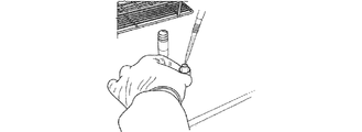
図1Bを参照すると、容器20の蓋26は、ポリオレフィン繊維マトリックス30を保持する蓋延長部28を有し、第2の密閉容器内に含有されるプランジャーに取り付けられるカップに永久に装着され得る。目的の検体を含有する任意の生体標本の液体懸濁液をポリオレフィン繊維マトリックス30の上に添加し、マトリックス30の中に完全に吸収させる(図3)。生体標本をその上に結合したマトリックス30を持つ蓋26を空気乾燥させ、次いで周囲温度での保存のために容器20で再構築する。
Referring to FIG. 1B, the
本発明の方法は、さらに任意に、安定化組成物をポリオレフィン繊維マトリックスに適用して目的の検体を分解から保護する中間ステップを含む。目的の検体に応じて、安定化組成物は、上記に論じられるように、限定されないが、弱塩基、キレート化剤、タンパク質変性剤(洗剤または界面活性剤など)、ヌクレアーゼ阻害剤、およびフリーラジカルトラップのうちの1種以上を含んでよい。特に不安定なRNAの保護のために、安定化組成物は、RNase阻害剤および不活性化剤、遺伝子プローブ、相補DNAまたはRNA(または機能的に等しい化合物)、RNAを安定化するか、またはその分解を予防するタンパク質および有機部分を含んでよい。 The method of the present invention further optionally includes an intermediate step of applying the stabilizing composition to the polyolefin fiber matrix to protect the analyte of interest from degradation. Depending on the analyte of interest, the stabilizing composition can include, but is not limited to, weak bases, chelating agents, protein denaturants (such as detergents or surfactants), nuclease inhibitors, and free radicals, as discussed above. One or more of the traps may be included. For the protection of particularly labile RNA, the stabilizing composition stabilizes RNA, or RNase inhibitors and inactivators, gene probes, complementary DNA or RNA (or functionally equivalent compounds), or It may contain proteins and organic moieties that prevent its degradation.
本発明は、目的の検体を含有する生体標本を、圧縮デバイス中のポリオレフィン繊維マトリックスから回収するための方法をさらに提供する。ある実施形態において、該方法は、以下のステップ:a)再構成媒質をマトリックスに適用し、目的の検体を含有する結合された生体標本を再水和することと、b)マトリックスを圧縮して生体標本の一部分を放出することと、を含む。本発明に従い、再構成媒質は分子グレード水である。他の実施形態において、再構成媒質は、1Xリン酸緩衝生理食塩水(PBS)またはヌクレアーゼを含まない水の成分を含み、任意にアジ化ナトリウムまたは他の抗菌剤の添加を伴う。再構成媒質は、任意の数または組み合わせの使用可能な生物学的保存剤または血液抗凝固剤を含んでもよく、限定されないが、エチレンジアミン四酢酸(EDTA)、クエン酸ナトリウム、およびヘパリンが挙げられる。PBSまたはヌクレアーゼを含まない水は、マトリックスからの目的の検体の再水和、再懸濁、および回収のための滅菌および中性媒質として機能する。アジ化ナトリウムなどの抗菌剤は、含まれるとき、微生物成長およびRNaseによる後次汚染を予防する。EDTA、クエン酸ナトリウム、およびヘパリンなどの生物学的保存剤は、含まれるとき、抗凝固剤および/またはキレート化剤として機能する。 The present invention further provides a method for recovering a biological specimen containing a specimen of interest from a polyolefin fiber matrix in a compression device. In certain embodiments, the method comprises the following steps: a) applying a reconstitution medium to the matrix to rehydrate the bound biological specimen containing the analyte of interest, and b) compressing the matrix. Releasing a portion of the biological specimen. In accordance with the present invention, the reconstitution medium is molecular grade water. In other embodiments, the reconstitution medium comprises a component of 1X phosphate buffered saline (PBS) or nuclease-free water, optionally with the addition of sodium azide or other antimicrobial agent. The reconstitution medium may include any number or combination of usable biological preservatives or blood anticoagulants, including but not limited to ethylenediaminetetraacetic acid (EDTA), sodium citrate, and heparin. PBS or nuclease free water serves as a sterile and neutral medium for rehydration, resuspension, and recovery of the analyte of interest from the matrix. When included, antimicrobial agents such as sodium azide prevent microbial growth and subsequent contamination with RNase. Biological preservatives such as EDTA, sodium citrate, and heparin, when included, function as anticoagulants and / or chelating agents.
図4〜7に示される実施形態において、生体試料は分析のために調製される。図4は、デバイスのポリオレフィン繊維マトリックス30を空のシリンジ筒52に移入する準備の斜視図である。図5は、ポリオレフィン繊維マトリックス30のシリンジ筒52への完全送達の斜視図である。
In the embodiment shown in FIGS. 4-7, the biological sample is prepared for analysis. FIG. 4 is a perspective view of preparation for transferring the
図6は、ピペットチップ53によりマトリックス30の上に静かに置かれ、再構成緩衝液をゆっくり分注するポリオレフィン繊維マトリックス30の再水和を示す。図7Aは、プランジャー54のシリンジ筒54への挿入を示す。図7Bは、シリンジプランジャー42への圧力の印加を示す。図7Cは、マトリックス30の圧縮を示す。図7Dは、マトリックス30からの試料回収の完了を示す。
FIG. 6 shows the rehydration of the
ある実施形態において、目的の検体は、DNAまたはRNA分子のいずれかまたは両方を含む核酸である。ある実施形態において、生体標本の液体懸濁液は、少なくとも約5アトグラムまたは1μgの単離されたDNAまたはRNA分子を含有する。本明細書において使用される場合、「単離された」、「単離」という用語、および「単離する」という語の他の派生語は、DNAまたはRNA分子が、天然に関連付けられる実質的に他の細胞物質、または組換え技術により生産されるときは培養媒質、または化学的に合成されるときは化学的前駆体もしくは他の化学物質のいくつかを含まないことを意味する。 In certain embodiments, the analyte of interest is a nucleic acid containing either or both DNA or RNA molecules. In certain embodiments, the liquid suspension of the biological specimen contains at least about 5 attograms or 1 μg of isolated DNA or RNA molecules. As used herein, the terms “isolated”, “isolated”, and other derivatives of the term “isolate” refer to substantially the DNA or RNA molecule with which it is naturally associated. Means that it contains no other cellular material, or culture medium when produced by recombinant techniques, or some of chemical precursors or other chemicals when chemically synthesized.
本発明は、デバイスのポリオレフィン繊維マトリックスから分子グレード水などの再構成媒質の中に回収された生体標本中に含有される目的の検体が、後次分析に供されることをさらに提供する。本明細書において使用される場合、「後次分析」という用語は、再構成媒質中に貯蔵される回収された生体標本上で行われ得る任意の分析を含む。あるいは、生体標本中に含有される目的の検体は、当該技術分野において既知の方法を使用して、分析前に単離、精製、または抽出されてよい。目的の検体は、化学的、生化学的、または生物学的分析に供されてよい。好ましい実施形態の1つにおいて、目的の検体は、事前の抽出、精製、または単離の有無にかかわらず、検出または分析することができるDNAまたはRNA分子のいずれかまたは両方を含む核酸である。DNAまたはRNAの抽出、精製、または単離は、必要に応じて、当該技術分野において既知の方法に基づいて行われる。後次分析の例として、ポリメラーゼ連鎖反応(PCR)、リガーゼ連鎖反応(LCR)、逆転写酵素で開始されたPCR、DNA、またはRNAハイブリダイゼーション技術が挙げられ、制限断片長多型(RFLP)、ウイルスDNAまたはRNA検出および定量化、ウイルス負荷試験、DNAまたはRNA遺伝子決定などが挙げられる。「後次分析」は、遺伝子プローブを使用する他の技法、ゲノム配列決定、酵素アッセイ、親和性標識、標識または抗体を使用する検出方法、および多の同様の方法も含む。 The present invention further provides that the analyte of interest contained in the biological specimen collected in a reconstitution medium such as molecular grade water from the polyolefin fiber matrix of the device is subjected to subsequent analysis. As used herein, the term “subsequent analysis” includes any analysis that can be performed on a collected biological specimen stored in a reconstitution medium. Alternatively, the analyte of interest contained in the biological specimen may be isolated, purified, or extracted prior to analysis using methods known in the art. The analyte of interest may be subjected to chemical, biochemical, or biological analysis. In one preferred embodiment, the analyte of interest is a nucleic acid containing either or both DNA or RNA molecules that can be detected or analyzed with or without prior extraction, purification, or isolation. Extraction, purification, or isolation of DNA or RNA is performed based on a method known in the art, if necessary. Examples of subsequent analysis include polymerase chain reaction (PCR), ligase chain reaction (LCR), reverse transcriptase initiated PCR, DNA, or RNA hybridization techniques, including restriction fragment length polymorphism (RFLP), Examples include viral DNA or RNA detection and quantification, viral load testing, DNA or RNA gene determination, and the like. “Subsequent analysis” also includes other techniques using gene probes, genomic sequencing, enzyme assays, affinity labels, detection methods using labels or antibodies, and many similar methods.
本発明は、目的の検体を含有する生体標本の液体懸濁液を保存するためのキットも提供する。本発明のキットは、1つ以上の容器、1つ以上のポリオレフィン繊維マトリックス、および任意に乾燥剤、および生体標本を保存するためのその使用についての指示書を含む本明細書に開示される圧縮デバイスを提供する。該キットは任意に、安定化溶液を含んでよい。本発明のキットは、再構成媒質、圧縮デバイス、および生体標本の再水和および回収のためのさらなるプロトコルをさらに含むことができる。該キットの容器は、目的の検体を含有する生体標本の液体懸濁液のマトリックスへの適用中、または適用および生体標本の試料の後次処理の1つ以上の段階中の使用に好適な任意の容器であってよい。したがって、ある実施形態において、生体標本の液体懸濁液は、同じキットで全て適用、貯蔵、輸送され、さらに処理されてよい。あるいは、液体懸濁液はマトリックスに適用されてもよく、該マトリックスは、異なる容器で処理するためにキット容器から取り出される。 The present invention also provides a kit for storing a liquid suspension of a biological specimen containing a target specimen. The kit of the present invention comprises one or more containers, one or more polyolefin fiber matrices, and optionally a desiccant, and a compression disclosed herein comprising instructions for its use for storing biological specimens. Provide the device. The kit may optionally include a stabilization solution. The kit of the present invention can further comprise a reconstitution medium, a compression device, and additional protocols for rehydration and recovery of biological specimens. The kit container may be any suitable for use during application of a biological specimen containing a specimen of interest to a liquid suspension matrix or during one or more stages of application and subsequent processing of a specimen of a biological specimen. It may be a container. Thus, in certain embodiments, a liquid suspension of biological specimens may all be applied, stored, transported and further processed in the same kit. Alternatively, the liquid suspension may be applied to a matrix, which is removed from the kit container for processing in a different container.
該キットは、本明細書に開示されるポリオレフィン繊維マトリックスのいずれかの1つ以上を含んでもよい。これは、生体標本中に含有される目的の検体の保護のための組成物の有無にかかわらず、1つ以上のポリオレフィン繊維マトリックスを含む。本発明のキットの一態様は、目的の検体を含有する再構成された生体標本がマトリックスを圧縮することにより放出されるということである。この手順は、試料を渦流および遠心分離することを避け、試料損傷の可能性、人件費、および試料のマトリックス汚染の減少を提供する。本発明のキットの圧縮デバイスは、マトリックス上に力または圧力を提供してそれを圧縮するように使用される任意のデバイスであってよい。ある実施形態において、プランジャーを備える圧縮デバイスは、ポリオレフィン繊維マトリックスに永久に取り付けられ、該マトリックスは、生体標本が中で調製および貯蔵される同じキット容器(複数可)中でマトリックスに対してプランジャーに力を印加することにより圧縮される。あるいは、圧縮デバイスは、ポリオレフィン繊維マトリックスから分離されたシリンジを備え、該マトリックスは、容器から取り出され、シリンジ筒中に置かれ、力または圧力がシリンジのプランジャーに印加され、マトリックスを圧縮して再構成された生体標本を放出する。 The kit may include one or more of any of the polyolefin fiber matrices disclosed herein. This includes one or more polyolefin fiber matrices, with or without a composition for the protection of the analyte of interest contained in the biological specimen. One aspect of the kit of the present invention is that the reconstituted biological specimen containing the analyte of interest is released by compressing the matrix. This procedure avoids vortexing and centrifuging the sample and provides the potential for sample damage, labor costs, and reduced sample matrix contamination. The compression device of the kit of the present invention may be any device used to provide force or pressure on the matrix to compress it. In certain embodiments, a compression device comprising a plunger is permanently attached to a polyolefin fiber matrix that is planned against the matrix in the same kit container (s) in which the biological specimen is prepared and stored. It is compressed by applying a force to the jar. Alternatively, the compression device comprises a syringe separated from the polyolefin fiber matrix, which is removed from the container and placed in a syringe barrel, and force or pressure is applied to the plunger of the syringe to compress and re-apply the matrix. Release the constructed biological specimen.
本出願全体で様々な刊行物が参照される。これらの刊行物、およびそれらの刊行物内で引用される参照文献の全ての開示は、本発明が属する技術分野の状態をより完全に説明するために、本明細書において参照によりそれら全体が本出願に組み込まれる。 Various publications are referenced throughout this application. The disclosures of all of these publications, and references cited within those publications, are hereby incorporated by reference in their entirety to more fully describe the state of the art to which this invention belongs. Incorporated into the application.
上記は本発明のある実施形態に関すること、および本発明の範囲から逸脱することなくそこに多数の変更を行ってよいことも理解されたい。本発明は、以下の実施例によってさらに例証され、それはいかなるようにも本発明の範囲に制限を与えるものとして解釈されない。逆に、種々の他の実施形態、その修正、および同等物を用いることができ、これらは、本明細書の説明を読むことで、本発明の精神および/または添付の特許請求の範囲の範囲から逸脱することなく、当業者に示され得ることを、明確に理解されたい。 It should also be understood that the above relates to certain embodiments of the present invention and that numerous changes may be made therein without departing from the scope of the present invention. The invention is further illustrated by the following examples, which are not to be construed as limiting the scope of the invention in any way. On the contrary, various other embodiments, modifications thereof, and equivalents may be used, which, upon reading the description herein, are within the spirit of the invention and / or the scope of the appended claims. It should be clearly understood that it can be shown to a person skilled in the art without departing from the invention.
実施例1
1.0mL試料調製およびデバイス回収キット
キット構成要素:
この実施例は、体液または組織からの36個の乾燥生体標本の調製、輸送、および回収のためのキットを提供する。乾燥周囲輸送のための36個の1.0mL試料の調製および回収のための物質および試薬は、以下を含む。
Example 1
1.0 mL sample preparation and device recovery kit Kit components:
This example provides a kit for the preparation, transport, and recovery of 36 dry biological specimens from body fluids or tissues. Materials and reagents for the preparation and recovery of 36 1.0 mL samples for dry ambient transport include:
構成要素 量
デバイスキット容器(管) それぞれ36個
再構成緩衝剤 3X13mL
使い捨て3mLシリンジ それぞれ36個
15mL円錐遠心分離管 それぞれ36個
Component Amount Device kit container (tube) 36 each Reconstitution buffer 3X13mL
36 disposable 3mL syringes each
36 each for 15 mL conical centrifuge tubes
貯蔵および取扱い: Storage and handling:
受領時に、全てのキット構成要素を室温(15〜25℃)で乾燥貯蔵する。指示乾燥剤が青色であるときだけデバイス容器管を使用する。デバイスキット容器管は、指示乾燥剤が白色またはピンク色に見える場合は使用してはならない。1000μLピペット、エアロゾル障壁を持つ1000μL滅菌DNaseを含まない、RNaseを含まないピペットチップ、15mL円錐管を保持するためのラック、安全眼鏡、実験室コート、粉末を含まない使い捨てグローブ、およびバイオハザード廃棄物容器などの物質も必要であるが、キットの安全注意事項により提供されない:使い捨ての粉末を含まないグローブを使用して感染因子を伝達することができるかのように全ての材料を取り扱う。血液由来病原体の伝播の予防に関する良好な実験室実施基準および普遍的予防策を利用する(疾病対策センター改訂:Universal precautions for prevention of transmission of human immunodeficiency virus,hepatitis B virus and other blood borne pathogens in healthcare settings.MMWR,1988、37:377−82,387−8、臨床研究所規格委員会Protection of laboratory workers from infectious disease transmitted by blood, body fluids, and tissue、承認されたガイドラインNCCLS Document M29−A Villanova(PA):NCCLS;1997 Dec.90p、連邦労働安全衛生法Bloodborne Pathogens Standard,29 CFR 1910,1030)。潜在的に感染因子を含有することが疑われる任意の流出は、0.5% w/v次亜塩素酸ナトリウム(10% v/v漂白剤)で速やかに清掃した。標本と接触した全ての標本および物質を、それらが感染因子を含有するように廃棄する。感染因子を含有することが知られているか、または疑われる物質が摂取されるか、または開いた裂傷、病巣、または粘膜(眼、鼻腔など)と接触した場合は、速やかに医師に相談する。 Upon receipt, all kit components are stored dry at room temperature (15-25 ° C.). Use the device container tube only when the indicator desiccant is blue. Device kit container tubes should not be used if the indicated desiccant appears white or pink. 1000 μL pipette, 1000 μL sterile DNase-free pipette tip with aerosol barrier, RNase-free pipette tip, rack to hold a 15 mL conical tube, safety glasses, laboratory coat, disposable glove without powder, and biohazardous waste Substances such as containers are also required, but are not provided by the kit's safety precautions: treat all materials as if they could transmit the infectious agent using disposable powder-free gloves. Utilize good laboratory practice standards and universal precautions for prevention of transmission of blood-borne pathogens (Revision of the Centers for Diseases and Prevention of Humanities of the Humanities of Hepatology MMWR, 1988, 37: 377-82, 387-8, Clinical Laboratory Standards Committee Protection of laboratory workers from infectious disease transmitted by blood, body fluids, and t issue, approved guidelines NCCLS Document M29-A Villanova (PA): NCCLS; 1997 Dec. 90p, Federal Occupational Safety and Health Act Bloodbone Pathogens Standard, 29 CFR 1910, 1030). Any spills suspected of containing potentially infectious agents were quickly cleaned with 0.5% w / v sodium hypochlorite (10% v / v bleach). All specimens and material in contact with the specimen are discarded so that they contain the infectious agent. If a substance known or suspected of containing an infectious agent is ingested or comes into contact with an open laceration, lesion, or mucous membrane (eg, eye, nasal cavity), consult a physician immediately.
実施例2
デバイスキットを使用する試料調製
試料調製ステップは、滅菌技法および潜在的に感染性の物質の取扱いに関する普遍的予防策を使用して、生物学的安全キャビネット内で行った。試料調製プロセスを開始する前に、図1Aおよび1Bに例証されるデバイスキットを使用するプロトコルをよく理解すべきである。
Example 2
Sample Preparation Using Device Kit The sample preparation step was performed in a biological safety cabinet using sterilization techniques and universal precautions regarding the handling of potentially infectious materials. Prior to initiating the sample preparation process, the protocol using the device kit illustrated in FIGS. 1A and 1B should be well understood.
目的の検体を含有する生体標本の試料液体懸濁液を負荷する前に、デバイス容器からのキャップを外し、反転させて清潔な作業台上に吸収性マトリックスを上に向けて置いた(図1Aおよび1B)。最大約1mLの試料流体を、マトリックスプラグの上にゆっくり添加し、マトリックスの中に完全に吸収させた。試料流体を負荷したデバイスキットマトリックスを空気乾燥させた。一般に、生物学的安全キャビネット内の空気乾燥は、約4.5〜5時間かかる。一旦試料が完全に乾燥すると、乾燥した標本を含有するマトリックスを保持するキャップをデバイスキット容器管に慎重に再び取り付けた。ここで標本は、周囲温度で出荷または貯蔵する準備ができている。 Prior to loading the sample liquid suspension of the biological specimen containing the analyte of interest, the cap from the device container was removed and inverted and the absorbent matrix was placed on a clean workbench (FIG. 1A). And 1B). Up to about 1 mL of sample fluid was slowly added over the matrix plug and completely absorbed into the matrix. The device kit matrix loaded with sample fluid was air dried. In general, air drying in a biological safety cabinet takes about 4.5-5 hours. Once the sample was completely dry, the cap holding the matrix containing the dried specimen was carefully reattached to the device kit container tube. Here the specimen is ready to be shipped or stored at ambient temperature.
実施例3
デバイスキットを使用する試料回収
試料回収ステップもまた、滅菌技法および潜在的に感染性の物質の取扱いに関する普遍的予防策を使用して、生物学的安全キャビネット内で行った。基本的に、滅菌3mLまたは5mL使い捨てLUER−LOKシリンジ(キットにより提供される)を、15mL収集管(これもキットにより提供される)の中に挿入した。プランジャーをシリンジ筒から取り出した。乾燥標本を含有する吸収性マトリックスを、それが取り付けられたキャップから抜け出すのに程良い圧力でシリンジ筒の口部の滅菌内側に対してマトリックスを押し付けることによりシリンジ筒に移入し、シリンジの底部に自由に落下させた(図4および5)。取り外されたマトリックスプラグを持つシリンジ筒を、15mLの円錐収集管の中に置き、それをさらにラックの中に置く。約1mLの再構成緩衝液(キットにより供給される)をゆっくり直接マトリックスプラグの上に適用し、マトリックス内に吸収される乾燥標本を静かに再水和した(図6)。吸収率を検査し、必要に応じて再構成緩衝液を添加しながら適用速度を調整する必要があり、再構成緩衝液を完全に吸収するのに失敗すると、より低い回収収率をもたらし得るため、最初にマトリックスに吸収されずに緩衝剤をシリンジの底部で回収するのを許さないようにする必要がある。再水和標本を、追加の175μLの再構成緩衝剤をマトリックスプラグの上に添加する前に室温で少なくとも10分間インキュベートさせた。
Example 3
Sample Collection Using Device Kit The sample collection step was also performed in a biological safety cabinet using universal precautions regarding sterilization techniques and handling of potentially infectious materials. Basically, a sterile 3 mL or 5 mL disposable LUER-LOK syringe (provided with the kit) was inserted into a 15 mL collection tube (also provided with the kit). The plunger was removed from the syringe barrel. The absorbent matrix containing the dried specimen is transferred into the syringe barrel by pressing the matrix against the sterilized inside of the syringe barrel mouth with sufficient pressure to pull it out of the cap to which it is attached, and into the bottom of the syringe. It was allowed to fall freely (Figures 4 and 5). Place the syringe barrel with the removed matrix plug into the 15 mL conical collection tube, which is then placed in the rack. Approximately 1 mL of reconstitution buffer (supplied with the kit) was slowly applied directly onto the matrix plug to gently rehydrate the dried specimen absorbed into the matrix (FIG. 6). The rate of application should be adjusted while examining the absorption rate and adding reconstitution buffer as needed, and failure to fully absorb the reconstitution buffer can result in lower recovery yields First, it should not be allowed to recover the buffer at the bottom of the syringe without being absorbed by the matrix. The rehydrated specimen was allowed to incubate at room temperature for at least 10 minutes before adding an additional 175 μL of reconstitution buffer onto the matrix plug.
シリンジプランジャーをシリンジ筒に再度挿入し、プランジャーがマトリックスプラグを完全に圧縮し、最大容積の約1mLが15mL回収管内に回収されるまで安定した均一な圧力で押した(図7A、7B、7C、および7D)。次いで、シリンジ筒、プランジャー、および圧縮したマトリックスを15mL回収管から取り出し、適切な廃棄物容器に破棄した。新たに回収された標本を含有する15mL回収管を、提供されたスクリューキャップで密封した。貯蔵、試験、またはさらなる後次分析のために再構成試料を準備する。 The syringe plunger was re-inserted into the syringe barrel, and the plunger fully compressed the matrix plug and pushed with stable and uniform pressure until approximately 1 mL of the maximum volume was collected in the 15 mL collection tube (FIGS. 7A, 7B, 7C and 7D). The syringe barrel, plunger, and compressed matrix were then removed from the 15 mL collection tube and discarded into an appropriate waste container. A 15 mL collection tube containing the newly collected specimen was sealed with the provided screw cap. Prepare reconstituted samples for storage, testing, or further subsequent analysis.
実施例4
ViveSTデバイスマトリックス比較研究
1.目的
本研究の目的は、セルロースマトリックスを持つViveSTデバイスの性能を、Abbott RELTIME HBVアッセイを使用して合成疎水性ポリオレフィン繊維マトリックスを持つ本発明のViveSTデバイスと比較することであった。HBV感染試料(3レベル、各5複製)を負荷し、両方のマトリックス上で周囲条件で7日間貯蔵した。マトリックスから回収された標本は、冷凍試料を用いて同時に行った。
Example 4
ViveST device matrix comparative study Purpose The purpose of this study was to compare the performance of a ViveST device with a cellulose matrix to the ViveST device of the present invention with a synthetic hydrophobic polyolefin fiber matrix using the Abbott RELTIME HBV assay. HBV-infected samples (3 levels, 5 replicates each) were loaded and stored for 7 days at ambient conditions on both matrices. The specimen collected from the matrix was simultaneously performed using a frozen sample.
2.方法論
Abbott REALTIME HBVアッセイ上の全ての試験は、修正なしにFDA承認プロトコル(0.5mL)に従い行った。1mL HBV感染血漿を各マトリックスの上に負荷し、7日間周囲温度で貯蔵して、1mLの分子グレード水を回収した。
2. Methodology All tests on the Abbott REALTIME HBV assay were performed according to the FDA approved protocol (0.5 mL) without modification. 1 mL HBV-infected plasma was loaded onto each matrix and stored at ambient temperature for 7 days to collect 1 mL of molecular grade water.
冷凍試料(3レベル、各5複製)のHBVウイルス負荷の結果を、セルロースマトリックスを持つViveSTデバイスおよび疎水性ポリオレフィン繊維マトリックスを有する本発明のViveSTデバイスから回収された標本と比較した。 HBV viral load results of frozen samples (3 levels, 5 replicates each) were compared to specimens recovered from the ViveST device with a cellulose matrix and the ViveST device of the present invention with a hydrophobic polyolefin fiber matrix.
3.装置および試薬
以下の市販の製品/装置を本研究の過程で用いた:本発明のViveST試料貯蔵および輸送デバイス(カタログ番号VST−1E、ViveBio LLC、Alphretta,GA)、およびセルロースマトリックスを持つViveSTデバイス(ViveBio,LLC、Alpharetta,GA);BD3mLシリンジ−LUER−LOKチップ:Ref 3096567(Becton Dickenson;Franklin Lakes,NJ);一般的な実験室消耗品および装置(遠心分離管、滅菌エアロゾル耐性ピペットチップ、ピペット、渦流、遠心分離など);HYCLONE HYPURE分子生物学グレード水(カタログ番号:SH30538.02、HyClone Laboratory Inc.,Logan,UT);ヒト血漿(Tennessee blood services;Memphis,TN);Abbott試料調製システム(4×24調製)、リスト番号:06K12−024(Abbott Molecular Inc;Des Plaines,IL);Abbott REALTIME HBV AMPキット(カタログ番号:02N40−90、Abbott Molecular Inc;Des Plaines,IL)Abbott REALTIME HBV制御キット(カタログ番号:02N40−80、Abbott Molecular Inc;Des Plaines,IL);Abbott REALTIME HBV較正キット(カタログ番号:02N40−70、Abbott Molecular Inc;Des Plaines,IL);m2000rtを含むAbbottm2000spシステム(Abbott Molecular Inc;Des Plaines,Illinois)、および関連物質。
3. Apparatus and Reagents The following commercially available products / apparatus were used in the course of this study: ViveST sample storage and transport device of the present invention (Catalog No. VST-1E, ViveBio LLC, Alphatta, GA) and ViveST device with cellulose matrix. (ViveBio, LLC, Alphatta, GA);
4.実験設計
高力価HBV感染血漿試料を正常なヒト血漿中に希釈し、3つの濃度を生じた(約5ログ、約4ログ、および約3ログ)。各濃度の5つの複製(合計n=15)を、セルロースマトリックスを持つViveSTデバイスおよびポリオレフィン繊維マトリックスを持つ本発明のViveSTデバイスのそれぞれの上に負荷した。同一アリコート(3レベル、5複製)を冷凍貯蔵した(−80℃)。全てのマトリックスを層流フード中で終夜乾燥させ、翌日キャップをして周囲条件で7日間貯蔵した。両方のマトリックスセットから標本を回収し、Abbott REALTIME HBVアッセイパッケージ添付文書に概説されるように、bioMONTR研究方法(RM−008.00、Abbott REALTIME HBVアッセイを使用するHBV RNAの定量化)に従い行った単一アッセイにおいて冷凍標本と同時に分析した。
5.結果−Abbott REALTIME HBVウイルス負荷結果の概要を以下の表1に提供する。

5. Results-A summary of Abbott REALTIME HBV viral load results is provided in Table 1 below.
本実施例の結果は、全ての濃度にわたって平均低減を実証した:a)冷凍血漿とポリオレフィン繊維マトリックスを持つ本発明のViveSTデバイス上に貯蔵された血漿試料との間の0.03 ログIU/mLおよびb)冷凍血漿とセルロースマトリックスを持つViveSTデバイス上に貯蔵された血漿試料との間の0.24ログ IU/mL。全ての濃度にわたる標準偏差(ログIU/mL)は、a)冷凍血漿に対して0.07未満、b)ポリオレフィン繊維マトリックスを持つ本発明のViveSTデバイス上に貯蔵された血漿試料に対して0.03未満、およびc)セルロースマトリックスを持つViveSTデバイス上に貯蔵された血漿試料に対して0.09未満であった。 The results of this example demonstrated an average reduction across all concentrations: a) 0.03 log IU / mL between frozen plasma and plasma sample stored on the ViveST device of the present invention with a polyolefin fiber matrix And b) 0.24 log IU / mL between frozen plasma and plasma sample stored on a ViveST device with cellulose matrix. The standard deviation across all concentrations (log IU / mL) is a) less than 0.07 for frozen plasma and b) 0. 0 for plasma samples stored on the ViveST device of the present invention with a polyolefin fiber matrix. <03, and c) <0.09 for plasma samples stored on the ViveST device with cellulose matrix.
図8に示されるように、線形回帰分析は、a)ポリオレフィン繊維マトリックスを持つ本発明のViveSTデバイス上に貯蔵された血漿試料と比較して、冷凍血漿に対してR2=0.999998、およびb)セルロースマトリックスを持つViveSTデバイス上に貯蔵された血漿試料と比較して、冷凍血漿に対してR2=0.9991を生じた。 As shown in FIG. 8, linear regression analysis shows that a) R 2 = 0.999998 for frozen plasma, compared to plasma samples stored on the ViveST device of the present invention with a polyolefin fiber matrix, and b) R 2 = 0.9999 for frozen plasma compared to plasma samples stored on a ViveST device with cellulose matrix.
6.最終結論
したがって、本実施例は、ポリオレフィン繊維マトリックスを持つ本発明のViveSTデバイス上に貯蔵されたHBV感染血漿試料が回収され、冷凍血漿と同様の結果を生じたことを提供する。冷凍血漿と比較したとき、最小喪失(0.03ログIU/mL)および全ての濃度にわたって高い再現性が見られた(標準偏差0.03未満)。対照的に、セルロースマトリックスを持つViveSTデバイス上に貯蔵されたHBV感染血漿試料は、冷凍血漿と比較してより大きな喪失(0.24 ログ IU/mL)およびより全ての濃度にわたる高い変動性(標準偏差0.09未満)を呈した。
6). Final Conclusion Accordingly, this example provides that an HBV-infected plasma sample stored on the ViveST device of the present invention with a polyolefin fiber matrix was recovered and produced similar results as frozen plasma. There was minimal loss (0.03 log IU / mL) and high reproducibility across all concentrations (standard deviation less than 0.03) when compared to frozen plasma. In contrast, HBV-infected plasma samples stored on a ViveST device with a cellulose matrix showed greater loss (0.24 log IU / mL) and higher variability (standard) over all concentrations compared to frozen plasma. Deviation less than 0.09).
したがって、ポリオレフィン繊維マトリックスを持つ本発明のViveSTデバイスは、より良好で優れた試料回収および最小限の試料喪失を提供するとともに、セルロースマトリックスを持つデバイスと比較して、全ての濃度にわたって再現性を提供し、ポリオレフィン繊維マトリックスが、セルロースマトリックスよりも良好に検体および懸濁粒子をマトリックス内に保持し、溶媒がより一貫して効率的に蒸発するのを許すことを示唆する。 Thus, the ViveST device of the present invention with a polyolefin fiber matrix provides better and better sample recovery and minimal sample loss and provides reproducibility across all concentrations compared to a device with a cellulose matrix This suggests that the polyolefin fiber matrix retains analytes and suspended particles within the matrix better than the cellulose matrix, allowing the solvent to evaporate more consistently and efficiently.
実施例5
抽出されたRNA対無傷のウイルス
1.実験設計
本実験の目的は、無傷のウイルスと比較して、ポリオレフィンマトリックスViveSTデバイスの核酸の結合および放出特性を評価することであった。−80℃で貯蔵された1mLアリコートのHCV感染血漿試料(N=20)を研究に使用し、以下のように指定した。
A=EasyMAGシステムを使用してRNAを抽出し、セルロース繊維マトリックスを持つViveSTデバイス上に負荷した後、水で回収し、Abbott REALTIME HCVアッセイで分析した。
B=EasyMAGシステムを使用してRNAを抽出し、ポリオレフィン繊維マトリックスを持つ本発明のViveSTデバイス上に負荷した後、水で回収し、Abbott REALTIME HCVアッセイで分析した。
C=セルロース繊維マトリックスを持つViveSTデバイス上に試料を負荷し、水で回収して、Abbott REALTIME HCVアッセイで分析した。
D=ポリオレフィン繊維マトリックスを持つ本発明のViveSTデバイス上に試料を負荷し、水で回収して、REALTIME Abbott HCVアッセイで分析した。
Example 5
Extracted RNA vs. intact virus Experimental design The purpose of this experiment was to evaluate the nucleic acid binding and release properties of polyolefin matrix ViveST devices compared to intact virus. A 1 mL aliquot of HCV-infected plasma sample (N = 20) stored at -80 ° C. was used in the study and designated as follows:
A = RNA was extracted using the EasyMAG system, loaded onto a ViveST device with a cellulose fiber matrix, then recovered with water and analyzed with the Abbott REALTIME HCV assay.
B = RNA was extracted using the EasyMAG system, loaded onto the ViveST device of the present invention with a polyolefin fiber matrix, then recovered with water and analyzed with the Abbott REALTIME HCV assay.
Samples were loaded onto a ViveST device with C = cellulose fiber matrix, collected with water and analyzed with the Abbott REALTIME HCV assay.
D = Samples were loaded onto a ViveST device of the present invention with a polyolefin fiber matrix, collected with water, and analyzed by the REALTIME Abbott HCV assay.
2.1 手順−EasyMAGを使用する核酸単離に続いて、セルロース繊維マトリックスまたはポリオレフィン繊維マトリックスのいずれかを持つViveSTデバイスを通じて処理される。
a)取り出した1mL血漿アリコートを「1A、2A...20A」および「1B、2B...20B」として指定し、−80℃から室温に解凍した。
b)各試料を渦流させて適切な混合を保証した。
c)EasyMAG標準核酸抽出プロトコルに従い、40個の試料+2個の陰性対照を処理し、抽出後、全ての試料をEasyMAG溶出緩衝液を使用して1mL体積を希釈した。
d)42個のViveSTデバイスを入手し、それぞれのキャップに試料指定を標識した(すなわち、1A〜20A、1B〜20B、陰性対照〜古いマトリックス、陰性対照〜新しいマトリックス)。
e)各試料に対して1個のViveSTデバイスを負荷した。
f)ViveSTデバイス中の負荷したマトリックスを層流フード内で少なくとも12時間、36時間を超えずに乾燥させた。
g)水を使用して、乾燥させたViveSTデバイスから試料を回収した。
h)Abbott REALTIME HCVアッセイに進む前に、回収された試料を−80℃で冷凍した。
2.1 Procedure-Following nucleic acid isolation using EasyMAG, it is processed through a ViveST device with either a cellulose fiber matrix or a polyolefin fiber matrix.
a) The removed 1 mL plasma aliquots were designated as “1A, 2A ... 20A” and “1B, 2B ... 20B” and thawed from −80 ° C. to room temperature.
b) Each sample was swirled to ensure proper mixing.
c) According to the EasyMAG standard nucleic acid extraction protocol, 40 samples + 2 negative controls were processed and after extraction, all samples were diluted in 1 mL volume using EasyMAG elution buffer.
d) 42 ViveST devices were obtained and each cap was labeled with a sample designation (ie 1A-20A, 1B-20B, negative control-old matrix, negative control-new matrix).
e) One ViveST device was loaded for each sample.
f) The loaded matrix in the ViveST device was dried in a laminar flow hood for at least 12 hours and no more than 36 hours.
g) Samples were collected from the dried ViveST device using water.
h) The collected samples were frozen at -80 ° C before proceeding to the Abbott REALTIME HCV assay.
2.2 手順−セルロース繊維マトリックスまたはポリオレフィン繊維マトリックスを持つViveSTデバイスを通じて処理された試料
a)取り出した1mL血漿アリコートを「1C、2C...20C」および「1D、2D...20D」として指定し、−80℃から室温に解凍した。
b)各試料を渦流させて適切な混合を保証した。
c)42個のViveSTデバイスを入手し、それぞれのキャップに試料指定を標識した(すなわち、1C〜20C、1D〜20D、陰性対照〜セルロースマトリックス、陰性対照〜ポリオレフィン繊維マトリックス)。
d)各試料に対して1個のViveSTデバイスを負荷した。
e)ViveSTデバイスの負荷したマトリックスを層流フード内で少なくとも12時間、36時間を超えずに乾燥させた。
f)水を使用して、乾燥させたViveSTデバイスから試料を回収した。
g)Abbott REALTIME HCVアッセイに進む前に、回収された試料を−80℃で冷凍した。
2.2 Procedure-Sample processed through ViveST device with cellulose fiber matrix or polyolefin fiber matrix
a) The removed 1 mL plasma aliquots were designated as “1C, 2C... 20C” and “1D, 2D... 20D” and thawed from −80 ° C. to room temperature.
b) Each sample was swirled to ensure proper mixing.
c) 42 ViveST devices were obtained and each cap was labeled with a sample designation (ie, 1C-20C, 1D-20D, negative control to cellulose matrix, negative control to polyolefin fiber matrix).
d) One ViveST device was loaded for each sample.
e) The loaded matrix of the ViveST device was dried in a laminar flow hood for at least 12 hours and no more than 36 hours.
f) Samples were collected from the dried ViveST device using water.
g) The collected samples were frozen at -80 ° C before proceeding to the Abbott REALTIME HCV assay.
2.3 手順−Abbott REALTIME HCVアッセイ:Abbott REALTIME HCVパッケージ添付文書に従い試料を処理した。 2.3 Procedure-Abbott REALTIME HCV Assay: Samples were processed according to the Abbott REALTIME HCV package insert.
3.結果:結果を以下の表2に提供する。
4.結論
平均して、抽出されたRNAの場合、ポリオレフィン繊維マトリックスは、セルロースマトリックスよりも高い回収を生じた(0.29ログIU/mLより高い)。臨床的に有意なカットオフ付近で、ポリオレフィンマトリックスは、セルロースマトリックスよりも抽出されたRNAに対してより良好に機能することを支持する。
4). Conclusion On average, for extracted RNA, the polyolefin fiber matrix produced a higher recovery than the cellulose matrix (above 0.29 log IU / mL). In the vicinity of a clinically significant cutoff, the polyolefin matrix supports better functioning on extracted RNA than the cellulose matrix.
平均して、ポリオレフィン繊維マトリックスは、新鮮な血漿と比較して抽出されたRNAに対してより高い回収を生じた(0.25ログIU/mLより高い)。臨床的に有意なカットオフ付近で、ポリオレフィンマトリックスは、新鮮な血漿よりも抽出されたRNAに対してより良好に機能することを支持する。 On average, the polyolefin fiber matrix produced a higher recovery for extracted RNA compared to fresh plasma (higher than 0.25 log IU / mL). Near the clinically significant cutoff, the polyolefin matrix supports better functioning on extracted RNA than on fresh plasma.
これらのデータに基づき、ポリオレフィン繊維マトリックスは、核酸の吸収、保存、安定化、および後次回収について、セルロースマトリックスと比較して優れている。これらの驚くべき結果は、恐らくポリオレフィンマトリックス内の埋め込まれた疎水性ポケットの特製に起因する。これらのポケットは、核酸を存在させる容器および「避難場所」を提供し、核酸から水を排除する一方、貯蔵中に安定した環境を核酸に提供する。 Based on these data, polyolefin fiber matrices are superior to cellulose matrices for nucleic acid absorption, storage, stabilization, and subsequent recovery. These surprising results are probably due to the speciality of embedded hydrophobic pockets within the polyolefin matrix. These pockets provide a container and a “evacuation site” for the presence of nucleic acids and exclude water from the nucleic acids while providing a stable environment for the nucleic acids during storage.
実施例6
本発明のViveSTデバイスを通じて処理された試料のHCV評価
1.実験設計
−80℃で貯蔵された1mLアリコートのHCV感染血漿試料(N=19)を分析の各部に使用し、以下のように指定した。
A=Abbott REALTIME HCVアッセイで分析した試料
B=ポリオレフィン繊維マトリックスを持つ本発明のViveSTデバイスを通じて処理し(水で溶出)、Abbott REALTIME HCVアッセイで分析した試料
C=Abbott HCV GTアッセイで分析した試料
D=ポリオレフィン繊維マトリックスを持つ本発明のViveSTデバイスを通じて処理し(水で溶出)、Abbott HCV GTアッセイで分析した試料
Example 6
HCV evaluation of samples processed through the ViveST device of the present invention.
A = sample analyzed with Abbott REALTIME HCV assay B = sample processed through ViveST device of the invention with polyolefin fiber matrix (eluted with water) and analyzed with Abbott REALTIME HCV assay C = sample analyzed with Abbott REALTIME HCV assay D = Sample processed through the ViveST device of the present invention with polyolefin fiber matrix (eluted with water) and analyzed by Abbott HCV GT assay
各血漿試料の追加のアリコートを追加の試験のために−80℃で維持した。 An additional aliquot of each plasma sample was maintained at −80 ° C. for additional testing.
2.1 手順−本発明のViveSTデバイスを通じて処理した試料
a)取り出した血漿アリコートを「1B、2B...19B」および「1D、2D...19D」として指定し、−80℃から室温に解凍した。
b)各試料を渦流させて適切な混合を保証した。
c)ポリオレフィン繊維マトリックスを持つ本発明の38個のViveSTデバイスを入手し、それぞれのキャップを試料指定(すなわち、1B〜19B、1D〜19D)で標識し、2個の追加のViveSTデバイスを入手してそれぞれを陰性対照として標識した。
d)各試料についてポリオレフィン繊維マトリックスを持つ本発明の1個のViveSTデバイスを負荷し、1mLの正常(HCV陰性)ヒト血漿を陰性対照と標識されたViveSTデバイス上に負荷した。
e)ViveSTデバイスの負荷したマトリックスを層流フード内で少なくとも12時間、36時間を超えずに乾燥させた。
f)水を使用して、乾燥させたViveSTデバイスから試料を回収した。
g)回収した試料を−80℃で冷凍した。
2.1 Procedure-Samples processed through the ViveST device of the present invention a) The removed plasma aliquots are designated as “1B, 2B... 19B” and “1D, 2D. Thawed.
b) Each sample was swirled to ensure proper mixing.
c) Obtain 38 ViveST devices of the present invention with polyolefin fiber matrix and label each cap with sample designation (ie 1B-19B, 1D-19D) to obtain 2 additional ViveST devices. Each was labeled as a negative control.
d) For each sample, one ViveST device of the present invention with a polyolefin fiber matrix was loaded and 1 mL of normal (HCV negative) human plasma was loaded onto the ViveST device labeled as a negative control.
e) The loaded matrix of the ViveST device was dried in a laminar flow hood for at least 12 hours and no more than 36 hours.
f) Samples were collected from the dried ViveST device using water.
g) The collected sample was frozen at -80 ° C.
2.2手順−Abbott m2000 REALTIME HCVアッセイ
a)取り出した血漿アリコートを「1A、2A...19A」として指定し、ポリオレフィン繊維マトリックスを持つ本発明のViveSTデバイスから回収したアリコートを「1B、2B...19Bおよび陰性対照」として指定し、−80℃から室温に解凍した。
b)Abbott REALTIME HCVパッケージ添付文書に従い試料を処理した。
2.2 Procedure-Abbott m2000 REALTIME HCV Assay a) The removed plasma aliquot is designated as “1A, 2A... 19A” and the aliquot recovered from the ViveST device of the present invention with a polyolefin fiber matrix is designated “1B, 2B. .. 19B and negative control ”and thawed from −80 ° C. to room temperature.
b) Samples were processed according to the Abbott REALTIME HCV package insert.
2.3.手順−Abbott m2000 HCV GTアッセイ
a)取り出した血漿アリコートを「1C、2C...19C」として指定し、ポリオレフィン繊維マトリックスを持つ本発明のViveSTデバイスから回収したアリコートを「1D、2D...19Dおよび陰性対照」として指定し、−80℃から室温に解凍した。
b)Abbott HCV GTパッケージ添付文書に従い試料を処理した。
2.3. Procedure-Abbott m2000 HCV GT assay a) The removed plasma aliquot is designated as “1C, 2C... 19C” and the aliquot recovered from the ViveST device of the present invention with a polyolefin fiber matrix is “1D, 2D. And as a negative control "and thawed from -80 ° C to room temperature.
b) Samples were processed according to the Abbott HCV GT package insert.
3.結果−結果の概要を以下の表3および4に提供する。
4.結論
HCV遺伝子型決定結果は、試験されるHCV遺伝子型1、1a、1b、2、および3を持つ冷凍血漿と比較して、本発明のViveSTデバイスから回収された血漿試料と100%一致を実証した(表3)。HCVウイルス負荷結果は、冷凍血漿と比較して、本発明のViveSTデバイスから取り出された血漿について、0.32の平均低減を示した(表4および図9)。
4). Conclusion HCV genotyping results demonstrate 100% agreement with plasma samples recovered from the ViveST device of the present invention compared to frozen plasma with
以前に刊行された乾燥血斑/血漿斑データに基づき、新鮮な血漿からの定量と乾燥した収集デバイスからの定量との間に約0.5〜0.7ログ低減を予想する(Amellal et al.,2007,HIV Med.8:396−400、中央値喪失は有意であり、0.64ログコピー/mLに等しいことを論じる;Hamers et al.,2009 Antiviral Therapy 14:619−29、DPSと血漿との間の中央値差は、0.077〜0.64ログコピー/mLであった)。ここで、本研究は、ポリオレフィンマトリックスを持つ本発明のViveSTデバイスを通じて処理された血漿試料から回収された平均ウイルスRNAは、刊行された文献に基づいて以前に予想された値よりも驚くほど高く、再現性が高いことを実証した。 Based on previously published dry spot / plasma spot data, we expect an approximately 0.5-0.7 log reduction between quantification from fresh plasma and quantification from a dry collection device (Amelal et al , 2007, HIV Med. 8: 396-400, median loss is significant and discussed to be equal to 0.64 log copies / mL; Hamers et al., 2009 Anti-Therapeutic Therapy 14: 619-29, DPS and Median difference from plasma was 0.077-0.64 log copies / mL). Here, the study shows that the average viral RNA recovered from plasma samples processed through the ViveST device of the present invention with a polyolefin matrix is surprisingly higher than previously expected based on published literature, It was demonstrated that reproducibility is high.
実施例7
本発明のViveSTデバイスを通じて処理された試料についてのHIV−1およびViroSeq HIV−1遺伝子型決定評価
1.実験設計
−80℃で貯蔵された1mLアリコートのHIV−1陽性血漿試料(N=20)を分析の各部に使用し、以下のように指定した。
A=Abbott REALTIME HIV−1アッセイで分析した試料
B=ポリオレフィン繊維マトリックスを持つ本発明のViveSTデバイスを通じて処理し(水で溶出)、Abbott REALTIME HIV−1アッセイで分析した試料
C=ポリオレフィン繊維マトリックスを持つ本発明のViveSTデバイスを通じて処理し(mLysis緩衝液で溶出)、Abbott REALTIME HIV−1アッセイで分析した試料
D=ViroSeq HIV−1 Pro & RT遺伝子型アッセイで分析した試料
E=ポリオレフィン繊維マトリックスを持つ本発明のViveSTデバイスを通じて処理し(水で溶出)、ViroSeq HIV−1 Pro & RT遺伝子型アッセイで分析した試料
Example 7
HIV-1 and ViroSeq HIV-1 genotyping evaluation on samples processed through the ViveST device of the present invention.
A = Sample analyzed with Abbott REALTIME HIV-1 assay B = Sample processed through ViveST device of the present invention with polyolefin fiber matrix (eluted with water) and analyzed with Abbott REALTIME HIV-1 assay C = Polyolefin fiber matrix Sample processed through ViveST device of the present invention (eluted with mLysis buffer) and analyzed with Abbott REALTIME HIV-1 assay D = Sample analyzed with ViroSeq HIV-1 Pro & RT genotype assay E = Book with polyolefin fiber matrix Samples processed through the inventive ViveST device (eluted with water) and analyzed with the ViroSeq HIV-1 Pro & RT genotype assay
各血漿試料の追加のアリコートを追加の試験のために−80℃で維持した。 An additional aliquot of each plasma sample was maintained at −80 ° C. for additional testing.
2.1 手順−本発明のViveSTデバイスを通じて処理した試料
a)取り出した血漿アリコートを「1B、2B...20B」、「1C、2C...20C」、および「1E、2E...20E」として指定し、−80°Cから室温に解凍した。
b)各試料を渦流させて適切な混合を保証した。
c)ポリオレフィン繊維マトリックスを持つ本発明の60個のViveSTデバイスを入手し、それぞれのキャップを試料指定(すなわち、1b〜20b、1c〜20c、1e〜20e)で標識し、2個の追加のViveSTデバイスを入手してそれぞれを陰性対照として標識した。
d)各試料について1個のViveSTデバイスを負荷し、1mLの正常(HIV−1陰性)ヒト血漿を陰性対照と標識されたViveSTデバイス上に負荷した。
e)ViveSTデバイス中の負荷したマトリックスを層流フード内で少なくとも12時間、36時間を超えずに乾燥させた。
f)水を使用して、乾燥させたViveSTデバイスから試料「1b〜20b」、「1e〜20e」、および「陰性対照〜水」を回収し、溶解緩衝液を用いて試料「1c〜20c」および「陰性対照〜溶解」を回収した。
g)回収した試料を−80°Cで冷凍した。
2.1 Procedure-Samples processed through the ViveST device of the invention a) Removed plasma aliquots from "1B, 2B ... 20B", "1C, 2C ... 20C", and "1E, 2E ... 20E" And thawed from −80 ° C. to room temperature.
b) Each sample was swirled to ensure proper mixing.
c) Obtain 60 ViveST devices of the present invention with a polyolefin fiber matrix, label each cap with a sample designation (ie, 1b-20b, 1c-20c, 1e-20e) and add 2 additional ViveSTs Devices were obtained and each was labeled as a negative control.
d) One ViveST device was loaded for each sample and 1 mL of normal (HIV-1 negative) human plasma was loaded onto the ViveST device labeled as a negative control.
e) The loaded matrix in the ViveST device was dried in a laminar flow hood for at least 12 hours and no more than 36 hours.
f) Using water, collect samples “1b-20b”, “1e-20e”, and “negative control-water” from the dried ViveST device and use lysis buffer to sample “1c-20c” And "Negative Control-Lysis" was collected.
g) The collected sample was frozen at -80 ° C.
2.2.手順−Abbott m2000 REALTIME HIV−1アッセイ
a)取り出した血漿アリコートを「1A、2A...20A」として指定し、ViveSTデバイスから回収したアリコートを「1B、2B...20B」、「1C...2C...20C」、「陰性対照〜水」、および「陰性対照〜溶解」として指定し、−80°Cから室温に解凍した。
b)Abbott REALTIME HIV−1アッセイのパッケージ添付文書に従い試料を処理した。
2.2. Procedure-Abbott m2000 REALTIME HIV-1 Assay a) The removed plasma aliquot was designated as “1A, 2A... 20A” and the aliquot recovered from the ViveST device was “1B, 2B. .2C ... 20C "," Negative control to water ", and" Negative control to lysis "and thawed from -80 ° C to room temperature.
b) Samples were processed according to the Abbott REALTIME HIV-1 assay package insert.
2.3 手順−ViroSeq HIV−1 Pro & RT遺伝子型アッセイ
a)取り出した血漿アリコートを「1D、2D...20D」として指定し、ViveSTデバイスから回収したアリコートを「1E、2E...20E」として指定し、−80℃から室温に解凍した。
b)ViroSeq HIV−1 Pro & RT遺伝子型アッセイのパッケージ添付文書に従い試料を処理した。
2.3 Procedure-ViroSeq HIV-1 Pro & RT genotype assay a) The removed plasma aliquot is designated as "1D, 2D ... 20D" and the aliquot recovered from the ViveST device is designated as "1E, 2E ... 20E". And thawed from −80 ° C. to room temperature.
b) Samples were processed according to the package insert of the ViroSeq HIV-1 Pro & RT genotype assay.
3.結果−結果の概要を以下の表5および6に提供する。
4.結論
HIV−1ウイルス負荷結果は、ポリオレフィン繊維マトリックスを持つ本発明のViveSTデバイスから回収された血漿について、mLysis緩衝液を使用して0.26ログの平均低減を示し、水を使用して0.59ログ低減を示した(図10、図11、および表6)。
4). CONCLUSION HIV-1 viral load results show an average reduction of 0.26 logs using mLysis buffer for plasma collected from the ViveST device of the present invention with a polyolefin fiber matrix and 0. 0 using water. 59 log reduction was shown (Figure 10, Figure 11 and Table 6).
HIV−1薬物耐性突然変異は、17対中10対(59%)で特定され、冷凍血漿と比較して、ViveSTデバイスから回収された血漿試料間の100%一致を実証した。混合物T215Y/Cは、ViveSTデバイスから回収された試料中17分の1で特定されたが、対応する血漿は突然変異T215Yを報告した。V751での突然変異は、血漿試料中17個のうち1個で特定されたが、ViveSTデバイスから回収された対合試料は野生型であった。17対中1対は、ViroSeq報告のT69予防生成における欠失を実証した。17対中1対の血漿試料は、M184Vを有したが、低いウイルス負荷に起因して、ViveSTデバイスを通じて処理された対応する試料についての結果は生成されなかった。低いウイルス負荷に起因して、17個のうち3個の対合試料についての遺伝子型結果は生成されなかった(表5)。 HIV-1 drug resistance mutations were identified in 10 of 17 pairs (59%), demonstrating 100% agreement between plasma samples collected from the ViveST device compared to frozen plasma. Mixture T215Y / C was identified in 1/17 of the sample collected from the ViveST device, but the corresponding plasma reported mutation T215Y. A mutation at V751 was identified in 1 out of 17 plasma samples, but the paired sample recovered from the ViveST device was wild type. One of the 17 pairs demonstrated a deletion in the T69 prevention generation of the ViroSeq report. One pair of 17 plasma samples had M184V, but due to the low viral load, no results were produced for the corresponding samples processed through the ViveST device. Due to the low viral load, no genotypic results were generated for 3 out of 17 paired samples (Table 5).
以前に刊行された乾燥血斑/血漿斑データに基づき、新鮮な血漿からの定量と乾燥した収集デバイスからの定量との間に約0.5〜0.7ログ低減を予想する(Amellal et al.,2007,HIV Med.8:396−400、中央値喪失は有意であり、0.64ログコピー/mLに等しいことを論じる;Hamers et al.,2009 Antiviral Therapy 14:619−29、DPSと血漿との間の中央値差は、0.077〜0.64ログコピー/mLであった)。さらに、刊行されたデータに基づき、特に低いウイルス血症HIV試料から有効な遺伝子型分析結果について、単位複製配列の十分な完全性(品質および量)が得られないと予想する(Lofgren et al.,2009,AIDS 23:2359−66、ウイルス学的失敗を検出するために、約5,000コピー/mLの血漿RNAレベルで患者からの試料に対して性能が最良であることを提供する;Hamers et al.,2009,Antiviral Therapy 14:619−29、全体増幅成功率は高いVLに対して高いが(3.0超〜4.0ログコピー/mL)、より低いVLの場合に低減されることを提供する(3.0ログ未満、血漿の場合の140mLと比較して、スポットに使用される少量に起因して感受性が低減される)。 Based on previously published dry spot / plasma spot data, we expect an approximately 0.5-0.7 log reduction between quantification from fresh plasma and quantification from a dry collection device (Amelal et al , 2007, HIV Med. 8: 396-400, median loss is significant and discussed to be equal to 0.64 log copies / mL; Hamers et al., 2009 Anti-Therapeutic Therapy 14: 619-29, DPS and Median difference from plasma was 0.077-0.64 log copies / mL). In addition, based on published data, it is expected that sufficient amplicon integrity (quality and quantity) will not be obtained, especially for valid genotyping results from low viremia HIV samples (Lofgren et al. , 2009, AIDS 23: 2359-66, providing the best performance for samples from patients at plasma RNA levels of about 5,000 copies / mL to detect virological failure; Hamers et al., 2009, Antiviral Therapy 14: 619-29, overall amplification success rate is high for high VL (> 3.0 to 4.0 log copies / mL) but reduced for lower VL (Less than 3.0 logs, due to the small amount used in the spot compared to 140 mL for plasma Susceptibility is reduced).
ここで、本研究は、ポリオレフィンマトリックスを持つ本発明のViveSTデバイスから回収された平均ウイルスRNAが、驚くほど高く(0.26ログを回収)、刊行された文献に基づいて以前に予想された値よりも再現性が高いことを実証した。さらに、広いウイルスRNA範囲にわたって分析された17対中14対(82%)において結果が得られ、予想よりも遺伝子型決定成功率が高い。 Here, the study shows that the average viral RNA recovered from the ViveST device of the present invention with a polyolefin matrix is surprisingly high (recovering 0.26 logs), a value previously predicted based on published literature It was proved that the reproducibility was higher than that. Furthermore, results were obtained in 14 out of 17 pairs (82%) analyzed over a broad viral RNA range, with a higher genotyping success rate than expected.
実施例8
本発明のViveSTデバイスを通じて処理された試料のHCV検証(直線性および精度)
1.実験設計
本研究の目的は、Abbott REALTIME HCVアッセイを使用して、ポリオレフィンマトリックスを持つ本発明のViveSTデバイスを通じて処理された試料について、分析測定範囲および精度を検証することであった。本実施例は、分析測定範囲(直線範囲)および精度の検証について説明する。
Example 8
HCV verification (linearity and accuracy) of samples processed through the ViveST device of the present invention
1. Experimental Design The purpose of this study was to validate the analytical measurement range and accuracy for samples processed through the ViveST device of the present invention with a polyolefin matrix using the Abbott REALTIME HCV assay. This example describes the analysis measurement range (linear range) and verification of accuracy.
2.精度(アッセイ間およびアッセイ内精度)
本発明のViveSTデバイス中に貯蔵された変動するウイルス負荷値(低、中間、および高ウイルス負荷)の3つの試料を3重複で、異なる日に2人の異なる操作者により行われる3つの別個のアッセイ上で試験した(N=27試料)。表7は、精度検証アッセイのための実験設計の命名法について説明する。
2. Accuracy (inter-assay and intra-assay accuracy)
Three samples of varying viral load values (low, medium, and high viral load) stored in the ViveST device of the present invention in triplicate, three separate tests performed by two different operators on different days Tested on the assay (N = 27 samples). Table 7 describes the experimental design nomenclature for the accuracy validation assay.
3.分析測定範囲
分析測定範囲を試験するために、高力価試料(4E6 IU/mL)を正常ヒト血漿中で連続的に希釈し、1:2、1:20、1:200、1:2,000、1:20,000、および1:200,000の希釈を生じ、ポリオレフィン繊維マトリックスを持つ本発明のViveSTデバイスを通じて処理した。各希釈をm2000プラットフォーム上の単一実験において3重複で試験する(N=21)。表8は、分析測定範囲検証アッセイのための実験設計の命名法について説明する。各血漿試料の追加のアリコートを追加の試験のために−80℃で維持した。
3. Analytical measurement range To test the analytical measurement range, a high titer sample (4E6 IU / mL) is serially diluted in normal human plasma to give 1: 2, 1:20, 1: 200, 1: 2, Dilutions of 000, 1: 20,000, and 1: 200,000 were generated and processed through the ViveST device of the present invention with a polyolefin fiber matrix. Each dilution is tested in triplicate in a single experiment on the m2000 platform (N = 21). Table 8 describes the experimental design nomenclature for the analytical measurement range validation assay. An additional aliquot of each plasma sample was maintained at −80 ° C. for additional testing.
4.手順
a)精度アッセイのための試料は、既知の濃度のHCV陽性試料であった。分析測定範囲アッセイのための試料を以下のように調製した:高力価試料を希釈して6連続希釈を作製し、1.3〜6.6ログ10 IU/mLの濃度範囲を持つ7つの試料をもたらした。これらの試料を表8に示されるように3重複で調製した。
b)各試料を渦流させて適切な混合を保証した。
c)ポリオレフィン繊維マトリックスを持つ51個のViveSTデバイスを入手し、それぞれのキャップを試料指定で標識した(表7)。
d)各試料(各1.0mL)について1個のViveSTデバイスを負荷し、1mLの正常(HCV陰性)ヒト血漿を陰性対照と標識されたViveSTデバイス上に負荷した。
e)ViveSTデバイス中の負荷したマトリックスを層流フード内で少なくとも12時間、36時間を超えずに乾燥させた。
f)水を使用して、乾燥させたViveSTデバイスから試料を回収した。
g)Abbott REALTIME HCVアッセイパッケージ添付文書に従い試料を処理した。
4). Procedure a) Samples for accuracy assay were HCV positive samples at known concentrations. Samples for analytical measurement range assays were prepared as follows: high titer samples were diluted to make 6 serial dilutions, 7 with a concentration range of 1.3-6.6
b) Each sample was swirled to ensure proper mixing.
c) 51 ViveST devices with polyolefin fiber matrix were obtained and each cap labeled with a sample designation (Table 7).
d) One ViveST device was loaded for each sample (1.0 mL each), and 1 mL normal (HCV negative) human plasma was loaded onto the ViveST device labeled as a negative control.
e) The loaded matrix in the ViveST device was dried in a laminar flow hood for at least 12 hours and no more than 36 hours.
f) Samples were collected from the dried ViveST device using water.
g) Samples were processed according to the Abbott REALTIME HCV assay package insert.
HCV精度アッセイの結果を以下の表7および9に提供する。HCV分析測定範囲決定の結果を以下の表8および図12に提供する。
5.結論
本研究は、本発明のViveSTデバイスを通じて処理されたHCV陽性試料の分析測定範囲が20IU/mL〜4,000,000IU/mLまたは1.3〜6.6ログ10IU/mLであることを決定した。線形回帰分析R2値は、分析測定範囲試料に対して0.9979である。全ての精度アッセイの標準偏差は、±0.2ログ10IU/mLであり、ロバスト再現性を示す。アッセイ間精度について95%信頼レベルでの変動係数(%CV)は、全ての試料濃度の全ての時点で0.06%未満であった。アッセイ内精度について95%信頼レベルでの変動係数(%CV)は、全ての試料濃度の全ての時点で0.05%未満であった。
5. Conclusion This study determined that the analytical measurement range of HCV positive samples processed through the ViveST device of the present invention is 20 IU / mL to 4,000,000 IU / mL or 1.3 to 6.6
以前に刊行された乾燥血斑スポットおよび乾燥血漿斑データは、少なくとも0.5ログを示した。精度の標準偏差は、ウイルス負荷分析の場合、高度に変動する回収および再現性の低減をもたらす(Andreotti et al.,2010,Clin.Virol.47:4−7、10%の症例(n=13)において、DBS RNAは検出可能でないが、血漿中で測定可能であることを論じる(2.1〜3.04ログ)。1つのDBSは、2.74ログを示したが、対応する血漿レベルは1.67ログ未満であった。全ての他の例において、血漿中の検出不可能なRNAは、DBS中で検出可能ではなかった(n=18)。ここで、本研究は、広いウイルス負荷範囲にわたって驚くべき再現性を実証し、ポリオレフィンマトリックスを持つ本発明のViveSTデバイスを使用する核酸の非常にロバストな貯蔵および回収を示す。 Previously published dry spot and dry plasma data showed at least 0.5 logs. The standard deviation of accuracy results in highly variable recovery and reduced reproducibility in the case of viral load analysis (Andreotti et al., 2010, Clin. Virol. 47: 4-7, 10% cases (n = 13 ) Discusses that DBS RNA is not detectable but can be measured in plasma (2.1-3.04 logs), one DBS showed 2.74 logs, but corresponding plasma levels In all other cases, undetectable RNA in plasma was not detectable in DBS (n = 18), where the study Demonstrating surprising reproducibility over the load range and showing very robust storage and recovery of nucleic acids using the ViveST device of the present invention with a polyolefin matrix.
実施例9
Roche TaqMan HCVアッセイを使用して本発明のViveSTデバイスを通じて処理された試料のHCV評価
1.実験設計
本研究の目的は、Roche COBAS AmpliPrep/COBAS TaqMan HCVアッセイを使用して、ポリオレフィンマトリックスを持つ本発明のViveSTデバイスを通じて処理された試料中のHCVを評価することであった。Roche COBAS AmpliPrep/COBAS TaqMan HCVアッセイを使用する分析の場合、1.2mLアリコートのHCV陽性血漿試料(N=20)を分析に使用した。各血漿試料の追加のアリコートを追加の試験のために−80℃で維持した。
Example 9
HCV evaluation of samples processed through the ViveST device of the present invention using the Roche TaqMan HCV assay. Experimental Design The purpose of this study was to evaluate HCV in samples processed through the ViveST device of the present invention with a polyolefin matrix using the Roche COBAS AmpliPrep / COBAS TaqMan HCV assay. For analysis using the Roche COBAS AmpliPrep / COBAS TaqMan HCV assay, 1.2 mL aliquots of HCV positive plasma samples (N = 20) were used for analysis. An additional aliquot of each plasma sample was maintained at −80 ° C. for additional testing.
2.手順
a)高力価試料をHCV陰性正常ヒト血漿中に希釈し、表10に記載されるように試料を生成した。
b)各試料を渦流させて適切な混合を保証した。
c)1.2mLの各試料を−80℃で冷凍貯蔵するか(未決の分析)、または表10に示されるように本発明のViveSTデバイスを通じて処理した。
d)20個のViveSTデバイスを入手し、それぞれのキャップを試料指定で標識した(表10)。
e)各試料に対して1個のViveSTデバイスを負荷した(各1.2mL)。
f)ViveSTデバイス中の負荷したマトリックスを層流フード内で少なくとも12時間、36時間を超えずに乾燥させた。
g)1.2mLの水を使用して、ViveSTデバイスから試料を回収した。
h)回収した試料を−80℃で冷凍した。
i)回収した試料および貯蔵した冷凍血漿アリコートを、Roche COBAS AmpliPrep/COBAS TaqMan HCVアッセイを使用して分析した。
2. Procedure a) High titer samples were diluted in HCV negative normal human plasma to generate samples as described in Table 10.
b) Each sample was swirled to ensure proper mixing.
c) 1.2 mL of each sample was stored frozen at −80 ° C. (pending analysis) or processed through the ViveST device of the present invention as shown in Table 10.
d) Twenty ViveST devices were obtained and each cap was labeled with a sample designation (Table 10).
e) Loaded one ViveST device for each sample (1.2 mL each).
f) The loaded matrix in the ViveST device was dried in a laminar flow hood for at least 12 hours and no more than 36 hours.
g) Samples were collected from the ViveST device using 1.2 mL of water.
h) The collected sample was frozen at −80 ° C.
i) Collected samples and stored frozen plasma aliquots were analyzed using the Roche COBAS AmpliPrep / COBAS TaqMan HCV assay.
3.結果−結果を以下の表10に提供する。
4.結論
Roche COBAS AmpliPrep/COBAS TaqMan HCVアッセイにおいて本発明のViveSTデバイスを通じて血漿を処理したときに観察されたHCV RNAの平均喪失は、0.32ログ10IU/mLである(表10)。血漿試料を2.3〜6.6ログ10IU/mLの濃度範囲にわたって本発明のViveSTデバイスを通じて処理された血漿試料と比較したときの直線性は、線形回帰分析値(R2)0.9874を有した(図13)。
4). Conclusions The average loss of HCV RNA observed when plasma was processed through the ViveST device of the present invention in the Roche COBAS AmpliPrep / COBAS TaqMan HCV assay is 0.32
以前に刊行された乾燥血斑/血漿斑データに基づき、新鮮な血漿からの定量と乾燥した収集デバイスからの定量との間に約0.5〜0.7ログ低減を予想する(Amellal et al.,2007,HIV Med.8:396−400、中央値喪失は有意であり、0.64ログコピー/mLに等しいことを論じる;Hamers et al.,2009 Antiviral Therapy 14:619−29、DPSと血漿との間の中央値差は、0.077〜0.64ログコピー/mLであった)。ここで、本研究は、ポリオレフィンマトリックスを持つ本発明のViveSTデバイスを使用して回収された平均ウイルスRNAは、刊行された文献に基づいて以前に予想された値よりも驚くほど高く、再現性が高いことを実証した。 Based on previously published dry spot / plasma spot data, we expect an approximately 0.5-0.7 log reduction between quantification from fresh plasma and quantification from a dry collection device (Amelal et al , 2007, HIV Med. 8: 396-400, median loss is significant and discussed to be equal to 0.64 log copies / mL; Hamers et al., 2009 Anti-Therapeutic Therapy 14: 619-29, DPS and Median difference from plasma was 0.077-0.64 log copies / mL). Here, the study shows that the average viral RNA recovered using the ViveST device of the present invention with a polyolefin matrix is surprisingly higher and reproducible than previously expected based on published literature. Proven high.
実施例10
Roche HIV1 RNA TaqManアッセイを使用して本発明のViveSTデバイスを通じて処理された試料のHIV評価
1.実験設計
本研究の目的は、Roche HIV1 RNA TaqManアッセイを使用して本発明のViveSTデバイスを通じて処理された試料中のHIVを評価することであった。Rocheアッセイを使用する分析のために、−80℃で貯蔵された1.2mLアリコートのHIV−1陽性血漿試料(N=20)を使用し、アリコートを以下のように指定した。
p=Roche HIV−1 RNA TaqManアッセイで分析した血漿試料
v=ポリオレフィン繊維マトリックスを持つViveSTデバイスを通じて処理し(水で溶出)、Roche HIV−1 RNA TaqManアッセイで分析した血漿試料
Example 10
HIV evaluation of samples processed through the ViveST device of the present invention using the Roche HIV1 RNA TaqMan assay. Experimental Design The purpose of this study was to evaluate HIV in samples processed through the ViveST device of the present invention using the Roche HIV1 RNA TaqMan assay. For analysis using the Roche assay, 1.2 mL aliquots of HIV-1 positive plasma samples (N = 20) stored at −80 ° C. were used and aliquots were designated as follows:
p = plasma sample analyzed with Roche HIV-1 RNA TaqMan assay v = plasma sample processed through ViveST device with polyolefin fiber matrix (eluted with water) and analyzed with Roche HIV-1 RNA TaqMan assay
各血漿試料の追加のアリコートを追加の試験のために−80℃で維持した。 An additional aliquot of each plasma sample was maintained at −80 ° C. for additional testing.
2.手順
a)ViveSTデバイスを通じて処理される血漿アリコートを取り出し、−80℃から室温に解凍した。
b)各試料を渦流させて適切な混合を保証した。
c)20個のViveSTデバイスを入手し、それぞれのキャップを試料指定で標識した(表11)。
d)各試料に対して1個のViveSTデバイスを負荷した(各1.2mL)。
e)ViveSTデバイス中の負荷したマトリックスを層流フード内で少なくとも12時間、36時間を超えずに乾燥させた。
f)水(各1.2mL)を使用して、ViveSTデバイスから試料を回収した。
g)回収した試料を−80℃で冷凍した。
h)回収した試料および貯蔵した冷凍血漿アリコートを、Roche HIV−1 RNA TaqManアッセイを使用して分析した。
2. Procedure a) A plasma aliquot to be processed through the ViveST device was removed and thawed from −80 ° C. to room temperature.
b) Each sample was swirled to ensure proper mixing.
c) Twenty ViveST devices were obtained and each cap was labeled with a sample designation (Table 11).
d) Loaded one ViveST device for each sample (1.2 mL each).
e) The loaded matrix in the ViveST device was dried in a laminar flow hood for at least 12 hours and no more than 36 hours.
f) Samples were collected from the ViveST device using water (1.2 mL each).
g) The collected sample was frozen at -80 ° C.
h) Collected samples and stored frozen plasma aliquots were analyzed using the Roche HIV-1 RNA TaqMan assay.
3.結果−結果を以下の表11および図14に提供する。
4.結論
Roche HIV−1 RNA TaqManアッセイにおいてViveSTデバイスを通じて血漿試料を処理したときに観察されたHIV RNAの平均喪失は、0.21ログc/mLである(表11)。血漿試料を約2.1〜5.5ログc/mLの濃度範囲にわたってViveSTデバイスを通じて処理された血漿試料と比較したときの直線性は、線形回帰分析値(R2)0.9717を有する(図14)。
4). Conclusion The average loss of HIV RNA observed when processing plasma samples through the ViveST device in the Roche HIV-1 RNA TaqMan assay is 0.21 log c / mL (Table 11). The linearity when the plasma sample is compared with the plasma sample processed through the ViveST device over a concentration range of about 2.1-5.5 log c / mL has a linear regression analysis value (R 2 ) of 0.9717 ( FIG. 14).
以前に刊行された乾燥血斑/血漿斑データに基づき、新鮮な血漿からの定量と乾燥した収集デバイスからの定量との間に約0.5〜0.7ログ低減を予想する(Amellal et al.,2007,HIV Med.8:396−400、中央値喪失は有意であり、0.64ログコピー/mLに等しいことを論じる;Hamers et al.,2009 Antiviral Therapy 14:619−29、DPSと血漿との間の中央値差は、0.077〜0.64ログコピー/mLであった)。ここで、本研究は、ポリオレフィンマトリックスを持つ本発明のViveSTデバイスを使用して回収された平均ウイルスRNAは、刊行された文献に基づいて以前に予想された値よりも驚くほど高く、再現性が高いことを実証した。 Based on previously published dry spot / plasma spot data, we expect an approximately 0.5-0.7 log reduction between quantification from fresh plasma and quantification from a dry collection device (Amelal et al , 2007, HIV Med. 8: 396-400, median loss is significant and discussed to be equal to 0.64 log copies / mL; Hamers et al., 2009 Anti-Therapeutic Therapy 14: 619-29, DPS and Median difference from plasma was 0.077-0.64 log copies / mL). Here, the study shows that the average viral RNA recovered using the ViveST device of the present invention with a polyolefin matrix is surprisingly higher and reproducible than previously expected based on published literature. Proven high.
実施例11
本発明のViveSTデバイスを通じて処理された試料のHIV−1アッセイ検証(直線性および精度)
1.実験設計
本研究の目的は、Abbott REALTIME HIV−1アッセイを使用して、本発明のViveSTデバイスを通じて処理された試料について、分析測定範囲および精度を検証することであった。本実施例は、分析測定範囲(直線範囲)および精度の検証について説明する。
Example 11
HIV-1 assay validation (linearity and accuracy) of samples processed through the ViveST device of the present invention
1. Experimental design The purpose of this study was to validate the analytical measurement range and accuracy for samples processed through the ViveST device of the present invention using the Abbott REALTIME HIV-1 assay. This example describes the analysis measurement range (linear range) and verification of accuracy.
2.精度(アッセイ間およびアッセイ内精度)
ViveSTデバイス中に貯蔵された変動するウイルス負荷値(低、中間、および高ウイルス負荷)の3つの試料を(最小)3重複で、異なる日に3つの別個のアッセイ上で試験した。
2. Accuracy (inter-assay and intra-assay accuracy)
Three samples of varying viral load values (low, medium, and high viral load) stored in the ViveST device were tested on three separate assays on different days (minimum) in triplicate.
3.分析測定範囲
分析測定範囲を試験するために、高力価試料(約8ログコピー/mL)を正常ヒト血漿中で連続的に希釈し、1:10、1:100、1:1,000、1:10,000、1:100,000、1:1,000,000、および1:10,000,000の希釈を生じ、ポリオレフィン繊維マトリックスを持つ本発明のViveSTデバイスを通じて処理した。各希釈をm2000プラットフォーム上の単一実験において3重複で試験した(N=21)。比較のために連続希釈液を冷凍し、解凍してm2000プラットフォーム上で分析した(N=21)。各血漿試料の追加のアリコートを追加の試験のために−80℃で維持した。
3. Analytical measurement range To test the analytical measurement range, a high titer sample (approximately 8 log copies / mL) is serially diluted in normal human plasma to give 1:10, 1: 100, 1: 1,000, Dilutions of 1: 10,000, 1: 100,000, 1: 1,000,000, and 1: 10,000,000 were generated and processed through the ViveST device of the present invention with a polyolefin fiber matrix. Each dilution was tested in triplicate in a single experiment on the m2000 platform (N = 21). For comparison, serial dilutions were frozen, thawed and analyzed on the m2000 platform (N = 21). An additional aliquot of each plasma sample was maintained at −80 ° C. for additional testing.
4.手順
a)精度アッセイのための試料は、約8ログコピー/mLの濃度を持つHIV−1陽性試料から希釈した。陰性ヒト血漿を用いて連続希釈液を作製し、約5ログコピー/mL、約4ログコピー/mL、および約3ログコピー/mLの濃度を持つ試料を生じた。分析測定範囲アッセイのための試料を以下のように調製した:高力価試料を7回連続希釈して、1〜7ログコピー/mLの濃度範囲を持つ7つの試料をもたらした。これらの試料を3重複で調製した。
b)各試料を渦流させて適切な混合を保証した。
c)適切な数のViveSTデバイスを入手し、それぞれのキャップを試料指定で標識した。
d)各試料(各1.15mL)について1個のViveSTデバイスを負荷し、1.15mLの正常(HIV−1陰性)ヒト血漿を陰性対照と標識されたViveSTデバイス上に負荷した。注記:Abbott REALTIME HIV−1 0.6mL適用は、1.1mL試料を必要とするため、1.15mL試料をViveSTデバイス上に負荷/回収して適切な回収を保証した。
e)ViveSTデバイス中の負荷したマトリックスを層流フード内で少なくとも12時間、36時間を超えずに乾燥させ、アッセイワークシート上に負荷/回収日を指定し、一旦乾燥したらViveSTデバイスをキャップをして、周囲実験室条件で貯蔵した。
f)水(各1.15mL)を使用して、乾燥したViveSTデバイスから試料を回収した。
g)Abbott REALTIME HIV−1パッケージ添付文書に従い試料を処理した(0.6mL適用)。
HIV−1精度アッセイの結果を以下の表12に提供する。HIV−1分析測定範囲決定の結果を以下の表13および図15に提供する。
b) Each sample was swirled to ensure proper mixing.
c) Obtained the appropriate number of ViveST devices and labeled each cap with sample designation.
d) One ViveST device was loaded for each sample (1.15 mL each) and 1.15 mL normal (HIV-1 negative) human plasma was loaded onto the ViveST device labeled as a negative control. Note: Since the Abbott REALTIME HIV-1 0.6 mL application required a 1.1 mL sample, a 1.15 mL sample was loaded / recovered onto the ViveST device to ensure proper recovery.
e) Dry the loaded matrix in the ViveST device in a laminar flow hood for at least 12 hours and no more than 36 hours, specify the loading / collection date on the assay worksheet, and once dried, cap the ViveST device. And stored at ambient laboratory conditions.
f) Samples were collected from the dried ViveST device using water (1.15 mL each).
g) Samples were processed according to Abbott REALTIME HIV-1 package insert (0.6 mL applied).
The results of the HIV-1 accuracy assay are provided in Table 12 below. The results of HIV-1 analytical measurement range determination are provided in Table 13 below and FIG.
5.結論
全ての分析は、Abbott REALTIME HIV−1アッセイを使用して行った(0.6mL適用)。本研究は、本発明のViveSTデバイスを通じて処理されたHIV−1陽性試料の分析測定範囲が2ログコピー/mL〜7ログコピー/mLまたは100コピー/mL〜10,000,000コピー/mLであることを決定した。線形回帰分析R2値は、分析測定範囲試料に対して0.9944である。Abbott REALTIME HIV−1 m2000システムを使用して分析した冷凍HIV−1試料と比較したとき、ViveSTデバイスを通じて処理され、それから回収されたHIV−1試料について、0.60の平均喪失が観察された。
5. Conclusion All analyzes were performed using the Abbott REALTIME HIV-1 assay (0.6 mL application). This study shows that the analytical measurement range of HIV-1 positive samples processed through the ViveST device of the present invention is 2 log copies / mL to 7 log copies / mL or 100 copies / mL to 10,000,000 copies / mL Decided. The linear regression analysis R2 value is 0.9944 for the analytical measurement range sample. An average loss of 0.60 was observed for HIV-1 samples processed through and recovered from the ViveST device when compared to frozen HIV-1 samples analyzed using the Abbott REALTIME HIV-1 m2000 system.
精度分析の場合、全てのアッセイの標準偏差は、±0.2ログコピー/mL未満であり、ロバスト再現性を示す。アッセイ間精度について95%信頼レベルでの変動係数(%CV)は、全ての試料濃度の全ての時点で0.07%未満であった。アッセイ内精度について95%信頼レベルでの変動係数(%CV)は、全ての試料濃度の全ての時点で0.14%未満であった。 For accuracy analysis, the standard deviation of all assays is less than ± 0.2 log copies / mL, indicating robust reproducibility. The coefficient of variation (% CV) at the 95% confidence level for inter-assay accuracy was less than 0.07% at all time points for all sample concentrations. The coefficient of variation (% CV) at the 95% confidence level for intra-assay accuracy was less than 0.14% at all time points for all sample concentrations.
実施例12
Abbott HCVアッセイを使用するHCVの7日安定性研究
1.実験設計
本研究の目的は、7日の期間にわたる周囲条件でのHCV感染試料の貯蔵について、ポリオレフィン繊維マトリックスを持つ本発明のViveSTデバイスを評価することであった。4つの濃度でのHCV感染試料を0日目に本発明のViveSTデバイスに添加し、終夜乾燥させた。次いで、ViveSTデバイスをキャッピングにより密封し、周囲条件で貯蔵した。1日目(各レベル4複製)、3日目、および7日目(各レベル5複製)に試料を回収し、Abbott REALTIME HCVアッセイで分析した。対照として、1日目に各レベルの1つの冷凍血漿試料を分析した。各時点に陰性対照を含めた。アッセイ設計を表14に示す。
7-day stability study of HCV using the Abbott HCV assay Experimental Design The purpose of this study was to evaluate the ViveST device of the present invention with a polyolefin fiber matrix for storage of HCV infected samples at ambient conditions over a period of 7 days. HCV infected samples at 4 concentrations were added to the ViveST device of the present invention on
2.手順
a)HCV陽性試料レベル1〜4を直線希釈により調製し(表15)、HCV陰性血漿を陰性対照に使用した。
b)各試料を渦流させて適切な混合を保証した。
c)60個のViveSTデバイスを入手し、それぞれのキャップに試料指定を標識し、表14に記載されるように、陰性対照に対して追加の3ViveSTデバイスを入手した。
d)各試料(各1.0mL)について1個のViveSTデバイスを負荷し、1mLの陰性ヒト血漿を各陰性対照の上に負荷した。
e)ViveSTデバイス中の負荷したマトリックスを層流フード内で少なくとも12時間、36時間を超えずに乾燥させた。
f)水を使用して、ViveSTデバイスから試料を回収した。
g)Abbott REALTIME HCVアッセイパッケージ添付文書に従い試料を処理した。
2. Procedure a) HCV positive sample levels 1-4 were prepared by linear dilution (Table 15) and HCV negative plasma was used as a negative control.
b) Each sample was swirled to ensure proper mixing.
c) 60 ViveST devices were obtained, each cap was labeled with the sample designation, and an additional 3 ViveST device was obtained for the negative control as described in Table 14.
d) One ViveST device was loaded for each sample (1.0 mL each) and 1 mL of negative human plasma was loaded over each negative control.
e) The loaded matrix in the ViveST device was dried in a laminar flow hood for at least 12 hours and no more than 36 hours.
f) Samples were collected from the ViveST device using water.
g) Samples were processed according to the Abbott REALTIME HCV assay package insert.
周囲条件でのHCV 7日安定性研究の結果は、以下の表15〜16および図16〜18に提供する。
3.結論
周囲温度で7日の期間にわたって本発明のViveSTデバイス上に貯蔵された血漿試料と比較して、冷凍血漿との間に0.57ログIU/mLの最大喪失を記録した(表16および図18)。直線適合(R2>0.99)を全ての時点にわたって各レベルの線形回帰分析により示されるように、7日研究の過程で保持された(図16):1日目の試料のR2値0.9942、3日目の試料のR2値0.9987、7日目の試料のR2値0.9924。ここで注目すべき結果は、本発明のViveSTデバイス中のポリオレフィンマトリックスの上に負荷されるウイルスRNAのレベルに関係なく、再現性、予測性、および定量性の高い喪失が、0.99を上回る各ウイルスRNAレベルでのR2値で経時的に実証されたという事実である。
3. Conclusions A maximum loss of 0.57 log IU / mL was recorded between frozen plasma compared to plasma samples stored on the ViveST device of the present invention over a period of 7 days at ambient temperature (Table 16 and Figure 16). 18). A linear fit (R 2 > 0.99) was retained in the course of the 7 day study as shown by each level of linear regression analysis across all time points (FIG. 16): R 2 value of the
実施例13
貯蔵条件比較を用いるHCVの21日安定性研究
1.実験設計
本研究の目的は、21日の期間にわたる様々な貯蔵条件でのHCV感染試料の貯蔵について、本発明のViveSTデバイスを評価することであった。
Example 13
21-day stability study of HCV using storage condition comparison Experimental design The purpose of this study was to evaluate the ViveST device of the present invention for the storage of HCV infected samples at various storage conditions over a 21 day period.
4つの濃度でのHCV感染試料を0日目に本発明のViveSTデバイスに添加し、終夜乾燥させた。次いで、ViveSTデバイスをキャッピングにより密封し、移動させて周囲条件(実験室ベンチ)、4℃(冷蔵庫)、および40℃/75% RH(微気象チャンバ)で貯蔵した。試料(各レベル5複製)をViveSTデバイスから回収し、Abbott REALTIME HCVアッセイを用いて1日目、3日目、7日目、10日目、14日目、および21日目に分析した。対照として、冷凍血漿試料(各レベル5複製)を1日目に分析した。各時点に陰性対照を含めた。アッセイ設計を表17に示す。
2.手順
a)HCV陽性試料レベル1〜4を、高力価HCV感染血漿試料をHCV陰性(正常)ヒト血漿に希釈することにより調製し(表17)、HCV陰性血漿を陰性対照に使用した。
b)各試料を渦流させて適切な混合を保証した。
c)441個の本発明のViveSTデバイスを入手し、それぞれのキャップに試料指定を標識し、表17に記載されるように、陰性対照に対してこれらのViveSTデバイスのうちの21個を使用し、追加のアリコート(各レベルに5個+1個の陰性対照)を−80℃で貯蔵し、1日目の試料と同時に試験した。
d)各試料(各1.0mL)について1個のViveSTデバイスを負荷し、1mLの陰性ヒト血漿を各陰性対照の上に負荷した。
e)ViveSTデバイス中の負荷したマトリックスを層流フード内で少なくとも12時間、36時間を超えずに乾燥させ、負荷/回収日/時間をアッセイワークシート上に指定した。
f)水を使用して、ViveSTデバイスから試料を回収した。
g)Abbott REALTIME HCVアッセイパッケージ添付文書に従い試料を処理した。
2. Procedure a) HCV positive sample levels 1-4 were prepared by diluting high titer HCV infected plasma samples into HCV negative (normal) human plasma (Table 17), and HCV negative plasma was used as a negative control.
b) Each sample was swirled to ensure proper mixing.
c) Obtain 441 ViveST devices of the present invention, label each cap with sample designation and use 21 of these ViveST devices for negative controls as described in Table 17. Additional aliquots (5 + 1 negative control at each level) were stored at −80 ° C. and tested at the same time as
d) One ViveST device was loaded for each sample (1.0 mL each) and 1 mL of negative human plasma was loaded over each negative control.
e) The loaded matrix in the ViveST device was dried in a laminar flow hood for at least 12 hours, not exceeding 36 hours, and the loading / recovery date / time was specified on the assay worksheet.
f) Samples were collected from the ViveST device using water.
g) Samples were processed according to the Abbott REALTIME HCV assay package insert.
3.結果−結果を表18〜表20および図19〜図25に提供する。
4.結論
周囲温度で貯蔵された試料について、冷凍血漿と21日の期間にわたって本発明のViveSTデバイス上に貯蔵された血漿試料との間に0.51ログIU/mL(−0.23〜−0.51ログIU/mLの範囲)の最大喪失を記録した。全てのレベル/全ての試験点全体の標準偏差は、0.01〜0.17の範囲であった。4℃で貯蔵された試料について、冷凍血漿と21日の期間にわたって本発明のViveSTデバイス上に貯蔵された血漿試料との間に0.40ログIU/mL(−0.20〜−0.40ログIU/mLの範囲)の最大喪失を記録した。全てのレベル/全ての試験点全体の標準偏差は、0.02〜0.09の範囲であった。微気象チャンバ中40℃/75% RHで貯蔵された試料について、冷凍血漿と21日の期間にわたって本発明のViveSTデバイス上に貯蔵された血漿試料との間に0.93ログIU/mL(−0.42〜−0.93ログIU/mLの範囲)の最大喪失を記録した。全てのレベル/全ての試験点全体の標準偏差は、0.01〜0.11の範囲であった。
4). CONCLUSION For samples stored at ambient temperature, 0.51 log IU / mL (−0.23 to −0. 0. 0) between frozen plasma and plasma samples stored on the ViveST device of the present invention over a 21 day period. Maximum loss of 51 logs IU / mL) was recorded. The standard deviation across all levels / all test points ranged from 0.01 to 0.17. For samples stored at 4 ° C., 0.40 log IU / mL (−0.20 to −0.40) between frozen plasma and plasma samples stored on the ViveST device of the present invention over a 21 day period. Maximum loss of log IU / mL range was recorded. The standard deviation across all levels / all test points ranged from 0.02 to 0.09. For samples stored at 40 ° C./75% RH in a micrometeorological chamber, 0.93 log IU / mL (−) between frozen plasma and plasma samples stored on the ViveST device of the present invention over a 21 day period. Maximum loss was recorded (range 0.42--0.93 log IU / mL). The standard deviation across all levels / all test points ranged from 0.01 to 0.11.
直線適合(R2>0.98)を全ての試験点および全ての貯蔵条件全体で線形回帰分析により示されるように、21日研究の過程で保持された(図19、21、および23)。21日分析の線形回帰結果の概要は:周囲貯蔵の場合、R2値0.9970、4℃貯蔵の場合、R2値0.9997、40℃/75% RH貯蔵の場合、R2値0.9959である。
A linear fit (R 2 > 0.98) was retained over the course of the 21 day study as shown by linear regression analysis across all test points and all storage conditions (FIGS. 19, 21, and 23). Summary of the linear regression results of 21 days Analysis: For ambient storage, when the R 2 value 0.9970,4 ° C. storage in the case of R 2 value 0.9997,40 ℃ / 75% RH storage, R 2
実施例14
HIV−1アッセイを使用するAbbott REALTIMEを使用する周囲条件でのHIVの28日安定性研究
1.実験設計
本研究の目的は、少なくとも28日の期間にわたる周囲条件でのHIV−1感染試料の貯蔵について、本発明のViveSTデバイスを評価することであった。
Example 14
28-day stability study of HIV at ambient conditions using Abbott REALTIME using the HIV-1 assay Experimental design The purpose of this study was to evaluate the ViveST device of the present invention for storage of HIV-1 infected samples at ambient conditions over a period of at least 28 days.
4つの濃度でのHIV−1感染試料を0日目に本発明のViveSTデバイスに添加し、終夜乾燥させた。次いで、ViveSTデバイスをキャッピングにより密封し、周囲条件で貯蔵した。1日目、3日目、7日目、10日目、14日目、21日目、および28日目に試料を回収し、Abbott REALTIME HIV−1アッセイで分析した。約3、約4、約5、および約6ログコピー/mLの4つの濃度レベルの5つの複製を各試験点で分析した。各時点に陰性対照を含めた。各血漿試料の追加のアリコートを追加の試験のために−80℃で維持する。
HIV-1 infected samples at 4 concentrations were added to the ViveST device of the present invention on
2.手順
a)HIV−1陽性試料レベル1〜4を直線希釈により調製し(表21)、HIV−1陰性血漿を陰性対照に使用した。
b)各試料を渦流させて適切な混合を保証した。
c)本発明の140個のViveSTデバイスを入手し、それぞれのキャップを試料指定で標識し、7個の追加のViveSTデバイスを入手してそれぞれを陰性対照として標識した。
d)各試料(各1.15mL)および1.15mL陰性ヒト血漿について1個のViveSTデバイスを各陰性対照の上に負荷し、Abbott REALTIME HIV−1 0.6mL適用は、1.1mL試料を必要とするため、1.15mL試料をViveSTデバイス上に負荷/回収し、適切な回収を保証した。
e)ViveSTデバイス中の負荷したマトリックスを層流フード内で少なくとも12時間、36時間を超えずに乾燥させ、アッセイワークシート上に負荷/回収日を指定し、一旦乾燥したらViveSTデバイスにキャップをして、周囲実験室条件で貯蔵した。
f)水(各1.15mL)を使用して、ViveSTデバイスから試料を回収した。
g)Abbott REALTIME HIV−1アッセイパッケージ添付文書に従い試料を処理した(0.6mL適用)。
2. Procedure a) HIV-1 positive sample levels 1-4 were prepared by linear dilution (Table 21) and HIV-1 negative plasma was used as a negative control.
b) Each sample was swirled to ensure proper mixing.
c) 140 ViveST devices of the invention were obtained, each cap was labeled with the sample designation, and 7 additional ViveST devices were obtained and each labeled as a negative control.
d) Load one ViveST device on each negative control for each sample (1.15 mL each) and 1.15 mL negative human plasma, and Abbott REALTIME HIV-1 0.6 mL application requires 1.1 mL sample Therefore, a 1.15 mL sample was loaded / recovered onto the ViveST device to ensure proper recovery.
e) Dry the loaded matrix in the ViveST device in a laminar flow hood for at least 12 hours and no more than 36 hours, specify the loading / collection date on the assay worksheet, and once dried, cap the ViveST device. And stored at ambient laboratory conditions.
f) Samples were collected from the ViveST device using water (1.15 mL each).
g) Samples were processed according to the Abbott REALTIME HIV-1 assay package insert (0.6 mL applied).
3.結果−結果を表21、表22、図26、および図27に提供する。
4.結論
全ての分析は、Abbott REALTIME HIV−1アッセイを使用して行った(0.6mL適用)。分析測定範囲試験の場合、1日間ViveSTデバイス上に貯蔵された試料は、冷凍試料と比較したとき、0.60ログc/mLの平均喪失をもたらした(実施例11、HIV−1検証(直線性および精度))。
4). Conclusion All analyzes were performed using the Abbott REALTIME HIV-1 assay (0.6 mL application). For analytical measurement range testing, samples stored on a 1 day ViveST device resulted in an average loss of 0.60 log c / mL when compared to frozen samples (Example 11, HIV-1 validation (linear) Sex and accuracy)).
冷凍血漿と周囲温度で28日の期間にわたって本発明のViveSTデバイス上に貯蔵された血漿試料との間に1.09ログc/mLの平均喪失(範囲=−0.87ログc/mL〜1.27ログコピー/mL)を記録した(表22)。直線適合(R2>0.9963)を全ての時点にわたって各レベルの線形回帰分析により示されるように、28日研究の過程で保持された(図26):3日目の試料のR2値0.9989、7日目の試料のR2値0.9963、10日目の試料のR2値0.9984、14日目の試料のR2値0.9979、21日目の試料のR2値0.9988、および28日目の試料のR2値0.999。
An average loss of 1.09 log c / mL between frozen plasma and plasma samples stored on the ViveST device of the present invention over a period of 28 days at ambient temperature (range = −0.87 log c / mL-1 .27 log copies / mL) were recorded (Table 22). A linear fit (R 2 > 0.9963) was retained in the course of the 28 day study as shown by each level of linear regression analysis across all time points (FIG. 26): R 2 value of the
実施例15
貯蔵条件比較を用いるHCVの62日安定性研究
1.実験設計
本研究は、実施例13のHCV 21日安定性研究を62日の貯蔵後に収集された追加データで補足するのに役立つ。元の研究中、各貯蔵条件に対して1つの追加の試料セットを本発明のViveSTデバイスの上に負荷した。これらの試料は、元の21日研究中に利用されなかったため、関連貯蔵条件で貯蔵されたままであり、62日後に分析した。本研究の目的は、60日以上の期間にわたる様々な貯蔵条件でのHCV感染試料の貯蔵について、本発明のViveSTデバイスを評価することであった。
Example 15
62 day stability study of HCV using storage condition comparison Experimental Design This study serves to supplement the HCV 21-day stability study of Example 13 with additional data collected after 62 days of storage. During the original study, one additional sample set for each storage condition was loaded onto the ViveST device of the present invention. Since these samples were not utilized during the original 21 day study, they remained stored at relevant storage conditions and were analyzed after 62 days. The purpose of this study was to evaluate the ViveST device of the present invention for storage of HCV infected samples at various storage conditions over a period of 60 days or longer.
4つの濃度でのHCV感染試料を0日目に本発明のViveSTデバイスに添加し、終夜乾燥させた。次いで、ViveSTデバイスをキャッピングにより密封し、移動させて周囲条件(実験室ベンチ)、4℃(冷蔵庫)、および40℃/75% RH(微気象チャンバ)で貯蔵した。試料(各レベル5複製)をViveSTデバイスから回収し、Abbott REALTIME HCVアッセイを用いて1日目、3日目、7日目、10日目、14日目、21日目、および62日目に分析した。対照として、冷凍血漿試料(各レベル5複製)を1日目に分析した。各時点に陰性対照を含めた。アッセイ設計を表23に示す。
2.手順
a) HCV陽性試料レベル1〜4を、高力価HCV感染血漿試料をHCV陰性(正常)ヒト血漿に希釈することにより調製し(表23)、HCV陰性血漿を陰性対照に使用した。
b) 各試料を渦流させて適切な混合を保証した。
c) 441個の本発明のViveSTデバイスを入手し、それぞれのキャップに試料指定を標識し、表23に記載されるように、陰性対照に対してこれらのViveSTデバイスのうちの21個を使用し、追加のアリコート(各レベルに5個+1個の陰性対照)を−80℃で貯蔵し、1日目の試料と同時に試験した。
d) 各試料(各1.0mL)について1個のViveSTデバイスを負荷し、1mLの陰性ヒト血漿を各陰性対照の上に負荷した。
e) ViveSTデバイス中の負荷したマトリックスを層流フード内で少なくとも12時間、36時間を超えずに乾燥させ、負荷/回収日/時間をアッセイワークシート上に指定した。
f) 水を使用して、ViveSTデバイスから試料を回収した。
g) Abbott REALTIME HCVパッケージ添付文書に従い試料を処理した。
2. Procedure a) HCV positive sample levels 1-4 were prepared by diluting high titer HCV infected plasma samples into HCV negative (normal) human plasma (Table 23), and HCV negative plasma was used as a negative control.
b) Each sample was swirled to ensure proper mixing.
c) Obtain 441 ViveST devices of the present invention, label each cap with sample designation, and use 21 of these ViveST devices for negative controls as described in Table 23. Additional aliquots (5 + 1 negative control at each level) were stored at −80 ° C. and tested at the same time as
d) One ViveST device was loaded for each sample (1.0 mL each) and 1 mL of negative human plasma was loaded over each negative control.
e) The loaded matrix in the ViveST device was dried in a laminar flow hood for at least 12 hours and no more than 36 hours and the loading / recovery date / time was specified on the assay worksheet.
f) Samples were collected from the ViveST device using water.
g) Samples were processed according to the Abbott REALTIME HCV package insert.
3.結果−結果を表24〜表26および図28〜図34に提供する。
4.結論
周囲温度で貯蔵された試料について、冷凍血漿と62日の期間にわたって本発明のViveSTデバイス上に貯蔵された血漿試料との間に0.58ログIU/mL(−0.23〜−0.58ログIU/mLの範囲)の最大喪失を記録した。全てのレベル/全ての試験点全体の標準偏差は、0.01〜0.17の範囲であった。4℃で貯蔵された試料について、冷凍血漿と62日の期間にわたって本発明のViveSTデバイス上に貯蔵された血漿試料との間に0.43ログIU/mL(−0.20〜−0.43ログIU/mLの範囲)の最大喪失を記録した。全てのレベル/全ての試験点全体の標準偏差は、0.02〜0.09の範囲であった。微気象チャンバ中40℃/75% RHで貯蔵された試料について、冷凍血漿と62日の期間にわたって本発明のViveSTデバイス上に貯蔵された血漿試料との間に1.37ログIU/mL(−0.42〜−1.37ログIU/mLの範囲)の最大喪失を記録した。全てのレベル/全ての試験点全体の標準偏差は、0.01〜0.11の範囲であった。微気象チャンバ中に貯蔵された試料について、本発明のViveSTデバイス上に1日間貯蔵された試料と62日間貯蔵された試料との間に0.88ログIU/mLの平均喪失を記録した。
4). Conclusion For samples stored at ambient temperature, 0.58 log IU / mL (-0.23--0. 0) between frozen plasma and plasma samples stored on the ViveST device of the present invention over a period of 62 days. Maximum loss of 58 logs IU / mL range was recorded. The standard deviation across all levels / all test points ranged from 0.01 to 0.17. For samples stored at 4 ° C., 0.43 log IU / mL (−0.20 to −0.43) between frozen plasma and plasma samples stored on the ViveST device of the present invention over a period of 62 days. Maximum loss of log IU / mL range was recorded. The standard deviation across all levels / all test points ranged from 0.02 to 0.09. For samples stored at 40 ° C./75% RH in a microclimate chamber, 1.37 log IU / mL (−) between frozen plasma and plasma samples stored on the ViveST device of the present invention over a period of 62 days Maximum loss was recorded (range 0.42--1.37 log IU / mL). The standard deviation across all levels / all test points ranged from 0.01 to 0.11. For samples stored in the microclimate chamber, an average loss of 0.88 log IU / mL was recorded between samples stored for 1 day on the ViveST device of the present invention and samples stored for 62 days.
直線適合(R2>0.98)を全ての試験点および全ての貯蔵条件全体で線形回帰分析により示されるように、62日研究の過程で保持された(図28、30、および32)。62日分析の線形回帰結果の概要は:周囲貯蔵の場合、R2値0.9918、4℃貯蔵の場合、R2値0.9977、40℃/75% RH貯蔵の場合、R2値0.9979である。
A linear fit (R 2 > 0.98) was retained over the course of the 62 day study as shown by linear regression analysis across all test points and all storage conditions (FIGS. 28, 30, and 32). 62 days overview of linear regression results of the analysis: For ambient storage, when the R 2 value 0.9918,4 ° C. storage in the case of R 2 value 0.9977,40 ℃ / 75% RH storage, R 2
実施例16
貯蔵条件比較を用いるHIVの62日安定性研究
1.実験設計
本研究の目的は、62日の期間にわたる様々な貯蔵条件でのHIV−1感染試料の貯蔵について、本発明のViveSTデバイスを評価することであった。
Example 16
62 day stability study of HIV using storage condition comparison Experimental design The purpose of this study was to evaluate the ViveST device of the present invention for the storage of HIV-1 infected samples at various storage conditions over a period of 62 days.
4つの濃度でのHIV−1感染試料を0日目に本発明のViveSTデバイスに添加し、終夜乾燥させた。次いで、ViveSTデバイスをキャッピングにより密封し、移動させて周囲条件(実験室ベンチ)、4℃(冷蔵庫)、および40℃/75% RH(微気象チャンバ)で貯蔵した。試料(各レベル5複製)をViveSTデバイスから回収し、Abbott REALTIME HIV−1アッセイを用いて1日目、3日目、7日目、10日目、14日目、21日目、および62日目に分析した。対照として、冷凍血漿試料(各レベル5複製)を1日目に分析した。各時点に陰性対照を含めた。アッセイ設計を表27に示す。
2.手順
a)HIV−1陽性試料レベル1〜4を、高力価HIV−1感染血漿試料をHIV−1陰性(正常)ヒト血漿に希釈することにより調製し(表27)、HIV−1陰性血漿を陰性対照に使用した。
b)各試料を渦流させて適切な混合を保証した。
c)441個の本発明のViveSTデバイスを入手し、それぞれのキャップに試料指定を標識し、表27に記載されるように、陰性対照に対してこれらのViveSTデバイスのうちの21個を使用し、追加のアリコート(各レベルに5個+1個の陰性対照)を−80℃で貯蔵し、1日目の試料と同時に試験した。
d)各試料(各1.1mL)および1.15mL陰性ヒト血漿について1個のViveSTデバイスを各陰性対照の上に負荷し、Abbott REALTIME HIV−1 0.6mL適用は、1.1mL試料を必要とするため、1.15mL試料をViveSTデバイス上に負荷し、適切な回収体積を保証した。
e)ViveSTデバイス中の負荷したマトリックスを層流フード内で少なくとも12時間、36時間を超えずに乾燥させ、負荷/回収日/時間をアッセイワークシート上に指定した。
f)水(各1.15mL)を使用して、ViveSTデバイスから試料を回収した。
g)Abbott REALTIME HIV−1アッセイパッケージ添付文書に従い試料を処理した(0.6mL適用)。
2. Procedure a) HIV-1 positive sample levels 1-4 are prepared by diluting high titer HIV-1 infected plasma samples into HIV-1 negative (normal) human plasma (Table 27), HIV-1 negative plasma Was used as a negative control.
b) Each sample was swirled to ensure proper mixing.
c) Obtain 441 ViveST devices of the present invention, label each cap with sample designation, and use 21 of these ViveST devices for negative controls as described in Table 27. Additional aliquots (5 + 1 negative control at each level) were stored at −80 ° C. and tested at the same time as
d) Load one ViveST device on each negative control for each sample (1.1 mL each) and 1.15 mL negative human plasma, and Abbott REALTIME HIV-1 0.6 mL application requires 1.1 mL sample Therefore, a 1.15 mL sample was loaded onto the ViveST device to ensure proper recovery volume.
e) The loaded matrix in the ViveST device was dried in a laminar flow hood for at least 12 hours, not exceeding 36 hours, and the loading / recovery date / time was specified on the assay worksheet.
f) Samples were collected from the ViveST device using water (1.15 mL each).
g) Samples were processed according to the Abbott REALTIME HIV-1 assay package insert (0.6 mL applied).
3.結果−結果を表28〜表30および図35〜図41に提供する。
4.結論
周囲温度で貯蔵された試料について、冷凍血漿と62日の期間にわたって本発明のViveSTデバイス上に貯蔵された血漿試料との間に0.91ログIU/mL(−0.65〜−0.91ログIU/mLの範囲)の最大喪失を記録した。全てのレベル/全ての試験点全体の標準偏差は、0.02〜0.13の範囲であった。4℃で貯蔵された試料について、冷凍血漿と62日の期間にわたってViveSTデバイス上に貯蔵された血漿試料との間に0.84ログIU/mL(−0.60〜−0.84ログIU/mLの範囲)の最大喪失を記録した。全てのレベル/全ての試験点全体の標準偏差は、0.02〜0.07の範囲であった。微気象チャンバ中40℃/75% RHで貯蔵された試料について、冷凍血漿と62日の期間にわたって本発明のViveSTデバイス上に貯蔵された血漿試料との間に1.69ログIU/mL(−0.79〜−1.69ログIU/mLの範囲)の最大喪失を記録した。全てのレベル/全ての試験点全体の標準偏差は、0.02〜0.12の範囲であった。微気象チャンバ中に貯蔵された試料について、本発明のViveSTデバイス上に1日間貯蔵された試料と62日間貯蔵された試料との間に0.70ログc/mLの平均喪失を記録した。
4). CONCLUSION For samples stored at ambient temperature, 0.91 log IU / mL (−0.65 to −0. 0.) between frozen plasma and plasma samples stored on the ViveST device of the present invention over a period of 62 days. Maximum loss of 91 log IU / mL range was recorded. The standard deviation across all levels / all test points ranged from 0.02 to 0.13. For samples stored at 4 ° C., 0.84 log IU / mL (−0.60 to −0.84 log IU / mL) between frozen plasma and plasma samples stored on the ViveST device over a period of 62 days. The maximum loss (in the mL range) was recorded. The standard deviation across all levels / all test points ranged from 0.02 to 0.07. For samples stored at 40 ° C./75% RH in a microclimate chamber, 1.69 log IU / mL (−) between frozen plasma and plasma samples stored on the ViveST device of the present invention over a period of 62 days Maximum loss was recorded (range 0.79 to -1.69 log IU / mL). The standard deviation across all levels / all test points ranged from 0.02 to 0.12. For samples stored in the microclimate chamber, an average loss of 0.70 log c / mL was recorded between samples stored for 1 day on the ViveST device of the present invention and samples stored for 62 days.
直線適合(R2>0.95)を全ての試験点および全ての貯蔵条件全体で線形回帰分析により示されるように、62日研究の過程で保持された(図35、37、および39)。62日分析の線形回帰結果の概要は:周囲貯蔵の場合、R2値0.9953、4℃貯蔵の場合、R2値0.9961、40℃/75% RH貯蔵の場合、R2値0.9555である。
A linear fit (R 2 > 0.95) was retained over the course of the 62 day study as shown by linear regression analysis across all test points and all storage conditions (FIGS. 35, 37, and 39). 62 days overview of linear regression results of the analysis: For ambient storage, when the R 2 value 0.9953,4 ° C. storage in the case of R 2 value 0.9961,40 ℃ / 75% RH storage, R 2
実施例17
Abbott HCVアッセイを使用するHIV−1検出限界(LOD)評価
1.実験設計
本実験の目的は、7日の期間にわたって周囲条件でHIV−1感染試料を貯蔵するための本発明のViveSTデバイスの検出限界(LOD)を評価することであった。
Example 17
HIV-1 limit of detection (LOD) assessment using the Abbott HCV assay Experimental design The purpose of this experiment was to evaluate the detection limit (LOD) of the ViveST device of the present invention for storing HIV-1 infected samples at ambient conditions over a period of 7 days.
6つの直線濃度でのHIV−1感染血漿試料(各レベルn=14試料)を本発明のViveSTデバイスに添加し(0日目)、層流フード内に置き、終夜乾燥させた。次いで、ViveSTデバイスをキャッピングにより密封し、周囲条件で7日間貯蔵した。7日目に試料をViveSTデバイスから回収し、Abbott REALTIME HIV−1アッセイで分析した。各濃度の追加のアリコートを冷凍貯蔵し、ViveSTデバイスから回収された試料と同時に分析した。アッセイ設計を表31に示す。各血漿試料の追加のアリコートを追加の試験のために−80℃で維持した。全てのロット番号をアッセイワークシートに記録した。
2.1 手順−本発明のViveSTデバイスを通じて処理した試料
a)LODアッセイのための試料は、約8ログ/mLの濃度を持つHIV−1陽性試料から希釈し、陰性ヒト血漿を用いて連続希釈液を作製して、表31に記載される濃度を持つ試料を生じ、各濃度の約20mLを必要とした。
b)各試料濃度を渦流させて適切な混合を保証した。
c)84個の本発明のViveSTデバイスを入手し、それぞれのキャップを試料指定で標識した。
d)各試料濃度(各1.15mL)について14個のViveSTデバイスを負荷し、Abbott REALTIME HIV−1 0.6mL適用は、1.1mL試料を必要とするため、1.15mL試料をViveSTデバイス上に負荷し、適切な回収体積を保証した。
e)1.1mLの各試料濃度を滅菌スクリューキャップ管にピペットで注入し、−80℃で貯蔵した。
f)ViveSTデバイス中の負荷したマトリックスを層流フード内で少なくとも12時間、36時間を超えずに乾燥させ、負荷/回収日/時間をアッセイワークシート上に指定した。
g)負荷したViveSTデバイスにキャップをして密封し、周囲実験質条件で7日間貯蔵した。
h)水(各1.15mL)を使用して、ViveSTデバイスから試料を回収した。
2.1 Procedure-Samples processed through the ViveST device of the present invention a) Samples for LOD assay are diluted from HIV-1 positive samples with a concentration of about 8 logs / mL and serially diluted with negative human plasma Solutions were made to yield samples with the concentrations listed in Table 31, requiring approximately 20 mL of each concentration.
b) Vortex each sample concentration to ensure proper mixing.
c) 84 ViveST devices of the invention were obtained and each cap was labeled with a sample designation.
d)
e) 1.1 mL of each sample concentration was pipetted into a sterile screw cap tube and stored at −80 ° C.
f) The loaded matrix in the ViveST device was dried in a laminar flow hood for at least 12 hours and no more than 36 hours, and the loading / recovery date / time was specified on the assay worksheet.
g) The loaded ViveST device was capped and sealed and stored at ambient laboratory conditions for 7 days.
h) Samples were collected from the ViveST device using water (1.15 mL each).
2.2 手順−Abbott m2000 REALTIME HCVアッセイ:Abbott REALTIME HIV−1アッセイパッケージ添付文書に従い、冷凍試料およびViveSTデバイスから回収した試料を処理した(0.6mL適用)。 2.2 Procedure-Abbott m2000 REALTIME HCV Assay: Frozen samples and samples collected from the ViveST device were processed according to the Abbott REALTIME HIV-1 assay package insert (0.6 mL application).
3.結果−ViveSTデバイスを通じて処理されなかったHIV−1感染冷凍血漿試料の結果を表32および図42に提示する。ViveSTデバイス上で7日間貯蔵した後に処理および回収されたHIV−1感染試料の結果を表33、表34、および図43に提示する。
4.結論
高力価HIV−1陽性試料を正常なヒト血漿中に希釈し、6つの濃度の希釈を生じた。希釈した試料は予想よりわずかに低い値を生じたが、線形回帰分析は、R2値0.92067を生じ、希釈した試料が、本研究における使用に許容されることを示す(表32および図42)。HIV−1 RNAの最大喪失0.88ログc/mLを、冷凍血漿と比較して、ViveSTデバイス上に7日間貯蔵した血漿試料について観察した(表33および表34)。データのプロビット分析に基づいて、353c/mL(2.55ログc/mL)のHIV−1濃度を持つ血漿試料をViveSTデバイス上に負荷および貯蔵したとき(最大7日間)、試料が95%の可能性で検出された(図43)。全ての分析に対して、Abbottデータ分析ソフトウェアにより40c/mL未満(1.60ログc/mL未満)であると報告された結果は、貯蔵されたHIV−1較正曲線を使用して手動で計算した。
4). Conclusion High titer HIV-1 positive samples were diluted in normal human plasma, resulting in 6 concentrations of dilution. Although the diluted sample produced a slightly lower value than expected, linear regression analysis yielded an R 2 value of 0.92067, indicating that the diluted sample is acceptable for use in this study (Table 32 and Figure 42). A maximum loss of HIV-8 RNA of 0.88 logs c / mL was observed for plasma samples stored on the ViveST device for 7 days compared to frozen plasma (Table 33 and Table 34). Based on probit analysis of the data, when a plasma sample with an HIV-1 concentration of 353 c / mL (2.55 log c / mL) was loaded and stored on the ViveST device (up to 7 days), the sample was 95% The possibility was detected (Figure 43). For all analyses, the results reported by the Abbott data analysis software to be less than 40 c / mL (less than 1.60 log c / mL) are manually calculated using a stored HIV-1 calibration curve. did.
実施例18
Vitro Seq HIV−1遺伝子型決定システム(v2.0)における使用のためにViveSTデバイスを通じて処理された試料の評価
1.目的
本研究の目的は、Vitro Seq HIV−1遺伝子型決定システム(v2.0)における使用のために本発明のViveSTデバイスを通じて処理された試料を評価することであった。本実施例は、ViveSTデバイスを通じて処理されていない冷凍血漿と比較して、精度の結果を説明する。
Example 18
Evaluation of samples processed through the ViveST device for use in the Vitro Seq HIV-1 genotyping system (v2.0) Objective The objective of this study was to evaluate samples processed through the ViveST device of the present invention for use in the Vitro Seq HIV-1 genotyping system (v2.0). This example illustrates the accuracy results compared to frozen plasma that has not been processed through the ViveST device.
2.方法論
ViroSeq HIV−1遺伝子型決定システム(v2.0)は、HIV−1ゲノム突然変異を検出する定性RNA系周期配列決定アッセイである。該アッセイは、HIV−1 pol遺伝子の全体プロテアーゼ領域および逆転写領域の3分の2において突然変異を検出する。該アッセイは、5つの主なプロセス:逆転写(RT);ポリメラーゼ連鎖反応(PCR);周期配列決定;自動配列決定;およびソフトウェア分析に基づいている。
2. Methodology The ViroSeq HIV-1 genotyping system (v2.0) is a qualitative RNA-based periodic sequencing assay that detects HIV-1 genomic mutations. The assay detects mutations in the entire protease region and two-thirds of the reverse transcription region of the HIV-1 pol gene. The assay is based on five main processes: reverse transcription (RT); polymerase chain reaction (PCR); periodic sequencing; automated sequencing; and software analysis.
プロテアーゼおよび逆転写領域を増幅して1.8kb単位複製配列を生成した。単位複製配列を、約1.3kbコンセンサス配列を生成する7つのプライマーの配列決定テンプレートとして使用した。ViroSeq HIV−1遺伝子型決定システム(v2.8)ソフトウェアを使用して、コンセンサス配列を既知のHXB−2参照配列と比較し、試料中に存在する突然変異を決定した。 The protease and reverse transcription regions were amplified to generate a 1.8 kb amplicon. The amplicon was used as a sequencing template for 7 primers that produced an approximately 1.3 kb consensus sequence. ViroSeq HIV-1 genotyping system (v2.8) software was used to compare the consensus sequence with the known HXB-2 reference sequence to determine the mutations present in the sample.
3.実験設計
ViroSeq HIV−1遺伝子型決定システム(v2.0)における本発明のViveSTデバイスを通じて処理された試料の性能を、精度について冷凍血漿と比較して評価した。比較遺伝子型分析を、3.58〜5.17ログc/mLの範囲のウイルス負荷を持つ10個の対合HIV−1血漿試料の重複アリコート上で行った(冷凍対ViveSTデバイスを通じて処理された試料)。再現性を評価するために、10個の対合試料のうち2つの試料の複製(純希釈、1:2、および1:4希釈)および1つの試料の複製(純希釈および1:4希釈)を分析した。冷凍血漿試料をEtOHを介して抽出した(FDA承認されたパッケージ添付文書に従い手動抽出)。本発明のViveSTデバイスを通じて処理された血漿試料は、bioMONTRの研究方法によって抽出した(RM−005.00、ViroSeq HIV−1遺伝子型決定システムおよびABI Prism 3100/3130遺伝子分析器を使用するHIV−1 Pro/RT領域の配列決定)。この方法は、NucliSENS easyMagプラットフォーム(bioMerieux,Inc.)を使用し、自動化RNA抽出、常磁性シリカ粒子を利用する。全てのHIV−1配列決定反応をABI PRISM 3100遺伝子分析器キャピラリープラットフォーム(Applied Biosystems)上で処理し、ViroSeqソフトウェア(v2.8)を使用してデータを分析した。bioMONTR独自のbioConT配列分析ツールを介してHIV−1配列相同性を分析した。
3. Experimental design The performance of samples processed through the ViveST device of the present invention in the ViroSeq HIV-1 genotyping system (v2.0) was evaluated for accuracy compared to frozen plasma. Comparative genotype analysis was performed on duplicate aliquots of 10 matched HIV-1 plasma samples with viral loads ranging from 3.58 to 5.17 log c / mL (processed through frozen vs. ViveST devices). sample). To assess reproducibility, two sample duplicates (pure dilution, 1: 2, and 1: 4 dilution) and one sample duplicate (pure dilution and 1: 4 dilution) of 10 paired samples Was analyzed. Frozen plasma samples were extracted via EtOH (manual extraction according to FDA approved package insert). Plasma samples processed through the ViveST device of the present invention were extracted by the bioMONTR study method (HIV-1 using RM-005.00, ViroSeq HIV-1 genotyping system and ABI Prism 3100/3130 gene analyzer). Sequencing of Pro / RT region). This method uses the NucliSENS easyMag platform (bioMerieux, Inc.) and utilizes automated RNA extraction, paramagnetic silica particles. All HIV-1 sequencing reactions were processed on an ABI PRISM 3100 gene analyzer capillary platform (Applied Biosystems) and data was analyzed using ViroSeq software (v2.8). HIV-1 sequence homology was analyzed via bioMONTR's proprietary bioConT sequence analysis tool.
4.結果
薬物耐性突然変異は、本発明のViveSTデバイスを通じて処理された血漿と冷凍血漿との間のViroSeq HIV−1生成報告において100%一致した(10/10対)。HIV−1薬物耐性突然変異は、6/10対合標本において検出されたWTウイルスとともに4/10対において特定された。対合試料の全てに対し、プロテアーゼおよび逆転写領域について、ViveSTデバイスを通じて処理された血漿試料を冷凍血漿と比較したとき、ヌクレオチドレベルに99%を上回る一致が存在した(表35)。複製試料の場合(純希釈、1:2および1:4希釈)、本発明のViveSTデバイスを通じて処理された血漿試料は、分析された希釈に関わらず、同一の薬物耐性プロファイルパターンを生成した。
5.結論
ViroSeq HIV−1遺伝子型決定システム(v2.0)における本発明のViveSTデバイスを通じて処理された試料の使用は、薬物耐性突然変異について100%一致、冷凍血漿と比較して99%を上回るヌクレオチドレベルを実証した。さらに、本発明のViveSTデバイスを通じて処理された血漿試料の複製は、同一の薬物耐性突然変異パターンを生成した。これらの結果は、HIV−1耐性試験のためにHIV−1陽性個体から得られた血漿を輸送するためのViveSTデバイスの有用性を実証する。
5. CONCLUSION The use of samples processed through the ViveST device of the present invention in the ViroSeq HIV-1 genotyping system (v2.0) is 100% consistent for drug resistance mutations, with nucleotide levels greater than 99% compared to frozen plasma Proved. Furthermore, replication of plasma samples processed through the ViveST device of the present invention produced identical drug resistance mutation patterns. These results demonstrate the usefulness of the ViveST device for transporting plasma obtained from HIV-1 positive individuals for HIV-1 resistance testing.
実施例19
Roche TaqMan HCVアッセイを使用するHigh Pure System検証
1.目的
本研究の目的は、Roche COBAS TaqMan HCV(v2.0)アッセイを使用して、High Pure Systemとの併用について本発明のViveSTデバイスを検証することであった。本研究は、精度研究;直線性(分析測定範囲);安定性(7日);冷凍血漿と比較した精度;および検出限界(LOD)/定量限界(LOQ)の結果を説明する。
Example 19
High Pure System validation using Roche TaqMan HCV assay Objective The purpose of this study was to validate the ViveST device of the present invention for use with the High Pure System using the Roche COBAS TaqMan HCV (v2.0) assay. This study describes the results of accuracy studies; linearity (analytical measurement range); stability (7 days); accuracy compared to frozen plasma; and limit of detection (LOD) / limit of quantification (LOQ).
2.方法論
High Pure Systemと併用するためのRoche COBAS TaqMan(v2.0)は、RT−PCRを使用して臨床標本中のHCVのRNAゲノムから増幅生成物を生成する定量RT−PCR系アッセイである。該プロセスは、2つの主なステップ:a)血漿試料からのウイルスRNAの抽出、およびb)ウイルスRNAの同時検出を伴う増幅に基づいている。
2. Methodology Roche COBAS TaqMan (v2.0) for use with the High Pure System is a quantitative RT-PCR based assay that uses RT-PCR to generate amplification products from the RNA genome of HCV in clinical specimens. The process is based on two main steps: a) extraction of viral RNA from plasma samples and b) amplification with simultaneous detection of viral RNA.
3.実験設計
High Pure Systemと併用するためのRoche COBAS TaqMan HCV(v2.0)上の全ての試験は、FDA承認プロトコル(0.5mL)に従い修正なしに行った。Roche HCVアッセイは、0.5mL試料を必要とするため、0.8mL試料を各ViveSTデバイス上に負荷し、そこから回収して適切な試料体積を保証した。全ての負荷したViveSTデバイスは、周囲温度(RT)で貯蔵した。本アッセイにおける本発明のViveSTデバイスを通じて処理された試料の性能を、精度、正確性、分析測定範囲、安定性、および検出限界(LOD)/定量限界(LOQ)について評価した。
3. Experimental Design All tests on the Roche COBAS TaqMan HCV (v2.0) for use with the High Pure System were performed without modification according to the FDA approved protocol (0.5 mL). Since the Roche HCV assay requires a 0.5 mL sample, a 0.8 mL sample was loaded onto each ViveST device and recovered from it to ensure proper sample volume. All loaded ViveST devices were stored at ambient temperature (RT). The performance of samples processed through the ViveST device of the present invention in this assay was evaluated for accuracy, accuracy, analytical measurement range, stability, and limit of detection (LOD) / limit of quantification (LOQ).
3.1 精度(アッセイ間およびアッセイ内精度)
アッセイ間およびアッセイ内精度を査定するために、変動するウイルス負荷値(低、中間、および高ウイルス負荷)を持つHCV感染血漿試料を3重複で本発明のViveSTデバイス上に貯蔵し、回収して、異なる日にRoche HCVアッセイで試験した(n=27)。結果の概要を表36に提供する。
To assess inter-assay and intra-assay accuracy, HCV-infected plasma samples with varying viral load values (low, medium, and high viral load) are stored in triplicate on the ViveST device of the present invention and collected. Tested in different days on the Roche HCV assay (n = 27). A summary of the results is provided in Table 36.
結論:
平均濃度約3.55、約4.15、および約4.45ログIU/mLで達成されたアッセイ間およびアッセイ内標準偏差(SD)は、0.15ログIU/mL未満であり、ロバスト再現性を示す。アッセイ間精度の95%信頼区間(95% CI)は、全ての試料濃度に対して全ての時点で+/−0.07であった。アッセイ内精度の95%信頼区間(95% CI)は、全ての試料濃度に対して全ての時点で+/−0.17であった。
Conclusion:
The inter-assay and intra-assay standard deviation (SD) achieved at mean concentrations of about 3.55, about 4.15, and about 4.45 log IU / mL is less than 0.15 log IU / mL, and robust reproduction Showing gender. The 95% confidence interval (95% CI) for inter-assay accuracy was +/− 0.07 at all time points for all sample concentrations. The 95% confidence interval (95% CI) for intra-assay accuracy was +/− 0.17 at all time points for all sample concentrations.
3.2 分析測定範囲および精度
分析測定範囲を試験するために、高力価HCV感染血漿試料(約6ログコピー/mL)を正常ヒト血漿中で連続的に希釈した(7レベル)。各レベルを本発明のViveSTデバイスの上に3重複で負荷し、7日間貯蔵し、回収して単一実行で試験した(n=21)。冷凍血漿と比較した正確性の場合、同一の連続希釈液を冷凍し(3重複)、解凍して分析した(N=21)。結果を表37および図44に提供する。
結論
本研究は、約3〜約6ログ(冷凍血漿と比較してViveSTデバイスを通じて処理された試料)の範囲全体で良好な試料相関を確認し、線形回帰分析を用いてR2値0.9954を生じた。冷凍血漿と比較したとき、回収および分析前に7日間周囲条件(RT)でViveSTデバイス上に貯蔵された試料について、0.53ログIU/mL HCV RNAの平均低減を観察した。
Conclusion This study confirmed a good sample correlation across a range of about 3 to about 6 logs (samples processed through the ViveST device compared to frozen plasma) and used linear regression analysis to find an R 2 value of 0.9954. Produced. When compared to frozen plasma, an average reduction of 0.53 log IU / mL HCV RNA was observed for samples stored on the ViveST device at ambient conditions (RT) for 7 days prior to collection and analysis.
3.3 安定性
安定性を査定するために、変動するウイルス負荷値(低、中間、および高ウイルス負荷)を持つHCV感染血漿試料を、周囲条件(RT)でViveSTデバイス上に1日間、3日間、および7日間貯蔵した後、Roche HCVアッセイで分析した(n=27)。結果を表38、図45、および図46に提供する。
結論
周囲温度(RT)で貯蔵された試料について、冷凍血漿と7日の期間にわたって本発明のViveSTデバイス上に貯蔵された血漿試料との間に0.57ログIU/mL(−0.31〜−0.57ログIU/mLの範囲)の最大低減を記録した(表38)。全てのレベル/全ての試験点全体の標準偏差は、0.01〜0.15の範囲であった。直線適合(R2>0.97)を全ての時点で線形回帰分析により示されるように、7日研究の過程で保持された(図45)。
Conclusion For samples stored at ambient temperature (RT), 0.57 log IU / mL (−0.31 to 0.17) between frozen plasma and plasma samples stored on the ViveST device of the present invention over a period of 7 days. A maximum reduction of -0.57 log IU / mL range was recorded (Table 38). The standard deviation across all levels / all test points ranged from 0.01 to 0.15. A linear fit (R 2 > 0.97) was retained over the course of the 7 day study as shown by linear regression analysis at all time points (FIG. 45).
3.4 検出限界(LOD)/定量限界(LOQ)
LOD/LOQの決定のために、HCV感染血漿をHCV陰性ヒト血漿中に希釈し、約40〜440IU/mLの希釈液を生じた。HCV RNA濃度を確認するために、希釈試料を分析し、線形回帰分析を行った。次いで、各濃度の20個の複製を本発明のViveSTデバイスの上に負荷し、周囲条件(RT)で7日間貯蔵した。回収後、単一ロットの抽出および増幅試薬を使用して試料を試験した。プロビット分析を行い、95%ヒット率を決定した。
For LOD / LOQ determination, HCV infected plasma was diluted into HCV negative human plasma, resulting in a dilution of approximately 40-440 IU / mL. To confirm HCV RNA concentration, diluted samples were analyzed and linear regression analysis was performed. Then 20 replicates of each concentration were loaded onto the ViveST device of the present invention and stored for 7 days at ambient conditions (RT). After collection, samples were tested using a single lot of extraction and amplification reagents. Probit analysis was performed to determine a 95% hit rate.
結論
LOD/LOQ研究の場合、希釈した血漿試料は、予想よりわずかに高いHCVウイルス負荷値を生じたが、線形回帰分析は、R2値0.9946を生じ、希釈した試料が、LOD/LOQ研究との併用に許容されることを示す(表39および図47)。ViveSTデバイスを通じて処理された血漿試料からのデータのプロビット分析に基づいて、161 IU/mL(2.21ログc/mL)のHCV RNA濃度を持つ血漿試料を本発明のViveSTデバイス上に負荷し、周囲条件(RT)で7日間貯蔵したとき、試料が95%の可能性で定量化された(図48)。プロビット値を再計算し、検出限界(LOD)を決定した。これは、AmpliLinkソフトウェアにより検出されたが定量化されなかった試料のウイルス負荷値を計算することを必要とした(すなわち、結果<25IU/mL)。
Conclusion For the LOD / LOQ study, the diluted plasma sample produced a slightly higher HCV viral load than expected, but linear regression analysis produced an R 2 value of 0.9946, and the diluted sample was LOD / LOQ. Shown to be acceptable for combined use with study (Table 39 and Figure 47). Based on probit analysis of data from plasma samples processed through the ViveST device, a plasma sample with an HCV RNA concentration of 161 IU / mL (2.21 log c / mL) was loaded onto the ViveST device of the present invention, When stored for 7 days at ambient conditions (RT), the sample was quantified with a 95% chance (Figure 48). The probit value was recalculated and the detection limit (LOD) was determined. This required calculating viral load values for samples that were detected but not quantified by AmpliLink software (ie, results <25 IU / mL).
5.最終結論
High Pure Systemと併用するためのRoche COBAS TaqMan HCV試験(v2.0)を用いる本発明のViveSTデバイスの使用は、許容される精度、再現性、正確性、および安定性を実証した。これらの結果は、周囲条件(RT)での7日間の貯蔵後、冷凍血漿と比較して、本発明のViveSTデバイスを通じて貯蔵および処理された試料について、HCV濃度の約0.55ログIU/mL低減が観察されたことを示す。7日後に95%の可能性で定量化されたHCV RNAの濃度は161IU/mLであった。これらの結果は、ウイルス負荷試験のためにHCV感染試料を貯蔵するためのViveSTデバイスの有用性を実証する。
5. Final Conclusion The use of the ViveST device of the present invention with the Roche COBAS TaqMan HCV test (v2.0) for use with the High Pure System has demonstrated acceptable accuracy, reproducibility, accuracy, and stability. These results show that for samples stored and processed through the ViveST device of the present invention after 7 days storage at ambient conditions (RT), an HCV concentration of approximately 0.55 log IU / mL Indicates that a reduction was observed. The concentration of HCV RNA quantified at 95% probability after 7 days was 161 IU / mL. These results demonstrate the usefulness of the ViveST device for storing HCV-infected samples for viral load testing.
実施例20
本発明のViveSTデバイスの濃度研究
1.目的
本研究の目的は、1.0mLより多くの標本(すなわち、最大2.0mL)が、本発明のViveSTデバイスの上に成功的に負荷され得るかを決定することであった。全ての標本は、負荷体積にかかわらず、1.0mL中に回収し、生体標本を濃縮することにより感受性が改善されるかどうかを確かめる。本研究は、標本負荷体積実験の結果、ならびに「濃縮標本」を「非濃縮標本」との比較で分析する結果を説明する。
Example 20
Concentration studies of the ViveST device of the present invention. Objective The purpose of this study was to determine whether more than 1.0 mL of specimen (ie, up to 2.0 mL) could be successfully loaded onto the ViveST device of the present invention. All specimens, regardless of loading volume, are collected in 1.0 mL to see if the sensitivity is improved by concentrating the biological specimen. This study explains the results of sample loading volume experiments and the analysis of "enriched specimens" compared to "non-enriched specimens".
2.方法論
HIV−1感染血漿は、本発明のViveSTデバイスの上に負荷し、そこから回収した。回収された標本を、Abbott REALTIME HIV−1アッセイパッケージ添付文書に概説されるように、bioMONTR研究方法(RM−002.00、Abbott REALTIME HIV−1アッセイを使用するHIV−1 RNAの定量化)に従い分析した。
2. Methodology HIV-1 infected plasma was loaded onto and recovered from the ViveST device of the present invention. The collected specimens are subjected to the bioMONTR study method (RM-002.00, quantification of HIV-1 RNA using the Abbott REALTIME HIV-1 assay) as outlined in the Abbott REALTIME HIV-1 assay package insert. analyzed.
3.実験設計
3.1 標本負荷体積
ポリオレフィンマトリックスを持つ本発明のViveSTデバイスの上に成功的に負荷され得る血漿の最大体積を査定するために、HIV−1感染血漿(1.0mL、1.5mL、および2.0mL)を個別に標識されたViveSTデバイスの各ポリオレフィンマトリックスの上にピペットで置いた。以下に記載されるように、写真を撮り結果を記録した。
3. Experimental Design 3.1 Specimen Loading Volume To assess the maximum volume of plasma that can be successfully loaded onto the ViveST device of the present invention with a polyolefin matrix, HIV-1 infected plasma (1.0 mL, 1.5 mL, And 2.0 mL) was pipetted onto each polyolefin matrix of individually labeled ViveST devices. Pictures were taken and the results recorded as described below.
結論:1.0mLの血漿を本発明のViveSTデバイスのポリオレフィンマトリックスの上に負荷し、負荷時に完全に吸収された。最大1.5mLの追加体積を負荷したが、負荷後約30分まで完全に吸収されなかった。1.5mLを上回る任意の体積は、マトリックスにより吸収されるように見えなかった。この過剰体積は、キャップの内面で乾燥したように見え、分析のために回収することはできなかった。 Conclusion: 1.0 mL of plasma was loaded onto the polyolefin matrix of the ViveST device of the present invention and completely absorbed upon loading. An additional volume of up to 1.5 mL was loaded but was not completely absorbed until about 30 minutes after loading. Any volume above 1.5 mL did not appear to be absorbed by the matrix. This excess volume appeared to be dry on the inner surface of the cap and could not be recovered for analysis.
3.2 Abbott REALTIME HIV−1
本発明のViveSTデバイスを使用してHIV−1感染血漿の濃度を査定するために、約2.08ログc/mL(約120c/mL)の濃度でのHIV−1感染血漿試料を分析した。1mLで10個の複製、および1.5mLで10個の複製それぞれをそれぞれ個別に標識されたViveSTデバイスの各ポリオレフィンマトリックスの上にピペットで置いた。
3.2 Abbott REALTIME HIV-1
To assess the concentration of HIV-1 infected plasma using the ViveST device of the present invention, an HIV-1 infected plasma sample at a concentration of about 2.08 log c / mL (about 120 c / mL) was analyzed. Ten replicates at 1 mL and 10 replicates at 1.5 mL each were pipetted onto each polyolefin matrix of an individually labeled ViveST device.
負荷されたマトリックスを周囲温度で層流フード内で終夜乾燥させた。デバイスにキャップをして、回収前に周囲温度で4日間貯蔵した。1mL分子グレード水を使用して全ての標本を回収し、Abbott REALTIME HIV−1パッケージ添付文書に従い分析した(0.5mL適用)。結果を表41および図53に提供する。
結論:1.5mLの低力価HIV−1感染血漿試料が本発明のViveSTデバイス上に負荷され、1.0mLの分子グレード水を使用して回収されたとき、1mLが負荷され、1.0mL分子グレード水を使用して回収されたときの平均値1.6ログc/mLと比較して、平均値2.14ログc/mLが得られた。これらの結果は、本発明のViveSTデバイスが血漿標本中のウイルスを濃縮するために使用され得ることを示す。 Conclusion: When 1.5 mL of low titer HIV-1 infected plasma sample is loaded onto the ViveST device of the present invention and collected using 1.0 mL of molecular grade water, 1 mL is loaded and 1.0 mL An average value of 2.14 log c / mL was obtained compared to an average value of 1.6 log c / mL when recovered using molecular grade water. These results indicate that the ViveST device of the present invention can be used to concentrate viruses in plasma specimens.
4.最終結論
最大1.5mLの血漿を、本発明のViveSTデバイスのポリオレフィンマトリックス上に成功的に負荷することができる。1.0mLの過剰体積は、即時に吸収されなかったが、約30分後にマトリックスに完全に吸収され得る。本発明のViveSTデバイスを使用して、負荷されたものより低い体積を回収することによりウイルス標的を濃縮した。しかしながら、1.5mLインプットと比較して、1mLインプットで得られた結果との間に直接比例関係はなく、本発明のViveSTデバイスでの標的濃度が定性アッセイに対してより有意義に適用され得ることを示す(陽性/陰性試験)。
4). Final Conclusion Up to 1.5 mL of plasma can be successfully loaded onto the polyolefin matrix of the ViveST device of the present invention. An excess volume of 1.0 mL was not immediately absorbed but can be completely absorbed into the matrix after about 30 minutes. The ViveST device of the present invention was used to concentrate the viral target by collecting a lower volume than that loaded. However, there is no direct proportional relationship between the results obtained with 1 mL input compared to 1.5 mL input, and the target concentration with the ViveST device of the present invention can be applied more meaningfully for qualitative assays. (Positive / negative test).
実施例21
低力価HCV研究
1.目的
本研究の目的は、低力価HCV感染血漿の貯蔵について、本発明のViveSTデバイスの性能を評価することであった。本研究は、低力価HCV研究の結果を説明する。
Example 21
Low titer HCV study Objective The objective of this study was to evaluate the performance of the ViveST device of the present invention for the storage of low-titer HCV-infected plasma. This study explains the results of the low titer HCV study.
2.方法論
Abbott REALTIME HCVアッセイ上の全ての試験は、修正なしにFDA承認プロトコル(0.9mL)に従い行った。1mL HCV感染血漿をViveSTデバイスの上に負荷し、乾燥させて、3日間、4日間、5日間、または7日間周囲温度で貯蔵して、1mL分子グレード水中に回収した。冷凍試料のHCVウイルス負荷結果をViveSTデバイスを通じて貯蔵および処理された試料と比較した。
2. Methodology All tests on the Abbott REALTIME HCV assay were performed according to the FDA approved protocol (0.9 mL) without modification. 1 mL HCV-infected plasma was loaded onto the ViveST device, dried, stored at ambient temperature for 3, 4, 5, or 7 days and collected in 1 mL molecular grade water. The HCV virus load results of the frozen samples were compared with samples stored and processed through the ViveST device.
3.実験設計
低力価HCV感染血漿試料の一群(HCV 1b型)をQnosticsから購入した。物質をドライアイス上で出荷し、−80℃で貯蔵し、分析を待った。Qnosticsは、WHO第2国際標準に対して試験したときに1.76ログIU/mL;WHO第4国際標準に対して試験したときに2.14ログIU/mL;および割り当て値100IU/mLという試験結果を提供した。
3. Experimental design A group of low-titer HCV-infected plasma samples (HCV type 1b) was purchased from Qnotics. The material was shipped on dry ice, stored at −80 ° C. and awaited analysis. Qnostics is 1.76 log IU / mL when tested against the WHO Second International Standard; 2.14 log IU / mL when tested against the WHO Fourth International Standard; and the assigned value is 100 IU / mL Test results were provided.
購入した物質のウイルス負荷を確認するために、45個の試料を解凍し、ViveSTデバイスを通じて処理されることなく分析した(すなわち、冷凍試料)。本発明のViveSTデバイスを通じて貯蔵および処理された試料の性能を評価するために、180個の試料を解凍し、ViveSTデバイスの上に負荷して(各1mL)乾燥させ、周囲条件で貯蔵した。3日間、4日間、5日間、および7日間の貯蔵後に1mL分子グレード水を使用して45個の試料をViveSTデバイスから回収した。冷凍試料および全ての回収された標本を、パッケージ添付文書およびbioMONTR研究方法(RM−003.00、Abbott REALTIME HCVアッセイを使用するHCV RNAの定量化)に従いAbbott REALTIME HCVアッセイにおいて分析した。 To confirm the viral load of the purchased material, 45 samples were thawed and analyzed without being processed through the ViveST device (ie, frozen samples). To assess the performance of samples stored and processed through the ViveST device of the present invention, 180 samples were thawed, loaded onto the ViveST device (1 mL each), dried, and stored at ambient conditions. Forty-five samples were collected from the ViveST device using 1 mL molecular grade water after 3 days, 4 days, 5 days, and 7 days of storage. Frozen samples and all collected specimens were analyzed in the Abbott REALTIME HCV assay according to package insert and bioMONTR study method (RM-003.00, quantification of HCV RNA using Abbott REALTIME HCV assay).
4.結果
冷凍試料および本発明のViveSTデバイスを通じて貯蔵および処理された試料のAbbott REALTIME HCVウイルス負荷結果の概要を以下の表42(ログIU/mL)および表43(IU/mL)に提供する。
4). Results A summary of Abbott REALTIME HCV viral load results for frozen samples and samples stored and processed through the ViveST devices of the present invention is provided in Table 42 (Log IU / mL) and Table 43 (IU / mL) below.
45個の冷凍試料を試験することに基づくQnosticsパネル試料の平均濃度は、1.80ログIU/mL(69IU/mL)であり、1.56〜2.20ログIU/mL(37〜158IU/mL)の範囲を持つ。平均ウイルス負荷は、Qnosticsの割り当て値が100IU/mL以下である。本発明のViveSTデバイスを通じて貯蔵および処理された試料の100%は、周囲温度で3日間貯蔵されたとき(n=45)、平均ウイルス負荷1.35ログIU/mL(23IU/mL);周囲温度で4日間貯蔵されたとき(n=45)、1.29ログIU/mL(21IU/mL)、周囲温度で5日間貯蔵されたとき(n=45)、1.27ログIU/mL(20IU/mL)、および周囲温度で7日間貯蔵されたとき(n=45)、1.26ログIU/mL(19IU/mL)で検出された。 The average concentration of Qnostics panel samples based on testing 45 frozen samples is 1.80 log IU / mL (69 IU / mL), 1.56-2.20 log IU / mL (37-158 IU / mL). mL). The average virus load is Qnostics assigned value of 100 IU / mL or less. 100% of samples stored and processed through the ViveST device of the present invention when stored at ambient temperature for 3 days (n = 45), average virus load 1.35 log IU / mL (23 IU / mL); Stored at 4 days (n = 45), 1.29 log IU / mL (21 IU / mL), 1.27 log IU / mL (20 IU) when stored at ambient temperature for 5 days (n = 45) / ML) and when stored at ambient temperature for 7 days (n = 45), was detected at 1.26 log IU / mL (19 IU / mL).
全てのアッセイ全体の標準偏差は、冷凍試料(n=45)の場合0.17ログIU/mL;周囲温度で3日間貯蔵されたとき(n=45)、0.12ログIU/mL;周囲温度で4日間貯蔵されたとき(n=45)、0.17ログIU/mL;周囲温度で5日間貯蔵されたとき(n=45)、0.19ログIU/mL;および周囲温度で7日間貯蔵されたとき(n=45)、0.13ログIU/mLであった。 The standard deviation across all assays is 0.17 log IU / mL for frozen samples (n = 45); 0.12 log IU / mL when stored at ambient temperature for 3 days (n = 45); 0.17 log IU / mL when stored at temperature for 4 days (n = 45); 0.19 log IU / mL when stored at ambient temperature for 5 days (n = 45); and 7 at ambient temperature When stored for days (n = 45), it was 0.13 log IU / mL.
本発明のViveSTデバイスを通じて貯蔵および処理された試料のウイルス負荷の平均低減は、冷凍血漿と比較して、周囲温度で3日間貯蔵されたとき(n=45)、0.45ログIU/mL;周囲温度で4日間貯蔵されたとき(n=45)、0.51ログIU/mL、周囲温度で5日間貯蔵されたとき(n=45)、0.53ログIU/mL、および周囲温度で7日間貯蔵されたとき(n=45)、0.54ログIU/mLであった。 The average reduction in viral load of samples stored and processed through the ViveST device of the invention is 0.45 log IU / mL when stored at ambient temperature for 3 days (n = 45) compared to frozen plasma; When stored at ambient temperature for 4 days (n = 45), 0.51 log IU / mL, when stored at ambient temperature for 5 days (n = 45), 0.53 log IU / mL, and at ambient temperature When stored for 7 days (n = 45), it was 0.54 log IU / mL.
図54に示されるように、散布図は、ViveSTデバイスを通じて貯蔵および処理された全ての試料が冷凍血漿より低い結果を生じること以外に顕著な傾向を示さない。冷凍試料(n=45)の平均が1.80ログIU/mLであったとき、ViveSTデバイスを通じて貯蔵および処理された全ての試料(n=180)は、0.71ログIU/mL〜1.77ログIU/mLの結果を生じた。Abbott REALTIME HCVデータ分析ソフトウェアにより1.08ログIU/mL未満(12IU/mL未満)と報告された結果は、貯蔵されたHCV較正曲線を使用して手動で計算した。
5.最終結論
1.80ログIU/mL(69IU/mL)の平均ウイルス負荷を持つ冷凍血漿試料(n=180)を本発明のViveSTデバイス上に最大7日間貯蔵した。回収時、これらの試料の100%をAbbott REALTIME HCVアッセイを使用して検出した。ウイルス負荷にいくらかの低減があったが、回収は貯蔵時間にかかわらず非常に再現可能であった。これらのデータは、本発明のViveSTデバイスを通じて貯蔵および処理された試料に対して、冷凍血漿から得られるであろう値でウイルス負荷を正規化/整列するために相関因子0.5ログIU/mLの使用を支持する。
5. Final Conclusions Frozen plasma samples (n = 180) with an average viral load of 1.80 log IU / mL (69 IU / mL) were stored on the ViveST device of the present invention for up to 7 days. Upon recovery, 100% of these samples were detected using the Abbott REALTIME HCV assay. Although there was some reduction in viral load, recovery was very reproducible regardless of storage time. These data show a correlation factor of 0.5 log IU / mL to normalize / align virus load with values that would be obtained from frozen plasma for samples stored and processed through the ViveST device of the present invention. Support the use of.
実施例22
Abbott REALTIME HBVアッセイとともに使用するためにViveSTデバイスを通じて処理された試料の検証
1.目的
本研究の目的は、Abbott REALTIME HBVアッセイを使用するために本発明のViveSTデバイスを通じて処理された試料を検証することであった。本研究は、精度および正確性研究;直線性(分析測定範囲);安定性(7日);冷凍血漿と比較した正確性;および検出限界(LOD)/定量限界(LOQ)の結果を説明する。
Example 22
Validation of samples processed through the ViveST device for use with the Abbott REALTIME HBV assay Objective The objective of this study was to validate samples processed through the ViveST device of the present invention to use the Abbott REALTIME HBV assay. This study explains the results of accuracy and accuracy studies; linearity (analytical measurement range); stability (7 days); accuracy compared to frozen plasma; and limit of detection (LOD) / limit of quantification (LOQ) .
2.方法論
Abbott REALTIME HBVアッセイは、慢性的にHBV感染した個人からのヒト血漿(EDTA)におけるB型肝炎ウイルス(HBV)DNAの定量化のためのインビトロポリメラーゼ連鎖反応(PCR)系アッセイである。該プロセスは、2つの主なステップ:a)血漿試料からのウイルスDNAの抽出、およびb)ウイルスDNAの同時検出を伴う増幅に基づいている。
2. Methodology The Abbott REALTIME HBV assay is an in vitro polymerase chain reaction (PCR) -based assay for quantification of hepatitis B virus (HBV) DNA in human plasma (EDTA) from chronically HBV-infected individuals. The process is based on two main steps: a) extraction of viral DNA from plasma samples and b) amplification with simultaneous detection of viral DNA.
3.実験設計
Abbott REALTIME HBVアッセイ上の全ての試験は、修正なしにFDA承認されたプロトコル(0.5mL)、およびbioMONTR研究方法(RM008.00、Abbott REALTIME HBVアッセイを使用するHBV DNAの定量化)に従い行った。Abbott HBVアッセイ(0.5mLプロトコル)は、0.7〜1.2mL試料を必要とするため、1.0mL試料を本発明のViveSTデバイス上に負荷し、そこから回収して適切な試料体積を保証した。全ての負荷したViveSTデバイスは、周囲温度(RT)で貯蔵した。本アッセイにおける本発明のViveSTデバイスを通じて貯蔵および処理された試料の性能を、精度、正確性、分析測定範囲、安定性、および検出限界(LOD)/定量限界(LOQ)について評価した。
3. Experimental design All tests on the Abbott REALTIME HBV assay are in accordance with the FDA approved protocol (0.5 mL) without modification and the bioMONTR study method (RM008.00, quantification of HBV DNA using the Abbott REALTIME HBV assay) went. Since the Abbott HBV assay (0.5 mL protocol) requires a 0.7-1.2 mL sample, a 1.0 mL sample is loaded onto the ViveST device of the present invention and recovered from there for the appropriate sample volume. Guaranteed. All loaded ViveST devices were stored at ambient temperature (RT). The performance of samples stored and processed through the ViveST device of the present invention in this assay was evaluated for accuracy, accuracy, analytical measurement range, stability, and limit of detection (LOD) / limit of quantification (LOQ).
4.1 精度(アッセイ間およびアッセイ内精度)
アッセイ間およびアッセイ内精度を査定するために、変動するウイルス負荷値(低、中間、および高ウイルス負荷)を持つHBV感染血漿試料を3重複で本発明のViveSTデバイス上に貯蔵し、回収して、異なる日にAbbott REALTIME HBVアッセイで試験した(n=27)。結果の概要を以下の表44に提供する。
To assess inter-assay and intra-assay accuracy, HBV-infected plasma samples with fluctuating viral load values (low, medium, and high viral load) are stored in triplicate on the ViveST device of the present invention and collected. Tested on different days with the Abbott REALTIME HBV assay (n = 27). A summary of the results is provided in Table 44 below.
結論:平均濃度約3.6、約4.7、および約5.8ログIU/mLで達成されたアッセイ間およびアッセイ内標準偏差(SD)は、0.13ログIU/mL未満であり、ロバスト再現性を示す。アッセイ間精度について95%信頼区間での変動係数(%CV)は、全ての試料濃度の全ての時点で0.06%未満であった。アッセイ内精度について95%信頼レベルでの変動係数(%CV)は、全ての試料濃度の全ての時点で0.14%未満であった。 Conclusion: The inter-assay and intra-assay standard deviation (SD) achieved with average concentrations of about 3.6, about 4.7, and about 5.8 log IU / mL is less than 0.13 log IU / mL; Shows robust reproducibility. The coefficient of variation (% CV) at the 95% confidence interval for inter-assay accuracy was less than 0.06% at all time points for all sample concentrations. The coefficient of variation (% CV) at the 95% confidence level for intra-assay accuracy was less than 0.14% at all time points for all sample concentrations.
4.2 分析測定範囲および精度
分析測定範囲を試験するために、高力価HCV感染血漿試料(約7ログIU/mL)を正常ヒト血漿中で連続的に希釈した(7レベル)。各レベルをViveSTデバイスの上に3重複で負荷し、7日間貯蔵し、回収して単一実行で試験した(n=21)。冷凍血漿と比較した正確性の場合、同一の連続希釈液を冷凍し(3重複)、解凍して分析した(N=21)。結果を表45および図55に提供する。
結論:本研究は、約1〜約7ログ(冷凍血漿と比較して本発明のViveSTデバイスを通じて貯蔵および処理された試料)の範囲全体で例外的な試料相関を確認し、線形回帰分析を用いてR2値0.99706を生じた。平均して、本発明のViveSTデバイスから回収された試料は、周囲条件(RT)で7日間貯蔵された後、冷凍血漿と比較して等しいウイルス負荷値を生じた。 Conclusion: This study confirms exceptional sample correlation across a range of about 1 to about 7 logs (samples stored and processed through the ViveST device of the invention compared to frozen plasma) and uses linear regression analysis Yielding an R 2 value of 0.99706. On average, samples collected from the ViveST devices of the present invention produced equal viral load values compared to frozen plasma after being stored for 7 days at ambient conditions (RT).
4.3 安定性
安定性を査定するために、変動するウイルス負荷値(低、中間、および高ウイルス負荷)を持つHBV感染血漿試料を、周囲条件(RT)で本発明のViveSTデバイス上に1日間、4日間、7日間、14日間、30日間、および60日間貯蔵した後、Abbott REALTIME HBVアッセイで分析した。冷凍血漿と比較した正確性の場合、同一の連続希釈液を冷凍し(3重複)、解凍して分析した(N=21)。結果を表46、図56、および図57に提供する。
結論:本発明のViveSTデバイス上で60日の期間にわたって周囲条件(RT)で貯蔵された試料の場合、冷凍血漿と比較してHBV DNAの低減はなかった(表46および図57)。全てのレベル/全ての試験点全体の標準偏差は、0.02〜0.13の範囲であった。直線適合(R2>0.99)を全て時点で線形回帰分析により示されるように、60日研究の過程で保持された(図56)。 Conclusion: There was no reduction in HBV DNA compared to frozen plasma for samples stored at ambient conditions (RT) over a period of 60 days on the ViveST device of the present invention (Table 46 and FIG. 57). The standard deviation across all levels / all test points ranged from 0.02 to 0.13. All linear fits (R 2 > 0.99) were retained in the course of the 60-day study as shown by linear regression analysis at the time points (FIG. 56).
4.4 検出限界(LOD)/定量限界(LOQ)
LOD/LOQの決定のために、HBV感染血漿をHBV陰性ヒト血漿中に希釈し、約1.5〜50IU/mLの希釈液を生じた。HBV DNA濃度を確認するために、希釈試料を分析し、線形回帰分析を行った。次いで、各濃度の15個の複製を本発明のViveSTデバイスの上に負荷し、周囲条件(RT)で7日間貯蔵した。回収後、単一ロットの抽出および増幅試薬を使用して試料を試験した。プロビット分析を行い、95%ヒット率を決定した。
For determination of LOD / LOQ, HBV infected plasma was diluted into HBV negative human plasma, resulting in a dilution of approximately 1.5-50 IU / mL. To confirm HBV DNA concentration, diluted samples were analyzed and linear regression analysis was performed. Fifteen replicates of each concentration were then loaded onto the ViveST device of the present invention and stored at ambient conditions (RT) for 7 days. After collection, samples were tested using a single lot of extraction and amplification reagents. Probit analysis was performed to determine a 95% hit rate.
結論:LOD/LOQ研究の場合、希釈した血漿試料は、予想よりわずかに高いHBVウイルス負荷値を生じたが、線形回帰分析は、R2値0.9575を生じ、希釈した試料が、LOD/LOQ研究との併用に許容されることを示す(表47および図58)。表48に要約されるように、15個の試料のうち14個、または推定ウイルス負荷6IU/mLを持つ93%が検出され、手動で計算された平均ウイルス負荷4IU/mLを生じた。推定ウイルス負荷7IU/mLを持つ全ての試料(15個のうち15個)が検出され、手動で計算された平均ウイルス負荷6IU/mLを生じた。 Conclusion: In the case of the LOD / LOQ study, the diluted plasma sample produced a slightly higher HBV viral load than expected, but linear regression analysis produced an R 2 value of 0.9575, and the diluted sample Shown to be acceptable for use in combination with LOQ studies (Table 47 and Figure 58). As summarized in Table 48, 14 out of 15 samples, or 93% with an estimated viral load of 6 IU / mL, were detected, yielding a manually calculated average viral load of 4 IU / mL. All samples (15 out of 15) with an estimated viral load of 7 IU / mL were detected, yielding an average virus load of 6 IU / mL calculated manually.
プロビット分析は、本発明のViveSTデバイスを通じて貯蔵および処理された全ての試料で行い、Abbott REALTIME HBVデータ分析ソフトウェアにより分析および定量化した。この分析に基づいて、13IU/mL(1.10ログIU/mL)のHBV DNA濃度を持つ血漿試料を本発明のViveSTデバイス上に負荷し、周囲条件(RT)で7日間貯蔵したとき、試料が95%の可能性で定量化された(図59)。Abbottソフトウェアにより定量化された試料値のみを使用してプロビット分析を行った(すなわち、>10IU/mL)。 Probit analysis was performed on all samples stored and processed through the ViveST device of the present invention, and analyzed and quantified with Abbott REALTIME HBV data analysis software. Based on this analysis, when a plasma sample with an HBV DNA concentration of 13 IU / mL (1.10 log IU / mL) was loaded onto the ViveST device of the present invention and stored for 7 days at ambient conditions (RT), the sample Was quantified with a probability of 95% (Figure 59). Probit analysis was performed using only sample values quantified by Abbott software (ie,> 10 IU / mL).
5.最終結論
Abbott REALTIME HBVアッセイを用いて本発明のViveSTデバイスを通じて貯蔵および処理された試料の使用は、許容される精度、再現性、正確性、および安定性を実証した。これらの結果は、本発明のViveSTデバイス上に貯蔵されたHBV感染血漿が、本発明のViveSTデバイスを通じて処理されていない冷凍血漿から得られたものに相当する結果を生じることを確認する。
5. Final Conclusion The use of samples stored and processed through the ViveST device of the present invention using the Abbott REALTIME HBV assay demonstrated acceptable accuracy, reproducibility, accuracy, and stability. These results confirm that HBV-infected plasma stored on the ViveST device of the present invention produces results comparable to those obtained from frozen plasma that has not been processed through the ViveST device of the present invention.
他の実施形態および使用は、本開示に照らして当業者には明らかである。当業者は、多数の変更および修正を本発明の実施形態に行うことができること、およびそのような変更および修正は、本発明の趣旨から逸脱することなく行うことができることを理解するであろう。したがって、添付の特許請求の範囲は、全てのそのような等しい変化形を本発明の趣旨および範囲内に含まれるものとして網羅することが意図される。 Other embodiments and uses will be apparent to those skilled in the art in light of this disclosure. Those skilled in the art will appreciate that numerous changes and modifications can be made to the embodiments of the invention, and that such changes and modifications can be made without departing from the spirit of the invention. Accordingly, the appended claims are intended to cover all such equivalent variations as fall within the spirit and scope of the invention.
Claims (26)
(a)側壁と、底部と、開閉可能で密閉可能な蓋とを有する内部空間を画定する容器を備え、吸収性3次元ポリオレフィンマトリックスが前記容器内に着脱可能に装着されたデバイス中に乾燥生体標本を提供することであって、前記ポリオレフィンマトリックスが、疎水性ポリオレフィン表面を持つ複数の間隙を含み、溶媒と、前記マトリックス上に吸収および乾燥させた生体標本とを含む少なくとも0.1mLの蒸発体積の液体懸濁液から得られた前記乾燥生体標本をその中に含有している、提供することと、
(b)前記ポリオレフィンマトリックス上の前記生体標本を、制御された体積の再構成媒質で再構成することと、
(c)前記生体標本および再構成媒質を、前記マトリックスを圧縮することにより前記ポリオレフィンマトリックスから取り出すことと、を含む、方法。 A method for storing and retrieving a biological specimen comprising:
(A) a dry living body in a device comprising a container defining an internal space having a side wall, a bottom, and an openable / closable lid, wherein an absorbent three-dimensional polyolefin matrix is detachably mounted in the container; Providing a specimen, wherein the polyolefin matrix includes a plurality of gaps having a hydrophobic polyolefin surface, and includes at least a 0.1 mL evaporation volume comprising a solvent and a biological specimen absorbed and dried on the matrix Providing said dried biological specimen obtained from a liquid suspension of
(B) reconstituting the biological specimen on the polyolefin matrix with a controlled volume of reconstitution medium;
(C) removing the biological specimen and the reconstitution medium from the polyolefin matrix by compressing the matrix.
(a)側壁、底部、および開閉可能で密封可能な蓋を有する内部空間を画定する密閉容器と、
(b)前記容器内に着脱可能に装着された吸収性3次元ポリオレフィンマトリックスであって、疎水性ポリエチレン表面を有する複数の繊維により画定される複数の間隙を含み、かつ溶媒および生体標本を含む少なくとも0.5mLの体積の液体懸濁液を同伴することができる、吸収性ポリオレフィンマトリックスと、を含む、デバイス。 A device for storing and retrieving biological specimens,
(A) an airtight container defining an interior space having a side wall, a bottom, and an openable and sealable lid;
(B) an absorptive three-dimensional polyolefin matrix detachably mounted in the container, comprising a plurality of gaps defined by a plurality of fibers having a hydrophobic polyethylene surface, and comprising at least a solvent and a biological specimen An absorbent polyolefin matrix capable of entraining a 0.5 mL volume of liquid suspension.
Applications Claiming Priority (3)
| Application Number | Priority Date | Filing Date | Title |
|---|---|---|---|
| US201261680193P | 2012-08-06 | 2012-08-06 | |
| US61/680,193 | 2012-08-06 | ||
| PCT/US2013/053799 WO2014025787A1 (en) | 2012-08-06 | 2013-08-06 | Matrix and system for preserving biological specimens for qualitative and quantitative analysis |
Publications (1)
| Publication Number | Publication Date |
|---|---|
| JP2015525890A true JP2015525890A (en) | 2015-09-07 |
Family
ID=50068520
Family Applications (1)
| Application Number | Title | Priority Date | Filing Date |
|---|---|---|---|
| JP2015526642A Pending JP2015525890A (en) | 2012-08-06 | 2013-08-06 | Matrix and system for storing biological specimens for qualitative and quantitative analysis |
Country Status (8)
| Country | Link |
|---|---|
| EP (1) | EP2880416A4 (en) |
| JP (1) | JP2015525890A (en) |
| CN (1) | CN104508451A (en) |
| BR (1) | BR112015002634A2 (en) |
| CA (1) | CA2879987A1 (en) |
| IN (1) | IN2015KN00254A (en) |
| WO (1) | WO2014025787A1 (en) |
| ZA (1) | ZA201500584B (en) |
Cited By (2)
| Publication number | Priority date | Publication date | Assignee | Title |
|---|---|---|---|---|
| JPWO2017104132A1 (en) * | 2015-12-16 | 2018-10-04 | 協和メデックス株式会社 | Stool collector, method for measuring components in stool sample, method for stabilizing components in stool sample, and method for storing stool sample |
| WO2022045309A1 (en) * | 2020-08-28 | 2022-03-03 | 花王株式会社 | Specimen storage device |
Families Citing this family (4)
| Publication number | Priority date | Publication date | Assignee | Title |
|---|---|---|---|---|
| US10000742B2 (en) | 2015-11-19 | 2018-06-19 | General Electric Company | Device and method of collection for RNA viruses |
| US20220074000A1 (en) * | 2020-09-10 | 2022-03-10 | Christopher Wong | Pathogen test systems and methods |
| CN113583967B (en) * | 2021-07-13 | 2023-05-05 | 北京农学院 | Method for preserving strawberry mottle virus sample |
| CN114480640A (en) * | 2021-12-13 | 2022-05-13 | 南京兔牙医疗器械有限公司 | Plasma preservation solution, blood collection tube and application of blood collection tube in ctDNA detection |
Family Cites Families (9)
| Publication number | Priority date | Publication date | Assignee | Title |
|---|---|---|---|---|
| US5997817A (en) * | 1997-12-05 | 1999-12-07 | Roche Diagnostics Corporation | Electrochemical biosensor test strip |
| CN1136028C (en) * | 2000-07-28 | 2004-01-28 | 朱兴源 | Diaphragm filter plate and its manufacture |
| US7435381B2 (en) * | 2003-05-29 | 2008-10-14 | Siemens Healthcare Diagnostics Inc. | Packaging of microfluidic devices |
| AU2003903507A0 (en) * | 2003-07-08 | 2003-07-24 | U. S. Filter Wastewater Group, Inc. | Membrane post-treatment |
| ES2360801T3 (en) * | 2004-04-09 | 2011-06-09 | Vivebio, Llc | DEVICES AND METHODS FOR COLLECTION, STORAGE AND TRANSPORTATION OF BIOLOGICAL SAMPLES. |
| EP1901067A3 (en) * | 2004-08-03 | 2009-05-13 | On-Chip Cellomics Consortium | Cellomics system |
| US20080003564A1 (en) * | 2006-02-14 | 2008-01-03 | Iquum, Inc. | Sample processing |
| EP2191897B1 (en) * | 2007-06-21 | 2014-02-26 | Gen-Probe Incorporated | Instrument and receptacles for performing processes |
| US8334097B2 (en) * | 2009-05-01 | 2012-12-18 | Vivebio, Llc | Method of pooling and/or concentrating biological specimens for analysis |
-
2013
- 2013-08-06 CA CA2879987A patent/CA2879987A1/en not_active Abandoned
- 2013-08-06 CN CN201380039828.1A patent/CN104508451A/en active Pending
- 2013-08-06 JP JP2015526642A patent/JP2015525890A/en active Pending
- 2013-08-06 BR BR112015002634A patent/BR112015002634A2/en not_active IP Right Cessation
- 2013-08-06 EP EP13828069.8A patent/EP2880416A4/en not_active Withdrawn
- 2013-08-06 WO PCT/US2013/053799 patent/WO2014025787A1/en active Application Filing
- 2013-08-06 IN IN254KON2015 patent/IN2015KN00254A/en unknown
-
2015
- 2015-01-26 ZA ZA2015/00584A patent/ZA201500584B/en unknown
Cited By (2)
| Publication number | Priority date | Publication date | Assignee | Title |
|---|---|---|---|---|
| JPWO2017104132A1 (en) * | 2015-12-16 | 2018-10-04 | 協和メデックス株式会社 | Stool collector, method for measuring components in stool sample, method for stabilizing components in stool sample, and method for storing stool sample |
| WO2022045309A1 (en) * | 2020-08-28 | 2022-03-03 | 花王株式会社 | Specimen storage device |
Also Published As
| Publication number | Publication date |
|---|---|
| EP2880416A4 (en) | 2016-03-23 |
| CA2879987A1 (en) | 2014-02-13 |
| BR112015002634A2 (en) | 2017-07-04 |
| ZA201500584B (en) | 2015-12-23 |
| EP2880416A1 (en) | 2015-06-10 |
| CN104508451A (en) | 2015-04-08 |
| IN2015KN00254A (en) | 2015-06-12 |
| WO2014025787A1 (en) | 2014-02-13 |
Similar Documents
| Publication | Publication Date | Title |
|---|---|---|
| CA2604075C (en) | Devices and methods for collection, storage and transportation of biological specimens | |
| US20140038172A1 (en) | Matrix and System for Preserving Biological Specimens for Qualitative and Quantitative Analysis | |
| US8334097B2 (en) | Method of pooling and/or concentrating biological specimens for analysis | |
| JP2015525890A (en) | Matrix and system for storing biological specimens for qualitative and quantitative analysis | |
| RU2654666C2 (en) | System and method for collecting sample of nucleic acid | |
| JP2005524850A (en) | Collection device | |
| JP7166169B2 (en) | Method for preparing a sterile composition for sterilization of extracellular nucleic acids | |
| WO2009155612A2 (en) | Sample collection and storage devices and methods of use thereof | |
| WO2016025726A1 (en) | An analytic membrane array, and plasma separation device incorporating the same | |
| US20140255271A1 (en) | Device for Rapid Urine Concentration | |
| Hapaurachchi et al. | Laboratory confirmation of dengue and chikungunya co-infection | |
| EP4136226A2 (en) | Methods, devices and kits for preparing nucleic acid samples for storage and analysis | |
| JP3983125B2 (en) | Nucleic acid separation and purification method | |
| CN113736748A (en) | Sample preservation solution and application thereof | |
| US20230160025A1 (en) | Point-of-care sars-cov-2 virus diagnostic device and methods of use thereof | |
| CN111926055B (en) | HPV sample collection and storage card and preparation method thereof | |
| KR20230171989A (en) | Compositions and methods for storing biological samples | |
| WO2021248053A2 (en) | Point-of-care sars-cov-2 virus diagnostic device and methods of use thereof | |
| Verani et al. | A new potential indicator of virological contamination of surfaces |