JP2015047194A5 - - Google Patents
Download PDFInfo
- Publication number
- JP2015047194A5 JP2015047194A5 JP2013178897A JP2013178897A JP2015047194A5 JP 2015047194 A5 JP2015047194 A5 JP 2015047194A5 JP 2013178897 A JP2013178897 A JP 2013178897A JP 2013178897 A JP2013178897 A JP 2013178897A JP 2015047194 A5 JP2015047194 A5 JP 2015047194A5
- Authority
- JP
- Japan
- Prior art keywords
- ray
- image
- displayed
- diagnostic apparatus
- subject
- Prior art date
- Legal status (The legal status is an assumption and is not a legal conclusion. Google has not performed a legal analysis and makes no representation as to the accuracy of the status listed.)
- Pending
Links
- 238000003384 imaging method Methods 0.000 claims description 35
- 238000003745 diagnosis Methods 0.000 claims description 7
- 239000002872 contrast media Substances 0.000 claims description 6
- 238000001514 detection method Methods 0.000 claims description 6
- 238000000034 method Methods 0.000 claims description 3
- 230000001678 irradiating Effects 0.000 claims description 2
- 238000003672 processing method Methods 0.000 claims description 2
- 230000001276 controlling effect Effects 0.000 claims 1
- 230000000875 corresponding Effects 0.000 claims 1
- 210000004204 Blood Vessels Anatomy 0.000 description 6
- 210000003141 Lower Extremity Anatomy 0.000 description 5
- 210000003462 Veins Anatomy 0.000 description 5
- 238000006243 chemical reaction Methods 0.000 description 2
- 206010046996 Varicose vein Diseases 0.000 description 1
- 230000015572 biosynthetic process Effects 0.000 description 1
- 238000005755 formation reaction Methods 0.000 description 1
- 230000003902 lesions Effects 0.000 description 1
- 230000036262 stenosis Effects 0.000 description 1
- 200000000009 stenosis Diseases 0.000 description 1
Images
Description
本発明は、被検体にX線を照射してX線画像を撮影するX線診断装置およびそのデータ処理方法に関する。
The present invention relates to an X-ray diagnostic apparatus that irradiates a subject with X-rays and captures an X-ray image, and a data processing method thereof .
X線診断装置は、被検体にX線を照射し、被検体を透過したX線をX線検出器で検出することにより被検体のX線信号を検出し、検出したX線信号に基づいてX線画像を生成する。生成したX線画像は、表示装置に表示されたり、X線診断装置に接続されているプリンタに送信されて印刷されたり、ネットワークを利用して外部のサーバーに送信されたりする。
The X-ray diagnostic apparatus detects an X-ray signal of a subject by irradiating the subject with X-rays and detecting an X-ray transmitted through the subject with an X-ray detector, and based on the detected X-ray signal An X-ray image is generated. The resulting X-ray image, or displayed on a display device, or printed are sent to the printer connected to the X-ray diagnosis apparatus, that turns out to have been transmitted to an external server using a network.
撮影されたX線画像は、体の色々な部分の診断に利用される。このため診断目的、診断対象部位によって、複数のX線画像が繋ぎ合わされて診断に使用されるX線画像が生成される場合がある。これについて、以下、下肢静脈の形状などを確認する下肢静脈造影検査の例で説明する。この検査では、足背静脈に造影剤を注入して血管内を流れる造影剤の撮影画像を取得する。このように血管内を流れる造影剤の撮影画像を撮影することにより、下肢静脈の形状などの情報を得るための下肢静脈造影検査が行われる。得られたX線画像から、静脈の閉塞、狭窄、副行路の形成、弁の異常、静脈血栓、静脈瘤等の色々な病変を確認することができる。
The photographed X-ray image is used for diagnosis of various parts of the body. For this reason, an X-ray image used for diagnosis may be generated by connecting a plurality of X-ray images depending on the purpose of diagnosis and the site to be diagnosed. This will be described below with an example of a leg venography test for confirming the shape and the like of the leg vein. In this test, obtaining the captured image of the contrast medium by injecting concrete Kagezai the instep vein flowing in the blood vessel. Thus, a lower limb vein contrast examination for obtaining information such as the shape of the lower limb vein is performed by capturing a captured image of the contrast medium flowing in the blood vessel. Various lesions such as vein occlusion, stenosis, collateral path formation, valve abnormality, venous thrombus, and varicose veins can be confirmed from the obtained X-ray image.
下肢静脈造影検査では、造影剤が血管内を移動することにより関心領域がX線検出器の撮影範囲を超えることがしばしば生じる。この場合には、造影剤の移動を追いかけるようにX線診断装置のX線検出器又は天板を移動させながら、関心領域を複数の撮影にてカバーするように造影像を撮影する。このようにした複数の撮影を行うことにより、関心領域をカバーする下肢全体の静脈血管の造影像を撮影する。複数の撮影を行うことにより、下肢の関心領域の静脈血管の造影像を撮影した後、複数の静脈血管の造影画像はお互いを貼り合わせて一枚の医用画像として合成する。このようにして全撮影領域が一望できるような画像である長尺画像を生成する。
In lower limb venography, the region of interest often exceeds the imaging range of the X-ray detector due to the contrast agent moving in the blood vessel. In this case, while moving the X-ray detector or the top plate of the X-ray diagnostic apparatus so as to follow the movement of the contrast agent, a contrast image is captured so as to cover the region of interest by a plurality of imaging. By performing a plurality of imaging operations as described above, a contrast image of the venous blood vessel of the entire lower limb covering the region of interest is captured. After capturing a plurality of images, a contrast image of the venous blood vessel in the region of interest of the lower limb is captured, and then the contrast images of the plurality of venous blood vessels are bonded together and synthesized as a single medical image. In this way, a long image that is an image that allows the entire imaging region to be viewed is generated.
X線撮影が不十分であった場合に改めてX線撮影を行うことは大変な無駄であり、また被検体への影響が大変大きい。特に造影像を用いたX線撮影が不十分であった場合に、再検査の実施は臨床上大変な困難を伴う。従って、単に関心領域をカバーする目的だけでなく、より適切なX線画像を得るために、必要以上の像、例えば造影像が撮影される。診断に使用する医療用画像を生成するために互いに貼り合わせるX線画像をX線撮影された画像から選び出す作業が生じる。撮影されたX線画像が多い場合には多くのX線撮影された画像から貼り合わせに使用する画像を選択する作業が生じる。複数の撮影画像からあるいは多くの撮影画像から、貼り合わせるX線画像を選択する作業をより効率的に行えることが望ましい。
If X-ray imaging is insufficient, it is very wasteful to perform X-ray imaging again, and the influence on the subject is very large. In particular, when X-ray imaging using a contrast image is insufficient, re-examination is accompanied with great clinical difficulty. Therefore, more than necessary images, for example, contrast images, are captured not only for the purpose of covering the region of interest but also for obtaining a more appropriate X-ray image. In order to generate a medical image to be used for diagnosis, an operation of selecting an X-ray image to be pasted together from images taken by X-ray imaging occurs. When there are many photographed X-ray images, an operation for selecting an image to be used for pasting from many X-ray photographed images occurs. It is desirable that the operation of selecting an X-ray image to be pasted from a plurality of captured images or from a large number of captured images can be performed more efficiently.
上記課題を解決するために、本発明のX線診断装置は、被検体にX線を照射するX線管と、前記被検体を透過したX線を検知するX線検出器と、前記被検体を載置して前記被検体の体軸方向に移動する天板と、前記X線検出器の検出結果に基づくX線画像を記憶する記憶装置と、制御部と、操作を行う操作部と、表示部と、を備え、前記記憶装置には、前記被検体の関心領域を重なり合う複数の撮影位置に分けて撮影したX線画像が記憶され、前記表示部に前記X線画像の撮影位置が複数個表示され、表示された前記撮影位置を選択することにより選択された前記撮影位置の前記X線画像が表示され、撮影された複数のX線画像から長尺画像を生成するためのX線画像が特定され、前記特定されたX線画像に基づいて長尺画像が生成される、ことを特徴としている。
In order to solve the above problems, an X-ray diagnostic apparatus of the present invention includes an X-ray tube that irradiates a subject with X-rays, an X-ray detector that detects X-rays transmitted through the subject , and the subject A top plate that moves in the body axis direction of the subject, a storage device that stores an X-ray image based on a detection result of the X-ray detector, a control unit, an operation unit that performs an operation, and a display unit, in the storage device, the X-ray image captured in a plurality of photographing positions overlapping a region of interest of the subject is stored, the imaging position of the X-ray image on the display unit a plurality X-ray images for generating a long image from a plurality of photographed X-ray images are displayed, and the X-ray images of the selected imaging positions are displayed by selecting the displayed imaging positions. Is specified, and a long image is generated based on the specified X-ray image. It is characterized by a door.
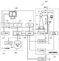
制御部150は、X線検出器80により検出された画像データを基に、種々の画像処理を行う装置である。
制御部150は、X線検出器80で検出されてデジタル信号に変換された画像データを受信し、受信した画像データに基づいて診断に供するように画像処理を行い、画像処理された画像データをデータ処理部170に送信する。制御部150では、例えば、ガンマ変換や、階調変換処理や、その他、画像の拡大や縮小、画像の上下や左右の反転、白黒の反転などを含む色々な画像処理が行われる。
The
The
図2に記載された表示部200の操作画面210の表示領域252には、X線画像の接合処理を行う対象となっている一連のX線画像のファイル名や、 被検体名や、 被検体の性別や、 被検体の年齢や、一連の撮影を行った検査業務を特定するための検査IDや、検査対象の部位、などが表示される。また表示領域254は、検査中に撮影されたX線画像の中から、長尺画像を生成するのに使用するX線画像を選択する操作に使用される。検査中に撮影された各X線画像には順に番号が付される。
図示の画像特定欄256に記載された番号は、上述の番号であり、この番号により各X線画像を特定することができる。但し各画像の特定は、この方法に限るものではなく、各撮影画像を特定する機能を有していればよく数字だけでなく文字を使用しても良い。表示領域252には画像番号欄256や、選択の指示を行う適用欄257や、表示の順位あるいは接合の順位を指示する順位欄258が設けられている。
In the display area 252 of the operation screen 210 has been displayed
The numbers described in the illustrated image specifying column 256 are the above-described numbers, and each X-ray image can be specified by this number. However, identification of each image is not limited to this method, and it is sufficient to have a function for identifying each captured image, and not only numbers but also characters may be used. The display area 252 is provided with an image number column 256, an application column 257 for instructing selection, and a rank column 258 for instructing a display order or a joining order.
長尺画像生成ボタン272は表示領域250にて選定した元画像の画像データから長尺画像の生成を実行するボタンである。終了ボタン274は長尺画像生成画面を終了するボタンである。
The long image generation button 272 is a button for executing generation of a long image from the image data of the original image selected in the display area 250. The end button 274 is a button for ending the long image generation screen.
〔6.撮影漏れの検出〕
長尺画像の撮影において、撮影漏れが生じないようにすることが重要である。 被検体の体軸方向において、撮影画像が互いに重なりを持ってつながることが望ましい。もし重なり部分が途中で途切れると、長尺画像を生成することが困難となる。図22は一定時間毎に実行され、上記課題を解決するフローチャートである。図12のフローチャートが短い周期で実行されるので、操作者にとっては常に本フローチャートが動作しているかのごとく機能する。
[6. (Detection of omissions)
It is important to prevent omissions in shooting long images. In the body axis direction of the subject, it is desirable that the captured images are connected in an overlapping manner. If the overlapping portion is interrupted, it is difficult to generate a long image. FIG. 22 is a flowchart executed at regular intervals to solve the above problem. Since the flowchart of FIG. 12 is executed in a short cycle, the operator always functions as if this flowchart is operating.
ステップS400が開始され、ステップS402で撮影位置が 被検体の体軸方向に移動したかどうかが、検知される。移動していなければステップS412、ステップS414およびステップS416の実行は必要が無いので、ステップS432の実行に移る。撮影位置が移動した場合には、新たに移動した撮影位置が一つ前の撮影位置に対して重なり部分を有するか否かをステップS412で演算する。例えば天板60を移動し過ぎたために重なり部分が無いことがステップS412の演算で明らかになると、ステップS414から実行がステップS416に移る。ステップS416でエラーを表示し、また撮影位置に重なり部分が無いことを警告表示する。
Step S400 is started, and it is detected in step S402 whether or not the imaging position has moved in the body axis direction of the subject . If it has not moved, since it is not necessary to execute step S412, step S414, and step S416, the process proceeds to step S432. If the shooting position has moved, it is calculated in step S412 whether or not the newly moved shooting position has an overlap with the previous shooting position. For example, if it is determined by the calculation in step S412 that the overlapping portion does not exist because the
ステップS432で撮影が終了したかどうかを判断し、もし撮影が終了した場合には、ステップS434で撮影漏れが無いかどうかが演算される。ステップS434の演算の結果重なり部分を介して各X線画像が繋がる場合には、撮影漏れが無いと判断する。もし漏れがあればステップS444に実行が移り、警告が発せられ、エラー表示が出る。この時点で問題点が見付かれば、 被検体は天板60に載置された状態であり、漏れた部分のX線撮影を行うなどの対応を行うことができる。
In step S432, it is determined whether shooting has been completed. If shooting has been completed, it is calculated in step S434 whether there is no shooting omission. If the X-ray images are connected through the overlapped portion as a result of the calculation in step S434, it is determined that there is no imaging omission. If there is a leak, execution proceeds to step S444, a warning is issued, and an error display appears. If a problem is found at this time, the subject is in a state of being placed on the
Claims (10)
前記被検体を透過したX線を検知するX線検出器と、
前記被検体を載置して前記被検体の体軸方向に移動する天板と、
前記X線検出器の検出結果に基づくX線画像を記憶する記憶装置と、
制御部と、
操作を行う操作部と、
表示部と、を備え、
前記記憶装置には、前記被検体の関心領域を重なり合う複数の撮影位置に分けて撮影したX線画像が記憶され、
前記表示部に前記X線画像の撮影位置が複数表示され、表示された前記撮影位置を選択することにより選択された前記撮影位置の前記X線画像が表示され、
撮影された複数のX線画像から長尺画像を生成するためのX線画像が特定され、前記特定されたX線画像に基づいて長尺画像が生成される、ことを特徴とするX線診断装置。 An X-ray tube that irradiates the subject with X-rays;
An X-ray detector for detecting X-rays transmitted through the subject ;
A top plate for placing the subject and moving in the body axis direction of the subject;
A storage device for storing an X-ray image based on the detection result of the X-ray detector;
A control unit;
An operation unit for performing an operation;
A display unit,
Wherein the storage device, the plurality of X-ray images taken divided into shooting position overlapping a region of interest of the subject are stored,
The imaging position of the X-ray image is displayed more, the X-ray image of said selected imaging position is displayed by selecting the displayed said photographing position on said display unit,
X-ray diagnosis for specifying an X-ray image for generating a long image from a plurality of X-ray images taken, and generating a long image based on the specified X-ray image apparatus.
表示された少なくとも2つの前記X線画像の位置関係を調整する位置調整部が設けられ、前記位置調整部の操作に基づき表示された前記X線画像の位置関係が変わることを特徴とする、ことを特徴とするX線診断装置。 The X-ray diagnostic apparatus according to claim 1,
A position adjustment unit that adjusts a positional relationship between at least two displayed X-ray images is provided, and the positional relationship between the displayed X-ray images changes based on an operation of the position adjustment unit. X-ray diagnostic apparatus characterized by the above.
前記記憶装置には、複数の撮影位置に分けて撮影された複数のX線画像に加え、前記複数のX線画像の撮影位置を表す情報が記憶され、前記表示部に画面に設けられる表示領域の座標に従って、前記複数の撮影位置がその撮影位置を表す情報に基づく前記表示領域の座標位置に表示される、ことを特徴とするX線診断装置。 The X-ray diagnostic apparatus according to claim 1,
The storage device stores information representing the imaging positions of the plurality of X-ray images in addition to the plurality of X-ray images captured at a plurality of imaging positions, and a display area provided on the screen in the display unit The X-ray diagnostic apparatus is characterized in that the plurality of imaging positions are displayed at the coordinate positions of the display area based on information representing the imaging positions according to the coordinates.
前記天板の位置を制御する寝台制御部とX線を発生するX線管の位置を制御する機構制御部とが設けられ、前記撮影位置を表す情報は前記天板の位置と前記X線管の位置に対応している、ことを特徴とするX線診断装置。 In the X-ray diagnostic apparatus according to claim 3,
Wherein a mechanism control unit for controlling the position of the X-ray tube for generating a bed controller and the X-ray to control the position of the top plate is provided, said information indicating the shooting position and the position of the front Symbol tabletop the X-ray An X-ray diagnostic apparatus characterized by corresponding to the position of a tube.
前記X線検出器によって造影剤が注入された前記被検体を透過した前記X線が検出され、前記X線検出器による検出結果を所定時間間隔で連続して前記表示部で表示することにより、前記表示部の画面には、前記被検体を流れる前記造影剤の状態が表示され、撮影指示の操作に基づき、前記X線検出器の検出結果がX線画像として取り込まれ、その時の撮影位置を表す情報と共に前記記憶装置に記憶される、ことを特徴とするX線診断装置。 The X-ray diagnostic apparatus according to claim 4,
Is detected prior Symbol the X-rays transmitted through the subject contrast agent is injected by the X-ray detector, by displaying on the display unit a detection result of the X-ray detector continuously at predetermined time intervals , the screen of the display unit, the display state of the contrast medium flowing in the subject, based on the operation of the imaging instruction, the detection result of the X-ray detector is taken as an X-ray image, the photographing position at that time An X-ray diagnostic apparatus characterized by being stored in the storage device together with information representing
前記天板の移動操作に基づく新たな撮影位置が、前の撮影位置と体軸方向において重なり合うかを演算し、重なりあわない場合に警告を行う、ことを特徴とするX線診断装置。 The X-ray diagnostic apparatus according to claim 4,
An X-ray diagnostic apparatus characterized by calculating whether a new imaging position based on the operation of moving the top plate overlaps the previous imaging position in the body axis direction, and gives a warning if they do not overlap.
選択された前記撮影位置において複数のX線画像が撮影されている場合に、前記選択操作により前記複数のX線画像の内の1つが表示され、さらに表示されていない前記複数の他のX線画像を表示する記号が表示され、前記記号が操作されることにより、前記複数の他のX線画像が表示される、ことを特徴とするX線診断装置。 The X-ray diagnostic apparatus according to claim 1,
When a plurality of X-ray images at a selected said imaging position has been taken, the selection one of the plurality of X-ray images by the operation but is displayed, further not displayed the plurality of other X-ray An X-ray diagnostic apparatus, wherein a symbol for displaying an image is displayed, and the plurality of other X-ray images are displayed by operating the symbol.
前記撮影位置が選択されると選択された前記撮影位置のX線画像が表示され、接合処理に使用する前記適用画像の決定操作を行うと、前記撮影位置の前記X線画像が、適用画像を表す表示に変わる、ことを特徴とするX線診断装置。 The X-ray diagnostic apparatus according to claim 1,
When the imaging position is selected, an X-ray image of the selected imaging position is displayed. When the application image used for the joining process is determined, the X-ray image at the imaging position is converted to the applied image. An X-ray diagnostic apparatus characterized in that the display changes to a display.
互いに重なり合う複数の前記撮影位置が、表示部に設けられた表示領域に表示され、前記撮影位置が選択されると選択された前記撮影位置のX線画像が最前面に表示され、さらに他の選択位置が新たに選択されると新たに選択された撮影位置のX線画像が最前面に表示される、ことを特徴とするX線診断装置。 The X-ray diagnostic apparatus according to claim 1 or 8,
A plurality of the imaging positions overlapping each other are displayed in a display area provided in the display unit, and when the imaging position is selected, an X-ray image of the selected imaging position is displayed in the foreground, and another selection An X-ray diagnostic apparatus characterized in that when a position is newly selected, an X-ray image of the newly selected imaging position is displayed in the foreground.
前記被検体を透過したX線検出器によりX線を検知する第2ステップと、
前記被検体を載置する天板を前記被検体の体軸方向に移動する第3ステップと、
前記X線検出器の検出結果に基づき、前記被検体の関心領域を重なり合う複数の撮影位置に分けて撮影したX線画像を記憶装置に記憶する第4ステップと、
前記X線画像を表示部に表示する第5ステップと、
前記表示部に前記X線画像の撮影位置が複数表示される第6ステップと、
表示された前記撮影位置を選択することにより選択された前記撮影位置の前記X線画像が表示される第7ステップと、
撮影された複数のX線画像から長尺画像を生成するためのX線画像が特定される第8ステップと、
前記特定されたX線画像に基づいて長尺画像が生成される第9ステップと、
を備えることを特徴とするX線診断装置のデータ処理方法。 A first step of irradiating a subject with X-rays generated by an X-ray tube;
A second step of detecting X-rays by an X-ray detector transmitted through the subject ;
A third step of moving the top plate on which the subject is placed in the body axis direction of the subject;
A fourth step of storing, in a storage device, an X-ray image obtained by dividing the region of interest of the subject into a plurality of overlapping imaging positions based on the detection result of the X-ray detector;
A fifth step of displaying the X-ray image on a display unit;
A sixth step in which a plurality of imaging positions of the X-ray image are displayed on the display unit;
A seventh step in which the X-ray image of the selected imaging position is displayed by selecting the displayed imaging position;
An eighth step in which an X-ray image for generating a long image from a plurality of captured X-ray images is specified;
A ninth step in which a long image is generated based on the identified X-ray image;
A data processing method for an X-ray diagnostic apparatus, comprising:
Priority Applications (1)
| Application Number | Priority Date | Filing Date | Title |
|---|---|---|---|
| JP2013178897A JP2015047194A (en) | 2013-08-30 | 2013-08-30 | X-ray diagnostic apparatus and data processing method by the same |
Applications Claiming Priority (1)
| Application Number | Priority Date | Filing Date | Title |
|---|---|---|---|
| JP2013178897A JP2015047194A (en) | 2013-08-30 | 2013-08-30 | X-ray diagnostic apparatus and data processing method by the same |
Publications (2)
| Publication Number | Publication Date |
|---|---|
| JP2015047194A JP2015047194A (en) | 2015-03-16 |
| JP2015047194A5 true JP2015047194A5 (en) | 2016-09-08 |
Family
ID=52697722
Family Applications (1)
| Application Number | Title | Priority Date | Filing Date |
|---|---|---|---|
| JP2013178897A Pending JP2015047194A (en) | 2013-08-30 | 2013-08-30 | X-ray diagnostic apparatus and data processing method by the same |
Country Status (1)
| Country | Link |
|---|---|
| JP (1) | JP2015047194A (en) |
Families Citing this family (3)
| Publication number | Priority date | Publication date | Assignee | Title |
|---|---|---|---|---|
| JP2017029324A (en) * | 2015-07-30 | 2017-02-09 | 国立大学法人 千葉大学 | Method and program for creating blood vessel highlighted image data |
| JP2019146679A (en) * | 2018-02-26 | 2019-09-05 | 株式会社島津製作所 | X-ray imaging apparatus |
| CN110226942A (en) * | 2019-05-29 | 2019-09-13 | 上海联影医疗科技有限公司 | Data display method, device, PET scan equipment and storage medium |
Family Cites Families (4)
| Publication number | Priority date | Publication date | Assignee | Title |
|---|---|---|---|---|
| JP4533587B2 (en) * | 2003-02-07 | 2010-09-01 | 株式会社東芝 | Medical image laminating device |
| JP2009011575A (en) * | 2007-07-05 | 2009-01-22 | Toshiba Corp | X-ray image diagnostic system |
| CN102448374B (en) * | 2009-05-27 | 2014-05-14 | 株式会社日立医疗器械 | Image processing apparatus, X-ray image capturing apparatus and image processing method |
| JP5595188B2 (en) * | 2010-08-31 | 2014-09-24 | キヤノン株式会社 | Image processing apparatus and method |
-
2013
- 2013-08-30 JP JP2013178897A patent/JP2015047194A/en active Pending
Similar Documents
| Publication | Publication Date | Title |
|---|---|---|
| RU2748435C2 (en) | Ultrasonic system and method for breast tissue visualization | |
| US10835762B2 (en) | Medical apparatus and method | |
| JP4616872B2 (en) | Image display device and image display program | |
| JP2007000629A (en) | Planning method of inspection and diagnostic device | |
| EP2024935A1 (en) | Method and apparatus for reconstructing an image | |
| JP2007185514A (en) | Imaging medical device and method to set working parameter of imaging medical device | |
| JP2007136164A (en) | Medical image processor and medical image processing method | |
| US8633945B2 (en) | X-ray diagnostic apparatus and image processing apparatus | |
| JP2016019724A (en) | X-ray diagnostic apparatus | |
| US10799100B2 (en) | Image processing device, method, and program | |
| JP2010154982A (en) | X-ray computer tomographic imaging apparatus and image processor | |
| JP2015047194A5 (en) | ||
| JP6143425B2 (en) | X-ray diagnostic equipment | |
| JP2015047194A (en) | X-ray diagnostic apparatus and data processing method by the same | |
| JP2004113592A (en) | Medical image diagnostic apparatus | |
| US20200069276A1 (en) | X-ray imaging apparatus and x-ray image processing method | |
| JP4626414B2 (en) | X-ray diagnostic equipment | |
| JP2007054147A (en) | Apparatus for assisting diagnostic imaging/treatment | |
| US11464474B2 (en) | Medical image processing apparatus, X-ray diagnostic apparatus, and medical image processing method | |
| US20120155738A1 (en) | X-ray apparatus | |
| JP6167383B2 (en) | Medical image generation system, medical image generation method, and medical image generation program | |
| JP7075115B2 (en) | Medical image processing system and medical image processing method | |
| US10980518B2 (en) | Medical image capturing control device, method, and program | |
| JP4503279B2 (en) | X-ray CT system | |
| JP2014212904A (en) | Medical projection system |