JP2014502510A - Improved method for determining cell viability using molecular nucleic acid based techniques - Google Patents
Improved method for determining cell viability using molecular nucleic acid based techniques Download PDFInfo
- Publication number
- JP2014502510A JP2014502510A JP2013547602A JP2013547602A JP2014502510A JP 2014502510 A JP2014502510 A JP 2014502510A JP 2013547602 A JP2013547602 A JP 2013547602A JP 2013547602 A JP2013547602 A JP 2013547602A JP 2014502510 A JP2014502510 A JP 2014502510A
- Authority
- JP
- Japan
- Prior art keywords
- cells
- pcr
- dna
- present
- dead
- Prior art date
- Legal status (The legal status is an assumption and is not a legal conclusion. Google has not performed a legal analysis and makes no representation as to the accuracy of the status listed.)
- Pending
Links
Images


Classifications
-
- C—CHEMISTRY; METALLURGY
- C12—BIOCHEMISTRY; BEER; SPIRITS; WINE; VINEGAR; MICROBIOLOGY; ENZYMOLOGY; MUTATION OR GENETIC ENGINEERING
- C12Q—MEASURING OR TESTING PROCESSES INVOLVING ENZYMES, NUCLEIC ACIDS OR MICROORGANISMS; COMPOSITIONS OR TEST PAPERS THEREFOR; PROCESSES OF PREPARING SUCH COMPOSITIONS; CONDITION-RESPONSIVE CONTROL IN MICROBIOLOGICAL OR ENZYMOLOGICAL PROCESSES
- C12Q1/00—Measuring or testing processes involving enzymes, nucleic acids or microorganisms; Compositions therefor; Processes of preparing such compositions
- C12Q1/68—Measuring or testing processes involving enzymes, nucleic acids or microorganisms; Compositions therefor; Processes of preparing such compositions involving nucleic acids
- C12Q1/6876—Nucleic acid products used in the analysis of nucleic acids, e.g. primers or probes
- C12Q1/6888—Nucleic acid products used in the analysis of nucleic acids, e.g. primers or probes for detection or identification of organisms
- C12Q1/689—Nucleic acid products used in the analysis of nucleic acids, e.g. primers or probes for detection or identification of organisms for bacteria
-
- C—CHEMISTRY; METALLURGY
- C12—BIOCHEMISTRY; BEER; SPIRITS; WINE; VINEGAR; MICROBIOLOGY; ENZYMOLOGY; MUTATION OR GENETIC ENGINEERING
- C12Q—MEASURING OR TESTING PROCESSES INVOLVING ENZYMES, NUCLEIC ACIDS OR MICROORGANISMS; COMPOSITIONS OR TEST PAPERS THEREFOR; PROCESSES OF PREPARING SUCH COMPOSITIONS; CONDITION-RESPONSIVE CONTROL IN MICROBIOLOGICAL OR ENZYMOLOGICAL PROCESSES
- C12Q1/00—Measuring or testing processes involving enzymes, nucleic acids or microorganisms; Compositions therefor; Processes of preparing such compositions
- C12Q1/68—Measuring or testing processes involving enzymes, nucleic acids or microorganisms; Compositions therefor; Processes of preparing such compositions involving nucleic acids
- C12Q1/6806—Preparing nucleic acids for analysis, e.g. for polymerase chain reaction [PCR] assay
-
- C—CHEMISTRY; METALLURGY
- C12—BIOCHEMISTRY; BEER; SPIRITS; WINE; VINEGAR; MICROBIOLOGY; ENZYMOLOGY; MUTATION OR GENETIC ENGINEERING
- C12Q—MEASURING OR TESTING PROCESSES INVOLVING ENZYMES, NUCLEIC ACIDS OR MICROORGANISMS; COMPOSITIONS OR TEST PAPERS THEREFOR; PROCESSES OF PREPARING SUCH COMPOSITIONS; CONDITION-RESPONSIVE CONTROL IN MICROBIOLOGICAL OR ENZYMOLOGICAL PROCESSES
- C12Q2600/00—Oligonucleotides characterized by their use
- C12Q2600/106—Pharmacogenomics, i.e. genetic variability in individual responses to drugs and drug metabolism
Landscapes
- Chemical & Material Sciences (AREA)
- Life Sciences & Earth Sciences (AREA)
- Organic Chemistry (AREA)
- Analytical Chemistry (AREA)
- Proteomics, Peptides & Aminoacids (AREA)
- Zoology (AREA)
- Wood Science & Technology (AREA)
- Health & Medical Sciences (AREA)
- Engineering & Computer Science (AREA)
- Microbiology (AREA)
- Bioinformatics & Cheminformatics (AREA)
- Molecular Biology (AREA)
- Biotechnology (AREA)
- Biophysics (AREA)
- Physics & Mathematics (AREA)
- Biochemistry (AREA)
- Immunology (AREA)
- General Engineering & Computer Science (AREA)
- General Health & Medical Sciences (AREA)
- Genetics & Genomics (AREA)
- Chemical Kinetics & Catalysis (AREA)
- Measuring Or Testing Involving Enzymes Or Micro-Organisms (AREA)
- Investigating Or Analysing Biological Materials (AREA)
Abstract
【解決手段】 本発明は、選択された運転中の電池の次の質問が微生物生存度の存在のインジケータであるところを新規な方法および、選択的に死細胞を有効なおよび死細胞を含有している混合薬から除外するために、臨床サンプル、血液製剤、健康診断/生物工学製品および食品の微生物細胞のようなキットと関連づける。特に、BacteremiaおよびFungemiaサンプルから微生物細胞生存度を有する相関のために、本発明は、Polymerase Chain Reaction(PCR)のようなダイレクト核酸増幅技術を実行する改良された方法および血液および他の体液の等温技術に関する。本発明によって設けられている改良された方法は特に敗血症の診断のために有利である、そして、全部で病的状態を決定するために、通常無菌のもう一方は流体を具体化する。
【選択図】 図3The present invention includes a novel method and a method that selectively validates dead cells and contains dead cells where the next question for a selected running battery is an indicator of the presence of microbial viability. In order to be excluded from existing drug combinations, it is associated with kits such as clinical samples, blood products, medical / biotech products and food microbial cells. In particular, because of the correlation with microbial cell viability from Bacteremia and Fungemia samples, the present invention provides improved methods for performing direct nucleic acid amplification techniques such as Polymerase Chain Reaction (PCR) and isothermal of blood and other body fluids. Regarding technology. The improved method provided by the present invention is particularly advantageous for the diagnosis of sepsis, and in order to determine the overall pathological condition, the normally sterile one embodies the fluid.
[Selection] Figure 3
Description
関連出願の相互参照
本出願は仮出願ではなく、2010年12月31日に提出された米国仮出願第61/428,892は参照により本明細書に組み込まれ、該仮出願に対して優先権を主張するものである。
CROSS REFERENCE TO RELATED APPLICATIONS This application is not a provisional application, and US provisional application 61 / 428,892, filed December 31, 2010, is incorporated herein by reference and has priority over the provisional application. Is an insistence.
本発明は、分子検出によって、生細胞および死細胞を含む混合物から死細胞のDNAを選択的に除去するための方法に関するものであり、特に、菌血症、真菌血症、ウイルス血症および試料を含む寄生生物の他のタイプからの生存細菌と相関関係にある血液および体液において直接的ポリメラーゼ連鎖反応(PCR)技術を実行するための改善された方法に関するものである。本発明によって提供される改善された方法は、敗血症の診断において特に役立つものである。 The present invention relates to a method for selectively removing dead cell DNA from a mixture comprising live and dead cells by molecular detection, in particular bacteremia, fungiemia, viremia and samples An improved method for performing direct polymerase chain reaction (PCR) techniques in blood and body fluids that correlate with viable bacteria from other types of parasites, including: The improved method provided by the present invention is particularly useful in the diagnosis of sepsis.
敗血症を診断することにおいて、結果としてなる(TTR)時間は、患者生存で最も重要な判定である。現在では、血液培養は金本位であるが、比較的遅い。そして、正になるために15時間(3時間〜5日の一般の範囲において)の近似の時間の中央値との次の同一視のための生存可能な微生物を生成する。そして、それの後、微生物識別は概して分析のためのさらに1―2日を加えることができる。PCRのような分子学的手法は、非常に改良型のTTRを微生物識別に対して提供するが、サンプル準備の間、生き残れる微生物細胞の不十分な選択性のため、主に特性の欠如が欠点である。従来は、血液の従来の敗血症PCR法は高コストのDNA隔離がPCR抑制剤を取り出すことを必要とする、しかし、主に死微生物細胞からのDNAおよびDNA隔離手順の間、従属する損失を処理しているサンプルの包含のため、隔離も血液培養の金本位と比較して偽陽性および感度の低下を与える。 In diagnosing sepsis, the resulting (TTR) time is the most important decision in patient survival. Currently, blood culture is gold standard but relatively slow. It then produces viable microorganisms for the next identification with the approximate median time of 15 hours (in the general range of 3 hours to 5 days) to become positive. And after that, microbial identification can generally add an additional 1-2 days for analysis. Molecular techniques such as PCR provide a much improved TTR for microbial discrimination, but the lack of properties mainly due to insufficient selectivity of surviving microbial cells during sample preparation It is. Traditionally, conventional septic PCR methods of blood require high cost DNA sequestration to remove PCR inhibitors, but deal with dependent losses primarily during DNA and DNA sequestration procedures from dead microbial cells. Because of the inclusion of the sample, isolation also gives false positives and reduced sensitivity compared to the gold standard of blood culture.
伝統的に、敗血症血液サンプルPCR準備は血液から常に単離されたDNAを有する、そして、長いおよび周知の血液を取り出す血液製剤はTaqポリメラーゼ(Kloucheおよびシュローダー物品が下記を引用したということを知る)のPCR Inhibitorsを引き出した。最近、この抑制を克服する試みで、一部のグループはこれらのポリメラーゼ上の血液製剤の抑制影響を減らすために設計される熱安定ポリメラーゼ(例えば周知の「omni taq」および「Phusion」技術)を修正したのと同様に、PCRを強化している混合物を開発した(JMD(2010)を参照;
12(2)(pp.152―161))。しかしながら、これらの方法の両方の制約は、まだ、最低による敏感性の欠如が許容したどちらでも受ける血液量、そして、高いコスト、そして、隔離システムと関係しているサンプルおよび高い複雑さの損失。さらに、DNA Isolationシステムは死細胞からしばしば細胞遊離DNAを含む。そして、それは偽陽性を混乱させることを引き起こす効果を有することができる。
Traditionally, sepsis blood sample PCR preparations have DNA that has always been isolated from blood, and blood products that draw long and well-known blood are Taq polymerase (Kloche and Schroder articles know that cited below) PCR Inhibitors were pulled out. Recently, in an attempt to overcome this suppression, some groups have introduced thermostable polymerases designed to reduce the inhibitory effects of blood products on these polymerases (eg, the well-known “omni taq” and “Phusion” technologies). A mixture enhancing PCR was developed as modified (see JMD (2010);
12 (2) (pp. 152-161)). However, the limitations of both of these methods are still the volume of blood incurred by any lack of sensitivity due to the minimum, and the high cost and loss of sample and high complexity associated with the isolation system. In addition, DNA Isolation systems often contain cell free DNA from dead cells. And it can have the effect of causing confusion with false positives.
Klouche、M.およびシュローダー(「血流感染症の診断のための急速方法」という題名の物品のU.)は、クランにおいて公表した。化学労働党。med.2008;46(7)(pp.888―908)は、患者からの血液の微生物病原体の直接の核酸ベースの検出および識別が血流感染症の迅速な診断のための有望な道具でありえることを開示する。本論文によると、血流感染症の日常的な精密検査のための幅広い範囲分子方法による循環細菌であるか菌類の核酸の検出の重要性は、しかしながら、現在明白でない。品質の改良および分子ものの再現性のための奨励の問題微生物安全分析からの経験に示すように、血流感染症の診断用アプリケーションは、細菌核酸、ブロッキングまたは過剰なヒトDNAの除去方法のための選択的な強化手順および臨床的に関連した所見を識別する生存度標識の使用を含む。現在高価なおよび技術的に厳しい技術(疾患を指向する多重PCR)にもかかわらず病原マイクロアレイ、そして、プロテオ・マイク側面図を作る伝染病診断のための重要な急速なおよび高いスループット診断手段として進化する可能性を有する。現時点では、3つの主考慮すべき問題は、血流感染症の日常的な診断の分子技術のユニークなアプリケーションを排除する:1によるNAT結果の解釈において問題点)外部の汚染の高い危険、感染の後の核酸のうち拡張した持続、そして、一時的な菌血(2))臨床的に関連した低い細菌負荷のために、そして、特定のバクテリアおよび菌類の検出および3のために限られた分析的な感度)同じく分子ものプロテオ・マイク・テストによるによる日常的な抗菌性の感受性テストの不足。 Klouche, M.C. And Schroeder (U. of the article entitled “Rapid Method for Diagnosis of Bloodstream Infections”) published in Clan. Chemical labor party. med. 2008; 46 (7) (pp. 888-908) show that direct nucleic acid-based detection and identification of microbial pathogens in blood from patients can be a promising tool for rapid diagnosis of bloodstream infections. Disclose. According to this paper, however, the importance of detecting circulating bacterial or fungal nucleic acids by a wide range of molecular methods for routine work-up of bloodstream infections is not currently clear. Encouragement issues for quality improvement and reproducibility of molecules as shown in experience from microbial safety analysis, diagnostic applications for bloodstream infections are for bacterial nucleic acid, blocking or excess human DNA removal methods Includes the use of viability markers to identify selective enhancement procedures and clinically relevant findings. Evolving as an important rapid and high-throughput diagnostic tool for infectious disease diagnosis to make pathogenic microarrays and proteo-mic side view despite current expensive and technically demanding technology (disease-oriented multiplex PCR) Have the potential to do. At present, the three main issues to consider are to eliminate the unique application of molecular technology for routine diagnosis of bloodstream infections: a problem in the interpretation of NAT results by 1) high risk of external contamination, infection Limited for extended persistence and transient bacteremia (2)) of clinically relevant low bacterial load and for detection of specific bacteria and fungi and 3 Analytical sensitivity) Lack of routine antimicrobial susceptibility testing due to the same molecular proteo-mic test.
運転中のおよび死電池の分化は、微生物診断法の重要なチャレンジである。代謝および再生活性および、病原性微生物の場合、潜在的健康リスクは、混合微生物人口の有効な部分に限られている。蛍光汚れを使用している流れ血球計算で、4つの生理的状態が、区別するために、従来の技術で使われる:繁殖的に現実的な、代謝的に作動中の、完全なおよび、透過する電池。状況に応じて、透過する細胞以外のすべてのステージには、復活に回復の可能性があることができて、このようにライブで潜在的に考慮されることができなければならない。3週(DNA)までの日間の範囲の細胞死の後のDNAの比較的長い持続に対する与えられるべきもの―ベースの診断法は、運転中の電池の数を過大評価する傾向がある。試料から抽出されるDNAは、そうすることができる死透過する細胞を含んでいる4つの言及された生理的状態のいずれかの細胞から生じる。後者の検出は、しかしながら、要求されない。現実的なおよび不可逆的に傷害性電池をと区別するための最も重要な基準は、膜完全性である。膜を危うくされた細胞に由来するノイズをソートすることは、代謝動作および健康リスクを細菌共同体の完全なおよび生き残れる部分に割り当てるのを助ける。完全な膜を有する運転中の電池は、DNAを除外するそれらの能力によって特徴づけられた―容易に死者または膜を透過する結合染料―危うくされた細胞。 Differentiation of operating and dead cells is an important challenge for microbial diagnostics. In the case of metabolic and regenerative activities and pathogenic microorganisms, potential health risks are limited to an effective part of the mixed microbial population. In flow cytometry using fluorescent stains, four physiological states are used in the prior art to distinguish: reproductively realistic, metabolically active, complete and transparent Battery to play. Depending on the situation, all stages other than the permeating cells must have the potential to recover to resurrection and thus must be potentially considered live. What is to be given for the relatively long duration of DNA after cell death in the range of days up to 3 weeks (DNA) -based diagnostics tend to overestimate the number of batteries in operation. The DNA extracted from the sample arises from cells in any of the four mentioned physiological states, including cells that are capable of death permeation. However, the latter detection is not required. The most important criterion for distinguishing realistic and irreversibly damaging cells is membrane integrity. Sorting noise from cells that have compromised the membrane helps assign metabolic behavior and health risks to the complete and surviving part of the bacterial community. Running batteries with complete membranes were characterized by their ability to exclude DNA-easily dead or bound dye that permeates the membrane-compromised cells.
近年では、EMA―PCRは、運転中のおよび死電池をと区別する顕微鏡であるか流れ血球計算分析の使いやすい変形例であることが報告された。この診断DNAベースの方法は、運転中の死識別染料の使用を速度およびリアルタイムPCRの感受性と結合する。エチジウム・モノ・アジ化物(EMA)。DNA―明るい可視光(460ナノメートルの最大吸収度)への暴露のDNAに化学ものの共有結合締め具がこの点に関しては用いられたと認めているアジ化物グループを有する染料をさし込むこと。細胞は、染料が危うくされた細胞壁/膜を有する死細胞を透過して、それらのDNAと結合することができて、5分間のEMAにさらされる。明るい可視光を使用しているEMAの光分解は、DNAおよび他の分子への共有結合リンクを形成することができるnitreneを生じる。 In recent years, EMA-PCR has been reported to be a microscope that distinguishes between running and dead cells or an easy-to-use variant of flow cytometry analysis. This diagnostic DNA-based method combines the use of an operating death discriminating dye with the speed and sensitivity of real-time PCR. Ethidium mono-azide (EMA). DNA—Incorporate dyes with azide groups that have been found to have been used in this regard for chemical covalent binding to DNA exposed to bright visible light (maximum absorbance at 460 nanometers). Cells can permeate dead cells with cell walls / membranes that have been compromised by the dye, bind to their DNA and are exposed to EMA for 5 minutes. Photolysis of EMA using bright visible light yields nitrene that can form covalent links to DNA and other molecules.
光誘導性架橋結合は、DNAのPCR増幅の死細胞を妨げることが報告された。実際にDNAにEMA―crosslinkingすることがゲノムDNA抽出の間、細胞片と共に損失に不溶性DNAおよび導線を与えることが最近示された。解放されたEMA(溶液において自由なままである)は、同時に、水分子と反応することによって不活発にされることができる。結果として生じるヒドロキシルアミンは、共有結合してDNAと結合することがもはやできない。生細胞(完全な細胞によって露光量の前に反応EMAから保護されている)からのDNA膜/細胞壁は、従って、細胞溶解の後、不活性EMAに影響を受けない。従って、このように現実的なおよび死電池の混成から成る細菌培養組織のEMA処理は、死細胞からDNAの選択的な除去につながる。テストされる種は、大腸菌0157であった:H7、サルモネラ属typhimu|iim、リステリア菌およびカンピロバクター属Jejuni。これらの研究は、しかしながら、死細胞からDNAの選択的な損失を調べなかった。 Light-induced cross-linking has been reported to prevent dead cells of DNA PCR amplification. It has recently been shown that EMA-crosslinking to DNA actually gives insoluble DNA and leads to loss with cell debris during genomic DNA extraction. Liberated EMA (which remains free in solution) can be made inactive by reacting with water molecules at the same time. The resulting hydroxylamine can no longer be covalently bound to DNA. DNA membranes / cell walls from living cells (protected from reactive EMA before exposure by intact cells) are therefore not affected by inactive EMA after cell lysis. Thus, EMA treatment of bacterial cultures consisting of such realistic and dead cell hybrids leads to selective removal of DNA from dead cells. The species tested was E. coli 0157: H7, Salmonella typhimu | im, Listeria monocytogenes and Campylobacter Jejuni. These studies, however, did not examine selective loss of DNA from dead cells.
この技術が有望であるにもかかわらず、DNA抽出より前のEMAの使用が大きな欠点が欠点であるとわかった。場合によっては、処理も、ログ位相において収集される生細胞のゲノムDNAのほぼ60%の損失に結果としてなった。EMAも直ちに、部分的なDNA損失に結果としてなっている他の細菌種の生細胞を透過すると述べられた。選択性の、そして、全体の適用性のこの不足は、新しく発達した代わりの化学製品のテストにつながった:Propidiumモノ・アジ化物(PMA)。公開された特許出願、ネッカーに対するWO/2007/100762、その他において、2007年9月7日に発表されて、選択的に運転中のおよび死電池の定義済みの部分を有する細菌培養組織から死細胞のゲノムDNAの検出を取り除くPMAの適合性は、開示される。PMAはヨウ化(PI)propidiumと同一である。但し、次の場合は除く−アジ化物グループの更なる存在は露光量にDNAにクロス結合を許容する。PIは、混合集団の死細胞を確認するために広範囲に用いた。PMA分子(EMAの場合1つだけと比較した2つの正電荷)およびPIを有する生きられない細胞の選択的な染色が多種多様な細胞タイプにうまく実行されたという理由のより高い負担は、分野の人々にPMAの使用がEMAによって観察される欠点を緩和するかもしれないと思わせた。この発表された特許において、PMA集中およびインキュベーション時間は、これらのパラメータを異なる細菌種の幅広い範囲の研究に適用する前に、グラム陰性1つおよび1つのグラム陽性の有機体によって最適化された。開示された方法が、意図的に分子診断法を微生物共同体の部分に制限する完全な細胞膜。これは、完全なおよび膜妥協された細胞の混成をphenanthridium誘導剤にさらすことによって達成される。開示された好ましい実施例において、PCRは、テンプレートとして混合からゲノムDNAを使用して実行される。 Despite the promise of this technique, the use of EMA prior to DNA extraction has proven to be a major drawback. In some cases, treatment also resulted in approximately 60% loss of live cell genomic DNA collected in the log phase. EMA was also stated to immediately permeate live cells of other bacterial species resulting in partial DNA loss. This lack of selectivity and overall applicability has led to the testing of newly developed alternative chemicals: Propidium monoazide (PMA). Published cells, published in the patent application WO / 2007/100762, Neckar, et al., September 7, 2007, dead cells from bacterial cultures with a defined part of selectively running and dead cells The suitability of PMA to eliminate detection of genomic DNA is disclosed. PMA is identical to iodinated (PI) rapidium. However, except in the following cases-the further presence of the azide group allows the DNA to cross-link to the exposure dose. PI was used extensively to identify mixed population dead cells. The higher burden of the selective staining of non-viable cells with PMA molecules (two positive charges compared to only one in the case of EMA) and PI has been successfully performed on a wide variety of cell types Of people thought that the use of PMA might alleviate the drawbacks observed by EMA. In this published patent, PMA concentration and incubation time were optimized by one Gram negative and one Gram positive organism before applying these parameters to a wide range of studies of different bacterial species. A complete cell membrane in which the disclosed method deliberately limits molecular diagnostics to parts of the microbial community. This is accomplished by exposing the complete and membrane compromised cell mix to a phenanthridium inducer. In the preferred embodiment disclosed, PCR is performed using genomic DNA from the mix as a template.
また、Published米国特許出願番号第2008/0160528号は、ローレンツに、2008年7月3日に発表されて、1またはいくつかのカオトロピック剤の存在および/または1の核酸またはいくつかの界面活性剤を分解させるためのヌクレアーゼ(特にDNA品位を下げるヌクレアーゼ)の使用を開示する。更なるこの特許出願にはDNAとこの種の方法を実施するためのキットと同様にRNAの混合物からRNAを精製する方法が開示されている。開示もする微生物細胞からの特に単離核酸のための方法が、加えて、この種の方法を実施するためのキットと同様により高い真核生物細胞から成る混合サンプルにおいて提供する。 Published US Patent Application No. 2008/0160528 was also published in Lorentz on July 3, 2008, in the presence of one or several chaotropic agents and / or one nucleic acid or several surfactants. Disclose the use of nucleases (especially nucleases that lower DNA quality) to degrade This further patent application discloses a method for purifying RNA from a mixture of RNA as well as a kit for carrying out this type of method. The disclosed method, particularly for isolated nucleic acids from microbial cells, is additionally provided in mixed samples consisting of higher eukaryotic cells as well as kits for performing such methods.
もう一方は、特許出願、ルーディに対するWO/2001/077379、その他を発行した。そして、2001年10月18日に発表される、サンプルの、そして、サンプルの中で細胞集団に関する定量的情報を得るための細胞を検出する方法を開示する。特に、方法は、サンプルの生活および死細胞をと区別するために開示される。方法は、サンプルの中で死細胞の核酸を修正する生存度プローブを有するサンプルを接触させることを有し、サンプルの細胞からの検出用核酸を備えている。記載されてもいるサンプル(成り立っている方法)の細胞を検出する方法である:サンプルの中で死細胞の核酸にラベルをつける生存度プローブを有するサンプルを接触させている(a);ラベルをつけられたおよび非のラベルが付いた分数に、核酸を細胞から分離する;(b)そして、分数の一方または両方の核酸を検出している(c)。 The other issued a patent application, WO / 2001/077379 to Rudy, and others. And disclosed is a method for detecting cells for obtaining quantitative information about a cell population in a sample and in a sample, published on October 18, 2001. In particular, the method is disclosed to distinguish between live and dead cells of a sample. The method comprises contacting the sample with a viability probe that modifies the nucleic acid of dead cells in the sample and comprises nucleic acid for detection from the cells of the sample. A method for detecting cells of a sample (consisting method) also described: contacting a sample with a viability probe that labels dead cell nucleic acids in the sample (a); Nucleic acids are separated from cells into labeled and unlabeled fractions; (b) and one or both nucleic acids in the fraction are detected (c).
前述の背景技術からみて、方法シフトがライブで効果的に識別する方法対分子核酸ベースの分析技術(例えば、発生するPCRの前に)より前の死微生物細胞DNAを開発することであることが分かる、そして、それも、例えば、PCR抑制剤を取り出して、目標DNAに集中するように設計された従来の隔離の高コストの負の効果を迂回する。驚くべきことに、本発明の実施例の実行によれば、PCRが血液に由来する生き残れる微生物細胞と相関することが示された。そして、選択的な血球溶解の組合せを使用した。そして、次の微生物細胞溶解およびPCRとともに(そして、または)デオキシリボヌクレアーゼを洗った。 In view of the foregoing background art, the method shift is to develop dead microbial cell DNA prior to methods that effectively discriminate between live versus molecular nucleic acid based analytical techniques (eg, prior to occurring PCR). As can be seen, it also bypasses the high-cost negative effects of conventional sequestration designed to, for example, remove PCR inhibitors and concentrate on the target DNA. Surprisingly, according to the practice of the examples of the present invention, it has been shown that PCR correlates with surviving microbial cells derived from blood. A selective hemolysis combination was then used. The deoxyribonuclease was then washed with (and / or) subsequent microbial cell lysis and PCR.
このように、上記の従来の方法とは対照的に、本発明は、分子核酸ベースの技術の、PCRを含む、劇的に高コストのDNA隔離およびサンプル準備を単純化することによる、そして、DNAを分離しなくて、むしろ死微生物DNAおよび細胞の迅速な分離の後、天然微生物溶解物上の迅速なおよび単純な直接解析を実行することによって、生き残れる微生物細胞の選択的な濃縮に結果としてなることによる潜在的TTR優位性を実現しようとする。これは、敗血症の診断において特に、そして、予想外に有利で、本発明の好ましい実施例に一致することを達成される:
I.すなわち、陽非汚染されたPCR結果より前の混乱させている死微生物細胞DNAの除去がその生細胞がそうであることを示すは、生存可能な敗血症微生物(s)の存在を提示して、この種のPCR結果意志として、示す現実的な血液微生物PCR=敗血症微生物。
Thus, in contrast to the conventional methods described above, the present invention is based on the simplification of molecular nucleic acid based techniques, including PCR, including dramatically high cost DNA isolation and sample preparation, and By performing rapid and simple direct analysis on natural microbial lysates after rapid separation of dead microbial DNA and cells without separating DNA, the result is selective enrichment of surviving microbial cells. To achieve a potential TTR advantage. This is achieved in particular and unexpectedly advantageous in the diagnosis of sepsis and in accordance with the preferred embodiment of the present invention:
I. That is, removal of disrupted dead microbial cell DNA prior to positive non-contaminated PCR results indicates that the live cells are so as to indicate the presence of viable septic microorganisms (s) As a result of this kind of PCR result, the realistic blood microorganism PCR shown = septic microorganism.
II. 血液からの周知の、死微生物細胞が血液培養において成長することができなくされているように、このように、重要な微生物に特有のPCRを測定しているいかなる2つ以上の時点も単一の血液培養ビンからの増加が生存可能な微生物を測定していなければならないと合図する。 II. Thus, any two or more time points measuring PCR specific to important microorganisms, such that well-known, dead microbial cells from blood cannot be grown in blood culture. Signal that the increase from a single blood culture bottle must be measuring viable microorganisms.
III. 血液からのPCR抑制因子は、化学変性剤の単純な組合せを介して除去されることができる(chaotropes:洗剤、pH、塩類、アルコール類およびアミンを含んでいる合成物のような双極子瞬間を経た有機化学ベースの差動の救済及び酵素(例えばヌクレアーゼ、プロテイナーゼその他))そして、洗うこと。(このことによりDNA隔離を迂回して、微生物溶解物―Direct―PCRを可能にする)
IV. 血液および血液培養に存在する生きている/死んだ微生物の比率が、それから、治療の、そして、処理の有効性を試験する効果の程度として使われることができる。
III. PCR inhibitors from the blood can be removed through a simple combination of chemical denaturants (chaotropes: dipoles like compounds containing detergents, pH, salts, alcohols and amines) Organic chemistry-based differential relief and enzymes (eg nucleases, proteinases etc.) via instant and washing. (This bypasses DNA sequestration and enables microbial lysate-Direct-PCR)
IV. The ratio of live / dead microorganisms present in the blood and blood culture can then be used as a measure of the effectiveness of testing the effectiveness of the treatment and treatment.
したがって、分子検出から、改良された方法を選択的に死細胞のDNAを有効なおよび死細胞を含有している混合薬から除外するために提供することは、本発明の目的である。 Accordingly, it is an object of the present invention to provide improved methods to selectively exclude dead cell DNA from effective and dead cell mixed drugs from molecular detection.
それは更なる目的であるの、効果的に識別する改良された方法が生きると定める本発明対分子核酸ベースの分析法より前の死微生物細胞DNAまたはPCRは事業を始めて、そして、それもPCR抑制剤を取り出して、目標DNAに集中するように設計されたそれらのような従来の隔離の高コストの負の効果を迂回する。 It is a further objective that dead microorganism cell DNA or PCR prior to the present invention versus molecular nucleic acid-based analysis methods that define an improved method to effectively identify live, and that also PCR suppression Take out the agent and bypass the high-cost negative effects of conventional isolation, such as those designed to concentrate on the target DNA.
例えば、次の微生物細胞溶解およびPCRとともに(そして、または)デオキシリボヌクレアーゼを洗って、選択的な血球溶解の組合せを使用することによって、PCRおよび他の分子分析技術の結果を血液に由来する生き残れる微生物細胞の存在に関連させる方法を提供することは、本発明の他の目的である。 For example, by washing the deoxyribonuclease with (and / or) subsequent microbial cell lysis and PCR, and using a combination of selective hemolysis, surviving microorganisms derived from blood by PCR and other molecular analysis techniques It is another object of the present invention to provide a method relating to the presence of cells.
改良された方法をBacteremiaおよびFungemiaサンプルから血液のダイレクトPCR技術および生き残れる微生物細胞を有する相関のための他の体液を実行するために提供することは本発明の更に別の目的である。そして、この種の改良された方法が特に敗血症の診断のために有利な本発明によって設けられている。 It is yet another object of the present invention to provide improved methods for performing direct PCR techniques on blood and other body fluids for correlation with surviving microbial cells from Bacteremia and Fungemia samples. And this kind of improved method is provided by the present invention which is particularly advantageous for the diagnosis of sepsis.
更なる目的および本発明の効果は、その好ましい実施例の以下の説明から明らかである。 Further objects and advantages of the present invention will be apparent from the following description of preferred embodiments thereof.
本発明が記載されていたにもかかわらず、以下の実施例も本発明の実施例の特定の具体例として、そして、理解の明快さのために設けられている。前へ本願明細書においてセットとしての本発明の教示を考慮して、特定の変更及び改造がこのように元気または本発明の範囲から逸脱することなく、記載されているこれらの実施例になされることができることは、当業者にとって直ちに明らかである。 Despite the invention being described, the following examples are also provided as specific examples of embodiments of the invention and for clarity of understanding. In view of the teachings of the present invention as a set herein before, certain changes and modifications may thus be made to these described embodiments without departing from the spirit or scope of the present invention. The ability to do so will be readily apparent to those skilled in the art.
カオトロピック剤(別名カオトロピック試薬およびchaotrope)は、高分子(例えばタンパク質、DNAまたはR A)の三次元構造を崩壊させる物質であって、それらを変性させる。カオトロピック剤は、非共有結合力(例えば水素結合、ファンデルワールス力および恐水症の効果)によって伝達されるinter―分子相互作用を安定させることを妨げる。円偏光二色性がchaotrope濃度依存的なファッションにおいて滴定されることができるにつれて手段によってそのようなものを検出したので、しばしば構造特徴。例えば、カオトロピック試薬は、含む:
尿素6−8つのmol/1
グアニジウム塩化物6 mol/1
リチウム過塩素酸塩4.5 mol/1
変性(生化学)
加えて、負担を保護して、塩橋の安定化を防止することによって、高い一般的な塩類は、カオトロピック特性を有することができる。水素結合は無極性メディアでより強いので、塩類(溶媒の双極子瞬間を増加させる)は水素結合を不安定にすることもできる。
Chaotropic agents (also called chaotropic reagents and chaotropes) are substances that disrupt the three-dimensional structure of macromolecules (eg proteins, DNA or RA) and denature them. Chaotropic agents prevent stabilizing inter-molecular interactions that are transmitted by non-covalent forces (eg, hydrogen bonds, van der Waals forces and the effects of diarrhea). Often structural features as such have been detected by means as circular dichroism can be titrated in a chaotrope concentration dependent fashion. For example, chaotropic reagents include:
Urea 6-8 mol / 1
Guanidium chloride 6 mol / 1
Lithium perchlorate 4.5 mol / 1
Denaturation (biochemistry)
In addition, by protecting the burden and preventing salt bridge stabilization, highly common salts can have chaotropic properties. Since hydrogen bonds are stronger in nonpolar media, salts (which increase the dipole moment of the solvent) can also destabilize hydrogen bonds.
円偏光二色性がchaotrope濃度依存的なファッションにおいて滴定されることができるにつれて手段によってそのようなものを検出したので、しばしば構造特徴。生化学および分子生物学の歴史的に役立つカオトロピック試薬の若干の例は、以下から成る:尿素6−8つのmol/1、グアニジウム塩化物6 mol/1、リチウム過塩素酸塩4.5 mol/1、alchohols、アミン(特に4原子のアミン)洗剤(特に非イオン物質)pH変化、ベタイン、プロリン、カルニチン、トレハロース、NP―40など、本発明とのBSA.In一致と同様に、実験(DoE)プロセスの設計が、有効な製剤範囲の最適化およびさまざまなchaotropes(混合物または試薬または「カクテル」)の範囲の組合せのために使われた:それらがそれらのサイズ(濾過)および密度(遠心分離)に基づいて運転中の電池から容易に切り離されるように、a)は死セル構造を変性させる;そして、b)吹きさらしの有効なセルが切り離した結果として生じるchaotropeカクテルを作成する下流の分析増幅分析(例えばPCRおよび有効な細胞派生内在性タンパク質)と直接互換性を持つ、そして、それらのかなりの生化学動作を維持する溶液。事実上、カオトロピック・カクテルはその差動の膜完全性に基づいて死細胞からライブで分化するために最適化される。そして、生存度相関分析のための有効な細胞内在性タンパク質動作を維持する。 Often structural features as such have been detected by means as circular dichroism can be titrated in a chaotrope concentration dependent fashion. Some examples of historically useful chaotropic reagents in biochemistry and molecular biology consist of: urea 6-8 mol / 1, guanidinium chloride 6 mol / 1, lithium perchlorate 4.5 mol / 1, alcohol, amine (especially 4-atom amine) detergent (especially nonionic substances) pH change, betaine, proline, carnitine, trehalose, NP-40, etc. Similar to In agreement, experimental (DoE) process designs were used for effective formulation range optimization and combinations of different chaotropes (mixtures or reagents or “cocktails”) ranges: A) denatures the dead cell structure so that it is easily detached from the running battery based on size (filtration) and density (centrifugation); and b) results from the detachment of the effective cell of the windshield Solutions that are directly compatible with downstream analytical amplification analysis (eg, PCR and effective cell-derived endogenous proteins) to create a chaotrope cocktail and maintain their significant biochemical behavior. In effect, chaotropic cocktails are optimized for live differentiation from dead cells based on their differential membrane integrity. It maintains effective cellular endogenous protein behavior for viability correlation analysis.
サンプル準備:
優先血球溶解状況は、例えば、敗血症血液培養サンプルで見つかって、血液―微生物混合物から血球の優先均質化を産生する。均質化は、効果的にこれらの2つの人口を切り離している濾過水側によるFeed側(所望の微生物細胞を保持する)から不必要な血球流体のフィルタの移動を可能にする充分なレベル(流体をつくる)で発生することを必要とする。これらの溶解状況は、微生物細胞が完全なままで、このように、微生物細胞を保持することによって均質化血球の迅速な/感受性が高いフィルタ・ベースの分離を可能にすることを可能にする。
Sample preparation:
A preferential hemolysis situation is found, for example, in septic blood culture samples, producing preferential homogenization of blood cells from a blood-microbe mixture. Homogenization is sufficient to allow unnecessary blood cell fluid migration from the Feed side (holding the desired microbial cells) by the filtered water side that effectively separates these two populations (fluids). Need to be generated. These lysis situations allow the microbial cells to remain intact and thus allow rapid / sensitive filter-based separation of homogenized blood cells by retaining the microbial cells.
本発明によれば、それらの結果として生じる細胞片の差動の血球Lysisおよび充分な均質化はフィルタがFeed側上の微生物を保持する差動の濾過性を可能にしている流体レベルまで減らされた血球に使用される。このように、次の不毛な流体分析のために、完全な微生物を切り離す。直径0.45um(0.22um)との間にO.煙突を測定しているポアサイズが充分でなければならないので、従来技術において人々に知られているポアサイズをフィルターに通す。しかしながら、これらの有効なポアサイズは、0.1より小さくありえたおよび微生物および差動の細胞片サイズ濾過性による0.45より大きくありえた。状況としては、所望の効果を成し遂げる洗剤、プロテイナーゼ、chaotrops、変性剤およびヌクレアーゼの最適化された組合せが挙げられるがこれらに限られない。 In accordance with the present invention, the resulting cell debris differential blood cell lysis and sufficient homogenization is reduced to fluid levels allowing the filter to retain the differential filterability of retaining microorganisms on the Feed side. Used for blood cells. In this way, complete microorganisms are isolated for subsequent barren fluid analysis. O.D. between 0.45um (0.22um) in diameter. Since the pore size measuring the chimney must be sufficient, the pore size known to people in the prior art is passed through the filter. However, these effective pore sizes could be less than 0.1 and greater than 0.45 due to microbial and differential cell size filterability. Situations include, but are not limited to, optimized combinations of detergents, proteinases, chaotrops, denaturing agents and nucleases that achieve the desired effect.
微生物に特有のfilter―in situは微生物がフィルタのFeed側に捕えられると共に物理的なおよび生化学細胞壁溶解方法を使用するとして本願明細書において定められる、そして、/、または、次の微生物に特有の分析物分析は元の位置にあてはまった。「元の位置に」さらにまた、本願明細書において溶解を意味する、そして、または、所望の微生物細胞がフィルタのFeed側にまだ保持されると共に、次の分析は望まれていない干渉する細胞(すなわちBlood細胞)の差動の分離の後、発生する。このように、捕えられた微生物がフィルタをロードして、洗うために用いる残留するFeed Filter溶液においてたぶんつるされることを思われる。これらのここで、分離された、完全なおよび、フィルタを含まれた微生物を溶解させるために使用される体力は、酵素の細胞壁消化を含むがこれに限らずこの技術に熟練した人々に共通のそれらである。さらに、分離された微生物を含んでいるフィルタ側上の表面張力によって保持される残留する液体を接触させている直接の調査によるすべての微生物の本発明filter―in situ超音波処理によれば、あるいは、微生物からフィルタの反対側と接触していて、固体による孔を経て、その溶解エネルギーを移していない音のプローブによって、材料を濾過する。加えて、血球が差別的に溶解して、濾過される血液において釘で打ちつけられる微生物を捕えることが分かれたあとそれがフィルタFeed面に直接するにつれて、バクテリアおよびイーストの微生物溶解のためのsituのフィルタ―ビード―工場の効率が閉microfuge管において同様に発生することを驚くほど分かった。このように、filter―in situは、本明細書で定義されるように、より少ない操作(汚染へのより少ない可能性)を有するより効果的な処理を可能にしている敗血症サンプル準備の簡潔な簡略化したものである両方とも手動でより可撓性のフォーマット、そして、オートメーション化した装置デザインが。 Microbe-specific filter-in situ is defined herein as the microbe is captured on the feed side of the filter and uses physical and biochemical cell wall lysis methods and / or is specific to the next microbe Analyte analysis was applied to the original position. “In situ” also means herein lysis and / or the desired microbial cells are still retained on the Feed side of the filter and subsequent analysis is not desired interfering cells ( Ie, after differential separation of Blood cells). Thus, it is likely that the trapped microorganisms are likely to be suspended in the remaining Feed Filter solution used to load and wash the filter. These physical strengths used to lyse isolated, complete and filtered microorganisms are common to those skilled in the art, including but not limited to cell wall digestion of enzymes. They are. Furthermore, according to the present invention filter-in situ sonication of all microorganisms by direct investigation contacting the residual liquid retained by the surface tension on the filter side containing the separated microorganisms, or The material is filtered by a sonic probe that is in contact with the other side of the filter from the microorganism and does not transfer its dissolution energy through the pores of the solid. In addition, as it goes directly to the filter feed surface after the blood cells are differentially lysed and separated to capture the nailed microorganisms in the filtered blood, the in situ for microbial lysis of bacteria and yeast It has surprisingly been found that filter bead factory efficiency occurs in closed microfuge tubes as well. Thus, filter-in situ is a succinct of septic sample preparation that allows more effective processing with less manipulation (less chance of contamination), as defined herein. Both are simplified and have a manually more flexible format and automated device design.
以下の実施例では使われるように、用語が技術(すなわち流体だけが通過することができる媒体を配置することによって流体(液体またはガス)から固体の分離のために使われる機械であるか物理的な動作)で共通して使うにつれて、濾過は使用される。典型的単純な濾過において、濾過されている液体の特大の粒子はフィルタが、流体の構造および小さい粒子が通過する格子を通過することができない。そして、濾過水になる。
(実施例)
As used in the examples below, the terminology is mechanical (ie, mechanical or physical used for separation of a solid from a fluid (liquid or gas) by placing a medium through which only the fluid can pass). Filtration is used as it is commonly used in In a typical simple filtration, the oversized particles of the liquid being filtered cannot pass through the grid through which the filter passes and the fluid structure and small particles. And it becomes filtered water.
(Example)
実験は、DNA Polymerase(PolMA)および量的遺伝子に特有のPCRを経たゲノムDNAを経てフィルタ―ビードmill―in situ微生物溶解および分析物分析を比較するために行われた。結果は、図面の図1において例示される表に示される。 Experiments were performed to compare filter-bead mill-in situ microbial lysis and analyte analysis via DNA Polymerase (PolMA) and genomic DNA via PCR specific to quantitative genes. The results are shown in the table illustrated in FIG. 1 of the drawings.
相対的なqPCR値を比較することがここでされるときに、δCt値の解釈は有意差と考えられるために2より大きくなければならない。 When comparing relative qPCR values here, the interpretation of δCt values must be greater than 2 in order to be considered significant.
結果および結論:
qPCR差が始まっている入力微生物スパイクおよび対応するフィルタ捕獲されたサンプルとの間に評価する親類は一般に、血液に釘で打ちつけられて、それからフィルタのFeed側に捕えられるさまざまな微生物の非常に高い%回復を示す、そうすると、ビード工場は「situのフィルタ―工場」とここで称されるフィルタの供給側に溶解した。PCRによって測定可能だった14の異なる微生物のうち、わずか4(すべてのカンディダ・イースト)(28%)は、いかなる有意なPCR回復違いも示した。それでも、これらのイーストのために、かなりのDNAポリメラーゼ活性の増加が、これらの同じサンプルからあった。全体として、これは、優れた回復および高効率が高いDNAポリメラーゼ活性および増幅可能なゲノムDNAを産生しているsituの工場をフィルターに通すことを示した。予想外に、本発明に従う元の位置の従属するPolMAを濾過する太い赤いショーの有意な負の値は、マイクロfuge管の重要な改良標準ミリングでありえる。
Results and conclusions:
Relatives assessing between input microbial spikes where the qPCR difference begins and the corresponding filter-captured sample are generally very high in various microorganisms that are nailed into the blood and then captured on the Feed side of the filter The bead factory dissolved on the supply side of the filter, referred to herein as the “situ filter factory”, indicating% recovery. Of the 14 different microorganisms that could be measured by PCR, only 4 (all Candida yeasts) (28%) showed any significant PCR recovery differences. Nevertheless, because of these yeasts, there was a significant increase in DNA polymerase activity from these same samples. Overall, this indicated that the filter passed through a factory in situ producing excellent recovery and highly efficient DNA polymerase activity and amplifiable genomic DNA. Unexpectedly, the significant negative value of the thick red show filtering the original position dependent PolMA according to the present invention can be an important improved standard milling of the microfuge tube.
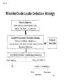
本発明による溶解物の微生物の検出に対する戦略は、図2として本願明細書に追加される線図にまとめられることができる。 The strategy for microbial detection of the lysate according to the present invention can be summarized in the diagram added to this specification as FIG.
本発明の実施例のこの実施例は、従来のDNA隔離技術のための、そして、微生物lysate―direct―probe―based―PCR技術が実行されることを可能にするための必要を迂回するための本発明の適合性を示す。 This example of an embodiment of the present invention is intended to circumvent the need for conventional DNA sequestration techniques and to allow microbial lysate-direct-probe-based-PCR techniques to be performed. The suitability of the present invention is shown.
a.黄色ブドウ球菌(SA)標準血液培養((カンディダ・コンセンサス分析、E.Coli、E faecium)WBC洗剤+ベース溶解、小球形にすることおよび、洗うことによって続かれる)に封じられた。 a. Sealed in S. aureus (SA) standard blood cultures ((Candida consensus analysis, E. coli, E faecium) WBC detergent + base lysis, followed by spheronization and washing).
b.本発明に従う直接のプローブ手順がいずれの場合においてもPCRの%溶解物のより高い許容度に関して優れていたことはTaqManの調査およびSYBRを使用している直接的な溶解物PCRの後、それを見つけられた(30ul PCRで、5ulの少なくとも5000の微生物から検出される抑制のない最高17%は、溶解物を機械加工する。血液培養陽性の瓶には―4000の微生物が入っている/ml培養組織の、下調べの2mlを配置することは8000の微生物/50ul溶解物を産生するPCR(上部BCレベルは、分析許容度を必要とした)の30ul PCR反応=160微生物の5ulいずれ。現在、推定する500のmicrobes/であるBCの検出の制限が、瓶詰めにする、または、10の微生物/ml、従って、5ul=2。10の微生物/瓶(一般的な)そしてそれから、=640/bottle(検出可能でありえる)に6 2倍になっている世代を必要としている5ul=0.2の微生物である。 b. The direct probe procedure according to the present invention was superior in all cases in terms of higher tolerance of PCR% lysate after the TaqMan investigation and direct lysate PCR using SYBR. Found (up to 17% uninhibited detected from 5 ul of at least 5000 microorganisms in 30 ul PCR machine lysate. Blood culture positive bottle contains -4000 microorganisms / ml Placement of 2 ml of the cultured tissue for follow-up is 30 ul PCR reaction for PCR (upper BC level required analytical tolerance) to produce 8000 microorganisms / 50 ul lysate = 5 ul of 160 microorganisms. Estimated 500 microbes / BC detection limit is bottled or 10 microbes / ml, subordinate 5ul = 0.2 microorganisms that require 10 microbes / bottle (general) and then 62x generation to = 640 / bottle (which may be detectable) It is.
c.したがって、それは、本発明に従ってそれを示されたSA(chaotrope+洗剤)MolYsisバッファおよびデオキシリボヌクレアーゼ処理の中を走って、1人のTEによって、ペレット及び洗浄が工場―直接的なプローブPCRと互換性を持つことになった。本発明の新規な改良された方法は、示された変性剤(Becton Dickinson Staph S/Rキット(Bectonディキンソンから市販されている))(サンプルの1/I0e6thだけがPCRにおいてある)のない血液培養ビード機械システムを変性剤を有する本発明の改良によって設けられているシステムと比較することによって、従来の技術の上の%血液の感受性および許容度に関して、血液の改良は、利用されるダイレクト・システムを機械加工する(DoE:グアニジン/トゥイーン、第三イオン/NaOH、トゥイーン/第三イオンその他) c. Therefore, it runs in SA (chaotrope + detergent) MolYsis buffer and deoxyribonuclease treatments shown it according to the present invention, making pellets and washing compatible with factory-direct probe PCR by one TE. I decided to have it. The new and improved method of the present invention is a blood culture without the indicated denaturant (Becton Dickinson Staph S / R kit (commercially available from Becton Dickinson)) (only 1 / I0e6th of the sample is in PCR) By comparing the bead mechanical system with the system provided by the improvement of the present invention having a denaturant, the blood improvement is related to the% blood sensitivity and tolerance over the prior art, the direct system utilized. (DoE: guanidine / tween, third ion / NaOH, tween / third ion, etc.)
更に本発明の開発の間の実験で、本願明細書に追加される図3に示される工程系統図に示されるにつれて、トリプシンおよびデオキシリボヌクレアーゼの追加が本発明の2つの「困難な」臨床サンプルの処理の間、観察されて詰まる重要な減少を可能にすることが証明された。 Further, during the development of the present invention, the addition of trypsin and deoxyribonuclease was performed on two “difficult” clinical samples of the present invention, as shown in the process flow diagram shown in FIG. 3 added herein. It has been proven to allow a significant reduction observed and clogged during processing.
本発明の大まかな基本的な原理および教示が変性剤対応天然溶解物(ビード工場及び超音波学)のすべてのバリエーションを最適化するために適用されることができる当業者によって認められるさまざまな生物学的組織サンプル(血液、体液および軟部組織を含むがこれに限らず)の―direct―probe/SYBR―PCR分析、だけでなくSA上で特に記載されているが、また、さまざまな病原体(例えばいかなるバクテリア、菌類、ウイルス、寄生虫、その他も)間に。 Various organisms recognized by those skilled in the art to which the broad basic principles and teachings of the present invention can be applied to optimize all variations of denaturant compatible natural lysates (bead mill and ultrasound) -Direct-probe / SYBR-PCR analysis of biological tissue samples (including but not limited to blood, body fluids and soft tissue) as well as specifically described on SA, but also various pathogens (eg Between any bacteria, fungi, viruses, parasites, etc.).
上記例も、本発明によって設けられている方法の実行が定義済みの混合物の、または、運転中のおよび殺された電池の定義済みの混合物によってスパイクをつけられる環境サンプルの殺された細胞から信号を能率的に抑制することができることを示す。本発明に従うサンプルの処理が膜を危うくされた細胞を分析から除外する良好な方法であるかもしれない点に注意することも、価値がある。 The above examples are also signaled from killed cells of an environmental sample spiked by a defined mixture of a method provided by the present invention or by a defined mixture of running and killed batteries. It can be efficiently suppressed. It is also worth noting that processing a sample according to the present invention may be a good way to exclude cells that have been compromised from the analysis.
上の説明を要約すると、本発明は、更に下流の分析の前に細菌個体群の速いおよびeasy―to―perform pre―治療を可能にしている新規な方法を提供する。本発明の潜在的多数の使用が当業者によって認められるにもかかわらず、本発明によって設けられている方法はDNAに大きな影響を及ぼすことができる―病原診断法、バイオテロリズムおよび微生物生態学を含むさまざまな分野のベースの診断法。 Summarizing the above description, the present invention provides a novel method that allows for fast and easy-to-perform pre-treatment of bacterial populations prior to further downstream analysis. Despite the many potential uses of the present invention recognized by those skilled in the art, the methods provided by the present invention can greatly affect DNA-including pathogenetics, bioterrorism and microbial ecology Diagnosis based on various fields.
本発明の好ましい実施例の実行において、細胞が成長しないので、微生物ターゲット信号の重要な増加を示す単一の血液培養から分離平等政策の約数を使用している少なくとも2つの別々の時点のいかなるPCR測定も微生物成長によりなければならないことは明らかである。それによって、生存可能な微生物(汚染効果を無視する)の存在を示す。生き残れる微生物溶解およびPCR準備の前に、すべての死細胞DNAが除去されるときに、血液の非成長ベースの単一ポイント陽性のPCR分析が生存可能な微生物の存在を示すと認められる−誘導されるいかなるPCRプロセスも露出する汚染。これは、DNasingおよびWashing離れて死細胞DNAによってデモをされることができる。 In practicing the preferred embodiment of the present invention, any of the at least two separate time points using a divisor of equality policy separated from a single blood culture exhibiting a significant increase in the microbial target signal because the cells do not grow. It is clear that PCR measurements must also be due to microbial growth. It indicates the presence of viable microorganisms (ignoring the pollution effect). Prior to surviving microbial lysis and PCR preparation, when all dead cell DNA is removed, a non-growth-based single point positive PCR analysis of blood is observed to indicate the presence of viable microorganisms—induced Contamination that exposes any PCR process. This can be demonstrated by dead cell DNA apart from DNasing and Washing.
特定の参照がPCRに本願明細書においてなされるにもかかわらず、本発明の改良がPCRまたは類似の方法論に限られていないItは更に認められることになっている。本発明用に考察される増幅分析としては、限定はされないが、他の周知の核酸ベースの技術(例えばDNA増幅分析、サーモ安定ポリメラーゼを組み込んでいるPCR分析および等温詳しい説明方法)が挙げられる。 It should be further appreciated that It is not intended that the improvements of the present invention be limited to PCR or similar methodologies, although specific references are made herein to PCR. Amplification analysis contemplated for the present invention includes, but is not limited to, other well-known nucleic acid-based techniques such as DNA amplification analysis, PCR analysis incorporating a thermostable polymerase and isothermal detailed description methods.
当業者が本発明の実施において、役立つさまざまな適切な増幅方法を思いつくことができる、そして、従って、本発明がこのことにより制限されることを目的としないと認められる。本発明がいずれでもおよびすべての方法のアプリケーションを有すると認められる。そして、手順および方法がDNA診断法を含む。かかるアプリケーションの例は限定されるものではないが、食品、水安全性、バイオテロリズム、健康診断/薬剤および/または病原検出を含んでいる何でも含んでいるそれらを包含する。食品業界において、本発明は、防腐剤の有効性をモニタするために用いることができる。本発明の方法には、すべての細胞に適用される可能性がある。細菌細胞が実施例において例証されるにもかかわらず、当業者は本発明の方法が多くの他の細胞タイプに適用されることができるということを従来技術において容易に知ることができる。本発明が、膜を崩壊させることができておよび/または細胞(例えば細菌細胞)を殺すことができる物質の識別のために用いられることもできる。多種薬剤の抵抗有機体が全盛で、保健機関および患者において広がった時から、新しい消毒薬の識別および/または抗生物質は現在優先権である。 Those skilled in the art will be able to come up with a variety of suitable amplification methods that are useful in the practice of the present invention, and it will therefore be appreciated that the present invention is not intended to be limited thereby. It is recognized that the present invention has any and all method applications. The procedures and methods include DNA diagnostic methods. Examples of such applications include, but are not limited to, those that include food, water safety, bioterrorism, health check / medicine and / or anything that includes pathogen detection. In the food industry, the present invention can be used to monitor the effectiveness of preservatives. The method of the present invention may be applied to all cells. Although bacterial cells are illustrated in the examples, one skilled in the art can readily know in the prior art that the methods of the invention can be applied to many other cell types. The present invention can also be used for the identification of substances that can disrupt membranes and / or kill cells (eg, bacterial cells). The identification of new disinfectants and / or antibiotics has now become a priority since the multi-drug resistance organism has flourished and has spread in health institutions and patients.
本発明の方法が、ツールとしての定量的PCRと結合して、細胞を培養して、成長を待つことに時間を費やさなければならないことのない消毒薬および/または抗生物質の衝撃を急速に、そして、うまく確認することができると更に認められる。ある場合には、有機体は培養組織に週まで何日もかかることができる、そして、このように、候補実質が細胞(微生物のような)を殺すことが可能だったかどうか見る重要な時間かかることができる。他の例において、特定の有機体は細胞培養で成長しない。そして、従って、物質が効果的だったかどうかについて決定することを困難にする。このように、本発明の新規な方法を適用することは、新しい消毒薬および/または抗生物質の識別のために、時間および資源を確保することができる。 The method of the present invention, coupled with quantitative PCR as a tool, rapidly disinfects disinfectants and / or antibiotics without having to spend time in culturing cells and waiting for growth, And it is further recognized that it can be confirmed well. In some cases, the organism can take days to week in culture, and thus takes significant time to see if the candidate parenchyma was able to kill the cells (like microorganisms) be able to. In other examples, the particular organism does not grow in cell culture. And thus makes it difficult to determine whether the substance was effective. Thus, applying the novel method of the present invention can save time and resources for the identification of new disinfectants and / or antibiotics.
本発明による新規な方法の更なる効果は、使いやすさである。例えばこれらの方法を用いて、大量のサンプルは、生細胞(例えばバクテリア)の存在を、容易に見つけるため検査されることができる。例えば、サンプルは、存在を見つけるため検査されることができる潜在的に完全な細胞膜を有する生きているバクテリア。他の実施形態では、環境サンプルは、生細胞(例えばバクテリア)の存在を見つけるため検査されることができる。これらのサンプルは、例えば、土壌から集められることができるかまたは、プラントの部分であることができる。 A further advantage of the novel method according to the invention is ease of use. For example, using these methods, large samples can be examined to easily find the presence of live cells (eg, bacteria). For example, a sample is a living bacterium with a potentially complete cell membrane that can be examined to find its presence. In other embodiments, environmental samples can be examined to find the presence of live cells (eg, bacteria). These samples can be collected, for example, from soil or can be part of a plant.
本発明による方法が、解放の前に、そして、の後、処理された廃水のテストのために、更に用いられることができる。本発明による方法が、医薬のサンプル(例えば気道、インプラントおよびカテーテル面からスツール・サンプル、血液培養、痰、組織サンプル(また、切る)損傷材料、尿およびサンプル)をテストするために、更に用いられることができる。 The method according to the invention can further be used for testing of the treated wastewater before and after release. The method according to the invention is further used to test pharmaceutical samples (eg stool samples, blood cultures, sputum, tissue samples (and also cut damaged materials, urine and samples) from airways, implants and catheter surfaces) be able to.
本発明による方法のアプリケーションの他の分野は、食品の制御でありえる。他の実施態様において、食品サンプルは、牛乳または乳製品(ヨーグルト、チーズ、甘いチーズ、バターおよびバターミルク)飲料水、飲料(レモネード、ビールおよび液)ベーカリ製品または肉製品から得られる。本発明の方法は、食品の防腐剤か食品(例えば低温殺菌)の抗菌性の処置が細胞増殖を防止したかどうか決定することができる。本発明による方法のアプリケーションの更なる分野は、製薬および美顔用製品(例えば軟膏、クリーム、チンキ、液、溶液、低下、など)の分析である。 Another area of application of the method according to the invention may be food control. In other embodiments, the food samples are obtained from milk or dairy products (yogurt, cheese, sweet cheese, butter and buttermilk) drinking water, beverages (lemonade, beer and liquid) bakery products or meat products. The method of the present invention can determine whether an antimicrobial treatment of a food preservative or food (eg, pasteurized) has prevented cell growth. A further field of application of the method according to the invention is the analysis of pharmaceutical and facial products (eg ointments, creams, tinctures, liquids, solutions, drops, etc.).
本発明の方法は、以前の方法をタイムリな警告および予防動きに不適当にしている長いインキュベーション時間(日の範囲で)の課題を解決する。加えて、最新のPCRベースの方法は、偽陽性結果(有機体が現実的でないにもかかわらず、有機体の陽性反応を示す)を与えることができる。さらに、調査は、それらがまだ現実的であるにもかかわらず、若干の有機体が、特定の状況の下で、複製する能力を失うことができるということを最近発見した。より適当な環境へ移される場合、これらの『現実的であるが、culturableでない』(VBNC)バクテリアは従来の培養を使用して検出されることができなくて、成長するそれらの能力を回復するかもしれない。これらの欠点は、本発明の方法と組み合わせてこれらの有機体の遺伝物質/DNAの検出に基づいて分子方法を適用することによって解決される。このように、例えば、サンプルの生存可能な有機体に注意している速いおよび正確な結果汚染された水(汚水、食品、医薬および/または化粧品)は、汚染された製品が市民にリリースされるのを防止することができる。現在の時間がかかる方法と比較して、サンプルの偽陽性(病原体が現実的でないにもかかわらず、病原体の陽性反応を示す)および迅速なテストを最小化することによって、本発明の方法は、資源を保存することができる。 The method of the present invention solves the problem of long incubation times (in the range of days) making previous methods unsuitable for timely warning and preventive action. In addition, modern PCR-based methods can give false positive results (indicating a positive reaction of an organism, even though the organism is not realistic). In addition, research has recently discovered that some organisms can lose the ability to replicate under certain circumstances, even though they are still realistic. When transferred to a more appropriate environment, these "real but not culturable" (VBNC) bacteria cannot be detected using conventional cultures and restore their ability to grow It may be. These disadvantages are solved by applying molecular methods based on the detection of genetic material / DNA of these organisms in combination with the methods of the present invention. Thus, for example, fast and accurate results that are paying attention to the viable organisms in the sample contaminated water (sewage, food, medicine and / or cosmetics), contaminated products are released to the public Can be prevented. Compared to current time-consuming methods, by minimizing false positives of the sample (indicating a positive pathogen response even though the pathogen is not realistic) and rapid testing, You can save resources.
加えて、本発明の方法は、生態学的な研究(農業および/または生態学的なシステムのための特定の土壌の健康)のための微生物共同体の潜在的に生き残れるメンバーを確認することができる。伝統的に、細菌共同体を確認することは、培養ベースの方法またはプレート数を使用して実行された。コロニーがより非常に計数されるほど、バクテリアは、より、オリジナルでサンプルroblemsであると推定される、しかしながら、時々この方法をタイムリなおよび正確な結果に不適当にしている長いインキュベーション時間(日の範囲で)から立つ。これらの欠点は、本発明の方法を利用している。 In addition, the methods of the present invention can identify potentially surviving members of a microbial community for ecological studies (specific soil health for agriculture and / or ecological systems) . Traditionally, identifying bacterial communities has been performed using culture-based methods or plate numbers. The more colonies are counted, the more bacteria are presumed to be original and sample robots, however, sometimes the long incubation times (days) making this method unsuitable for timely and accurate results Stand up from). These disadvantages utilize the method of the present invention.
本発明の方法を使用している分析を受けることができるバクテリアの中で非限定的な実施例、または、本発明の方法を使用しているサンプルの潜在的生存度を検出することは、成り立つ、に加えてSA前述したように:B.百日咳、Leptospira pomona、S.paratyphi AおよびB、C.diphtheriae、C.tetani、C.botidinum、C. perfringens、C.feseriおよびその他ガス壊疽バクテリア、B.anthracis、P.pestis、パスツレラ・ムルトシダ、髄膜炎菌、N.gonorrheae、ヘモフィルス属influenzae、アクチノミセス属{Norcardiaで例えばある)アシネトバクター属(バシラス科){例えば、バチルス属anthrasis)バクテロイデス属{バクテロイデス・フラジリスで例えばある)Blastomycosis、Bordetella、ボレリア属{Borrelia burgdorferiで例えばある)Brucella、カンピロバクター属、クラミジア、Coccidioides、コリネバクテリウム属{コリネバクテリウム属diptheriaeで例えばある)大腸菌{Enterotoxigenic大腸菌およびEnterohemorrhagic大腸菌で例えばある)エンテロバクター属(例えばエンターobacterエアロ遺伝子)(Enterobacteriaceae)(クレブシエラ属、サルモネラ属(例えばチフス菌、腸炎菌、セラチア属、エルシニア属、赤痢菌)エリジペロスリックス属、ヘモフィルス属(例えばヘモフィルス属インフルエンザ・タイプB)ヘリコバクター属、レジオネラ(例えばレジオネラ・ニューモフィラ菌)Leptospira、リステリア属(例えばリステリア菌)Mycoplasma、マイコバクテリウム(例えばらい菌およびヒト型結核菌)ビブリオ属(例えばコレラ菌)Pasteurellacea、プロテウス、シュードモナス(例えば緑膿菌)Rickettsiaceae、Spirochetes(例えば、Treponema spp.Leptospira spp.ボレリア属種。)赤痢菌spp.Meningiococcus、Pneumococcusおよびすべての連鎖球菌(例えば肺炎連鎖球菌およびGroups A3 BおよびC Streptococci)(ウレアプラズマ属)。トレポネーマpollidum、黄色ブドウ球菌、Pasteurella haemolytica、コリネバクテリウム属diptheriaeトキソイド、髄膜炎菌多糖類、Bordetella pertusis、肺炎連鎖球菌、破傷風菌トキソイドおよびマイコバクテリウム‐ボビス。上記リストは、単に図示するだけのことを目的として、決してそれらの特定の細菌有機体に本発明を検出に制限するはずでない。 Detecting the potential viability of non-limiting examples in bacteria that can be analyzed using the method of the invention, or samples using the method of the invention, is valid In addition to SA as described above: Pertussis, Leptospira pomona, S. paratyphi A and B, C.I. diphtheriae, C.I. tetani, C.I. botidinum, C.I. perfringens, C.I. feseri and other gas gangrene bacteria, anthracis, P.A. pestis, Pasteurella multocida, Neisseria meningitidis, N. gonorrheae, Haemophilus influenzae, Actinomyces genus (e.g. Norcardia) Acinetobacter genus (e.g. Bacillus family) {e.g. Bacillus anthracis) Bacteroides genus {e.g. ) Brucella, Campylobacter, Chlamydia, Coccidioides, Corynebacterium (e.g. Corynebacterium dipthelia) E. coli {Enterotoxigenic E. coli and Enterohemorrhagic E. coli) e.g. Enterobacter aerial (Enterobacteriaceae) (Klebsiella, Salmonella (eg, Salmonella typhi, Enterococcus, Serratia, Yersinia, Shigella), Eridiperosrix, Haemophilus (eg, Haemophilus influenza type B), Helicobacter, Legionella ( For example, Legionella pneumophila) Leptospira, Listeria (eg, Listeria) Mycoplasma, Mycobacterium (eg, Mycobacterium tuberculosis) and Vibrio (eg, Vibrio cholerae) Pasteurellacea, Proteus, Pseudomonas (eg, Pseudomonas aeruginosa) Rickettsiacea Spirochetes (eg, Treponema spp. Leptospira spp. Borrelia sp.) Shigella spp. eningiococcus, Pneumococcus and all streptococci (for example, Streptococcus pneumoniae and Groups A3 B and C Streptococci) (Ureaplasma). Bordetella pertusis, Streptococcus pneumoniae, tetanus toxoid and Mycobacterium bovis, the list above should not limit the present invention to detection of these specific bacterial organisms for purposes of illustration only .
本発明の特に好適な実施例は、PCRを利用する。PCRのための一般の手順は、米国特許第4,683,195号(マリス、その他)および米国特許第4,683,202号(マリス、その他)において教示される。しかしながら、増幅反応ごとに使用する最適PCR状況は、通常、分野の職人によって共通に使用されるコンピュータ・ソフトウェアによって、経験的に決定されるかまたは推定される。多くのパラメータは、反応の成功に影響する。焼なまし温度および時間(拡張時間、Mg2+、pHおよびプライマ、テンプレートおよびデオキシリボヌクレオチドの相対的な濃度)は、それらの一つである。通常、テンプレート核酸は、ポリメラーゼ反応の前に1〜10分間の少なくとも約95°Cまで加熱することによって、変性する。ほぼ20―99サイクルの増幅は、90°C〜0.05〜1分間の96°Cの範囲の変性、48°Cから0.05〜2分間の72°Cにわたっている温度の焼鈍および68°C〜最適最後のサイクルを有する少なくとも0.1分間の75°Cの拡張を使用して実行される。実施例において、PCR反応は、各種類の0.5mmのdNTPおよび市販の熱安定DNAポリメラーゼの0.5〜5台の装置に約100ngのテンプレート核酸、20uMの上流のおよび下流のプライマおよび0.05を含むことができる。 A particularly preferred embodiment of the present invention utilizes PCR. General procedures for PCR are taught in US Pat. No. 4,683,195 (Marris et al.) And US Pat. No. 4,683,202 (Maris et al.). However, the optimal PCR situation to use for each amplification reaction is usually determined or estimated empirically by computer software commonly used by artisans in the field. Many parameters affect the success of the reaction. Annealing temperature and time (expansion time, Mg2 +, pH and primer, relative concentration of template and deoxyribonucleotide) are among them. Typically, the template nucleic acid is denatured by heating to at least about 95 ° C. for 1-10 minutes prior to the polymerase reaction. Approximately 20-99 cycles of amplification included denaturation ranging from 90 ° C. to 0.05 ° C. to 96 ° C., annealing from 48 ° C. to 0.05 ° C. to 72 ° C. and 68 ° C. Performed using a 75 ° C expansion for at least 0.1 minutes with C to optimal last cycle. In the examples, PCR reactions were performed on 0.5 to 5 devices for each type of 0.5 mm dNTP and commercially available thermostable DNA polymerase, about 100 ng template nucleic acid, 20 uM upstream and downstream primers and 0. 05 can be included.
従来のPCRのバリエーションは、逆転写PCR反応(RT―PCR)である、シングルに対する分子が立ち往生させた逆転写酵素第1の茂みRNA cDNA分子(それからポリメラーゼ連鎖反応の次の増幅のテンプレートとして使用される)。RNAの隔離は、公知技術である。RT―PCRを実施することにおいて、目標核酸が変性する熱であったあと、逆転写酵素は通常、反応サンプルに加えられる。増幅の予定のサイクルが起こる前に、反応はそれから、cDNAテンプレートを生成するために充分な時間量(10―60分)のための適切な温度(例えば30―45°C)に維持される。当業者は、定量的結果が要求される場合、注意がそれが維持するかまたは増幅された核酸の相対的なコピーのために制御する方法を使用するためにされなければならないと従来技術において認める。「定量的」増幅の方法は、当業者にとって周知である。例えば、定量的PCRは、同時に同じプライマを使用している制御シーケンスの既知量を共同増幅することが必要でありえる。これは、PCR反応を調整するために用いることができる内部標準を提供する。 A variation of conventional PCR is the reverse transcription PCR reaction (RT-PCR), a reverse transcriptase first bush RNA cDNA molecule that has been stuck to a single molecule (and then used as a template for subsequent amplification of the polymerase chain reaction). ) RNA isolation is a known technique. In performing RT-PCR, reverse transcriptase is usually added to the reaction sample after the heat of denaturing the target nucleic acid. The reaction is then maintained at an appropriate temperature (eg, 30-45 ° C.) for an amount of time (10-60 minutes) sufficient to generate a cDNA template before the expected cycle of amplification occurs. One skilled in the art recognizes in the prior art that when quantitative results are required, attention must be made to use methods that maintain or control for relative copying of amplified nucleic acids. . Methods of “quantitative” amplification are well known to those skilled in the art. For example, quantitative PCR may require co-amplification of known amounts of control sequences using the same primer at the same time. This provides an internal standard that can be used to adjust the PCR reaction.
PCRの他の二者択一は定量的PCR(qPCR)である。qPCRは小さい挿入または削除によって大きさにおいて目標と異なる内部相応する制御を使用している競争的技術に通されることができる。しかしながら、非競争的および動力学的定量的PCRが、使われることもできる。同時に標的配列と一緒に検出されることができる内部相応する制御とリアルタイム、動力学的PCR検出の組合せは、有利でありえる。 Another alternative to PCR is quantitative PCR (qPCR). qPCR can be passed through competitive techniques using internal corresponding controls that differ in size from targets by small insertions or deletions. However, non-competitive and kinetic quantitative PCR can also be used. A combination of internal corresponding control and real-time, kinetic PCR detection that can be detected together with the target sequence at the same time can be advantageous.
PCR、RT―PCRおよび/またはqPCRのためのプライマは、その特定の有機体のために選択されるDNA領域を増幅するだけである地方または特定のバクテリアの範囲内で選択される。あるいは、すべての有機体のために一般的であるDNAの部分の雑種を作って、増幅するプライマは、選択される。選択および構造が従来技術において公知で一般にあるプライマ。一般に、1つのプライマは、増幅されるシーケンスの各先端にある。この種のプライマが、通常長さの10〜35のヌクレオチドの間にあって、18〜22のヌクレオチドの間から、好適な長さを有する。増幅されることができる最も小さいシーケンスは、長さ(前方及び後方のプライマ、長さの20のヌクレオチドの両方ともで例えばある、シーケンスの場所が、少なくとも10のヌクレオチドによって切り離される)のほぼ50のヌクレオチドである。非常により長いシーケンスは、増幅されることができる。1つのプライマは、「前方のプライマ」と呼ばれていて、増幅される領域の左端にある。前方のプライマは、順番に、DNA(倍―足止めされるDNAが、一番上の索が5フィート〜3フィート方向の両極性によって示される慣例を使用して描かれる)の一番上の要素の領域と同一である。前方のプライマの配列は、それがDNAの一番上の要素と相補的であるDNAの要素に交雑するようなものである。他のプライマは、「復帰プライマ」と呼ばれていて、増幅される領域の右端にある。すなわち、それが順番にそれと相補的であるように、プライマがそうである後退のシーケンスは、シーケンスを逆補うものである、DNAの一番上の要素の領域。復帰プライマは、DNAの頂端部に交雑する。PCRプライマは、多くの他の状況を前提としても選択されなければならない。PCRプライマは、テンプレートの1つを超える領域に雑種形成を最小化するのに長く十分でなければならない(長さの好ましくは10〜30のヌクレオチド)。可能ならば、一塩基のロングランを有するプライマは、回避されなければならない。プライマは、40および60%間の1パーセントのG+C内容を好ましくは有しなければならない。可能ならば、プライマの3’末端のパーセントG+C内容は、プライマの5’末端のパーセントG+C内容より高くなければならない。プライマは、プライマ(すなわちパリンドローム)の範囲内で他のシーケンスに交雑することができるシーケンスを含んではならない。同じPCR反応において使用する2つのプライマは、互いに交雑することが可能であってはならない。PCRプライマが上記の勧告に対する好ましくは選ばれた主題であるにもかかわらず、プライマがこれらの状況にかなうことは必要でない。他の下塗りには、動くことができるが、良い結果を得る下の可能性があることができる。 Primers for PCR, RT-PCR and / or qPCR are selected within local or specific bacteria that only amplify the DNA region selected for that particular organism. Alternatively, primers are selected that make and hybridize portions of the DNA that are common for all organisms. Primers whose selection and structure are generally known in the prior art. In general, one primer is at each tip of the sequence to be amplified. This type of primer is usually between 10-35 nucleotides in length and has a suitable length from between 18-22 nucleotides. The smallest sequence that can be amplified is approximately 50 in length (forward and backward primers, both 20 nucleotides in length, for example, where the sequence location is separated by at least 10 nucleotides) It is a nucleotide. Very longer sequences can be amplified. One primer is called the “front primer” and is at the left end of the region to be amplified. The forward primer, in turn, is the top element of DNA (double-stopped DNA is drawn using the convention that the top cord is indicated by a bipolar polarity in the 5 to 3 ft direction) The same area as The sequence of the forward primer is such that it hybridizes to an element of DNA that is complementary to the top element of DNA. The other primer is called the “return primer” and is at the right end of the region to be amplified. That is, the reverse sequence that the primer is, as it is complementary in turn, is the region of the top element of DNA that reverses the sequence. The return primer hybridizes to the top end of the DNA. PCR primers must also be selected assuming many other situations. The PCR primer must be long enough to minimize hybrid formation in more than one region of the template (preferably 10-30 nucleotides in length). If possible, primers with a single base long run should be avoided. The primer should preferably have a 1% G + C content between 40 and 60%. If possible, the percent G + C content at the 3 'end of the primer should be higher than the percent G + C content at the 5' end of the primer. A primer must not contain sequences that can be crossed to other sequences within the scope of the primer (ie palindrome). The two primers used in the same PCR reaction should not be able to cross each other. Although PCR primers are the preferred subject of choice for the above recommendations, it is not necessary for the primers to meet these circumstances. Other undercoats can move, but can have the possibility of getting good results.
所与のシーケンスの中でDNAを増幅するために用いることができるPCRプライマは、利用できる多くのコンピュータプログラムのうちの1つを使用して選択されることができる。この種のプログラムは、所与のシーケンス(すなわち、PCRプライマの機能を最大にすることができる他の状況に加えて、この種のプログラムは、上で述べられる状況を前提として、プライマを選択する)の増幅のために至適であるプライマを選択する。1つのコンピュータプログラムは、PCRプライマの選択のためのルーチンがあるGenetics Computer Group(GCGは、最近Accelrysになった)分析一括法案である。 PCR primers that can be used to amplify DNA within a given sequence can be selected using one of many computer programs available. This type of program selects a primer given the given sequence (ie, other situations that can maximize the functionality of the PCR primer, given the situation described above) Select the primer that is optimal for amplification). One computer program is the Genetics Computer Group (GCG has recently become Accelrys) analysis package bill with routines for the selection of PCR primers.
下で開示されるオリゴヌクレオチドプライマおよび調査は、多くの方法でなされることができる。これらのオリゴヌクレオチドを作る1つの方法は、購入可能な核酸シンセサイザを使用してそれらを総合することである。この種のシンセサイザのバラエティが、存在して、当業者に周知である。 The oligonucleotide primers and studies disclosed below can be done in a number of ways. One way to make these oligonucleotides is to synthesize them using a commercially available nucleic acid synthesizer. A variety of this type of synthesizer exists and is well known to those skilled in the art.
本発明と関連して役立つPCRの他の二者択一は、特定のDNAまたはRNA目標の検出の等温核酸増幅検査法である。非―核酸の等温増幅のための制限する実施例は、均一なリアルタイム索置換増幅(圧延円増幅の基礎を形成されるPhi29 DNAポリメラーゼ)である。DNA塩基配列決定のテンプレート、PNAオープナによって援助される二重DNA配列の圧延円増幅またはDNA分析物のループによって媒介される等温増幅。 Another alternative to PCR that is useful in connection with the present invention is an isothermal nucleic acid amplification assay for the detection of specific DNA or RNA targets. A limiting example for isothermal amplification of non-nucleic acids is homogeneous real-time cord displacement amplification (Phi29 DNA polymerase that forms the basis of rolling circle amplification). Template for DNA sequencing, rolling circle amplification of double DNA sequences assisted by PNA openers or isothermal amplification mediated by loops of DNA analytes.
核酸は、雑種形成方法によって検出されることもできる。
これらの方法では、ラベルをつけられた核酸は、ラベルをつけられたかラベルのない核酸の調査を含んでいる基板に加えられることができる。あるいは、ラベルのないかラベルのない核酸は、ラベルをつけられた核酸の調査を含んでいる基板に加えられることができる。実施例、Micro Array Analysis、マークSchena、ジョン・ワイリー社、ホーボーケN.J.2003のために、雑種形成方法は、中で開示される。
Nucleic acids can also be detected by hybrid formation methods.
In these methods, labeled nucleic acids can be added to a substrate containing a survey of labeled or unlabeled nucleic acids. Alternatively, unlabeled or unlabeled nucleic acids can be added to a substrate containing a survey of labeled nucleic acids. Examples, Micro Array Analysis, Mark Schena, John Wylie, Hoboke N .; J. et al. For 2003, a hybrid formation method is disclosed in.
核酸を検出する方法は、ラベルの使用を含み得る。例えば、識別用放射性同位元素は、写真フィルムまたは燐酸映像器(放射性リン酸塩編入を検出して、定量化するために)を使用して検出されることができる。蛍光マーカは、発された光(典型的な装置のために、米国特許第5,143,854号を参照)を検出するために光検出器を使用して検出されることができて、定量化されることができる。酵素標識は、酵素に基板を提供して、酵素の基板に及ぼす作用によってできる反応製品を測定することによって、典型的に検出される。比色ラベルは、単に着色したラベルを視覚化することによって検出される。実施例において、増幅された核酸分子は、直接増幅生成物を核酸をさし込んでいる染料で染色することによって視覚化される。当業者にとって明らかであるように、典型的な染料は、含む、しかし、SYBR緑、SYBR青、DAPI、propidiumヨウ素、Hoeste、SYBR金および臭化エチジウムに制限する。増幅されたDNA分子にさし込まれる発光の染料の量は増幅産物の量に正比例する。そして、それは製造業者の指示に従ってFlurolmager(分子Dynamics)または他の等価な装置を使用して便利に定量化されることができる。この種の方法のバリエーションは、選択されたものの染色および視覚化が続く増幅産物のゲル電気泳動である染料をさし込むこと。あるいは、標識化オリゴヌクレオチド・ハイブリッド形成プローブ(例えば蛍光プローブ(例えば蛍光反響エネルギー転送(FRET)の調査および比色プローブ))は、増幅を検出するために用いることができる。要求される所で、テストされている生物学的実体のゲノム配列典型の特定の増幅は塩基配列決定によって検査されることができるかまたは増幅産物が予測されたサイズを有することを証明していることができて、予測された規制消化パターンを呈することができるか、または、正しいクローンをつくられたヌクレオチド配列に交雑することができる。 The method of detecting a nucleic acid can include the use of a label. For example, the identifying radioisotope can be detected using a photographic film or a phosphoric acid imager (to detect and quantify radioactive phosphate incorporation). The fluorescent marker can be detected using a photodetector to detect emitted light (for a typical device, see US Pat. No. 5,143,854) and can be quantified. Can be Enzymatic labels are typically detected by providing a substrate for the enzyme and measuring the reaction product that results from the effect of the enzyme on the substrate. Colorimetric labels are detected simply by visualizing the colored label. In an embodiment, the amplified nucleic acid molecule is visualized by directly staining the amplified product with a dye impregnating the nucleic acid. As will be apparent to those skilled in the art, typical dyes include, but are limited to SYBR green, SYBR blue, DAPI, propidium iodine, Hoeste, SYBR gold and ethidium bromide. The amount of luminescent dye that is inserted into the amplified DNA molecule is directly proportional to the amount of amplified product. It can then be conveniently quantified using a Fluorolmager (Molecular Dynamics) or other equivalent device according to the manufacturer's instructions. A variation of this type of method is to incorporate a dye that is gel electrophoresis of the amplification product followed by staining and visualization of the selected one. Alternatively, labeled oligonucleotide hybridization probes (eg, fluorescent probes (eg, fluorescent echo energy transfer (FRET) probes and colorimetric probes)) can be used to detect amplification. Where required, specific amplification of the genomic sequence typical of the biological entity being tested can be examined by sequencing or proves that the amplification product has the expected size And can exhibit the predicted regulatory digestion pattern or can be crossed to the correct cloned nucleotide sequence.
本発明には、キットが具備されている。例えば、キットは、特に、または、一般に有機体に対応する核酸分子を増幅することに役立つプライマ、バッファおよびDNAを分離するための試薬およびPCRのための試薬を含むことができる。キットは検出可能的に標識化オリゴヌクレオチドを含むこともできる。そして、それは興味がある有機体に対応するポリペプチドをコード化している核酸配列に交雑する。キットは、試験サンプルと比較して検定されることができて、含まれることができる制御サンプルまたは一連の制御サンプルを含むこともできる。キットの各構成要素は個々の容器の範囲内で囲まれることができる、そして、キットを使用して実行される分析の結果を解釈するための指示とともに、さまざまな容器の全てが単一のパッケージの範囲内であることができる。 The present invention includes a kit. For example, the kit can include reagents for separating primers, buffers and DNA, and reagents for PCR, which are particularly or generally useful for amplifying nucleic acid molecules corresponding to organisms. The kit can also include detectably labeled oligonucleotides. It then hybridizes to a nucleic acid sequence encoding a polypeptide corresponding to the organism of interest. The kit can be assayed relative to the test sample and can also include a control sample or series of control samples that can be included. Each component of the kit can be enclosed within an individual container, and all of the various containers are in a single package, with instructions for interpreting the results of the analysis performed using the kit. Can be within the range of
あたかも個々の刊行、特許または特許出願が引用したものとすることが特に、そして、個々に示されるかのように、すべての参考文献の内容は、同じ範囲に本願明細書に引用したものとすると、特許および公開された特許出願がこの用途の全体にわたって引用した。 It is specifically assumed that individual publications, patents or patent applications are cited, and the contents of all references shall be cited in this specification within the same scope, as if individually indicated. Patents and published patent applications are cited throughout this application.
前述の詳細な説明は理解だけの明るさのために与えられた、そして、変更態様が当業者にとって明らかであるにつれて、不必要な限界はそこから理解されてはならない。それは本願明細書において設けられている情報のいずれも従来技術であるか関連するという承認でないために現在請求する発明またはそれ、いかなる刊行も特に、または、暗に、参照されて従来技術である。 The foregoing detailed description has been given for clarity of understanding only, and as variations will become apparent to those skilled in the art, unnecessary limitations should not be understood therefrom. The invention presently claimed, or any publication, in particular or implicitly, is referred to in the prior art because it is not an admission that any of the information provided herein is prior art or related.
さもなければ定められない限り、本願明細書において用いられるすべての専門的なおよび科学的な用語は共通に、本発明が帰属する当業者によって従来技術において理解するのと同じ意味を有する。本発明が本発明の特定の実施例と関連して記載されると共に、それが更なる変更態様ができる、そして、この用途が続いている本発明のいかなるバリエーションも、使用法または改作をカバーすることを目的とすることが理解されよう、一般に、本発明が関係する技術の範囲内で周知であるか慣習的な実行の範囲内で来る、本発明の中で、そして、現在の開示からのこの種の離脱を含む原理、そして、そのことは先に記載される基本的特徴に適用されることができる、そして、中で以下の通りに、添付のものの範囲は請求する。 Unless defined otherwise, all technical and scientific terms used herein have the same meaning as commonly understood by one of ordinary skill in the art to which this invention belongs. While the invention has been described in connection with specific embodiments of the invention, it is capable of further modifications and any variation of the invention followed by this application will cover usage or adaptation It is understood that the present invention is generally intended to be within the scope of the present invention and from the current disclosure, which is well known or customary within the scope of the technology to which the present invention pertains. The principles involving this kind of departure, and that can be applied to the basic features described above, and in the following, the scope of the appended claims.
また、本発明の好ましい実施例の中で確かな記載されていて、そして、上で特に例証する、本発明がこの種の実施例に限られていることが意図されない、そして、いずれの種類の限界は以下の請求項だけに含まれる。 Also, it is certainly described in the preferred embodiments of the present invention, and it is not intended that the present invention be limited to this type of embodiment, and is specifically illustrated above, and any kind of Limitations are included only in the following claims.
Claims (7)
Applications Claiming Priority (3)
| Application Number | Priority Date | Filing Date | Title |
|---|---|---|---|
| US201061428892P | 2010-12-31 | 2010-12-31 | |
| US61/428,892 | 2010-12-31 | ||
| PCT/US2011/067329 WO2012092238A1 (en) | 2010-12-31 | 2011-12-27 | Improved methods for determining cell viability using molecular nucleic acid-based techniques |
Related Child Applications (1)
| Application Number | Title | Priority Date | Filing Date |
|---|---|---|---|
| JP2016110520A Division JP2016192967A (en) | 2010-12-31 | 2016-06-01 | Methods for microbe-specific filter-in situ analysis for blood samples |
Publications (2)
| Publication Number | Publication Date |
|---|---|
| JP2014502510A true JP2014502510A (en) | 2014-02-03 |
| JP2014502510A5 JP2014502510A5 (en) | 2016-07-28 |
Family
ID=46383494
Family Applications (2)
| Application Number | Title | Priority Date | Filing Date |
|---|---|---|---|
| JP2013547602A Pending JP2014502510A (en) | 2010-12-31 | 2011-12-27 | Improved method for determining cell viability using molecular nucleic acid based techniques |
| JP2016110520A Pending JP2016192967A (en) | 2010-12-31 | 2016-06-01 | Methods for microbe-specific filter-in situ analysis for blood samples |
Family Applications After (1)
| Application Number | Title | Priority Date | Filing Date |
|---|---|---|---|
| JP2016110520A Pending JP2016192967A (en) | 2010-12-31 | 2016-06-01 | Methods for microbe-specific filter-in situ analysis for blood samples |
Country Status (8)
| Country | Link |
|---|---|
| US (1) | US20140186828A1 (en) |
| EP (1) | EP2659001A4 (en) |
| JP (2) | JP2014502510A (en) |
| CN (1) | CN103476945A (en) |
| AU (2) | AU2011352333B2 (en) |
| CA (1) | CA2862523A1 (en) |
| NZ (1) | NZ613671A (en) |
| WO (1) | WO2012092238A1 (en) |
Families Citing this family (6)
| Publication number | Priority date | Publication date | Assignee | Title |
|---|---|---|---|---|
| CN104520438A (en) * | 2012-04-12 | 2015-04-15 | 宙斯科技公司 | Methods for measuring polymerase activity useful for sensitive, quantitative measurements of any polymerase extension activity and for determining the presence of viable cells |
| US10495555B2 (en) * | 2013-10-08 | 2019-12-03 | David Putnam | Filter-cartridge based fluid-sample preparation and assay system |
| US10112194B2 (en) | 2014-04-14 | 2018-10-30 | Q-Linea Ab | Detection of microscopic objects |
| GB201617713D0 (en) * | 2016-10-19 | 2016-11-30 | Q-Linea Ab | Method for recovering microbial cells |
| US11319582B2 (en) * | 2019-07-19 | 2022-05-03 | Pathogendx, Inc. | Methods for microbial DNA analysis |
| DE102020103971B4 (en) * | 2020-02-14 | 2022-02-24 | Testo bioAnalytics GmbH | Method for detecting living microorganisms and a fluidic channel system |
Citations (4)
| Publication number | Priority date | Publication date | Assignee | Title |
|---|---|---|---|---|
| US6210881B1 (en) * | 1996-12-30 | 2001-04-03 | Becton, Dickinson And Company | Method for reducing inhibitors of nucleic acid hybridization |
| US20040248148A1 (en) * | 2002-11-26 | 2004-12-09 | Horgen Paul A. | Ultrasensitive detection of pathogenic microbes |
| WO2007100762A2 (en) * | 2006-02-28 | 2007-09-07 | Montana State University | Use of phenanthridium derivatives for distinguishing between intact and membrane-compromised cells using molecular nucleic acid-based techniques |
| WO2009071892A2 (en) * | 2007-12-05 | 2009-06-11 | Immunosolv Limited | Method for the detection of dead and dying cells |
Family Cites Families (12)
| Publication number | Priority date | Publication date | Assignee | Title |
|---|---|---|---|---|
| WO2002052034A1 (en) * | 2000-12-26 | 2002-07-04 | Joji Oshima | Bioscopy method and method of nucleic acid amplification |
| EP1407051B1 (en) * | 2001-07-19 | 2006-04-12 | Infectio Diagnostic (I.D.I.) INC. | Universal method and composition for the rapid lysis of cells for the release of nucleic acids and their detection |
| CN100577803C (en) * | 2002-07-12 | 2010-01-06 | 阿克松神经科学研究和发展股份有限公司 | Express the transgenic animal of Alzheimer's tau protein |
| US20050160528A1 (en) * | 2003-07-28 | 2005-07-28 | Clark Steven F. | Bed frame with integral air purifying device |
| US7759348B2 (en) * | 2003-07-30 | 2010-07-20 | Xenon Pharmaceuticals Inc. | Pyridazine derivatives and their use as therapeutic agents |
| FR2885140B1 (en) * | 2005-04-29 | 2010-12-31 | Millipore Corp | METHOD FOR DETECTING AND CHARACTERIZING MICROORGARISTS ON A MEMBRANE |
| JP2008125457A (en) * | 2006-11-22 | 2008-06-05 | Asahi Breweries Ltd | Method for detection of microorganism |
| JP5245379B2 (en) * | 2007-12-04 | 2013-07-24 | 株式会社日立プラントテクノロジー | Collection device and analysis system using the same |
| EP2238258A4 (en) * | 2007-12-24 | 2011-01-12 | Zeus Scientific Inc | Methods and compositions including diagnostic kits for the detection of staphylococcus aureus |
| JP2009159868A (en) * | 2007-12-28 | 2009-07-23 | Institute Of Physical & Chemical Research | Method of maintenance and expansion of hematopoietic stem cell |
| EP2262520B1 (en) * | 2008-02-26 | 2017-05-03 | Cornell University | Compositions for prevention and treatment of renal injury |
| WO2009125804A1 (en) * | 2008-04-09 | 2009-10-15 | 武田薬品工業株式会社 | Screening method |
-
2011
- 2011-12-27 AU AU2011352333A patent/AU2011352333B2/en not_active Ceased
- 2011-12-27 US US13/977,719 patent/US20140186828A1/en not_active Abandoned
- 2011-12-27 CA CA2862523A patent/CA2862523A1/en not_active Abandoned
- 2011-12-27 WO PCT/US2011/067329 patent/WO2012092238A1/en active Application Filing
- 2011-12-27 EP EP11852328.1A patent/EP2659001A4/en not_active Withdrawn
- 2011-12-27 NZ NZ613671A patent/NZ613671A/en not_active IP Right Cessation
- 2011-12-27 CN CN2011800653159A patent/CN103476945A/en active Pending
- 2011-12-27 JP JP2013547602A patent/JP2014502510A/en active Pending
-
2016
- 2016-06-01 JP JP2016110520A patent/JP2016192967A/en active Pending
-
2017
- 2017-02-28 AU AU2017201390A patent/AU2017201390A1/en not_active Abandoned
Patent Citations (4)
| Publication number | Priority date | Publication date | Assignee | Title |
|---|---|---|---|---|
| US6210881B1 (en) * | 1996-12-30 | 2001-04-03 | Becton, Dickinson And Company | Method for reducing inhibitors of nucleic acid hybridization |
| US20040248148A1 (en) * | 2002-11-26 | 2004-12-09 | Horgen Paul A. | Ultrasensitive detection of pathogenic microbes |
| WO2007100762A2 (en) * | 2006-02-28 | 2007-09-07 | Montana State University | Use of phenanthridium derivatives for distinguishing between intact and membrane-compromised cells using molecular nucleic acid-based techniques |
| WO2009071892A2 (en) * | 2007-12-05 | 2009-06-11 | Immunosolv Limited | Method for the detection of dead and dying cells |
Also Published As
| Publication number | Publication date |
|---|---|
| JP2016192967A (en) | 2016-11-17 |
| CN103476945A (en) | 2013-12-25 |
| NZ613671A (en) | 2017-01-27 |
| EP2659001A4 (en) | 2014-07-02 |
| AU2017201390A1 (en) | 2017-03-23 |
| AU2011352333B2 (en) | 2016-12-08 |
| EP2659001A1 (en) | 2013-11-06 |
| CA2862523A1 (en) | 2012-07-05 |
| AU2011352333A1 (en) | 2013-08-15 |
| WO2012092238A1 (en) | 2012-07-05 |
| US20140186828A1 (en) | 2014-07-03 |
Similar Documents
| Publication | Publication Date | Title |
|---|---|---|
| JP6603956B2 (en) | Method for detecting the presence of microorganisms in non-purified samples using polymerase activity as an indicator | |
| AU2017201390A1 (en) | Improved methods for determining cell viability using molecular nucleic acid-based techniques | |
| CN107109402B (en) | Compositions for reducing inhibition of nucleic acid amplification | |
| JP6840207B2 (en) | Compositions and Methods for Detection and Analysis of Mycobacterium Tubercrosis | |
| WO2016024263A1 (en) | Methods for isolating microbial dna from a blood sample | |
| EP3294905A1 (en) | Composition for reducing inhibition of nucleic acid amplification | |
| JP2014502510A5 (en) | ||
| US20110200984A1 (en) | Using nucleic acids for clinical microbiology testing | |
| JP6728608B2 (en) | Sample preparation method for nucleic acid analysis | |
| JP4903722B2 (en) | Method for detecting live cells in a sample by using a virus | |
| EP2419524B1 (en) | Detection of bacteria and fungi | |
| WO2017176469A1 (en) | Process for cell lysis and nucleic acid amplification | |
| US20210371940A1 (en) | Process for identifying and quantifying nucleic acid sequences from live microorganisms | |
| Maheux et al. | Comparative analysis of classical and molecular microbiology methods for the detection of Escherichia coli and Enterococcus spp. in well water | |
| AU2003226729A1 (en) | Method for the identification of microorganisms by means of in situ hybridization and flow cytometry | |
| Brauge et al. | Viability Detection of Foodborne Bacterial Pathogens in Food Environment by PMA-qPCR and by Microscopy |
Legal Events
| Date | Code | Title | Description |
|---|---|---|---|
| RD02 | Notification of acceptance of power of attorney |
Free format text: JAPANESE INTERMEDIATE CODE: A7422 Effective date: 20141209 |
|
| A621 | Written request for application examination |
Free format text: JAPANESE INTERMEDIATE CODE: A621 Effective date: 20141210 |
|
| A131 | Notification of reasons for refusal |
Free format text: JAPANESE INTERMEDIATE CODE: A131 Effective date: 20151201 |
|
| A601 | Written request for extension of time |
Free format text: JAPANESE INTERMEDIATE CODE: A601 Effective date: 20160229 |
|
| A524 | Written submission of copy of amendment under section 19 (pct) |
Free format text: JAPANESE INTERMEDIATE CODE: A524 Effective date: 20160601 |
|
| A02 | Decision of refusal |
Free format text: JAPANESE INTERMEDIATE CODE: A02 Effective date: 20161025 |
|
| A521 | Written amendment |
Free format text: JAPANESE INTERMEDIATE CODE: A523 Effective date: 20170224 |
|
| A911 | Transfer to examiner for re-examination before appeal (zenchi) |
Free format text: JAPANESE INTERMEDIATE CODE: A911 Effective date: 20170308 |
|
| A912 | Re-examination (zenchi) completed and case transferred to appeal board |
Free format text: JAPANESE INTERMEDIATE CODE: A912 Effective date: 20170519 |
|
| A711 | Notification of change in applicant |
Free format text: JAPANESE INTERMEDIATE CODE: A711 Effective date: 20171106 |
|
| A521 | Written amendment |
Free format text: JAPANESE INTERMEDIATE CODE: A821 Effective date: 20171106 |