JP2014178403A - Digital microscope system, information processing method and information processing program - Google Patents
Digital microscope system, information processing method and information processing program Download PDFInfo
- Publication number
- JP2014178403A JP2014178403A JP2013051274A JP2013051274A JP2014178403A JP 2014178403 A JP2014178403 A JP 2014178403A JP 2013051274 A JP2013051274 A JP 2013051274A JP 2013051274 A JP2013051274 A JP 2013051274A JP 2014178403 A JP2014178403 A JP 2014178403A
- Authority
- JP
- Japan
- Prior art keywords
- image
- unit
- stage
- imaging
- white image
- Prior art date
- Legal status (The legal status is an assumption and is not a legal conclusion. Google has not performed a legal analysis and makes no representation as to the accuracy of the status listed.)
- Pending
Links
Images


Classifications
-
- G—PHYSICS
- G02—OPTICS
- G02B—OPTICAL ELEMENTS, SYSTEMS OR APPARATUS
- G02B21/00—Microscopes
- G02B21/36—Microscopes arranged for photographic purposes or projection purposes or digital imaging or video purposes including associated control and data processing arrangements
- G02B21/365—Control or image processing arrangements for digital or video microscopes
Abstract
Description
本技術は、デジタル顕微鏡装置におけるシェーディング補正に関する。 The present technology relates to shading correction in a digital microscope apparatus.
デジタル顕微鏡装置において、病理スライド(プレパラートPRT)から病理画像を取得する際に、シェーディング補正を適切に行うことは重要である。シェーディング補正を適切に行う為には、シェーディング補正用に、生体サンプルSPLの写っていない画像(以下、全白画像または白画像という)を適切に撮像することが重要である。 In a digital microscope apparatus, it is important to appropriately perform shading correction when acquiring a pathological image from a pathological slide (preparation PRT). In order to appropriately perform shading correction, it is important to appropriately capture an image in which the biological sample SPL is not captured (hereinafter, referred to as an all-white image or a white image) for shading correction.
シェーディング補正を行う際に全白画像を撮像する方法として、ステージ上に試料を配置せずに全白画像を撮像することで、光源、照明光学系、結像光学系の全てのムラ要因だけが重畳された全白画像を得る方法が開示されている(例えば、特許文献1参照)。 As a method of capturing an all-white image when performing shading correction, by capturing an all-white image without placing a sample on the stage, all the non-uniformity factors of the light source, illumination optical system, and imaging optical system are eliminated. A method for obtaining a superimposed white image is disclosed (for example, see Patent Document 1).
また、デジタル顕微鏡装置が撮像する病理スライドの病理画像におけるコントラストの不適切な低下を防ぐ為には、照明光学系が適切に合焦していなければならない。 In order to prevent an inappropriate reduction in contrast in a pathological image of a pathological slide imaged by the digital microscope apparatus, the illumination optical system must be properly focused.
そこで、位相差AF(オートフォーカス)方式を用いて、照明光学系を自動的かつ適切に合焦させる技術がある(例えば、特許文献2参照)。 Thus, there is a technique for automatically and appropriately focusing an illumination optical system using a phase difference AF (autofocus) method (see, for example, Patent Document 2).
しかし、デジタル顕微鏡装置では、大量の検体の画像を可及的に高い品質でかつ高速に取得したいという要望があるものの、未だ十分な解決には至っていない。 However, in the digital microscope apparatus, although there is a demand for acquiring images of a large amount of specimen with as high a quality as possible and at a high speed, it has not yet been fully solved.
以上のような事情に鑑み、本技術の目的は、大量の検体の画像を可及的に高い品質でかつ高速に取得することのできるデジタル顕微鏡装置、情報処理方法、および情報処理プログラムを提供することにある。 In view of the circumstances as described above, an object of the present technology is to provide a digital microscope apparatus, an information processing method, and an information processing program capable of acquiring a large amount of specimen images with as high a quality as possible at high speed. There is.
上記目的を達成するため、本技術の一形態に係るデジタル顕微鏡装置は、照明光を出射する照明光学系と、前記照明光を透過可能な開口部を有し、この開口部の位置に合わせてプレパラートを載置可能なステージと、前記照明光学系と前記ステージを挟んで対向して配置され、像を拡大する対物レンズと、この対物レンズにより拡大された像を撮像する撮像素子とを含む拡大撮像部と、前記ステージの前記開口部を開放させ、前記照明光学系の結像点を前記拡大撮像部の前記対物レンズの焦点に合わせた状態で、前記照明光学系より前記照明光を出射させ、前記拡大撮像部の前記撮像素子に結像した像を白画像として取得する白画像取得部と、撮像された前記白画像を用いてシェーディング補正係数を算出する算出部とを具備する。 In order to achieve the above object, a digital microscope apparatus according to an embodiment of the present technology includes an illumination optical system that emits illumination light, and an opening that can transmit the illumination light, and is matched to the position of the opening. An enlargement including a stage on which a preparation can be placed, an objective lens that is arranged opposite to the illumination optical system and the stage, and that magnifies an image, and an image sensor that captures an image magnified by the objective lens The illumination light is emitted from the illumination optical system in a state where the imaging unit and the opening of the stage are opened, and the imaging point of the illumination optical system is aligned with the focus of the objective lens of the magnification imaging unit A white image acquisition unit that acquires an image formed on the image sensor of the enlargement imaging unit as a white image, and a calculation unit that calculates a shading correction coefficient using the captured white image.
上記目的を達成するため、本技術の一形態に係るデジタル顕微鏡装置では、前記拡大撮像部のデフォーカス情報を検出するデフォーカス検出部と、前記照明光学系の光軸方向の位置を調整する調整部とを更に具備し、前記白画像取得部は、前記デフォーカス情報をもとに、前記照明光の視野絞りの像が前記拡大撮像部の前記撮像素子の前記撮像面に結像するように前記調整部を動作させる構成でもよい。 In order to achieve the above object, in a digital microscope apparatus according to an aspect of the present technology, a defocus detection unit that detects defocus information of the magnification imaging unit, and an adjustment that adjusts the position of the illumination optical system in the optical axis direction The white image acquisition unit is configured to form an image of the field stop of the illumination light on the imaging surface of the imaging element of the magnification imaging unit based on the defocus information. The structure which operates the said adjustment part may be sufficient.
上記目的を達成するため、本技術の一形態に係るデジタル顕微鏡装置では、前記調整部による前記調整は、前記照明光学系を、前記ステージ上に前記プレパラートが載置されている状態での位置から、所定量だけ前記ステージ側へ移動させることにより行われる構成でもよい。 In order to achieve the above object, in the digital microscope apparatus according to an embodiment of the present technology, the adjustment by the adjustment unit is performed by moving the illumination optical system from a position where the preparation is placed on the stage. Alternatively, a configuration may be employed in which a predetermined amount is moved to the stage side.
上記目的を達成するため、本技術の一形態に係るデジタル顕微鏡装置では、前記白画像取得部は、前記移動のとき、前記ステージを、前記プレパラートが載置されている状態での位置から、前記所定量だけ同方向へ移動させる構成でもよい。 To achieve the above object, in the digital microscope apparatus according to an aspect of the present technology, the white image acquisition unit moves the stage from the position where the preparation is placed when the movement is performed. It may be configured to move in the same direction by a predetermined amount.
上記目的を達成するため、本技術の一形態に係るデジタル顕微鏡装置では、前記所定量は、以下の数式で求めてもよい。
d(n−1)/n (但し、dは前記プレパラートの厚み、nは前記プレパラートの屈折率)
In order to achieve the above object, in the digital microscope apparatus according to an embodiment of the present technology, the predetermined amount may be obtained by the following mathematical formula.
d (n-1) / n (where d is the thickness of the preparation and n is the refractive index of the preparation)
上記目的を達成するため、本技術の一形態に係るデジタル顕微鏡装置では、前記白画像取得部は、前記ステージ上に前記プレパラートが載置された状態での前記拡大画像撮像時に設定する前記照明光の強度を第1の強度として、前記白画像撮像時の前記照明光の強度を、前記第1の強度より低い第2の強度に設定する構成でもよい。 In order to achieve the above object, in the digital microscope apparatus according to an aspect of the present technology, the white image acquisition unit sets the illumination light that is set when the enlarged image is captured in a state where the preparation is placed on the stage. The intensity of the illumination light when the white image is captured may be set to a second intensity lower than the first intensity.
上記目的を達成するため、本技術の一形態に係る情報処理方法では、白画像取得部が、照明光学系からの照明光を透過する開口部を有し当該開口部に跨がるようにプレパラートを載置可能なステージ上に前記プレパラートが載置されていない状態で、前記照明光学系の視野絞りの像が、前記ステージ上の所定の領域の拡大像を撮像する撮像部の撮像素子の撮像面に結像するように、前記照明光学系の焦点位置を調整する調整部に前記照明光学系の焦点位置を調整させ、前記撮像部に、前記拡大像を白画像として撮像させ、算出部が、撮像された前記白画像に基づきシェーディング補正係数を算出する。 In order to achieve the above object, in the information processing method according to an aspect of the present technology, the white image acquisition unit has an opening through which illumination light from the illumination optical system is transmitted, and is prepared so as to straddle the opening. The imaging device of the imaging unit captures an enlarged image of a predetermined area on the stage as the image of the field stop of the illumination optical system in a state where the preparation is not placed on the stage on which the stage can be placed. An adjustment unit that adjusts the focal position of the illumination optical system adjusts the focal position of the illumination optical system so as to form an image on a surface, causes the imaging unit to capture the enlarged image as a white image, and the calculation unit Then, a shading correction coefficient is calculated based on the captured white image.
上記目的を達成するため、本技術の一形態に係る情報処理プログラムは、照明光学系からの照明光を透過する開口部を有し当該開口部に跨がるようにプレパラートを載置可能なステージ上に前記プレパラートが載置されていない状態で、前記照明光学系の視野絞りの像が、前記ステージ上の所定の領域の拡大像を撮像する撮像部の撮像素子の撮像面に結像するように、前記照明光学系の焦点位置を調整する調整部に前記照明光学系の焦点位置を調整させ、前記撮像部に、前記拡大像を白画像として撮像させる白画像取得部および撮像された前記白画像に基づきシェーディング補正係数を算出する算出部としてコンピュータを機能させる。 In order to achieve the above object, an information processing program according to an aspect of the present technology includes an opening through which illumination light from an illumination optical system is transmitted, and a stage on which a preparation can be placed so as to straddle the opening. The image of the field stop of the illumination optical system is formed on the imaging surface of the imaging unit of the imaging unit that captures an enlarged image of a predetermined area on the stage, with the preparation not being placed thereon. A white image acquisition unit that causes an adjustment unit that adjusts a focal position of the illumination optical system to adjust a focal position of the illumination optical system, and causes the imaging unit to capture the enlarged image as a white image, and the captured white A computer is caused to function as a calculation unit that calculates a shading correction coefficient based on an image.
以上のように、本技術によれば、大量の検体の画像を可及的に高い品質でかつ高速に取得することが出来る。 As described above, according to the present technology, it is possible to acquire a large number of specimen images with as high quality as possible and at high speed.
以下、本技術に係る実施形態を、図面を参照しながら説明する。
<第1の実施形態>
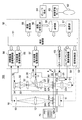
図1は、本実施形態に係るデジタル顕微鏡装置100の全体構成を示したブロック図である。
Hereinafter, embodiments according to the present technology will be described with reference to the drawings.
<First Embodiment>
FIG. 1 is a block diagram illustrating an overall configuration of a
[概要]
本技術では、シェーディング補正用の白画像を撮像する際、顕微鏡のステージの開口部上にプレパラートが無い状態で撮像する。プレパラートが無いと、プレパラートを構成するスライドガラスが無い分、光源から撮像素子までの光路長が変わってくる。そのため、ステージ上にプレパラートが無い状態で照明光学系の焦点位置をピントが合うように調整する。それ故、照明光学系のピントが合った状態で白画像を適切に撮像することが出来、適切に撮像された白画像に基づくことにより、適切にシェーディング補正を行うことが出来る。
[Overview]
In the present technology, when a white image for shading correction is captured, the image is captured in a state where there is no preparation on the opening of the stage of the microscope. If there is no preparation, the optical path length from the light source to the image sensor changes due to the absence of the slide glass constituting the preparation. Therefore, the focus position of the illumination optical system is adjusted so that it is in focus with no preparation on the stage. Therefore, it is possible to appropriately capture a white image while the illumination optical system is in focus, and it is possible to appropriately perform shading correction based on the appropriately captured white image.
[全体構成]
このデジタル顕微鏡装置100は、俯瞰画像撮像部10と、拡大画像撮像部(撮像部)20と、位相差像撮像部30と、ステージ40と、制御部50とを有する。
[overall structure]
The
俯瞰画像撮像部10は、生体サンプルSPLが配設されるプレパラートPRT全体の画像(以下、この画像を「俯瞰画像」と称する。)を撮像する。
The overhead
拡大画像撮像部20は、生体サンプルSPLが所定倍率で拡大された画像(以下、この画像を「拡大画像」と称する。)を撮像する。
The enlarged
位相差像撮像部30は、拡大画像撮像部20の対物レンズ23の焦点とプレパラートPRT上の生体サンプルSPLとの光軸方向のずれの量と向きをデフォーカス量として検出する。また、位相差像撮像部30は、コンデンサレンズ22aの光軸方向のずれの量と向きをデフォーカス量として検出する。
The phase difference
ステージ40は、プレパラートPRTを載置して俯瞰画像撮像部10による撮像位置および拡大画像撮像部20による撮像位置に移動させるためのものである。ステージ40は、ステージ駆動機構41により、拡大画像撮像部20の対物レンズ23の光軸の方向(Z軸方向)と、光軸の方向に対して直交する方向(X軸−Y軸方向)に移動自在とされている。
The
なお、プレパラートPRTは、血液等の結合組織、上皮組織又はそれらの双方の組織などの組織切片又は塗抹細胞からなる生体サンプルSPLを、所定の固定手法によりスライドガラスに固定したものである。これらの組織切片又は塗抹細胞には、必要に応じて各種の染色が施される。この染色には、HE(ヘマトキシリン・エオシン)染色、ギムザ染色、パパニコロウ染色、チール・ネールゼン染色、グラム染色等に代表される一般染色のみならず、FISH(Fluorescence In−Situ Hybridization)や酵素抗体法等の蛍光染色が含まれる。 The preparation PRT is obtained by fixing a biological sample SPL made of tissue sections such as connective tissue such as blood, epithelial tissue, or both tissues, or smeared cells to a slide glass by a predetermined fixing method. These tissue sections or smear cells are subjected to various stains as necessary. This staining includes not only general staining represented by HE (hematoxylin / eosin) staining, Giemsa staining, Papanicolaou staining, Thiel-Nölsen staining, Gram staining, FISH (Fluorescence In-Situ Hybridization), enzyme antibody method, and the like. Of fluorescent staining.
また、このデジタル顕微鏡装置100には、サンプルSPLを含むプレパラートPRTを蓄積し、蓄積されたプレパラートPRTを一つずつステージ40の上にローディングするプレパラートストック・ローダ70が付設されている。なお、プレパラートストック・ローダ70はデジタル顕微鏡装置100に組み込まれたものであってもよい。
Further, the
次に、上述の俯瞰画像撮像部10と、拡大画像撮像部20と、位相差像撮像部30の詳細について説明する。
Next, details of the above-described overhead
[俯瞰画像撮像部10]
俯瞰画像撮像部10は、図に示したように、光源11と、対物レンズ12と、撮像素子13とを有する。
[Overhead image capturing unit 10]
The overhead
光源11は、ステージ40のプレパラート配置面とは逆の面側に設けられる。
The
俯瞰画像撮像部10には、プレパラートPRTに貼付されたラベルに記載されている付帯情報を撮像するための光を照射するラベル光源(図示せず)が別途設けられていてもよい。
The overhead
対物レンズ12は、プレパラートPRT配置面における俯瞰画像撮像部10の基準位置の法線を光軸SRとして、ステージ40のプレパラートPRT配置面側に配設される。ステージ40上に載置されたプレパラートPRTを透過した透過光は、この対物レンズ12によって集光されて、対物レンズ12の後方(すなわち、照明光の進行方向)に設けられた撮像素子13に結像する。
The
撮像素子13には、ステージ40のプレパラート配置面に載置されたプレパラートPRT全体を包括する撮影範囲の光(換言すれば、プレパラートPRT全体を透過した透過光)が結像する。この撮像素子13上に結像した像が、プレパラートPRT全体を撮像した顕微鏡画像である俯瞰画像となる。
The
[拡大画像撮像部]
拡大画像撮像部20は、図に示したように、光源21と、コンデンサレンズ(照明光学系)22と、対物レンズ23と、撮像素子24と、コンデンサレンズ駆動機構25を有する。
[Enlarged image capturing unit]
The enlarged
光源21は、照明光を照射するものである。光源21はステージ40のプレパラート配置面とは逆の面側に設けられる。
The
コンデンサレンズ22aは、光源21から照射された照明光を集光して、ステージ40上のプレパラートPRTに導くレンズである。このコンデンサレンズ22aは、プレパラートPRT配置面における拡大画像撮像部20の基準位置の法線を光軸ERとして、光源21とステージ40との間に配設される。
The
なお、図1に示されないが、光源21とコンデンサレンズ22aとの間には、光源21から出射した照明光を集光する集光光学系22c、視野絞り22bなども設けられる。
以降、上記の集光光学系22c、視野絞り22bおよびコンデンサレンズ22aを「照明光学系」と呼ぶ。
Although not shown in FIG. 1, a condensing
Hereinafter, the condensing
コンデンサレンズ駆動機構25は、コンデンサレンズ22aを光軸ER方向に沿って駆動することによって、コンデンサレンズ22aの光軸ER上の位置を変える。
The condenser
対物レンズ23は、プレパラートPRT配置面における拡大画像撮像部20の基準位置の法線を光軸ERとして、ステージ40のプレパラートPRT配置面側に配設される。拡大画像撮像部20では、この対物レンズ23を適宜交換することで、生体サンプルSPLを様々な倍率に拡大して撮像することが可能となる。ステージ40上に載置されたプレパラートPRTを透過した透過光は、この対物レンズ23によって集光されて、対物レンズ23の後方(すなわち、照明光の進行方向)に設けられた撮像素子24に結像する。
The
対物レンズ23と撮像素子24との間の光軸ER上にはビームスプリッター31が設けられる。ビームスプリッター31は、対物レンズ23を透過した透過光の一部を位相差像撮像部30へと導く。
A
撮像素子24には、撮像素子24の画素サイズ及び対物レンズ23の倍率に応じて、ステージ40のプレパラートPRT配置面上における所定の横幅及び縦幅からなる撮影範囲(以下、小領域と呼ぶ)の像が結像される。なお、対物レンズ23により生体サンプルSPLの一部が拡大されるため、上述の撮影範囲は、撮像素子13の撮影範囲に比べて十分に狭い範囲となる。
The
なお、図1に示されないが、光源21とコンデンサレンズ22aとの間には、光源21からステージ40上のプレパラートPRTに照射される照明光の範囲を制限する視野絞り22b(図8参照)が設けられる。コンデンサレンズ22aの焦点を合わせる場合、この視野絞り22bのエッジ部分を目安に焦点位置合わせが行われる。
Although not shown in FIG. 1, a
[位相差像撮像部]
位相差像撮像部30は、図に示したように、ビームスプリッター31と、フィールドレンズ32と、セパレータレンズ33と、撮像素子34とを有する。
[Phase difference imaging unit]
As shown in the drawing, the phase difference
ビームスプリッター31は、先に説明したように、拡大画像撮像部20の対物レンズ23と撮像素子24との間の光軸ER上に設けられており、対物レンズ23を透過した透過光の一部を反射させる。換言すれば、ビームスプリッター31によって、対物レンズ23を透過した光は、撮像素子24へと向かう反射光と、位相差像撮像部30内のフィールドレンズ32へと向かう透過光とに分岐される。
As described above, the
ビームスプリッター31によって分岐された透過光の進行方向側には、フィールドレンズ32が設けられる。このフィールドレンズ32は、ビームスプリッター31によって分岐された透過光を集光して、フィールドレンズ32の後方(透過光の進行方向側)に設けられたセパレータレンズ33へと導く。
A
セパレータレンズ33は、フィールドレンズ32から導光された光束を2つの光束へと分割する。分割された光束は、セパレータレンズ33の後方(透過光の進行方向側)に設けられた撮像素子34の結像面に対して、1組の被写体像を形成する。
The
撮像素子34には、セパレータレンズ33を透過した光がそれぞれ結像する。その結果、撮像素子34の撮像面には、1組の被写体像が形成されることとなる。なお、ここでは2つのセパレータレンズ33を透過した光は、単一の撮像素子34において撮像されるとしたが、セパレータレンズ33を透過した光それぞれを2つの撮像素子34により撮像する構成でもよい。セパレータレンズ33には、フィールドレンズ32を射出した様々な方向の光束が入射するため、形成される1組の被写体像間には、位相差が存在する。以下では、この1組の被写体像を「位相差像」と称する。
The light that has passed through the
なお、以上の説明では、対物レンズ23と撮像素子24との間にビームスプリッター31が設けられる場合について説明したが、光線を分岐するための光線分岐手段はビームスプリッターに限定されるわけではなく、可動式ミラー等を利用することも可能である。また、拡大画像撮像部20の鏡筒と位相差像撮像部30の鏡筒とを切り替える機構を利用することも可能である。
In the above description, the case where the
また、前述の説明では、位相差像撮像部30内の位相差AF(Auto Focus)光学系としてフィールドレンズ32、セパレータレンズ33及び撮像素子34を有する構成を示したが、かかる例に限定されるわけではない。かかる位相差AF光学系は、例えば、フィールドレンズ及びセパレータレンズの代わりにコンデンサレンズ及び2眼レンズを利用したりするなど、同等の機能を実現可能なものであれば、他の光学系であってもよい。
また、俯瞰画像撮像部10、拡大画像撮像部20及び位相差像撮像部30それぞれに設けられる撮像素子は、1次元撮像素子であってもよく、2次元撮像素子であってもよい。
In the above description, the configuration including the
In addition, the imaging elements provided in each of the overhead
また、以上の説明では、位相差像撮像部30を対物レンズ23の光軸ER上に配置し、拡大画像撮像部20の撮像素子24をビームスプリッター31にて分岐された反射光が入射される位置に配置した。しかし、逆に、拡大画像撮像部20の撮像素子24を対物レンズ23の光軸ER上に配置し、位相差像撮像部30をビームスプリッター31にて分岐された反射光が入射される位置に配置してもよい。
Further, in the above description, the phase difference
[制御部50]
制御部50は、統合制御部51と、照明制御部52と、ステージ制御部53と、コンデンサレンズ駆動制御部54と、位相差像撮像制御部55と、俯瞰画像撮像制御部56と、拡大画像撮像制御部57と、記憶部58と、現像処理部59と、画像符号化部60とを有する。
[Control unit 50]
The
統合制御部51は、例えば、CPU(Central Processing Unit)と、ROM(Read Only Memory)と、RAM(Random Access Memory)とを含むコンピュータのハードウェア要素で構成される。あるいはFPGA(field programmable gate array)などの専用ICによって構成されてもよい。統合制御部51は、照明制御部52、ステージ駆動制御部53、コンデンサレンズ駆動制御部54、位相差像撮像制御部55、俯瞰画像撮像制御部56、拡大画像撮像制御部57、記憶部58、現像処理部59、画像符号化部60、通信部61との間で各種信号をやりとりして、検体の拡大像を取得するための様々な演算処理および制御を実行する。RAMには、そのための各種のプログラムおよびデータがロードされ、CPUはRAMにロードされたプログラムを実行する。ROMには、RAMにロードされるプログラムやデータなどが格納される。
The
照明制御部52、ステージ駆動制御部53、コンデンサレンズ駆動制御部54、位相差像撮像制御部55、俯瞰画像撮像制御部56および拡大画像撮像制御部57は、例えば、CPU、ROM、RAMなどを含むコンピュータのハードウェア要素で構成されてもよいし、FPGAなどの専用ICによって構成されてもよい。
The
現像処理部59および画像符号化部60は、例えば、CPU、ROM、RAMなどを含むコンピュータのハードウェア要素で構成される。あるいはGPU(Graphics Processing Unit)によって構成されてもよい。
The
照明制御部52は、統合制御部51から与えられる、検体SPLの照明方法の指示をもとに光源11、21の制御を行う。例えば、照明制御部52は、統合制御部51からの指示に従って、光源11、21の照明光の強度や、明視野用の光源、暗視野用の光源など、光源の種類の選択等を行う。明視野用の光源としては、例えば、可視光を照明するものなどが想定される。明視野用の光源としては、例えば、特殊染色で用いられる蛍光マーカを励起可能な波長を含む光を照明するものなどが挙げられる。
The
ステージ駆動制御部53は、例えば、統合制御部51から俯瞰画像を撮像する指示が与えられると、プレパラートPRT全体が撮像素子13の撮影範囲に入るようにステージ駆動機構41を駆動して、ステージ面方向(X―Y軸方向)にステージ40を移動させる。ステージ制御部53は、プレパラートPRT全体に対物レンズ12の焦点が合うようにステージ駆動機構41を駆動して、ステージ40をZ軸方向に移動させる。
For example, when an instruction to capture a bird's-eye view image is given from the
また、ステージ制御部53は、統合制御部51から拡大画像を撮像する指示が与えられると、その指示されたサンプルSPLの撮影範囲(小領域)が撮像素子24の撮影範囲に入るように、ステージ駆動機構41を駆動して、ステージ面方向(X―Y軸方向)にステージ40を移動させる。ステージ制御部53は、サンプルSPLに対物レンズ23の焦点が合うように、ステージ駆動機構41を駆動して、ステージ40をZ軸方向に移動させる。
In addition, when an instruction to capture an enlarged image is given from the
コンデンサレンズ駆動制御部54は、統合制御部51からの、光源21からの照明光の視野絞りのデフォーカス量に関する情報に基づいてコンデンサレンズ駆動機構25の制御を行うことで、コンデンサレンズ22aのピントを合わせ、光源21からの照明光をサンプルSPLの観察範囲にだけ当たるように調整する。視野絞りに関する情報はデフォーカス量とデフォーカスの向きを含む。これらの情報は、位相差像撮像部30により生成される一組の位相差像の距離をもとに求められる。
The condenser lens
位相差像撮像制御部55は、位相差像撮像部30に設けられた撮像素子34の結像面に結像した一組の位相差像の信号を取得し、統合制御部51に供給する。統合制御部51は、その中のメインメモリにロードされたプログラムに従って、位相差像撮像制御部55より取得した一組の位相差像の距離をもとに、拡大画像撮像部20の対物レンズ23の焦点のサンプルSPLに対するデフォーカス量とデフォーカスの向きを算出する。
The phase difference image capturing
統合制御部51は、これらの情報をもとにステージ40の制御情報を生成し、ステージ制御部53に供給する。ステージ制御部53は、統合制御部51からの制御情報をもとにステージ40をZ軸方向に移動させるようにステージ駆動機構41を駆動する。これにより、拡大画像撮像部20の対物レンズ23の焦点をサンプルSPLに合わせる位相差AFが行われる。
The
俯瞰画像撮像制御部56は、俯瞰画像撮像部10の撮像素子13の結像面に結像した俯瞰画像に対応する信号をもとに当該俯瞰画像に対応するデータを生成して統合制御部51に供給する。統合制御部51は、その中のメインメモリにロードされたプログラムに従って、俯瞰画像撮像制御部56から取得した俯瞰画像からサンプルSPLが存在する領域を特定する処理などを行う。この領域を「小領域」と呼ぶ。
The overhead image capturing
拡大画像撮像制御部55は、拡大画像撮像部20の撮像素子24の結像面に結像した小領域毎の観察像に対応する信号をもとに当該小領域毎の観察像に対応するRAWデータを生成して統合制御部51に供給する。統合制御部51は、拡大画像撮像制御部55より取得した小領域毎のRAWデータを現像処理部59に供給して現像処理を実行させる。統合制御部51は、現像処理部59にて現像された小領域毎の拡大像のデータを接続して検体SPL単位の大画像を生成し、生成された検体SPL単位の大画像をタイルと呼ばれる所定の解像度の単位に分割する処理などを行う。さらに、統合制御部51は、生成された各々のタイルを画像符号化部60に供給して、所定の圧縮符号化形式の画像データを生成させ、記憶部58に保存させる。
The enlarged image capturing
記憶部58は、デジタル顕微鏡装置100を制御するための各種設定情報やプログラム、さらには所定の圧縮符号化形式のタイル群などを格納する。
The
現像部59は、観察像撮像部20によって撮像された小領域毎の観察像のRAWデータを現像する。
The developing
画像符号化部60は、タイル毎の画像データを所定の画像圧縮形式に符号化する。ここで、画像圧縮形式として、例えば、JPEG(Joint Photographic Experts Group)などが採用される。勿論、JPEG以外の圧縮符号化形式が採用されてもよい。
The
記憶部58に記憶された各タイルは、通信部61によってネットワーク62を通じて画像管理サーバ63に蓄積される。画像管理サーバ63は、ビューワ端末64からのリクエストに応じて該当する1以上のタイルをビューワ端末64に応答する。ビューワ端末64は、画像管理サーバ63より取得した1以上のタイルを用いて表示用の観察像を生成して、ビューワ端末64の表示部に表示させる。
Each tile stored in the
[拡大画像撮像部の対物レンズのオートフォーカス]
この実施形態のデジタル顕微鏡装置100には、拡大画像撮像部20の対物レンズ23のオートフォーカス方式として、位相差オートフォーカス方式およびコントラストオートフォーカス方式が搭載されている。
[Auto focus of the objective lens of the enlarged image pickup unit]
The
位相差オートフォーカス方式を用いる場合、統合制御部51は位相差像撮像制御部55に位相差像を撮像させるように指示を出す。位相差像撮像制御部55は、この指示を受けると、位相差像撮像部30から、撮像素子34の撮像面に並んで結像された一組の位相差像の信号を取り込み、2つの位相差像の位相差を求める。
When the phase difference autofocus method is used, the
ここで、対物レンズ23の焦点が適切な面から遠ざかると、2つの位相差像上における観測面の同一の領域は、撮像素子24の外側方向に向かって互いに離れるように移動する。逆に、対物レンズ23の焦点が適切な面よりも近くなると、2つの位相差像上における観測面の同一の領域は撮像素子24の内側方向に向かって互いに近づくように移動する。統合制御部51は、2つの位相差像上における観測面の同一の領域間の距離を、上記の位相差として求める。
Here, when the focal point of the
統合制御部51は、求めた位相差から対物レンズ23の焦点の観察対象であるサンプルSPLに対するデフォーカス量とデフォーカスの向きを求める。統合制御部51は、求めたデフォーカス量とデフォーカスの向きをもとにステージ40の制御情報を生成し、ステージ制御部53に供給する。ステージ制御部53は、統合制御部51からの制御情報をもとにステージ40をZ軸方向に移動させるようにステージ駆動機構41を駆動する。これにより、拡大画像撮像部20の対物レンズ23の焦点をサンプルSPLに合わせる位相差オートフォーカスが行われる。
The
一方、コントラストオートフォーカス方式は、拡大画像撮像部20を用いて山登り方式で焦点探索を行う方式である。コントラストオートフォーカス方式を用いる場合、統合制御部51は、対物レンズ23の焦点位置を所定の距離ずつずらし、各々の焦点位置で拡大画像撮像部20にサンプルSPLの撮影範囲の撮像を実行させる。統合制御部51は、撮像された画像の中でコントラストがもっとも高い画像が撮像されたときの焦点位置を最適な焦点位置として判定する。
On the other hand, the contrast autofocus method is a method of performing a focus search by a hill-climbing method using the enlarged
次に、本実施形態のデジタル顕微鏡装置100の統合制御部51に実装されたシェーディング補正に関する機能について説明する。
本実施形態のデジタル顕微鏡装置100では、ステージの上にプレパラートが載置されていない状態でのシェーディング補正を良好に行うことを目的の一つとする。
Next, functions relating to shading correction implemented in the
One object of the
プレパラートにはゴミなどの異物も保持されている可能性があり、白画像撮像時とプレパラートPRTの拡大画像撮像時で、ゴミの状態が異なるため、シェーディング補正に悪影響が生じるからである。プレパラートがステージに載置されていない状態でのシェーディング補正を行うことで、ゴミなどの異物による影響を受けることなく、シェーディング補正を良好に行うことができる。しかし、その場合、照明光学系のピント合わせが課題となってくる。 This is because there is a possibility that foreign matters such as dust are also held in the preparation, and the shading correction is adversely affected because the state of dust is different when capturing a white image and when capturing an enlarged image of the preparation PRT. By performing the shading correction in a state where the preparation is not placed on the stage, the shading correction can be performed satisfactorily without being affected by foreign matters such as dust. However, in that case, focusing the illumination optical system becomes a problem.
すなわち、白画像の輝度分布は照明光学系のピント状態に依存するため、照明光学系のピントが適切ではない状態で白画像を撮影すると、プレパラートPRTごとにピントが異なるためシェーディング補正が適切に行われないからである。 In other words, the brightness distribution of the white image depends on the focus state of the illumination optical system. Therefore, if a white image is taken when the illumination optical system is not in focus, the focus differs depending on the preparation PRT, so that shading correction is performed appropriately. Because it is not broken.
[統合制御部51について]
次に、上述した統合制御部51の詳細について説明する。図2は、統合制御部51において、上述した照明光学系のピント合わせを実現する機能の構成を示すブロック図である。
[About Integrated Control Unit 51]
Next, details of the above-described
同図に示すように、統合制御部51は、白画像撮像制御部(白画像取得部)514、補正係数算出部(算出部)515、シェーディング補正部516を備える。これらの機能は、統合制御部51内のCPUがRAMにロードされたプログラムを実行することによって実現される。
As shown in the figure, the
白画像撮像制御部514は、プレパラートPRTがステージ40に載置されていない状態でのシェーディング補正を行うために必要な白画像を撮像する制御を行う。ここで、プレパラートPRTがステージ40に載置されていない状態での白画像の撮像とは、プレパラートPRTの開口部40aがプレパラートPRTによって覆われていないため、開口部40aを通して光源21からの照明光の視野絞り22bの像を直接、拡大画像撮像部20の撮像素子24により撮像することを言う。このようにして撮像された画像が「白画像」である。
The white image capturing
白画像撮像制御部514は、より具体的には、プレパラートPRTがステージ40に載置されていない状態において、位相差像撮像制御部55に対して位相差像の撮像を指示する。位相差像撮像制御部55は、位相差像撮像部30に撮像を実行させる。これにより、位相差像撮像部30にて、コンデンサレンズ22aによって結像された照明光学系の視野絞り22bの像に対する位相差像が得られ、白画像撮像制御部514に供給される。白画像撮像制御部514は、この位相差像をもとに位相差を算出し、この位相差に応じたデフォーカス情報を生成し、コンデンサレンズ駆動制御部54に、そのデフォーカス情報に対応した制御情報を出力する。コンデンサレンズ駆動制御部54は、制御情報をもとにコンデンサレンズ駆動機構25を駆動する。これにより、コンデンサレンズ22aによって結像された照明光学系の視野絞り22bの像に対物レンズ23の焦点が合わせられる。この後、白画像撮像制御部514は、照明制御部52に光源21の点灯を指示する一方で、拡大画像撮像制御部57に拡大画像の撮像を指示することによって、白画像が撮像される。
More specifically, the white image capturing
補正係数算出部515は、白画像撮像制御部514が撮像した白画像に基づきシェーディング補正係数を算出する。この白画像からのシェーディング補正係数の算出方法は、ステージ40にプレパラートPRTを載置した状態で撮像された画像からのシェーディング補正係数の算出方法と同じである。本技術は、シェーディング補正係数の算出方法そのものに特徴を有するものではないので、詳細な説明を省くこととする。
The correction
シェーディング補正部516は、補正係数算出部515が算出したシェーディング補正係数に基づき、拡大画像撮像制御部57が撮像したサンプルSPLの拡大画像に対してシェーディング補正を行う。
The
なお、補正係数算出部515によって算出されたシェーディング補正係数は例えば制御部50の記憶部58などに記憶される。シェーディング補正部516は、実際にサンプルSPLの拡大画像の撮像時に記憶部58からシェーディング補正係数を読み出し、拡大画像撮像制御部57によって撮像されたサンプルSPLの拡大画像に対してシェーディング補正を行う。
以上、統合制御部51の詳細について説明した。
The shading correction coefficient calculated by the correction
The details of the
[全体的な処理の流れについて]
次に、本実施形態のデジタル顕微鏡装置100における、シェーディング補正用の補正係数を算出するまでの処理の流れと、個々の拡大画像の撮像からシェーディング補正までの処理の流れについて説明する。図3は、本実施形態のデジタル顕微鏡装置100における、シェーディング補正用の補正係数を算出するまでの処理の流れと、個々の拡大画像の撮像からシェーディング補正までの処理の流れについて説明するフローチャートである。
[Overall process flow]
Next, in the
最初に、白画像を取得して、シェーディング補正用の補正係数を算出するまでの処理について説明する。なお、ここで説明する白画像を取得して、シェーディング補正用の補正係数を算出するまでの処理は、一定以上の頻度で行うことが望ましい。白画像により得られる輝度分布は、温度や迷光の影響などにより変化するためである。 First, a process from acquiring a white image to calculating a correction coefficient for shading correction will be described. It should be noted that it is desirable to perform the processing from acquiring the white image described here to calculating the correction coefficient for shading correction at a certain frequency or more. This is because the luminance distribution obtained from the white image changes due to the influence of temperature, stray light, and the like.
上記の処理をプレパラートストック・ローダ70がプレパラートPRTを交換するタイミングで行えば、白画像撮像のために、わざわざプレパラートPRTをステージ50上から取り除く必要が無いので、効率的であり、撮影効率を向上させることが出来る。
If the above-described processing is performed at the timing when the
まず、最初のステップとして、ステージ制御部53が、プレパラートPRTの載置されていないステージ40の開口部40aが対物レンズ23の直下に来るように、ステージ40を移動させる(ステップST1)。
First, as a first step, the
ここで言う開口部とは、プレパラートPRTを撮像する際にプレパラートPRT上のサンプルSPLに撮像用の光を当てるための開口部40aに限らない。光源21から射出し、コンデンサレンズ22aを透過し、対物レンズ23に入射する光を、プレパラートPRTやスライドガラスが遮らない状態であればよい。
The opening referred to here is not limited to the
次のステップとして、白画像撮像制御部514は、位相差像撮像制御部55に対して位相差像の撮像を指示する。この指示に従って位相差像撮像制御部55は位相差像撮像部30に撮像を実行させる。これにより、コンデンサレンズ22aにより結像された照明光学系の視野絞り22bの像に対する位相差像が得られる。位相差像撮像制御部55はこの位相差像を白画像撮像制御部514に供給する。白画像撮像制御部514は、照明光学系の視野絞り22bの像に対する位相差像をもとに位相差を算出し、この位相差に応じたデフォーカス情報を生成し、コンデンサレンズ駆動制御部54にデフォーカス情報に対応した制御情報を出力する。コンデンサレンズ駆動制御部54は制御情報をもとにコンデンサレンズ22aを光軸方向に移動させるようにコンデンサレンズ駆動機構25を制御する。これにより、照明光学系のピントが合わせられる。言い換えれば、対物レンズ23の焦点に、コンデンサレンズ22aによる照明光学系の視野絞り22bの像の結像点が合わせられる(ステップST2)。
As the next step, the white image capturing
ここでは、照明光学系のピントを合わせる方式として位相差AFを用いたが、コントラストAF方式を用いて照明光学系のピントを合わせるようにしてもよい。 Here, the phase difference AF is used as a method for focusing the illumination optical system, but the illumination optical system may be focused using a contrast AF method.
なお、上記のような白画像の撮像時には、コンデンサレンズ22aと対物レンズ23の間に、空気よりも屈折率の高いガラスを用いたプレパラートPRTやスライドガラスが存在しないので、その分、光源21から射出し撮像素子24に入射する光の光路長は短くなる。そのため、光源21を含む照明光学系は、プレパラートPRTの撮像時より対物レンズ23方向に所定の距離ΔZだけ移動させておく必要がある。この所定の距離ΔZについては後述する。
At the time of capturing a white image as described above, there is no preparation PRT or slide glass using glass having a refractive index higher than that of air between the
次に、白画像撮像制御部514は、光源21を点灯させるように照明制御部52を制御する(ステップST3)。
Next, the white image capturing
次のステップとして、白画像撮像制御部514が、拡大画像撮像制御部57に指示を出して、白画像を撮像する(ステップST4)。
As the next step, the white image capturing
最後のステップとして、補正係数算出部515が、ステップST4において取得した白画像に基づき、シェーディング補正用の補正係数を算出する(ステップST5)。
As the last step, the correction
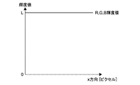
白画像の輝度値は、一般的に、図4に示すような輝度分布をとる。この図は、白画像のX方向における輝度情報の分布を表したものである。Red、Green、Blue各色の輝度分布はそれぞれ独立している。各ピクセル座標位置(x,y)における、RGB各色の輝度値を、それぞれWr(x,y)、Wg(x,y)、Wb(x,y)と表す。 The luminance value of the white image generally has a luminance distribution as shown in FIG. This figure shows the distribution of luminance information in the X direction of a white image. The luminance distribution of each color of Red, Green, and Blue is independent. The luminance value of each color of RGB at each pixel coordinate position (x, y) is expressed as Wr (x, y), Wg (x, y), Wb (x, y), respectively.
ここで、シェーディング補正後のターゲット輝度値をLとしたとき、各色R、G、B毎のシェーディング補正係数LR、LG、LBは、それぞれ以下の計算式で算出される。
LR(x,y) = L / Wr(x,y)
LG(x,y) = L / Wg(x,y)
LB(x,y) = L / Wb(x,y)
Here, when the target luminance value after the shading correction is L, the shading correction coefficients LR, LG, and LB for each of the colors R, G, and B are calculated by the following calculation formulas, respectively.
LR (x, y) = L / Wr (x, y)
LG (x, y) = L / Wg (x, y)
LB (x, y) = L / Wb (x, y)
白画像の各ピクセルのRGB輝度値に、シェーディング補正係数LR、LG、LBをそれぞれ掛けることにより、各ピクセルの輝度値は、図5に示すように、ターゲット輝度値Lとなり、シェーディングを補正することが可能となる。なお、上記補正係数の演算手法は、上述したものに限らず、どのような方法で行っても構わない。 By multiplying the RGB luminance value of each pixel of the white image by the shading correction coefficients LR, LG, and LB, the luminance value of each pixel becomes the target luminance value L as shown in FIG. 5, and the shading is corrected. Is possible. The correction coefficient calculation method is not limited to the above-described method, and any method may be used.
以上、シェーディング補正用の補正係数を算出するまでの処理の流れについて説明した。 The flow of processing up to the calculation of the correction coefficient for shading correction has been described above.
次に、個々の拡大画像の撮像からシェーディング補正までの処理の流れについて説明する。以下の処理では、ステップST5において算出したシェーディング補正係数LR、LG、LBを用いる。 Next, the flow of processing from capturing individual enlarged images to shading correction will be described. In the following process, the shading correction coefficients LR, LG, and LB calculated in step ST5 are used.
最初のステップとして、プレパラートストック・ローダ70がプレパラートPRTをステージ40上に載置する(ステップST6)。
As a first step, the
ステージ制御部53は、載置されたプレパラートPRTが対物レンズ23の真下に来るように、ステージ40を移動させる。
The
次のステップとして、統合制御部51が、位相差像撮像制御部55からの出力に基づき、コンデンサレンズ駆動制御部54に指示を出して、コンデンサレンズ22aのピントを合わせる(ステップST7)。
As the next step, the
ここでピントを合わせることにより、コンデンサレンズ22aのピントずれに起因する明度ムラの変動を防ぐことが出来る。
By adjusting the focus here, it is possible to prevent fluctuations in brightness due to the focus shift of the
次のステップとして、ステージ制御部53が、生体サンプルSPLの任意の位置を拡大撮影するために、ステージ40を移動させる(ステップST8)。
As the next step, the
次のステップとして、照明制御部52が、光源21を点灯させる(ステップST9)。
As the next step, the
次のステップとして、拡大画像撮像制御部57が、プレパラートPRT上の生体サンプルSPLの拡大画像を撮像する(ステップST10)。
As the next step, the enlarged image capturing
撮像された拡大画像のX方向の輝度分布の例を図6に示す。撮像素子24の撮像面に、生体サンプルSPLの像が写ると、その像に対応したピクセル位置では受光量が減少するので、図に示すような輝度分布となる。
An example of the luminance distribution in the X direction of the captured enlarged image is shown in FIG. When an image of the biological sample SPL appears on the imaging surface of the
なお、図6に示す輝度分布では、X方向の輝度分布曲線の中央部分が生体サンプルSPLの像が写っている影響で輝度値が低下しているのに加え、画像の周辺部でも輝度値が低下している。これは、図4に示した白画像の輝度分布において、画像周辺部で輝度値が減少していることに起因する。 In the luminance distribution shown in FIG. 6, the luminance value is lowered due to the influence of the image of the biological sample SPL in the central portion of the luminance distribution curve in the X direction, and the luminance value is also present in the peripheral portion of the image. It is falling. This is due to the fact that the brightness value is reduced at the periphery of the image in the brightness distribution of the white image shown in FIG.
最後のステップとして、シェーディング補正部516が、ステップST10において取得した拡大画像に対して、シェーディング補正を行う(ステップST11)。
As the last step, the
拡大画像のRGB各画素の輝度値に、シェーディング補正係数LR、LG、LBをそれぞれ掛けることにより、シェーディング補正が行われる。図6に示した輝度分布をシェーディング補正した結果を図7に示す。図に示すように、生体サンプルSPLが写っていない画像周辺部の輝度値が補正され、ターゲット輝度値Lになっている。シェーディング補正が行われた拡大画像は、現像処理部59に出力される。
Shading correction is performed by multiplying the luminance value of each RGB pixel of the enlarged image by the shading correction coefficients LR, LG, and LB, respectively. FIG. 7 shows the result of shading correction of the luminance distribution shown in FIG. As shown in the figure, the luminance value at the periphery of the image where the biological sample SPL is not captured is corrected to a target luminance value L. The enlarged image subjected to the shading correction is output to the
以上、個々の拡大画像の撮像からシェーディング補正までの処理の流れについて説明した。 The flow of processing from capturing individual enlarged images to shading correction has been described above.
[光源21の光量調整について]
次に、プレパラートPRTをステージ40上に載置せずに白画像を撮像する際の、光源21の光量調整について説明する。
[Regarding the light amount adjustment of the light source 21]
Next, the light amount adjustment of the
プレパラートPRTを構成するスライドガラスに光源21から光を照射する場合、一般的なスライドガラスでは、その表面および裏面でそれぞれ4%程度の光が反射される。そのため、光源21の光量調整を行わない場合、プレパラートPRTをステージ40上に載置せずに白画像を撮像する際、撮像素子24には、プレパラートPRTをステージ40上に載置する場合に比べ、より強い光が入射してしまう。
When light is emitted from the
それ故、本技術では、プレパラートPRTをステージ40上に載置せずに白画像を撮像する際、光源21の光量を92%程度まで減光する。なお、減光は光源21の光量により行ってもよいし、開口絞り(図示せず)により行ってもよい。
Therefore, in the present technology, when a white image is captured without placing the preparation PRT on the
この減光により、ステージ40上のプレパラートPRTの有無に関わらず、白画像と拡大画像とで同程度の信号強度の画像を得ることが出来る。
By this dimming, it is possible to obtain images with the same signal intensity for the white image and the enlarged image regardless of the presence or absence of the preparation PRT on the
以上、プレパラートPRTをステージ40上に載置せずに白画像を撮像する際の、光源21の光量調整について説明した。
The light amount adjustment of the
[白画像撮像時のコンデンサレンズ22aおよびステージ40の位置調整について]
上述した、プレパラートPRTをステージ40上に載置しない状態での白画像撮影時に、照明光学系および光源21をΔZだけ対物レンズ23側に移動する点について説明する。
[Position adjustment of
The point that the illumination optical system and the
ΔZの具体的な値は、例えばコンデンサレンズ22aを含む透過照明の光学系が対物レンズ23側でテレセントリックをなしている場合、プレパラートPRT無しでコンデンサレンズ22aのピントを合わせる際には、屈折率n、厚みdのプレパラートPRTを載置する場合に比べて、ΔZ=d(n−1)/nだけコンデンサレンズ22aおよび光源21を対物レンズ23側に移動させなければならない。この数式の根拠は、特許文献2による。
A specific value of ΔZ is, for example, when the optical system of transmitted illumination including the
顕微鏡においては、一般的に、対物レンズ23とステージ40間の距離や、ステージ40とコンデンサレンズ22a間の距離を十分に確保することは難しい。
In a microscope, it is generally difficult to ensure a sufficient distance between the
そのため、照明光学系および光源21を対物レンズ23側にΔZだけ移動させる時は、コンデンサレンズ22aとステージ40が衝突しないように一定以上の距離を確保するため、ステージ40もΔZだけ対物レンズ23方向に移動させることが望ましい。
Therefore, when the illumination optical system and the
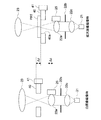
図8は、白画像撮像時(プレパラートPRT無し、図左側)には、拡大画像撮像時(プレパラート有り、図右側)と比べて、コンデンサレンズ22a、集光光学系22c、視野絞り22b、光源21、およびステージ40のZ方向の位置がΔZだけ対物レンズ23側に移動している様子を示す図である。
FIG. 8 shows a
上述のとおり、白画像撮像時には、ステージ40もΔZだけ対物レンズ23側に移動させることで、プレパラートPRTの有無に関わらずコンデンサレンズ22aとステージ40との距離を確保することが出来る。
As described above, when the white image is captured, the distance between the
なお、白画像撮影時には、ステージ40がΔZだけ対物レンズ23に接近することになるが、プレパラートPRTが載置されていない分だけ余裕となるので、ステージ40と対物レンズ23の間の距離を確保する事も容易となる。
Note that, when photographing a white image, the
以上、プレパラートPRTをステージ40上に載置しない状態での白画像撮影時に、照明光学系および光源21をΔZだけ対物レンズ23側に移動する点について説明した。
As described above, the point that the illumination optical system and the
[本技術により得られる効果について]
本技術では、上述した効果に加え、以下の様な効果ももたらす事が出来る。
(1)本技術では、シェーディング補正用の白画像をステージ40上にプレパラートPRTを載置しない状態で撮像する。そのため、本実施形態のように、デジタル顕微鏡装置100が複数枚のプレパラートPRTを格納するプレパラートストック・ローダ70を有している場合、プレパラートPRTを交換する時間内の任意のタイミングにおいてシェーディング補正用の補正係数を生成することが可能となる。プレパラートPRTの交換と補正係数の算出とを同時並行して処理することにより、病理スライドの画像取得のスループットを向上させることが出来る。
[About effects obtained by this technology]
In addition to the effects described above, the present technique can also provide the following effects.
(1) In the present technology, a white image for shading correction is captured in a state where the preparation PRT is not placed on the
(2)本技術では、シェーディング補正用の白画像を撮像する際、そしてプレパラートPRT上の生体サンプルSPLの拡大画像を撮像する際の両方の時点において、コンデンサレンズ22aのピント合わせを行う。そのため、コンデンサレンズ22aのピントずれに起因する輝度分布ずれの影響を除去したシェーディング補正用の補正係数を生成することが出来る。
(2) In the present technology, the
[補足事項]
その他、本技術は、上述の実施形態にのみ限定されるものではなく、本技術の要旨を逸脱しない範囲内において種々変更を加え得ることは勿論である。
[Additional notes]
In addition, the present technology is not limited to the above-described embodiment, and various changes can be made without departing from the scope of the present technology.
[本技術の別の構成]
なお、本技術は以下のような構成も採ることができる。
(1)
照明光を出射する照明光学系と、
前記照明光を透過可能な開口部を有し、この開口部の位置に合わせてプレパラートを載置可能なステージと、
前記照明光学系と前記ステージを挟んで対向して配置され、像を拡大する対物レンズと、この対物レンズにより拡大された像を撮像する撮像素子とを含む拡大撮像部と、
前記ステージの前記開口部を開放させ、前記照明光学系の結像点を前記拡大撮像部の前記対物レンズの焦点に合わせた状態で、前記照明光学系より前記照明光を出射させ、前記拡大撮像部の前記撮像素子に結像した像を白画像として取得する白画像取得部と、
撮像された前記白画像を用いてシェーディング補正係数を算出する算出部と
を具備するデジタル顕微鏡装置。
(2)
前記(1)に記載のデジタル顕微鏡装置であって、
前記拡大撮像部のデフォーカス情報を検出するデフォーカス検出部と、
前記照明光学系の光軸方向の位置を調整する調整部と
を更に具備し、
前記白画像取得部は、
前記デフォーカス情報をもとに、前記照明光の視野絞りの像が前記拡大撮像部の前記撮像素子の前記撮像面に結像するように前記調整部を動作させる
デジタル顕微鏡装置。
(3)
前記(2)に記載のデジタル顕微鏡装置であって、
前記調整部による前記調整は、
前記照明光学系を、前記ステージ上に前記プレパラートが載置されている状態での位置から、所定量だけ前記ステージ側へ移動させることにより行われる
デジタル顕微鏡装置。
(4)
前記(3)に記載のデジタル顕微鏡装置であって、
前記白画像取得部は、
前記移動のとき、前記ステージを、前記プレパラートが載置されている状態での位置から、前記所定量だけ同方向へ移動させる
デジタル顕微鏡装置。
(5)
前記(2)または(3)に記載のデジタル顕微鏡装置であって、
前記所定量は、
d(n−1)/n
(但し、dは前記プレパラートの厚み、nは前記プレパラートの屈折率)である
デジタル顕微鏡装置。
(6)
前記(1)から(5)のうちいずれか1つに記載のデジタル顕微鏡装置であって、
前記白画像取得部は、
前記ステージ上に前記プレパラートが載置された状態での前記拡大画像撮像時に設定する前記照明光の強度を第1の強度として、
前記白画像撮像時の前記照明光の強度を、前記第1の強度より低い第2の強度に設定する
デジタル顕微鏡装置。
(7)
白画像取得部が、照明光学系からの照明光を透過する開口部40aを有し当該開口部40aに跨がるようにプレパラートを載置可能なステージ上に前記プレパラートが載置されていない状態で、
前記照明光学系の視野絞りの像が、前記ステージ上の所定の領域の拡大像を撮像する撮像部の撮像素子の撮像面に結像するように、前記照明光学系の焦点位置を調整する調整部に前記照明光学系の焦点位置を調整させ、
前記撮像部に、前記拡大像を白画像として撮像させ、
算出部が、撮像された前記白画像に基づきシェーディング補正係数を算出する
情報処理方法。
(8)
照明光学系からの照明光を透過する開口部40aを有し当該開口部40aに跨がるようにプレパラートを載置可能なステージ上に前記プレパラートが載置されていない状態で、
前記照明光学系の視野絞りの像が、前記ステージ上の所定の領域の拡大像を撮像する撮像部の撮像素子の撮像面に結像するように、前記照明光学系の焦点位置を調整する調整部に前記照明光学系の焦点位置を調整させ、
前記撮像部に、前記拡大像を白画像として撮像させる白画像取得部および
撮像された前記白画像に基づきシェーディング補正係数を算出する算出部
としてコンピュータを機能させるための情報処理プログラム。
[Another configuration of this technology]
In addition, this technique can also take the following structures.
(1)
An illumination optical system that emits illumination light;
A stage having an opening through which the illumination light can be transmitted, and a stage on which a preparation can be placed according to the position of the opening;
An enlargement imaging unit that is disposed opposite to the illumination optical system and the stage, and includes an objective lens that enlarges an image, and an imaging element that takes an image enlarged by the objective lens;
With the opening of the stage open, the illumination light is emitted from the illumination optical system in a state where the imaging point of the illumination optical system is aligned with the focus of the objective lens of the enlargement imaging unit, and the enlarged imaging A white image acquisition unit that acquires, as a white image, an image formed on the imaging element of the unit;
A digital microscope apparatus comprising: a calculation unit that calculates a shading correction coefficient using the captured white image.
(2)
The digital microscope apparatus according to (1),
A defocus detection unit for detecting defocus information of the enlarged imaging unit;
An adjustment unit that adjusts the position of the illumination optical system in the optical axis direction;
The white image acquisition unit
A digital microscope apparatus that operates the adjusting unit based on the defocus information so that an image of a field stop of the illumination light is formed on the imaging surface of the imaging element of the magnifying imaging unit.
(3)
The digital microscope apparatus according to (2),
The adjustment by the adjustment unit is
A digital microscope apparatus which is performed by moving the illumination optical system by a predetermined amount from a position where the preparation is placed on the stage.
(4)
The digital microscope apparatus according to (3),
The white image acquisition unit
A digital microscope apparatus that moves the stage in the same direction by the predetermined amount from the position where the preparation is placed during the movement.
(5)
The digital microscope apparatus according to (2) or (3),
The predetermined amount is
d (n-1) / n
(Where d is the thickness of the preparation, and n is the refractive index of the preparation).
(6)
The digital microscope apparatus according to any one of (1) to (5),
The white image acquisition unit
The intensity of the illumination light set at the time of capturing the enlarged image in a state where the preparation is placed on the stage is set as a first intensity,
A digital microscope apparatus that sets the intensity of the illumination light at the time of capturing the white image to a second intensity lower than the first intensity.
(7)
A state in which the white image acquisition unit has an
Adjustment for adjusting the focal position of the illumination optical system so that the image of the field stop of the illumination optical system forms an image on the imaging surface of the image sensor of the imaging unit that captures an enlarged image of a predetermined area on the stage To adjust the focal position of the illumination optical system,
Causing the imaging unit to capture the enlarged image as a white image;
An information processing method in which a calculation unit calculates a shading correction coefficient based on the captured white image.
(8)
In a state in which the preparation is not placed on a stage having an
Adjustment for adjusting the focal position of the illumination optical system so that the image of the field stop of the illumination optical system forms an image on the imaging surface of the image sensor of the imaging unit that captures an enlarged image of a predetermined area on the stage To adjust the focal position of the illumination optical system,
An information processing program for causing a computer to function as a white image acquisition unit that causes the imaging unit to capture the enlarged image as a white image and a calculation unit that calculates a shading correction coefficient based on the captured white image.
10…俯瞰画像撮像部
11…光源
12…対物レンズ
13…撮像素子
20…拡大画像撮像部
21…光源
22a…コンデンサレンズ
22b…視野絞り
22c…集光光学系
23…対物レンズ
24…撮像素子
25…コンデンサレンズ駆動機構
30…位相差像撮像部
31…ビームスプリッター
32…フィールドレンズ
33…セパレータレンズ
34…撮像素子
40…ステージ
40a…開口部
41…ステージ駆動機構
50…制御部
51…統合制御部
514…白画像撮像制御部
515…補正係数算出部
516…シェーディング補正部
52…照明制御部
53…ステージ制御部
54…コンデンサレンズ駆動制御部
55…位相差像撮像制御部
56…俯瞰画像撮像制御部
57…拡大画像撮像制御部
58…記憶部
59…現像処理部
60…画像符号化部
70…プレパラートストック・ローダ
100…デジタル顕微鏡装置
PRT…プレパラート
SPL…生体サンプル
DESCRIPTION OF
Claims (8)
前記照明光を透過可能な開口部を有し、この開口部の位置に合わせてプレパラートを載置可能なステージと、
前記照明光学系と前記ステージを挟んで対向して配置され、像を拡大する対物レンズと、この対物レンズにより拡大された像を撮像する撮像素子とを含む拡大撮像部と、
前記ステージの前記開口部を開放させ、前記照明光学系の結像点を前記拡大撮像部の前記対物レンズの焦点に合わせた状態で、前記照明光学系より前記照明光を出射させ、前記拡大撮像部の前記撮像素子に結像した像を白画像として取得する白画像取得部と、
撮像された前記白画像を用いてシェーディング補正係数を算出する算出部と
を具備するデジタル顕微鏡装置。 An illumination optical system that emits illumination light;
A stage having an opening through which the illumination light can be transmitted, and a stage on which a preparation can be placed according to the position of the opening;
An enlargement imaging unit that is disposed opposite to the illumination optical system and the stage, and includes an objective lens that enlarges an image, and an imaging element that takes an image enlarged by the objective lens;
With the opening of the stage open, the illumination light is emitted from the illumination optical system in a state where the imaging point of the illumination optical system is aligned with the focus of the objective lens of the enlargement imaging unit, and the enlarged imaging A white image acquisition unit that acquires, as a white image, an image formed on the imaging element of the unit;
A digital microscope apparatus comprising: a calculation unit that calculates a shading correction coefficient using the captured white image.
前記拡大撮像部のデフォーカス情報を検出するデフォーカス検出部と、
前記照明光学系の光軸方向の位置を調整する調整部と
を更に具備し、
前記白画像取得部は、
前記デフォーカス情報をもとに、前記照明光の視野絞りの像が前記拡大撮像部の前記撮像素子の前記撮像面に結像するように前記調整部を動作させる
デジタル顕微鏡装置。 The digital microscope apparatus according to claim 1,
A defocus detection unit for detecting defocus information of the enlarged imaging unit;
An adjustment unit that adjusts the position of the illumination optical system in the optical axis direction;
The white image acquisition unit
A digital microscope apparatus that operates the adjusting unit based on the defocus information so that an image of a field stop of the illumination light is formed on the imaging surface of the imaging element of the magnifying imaging unit.
前記調整部による前記調整は、
前記照明光学系を、前記ステージ上に前記プレパラートが載置されている状態での位置から、所定量だけ前記ステージ側へ移動させることにより行われる
デジタル顕微鏡装置。 The digital microscope apparatus according to claim 2,
The adjustment by the adjustment unit is
A digital microscope apparatus which is performed by moving the illumination optical system by a predetermined amount from a position where the preparation is placed on the stage.
前記白画像取得部は、
前記移動のとき、前記ステージを、前記プレパラートが載置されている状態での位置から、前記所定量だけ同方向へ移動させる
デジタル顕微鏡装置。 The digital microscope apparatus according to claim 3,
The white image acquisition unit
A digital microscope apparatus that moves the stage in the same direction by the predetermined amount from the position where the preparation is placed during the movement.
前記所定量は、
d(n−1)/n
(但し、dは前記プレパラートの厚み、nは前記プレパラートの屈折率)である
デジタル顕微鏡装置。 The digital microscope apparatus according to claim 4, wherein
The predetermined amount is
d (n-1) / n
(Where d is the thickness of the preparation, and n is the refractive index of the preparation).
前記白画像取得部は、
前記ステージ上に前記プレパラートが載置された状態での前記拡大画像撮像時に設定する前記照明光の強度を第1の強度として、
前記白画像撮像時の前記照明光の強度を、前記第1の強度より低い第2の強度に設定する
デジタル顕微鏡装置。 The digital microscope apparatus according to claim 1,
The white image acquisition unit
The intensity of the illumination light set at the time of capturing the enlarged image in a state where the preparation is placed on the stage is set as a first intensity,
A digital microscope apparatus that sets the intensity of the illumination light at the time of capturing the white image to a second intensity lower than the first intensity.
算出部が、撮像された前記白画像を用いてシェーディング補正係数を算出する
情報処理方法。 In a state where the preparation is not placed on the stage having the opening that can transmit the illumination light, the white image acquisition unit connects the image of the field stop of the illumination light to the imaging surface of the imaging element of the magnification imaging unit. An adjustment unit that adjusts the focal position of the illumination light so as to form an image, and the image of the field stop imaged on the imaging surface of the imaging element is captured as a white image by the enlarged imaging unit;
An information processing method in which a calculation unit calculates a shading correction coefficient using the captured white image.
撮像された前記白画像を用いてシェーディング補正係数を算出する算出部
としてコンピュータを機能させるための情報処理プログラム。 The illumination so that the image of the field stop of the illumination light forms an image on the imaging surface of the imaging device of the magnified imaging unit in a state where a preparation is not placed on a stage having an opening that can transmit the illumination light. A white image acquisition unit that operates an adjustment unit that adjusts a focal position of light, and causes the enlarged imaging unit to capture an image of the field stop imaged on the imaging surface of the imaging element as a white image;
An information processing program for causing a computer to function as a calculation unit that calculates a shading correction coefficient using the captured white image.
Priority Applications (3)
| Application Number | Priority Date | Filing Date | Title |
|---|---|---|---|
| JP2013051274A JP2014178403A (en) | 2013-03-14 | 2013-03-14 | Digital microscope system, information processing method and information processing program |
| CN201410084243.8A CN104049352B (en) | 2013-03-14 | 2014-03-07 | Digital micro-analysis lens device and information processing method |
| US14/200,810 US9575305B2 (en) | 2013-03-14 | 2014-03-07 | Digital microscope apparatus, information processing method, and information processing program |
Applications Claiming Priority (1)
| Application Number | Priority Date | Filing Date | Title |
|---|---|---|---|
| JP2013051274A JP2014178403A (en) | 2013-03-14 | 2013-03-14 | Digital microscope system, information processing method and information processing program |
Publications (1)
| Publication Number | Publication Date |
|---|---|
| JP2014178403A true JP2014178403A (en) | 2014-09-25 |
Family
ID=51502425
Family Applications (1)
| Application Number | Title | Priority Date | Filing Date |
|---|---|---|---|
| JP2013051274A Pending JP2014178403A (en) | 2013-03-14 | 2013-03-14 | Digital microscope system, information processing method and information processing program |
Country Status (3)
| Country | Link |
|---|---|
| US (1) | US9575305B2 (en) |
| JP (1) | JP2014178403A (en) |
| CN (1) | CN104049352B (en) |
Cited By (1)
| Publication number | Priority date | Publication date | Assignee | Title |
|---|---|---|---|---|
| WO2022269961A1 (en) * | 2021-06-25 | 2022-12-29 | ソニーグループ株式会社 | Microscope system, information processing device and control method |
Families Citing this family (2)
| Publication number | Priority date | Publication date | Assignee | Title |
|---|---|---|---|---|
| CN107071234B (en) * | 2017-01-23 | 2020-03-20 | 上海兴芯微电子科技有限公司 | Lens shadow correction method and device |
| CN110996002B (en) * | 2019-12-16 | 2021-08-24 | 深圳市瑞图生物技术有限公司 | Microscope focusing method, device, computer equipment and storage medium |
Family Cites Families (9)
| Publication number | Priority date | Publication date | Assignee | Title |
|---|---|---|---|---|
| JP3796635B2 (en) * | 1996-03-06 | 2006-07-12 | 富士写真フイルム株式会社 | Fluorescence detection device |
| US6571119B2 (en) * | 1996-03-06 | 2003-05-27 | Fuji Photo Film Co., Ltd. | Fluorescence detecting apparatus |
| JP4673677B2 (en) * | 2005-06-15 | 2011-04-20 | 興和株式会社 | Fundus photographing device |
| JP2008052227A (en) * | 2005-09-15 | 2008-03-06 | Olympus Corp | Observation apparatus |
| JP2011124948A (en) * | 2009-12-14 | 2011-06-23 | Sony Corp | Information processor, method of processing information, program and image pickup device with optical microscope mounted thereon |
| JP5577885B2 (en) * | 2010-06-28 | 2014-08-27 | ソニー株式会社 | Microscope and focusing method |
| JP6071177B2 (en) * | 2010-10-29 | 2017-02-01 | キヤノン株式会社 | Microscope, image acquisition device, and image acquisition system |
| CN202177742U (en) * | 2011-08-18 | 2012-03-28 | 杭州富光科技有限公司 | LCD digital microscope for on-site detection |
| CN102540446B (en) * | 2011-12-28 | 2014-03-26 | 中国科学院西安光学精密机械研究所 | High-speed structure illumination optical microscope system and method based on digital micromirror device |
-
2013
- 2013-03-14 JP JP2013051274A patent/JP2014178403A/en active Pending
-
2014
- 2014-03-07 US US14/200,810 patent/US9575305B2/en active Active
- 2014-03-07 CN CN201410084243.8A patent/CN104049352B/en active Active
Cited By (1)
| Publication number | Priority date | Publication date | Assignee | Title |
|---|---|---|---|---|
| WO2022269961A1 (en) * | 2021-06-25 | 2022-12-29 | ソニーグループ株式会社 | Microscope system, information processing device and control method |
Also Published As
| Publication number | Publication date |
|---|---|
| US20140267676A1 (en) | 2014-09-18 |
| US9575305B2 (en) | 2017-02-21 |
| CN104049352B (en) | 2017-01-04 |
| CN104049352A (en) | 2014-09-17 |
Similar Documents
| Publication | Publication Date | Title |
|---|---|---|
| US20180307005A1 (en) | Multifunction Autofocus System and Method for Automated Microscopy | |
| JP6414050B2 (en) | Information processing apparatus, information processing method, and information processing program | |
| US11156823B2 (en) | Digital microscope apparatus, method of searching for in-focus position thereof, and program | |
| US9810895B2 (en) | Biological observation apparatus | |
| JP5672688B2 (en) | Focusing device, focusing method, focusing program, and microscope | |
| US9088729B2 (en) | Imaging apparatus and method of controlling same | |
| US20190268573A1 (en) | Digital microscope apparatus for reimaging blurry portion based on edge detection | |
| JP2012008450A (en) | Microscope and focusing method | |
| US20110157349A1 (en) | Stage control device, stage control method, stage control program, and microscope | |
| CA3075288A1 (en) | Real-time autofocus scanning | |
| CN102375228A (en) | Microscope control device and optical distortion correction method | |
| JP6327829B2 (en) | Microscope control apparatus, microscope system, control method, and program | |
| US9575305B2 (en) | Digital microscope apparatus, information processing method, and information processing program | |
| CN103168265A (en) | Imaging systems and associated methods thereof | |
| JP5581690B2 (en) | Thickness information acquisition device, thickness information acquisition method, thickness information acquisition program, and microscope | |
| JP2012042564A (en) | Microscope and ghost removing method | |
| JP7193989B2 (en) | microscope equipment | |
| US20090168156A1 (en) | Microscope system, microscope system control program and microscope system control method | |
| US20220082808A1 (en) | Method of adjusting optical apparatus, adjustment support method, optical system, and optical apparatus | |
| JP2015034859A (en) | Automatic focus adjustment lens device and photographing device | |
| JP2005037683A (en) | Imaging device for microscope, its control method and program for control method | |
| JP2015090493A (en) | Image acquisition device and image acquisition method | |
| JP2015082101A (en) | Microscope, controller, microscope system, and control method | |
| JP2011003969A (en) | Image pickup device | |
| JP2016086388A (en) | Controller for imaging apparatus, imaging apparatus, imaging method, imaging program, storage medium, and microscope system |