JP2014079392A - Ophthalmology imaging apparatus - Google Patents
Ophthalmology imaging apparatus Download PDFInfo
- Publication number
- JP2014079392A JP2014079392A JP2012229454A JP2012229454A JP2014079392A JP 2014079392 A JP2014079392 A JP 2014079392A JP 2012229454 A JP2012229454 A JP 2012229454A JP 2012229454 A JP2012229454 A JP 2012229454A JP 2014079392 A JP2014079392 A JP 2014079392A
- Authority
- JP
- Japan
- Prior art keywords
- fundus
- light
- illumination light
- illumination
- focus detection
- Prior art date
- Legal status (The legal status is an assumption and is not a legal conclusion. Google has not performed a legal analysis and makes no representation as to the accuracy of the status listed.)
- Pending
Links
Images
Classifications
-
- A—HUMAN NECESSITIES
- A61—MEDICAL OR VETERINARY SCIENCE; HYGIENE
- A61B—DIAGNOSIS; SURGERY; IDENTIFICATION
- A61B3/00—Apparatus for testing the eyes; Instruments for examining the eyes
- A61B3/10—Objective types, i.e. instruments for examining the eyes independent of the patients' perceptions or reactions
- A61B3/14—Arrangements specially adapted for eye photography
-
- A—HUMAN NECESSITIES
- A61—MEDICAL OR VETERINARY SCIENCE; HYGIENE
- A61B—DIAGNOSIS; SURGERY; IDENTIFICATION
- A61B3/00—Apparatus for testing the eyes; Instruments for examining the eyes
- A61B3/10—Objective types, i.e. instruments for examining the eyes independent of the patients' perceptions or reactions
- A61B3/12—Objective types, i.e. instruments for examining the eyes independent of the patients' perceptions or reactions for looking at the eye fundus, e.g. ophthalmoscopes
Landscapes
- Life Sciences & Earth Sciences (AREA)
- Health & Medical Sciences (AREA)
- Medical Informatics (AREA)
- Biophysics (AREA)
- Ophthalmology & Optometry (AREA)
- Engineering & Computer Science (AREA)
- Biomedical Technology (AREA)
- Heart & Thoracic Surgery (AREA)
- Physics & Mathematics (AREA)
- Molecular Biology (AREA)
- Surgery (AREA)
- Animal Behavior & Ethology (AREA)
- General Health & Medical Sciences (AREA)
- Public Health (AREA)
- Veterinary Medicine (AREA)
- Eye Examination Apparatus (AREA)
Abstract
Description
本発明は、集団健診や人間ドックなどの健康診断で使用される、眼底カメラ等の眼科撮影装置に関する。 The present invention relates to an ophthalmologic photographing apparatus such as a fundus camera used in a health checkup such as a group checkup or a medical checkup.
従来、住民健診や企業健診等の集団健診においては眼底検査が実施されている。集団健診での眼底撮影では通常、散瞳剤を必要としない無散瞳撮影が行われている。無散瞳撮影は検査室内を暗くしたり、簡易暗室等により被検眼部を室内光から遮断したりすることにより、被検眼の自然散瞳を促した上で撮影を行うものである。 Conventionally, fundus examinations have been performed in group medical examinations such as resident medical examinations and company medical examinations. In fundus photography during group medical examination, non-mydriatic photography that does not require a mydriatic agent is usually performed. Non-mydriatic radiography is performed while darkening the inside of the examination room or blocking the eye part to be examined from room light by using a simple dark room or the like to promote natural mydriasis of the eye to be examined.
無散瞳眼底撮影を行う眼科撮影装置は、一般に縮瞳がおこらない赤外波長域の観察光源と、可視撮影光源を備えている。眼底撮影においては観察光源で眼底を照明し、撮影装置の位置合わせ、合焦を行った後、撮影光源で照明した眼底像の撮影を行う。 An ophthalmologic photographing apparatus that performs non-mydriatic fundus photographing generally includes an observation light source in an infrared wavelength region where a miosis does not occur and a visible photographing light source. In fundus photography, the fundus is illuminated with an observation light source, the position of the photographing apparatus is aligned and focused, and then the fundus image illuminated with the photographing light source is photographed.
合焦においては、被検眼眼底における特定部位に対して自動合焦を行う特許文献1の眼底カメラが知られている。特許文献1で紹介される眼底カメラでは、被検眼眼底における特定部位のコントラストを検出する合焦状態検出手段に基づいて合焦評価値を算出し、該合焦評価値が極大となる位置を合焦位置として自動合焦する。また特許文献1の眼底カメラでは、より高精度な自動合焦を実現する技術として、観察用光源の照明光量を調整する照明光量制御手段を有しており、前記合焦状態検出手段の出力に基づいて前記照明光量を制御する。 In focusing, a fundus camera of Patent Document 1 that performs automatic focusing on a specific part of the fundus of the eye to be examined is known. In the fundus camera introduced in Patent Document 1, a focus evaluation value is calculated based on focus state detection means for detecting the contrast of a specific part on the fundus of the eye to be examined, and the position at which the focus evaluation value is maximized is adjusted. Automatic focusing as the focal position. Further, the fundus camera of Patent Document 1 has illumination light quantity control means for adjusting the illumination light quantity of the observation light source as a technique for realizing higher-precision automatic focusing. Based on this, the amount of illumination light is controlled.
被検眼眼底においてコントラストを検出するにあたり、眼底の明るさは被検眼ごとに異なり、また、観察用光源にも個体差があるので、観察時における眼底の明るさにはバラつきが生じてしまう。このバラつきはコントラスト値の検出にも影響し、安定した合焦検出を行うことができない。したがって、安定して合焦検出を行うには、事前に眼底の明るさを検出し、その明るさに応じて照明光量を調整する必要がある。照明光量を調整するためには、照明光量を検出する必要がある。 When detecting contrast in the fundus of the eye to be examined, the brightness of the fundus varies from eye to eye and there are individual differences in the light source for observation, and thus the brightness of the fundus during observation varies. This variation also affects the detection of the contrast value, and stable focus detection cannot be performed. Therefore, in order to stably detect the focus, it is necessary to detect the brightness of the fundus in advance and adjust the amount of illumination light according to the brightness. In order to adjust the illumination light quantity, it is necessary to detect the illumination light quantity.
特許文献1に開示される眼底カメラでは、合焦状態検出手段を用いて、眼底像における各画素の輝度値から算出したコントラストに基づいて合焦評価値を出力し、該合焦評価値の極大点を検出することで合焦評価を行う。また、該合焦評価値は眼底像の輝度値を参照しているため、合焦評価を行うのと同時に、輝度値に関して飽和が生じているかも検出することが可能である。輝度値に飽和が生じた場合には、照明光量を調整する。 In the fundus camera disclosed in Patent Document 1, the focus evaluation value is output based on the contrast calculated from the luminance value of each pixel in the fundus image using the focus state detection unit, and the focus evaluation value is maximized. Focus detection is performed by detecting points. Further, since the focus evaluation value refers to the luminance value of the fundus image, it is possible to detect whether the luminance value is saturated at the same time as performing the focus evaluation. When saturation occurs in the luminance value, the illumination light quantity is adjusted.
しかしながら合焦評価を開始した後で照明光量を調整し、眼底における観察条件を変更してしまうと、眼底像のコントラスト値も変化するため、合焦評価値極大点の探索を再度行う必要が生じる。このような場合には、合焦に時間がかかり、また余分な光量を被検眼に照明することになるなど、検者や被検者の負担が大きくなる。 However, if the illumination light quantity is adjusted after the focus evaluation is started and the observation condition on the fundus is changed, the contrast value of the fundus image also changes, so that the focus evaluation value maximum point needs to be searched again. . In such a case, it takes time for focusing, and the burden on the examiner and the subject increases, such as illuminating the eye with extra light.
上記目的を達成するため、本発明に係る眼科撮影装置は、観察及び撮影において被検眼の眼底を照明するための照明手段と、
フォーカスレンズを介して照明された前記被検眼の前記眼底を撮像する撮像手段と、
前記眼底を照明する前記照明手段の光量を制御する照明光量制御手段と、
前記フォーカスレンズの合焦位置を検出する合焦検出手段と、
前記照明手段により照明された前記眼底からの反射光を測光する測光手段と、
を有する眼底カメラにおいて、
前記測光手段は前記眼底からの前記反射光の測光値を算出し、
前記照明光量制御手段は前記測光値に基づいて照明光量を制御し、
前記合焦検出手段は前記照明光量制御手段による前記照明光量が制御された後に合焦評価を行うことを特徴とする。
In order to achieve the above object, an ophthalmologic photographing apparatus according to the present invention includes illumination means for illuminating the fundus of an eye to be examined in observation and photographing,
Imaging means for imaging the fundus of the eye to be examined illuminated through a focus lens;
Illumination light quantity control means for controlling the light quantity of the illumination means for illuminating the fundus;
A focus detection means for detecting a focus position of the focus lens;
Photometric means for measuring reflected light from the fundus illuminated by the illumination means;
In a fundus camera having
The photometric means calculates a photometric value of the reflected light from the fundus;
The illumination light quantity control means controls the illumination light quantity based on the photometric value,
The focus detection unit performs focus evaluation after the illumination light amount is controlled by the illumination light amount control unit.
本発明に係る眼科撮影装置においては、合焦評価値を取得する前に被検眼眼底像の明るさを検出することが可能である。これにより、被検眼の眼底を合焦評価値の算出に適した明るさで照明した後で、合焦評価を開始することができる。したがって、輝度値の飽和などによる合焦検出の失敗をあらかじめ防止することでき、より安定した高精度な合焦検出が可能となる。 In the ophthalmologic photographing apparatus according to the present invention, it is possible to detect the brightness of the fundus image of the eye to be examined before acquiring the focus evaluation value. Thereby, the focus evaluation can be started after the fundus of the eye to be examined is illuminated with brightness suitable for the calculation of the focus evaluation value. Therefore, it is possible to prevent the focus detection failure due to the saturation of the luminance value in advance, and it is possible to detect the focus more stably and accurately.
以下に、本発明の一実施の形態に係る眼科撮影装置を添付の図面に基づいて詳細に説明する。 Hereinafter, an ophthalmologic photographing apparatus according to an embodiment of the present invention will be described in detail with reference to the accompanying drawings.
本発明を図1〜図6に図示の実施の形態に基づいて詳細に説明する。
図1は本発明の一実施形態に係る眼科撮影装置である眼底カメラの構成図である。被検眼Eに対向して、対物レンズ1が配置され、その光軸L1上には、撮影絞り2、フォーカスレンズ3、結像レンズ4、可視光と赤外光に感度を有する撮像素子5が設けられている。これらの対物レンズ1から結像レンズ4により観察撮影光学系が構成され、撮像素子5と合わせて眼底像観察撮像手段が構成されている。被検眼Eの眼底Erからのこれら可視光及び赤外光の反射光は、該光軸L1に沿った光路を経て撮像素子5に導かれる。なお、フォーカスレンズ3は、フォーカスレンズ駆動部6に接続されており、光軸L1方向に移動するようになっている。
The present invention will be described in detail based on the embodiment shown in FIGS.
FIG. 1 is a configuration diagram of a fundus camera that is an ophthalmologic photographing apparatus according to an embodiment of the present invention. The objective lens 1 is arranged facing the eye E, and on its optical axis L1, an imaging aperture 2, a focus lens 3, an imaging lens 4, and an imaging device 5 having sensitivity to visible light and infrared light are arranged. Is provided. The objective lens 1 and the imaging lens 4 constitute an observation and photographing optical system, and together with the imaging element 5, a fundus image observation and imaging unit is constituted. The reflected light of visible light and infrared light from the fundus Er of the eye E is guided to the image sensor 5 through an optical path along the optical axis L1. The focus lens 3 is connected to the focus lens driving unit 6 and moves in the direction of the optical axis L1.
一方、撮影絞り2の付近には、穴あきミラー7が斜設されている。穴あきミラー7の反射方向の光軸L2上には、レンズ8、レンズ9が配置されている。また、光軸L2上にはレンズ8とレンズ9より被検眼Eの瞳孔Epと光学的に略共役な位置に配置され光軸中心に遮光部があるリング状の開口を有するリング絞り10、赤外光を透過し可視光を反射する特性を有するダイクロイックミラー11が配置されている。ダイクロイックミラー11の反射方向の光軸L3上には、コンデンサレンズ12、可視のパルス光を発する撮影用光源であるストロボ光源13が配置されている。ダイクロイックミラー11の透過方向の光軸L4上には、コンデンサレンズ14、赤外の定常光である赤外光を発する赤外LEDが複数個配置された観察光源である赤外LED15(赤外光源)が配置されている。これらの対物レンズ1からダイクロイックミラー11、およびコンデンサレンズ12、コンデンサレンズ14により眼底照明光学系が構成されている。この眼底照明光学系と撮影光源であるストロボ光源13、観察光源である赤外LED15により、眼底照明手段が構成されている。本実施例でストロボ光源13は420〜750nmの広帯域波長光源であり、赤外LED15は850nmの単波長光源である。
On the other hand, a perforated mirror 7 is obliquely provided in the vicinity of the photographing aperture 2. On the optical axis L2 in the reflection direction of the perforated mirror 7, a lens 8 and a lens 9 are arranged. Further, on the optical axis L2, a ring diaphragm 10 having a ring-shaped opening disposed at a position substantially optically conjugate with the pupil Ep of the eye E from the lenses 8 and 9, and having a light-shielding portion at the center of the optical axis, red A dichroic mirror 11 having characteristics of transmitting outside light and reflecting visible light is disposed. On the optical axis L3 in the reflection direction of the dichroic mirror 11, a
以上の眼底像観察撮像手段、眼底照明手段は、ひとつの筐体に保持され、眼底カメラ光学部を構成している。そして、眼底カメラ光学部は図示のない摺動台に載せられており、被検眼Eとの位置合せができるようになっている。 The above fundus image observation imaging means and fundus illumination means are held in one housing and constitute a fundus camera optical unit. The fundus camera optical unit is placed on a slide (not shown) so that it can be aligned with the eye E.
また、撮像素子5の出力はA/D変換素子16によりデジタル信号化され、メモリ17に保存されるとともに、測光値算出手段18に出力され、それぞれが装置全体の制御を行うCPUなどのシステム制御部19に接続されている。システム制御部19には画像メモリ20が接続されており、撮像素子5で撮像された静止画像がデジタル画像として保存される。撮像素子5、A/D変換素子16、メモリ17、測光値算出手段18に加え、撮像素子5で撮像された赤外観察像、可視撮影像などを表示するためのモニタ21と撮像手段制御部22とともに、撮像手段23が構成されている。さらに、この撮像手段23は、眼底カメラ光学部の筐体と図示のないマウント部で着脱可能に固定されている。
Further, the output of the image sensor 5 is converted into a digital signal by the A / D converter 16 and stored in the memory 17 and is also output to the photometric value calculation means 18, and each system control such as a CPU for controlling the entire apparatus. Connected to section 19. An image memory 20 is connected to the system control unit 19, and a still image captured by the image sensor 5 is stored as a digital image. In addition to the image sensor 5, the A / D conversion element 16, the memory 17, and the photometric value calculation means 18, a monitor 21 for displaying an infrared observation image, a visible photographed image, and the like imaged by the image sensor 5, and an imaging means
さらにシステム制御部19は、フォーカスレンズ駆動部6と操作入力部24に接続され、光軸L1上におけるフォーカスレンズ3の位置を制御する。なお、本実施例はフォーカス調整を自動的に実行する自動合焦機能を有する装置として説明する。手動合焦モード時には、操作入力部24の操作入力に従って、フォーカスレンズ駆動部6を制御する。また、自動合焦モード時には、システム制御部19内部の合焦検出部25の検出結果に基づいて、フォーカスレンズ駆動部6を制御する。
Further, the system control unit 19 is connected to the focus lens driving unit 6 and the operation input unit 24, and controls the position of the focus lens 3 on the optical axis L1. This embodiment will be described as an apparatus having an automatic focusing function for automatically executing focus adjustment. In the manual focusing mode, the focus lens driving unit 6 is controlled according to the operation input of the operation input unit 24. In the automatic focusing mode, the focus lens driving unit 6 is controlled based on the detection result of the focusing
一方、赤外LED15には観察光源制御手段26が、ストロボ光源13には撮影光源制御手段27が接続されている。それらは共に発光量演算手段28を兼ねるシステム制御部19に接続され、観察光源である赤外LED15の光量調整・点灯・消灯などの制御と、撮影光源であるストロボ光源13の光量調整・点灯・消灯などの制御が行なわれる。なお、これら赤外LED15及びストロボ光源13を制御する観察光源制御手段26及び撮影光源制御手段27は照明手段の照明光の光量である照明光量を制御する照明光量制御手段として機能する。 On the other hand, an observation light source control means 26 is connected to the infrared LED 15, and an imaging light source control means 27 is connected to the strobe light source 13. They are both connected to the system control unit 19 that also serves as the light emission amount calculating means 28, and controls the light amount adjustment / lighting / lighting off of the infrared LED 15 as an observation light source, and the light amount adjustment / lighting up / lighting of the strobe light source 13 as a photographing light source Control such as turning off the light is performed. The observation light source control means 26 and the imaging light source control means 27 for controlling the infrared LED 15 and the strobe light source 13 function as illumination light quantity control means for controlling the illumination light quantity that is the light quantity of the illumination light of the illumination means.
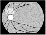
図2は表示モニタ21に表示される眼底観察像を示している。眼底観察時には、眼底像観察撮像手段で得られた眼底像に重畳して、合焦検出範囲表示部21aの枠部により合焦検出範囲を検者に提示する。これにより、合焦検出位置を視覚的に検者に提示できるため、自動合焦における操作性を向上させることができる。なお、合焦検出範囲は検者による操作で変更可能であり、被検眼眼底における特定部位としても、眼底全体としてもよい。
FIG. 2 shows a fundus observation image displayed on the display monitor 21. At the time of fundus observation, the in-focus detection range is presented to the examiner by the frame portion of the in-focus detection
続いて、合焦検出部25の詳細について図3を用いて説明する。図3に示すように、合焦検出部25には、眼底Erの特定位置を合焦検出の対象とする合焦検出範囲決定手段25aが設けられており、検者は操作入力部24を操作することで合焦検出範囲を決めることができる。即ち、該合焦検出範囲は可変とされている。更に、合焦検出部25には眼底像のコントラスト値とフォーカスレンズ3の位置を記憶する合焦評価値記憶手段25bが内蔵されている。
Next, details of the
本実施例では、合焦検出は撮影光束により結像される眼底像そのもののコントラスト値を検出することによって行う。ここでいうコントラストとは、隣接する画素の輝度差のことであり、コントラスト値とは所定の輝度データ中の最も大きい輝度差の値としている。 In this embodiment, focus detection is performed by detecting the contrast value of the fundus image itself formed by the photographing light beam. The contrast here is a luminance difference between adjacent pixels, and the contrast value is a value of the largest luminance difference in predetermined luminance data.
図4のグラフは、フォーカスレンズ駆動部6によって移動されるフォーカスレンズ3の位置に対するコントラスト値の遷移を表している。同図から明らかなように、合焦位置M2においてはコントラスト値が最大となり、ピントが大きくずれた位置M1では、コントラスト値が小さくなる。合焦位置M2は、モニタ21に映出された眼底像を最も鮮明に観察可能な位置であり、撮像後にモニタ21に映出される眼底像を最も鮮明にすることのできる位置でもある。したがって、本実施例ではこのコントラスト検出の原理を用いて、被検眼の収差の影響を受けない合焦検出を行うことが可能となる。 The graph of FIG. 4 represents the transition of the contrast value with respect to the position of the focus lens 3 moved by the focus lens driving unit 6. As is clear from the figure, the contrast value is maximum at the in-focus position M2, and the contrast value is small at the position M1 where the focus is greatly shifted. The in-focus position M2 is a position at which the fundus image projected on the monitor 21 can be observed most clearly, and is also a position at which the fundus image projected on the monitor 21 after imaging can be most clearly observed. Therefore, in this embodiment, it is possible to perform focus detection that is not affected by the aberration of the eye to be inspected using the principle of contrast detection.
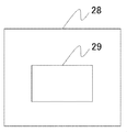
続いて、発光量演算手段28について説明する。撮像素子5の各画素からの出力はA/D変換素子16によりA/D変換され、メモリ17に一時的に格納される。測光値算出手段18はメモリ17に保存された画素出力から、合焦検出範囲の輝度値の最大値を測光値として、発光量演算手段28に出力する。図4に示すように、発光量演算手段28は合焦検出に適するように定められた観察光量の基準値を記憶した光量メモリ29(図5参照)を有しており、測光値と基準値とを比較することで観察光の発光量を決定する。例えば、測光値が基準値よりも高い場合は、眼底へ照明している観察光量が高いと判断し、輝度値の飽和を防ぐことを目的として光量を低減するように発光量を決定する。その一方、測光値が基準値よりも低い場合は、眼底へ照明している観察光量が低いと判断でき、コントラスト値の極大点の検出を容易にするために光量を高くするように発光量を決定する。なお、光量メモリ29が記憶する基準値は、例えば輝度値が飽和しないように眼底の反射率の平均値、眼底における血管部分を避けた特定部分の輝度値の平均値等、に基づいて定められる。 Next, the light emission amount calculating means 28 will be described. The output from each pixel of the image sensor 5 is A / D converted by the A / D converter 16 and temporarily stored in the memory 17. The photometric value calculating means 18 outputs the maximum luminance value in the focus detection range from the pixel output stored in the memory 17 to the light emission amount calculating means 28 as a photometric value. As shown in FIG. 4, the light emission amount calculating means 28 has a light amount memory 29 (see FIG. 5) that stores a reference value of the observation light amount determined so as to be suitable for focus detection. And the amount of observation light emitted is determined. For example, when the photometric value is higher than the reference value, it is determined that the observation light amount illuminating the fundus is high, and the light emission amount is determined so as to reduce the light amount for the purpose of preventing saturation of the luminance value. On the other hand, when the photometric value is lower than the reference value, it can be determined that the amount of observation light illuminating the fundus is low, and the light emission amount is increased so as to increase the light amount in order to facilitate the detection of the maximum point of the contrast value. decide. The reference value stored in the light amount memory 29 is determined based on, for example, the average value of the reflectance of the fundus, the average value of the luminance value of a specific part avoiding the blood vessel part in the fundus, etc. so that the luminance value is not saturated. .
次に、本実施例での動作について説明する。
赤外LED15から射出した光は、コンデンサレンズ14により集光され、ダイクロイックミラー11を透過した後、リング絞り10によってリング状に光束が制限される。リング絞り10で制限された光は、レンズ9、レンズ8を介し、一度穴あきミラー7上にリング絞り10の像を作り、かつ穴あきミラー7により光軸L1方向に反射される。さらに、対物レンズ1によって被検眼Eの瞳孔Ep付近に再びリング絞り10の像を作り、被検眼Eの眼底Erを照明する。
Next, the operation in this embodiment will be described.
The light emitted from the infrared LED 15 is collected by the condenser lens 14, passes through the dichroic mirror 11, and then the light beam is limited in a ring shape by the ring diaphragm 10. The light limited by the ring aperture 10 once forms an image of the ring aperture 10 on the perforated mirror 7 via the lens 9 and the lens 8, and is reflected by the perforated mirror 7 in the direction of the optical axis L1. Further, the objective lens 1 again forms an image of the ring diaphragm 10 in the vicinity of the pupil Ep of the eye E, and illuminates the fundus Er of the eye E.
定常光を発する赤外LED15からの光により照明された眼底Erからの反射散乱した光束は、瞳孔Epから被検眼Eを射出し、対物レンズ1、撮影絞り2、フォーカスレンズ3、結像レンズ4を介して、撮像素子5に達し撮像される。撮像素子からの出力はA/D変換素子16によりデジタル信号化された後、撮像手段制御部22を介してモニタ21に眼底観察像が映出される。
The light beam reflected and scattered from the fundus Er illuminated by the light from the infrared LED 15 that emits steady light exits the eye E through the pupil Ep, and the objective lens 1, the photographing aperture 2, the focus lens 3, and the imaging lens 4 And reaches the image pickup device 5 to be imaged. The output from the imaging device is converted into a digital signal by the A / D conversion device 16 and then a fundus observation image is displayed on the monitor 21 via the imaging means
検者はモニタ21に映出された眼底像を観察し、図示のない操作桿を使い、被検眼Eと眼底カメラ光学部との位置合せを行う。図示しない合焦モード切換え手段により、手動合焦モードに設定されている場合は、検者はモニタ21に映出された眼底像を観察しながら、眼底が適当な明るさとなるように赤外LED15の光量の調整と、フォーカスレンズ3の光軸L1方向における位置の調整を、操作入力部24により行う。その後、図示しない操作入力部24における撮影スイッチ30を押して撮影を行う。 The examiner observes the fundus image displayed on the monitor 21, and uses an operation rod (not shown) to align the eye E to be examined and the fundus camera optical unit. When the manual focus mode is set by the focus mode switching means (not shown), the examiner observes the fundus image displayed on the monitor 21 and the infrared LED 15 so that the fundus has an appropriate brightness. The operation input unit 24 adjusts the amount of light and the position of the focus lens 3 in the optical axis L1 direction. Thereafter, the photographing switch 30 in the operation input unit 24 (not shown) is pressed to perform photographing.
検者が撮影スイッチ30を押すと、ストロボ光源13がパルス発光し、ストロボ光源13から発した光束はコンデンサレンズ12により集光され、ダイクロイックミラー11で反射された後、リング絞り10によってリング状に光束が制限される。リング絞り10で制限された光は、レンズ9、レンズ8を介し、一度穴あきミラー7上にリング絞り10の像を作り、かつ穴あきミラー7により光軸L1方向に反射され、対物レンズ1によって被検眼Eの瞳孔Ep付近に再びリング絞り10の像を作り、被検眼Eの眼底Erを照明する。ストロボ光源13から発した光束により照明された眼底Erからの反射散乱した光束は、瞳孔Epから被検眼Eを射出する。さらに、対物レンズ1、撮影絞り2、フォーカスレンズ3、結像レンズ4を介して、撮像素子5に達し撮像され、A/D変換素子16によりデジタル信号化され、画像メモリ20に静止画として保存される。
When the examiner presses the photographing switch 30, the strobe light source 13 emits a pulse, and the light beam emitted from the strobe light source 13 is collected by the
続いて、本実施例の特徴である自動合焦モードに設定された際の制御方法について説明する。図2に示すように、自動合焦モードでは、被検眼眼底の観察時に眼底像観察撮像手段で得られた眼底像に重畳して、合焦検出範囲表示部21aの枠部による合焦検出範囲が提示される。この眼底像の取得は本発明における観察工程に対応する。検者は操作入力部24によって合焦検出範囲表示部21aの配置を変更し、合焦検出範囲を決定する。続いて、図示しない自動合焦開始スイッチが検者により押下されることで、自動合焦が開始される。なお、本実施例では操作入力部24により合焦検出範囲を変更したが、例えば図示されていない固視灯位置などに基づいてシステム制御部19により自動的に決定してもよい。
Next, a control method when the automatic focusing mode that is a feature of the present embodiment is set will be described. As shown in FIG. 2, in the autofocus mode, the focus detection range by the frame portion of the focus detection
図6のフローチャートは、自動合焦が開始された際の動作を示している。自動合焦の開始が指示されると、ステップ1で、測光工程として、測光値算出手段18はメモリ17に保存された画素出力から合焦検出範囲の輝度値の最大値を測光値として算出し、発光量演算手段28に出力する。ステップ2では、比較工程として、発光量演算手段28によって、光量メモリ29の有する発光量の基準値とステップ1で算出された測光値の比較が行なわれ、観察光の発光量が決定される。ステップ3は、光量制御工程として観察光源制御手段26によって実行され、ステップ2によって決定された光量の観察光が眼底に照射される。ステップ4において、合焦検出部25によるコントラスト値の算出を行い、ステップ5で、合焦検出部25における合焦評価値記憶手段25bにステップ4で算出されたコントラスト値とフォーカスレンズ3の位置が記録される。ステップ6は、ステップ5において記録されたコントラスト値の中に図3に示した位置M2である極大点が含まれるかどうかを検出する。
The flowchart in FIG. 6 shows an operation when automatic focusing is started. When the start of automatic focusing is instructed, in step 1, as a photometric process, the photometric value calculating means 18 calculates the maximum value of the brightness value in the focus detection range from the pixel output stored in the memory 17 as a photometric value. And output to the light emission amount calculating means 28. In step 2, as a comparison process, the light emission amount calculation means 28 compares the light emission amount reference value of the light amount memory 29 with the photometric value calculated in step 1 to determine the light emission amount of the observation light. Step 3 is executed by the observation light source control means 26 as a light quantity control step, and the fundus of the observation light quantity determined in step 2 is irradiated to the fundus. In step 4, the contrast value is calculated by the
ステップ6で、極大点が検出されなかった場合はステップ7に進み、フォーカスレンズ3を所定の移動量だけ駆動させることでフォーカスレンズ位置を変更し、ステップ4とステップ5の処理を繰り返す。続いてステップ6に進み、コントラスト値の極大点が検出されるかを判断する。以降、ステップ6でコントラスト値の極大点が検出されるまで、ステップ7、ステップ4、ステップ5を繰り返す。 If the local maximum point is not detected in step 6, the process proceeds to step 7, the focus lens position is changed by driving the focus lens 3 by a predetermined movement amount, and the processes in steps 4 and 5 are repeated. Subsequently, the process proceeds to step 6 to determine whether the maximum point of the contrast value is detected. Thereafter, Step 7, Step 4, and Step 5 are repeated until the maximum point of the contrast value is detected in Step 6.
ステップ6で極大点が検出された場合、次はステップ8に進む。ステップ8は、合焦検出部25で実行され、フォーカスレンズ3の移動量の算出を行う。ここで、ステップ8でのフォーカスレンズの移動量とは、極大点の検出位置までのフォーカスレンズ駆動量のことである。次にステップ9では、ステップ7で算出したフォーカスレンズの移動量に従ってフォーカスレンズ3の駆動を行い、フォーカスレンズ3の位置をコントラスト値の極大値の位置に移動させる。以上の合焦工程にて行われる動作によって、被検眼Eに球面収差や乱視など収差に個人差があった場合でも、その収差に合わせたフォーカス調整が可能となる。
If a local maximum is detected in step 6, the process proceeds to step 8. Step 8 is executed by the
このような動作は、赤外光を用いて観察を行う無散瞳型の眼底カメラにおいて、特に有効となる。赤外光に対する眼底の中大血管のコントラストは低いため、フォーカスレンズ位置に対するコントラスト値の差が出にくく、オートフォーカスにおいて図3に示した極大点の位置M2を検出することは困難である。そのため、眼底を照明する赤外LEDの光量を増やし、少しでも観察像のコントラストを上げる必要があるが、必要以上に眼底が明るくなると輝度値の飽和が生じ、コントラスト値の算出が正しく行われない。しかしながら、測光手段を用いることでコントラスト値を算出する前に適正な観察光量を算出し制御すれば、輝度値の飽和をあらかじめ防ぐことでき、コントラスト値の算出が安定して行えるため、精度良い合焦検出が可能となる。 Such an operation is particularly effective in a non-mydriatic retinal camera that performs observation using infrared light. Since the contrast of the middle and large blood vessels of the fundus with respect to the infrared light is low, it is difficult to produce a difference in contrast value with respect to the focus lens position, and it is difficult to detect the position M2 of the maximum point shown in FIG. Therefore, it is necessary to increase the light intensity of the infrared LED that illuminates the fundus and increase the contrast of the observation image as much as possible. However, if the fundus becomes brighter than necessary, the brightness value is saturated and the contrast value is not calculated correctly. . However, if the appropriate amount of observation light is calculated and controlled before the contrast value is calculated by using the photometric means, the saturation of the luminance value can be prevented in advance, and the contrast value can be calculated stably. Focus detection is possible.
(その他の実施例)
また、本発明は、以下の処理を実行することによっても実現される。即ち、上述した実施形態の機能を実現するソフトウェア(プログラム)を、ネットワーク又は各種記憶媒体を介してシステム或いは装置に供給し、そのシステム或いは装置のコンピュータ(またはCPUやMPU等)がプログラムを読み出して実行する処理である。
(Other examples)
The present invention can also be realized by executing the following processing. That is, software (program) that realizes the functions of the above-described embodiments is supplied to a system or apparatus via a network or various storage media, and a computer (or CPU, MPU, or the like) of the system or apparatus reads the program. It is a process to be executed.
E:被検眼
3:フォーカスレンズ
6:フォーカスレンズ駆動部
15:赤外LED
18:測光値算出手段
19:システム制御部
25:合焦検出部
26:観察光源制御手段
28:発光量演算手段
29:光量メモリ
25a:合焦検出範囲決定手段
E: Eye to be examined 3: Focus lens 6: Focus lens driving unit 15: Infrared LED
18: Photometric value calculation means 19: System control section 25: Focus detection section 26: Observation light source control means 28: Light emission amount calculation means 29: Light quantity memory 25a: Focus detection range determination means
Claims (9)
フォーカスレンズを介して照明された前記被検眼の前記眼底を撮像する撮像手段と、
前記眼底を照明する前記照明手段の光量を制御する照明光量制御手段と、
前記フォーカスレンズの合焦位置を検出する合焦検出手段と、
前記照明手段により照明された前記眼底からの反射光を測光する測光手段と、
を有する眼底カメラにおいて、
前記測光手段は前記眼底からの前記反射光の測光値を算出し、
前記照明光量制御手段は前記測光値に基づいて照明光量を制御し、
前記合焦検出手段は前記照明光量制御手段による前記照明光量が制御された後に合焦評価を行うことを特徴とする眼科撮影装置。 Illuminating means for illuminating the fundus of the subject's eye during observation and imaging;
Imaging means for imaging the fundus of the eye to be examined illuminated through a focus lens;
Illumination light quantity control means for controlling the light quantity of the illumination means for illuminating the fundus;
A focus detection means for detecting a focus position of the focus lens;
Photometric means for measuring reflected light from the fundus illuminated by the illumination means;
In a fundus camera having
The photometric means calculates a photometric value of the reflected light from the fundus;
The illumination light quantity control means controls the illumination light quantity based on the photometric value,
The ophthalmic photographing apparatus according to claim 1, wherein the focus detection unit performs a focus evaluation after the illumination light amount is controlled by the illumination light amount control unit.
前記合焦検出手段は、前記合焦検出範囲決定手段で決定された前記合焦検出範囲において前記測光手段により前記測光値を算出し且つ前記照明光量制御手段が前記測光値に基づいて前記照明光量を制御した後、前記合焦検出範囲の前記合焦評価を行うことを特徴とする請求項1に記載の眼科撮影装置。 A focus detection range determining means for determining a focus detection range for the fundus image captured by the imaging means;
The focus detection unit calculates the photometric value by the photometry unit in the focus detection range determined by the focus detection range determination unit, and the illumination light amount control unit calculates the illumination light amount based on the photometric value. The ophthalmologic photographing apparatus according to claim 1, wherein the focus evaluation of the focus detection range is performed after controlling the focus.
前記基準値は、前記眼底の反射率の平均値に基づいて定められることを特徴とする請求項1乃至5の何れか1項に記載の眼科撮影装置。 The illumination light amount control means further includes a light emission amount calculation means for determining the light amount of the illumination light by comparing a reference value of the illumination light and the photometric value when the illumination means images the fundus.
The ophthalmologic photographing apparatus according to claim 1, wherein the reference value is determined based on an average value of the reflectance of the fundus.
前記照明光により照明された前記眼底を撮像する撮像手段と、
前記照明光の光量を制御する照明光量制御手段と、
前記眼底からの前記照明光の反射光を測光して測光値を算出する測光手段と、
前記照明手段が前記眼底を撮像する際の前記照明光の基準値と前記測光値とを比較して前記照明光の光量を決定する発光量演算手段と、
を有し、
前記照明光量制御手段は前記発光量演算手段の決定に応じて前記照明光の光量を制御することを特徴とする眼科撮影装置。 Illumination means for illuminating the fundus of the eye to be examined with illumination light;
Imaging means for imaging the fundus illuminated by the illumination light;
Illumination light quantity control means for controlling the light quantity of the illumination light;
Photometric means for measuring a reflected light of the illumination light from the fundus and calculating a photometric value;
A light emission amount calculation means for determining a light amount of the illumination light by comparing a reference value of the illumination light and the photometric value when the illumination means images the fundus;
Have
The ophthalmologic photographing apparatus characterized in that the illumination light quantity control means controls the light quantity of the illumination light according to the determination of the light emission quantity calculation means.
前記眼底像を構成する反射光を測光する測光工程と、
前記反射光の測光値と基準値とを比較する比較工程と、
前記比較工程における比較の結果に基づいて前記照明光の光量を制御する光量制御工程と、
光量の制御が為された照明光を眼底に照射し、フォーカスレンズにより合焦することで撮像手段により前記眼底像を撮像する合焦工程と、を有することを特徴とする眼科撮影方法。 An observation step of illuminating the fundus of the subject's eye with illumination light and capturing the fundus image of the fundus with an imaging means by focusing with a focus lens;
A photometric step of measuring the reflected light constituting the fundus image;
A comparison step of comparing the photometric value of the reflected light with a reference value;
A light amount control step for controlling the light amount of the illumination light based on the comparison result in the comparison step;
An ophthalmologic photographing method comprising: a focusing step of irradiating the fundus with illumination light whose light amount has been controlled, and focusing the image with a focus lens to capture the fundus image with an imaging unit.
Priority Applications (3)
| Application Number | Priority Date | Filing Date | Title |
|---|---|---|---|
| JP2012229454A JP2014079392A (en) | 2012-10-17 | 2012-10-17 | Ophthalmology imaging apparatus |
| US14/049,515 US9572489B2 (en) | 2012-10-17 | 2013-10-09 | Ophthalmologic imaging apparatus, ophthalmologic imaging method, and program |
| CN201310486563.1A CN103767676A (en) | 2012-10-17 | 2013-10-17 | Ophthalmologic imaging apparatus, ophthalmologic imaging method |
Applications Claiming Priority (1)
| Application Number | Priority Date | Filing Date | Title |
|---|---|---|---|
| JP2012229454A JP2014079392A (en) | 2012-10-17 | 2012-10-17 | Ophthalmology imaging apparatus |
Publications (2)
| Publication Number | Publication Date |
|---|---|
| JP2014079392A true JP2014079392A (en) | 2014-05-08 |
| JP2014079392A5 JP2014079392A5 (en) | 2015-12-03 |
Family
ID=50475062
Family Applications (1)
| Application Number | Title | Priority Date | Filing Date |
|---|---|---|---|
| JP2012229454A Pending JP2014079392A (en) | 2012-10-17 | 2012-10-17 | Ophthalmology imaging apparatus |
Country Status (3)
| Country | Link |
|---|---|
| US (1) | US9572489B2 (en) |
| JP (1) | JP2014079392A (en) |
| CN (1) | CN103767676A (en) |
Cited By (3)
| Publication number | Priority date | Publication date | Assignee | Title |
|---|---|---|---|---|
| JP2019162336A (en) * | 2018-03-20 | 2019-09-26 | パナソニックIpマネジメント株式会社 | Ophthalmic operation apparatus and ophthalmic photographic apparatus |
| JP2020028597A (en) * | 2018-08-24 | 2020-02-27 | 興和株式会社 | Ophthalmic photographing apparatus |
| JP2023083540A (en) * | 2019-03-25 | 2023-06-15 | 株式会社トプコン | Ophthalmologic apparatus and maintenance method of ophthalmologic apparatus |
Families Citing this family (4)
| Publication number | Priority date | Publication date | Assignee | Title |
|---|---|---|---|---|
| US10602926B2 (en) * | 2016-09-29 | 2020-03-31 | Welch Allyn, Inc. | Through focus retinal image capturing |
| CN106444062A (en) * | 2016-11-03 | 2017-02-22 | 曾庆涛 | Device for displaying 3D image and 3D image display device |
| WO2021010345A1 (en) * | 2019-07-16 | 2021-01-21 | 株式会社ニデック | Ophthalmologic imaging apparatus |
| CN116017147A (en) * | 2022-12-29 | 2023-04-25 | 上海哔哩哔哩科技有限公司 | Video shooting method, device, computer equipment and storage medium |
Citations (7)
| Publication number | Priority date | Publication date | Assignee | Title |
|---|---|---|---|---|
| JPS56100039A (en) * | 1980-01-10 | 1981-08-11 | Canon Kk | Eyeground camera |
| JPH04150831A (en) * | 1990-10-12 | 1992-05-25 | Topcon Corp | Eyeground camera |
| JP2011050531A (en) * | 2009-09-01 | 2011-03-17 | Canon Inc | Fundus camera |
| US20110090457A1 (en) * | 2009-10-20 | 2011-04-21 | Canon Kabushiki Kaisha | Ophthalmic imaging apparatus, imaging unit, inspection apparatus, and method of producing ophthalmic imaging apparatus |
| US20110228220A1 (en) * | 2010-03-16 | 2011-09-22 | Canon Kabushiki Kaisha | Ophthalmologic imaging apparatus and method for controlling the same |
| JP2011250961A (en) * | 2010-06-01 | 2011-12-15 | Canon Inc | Ophthalmic imaging apparatus and control method thereof |
| US20120050680A1 (en) * | 2010-08-31 | 2012-03-01 | Canon Kabushiki Kaisha | Light intensity control apparatus, light intensity control method, program, and ophethalmologic apparatus |
Family Cites Families (6)
| Publication number | Priority date | Publication date | Assignee | Title |
|---|---|---|---|---|
| JPH08265629A (en) | 1995-03-27 | 1996-10-11 | Canon Inc | Automatic focus adjustment device |
| US6564014B1 (en) * | 1999-09-24 | 2003-05-13 | Nikon Corporation | Flash control device |
| JP3958164B2 (en) * | 2002-09-19 | 2007-08-15 | キヤノン株式会社 | Camera photometric device |
| JP2007108455A (en) | 2005-10-14 | 2007-04-26 | Fujifilm Corp | Automatic focusing controller and control method |
| JP5225065B2 (en) | 2008-12-27 | 2013-07-03 | キヤノン株式会社 | Imaging apparatus and imaging method |
| JP5777308B2 (en) | 2010-08-31 | 2015-09-09 | キヤノン株式会社 | Ophthalmic photographing apparatus, ophthalmic photographing method and program |
-
2012
- 2012-10-17 JP JP2012229454A patent/JP2014079392A/en active Pending
-
2013
- 2013-10-09 US US14/049,515 patent/US9572489B2/en active Active
- 2013-10-17 CN CN201310486563.1A patent/CN103767676A/en active Pending
Patent Citations (7)
| Publication number | Priority date | Publication date | Assignee | Title |
|---|---|---|---|---|
| JPS56100039A (en) * | 1980-01-10 | 1981-08-11 | Canon Kk | Eyeground camera |
| JPH04150831A (en) * | 1990-10-12 | 1992-05-25 | Topcon Corp | Eyeground camera |
| JP2011050531A (en) * | 2009-09-01 | 2011-03-17 | Canon Inc | Fundus camera |
| US20110090457A1 (en) * | 2009-10-20 | 2011-04-21 | Canon Kabushiki Kaisha | Ophthalmic imaging apparatus, imaging unit, inspection apparatus, and method of producing ophthalmic imaging apparatus |
| US20110228220A1 (en) * | 2010-03-16 | 2011-09-22 | Canon Kabushiki Kaisha | Ophthalmologic imaging apparatus and method for controlling the same |
| JP2011250961A (en) * | 2010-06-01 | 2011-12-15 | Canon Inc | Ophthalmic imaging apparatus and control method thereof |
| US20120050680A1 (en) * | 2010-08-31 | 2012-03-01 | Canon Kabushiki Kaisha | Light intensity control apparatus, light intensity control method, program, and ophethalmologic apparatus |
Cited By (4)
| Publication number | Priority date | Publication date | Assignee | Title |
|---|---|---|---|---|
| JP2019162336A (en) * | 2018-03-20 | 2019-09-26 | パナソニックIpマネジメント株式会社 | Ophthalmic operation apparatus and ophthalmic photographic apparatus |
| JP2020028597A (en) * | 2018-08-24 | 2020-02-27 | 興和株式会社 | Ophthalmic photographing apparatus |
| JP7302924B2 (en) | 2018-08-24 | 2023-07-04 | 興和株式会社 | ophthalmic imaging equipment |
| JP2023083540A (en) * | 2019-03-25 | 2023-06-15 | 株式会社トプコン | Ophthalmologic apparatus and maintenance method of ophthalmologic apparatus |
Also Published As
| Publication number | Publication date |
|---|---|
| US9572489B2 (en) | 2017-02-21 |
| CN103767676A (en) | 2014-05-07 |
| US20140104570A1 (en) | 2014-04-17 |
Similar Documents
| Publication | Publication Date | Title |
|---|---|---|
| JP5600478B2 (en) | Ophthalmic imaging apparatus and control method thereof | |
| JP5371638B2 (en) | Ophthalmic imaging apparatus and method | |
| US10039450B2 (en) | Ophthalmologic apparatus | |
| JP5641752B2 (en) | Ophthalmic photographing apparatus and ophthalmic photographing method | |
| JP5850292B2 (en) | Ophthalmic equipment | |
| JP5773598B2 (en) | Image processing apparatus, image processing method, and program | |
| US20130208243A1 (en) | Ophthalmologic apparatus, method for controlling ophthalmologic apparatus, and storage medium | |
| JP2014079392A (en) | Ophthalmology imaging apparatus | |
| US8708492B2 (en) | Fundus camera and control method for the fundus camera | |
| JP6124548B2 (en) | Ophthalmic imaging method and ophthalmic apparatus | |
| JP2017012663A (en) | Ophthalmic photographing apparatus, control method thereof and program | |
| JP2016185192A (en) | Ophthalmologic apparatus, and control method of ophthalmologic apparatus | |
| JP2014083397A (en) | Ophthalmologic apparatus, control method thereof, and program | |
| JP2017099717A (en) | Ophthalmic photographing apparatus | |
| JP2014083352A (en) | Image capturing apparatus, and focusing method in image capturing apparatus | |
| JP2015146961A (en) | Ophthalmologic apparatus, and control method of ophthalmologic apparatus | |
| JP2021094456A (en) | Ophthalmologic apparatus | |
| JP2016010630A (en) | Ophthalmologic photographing apparatus, photographing control method and program | |
| JP6025903B2 (en) | Ophthalmic apparatus, image generation method, and program | |
| JP6784019B2 (en) | Fundus photography device and information processing program for ophthalmology | |
| JP5963839B2 (en) | Ophthalmic photographing apparatus and image composition method | |
| JP2015100510A (en) | Ophthalmic photographing apparatus and control method | |
| JP2015150283A (en) | Ophthalmologic examination apparatus | |
| JP2020130525A (en) | Ophthalmologic apparatus | |
| JP2013248538A (en) | Ophthalmic imaging apparatus and method for controlling the same |
Legal Events
| Date | Code | Title | Description |
|---|---|---|---|
| A521 | Request for written amendment filed |
Free format text: JAPANESE INTERMEDIATE CODE: A523 Effective date: 20151014 |
|
| A621 | Written request for application examination |
Free format text: JAPANESE INTERMEDIATE CODE: A621 Effective date: 20151014 |
|
| A977 | Report on retrieval |
Free format text: JAPANESE INTERMEDIATE CODE: A971007 Effective date: 20160720 |
|
| A131 | Notification of reasons for refusal |
Free format text: JAPANESE INTERMEDIATE CODE: A131 Effective date: 20160804 |
|
| A521 | Request for written amendment filed |
Free format text: JAPANESE INTERMEDIATE CODE: A523 Effective date: 20160920 |
|
| A02 | Decision of refusal |
Free format text: JAPANESE INTERMEDIATE CODE: A02 Effective date: 20170207 |


