JP2013545472A - Simultaneous detection of biomolecules in a single cell - Google Patents
Simultaneous detection of biomolecules in a single cell Download PDFInfo
- Publication number
- JP2013545472A JP2013545472A JP2013541336A JP2013541336A JP2013545472A JP 2013545472 A JP2013545472 A JP 2013545472A JP 2013541336 A JP2013541336 A JP 2013541336A JP 2013541336 A JP2013541336 A JP 2013541336A JP 2013545472 A JP2013545472 A JP 2013545472A
- Authority
- JP
- Japan
- Prior art keywords
- biomolecule
- biological
- cell
- biomolecules
- derivative
- Prior art date
- Legal status (The legal status is an assumption and is not a legal conclusion. Google has not performed a legal analysis and makes no representation as to the accuracy of the status listed.)
- Pending
Links
Images


Classifications
-
- G—PHYSICS
- G01—MEASURING; TESTING
- G01N—INVESTIGATING OR ANALYSING MATERIALS BY DETERMINING THEIR CHEMICAL OR PHYSICAL PROPERTIES
- G01N33/00—Investigating or analysing materials by specific methods not covered by groups G01N1/00 - G01N31/00
- G01N33/48—Biological material, e.g. blood, urine; Haemocytometers
- G01N33/50—Chemical analysis of biological material, e.g. blood, urine; Testing involving biospecific ligand binding methods; Immunological testing
- G01N33/53—Immunoassay; Biospecific binding assay; Materials therefor
- G01N33/5308—Immunoassay; Biospecific binding assay; Materials therefor for analytes not provided for elsewhere, e.g. nucleic acids, uric acid, worms, mites
-
- G—PHYSICS
- G01—MEASURING; TESTING
- G01N—INVESTIGATING OR ANALYSING MATERIALS BY DETERMINING THEIR CHEMICAL OR PHYSICAL PROPERTIES
- G01N33/00—Investigating or analysing materials by specific methods not covered by groups G01N1/00 - G01N31/00
- G01N33/48—Biological material, e.g. blood, urine; Haemocytometers
- G01N33/50—Chemical analysis of biological material, e.g. blood, urine; Testing involving biospecific ligand binding methods; Immunological testing
- G01N33/53—Immunoassay; Biospecific binding assay; Materials therefor
- G01N33/543—Immunoassay; Biospecific binding assay; Materials therefor with an insoluble carrier for immobilising immunochemicals
-
- C—CHEMISTRY; METALLURGY
- C12—BIOCHEMISTRY; BEER; SPIRITS; WINE; VINEGAR; MICROBIOLOGY; ENZYMOLOGY; MUTATION OR GENETIC ENGINEERING
- C12Q—MEASURING OR TESTING PROCESSES INVOLVING ENZYMES, NUCLEIC ACIDS OR MICROORGANISMS; COMPOSITIONS OR TEST PAPERS THEREFOR; PROCESSES OF PREPARING SUCH COMPOSITIONS; CONDITION-RESPONSIVE CONTROL IN MICROBIOLOGICAL OR ENZYMOLOGICAL PROCESSES
- C12Q1/00—Measuring or testing processes involving enzymes, nucleic acids or microorganisms; Compositions therefor; Processes of preparing such compositions
- C12Q1/68—Measuring or testing processes involving enzymes, nucleic acids or microorganisms; Compositions therefor; Processes of preparing such compositions involving nucleic acids
- C12Q1/6813—Hybridisation assays
- C12Q1/6834—Enzymatic or biochemical coupling of nucleic acids to a solid phase
-
- C—CHEMISTRY; METALLURGY
- C12—BIOCHEMISTRY; BEER; SPIRITS; WINE; VINEGAR; MICROBIOLOGY; ENZYMOLOGY; MUTATION OR GENETIC ENGINEERING
- C12Q—MEASURING OR TESTING PROCESSES INVOLVING ENZYMES, NUCLEIC ACIDS OR MICROORGANISMS; COMPOSITIONS OR TEST PAPERS THEREFOR; PROCESSES OF PREPARING SUCH COMPOSITIONS; CONDITION-RESPONSIVE CONTROL IN MICROBIOLOGICAL OR ENZYMOLOGICAL PROCESSES
- C12Q1/00—Measuring or testing processes involving enzymes, nucleic acids or microorganisms; Compositions therefor; Processes of preparing such compositions
- C12Q1/68—Measuring or testing processes involving enzymes, nucleic acids or microorganisms; Compositions therefor; Processes of preparing such compositions involving nucleic acids
- C12Q1/6844—Nucleic acid amplification reactions
-
- C—CHEMISTRY; METALLURGY
- C12—BIOCHEMISTRY; BEER; SPIRITS; WINE; VINEGAR; MICROBIOLOGY; ENZYMOLOGY; MUTATION OR GENETIC ENGINEERING
- C12Q—MEASURING OR TESTING PROCESSES INVOLVING ENZYMES, NUCLEIC ACIDS OR MICROORGANISMS; COMPOSITIONS OR TEST PAPERS THEREFOR; PROCESSES OF PREPARING SUCH COMPOSITIONS; CONDITION-RESPONSIVE CONTROL IN MICROBIOLOGICAL OR ENZYMOLOGICAL PROCESSES
- C12Q1/00—Measuring or testing processes involving enzymes, nucleic acids or microorganisms; Compositions therefor; Processes of preparing such compositions
- C12Q1/68—Measuring or testing processes involving enzymes, nucleic acids or microorganisms; Compositions therefor; Processes of preparing such compositions involving nucleic acids
- C12Q1/6869—Methods for sequencing
-
- G—PHYSICS
- G01—MEASURING; TESTING
- G01N—INVESTIGATING OR ANALYSING MATERIALS BY DETERMINING THEIR CHEMICAL OR PHYSICAL PROPERTIES
- G01N33/00—Investigating or analysing materials by specific methods not covered by groups G01N1/00 - G01N31/00
- G01N33/48—Biological material, e.g. blood, urine; Haemocytometers
- G01N33/50—Chemical analysis of biological material, e.g. blood, urine; Testing involving biospecific ligand binding methods; Immunological testing
- G01N33/68—Chemical analysis of biological material, e.g. blood, urine; Testing involving biospecific ligand binding methods; Immunological testing involving proteins, peptides or amino acids
- G01N33/6803—General methods of protein analysis not limited to specific proteins or families of proteins
- G01N33/6842—Proteomic analysis of subsets of protein mixtures with reduced complexity, e.g. membrane proteins, phosphoproteins, organelle proteins
-
- C—CHEMISTRY; METALLURGY
- C12—BIOCHEMISTRY; BEER; SPIRITS; WINE; VINEGAR; MICROBIOLOGY; ENZYMOLOGY; MUTATION OR GENETIC ENGINEERING
- C12Q—MEASURING OR TESTING PROCESSES INVOLVING ENZYMES, NUCLEIC ACIDS OR MICROORGANISMS; COMPOSITIONS OR TEST PAPERS THEREFOR; PROCESSES OF PREPARING SUCH COMPOSITIONS; CONDITION-RESPONSIVE CONTROL IN MICROBIOLOGICAL OR ENZYMOLOGICAL PROCESSES
- C12Q2523/00—Reactions characterised by treatment of reaction samples
- C12Q2523/30—Characterised by physical treatment
- C12Q2523/303—Applying a physical force on a nucleic acid
-
- C—CHEMISTRY; METALLURGY
- C12—BIOCHEMISTRY; BEER; SPIRITS; WINE; VINEGAR; MICROBIOLOGY; ENZYMOLOGY; MUTATION OR GENETIC ENGINEERING
- C12Q—MEASURING OR TESTING PROCESSES INVOLVING ENZYMES, NUCLEIC ACIDS OR MICROORGANISMS; COMPOSITIONS OR TEST PAPERS THEREFOR; PROCESSES OF PREPARING SUCH COMPOSITIONS; CONDITION-RESPONSIVE CONTROL IN MICROBIOLOGICAL OR ENZYMOLOGICAL PROCESSES
- C12Q2527/00—Reactions demanding special reaction conditions
- C12Q2527/109—Pressure
-
- C—CHEMISTRY; METALLURGY
- C12—BIOCHEMISTRY; BEER; SPIRITS; WINE; VINEGAR; MICROBIOLOGY; ENZYMOLOGY; MUTATION OR GENETIC ENGINEERING
- C12Q—MEASURING OR TESTING PROCESSES INVOLVING ENZYMES, NUCLEIC ACIDS OR MICROORGANISMS; COMPOSITIONS OR TEST PAPERS THEREFOR; PROCESSES OF PREPARING SUCH COMPOSITIONS; CONDITION-RESPONSIVE CONTROL IN MICROBIOLOGICAL OR ENZYMOLOGICAL PROCESSES
- C12Q2527/00—Reactions demanding special reaction conditions
- C12Q2527/119—Reactions demanding special reaction conditions pH
-
- C—CHEMISTRY; METALLURGY
- C12—BIOCHEMISTRY; BEER; SPIRITS; WINE; VINEGAR; MICROBIOLOGY; ENZYMOLOGY; MUTATION OR GENETIC ENGINEERING
- C12Q—MEASURING OR TESTING PROCESSES INVOLVING ENZYMES, NUCLEIC ACIDS OR MICROORGANISMS; COMPOSITIONS OR TEST PAPERS THEREFOR; PROCESSES OF PREPARING SUCH COMPOSITIONS; CONDITION-RESPONSIVE CONTROL IN MICROBIOLOGICAL OR ENZYMOLOGICAL PROCESSES
- C12Q2531/00—Reactions of nucleic acids characterised by
- C12Q2531/10—Reactions of nucleic acids characterised by the purpose being amplify/increase the copy number of target nucleic acid
- C12Q2531/113—PCR
-
- C—CHEMISTRY; METALLURGY
- C12—BIOCHEMISTRY; BEER; SPIRITS; WINE; VINEGAR; MICROBIOLOGY; ENZYMOLOGY; MUTATION OR GENETIC ENGINEERING
- C12Q—MEASURING OR TESTING PROCESSES INVOLVING ENZYMES, NUCLEIC ACIDS OR MICROORGANISMS; COMPOSITIONS OR TEST PAPERS THEREFOR; PROCESSES OF PREPARING SUCH COMPOSITIONS; CONDITION-RESPONSIVE CONTROL IN MICROBIOLOGICAL OR ENZYMOLOGICAL PROCESSES
- C12Q2563/00—Nucleic acid detection characterized by the use of physical, structural and functional properties
- C12Q2563/149—Particles, e.g. beads
-
- C—CHEMISTRY; METALLURGY
- C12—BIOCHEMISTRY; BEER; SPIRITS; WINE; VINEGAR; MICROBIOLOGY; ENZYMOLOGY; MUTATION OR GENETIC ENGINEERING
- C12Q—MEASURING OR TESTING PROCESSES INVOLVING ENZYMES, NUCLEIC ACIDS OR MICROORGANISMS; COMPOSITIONS OR TEST PAPERS THEREFOR; PROCESSES OF PREPARING SUCH COMPOSITIONS; CONDITION-RESPONSIVE CONTROL IN MICROBIOLOGICAL OR ENZYMOLOGICAL PROCESSES
- C12Q2563/00—Nucleic acid detection characterized by the use of physical, structural and functional properties
- C12Q2563/159—Microreactors, e.g. emulsion PCR or sequencing, droplet PCR, microcapsules, i.e. non-liquid containers with a range of different permeability's for different reaction components
Abstract
本発明は、単一細胞または他の生物学的実体から複数の生体分子を検出することに関する方法、イムノアッセイ、キット、および装置を提供する。本発明はまた、そのような実体から、相互作用する生体分子を高度に並行的に検出することを可能にする。
【選択図】図1The present invention provides methods, immunoassays, kits, and devices relating to detecting multiple biomolecules from a single cell or other biological entity. The present invention also allows highly interactive detection of interacting biomolecules from such entities.
[Selection] Figure 1
Description
本出願は2010年12月1日に出願された米国仮特許出願第61/418,423号明細書の利益を主張するものであり、その内容全体を参照によって組み込むものとする。 This application claims the benefit of US Provisional Patent Application No. 61 / 418,423, filed Dec. 1, 2010, the entire contents of which are incorporated by reference.
本発明は、単一細胞または他の生物学的実体から複数の生体分子を検出することに関する方法、イムノアッセイ、キット、および装置を提供する。本発明はまた、そのような実体から生体分子を高度に並行的に検出することを可能にする。 The present invention provides methods, immunoassays, kits, and devices relating to detecting multiple biomolecules from a single cell or other biological entity. The present invention also allows for highly parallel detection of biomolecules from such entities.
生物界は、核酸、ポリペプチド、炭水化物、脂肪酸、およびその他多数を含む種々のタイプの有機物および無機物から構成されている。それらは、細胞、組織、器官、および生体を形成し、次いで、他のコンパートメントまたは周辺の組織もしくは液体の中に存在する物質および化合物に反応し、相互作用する。これらの物質(本発明では、「生体ユニット」または「生体分子」と呼ばれる)すべてについて、検出方法が存在する。所与の問題について最適な検出方法は、生体ユニットそれ自体の性質および試験される試料の起源など、多くの因子に依存する。全体として、最近の10数年で、ほとんどの検出方法の感度が大幅に改善した。特定の生体ユニットについては、単一分子の検出も可能にする検出方法が存在する。そのような高感度な方法では、それぞれの検出方法を妨害する因子を除去するために、試料の複雑な処理を必要とすることが多い。本発明は、生体ユニットの検出のための新規かつ優れた方法を開示する。そのような生体ユニットは、例えば、生体分子であってもよい。 The biological kingdom is composed of various types of organic and inorganic materials including nucleic acids, polypeptides, carbohydrates, fatty acids, and many others. They form cells, tissues, organs, and organisms, and then react and interact with substances and compounds present in other compartments or surrounding tissues or fluids. Detection methods exist for all of these substances (called “biological units” or “biomolecules” in the present invention). The optimal detection method for a given problem depends on many factors, such as the nature of the biological unit itself and the origin of the sample being tested. Overall, in the last decades, the sensitivity of most detection methods has improved significantly. For certain biological units, detection methods exist that also allow single molecule detection. Such sensitive methods often require complex sample processing to remove factors that interfere with the respective detection method. The present invention discloses a new and superior method for the detection of biological units. Such a biological unit may be, for example, a biomolecule.
本発明の方法により検出または同定される生体ユニットまたは生体分子が試料に含まれると、本発明は、前記生体ユニットまたは生体分子の少なくとも2つの並行検出を達成する。試料それ自体は、生体ユニットまたは生体分子を含有する、1つまたは複数の(典型的には複数の)構造的または生物学的サブユニット(本発明の用語における「生物学的実体」)を含むことができる。例として:試料が1滴の血液の場合、生物学的実体が前記1滴の血液に含まれる1つの単一B細胞であり、並行検出される生体ユニットが、前記B細胞により産生される抗体のVH鎖およびVL鎖をコードする核酸である。 When a biological unit or biomolecule detected or identified by the method of the present invention is included in the sample, the present invention achieves at least two parallel detections of said biological unit or biomolecule. The sample itself includes one or more (typically multiple) structural or biological subunits (“biological entities” in the terminology of the present invention) that contain biological units or biomolecules. be able to. As an example: if the sample is a drop of blood, the biological entity is one single B cell contained in the drop of blood and the biological unit detected in parallel is an antibody produced by the B cell The nucleic acid encoding the VH chain and VL chain.
理論的には、細胞などの生物学的実体における、いくつかの異なる単一(またはごく少数の)生体ユニットまたは生体分子の存在は、生物学的実体または細胞を、異なる一群の生体ユニット/生体分子に分離し、それぞれの群の中で、目的とする所望の生体ユニット/生体分子を同定することにより分析することができる(例えば、細胞における所与の遺伝子またはポリペプチドの有無)。しかしながら、具体的には、2つ以上の、可能性としては数百数千もの生物学的実体を試料中で分析することになる場合には、そのような分離および検出方法は極めて厄介である。したがって、連続的ではなく並行的に処理を行わなければならない。多くの既存の検出システムには並行的手法がない。さらに、並行的手法は、生物学的実体または細胞当たり1つの生体ユニットまたは生体分子の並行検出に焦点を合わせる。 Theoretically, the presence of several different single (or very few) biological units or biomolecules in a biological entity such as a cell can result in the biological entity or cell being separated from a different group of biological units / biological units. The molecules can be separated and analyzed within each group by identifying the desired biological unit / biomolecule of interest (eg, the presence or absence of a given gene or polypeptide in the cell). However, in particular, such separation and detection methods are extremely cumbersome when two or more, potentially hundreds or thousands, of biological entities are to be analyzed in a sample. . Therefore, processing must be performed in parallel rather than continuously. Many existing detection systems do not have a parallel approach. In addition, parallel approaches focus on parallel detection of one biological unit or biomolecule per biological entity or cell.
生物学的実体または細胞当たり少なくとも2つの生体ユニットまたは生体分子の高度な並行処理および並行検出のために、各生物学的実体または細胞を個別のコンパートメントまたはキャビティー(本発明では、「有効範囲」または「コンパートメント」と呼ばれる)に移すことにより試料を検出前に分割しなければならない。具体的には、生体ユニットまたは生体分子を単一試料中で検出しなければならない場合は、位置情報、すなわち、いずれの生体ユニット/生体分子がいずれの生物学的実体に存在するかに関する情報を保持する必要がある。これは、例えば、エッペンドルフチューブまたは96ウェルもしくは384ウェルのマイクロタイタープレートまたはピコタイタープレートのキャビティーなど、異なる容器またはキャビティーに試料を物理的に分配または分割することにより達成され、試料、生物学的実体または細胞、および検出される生体ユニット/生体分子の性質に依存する。しかし、そのようなシステムは、高度な並行処理能力を欠いている。より高度な並行化は、約106またはそれ以上もあるキャビティーを含有するいわゆるピコタイタープレートで達成することができる。しかし、ここで、試料がほんの小量、例えば、ほんの数マイクロリットルまたはピコリットルでもある場合で、分割工程を介して容量をさらに低減しなければならない場合、試料の分配は特に面倒である。明らかに、具体的に試料それ自体が、試験される生体ユニット/生体分子をほとんど含まない場合、これは問題をもたらす。分割工程での生体ユニット/生体分子の統計的分布により、個々のキャビティーが、生体ユニット/生体分子をまったく含有しないか、または検出閾値を下回る濃度でのみ含有する可能性がある。単一生体ユニット/生体分子を検出することになる場合、これは明白な問題をもたらす。別の問題は、小量を取り扱うことの実際的な困難であり、小量では、完全に乾燥する傾向があり、またキャビティーの表面に吸着する傾向を示し、重力の影響を受けない。一方、試料が多くの生体ユニット/生体分子を含有する場合、分割工程が統計的に行われると、生体ユニット/生体分子間の位置情報が破壊されることになる。これは、例えば、いくつかの細胞からDNAを採取して増幅前に混合する第2世代シークエンシング技術の場合である。本発明の他の態様では、試料は、コンパートメントよりも大きな生物学的実体を含有することがある(例えば、コンパートメントがピコタイタープレートのキャビティーにより形成される場合)。例えば、試料は、腎臓である場合も、その一部である場合もあり、かつ生物学的実体がネフロンである場合もある。そのような場合に、本発明は、生体ユニット/生体分子を並行して単離しかつ検出しなければならない高度に並行的な方法を提供する。 For the highly parallel processing and detection of at least two biological units or biomolecules per biological entity or cell, each biological entity or cell is separated into individual compartments or cavities (in the present invention “effective range”). Or called the “compartment”) the sample must be divided prior to detection. Specifically, if biounits or biomolecules must be detected in a single sample, location information, i.e., information on which biounit / biomolecule is present in which biological entity. Need to hold. This is accomplished by physically dispensing or dividing the sample into different containers or cavities, such as, for example, eppendorf tubes or cavities in 96-well or 384-well microtiter plates or picotiter plates. Depending on the nature of the target entity or cell and the biounit / biomolecule to be detected. However, such systems lack a high degree of parallel processing capability. More advanced parallelization can be achieved with so-called picotiter plate containing a cavity that is also about 10 6 or more. However, here the distribution of the sample is particularly troublesome if the sample is only a small amount, for example only a few microliters or picoliters, and the volume has to be further reduced through a splitting process. Obviously, this leads to problems when specifically the sample itself contains few biounits / biomolecules to be tested. Due to the statistical distribution of biounits / biomolecules in the splitting process, individual cavities may contain no biounits / biomolecules at all or only at concentrations below the detection threshold. This leads to obvious problems when it comes to detecting single biounits / biomolecules. Another problem is the practical difficulty of handling small quantities, which tend to dry completely and tend to adsorb to the surface of the cavity and are not affected by gravity. On the other hand, if the sample contains many biological units / biomolecules, the positional information between the biological units / biomolecules will be destroyed if the dividing process is statistically performed. This is the case, for example, with second generation sequencing techniques where DNA is collected from several cells and mixed prior to amplification. In other aspects of the invention, the sample may contain a larger biological entity than the compartment (eg, where the compartment is formed by a cavity of a picotiter plate). For example, the sample may be a kidney or a part thereof, and the biological entity may be nephron. In such cases, the present invention provides a highly parallel method in which biological units / biomolecules must be isolated and detected in parallel.
本発明が解決した問題の1つは、生物学的実体または細胞の高度な並行処理および前記生物学的実体または細胞に存在するかまたはそれに由来する2つ以上の生体ユニットまたは生体分子の同時分析である。本発明の方法で分析および検出される2つ以上の生体ユニットまたは生体分子は、任意の方法または形式で互いに相互作用する。これは、例えば、互いに直接結合することにより可能となりうる。あるいは、それらは、2つの生体ユニットまたは生体分子に「結合する」第3のタンパク質または他の実体もしくは成分に結合することがある。さらに、あるいは、2つ以上の生体ユニットまたは生体分子が、任意の直接的または間接的相互作用を伴って、同じ生物学的実体または細胞の中に単純に存在する可能性もある。そのような場合、本発明は、所与の生物学的実体または細胞の中に存在するそのような2つ以上の生体ユニットまたは生体分子を検出するように適用することができる。特定の実施形態では、2つ以上の生体ユニットまたは生体分子は、直ちに分析されずに、後の分析のために保存される。これは、生体ユニットもしくは生体分子または前記生体ユニットもしくは生体分子から生成される派生体に結合することができる成分を準備することにより達成される。 One of the problems solved by the present invention is the highly parallel processing of biological entities or cells and the simultaneous analysis of two or more biological units or molecules present or derived from said biological entities or cells. It is. Two or more biological units or biomolecules analyzed and detected by the methods of the present invention interact with each other in any manner or format. This can be possible, for example, by bonding directly to each other. Alternatively, they may bind to a third protein or other entity or component that “binds” to two biological units or biomolecules. In addition, or alternatively, two or more biological units or biomolecules may simply be present in the same biological entity or cell, with any direct or indirect interaction. In such cases, the present invention can be applied to detect two or more such biological units or biomolecules present in a given biological entity or cell. In certain embodiments, two or more biological units or biomolecules are stored for later analysis rather than being analyzed immediately. This is accomplished by providing a component that can bind to a biounit or biomolecule or a derivative produced from said biounit or biomolecule.
前記問題の解決に向けた一工程は、エマルション(例えば、油中水型エマルション)の使用である。ここでは、典型的には水性小滴が油に取り囲まれ、それによって、単一細胞などの単一生物学的実体を捕捉することができる有効範囲が生成する。液滴のサイズは、例えば腎臓のネフロンのような完全な組織または機能単位を含むほどの大きさにもなりうる。互いに関係もしくは近接する(例えば、同じ細胞内に存在する)か、または任意の方法もしくは形式で互いに相互作用する生体ユニットまたは生体分子は、エマルションの1つの単一液滴に捕獲され、捕捉されるようになる。これに対して、別の生物学的実体または細胞に含まれる生体ユニット/生体分子は分離されることになる。実際には、そのようなエマルション、具体的には、さらなる処理工程が可能であり、かつ単一生物学的実体を含有するのみであるエマルションを生成させることは極めて困難である。水性液滴の短所の1つは、特定の処理タイプのみが可能であることである。生体ユニット、生体分子、または生物学的実体が核酸である場合には、PCRまたはRT−PCRにより増幅することができる。細胞は、細胞の通常の増殖により増加させることができる。しかしながら、そのような技術(例えば、RainDance技術、PNAS(2009)106,14195−200を参照のこと)は、生体ユニット、生体分子、またはそれらの派生体を結合することができる成分を提供も開示もしていない。これは、その後の高度な並行分析、例えばシークエンシングのための前提条件である。 One step towards solving the problem is the use of an emulsion (eg, a water-in-oil emulsion). Here, aqueous droplets are typically surrounded by oil, thereby creating an effective range that can capture a single biological entity, such as a single cell. The size of the droplet can be large enough to include a complete tissue or functional unit, such as a nephron in the kidney. Biological units or biomolecules that are related to or in close proximity to each other (eg, present in the same cell) or interact with each other in any way or format are captured and captured in one single droplet of the emulsion It becomes like this. In contrast, biological units / biomolecules contained in another biological entity or cell will be separated. In practice, it is extremely difficult to produce such emulsions, specifically emulsions that are capable of further processing steps and that only contain a single biological entity. One disadvantage of aqueous droplets is that only certain processing types are possible. If the biological unit, biomolecule, or biological entity is a nucleic acid, it can be amplified by PCR or RT-PCR. Cells can be increased by normal growth of cells. However, such techniques (see, eg, RainDance technology, PNAS (2009) 106, 14195-200) also provide components that can bind biounits, biomolecules, or derivatives thereof. I have not done it. This is a prerequisite for subsequent highly parallel analysis, eg sequencing.
RettigおよびFolchは、マイクロウェルアレイへの単一細胞の捕捉を可能にする方法について開示している(Anal Chem(2005)77,5628−34)。これに関連して、BioTrove,Inc.(現在はLife Technologies Corporationの一部)は、適切に設計されたチップ(Living Chip(商標))の20,000ウェル以上に単一細胞を捕捉する方法について開示した。しかしながら、これらの方法は、異なるキャビティーの中に生体ユニット/生体分子を物理的に分配する問題に関するのみである。それは、本発明に関する可能性のある使用について、例えばストレージへの関連もしくは相互作用する生体ユニットの捕捉、または相互作用するパートナーの同定および/もしくは検出についてまったく言及していない。 Rettig and Folch disclose a method that allows capture of single cells into a microwell array (Anal Chem (2005) 77, 5628-34). In this regard, BioTrove, Inc. (Currently part of Life Technologies Corporation) disclosed a method for capturing single cells in over 20,000 wells of a properly designed chip (Living Chip ™). However, these methods are only concerned with the problem of physically distributing biounits / biomolecules in different cavities. It makes no mention of possible uses related to the present invention, for example the capture of relevant or interacting biological units to storage, or the identification and / or detection of interacting partners.
Grosvenorら(Anal Chem(2000)72,2590−4)は、ピコリットルフォーマットにおける特定のアッセイの開発について報告している。しかしながら、この発表は、小規模アッセイそれ自体にのみ関するものである。実施されたアッセイは、全細胞を利用せず、しかも例えばシークエンシングなど、いかなる主要な操作工程も考慮に入れないという意味で簡単である。Curnowら(Invest Opthalmol Visual Sci(2005)46,4251−9)は、細胞を利用する、ビーズ上の多重アッセイについて記載している。しかしながら、彼らは個々の細胞または生物学的実体について分析しなかった。同様の方法が、Vignali(J Immunol Meth(2000)243,243−55)に開示されている。Taniguchiら(Nature Methods(2009)6,503−6)は、単一細胞のいくつかの遺伝子の発現を分析することができるPCR手法について記載している。しかしながら、この手法は、極めて厄介であり、さらには細胞の集団全体ではなく単一細胞の分析に、または前記のより大きなユニット内の結合または相互作用する生体ユニット/生体分子の検出および/または同定に限定される。 Grosvenor et al. (Anal Chem (2000) 72, 2590-4) report on the development of specific assays in picoliter format. However, this announcement relates only to the small-scale assay itself. The assay performed is simple in the sense that it does not utilize whole cells and does not take into account any major operational steps such as sequencing. Curnow et al. (Invest Optalmol Visual Sci (2005) 46, 4251-9) describe a multiplex assay on beads utilizing cells. However, they did not analyze individual cells or biological entities. A similar method is disclosed in Vignali (J Immunol Meth (2000) 243, 243-55). Taniguchi et al. (Nature Methods (2009) 6,503-6) describe a PCR technique that can analyze the expression of several genes in a single cell. However, this approach is extremely cumbersome and even for the analysis of single cells rather than the entire population of cells, or for the detection and / or identification of biounits / biomolecules that bind or interact within the larger units. It is limited to.
他の報告は、例えば遺伝子多型の検出など、シークエンシング技術および関連用途に焦点を合わせているが(例えば、国際公開第2005/082098号パンフレット、米国特許第6013445号明細書、米国特許出願公開第2009/0269749号明細書、米国特許出願公開第2006/0292611号明細書、米国特許出願公開第2006/0228721号明細書、または米国特許第7323305号明細書を参照のこと)、これらの報告はいずれも、本発明で想定される、生物学的実体または細胞に由来する核酸などの生体ユニットまたは生体分子の同時の検出または同定を目指しても達成もしていない。 Other reports focus on sequencing technology and related applications, such as detection of genetic polymorphisms (eg, WO 2005/082098, US Pat. No. 6,013,445, US Patent Application Publication). No. 2009/0269749, U.S. Patent Application Publication No. 2006/0292611, U.S. Patent Application Publication No. 2006/0228721, or U.S. Pat. No. 7,323,305), these reports are None have been aimed at or at the same time aimed at the simultaneous detection or identification of biological units or biomolecules such as nucleic acids derived from biological entities or cells as envisaged in the present invention.
Zengら(Anal Chem(2010)82,3183−90)は、単一細胞遺伝子分析に適したマイクロ流体アレイについて開示している。彼らは、野生型細胞および/または変異/病原性細胞を検出および定量するために開発された多重単一細胞PCR法について記載している。Zengらは、多重順方向プライマー、すなわち、それぞれの野生型遺伝子または変異遺伝子に特異的なプライマーで機能化された微小ビーズを利用する。Zengらとは対照的に、本発明は、細胞当たり1つの生体ユニットまたは生体分子(すなわち、遺伝子の野生型または変異型)を検出するだけでなく、細胞当たり2つ以上の生体分子または生物学的実体を検出することができる。さらに、Zengらに記載の方法は、長いゲノムDNA分子についてのみ技術的に可能であり、他のいずれの生体ユニットまたは生体分子についても可能ではない。 Zeng et al. (Anal Chem (2010) 82, 3183-90) discloses a microfluidic array suitable for single cell gene analysis. They describe a multiplex single cell PCR method developed to detect and quantify wild type cells and / or mutant / pathogenic cells. Zeng et al. Utilize microbeads functionalized with multiple forward primers, ie primers specific for each wild-type gene or mutant gene. In contrast to Zeng et al., The present invention not only detects one biounit or biomolecule per cell (ie, wild type or mutant form of a gene) but also more than one biomolecule or biology per cell. The target entity can be detected. Furthermore, the method described in Zeng et al. Is technically possible only for long genomic DNA molecules and not for any other biological unit or biomolecule.
米国特許出願公開第2006/0263836号明細書は、多重化マイクロ粒子をベースとしたシステムについて記載している。しかしながら、このアッセイシステムは、米国特許出願公開第2006/0263836号明細書の生物学的実体または試料の中で、唯一つの生体ユニット/生体分子を検出する(すなわち、抗体)点で本発明の方法とは基本的に異なる。このアッセイで利用される第2の生体ユニット/生体分子は、マイクロ粒子上に固定化される抗原である。すなわち、前記第2の生体分子は、生物学的実体または試料に由来または含有される生体分子ではなく、米国特許出願公開第2006/0263836号明細書に開示されたアッセイの工程中に人為的に加えられる。 US 2006/0263836 describes a system based on multiplexed microparticles. However, this assay system detects the only biological unit / biomolecule (ie, antibody) in the biological entity or sample of US 2006/0263836. Is fundamentally different. The second biounit / biomolecule utilized in this assay is the antigen immobilized on the microparticle. That is, the second biomolecule is not a biomolecule derived from or contained in a biological entity or sample, but artificially generated during the assay process disclosed in US 2006/0263836. Added.
国際公開第2007/081387号パンフレットは、特定の生体ユニット間および生体分子間の相互作用の同定に関する方法およびアッセイについて記載している。同様に、国際公開第2007/081387号パンフレットの方法で使用される生体ユニット/生体分子の1つは、生物学的実体または試料に由来または含有される生体分子ではなく、アッセイの工程中に人為的に加えられる。換言すれば、国際公開第2007/081387号パンフレットにおける生体ユニット/生体分子の1つは、第2の生体ユニット/生体分子に論理的に結合させるために使用されるものであり、検出される相互作用についての知識が予め必要である。 WO 2007/081387 describes methods and assays relating to the identification of interactions between specific biological units and between biomolecules. Similarly, one of the biounits / biomolecules used in the method of WO 2007/081387 is not a biomolecule derived from or contained in a biological entity or sample, but is an artifact during the assay process. Added. In other words, one of the biounits / biomolecules in WO 2007/081387 is used to logically bind to the second biounit / biomolecule and is detected relative to each other. Knowledge about the action is required in advance.
米国特許出願公開第2005/0227264号明細書は、連続フローシステムにおける、油中水型エマルション中の核酸増幅法について記載している。この方法の有用性が本発明とはまったく異なる有用性であるという事実は別として、米国特許出願公開第2005/0227264号明細書は、開示のPCT手法を介して個々のPCR産物をエンコードおよびデコードすることを単に目的としている。 US 2005/0227264 describes a method for nucleic acid amplification in a water-in-oil emulsion in a continuous flow system. Apart from the fact that the utility of this method is completely different from that of the present invention, US Patent Application Publication No. 2005/0227264 encodes and decodes individual PCR products via the disclosed PCT technique. It is simply aimed to do.
米国特許第7,244,567号明細書は、シークエンシングプライマーの1つを一時的に遮断する技術を使用して、核酸分子のセンス鎖およびアンチセンス鎖を同時にシークエンシングする方法について開示している。しかしながら、米国特許第7,244,567号明細書は、試料をさらに処理する前に、生物学的実体または細胞を空間分離するというコンセプトを開示していない。さらに、米国特許第7,244,567号明細書は、両末端から1つの単一核酸分子をシークエンシングするため、2つ以上の生体ユニットまたは生体分子を検出も同定もしない。 U.S. Patent No. 7,244,567 discloses a method for simultaneously sequencing the sense and antisense strands of a nucleic acid molecule using a technique that temporarily blocks one of the sequencing primers. Yes. However, US Pat. No. 7,244,567 does not disclose the concept of spatial separation of biological entities or cells prior to further processing of the sample. In addition, US Pat. No. 7,244,567 does not detect or identify more than one biounit or biomolecule because it sequences one single nucleic acid molecule from both ends.
要約すると、上記に列挙した方法はすべて、1つまたは複数の欠点または制約を受ける。試料の物理的な小量分割は、単一細胞分析の固有の問題である。エマルションなどの小さな水溶液を利用する方法では、関連、相互作用、または結合する生体ユニットのストレージおよびその後の分析を可能にする成分がないという問題がある。生体ユニットの同時分析に利用可能な方法、例えばシークエンシングでは、非効率(例えば、PCRベースの増幅反応を介して2つの異なる核酸分子を架橋する方法であるリンケージPCR(linkage PCR)は、エマルション中で効率的であるとは決して実証されていない)もしくは頑健性の欠如、例えば低いシグナル対ノイズ比などの技術的な落とし穴、またはさらには定量的よりむしろ定性的な結果という問題がある。例えば、国際公開第2005/042774号パンフレット(Symphogen)で実施されたリンケージPCRは、2つのPCR反応を必要とし、PCR産物は古典的なDNAシークエンシングにより同定される。本発明は、単一PCR工程で同じ結果を達成し、このPCR反応は直接シークエンシングをすでに可能にする。この差は完全に異なるスループットをもたらし、試料の大規模処理およびそれぞれのPCR産物のその後の同定を可能にする。 In summary, all the methods listed above are subject to one or more disadvantages or limitations. Sample physical subdivision is an inherent problem of single cell analysis. Methods utilizing small aqueous solutions, such as emulsions, have the problem that there are no components that allow storage and subsequent analysis of related, interacting, or binding biological units. In methods available for simultaneous analysis of biological units, such as sequencing, linkage PCR (linkage PCR, which is a method of cross-linking two different nucleic acid molecules via a PCR-based amplification reaction) is performed in an emulsion. And have never been proven to be efficient) or lack of robustness, technical pitfalls such as low signal-to-noise ratios, or even qualitative rather than quantitative results. For example, the linkage PCR performed in WO 2005/042774 (Symphogen) requires two PCR reactions and the PCR product is identified by classical DNA sequencing. The present invention achieves the same result in a single PCR step, and this PCR reaction already allows direct sequencing. This difference results in completely different throughput, allowing large scale processing of samples and subsequent identification of each PCR product.
本発明の少なくとも2つの生体ユニットまたは生体分子の検出および/または同定は、有効範囲またはコンパートメントの中に、前記少なくとも2つの生体ユニットまたは生体分子を含む生物学的実体または細胞を捕捉することにより達成される。コンパートメントが生体ユニットもしくは生体分子または前記生体ユニットもしくは生体分子の派生体を結合することができる成分を含む場合に、これは有利となりうる。コンパートメントの中に生物学的実体または細胞が捕捉されると、生体ユニットまたは生体分子が共通の起源を有しているか、または例えば一時的な相互作用を介して、試料中で互いに相互作用するという情報が保持されていることが保証される。次いで、位置情報とも呼ばれるこの情報は、生体ユニットもしくは生体分子または前記生体ユニットもしくは生体分子の派生体を結合することができる成分上に移動またはコピーすることができる。位置情報の保持はまた、前記生物学的実体または細胞の一部でもなく、それに含まれることもないコンパートメントの中に生体ユニットも生体分子も存在しないことを保証する。すなわち、偽陽性の生体ユニットは検出も同定もされない。これは当技術分野で公知の技術では可能ではない。 Detection and / or identification of at least two biounits or biomolecules of the present invention is accomplished by capturing a biological entity or cell containing said at least two biounits or biomolecules within an effective range or compartment. Is done. This can be advantageous when the compartment contains a component capable of binding a biounit or biomolecule or a derivative of said biounit or biomolecule. When a biological entity or cell is captured in a compartment, biological units or biomolecules have a common origin, or interact with each other in a sample, for example, via transient interactions It is guaranteed that the information is retained. This information, also called location information, can then be transferred or copied onto a component that can bind the biounit or biomolecule or a derivative of said biounit or biomolecule. Retention of location information also ensures that there are no biological units or biomolecules in compartments that are not part of and not contained in the biological entity or cell. That is, no false positive biological unit is detected or identified. This is not possible with techniques known in the art.
したがって、現在の技術では、大集団の細胞にわたる各細胞内の2つ以上の遺伝子など、2つ以上の関連または結合した生体ユニットまたは生体分子の関連、相互作用、または共通起源を信頼性高くかつ高度に並行的に検出、同定、記録、および/または定量することは可能でない。現在利用可能な単一細胞技術は、1つまたはいくつかの単一生体ユニットまたは生体分子の分析および検出を可能にするだけである。本発明は、この制限を克服し、任意のサイズの生体ユニットまたは生体分子の集団の取り扱いを単純化する。その使用の1つは、単一細胞に由来する2つのmRNAの記録または検出、および全細胞集団の規模での大量並行シークエンシングによるそれらの同定である。 Thus, current technology reliably determines the association, interaction, or common origin of two or more related or linked biological units or biomolecules, such as two or more genes within each cell across a large population of cells. It is not possible to detect, identify, record and / or quantify highly in parallel. Currently available single cell technologies only allow analysis and detection of one or several single biounits or biomolecules. The present invention overcomes this limitation and simplifies the handling of populations of any size biounit or biomolecule. One of its uses is the recording or detection of two mRNAs derived from a single cell and their identification by massively parallel sequencing at the scale of the whole cell population.
この効果を達成するために、本発明はストレージを利用する。ストレージは、本発明で検出される生体ユニットまたは生体分子に結合する成分である。ストレージは、特定の場所、コンパートメント、または有効範囲の中に、捕捉および空間分離される生体ユニットまたは生体分子を格納し、それによって、検出される生体ユニットまたは生体分子に物理的結合をもたらす。前記生体分子、生体ユニット、またはそれらの派生体を結合することができる成分上に検出される生体分子または生体ユニットの一体感は、生物学的実体または細胞における生体分子または生体ユニットの最初の一体感に等しい。それぞれのコンパートメントの中にそのように捕捉または固定された生体ユニットまたは生体分子のみが、それぞれの分子のその後の高度な並行処理、および最終的にはそれらの検出および/または同定を受けることができる。 In order to achieve this effect, the present invention utilizes storage. Storage is a component that binds to a biounit or biomolecule detected in the present invention. Storage stores biounits or biomolecules that are captured and spatially separated within a specific location, compartment, or coverage, thereby providing physical binding to the biounit or biomolecule to be detected. The sense of unity of the biomolecule or biounit detected on the component capable of binding the biomolecule, biounit or derivative thereof is the first of the biomolecule or biounit in the biological entity or cell. Equal to the experience. Only biounits or biomolecules so captured or immobilized in their respective compartments can undergo subsequent highly parallel processing of each molecule and ultimately their detection and / or identification. .
定義
用語「生体ユニット」は、任意の分子、分子の集合体もしくは複合体、または細胞(またはそれらのサブユニット)を指す。生体ユニットという用語はまた、組織、器官、細胞小器官、生体全体、または生物システムの一部もしくはそれに含まれる他の任意の実体を含む。
Definitions The term “biological unit” refers to any molecule, assembly or complex of molecules, or cells (or subunits thereof). The term biological unit also includes a tissue, organ, organelle, whole organism, or any other entity contained in or part of a biological system.
生体ユニットという用語は生体分子を含む。用語「生体分子」は、当技術分野で認識されており、生物システムが生成するかまたは生成することができる任意の分子を含む。生体分子は、アミノ酸(ポリペプチド、タンパク質、もしくはペプチドなど)、ヌクレオチド(DNA、RNA、もしくはそれらの修飾物など)、炭水化物(もしくは糖の任意の他の形態)、または脂肪酸、脂質、または天然の有機小分子、代謝産物、または前述のいずれかの任意の派生体、一部、もしくは組合せから構成される分子を含む。 The term biological unit includes biomolecules. The term “biomolecule” is art-recognized and includes any molecule that a biological system can or can produce. Biomolecules can be amino acids (such as polypeptides, proteins, or peptides), nucleotides (such as DNA, RNA, or modifications thereof), carbohydrates (or any other form of sugar), or fatty acids, lipids, or natural Includes molecules composed of small organic molecules, metabolites, or any derivative, part, or combination of any of the foregoing.
抗体および抗体断片は本発明の代表的かつ好ましいポリペプチドである。代表的な核酸には、抗体および抗体断片をコードする遺伝子が含まれる。生体ユニットは天然に存在しても、天然に存在する分子から誘導されてもよい。生体ユニットという用語はまた、所与の生物システムにとって外来性であるが、例えば、前記生物システムにおける特定の効果を達成または研究するためにそのシステムに加えられる分子を含む。したがって、特定の細胞、組織、または患者に投与するかまたは接触させる、薬学的に活性な化合物は本発明の生体ユニットである。生体ユニットおよび生体分子はまた、代謝産物、例えば同化または異化代謝分子であってもよい。(単一)ゲノム上にコードされる2つ(以上)の遺伝子または遺伝子産物は、2つ(以上)の生体ユニットまたは生体分子と見なされる。生体ユニットおよび生体分子は、生物学的実体の中に、その上に、またはそれに結合して含まれてもよい。特定の環境下では、それら自体が生物学的実体であってもよい。生体ユニットまたは生体分子は、バインダー、バインダーターゲット、修飾因子、もしくは修飾因子ターゲット、またはそれらの直接的もしくは間接的派生体であってもよい。 Antibodies and antibody fragments are representative and preferred polypeptides of the invention. Exemplary nucleic acids include genes encoding antibodies and antibody fragments. The biological unit may be naturally occurring or derived from a naturally occurring molecule. The term biological unit also includes molecules that are foreign to a given biological system, but are added to the system, for example, to achieve or study a particular effect in the biological system. Thus, a pharmaceutically active compound that is administered or contacted to a particular cell, tissue, or patient is a biological unit of the invention. Biounits and biomolecules may also be metabolites, such as anabolic or catabolic metabolic molecules. Two (or more) genes or gene products encoded on a (single) genome are considered two (or more) biological units or biomolecules. Biological units and biomolecules may be included in, on or coupled to a biological entity. Under certain circumstances, they may themselves be biological entities. The biounit or biomolecule may be a binder, binder target, modifier, or modifier target, or a direct or indirect derivative thereof.
本発明は、試料由来の核酸またはポリペプチドなどの少なくとも2つの生体ユニットまたは生体分子の高度な並行検出のための方法を提供する。具体的には、かつ好ましくは、前記生体ユニットまたは生体分子は、任意の形状、形態、または手段で互いに相互作用する。そのような相互作用、関係、または結合は、用語「相互作用する」または「相互作用」によって特徴づけられる。相互作用は、天然の共有結合のことも非共有結合のこともある物理的相互作用であってよいが、相互作用は、2つの生体分子または生体ユニット間の別の非物理的結合であってもよい。例としては、以下のものが挙げられる。
・少なくとも2つの生体ユニット間の現在または過去の相互作用、例えば、
○抗原または抗体間の相互作用、
○2つのDNA分子間のハイブリダイゼーション
○2つのDNA分子または遺伝子間のリンケージ
○ウイルスと細胞の間の相互作用
○2つの細胞間の相互作用
○同じ抗原に対する2つの抗体の相互作用または結合
・共通起源の少なくとも2つの生体ユニット、例えば、
○同じ細胞内の2つの分子
○同じゲノムに由来または起源する2つのメッセンジャーRNA
○同じゲノムまたは細胞に由来または起源する2つのDNA配列、RNA、ペプチド、またはタンパク質
○2つの娘細胞
○同じ祖先に起源する細胞。
The present invention provides a method for highly parallel detection of at least two biological units or biomolecules such as nucleic acids or polypeptides from a sample. Specifically and preferably, the biological units or biomolecules interact with each other in any shape, form, or means. Such interactions, relationships, or bonds are characterized by the terms “interact” or “interaction”. An interaction may be a physical interaction, which may be a natural covalent bond or a non-covalent bond, but an interaction is another non-physical bond between two biomolecules or biounits. Also good. Examples include the following.
A current or past interaction between at least two biological units, eg
○ Interaction between antigen or antibody,
○ Hybridization between two DNA molecules ○ Linkage between two DNA molecules or genes ○ Interaction between virus and cell ○ Interaction between two cells ○ Interaction or binding of two antibodies against the same antigen ・ Common At least two biological units of origin, eg
○ Two molecules in the same cell ○ Two messenger RNAs derived or originating from the same genome
O Two DNA sequences, RNA, peptides, or proteins that originate or originate from the same genome or cell o Two daughter cells o A cell that originates from the same ancestor.
特定の態様では、本発明は、生物学的実体または細胞における、2つ以上の生体分子または生体ユニットの検出のための方法を提供する。代替態様では、本発明は、生物学的実体または細胞における、少なくとも2つの生体分子または生体ユニットの検出のための方法を提供する。特定の態様では、3つ以上、例えば3、4、5、10、20、またはそれより多くの生体分子または生体ユニットが、本発明で検出される。 In certain aspects, the present invention provides a method for the detection of two or more biomolecules or biounits in a biological entity or cell. In an alternative aspect, the present invention provides a method for the detection of at least two biomolecules or biounits in a biological entity or cell. In certain aspects, more than two, eg 3, 4, 5, 10, 20, or more biomolecules or biounits are detected in the present invention.
用語「バインダー」は、別の生体ユニットまたは生体分子(「バインダーターゲット」と呼ばれる)に結合することができる生体ユニットまたは生体分子を指す。本発明のバインダーおよびバインダーターゲットは、本明細書で上記に定義されたいずれの生体ユニットであっても生体分子であってもよい。本発明の典型的なバインダーには、抗体およびそれらの派生体が含まれる。本発明の典型的なバインダーターゲットには抗原が含まれる。最も一般的には、抗原はタンパク質性の構造であるが、抗原は、例えば炭水化物、脂肪酸、または脂質のような異なる性質からなることもある。さらに、ストレージおよびバインダーは同じ実体であってもよい。すなわち、特定の実施形態では、同じ分子が本発明のバインダーおよびストレージとして役立つことができる。 The term “binder” refers to a biounit or biomolecule that can bind to another biounit or biomolecule (referred to as a “binder target”). The binder and binder target of the present invention may be any biounit or biomolecule as defined herein above. Exemplary binders of the present invention include antibodies and their derivatives. A typical binder target of the present invention includes an antigen. Most commonly, antigens are proteinaceous structures, but antigens may consist of different properties such as carbohydrates, fatty acids, or lipids. Further, the storage and binder may be the same entity. That is, in certain embodiments, the same molecule can serve as the binder and storage of the present invention.
用語「修飾因子」は、別の生体ユニットまたは生体分子(「修飾因子ターゲット」と呼ばれる)を修飾することができる生体ユニットまたは生体分子を指す。修飾因子は、特定の成分(例えば、リン酸基または糖成分)を基質、例えばバインダーに加え、それによってバインダーの結合活性を増大もしくは減少させるか、またはバインダー(すなわち、本発明の用語では修飾因子ターゲット)のターゲティング、物理的、生理的、または化学的性質を変化させる酵素を含む。 The term “modifier” refers to a biounit or biomolecule that can modify another biounit or biomolecule (referred to as a “modifier target”). A modifier adds a particular component (eg, a phosphate group or sugar moiety) to a substrate, eg, a binder, thereby increasing or decreasing the binding activity of the binder, or a binder (ie, a modifier in the terminology of the present invention). Including enzymes that alter the targeting, physical, physiological, or chemical properties of the target.
用語「レプリケート(replicate)」は、ソース分子に由来するかまたはそれから生成する分子を指す。レプリケートは、例えばソースデオキシリボ核酸分子の複製により生成した二本鎖デオキシリボ核酸分子などのソース分子の正確なコピーであってよい。レプリケートはまた、例えばソースデオキシリボ核酸分子の転写により生成したメッセンジャーRNAなどのソース分子の派生体であってもよい。後者の場合では、レプリケートは、それが由来したソース分子の情報を依然として保持する。すなわち、明確に後戻りすること、またはソース分子を同定することが依然として可能である。 The term “replicate” refers to a molecule derived from or generated from a source molecule. A replicate may be an exact copy of a source molecule, such as a double stranded deoxyribonucleic acid molecule produced by replication of the source deoxyribonucleic acid molecule. A replicate may also be a derivative of a source molecule, such as a messenger RNA generated by transcription of a source deoxyribonucleic acid molecule. In the latter case, the replicate still retains information about the source molecule from which it was derived. That is, it is still possible to clearly go back or identify the source molecule.
用語「派生体」は、それがソース分子の直接的かつ明確な産物であるという意味で、別の分子(ソース分子)の派生体、コピー、またはイメージである分子を指す。すなわち、ソース分子の正確なアイデンティティーおよび性質は、知られているか、または派生体が知られている場合には推定することができる。PCR産物は、本発明の典型的な派生体である。ソース分子のアイデンティティー、性質(および配列)は、所与のPCR産物から直ちに推定することができる。同様に、RT−PCR産物は、本発明の派生体である。すなわち、逆転写により合成された、mRNA鎖のcDNA再スクリプトは派生体である。 The term “derivative” refers to a molecule that is a derivative, copy, or image of another molecule (source molecule) in the sense that it is a direct and distinct product of the source molecule. That is, the exact identity and nature of the source molecule is known or can be deduced if the derivative is known. The PCR product is a typical derivative of the present invention. The identity, nature (and sequence) of the source molecule can be immediately deduced from a given PCR product. Similarly, RT-PCR products are derivatives of the present invention. That is, the cDNA rescript of the mRNA strand synthesized by reverse transcription is a derivative.
用語「生物学的実体」は、生物学的ユニットの内部、その上、またはその近傍に両方が存在するか、生物学的ユニットに結合しているか、互いに相互作用または結合するか、別の第3の分子に相互作用または結合するか、または一緒になって特定の下流イベントを引き起こすかもしくは何らかの他の因果関係を有する、少なくとも2つの生体ユニットまたは生体分子を含む機能的な前記生物学的ユニットを指す。その最も簡単な形態では、そのような生物学的実体は、バインダーおよびそれに対応するバインダーターゲットなど、別の分子に相互作用または結合する分子を包含する。生物学的実体の例としては、抗体およびその対応抗原、リガンドおよび受容体、酵素およびその基質、または薬物およびその薬物ターゲットがある。別の形態では、生物学的実体は、修飾因子およびそれに対応する修飾因子ターゲットなど、別の分子を修飾する分子を包含する。例としては、ホスファターゼなど、ターゲット分子を修飾する(この場合はリン酸化)酵素がある。他の生物学的実体は、2つ以上の生体ユニットまたは生体分子からなるかまたはそれらを含む分子複合体である。そのような生物学的実体は、抗体またはリボソームなど、タンパク質/ポリペプチド、RNAおよび/またはDNAの複合体、ホモマーまたはヘテロマーのタンパク質または酵素の複合体であってもよい。他の生物学的実体としては、単一細胞、ウイルス、細菌、細胞コンパートメント、細胞クラスター、組織、器官、または多細胞生物がある。生物学的実体において少なくとも生体ユニットは、生物学的実体内にて任意の形状または形態で相互作用する。 The term “biological entity” refers to the presence of, within, on or near a biological unit, bound to a biological unit, interacts with or bound to each other, A functional biological unit comprising at least two biological units or biomolecules that interact or bind to three molecules, or together cause a specific downstream event or have some other causal relationship Point to. In its simplest form, such biological entities include molecules that interact or bind to another molecule, such as a binder and its corresponding binder target. Examples of biological entities are antibodies and their corresponding antigens, ligands and receptors, enzymes and their substrates, or drugs and their drug targets. In another form, the biological entity includes a molecule that modifies another molecule, such as a modifier and its corresponding modifier target. Examples include enzymes that modify the target molecule (in this case phosphorylation), such as phosphatases. Other biological entities are molecular complexes consisting of or containing two or more biological units or biomolecules. Such biological entities may be protein / polypeptide, RNA and / or DNA complexes, homomeric or heteromeric protein or enzyme complexes, such as antibodies or ribosomes. Other biological entities include single cells, viruses, bacteria, cell compartments, cell clusters, tissues, organs, or multicellular organisms. At least biological units in a biological entity interact in any shape or form within the biological entity.
用語「ストレージ」は、少なくとも2つの生体ユニットもしくは生体分子またはそれらの派生体もしくはレプリケートのためのバインダーを含む成分を指す。本発明の特定の実施形態では、ストレージそれ自体が、本発明の生体ユニットもしくは生体分子またはそれらの任意の派生体もしくはレプリケートを結合することができるかまたは結合する能力を有する。特定の好ましい実施形態では、ストレージは、前記生体ユニットまたは生体分子の派生体を結合することができる成分を含む。ストレージまたは成分の役割は、有効範囲の中に生体ユニットまたは生体分子を吸収および捕捉し(定性的、より好ましくは定量的に)、それによって物理的結合を誘起し、格納された生体ユニットまたは生体分子のさらなる処理(例えば、複製および/または修飾)および最終的にはそれらの検出を可能にすることである。例としては、ビーズ、スライドガラス、マイクロタイタープレート、ピコタイタープレート、または前述のいずれかの蓋が挙げられる。格納された生体ユニットまたは生体分子の検出および同定は、種々の方法で可能であり、生体ユニットまたは生体分子の性質に依存する。例としては、DNAのシークエンシング、RNAのRT−PCR、酵素の生物活性の測定、抗体の結合特性の決定、またはファージもしくは細菌の感染力の測定が挙げられる。シークエンシングは、格納された生体ユニットもしくは生体分子の上で、または生体ユニットもしくは生体分子の派生体上で直接実施することができる。シークエンシング工程に適切なプライマーを加えることができる。生体ユニットまたは生体分子の検出および同定のためのシークエンシングプライマーおよびシークエンシング反応は、同時にまたは連続して実施することができる。特定の環境下では、シークエンシング反応を連続して実施することが有利になりうる。すなわち、最初に第1のシークエンシングプライマーを加えて、第1のシークエンシング反応を実施し、次に続けて、第2のシークエンシングプライマーを加えて、第2のシークエンシング反応を実施する。 The term “storage” refers to a component comprising a binder for at least two biounits or biomolecules or derivatives or replicates thereof. In certain embodiments of the invention, the storage itself is capable or capable of binding the biounit or biomolecule of the invention or any derivative or replicate thereof. In certain preferred embodiments, the storage includes components capable of binding the biounit or biomolecule derivative. The role of the storage or component is to absorb and capture (qualitatively, more preferably quantitatively) the biological unit or biomolecule within the effective range, thereby inducing physical binding and storing the biological unit or biological To allow further processing of the molecules (eg replication and / or modification) and ultimately their detection. Examples include beads, glass slides, microtiter plates, picotiter plates, or lids of any of the foregoing. Detection and identification of stored biounits or biomolecules is possible in various ways, depending on the nature of the biounit or biomolecule. Examples include DNA sequencing, RNA RT-PCR, measurement of enzyme biological activity, determination of antibody binding properties, or measurement of phage or bacterial infectivity. Sequencing can be performed directly on a stored biounit or biomolecule or directly on a biounit or biomolecule derivative. Appropriate primers can be added to the sequencing step. Sequencing primers and sequencing reactions for detection and identification of biological units or biomolecules can be performed simultaneously or sequentially. Under certain circumstances, it may be advantageous to perform the sequencing reaction continuously. That is, first the first sequencing primer is added to perform the first sequencing reaction, and then the second sequencing primer is added to perform the second sequencing reaction.
特定の環境下では、有効範囲、コンパートメント、または生物学的実体それ自体が、さらにストレージとして機能することができる。例えば、生体ユニットを生物学的実体に直接連結することができる場合、生物学的実体はストレージとして機能することができる。これは、例えば、ホルムアルデヒドまたは乾燥を介するなどの重合または重縮合など、分子の架橋によって達成することができる。有効範囲またはコンパートメントは、さらにストレージとして機能することができる。例えば、有効範囲またはコンパートメントがリポソームまたはウェルである場合、リポソームまたはウェルの壁はストレージとして機能することができる。さらに、ストレージおよびバインダーは同じ実体であってもよい。すなわち、本発明の特定の実施形態では、同じ分子が本発明のバインダーおよびストレージとして役立つことができる。 Under certain circumstances, a coverage, compartment, or biological entity itself can further serve as storage. For example, if a biological unit can be directly coupled to a biological entity, the biological entity can function as a storage. This can be achieved, for example, by cross-linking of the molecules such as polymerization or polycondensation such as via formaldehyde or drying. The effective range or compartment can further serve as storage. For example, if the effective area or compartment is a liposome or well, the liposome or well wall can function as a storage. Further, the storage and binder may be the same entity. That is, in certain embodiments of the invention, the same molecule can serve as the binder and storage of the invention.
本発明の特定の実施形態では、生体ユニット、生体分子、またはそれらの派生体は、核酸を結合することができる成分にハイブリダイゼーションにより結合する前記核酸であり、前記成分は固相粒子である。特定の実施形態では、前記固相粒子は工程(e)のシークエンシングために使用される。 In certain embodiments of the invention, the biological unit, biomolecule, or derivative thereof is the nucleic acid that binds by hybridization to a component capable of binding nucleic acid, and the component is a solid phase particle. In certain embodiments, the solid phase particles are used for sequencing in step (e).
本発明の特定の実施形態では、前記生体ユニット、生体分子、またはそれらの派生体は、ポリペプチドまたはタンパク質を結合することができる成分の表面に直接結合する前記ポリペプチドまたはタンパク質であり、前記成分は固相粒子であり、前記固相粒子上の前記生体ユニット、生体分子、または派生体は、イムノアッセイにより検出される。 In a particular embodiment of the invention, said biological unit, biomolecule, or derivative thereof is said polypeptide or protein that binds directly to the surface of a component capable of binding polypeptide or protein, said component Is a solid phase particle, and the biological unit, biomolecule, or derivative on the solid phase particle is detected by immunoassay.
用語「有効範囲」は、生化学反応もしくは化学反応が起こる場所もしくは空間範囲、少なくとも2つの生体ユニットもしくは生体分子を含む場所もしくは空間範囲、またはその両方を指す。生体ユニットまたは生体分子は有効範囲の中に捕捉され、例えば、前記有効範囲の中で修飾または結合されうる(それらは、例えば、バインダーターゲットまたは修飾因子ターゲットとして機能することができる)。有効範囲のサイズは、時間経過と共に変化することができ、さらには内部または外部のパラメーターを通して、増大、減少、または安定化することができる。特定の生化学反応については、極めて小さな有効範囲を利用することが好ましい。有効範囲は、その内部に捕捉された生体ユニットまたは生体分子を保持するのに適した任意の手段によって形成することができる。例としては、マイクロタイタープレートのウェルの壁または他の任意の物理的手段、電流または電荷がある。物理的手段により生成される有効範囲は、「コンパートメント」と呼ばれる。有効範囲またはコンパートメントは、ストレージを含んでも含まなくてもよい。本発明の特定の実施形態では、有効範囲またはコンパートメントの中に捕捉される生体ユニットまたは生体分子(またはそれらの派生体)は、直接検出または同定することができるため、すなわち、生体ユニットまたは生体分子をさらに処理する必要がないため、ストレージを必要としないことがある。代替実施形態では、生体ユニットまたは生体分子(またはそれらの派生体)のその後の処理には、生体ユニットまたは生体分子を一緒に保持し、有効範囲またはコンパートメントの内部に捕捉したままにすることが必要であるため、ストレージが必要となりうる。本発明の生体ユニットまたは生体分子を有効範囲またはコンパートメントの中に導くかまたは移す方法は、それぞれの生体ユニットまたは生体分子の「捕捉」または「空間分離」と呼ばれる。 The term “effective range” refers to a place or space range where a biochemical reaction or chemical reaction takes place, a place or space range containing at least two biological units or biomolecules, or both. Biounits or biomolecules can be captured within the effective range, for example, modified or bound within the effective range (they can function, for example, as a binder target or modifier target). The size of the effective range can vary over time, and can be increased, decreased, or stabilized through internal or external parameters. For certain biochemical reactions, it is preferable to use a very small effective range. The effective range can be formed by any means suitable for holding biological units or biomolecules trapped therein. Examples include the wall of a well of a microtiter plate or any other physical means, current or charge. The effective range generated by physical means is called the “compartment”. A coverage or compartment may or may not include storage. In certain embodiments of the invention, biounits or biomolecules (or their derivatives) trapped within the effective range or compartment can be directly detected or identified, i.e., biounits or biomolecules. Because no further processing is required, storage may not be required. In alternative embodiments, subsequent processing of biounits or biomolecules (or their derivatives) requires the biounits or biomolecules to be held together and remain trapped within the effective range or compartment. Therefore, storage may be necessary. The method of directing or transferring a biounit or biomolecule of the present invention into an effective range or compartment is referred to as “capture” or “spatial separation” of the respective biounit or biomolecule.
本発明の特定の実施形態では、2つの相互作用する生体ユニットまたは生体分子を含む生物学的実体は、同じ試料の中に含まれる他の生物学的実体から空間分離される。特定の実施形態では、前記生物学的実体は、細胞、好ましくはB細胞などの単一細胞である。特定の実施形態では、2つの相互作用する生体ユニットまたは生体分子を含む細胞は、同じ試料の中に含まれる他の細胞から空間分離される。特定の実施形態では、前記細胞は、B細胞などの単一細胞である。 In certain embodiments of the invention, biological entities comprising two interacting biological units or biomolecules are spatially separated from other biological entities contained in the same sample. In a particular embodiment, said biological entity is a single cell, such as a cell, preferably a B cell. In certain embodiments, cells containing two interacting biological units or biomolecules are spatially separated from other cells contained in the same sample. In certain embodiments, the cell is a single cell, such as a B cell.
用語「試料」は、少なくとも1つの生物学的実体または細胞を含む任意の材料を指す。試料は、2つ以上、時として数千、数百万、またはそれ以上の生物学的実体または細胞を含むことが極めて多い。例えば、1mlの血液試料は、40億超の細胞、すなわち40億超の生物学的実体を含有し、それぞれの生物学的実体は、数千の異なる生体ユニットまたは生体分子を含む。 The term “sample” refers to any material comprising at least one biological entity or cell. Very often a sample contains two or more, sometimes thousands, millions, or more biological entities or cells. For example, a 1 ml blood sample contains over 4 billion cells, ie over 4 billion biological entities, each biological entity containing thousands of different biological units or biomolecules.
用語「位置情報」は、特定の生体ユニットまたは生体分子が、試料中の同じ生物学的実体または細胞の中に含有されるか、それに由来するか、またはそれからもしくはそれに結合しているか否かを伝える情報である。 The term “location information” refers to whether a particular biological unit or biomolecule is contained in, derived from, or bound to or from the same biological entity or cell in the sample. It is information to convey.
本明細書で使用する用語「タグ」は、分子の精製または同定に適した任意のペプチド配列を指す。タグは、タグに親和性を有する別の成分に特異的に結合する。タグに特異的に結合するそのような成分は、アガロースビーズなどのマトリックスまたは樹脂に通常結合している。タグに特異的に結合する成分としては、抗体、ニッケルまたはコバルトのイオンまたは樹脂、ビオチン、アミロース、マルトース、およびシクロデキストリンが挙げられる。代表的な精製タグとしては、ヒスチジンタグ(ヘキサヒスチジンペプチドなど)が挙げられ、これはニッケルイオンまたはコバルトイオンなどの金属イオンに結合することになる。用語「タグ」はまた、「エピトープタグ」、すなわち抗体により特異的に認識されるペプチド配列を含む。代表的なエピトープタグとしては、FLAGタグが挙げられ、これはモノクローナル抗FLAG抗体により特異的に認識される。抗FLAG抗体により認識されるペプチド配列は、配列DYKDDDDKまたはその実質的に同一の変異体からなる。したがって、特定の実施形態では、精製タグは、抗体により特異的に認識されるペプチド配列を含むかそれからなる。 The term “tag” as used herein refers to any peptide sequence suitable for purification or identification of a molecule. The tag specifically binds to another component that has an affinity for the tag. Such components that specifically bind to the tag are usually bound to a matrix or resin such as agarose beads. Components that specifically bind to the tag include antibodies, nickel or cobalt ions or resins, biotin, amylose, maltose, and cyclodextrins. Exemplary purification tags include histidine tags (such as hexahistidine peptides), which will bind to metal ions such as nickel ions or cobalt ions. The term “tag” also includes “epitope tags”, ie peptide sequences that are specifically recognized by antibodies. A typical epitope tag includes a FLAG tag, which is specifically recognized by a monoclonal anti-FLAG antibody. The peptide sequence recognized by the anti-FLAG antibody consists of the sequence DYKDDDDK or a substantially identical variant thereof. Thus, in certain embodiments, the purification tag comprises or consists of a peptide sequence that is specifically recognized by the antibody.
本発明の用語「同時の」または「同時に」は、単一試料に由来する少なくとも2つの生体ユニットまたは生体分子の検出を指す。前記少なくとも生体ユニットは、位置情報を保持するように有効範囲またはコンパートメントの中に捕捉される生体試料である。これは、単一試料または単一の生物学的実体もしくはコンパートメントの中の少なくとも2つの生体ユニットまたは生体分子の高度な並行検出を可能にする。 The term “simultaneous” or “simultaneously” in the present invention refers to the detection of at least two biological units or biomolecules derived from a single sample. The at least biological unit is a biological sample captured in an effective range or compartment so as to retain position information. This allows for highly parallel detection of at least two biounits or biomolecules in a single sample or single biological entity or compartment.
用語「検出する」または「検出」は、当技術分野で認識されており、所与の試料、生物学的実体、またはコンパートメントの中の既知の生体ユニットまたは生体分子の同定を指す。 The term “detect” or “detection” is art-recognized and refers to the identification of a known biological unit or biomolecule within a given sample, biological entity, or compartment.
用語「同定する」または「同定」もまた、当技術分野で認識されており、所与の試料、生物学的実体、またはコンパートメントの中の生体ユニットまたは生体分子の同定を指し、ここで、前記生体ユニットまたは生体分子の存在は知られていなかったか単に疑われていたものである。 The term “identify” or “identification” is also art-recognized and refers to the identification of a biounit or biomolecule within a given sample, biological entity, or compartment, where The presence of biological units or biomolecules was unknown or simply suspected.
一態様では、本発明は、2つの生体ユニットまたは生体分子の相互作用の検出のための方法であって、前記方法が、
(a)前記2つの相互作用する生体分子を含む細胞を含む試料を準備する工程と、
(b)前記生体分子の派生体を結合することができる成分を含むコンパートメントの中に前記細胞を空間分離する工程と、
(c)細胞から生体分子を放出させる工程と、
(d)前記生体分子/ユニットの派生体を生成させる工程と、
(e)前記生体分子の派生体がストレージに結合することを可能にする工程と、
生体分子の派生体を検出または同定する工程と
を含む方法を提供する。特定の態様では、前記試料は、前記2つの相互作用する生体分子を含む2つ以上の細胞を含む。好ましい態様では、前記2つの相互作用する生体分子は、前記2つ以上の細胞のうちの1つの細胞に含まれる。他の態様では、前記試料は、前記2つの相互作用する生体分子を含む少なくとも2つの細胞を含む。好ましい態様では、前記2つの相互作用する生体分子は、前記2つ以上の細胞のうちの1つの細胞に含まれる。好ましい態様では、前記2つの相互作用する生体分子は、前記2つ以上の細胞のうちの1つの細胞に含まれる。
In one aspect, the invention is a method for the detection of the interaction of two biological units or biomolecules, said method comprising:
(A) providing a sample comprising cells comprising the two interacting biomolecules;
(B) spatially separating the cells into a compartment containing a component capable of binding a derivative of the biomolecule;
(C) releasing a biomolecule from the cell;
(D) generating a derivative of said biomolecule / unit;
(E) allowing the derivative of the biomolecule to bind to storage;
Detecting or identifying a derivative of the biomolecule. In a particular embodiment, the sample comprises two or more cells that contain the two interacting biomolecules. In a preferred embodiment, the two interacting biomolecules are contained in one of the two or more cells. In another aspect, the sample comprises at least two cells containing the two interacting biomolecules. In a preferred embodiment, the two interacting biomolecules are contained in one of the two or more cells. In a preferred embodiment, the two interacting biomolecules are contained in one of the two or more cells.
一態様では、本発明は、少なくとも2つの生体ユニットまたは生体分子の検出のための方法であって、前記方法が、
(a)少なくとも2つの生体ユニットまたは生体分子を含む生物学的実体または細胞を含む試料を準備する工程と、
(b)前記生物学的実体または細胞を、有効範囲またはコンパートメントの中に捕捉または空間分離する工程であって、前記有効範囲またはコンパートメントが、前記生体ユニット、生体分子、またはそれらの派生体のためのストレージを追加して含むか、またはそれ自体がストレージである工程と、
(c)場合によっては、生物学的実体から生体ユニットを放出させる工程と、
(d)場合によっては、前記生体ユニットの派生体を生成させる工程と、
(e)生体ユニットまたはそれらの派生体がストレージに結合することを可能にする工程と、
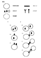
(f)生体ユニットまたはそれらの派生体を検出または同定する工程と
を含む方法を提供する。この方法は図1に示される。
In one aspect, the invention is a method for the detection of at least two biological units or biomolecules, said method comprising:
(A) providing a sample comprising a biological entity or cell comprising at least two biological units or biomolecules;
(B) capturing or spatially separating the biological entity or cell in an effective range or compartment, wherein the effective range or compartment is for the biological unit, biomolecule, or derivative thereof Including additional storage of or including itself as storage,
(C) optionally releasing the biological unit from the biological entity;
(D) optionally generating a derivative of the biological unit;
(E) allowing a biological unit or derivative thereof to be coupled to storage;
(F) detecting or identifying a biological unit or derivative thereof. This method is illustrated in FIG.
他の態様では、本発明は、少なくとも2つの生体ユニットまたは生体分子の検出のための方法であって、前記方法が、
(a)少なくとも2つの生体ユニットまたは生体分子を含む生物学的実体または細胞を含む試料を準備する工程と、
(b)前記生物学的実体または細胞を、有効範囲またはコンパートメントの中に捕捉または空間分離する工程であって、前記有効範囲またはコンパートメントが、前記生体ユニットまたは生体分子の派生体を結合することができる成分を含む工程と、
(c)生物学的実体または細胞から生体ユニットまたは生体分子を放出させる工程と、
(d)前記生体ユニットまたは生体分子の派生体を生成させる工程と、
(e)生体ユニットまたは生体分子の派生体が、前記生体ユニットまたは生体分子の前記派生体を結合することができる成分に結合することを可能にする工程と、
(f)生体ユニットまたは生体分子の派生体を検出または同定する工程と
を含む方法を提供する。
In another aspect, the invention provides a method for the detection of at least two biounits or biomolecules, said method comprising:
(A) providing a sample comprising a biological entity or cell comprising at least two biological units or biomolecules;
(B) capturing or spatially separating the biological entity or cell in an effective range or compartment, wherein the effective range or compartment binds the biological unit or a derivative of a biomolecule; A process comprising a component capable of;
(C) releasing the biological unit or biomolecule from the biological entity or cell;
(D) generating a derivative of the biological unit or biomolecule;
(E) allowing a biounit or biomolecule derivative to bind to a component capable of binding the biounit or biomolecule derivative;
(F) detecting or identifying a biounit or biomolecule derivative.
工程(c)および(d)は任意選択であってよい。特定の実施形態では、工程(c)は必要ないこともある。これは、例えば、両方ともシークエンシングタグを含有する2つの抗体が同じ抗原の異なるエピトープに結合する場合である。そのような場合、抗体(すなわち、生体ユニットまたは生体分子)は、生物学的実体または細胞から予め放出されることなく、直接シークエンシング(すなわち、検出または同定)することができる。さらに、工程(d)は任意選択であってもよい。生体ユニットまたは生体分子を直接処理することができるか、またはさらなる処理のために生体ユニットまたは生体分子の派生体を生成させることができるかのいずれかである。これは特定の実施形態の下では有利となりうる。例えば、生体ユニットまたは生体分子それ自体が、相当不安定な場合(例えば、mRNA)、より安定な形態(例えば、DNA)への変換が好ましい。 Steps (c) and (d) may be optional. In certain embodiments, step (c) may not be necessary. This is the case, for example, when two antibodies that both contain a sequencing tag bind to different epitopes of the same antigen. In such cases, the antibody (ie, biological unit or biomolecule) can be sequenced (ie, detected or identified) directly without being previously released from the biological entity or cell. Furthermore, step (d) may be optional. Either the biounit or biomolecule can be processed directly, or a biounit or biomolecule derivative can be generated for further processing. This can be advantageous under certain embodiments. For example, if the biounit or biomolecule itself is fairly unstable (eg, mRNA), conversion to a more stable form (eg, DNA) is preferred.
一態様では、本発明は、少なくとも2つの生体ユニットまたは生体分子の検出のための方法であって、前記方法が、
(a)少なくとも2つの生体ユニットまたは生体分子を含む生物学的実体または細胞を含む試料を準備する工程と、
(b)前記生物学的実体または前記細胞を、有効範囲またはコンパートメントの中に捕捉または空間分離する工程であって、前記有効範囲またはコンパートメントが、前記生体ユニット、生体分子、またはそれらの派生体を結合することができる成分を含む工程と、
(c)生物学的実体または細胞から生体ユニットまたは生体分子を放出させる工程と、
(d)前記生体ユニットまたは生体分子の派生体を生成させる工程と、
(e)生体ユニット、生体分子、またはそれらの派生体が、前記生体ユニット、生体分子、またはそれらの派生体を結合することができる成分に結合することを可能にする工程と、
(f)生体ユニット、生体分子、またはそれらの派生体を検出または同定する工程と
を含む方法を提供する。
In one aspect, the invention is a method for the detection of at least two biological units or biomolecules, said method comprising:
(A) providing a sample comprising a biological entity or cell comprising at least two biological units or biomolecules;
(B) capturing or spatially separating the biological entity or the cell in an effective range or compartment, wherein the effective range or compartment contains the biological unit, biomolecule, or derivative thereof. Including a component that can be combined;
(C) releasing the biological unit or biomolecule from the biological entity or cell;
(D) generating a derivative of the biological unit or biomolecule;
(E) allowing the biounit, biomolecule, or derivative thereof to bind to a component capable of binding the biounit, biomolecule, or derivative thereof;
(F) detecting or identifying a biounit, biomolecule, or derivative thereof.
一態様では、本発明は、2つの生体ユニットまたは生体分子の相互作用の検出のための方法であって、前記方法が、
(a)前記2つの相互作用する生体ユニットまたは生体分子を含む生物学的実体または細胞を含む試料を準備する工程と、
(b)前記生物学的実体または前記細胞を、有効範囲またはコンパートメントの中に捕捉または空間分離する工程であって、前記有効範囲またはコンパートメントが、前記生体ユニット、生体分子、またはそれらの派生体を結合することができる成分を含む工程と、
(c)生物学的実体または細胞から生体ユニットまたは生体分子を放出させる工程と、
(d)前記生体ユニットまたは生体分子の派生体を生成させる工程と、
(e)生体ユニット、生体分子、またはそれらの派生体が、前記生体ユニット、生体分子、またはそれらの派生体を結合することができる成分に結合することを可能にする工程と、
(f)生体ユニット、生体分子、またはそれらの派生体を検出または同定する工程と
を含む方法を提供する。
In one aspect, the invention is a method for the detection of the interaction of two biological units or biomolecules, said method comprising:
(A) providing a sample comprising a biological entity or cell comprising the two interacting biological units or biomolecules;
(B) capturing or spatially separating the biological entity or the cell in an effective range or compartment, wherein the effective range or compartment contains the biological unit, biomolecule, or derivative thereof. Including a component that can be combined;
(C) releasing the biological unit or biomolecule from the biological entity or cell;
(D) generating a derivative of the biological unit or biomolecule;
(E) allowing the biounit, biomolecule, or derivative thereof to bind to a component capable of binding the biounit, biomolecule, or derivative thereof;
(F) detecting or identifying a biounit, biomolecule, or derivative thereof.
代替態様では、本発明は、少なくとも2つの生体ユニットまたは生体分子の検出のための方法であって、前記方法が、
(a)少なくとも2つの生体ユニットまたは生体分子を含む生物学的実体または細胞を含む試料を準備する工程と、
(b)前記生物学的実体または細胞を、有効範囲またはコンパートメントの中に捕捉または空間分離する工程であって、前記有効範囲またはコンパートメントが、前記生体ユニット、生体分子、またはそれらの派生体を結合することができる成分を含む工程と、
(c)前記生体ユニットまたは生体分子の派生体を生成させる工程と、
(d)生体ユニットまたは生体分子の派生体が、前記生体ユニット、生体分子、またはそれらの派生体を結合することができる成分に結合することを可能にする工程と、
(e)生体ユニット、生体分子、またはそれらの派生体を検出または同定する工程と
を含む方法を提供する。
In an alternative aspect, the present invention is a method for the detection of at least two biological units or biomolecules, said method comprising:
(A) providing a sample comprising a biological entity or cell comprising at least two biological units or biomolecules;
(B) capturing or spatially separating the biological entity or cell in an effective range or compartment, wherein the effective range or compartment binds the biological unit, biomolecule, or derivative thereof. Including a component that can be
(C) generating a derivative of the biological unit or biomolecule;
(D) allowing a biounit or biomolecule derivative to bind to a component capable of binding the biounit, biomolecule, or derivative thereof;
(E) detecting or identifying a biounit, biomolecule, or derivative thereof.
代替態様では、本発明は、2つの生体ユニットまたは生体分子の相互作用の検出のための方法であって、前記方法が、
(a)前記2つの相互作用する生体ユニットまたは生体分子を含む生物学的実体または細胞を含む試料を準備する工程と、
(b)前記生物学的実体または細胞を、有効範囲またはコンパートメントの中に捕捉または空間分離する工程であって、前記有効範囲またはコンパートメントが、前記生体ユニット、生体分子、またはそれらの派生体を結合することができる成分を含む工程と、
(c)前記生体ユニットまたは生体分子の派生体を生成させる工程と、
(d)生体ユニットまたは生体分子の派生体が、前記生体ユニット、生体分子、またはそれらの派生体を結合することができる成分に結合することを可能にする工程と、
(e)生体ユニット、生体分子、またはそれらの派生体を検出または同定する工程と
を含む方法を提供する。
In an alternative aspect, the present invention is a method for the detection of the interaction of two biological units or biomolecules, said method comprising:
(A) providing a sample comprising a biological entity or cell comprising the two interacting biological units or biomolecules;
(B) capturing or spatially separating the biological entity or cell in an effective range or compartment, wherein the effective range or compartment binds the biological unit, biomolecule, or derivative thereof. Including a component that can be
(C) generating a derivative of the biological unit or biomolecule;
(D) allowing a biounit or biomolecule derivative to bind to a component capable of binding the biounit, biomolecule, or derivative thereof;
(E) detecting or identifying a biounit, biomolecule, or derivative thereof.
さらなる代替態様では、本発明は、少なくとも2つの生体ユニットまたは生体分子の検出のための方法であって、前記方法が、
(a)少なくとも2つの生体ユニットまたは生体分子を含む生物学的実体または細胞を含む試料を準備する工程と、
(b)前記生物学的実体または細胞を、有効範囲またはコンパートメントの中に捕捉または空間分離し、生物学的実体または細胞から生体ユニットまたは生体分子を放出させる工程であって、前記有効範囲またはコンパートメントが、前記生体ユニットまたは生体分子に結合することができる成分を含む工程と、
(c)生体ユニットまたは生体分子が、前記生体ユニットまたは生体分子を結合することができる成分に結合することを可能にする工程と、
(d)生体ユニットまたは生体分子を検出または同定する工程と
を含む方法を提供する。
In a further alternative aspect, the present invention is a method for the detection of at least two biological units or biomolecules, said method comprising:
(A) providing a sample comprising a biological entity or cell comprising at least two biological units or biomolecules;
(B) capturing or spatially separating the biological entity or cell in an effective range or compartment to release a biological unit or molecule from the biological entity or cell, the effective range or compartment being Comprising a component capable of binding to the biological unit or biomolecule;
(C) allowing the biounit or biomolecule to bind to a component capable of binding the biounit or biomolecule;
(D) detecting or identifying a biounit or biomolecule.
さらなる代替態様では、本発明は、2つの生体ユニットまたは生体分子の相互作用の検出のための方法であって、前記方法が、
(a)前記2つの相互作用する生体ユニットまたは生体分子を含む生物学的実体または細胞を含む試料を準備する工程と、
(b)前記生物学的実体または細胞を、有効範囲またはコンパートメントの中に捕捉または空間分離する工程であって、前記有効範囲またはコンパートメントが、前記生体ユニットまたは生体分子を結合することができる成分を含む工程と、
(c)生物学的実体または細胞から生体ユニットまたは生体分子を放出させる工程と、
(d)生体ユニットまたは生体分子が、前記生体ユニットまたは生体分子を結合することができる成分に結合することを可能にする工程と、
(e)生体ユニットまたは生体分子を検出または同定する工程と
を含む方法を提供する。
In a further alternative aspect, the present invention is a method for detecting the interaction of two biological units or biomolecules, said method comprising:
(A) providing a sample comprising a biological entity or cell comprising the two interacting biological units or biomolecules;
(B) capturing or spatially separating the biological entity or cell in an effective range or compartment, wherein the effective range or compartment comprises a component capable of binding the biological unit or biomolecule; Including a process;
(C) releasing the biological unit or biomolecule from the biological entity or cell;
(D) allowing the biounit or biomolecule to bind to a component capable of binding the biounit or biomolecule;
(E) detecting or identifying a biological unit or biomolecule.
さらなる代替態様では、本発明は、少なくとも2つの生体ユニットまたは生体分子の検出のための方法であって、前記方法が、
(a)少なくとも2つの生体ユニットまたは生体分子を含む生物学的実体または細胞を含む試料を準備する工程と、
(b)前記生物学的実体または細胞を、有効範囲またはコンパートメントの中に捕捉または空間分離する工程であって、前記有効範囲またはコンパートメントが、前記生体ユニットまたは生体分子を結合することができる成分を含む工程と、
(c)生体ユニットが、前記生体ユニットまたは生体分子を結合することができる成分に結合することを可能にする工程と、
(d)生体ユニットまたは生体分子を検出または同定する工程と
を含む方法を提供する。
In a further alternative aspect, the present invention is a method for the detection of at least two biological units or biomolecules, said method comprising:
(A) providing a sample comprising a biological entity or cell comprising at least two biological units or biomolecules;
(B) capturing or spatially separating the biological entity or cell in an effective range or compartment, wherein the effective range or compartment comprises a component capable of binding the biological unit or biomolecule; Including a process;
(C) allowing the biological unit to bind to a component capable of binding the biological unit or biomolecule;
(D) detecting or identifying a biounit or biomolecule.
さらなる代替態様では、本発明は、2つの生体ユニットまたは生体分子の相互作用の検出のための方法であって、前記方法が、
(a)前記2つの相互作用する生体ユニットまたは生体分子を含む生物学的実体または細胞を含む試料を準備する工程と、
(b)前記生物学的実体または細胞を、有効範囲またはコンパートメントの中に捕捉または空間分離する工程であって、前記有効範囲またはコンパートメントが、前記生体ユニットまたは生体分子を結合することができる成分を含む工程と、
(c)生体ユニットが、前記生体ユニットまたは生体分子を結合することができる成分に結合することを可能にする工程と、
(d)生体ユニットまたは生体分子を検出または同定する工程と
を含む方法を提供する。
In a further alternative aspect, the present invention is a method for detecting the interaction of two biological units or biomolecules, said method comprising:
(A) providing a sample comprising a biological entity or cell comprising the two interacting biological units or biomolecules;
(B) capturing or spatially separating the biological entity or cell in an effective range or compartment, wherein the effective range or compartment comprises a component capable of binding the biological unit or biomolecule; Including a process;
(C) allowing the biological unit to bind to a component capable of binding the biological unit or biomolecule;
(D) detecting or identifying a biounit or biomolecule.
本発明の少なくとも2つの生体ユニットまたは生体分子は、1つの生物学的実体の中に存在することができる。あるいは、本発明の少なくとも2つの生体ユニットまたは生体分子は、1つの細胞の中に存在することができる。特定の実施形態では、両方の生体ユニットまたは生体分子は、生物学的実体または細胞の内部に位置することができる。代替実施形態では、両方の生体ユニットまたは生体分子は、生物学的実体または細胞の上にまたはその外部に位置することができる。さらなる代替実施形態では、一方の生体ユニットまたは生体分子は、生物学的実体または細胞の内部に位置することができ、他方の生体ユニットまたは生体分子は、生物学的実体または細胞の上にまたはその外部に位置することができる。 At least two biounits or biomolecules of the present invention can be present in one biological entity. Alternatively, at least two biounits or biomolecules of the present invention can be present in one cell. In certain embodiments, both biological units or biomolecules can be located inside a biological entity or cell. In an alternative embodiment, both biological units or biomolecules can be located on or outside the biological entity or cell. In a further alternative embodiment, one biological unit or biomolecule can be located inside a biological entity or cell and the other biological unit or biomolecule is on or on the biological entity or cell. Can be located outside.
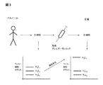
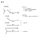
少なくとも2つの生体ユニットまたは生体分子はまた、任意の方法または形態で互いに相互作用する2つの生物学的実体または細胞の中に存在することができる。特定の実施形態では、第1の生体ユニットまたは生体分子は、第1の生物学的実体または細胞の内部に位置し、第2の生体ユニットまたは生体分子は、第2の生物学的実体または細胞の内部に位置する。他の実施形態では、第1の生体ユニットまたは生体分子は、第1の生物学的実体または細胞の上にまたはその外部に位置し、第2の生体ユニットまたは生体分子は、第2の生物学的実体または細胞の内部に位置する。さらに他の実施形態では、第1の生体ユニットまたは生体分子は、第1の生物学的実体または細胞の上にまたはその外部に位置し、第2の生体ユニットまたは生体分子は、第2の生物学的実体または細胞の上にまたはその外部に位置する。さらに他の実施形態では、第1の生体ユニットまたは生体分子は、第1の生物学的実体または細胞の上にまたはその外部に位置し、第2の生体ユニットまたは生体分子は、第2の生物学的実体または細胞の上にまたはその外部に位置し、かつ2つの生体ユニットまたは生体分子は、第3の分子、例えば第1の生体ユニットまたは生体分子および第2の生体ユニットまたは生体分子の両方に結合する生体ユニットまたは生体分子を介して間接的に相互作用している。図2に、可能なシナリオの一部を図示する。 At least two biological units or biomolecules can also be present in two biological entities or cells that interact with each other in any manner or form. In certain embodiments, the first biological unit or biomolecule is located within the first biological entity or cell, and the second biological unit or biomolecule is the second biological entity or cell. Located inside. In other embodiments, the first biological unit or biomolecule is located on or outside of the first biological entity or cell, and the second biological unit or biomolecule is the second biological unit or biomolecule. Located inside the target entity or cell. In still other embodiments, the first biological unit or biomolecule is located on or outside of the first biological entity or cell, and the second biological unit or biomolecule is the second biological entity. Located on or outside of the biological entity or cell. In still other embodiments, the first biological unit or biomolecule is located on or outside of the first biological entity or cell, and the second biological unit or biomolecule is the second biological entity. Two biological units or biomolecules located on or outside of a biological entity or cell and a third molecule, eg both a first biological unit or biomolecule and a second biological unit or biomolecule It interacts indirectly via biological units or biomolecules that bind to. FIG. 2 illustrates some of the possible scenarios.
これらの技術領域で使用される従来技術の方法には、酵母ツーハイブリッドシステム、酵母スリーハイブリッドシステム、SIP技術(自己感染性ファージ)、PCAアッセイ(タンパク質−断片相補性アッセイ)、およびスプリット−ユビキチンシステムが挙げられる。これらのアッセイおよびシステムはすべて、読み取りシグナルの高いバックグラウンドノイズが受け、不満足なシグナル対バックグラウンド比がもたらされる。ほとんどの場合、これは、これらのシステムで起こる非特異的な相互作用によるものである。読み取りシグナルの定量化によって、例えばカラー強度またはコロニーサイズの測定によって、これらの問題を回避することは、部分的にしか成功しておらず、依然として厄介で誤りがちなものとなっている。本発明は、これらの欠点に対して簡潔な解決法を提供する。数千または数百万もの相互作用または事象を分析することにより、統計的に有意な読み取りシグナルを受け取る問題は、分析される相互作用または事象の数を増加させることにより解決される。本発明は、大量の読み取りシグナルを分析することができるハイスループット法を提供する。これは、例えば、それぞれの表現型読み取りシグナルが低いシグナル対バックグラウンド比になる場合に、遺伝子型レベルで達成することができる。 Prior art methods used in these areas of technology include yeast two-hybrid systems, yeast three-hybrid systems, SIP technology (self-infecting phage), PCA assays (protein-fragment complementation assays), and split-ubiquitin systems. Is mentioned. All of these assays and systems are subject to high background noise of the read signal, resulting in an unsatisfactory signal to background ratio. In most cases this is due to non-specific interactions occurring in these systems. Avoiding these problems by quantifying the read signal, for example, by measuring color intensity or colony size, has been only partially successful and remains cumbersome and error prone. The present invention provides a simple solution to these drawbacks. By analyzing thousands or millions of interactions or events, the problem of receiving a statistically significant read signal is solved by increasing the number of interactions or events analyzed. The present invention provides a high-throughput method that can analyze large amounts of read signal. This can be achieved, for example, at the genotype level when each phenotypic read signal results in a low signal to background ratio.
本発明はまた、ライブラリー対ライブラリー用途、例えば1つのライブラリーのメンバーと2番目のライブラリーのメンバーとの間の相互作用に対するスクリーニングで使用することができる。一例として、感染または疾患に応答したヒトの免疫レパートリーなど、生体の免疫応答を測定することができるであろう。統計的に有意でかつ満足すべき方法における免疫応答全体の複雑な分析は、本発明に記載の方法によってのみ可能になった。それぞれの実験手法を図3に示す。 The present invention can also be used in library-to-library applications, such as screening for interactions between one library member and a second library member. As an example, the immune response of an organism could be measured, such as a human immune repertoire in response to an infection or disease. Complex analysis of the overall immune response in a statistically significant and satisfactory way was only possible with the method according to the invention. Each experimental technique is shown in FIG.
特定の態様では、前記生体ユニットは、サブクラスであるポリペプチド、タンパク質、ペプチド、核酸、炭水化物、脂肪酸、小分子、細胞小器官、細胞、組織、または前述のいずれかの派生体、一部、もしくは組合せからなる群から選択される。特定の態様では、すべての生体ユニットは同じサブクラスに由来する。特定の態様では、生体ユニットは異なるサブクラスに由来する。特定の態様では、前記サブクラスは、ポリペプチドのサブクラスまたは核酸のサブクラスである。特定の態様では、前記サブクラスは、ポリペプチドのサブクラスである。 In certain embodiments, the biological unit is a subclass of a polypeptide, protein, peptide, nucleic acid, carbohydrate, fatty acid, small molecule, organelle, cell, tissue, or derivative, part, or any of the foregoing Selected from the group consisting of combinations. In certain embodiments, all biological units are from the same subclass. In certain embodiments, the biological units are from different subclasses. In certain embodiments, the subclass is a polypeptide subclass or a nucleic acid subclass. In certain embodiments, the subclass is a subclass of a polypeptide.
特定の態様では、前記生体ユニットは生体分子である。特定の好ましい態様では、前記生体ユニットまたは生体分子は、タンパク質、ポリペプチド、またはペプチドである。代替態様では、前記生体分子は、DNAまたはRNAなどの核酸である。前記核酸は、一本鎖であっても二本鎖であってもよい。DNA分子は、RNAまたはDNAの鋳型から合成することができることは理解されよう。同様に、RNA分子も、RNAまたはDNAの鋳型から合成することができる。 In a particular embodiment, the biological unit is a biomolecule. In certain preferred embodiments, the biological unit or biomolecule is a protein, polypeptide, or peptide. In an alternative embodiment, the biomolecule is a nucleic acid such as DNA or RNA. The nucleic acid may be single-stranded or double-stranded. It will be appreciated that DNA molecules can be synthesized from RNA or DNA templates. Similarly, RNA molecules can be synthesized from RNA or DNA templates.
特定の態様では、前記生体ユニットまたは生体分子は、多量体のタンパク質または酵素の一部である。特定の態様では、前記生体ユニットまたは生体分子は、多量体のタンパク質または酵素の一部であるポリペプチドである。特定の態様では、前記生体ユニットまたは生体分子は、多量体のタンパク質または酵素の一部をコードする核酸である。特定の態様では、前記多量体タンパク質は、免疫グロブリンまたはその機能性断片である。特定の態様では、前記生体ユニットまたは生体分子は、免疫グロブリンまたはその機能性断片の可変重鎖および可変軽鎖をコードする遺伝子である。 In certain embodiments, the biological unit or biomolecule is part of a multimeric protein or enzyme. In a particular embodiment, the biological unit or biomolecule is a polypeptide that is part of a multimeric protein or enzyme. In a particular embodiment, the biological unit or biomolecule is a nucleic acid that encodes a portion of a multimeric protein or enzyme. In a particular embodiment, the multimeric protein is an immunoglobulin or a functional fragment thereof. In a particular embodiment, the biological unit or biomolecule is a gene encoding a variable heavy chain and a variable light chain of an immunoglobulin or functional fragment thereof.
特定の態様では、本発明で使用される試料は、血液、骨髄、腫瘍、単一細胞生物、原核生物、または体液に由来した試料である。特定の態様では、前記試料は患者に由来する試料であり、前記患者は、健常患者、免疫患者、感染患者、または疾患もしくは障害を有する患者である。 In certain embodiments, the sample used in the present invention is a sample derived from blood, bone marrow, tumor, single cell organism, prokaryote, or body fluid. In certain embodiments, the sample is a sample derived from a patient, and the patient is a healthy patient, an immune patient, an infected patient, or a patient with a disease or disorder.
特定の態様では、本発明で使用される生物学的実体は、単一細胞である。特定の態様では、前記単一細胞は単一B細胞である。 In certain embodiments, the biological entity used in the present invention is a single cell. In certain embodiments, the single cell is a single B cell.
特定の態様では、生物学的実体は、別の細胞、ウイルス、細菌、分子、もしくは生体分子、または前述のいずれかの派生体、断片、もしくは複合物と相互作用する細胞である。特定の態様では、生物学的実体は、細胞、および前記細胞に感染するウイルスである。特定の態様では、生物学的実体は、細胞、および前記細胞に感染する細菌である。特定の態様では、生物学的実体は、細胞および別の細胞である。前記細胞は、ドナーおよびアクセプターの観点から、エフェクターおよび作用を受ける実体の観点から、または阻害剤および阻害される細胞の観点から互いに情報交換することができる。 In certain embodiments, the biological entity is a cell that interacts with another cell, virus, bacterium, molecule, or biomolecule, or any derivative, fragment, or complex of any of the foregoing. In certain embodiments, the biological entity is a cell and a virus that infects the cell. In certain embodiments, the biological entity is a cell and a bacterium that infects the cell. In certain embodiments, the biological entity is a cell and another cell. The cells can exchange information with each other from the perspective of donors and acceptors, from the perspective of effectors and affected entities, or from the perspective of inhibitors and cells to be inhibited.
特定の態様では、生物学的実体は、2つの化学的および/または生物学的ライブラリー(例えば、ファージライブラリーまたはリボソームディスプレイライブラリーなど)の混合物を含み、そこでは、第1ライブラリーの少なくとも1つのメンバーが、第2ライブラリーのメンバーと相互作用または結合する。特定の態様では、第1ライブラリーの各メンバーは、第1ライブラリーに特異的なタグを含み、第2ライブラリーは、第2ライブラリーに特異的なタグを含む。 In certain embodiments, the biological entity comprises a mixture of two chemical and / or biological libraries (eg, such as a phage library or a ribosome display library), wherein at least one of the first library One member interacts or binds with a member of the second library. In certain embodiments, each member of the first library includes a tag specific for the first library, and the second library includes a tag specific for the second library.
特定の態様では、生物学的実体は、少なくとも1つの化学的および/または生物学的ライブラリー(例えば、ファージライブラリーまたはリボソームディスプレイライブラリーなど)の少なくとも2つのメンバーと相互作用または結合する分子を含む。特定の態様では、ライブラリーの各メンバーは、前記ライブラリーに特異的なタグを含む。 In certain embodiments, the biological entity is a molecule that interacts or binds to at least two members of at least one chemical and / or biological library (such as a phage library or a ribosome display library). Including. In certain embodiments, each member of the library includes a tag specific to the library.
代替態様では、本発明は、少なくとも2つの生体ユニットまたは生体分子の検出のための方法であって、前記方法が、
(a)少なくとも2つの生体ユニットまたは生体分子を含む生物学的実体または細胞を含む試料を準備する工程と、
(b)前記少なくとも2つの生体ユニットまたは生体分子および前記生体ユニットまたは生体分子を結合することができる成分を、有効範囲またはコンパートメントの中に捕捉または空間分離する工程と、
(c)場合によっては、生物学的実体または細胞から生体ユニットまたは生体分子を放出させる工程と、
(d)生体ユニットまたは生体分子が、前記生体ユニットまたは生体分子を結合することができる成分に結合することを可能にする工程と、
(e)前記生体ユニットまたは生体分子を結合することができる成分上の生体ユニットまたは生体分子を検出または同定する工程と
を含む方法を提供する。
In an alternative aspect, the present invention is a method for the detection of at least two biological units or biomolecules, said method comprising:
(A) providing a sample comprising a biological entity or cell comprising at least two biological units or biomolecules;
(B) capturing or spatially separating the at least two biological units or biomolecules and components capable of binding the biological units or biomolecules within an effective range or compartment;
(C) optionally releasing the biological unit or molecule from the biological entity or cell;
(D) allowing the biounit or biomolecule to bind to a component capable of binding the biounit or biomolecule;
(E) detecting or identifying a biounit or biomolecule on a component capable of binding the biounit or biomolecule.
代替態様では、本発明は、2つの生体ユニットまたは生体分子の相互作用の検出のための方法であって、前記方法が、
(a)前記2つの相互作用する生体ユニットまたは生体分子を含む生物学的実体または細胞を含む試料を準備する工程と、
(b)前記少なくとも2つの生体ユニットまたは生体分子および前記生体ユニットまたは生体分子を結合することができる成分を、有効範囲またはコンパートメントの中に捕捉または空間分離する工程と、
(c)場合によっては、生物学的実体または細胞から生体ユニットまたは生体分子を放出させる工程と、
(d)生体ユニットまたは生体分子が、前記生体ユニットまたは生体分子を結合することができる成分に結合することを可能にする工程と、
(e)前記生体ユニットまたは生体分子を結合することができる成分上の生体ユニットまたは生体分子を検出または同定する工程と
を含む方法を提供する。
In an alternative aspect, the present invention is a method for the detection of the interaction of two biological units or biomolecules, said method comprising:
(A) providing a sample comprising a biological entity or cell comprising the two interacting biological units or biomolecules;
(B) capturing or spatially separating the at least two biological units or biomolecules and components capable of binding the biological units or biomolecules within an effective range or compartment;
(C) optionally releasing the biological unit or molecule from the biological entity or cell;
(D) allowing the biounit or biomolecule to bind to a component capable of binding the biounit or biomolecule;
(E) detecting or identifying a biounit or biomolecule on a component capable of binding the biounit or biomolecule.
代替態様では、本発明は、少なくとも2つの生体ユニットまたは生体分子の検出のための方法であって、前記方法が、
(a)少なくとも2つの生体ユニットまたは生体分子を含む生物学的実体または細胞を含む試料を準備する工程と、
(b)前記少なくとも2つの生体ユニットまたは生体分子を、有効範囲またはコンパートメントの中に捕捉または空間分離する工程であって、前記有効範囲またはコンパートメントが、前記生体ユニットまたは生体分子を結合することができる工程と、
(c)場合によっては、生物学的実体または細胞から生体ユニットまたは生体分子を放出させる工程と、
(d)生体ユニットまたは生体分子が、有効範囲またはコンパートメントに結合することを可能にする工程と、
(e)有効範囲またはコンパートメントの上の生体ユニットまたは生体分子を検出または同定する工程と
を含む方法を提供する。
In an alternative aspect, the present invention is a method for the detection of at least two biological units or biomolecules, said method comprising:
(A) providing a sample comprising a biological entity or cell comprising at least two biological units or biomolecules;
(B) capturing or spatially separating the at least two biological units or biomolecules within an effective range or compartment, wherein the effective range or compartment can bind the biological unit or biomolecule. Process,
(C) optionally releasing the biological unit or molecule from the biological entity or cell;
(D) allowing a biological unit or biomolecule to bind to an effective range or compartment;
(E) detecting or identifying a biounit or biomolecule above the effective range or compartment.
代替態様では、本発明は、2つの生体ユニットまたは生体分子の相互作用の検出のための方法であって、前記方法が、
(a)前記2つの相互作用する生体ユニットまたは生体分子を含む生物学的実体または細胞を含む試料を準備する工程と、
(b)前記少なくとも2つの生体ユニットまたは生体分子を、有効範囲またはコンパートメントの中に捕捉または空間分離する工程であって、前記有効範囲またはコンパートメントが、前記生体ユニットまたは生体分子を結合することができる工程と、
(c)場合によっては、生物学的実体または細胞から生体ユニットまたは生体分子を放出させる工程と、
(d)生体ユニットまたは生体分子が、有効範囲またはコンパートメントに結合することを可能にする工程と、
(e)有効範囲またはコンパートメントの上の生体ユニットまたは生体分子を検出または同定する工程と
を含む方法を提供する。
In an alternative aspect, the present invention is a method for the detection of the interaction of two biological units or biomolecules, said method comprising:
(A) providing a sample comprising a biological entity or cell comprising the two interacting biological units or biomolecules;
(B) capturing or spatially separating the at least two biological units or biomolecules within an effective range or compartment, wherein the effective range or compartment can bind the biological unit or biomolecule. Process,
(C) optionally releasing the biological unit or molecule from the biological entity or cell;
(D) allowing a biological unit or biomolecule to bind to an effective range or compartment;
(E) detecting or identifying a biounit or biomolecule above the effective range or compartment.
一工程では、本発明の方法は、有効範囲またはコンパートメントの中への、少なくとも2つの生体ユニットまたは生体分子および前記生体ユニットまたは生体分子のためのストレージを含む少なくとも1つの生物学的実体または細胞の捕捉を含む。前述のストレージは、前記生体ユニットもしくは生体分子またはそれらの派生体を結合することができる成分である。特定の実施形態では、前記ストレージは、前記生体ユニットまたは生体分子を結合することができる成分である。他の実施形態では、前記ストレージは、前記生体ユニットまたは生体分子の派生体を結合することができる成分である。 In one step, the method of the present invention comprises at least one biological unit or biomolecule and a storage for at least one biological unit or biomolecule and a storage for said biological unit or biomolecule into an effective range or compartment. Includes capture. The aforementioned storage is a component capable of binding the biounit or biomolecule or a derivative thereof. In a particular embodiment, the storage is a component capable of binding the biounit or biomolecule. In another embodiment, the storage is a component capable of binding the biounit or biomolecule derivative.
前記生物学的実体または細胞、生体ユニットまたは生体分子、およびストレージの捕捉または空間分離は、捕捉または空間分離された生体ユニットまたは生体分子のその後の検出を可能にする任意の方法または形態で達成することができる。特定の実施形態では、これは、例えば油中水型エマルションなどのエマルションを介して達成することができる(図4、パネルAを参照のこと)。このような実施形態では、生物学的実体を細胞とすることができ、生体ユニットまたは生体分子をDNA配列(例えば、抗体の可変重鎖および可変軽鎖)とすることができ、ストレージをプライマーとすることができる(例えば、抗体の可変重鎖に結合する1つのプライマーおよび同じ抗体の可変軽鎖に結合する別のプライマー)。油中水型エマルションの代わりとして、水中油型エマルションを使用することができ、それによって有効範囲またはコンパートメントとして役立つミセルが形成される。他の実施形態では、前記生物学的実体または細胞、生体ユニットまたは生体分子、およびストレージの捕捉または空間分離は、マイクロタイタープレート、ピコタイタープレート、またはシークエンシングチップのウェルにおいて達成することができる(図4、パネルBを参照のこと)。有効範囲またはコンパートメントは壁および蓋により形成される。そのような実施形態では、生物学的実体を細胞とすることができ、生体ユニットまたは生体分子を抗原とすることができ、ストレージを、例えば、生体ユニット、すなわち抗原に特異的な抗体とすることができる。あるいは、マイクロタイタープレート、ピコタイタープレート、シークエンシングチップ、または前記容器の蓋は、それ自体ストレージであってもよく、またはストレージとして役立ってもよい。さらに、ストレージは少なくとも2つのバインダー、例えばプライマーを含む。 Capturing or spatial separation of the biological entity or cell, biological unit or biomolecule, and storage is accomplished in any manner or form that allows subsequent detection of the captured or spatially separated biological unit or biomolecule. be able to. In certain embodiments, this can be accomplished via an emulsion, such as a water-in-oil emulsion (see FIG. 4, panel A). In such embodiments, the biological entity can be a cell, the biounit or biomolecule can be a DNA sequence (eg, an antibody variable heavy and variable light chain), and the storage can be a primer. (Eg, one primer that binds to the variable heavy chain of an antibody and another primer that binds to the variable light chain of the same antibody). As an alternative to water-in-oil emulsions, oil-in-water emulsions can be used, thereby forming micelles that serve as effective ranges or compartments. In other embodiments, capture or spatial separation of the biological entity or cell, biological unit or biomolecule, and storage can be accomplished in a microtiter plate, picotiter plate, or well of a sequencing chip ( (See FIG. 4, Panel B). The effective area or compartment is formed by a wall and a lid. In such embodiments, the biological entity can be a cell, the biounit or biomolecule can be an antigen, and the storage can be, for example, a biounit, ie, an antibody specific for the antigen. Can do. Alternatively, the microtiter plate, picotiter plate, sequencing chip, or lid of the container may itself be storage or serve as storage. In addition, the storage includes at least two binders, such as primers.
本発明の特定の態様では、前記有効範囲またはコンパートメントは、キャビティー、ウェル、エマルション、相境界システム、疎水性スポット、粒子、固相粒子、物理力、または化学的架橋により形成される。本発明の特定の態様では、前記相境界は、空気中の水滴のように水と気体の間の相分離によって、または油中の水滴のように水と液体の間の相分離によって、またマイクロタイタープレート中の水滴のように固相によって実現される。本発明の特定の態様では、前記キャビティーまたは前記ウェルは、マイクロタイタープレート、ピコタイタープレート、または微小構造基材の上にある。本発明の特定の態様では、前記有効範囲は、化学的架橋で形成され、前記化学的架橋はホルムアルデヒドによる架橋である。 In certain embodiments of the invention, the effective range or compartment is formed by a cavity, well, emulsion, phase boundary system, hydrophobic spot, particle, solid phase particle, physical force, or chemical cross-linking. In a particular aspect of the invention, the phase boundary is formed by phase separation between water and gas, like water droplets in air, or by phase separation between water and liquid, like water droplets in oil, and microscopic. Realized by a solid phase like a water drop in a titer plate. In certain embodiments of the invention, the cavity or well is on a microtiter plate, picotiter plate, or microstructure substrate. In a particular aspect of the invention, the effective range is formed by chemical crosslinking, the chemical crosslinking being formaldehyde crosslinking.
本発明の特定の態様では、前記生体ユニットまたは生体分子および前記生体ユニット、生体分子、またはそれらの派生体に対する前記ストレージは、限界希釈によってまたはセルソーティングなどの任意の手段によるソーティングによって、有効範囲またはコンパートメントの中に捕捉または空間分離される。 In a particular aspect of the invention, the storage for the biounit or biomolecule and the biounit, biomolecule, or derivatives thereof is in the effective range or by sorting by any means such as limiting dilution or cell sorting. Captured or spatially separated in the compartment.
本発明の特定の態様では、有効範囲またはコンパートメントは、エマルションにより形成され、前記エマルションは、油中水型または水中油型のエマルションである。 In a particular embodiment of the invention, the effective range or compartment is formed by an emulsion, said emulsion being a water-in-oil or oil-in-water emulsion.
本発明の特定の態様では、有効範囲またはコンパートメントは、粒子または固相粒子により形成され、前記粒子は、シリカ、ガラス、アガロース、ポリマー、金属酸化物、またはそれらの複合物からなる。 In a particular embodiment of the invention, the effective range or compartment is formed by particles or solid phase particles, said particles consisting of silica, glass, agarose, polymer, metal oxide, or composites thereof.
本発明の特定の態様では、有効範囲またはコンパートメントは、物理力により形成され、前記物理力は、静電力、電流力、誘電泳動力、電磁力、磁気、光学、温度、または密度効果である。本発明の特定の態様では、有効範囲またはコンパートメントは、光ピンセットにより生成される。 In certain aspects of the invention, the effective range or compartment is formed by a physical force, which is an electrostatic force, current force, dielectrophoretic force, electromagnetic force, magnetic, optical, temperature, or density effect. In certain aspects of the invention, the effective range or compartment is generated by optical tweezers.
本発明の特定の態様では、有効範囲またはコンパートメントは、ネブライザーにより形成される。 In certain embodiments of the invention, the effective range or compartment is formed by a nebulizer.
本発明の特定の態様では、生体ユニット、生体分子、またはそれらの派生体を結合することができる前記ストレージまたは前記成分は、ビーズ、スライドガラスの表面上領域、マイクロタイタープレートもしくはピコタイタープレートのウェル、電場もしくは磁場、フィールド勾配もしくはフィールドケージ、または前述のいずれかの蓋上領域もしくは分離可能表面である。 In a particular aspect of the invention, said storage or said component capable of binding a biounit, biomolecule, or derivative thereof is a bead, an area on the surface of a glass slide, a well of a microtiter plate or picotiter plate An electric or magnetic field, a field gradient or a field cage, or an on-lid area or separable surface as described above.
本発明の特定の態様では、生物学的実体または細胞からの生体ユニットまたは生体分子の放出は、化学的または物理的条件の変化により行われる。特定の態様では、化学的条件の変化は、pH変化、塩濃度の変化、酵素の添加、または細胞溶解剤の添加である。特定の態様では、物理的条件の変化は、加熱、凍結、電場、磁場もしくは誘電場の印加、剪断力または遠心力、機械的変形、弛緩、超音波、または任意の物理的な破断作用である。特定の態様では、物理的条件の変化は、加熱、例えば90℃を超える加熱である。特定の態様では、前記物理的条件の変化は、溶液への粒子の溶解、生体ユニットまたは生体分子のまわりの保護シェルの溶解、または酵素による誘導など、時間依存的にもたらされる。 In a particular embodiment of the invention, the release of the biounit or biomolecule from the biological entity or cell is effected by a change in chemical or physical conditions. In certain embodiments, the change in chemical condition is a change in pH, a change in salt concentration, the addition of an enzyme, or the addition of a cytolytic agent. In certain aspects, the change in physical condition is heating, freezing, application of an electric field, magnetic field or dielectric field, shear or centrifugal force, mechanical deformation, relaxation, ultrasound, or any physical breaking action. . In certain embodiments, the change in physical condition is heating, eg, heating above 90 ° C. In certain embodiments, the change in physical condition is effected in a time-dependent manner, such as dissolution of particles in solution, dissolution of a protective shell around a biounit or biomolecule, or enzymatic induction.
別の工程では、本発明の方法は、生体ユニットまたは生体分子が、生体ユニット、生体分子、またはそれらの派生体を結合することができるストレージまたは成分に結合することを可能にする工程を含む。この工程は、前記ストレージまたは前記成分への生体ユニットまたは生体分子の結合を可能にするのに十分な時間および方法で、生体ユニットまたは生体分子をインキュベートする工程を含む。こうすることにより、生体ユニットまたは生体分子は、生体ユニット、生体分子、またはそれらの派生体を結合することができるストレージまたは成分により捕捉される。生体ユニットまたは生体分子の結合は、直接的であっても、生体ユニットまたは生体分子の派生体を介して間接的であってもよい。直接的結合は、例えばPCRまたは直接反応を介して達成することができる。間接的結合は、生体ユニットまたは生体分子の派生体の生成、およびそのような派生体を結合することができるストレージまたは成分への前記派生体の結合によって達成することができる。一例は、RNAからのcDNA鋳型の生成、およびストレージ、例えばプライマーへのcDNAの結合である。 In another step, the method of the invention includes the step of allowing a biounit or biomolecule to bind to a storage or component capable of binding a biounit, biomolecule, or derivative thereof. This step includes incubating the biounit or biomolecule for a time and method sufficient to allow binding of the biounit or biomolecule to the storage or the component. In this way, the biounit or biomolecule is captured by a storage or component that can bind the biounit, biomolecule, or derivative thereof. The binding of the biounit or biomolecule may be direct or indirect via a biounit or biomolecule derivative. Direct binding can be achieved, for example, via PCR or direct reaction. Indirect binding can be achieved by the generation of derivatives of biounits or biomolecules and the binding of said derivatives to storage or components that can bind such derivatives. An example is the generation of a cDNA template from RNA and storage, eg, binding of the cDNA to a primer.
したがって、本発明の特定の態様では、工程(d)は、前記生体ユニットまたは生体分子のレプリケートまたは派生体の生成をもたらす増幅反応を含む。本発明の特定の態様では、前記増幅反応は、PCRまたはRT−PCRであり、前記PCRまたはRT−PCRの間に、前記レプリケートまたは派生体がそのような派生体を結合することができるストレージまたは成分に結合することを可能にする[第1の]タグが加えられる。本発明の特定の態様では、前記PCRまたはRT−PCRの間に、PCR産物またはRT−PCR産物のその後のシークエンシングを可能にする第2のタグが加えられる。本発明の特定の態様では、PCR反応はエマルションPCR反応である。代替態様では、RT−PCR反応はエマルションRT−PCRである。本発明の特定の態様では、検出される2つの核酸分子は、本方法の工程(d)で増幅される。 Thus, in a particular aspect of the invention, step (d) comprises an amplification reaction that results in the production of a replicate or derivative of the biounit or biomolecule. In a particular aspect of the invention, the amplification reaction is PCR or RT-PCR, and during the PCR or RT-PCR the storage or to which the replicate or derivative can bind such derivative or A [first] tag is added that allows binding to the component. In a particular embodiment of the present invention, a second tag is added during the PCR or RT-PCR that allows the subsequent sequencing of the PCR product or RT-PCR product. In a particular aspect of the invention, the PCR reaction is an emulsion PCR reaction. In an alternative embodiment, the RT-PCR reaction is an emulsion RT-PCR. In a particular embodiment of the invention, the two nucleic acid molecules to be detected are amplified in step (d) of the method.
別の工程では、本発明の方法は、生体ユニットまたは生体分子を結合することができるストレージ上または成分上の生体ユニットまたは生体分子を検出する工程を含む。これは、検出される生体ユニットまたは生体分子について公知の任意の適切な検出方法によって達成することができ、使用される方法は、生体ユニットまた生体分子それ自体の性質に主として依存する。例としては、並行または連続して実施される(少なくとも)2つのイムノアッセイ、少なくとも2つの色素による並行染色、及び並行シークエンシングがある。 In another step, the method of the invention comprises detecting a biounit or biomolecule on a storage or component capable of binding the biounit or biomolecule. This can be achieved by any suitable detection method known for the biounit or biomolecule to be detected, the method used depending mainly on the nature of the biounit or the biomolecule itself. Examples include (at least) two immunoassays performed in parallel or sequentially, parallel staining with at least two dyes, and parallel sequencing.
本発明の特定の態様では、生体ユニットまたは生体分子の検出は、DNAシークエンシングにより行われる。本発明の特定の態様では、前記DNAシークエンシングは、PCR産物またはRT−PCR産物を連続または並行してシークエンシングすることにより行われる。本発明の特定の態様では、前記DNAシークエンシングは、ストレージ上またはストレージのコピー上のPCR産物またはRT−PCR産物をシークエンシングすることにより行われる。本発明の特定の態様では、検出される生体ユニットまたは生体分子は核酸分子であり、種々の開始プライマーが核酸分子のシークエンシングおよび同定のために使用される。本発明の特定の態様では、シークエンシング反応は、エマルションPCRにより、好ましくはビーズ上で直接行われる。本発明の特定の態様では、2つのシークエンシング反応は、連続して行われる。例えば、第1のシークエンシングプライマーを利用する第1のシークエンシング反応、および第2のシークエンシングプライマーを利用する第2のシークエンシング反応である。特定の実施形態では、第1の核酸分子は、免疫グロブリン、抗体、またはその断片の重鎖をコードし、第2の核酸分子は、免疫グロブリン、抗体、またはその断片の軽鎖をコードする。 In a particular embodiment of the invention, the detection of the biounit or biomolecule is performed by DNA sequencing. In a particular embodiment of the invention, the DNA sequencing is performed by sequencing PCR products or RT-PCR products sequentially or in parallel. In a particular embodiment of the invention, the DNA sequencing is performed by sequencing PCR products or RT-PCR products on storage or a copy of storage. In certain aspects of the invention, the biological unit or biomolecule to be detected is a nucleic acid molecule, and various starting primers are used for sequencing and identification of the nucleic acid molecule. In a particular embodiment of the invention, the sequencing reaction is performed by emulsion PCR, preferably directly on the beads. In certain aspects of the invention, the two sequencing reactions are performed sequentially. For example, a first sequencing reaction using a first sequencing primer and a second sequencing reaction using a second sequencing primer. In certain embodiments, the first nucleic acid molecule encodes the heavy chain of an immunoglobulin, antibody, or fragment thereof, and the second nucleic acid molecule encodes the light chain of an immunoglobulin, antibody, or fragment thereof.
本発明の方法の検出工程がシークエンシングにより行われる場合、特定の技術的変法が可能である。例えば、第2のシークエンシング反応のためのプライマーを酵素的に切断可能な保護基に連結することができよう。これは、保護基の切断の後でのみ、第2の核酸分子をシークエンシングすることが可能になることを保証する。これはシークエンシングエラーを低減するであろう。あるいは、第2のシークエンシングプライマーを続けて、すなわち第1のシークエンシング反応が行われた後に加えることができる。さらに、特定の核酸モチーフを使用して、検出される核酸分子に結合させることができる。これらの核酸モチーフを一種の「ZIPコード」で使用して、核酸分子を同定またはマークする。 Certain technical variations are possible when the detection step of the method of the present invention is performed by sequencing. For example, the primer for the second sequencing reaction could be linked to an enzymatically cleavable protecting group. This ensures that the second nucleic acid molecule can be sequenced only after cleavage of the protecting group. This will reduce sequencing errors. Alternatively, the second sequencing primer can be added subsequently, ie after the first sequencing reaction has been performed. Furthermore, specific nucleic acid motifs can be used to bind to the nucleic acid molecule to be detected. These nucleic acid motifs are used in a kind of “ZIP code” to identify or mark nucleic acid molecules.
本発明の方法で使用することができる代表的なシークエンシングシステムには、GS FLX 454システム(Roche)およびHiSeq2000システム(Illumina)が含まれる。 Exemplary sequencing systems that can be used in the methods of the present invention include the GS FLX 454 system (Roche) and the HiSeq2000 system (Illumina).
本発明の特定の態様では、試料は、少なくとも103、少なくとも106、少なくとも109、または少なくとも1012の生物学的実体または細胞を含む。本発明の特定の態様では、前記生物学的実体または細胞のそれぞれの中に、少なくとも2つ、少なくとも3つ、少なくとも5つ、少なくとも10、少なくとも20、少なくとも50、少なくとも100、少なくとも500、または少なくとも1000の生体ユニットまたは生体分子が検出される。本発明の特定の態様では、試料は、少なくとも103、少なくとも106、少なくとも109、または少なくとも1012の生物学的実体または細胞を含み、前記生物学的実体または細胞のそれぞれの中に、少なくとも2つ、少なくとも3つ、少なくとも5つ、少なくとも10、少なくとも20、少なくとも50、少なくとも100、少なくとも500、または少なくとも1000の生体ユニットまたは生体分子が検出される。 In particular aspects of the invention, the sample comprises at least 10 3 , at least 10 6 , at least 10 9 , or at least 10 12 biological entities or cells. In certain aspects of the invention, each of said biological entities or cells has at least 2, at least 3, at least 5, at least 10, at least 20, at least 50, at least 100, at least 500, or at least 1000 biological units or biomolecules are detected. In particular aspects of the invention, the sample comprises at least 10 3 , at least 10 6 , at least 10 9 , or at least 10 12 biological entities or cells, each of said biological entities or cells, At least 2, at least 3, at least 5, at least 10, at least 20, at least 50, at least 100, at least 500, or at least 1000 biounits or biomolecules are detected.
本発明の特定の態様では、前記生物学的実体もしくは細胞の中の前記少なくとも2つの生体ユニットもしくは生体分子の存在について、または前記2つの生体ユニットもしくは生体分子の相互作用についての相関が統計的に分析および決定される。適切な統計分析ツールは当業者に公知である。適切な統計ツールの非限定例としては、ブラベ−ピアソンによるまたはスピアマンによる共分散または相関係数の分析または決定が挙げられる。 In a particular aspect of the invention, there is a statistical correlation between the presence of the at least two biounits or biomolecules in the biological entity or cell, or the interaction of the two biounits or biomolecules. Analyzed and determined. Suitable statistical analysis tools are known to those skilled in the art. Non-limiting examples of suitable statistical tools include analysis or determination of covariance or correlation coefficients by Brave-Pearson or by Spearman.
本発明の特定の態様では、前記生体ユニットまたは生体分子は、粒子にハイブリダイゼーションにより結合する核酸であり、前記粒子は、工程(e)におけるシークエンシングのために使用される。 In a particular embodiment of the invention, the biological unit or biomolecule is a nucleic acid that binds to the particle by hybridization, and the particle is used for sequencing in step (e).
本発明の特定の態様では、前記生体ユニットまたは生体分子は、粒子の表面に直接結合するポリペプチド、ペプチド、またはタンパク質であり、前記粒子上の前記生体ユニットまたは生体分子は、工程(e)においてイムノアッセイにより検出される。 In a particular aspect of the invention, the biological unit or biomolecule is a polypeptide, peptide or protein that binds directly to the surface of the particle, and the biological unit or biomolecule on the particle is in step (e) Detected by immunoassay.
特定の実施形態では、本発明は、生物学的実体または細胞における、少なくとも2つの生体ユニットまたは生体分子の検出のための方法であって、前記方法が、
(a)有効範囲またはコンパートメントの中に、少なくとも2つの生体ユニットまたは生体分子および前記生体ユニットまたは生体分子のためのストレージを含む少なくとも1つの生物学的実体または細胞を捕捉または空間分離する工程と、
(b)場合によっては、生物学的実体または細胞から生体ユニットまたは生体分子を放出させる工程と、
(c)生体ユニットまたは生体分子がストレージに結合することを可能にする工程と、
(d)ストレージ上の生体ユニットまたは生体分子を検出または同定する工程と
を含む方法を提供する。
In certain embodiments, the present invention provides a method for detection of at least two biological units or biomolecules in a biological entity or cell, the method comprising:
(A) capturing or spatially separating at least one biological entity or cell that includes at least two biological units or biomolecules and storage for said biological units or biomolecules within an effective range or compartment;
(B) optionally releasing the biological unit or molecule from the biological entity or cell;
(C) allowing the biological unit or biomolecule to bind to the storage;
(D) detecting or identifying a biounit or biomolecule on the storage.
代替実施形態では、本発明は、生物学的実体または細胞における、2つの生体ユニットまたは生体分子の相互作用の検出のための方法であって、前記方法が、
(a)有効範囲またはコンパートメントの中に、少なくとも2つの生体ユニットまたは生体分子および前記生体ユニットまたは生体分子のためのストレージを含む少なくとも1つの生物学的実体または細胞を捕捉または空間分離する工程と、
(b)場合によっては、生物学的実体または細胞から生体ユニットまたは生体分子を放出させる工程と、
(c)生体ユニットまたは生体分子がストレージに結合することを可能にする工程と、
(d)ストレージ上の生体ユニットまたは生体分子を検出または同定する工程と
を含む方法を提供する。
In an alternative embodiment, the present invention is a method for detecting the interaction of two biological units or biomolecules in a biological entity or cell, said method comprising:
(A) capturing or spatially separating at least one biological entity or cell that includes at least two biological units or biomolecules and storage for said biological units or biomolecules within an effective range or compartment;
(B) optionally releasing the biological unit or molecule from the biological entity or cell;
(C) allowing the biological unit or biomolecule to bind to the storage;
(D) detecting or identifying a biounit or biomolecule on the storage.
他の実施形態では、本発明は、生物学的実体または細胞における、少なくとも2つの生体ユニットまたは生体分子の検出のための方法であって、前記方法が、
(a)有効範囲またはコンパートメントの中に、少なくとも2つの生体ユニットまたは生体分子を含む少なくとも1つの生物学的実体または細胞を捕捉または空間分離する工程であって、前記有効範囲またはコンパートメントがストレージでもある工程と、
(b)場合によっては、生物学的実体または細胞から生体ユニットまたは生体分子を放出させる工程と、
(c)生体ユニットまたは生体分子がストレージに結合することを可能にする工程と、
(d)ストレージ上の生体ユニットまたは生体分子を検出または同定する工程と
を含む方法を提供する。
In another embodiment, the invention provides a method for the detection of at least two biological units or biomolecules in a biological entity or cell, said method comprising:
(A) capturing or spatially separating at least one biological entity or cell containing at least two biological units or biomolecules within an effective range or compartment, wherein the effective range or compartment is also a storage Process,
(B) optionally releasing the biological unit or molecule from the biological entity or cell;
(C) allowing the biological unit or biomolecule to bind to the storage;
(D) detecting or identifying a biounit or biomolecule on the storage.
他の実施形態では、本発明は、生物学的実体または細胞における、2つの生体ユニットまたは生体分子の相互作用の検出のための方法であって、前記方法が、
(a)有効範囲またはコンパートメントの中に、少なくとも2つの生体ユニットまたは生体分子を含む少なくとも1つの生物学的実体または細胞を捕捉または空間分離する工程であって、前記有効範囲またはコンパートメントがストレージでもある工程と、
(b)場合によっては、生物学的実体または細胞から生体ユニットまたは生体分子を放出させる工程と、
(c)生体ユニットまたは生体分子がストレージに結合することを可能にする工程と、
(d)ストレージ上の生体ユニットまたは生体分子を検出または同定する工程と
を含む方法を提供する。
In another embodiment, the present invention provides a method for detecting the interaction of two biological units or biomolecules in a biological entity or cell, said method comprising:
(A) capturing or spatially separating at least one biological entity or cell containing at least two biological units or biomolecules within an effective range or compartment, wherein the effective range or compartment is also a storage Process,
(B) optionally releasing the biological unit or molecule from the biological entity or cell;
(C) allowing the biological unit or biomolecule to bind to the storage;
(D) detecting or identifying a biounit or biomolecule on the storage.
前記少なくとも2つの生体ユニットまたは生体分子は、互いに相互作用しても、相互作用しなくてもよい。前記生体ユニットまたは生体分子は、それぞれの生物学的実体、例えば細胞の中に存在するだけでよく、互いに直接相互作用しなくてもよい。しかしながら、それらは、例えば特定の遺伝子の発現およびそれぞれのポリペプチドのその後の産生を誘発する刺激など、共通の事象を介して連結することができる。前記生体ユニットまたは生体分子は、互いに相互作用する生体ユニットまたは生体分子であってもよい。例としては、互いに結合するか、互いとまたは第3の分子を介して複合体を形成する2つのポリペプチドがある。別の例としては、多量体タンパク質(例えば、抗体などの免疫グロブリンの可変重鎖および可変軽鎖)のサブユニットがある。 The at least two biological units or biomolecules may or may not interact with each other. The biological units or biomolecules need only be present in their respective biological entities, eg cells, and do not have to interact directly with each other. However, they can be linked through common events, such as stimulation that induces the expression of specific genes and subsequent production of each polypeptide. The biounits or biomolecules may be biounits or biomolecules that interact with each other. Examples are two polypeptides that bind to each other or form a complex with each other or via a third molecule. Another example is the subunits of multimeric proteins (eg, variable heavy and variable light chains of immunoglobulins such as antibodies).
少なくとも2つの生体ユニットまたは生体分子が互いに相互作用する場合、本発明によって提供される方法は、生物学的実体または細胞における、少なくとも2つの生体ユニットまたは生体分子の相互作用の検出のための方法であって、前記方法が、
(a)有効範囲またはコンパートメントの中に、少なくとも2つの相互作用する生体ユニットまたは生体分子および前記生体ユニットまたは生体分子のためのストレージを含む少なくとも1つの生物学的実体または細胞を捕捉または空間分離する工程と、
(b)生物学的実体または細胞から生体ユニットまたは生体分子を放出させる工程と、
(c)生体ユニットまたは生体分子がストレージに結合することを可能にする工程と、
(d)ストレージ上の生体ユニットまたは生体分子を検出または同定する工程と
を含む方法として言い直すことができる。
When at least two biounits or biomolecules interact with each other, the method provided by the present invention is a method for the detection of the interaction of at least two biounits or biomolecules in a biological entity or cell. And said method is
(A) capture or spatially separate at least one biological entity or cell comprising at least two interacting biological units or biomolecules and storage for said biological units or biomolecules within an effective range or compartment; Process,
(B) releasing the biological unit or biomolecule from the biological entity or cell;
(C) allowing the biological unit or biomolecule to bind to the storage;
(D) It can be rephrased as a method including a step of detecting or identifying a biological unit or biomolecule on the storage.
あるいは、本発明は、生物学的実体または細胞における、少なくとも2つの生体ユニットまたは生体分子の相互作用の検出のための方法であって、前記方法が、
(a)有効範囲またはコンパートメントの中に、少なくとも2つの相互作用する生体ユニットまたは生体分子および生体ユニット、生体分子、またはそれらの派生体を結合することができる成分を含む少なくとも1つの生物学的実体または細胞を捕捉または空間分離する工程と、
(b)生物学的実体または細胞から生体ユニットまたは生体分子を放出させる工程と、
(c)生体ユニットまたは生体分子が、生体ユニット、生体分子、またはそれらの派生体を結合することができる成分に結合することを可能にする工程と、
(d)生体ユニット、生体分子、またはそれらの派生体を結合することができる成分上の生体ユニットまたは生体分子を検出または同定する工程と
を含む方法を提供する。
Alternatively, the present invention is a method for detecting the interaction of at least two biological units or biomolecules in a biological entity or cell, said method comprising:
(A) at least one biological entity comprising within the effective range or compartment at least two interacting biounits or biomolecules and components capable of binding biounits, biomolecules, or derivatives thereof. Or capturing or spatially separating cells;
(B) releasing the biological unit or biomolecule from the biological entity or cell;
(C) allowing the biounit or biomolecule to bind to a component capable of binding the biounit, biomolecule, or derivative thereof;
(D) detecting or identifying a biounit or biomolecule on a component capable of binding a biounit, biomolecule, or derivative thereof.
代替実施形態では、少なくとも2つの生体ユニットまたは生体分子が互いに相互作用する場合、本発明によって提供される方法は、生物学的実体または細胞における、少なくとも2つの生体ユニットまたは生体分子の相互作用の検出のための方法であって、前記方法が、
(a)有効範囲またはコンパートメントの中に、少なくとも2つの相互作用する生体ユニットまたは生体分子を含む少なくとも1つの生物学的実体または細胞を捕捉または空間分離する工程であって、前記有効範囲またはコンパートメントがストレージでもある工程と、
(b)生物学的実体または細胞から生体ユニットまたは生体分子を放出させる工程と、
(c)生体ユニットまたは生体分子が、生体ユニット、生体分子、またはそれらの派生体を結合することができる成分に結合することを可能にする工程と、
(d)生体ユニット、生体分子、またはそれらの派生体を結合することができる成分上の生体ユニットまたは派生体を検出または同定する工程と
を含む方法として言い直すことができる。
In an alternative embodiment, when at least two biounits or biomolecules interact with each other, the method provided by the present invention detects the interaction of at least two biounits or biomolecules in a biological entity or cell. A method for the method comprising:
(A) capturing or spatially separating at least one biological entity or cell containing at least two interacting biological units or biomolecules within an effective range or compartment, wherein the effective range or compartment is A process that is also storage,
(B) releasing the biological unit or biomolecule from the biological entity or cell;
(C) allowing the biounit or biomolecule to bind to a component capable of binding the biounit, biomolecule, or derivative thereof;
And (d) detecting or identifying a biounit or derivative on a component capable of binding a biounit, biomolecule, or derivative thereof.
この方法は、第1ライブラリーに含まれる生体ユニットまたは生体分子と第2ライブラリーに含まれる生体ユニットまたは生体分子との間の相互作用のスクリーニングに適合させることができる。主として、例えば、所与の生体または細胞の中のタンパク質−タンパク質相互作用をすべて同定することが可能である。 This method can be adapted for screening interactions between biounits or biomolecules contained in the first library and biounits or biomolecules contained in the second library. For example, it is possible to identify all protein-protein interactions in a given organism or cell, for example.
本発明の特定の実施形態では、特に、生体ユニットまたは生体分子がそれぞれのライブラリーに含まれる場合に、ディスプレイ技術が、本発明の生体ユニットまたは生体分子を提示するために使用される。ファージディスプレイ技術およびリボソームディスプレイ技術は、タンパク質、ポリペプチド、またはペプチドなどのタンパク質性の生体ユニットまたは生体分子のディスプレイに特に有用である。 In certain embodiments of the present invention, display technology is used to present the biounits or biomolecules of the present invention, particularly where the biounits or biomolecules are included in their respective libraries. Phage display technology and ribosome display technology are particularly useful for the display of proteinaceous biological units or molecules such as proteins, polypeptides, or peptides.
本明細書において上記のように、本発明を使用して、生体ユニットまたは生体分子の第2ライブラリーに対して、生体ユニットまたは生体分子の第1ライブラリーをスクリーニングすることができる。これは、第1ライブラリーの生体ユニットまたは生体分子と第2ライブラリーの生体ユニットまたは生体分子との間のすべての相互作用の同定に導く。 As described herein above, the present invention can be used to screen a first library of biounits or biomolecules against a second library of biounits or biomolecules. This leads to the identification of all interactions between biounits or biomolecules of the first library and biounits or biomolecules of the second library.
このようなライブラリー対ライブラリー手法の別の適用は、両方が共通のターゲットタンパク質に結合する2つの生体ユニットまたは生体分子の同定である。そのように同定された生体ユニットまたは生体分子は、同じターゲット分子であるが、前記ターゲット分子の異なるエピトープに結合することになる。そのような生体ユニットまたは生体分子のそれぞれの対は、例えば、ELISAアッセイに有用である。実験的には、タグを含むターゲット分子を、2つのライブラリー、例えばファージディスプレイライブラリーとともに、前記ライブラリーのメンバーが前記ターゲット分子に結合することを可能にする条件でインキュベートする。ターゲット分子と、前記ターゲットに結合する、前記ライブラリーのファージディスプレイメンバーとを含む複合体は、ビーズに親和性を有する前記ターゲット分子上のタグを介して単離される。本発明に記載のさらなる処理は、ターゲット分子に同時に、すなわち同時かつ互いに干渉することなく、結合する2つの生体ユニットまたは生体分子の同定に導くことになる。本発明で唯一可能となる、高い冗長性およびハイスループットにより、従来技術の同様の実験で同定されることが極めて多い、非特異的または粘着性バインダーの同定の問題が解決される。 Another application of such a library-to-library approach is the identification of two biological units or biomolecules that both bind to a common target protein. The biounit or biomolecule so identified is the same target molecule, but will bind to a different epitope of the target molecule. Each such biounit or biomolecule pair is useful, for example, in an ELISA assay. Experimentally, a target molecule containing a tag is incubated with two libraries, such as a phage display library, in conditions that allow members of the library to bind to the target molecule. A complex comprising a target molecule and a phage display member of the library that binds to the target is isolated via a tag on the target molecule that has an affinity for beads. Further processing according to the invention will lead to the identification of two biological units or biomolecules that bind to the target molecule simultaneously, ie simultaneously and without interfering with each other. The high redundancy and high throughput that is only possible with the present invention solves the problem of identifying non-specific or sticky binders that are very often identified in similar experiments of the prior art.
ライブラリー対ライブラリー手法の別の適用は、酵素の1つのイソ酵素形態にのみ相互作用または結合する生体ユニットまたは生体分子の同定である。例えば、生体ユニットまたは生体分子の第1ライブラリーを、所与の酵素の第1のアイソフォームとインキュベートする。次いで、生体ユニットまたは生体分子の第2ライブラリーを前記酵素の第2のアイソフォームとインキュベートする。ここで前記第2ライブラリーのメンバーはタグを含む。次いで、第1ライブラリーが第2ライブラリーより過剰に存在するように、2つのライブラリーを混合する。次に、タグに親和性を有するビーズを加えて、第1のアイソフォームには結合せずに第2のアイソフォームにのみ結合するファージを単離する。 Another application of the library-to-library approach is the identification of biounits or biomolecules that interact or bind to only one isoenzyme form of the enzyme. For example, a first library of biounits or biomolecules is incubated with a first isoform of a given enzyme. A second library of biounits or biomolecules is then incubated with the second isoform of the enzyme. Here, the member of the second library includes a tag. The two libraries are then mixed so that the first library is present in excess of the second library. Next, beads having affinity for the tag are added to isolate phage that bind only to the second isoform and not to the first isoform.
ライブラリー対ライブラリー手法の別の適用は、細菌、例えば様々な種または亜種の細菌の混合集団に対する抗体ライブラリーのスクリーニングである。細菌の集団を抗体ライブラリーと混合する。ここで、それぞれの抗体はDNAタグを含む。DNAタグを介して抗体−細菌複合体を単離する。単離された細菌は、その16SrRNAまたは細菌の遺伝子同定に適した任意の他の配列のシークエンシングにより同定することができる。これは、混合集団から単離された特定の細菌に特異的な抗体の同定に導くことになり、同時に、細菌の種および亜種についての情報を収集することができる。 Another application of the library-to-library approach is the screening of antibody libraries against bacteria, eg, mixed populations of bacteria of various species or subspecies. The bacterial population is mixed with the antibody library. Here, each antibody includes a DNA tag. The antibody-bacteria complex is isolated via a DNA tag. Isolated bacteria can be identified by sequencing their 16S rRNA or any other sequence suitable for bacterial gene identification. This will lead to the identification of specific bacteria-specific antibodies isolated from the mixed population, while at the same time information about bacterial species and subspecies can be collected.
この方法の他の使用には、酵母ツーハイブリッド及び酵母スリーハイブリッド用途での使用がある。統計的に意味のあるデータ分析を可能にする、高度に並行形態の本発明の技術に大きな利点がある。本発明の方法の文脈中で使用することができる他の適切な技術としては、SIP技術(自己感染性ファージ)、PCA(タンパク質相補性アッセイ)、またはタンパク質−タンパク質、若しくはタンパク質−リガンドの相互作用、例えば病原体−宿主、宿主−共生体、もしくは宿主−寄生体の相互作用の同定に適した他の技術がある。 Other uses of this method include use in yeast two-hybrid and yeast three-hybrid applications. There are significant advantages to the highly parallel form of the inventive technique that allows statistically meaningful data analysis. Other suitable techniques that can be used in the context of the methods of the present invention include SIP technology (self-infecting phage), PCA (protein complementation assay), or protein-protein or protein-ligand interactions. For example, there are other techniques suitable for identifying pathogen-host, host-symbiosis, or host-parasite interactions.
さらに、本発明を使用して、同時に、例えば特定の刺激または環境における特定の条件の変化に応答して、発現される遺伝子を同定することができる。そのような分析は、定性的または定量的に実施することができる。定量的分析は、本発明が使用することができる高度な並行形態によって可能である。 In addition, the present invention can be used to identify genes that are expressed simultaneously, eg, in response to specific stimuli or changes in specific conditions in the environment. Such analysis can be performed qualitatively or quantitatively. Quantitative analysis is possible due to the highly parallel form that the invention can use.
分析される細胞または他の生物学的実体は、癌細胞であっても、特定の病原体に感染した細胞であっても、特定の医薬品で処理された細胞であってもよい。本発明の方法で検出することができる事象としては、遺伝子またはポリペプチドの共発現、例えば感染に応答した特定遺伝子の発現の検出、細胞の抵抗性パターン、異なる組織における細胞の差次的発現が挙げられる。 The cell or other biological entity to be analyzed may be a cancer cell, a cell infected with a specific pathogen, or a cell treated with a specific pharmaceutical agent. Events that can be detected by the methods of the invention include gene or polypeptide co-expression, such as detection of specific gene expression in response to infection, cell resistance patterns, and differential expression of cells in different tissues. Can be mentioned.
さらに本発明を使用して、B細胞などの細胞、組織、または生体のイムノームを同定しかつ特徴づけることができる。具体的には、免疫グロブリンのいずれの可変重鎖がいずれの可変軽鎖と対になるかを同定することが可能である。この情報を使用して、生体の免疫レパートリーを特徴づけることができる。さらに、このような情報を使用して、健常患者の免疫レパートリーを病人または感染患者の免疫レパートリーと比較することができる(図2を参照のこと)。 Further, the present invention can be used to identify and characterize cells, tissues such as B cells, tissues, or living body immunogens. Specifically, it is possible to identify which variable heavy chain of an immunoglobulin is paired with which variable light chain. This information can be used to characterize the body's immune repertoire. Furthermore, such information can be used to compare the immune patient's immune repertoire with that of a sick or infected patient (see FIG. 2).
さらに、本発明を使用して、経路を同定、精査、および特徴づけることができる。所与の経路にある2つ以上の遺伝子の定量的シークエンシングは、例えば薬物ライブラリーの作用様式を評価するための標準として使用することができる。薬物の効果は、転写物の定量により直接測定することができる。レポーター遺伝子による間接的かつ不正確な定量は必要ではない。 In addition, the present invention can be used to identify, probe and characterize pathways. Quantitative sequencing of two or more genes in a given pathway can be used, for example, as a standard for evaluating the mode of action of a drug library. The effect of the drug can be measured directly by quantifying the transcript. Indirect and inaccurate quantification with a reporter gene is not necessary.
さらに、本発明を使用して、核酸シークエンシングに関連した困難および落とし穴を克服することができる。明らかに、本発明は、ハイスループットかつ高度な並行シークエンシングの利点を提供する。これは、全ゲノム、イムノーム、またはSNP(一塩基多型)の分析など、高度に複雑な配列集団の生成を可能にする。さらに、本発明の中ですでに論述されているように、前処理対後処理、健常対病人、または比較する必要のある核酸分子の任意の他のプールなど、核酸分子の異なる集団を比較することが可能である。 Furthermore, the present invention can be used to overcome difficulties and pitfalls associated with nucleic acid sequencing. Clearly, the present invention provides the advantages of high throughput and advanced parallel sequencing. This allows the generation of highly complex sequence populations such as whole genome, immunome, or SNP (single nucleotide polymorphism) analyses. In addition, as already discussed in the present invention, different populations of nucleic acid molecules are compared, such as pre-treatment versus post-treatment, healthy versus sick, or any other pool of nucleic acid molecules that need to be compared. It is possible.
本発明はまた、読み取りの長さにおける標準シークエンシング技術の能力に限界があるという問題点を克服する。それぞれのハイブリダイゼーションプライマーを使用することにより、あるいはシークエンシングされる遺伝子の公知の配列ストレッチを代替的に使用することにより、両末端からそれぞれの核酸分子をシークエンシングし、それによって共通の読み取り長を基本的に再現することが可能になる(図5を参照のこと)。 The present invention also overcomes the limitation of the ability of standard sequencing techniques in reading length. Sequence each nucleic acid molecule from both ends by using the respective hybridization primer or alternatively by using a known sequence stretch of the sequenced gene, thereby providing a common read length. Basically it is possible to reproduce (see FIG. 5).
本発明の別の可能性は、酵素鎖の同定および特徴づけである。細胞に2つの異なるプラスミドを形質移入する:プラスミド#1はタグ#1を含む酵素#1をコードし、プラスミド#2はタグ#2を含む酵素#2をコードする。有効範囲またはコンパートメントの中に細胞を捕捉または空間分離して溶解する。次いで、両方のタグ、すなわちタグ#1およびタグ#2に対して特異性を有するビーズを使用して、それぞれの酵素を捕捉する。次いでビーズについて酵素活性を試験する。例えば、酵素#1の基質を加え、その基質を酵素#1によりそれぞれの第1生成物に変換する。この第1生成物を直接検出することができない場合には、別の検出可能な、酵素#2による生成物にさらに変換することができる。すなわち、第1の酵素反応の生成物は、第2の酵素反応のための遊離体になり、最終生成物が検出可能な、例えば蛍光物質になる。さらに、この方法の変法を使用して、天然または合成の補酵素の同定、細胞内のイソ酵素の検出および/またはイソ酵素比の定量、補因子、補酵素、もしくはアロステリック阻害物質などの阻害物質の同定を行うことができる。
Another possibility of the present invention is the identification and characterization of enzyme chains. Cells are transfected with two different plasmids:
本発明の他の可能性として、試料または患者のMHCアイソタイプの決定、または特定の性質を有するMHC反応性抗体、例えばT細胞応答を抑制または促進するMHC反応性抗体の同定を目的とするMHCスクリーニングなど、MHCに関する検討が挙げられる。 Other possibilities of the present invention include the determination of the MHC isotype of a sample or patient, or the identification of MHC-reactive antibodies having specific properties, such as MHC-reactive antibodies that suppress or promote T cell responses. For example, studies on MHC are included.
本発明のさらに他の用途は、DNAまたはRNAなどの核酸、プロモーター、アクチベーター、サイレンサー、または調節エレメントなどの任意の他の因子の結合モチーフの決定および特徴づけに関するものである。これには、遺伝子療法などのsiRNAベースの介入のための結合モチーフのスクリーニングが含まれる。他の関連する使用は、アプタマースクリーニングに関するものである。 Yet another application of the invention relates to the determination and characterization of binding motifs of nucleic acids such as DNA or RNA, promoters, activators, silencers, or any other factors such as regulatory elements. This includes screening for binding motifs for siRNA-based interventions such as gene therapy. Another related use relates to aptamer screening.
本発明のさらに他の用途は、目的の所与の分子のターゲット分子、例えばターゲットタンパク質の同定に関するものである。さらに、公知の相互作用に対する分子の効果、例えば、患者の第1ポリペプチドと第2ポリペプチドの間の公知の相互作用に対して、特定の化合物が有する効果を研究することも可能である。 Yet another application of the invention relates to the identification of a target molecule of a given molecule of interest, eg a target protein. In addition, it is possible to study the effect of a particular compound on the effect of a molecule on a known interaction, eg, on a known interaction between a patient's first polypeptide and a second polypeptide.
本発明のさらに他の用途は、卵母細胞または精細胞などの生殖細胞の分析に関するものである。これには、例えば胚の性別の判定または遺伝的素因に関する判定を目的とした、1つまたは複数の遺伝子または1つまたは複数の対立遺伝子の同定が含まれる。 Yet another application of the invention relates to the analysis of germ cells such as oocytes or sperm cells. This includes the identification of one or more genes or one or more alleles, for example for the purpose of determining the sex of the embryo or determining a genetic predisposition.
本発明のさらに他の用途は、一般に、阻害物質、誘導物質、変異、または観察されるもしくは観察を目的とする特定の効果を誘発するかまたはそれに関連する任意の他の変化の同定に関するものである。 Still other uses of the invention generally relate to the identification of inhibitors, inducers, mutations, or any other change that induces or is associated with a particular effect that is observed or intended for observation. is there.
本発明の方法は、種々のタイプのアッセイ、例えばイムノアッセイに組み込むことができる。したがって、特定の態様では、本発明は、本発明の方法のいずれかを組み込むかまたは利用するアッセイ、例えばイムノアッセイを提供する。 The methods of the invention can be incorporated into various types of assays, such as immunoassays. Accordingly, in certain aspects, the present invention provides assays, such as immunoassays, that incorporate or utilize any of the methods of the present invention.
特定の態様では、本発明は、本発明の方法またはイムノアッセイを実施するための装置を提供する。 In certain aspects, the present invention provides an apparatus for performing the methods or immunoassays of the present invention.
特定の態様では、本発明は、本発明の方法またはイムノアッセイを実施するための装置および説明書を含むキットを提供する。 In a particular aspect, the present invention provides a kit comprising a device and instructions for performing a method or immunoassay of the present invention.
さらに他の態様では、本発明の方法は、特定の情報、製品、または後に続く種々の方法のためにそれら自体使用することができる特定の製品についての情報を提供する。 In yet another aspect, the methods of the present invention provide information about specific information, products, or specific products that can themselves be used for the various methods that follow.
本発明が提供する方法は、数多くの用途に適合させることができる。 The method provided by the present invention can be adapted to numerous applications.
特定の態様では、本発明は、細胞における、2つ以上の生体分子の検出のための方法を提供する。前記細胞を、検出される生体分子が同じ細胞に由来するという情報を保持させるために分離する。特定の態様では、前記生体分子の派生体を生成させることが好ましい。本発明の特定の態様では、前記細胞中に検出される生体分子は、前記細胞中で互いに相互作用する。後者の態様に対する例としては、多量体酵素のサブユニット、例えば免疫グロブリンまたはその断片の重鎖ポリペプチドおよび軽鎖ポリペプチドの検出がある。 In certain aspects, the present invention provides a method for the detection of two or more biomolecules in a cell. The cells are separated in order to retain the information that the detected biomolecule is from the same cell. In certain embodiments, it is preferred to generate a derivative of the biomolecule. In a particular embodiment of the invention, the biomolecules detected in the cells interact with each other in the cells. An example for the latter embodiment is the detection of heavy and light chain polypeptides of multimeric enzyme subunits, such as immunoglobulins or fragments thereof.
任意の生体分子をこの方法で検出することができる。好ましい生体分子は、ペプチド、ポリペプチド、およびタンパク質などのポリペプチド性生体分子、ならびにリボ核酸またはデオキシリボ核酸などの核酸である。本発明による典型的な派生体は、逆転写(transcribin)RNA分子、例えばmRNAにより調製されるcDNA分子である。これにより、検出することができる、極めて安定な分子がもたらされるだけでなく、同時に、検出される生体分子または派生体のコピー数を増加させるために、例えばPCRによりその分子を増幅し、それによってそれぞれの方法の感度を増大させることができる。これは、生体分子またはその派生体を結合し、それによってその後の分析および検出のためにコンパートメントの中に生体分子または派生体を保持することができる成分によりさらに容易になる。 Any biomolecule can be detected by this method. Preferred biomolecules are polypeptide biomolecules such as peptides, polypeptides, and proteins, and nucleic acids such as ribonucleic acid or deoxyribonucleic acid. A typical derivative according to the present invention is a reverse transcribed RNA molecule, for example a cDNA molecule prepared by mRNA. This not only results in a very stable molecule that can be detected, but at the same time amplifies the molecule, for example by PCR, thereby increasing the copy number of the biomolecule or derivative detected. The sensitivity of each method can be increased. This is further facilitated by components that can bind the biomolecule or derivative thereof, thereby retaining the biomolecule or derivative in the compartment for subsequent analysis and detection.
本発明のこの態様に含まれる例としては、例えばB細胞における、抗体の重鎖および軽鎖をコードする核酸の検出が挙げられる。成熟B細胞は、1つの抗体種を生成し、それによって大量の各自のmRNAを生成する。多数のB細胞(例えば、特定のタイプの疾患または障害を有する患者のB細胞の代表的な大集団)における、重鎖および軽鎖をコードするmRNAの検出によって、いずれの重鎖mRNAおよびいずれの軽鎖mRNAがそのような細胞によって生成されるかについての情報が提供されるだけでなく、個々それぞれのB細胞において、抗体のいずれの重鎖がいずれの軽鎖と対になるかについての価値ある情報も提供される。これは、直接デノボ合成し、それぞれの抗体について、効果、例えば治療効果を試験するための理論的根拠を提供する。 Examples included in this aspect of the invention include the detection of nucleic acids encoding antibody heavy and light chains, eg, in B cells. Mature B cells produce one antibody species, thereby producing large amounts of their own mRNA. By detecting mRNA encoding heavy and light chains in a large number of B cells (eg, a representative large population of B cells from patients with a particular type of disease or disorder), any heavy chain mRNA and any Not only is information provided as to whether light chain mRNA is produced by such cells, but also the value of which heavy chain of an antibody is paired with which light chain in each individual B cell. Some information is also provided. This provides a rationale for direct de novo synthesis and testing the effects, eg therapeutic effects, for each antibody.
同様に、任意の他のmRNA分子も、同じ手法により検出することができる。本発明の方法で達成することができるハイスループットにより、調査される特定の疾患、障害、または任意の他の症状と特定のmRNAの出現を関連づけることを可能にする統計分析を使用することができる。したがって、この方法は、そのような疾患、障害、または症状のためのバイオマーカーの同定に適している。 Similarly, any other mRNA molecule can be detected by the same technique. The high throughput that can be achieved with the methods of the present invention can use statistical analysis that allows to correlate the appearance of specific mRNAs with the particular disease, disorder, or any other symptom being investigated. . This method is therefore suitable for the identification of biomarkers for such diseases, disorders or conditions.
そのようなバイオマーカーが既知の場合、本発明を使用して、任意の所与の試料、例えば、喀痰、唾液、髄液、血液、または任意の他の体液など、患者から得られる試料の中のそのようなバイオマーカーの出現または存在を同定することができる。この方法のハイスループットにより、そのような試料中の特定のバイオマーカーの存在を定量することが可能になる。例えば、特定の疾患では、特定のバイオマーカーを保持する細胞の数、すなわち全細胞数の中の割合を理解することが重要である。そのような情報は、癌など数多くの疾患の病期分類およびモニタリングの基礎になり、使用される治療に直接関係する。 Where such biomarkers are known, the present invention can be used in any given sample, for example, a sample obtained from a patient such as sputum, saliva, spinal fluid, blood, or any other body fluid. Of such biomarkers can be identified. The high throughput of this method makes it possible to quantify the presence of specific biomarkers in such samples. For example, for a particular disease, it is important to understand the number of cells that retain a particular biomarker, i.e., the percentage of total cells. Such information is the basis for the staging and monitoring of many diseases such as cancer and is directly related to the treatment used.
共出現するmRNA種細胞の検出の他の用途は、当業者には、自ずと明らかであろう。 Other uses for the detection of co-occurring mRNA species cells will be apparent to those skilled in the art.
本発明はまた、細胞における2つ以上のDNA種の検出を提供する。例えば、第1のDNA種を第1の遺伝子または遺伝子断片とすることができ、第2のDNA種を第2の遺伝子または遺伝子断片とすることができる。そのような遺伝子断片は、一塩基多型(SNP)であっても他のゲノムマーカーであってもよい。したがって、本発明によれば、2つ以上のSNPまたは他のゲノムマーカーの出現を検出することができる。多数のマーカーを検出する場合、本発明は、患者からの試料など、任意の所与の試料におけるDNA試料を同時にスクリーニング、検出、または特徴づけるための方法を提供する。したがって、本発明の方法は、遺伝物質を特徴づけるための便利な方法を提供する。そのような情報は、診断および医療分野に有用である。例えば、特定の抵抗性マーカーの検出は、異なる治療オプションについて決定する際に価値あるパラメーターとなる。多くの白血病およびリンパ腫では、そのような抵抗性マーカーのパーセントが時間経過とともに増加する。したがって、本発明の方法は、前記抵抗性マーカーを定量し、それによって適切な治療オプションを指示する価値あるツールを提供する。T細胞受容体、補体、免疫系の他の可変部分、または遺伝子モザイクの特徴づけなど、特定の遺伝子の編成または再編成の特徴づけおよび定量を含む、同様の他の使用が可能である。 The present invention also provides for the detection of two or more DNA species in a cell. For example, the first DNA species can be a first gene or gene fragment, and the second DNA species can be a second gene or gene fragment. Such gene fragments may be single nucleotide polymorphisms (SNPs) or other genomic markers. Thus, according to the present invention, the appearance of two or more SNPs or other genomic markers can be detected. When detecting multiple markers, the present invention provides a method for simultaneously screening, detecting, or characterizing a DNA sample in any given sample, such as a sample from a patient. Thus, the method of the present invention provides a convenient way to characterize genetic material. Such information is useful in the diagnostic and medical fields. For example, the detection of specific resistance markers is a valuable parameter in determining for different treatment options. In many leukemias and lymphomas, the percentage of such resistance markers increases over time. Thus, the method of the present invention provides a valuable tool for quantifying the resistance marker and thereby indicating appropriate treatment options. Other similar uses are possible, including characterization and quantification of specific gene organization or rearrangement, such as characterization of T cell receptors, complement, other variable parts of the immune system, or genetic mosaics.
他の態様では、本発明の方法を使用して、DNAメチル化パターンの検出および特徴づけを行うことができる。そのようなメチル化パターンもまた、疾患進行および治療オプションのための価値あるマーカーとなる。 In other embodiments, the methods of the invention can be used to detect and characterize DNA methylation patterns. Such methylation patterns are also valuable markers for disease progression and treatment options.
本発明はまた、細胞における2つ以上のペプチド、ポリペプチド、またはタンパク質の検出を提供する。本明細書において上記されているように、ペプチド、ポリペプチド、およびタンパク質もまた、特定の疾患、障害、症状、または特定の病期のための指標となる。したがって、2つ以上のペプチド、ポリペプチド、またはタンパク質の同時検出もまた、多くの用途のための価値あるツールとなる。 The invention also provides for the detection of two or more peptides, polypeptides, or proteins in a cell. As described herein above, peptides, polypeptides, and proteins are also indicators for a particular disease, disorder, symptom, or a particular stage. Thus, the simultaneous detection of two or more peptides, polypeptides, or proteins is also a valuable tool for many applications.
特定の実施形態では、本発明は、2つ以上の生体分子の相互作用を検出するための方法を提供する。特定の態様では、前記生体分子の相互作用が細胞の中に存在する。他の態様では、前記生体分子の相互作用が、細胞外部に、細胞表面上に、または無細胞環境中に存在する。図2にいくつかのシナリオを示す。例えば、本発明の方法を使用して、細胞−細胞相互作用、抗体−抗原相互作用、例えば、ファージディスプレイライブラリーと抗原との相互作用、または細胞とホルモン、増殖因子、または他の分子などの他の生体分子との相互作用を検出および同定することができる。 In certain embodiments, the present invention provides a method for detecting the interaction of two or more biomolecules. In a particular embodiment, the biomolecule interaction is present in a cell. In other embodiments, the biomolecule interaction exists outside the cell, on the cell surface, or in a cell-free environment. FIG. 2 shows some scenarios. For example, using the methods of the invention, cell-cell interactions, antibody-antigen interactions, such as phage display libraries and antigen interactions, or cells and hormones, growth factors, or other molecules, etc. Interactions with other biomolecules can be detected and identified.
実施例1:ストレージおよびバインダーのカップリング
シークエンシングビーズは、特定配列(これ以降、配列A)の小型アダプター連結一本鎖DNA断片を含有する。代表的なビーズは、454 Life Sciences(現在、Rocheの子会社)製のシステムによるシークエンシングのために購入することができるものである。あるいは、任意の他のビーズを購入して、特定配列のDNA断片を負荷することができる。小型アダプター連結一本鎖DNA断片を負荷されたそのようなビーズは、ストレージとして役立つ。
Example 1 Storage and Binder Coupling Sequencing beads contain small adapter-linked single-stranded DNA fragments of a specific sequence (hereinafter sequence A). Exemplary beads are those that can be purchased for sequencing by a system made by 454 Life Sciences (currently a subsidiary of Roche). Alternatively, any other bead can be purchased and loaded with a specific sequence of DNA fragments. Such beads loaded with small adapter-linked single stranded DNA fragments serve as storage.
バインダーとして、シークエンシングビーズ(配列A’)のDNA断片に相補的なDNA断片(配列A)を、そのC末端またはそのN末端に含む抗体を使用する。そのような抗体は市販されているか、またはデノボ生成することができる。この実施例では、B細胞に特異的な抗体を使用する。これにより、選択の抗体(ここでは、B細胞に特異的な抗体)を保持および提示するビーズが生じる。そのような抗体負荷ビーズの生成は図6に示される。 As a binder, an antibody containing a DNA fragment (sequence A) complementary to the DNA fragment of sequencing beads (sequence A ') at its C-terminus or its N-terminus is used. Such antibodies are commercially available or can be generated de novo. In this example, antibodies specific for B cells are used. This results in beads that retain and display the selected antibody, here an antibody specific for B cells. The generation of such antibody-loaded beads is shown in FIG.
実施例2:生物学的実体の捕捉
この実施例では、特定の病原体に感染したか、特定の疾患または障害を有する個体のB細胞を、実施例1から得られたビーズに捕捉する。実施例1で生成したビーズは、B細胞に特異的であり、したがって、適切な条件でインキュベートすると試料のB細胞に結合する。これは確率過程であるので、種々の生成物、すなわち、空のビーズ、1つのB細胞を結合するビーズ、2つ以上のB細胞を結合するビーズ、単独B細胞(いかなるビーズにも捕捉されなかったB細胞)が形成されうる。この工程は図7に示される。
Example 2: Capturing biological entities In this example, B cells from individuals infected with a particular pathogen or having a particular disease or disorder are captured on the beads obtained from Example 1. The beads produced in Example 1 are specific for B cells and therefore bind to the sample B cells when incubated under the appropriate conditions. Since this is a stochastic process, various products, ie, empty beads, beads that bind one B cell, beads that bind two or more B cells, single B cells (not captured by any bead) B cells) can be formed. This process is shown in FIG.
実施例3:有効範囲またはコンパートメントの生成
捕捉された生物学的実体を含むビーズを、遠心分離によりピコタイタープレートのキャビティーに充填する。キャビティーのサイズにより、それぞれのキャビティーは1つ以下のビーズを含有することができる。これもまた確率過程であるので、以下のシナリオが起こりうる:(1)キャビティーは、1つの細胞を有するビーズを含有する、(2)キャビティーは、2つ以上の細胞を有するビーズを含有する、(3)キャビティーは、1つまたは複数の細胞を含有するが、ビーズを含有しない、そして(4)キャビティーは空である。この工程は図8に示される。
Example 3: Generation of coverage or compartments Beads containing captured biological entities are filled into cavities of a picotiter plate by centrifugation. Depending on the size of the cavities, each cavity can contain no more than one bead. Since this is also a stochastic process, the following scenarios can occur: (1) the cavity contains beads with one cell, (2) the cavity contains beads with two or more cells (3) The cavity contains one or more cells, but no beads, and (4) the cavity is empty. This process is shown in FIG.
キャビティーをビーズで充填した後、ピコタイタープレートを洗浄して、PCR混合物を加える(詳細は、実施例4を参照のこと)。蓋を加えて、有効範囲またはコンパートメントを生成する。ビーズ、ピコタイタープレートの壁、または蓋はいずれも、ストレージとして役立ちうる。 After filling the cavities with beads, the picotiter plate is washed and the PCR mixture is added (see Example 4 for details). Add a lid to create an effective range or compartment. Any of the beads, pico titer plate walls, or lids can serve as storage.
次に、生体ユニットまたは生体分子を生物学的実体から放出させる。これは、95℃で細胞を溶解することにより達成される。細胞は破裂し、生体ユニットまたは生体分子が放出されることになる。さらに、抗体はビーズから分離し、シークエンシング反応に利用可能になる。本実施例の生体ユニットまたは生体分子は、2つの遺伝子、より具体的には、抗体の可変重鎖をコードする1つの遺伝子(遺伝子1)および同じ抗体の可変軽鎖をコードする遺伝子(遺伝子2)である。 The biological unit or biomolecule is then released from the biological entity. This is achieved by lysing the cells at 95 ° C. The cell will rupture and the biounit or biomolecule will be released. Furthermore, the antibody is separated from the beads and becomes available for sequencing reactions. The biological unit or biomolecule of the present example comprises two genes, more specifically, one gene (gene 1) encoding the variable heavy chain of an antibody and a gene (gene 2) encoding the variable light chain of the same antibody. ).
実施例4:生体ユニットまたは生体分子のストレージへの結合および増幅
実施例1で概説したように、ビーズは、抗体上の配列A’に相補的な配列Aを含有する。AとA’との間のまったく同じ相補性を、本実施例の生体ユニットまたは生体分子のストレージへの結合において使用する。
Example 4: Binding and amplification of biounits or biomolecules to storage As outlined in Example 1, the beads contain a sequence A that is complementary to the sequence A 'on the antibody. Exactly the same complementarity between A and A ′ is used in binding the biounit or biomolecule of this example to the storage.
実施例3で加えられたPCR混合物は、遺伝子1の順方向プライマー(P1)、遺伝子1の逆方向プライマー(R1)、遺伝子2の順方向プライマー(P2)、および遺伝子2の逆方向プライマー(R2)を含む。
The PCR mixture added in Example 3 consists of
プライマーP1は、以下の3つの領域を含有する:
−配列A’、ビーズ上の配列に相補的である、
−配列C’、後にシークエンシングのために使用される、
−配列X’、遺伝子1の配列Xに相補的である。
プライマーR1は、遺伝子1の領域R1’に相補的である。
ピコタイタープレートのキャビティーにおけるPCR増幅は、生成物A−C−X−遺伝子1−R1をもたらす。
Primer P1 contains the following three regions:
-Sequence A ', complementary to the sequence on the bead,
-Sequence C ', used later for sequencing,
Sequence X ′, complementary to sequence X of
Primer R1 is complementary to
PCR amplification in the cavity of the picotiter plate results in the product ACX-gene 1-R1.
プライマーP2は以下の3つの領域を含有する:
−配列A’、ビーズ上の配列に相補的である、
−配列D’、後にシークエンシングのために使用される、
−配列Y’、遺伝子2の配列Yに相補的である。
プライマーR2は、遺伝子2の領域R2’に相補的である。
ピコタイタープレートのキャビティーにおけるPCR増幅は、生成物A−D−Y−遺伝子2−R2をもたらす。
Primer P2 contains the following three regions:
-Sequence A ', complementary to the sequence on the bead,
-Sequence D ', used later for sequencing,
-Sequence Y ', complementary to sequence Y of
Primer R2 is complementary to region R2 ′ of
PCR amplification in the cavity of the picotiter plate results in the product A-D-Y-gene 2-R2.
この工程は、図9、10および11に示される。 This process is illustrated in FIGS.
実施例5:生体ユニットまたは生体分子の検出
遺伝子1の領域Cに相補的なシークエンシングプライマーC’を使用して、標準シークエンシング反応を実施する。核酸鎖全体の核酸配列は、標準技術、例えば454シークエンサーによるピロシークエンシングを用いて決定する。同様に、遺伝子2は、遺伝子2の領域Dに相補的なプライマーD’を利用してシークエンシングする。
Example 5: Detection of biounits or biomolecules A standard sequencing reaction is performed using a sequencing primer C 'complementary to region C of
この工程は図12に示される。 This process is shown in FIG.
実施例6:2つ以上の生物学的実体または細胞を含むキャビティーからの配列データの除去
実施例3に記載のように、キャビティーは、2つ以上の細胞を有するビーズを含有する可能性がある。そのようなキャビティーは、シークエンシングが混合したシグナル、すなわち、異なる配列の2つまたはそれ以上の核酸分子に由来するシグナルを伝えることになるので、容易に解釈することができないシークエンシングデータ(実施例4)を生成することになる。そのようなキャビティーは、シークエンシングのために使用されるプライマーに較正配列を挿入することにより同定することができる。較正配列は、配列A’、すなわちビーズ上の配列に相補的なプライマーの一部と、遺伝子1(遺伝子2)の配列X(Y)に相補的な配列X’(またはY’)との間に位置する。その最も簡単な形態では、較正配列は単に4つの異なるヌクレオチドA、C、G、およびTを含有するものである。しかしながら、それはまた、任意の順序で追加の重複性ヌクレオチドを含みうるが、4つのヌクレオチドのそれぞれは、較正配列内に少なくとも1回存在することが好ましい。
Example 6 Removal of Sequence Data from a Cavity Containing Two or More Biological Entities or Cells As described in Example 3, the cavity may contain beads having two or more cells. There is. Such cavities will convey sequencing signals that are mixed, i.e., signals derived from two or more nucleic acid molecules of different sequences, and therefore cannot be easily interpreted Example 4) will be generated. Such cavities can be identified by inserting a calibration sequence into the primer used for sequencing. The calibration sequence is between sequence A ′, the part of the primer complementary to the sequence on the bead, and sequence X ′ (or Y ′) complementary to sequence X (Y) of gene 1 (gene 2). Located in. In its simplest form, the calibration sequence simply contains four different nucleotides A, C, G, and T. However, it may also include additional redundant nucleotides in any order, but it is preferred that each of the four nucleotides be present at least once within the calibration sequence.
シークエンシング中、各核酸分子は、個々の分子がその後に異なる遺伝子を保持しても、すなわちPCR混合物中に異なる遺伝子Xがあっても、同じ較正配列で始まる。キャビティーが2つ以上の細胞を有するビーズを含有する場合、この状況が起こることになる。 During sequencing, each nucleic acid molecule begins with the same calibration sequence, even if the individual molecule subsequently holds a different gene, ie there is a different gene X in the PCR mixture. This situation will occur if the cavity contains beads with more than one cell.
その後のシークエンシング過程では、シグナルは較正配列で得られたものと異なるので、そのようなキャビティーは同定することができる。すなわち、シークエンシングシグナルは較正配列で得られるものより低下することになり、かつ混合したシグナルになる。すなわち、2つ以上のヌクレオチドに対するシグナルが得られることになるそのようなキャビティーを同定し、更なる分析を免除することができる。この方法は図13に示される。 In subsequent sequencing steps, such cavities can be identified because the signal differs from that obtained with the calibration sequence. That is, the sequencing signal will be lower than that obtained with the calibration sequence and will be a mixed signal. That is, such cavities that would result in signals for two or more nucleotides can be identified and exempt from further analysis. This method is illustrated in FIG.
実施例7:油中エマルション
本発明の方法はまた、ピコタイタープレートを使用するのではなく、油中水型エマルションを使用して実行することができる。そのために、実施例1および2を、本明細書で上記したように実施する。実施例3を続ける代わりに、油エマルションを調製する。水滴は、細胞およびPCR混合物を有するビーズを含む。水と油の間の相境界は、有効範囲を生成し画定する。図14を参照されたい。生体ユニットまたは生体分子のストレージへの結合、増幅、および検出は、実施例4および5に記載のように実施する。標準PCRの代わりに、エマルションPCRを実施する。この実施形態はまた、較正配列を用いて実行することができる(実施例6を参照のこと)。
Example 7: Emulsion in oil The method of the invention can also be carried out using a water-in-oil emulsion rather than using a picotiter plate. To that end, Examples 1 and 2 are carried out as described herein above. Instead of continuing Example 3, an oil emulsion is prepared. The water drop contains beads with cells and PCR mix. The phase boundary between water and oil creates and defines an effective range. See FIG. Binding, amplification, and detection of biounits or biomolecules to storage is performed as described in Examples 4 and 5. Emulsion PCR is performed instead of standard PCR. This embodiment can also be performed using a calibration arrangement (see Example 6).
実施例8:ライブラリー対ライブラリスクリーニング
この実施例では、第2ライブラリーに対して第1ライブラリーをスクリーニングするための技術的手法を記載する。それによって、第1ライブラリーに含有される生体ユニットまたは生体分子と第2ライブラリーに含有される生体ユニットまたは生体分子との間のすべての相互作用を同定することが可能である。
Example 8: Library vs. Library Screening This example describes a technical approach for screening a first library against a second library. Thereby, it is possible to identify all interactions between the biounits or biomolecules contained in the first library and the biounits or biomolecules contained in the second library.
第1ファージライブラリーは遺伝子ライブラリーを含み、このライブラリーの遺伝子をコードする核酸はそれぞれ少なくとも以下の3つの領域を含有する:
−配列C、後にシークエンシングのために使用される、
−配列X、ライブラリーの遺伝子Xをコードする、
−配列R1、これも後にシークエンシングのために使用される。
The first phage library includes a gene library, and the nucleic acids encoding the genes of this library each contain at least the following three regions:
Sequence C, used later for sequencing,
-Sequence X, encoding gene X of the library,
The sequence R1, which is also used later for sequencing.
第1ライブラリーのファージはまた、その表面上にタグを含有する。このタグは、任意の実体、好ましくは、そのようなタグを保持するファージを捕捉および単離するために使用することができるペプチド配列であってよい。そのようなタグは、それぞれの抗体により認識される任意のエピトープであってよい。この抗体は、シークエンシングビーズ上のDNA断片(配列A’)に相補的なDNA断片AをそのC末端に含む。詳細については、実施例1を参照されたい。 The first library of phage also contains a tag on its surface. The tag may be a peptide sequence that can be used to capture and isolate any entity, preferably a phage carrying such a tag. Such a tag may be any epitope recognized by the respective antibody. This antibody contains at its C-terminus a DNA fragment A complementary to the DNA fragment on the sequencing bead (sequence A '). See Example 1 for details.
第2ファージライブラリーは遺伝子ライブラリーを含み、このライブラリーの遺伝子をコードする核酸はそれぞれ少なくとも以下の3つの領域を含有する:
−配列D、後にシークエンシングのために使用される、
−配列Y、ライブラリーの遺伝子Yをコードする、
−配列R2、これも後にシークエンシングのために使用される。
The second phage library includes a gene library, and the nucleic acids encoding the genes of this library each contain at least the following three regions:
-Sequence D, used later for sequencing,
-Sequence Y, coding for gene Y of the library,
The sequence R2, which is also used later for sequencing.
第1の工程では、目的の遺伝子産物を発現する2つのファージライブラリーを、第1ライブラリーの遺伝子産物と第2ライブラリーの遺伝子産物との間の相互作用の形成を可能にする条件下で混合する。それぞれのファージ対を形成させた後、第1ファージライブラリー上に提示されたタグに対して特異性を有する抗体を加える。そのC末端配列を介して、この抗体は、ビーズ上の対応する配列に結合し、それによって、ビーズ、抗体、および2つのファージを含む複合体が形成されることになる。しかしながら、これは確率過程であるので、以下の種々の生成物が形成されうる(抗体は、単に技術的なビヒクルとして使用するので、載せていない):(1)空のビーズ、(2)第1ライブラリーのファージを含有するビーズ、(3)第1ライブラリーのファージおよび第2ライブラリーのファージを含有するビーズ、および(4)第1ライブラリーの2つ以上のファージおよび/または第2ライブラリーの2つ以上のファージを含有するビーズ。 In the first step, two phage libraries expressing the gene product of interest are subjected to conditions that allow formation of an interaction between the gene product of the first library and the gene product of the second library. Mix. After each phage pair is formed, an antibody having specificity for the tag displayed on the first phage library is added. Through its C-terminal sequence, this antibody binds to the corresponding sequence on the bead, thereby forming a complex comprising the bead, the antibody, and two phages. However, since this is a stochastic process, the following various products can be formed (antibodies are not listed because they are only used as technical vehicles): (1) empty beads, (2) second Beads containing phage of one library, (3) beads containing phage of the first library and phage of the second library, and (4) two or more phages of the first library and / or second Beads containing two or more phage from the library.
PCRシークエンシングは、遺伝子Xに対するプライマーP1およびR1ならびに遺伝子Yに対するプライマーP2およびR2を利用して実施する。プライマーP1は以下の2つの領域を含有する:ビーズ上の配列に相補的な配列A’、および配列C。プライマーR1は、配列R1に相補的な配列R1’を含有する。プライマーP2は以下の2つの領域を含有する:ビーズ上の配列に相補的な配列A’、および配列D。プライマーR2は、配列R2に相補的な配列R2’を含有する。 PCR sequencing is performed using primers P1 and R1 for gene X and primers P2 and R2 for gene Y. Primer P1 contains the following two regions: sequence A 'complementary to the sequence on the bead and sequence C. Primer R1 contains a sequence R1 'complementary to sequence R1. Primer P2 contains the following two regions: sequence A 'complementary to the sequence on the bead and sequence D. Primer R2 contains a sequence R2 'complementary to sequence R2.
以下の配列が、上記に列挙した形成生成物に応じて、得られることになる:
ケース(1)空のビーズ:シグナルなし、すなわち配列なし、
ケース(2)ビーズ+ファージ1:適正な配列であるが、第1ライブラリーのファージに対してのみ、すなわち相互作用パートナーはなし、
ケース(3)ビーズ+ファージ1および2:2つの相互作用するポリペプチドの適正な配列
ケース(4)数個のファージを有するビーズ:混合したシグナル、解釈は可能でない。
The following sequences will be obtained depending on the formation products listed above:
Case (1) Empty beads: no signal, ie no sequence,
Case (2) Bead + phage 1: correct sequence but only for phage in the first library, ie no interaction partner,
Case (3) Beads +
図15に、ライブラリー対ライブラリーのスクリーニング手法を概説する。 FIG. 15 outlines the library-to-library screening procedure.
実施例9:酵母ツーハイブリッド(Y2H)システムの改善
酵母ツーハイブリッドシステム(Fields & Song,Nature(1989),340,245−6)は、相互作用するタンパク質の同定に使用される。鍵となる原理は以下のとおりである。原論文の転写因子の一部GAL4−BDを、レポーター酵母株(UASプロモーター(インタクトなGALタンパク質により活性化される)の上流にlacZを含有する)において、あるタンパク質に融合し(いわゆるベイト)、このレポーター酵母を、GAL4−ADと命名された、GALタンパク質の2つの第2ドメインに融合された潜在的な相互作用パートナーのライブラリー(いわゆるプレイライブラリー)で形質転換する。プレイとベイトタンパク質が相互作用すると、機能的なGALタンパク質が組み立てられ、lacZの転写が起こって、増殖寒天に加えられたX−gal(5−ブロモ−4−クロロ−3−インドリル−β−D−ガラクトシド)の青い沈澱物が放出される。青いコロニーを採集し、プレイ配列を分析して、ベイト配列に対する潜在的な相互作用パートナーとして同定する。改善された方法では、Joungら(PNAS(2000)97,7382−7)は、HIS3遺伝子の産物により付与されるスペクチノマイシン抵抗性に基づいたレポーターシステムを開発した。本発明内に記載の方法は、この方法を著しく改善することになる。記載の方法は、酵母ツーハイブリッドライブラリー対ライブラリーを、ハイスループットで、かつこれまでにないようなシグナル対バックグラウンド比およびシグナル対ノイズ比で可能にするであろう。ベイトライブラリーを酵母株に形質転換し、かつこのライブラリーをプレイライブラリーと同時形質転換する。次いで、正の選択システム、例えばJoungらが記載のシステムを適用する(例えば、液体培養中で)。それぞれのプレイ/ベイト対合を維持しながら、酵母細胞を単離し、シークエンシングする。
Example 9: Improvement of the yeast two-hybrid (Y2H) system The yeast two-hybrid system (Fields & Song, Nature (1989), 340, 245-6) is used to identify interacting proteins. The key principles are as follows. Fusing a portion of the original transcription factor GAL4-BD to a protein in a reporter yeast strain (containing lacZ upstream of the UAS promoter (activated by the intact GAL protein)) (so-called bait) This reporter yeast is transformed with a library of potential interaction partners (so-called pre-library), named GAL4-AD, fused to the two second domains of the GAL protein. When prey and bait protein interact, a functional GAL protein is assembled, transcription of lacZ occurs, and X-gal (5-bromo-4-chloro-3-indolyl-β-D added to growth agar A blue precipitate of -galactoside) is released. Blue colonies are picked and the prey sequence is analyzed and identified as a potential interaction partner for the bait sequence. In an improved method, John et al. (PNAS (2000) 97, 7382-7) developed a reporter system based on spectinomycin resistance conferred by the product of the HIS3 gene. The method described within the present invention will significantly improve this method. The described method will enable yeast two-hybrid libraries to libraries with high throughput and unprecedented signal to background and signal to noise ratios. The bait library is transformed into a yeast strain and this library is cotransformed with the prey library. A positive selection system is then applied (eg, in liquid culture), eg, the system described by Jung et al. Yeast cells are isolated and sequenced while maintaining each prey / bait pair.
ベイトライブラリーは、例えば、HIS3遺伝子がUASプロモーターの制御下にある酵母の中の遺伝子ライブラリーを含み、このライブラリーの遺伝子をコードする核酸はそれぞれ、少なくとも以下の3つの領域を含有する:
−配列C、後にシークエンシングのために使用される、
−配列X、それぞれのベイト調節配列部分、例えば(GAL4−BD)に融合した、ライブラリーの遺伝子Xをコードする、
−配列R1、これも後にシークエンシングのために使用される。
The bait library includes, for example, a gene library in yeast in which the HIS3 gene is under the control of the UAS promoter, and each nucleic acid encoding a gene in this library contains at least the following three regions:
Sequence C, used later for sequencing,
-Encoding the gene X of the library fused to the sequence X, part of the respective bait regulatory sequence, eg (GAL4-BD),
The sequence R1, which is also used later for sequencing.
第2ライブラリー(プラスミドコード)はプレイ遺伝子ライブラリーをコードし、このライブラリーの遺伝子をコードする核酸はそれぞれ、少なくとも以下の3つの領域を含有する:
−配列D、後にシークエンシングのために使用される、
−配列Y、転写因子の相補性部分(例えばGAL4−AD)に融合した、ライブラリーの遺伝子Yをコードする、および
−配列R2、これも後にシークエンシングのために使用される。
The second library (plasmid code) encodes a prey gene library, and each nucleic acid encoding a gene in this library contains at least the following three regions:
-Sequence D, used later for sequencing,
-Sequence Y, fused to the complementary part of the transcription factor (eg GAL4-AD), coding for gene Y of the library; and-sequence R2, which is also used later for sequencing.
酵母ベイトライブラリーを、プレイライブラリーと同時形質転換し、それぞれの正の選択条件下(例えば、スペクチノマイシンの存在下)で増殖させる。増殖する酵母細胞を、限界稀釈手法を使用してビーズ上に固定する。PCRシークエンシングは、遺伝子Xに対するプライマーP1およびR1ならびに遺伝子Yに対するプライマーP2およびR2を利用して実施する。プライマーP1は以下の2つの領域を含有する:ビーズ上の配列に相補的な配列A’、および配列C。プライマーR1は、配列R1に相補的な配列R1’を含有する。プライマーP2は以下の2つの領域を含有する:ビーズ上の配列に相補的な配列A’、および配列D。プライマーR2は、配列R2に相補的な配列R2’を含有する。 The yeast bait library is co-transformed with the prey library and grown under each positive selection condition (eg, in the presence of spectinomycin). Growing yeast cells are fixed on the beads using a limiting dilution technique. PCR sequencing is performed using primers P1 and R1 for gene X and primers P2 and R2 for gene Y. Primer P1 contains the following two regions: sequence A 'complementary to the sequence on the bead and sequence C. Primer R1 contains a sequence R1 'complementary to sequence R1. Primer P2 contains the following two regions: sequence A 'complementary to the sequence on the bead and sequence D. Primer R2 contains a sequence R2 'complementary to sequence R2.
以下の配列が、上記に列挙した形成生成物に応じて、得られることになる:
ケース(1)空のビーズ:シグナルなし、すなわち配列なし、
ケース(2)ビーズ+ベイト酵母:適正な配列であるが、ベイトに対してのみであり、偽陽性は正の選択圧により起こらないはずである、
ケース(3)ビーズ+ベイトおよびプレイ:2つの相互作用するポリペプチドの適正な配列、
ケース(4)数個のプレイ配列を有するビーズ:おそらく2つ以上の酵母を固定化したことに起因する。
The following sequences will be obtained depending on the formation products listed above:
Case (1) Empty beads: no signal, ie no sequence,
Case (2) Bead + bait yeast: correct sequence, but only against bait, false positive should not occur due to positive selection pressure,
Case (3) Bead + bait and prey: proper sequence of two interacting polypeptides,
Case (4) Beads with several prey sequences: probably due to immobilization of two or more yeasts.
Claims (39)
(a)前記生体分子を含む細胞を含む試料を準備する工程と、
(b)前記生体分子の派生体を結合することができる成分を含むコンパートメントの中に前記細胞を空間分離する工程と、
(c)前記細胞から前記生体分子を放出させる工程と、
(d)前記生体分子の派生体を生成させる工程と、
(e)前記生体分子の派生体が、前記生体分子の派生体を結合することができる前記成分に結合することを可能にする工程と、
(f)前記生体分子の派生体を検出または同定する工程と
を含むことを特徴とする方法。 In a method for detecting two or more biomolecules, the method comprises:
(A) preparing a sample containing a cell containing the biomolecule;
(B) spatially separating the cells into a compartment containing a component capable of binding a derivative of the biomolecule;
(C) releasing the biomolecule from the cell;
(D) generating a derivative of the biomolecule;
(E) allowing the biomolecule derivative to bind to the component capable of binding the biomolecule derivative;
(F) detecting or identifying a derivative of the biomolecule.
Applications Claiming Priority (5)
| Application Number | Priority Date | Filing Date | Title |
|---|---|---|---|
| US41842310P | 2010-12-01 | 2010-12-01 | |
| EP10193291 | 2010-12-01 | ||
| US61/418,423 | 2010-12-01 | ||
| EP10193291.1 | 2010-12-01 | ||
| PCT/EP2011/071433 WO2012072705A1 (en) | 2010-12-01 | 2011-11-30 | Simultaneous detection of biomolecules in single cells |
Publications (2)
| Publication Number | Publication Date |
|---|---|
| JP2013545472A true JP2013545472A (en) | 2013-12-26 |
| JP2013545472A5 JP2013545472A5 (en) | 2014-12-25 |
Family
ID=43638742
Family Applications (1)
| Application Number | Title | Priority Date | Filing Date |
|---|---|---|---|
| JP2013541336A Pending JP2013545472A (en) | 2010-12-01 | 2011-11-30 | Simultaneous detection of biomolecules in a single cell |
Country Status (5)
| Country | Link |
|---|---|
| US (2) | US20140011698A1 (en) |
| EP (1) | EP2646573A1 (en) |
| JP (1) | JP2013545472A (en) |
| KR (1) | KR20140027915A (en) |
| WO (1) | WO2012072705A1 (en) |
Families Citing this family (8)
| Publication number | Priority date | Publication date | Assignee | Title |
|---|---|---|---|---|
| JP6558830B2 (en) * | 2012-06-15 | 2019-08-14 | ボード・オブ・リージエンツ,ザ・ユニバーシテイ・オブ・テキサス・システム | High-throughput sequencing of multiple transcripts |
| CN110579435B (en) | 2012-10-15 | 2023-09-26 | 纳诺赛莱克特生物医药股份有限公司 | System, apparatus and method for particle sorting |
| US10119134B2 (en) | 2013-03-15 | 2018-11-06 | Abvitro Llc | Single cell bar-coding for antibody discovery |
| US10457088B2 (en) * | 2013-05-13 | 2019-10-29 | Ridgefield Acquisition | Template for self assembly and method of making a self assembled pattern |
| US11952622B2 (en) * | 2013-07-18 | 2024-04-09 | The Johns Hopkins University | Analysis of DNA-containing samples and resolution of mixed contributor DNA samples |
| ES2727656T3 (en) | 2014-09-15 | 2019-10-17 | Abvitro Llc | High performance sequencing of nucleotide banks |
| US10513733B2 (en) | 2015-03-23 | 2019-12-24 | Board Of Regents, The University Of Texas System | High throughout sequencing of paired VH and VL transcripts from B cells secreting antigen-specific antibodies |
| US11866785B2 (en) | 2017-10-27 | 2024-01-09 | Board Of Regents, The University Of Texas System | Tumor specific antibodies and T-cell receptors and methods of identifying the same |
Family Cites Families (11)
| Publication number | Priority date | Publication date | Assignee | Title |
|---|---|---|---|---|
| US6013445A (en) | 1996-06-06 | 2000-01-11 | Lynx Therapeutics, Inc. | Massively parallel signature sequencing by ligation of encoded adaptors |
| US7674632B1 (en) | 2001-12-10 | 2010-03-09 | Zeus Scientific, Inc | Method and composition for homogeneous multiplexed microparticle-based assay |
| ES2342665T3 (en) | 2003-01-29 | 2010-07-12 | 454 Corporation | SEQUENCING FROM TWO EXTREME. |
| TWI333977B (en) | 2003-09-18 | 2010-12-01 | Symphogen As | Method for linking sequences of interest |
| CA2553833C (en) * | 2004-01-28 | 2012-10-02 | 454 Corporation | Nucleic acid amplification with continuous flow emulsion |
| WO2005082098A2 (en) | 2004-02-27 | 2005-09-09 | President And Fellows Of Harvard College | Polony fluorescent in situ sequencing beads |
| US20060228721A1 (en) | 2005-04-12 | 2006-10-12 | Leamon John H | Methods for determining sequence variants using ultra-deep sequencing |
| EP1910537A1 (en) | 2005-06-06 | 2008-04-16 | 454 Life Sciences Corporation | Paired end sequencing |
| DK3404114T3 (en) | 2005-12-22 | 2021-06-28 | Keygene Nv | Method for detecting high throughput AFLP-based polymorphism |
| US20100137163A1 (en) * | 2006-01-11 | 2010-06-03 | Link Darren R | Microfluidic Devices and Methods of Use in The Formation and Control of Nanoreactors |
| US8929630B2 (en) * | 2009-03-27 | 2015-01-06 | Life Technologies Corporation | Systems and methods for assessing images |
-
2011
- 2011-11-30 EP EP11790968.9A patent/EP2646573A1/en not_active Withdrawn
- 2011-11-30 WO PCT/EP2011/071433 patent/WO2012072705A1/en active Application Filing
- 2011-11-30 US US13/989,814 patent/US20140011698A1/en not_active Abandoned
- 2011-11-30 KR KR1020137017037A patent/KR20140027915A/en not_active Application Discontinuation
- 2011-11-30 JP JP2013541336A patent/JP2013545472A/en active Pending
-
2017
- 2017-07-26 US US15/660,396 patent/US20170322204A1/en not_active Abandoned
Non-Patent Citations (3)
| Title |
|---|
| ANAL CHEM, 2008, VOL.80, P.3522-3529, JPN6015044478 * |
| LAB CHIP. EPUB 2010 AUG 4, VOL.10, NO.18, P.2334-2337, JPN6015044476 * |
| NATURE REVIEWS GENETICS, JANUARY 2010, VOL.11, P.31-46, JPN6016034364 * |
Also Published As
| Publication number | Publication date |
|---|---|
| KR20140027915A (en) | 2014-03-07 |
| EP2646573A1 (en) | 2013-10-09 |
| US20140011698A1 (en) | 2014-01-09 |
| WO2012072705A1 (en) | 2012-06-07 |
| US20170322204A1 (en) | 2017-11-09 |
Similar Documents
| Publication | Publication Date | Title |
|---|---|---|
| US20170322204A1 (en) | Simultaneous detection of biomolecules in biological entities | |
| McCullumsmith et al. | Postmortem brain: an underutilized substrate for studying severe mental illness | |
| WO2023018799A1 (en) | Methods, compositions and systems for identifying antigen-binding molecules | |
| Phung et al. | Rapid generation of in vitro multicellular spheroids for the study of monoclonal antibody therapy | |
| ES2687761T3 (en) | Methods of identification of multiple epitopes in cells | |
| Meyer et al. | Aptamers: versatile probes for flow cytometry | |
| EP1392342B1 (en) | Global analysis of protein activities using proteome chips | |
| MX2009000044A (en) | Detectable nucleic acid tag. | |
| WO2005074417A2 (en) | Multiple antigen detection assays and reagents | |
| WO2009018576A1 (en) | Compositions and methods for analyte detection and quantitation | |
| JP2018154627A (en) | Screening methods and uses thereof | |
| JP4425640B2 (en) | DNA-binding protein detection method | |
| CA2942605C (en) | Methods for identifying a biomarker indicative of a reduced drug response using a thermal shift assay | |
| CN111480079A (en) | Disease proteome protein array and application thereof | |
| Cekaite et al. | Protein arrays: a versatile toolbox for target identification and monitoring of patient immune responses | |
| KR101580360B1 (en) | Micro Magnetic System for Screening Ligand and Uses Thereof | |
| WO2015186870A1 (en) | Method for analyzing activation state of signaling pathway and method for selecting personalized medicine using same | |
| WO2020163789A1 (en) | Single cell/exosome/vesicle protein profiling | |
| WO2021146657A1 (en) | Methods of identifying synthetic molecular binding agents | |
| WO2023150742A2 (en) | Methods for generating nucleic acid encoded protein libraries and uses thereof | |
| Srivastava et al. | Omics and Its Application in Clinical Nanotechnology and Nanodiagnostics | |
| Cao | Microfluidic Engineering for Ultrasensitive Molecular Analysis of cells | |
| Justo et al. | From Combinatorial Display Techniques to Microarray Technology: New Approaches to the Development and Toxicological Profiling of Targeted Nanomedicines | |
| Nomellini et al. | Modern techniques for protein assessment | |
| Parihar et al. | Designing and synthesis of aptamer via various approaches (SELEX and ExSELEX) |
Legal Events
| Date | Code | Title | Description |
|---|---|---|---|
| A521 | Request for written amendment filed |
Free format text: JAPANESE INTERMEDIATE CODE: A523 Effective date: 20131031 |
|
| A521 | Request for written amendment filed |
Free format text: JAPANESE INTERMEDIATE CODE: A523 Effective date: 20141107 |
|
| A621 | Written request for application examination |
Free format text: JAPANESE INTERMEDIATE CODE: A621 Effective date: 20141107 |
|
| A131 | Notification of reasons for refusal |
Free format text: JAPANESE INTERMEDIATE CODE: A131 Effective date: 20151104 |
|
| A601 | Written request for extension of time |
Free format text: JAPANESE INTERMEDIATE CODE: A601 Effective date: 20160201 |
|
| A601 | Written request for extension of time |
Free format text: JAPANESE INTERMEDIATE CODE: A601 Effective date: 20160223 |
|
| A601 | Written request for extension of time |
Free format text: JAPANESE INTERMEDIATE CODE: A601 Effective date: 20160404 |
|
| A521 | Request for written amendment filed |
Free format text: JAPANESE INTERMEDIATE CODE: A523 Effective date: 20160422 |
|
| A02 | Decision of refusal |
Free format text: JAPANESE INTERMEDIATE CODE: A02 Effective date: 20160913 |
|
| A521 | Request for written amendment filed |
Free format text: JAPANESE INTERMEDIATE CODE: A523 Effective date: 20170105 |
|
| A911 | Transfer to examiner for re-examination before appeal (zenchi) |
Free format text: JAPANESE INTERMEDIATE CODE: A911 Effective date: 20170220 |
|
| A912 | Re-examination (zenchi) completed and case transferred to appeal board |
Free format text: JAPANESE INTERMEDIATE CODE: A912 Effective date: 20170331 |
|
| A711 | Notification of change in applicant |
Free format text: JAPANESE INTERMEDIATE CODE: A711 Effective date: 20180117 |
|
| A521 | Request for written amendment filed |
Free format text: JAPANESE INTERMEDIATE CODE: A821 Effective date: 20180117 |