JP2012530735A - Use of CD44v6 in the treatment of eye diseases - Google Patents
Use of CD44v6 in the treatment of eye diseases Download PDFInfo
- Publication number
- JP2012530735A JP2012530735A JP2012516550A JP2012516550A JP2012530735A JP 2012530735 A JP2012530735 A JP 2012530735A JP 2012516550 A JP2012516550 A JP 2012516550A JP 2012516550 A JP2012516550 A JP 2012516550A JP 2012530735 A JP2012530735 A JP 2012530735A
- Authority
- JP
- Japan
- Prior art keywords
- peptide
- cd44v6
- seq
- vegfr
- vegf
- Prior art date
- Legal status (The legal status is an assumption and is not a legal conclusion. Google has not performed a legal analysis and makes no representation as to the accuracy of the status listed.)
- Pending
Links
Images


Classifications
-
- A—HUMAN NECESSITIES
- A61—MEDICAL OR VETERINARY SCIENCE; HYGIENE
- A61K—PREPARATIONS FOR MEDICAL, DENTAL OR TOILETRY PURPOSES
- A61K38/00—Medicinal preparations containing peptides
- A61K38/04—Peptides having up to 20 amino acids in a fully defined sequence; Derivatives thereof
- A61K38/08—Peptides having 5 to 11 amino acids
-
- A—HUMAN NECESSITIES
- A61—MEDICAL OR VETERINARY SCIENCE; HYGIENE
- A61K—PREPARATIONS FOR MEDICAL, DENTAL OR TOILETRY PURPOSES
- A61K38/00—Medicinal preparations containing peptides
- A61K38/16—Peptides having more than 20 amino acids; Gastrins; Somatostatins; Melanotropins; Derivatives thereof
- A61K38/17—Peptides having more than 20 amino acids; Gastrins; Somatostatins; Melanotropins; Derivatives thereof from animals; from humans
- A61K38/177—Receptors; Cell surface antigens; Cell surface determinants
-
- A—HUMAN NECESSITIES
- A61—MEDICAL OR VETERINARY SCIENCE; HYGIENE
- A61P—SPECIFIC THERAPEUTIC ACTIVITY OF CHEMICAL COMPOUNDS OR MEDICINAL PREPARATIONS
- A61P27/00—Drugs for disorders of the senses
-
- A—HUMAN NECESSITIES
- A61—MEDICAL OR VETERINARY SCIENCE; HYGIENE
- A61P—SPECIFIC THERAPEUTIC ACTIVITY OF CHEMICAL COMPOUNDS OR MEDICINAL PREPARATIONS
- A61P27/00—Drugs for disorders of the senses
- A61P27/02—Ophthalmic agents
-
- A—HUMAN NECESSITIES
- A61—MEDICAL OR VETERINARY SCIENCE; HYGIENE
- A61P—SPECIFIC THERAPEUTIC ACTIVITY OF CHEMICAL COMPOUNDS OR MEDICINAL PREPARATIONS
- A61P9/00—Drugs for disorders of the cardiovascular system
- A61P9/10—Drugs for disorders of the cardiovascular system for treating ischaemic or atherosclerotic diseases, e.g. antianginal drugs, coronary vasodilators, drugs for myocardial infarction, retinopathy, cerebrovascula insufficiency, renal arteriosclerosis
Abstract
本発明は、眼疾患の予防及び/治療のためのペプチド化合物の使用に関する。特に、本発明は、個体における眼疾患の予防及び/又は治療における使用のための、配列番号2(KEQWFGNRWHEGYR)若しくは配列番号1(KEKWFENEWQGKNP)のアミノ酸7〜11により表示されるアミノ酸配列を含むペプチド化合物、又はその機能的に活性な誘導体、又はその薬学的に許容される塩に関する。配列番号2がヒトCD44v6の一部である一方で、配列番号1はラットCD44v6の一部である。
【選択図】なしThe present invention relates to the use of peptide compounds for the prevention and / or treatment of eye diseases. In particular, the present invention relates to a peptide compound comprising an amino acid sequence represented by amino acids 7 to 11 of SEQ ID NO: 2 (KEQWFGNRWHEGYR) or SEQ ID NO: 1 (KEKWFENEWQGKNP) for use in the prevention and / or treatment of eye diseases in an individual Or a functionally active derivative thereof, or a pharmaceutically acceptable salt thereof. SEQ ID NO: 2 is part of human CD44v6, while SEQ ID NO: 1 is part of rat CD44v6.
[Selection figure] None
Description
本発明は、眼疾患の予防及び/又は治療のためのペプチド化合物の使用に関する。 The present invention relates to the use of peptide compounds for the prevention and / or treatment of eye diseases.
血管新生は、既存の血管からの新たな血管の形成をもたらす複雑なプロセスである。胚形成中、血管新生は、造血前駆体からの新たな血管の作製である脈管形成を補完する(complements)。対照的に、成体生物体では、血管新生は、雌生殖周期中の正常な条件下で、又は例えば腫瘍成長及び創傷治癒における病的状態下で起きる。腫瘍塊からの血管新生因子の分泌は、血管の形成を誘導し、これががん細胞に酸素及び栄養分を供給する。これらの血管が最終的には、転移の伝播経路として使用される。 Angiogenesis is a complex process that results in the formation of new blood vessels from existing blood vessels. During embryogenesis, angiogenesis complements angiogenesis, the creation of new blood vessels from hematopoietic progenitors. In contrast, in adult organisms, angiogenesis occurs under normal conditions during the female reproductive cycle or under pathological conditions such as tumor growth and wound healing. Secretion of angiogenic factors from the tumor mass induces blood vessel formation, which supplies oxygen and nutrients to the cancer cells. These blood vessels are ultimately used as metastatic propagation paths.
VEGF(血管内皮成長因子)、FGF、TGF−α、TGF−β、HGF、TNF、アンジオゲニン、IL−8及びアンジオポエチンを含む幾つかの血管新生因子が記載されている。最も著名な血管新生因子は、PIGF(胎盤成長因子)を含む成長因子のVEGFファミリー成員であるVEGF−A、VEGF−B、VEGF−C、VEGF−D及びVEGF−Eである。VEGFは、VEGFRファミリーの3つの関連成員であるVEGFR−1、VEGFR−2及びVEGFR−3に結合する。VEGF及びそれらの受容体の重要性は、各々のノックアウトマウスの表現型により実証される。実際に、VEGF−A及びVEGFR−2ノックアウトマウスは、脈管構造形成における不全を示し、胚形成中に死滅する一方で、VEGFR−1欠損マウスは、血管の過成長が原因で死滅する。血管新生との闘い(Fighting)は、がん療法にとって非常に魅力的な目標となっている。実際に、腫瘍細胞に直接対処するのではなく、血管新生を標的とすることは、同じ試薬を多くの異なるタイプの腫瘍に適用させることができるという利点を有する。 Several angiogenic factors have been described, including VEGF (vascular endothelial growth factor), FGF, TGF-α, TGF-β, HGF, TNF, angiogenin, IL-8 and angiopoietin. The most prominent angiogenic factors are VEGF-A, VEGF-B, VEGF-C, VEGF-D and VEGF-E, members of the VEGF family of growth factors including PIGF (placental growth factor). VEGF binds to three related members of the VEGFR family, VEGFR-1, VEGFR-2 and VEGFR-3. The importance of VEGF and their receptors is demonstrated by the phenotype of each knockout mouse. Indeed, VEGF-A and VEGFR-2 knockout mice show a failure in vasculature formation and die during embryogenesis, while VEGFR-1-deficient mice die due to vascular overgrowth. Fighting angiogenesis has become a very attractive goal for cancer therapy. Indeed, targeting angiogenesis rather than directly dealing with tumor cells has the advantage that the same reagent can be applied to many different types of tumors.
さらに、EC(内皮細胞)の低い代謝回転率に起因して、ECは、化学療法に耐性となりにくい。化学療法又は放射線療法と併用することができる幾つかの抗VEGF治療投与計画(regimen)が既に存在する。これらの治療は、VEGFに対する抗体(ベバシズマブ)、VEGFR−2シグナル伝達を阻害する幾つかの小分子及びVEGFへの結合に関して内皮受容体と競合する可溶性VEGF受容体のようなVEGF阻害剤を活用する。しかしながら、これらの治療は全て、ほとんどのがん患者にとって相対的に中程度の有益性しかないため、依然として十分改善の余地がある。 Furthermore, due to the low turnover rate of EC (endothelial cells), EC is less resistant to chemotherapy. There are already several anti-VEGF treatment regimens that can be used in combination with chemotherapy or radiation therapy. These therapies utilize VEGF inhibitors such as antibodies to VEGF (bevacizumab), several small molecules that inhibit VEGFR-2 signaling and soluble VEGF receptors that compete with endothelial receptors for binding to VEGF. . However, all these treatments still have room for improvement, as they have only a relatively moderate benefit for most cancer patients.
HGFは、別の強力な血管新生因子であり、HGF及びその受容体であるc−Metの発現は腫瘍血管新生化と相関し、様々な細胞及び組織におけるVEGFの産生はHGFにより誘導され、HGFは、VEGFの活性を高めることができる。さらに、HGFは、内皮前駆細胞の動員(mobilization)を導き、可溶性c−Met受容体(デコイMet)の発現は、腫瘍血管新生を損なう。CD44ファミリーの細胞接着タンパク質は、細胞外ドメインが異なる様々なアイソフォームを含む。この領域では、10個の変異体エクソンは、完全に排除され得る(最小アイソフォームCD44s)か、又は各種組合せにおける選択的スプライシングにより包含され得る(変異体アイソフォーム、CD44v)。これらのCD44vアイソフォームの発現は多くの場合、がんにおいて上方調節される。変異体エクソンv6を含有するCD44アイソフォームは、転移性決定因子であることが分かっている。転移におけるCD44v6の役割は、ほぼ確実にRTK c−Metとの協働作用に起因する。多くの癌及び原発性細胞では、c−Met受容体からの活性化及びシグナル伝達は、CD44v6アイソフォームに依存し、CD44v6特異的抗体によって、CD44v6 siRNAによって、及び最も興味深いことにはCD44エクソンv6特異的ペプチドによって阻止することができる。これらのアイソフォームは、c−Met依存的シグナル伝達に関して二重の役割を果たす。それらの細胞外部分はc−Met活性化に必要とされるのに対して、CD44の細胞質ドメインは、細胞骨格に結合するERMタンパク質(エズリン、ラジキシン、モエシン)を補充して(recruit)、そのグアニジン交換因子(GEF)SOSによってRasの活性化を促進する。 HGF is another potent angiogenic factor, the expression of HGF and its receptor c-Met correlates with tumor angiogenesis, the production of VEGF in various cells and tissues is induced by HGF, Can increase the activity of VEGF. In addition, HGF leads to endothelial progenitor cell mobilization and expression of soluble c-Met receptor (decoy Met) impairs tumor angiogenesis. The CD44 family of cell adhesion proteins includes various isoforms that differ in the extracellular domain. In this region, 10 mutant exons can be completely eliminated (minimal isoform CD44s) or can be included by alternative splicing in various combinations (variant isoform, CD44v). Expression of these CD44v isoforms is often upregulated in cancer. The CD44 isoform containing mutant exon v6 has been found to be a metastatic determinant. The role of CD44v6 in metastasis is almost certainly due to the cooperative action with RTK c-Met. In many cancers and primary cells, activation and signaling from the c-Met receptor is dependent on the CD44v6 isoform, with CD44v6-specific antibodies, with CD44v6 siRNA, and most interestingly with CD44 exon v6-specific. Can be blocked by specific peptides. These isoforms play a dual role in c-Met dependent signaling. Their extracellular portion is required for c-Met activation, whereas the cytoplasmic domain of CD44 recruits ERM proteins (ezrin, radixin, moesin) that bind to the cytoskeleton, and that Ras activation is promoted by guanidine exchange factor (GEF) SOS.
c−MetはCD44ヌルマウスの状況ではハプロ不全であることから、in vivoでもCD44とc−Metとの間の協働が確認された。複合体cd44−/−met、+/−マウスは、重篤な呼吸欠陥を示し、出生時に死滅する。これらのマウスでは、呼吸リズム発生ネットワークにおけるシナプス伝達が、脳において損なわれている。さらに、横隔神経におけるシュワン細胞分化が変更されている。 Since c-Met is haplo-insufficient in the context of CD44 null mice, cooperation between CD44 and c-Met was confirmed in vivo. The complex cd44 − / − met, +/− mice exhibit severe respiratory defects and die at birth. In these mice, synaptic transmission in the respiratory rhythm generation network is impaired in the brain. In addition, Schwann cell differentiation in the phrenic nerve is altered.
CD44タンパク質は、血管新生に関連することが既に分かっており、2つの重要な血管新生因子であるbFGF及びVEGFは、in vivoでEC上のCD44をアップレギュレートし、特異的抗体によるCD44の標的化は、EC死滅をもたらす。CD44特異的抗体は、フィブリンマトリックスにおけるEC増殖及び毛細血管形成を抑制する。ECの増殖及びECMの構成成分であるヒアルロナンへのECの接着は、CD44に依存する。CD44はbFGFとともに、コラーゲンゲルにおいてECの細管形成に関与する。最終的に、CD44v3は、ECで検出され、v3特異的抗体は、これらの細胞の走化性を阻止した。興味深いことに、組織損傷及び炎症時に放出されるマトリックスヒアルロナンの分解産物である低分子量ヒアルロナンは、CD44への結合によってEC増殖を刺激する。このことが、MAPキナーゼ経路の活性化、及び続く初期応答遺伝子の誘導をもたらす。 CD44 protein has already been shown to be associated with angiogenesis, and two important angiogenic factors, bFGF and VEGF, up-regulate CD44 on ECs in vivo and target CD44 by specific antibodies. Conversion leads to EC death. CD44-specific antibodies inhibit EC proliferation and capillary formation in the fibrin matrix. EC proliferation and adhesion of EC to hyaluronan, a component of ECM, is dependent on CD44. CD44, together with bFGF, is involved in EC tubule formation in collagen gels. Finally, CD44v3 was detected with EC, and v3 specific antibodies blocked the chemotaxis of these cells. Interestingly, low molecular weight hyaluronan, a degradation product of matrix hyaluronan released during tissue damage and inflammation, stimulates EC proliferation by binding to CD44. This results in activation of the MAP kinase pathway and subsequent induction of early response genes.
VEGFR−2の活性化におけるその役割に加えて、CD44v6もまた、細胞内シグナル伝達に必要である。MAPK経路の活性化に関して、CD44v6細胞質ドメインは、上皮細胞においてc−Metに関して示されていることに類似して、そのGEF SOSによるRas活性化を可能にするために、ERMタンパク質及び細胞骨格を補充するようである。これが特に興味深いのは、MAPK経路は、ECの増殖、生存及び遊走のための血管新生において重大な役割を果たすためである。さらに、Mek1ノックアウトマウスは、胚性致死であり、損なわれた血管新生によって引き起こされる胎盤異常により死亡する。MAPK経路は、Rhoを阻害することによってEC生存及び発芽(sprouting)を誘導することが分かっている。VEGFは、へパリン結合成長因子として記載されており、HSを保有するCD44エクソンv3含有アイソフォームと結合することが分かっている。VEGF−A121は、あまり強力でないEC有糸分裂促進因子であり、VEGF−A165よりも10倍〜100倍低い生理活性を有する。VEGF−A165に結合するが、VEGF−A121には結合しないHS改変タンパク質であるニューロピリン(NRP1)は、HS部分を提供するための良好な候補であるようである。 In addition to its role in the activation of VEGFR-2, CD44v6 is also required for intracellular signaling. With respect to activation of the MAPK pathway, the CD44v6 cytoplasmic domain supplements the ERM protein and cytoskeleton to allow Ras activation by its GEF SOS, similar to that shown for c-Met in epithelial cells. It seems to do. This is particularly interesting because the MAPK pathway plays a critical role in angiogenesis for EC proliferation, survival and migration. Furthermore, Mekl knockout mice are embryonic lethal and die due to placental abnormalities caused by impaired angiogenesis. The MAPK pathway has been shown to induce EC survival and sprouting by inhibiting Rho. VEGF has been described as a heparin-binding growth factor and has been shown to bind to CD44 exon v3-containing isoforms carrying HS. VEGF-A121 is a less potent EC mitogen and has 10 to 100 times less bioactivity than VEGF-A165. Neuropilin (NRP1), an HS engineered protein that binds VEGF-A165 but not VEGF-A121, appears to be a good candidate for providing the HS moiety.
多数の実験研究及び臨床研究により、ほとんどの新生血管疾患及び滲出性眼疾患におけるVEGFの重要な役割が示されている。抗VEGF療法は、多数の予測制御臨床試験において有望な結果を示している。さらに、新生血管加齢性黄斑変性症(AMD)の治療における硝子体内抗VEGF療法は、ほとんどの患者において視力を安定化させて、かなりの数の患者において視力を回復させることが示されている。さらに、糖尿病性網膜症を患う患者において網膜VEGFRの発現が増大することが既知である。 Numerous experimental and clinical studies have shown an important role for VEGF in most neovascular and exudative eye diseases. Anti-VEGF therapy has shown promising results in a number of predictive controlled clinical trials. Furthermore, intravitreal anti-VEGF therapy in the treatment of neovascular age-related macular degeneration (AMD) has been shown to stabilize vision in most patients and restore vision in a significant number of patients. . Furthermore, it is known that retinal VEGFR expression is increased in patients suffering from diabetic retinopathy.
したがって、本発明の基礎となる技術的課題は、眼疾患の予防及び/又は治療に使用することができる新規薬剤を提供することである。 Therefore, the technical problem underlying the present invention is to provide a novel drug that can be used for the prevention and / or treatment of eye diseases.
上記技術的課題に対する解決は、特許請求の範囲を特徴とする実施の形態によって達成される。 The solution to the above technical problem is achieved by the embodiments characterized in the claims.
特に、本発明は、個体における眼疾患の予防及び/又は治療における使用のための配列番号2(KEQWFGNRWHEGYR)又は配列番号1(KEKWFENEWQGKNP)のアミノ酸7〜11により表示されるアミノ酸配列を含むペプチド化合物、又はその機能的に活性な誘導体、又はその薬学的に許容される塩に関する。配列番号2がヒトCD44v6の一部である一方で、配列番号1はラットCD44v6の一部である。 In particular, the present invention provides a peptide compound comprising an amino acid sequence represented by amino acids 7-11 of SEQ ID NO: 2 (KEQWFGNRWHEGYR) or SEQ ID NO: 1 (KEKWFENEWQGKNP) for use in the prevention and / or treatment of eye diseases in an individual, Or a functionally active derivative thereof, or a pharmaceutically acceptable salt thereof. SEQ ID NO: 2 is part of human CD44v6, while SEQ ID NO: 1 is part of rat CD44v6.
別の態様では、本発明は、個体における眼疾患の予防及び/又は治療用の薬剤の製造における使用のための配列番号2又は配列番号1により表示されるアミノ酸配列を含むペプチド化合物、又はその機能的に活性な誘導体、又はその薬学的に許容される塩に関する。 In another aspect, the present invention relates to a peptide compound comprising the amino acid sequence represented by SEQ ID NO: 2 or SEQ ID NO: 1 for use in the manufacture of a medicament for the prevention and / or treatment of eye diseases in an individual, or the function thereof Active derivatives or pharmaceutically acceptable salts thereof.
好ましい実施の形態では、本発明のペプチド化合物は、配列番号2又は配列番号1のアミノ酸7〜11で構成されるペプチドである。
In a preferred embodiment, the peptide compound of the present invention is a peptide composed of
本発明の別の好ましい実施の形態では、本発明のペプチド化合物は、配列番号2又は配列番号1で構成されるペプチドである。 In another preferred embodiment of the present invention, the peptide compound of the present invention is a peptide composed of SEQ ID NO: 2 or SEQ ID NO: 1.
本発明のペプチド化合物は、欧州特許出願第1 647 556号に記載される任意のペプチド化合物であり得る。 The peptide compound of the present invention may be any peptide compound described in European Patent Application No. 1 647 556.
好ましい実施形態では、本発明のペプチド化合物は、配列番号2若しくは配列番号1のフラグメントを含むか、又はそれらで構成され、上記フラグメントは、CD44とVEGFR−2との間の複合体形成を阻害する活性を有する。 In a preferred embodiment, the peptide compound of the invention comprises or consists of SEQ ID NO: 2 or a fragment of SEQ ID NO: 1 which inhibits complex formation between CD44 and VEGFR-2 Has activity.
例えば、本発明のペプチド化合物は、
(a)配列番号2又は配列番号1で示されるようなアミノ酸配列を含むか、又はそれらで構成されるペプチド、
(b)配列番号2又は配列番号1のフラグメントで構成され、かつCD44と、VEGFR−2との間の複合体形成を阻害する活性を有するペプチド、
(c)異種アミノ酸配列に融合された(a)若しくは(b)によるペプチドを含むか、又はそれらで構成される異種融合ペプチド、及び
(d)CD44と、VEGFR−2との間の複合体形成を阻害する活性を有する(a)、(b)又は(c)によるペプチドの誘導体
からなる群から選択される。
For example, the peptide compound of the present invention is
(A) a peptide comprising or consisting of an amino acid sequence as shown in SEQ ID NO: 2 or SEQ ID NO: 1,
(B) a peptide composed of SEQ ID NO: 2 or a fragment of SEQ ID NO: 1 and having an activity of inhibiting the complex formation between CD44 and VEGFR-2,
(C) a heterologous fusion peptide comprising or consisting of a peptide according to (a) or (b) fused to a heterologous amino acid sequence, and (d) complex formation between CD44 and VEGFR-2 Selected from the group consisting of derivatives of peptides according to (a), (b) or (c) which have the activity of inhibiting.
本発明の好ましい実施の形態では、本発明のペプチド化合物は、
(a)配列番号2若しくは配列番号1で示されるようなアミノ酸配列を含むか、又はそれらで構成されるペプチド、
(b)下記アミノ酸配列:
配列番号2又は配列番号1のアミノ酸2〜14、
配列番号2又は配列番号1のアミノ酸2〜13、
配列番号2又は配列番号1のアミノ酸2〜12、
配列番号2又は配列番号1のアミノ酸2〜11、
配列番号2又は配列番号1のアミノ酸3〜14、
配列番号2又は配列番号1のアミノ酸3〜13、
配列番号2又は配列番号1のアミノ酸3〜12、
配列番号2又は配列番号1のアミノ酸3〜11、
配列番号2又は配列番号1のアミノ酸4〜14、
配列番号2又は配列番号1のアミノ酸4〜13、
配列番号2又は配列番号1のアミノ酸4〜12、
配列番号2又は配列番号1のアミノ酸4〜11、
配列番号2又は配列番号1のアミノ酸5〜14、
配列番号2又は配列番号1のアミノ酸5〜13、
配列番号2又は配列番号1のアミノ酸5〜12、
配列番号2又は配列番号1のアミノ酸5〜11、
配列番号2又は配列番号1のアミノ酸6〜14、
配列番号2又は配列番号1のアミノ酸6〜13、
配列番号2又は配列番号1のアミノ酸6〜12、
配列番号2又は配列番号1のアミノ酸6〜11、
配列番号2又は配列番号1のアミノ酸7〜14、
配列番号2又は配列番号1のアミノ酸7〜13、
配列番号2又は配列番号1のアミノ酸7〜12、
配列番号2又は配列番号1のアミノ酸7〜11
のいずれか1つを含むか、又はそれらで構成されるペプチド、及び
(c)異種アミノ酸配列に融合された(a)又は(b)を含む異種融合ペプチド
からなる群から選択される。
In a preferred embodiment of the present invention, the peptide compound of the present invention comprises
(A) a peptide comprising or consisting of an amino acid sequence as shown in SEQ ID NO: 2 or SEQ ID NO: 1,
(B) The following amino acid sequence:
Amino acids 2-14 of SEQ ID NO: 2 or SEQ ID NO: 1,
Amino acids 2-12 of SEQ ID NO: 2 or SEQ ID NO: 1;
Amino acids 2-11 of SEQ ID NO: 2 or SEQ ID NO: 1;
Amino acids 3-14 of SEQ ID NO: 2 or SEQ ID NO: 1,
Amino acids 3-12 of SEQ ID NO: 2 or SEQ ID NO: 1;
Amino acids 3-11 of SEQ ID NO: 2 or SEQ ID NO: 1;
Amino acids 4-14 of SEQ ID NO: 2 or SEQ ID NO: 1,
Amino acids 4 to 13 of SEQ ID NO: 2 or SEQ ID NO: 1,
Amino acids 4-12 of SEQ ID NO: 2 or SEQ ID NO: 1;
Amino acids 4-11 of SEQ ID NO: 2 or SEQ ID NO: 1;
Amino acids 5-12 of SEQ ID NO: 2 or SEQ ID NO: 1;
Amino acids 5-11 of SEQ ID NO: 2 or SEQ ID NO: 1;
Amino acids 6-14 of SEQ ID NO: 2 or SEQ ID NO: 1,
Amino acids 6-13 of SEQ ID NO: 2 or SEQ ID NO: 1,
Amino acids 6-12 of SEQ ID NO: 2 or SEQ ID NO: 1,
Amino acids 6-11 of SEQ ID NO: 2 or SEQ ID NO: 1;
Amino acids 7-14 of SEQ ID NO: 2 or SEQ ID NO: 1,
Amino acids 7-12 of SEQ ID NO: 2 or SEQ ID NO: 1;
Amino acids 7-11 of SEQ ID NO: 2 or SEQ ID NO: 1
Selected from the group consisting of: a peptide comprising or consisting of any one of: and (c) a heterologous fusion peptide comprising (a) or (b) fused to a heterologous amino acid sequence.
本発明のペプチド化合物は、他のタンパク質に由来するアミノ酸配列を含んでもよい。したがって、好ましい実施の形態では、本発明のペプチドは、別の(好ましくは、異種)アミノ酸配列に融合された上記アミノ酸配列の1つを含む融合ペプチドを含む。異種アミノ酸配列は、1個、2個、3個、4個若しくはそれ以上のアミノ酸を含み得るか、又はそれらで構成され得る。異種アミノ酸配列は例えば、少なくとも5個、若しくは少なくとも10個、若しくは少なくとも20個の異種アミノ酸を含み得るか、又はそれらで構成され得る。異種アミノ酸配列は、本発明のペプチド化合物のN末端及び/又はC末端に融合され得る。 The peptide compound of the present invention may contain an amino acid sequence derived from another protein. Thus, in a preferred embodiment, the peptide of the invention comprises a fusion peptide comprising one of the above amino acid sequences fused to another (preferably heterologous) amino acid sequence. A heterologous amino acid sequence can comprise, or consist of 1, 2, 3, 4 or more amino acids. A heterologous amino acid sequence can comprise or consist of, for example, at least 5, or at least 10, or at least 20 heterologous amino acids. The heterologous amino acid sequence can be fused to the N-terminus and / or C-terminus of the peptide compound of the present invention.
本発明の更なる実施の形態では、本発明のペプチド化合物は、上述のペプチドの誘導体である。「誘導体」という用語は、本明細書中で使用する場合、ペプチド化合物の機能的に活性な誘導体、変異体及び化学的誘導体を含み、野生型と異なる翻訳後修飾、例えばグリコシル化パターンを包含する。 In a further embodiment of the invention, the peptide compound of the invention is a derivative of the peptide described above. The term “derivative” as used herein includes functionally active derivatives, variants and chemical derivatives of peptide compounds and encompasses post-translational modifications such as glycosylation patterns that differ from the wild type. .
「機能的に活性な誘導体」という用語は、本明細書中で使用する場合、アミノ酸の欠失、付加及び/又は置換を含有する誘導体に関し、その存在、非存在又は置換は、ペプチド化合物の活性に対して実質的に影響を与えない(例えば、保存的アミノ酸置換、即ち、類似した化学特性を有するアミノ酸によるアミノ酸の置換)。 The term “functionally active derivative” as used herein relates to a derivative containing amino acid deletions, additions and / or substitutions, the presence, absence or substitution of which is the activity of the peptide compound. (Eg, conservative amino acid substitutions, ie, replacement of amino acids with amino acids having similar chemical properties).
ペプチドの「変異体」は、全ペプチド又は本質的に同じ機能を有するそれらのフラグメントのいずれかに実質的に類似した分子を指すと意図される。第1のペプチドの変異体は、上記第1のペプチドに関して、1個〜5個、好ましくは1個〜4個、より好ましくは1個〜3個、より好ましくは1個又は2個のアミノ酸置換、付加及び/又は欠失を有する第2のペプチドであり得る。例えば、c−Metのリン酸化及び内部移行並びにErkのリン酸化をもたらすCD44とVEGFR−2との間の複合体形成を阻害する活性が、配列番号2で示されるようなアミノ酸配列で構成されるペプチドの活性と実質的に同じである限りは、配列番号2の変異体は、配列番号2に関して、1個〜5個のアミノ酸置換を有し得る。 A “variant” of a peptide is intended to refer to a molecule that is substantially similar to either the whole peptide or a fragment thereof having essentially the same function. The first peptide variant is an amino acid substitution of 1 to 5, preferably 1 to 4, more preferably 1 to 3, more preferably 1 or 2 with respect to the first peptide. A second peptide having additions and / or deletions. For example, the activity of inhibiting the complex formation between CD44 and VEGFR-2 that leads to phosphorylation and internalization of c-Met and phosphorylation of Erk is composed of an amino acid sequence as shown in SEQ ID NO: 2. So long as it is substantially the same as the activity of the peptide, the variant of SEQ ID NO: 2 may have 1-5 amino acid substitutions with respect to SEQ ID NO: 2.
分子は、それが第1のペプチド中に存在しない更なる化学部分を含有する場合の第1のペプチドの「化学的誘導体」である。かかる部分は、例えば分子の溶解度、吸収又は生物学的半減期を改善させ得る。代替的に、この化学部分は、例えば分子の毒性を減少させ得るか、又は分子の任意の望ましくない副作用を排除若しくは減弱させ得る。 A molecule is a “chemical derivative” of a first peptide when it contains additional chemical moieties that are not present in the first peptide. Such moieties can improve, for example, the solubility, absorption or biological half-life of the molecule. Alternatively, this chemical moiety can, for example, reduce the toxicity of the molecule or eliminate or attenuate any undesirable side effects of the molecule.
概して、誘導体は、それが由来するペプチド化合物のCD44とVEGFR−2との間の複合体形成を阻害する活性の少なくとも75%、好ましくは少なくとも100%の活性を有する。 In general, the derivative has at least 75%, preferably at least 100% activity of inhibiting the complex formation between CD44 and VEGFR-2 of the peptide compound from which it is derived.
所定のペプチドの「フラグメント」という用語は、本明細書中で使用する場合、上記ペプチドの任意のペプチドサブセットを指すと意図される。概して、フラグメントは、上記ペプチドの配列の少なくとも2個の隣接アミノ酸を含む。好ましくは、フラグメントは、上記ペプチドの少なくとも4個、より好ましくは少なくとも5個、より好ましくは少なくとも6個、より好ましくは少なくとも8個、更に好ましくは少なくとも10個の隣接アミノ酸を含む。 The term “fragment” of a given peptide, as used herein, is intended to refer to any peptide subset of the peptide. In general, a fragment comprises at least two contiguous amino acids of the sequence of the peptide. Preferably, the fragment comprises at least 4, more preferably at least 5, more preferably at least 6, more preferably at least 8, even more preferably at least 10 contiguous amino acids of the peptide.
CD44とVEGFR−2との間の複合体形成を阻害する活性を求める方法は、当該技術分野で既知である。本発明のペプチド化合物は、CD44とVEGFR−2との間の複合体形成を阻害する活性を有する。阻害活性は、下流のVEGFR−2の活性化の低減をもたらす。好ましくは、下流のVEGFR−2の活性化は、対照ペプチドの存在下で下流のVEGFR−2の活性化に関して、少なくとも30%、より好ましくは少なくとも50%、更に好ましくは少なくとも70%低減される。例えば、活性は、細胞培養において又はin vivoで求めることができる。 Methods for determining activity to inhibit complex formation between CD44 and VEGFR-2 are known in the art. The peptide compound of the present invention has an activity of inhibiting the complex formation between CD44 and VEGFR-2. Inhibitory activity results in reduced activation of downstream VEGFR-2. Preferably, activation of downstream VEGFR-2 is reduced by at least 30%, more preferably at least 50%, even more preferably at least 70% with respect to activation of downstream VEGFR-2 in the presence of a control peptide. For example, activity can be determined in cell culture or in vivo.
「ペプチド化合物」という用語は、本明細書中で使用する場合、少なくとも1つのペプチドを含む化合物を意味する。本願の1つの実施の形態では、「ペプチド化合物」は、ペプチドで構成される。「ペプチド化合物」という用語は、ペプチドとアミノ酸ではない化学部分とを含む化合物を包含する。 The term “peptide compound” as used herein means a compound comprising at least one peptide. In one embodiment of the present application, the “peptide compound” is composed of peptides. The term “peptide compound” includes compounds that contain a peptide and a chemical moiety that is not an amino acid.
「ペプチド」という用語は、本明細書中で使用する場合、ペプチド結合又は修飾ペプチド結合により互いに結合された2個以上のアミノ酸を含む任意の化合物、即ちペプチドアイソステア(peptide isostere)を指す。「ペプチド」は、短鎖及び、一般的にポリペプチドと称されるより長い鎖の両方を指す。ペプチドは、20個の遺伝子によりコードされるアミノ酸以外のアミノ酸を含有し得る。ペプチドは、翻訳後プロセシングのような天然プロセスにより、当該技術分野で既知である化学的修飾技法により、修飾されたアミノ酸配列を包含する。修飾は、ペプチド骨格、アミノ酸側鎖及びアミノ末端又はカルボキシル末端を包含するペプチドにおける任意の場所で起こり得る。同じタイプの修飾は、所定のペプチドにおいて幾つかの部位で同じ程度又は様々な程度で存在し得る。同様に、所定のペプチドは、多数のタイプの修飾を含有してもよい。 The term “peptide” as used herein refers to any compound comprising two or more amino acids joined to each other by peptide bonds or modified peptide bonds, ie, peptide isostere. “Peptide” refers to both short chains and longer chains, commonly referred to as polypeptides. Peptides can contain amino acids other than those encoded by the 20 genes. Peptides include amino acid sequences that have been modified by natural processes such as post-translational processing, by chemical modification techniques known in the art. Modifications can occur anywhere in the peptide including the peptide backbone, amino acid side chains, and the amino terminus or carboxyl terminus. The same type of modification may be present in the same or varying degrees at several sites in a given peptide. Similarly, a given peptide may contain many types of modifications.
ペプチドは分岐していてもよく、ペプチドは、分岐を伴って又は分岐を伴わずに、環状であってもよい。環状ペプチド、分岐ペプチド及び分岐環状ペプチドは、翻訳後の天然プロセスに起因し得るか、又は合成法によって作製され得る。修飾としては、アセチル化、アシル化、ADPリボシル化、アミド化、フラビンの共有結合、ヘム部位の共有結合、ヌクレオチド又はヌクレオチド誘導体の共有結合、脂質又は脂質誘導体の共有結合、ホスファチジルコリンの共有結合、架橋、環化、ジスルフィド結合形成、脱メチル化、共有結合による架橋の形成、システインの形成、ピログルタミン酸の形成、ホルミル化、γ−カルボキシル化、グリコシル化、GPIアンカー形成、ヒドロキシル化、ヨウ素化、メチル化、ミリストイル化、酸化、タンパク質分解性プロセシング、リン酸化、プレニル化、ラセミ化、セレノイル化、硫酸化、タンパク質へのアミノ酸のトランスファーRNA媒介性付加(例えば、アルギニル化)、ユビキチン化及びSUMO化が挙げられる。 The peptide may be branched and the peptide may be cyclic with or without branching. Cyclic peptides, branched peptides and branched cyclic peptides can result from post-translation natural processes or can be made by synthetic methods. Modifications include acetylation, acylation, ADP ribosylation, amidation, flavin covalent bond, heme site covalent bond, nucleotide or nucleotide derivative covalent bond, lipid or lipid derivative covalent bond, phosphatidylcholine covalent bond, cross-linking , Cyclization, disulfide bond formation, demethylation, covalent bridge formation, cysteine formation, pyroglutamic acid formation, formylation, γ-carboxylation, glycosylation, GPI anchor formation, hydroxylation, iodination, methyl , Myristoylation, oxidation, proteolytic processing, phosphorylation, prenylation, racemization, selenoylation, sulfation, transfer RNA-mediated addition of amino acids to proteins (eg arginylation), ubiquitination and SUMOylation Can be mentioned.
「ペプチド化合物」という用語は、好ましい実施の形態として、本明細書中に記載されるペプチドの塩、好ましくは薬学的に許容される塩を包含する。「薬学的に許容される塩」という用語内に包含される塩は、本発明のペプチド化合物の無毒性塩を指す。代表的な塩及びエステルとしては、アセテート、アスコルベート、ベンゼンスルホネート、ベンゾエート、ビカーボネート、ビスルフェート、ビタートレート、ボレート、カンシレート(caamsylate)、カーボネート、シトレート、ジヒドロクロリド、メタンスルホネート、エタンスルホネート、p−トルエンスルホネート、シクロヘキシルスルファメート、キネート、エデテート、エジシレート、エストレート、エシレート、フマレート、グルコネート、グルタメート、グリセロホスフェート(glycerophophates)、ヒドロブロミド、ヒドロクロリド、ヒドロキシナフトエート、ラクテート、ラクトビオネート、ラウレート、マレート、マレエート、マンデレート、メシレート、ムケート、ナプシレート、ニトレート、n−メチルグルカミン、オレエート、オキサレート、パルモエート、パモエート(エンボネート)、パルミテート、パントテネート、パークロレート、ホスフェート/ジホスフェート、ポリガラクツロネート、サリチレート、ステアレート、スクシネート、スルフェート、スルファメート、スバセテート、スクシネート、タンネート、タートレート、トシレート、トリフルオロアセテート及びバレレートが挙げられる。他の塩としては、Ca塩、Li塩、Mg塩、Na塩及びK塩、リシン又はアルギニンのようなアミノ酸の塩、グアニジン、ジエタノールアミン又はコリン、アンモニウム、置換アンモニウム塩又はアルミニウム塩が挙げられる。塩は、従来の方法により調製される。 The term “peptide compound” includes, as a preferred embodiment, salts of the peptides described herein, preferably pharmaceutically acceptable salts. Salts encompassed within the term “pharmaceutically acceptable salts” refer to non-toxic salts of the peptide compounds of the invention. Typical salts and esters include acetate, ascorbate, benzenesulfonate, benzoate, bicarbonate, bisulphate, bitartrate, borate, caamsylate, carbonate, citrate, dihydrochloride, methanesulfonate, ethanesulfonate, p-toluene. Sulfonate, cyclohexyl sulfamate, quinate, edetate, edicylate, estrate, esylate, fumarate, gluconate, glutamate, glycerophate, hydrobromide, hydrochloride, hydroxynaphthoate, lactate, lactobionate, laurate, malate, Maleate, mandelate, mesylate, mucate, napsilate, nitrate, n-methylglucamine, ole , Oxalate, palmoate, pamoate (embonate), palmitate, pantothenate, perchlorate, phosphate / diphosphate, polygalacturonate, salicylate, stearate, succinate, sulfate, sulfamate, suvacetate, succinate, tannate, tartrate, tosylate , Trifluoroacetate and valerate. Other salts include Ca salts, Li salts, Mg salts, Na salts and K salts, salts of amino acids such as lysine or arginine, guanidine, diethanolamine or choline, ammonium, substituted ammonium salts or aluminum salts. Salts are prepared by conventional methods.
本発明のペプチドは、少なくとも2アミノ酸長を有する。好ましくは、ペプチドの長さは、少なくとも4アミノ酸、より好ましくは少なくとも5アミノ酸、より好ましくは少なくとも6アミノ酸、より好ましくは少なくとも8アミノ酸、最も好ましくは少なくとも10アミノ酸である。最大長は特に限定されない。しかしながら、ペプチドは、約6アミノ酸〜約30アミノ酸、好ましくは約8アミノ酸〜約25アミノ酸、より好ましくは約10アミノ酸〜約20アミノ酸、最も好ましくは約10アミノ酸〜約15アミノ酸の長さを有することが好ましい。より大きいペプチドは、例えば異種アミノ酸配列を伴う融合ペプチドが調製される場合に使用することができる。 The peptides of the present invention have a length of at least 2 amino acids. Preferably, the length of the peptide is at least 4 amino acids, more preferably at least 5 amino acids, more preferably at least 6 amino acids, more preferably at least 8 amino acids, and most preferably at least 10 amino acids. The maximum length is not particularly limited. However, the peptide has a length of about 6 amino acids to about 30 amino acids, preferably about 8 amino acids to about 25 amino acids, more preferably about 10 amino acids to about 20 amino acids, and most preferably about 10 amino acids to about 15 amino acids. Is preferred. Larger peptides can be used, for example, when fusion peptides with heterologous amino acid sequences are prepared.
本発明のペプチドは単離ペプチドであることが好ましい。 The peptide of the present invention is preferably an isolated peptide.
同様に、本発明のペプチドは純粋状態であることが好ましい。好ましくは、ペプチドは80%純粋、好ましくは90%純粋、より好ましくは95%純粋、更に好ましくは99%純粋であり、高分子、特に他のペプチドの混入に関して、99.9%を上回り薬学的に純粋な状態であることが特に好ましい。ペプチドは、感染及び発熱因子を含まないことが好ましい。 Similarly, the peptides of the present invention are preferably in a pure state. Preferably, the peptide is 80% pure, preferably 90% pure, more preferably 95% pure, even more preferably 99% pure, and more than 99.9% pharmaceutical with respect to contamination of macromolecules, particularly other peptides. It is particularly preferable that it is in a pure state. The peptide is preferably free of infection and fever factors.
好ましくは、純粋ペプチドは、他のペプチドを実質的に含まない。この文脈で使用される場合、「純粋な」という用語は、二量体のような代替的な物理的形態にある同じペプチドの存在を排除しない。 Preferably, a pure peptide is substantially free of other peptides. As used in this context, the term “pure” does not exclude the presence of the same peptide in alternative physical forms, such as dimers.
本発明のペプチドは、化学合成によって、又は宿主細胞における組換え発現によって調製され得る。化学合成による調製が好ましい。タンパク質産物として、配列番号2若しくは配列番号1の化合物、又は本発明の他のペプチドは、液相又は固相ペプチド合成の技法による生産に適している。合成ペプチド合成アプローチは概して、自動合成機及び固相として適切な樹脂(そこへ、所望のペプチドのC末端アミノ酸が結合される)の使用を伴う。続いて、N末端方向でのペプチドの伸長は、通常FMOC又はBOCベースの化学プロトコルを使用して、次の所望のアミノ酸の適切に保護された形態を、合成が完了するまで順次カップリングさせることによって達成される。次に、通常同時に樹脂からのペプチドの切断とともに、保護基はペプチドから切断され、続いてペプチドは、従来技法を使用して、例えば溶媒としてアセトニトリル及びイオン対形成剤としてトリフルオロ酢酸を使用した逆相HPLCによって、単離及び精製される。かかる手順は概して、多数の刊行物に記載されている。 The peptides of the invention can be prepared by chemical synthesis or by recombinant expression in a host cell. Preparation by chemical synthesis is preferred. As protein products, the compounds of SEQ ID NO: 2 or SEQ ID NO: 1, or other peptides of the invention are suitable for production by techniques of liquid phase or solid phase peptide synthesis. Synthetic peptide synthesis approaches generally involve the use of an automated synthesizer and a suitable resin as a solid phase to which the C-terminal amino acid of the desired peptide is bound. Subsequently, extension of the peptide in the N-terminal direction, typically using an FMOC or BOC-based chemical protocol, sequentially couples the appropriately protected form of the next desired amino acid until synthesis is complete. Achieved by: The protecting group is then cleaved from the peptide, usually with simultaneous cleavage of the peptide from the resin, and the peptide is subsequently reversed using conventional techniques, for example using acetonitrile as the solvent and trifluoroacetic acid as the ion pairing agent. Isolated and purified by phase HPLC. Such procedures are generally described in numerous publications.
本発明のペプチド化合物は、本明細書中に記載されるペプチド配列から転換された(diverted)化学的に誘導される構造、又はその薬学的に許容される塩及び/若しくはそれらの生理学的に機能的な誘導体であり得る。化学的に誘導される構造は、環状ペプチド、若しくはそれらの薬学的に許容される塩及び/又はそれらの生理学的に機能的な誘導体であり得る。本発明は更に、本発明のペプチド化合物へ代謝させる分子の使用を包含する。 The peptide compounds of the present invention are chemically derived structures diverted from the peptide sequences described herein, or pharmaceutically acceptable salts thereof and / or their physiological functions. May be a typical derivative. The chemically derived structure can be a cyclic peptide, or a pharmaceutically acceptable salt thereof and / or a physiologically functional derivative thereof. The invention further encompasses the use of molecules that are metabolized to the peptide compounds of the invention.
本発明による個体は、(脊椎動物のような)眼疾患にかかりやすい任意の個体であり得る。好ましい実施の形態では、個体は(例えば、マウス、ラット、ヒト、ウサギ、ブタ、ウシ又はウマのような)哺乳類であり、最も好ましくは、ラット又はヒトである。 An individual according to the present invention may be any individual susceptible to eye diseases (such as vertebrates). In preferred embodiments, the individual is a mammal (such as a mouse, rat, human, rabbit, pig, cow or horse), most preferably a rat or human.
好ましい実施の形態では、ペプチド化合物は、ラットにおける眼疾患の予防及び/又は治療における使用のための配列番号1の少なくともアミノ酸7〜アミノ酸11により表示されるアミノ酸配列、又はその機能的に活性な誘導体、又はその薬学的に許容される塩を含む。別の好ましい実施の形態では、ペプチド化合物は、ヒトにおける眼疾患の予防及び/又は治療における使用のための配列番号2の少なくともアミノ酸7〜アミノ酸11により表示されるアミノ酸配列、又はその機能的に活性な誘導体、又は薬学的に許容される塩を含む。
In a preferred embodiment, the peptide compound is an amino acid sequence represented by at least
本発明の好ましい実施の形態では、眼疾患は、血管新生に関連しており、例えば、眼疾患は、VEGFR−2の過剰発現と関連し得る。本発明によれば、眼疾患は、例えばVEGFR−2の過剰発現により引き起こされる可能性があり、及び/又は眼疾患は、内皮細胞の過剰増殖と関連され得る。 In a preferred embodiment of the invention, the ocular disease is associated with angiogenesis, for example, the ocular disease can be associated with overexpression of VEGFR-2. According to the present invention, ocular diseases can be caused, for example, by overexpression of VEGFR-2, and / or ocular diseases can be associated with hyperproliferation of endothelial cells.
本発明の別の好ましい実施の形態では、眼疾患は、黄斑変性症、糖尿病性網膜症からなる群から選択される。本発明の特に好ましい実施の形態では、眼疾患は、黄斑変性症又は糖尿病性網膜症である。 In another preferred embodiment of the invention, the ocular disease is selected from the group consisting of macular degeneration, diabetic retinopathy. In a particularly preferred embodiment of the invention, the ocular disease is macular degeneration or diabetic retinopathy.
本発明の別の目的では、適切な量で本発明のペプチドを投与する工程を含む上記で規定されるような眼疾患の治療に関する方法が提供される。 Another object of the present invention provides a method for the treatment of ophthalmic diseases as defined above comprising the step of administering a peptide of the present invention in an appropriate amount.
本発明の薬剤は、単独で又は薬学的に許容されるキャリア、賦形剤及び/若しくは希釈剤と混合して、少なくとも1つの本発明の化合物を含む、例えば液体、懸濁液、エマルジョン、ロゼンジ、サシェ、アンプル、坐剤、ペッサリー、軟膏、ジェル、ペースト、スプレー、ローション、オイル、ボーラス、舐剤、エアロゾル、粉末、顆粒、錠剤、丸剤、カプセル、注射、溶液、フォーム、クリーム、浣腸等として配合され得る。 The medicament of the present invention comprises at least one compound of the present invention alone or mixed with a pharmaceutically acceptable carrier, excipient and / or diluent, for example, a liquid, suspension, emulsion, lozenge , Sachet, ampoule, suppository, pessary, ointment, gel, paste, spray, lotion, oil, bolus, electuary, aerosol, powder, granule, tablet, pill, capsule, injection, solution, foam, cream, enema, etc. Can be formulated as
任意の特定患者に関する本発明の薬剤の特定用量レベルは、年齢、体重、全体的な健康、性別、食事及び投薬歴、並びに患者の特定疾患の重篤性、及び用いられる特定化合物の活性、投与時間、投与経路、排泄の速度、治療の持続期間、併用して使用される他の薬物、化合物及び/又は材料を含む様々な要因に応じて用いられる。薬剤の適切な投与量は、患者によって様々である。最適な投与量を求めることは、一般的に、治療の任意の危険性又は有害な副作用に対する治療上の有益性のレベルのバランスをとることを包含する。 The specific dose level of the agent of the present invention for any specific patient is the age, weight, overall health, sex, diet and medication history, as well as the severity of the patient's specific disease, and the activity of the specific compound used, administration Depending on various factors including time, route of administration, rate of excretion, duration of treatment, other drugs, compounds and / or materials used in combination. The appropriate dosage of drug varies from patient to patient. Determining an optimal dosage generally involves balancing the level of therapeutic benefit against any risk of treatment or adverse side effects.
in vivoでの投与は、治療のコース全体にわたって、1回用量で、連続して又は断続的に達成され得る。投与の最も有効な手段及び投与量を求める方法は、当業者に既知であり、療法に使用される配合物、療法の目的、処理対象の標的細胞及び治療対象の被験体によって様々である。単回投与又は複数回投与は、主治医によって選択される用量レベル及びパターンを用いて実行することができる。 Administration in vivo can be accomplished continuously or intermittently in a single dose throughout the course of treatment. The most effective means of administration and methods for determining dosage are known to those skilled in the art and will vary with the formulation used for therapy, the purpose of the therapy, the target cell to be treated and the subject to be treated. Single or multiple administrations can be carried out with the dose level and pattern being selected by the attending physician.
概して、本発明の薬剤の活性化合物の適切な全身用量は、1日につき被験体の体重1キログラム当たり約0.01mg〜約1000mg、好ましくは体重1キログラム当たり0.1mg〜500mg、更に好ましくは体重1キログラム当たり1.0mg〜500mgの範囲内である。活性成分が塩、エステル、プロドラッグ等である場合、投与される量は、親化合物に基づいて算出され、したがって使用されるべき実際の重量は、比例して増大する。化合物が局所的に適用される場合、化合物の量は、上記で付与される推定から変化し得る。しかしながら、かかる適用は、約0.1ng/ml〜10mg/ml、より好ましくは1ng/ml〜1mg/mlの範囲の薬物の局所濃度に到達するのを目標にする。 In general, a suitable systemic dose of the active compound of the medicament of the present invention is about 0.01 mg to about 1000 mg per kilogram body weight of the subject per day, preferably 0.1 mg to 500 mg per kilogram body weight, more preferably body weight It is in the range of 1.0 mg to 500 mg per kilogram. When the active ingredient is a salt, ester, prodrug, etc., the amount to be administered is calculated based on the parent compound and thus the actual weight to be used increases proportionally. If the compound is applied topically, the amount of compound may vary from the estimates given above. However, such applications are aimed at reaching local concentrations of drug in the range of about 0.1 ng / ml to 10 mg / ml, more preferably 1 ng / ml to 1 mg / ml.
本発明によれば、CD44細胞表面タンパク質ファミリーの特異的スプライス変異体であり、ラットに由来する場合は配列番号1で表示されるアミノ酸配列を、又はヒトに由来する場合は配列番号2で表示されるアミノ酸配列を有するCD44v6は、上皮細胞上の受容体チロシンキナーゼ(RTK)c−Metに関する共受容体として作用することが分かっている。特に、同様に内皮細胞上では、c−Metの活性は、CD44v6に依存する。さらに、別のRTKであり、内皮細胞の遊走挙動を調節するVEGFR−2もまた、CD44v6によって調節される。CD44v6外部ドメイン、及びCD44v6の特異的細胞外モチーフ又はこのエピトープを阻止する抗体を模倣する小ペプチドは、CD44v6媒介性受容体活性化を予防する。これにより、CD44v6の細胞外部分は、c−Met又はVEGFR−2との相互作用に必要とされることが示される。細胞質では、活性化されたc−Met及びVEGFR−2によるシグナル伝達は、CD44v6を細胞骨格へと連結させるエズリンによるCD44カルボキシ末端の結合を必要とする。CD44v6は、内皮細胞遊走、発芽、HGF又はVEGFAにより誘導される細管形成及び形質転換されたランゲルハンス島により放出される血管新生因子に対する応答を制御する。移植された内皮細胞スフェロイドからの血管のin vivoでの発達及び腫瘍における血管新生は、CD44v6ブロッキング試薬により損なわれ、c−Met及びVEGFR−2に対するCD44v6の共受容体機能が、病的状態において血管新生を阻止するための有望な標的であることを示唆している。 According to the present invention, it is a specific splice variant of the CD44 cell surface protein family, which is represented by the amino acid sequence represented by SEQ ID NO: 1 when derived from rat, or SEQ ID NO: 2 when derived from human. CD44v6, which has the amino acid sequence shown below, has been shown to act as a co-receptor for the receptor tyrosine kinase (RTK) c-Met on epithelial cells. In particular, as well, on endothelial cells, the activity of c-Met is dependent on CD44v6. In addition, VEGFR-2, another RTK that regulates endothelial cell migration behavior, is also regulated by CD44v6. A CD44v6 ectodomain and a small peptide that mimics a specific extracellular motif of CD44v6 or an antibody that blocks this epitope prevents CD44v6-mediated receptor activation. This indicates that the extracellular portion of CD44v6 is required for interaction with c-Met or VEGFR-2. In the cytoplasm, signaling by activated c-Met and VEGFR-2 requires binding of the CD44 carboxy terminus by ezrin, linking CD44v6 to the cytoskeleton. CD44v6 regulates endothelial cell migration, germination, tubule formation induced by HGF or VEGFA, and the response to angiogenic factors released by transformed islets of Langerhans. In vivo development of blood vessels from transplanted endothelial spheroids and angiogenesis in tumors are impaired by CD44v6 blocking reagents, and CD44v6 co-receptor function for c-Met and VEGFR-2 is vascularized in pathological conditions. It suggests a promising target for preventing neoplasia.
したがって、CD44v6ペプチドは、例えばVEGFR−2の過剰発現に関連する眼疾患の予防及び/又は治療において血管新生阻害剤として好適に使用することができる。かかる療法に関してCD44v6ペプチドを使用する利点は数倍となり得る。まず、それらの小さいサイズ(14mer、又は更には5mer)により、CD44v6ペプチドは容易に産生することができ、CD44v6ペプチドは免疫応答を誘導する可能性が低く、CD44v6ペプチドはそれらの標的部位へ容易に送達され得る。さらに、CD44v6ペプチドは、幾つかのRTKを阻止することができ、血管新生及び転移に対して効率的であり、同様に幾つかのタイプの腫瘍の療法においても有効であり得る。 Therefore, the CD44v6 peptide can be suitably used as an angiogenesis inhibitor, for example, in the prevention and / or treatment of ocular diseases associated with overexpression of VEGFR-2. The benefits of using CD44v6 peptides for such therapies can be severalfold. First, due to their small size (14 mer, or even 5 mer), CD44v6 peptides can be easily produced, CD44v6 peptides are unlikely to induce an immune response, and CD44v6 peptides are easily accessible to their target site Can be delivered. Furthermore, the CD44v6 peptide can block some RTKs, is efficient against angiogenesis and metastasis, and may be effective in the treatment of several types of tumors as well.
本発明はここで、下記実施例でさらに説明するが、これに限定されない。
実験手順:
The invention will now be further described in the following examples, without being limited thereto.
Experimental procedure:
細胞及び細胞培養
ヒト臍帯静脈内皮細胞(HUVEC、Provitri GmbH、ドイツ、ベルリン)は、SupplementMix(Provitro GmbH、ドイツ、ベルリン)を添加した内皮細胞成長培地中で成長させた。ヒト大動脈内皮細胞(HAOEC、Promocell、ドイツ、ハイデルベルク)及びヒト心臓微小血管内皮細胞(HCMEC、Promocell、ドイツ、ハイデルベルク)は、製造業者の取扱説明書に従ってサプリメントを備えた内皮細胞成長培地MV(Prmocell、ハイデルベルク)中で成長させた。EC細胞は全て、9回を超えて継代培養させなかった(not passaged)。HEK293細胞(American Tissue Culture Collection、ATCC、ドイツ、ウェーゼル)及びエストロゲン非依存的ヒト乳がん細胞株MDA−MB231(American Type Culture Collection、メリーランド州ロックビル)は、10%FCS(PAA Laboratories、ドイツ、コエルバ(Coelbe))を添加したDMEM(Invitrogen、ドイツ、カールスルーエ)中で成長させた。ラット膵癌細胞株BSp73AS及びそのトランスフェクタント(Bsp73ASs6)は、RMPI(Invitrogen、ドイツ、カールスルーエ)+10%FCS中で成長させた。ドイツのミュンヘン大学のC. Bruns氏によりご厚意で提供されたヒト膵がん細胞株L3.6plは、10%FCS、ピルビン酸ナトリウム、非必須アミノ酸、L−グルタミン及び2倍ビタミン溶液(Invitrogen、ドイツ、カールスルーエ)を添加したDMEM中に維持した。
Cells and cell culture Human umbilical vein endothelial cells (HUVEC, Provitri GmbH, Berlin, Germany) were grown in endothelial cell growth medium supplemented with SupplementMix (Provitro GmbH, Berlin, Germany). Human aortic endothelial cells (HAOEC, Promocell, Heidelberg, Germany) and human cardiac microvascular endothelial cells (HCMEC, Promocell, Heidelberg, Germany) were prepared according to the manufacturer's instructions for endothelial cell growth medium MV (Prmocell, Heidelberg). All EC cells were not passaged more than 9 times. HEK293 cells (American Tissue Culture Collection, ATCC, Wesel, Germany) and estrogen-independent human breast cancer cell line MDA-MB231 (American Type Culture Collection, Rockville, MD) are 10% FCS (PAA Laboratories, Coerba, Germany). (Coelbe)) in DMEM (Invitrogen, Karlsruhe, Germany). The rat pancreatic cancer cell line BSp73AS and its transfectants (Bsp73ASs6) were grown in RMPI (Invitrogen, Karlsruhe, Germany) + 10% FCS. The human pancreatic cancer cell line L3.6pl, courtesy of C. Bruns of the University of Munich, Germany, has 10% FCS, sodium pyruvate, non-essential amino acids, L-glutamine and a double vitamin solution (Invitrogen Maintained in DMEM supplemented with Karlsruhe, Germany).
抗体及び他の試薬
CD44v6に対するヒトモノクローナル抗体VFF18(14mer:KEQWFGNRWHEGYR、配列番号2)は、Bender(オーストリア、ウィーン)から入手した。汎CD44抗体IM7は、BD Biosciences(カリフォルニア州サンディエゴ)製のものであり、抗Erk 1(K−23)抗体は、Santa Cruz(ドイツ、ハイデルベルグ)製であった。リンVEGFR−2(Tyr1175)及びリンErk(リンp44/42)は、Cell Signaling Technology(イングランド、ベバリー)から購入した。VEGFR−2に対する抗体は、R&D Systems(ドイツ、ウィースバーデン)から又はSanta Cruz(ハイデルベルク)(クローンA−3)から入手した。HRPで標識した二次抗体は、Dako(ドイツ、ハンブルク)から購入した。VEGF−A165及びVEGF−A121は、ピチア・パストリス(Pichia pastoris)で産生された。HGFは、George Vande Woude(Van Andel Institute、米国、グランド・ラピッズ)のご厚意による贈呈であった。v6ヒトペプチド(14mer)及び対照ペプチドは、Matzke他、「5−アミノ酸ペプチドはMet及びRon依存的細胞遊走を阻止する(A 5-amino-acid peptide blocks Met and Ron dependent cell migration)」(Cancer research 65: 6105-6110)に記載されている。v6マウスペプチドの配列は、QETWFQNGWQGKNP(配列番号3)であった。
Antibodies and other reagents Human monoclonal antibody VFF18 (14mer: KEQWFGNRWHEGYR, SEQ ID NO: 2) against CD44v6 was obtained from Bender (Vienna, Austria). Pan-CD44 antibody IM7 was from BD Biosciences (San Diego, Calif.) And anti-Erk 1 (K-23) antibody was from Santa Cruz (Heidelberg, Germany). Phosphorus VEGFR-2 (Tyr1175) and Lynn Erk (Lin p44 / 42) were purchased from Cell Signaling Technology (Beverly, England). Antibodies against VEGFR-2 were obtained from R & D Systems (Wiesbaden, Germany) or from Santa Cruz (Heidelberg) (clone A-3). Secondary antibody labeled with HRP was purchased from Dako (Hamburg, Germany). VEGF-A165 and VEGF-A121 were produced in Pichia pastoris. HGF was a kind gift from George Vande Woude (Van Andel Institute, Grand Rapids, USA). v6 human peptide (14mer) and control peptide are Matzke et al., “5-Amino-acid peptide blocks Met and Ron dependent cell migration” (Cancer research 65: 6105-6110). The sequence of the v6 mouse peptide was QETWFQNGWQGKNP (SEQ ID NO: 3).
構築物及びタンパク質産生
VEGFR−2発現プラスミドpBE hVEGFR−2は、フォワードプライマー:
GCTCTTCGGGGAGCAGCGATGGAGAGCAAGGTGCTGCTG(配列番号4)及びリバースプライマー:
GGAGGTTTTTTAAAGCAAGTAAAACCTTTATCACAGATCCTCTTCTGAGAGAG(配列番号5)を用いてPCRサブクローニング法によりヒトVEGFR−2をコードする配列を導入して、GFPリーディングフレームを除去することによって、Clontech(カリフォルニア州マウンテンビュー)製のpEGFP−C1ベクターから得た。
Constructs and Protein Production The VEGFR-2 expression plasmid pBE hVEGFR-2 is a forward primer:
GCTCTTCGGGGAGCAGCGATGGAGACACAGGGTCGTCTG (SEQ ID NO: 4) and reverse primer:
Obtained from the pEGFP-C1 vector from Clontech (Mountain View, Calif.) By introducing the sequence encoding human VEGFR-2 by PCR subcloning method using GGAGGTTTTTAAAGCAAGTAAAACCTTTATCACAGATCCCTCTTCGAGAGAG (SEQ ID NO: 5) and removing the GFP reading frame. It was.
可溶性野生型及び突然変異GST−CD44細胞質ドメインをコードする構築物が記載されており、C. Isacke氏(Breakthrough Breast Cancer Research Center、英国、ロンドン)から入手した。ドミナントネガティブなエズリン(dnエズリン)発現構築物は、M. Arpin氏(Institute Pasteur、フランス、パリ)からの贈呈であった。ピチア・パストリスにおけるVEGF−A及びCD44v6外部ドメイン(CD44v6ECD)を産生するための発現ベクターは、Invitrogen(米国カリフォルニア州カールズバッド)製のpPICZαAベクター系を使用して生成した。発現ベクターは全て、PCRサブクローニング技術を用いて作製し、VEGF−A165を除く全ての組換えタンパク質が、アミノ末端に6つのヒスチジンタグを保有した。ピチア・パストリス株X33は、電気穿孔によりトランスフェクトして、ゼオシン耐性クローンを採取して(picked)、メタノール誘導時に導入遺伝子発現に関して試験した。分泌されたタンパク質を、固定化金属アフィニティクロマトグラフィ(IMAC)により酵母培養上清から精製して、Superdex 200(GE Healthcare、ドイツ、ミュンヘン)上で完全に精製した(polished)。 Constructs encoding soluble wild type and mutant GST-CD44 cytoplasmic domains have been described and were obtained from C. Isacke (Breakthrough Breast Cancer Research Center, London, UK). The dominant negative ezrin (dn ezrin) expression construct was a gift from M. Arpin (Institute Pasteur, Paris, France). Expression vectors for producing VEGF-A and CD44v6 ectodomain (CD44v6ECD) in Pichia pastoris were generated using the pPICZαA vector system from Invitrogen (Carlsbad, Calif., USA). All expression vectors were generated using PCR subcloning technology, and all recombinant proteins except VEGF-A165 carried 6 histidine tags at the amino terminus. Pichia pastoris strain X33 was transfected by electroporation and zeocin resistant clones were picked and tested for transgene expression upon methanol induction. The secreted protein was purified from the yeast culture supernatant by immobilized metal affinity chromatography (IMAC) and completely purified on Superdex 200 (GE Healthcare, Munich, Germany).
エクソン特異的RT−PCR
エクソン特異的RT−PCRは、Konig他、1996年、「トランス作動性因子はCD44スプライス変異体の発現を調節する(Trans-acting factors regulate the expression of CD44 splice variants)」(EMBO J. 15:4030-4039)に記載されるように、同じヒトプライマーを使用して実施した。
Exon specific RT-PCR
Exon-specific RT-PCR is described in Konig et al., 1996, “Trans-acting factors regulate the expression of CD44 splice variants” (EMBO J. 15: 4030 -4039) was performed using the same human primers.
トランスフェクション
HEK293細胞を、製造業者のプロトコルに従って、6ウェルプレート中でリポフェクトアミン 2000(Invitrogen、ドイツ、カールスルーエ)で一過的にトランスフェクトした。BSp73AS細胞及びBSp73ASs6細胞のトランスフェクションを電気穿孔により実施した。簡潔に述べると、3×10⁶個の細胞を、氷上でベクターDNA 5μgと混合した。Gene Pulser(Bio-Rad、ドイツ、ミュンヘン)を使用して、250μF、0.28kVで4mm電気穿孔キュベット中で、電気穿孔を実施した。血清を含有する予め温めた培地を添加して、細胞を6ウェルプレート中に分配した。次に、細胞を24時間成長させ、血清を24時間飢餓状態にさせて、実験に使用した。
Transfection HEK293 cells were transiently transfected with Lipofectamine 2000 (Invitrogen, Karlsruhe, Germany) in 6-well plates according to the manufacturer's protocol. Transfection of BSp73AS cells and BSp73ASs6 cells was performed by electroporation. Briefly, 3 × 10 6 cells were mixed with 5 μg of vector DNA on ice. Electroporation was performed using a Gene Pulser (Bio-Rad, Munich, Germany) in a 4 mm electroporation cuvette at 250 μF, 0.28 kV. Cells were distributed into 6-well plates by adding pre-warmed media containing serum. Cells were then grown for 24 hours and serum was starved for 24 hours and used for experiments.
腫瘍細胞の注入
MDA−MB231細胞(動物1匹当たり1×10⁶個)を亜集密的に収集して、PBSで洗浄して、PBS 50μl中で再懸濁した。皮膚切開を介して、インスリンシリンジを用いて、麻酔をかけた雌SCIDマウス(株C.B−17/Ztm−scid)の第4乳腺複合体の乳腺脂肪体へ非常にゆっくりと細胞を移植した。皮膚用の断続バイクリル縫合(5/0、Ethicon、ドイツ、ノルダーシュテット)を使用して、切開を閉塞した。腫瘍を1週間成長させ、続いて動物をグループに分割して(1グループ当たり6匹〜7匹の動物)、抗マウスCD44v6(クローン 9A4)、対照IgG(NatuTec、ドイツ、フランクフルト)、マウスv6ペプチド又は対照ペプチド 20μgで、1週間に3回、非経口処理した。腫瘍成長は、ノギスを使用して、又はfpVCT画像化により1週間に1回、モニタリングした(以下を参照)。腫瘍細胞移植の5週後に、動物を屠殺して、腫瘍を切除して、測定して、半分に分割して、免疫組織化学的分析用に4%ホルマリン又は亜鉛固定剤(0.1M Tris(pH7.4)中に0.5g/lの酢酸カルシウム、5g/lの酢酸亜鉛、5g/lの塩化亜鉛)中で24時間固定させた。L3.6plヒト膵がん細胞を同所に注入した。簡潔に述べると、小さな左腹部脇腹切開を行い、脾臓を露出させた。5×105個の細胞/40μl(ハンクス緩衝塩溶液、Invitrogen、ドイツ、カールスルーエ)を、30ゲージ針を使用して脾臓の真下の膵臓の被膜領域へ注入した。注入の傍らに綿棒を1分間保持して、漏出を防いだ。出現してくる流動的な泡は、被膜膵臓内注入の成功の徴候とみなした。腫瘍細胞の移植の7日後、マウスをそれぞれ5匹のマウスのグループへと無作為に割り当てた:(1)PBS、(2)対照ペプチド(20μg/ml)、(3)CD44v6ペプチド(20μg/ml)を1週に3回注入。処理の開始の21日後に、動物を屠殺した。腫瘍体積は、長さ×高さ×幅÷2として算出した。組織は上述のように加工処理した。動物は全て、動物実験に関するドイツの規制に従って収容し、実験は全て、欧州及びドイツの法的規制に従って実施した。マウスは全て、Harlan(ドイツ)から入手した。
Tumor Cell Injection MDA-MB231 cells (1 × 10 6 per animal) were collected sub-confluent, washed with PBS and resuspended in 50 μl PBS. Cells were transplanted very slowly through the skin incision into the mammary fat pad of the 4th mammary complex of anesthetized female SCID mice (strain CB-17 / Ztm-scid) using an insulin syringe. . The incision was occluded using intermittent bicyclyl suture for skin (5/0, Ethicon, Norderstedt, Germany). Tumors were allowed to grow for 1 week, and then animals were divided into groups (6-7 animals per group), anti-mouse CD44v6 (clone 9A4), control IgG (NatuTec, Frankfurt, Germany), mouse v6 peptide Alternatively, 20 μg of the control peptide was treated parenterally 3 times a week. Tumor growth was monitored once a week using calipers or by fpVCT imaging (see below). Five weeks after tumor cell implantation, animals were sacrificed, tumors excised, measured, divided in half, and 4% formalin or zinc fixative (0.1 M Tris ( pH 7.4) in 0.5 g / l calcium acetate, 5 g / l zinc acetate, 5 g / l zinc chloride) for 24 hours. L3.6pl human pancreatic cancer cells were injected in the same place. Briefly, a small left abdominal flank incision was made to expose the spleen. 5 × 10 5 cells / 40 μl (Hanks buffered salt solution, Invitrogen, Karlsruhe, Germany) were injected into the capsule area of the pancreas just below the spleen using a 30 gauge needle. A cotton swab was held beside the injection for 1 minute to prevent leakage. Emerging fluid foam was considered a sign of successful intracapsular pancreatic infusion. Seven days after tumor cell implantation, mice were randomly assigned to groups of 5 mice each: (1) PBS, (2) control peptide (20 μg / ml), (3) CD44v6 peptide (20 μg / ml). ) Three times a week. The animals were sacrificed 21 days after the start of treatment. Tumor volume was calculated as length × height × width ÷ 2. The tissue was processed as described above. All animals were housed in accordance with German regulations for animal testing, and all experiments were performed in accordance with European and German legal regulations. All mice were obtained from Harlan (Germany).
フラットパネル検出器ボリュームコンピュータ断層撮影法(fpVCT)画像化
マウスは、ノンクリニカルボリュームコンピュータ断層撮影法プロトタイプフラットパネル検出器(GE Global Research、米国ニューヨーク州ニスカユナ)により画像化した。簡潔に述べると、マウスを0.8%〜1%の気化イソフルランで麻酔して、系のz軸に対して垂直に配置した。スキャニングの30秒前に、ヨウ素含有造影剤Isovist 300(マウス1匹当たり150μl、Bayer-Schering、ドイツ、ベルリン)を静脈内に適用した。小さな血管のより良好な表示のために、Isovist 300を血液プール剤eXia 160(Binitio Biomedical Inc., カナダ、オタワ)で置き換え、それを解剖の当日にスキャンの90秒前に使用した。データ組は全て、同じプロトコルで取得した。1回転につき500回の表示(view)、回転時間4秒、360の使用検出器列、管電圧80kVp及び電流100mA。修正Feldkampアルゴリズムが画像再構成に使用され、等方性高解像度ボリュームデータ組(512×512マトリクス、およそ100£gmの等方性ボクセルサイズで)。腫瘍区分け及び体積推定に関して、voxtools 3.0.64 Advantage Workstation 4.2(GE Healthcare、英国)を用いて、データ組を解析した。
Flat Panel Detector Volume Computed Tomography (fpVCT) Imaging Mice were imaged with a nonclinical volume computed tomography prototype flat panel detector (GE Global Research, Niskayuna, NY, USA). Briefly, mice were anesthetized with 0.8% to 1% vaporized isoflurane and placed perpendicular to the system z-axis. Thirty seconds prior to scanning, iodine-containing contrast agent Isovist 300 (150 μl per mouse, Bayer-Schering, Berlin, Germany) was applied intravenously. For better indication of small blood vessels,
RTK及びErkの活性化
血清飢餓細胞(24時間)を、成長因子HGF(20ng/ml)を用いて37℃にて5分間、又はVEGF−A165又はVEGF−A121(40ng/ml)を用いて37℃にて8分間誘導した。示されている場合は、37℃で10分間の誘導(100μg/mlの抗CD44v6、100ng/mlのv6ペプチド又は100ng/mlの対照ペプチド、0.5μg/mlのCD44v6ECD)の前に、細胞をブロッキング試薬で処理した。細胞を氷冷PBSで洗浄した。活性化Erkを検出するために、100mM ジチオスレイトール(DTT)を含有する沸騰SDSサンプル緩衝液中に細胞を溶解させて、リン酸化Erkに対する抗体を使用して、ウェスタンブロット分析に付した。Erk負荷対照は、剥脱した同じブロット上で実施して(62.5mM Tris(pH6.8)、2%SDS、0.8%DTT)、Erk抗体でプローブした。活性化VEGFR−2を検出するために、細胞を還元性サンプル緩衝液中に溶解させて、SDS−PAGEゲルのブロットを、リン酸化VEGFR−2に対する抗体でプローブした。代替的には、溶解液(20mM Tris(pH 7.4)、1mM EDTA、1mM EGTA、1mM DTT、25mM NaCl、1.5% Triton X−100、10mM NaF、1mM PMSF、1mM オルトバナジン酸ナトリウム、1mM アポロチニン及びロイペプチン)を調製した。遠心分離後、清澄な溶解液(12000rpmで15分間)をVEGFR−2抗体(クローン A−3、Santa Cruz、ドイツ、ハイデルベルク)又はマウスIgG対照(Santa Cruz、ドイツ、ハイデルベルク)とともに4℃で一晩インキュベートした後、プロテインA/Gアガロースビーズ(Merck、ドイツ、ダルムシュタット)とともに4℃で2時間、インキュベートした。ビーズを3回洗浄して、サンプル緩衝液中で沸騰させて、リン特異的VEGFR−2抗体を使用してウェスタンブロット分析に付した。負荷対照を得るために、ブロットを剥脱して、VEGFR−2抗体で再プローブした。ブロットは、増強した化学発光系(ECL、Thermo FIshcer Scientific Inc.,マサチューセッツ州ウォルサム)を使用して染色した。ウェスタンブロット分析におけるバンドは、プログラムImage Jで定量化した。
Activation of RTK and Erk Serum starved cells (24 hours) were grown with growth factor HGF (20 ng / ml) for 5 minutes at 37 ° C., or with VEGF-A165 or VEGF-A121 (40 ng / ml). Induction was carried out at 0 ° C. for 8 minutes. Where indicated, cells were induced prior to induction for 10 minutes at 37 ° C. (100 μg / ml anti-CD44v6, 100 ng / ml v6 peptide or 100 ng / ml control peptide, 0.5 μg / ml CD44v6 ECD). Treated with blocking reagent. Cells were washed with ice cold PBS. To detect activated Erk, cells were lysed in boiling SDS sample buffer containing 100 mM dithiothreitol (DTT) and subjected to Western blot analysis using antibodies against phosphorylated Erk. An Erk loading control was performed on the same blotted strip (62.5 mM Tris (pH 6.8), 2% SDS, 0.8% DTT) and probed with the Erk antibody. To detect activated VEGFR-2, cells were lysed in reducing sample buffer and SDS-PAGE gel blots were probed with antibodies to phosphorylated VEGFR-2. Alternatively, lysate (20 mM Tris (pH 7.4), 1 mM EDTA, 1 mM EGTA, 1 mM DTT, 25 mM NaCl, 1.5% Triton X-100, 10 mM NaF, 1 mM PMSF, 1 mM sodium orthovanadate, 1 mM aporotinin and leupeptin) were prepared. After centrifugation, the clear lysate (15 minutes at 12000 rpm) is combined with VEGFR-2 antibody (clone A-3, Santa Cruz, Heidelberg, Germany) or mouse IgG control (Santa Cruz, Heidelberg, Germany) at 4 ° C overnight. After incubation, it was incubated with protein A / G agarose beads (Merck, Darmstadt, Germany) for 2 hours at 4 ° C. The beads were washed 3 times, boiled in sample buffer and subjected to Western blot analysis using a phospho-specific VEGFR-2 antibody. To obtain a loading control, the blot was stripped and reprobed with VEGFR-2 antibody. Blots were stained using an enhanced chemiluminescence system (ECL, Thermo FIshcer Scientific Inc., Waltham, Mass.). Bands in Western blot analysis were quantified with the program Image J.
共免疫沈降
共免疫沈降に関して、HUVEC(10cmプレート中に1.5×10⁶個)は、上述するように各々のリガンドによって誘導させた。細胞を溶解緩衝液(25mM ヘペス(pH7.5)、100mM NaCl、10mM MgCl2、1mM EDTA、10%グリセロール、1% Igepal、10mM NaF、1mM PMSF、1mM オルトバナジン酸ナトリウム、1mMアポロチニン及びロイペプチン)中で氷上にて30分間インキュベートして、続いて12000rpmで20分間遠心分離した。免疫沈降に関して、清澄な溶解液を汎CD44に対する抗体(IM7)とともに4℃で一晩インキュベートした後、プロテインA/Gアガロースビーズ(Merck、ドイツ、ダルムシュタット)を用いて沈降させた。沈降物を溶解緩衝液中で3回洗浄して、ウェスタンブロット分析に付した。
Co-immunoprecipitation For co-immunoprecipitation, HUVEC (1.5 × 10 6 in 10 cm plates) were induced with each ligand as described above. Cells in lysis buffer (25 mM Hepes (pH 7.5), 100 mM NaCl, 10 mM MgCl 2 , 1 mM EDTA, 10% glycerol, 1% Igepal, 10 mM NaF, 1 mM PMSF, 1 mM sodium orthovanadate, 1 mM apolitinin and leupeptin) Incubate for 30 minutes on ice followed by centrifugation at 12000 rpm for 20 minutes. For immunoprecipitation, the clear lysate was incubated overnight at 4 ° C. with an antibody against pan CD44 (IM7) and then precipitated using protein A / G agarose beads (Merck, Darmstadt, Germany). The precipitate was washed 3 times in lysis buffer and subjected to Western blot analysis.
スクラッチアッセイ
HUVEC及びHAOECをウェル1つ当たり2.5×10⁵個の細胞の濃度で12ウェルプレート中に播種させた。24時間後、滅菌ピペットチップを使用して集密的な細胞層にスクラッチを作製した。培地を変えて、新鮮な培地又は100μg/mlの抗CD44v6、100ng/mlのv6ペプチド若しくは100ng/ml対照ペプチドを含有する培地と交換した。37℃で10分後に、成長因子(HGF(20ng/ml)、VEGF−A165又はVEGF−A121(40ng/ml))による誘導を実施した。Canon Power Shot S620デジタルカメラを使用して、誘導の24時間後に細胞の写真を撮った。コンピュータプログラムImage Jを定量的評価に使用した。スクラッチ中の細胞を含まない領域を測定した。スクラッチの閉塞の効率は、細胞を含まない領域のパーセントとして表わされる。
Scratch assay HUVEC and HAOEC were seeded in 12-well plates at a concentration of 2.5 × 10 5 cells per well. After 24 hours, scratches were made in the confluent cell layer using a sterile pipette tip. The medium was changed and replaced with fresh medium or medium containing 100 μg / ml anti-CD44v6, 100 ng / ml v6 peptide or 100 ng / ml control peptide. After 10 minutes at 37 ° C., induction with growth factors (HGF (20 ng / ml), VEGF-A165 or VEGF-A121 (40 ng / ml)) was performed. Cells were photographed 24 hours after induction using a Canon Power Shot S620 digital camera. The computer program Image J was used for quantitative evaluation. The area without cells in the scratch was measured. The efficiency of scratch occlusion is expressed as a percentage of the area containing no cells.
発芽アッセイ
HUVECのスフェロイドを懸滴中に生成させた。0.25%(wt/vol)メチルセルロースを含有する内皮細胞成長培地(Sigma、ドイツ、ハンブルク)中に懸濁させて、一晩インキュベートした(スフェロイド1個当たり:25μl中に750個の細胞)。穏やか遠心分離(5分、800rpm)によりスフェロイドを収集して、1mg/mlのラット尾コラーゲンI(BD Biosciences、マサチューセッツ州ベッドフォード)及び0.6%(wt/vol)メチルセルロースを含有する内皮細胞成長培地中で再懸濁させた。スフェロイド/マトリックス混合物を48ウェルプレート中に分配した(1ウェルあたり30個のスフェロイド)。37℃でのコラーゲンの凝固後に、スフェロイド/マトリックス混合物を内皮細胞成長培地で覆った。ブロッキング試薬(100μg/mlの抗CD44v6、100ng/mlのv6ペプチド又は100ng/mlの対照ペプチド)を添加して、スフェロイドをVEGF(40ng/ml)によって誘導した。48時間後に写真を撮った。
Germination assay HUVEC spheroids were generated in hanging drops. Suspended in endothelial cell growth medium (Sigma, Hamburg, Germany) containing 0.25% (wt / vol) methylcellulose and incubated overnight (per spheroid: 750 cells in 25 μl). Spheroids were collected by gentle centrifugation (5 min, 800 rpm) to grow endothelial cells containing 1 mg / ml rat tail collagen I (BD Biosciences, Bedford, Mass.) And 0.6% (wt / vol) methylcellulose. Resuspended in medium. The spheroid / matrix mixture was dispensed into 48 well plates (30 spheroids per well). After collagen coagulation at 37 ° C., the spheroid / matrix mixture was covered with endothelial cell growth medium. Blocking reagent (100 μg / ml anti-CD44v6, 100 ng / ml v6 peptide or 100 ng / ml control peptide) was added and spheroids were induced by VEGF (40 ng / ml). I took a picture 48 hours later.
管形成アッセイ
1:1の比で内皮細胞成長培地と混合した成長因子低減マトリゲル(BD Biosciences、マサチューセッツ州ベッドフォード)で、48ウェルプレートをコーティングした。HUVECは、ウェル1つ当たり2.5×10⁴個の細胞の濃度で、これらのマトリゲルでコーティングしたプレート上に播種した。ブロッキング試薬(100μg/mlの抗CD44v6、100ng/mlのv6ペプチド又は100ng/mlの対照ペプチド)を37℃で10分間添加した後、成長因子で誘導した。24時間後に写真を撮った。管形成の定量化は、コンピュータプログラムImage Jを使用して、1視野当たりの分岐点又は総血管長を計数することにより実施した。
Tube formation assay 48-well plates were coated with growth factor-reduced Matrigel (BD Biosciences, Bedford, Mass.) Mixed with endothelial cell growth medium in a 1: 1 ratio. HUVECs were seeded on these matrigel-coated plates at a concentration of 2.5 × 10 4 cells per well. Blocking reagent (100 μg / ml anti-CD44v6, 100 ng / ml v6 peptide or 100 ng / ml control peptide) was added for 10 minutes at 37 ° C. and then induced with growth factors. I took a picture 24 hours later. Quantification of tube formation was performed by counting the number of branch points or total vessel length per field using the computer program Image J.
コラーゲンゲルアッセイ
8週〜9週齢のRip1Tag2マウスから得られた血管新生過形成性ランゲルハンス島は、コラゲナーゼ灌流により単離した。HUVECはブロッキング試薬と混合して、24ウェルプレートにおいて三次元コラーゲンマトリックス中でこれらの島と共培養した(ウェル1つ当たり:コラーゲンマトリックス 350μl中に4×10⁴個の細胞、5個の血管新生島)。3日に1回、新たなブロッキング試薬を添加した。5日〜7日後、血管新生島に対するECの応答を求めた。1つの条件当たりおよそ60個の島を分析した。
Collagen gel assay Angiogenic hyperplastic Langerhans islets obtained from 8-9 week old Rip1Tag2 mice were isolated by collagenase perfusion. HUVEC were mixed with blocking reagent and co-cultured with these islets in a three-dimensional collagen matrix in a 24 well plate (per well: 4 × 10 4 cells in 350 μl collagen matrix, 5 angiogenesis island). New blocking reagent was added once every 3 days. After 5-7 days, EC responses to neovascularized islands were determined. Approximately 60 islands were analyzed per condition.
スフェロイドベースのin vivo血管新生アッセイ
このアッセイは、過去に記載されるように実施した(Alajati他、2008年、「マウスにおけるヒト脈管構造のスフェロイドベースの操作(Speroid-based engineering of a human vasculature in mice」(Nat Methods 5:439-445))。HUVECのスフェロイドを、100個の細胞を含有する懸滴(0.25%(wt/vol)メチルセルロースを含有する成長培地(Sigma、ドイツ、ハンブルク)25ml)中で生成させて、一晩インキュベートした。スフェロイドは、穏やか遠心分離(5分、800rpm)により収集して、内皮細胞成長培地で洗浄して、マトリゲル(成長因子は低減されている;BD Sciences、ドイツ、ハイデルベルク)600μl及び500ng/mlの濃度で成長因子HGF又はVEGF−A165を含有するフィブリノーゲン(最終濃度 2ml/ml、Calbiochem、英国、ビーストン)と混合した。CD44v6抗体(VFF18)又はペプチド20μgを混合物へ添加した後、トロンビン(0.4U、Carbiochem、英国、ビーストン)を添加した。混合物を、腹部正中部に対して側面の各側面上に4週〜6週齢のSCIDマウスへ皮下注入した。2日に1回、ブロッキング試薬(マウス1匹当たり20μg)を、マトリゲル/フィブリンプラグの近くに注入した。移植の21日後にマウスを屠殺して、プラグを回収して、免疫組織学的分析用に4%ホルムアルデヒド中で一晩固定した。
Spheroid-based in vivo angiogenesis assay This assay was performed as previously described (Alajati et al., 2008, "Speroid-based engineering of a human vasculature in mice "(Nat Methods 5: 439-445)) HUVEC spheroids in hanging drop containing 100 cells (growth medium containing 0.25% (wt / vol) methylcellulose (Sigma, Hamburg, Germany). The spheroids were collected by gentle centrifugation (5 min, 800 rpm) and washed with endothelial cell growth medium to reduce matrigel (growth factors reduced; BD Sciences, Heidelberg, Germany) Growth Factor HGF or VEGF-A at concentrations of 600 μl and 500 ng / ml Mixed with fibrinogen containing 65 (
免疫組織化学的分析
固定後、マトリゲル/フィブリンプラグ又は腫瘍組織を加工処理して、パラフィン中に包埋した。パラフィンブロックの7μm切片を脱パラフィン処理して、再度水和させた。マトリゲル/フィブリンプラグ切片を、10%ヤギ血清(Dako、ドイツ、ハンブルク)で60分間ブロッキングした後、マウス抗ヒトCD34抗体(クローンQBEND/10、20μg/ml、2時間;Novocastra、英国、ニューカッスルアポンタイン)で染色した。次に、切片を、ヤギ抗マウスAlexa Fluor 488(Invitrogen、ドイツ、カールスルーエ)とともに45分間インキュベートした。核をHoechst色素 33258(Sigma Aldrich、ドイツ、ハンブルク)で染色した。OlympusIX50倒立顕微鏡を使用して、完全マトリゲル/フィブリン領域の画像を撮影した。完全マトリックス領域中の蛍光構造を計数して、1mm2当たりの血管数として算出した。腫瘍切片において、内因性ペルオキシダーゼを、PBS中の3%H2O2でブロックした後、アビジン/ビオチン(Dako、ドイツ、ハンブルク)とともにインキュベートした。ラット抗マウスCD31抗体とのインキュベーション(0.5μg/ml 4℃で一晩)の前に、非特異的結合を10%ウサギ血清(BD Biosciences、ドイツ、ハイデルベルク)で60分間ブロックした。続いて、切片をビオチン化ウサギ抗ラット抗体(2μ/ml、45分)とともにインキュベートした後、ストレプトアビジンペルオキシダーゼ結合体(conjugate)(Dako、ドイツ、ハンブルク)処理を行い、DAB基質系(3,3’−ジアミノベンジジン、Biozol、ドイツ、エヒング)で発色させた。コンピュータプログラムImage Jを用いて、染色した構造全てを、1mm2当たりの血管数及び平均血管サイズに関して解析した。
Immunohistochemical analysis After fixation, Matrigel / fibrin plugs or tumor tissue were processed and embedded in paraffin. A 7 μm section of the paraffin block was deparaffinized and rehydrated. Matrigel / fibrin plug sections were blocked with 10% goat serum (Dako, Hamburg, Germany) for 60 minutes before mouse anti-human CD34 antibody (clone QBEND / 10, 20 μg / ml, 2 hours; Novocastra, Newcastle upon Tyne, UK) ). The sections were then incubated with goat anti-mouse Alexa Fluor 488 (Invitrogen, Karlsruhe, Germany) for 45 minutes. Nuclei were stained with Hoechst dye 33258 (Sigma Aldrich, Hamburg, Germany). Images of the complete Matrigel / fibrin area were taken using an Olympus IX50 inverted microscope. The fluorescent structure in the complete matrix region was counted and calculated as the number of blood vessels per mm 2 . In tumor sections, endogenous peroxidase was blocked with 3% H 2 O 2 in PBS and then incubated with avidin / biotin (Dako, Hamburg, Germany). Non-specific binding was blocked with 10% rabbit serum (BD Biosciences, Heidelberg, Germany) for 60 minutes prior to incubation with rat anti-mouse CD31 antibody (0.5 μg / ml overnight at 4 ° C.). Subsequently, the sections were incubated with a biotinylated rabbit anti-rat antibody (2 μ / ml, 45 minutes), and then treated with a streptavidin peroxidase conjugate (Dako, Hamburg, Germany) to obtain a DAB substrate system (3, 3 Color was developed with '-diaminobenzidine, Biozol, Eching, Germany). Using the computer program Image J, all stained structures were analyzed for the number of vessels per mm 2 and the average vessel size.
定量化及び統計学的解析
定量化は全て、平均値±標準偏差(s.d.)として与えられる。各種条件間の差は、対応のあるスチューデントのt検定(ttest)により解析されて、p<0.05は、統計学的に有意であるとみなした。
実施例1:CD44v6は、VEGFR−2の活性化及び下流のシグナル伝達を制御する。
Quantification and statistical analysis All quantifications are given as mean ± standard deviation (sd). Differences between the various conditions were analyzed by paired Student's t test, and p <0.05 was considered statistically significant.
Example 1: CD44v6 regulates VEGFR-2 activation and downstream signaling.
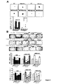
様々ながん細胞株及び原発性細胞において、c−Met活性化及びシグナル伝達は、CD44v6抗体及びペプチドによってブロックすることができる。CD44アイソフォームがECにおいても同様にc−Metに関する共受容体として作用し得るかどうかを試験するために、エクソン特異的RT−PCR分析によりHUVEC(ヒト臍帯静脈EC)におけるCD44変異体の発現プロフィールをまず検査した(Konig他、1996年、トランス作動性因子はCD44スプライス変異体の発現を調節する(Trans-acting factors regulate the expression of CD44 splice variant)」(EMBO J. 15:4030-4039))(図1A)。幾つかの変異体アイソフォームは実際に発現されている。興味深いことに、エクソンv6は、エクソンv7〜エクソンv10とともに(ラダーによって示されている)、またおそらく同様に単独でも(v6レーン中の低バンドにより示されている)発現されるようである。v6は、非依存的アイソフォームとして出現するエクソン3は、同時発現されないようであることに留意されたい。c−Metは、HUVEC中で活性化させることができ、実際に、ErkのHGF誘導性の活性化は、CD44v6抗体及びペプチドにより完全に抑止することができる(図1B)。
In various cancer cell lines and primary cells, c-Met activation and signaling can be blocked by CD44v6 antibodies and peptides. To test whether the CD44 isoform can act as a co-receptor for c-Met in EC as well, the expression profile of the CD44 variant in HUVEC (human umbilical vein EC) by exon-specific RT-PCR analysis (Konig et al., 1996, Trans-acting factors regulate the expression of CD44 splice variant) (EMBO J. 15: 4030-4039)) (FIG. 1A). Some mutant isoforms are actually expressed. Interestingly, exon v6 appears to be expressed with exon v7 to exon v10 (indicated by the ladder) and possibly alone (indicated by the low band in the v6 lane). Note that v6 does not appear to be
この結果は、ECでは、上皮細胞と同様に、CD44v6アイソフォームは、c−Metに関する共受容体として作用することを示唆している。血管新生に関与する最も顕著なRTKであるVEGFR−2の活性化に対するCD44v6抗体及びペプチドの効果もまた試験した。VEGFR−2は、VEGFファミリーの優勢なアイソフォームであるVEGF−A165で活性化された。最も興味深いことに、エクソンv6に対指向性がある抗体及びv6特異的ペプチドは、HUVECにおいてVEGFR−2の活性化及び下流のErKの活性化を抑止した(図1B及び図1C)。対照ペプチドは効果がなかった。 This result suggests that in EC, the CD44v6 isoform acts as a co-receptor for c-Met, similar to epithelial cells. The effect of CD44v6 antibody and peptide on the activation of VEGFR-2, the most prominent RTK involved in angiogenesis, was also tested. VEGFR-2 was activated with VEGF-A165, the predominant isoform of the VEGF family. Most interestingly, antibodies directed against exon v6 and v6-specific peptides abrogated VEGFR-2 activation and downstream ErK activation in HUVEC (FIGS. 1B and 1C). The control peptide had no effect.
ECにおけるCD44v6に対するVEGFR−2及びc−Metの依存性を更に確認するために、それらの活性化に対する可溶性CD44v6外部ドメイン(CD44v6ECD)の効果を試験した。CD44v6ECDは、HGF及びVEGF−A165によって誘導されるErkの活性化を完全に抑止した(図1D)。対照的に、c−Metに関するその共受容体機能に役立つ3つのアミノ酸において突然変異させたCD44v6ECD(Matzke他、「5−アミノ酸ペプチドは、Met及びRon依存的細胞遊走を阻止する(A 5-amino-acid peptide blocks Met and Ron dependent cell migration」(Cancer research 65:6105-6110))は、いかなる効果もなかった(図1D)。エクソンv3を含有するCD44タンパク質は全て、ヘパラン硫酸(HS)により修飾することができる。この修飾は、FGF又はHB−EGFのような成長因子に結合するのに必要とされるようである。ヘパラン硫酸プロテオグリカンへの及びHS修飾CD44アイソフォームへのVEGF−165の結合についても記載されている。VEGFR−2の活性化に対するHS残基の要件に対処するために、HUVECを、エクソン6及びエクソン7を欠くVEGF−A121で活性化させた。エクソン7は、HSへの結合を請け負う。VEGF−A121はまた、HUVECにおいてVEGFR−2及びErkを活性化することができ(図1D及び図2A)、この活性化も同様に、CD44v6ECD(図1D)、CD44v6抗体及びペプチド(図2A)によって完全に阻止された。
To further confirm the dependence of VEGFR-2 and c-Met on CD44v6 in EC, the effect of soluble CD44v6 ectodomain (CD44v6ECD) on their activation was tested. CD44v6ECD completely abrogated Erk activation induced by HGF and VEGF-A165 (FIG. 1D). In contrast, CD44v6ECD mutated in three amino acids that serve its co-receptor function for c-Met (Matzke et al., “5-amino acid peptides block Met and Ron-dependent cell migration (A 5-amino -acid peptide blocks (Met and Ron dependent cell migration) (Cancer research 65: 6105-6110)) did not have any effect (Figure 1D) All CD44 proteins containing exon v3 were modified with heparan sulfate (HS). This modification appears to be required to bind to growth factors such as FGF or HB-EGF Binding of VEGF-165 to heparan sulfate proteoglycans and to HS-modified CD44 isoforms In order to address the HS residue requirement for VEGFR-2 activation, HUVEC Was activated with VEGF-A121, which lacks
さらに、VEGFR−2発現ベクターでトランスフェクトしたヒト腎臓癌細胞(HEK293細胞)では、CD44v6ペプチドはまた、VEGF−A165及びVEGF−A121によって誘導されるVEGFR−2の活性化を阻止した(図2B)。これらの結果は、VEGFR−2の活性化がHS修飾とは無関係に起こることを示唆する。 Furthermore, in human kidney cancer cells (HEK293 cells) transfected with the VEGFR-2 expression vector, the CD44v6 peptide also blocked VEGFR-2 activation induced by VEGF-A165 and VEGF-A121 (FIG. 2B). . These results suggest that VEGFR-2 activation occurs independently of HS modification.
さらに、VEGFR−2活性化に関与するCD44v6アイソフォームは、HSにより修飾することができる唯一の配列であるv3配列(エクソンv6及びエクソンv3はRT−PCRにより示されるように同じCD44アイソフォーム上では同時発現されない、図1)を保有しないようである。VEGFR−2の活性化が実際にCD44のHS修飾に無関係であることを確認するために、c−Metの場合と同様に、エクソンv6を単独で含有するCD44変異体アイソフォームがVEGFR−2活性化にとって十分であるかどうかを試験した。VEGFR−2発現ベクターを、CD44のみを発現するラットBSp73AS膵癌細胞、又はCD44v6で安定的にトランスフェクトしたBsp73AS細胞(ASs6)のいずれかへ一過的にトランスフェクトさせた。VEGF−A165による処理時に、ASs6細胞のみが誘導性であった(図2C)。 Furthermore, the CD44v6 isoform involved in VEGFR-2 activation is the only sequence that can be modified by HS, the v3 sequence (exon v6 and exon v3 are on the same CD44 isoform as shown by RT-PCR. It does not appear to possess Figure 1), which is not co-expressed. To confirm that VEGFR-2 activation is indeed independent of HS modification of CD44, as in c-Met, the CD44 mutant isoform containing exon v6 alone is responsible for VEGFR-2 activity. It was tested whether it was sufficient for conversion. The VEGFR-2 expression vector was transiently transfected into either rat BSp73AS pancreatic cancer cells expressing only CD44 or Bsp73AS cells stably transfected with CD44v6 (ASs6). Only ASs6 cells were inducible upon treatment with VEGF-A165 (FIG. 2C).
したがって、変異体エクソンv6のみを含有するCD44変異体アイソフォームは、VEGFR−2共受容体として作用するのに十分であり、またこの共受容体機能は、HS修飾を必要としない。VEGFR−2に関するCD44v6の共受容体機能は、HUVEC又はHEK293及びVEGFR−2でトランスフェクトしたASs6のような幾つかの細胞型で観察することができる。 Thus, a CD44 mutant isoform containing only mutant exon v6 is sufficient to act as a VEGFR-2 coreceptor, and this coreceptor function does not require HS modification. CD44v6 co-receptor function for VEGFR-2 can be observed in several cell types such as ASs6 transfected with HUVEC or HEK293 and VEGFR-2.
ヒト心臓微小血管EC(HCMEC)及びヒト大動脈EC(HAOEC)は原発性ECであり、ここで、CD44v6と、c−Met又はVEGFR−2との間のこの協働を同様に実証することができる(図3A及び図3B)。実際に、両方の細胞型において、ErkのVEGF−A165活性化は、CD44v6ペプチド又はCD44v6抗体による細胞の処理時に阻害される(図3)。対照ペプチド及び対照IgGは効果がなかった。CD44とVEGFR−2との間の協働は、これらのタンパク質が近接していることを示唆している。HUVECからの内因性CD44v6及びVEGFR−2の共沈降は、この仮定を確認する(図3C)。 Human cardiac microvascular EC (HCMEC) and human aortic EC (HAOEC) are primary ECs, where this cooperation between CD44v6 and c-Met or VEGFR-2 can be demonstrated as well. (FIGS. 3A and 3B). Indeed, in both cell types, Erk VEGF-A165 activation is inhibited upon treatment of cells with CD44v6 peptide or CD44v6 antibody (FIG. 3). Control peptide and control IgG had no effect. The cooperation between CD44 and VEGFR-2 suggests that these proteins are in close proximity. Co-precipitation of endogenous CD44v6 and VEGFR-2 from HUVEC confirms this assumption (FIG. 3C).
興味深いことに、これらの2つの分子間の結合は構成的であり、かつリガンドVEGFの添加に非依存的であるようである。このことは、HGF誘導性であるCD44v6/c−Met結合と対照的である。c−Met受容体の場合、CD44共受容体は、シグナル伝達を促進するために、ERMタンパク質及び細胞骨格と結合する。CD44v6、c−Met、HGF、ERMタンパク質及び細胞骨格は、そのGEF SOSによるRasの活性化を可能にするシグナロソームを形成する。 Interestingly, the binding between these two molecules appears to be constitutive and independent of the addition of ligand VEGF. This is in contrast to CD44v6 / c-Met binding, which is HGF-induced. In the case of the c-Met receptor, the CD44 co-receptor binds to the ERM protein and the cytoskeleton to facilitate signal transduction. CD44v6, c-Met, HGF, ERM protein and cytoskeleton form a signalosome that allows activation of Ras by its GEF SOS.
このメカニズムがVEGFR−2の場合に関与するかどうかを試験するために、HEK293細胞をVEGFR−2及びCD44細胞質ドメイン発現ベクターで同時トランスフェクトして、このドメインが内因性CD44の活性と競合するかどうかを観察した。これは実際にその通りであった。CD44細胞質ドメインVEGFR−2の存在下で、Erkへのシグナル伝達は、VEGFR−2リン酸化自体に影響を及ぼさずに阻止された(図4A)。ERM結合配列において突然変異されたCD44細胞質ドメインの発現は、Erk及びVEGFR−2活性化に対して効果がなかった(図4A)。これらの実験から、VEGFR−2の活性化はCD44の細胞質ドメインに非依存的であるのに対して、シグナル伝達は、このドメイン及びERMタンパク質の結合を必要とすると結論付けることができる。 To test whether this mechanism is involved in the case of VEGFR-2, is HEK293 cells co-transfected with VEGFR-2 and a CD44 cytoplasmic domain expression vector, and does this domain compete with the activity of endogenous CD44? I observed how. This was indeed the case. In the presence of the CD44 cytoplasmic domain VEGFR-2, signaling to Erk was blocked without affecting VEGFR-2 phosphorylation itself (FIG. 4A). Expression of the CD44 cytoplasmic domain mutated in the ERM binding sequence had no effect on Erk and VEGFR-2 activation (FIG. 4A). From these experiments it can be concluded that activation of VEGFR-2 is independent of the cytoplasmic domain of CD44, whereas signal transduction requires binding of this domain and the ERM protein.
ERMタンパク質の関与に直接対処するために、HEK293細胞を、アクチン結合ドメインを欠いているエズリンドミナントネガティブ(dn)構築物と一緒に、VEGFR−2でトランスフェクトした(図4B)。エズリンのこの切断型もまた、ErkへのVEGF−A165及びVEGF−A121シグナル伝達を阻害して、同様にこのことも細胞骨格へのERMタンパク質の結合が、VEGFR−2からのシグナル伝達に必要とされることを示した。 To directly address the involvement of ERM protein, HEK293 cells were transfected with VEGFR-2 along with an ezrin dominant negative (dn) construct lacking the actin binding domain (FIG. 4B). This truncated form of ezrin also inhibits VEGF-A165 and VEGF-A121 signaling to Erk, which also requires that ERM protein binding to the cytoskeleton is required for signaling from VEGFR-2. Showed that.
結論として、CD44v6は、ECにおけるVEGFR−2に関する共受容体である。CD44v6抗体、ペプチド及びCD44v6ECDは、VEGFR−2活性化を阻止する。2つの分子は、共免疫沈降実験により実証されるように、構成的な複合体を形成する。さらに、CD44v6の細胞質ドメインは、ERMタンパク質及び細胞骨格を補充して、シグナル伝達を促進する。
実施例2:CD44v6ペプチド及び抗体は、VEGFに対するECの応答を阻止する。
In conclusion, CD44v6 is a co-receptor for VEGFR-2 in EC. CD44v6 antibodies, peptides and CD44v6 ECD block VEGFR-2 activation. The two molecules form a constitutive complex, as demonstrated by coimmunoprecipitation experiments. In addition, the cytoplasmic domain of CD44v6 recruits ERM proteins and the cytoskeleton to promote signaling.
Example 2: CD44v6 peptides and antibodies block the EC response to VEGF.
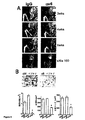
血管新生プロセスは幾つかの工程を含み、最終的に新たな毛細血管の形成をもたらす。まず、血管周囲の基底層が局所的に破壊され、その結果、ECが間質を浸潤することができる。ECは増殖して、遊走して、最終的に互いに接着して、新たな細管を形成する。これらの工程の幾つかをin vitroで模倣することができる。スクラッチアッセイでは、ECの遊走を測定することができる。HGF及びVEGF−Aは、HAOECの遊走を誘導して、集密的な単層においてスクラッチの閉塞を導いた(図5)。v6ペプチド又は抗体の存在下で、このプロセスは強く阻害された。3H−チミジン取込みアッセイを使用した増殖の測定により、細胞が観察の期間に増殖していないことが明らかとなった(データは示さず)。HUVECを使用して同じアッセイを実施して、同様の結果を導いた(図5)。 The angiogenesis process involves several steps, ultimately leading to the formation of new capillaries. First, the basal layer around the blood vessel is locally destroyed, so that EC can infiltrate the stroma. ECs proliferate, migrate and eventually adhere to each other to form new tubules. Some of these steps can be mimicked in vitro. In the scratch assay, EC migration can be measured. HGF and VEGF-A induced HAOEC migration leading to scratch occlusion in a confluent monolayer (FIG. 5). In the presence of v6 peptide or antibody, this process was strongly inhibited. Measurement of proliferation using the 3H-thymidine incorporation assay revealed that the cells were not growing during the period of observation (data not shown). The same assay was performed using HUVEC with similar results (FIG. 5).
ECは、メチルセルロース中で成長させる場合にスフェロイドを形成する特性を有する。VEGFA165及びVEGF−A121の存在下でコラーゲンへ移入されるHUVECからのスフェロイドは発芽した(図6A)。発芽は、CD44v6ペプチドによって強く阻害されたが、対照ペプチドによっては阻害されなかった(図6A、グラフ中での定量化)。最終的に、細管ネットワーク形成アッセイを使用して、新たな血管の確立におけるCD44の役割を試験した。HUVECを成長因子低減マトリゲル上で成長させて、v6ペプチド及び抗体の存在下又は非存在下でHGF、VEGF−A165又はVEGF−A121で処理した。 EC has the property of forming spheroids when grown in methylcellulose. Spheroids from HUVEC transferred to collagen in the presence of VEGFA165 and VEGF-A121 germinated (FIG. 6A). Germination was strongly inhibited by the CD44v6 peptide but not by the control peptide (FIG. 6A, quantification in the graph). Finally, a tubular network formation assay was used to test the role of CD44 in establishing new blood vessels. HUVECs were grown on growth factor-reduced matrigel and treated with HGF, VEGF-A165 or VEGF-A121 in the presence or absence of v6 peptide and antibody.
成長因子の非存在下では、予備ネットワークは、おそらくマトリゲル中の微量の成長因子に起因して観察することができる(図6B)。種々の成長因子による誘導は、ネットワークの密度を増加させた。CD44v6ペプチド又は抗体による処理は、ネットワークの形成を妨害する一方で、対照ペプチドは効果がなかった。このアッセイの定量化により、分岐点の数がHGFに関して50%にまで、またVEGF−A165及びVEGF−A121に関しては30%にまで劇的に低減することが明らかとなった(図6B)。血管の長さもまた、HGFに関して60%にまで、またVEGF−A165及びVEGF−A121に関してはおよそ45%にまで減少した。 In the absence of growth factors, a reserve network can be observed, probably due to trace amounts of growth factors in Matrigel (FIG. 6B). Induction with various growth factors increased the density of the network. Treatment with CD44v6 peptide or antibody prevented the formation of the network, while the control peptide had no effect. Quantification of this assay revealed that the number of branch points was dramatically reduced to 50% for HGF and to 30% for VEGF-A165 and VEGF-A121 (FIG. 6B). Vessel length was also reduced to 60% for HGF and to approximately 45% for VEGF-A165 and VEGF-A121.
結論として、スクラッチ閉塞、スフェロイド発芽及び細管ネットワーク形成のような各種アッセイは、HGF、VEGF−A165及びVEGF−A121に対するECの応答にはCD44v6が必要であることを実証している。
実施例3:Rip1Taq2マウスから単離した過形成性ランゲルハンス島により誘導される血管新生応答は、CD44v6に依存的である。
In conclusion, various assays such as scratch occlusion, spheroid germination and tubule network formation demonstrate that CD44v6 is required for EC responses to HGF, VEGF-A165 and VEGF-A121.
Example 3: The angiogenic response induced by hyperplastic islets of Langerhans isolated from Rip1Taq2 mice is dependent on CD44v6.
これまでに、幾つかの成長因子を別個に試験して、in vitroアッセイを使用してそれらの効果について観察してきた。ex−vivo実験も実施し、ここでは過形成性ランゲルハンス島をRip1Tag2マウスから単離し、HUVECとともにインキュベートさせた(図7A)。Rip1Taq2マウスモデルは、インスリノーマの十分規定されたモデルである。これらのトランスジェニック動物は、ラットインスリンプロモーター制御下でSV40ラージT抗原を発現し、膵臓のランゲルハンス島において単独でT抗原の発現をもたらす。多段階の腫瘍発達中に、過形成性島は、様々な血管新生因子の活性化によって腫瘍血管新生を徐々に誘導する。 So far, several growth factors have been tested separately and observed for their effects using in vitro assays. An ex-vivo experiment was also performed, in which hyperplastic islets of Langerhans were isolated from Rip1Tag2 mice and incubated with HUVEC (FIG. 7A). The Rip1Taq2 mouse model is a well-defined model of insulinoma. These transgenic animals express the SV40 large T antigen under the control of the rat insulin promoter, resulting in T antigen expression alone in the pancreatic islets of Langerhans. During multi-stage tumor development, hyperplastic islands gradually induce tumor angiogenesis through the activation of various angiogenic factors.
ランゲルハンス島は、大部分が血管新生性である段階で単離され(8週〜9週齢のマウス)、CD44v6抗体又はペプチドの存在下で、それらをHUVECとともにインキュベートした。ランゲルハンス島からの血管新生因子の放出は、それらに対するHUVECの成長を促進する。CD44v6抗体又はペプチドの存在下では、ランゲルハンス島の血管新生活性は阻止された(図7A)。したがって、幾つかの成長因子が血管新生応答を媒介する場合でさえ、CD44v6の共受容体機能の阻止はこの応答を低減した。
実施例4:移植したECスフェロイドから血管のin vivoでの発達は、CD44v6ブロッキング試薬による処理時に損なわれる。
Islets of Langerhans were isolated at a largely angiogenic stage (8-9 week old mice) and they were incubated with HUVEC in the presence of CD44v6 antibody or peptide. Release of angiogenic factors from the islets of Langerhans promotes the growth of HUVECs against them. In the presence of CD44v6 antibody or peptide, the angiogenic activity of the islets of Langerhans was blocked (FIG. 7A). Therefore, blocking CD44v6 co-receptor function reduced this response even when several growth factors mediated the angiogenic response.
Example 4: In vivo vascular development from transplanted EC spheroids is impaired upon treatment with CD44v6 blocking reagent.
ヒト脈管構造は、SCIDマウスの脇腹への移植時にマトリゲル/フィブリン中に成長因子と一緒に包埋されたヒトスフェロイドから操作することができる。このアッセイを使用して、CD44v6がこの脈管構造の発達に関与するかどうかを試験した。CD44v6ペプチド又は抗体(又は対照ペプチド若しくはIgG)と一緒にHGF又はVEGF−A165のいずれかを含有するマトリゲル/フィブリン中に包埋されたHUVECスフェロイドを、SCIDマウスの脇腹へ皮下注入した。ヒトCD34抗体によるマトリゲル/フィブリンプラグの染色により、ヒト血管ネットワークの形成を3週後に求めた。HGF及びVEGFはともに、いずれの成長因子の非存在下では検出不可能であった(データは示さず)血管形成を誘導した(図7B)。CD44v6ペプチド及びv6特異的抗体は、50%を上回って血管新生応答を有意に低減させた(図7B)。したがって、同様にこのアッセイでは、血管新生応答は、CD44v6に依存的である。さらに、これらの結果により、HGFが血管新生因子として作用することができることが確認される。
実施例5:乳腺腫瘍及び膵臓腫瘍の血管新生化はCD44v6を必要とする。
Human vasculature can be manipulated from human spheroids embedded with growth factors in Matrigel / fibrin at the time of implantation into the flank of SCID mice. This assay was used to test whether CD44v6 is involved in the development of this vasculature. HUVEC spheroids embedded in Matrigel / fibrin containing either HGF or VEGF-A165 together with CD44v6 peptide or antibody (or control peptide or IgG) were injected subcutaneously into the flank of SCID mice. Human vascular network formation was determined after 3 weeks by staining of Matrigel / fibrin plugs with human CD34 antibody. Both HGF and VEGF induced angiogenesis that was undetectable in the absence of any growth factor (data not shown) (FIG. 7B). CD44v6 peptide and v6 specific antibody significantly reduced the angiogenic response by more than 50% (FIG. 7B). Thus, similarly in this assay, the angiogenic response is dependent on CD44v6. Furthermore, these results confirm that HGF can act as an angiogenic factor.
Example 5: Angiogenesis of breast and pancreatic tumors requires CD44v6.
腫瘍誘導性の血管新生がCD44v6ペプチド及び抗体により抑圧することができるかどうかを証明するために、ヒト乳癌(MDA−MB231)及びヒト膵癌細胞(L3.6pl)を使用した。細胞はともに、マウスにおいて同所にあるヒト腫瘍における血管新生を研究するのに既に使用されている(MDAMB231:、L3.6pl:)。MDA−MB231細胞は、SCIDマウスの乳腺脂肪体へ移植されたのに対して、L3.6pl細胞は、SCIDマウスの膵臓の尾へ注入した。1週後、CD44v6ペプチド又は抗体(又は対照ペプチド若しくは対照IgG)を、1週間につき3回をMDA−MB231腫瘍の場合には4週間、又はL3.6pl腫瘍の場合には3週間、腹腔内投与した。 To demonstrate whether tumor-induced angiogenesis can be suppressed by CD44v6 peptides and antibodies, human breast cancer (MDA-MB231) and human pancreatic cancer cells (L3.6pl) were used. Both cells have already been used to study angiogenesis in human tumors that are sympatric in mice (MDAMB231: L3.6pl :). MDA-MB231 cells were transplanted into the mammary fat pad of SCID mice, whereas L3.6pl cells were injected into the pancreatic tail of SCID mice. After 1 week, CD44v6 peptide or antibody (or control peptide or control IgG) was administered intraperitoneally 3 times per week for 4 weeks for MDA-MB231 tumors or 3 weeks for L3.6pl tumors did.
これまでは、CD44v6ヒトペプチド及びヒト抗体を使用して、ヒトECを標的としてきた。しかしながら、腫瘍モデルでは、マウス特異的抗体及びペプチドを使用した。CD44v6ペプチドは種特異的であり、交差反応しない。特に、同所性腫瘍モデルで使用されるCD44v6ペプチド及びCD44v6抗体は、腫瘍細胞(ヒト)上でのCD44機能を妨害しなかった。したがって、それらは、宿主ECの血管新生のみを標的とした。MDA−MB231腫瘍の場合、CD44v6抗体又は対照IgGのいずれかで処理した2つの動物群を、フラットパネル検出器ボリュームコンピュータ断層撮影法(fpVCT)を使用して検査して、経時的な腫瘍成長及び腫瘍血管新生化を追跡した。実験の終了時に、腫瘍を全ての動物から単離して、切片を作製して、CD31抗体で染色した。血管密度及び血管長を、染色した切片から推定した。MDA−MB231腫瘍に関して、腫瘍成長は、マウスCD44v6ペプチド又は抗体により影響を受けなかった(データは示さず)。同様に、平均血管サイズは、CD44v6ツールにより影響を受けなかった(表1)。対照的に、微小血管密度は、CD44v6ペプチドによって1mm2当たり240個から183個へ、また抗体により1mm2当たり247個から220個へ有意に減少した(表1)。同様に、腫瘍を取り囲みかつ浸潤する小血管の減少が、抗体で処理したマウスにおいてfpVCTによって観察された(図8A)。この減少は、移植の3週後には既に見ることができ、5週後の実験の終了まで続けることができた(図8A)。 So far, human ECs have been targeted using CD44v6 human peptides and human antibodies. However, mouse-specific antibodies and peptides were used in the tumor model. CD44v6 peptide is species specific and does not cross-react. In particular, the CD44v6 peptide and CD44v6 antibody used in the orthotopic tumor model did not interfere with CD44 function on tumor cells (human). They therefore targeted only angiogenesis of the host EC. In the case of MDA-MB231 tumors, two groups of animals treated with either CD44v6 antibody or control IgG were examined using flat panel detector volume computed tomography (fpVCT) to determine tumor growth over time and Tumor angiogenesis was followed. At the end of the experiment, tumors were isolated from all animals, sections were made and stained with CD31 antibody. Vessel density and vessel length were estimated from the stained sections. For MDA-MB231 tumors, tumor growth was not affected by mouse CD44v6 peptide or antibody (data not shown). Similarly, mean vessel size was not affected by the CD44v6 tool (Table 1). In contrast, the microvessel density was significantly reduced from 240 to 183 per mm 2 by CD44v6 peptide and from 247 to 220 per mm 2 by antibody (Table 1). Similarly, a reduction in small blood vessels surrounding and infiltrating the tumor was observed by fpVCT in mice treated with antibody (FIG. 8A). This decrease was already visible after 3 weeks of transplantation and could continue until the end of the experiment after 5 weeks (FIG. 8A).
対照的に、CD44v6ペプチドの劇的な阻害効果は、L3.6pl細胞から確立されたヒト膵臓腫瘍の成長に関して既に観察されていた:腫瘍サイズが10パーセントへ低減された(図8B)。さらに、同様に血管密度及び血管サイズは、CD44v6ペプチドによる処理時に60%を上回って減少した(図8B)。これらのデータにより、この膵臓腫瘍の成長は、血管脈管構造の確立に特に依存的であることが示唆される。結論として、CD44v6ペプチド及び抗体は、幾つかのEC上でVEGFR−2の活性化及びシグナル伝達を阻止するだけでなく、それにより様々な試験系において細管伸長及びEC遊走を妨害し、またそれらはin vivoでの腫瘍誘導性の血管形成を阻止することが示される。
表1:CD44v6ペプチド及び抗体による腫瘍誘導性の血管の阻害
SCIDマウスへのMDA−MB231細胞の注入、及び続く処理は、図7Aに記載するように実施した。腫瘍切片をCD31特異的抗体で染色して、血管数及びサイズを計数した。数は、5匹の動物それぞれのカウントを反映する。
In contrast, a dramatic inhibitory effect of the CD44v6 peptide was already observed for the growth of human pancreatic tumors established from L3.6pl cells: the tumor size was reduced to 10 percent (FIG. 8B). Furthermore, similarly, vessel density and vessel size decreased by more than 60% upon treatment with CD44v6 peptide (FIG. 8B). These data suggest that the growth of this pancreatic tumor is particularly dependent on the establishment of vascular vasculature. In conclusion, CD44v6 peptides and antibodies not only block VEGFR-2 activation and signaling on some ECs, thereby preventing tubule elongation and EC migration in various test systems, and It has been shown to block tumor-induced angiogenesis in vivo.
Table 1: Tumor-induced vascular inhibition by CD44v6 peptides and antibodies Injection of MDA-MB231 cells into SCID mice and subsequent treatments were performed as described in FIG. 7A. Tumor sections were stained with CD31 specific antibodies and the number and size of blood vessels were counted. The number reflects the count of each of the 5 animals.
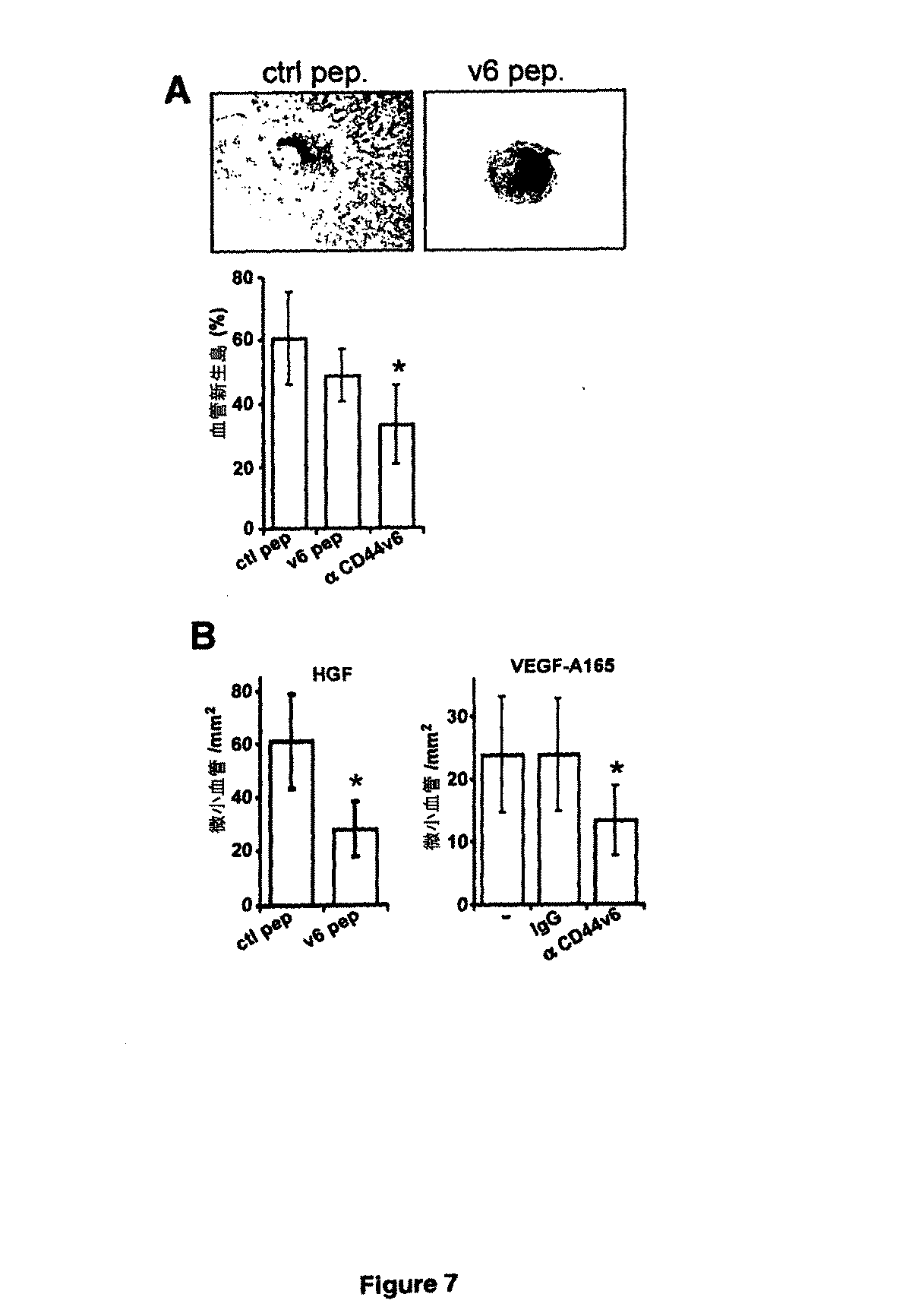
上記実験では、ECにおけるc−Met及びVEGFR−2のそれらの各々のリガンドによる活性化は、変異体エクソンv6を含有するCD44アイソフォームに厳密に依存していることが示されている。HUVECでは、CD44v6特異的抗体、v6ペプチド及びCD44v6ECDは、c−Met及びVEGFR−2の両方の活性化、並びに続くEC遊走、スフェロイド発芽及び細管形成の誘導を抑止する。共免疫沈降研究により、VEGFR−2及びCD44v6は複合体を形成し、互いに構成的に結合されるようであることが示される。CD44v6に対するVEGFR−2の依存性はHUVECだけでなく、HAOEC及びHCMECにも当てはまり、VEGFR−2を発現するようにトランスフェクトされた他の細胞(HEK293細胞及びASs6細胞)でシミュレートすることができる。 The above experiments show that the activation of c-Met and VEGFR-2 by their respective ligands in EC is strictly dependent on the CD44 isoform containing mutant exon v6. In HUVEC, CD44v6-specific antibodies, v6 peptides and CD44v6ECD inhibit both c-Met and VEGFR-2 activation and subsequent induction of EC migration, spheroid germination and tubule formation. Co-immunoprecipitation studies show that VEGFR-2 and CD44v6 form a complex and appear to be constitutively bound to each other. The dependence of VEGFR-2 on CD44v6 applies not only to HUVEC, but also to HAOEC and HCME, and can be simulated in other cells transfected to express VEGFR-2 (HEK293 cells and ASs6 cells). .
HGF及びc−Metはまた、血管新生でも重要であるため、それらがEC細胞においても同様にCD44v6アイソフォームと協働するかどうかを検査した。CD44v6特異的ペプチド及びCD44v6抗体は、EC上でc−Met活性を実際に阻止することができ、ECによるHGF誘導性遊走及び細管形成が損なわれる。最も興味深いことに、このペプチド及び抗体は、VEGF誘導性VEGFR−2活性化も阻止した。さらに、CD44と結合するERMタンパク質は、VEGFR−2からのシグナル伝達を調節する。したがって、CD44の作用メカニズムは、c−Met及びVEGFR−2活性化の両方に関して非常に類似しているようである。実際に、CD44v6は、in vitroで及び更には腫瘍において、血管の形成を含む生理学的変化を制御する。 Since HGF and c-Met are also important in angiogenesis, we examined whether they cooperate with CD44v6 isoforms in EC cells as well. CD44v6-specific peptides and CD44v6 antibodies can actually block c-Met activity on EC, and HGF-induced migration and tubule formation by EC is impaired. Most interestingly, this peptide and antibody also blocked VEGF-induced VEGFR-2 activation. In addition, ERM proteins that bind to CD44 regulate signaling from VEGFR-2. Thus, the mechanism of action of CD44 appears to be very similar with respect to both c-Met and VEGFR-2 activation. Indeed, CD44v6 regulates physiological changes including blood vessel formation in vitro and even in tumors.
VEGFR−2の活性化が、へパリン非結合性VEGF−A121によっても誘導され得ること、及びこの活性化はまたCD44エクソンv6に依存することが上記で実証されている。さらに、ECはCD44エクソンv3及びエクソンv6を発現するが、これらのエクソンは、RT−PCR分析から推測されるのと同じタンパク質上に存在しないようである。最終的に、VEGFR−2の活性化は、CD44v6及びCD44sだけでなく、CD44v3ヘパリン硫酸化型も発現するBSp73ASs6細胞において観察された。これらの結果により、VEGFR−2の活性化自体は、HSを依らない可能性があることが示される。しかしながら、HSへの結合は、本格的な血管新生応答には必要とされるようである。VEGFR−2、VEGF、ニューロピリン及びCD44v6間の協働は、本格的な血管新生応答に関与する可能性が高い。 It has been demonstrated above that VEGFR-2 activation can also be induced by non-heparin-binding VEGF-A121 and that this activation is also dependent on CD44 exon v6. Furthermore, EC expresses CD44 exon v3 and exon v6, but these exons do not appear to be on the same protein as deduced from RT-PCR analysis. Finally, activation of VEGFR-2 was observed in BSp73ASs6 cells expressing not only CD44v6 and CD44s but also CD44v3 heparin sulfated form. These results indicate that VEGFR-2 activation itself may not depend on HS. However, binding to HS appears to be required for a full-scale angiogenic response. Cooperation between VEGFR-2, VEGF, neuropilin and CD44v6 is likely to be involved in a full-scale angiogenic response.
VEGFR−2に関するCD44v6の共受容体機能は、2つの異なる同所性腫瘍モデル上でのCD44v6ペプチド及びCD44v6抗体の効果により示されるように、腫瘍血管新生において非常に関係している。ヒト乳癌MBA−MD231細胞に由来する腫瘍におけるCD44v6の阻止は、微小血管密度に対して有意な効果があるのに対して、平均血管サイズ及び腫瘍成長速度は影響を受けない。L3.6pl細胞に由来するヒト膵臓腫瘍では、腫瘍サイズ並びに微小血管密度及び平均血管サイズは、CD44v6ペプチドによる処理後に劇的に低減する。これらの2つの腫瘍系の異なる応答は、がん細胞の固有の特性に起因し得る。可能性の1つは、両方の場合で産生される血管新生因子が異なるということである。CD44v6は、c−Met、VEGFR−2、Trk及びRTKのEGFRファミリーの特定成員に関する共受容体としてだけでなく、PDGFR又はFGFRに関する共受容体としても作用することができるため、ブロッキング試薬の効果は、どの血管新生成長因子が産生されるかに従って異なる。CD44アイソフォームが血管新生に関係するという本明細書での見解は、腫瘍形成におけるCD44の役割に新たな側面を付け加えている。 CD44v6 co-receptor function for VEGFR-2 is highly implicated in tumor angiogenesis, as shown by the effects of CD44v6 peptide and CD44v6 antibody on two different orthotopic tumor models. Inhibition of CD44v6 in tumors derived from human breast cancer MBA-MD231 cells has a significant effect on microvessel density, while mean vessel size and tumor growth rate are not affected. In human pancreatic tumors derived from L3.6pl cells, tumor size and microvessel density and mean vessel size are dramatically reduced after treatment with CD44v6 peptide. The different responses of these two tumor lines can be attributed to the inherent properties of cancer cells. One possibility is that the angiogenic factors produced in both cases are different. Since CD44v6 can act not only as a co-receptor for specific members of the EGFR family of c-Met, VEGFR-2, Trk and RTK, but also as a co-receptor for PDGFR or FGFR, the effect of blocking reagents is , Depending on which angiogenic growth factor is produced. The view here that CD44 isoforms are involved in angiogenesis adds a new dimension to the role of CD44 in tumorigenesis.
配列番号4:プライマー
配列番号5:プライマー
SEQ ID NO: 4: Primer SEQ ID NO: 5: Primer
Claims (10)
Applications Claiming Priority (3)
| Application Number | Priority Date | Filing Date | Title |
|---|---|---|---|
| EP09008272.8 | 2009-06-24 | ||
| EP09008272A EP2266593A1 (en) | 2009-06-24 | 2009-06-24 | Use of CD44v6 in the treatment of ophthalmic diseases |
| PCT/EP2010/003523 WO2010149281A1 (en) | 2009-06-24 | 2010-06-11 | Use of cd44v6 in the treatment of ophthalmic diseases |
Publications (1)
| Publication Number | Publication Date |
|---|---|
| JP2012530735A true JP2012530735A (en) | 2012-12-06 |
Family
ID=41338473
Family Applications (1)
| Application Number | Title | Priority Date | Filing Date |
|---|---|---|---|
| JP2012516550A Pending JP2012530735A (en) | 2009-06-24 | 2010-06-11 | Use of CD44v6 in the treatment of eye diseases |
Country Status (4)
| Country | Link |
|---|---|
| US (2) | US20120115794A1 (en) |
| EP (1) | EP2266593A1 (en) |
| JP (1) | JP2012530735A (en) |
| WO (1) | WO2010149281A1 (en) |
Families Citing this family (8)
| Publication number | Priority date | Publication date | Assignee | Title |
|---|---|---|---|---|
| JP5960793B2 (en) * | 2011-03-24 | 2016-08-02 | ニューリム ファーマシューティカルズ(1991)リミテッド | Neuroprotective peptide |
| GB201220899D0 (en) * | 2012-11-21 | 2013-01-02 | Kit Karlsrusher Inst Fuer Technologie Und Inst Fuer Toxikologie Und Genetik And Amcure Gmbh | CD44v6-derived pegylated peptides |
| GB201220889D0 (en) | 2012-11-21 | 2013-01-02 | Kit Karlsrusher Inst Fuer Technologie Und Inst Fuer Toxikologie Und Genetik And Amcure Gmbh | CD44v6-derived peptides for treating metastasizing cancers |
| GB201220901D0 (en) | 2012-11-21 | 2013-01-02 | Kit Karlsrusher Inst Fuer Technologie Und Inst Fuer Toxikologie Und Genetik And Amcure Gmbh | CD44v6-derived peptides for treating pancreatic cancer |
| GB201220891D0 (en) | 2012-11-21 | 2013-01-02 | Kit Karlsrusher Inst Fuer Technologie Und Inst Fuer Toxikologie Und Genetik And Amcure Gmbh | CD44v6-derived peptides for treating breast cancers |
| GB201421647D0 (en) | 2014-12-05 | 2015-01-21 | Amcure Gmbh And Ruprecht-Karls-Universitat And Karlsruher Institut F�R Technologie | CD44v6-derived cyclic peptides for treating cancers and angiogenesis related diseases |
| US10604586B2 (en) | 2016-05-09 | 2020-03-31 | Industrial Technology Research Institute | Humanized monoclonal antibody and uses thereof |
| CN115315446A (en) * | 2020-03-06 | 2022-11-08 | Go医疗股份有限公司 | anti-sugar-CD 44 antibodies and uses thereof |
Citations (1)
| Publication number | Priority date | Publication date | Assignee | Title |
|---|---|---|---|---|
| EP1647556A1 (en) * | 2004-10-14 | 2006-04-19 | TopoTarget Germany AG | Peptidic compounds and derivatives thereof for the treatment of human diseases through inhibition of signaling via growth factors |
Family Cites Families (2)
| Publication number | Priority date | Publication date | Assignee | Title |
|---|---|---|---|---|
| UA58482C2 (en) * | 1994-06-08 | 2003-08-15 | Бьорінгер Інгельхайм Інтернаціональ Гмбх | MONOCLONAL ANTIBODY VFF-18 AGAINST CD44v6 AND ITS FRAGMENTS |
| WO2009023411A1 (en) * | 2007-08-09 | 2009-02-19 | Bausch & Lomb Incorporated | Compositions and methods for treating or controlling anterior- and posterior-segment ophthalmic diseases |
-
2009
- 2009-06-24 EP EP09008272A patent/EP2266593A1/en not_active Withdrawn
-
2010
- 2010-06-11 JP JP2012516550A patent/JP2012530735A/en active Pending
- 2010-06-11 WO PCT/EP2010/003523 patent/WO2010149281A1/en active Application Filing
- 2010-06-11 US US13/378,574 patent/US20120115794A1/en not_active Abandoned
-
2012
- 2012-12-13 US US13/713,594 patent/US8883738B2/en not_active Expired - Fee Related
Patent Citations (1)
| Publication number | Priority date | Publication date | Assignee | Title |
|---|---|---|---|---|
| EP1647556A1 (en) * | 2004-10-14 | 2006-04-19 | TopoTarget Germany AG | Peptidic compounds and derivatives thereof for the treatment of human diseases through inhibition of signaling via growth factors |
Also Published As
| Publication number | Publication date |
|---|---|
| US20120115794A1 (en) | 2012-05-10 |
| US20130109632A1 (en) | 2013-05-02 |
| EP2266593A1 (en) | 2010-12-29 |
| WO2010149281A1 (en) | 2010-12-29 |
| US8883738B2 (en) | 2014-11-11 |
Similar Documents
| Publication | Publication Date | Title |
|---|---|---|
| US8883738B2 (en) | Use of a peptide fragment of CD44v6 in the treatment of ophthalmic diseases | |
| Ferrara | Vascular endothelial growth factor: basic science and clinical progress | |
| Ferrara | Role of vascular endothelial growth factor in regulation of physiological angiogenesis | |
| PT1140969E (en) | Connective tissue growth factor fragments and methods and uses thereof | |
| CN107135646A (en) | Method and composition for treating ulcer | |
| ES2525669T3 (en) | Vascularization inhibitors | |
| JP2006527198A (en) | Tumor regression method using VEGF inhibitor | |
| CA2592292A1 (en) | Sustained delivery of pdgf using self-assembling peptide nanofibers | |
| JPH11510479A (en) | Prevention and treatment of cardiovascular disease with tamoxifen analogs | |
| WO2017174758A1 (en) | Combination of an apelin antagonist and an angiogenesis inhibitor for the treatment of cancer | |
| WO2005087954A2 (en) | Roles of foxc2, pdgfr, vegfr3 in lymphatic vessel development | |
| RU2486200C2 (en) | Method of inhibiting angiogenesis by egfl8 antagonists | |
| US8420780B2 (en) | Mutated netrin 4, fragments thereof and uses thereof as drugs | |
| KR20080085201A (en) | Methods of treating or preventing tissue damage caused by increased blood flow | |
| US8506965B2 (en) | R-RAS activity in vascular regulation | |
| US9718878B2 (en) | Kit for detecting Cthrc1 in a sample | |
| JP7232537B2 (en) | c-MET agonist antibody and use thereof | |
| EP3506919A1 (en) | Kif13b-derived peptide and method of inhibiting angiogenesis | |
| JP2003529370A (en) | Antagonist antibodies to VE-cadherin without adverse effects on vascular permeability | |
| EP3096780A1 (en) | Abl1 inhibitor for treating and preventing ocular neovascularisation | |
| KR102384795B1 (en) | Pharmaceutical compositions and methods for the treatment and prevention of metastatic cancer | |
| JPWO2002100441A1 (en) | Revascularization therapy | |
| Brkić | Molecular regulation of intussusceptive angiogenesis by ephrinB2/EphB4 signaling and its therapeutic potential | |
| US20160376339A1 (en) | Decoy peptides inhibiting binding of amigo2 and 3-phosphoinositide-dependent kinase 1 | |
| KR20030081402A (en) | Modulation of smooth muscle cell proliferation by VEGF-X or antagonists thereof |
Legal Events
| Date | Code | Title | Description |
|---|---|---|---|
| A621 | Written request for application examination |
Free format text: JAPANESE INTERMEDIATE CODE: A621 Effective date: 20130121 |
|
| A131 | Notification of reasons for refusal |
Free format text: JAPANESE INTERMEDIATE CODE: A131 Effective date: 20140304 |
|
| A521 | Request for written amendment filed |
Free format text: JAPANESE INTERMEDIATE CODE: A523 Effective date: 20140521 |
|
| A131 | Notification of reasons for refusal |
Free format text: JAPANESE INTERMEDIATE CODE: A131 Effective date: 20141007 |
|
| A521 | Request for written amendment filed |
Free format text: JAPANESE INTERMEDIATE CODE: A523 Effective date: 20141209 |
|
| A02 | Decision of refusal |
Free format text: JAPANESE INTERMEDIATE CODE: A02 Effective date: 20150203 |