JP2012524781A - Compositions, kits and methods for promoting healing of ischemic and diabetic wounds - Google Patents
Compositions, kits and methods for promoting healing of ischemic and diabetic wounds Download PDFInfo
- Publication number
- JP2012524781A JP2012524781A JP2012507248A JP2012507248A JP2012524781A JP 2012524781 A JP2012524781 A JP 2012524781A JP 2012507248 A JP2012507248 A JP 2012507248A JP 2012507248 A JP2012507248 A JP 2012507248A JP 2012524781 A JP2012524781 A JP 2012524781A
- Authority
- JP
- Japan
- Prior art keywords
- selectin
- wound
- sdf
- protein
- subject
- Prior art date
- Legal status (The legal status is an assumption and is not a legal conclusion. Google has not performed a legal analysis and makes no representation as to the accuracy of the status listed.)
- Pending
Links
Images


Classifications
-
- A—HUMAN NECESSITIES
- A61—MEDICAL OR VETERINARY SCIENCE; HYGIENE
- A61K—PREPARATIONS FOR MEDICAL, DENTAL OR TOILETRY PURPOSES
- A61K38/00—Medicinal preparations containing peptides
- A61K38/16—Peptides having more than 20 amino acids; Gastrins; Somatostatins; Melanotropins; Derivatives thereof
- A61K38/17—Peptides having more than 20 amino acids; Gastrins; Somatostatins; Melanotropins; Derivatives thereof from animals; from humans
- A61K38/19—Cytokines; Lymphokines; Interferons
- A61K38/195—Chemokines, e.g. RANTES
-
- A—HUMAN NECESSITIES
- A61—MEDICAL OR VETERINARY SCIENCE; HYGIENE
- A61K—PREPARATIONS FOR MEDICAL, DENTAL OR TOILETRY PURPOSES
- A61K48/00—Medicinal preparations containing genetic material which is inserted into cells of the living body to treat genetic diseases; Gene therapy
-
- A—HUMAN NECESSITIES
- A61—MEDICAL OR VETERINARY SCIENCE; HYGIENE
- A61K—PREPARATIONS FOR MEDICAL, DENTAL OR TOILETRY PURPOSES
- A61K9/00—Medicinal preparations characterised by special physical form
- A61K9/0012—Galenical forms characterised by the site of application
- A61K9/0019—Injectable compositions; Intramuscular, intravenous, arterial, subcutaneous administration; Compositions to be administered through the skin in an invasive manner
-
- A—HUMAN NECESSITIES
- A61—MEDICAL OR VETERINARY SCIENCE; HYGIENE
- A61K—PREPARATIONS FOR MEDICAL, DENTAL OR TOILETRY PURPOSES
- A61K9/00—Medicinal preparations characterised by special physical form
- A61K9/0012—Galenical forms characterised by the site of application
- A61K9/0053—Mouth and digestive tract, i.e. intraoral and peroral administration
-
- A—HUMAN NECESSITIES
- A61—MEDICAL OR VETERINARY SCIENCE; HYGIENE
- A61P—SPECIFIC THERAPEUTIC ACTIVITY OF CHEMICAL COMPOUNDS OR MEDICINAL PREPARATIONS
- A61P17/00—Drugs for dermatological disorders
- A61P17/02—Drugs for dermatological disorders for treating wounds, ulcers, burns, scars, keloids, or the like
-
- A—HUMAN NECESSITIES
- A61—MEDICAL OR VETERINARY SCIENCE; HYGIENE
- A61P—SPECIFIC THERAPEUTIC ACTIVITY OF CHEMICAL COMPOUNDS OR MEDICINAL PREPARATIONS
- A61P3/00—Drugs for disorders of the metabolism
- A61P3/08—Drugs for disorders of the metabolism for glucose homeostasis
- A61P3/10—Drugs for disorders of the metabolism for glucose homeostasis for hyperglycaemia, e.g. antidiabetics
-
- A—HUMAN NECESSITIES
- A61—MEDICAL OR VETERINARY SCIENCE; HYGIENE
- A61P—SPECIFIC THERAPEUTIC ACTIVITY OF CHEMICAL COMPOUNDS OR MEDICINAL PREPARATIONS
- A61P43/00—Drugs for specific purposes, not provided for in groups A61P1/00-A61P41/00
-
- C—CHEMISTRY; METALLURGY
- C07—ORGANIC CHEMISTRY
- C07K—PEPTIDES
- C07K14/00—Peptides having more than 20 amino acids; Gastrins; Somatostatins; Melanotropins; Derivatives thereof
- C07K14/435—Peptides having more than 20 amino acids; Gastrins; Somatostatins; Melanotropins; Derivatives thereof from animals; from humans
- C07K14/705—Receptors; Cell surface antigens; Cell surface determinants
- C07K14/7056—Lectin superfamily, e.g. CD23, CD72
- C07K14/70564—Selectins, e.g. CD62
-
- A—HUMAN NECESSITIES
- A61—MEDICAL OR VETERINARY SCIENCE; HYGIENE
- A61K—PREPARATIONS FOR MEDICAL, DENTAL OR TOILETRY PURPOSES
- A61K2121/00—Preparations for use in therapy
-
- A—HUMAN NECESSITIES
- A61—MEDICAL OR VETERINARY SCIENCE; HYGIENE
- A61K—PREPARATIONS FOR MEDICAL, DENTAL OR TOILETRY PURPOSES
- A61K38/00—Medicinal preparations containing peptides
Abstract
SDF−1αが成熟内皮細胞(EC)におけるE−セレクチンの発現を特異的にアップレギュレートして、EC−内皮前駆細胞(EPC)間の接着及びEPCホーミングを増加させるという発見に基づく、組成物、キット、及び糖尿病性創傷の治癒を促進する方法。糖尿病の対象における創傷治癒を促進する方法は、E−セレクチンタンパク質又はE−セレクチンタンパク質をコードする核酸、及び任意に、E−セレクチンの発現を特異的にアップレギュレートする薬剤(例えば、SDF−1α)、を含有する治療有効量の組成物を提供する工程を含む。前記方法は、対象に高圧酸素療法を実施する工程をもまた含む場合がある。組成物の対象への投与は、骨髄由来前駆細胞の創傷への遊走、創傷治癒の促進、及び対象におけるE−セレクチンの発現のアップレギュレーションをもたらす。
【選択図】図1Composition based on the discovery that SDF-1α specifically upregulates the expression of E-selectin in mature endothelial cells (EC) to increase EC-endothelial progenitor cell (EPC) adhesion and EPC homing , Kits and methods for promoting healing of diabetic wounds. Methods for promoting wound healing in diabetic subjects include E-selectin protein or nucleic acid encoding E-selectin protein, and optionally an agent that specifically upregulates expression of E-selectin (eg, SDF-1α ), Providing a therapeutically effective amount of the composition. The method may also include the step of performing hyperbaric oxygen therapy on the subject. Administration of the composition to the subject results in migration of bone marrow-derived progenitor cells to the wound, promotion of wound healing, and upregulation of E-selectin expression in the subject.
[Selection] Figure 1
Description
本発明は一般に、医療及び遺伝子治療の分野に関する。より詳細には、本発明は糖尿病の対象における創傷治癒を促進する組成物、キット及び方法に関する。 The present invention relates generally to the fields of medicine and gene therapy. More particularly, the present invention relates to compositions, kits and methods that promote wound healing in diabetic subjects.
欠陥のある創傷治癒は、糖尿病患者における重大な臨床上の問題であり、また下肢切断の主要な原因である。現在の治療の成功率は低く、糖尿病性の微小血管病理に対する取り組みは不十分である。糖尿病性創傷治癒の不良は、不十分な血管形成及び脈管形成によって特徴付けられる。脈管形成はBM由来前駆細胞からの新生血管の成長を含み、後天性の血管新生過程及び創傷治癒に寄与する。骨髄(BM)由来の内皮前駆細胞(EPC)は脈管形成において機能する重要な細胞であり、虚血に反応して末梢組織にホーミングする。EPCの動員及びリクルートメント(すなわち虚血)に対する初期の生理的刺激が、糖尿病の宿主においては、なぜ治療的なEPC介在性血管新生及び治癒を誘導しないのかは未だに解明されていない。 Defective wound healing is a significant clinical problem in diabetic patients and is a major cause of lower limb amputation. Current treatment success rates are low and efforts to diabetic microvascular pathology are inadequate. Poor diabetic wound healing is characterized by inadequate angiogenesis and vasculogenesis. Angiogenesis involves the growth of new blood vessels from BM-derived progenitor cells and contributes to the acquired angiogenic process and wound healing. Bone marrow (BM) -derived endothelial progenitor cells (EPCs) are important cells that function in angiogenesis and home to peripheral tissues in response to ischemia. It is still unclear why early physiological stimulation to EPC mobilization and recruitment (ie, ischemia) does not induce therapeutic EPC-mediated angiogenesis and healing in diabetic hosts.
そのため、現在においても、糖尿病性創傷治癒を高める治療薬及び方法が必要とされている。 Therefore, there is still a need for therapeutic agents and methods that enhance diabetic wound healing.
本明細書においては、SDF−1αが成熟ECにおけるE−セレクチンの発現を特異的にアップレギュレートし、EC−EPC間接着及びEPCホーミングの増加を引き起こすという発見に基づく、虚血性(例えば、糖尿病性)創傷の治癒を促進する組成物、キット及び方法が記載される。EPCの標的組織へのホーミング機序には、EPCの骨髄ニッチからの離脱、血管内への移行、及び循環系内での移動、ホーミングシグナルの感知、毛細血管の単層内皮細胞(EC)上での回転及び接着、そしてその後のEPC−EC間の直接的な相互作用が必要とされる経内皮遊走を含む、連続したイベントのカスケードが含まれる。本明細書に記載される実施例においては、EPCの標的組織へのホーミングを達成するためには、毛細血管内膜のECと循環しているEPCとの間の直接的な細胞間相互作用が必要かどうか、及び、EC単層における特異的な接着分子の制御により、少なくとも部分的には、EPCホーミングにおけるSDF−1αの効果が仲介されるかどうかについて調べた。SDF−1α誘導性のEPCホーミングを仲介する接着分子として、E−セレクチンを特定した。本明細書において記載した実施例の結果は、マウス及びヒトの成熟EC単層においては、SDF−1αがE−セレクチンの発現を特異的にアップレギュレートすること、E−セレクチンはEPCのEC単層への接着を高めることにより、EPCホーミング及びそれらの経内皮遊走におけるSDF−1αの効果を仲介する原因となること、並びにこれらの効果が創傷性血管新生及び創傷治癒(血管新生)の有意な増大を生じること、を示した。これら新規の知見は、EPCホーミングにおけるSDF−1αの生物学的な効果の根底にある分子メカニズム(シグナル)への洞察を提供するばかりでなく、E−セレクチンが虚血性(例えば、糖尿病性)創傷の治癒における治療的適応の新しい標的であることを明らかにした。 As used herein, ischemic (eg, diabetics) based on the discovery that SDF-1α specifically upregulates E-selectin expression in mature ECs, causing increased EC-EPC adhesion and EPC homing. Sex) Wound healing compositions, kits and methods are described. EPC homing mechanisms to target tissues include EPC withdrawal from the bone marrow niche, translocation into the vasculature, and movement within the circulatory system, homing signal sensing, on capillary monolayer endothelial cells (EC) A cascade of consecutive events is involved, including transendothelial migration that requires rotation and adhesion at the end, and subsequent direct interaction between EPC-EC. In the examples described herein, in order to achieve homing of EPC to the target tissue, a direct cell-cell interaction between the EC of the capillary intima and the circulating EPC We investigated whether it was necessary and whether the effects of SDF-1α in EPC homing were mediated, at least in part, by controlling specific adhesion molecules in the EC monolayer. E-selectin was identified as an adhesion molecule that mediates SDF-1α-induced EPC homing. The results of the examples described herein show that in mouse and human mature EC monolayers, SDF-1α specifically upregulates E-selectin expression, and E-selectin is an EPC EC monolayer. Increasing adhesion to the layer is responsible for mediating the effects of SDF-1α in EPC homing and their transendothelial migration, and these effects are significant in wound angiogenesis and wound healing (angiogenesis) It was shown to cause an increase. These new findings not only provide insight into the molecular mechanisms (signals) that underlie the biological effects of SDF-1α in EPC homing, but E-selectin is also an ischemic (eg, diabetic) wound It has been revealed that it is a new target for therapeutic indications in the healing of children.
別段の指定のない限り、本明細書で使用される全ての専門用語は、本発明が属する分野の当業者により通常理解されるものと同じ意味を有する。 Unless otherwise specified, all terminology used herein has the same meaning as commonly understood by one of ordinary skill in the art to which this invention belongs.
本明細書で使用する場合、「核酸」又は「核酸分子」は、RNA(リボ核酸)及びDNA(デオキシリボ核酸)のような2つ以上のヌクレオチドの鎖、及び化学的に修飾したヌクレオチドを意味する。「精製した」核酸分子は、その核酸が天然に生じる細胞又は生物中におけるその他の核酸配列から実質的に分離された核酸分子である(汚染物質を、例えば、30、40、50、60、70、80、90、95、96、97、98、99、100%含まない)。この用語は、例えば、ベクター、プラスミド、ウイルスに組み入れられた組換え核酸分子、又は原核生物又は真核生物のゲノムを含む。精製した核酸の例としては、cDNA、ゲノム核酸の断片、ポリメラーゼ連鎖反応(PCR)によって生産した核酸、ゲノム核酸の制限酵素処理によって形成した核酸、組換え核酸、及び化学的に合成した核酸分子が挙げられる。「組換え」核酸分子は、例えば、化学的な合成によって、又は遺伝子工学技術による単離した核酸セグメントの操作によって、そういうことをしなければ分離していた2つの配列セグメントを人工的に組み合わせることによって作出した核酸分子である。 As used herein, “nucleic acid” or “nucleic acid molecule” means a chain of two or more nucleotides, such as RNA (ribonucleic acid) and DNA (deoxyribonucleic acid), and chemically modified nucleotides. . A “purified” nucleic acid molecule is a nucleic acid molecule in which the nucleic acid is substantially separated from other nucleic acid sequences in naturally occurring cells or organisms (contaminants, eg, 30, 40, 50, 60, 70 80, 90, 95, 96, 97, 98, 99, 100%). The term includes, for example, vectors, plasmids, recombinant nucleic acid molecules incorporated into viruses, or prokaryotic or eukaryotic genomes. Examples of purified nucleic acids include cDNA, genomic nucleic acid fragments, nucleic acids produced by polymerase chain reaction (PCR), nucleic acids formed by restriction enzyme treatment of genomic nucleic acids, recombinant nucleic acids, and chemically synthesized nucleic acid molecules. Can be mentioned. A “recombinant” nucleic acid molecule is an artificial combination of two sequence segments that would otherwise be separated, eg, by chemical synthesis or by manipulation of an isolated nucleic acid segment by genetic engineering techniques. Is a nucleic acid molecule produced by
「遺伝子」という用語は、特定のタンパク質又は、特定の例においては、機能性若しくは構造性RNA分子をコードする核酸分子を意味する。 The term “gene” refers to a nucleic acid molecule that encodes a particular protein or, in a particular example, a functional or structural RNA molecule.
「E−セレクチン遺伝子」、「E−セレクチンポリヌクレオチド」、又は「E−セレクチン核酸」という用語は、天然のヒトE−セレクチン又はE−セレクチンをコードする核酸配列、例えば、天然のヒトE−セレクチン遺伝子(アクセッション番号 NM_000450)、E−セレクチンcDNAを転写することができる配列由来の配列を含む核酸;及び/又は対立変異体及び前述したもののホモログを意味する。この用語は、二本鎖DNA、一本鎖DNA及びRNAを包含する。 The term “E-selectin gene”, “E-selectin polynucleotide” or “E-selectin nucleic acid” refers to natural human E-selectin or a nucleic acid sequence encoding E-selectin, eg, natural human E-selectin. Means a gene (accession number NM_000450), a nucleic acid comprising a sequence derived from a sequence capable of transcribing E-selectin cDNA; and / or allelic variants and homologues of the foregoing. The term includes double-stranded DNA, single-stranded DNA and RNA.
「SDF−1α遺伝子」、「SDF−1αポリヌクレオチド」、又は「SDF−1α核酸」という用語は、天然のヒトSDF−1α又はSDF−1αをコードする核酸配列、例えば、天然のヒトSDF−1α遺伝子(アクセッション番号 NM_199168、NM_000609、NM_001033886)、SDF−1α cDNAを転写することができる配列由来の配列を含む核酸;及び/又は対立変異体及び前述したもののホモログを意味する。この用語は、二本鎖DNA、一本鎖DNA及びRNAを包含する。 The term “SDF-1α gene”, “SDF-1α polynucleotide” or “SDF-1α nucleic acid” refers to native human SDF-1α or a nucleic acid sequence encoding SDF-1α, eg, native human SDF-1α. It means a gene (accession number NM — 199168, NM — 000609, NM — 0010338886), a nucleic acid comprising a sequence derived from a sequence capable of transcribing SDF-1α cDNA; and / or allelic variants and homologs of the foregoing. The term includes double-stranded DNA, single-stranded DNA and RNA.
核酸分子における突然変異について言及する場合、「サイレントな」変化とは、ヌクレオチド配列中の1つ以上の塩基対が置換されるが、その配列によりコードされるポリペプチドのアミノ酸配列が変化しない変化である。「保存的な」変化とは、核酸のタンパク質コード領域の少なくとも1つのコドンを、この核酸配列によりコードされるポリペプチドのアミノ酸のうちの少なくとも1つが、類似した特徴を有するその他のアミノ酸に置換されるように変化する、変化である。 When referring to a mutation in a nucleic acid molecule, a “silent” change is a change in which one or more base pairs in a nucleotide sequence are replaced, but the amino acid sequence of the polypeptide encoded by the sequence is not changed. is there. A “conservative” change is one in which at least one codon of a protein coding region of a nucleic acid is replaced with at least one amino acid of a polypeptide encoded by the nucleic acid sequence with another amino acid having similar characteristics. It is a change that changes.
ペプチド、オリゴペプチド又はタンパク質におけるアミノ酸残基について言及する場合、「アミノ酸残基」、「アミノ酸」及び「残基」という用語は区別なく用いられ、本明細書で使用する場合、アミド結合又は模倣アミド結合を介して少なくとも1つの他のアミノ酸又は模倣アミノ酸に共有結合した、アミノ酸又は模倣アミノ酸を意味する。 When referring to amino acid residues in a peptide, oligopeptide or protein, the terms “amino acid residue”, “amino acid” and “residue” are used interchangeably and as used herein, an amide bond or mimetic amide By amino acid or mimetic amino acid is covalently bonded to at least one other amino acid or mimetic amino acid via a bond.
本明細書で使用する場合、「タンパク質」及び「ポリペプチド」は同義的に用いられ、長さ又はグリコシル化若しくはリン酸化のような翻訳後修飾に関わらず、任意のペプチド結合したアミノ酸の鎖を意味する。 As used herein, “protein” and “polypeptide” are used interchangeably and refer to any peptide-linked chain of amino acids, regardless of length or post-translational modification such as glycosylation or phosphorylation. means.
「E−セレクチンタンパク質」又は「E−セレクチン」という用語は、天然のヒトE−セレクチンタンパク質(アクセッション番号 AAQ67702、NP_000441.2)のようなE−セレクチン遺伝子の発現産物、又は前述したものと少なくとも65%(しかし好ましくは75、80、85、90、95、96、97、98、又は99%)のアミノ酸配列同一性を有し、かつ、天然のE−セレクチンタンパク質の機能活性を示すタンパク質を意味する。タンパク質の「機能活性」とは、そのタンパク質の生理機能に関連する任意の活性である。例えば、天然のE−セレクチンタンパク質の機能活性には、EC−EPC間接着の仲介及び、細胞と血管内膜との接着を仲介することによる、炎症部位での白血球の蓄積の促進が含まれ得る。 The term “E-selectin protein” or “E-selectin” refers to an expression product of an E-selectin gene, such as a natural human E-selectin protein (accession number AAQ67702, NP — 000041.2), or at least as described above. A protein having 65% (but preferably 75, 80, 85, 90, 95, 96, 97, 98, or 99%) amino acid sequence identity and exhibiting the functional activity of a natural E-selectin protein means. A “functional activity” of a protein is any activity related to the physiological function of the protein. For example, the functional activity of native E-selectin protein can include mediating EC-EPC adhesion and promoting the accumulation of leukocytes at the site of inflammation by mediating cell-intimal adhesion. .
「SDF−1αタンパク質」又は「SDF−1α」という用語は、天然のヒトSDF−1αタンパク質(アクセッション番号 CAG29279.1)のようなSDF−1α遺伝子の発現産物、又は前述したものと少なくとも65%(しかし好ましくは75、80、85、90、95、96、97、98、又は99%)のアミノ酸配列同一性を有し、かつ、天然のSDF−1αタンパク質の機能活性を示すタンパク質を意味する。タンパク質の「機能活性」とは、そのタンパク質の生理機能に関連する任意の活性である。例えば、天然のSDF−1αタンパク質の機能活性には、細胞成長又は血管形成に対する刺激が含まれ得る。SDF−1αは、EPCへの強力なホーミングシグナルとして作用するケモカインである(Lapidot et al.,Ann NY Acad Sci 938:83−95,2001;米国特許第2008/003760号を参照のこと)。 The term “SDF-1α protein” or “SDF-1α” refers to an expression product of an SDF-1α gene, such as the native human SDF-1α protein (Accession No. CAG29279.1), or at least 65% of that described above. Means a protein having amino acid sequence identity (but preferably 75, 80, 85, 90, 95, 96, 97, 98, or 99%) and exhibiting functional activity of a natural SDF-1α protein . A “functional activity” of a protein is any activity related to the physiological function of the protein. For example, the functional activity of a native SDF-1α protein can include stimulation of cell growth or angiogenesis. SDF-1α is a chemokine that acts as a strong homing signal to EPC (see Lapidot et al., Ann NY Acad Sci 938: 83-95, 2001; US Patent No. 2008/003760).
核酸分子、ポリペプチド、又は感染性病原体について言及する場合には、「天然の」という用語は、天然に生じる(例えば、野生型(WT))核酸、ポリペプチド、又は感染性病原体を意味する。 When referring to a nucleic acid molecule, polypeptide, or infectious pathogen, the term “native” means a naturally occurring (eg, wild type (WT)) nucleic acid, polypeptide, or infectious pathogen.
「血管形成」という用語は、既存の血管に由来する新しい血管の成長を意味する。血管形成は、分岐のない血管セグメントの数の測定(単位面積当たりのセグメント数)、機能性血管密度(単位面積当たりに潅流する血管の全長)、血管径、又は血管容量密度(単位面積当たりの各セグメントの長さ及び直径に基づいて算出した全血管容量)により、評価することができる。 The term “angiogenesis” refers to the growth of new blood vessels derived from existing blood vessels. Angiogenesis is a measure of the number of vascular segments without branching (number of segments per unit area), functional vascular density (total length of blood perfused per unit area), vascular diameter, or vascular volume density (per unit area). The total blood vessel volume calculated based on the length and diameter of each segment can be evaluated.
「特異的な結合」及び「特異的に結合する」という用語は、酵素/基質、受容体/アゴニスト、抗体/抗原、などのような対になった種の間に生じ、かつ、共有若しくは非共有相互作用、又は共有及び非共有相互作用の組み合わせが介在する可能性のある結合を意味する。2つの種の相互作用が非共有的に結合した複合体を生産する場合、生じる結合は、一般的には静電、水素結合、又は親油性相互作用の結果である。従って、「特異的な結合」は、抗体/抗原又は酵素/基質相互作用の特徴を有する結合した複合体を生産する、2つの対になった相互作用のある種の間に生じる。具体的には、特異的な結合は、対をなすメンバーの片方が特定の種へは結合するが、化合物ファミリー(結合するメンバーに対応するメンバーが属する)内のその他の種には結合しないことによって特徴付けられる。 The terms “specific binding” and “specifically bind” occur between paired species such as enzymes / substrates, receptors / agonists, antibodies / antigens, and the like and are shared or non- By covalent interaction, or by a combination of covalent and non-covalent interactions may be involved. When two species of interaction produce a non-covalently bound complex, the resulting bond is generally the result of electrostatic, hydrogen bonding, or lipophilic interaction. Thus, “specific binding” occurs between two pairs of interacting species that produce a bound complex with the characteristics of an antibody / antigen or enzyme / substrate interaction. Specifically, specific binding means that one of the paired members binds to a particular species but does not bind to any other species in the compound family (to which the member corresponding to the member to which it belongs) belongs. Is characterized by
本明細書で使用する場合、「配列同一性」という句は、サブユニットの一致が最大になるように(すなわちギャップ及び挿入を考慮に入れて)2つの配列を整列させた場合に、2つの配列(例えば、核酸配列、アミノ酸配列)において対応する位置にある、同一のサブユニットのパーセンテージを意味する。配列同一性は、配列解析ソフトウェア(例えば、Accelrys CGC、San Diego、CAのSequence Analysis Software Package)を用いて測定することができる。 As used herein, the phrase “sequence identity” means that two sequences are aligned when the two sequences are aligned for maximum subunit match (ie, taking into account gaps and insertions). It means the percentage of identical subunits at corresponding positions in a sequence (eg nucleic acid sequence, amino acid sequence). Sequence identity can be measured using sequence analysis software (eg, Sequence Analysis Software Package from Accelrys CGC, San Diego, Calif.).
「単離した」又は「生物学的に純粋」という句は、天然の状態で見られるように、その材料に通常付随する成分を実質的に又は本質的に含まない材料を意味する。 The phrase “isolated” or “biologically pure” means a material that is substantially or essentially free from components that normally accompany the material, as found in nature.
「抗体」という用語は、ポリクローナル抗体、モノクローナル抗体(mAb)、キメラ抗体、ヒト化抗体、可溶性又は結合形態において標識することができる抗体に対する抗イディオタイプ(抗Id)抗体、並びに、例えば酵素的開裂、ペプチド合成、若しくは組換え技術を含むがこれらには限定されない、任意の既知の技術によって提供される断片、領域又はその誘導体、を含むことを意味する。 The term “antibody” refers to polyclonal antibodies, monoclonal antibodies (mAbs), chimeric antibodies, humanized antibodies, anti-idiotype (anti-Id) antibodies to antibodies that can be labeled in soluble or bound form, as well as, for example, enzymatic cleavage. Is meant to include fragments, regions or derivatives thereof provided by any known technique, including but not limited to, peptide synthesis, or recombinant techniques.
本明細書で使用する場合、「慢性創傷」及び「難治性創傷」及び「糖尿病性創傷」という用語は、持続的な解剖学的及び機能的な結果が達成されず、また、壊死片及び感染の除去、炎症の消失、結合組織マトリックスの修復、血管形成、及び表面再建を含む通常の創傷治癒方法では治癒しない創傷を指す。例えば、病理学的に低酸素症が上昇した場合、創傷治癒は不全であり、かつ、創傷感染率も上昇する。通常の治癒において必須な部分は、肉芽組織形成とも呼ばれる、一時的な創傷マトリックス中での新しい血管の形成である。 As used herein, the terms “chronic wound” and “refractory wound” and “diabetic wound” do not achieve sustained anatomical and functional results, and necrotic fragments and infections. Refers to a wound that does not heal with normal wound healing methods, including removal of inflammation, disappearance of inflammation, repair of connective tissue matrix, angiogenesis, and surface reconstruction. For example, if pathologic hypoxia is increased, wound healing is impaired and wound infection rates are also increased. An essential part of normal healing is the formation of new blood vessels in the temporary wound matrix, also called granulation tissue formation.
一実施形態においては、「血管形成」という用語は、創傷に隣接した血管網の在住内皮細胞の増殖による過程、並びにその他の実施形態においては、繊維芽細胞のような成熟ストローマ細胞に補助される、最初は無血管である創傷組織中に成長する新生血管への遊走及びリモデリング、を指す。別の実施形態においては、「脈管形成」という用語は、創傷にリクルートされたEPCが、内皮細胞へと分化し、血管網の置換を引き起こすデノボ過程を指す。 In one embodiment, the term “angiogenesis” is assisted by a process by the proliferation of resident endothelial cells in the vascular network adjacent to the wound, and in other embodiments by mature stromal cells such as fibroblasts. , Refers to migration and remodeling into new blood vessels that grow into wound tissue that is initially avascular. In another embodiment, the term “angiogenesis” refers to a de novo process in which EPCs recruited to a wound differentiate into endothelial cells and cause replacement of the vascular network.
「前駆細胞」、又は「内皮前駆細胞」又は「EPC」という用語は、分化及び増殖によって完全に分化した機能的な後代を生成する能力を有する、任意の体細胞を意味する。別の実施形態においては、前駆細胞は、任意の組織又は血液、神経、筋肉、皮膚、消化管、骨、腎臓、肝臓、膵臓、胸腺、などを含むがこれらには限定されない器官系からの前駆細胞を含む。前駆細胞は、「分化した細胞」とは区別され、別の実施形態においては、これらは、増殖(すなわち自己複製)する能力を有しても有さなくてもよいが、通常の生理学的な条件においては異なる細胞型へのさらなる分化はしない、細胞と定義される。一実施形態においては、前駆細胞はさらに、増殖(自己複製)するが、未成熟又は未分化であるように見えるにもかかわらず通常さらには分化しない、癌細胞、特に白血病細胞のような異常な細胞からは区別される。 The term “progenitor cell”, or “endothelial progenitor cell” or “EPC” means any somatic cell that has the ability to generate functional progeny that are fully differentiated by differentiation and proliferation. In another embodiment, the progenitor cells are progenitors from any tissue or organ, including but not limited to blood, nerves, muscles, skin, gastrointestinal tract, bone, kidney, liver, pancreas, thymus, etc. Contains cells. Progenitor cells are distinguished from “differentiated cells” and in another embodiment they may or may not have the ability to proliferate (ie, self-replicate) but may be normal physiological A condition is defined as a cell that does not further differentiate into different cell types. In one embodiment, the progenitor cells further proliferate (self-replicate) but are abnormal, such as cancer cells, particularly leukemia cells, that appear to be immature or undifferentiated but usually do not differentiate further. Differentiated from cells.
本明細書で使用する場合、「全能性を有する」という用語は、胚性幹細胞(すなわち全ての型の成熟細胞の生成に必要、かつ、十分である)のような不確定な前駆細胞を意味する。全ての膵臓細胞系統を生成する能力を保持するが、自己更新できない前駆細胞は、「多能性を有する」と呼ばれる。別の実施形態においては、全てではないが内皮系統のいくつかの細胞を生産することができ、かつ、自己更新できな前駆細胞は、「いろいろな能力を有する」と呼ばれる。 As used herein, the term “totipotent” means an indeterminate progenitor cell such as an embryonic stem cell (ie, necessary and sufficient for the generation of all types of mature cells) To do. Progenitor cells that retain the ability to generate all pancreatic cell lineages but are not capable of self-renewal are termed “pluripotent”. In another embodiment, a progenitor cell that is capable of producing some, but not all, cells of the endothelial lineage and that is not capable of self-renewal is referred to as “having various abilities”.
本明細書で使用する場合、「骨髄由来前駆細胞」及び「BM由来前駆細胞」という句は、骨髄幹細胞系統から派生した前駆細胞を意味する。骨髄由来前駆細胞の例としては、骨髄由来間葉幹細胞(MSC)及びEPCが挙げられる。 As used herein, the phrases “bone marrow derived progenitor cells” and “BM derived progenitor cells” refer to progenitor cells derived from the bone marrow stem cell lineage. Examples of bone marrow-derived progenitor cells include bone marrow-derived mesenchymal stem cells (MSC) and EPC.
「ホーミング」という用語は、治癒に関わる細胞が、損傷部位に遊走し修復を補助するように誘導及び刺激するシグナルを指す。 The term “homing” refers to a signal that induces and stimulates healing cells to migrate to the site of injury and assist in repair.
「治療有効量」及び「有効量」という句は、治療上(例えば臨床上)期待される結果(結果の正確な特性は治療される障害の特性によって多様になり得る)を生じるのに有効な量を意味する。例えば、治療する障害が非治癒性の糖尿病性創傷である場合、結果は創傷の治癒となり得る(EC−EPC間接着の増大、EPCホーミング及び創傷血管新生の増大を誘導する、成熟ECにおけるE−セレクチン発現の特異的なアップレギュレートによる)。本明細書において記載される組成物及びワクチンは、1日当たり1回以上から1週間当たり1回以上までの間で投与することができる。当業者は、対象を効果的に治療するには、疾患又は障害の重篤度、それまでの治療、総体的な健康及び/又は対象の年齢、並びにその他に見つかる疾患を含むがこれらには限定されない特定の因子が、その投与量及びタイミングに影響をおよぼす可能性があることを理解するだろう。さらに、治療有効量の本発明の組成物又はワクチンを用いた対象の治療は、単独治療であっても又は一連の治療であってもよい。 The phrases “therapeutically effective amount” and “effective amount” are effective to produce a therapeutically (eg, clinically) expected result (the exact characteristics of the result can vary depending on the characteristics of the disorder being treated). Means quantity. For example, if the disorder to be treated is a non-healing diabetic wound, the result may be wound healing (E- in mature ECs, leading to increased EC-EPC adhesion, EPC homing and increased wound angiogenesis). By specific upregulation of selectin expression). The compositions and vaccines described herein can be administered from one or more times per day to one or more times per week. Those skilled in the art can effectively treat a subject, including but not limited to the severity of the disease or disorder, previous treatment, overall health and / or age of the subject, and other diseases found. It will be appreciated that certain factors that may not be affected can affect their dosage and timing. Furthermore, treatment of a subject with a therapeutically effective amount of a composition or vaccine of the invention can be a single treatment or a series of treatments.
本明細書で使用する場合、「治療」という用語は、本明細書において記載した若しくは本明細書において記載した方法により特定した治療薬の、患者への、適用又は投与、又は、疾患、疾患の症状、又は疾病素因を、治す、治癒する、軽減する、救助する、変質させる、治療する、回復させる、改善する又は作用する目的における、疾患、疾患の症状若しくは疾病素因を有する患者から単離した組織又は細胞株への治療薬の適用又は投与、と定義される。 As used herein, the term “treatment” refers to the application or administration of a therapeutic agent described herein or identified by the methods described herein to a patient, or of a disease, disorder. Isolated from a patient with a disease, disease symptom or predisposition for the purpose of cure, cure, reduce, rescue, alter, treat, restore, ameliorate or act symptoms or disease predisposition Defined as the application or administration of a therapeutic agent to a tissue or cell line.
「患者」、「対象」及び「個体」という用語は、本明細書においては区別なく用いられ、好ましくはヒト患者である、治療を受ける哺乳類対象を意味する。いくつかの例においては、本発明の方法は、マウス、ラット及びハムスターのような齧歯類、並びに非ヒト霊長類を含むがこれらには限定されない、実験動物における、獣医学的適用における、及び疾患への動物モデルの開発における利用法を見いだす。 The terms “patient”, “subject” and “individual” are used interchangeably herein and refer to a mammalian subject to be treated, preferably a human patient. In some examples, the methods of the invention include rodents such as mice, rats and hamsters, and non-human primates, in laboratory animals, veterinary applications, and Find uses in the development of animal models for disease.
従って、本明細書においては、糖尿病の対象における糖尿病性創傷の治療を促進する方法が記載される。この方法は以下の工程を含む:E−セレクチンタンパク質、E−セレクチンタンパク質をコードする核酸、及びE−セレクチンの発現を特異的にアップレギュレートする薬剤からなる群より選択される少なくとも1つの治療薬と、薬学的に許容可能な担体とを含む、治療有効量の組成物を提供する工程;及び、対象において骨髄由来前駆細胞(例えば、EPC)の創傷への遊走が増加する条件で、組成物を対象に投与する工程。組成物を、例えば、経口的に、局所的に、静脈内に直接、又は血管内カテーテルを介して、創傷又は創傷に隣接した部位に投与することができる。対象への組成物の投与は、創傷治癒の促進もたらす。一実施形態においては、組成物は、E−セレクチンタンパク質又はE−セレクチンタンパク質をコードする核酸、及びE−セレクチンの発現を特異的にアップレギュレートする薬剤、ここでこのE−セレクチン発現を特異的にアップレギュレートする薬剤はSDF−1αタンパク質又はSDF−1αタンパク質をコードする核酸である、を含む。この方法はさらに、対象に高圧酸素両方を実施する工程を含んでいてもよい。 Accordingly, described herein are methods for promoting the treatment of diabetic wounds in diabetic subjects. The method comprises the following steps: at least one therapeutic agent selected from the group consisting of an E-selectin protein, a nucleic acid encoding the E-selectin protein, and an agent that specifically upregulates the expression of E-selectin. Providing a therapeutically effective amount of the composition comprising: and a pharmaceutically acceptable carrier; and under conditions that increase migration of bone marrow-derived progenitor cells (eg, EPC) to the wound in the subject Administering to a subject. The composition can be administered, for example, orally, topically, directly intravenously, or via an intravascular catheter to the wound or site adjacent to the wound. Administration of the composition to the subject results in promoting wound healing. In one embodiment, the composition comprises an E-selectin protein or nucleic acid encoding an E-selectin protein and an agent that specifically upregulates the expression of E-selectin, wherein the E-selectin expression is specific. Agents that up-regulate include SDF-1α protein or a nucleic acid encoding SDF-1α protein. The method may further include performing both high pressure oxygen on the subject.
本明細書においては、糖尿病性創傷を有する糖尿病の対象においてE−セレクチン発現をアップレギュレートする方法がさらに記載される。この方法は、第一のAAV逆方向末端反復と第二のAAV逆方向末端反復との間に挿入されたE−セレクチンをコードするポリヌクレオチドを含有する少なくとも1つのrAAVビリオンを包含する組成物を、E−セレクチンの発現をアップレギュレートし、骨髄由来前駆細胞(例えば、EPC)の創傷への遊走を誘導し、かつ対象における創傷の治癒を促進するために有効な量で、糖尿病の対象に投与する工程を含む。少なくとも1つのrAAVビリオンは、血清型2カプシドタンパク質を含有する場合がある。組成物を、例えば、創傷又は創傷に隣接した部位に直接投与してもよい。この方法は、SDF−1αタンパク質又はSDF−1αタンパク質をコードする核酸を対象に投与する工程及び/又は対象に高圧酸素療法を実施する工程、をさらに含む場合もある。
Further described herein are methods for upregulating E-selectin expression in a diabetic subject having a diabetic wound. The method comprises a composition comprising at least one rAAV virion containing a polynucleotide encoding E-selectin inserted between a first AAV inverted terminal repeat and a second AAV inverted terminal repeat. In a diabetic subject in an amount effective to upregulate the expression of E-selectin, induce migration of bone marrow-derived progenitor cells (eg, EPC) into the wound, and promote wound healing in the subject. Administering. At least one rAAV virion may contain a
本明細書においては、哺乳類対象における少なくとも1つの糖尿病性創傷を治療するキットがさらに記載される。このキットは、E−セレクチンタンパク質、E−セレクチンタンパク質をコードする核酸、及びE−セレクチンの発現を特異的にアップレギュレートする薬剤からなる群より選択される少なくとも1つの治療薬と、薬学的に許容可能な担体とを含む、治療有効量の組成物;及び使用説明書を含む。一実施形態においては、この組成物は、E−セレクチンタンパク質又はE−セレクチンタンパク質をコードする核酸、及びE−セレクチンの発現を特異的にアップレギュレートする薬剤を含み、ここでこのE−セレクチンの発現を特異的にアップレギュレートする薬剤はSDF−1αタンパク質又はSDF−1αタンパク質をコードする核酸である。一実施形態においては、使用説明書は、対象に高圧酸素療法を実施するための説明を含む。 Further described herein is a kit for treating at least one diabetic wound in a mammalian subject. The kit comprises at least one therapeutic agent selected from the group consisting of E-selectin protein, a nucleic acid encoding E-selectin protein, and an agent that specifically upregulates expression of E-selectin, and pharmaceutically A therapeutically effective amount of the composition comprising an acceptable carrier; and instructions for use. In one embodiment, the composition comprises an E-selectin protein or nucleic acid encoding an E-selectin protein and an agent that specifically upregulates expression of E-selectin, wherein the E-selectin An agent that specifically upregulates expression is SDF-1α protein or a nucleic acid encoding SDF-1α protein. In one embodiment, the instructions for use include instructions for performing hyperbaric oxygen therapy on the subject.
本明細書においては、糖尿病の対象における糖尿病性創傷の治癒を促進する方法もまた記載される。この方法は、薬学的に許容可能な担体と複数の骨髄由来前駆細胞(E−セレクチンをコードするポリヌクレオチドを含有する)とを含む組成物を提供する工程、及び骨髄由来前駆細胞(例えば、EPC)の創傷への遊走を増加させ、対象における創傷治癒を促進するために有効な量の組成物を対象に投与する工程、を含む。E−セレクチンをコードするポリヌクレオチドは、ウイルスベクター、例えば、ウイルス粒子中に含まれるウイルスベクター中にあってもよい(例えば、AAV粒子中のrAAVベクター)。組成物は、SDF−1αタンパク質又はSDF−1αタンパク質をコードする核酸をさらに含んでいてもよく、創傷又は創傷に隣接した部位に直接投与することもできる。一実施形態においては、この方法は、対象に高圧酸素療法を実施する工程をさらに含む。 Also described herein are methods for promoting the healing of diabetic wounds in diabetic subjects. The method includes providing a composition comprising a pharmaceutically acceptable carrier and a plurality of bone marrow-derived progenitor cells (containing a polynucleotide encoding E-selectin), and bone marrow-derived progenitor cells (eg, EPC Administering to the subject an amount of the composition effective to increase migration to the wound and promote wound healing in the subject. The polynucleotide encoding E-selectin may be in a viral vector, eg, a viral vector contained in a viral particle (eg, an rAAV vector in an AAV particle). The composition may further comprise SDF-1α protein or a nucleic acid encoding SDF-1α protein and can be administered directly to the wound or site adjacent to the wound. In one embodiment, the method further comprises performing hyperbaric oxygen therapy on the subject.
本発明の実施及び試験においては、本明細書において記載したものと類似又は同等な組成物、キット及び方法を用いることができるが、好適な組成物、キット及び方法を以下に記載する。本明細書において言及する全ての出版物、特許出願、及び特許は、その全体が参照することにより組み入れられる。不一致が生じる場合には、定義を含む本明細書が優先される。以下で議論される特定の実施形態は説明するためだけのものであり、発明を限定するものではない。 Although compositions, kits, and methods similar or equivalent to those described herein can be used in the practice or testing of the present invention, suitable compositions, kits, and methods are described below. All publications, patent applications, and patents mentioned in this specification are incorporated by reference in their entirety. In case of conflict, the present specification, including definitions, will control. The specific embodiments discussed below are for illustrative purposes only and do not limit the invention.
詳細な説明
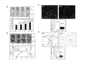
本明細書に記載した組成物、方法及びキットは、EC及び循環するEPCにより成熟したSDF−1αが仲介するEPCホーミングを調節するシグナルの発見に基づいている。下記に記載する実験において、細胞を用いた治療を介した創傷におけるSDF−1αレベルの上昇が、糖尿病マウスでの治癒を促進することを示した(3日目における治癒の上昇率は〜20%、P=0.006)。SDF−1αはEC−EPC間接着を増加させ、そしてヒト毛細血管ECでは、E−セレクチンの発現を特異的にアップレギュレートした(4.6倍の上昇、P<0.01)。この効果は実験マウスの血管においてもまた有意であり、創傷血管新生の増加をもたらした。EC−EPC間接着及びEPCホーミングにおけるSDF−1αの制御作用はE−セレクチンによって特異的に仲介され、そのため、E−セレクチンアンタゴニストの適用はSDF−1α誘導性のEC−EPC間接着、EPCホーミング、創傷血管新生、及び創傷治癒を有意に阻害する。E−sel−/−マウスモデルを用いて、SDF−1α誘導性の創傷治癒の仲介におけるE−セレクチンの必要性を示した。SDF−1αを改変した細胞を用いた治療は、成熟ECにおけるE−セレクチンの発現を特異的にアップレギュレートしてEC−EPC間接着及びEPCホーミングの増加を引き起こし、そして創傷血管新生を増大することにより、マウスにおける糖尿病性創傷の治癒を促進する。これらの発見は、EPCホーミングにおけるSDF−1αの生物学的な作用の根底にあるシグナルへの新規の洞察を提供し、そして、E−セレクチンが虚血性(例えば、糖尿病性)創傷の治癒において、EPC輸送を治療的に操作するための新しい、潜在的な標的であることを示すものである。
DETAILED DESCRIPTION The compositions, methods, and kits described herein are based on the discovery of signals that modulate EPC homing mediated by mature SDF-1α by EC and circulating EPC. In the experiments described below, it was shown that elevated SDF-1α levels in wounds through cell therapy promoted healing in diabetic mice (the rate of healing increase on
以下に記載する好ましい実施形態は、これらの組成物、ワクチン、キット及び方法の適用を説明する。しかし、これらの実施形態の詳細な説明により、以下に提供した説明に基づいて、本発明のその他の態様を作出及び/又は実施することができる。
生物学的方法
The preferred embodiments described below illustrate the application of these compositions, vaccines, kits and methods. However, the detailed description of these embodiments allows other aspects of the invention to be made and / or implemented based on the description provided below.
Biological method
本明細書においては、標準的な分子生物学的技術を含む方法を記載する。このような技術は通常当該分野において一般的に既知であり、かつ、Molecular Cloning: A Laboratory Manual, 3rd ed., vol. 1−3, ed. Sambrook et al., Cold Spring Harbor Laboratory Press, Cold Spring Harbor, N.Y., 2001;及びCurrent Protocols in Molecular Biology, ed. Ausubel et al., Greene Publishing and Wiley−Interscience, New York, 1992(定期的な更新を含む)のような方法論に詳細に記述されている。免疫学的技術は通常当該分野において一般的に既知であり、かつ、Advances in Immunology, volume 93, ed. Frederick W. Alt, Academic Press, Burlington, MA, 2007; Making and Using Antibodies: A Practical Handbook, eds. Gary C. Howard and Matthew R. Kaser, CRC Press, Boca Raton, Fl, 2006; Medical Immunology, 6th ed., edited by Gabriel Virella, Informa Healthcare Press, London, England, 2007;及びHarlow and Lane ANTIBODIES: A Laboratory Manual, Cold Spring Harbor Laboratory Press, Cold Spring Harbor, NY, 1988のような方法論に詳細に記述されている。遺伝子導入及び遺伝子治療の標準的な方法もまた本発明での使用に適している場合がある。例えば、Gene Therapy: Principles and Applications, ed. T. Blackenstein, Springer Verlag, 1999; 及びGene Therapy Protocols (Methods in Molecular Medicine), ed. P.D. Robbins, Humana Press, 1997を参照のこと。
虚血性創傷の治癒を促進する組成物
Described herein are methods involving standard molecular biology techniques. Such techniques are generally known in the art and are described in Molecular Cloning: A Laboratory Manual, 3rd ed. , Vol. 1-3, ed. Sambrook et al. , Cold Spring Harbor Laboratory Press, Cold Spring Harbor, N .; Y. , 2001; and Current Protocols in Molecular Biology, ed. Ausubel et al. , Green Publishing and Wiley-Interscience, New York, 1992 (including periodic updates). Immunological techniques are generally commonly known in the art and are described in Advances in Immunology, volume 93, ed. Frederick W. Alt, Academic Press, Burlington, MA, 2007; Making and Using Antibodies: A Practical Handbook, eds. Gary C. Howard and Matthew R.M. Kaser, CRC Press, Boca Raton, Fl, 2006; Medical Immunology, 6 th ed. , Edited by Gabriel Virella, Informal Healthcare Press, London, England, 2007; . Standard methods of gene transfer and gene therapy may also be suitable for use in the present invention. See, eg, Gene Therapy: Principles and Applications, ed. T.A. Blackenstein, Springer Verlag, 1999; and Gene Therapy Protocols (Methods in Molecular Medicine), ed. P. D. See Robbins, Humana Press, 1997.
Composition for promoting ischemic wound healing
本明細書においては、糖尿病の対象における虚血性(例えば、糖尿病性)創傷の治癒を促進する組成物が記載される。本明細書において記載される組成物は、糖尿病性創傷のような任意の型の虚血性創傷の治癒を促進するために用いることができる。それらの組成物は通常、薬学的に許容可能な担体とE−セレクチンタンパク質又はE−セレクチンタンパク質をコードする核酸のような少なくとも1つの治療薬とを含有する、治療有効量の組成物を含む。組成物は、E−セレクチンの発現を特異的にアップレギュレートする薬剤をさらに含んでいてもよい。この実施形態においては、E−セレクチンの発現を特異的にアップレギュレートする任意の好適な薬剤を用いることができる。例えば、SDF−1αを用いることができる。さらなる例としては、IL−1、TNF−アルファ及びリポ多糖類、ポリサッカライド(LPS)が挙げられる。この実施形態においては、組成物はE−セレクチンタンパク質又はE−セレクチンタンパク質をコードする核酸、及びSDF−1αタンパク質又はSDF−1αタンパク質をコードする核酸を含む。対象へのこの組成物の投与は、骨髄由来前駆細胞の創傷への遊走の増加、及び対象における創傷治癒の促進をもたらす。創傷治癒は、創傷の再上皮形成によって特徴付けられる。 Described herein are compositions that promote the healing of ischemic (eg, diabetic) wounds in diabetic subjects. The compositions described herein can be used to promote healing of any type of ischemic wound, such as a diabetic wound. These compositions usually comprise a therapeutically effective amount of the composition containing a pharmaceutically acceptable carrier and at least one therapeutic agent such as E-selectin protein or a nucleic acid encoding E-selectin protein. The composition may further comprise an agent that specifically upregulates the expression of E-selectin. In this embodiment, any suitable agent that specifically upregulates E-selectin expression can be used. For example, SDF-1α can be used. Further examples include IL-1, TNF-alpha and lipopolysaccharide, polysaccharide (LPS). In this embodiment, the composition comprises E-selectin protein or a nucleic acid encoding E-selectin protein and a nucleic acid encoding SDF-1α protein or SDF-1α protein. Administration of this composition to a subject results in increased migration of bone marrow-derived progenitor cells into the wound and promotes wound healing in the subject. Wound healing is characterized by wound re-epithelialization.
E−セレクチンタンパク質は、任意の好適なプロトコールにより、単離又は合成することができる(例えば、プロテインA及びプロテインGビーズを用いた抗E−セレクチン抗体捕捉技術(Invitrogen))。E−セレクチンタンパク質を投与する一般的な実施形態においては、細菌細胞(例えば、E.Coli)を形質転換、又は哺乳類細胞をトランスフェクションし、その後細胞からE−セレクチンを精製することにより、E−セレクチンタンパク質が調製/合成される。SDF−1αタンパク質も投与される実施形態においては、SDF−1αタンパク質が同様に調製される。 E-selectin protein can be isolated or synthesized by any suitable protocol (eg, anti-E-selectin antibody capture technology using protein A and protein G beads (Invitrogen)). In a typical embodiment of administering an E-selectin protein, E-selectin is obtained by transforming bacterial cells (eg, E. coli) or transfecting mammalian cells and then purifying E-selectin from the cells. Selectin protein is prepared / synthesized. In embodiments where SDF-1α protein is also administered, SDF-1α protein is similarly prepared.
別の実施形態においては、糖尿病性創傷を治療するために、E−セレクチンをコードする核酸を対象に投与してもよい。E−セレクチンをコードするコード配列は、アクセッション番号NM_000450のヌクレオチド配列と同一であってもよく、又は、遺伝子暗号の重複又は縮重の結果異なるコード配列ではあるが、アクセッション番号NM_000450のポリヌクレオチドと同じポリペプチドをコードする配列、であってもよい。本明細書において記載されるその他の核酸分子は、断片、アナログ、及び天然のE−セレクチンタンパク質の誘導体をコードする核酸分子のような、天然のE−セレクチン遺伝子の変異体を含む。このような変異体は、例えば、天然に生じる天然のE−セレクチン遺伝子の対立変異体、天然のE−セレクチン遺伝子のホモログ、又は天然に生じる天然のE−セレクチン遺伝子の変異体であってもよい。これらの変異体は、天然のE−セレクチン遺伝子とは塩基が1以上異なるヌクレオチド配列を有する。例えば、それら変異体のヌクレオチド配列は、天然のE−セレクチン遺伝子のヌクレオチドの1つ以上の欠損、付加又は置換を特徴とする場合がある。 In another embodiment, a nucleic acid encoding E-selectin may be administered to a subject to treat a diabetic wound. The coding sequence encoding E-selectin may be identical to the nucleotide sequence of accession number NM_000450, or a coding sequence that differs as a result of duplication or degeneracy of the genetic code, but the polynucleotide of accession number NM_000450 And a sequence encoding the same polypeptide. Other nucleic acid molecules described herein include variants of the natural E-selectin gene, such as nucleic acids that encode fragments, analogs, and derivatives of the natural E-selectin protein. Such a variant may be, for example, a naturally occurring allelic variant of the natural E-selectin gene, a homologue of the natural E-selectin gene, or a naturally occurring variant of the natural E-selectin gene. . These mutants have a nucleotide sequence that differs by 1 or more bases from the natural E-selectin gene. For example, the nucleotide sequences of these variants may be characterized by one or more deletions, additions or substitutions of nucleotides of the natural E-selectin gene.
その他の実施形態においては、コードされたポリペプチドに保存的置換に満たない変化を生じるヌクレオチド置換を作出することにより、構造が実質的に変化した変異型E−セレクチンタンパク質を生成することができる。そのようなヌクレオチド置換の例としては、(a)ポリペプチド骨格の構造;(b)ポリペプチドの電荷又は疎水性;又は(c)アミノ酸側鎖の大部分、における変化を生じる置換が挙げられる。通常タンパク質の特性に大きな変化を生じると予測されるヌクレオチド置換は、コドンに非保存的置換を生じる置換である。タンパク質構造に大きな変化を生じると考えられるコドン変化の例としては、(a)親水性残基、例えば、セリン又はトレオニン、の(又はによる)疎水性残基、例えば、ロイシン、イソロイシン、フェニルアラニン、バリン又はアラニンへの置換;(b)システイン又はプロリンの(又はによる)任意のその他の残基への置換;(c)電気的陽性の側鎖を有する残基、例えば、リジン、アルギニン、又はヒスチジン、の(又はによる)電気的陰性の残基、例えば、グルタミン又アスパラギンへの置換;又は(d)大きな側鎖を有する残基、例えば、フェニルアラニン、の(又はによる)側鎖のない残基、例えば、グリシンへの置換を生じる置換である。 In other embodiments, mutant E-selectin proteins with substantially altered structure can be generated by creating nucleotide substitutions that produce changes that are less than conservative substitutions in the encoded polypeptide. Examples of such nucleotide substitutions include substitutions that cause changes in (a) the structure of the polypeptide backbone; (b) the charge or hydrophobicity of the polypeptide; or (c) the majority of amino acid side chains. Nucleotide substitutions that are normally predicted to produce a significant change in protein properties are those that result in non-conservative substitutions at the codon. Examples of codon changes that are thought to cause significant changes in protein structure include (a) hydrophobic residues of (or by) hydrophilic residues such as serine or threonine such as leucine, isoleucine, phenylalanine, valine Or substitution with alanine; (b) substitution of (or with) cysteine or proline with any other residue; (c) a residue with an electropositive side chain, such as lysine, arginine, or histidine, An electronegative residue of (or by) substitution of, for example, glutamine or asparagine; or (d) a residue having a large side chain, such as phenylalanine, without (or by) a side chain, such as , A substitution that results in a substitution for glycine.
本明細書において記載される天然のE−セレクチン遺伝子の天然に生じる対立変異体又は天然のE−セレクチンmRNAは、天然のE−セレクチン遺伝子又は天然のE−セレクチンmRNAと少なくとも75%(例えば、76%、77%、78%、79%、80%、81%、82%、83%、84%、85%、86%、87%、88%、89%、90%、92%、93%、94%、95%、96%、97%、98%、及び99%)の配列同一性を有し、かつ、天然のE−セレクチンタンパク質と類似の構造を有するポリペプチドをコードする、ヒト組織から単離された核酸である。本明細書において記載される天然のE−セレクチン遺伝子のホモログ又は天然のE−セレクチンmRNAは、天然のヒトE−セレクチン遺伝子又は天然のヒトE−セレクチンmRNAと少なくとも75%(例えば、76%、77%、78%、79%、80%、81%、82%、83%、84%、85%、86%、87%、88%、89%、90%、91%、92%、93%、94%、95%、96%、97%、98%、及び99%)の配列同一性を有し、かつ、天然のヒトE−セレクチンタンパク質と類似の構造を有するポリペプチドをコードする、その他の種から単離された核酸である。天然のE−セレクチン遺伝子又は天然のE−セレクチンmRNAと高いパーセンテージ(例えば、70、80、90%、又はそれ以上)の配列同一性を有するその他の核酸分子を同定するために、公開された及び/又は登録された核酸データベースを検索することができる。 A naturally occurring allelic variant or native E-selectin mRNA of a native E-selectin gene described herein is at least 75% (e.g., 76%) of a native E-selectin gene or native E-selectin mRNA. %, 77%, 78%, 79%, 80%, 81%, 82%, 83%, 84%, 85%, 86%, 87%, 88%, 89%, 90%, 92%, 93%, 94%, 95%, 96%, 97%, 98%, and 99%) from human tissue that encodes a polypeptide having a sequence identity and having a structure similar to the native E-selectin protein Isolated nucleic acid. A homologue of a natural E-selectin gene or a natural E-selectin mRNA described herein is at least 75% (eg, 76%, 77%) of a natural human E-selectin gene or a natural human E-selectin mRNA. %, 78%, 79%, 80%, 81%, 82%, 83%, 84%, 85%, 86%, 87%, 88%, 89%, 90%, 91%, 92%, 93%, 94%, 95%, 96%, 97%, 98%, and 99%) and other polypeptides that encode polypeptides having a structure similar to that of native human E-selectin protein A nucleic acid isolated from a species. Published and identified to identify other nucleic acid molecules that have a high percentage (eg, 70, 80, 90%, or more) sequence identity with the native E-selectin gene or native E-selectin mRNA A registered nucleic acid database can be searched.
天然に生じないE−セレクチン遺伝子又はmRNA変異体とは、天然のヒトE−セレクチン遺伝子又は天然のヒトE−セレクチンmRNAと少なくとも75%(例えば、76%、77%、78%、79%、80%、81%、82%、83%、84%、85%、86%、87%、88%、89%、90%、91%、92%、93%、94%、95%、96%、97%、98%、及び99%)の配列同一性を有し、かつ、天然のヒトE−セレクチンタンパク質と類似の構造を有するポリペプチドをコードする、天然には生じない(例えば、人工の)核酸である。天然に生じないE−セレクチン遺伝子変異体の例としては、E−セレクチンタンパク質の断片をコードするもの、天然のE−セレクチン遺伝子又は天然のE−セレクチン遺伝子の相補鎖にストリンジェント条件下でハイブリダイズするもの、天然のE−セレクチン遺伝子又はその相補鎖と少なくとも65%の配列同一性を有するもの、及びE−セレクチン融合タンパク質をコードするもの、が挙げられる。 A non-naturally occurring E-selectin gene or mRNA variant is at least 75% (eg, 76%, 77%, 78%, 79%, 80%) with a natural human E-selectin gene or natural human E-selectin mRNA. %, 81%, 82%, 83%, 84%, 85%, 86%, 87%, 88%, 89%, 90%, 91%, 92%, 93%, 94%, 95%, 96%, 97%, 98%, and 99%) and does not occur naturally (eg, artificially) that encodes a polypeptide having a structure similar to the native human E-selectin protein It is a nucleic acid. Examples of non-naturally occurring E-selectin gene variants include those encoding a fragment of E-selectin protein, hybridized under stringent conditions to a natural E-selectin gene or a complementary strand of a natural E-selectin gene And those having at least 65% sequence identity with the natural E-selectin gene or its complementary strand, and those encoding an E-selectin fusion protein.
本明細書において記載した、天然のE−セレクチンタンパク質の断片をコードする核酸は、例えば、2、5、10、25、50、100、150、200又はそれ以上の天然のE−セレクチンタンパク質のアミノ酸残基をコードするものである。天然のE−セレクチンタンパク質の断片をコードする核酸をコードする又はそれとハイブリダイズするより短いオリゴヌクレオチド(例えば、6、7、8、9、10、11、12、13、14、15、16、17、18、19、20、30、50、塩基対長)は、プローブ、プライマー、又はアンチセンス分子として用いることができる。天然のE−セレクチンタンパク質の断片をコードする核酸は、酵素を用いた消化(例えば、制限酵素を用いて)又は完全長の天然のE−セレクチン遺伝子、E−セレクチンmRNA若しくはcDNA、又は前述したものの変異体を化学的に分解することよって作製することができる。これまでに報告された天然のヒトE−セレクチン遺伝子のヌクレオチド配列及び天然のE−セレクチンタンパク質のアミノ酸配列を用いて、当業者は、例えば、標準的な核酸突然変異生成技術又は化学的合成によってそれらのヌクレオチド配列中に僅かな変化を有する核酸分子を作製することができる。変異型E−セレクチンタンパク質を生産するように、変異型E−セレクチン核酸分子を発現させることができる。 Nucleic acids encoding fragments of the natural E-selectin protein described herein include, for example, 2, 5, 10, 25, 50, 100, 150, 200 or more amino acids of natural E-selectin protein It encodes a residue. Shorter oligonucleotides (eg, 6, 7, 8, 9, 10, 11, 12, 13, 14, 15, 16, 17 that encode or hybridize to nucleic acids encoding fragments of the native E-selectin protein. , 18, 19, 20, 30, 50, base pair length) can be used as probes, primers, or antisense molecules. Nucleic acids encoding fragments of native E-selectin protein can be digested with enzymes (eg, using restriction enzymes) or full-length natural E-selectin gene, E-selectin mRNA or cDNA, or those described above Mutants can be made by chemically degrading them. Using the previously reported nucleotide sequence of the natural human E-selectin gene and the amino acid sequence of the natural E-selectin protein, those skilled in the art can, for example, use standard nucleic acid mutagenesis techniques or chemical synthesis to Nucleic acid molecules with slight changes in the nucleotide sequence of can be made. A mutant E-selectin nucleic acid molecule can be expressed so as to produce a mutant E-selectin protein.
糖尿病性創傷の治癒を促進する方法
糖尿病の対象における糖尿病性創傷の治癒を促進する方法の一実施形態は、薬学的に許容可能な担体と、E−セレクチンタンパク質、E−セレクチンタンパク質をコードする核酸、及びE−セレクチンの発現を特異的にアップレギュレートする薬剤のような少なくとも1つの治療薬を含有する治療有効量の組成物を提供する工程;及び対象において骨髄由来前駆細胞(例えば、EPC)の創傷への遊走が増加する条件下において、この組成物を対象に投与する工程を含む。例えば、組成物は、E−セレクチンタンパク質又はE−セレクチンタンパク質をコードする核酸、及び、SDF−1αタンパク質又はSDF−1αタンパク質をコードする核酸のようなE−セレクチンの発現を特異的にアップレギュレートする薬剤を含む可能性がある。この方法においては、この組成物は任意の好適な経路、例えば、経口的、局所的、静脈内、又は創傷若しくは創傷に隣接した部位に直接、投与することができる。対象への組成物の投与は、創傷治癒の促進をもたらす。
Methods for Promoting Diabetic Wound Healing One embodiment of a method for promoting diabetic wound healing in a diabetic subject comprises a pharmaceutically acceptable carrier and an E-selectin protein, a nucleic acid encoding E-selectin protein And providing a therapeutically effective amount of a composition containing at least one therapeutic agent, such as an agent that specifically upregulates the expression of E-selectin; and bone marrow-derived progenitor cells (eg, EPC) in the subject Administering the composition to a subject under conditions that increase migration to the wound. For example, the composition specifically upregulates expression of E-selectin, such as E-selectin protein or nucleic acid encoding E-selectin protein, and SDF-1α protein or nucleic acid encoding SDF-1α protein. May contain drugs. In this method, the composition can be administered by any suitable route, eg, orally, topically, intravenously, or directly to the wound or site adjacent to the wound. Administration of the composition to the subject results in promoting wound healing.
糖尿病の対象における糖尿病性創傷の治癒方法は、対象に高圧酸素療法を実施する工程をさらに含んでいてもよい。本明細書において記載した方法においては、高圧酸素療法(HBO2)は通常、毛細血管が少なくなった状況下で、創傷治癒を刺激するために用いられる補助的な治療である。HBO2を用いた糖尿病の対象の治療方法は、例えば、参照することにより本明細書に組み入れられる、米国特許第2008/003760号に記載されている。糖尿病の対象(患者)における創傷治癒の促進方法の一例においては、患者は1日に1回又は2回、絶対気圧(ATA)が約2.0から約3.2の間になるように圧をかけたチャンバー内で、100% O2を吸い込む処置を20回以上受ける。処置時間は通常、約10分から約240分(例えば、約10、15、30、60、90、120、150、180、210、240分など)の範囲である。 The method for healing a diabetic wound in a diabetic subject may further comprise performing hyperbaric oxygen therapy on the subject. In the methods described herein, hyperbaric oxygen therapy (HBO 2 ) is usually an adjunct treatment used to stimulate wound healing in situations where there are fewer capillaries. Methods for treating subjects with diabetes using HBO 2 are described, for example, in US 2008/003760, which is incorporated herein by reference. In one example of a method for promoting wound healing in a diabetic subject (patient), the patient is pressured once or twice a day such that the absolute atmospheric pressure (ATA) is between about 2.0 and about 3.2. In a chamber subjected to, the treatment of inhaling 100% O 2 is received 20 times or more. Treatment times typically range from about 10 minutes to about 240 minutes (eg, about 10, 15, 30, 60, 90, 120, 150, 180, 210, 240 minutes, etc.).
別の実施形態においては、糖尿病性創傷を有する糖尿病の対象においてE−セレクチンの発現をアップレギュレートする方法は、第一のAAV逆方向末端反復と第二のAAV逆方向末端反復との間に挿入されたE−セレクチンをコードするポリヌクレオチドを含有する少なくとも1つのrAAVビリオンを包含する組成物を、糖尿病の対象に投与する工程を含む。この実施形態においては、この組成物の量は、E−セレクチンの発現をアップレギュレートする、骨髄由来前駆細胞(例えば、EPC)の創傷への遊走を誘導する、及び対象における創傷治癒を促進するのに有効な量である。この方法においては、少なくとも1つのrAAVビリオンは、血清型2カプシドタンパク質を含む場合がある。本明細書において記載するその他の実施形態では、組成物を任意の好適な経路、例えば、経口的、局所的、静脈内、又は、創傷若しくは創傷に隣接した部位に直接、投与することができる。この方法は、対象にSDF−1αタンパク質又はSDF−1αタンパク質をコードする核酸を投与する工程、及び/又は対象に高圧酸素療法を実施する工程をさらに含んでいてもよい。
In another embodiment, the method of upregulating E-selectin expression in a diabetic subject having a diabetic wound is between a first AAV inverted terminal repeat and a second AAV inverted terminal repeat. Administering a composition comprising at least one rAAV virion containing a polynucleotide encoding the inserted E-selectin to a diabetic subject. In this embodiment, the amount of the composition upregulates E-selectin expression, induces migration of bone marrow-derived progenitor cells (eg, EPC) into the wound, and promotes wound healing in the subject. This is an effective amount. In this method, at least one rAAV virion may comprise a
さらにその他の実施形態においては、糖尿病の対象における糖尿病性創傷の治癒を促進する方法は、薬学的に許容可能な担体と複数の骨髄由来前駆細胞を含む組成物を提供する工程、及び、この組成物を対象に、骨髄由来前駆細胞(例えば、EPC)の創傷への遊走を増加させ、かつ対象における創傷治癒を促進するのに有効な量を、投与する工程を含む。この実施形態においては、骨髄由来前駆細胞は、E−セレクチンをコードするポリヌクレオチドを含む。E−セレクチンをコードするポリヌクレオチドを、rAAVベクターのようなウイルスベクター中に含有させてもよい。通常、rAAVベクターはrAAVウイルス(粒子)に含有される。この実施形態においては、組成物は、SDF−1αタンパク質又はSDF−1αタンパク質をコードする核酸を、さらに含んでいてもよい。組成物がSDF−1αをコードする核酸を含む場合、この核酸はrAAVベクター中、第二のrAAVベクター中、又はrAAV以外のウイルスベクター中に存在する可能性がある。組成物を、任意の好適な経路、例えば、経口的、局所的、静脈内、又は、創傷又は創傷に隣接した部位に直接、投与することができる。この方法は、対象に高圧酸素療法を実施する工程をさらに含んでいてもよい。 In yet another embodiment, a method of promoting diabetic wound healing in a diabetic subject comprises providing a composition comprising a pharmaceutically acceptable carrier and a plurality of bone marrow-derived progenitor cells, and the composition Administering to the subject an amount effective to increase migration of bone marrow-derived progenitor cells (eg, EPC) to the wound and promote wound healing in the subject. In this embodiment, the bone marrow derived progenitor cell comprises a polynucleotide encoding E-selectin. A polynucleotide encoding E-selectin may be included in a viral vector such as an rAAV vector. Usually, rAAV vectors are contained in rAAV viruses (particles). In this embodiment, the composition may further comprise SDF-1α protein or a nucleic acid encoding SDF-1α protein. If the composition comprises a nucleic acid encoding SDF-1α, the nucleic acid may be present in the rAAV vector, in a second rAAV vector, or in a viral vector other than rAAV. The composition can be administered by any suitable route, eg, orally, topically, intravenously, or directly to the wound or site adjacent to the wound. The method may further comprise performing hyperbaric oxygen therapy on the subject.
本明細書において記載した方法は、数多くの異なる型の創傷の治療に用いることができる。糖尿病性創傷の一例としては、網状皮斑及び白色萎縮に関連した有痛性潰瘍により特徴付けられる障害である、リベド血管炎が挙げられる。糖尿病性創傷の別の例としては、例えば、末梢動脈疾患性潰瘍、静脈鬱血性潰瘍、慢性難治性潰瘍、褥瘡、褥瘡性潰瘍、慢性足潰瘍などの糖尿病性潰瘍がある。創傷は、1以上の上記に挙げた創傷の組み合わせである場合もある。糖尿病の対象は、治療を受ける1つ又は複数の創傷を有する場合もある。対象は、ヒト、ラット、マウス、ネコ、イヌ、ヤギ、ヒツジ、ウマ、サル、類人猿、ウサギ、ウシなどのような任意の哺乳類を含む。対象(例えば、哺乳類)は、成人及び小児を含む、発達のいずれの段階にあるものであってもよい。標的組織は、網膜、肝臓、腎臓、心臓、肺、消化管の部分、膵臓、胆嚢、膀胱、中枢神経系(脳を含む)、皮膚、骨などのような、対象におけるいずれの組織であってもよい。 The methods described herein can be used to treat many different types of wounds. An example of a diabetic wound is Ribed vasculitis, a disorder characterized by painful ulcers associated with reticulated skin spots and white atrophy. Other examples of diabetic wounds include, for example, diabetic ulcers such as peripheral arterial disease ulcers, venous stasis ulcers, chronic intractable ulcers, pressure ulcers, pressure ulcers, chronic foot ulcers. The wound may be a combination of one or more of the above listed wounds. A diabetic subject may also have one or more wounds to be treated. A subject includes any mammal such as a human, rat, mouse, cat, dog, goat, sheep, horse, monkey, ape, rabbit, cow and the like. A subject (eg, a mammal) may be at any stage of development, including adults and children. The target tissue is any tissue in the subject such as the retina, liver, kidney, heart, lung, digestive tract, pancreas, gallbladder, bladder, central nervous system (including brain), skin, bone, etc. Also good.
E−セレクチンをコードする核酸(及び任意にSDF−1αをコードする核酸又はその他のEPCホーミングを調節する薬剤)を糖尿病の対象に投与する方法においては、本明細書において記載した核酸は、E−セレクチンをコードする核酸(及び任意にSDF−1αをコードする核酸又はその他のEPCホーミングを調節する薬剤)に操作可能に連結した、1つ以上の発現制御配列を含む場合がある。数多くのそのような配列が知られている。含まれるそれらの配列は、その他の適用におけるそれらの既知の機能に基づいて選択され得る。発現制御配列の例としては、プロモーター、インシュレーター、サイレンサー、応答エレメント、イントロン、エンハンサー、開始部位、終結シグナル、及びpAテールが挙げられる。 In a method of administering a nucleic acid encoding E-selectin (and optionally a nucleic acid encoding SDF-1α or other agent that modulates EPC homing) to a diabetic subject, the nucleic acids described herein include E- It may include one or more expression control sequences operably linked to a nucleic acid encoding selectin (and optionally a nucleic acid encoding SDF-1α or other agent that modulates EPC homing). Many such sequences are known. Those sequences included can be selected based on their known function in other applications. Examples of expression control sequences include promoters, insulators, silencers, response elements, introns, enhancers, initiation sites, termination signals, and pA tails.
E−セレクチン(及び任意にSDF−1α)を適切なレベルにするために、標的細胞において使用するのに好適な、任意の数のプロモーターを用いることができる。例えば、異なる強さの構成的プロモーターを用いることができる。本明細書において記載した発現ベクター及びプラスミドは、ウイルス性プロモーター、又は、通常転写の促進において活性を有する哺乳類の遺伝子由来のプロモーターのような、1つ以上の構成的プロモーターを含んでもよい。構成的なウイルス性プロモーターの例としては、単純ヘルペスウイルス(HSV)、チミジンキナーゼ(TK)、ラウス肉腫ウイルス(RSV)、シミアンウイルス40(SV40)、マウス乳癌ウイルス(MMTV)、Ad E1A及びサイトメガロウイルス(CMV)プロモーターが挙げられる。構成的哺乳類プロモーターの例としては、β−アクチンプロモーターのような、様々なハウスキーピング遺伝子のプロモーターが挙げられる。 Any number of promoters suitable for use in target cells can be used to bring E-selectin (and optionally SDF-1α) to appropriate levels. For example, constitutive promoters of different strengths can be used. The expression vectors and plasmids described herein may include one or more constitutive promoters, such as viral promoters or promoters from mammalian genes that are normally active in promoting transcription. Examples of constitutive viral promoters include herpes simplex virus (HSV), thymidine kinase (TK), rous sarcoma virus (RSV), simian virus 40 (SV40), mouse mammary tumor virus (MMTV), Ad E1A and cytomegalo A viral (CMV) promoter is mentioned. Examples of constitutive mammalian promoters include promoters of various housekeeping genes, such as the β-actin promoter.
誘導性プロモーター及び/又は調節エレメントもまた、本明細書において記載した組成物及び方法での使用のために検討されてもよい。好適な誘導性プロモーターの例としては、シトクロムP450遺伝子、熱ショックタンパク質遺伝子、メタロチオネイン遺伝子、及びホルモン誘導性遺伝子(エストロゲン遺伝子プロモーターなど)のような遺伝子由来のプロモーターが挙げられる。誘導性プロモーターの別の例には、テトラサイクリン応答性のtetVP16プロモーターがある。 Inducible promoters and / or regulatory elements may also be considered for use in the compositions and methods described herein. Examples of suitable inducible promoters include promoters derived from genes such as cytochrome P450 genes, heat shock protein genes, metallothionein genes, and hormone-inducible genes (such as estrogen gene promoters). Another example of an inducible promoter is the tetracycline responsive tetVP16 promoter.
組織特異的プロモーター及び/又は調節エレメントは、本明細書において記載した組成物及び方法の特定の実施形態において有用である。本明細書において記載した発現ベクターのような発現ベクターと共に用いることができるそれらプロモーターの例としては、Tie−2又はKDR_プロモーターが挙げられる。 Tissue specific promoters and / or regulatory elements are useful in certain embodiments of the compositions and methods described herein. Examples of those promoters that can be used with expression vectors such as those described herein include Tie-2 or KDR_promoter.
本明細書において記載した組成物(例えば、E−セレクチンをコードするウイルスベクターを含む組成物)を哺乳類対象に、任意の好適な技術により投与することができる。本明細書において記載した組成物及び方法により、細胞中にE−セレクチン遺伝子を導入するためのウイルスベクターを用いた、様々な技術が提供される。ウイルスは、それらの遺伝子を宿主細胞中に効率良く送達する、天然に発生した媒体であり、従って、治療的遺伝子の送達のための望ましいベクター系である。好ましいウイルスベクターは宿主細胞に対する低い毒性を示し、かつ、治療的な量のE−セレクチンタンパク質を(例えば、組織特異的な方法で)生産するものである。ウイルスベクター法及びプロトコールは、Kay et al. Nature Medicine 7:33−40, 2001にまとめられている。 A composition described herein (eg, a composition comprising a viral vector encoding E-selectin) can be administered to a mammalian subject by any suitable technique. The compositions and methods described herein provide various techniques using viral vectors for introducing E-selectin genes into cells. Viruses are naturally occurring vehicles that efficiently deliver these genes into host cells and are therefore desirable vector systems for the delivery of therapeutic genes. Preferred viral vectors are those that exhibit low toxicity to the host cell and produce a therapeutic amount of E-selectin protein (eg, in a tissue specific manner). Viral vector methods and protocols are described in Kay et al. Nature Medicine 7: 33-40, 2001.
以下に記載した実施例はrAAV及びレンチウイルスを含むが、任意の好適なウイルスベクターを使用することができる。哺乳類対象に遺伝子を送達するための多くのウイルスベクターが当該分野において既知であり、以下に限定的な例を挙げる。遺伝子治療ベクターとして組換えアデノウイルスを使用する方法は、例えば、W.C. Russell, Journal of General Virology 81:2573−2604, 2000,及びBramson et al., Curr. Opin. Biotechnol. 6:590−595, 1995で議論された。単純ヘルペスウイルスベクターの使用方法については、例えば、Cotter and Robertson, Curr. Opin. MoI. Ther. 1:633−644, 1999で議論された。HIVを含む、複製欠損レンチウイルスベクターもまた用いることができる。レンチウイルスベクターの使用方法は、例えば、Vigna and Naldini, J. Gene Med. 5:308−316, 2000 及び Miyoshi et al., J. Virol. 72:8150−8157, 1998で議論された。マウス白血病ウイルスに基づくベクターを含む、レトロウイルスベクターもまた用いることができる。レトロウイルスに基づくベクターの使用方法は、例えば、Hu and Pathak, Pharmacol. Rev. 52:493−511, 2000 及び Fong et al., Crit. Rev. Ther. Drug Carrier Syst. 17:1−60, 2000で議論された。用いることができるその他のウイルスベクターには、セムリキ森林ウイルス及びシンドビスウイルスを含む、アルファウイルスが挙げられる。E−セレクチン遺伝子を標的組織(例えば、糖尿病性創傷)に送達するために、ハイブリッドウイルスベクターを用いてもよい。ハイブリッドベクターを構築するための標準的な技術は当業者に周知である。そのような技術は、例えば、Sambrook, et al., In Molecular Cloning: A laboratory manual. Cold Spring Harbor, NY又は組換えDNA技術について議論した多くの実験手引き書において見ることができる。 The examples described below include rAAV and lentivirus, but any suitable viral vector can be used. Many viral vectors for delivering genes to mammalian subjects are known in the art and the following are limited examples. Methods for using recombinant adenovirus as a gene therapy vector are described, for example, in W.W. C. Russell, Journal of General Virology 81: 2573-2604, 2000, and Bramson et al. Curr. Opin. Biotechnol. 6: 590-595, 1995. For methods of using herpes simplex virus vectors, see, for example, Cotter and Robertson, Curr. Opin. MoI. Ther. 1: 633-644, discussed in 1999. Replication deficient lentiviral vectors, including HIV, can also be used. Methods for using lentiviral vectors are described, for example, in Vigna and Naldini, J. et al. Gene Med. 5: 308-316, 2000 and Miyoshi et al. , J. et al. Virol. 72: 8150-8157, 1998. Retroviral vectors can also be used, including vectors based on murine leukemia virus. Methods for using retrovirus-based vectors are described, for example, in Hu and Pathak, Pharmacol. Rev. 52: 493-511, 2000 and Fong et al. , Crit. Rev. Ther. Drug Carrier Syst. 17: 1-60, 2000. Other viral vectors that can be used include alphaviruses, including Semliki Forest virus and Sindbis virus. Hybrid viral vectors may be used to deliver the E-selectin gene to target tissues (eg, diabetic wounds). Standard techniques for constructing hybrid vectors are well known to those of skill in the art. Such techniques are described, for example, in Sambrook, et al. , In Molecular Cloning: A laboratory manual. It can be found in many experimental manuals discussing Cold Spring Harbor, NY or recombinant DNA technology.
いくつかの実施形態においては、本明細書において記載した組成物及び方法の核酸は、それらの細胞中への導入を促進するために、rAAVベクター及び/又はビリオン中に組み入れられる。有用なrAAVベクターは、(1)発現する異種配列(例えば、E−セレクチンタンパク質をコードするポリヌクレオチド)及び(2)異種遺伝子の組換え及び発現を促進するウイルス配列、を含む組換え核酸構築物である。ウイルス配列は、複製、及びDNAのビリオン中へのパッケージング(例えば、機能性ITR)のためにシスに必要とされるAAVの配列を含む場合もある。一般的な適用においては、異種遺伝子は、骨髄由来前駆細胞の創傷への遊走を増加し、糖尿病の対象における創傷治癒を促進するのに有用なE−セレクチンをコードする。そのようなrAAVベクターは、マーカー又はレポーター遺伝子をもまた含む場合がある。有用なrAAVベクターは、その全体又は一部分が欠損しているが機能性ITR配列を保持する、1つ以上のAAV WT遺伝子を有する。AAV ITRは、特定の適用において好適な任意の血清型(例えば、血清型2由来の)であってよい。rAAVベクターの使用方法は、例えば、Tal, J.,J. Biomed. Sci. 7:279−291, 2000及びMonahan and Samulski, Gene delivery 7:24−30, 2000において議論された。 In some embodiments, the nucleic acids of the compositions and methods described herein are incorporated into rAAV vectors and / or virions to facilitate their introduction into cells. Useful rAAV vectors are recombinant nucleic acid constructs comprising (1) a heterologous sequence to be expressed (eg, a polynucleotide encoding an E-selectin protein) and (2) a viral sequence that facilitates recombination and expression of the heterologous gene. is there. Viral sequences may include sequences of AAV that are required in cis for replication and packaging of DNA into virions (eg, functional ITRs). In general applications, the heterologous gene encodes an E-selectin that is useful for increasing migration of bone marrow-derived progenitor cells to the wound and promoting wound healing in diabetic subjects. Such rAAV vectors may also contain a marker or reporter gene. Useful rAAV vectors have one or more AAV WT genes that are defective in whole or in part but retain functional ITR sequences. The AAV ITR may be any serotype suitable for a particular application (eg, from serotype 2). Methods for using rAAV vectors are described, for example, in Tal, J. et al. , J .; Biomed. Sci. 7: 279-291, 2000 and Monahan and Samulski, Gene delivery 7: 24-30, 2000.
本発明の核酸及びベクターを、その核酸又はベクターの細胞中への導入を促進するために、rAAVビリオン中に組み入れることができる。AAVのカプシドタンパク質は、ビリオンの外部、非核酸部分を含み、かつ、AAV cap遺伝子によりコードされる。cap遺伝子は、ビリオンの集合に必要な3つのウイルス性コートタンパク質であるVP1、VP2及びVP3、をコードする。rAAVビリオンについては既に記載がある。例えば、米国特許第5,173,414号、同5,139,941号、同5,863,541号、及び同5,869,305号、同6,057,152号、同6,376,237号;Rabinowitz et al.、J.Virol.76:791−801、2002;並びに、Rabinowitz et al., J. Virol. 76:791−801, 2002;及びBowles et al., J. Virol. 77:423−432, 2003を参照のこと。 The nucleic acids and vectors of the invention can be incorporated into rAAV virions to facilitate introduction of the nucleic acid or vector into cells. The AAV capsid protein contains the outer, non-nucleic acid portion of the virion and is encoded by the AAV cap gene. The cap gene encodes three viral coat proteins, VP1, VP2 and VP3, required for virion assembly. The rAAV virion has already been described. For example, U.S. Pat. Nos. 5,173,414, 5,139,941, 5,863,541, and 5,869,305, 6,057,152, 6,376, 237; Rabinowitz et al. , J .; Virol. 76: 791-801, 2002; and Rabinowitz et al. , J. et al. Virol. 76: 791-801, 2002; and Bowles et al. , J. et al. Virol. 77: 423-432, 2003.
本発明において有用なrAAVビリオンは、1、2、3、4、5、6、7、8及び9を含む、多数のAAV血清型由来のビリオンを含む。異なる血清型のAAVベクター及びAAVタンパク質の構築及び使用については、Chao et al., MoI. Ther. 2:619−623, 2000; Davidson et al., PNAS 97:3428−3432, 2000; Xiao et al., J. Virol. 72:2224−2232, 1998; Halbert et al., J. Virol. 74:1524−1532, 2000; Halbert et al., J. Virol. 75:6615−6624, 2001;及びAuricchio et al., Hum. Molec. Genet. 10:3075−3081, 2001において議論された。 RAAV virions useful in the present invention include virions from a number of AAV serotypes, including 1, 2, 3, 4, 5, 6, 7, 8, and 9. For the construction and use of different serotypes of AAV vectors and AAV proteins, see Chao et al. MoI. Ther. 2: 619-623, 2000; Davidson et al. , PNAS 97: 3428-3432, 2000; Xiao et al. , J. et al. Virol. 72: 2224-2232, 1998; Halbert et al. , J. et al. Virol. 74: 1524-1532, 2000; Halbert et al. , J. et al. Virol. 75: 6615-6624, 2001; and Auricchio et al. , Hum. Molec. Genet. 10: 3075-3081, 2001.
本明細書において記載した組成物、キット及び方法においては、偽型rAAVもまた有用である。偽型ベクターは、ある特定の偽型血清型のAAVベクターと、その特定の血清型以外の血清型由来のカプシド遺伝子とを含む。偽型rAAVビリオンの構築及び使用を含む技術は、当該分野において既知であり、及びDuan et al., J. Virol, 75:7662−7671, 2001; Halbert et al., J. Virol, 74:1524−1532, 2000; Zolotukhin et al, Methods, 28: 158−167, 2002;及びAuricchio et al, Hum. Molec. Genet. 10:3075−3081, 2001に記載されている。 Pseudo rAAV is also useful in the compositions, kits and methods described herein. The pseudotype vector includes an AAV vector of a specific pseudotype serotype and a capsid gene derived from a serotype other than the specific serotype. Techniques involving the construction and use of pseudotyped rAAV virions are known in the art, and Duan et al. , J. et al. Virol, 75: 7662-7671, 2001; Halbert et al. , J. et al. Virol, 74: 1524-1532, 2000; Zlotukhin et al, Methods, 28: 158-167, 2002; and Auricchio et al, Hum. Molec. Genet. 10: 3075-3081, 2001.
特定の細胞型に、非変異型カプシドビリオンを用いた場合よりも、より効果的に感染させるために、ビリオンカプシド中に変異を有するAAVビリオンを用いてもよい。例えば、好適なAAV突然変異体は、AAVの特異的細胞型へのターゲティングを促進するために、リガンド挿入変異を有する場合もある。挿入変異体、アラニンスクリーニング突然変異体、及びエピトープタグ突然変異体を含むAAVカプシド突然変異体の構築及び解析については、Wu et al,J.Virol. 74:8635−45,2000に記載されている。本明細書において記載した方法及び組成物において使用することができるその他のrAAVビリオンとしては、ウイルスの分子育種並びにエキソンシャッフリングにより生成されたカプシドハイブリッドがある。Soong et al, Nat. Genet. 25:436−439, 2000;及びKolman and Stemmer Nat. Biotechnol 19:423−428, 2001を参照のこと。 AAV virions with mutations in virion capsids may be used to more effectively infect certain cell types than when non-mutated capsid virions are used. For example, suitable AAV mutants may have ligand insertion mutations to facilitate targeting of AAV to specific cell types. For the construction and analysis of AAV capsid mutants, including insertion mutants, alanine screening mutants, and epitope tag mutants, see Wu et al, J. et al. Virol. 74: 8635-45, 2000. Other rAAV virions that can be used in the methods and compositions described herein include capsid hybrids generated by molecular breeding of viruses and exon shuffling. Soong et al, Nat. Genet. 25: 436-439, 2000; and Kolman and Stemmer Nat. See Biotechnol 19: 423-428, 2001.
ウイルスベクター又はウイルス粒子の注入による非経口投与、例えばボーラス注入又は持続注入、を行ってもよい。注入用製剤は、単位容量形態、例えばアンプル中又は複数回投与用の容器中に、保存料を含めて提供してもよい。組成物は、懸濁液、溶液、又は油性若しくは水性媒体中の乳濁液としてそのような形態をとることができ、また、懸濁化剤、安定化剤、及び/又は分散剤のような成形剤を含んでもよい。あるいは、ベクター又はウイルス粒子は、使用前に無菌の発熱物質を含まない水などで構成するための粉末形態(例えば凍結乾燥した)であってもよい。 Parenteral administration by injection of viral vectors or virus particles, such as bolus injection or continuous infusion, may be performed. Injectable formulations may be presented in pre-contained form in unit volume form, such as ampoules or in multi-dose containers. The compositions can take such forms as suspensions, solutions, or emulsions in oily or aqueous media, such as suspending, stabilizing, and / or dispersing agents. A molding agent may be included. Alternatively, the vector or virus particle may be in the form of a powder (eg, lyophilized) for constitution with sterile pyrogen-free water before use.
ウイルスベクターに加え、少なくとも1つの糖尿病性創傷を有する糖尿病の対象へE−セレクチンをコードする核酸を導入するのに好適な任意の媒体又はベクターを用いることができる。例えば、超音波を用いた遺伝子送達技術を用いることができる。さらなる例としては、パーティクルボンバードメント及び細胞のエレクトロポレーションが挙げられる。プラスミドDNAと共に多細胞性集合体を形成する、合成遺伝子導入分子もまた有用である。そのような分子には、高分子DNA結合性陽イオン(Guy et al.,MoI. Biotechnol. 3:237−248,1995)、陽イオン性両親媒性試薬(リポポリアミン及び陽イオン性脂質、Feigner et al.,Ann.NY Acad.Sci.772:126−139,1995)、及び陽イオン性リポソーム(Fominaya et al.,J.Gene Med.2:455−464,2000)が含まれる。 In addition to the viral vector, any medium or vector suitable for introducing a nucleic acid encoding E-selectin into a diabetic subject having at least one diabetic wound can be used. For example, a gene delivery technique using ultrasound can be used. Further examples include particle bombardment and cell electroporation. Synthetic transgenic molecules that form multicellular aggregates with plasmid DNA are also useful. Such molecules include polymeric DNA binding cations (Guy et al., MoI. Biotechnol. 3: 237-248, 1995), cationic amphiphilic reagents (lipopolyamines and cationic lipids, Feigner). et al., Ann. NY Acad. Sci. 772: 126-139, 1995), and cationic liposomes (Fominya et al., J. Gene Med. 2: 455-464, 2000).
いくつかの実施形態においては、rAAV−E−セレクチンベクター又はウイルスを含むEPCを、1以上の糖尿病性創傷を治療するために糖尿病の対象に投与する。対象にEPCを導入するためには、カテーテルを介した静脈内送達(例えば、血管内カテーテル)又は標的部位、例えば糖尿病性創傷への直接注入を含む複数の方法が用いられ得る。ドナー幹細胞の単離及び、そのような単離した細胞の移植の技術は当該分野において周知である。rAAVビリオンを用いて導入した細胞のex vivo送達もまた、本明細書において記載した方法に包含される。ex vivoの遺伝子送達は、移植、例えばrAAVを導入した宿主細胞(例えばEPC)を宿主に戻すために用いられる場合がある。好適なex vivoのプロトコールは複数の工程を含む場合がある。標的組織のセグメント(例えば、BM由来EPC)を宿主から回収してもよく、及びE−セレクチンをコードする核酸を対象(すなわち宿主)の細胞に導入するために、rAAVビリオンを用いてもよい。これらの遺伝的に改変された細胞をその後、対象に戻すために移植してもよい。細胞を対象に再導入するためには、静脈内注入、腹腔内注入、又は標的組織へのin situ注入を含む複数の方法が用いられ得る。用いることができるその他の技術には、ex vivoでrAAVを導入又は感染させた細胞のマイクロカプセル化がある。本発明による自家並びに同種細胞移植を用いてもよい。 In some embodiments, an EPC comprising an rAAV-E-selectin vector or virus is administered to a diabetic subject to treat one or more diabetic wounds. A number of methods can be used to introduce EPC into a subject, including intravenous delivery via a catheter (eg, an intravascular catheter) or direct injection into a target site, eg, a diabetic wound. Techniques for isolating donor stem cells and transplanting such isolated cells are well known in the art. Ex vivo delivery of cells introduced using rAAV virions is also encompassed by the methods described herein. Ex vivo gene delivery may be used to transfer a host cell, such as an EPC, into which rAAV has been introduced (eg, EPC) back to the host. A suitable ex vivo protocol may involve multiple steps. A segment of the target tissue (eg, BM-derived EPC) may be recovered from the host, and rAAV virions may be used to introduce nucleic acid encoding E-selectin into the cells of the subject (ie, host). These genetically modified cells may then be transplanted back to the subject. A number of methods can be used to reintroduce cells into a subject, including intravenous injection, intraperitoneal injection, or in situ injection into a target tissue. Other techniques that can be used include microencapsulation of cells transfected or infected with rAAV ex vivo. Autologous and allogeneic cell transplants according to the present invention may be used.
BMから非BM区域(例えば、標的組織)へのBM由来前駆細胞のリクルートメントを増加するために、本明細書において記載した創傷治癒を促進する組成物は、E−セレクチン及びSDF−1αに加えて、BM由来前駆細胞のリクルートメントを促進することができる任意のその他の薬剤を含む場合がある。多数のそのような薬剤が既知である。例えば、国際公開第00/50048号に記載されている薬剤;インテグリン(例えば、α4、α5)、接着分子のセレクチンファミリー、VCAM−1、及びコロニー刺激因子を参照のこと。 In order to increase recruitment of BM-derived progenitor cells from BM to non-BM areas (eg, target tissue), the composition for promoting wound healing described herein is in addition to E-selectin and SDF-1α. And any other agent that can facilitate the recruitment of BM-derived progenitor cells. A number of such agents are known. See, for example, the agents described in WO 00/50048; integrins (eg, α4, α5), selectin family of adhesion molecules, VCAM-1, and colony stimulating factor.
本明細書において記載した治療方法は通常、治療を必要とする哺乳類(特にヒト)対象(例えば動物、ヒト)に、治療有効量の本明細書において記載した組成物を投与する工程を含む。そのような治療は、対象(特に、疾患、障害、又はその症状に、罹患している、有する、感受性のある、又はリスクを有するヒト)に好適な方法で投与される。「リスクを有する」対象の決定は、診断検査又は対象若しくは健康管理者の意見による、任意の主観的又は客観的判断においてなされる場合がある。本明細書において記載される方法及び組成物は、E−セレクチンのシグナル伝達、発現、又は活性のダウンレギュレーションを意味すると考えられる、いずれのその他の障害の治療にもまた有用である可能性がある。 The treatment methods described herein typically comprise administering to a mammalian (particularly human) subject in need of treatment (eg, an animal, human) a therapeutically effective amount of the composition described herein. Such treatment is administered in a manner suitable for a subject, particularly a human suffering from, having, susceptible to, or at risk of a disease, disorder, or symptom thereof. The determination of a “at risk” subject may be made in any subjective or objective judgment by diagnostic tests or by the opinion of the subject or health care provider. The methods and compositions described herein may also be useful in the treatment of any other disorder that is believed to mean down-regulation of E-selectin signaling, expression, or activity. .
一実施形態においては、糖尿病の対象における糖尿病性創傷の治癒を促進する方法は、治療経過をモニタリングする工程を含む。対象における治療経過のモニタリングは通常、創傷の治療を促進するのに十分な治療量の本明細書において記載した組成物を対象に投与する前に、糖尿病性創傷を有する対象における創傷の大きさの測定値又はその他の診断的測定値を決定する工程を含む。治療を促進するのに十分な治療量の本明細書において記載した組成物を対象に投与した後、1回以上の時点において、創傷の大きさの2回目の測定を行い、最初に測定した創傷の大きさと比較する。最初及びその後の大きさを、創傷治癒の経過(例えば創傷の大きさの減少)及びその治療の効果をモニターするために比較する。 In one embodiment, a method for promoting healing of a diabetic wound in a diabetic subject includes monitoring the course of treatment. Monitoring of the course of treatment in a subject typically involves measuring the size of the wound in a subject having a diabetic wound prior to administering to the subject a therapeutic amount sufficient to facilitate wound treatment. Determining a measurement or other diagnostic measurement. After administering to a subject a therapeutic amount sufficient to facilitate treatment, a second measurement of wound size is made at one or more time points, and the first measured wound Compare with the size of. Initial and subsequent sizes are compared to monitor the course of wound healing (eg, reduction in wound size) and the effect of the treatment.
キット
哺乳類対象における少なくとも1つの糖尿病性創傷を治療するキットを本明細書において記載した。一般的にはキットは、E−セレクチンタンパク質、E−セレクチンタンパク質をコードする核酸、又はE−セレクチンの発現を特異的にアップレギュレートする薬剤のような治療薬を少なくとも1つと、薬学的に許容可能な担体とを含有する、治療有効量の組成物、並びに使用説明書を含む。一実施形態においては、キットは、治療有効量のE−セレクチンタンパク質又はE−セレクチンタンパク質をコードする核酸、及びE−セレクチンの発現を特異的にアップレギュレートする薬剤を含有する治療用組成物、並びに使用説明書を含む。例えば、哺乳類対象における少なくとも1つの糖尿病性創傷を治療するキットは、治療有効量のE−セレクチンタンパク質又はE−セレクチン(例えば、rAAV−E−セレクチン)をコードする核酸、及び創傷治癒を促進する治療有効量のSDF−1α、並びに薬学的に許容可能な担体とを単位容量形態で含有する治療用組成物を含む。通常、本明細書において記載したキットは、包装及び使用説明書を含む。いくつかの実施形態においては、このキットは治療用又は予防用組成物を含む無菌的な容器を含む。そのような容器は、箱、アンプル、瓶、バイアル、チューブ、袋、パウチ、ブリスターパック、又はその他の、当該分野において既知の好適な形態の容器であってよい。そのような容器は、プラスチック、ガラス、ラミネート紙、金属ホイル、又はその他の、薬物を保持するための好適な材料から作製することができる。本明細書において記載した治療用組成物に加えて、治療を受ける対象に高圧酸素療法を実施する実施形態においては、使用説明書は一般的に、対象への高圧酸素療法の実施についての方法を含むだろう。
Kits A kit for treating at least one diabetic wound in a mammalian subject has been described herein. In general, the kit is pharmaceutically acceptable with at least one therapeutic agent, such as an E-selectin protein, a nucleic acid encoding the E-selectin protein, or an agent that specifically upregulates the expression of E-selectin. A therapeutically effective amount of the composition containing possible carriers, as well as instructions for use. In one embodiment, the kit comprises a therapeutic composition comprising a therapeutically effective amount of E-selectin protein or nucleic acid encoding E-selectin protein and an agent that specifically upregulates expression of E-selectin; As well as instructions for use. For example, a kit for treating at least one diabetic wound in a mammalian subject comprises a nucleic acid encoding a therapeutically effective amount of E-selectin protein or E-selectin (eg, rAAV-E-selectin), and a treatment that promotes wound healing. A therapeutic composition containing an effective amount of SDF-1α as well as a pharmaceutically acceptable carrier in unit volume form. Typically, the kits described herein include packaging and instructions for use. In some embodiments, the kit includes a sterile container that contains a therapeutic or prophylactic composition. Such containers may be boxes, ampoules, bottles, vials, tubes, bags, pouches, blister packs, or other suitable forms of containers known in the art. Such containers can be made from plastic, glass, laminated paper, metal foil, or other suitable material for holding the drug. In embodiments in which hyperbaric oxygen therapy is performed on a subject to be treated in addition to the therapeutic composition described herein, the instructions generally provide a method for performing hyperbaric oxygen therapy on the subject. Would include.
EPC(例えば、rAAV−E−セレクチンを含むEPC)を糖尿病の対象に投与する一実施形態では、本明細書において記載したキットは、治療量のEPCと共に、1つ以上の糖尿病性創傷を有する対象への細胞の投与方法についての説明書を含む。細胞を輸送及び保存するために好適な任意の手段で、細胞を包装することができ、そのような方法は当該分野において周知である。使用説明書は通常、細胞;糖尿病性創傷の治療のための投与量計画及び投与;予防策;警告;適応症;不適応症;過剰投与情報;有害反応;動物での薬理;臨床試験;及び/又は参考文献、についての説明を1つ以上含む。この使用説明書は、容器(がある場合)に直接印刷してもよく、又は容器に適用したラベルとして、又は、容器中に若しくは容器と共に提供する別個の紙、パンフレット、カード若しくはホルダーとして印刷してもよい。 In one embodiment of administering an EPC (eg, an EPC comprising rAAV-E-selectin) to a diabetic subject, the kit described herein comprises a subject having one or more diabetic wounds along with a therapeutic amount of EPC. Instructions on how to administer cells to Cells can be packaged by any means suitable for transporting and storing cells, and such methods are well known in the art. Instructions for use are usually cells; dosage planning and administration for the treatment of diabetic wounds; preventive measures; warnings; indications; maladaptation; overdose information; adverse reactions; pharmacology in animals; Includes one or more explanations of / and references. The instructions may be printed directly on the container (if any) or printed as a label applied to the container or as a separate paper, brochure, card or holder provided in or with the container. May be.
組成物の投与
本発明の組成物を、哺乳類(例えば、齧歯類、ヒト)に任意の好適な製剤として投与することができる。例えば本明細書において記載した創傷治癒を促進する組成物(例えば、E−セレクチン、E−セレクチンをコードする核酸、SDF−1α)を、薬学的に許容可能な担体又は生理食塩水若しくは緩衝塩類溶液のような希釈剤中に処方することができる。方法及び投与経路並びに標準的な薬務に基づき、好適な担体及び希釈剤を選択することができる。薬学的に許容可能な担体及び希釈剤、並びに薬剤処方の例についての説明は、当該分野における標準的な教科書であるRemington’s Pharmaceutical Sciences、及びUSP/NF中に見ることができる。組成物の安定化及び/又は保存のために、その他の物質を加えてもよい。
Administration of Compositions The compositions of the present invention can be administered to mammals (eg, rodents, humans) as any suitable formulation. For example, a composition (eg, E-selectin, a nucleic acid encoding E-selectin, SDF-1α) that promotes wound healing described herein may be used as a pharmaceutically acceptable carrier or physiological saline or buffered saline solution. Can be formulated in a diluent such as Based on the method and route of administration and standard pharmaceutical practice, suitable carriers and diluents can be selected. Descriptions of pharmaceutically acceptable carriers and diluents and examples of pharmaceutical formulations can be found in Remington's Pharmaceutical Sciences, a standard textbook in the field, and USP / NF. Other materials may be added to stabilize and / or preserve the composition.
任意の標準的な技術により、本発明の組成物を哺乳類に投与することができる。一般的には、それらの投与は、局所(例えば、エアロゾル、クリーム、泡、ゲル、液体、軟膏、ペースト、粉末、シャンプー、スプレー、パッチ、円板状、若しくは包帯)又は経口となるであろう。一実施形態において組成物が経口送達用に処方される場合、活性成分は賦形剤に含まれ、摂取可能な錠剤、口腔用錠剤、トローチ、カプセル、エリキシール、懸濁液、シロップ、ウェハース、などの形態において用いられ得る。錠剤、トローチ、丸薬、カプセルなどは、結合剤(トラガカントゴム、アカシアゴム、コーンスターチ、若しくはゼラチンなど);賦形剤(第二リン酸カルシウムなど);崩壊剤(コーンスターチ、ジャガイモデンプン、アルギン酸など);滑沢剤(ステアリン酸マグネシウムなど);及び甘味料(ショ糖、ラクトース、若しくはサッカリンなどを加えてもよい)又は香味料(ペパーミント、イチヤクソウ油、若しくはサクランボ香料)をまた含んでいてもよい。投与量単位形態がカプセルである場合、それは、上述の型の材料に加え、液体担体を含んでもよい。様々なその他の材料を、被覆材として、そうでなければ投与量単位の物理的形状を変化させるものとして含んでもよい。例えば、錠剤、丸薬又はカプセルを、シェラック、糖、又はその両方で被覆してもよい。エリキシールのシロップは、活性化合物、甘味料としてのショ糖、保存剤としてのメチルパラベン及びプロピルパラベン、色素、並びに香味料としてのサクランボ及びオレンジ香料を含む場合がある。加えて、活性化合物を、徐放性、パルス放出性、制御放出性又は遅延放出性調整物及び製剤中に組み入れることもできる。 The composition of the present invention can be administered to a mammal by any standard technique. In general, their administration will be topical (eg, aerosol, cream, foam, gel, liquid, ointment, paste, powder, shampoo, spray, patch, disc, or bandage) or oral . In one embodiment, when the composition is formulated for oral delivery, the active ingredient is included in the excipient and is ingestible tablet, oral tablet, troche, capsule, elixir, suspension, syrup, wafer, etc. Can be used. Tablets, troches, pills, capsules, etc. are binders (such as tragacanth gum, acacia gum, corn starch, or gelatin); excipients (such as dicalcium phosphate); disintegrants (such as corn starch, potato starch, alginic acid); lubricants (Such as magnesium stearate); and sweeteners (such as sucrose, lactose, or saccharin may be added) or flavors (such as peppermint, yew oil, or cherries) may also be included. Where the dosage unit form is a capsule, it may contain a liquid carrier in addition to the type of material described above. A variety of other materials may be included as dressings or otherwise changing the physical shape of the dosage unit. For instance, tablets, pills, or capsules may be coated with shellac, sugar or both. Elixir syrups may contain the active compound, sucrose as a sweetener, methyl and propylparabens as preservatives, a dye, and cherry and orange flavors as flavorings. In addition, the active compound can be incorporated into sustained-release, pulse-release, controlled-release or delayed-release preparations and formulations.
あるいは、投与は非経口(例えば、静脈内、皮下、腫瘍内、筋内、腹腔内、又は髄腔内導入)であってもよい。本明細書においては、組成物はまた、皮内パッチ、すなわち治療する皮膚部分に隣接して投与するパッチを介しても提供され得る。本明細書で使用する場合、「パッチ」は少なくとも、本明細書において提供される組成物、及びそのパッチが治療する皮膚部分を覆って配置される被覆層を含む。本明細書において提供する組成物の、角質層を介した表皮又は真皮への送達が最大になるように、遅滞時間が減少するように、一様な吸収を促進するように、及び機械的な摩擦落ちを低減させるように、パッチを設計することができる。 Alternatively, administration may be parenteral (eg, intravenous, subcutaneous, intratumoral, intramuscular, intraperitoneal, or intrathecal introduction). As used herein, the composition can also be provided via an intradermal patch, ie, a patch that is administered adjacent to the portion of skin to be treated. As used herein, a “patch” includes at least a composition provided herein and a covering layer disposed over the portion of the skin that the patch is to treat. The composition provided herein promotes uniform absorption so as to maximize delivery to the epidermis or dermis via the stratum corneum, reduce lag time, and mechanical Patches can be designed to reduce rub-off.
組成物を標的部位に、例えば、標的部位の内部若しくは外部への外科的な送達、又は血管から到達することが可能な部位へのカテーテル(例えば、血管内カテーテル)を用いて、直接投与することもできる。糖尿病性創傷を有する対象を治療する場合、静脈内に、直接創傷中に、又は創傷表面に、組成物を対象に投与することができる。組成物を、単一の丸薬、頻回注射として、又は持続注入(例えば、静脈内に、腹膜透析、輸液ポンプ)により投与することができる。非経口投与については、組成物は好ましくは無菌的な発熱物質を含まない形態において処方される。 Administering the composition directly to the target site, for example, using surgical delivery to the interior or exterior of the target site, or a catheter (eg, an intravascular catheter) to a site accessible from the blood vessel You can also. When treating a subject having a diabetic wound, the composition can be administered to the subject intravenously, directly into the wound, or on the wound surface. The composition can be administered as a single pill, as a frequent injection, or by continuous infusion (eg, intravenously, peritoneal dialysis, infusion pump). For parenteral administration, the composition is preferably formulated in a sterile pyrogen-free form.
治療薬(例えば、E−セレクチンタンパク質、E−セレクチンをコードする核酸、E−セレクチンの発現を特異的にアップレギュレートする薬剤)を実質的に投与の直後に、又は投与後の任意の予め決められた時間若しくは期間放出するように、本明細書において記載した医薬組成物を処方してもよい。後者の型の組成物は通常、制御放出処方として知られている。長期間にわたって体内の薬の濃度を実質的に一定にする製剤を用いることができる。別の実施形態においては、予め決められた遅滞時間の後に、長期間にわたって体内の薬の濃度を実質的に一定にする製剤を用いることができる。別の例としては、相対的な、一定の、効果的な体内レベルを維持し、同時に、活性物質の血漿レベルにおける変動と関係する望ましくない副作用を最小限にすることにより、予めじめ決められた期間中その作用を持続する製剤を用いることができる。本明細書において記載した組成物は、簡便な投与、例えば投与量の毎週又は隔週に一度の投与、を可能にする製剤、並びに、例えば浸透圧ポンプ又は治療薬を特定の細胞型(例えば、内皮細胞)に送達するための超音波を用いた遺伝子送達技術により糖尿病性創傷を標的とする製剤においてもまた用いられる。 Any pre-determined therapeutic agent (eg, E-selectin protein, nucleic acid encoding E-selectin, agent that specifically upregulates expression of E-selectin) substantially immediately after administration or after administration The pharmaceutical compositions described herein may be formulated to release for a specified time or period. The latter type of composition is usually known as a controlled release formulation. Formulations that make the drug concentration in the body substantially constant over an extended period of time can be used. In another embodiment, a formulation that renders the drug concentration in the body substantially constant over an extended period of time after a predetermined lag time can be used. Another example is pre-determined by maintaining a relative, constant and effective body level, while at the same time minimizing undesirable side effects associated with fluctuations in plasma levels of the active agent. Formulations that maintain their action over a period of time can be used. The compositions described herein include formulations that allow convenient administration, eg, once weekly or biweekly administration of doses, as well as osmotic pumps or therapeutic agents for particular cell types (eg, endothelial It is also used in formulations targeting diabetic wounds by gene delivery technology using ultrasound to deliver to cells.
送達される治療薬の放出速度が代謝速度に勝る制御放出を達成するために、数多くの方法が使用可能である。一例において制御放出は、様々な製剤指標、並びに成分(例えば、様々な型の制御放出組成物及び被覆剤を含む)の適切な選択により達成される。治療薬を適切な賦形剤と共に、投与に際して治療薬を制御された方法で放出する医薬組成物として処方することができる。例としては、単一又は複数単位の錠剤若しくはカプセル組成物、油溶液、懸濁液、乳濁液、マイクロカプセル、ミクロスフェア、分子複合体、ナノ粒子、パッチ、及びリポソームが挙げられる。 Numerous methods can be used to achieve a controlled release where the release rate of the delivered therapeutic agent exceeds the metabolic rate. In one example, controlled release is achieved by appropriate selection of various formulation indicators and ingredients (eg, including various types of controlled release compositions and coatings). The therapeutic agent can be formulated with a suitable excipient as a pharmaceutical composition that releases the therapeutic agent in a controlled manner upon administration. Examples include single or multiple unit tablet or capsule compositions, oil solutions, suspensions, emulsions, microcapsules, microspheres, molecular complexes, nanoparticles, patches, and liposomes.
有効投与量
上述した組成物は、有効量で、すなわち治療される哺乳類において所望される効果(例えば、成熟ECでのE−セレクチンの発現を特異的にアップレギュレートし、EC−EPC間接着、EPCホーミング及び創傷血管新生の増加を誘導することにより、マウスにおける糖尿病性創傷の治癒を促進する)をもたらすことが可能である量で、好ましくは哺乳類(例えば、ヒト)に投与される。そのような治療有効量は以下に記載するように決定することができる。
Effective Dosage The composition described above is effective in the desired effect, i.e., specifically up-regulates the expression of E-selectin in mature EC, e.g., EC-EPC adhesion, It is preferably administered to a mammal (eg, a human) in an amount capable of producing EPC homing and increased wound angiogenesis to promote diabetic wound healing in mice. Such a therapeutically effective amount can be determined as described below.
本発明の方法において用いられる組成物の毒性及び治療上の有効性は、培養細胞、又はLD50(集団の50%致死量)を決定するための実験動物のいずれかを用いて、標準的な薬学的手法により決定することができる。毒性と治療効果との間の用量比が治療指数であり、これはLD50/ED50比として表すことができる。高い治療指数を示す組成物が好ましい。有毒性の副作用を示す組成物を使用することもできるが、そのような副作用による潜在的な被害を最少にする送達系を設計する治療を用いなくてはならない。好ましい組成物の投与量は、好ましくは、毒性がないまたは毒性がほとんどない、ED50を含んだ範囲にある。投与量は、用いられる投与形態及び使用する投与経路により、この範囲内で多様になり得る。
The toxicity and therapeutic efficacy of the compositions used in the methods of the invention are standard using either cultured cells or experimental animals to determine LD 50 (50% lethal dose of the population). It can be determined by pharmaceutical techniques. The dose ratio between toxic and therapeutic effects is the therapeutic index and it can be expressed as the
医学及び獣医学の分野において周知であるように、任意のある対象に対する投与量は、対象の大きさ、体表面積、年齢、投与する特定の組成物、投与の時間及び経路、総体的な健康、及び同時に投与されるその他の薬剤、を含む多くの因子に左右される。静脈内に投与する粒子の適切な投与量は、例えば、1つの創傷当たり約109のウイルス粒子、及び組換えタンパク質(例えば、SDF−1α、E−セレクチン)の適切な投与量は例えば、25μg/kgの範囲になると予測される。 As is well known in the fields of medicine and veterinary medicine, the dosage for any given subject is the subject's size, body surface area, age, specific composition to be administered, time and route of administration, overall health, And other factors that are administered at the same time. A suitable dose of particles to be administered intravenously is, for example, about 10 9 viral particles per wound, and a suitable dose of recombinant protein (eg, SDF-1α, E-selectin) is, for example, 25 μg / Kg is expected.
実施例
本発明を以下の特定の実施例によりさらに説明する。提供した実施例は説明するためだけのものであり、決して本発明の範囲を限定すると解釈されるものではない。
Examples The invention is further illustrated by the following specific examples. The examples provided are for illustrative purposes only and are not to be construed as limiting the scope of the invention in any way.
実施例1 E−セレクチンはSDF−1αに応答して内皮前駆細胞のホーミングを仲介し、また、正常な四肢血行再建及び創傷治癒率を仲介する:糖尿病に関連した難治性細小血管障害及び難治性創傷への潜在的な臨床的有用性
SDF−1αは、骨髄から血管新生を必要とする末梢部位へとEPCをリクルートさせるためのホーミングシグナルを生じるが(Gallagher et al., SDF−lα as a critically important factor, deficient in Diabetes−associated non−healing cutaneous wounds: J Clin Invest 2007, 117:1249−1259)、SDF−1αによって循環しているEPCが標的組織へとホーミングするようになる機序は、これまで明かになっていなかった。EPCの標的組織へのホーミングを達成するためには、毛細血管内膜のECと循環しているEPCとの間の直接的な細胞間相互作用が必要であり、かつ、それらの相互作用が四肢血行再建及び皮膚創傷の治癒率に影響を与えると推測された。本明細書においては、成熟ECにおいてSDF−1αにより制御される特異的接着分子であって、EPCホーミング過程を仲介する分子の同定について記載した。マウス及びヒト細胞における研究によるこの新規データから、この分子をE−セレクチンと同定した。さらなるデータにより、E−セレクチンの組織レベルの発現が、四肢血行再建及び創傷治癒に有利に影響を与えることが示された。また、E−セレクチンを細小血管障害に罹患した難治性創傷(糖尿病を有する患者における難治性創傷のような)へ局所的に送達することが、新しい創傷治癒技術としての有用性をもたらすだろうと推測された。虚血性及び糖尿病性創傷において試験することができる、E−セレクチンをコードするAAVベクターを開発中である。
Example 1 E-Selectin Mediates Endothelial Progenitor Cell Homing in Response to SDF-1α and Mediates Normal Limb Revascularization and Wound Healing Rate: Refractory Microangiopathies and Refractory Associated with Diabetes Potential clinical utility for wounds SDF-1α produces a homing signal to recruit EPCs from bone marrow to peripheral sites that require angiogenesis (Gallagher et al., SDF-lα as a critically) IMPORTANT FACTOR, DEFICIENT IN DIABETES-ASSESSED NON-HEALING CUTANEOUS Wounds: J Clin Invest 2007, 117: 1249-1259), and EPC circulating by SDF-1α to target tissue Mechanisms that come to is, was not revealed until now. In order to achieve EPC homing to the target tissue, a direct cell-cell interaction between the EC of the capillaries and the circulating EPC is required, and these interactions are It was speculated to affect revascularization and the healing rate of skin wounds. Herein, the identification of specific adhesion molecules that are regulated by SDF-1α in mature ECs and mediate the EPC homing process has been described. From this new data from studies in mouse and human cells, this molecule was identified as E-selectin. Further data showed that tissue-level expression of E-selectin has a beneficial effect on limb revascularization and wound healing. It is also speculated that local delivery of E-selectin to refractory wounds suffering from microvascular disorders (such as refractory wounds in patients with diabetes) will provide utility as a new wound healing technique. It was done. An AAV vector encoding E-selectin is being developed that can be tested in ischemic and diabetic wounds.
RT2 Profiler PCRアレイを用いて、ヒト成熟ECにおいてSDF−1αによって誘導される細胞外マトリックス及び接着分子を解析した。アレイにより、遺伝子発現が有意に増加することが認められた分子については、ウェスタンブロッティング及び免疫組織化学によって確認した。ヒトEPCとECとの間の直接的な細胞間相互作用におけるSDF−1αの効果を、細胞接着及び経内皮遊走アッセイによって解析した。同定した接着分子の、SDF−1α誘導性EC−EPC相互作用、EPCホーミング、四肢血管再建、及び創傷治癒の仲介における特異的な機能を、アンタゴニストを用いてインビトロ及びインビボで試験した。組織レベルでのE−セレクチンの発現がある場合及び発現がない場合のEPCの創傷へのリクルートメント、及び創傷治癒率を調べるために、骨髄移植実験(Rasa26マウス由来の骨髄細胞をE−sel+/+若しくはE−sel−/−マウスに移植した)を行った。データをANOVAにより解析した。 The RT 2 Profiler PCR array was used to analyze the extracellular matrix and adhesion molecules induced by SDF-1α in human mature EC. Molecules whose gene expression was found to increase significantly by the array were confirmed by Western blotting and immunohistochemistry. The effect of SDF-1α on direct cell-cell interaction between human EPC and EC was analyzed by cell adhesion and transendothelial migration assays. The specific function of the identified adhesion molecules in mediating SDF-1α-induced EC-EPC interaction, EPC homing, limb vascular reconstruction, and wound healing was tested in vitro and in vivo with antagonists. To examine the recruitment of EPC to the wound with and without E-selectin expression at the tissue level, and the wound healing rate, bone marrow transplantation experiments (bone marrow cells from Rasa26 mice were treated with E-sel + / + Or E-sel − / − mice). Data was analyzed by ANOVA.
SDF−1αは、インビトロでのEC−EPC間接着及び経内皮遊走を有意に増加させ、及びインビボでのEPCホーミングを高めた。SDF−1αは、培養ヒト成熟微小血管EC及び実験用マウスの血管におけるE−セレクチンの発現を特異的にアップレギュレートしたが、P−及びL−セレクチンの発現は増加させなかった。E−セレクチンアンタゴニストの適用がSDF−1α誘導性EC−EPC間接着、遊出及びEPCホーミングを有意に阻害したように、EC−EPC間接着及びEPCホーミングにおけるSDF−1αの効果はE−セレクチンにより特異的に仲介される。インビボにおいてはE−セレクチンの組織レベルでの発現が正常な創傷治癒及び血管新生に必要であり、E−セレクチンの組織レベルでの発現は、糖尿病に関連した難治性創傷の正常な治癒及び不良において重大なこれら2つの生物学的イベントに好都合な影響を及ぼす。 SDF-1α significantly increased EC-EPC adhesion and transendothelial migration in vitro and enhanced EPC homing in vivo. SDF-1α specifically upregulated E-selectin expression in cultured human mature microvessel ECs and blood vessels of experimental mice, but did not increase P- and L-selectin expression. Just as the application of E-selectin antagonist significantly inhibited SDF-1α-induced EC-EPC adhesion, transmigration and EPC homing, the effect of SDF-1α on EC-EPC adhesion and EPC homing is due to E-selectin. Specifically mediated. In vivo, expression at the tissue level of E-selectin is required for normal wound healing and angiogenesis, and expression at the tissue level of E-selectin is found in normal healing and failure of refractory wounds associated with diabetes. It has a favorable effect on these two important biological events.
結論として、SDF−1αは成熟ECにおけるE−セレクチンの発現を特異的にアップレギュレートして、EC−EPC間接着、EPCホーミング、四肢血管再建、及び創傷治癒の増加を引き起こす。これら新規の知見は、EPCホーミング及び糖尿病性創傷の治癒に及ぼすSDF−1αの生物学的効果の根底にある分子機構に対する深い洞察を提供し、その結果、糖尿病に関連した細小血管障害及び難治性皮膚創傷におけるEPC輸送、血管新生、及び創傷治癒の治療上の取扱いにおいてE−セレクチンが新しい標的であることを示す。 In conclusion, SDF-1α specifically upregulates the expression of E-selectin in mature ECs, causing increased EC-EPC adhesion, EPC homing, limb vascular reconstruction, and wound healing. These novel findings provide deep insights into the molecular mechanisms underlying the biological effects of SDF-1α on EPC homing and diabetic wound healing, resulting in microvascular disorders and refractory associated with diabetes It shows that E-selectin is a new target in the therapeutic treatment of EPC transport, angiogenesis, and wound healing in skin wounds.
実施例2 後天的な血管新生の制御に対する新規標的としてのE−セレクチンの同定:糖尿病性創傷治癒における重要性
糖尿病性創傷においては、EPCの血管新生部位へのリクルートメントのためのホーミングシグナルであるSDF−1αがダウンレギュレートされていることがこれまでに報告されている(Gallagher et al., J Clin Invest 2007, 117:1249−1259)。以下に記載した実施例では、成熟EC及び循環しているEPCのSDF−1α介在性EPCホーミングを達成するシグナルについての研究を行った。糖尿病性創傷におけるSDF−1αは、SDF−1α改変骨髄由来繊維芽細胞を注入することにより、対照細胞と比較して、治療的に増加した(N=48(20、NOD)、(28、STZ−C57))。PCRアレイによる遺伝子発現の違いを、ウェスタンブロッティング及び免疫組織化学により確認した。さらにアンタゴニストを用いて、SDF−1α誘導性EPCホーミング及び創傷治癒の仲介における接着分子の機能についてインビトロ及びインビボで研究を行った。細胞を用いた治療を介した創傷でのSDF−1αの増加は、糖尿病性マウスでの治癒を促進した(3日目における治癒率は〜20%増加した。p=0.006)。SDF−1αはEC−EPC間接着を増加させ、ヒト微小血管ECにおけるE−セレクチンの発現を特異的にアップレギュレートした(2.3倍の増加、p<0.01)。この効果は実験マウスの血管においても有意であり、そして創傷性血管新生の増加をもたらした。EC−EPC間接着及びEPCホーミングにおけるSDF−1αの制御効果はE−セレクチンによって特異的に仲介され、E−セレクチンアンタゴニストの適用はSDF−1α誘導性EC−EPC間接着、EPCホーミング、創傷性血管新生、及び創傷治癒を有意に阻害した。SDF−1α改変細胞を用いた治療は、成熟ECにおけるE−セレクチンの発現を特異的にアップレギュレートしてEC−EPC間接着の増加、EPCホーミング、及び創傷性血管新生の増加を引き起こすことにより、マウスにおける糖尿病性創傷治癒を促進した。これらの知見は、EPCホーミングにおけるSDF−1αの生物学的効果の根底にあるシグナルについての新規の知見を提供し、かつ、糖尿病性創傷の治癒でのEPC輸送の治療的取扱いにおいてE−セレクチンが新しい標的となることを示すものである。
Example 2 Identification of E-Selectin as a Novel Target for Acquired Angiogenesis Control: Importance in Diabetic Wound Healing In diabetic wounds, it is a homing signal for recruitment of EPC to angiogenic sites It has been previously reported that SDF-1α is down-regulated (Gallagher et al., J Clin Invest 2007, 117: 1249-1259). In the examples described below, studies were conducted on signals that achieve SDF-1α-mediated EPC homing of mature ECs and circulating EPCs. SDF-1α in diabetic wounds was therapeutically increased compared to control cells by injecting SDF-1α modified bone marrow derived fibroblasts (N = 48 (20, NOD), (28, STZ). -C57)). Differences in gene expression by PCR array were confirmed by Western blotting and immunohistochemistry. In addition, antagonists were used to study the function of adhesion molecules in mediating SDF-1α-induced EPC homing and wound healing in vitro and in vivo. Increased SDF-1α in wounds through treatment with cells promoted healing in diabetic mice (healing rate on
材料及び方法
正常ヒト真皮からHMVECを単離し、1%ゼラチンでコーティングしたプレート上で培養した。ヒトEPCをNDRI(Philadelphia、PA)から購入し、サプリメント及び5%ウシ胎仔血清(FBS)(Cambrex Bioscience,Walkersville、MD)を含む完全EGM2培地中で培養した。293、293T及びNIH/3T3細胞を10% FBSを添加したDMEM(Invitrogen,Carsbad、CA)中で培養した。細胞は全て、5% CO2を含む98%に加湿した大気中、37℃で培養した。細胞接着及び経内皮遊走アッセイには、EPCをDil−Ac−LDL(BT−902,Biomedical Technologies,Stoughton,MA)で4時間、37℃で標識し、その後リン酸緩衝生理食塩水(PBS)で洗浄した。サブコンフルエントに達したHMVEC又はEPCを組換えヒトSDF−1α(100ng/ml)で、実施例において示したように様々な時間刺激した。BSA(100ng/ml)を対照として用いた。
Materials and Methods HMVECs were isolated from normal human dermis and cultured on plates coated with 1% gelatin. Human EPC was purchased from NDRI (Philadelphia, PA) and cultured in complete EGM2 medium containing supplements and 5% fetal bovine serum (FBS) (Cambrex Bioscience, Walkersville, MD). 293, 293T and NIH / 3T3 cells were cultured in DMEM (Invitrogen, Carsbad, CA) supplemented with 10% FBS. All cells were cultured at 37 ° C. in a 98% humidified atmosphere containing 5% CO 2 . For cell adhesion and transendothelial migration assays, EPCs were labeled with Dil-Ac-LDL (BT-902, Biomedical Technologies, Stoughton, Mass.) For 4 hours at 37 ° C and then with phosphate buffered saline (PBS). Washed. HMVEC or EPC reaching subconfluence was stimulated with recombinant human SDF-1α (100 ng / ml) for various times as shown in the Examples. BSA (100 ng / ml) was used as a control.
8〜12週齢の野生型(WT)のメスのC57BL6マウスはCharles River(Wilmington,MA)から購入した。10〜12週齢のNOD(NOD/shilTJ)、8週齢のE−sel−/−(B6.129S4−seletmiDmil/J)マウス及び10〜12週齢のRosa26(lacZ+)(B6.129S7−GT(ROSA)26sor/J)マウスはJackson Laboratory(Bar Harbor,ME)から購入した。全ての外科的手法では、80mg/kgのケタミン及び20mg/kgのキシラジンの腹腔内注入により、マウスを麻酔した。骨髄移植実験には、Rosa26マウス由来の1x107個の骨髄細胞を100μlのPBSに懸濁し、尾静脈注射により、E−sel−/−又はWTマウス(C57BL6)中に移植した。 8-12 week old wild type (WT) female C57BL6 mice were purchased from Charles River (Wilmington, Mass.). 10-12 week old NOD (NOD / shilTJ), 8 week old E-sel − / − (B6.129S4- selmiDmil / J ) mice and 10-12 week old Rosa26 (lacZ + ) (B6.129S7) -GT (ROSA) 26 sor / J) mice were purchased from Jackson Laboratory (Bar Harbor, ME). In all surgical procedures, mice were anesthetized with an intraperitoneal injection of 80 mg / kg ketamine and 20 mg / kg xylazine. For bone marrow transplantation experiments, 1 × 10 7 bone marrow cells from Rosa26 mice were suspended in 100 μl of PBS and transplanted into E-sel − / − or WT mice (C57BL6) by tail vein injection.
既に記載のあるようにストレプトゾシン(STZ、Sigma−Aldrich)糖尿病を誘導し、モニターした(Gallagher et al,J Clin Invest 2007,117:1249−1259)。NODマウスは一般に、14〜20週以内に糖尿病を発症する。全48匹の糖尿病マウス(N=28、STZ−C57;N=20、NOD)を解析した。グルコメーターを用いて、マウス尾静脈の血清グルコースを測定した。血清グルコースが250mg/dlに達した後、実験での使用に先だって、マウスを3日間毎日測定した。糖尿病マウスにおける平均血清グルコースレベルは446mg/dlであり、その範囲は356〜512mg/dlであったが、対照の非糖尿病マウスにおける平均血清グルコースレベルは122mg/dlであり、その範囲は96〜137mg/dlだった。6mmのパンチ生検を用いてマウス背の背面に創傷を誘導した。完全に肥厚した皮膚を除去し、その下にある筋肉を露出させた。 Streptozocin (STZ, Sigma-Aldrich) diabetes was induced and monitored as previously described (Gallagher et al, J Clin Invest 2007, 117: 1249-1259). NOD mice generally develop diabetes within 14-20 weeks. A total of 48 diabetic mice (N = 28, STZ-C57; N = 20, NOD) were analyzed. Serum glucose in the mouse tail vein was measured using a glucometer. After serum glucose reached 250 mg / dl, mice were measured daily for 3 days prior to experimental use. The average serum glucose level in diabetic mice was 446 mg / dl and the range was 356-512 mg / dl, while the average serum glucose level in control non-diabetic mice was 122 mg / dl and the range was 96-137 mg. / Dl. A 6 mm punch biopsy was used to induce a wound on the dorsal back of the mouse. Completely thickened skin was removed to expose the underlying muscle.
最適なウイルスの導入効率を記載したこれまでの研究、及び予測された潜在的な臨床的関連に基づき、SDF−1αのレベルを操作する道具として、インビトロでの研究のためにレンチウイルスベクターを、インビボ実験のためにアデノ随伴ウイルスベクターを選択した。pHXベクター中にヒト又はマウスSDF−1a遺伝子を挿入することにより、ヒトSDF−lα/lentiを構築した(Balint et al.,J Clin Invest 2005,115:3166−3176)。対照ベクターであるGFP/lentiは、これまでの記載に従って構築した(Liu et al.,Cancer Res 2006,66:4182−4190)。293T細胞と既に記載のある3種類のプラスミドを共にトランスフェクションすることにより、偽型レンチウイルスを作製した(Liu et al.,FASEB J 2006,20:1009−1011)。形質転換の48時間後に回収したレンチウイルスの力価は、NIH/3T3細胞中において、約107導入単位/mlであった。レンチウイルスを標的細胞に感染させるには、細胞を6時間、4μg/mlポリブレン(Sigma−Aldrich)存在下で、MOI(感染多重度)5のウイルスに曝露した。その後細胞を洗い、さらに2日間通常の完全培地で培養し、そしてELISAでタンパク質発現を解析するか、又は個々の実験において示したその後の解析用に蓄えた。マウスSDF−1α/AAV及び対照ベクターであるLacZ/AAVを、AAV2ベクター中にマウスSDF−1α又はLacZ遺伝子を挿入することにより構築した(Gao et al.,Curr Gene Ther 2005,5:285−297)。293細胞と3種類のプラスミドをトランスフェクションすることによりAAVを作出し、そしてAAVをヘパリンクロマトグラフ法により精製し、力価を測定した。組換えAAVを用いた局所的な創傷への注入には、PBSに溶解した1012ウイルス単位/mlのAAV、100μlを、創底に注入した。細胞を用いた治療には、マウス骨髄細胞を10% FBSを含むDMEM培地を入れたプラスチック製の容器中で2週間、3日毎に培地を交換しながら培養することにより、骨髄由来繊維芽細胞(BMDF)を作出した。接着細胞は紡錘型、及び筋繊維芽細胞表現型と一致するα−平滑筋アクチン+(αSMC+)を示した。BMDFを、マウスSDF−1α又はGFP(対照)をコードするレンチウイルスベクターと共に形質導入した。BMDFにおける外因性mSDF−1αの発現をELISAによって確認した(データは示さない)。6mmのパンチ生検皮膚創傷を作製し、100μlのPBSに懸濁した1x10⁷mSDF−1α/BMDF又はGFP/BMDFを創傷に注入した。 Based on previous studies describing optimal virus transfer efficiency and the potential clinical relevance predicted, as a tool to manipulate the level of SDF-1α, a lentiviral vector for in vitro studies, Adeno-associated virus vector was selected for in vivo experiments. Human SDF-lα / lenti was constructed by inserting the human or mouse SDF-1a gene into the pHX vector (Balint et al., J Clin Invest 2005, 115: 3166-3176). The control vector, GFP / lenti, was constructed as previously described (Liu et al., Cancer Res 2006, 66: 4182-4190). Pseudotype lentivirus was generated by transfection of 293T cells and three previously described plasmids together (Liu et al., FASEB J 2006, 20: 1009-1011). The titer of lentivirus recovered 48 hours after transformation was approximately 10 7 transduction units / ml in NIH / 3T3 cells. To infect target cells with lentivirus, cells were exposed to MOI (multiplicity of infection) 5 virus for 6 hours in the presence of 4 μg / ml polybrene (Sigma-Aldrich). Cells were then washed and cultured for 2 days in normal complete medium and analyzed for protein expression by ELISA or stored for subsequent analysis as shown in individual experiments. Mouse SDF-1α / AAV and the control vector LacZ / AAV were constructed by inserting the mouse SDF-1α or LacZ gene into the AAV2 vector (Gao et al., Curr Gene Ther 2005, 5: 285-297. ). AAV was generated by transfecting 293 cells and 3 types of plasmids, and AAV was purified by heparin chromatography and titered. For topical wound injection with recombinant AAV, 100 μl of 10 12 viral units / ml AAV dissolved in PBS was injected into the wound bed. In the treatment using cells, mouse bone marrow cells are cultured in plastic containers containing DMEM medium containing 10% FBS for 2 weeks or 3 days while exchanging the medium every 3 days. BMDF). Adherent cells showed α-smooth muscle actin + (αSMC + ) consistent with spindle type and myofibroblast phenotype. BMDF was transduced with a lentiviral vector encoding mouse SDF-1α or GFP (control). The expression of exogenous mSDF-1α in BMDF was confirmed by ELISA (data not shown). A 6 mm punch biopsy skin wound was made and 1 × 10 7 mSDF-1α / BMDF or GFP / BMDF suspended in 100 μl PBS was injected into the wound.
Human Adhesion Molecules & ECM RT Profiler(商標)PCRアレイは、接着分子及びECMの84遺伝子の発現を定量的にプロファイリングする(#PA−011、SABiosciences,Frederick,MD)。Trizol(登録商標)(invitrogen)を用いてトータルRNAを抽出し、そしてRT2 First Strandキット(SABiosciences)を用いてcDNAを合成した。PCRアレイは製造業者によるプロトコールに従って行った。検量線をプロットするためには閾値に達するサイクル(Ct)での値を用いた。全ての試料は、β−アクチンに対する相対的なレベルとして標準化し、そして結果を相対的なレベルにおける蛍光強度として表した。 The Human Adhesion Molecules & ECM RT Profiler ™ PCR array quantitatively profiles the expression of 84 genes of adhesion molecules and ECM (# PA-011, SABiosciences, Frederick, MD). Total RNA was extracted using Trizol® (invitrogen) and cDNA was synthesized using RT 2 First Strand kit (SABiosciences). PCR arrays were performed according to the manufacturer's protocol. To plot the calibration curve, the value at the cycle (Ct) reaching the threshold was used. All samples were normalized as relative levels relative to β-actin and results were expressed as fluorescence intensity at relative levels.
培養細胞上清におけるSDF−1αの濃度を、Quantikine(登録商標)SDF−1αELISAキット(DY460,R&D Systems)を用いて、製造業者のプロトコールに従って測定した。 The concentration of SDF-1α in the cultured cell supernatant was measured using the Quantikine® SDF-1α ELISA kit (DY460, R & D Systems) according to the manufacturer's protocol.
HMVECのEC単層を、コンフルエント近くになるまで、24ウェルプレート中で培養し、100ng/mlの組換えヒトSDF−1α又はBSAを用いて8時間刺激した。その後培地を、SDF−1αを含まないEGM2培地に交換した。予め標識し、2%のアガロースでコーティングされたプレート上で懸濁液として16時間培養しておいた、1x105のDil−Ac−LDL標識EPCをウェル中に加え、HMVEC単層と共に1時間、37℃で培養した。結合しなかったEPCをPBSで2回洗い流し、接着Dil−Ac−LDL−標識EPCを蛍光スキャナー(GE Typhoon Trio,Piscataway,NJ)を用いて測定し、撮影した。 HMVEC EC monolayers were cultured in 24-well plates until near confluence and stimulated with 100 ng / ml recombinant human SDF-1α or BSA for 8 hours. The medium was then replaced with EGM2 medium without SDF-1α. 1 × 10 5 Dil-Ac-LDL-labeled EPC, which has been pre-labeled and cultured as a suspension on a plate coated with 2% agarose for 16 hours, is added to the wells and 1 hour with the HMVEC monolayer. Cultured at 37 ° C. Unbound EPC was washed away twice with PBS, and adhesive Dil-Ac-LDL-labeled EPC was measured and photographed using a fluorescence scanner (GE Typhoon Trio, Piscataway, NJ).
フィブロネクチン(5μg/ml)でコーティングされた24トランスウェルインサート(8.0μM 孔;Falcon353097,Becton Dickinson Bedford,MA)の上部チャンバー内で、HMVEC細胞を1ウェル当たり1x10⁴細胞の密度で培養した。各実験の前に、倒立蛍光顕微鏡を用いて、コンフルエントな単層であることを確認した。HMVEC単層を、100ng/mlの組換えヒトSDF−1α又はBSAを用いて8時間刺激した。先に詳しく記載したように予め標識した1x104のDil−Ac−LDL−標識EPCを、0.3mLの基本EGM2培地を加えた上部チャンバーに添加した。0.6mLの完全EGM2培地を、トランスウェルの下部チャンバーに添加した。細胞を12時間、37℃で培養し、トランスウェルの上部チャンバーから下部チャンバーへのEPCの横断を定量した。 HMVEC cells were cultured at a density of 1 × 10 4 cells per well in the upper chamber of a 24 transwell insert (8.0 μM pore; Falcon 353097, Becton Dickinson Bedford, Mass.) Coated with fibronectin (5 μg / ml). Prior to each experiment, it was confirmed that it was a confluent monolayer using an inverted fluorescence microscope. HMVEC monolayers were stimulated with 100 ng / ml recombinant human SDF-1α or BSA for 8 hours. Pre-labeled 1 × 10 4 Dil-Ac-LDL-labeled EPC as described in detail above was added to the upper chamber with 0.3 mL of basic EGM2 medium. 0.6 mL of complete EGM2 medium was added to the lower chamber of the transwell. Cells were cultured for 12 hours at 37 ° C. and EPC crossing from the upper chamber to the lower chamber of the transwell was quantified.
免疫ブロットを記載されたように行った(Liu et al.,Mol Cell Biol 2003、23:14−25)。膜をE−セレクチン(ab−18981)又はβ−アクチン(AC−15、Abcam,Cambridge,MA)に対する抗体(Ab)を用いてプロービングした。これを次にHRPに結合した二次Ab(Santa Cruz,Santa Cruz,CA)を用いてプロービングし、そしてECL(Amersham Biosciences,Piscataway,NJ)に用いた。膜をはがし、個々の実験で望まれるように再度ブロッティングした。 Immunoblots were performed as described (Liu et al., Mol Cell Biol 2003, 23: 14-25). Membranes were probed with antibodies (Ab) against E-selectin (ab-18981) or β-actin (AC-15, Abcam, Cambridge, Mass.). This was then probed with a secondary Ab conjugated to HRP (Santa Cruz, Santa Cruz, Calif.) And used for ECL (Amersham Biosciences, Piscataway, NJ). The membrane was removed and blotted again as desired in individual experiments.
免疫染色及び免疫組織化学(IHC)用に、5μmのパラフィン又は凍結切片を作製し、その後、FITC−抗E−セレクチン(R&DS ystems)、PE−抗KDR(Cell Signaling Technology,Danvers,MA)又は抗SDF−1α(sc28876,Santa Cruz)と4℃で一晩インキュベーションし、次にHRP結合二次抗体とインキュベーションした。DABキット(Dako,Carpinteria,CA)を用いて免疫反応を検出した。DAPI(Vector Labs,Burlingame,CA)又はヘマトキシリン(IHC)のいずれかを用いて核を対比染色した。全ての抗体に対するネガティブコントロールとしては、同じ業者から購入した、非免疫原性でアイソタイプが一致するAbを一次抗体として置き換えたものを用いた。 For immunostaining and immunohistochemistry (IHC), 5 μm paraffin or frozen sections are made, followed by FITC-anti-E-selectin (R & DS systems), PE-anti-KDR (Cell Signaling Technology, Danvers, MA) or anti Incubation with SDF-1α (sc28876, Santa Cruz) at 4 ° C. overnight followed by incubation with HRP-conjugated secondary antibody. The immune reaction was detected using a DAB kit (Dako, Carpinteria, CA). Nuclei were counterstained with either DAPI (Vector Labs, Burlingame, CA) or hematoxylin (IHC). As negative controls for all antibodies, non-immunogenic and isotype-matched Abs purchased from the same vendor were used as primary antibodies.
四肢虚血を誘導するために、右大腿動脈/静脈血管束の全長(約3〜4mm)を結紮し、そして切除した(2群のマウスについて試験した:E−sel−/−マウス(n=6)及びWTマウス(n=6))。その後5−0ナイロン(Ethicon)を用いて皮膚を縫合した。四肢虚血をレーザードップラー血流画像化装置(Laser Dopplerper fusion imaging)により確認した。4mmのパンチ生検を用いて、マウス大腿の腹面に、虚血性後肢創傷を誘導した。皮膚の完全な厚さの切片を除去し、大腿部のレベルより遠位の、下層にある筋肉に曝露した。組換えマウスSDF−1αタンパク質(R&D Systems)をPBS中に再構成し、手術直後に、創底に注入した(25μg/kg)。 To induce limb ischemia, the entire length of the right femoral artery / venous vascular bundle (approximately 3-4 mm) was ligated and excised (tested on 2 groups of mice: E-sel − / − mice (n = 6) and WT mice (n = 6)). Thereafter, the skin was sutured with 5-0 nylon (Ethicon). Limb ischemia was confirmed with a laser Doppler blood flow imaging device. Ischemic hindlimb wounds were induced in the abdominal surface of the mouse thigh using a 4 mm punch biopsy. A full thickness section of skin was removed and exposed to the underlying muscle distal to the thigh level. Recombinant mouse SDF-1α protein (R & D Systems) was reconstituted in PBS and injected into the wound bed (25 μg / kg) immediately after surgery.
オリンパス製デジタルカメラを用いて、創傷を毎日のデジタル写真により連続して観察した。測定を較正可能にするために、目盛りを全ての写真に含めた。画像をImageJソフトウェア(Imaging Processing and Analysis in Java(登録商標), National Institutes of Health, MD)を用いて解析した。毎日創傷部位を測定し、そして創傷の回復率のパーセンテージを{(最初の創傷面積−その日の創傷面積)/(最初の創傷面積)}X100として表した。 Using an Olympus digital camera, the wound was continuously observed with daily digital photographs. A scale was included in every photo to make the measurement calibratable. Images were analyzed using ImageJ software (Imaging Processing and Analysis in Java®, National Institutes of Health, MD). The wound site was measured daily and the percentage of wound recovery was expressed as {(initial wound area-daily wound area) / (initial wound area)} X100.
接触すると内皮細胞膜に取り込まれるDiI(D−282,Invitrogen/Molecular Probes)を含む、特別に処方した水溶液(7ml/マウス)を用いた肝潅流により、麻酔したマウスの血管を直接インビボで標識し、そしてこの溶液を動物を実験に用いる前に、直接心内注入によって投与した。Dilを潅流した後、7mlの固定液(4%パラホルムアルデヒド)を注入し、そして全創傷組織を回収した。レーザー走査共焦点顕微鏡(Vibratome(VTlOOOS、Leica Microsystems)を用いて、厚さ又は深さ200μmの全創傷組織をスキャニングすることによって、血管網を可視化した。ImageJソフトウェアを用いて、スキャンした全創傷面積に対して標準化した、赤色のDilで標識された血管の総数を評価することにより、血管密度を定量した。 Labeling the blood vessels of anesthetized mice directly in vivo by liver perfusion with a specially formulated aqueous solution (7 ml / mouse) containing DiI (D-282, Invitrogen / Molecular Probes) that is taken up by the endothelial cell membrane upon contact, This solution was then administered by direct intracardiac injection before the animals were used for experiments. After Dil perfusion, 7 ml of fixative (4% paraformaldehyde) was injected and the whole wound tissue was collected. The vascular network was visualized by scanning whole wound tissue with a thickness or depth of 200 μm using a laser scanning confocal microscope (Vibratome (VTlOOOS, Leica Microsystems). Total wound area scanned with ImageJ software. Vessel density was quantified by evaluating the total number of vessels labeled with red Dil, normalized to.
レーザードップラー血流画像化装置(LDI)(Periscan PIM II、Perimed AB、Sweden)を用いて、各日の四肢潅流を評価した。四肢とは、マウスの大腿部より遠位の、画像化した全ての組織として定義した。温度の変化及び鎮静のレベルによるアーティファクトを最小限にするために重量に基づく鎮静を用い、温度を制御した施設においてLDIを行った。相対的な潅流のデータを、虚血(右)と正常(左)な四肢血流の比として表した。 Limb perfusion was assessed on each day using a laser Doppler blood flow imaging device (LDI) (Periscan PIM II, Perimed AB, Sweden). The limb was defined as all imaged tissue distal to the mouse thigh. LDI was performed in a temperature controlled facility using weight based sedation to minimize artifacts due to temperature changes and levels of sedation. Relative perfusion data was expressed as the ratio of ischemic (right) and normal (left) limb blood flow.
10〜12週齢のRosa26(LacZ+)マウス由来の1x10⁷個の骨髄細胞を、虚血性後肢創傷(右)を作製した直後に、尾静脈注入を介してE−sel−/−マウス(n=6)及びWTマウス(n=6)に移植した。β−ガラクトシダーゼアッセイにより、創傷組織にリクルートされ、且つ組織切片内の血管に組み込まれたLacZ+EPCの数を定量した。回収した創傷組織を凍結させ、その後組織切片をX−gal(Fermentas,Canada)及び抗KDR(ab−2349,Abcam)と共に室温で2時間インキュベーションした。切片をヌクレアファストレッド(nuclear fast red)(Vector Labs)を用いて対比染色した。EPCの数は、手術後7日目に、切除した創傷の下層にある創傷肉芽組織の一連の切片における、KDR+血管中のβ−ガラクトシダーゼ+細胞を計測することにより定量した(n=3)。細胞の計測は、少なくとも3つの連続した切片の、1切片当たり5つの無作為に選択した高倍率視野(HPF、40X)について行った。 Immediately after creating an ischemic hind limb wound (right), 1 × 10 7 bone marrow cells from 10-12 weeks old Rosa26 (LacZ + ) mice were injected into the E-sel − / − mice (n = 6) and WT mice (n = 6). The number of LacZ + EPC recruited to the wound tissue and incorporated into blood vessels within the tissue section was quantified by β-galactosidase assay. The recovered wound tissue was frozen and then the tissue sections were incubated with X-gal (Fermentas, Canada) and anti-KDR (ab-2349, Abcam) for 2 hours at room temperature. Sections were counterstained with nuclear fast red (Vector Labs). The number of EPCs was quantified by counting KDR + β-galactosidase + cells in blood vessels in a series of sections of wound granulation tissue underlying the excised wound, 7 days after surgery (n = 3). Cell counts were performed on at least 3 consecutive sections, 5 randomly selected high power fields (HPF, 40X) per section.
差の統計分析は、ANOVA及びスチューデントの両側t検定を用いて行った。Microsoft Excel(Microsoft Corp,Redmond,WA)を用いてデータを解析した。データは、平均±標準偏差として表す。p<0.05の場合に、値が統計的に有意であると見なされる。 Statistical analysis of differences was performed using ANOVA and Student's two-tailed t-test. Data were analyzed using Microsoft Excel (Microsoft Corp, Redmond, WA). Data are expressed as mean ± standard deviation. Values are considered statistically significant when p <0.05.
結果
SDF−1α−改変細胞を用いた治療は、皮膚の創傷血管新生及び糖尿病性創傷の治癒を促進する。皮膚創傷の肉芽組織中にある微小血管は、定住繊維芽細胞により生成される結合組織要素によって物理的に支持されている。これらの繊維芽細胞は、血管の新生を促進し、持続させる固有の微小環境を提供する。繊維芽細胞及びそれらの活性化体である筋繊維芽細胞は、細胞外マトリックス(ECM)の合成、及び様々な可溶性因子を分泌することにより、創傷治癒過程の血管新生の制御において中心的な役割を果たす(Tomasek et al., Nat Rev MoI Cell Biol 2002, 3:349−363; Kalluri and Zeisberg, Nat Rev Cancer 2006, 6:392−401; Hinz et al., Am J Pathol 2007, 170:1807−1816)。糖尿病性マウス創傷においてはSDF−1αのレベルが有意に低下していることが既に報告されており、これは部分的には、筋繊維芽細胞におけるSDF−1αのダウンレギュレーションによる。筋繊維芽細胞は、局所的な定住繊維芽細胞又は循環している間葉前駆体/幹細胞のいずれかから発生する可能性がある(De Wever and Mareel, J Pathol 2003, 200:429−447; Direkze et al., Cancer Res 2004, 64:8492−8495)。
Results Treatment with SDF-1α-modified cells promotes wound wound angiogenesis and healing of diabetic wounds. Microvessels in the granulation tissue of skin wounds are physically supported by connective tissue elements generated by resident fibroblasts. These fibroblasts provide a unique microenvironment that promotes and sustains neovascularization. Fibroblasts and their activated forms, myofibroblasts, play a central role in the regulation of angiogenesis during wound healing by secreting extracellular matrix (ECM) and various soluble factors. (Tomasek et al., Nat Rev MoI Cell Biol 2002, 3: 349-363; Kalluri and Zeisberg, Nat Rev Cancer 2006, 6: 392-401; Hinz et al., Am J 7: 200 7: 1816). It has already been reported that levels of SDF-1α are significantly reduced in diabetic mouse wounds, partly due to down-regulation of SDF-1α in myofibroblasts. Myofibroblasts can originate from either local resident fibroblasts or circulating mesenchymal progenitor / stem cells (De Weber and Mariel, J Pathol 2003, 200: 429-447; Direkze et al., Cancer Res 2004, 64: 8492-8495).
糖尿病性創傷の治癒におけるSDF−1α改変細胞を用いた治療の有効性を試験した。遺伝的(NOD)糖尿病マウスモデルの糖尿病性創傷の治癒における線維芽細胞由来SDF−1αの治癒促進効果を試験することを目的とした。糖尿病に関連した創傷を有する患者における成熟した定住皮膚繊維芽細胞は欠陥があることが知られており、そのため潜在的な治療媒体としての臨床的関連をほとんどもたないので、BMDFを選択した。100μlのPBSに懸濁した1x107個のmSDF−1α/BMDF又はGFP/BMDFを創傷に注入した。閉塞するまで、創傷を毎日撮影した。mSDF−1α/BMDFで処理した糖尿病性創傷は、GFP/BMDFを注入した創傷よりも、有意に早く完全に治癒した(図1A)。同様の治癒促進効果が、皮膚創傷を有するSTZ−C57糖尿病マウスにおいても観察された。ネイキッドベクターを用いた創傷の治療もまた治癒促進反応を示したが、細胞を用いた方法ほど著明ではなかった。最も有意な差は、4及び5日目に見られた。これと一致して、Dil色素を用いて血管還流を行った後の創傷の共焦点レーザー走査顕微鏡像により示されたように、mSDF−1α/BMDFを用いて治療した創傷においては、対照の創傷と比較して、有意により活性な血管新生が発達した(図1B)。創傷を回収し、IHC解析に用いた。GFP/BMDFを注入した場合よりも、mSDF−1α/BMDFを注入した創傷においてSDF−1αがより強く発現することを、IHCにより確認した(図1C)。これらの結果は、糖尿病マウスモデルにおけるSDF−1αの治癒促進及び血管形成促進効果を確認し、並びに、SDF−1α改変細胞を用いた治療が糖尿病性創傷の治療における新規ツールとして役立つ可能性があるという前臨床的証拠を提供した。
The effectiveness of treatment with SDF-1α modified cells in the healing of diabetic wounds was tested. We aimed to test the healing-promoting effect of fibroblast-derived SDF-1α in the healing of diabetic wounds in a genetic (NOD) diabetic mouse model. BMDF was chosen because mature, settled dermal fibroblasts in patients with diabetes-related wounds are known to be defective and therefore have little clinical relevance as a potential therapeutic vehicle. 1 × 10 7 mSDF-1α / BMDF or GFP / BMDF suspended in 100 μl PBS were injected into the wound. Wounds were taken daily until occluded. Diabetic wounds treated with mSDF-1α / BMDF healed significantly faster and completely than wounds injected with GFP / BMDF (FIG. 1A). Similar healing promoting effects were observed in STZ-C57 diabetic mice with skin wounds. Treatment of wounds with naked vectors also showed a healing-promoting response, but not as markedly as the cell-based method. The most significant difference was seen on
インビトロにおける、SDF−1αで刺激したEC単層への、ヒトEPCの接着の上昇。SDF−1αがEPCホーミングに及ぼす効果が、成熟した内皮細胞上の接着分子の制御により仲介されているという仮説を試すために、インビトロ細胞間接着アッセイにおいて、SDF−1αによる刺激が、EC単層がEPCの接着をより支持するようになるかどうかを試験した。24ウェルプレート中で培養した、サブコンフルエントなHMVECを、組換えヒトSDF−1α又はBSAで刺激した。Dil−Ac−LDL−標識EPCをウェル中に添加し、EC単層と共に1時間培養した。結合しなかったEPCをPBSで2回洗い流し、接着したDil−Ac−LDL−標識EPCを蛍光スキャナーを用いて測定した(図2A)。SDF−1αで刺激したEC単層では、BSAで処理した対照と比較して、EPC接着の数がおよそ8倍増加した(図2B)。このデータは、SDF−1αでの刺激がEPC−EC間の直接的な接着を促進することを示し、このことはEC上の特定の接着分子の発現が、SDF−1αによって特異的に制御されている可能性を示唆している。
Increased adhesion of human EPC to SDF-1α stimulated EC monolayers in vitro. To test the hypothesis that the effect of SDF-1α on EPC homing is mediated by regulation of adhesion molecules on mature endothelial cells, in vitro cell-cell adhesion assays, stimulation by SDF-1α Was tested to be more supportive of EPC adhesion. Subconfluent HMVECs cultured in 24-well plates were stimulated with recombinant human SDF-1α or BSA. Dil-Ac-LDL-labeled EPC was added into the wells and incubated with EC monolayer for 1 hour. Unbound EPC was washed away twice with PBS and the adhered Dil-Ac-LDL-labeled EPC was measured using a fluorescence scanner (FIG. 2A). EC monolayers stimulated with SDF-1α increased the number of EPC adhesions approximately 8-fold compared to controls treated with BSA (FIG. 2B). This data indicates that stimulation with SDF-1α promotes direct adhesion between EPC-EC, indicating that the expression of specific adhesion molecules on EC is specifically controlled by SDF-1α. Suggests the possibility.
SDF−1αによる刺激は、EC単層(インビトロ)及びマウスの創傷毛細血管の内皮(インビボ)でのE−セレクチンの発現をアップレギュレートした。SDF−1αにより、EC単層でアップレギュレートされた接着分子を同定するために、RT Profiler(商標)PCRアレイを行った。サブコンフルエントなHMVECを、100ng/mlの組換えヒトSDF−1αタンパク質又はBSAで4時間刺激した。細胞を回収し、トータルRNAを抽出してRT2 Profiler(商標)PCRアレイに用いた。アレイで試験した84遺伝子のうち、13の遺伝子がアップレギュレートされ(>1.5倍)、20の遺伝子がダウンレギュレートされ(<1.5倍)、そして51の遺伝子が変化しなかった(表1)。特に、E−セレクチン遺伝子の発現はSDF−1αによる刺激によって、約2.3倍上昇した(図3A)。観察されたE−セレクチンmRNAの上昇を確認するために、免疫ブロット解析を行い、そして、SDF−1αで刺激したEC単層では、BSAで処理した対照と比較して、E−セレクチンのタンパク質発現が上昇することを確認した(図3B)。成熟した内皮細胞における、SDF−1αによるE−セレクチンの上昇をインビボでさらに確認するために、mSDF−1α/AAV又はlacZ/AAVを注入した糖尿病NODマウス創傷の血管を免疫染色により評価した。FITC−結合抗E−セレクチンAbを用いてE−セレクチンを、そしてPE−結合抗KDRAbを用いてECを染色した。mSDF−1α/AAVを注入した糖尿病マウス創傷の血管中のECにおいては、LacZ/AAVを注入したものと比較して、より強くE−セレクチン(黄色)が発現していた(図3C)。mSDF−1α/AAVにおける組織レベルでの上昇をIHCにより示した(図3D)。これらの実験により、E−セレクチンが、血管のECにおいてSDF−1αの下流の標的としてアップレギュレートされる、重要な接着分子の1つであることを確認した。 SDF-1α stimulation up-regulated E-selectin expression in EC monolayers (in vitro) and endothelium of mouse wound capillaries (in vivo). RT Profiler ™ PCR arrays were performed to identify adhesion molecules that were up-regulated in EC monolayers with SDF-1α. Subconfluent HMVEC were stimulated with 100 ng / ml recombinant human SDF-1α protein or BSA for 4 hours. Cells were harvested and total RNA was extracted and used for RT 2 Profiler ™ PCR arrays. Of the 84 genes tested in the array, 13 genes were up-regulated (> 1.5 fold), 20 genes were down-regulated (<1.5 fold), and 51 genes were unchanged. (Table 1). In particular, the expression of the E-selectin gene was increased about 2.3 times by stimulation with SDF-1α (FIG. 3A). To confirm the observed increase in E-selectin mRNA, immunoblotting analysis was performed, and in EDF monolayers stimulated with SDF-1α, protein expression of E-selectin compared to controls treated with BSA Was confirmed to rise (FIG. 3B). To further confirm in vivo the elevation of E-selectin by SDF-1α in mature endothelial cells, blood vessels of diabetic NOD mouse wounds injected with mSDF-1α / AAV or lacZ / AAV were evaluated by immunostaining. E-selectin was stained with FITC-conjugated anti-E-selectin Ab and EC was stained with PE-conjugated anti-KDRAb. E-selectin (yellow) was expressed more strongly in ECs in the blood vessels of diabetic mouse wounds injected with mSDF-1α / AAV compared to those injected with LacZ / AAV (FIG. 3C). The increase at the tissue level in mSDF-1α / AAV was shown by IHC (FIG. 3D). These experiments confirmed that E-selectin is one of the key adhesion molecules that is upregulated as a downstream target of SDF-1α in vascular EC.

アップレギュレートされたE−セレクチンは、SDF−1αにより高められたEC−EPC間相互作用及びEPC経内皮遊走の仲介に関与する。アップレギュレートされたE−セレクチンがSDF−1αにより高められたEC−EPC間接着の仲介に関与しているかどうかを試験するために、SDF−1αで刺激したEC単層へのEPCの接着におけるE−セレクチンアンタゴニストの効果をインビトロで試験した。24ウェルプレート中で培養したサブコンフルエントなHMVECを100ng/mlの組換えヒトSDF−1α又はBSAで8時間刺激し、そして培地を、E−セレクチン中和Ab又はアイソタイプが一致した対照Ab(2μg/ml)のいずれかを含み、SDF−1αを含まないEGM2培地と交換し、37℃で15分間インキュベーションし、その後EPCを添加した。その後、先に詳しく記載したように予め標識しておいた1x105のDil−Ac−LDL−標識EPCをウェル中に添加し、EC単層と共に37℃で1時間培養した。結合しなかったEPCをPBSで2回洗い流し、接着したDil−Ac−LDL−標識EPCを蛍光スキャナーで測定した。E−セレクチン中和Abの添加は、SDF−1αで刺激したEC単層に接着するEPCの数を有意に抑制し、一方対照のAbは有意な効果を示さなかった(図4A)。EPCとEC単層との間の特異的相互作用の仲介におけるE−セレクチンの生物学的機能をさらに研究するために、EPCとEC単層との間の接着の増加が、EC単層を通じたより多くのEPC遊出をもたらすかどうかを試験し、もしそうであった場合には、この遊出効果がE−セレクチン依存性かどうかを試験した。EPC経内皮遊走をインビトロトランスウェル系において試験した。HMVEC単層を、γhSDF−1α又はBSA存在下で、トランスウェルインサートの上部チャンバー内で培養した。EPCをインサート中に添加する15分前に、下部チャンバー内のEGM2培地を、γhSDF−1α又はBSAをそれぞれ含む新しい培地と交換した。上述したように調製した1x105のDil−Ac−LDL−標識EPC懸濁液をインサート中に添加し、そして37℃で一晩培養した。BSAで処理したEC単層と比較して、SDF−1αで刺激したEC単層では、トランスウェルの上部チャンバーから下部チャンバーへのEPC遊出の有意な増加が認められた(図4B)。重要なことは、E−セレクチンのブロッキング剤である中和Ab(2μg/ml)のトランスウェルへの添加が有意にEPCの経内皮遊走を阻害したことから、この効果が、少なくとも部分的には、E−セレクチン依存性であることである(図4B)。全体として、これらの結果は、アップレギュレートされたE−セレクチンが、SDF−1αにより増加したEC−EPC間接着及びEPC経内皮遊走の仲介に関与することを示した。 Up-regulated E-selectin is involved in mediating EC-EPC interactions and EPC transendothelial migration enhanced by SDF-1α. To test whether up-regulated E-selectin is involved in mediating EC-EPC adhesion enhanced by SDF-1α, in EPC adhesion to EC monolayers stimulated with SDF-1α The effect of E-selectin antagonists was tested in vitro. Subconfluent HMVECs cultured in 24-well plates were stimulated with 100 ng / ml recombinant human SDF-1α or BSA for 8 hours, and the medium was incubated with E-selectin neutralizing Ab or isotype matched control Ab (2 μg / ml). ml) and was replaced with EGM2 medium without SDF-1α and incubated at 37 ° C. for 15 minutes, after which EPC was added. Thereafter, 1 × 10 5 Dil-Ac-LDL-labeled EPC, previously labeled as described in detail above, was added into the wells and incubated with EC monolayer at 37 ° C. for 1 hour. Unbound EPC was washed away twice with PBS and the adhered Dil-Ac-LDL-labeled EPC was measured with a fluorescence scanner. Addition of E-selectin neutralizing Ab significantly suppressed the number of EPCs adhering to SDF-1α-stimulated EC monolayers, whereas the control Ab had no significant effect (FIG. 4A). In order to further study the biological function of E-selectin in mediating specific interactions between EPC and EC monolayers, increased adhesion between EPC and EC monolayers is more We tested whether it resulted in a lot of EPC migration, and if so, we tested whether this migration effect was E-selectin dependent. EPC transendothelial migration was tested in an in vitro transwell system. HMVEC monolayers were cultured in the upper chamber of the transwell insert in the presence of γhSDF-1α or BSA. Fifteen minutes before EPC was added to the insert, the EGM2 medium in the lower chamber was replaced with fresh medium containing γhSDF-1α or BSA, respectively. 1 × 10 5 Dil-Ac-LDL-labeled EPC suspension prepared as described above was added into the insert and incubated at 37 ° C. overnight. Compared to EC monolayers treated with BSA, EC monolayers stimulated with SDF-1α showed a significant increase in EPC emigration from the upper chamber to the lower chamber of the transwell (FIG. 4B). Importantly, the addition of neutralizing Ab (2 μg / ml), a blocking agent for E-selectin, to the transwell significantly inhibited EPC transendothelial migration, and this effect was at least partially E-selectin dependence (FIG. 4B). Overall, these results indicated that up-regulated E-selectin is involved in mediating EC-EPC adhesion and EPC transendothelial migration increased by SDF-1α.
SDF−1αによる刺激はEPCでのE−セレクチンリガンドの発現をアップレギュレートする。最終的に、創傷部の毛細血管内膜のECと循環しているEPCとの間の直接的な相互作用は、両方の細胞型の表面にある接着分子の相互に関連した対応部に依存すると考えられる。SDF−1αが、EC及びEPC両方の関連した接着分子の発現を誘導するだろうと予測した。先に詳しく記載した、ECにおいてはSDF−1α誘導性E−セレクチンが直接的なEC−EPC間相互作用を仲介するという結果に基づき、SDF−1αでの刺激に応答した、EPC上での2種類のE−セレクチンリガンド、CD44及びCD162(PSGP−1)の発現を解析した。サブコンフルエントなヒトEPCを、γhSDF−1α又はBSA、それぞれを用いて、様々な時間、刺激した。細胞を回収し、免疫ブロット解析に用いた。刺激しなかったEPCでは、強い基礎レベルのCD162及び低いレベルのCD44の発現が認められた。SDF−1αによる刺激は、EPCにおけるCD162及びCD44両方の発現をアップレギュレートした(図5)。CD162及びCD44の誘導は、SDF−1αで刺激してから4時間後に最初に観察され、その後16時間まで上昇し続けた。これらの実験により、EPCが基礎レベルのE−セレクチンリガンドを発現することが確認され、またSDF−1αがEPCでのこれらE−セレクチンリガンドの発現をアップレギュレートすることが可能であることが示された。このことは、可溶性の因子であるSDF−1αが、2種類の重要な細胞型である成熟内皮細胞とEPCにおける接着分子のプロファイルを特異的に制御することによりEPCホーミングを仲介するという、全体的な仮説と一致するものであった。 Stimulation with SDF-1α up-regulates E-selectin ligand expression in EPC. Finally, the direct interaction between wound capillary intima EC and circulating EPC depends on the interrelated counterparts of adhesion molecules on the surface of both cell types. Conceivable. It was predicted that SDF-1α would induce the expression of both EC and EPC related adhesion molecules. Based on the results described above in detail that SDF-1α-inducible E-selectin mediates direct EC-EPC interactions in EC, 2 on EPC in response to stimulation with SDF-1α. The expression of the various E-selectin ligands, CD44 and CD162 (PSGP-1) was analyzed. Subconfluent human EPCs were stimulated with γhSDF-1α or BSA, respectively, for various times. Cells were harvested and used for immunoblot analysis. Unstimulated EPCs showed strong basal levels of CD162 and low levels of CD44 expression. SDF-1α stimulation upregulated the expression of both CD162 and CD44 in EPC (FIG. 5). Induction of CD162 and CD44 was first observed 4 hours after stimulation with SDF-1α and continued to increase until 16 hours thereafter. These experiments confirm that EPCs express basal levels of E-selectin ligands and show that SDF-1α can up-regulate the expression of these E-selectin ligands in EPCs. It was done. This indicates that the soluble factor, SDF-1α, mediates EPC homing by specifically controlling the adhesion molecule profile in two important cell types, mature endothelial cells and EPC. It was consistent with the new hypothesis.
E−セレクチンは、マウス虚血性後肢及び皮膚創傷モデルにおいてSDF−1αが、EPCホーミング、血管新生及び創傷治癒に及ぼす効果の仲介に必要である。EPCホーミング、血管新生、及び皮膚創傷の治癒におけるSDF−1α誘導性E−セレクチンの特異的な生物学的重要性を試験するために、機能喪失型の方法を、骨髄移植と組み合わせて用いた。E−sel−/−又はWTマウスAを用いて、大腿動脈の結紮/切除を介した片側後肢虚血のマウスモデルを作出し、その後両側に、4mmの皮膚切除創傷を作製した。直接的な創傷注入により、組換えマウスSDF−1αを創傷に投与した。血管新生及び創傷の治癒におけるEPCホーミングの貢献度を定量(部分的に)するために、尾静脈を介してマウス中にRosa26(LacZ+)マウス由来の骨髄細胞、1x107個を注入することにより、さらなる治療を施した。手術後の四肢虚血の確認、モニター、及び、時間経過に伴って測定した潅流の平均を介した、後肢血流の自然な回復の定量には、LDIを用いた。デジタル写真撮影により、創傷面積の測定を毎日行った。7日目に半数のマウスから回収した創傷組織を用いた血管Dil潅流とその後のレーザー走査共焦点顕微鏡解析により、創傷組織における血管新生を評価した。残りのマウスについては、創傷を回収し、IHCに用いた。創傷組織にリクルートされ、血管に組み入れられたEPCを、β−gal(青)及び抗KDRを用いて二重染色した。WTマウスと比較して、E−sel−/−マウスの虚血性後肢は、有意に低い平均血流測定値によって示されたように、時間経過にわたる潅流において回復の遅れを示した(図6A)。一貫して、E−sel−/−マウスの虚血性創傷は、WTマウスにおける閉塞率と比較して、有意に遅い閉塞率を有した(図6B)。加えて、E−sel−/−マウスの虚血性創傷は、WTマウスのものと比較して、かなり少ない血管化しか示さなかった(図6C)。WTマウスと比較して、E−sel−/−マウスの虚血性創傷の血管においては有意に少ないLacZ+細胞しか検出されなかったことから(図6D)、E−sel−/−マウスの虚血性創傷におけるこの少ない血管新生は、少なくとも部分的には、より少ないEPCホーミングと関連した。これらインビボでの実験は、SDF−1α誘導性EPCホーミング、血管新生及び創傷治癒の仲介に、E−セレクチンが必要であることを示した。
E-selectin is required to mediate the effects of SDF-1α on EPC homing, angiogenesis and wound healing in mouse ischemic hindlimb and skin wound models. In order to test the specific biological importance of SDF-1α-induced E-selectin in EPC homing, angiogenesis, and skin wound healing, a loss-of-function method was used in combination with bone marrow transplantation. E-sel − / − or WT mouse A was used to create a mouse model of unilateral hindlimb ischemia via ligation / resection of the femoral artery, followed by 4 mm skin excision wounds. Recombinant mouse SDF-1α was administered to the wound by direct wound injection. To quantify (partially) the contribution of EPC homing in angiogenesis and wound healing, by injecting 1x10 7 bone marrow cells from Rosa26 (LacZ + ) mice into the mice via the tail vein Further treatment was given. LDI was used for confirmation of limb ischemia after surgery, monitoring, and quantifying the natural recovery of hindlimb blood flow through the average of perfusion measured over time. The wound area was measured daily by digital photography. Angiogenesis in the wound tissue was assessed by vascular Dil perfusion using wound tissue collected from half of the mice on
本明細書において記載した実験は、SDF−1αが成熟ECにおけるE−セレクチンの発現及びEPC上のE−セレクチンリガンドを特異的にアップレギュレートし、それにより、EC−EPC間接着及びEPCホーミングを仲介することを示した。これらの知見は、SDF−1αがEPCホーミングに及ぼす生物学的効果の根底にある分子機構についての深く、かつ、新規の洞察を提供し、及び、糖尿病に関連した細小血管障害及び創傷治癒の遅延(並びにその他の虚血性創傷)でのEPCホーミングの治療上の操作において、E−セレクチンが新しい標的であることを示した。EC及びEPC中のE−セレクチン及びそのリガンドを変化させることは、EPCに関連した直接的な血管新生反応を制御するばかりでなく、創傷治癒に及ぼすEPCに関連し、糖尿病に関連した皮膚創傷治癒の遅延の未解決な臨床上の問題に本質的に関連する、直接的及び間接的な効果をも提供する。 The experiments described herein indicate that SDF-1α specifically upregulates E-selectin expression and E-selectin ligand on EPC in mature EC, thereby enhancing EC-EPC adhesion and EPC homing. Shown to mediate. These findings provide deep and novel insights into the molecular mechanisms underlying the biological effects of SDF-1α on EPC homing and the delay in microvascular disorders and wound healing associated with diabetes In therapeutic manipulation of EPC homing in (as well as other ischemic wounds), E-selectin has been shown to be a new target. Altering E-selectin and its ligands in EC and EPC not only controls the direct angiogenic response associated with EPC, but also affects EPC on wound healing, skin wound healing associated with diabetes It also provides direct and indirect effects that are inherently related to the unresolved clinical problems of this delay.
その他の実施形態
組成物、キット、及び方法工程の一部分又は全てに、任意の変更を加えてもよい。出版物、特許出願及び特許を含む、本明細書において引用された全ての参考文献は、参照することにより、本明細書に組み入れられる。本明細書において提供された、任意の及び全ての実施例の使用、又は例となる言葉(例えば、「のような」)は、本発明を説明することを目的とし、別途に請求した場合を除き、本発明の範囲を限定するものではない。例えば、本明細書において記載した実験は糖尿病性創傷を扱うが、本明細書において記載した組成物、キット及び方法は、任意の虚血性創傷を含む、多数のその他の治療的及び予防的適用において用いることが可能である。本発明又は好ましい実施形態の本質又は有効性に関する、本明細書における任意の文言は限定することを意図せず、及び添付の請求項もそれらの文言によって限定されると見なされるべきではない。さらに通常、本明細書に明示されていなくても、本発明の実施に必須な請求されていない要素は示されていると解釈されるべきである。本発明は、適用法によって認められるように、全ての変更、及び添付の請求項に記載された対象事項の等価物を含む。さらに別段の指定のない限り、本明細書においては、可能な全変異型の上述した要素の任意の組み合わせが本発明に包含され、そうでない場合は、文脈から明らかである。
Other Embodiments Any changes may be made to some or all of the compositions, kits, and method steps. All references cited herein, including publications, patent applications and patents, are hereby incorporated by reference. The use of any and all examples provided herein, or exemplary language (eg, “such as”), is intended to describe the present invention and may be separately claimed. Except not to limit the scope of the invention. For example, while the experiments described herein deal with diabetic wounds, the compositions, kits and methods described herein are useful in numerous other therapeutic and prophylactic applications, including any ischemic wound. It is possible to use. Any language herein as to the nature or effectiveness of the present invention or preferred embodiments is not intended to be limiting, and the appended claims should not be construed as limited by those terms. Further, it is to be construed that normally unclaimed elements essential to the practice of the invention are shown, even if not explicitly stated herein. This invention includes all modifications and equivalents of the subject matter recited in the claims appended hereto as permitted by applicable law. Unless otherwise specified, as used herein, any combination of the above-described elements in all possible variations is encompassed by the invention, otherwise it will be apparent from the context.
Claims (24)
E−セレクチンタンパク質、E−セレクチンタンパク質をコードする核酸、及びE−セレクチンの発現を特異的にアップレギュレートする薬剤からなる群より選択される少なくとも1つの治療薬と、薬学的に許容可能な担体とを含む、治療有効量の組成物を提供する工程;並びに、
前記対象における骨髄由来前駆細胞の前記創傷への遊走を増加させる条件下で、前記組成物を前記対象に投与する工程、を含む方法。 A method for promoting healing of a diabetic wound in a diabetic subject comprising:
At least one therapeutic agent selected from the group consisting of E-selectin protein, nucleic acid encoding E-selectin protein, and an agent that specifically upregulates expression of E-selectin, and a pharmaceutically acceptable carrier Providing a therapeutically effective amount of the composition comprising: and
Administering the composition to the subject under conditions that increase migration of bone marrow-derived progenitor cells into the wound in the subject.
薬学的に許容可能な担体及びE−セレクチンをコードするポリヌクレオチドを含む複数の骨髄由来前駆細胞を含む組成物を提供する工程;並びに、
骨髄由来前駆細胞の前記創傷への遊走を増加させ、かつ前記対象における創傷の治癒を促進するのに有効な量の前記組成物を前記対象に投与する工程、を含む方法。 A method for promoting healing of a diabetic wound in a diabetic subject comprising:
Providing a composition comprising a plurality of bone marrow-derived progenitor cells comprising a pharmaceutically acceptable carrier and a polynucleotide encoding E-selectin; and
Administering to the subject an amount of the composition effective to increase migration of bone marrow-derived progenitor cells into the wound and promote wound healing in the subject.
(a)E−セレクチンタンパク質、E−セレクチンタンパク質をコードする核酸、及びE−セレクチンの発現を特異的にアップレギュレートする薬剤、からなる群より選択される少なくとも1つの治療薬と、薬学的に許容可能な担体とを含む、治療有効量の組成物;並びに、
(b)使用説明書
を含むキット。 A kit for treating at least one diabetic wound in a mammalian subject comprising:
(A) at least one therapeutic agent selected from the group consisting of E-selectin protein, nucleic acid encoding E-selectin protein, and an agent that specifically upregulates expression of E-selectin, and pharmaceutically A therapeutically effective amount of the composition comprising an acceptable carrier; and
(B) A kit containing instructions for use.
Applications Claiming Priority (3)
| Application Number | Priority Date | Filing Date | Title |
|---|---|---|---|
| US17127109P | 2009-04-21 | 2009-04-21 | |
| US61/171,271 | 2009-04-21 | ||
| PCT/US2010/030471 WO2010123699A2 (en) | 2009-04-21 | 2010-04-09 | Compositions, kits and methods for promoting ischemic and diabetic wound healing |
Publications (2)
| Publication Number | Publication Date |
|---|---|
| JP2012524781A true JP2012524781A (en) | 2012-10-18 |
| JP2012524781A5 JP2012524781A5 (en) | 2013-05-30 |
Family
ID=43011688
Family Applications (1)
| Application Number | Title | Priority Date | Filing Date |
|---|---|---|---|
| JP2012507248A Pending JP2012524781A (en) | 2009-04-21 | 2010-04-09 | Compositions, kits and methods for promoting healing of ischemic and diabetic wounds |
Country Status (6)
| Country | Link |
|---|---|
| US (1) | US20120058086A1 (en) |
| EP (1) | EP2421967A4 (en) |
| JP (1) | JP2012524781A (en) |
| KR (1) | KR20120023633A (en) |
| CN (1) | CN102421894A (en) |
| WO (1) | WO2010123699A2 (en) |
Cited By (1)
| Publication number | Priority date | Publication date | Assignee | Title |
|---|---|---|---|---|
| JP2016516071A (en) * | 2013-03-15 | 2016-06-02 | ユベンタス セラピューティクス,インコーポレイテッド | Use of SDF-1 to reduce scar formation |
Families Citing this family (4)
| Publication number | Priority date | Publication date | Assignee | Title |
|---|---|---|---|---|
| CA2971520C (en) * | 2014-12-23 | 2024-04-09 | Ilya Pharma Ab | Methods for wound healing |
| WO2016149632A1 (en) * | 2015-03-18 | 2016-09-22 | Bio1 Systems, Llc | Digital wound assessment device and method |
| CN106581068A (en) * | 2016-11-08 | 2017-04-26 | 广州医科大学附属第三医院 | Biofilm capable of rapidly recovering skin wound surface, and preparation method thereof |
| JP7450244B2 (en) * | 2017-05-02 | 2024-03-15 | ユニヴァーシティー オブ マイアミ | Methods for treating ischemic tissue |
Citations (1)
| Publication number | Priority date | Publication date | Assignee | Title |
|---|---|---|---|---|
| WO2008118370A2 (en) * | 2007-03-22 | 2008-10-02 | The Trustees Of The University Of Pennsylvania | Hyperbaric treatment in wound healing |
Family Cites Families (1)
| Publication number | Priority date | Publication date | Assignee | Title |
|---|---|---|---|---|
| US20070244043A1 (en) * | 2005-03-10 | 2007-10-18 | Novavax, Inc. | Recombinant E-selectin made in insect cells |
-
2010
- 2010-04-09 EP EP10767514A patent/EP2421967A4/en not_active Withdrawn
- 2010-04-09 WO PCT/US2010/030471 patent/WO2010123699A2/en active Application Filing
- 2010-04-09 CN CN201080019437XA patent/CN102421894A/en active Pending
- 2010-04-09 US US13/138,889 patent/US20120058086A1/en not_active Abandoned
- 2010-04-09 JP JP2012507248A patent/JP2012524781A/en active Pending
- 2010-04-09 KR KR1020117026193A patent/KR20120023633A/en active IP Right Grant
Patent Citations (1)
| Publication number | Priority date | Publication date | Assignee | Title |
|---|---|---|---|---|
| WO2008118370A2 (en) * | 2007-03-22 | 2008-10-02 | The Trustees Of The University Of Pennsylvania | Hyperbaric treatment in wound healing |
Non-Patent Citations (4)
| Title |
|---|
| JPN6014019975; GALLAGHER K A et al: Journal of the American College of Surgeons , 20030901, S40 * |
| JPN6014019977; OH II Young et al: Blood vol.110, no.12, 2007, p.3891-3899 * |
| JPN6014019979; Nishiwaki Y et al: Arterioscler Thromb Vasc Biol. vol.27, no. 3, 200703, p.512-518 * |
| JPN6014019980; Chen Feng et al: Eur J Pharmacol vol.568, 2007, p.222-230 * |
Cited By (1)
| Publication number | Priority date | Publication date | Assignee | Title |
|---|---|---|---|---|
| JP2016516071A (en) * | 2013-03-15 | 2016-06-02 | ユベンタス セラピューティクス,インコーポレイテッド | Use of SDF-1 to reduce scar formation |
Also Published As
| Publication number | Publication date |
|---|---|
| WO2010123699A2 (en) | 2010-10-28 |
| CN102421894A (en) | 2012-04-18 |
| EP2421967A2 (en) | 2012-02-29 |
| KR20120023633A (en) | 2012-03-13 |
| US20120058086A1 (en) | 2012-03-08 |
| WO2010123699A3 (en) | 2011-01-06 |
| EP2421967A4 (en) | 2013-01-02 |
Similar Documents
| Publication | Publication Date | Title |
|---|---|---|
| Zhang et al. | Enhancing efficacy of stem cell transplantation to the heart with a PEGylated fibrin biomatrix | |
| Asahara et al. | Bone marrow origin of endothelial progenitor cells responsible for postnatal vasculogenesis in physiological and pathological neovascularization | |
| CA2504019C (en) | Stromal cell-derived factor-1 mediates stem cell homing and tissue regeneration in ischemic cardiomyopathy | |
| US20040161412A1 (en) | Cell-based VEGF delivery | |
| Xu et al. | Rosuvastatin treatment activates JAK-STAT pathway and increases efficacy of allogeneic mesenchymal stem cell transplantation in infarcted hearts | |
| US20150139952A1 (en) | Methods, compositions, cells, and kits for treating ischemic injury | |
| EP2142206B1 (en) | SDF-1 for use in the treatment of ischemic peripheral vascular disorders | |
| Liu et al. | Identification of E-selectin as a novel target for the regulation of postnatal neovascularization: implications for diabetic wound healing | |
| JP2012524781A (en) | Compositions, kits and methods for promoting healing of ischemic and diabetic wounds | |
| EP2964330B1 (en) | Compositions and methods for cardiac tissue repair | |
| CN108606981B (en) | Application of MSCs (mesenchymal stem cells) directed chemotactic property to carry EPO (erythropoietin) for treating pulmonary fibrosis | |
| Li et al. | CD31+ cell transplantation promotes recovery from peripheral neuropathy | |
| US20120122789A1 (en) | Methods of Reducing Myocardial Injury Following Myocardial Infarction | |
| JP7450244B2 (en) | Methods for treating ischemic tissue | |
| WO2023283404A1 (en) | Myocardial wound healing post ischemic injury | |
| Merentie | Cardiological studies in mice: special emphasis on gene therapy, imaging and ECG findings | |
| KR20230137808A (en) | Composition for promoting angiogenesis comprising adipose tissue-derived mesenchymal stem cells and human stromal vascular fraction | |
| AU2010241483B2 (en) | Stromal cell-derived factor-1 mediates stem cell homing and tissue regeneration | |
| JP2006519873A (en) | Use of tumor endothelial markers 1, 9, and 17 to promote angiogenesis | |
| Fujimura et al. | PHARMACOLOGICAL INTERVENTIONS AND REGENERATIVE MEDICINE FOR NEUROMUSCULAR DISEASES |
Legal Events
| Date | Code | Title | Description |
|---|---|---|---|
| A521 | Request for written amendment filed |
Free format text: JAPANESE INTERMEDIATE CODE: A523 Effective date: 20130409 |
|
| A621 | Written request for application examination |
Free format text: JAPANESE INTERMEDIATE CODE: A621 Effective date: 20130409 |
|
| A131 | Notification of reasons for refusal |
Free format text: JAPANESE INTERMEDIATE CODE: A131 Effective date: 20140520 |
|
| A601 | Written request for extension of time |
Free format text: JAPANESE INTERMEDIATE CODE: A601 Effective date: 20140819 |
|
| A602 | Written permission of extension of time |
Free format text: JAPANESE INTERMEDIATE CODE: A602 Effective date: 20140826 |
|
| A601 | Written request for extension of time |
Free format text: JAPANESE INTERMEDIATE CODE: A601 Effective date: 20140919 |
|
| A602 | Written permission of extension of time |
Free format text: JAPANESE INTERMEDIATE CODE: A602 Effective date: 20140929 |
|
| A601 | Written request for extension of time |
Free format text: JAPANESE INTERMEDIATE CODE: A601 Effective date: 20141017 |
|
| A602 | Written permission of extension of time |
Free format text: JAPANESE INTERMEDIATE CODE: A602 Effective date: 20141024 |
|
| A521 | Request for written amendment filed |
Free format text: JAPANESE INTERMEDIATE CODE: A523 Effective date: 20141120 |
|
| A521 | Request for written amendment filed |
Free format text: JAPANESE INTERMEDIATE CODE: A821 Effective date: 20141120 |
|
| A02 | Decision of refusal |
Free format text: JAPANESE INTERMEDIATE CODE: A02 Effective date: 20150512 |