JP2012508015A - Nucleic acid extraction on curved glass surfaces - Google Patents
Nucleic acid extraction on curved glass surfaces Download PDFInfo
- Publication number
- JP2012508015A JP2012508015A JP2011535647A JP2011535647A JP2012508015A JP 2012508015 A JP2012508015 A JP 2012508015A JP 2011535647 A JP2011535647 A JP 2011535647A JP 2011535647 A JP2011535647 A JP 2011535647A JP 2012508015 A JP2012508015 A JP 2012508015A
- Authority
- JP
- Japan
- Prior art keywords
- nucleic acid
- lumen
- hole
- buffer
- dna
- Prior art date
- Legal status (The legal status is an assumption and is not a legal conclusion. Google has not performed a legal analysis and makes no representation as to the accuracy of the status listed.)
- Pending
Links
Images


Classifications
-
- B—PERFORMING OPERATIONS; TRANSPORTING
- B01—PHYSICAL OR CHEMICAL PROCESSES OR APPARATUS IN GENERAL
- B01L—CHEMICAL OR PHYSICAL LABORATORY APPARATUS FOR GENERAL USE
- B01L3/00—Containers or dishes for laboratory use, e.g. laboratory glassware; Droppers
- B01L3/50—Containers for the purpose of retaining a material to be analysed, e.g. test tubes
- B01L3/502—Containers for the purpose of retaining a material to be analysed, e.g. test tubes with fluid transport, e.g. in multi-compartment structures
- B01L3/5027—Containers for the purpose of retaining a material to be analysed, e.g. test tubes with fluid transport, e.g. in multi-compartment structures by integrated microfluidic structures, i.e. dimensions of channels and chambers are such that surface tension forces are important, e.g. lab-on-a-chip
-
- C—CHEMISTRY; METALLURGY
- C12—BIOCHEMISTRY; BEER; SPIRITS; WINE; VINEGAR; MICROBIOLOGY; ENZYMOLOGY; MUTATION OR GENETIC ENGINEERING
- C12Q—MEASURING OR TESTING PROCESSES INVOLVING ENZYMES, NUCLEIC ACIDS OR MICROORGANISMS; COMPOSITIONS OR TEST PAPERS THEREFOR; PROCESSES OF PREPARING SUCH COMPOSITIONS; CONDITION-RESPONSIVE CONTROL IN MICROBIOLOGICAL OR ENZYMOLOGICAL PROCESSES
- C12Q1/00—Measuring or testing processes involving enzymes, nucleic acids or microorganisms; Compositions therefor; Processes of preparing such compositions
- C12Q1/68—Measuring or testing processes involving enzymes, nucleic acids or microorganisms; Compositions therefor; Processes of preparing such compositions involving nucleic acids
- C12Q1/6806—Preparing nucleic acids for analysis, e.g. for polymerase chain reaction [PCR] assay
-
- C—CHEMISTRY; METALLURGY
- C12—BIOCHEMISTRY; BEER; SPIRITS; WINE; VINEGAR; MICROBIOLOGY; ENZYMOLOGY; MUTATION OR GENETIC ENGINEERING
- C12Q—MEASURING OR TESTING PROCESSES INVOLVING ENZYMES, NUCLEIC ACIDS OR MICROORGANISMS; COMPOSITIONS OR TEST PAPERS THEREFOR; PROCESSES OF PREPARING SUCH COMPOSITIONS; CONDITION-RESPONSIVE CONTROL IN MICROBIOLOGICAL OR ENZYMOLOGICAL PROCESSES
- C12Q1/00—Measuring or testing processes involving enzymes, nucleic acids or microorganisms; Compositions therefor; Processes of preparing such compositions
- C12Q1/68—Measuring or testing processes involving enzymes, nucleic acids or microorganisms; Compositions therefor; Processes of preparing such compositions involving nucleic acids
- C12Q1/6844—Nucleic acid amplification reactions
- C12Q1/6851—Quantitative amplification
-
- B—PERFORMING OPERATIONS; TRANSPORTING
- B01—PHYSICAL OR CHEMICAL PROCESSES OR APPARATUS IN GENERAL
- B01L—CHEMICAL OR PHYSICAL LABORATORY APPARATUS FOR GENERAL USE
- B01L2200/00—Solutions for specific problems relating to chemical or physical laboratory apparatus
- B01L2200/02—Adapting objects or devices to another
- B01L2200/026—Fluid interfacing between devices or objects, e.g. connectors, inlet details
- B01L2200/027—Fluid interfacing between devices or objects, e.g. connectors, inlet details for microfluidic devices
-
- B—PERFORMING OPERATIONS; TRANSPORTING
- B01—PHYSICAL OR CHEMICAL PROCESSES OR APPARATUS IN GENERAL
- B01L—CHEMICAL OR PHYSICAL LABORATORY APPARATUS FOR GENERAL USE
- B01L2200/00—Solutions for specific problems relating to chemical or physical laboratory apparatus
- B01L2200/10—Integrating sample preparation and analysis in single entity, e.g. lab-on-a-chip concept
-
- B—PERFORMING OPERATIONS; TRANSPORTING
- B01—PHYSICAL OR CHEMICAL PROCESSES OR APPARATUS IN GENERAL
- B01L—CHEMICAL OR PHYSICAL LABORATORY APPARATUS FOR GENERAL USE
- B01L2300/00—Additional constructional details
- B01L2300/06—Auxiliary integrated devices, integrated components
- B01L2300/0627—Sensor or part of a sensor is integrated
- B01L2300/0645—Electrodes
-
- B—PERFORMING OPERATIONS; TRANSPORTING
- B01—PHYSICAL OR CHEMICAL PROCESSES OR APPARATUS IN GENERAL
- B01L—CHEMICAL OR PHYSICAL LABORATORY APPARATUS FOR GENERAL USE
- B01L2300/00—Additional constructional details
- B01L2300/08—Geometry, shape and general structure
- B01L2300/0809—Geometry, shape and general structure rectangular shaped
- B01L2300/0816—Cards, e.g. flat sample carriers usually with flow in two horizontal directions
-
- B—PERFORMING OPERATIONS; TRANSPORTING
- B01—PHYSICAL OR CHEMICAL PROCESSES OR APPARATUS IN GENERAL
- B01L—CHEMICAL OR PHYSICAL LABORATORY APPARATUS FOR GENERAL USE
- B01L2300/00—Additional constructional details
- B01L2300/08—Geometry, shape and general structure
- B01L2300/0861—Configuration of multiple channels and/or chambers in a single devices
- B01L2300/0864—Configuration of multiple channels and/or chambers in a single devices comprising only one inlet and multiple receiving wells, e.g. for separation, splitting
-
- B—PERFORMING OPERATIONS; TRANSPORTING
- B01—PHYSICAL OR CHEMICAL PROCESSES OR APPARATUS IN GENERAL
- B01L—CHEMICAL OR PHYSICAL LABORATORY APPARATUS FOR GENERAL USE
- B01L2300/00—Additional constructional details
- B01L2300/08—Geometry, shape and general structure
- B01L2300/0861—Configuration of multiple channels and/or chambers in a single devices
- B01L2300/087—Multiple sequential chambers
-
- B—PERFORMING OPERATIONS; TRANSPORTING
- B01—PHYSICAL OR CHEMICAL PROCESSES OR APPARATUS IN GENERAL
- B01L—CHEMICAL OR PHYSICAL LABORATORY APPARATUS FOR GENERAL USE
- B01L2300/00—Additional constructional details
- B01L2300/08—Geometry, shape and general structure
- B01L2300/0887—Laminated structure
-
- B—PERFORMING OPERATIONS; TRANSPORTING
- B01—PHYSICAL OR CHEMICAL PROCESSES OR APPARATUS IN GENERAL
- B01L—CHEMICAL OR PHYSICAL LABORATORY APPARATUS FOR GENERAL USE
- B01L2300/00—Additional constructional details
- B01L2300/18—Means for temperature control
- B01L2300/1805—Conductive heating, heat from thermostatted solids is conducted to receptacles, e.g. heating plates, blocks
- B01L2300/1827—Conductive heating, heat from thermostatted solids is conducted to receptacles, e.g. heating plates, blocks using resistive heater
-
- B—PERFORMING OPERATIONS; TRANSPORTING
- B01—PHYSICAL OR CHEMICAL PROCESSES OR APPARATUS IN GENERAL
- B01L—CHEMICAL OR PHYSICAL LABORATORY APPARATUS FOR GENERAL USE
- B01L2400/00—Moving or stopping fluids
- B01L2400/04—Moving fluids with specific forces or mechanical means
- B01L2400/0403—Moving fluids with specific forces or mechanical means specific forces
- B01L2400/0406—Moving fluids with specific forces or mechanical means specific forces capillary forces
-
- B—PERFORMING OPERATIONS; TRANSPORTING
- B01—PHYSICAL OR CHEMICAL PROCESSES OR APPARATUS IN GENERAL
- B01L—CHEMICAL OR PHYSICAL LABORATORY APPARATUS FOR GENERAL USE
- B01L2400/00—Moving or stopping fluids
- B01L2400/04—Moving fluids with specific forces or mechanical means
- B01L2400/0403—Moving fluids with specific forces or mechanical means specific forces
- B01L2400/0457—Moving fluids with specific forces or mechanical means specific forces passive flow or gravitation
-
- B—PERFORMING OPERATIONS; TRANSPORTING
- B01—PHYSICAL OR CHEMICAL PROCESSES OR APPARATUS IN GENERAL
- B01L—CHEMICAL OR PHYSICAL LABORATORY APPARATUS FOR GENERAL USE
- B01L2400/00—Moving or stopping fluids
- B01L2400/04—Moving fluids with specific forces or mechanical means
- B01L2400/0475—Moving fluids with specific forces or mechanical means specific mechanical means and fluid pressure
- B01L2400/0487—Moving fluids with specific forces or mechanical means specific mechanical means and fluid pressure fluid pressure, pneumatics
-
- B—PERFORMING OPERATIONS; TRANSPORTING
- B01—PHYSICAL OR CHEMICAL PROCESSES OR APPARATUS IN GENERAL
- B01L—CHEMICAL OR PHYSICAL LABORATORY APPARATUS FOR GENERAL USE
- B01L2400/00—Moving or stopping fluids
- B01L2400/06—Valves, specific forms thereof
- B01L2400/0622—Valves, specific forms thereof distribution valves, valves having multiple inlets and/or outlets, e.g. metering valves, multi-way valves
-
- B—PERFORMING OPERATIONS; TRANSPORTING
- B01—PHYSICAL OR CHEMICAL PROCESSES OR APPARATUS IN GENERAL
- B01L—CHEMICAL OR PHYSICAL LABORATORY APPARATUS FOR GENERAL USE
- B01L2400/00—Moving or stopping fluids
- B01L2400/06—Valves, specific forms thereof
- B01L2400/0688—Valves, specific forms thereof surface tension valves, capillary stop, capillary break
-
- B—PERFORMING OPERATIONS; TRANSPORTING
- B01—PHYSICAL OR CHEMICAL PROCESSES OR APPARATUS IN GENERAL
- B01L—CHEMICAL OR PHYSICAL LABORATORY APPARATUS FOR GENERAL USE
- B01L2400/00—Moving or stopping fluids
- B01L2400/08—Regulating or influencing the flow resistance
- B01L2400/084—Passive control of flow resistance
-
- B—PERFORMING OPERATIONS; TRANSPORTING
- B01—PHYSICAL OR CHEMICAL PROCESSES OR APPARATUS IN GENERAL
- B01L—CHEMICAL OR PHYSICAL LABORATORY APPARATUS FOR GENERAL USE
- B01L3/00—Containers or dishes for laboratory use, e.g. laboratory glassware; Droppers
- B01L3/50—Containers for the purpose of retaining a material to be analysed, e.g. test tubes
- B01L3/502—Containers for the purpose of retaining a material to be analysed, e.g. test tubes with fluid transport, e.g. in multi-compartment structures
- B01L3/5027—Containers for the purpose of retaining a material to be analysed, e.g. test tubes with fluid transport, e.g. in multi-compartment structures by integrated microfluidic structures, i.e. dimensions of channels and chambers are such that surface tension forces are important, e.g. lab-on-a-chip
- B01L3/502715—Containers for the purpose of retaining a material to be analysed, e.g. test tubes with fluid transport, e.g. in multi-compartment structures by integrated microfluidic structures, i.e. dimensions of channels and chambers are such that surface tension forces are important, e.g. lab-on-a-chip characterised by interfacing components, e.g. fluidic, electrical, optical or mechanical interfaces
-
- B—PERFORMING OPERATIONS; TRANSPORTING
- B01—PHYSICAL OR CHEMICAL PROCESSES OR APPARATUS IN GENERAL
- B01L—CHEMICAL OR PHYSICAL LABORATORY APPARATUS FOR GENERAL USE
- B01L7/00—Heating or cooling apparatus; Heat insulating devices
- B01L7/52—Heating or cooling apparatus; Heat insulating devices with provision for submitting samples to a predetermined sequence of different temperatures, e.g. for treating nucleic acid samples
- B01L7/525—Heating or cooling apparatus; Heat insulating devices with provision for submitting samples to a predetermined sequence of different temperatures, e.g. for treating nucleic acid samples with physical movement of samples between temperature zones
-
- Y—GENERAL TAGGING OF NEW TECHNOLOGICAL DEVELOPMENTS; GENERAL TAGGING OF CROSS-SECTIONAL TECHNOLOGIES SPANNING OVER SEVERAL SECTIONS OF THE IPC; TECHNICAL SUBJECTS COVERED BY FORMER USPC CROSS-REFERENCE ART COLLECTIONS [XRACs] AND DIGESTS
- Y02—TECHNOLOGIES OR APPLICATIONS FOR MITIGATION OR ADAPTATION AGAINST CLIMATE CHANGE
- Y02P—CLIMATE CHANGE MITIGATION TECHNOLOGIES IN THE PRODUCTION OR PROCESSING OF GOODS
- Y02P20/00—Technologies relating to chemical industry
- Y02P20/50—Improvements relating to the production of bulk chemicals
- Y02P20/582—Recycling of unreacted starting or intermediate materials
-
- Y—GENERAL TAGGING OF NEW TECHNOLOGICAL DEVELOPMENTS; GENERAL TAGGING OF CROSS-SECTIONAL TECHNOLOGIES SPANNING OVER SEVERAL SECTIONS OF THE IPC; TECHNICAL SUBJECTS COVERED BY FORMER USPC CROSS-REFERENCE ART COLLECTIONS [XRACs] AND DIGESTS
- Y10—TECHNICAL SUBJECTS COVERED BY FORMER USPC
- Y10T—TECHNICAL SUBJECTS COVERED BY FORMER US CLASSIFICATION
- Y10T137/00—Fluid handling
- Y10T137/8593—Systems
- Y10T137/85978—With pump
- Y10T137/85986—Pumped fluid control
Abstract
生物学的標本から核酸を抽出する為の方法及び関連した組み立て体及びキットが開示されている。方法は:(a)内表面,外表面,第1孔,そして第2孔を備え、内表面が変性されておらず滑らかなガラスから成り第1孔と第2孔との間の流体連通を提供している管状内腔を規定していて、内腔が断面において円形状,長円形状,又は楕円形状であり、そして内腔が核酸特定結合位置から本質的に自由である、装置を提供する工程;(b)第1孔を介して装置の内腔中に核酸を包含している標本を導入する工程;(c)標本中の核酸を変性されておらず滑らかなガラスに結合させることを許容する工程;そして、(d)結合された核酸を洗浄する工程、を備えている。
【選択図】 図2AMethods and associated assemblies and kits for extracting nucleic acids from biological specimens are disclosed. The method is: (a) An inner surface, an outer surface, a first hole, and a second hole, the inner surface is made of a smooth glass that is not modified, and fluid communication between the first hole and the second hole is achieved. Providing a device defining a provided tubular lumen, wherein the lumen is circular, oval or elliptical in cross-section and the lumen is essentially free from nucleic acid specific binding positions (B) introducing the specimen containing the nucleic acid into the lumen of the device through the first hole; (c) binding the nucleic acid in the specimen to an undenatured smooth glass. And (d) washing the bound nucleic acid.
[Selection] Figure 2A
Description
生物学的標本から核酸を抽出する方法,その為の組立体,その為のキット。 A method for extracting nucleic acids from biological specimens, an assembly therefor, and a kit therefor.
関連している出願の相互参照
この出願は、2008年11月4日に提出された米国仮出願番号61/111,079の利益を請求していて、それは参照によりその全体がここに組み込まれる。
CROSS REFERENCE TO RELATED APPLICATIONS This application claims the benefit of US Provisional Application No. 61 / 111,079, filed November 4, 2008, which is hereby incorporated by reference in its entirety.
生物学的標本からの核酸の急速な分析は、細胞溶解産物及び他の資源から核酸を抽出することが可能な微小流体(microfluidic)技術の発展により進化している。血液,細胞組織,培養された細胞,又は他の生物学的材料の微小な標本から核酸の有用な量を提供する為に急速抽出方法学(Rapid extraction methodology)はポリメラーゼ連鎖反応(PCR:polymerase chain reaction)の如き増幅(amplification)技術と組み合わされることが出来る。これらの微小流体(microfluidic)技術は、生物医学的調査研究施設において広く採用されていて、例えば、培養されたバクテリア又は他の宿主細胞(host cell)からのクローン化(cloned)されているDNAの“ライブラリー(libraries)”の高い処理量のスクリーニング(screening)を許容する。 Rapid analysis of nucleic acids from biological specimens has evolved with the development of microfluidic technology that can extract nucleic acids from cell lysates and other resources. To provide useful quantities of nucleic acids from small specimens of blood, tissue, cultured cells, or other biological materials, the Rapid extraction methodology is a polymerase chain reaction (PCR). can be combined with amplification techniques such as reaction. These microfluidic technologies are widely adopted in biomedical research facilities, for example, for cloned DNA from cultured bacteria or other host cells. Allows high throughput screening of “libraries”.
このような小さな尺度でDNAを抽出する為の通常使用されている方法は、DNAを、シリカゲル(silica gel),シリカ膜(silica membrane),多孔性ガラス(porous glass),又は珪藻土(diatomaceous earth)の如き材料に結合させる傾向を発揮する。一つのこのようなシステムは、(“回転カラム(spin column)”として知られている)DNA−結合媒体を含んでいる微小遠心チューブ(microcentrifuge tube)を提供している。標本は、チューブ中に装填され、そして回転され遠心がかけられ、これによりDNAが獲得され、そして汚染物質を含んでいる液相がチューブの底に向かい通過する。このような手順は、例えば、サウアーその他(Sauer et al)に対する米国特許第6,821,757号中に開示されている。回転カラム(spin column)技術が研究社会により広く採用されているにしても、汚染物質の除去は不十分であり、結果としてDNAはしばしば、PCRの如き下流での適用において使用する為には低い品質である。さらに、開口チューブ中へ複数の試薬をピペットする必要が、標本汚染の重大な危険性の結果となる。このような方法は、手動で行なわれた時には時間を多く費やし、そして自動化するには非常に高価である。 Commonly used methods for extracting DNA on such a small scale are DNA, silica gel, silica membrane, porous glass, or diatomaceous earth. It tends to be bonded to such materials. One such system provides a microcentrifuge tube containing a DNA-binding medium (known as a “spin column”). The specimen is loaded into the tube and rotated and centrifuged, thereby obtaining DNA and passing the liquid phase containing contaminants toward the bottom of the tube. Such a procedure is disclosed, for example, in US Pat. No. 6,821,757 to Sauer et al. Even though spin column technology is widely adopted by the research community, contaminant removal is inadequate and as a result DNA is often low for use in downstream applications such as PCR. Quality. Furthermore, the need to pipet multiple reagents into the open tube results in a significant risk of sample contamination. Such methods are time consuming when performed manually and are very expensive to automate.
研究における急速DNA抽出技術の成功した使用は、装置及び方法を開発することにおける興味をもたらし、これらを介してこの技術は、治療時点での診断(point-of-care diagnosis)又は血液成分の分析の如き医療適用において使用されることが出来る。より簡素でコンパクトな装置に向かう最近の進歩は、マリックその他(Malic et al)による工学における最近の特許(Recent Patents on Engineering)7:71−88,2007により見ることが出来る。これらの近年の進歩にも関わらず、高品質DNA及びRNAを生物学的標本から急速にそして経済的に抽出することが出来る装置及び方法の必要が当該技術分野において存在している。 The successful use of rapid DNA extraction technology in research has led to interest in developing devices and methods through which this technology can be used for point-of-care diagnosis or analysis of blood components Can be used in medical applications such as Recent progress towards simpler and more compact devices can be seen in Recent Patents on Engineering 7: 71-88, 2007 by Malic et al. Despite these recent advances, there is a need in the art for devices and methods that can rapidly and economically extract high quality DNA and RNA from biological specimens.
この発明は、液体標本からの、DNA及びRNAを含む、核酸の抽出の為に役立つ方法(processes),装置(devices),組立体(assemblies),そしてキット(kits)を提供する。 The present invention provides processes, devices, assemblies, and kits useful for the extraction of nucleic acids, including DNA and RNA, from liquid specimens.
この発明の1つの概念は、生物学的標本から核酸を抽出する為の方法を提供する。この方法は:(a)内表面,外表面,第1孔,そして第2孔を備え、内表面が変性されておらず(unmodified)滑らかなガラスから成り第1孔と第2孔との間の流体連通を提供している管状内腔を規定していて、内腔が断面において円形状(circular),長円形状(oval),又は楕円形状(elliptical)であり、そして内腔が核酸特定結合位置(nucleic acid-specific binding site)から本質的に自由である、装置を提供する工程;(b)第1及び第2孔の1つを介して装置の内腔中に核酸を包含している標本を導入する工程;(c)標本中の核酸を変性されておらず滑らかなガラス表面に結合させることを許容する工程;そして、(d)汚染物質を溶出させるために、結合された核酸を洗浄する工程、を備えている。1つの実施形態の範囲内では、方法はさらに、洗浄工程に続き、変性されておらず滑らかなガラス表面から結合された核酸を溶出させることを備えている。他の実施形態の範囲内においては、内腔は、長手方向軸を伴っている線状内腔である。関係している実施形態の範囲内においては、内腔の少なくとも一部分が長手方向軸に沿いテーパーにされている(tapered)。もう1つの実施形態の範囲内においては、内腔は曲がりくねっている。関係している実施形態の範囲内においては、内腔は螺旋である。もう1つの実施形態の範囲内においては、外表面が、長手方向峰(longitudinal ridge)を備えている。追加の実施形態の範囲内においては、装置は内腔内に内方構成要素を備えていて、内方構成要素は、断面において凸状である、変性されておらず滑らかなガラス表面を備えている。更なる実施形態の範囲内においては、方法はさらに、核酸を含有している標本を準備する為に細胞標本を溶解させる(lysing)ことを備えている。さらにもう1つの実施形態の範囲内においては、核酸を含有している標本が、カオトロピズムの塩(chaotropic salt)を備えている。更なる実施形態の範囲内においては、核酸を含有している標本が、動物の核酸,人の核酸,又は微生物の核酸を含有している。もう1つの実施形態の範囲内においては、核酸はDNAである。追加の実施形態の範囲内においては、核酸は、導入工程の以前に、断片化されている(fragmented)。もう1つの実施形態の範囲内においては、結合された核酸が、核酸の存在により蛍光発光強度の変化を発揮する蛍光配合物(fluorescent compound)を含有している緩衝剤(buffer)により溶出される(eluted)。さらなる実施形態の範囲内においては、内腔の少なくとも一部分を通過する液体の流れが乱流である。追加の実施形態の範囲内においては、工程が、溶出された核酸を増幅させる追加の工程を備えている。増幅工程は、等温増幅(isothermal amplification)を備えて良い。もう1つの実施形態の範囲内においては、洗浄工程が、第1及び第2孔の1つを介し装置の内腔中に洗浄試薬(wash reagent)を導入すること、洗浄試薬が結合された核酸に接触することを許容すること、そして、第1及び第2孔の1つを介し内腔から洗浄試薬(wash reagent)を除去すること、を備えている。さらなる実施形態の範囲内においては、標本が内腔中に導入され、そして、溶出された核酸が同じ孔を介して内腔から除去される。 One concept of the invention provides a method for extracting nucleic acids from biological specimens. This method includes: (a) an inner surface, an outer surface, a first hole, and a second hole, and the inner surface is made of smooth glass and is between the first and second holes. Defining a tubular lumen providing fluid communication, wherein the lumen is circular, oval, or elliptical in cross section, and the lumen is nucleic acid specific Providing a device that is essentially free from a nucleic acid-specific binding site; (b) including a nucleic acid in the lumen of the device through one of the first and second holes; (C) allowing nucleic acids in the specimen to bind to an undenatured smooth glass surface; and (d) bound nucleic acids to elute contaminants. And a step of cleaning. Within one embodiment, the method further comprises eluting bound nucleic acid from an undenatured, smooth glass surface following a washing step. Within other embodiments, the lumen is a linear lumen with a longitudinal axis. Within the scope of the related embodiments, at least a portion of the lumen is tapered along the longitudinal axis. Within the scope of another embodiment, the lumen is tortuous. Within the scope of the embodiment concerned, the lumen is a helix. Within the scope of another embodiment, the outer surface comprises a longitudinal ridge. Within the scope of additional embodiments, the device comprises an inner component within the lumen, the inner component comprising an unmodified, smooth glass surface that is convex in cross-section. Yes. Within further embodiments, the method further comprises lysing the cell specimen to prepare a specimen containing the nucleic acid. Within yet another embodiment, the sample containing the nucleic acid comprises a chaotropic salt. Within further embodiments, a sample containing nucleic acid contains animal nucleic acid, human nucleic acid, or microbial nucleic acid. Within another embodiment, the nucleic acid is DNA. Within additional embodiments, the nucleic acid is fragmented prior to the introduction step. Within another embodiment, bound nucleic acid is eluted with a buffer containing a fluorescent compound that exhibits a change in fluorescence emission intensity due to the presence of the nucleic acid. (Eluted). Within further embodiments, the liquid flow through at least a portion of the lumen is turbulent. Within the scope of additional embodiments, the process comprises an additional step of amplifying the eluted nucleic acid. The amplification step may comprise isothermal amplification. Within another embodiment, the washing step introduces a wash reagent into the lumen of the device through one of the first and second holes, and the nucleic acid to which the wash reagent is bound. And allowing the wash reagent to be removed from the lumen through one of the first and second holes. Within further embodiments, a specimen is introduced into the lumen and the eluted nucleic acid is removed from the lumen through the same hole.
この発明の第2の概念の範囲内においては、(a)内表面,外表面,第1孔,そして第2孔を備え、内表面が変性されておらず滑らかなガラスから成り第1孔と第2孔との間の流体連通を提供している管状内腔を規定していて、内腔が断面において円形状,長円形状,又は楕円形状であり、そして内腔が核酸特定結合位置から本質的に自由である、装置と;そして、(b)装置の内腔と流体連通しているポンプと、を備えている組立体が提供される。1つの実施形態の範囲内においては、ポンプは装置の第2孔に連結されている。関係している実施形態においては、ポンプは多岐管(manifold)を介し装置の第2孔に連結されている。更なる実施形態の範囲内においては、組立体はポンプとの流体連結において流体分配制御手段(fluid distribution control means)を備えている。 Within the scope of the second concept of the present invention, (a) an inner surface, an outer surface, a first hole, and a second hole, the inner surface is made of a smooth glass without being modified, Defining a tubular lumen providing fluid communication with the second bore, wherein the lumen is circular, oval, or elliptical in cross-section, and the lumen is from a nucleic acid specific binding location An assembly is provided that is essentially free of the device; and (b) a pump in fluid communication with the lumen of the device. Within the scope of one embodiment, the pump is connected to the second bore of the device. In the embodiment concerned, the pump is connected to the second bore of the device via a manifold. Within further embodiments, the assembly comprises fluid distribution control means in fluid communication with the pump.
この発明の第3の概念の範囲内においては、(a)夫々が、内表面,外表面,第1孔,そして第2孔を備え、内表面が変性されておらず滑らかなガラスから成り第1孔と第2孔との間の流体連通を提供している管状内腔を規定していて、内腔が断面において円形状,長円形状,又は楕円形状であり、そして内腔が核酸特定結合位置から本質的に自由である、複数の装置と;(b)夫々が、複数の装置の1つを受け入れ複数の孔の1つを介してその内腔中へと流体通路を提供するよう適用されている、複数の連結具を備えている多岐管(manifold)と;そして、(c)多岐管と流体連結しているポンプと、を備えていて、複数の装置の夫々が多岐管の連結具に連結されている、組立体が提供される。 Within the scope of the third concept of the present invention, (a) each comprises an inner surface, an outer surface, a first hole, and a second hole, and the inner surface is not modified and is made of smooth glass. Defining a tubular lumen providing fluid communication between the first and second holes, wherein the lumen is circular, oval, or elliptical in cross-section, and the lumen is nucleic acid specific A plurality of devices that are essentially free from the coupling position; and (b) each receiving one of the plurality of devices and providing a fluid passageway through one of the plurality of holes into its lumen. A manifold having a plurality of connectors applied; and (c) a pump in fluid communication with the manifold, each of the plurality of devices having a manifold An assembly is provided that is coupled to the coupler.
この発明の第4の概念の範囲内においては、(a)内表面,外表面,第1孔,そして第2孔を備え、内表面が変性されておらず滑らかなガラスから成り第1孔と第2孔との間の流体連通を提供している管状内腔を規定していて、内腔が断面において円形状,長円形状,又は楕円形状であり、そして内腔が核酸特定結合位置から本質的に自由である、装置と;そして、(b)密閉されている容器中の緩衝剤(buffer)と、を備えているキット(kit)が提供される。緩衝剤は、溶解(lysis)緩衝剤,洗浄(wash)緩衝剤,又は溶出(elution)緩衝剤で良い。1つの実施形態の範囲内においては、緩衝剤は、溶出(elution)緩衝剤である。関係している実施形態の範囲内においては、緩衝剤は、ビス−ベンズイミダイン配合物(bis-benzimidine compound)の如き、核酸の存在により蛍光発光強度の変化を発揮する蛍光配合物(fluorescent compound)を備えている溶出緩衝剤である。 Within the scope of the fourth concept of the present invention, (a) an inner surface, an outer surface, a first hole, and a second hole are provided. Defining a tubular lumen providing fluid communication with the second bore, wherein the lumen is circular, oval, or elliptical in cross-section, and the lumen is from a nucleic acid specific binding location A kit is provided comprising an apparatus that is essentially free; and (b) a buffer in a sealed container. The buffer may be a lysis buffer, a wash buffer, or an elution buffer. Within one embodiment, the buffer is an elution buffer. Within the scope of related embodiments, the buffer is a fluorescent compound that exhibits a change in fluorescence emission intensity due to the presence of nucleic acid, such as a bis-benzimidine compound. Elution buffer provided.
この発明のこれらの及び他の概念は、この発明の以下の詳細な記載及び添付されている図面を参照することにより明らかになる。 These and other concepts of the invention will become apparent upon reference to the following detailed description of the invention and the accompanying drawings.
ここに引用された全ての引用文献は、引用によりそれらの全体が組み込まれる。ここで引用された数字の範囲は、終了点(endpoint)を含む。 All references cited herein are incorporated by reference in their entirety. The range of numbers quoted here includes the endpoint.
この発明は、生物学的標本からの、デオキシリボ核酸(DNA:deoxyribonucleic acid)及びリボ核酸(RNA:ribonucleic acid)を含んでいる、核酸の抽出を提供する。ここで使用されている如く、陽後「生物学的標本(biological sample)」は、細胞又は細胞構成要素を包含している標本を意味していて、核酸を包含している、液体又は固体の、いかなる標本も含む。この発明の範囲内で使用されることが出来る適切な生物学的標本は、限定することなしで、細胞培養(cell culture),培養液(culture broth),細胞懸濁(cell suspension),組織標本(tissue sample),細胞溶解産物(cell lysate),清澄にされた細胞溶解産物(cleared cell lysate),全血(whole blood),血清(serum),軟膜(buffy coat),尿(urine),大便(feces),脳脊髄液(cerebrospinal fluid),精液(semen),唾液(saliva),傷口浸出液(wound exudate),ウイルス(virus),ミトコンドリア(mitochondria),そして葉緑体(chloroplast)を含む。1つの実施形態においては、標本は血液又は血液産出物(blood product)(例えば、血小板(platelet))であり、そして抽出された核酸は血液又は血液産出物中の汚染物質バクテリア病原体(contaminant bacterial pathogen)からのこれらである。 The present invention provides for the extraction of nucleic acids from biological specimens containing deoxyribonucleic acid (DNA) and ribonucleic acid (RNA). As used herein, a “biological sample” means a sample that contains cells or cell components and is a liquid or solid that contains nucleic acids. , Including any specimen. Suitable biological specimens that can be used within the scope of this invention include, without limitation, cell cultures, culture broths, cell suspensions, tissue specimens. (Tissue sample), cell lysate, cleared cell lysate, whole blood, serum, buffy coat, urine, stool (Feces), cerebrospinal fluid, semen, saliva, wound exudate, virus, mitochondria, and chloroplast. In one embodiment, the specimen is blood or a blood product (eg, a platelet), and the extracted nucleic acid is a contaminant bacterial pathogen in the blood or blood product. These are from).
この発明を介して産出されたDNAは、下流における利用(例えば、増幅)の為に高品質となることがわかっている。多孔性ガラス表面と比較すると、この発明において使用された滑らかなガラス表面は、PCRを基にした分析法(PCR-based assay)に影響を与えることが出来る、酵素(enzyme),金属(metal)(例えば、ヘム(heme)),そして他の蛋白汚染物質(protein contaminant)の洗浄除去をすることが容易である。PCR収量(PCR yield)は改善され、そして変動可能性が減少された。この発明の装置はまた、抽出された核酸が濃縮されることを許容する。例えば、0.5ml標本から獲得されたDNAは、装置の内腔を通して緩衝剤を寄せ集めることにより、溶出緩衝剤(elution buffer)の0.1ml中に濃縮されることが出来る。この濃縮効果は、改良された感度(sensitivity)を伴った希釈標本(dilute sample)又は病原体検出(pathogen detection)の為に価値がある。 It has been found that the DNA produced through this invention is of high quality for downstream use (eg, amplification). Compared with porous glass surface, the smooth glass surface used in this invention can affect PCR-based assay, enzyme, metal It is easy to wash away (eg, heme) and other protein contaminants. PCR yield was improved and the variability was reduced. The device of the invention also allows the extracted nucleic acid to be concentrated. For example, DNA obtained from a 0.5 ml sample can be concentrated in 0.1 ml of an elution buffer by collecting the buffer through the lumen of the device. This enrichment effect is valuable for dilute samples or pathogen detection with improved sensitivity.
現在広範囲で使用されている回転カラム(spin column)と比較すると、この発明は、外部環境から切り離されることが出来る核酸抽出装置と合体する。この発明は従って、標本及び試薬の導入,そして廃棄物(waste product),洗浄剤(wash),そして抽出された核酸の除去を許容する対処(provision)(例えば、密封可能孔又は取り付け部材(fitting))をシステムが備えるとはいうものの、抽出装置の内容物が環境から本質的に分離されている、システムを提供する。多くの適用の為に、このように閉鎖されたシステムは汚染に対し本質的に抵抗するので好ましい。 Compared to the spin column currently in widespread use, the present invention incorporates a nucleic acid extraction device that can be disconnected from the external environment. The invention thus provides provisions (e.g. sealable holes or fittings) that allow for the introduction of specimens and reagents and the removal of waste products, wash and extracted nucleic acids. )), But the system provides a system in which the contents of the extractor are essentially separated from the environment. For many applications, such closed systems are preferred because they are inherently resistant to contamination.
この発明の範囲内において使用される装置は、多孔性シリカ基板を採用している公知の装置よりも、非常により低い表面領域:容積比を有し、依然として液体標本からDNAを効率よく抽出する。回転カラム(spin column)の如き円形状装置における多孔性シリカ基板は、空間容積(void volume)のμL当たり数百mm2のガラス表面積を有している。例えば、10μm多孔性シリカビーズ(bead)が詰められている0.6mmX5mm直径円柱は、略3684mm2のガラス面積及び5.641μLの空間容積を有していて、653mm2/μLの表面積:空間容積比の結果となっている。対照的に、この発明の装置は、0.1mm2/μLから20mm2/μLまでの、より普通には0.25mm2/μLから10mm2/μLまでの、そして通常は0.5mm2/μLから5mm2/μLまでの、表面積:空間容積比を有する。この発明の範囲内において使用されることが出来る、典型的なパスツールピペット(Pasteur pipette)は、大きい方の端で略0.57mm2/μL、そして小さい方の端で略4mm2/μLの表面積:容積比を有している。 The device used within the scope of the present invention has a much lower surface area: volume ratio than known devices employing porous silica substrates and still efficiently extracts DNA from liquid specimens. The porous silica substrate in a circular device such as a spin column has a glass surface area of several hundred mm 2 per μL of void volume. For example, 0.6MmX5mm diameter cylinder 10μm porous silica beads (bead) is packed is have a glass area and space volume of 5.641μL substantially 3684Mm 2, the surface area of 653mm 2 / μL: space volume The result of the ratio. In contrast, devices of the present invention is from 0.1 mm 2 / [mu] L to 20 mm 2 / [mu] L, from more usually 0.25 mm 2 / [mu] L to 10 mm 2 / [mu] L, and usually 0.5 mm 2 / It has a surface area: space volume ratio from μL to 5 mm 2 / μL. Can be used within the scope of the invention, exemplary Pasteur pipette (Pasteur pipette) is larger end at approximately 0.57 mm 2 / [mu] L of and smaller substantially 4 mm 2 / [mu] L at the end of, It has a surface area: volume ratio.
この発明の範囲内で使用される核酸抽出装置は、核酸を含有している標本をそこを介し導入することが出来るとともに、汚染物質及び抽出された核酸をそこを介して取り除くことが出来る、第1及び第2孔を備えている。装置はさらに、装置に内表面により規定されている管状内腔(tubular lumen)を備えていて、そこにおいては内表面が、変性されておらず滑らかなガラスで構成されている。断面において円形状,長円形状,又は楕円形状である内腔は、核酸特定結合位置(nucleic acid-specific binding site)から本質的に自由であるとともに、2つの孔と流体連通している。この発明の実施の範囲内においては、核酸は装置の内表面に結合されている。さらに、装置は、液体の塊(bolus)が、空気の泡が先端を貫通したり塊(bolus)中に取り込まれる(entrain)ようになること無しで装置を通って移動することを可能にするよう設計されている。装置は、異なった型の標本との性能(performance)を最適にするよう寸法付けされることが出来る。最適な性能(performance)において考慮されるパラメータは、内腔の直径及び長さを含む。例えば、内腔の容積は、標本の容積を基に選択されることが出来る。より幅広の直径の内腔は、より粘性が大きい(viscous)標本との流量を改良させる。 The nucleic acid extraction apparatus used within the scope of the present invention is capable of introducing a specimen containing nucleic acid therethrough and removing contaminants and extracted nucleic acid therethrough. 1 and a second hole. The device further comprises a tubular lumen defined by the inner surface of the device, wherein the inner surface is composed of non-denatured and smooth glass. A lumen that is circular, oval, or elliptical in cross-section is essentially free from a nucleic acid-specific binding site and is in fluid communication with the two holes. Within the practice of this invention, the nucleic acid is bound to the inner surface of the device. Furthermore, the device allows a liquid bolus to move through the device without air bubbles penetrating the tip or becoming entrained into the bolus. It is designed as follows. The device can be sized to optimize performance with different types of specimens. Parameters considered for optimal performance include lumen diameter and length. For example, the lumen volume can be selected based on the volume of the specimen. The wider diameter lumen improves the flow rate with the more viscous specimen.
当該技術分野に習熟している人々は、含まれている製造方法を考慮して、装置の内表面が形状において不均一を示してよいと認識する。このような不均一は、例えば製造方法の人工物(artifact)として生じる。このような不均一は、実行可能な程度において最小にすることが一般的に望まれている。 Those skilled in the art recognize that the inner surface of the device may exhibit non-uniform shapes in view of the manufacturing methods involved. Such non-uniformity occurs, for example, as an artifact of the manufacturing process. It is generally desirable to minimize such inhomogeneities to the extent practicable.
この発明の1つの実施形態においては、内腔は線状(linear)内腔である。この実施形態の範囲内では、装置は通常、中央内腔を伴っている直線状チューブを備えている。別の例では、内腔はその長手方向軸に沿いテーパー加工(tapered)されていることが出来る。全内腔がテーパー加工されていることが出来るし、或いはテーパー加工は内腔の小さな領域に限定されていることが出来る。後者の配置を実現している装置がパスツールピペット(Pasteur pipette)である。 In one embodiment of the invention, the lumen is a linear lumen. Within the scope of this embodiment, the device typically comprises a straight tube with a central lumen. In another example, the lumen can be tapered along its longitudinal axis. The entire lumen can be tapered or the tapering can be limited to a small area of the lumen. A device that realizes the latter arrangement is a Pasteur pipette.
この発明の他の実施形態においては、内腔はその中心軸に沿い湾曲されている。多様な湾曲した構造が考えられている。代表的な湾曲している内腔は、限定されることなく、C又はS形状,そして、複数の折り曲げ(bend),渦巻き(spiral),そして螺旋(helical)コイルを備えている、より大規模に曲がりくねった内腔を含む。全体の装置容量に対する内腔容量の高度な割合は、3次元方向に内腔を湾曲させることにより得られることが出来る。この発明は従って、例えば、複数の平行な平面中に配列されている複数の曲がりくねった通路(channel)又は複数の同軸の螺旋通路を備えている内腔を含む。この形式の装置は、細管(capillary),ガスクロマトグラフィーカラム(gas chromatography column),コンデンサーチューブ(condenser tube),そして同様なものの如きガラスチューブの容易に入手可能な形状から従来のようにして構成される。 In another embodiment of the invention, the lumen is curved along its central axis. A variety of curved structures are contemplated. Exemplary curved lumens include, but are not limited to, C or S shapes, and larger, with multiple bends, spirals, and helical coils Including a tortuous lumen. A high ratio of lumen volume to total device volume can be obtained by bending the lumen in three dimensions. The invention thus includes, for example, a lumen comprising a plurality of tortuous channels or a plurality of coaxial helical passages arranged in a plurality of parallel planes. This type of device is conventionally constructed from readily available shapes of glass tubes such as capillary, gas chromatography column, condenser tube, and the like. The
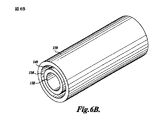
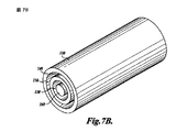
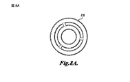
基本的な実施形態においては、装置は、内表面,外表面,第1孔,そして第2孔から成り、内表面が第1孔と第2孔との間の流体連通を提供する内腔を規定している。他の実施形態においては、装置は、内腔中に内方構成要素を備えていて、内方構成要素は、横断面において凸形状であり変性されていない(unmodified)滑らかなガラス表面を備えている。このような装置は、チューブ又は棒(rod)の如き本質的に同心的な結合構成要素の複数を備えていて、これにより装置の内腔中に変性されていない滑らかなガラス結合表面の複数を提供している。図6A及び6Bは、このような装置の例を図示していて、そこでは同心的なガラスチューブ130及び140が2つの内腔150を規定している。外方内腔は凹状及び凸状壁の両方を有していて、内方内腔は凹状壁を有している。図7A及び7Bは、同心的なチューブ130及び140に加えて、中央ガラス棒160を備えているもう1つの実施形態を図示している。この実施形態の範囲内においては、内方及び外方内腔150の両方が凹状及び凸状壁の両方を有している。チューブ及び/又は棒のこのような配列は、以下に記載されている如き保持構成要素の使用を介して安定されることが出来る。図8A及び8B中に示されている如く、この配置は、保持構成要素に対する末端に端キャップ170を提供することによりさらに安定されることが出来る。保持構成要素及び端キャップは、装置の範囲内の全てのガラス表面に対するそれらを通過した流体流れを許容するよう構成される。
In a basic embodiment, the device comprises an inner surface, an outer surface, a first hole, and a second hole, the inner surface providing a lumen that provides fluid communication between the first hole and the second hole. It prescribes. In other embodiments, the device comprises an inner component in the lumen, the inner component comprising a smooth glass surface that is convex and unmodified in cross-section. Yes. Such a device comprises a plurality of essentially concentric coupling components, such as tubes or rods, thereby providing a plurality of smooth glass bonding surfaces that are not modified in the lumen of the device. providing. FIGS. 6A and 6B illustrate an example of such a device in which
ガラスチューブがこの発明の範囲内において使用される時、チューブの端は入口及び出口孔を提供することが出来、中間部分は内腔を規定している。チューブの端(入口及び出口孔)には端キャップ又は他の取り付け部材(fitting)が適合されることが出来、これらを通して、以下により詳細に記載される如く、試薬が追加されそして回収される。このような取り付け部材はまた、装置を密封することが出来る。このような装置はさらに、取扱いを容易にするとともにチューブを破損から保護する為に、保護ハウジング,ガード(guard),取っ手(handle),又はこれらと同様なものを備える。これらの構成要素は、従来の如くポリマー材料(polymeric material)から構成されている。この技術分野において習熟している人々は、ガラスチューブがハウジングに対し適合されることができ、それによって入口及び出口孔がハウジングの表面を通過した開口として形成されてガラスチューブに対する流体の接近を提供する、ことを認識する。 When a glass tube is used within the scope of the present invention, the end of the tube can provide inlet and outlet holes and the middle portion defines a lumen. Ends of tubes (inlet and outlet holes) can be fitted with end caps or other fittings through which reagents are added and recovered as described in more detail below. Such mounting members can also seal the device. Such devices further include a protective housing, guard, handle, or the like to facilitate handling and protect the tube from breakage. These components are made of a polymeric material as is conventional. For those skilled in the art, the glass tube can be fitted to the housing, whereby the inlet and outlet holes are formed as openings through the surface of the housing to provide fluid access to the glass tube Recognize that.
一実施形態においては、内腔の少なくとも一部分の形状及び大きさの割合(proportion)は、そこを通過する液体の乱流を提供するよう選択されている。乱流は、内腔を通過する液体の混合を容易にできる。流れが乱流であろうと層流であろうと、そのレイノズル数(Re)により特徴付けられることが出来る。レイノズル数は粘性力(viscous force)に対する慣性力(inertial force)の比として記載されることが出来、粘性力は速度に対する抵抗として考えられることが出来、そして慣性力は速度における変化に対する抵抗として考えられることが出来る。 In one embodiment, the shape and size proportion of at least a portion of the lumen is selected to provide turbulent flow of liquid therethrough. Turbulence can facilitate mixing of liquids that pass through the lumen. Whether the flow is turbulent or laminar, it can be characterized by its Ray nozzle number (Re). Ray nozzle number can be described as the ratio of inertial force to viscous force, viscous force can be considered as resistance to speed, and inertial force is considered as resistance to change in speed. Can be done.
Re=(pxVsxL)/(u),ここでは:
p=流体密度(kg/m3)
Vs=平均流速(m/s)
L=特性長さ(m),パイプの場合はDh=流体直径(m)
Dh=(4x面積)/(周囲長)、即ち、パイプの断面の面積及び周囲長
u=絶対速度(sN/m2)
Reが2300以下である時、流れは層流であると考えられ、そしてReが4000以上である時、流れは乱流であると考えられる。これら2つの値の間のいずれであっても遷移領域(transition region)であると考えられる。
Re = (pxVsxL) / (u), where:
p = fluid density (kg / m 3 )
Vs = average flow velocity (m / s)
L = characteristic length (m), for pipes Dh = fluid diameter (m)
Dh = (4 × area) / (peripheral length), that is, the cross-sectional area and perimeter of the pipe u = absolute velocity (sN / m 2 )
When Re is less than 2300, the flow is considered laminar and when Re is greater than 4000, the flow is considered turbulent. Any one between these two values is considered a transition region.
典型的なパスツールピペット(Pasteur pipette)は、いずれかの端で、テーパーにされている(tapered)部分により連結されている2つの均一直径区域を有している、直径変更形式である。単純化の為に、2つの均一直径区域におけるレイノズル数は以下の如く計算される。狭い区域は0.9mmの直径を有していて、より大きな区域は5mmの直径を有している。流量は一般的に600μL/秒を超えることがなく、典型的には略60μL/秒である。上述した方程式を使用し、そしてその値は:
L=0.0009m(小さな区域)又は0.005m(大きな区域)
Vs=0.94m/s(小さな区域、早い流れ);0.094m/s(小さな区域、遅い流れ);0.03m/s(大きな区域、早い流れ);0.003m/s(大きな区域、遅い流れ)
P(水)=1000kg/m3
u(水)=1/1000sN/m2
Re=1000x0.094x0.0009x1000=84.9,小さな区域における流量60μL/秒で;そして、Re=1000x0.003x0.005x1000=15.3,大きな区域における流量60μL/秒で、である。600μL/秒の流量では、小さな区域においてはRe=849であり、大きな区域においてはRe=153である。従って、上述した寸法を有している装置は、Reが遷移領域に接近する以前に1625μL/秒を超える流量を適用(accommodate)できる。
A typical Pasteur pipette is a diameter-changing version that has two uniform diameter sections that are joined at either end by a tapered portion. For simplicity, the number of ray nozzles in two uniform diameter areas is calculated as follows: The narrow area has a diameter of 0.9 mm and the larger area has a diameter of 5 mm. The flow rate generally does not exceed 600 μL / sec and is typically about 60 μL / sec. Use the equation above and its value is:
L = 0.0009m (small area) or 0.005m (large area)
Vs = 0.94 m / s (small area, fast flow); 0.094 m / s (small area, slow flow); 0.03 m / s (large area, fast flow); 0.003 m / s (large area, Slow flow)
P (water) = 1000 kg / m 3
u (water) = 1/1000 sN / m 2
Re = 1000 × 0.094 × 0.0009 × 1000 = 84.9, at a flow rate of 60 μL / sec in the small area; and Re = 1000 × 0.003 × 0.005 × 1000 = 15.3, at a flow rate of 60 μL / sec in the large area. At a flow rate of 600 μL / sec, Re = 849 in the small area and Re = 153 in the large area. Thus, a device having the dimensions described above can accommodate a flow rate exceeding 1625 μL / sec before Re approaches the transition region.
この発明の一実施形態の範囲内においては、内腔は曲がりくねった形状をしている。ここで使用されている時、「曲がりくねった(surpentine)」内腔は、2次元において折り曲げられている平坦な内腔を、その螺旋(helix)及び種々の(variant)形状を有している3次元通路とともに、含む。このような3次元構造は、装置全体の容積を減少させるために少なくとも一側に沿い周囲が平坦にされていることが出来る。曲がりくねった形状は、ガラスの大きな表面積に対する標本の露出を許容し、同時に内腔の断面及び装置全体を小さく維持している。内腔断面寸法を限定することは、内腔の範囲内における液体の塊(liquid bolus)の先端縁をするりと過ぎる(slipping past)空気の泡を阻止することに貢献する。曲がりくねった設計はまた、大きな表面積(high surface area)(ガラス−液体界面)と小さな断面との組み合わせがコンパクトな設置面積(footprint)の範囲内に存在することを許容する。上述した如く、(螺旋を含んでいる)曲がりくねった内腔は、円形状横断面及び他の形状を伴っているそれらを含む。
Within the scope of one embodiment of the present invention, the lumen has a serpentine shape. As used herein, a “surpentine” lumen has a flat lumen that is folded in two dimensions, with its helix and
この発明の装置は、変性されていない(unmodified)滑らかなガラスから成る内表面を備えている。この表面は、DNA及びRNAを含んでいる、核酸を結合する為に効果的である。ここで使用される時、「変性されていない滑らかなガラス表面(unmodified smooth glass surface)」は、標準的な顕微鏡スライド(microscope slide),パスツールピペット(Pasteur pipette),ガラス毛管(glass capillary),又はこれらと同様なものの滑らかさに対応している滑らかさを有しているガラス表面を意味していて、ここにおいて表面はその表面積を増大させるよう刻まれていたり(etched)、又は代わりのその他ではなく、そして、それは以下に記載されている如く核酸を特に結合する(bind)よう変性されていない。 The device of the present invention has an inner surface made of unmodified smooth glass. This surface is effective for binding nucleic acids, including DNA and RNA. As used herein, “unmodified smooth glass surface” refers to standard microscope slides, Pasteur pipettes, glass capillaries, Or a glass surface having a smoothness corresponding to the smoothness of these or the like, where the surface is etched to increase its surface area, or other alternative Rather, it has not been denatured to specifically bind nucleic acids as described below.
「滑らかなガラス」から特に除外されるのは、通常はビーズ(bead),フリット(frit),または膜(membrane)形状である、核酸を獲得するよう当該技術分野において知られている多孔性ガラスである。このような多孔性ガラスは通常、0.1μm乃至300μmの範囲内に寸法付けされている多孔を有している。この発明の範囲内において使用の為に適したガラス材料は、ソーダ石灰ガラス(soda lime glass)(例えば、エリ エレクトロベール ガラス:エリ サイエンティフィック カンパニー,ポーツマス,ニューハンプシャー(Erie Electroverre Glass: Erie Scientific Company, Portsmouth, New Hampshire)),珪硼酸ガラス(borosilicate glass)(例えば、コーニング 0211,PYREX(登録商標) 7740;コーニング インコーポレイテッド,コーニング,ニューヨーク(Corning 0211, PYREX(R) 7740; Corning Incorporated, Corning, New York)),亜鉛チタニアガラス(zinc titania glass)(Corning),そして石英ガラス(silica glass)(例えば、VYCOR 7913;コーニング インコーポレイテッド(VYCOR 7913; Corning Incorporated))である。この発明の範囲内において使用の為に適切なのは、種々の寸法が容易に入手可能なガラスチューブである。特に興味深いのは、高価でなく、良好な表面:容積比を提供し、そして、内腔内に試薬の混合を容易にする大きな直径の領域を含む、パスツールピペット(Pasteur pipette)である。上で議論した如く、滑らかなガラス表面を有している、ガラス毛管(glass capillary),クロマトグラフィーカラム(chromatography column),濃縮チューブ(condenser tube),そしてこれらと同様なものも採用することが出来る。内腔は本質的に、結合されたオリゴヌクレオチド(immobilized oligonucleotide),微小溝結合薬剤(minor groove binding agent),内位添加薬剤(intercalating agent),又はこれらと同様なものにより提供された、装填表面(charged surface)又は結合位置(binding site)の如き、核酸特定結合位置(nucleic acid-specific binding site)から自由である。「核酸特定結合位置(nucleic acid-specific binding site)から本質的に自由である」内腔は、ガラスと比較して核酸結合における統計上大きな増大を与えるのに十分なこのような位置を含まないものである。 Specifically excluded from “smooth glass” are porous glasses known in the art to obtain nucleic acids, usually in the form of beads, frit, or membranes. It is. Such porous glass typically has a pore dimensioned in the range of 0.1 μm to 300 μm. Glass materials suitable for use within the scope of the present invention include soda lime glass (eg, Erie Electroverre Glass: Erie Scientific Company, Erie Scientific Company, Portsmouth, New Hampshire), borosilicate glass (eg, Corning 0211, PYREX® 7740; Corning 0211, PYREX® 7740; Corning Incorporated, Corning, New York), zinc titania glass (Corning), and silica glass (eg, VYCOR 7913; Corning Incorporated). Suitable for use within the scope of this invention are glass tubes that are readily available in various dimensions. Of particular interest are Pasteur pipettes that are inexpensive, provide a good surface: volume ratio, and include large diameter areas that facilitate mixing of reagents within the lumen. As discussed above, glass capillaries, chromatography columns, condenser tubes, and the like with smooth glass surfaces can be employed. . The lumen is essentially a loading surface provided by an immobilized oligonucleotide, a minor groove binding agent, an intercalating agent, or the like. Free from nucleic acid-specific binding sites such as (charged surface) or binding sites. “Intrinsically free from nucleic acid-specific binding sites” lumens do not contain enough such positions to give a statistically large increase in nucleic acid binding compared to glass Is.
その最も単純な形状においては、この発明の範囲内において使用されている装置は、個々の端に孔を伴っているガラスチューブである。当該技術分野において習熟している人々は、他の構成を採用できること、そして、種々の形状のガラスチューブをより大きなそしてより複雑な装置中に組み込まれることが出来ること、を認識する。これらの他の装置は、例えば、自動化されている取り扱いを容易にし、壊れやすいガラス構成要素を保護することにより耐久性を増大させ、又は、標本の上流又は下流での取り扱いの為に使用される他の装置と連結するよう、構成されていることが出来る。このような装置の本体の残りは、好ましくは、核酸との低い自蛍光(auto-fluorescence)及び非常に低い結合を発揮する材料から形成されている。この材料はまた、使用中にこれらが接触するかもしれない試薬(例えば、エタノール)に対し不浸透性でなければならない。硬質(rigid)又は半硬質(semi-rigid),有機ポリマー材料が好ましい。このような材料の代表は、アクリル(acrylic)(高分子量硬質材料),ポリカーボネート(polycarbonate),ポリプロピレン(polypropylene)(低表面エネルギー薄膜),セルロースアセテート(cellulose acetate),ポリエチレンテレフタレート(PET:polyethlene terephthalate),ポリ塩化ビニル(polyvinylchloride),そして、高密度ポリエチレン(HDPE:high density polyethylene)を含み、ポリスチレン(polystyrene)を含まない。ポリマー材料を接合する為に適切な接着剤は、限定されることなく、300LSE接着フィルム(3M);467アクリル接着フィルム(3M Company, St. Paul, MN.):8141アクリル接着フィルム(3M Company);そして、トランシルシリコーン接着フィルム(Transil silicone adhesive film)を含む。装置製造後におけるある接着剤のガス発生は、DNA収量を減少させるかもしれず、この問題を軽減する為に真空ガス除去を使用することが出来る。 In its simplest form, the device used within the scope of this invention is a glass tube with holes at each end. Those skilled in the art will recognize that other configurations can be employed and that various shaped glass tubes can be incorporated into larger and more complex devices. These other devices, for example, facilitate automated handling, increase durability by protecting fragile glass components, or are used for handling upstream or downstream of specimens It can be configured to connect with other devices. The remainder of the body of such a device is preferably formed from a material that exhibits low auto-fluorescence and very low binding to nucleic acids. This material must also be impermeable to reagents (eg ethanol) that they may come into contact with during use. Rigid or semi-rigid, organic polymer materials are preferred. Representative of such materials are acrylic (high molecular weight hard material), polycarbonate, polypropylene (low surface energy thin film), cellulose acetate, polyethylene terephthalate (PET). , Polyvinyl chloride, and high density polyethylene (HDPE), but not polystyrene. Adhesives suitable for joining polymeric materials include, but are not limited to, 300 LSE adhesive film (3M); 467 acrylic adhesive film (3M Company, St. Paul, MN): 8141 acrylic adhesive film (3M Company) And including a Transil silicone adhesive film. Gas generation of certain adhesives after device manufacture may reduce DNA yield, and vacuum gas removal can be used to alleviate this problem.
この装置はさらに、それを介して液体を内腔中に導入できるか又は液体を内腔から取り除くことが出来る孔を備えている。従って、この孔は、装置の表面を貫通した開口を提供し内腔と流体連通する。最も単純な構成においては、チューブ端における開口の如き、装置中の開口として入口及び出口孔が提供されている。このような開口は、形状において円形状であることが便利であるが、形状は日常の設計選択事項である。ガラスチューブの端によって孔が提供されている装置は、以下により詳細に開示される如き多岐管(manifold)又は他の保持(retention)構成要素中に直接的に挿入されることが出来る。入口及び出口孔はさらに、標本及び他の試薬が種々の手段により装置中に導入されることを許容する追加の構成要素を備えている。例えば、手動入力を許容するようピークチューブ片(peek tubing stub)が装置に取り付けられることが出来る。手動による追加は、種々の緩衝剤(buffer)が、容量,培養時間(incubation time),そして流量の為に最適となることを許容する。代わりに、標準1−mlポリプロピレン注射器又はプログラム設定可能蠕動ポンプ(peristaltic pump)をチューブ(tubing)及びルアー−固定アダプタ(Luer-lock adaptor)とともに使用することが出来る。もう1つの実施形態の範囲内においては、入口及び出口孔は、そこに挿入される針(例えば、鈍チップ(blunt tip),22G注射針)を受け入れるよう寸法が決められている小さな直径の孔により提供されている。針に対する連結は、ルアー−固定取付部材(Luer-lock fitting)を使用して行われる。もう1つの実施形態においては、入口及び出口孔の夫々は、針又はカニューレ(cannula)が突き刺さることができる弾性隔膜又はキャップ(elastomeric septum or cap)を備えていて、それによって使用時まで密封されている装置を提供している。孔はさらに、O−リング,ガスケット,又はこれらと同様なものの包含により漏れに対し密閉されることが出来る。 The device further comprises a hole through which liquid can be introduced into or removed from the lumen. Thus, this hole provides an opening through the surface of the device and is in fluid communication with the lumen. In the simplest configuration, inlet and outlet holes are provided as openings in the device, such as openings at the tube ends. Such openings are conveniently circular in shape, but shape is an everyday design choice. The device provided with holes by the end of the glass tube can be inserted directly into a manifold or other retention component as disclosed in more detail below. The inlet and outlet holes further comprise additional components that allow specimens and other reagents to be introduced into the device by various means. For example, a peek tubing stub can be attached to the device to allow manual input. Manual addition allows various buffers to be optimized for volume, incubation time, and flow rate. Alternatively, a standard 1-ml polypropylene syringe or a programmable peristaltic pump can be used with tubing and a Luer-lock adaptor. Within another embodiment, the inlet and outlet holes are small diameter holes that are sized to receive a needle (eg, a blunt tip, 22G needle) inserted therein. Is provided by. The connection to the needle is made using a Luer-lock fitting. In another embodiment, each of the inlet and outlet holes is provided with an elastic septum or cap that can be pierced by a needle or cannula, thereby being sealed until use. Equipment is provided. The holes can be further sealed against leakage by inclusion of O-rings, gaskets, or the like.
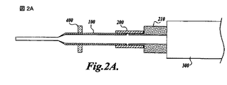
図1は、装置100及びポンプ300を備えている、この発明の組立体を図示している。装置100の第2孔120は保持構成要素(retention element)200中に挿入されている。保持構成要素(retention element)200は、射出成型の如き公知の方法により構成されている。保持構成要素(retention element)200はポンプ300に連結されていて、装置100の内腔との流体連通の為に提供されている。この構成においては、ポンプ300は吸引を適用でき、そして、第1孔110を介して装置100中へ液体を引き入れることができる。或いは、液体は第2孔120を介して装置100の内腔中に分配されることが出来る。図示されている実施形態においては、保持構成要素(retention element)200は、装置100をポンプ300に対し安定した位置に保持するよう設計されている。当該技術分野において習熟している人々は、保持構成要素(retention element)200が種々の別の方法で構成されていることが出来ることを認識する。例えば、保持構成要素(retention element)200は、可撓性の又は硬いチューブから構成されていることが出来、そして装置100はクランプ(clamp)又はそれと同様なものを使用して固定された位置に保持されることが出来る。図示されている例においては、0.25インチ内径(0.25’’ i.d.)ポリウレタン(例えば、TYGON)チューブが、0.27インチ外径(0.27’’ o.d.)のより大きな端を有している従来のパスツールピペット(Pasteur pipette)との緊密な密封を形成している。この寸法のチューブはまた、1ml−注射器又は手持ち(hand-held)ピペットの端に緊密に合致する。このような保持構成要素(retention element)は、3/8インチ長さに切断された薄壁(例えば、1/32インチ)チューブを使用して容易に準備される。
FIG. 1 illustrates an assembly of the present invention comprising a
図1の構成は、複数の装置100の同時使用を提供するよう図2A及び図2B中に示されている如く容易に変更される。組立体600として示されている後者の配置においては、複数の装置100が、薄壁ポリウレタンチューブから構成されている保持構成要素(retention element)200を介して多岐管(manifold)210に連結されている。多岐管(manifold)210は次にポンプ300に連結されていて、そしてポンプ300と装置100との間の流体連通を提供する。このような複数装置組立体は、複数の装置100が、96−溜り(well)板の如き、標準複数溜り(well)板の溜り(well)に対応して配置されているよう構成されることが出来る。図示されている組立体においては、8個の装置100が、96−溜り(well)板中の8つの溜りの一列に整列するよう整列板400により所定位置に保持されている。このような配置においては、組立体は、このような板の1つまたは連続から流体を吸い上げることが出来、そしてこのような板の1つまたは連続中へと流体を排出することが出来る。標本及び試薬(例えば、洗浄及び溶出緩衝剤)は、単一板の異なった列に整列されることが出来、そして、板又は組立体のいずれかが装置の端を適切な溜り(well)中に挿入させるよう移動される。この方法は、手動により、又は自動により実行されることが出来る。複数溜り(well)板は、柔軟なシステムを提供するとともに薄められた標本からの核酸の濃縮を容易にする、溜り容量(well volume)の範囲内(例えば、200μL,0.5mL,1.0mL,2.0mL)で利用可能である。当該技術分野における通常の技術の人々により認められる如く、チューブ(例えば、微小遠心分離チューブ(microcentrifuge tube)),板,又は皿の如き他の容器もまた使用されることが出来る。チューブは、複数溜り(well)板様式(format)により配置されることが出来る。ガラスチューブが使用された時、チューブの内部は、核酸獲得の為に使用されることができるさらなる滑らかなガラス表面を提供する。この配置においては、装置及びチューブのガラス表面から溶出された核酸は装置中に取集されもう一つの容器へと移送されることが出来るし、又はチューブ中に収集されることが出来る」。このような複数―装置組立体の為に、組立体中の個々の装置は個々に動作することが出来るし、又は組立体中の全ての装置が同時に動作することが出来る。
The configuration of FIG. 1 is easily modified as shown in FIGS. 2A and 2B to provide for simultaneous use of
図2Bは、組立体の残りがそこに固定されている取扱い板500をさらに備えている組立体を示している。取扱い板500はさらに組立体600の構成要素を安定させ、そして全組立体の3次元回転を許容する。典型的な核酸抽出方法においては、結合/溶解緩衝剤(binding/lysis buffer)内の核酸包含標本はプンプ300により装置100中に引き入れられ、そして核酸は装置の内壁に結合されることを許容される。装置中に液体を伴って、組立体600は側方に任意に傾けられ(tipped)そして回転され、装置100の上方(幅広)区域中における標本とガラスとの間の接触を最大にする。液体は次に排出され、そして第1洗浄緩衝剤が装置中に引き入れられる。この緩衝剤は、ポンプ300の動作により装置の内腔内で上下に動かされる。この緩衝剤は次に排出され、そして、洗浄が要求に従い繰り返えされる。最終の洗浄の後、空気の流れが装置100中を通過され結合されている核酸を乾燥させる。ポンプ300の形式により、装置100をポンプ300から(多岐管210を伴い、又は伴わず)取り外し、それらを他の手段により提供された空気流に連結させることにより、空気乾燥が容易にされる。最後に、核酸が装置から溶出され、そして、96−溜り(well)板,一組のチューブ,又はこれらと同様なものの中に移送される。ポンプ300はまた、装置100の内方表面を事前洗浄(pre-wash)又は事前処理(pre-treat)為に使用されることが出来る。
FIG. 2B shows the assembly further comprising a
これらの組立体を、以下により詳細に開示されている如く、マイクロプロセッサーにより制御された複数通路ポンプ及び流体分配制御手段に連結されている弁機構に連結させることにより、更なる自動化が提供されることが出来る。このような組立体は、十分に自動化された標本取り扱いを提供するよう、標準的な実験室自動化システム(standard laboratory robotic system)と組み合わされることが出来る。 Further automation is provided by coupling these assemblies to a valve mechanism coupled to a microprocessor controlled multi-pass pump and fluid distribution control means, as disclosed in more detail below. I can do it. Such an assembly can be combined with a standard laboratory robotic system to provide fully automated specimen handling.
装置は通常はチューブの形状を取り、ここにおいては外方断面が内腔の断面と同じ形状である。装置のこの形状は、高価ではなく、貯蔵及び取扱いが容易であり、そして、使用における十分な柔軟性を提供する。 The device typically takes the shape of a tube, where the outer cross section is the same shape as the lumen cross section. This shape of the device is not expensive, is easy to store and handle, and provides sufficient flexibility in use.
一つの実施形態の範囲内においては、装置の外表面が少なくとも1つの長手方向峰(longitudinal ridge)を備えている。峰が付けられている装置は、標本収集の間の組織の破壊(disrupt)及び/又は装置の内腔中への導入に先立つ標本の混合の為に使用されることが出来る。典型的な適用においては、核酸含有材料は緩衝剤とともにチューブ中に置かれ、峰が付けられている装置がチューブ中に挿入されて標本を混合するよう回転され、そして標本が装置中に引き込まれる。 Within one embodiment, the outer surface of the device comprises at least one longitudinal ridge. The ridged device can be used for tissue disruption during sample collection and / or for sample mixing prior to introduction into the lumen of the device. In a typical application, the nucleic acid-containing material is placed in a tube with a buffer, the ridged device is inserted into the tube and rotated to mix the sample, and the sample is drawn into the device .
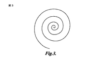
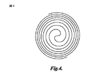
もう1つの実施形態においては、上述された如きチューブ状装置が上に手短に記載されている如くより大きな構造内に収納されている。このような配置は、曲がりくねった内腔を伴っている装置を使用した時にガラスを破壊から保護し取扱いを容易にするのに特に有利である。例えば、螺旋形状毛管(spiral-shaped capillary)チューブは、接着剤,樹脂,エポキシ(epoxy),又は同様なものから準備されたカード状(card-like)又はブロック状(block-like)本体内に含められることが出来る。用語「螺旋(spiral)」はその通常の意味でここで使用され、それは、中心点の回りで連続して徐々に曲げられている形状に曲げられている平面湾曲である。適切な螺旋の例は、アルキメデス螺旋(Archimedean spiral)(図3)及びフェルマーの螺旋(Fermat’s spiral)(図4)を含み、とはいうものの他の形状も採用することが出来る。例えば、ウィキペディア(Wikipedia)(en. wikipedia.org/wiki/Spiral)を参照。ガラスチューブ(例えば、毛管(capillary)チューブ)は、真っ直ぐなガラス毛管(capillary)チューブをその軟化点まで加熱し、それをチューブの整列を保つよう設計されている側壁を伴っているリール(reel)上に巻きつけることにより所望の螺旋形状へと折り曲げることが出来る。螺旋は、単一面構造として、又は複数面(即ち、互いの上に平坦に載置されている2つ又はそれ以上の螺旋)に構成されていることが出来る。螺旋の端は螺旋の平面から僅かに上方を向き、そして突出し第1及び第2孔を提供するよう折り曲げられている。端は次に覆われ、そして本体材料(例えば、接着剤,樹脂,又はエポキシ(epoxy))が螺旋中に注がれるか、又は噴霧されて強度及び取り扱いの容易さを提供する。所望の形状を創出する為に型(mold)を使用することが出来、それは保持体又は多岐管(manifold)に対する装置の合致を容易にするよう整列孔,細長孔,又は突起を含むことが出来る。材料が硬化した後には、チューブ端(孔)は覆われない。典型的な実施形態においては、結果としての構造はその上方面上に第1及び第2孔を伴っている平坦な円板の形状である。孔には上に詳細に記載されていた如き追加の構成要素が設けられていることが出来る。本体材料中に孔を残すことにより観察窓が設けられて良い。 In another embodiment, the tubular device as described above is housed in a larger structure as briefly described above. Such an arrangement is particularly advantageous for protecting the glass from breakage and facilitating handling when using a device with a tortuous lumen. For example, a spiral-shaped capillary tube is placed in a card-like or block-like body prepared from an adhesive, resin, epoxy, or the like. Can be included. The term “spiral” is used herein in its ordinary sense, which is a planar curvature that is bent into a shape that is bent gradually and continuously around a center point. Examples of suitable spirals include the Archimedean spiral (FIG. 3) and the Fermat's spiral (FIG. 4), although other shapes may be employed. See, for example, Wikipedia (en. Wikipedia.org/wiki/Spiral). A glass tube (eg, a capillary tube) is a reel with sidewalls designed to heat a straight glass capillary tube to its softening point and keep it aligned. It can be folded into a desired spiral shape by winding it up. The helix can be configured as a single face structure or in multiple faces (ie, two or more helices mounted flat on top of each other). The end of the helix faces slightly upward from the plane of the helix and is bent to project and provide first and second holes. The edges are then covered and body material (eg, adhesive, resin, or epoxy) is poured or sprayed into the helix to provide strength and ease of handling. A mold can be used to create the desired shape, which can include alignment holes, elongated holes, or protrusions to facilitate the fitting of the device to a retainer or manifold. . The tube ends (holes) are not covered after the material is cured. In an exemplary embodiment, the resulting structure is in the form of a flat disc with first and second holes on its upper surface. The holes can be provided with additional components as described in detail above. An observation window may be provided by leaving a hole in the body material.
構成の代わりの方法は、当該技術分野における通常の技術の人々には明らかである。例えば、積層されているプラスチック構造は、リードその他(Reed et al.),米国20090215125AIにより開示されている如く本質的に採用されることが出来る。手短かには、個々のポリマー層は、レーザー切断,CNC引きずりナイフ(drag knife)切断,そして打ち抜き(die cutting)の如き公知の方法を使用して形状に切断される。接着剤層が乾燥プラスチックの層間に行くよう準備される。接着剤層は通常、プラスチックの為に使用されたのと同じ方法を使用して切断されることが出来る薄いフィルムとし)て入手可能な感圧接着剤である。接着剤は、接着剤(Adhesive)−担持体(Carrier)−接着剤(Adhesive)(ACA)様式で使用されてよく、ここで担持体(Carrier)は装置の他の層において使用されているのと同じであることが好ましい。スクリーン印刷の如き、液体接着剤を適用する他の方法もまた採用されて良い。いくつかの層が互いに整合され(registered)そして共に押される。整列孔の如き、整合を助けるための構成が最終設計中に好ましくは組み込まれる。硬化作業周期(cure cycle)中の圧力及び温度は接着剤−従属であり;適切な状況の選択は当該技術分野における通常の技術の水準の範囲内である。代わりに、装置は、20090215125AI中に開示されている如き圧縮密封(compression seal)の使用を介して組み立てられることが出来る。積層は、上述した如き成型構成要素を組み込むことが出来る。 Alternative methods of construction will be apparent to those of ordinary skill in the art. For example, laminated plastic structures can be employed essentially as disclosed by Reed et al., US200902125125AI. Briefly, individual polymer layers are cut into shapes using known methods such as laser cutting, CNC drag knife cutting, and die cutting. An adhesive layer is prepared to go between the layers of dry plastic. The adhesive layer is usually a pressure sensitive adhesive available as a thin film that can be cut using the same methods used for plastics. The adhesive may be used in an Adhesive-Carrier-Adhesive (ACA) mode, where the Carrier is used in other layers of the device. Is preferably the same. Other methods of applying a liquid adhesive, such as screen printing, can also be employed. Several layers are registered with each other and pushed together. Configurations to aid alignment, such as alignment holes, are preferably incorporated into the final design. The pressure and temperature during the cure cycle are adhesive-dependent; the selection of the appropriate situation is within the level of ordinary skill in the art. Alternatively, the device can be assembled through the use of a compression seal as disclosed in 200090215125AI. Lamination can incorporate molded components as described above.
この発明はまた、ここに記載された如き装置及び装置の内腔と流体連結されているポンプを備えている組立体を提供する。用語「ポンプ(pump)」は、ここにおいては、手動で操作される装置(例えば、注射器及び複数通路ピペット(multi-channel pipettor))及び動力で操作される(例えば、電気)装置の両方を含むよう使用されている。この組立体は、ポンプが流体を孔の1つ又は両方を介して内孔中に分配し、そして内腔からそれらを取り除くことが出来るよう構成されている。ポンプはその能力が以下の尺度に合致するよう選択され:(1)20μL乃至少なくとも1000μL,そして好ましくは最大2.5mLまで、の範囲内の容積を正確に吐出する能力;(2)液体と同様空気を効果的に押し出す能力;そして(3)逆にも操作できる能力、である。注射器型又は蛇腹型(bellows-type)ポンプはこれらの尺度を満足し、そして、装置が、第1及び第2孔の1つが全ての試薬の導入及び除去の為に使用される、従来のピペットの方法で動作されることを許容する。液体が両方の孔を介して移動される時、試薬の相互汚染(cross contamination)を最小にする低い又はゼロ無駄容積(dead volume)もまた提供し、そして、使用された種々の試薬(例えば、カオトロピズムの塩(chaotropic salt)及びエタノール)と共存可能な材料で形成された濡れた表面を有するポンプを使用することが好ましい。蠕動ポンプ(peristaltic pump)はこれらの特色の全てとの良好な動作組み合わせ提供するが、全てのポンプ付属品の最も正確な容積排出を提供しない。蠕動ポンプ(peristaltic pump)は、液体のより大きな容積が取り扱われる時に使用されることが好ましい。コンピュータにより制御された複数通路蠕動ポンプ(peristaltic pump)(例えば、ISMATEC 12−通路ポンプ;イスマテック SA,グラットブルグ,スイス(Ismatec SA, Glattbrugg, Switzerland))は、同時に複数の装置を収容し、そして、開始/停止/流量の変更又は流れの方向の逆転をプログラムされることが出来る。他のポンプ様式を採用した時には、複数のポンプが特定の機能の為に要求され、このような配置が全流体管理システムを複雑にする。 The present invention also provides an assembly comprising a device as described herein and a pump in fluid communication with the lumen of the device. The term “pump” as used herein includes both manually operated devices (eg, syringes and multi-channel pipettors) and powered (eg, electrical) devices. Have been used. The assembly is configured such that the pump can distribute fluid into the bore through one or both of the bores and remove them from the lumen. The pump is selected so that its capacity meets the following scale: (1) Ability to accurately dispense volumes in the range of 20 μL to at least 1000 μL, and preferably up to 2.5 mL; (2) Similar to liquid The ability to push air effectively; and (3) the ability to operate in reverse. Syringe-type or bellows-type pumps satisfy these measures, and the device is a conventional pipette in which one of the first and second holes is used for the introduction and removal of all reagents. It is allowed to be operated in the manner of When the liquid is moved through both holes, it also provides a low or zero dead volume that minimizes cross contamination of the reagents, and the various reagents used (eg, It is preferred to use a pump having a wet surface formed of materials compatible with chaotropic salts and ethanol. A peristaltic pump provides a good operating combination with all of these features, but does not provide the most accurate volumetric discharge of all pump accessories. Peristaltic pumps are preferably used when larger volumes of liquid are handled. A computer controlled peristaltic pump (eg, ISMATEC 12-passage pump; Ismatec SA, Glattbrugg, Switzerland) accommodates multiple devices simultaneously, and Start / stop / flow change or flow direction reversal can be programmed. When other pump styles are employed, multiple pumps are required for specific functions, and such an arrangement complicates the overall fluid management system.
この発明の組立体はさらに、ポンプとの流体連通において流体分配制御手段を含んで良い。流体分配制御手段は、複数の流体が装置を介して連続してポンプ作用されることを許容する、典型的には弁−多岐管ブロックの形状をしている、1つ又はそれ以上の弁を備えている。多岐管入口及び出口が殺菌(sterile)フィルターを通過して弁−多岐管組立体を汚染から阻止し、そして、出口配管が逆止弁を有してポンプチューブから多岐管中への逆流を阻止することが好ましい。例示的な流体分配制御手段は、アップチャーチ サイエンティフィク,オーク ハーバー,ワシントン(Upchurch Scientific, Oak Harbor, Washington)により製造されているV−1241−DC型6位置7孔回転選択弁である。この選択弁は試薬間への空気隙間の導入を許容する。流体分配制御手段はさらに、外部から弁機構に、又はそれらと十分に一体化されているプログラム可能コンピュータを備えていて良い。この発明の或る実施形態においては、プログラム可能コンピュータはデスクトップ又はラップトップパーソナルコンピュータである。他の実施形態においては、プログラム可能コンピュータは専用マイクロプロセッサー装置である。例示的なシステムにおいては、流体分配の制御は、マイクロソフトエクセル用のビジュアルベーシック(Visual Basic for Microsoft Excel)により書かれていてパーソナルコンピュータ上で作動するアプリケーションを使用して複数通路蠕動ポンプとの組み合わせにおける上述した選択弁を使用して達成される。弁機構及びポンプの両方はRS232制御インターフェースを特徴にしている(feature)。これ等の構成要素はコンピュータのUSBポートを介したエクセル(Excel)又はUSB対シリアルコンバーター(USB-to-Serial converter)を使用してアドレス指定されている(addressed)。当該技術分野において習熟している人々により理解される如く、カスタムファームウェアソフトウェア(custom firmware software)もまた採用されて良い。 The assembly of the present invention may further include fluid distribution control means in fluid communication with the pump. The fluid distribution control means includes one or more valves, typically in the shape of a valve-manifold block, that allow a plurality of fluids to be pumped sequentially through the device. I have. Manifold inlet and outlet pass through sterile filter to prevent valve-manifold assembly from contamination, and outlet tubing has check valve to prevent back flow from pump tube into manifold It is preferable to do. An exemplary fluid distribution control means is a V-1241-DC 6 position 7 hole rotary select valve manufactured by Upchurch Scientific, Oak Harbor, Washington. This selection valve allows the introduction of an air gap between the reagents. The fluid distribution control means may further comprise a programmable computer that is externally integrated into or fully integrated with the valve mechanism. In some embodiments of the invention, the programmable computer is a desktop or laptop personal computer. In other embodiments, the programmable computer is a dedicated microprocessor device. In an exemplary system, fluid distribution control is in combination with a multi-pass peristaltic pump using an application written on Visual Basic for Microsoft Excel and running on a personal computer. This is accomplished using the selection valve described above. Both the valve mechanism and the pump feature an RS232 control interface. These components are addressed using Excel or USB-to-Serial converter via a computer USB port. Custom firmware software may also be employed, as will be appreciated by those skilled in the art.
液体試薬が従来は、殺菌フィルター孔が備えられている隔膜(septum)−密封小瓶(vial)中に貯蔵されている。小瓶は、隔膜を通して挿入され微小孔チューブを介して多岐管入口に連通されている標準ルアー型(Luer-type)針を使用して、流体分配制御手段に連結されて良い。 Liquid reagents are conventionally stored in septum-sealed vials equipped with sterilizing filter holes. The vial may be connected to the fluid distribution control means using a standard Luer-type needle inserted through the septum and communicating with the manifold inlet via a microporous tube.
組み立て後、装置は好ましくは、病原体(pathogen)を取り除く為に酸化エチレン(ethylene oxide)又はγ殺菌(gamma sterilization)により処理される。装置で使用される試薬は好ましくは、汚染を取り除く為に入口で2ミクロンセルロースフィルターを通過する。核酸増幅を妨げるかもしれない汚染を含む、汚染を取り除く他の方法は、リードその他(Reed et al.),WO2008002882により開示されている。装置上の試薬孔は、黄色又は青色ピペットチップ(pipette tip)に対するインターフェースを提供を提供して良い。針−隔膜(septum)インターフェースが設けられることが出来る。 After assembly, the device is preferably treated with ethylene oxide or gamma sterilization to remove pathogens. The reagent used in the device preferably passes through a 2 micron cellulose filter at the inlet to remove contamination. Other methods of removing contamination, including contamination that may interfere with nucleic acid amplification, are disclosed by Reed et al., WO2008002882. Reagent holes on the device may provide an interface to a yellow or blue pipette tip. A needle-septum interface can be provided.
液体標本は通常、略0.1niL/分乃至略5.0mL/分の流量で装置中に導入されるが、しかしながら、上述された如く、非常により大きな流量が使用されることが出来る。実際の流量は、流体通路の全容量及び内腔の形状を考慮に入れた設計従属である。 Liquid specimens are typically introduced into the apparatus at a flow rate of approximately 0.1 niL / min to approximately 5.0 mL / min, however, as described above, much higher flow rates can be used. The actual flow rate is design dependent taking into account the total volume of the fluid passage and the shape of the lumen.
希釈又は濃縮された標本が、装置中への入力の為に準備されることが出来る。無傷な細胞(intact cell)の溶解(lysis)及び蒸解(digestion)は、残留タンパク質(例えば、ヒストン(histone))からDNA又はRNAを解放する。或いは、固体標本(例えば、布上のバクテリア胞子(bacterial spore)又は乾燥した血液)又は半固体標本(例えば、マウスの尻尾又は痰/便(sputum/stool))は、装置中に入れられる前に均質化され(homogenized)そして溶解される(lysed)ことが出来、装置の内腔を通過して流れる均質化され、そして非粘性の標本を提供する。血液の如きより粘性の標本もまた使用されることが出来る。 Diluted or concentrated specimens can be prepared for input into the device. Intact cell lysis and digestion releases DNA or RNA from residual proteins (eg, histones). Alternatively, solid specimens (eg, bacterial spore or dry blood on cloth) or semi-solid specimens (eg, mouse tail or sputum / stool) must be placed in the device. It can be homogenized and lysed, providing a homogenized and non-viscous specimen that flows through the lumen of the device. More viscous specimens such as blood can also be used.
核酸は、少なくとも0.5M乃至略2M又は溶解度に従いそれ以上の濃度の塩(例えば、KCI)、又は、少なくとも1M乃至略6M又は溶解度の限度までの濃度のカオトロープ(chaotrope)(例えば、グアニジン(guanidine)HCI又はグアニジンチオシアン酸塩(guanidine thiocyanate))の存在で、装置のガラス表面に結合される。核酸の結合は通常、略5乃至8、好ましくは略6のPHで行われる。内腔は次に、塩濃度を減少させる緩衝剤が入れられた溶液(buffered solution)を使用して洗浄される。塩濃度が減少するのに伴い、エタノールが洗浄溶液に加えられ、ガラス上に核酸を保持し、そして、核酸増幅の如き下流の工程と干渉するかもしれない汚染物質を取り除く。洗浄はPH6〜9、通常はPH6〜8、で行われる。核酸は、基本(basic)PH、通常はPH8〜9、の低塩溶液により装置から溶出される(eluted)。 Nucleic acids can be salts of at least 0.5M to about 2M or higher according to solubility (eg, KCI), or chaotrope (eg, guanidine) at a concentration of at least 1M to about 6M or up to the limit of solubility. ) In the presence of HCI or guanidine thiocyanate) it is bonded to the glass surface of the device. Nucleic acid binding is usually performed at a pH of about 5 to 8, preferably about 6. The lumen is then cleaned using a buffered solution that reduces the salt concentration. As the salt concentration decreases, ethanol is added to the wash solution to retain the nucleic acids on the glass and remove contaminants that may interfere with downstream processes such as nucleic acid amplification. Cleaning is performed at PH 6-9, usually PH 6-8. The nucleic acid is eluted from the device with a low salt solution of basic PH, usually PH 8-9.
一般的には、生物学的標本中に細胞が存在している時には、それらは溶解されてそこから核酸が抽出される細胞溶解産物を提供する。細胞溶解の種々の方法が当該技術分野において知られていて、この発明の範囲内における使用の為に適している。細胞溶解方法の例は、(例えば、プロティナーゼ K(proteinase K),プロナーゼ(pronase),又はサブチリシン(subtilisin)を使用した)酵素処置(enzymatic method),(例えば、音波破砕(sonication),高圧の適用,圧電ブザー(piezobuzzer)装置の使用,又はビーズ叩解(bead beating)による)機械的な破砕(mechanical disruption),又は、化学的な処理を含む。機械的な破砕(mechanical disruption)の為に使用されるビーズは、破砕状況の下で核酸と結合しない物質で作られていなければならない。適切な物質は、アクリル(acrylic),ポリカーボネート(polycarbonate),ポリプロピレン(polypropylene),セルロースアセテート(cellulose acetate),ポリエチレンテレフタレート(polyethlene terephthalate),ポリ塩化ビニル(polyvinylchloride),そして、高密度ポリエチレン(high density polyethylene)を含む。カオトロピズムの塩(chaotropic salt)の溶液により処理された標本中の溶解細胞は、特に好ましい。カオトロピズムの塩(chaotropic salt)を使用した細胞の溶解の為の方法及び試薬は、当該技術分野において知られていて、そして、試薬は商業的な供給者(commercial supplier)から購入することが出来る。特別な試薬構成及び反応条件は、溶解する細胞の型により一部が決められ、そしてこのような決定は当該技術分野における通常の技術の水準の範囲内である。適切なカオトロピズムの塩(chaotropic salt)は、グアニジンチオシアン酸塩(guanidine thiocyanate),グアニジン塩酸塩(guanidine hydrochloride),沃化ナトリウム(sodium iodide),そして、過塩素酸塩ナトリウム(sodium perchlorate)を含む。血液細胞を溶解させる為に好ましいグアニジン塩酸塩(guanidine hydrochloride)は、1M乃至10M、普通は1M乃至5M、通常は1M乃至3M、の濃度で使用される。塩の飽和点(saturation point)(12M)に近づく、沃化ナトリウム(sodium iodide)のより高度な濃度が要求されている。過塩素酸塩ナトリウム(sodium perchlorate)は、中間の濃度、普通は5M周辺、で使用されることが出来る。塩化カリウム(potassium chloride)及び塩化ナトリウム(sodium chloride)の如き中性塩(neutral salt)もまた、ガラス表面に対する核酸の結合を得るために使用されることが出来、そして、細胞溶解が要求されていないか、または他の手段により達成される時(例えば、バクテリア細胞分解の場合)、カオトロピズムの塩(chaotropic salt)の代わりに使用されて良い。中性塩(neutral salt)を使用する時、緩衝剤のイオン強度(ionic strength)は少なくとも0.25Mでなければならない。例示的な分解緩衝剤は、PH6.4のグアニジンチオシアン酸塩(guanidine thiocyanate)(GuSCN)緩衝剤の2M溶液である。カオトロピズムの塩(chaotropic salt)溶液中での溶解(lysis)はまた、ゲノムの(genomic)DNAからヒストンプロテイン(histone protein)を取り除き、そしてヌクレアーゼ(nuclease)を不活性にする。溶解緩衝剤は一般的にはまた、中性近傍乃至わずかに酸性のPHを維持するよう1つ又はそれ以上の緩衝剤を包含する。適切な緩衝剤はクエン酸塩ナトリウム(sodium citrate)である。1つ又はそれ以上の洗浄剤も含まれることが出来る。適切な洗浄剤は、例えばポリオキシエチレンソルビタン(polyoxyethylenesorbitan) モノラウレート(monolaurate)(TWEEN20),t−オクチルフェノキシポリエソキシエタノール(t-octylphenoxypolyethoxyethanol)(TRITON X−100),ドデシル硫酸塩ナトリウム(sodium dodecyl sulfate)(SDS),NP−40,CTAB,CHAPS,そしてサーコシル(sarkosyl)を含む。アルコール、普通はエタノール、が、洗浄剤中の低下された塩濃度を補償する為に選択された実際の濃度により、分解及び洗浄溶液中に含まれている。塩の不在においては、アルコールは最終洗浄剤中に、少なくとも50%、好ましくは70%アルコールの濃度で含まれている。もし塩が試薬中に含まれているのであれば、アルコール濃度は通常10%と80%との間、しばしば10%と60%との間、通常は20%と50%との間、の範囲である。緩衝剤の最高活用化(optimization)は当該技術分野において通常の技術の水準の範囲内である。溶解は一般には、細胞の型に従い、室温と略95℃との間で行われる。血液細胞は従来は室温で溶解された。シリカ微粒子(silica particle)は核酸と結合し抽出工程の効率を減少させるので、細胞溶解中でシリカ微粒子(silica particle)の使用を避けることは一般的に好ましい。必要ではないにも関わらず、DNAは、抽出装置中への溶解産出物(lysate)の装填に先立って、剪断されて(sheared)良い。DNAを剪断する(shearing)する為の方法は、当該技術分野において知られている。 Generally, when cells are present in a biological specimen, they provide a cell lysate that is lysed and from which nucleic acids are extracted. Various methods of cell lysis are known in the art and are suitable for use within the scope of this invention. Examples of cell lysis methods include enzymatic methods (eg, using proteinase K, pronase, or subtilisin), (eg, sonication, high pressure application) , Including the use of a piezobuzzer device, mechanical disruption by bead beating, or chemical treatment. Beads used for mechanical disruption must be made of materials that do not bind nucleic acids under disruption conditions. Suitable materials are acrylic, polycarbonate, polypropylene, cellulose acetate, polyethlene terephthalate, polyvinylchloride, and high density polyethylene. )including. Particularly preferred are lysed cells in the specimen treated with a solution of chaotropic salt. Methods and reagents for lysis of cells using chaotropic salts are known in the art and reagents can be purchased from commercial suppliers. The particular reagent configuration and reaction conditions are determined in part by the type of cell being lysed, and such determination is within the level of ordinary skill in the art. Suitable chaotropic salts include guanidine thiocyanate, guanidine hydrochloride, sodium iodide, and sodium perchlorate. Preferred guanidine hydrochloride for lysing blood cells is used at a concentration of 1M to 10M, usually 1M to 5M, usually 1M to 3M. Higher concentrations of sodium iodide are required, approaching the salt saturation point (12M). Sodium perchlorate can be used at intermediate concentrations, usually around 5M. Neutral salts such as potassium chloride and sodium chloride can also be used to obtain nucleic acid binding to the glass surface and cell lysis is required. When not achieved or achieved by other means (eg, in the case of bacterial cell degradation), it may be used in place of the chaotropic salt. When using a neutral salt, the buffer must have an ionic strength of at least 0.25M. An exemplary degradation buffer is a 2M solution of guanidine thiocyanate (GuSCN) buffer at PH 6.4. Lysis in chaotropic salt solution also removes histone proteins from genomic DNA and inactivates nucleases. Lysis buffers generally also include one or more buffers to maintain near neutral to slightly acidic pH. A suitable buffer is sodium citrate. One or more cleaning agents may also be included. Suitable detergents are, for example, polyoxyethylenesorbitan monolaurate (TWEEN 20), t-octylphenoxypolyethoxyethanol (TRITON X-100), sodium dodecyl sulfate sulfate (SDS), NP-40, CTAB, CHAPS, and sarkosyl. Alcohol, usually ethanol, is included in the degradation and cleaning solution, with the actual concentration selected to compensate for the reduced salt concentration in the cleaning agent. In the absence of salt, the alcohol is included in the final detergent at a concentration of at least 50%, preferably 70% alcohol. If salt is included in the reagent, the alcohol concentration is usually between 10% and 80%, often between 10% and 60%, usually between 20% and 50%. It is. The optimization of the buffer is within the level of ordinary skill in the art. Lysis is generally performed between room temperature and approximately 95 ° C., depending on the cell type. Blood cells were conventionally lysed at room temperature. It is generally preferred to avoid the use of silica particles during cell lysis because silica particles bind to nucleic acids and reduce the efficiency of the extraction process. Although not required, the DNA may be sheared prior to loading the lysate into the extraction device. Methods for shearing DNA are known in the art.
核酸含有標本は、孔の1つを介して装置中に導入される。核酸は、上述した如く、塩又カオトロピズムの塩(chaotropic salt)の存在の下でガラス表面上に獲得される。ガラスに対する核酸の満足のいく結合は室温(15°〜30℃,普通は約20℃)で達成されるが、抽出工程は、上限37〜42℃又は上限56℃の如き、より高い温度で行うことが出来、とはいうものの、より高い温度は核酸の回収率(recovery)を減少させるかもしれない。標本はある時間装置中に放置されることが許容されて良く、そして標本溶液は内腔を通して前後に動かされて(pumped)良い。洗浄緩衝剤は次に1つの孔中に、蠕動ポンプ,注射器,またはピペット(pipetter)の使用による如く、送り込まれる。洗浄緩衝剤の選択は部分的には、標本装填溶液の組成(composition)に従属する。一般的には、塩濃度は洗浄工程の間に減少され、そしてPHはわずかに増大される。溶解緩衝剤がカオトロピズムの塩(chaotropic salt)を包含しているのであれば、最初の洗浄剤は、同じ又は幾分低い濃度の塩(例えば、1〜3MGuSCN)もまた普通は含む。最後の洗浄剤は、下流の工程における核酸増幅の抑制を最小にするために、エタノール濃度を50%以下、好ましくは略10%〜20%まで、減少させなければならない。洗浄溶液のアルコール含有量は通常は20%と80%との間の範囲にある。少なくとも50%エタノール、好ましくは略70%エタノールを含有している洗浄溶液は、核酸獲得を向上させることが分かっている。装置の内腔からの最後の洗浄剤の完全な除去はまた、或る実施形態において必要である。最後の洗浄剤のこの除去の為の方法は、空気ポンプを使用して内腔の表面上に空気を1乃至3分間通過させることによる乾燥を含む。洗浄後は、核酸は最後の洗浄剤よりも高いPHの低塩緩衝剤により装置から溶出される。溶出緩衝剤は典型的にはPH≧8.0の低イオン強度(ionic strength)の緩衝された(buffered)溶液であり、とはいうものの核酸は水によって装置から溶出されることが出来る。溶出は最大略56℃までの周囲温度で実施することが出来る。 The nucleic acid containing specimen is introduced into the device through one of the holes. Nucleic acids are acquired on the glass surface in the presence of salts or chaotropic salts as described above. Satisfactory binding of nucleic acids to glass is achieved at room temperature (15 ° -30 ° C., usually about 20 ° C.), but the extraction process is carried out at higher temperatures, such as an upper limit of 37-42 ° C. or an upper limit of 56 ° Although higher, higher temperatures may reduce nucleic acid recovery. The specimen may be allowed to stand in the device for some time and the specimen solution may be pumped back and forth through the lumen. The wash buffer is then pumped into one hole, such as by use of a peristaltic pump, syringe, or pipette. The choice of wash buffer depends in part on the composition of the specimen loading solution. In general, the salt concentration is decreased during the washing step and the PH is slightly increased. If the lysis buffer includes a chaotropic salt, the initial detergent will usually also contain the same or somewhat lower concentration of salt (eg, 1-3 MGuSCN). The last detergent should reduce the ethanol concentration to 50% or less, preferably approximately 10% to 20%, in order to minimize the suppression of nucleic acid amplification in downstream processes. The alcohol content of the cleaning solution is usually in the range between 20% and 80%. A wash solution containing at least 50% ethanol, preferably about 70% ethanol, has been found to improve nucleic acid acquisition. Complete removal of the last cleaning agent from the device lumen is also necessary in certain embodiments. The method for this removal of the final cleaning agent includes drying by passing air over the surface of the lumen using an air pump for 1-3 minutes. After washing, the nucleic acid is eluted from the device with a low salt buffer with a higher pH than the last detergent. The elution buffer is typically a buffered solution of low ionic strength with a PH ≧ 8.0, although nucleic acid can be eluted from the device with water. Elution can be performed at ambient temperatures up to approximately 56 ° C.
この装置の設計は、液体及び気体の両方を含んでいる流体が一方の開口から他方の開口へと装置を通過することを許容する。この方法においては、緩衝剤は、洗浄及び溶出の効率を向上させる為に内腔を通って前後に動かされる(pumped)ことが出来、そして空気は残留緩衝剤を取り除く為に洗浄剤の間を通って動かされる(pumped)ことが出来る。装置は、試薬の導入及び除去に関して種々の方法で構成されることが出来る。1つの配置においては、試薬は孔の1つを介して装置の内腔中に導入され、そして他の孔を介して除去される。第2の配置においては、1つの孔が試薬の為の入口及び出口の両方として働き、そして第2の孔は吸引及び加圧を提供するポンプに連結されている。この第2の配置は、ポンプ及び流体分配制御手段が試薬と接触することを避けさせ、そして、腐食性薬品又は強力な溶剤である試薬を使用するのであれば特に好ましい。第3の配置は、第1及び第2の配置を組み合わせて幾つかの流体が1つの孔から他の孔まで完全に装置を完全に通って通過し、そして他の流体が同じ孔を介して導入され除去されるようにする。例えば、核酸を含有している標本が第1の孔を介して内腔中に導入され、そして第2の孔を介して除去されることが出来、そして、洗浄及び溶出試薬が第1の孔を介して適用された吸引及び空気圧力を使用して第2を介し導入されそして除去される。当該技術分野に習熟している人々は、これらの基本的な配置における多くの変更が可能であることを認識する。 This design of the device allows fluid containing both liquid and gas to pass through the device from one opening to the other. In this method, the buffer can be pumped back and forth through the lumen to improve the efficiency of washing and elution, and air can be passed between the detergent to remove residual buffer. Can be pumped through. The apparatus can be configured in various ways with respect to reagent introduction and removal. In one arrangement, the reagent is introduced into the lumen of the device through one of the holes and removed through the other hole. In the second arrangement, one hole serves as both the inlet and outlet for the reagent, and the second hole is connected to a pump that provides suction and pressurization. This second arrangement is particularly preferred if the pump and fluid distribution control means avoid contact with the reagent and use reagents that are corrosive chemicals or strong solvents. The third arrangement combines the first and second arrangements so that some fluid passes completely through the device from one hole to the other and other fluids pass through the same hole. To be introduced and removed. For example, a sample containing nucleic acid can be introduced into the lumen through a first hole and removed through a second hole, and washing and elution reagents can be removed from the first hole. Are introduced and removed via the second using suction and air pressure applied via. Those skilled in the art recognize that many changes in these basic arrangements are possible.
当該技術分野に習熟している人々により理解されている如く、実際の動作容量は、内腔容量を含む、装置の寸法により、いつもの経験設計によっても、決定される。パスツールピペット(Pasteur pipette)と比較しえる小容量装置の為には、容量は普通は略20μLから略500μLまでの範囲であるが、最大1mLまでの又は2.5mLと同じ量までのより大きな容量が使用されることが出来る。標本は、溶出緩衝剤の容量を減少させることにより濃縮されることが出来る。 As will be appreciated by those skilled in the art, the actual operating capacity is determined by the size of the device, including the lumen volume, and by routine experience design. For small volume devices that can be compared to Pasteur pipettes, the volume is usually in the range of about 20 μL to about 500 μL, but larger up to 1 mL maximum or the same volume as 2.5 mL Capacity can be used. The sample can be concentrated by reducing the volume of elution buffer.
抽出された核酸の計量(quantitation)は溶出緩衝剤内における蛍光配合物(fluorescent compound)の包含により容易にされ、これによって、抽出された核酸が装置内に依然としてある間に、抽出工程上での迅速な品質チェックを提供する。従って、この発明の一つの実施形態の範囲内においては、核酸が核酸の濃度に応じた蛍光発光強度を有している蛍光配合物(fluorescent compound)と接触され、そして蛍光配合物(fluorescent compound)の蛍光発光が測定される。核酸の濃度に応じた蛍光発光強度を有している蛍光配合物(fluorescent compound)は、核酸の存在により蛍光発光強度における適合−従属変化(conformation-dependent change)を発揮する蛍光配合物(fluorescent compound)である。役に立つ蛍光配合物(fluorescent compound)は、核酸の存在によりその強度を増加させる配合物を含む。代表的な蛍光配合物(fluorescent compound)は、ビス−ベンズイミダイン配合物(bis-benzimidine compound)の如き蛍光助剤微小溝結合薬剤(fluorogenic minor groove binder agent)及びエチジウムブロミド(ethidium bromide)の如き内位添加蛍光助剤薬剤(intercalating fluorogenic agent)、そして商業的に入手可能な蛍光染料(fluorescent dye)(例えば、SYBR Green;インビトロゲン会社(Invitrogen Corp.))を含む。蛍光配合物(fluorescent compound)は、溶出緩衝剤内で装置中に導入されることが出来るし、又は内腔中で動けなくされていることが出来る。内腔中で蛍光配合物(fluorescent compound)を動けなくする為の方法及び役に立つ蛍光配合物(fluorescent compound)は、リードその他(Reed et al.),米国出願公報20060166223A1中に記載されている。この発明の装置は、内腔中で蛍光配合物に対し励起エネルギーを伝達する為、そして内腔中で配合物からの蛍光発光放射強度(fluorescence emission intensity)を伝達する為に適した内腔の少なくとも一部分を有することにより、蛍光による内腔の調査(interrogation)を許容する。 The quantitation of the extracted nucleic acid is facilitated by the inclusion of a fluorescent compound in the elution buffer, which allows the extracted nucleic acid to remain on the extraction process while still in the device. Provide quick quality checks. Thus, within one embodiment of the invention, the nucleic acid is contacted with a fluorescent compound having a fluorescence emission intensity that depends on the concentration of the nucleic acid, and the fluorescent compound. The fluorescence emission of is measured. A fluorescent compound having a fluorescence emission intensity depending on the concentration of nucleic acid exhibits a conformation-dependent change in fluorescence emission intensity due to the presence of nucleic acid. ). Useful fluorescent compounds include those that increase their intensity due to the presence of nucleic acids. Typical fluorescent compounds are fluorogenic minor groove binder agents such as bis-benzimidine compounds and internal positions such as ethidium bromide. Includes added intercalating fluorogenic agents, and commercially available fluorescent dyes (eg, SYBR Green; Invitrogen Corp.). The fluorescent compound can be introduced into the device within the elution buffer or can be immobilized in the lumen. A method and useful fluorescent compound for immobilizing a fluorescent compound in a lumen is described in Reed et al., US Application Publication No. 200601626223 A1. The device of the present invention provides a lumen suitable for transmitting excitation energy to a fluorescent formulation in the lumen and for transmitting fluorescence emission intensity from the formulation in the lumen. Having at least a portion allows for fluorescence interrogation.
第1には如何なる蛍光DNA−結合染料もこの発明において使用されることが出来るが、PCRの如き下流の工程と矛盾しない染料(dye)を使用することが好ましい。好適な染料は、リードその他(Reed et al.),米国出願公報20060166223A1により記載されているビス−ベンズイミダイン(bis-benzimidine)(BB)染料であり、これは360nm(40nmスリット幅)で励起された時に強力な蛍光発光信号(460nmで検出,40nmフィルタースリット幅)を与える。BB染料はdsDNAの為に選択されるが、RNAもまた検知できる。良く知られている緑蛍光発光染料、SYBR green(インビトロゲン会社(Invitrogen Corp.))は、いわゆる「実時間(real time)」PCR又は定量(quantitative)PCRにおいてしばしば使用されている。BB染料と大変似ていて、SYBR greenは、増幅前の抽出されたDNAの計量(quantitate)及びPCRの間の遺伝子−特定(gene-specific)増加の監視の両方の為に使用されることが出来る。NASBAの如き等温核酸試験中における蛍光助剤(fluorogenic)DNA染料又はDNAプローブ(probe)の使用はまた知られている。 First, any fluorescent DNA-binding dye can be used in the present invention, but it is preferred to use a dye that is consistent with downstream processes such as PCR. A preferred dye is the bis-benzimidine (BB) dye described by Reed et al., US application publication 200601166223 A1, which was excited at 360 nm (40 nm slit width). Sometimes gives a strong fluorescence signal (detected at 460 nm, 40 nm filter slit width). BB dye is selected for dsDNA, but RNA can also be detected. The well-known green fluorescent dye, SYBR green (Invitrogen Corp.), is often used in so-called “real time” PCR or quantitative PCR. Very similar to the BB dye, SYBR green can be used for both quantitate of extracted DNA prior to amplification and monitoring of gene-specific increase during PCR. I can do it. The use of fluorogenic DNA dyes or DNA probes in isothermal nucleic acid tests such as NASBA is also known.
好適なビス−ベンズイミダイン(bis-benzimidine)染料は、いくつかのDNA−結合染料の如く敏感ではないものの、DNA−豊富(rich)全血(whole blood)からの抽出後における標本のゲノムのDNA内容物の測定の為に良く適切であることが分かっている。微小溝−結合BB染料(minor groove−binding BB dye)は2重鎖状(double stranded)DNAの存在において青い蛍光を発し、そして、PCR増幅緩衝剤に対し直接追加されることが出来る。反対に、PICOGREEN(インビトロゲン(Invitrogen))の如き強力な結合DNA染料はPCRを阻止するかもしれない。 Preferred bis-benzimidine dyes are not as sensitive as some DNA-binding dyes, but the DNA content of the genome of the specimen after extraction from DNA-rich whole blood It has been found to be well-suited for measuring objects. The minor groove-binding BB dye fluoresces in the presence of double stranded DNA and can be added directly to the PCR amplification buffer. Conversely, strong binding DNA dyes such as PICOGREEN (Invitrogen) may block PCR.
事前の証拠(preliminary evidence)は、PCRプライマー伸長法(primer extenson)がより高いアニーリング(annealing)温度(61.5℃対60℃)で行われるのであれば、BB染料が現存するPCR分析法(assay)において使用されることが出来ることを示している。溶出緩衝剤中におけるBB染料の直接の包含は従って、遺伝子−特定(gene-specific)増幅以前,中,そして後においてDNAが測定されることを許容する。BB染料の追加を要求するより高いプライマー伸長法(primer extenson)温度は、(PCRエンハンサー(enhancer)として作用する)PCR分析法(assay)において好ましいかもしれない。MGBタックマン(TaqMan)システム(米国特許番号6,727、356)と大変似ていて、A/T豊富(rich)プライマー/ターゲット(target)相互作用はPCR混合物(mix)においてBBにより安定され、そして増大された2倍の安定性(duplex stability)がより短い(より特定の)DNAプローブ(probe)が使用されることを許容する。青色発光MGB染料は、2色蛍光助剤(fluorogenic)DNAプローブとともに広く使用されている緑〜赤蛍光波長を妨げることがない。 Preliminary evidence is the PCR analysis method in which BB dye is present (if primer extenson is performed at a higher annealing temperature (61.5 ° C. vs. 60 ° C.)). assay) can be used. The direct inclusion of the BB dye in the elution buffer thus allows DNA to be measured before, during and after gene-specific amplification. A higher primer extenson temperature requiring the addition of a BB dye may be preferred in a PCR assay (acting as a PCR enhancer). Very similar to the MGB TaqMan system (US Pat. No. 6,727,356), the A / T rich primer / target interaction is stabilized by BB in the PCR mix, and A shorter (more specific) DNA probe with increased duplex stability is allowed to be used. Blue-emitting MGB dyes do not interfere with the green to red fluorescence wavelengths that are widely used with two-color fluorogenic DNA probes.
リボグリーン(Ribogreen)(蛍光プローブ及び研究生成物の分子プローブハンドブック(Molecular Probes Handbook of Fluorescent Probes and Research Product)、第9版、8章を参照)の如きRNA−選択染料もまた、装置又は溶出緩衝剤中において使用されることが出来る。RNA−選択染料は、NASBAの如き実時間RNA分析法(real time RNA assay)の為に有利である。遺伝子−特定(gene-specific)DNA又はRNA試験の抑制についての上述した警告(caveat)はまたRNA検出蛍光助剤(fluorogenic)染料にも適用される。 RNA-selective dyes such as Ribogreen (see Molecular Probes Handbook of Fluorescent Probes and Research Product, 9th edition, Chapter 8) can also be used in instrument or elution buffers. It can be used in the agent. RNA-selective dyes are advantageous for real time RNA assays such as NASBA. The above-mentioned caveat for suppression of gene-specific DNA or RNA testing also applies to RNA-detecting fluorogenic dyes.
望むのであれば、装置は、洗浄による残留核酸及び/又は試薬の除去に引き続き、再使用されることが出来る。多くの場合において、満足の行く洗浄は、蒸留殺菌水(distrilled sterile water)の幾つか(典型的には5〜10)の通路容積を内腔を通して走らせることにより達成されることが出来る。好適な方法においては、装置は最初に蒸留殺菌水(distrilled sterile water)の5〜10の通路容積で洗浄され、続いて、70%EtOHの2〜3通路容積で洗浄され、次に、蒸留殺菌水(distrilled sterile water)による更なる2〜3の通路容積の洗浄により引き継がれる。洗浄溶液はポンプ(例えば、蠕動ポンプ),注射器,又はこれらと同様なものを使用し装置を通過して動かされることが出来る。清浄規約(cleaning protocal)は、自動化されているポンプを使用し多岐管を通じて実行されることが出来る。 If desired, the device can be reused following removal of residual nucleic acids and / or reagents by washing. In many cases, satisfactory cleaning can be achieved by running several (typically 5-10) passage volumes of distrilled sterile water through the lumen. In a preferred method, the apparatus is first cleaned with 5-10 passage volumes of distrilled sterile water, followed by 2-3 passage volumes of 70% EtOH, and then distilled sterilization. It is taken over by washing a few more passage volumes with distrilled sterile water. The cleaning solution can be moved through the device using a pump (eg, a peristaltic pump), a syringe, or the like. The cleaning protocal can be implemented through a manifold using an automated pump.
結合された核酸は装置中に貯蔵され、試験結果の確認を含む後の試験において使用されることが出来る。装置はエタノールが豊富なリンス液(ethanol-rich rinse)によりすすがれ、そして乾燥される。貯蔵は室温で最大数日までであり、又は冷凍庫中でより長い期間である。 The bound nucleic acid can be stored in the device and used in subsequent tests, including verification of test results. The apparatus is rinsed with an ethanol-rich rinse and dried. Storage is up to a few days at room temperature or for a longer period in the freezer.
この発明はまた、上述した如き核酸抽出装置及び密閉された容器中の緩衝剤備えているキット(kit)を提供する。緩衝剤は、ここで一般的に述べられた如き溶解緩衝剤,洗浄緩衝剤,又は溶出緩衝剤であることが出来る。普通、装置は1つ以上の緩衝剤、一般には生物学的標本から核酸を抽出する為の緩衝剤の完全な一揃い(set)、とともに包装される。いくつかの適用の為に、溶出緩衝剤は、核酸の存在により蛍光発光強度における変化を発揮させる蛍光配合物(fluorescent compound)を備える。典型的なキットは、使用の為の印刷された指示書の一揃い(set)とともに、単一の包装中にこれらの構成部分を備えている。 The present invention also provides a kit comprising a nucleic acid extraction apparatus as described above and a buffer in a sealed container. The buffer can be a lysis buffer, a wash buffer, or an elution buffer as generally described herein. Usually, the device is packaged with one or more buffers, generally a complete set of buffers for extracting nucleic acids from biological specimens. For some applications, the elution buffer comprises a fluorescent compound that exerts a change in fluorescence emission intensity due to the presence of nucleic acid. A typical kit includes these components in a single package, along with a set of printed instructions for use.
この発明は、実験室研究(laboratory research),人及び獣医学,公衆健康及び衛生,犯罪科学,人類学研究,環境監視,そして工業において多数の適用を有する。このような適用は、限定されることなく、バクテリア及びウイルスの検知及び型決め(typing),微生物薬剤抵抗スクリーニング(screening),ウイルス負荷分析法(viral load assay),遺伝子型決定(genotyping),感染制御,そして(例えば、血液,組織,食物,化粧品,水,土,そして空気の)病原体(pathogen)スクリーニング,生薬ゲノム(pharmacogenomic),血漿(plasma)における細胞無し(cell-free)DNAの検出,白血球計数(white cell counting),そして、生物学的標本からのDNAの準備及び分析が関係している他の分野を含む。上に記載されている如く、この発明の装置及び方法を使用して抽出された核酸は、増幅,ハイブリッド形成(hybridization),ブロッティング(blotting),そしてこれらの組み合わせを含む、種々の下流工程において容易に使用される。この発明の装置及び方法は、病気の病原体(pathogen)を特定する為に治療現場診断分析法(point-of-care diagnostic assay)内で採用されることが出来、そして、遺伝学上のスクリーニング(genetic screening)において使用されることが出来る。これらの装置及び方法はまた、犬,猫,馬,牛,羊,山羊,豚,その他の如き家畜及び伴侶動物(companion animal)を含む、動物の診断及び治療の為の獣医学の範囲内で使用されることが出来る。 The invention has numerous applications in laboratory research, human and veterinary medicine, public health and hygiene, crime science, anthropological research, environmental monitoring, and industry. Such applications include, but are not limited to, bacterial and viral detection and typing, microbial drug resistance screening, viral load assay, genotyping, infection Control and pathogen screening (eg, blood, tissue, food, cosmetics, water, soil, and air), pharmacogenomic, detection of cell-free DNA in plasma, This includes white cell counting and other areas where DNA preparation and analysis from biological specimens is concerned. As described above, nucleic acids extracted using the devices and methods of this invention can be easily obtained in a variety of downstream processes, including amplification, hybridization, blotting, and combinations thereof. Used for. The apparatus and method of the present invention can be employed within a point-of-care diagnostic assay to identify disease pathogens and genetic screening ( genetic screening). These devices and methods are also within the scope of veterinary medicine for the diagnosis and treatment of animals, including domestic animals and companion animals such as dogs, cats, horses, cows, sheep, goats, pigs, etc. Can be used.
核酸は、幅広い資源から抽出されることが出来る。研究及び医学適用の為に、適切な資源は、限定されることなく、痰(sputum),唾液(saliva),喉スワブ(throat swab),口内洗浄物(oral rinse),鼻咽頭スワブ(nasopharyngeal swab),鼻咽頭吸引物(nasopharyngeal aspirate),鼻スワブ(nasal swab),鼻洗浄液(nasal wash),粘液(mucus),気管支吸引物(bronchial aspiration),気管支肺胞洗浄流体(bronchoalveolar lavage fluid),
胸膜流体(pleural fluid),気管内吸引物(endotracheal aspirates),脳脊髄流体(cerebrospinal fluid),大便(feces),尿(urine),血液(blood),血漿(plasma),血清(serum),索血液(cord blood),血液構成要素(blood component)(例えば、血小板濃縮物(platelet concentrate)),血液培養(blood culture),抹消血液単核細胞(peripheral blood mononuclear cell),抹消血液白血球(peripheral blood leukocyte),血漿溶解産物(plasma lysate),白血球溶解産物(leukocyte lysate),緩衝剤被覆白血球(buffy coat leukocyte),肛門スワブ(anal swab),直腸スワブ(rectal swab),膣スワブ(vaginal swab),子宮頸管内(endocervical swab),精液(semen),生研標本(biopsy sample),リンパ系組織(lymphoid tissue)(例えば、扁桃(tonsil),リンパ節(lymph node)),骨髄(bone marrow),他の組織標本,バクテリア分離菌(bacterial isolate),硝子体液(vitreous fluid),羊膜流体(amniotic fluid),乳房乳(breast milk),そして細胞培養上澄み液(cell culture supernatant)を含む。核酸の抽出の為の他の出発材料は、水標本,空気標本,土標本,化粧品,食物,そして食物原料,医療品及び医療機器,そしてこれらと同様なものを含む。
Nucleic acids can be extracted from a wide range of resources. For research and medical applications, suitable resources include, but are not limited to, sputum, saliva, throat swab, oral rinse, nasopharyngeal swab ), Nasopharyngeal aspirate, nasal swab, nasal wash, mucus, bronchial aspiration, bronchoalveolar lavage fluid,
Pleural fluid, endotracheal aspirates, cerebrospinal fluid, feces, urine, blood, plasma, serum, cord Blood, blood components (eg, platelet concentrate), blood culture, peripheral blood mononuclear cells, peripheral blood white blood cells (peripheral blood) leukocyte), plasma lysate, leukocyte lysate, buffer-coated leukocyte, anal swab, rectal swab, vaginal swab, Endocervical swab, semen, biopsy sample, lymphoid tissue (eg, tonsil, lymph node), bone marrow, etc. Tissue specimens of , Bacterial isolates, vitreous fluid, amniotic fluid, breast milk, and cell culture supernatant. Other starting materials for nucleic acid extraction include water samples, air samples, soil samples, cosmetics, food, and food ingredients, medical products and devices, and the like.
この発明の方法及び組立体は、断片にされたDNAの抽出及び分析の為に使用されることが出来る。DNAは、(制限エンドヌクリアーゼ(restriction endonuclease)及びデオキシリボヌクレアーゼ(DNase)による蒸解(digestion)を含んでいる)ヌクリアーゼ蒸解(nuclease digestion),音波粉砕(sonication),加熱,(剪断(shearing)又は渦巻き(vortexing)によるの如き)機械的な破砕(mechanical disruption),及び化学的な処理の如き、当該技術分野において知られている種々の方法により断片にされることが出来る。 The methods and assemblies of this invention can be used for the extraction and analysis of fragmented DNA. DNA may be digested with nuclease digestion (including digestion with restriction endonuclease and deoxyribonuclease (DNase)), sonication, heating, (shearing or vortexing) It can be fragmented by various methods known in the art, such as mechanical disruption (such as by vortexing) and chemical treatment.
適用可能な化学的な処理は、例えば、鉄の如き金属イオン(Zhang et al., Nucl.Acids Res. 29(13):e66, 2001),重硫酸塩(bisulfate)の如き酸化剤(Ehrich et al., Nucl.Acids Res. 35(5):e29, 2007),そして、ブレオマイシン(bleomycin)の如き抗生物質及び医薬品(Chen et al., Nucl.Acids Res. 3<5(II):3781-3790, 2008)の使用を含む。断片にされたDNAの準備は寸法の或る範囲の断片を包含することが出来、又は、寸法の範囲が比較的制限されて良い。当該技術分野において習熟している人々は、断片の実際の寸法が選択された断片化方法及び使用された条件(例えば、処置の時間)の如き要因により決定されることを認識する。 Applicable chemical treatments include, for example, metal ions such as iron (Zhang et al., Nucl. Acids Res. 29 (13): e66, 2001), oxidants such as bisulfate (Ehrich et al. al., Nucl. Acids Res. 35 (5): e29, 2007), and antibiotics and pharmaceuticals such as bleomycin (Chen et al., Nucl. Acids Res. 3 <5 (II): 3781- 3790, 2008). Fragmented DNA preparations can include a range of dimensions, or the range of dimensions can be relatively limited. Those skilled in the art will recognize that the actual size of the fragment is determined by factors such as the fragmentation method selected and the conditions used (eg, time of treatment).
この発明に従って準備された核酸は、ポリメラーゼ連鎖反応(polymerase chain reaction)(PCR)(例えば、ミュリス(Mullis),米国特許番号4,683,202参照)及び等温増幅方法を含む、当該技術分野において知られている方法により増幅されることが出来る。実時間ポリメラーゼ連鎖反応(real-time polymerase chain reaction)(RT−PCR)が通常は使用されている。例えば、Cockerill, Arch. Pathol. Lab. Med. 127:1112-1120, 2002; and Cockerill and Uhl, "Applications and challenges of real-time PCR for the clinical microbiology laboratory," pp.3-27 in Reischl et al, eds., Rapid cycle real-time PCR methods and applications, Springer-Verlag, Berlin, 2002を参照。臨床細菌学(clinical microbiology)におけるRT−PCRの使用の概観(review)の為には、Espy et al., Clin. Microbiol. Rev. 19:165-256, 2006を参照。PCRを実行する為の器具の使用(Instrumentation)及び化学(chemistry)は商業的に入手可能である。器具は、熱循環器(thermal cyclers)(例えば、生物系(Biosystem)に適用されたABI7000, 7300, 7500, 7700, and 7900, Foster City, CA; 科学(Science)に適用されたLIGHTCYCLER, Roche, Indianapolis, IN; SMARTCYCLER, Cepheid, Sunnyvale, CA; ICYCLER, Bio-Rad Laboratories, Inc., Hercules, CA; ROBOCYCLER and MX3000P, Stratagene, La Jolla, CA),蛍光発光プローブ(fluorescent probe)とともに使用される検出システム(detection system)(例えば、MYIQ and CHROMO4, Bio-Rad Laboratories, Inc.),核酸分析器(nucleic acid analyzer)(例えば、Rotor-Gene 6000, Corbett Life Science, Concorde, NSW, Australia),そして、増幅及び検出システム(amplification and detection system)(例えば、BD PROBETEC ET, Becton Dickinson, Franklin Lakes, NJ)。他のPCR技術は、定量(quantitative)PCRの為の蛍光発光染料(fluorescent dye)(例えば、SYBR, Invitrogen Corp.)、及び、FRET(蛍光共鳴エネルギートランスファー(fluorescent resonance energy transfer)ハイブリッド形成プローブ(hybridization probe)(Walker, Science 296:557-559, 2002),TAQMANプローブ(probe)(バイオシステムに適用された、 Foster City, CA; 参照,Kutyavin et al., Nucl. Acids. Res. 28:655-661, 2000),ECLIPSEプローブ(probe)(Nanogen, Bothell WA),そして分子ビーコン(molecular beacon)(米国特許番号5,925,517及び6,150,097)を含んでいるフッ素形成プローブ(fluorogenic probe)を含む。当該技術分野において知られている等温増幅方法は、核酸連鎖基礎増幅(nucleic acid sequence-based amplification)(NASBA)(Malek et al., 米国特許番号5,130,238;Compton, Nature 350:91-92, 1991; Deiman et al., Mol. Biotechnol. 20:163-179, 2002),分岐されたDNA(branched DNA)(Alter et al., J. Viral Hepat. 2:121-132, 1995; Erice et al., J. Clin. Microbiol. 38:2837-2845, 2000),転写仲介増幅(transcription mediated amplification)(Hill, Expert. Rev. Mol. Diagn. 1:445-455, 2001), 鎖移動増幅(strand displacement amplification)(Walker, PCR Methods and Applications 3:1-6, 1993; Spargo et al., Mol. Cell Probes 10:247-256, 1996),ヘリケース−従属増幅(helicase-dependent amplification)(Vincent et al., EMBO Rep. 5:795-800, 2004),
ループ−仲介等温増幅(loop-mediated isothermal amplification)(Notomi et al., Nucl. Acids Res. 28:E63, 2000),INVADER分析法(INVADER assay)(Olivier et al., Nucl. Acids Res. 30:e53, 2002; Ledford et al., J. Mol. Diagn.2:97-104, 2000),循環プローブ技術(cycling probe technology)(Duck et al., BioTechniques 9:142-148, 1990; Cloney et al., Mol. Cell Probes 13:191-197, 1999),回転循環増幅(rolling circle amplification)(Fire and Xu, Proc. Nat. Acad. Sci. USA 92:4641-4645, 1995; Liu et al., J. Am. Chem. Soc.118:1587-1594, 1996),そして、Q−ベータレプリカーゼ(Q-beta replicase)(Shah et al., J. Clin. Microbiol. 32:2718-2724, 1994; Shah et al., J. Clin. Microbiol. 33:1435-1441, 1995)を含む。等温増幅方法の検討(review)の為には、Gill and Ghaemi, Nucleosides Nucleotides Nucleic Acids 27:224-243, 2008を参照。
Nucleic acids prepared in accordance with the present invention are known in the art, including polymerase chain reaction (PCR) (see, eg, Mullis, US Pat. No. 4,683,202) and isothermal amplification methods. It can be amplified by conventional methods. Real-time polymerase chain reaction (RT-PCR) is usually used. For example, Cockerill, Arch. Pathol. Lab. Med. 127: 1112-1120, 2002; and Cockerill and Uhl, "Applications and challenges of real-time PCR for the clinical microbiology laboratory," pp.3-27 in Reischl et al. , eds., Rapid cycle real-time PCR methods and applications, Springer-Verlag, Berlin, 2002. For a review of the use of RT-PCR in clinical microbiology, see Espy et al., Clin. Microbiol. Rev. 19: 165-256, 2006. Instrumentation and chemistry for performing PCR are commercially available. Instruments are thermal cyclers (eg, ABI7000, 7300, 7500, 7700, and 7900, Foster City, CA applied to Biosystem; LIGHTCYCLER, Roche, applied to Science, Indianapolis, IN; SMARTCYCLER, Cepheid, Sunnyvale, CA; ICYCLER, Bio-Rad Laboratories, Inc., Hercules, CA; ROBOCYCLER and MX3000P, Stratagene, La Jolla, CA), detection used with fluorescent probes A detection system (eg, MYIQ and CHROMO4, Bio-Rad Laboratories, Inc.), a nucleic acid analyzer (eg, Rotor-Gene 6000, Corbett Life Science, Concorde, NSW, Australia), and Amplification and detection system (eg BD PROBETEC ET, Becton Dickinson, Franklin Lakes, NJ). Other PCR techniques include fluorescent dyes for quantitative PCR (eg, SYBR, Invitrogen Corp.), and FRET (fluorescent resonance energy transfer) hybridization probes. probe) (Walker, Science 296: 557-559, 2002), TAQMAN probe (probe) (applied to biosystems, Foster City, CA; see, Kutyavin et al., Nucl. Acids. Res. 28: 655- 661, 2000), an ECLIPSE probe (Nanogen, Bothell WA), and a molecular beacon (US Pat. Nos. 5,925,517 and 6,150,097). Isothermal amplification methods known in the art include nucleic acid sequence-based amplification (NASBA) (Malek et al.). al., US Pat. No. 5,130,238; Compton, Nature 350: 91-92, 1991; Deiman et al., Mol. Biotechnol. 20: 163-179, 2002), branched DNA ( Alter et al., J. Viral Hepat. 2: 121-132, 1995; Erice et al., J. Clin. Microbiol. 38: 2837-2845, 2000), transcription mediated amplification (Hill, Expert Rev. Mol. Diagn. 1: 445-455, 2001), strand displacement amplification (Walker, PCR Methods and Applications 3: 1-6, 1993; Spargo et al., Mol. Cell Probes 10: 247-256, 1996), helicase-dependent amplification (Vincent et al., EMBO Rep. 5: 795-800, 2004),
Loop-mediated isothermal amplification (Notomi et al., Nucl. Acids Res. 28 : E63, 2000), INVADER assay (Olivier et al., Nucl. Acids Res. 30: e53, 2002; Ledford et al., J. Mol. Diagn. 2: 97-104, 2000), cycling probe technology (Duck et al., BioTechniques 9: 142-148, 1990; Cloney et al ., Mol. Cell Probes 13: 191-197, 1999), rolling circle amplification (Fire and Xu, Proc. Nat. Acad. Sci. USA 92: 4641-4645, 1995; Liu et al., J. Am. Chem. Soc. 118: 1587-1594, 1996) and Q-beta replicase (Shah et al., J. Clin. Microbiol. 32: 2718-2724, 1994; Shah et al., J. Clin. Microbiol. 33: 1435-1441, 1995). For a review of isothermal amplification methods, see Gill and Ghaemi, Nucleosides Nucleotides Nucleic Acids 27: 224-243, 2008.
NASBAは、標的(target)核酸連鎖(sequence)を増幅させる3つの酵素(enzyme)の協力した動作に従っている。2重鎖状DNAを増幅させることが出来る一方で、NASBAは、RNAの増幅の為に特に適している。標的(target)RNAは第1プライマー(primer)に対する結合により作業周期(cycle)に入り、それは次に、DNA/RNA交雑(hybrid)を形成する為に逆転写酵素(reverse transcriptase)により伸展される(extended)。RNA鎖はRNaseの動作により除去されて単一鎖cDNAを産出する。このcDNAは(T7RNAポリメラーゼ(polymerase)プロモータ連鎖(promoter sequence)を含む)第2プライマー(primer)と結合でき、そして次に逆転写酵素活動(reverse transcriptase activity)の動作により2重鎖中間体(intermediate)を形成する。中間体は次に、T7RNAポリメラーゼ(polymerase)の動作により複数の単一鎖RNA複写(copy)(テンプレート(template)の複写(copy)当たり10〜1000複写)中に複写される。これ等のRNA複写は次に作業周期(cycle)に入り、そして、自己持続法(self-sustained manner)によりより多くの複写を形成することを続ける。NASBA機構に基づいて、2つの産出物が検出されることが出来る:2重鎖DNA中間体及び単一鎖RNA産出物(product)である。 NASBA follows the cooperative operation of three enzymes that amplify a target nucleic acid sequence. While double-stranded DNA can be amplified, NASBA is particularly suitable for amplification of RNA. The target RNA enters the cycle by binding to the first primer, which is then extended by reverse transcriptase to form a DNA / RNA hybrid. (Extended). The RNA strand is removed by the operation of RNase to yield a single stranded cDNA. This cDNA can bind to a second primer (which contains a T7 RNA polymerase promoter sequence) and then undergoes a reverse transcriptase activity to produce a duplex intermediate. ). The intermediate is then copied into multiple single-stranded RNA copies (10-1000 copies per template copy) by the action of T7 RNA polymerase. These RNA copies then enter a work cycle and continue to make more copies in a self-sustained manner. Based on the NASBA mechanism, two products can be detected: a double-stranded DNA intermediate and a single-stranded RNA product.
NASBAは、等温(即ち、温度循環(temperature cycling)が要求されていない)なので、この発明の装置とともに使用するのに好都合である。DNA標本が選択された時を除き、変性化(denaturation)工程は必要でない。この発明の装置においてNASBAが行われる時の2つの考慮すべきことは、熱伝達及びタンパク質吸着である。反応温度は30℃〜50℃の範囲内でなけれならず、通常は少なくとも37℃、そして好ましくは42℃であり、ここではプライマー結合(primer binding)がより効果的になる。室温はNASBAを支持せず、そして、通路温度は効果的に上げられなくてはならず、さもなければ反応は働かない。さらに、NASBA酵素(enzyme)の如きタンパク質はガラス及びいくつかの有機ポリマー材料に容易に付着し、それらを動かなくしてNASBA作業周期(cycle)を停止させる。これらに向けられた2つの方法は、(1)血清アルブミン(serum albumin)の如き担体(carrier)をガラスに予め吸着(preadsorb)させておくこと、又は(2)十分な血清アルブミン(serum albumin)をNASBA反応混合物(reaction mixture)に追加し酵素(enzyme)の損失を最小にすること、である。 NASBA is convenient to use with the device of the present invention because it is isothermal (ie, temperature cycling is not required). A denaturation step is not necessary except when a DNA specimen is selected. Two considerations when NASBA is performed in the apparatus of the present invention are heat transfer and protein adsorption. The reaction temperature must be in the range of 30 ° C. to 50 ° C., usually at least 37 ° C., and preferably 42 ° C., where primer binding is more effective. Room temperature does not support NASBA and the passage temperature must be increased effectively or the reaction will not work. In addition, proteins such as NASBA enzymes readily attach to glass and some organic polymer materials, causing them to move and stop the NASBA cycle. Two methods directed at these are: (1) pre-adsorbing a carrier such as serum albumin on the glass, or (2) sufficient serum albumin. Is added to the NASBA reaction mixture to minimize enzyme loss.
核酸増幅のさらなる方法は当該技術分野において知られていて、この発明に従って準備されたDNAに対し適用されることが出来る。このような方法の例は、リガーゼ連鎖反応(ligase chain reaction)(Wu and Wallace, Genomics 4:560-569, 1989; Barany, Proc. Natl. Acad. Sci. USA 88:189-193, 1991),ポリメラーゼリガーゼ連鎖反応(polymerase ligase chain reaction)(Garany, PCR Methods and Applic. 1:5-16, 1991),ギャプリガーゼ連鎖反応(gap ligase chain reaction)(Segev, WO 90/01069),修復連鎖反応(repair chain reaction)(Backman et al., 米国特許番号第5,792,607). そして回転循環増幅(rolling circle amplification)(RCA)(Lisby, Mol. Biotechnol. 12:75-99, 1999)を含む。 Additional methods of nucleic acid amplification are known in the art and can be applied to DNA prepared according to the present invention. Examples of such methods are the ligase chain reaction (Wu and Wallace, Genomics 4: 560-569, 1989; Barany, Proc. Natl. Acad. Sci. USA 88: 189-193, 1991), Polymerase ligase chain reaction (Garany, PCR Methods and Applic. 1: 5-16, 1991), gap ligase chain reaction (Segev, WO 90/01069), repair chain reaction (repair chain reaction) (Backman et al., US Pat. No. 5,792,607). And rolling circle amplification (RCA) (Lisby, Mol. Biotechnol. 12: 75-99, 1999).
当該技術分野における通常の技術の人々により理解される如く、この発明に従い準備された核酸はまた、当該技術分野において知られている方法を使用して増幅することなく検出され及び/又は分析されることが出来る。適切な方法は、限定されることなく、蛍光剤(fluorescence)または免疫学的検定法(immunoassay)と連結されることが出来、オリゴヌクレオチド−ナノ微粒子共役(oligonucleotide-nanoparticle conjugate)へのハイブリッド形成(hybridization)(Park et al., 米国特許番号7,169,556)及びDNAバーコード(DNA barcodes)(Mirkin et al.,米国出願公開番号20060040286A1)を含んでいるハイブリッド形成(hybridization);ハイブリッド形成(hybridization),診断(diagnostic),遺伝子特定(gene identification),多型性分析(polymorphism analysis),そして核酸連鎖化(nucleic acid sequencing)によるプロファイリング(profiling)を発現(expression)させる為に使用されることが出来る、微小配列技術(microarray technology);ハイブリッド形成保護分析法(hybridization protection assay)(Arnold et al., Clin. Chem. 35:1588-1594, 1989);2重運動分析法(dual kinetic assay)(例えば、APTIMA COMBO 2 assay, Gen-Probe Incorporated);そして、微小連鎖化(microsequencing)(例えば、MICROSEQ 500 16s rDNA bacterial identification kit, Applied Biosystems)を含んでいる連鎖化(sequencing)を含む。多型性検出(detecting polymorphisms)の方法は、血漿(plasma)mRNA分配(distributions)及び/又は血漿(plasma)DNAのメチル化(methylation)及びヒストン変異(histone modification)(Fan et al., Proc. Natl. Acad. Sci. USA 105:16266-16271, 2005)の如き細胞無し(cell-free)核酸の従来知られていない特徴を検出することが出来る巨大な平行ショットガン連鎖化(massively parallel shotgun sequencing)(Nature 437:326-327, 2005)を含む。当該技術分野における通常の技術の人々はさらに、これらの及び他の方法が核酸増幅との組み合わせにおいて使用されることが出来ることを認識する。
As understood by those of ordinary skill in the art, nucleic acids prepared in accordance with the present invention are also detected and / or analyzed without amplification using methods known in the art. I can do it. Suitable methods include, but are not limited to, hybridization to fluorescence or immunoassay, and hybridization to oligonucleotide-nanoparticle conjugates ( hybridization) (Park et al., US Pat. No. 7,169,556) and DNA barcodes (Mirkin et al., US Application Publication No. 20060406286A1); used to express profiling by hybridization, diagnostic, gene identification, polymorphism analysis, and nucleic acid sequencing Microarray technology; Hybridization protection analysis method (hybridizat) ion protection assay) (Arnold et al., Clin. Chem. 35: 1588-1594, 1989); dual kinetic assay (eg,
上述した如く、抽出された核酸は、バクテリア,ウイルス,菌門(fungi),そして寄生生物(parasite)を含む、病原体(pathogen)を検出する為の方法の範囲内で使用されることが出来る。さらには、抽出された核酸は、薬抵抗性(drug resistance)及び感染因子(infectious agent)の薬剤感受性(drug sensitivity)(例えば、ブドウ球菌アウレアス(staphylocccus aureus))におけるメチシリン(methicillin)又は他の抗生物質耐性(antibiotic resistance)を特徴付けする為に分析されることが出来る。多くのこのような方法が当該技術分野において知られていて、そして多数のこのような試験が人の診断の使用の為に米国食糧医薬局(U.S. Food and Drug Administration)により承認されている。例えば表1は、クラミジア(chlamydia)の為のFDA−認証試験の一覧表(list)である。追加の試験が、表2−1,2−2,2−3中に一覧表にされている(listed)。核酸を基礎とした試験が関係していることが知られている他の病原体(pathogen)は、血液生成病原体(bloodborne pathogen),コクシジオイデスイミット(Coccidioides immiti),クリプトコックス(Cryptococcus),膣ガルドネレラ(Gardnerella vaginalis),ヘモフィルス種(Haemophilus spp.),ヒストプラズマ被膜体(Histoplasma capsulatum),インフルエンザウイルス(influenza virus),マイコプラズマ種(Mycoplasma spp.),サルモネラ種(Salmonella spp.),赤痢菌種(Shigella spp.),そして膣トリコモナス(Trichomonas vaginalis)を含む。PCRを使用した、食物及び他の生産物の標本における、バクテリア,ウィルス,菌門(fungi),そして寄生生物(parasite)を含む微生物汚染の検出の為の方法は、例えばロミックその他(Romick et al.),米国特許番号6,468,743B1により開示されている。腸球菌種(Enterococcus species)の為の水標本試験におけるPCRの使用は、Frahm and Obst, J. Microbiol. Methods 52:123-131, 2003により開示されている。

ウィルスの検出及び診断の為の試験はまた当該技術分野において知られている。このような試験の例が表3−1,3−2,そして3−3中に示されている。
真菌類病原体(fungal pathogen)の検出及び診断の為の試験の例が、表4中に示されている。
寄生生物(parasite)の検出及び診断の為の知られている試験の例が、表5中に示されている。
この発明に従って準備されたDNAはまた、出生前スクリーニング(screening),病気疾病素質の予想(prediction of disease predisposition)(例えば、高血圧症(hypertension),骨粗しょう症(osteoporosis),早期発症アルツハイマー症(early onset Alzheimer's),I型糖尿病(type I diabetes),そして心臓血管病(cardiovascular disease)),毒物学(toxicology),薬有効性調査(drug efficacy studies),そして物質代謝調査(metabolic studies)の如き遺伝子型決め(genotyping)において使用されることが出来る。例は、腹腔の病気(celiac disease),嚢胞性繊維症(cystic fibrosis),HLA−B27,ナルコレプシー(narcolepsy),そしてテイ−サック病(Tay-Sachs disease)(Kimball Genetics Inc., Denver, CO)の為の試験を含む。薬有効性(drug efficacy)又は投薬(dosing)を予測する為の試験は、例えばACE抑制応答者分析法(ACE inhibitor responder assays),薬物代謝(drug metabolism)の割合に影響しているCYP2D6&CYP2C19遺伝子(gene)におけるDNA多型性(polymorphisms)の為のスクリーニング(screening),タモキシフェン代謝(tamoxifen metabolism)に影響している遺伝子(gene)の為のスクリーニング(screening),そしてイリノテカン投薬(irinotecan dosing)の為の遺伝的スクリーニング(genetic screening)を含む。単一ヌクレオチドエ多型性(single nucleotide polymorphisms)(SNPs)の遺伝子型決定(genotyping)は、PCRを基にした分析法(PCR-based assay)を使用したハスその他(Hsu et al.), Clin. Chem. 47:1373-1377, 2001により、そして微小配列プラットフォーム(microarray platform)を使用したバオその他(Bao et al.),Nucl. Acids Res. 33(2):e15, 2005により開示されている。SNPsは、複雑な遺伝的疾患(complex genetic disorder),薬応答性(drug response),そして他の遺伝的体質(genetic trait)の診断(diagnostic)となりえる。癌治療案内の為に使用される試験は、それらの遺伝子発現水準(expression level)を含む、BRCA−1,BRCA−2,そしてHer−2/Neuの為の試験を含む。ミンその他(Min et al. )(Cancer Research 58:4581-4584, 1998)は、RT−PCRによる腫瘍マーカー(tumor marker)の発現(expression)の為の見張りリンパ節(sentinel lymph nodes)のスクリーニング(screening)の方法を開示している。核酸技術を使用した他の癌マーカー(cancer marker)の特定(Identification)は調査中である。さらなる遺伝的試験(genetic test)は表6−1及び6−2中に示されている。
この発明はまた、血漿(plasma)における細胞無し(cell-free)DNAの検出の為にも使用されることが出来る。細胞無し(cell-free)ゲノムのDNAの増大された濃縮は、全身性の狼瘡エリストマーデス(systemic lupus erythematosus),肺塞栓症(pulmonary embolism),そして悪性疾患(malignancy)の症状(symptomatic)である。母系血漿(maternal plasma)又は血清(serum)における胎児の(fetal)DNAは、性(gender)及びアカゲザル状況(rhesus status)の決定,或る異常血色素症(haemoglobinopathies)の検出,そして、潜在性索状組織血液寄付(potential cord blood donation)の為の胎児(fetal)HLA状態の決定の為に使用されて良い。例えば、リードその他(Reed et al.),骨髄移植(Bone Marrow Transplantation)29:527-529, 2002を見よ。循環している胎児のDNAの異常に高い濃縮は、胎児におけるトリソミー21(trisomy 21)(ロその他(Lo et al.),Clin. Chem. 45:1747-1751, 1999)及び子癇前症(レビンその他(Levine et al.),Am. J. Obstet. Gynecol. 190:707-713, 2004)と対応している。母系血漿(maternal plasma)又は血清(serum)における胎児の(fetal)DNAを測定する為の方法は、当該技術分野において知られている。例えば、ロその他(Lo et al.),Lancet 350:485-487, 1997 及びロその他(Lo et al.),Am. J. Hum. Genet. 62:768-775, 1998を見よ。特に価値のある適用は、母系血漿(maternal plasma)を使用した胎児の(fetal)アカゲザルD状態(Rhesus D status)を決定する為の胎児の(fetal)DNAの遺伝子決定(genotyping)の使用である(ミュラーその他(Muller et al.),Transfusion 48: 2292-2301, 2008)。 The invention can also be used for the detection of cell-free DNA in plasma. Increased enrichment of cell-free genomic DNA is associated with systemic lupus erythematosus, pulmonary embolism, and malignancy symptomatic is there. Fetal DNA in maternal plasma or serum determines gender and rhesus status, detects certain haemoglobinopathies, and potential cords It can be used to determine fetal HLA status for potential cord blood donation. See, for example, Reed et al., Bone Marrow Transplantation 29: 527-529, 2002. Abnormally high concentrations of circulating fetal DNA are associated with trisomy 21 (Lo et al., Clin. Chem. 45: 1747-1751, 1999) and preeclampsia (Levin) in fetuses. Others (Levine et al.), Am. J. Obstet. Gynecol. 190: 707-713, 2004). Methods for measuring fetal DNA in maternal plasma or serum are known in the art. See, for example, Lo et al., Lancet 350: 485-487, 1997 and Lo et al., Am. J. Hum. Genet. 62: 768-775, 1998. A particularly valuable application is the use of fetal DNA genotyping to determine fetal rhesus D status using maternal plasma. (Muller et al., Transfusion 48: 2292-2301, 2008).
この発明に従い準備されたDNAはまた、RTC−PCRによる血小板(platelet)濃縮における残留(residual)白血球(white blood cell)又はWBC断片(fragment)の計量(quantitation)の為に使用されることができる。例えば、リーその他(Lee et al.),Transfusion 42:87-93, 2002;モハマディその他(Mohammadi et al.),Transfusion 44:1314-1318, 2004;そしてディクストラ−ティクストラその他(Dijkstra-Tiekstra et al.), Vox Sanguinis 87:250-256, 2004を参照。 DNA prepared according to this invention can also be used for quantitation of residual white blood cells or WBC fragments in platelet concentration by RTC-PCR. . For example, Lee et al., Transfusion 42: 87-93, 2002; Mohammadi et al., Transfusion 44: 1314-1318, 2004; and Dijkstra-Tiekstra et al. .), Vox Sanguinis 87: 250-256, 2004.
この発明はまた、病気スクリーニング及び診断を含む、獣医学(veterinary medicine)に対しても適用可能である。例えば、オーストラリアへ輸入された馬は、PCRにより馬インフルエンザ(equine influenza)の為に試験されなければならない。馬インフルエンザは犬に伝染されることが出来る(クロフォードその他(Crawford et al.)Science 310:482-485, 2005)。 The invention is also applicable to veterinary medicine, including disease screening and diagnosis. For example, horses imported into Australia must be tested for equine influenza by PCR. Equine influenza can be transmitted to dogs (Crawford et al. Science 310: 482-485, 2005).
この発明は、以下の限定しない例によりさらに図示される。 The invention is further illustrated by the following non-limiting examples.
例
例1
DNAの浄化(purification)の為に滑らかで湾曲されたガラス表面を使用することの可能性は、パスツールピペットの内表面を使用して試験された。血液溶解産物(blood lysate)が、10μlプロテイナーゼ(Proteinase)K(10mg/ml),200μl全血(whole blood),そして200μl溶解(lysis)緩衝剤(6Mグアニジン(guanidine)HCI,20mM EDTA,50mMクエン酸PH6,10%Tween−20,3%Triton X−100)を混合することにより準備された。15分後、100%エタノール(ethanol)の200μlが追加された。溶解産物(lysate)は次に、パスツールピペット中に引き上げられ、そして略15分間留められることが許容された。溶解産物(lysate)は次に排出された。ピペットは次に洗浄(Wash)1(2Mグアニジン(guanidine)HCI,7mM EDTA,17mMクエン酸PH6,33%エタノール)により3回洗浄され、そして洗浄(Wash)2(20mM Tris PH7.70%エタノール)により4回洗浄された。余分なエタノールは、30分間の真空の下で乾燥された。結合されたDNAは、各々200μlTE(10mM Tris 1mM EDTA pH8.0)を使用した3回連続の溶出(elusion)においてガラス表面から溶出された。2μlの個々の溶出は、商業的に入手可能な分析法(assay)(PICOGREEN分析法(assay);Invitrogen)を使用して計量された(quantitated)。
Example
Example 1
The possibility of using a smooth and curved glass surface for DNA purification was tested using the inner surface of a Pasteur pipette. The blood lysate is 10 μl proteinase K (10 mg / ml), 200 μl whole blood, and 200 μl lysis buffer (6 M guanidine HCl, 20 mM EDTA, 50 mM quenze). Acid PH6, 10% Tween-20, 3% Triton X-100). After 15 minutes, 200 μl of 100% ethanol was added. The lysate was then allowed to be pulled up into a Pasteur pipette and held for approximately 15 minutes. The lysate was then discharged. The pipette is then washed 3 times with Wash 1 (2M guanidine HCl, 7 mM EDTA, 17 mM citrate PH 6, 33% ethanol) and Wash 2 (20 mM Tris PH 7.70% ethanol). Was washed 4 times. Excess ethanol was dried under 30 minutes of vacuum. The bound DNA was eluted from the glass surface in three successive elusions using 200 μl TE (10
浄化は3回行われ、そして、平坦ガラス核酸獲得表面(S−channel card B0023;リードその他(Reed et al.),米国出願公開番号20090215125A1参照)を備えている装置と比較された。計量(quantitation)の結果が表7中に示されている。
この実験においては、S−channel cardは、第1溶出において200μlの血液から略9ngのDNAのみが回収された(recover)。対照的に、ピペットはより多くのDNA(標本1および3)を分離させた。標本2は計量(quantitation)から外された。その理由は知られていない。ピペット及びS−channel cardの全表面積は決定されなかったが、DNAを浄化するにはピペットがより効率的であろう。
In this experiment, S-channel card recovered only approximately 9 ng of DNA from 200 μl of blood in the first elution (recovery). In contrast, the pipette separated more DNA (
分離されたDNAの機能性(functionality)を試験する為に、人GAPDHの為のプライマーを使用してPCRが行われた。PCR反応(50−μl容量)が、10mM Tris pH8.0,50mM KCI,3mM MgCl2,200μM dNTPs,1μMの各クプライマー,0.2単位(unit)Taqポリメラーゼ(polymerase)(New England Biolabs),そして5μlの希釈されていない標本を含んだ混合物において行われた。プライマーは、G3001(GAGATCCCTCCAAAATCAAG;SEQ ID NO:1)及びG3002(CAAAGTTGTCATGGATGACC;SEQ ID NO:2)であった。熱循環(thermocyle)図表(profile)は、全35サイクルの間に94℃で1分,54℃で1分,そして72℃で1分であった。7.5μLの個々の反応が、2μlの標本緩衝剤(New England Biolabs)と混合され、IX TAE(40mM Tris−アセテート(Acetate)pH8.3,1mm EDTA)中の2%アガロースゲル(agarose gel)及び2μg/mlエチジウムブロマイド(ethidium bromide)上で行われた。バンド(band)は、短波長UV光の下で見ることが出来、そして写真に撮られた。 To test the functionality of the isolated DNA, PCR was performed using primers for human GAPDH. PCR reaction (50-μl volume) was performed using 10 mM Tris pH 8.0, 50 mM KCI, 3 mM MgCl 2 , 200 μM dNTPs, 1 μM of each primer, 0.2 unit Taq polymerase (New England Biolabs), And performed in a mixture containing 5 μl of undiluted specimen. The primers were G3001 (GAGATCCCTCCCAAAATCAG; SEQ ID NO: 1) and G3002 (CAAAGTTGTCATGGATGACC; SEQ ID NO: 2). The thermocyle profile was 94 ° C for 1 minute, 54 ° C for 1 minute, and 72 ° C for 1 minute for a total of 35 cycles. 7.5 μL of individual reactions are mixed with 2 μl of sample buffer (New England Biolabs) and 2% agarose gel in IX TAE (40 mM Tris-Acetate pH 8.3, 1 mm EDTA) And 2 μg / ml ethidium bromide. The band was visible under short wavelength UV light and was photographed.
PCR産出物のゲル(gel)分析は図5中に示されている。「M」で印をつけられたレーン(lane)は電気泳動の動きの標識(mobility marker)を含んでいる。「(−)」及び「(+)」レーンは、テンプレート追加無し(no-template added)制御及び10ngの人DNA(シグマ−アルドリッヒ(Shigma-Aldrich))の追加による積極(positive)制御の夫々を示しているPCR制御である。B0023は、S−通路浄化(S-channel purified)DNAに関係している。次の9つのレーン(lane)は、第1,第2,そして第3溶出からのピペット分離(pipette-isolated)DNAである。パスツールピペットから分離(isolated)された全てのDNAは、非常に効率よく増幅された。 A gel analysis of the PCR product is shown in FIG. The lane marked with “M” contains an electrophoretic mobility marker. The “(−)” and “(+)” lanes provide no-template added control and positive control by adding 10 ng human DNA (Sigma-Aldrich), respectively. It is the PCR control shown. B0023 is related to S-channel purified DNA. The next nine lanes are pipet-isolated DNA from the first, second, and third elutions. All DNA isolated from the Pasteur pipette was amplified very efficiently.
これらの結果は、滑らかで湾曲されているガラス表面が複雑な生物学的標本(血液)からのDNAの為の適切な分離(isolation)媒体であることを証明している。DNAは良い収量(yield)で分離されることができ、そしてPCR中で非常に効率よく増幅されることが出来る。 These results demonstrate that a smooth and curved glass surface is a suitable isolation medium for DNA from complex biological specimens (blood). DNA can be separated with good yield and can be amplified very efficiently in PCR.
例2
ガラスパスツールピペット及びガラススライド(1インチX3インチ及び2インチX3インチ)が、DNA結合の為のそれらの能力を比較された。使用された緩衝剤は例1中に開示されている。全ての場合における標本は、0.6mL結合緩衝剤(0.2ml溶解緩衝剤+0.2ml水+0.2mlアルコール+DNA)中のDNA(500ng又は1000ng)であった。DNA標本は、ガラススライド上に置かれ、そして30分間静止される(layered)ことが許容された。スライドは次に洗浄(Wash)1により3回(3X)、そして洗浄(Wash)2により6回(6X)洗浄された。洗浄されたスライドは一晩中空気乾燥されることが許容された。結合されたDNAはTE緩衝剤の3つの0.2mlアリコート(aliquot)中に溶出された。
Example 2
Glass Pasteur pipettes and glass slides (1 "
ピペットの為に、標本の0.6mLがピペット中に引かれ、そして、ピペットの上部が標本を所定位置に中に保持するよう密閉された。ピペットの幅広い部位が内腔のテーパーにされている部位の上方略1.8cmまで内腔の幅広い部位中に満たされた。液体はまた、内腔の狭い部位中に6cm位置された。30分後、結合混合物が排出され、そしてピペットが3回(3X)洗浄(Wash)1を、そして6回(6X)、そして洗浄(Wash)2をピペット中に引き上げることにより洗浄された。ピペットは一晩かけて空気乾燥されることが許容された。結合されたDNAを溶出する為に、0.2mlTEがピペット中に引かれて内表面をすすぎ、次に排出される。ピペットは、内表面上に形成されたTEのフィルムを回収する為に、小部分(a bit)が引かれることを許容された。溶出は2回以上繰り返された。
For pipetting, 0.6 mL of the specimen was drawn into the pipette and the top of the pipette was sealed to hold the specimen in place. A wide area of the pipette was filled into the wide area of the lumen up to approximately 1.8 cm above the tapered area of the lumen. The liquid was also located 6 cm in a narrow area of the lumen. After 30 minutes, the binding mixture was drained and the pipette was washed by pulling 3 times (3X)
ピペットの表面は0.696cmの幅広端の外直径と0.123cmの狭い端の外直径を使用して評価された。表面積は次の式から計算された。表面積=2XPiX半径X高さ(又は、PiX直径X高さ)。計算目的の為に、テーパ―の半分が大きな直径の区域中に含まれ、そして半分が小さな直径の区域中に含まれている。ピペットの幅広端中及び狭い端中の液体により覆われた面積は計算され、そして、液体(結合混合物(binding mix))により覆われた全面積の為に合計された。計算された面積は6.2cm2であり、実際の内表面面積幾分少ないと考えられる。 The pipette surface was evaluated using a 0.696 cm wide end outer diameter and a 0.123 cm narrow end outer diameter. The surface area was calculated from the following formula: Surface area = 2 XPiX radius X height (or PiX diameter X height). For calculation purposes, half of the taper is contained in the large diameter area and half is contained in the small diameter area. The area covered by the liquid in the wide and narrow ends of the pipette was calculated and summed for the total area covered by the liquid (binding mix). The calculated area is 6.2 cm 2 and is considered to be somewhat less than the actual inner surface area.
DNA収量(yied)は、スライド又はピペットのいずれかの表面積に統一化(normalize)された。結果は表8中に示されている。
結果は、表面積に統一化(normalize)されたとき、DNA分離においてはガラススライドに比べピペットが略2倍効率的であったことを示した。上述した如く、ピペットの内表面面積は過大評価されると信じられ、その結果実際の結合容量は多分より大きい。パーセント収量(percent yield)はピペットにおいてより低く、しかし、より小さな表面面積のおかげで効率はより高い。 The results showed that when normalized to surface area, pipettes were approximately twice as efficient in DNA separation as glass slides. As mentioned above, the inner surface area of the pipette is believed to be overestimated so that the actual binding capacity is probably greater. The percent yield is lower in pipettes, but the efficiency is higher thanks to the smaller surface area.
例3
20μLプロテイナーゼ(Proteinase)が200μL全血と混合された。200μL溶解剤(28.7gグアニジン塩酸塩(guanidine hydrochloride),25mL 0.1M クエン酸塩ナトリウム(sodium citrate)PH6.5,2.5mL 0.2M EDTA,1mL TRITON X−100,3mL TWEEN−20)が加えられた。溶液は良く混合され、そして56℃で15分間培養(incubate)された。溶液は次に冷やされ、そして200μLエタノールが加えられた。チューブの内容物は混合され、そしてチューブは回転降下(spin down)の為に遠心された。
Example 3
20 μL proteinase was mixed with 200 μL whole blood. 200 μL solubilizer (28.7 g guanidine hydrochloride, 25 mL 0.1 M sodium citrate PH 6.5, 2.5 mL 0.2 M EDTA, 1 mL TRITON X-100, 3 mL TWEEN-20) Was added. The solution was mixed well and incubated at 56 ° C. for 15 minutes. The solution was then cooled and 200 μL ethanol was added. The contents of the tube were mixed and the tube was centrifuged for spin down.
一つの孔に連結された注射器を使用して、全標本が抽出装置中にゆっくりと装填された。標本は装置を通過して移動し、そして内腔が次に洗浄緩衝剤1(水の等しい容積により薄められ清浄剤(detergent)を伴っていない溶解緩衝剤及び100%エタノール)により満たされた。緩衝剤は取り除かれ、そして洗浄が繰り返された。内腔は次に、(50部(parts)洗浄剤2濃縮物(10mL 1M Tris,5mL 0.5M EDTA,そして5N HCIによりpH7.4に調節された2.93g NACL)を30部の水及び20部の100%エタノールと混合させることにより準備された)洗浄緩衝剤2により満たされ、そして緩衝剤は30秒乃至12分放置されることが許容され、次に完全に取り除かれる。この洗浄は2回繰り返された。装置は次に、洗浄剤2の如何なる付着した滴(drop)を回収するよう前後にわずかに揺すられ、それらは注射器により取り除かれる。
Using a syringe connected to one hole, the entire specimen was slowly loaded into the extraction device. The specimen moved through the device and the lumen was then filled with wash buffer 1 (lysis buffer diluted with an equal volume of water and without detergent) and 100% ethanol. The buffer was removed and the wash was repeated. The lumen was then filled with 30 parts water (50
結合されたDNAを溶出させる為に、75乃至100μLのTE(10mM Tris pH8.0,1mM EDTA)が装置中に装填され、そして内腔を通してその末端までゆっくり掃き流され(swept)、次に戻される。この溶出物は計量(quantitation)の為に回収された。 To elute the bound DNA, 75-100 μL of TE (10 mM Tris pH 8.0, 1 mM EDTA) was loaded into the device and swept slowly through the lumen to its end and then returned. It is. This eluate was collected for quantitation.
例4
血液からRNAを浄化する(purify)為に、商業的に入手可能な緩衝剤(Qiagen,Inc.)が使用された。赤血球(erythrocyte)溶解溶液(緩衝剤 EL)の5容積が全血の標本に加えられた。この溶液は赤血球細胞を溶解し、そしてRNA−包含白血球細胞を無傷のままに残す。白血球細胞は次に、遠心によりペレットにされた(pelleted)。赤血球細胞汚染を取り除く為に1つの追加の洗浄の後に、白血球は(グアニジンチオシアン酸塩(guanidine thiocyanate)を包含している)緩衝剤RLT中で溶解された。純粋エタノールが溶解物中に加えられ、それは次にチューブ状抽出装置中に注入された。装置は、RNAがガラス中に吸着されるよう20分間静止(stand)されるよう放置された。吸着後、内腔は緩衝剤RW1及び(エタノールを含んでいる)緩衝剤RPEによりすすがれた。RNAは無菌水(sterile water)により内腔から溶出された。
Example 4
A commercially available buffer (Qiagen, Inc.) was used to purify RNA from blood. Five volumes of erythrocyte lysis solution (Buffer EL) were added to the whole blood specimen. This solution lyses the red blood cells and leaves the RNA-containing white blood cells intact. The white blood cells were then pelleted by centrifugation. After one additional wash to remove red blood cell contamination, leukocytes were lysed in buffer RLT (which includes guanidine thiocyanate). Pure ethanol was added into the lysate, which was then injected into the tubular extractor. The device was left to stand for 20 minutes so that the RNA was adsorbed into the glass. After adsorption, the lumen was rinsed with buffer RW1 and buffer RPE (containing ethanol). RNA was eluted from the lumen with sterile water.
前述したことから、この発明の特定の実施形態が図示の目的のためにここに記載されていたが、種々の実施形態をこの発明の精神及び範囲から逸脱することなく作られて良い。従って、発明は添付の請求の範囲による以外には限定されない。 From the foregoing, while specific embodiments of the invention have been described herein for purposes of illustration, various embodiments may be made without departing from the spirit and scope of the invention. Accordingly, the invention is not limited except as by the appended claims.
前述したことから、この発明の特定の実施形態が図示の目的のためにここに記載されていたが、種々の実施形態をこの発明の精神及び範囲から逸脱することなく作られて良い。従って、発明は添付の請求の範囲による以外には限定されない。
なお、この発明は以下のようにも記載することが出来る。
1.内表面,外表面,第1孔,そして第2孔を備え、内表面が変性されておらず滑らかなガラスから成り第1孔と第2孔との間の流体連通を提供している管状内腔を規定していて、内腔が断面において円形状,長円形状,又は楕円形状であり、そして内腔が核酸特定結合位置から本質的に自由である、装置を提供する工程;
第1及び第2孔の1つを介して装置の内腔中に核酸を包含している標本を導入する工程;
標本中の核酸を変性されておらず滑らかなガラス表面に結合させて結合された核酸を産出することを許容する工程;そして、
結合された核酸を洗浄する工程、
を備えている、生物学的標本から核酸を抽出する為の方法。
2.洗浄工程に続き、変性されておらず滑らかなガラス表面から結合された核酸を溶出させる工程をさらに備えている、上記1の方法。
3.溶出された核酸を増幅する追加の工程を備えている、上記2の方法。
4.前記増幅工程が等温増幅を備えている、上記3の方法。
5.溶出された核酸が、第1及び第2孔の前記1つを介して内腔から取り除かれる、上記2の方法。
6.結合された核酸が、核酸の存在により蛍光発光強度の変化を発揮する蛍光配合物を含有している緩衝剤により溶出される、上記2の方法。
7.内腔が、長手方向軸を伴っている線状内腔である、上記1の方法。
8.内腔の少なくとも一部分が、長手方向軸に沿いテーパーにされている、上記7の方法。
9.内腔が曲がりくねっている、上記1の方法。
10.内腔が螺旋状である、上記9の方法。
11.外表面が長手方向峰を備えている、上記1の方法。
12.装置が内腔中に内方構成要素を備えており、内方構成要素が断面において凸状の変性されておらず滑らかなガラス表面を備えている、上記1の方法。
13.細胞標本を溶解させて核酸を含有している標本を準備する工程をさらに備えている、上記1の方法。
14.核酸を含有している標本がカオトロピズムの塩を備えている、上記1の方法。
15.核酸を含有している標本が動物の核酸を備えている、上記1の方法。
16.動物の核酸が人の核酸である、上記15の方法。
17.核酸が微生物の核酸である、上記1の方法。
18.核酸がDNAである、上記1の方法。
19.核酸が導入工程に先立って断片化されている、上記1の方法。
20.内腔の少なくとも一部分を通過する液体の流れが乱流である、上記1の方法。
21.洗浄工程が:
第1及び第2孔の前記一方を介して洗浄試薬を装置の内腔中に導入する工程;
洗浄試薬が結合された核酸に接触することを許容する工程;そして、
第1及び第2孔の前記一方を介して洗浄試薬を内腔から取り除く工程、
を備えている、
上記1の方法。
22.内表面,外表面,第1孔,そして第2孔を備え、内表面が変性されておらず滑らかなガラスから成り第1孔と第2孔との間の流体連通を提供している管状内腔を規定していて、内腔が断面において円形状,長円形状,又は楕円形状であり、そして内腔が核酸特定結合位置から本質的に自由である、装置と;そして、
装置の内腔と流体連通しているポンプと、
を備えている組立体。
23.ポンプが装置の第2孔に連結されている、上記22の組立体。
24.ポンプが多岐管を介し装置の第2孔に連結されている、上記23の組立体。
25.ポンプとの流体連結において流体分配制御手段をさらに備えている、上記22の組立体。
26.夫々が、内表面,外表面,第1孔,そして第2孔を備え、内表面が変性されておらず滑らかなガラスから成り第1孔と第2孔との間の流体連通を提供している管状内腔を規定していて、内腔が断面において円形状,長円形状,又は楕円形状であり、そして内腔が核酸特定結合位置から本質的に自由である、複数の装置と;
夫々が、複数の装置の1つを受け入れ複数の孔の1つを介してその内腔中へと流体通路を提供するよう適用されている、複数の連結具を備えている多岐管と;そして、
多岐管と流体連結しているポンプと、
を備えていて、
複数の装置の夫々が多岐管の連結具に連結されている、
組立体。
27.内表面,外表面,第1孔,そして第2孔を備え、内表面が変性されておらず滑らかなガラスから成り第1孔と第2孔との間の流体連通を提供している管状内腔を規定していて、内腔が断面において円形状,長円形状,又は楕円形状であり、そして内腔が核酸特定結合位置から本質的に自由である、装置と;そして、
密閉されている容器中の、溶解緩衝剤,洗浄緩衝剤,又は溶出緩衝剤と、
を備えているキット。
28.緩衝剤が、核酸の存在により蛍光発光強度の変化を発揮する蛍光配合物を備えている溶出緩衝剤である、上記27のキット。
29.配合物がビス−ベンズイミダイン配合物である、上記28のキット。
From the foregoing, while specific embodiments of the invention have been described herein for purposes of illustration, various embodiments may be made without departing from the spirit and scope of the invention. Accordingly, the invention is not limited except as by the appended claims.
The present invention can also be described as follows.
1. A tubular interior having an inner surface, an outer surface, a first hole, and a second hole, the inner surface being made of smooth glass and providing fluid communication between the first hole and the second hole Providing a device defining a cavity, wherein the lumen is circular, oval, or elliptical in cross-section, and the lumen is essentially free from nucleic acid specific binding positions;
Introducing a sample containing nucleic acid into the lumen of the device through one of the first and second holes;
Allowing the nucleic acid in the specimen to bind to an undenatured smooth glass surface to yield bound nucleic acid; and
Washing the bound nucleic acid;
A method for extracting nucleic acid from a biological specimen, comprising:
2. The method according to 1 above, further comprising a step of eluting the bound nucleic acid from a non-denatured smooth glass surface following the washing step.
3. 3. The method of 2 above, comprising the additional step of amplifying the eluted nucleic acid.
4). The method of 3 above, wherein the amplification step comprises isothermal amplification.
5. The method of
6). The method of 2 above, wherein the bound nucleic acid is eluted with a buffer containing a fluorescent formulation that exhibits a change in fluorescence emission intensity due to the presence of the nucleic acid.
7). The method of
8). The method of claim 7, wherein at least a portion of the lumen is tapered along the longitudinal axis.
9. The method of 1 above, wherein the lumen is winding.
10. 9. The method of 9 above, wherein the lumen is helical.
11. The method of
12 The method of 1 above, wherein the device comprises an inner component in the lumen, the inner component comprising a non-convex modified, smooth glass surface in cross-section.
13. The method according to 1 above, further comprising the step of preparing a sample containing nucleic acid by lysing the cell sample.
14 The method of 1 above, wherein the sample containing nucleic acid comprises a salt of chaotropism.
15. The method of 1 above, wherein the specimen containing nucleic acid comprises animal nucleic acid.
16. 15. The method according to 15 above, wherein the animal nucleic acid is human nucleic acid.
17. The method according to 1 above, wherein the nucleic acid is a nucleic acid of a microorganism.
18. The method according to 1 above, wherein the nucleic acid is DNA.
19. The method according to 1 above, wherein the nucleic acid is fragmented prior to the introducing step.
20. The method of
21. The cleaning process is:
Introducing a cleaning reagent into the lumen of the device through the one of the first and second holes;
Allowing the wash reagent to contact the bound nucleic acid; and
Removing the cleaning reagent from the lumen through the one of the first and second holes;
With
22. A tubular interior having an inner surface, an outer surface, a first hole, and a second hole, the inner surface being made of smooth glass and providing fluid communication between the first hole and the second hole A device defining a cavity, wherein the lumen is circular, oval, or elliptical in cross-section, and the lumen is essentially free from nucleic acid specific binding positions; and
A pump in fluid communication with the lumen of the device;
An assembly comprising:
23. The assembly of claim 22 wherein the pump is connected to the second bore of the device.
24. 24. The assembly of
25. 23. The assembly of claim 22, further comprising fluid distribution control means in fluid connection with the pump.
26. Each has an inner surface, an outer surface, a first hole, and a second hole, the inner surface is made of unmodified glass and provides fluid communication between the first hole and the second hole. A plurality of devices defining a tubular lumen, wherein the lumen is circular, oval, or elliptical in cross-section, and the lumen is essentially free from nucleic acid specific binding positions;
A manifold comprising a plurality of connectors, each adapted to receive one of the plurality of devices and provide a fluid passageway through one of the plurality of holes into the lumen; and ,
A pump in fluid connection with the manifold;
With
Each of the devices is connected to a manifold connector,
Assembly.
27. A tubular interior having an inner surface, an outer surface, a first hole, and a second hole, the inner surface being made of smooth glass and providing fluid communication between the first hole and the second hole A device defining a cavity, wherein the lumen is circular, oval, or elliptical in cross-section, and the lumen is essentially free from nucleic acid specific binding positions; and
A lysis buffer, a washing buffer, or an elution buffer in a sealed container;
Kit with.
28. 27. The kit according to 27 above, wherein the buffer is an elution buffer provided with a fluorescent compound that exhibits a change in fluorescence emission intensity due to the presence of nucleic acid.
29. 28. The kit of claim 28, wherein the formulation is a bis-benzimidine formulation.
Claims (29)
第1及び第2孔の1つを介して装置の内腔中に核酸を包含している標本を導入する工程;
標本中の核酸を変性されておらず滑らかなガラス表面に結合させて結合された核酸を産出することを許容する工程;そして、
結合された核酸を洗浄する工程、
を備えている、生物学的標本から核酸を抽出する為の方法。 A tubular interior having an inner surface, an outer surface, a first hole, and a second hole, the inner surface being made of smooth glass and providing fluid communication between the first hole and the second hole Providing a device defining a cavity, wherein the lumen is circular, oval, or elliptical in cross-section, and the lumen is essentially free from nucleic acid specific binding positions;
Introducing a sample containing nucleic acid into the lumen of the device through one of the first and second holes;
Allowing the nucleic acid in the specimen to bind to an undenatured smooth glass surface to yield bound nucleic acid; and
Washing the bound nucleic acid;
A method for extracting nucleic acid from a biological specimen, comprising:
第1及び第2孔の前記一方を介して洗浄試薬を装置の内腔中に導入する工程;
洗浄試薬が結合された核酸に接触することを許容する工程;そして、
第1及び第2孔の前記一方を介して洗浄試薬を内腔から取り除く工程、
を備えている、
請求項1の方法。 The cleaning process is:
Introducing a cleaning reagent into the lumen of the device through the one of the first and second holes;
Allowing the wash reagent to contact the bound nucleic acid; and
Removing the cleaning reagent from the lumen through the one of the first and second holes;
With
The method of claim 1.
装置の内腔と流体連通しているポンプと、
を備えている組立体。 A tubular interior having an inner surface, an outer surface, a first hole, and a second hole, the inner surface being made of smooth glass and providing fluid communication between the first hole and the second hole A device defining a cavity, wherein the lumen is circular, oval, or elliptical in cross-section, and the lumen is essentially free from nucleic acid specific binding positions; and
A pump in fluid communication with the lumen of the device;
An assembly comprising:
夫々が、複数の装置の1つを受け入れ複数の孔の1つを介してその内腔中へと流体通路を提供するよう適用されている、複数の連結具を備えている多岐管と;そして、
多岐管と流体連結しているポンプと、
を備えていて、
複数の装置の夫々が多岐管の連結具に連結されている、
組立体。 Each has an inner surface, an outer surface, a first hole, and a second hole, the inner surface is made of unmodified glass and provides fluid communication between the first hole and the second hole. A plurality of devices defining a tubular lumen, wherein the lumen is circular, oval, or elliptical in cross-section, and the lumen is essentially free from nucleic acid specific binding positions;
A manifold comprising a plurality of connectors, each adapted to receive one of the plurality of devices and provide a fluid passageway through one of the plurality of holes into the lumen; and ,
A pump in fluid connection with the manifold;
With
Each of the devices is connected to a manifold connector,
Assembly.
密閉されている容器中の、溶解緩衝剤,洗浄緩衝剤,又は溶出緩衝剤と、
を備えているキット。 A tubular interior having an inner surface, an outer surface, a first hole, and a second hole, the inner surface being made of smooth glass and providing fluid communication between the first hole and the second hole A device defining a cavity, wherein the lumen is circular, oval, or elliptical in cross-section, and the lumen is essentially free from nucleic acid specific binding positions; and
A lysis buffer, a washing buffer, or an elution buffer in a sealed container;
Kit with.
Applications Claiming Priority (3)
| Application Number | Priority Date | Filing Date | Title |
|---|---|---|---|
| US11107908P | 2008-11-04 | 2008-11-04 | |
| US61/111,079 | 2008-11-04 | ||
| PCT/US2009/063296 WO2010054004A2 (en) | 2008-11-04 | 2009-11-04 | Nucleic acid extraction on curved glass surfaces |
Publications (1)
| Publication Number | Publication Date |
|---|---|
| JP2012508015A true JP2012508015A (en) | 2012-04-05 |
Family
ID=42153533
Family Applications (1)
| Application Number | Title | Priority Date | Filing Date |
|---|---|---|---|
| JP2011535647A Pending JP2012508015A (en) | 2008-11-04 | 2009-11-04 | Nucleic acid extraction on curved glass surfaces |
Country Status (8)
| Country | Link |
|---|---|
| US (1) | US20110203688A1 (en) |
| EP (1) | EP2362907A4 (en) |
| JP (1) | JP2012508015A (en) |
| KR (1) | KR20110101143A (en) |
| CN (1) | CN102264899A (en) |
| AU (1) | AU2009313556A1 (en) |
| CA (1) | CA2742598A1 (en) |
| WO (1) | WO2010054004A2 (en) |
Families Citing this family (19)
| Publication number | Priority date | Publication date | Assignee | Title |
|---|---|---|---|---|
| WO2009117167A1 (en) * | 2008-01-02 | 2009-09-24 | Blood Cell Storage, Inc. | Devices and processes for nucleic acid extraction |
| EP2646564A4 (en) * | 2010-12-03 | 2014-05-07 | Blood Cell Storage Inc | Processes for isolating microorganisms |
| US8629264B2 (en) | 2011-05-19 | 2014-01-14 | Blood Cell Storage, Inc. | Gravity flow fluidic device for nucleic acid extraction |
| US20150011396A1 (en) | 2012-07-09 | 2015-01-08 | Benjamin G. Schroeder | Methods for creating directional bisulfite-converted nucleic acid libraries for next generation sequencing |
| US9289761B2 (en) * | 2012-11-16 | 2016-03-22 | Honeywell International Inc. | Card waste storage mechanism |
| US20150322489A1 (en) * | 2013-01-25 | 2015-11-12 | Douglas Scientific, LLC | Silica-based biological material isolation |
| JP6295578B2 (en) | 2013-09-30 | 2018-03-20 | 凸版印刷株式会社 | Reaction vessel, nucleic acid analyzer, and nucleic acid analysis method |
| WO2015138343A1 (en) | 2014-03-10 | 2015-09-17 | Click Diagnostics, Inc. | Cartridge-based thermocycler |
| MX2017008618A (en) | 2014-12-31 | 2018-03-23 | Click Diagnostics Inc | Devices and methods for molecular diagnostic testing. |
| WO2017027538A1 (en) * | 2015-08-11 | 2017-02-16 | Stem Arts Projects, Llc | Portable nucleic acid extraction apparatus and method of using the same |
| WO2017197040A1 (en) | 2016-05-11 | 2017-11-16 | Click Diagnostics, Inc. | Devices and methods for nucleic acid extraction |
| WO2018005710A1 (en) | 2016-06-29 | 2018-01-04 | Click Diagnostics, Inc. | Devices and methods for the detection of molecules using a flow cell |
| SG11201900253TA (en) * | 2016-07-13 | 2019-02-27 | Ubiome Inc | Method and system for microbial pharmacogenomics |
| CA3044035A1 (en) * | 2016-11-15 | 2018-05-24 | Quest Diagnostics Investments Llc | Methods for detecting dna mutations using mitra tip extraction |
| WO2019094784A1 (en) | 2017-11-09 | 2019-05-16 | Click Diagnostics, Inc. | Portable molecular diagnostic device and methods for the detection of target viruses |
| WO2019136401A1 (en) * | 2018-01-05 | 2019-07-11 | Freeflow Medical Devices Llc | Extracorporeal membrane oxygenation apparatuses and methods of their preparation and use |
| CN112020564A (en) * | 2018-03-20 | 2020-12-01 | 纽亘技术公司 | Method for linear sample processing |
| US11352675B2 (en) | 2020-01-03 | 2022-06-07 | Visby Medical, Inc. | Devices and methods for antibiotic susceptability testing |
| EP4217363A1 (en) * | 2020-09-24 | 2023-08-02 | Mayo Foundation for Medical Education and Research | Screening platforms |
Citations (6)
| Publication number | Priority date | Publication date | Assignee | Title |
|---|---|---|---|---|
| JP2001245684A (en) * | 2000-02-11 | 2001-09-11 | Roche Diagnostics Gmbh | System for simple nucleic acid analysis |
| JP2002528249A (en) * | 1998-10-26 | 2002-09-03 | ドラモンド・サイエンティフィック・カンパニー | Method of manufacturing capillary pipette and method of using the same |
| JP2003254873A (en) * | 2002-03-01 | 2003-09-10 | Aloka Co Ltd | Nozzle tip for dispensing device |
| JP2005525797A (en) * | 2002-02-08 | 2005-09-02 | アマシャム・バイオサイエンス・(エスブイ)・コーポレイション | Method and apparatus for performing submicroliter reaction using nucleic acid or protein |
| US20080038740A1 (en) * | 2006-06-26 | 2008-02-14 | Blood Cell Storage, Inc. | Device and method for extraction and analysis of nucleic acids from biological samples |
| JP2008532485A (en) * | 2005-01-26 | 2008-08-21 | リード、マイケル、ダブリュー. | DNA purification and analysis on nano-machined surfaces |
Family Cites Families (52)
| Publication number | Priority date | Publication date | Assignee | Title |
|---|---|---|---|---|
| US4673657A (en) * | 1983-08-26 | 1987-06-16 | The Regents Of The University Of California | Multiple assay card and system |
| US4883867A (en) * | 1985-11-01 | 1989-11-28 | Becton, Dickinson And Company | Detection of reticulocytes, RNA or DNA |
| US5720928A (en) * | 1988-09-15 | 1998-02-24 | New York University | Image processing and analysis of individual nucleic acid molecules |
| US6147198A (en) * | 1988-09-15 | 2000-11-14 | New York University | Methods and compositions for the manipulation and characterization of individual nucleic acid molecules |
| US6150089A (en) * | 1988-09-15 | 2000-11-21 | New York University | Method and characterizing polymer molecules or the like |
| JPH02176457A (en) * | 1988-09-15 | 1990-07-09 | Carnegie Inst Of Washington | Pulse oriented electrophoresis |
| US5998135A (en) * | 1989-02-24 | 1999-12-07 | Enzo Diagnostics, Inc. | Energy transfer hybridization assay using intercalators and lanthanide metals |
| US5234809A (en) * | 1989-03-23 | 1993-08-10 | Akzo N.V. | Process for isolating nucleic acid |
| US6610256B2 (en) * | 1989-04-05 | 2003-08-26 | Wisconsin Alumni Research Foundation | Image processing and analysis of individual nucleic acid molecules |
| DE69025969T2 (en) * | 1989-04-05 | 1996-08-08 | Univ New York | Particle characterization method |
| US5994056A (en) * | 1991-05-02 | 1999-11-30 | Roche Molecular Systems, Inc. | Homogeneous methods for nucleic acid amplification and detection |
| US5155018A (en) * | 1991-07-10 | 1992-10-13 | Hahnemann University | Process and kit for isolating and purifying RNA from biological sources |
| US5587128A (en) * | 1992-05-01 | 1996-12-24 | The Trustees Of The University Of Pennsylvania | Mesoscale polynucleotide amplification devices |
| US5500187A (en) * | 1992-12-08 | 1996-03-19 | Westinghouse Electric Corporation | Disposable optical agglutination assay device and method for use |
| DE4321904B4 (en) * | 1993-07-01 | 2013-05-16 | Qiagen Gmbh | Method for chromatographic purification and separation of nucleic acid mixtures |
| WO1995006652A1 (en) * | 1993-08-30 | 1995-03-09 | Promega Corporation | Nucleic acid purification compositions and methods |
| US6403367B1 (en) * | 1994-07-07 | 2002-06-11 | Nanogen, Inc. | Integrated portable biological detection system |
| GB9425138D0 (en) * | 1994-12-12 | 1995-02-08 | Dynal As | Isolation of nucleic acid |
| US6168948B1 (en) * | 1995-06-29 | 2001-01-02 | Affymetrix, Inc. | Miniaturized genetic analysis systems and methods |
| US6153425A (en) * | 1995-07-13 | 2000-11-28 | Xtrana, Inc. | Self-contained device integrating nucleic acid extraction, amplification and detection |
| EP0880537B1 (en) * | 1996-02-14 | 2004-12-15 | bioMerieux B.V. | Isolation of single stranded nucleic acids |
| US5846727A (en) * | 1996-06-06 | 1998-12-08 | Board Of Supervisors Of Louisiana State University And Agricultural & Mechanical College | Microsystem for rapid DNA sequencing |
| US6027945A (en) * | 1997-01-21 | 2000-02-22 | Promega Corporation | Methods of isolating biological target materials using silica magnetic particles |
| DE19702907A1 (en) * | 1997-01-28 | 1998-07-30 | Boehringer Mannheim Gmbh | Method and device for the purification of nucleic acids |
| GB9709728D0 (en) * | 1997-05-13 | 1997-07-02 | Dynal As | Single step method |
| JP2002507121A (en) * | 1997-06-25 | 2002-03-05 | プロメガ コーポレイション | RNA isolation method |
| US6174671B1 (en) * | 1997-07-02 | 2001-01-16 | Wisconsin Alumni Res Found | Genomics via optical mapping ordered restriction maps |
| AU9317198A (en) * | 1997-09-17 | 1999-04-05 | Gentra Systems, Inc. | Apparatuses and methods for isolating nucleic acid |
| US6210910B1 (en) * | 1998-03-02 | 2001-04-03 | Trustees Of Tufts College | Optical fiber biosensor array comprising cell populations confined to microcavities |
| US6194562B1 (en) * | 1998-04-22 | 2001-02-27 | Promega Corporation | Endotoxin reduction in nucleic acid purification |
| US6979728B2 (en) * | 1998-05-04 | 2005-12-27 | Baylor College Of Medicine | Articles of manufacture and methods for array based analysis of biological molecules |
| EP0969090A1 (en) * | 1998-05-27 | 2000-01-05 | QIAGEN GmbH | Rapid and simple process for isolation of circular nucleic acids |
| US6942771B1 (en) * | 1999-04-21 | 2005-09-13 | Clinical Micro Sensors, Inc. | Microfluidic systems in the electrochemical detection of target analytes |
| US6818395B1 (en) * | 1999-06-28 | 2004-11-16 | California Institute Of Technology | Methods and apparatus for analyzing polynucleotide sequences |
| US6423536B1 (en) * | 1999-08-02 | 2002-07-23 | Molecular Dynamics, Inc. | Low volume chemical and biochemical reaction system |
| US7138254B2 (en) * | 1999-08-02 | 2006-11-21 | Ge Healthcare (Sv) Corp. | Methods and apparatus for performing submicroliter reactions with nucleic acids or proteins |
| GB2355717A (en) * | 1999-10-28 | 2001-05-02 | Amersham Pharm Biotech Uk Ltd | DNA isolation method |
| SE9904506D0 (en) * | 1999-12-09 | 1999-12-09 | Karolinska Innovations Ab | Method of Immobilization |
| AU2001286945A1 (en) * | 2000-08-30 | 2002-03-13 | John Hopkins University School Of Medicine | Identification of activated receptors and ion channels |
| WO2002078847A1 (en) * | 2001-03-28 | 2002-10-10 | Hitachi, Ltd. | Instrument and method for recovering nucleic acid |
| US20020164816A1 (en) * | 2001-04-06 | 2002-11-07 | California Institute Of Technology | Microfluidic sample separation device |
| US7338760B2 (en) * | 2001-10-26 | 2008-03-04 | Ntu Ventures Private Limited | Sample preparation integrated chip |
| JP4148900B2 (en) * | 2002-01-08 | 2008-09-10 | エフ.ホフマン−ラ ロシュ アーゲー | Use of silica materials in amplification reactions |
| GB0207975D0 (en) * | 2002-04-05 | 2002-05-15 | Genovision As | Isolating nucleic acid |
| DE10222133A1 (en) * | 2002-05-17 | 2003-12-04 | Gl Biotech Gmbh | Process for nucleic acid extraction and nucleic acid purification |
| US20040018494A1 (en) * | 2002-07-25 | 2004-01-29 | Shurtliff Roxanne N | Isothermal transcription based amplification assay for detection and quantification of guanylyl cyclase C |
| KR20050118668A (en) * | 2003-01-21 | 2005-12-19 | 마이크로닉스 인코포레이티드. | Method and system for microfluidic manipulation, amplification and analysis of fluids, for example, bacteria assays and antiglobulin testing |
| US20040152085A1 (en) * | 2003-02-04 | 2004-08-05 | Veridian Systems Division | Surface for collection and/or purification of nucleic acids |
| US20050142565A1 (en) * | 2003-12-30 | 2005-06-30 | Agency For Science, Technology And Research | Nucleic acid purification chip |
| US7462446B2 (en) * | 2005-03-18 | 2008-12-09 | University Of Washington | Magnetic nanoparticle compositions and methods |
| WO2009117167A1 (en) * | 2008-01-02 | 2009-09-24 | Blood Cell Storage, Inc. | Devices and processes for nucleic acid extraction |
| US20110243813A1 (en) * | 2010-03-30 | 2011-10-06 | Jackinsky Steve W | Diagnostic card with micro-fluidic channels and method of construction thereof |
-
2009
- 2009-11-04 CA CA 2742598 patent/CA2742598A1/en not_active Abandoned
- 2009-11-04 KR KR1020117012854A patent/KR20110101143A/en not_active Application Discontinuation
- 2009-11-04 AU AU2009313556A patent/AU2009313556A1/en not_active Abandoned
- 2009-11-04 EP EP09825369A patent/EP2362907A4/en not_active Withdrawn
- 2009-11-04 CN CN2009801520091A patent/CN102264899A/en active Pending
- 2009-11-04 WO PCT/US2009/063296 patent/WO2010054004A2/en active Application Filing
- 2009-11-04 JP JP2011535647A patent/JP2012508015A/en active Pending
-
2010
- 2010-11-17 US US12/948,566 patent/US20110203688A1/en not_active Abandoned
Patent Citations (6)
| Publication number | Priority date | Publication date | Assignee | Title |
|---|---|---|---|---|
| JP2002528249A (en) * | 1998-10-26 | 2002-09-03 | ドラモンド・サイエンティフィック・カンパニー | Method of manufacturing capillary pipette and method of using the same |
| JP2001245684A (en) * | 2000-02-11 | 2001-09-11 | Roche Diagnostics Gmbh | System for simple nucleic acid analysis |
| JP2005525797A (en) * | 2002-02-08 | 2005-09-02 | アマシャム・バイオサイエンス・(エスブイ)・コーポレイション | Method and apparatus for performing submicroliter reaction using nucleic acid or protein |
| JP2003254873A (en) * | 2002-03-01 | 2003-09-10 | Aloka Co Ltd | Nozzle tip for dispensing device |
| JP2008532485A (en) * | 2005-01-26 | 2008-08-21 | リード、マイケル、ダブリュー. | DNA purification and analysis on nano-machined surfaces |
| US20080038740A1 (en) * | 2006-06-26 | 2008-02-14 | Blood Cell Storage, Inc. | Device and method for extraction and analysis of nucleic acids from biological samples |
Non-Patent Citations (2)
| Title |
|---|
| JPN6014018449; Anal.Biochem.,2007 Jun 15,365(2),p.240-5 * |
| JPN6014018452; Biotechniques,2001 Feb,30(2),p.368-70,372,374,376,378,379 * |
Also Published As
| Publication number | Publication date |
|---|---|
| WO2010054004A2 (en) | 2010-05-14 |
| CA2742598A1 (en) | 2010-05-14 |
| EP2362907A2 (en) | 2011-09-07 |
| EP2362907A4 (en) | 2012-08-08 |
| CN102264899A (en) | 2011-11-30 |
| AU2009313556A1 (en) | 2010-05-14 |
| WO2010054004A3 (en) | 2010-10-21 |
| KR20110101143A (en) | 2011-09-15 |
| US20110203688A1 (en) | 2011-08-25 |
Similar Documents
| Publication | Publication Date | Title |
|---|---|---|
| JP2012508015A (en) | Nucleic acid extraction on curved glass surfaces | |
| WO2009117167A1 (en) | Devices and processes for nucleic acid extraction | |
| US7608399B2 (en) | Device and method for extraction and analysis of nucleic acids from biological samples | |
| Park et al. | Advances in microfluidic PCR for point-of-care infectious disease diagnostics | |
| JP5782384B2 (en) | Self-contained apparatus and purification method for isolating nucleic acids from untreated samples | |
| JP7126445B2 (en) | Sample preparation for difficult sample types | |
| US20120107799A1 (en) | Disposable, rapid extraction apparatus and methods | |
| CN107249746B (en) | Systems and methods for collecting nucleic acid samples | |
| Zhang et al. | A microfluidic liquid phase nucleic acid purification chip to selectively isolate DNA or RNA from low copy/single bacterial cells in minute sample volume followed by direct on-chip quantitative PCR assay | |
| JP6141878B2 (en) | Biomolecule isolation | |
| Hung et al. | Genomic DNA extraction from whole blood using a digital microfluidic (DMF) platform with magnetic beads | |
| JP6440699B2 (en) | Biomolecule isolation and heat treatment | |
| EP2766493A1 (en) | Sample processing method and sample processing cartridge | |
| Hagan et al. | Microchip-based solid-phase purification of RNA from biological samples | |
| Li et al. | Recent advancements in nucleic acid detection with microfluidic chip for molecular diagnostics | |
| Lien et al. | Microfluidic system for detection of α-thalassemia-1 deletion using saliva samples | |
| JP2016501011A (en) | Single-step nucleic acid amplification method for non-eluting samples | |
| US20130122496A1 (en) | Storage of nucleic acid | |
| Hongzhou et al. | Lab-on-a-chip technologies for genodermatoses: recent progress and future perspectives | |
| Mamaev et al. | Method for automated extraction and purification of nucleic acids and its implementation in microfluidic system | |
| US20220339623A1 (en) | Devices for determining a target nucleic acid profile and methods of use thereof | |
| WO2023186159A1 (en) | Chip device and instrument for rapid nucleic acid detection, and application thereof | |
| WO2021128035A1 (en) | High throughput single cell transcriptome sequencing method and test kit | |
| WO2024086856A2 (en) | System for automation of chemical and biological analytical processes |
Legal Events
| Date | Code | Title | Description |
|---|---|---|---|
| A621 | Written request for application examination |
Free format text: JAPANESE INTERMEDIATE CODE: A621 Effective date: 20121101 |
|
| A131 | Notification of reasons for refusal |
Free format text: JAPANESE INTERMEDIATE CODE: A131 Effective date: 20140507 |
|
| A02 | Decision of refusal |
Free format text: JAPANESE INTERMEDIATE CODE: A02 Effective date: 20141014 |