JP2012507290A - How to use biomarkers - Google Patents
How to use biomarkers Download PDFInfo
- Publication number
- JP2012507290A JP2012507290A JP2011534634A JP2011534634A JP2012507290A JP 2012507290 A JP2012507290 A JP 2012507290A JP 2011534634 A JP2011534634 A JP 2011534634A JP 2011534634 A JP2011534634 A JP 2011534634A JP 2012507290 A JP2012507290 A JP 2012507290A
- Authority
- JP
- Japan
- Prior art keywords
- seq
- cancer
- patients
- yap
- treatment
- Prior art date
- Legal status (The legal status is an assumption and is not a legal conclusion. Google has not performed a legal analysis and makes no representation as to the accuracy of the status listed.)
- Pending
Links
Images


Classifications
-
- G—PHYSICS
- G01—MEASURING; TESTING
- G01N—INVESTIGATING OR ANALYSING MATERIALS BY DETERMINING THEIR CHEMICAL OR PHYSICAL PROPERTIES
- G01N33/00—Investigating or analysing materials by specific methods not covered by groups G01N1/00 - G01N31/00
- G01N33/48—Biological material, e.g. blood, urine; Haemocytometers
- G01N33/50—Chemical analysis of biological material, e.g. blood, urine; Testing involving biospecific ligand binding methods; Immunological testing
- G01N33/53—Immunoassay; Biospecific binding assay; Materials therefor
- G01N33/574—Immunoassay; Biospecific binding assay; Materials therefor for cancer
- G01N33/57407—Specifically defined cancers
-
- C—CHEMISTRY; METALLURGY
- C12—BIOCHEMISTRY; BEER; SPIRITS; WINE; VINEGAR; MICROBIOLOGY; ENZYMOLOGY; MUTATION OR GENETIC ENGINEERING
- C12Q—MEASURING OR TESTING PROCESSES INVOLVING ENZYMES, NUCLEIC ACIDS OR MICROORGANISMS; COMPOSITIONS OR TEST PAPERS THEREFOR; PROCESSES OF PREPARING SUCH COMPOSITIONS; CONDITION-RESPONSIVE CONTROL IN MICROBIOLOGICAL OR ENZYMOLOGICAL PROCESSES
- C12Q1/00—Measuring or testing processes involving enzymes, nucleic acids or microorganisms; Compositions therefor; Processes of preparing such compositions
- C12Q1/68—Measuring or testing processes involving enzymes, nucleic acids or microorganisms; Compositions therefor; Processes of preparing such compositions involving nucleic acids
- C12Q1/6876—Nucleic acid products used in the analysis of nucleic acids, e.g. primers or probes
- C12Q1/6883—Nucleic acid products used in the analysis of nucleic acids, e.g. primers or probes for diseases caused by alterations of genetic material
- C12Q1/6886—Nucleic acid products used in the analysis of nucleic acids, e.g. primers or probes for diseases caused by alterations of genetic material for cancer
-
- C—CHEMISTRY; METALLURGY
- C12—BIOCHEMISTRY; BEER; SPIRITS; WINE; VINEGAR; MICROBIOLOGY; ENZYMOLOGY; MUTATION OR GENETIC ENGINEERING
- C12Q—MEASURING OR TESTING PROCESSES INVOLVING ENZYMES, NUCLEIC ACIDS OR MICROORGANISMS; COMPOSITIONS OR TEST PAPERS THEREFOR; PROCESSES OF PREPARING SUCH COMPOSITIONS; CONDITION-RESPONSIVE CONTROL IN MICROBIOLOGICAL OR ENZYMOLOGICAL PROCESSES
- C12Q2600/00—Oligonucleotides characterized by their use
- C12Q2600/106—Pharmacogenomics, i.e. genetic variability in individual responses to drugs and drug metabolism
-
- C—CHEMISTRY; METALLURGY
- C12—BIOCHEMISTRY; BEER; SPIRITS; WINE; VINEGAR; MICROBIOLOGY; ENZYMOLOGY; MUTATION OR GENETIC ENGINEERING
- C12Q—MEASURING OR TESTING PROCESSES INVOLVING ENZYMES, NUCLEIC ACIDS OR MICROORGANISMS; COMPOSITIONS OR TEST PAPERS THEREFOR; PROCESSES OF PREPARING SUCH COMPOSITIONS; CONDITION-RESPONSIVE CONTROL IN MICROBIOLOGICAL OR ENZYMOLOGICAL PROCESSES
- C12Q2600/00—Oligonucleotides characterized by their use
- C12Q2600/158—Expression markers
-
- G—PHYSICS
- G01—MEASURING; TESTING
- G01N—INVESTIGATING OR ANALYSING MATERIALS BY DETERMINING THEIR CHEMICAL OR PHYSICAL PROPERTIES
- G01N2800/00—Detection or diagnosis of diseases
- G01N2800/52—Predicting or monitoring the response to treatment, e.g. for selection of therapy based on assay results in personalised medicine; Prognosis
-
- G—PHYSICS
- G01—MEASURING; TESTING
- G01N—INVESTIGATING OR ANALYSING MATERIALS BY DETERMINING THEIR CHEMICAL OR PHYSICAL PROPERTIES
- G01N2800/00—Detection or diagnosis of diseases
- G01N2800/60—Complex ways of combining multiple protein biomarkers for diagnosis
Abstract
本発明は、バイオマーカーを用いた癌治療への患者の応答を予測するための方法及び組成物を提供する:YAP−1、bcl−2、VEGF−c、c−met、及びクローディン−4。 The present invention provides methods and compositions for predicting patient response to cancer treatment using biomarkers: YAP-1, bcl-2, VEGF-c, c-met, and claudin-4. .
Description
関連出願
本出願は、内容全体が参照として本明細書に包含される、2008年10月29日に出願された米国仮出願第61/109,331号の利益を主張する。
Related Application This application claims the benefit of US Provisional Application No. 61 / 109,331, filed Oct. 29, 2008, the entire contents of which are incorporated herein by reference.
口腔咽頭及び喉頭SCHNN(頭頸部扁平上皮癌)に対する主要な外科的治療は、根治的放射線治療へ又は単一の治療法として同時化学放射線療法へ、又は臓器の保存に有利な化学療法の併用へと移行してきた。これを支持するために、無作為化試験は、頭頸部の扁平上皮癌の患者の臓器温存のために放射線療法(RT)を使用する利点、及び報告された局所管理の利点だけでなく、放射線療法(CRT)と併用した化学療法の送達による生存率の改善を実証した。現在、PET/CT(陽電子放射断層撮影法/コンピュータ断層撮影)などの最近の画像診断法の出現により、CRTへの臨床的応答を有する初期のリンパ節転移陽性疾患の患者で頸部を観察するのが好ましいため、計画的に頸部を切開することは少なくなっている。 The main surgical treatment for oropharynx and larynx SCHNN (head and neck squamous cell carcinoma) is to radical radiotherapy or as a single treatment to concurrent chemoradiotherapy or to a combination of chemotherapy that favors organ preservation I have moved. To support this, randomized trials have included not only the benefits of using radiation therapy (RT) for organ preservation in patients with squamous cell carcinoma of the head and neck, and the benefits of reported local management, but also radiation Improved survival was demonstrated by delivery of chemotherapy in combination with therapy (CRT). Currently, with the advent of recent imaging techniques such as PET / CT (positron emission tomography / computed tomography), the neck is observed in patients with early lymph node metastasis-positive disease with clinical response to CRT Therefore, it is less likely that the cervical incision is planned.
上記の研究は、主に患者の腫瘍リンパ節−転移(TNM)ステージ分類に基づく、現在の治療法の決定のための基礎を提供している。しかし、同じTNMステージの患者でも、治療に対する反応は異なる。これらの患者はしばしば唯一の治療法としてRT及びCRTを受けているので、各患者の、それぞれの治療に応答する可能性を決定すること、及び局所再発のリスクが高い人々を分離することが重要になる。 The above studies provide the basis for current treatment decisions based primarily on the patient's tumor lymph node-metastasis (TNM) staging. However, patients with the same TNM stage have different responses to treatment. Because these patients often receive RT and CRT as the only treatments, it is important to determine each patient's chances of responding to each treatment and to isolate those at high risk for local recurrence become.
腫瘍の一定でない性質のため、何らかの1つのマーカーが予後的又は予測的な価値を有する可能性はさらに低くなる。このように、頭頸部癌患者におけるRT及びCRTに対する臨床応答を予測するために、前処理生検を評価する際に使用するためのバイオマーカーを同定する技術が求められている。 Due to the non-constant nature of the tumor, it is even less likely that any one marker will have prognostic or predictive value. Thus, there is a need for techniques to identify biomarkers for use in evaluating pretreatment biopsies in order to predict clinical response to RT and CRT in head and neck cancer patients.
一態様において、本発明は、患者からの生物学的サンプル中の、(i)YAP−1;又は(ii)(b)から選択される少なくとも1つのバイオマーカー、及び(c)から選択される少なくとも一つのバイオマーカー:(b)bcl−2、及びVEGF−c、(c)はc−met及びクローディン−4、又は(iii)(i)及び(ii)の組み合わせのタンパク質又はmRNAのレベルを測定することを含む、頭部癌又は頸部癌に罹患している患者における化学放射線療法の治療への応答を予測するための方法を提供する。特定の態様において、mRNAレベルは、配列番号1(YAP−1)、配列番号2(bcl−2)、配列番号3(VEGF−c)、配列番号4(c−met)、及び配列番号5(クローディン−4)からなる群から選択される一種以上の核酸配列のレベルを測定することにより決定される。別の態様において、タンパク質レベルは、配列番号6(YAP−1)、配列番号7(bcl−2)、配列番号8(VEGF−c)、配列番号9(c−met)、及び配列番号10(クローディン−4)からなる群から選択される一種以上のアミノ酸配列のレベルを測定することにより決定される。特定の態様において、頭部癌又は頸部癌は、頭頸部扁平上皮癌である。別の態様において、癌は口腔咽頭扁平上皮癌及び喉頭扁平上皮癌である。 In one aspect, the invention is selected from (i) at least one biomarker selected from (i) YAP-1; or (ii) (b) and (c) in a biological sample from a patient. At least one biomarker: (b) bcl-2 and VEGF-c, (c) c-met and claudin-4, or (iii) a combination of (i) and (ii) protein or mRNA levels A method for predicting a response to chemoradiotherapy treatment in a patient suffering from head or neck cancer. In certain embodiments, the mRNA levels are SEQ ID NO: 1 (YAP-1), SEQ ID NO: 2 (bcl-2), SEQ ID NO: 3 (VEGF-c), SEQ ID NO: 4 (c-met), and SEQ ID NO: 5 ( It is determined by measuring the level of one or more nucleic acid sequences selected from the group consisting of claudin-4). In another embodiment, the protein level is SEQ ID NO: 6 (YAP-1), SEQ ID NO: 7 (bcl-2), SEQ ID NO: 8 (VEGF-c), SEQ ID NO: 9 (c-met), and SEQ ID NO: 10 ( It is determined by measuring the level of one or more amino acid sequences selected from the group consisting of claudin-4). In certain embodiments, the head or neck cancer is head and neck squamous cell carcinoma. In another embodiment, the cancer is oropharyngeal squamous cell carcinoma and laryngeal squamous cell carcinoma.
さらなる本発明の目的、特徴及び利点は、本明細書の一部を形成している図面及び特許請求の範囲を参照して、以下の開示を再度検討した後に、当業者にとって容易に明らかになるであろう。 Additional objects, features, and advantages of the present invention will become readily apparent to those of ordinary skill in the art after reviewing the following disclosure with reference to the drawings and claims that form a part hereof. Will.
本発明は、頭頸部癌患者の化学放射線療法への応答を予測するバイオマーカーの使用のための方法及び組成物を提供する。特に、本発明は、患者からの生物学的サンプル中の、(i)YAP−1;又は(ii)(b)から選択される少なくとも1つのバイオマーカー、及び(c)から選択される少なくとも一つのバイオマーカー:(b)bcl−2、及びVEGF−c、(c)c−met及びクローディン−4、又は(iii)(i)及び(ii)の組み合わせのタンパク質又はmRNAのレベルを測定することを含む、頭部又は頸部癌に罹患した患者における化学放射線療法の治療への応答を予測するための方法を提供する。 The present invention provides methods and compositions for the use of biomarkers to predict the response of head and neck cancer patients to chemoradiotherapy. In particular, the present invention provides at least one biomarker selected from (i) YAP-1; or (ii) (b), and (c) in a biological sample from a patient. Measure the level of protein or mRNA of two biomarkers: (b) bcl-2 and VEGF-c, (c) c-met and claudin-4, or (iii) (i) and (ii) combinations A method for predicting response to chemoradiotherapy treatment in a patient suffering from head or neck cancer.
特定の態様において、該方法は、患者からの生物学的サンプル中の、YAP−1(Yes−関連タンパク質 65kDa)、bcl−2(B−細胞CLL/リンパ腫2)、VEGF−c(血管内皮増殖因子C)、c−met、又はクローディン−4のいずれか1つ以上の核酸レベルを測定することを含む。YAP−1、bcl−2、VEGF−c、c−met又はクローディン−4の各々に関連する核酸の具体例を、表1に示す:
YAP−1、c−met、又はクローディン−4の各々に関連する発現された配列タグ核酸配列の具体例を、表2に示す。
特定の態様において、該方法は患者からの生物学的サンプル中の、YAP−1、bcl−2、VEGF−c、c−met、又はクローディン−4の一つ以上のタンパク質レベルを測定することを含む。YAP−1、bcl−2、VEGF−c、c−met又はクローディン−4の各々に関連するアミノ酸配列の具体例を、表3に示す:
一つ以上のバイオマーカーのmRNA又はタンパク質発現のレベルを調べるために、頭部又は頸部癌患者の生体試料は、典型的に、アッセイされる。「生物学的サンプル」は、腫瘍、がん組織、前癌組織、生検、血液、血清、唾液、又は組織を含む、頭部癌又は頸部癌に罹患した患者、又はまだ頭頸部癌と診断されていない患者からのサンプルを含む。 In order to determine the level of mRNA or protein expression of one or more biomarkers, a biological sample of a head or neck cancer patient is typically assayed. A “biological sample” is a patient with head or neck cancer, including tumor, cancer tissue, precancerous tissue, biopsy, blood, serum, saliva, or tissue, or still with head and neck cancer. Includes samples from undiagnosed patients.
生物学的サンプルは、典型的には、例えばmRNA、cDNA、cRNA、タンパク質などのようなバイオマーカー遺伝子の発現産物の1つ以上について、アッセイされる。 A biological sample is typically assayed for one or more of the expression products of a biomarker gene such as, for example, mRNA, cDNA, cRNA, protein, and the like.
一実施形態において、生体サンプルからRNAを含む試料は、バイオマーカーのmRNAレベルを測定するために直接使用される。特定の一実施形態において、RNAは生物学的サンプルから得られる。RNAは、当技術分野において公知の方法を用いて、cDNA(相補的DNA)コピーへと変換される。特定の実施形態においては、cDNAを蛍光標識又は他の検出可能な標識で標識する。cDNAは、目的の一つ又は複数のプローブを含む基質とハイブリダイズする。目的のプローブは、典型的に、目的のDNA配列にストリンジェントなハイブリダイゼーション条件下でハイブリダイズする。特定の態様において、1つ以上の核酸プローブは、65℃で6xSSC(0.9M NaCl、0.09M クエン酸ナトリウム、pH7.4)のハイブリダイゼーション条件下で、目的の配列(例えば、配列番号1〜5、11〜13、又はそのフラグメント(例えば、フラグメントは少なくとも15ヌクレオチド長)のいずれか)とハイブリダイズすることが可能である。プローブは、核酸を含んでよい。核酸の具体例は、DNAである。用語「核酸」はそのデオキシリボヌクレオチド又はリボヌクレオチド及びそれらのポリマーを指す。該用語は、合成由来、天然、及び非天然由来であり、参照される核酸と同様の結合特性を有し、参照される核酸と同様の様式で代謝される、既知のヌクレオチド類似体又は修飾されたバックボーン残基又は連結を含む核酸を包含する。このような類似体の具体例には、ホスホロチオエート、ホスホロアミダイト、メチルホスホネート、キラル−メチルホスホネート、及びペプチド−核酸(PNA)が包含されるが、これらに限定されない。 In one embodiment, a sample comprising RNA from a biological sample is directly used to measure biomarker mRNA levels. In one particular embodiment, the RNA is obtained from a biological sample. The RNA is converted into a cDNA (complementary DNA) copy using methods known in the art. In certain embodiments, the cDNA is labeled with a fluorescent label or other detectable label. The cDNA hybridizes with a substrate containing one or more probes of interest. The probe of interest typically hybridizes to the DNA sequence of interest under stringent hybridization conditions. In a particular embodiment, the one or more nucleic acid probes are a sequence of interest (eg, SEQ ID NO: 1) under hybridization conditions of 6 × SSC (0.9 M NaCl, 0.09 M sodium citrate, pH 7.4) at 65 ° C. -5, 11-13, or a fragment thereof (eg, the fragment is at least 15 nucleotides in length). The probe may comprise a nucleic acid. A specific example of a nucleic acid is DNA. The term “nucleic acid” refers to the deoxyribonucleotides or ribonucleotides and polymers thereof. The term is a known nucleotide analog or modified that is synthetic, natural, and non-natural, has similar binding properties as the referenced nucleic acid, and is metabolized in a manner similar to the referenced nucleic acid. Nucleic acid containing backbone residues or linkages. Examples of such analogs include, but are not limited to, phosphorothioates, phosphoramidites, methyl phosphonates, chiral-methyl phosphonates, and peptide-nucleic acids (PNA).
特定の場合において、プローブは長さが約15〜約50塩基対である。cDNAのハイブリダイゼーションの量は、蛍光体などの検出可能な標識の存在をアッセイすることによって測定することができる。ハイブリダイゼーションシグナルの量は、試料中の目的の核酸のレベルを定性的又は定量的に測定するために使用することができる。 In certain cases, the probe is about 15 to about 50 base pairs in length. The amount of hybridization of the cDNA can be measured by assaying for the presence of a detectable label such as a fluorophore. The amount of hybridization signal can be used to qualitatively or quantitatively determine the level of nucleic acid of interest in a sample.
用語「検出可能な標識」は、存在する測定対象又はプローブに共有結合又は非共有結合手段を介して接続されている部分を指す。「検出可能な標識」は、放射性部分、蛍光部分、化学発光部分等であり得る。用語「蛍光標識」は、ある波長の放射エネルギーを受け取り、別の波長の放射エネルギーを放射するラベルを指す。検出可能な標識の存在は、分光手段(例えば、分光光度計)、放射手段(例えば、シンチレーションカウンター)、蛍光、ルミノメーターなどの、特定のラベルを検出するのに適切な、当技術分野における既知の方法を用いてアッセイすることができる。 The term “detectable label” refers to a moiety that is connected to an existing analyte or probe via covalent or non-covalent means. A “detectable label” can be a radioactive moiety, a fluorescent moiety, a chemiluminescent moiety, and the like. The term “fluorescent label” refers to a label that receives radiant energy of one wavelength and emits radiant energy of another wavelength. The presence of a detectable label is known in the art, suitable for detecting a particular label, such as spectroscopic means (eg spectrophotometer), emission means (eg scintillation counter), fluorescence, luminometer, etc. Can be assayed using this method.
本発明の範囲内には、一つ以上のバイオマーカーの遺伝子配列にストリンジェントなハイブリダイゼーション条件下でハイブリダイズする複数の配列を含むDNAマイクロアレイが含まれる。一つ以上の目的のプローブを含む基質の具体例は、基質に添加されている複数のDNAプローブである。特定の態様において、基質はゲル、ニトロセルロース、ナイロン、石英、ガラス、金属、シリカ系材料、シリカ、樹脂、ポリマー等、又はこれらの組み合わせなどの、1つ以上の材料を含んでよい。一般的に、DNAプローブは、約10〜50bpの連続するDNAを含む。特定の態様において、DNAプローブは、連続した約20〜約50bpのDNAである。特定の態様において、本発明は、配列番号1〜5、11〜13の一つ以上に、又は配列番号1〜5、11〜13のいずれかの相補鎖にストリンジェントにハイブリダイズすることができる、複数の配列のマイクロアレイ構成キット及びその使用のための指示書きに関する。キットは、1つ以上のマイクロアレイを含むコンテナ及びこれらの使用のための指示書きを含んでよい。 Within the scope of the present invention is a DNA microarray comprising a plurality of sequences that hybridize under stringent hybridization conditions to the gene sequence of one or more biomarkers. A specific example of a substrate containing one or more probes of interest is a plurality of DNA probes added to the substrate. In certain embodiments, the substrate may comprise one or more materials such as gel, nitrocellulose, nylon, quartz, glass, metal, silica-based material, silica, resin, polymer, etc., or combinations thereof. Generally, a DNA probe contains about 10-50 bp of continuous DNA. In certain embodiments, the DNA probe is a continuous about 20 to about 50 bp DNA. In certain embodiments, the present invention is capable of hybridizing stringently to one or more of SEQ ID NOs: 1-5, 11-13, or to the complementary strand of any of SEQ ID NOs: 1-5, 11-13. , A multi-array microarray construction kit and instructions for its use. The kit may include a container containing one or more microarrays and instructions for their use.
生物学的サンプルは、PCR(ポリメラーゼ連鎖反応)、RT−PCR(逆転写酵素−ポリメラーゼ連鎖反応)、定量PCRなどの、但しこれらに限定されない、核酸を検出することができる方法を使用して、1つ以上のバイオマーカーのmRNAについて分析することもできる。 Biological samples can be obtained using methods that can detect nucleic acids such as, but not limited to, PCR (polymerase chain reaction), RT-PCR (reverse transcriptase-polymerase chain reaction), quantitative PCR, etc. One or more biomarker mRNAs can also be analyzed.
特定の態様において、バイオマーカータンパク質のレベルは、遺伝子又はDNA配列(例えば、配列番号6〜10のいずれか)のタンパク質の発現産物を検出することによって測定される。タンパク質生成物のレベルは、特定のタンパク質に結合する抗体の使用を含む、当該分野で既知の方法を用いて測定してよい。ポリクローナル又はモノクローナル抗体を含むこれらの抗体は、当該分野で既知の方法を使用して製造することができる。これらの抗体はまた、抗体チップや抗体のマイクロアレイを形成するために、固体基板に結合することができる。抗体又はタンパク質マイクロアレイは、当技術分野で既知の方法を用いて製造することができる。また、免疫組織化学を含むイムノアッセイを用いることができる。特定の態様において、本発明は、配列番号6〜10のいずれかに特異的に結合する試薬(抗体など)を含むキット、及びその使用方法についての指示書きに関する。該キットは、1つ以上の試薬を含む容器、及びこれらの使用のための指示書きを含んでよい。さらに、質量分析が、タンパク質又はその断片を検出するために使用されてもよく、及びHPLCのような他の技術と組み合わせて使用してもよい。 In certain embodiments, the level of biomarker protein is measured by detecting the expression product of a protein of a gene or DNA sequence (eg, any of SEQ ID NOs: 6-10). The level of protein product may be measured using methods known in the art, including the use of antibodies that bind to a particular protein. These antibodies, including polyclonal or monoclonal antibodies, can be produced using methods known in the art. These antibodies can also be bound to a solid substrate to form antibody chips or antibody microarrays. Antibody or protein microarrays can be produced using methods known in the art. In addition, immunoassays including immunohistochemistry can be used. In certain embodiments, the present invention relates to kits comprising reagents (such as antibodies) that specifically bind to any of SEQ ID NOs: 6-10, and instructions for methods of use thereof. The kit may include a container containing one or more reagents and instructions for their use. Furthermore, mass spectrometry may be used to detect proteins or fragments thereof and may be used in combination with other techniques such as HPLC.
特定の態様における頭頸部癌の治療は、YAP−1、bcl−2、VEGF−c、c−met、及びクローディン−4からなる群より選択される1つ以上のバイオマーカーのmRNA又はタンパク質のレベルを測定することを含む。治療方法は、癌の化学療法剤と放射線からなる群より選択される1つ以上の癌の治療剤の治療有効量を投与することを、典型的にはさらに含む。癌の治療は、手術及び外科処置を含んでもよい。用語「投与する」は、化合物を患者に接触させる方法を指す。「投与」の様式は、癌化学療法剤を、静脈内、腹腔内、鼻腔内、経皮、局所、移植を経由して、皮下、非経口、筋肉内、経口、全身、及び吸着により、接触させることを含む方法を含むが、これらに限定されない。用語「治療」は、治療対象となる癌に関連又は起因する、少なくとも一つの症状又特性の、急性な又は予防的な減退又は緩和を含む。例えば、治療は、癌のいくつかの症状の減退、又は癌の完全撲滅を含むことができる。用語「治療有効量」は、単独で又は他の薬剤と併用投与した場合、治療対象となる癌を阻害、停止、又は改善を可能にするのに十分な、癌化学療法剤又はその薬学的に許容される塩の、量を意味する。例えば、ヒトにおける治療上有効量は、特定の疾患と治療される患者のための臨床現場で実験的に決定することができる。適切な剤形、投与量及び投与経路の決定は、医薬品や医療分野における当業者のレベル内であることを理解すべきである。 In certain embodiments, treatment of head and neck cancer comprises the mRNA or protein of one or more biomarkers selected from the group consisting of YAP-1, bcl-2, VEGF-c, c-met, and claudin-4. Including measuring the level. The method of treatment typically further comprises administering a therapeutically effective amount of one or more cancer therapeutic agents selected from the group consisting of cancer chemotherapeutic agents and radiation. Cancer treatment may include surgery and surgical procedures. The term “administering” refers to the method of contacting a compound with a patient. The mode of “administration” involves contacting a cancer chemotherapeutic agent intravenously, intraperitoneally, intranasally, transdermally, topically, via implantation, subcutaneously, parenterally, intramuscularly, orally, systemically, and by adsorption. Including, but not limited to, methods comprising The term “treatment” includes acute or prophylactic reduction or alleviation of at least one symptom or characteristic associated with or resulting from the cancer being treated. For example, treatment can include diminishing some symptoms of cancer or completely eradicating cancer. The term “therapeutically effective amount” refers to a cancer chemotherapeutic agent or pharmacological agent thereof that, when administered alone or in combination with other drugs, is sufficient to allow the cancer being treated to be inhibited, stopped, or ameliorated. Means the amount of acceptable salt. For example, a therapeutically effective amount in humans can be determined experimentally in clinical practice for a patient being treated with a particular disease. It should be understood that the determination of the appropriate dosage form, dosage and route of administration is within the level of ordinary skill in the pharmaceutical and medical arts.
これは適切な治療法を選択する熟練した医療従事者の範囲内である。治療法は、がんの化学療法剤及び/又は放射線の使用を含むことができる。化学放射線療法は、癌に罹患している患者を治療するために、放射線療法及び化学療法の両方を使用することである。放射線及び化学療法を同時に行う必要はなく、例えば、時間、日、又は月等の、時間によって分離することができる。癌化学療法剤は、癌の増殖を鈍化、減速、又は停止する、又は米国食品医薬品局によって癌を治療するために承認されている、化学的又は生物学的作用物質(例えば、抗体、タンパク質、RNA、DNA等)である。頭頸部癌の化学療法剤の具体例としては、シスプラチン、セツキシマブ、ドセタキセル、及びエルロチニブを含むが、これらに限定されない。特定のケースでは、化学放射線療法は、シスプラチン及び5−FUを投与することを含む。癌治療手段の別の例は、放射線である。特定の態様において、癌は頭部癌又は頸部癌である。頭部癌又は頸部癌の例としては、頭頸部の扁平上皮癌を含むが、これに限定されない。頭頸部癌のさらなる具体例は、口腔咽頭及び喉頭扁平上皮癌を含む。 This is within the scope of skilled medical personnel to select an appropriate treatment. Treatment may include the use of cancer chemotherapeutic agents and / or radiation. Chemoradiotherapy is the use of both radiation therapy and chemotherapy to treat patients suffering from cancer. Radiation and chemotherapy need not be performed simultaneously, but can be separated by time, eg, hours, days, or months. Cancer chemotherapeutic agents are chemical or biological agents that are approved to slow, slow, or stop the growth of cancer, or to treat cancer by the US Food and Drug Administration (e.g., antibodies, proteins, RNA, DNA, etc.). Specific examples of chemotherapeutic agents for head and neck cancer include, but are not limited to, cisplatin, cetuximab, docetaxel, and erlotinib. In certain cases, chemoradiotherapy involves administering cisplatin and 5-FU. Another example of a cancer treatment tool is radiation. In certain embodiments, the cancer is head cancer or cervical cancer. Examples of head or neck cancer include, but are not limited to, squamous cell carcinoma of the head and neck. Further specific examples of head and neck cancer include oropharynx and laryngeal squamous cell carcinoma.
目的:頭頸部(SCCHN)の扁平上皮癌患者の、前処理された生検における、VEGF−c、bcl−2、クローディン−4、c−met、及びYAP−1の発現と、臨床転帰とを相関させる。 Objective: Expression of VEGF-c, bcl-2, claudin-4, c-met, and YAP-1 in pretreated biopsies of patients with squamous cell carcinoma of the head and neck (SCCHN) and clinical outcomes Are correlated.
方法:1995年12月から2004年11月にかけて、放射線療法単独(RT)又は同時シスプラチンベースの化学放射線療法(CRT)を施行した臨床病期II−IVAのSCCHNの86人の患者を選択した。VEGF−c、bcl−2、クローディン−4、YAP−1及びc−metのタンパク質の免疫組織化学染色(IHC)を、86人全ての患者から得られた治療前の生検標本について行った。染色は、2人の独立した観察者によって、陽性細胞の強度や割合に応じて評価した。 METHODS: From December 1995 to November 2004, 86 patients with clinical stage II-IVA SCCHN who underwent radiation therapy alone (RT) or concurrent cisplatin-based chemoradiotherapy (CRT) were selected. Immunohistochemical staining (IHC) of VEGF-c, bcl-2, claudin-4, YAP-1 and c-met proteins was performed on pre-treatment biopsy specimens obtained from all 86 patients. . Staining was evaluated by two independent observers according to the intensity and proportion of positive cells.
結果:追跡期間の中央値は33.8ヶ月であった。12人の患者はIR(不完全応答)を経験し、11人の患者は中央値期間12.6ヶ月で再発した。再発のうち、7例はLRR(局所再発)を有することが明らかにされ、及び4人の患者が遠隔転移を有することが明らかにされた。原因特異的生存率(CSS)及び無再発生存率(RFS)はそれぞれ、2年で85%及び90%、3年で81%及び84%であった。IRのための予測バイオマーカーが増加した:VEGF−c(P=0.02)、YAP−1(P<0.01)、クローディン−4(P<0.01)、c−met(p<0.01)及びbcl−2(P=0.02)。RFSの予測バイオマーカーは、YAP−1(p=0.01)及びbcl−2(P<0.01)であった。CSSの予測バイオマーカーは、YAP−1(P=0.04)、VEGF−c(P=0.03)、及びクローディン−4(P=0.03)であった。 Results: The median follow-up period was 33.8 months. Twelve patients experienced IR (Incomplete Response) and 11 patients relapsed with a median duration of 12.6 months. Of the relapses, 7 cases were found to have LRR (local recurrence) and 4 patients were found to have distant metastases. Cause-specific survival (CSS) and recurrence-free survival (RFS) were 85% and 90% at 2 years and 81% and 84% at 3 years, respectively. Predictive biomarkers for IR increased: VEGF-c (P = 0.02), YAP-1 (P <0.01), claudin-4 (P <0.01), c-met (p <0.01) and bcl-2 (P = 0.02). The predictive biomarkers for RFS were YAP-1 (p = 0.01) and bcl-2 (P <0.01). The predictive biomarkers for CSS were YAP-1 (P = 0.04), VEGF-c (P = 0.03), and claudin-4 (P = 0.03).
結論:すべてのバイオマーカーは、IRについて予測的であった。加えて、クローディン−4及びVEGF−cは、CSSについて、そしてbcl−2はRFSについて予測的であった。YAP−1はすべてのエンドポイントの予測について普遍的マーカーであった。上記のマーカーを使用して、臨床現場で個々の遺伝子プロファイルを解析することで、成果を向上させることを可能にする患者特異的な治療ができる可能性がある。 Conclusion: All biomarkers were predictive for IR. In addition, claudin-4 and VEGF-c were predictive for CSS and bcl-2 were predictive for RFS. YAP-1 was a universal marker for prediction of all endpoints. Analyzing individual gene profiles in the clinical setting using the above markers may provide patient-specific treatments that can improve outcomes.
材料と方法
患者
このヒト調査委員会(Human Investigation Committee)が承認した研究に対して、William Beaumont病院で、1995年5月〜2004年7月にかけて、中咽頭及び喉頭の扁平上皮癌に対して一次RT又はCRTで継続的に治療された130人の患者が特定された。45人の患者が、非黒色腫(non−melanomatous)皮膚癌を除く癌の既往歴、外部機関での治療、治療の記録が使用不能であるか又は分析に使用できる組織の欠如が原因で、解析から除外された。1995年12月から2004年11月にかけて、RT単独(n=47)又はCRT(N=39)のいずれかの治療を受けた、臨床ステージI−IVaの口腔咽頭(N=30)と、ステージI−IVaの喉頭(N=56)のSCCHNである86人の患者を、分析のために選択した。
MATERIALS AND METHODS PATIENTS Primary study for oropharyngeal and laryngeal squamous cell carcinoma from May 1995 to July 2004 at William Beamumont Hospital for a study approved by this Human Investigation Committee. 130 patients who were treated continuously with RT or CRT were identified. Forty-five patients had a history of cancer, excluding non-melanomatous skin cancer, treatment at an external institution, treatment records were unavailable, or lack of tissue available for analysis, Excluded from analysis. Oralopharynx (N = 30) in clinical stage I-IVa who received either RT alone (n = 47) or CRT (N = 39) treatment from December 1995 to November 2004, and stage 86 patients with SCCHN in the larynx of I-IVa (N = 56) were selected for analysis.
治療
原発部位を、6mVの光子で処理した。早期の喉頭の患者については、5×5又は6×6のフィールドボックスを利用した二次元放射線治療計画を使用した。2004年以前に、残りの患者のほとんどは、三次元原体照射で治療され、その後、強度変調放射線治療が最大限に正常組織を残すために使用された。15人の患者は、1日2回、週5日の割合で、画分(fraction)あたり120cGy(センチグレイ)の治療を受けた。71人の患者は、1日1回、週5日の割合で、画分あたり180〜225cGyの範囲の治療を受けた。原発部位への総投与量の中央値は、7000cGy(2400cGy〜8160cGyの範囲)であり、全体的な処理時間の中央値は、50日(16〜69日の範囲)であった。1人の患者は、治療コンプライアンスの困難性に続く小分割照射投与計画で2400cGyを受けた。すべてのCRT患者は、放射線治療と並行して供給されるプラチナベースの化学療法を受けた。治療に対する応答は、上咽頭鏡検査、コンピュータ断層撮影、及び/又は生検を用いて治療中及び治療後に評価した。放射線治療終了後6ヶ月以内の任意の局所領域での再発は、部分的応答又は応答なしのいずれかとして定義される、不完全応答と考えられていた。
Treatment The primary site was treated with 6 mV photons. For patients with early larynx, a two-dimensional radiation treatment plan utilizing a 5 × 5 or 6 × 6 field box was used. Prior to 2004, most of the remaining patients were treated with three-dimensional conformal irradiation, after which intensity-modulated radiation therapy was used to leave the maximum amount of normal tissue. Fifteen patients received 120 cGy (centimeter gray) per fraction twice a day, 5 days a week. Seventy-one patients received treatment in the range of 180-225 cGy per fraction once a day at a rate of 5 days per week. The median total dose to the primary site was 7000 cGy (range 2400 cGy to 8160 cGy), and the median overall treatment time was 50 days (range 16 to 69 days). One patient received 2400 cGy on a subdivided dose regimen following treatment compliance difficulties. All CRT patients received platinum-based chemotherapy delivered in parallel with radiation therapy. Response to treatment was assessed during and after treatment using nasopharyngeal examination, computed tomography, and / or biopsy. Recurrence in any local area within 6 months after the end of radiation therapy was considered an incomplete response, defined as either partial response or no response.
腫瘍サンプル
腫瘍サンプルは、上記の86人の患者からの制度的ヒト治験委員会(institutional human investigational committee)の承認を得て収集した。腫瘍のすべてを、ホルマリンで固定し、そしてパラフィンワックスに包埋した。頭頸部病理学者は、各事例の病理組織学を、ヘマトキシリン及びエオシン染色切片の光学顕微鏡により検討し、免疫組織化学を実行するための適切な組織の領域を同定した。
Tumor Samples Tumor samples were collected with the approval of the institutional human investigative committee from the 86 patients described above. All of the tumors were fixed in formalin and embedded in paraffin wax. Head and neck pathologists examined the histopathology of each case with a light microscope of hematoxylin and eosin-stained sections and identified the appropriate tissue regions to perform immunohistochemistry.
組織アレイ
1〜4個の1.5mmパンチ生検を、ヘマトキシリン/エオシン(H&E)染色した切片中の代表的な腫瘍を含む領域を顕微鏡による同定した後、パラフィン包埋標本から採取した。パンチ生検を、その後、新しいパラフィンブロックに取り付け、個々のブロックで最大100検体を得た。5μmの切片を、免疫組織化学的分析のための正規の方法で切断した。
Tissue Array 1-4 1.5 mm punch biopsies were taken from paraffin-embedded specimens after microscopic identification of areas containing representative tumors in hematoxylin / eosin (H & E) stained sections. The punch biopsy was then attached to a new paraffin block and up to 100 specimens were obtained in each block. 5 μm sections were cut in a regular manner for immunohistochemical analysis.
免疫組織化学
VEGF、bcl−2、クローディン−4、YAP−1及びc−metについての免疫組織化学染色(IHC)は、86人の患者全てから得られた検前生検標本について行った。組織アレイ技術を、各試料中の目的の領域を分析するために使用した。これらの領域を、病理学者と一緒にマークした。ヘマトキシリン/エオシン(H&E)を、特定の切片を染色するために使用した。5種類のタンパク質を検出するために、免疫組織化学を、Discovery XT System(Ventana,Tucson,AZ)を使用して、以下の希釈率で完了した:bcl−2(1:200)、c−met(1:50)、クローディン−4(1:50)、VEGF−c(1:100)、及びYAP−1(1:25)。抗原賦活化は、pH6のクエン酸緩衝液を用いて、95℃で25分間行った。内因性ペルオキシダーゼのクエンチングのために、切片を2.5%正常ウマ血清(RT.U.Vectastain Kit,Vector Laboratories,Burlingame,CA)で20分間ブロックした。二次抗体(RTUビオチン標識ユニバーサル抗体 抗ウサギ/マウス IgG、ベクタステイン ABCキット、Vector Laboratories,Burlingame,CA)を、30分間添加した。PBS中で洗浄後、検体をABC−試薬(ベクタステイン ABCキット、Vector Laboratories,Burlingame,CA)で30分間インキュベートし、そしてヘマトキシリンで対比染色した。
Immunohistochemistry Immunohistochemical staining (IHC) for VEGF, bcl-2, claudin-4, YAP-1 and c-met was performed on pre-test biopsy specimens obtained from all 86 patients. Tissue array technology was used to analyze the area of interest in each sample. These areas were marked with a pathologist. Hematoxylin / eosin (H & E) was used to stain specific sections. In order to detect five proteins, immunohistochemistry was completed using the Discovery XT System (Ventana, Tucson, AZ) at the following dilutions: bcl-2 (1: 200), c-met. (1:50), claudin-4 (1:50), VEGF-c (1: 100), and YAP-1 (1:25). Antigen activation was performed at 95 ° C. for 25 minutes using a pH 6 citrate buffer. Sections were blocked with 2.5% normal horse serum (RT.U. Vectastein Kit, Vector Laboratories, Burlingame, Calif.) For 20 minutes for endogenous peroxidase quenching. Secondary antibody (RTU biotin-labeled universal antibody anti-rabbit / mouse IgG, Vector stain ABC kit, Vector Laboratories, Burlingame, CA) was added for 30 minutes. After washing in PBS, specimens were incubated with ABC-reagent (Vector Stain ABC kit, Vector Laboratories, Burlingame, Calif.) For 30 minutes and counterstained with hematoxylin.
スライドは、2人の独立した観察者によって採点された。染色は、陽性細胞の強度及び割合に応じて評価された。2人の独立した観察者の間に不一致があった場合、第三の独立した観察者が、スライドを採点し、そして結果を平均した。 The slides were scored by two independent observers. Staining was evaluated according to the intensity and proportion of positive cells. If there was a discrepancy between the two independent observers, a third independent observer scored the slides and averaged the results.
統計解析
不完全応答は、治療後の巨視的もしくは微視的な、持続的に生存可能な腫瘍、又は治療終了後半年以内の腫瘍の再発として定義した。無再発生存率を、放射線治療の終了から、最初の再発(治療終了から半年後)、死亡、又は最後の追跡の、いずれか先に発生するまでの期間として定義した。原因特異的生存率を、放射線治療の終了から、疾患に起因する死亡又は最後の追跡までの時間として定義した。累積生存確率はカプラン−マイヤー曲線を用いてプロットし、そしてログランク検定で比較した。計算されたp値が0.05未満であった場合、すべての試験を、統計的に有意であると宣言した。カイ二乗検定を、バイオマーカーと臨床治療要因間の相関関係を検出するために使用した。統計解析を、SAS統計ソフトウェアパッケージ(SAS Institute Inc,Cary,NC)のバージョン5.0及び(Rバージョン2.6.1)を用いて行った。
Statistical analysis An incomplete response was defined as a macroscopic or microscopic, persistently viable tumor after treatment, or a recurrence of a tumor within the second half of the end of treatment. Recurrence-free survival was defined as the period from the end of radiation therapy to the first recurrence (half year after the end of treatment), death, or last follow-up, whichever occurs first. Cause-specific survival was defined as the time from the end of radiation therapy to death due to disease or last follow-up. Cumulative survival probabilities were plotted using Kaplan-Meier curves and compared by log rank test. All tests were declared statistically significant if the calculated p-value was less than 0.05. Chi-square test was used to detect the correlation between biomarkers and clinical treatment factors. Statistical analysis was performed using version 5.0 and (R version 2.6.1) of the SAS statistical software package (SAS Institute Inc, Cary, NC).
結果
患者/腫瘍特性
追跡期間の中央値は29ヶ月であった。臨床病理学的特性を表4にまとめる。表に示すように、年齢の中央値は、63歳(範囲、40〜95歳)であった。86人の患者の内、69人(80%)が男性であった。31人の患者が原発性口腔咽頭を有し、及び55人の患者が原発性喉頭を有していた。喉頭SCCHNのうち、41人の患者がT1−T2病変であり、14人の患者がT3−T4病変であり、そして10人の患者が局所リンパ節転移を有していた。口腔咽頭SCCHNのうち、19人の患者がT1−T2病変を有し、12人の患者がT3−T4病変を有し、そして22人の患者がリンパ節転移を有していた。
臨床転帰
患者を、臨床試験、上咽頭鏡検査、放射線学的検査、又は生検を通じて、治療中及び治療後の応答について評価した。12人の患者(喉頭7、中咽頭5)が不完全応答を経験し、これらの患者の臨床特性及び治療特性を表5にまとめる。不完全応答の、又は再発した患者の誰も、6600cGy未満を受けていない。不完全応答の予測において有意な臨床的要因の唯一のものは、50日を越える放射線の経過時間(P=0.04)を有していた。治療への不完全応答(P<0.01)と共に、遠隔再発(distant failure)を経験する可能性が増大した。
11人の患者(喉頭8、中咽頭3)は、再発までの期間の中央値が12.5ヶ月(範囲7.6〜67.8ヶ月)で再発することが認められた。再発のうち、7人の患者が局所再発(中央値、10.8ヵ月)していることが判明し、及び4人の患者が遠隔転移(中央値、25.2ヵ月)していることが判明した。
Eleven patients (
2年及び3年での無再発生存率(RFS)及び原因特異的生存率(CSS)はそれぞれ90%及び85%、84%及び81%であった。分析された治療変数は、年齢、性別、原発部位、臨床病期、一次治療、放射線量、放射経過所要日数を含んだ。単変量解析では、年齢(P=0.04)及び一次治療(RT対CRT)(P=0.03)が、RFSを予測することが明らかになった。一次治療の経過を、その後、2以下のステージ(N=38)対2を越えるステージ(N=48)、及びリンパ節陽性患者(n=54)対リンパ節陰性患者(n=32)について解析した。予想通り、化学放射線療法は、ステージ3及び4の患者群、並びにリンパ節陽性の患者群で有意な効果がある。連続変数(P=0.04)としての解析時における高い年齢、並びに62歳超であるか又は未満であるか(P=0.03)及びより高い臨床Tステージ(0.03)は、UVA(単変量解析)上のCSSを予測した。 Relapse-free survival (RFS) and cause-specific survival (CSS) at 2 and 3 years were 90% and 85%, 84% and 81%, respectively. The treatment variables analyzed included age, gender, primary site, clinical stage, primary treatment, radiation dose, and number of days required for radiation course. Univariate analysis revealed that age (P = 0.04) and primary treatment (RT vs. CRT) (P = 0.03) predict RFS. The course of primary treatment is then analyzed for 2 or less stages (N = 38) vs. more than 2 stages (N = 48) and lymph node positive patients (n = 54) vs. node negative patients (n = 32) did. As expected, chemoradiotherapy has a significant effect in the stage 3 and 4 patient groups and in the lymph node positive patient groups. High age at the time of analysis as a continuous variable (P = 0.04) and whether it is above or below 62 years old (P = 0.03) and higher clinical T stage (0.03) The CSS above (univariate analysis) was predicted.
予測的バイオマーカー
不完全応答の予測バイオマーカー
不完全応答のための予測バイオマーカーは、増加した78.6%のVEGF−c最適カット(optimal cut)(P=0.02)、YAP−1の強度等級(P<0.01)、連続的な変数としてのYAP−1の強度等級(P<0.01)、増加した85.1%のクローディン−4最適カット(P<0.01)、c−metの強度等級(p<0.01)及びbcl−2の強度等級(P=0.02)であった。
Predictive biomarkers Predictive biomarkers for incomplete responses Predictive biomarkers for incomplete responses were increased 78.6% VEGF-c optimal cut (P = 0.02), YAP-1 Strength grade (P <0.01), strength grade of YAP-1 as a continuous variable (P <0.01), increased 85.1% claudin-4 optimal cut (P <0.01) C-met strength rating (p <0.01) and bcl-2 strength rating (P = 0.02).
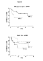
無再発生存率を予測するバイオマーカー
RFSの予測バイオマーカーは、37.8%のYAP−1最適カット(P=0.01)、YAP−1の強度等級(P=0.03)、及び10%のBcl−2最適カット(P<0.01)であった。
Biomarkers for predicting recurrence-free survival RFS predictive biomarkers were 37.8% YAP-1 optimal cut (P = 0.01), YAP-1 intensity grade (P = 0.03), and 10 % Bcl-2 optimal cut (P <0.01).
原因特異的生存率の予測バイオマーカー
CSSの予測バイオマーカーは、YAP−1の中央値強度等級(P=0.04)、81.8%のYAP−1最適カット(P=0.02)、78.6%のVEGF−c最適カット(P=0.03)、及び85.1%のクローディン−4最適カット(P=0.03)であった。
Cause-specific survival predictive biomarkers The CSS predictive biomarkers are YAP-1 median intensity grade (P = 0.04), 81.8% YAP-1 optimal cut (P = 0.02), 78.6% VEGF-c optimal cut (P = 0.03) and 85.1% claudin-4 optimal cut (P = 0.03).
重要なバイオマーカーを、バイオマーカーの独立した予後的意義を試験するために、各エンドポイントについて重要な臨床的要因に対して試験した。YAP−1及びbcl−2は、RFSについての予測において診断時年齢とは独立であることが判明し、YAP−1及びVEGF−cは、CSSについての診断時年齢とは独立し、及びVEGF−cは、CSSについての連続変数としての臨床Tステージとは独立していた。すべてのバイオマーカーは、不完全応答の予測において放射日数とは独立していた。興味深いことに、ログランクスコアが、CSS及びRFSの両方について評価され、そして、臨床ステージ、クローディン−4、YAP−1及び年齢の組み合わせがCSSについて非常に重要であること(P<0.01)、並びに年齢、YAP−1、クローディン−4、及び一次治療がRFSについて重要であること(P=0.02)を明らかにした。 Important biomarkers were tested against important clinical factors for each endpoint to test the independent prognostic significance of the biomarkers. YAP-1 and bcl-2 were found to be independent of age at diagnosis in prediction for RFS, YAP-1 and VEGF-c were independent of age at diagnosis for CSS, and VEGF- c was independent of the clinical T stage as a continuous variable for CSS. All biomarkers were independent of radiation days in predicting incomplete response. Interestingly, the log rank score is evaluated for both CSS and RFS, and the combination of clinical stage, claudin-4, YAP-1 and age is very important for CSS (P <0.01). ), And age, YAP-1, claudin-4, and primary treatment are important for RFS (P = 0.02).
バイオマーカーYAP−1、bcl−2、VEGF−c、c−met、及びクローディン−4を、陽性細胞の等級と陽性細胞の割合に関して分析したところ、2つの組み合わせ、すなわち割合に等級を乗じたものは、相乗効果を有していた。この式に基づく等級と割合の組み合わせは、治療及び予後の応答を分析する際の、バイオマーカーの重要性を改善した。さらに、c−met及びYAP−1の組み合わせは、これら2つのマーカーを放射線単独で治療された患者又は化学放射線療法の治療を受けた患者のいずれかからなるサブグループにおいて解析した場合と比べて、治療結果を90%正しく予測する、全調査対象(n=86)における放射線ベースの治療法に対する応答のより有意な予測因子であった。
The biomarkers YAP-1, bcl-2, VEGF-c, c-met, and claudin-4 were analyzed for positive cell grade and positive cell percentage, and the two combinations, ie, percentage multiplied by grade Things had a synergistic effect. The combination of grade and ratio based on this formula improved the importance of biomarkers in analyzing treatment and prognostic responses. In addition, the combination of c-met and YAP-1 compared to when these two markers were analyzed in a subgroup consisting of either patients treated with radiation alone or patients treated with chemoradiotherapy, It was a more significant predictor of response to radiation-based therapy in all study subjects (n = 86), predicting
考察
SCCHNの一次治療は放射線療法又は化学放射線療法の使用にシフトしているため、両方の方法に対する腫瘍応答を予測することは、患者に特異的な治療をするために有用である。本研究において、治療に対する治療応答は、TNMステージとは独立していることが判明し、そして一次治療(RT又はCRT)について不完全応答をする患者は、完全応答をする患者よりも、より遠隔転移を経験し、低いCSSを有する傾向がある。治療は、確立された予後及び予測バイオマーカーを評価することによる、個々の腫瘍バイオロジーを評価することによって導くことができる。この方法は、化学療法の利点を決定するために、早期の乳がんにおいてすでに使用されている。
Discussion Since primary treatment of SCCHN has shifted to the use of radiation therapy or chemoradiotherapy, predicting tumor response to both methods is useful for patient-specific treatment. In this study, the therapeutic response to treatment was found to be independent of the TNM stage, and patients with an incomplete response to primary treatment (RT or CRT) are more distant than those with a complete response They tend to experience metastases and have low CSS. Treatment can be guided by assessing individual tumor biology by assessing established prognosis and predictive biomarkers. This method has already been used in early breast cancer to determine the benefits of chemotherapy.
本研究において、YES関連タンパク質(YAP−1)は普遍的なバイオマーカーであることが判明した。YAP−1の過剰発現は、上衣細胞腫(Modena et al.(2006))、NSCLC(非小細胞肺癌)(Saviozzi et al.2006)、及び膵臓癌(Guo et al.(2006))に見られる。 In this study, it was found that YES-related protein (YAP-1) is a universal biomarker. Overexpression of YAP-1 is found in ependymoma (Modena et al. (2006)), NSCLC (non-small cell lung cancer) (Saviozzi et al. 2006), and pancreatic cancer (Guo et al. (2006)). It is done.
以前の研究では、チロシンキナーゼYES関連タンパク質(YAP65)の遺伝子が、形質転換及び転移性腫瘍細胞株において優先的に発現していることが示されている(Dong et al.(1997))。YAP−1のサイレンシングは、p53のヒストンアセチル化を減少し、その結果P73が関与するアポトーシスが遅延又は減少する(Strano et al.(2005))。 Previous studies have shown that the tyrosine kinase YES related protein (YAP65) gene is preferentially expressed in transformed and metastatic tumor cell lines (Dong et al. (1997)). YAP-1 silencing reduces histone acetylation of p53, resulting in delayed or reduced apoptosis involving P73 (Strano et al. (2005)).
先行研究は、c−metの低い発現がシスプラチン感受性と関連することを見出し、及び先行研究の間、c−metが高発現している腫瘍を有する患者は、併用化学放射線療法のための良い候補にはならない可能性があると結論されていた(Akervall et al.(2004)。これは本研究において検証されているように、c−metは、放射線療法又は化学放射線療法に対する乏しい応答についての予測に有用であった。METは、増殖、有糸分裂、血管新生、及び転移に関与するチロシンキナーゼ受容体である。Metの過剰発現は、乳、卵巣、甲状腺、膵臓、脳、及び消化管の腫瘍で報告されており、及びc−metの過剰発現は、上咽頭癌の患者における予後不良と相関している(Qian et al.(2002))。c−metの過剰発現は、口腔舌癌の局所再発の予測因子であることも示されている(Endo et al.(2006))。これまでは、c−metは、CSSにおいて予測的ではなかった。RFSにおいてc−metの予測は存在していなかった。これは、不完全応答を有する患者が、RFS解析を打ち切られたという事実が原因である可能性がある。 Previous studies have found that low c-met expression is associated with cisplatin sensitivity, and patients with tumors with high c-met expression during prior studies are good candidates for combination chemoradiotherapy (Akervall et al. (2004). As this has been verified in this study, c-met predicts poor response to radiotherapy or chemoradiotherapy. MET is a tyrosine kinase receptor involved in proliferation, mitosis, angiogenesis, and metastasis.Met overexpression is found in breast, ovary, thyroid, pancreas, brain, and gastrointestinal tract. Tumors have been reported and c-met overexpression correlates with poor prognosis in patients with nasopharyngeal carcinoma (Qian et al. (2002)). Overexpression of c-met has also been shown to be a predictor of local recurrence of oral tongue cancer (Endo et al. (2006)) To date, c-met has not been predictive in CSS. There was no c-met prediction in RFS, possibly due to the fact that patients with incomplete responses were discontinued from RFS analysis.
本研究では、クローディン−4は、RFSを予測する上で重要であることが判明した。クローディン−4は、タイトジャンクションタンパク質をコードしている。その高発現は、乳癌、尿路上皮癌、及び前立腺癌の患者で認められている。HNSCC及びNSCLC細胞株におけるクローディン−4の増加は、ゲフィチニブについての感度の増加と関連付けられることが発見されたが(Frederick et al.(2007))、これは標的療法であり、該結論は本研究では正しくない可能性がある。漿液性乳頭癌において、クローディン−4の過剰発現は、DFS及びOSの欠如、悪性表現型に見られる過剰発現と関連していた(Konecny et al.(2008))。尿路上皮癌及び前立腺癌において、これはステージ及び転移と関連付けられることが判明した。おそらく、クローディン−4は、腫瘍の広がりに対する代理マーカー(surrogate marker)であり、このことがより悪いRFSにつながり得る理由である。 In this study, claudin-4 was found to be important in predicting RFS. Claudin-4 encodes a tight junction protein. Its high expression has been observed in patients with breast cancer, urothelial cancer, and prostate cancer. An increase in claudin-4 in HNSCC and NSCLC cell lines was found to be associated with increased sensitivity for gefitinib (Frederick et al. (2007)), which is a targeted therapy and the conclusion It may be incorrect in research. In serous papillary carcinoma, claudin-4 overexpression was associated with the lack of DFS and OS, the overexpression found in the malignant phenotype (Konecny et al. (2008)). In urothelial cancer and prostate cancer this has been found to be associated with stage and metastasis. Perhaps claudin-4 is a surrogate marker for tumor spread, which is why it can lead to worse RFS.
本研究において、bcl−2は無再発生存率及び原因特異的生存率を予測する上で有意であった。歴史的に、これはp5 3を抑制する、抗アポトーシス癌遺伝子として発見された。最近では、高用量の放射線療法を施行した鼻咽頭の患者のコホートにおいて、bcl−2の過剰発現は悪い5年DFSを伴った(Chen et al.(2008));化学放射線療法で治療された、局所進行SCCHNの21人の患者は、Bcl−2を過剰発現した場合、好ましくない結果と短いRFSを有することが判明した(Mannarini et al.(2007))。 In this study, bcl-2 was significant in predicting relapse-free survival and cause-specific survival. Historically, it was discovered as an anti-apoptotic oncogene that suppresses p53. Recently, in a cohort of nasopharyngeal patients who underwent high-dose radiation therapy, overexpression of bcl-2 was associated with poor 5-year DFS (Chen et al. (2008)); treated with chemoradiotherapy Twenty-one patients with locally advanced SCCHN were found to have unfavorable results and short RFS when overexpressing Bcl-2 (Mannarini et al. (2007)).
腫瘍低酸素症は、放射線治療への反応の乏しさと関連している。本研究において、VEGF−cは、RFS及びCSSを予測する上で意義を有することが判明した。VEGF−cは、血管新生促進癌遺伝子である。以前は、VEGF−cの高発現は、放射線治療への応答不良と関連していた。病理学的完全応答は、術前の直腸癌患者で評価された。VEGF−c陽性と考えられていた癌は、病理学的に不完全応答を有することが認められた(Zlobec et al.(2008))。ステージII−IV SCCHNの患者27人において、VEGF−cの高発現は有意に局所制御及び生存率を減少した(Martin et al.(2007))。高レベルのVEGF−cは、腫瘍低酸素、すなわち放射線抵抗性を導く低酸素症、の代用であり得る。 Tumor hypoxia is associated with poor response to radiation therapy. In this study, VEGF-c was found to have significance in predicting RFS and CSS. VEGF-c is a pro-angiogenic oncogene. Previously, high expression of VEGF-c was associated with poor response to radiation therapy. Pathological complete response was evaluated in patients with rectal cancer before surgery. Cancers that were thought to be VEGF-c positive were found to have pathologically incomplete responses (Zlobec et al. (2008)). In 27 patients with stage II-IV SCCHN, high expression of VEGF-c significantly reduced local control and survival (Martin et al. (2007)). High levels of VEGF-c may be a substitute for tumor hypoxia, hypoxia leading to radioresistance.
本研究にはいくつかの制限がある。分析された患者は、異なる頭頸部の病変部位及びTNMステージを有する、混成グループだった。声門原発性の患者対口腔咽頭原発性の患者間で、局所リンパ節のへの広がりのパターンは異なる。この研究は、9年間の期間に亘ったので、異なる放射線治療技術と化学療法投与計画とが用いられた。しかし、本研究で特定されたものと同様の不均一な集団は、全国で実施されている臨床の広い範囲で遭遇する人をより代表する可能性があり、従ってマーカーのパネルの使用を、より普遍的に適用可能なものとする。また、すべての過去の研究に伴う偏見(bias)を考慮する必要がある。 This study has some limitations. The patients analyzed were mixed groups with different head and neck lesion sites and TNM stages. The pattern of regional lymph node spread differs between patients with glottic primary versus oropharyngeal primary. Since this study spans a period of nine years, different radiotherapy techniques and chemotherapy regimens were used. However, heterogeneous populations similar to those identified in this study may be more representative of those encountered in a wide range of clinical practices conducted across the country, thus making the use of marker panels more It shall be universally applicable. It is also necessary to consider the bias associated with all past studies.
結論
本研究では、以前のc−DNAマイクロアレイ研究(発表済み及び未発表)に基づく化学的感受性又は放射線感受性のための潜在的な重要性を有する5つのマーカー(VEGF−c、bcl−2、クローディン−4、c−met、及びYAP−1)の予後及び予測能力の評価を行った。前処理された生検を評価することによって、YAP−1は、RT/CRT応答、CSS、及びRFSの予測のための普遍的なマーカーであることがわかった。クローディン−4及びVEGF−cは、CSS及びRFSの両方を予測し、並びにBcl−2は、RT/CRT応答及びRFSを予測する。上記の試験したバイオマーカーを用いて、局所再発のリスクが高い患者を早期に同定することができ、そして適切な治療を前もって供給して、がんの治療を最適化し、及び一次治療に追加する必要なサルベージ療法によって、罹患率を減少することができる。
CONCLUSION In this study, five markers (VEGF-c, bcl-2, claw with potential importance for chemosensitivity or radiosensitivity based on previous c-DNA microarray studies (published and unpublished) were studied. Din-4, c-met, and YAP-1) were evaluated for prognosis and prediction ability. By evaluating pretreated biopsies, YAP-1 was found to be a universal marker for prediction of RT / CRT response, CSS, and RFS. Claudin-4 and VEGF-c predict both CSS and RFS, and Bcl-2 predicts RT / CRT response and RFS. Using the biomarkers tested above, patients at high risk for local recurrence can be identified early, and appropriate treatment given in advance to optimize cancer treatment and add to primary treatment The necessary salvage therapy can reduce morbidity.
参考文献
Akervall et al.“Genetic and expression profiles of squamous cell carcinoma of the head and neck correlate with cisplatin sensitivity and resistance in cell lines and patients.”Clin Cancer Res.(2004)10(24):8204−8213.
Chen et al.“Prognostic impact of bcl−2 expression on advanced nasopharyngeal carcinoma.”Head Neck.(2008)30(8):1052−1057
Dong,G. et al.“Genes differentially expressed with malignant transformation and metastatic tumor progression of murine squamous cell carcinoma.”J Cell Biochem.Suppl 28−29(1997):90−100.
Endo et al.“Prognostic value of cell motility activation factors in patients with tongue squamous cell carcinoma.”Hum Pathol.(2006)37(8):1111−1116.
Frederick et al.“Epithelial to mesenchymal transition predicts gefitinib resistance in cell lines of head and neck squamous cell carcinoma and non−small cell lung carcinoma.”Mol Cancer Ther.(2007)6(6):1683−1691.
Guo,J.et al.“Yes−associated protein(YAP65) in relation to Smad7 expression in human pancreatic ductal adenocarcinoma.”Int.J Mol.Med. 17.5(2006):761−67.
Konecny et al.“Claudin−3 and claudin−4 expression in serous papillary,clear−cell,and endometrioid endometrial cancer.”Gynecol Oncol.(2008)109(2):263−269.
Mannahni et al.“Markers of chemoradiation resistance in patients with locally advanced head and neck squamous cell carcinoma,treated by intra−artehal carboplatin and concurrent radiation.”Acta Otorhinolaryngol Ital.(2007)27(4):173−180
Martin et al.“Vascular endothelial growth factor expression predicts outcome after primary radiotherapy for head and neck squamous cell cancer.”Clin Oncol(R Coll Radiol).(2007)19(1):71−76
Modena,P.et al.“Identification of tumor−specific molecular signatures in intracranial ependymoma and association with clinical characteristics.”J Clin.Oncol.24.33(2006):5223−33.
Qian et al.“Met protein expression level correlates with survival in patients with late−stage nasopharyngeal carcinoma.”Cancer Res.(2002)62(2):589−596
Saviozzi,S.et al.“Selection of suitable reference genes for accurate normalization of gene expression profile studies in non−small cell lung cancer.”BMC.Cancer 6(2006):200.
Strano, S.et al.“The transcriptional coactivator Yes−associated protein drives p73 gene−target specificity in response to DNA Damage.”Mol.Cell 18.4(2005):447−59.
Zlobec et al.“Combined analysis of VEGF and EGFR predicts complete tumour response in rectal cancer treated with preoperative radiotherapy.”Br J Cancer.(2008)98(2):450−456.
References
Akervall et al. “Genetic and expression profiles of squamous cells carcinoma of the head and neck correlates with citplatin sensitiveness and resilience in cells.” (2004) 10 (24): 8204-8213.
Chen et al. “Prognostic impact of bcl-2 expression on advanced nasopharyngeal carcinoma.” Head Neck. (2008) 30 (8): 1052-1057
Dong, G.M. et al. “Genes differentially expressed with malformed transformation and metallic tumour of the squamous cell carcinoma.” J Cell Biochem. Suppl 28-29 (1997): 90-100.
Endo et al. “Prognostic value of cell motivation factors in participants with tongue squalous cell carcinoma.” Hum Pathol. (2006) 37 (8): 1111-1116.
Frederick et al. "Epithelial to mesenchymal transition predicts gefitinib resilience in cell lines of head and neck squarmous cell carcinoma and non-smalmol cell. (2007) 6 (6): 1683-1691.
Guo, J. et al. et al. “Yes-associated protein (YAP65) in relation to Smad7 expression in human pancreatic adenocarcinooma.” Int. J Mol. Med. 17.5 (2006): 761-67.
Konecny et al. “Claudin-3 and claudin-4 expression in serous pipeline, clear-cell, and endometrioid endometrial cancer.” Gynecol Oncol. (2008) 109 (2): 263-269.
Mannahni et al. “Markers of chemical resilience in partnerships with locally advanced head and neck quat i ral c ar c i nt a r i c i c i c i s s s s s s ed i n s i s s s s s s ed i n s i s s s s s s s ed i n s i n e n s i n e n e n i n l e n i n l e n i n l e n i n l e n i n l e n i n l e n i n l e n i n t i n i n s i n i i n i n e n i n i n i n. (2007) 27 (4): 173-180
Martin et al. “Vascular endorative growth factor expression predicates outcome after primary radio head for head and neck squamous cell cancer.” Clin Oncol. (2007) 19 (1): 71-76
Moderna, P.M. et al. “Identification of tumor-specific molecular signatures in intramolecular epidemioma and association with clinical charactaristics.” J Clin. Oncol. 24.33 (2006): 5223-33.
Qian et al. “Met protein expression level correlates with survivable in patents with late-stage nasopharyngeal carcinoma.” Cancer Res. (2002) 62 (2): 589-596
Saviozzi, S .; et al. “Selection of sustainable reference genes for accumulating normalization of gene expression profiles in non-small cell lung cancer.” BMC. Cancer 6 (2006): 200.
Strano, S.M. et al. “The transcriptional coactivator Yes-associated protein drives p73 gene-target specificity in response to DNA Damage.” Mol. Cell 18.4 (2005): 447-59.
Zlobec et al. “Combined analysis of VEGF and EGFR predicates complete response response in the treatment of preferential radiation therapy.” Br J Cancer. (2008) 98 (2): 450-456.
Claims (10)
該患者からの生物学的サンプル中の、
(i)YAP−1;又は
(ii)(b)から選択される少なくとも1つのバイオマーカー、及び(c)から選択される少なくとも一つのバイオマーカー:
(b)bcl−2、及びVEGF−c、
(c)c−met及びクローディン−4、又は
(iii)(i)及び(ii)の組み合わせ
のタンパク質又はmRNAのレベルを測定することを含む、方法。 A method for predicting response to treatment with chemoradiotherapy in a patient suffering from head or neck cancer comprising:
In a biological sample from the patient,
(I) YAP-1; or (ii) at least one biomarker selected from (b), and at least one biomarker selected from (c):
(B) bcl-2 and VEGF-c,
(C) measuring the protein or mRNA level of c-met and claudin-4, or (iii) (i) and (ii) in combination.
配列番号1(YAP−1)、
配列番号2(bcl−2)、
配列番号3(VEGF−c)、
配列番号4(c−met)、及び
配列番号5(クローディン−4)
からなる群より選択される1つ以上の核酸配列のレベルを測定することにより決定される、請求項1に記載の方法。 The mRNA level is:
SEQ ID NO: 1 (YAP-1),
SEQ ID NO: 2 (bcl-2),
SEQ ID NO: 3 (VEGF-c),
SEQ ID NO: 4 (c-met), and SEQ ID NO: 5 (Claudin-4)
2. The method of claim 1, wherein the method is determined by measuring the level of one or more nucleic acid sequences selected from the group consisting of.
配列番号6(YAP−1)、
配列番号7(bcl−2)、
配列番号8(VEGF−c)、
配列番号9(c−met)、及び
配列番号10(クローディン−4)
からなる群より選択される1つ以上のアミノ酸配列のレベルを測定することにより決定される、請求項1に記載の方法。 The level of the protein is
SEQ ID NO: 6 (YAP-1),
SEQ ID NO: 7 (bcl-2),
SEQ ID NO: 8 (VEGF-c),
SEQ ID NO: 9 (c-met) and SEQ ID NO: 10 (Claudin-4)
2. The method of claim 1, wherein the method is determined by measuring the level of one or more amino acid sequences selected from the group consisting of.
Applications Claiming Priority (3)
| Application Number | Priority Date | Filing Date | Title |
|---|---|---|---|
| US10933108P | 2008-10-29 | 2008-10-29 | |
| US61/109,331 | 2008-10-29 | ||
| PCT/US2009/061670 WO2010053717A1 (en) | 2008-10-29 | 2009-10-22 | Methods of using biomarkers |
Publications (1)
| Publication Number | Publication Date |
|---|---|
| JP2012507290A true JP2012507290A (en) | 2012-03-29 |
Family
ID=42153176
Family Applications (1)
| Application Number | Title | Priority Date | Filing Date |
|---|---|---|---|
| JP2011534634A Pending JP2012507290A (en) | 2008-10-29 | 2009-10-22 | How to use biomarkers |
Country Status (5)
| Country | Link |
|---|---|
| US (1) | US20110263442A1 (en) |
| EP (2) | EP2722400A3 (en) |
| JP (1) | JP2012507290A (en) |
| CA (1) | CA2741906A1 (en) |
| WO (1) | WO2010053717A1 (en) |
Families Citing this family (6)
| Publication number | Priority date | Publication date | Assignee | Title |
|---|---|---|---|---|
| SG10201408229WA (en) * | 2010-08-31 | 2015-02-27 | Genentech Inc | Biomarkers and methods of treatment |
| AU2011352231B2 (en) * | 2010-12-27 | 2016-09-15 | Expression Pathology, Inc. | cMET protein SRM/MRM assay |
| KR102049990B1 (en) | 2013-03-28 | 2019-12-03 | 삼성전자주식회사 | Fusion protein comprising anti-c-Met antibody and VEGF binding fragment |
| BR112016021383A2 (en) | 2014-03-24 | 2017-10-03 | Genentech Inc | METHOD TO IDENTIFY A PATIENT WITH CANCER WHO IS LIKE OR LESS LIKELY TO RESPOND TO TREATMENT WITH A CMET ANTAGONIST, METHOD TO IDENTIFY A PATIENT WITH PREVIOUSLY TREATED CANCER, METHOD TO DETERMINE THE EXPRESSION OF THE HGF BIOMARKER, ANTI-C-MET ANTAGONIST AND ITS USE, DIAGNOSTIC KIT AND ITS PREPARATION METHOD |
| US10969390B2 (en) | 2014-09-24 | 2021-04-06 | National Cancer Center | Method for evaluating efficacy of chemoradiotherapy against squamous cell carcinoma |
| WO2018189403A1 (en) * | 2017-04-14 | 2018-10-18 | INSERM (Institut National de la Santé et de la Recherche Médicale) | Methods and pharmaceutical compositions for the treatment of cancer |
Citations (2)
| Publication number | Priority date | Publication date | Assignee | Title |
|---|---|---|---|---|
| WO2007015935A2 (en) * | 2005-07-29 | 2007-02-08 | Bayer Healthcare Llc | Diagnostic methods for the prediction of therapeutic success, recurrence free and overall survival in cancer therapy |
| WO2007059430A2 (en) * | 2005-11-10 | 2007-05-24 | Bristol-Myers Squibb Pharma Company | Moesin, caveolin 1 and yes associated protein 1 as predictive markers of response to dasatinib in breast cancers |
Family Cites Families (9)
| Publication number | Priority date | Publication date | Assignee | Title |
|---|---|---|---|---|
| US6436703B1 (en) * | 2000-03-31 | 2002-08-20 | Hyseq, Inc. | Nucleic acids and polypeptides |
| US6969592B2 (en) | 2001-09-26 | 2005-11-29 | Pharmacia Italia S.P.A. | Method for predicting the sensitivity to chemotherapy |
| EP2258872B1 (en) | 2002-03-13 | 2013-08-14 | Genomic Health, Inc. | Gene expression profiling in biopsied tumor tissues |
| US7655397B2 (en) * | 2002-04-25 | 2010-02-02 | The United States Of America As Represented By The Department Of Health And Human Services | Selections of genes and methods of using the same for diagnosis and for targeting the therapy of select cancers |
| CA2506066A1 (en) * | 2002-11-15 | 2004-06-03 | Genomic Health, Inc. | Gene expression profiling of egfr positive cancer |
| EP1581629B1 (en) * | 2002-12-06 | 2015-04-01 | Millennium Pharmaceuticals, Inc. | Methods for the identification, assessment, and treatment of patients with proteasome inhibition therapy |
| US20040185444A1 (en) | 2003-03-18 | 2004-09-23 | Nicole Boehringer-Wyss | System for predicting susceptibility of human oropharyngeal squamous cell carcinoma to radiotherapy |
| WO2004111273A2 (en) * | 2003-05-30 | 2004-12-23 | Genomic Health, Inc. | Gene expression markers for response to egfr inhibitor drugs |
| US20060037088A1 (en) | 2004-08-13 | 2006-02-16 | Shulin Li | Gene expression levels as predictors of chemoradiation response of cancer |
-
2009
- 2009-10-22 EP EP13181855.1A patent/EP2722400A3/en not_active Withdrawn
- 2009-10-22 JP JP2011534634A patent/JP2012507290A/en active Pending
- 2009-10-22 US US13/126,129 patent/US20110263442A1/en not_active Abandoned
- 2009-10-22 CA CA2741906A patent/CA2741906A1/en not_active Abandoned
- 2009-10-22 EP EP09825206A patent/EP2342355A4/en not_active Withdrawn
- 2009-10-22 WO PCT/US2009/061670 patent/WO2010053717A1/en active Application Filing
Patent Citations (2)
| Publication number | Priority date | Publication date | Assignee | Title |
|---|---|---|---|---|
| WO2007015935A2 (en) * | 2005-07-29 | 2007-02-08 | Bayer Healthcare Llc | Diagnostic methods for the prediction of therapeutic success, recurrence free and overall survival in cancer therapy |
| WO2007059430A2 (en) * | 2005-11-10 | 2007-05-24 | Bristol-Myers Squibb Pharma Company | Moesin, caveolin 1 and yes associated protein 1 as predictive markers of response to dasatinib in breast cancers |
Non-Patent Citations (3)
| Title |
|---|
| JPN6014016751; PATHOLOGY ONCOLOGY RESEARCH Vol.3, No.3, 1997, P.204-210 * |
| JPN6014016754; Molecular Cancer Therapeutics Vol.6, No.6, 2007, P.1683-1691 * |
| JPN6014016757; Clinical Cancer Research Vol.10, 2004, P.8204-8213 * |
Also Published As
| Publication number | Publication date |
|---|---|
| EP2342355A1 (en) | 2011-07-13 |
| WO2010053717A1 (en) | 2010-05-14 |
| EP2722400A3 (en) | 2014-07-09 |
| EP2342355A4 (en) | 2012-06-27 |
| EP2722400A2 (en) | 2014-04-23 |
| US20110263442A1 (en) | 2011-10-27 |
| CA2741906A1 (en) | 2010-05-14 |
Similar Documents
| Publication | Publication Date | Title |
|---|---|---|
| JP5951603B2 (en) | Diagnosis and treatment of breast cancer | |
| EP3055429B1 (en) | Method for the prognosis and treatment of metastasizing cancer of the bone originating from breast cancer | |
| EP2257810B1 (en) | Molecular diagnosis and classification of malignant melanoma | |
| US11654153B2 (en) | Therapeutic treatment of breast cancer based on c-MAF status | |
| US20110104062A1 (en) | Biomarkers for Head-And-Neck Cancers and Precancers | |
| JP2017062244A (en) | Biomarkers for predicting and assessing responce of thyroid and kidney cancer subjects with respect to lenvatinib compounds | |
| MX2010014280A (en) | Signatures and determinants associated with metastasis methods of use thereof. | |
| US20120052079A1 (en) | Compositions, Kits, and Methods for Predicting Anti-Cancer Response to Anthracyclines | |
| JP2011526487A (en) | Breast cancer genome fingerprint | |
| KR20160057416A (en) | Molecular diagnostic test for oesophageal cancer | |
| EP2722400A2 (en) | Methods of using biomarkers | |
| WO2016145294A1 (en) | Methods for determining prognosis for breast cancer patients | |
| KR20130124961A (en) | Agtr1 as a marker for bevacizumab combination therapies | |
| US20160201143A1 (en) | LKB1 Levels and Brain Metastasis from Non-Small-Cell Lung Cancer (NSCLC) | |
| Uemura et al. | Hypoxia-inducible adrenomedullin in colorectal cancer | |
| US9110082B2 (en) | Methods for the identification and treatment of patients sensitive to anti IGF-1R inhibition therapy | |
| US20130102486A1 (en) | Perp as a prognostic and diagnostic marker for dysplasia and cancer | |
| CN113774140A (en) | Product for predicting sensitivity of colorectal cancer to oxaliplatin treatment | |
| US20220235357A1 (en) | Compositions and methods for identifying and inhibiting a pan-cancer cellular transition of adipose-derived stromal cells | |
| CN111417855A (en) | Methods for treating and diagnosing prostate cancer | |
| KR20220072481A (en) | Biomarker composition comprising CXCL12 in plasma membrane of cancer cells for predicting prognosis of rectal adenocarcinoma | |
| CN113817829A (en) | Use of biomarkers for the preparation of a product for predicting the sensitivity of colorectal cancer to treatment with oxaliplatin | |
| CN116042823B (en) | Molecular marker for esophageal squamous carcinoma prognosis and therapy efficacy evaluation and application thereof | |
| KR102259708B1 (en) | Novel biomarker for predicting drug-responsibility to colon cancer | |
| KR20230086458A (en) | Novel Biomarker for Predicting Therapeutic Response and Prognosis of Metastatic Breast Cancer To Chemotherapeutic Agents and Uses Thereof |
Legal Events
| Date | Code | Title | Description |
|---|---|---|---|
| A621 | Written request for application examination |
Free format text: JAPANESE INTERMEDIATE CODE: A621 Effective date: 20121002 |
|
| A131 | Notification of reasons for refusal |
Free format text: JAPANESE INTERMEDIATE CODE: A131 Effective date: 20140422 |
|
| A02 | Decision of refusal |
Free format text: JAPANESE INTERMEDIATE CODE: A02 Effective date: 20140930 |