JP2012143528A - Oral imaging and display system - Google Patents
Oral imaging and display system Download PDFInfo
- Publication number
- JP2012143528A JP2012143528A JP2011017192A JP2011017192A JP2012143528A JP 2012143528 A JP2012143528 A JP 2012143528A JP 2011017192 A JP2011017192 A JP 2011017192A JP 2011017192 A JP2011017192 A JP 2011017192A JP 2012143528 A JP2012143528 A JP 2012143528A
- Authority
- JP
- Japan
- Prior art keywords
- image
- unit
- display
- medical
- intraoral
- Prior art date
- Legal status (The legal status is an assumption and is not a legal conclusion. Google has not performed a legal analysis and makes no representation as to the accuracy of the status listed.)
- Ceased
Links
Images


Landscapes
- Endoscopes (AREA)
Abstract
Description
本発明は、口腔内を撮影し、表示するシステムに関するものである。 The present invention relates to a system for photographing and displaying an intraoral area.
虫歯等の口腔内疾病の治療は、目的とする治療が終わると通院も終了することが多い。
虫歯の治療は、口腔内に痛み、違和感等の症状を自覚して始まることが多く、又、治療が終了すると通院も終了するパターンが多いことから、その他に虫歯があっても、自覚症状が無い場合は、そのまま放置され、虫歯が進行して初めて通院するというケースが多い。疾病が進行してしまった後での治療は、抜歯といった患者が被る苦痛を覚悟しなければならず、又、患者の費用負担も、結果として大きくなる。
又、このような一過性の通院では、口腔内の健全化が図れるものでもなく、汚れ、腫れ、欠損、歯石、摩耗、唾石、不具合、等自覚症状が無いが、外部から見ることができる症状は、本人が知らない場合も多くある。
歯科医業の経営にとっても、単発的に治療を行うことは、収益性が決して良いとは言い得ない場合があるが、解決する好適な手段は見あたらなかった。In the treatment of oral diseases such as dental caries, the hospital visit is often terminated after the intended treatment is completed.
The treatment of caries often starts with awareness of symptoms such as pain and discomfort in the oral cavity, and there are many patterns in which the hospital visit ends when the treatment is completed. If there is not, it is often left as it is, and there are many cases that go to the hospital only after the caries progresses. Treatment after the disease has progressed must be prepared for the suffering suffered by the patient, such as tooth extraction, and the cost burden on the patient will increase as a result.
Also, in such a transient hospital visit, the oral cavity cannot be sounded and there are no subjective symptoms such as dirt, swelling, defects, tartar, wear, salivary stones, malfunctions, etc. There are many cases where the person does not know the symptoms.
Even for the dentistry business, it may be impossible to say that it is profitable to perform treatment on a one-off basis, but there has been no suitable means to solve it.
又、このような、一過性的な治療において、歯科医院で使用される口腔内カメラは、治療目的が明確な場合の患者への説明の為に用いられる場合が多く、口腔内の健全化の為の治療システムは未解明な部分が多い。 In such a temporary treatment, the intraoral camera used in the dental clinic is often used for explanation to the patient when the treatment purpose is clear. There are many unclear parts of the treatment system for this.
特開平10−97404には、コンピュータモニター上に全歯列が表示され、個々の歯に着色が付されることで、患者側からも見やすい電子カルテの構成が示されている。
歯列の見やすさは、インフォームドコンセントにおいても求められるが、全歯列の一部が見やすい状態であっても、治療の説明に供するには、より拡大され、その治療目的を理解するような構成が必要である。
画像表示による口腔内の観察は、数多く提案されているが、結局従来の単発的な治療システム上での、患者への情報提供にとどまるものであり、口腔内の健全化を図るようなシステムにまでは到達していない。Japanese Patent Application Laid-Open No. 10-97404 shows a configuration of an electronic medical chart that is easy to see from the patient side by displaying all teeth on a computer monitor and coloring each tooth.
The visibility of the dentition is also required for informed consent. However, even if a part of the entire dentition is easy to see, it will be expanded to explain the treatment, and the purpose of the treatment will be understood. Configuration is required.
Many observations in the oral cavity using image display have been proposed, but in the end, the conventional single treatment system is limited to providing information to the patient. It has not reached.
上記に鑑み本発明は、
口腔内を診療改善単位毎に画像化する単位画像化手段、前記単位画像化手段で、単位画像化した画像にたいし、診療改善順序情報を設定する設定手段、前記診療改善順序情報が付けられた画像を診療改善順序情報に基づいて、一覧的に表示可能に表示する表示手段、前記表示手段で得られた表示情報を表示記録した表示媒体よりなる組み合わせ構成により、患者の治療への自覚を高めて、口腔内の健全化を図ると共に、患者の継続的獲得を可能として歯科医業の経営効率の向上を実現する。
更にこの表示媒体を患者が携行することで、自らの口腔内の状況を認識すると共に、患者だけでなく、治療を要しない者に対しても口腔内への関心を高めることを可能とする。In view of the above, the present invention
Unit imaging means for imaging the oral cavity for each medical improvement unit, setting means for setting medical improvement order information for an image that has been unit-imaged by the unit imaging means, and the medical improvement order information Based on the medical improvement order information, the combination of the display means for displaying the list in a displayable manner and the display medium for displaying and recording the display information obtained by the display means, the patient is aware of treatment. Increase the health of the oral cavity and improve the management efficiency of the dentistry by enabling continuous patient acquisition.
Further, by carrying this display medium by the patient, it is possible to recognize the situation in the oral cavity of the patient and increase the interest in the oral cavity not only for the patient but also for those who do not require treatment.
口腔内を診療改善単位毎に画像化する単位画像化手段における、口腔内とは、歯、歯列、歯肉、歯槽骨、口唇、硬口蓋、軟口蓋、口蓋垂等の領域を示すものである。
診療には、診察と治療が含まれ、歯科医では、診察して、治療は、全問の医療機関で行う場合がある。
診療改善単位とは、虫歯、歯周病、舌癌、歯肉癌等、好ましくは、ひとつの診察、治療の範囲を示す他、汚れ、矯正等の改善、予防処理、準診療も示す場合がある。
改善とは、ブラッシング、フッ素、口臭予防剤の塗布等で、克服できる程度のものや、歯にフッ素を塗布するような予防処置、クリーニング、歯周病等の予防剤の塗布等、の行為を示すものである。
画像化とは、コンピュータディスプレイ、携帯電話表示部へ出力表示可能な画像データの他、用紙に印字・印刷された状態等の2次元的又は3次元的な表示がされた状態を示す。
前記単位画像化手段で、単位画像化した画像にたいし、診療改善順序情報を設定する設定手段の診療改善順序情報とは、診療、予防、改善の順位を示す記号、符合、数値等を示すものであって、その他、診療日時、疾病の進行状況、疾病進行の予測等のデータをしめすものであり、少なくとも複数の単位画像に対し、診療改善の順番を決定できるものであれば良く、直接目視確認できる内容の他、演算用のパラメータであって、コンピュータ演算処理によって確認可能なものであっても良い。In the unit imaging means for imaging the oral cavity for each medical improvement unit, the oral cavity indicates a region such as a tooth, a dentition, a gum, an alveolar bone, a lip, a hard palate, a soft palate, and a uvula.
Medical examination includes examination and treatment, and a dentist examines and treatment may be performed at all medical institutions.
Medical care improvement units include caries, periodontal disease, tongue cancer, gingival cancer, etc., preferably one diagnosis and treatment range, dirt, correction, etc., prevention treatment, quasi-medical treatment may also be indicated .
Improvement refers to actions that can be overcome by brushing, fluorine, application of anti-oral breathing agent, preventive measures such as applying fluorine to teeth, cleaning, application of preventive agents such as periodontal disease, etc. It is shown.
Imaging refers to a state in which two-dimensional or three-dimensional display such as a state of being printed / printed on a sheet is performed in addition to image data that can be output and displayed on a computer display or a mobile phone display unit.
The medical improvement order information of the setting means for setting the medical improvement order information for the unit imaged image by the unit imaging means indicates symbols, signs, numerical values, etc. indicating the order of medical care, prevention, and improvement. Any other data that indicates the date and time of medical treatment, the progress of disease, the prediction of disease progression, etc., as long as it can determine the order of medical improvement for at least a plurality of unit images. In addition to the contents that can be visually confirmed, it may be a parameter for calculation that can be confirmed by computer calculation processing.
「一覧的に表示可能に表示する」とは、例えば、用紙に、単位画像が診療改善順序情報と共に印字表示された状態であって、1枚以上からなる状態、あるいは、JPEG、GIF、BMP等の様式に基づいて画像表示されている状態であって、ディスプレイ上に改ページしない状態で、表示される状態等が示されるが、少なくとも、診療改善順序が一覧であって、全体を容易に認識できる状態であれば、複数枚の用紙に印刷したもの、複数画像をスクロールで入れ替えて表示した状態等で構成されても良い場合もある。 “Display so that it can be displayed as a list” means, for example, a state in which unit images are printed and displayed together with medical treatment improvement order information on a sheet, and is composed of one or more sheets, or JPEG, GIF, BMP, etc. The image is displayed based on the style of the display, the page is displayed on the display without any page breaks, etc., but the display status etc. are shown, but at least the medical improvement order is a list, and the whole is easily recognized As long as it is possible, it may be configured by printing on a plurality of sheets, a state in which a plurality of images are scrolled and displayed, and the like.
前記表示手段で得られた表示情報を表示記録した表示媒体とは、1枚以上の用紙、冊子に印字、印刷して表示された状態や、JPEG、GIF、BMP等の様式に基づいた画像データを携帯可能に表示する状態を示し、表示媒体とは、用紙状、冊子状の紙媒体、USBメモリー、SDカードや、メモリー等の記録素子を具えた表示装置、携帯電話、等を示す場合もあるが、少なくても患者が、携行し、口腔内を見ることができる印刷、印字物、電子画像、等を示す。又、インターネット上のホームページ表示上で、自らの口腔内データを閲覧する場合も含まれることから、表示媒体には、デスクトップ、ノート型のパーソナルコンピュータも含まれるものである。 The display medium on which the display information obtained by the display means is displayed and recorded is image data based on a state printed or printed on one or more sheets or booklets, or a format such as JPEG, GIF, or BMP. In some cases, the display medium may be a paper or booklet paper medium, a USB memory, an SD card, a display device having a recording element such as a memory, a mobile phone, or the like. There are prints, printed matter, electronic images, etc. that can be carried by at least the patient and see inside the oral cavity. In addition, since it includes the case of browsing own intraoral data on a homepage display on the Internet, the display medium includes a desktop and a notebook personal computer.
又、本発明は、最初に患者の上顎、及び下顎の全歯を撮影して静止画像を形成して、この全歯画像に基づいて単位画像を形成することが好ましい。
全歯画像の取得は、例えば、魚眼レンズ、凸面鏡を用いた口腔内カメラの他、静止画像の撮影を歯列に沿って撮影していき、接続していくことで、形成する手法、等が例示される。
得られた全歯画像から、診療歯牙を指定し、指定された歯を中心に、静止画像を形成する。
この場合、全歯画像から、該当する部分だけをコピーし拡大して表示することが、一回の撮影で済む点で、患者への負担が少なく、画像処理を簡単に終わらせることが出来る点で好ましい。In the present invention, it is preferable that all the teeth of the patient's upper jaw and lower jaw are first photographed to form a still image, and a unit image is formed based on the whole tooth image.
The acquisition of all-tooth images is exemplified by, for example, a method of forming by capturing and connecting a still image along a dentition in addition to an intraoral camera using a fish-eye lens and a convex mirror. Is done.
A medical tooth is designated from the obtained whole tooth image, and a still image is formed around the designated tooth.
In this case, it is possible to finish the image processing easily with less burden on the patient, because it is only necessary to copy and enlarge only the corresponding part from the entire tooth image and display it. Is preferable.
新たに撮影しても良いが、カメラの画素数を大きくしたり、色、輪郭等を補完する等して、全歯撮影を行うことで、診療歯牙のみを抽出して拡大してもよく、その場合は一回の撮影で済むので、説明時間の合理性が図られる。
又、コンピュータによる画像処理により、歯毎にクリッカブルにリンクして、表示される構成とすることで、後で発見した診療部位、診療準備部位を同様の表紙をすることを可能としてもよい。
又、全歯画像撮影を三次元撮影化しておくことで、診療歯牙をクリッカブルリンクにすると、その部分を容易に拡大して表示させることを可能とすしてもよい。You may take a new picture, but you can enlarge the number of pixels of the camera, supplement the color, contour, etc. In such a case, only one shooting is required, so that the explanation time is rational.
Moreover, it may be possible to make a similar cover for a medical site and a medical site that are discovered later by using a computer that performs a clickable link for each tooth by image processing.
Moreover, if the dental teeth are clickable links, it is possible to easily enlarge and display the portions of the whole tooth images by making them three-dimensional.
又、順位、診療開始予定日、診療の深刻さ、診療期間、診療費用等をメッセージ欄として、個々の、歯牙に対応するように配置させることで、クリックするたびに、メッセージ欄が表示されるとと共に、順位毎に整列した画像を可能な限り一画面上に表示する形式であっても良い。
この場合、画像の大きさを調整するたび毎に、一覧表示の数を調整するものであってもよい。
また、全歯画像が取得されたと同時に、それぞれの歯をソフトウエア処理に基づく輪郭抽出作業を自動で行い、その抽出範囲から所定の距離を持つ領域を自動的に設定して各歯の個別画像を形成しても良い。In addition, the message column is displayed each time a click is made by placing the message, such as rank, scheduled medical start date, seriousness of medical treatment, medical treatment period, medical fee, etc., corresponding to each tooth. In addition, a format in which images arranged in each order are displayed on one screen as much as possible is also possible.
In this case, the number of list displays may be adjusted every time the size of the image is adjusted.
Also, at the same time that all tooth images are acquired, each tooth is automatically subjected to contour extraction work based on software processing, and an area having a predetermined distance from the extraction range is automatically set, and an individual image of each tooth May be formed.
或いは、予め設定された一定の範囲を指定しておき、おおまかな感覚での全歯の指定も可能であるほか目視により、それぞれ、範囲指定、付随するメッセージ入力等、グラフィックソフトウエア的にデータを形成しても良い。 Alternatively, it is possible to specify a certain range set in advance and specify all teeth with a rough sense. In addition, by visual inspection, data can be specified by graphic software such as range specification and accompanying message input. It may be formed.
本発明は、口腔内診療の際、個々の診療部位を画像化した単位画像と診療改善情報を付加したものを、一覧として表示することで、患者が、自らの口腔内の状況を認識することが出来、診療や改善の必要性や、口腔内の改善のための処置の必要性、口腔内の診療改善の為の継続的処置を可能とすることによる歯科医業の経営的安定性をはかる事が可能となる。 The present invention enables a patient to recognize the situation in his oral cavity by displaying a list of unit images obtained by imaging individual medical treatment sites and medical improvement information at the time of intraoral medical treatment. To ensure the management stability of the dentistry business by enabling the need for medical treatment and improvement, the need for treatment for improvement in the oral cavity, and the continuous treatment for improvement of medical treatment in the oral cavity. Is possible.
本発明は、歯科診療の際、又は、検診、その他、家庭で撮影した口腔画像から、診療、改善に対応した単位画像を口腔内カメラ、X線撮影装置を用いて取得する。この単位画像を、例えば患者と見ることが出来るモニターディスプレイに写しだして、患者が口腔内の状況を認識しながら、歯科医師と共に診療の順位、診療の開始期間、診療の必要性の度合い等を記入していって得られる診療改善順序情報と単位画像を、一枚の用紙に印字、印刷し、或いは、記憶媒体を具えた携帯電話に記憶させ、又は、歯科医師が開設するホームページであって、個人専用の画面に表示する。
患者は、常に自らの口腔内の状況と、診療の必要性を当該表示手段によって認識することで、口腔内健全化のための診療と改善を継続的に行う可能性が高くなり、歯科医業における収益の向上等の経営改善も可能となる。In the present invention, a unit image corresponding to medical care and improvement is acquired from an oral image taken at the time of dental care or in a medical examination or other home using an intraoral camera and an X-ray imaging apparatus. This unit image is displayed on a monitor display that can be seen as a patient, for example. It is a homepage established by a dentist who prints, prints, or stores a medical improvement order information and unit image obtained on a single sheet of paper or stores them on a mobile phone equipped with a storage medium. Display on a personal screen.
The patient always recognizes the situation in the oral cavity and the necessity of medical treatment by the display means, so that the possibility of continuous medical care and improvement for oral health is increased. Management improvements such as improved profitability are also possible.
次に本発明の実施例を図面を参照して詳細に説明する。
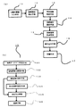
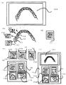
図1(a)は、本発明の一実施例を示す図である。
11は、口腔内画像撮影手段であり、例えば、上顎、下顎の全歯を撮影する為のカメラを用いて、撮影し、デジタル画像データを得る装置である。
口腔内画像撮影手段11は、例えば、図3(c)で示すような凸面鏡を用いた反射型の歯科用カメラ、その他魚眼レンズを用いた口腔内撮影用のカメラであって、全歯のデジタル画像を出力する。
或いは、図6で示すように通常の口腔内カメラで各歯を撮影し、個々の撮影画像から輪郭を抽出して、その輪郭の共通部で接続していき、全歯画像を得る手法等を用いても良い。
12は、診療部位検出手段であり、例えば、診療又は改善の為の歯及びその歯の所定の範囲を設定する為のものであり、口腔内画像撮影手段11で入力された広範囲の口腔内画像から、目視によりグラフィックソフトを用いて、切り出して抽出する。
又は、歯の輪郭をソフトウエアの処理により抽出して求め、抽出した輪郭を円と仮定してその中心を求め、この中心から、半径が輪郭よりも、10%〜20%長い半径の画像を抽出する工程等が示される。
又、抽出した部位にマーキングを施す工程を付加しても良い。Next, embodiments of the present invention will be described in detail with reference to the drawings.
FIG. 1A is a diagram showing an embodiment of the present invention.
Reference numeral 11 denotes intraoral image photographing means, which is a device that obtains digital image data by photographing using, for example, a camera for photographing all teeth of the upper jaw and lower jaw.
The intraoral image photographing means 11 is, for example, a reflection-type dental camera using a convex mirror as shown in FIG. 3C, or another intraoral photographing camera using a fisheye lens, and is a digital image of all teeth. Is output.
Alternatively, as shown in FIG. 6, a method of obtaining each tooth image by photographing each tooth with a normal intraoral camera, extracting the contour from each captured image, and connecting at a common portion of the contour, etc. It may be used.
Alternatively, the tooth contour is extracted by software processing, and the center of the extracted contour is calculated assuming a circle, and an image having a radius 10% to 20% longer than the contour from the center is obtained. The extracting process and the like are shown.
Moreover, you may add the process of marking to the extracted site | part.
マーキングとは、口腔内画像上で診療する部位に、例えばコンピュータ処理であれば、汎用のグラフィックソフトを用いた丸、四角、三角等の線図を目立つ色彩を付して行うものである。当該マーキングは、データとして画像表示されると共に、データと一体的に取り扱われてもよく、繰り返し使う場合は、表示の際重ね合わされる表示を行うものであってもよい。
尚、必要に応じて範囲は、例えば指定操作の際、自動的に所定の範囲、にマークを付するようにしてもよく、又自動的に単位画像が表示されても良い。ここで示す自動とは、ソフトウエア処理のことであるが、ハードウエアによる専用回路を用いても良い。Marking is performed by attaching a conspicuous color to a diagram to be treated on an intraoral image, such as a circle, square, or triangle using general-purpose graphic software, for example, if it is computer processing. The marking may be displayed as an image and may be handled integrally with the data. When the marking is used repeatedly, the marking may be superimposed.
If necessary, for example, a range may be automatically marked with a predetermined range during a designation operation, or a unit image may be automatically displayed. Here, “automatic” refers to software processing, but a dedicated circuit using hardware may be used.
13は、単位画像形成手段であり、前記診療部位検出手段12で得られた診療のための画像を表示用に加工形成するものであり、診療順位、コメントを記入する領域を付加した画像を形成する為のものである。
単位画像形成手段13は上述した診療部位検出手段12で診療部位を、ソフトウエアによるアイコン操作により、指定することで自動的にひな形が作成表示される。
画像は、口腔内画像撮影手段11で入力された画像から指定範囲のみを映し出す場合や、別途、作成された単位画像表示のひな形を使用者が自ら指定してもよい。
画像も、単位画像が表示された時点で、図3でしめす適当な反射鏡を用いたカメラで撮影して、表示させても良い。その表示は、前記単位画像エリアで最初動画的に表示され、その後、確定ボタンを押すことで、確定して静止画像として表示してもよい。
14は、診療順位設定手段であり、例えば虫歯の進行状況や、診療改善の程度により、歯科医師が任意に又は、患者との相談をしながら、又は、虫歯の進行状況や、形状の変形度を自動的に測定して、順位を決定しても良い。自動順位の決定は、歯の色の違いを数値化して一定の閾値と比較したり、歯の基本形から、その変形度がある一定以上越えていたり、虫歯検出波長の照明光源に対する波長に基づくスペクトル成分の、大きさが所定の値以上であることなどにより、順位参照が行われ、モニターディスプレイ上での画像の順位を変更しても良い。Reference numeral 13 denotes a unit image forming means for processing and forming an image for medical care obtained by the medical site detection means 12 for display, and forming an image to which an area for entering medical order and comments is added. It is for doing.
The unit image forming unit 13 automatically creates and displays a template by designating the medical site by the above-described medical
As for the image, only the designated range is projected from the image input by the intraoral image photographing means 11, or the user may designate a separately created unit image display template.
The image may also be captured and displayed by a camera using an appropriate reflecting mirror shown in FIG. 3 when the unit image is displayed. The display may be initially displayed as a moving image in the unit image area, and then confirmed and displayed as a still image by pressing a confirm button.
14 is a medical treatment order setting means, for example, depending on the progress of dental caries and the degree of medical improvement, the dentist arbitrarily or while consulting with the patient, or the progress of caries and the degree of deformation of the caries May be automatically measured to determine the ranking. The automatic rank is determined by digitizing the difference in tooth color and comparing it with a certain threshold value, or when the degree of deformation exceeds a certain level from the basic shape of the tooth, or a spectrum based on the wavelength of the tooth detection wavelength for the illumination light source. The order of the image may be changed on the monitor display by referring to the order when the magnitude of the component is a predetermined value or more.
この変更は、患者との話し合いの上で、治療、診察の順位を決めることから、決めたあと、確定ボタンを押すことで、画面上に配置された単位画像の順位が自動的に変更され患者用の印刷を施すことで、診療時間の合理性を図ることができる。
15は、診療説明形成手段であり、診療順位設定手段14と同様、診療開始時期の記入、診療の緊急性、或いは、診療手法等、患者が、必要と思う内容を歯科医師がデータとして記入する手段である。この記入は、コンピュータのキーボード(例えば図3(315))からの入力、マウス(例えば図3(316))を用いた操作による既定説明の選択、図3で示す口腔内カメラの操作ボタンがコンピュータの入力インタフェースに接続した状態で、カメラ本体に付随するボタンを操作する事による入力であっても良い。
診療説明形成手段15は、図2(c)から(e)に示す21bから23bに診療開始日又は診療開始予定日が記入されるが、この日付は、自動的に診療可能最短日が表示されてもよい。
診療開始可能最短日は、記録手段17で記録され蓄積された各患者のデータベース中の診療開始予定日記入欄を呼び出して、その中で、記入されていない日であって、最短日を表示する様に設定される機能を有しても良い。This change determines the order of treatment and examination after discussion with the patient. After making the decision, pressing the confirm button automatically changes the order of the unit images placed on the screen. Therefore, the rationalization of the examination time can be achieved.
15 is a medical treatment explanation forming means, and, similar to the medical treatment order setting means 14, the dentist fills in the contents that the patient thinks necessary, such as the entry of the medical treatment start time, the urgency of the medical treatment, or the medical treatment technique. Means. This entry is made by inputting from a computer keyboard (for example, FIG. 3 (315)), selecting a default explanation by an operation using a mouse (for example, FIG. 3 (316)), or operating buttons of the intraoral camera shown in FIG. It may be input by operating a button attached to the camera body in a state of being connected to the input interface.
In the medical treatment
The shortest possible medical start date is called the planned medical start date entry column in the database of each patient recorded and accumulated by the recording means 17, and is the date that has not been entered, and the shortest date is displayed. You may have the function set up like this.
具体的構成を図1(b)に示す。これは、診療説明形成手段の一部の構成であって、その他を省略した状態を示している。151は、患者データベース呼出手段であって、図2でしめす画像データ、順位データ、治療開始日(時間を含む)データ、及び説明データが記録されたデータベースであり、一般的な管理がされているものから、関連するデータを一時的な記録領域へ記録していく。これは、蓄積されたデータが多い場合は、検査する旅にデータベースへデータを呼びにいく構成であっても良い。
最短診療日検索手段152は、この中から、診療開始日データを呼び出して診療開始日が記入されていない日付を、検索開始日から検索していき、一致しないデータがあった場合、これを最短診療日として出力する。
153は、最短診療日表示手段であり、前記最短診療日検索手段152で検索して検出された日時を単位画像上の表示部に表示する手段である。A specific configuration is shown in FIG. This is a partial configuration of the medical treatment explanation forming unit, and shows a state where the others are omitted. 151 is a patient database calling means, which is a database in which image data, ranking data, treatment start date (including time) data, and explanation data shown in FIG. 2 are recorded, and is generally managed. The related data is recorded in a temporary recording area. This may be configured such that when there is a large amount of accumulated data, the data is called to the database on the trip to be examined.
The shortest medical day search means 152 calls the medical start date data and searches for the date on which the medical start date has not been entered from the search start date. Output as the date of treatment.
Reference numeral 153 denotes a shortest medical care date display means for displaying the date and time detected by searching by the shortest medical care date search means 152 on the display unit on the unit image.
154は、空き診療日表示手段であり、開いている診療日時をわかりやすいように表示するものであり、例えばアナログ時計とカレンダーの略式表示等、コンピュータモニター上に、1か月単位、数ヶ月単位、又は1年単位の表示を行ってもよい。
155は、決定入力手段であり、患者の同意と共にデータベースへの記録を開始するための入力部である。
156は、記録手段であり、データベースへの記録を行う手段であるが、この記録手段156は、記録手段17と同じであり、記録手段17で確定記録がされてもよいが、診療日時は、同時に他の医師が患者と同様のスケジュールを立てている可能性があることから、迅速なエータベースの記録が必要であることから、決定次第データベースへの記録が行われる事が好ましい。154 is an empty medical treatment date display means for displaying the open medical treatment date in an easy-to-understand manner. For example, an analog clock and calendar abbreviated display, etc. Or you may display by a year unit.
16は、表示出力手段であり、単位画像、診療順位、及び診療説明からなる画像をコンピュータのモニタディスプレイ上に編集して表示したり、プリンタ(例えば、図3(317))を用いて用紙上に編集した画像を印字、印刷したりするものである。
17は、記録手段であり、編集された画像データを記録するためのものであって、歯科医が具えた電子カルテの一部として記録したり、患者の携帯電話、コンピュータに、記憶媒体を介して記録したりする為の手段である。記録手段17は、患者個々のデータから、患者全体のデータを蓄積したデータベースを含む。
次に図3で示す口腔内カメラの一例を示し説明する。
301は、把持用筐体であり、ペンシル状の口腔内カメラを形成すべく、円筒状で形成され、内部に回路基板、外部との連結のためのUSB接続回路、USBソケットが内接されている。
先端にカメラユニット309が一体的に接続されており、例えば 図3(e)で示すようにカメラユニット309は、中心に例えばCCDカメラを配置し、その周辺に、白色LED、その他の色のLED、その他の照明用素子312が、同心円状に等間隔で配置されている構成が示される。
302は、反射鏡ユニットであり、先端に、例えば45度角度で配置された平面鏡303が接続し、後端は、カメラユニット309の外周に挿入接続し交換可能な状態で円筒部305が形成されている。カメラユニット309の外形と、反射鏡ユニット302の円筒部305の内形は、楕円状で形成され、挿入装着時に回転しないような設定がなされる事が好ましい。
反射鏡ユニット302は、適時、交換可能であり、図3(b)は、平面鏡303を具えた反射鏡ユニットを装着した状態、図3(c)は、球面状の凸面鏡308が設定された反射鏡ユニット310がカメラユニット309と挿入結合した状態となっている。Next, an example of the intraoral camera shown in FIG. 3 will be described.
A
The reflecting
このように、全歯を撮影する場合は、図3(c)の凸面鏡308を接続した反射鏡ユニット310を用いて、凸面鏡308の凸面反射映像をカメラユニット309のカメラ313で撮影し、照明用素子312の出力光は、凸面鏡308を反射して、口腔内の観察部位を照射する。カメラ313は、CCD様式、C−MOS様式等が例示され、分解能は、より高画質が好ましいが、動画像を主とする場合は、低画像化されて使用されてもよい。
通常の撮影の場合は、図3(b)で示す平面鏡303を装着した反射鏡ユニットの円筒部305をカメラユニット309の外周に挿入して結合させて用いる。As described above, when photographing all teeth, the convex reflection image of the convex mirror 308 is photographed by the camera 313 of the
In the case of normal photographing, the
304は、リード線であり、専用電気リード線や汎用USBケーブル、等が利用できる。
306、307は、操作ボタンであり、1乃至複数の押圧式、回転式、これらの複合式、等のボタンであり、本実施例では、2つ示した。電源のオンオフ、等のカメラの操作を行う他、リード線304を介して接続するコンピュータ314のモニターデイスプレイ314Aに表示された選択、操作用表示ウィンドウをこの操作ボタン307を押したりして操作するGUI(グラフィカル、ユーザ インタフェース)機能を備えている場合もある。
例えば、単位画像の順位が決まった後、自動的に順位順に並び替える場合、この操作ボタン306、307を操作して実行可能とする。
For example, when the order of unit images is determined, the
例えば、306をマウスの左クリック、機能に相当するボタン、307を右クリックに相当するボタンとしてもよい。
314は、コンピュータであり、モニターディスプレイ314Aと一体型を形成したものを例示する。その他、専用装置としての組み合わせであってもよい。
315は、キーボードであり、316はコンピュータ用のマウスである。いずれもコンピュータ操作用として用いられ、更に口腔内カメラを操作するためのスイッチも兼ねている。
又317は、プリンタであり、インクジェット方式、レーザ方式のカラープリンタ等で形成され、患者に自らの口腔内画像を印刷したりして提供する際に用いられる。
図3で示すカメラ構成は、反射鏡に反射させた画像を撮影する為に、ピンぼけがなく、常に明瞭な画像が得られる。For example, 306 may be a left click of the mouse, a button corresponding to a function, and 307 may be a button corresponding to a right click.
Reference numeral 317 denotes a printer, which is formed by an ink jet method, a laser type color printer, or the like, and is used when a patient's own intraoral image is printed and provided.
The camera configuration shown in FIG. 3 captures an image reflected by a reflecting mirror, so that a clear image is always obtained without being out of focus.
図3(d)は、反射鏡として凹面鏡311を用いた反射鏡ユニット318を示すものであって、例えば拡大的な画像を必要とする場合に用いる。又、口腔内の場合、歯列から内側だけを拡大的に撮影する場合、凹面鏡311の曲率を平面鏡に近い方向で調整して、歯列から多少離れた所から撮影することで、本発明でしめす広範囲的な画像の取得を行う場合もある。
本発明は、歯列だけでなく、口舌、唇、歯肉 等、を広範囲画像、単位画像として取り扱うものである。
例えば、舌の癌の現れであるポリープを単位画像として表示して、診療の説明を行う場合等の本発明は好適に利用可能である。FIG. 3D shows a reflecting mirror unit 318 using a concave mirror 311 as a reflecting mirror, and is used when, for example, an enlarged image is required. In addition, in the case of intraoral, when only the inner side from the dentition is magnified, the curvature of the concave mirror 311 is adjusted in the direction close to the plane mirror, and shoots from a position slightly away from the dentition. In some cases, a wide range of images is acquired.
The present invention handles not only the dentition but also the tongue, lips, gingiva, etc. as a wide range image or unit image.
For example, the present invention can be suitably used when a polyp, which is a manifestation of cancer of the tongue, is displayed as a unit image to explain medical care.
次に全歯画像を形成するための一例を図6に示した。
用いるカメラは、図3(b)でしめすような、平面鏡303を用いた反射鏡ユニット302を用いたものである。
又、画像は、静止デジタル画撮影でもよく、動画デジタル撮影であってもよい。
尚、動画デジタル画像から複数の静止画を得る場合、動画撮影用であるため、画素数が比較的小さくなることから、静止画をオートシャッタで動かしながら撮影することが好ましい。
図6(a)でしめす下顎600の全歯に対し、反射鏡ユニット302の平面鏡303を撮影面601から612方向へ移動させながら、撮影する。デジタル動画撮影の場合は、静止画を30枚/秒程度で入力するしているのと同様であることから、デジタル動画撮影用の口腔内カメラの反射鏡部分を図6(a)の撮影面601から612の軌跡に沿って移動させれば、多数の静止画が求められる。尚、静止画連続撮影の方が、動画撮影による静止画取得よりも、画素数が多く、分解能が高いので、この全歯画像から個々の歯の画像を取得する場合に好ましい態様となる。Next, an example for forming a full-tooth image is shown in FIG.
The camera to be used is one using a reflecting
The image may be a still digital image or a moving image digital image.
Note that when a plurality of still images are obtained from a moving image digital image, since the number of pixels is relatively small because it is for moving image shooting, it is preferable to shoot a still image while moving it with an auto shutter.
Photographing is performed on all teeth of the
図6(b)は、図6(a)で示す撮影を行った場合の個々の画像の一部を示している。613は、画像601と602の共通部分画像であり、614は、画像602と603の共通部分画像、615は、画像603と604の共通部分画像である。
その他、604と605、605と606、606と607、607と608、608と609、609と610、610と611、611と612のそれぞれにも共通部分画像が得られるように撮影する。
これらの画像を2値化し、輪郭を取得した状態で、共通部分の輪郭を画像間で一致するように重ね合わせていく。更に画像605、606、607、608、609、610、611、612と順に撮影して画像化し、それぞれの画像を互いの共通部に基づいて連結させ、全歯画像が得るものである。また、この画像は、より歯に近いところで撮影しているのに対し、多少、距離をおく事で、舌、口唇部分を画像として取り込み、反射鏡として凸面鏡を用いた場合と同様の歯以外の部分を含む広範囲画像が形成できる。FIG. 6B shows a part of each image when the photographing shown in FIG. 6A is performed. 613 is a common partial image of the
In addition, photographing is performed so that common partial images can be obtained in 604 and 605, 605 and 606, 606 and 607, 607 and 608, 608 and 609, 609 and 610, 610 and 611, and 611 and 612, respectively.
In a state where these images are binarized and the contours are acquired, the contours of the common parts are superimposed so as to match between the images. Furthermore,
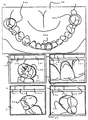
次に実施例の動作を図2を参照して詳細に説明する。
口腔内画像撮影手段11により口腔内の例えば上顎全歯を撮影する。撮影した画像の位置れを図2(a)に示す。
口腔内画像撮影手段11は、少なくとも歯科医師が診療対象とした歯が含まれ、それが口腔内のどの部分に相当するかわかる画像であれば良い。
図2(a)で示す画像は、例えば図3(c)で示す口腔内撮影ユニットによって撮影されたものであり、更にキャリブレーションを行って、歪みを補正してもよい。又、口腔内画像撮影手段11は、必ずしも全歯というわけではなく、部分歯である場合や、一つの歯である場合もある。図2(a)は、上顎20を示し、全歯及び硬口蓋部も撮影されており、この部位の診療も併せて行う場合もある。Next, the operation of the embodiment will be described in detail with reference to FIG.
The intraoral image photographing means 11 photographs, for example, all upper teeth in the oral cavity. The position of the captured image is shown in FIG.
The intraoral image photographing means 11 may be any image that includes at least teeth that have been treated by a dentist and that can be understood as to which part of the mouth corresponds.
The image shown in FIG. 2A is, for example, taken by the intraoral photographing unit shown in FIG. 3C, and further calibration may be performed to correct the distortion. Further, the intraoral image photographing means 11 is not necessarily all teeth, but may be a partial tooth or a single tooth. FIG. 2 (a) shows the
次に、診療部位検出手段12により、診療、改善を要する部位を自動又は手動で抽出する。手動で検出する場合、グラフィックソフトウエアと同様に、マウスを用いて丸又は四角で指定して、コピー、切り取り等をして、更にこれを貼り付け作業により貼り付ける
図2(a)の20a、20b及び20cは、対象とする歯にグラフィックソフトウエアによって目立つ色彩の円を手動又は自動で指示表示した状態を示す。
手動で指示表示するとは、例えばコンピュータに付属したマウス、キーボードを操作して円、四角、その他の輪郭図形等であって、内部が透過処理されたものを描くことであり、自動で指示表示するとは、例えば、画面上にマウスで、指定部位にポインタをもっていってボタンを押すと、所定の半径の円や、所定の面積の四角等であって、内部が透過処理されたものが表示される。
次に単位画像形成手段13で、図2(c)でしめす指定された歯20aを単位画像21に調整する。Next, the medical
Manual indication display means, for example, drawing a circle, square, or other contour figure etc. that has been transparently processed by operating the mouse and keyboard attached to the computer. For example, when the button is pressed with the mouse on the screen with the pointer on the designated area, a circle with a predetermined radius, a square with a predetermined area, etc., and the inside of which is transparently processed are displayed. .
Next, the unit image forming unit 13 adjusts the designated tooth 20a shown in FIG.
その際、例えば治療の順位を決定した後に順位を記入する順位欄21a、例えば治療開始を決定した後に記入する診療開始日欄21b、どのような診療がされるかを記入する説明欄21cが付加設定される。
この最短診療日を、例えば、図2の順位が一番早い単位画像の診療開始日欄21bに表示する。この日に対して患者が同意する場合は、決定入力手段155へ移動して、この日を決定し、記録手段156で患者のデータベースへ記録する。
患者が同意しない場合は、空き診療日表示手段154で空き診療日がカレンダー形式のような2次元形式で表示される。
この表示は、一覧形、手めくり形など、スケジュール表示の手法が採用されるが少なくとも患者がわかりやすい表示であれば良い。At that time, for example, a rank column 21a for entering the rank after determining the rank of treatment, for example, a medical treatment start date column 21b for filling in after the treatment start is decided, and an explanation column 21c for describing what kind of medical treatment is performed are added. Is set.
This shortest medical treatment date is displayed, for example, in the medical treatment start date column 21b of the unit image with the earliest ranking in FIG. If the patient agrees with this date, the user moves to the decision input means 155 to determine this date and records it in the patient database with the recording means 156.
When the patient does not agree, the empty medical day display means 154 displays the empty medical day in a two-dimensional format such as a calendar format.
For this display, a schedule display method such as a list form or a hand turn form is adopted, but at least a display that is easy to understand for the patient may be used.
尚、日付だけでなく、時間も必要であることから、時計表示も併せて行われる事が好ましい。この表示に基づいて、診療開始日の同意があると、決定入力手段155でその日時が入力され、記録手段156で、データベースに記録される。
次の順位の単位画像へ移動して同じ様な診療開始日が決定記入される。
このような、診療日のスケジュール操作は、本発明が、複数の診療予定日を設定する際は、一過性の診療では、生じなかった他の患者とのスケジュールとの混同を避ける必要があるため、スケジュール明確化には有効である。
尚、歯毎にID番号をいずれかの欄に記入したものであっても良い。この欄は、データベースで用いられる入力用ボックス表示等が示され、診療日は、他の患者の診療スケジュールから開いた日を自動的に決定可能であることから、
単位画像が表示された時点で、他の患者のデータから、空いた時間を表示しても良い。Since not only the date but also the time is required, it is preferable that the clock display is also performed. Based on this display, if there is an agreement on the medical treatment start date, the date and time is input by the decision input means 155 and recorded in the database by the recording means 156.
Moving to the next rank unit image, the same medical start date is determined and entered.
Such a schedule operation for a medical treatment date should avoid confusion with a schedule with other patients that did not occur in a temporary medical treatment when the present invention sets a plurality of scheduled medical treatment dates. Therefore, it is effective for clarifying the schedule.
The ID number may be entered in any column for each tooth. This field shows the box display for input used in the database, etc., and the medical date can be automatically determined from the medical schedule of other patients,
When the unit image is displayed, the vacant time may be displayed from the data of other patients.
これらの欄枠の表示内容は、治療前の画像は少なくとも治療順位が表示される欄があれば良く、又歯それぞれに、識別符合を付してそれを表示しても良い。
図2(d)は、図2(a)の診療用の歯20b、を示す単位画像22であり、図2(e)は、図2(a)の診療用の歯20cの単位画像23をそれぞれ示す。
単位画像22には、順位欄22a、診療開始日欄22b、説明欄22cが一体的に表示され、図2(e)も同様に、単位画像23において順位欄23a、診療開始日欄23b、説明欄23cが一体的に表示される。
尚、確定するときは、確定ボタン(画面上で表示された仮想ボタンを含む)を押す。そうすると、治療後の画像を表示する場合は、先に表示し、後は、順位記入欄の番号に沿って、並び替えられて表示されてもよく、この作業を自動化することで、最終的に患者へ渡す用紙の作成時間を短くできる。As for the display contents of these column frames, it suffices if there is at least a column in which the treatment order is displayed in the pre-treatment image, and each tooth may be displayed with an identification code.
FIG. 2D is a
In the
When confirming, the confirm button (including the virtual button displayed on the screen) is pressed. Then, when displaying the image after treatment, it may be displayed first, and after that, it may be rearranged and displayed according to the number in the rank entry field. By automating this work, finally It can shorten the time for preparing the paper to be delivered to the patient.
図2で示す図は、全体が一つのコンピュータのモニターデイスプレーに表示されている状態を示す場合もある。この場合は、既に表示出力手段16での操作範囲を兼ねている場合もある。
診療順位設定手段14では、順位欄22aの順位を、歯科医単独で又は、歯科医と患者が話し合いながら、決定し記入する。同様に診療説明形成手段15で診療開始日欄22b、説明欄22cを記入する。この記入は、専門的内容を多く含むことから、歯科医が予め単独で行う場合もある。
表示出力手段16は、コンピュータ画面上に確定した複数の単位画像と、全歯画像が掲載されている状態を形成し、表示する。
当該表示出力手段16は、歯科診療時等に行われる場合は、一画面上に表示される事が好ましい。The figure shown in FIG. 2 may show a state in which the whole is displayed on the monitor display of one computer. In this case, there is a case where the operation range of the display output means 16 is already used.
In the medical treatment order setting means 14, the order of the order column 22a is determined and entered by the dentist alone or while the dentist and the patient discuss. Similarly, the medical treatment
The display output means 16 forms and displays a state in which a plurality of unit images determined on the computer screen and all tooth images are posted.
The display output means 16 is preferably displayed on one screen when it is performed during dental treatment.
しかしながら、単位画像が多い場合は、スクロールも行われたり、必要に応じ縮小されたり、又はサムネール状に表示されても良い。
更に確定した表示出力手段16における画像は、プリンタによって、用紙に印字され、患者に配布されてもよい。患者は、自らの口腔内を画像で所持することで、口腔内の健全化を図るメニュープログラムを実行する用に通院を行い、歯の診療と改善を行う。診療完了後、再度図1でしめす様に口腔内の撮影を行う。
同じ部位の歯20aを単位画像24として図2(b)で示す様な画像表示を行う。24dが診療部位であり、24aは、例えば、診療順位設定手段14で、予め診療順位が設定された画像データから同一の画像データを検索し、その際表示された順位表示が行われる。However, when there are many unit images, scrolling may be performed, the image may be reduced as necessary, or displayed in a thumbnail shape.
Further, the confirmed image in the display output means 16 may be printed on paper by a printer and distributed to the patient. A patient goes to the hospital to execute a menu program for improving the health of the oral cavity by possessing his or her oral cavity as an image, and performs dental care and improvement. After completion of the medical examination, the intraoral imaging is performed again as shown in FIG.
The image display as shown in FIG. 2B is performed by using the teeth 20a of the same part as the
24bは、例えば診療説明形成手段15で診療した際の記録事項、等が予め記載されている。
そして図2(a)で示す診療後の単位画像は、診療前の単位画像であって、同一のものに隣接する箇所で表示され、歯科医への信頼や、診療した歯のメンテナンスの為の通院のタイミングが認識できる。24cは、例えば診療後の説明欄であり、補綴物であれば、耐久期間、歯の汚れであれば、次のクリーニング適当日等が記載され、次に診療のための説明や、診療の際の注意事項等が記載されていていてもよく、次の診療の開始を患者と相談する為の説明欄がより好ましい。そして診療開始する場合は、単位画像21の様式に欄が変更され、順位、診療開始日時等が設定されても良い。この場合は、数年後という場合もあるが、診療開始日にその日をいれておくだけで、他の患者との治療日の重複はさけられる。
そして、表示出力手段16で、プリンタにより一枚の用紙に、例えば図4の400で示すような4枚の画像を印字して患者に渡し、継続的診療の必要性を患者に与える。In 24b, for example, recorded items when medical treatment is performed by the medical treatment
The unit image after medical treatment shown in FIG. 2A is a unit image before medical treatment, which is displayed at a location adjacent to the same, and is used for trust to the dentist and maintenance of the treated tooth. Recognize the timing of hospital visits. 24c is, for example, an explanation column after medical treatment. If it is a prosthesis, the lasting period, and if it is a tooth stain, the next appropriate cleaning date is described. The explanation column for consulting with the patient about the start of the next medical treatment is more preferable. And when starting medical care, a column may be changed into the format of the
Then, the display output means 16 prints, for example, four images as indicated by 400 in FIG. 4 on a single sheet by a printer and passes it to the patient, giving the patient the necessity of continuous medical care.
尚、400は、図2(a)で示す口腔内広範囲画像が掲載されていないが、好ましくは広範囲画像を掲載して、診療部位を明確にすることで、患者のより一層の理解が得られる。尚、診療順位と診療開始日時は、いずれか一方であっても良く、その場合は、単位画像の順が診療順を表すようにし、診療開始日が少なくとも付随した単位画像表示が好ましい。
尚、図2、図4の各表示欄は、例であり、単位画像あたりの表示欄の数、表示内容も、治療の目的、スケジュール等に応じて適宜選択されるものである。In FIG. 2, the intraoral wide-area image shown in FIG. 2 (a) is not posted. However, it is preferable to display the wide-area image and clarify the medical treatment site to obtain a better understanding of the patient. . Note that the medical treatment order and the medical treatment start date and time may be either one. In this case, it is preferable that the order of the unit images represents the medical treatment order, and the unit image display accompanied by at least the medical treatment start date is preferable.
Each of the display columns in FIGS. 2 and 4 is an example, and the number of display columns per unit image and the display contents are also appropriately selected according to the purpose of treatment, the schedule, and the like.
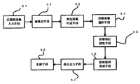
次に、他の実施例を図5に示し説明する。
51は、広範囲画像入力手段であり、口腔内の上顎の全歯、下顎の全歯を撮影する為の手段であり、例えば図3の(c)で示す凸面鏡に映した全歯をカメラで撮影することで広範囲画像を得たり、図6で示す様な連続撮影、デジタル動画を構成する静止画から共通部分を合成して、全歯画像の形成を行うものであってもよい。
52は、歯検出手段であり、広範囲画像入力手段51で得られた歯列からそれぞれの歯の画像データを抽出する手段である。抽出は、例えば、輪郭抽出により、閉じた円を検出してその周辺を所定範囲だけ抽出して歯をそれぞれ一枚の画像化する手段の他、図3(b)で示す平面鏡を用いて個々の歯を撮影して一画像化する手段をとっても良いが、カメラの画素数をより高分解能にすれば、全歯画像や口腔内広範囲画像から単位画像を検出する手段の方が、合理的で好ましいものである。Next, another embodiment will be described with reference to FIG.
52 is a tooth detection means for extracting image data of each tooth from the dentition obtained by the wide range image input means 51. For the extraction, for example, by using a plane mirror shown in FIG. 3 (b) in addition to means for detecting a closed circle by contour extraction, extracting the periphery of the circle by a predetermined range, and imaging each tooth as a single sheet. However, if the number of pixels of the camera is set to a higher resolution, the means for detecting the unit image from the entire tooth image or the intraoral wide-area image is more reasonable. It is preferable.
53は、単位画像形成手段であり、歯検出手段52で得られた個々の画像に、それぞれ、順位記入欄、診療説明記入欄などを付加して、表示用の画像を形成するためのものである。
更に歯用のデータベースを形成して、固有の記号を付して管理することが好ましい。
54は、診療画像選択手段であり、虫歯、欠損等の診療対象となる歯を画像から選択するための手段であり、歯科医師が目視で又は、先に登録したデータを読み出して比較して、色、形状等の相違により診療対象の歯として選択してもよい。
本発明は、全歯を一回の撮影で登録できることから、診療対象の歯を見つけやすいのである。53 is a unit image forming means for adding a rank entry column, a medical treatment explanation entry column, etc. to the individual images obtained by the tooth detection means 52 to form a display image. is there.
Furthermore, it is preferable that a dental database is formed and managed with unique symbols.
54 is a medical image selection means, a means for selecting a tooth to be medical care such as caries, a defect, etc. from the image, the dentist visually or by reading and comparing previously registered data, You may select as a tooth of medical treatment object by the difference in a color, a shape, etc.
In the present invention, since all teeth can be registered by one imaging, it is easy to find a tooth to be treated.
55は、診療順位設定手段であり、これは、歯科医師が単独で決定するほか、歯科医師と、患者が、コンピュータのモニターデイスプレイ(例えば図3(a)のモニターディスプレイ314A)に映し出された又は、印字印刷された画像を元に、決定してもよく、更に、先に撮影したデータとの比較により、その差の大きさで決定しても良い。
56は、診療説明形成手段であり、診療の期間、開始日、及び診療の内容、改善の必要性等を歯科医師単独又は、患者との打ち合わせにより記入されたり、先のデータとの比較による、その差の大きさにより予め記録された説明を表示しても良い。
57は、表示出力手段であり、前記診療対象となる表示画像を1枚の用紙に、更には、コンピュータのモニターディスプレイ(例えば図3(a)の314A)に表示し、患者の同意と、理解を得るための説明に供されてもよい。又、用紙に印刷された画像は、患者に提供され、今後の診療スケジュールとして、継続的な歯科診療が実現される。
58は、記録手段であり、カルテとして、データベースとして記録され、又はホームページ上に、患者専用のページにアップロードしたり、患者の携帯電話の記録部に歯画像データを記録させてもよい。55 is a medical treatment order setting means, which is determined by the dentist alone, as well as when the dentist and the patient are displayed on the monitor display of the computer (for example, the monitor display 314A in FIG. 3A) or The determination may be made based on the printed and printed image, or may be determined based on the magnitude of the difference by comparison with the previously captured data.
56 is a medical explanation forming means, and the period of the medical treatment, the start date, the content of the medical treatment, the necessity for improvement, etc. are entered by the dentist alone or in consultation with the patient, or by comparison with the previous data, You may display the description recorded beforehand by the magnitude | size of the difference.
次に図5で示す実施例の動作を図7を参照して説明する。
患者の口腔内から広範囲画像入力手段51で、全歯を撮影して、一枚、又は複数枚からなる全歯の画像データ700を形成する(図7(a))。全歯を示す画像データ700は、図6で示した静止画像を連結して形成したものを一例とすることができる。
得られた広範囲画像データ700から、歯検出手段52により手動で又は自動で歯画像を形成する。
自動で、歯を検出する手法として、口腔内の画像データを輪郭抽出して、歯の輪郭をそれぞれ検出する。Next, the operation of the embodiment shown in FIG. 5 will be described with reference to FIG.
All the teeth are photographed by the wide-area image input means 51 from within the oral cavity of the patient, and
From the wide
As a method for automatically detecting teeth, the contours of image data in the oral cavity are extracted and the contours of the teeth are detected.
この場合、輪郭が不完全な場合は、その数点を検出して、この数点を通過する仮想円を形成する。この仮想円を、一つの歯の位置とみなすことが出来るから、想定された中心から更に半径を所定値だけ広げ、四角面としてその範囲を一つの歯の画像として抽出する。
抽出した画像を例えば、図7(b)で示す。歯71に対し歯画像701、歯72に対し、歯画像702、歯73に対し歯画像703である。
次に、単位画像形成手段53で、これらの歯の画像に、識別子等、患者の情報と関連づけて単位画像化する(図7(c))。単位画像74には、抽出れた歯画像701と、診療順位等を記入するための表示欄704が掲載されている。
単位画像75には、歯画像702と表示欄705、単位画像76には、歯画像703と表示欄706がそれぞれ掲載されている。In this case, when the contour is incomplete, several points are detected and a virtual circle passing through the several points is formed. Since this virtual circle can be regarded as the position of one tooth, the radius is further expanded from the assumed center by a predetermined value, and the range is extracted as a square surface as an image of one tooth.
For example, FIG. 7B shows the extracted image. A
Next, in the unit
The unit image 75 includes a
これらの単位画像は、予め設定された患者データベースに記録登録され患者の上下口腔内データリストが形成可能となる。
診療画像選択手段54では、この中から、診療又は改善対象となる歯牙画像を示す単位画像77を、目視又は自動で抽出する(図7(d))。
図1と同様の構成を有し同様の動作を行う診療順位設定手段55、及び診療説明形成手段56で、それぞれ、歯科医師単独又は、好ましくは患者と、コンピュータデイスプレイに写った単位歯画像を見ながら、記入をしていく。
そして、診療順位が決定し、診療開始日等が書き込まれた段階で、表示出力手段57において、コンピュータのモニターデイスプレイ78に例えば図7(e)で示すような順序表示された単位画像の配列又は、図7(f)でしめす様に広範囲画像を更に含めて編集された状態で表示されてもよく、患者に診療の認識と、継続的な診療に同意が得られる状況を形成することができる。These unit images are recorded and registered in a preset patient database, and a patient's upper / lower intraoral data list can be formed.
The medical image selection means 54 extracts a
The medical order setting unit 55 and the medical treatment explanation forming unit 56 having the same configuration as in FIG. 1 and performing the same operation, respectively, see the unit tooth image shown on the computer display with the dentist alone or preferably with the patient. While filling in.
Then, at the stage where the medical treatment order is determined and the medical treatment start date and the like are written, the
単位画像707、708、709及び710は、順位順又は治療開始順に並ぶことが好ましい。図7(f)は、例えば全歯画像データ711を含む画面表示又は、患者が携行する印刷物化した歯データ79である。
更に、この状態を、カルテ、配布用紙にプリントアウトしたり、携帯電話や、専用ホームページ等の患者専用の記憶端末に出力して、患者の継続的診療に対する意識と口腔内健全化を患者だけでなく、周辺の人々にも意識を持たせ、併せて歯科経営の健全化を図ることを可能とする。記録手段58は、これらの単位画像をデータベースに記録し、他の患者のスケジュールの調整に適宜利用される。The
Furthermore, this state can be printed out on a medical record or distribution form, or output to a patient-specific storage terminal such as a mobile phone or a dedicated homepage, so that the patient's awareness of continuous medical care and oral health can be improved only by the patient. In addition, it is possible to raise awareness of the surrounding people and to improve the soundness of dental management. The recording means 58 records these unit images in a database and is used as appropriate for adjusting the schedules of other patients.
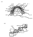
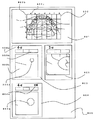
更に本発明の他の実施例を図8を参照して詳細に説明する。
図8において、口腔内広範囲画像801に、座標表示のグリッド802を重ねて表示させたものである。
800は、一枚の用紙に画像を印字した診療画像表示を示すか、又はコンピュータモニター画像に表示した診療画像表示を示す。
印字した状態の場合は、各単位画像が静止画として確定した状態を示している。
横軸802aは、等間隔に区切って、それぞれ、記号a,b,c,d,・・・を付し、縦軸802bも同様に等間隔に区切って、それぞれ、番号1,2,3,・・・を付し、場所の特定を行うようにしたものである。
この縦軸と横軸で囲まれた範囲を単位画像として、表示し、診療順位欄、診療説明欄を付したものが、803から804である。
803は、口腔内広範囲画像801の座標4−e(803a)の部分であり、
803bは、治療順位、治療開始日、説明が記入、表示される欄であり、803cは、該当する座標内の画像である。Further, another embodiment of the present invention will be described in detail with reference to FIG.
In FIG. 8, a coordinate
In the printed state, each unit image is confirmed as a still image.
The horizontal axis 802a is divided into equal intervals, respectively, with symbols a, b, c, d,..., And the vertical axis 802b is also equally divided into equal numbers, 1, 2, 3, respectively. ... is used to identify the location.
803 is a portion of coordinates 4-e (803a) of the intraoral wide area image 801,
Reference numeral 803b denotes a field in which the treatment order, treatment start date, and description are entered and displayed, and 803c is an image within the corresponding coordinates.
804は、口腔内広範囲画像801の座標4−eの部分の拡大画像であり、別途他のカメラで撮影した場合は、その表示として804aの様な凹面鏡を反射鏡として用いた口腔内カメラ(図3(d)でしめす)の使用を明示してもよい。
805は、座標802でしめす2−eの部位の単位画像であり、単位画像803,803と同様の様式で形成されている。
それぞれ該当する部分を拡大又は、例えば、図3(b)でしめす平面鏡を反射鏡として用いた口腔内カメラ、図3(d)でしめす凹面鏡を反射鏡として使用した口腔内カメラで撮影したものが示される。使用したカメラは、図8の804aの様な表示で示されることが好ましい。
これは、一つのモニター内に口腔内広範囲画像801を静止画として表示し、その周辺に、単位画像803を表示すると共に、画像部分803cを動画モードとし、口腔内を図3(b)の平面鏡で撮影し、画面803c内の動画像が口腔内広範囲画像801の座標4−eとなるまで位置決めし、一致したタイミングで静止画となるようシャッターボタンを押して、対応する単位画像803を得るという手法が例示される。803dは、例えばポリープである。
Reference numeral 805 denotes a unit image of the portion 2-e indicated by the
Each of the corresponding parts is magnified or taken with an intraoral camera using, for example, a plane mirror shown in FIG. 3 (b) as a reflecting mirror, and an intraoral camera using a concave mirror shown in FIG. 3 (d) as a reflecting mirror. Indicated. The camera used is preferably shown with a display such as 804a in FIG.
This is because the intraoral wide-area image 801 is displayed as a still image in one monitor, the
上述した様に単位画像804は、凹面鏡318で撮影したものである。804aと示して表示を区別している。803で示す単位画像は、舌にできたポリープ804bを拡大したものをしめしており、これを拡大した状態を単位画像804で示している。
このように、口腔内の撮影位置を決定する手段として、口腔内広域画像にその座標的な情報を形成することで、歯肉炎、舌ガン等の位置を患者にわかりやすく教えることができると共に、この部位をより強く関連づけた単位画像との関係において、拡大された画像を、よりわかりやすい形で表示可能とする。
又、本発明は、広域画像取得に際し、図3で示した凹面鏡と凸面鏡を用いた手法、反射鏡を歯列に沿って移動させて、静止画像を結合して得る手法を併合して一枚の画像を形成しても良い。As described above, the
In this way, as a means for determining the imaging position in the oral cavity, by forming the coordinate information in the intraoral wide area image, it is possible to easily teach the patient the position of gingivitis, tongue cancer, etc. The enlarged image can be displayed in a more easy-to-understand form in relation to the unit image in which this part is more strongly associated.
In addition, the present invention combines a technique using a concave mirror and a convex mirror shown in FIG. 3 and a technique obtained by combining a still image by moving a reflecting mirror along a dentition when acquiring a wide area image. The image may be formed.
即ち、歯列のみを静止画像とし、舌の部位を凸面、凹面の反射鏡を用いて撮影し、画像合成をすることで、歪みのない歯列を具えた口腔内広域画像が得られるのであり、更に診療部位を、歯列を基準とした変形座標を用いて、下部の部位を特定することも可能となる。
又、本発明は、全て平面の静止画像を組み合わせて形成することも可能である。
例えば、歯列画像を図6でしめす手法で撮影取得し、更に平面鏡を用いて、多少高い位置から撮影し、少なくとも形状比較が出来そうな歯の辺部を含む画像であって、口腔内全域を数枚の画像で取得する。
そして、歯列画像と、その他口腔内を含む画像を比較する。
その際、共通する歯の大きさから、変換係数を求める。この変換係数に基づいて、その他口腔内画像を縮小又は拡大する。撮影状態が傾斜している場合は、離れた歯の形状を比較し、傾斜度を考慮してもよい。
このようにしてつなぎ合わせることで、口腔内広域画像であって、より変形度の少ない画像が得られる。That is, only the dentition is taken as a still image, the part of the tongue is photographed using convex and concave reflectors, and the images are combined to obtain a wide intraoral image with a dentition without distortion. Furthermore, it is also possible to specify the lower part using the deformed coordinates with reference to the dentition as the medical treatment part.
The present invention can also be formed by combining all flat still images.
For example, a dentition image is obtained by the method shown in FIG. 6 and is taken from a slightly higher position using a plane mirror, and includes at least the side of the tooth that can be compared in shape, and the entire intraoral area Is acquired with several images.
Then, the dentition image is compared with other images including the oral cavity.
At that time, a conversion coefficient is obtained from the common tooth size. Other intraoral images are reduced or enlarged based on the conversion coefficient. When the photographing state is inclined, the shape of the distant teeth may be compared to consider the inclination.
By stitching together in this way, an intraoral wide area image with less deformation can be obtained.
本発明は、歯科診療における口腔内健全化を図るべく、患者に口腔内情報を見やすい形式で提供することで、患者の口腔内情報を新たにすることができる歯科システムを提案するものであり、歯科医療分野において有効に利用される。 The present invention proposes a dental system that can renew patient's intraoral information by providing the patient with easy-to-see intraoral information in order to achieve oral health in dental practice, It is effectively used in the field of dentistry.
11 口腔内画像撮影手段
12 診療部位検出手段
13 単位画像形成手段
14 診療順位設定手段
15 診療説明形成手段
16 表示出力手段
17 記録手段DESCRIPTION OF SYMBOLS 11 Intraoral image imaging means 12 Medical site detection means 13 Unit image formation means 14 Medical treatment order setting means 15 Medical treatment description formation means 16 Display output means 17 Recording means
Claims (5)
Priority Applications (8)
| Application Number | Priority Date | Filing Date | Title |
|---|---|---|---|
| JP2011017192A JP2012143528A (en) | 2011-01-12 | 2011-01-12 | Oral imaging and display system |
| TW100147322A TWI524873B (en) | 2011-01-11 | 2011-12-20 | Intraocular photography display system |
| CA2824665A CA2824665C (en) | 2011-01-11 | 2012-01-11 | Intraoral video camera and display system |
| AU2012206109A AU2012206109B2 (en) | 2011-01-11 | 2012-01-11 | Oral imaging and display system |
| EP12734447.1A EP2664272A4 (en) | 2011-01-11 | 2012-01-11 | Oral imaging and display system |
| PCT/JP2012/050394 WO2012096312A1 (en) | 2011-01-11 | 2012-01-11 | Oral imaging and display system |
| US13/978,939 US9463081B2 (en) | 2011-01-11 | 2012-01-11 | Intraoral video camera and display system |
| CN201280008136.6A CN103347436B (en) | 2011-01-11 | 2012-01-11 | Display system is shot in oral cavity |
Applications Claiming Priority (1)
| Application Number | Priority Date | Filing Date | Title |
|---|---|---|---|
| JP2011017192A JP2012143528A (en) | 2011-01-12 | 2011-01-12 | Oral imaging and display system |
Publications (2)
| Publication Number | Publication Date |
|---|---|
| JP2012143528A true JP2012143528A (en) | 2012-08-02 |
| JP2012143528A5 JP2012143528A5 (en) | 2014-02-20 |
Family
ID=46787727
Family Applications (1)
| Application Number | Title | Priority Date | Filing Date |
|---|---|---|---|
| JP2011017192A Ceased JP2012143528A (en) | 2011-01-11 | 2011-01-12 | Oral imaging and display system |
Country Status (1)
| Country | Link |
|---|---|
| JP (1) | JP2012143528A (en) |
Cited By (8)
| Publication number | Priority date | Publication date | Assignee | Title |
|---|---|---|---|---|
| JP2016140760A (en) * | 2015-01-30 | 2016-08-08 | デンタル・イメージング・テクノロジーズ・コーポレーション | Intra-oral image acquisition alignment |
| JP2017020930A (en) * | 2015-07-13 | 2017-01-26 | 株式会社モリタ製作所 | Intraoral three-dimensional measuring device, intraoral three-dimensional measuring method, and method for displaying intraoral three-dimensional measuring result |
| KR20180073956A (en) * | 2016-12-23 | 2018-07-03 | 동의대학교 산학협력단 | Prevention System For Dental Plaque and Gingivitis And Method Threrof |
| WO2019150607A1 (en) * | 2018-01-31 | 2019-08-08 | MediDoc Search株式会社 | Advertisement presentation method and advertisement presentation system |
| KR20200068992A (en) * | 2018-12-06 | 2020-06-16 | 오스템임플란트 주식회사 | Method, Apparatus and Recording For Computerizing Of Electro-Magnetic Resonance |
| JP2022516052A (en) * | 2018-12-21 | 2022-02-24 | ディーオーエフ インコーポレイテッド | 3D scanner and scanning method using this |
| WO2024005344A1 (en) * | 2022-07-01 | 2024-01-04 | 오스템임플란트 주식회사 | Thumbnail provision method for patient management system, and computing device performing same method |
| WO2024116769A1 (en) * | 2022-11-28 | 2024-06-06 | パナソニックIpマネジメント株式会社 | Periodontal disease detection system, periodontal disease detection method, and periodontal disease detection program |
Citations (1)
| Publication number | Priority date | Publication date | Assignee | Title |
|---|---|---|---|---|
| JP2010099157A (en) * | 2008-10-22 | 2010-05-06 | Konica Minolta Medical & Graphic Inc | Medical image processing apparatus and program |
-
2011
- 2011-01-12 JP JP2011017192A patent/JP2012143528A/en not_active Ceased
Patent Citations (1)
| Publication number | Priority date | Publication date | Assignee | Title |
|---|---|---|---|---|
| JP2010099157A (en) * | 2008-10-22 | 2010-05-06 | Konica Minolta Medical & Graphic Inc | Medical image processing apparatus and program |
Non-Patent Citations (1)
| Title |
|---|
| JPN6014054197; 須崎明: '"かかりつけ"を増やすツールに役立てる' 日本歯科評論 No.789,Vol.68(7), 20080711, 143〜148頁 * |
Cited By (11)
| Publication number | Priority date | Publication date | Assignee | Title |
|---|---|---|---|---|
| JP2016140760A (en) * | 2015-01-30 | 2016-08-08 | デンタル・イメージング・テクノロジーズ・コーポレーション | Intra-oral image acquisition alignment |
| JP2017020930A (en) * | 2015-07-13 | 2017-01-26 | 株式会社モリタ製作所 | Intraoral three-dimensional measuring device, intraoral three-dimensional measuring method, and method for displaying intraoral three-dimensional measuring result |
| KR20180073956A (en) * | 2016-12-23 | 2018-07-03 | 동의대학교 산학협력단 | Prevention System For Dental Plaque and Gingivitis And Method Threrof |
| KR101962241B1 (en) * | 2016-12-23 | 2019-07-18 | 동의대학교 산학협력단 | Prevention System For Dental Plaque and Gingivitis |
| WO2019150607A1 (en) * | 2018-01-31 | 2019-08-08 | MediDoc Search株式会社 | Advertisement presentation method and advertisement presentation system |
| KR20200068992A (en) * | 2018-12-06 | 2020-06-16 | 오스템임플란트 주식회사 | Method, Apparatus and Recording For Computerizing Of Electro-Magnetic Resonance |
| KR102294618B1 (en) * | 2018-12-06 | 2021-08-30 | 오스템임플란트 주식회사 | Method, Apparatus and Recording For Computerizing Of Electro-Magnetic Resonance |
| JP2022516052A (en) * | 2018-12-21 | 2022-02-24 | ディーオーエフ インコーポレイテッド | 3D scanner and scanning method using this |
| JP7142395B2 (en) | 2018-12-21 | 2022-09-27 | ディーオーエフ インコーポレイテッド | 3D scanner and scanning method using the same |
| WO2024005344A1 (en) * | 2022-07-01 | 2024-01-04 | 오스템임플란트 주식회사 | Thumbnail provision method for patient management system, and computing device performing same method |
| WO2024116769A1 (en) * | 2022-11-28 | 2024-06-06 | パナソニックIpマネジメント株式会社 | Periodontal disease detection system, periodontal disease detection method, and periodontal disease detection program |
Similar Documents
| Publication | Publication Date | Title |
|---|---|---|
| US12070367B2 (en) | Correction of unclear features in three-dimensional models of dental sites | |
| WO2012096312A1 (en) | Oral imaging and display system | |
| JP2012143528A (en) | Oral imaging and display system | |
| JP5784381B2 (en) | Dental care system | |
| JP6099310B2 (en) | Automatic dental chart creation method using digital images | |
| KR102267197B1 (en) | Method and apparatus for recording and displaying dental care data on a digital dental image | |
| KR101915215B1 (en) | Identification of areas of interest during intraoral scans | |
| ES2724115T3 (en) | Graphical user interface for computer-assisted margin marking on dentures | |
| JP2016140761A (en) | Dental variation tracking and prediction | |
| Ahmad | Prosthodontics at a Glance | |
| TWI524873B (en) | Intraocular photography display system | |
| LT6296B (en) | Method and system of treatment plan and administration of dental prosthesis of a patient | |
| US20230071852A1 (en) | Data processing apparatus and method thereof | |
| US20230196511A1 (en) | Image processing apparatus, image capturing apparatus, image capturing system, and method | |
| KR100604182B1 (en) | United management method for images of dental clinic and record medium for united management program thereof | |
| KR102369100B1 (en) | Method for displaying multi panoramic image and imaging processing apparatus thereof | |
| Reesu | Investigation of photographic and digital methods for odontological comparison to aid with human identification | |
| WO2024056719A1 (en) | 3d digital visualization, annotation and communication of dental oral health |
Legal Events
| Date | Code | Title | Description |
|---|---|---|---|
| A521 | Written amendment |
Free format text: JAPANESE INTERMEDIATE CODE: A523 Effective date: 20131225 |
|
| A621 | Written request for application examination |
Free format text: JAPANESE INTERMEDIATE CODE: A621 Effective date: 20131225 |
|
| A131 | Notification of reasons for refusal |
Free format text: JAPANESE INTERMEDIATE CODE: A131 Effective date: 20141222 |
|
| A521 | Written amendment |
Free format text: JAPANESE INTERMEDIATE CODE: A523 Effective date: 20150220 |
|
| A131 | Notification of reasons for refusal |
Free format text: JAPANESE INTERMEDIATE CODE: A131 Effective date: 20150702 |
|
| A521 | Written amendment |
Free format text: JAPANESE INTERMEDIATE CODE: A523 Effective date: 20150828 |
|
| A01 | Written decision to grant a patent or to grant a registration (utility model) |
Free format text: JAPANESE INTERMEDIATE CODE: A01 Effective date: 20151029 |
|
| A045 | Written measure of dismissal of application |
Free format text: JAPANESE INTERMEDIATE CODE: A045 Effective date: 20160218 |