JP2011508917A - Method and system for case-based computer-aided diagnosis with cross-modality - Google Patents
Method and system for case-based computer-aided diagnosis with cross-modality Download PDFInfo
- Publication number
- JP2011508917A JP2011508917A JP2010538974A JP2010538974A JP2011508917A JP 2011508917 A JP2011508917 A JP 2011508917A JP 2010538974 A JP2010538974 A JP 2010538974A JP 2010538974 A JP2010538974 A JP 2010538974A JP 2011508917 A JP2011508917 A JP 2011508917A
- Authority
- JP
- Japan
- Prior art keywords
- image
- modality
- features
- feature
- cases
- Prior art date
- Legal status (The legal status is an assumption and is not a legal conclusion. Google has not performed a legal analysis and makes no representation as to the accuracy of the status listed.)
- Granted
Links
- 238000000034 method Methods 0.000 title claims abstract description 35
- 238000004195 computer-aided diagnosis Methods 0.000 title abstract description 5
- 238000013507 mapping Methods 0.000 claims abstract description 18
- 238000002595 magnetic resonance imaging Methods 0.000 claims description 21
- 230000015572 biosynthetic process Effects 0.000 claims description 14
- 238000002591 computed tomography Methods 0.000 claims description 12
- 238000002604 ultrasonography Methods 0.000 claims description 12
- 238000000556 factor analysis Methods 0.000 claims description 9
- 230000003902 lesion Effects 0.000 claims description 4
- 239000000284 extract Substances 0.000 claims description 2
- 238000012314 multivariate regression analysis Methods 0.000 claims 2
- 238000012831 peritoneal equilibrium test Methods 0.000 claims 2
- 238000012636 positron electron tomography Methods 0.000 claims 2
- 238000012877 positron emission topography Methods 0.000 claims 2
- 206010028980 Neoplasm Diseases 0.000 description 16
- 238000003384 imaging method Methods 0.000 description 10
- 230000000875 corresponding effect Effects 0.000 description 8
- 201000010099 disease Diseases 0.000 description 8
- 208000037265 diseases, disorders, signs and symptoms Diseases 0.000 description 8
- 238000004458 analytical method Methods 0.000 description 3
- 238000004364 calculation method Methods 0.000 description 3
- 238000003745 diagnosis Methods 0.000 description 3
- 238000010586 diagram Methods 0.000 description 3
- 230000006870 function Effects 0.000 description 3
- 239000011159 matrix material Substances 0.000 description 3
- 238000012986 modification Methods 0.000 description 3
- 230000004048 modification Effects 0.000 description 3
- 239000013598 vector Substances 0.000 description 3
- 230000000007 visual effect Effects 0.000 description 3
- 206010006187 Breast cancer Diseases 0.000 description 2
- 208000026310 Breast neoplasm Diseases 0.000 description 2
- 230000002596 correlated effect Effects 0.000 description 2
- 238000002600 positron emission tomography Methods 0.000 description 2
- 230000002685 pulmonary effect Effects 0.000 description 2
- 206010058467 Lung neoplasm malignant Diseases 0.000 description 1
- 206010054107 Nodule Diseases 0.000 description 1
- 238000013459 approach Methods 0.000 description 1
- 230000036772 blood pressure Effects 0.000 description 1
- 210000004072 lung Anatomy 0.000 description 1
- 201000005202 lung cancer Diseases 0.000 description 1
- 208000020816 lung neoplasm Diseases 0.000 description 1
- 230000003211 malignant effect Effects 0.000 description 1
- 230000000391 smoking effect Effects 0.000 description 1
- 238000012549 training Methods 0.000 description 1
- 230000009466 transformation Effects 0.000 description 1
- 238000000844 transformation Methods 0.000 description 1
- 230000001131 transforming effect Effects 0.000 description 1
- 238000012285 ultrasound imaging Methods 0.000 description 1
Images


Classifications
-
- G—PHYSICS
- G16—INFORMATION AND COMMUNICATION TECHNOLOGY [ICT] SPECIALLY ADAPTED FOR SPECIFIC APPLICATION FIELDS
- G16H—HEALTHCARE INFORMATICS, i.e. INFORMATION AND COMMUNICATION TECHNOLOGY [ICT] SPECIALLY ADAPTED FOR THE HANDLING OR PROCESSING OF MEDICAL OR HEALTHCARE DATA
- G16H30/00—ICT specially adapted for the handling or processing of medical images
- G16H30/40—ICT specially adapted for the handling or processing of medical images for processing medical images, e.g. editing
-
- G—PHYSICS
- G16—INFORMATION AND COMMUNICATION TECHNOLOGY [ICT] SPECIALLY ADAPTED FOR SPECIFIC APPLICATION FIELDS
- G16H—HEALTHCARE INFORMATICS, i.e. INFORMATION AND COMMUNICATION TECHNOLOGY [ICT] SPECIALLY ADAPTED FOR THE HANDLING OR PROCESSING OF MEDICAL OR HEALTHCARE DATA
- G16H30/00—ICT specially adapted for the handling or processing of medical images
- G16H30/20—ICT specially adapted for the handling or processing of medical images for handling medical images, e.g. DICOM, HL7 or PACS
-
- G—PHYSICS
- G16—INFORMATION AND COMMUNICATION TECHNOLOGY [ICT] SPECIALLY ADAPTED FOR SPECIFIC APPLICATION FIELDS
- G16H—HEALTHCARE INFORMATICS, i.e. INFORMATION AND COMMUNICATION TECHNOLOGY [ICT] SPECIALLY ADAPTED FOR THE HANDLING OR PROCESSING OF MEDICAL OR HEALTHCARE DATA
- G16H50/00—ICT specially adapted for medical diagnosis, medical simulation or medical data mining; ICT specially adapted for detecting, monitoring or modelling epidemics or pandemics
- G16H50/70—ICT specially adapted for medical diagnosis, medical simulation or medical data mining; ICT specially adapted for detecting, monitoring or modelling epidemics or pandemics for mining of medical data, e.g. analysing previous cases of other patients
-
- G—PHYSICS
- G16—INFORMATION AND COMMUNICATION TECHNOLOGY [ICT] SPECIALLY ADAPTED FOR SPECIFIC APPLICATION FIELDS
- G16H—HEALTHCARE INFORMATICS, i.e. INFORMATION AND COMMUNICATION TECHNOLOGY [ICT] SPECIALLY ADAPTED FOR THE HANDLING OR PROCESSING OF MEDICAL OR HEALTHCARE DATA
- G16H50/00—ICT specially adapted for medical diagnosis, medical simulation or medical data mining; ICT specially adapted for detecting, monitoring or modelling epidemics or pandemics
- G16H50/20—ICT specially adapted for medical diagnosis, medical simulation or medical data mining; ICT specially adapted for detecting, monitoring or modelling epidemics or pandemics for computer-aided diagnosis, e.g. based on medical expert systems
Abstract
クロスモダリティによる事例ベースのコンピュータ支援診断のためのシステム及び方法は、複数の事例を格納することを有する。各事例は、複数のモダリティのうちの1つの少なくとも1つの画像と、非画像情報とを含む。当該システム及び方法は更に、第1のモダリティの画像からの特徴と第2のモダリティの画像からの特徴との間の特徴関係をマッピングすること、及び該関係を格納することを有する。 A system and method for case-based computer-aided diagnosis with cross-modality includes storing a plurality of cases. Each case includes at least one image of one of the modalities and non-image information. The system and method further comprises mapping a feature relationship between features from the image of the first modality and features from the image of the second modality, and storing the relationship.
Description
本出願は概して、クロスモダリティによる事例ベースのコンピュータ支援診断(“CADx”)のためのシステム及び方法に関する。具体的には、該システム及び方法は、CADxシステムのユーザが異なる複数の撮像モダリティにまたがって類似事例を検索することを可能にし得る。 The present application generally relates to systems and methods for case-based computer-aided diagnosis (“CADx”) with cross-modality. Specifically, the system and method may allow a CADx system user to search for similar cases across multiple imaging modalities.
事例ベースのCADxシステムは、臨床医が経験によって、また、以前に見た事例を参照することによって、知識を取得するという概念に基づく。臨床医が例えば肺癌のCTスキャン(又は、X線、磁気共鳴撮像(MRI)、超音波、陽電子放出型断層撮影(PET)などのその他のモダリティスキャン)に基づいて診断を下す際に意思決定支援システムが臨床医を支援することが可能な一手法は、既に診断され且つ問題の事例と類似する以前の事例を提供することである。事例ベースの1つの考え方は、診断すべき肺結節に類似する肺結節が、既に診断された結節のデータベースから検索され、放射線医に提示されるというものである。これが事例ベースCADxシステムの基本的な前提である。 Case-based CADx systems are based on the concept that clinicians acquire knowledge by experience and by referring to previously seen cases. Decision support when clinicians make diagnoses based on CT scans of lung cancer (or other modality scans such as X-rays, magnetic resonance imaging (MRI), ultrasound, positron emission tomography (PET), etc.) One way the system can assist clinicians is to provide previous cases that have already been diagnosed and are similar to the problem case. One case-based idea is that a pulmonary nodule similar to the pulmonary nodule to be diagnosed is retrieved from a database of already diagnosed nodules and presented to the radiologist. This is the basic premise of the case-based CADx system.
事例ベースのCADxは典型的に、データベースから、例えば病状すなわち悪性であるか良性であるかが知られた腫瘍又は病変などの、疾患に関する情報を取り出すことを必要とする。この情報は典型的に、診断すべき腫瘍の診断スキャンと視覚的に比較するための、既に診断された腫瘍の診断スキャンを含む。腫瘍は、例えば、患者の肺の中とし得る。腫瘍の診断スキャンは、数多くの撮像技術のうちの何れか1つによって捕捉され得る。撮像技術の幾つかは上述した。スキャンから、各特徴が腫瘍の特定の視覚的特徴を表すような腫瘍の特徴群が計算され得る。診断すべき腫瘍、及びデータベースの腫瘍群は、共通の特徴空間、すなわち、各次元がN個の測定される特徴のうちのそれぞれの1つを表すN次元空間、に配置されることができる。データベースの何れかの腫瘍と診断すべき腫瘍との間の類似性を、特徴空間内での2つの腫瘍の近接性に基づいて、暫定的且つ客観的に調査することができる。典型的に、データベースから、最も近接する複数の腫瘍が類似腫瘍として取り出される。取り出された実例は、視覚的な比較のため、診断すべき腫瘍と並べて表示され得る。事例ベースのCADxはまた、様々な実感を診断することに関して医療関係者を訓練することにも有用である。 Case-based CADx typically requires retrieving information about a disease from a database, such as a disease state or tumor or lesion known to be malignant or benign. This information typically includes a diagnostic scan of an already diagnosed tumor for visual comparison with a diagnostic scan of the tumor to be diagnosed. The tumor can be, for example, in the patient's lungs. A diagnostic scan of the tumor can be captured by any one of a number of imaging techniques. Some of the imaging techniques have been described above. From the scan, a set of tumor features can be calculated such that each feature represents a particular visual feature of the tumor. The tumor to be diagnosed and the tumor group in the database can be arranged in a common feature space, ie an N-dimensional space where each dimension represents one of each of the N measured features. Similarity between any tumor in the database and the tumor to be diagnosed can be tentatively and objectively investigated based on the proximity of the two tumors in the feature space. Typically, multiple closest tumors are retrieved from the database as similar tumors. The removed example can be displayed alongside the tumor to be diagnosed for visual comparison. Case-based CADx is also useful for training medical personnel in diagnosing various realities.
事例ベースのコンピュータ支援診断のための方法及びシステムを提供する。 A method and system for case-based computer-aided diagnosis is provided.
本発明は、複数の事例を格納するステップであり、各事例が、複数のモダリティのうちの1つの少なくとも1つの画像と、非画像情報とを含む、格納するステップと、第1のモダリティの画像からの特徴と第2のモダリティの画像からの特徴との間の特徴関係をマッピングするステップと、前記関係を格納するステップとを有する方法を対象とする。他の一態様において、当該方法は更に、原画像から特徴を抽出するステップと、抽出された特徴と前記特徴関係とに基づいて少なくとも1つの事例を検索するステップと、原画像と検索された事例とを同時に表示するステップとを有する。 The present invention includes storing a plurality of cases, each case including at least one image of one of the modalities and non-image information, and storing an image of the first modality. A method comprising mapping a feature relationship between a feature from and a feature from an image of a second modality and storing the relationship. In another aspect, the method further includes extracting features from the original image, searching for at least one case based on the extracted features and the feature relationship, and the case searched for the original image. Are simultaneously displayed.
システムは、複数の事例を格納するメモリであり、各事例が、複数のモダリティのうちの1つの少なくとも1つの画像と、非画像情報とを含む、メモリと、第1のモダリティの画像からの特徴と第2のモダリティの画像からの特徴との間の特徴関係をマッピングし、該特徴関係を前記メモリに格納するプロセッサとを有する。他の一態様において、プロセッサは更に、原画像から特徴を抽出し、抽出された特徴と前記特徴関係とに基づいて、格納された前記複数の事例のうちの少なくとも1つのを検索し、且つ原画像と検索された事例とを同時に表示する。 The system is a memory that stores a plurality of cases, each case including at least one image of one of the modalities and non-image information, and features from the image of the first modality. And a processor for mapping a feature relationship between the feature from the image of the second modality and storing the feature relationship in the memory. In another aspect, the processor further extracts a feature from the original image, retrieves at least one of the stored cases based on the extracted feature and the feature relationship, and Display images and searched cases at the same time.
システムは、複数の事例を格納する手段であり、各事例が、複数のモダリティのうちの1つの少なくとも1つの画像と、非画像情報とを含む、格納する手段と、第1のモダリティの画像からの特徴と第2のモダリティの画像からの特徴との間の特徴関係をマッピングし、該関係をメモリに格納する手段とを有する。 The system is a means for storing a plurality of cases, each means including a means for storing including at least one image of one of the modalities and non-image information, and an image of the first modality. Means for mapping a feature relationship between the feature and the feature from the image of the second modality and storing the relationship in a memory.
以下の典型的な実施形態の説明、及び関連する添付図面を参照することにより、本発明は更に理解され得る。図面において、同様の要素には同一の参照符号を付する。本発明の典型的な実施形態は、事例ベースコンピュータ支援診断(CADx)システムを用いて、単一のモダリティの画像に基づいて、問題としている事例に類似する以前に診断された事例であってマルチモダリティ画像(例えば、CTスキャン、MRI、超音波など)及び患者情報を含む事例を検索するシステム及び方法に関する。具体的には、典型的なシステム及び方法は、事例ベースCADxシステムのユーザ(例えば、臨床医、医師、放射線医など)が、問題としている患者の原モダリティ(オリジナルモダリティ)のスキャン(分析されるスキャン)に類似するマルチモダリティ画像を有する事例を、原モダリティにおける特徴のその他のモダリティにおける特徴へのマッピングに基づいて検索することを可能にし得る。検索された事例は、比較のため、問題としている事例と同時に表示され得る。 The invention can be further understood with reference to the following description of exemplary embodiments and the associated accompanying drawings. In the drawings, similar elements are denoted by the same reference numerals. An exemplary embodiment of the present invention uses a case-based computer-aided diagnosis (CADx) system to create a previously diagnosed case similar to the case in question based on a single modality image The present invention relates to a system and method for retrieving cases including modality images (eg, CT scan, MRI, ultrasound, etc.) and patient information. Specifically, a typical system and method is a case-based CADx system user (eg, clinician, physician, radiologist, etc.) that scans (analyzes) the original modality of the patient in question (original modality). Cases with multi-modality images similar to (scan) may be allowed to be searched based on the mapping of features in the original modality to features in other modalities. The retrieved cases can be displayed simultaneously with the case in question for comparison.
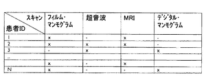
クロスモダリティ事例ベースCADxシステムは、特定の疾患又は病気の患者のデータベースを構築することによって作成される。図1は、乳癌を有する患者及びその患者が受けた撮像スキャンのデータベースの一典型例を示している。データベースは、患者の画像スキャンと画像以外に基づく情報とを含むことができ、例えば患者識別情報、デモグラフィック(人口統計学データ)情報(例えば、年齢、性別)、患者臨床履歴(例えば、過去及び現在の疾患、主な訴え)、家族歴、人口動態統計(血圧、体重など)などの入力項目(エントリー)を含む。様々な画像スキャンモダリティは、例えば、フィルム・マンモグラム、超音波、MRI、デジタル・マンモグラムなどとし得る。例えば、患者1はフィルム・マンモグラム及びMRIスキャンを受けているが、超音波は受けていない。患者2はフィルム・マンモグラム、超音波及びMRIスキャンを受けているが、デジタル・マンモグラムは受けていない。異なる癌及び病気に関して同様のデータベースが作成されてもよく、そのデータベースは、例えばCTスキャン、X線、PETスキャンなどの更なる画像モダリティを含んでいてもよい。
A cross-modality case-based CADx system is created by building a database of patients with a particular disease or condition. FIG. 1 shows a typical example of a database of patients with breast cancer and the imaging scans they received. The database may include patient image scans and non-image based information, such as patient identification information, demographic (demographic data) information (eg, age, gender), patient clinical history (eg, past and Includes input items (entries) such as current disease, main complaints), family history, demographics (blood pressure, weight, etc.). The various image scan modalities can be, for example, film mammograms, ultrasound, MRI, digital mammograms, and the like. For example,
システムは更に、1つのモダリティから別のモダリティへの特徴マッピングを見出すため、相異なるモダリティにまたがって関心ボリューム(“VOI”)を分析する。この情報は、1つのモダリティと別のモダリティとの間での特徴値の比及びマッピングを与える表(テーブル)にデータ投入するために用いられる。例えば、そのようなマッピング方法の1つは因子分析と呼ぶことができ、それは、1つのモダリティの画像に基づく情報を別のモダリティの画像に基づく情報にマッピングするために使用され得る。1つのモダリティから別のモダリティに画像ベースの特徴をマッピングするため、画像から抽出され得る可能な限りの画像ベースの特徴のリストが生成される。これらの特徴は、特徴群が複数の異なる種類の画像モダリティに対応するようなコンテンツ・マトリクスを形成するために使用される。そして、コンテンツ・マトリクスは、利用可能な患者事例に基づいてマッピングされる。利用可能な患者事例は、画像以外に基づく非画像ベースの特徴(例えば、年齢、性別、病気)、及び利用可能な画像モダリティの種類を指し示してもよい。 The system further analyzes the volume of interest (“VOI”) across different modalities to find feature mapping from one modality to another. This information is used to populate a table that gives the ratio and mapping of feature values between one modality and another. For example, one such mapping method can be referred to as factor analysis, which can be used to map information based on an image of one modality to information based on an image of another modality. In order to map image-based features from one modality to another, a list of all possible image-based features that can be extracted from the image is generated. These features are used to form a content matrix in which feature groups correspond to multiple different types of image modalities. The content matrix is then mapped based on available patient cases. Available patient cases may indicate non-image based features (eg, age, gender, illness) based on other than images, and the types of image modalities available.
因子分析は、変数の組を、より少ない数の変数又は因子の組へと削減するために使用され得る統計的手法である。因子分析は、複数の変数間の相互相関のパターンを検査し、互いに高く相関するがその他のサブセット又は因子とは低い相関を示すサブセット(部分集合)が存在するかを決定する。小さい相違を有する特徴は排除され、1つのモダリティの画像特徴と別のモダリティの画像特徴との間でのマッピングが生成された概念値マトリクスが作成される。故に、因子分析から得られた結果に基づいて、原モダリティの画像からの抽出特徴が与えられて事例ベースマルチモダリティ画像を生成するアルゴリズムが設計され得る。 Factor analysis is a statistical technique that can be used to reduce a set of variables to a smaller number of variables or sets of factors. Factor analysis examines the pattern of cross-correlation between multiple variables and determines whether there are subsets (subsets) that are highly correlated with each other but less correlated with other subsets or factors. Features with small differences are eliminated and a conceptual matrix is created in which a mapping between image features of one modality and image features of another modality is generated. Thus, based on the results obtained from the factor analysis, an algorithm can be designed to generate case-based multi-modality images given the extracted features from the original modality image.
特定の疾患に関して多数のVOIが特定されると、複数のモダリティにおける全ての画像ベースの特徴が計算される。そして、異なる複数のモダリティにおける特徴の傾向を推測するため、因子分析が用いられる。さらには、異なる複数の特徴が複数のモダリティにまたがって関連付けられてもよい。例えば、1つのモダリティにおける密度が、別のモダリティにおけるテクスチャに比例することがある。しかしながら、当業者に理解されるように、因子分析は複数の変数間の相互相関を分析する単なる1つの方法であり、異なる複数のモダリティにおける特徴の傾向を推測するために特徴を分析することが可能である限り、如何なる分析方法が用いられてもよい。代替的な一分析方法は、一組の特徴を別の一組の特徴にマッピングするために多変数回帰を用いるものである。 When multiple VOIs are identified for a particular disease, all image-based features in multiple modalities are calculated. Factor analysis is then used to infer the trend of features in different modalities. Furthermore, different features may be associated across multiple modalities. For example, the density in one modality may be proportional to the texture in another modality. However, as will be appreciated by those skilled in the art, factor analysis is just one way of analyzing cross-correlation between multiple variables, and analyzing features to infer trends in features across different modalities. Any analytical method may be used as long as possible. An alternative analysis method uses multivariate regression to map one set of features to another set of features.
図2は、特定の疾患又は病気に関しての、複数の撮像モダリティにまたがっての典型的な相対特徴比のテーブルを示している。上述のように、この特徴テーブルは、因子分析、多変数回帰又はその他の特徴分析方法の結果に基づいてデータ投入され得る。この情報が構築されると、1つのモダリティのVOIが、その他のモダリティにおける類似事例を検索するために使用され得る。VOIから抽出された画像特徴ベクトルが、同一モダリティ内での類似病変の初期検索を可能にする。画像特徴ベクトルは、その他の所望のモダリティに関する画像特徴ベクトルへと変換される。 FIG. 2 shows a typical relative feature ratio table across multiple imaging modalities for a particular disease or condition. As described above, this feature table can be populated based on the results of factor analysis, multivariate regression or other feature analysis methods. Once this information is built, the VOI of one modality can be used to search for similar cases in other modalities. Image feature vectors extracted from VOIs allow for an initial search for similar lesions within the same modality. The image feature vectors are converted into image feature vectors for other desired modalities.
図2に例示したテーブルにおいては、特徴マッピングを作成するように、CTモダリティ特徴が、様々な画像モダリティ(例えば、超音波、MRI、X線)における対応する特徴に対して指標化されている。特徴マッピングは、異なる複数の画像モダリティにおける同一の特徴又は異なる特徴の間の特徴関係をもたらす。例えば、超音波の棘形成特徴は、CTスキャンから計算されたその特徴の0.2倍であるが、MRIスキャンから計算された棘形成特徴はCTスキャンのそれの5.0倍である。故に、図2に例示した値に従って、棘形成をCTスキャンから超音波に変換するためには、CTから計算された特徴値が0.2倍され、MRI棘形成値に変換するためには、同一のCT棘形成値が5倍される。残りの特徴(例えば、密度、テクスチャ、平均階調値など)についても同様の変換が為され得る。故に、図2の例において、マッピングは、異なる画像モダリティ間での特徴値の単純な比に基づく。しかしながら、当業者に理解されるように、特定のモダリティにおける未知の特徴値を見積もるために複数の特徴が用いられてもよい。また、上述のケースで、複数のモダリティにおいて棘形成に関連性がなく、超音波スキャンにおけるVOIの棘形成特徴を見積もるために全く異なる特徴が用いられることも生じ得る。 In the table illustrated in FIG. 2, CT modality features are indexed against corresponding features in various image modalities (eg, ultrasound, MRI, X-ray) so as to create a feature mapping. Feature mapping results in feature relationships between the same or different features in different image modalities. For example, an ultrasonic spine formation feature is 0.2 times that calculated from a CT scan, whereas a spine formation feature calculated from an MRI scan is 5.0 times that of a CT scan. Therefore, according to the values illustrated in FIG. 2, in order to convert spine formation from CT scan to ultrasound, the feature value calculated from CT is multiplied by 0.2, and to convert to MRI spine formation value, The same CT spine formation value is multiplied by 5. Similar transformations can be made for the remaining features (eg, density, texture, average tone value, etc.). Thus, in the example of FIG. 2, the mapping is based on a simple ratio of feature values between different image modalities. However, as will be appreciated by those skilled in the art, multiple features may be used to estimate an unknown feature value in a particular modality. Also, in the case described above, it may happen that spine formation is irrelevant in multiple modalities and completely different features are used to estimate the VOI spine formation characteristics in an ultrasound scan.
図3は、1つのモダリティにおける特徴を別のモダリティにおける特徴から見積もるために、多項式関数をフィッティングすることによって異なる複数のモダリティ間で特徴を変換する代替的な一手法を示している。図3の例において、画像ベースの特徴は、1つの画像モダリティから計算された特徴を表すx軸と、別の画像モダリティからの同一の特徴を表すy軸とを有する2次元グラフ上にグラフ化され得る。図3のグラフは、例えば、x軸としてCTスキャンにおける棘形成、y軸としてMRIスキャンにおける棘形成を有するとし得る。そして、例示したグラフは、2次の多項式モデルでフィッティングされ得る。図3のカーブの値に基づく例示の2次多項式は:棘形成MRI=0.4981+1.0231×棘形成CT−0.2942×[棘形成CT]2となり得る。そして、この多項式モデルの方程式が、1つの画像モダリティの特徴を別の画像モダリティにおける同一の特徴にマッピングするために用いられてもよい。 FIG. 3 illustrates an alternative approach for transforming features between different modalities by fitting a polynomial function to estimate features in one modality from features in another modality. In the example of FIG. 3, image-based features are graphed on a two-dimensional graph having an x-axis representing the features calculated from one image modality and a y-axis representing the same features from another image modality. Can be done. The graph of FIG. 3 may have, for example, spine formation in a CT scan as the x axis and spine formation in an MRI scan as the y axis. The illustrated graph can then be fitted with a second order polynomial model. An example quadratic polynomial based on the values of the curves in FIG. 3 may be: spine formation MRI = 0.4981 + 1.0231 × spin formation CT− 0.2942 × [spin formation CT ] 2 This polynomial model equation may then be used to map the features of one image modality to the same features in another image modality.
故に、異なる複数の画像モダリティの特徴のマッピングが(例えば、図2−3を参照して説明したようにして)完了した後、問題としている事例に対応する異なるモダリティの画像又は事例が検索され得る。これは、先ず問題としている患者のスキャンから画像ベースの特徴を抽出し、それをマッピングすることによって達成され得る。そして、これらの特徴は、マルチモダリティ画像のデータベース内に含まれるマッピングされた特徴と比較される。画像特徴がマッピングされると、その他の撮像モダリティによるスキャンを含む事例群を検索するアルゴリズムが用いられ得る。このアルゴリズムは、原画像からの特徴を、同一種類の画像における対応する特徴と(例えば、CT特徴をCT特徴と)、また異なるモダリティからの対応する特徴と(例えば、CT特徴をMRI特徴と)比較する。そのようなアルゴリズムの1つが、米国特許出願第60/804,955号明細書(Agnihotri等の“Clinician Driven Example Based CADx”)にて開示されている。しかしながら、当業者に一般的に理解されるように、その他の撮像モダリティによるスキャンを有する事例を検索するために如何なるアルゴリズムが用いられてもよい。例えば、診断すべき事例が患者のCTスキャンを用いてのみ達成されるとき、マルチモダリティCADxシステムはデータベースから、類似のCTスキャンとともに、MRI、超音波、及びその他のスキャンを検索してもよい。 Thus, after the mapping of features of different image modalities is complete (eg, as described with reference to FIGS. 2-3), images or cases of different modalities corresponding to the case in question may be retrieved. . This can be accomplished by first extracting image-based features from the patient scan in question and mapping them. These features are then compared to the mapped features contained within the multi-modality image database. Once the image features are mapped, an algorithm can be used to search for cases that include scans with other imaging modalities. This algorithm uses features from the original image, corresponding features in the same type of image (eg, CT features as CT features), and corresponding features from different modalities (eg, CT features as MRI features). Compare. One such algorithm is disclosed in US patent application Ser. No. 60 / 804,955 (Agnihotri et al., “Clinician Driven Example Based CADx”). However, as generally understood by those skilled in the art, any algorithm may be used to search for cases having scans with other imaging modalities. For example, when the case to be diagnosed is only achieved using a patient CT scan, the multi-modality CADx system may retrieve MRI, ultrasound, and other scans along with similar CT scans from the database.
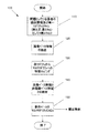
図4は、マルチモダリティCADxシステムにおける方法100の一典型例を示しており、ステップ110にて、診断対象の患者の単一モダリティスキャンがオリジナルの画像モダリティにて行われる。ステップ120にて、原画像上で、画像ベースの特徴の抽出が行われる。そして、ステップ130にて、抽出された特徴が、原モダリティの画像からマルチモダリティの画像にマッピングされる。例えば、原患者スキャンがX線スキャンである場合、特徴(例えば、密度、テクスチャなど)は抽出されて、X線スキャンからその他のマルチモダリティ画像(例えば、CT、MRI、超音波など)にマッピングされる。上述のように、異なる複数の画像モダリティにおける特徴間の関係が、例えば、図2に示したような比によって、あるいは図3に示したように、特徴を見積もるために多項式関数をフィッティングすることによって構築され得る。そして、ステップ130にて、特徴のこれらの関係が用いられ、診断すべき患者の原モダリティの画像ベース特徴に基づいて、同一あるいは異なるモダリティによる画像を有するその他の事例が検索される。
FIG. 4 shows an
そして、ステップ140にて、問題としている患者の画像ベースの特徴(原モダリティからの画像ベースの特徴、及びマッピングされた特徴)と非画像ベースの情報とが組み合わされる。すなわち、原モダリティからの特徴が、同様のモダリティの画像から計算された特徴、及び異なるモダリティによる画像から計算された特徴と(例えば、問題としている患者のCTスキャンから計算された特徴が、検索された画像からのMRI特徴と)組み合わされ得る。例えば、図1に関連して説明したように、例えば患者の年齢、性別、主な訴え、現在及び過去の該患者の疾患、家族歴、生活スタイル、喫煙歴など、その他の患者データが、検索された様々な画像に関連付けられてもよい。ステップ140では、これらその他の非画像ベースの特徴が、画像ベースの特徴と組み合わされてもよい。利用可能な全ての画像モダリティと患者の画像以外の臨床情報とにわたって組み合わされたこれらの特徴は、ステップ150にて、事例ベースのマルチモダリティCADxを作り出すために使用され得る。故に、ステップ110からの単一モダリティ(例えば、X線)画像を用いることによって、ステップ150にて、医師に、類似の特徴を有する更なるX線、類似の特徴を有するCT、MRI、マンモグラムなど、及び検索されたこれら更なる事例に関する非画像の患者データが提示され得る。
Then, at
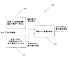
図5は、典型的な他の一実施形態に係るマルチモダリティCADxシステムにおける方法200を示している。方法200は、方法100のステップ130及び140にて実行されるサブステップとし得る。方法200においては、ステップ210にて、原モダリティに基づく類似事例を検索するため、原モダリティに基づく事例ベースCADxが作り出される。例えば、問題としている患者のスキャンがX線スキャンである場合、システム200は、問題としている患者のXスキャンから抽出された特徴と、データベース内の事例の対応する特徴とに基づいて、類似の、以前に診断されたX線スキャンを検索する。ステップ220にて、原モダリティでないモダリティの画像が検索される。上述のように、以前に分析されたデータに基づいて、1つのモダリティからの特徴が、その他のモダリティの対応する特徴にマッピングされる。原X線から抽出された特徴は、その他のモダリティにおける特徴にマッピングされ得る(例えば、原X線における特徴に関する値が、別のモダリティ(CT、MRIなど)における対応する特徴の値に変換される)。そして、対応する特徴を用いて、原モダリティでないモダリティからの類似事例が検索される。例えば、原X線スキャンを用いて、類似の、以前に診断された例えば超音波、MRIなどのスキャンが検索される。ステップ230にて、原画像における特徴に基づく最終的な類似事例の検索のため、原モダリティからの類似事例210と原モダリティでないモダリティからの類似事例220とが組み合わされる。ステップ230は、ステップ210及び220の2つの結果を組み合わせるために如何なる方法を用いてもよい。距離計算により、画像モダリティにかかわらず、提示されたスキャンに最も近い特徴を有する事例を検索することができる。故に、X線スキャンが提示された場合、距離計算によって、提示されたスキャンに最も近い特徴を有するMRIが検索され得る。
FIG. 5 illustrates a
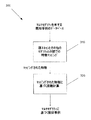
図6は、更なる一実施形態に係るマルチモダリティCADxシステムにおける方法300を示している。方法300も、方法100のステップ130及び140にて実行されるサブステップとし得る。ステップ310にて、図4のステップ130に関して説明したのと同様にして、原画像の特徴がその他のモダリティにおける特徴にマッピングされる。しかしながら、ステップ320においては、これらマッピングされた特徴は、マルチモダリティ画像に対する距離を計算するために使用され得る。距離計算により、その結合画像が原モダリティの単一の画像より近い距離を有し得るような患者データが検索され得る。例えば、ステップ320は、患者のX線スキャン及びMRIスキャンを組み合わせたものが、別のX線単独より近い距離を有する事例を検索してもよい。当業者に理解されるように、例えば画像の種類、関連ある診断などといった多様な因子に基づき得る異なる複数の特徴に基づいて距離を見出す方法は数多く存在し得る。
FIG. 6 illustrates a
上記の典型的な方法及びシステムの使用により、臨床医は、単一モダリティ画像に基づいて、異なる撮像モダリティを有する類似事例を検索することができる。このようなシステムは、臨床医が、経験による知識と、診断が既知である過去事例のデータベースの事例を参照することによる知識とを取得することを可能にする。故に、様々なモダリティにまたがる画像を有する事例を探索して取り出すことができることは、臨床医の診断及び治療計画作成において臨床医の助けとなる。 Use of the exemplary methods and systems described above allows a clinician to search for similar cases having different imaging modalities based on a single modality image. Such a system allows a clinician to acquire knowledge from experience and knowledge by referring to cases in a database of past cases whose diagnosis is known. Thus, the ability to search for and retrieve cases with images that span various modalities helps the clinician in clinician diagnosis and treatment planning.
当業者に明らかなように、本発明には、本発明の主旨及び範囲を逸脱することなく、様々な変更が為され得る。故に、本発明への変更及び変形が添付の請求項の範囲又はその均等範囲に入る限り、本発明はそのような変更及び変形にも及ぶものである。 It will be apparent to those skilled in the art that various modifications can be made to the present invention without departing from the spirit and scope of the invention. Therefore, so long as changes and modifications to the present invention fall within the scope of the appended claims or their equivalents, the present invention extends to such changes and modifications.
Claims (20)
第1のモダリティの画像からの特徴と第2のモダリティの画像からの特徴との間の特徴関係をマッピングするステップ;及び
前記特徴関係を格納するステップ;
を有する方法。 Storing a plurality of cases, each case including at least one image of one of the modalities and non-image information;
Mapping a feature relationship between features from an image of a first modality and features from an image of a second modality; and storing the feature relationship;
Having a method.
抽出された特徴と前記特徴関係とに基づいて少なくとも1つの事例を検索するステップ;
を更に有する請求項1に記載の方法。 Extracting features from the original image; and retrieving at least one instance based on the extracted features and the feature relationship;
The method of claim 1 further comprising:
前記第1のモダリティの画像から、及び前記第2のモダリティの画像からの関心ボリュームを分析するステップ
を含む、請求項1に記載の方法。 The mapping steps are:
The method of claim 1, comprising analyzing a volume of interest from an image of the first modality and from an image of the second modality.
を更に有する請求項2に記載の方法。 Simultaneously displaying the original image and the retrieved cases;
The method of claim 2 further comprising:
を更に有する請求項2に記載の方法。 Calculating a distance between the extracted features of the original image and corresponding features from the plurality of cases;
The method of claim 2 further comprising:
第1のモダリティの画像からの特徴と第2のモダリティの画像からの特徴との間の特徴関係をマッピングし、該特徴関係を前記メモリに格納するプロセッサ;
を有するシステム。 A memory storing a plurality of cases, wherein each case includes at least one image of one of the modalities and non-image information; and a feature from the image of the first modality and a second A processor that maps feature relationships between features from images of different modalities and stores the feature relationships in the memory;
Having a system.
第1のモダリティの画像からの特徴と第2のモダリティの画像からの特徴との間の特徴関係をマッピングし、該関係を前記メモリに格納する手段;
を有するシステム。 Means for storing a plurality of cases, wherein each case includes at least one image of one of the plurality of modalities and non-image information; and a feature from the image of the first modality; Means for mapping a feature relationship between features from an image of a second modality and storing the relationship in the memory;
Having a system.
Applications Claiming Priority (3)
| Application Number | Priority Date | Filing Date | Title |
|---|---|---|---|
| US1591907P | 2007-12-21 | 2007-12-21 | |
| US61/015,919 | 2007-12-21 | ||
| PCT/IB2008/055184 WO2009083837A1 (en) | 2007-12-21 | 2008-12-09 | Method and system for cross-modality case-based computer-aided diagnosis |
Publications (2)
| Publication Number | Publication Date |
|---|---|
| JP2011508917A true JP2011508917A (en) | 2011-03-17 |
| JP5647522B2 JP5647522B2 (en) | 2014-12-24 |
Family
ID=40545816
Family Applications (1)
| Application Number | Title | Priority Date | Filing Date |
|---|---|---|---|
| JP2010538974A Active JP5647522B2 (en) | 2007-12-21 | 2008-12-09 | Method and system for case-based computer-aided diagnosis with cross-modality |
Country Status (6)
| Country | Link |
|---|---|
| US (1) | US10318709B2 (en) |
| EP (1) | EP2225683B1 (en) |
| JP (1) | JP5647522B2 (en) |
| CN (1) | CN101903888B (en) |
| BR (1) | BRPI0821291B1 (en) |
| WO (1) | WO2009083837A1 (en) |
Cited By (3)
| Publication number | Priority date | Publication date | Assignee | Title |
|---|---|---|---|---|
| JP2013200590A (en) * | 2012-03-23 | 2013-10-03 | Fujifilm Corp | Similar image retrieval device, method, and program |
| JP2014522532A (en) * | 2011-06-06 | 2014-09-04 | コーニンクレッカ フィリップス エヌ ヴェ | Cross-modal application of combination signs indicating phenotypes |
| JP2022025095A (en) * | 2014-12-10 | 2022-02-09 | コーニンクレッカ フィリップス エヌ ヴェ | System and method for translation of medical imaging using machine learning |
Families Citing this family (8)
| Publication number | Priority date | Publication date | Assignee | Title |
|---|---|---|---|---|
| JP5538749B2 (en) * | 2009-06-03 | 2014-07-02 | キヤノン株式会社 | Diagnosis support system, diagnosis support method and program |
| CN103200861B (en) * | 2011-11-04 | 2015-10-14 | 松下电器产业株式会社 | Similar cases indexing unit and similar cases search method |
| KR101497662B1 (en) * | 2013-03-26 | 2015-03-03 | 재단법인대구경북과학기술원 | Endoscope system for assisting diagnosis and controlling method for the same |
| CN103324852A (en) * | 2013-06-25 | 2013-09-25 | 上海交通大学 | Four-modal medical imaging diagnosis system based on feature matching |
| CN104318500A (en) * | 2014-10-29 | 2015-01-28 | 无锡中盛医疗设备有限公司 | Medical image workstation |
| US11144785B2 (en) * | 2016-03-17 | 2021-10-12 | Imagia Cybernetics Inc. | Method and system for processing a task with robustness to missing input information |
| US10796430B2 (en) | 2018-04-24 | 2020-10-06 | General Electric Company | Multimodality 2D to 3D imaging navigation |
| US11532244B2 (en) * | 2020-09-17 | 2022-12-20 | Simbionix Ltd. | System and method for ultrasound simulation |
Citations (11)
| Publication number | Priority date | Publication date | Assignee | Title |
|---|---|---|---|---|
| JPH07322081A (en) * | 1994-05-26 | 1995-12-08 | Fuji Xerox Co Ltd | Method and device for color conversion of color picture |
| JPH1170083A (en) * | 1997-08-29 | 1999-03-16 | Fuji Photo Film Co Ltd | Image processing system |
| JPH1173490A (en) * | 1997-08-29 | 1999-03-16 | Fuji Photo Film Co Ltd | Image processing system |
| JP2001346042A (en) * | 2000-06-06 | 2001-12-14 | Canon Inc | Image processor, image processing system, image processing method and storage medium |
| JP2006108805A (en) * | 2004-09-30 | 2006-04-20 | Seiko Epson Corp | Color conversion table generating method, color conversion table generating program, print control method, print control apparatus, print control program, and color conversion table |
| WO2006064400A2 (en) * | 2004-12-15 | 2006-06-22 | Koninklijke Philips Electronics, N.V. | Registration of multi-modality images |
| JP2007524461A (en) * | 2003-06-25 | 2007-08-30 | シーメンス メディカル ソリューションズ ユーエスエー インコーポレイテッド | Mammography automatic diagnosis and decision support system and method |
| JP2007279942A (en) * | 2006-04-05 | 2007-10-25 | Fujifilm Corp | Similar case retrieval device, similar case retrieval method and program |
| JP2007275440A (en) * | 2006-04-11 | 2007-10-25 | Fujifilm Corp | Similar image retrieval system, method, and program |
| JP2007286945A (en) * | 2006-04-18 | 2007-11-01 | Fujifilm Corp | Similar image search apparatus and method, and program |
| JP2007307290A (en) * | 2006-05-22 | 2007-11-29 | Konica Minolta Medical & Graphic Inc | Medical image reading system |
Family Cites Families (8)
| Publication number | Priority date | Publication date | Assignee | Title |
|---|---|---|---|---|
| US6135117A (en) | 1997-05-12 | 2000-10-24 | Cornell Research Foundation, Inc. | Non-ocular circadian clock resetting in humans |
| US7157023B2 (en) | 2001-04-09 | 2007-01-02 | E. I. Du Pont De Nemours And Company | Conductor compositions and the use thereof |
| US20030103663A1 (en) * | 2001-11-23 | 2003-06-05 | University Of Chicago | Computerized scheme for distinguishing between benign and malignant nodules in thoracic computed tomography scans by use of similar images |
| US7536644B2 (en) * | 2002-06-27 | 2009-05-19 | Siemens Medical Solutions Usa, Inc. | Method and system for facilitating selection of stored medical images |
| CN101061520B (en) * | 2004-11-22 | 2010-10-13 | 皇家飞利浦电子股份有限公司 | Diagnostic Imaging system and method |
| TW200817956A (en) * | 2006-06-16 | 2008-04-16 | Koninkl Philips Electronics Nv | Clinician-driven example-based computer-aided diagnosis |
| US7899225B2 (en) * | 2006-10-26 | 2011-03-01 | Mcgill University | Systems and methods of clinical state prediction utilizing medical image data |
| US8150192B2 (en) * | 2006-11-27 | 2012-04-03 | Merge Cad Inc. | System and method for feature score mapping and visualization of medical images |
-
2008
- 2008-12-09 US US12/747,602 patent/US10318709B2/en active Active
- 2008-12-09 EP EP08866648.2A patent/EP2225683B1/en active Active
- 2008-12-09 WO PCT/IB2008/055184 patent/WO2009083837A1/en active Application Filing
- 2008-12-09 BR BRPI0821291-0A patent/BRPI0821291B1/en not_active IP Right Cessation
- 2008-12-09 CN CN2008801223029A patent/CN101903888B/en active Active
- 2008-12-09 JP JP2010538974A patent/JP5647522B2/en active Active
Patent Citations (11)
| Publication number | Priority date | Publication date | Assignee | Title |
|---|---|---|---|---|
| JPH07322081A (en) * | 1994-05-26 | 1995-12-08 | Fuji Xerox Co Ltd | Method and device for color conversion of color picture |
| JPH1170083A (en) * | 1997-08-29 | 1999-03-16 | Fuji Photo Film Co Ltd | Image processing system |
| JPH1173490A (en) * | 1997-08-29 | 1999-03-16 | Fuji Photo Film Co Ltd | Image processing system |
| JP2001346042A (en) * | 2000-06-06 | 2001-12-14 | Canon Inc | Image processor, image processing system, image processing method and storage medium |
| JP2007524461A (en) * | 2003-06-25 | 2007-08-30 | シーメンス メディカル ソリューションズ ユーエスエー インコーポレイテッド | Mammography automatic diagnosis and decision support system and method |
| JP2006108805A (en) * | 2004-09-30 | 2006-04-20 | Seiko Epson Corp | Color conversion table generating method, color conversion table generating program, print control method, print control apparatus, print control program, and color conversion table |
| WO2006064400A2 (en) * | 2004-12-15 | 2006-06-22 | Koninklijke Philips Electronics, N.V. | Registration of multi-modality images |
| JP2007279942A (en) * | 2006-04-05 | 2007-10-25 | Fujifilm Corp | Similar case retrieval device, similar case retrieval method and program |
| JP2007275440A (en) * | 2006-04-11 | 2007-10-25 | Fujifilm Corp | Similar image retrieval system, method, and program |
| JP2007286945A (en) * | 2006-04-18 | 2007-11-01 | Fujifilm Corp | Similar image search apparatus and method, and program |
| JP2007307290A (en) * | 2006-05-22 | 2007-11-29 | Konica Minolta Medical & Graphic Inc | Medical image reading system |
Non-Patent Citations (1)
| Title |
|---|
| 渡部 浩司: "マルチモダリティの画像位置合わせと重ね合わせ", 日本放射線技術学会雑誌, vol. 59, no. 1, JPN6013008485, January 2003 (2003-01-01), pages 60 - 65, ISSN: 0002916822 * |
Cited By (3)
| Publication number | Priority date | Publication date | Assignee | Title |
|---|---|---|---|---|
| JP2014522532A (en) * | 2011-06-06 | 2014-09-04 | コーニンクレッカ フィリップス エヌ ヴェ | Cross-modal application of combination signs indicating phenotypes |
| JP2013200590A (en) * | 2012-03-23 | 2013-10-03 | Fujifilm Corp | Similar image retrieval device, method, and program |
| JP2022025095A (en) * | 2014-12-10 | 2022-02-09 | コーニンクレッカ フィリップス エヌ ヴェ | System and method for translation of medical imaging using machine learning |
Also Published As
| Publication number | Publication date |
|---|---|
| CN101903888A (en) | 2010-12-01 |
| BRPI0821291A2 (en) | 2015-06-16 |
| EP2225683B1 (en) | 2017-07-26 |
| EP2225683A1 (en) | 2010-09-08 |
| US10318709B2 (en) | 2019-06-11 |
| JP5647522B2 (en) | 2014-12-24 |
| WO2009083837A1 (en) | 2009-07-09 |
| CN101903888B (en) | 2013-12-04 |
| BRPI0821291B1 (en) | 2020-10-06 |
| US20100272338A1 (en) | 2010-10-28 |
Similar Documents
| Publication | Publication Date | Title |
|---|---|---|
| JP5647522B2 (en) | Method and system for case-based computer-aided diagnosis with cross-modality | |
| US6925199B2 (en) | Computer readable recording medium recorded with diagnosis supporting program, diagnosis supporting apparatus and diagnosis supporting method | |
| JP6014059B2 (en) | Method and system for intelligent linking of medical data | |
| US10304198B2 (en) | Automatic medical image retrieval | |
| JP4912015B2 (en) | Similar image retrieval apparatus and method, and program | |
| JP4874701B2 (en) | Similar image retrieval apparatus and method, and program | |
| US7599534B2 (en) | CAD (computer-aided decision) support systems and methods | |
| JP5899236B2 (en) | System and method for medical decision support for treatment planning using case-based reasoning | |
| US7747050B2 (en) | System and method for linking current and previous images based on anatomy | |
| US20160321427A1 (en) | Patient-Specific Therapy Planning Support Using Patient Matching | |
| US20100231605A1 (en) | Medical image processing device and medical image processing program | |
| JP5431924B2 (en) | Clinician-driven, example-based computer-aided diagnosis | |
| JP7021215B2 (en) | CAD System Personalization Methods and Means for Providing Confidence Level Indicators for CAD System Recommendations | |
| JP2009516551A (en) | Quantitative and qualitative computer-aided analysis method and system for medical images | |
| JP2011092286A (en) | Information processing apparatus, information processing method, and program | |
| US20190150870A1 (en) | Classification of a health state of tissue of interest based on longitudinal features | |
| CN112529834A (en) | Spatial distribution of pathological image patterns in 3D image data | |
| JP7170747B2 (en) | Similarity determination device, method and program | |
| WO2019176407A1 (en) | Learning assisting device, learning assisting method, learning assisting program, region-of-interest discriminating device, region-of-interest discriminating method, region-of-interest discriminating program, and learned model | |
| US10282516B2 (en) | Medical imaging reference retrieval | |
| Huang et al. | Image-matching as a medical diagnostic support tool (DST) for brain diseases in children | |
| Doğanay et al. | A hybrid lung segmentation algorithm based on histogram-based fuzzy C-means clustering | |
| Cherezov et al. | Rank acquisition impact on radiomics estimation (AсquIRE) in chest CT imaging: A retrospective multi-site, multi-use-case study | |
| US20230360213A1 (en) | Information processing apparatus, method, and program | |
| Gao et al. | An Artificial Intelligence Representation of Human Knowledge for Lung Nodule Classification |
Legal Events
| Date | Code | Title | Description |
|---|---|---|---|
| A621 | Written request for application examination |
Free format text: JAPANESE INTERMEDIATE CODE: A621 Effective date: 20111207 |
|
| A977 | Report on retrieval |
Free format text: JAPANESE INTERMEDIATE CODE: A971007 Effective date: 20130130 |
|
| A131 | Notification of reasons for refusal |
Free format text: JAPANESE INTERMEDIATE CODE: A131 Effective date: 20130226 |
|
| A601 | Written request for extension of time |
Free format text: JAPANESE INTERMEDIATE CODE: A601 Effective date: 20130527 |
|
| A602 | Written permission of extension of time |
Free format text: JAPANESE INTERMEDIATE CODE: A602 Effective date: 20130603 |
|
| A131 | Notification of reasons for refusal |
Free format text: JAPANESE INTERMEDIATE CODE: A131 Effective date: 20140107 |
|
| A601 | Written request for extension of time |
Free format text: JAPANESE INTERMEDIATE CODE: A601 Effective date: 20140404 |
|
| A602 | Written permission of extension of time |
Free format text: JAPANESE INTERMEDIATE CODE: A602 Effective date: 20140411 |
|
| A521 | Request for written amendment filed |
Free format text: JAPANESE INTERMEDIATE CODE: A523 Effective date: 20140707 |
|
| TRDD | Decision of grant or rejection written | ||
| A01 | Written decision to grant a patent or to grant a registration (utility model) |
Free format text: JAPANESE INTERMEDIATE CODE: A01 Effective date: 20141014 |
|
| A61 | First payment of annual fees (during grant procedure) |
Free format text: JAPANESE INTERMEDIATE CODE: A61 Effective date: 20141107 |
|
| R150 | Certificate of patent or registration of utility model |
Ref document number: 5647522 Country of ref document: JP Free format text: JAPANESE INTERMEDIATE CODE: R150 |
|
| R250 | Receipt of annual fees |
Free format text: JAPANESE INTERMEDIATE CODE: R250 |
|
| R250 | Receipt of annual fees |
Free format text: JAPANESE INTERMEDIATE CODE: R250 |
|
| R250 | Receipt of annual fees |
Free format text: JAPANESE INTERMEDIATE CODE: R250 |
|
| R250 | Receipt of annual fees |
Free format text: JAPANESE INTERMEDIATE CODE: R250 |
|
| R250 | Receipt of annual fees |
Free format text: JAPANESE INTERMEDIATE CODE: R250 |
|
| R250 | Receipt of annual fees |
Free format text: JAPANESE INTERMEDIATE CODE: R250 |
|
| R250 | Receipt of annual fees |
Free format text: JAPANESE INTERMEDIATE CODE: R250 |