JP2011502687A - Interventional navigation using 3D contrast ultrasound - Google Patents
Interventional navigation using 3D contrast ultrasound Download PDFInfo
- Publication number
- JP2011502687A JP2011502687A JP2010533703A JP2010533703A JP2011502687A JP 2011502687 A JP2011502687 A JP 2011502687A JP 2010533703 A JP2010533703 A JP 2010533703A JP 2010533703 A JP2010533703 A JP 2010533703A JP 2011502687 A JP2011502687 A JP 2011502687A
- Authority
- JP
- Japan
- Prior art keywords
- image
- ceus
- contrast
- tissue
- real
- Prior art date
- Legal status (The legal status is an assumption and is not a legal conclusion. Google has not performed a legal analysis and makes no representation as to the accuracy of the status listed.)
- Pending
Links
Images


Classifications
-
- G—PHYSICS
- G01—MEASURING; TESTING
- G01S—RADIO DIRECTION-FINDING; RADIO NAVIGATION; DETERMINING DISTANCE OR VELOCITY BY USE OF RADIO WAVES; LOCATING OR PRESENCE-DETECTING BY USE OF THE REFLECTION OR RERADIATION OF RADIO WAVES; ANALOGOUS ARRANGEMENTS USING OTHER WAVES
- G01S15/00—Systems using the reflection or reradiation of acoustic waves, e.g. sonar systems
- G01S15/88—Sonar systems specially adapted for specific applications
- G01S15/89—Sonar systems specially adapted for specific applications for mapping or imaging
- G01S15/8906—Short-range imaging systems; Acoustic microscope systems using pulse-echo techniques
- G01S15/899—Combination of imaging systems with ancillary equipment
-
- A—HUMAN NECESSITIES
- A61—MEDICAL OR VETERINARY SCIENCE; HYGIENE
- A61B—DIAGNOSIS; SURGERY; IDENTIFICATION
- A61B8/00—Diagnosis using ultrasonic, sonic or infrasonic waves
- A61B8/08—Detecting organic movements or changes, e.g. tumours, cysts, swellings
- A61B8/0833—Detecting organic movements or changes, e.g. tumours, cysts, swellings involving detecting or locating foreign bodies or organic structures
-
- A—HUMAN NECESSITIES
- A61—MEDICAL OR VETERINARY SCIENCE; HYGIENE
- A61B—DIAGNOSIS; SURGERY; IDENTIFICATION
- A61B8/00—Diagnosis using ultrasonic, sonic or infrasonic waves
- A61B8/42—Details of probe positioning or probe attachment to the patient
- A61B8/4245—Details of probe positioning or probe attachment to the patient involving determining the position of the probe, e.g. with respect to an external reference frame or to the patient
- A61B8/4263—Details of probe positioning or probe attachment to the patient involving determining the position of the probe, e.g. with respect to an external reference frame or to the patient using sensors not mounted on the probe, e.g. mounted on an external reference frame
-
- G—PHYSICS
- G01—MEASURING; TESTING
- G01S—RADIO DIRECTION-FINDING; RADIO NAVIGATION; DETERMINING DISTANCE OR VELOCITY BY USE OF RADIO WAVES; LOCATING OR PRESENCE-DETECTING BY USE OF THE REFLECTION OR RERADIATION OF RADIO WAVES; ANALOGOUS ARRANGEMENTS USING OTHER WAVES
- G01S7/00—Details of systems according to groups G01S13/00, G01S15/00, G01S17/00
- G01S7/52—Details of systems according to groups G01S13/00, G01S15/00, G01S17/00 of systems according to group G01S15/00
- G01S7/52017—Details of systems according to groups G01S13/00, G01S15/00, G01S17/00 of systems according to group G01S15/00 particularly adapted to short-range imaging
- G01S7/52053—Display arrangements
- G01S7/52057—Cathode ray tube displays
- G01S7/52074—Composite displays, e.g. split-screen displays; Combination of multiple images or of images and alphanumeric tabular information
-
- G—PHYSICS
- G01—MEASURING; TESTING
- G01S—RADIO DIRECTION-FINDING; RADIO NAVIGATION; DETERMINING DISTANCE OR VELOCITY BY USE OF RADIO WAVES; LOCATING OR PRESENCE-DETECTING BY USE OF THE REFLECTION OR RERADIATION OF RADIO WAVES; ANALOGOUS ARRANGEMENTS USING OTHER WAVES
- G01S7/00—Details of systems according to groups G01S13/00, G01S15/00, G01S17/00
- G01S7/52—Details of systems according to groups G01S13/00, G01S15/00, G01S17/00 of systems according to group G01S15/00
- G01S7/52017—Details of systems according to groups G01S13/00, G01S15/00, G01S17/00 of systems according to group G01S15/00 particularly adapted to short-range imaging
- G01S7/52098—Details of systems according to groups G01S13/00, G01S15/00, G01S17/00 of systems according to group G01S15/00 particularly adapted to short-range imaging related to workflow protocols
-
- G—PHYSICS
- G01—MEASURING; TESTING
- G01S—RADIO DIRECTION-FINDING; RADIO NAVIGATION; DETERMINING DISTANCE OR VELOCITY BY USE OF RADIO WAVES; LOCATING OR PRESENCE-DETECTING BY USE OF THE REFLECTION OR RERADIATION OF RADIO WAVES; ANALOGOUS ARRANGEMENTS USING OTHER WAVES
- G01S15/00—Systems using the reflection or reradiation of acoustic waves, e.g. sonar systems
- G01S15/88—Sonar systems specially adapted for specific applications
- G01S15/89—Sonar systems specially adapted for specific applications for mapping or imaging
- G01S15/8906—Short-range imaging systems; Acoustic microscope systems using pulse-echo techniques
- G01S15/8993—Three dimensional imaging systems
Landscapes
- Engineering & Computer Science (AREA)
- Health & Medical Sciences (AREA)
- Life Sciences & Earth Sciences (AREA)
- Physics & Mathematics (AREA)
- Radar, Positioning & Navigation (AREA)
- Remote Sensing (AREA)
- Nuclear Medicine, Radiotherapy & Molecular Imaging (AREA)
- Medical Informatics (AREA)
- Computer Networks & Wireless Communication (AREA)
- Biophysics (AREA)
- Veterinary Medicine (AREA)
- Pathology (AREA)
- Radiology & Medical Imaging (AREA)
- Biomedical Technology (AREA)
- Heart & Thoracic Surgery (AREA)
- General Physics & Mathematics (AREA)
- Molecular Biology (AREA)
- Surgery (AREA)
- Animal Behavior & Ethology (AREA)
- General Health & Medical Sciences (AREA)
- Public Health (AREA)
- Acoustics & Sound (AREA)
- Ultra Sonic Daignosis Equipment (AREA)
Abstract
3D造影超音波(CEUS)イメージング10を使用する介入のナビゲーションのための方法は、生体構造に投与される造影剤の有効な寿命の間に器具40での介入の治療を受ける生体構造22の所望の部分のための追跡情報と基準3D CEUSボリュームとを獲得するステップを有する。リアルタイムに追跡された組織画像は、介入の治療の間に獲得される。加えて、獲得されたリアルタイムに追跡された組織画像の少なくとも一つの画像に対する少なくとも一つの対応する造影超音波画像の多断面再構成(MPR)44が生成される。獲得されたリアルタイムに追跡された組織画像の対応する一つの画像が、対応するCEUS MPRと一緒に表示される(20)。表示されたリアルタイムに追跡された組織画像は生体構造の所望の部分内で少なくとも器具の画像を含み、CEUS MPRは表示されたリアルタイムに追跡された組織画像に対応する。CEUS MPR画像は、基準3D CEUSボリュームから得られた造影MPR画像を含み、関心ターゲットボリューム46を含み、これにより、造影の有効な寿命の満了に少なくとも続く介入のナビゲーションのために有効な造影画像情報及び組織画像情報の同時の表示を提供する。 A method for interventional navigation using 3D contrast-enhanced ultrasound (CEUS) imaging 10 is desirable for a anatomy 22 that undergoes treatment of the intervention with the instrument 40 during the effective life of a contrast agent administered to the anatomy. Obtaining tracking information and a reference 3D CEUS volume for a portion of Real-time tracked tissue images are acquired during interventional treatment. In addition, a multi-section reconstruction (MPR) 44 of at least one corresponding contrast-enhanced ultrasound image for at least one image of the acquired real-time tracked tissue image is generated. One corresponding image of the acquired real-time tracked tissue image is displayed (20) along with the corresponding CEUS MPR. The displayed real-time tracked tissue image includes at least an image of the instrument within the desired portion of the anatomy, and the CEUS MPR corresponds to the displayed real-time tracked tissue image. The CEUS MPR image includes a contrast MPR image obtained from a reference 3D CEUS volume and includes a target volume of interest 46, thereby enabling contrast image information useful for interventional navigation at least following expiration of the effective life of the contrast. And provides simultaneous display of tissue image information.
Description
この出願は、ジョチェン クルエッカーらにより2007年11月16日に出願され、本発明の譲受人に譲渡された「3D造影超音波を使用する介入ナビゲーション」という題名の仮出願(出願番号60/988472)の利益を請求する。 This application was filed on November 16, 2007 by Jochen Kruecker et al. And assigned to the assignee of the present invention, a provisional application entitled “Interventional Navigation Using 3D Contrast-enhanced Ultrasound” (Application No. 60/984722). Claim the profits.
本発明は、米国公衆衛生局との共同研究及び開発協定の遂行(CRADA番号NCI―NIHCC―01864)においてなされた。米国の政府は、本発明における特定の権利を持つ。 The present invention was made in the joint research and development agreement (CRADA number NCI-NIHCC-01864) with the US Public Health Service. The US government has certain rights in the invention.
本実施例は、概して超音波画像診断システム、より詳しくは、3D造影超音波を使用する介入ナビゲーションのための方法と装置とに関する。 This example relates generally to an ultrasound imaging system, and more particularly to a method and apparatus for interventional navigation using 3D contrast ultrasound.
超音波イメージングは、多くの最小限侵襲性の介入治療のための主要な画像ガイダンス方法の一つである。特に、ほとんどの手術針生検及び手術針ベースの切除治療は、超音波によりガイドされる。超音波の利点は、リアルタイム・イメージング機能、低コスト、そのアプリケーションの柔軟性及び電離放射線が使われないという事実を含む。しかしながら、一般に使用されるグレースケール超音波画像を含む造影されていない超音波は、所望のコントラストで特定のターゲット(例えば、腫瘍)を視覚化できず、場合によっては、ターゲットを全く視覚化しない。これらの場合は、異なるモダリティを使用してターゲットを撮像し、両方の画像診断法で識別される腫瘍の近くの生体構造学的目印に基づいて「精神的に」腫瘍位置をリアルタイム超音波画像に変えるステップを含むため、手術針の配置は非常に困難になり不正確の傾向がある。結果は、偽陰性生検、失敗した腫瘍治療、及び概して劣った治療的結果となる。 Ultrasound imaging is one of the leading image guidance methods for many minimally invasive interventional therapies. In particular, most surgical needle biopsies and surgical needle-based ablation treatments are guided by ultrasound. The advantages of ultrasound include real-time imaging capabilities, low cost, flexibility of its application and the fact that ionizing radiation is not used. However, non-contrast-enhanced ultrasound, including commonly used grayscale ultrasound images, cannot visualize a particular target (eg, a tumor) with the desired contrast and, in some cases, does not visualize the target at all. In these cases, the target is imaged using different modalities, and a “mentally” tumor location based on anatomical landmarks near the tumor identified by both diagnostic imaging methods in real-time ultrasound images. Because it involves changing steps, the placement of the surgical needle is very difficult and tends to be inaccurate. The result is a false negative biopsy, failed tumor treatment, and generally poor therapeutic outcome.
造影超音波(CEUS)イメージングは、(ブリストル‐マイヤーズ・スクイブ社のDefinity(登録商標)のような)超音波造影剤の静注の後の超音波イメージングを指す超音波イメージングのもう一つの形式である。最新の超音波スキャナでは、特定の結像モードが造影剤の非線形音響反応を利用するために実行され、よって、コントラスト取り込みを持つ組織を強調するだけだった。結果として生じる画像は、「コントラスト画像」と呼ばれ、非コントラスト画像と比較して非常に異なる外観を持つ。通常のグレイスケール・モードにおいて、コントラスト注入(造影剤注入)の後、組織を画像化することも可能である。後者の例において、結果として生じる画像は、「組織画像」と呼ばれ、コントラスト取り込みの領域の小さな増強だけを示すが、コントラスト注入なしで得られたグレイスケール画像と同様に見える。 Contrast-enhanced ultrasound (CEUS) imaging is another form of ultrasound imaging that refers to ultrasound imaging after intravenous injection of ultrasound contrast agent (such as Bristol-Myers Squibb's Definity®). is there. In modern ultrasound scanners, specific imaging modes have been implemented to take advantage of the nonlinear acoustic response of contrast agents, thus only highlighting tissue with contrast uptake. The resulting image is called a “contrast image” and has a very different appearance compared to a non-contrast image. It is also possible to image the tissue after contrast injection (contrast agent injection) in the normal grayscale mode. In the latter example, the resulting image is called a “tissue image” and shows only a small enhancement in the area of contrast capture, but looks similar to a grayscale image obtained without contrast injection.
CEUSは、非造影超音波イメージングと比較して、腫瘍、血管分布及び関心の他の組織の優れた視覚化を提供できることに注意されたい。しかしながら、ボーラス注射の後の造影は、一過性の現象で、通常は数分後に消える。数分という斯様な時間制限は、所望の処置(例えば、生検又は切除のための手術針の配置のような)を実行するために、しばしば不十分な時間である。換言すれば、生検及び切除のための手術針配置のような介入の治療の間のガイダンスのために、造影の時間窓は、介入の治療を実行するために必要とされる時間と比較して不十分である。造影剤の2回目の注入は、造影効果を延長するために可能ではあるが、これは所望の治療を完了するにはまだ不十分である。 Note that CEUS can provide superior visualization of tumors, vascular distribution and other tissues of interest compared to non-contrast-enhanced ultrasound imaging. However, the contrast after the bolus injection is a transient phenomenon and usually disappears after a few minutes. Such a time limit of a few minutes is often insufficient time to perform the desired procedure (eg, placement of a surgical needle for biopsy or excision). In other words, for guidance during interventional treatments such as surgical needle placement for biopsy and resection, the imaging time window is compared to the time required to perform the interventional treatment. Is insufficient. A second injection of contrast agent is possible to prolong the contrast effect, but this is still insufficient to complete the desired treatment.
予め獲得されたCEUSボリューム単独の使用と関連する従来技術の付加的な制限は、例えば、予め獲得されたCEUSボリュームと関係する介入の治療の間に引き続き獲得されるリアルタイム超音波組織画像の位置がわからず、推定する必要があることを含み、よって不正確になる傾向がある。さらにまた、組織の動きは、予め獲得されたCEUSボリュームに基づくターゲット位置の見積もりを複雑にする。 An additional limitation of the prior art associated with the use of a pre-acquired CEUS volume alone is, for example, that the location of real-time ultrasound tissue images subsequently acquired during treatment of an intervention associated with the pre-acquired CEUS volume is It does not know and includes what needs to be estimated, and thus tends to be inaccurate. Furthermore, tissue movement complicates target location estimation based on pre-acquired CEUS volume.
従って、従来技術の課題を解決するための改良型の方法及びシステムが要求される。 Accordingly, there is a need for an improved method and system for solving the problems of the prior art.
本開示の実施例は、介入の治療の間、画像ガイダンスのための造影超音波イメージング(CEUS)を利用するシステム及び方法を提供する。特に、本開示の実施例は、CEUSを使用する方法及びシステムが、ワークフローを修正又は完全に異なる画像診断法へ切り替える必要がなく介入の治療の目標精度を改善できる。 Embodiments of the present disclosure provide systems and methods that utilize contrast ultrasound imaging (CEUS) for image guidance during treatment of interventions. In particular, embodiments of the present disclosure allow methods and systems using CEUS to improve the target accuracy of interventional treatments without having to modify the workflow or switch to a completely different imaging method.
本願明細書に開示されるように、当該システム及び方法は、超音波プローブの位置を追跡するために構成される空間追跡システムを含む。追跡システムの使用は、介入の治療の始めに3D CEUSボリュームの位置の決定を可能にする。一つの実施例において、3D組織ボリュームもまた、これ以降更に説明されるように、3D CEUSボリュームで同時に得られる。その後、介入の治療の間、超音波プローブの空間追跡は、最初の3D CEUSボリュームから得られる対応するCEUS多断面再構成(MPR)と現在のリアルタイム超音波組織画像との共同表示を可能にする。最初の3D CEUS獲得とリアルタイム超音波組織イメージングとの間の組織の動きは、最初の3D CEUSボリュームで共に得られる3D超音波組織ボリュームとリアルタイム超音波組織画像との間の画像ベースの位置合せにより修正される。(i)最初のCEUSボリュームから得た対応するMPRと、(ii)リアルタイム非造影超音波組織画像との共同の表示は、好適に手術針位置及びターゲット位置の共同の視覚化を可能にし、よって介入の治療の間、造影剤の増強効果の満了の後、ターゲットへの手術針のガイドを可能にする。 As disclosed herein, the systems and methods include a spatial tracking system configured to track the position of an ultrasound probe. The use of a tracking system allows the determination of the position of the 3D CEUS volume at the beginning of the intervention treatment. In one embodiment, a 3D tissue volume is also obtained simultaneously with a 3D CEUS volume, as will be described further below. Thereafter, during interventional treatment, spatial tracking of the ultrasound probe allows co-displaying of the corresponding CEUS multi-section reconstruction (MPR) obtained from the initial 3D CEUS volume and the current real-time ultrasound tissue image . Tissue movement between the initial 3D CEUS acquisition and real-time ultrasound tissue imaging is due to the image-based registration between the 3D ultrasound tissue volume and the real-time ultrasound tissue image obtained together with the initial 3D CEUS volume. Will be corrected. The joint display of (i) the corresponding MPR obtained from the initial CEUS volume and (ii) the real-time non-contrast ultrasound tissue image preferably allows joint visualization of the surgical needle position and the target position, thus During the interventional treatment, after the expiration of the enhancement effect of the contrast agent, it is possible to guide the surgical needle to the target.
図において、類似の参照符号は、類似の要素を指す。加えて、図は一定の比率で描画されていない点に留意されるべきである。 In the drawings, like reference numerals indicate like elements. In addition, it should be noted that the figures are not drawn to scale.
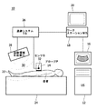
ここで図面に戻ると、図1は、本開示の一つの実施例による3D造影超音波を使用する介入のナビゲーションのためのシステム10のブロック図である。システム10は、超音波イメージングプローブ14を備え及び/又は結合される超音波スキャナ(US)12を有する。一つの実施例において、超音波スキャナ12は、例えば、フィリップスメディカルシステムズから市販されているiU22超音波スキャナを有する。イメージングプローブ14は、適切な3D超音波イメージングプローブを有する。加えて、超音波スキャナ12は、スキャナ・ディスプレイ16を含む。さらにまた、超音波スキャナ12は、コントラスト及び組織画像の同時獲得のために設定される。超音波スキャナ12は、例えば、データストリームを介してリアルタイムで画像をワークステーション18へ転送するように更に設定される。例えば、リアルタイムで画像を転送することは、デジタルナビゲーションリンク(Digital Navigation Link)ソフトウェアでiU22超音波スキャナを使用して達成できる。超音波スキャナ12とは別に例示されているが、ワークステーション18は、超音波スキャナ12の部分に統合されていてもよい。
Turning now to the drawings, FIG. 1 is a block diagram of a
まだ図1を参照して、ワークステーション18は、ワークステーション・ディスプレイ20を含む。動作時、超音波スキャナ12、プローブ14及びワークステーション18は、超音波診断及び/又は対応する処理若しくは医学療法を要する生体構造を持つ患者22に関連して用いられ、患者22は患者テーブル24に配置される。超音波スキャナ12は、例えば、コントラスト及び組織フレームのインターリーブされた獲得に対応する「サイドバイサイドモード」においてコントラスト及び組織の超音波画像を得るように設定され、獲得された両方の画像をワークステーション18に送信する。ソフトウェアは、ここで更に説明されるようなワークフローに適応するように、ワークステーション18により実行される。
Still referring to FIG. 1, the
3D造影超音波を使用する介入のナビゲーションのためのシステム10は、本開示の実施例による位置追跡も含む。システム10は、外部の位置追跡システム(TS)26との統合により強化される。外部の位置追跡システム26は、概して参照符号30により示される、追跡領域を生じるために設定される追跡領域生成器28を含む。センサ32は超音波プローブ14に結合され、検知領域30の範囲内に位置されるセンサに応じて、センサの位置及び向きは追跡システム26により追跡できる。一つの実施例において、ワークステーション18は、追跡システム26に結合され、追跡情報を通信するため及び/又は所与の介入のナビゲーション若しくは実行の要件に従ってワークステーション18と追跡システム26との間の追跡命令を供給するように設定される。追跡システム26は、例えば、カナダ、ワーテルローのノーザンデジタル社による電磁「オーロラ」システムのような適切な追跡システムを有する。他の実施例において、追跡システム26は、光学追跡システムを含み、追跡領域生成器28は、図の光学領域に対応する追跡領域30内で超音波プローブ14を光学的に追跡するために、例えばカメラを有する。斯様な光学追跡システムは、例えば、カナダ、ワーテルローのノーザンデジタル社による「北極星」システムを有する。
The
図1のシステム10において、外部の位置追跡システム(TS)26は、患者22の隣又はすぐ近くに準備される。6つの自由度(6DoF)位置センサ(S)32は超音波プローブ14に結合され、追跡領域生成器28はプローブ位置が追跡領域30内で追跡できるように配置される。
In the
ワークステーション18はまた、コンピュータ可読の媒体に含まれて、ワークステーションのメモリにロードされるソフトウェアを含み、当該ソフトウェアは、(i)センサ32により供給されるボリュームの追跡システム座標と共に、少なくとも一つの3D CEUSボリュームを得て格納し、(ii)リアルタイムの追跡センサ座標を得て処理するために、ワークステーションのプロセッサにより実行可能な命令を含み、少なくとも一つの獲得し格納された3D CEUSボリュームの多断面再構成(MPR)を計算し表示するために、リアルタイムの追跡センサ座標を使用して、対応するMPRが最新の獲得されたリアルタイム組織画像と同じ組織を示すようにする。
The
一つの実施例において、超音波スキャナ12は、同時にCEUS画像/ボリュームと組織画像/ボリュームとを得て、送信するように設定される。斯様な実施例では、ワークステーション18のソフトウェアは、(iii)センサ32により供給される対応するボリュームの追跡システム座標と共に、同時に得られた3D CEUSボリューム及び3D組織ボリュームを得て格納し、(iv)リアルタイムに追跡センサ座標を得て処理し、3D CEUSボリュームと同時に得られる3D組織ボリュームと、非コントラスト・リアルタイム組織画像との画像ベースの位置合せのためリアルタイム追跡センサ座標を使用するワークステーション18のプロセッサにより実行可能な命令を更に含み、結果として生じる位置合せ変換は、CEUS MPRが最新の獲得されたリアルタイム組織画像と同じ組織を示すように、最初に獲得し格納された3D CEUSボリュームのMPRを計算し表示するために使われる。
In one embodiment, the
一つの実施例において、システム及び方法は、コントラスト及び組織モードのリアルタイム三次元画像を得ることができ、ワークステーション18(又は、例えば適切な通信ネットワークを介した他のシステム)に画像データを流す(すなわち、リアルタイムに転送する)ことができる超音波スキャナ12を使用することを有する。この実施例と連動して、当該方法は、患者が呼吸命令に従って、再現可能な息止めを生じることが更に可能であると仮定する。さらにまた、ワークステーション18で動くソフトウェアは、リアルタイム電子回路データ転送を使用して、さまざまなハードウェア(例えば、追跡システム、超音波スキャナ等)と通信するように設定される。
In one embodiment, the system and method can obtain real-
ワークフローの一つの例において、超音波ガイドされた手術針切除を受ける患者22が、検査テーブル24に置かれる。位置センサ32が、超音波プローブ14に取り付けられている。追跡システム26の発信器/受信器28は、取付けられた位置センサ32を持つ超音波プローブ14が治療のために必要な超音波イメージングの間、発信器/受信器28の視野30の領域にあるように、患者22の近くに配置される。超音波スキャナ12は、まず最初にコントラスト結像モードに構成されているか又は設定され、造影剤の静脈内ボーラス注射が患者22に投与される。ディスプレイ16に表示される超音波画像に造影が見えると、このとき、関心領域又は腫瘍をカバーする3次元(3D)CEUS走査が得られる。3D CEUSボリューム及び対応するプローブ位置データは、ナビゲーション・ワークステーション18へ転送される。造影が消失した後の後続のイメージングのために、リアルタイム2D又は3D超音波組織画像及び対応するプローブ位置データが、ワークステーション18へ連続的に転送される(すなわち、流れる)。現在のプローブ位置に基づいて、初めに獲得された3D CEUS画像(予め獲得された3D CEUS画像とも呼ばれる)と関係する現在の超音波組織画像の位置が計算される。ワークステーション・ディスプレイ20に、現在のリアルタイム超音波組織画像が、CEUS画像の対応する多断面再構成(MPR)(すなわち、他の視覚化)と共に表示される。この表示は、腫瘍が単独でリアルタイム組織画像では見えないが、リアルタイム組織画像の手術針の位置決め、CEUS画像内の腫瘍の位置決め、よって腫瘍への手術針の案内を可能にする。
In one example workflow, a
図2は、本開示の実施例による図1の3D造影超音波を使用する介入のナビゲーションのためのシステムに関連するさまざまな座標系間の変換を例示する部分的なブロック図である。特に、図2は、2D超音波画像、3D超音波画像、超音波プローブに取り付けられる追跡センサ及び追跡システムの座標系間の変換の例示である。一つの実施例において、図2は、6台のDoF位置センサ、追跡システム、対応する超音波フレーム間の変換の関係を例示する。 FIG. 2 is a partial block diagram illustrating conversion between various coordinate systems associated with the system for interventional navigation using the 3D contrast ultrasound of FIG. 1 according to an embodiment of the present disclosure. In particular, FIG. 2 is an illustration of a 2D ultrasound image, a 3D ultrasound image, a tracking sensor attached to an ultrasound probe, and a conversion between the tracking system coordinate systems. In one embodiment, FIG. 2 illustrates the transformation relationship between six DoF position sensors, tracking systems, and corresponding ultrasound frames.
変換Ttrackingは、追跡システム(26,28)に関係する追跡センサ14の現在の位置及び方向(さもなければ、「ポーズ」と呼ばれる)を記述する。換言すれば、変換Ttrackingは、追跡システムCtrackingの座標系と追跡センサCsensorの座標系との間の関係を記述する。変換Ttrackingは、追跡システムにより供給され、例えば本開示の実施例による介入の治療の所与の実行のために要求されて又は連続的にリアルタイムでワークステーション18により得られる。変換Tcalibrationは、3D超音波画像の座標系C3DUS(すなわち、ボクセル座標)とプローブ14に取り付けられた追跡センサ32の座標系Csensorとの間の関係を記述する。変換Tcalibrationは、1回限りの較正手順で決定され、センサ及び/又はプローブの置換及び/又は変化に応じて再調整され、超音波プローブ14に固く取り付けられる所与の追跡センサ32に対して固定されたままである。最後に、変換T2DUS→3DUSは、2D超音波画像の座標系C2DUSと3D超音波画像の座標系C3DUS(すなわち、ボクセル座標系)との間の関係を記述する。すなわち、追跡2D及び3D超音波画像獲得のための座標系間の変換は、T2DUS→3DUSにより付与される。要約すると、変換T2DUS→3DUSは、2D画像ピクセル座標を3D画像ボクセル座標へ変換し、Tcalibrationは、3D画像ボクセル座標をセンサ座標へ変換し、Ttrackingは、センサ座標を追跡システム座標へ変換する。Ttrackingが追跡システム(26,28)により供給されるセンサ32のリアルタイム・ポーズ情報であることに注意されたい。
The transformation T tracking describes the current position and orientation of the tracking
一つの実施例によると、CEUS画像獲得は以下の通りである。コントラスト注入の後、ワークステーション上のナビゲーション・ソフトウェアは、音波検査者に3D CEUSモードで腫瘍ターゲットを視覚化するプローブ位置を見つけるように要求する。スキャナからのすべての三次元画像及びセンサからのすべての位置データが、ワークステーションに連続的に流される。適当な画像がワークステーションで得られて、確認されるとき、画像は、追跡センサにより与えられる対応するプローブ位置Ttracking、3DCEUSとともにワークステーションに保存される。3D CEUSボリュームに割り当てられる基準座標系は、治療の全体にわたって静止している追跡システムの座標系である。図2の具体例を用いて、3D CEUSボクセル座標から追跡システム座標への変換は、下記の式により与えられる:
T3DCEUS=Tcalibration・Ttracking、3DCEUS
According to one embodiment, CEUS image acquisition is as follows. After contrast injection, the navigation software on the workstation asks the sonographer to find a probe position that visualizes the tumor target in 3D CEUS mode. All 3D images from the scanner and all position data from the sensors are continuously streamed to the workstation. When a suitable image is obtained and confirmed at the workstation, the image is stored at the workstation along with the corresponding probe position T tracking, 3DCEUS provided by the tracking sensor. The reference coordinate system assigned to the 3D CEUS volume is the coordinate system of the tracking system that is stationary throughout the treatment. Using the example of FIG. 2, the transformation from 3D CEUS voxel coordinates to tracking system coordinates is given by:
T 3DCEUS = T calibration / T tracking, 3DCEUS
加えて、本開示の実施例は、以下の画像ガイダンスを提供する。3D CEUS獲得の後、超音波スキャナは、画像ガイダンスのための2D結像モードに切り替えられる。2D画像座標と3D画像座標との間の変換T2DUS→3DUSは、超音波スキャナ上のイメージング・アルゴリズムに基づいて知られている。よって、追跡センサから現在の追跡データTtrackingを使用して、Ttracking、3D CEUSで得られるCEUS基準画像内の現在の2D超音波組織画像の位置は、下記の変換を使用して決定できる。
T2DUS→3DUS
=T2DUS→3DUS・Tcalibration・Ttracking・(T3DCEUS)―1
=T2DUS→3DUS・Tcalibration・Ttracking・(Ttracking、3DCEUS)―1・(Tcalibration)―1
In addition, embodiments of the present disclosure provide the following image guidance. After 3D CEUS acquisition, the ultrasound scanner is switched to 2D imaging mode for image guidance. The transformation T 2DUS → 3DUS between 2D image coordinates and 3D image coordinates is known based on an imaging algorithm on an ultrasound scanner. Thus, using the current tracking data Ttracking from the tracking sensor, the position of the current 2D ultrasound tissue image within the CEUS reference image obtained with Ttracking, 3D CEUS can be determined using the following transformation:
T 2DUS → 3DUS
= T 2DUS → 3DUS / T calibration / T tracking / (T 3DCEUS ) -1
= T 2DUS-> 3DUS -T calibration -T tracking- (T tracking, 3DCEUS ) -1- (T calibration ) -1
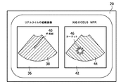
ワークステーションは、リアルタイムに、2D組織画像の現在のポーズに対応する3D CEUS画像からMPRを抽出するために、この関係を使用するだろう。ワークステーション・ディスプレイ20上に、一つの実施例では両方の画像が並んで表示され、又は、もう一つの実施例ではユーザにより決定された透明度を持つ半透明な重ね合わせを使用して表示される。図3は、挿入された手術針40を示して、リアルタイム組織画像38の表示36と、造影により強調されるターゲット46を示して、予め獲得されたCEUSボリュームからの対応するMPR44の表示42とが並んでいる例示的図である。参照符号50により図4に示されるように、図4は、同じ組織及びコントラスト画像の半透明な重ね合わせ48の例示的図である。
The workstation will use this relationship to extract the MPR from the 3D CEUS image corresponding to the current pose of the 2D tissue image in real time. On the
他の実施例によると、方法は、複数の3D CEUSボリュームの獲得を含む。すなわち、3D CEUSボリューム獲得の間、関心の造影された組織を示す単一のボリュームだけをワークステーション18に保存するよりはむしろ、時系列のボリューム及びそれらの対応する位置情報も格納される。このとき、当該方法は、コントラスト取り込みの動特性(例えば、流入及び流出)を確認でき、これは診断にとって有益なインジケーターでもある。当該方法は、更に、(a)リアルタイム組織画像との単なる単一のCEUS MPRを共同表示するのではなく、「動画」としての全体の時系列の複数のMPRを共同表示するステップを有する。加えて、当該方法はまた、(b)流入時定数のような診断に関連したパラメータのボリューム的マップを作成するための時系列データを処理し、介入の治療の間、リアルタイム組織画像と共に、このパラメータMAPのMPRを共同表示するステップを含む。
According to another embodiment, the method includes acquiring multiple 3D CEUS volumes. That is, during 3D CEUS volume acquisition, rather than storing only a single volume representing the contrasted tissue of interest at the
他の実施例では、方法は、交互のターゲット視覚化を使用するステップを含む。すなわち、3D CEUSボリュームのMPRを組織画像と重ね合わせる又は共同表示するよりはむしろ、関心ターゲットボリューム(VOI)は、CEUS獲得の後3D CEUSボリュームから分割される。斯様なターゲットVOIは、例えば、3D CEUSボリュームの最も強い組織増強のエリアをカバーする球体を含む。本実施例において、介入の治療の間、現在の追跡位置によって定まるようなVOIの適当な横断面だけが、リアルタイム組織画像上に重畳される。 In other embodiments, the method includes using alternating target visualization. That is, rather than overlaying or co-displaying the MPR of the 3D CEUS volume with the tissue image, the target volume of interest (VOI) is segmented from the 3D CEUS volume after CEUS acquisition. Such target VOI includes, for example, a sphere that covers the area of strongest tissue augmentation of the 3D CEUS volume. In this example, only the appropriate cross-section of the VOI as determined by the current tracking position is superimposed on the real-time tissue image during the interventional treatment.
他の実施例では、方法は、ガイダンスの間、リアルタイム3Dイメージングを使用するステップを含む。すなわち、ガイダンスのための2Dイメージングを使用する代わりに、方法は、リアルタイム3Dイメージングを使用する。この場合、MPRを生成するよりはむしろ、最大値投影法(MIP)のような、視覚化の他の手段が用いられる。MIP投影は、上述の実施例にて説明されたように、同一セットの変換を使用して計算できる。
In other embodiments, the method includes using real-
さらに別の実施例では、方法は、非リアルタイム3D CEUS獲得を使用する。この実施例において、例えば、リアルタイム3D CEUSが利用できない場合、基準CEUSボリュームはまた、2D超音波プローブを追跡し、すべての2D画像フレームをワークステーションへ流し、追跡データを対応させ、コントラスト注入の後ターゲット・エリア上に「掃引」を得て、獲得された2D画像及び追跡データに基づいて3D CEUSボリュームを再構築することにより生成できる。
In yet another embodiment, the method uses
他の実施例において、方法は、動き補正のための画像ベースの位置合せの使用を含む。この実施例において、リアルタイム組織画像及び対応する3D CEUS MPRの上述の共同表示は、患者又は撮像されている器官が3D CEUSボリュームの獲得とリアルタイム組織画像の獲得との間で動いていない場合にのみ正確である。組織動きがある場合に精度を強化するために、本発明の実施例による方法は、更に例えば、以下の動き補正方法のうちの一つを含む。第1の動き補正方法は、3D組織画像と3D CEUS画像との同時獲得を含む。この動き補正方法は、例えば、フィリップスiU22のような最新のスキャナ上のリアルタイム2Dにおいて利用できるような同時のデュアルモード獲得及び視覚化を利用し、上述の通りに非リアルタイム同時の3D獲得を可能にする。第2の動き補正方法は、3D CEUS画像と同時に獲得される3D組織画像と、現在の2D組織画像(又は、最後のN個の2D組織画像、Nは小さく例えば10より小さい)との画像ベースの位置合せを含む。2D画像と関連した追跡座標は、位置合せのための初期位置(すなわち、器官動きがないとする)として役立つ。位置合せ自体は、ガウス―ニュートン・オプティマイザ及び二乗誤差和の同様の手段のようなさまざまなオプティマイザ及び同様の手段を利用できる。位置合せの結果は、変換T2DUS→3Dtissueである。3D CEUS及び組織画像が同時に得られたので、これらの座標系は同一であり、同じ変換が現在の2D座標を3D CEUS座標に変換するために使用できる。
In other embodiments, the method includes the use of image-based registration for motion correction. In this example, the above-mentioned joint display of real-time tissue images and corresponding 3D CEUS MPR is only possible if the patient or the organ being imaged is not moving between acquisition of 3D CEUS volume and acquisition of real-time tissue images. Is accurate. In order to enhance accuracy when there is tissue movement, the method according to embodiments of the present invention further includes, for example, one of the following motion correction methods. The first motion correction method includes simultaneous acquisition of a 3D tissue image and a 3D CEUS image. This motion correction method utilizes simultaneous dual-mode acquisition and visualization, such as that available in real-
ここで説明されるように、本開示の実施例によるシステム及び方法は、超音波プローブの位置を追跡し、造影超音波イメージング(CEUS)の間、介入の治療の画像ガイダンスのため空間追跡システムを使用する。介入の治療の始めに、3次元(3D)CEUSボリュームが、通常の3D超音波組織画像と同時に得られる。治療の間、超音波プローブの空間追跡は、治療の始めに得られた3D CEUSボリューム(ここではまた、予め獲得された3D CEUSボリュームと呼ばれる)から、対応する多断面再構成(MPR)と現在のリアルタイム超音波組織画像との共同表示を可能にする。治療の始めに得られた3D CEUS獲得と、治療の始めの後又は治療の初めに続いたリアルタイム・イメージングとの間の組織動きは、リアルタイム3D組織画像と、治療の初めでの3D CEUSと共に得られた3D組織画像との間の画像ベースの位置合せにより修正される。予め獲得されたCEUS、より詳しくは、予め獲得されたCEUSの多断面再構成(MPR)(ここでMPRはリアルタイムの所与の組織画像に対応する)とリアルタイムの非コントラスト超音波との共同表示は、手術針位置及びターゲット位置の共同の視覚化を可能にし、よって手術針のターゲットへの案内を可能にする。前述のように、CEUSは、非造影超音波イメージングと比較して、腫瘍、血管分布及び関心の他の組織の優れた視覚化を提供できる。単独では、ボーラス注射後のコントラスト増強は、一過性の現象で、通常は数分後に消える。生検及び切除のための手術針配置のような介入の治療の間のガイダンスのためには、コントラスト増強単独の過渡状態現象の時間窓は、不十分である。しかしながら、本開示の実施例は、この制限を克服するシステム及び方法を好適に提供する。
As described herein, a system and method according to embodiments of the present disclosure tracks the position of an ultrasound probe and uses a spatial tracking system for image guidance of interventional interventions during contrasted ultrasound imaging (CEUS). use. At the beginning of the interventional treatment, a three-dimensional (3D) CEUS volume is acquired simultaneously with a normal 3D ultrasound tissue image. During treatment, the spatial tracking of the ultrasound probe is performed from the 3D CEUS volume obtained at the beginning of the treatment (also referred to herein as the pre-acquired 3D CEUS volume) and the corresponding multi-section reconstruction (MPR) Enables joint display with real-time ultrasound tissue images. Tissue movement between 3D CEUS acquisition obtained at the beginning of treatment and real-time imaging following the beginning of treatment or at the beginning of treatment is obtained with real-
従って、ここで開示される3D造影超音波(CEUS)イメージングを使用する介入のナビゲーションのための方法は、器具での介入の治療を受ける生体構造の所望の部分のための追跡情報及び基準3D CEUSボリュームを獲得する獲得ステップであって、当該獲得ステップは生体構造の所望の部分に投与される造影剤の有効な寿命の間に起こる当該獲得ステップと、介入の治療の間、リアルタイムに追跡された組織画像を獲得するステップと、基準3D CEUSボリューム及び追跡情報の関数として、介入の治療の間、獲得されたリアルタイムに追跡された組織画像の少なくとも一つの画像に対する少なくとも一つの対応する造影超音波画像の多断面再構成(CEUS MPR)を生成する生成ステップと、獲得されたリアルタイムに追跡された組織画像の少なくとも一つの組織画像を表示する表示ステップであって、表示されたリアルタイムに追跡された組織画像は生体構造の所望の部分内に少なくとも器具の画像を含む当該表示ステップと、表示されたリアルタイムに追跡された組織画像に対応する少なくとも一つの対応する造影超音波画像の多断面再構成(CEUS MPR)を表示するステップとを有し、CEUS MPR画像は基準3D CEUSボリュームから得られた造影MPR画像を含み、関心ターゲットボリュームを含み、これにより、造影の有効な寿命の満了に少なくとも続く介入のナビゲーションのために有効な造影画像情報及び組織画像情報の同時の表示を提供することが現時点で理解されるだろう。
Accordingly, the method for interventional navigation using 3D contrast-enhanced ultrasound (CEUS) imaging disclosed herein provides tracking information and
他の実施例によると、基準3D CEUSボリュームの獲得は、少なくとも一つの追跡された3D超音波コントラスト画像と組織画像との対の同時の獲得を含む。加えて、少なくとも一つの追跡された3D超音波コントラストと組織画像との対は、最初の超音波コントラスト画像と、実質的に同時に捕捉され、互いに自動的に重ね合わされた対応する最初の組織画像とを有する。
According to another embodiment, acquiring the
もう一つの実施例では、介入の治療は、造影の有効な寿命の満了の前に起こっている第1の部分と、造影の有効な寿命の満了の後に起こる第2の部分とを含む。さらなる実施例において、少なくとも一つの対応する造影超音波画像の多断面再構成が、獲得されたリアルタイムに追跡された組織画像の対応する一つの画像と空間的に重ね合わされる。もう一つの実施例では、リアルタイムに追跡された組織画像及び対応するCEUS MPRは、互いに隣接して表示される。さらに別の実施形態では、リアルタイムに追跡された組織画像及び対応するCEUS MPR基準画像は、単一表示で一緒に表示される。後者の例では、リアルタイムに追跡された組織画像及び対応するCEUS MPRは、一つの画像が他の画像に重なるように、重ね合わせる配置で更に表示できる。またさらに、重ね合わせる配置は、他の画像に関して半透明の一つの画像を有し得る。 In another embodiment, the interventional treatment includes a first portion that occurs before the expiration of the effective lifetime of the contrast and a second portion that occurs after the expiration of the effective lifetime of the contrast. In a further embodiment, a multi-section reconstruction of at least one corresponding contrast-enhanced ultrasound image is spatially superimposed with a corresponding one of the acquired real-time tracked tissue images. In another embodiment, the tissue images tracked in real time and the corresponding CEUS MPR are displayed adjacent to each other. In yet another embodiment, the tissue image tracked in real time and the corresponding CEUS MPR reference image are displayed together in a single display. In the latter example, the tissue image tracked in real time and the corresponding CEUS MPR can be further displayed in an overlapping arrangement so that one image overlaps the other image. Still further, the overlay arrangement may have one image that is translucent with respect to other images.
他の実施例によると、介入の治療の間、獲得されたリアルタイムに追跡された組織画像の少なくとも一つの画像に対して少なくとも一つの対応する造影超音波多断面再構成(CEUS MPR)を生成する代わりに、また、獲得されたリアルタイムに追跡された組織画像を表示する代わりに、方法は、獲得された追跡された3Dリアルタイムの超音波コントラスト画像と組織画像との少なくとも一つの対の関数として、最大値投影(MIP)を生成するステップと、獲得されたリアルタイムに追跡された3D組織画像の少なくとも一つの画像の最大値投影(MIP)を表示する表示ステップであって、表示されたリアルタイムに追跡された組織画像MIPが、生体構造の所望の部分内に少なくとも器具の画像を含む当該表示ステップと、少なくとも一つの獲得されたリアルタイムに追跡された3D組織画像の表示されたMIPに対応する少なくとも一つの対応する造影超音波画像最大値投影(CEUS MIP)を表示するステップとを有し、CEUS MIP画像は、関心ターゲットボリュームからの造影MIP画像を含み、これにより、造影の有効な寿命の満了に少なくとも続く介入のナビゲーションのために有効な同時の表示を提供する。 According to another embodiment, during an interventional treatment, at least one corresponding contrast ultrasound multi-section reconstruction (CEUS MPR) is generated for at least one image of the acquired real-time tracked tissue image. Alternatively, and instead of displaying the acquired real-time tracked tissue image, the method may be as a function of at least one pair of acquired acquired 3D real-time ultrasound contrast image and tissue image: Generating a maximum value projection (MIP) and displaying a maximum value projection (MIP) of at least one of the acquired real-time tracked 3D tissue images, wherein the displayed real-time tracking The displayed tissue image MIP includes at least an image of the instrument within a desired portion of the anatomy, Displaying at least one corresponding contrast ultrasound image maximum projection (CEUS MIP) corresponding to the displayed MIP of one acquired real-time tracked 3D tissue image, the CEUS MIP image Includes contrast MIP images from the target volume of interest, thereby providing an effective simultaneous display for interventional navigation at least following expiration of the effective lifetime of the contrast.
もう一つの実施例では、リアルタイムの3D超音波がない場合、基準3D CEUSボリュームは、2D超音波プローブを追跡し、造影剤付与後、生体構造の所望の部分にわたって追跡された2D超音波プローブを掃引しながら一連の2Dコントラスト及び組織画像フレーム並びに対応する追跡データを得て、獲得された一連の2Dコントラスト及び組織画像並びに対応する追跡データをワークステーションに流し、獲得された2Dコントラスト及び組織画像並びに対応する追跡データに基づいて、ワークステーションで基準3D CEUSボリュームを再構築することにより獲得される。
In another embodiment, in the absence of real-
さらに別の実施形態では、基準3D CEUSボリュームを獲得することは、基準3D CEUSボリュームを獲得する際に使用される超音波プローブの位置を追跡するように設定される空間追跡システムを使用することを含み、前記追跡システムは基準3D CEUSボリュームの位置及び方向の決定を可能にし、前記追跡システムは、介入の治療の間、獲得された組織画像を追跡するように更に設定される。方法は、更に、(i)基準3D CEUSボリュームの獲得の時間と、(ii)少なくとも一つのリアルタイムに追跡された組織画像の獲得の時間との間に起こる生体構造の所望の部分内の組織動きを修正するステップを有する。なお更に、組織動きを修正するステップは、(a)リアルタイムに追跡された3D組織画像と、(b)追跡された3D超音波コントラスト画像と組織画像との対の超音波組織画像との間の画像ベースの位置合せを用いるステップを含む。
In yet another embodiment, acquiring the
他の実施例によると、基準3D CEUSボリューム及び追跡情報を獲得する獲得ステップは、時系列の3D CEUSボリューム及び対応する追跡情報を獲得するステップを有し、少なくとも一つの対応するCEUS MPR画像を生成する生成ステップは、時系列のCEUS MPR画像を生成するステップを含み、獲得されたリアルタイムに追跡された組織画像を表示し、少なくとも一つの対応するCEUS MPR画像を表示するステップは、時系列データとしてリアルタイム組織画像とCEUS MPR画像とを共同表示するステップを有する。方法は、さらに、診断に関連したパラメータのボリューム的マップを作成するために時系列データを処理するステップと、(i)CEUS MPR画像及び(ii)リアルタイムの組織画像と、前記ボリューム的マップとを共同表示するステップとを有する。加えて、診断に関連したパラメータは、少なくとも造影剤の流入時定数を含む。
According to another embodiment, the acquiring step of acquiring the
さらに別の実施例では、方法は、基準3D CEUSボリュームから関心ターゲットボリュームを分割する分割ステップであって、前記関心ターゲットボリュームが最も強い組織増強の領域を有する当該分割ステップと、少なくとも一つの対応する造影超音波画像の多断面再構成(CEUS MPR)を表示する代わりに、表示されたリアルタイムに追跡された組織画像に対応する前記関心ターゲットボリュームの対応する横断面区分を表示するステップと、表示されたリアルタイムに追跡された組織画像上に前記関心ターゲットボリュームの対応する横断面区分を重畳するステップとを更に有する。
In yet another embodiment, the method is a segmentation step of segmenting a target volume of interest from a
他の実施例によると、3D造影超音波(CEUS)イメージングを使用する介入のナビゲーションのための方法は、器具での介入の治療を受ける生体構造の所望の部分のための追跡情報及び基準3D CEUSボリュームを獲得する獲得ステップであって、当該獲得ステップは、生体構造の所望の部分に投与される造影剤の有効な寿命の間に起こり、基準3D CEUSボリュームの獲得が少なくとも一つの追跡された3D超音波コントラスト画像と組織画像との対の同時の獲得を含む当該獲得ステップと、介入の治療の間、リアルタイムに追跡された組織画像を獲得するステップと、基準3D CEUSボリューム及び追跡情報の関数として、介入の治療の間、獲得されたリアルタイムに追跡された組織画像の少なくとも一つの画像に対して、少なくとも一つの対応する造影超音波画像の多断面再構成(CEUS MPR)を生成する生成ステップであって、前記少なくとも一つの対応する造影超音波画像の多断面再構成は、獲得されたリアルタイムに追跡された組織画像の対応する一つの画像と空間的に重ね合わされる当該生成ステップと、獲得されたリアルタイムに追跡された組織画像を表示する表示ステップであって、表示されたリアルタイムに追跡された組織画像は、生体構造の所望の部分内に少なくとも器具の画像を含む当該表示ステップと、表示されたリアルタイムに追跡された組織画像に対応する少なくとも一つの対応する造影超音波画像の多断面再構成(CEUS MPR)を表示するステップとを有し、CEUS MPR画像は、基準3D CEUSボリュームから得られた造影MPR画像を含み、関心ターゲットボリュームを含み、これにより、造影の有効な寿命の満了に少なくとも続く介入のナビゲーションのために有効な造影画像情報及び組織画像情報の同時の表示を提供する。
According to another embodiment, a method for interventional navigation using 3D contrast-enhanced ultrasound (CEUS) imaging includes tracking information and
さらに別の実施例では、3D造影超音波(CEUS)イメージングを使用する介入のナビゲーションのためのシステムは、(i)器具での介入の治療を受ける生体構造の所望の部分のための追跡情報及び基準3D CEUSボリュームを獲得する超音波画像形成デバイスであって、当該超音波画像形成デバイスは、生体構造の所望の部分に投与される造影剤の有効な寿命の間に、基準3D CEUSボリューム及び追跡情報を獲得するように更に設定され、(ii)介入の治療の間、リアルタイムに追跡された組織画像を獲得し、(iii)基準3D CEUSボリューム及び追跡情報の関数として、介入の治療の間、獲得されたリアルタイムに追跡された組織画像の少なくとも一つの画像に対して少なくとも一つの対応する造影超音波画像の多断面再構成(CEUS MPR)を生成する当該超音波画像形成デバイスと、当該超音波画像形成デバイスに結合され、(i)獲得されたリアルタイムに追跡された組織画像を表示するディスプレイであって、表示されたリアルタイムに追跡された組織画像が生体構造の所望の部分内で少なくとも器具の画像を含み、(ii)表示されたリアルタイムに追跡された組織画像に対応する少なくとも一つの対応する造影超音波画像の多断面再構成(CEUS MPR)を表示する当該ディスプレイとを有し、CEUS MPR画像が基準3D CEUSボリュームから得られた造影MPR画像を含み、関心ターゲットボリュームを含み、これにより造影の有効な寿命の満了に少なくとも続く介入のナビゲーションのために有効な造影画像情報及び組織画像情報の同時の表示を提供する。
In yet another embodiment, a system for interventional navigation using 3D contrast-enhanced ultrasound (CEUS) imaging comprises: (i) tracking information for a desired portion of the anatomy undergoing treatment with an instrument intervention; and An ultrasound imaging device that acquires a
ここで述べられたように、本開示の実施例は、診断及び治療的な医学療法の超音波ベースの画像ガイダンスに適用できる。例えば、本開示の実施例は、生検及び切除治療のための手術針ガイダンスを改善できる。加えて、本開示の実施例は、限られた視覚化又はいくつかの腫瘍/ターゲットが視覚化できないことや、ターゲット位置が予め獲得されたモダリティ(本開示の実施例に関して開示される予め獲得されたCEUS以外)に基づいて推定される場合、治療の実行の低い精度、所与の介入の治療の実行には不十分であるCEUSイメージングのコントラスト増強の短い期間のような現在の超音波ベースの治療ガイダンスの限界及び不利な点を好適に克服する。 As described herein, embodiments of the present disclosure can be applied to ultrasound-based image guidance for diagnostic and therapeutic medical therapies. For example, embodiments of the present disclosure can improve surgical needle guidance for biopsy and ablation treatments. In addition, the embodiments of the present disclosure may have limited visualization or inability to visualize some tumors / targets, or the modalities in which the target location is pre-acquired (pre-acquired as disclosed with respect to the embodiments of the present disclosure). Current ultrasound-based methods such as low accuracy of treatment execution, short period of contrast enhancement of CEUS imaging that is insufficient to perform treatment for a given intervention Preferably overcome the limitations and disadvantages of treatment guidance.
少数の例示的な実施例だけが上で詳述されたにもかかわらず、当業者は、多くの変更態様が、本開示の実施例の新しい教示及び利点から著しく逸脱することなく例示的な実施例において可能であることは容易に理解されるだろう。従って、斯様なすべての変更態様は、以下の請求項に規定されるように、本開示の実施例の範囲内に含まれることを意図する。請求項において、ミーンズプラスファンクション(機能的手段)の項は、引用された機能を実行するようにここで説明された構造をカバーし、構造的に等価なものだけでなく、等価な構造もカバーすることを意図する。 Although only a few exemplary embodiments have been described in detail above, those skilled in the art will recognize that many modifications may be made to the exemplary implementations without significantly departing from the new teachings and advantages of the embodiments of the present disclosure. It will be readily understood what is possible in the examples. Accordingly, all such modifications are intended to be included within the scope of the embodiments of the present disclosure, as defined in the following claims. In the claims, the terms “means plus function” cover the structures described herein to perform the recited function, and cover not only structurally equivalent but also equivalent structures. Intended to be.
加えて、一つ以上の請求項の括弧にある何れの参照符号も、請求項を制限するものとして解釈されない。語「を有する」等は、全体として何れの請求項又は明細書にリストされるもの以外の要素又はステップの存在を除外しない。単一の要素は、斯様な要素の複数を除外しないし、逆もそうである。一つ以上の実施例は、幾つかの明白な要素を有するハードウェアによって、及び/又は最適にプログラムされたコンピュータによって実行されてもよい。幾つかの手段を列挙するデバイスの請求項において、これらの手段の幾つかは、ハードウェアの全く同一の品目により具現化されてもよい。特定の手段が相互に異なる従属クレームに引用されているという単なる事実は、これらの手段の組合せが効果的に使用できないことを示さない。 In addition, any reference signs placed in parentheses in one or more claims shall not be construed as limiting the claims. The word “comprising” does not exclude the presence of elements or steps other than those listed in any claim or specification as a whole. A single element does not exclude a plurality of such elements and vice versa. One or more embodiments may be performed by hardware having several distinct elements and / or by an optimally programmed computer. In the device claim enumerating several means, several of these means may be embodied by one and the same item of hardware. The mere fact that certain measures are recited in mutually different dependent claims does not indicate that a combination of these measures cannot be used effectively.
Claims (20)
Applications Claiming Priority (3)
| Application Number | Priority Date | Filing Date | Title |
|---|---|---|---|
| US98847207P | 2007-11-16 | 2007-11-16 | |
| US5228808P | 2008-05-12 | 2008-05-12 | |
| PCT/IB2008/054769 WO2009063423A1 (en) | 2007-11-16 | 2008-11-13 | Interventional navigation using 3d contrast-enhanced ultrasound |
Publications (1)
| Publication Number | Publication Date |
|---|---|
| JP2011502687A true JP2011502687A (en) | 2011-01-27 |
Family
ID=40384555
Family Applications (1)
| Application Number | Title | Priority Date | Filing Date |
|---|---|---|---|
| JP2010533703A Pending JP2011502687A (en) | 2007-11-16 | 2008-11-13 | Interventional navigation using 3D contrast ultrasound |
Country Status (7)
| Country | Link |
|---|---|
| US (1) | US9651662B2 (en) |
| EP (1) | EP2212716B1 (en) |
| JP (1) | JP2011502687A (en) |
| CN (1) | CN101868737B (en) |
| BR (1) | BRPI0819439A8 (en) |
| RU (1) | RU2494676C2 (en) |
| WO (1) | WO2009063423A1 (en) |
Cited By (5)
| Publication number | Priority date | Publication date | Assignee | Title |
|---|---|---|---|---|
| JP2011031042A (en) * | 2009-07-31 | 2011-02-17 | Medison Co Ltd | Ultrasonic system and sensor coordinate calibration method |
| JP2011072656A (en) * | 2009-09-30 | 2011-04-14 | Toshiba Corp | Ultrasonic diagnostic equipment, ultrasonic image processing apparatus, ultrasonic diagnostic equipment control program, and ultrasonic image processing program |
| JP2012081152A (en) * | 2010-10-14 | 2012-04-26 | Tokyo Univ Of Agriculture & Technology | Ultrasonic therapeutic system |
| JP2013138841A (en) * | 2011-12-06 | 2013-07-18 | Toshiba Corp | Ultrasonic diagnostic apparatus and coordinate conversion program |
| US9545242B2 (en) | 2009-07-31 | 2017-01-17 | Samsung Medison Co., Ltd. | Sensor coordinate calibration in an ultrasound system |
Families Citing this family (47)
| Publication number | Priority date | Publication date | Assignee | Title |
|---|---|---|---|---|
| JP4981058B2 (en) * | 2005-10-26 | 2012-07-18 | トムソン ライセンシング | System and method for compensating for satellite gateway failure |
| WO2008017051A2 (en) | 2006-08-02 | 2008-02-07 | Inneroptic Technology Inc. | System and method of providing real-time dynamic imagery of a medical procedure site using multiple modalities |
| JP5148094B2 (en) * | 2006-09-27 | 2013-02-20 | 株式会社東芝 | Ultrasonic diagnostic apparatus, medical image processing apparatus, and program |
| WO2009094646A2 (en) | 2008-01-24 | 2009-07-30 | The University Of North Carolina At Chapel Hill | Methods, systems, and computer readable media for image guided ablation |
| US8340379B2 (en) | 2008-03-07 | 2012-12-25 | Inneroptic Technology, Inc. | Systems and methods for displaying guidance data based on updated deformable imaging data |
| US8554307B2 (en) | 2010-04-12 | 2013-10-08 | Inneroptic Technology, Inc. | Image annotation in image-guided medical procedures |
| US8690776B2 (en) | 2009-02-17 | 2014-04-08 | Inneroptic Technology, Inc. | Systems, methods, apparatuses, and computer-readable media for image guided surgery |
| US11464578B2 (en) | 2009-02-17 | 2022-10-11 | Inneroptic Technology, Inc. | Systems, methods, apparatuses, and computer-readable media for image management in image-guided medical procedures |
| US8641621B2 (en) | 2009-02-17 | 2014-02-04 | Inneroptic Technology, Inc. | Systems, methods, apparatuses, and computer-readable media for image management in image-guided medical procedures |
| CN102081697B (en) * | 2009-11-27 | 2013-12-11 | 深圳迈瑞生物医疗电子股份有限公司 | Method and device for defining interested volume in ultrasonic imaging space |
| EP2640275A1 (en) * | 2010-11-19 | 2013-09-25 | Koninklijke Philips N.V. | Three dimensional ultrasonic guidance of surgical instruments |
| EP2665423B1 (en) | 2011-01-17 | 2021-03-17 | Koninklijke Philips N.V. | System for needle deployment detection in image-guided biopsy |
| EP2509013A1 (en) * | 2011-04-04 | 2012-10-10 | Agfa Healthcare | 3D image navigation method |
| US10376179B2 (en) | 2011-04-21 | 2019-08-13 | Koninklijke Philips N.V. | MPR slice selection for visualization of catheter in three-dimensional ultrasound |
| US8670816B2 (en) | 2012-01-30 | 2014-03-11 | Inneroptic Technology, Inc. | Multiple medical device guidance |
| RU2629237C2 (en) * | 2012-06-28 | 2017-08-28 | Конинклейке Филипс Н.В. | Ultrasound guided biopsies in three dimensions |
| EP2916740B1 (en) * | 2012-11-06 | 2019-09-04 | Koninklijke Philips N.V. | Enhancing ultrasound images |
| US20140142419A1 (en) * | 2012-11-19 | 2014-05-22 | Biosense Webster (Israel), Ltd. | Patient movement compensation in intra-body probe |
| US9232934B2 (en) * | 2012-12-14 | 2016-01-12 | General Electric Company | Systems and methods for communicating ultrasound probe location and image information |
| US10314559B2 (en) | 2013-03-14 | 2019-06-11 | Inneroptic Technology, Inc. | Medical device guidance |
| WO2014207621A1 (en) * | 2013-06-28 | 2014-12-31 | Koninklijke Philips N.V. | Rib blockage delineation in anatomically intelligent echocardiography. |
| EP3054885B1 (en) * | 2013-09-30 | 2020-04-29 | Koninklijke Philips N.V. | Image guidance system with user definable regions of interest |
| US20170169609A1 (en) * | 2014-02-19 | 2017-06-15 | Koninklijke Philips N.V. | Motion adaptive visualization in medical 4d imaging |
| KR102250086B1 (en) * | 2014-05-16 | 2021-05-10 | 삼성전자주식회사 | Method for registering medical images, apparatus and computer readable media including thereof |
| US11188285B2 (en) * | 2014-07-02 | 2021-11-30 | Covidien Lp | Intelligent display |
| CA2953694A1 (en) * | 2014-07-02 | 2016-01-07 | Covidien Lp | Alignment ct |
| US9901406B2 (en) | 2014-10-02 | 2018-02-27 | Inneroptic Technology, Inc. | Affected region display associated with a medical device |
| US10188467B2 (en) | 2014-12-12 | 2019-01-29 | Inneroptic Technology, Inc. | Surgical guidance intersection display |
| JP6405058B2 (en) * | 2015-03-31 | 2018-10-17 | コーニンクレッカ フィリップス エヌ ヴェKoninklijke Philips N.V. | Medical imaging equipment |
| JP6714019B2 (en) * | 2015-05-07 | 2020-06-24 | コーニンクレッカ フィリップス エヌ ヴェKoninklijke Philips N.V. | System and method for motion compensation in medical procedures |
| DE102015209143B4 (en) * | 2015-05-19 | 2020-02-27 | Esaote S.P.A. | Method for determining a mapping rule and image-based navigation and device for image-based navigation |
| EP3316789A1 (en) * | 2015-07-02 | 2018-05-09 | Siemens Healthcare GmbH | Intervolume lesion detection and image preparation |
| US9949700B2 (en) | 2015-07-22 | 2018-04-24 | Inneroptic Technology, Inc. | Medical device approaches |
| EP3391083B1 (en) * | 2015-12-16 | 2021-08-11 | Koninklijke Philips N.V. | Interventional device recognition |
| US9675319B1 (en) | 2016-02-17 | 2017-06-13 | Inneroptic Technology, Inc. | Loupe display |
| WO2017211774A1 (en) * | 2016-06-06 | 2017-12-14 | Koninklijke Philips N.V. | Medical ultrasound image processing device |
| US10278778B2 (en) | 2016-10-27 | 2019-05-07 | Inneroptic Technology, Inc. | Medical device navigation using a virtual 3D space |
| EP3369381A1 (en) * | 2017-03-01 | 2018-09-05 | Koninklijke Philips N.V. | Ultrasound probe arrangement |
| EP3622480A1 (en) * | 2017-05-11 | 2020-03-18 | Koninklijke Philips N.V. | Workflow, system and method for motion compensation in ultrasound procedures |
| US11259879B2 (en) | 2017-08-01 | 2022-03-01 | Inneroptic Technology, Inc. | Selective transparency to assist medical device navigation |
| WO2019048286A1 (en) * | 2017-09-08 | 2019-03-14 | Koninklijke Philips N.V. | Ultrasound probe localization with drift correction |
| EP3482690A1 (en) * | 2017-11-14 | 2019-05-15 | Koninklijke Philips N.V. | Ultrasound tracking and visualization |
| US11484365B2 (en) | 2018-01-23 | 2022-11-01 | Inneroptic Technology, Inc. | Medical image guidance |
| WO2019195699A1 (en) * | 2018-04-06 | 2019-10-10 | Medtronic, Inc. | Image-based navigation system and method of using same |
| CN109934888B (en) * | 2019-04-24 | 2020-11-17 | 清华大学 | Non-contrast agent enhanced magnetic resonance dynamic blood vessel imaging method and system |
| US20210089773A1 (en) * | 2019-09-20 | 2021-03-25 | Gn Hearing A/S | Application for assisting a hearing device wearer |
| US20230054610A1 (en) | 2020-03-05 | 2023-02-23 | Koninklijke Philips N.V. | Contextual multiplanar reconstruction of three-dimensional ultrasound imaging data and associated devices, systems, and methods |
Citations (1)
| Publication number | Priority date | Publication date | Assignee | Title |
|---|---|---|---|---|
| JP2007125169A (en) * | 2005-11-02 | 2007-05-24 | Toshiba Corp | Image diagnostic/treatment supporting apparatus and method of displaying image data |
Family Cites Families (16)
| Publication number | Priority date | Publication date | Assignee | Title |
|---|---|---|---|---|
| WO1996025882A1 (en) | 1995-02-22 | 1996-08-29 | Groenningsaeter Aage | Method for ultrasound guidance during clinical procedures |
| US7328059B2 (en) * | 1996-08-23 | 2008-02-05 | The Texas A & M University System | Imaging of light scattering tissues with fluorescent contrast agents |
| IL119262A0 (en) * | 1996-02-15 | 1996-12-05 | Biosense Israel Ltd | Locatable biopsy needle |
| AU1983397A (en) * | 1996-02-29 | 1997-09-16 | Acuson Corporation | Multiple ultrasound image registration system, method and transducer |
| JP2003093389A (en) * | 2001-09-27 | 2003-04-02 | Hitachi Medical Corp | Ultrasonograph |
| US7477763B2 (en) | 2002-06-18 | 2009-01-13 | Boston Scientific Scimed, Inc. | Computer generated representation of the imaging pattern of an imaging device |
| JP4058368B2 (en) * | 2003-03-27 | 2008-03-05 | ジーイー・メディカル・システムズ・グローバル・テクノロジー・カンパニー・エルエルシー | Ultrasonic diagnostic equipment |
| EP1498746B1 (en) * | 2003-07-09 | 2013-12-11 | Panasonic Corporation | Ultrasonic diagnostic apparatus and tomographic image processing apparatus |
| US20060020204A1 (en) * | 2004-07-01 | 2006-01-26 | Bracco Imaging, S.P.A. | System and method for three-dimensional space management and visualization of ultrasound data ("SonoDEX") |
| KR20070110855A (en) * | 2005-02-23 | 2007-11-20 | 코닌클리케 필립스 일렉트로닉스 엔.브이. | Ultrasonic diagnostic imaging system and method for detecting lesions of the liver |
| US20060239585A1 (en) * | 2005-04-04 | 2006-10-26 | Valadez Gerardo H | System and method for reducing artifacts in motion corrected dynamic image sequences |
| US8303505B2 (en) | 2005-12-02 | 2012-11-06 | Abbott Cardiovascular Systems Inc. | Methods and apparatuses for image guided medical procedures |
| KR20070058785A (en) | 2005-12-05 | 2007-06-11 | 주식회사 메디슨 | Ultrasound system for interventional treatment |
| KR20070110965A (en) | 2006-05-16 | 2007-11-21 | 주식회사 메디슨 | Ultrasound system for displaying compound image of ultrasound image and external medical image |
| KR101517252B1 (en) * | 2007-01-19 | 2015-05-04 | 써니브룩 헬스 사이언시즈 센터 | Scanning mechanisms for imaging probe |
| US8355550B2 (en) * | 2007-05-01 | 2013-01-15 | Siemens Aktiengesellschaft | Methods and apparatus for virtual coronary mapping |
-
2008
- 2008-11-13 CN CN2008801161164A patent/CN101868737B/en active Active
- 2008-11-13 BR BRPI0819439A patent/BRPI0819439A8/en not_active Application Discontinuation
- 2008-11-13 JP JP2010533703A patent/JP2011502687A/en active Pending
- 2008-11-13 RU RU2010124373/14A patent/RU2494676C2/en not_active IP Right Cessation
- 2008-11-13 EP EP08850546.6A patent/EP2212716B1/en active Active
- 2008-11-13 WO PCT/IB2008/054769 patent/WO2009063423A1/en active Application Filing
- 2008-11-13 US US12/742,255 patent/US9651662B2/en active Active
Patent Citations (1)
| Publication number | Priority date | Publication date | Assignee | Title |
|---|---|---|---|---|
| JP2007125169A (en) * | 2005-11-02 | 2007-05-24 | Toshiba Corp | Image diagnostic/treatment supporting apparatus and method of displaying image data |
Cited By (12)
| Publication number | Priority date | Publication date | Assignee | Title |
|---|---|---|---|---|
| JP2011031042A (en) * | 2009-07-31 | 2011-02-17 | Medison Co Ltd | Ultrasonic system and sensor coordinate calibration method |
| US9082178B2 (en) | 2009-07-31 | 2015-07-14 | Samsung Medison Co., Ltd. | Sensor coordinate calibration in an ultrasound system |
| US9468422B2 (en) | 2009-07-31 | 2016-10-18 | Samsung Medison Co., Ltd. | Sensor coordinate calibration in an ultrasound system |
| US9545242B2 (en) | 2009-07-31 | 2017-01-17 | Samsung Medison Co., Ltd. | Sensor coordinate calibration in an ultrasound system |
| US9782151B2 (en) | 2009-07-31 | 2017-10-10 | Samsung Medison Co., Ltd. | Sensor coordinate calibration in an ultrasound system |
| US9955951B2 (en) | 2009-07-31 | 2018-05-01 | Samsung Medison Co., Ltd. | Sensor coordinate calibration in an ultrasound system |
| US10271822B2 (en) | 2009-07-31 | 2019-04-30 | Samsung Medison Co., Ltd. | Sensor coordinate calibration in an ultrasound system |
| US10278663B2 (en) | 2009-07-31 | 2019-05-07 | Samsung Medison Co., Ltd. | Sensor coordinate calibration in an ultrasound system |
| US10561403B2 (en) | 2009-07-31 | 2020-02-18 | Samsung Medison Co., Ltd. | Sensor coordinate calibration in an ultrasound system |
| JP2011072656A (en) * | 2009-09-30 | 2011-04-14 | Toshiba Corp | Ultrasonic diagnostic equipment, ultrasonic image processing apparatus, ultrasonic diagnostic equipment control program, and ultrasonic image processing program |
| JP2012081152A (en) * | 2010-10-14 | 2012-04-26 | Tokyo Univ Of Agriculture & Technology | Ultrasonic therapeutic system |
| JP2013138841A (en) * | 2011-12-06 | 2013-07-18 | Toshiba Corp | Ultrasonic diagnostic apparatus and coordinate conversion program |
Also Published As
| Publication number | Publication date |
|---|---|
| BRPI0819439A2 (en) | 2009-05-22 |
| US20100268085A1 (en) | 2010-10-21 |
| RU2010124373A (en) | 2011-12-27 |
| WO2009063423A1 (en) | 2009-05-22 |
| CN101868737B (en) | 2013-04-24 |
| US9651662B2 (en) | 2017-05-16 |
| EP2212716A1 (en) | 2010-08-04 |
| CN101868737A (en) | 2010-10-20 |
| BRPI0819439A8 (en) | 2015-11-10 |
| RU2494676C2 (en) | 2013-10-10 |
| EP2212716B1 (en) | 2014-02-26 |
Similar Documents
| Publication | Publication Date | Title |
|---|---|---|
| JP2011502687A (en) | Interventional navigation using 3D contrast ultrasound | |
| US7467007B2 (en) | Respiratory gated image fusion of computed tomography 3D images and live fluoroscopy images | |
| EP3209213B1 (en) | Surgical devices | |
| JP6509906B2 (en) | Method of operating a medical device | |
| EP1685535B1 (en) | Device and method for combining two images | |
| JP6395995B2 (en) | Medical video processing method and apparatus | |
| EP3003161B1 (en) | Method for 3d acquisition of ultrasound images | |
| US8045780B2 (en) | Device for merging a 2D radioscopy image with an image from a 3D image data record | |
| US7302286B2 (en) | Method and apparatus for the three-dimensional presentation of an examination region of a patient in the form of a 3D reconstruction image | |
| US8111892B2 (en) | Registration of CT image onto ultrasound images | |
| US20090275831A1 (en) | Image registration and methods for compensating intraoperative motion in image-guided interventional procedures | |
| US20080095421A1 (en) | Registering 2d and 3d data using 3d ultrasound data | |
| EP3338246A1 (en) | Registration of video camera with medical imaging | |
| JP6620252B2 (en) | Correction of probe induced deformation in ultrasonic fusion imaging system | |
| JP2006305360A (en) | Display of two-dimensional fan-shaped ultrasonic image | |
| JP2006305357A (en) | Registration of ultrasound data with pre-acquired image | |
| JP2006312037A (en) | Superposition of electro-anatomical map with pre-acquired image using ultrasound | |
| JP2006305361A (en) | Display of catheter tip using beam direction for ultrasonic system | |
| Housden et al. | Evaluation of a real-time hybrid three-dimensional echo and X-ray imaging system for guidance of cardiac catheterisation procedures | |
| Konishi et al. | Augmented reality navigation system for endoscopic surgery based on three-dimensional ultrasound and computed tomography: Application to 20 clinical cases | |
| CN108430376B (en) | Providing a projection data set | |
| US20160367216A1 (en) | Zone visualization for ultrasound-guided procedures | |
| KR20140054783A (en) | 4d fusion display technique of motion compensated pet, and ct and/or mr | |
| US20220296303A1 (en) | Systems and methods for registering imaging data from different imaging modalities based on subsurface image scanning | |
| Lu et al. | Multimodality image-guided lung intervention systems |
Legal Events
| Date | Code | Title | Description |
|---|---|---|---|
| A621 | Written request for application examination |
Free format text: JAPANESE INTERMEDIATE CODE: A621 Effective date: 20111110 |
|
| A977 | Report on retrieval |
Free format text: JAPANESE INTERMEDIATE CODE: A971007 Effective date: 20130531 |
|
| A131 | Notification of reasons for refusal |
Free format text: JAPANESE INTERMEDIATE CODE: A131 Effective date: 20130830 |
|
| A521 | Request for written amendment filed |
Free format text: JAPANESE INTERMEDIATE CODE: A523 Effective date: 20131128 |
|
| A02 | Decision of refusal |
Free format text: JAPANESE INTERMEDIATE CODE: A02 Effective date: 20140220 |
|
| A521 | Request for written amendment filed |
Free format text: JAPANESE INTERMEDIATE CODE: A523 Effective date: 20140618 |
|
| A911 | Transfer to examiner for re-examination before appeal (zenchi) |
Free format text: JAPANESE INTERMEDIATE CODE: A911 Effective date: 20140626 |
|
| A912 | Re-examination (zenchi) completed and case transferred to appeal board |
Free format text: JAPANESE INTERMEDIATE CODE: A912 Effective date: 20140829 |