JP2010540674A - HIV preventive vaccine based on HIV specific antibody - Google Patents
HIV preventive vaccine based on HIV specific antibody Download PDFInfo
- Publication number
- JP2010540674A JP2010540674A JP2010528318A JP2010528318A JP2010540674A JP 2010540674 A JP2010540674 A JP 2010540674A JP 2010528318 A JP2010528318 A JP 2010528318A JP 2010528318 A JP2010528318 A JP 2010528318A JP 2010540674 A JP2010540674 A JP 2010540674A
- Authority
- JP
- Japan
- Prior art keywords
- hiv
- peptide
- recombinant
- protein
- vaccine
- Prior art date
- Legal status (The legal status is an assumption and is not a legal conclusion. Google has not performed a legal analysis and makes no representation as to the accuracy of the status listed.)
- Pending
Links
- 229960005486 vaccine Drugs 0.000 title claims description 53
- 230000003449 preventive effect Effects 0.000 title claims description 6
- 238000000034 method Methods 0.000 claims abstract description 97
- 208000015181 infectious disease Diseases 0.000 claims abstract description 32
- 229940033330 HIV vaccine Drugs 0.000 claims abstract description 23
- 208000031886 HIV Infections Diseases 0.000 claims abstract description 13
- 238000011225 antiretroviral therapy Methods 0.000 claims abstract description 13
- 208000037357 HIV infectious disease Diseases 0.000 claims abstract description 10
- 208000033519 human immunodeficiency virus infectious disease Diseases 0.000 claims abstract description 10
- 241000725303 Human immunodeficiency virus Species 0.000 claims description 214
- 108090000765 processed proteins & peptides Proteins 0.000 claims description 188
- 108090000623 proteins and genes Proteins 0.000 claims description 118
- 230000014509 gene expression Effects 0.000 claims description 87
- 102000004196 processed proteins & peptides Human genes 0.000 claims description 77
- 102000004169 proteins and genes Human genes 0.000 claims description 77
- 239000012634 fragment Substances 0.000 claims description 74
- 101710090322 Truncated surface protein Proteins 0.000 claims description 73
- 239000000203 mixture Substances 0.000 claims description 65
- 239000002502 liposome Substances 0.000 claims description 50
- 239000013598 vector Substances 0.000 claims description 46
- 101800001690 Transmembrane protein gp41 Proteins 0.000 claims description 41
- 230000003612 virological effect Effects 0.000 claims description 36
- 230000003053 immunization Effects 0.000 claims description 31
- 229920001184 polypeptide Polymers 0.000 claims description 31
- 230000002441 reversible effect Effects 0.000 claims description 24
- 238000011161 development Methods 0.000 claims description 22
- 238000004091 panning Methods 0.000 claims description 20
- 238000010367 cloning Methods 0.000 claims description 19
- 238000004519 manufacturing process Methods 0.000 claims description 18
- 150000007523 nucleic acids Chemical class 0.000 claims description 17
- 238000012163 sequencing technique Methods 0.000 claims description 17
- 210000003719 b-lymphocyte Anatomy 0.000 claims description 15
- 230000013595 glycosylation Effects 0.000 claims description 15
- 238000006206 glycosylation reaction Methods 0.000 claims description 15
- 239000000463 material Substances 0.000 claims description 15
- 241000713772 Human immunodeficiency virus 1 Species 0.000 claims description 14
- 239000002299 complementary DNA Substances 0.000 claims description 14
- 239000002671 adjuvant Substances 0.000 claims description 13
- 238000013459 approach Methods 0.000 claims description 12
- 238000002360 preparation method Methods 0.000 claims description 11
- 108060003951 Immunoglobulin Proteins 0.000 claims description 10
- 102000018358 immunoglobulin Human genes 0.000 claims description 10
- 238000003199 nucleic acid amplification method Methods 0.000 claims description 9
- 230000003321 amplification Effects 0.000 claims description 8
- 230000002163 immunogen Effects 0.000 claims description 8
- 229940021993 prophylactic vaccine Drugs 0.000 claims description 8
- 239000000277 virosome Substances 0.000 claims description 8
- 108020004707 nucleic acids Proteins 0.000 claims description 7
- 102000039446 nucleic acids Human genes 0.000 claims description 7
- 208000037265 diseases, disorders, signs and symptoms Diseases 0.000 claims description 6
- 208000030507 AIDS Diseases 0.000 claims description 5
- 102000008394 Immunoglobulin Fragments Human genes 0.000 claims description 5
- 108010021625 Immunoglobulin Fragments Proteins 0.000 claims description 5
- 230000001580 bacterial effect Effects 0.000 claims description 5
- 238000002955 isolation Methods 0.000 claims description 4
- 238000004445 quantitative analysis Methods 0.000 claims description 4
- 238000004949 mass spectrometry Methods 0.000 claims description 3
- 206010051259 Therapy naive Diseases 0.000 claims description 2
- 102000006496 Immunoglobulin Heavy Chains Human genes 0.000 claims 2
- 108010019476 Immunoglobulin Heavy Chains Proteins 0.000 claims 2
- 102000013463 Immunoglobulin Light Chains Human genes 0.000 claims 2
- 108010065825 Immunoglobulin Light Chains Proteins 0.000 claims 2
- 230000004936 stimulating effect Effects 0.000 claims 1
- 230000001131 transforming effect Effects 0.000 claims 1
- 241001515965 unidentified phage Species 0.000 claims 1
- 230000028993 immune response Effects 0.000 abstract description 34
- 230000027455 binding Effects 0.000 abstract description 24
- 230000015572 biosynthetic process Effects 0.000 abstract description 8
- 210000004027 cell Anatomy 0.000 description 49
- 108020004414 DNA Proteins 0.000 description 44
- FAPWRFPIFSIZLT-UHFFFAOYSA-M Sodium chloride Chemical compound [Na+].[Cl-] FAPWRFPIFSIZLT-UHFFFAOYSA-M 0.000 description 37
- 239000013615 primer Substances 0.000 description 37
- 241000700605 Viruses Species 0.000 description 35
- 238000002649 immunization Methods 0.000 description 30
- 108010076504 Protein Sorting Signals Proteins 0.000 description 29
- 239000000427 antigen Substances 0.000 description 28
- 239000012528 membrane Substances 0.000 description 28
- 108091007433 antigens Proteins 0.000 description 26
- 102000036639 antigens Human genes 0.000 description 26
- 238000004458 analytical method Methods 0.000 description 25
- 239000002609 medium Substances 0.000 description 23
- 239000000872 buffer Substances 0.000 description 22
- 241000588724 Escherichia coli Species 0.000 description 21
- 230000018109 developmental process Effects 0.000 description 21
- 229920001223 polyethylene glycol Polymers 0.000 description 21
- 239000002953 phosphate buffered saline Substances 0.000 description 19
- 239000011780 sodium chloride Substances 0.000 description 19
- PEDCQBHIVMGVHV-UHFFFAOYSA-N Glycerine Chemical compound OCC(O)CO PEDCQBHIVMGVHV-UHFFFAOYSA-N 0.000 description 18
- 239000002245 particle Substances 0.000 description 18
- 150000002632 lipids Chemical class 0.000 description 17
- XLYOFNOQVPJJNP-UHFFFAOYSA-N water Substances O XLYOFNOQVPJJNP-UHFFFAOYSA-N 0.000 description 17
- 239000006228 supernatant Substances 0.000 description 16
- 241000222722 Leishmania <genus> Species 0.000 description 15
- 238000000338 in vitro Methods 0.000 description 15
- 239000000243 solution Substances 0.000 description 14
- 241001465754 Metazoa Species 0.000 description 13
- 230000001939 inductive effect Effects 0.000 description 13
- 238000002965 ELISA Methods 0.000 description 12
- DHMQDGOQFOQNFH-UHFFFAOYSA-N Glycine Chemical compound NCC(O)=O DHMQDGOQFOQNFH-UHFFFAOYSA-N 0.000 description 12
- 230000036436 anti-hiv Effects 0.000 description 12
- 238000002415 sodium dodecyl sulfate polyacrylamide gel electrophoresis Methods 0.000 description 12
- 238000011282 treatment Methods 0.000 description 12
- 238000001262 western blot Methods 0.000 description 12
- 230000000694 effects Effects 0.000 description 11
- 238000005516 engineering process Methods 0.000 description 11
- 238000011534 incubation Methods 0.000 description 11
- 210000004698 lymphocyte Anatomy 0.000 description 11
- 230000036961 partial effect Effects 0.000 description 11
- 238000002823 phage display Methods 0.000 description 11
- 239000013612 plasmid Substances 0.000 description 11
- 108700010908 HIV-1 proteins Proteins 0.000 description 10
- 239000000499 gel Substances 0.000 description 10
- 230000002458 infectious effect Effects 0.000 description 10
- 239000000523 sample Substances 0.000 description 10
- 238000001890 transfection Methods 0.000 description 10
- SNKAWJBJQDLSFF-NVKMUCNASA-N 1,2-dioleoyl-sn-glycero-3-phosphocholine Chemical compound CCCCCCCC\C=C/CCCCCCCC(=O)OC[C@H](COP([O-])(=O)OCC[N+](C)(C)C)OC(=O)CCCCCCC\C=C/CCCCCCCC SNKAWJBJQDLSFF-NVKMUCNASA-N 0.000 description 9
- QKNYBSVHEMOAJP-UHFFFAOYSA-N 2-amino-2-(hydroxymethyl)propane-1,3-diol;hydron;chloride Chemical compound Cl.OCC(N)(CO)CO QKNYBSVHEMOAJP-UHFFFAOYSA-N 0.000 description 9
- 102100038132 Endogenous retrovirus group K member 6 Pro protein Human genes 0.000 description 9
- LFQSCWFLJHTTHZ-UHFFFAOYSA-N Ethanol Chemical compound CCO LFQSCWFLJHTTHZ-UHFFFAOYSA-N 0.000 description 9
- 241000699670 Mus sp. Species 0.000 description 9
- HEMHJVSKTPXQMS-UHFFFAOYSA-M Sodium hydroxide Chemical compound [OH-].[Na+] HEMHJVSKTPXQMS-UHFFFAOYSA-M 0.000 description 9
- 150000001413 amino acids Chemical group 0.000 description 9
- 210000004369 blood Anatomy 0.000 description 9
- 239000008280 blood Substances 0.000 description 9
- 210000004899 c-terminal region Anatomy 0.000 description 9
- 238000004113 cell culture Methods 0.000 description 9
- 108700004025 env Genes Proteins 0.000 description 9
- RAXXELZNTBOGNW-UHFFFAOYSA-N imidazole Natural products C1=CNC=N1 RAXXELZNTBOGNW-UHFFFAOYSA-N 0.000 description 9
- 108020004999 messenger RNA Proteins 0.000 description 9
- 230000008569 process Effects 0.000 description 9
- 108091032973 (ribonucleotides)n+m Proteins 0.000 description 8
- 101710091045 Envelope protein Proteins 0.000 description 8
- 241001524679 Escherichia virus M13 Species 0.000 description 8
- 101710188315 Protein X Proteins 0.000 description 8
- 108010008281 Recombinant Fusion Proteins Proteins 0.000 description 8
- 102000007056 Recombinant Fusion Proteins Human genes 0.000 description 8
- 240000004808 Saccharomyces cerevisiae Species 0.000 description 8
- 238000006243 chemical reaction Methods 0.000 description 8
- HVYWMOMLDIMFJA-DPAQBDIFSA-N cholesterol Chemical compound C1C=C2C[C@@H](O)CC[C@]2(C)[C@@H]2[C@@H]1[C@@H]1CC[C@H]([C@H](C)CCCC(C)C)[C@@]1(C)CC2 HVYWMOMLDIMFJA-DPAQBDIFSA-N 0.000 description 8
- 230000035772 mutation Effects 0.000 description 8
- 239000008188 pellet Substances 0.000 description 8
- YBYRMVIVWMBXKQ-UHFFFAOYSA-N phenylmethanesulfonyl fluoride Chemical compound FS(=O)(=O)CC1=CC=CC=C1 YBYRMVIVWMBXKQ-UHFFFAOYSA-N 0.000 description 8
- 238000000746 purification Methods 0.000 description 8
- 230000003248 secreting effect Effects 0.000 description 8
- 239000004593 Epoxy Substances 0.000 description 7
- 239000003463 adsorbent Substances 0.000 description 7
- 238000004587 chromatography analysis Methods 0.000 description 7
- 230000001086 cytosolic effect Effects 0.000 description 7
- 239000013613 expression plasmid Substances 0.000 description 7
- 230000002068 genetic effect Effects 0.000 description 7
- -1 gp140 Proteins 0.000 description 7
- 238000001727 in vivo Methods 0.000 description 7
- 230000010354 integration Effects 0.000 description 7
- 229940023041 peptide vaccine Drugs 0.000 description 7
- 239000000725 suspension Substances 0.000 description 7
- 230000014616 translation Effects 0.000 description 7
- 238000002255 vaccination Methods 0.000 description 7
- 239000013603 viral vector Substances 0.000 description 7
- WEVYAHXRMPXWCK-UHFFFAOYSA-N Acetonitrile Chemical compound CC#N WEVYAHXRMPXWCK-UHFFFAOYSA-N 0.000 description 6
- 102100034353 Integrase Human genes 0.000 description 6
- 229920001213 Polysorbate 20 Polymers 0.000 description 6
- 239000012506 Sephacryl® Substances 0.000 description 6
- 210000001744 T-lymphocyte Anatomy 0.000 description 6
- 238000003556 assay Methods 0.000 description 6
- 230000008901 benefit Effects 0.000 description 6
- 238000001514 detection method Methods 0.000 description 6
- 239000003814 drug Substances 0.000 description 6
- 108010078428 env Gene Products Proteins 0.000 description 6
- 239000013642 negative control Substances 0.000 description 6
- 239000000256 polyoxyethylene sorbitan monolaurate Substances 0.000 description 6
- 235000010486 polyoxyethylene sorbitan monolaurate Nutrition 0.000 description 6
- 210000001519 tissue Anatomy 0.000 description 6
- 239000003643 water by type Substances 0.000 description 6
- PXFBZOLANLWPMH-UHFFFAOYSA-N 16-Epiaffinine Natural products C1C(C2=CC=CC=C2N2)=C2C(=O)CC2C(=CC)CN(C)C1C2CO PXFBZOLANLWPMH-UHFFFAOYSA-N 0.000 description 5
- 108010010803 Gelatin Proteins 0.000 description 5
- 108091028043 Nucleic acid sequence Proteins 0.000 description 5
- 238000012408 PCR amplification Methods 0.000 description 5
- 229930006000 Sucrose Natural products 0.000 description 5
- CZMRCDWAGMRECN-UGDNZRGBSA-N Sucrose Chemical compound O[C@H]1[C@H](O)[C@@H](CO)O[C@@]1(CO)O[C@@H]1[C@H](O)[C@@H](O)[C@H](O)[C@@H](CO)O1 CZMRCDWAGMRECN-UGDNZRGBSA-N 0.000 description 5
- 102100036011 T-cell surface glycoprotein CD4 Human genes 0.000 description 5
- 239000004098 Tetracycline Substances 0.000 description 5
- 239000007983 Tris buffer Substances 0.000 description 5
- 108010003533 Viral Envelope Proteins Proteins 0.000 description 5
- 108010067390 Viral Proteins Proteins 0.000 description 5
- 239000000495 cryogel Substances 0.000 description 5
- 238000009826 distribution Methods 0.000 description 5
- 229940079593 drug Drugs 0.000 description 5
- 238000004520 electroporation Methods 0.000 description 5
- 238000002474 experimental method Methods 0.000 description 5
- 102000037865 fusion proteins Human genes 0.000 description 5
- 108020001507 fusion proteins Proteins 0.000 description 5
- 239000008273 gelatin Substances 0.000 description 5
- 229920000159 gelatin Polymers 0.000 description 5
- 235000019322 gelatine Nutrition 0.000 description 5
- 235000011852 gelatine desserts Nutrition 0.000 description 5
- 238000003780 insertion Methods 0.000 description 5
- 230000037431 insertion Effects 0.000 description 5
- 238000004895 liquid chromatography mass spectrometry Methods 0.000 description 5
- 230000003472 neutralizing effect Effects 0.000 description 5
- 238000010899 nucleation Methods 0.000 description 5
- 239000012071 phase Substances 0.000 description 5
- 150000003904 phospholipids Chemical class 0.000 description 5
- 230000004044 response Effects 0.000 description 5
- 239000005720 sucrose Substances 0.000 description 5
- 238000004114 suspension culture Methods 0.000 description 5
- 229960002180 tetracycline Drugs 0.000 description 5
- 229930101283 tetracycline Natural products 0.000 description 5
- 235000019364 tetracycline Nutrition 0.000 description 5
- 150000003522 tetracyclines Chemical class 0.000 description 5
- 230000001225 therapeutic effect Effects 0.000 description 5
- LENZDBCJOHFCAS-UHFFFAOYSA-N tris Chemical compound OCC(N)(CO)CO LENZDBCJOHFCAS-UHFFFAOYSA-N 0.000 description 5
- 238000005199 ultracentrifugation Methods 0.000 description 5
- HZAXFHJVJLSVMW-UHFFFAOYSA-N 2-Aminoethan-1-ol Chemical compound NCCO HZAXFHJVJLSVMW-UHFFFAOYSA-N 0.000 description 4
- JKMHFZQWWAIEOD-UHFFFAOYSA-N 2-[4-(2-hydroxyethyl)piperazin-1-yl]ethanesulfonic acid Chemical compound OCC[NH+]1CCN(CCS([O-])(=O)=O)CC1 JKMHFZQWWAIEOD-UHFFFAOYSA-N 0.000 description 4
- 229940021995 DNA vaccine Drugs 0.000 description 4
- KCXVZYZYPLLWCC-UHFFFAOYSA-N EDTA Chemical compound OC(=O)CN(CC(O)=O)CCN(CC(O)=O)CC(O)=O KCXVZYZYPLLWCC-UHFFFAOYSA-N 0.000 description 4
- 239000004471 Glycine Substances 0.000 description 4
- 239000007995 HEPES buffer Substances 0.000 description 4
- 241000282412 Homo Species 0.000 description 4
- 229930193140 Neomycin Natural products 0.000 description 4
- 239000000020 Nitrocellulose Substances 0.000 description 4
- 108091034117 Oligonucleotide Proteins 0.000 description 4
- 229920000362 Polyethylene-block-poly(ethylene glycol) Polymers 0.000 description 4
- 108020005202 Viral DNA Proteins 0.000 description 4
- 230000000903 blocking effect Effects 0.000 description 4
- 238000005119 centrifugation Methods 0.000 description 4
- 235000012000 cholesterol Nutrition 0.000 description 4
- 201000010099 disease Diseases 0.000 description 4
- 101150030339 env gene Proteins 0.000 description 4
- 239000012737 fresh medium Substances 0.000 description 4
- 230000004927 fusion Effects 0.000 description 4
- 230000012010 growth Effects 0.000 description 4
- 239000001963 growth medium Substances 0.000 description 4
- 238000004128 high performance liquid chromatography Methods 0.000 description 4
- 210000000987 immune system Anatomy 0.000 description 4
- 238000003018 immunoassay Methods 0.000 description 4
- 231100000110 immunotoxic Toxicity 0.000 description 4
- 230000002625 immunotoxic effect Effects 0.000 description 4
- 230000002779 inactivation Effects 0.000 description 4
- 210000004962 mammalian cell Anatomy 0.000 description 4
- 239000011159 matrix material Substances 0.000 description 4
- 229960004927 neomycin Drugs 0.000 description 4
- 229920001220 nitrocellulos Polymers 0.000 description 4
- 239000004417 polycarbonate Substances 0.000 description 4
- 230000000069 prophylactic effect Effects 0.000 description 4
- 238000011002 quantification Methods 0.000 description 4
- 238000012216 screening Methods 0.000 description 4
- 238000000926 separation method Methods 0.000 description 4
- 238000001179 sorption measurement Methods 0.000 description 4
- 238000012546 transfer Methods 0.000 description 4
- 238000000108 ultra-filtration Methods 0.000 description 4
- 230000029812 viral genome replication Effects 0.000 description 4
- 210000002845 virion Anatomy 0.000 description 4
- NRJAVPSFFCBXDT-HUESYALOSA-N 1,2-distearoyl-sn-glycero-3-phosphocholine Chemical compound CCCCCCCCCCCCCCCCCC(=O)OC[C@H](COP([O-])(=O)OCC[N+](C)(C)C)OC(=O)CCCCCCCCCCCCCCCCC NRJAVPSFFCBXDT-HUESYALOSA-N 0.000 description 3
- 229920001817 Agar Polymers 0.000 description 3
- 108010041397 CD4 Antigens Proteins 0.000 description 3
- HEDRZPFGACZZDS-UHFFFAOYSA-N Chloroform Chemical compound ClC(Cl)Cl HEDRZPFGACZZDS-UHFFFAOYSA-N 0.000 description 3
- 239000003155 DNA primer Substances 0.000 description 3
- 241000702421 Dependoparvovirus Species 0.000 description 3
- 241000238631 Hexapoda Species 0.000 description 3
- 108091029795 Intergenic region Proteins 0.000 description 3
- 241000222734 Leishmania mexicana Species 0.000 description 3
- 239000007987 MES buffer Substances 0.000 description 3
- 241000282560 Macaca mulatta Species 0.000 description 3
- 241000124008 Mammalia Species 0.000 description 3
- OKKJLVBELUTLKV-UHFFFAOYSA-N Methanol Chemical compound OC OKKJLVBELUTLKV-UHFFFAOYSA-N 0.000 description 3
- 241000699666 Mus <mouse, genus> Species 0.000 description 3
- 101001092933 Mycobacterium tuberculosis (strain ATCC 25618 / H37Rv) Phosphatidylinositol-3-phosphatase Proteins 0.000 description 3
- 206010028980 Neoplasm Diseases 0.000 description 3
- PXHVJJICTQNCMI-UHFFFAOYSA-N Nickel Chemical compound [Ni] PXHVJJICTQNCMI-UHFFFAOYSA-N 0.000 description 3
- 241000282579 Pan Species 0.000 description 3
- 238000009825 accumulation Methods 0.000 description 3
- 239000008272 agar Substances 0.000 description 3
- 238000013019 agitation Methods 0.000 description 3
- 229960000723 ampicillin Drugs 0.000 description 3
- AVKUERGKIZMTKX-NJBDSQKTSA-N ampicillin Chemical compound C1([C@@H](N)C(=O)N[C@H]2[C@H]3SC([C@@H](N3C2=O)C(O)=O)(C)C)=CC=CC=C1 AVKUERGKIZMTKX-NJBDSQKTSA-N 0.000 description 3
- 230000000890 antigenic effect Effects 0.000 description 3
- 230000015556 catabolic process Effects 0.000 description 3
- 239000003153 chemical reaction reagent Substances 0.000 description 3
- 238000011097 chromatography purification Methods 0.000 description 3
- 230000002759 chromosomal effect Effects 0.000 description 3
- 210000000349 chromosome Anatomy 0.000 description 3
- 239000013599 cloning vector Substances 0.000 description 3
- 239000011248 coating agent Substances 0.000 description 3
- 238000000576 coating method Methods 0.000 description 3
- 238000010276 construction Methods 0.000 description 3
- 239000012228 culture supernatant Substances 0.000 description 3
- 238000006731 degradation reaction Methods 0.000 description 3
- 238000010790 dilution Methods 0.000 description 3
- 239000012895 dilution Substances 0.000 description 3
- VHJLVAABSRFDPM-QWWZWVQMSA-N dithiothreitol Chemical compound SC[C@@H](O)[C@H](O)CS VHJLVAABSRFDPM-QWWZWVQMSA-N 0.000 description 3
- 238000003114 enzyme-linked immunosorbent spot assay Methods 0.000 description 3
- 210000003527 eukaryotic cell Anatomy 0.000 description 3
- 239000013604 expression vector Substances 0.000 description 3
- 238000001125 extrusion Methods 0.000 description 3
- 238000001641 gel filtration chromatography Methods 0.000 description 3
- 238000002523 gelfiltration Methods 0.000 description 3
- 238000010438 heat treatment Methods 0.000 description 3
- 230000006801 homologous recombination Effects 0.000 description 3
- 238000002744 homologous recombination Methods 0.000 description 3
- 230000002209 hydrophobic effect Effects 0.000 description 3
- 238000009169 immunotherapy Methods 0.000 description 3
- 231100000386 immunotoxicity Toxicity 0.000 description 3
- 230000007688 immunotoxicity Effects 0.000 description 3
- 238000004811 liquid chromatography Methods 0.000 description 3
- 230000007774 longterm Effects 0.000 description 3
- 239000003550 marker Substances 0.000 description 3
- 239000002773 nucleotide Substances 0.000 description 3
- 125000003729 nucleotide group Chemical group 0.000 description 3
- 238000012856 packing Methods 0.000 description 3
- 230000001717 pathogenic effect Effects 0.000 description 3
- 210000001322 periplasm Anatomy 0.000 description 3
- 239000011148 porous material Substances 0.000 description 3
- 239000002244 precipitate Substances 0.000 description 3
- 238000001556 precipitation Methods 0.000 description 3
- 239000003755 preservative agent Substances 0.000 description 3
- 230000002335 preservative effect Effects 0.000 description 3
- 230000002829 reductive effect Effects 0.000 description 3
- 238000011160 research Methods 0.000 description 3
- 230000001177 retroviral effect Effects 0.000 description 3
- 210000002966 serum Anatomy 0.000 description 3
- 238000007619 statistical method Methods 0.000 description 3
- 238000007920 subcutaneous administration Methods 0.000 description 3
- 239000000126 substance Substances 0.000 description 3
- 230000004083 survival effect Effects 0.000 description 3
- 238000012360 testing method Methods 0.000 description 3
- RTKIYNMVFMVABJ-UHFFFAOYSA-L thimerosal Chemical compound [Na+].CC[Hg]SC1=CC=CC=C1C([O-])=O RTKIYNMVFMVABJ-UHFFFAOYSA-L 0.000 description 3
- 229940033663 thimerosal Drugs 0.000 description 3
- 238000013518 transcription Methods 0.000 description 3
- 230000035897 transcription Effects 0.000 description 3
- 230000009466 transformation Effects 0.000 description 3
- 238000013519 translation Methods 0.000 description 3
- 241000701161 unidentified adenovirus Species 0.000 description 3
- 230000009385 viral infection Effects 0.000 description 3
- LMDZBCPBFSXMTL-UHFFFAOYSA-N 1-ethyl-3-(3-dimethylaminopropyl)carbodiimide Chemical compound CCN=C=NCCCN(C)C LMDZBCPBFSXMTL-UHFFFAOYSA-N 0.000 description 2
- 108020004463 18S ribosomal RNA Proteins 0.000 description 2
- 101150094949 APRT gene Proteins 0.000 description 2
- 208000002109 Argyria Diseases 0.000 description 2
- IJGRMHOSHXDMSA-UHFFFAOYSA-N Atomic nitrogen Chemical compound N#N IJGRMHOSHXDMSA-UHFFFAOYSA-N 0.000 description 2
- 208000035143 Bacterial infection Diseases 0.000 description 2
- 102100035875 C-C chemokine receptor type 5 Human genes 0.000 description 2
- 101710149870 C-C chemokine receptor type 5 Proteins 0.000 description 2
- 241000282693 Cercopithecidae Species 0.000 description 2
- 108020004705 Codon Proteins 0.000 description 2
- 102000053602 DNA Human genes 0.000 description 2
- 238000001712 DNA sequencing Methods 0.000 description 2
- 108010014303 DNA-directed DNA polymerase Proteins 0.000 description 2
- 102000016928 DNA-directed DNA polymerase Human genes 0.000 description 2
- GZDFHIJNHHMENY-UHFFFAOYSA-N Dimethyl dicarbonate Chemical compound COC(=O)OC(=O)OC GZDFHIJNHHMENY-UHFFFAOYSA-N 0.000 description 2
- 241000206602 Eukaryota Species 0.000 description 2
- WSFSSNUMVMOOMR-UHFFFAOYSA-N Formaldehyde Chemical compound O=C WSFSSNUMVMOOMR-UHFFFAOYSA-N 0.000 description 2
- 208000000666 Fowlpox Diseases 0.000 description 2
- 241001272567 Hominoidea Species 0.000 description 2
- 101000738771 Homo sapiens Receptor-type tyrosine-protein phosphatase C Proteins 0.000 description 2
- KFZMGEQAYNKOFK-UHFFFAOYSA-N Isopropanol Chemical compound CC(C)O KFZMGEQAYNKOFK-UHFFFAOYSA-N 0.000 description 2
- 230000004988 N-glycosylation Effects 0.000 description 2
- 102000057297 Pepsin A Human genes 0.000 description 2
- 108090000284 Pepsin A Proteins 0.000 description 2
- 108091000080 Phosphotransferase Proteins 0.000 description 2
- 239000004696 Poly ether ether ketone Substances 0.000 description 2
- 229920002565 Polyethylene Glycol 400 Polymers 0.000 description 2
- 108010029485 Protein Isoforms Proteins 0.000 description 2
- 102000001708 Protein Isoforms Human genes 0.000 description 2
- 108010026552 Proteome Proteins 0.000 description 2
- 239000012980 RPMI-1640 medium Substances 0.000 description 2
- 102100037422 Receptor-type tyrosine-protein phosphatase C Human genes 0.000 description 2
- 241000283984 Rodentia Species 0.000 description 2
- 238000012300 Sequence Analysis Methods 0.000 description 2
- PXIPVTKHYLBLMZ-UHFFFAOYSA-N Sodium azide Chemical compound [Na+].[N-]=[N+]=[N-] PXIPVTKHYLBLMZ-UHFFFAOYSA-N 0.000 description 2
- 241000223104 Trypanosoma Species 0.000 description 2
- 108091023045 Untranslated Region Proteins 0.000 description 2
- 241000700618 Vaccinia virus Species 0.000 description 2
- 206010046865 Vaccinia virus infection Diseases 0.000 description 2
- 208000036142 Viral infection Diseases 0.000 description 2
- 230000009471 action Effects 0.000 description 2
- 239000011543 agarose gel Substances 0.000 description 2
- 230000003698 anagen phase Effects 0.000 description 2
- 208000003455 anaphylaxis Diseases 0.000 description 2
- 238000000137 annealing Methods 0.000 description 2
- 230000000798 anti-retroviral effect Effects 0.000 description 2
- 239000008346 aqueous phase Substances 0.000 description 2
- 208000022362 bacterial infectious disease Diseases 0.000 description 2
- 230000003115 biocidal effect Effects 0.000 description 2
- 238000006065 biodegradation reaction Methods 0.000 description 2
- 201000011510 cancer Diseases 0.000 description 2
- 239000006285 cell suspension Substances 0.000 description 2
- 230000001413 cellular effect Effects 0.000 description 2
- 239000003795 chemical substances by application Substances 0.000 description 2
- 238000002512 chemotherapy Methods 0.000 description 2
- 238000003776 cleavage reaction Methods 0.000 description 2
- 238000010668 complexation reaction Methods 0.000 description 2
- 238000012790 confirmation Methods 0.000 description 2
- 238000004624 confocal microscopy Methods 0.000 description 2
- 230000021615 conjugation Effects 0.000 description 2
- NKLPQNGYXWVELD-UHFFFAOYSA-M coomassie brilliant blue Chemical compound [Na+].C1=CC(OCC)=CC=C1NC1=CC=C(C(=C2C=CC(C=C2)=[N+](CC)CC=2C=C(C=CC=2)S([O-])(=O)=O)C=2C=CC(=CC=2)N(CC)CC=2C=C(C=CC=2)S([O-])(=O)=O)C=C1 NKLPQNGYXWVELD-UHFFFAOYSA-M 0.000 description 2
- 230000009089 cytolysis Effects 0.000 description 2
- 239000000824 cytostatic agent Substances 0.000 description 2
- 230000001085 cytostatic effect Effects 0.000 description 2
- 230000006378 damage Effects 0.000 description 2
- 230000007423 decrease Effects 0.000 description 2
- 229940029030 dendritic cell vaccine Drugs 0.000 description 2
- 102100035859 eIF5-mimic protein 2 Human genes 0.000 description 2
- 238000011156 evaluation Methods 0.000 description 2
- 210000003495 flagella Anatomy 0.000 description 2
- 238000009472 formulation Methods 0.000 description 2
- 239000011521 glass Substances 0.000 description 2
- 230000007062 hydrolysis Effects 0.000 description 2
- 238000006460 hydrolysis reaction Methods 0.000 description 2
- 229960001438 immunostimulant agent Drugs 0.000 description 2
- 239000003022 immunostimulating agent Substances 0.000 description 2
- 230000003308 immunostimulating effect Effects 0.000 description 2
- 238000011065 in-situ storage Methods 0.000 description 2
- 230000006698 induction Effects 0.000 description 2
- 238000011081 inoculation Methods 0.000 description 2
- 238000007918 intramuscular administration Methods 0.000 description 2
- 238000001990 intravenous administration Methods 0.000 description 2
- 229960000318 kanamycin Drugs 0.000 description 2
- 229930027917 kanamycin Natural products 0.000 description 2
- SBUJHOSQTJFQJX-NOAMYHISSA-N kanamycin Chemical compound O[C@@H]1[C@@H](O)[C@H](O)[C@@H](CN)O[C@@H]1O[C@H]1[C@H](O)[C@@H](O[C@@H]2[C@@H]([C@@H](N)[C@H](O)[C@@H](CO)O2)O)[C@H](N)C[C@@H]1N SBUJHOSQTJFQJX-NOAMYHISSA-N 0.000 description 2
- 229930182823 kanamycin A Natural products 0.000 description 2
- 238000012423 maintenance Methods 0.000 description 2
- 238000005259 measurement Methods 0.000 description 2
- 238000002844 melting Methods 0.000 description 2
- 230000008018 melting Effects 0.000 description 2
- BDAGIHXWWSANSR-UHFFFAOYSA-N methanoic acid Natural products OC=O BDAGIHXWWSANSR-UHFFFAOYSA-N 0.000 description 2
- 210000001616 monocyte Anatomy 0.000 description 2
- 230000004899 motility Effects 0.000 description 2
- 239000002726 nonnucleoside reverse transcriptase inhibitor Substances 0.000 description 2
- 244000052769 pathogen Species 0.000 description 2
- 229940111202 pepsin Drugs 0.000 description 2
- 239000000137 peptide hydrolase inhibitor Substances 0.000 description 2
- 238000009522 phase III clinical trial Methods 0.000 description 2
- 239000008363 phosphate buffer Substances 0.000 description 2
- 102000020233 phosphotransferase Human genes 0.000 description 2
- 239000004033 plastic Substances 0.000 description 2
- 229920003023 plastic Polymers 0.000 description 2
- 238000002264 polyacrylamide gel electrophoresis Methods 0.000 description 2
- 229920002530 polyetherether ketone Polymers 0.000 description 2
- 229920002704 polyhistidine Polymers 0.000 description 2
- 230000004481 post-translational protein modification Effects 0.000 description 2
- 230000001124 posttranscriptional effect Effects 0.000 description 2
- 239000002243 precursor Substances 0.000 description 2
- 230000002265 prevention Effects 0.000 description 2
- 238000012545 processing Methods 0.000 description 2
- 230000002035 prolonged effect Effects 0.000 description 2
- ZCCUUQDIBDJBTK-UHFFFAOYSA-N psoralen Chemical compound C1=C2OC(=O)C=CC2=CC2=C1OC=C2 ZCCUUQDIBDJBTK-UHFFFAOYSA-N 0.000 description 2
- 108020003175 receptors Proteins 0.000 description 2
- 102000005962 receptors Human genes 0.000 description 2
- 230000010076 replication Effects 0.000 description 2
- 108091008146 restriction endonucleases Proteins 0.000 description 2
- 238000004007 reversed phase HPLC Methods 0.000 description 2
- 239000003419 rna directed dna polymerase inhibitor Substances 0.000 description 2
- 239000006152 selective media Substances 0.000 description 2
- 239000002356 single layer Substances 0.000 description 2
- 239000011734 sodium Substances 0.000 description 2
- 239000007787 solid Substances 0.000 description 2
- 241000894007 species Species 0.000 description 2
- 238000010186 staining Methods 0.000 description 2
- UCSJYZPVAKXKNQ-HZYVHMACSA-N streptomycin Chemical compound CN[C@H]1[C@H](O)[C@@H](O)[C@H](CO)O[C@H]1O[C@@H]1[C@](C=O)(O)[C@H](C)O[C@H]1O[C@@H]1[C@@H](NC(N)=N)[C@H](O)[C@@H](NC(N)=N)[C@H](O)[C@H]1O UCSJYZPVAKXKNQ-HZYVHMACSA-N 0.000 description 2
- 208000024891 symptom Diseases 0.000 description 2
- 231100000331 toxic Toxicity 0.000 description 2
- 230000002588 toxic effect Effects 0.000 description 2
- 230000009261 transgenic effect Effects 0.000 description 2
- 230000001052 transient effect Effects 0.000 description 2
- 241001430294 unidentified retrovirus Species 0.000 description 2
- 208000007089 vaccinia Diseases 0.000 description 2
- 238000010200 validation analysis Methods 0.000 description 2
- 229940126580 vector vaccine Drugs 0.000 description 2
- 239000003981 vehicle Substances 0.000 description 2
- 238000005406 washing Methods 0.000 description 2
- 238000005303 weighing Methods 0.000 description 2
- CITHEXJVPOWHKC-UUWRZZSWSA-N 1,2-di-O-myristoyl-sn-glycero-3-phosphocholine Chemical compound CCCCCCCCCCCCCC(=O)OC[C@H](COP([O-])(=O)OCC[N+](C)(C)C)OC(=O)CCCCCCCCCCCCC CITHEXJVPOWHKC-UUWRZZSWSA-N 0.000 description 1
- GVJXGCIPWAVXJP-UHFFFAOYSA-N 2,5-dioxo-1-oxoniopyrrolidine-3-sulfonate Chemical compound ON1C(=O)CC(S(O)(=O)=O)C1=O GVJXGCIPWAVXJP-UHFFFAOYSA-N 0.000 description 1
- OJWSHVDIJKEMSA-UHFFFAOYSA-N 2-phenylbutanoic acid;pyrrole-2,5-dione Chemical compound O=C1NC(=O)C=C1.CCC(C(O)=O)C1=CC=CC=C1 OJWSHVDIJKEMSA-UHFFFAOYSA-N 0.000 description 1
- VXGRJERITKFWPL-UHFFFAOYSA-N 4',5'-Dihydropsoralen Natural products C1=C2OC(=O)C=CC2=CC2=C1OCC2 VXGRJERITKFWPL-UHFFFAOYSA-N 0.000 description 1
- OSWFIVFLDKOXQC-UHFFFAOYSA-N 4-(3-methoxyphenyl)aniline Chemical compound COC1=CC=CC(C=2C=CC(N)=CC=2)=C1 OSWFIVFLDKOXQC-UHFFFAOYSA-N 0.000 description 1
- 229940124718 AIDS vaccine Drugs 0.000 description 1
- 229940023859 AIDSVAX Drugs 0.000 description 1
- 229920000936 Agarose Polymers 0.000 description 1
- 241000710929 Alphavirus Species 0.000 description 1
- 206010002198 Anaphylactic reaction Diseases 0.000 description 1
- 108010083590 Apoproteins Proteins 0.000 description 1
- 102000006410 Apoproteins Human genes 0.000 description 1
- BTBUEUYNUDRHOZ-UHFFFAOYSA-N Borate Chemical compound [O-]B([O-])[O-] BTBUEUYNUDRHOZ-UHFFFAOYSA-N 0.000 description 1
- 108091003079 Bovine Serum Albumin Proteins 0.000 description 1
- 101710149863 C-C chemokine receptor type 4 Proteins 0.000 description 1
- 102100031650 C-X-C chemokine receptor type 4 Human genes 0.000 description 1
- 102100032976 CCR4-NOT transcription complex subunit 6 Human genes 0.000 description 1
- 241000282552 Chlorocebus aethiops Species 0.000 description 1
- 108020004638 Circular DNA Proteins 0.000 description 1
- 241000699802 Cricetulus griseus Species 0.000 description 1
- 108010041986 DNA Vaccines Proteins 0.000 description 1
- 108090000626 DNA-directed RNA polymerases Proteins 0.000 description 1
- 102000004163 DNA-directed RNA polymerases Human genes 0.000 description 1
- 206010013654 Drug abuse Diseases 0.000 description 1
- 102000002322 Egg Proteins Human genes 0.000 description 1
- 108010000912 Egg Proteins Proteins 0.000 description 1
- 238000011510 Elispot assay Methods 0.000 description 1
- 241000196324 Embryophyta Species 0.000 description 1
- 241000701832 Enterobacteria phage T3 Species 0.000 description 1
- 108010013369 Enteropeptidase Proteins 0.000 description 1
- 102100029727 Enteropeptidase Human genes 0.000 description 1
- 102000004190 Enzymes Human genes 0.000 description 1
- 108090000790 Enzymes Proteins 0.000 description 1
- 101000686777 Escherichia phage T7 T7 RNA polymerase Proteins 0.000 description 1
- 241000233866 Fungi Species 0.000 description 1
- 206010064571 Gene mutation Diseases 0.000 description 1
- 244000068988 Glycine max Species 0.000 description 1
- 235000010469 Glycine max Nutrition 0.000 description 1
- 108010093488 His-His-His-His-His-His Proteins 0.000 description 1
- 101000922348 Homo sapiens C-X-C chemokine receptor type 4 Proteins 0.000 description 1
- 101000802734 Homo sapiens eIF5-mimic protein 2 Proteins 0.000 description 1
- 108010048209 Human Immunodeficiency Virus Proteins Proteins 0.000 description 1
- 101900082162 Human immunodeficiency virus type 1 group M subtype B Surface protein gp120 Proteins 0.000 description 1
- 102000009786 Immunoglobulin Constant Regions Human genes 0.000 description 1
- 108010009817 Immunoglobulin Constant Regions Proteins 0.000 description 1
- 108700005091 Immunoglobulin Genes Proteins 0.000 description 1
- 238000012404 In vitro experiment Methods 0.000 description 1
- 108010002350 Interleukin-2 Proteins 0.000 description 1
- 108091026898 Leader sequence (mRNA) Proteins 0.000 description 1
- 241000222702 Leishmania tarentolae Species 0.000 description 1
- 241000713666 Lentivirus Species 0.000 description 1
- 206010064912 Malignant transformation Diseases 0.000 description 1
- 241000712079 Measles morbillivirus Species 0.000 description 1
- 108010052285 Membrane Proteins Proteins 0.000 description 1
- 102000018697 Membrane Proteins Human genes 0.000 description 1
- SQVRNKJHWKZAKO-PFQGKNLYSA-N N-acetyl-beta-neuraminic acid Chemical compound CC(=O)N[C@@H]1[C@@H](O)C[C@@](O)(C(O)=O)O[C@H]1[C@H](O)[C@H](O)CO SQVRNKJHWKZAKO-PFQGKNLYSA-N 0.000 description 1
- 101100441075 Neosartorya fumigata (strain ATCC MYA-4609 / Af293 / CBS 101355 / FGSC A1100) crf2 gene Proteins 0.000 description 1
- 102100021079 Ornithine decarboxylase Human genes 0.000 description 1
- 108700005126 Ornithine decarboxylases Proteins 0.000 description 1
- 206010034133 Pathogen resistance Diseases 0.000 description 1
- 229930182555 Penicillin Natural products 0.000 description 1
- JGSARLDLIJGVTE-MBNYWOFBSA-N Penicillin G Chemical compound N([C@H]1[C@H]2SC([C@@H](N2C1=O)C(O)=O)(C)C)C(=O)CC1=CC=CC=C1 JGSARLDLIJGVTE-MBNYWOFBSA-N 0.000 description 1
- 108091005804 Peptidases Proteins 0.000 description 1
- 108010067902 Peptide Library Proteins 0.000 description 1
- 239000004698 Polyethylene Substances 0.000 description 1
- 229920002594 Polyethylene Glycol 8000 Polymers 0.000 description 1
- 239000002202 Polyethylene glycol Substances 0.000 description 1
- 229920002873 Polyethylenimine Polymers 0.000 description 1
- 108010039918 Polylysine Proteins 0.000 description 1
- 239000004743 Polypropylene Substances 0.000 description 1
- 241000288906 Primates Species 0.000 description 1
- 239000004365 Protease Substances 0.000 description 1
- 229940124158 Protease/peptidase inhibitor Drugs 0.000 description 1
- 238000002123 RNA extraction Methods 0.000 description 1
- 239000013614 RNA sample Substances 0.000 description 1
- 239000006146 Roswell Park Memorial Institute medium Substances 0.000 description 1
- 229920005654 Sephadex Polymers 0.000 description 1
- 239000012507 Sephadex™ Substances 0.000 description 1
- 229920002684 Sepharose Polymers 0.000 description 1
- DBMJMQXJHONAFJ-UHFFFAOYSA-M Sodium laurylsulphate Chemical compound [Na+].CCCCCCCCCCCCOS([O-])(=O)=O DBMJMQXJHONAFJ-UHFFFAOYSA-M 0.000 description 1
- 108010006785 Taq Polymerase Proteins 0.000 description 1
- 108020005038 Terminator Codon Proteins 0.000 description 1
- 239000007984 Tris EDTA buffer Substances 0.000 description 1
- 239000013504 Triton X-100 Substances 0.000 description 1
- 229920004890 Triton X-100 Polymers 0.000 description 1
- 108090000631 Trypsin Proteins 0.000 description 1
- 102000004142 Trypsin Human genes 0.000 description 1
- 241000251539 Vertebrata <Metazoa> Species 0.000 description 1
- 108700005077 Viral Genes Proteins 0.000 description 1
- 108020000999 Viral RNA Proteins 0.000 description 1
- 102100025342 Voltage-dependent N-type calcium channel subunit alpha-1B Human genes 0.000 description 1
- 101710088658 Voltage-dependent N-type calcium channel subunit alpha-1B Proteins 0.000 description 1
- 241000710772 Yellow fever virus Species 0.000 description 1
- JLCPHMBAVCMARE-UHFFFAOYSA-N [3-[[3-[[3-[[3-[[3-[[3-[[3-[[3-[[3-[[3-[[3-[[5-(2-amino-6-oxo-1H-purin-9-yl)-3-[[3-[[3-[[3-[[3-[[3-[[5-(2-amino-6-oxo-1H-purin-9-yl)-3-[[5-(2-amino-6-oxo-1H-purin-9-yl)-3-hydroxyoxolan-2-yl]methoxy-hydroxyphosphoryl]oxyoxolan-2-yl]methoxy-hydroxyphosphoryl]oxy-5-(5-methyl-2,4-dioxopyrimidin-1-yl)oxolan-2-yl]methoxy-hydroxyphosphoryl]oxy-5-(6-aminopurin-9-yl)oxolan-2-yl]methoxy-hydroxyphosphoryl]oxy-5-(6-aminopurin-9-yl)oxolan-2-yl]methoxy-hydroxyphosphoryl]oxy-5-(6-aminopurin-9-yl)oxolan-2-yl]methoxy-hydroxyphosphoryl]oxy-5-(6-aminopurin-9-yl)oxolan-2-yl]methoxy-hydroxyphosphoryl]oxyoxolan-2-yl]methoxy-hydroxyphosphoryl]oxy-5-(5-methyl-2,4-dioxopyrimidin-1-yl)oxolan-2-yl]methoxy-hydroxyphosphoryl]oxy-5-(4-amino-2-oxopyrimidin-1-yl)oxolan-2-yl]methoxy-hydroxyphosphoryl]oxy-5-(5-methyl-2,4-dioxopyrimidin-1-yl)oxolan-2-yl]methoxy-hydroxyphosphoryl]oxy-5-(5-methyl-2,4-dioxopyrimidin-1-yl)oxolan-2-yl]methoxy-hydroxyphosphoryl]oxy-5-(6-aminopurin-9-yl)oxolan-2-yl]methoxy-hydroxyphosphoryl]oxy-5-(6-aminopurin-9-yl)oxolan-2-yl]methoxy-hydroxyphosphoryl]oxy-5-(4-amino-2-oxopyrimidin-1-yl)oxolan-2-yl]methoxy-hydroxyphosphoryl]oxy-5-(4-amino-2-oxopyrimidin-1-yl)oxolan-2-yl]methoxy-hydroxyphosphoryl]oxy-5-(4-amino-2-oxopyrimidin-1-yl)oxolan-2-yl]methoxy-hydroxyphosphoryl]oxy-5-(6-aminopurin-9-yl)oxolan-2-yl]methoxy-hydroxyphosphoryl]oxy-5-(4-amino-2-oxopyrimidin-1-yl)oxolan-2-yl]methyl [5-(6-aminopurin-9-yl)-2-(hydroxymethyl)oxolan-3-yl] hydrogen phosphate Polymers Cc1cn(C2CC(OP(O)(=O)OCC3OC(CC3OP(O)(=O)OCC3OC(CC3O)n3cnc4c3nc(N)[nH]c4=O)n3cnc4c3nc(N)[nH]c4=O)C(COP(O)(=O)OC3CC(OC3COP(O)(=O)OC3CC(OC3COP(O)(=O)OC3CC(OC3COP(O)(=O)OC3CC(OC3COP(O)(=O)OC3CC(OC3COP(O)(=O)OC3CC(OC3COP(O)(=O)OC3CC(OC3COP(O)(=O)OC3CC(OC3COP(O)(=O)OC3CC(OC3COP(O)(=O)OC3CC(OC3COP(O)(=O)OC3CC(OC3COP(O)(=O)OC3CC(OC3COP(O)(=O)OC3CC(OC3COP(O)(=O)OC3CC(OC3COP(O)(=O)OC3CC(OC3COP(O)(=O)OC3CC(OC3COP(O)(=O)OC3CC(OC3CO)n3cnc4c(N)ncnc34)n3ccc(N)nc3=O)n3cnc4c(N)ncnc34)n3ccc(N)nc3=O)n3ccc(N)nc3=O)n3ccc(N)nc3=O)n3cnc4c(N)ncnc34)n3cnc4c(N)ncnc34)n3cc(C)c(=O)[nH]c3=O)n3cc(C)c(=O)[nH]c3=O)n3ccc(N)nc3=O)n3cc(C)c(=O)[nH]c3=O)n3cnc4c3nc(N)[nH]c4=O)n3cnc4c(N)ncnc34)n3cnc4c(N)ncnc34)n3cnc4c(N)ncnc34)n3cnc4c(N)ncnc34)O2)c(=O)[nH]c1=O JLCPHMBAVCMARE-UHFFFAOYSA-N 0.000 description 1
- 239000004480 active ingredient Substances 0.000 description 1
- 239000013543 active substance Substances 0.000 description 1
- 230000001154 acute effect Effects 0.000 description 1
- 238000001261 affinity purification Methods 0.000 description 1
- 238000003314 affinity selection Methods 0.000 description 1
- 125000003277 amino group Chemical group 0.000 description 1
- 238000005349 anion exchange Methods 0.000 description 1
- 229940045799 anthracyclines and related substance Drugs 0.000 description 1
- 230000002924 anti-infective effect Effects 0.000 description 1
- 230000006023 anti-tumor response Effects 0.000 description 1
- 229940038444 antibody-based vaccine Drugs 0.000 description 1
- 239000002246 antineoplastic agent Substances 0.000 description 1
- 239000012062 aqueous buffer Substances 0.000 description 1
- 239000007900 aqueous suspension Substances 0.000 description 1
- 230000001363 autoimmune Effects 0.000 description 1
- SQVRNKJHWKZAKO-UHFFFAOYSA-N beta-N-Acetyl-D-neuraminic acid Natural products CC(=O)NC1C(O)CC(O)(C(O)=O)OC1C(O)C(O)CO SQVRNKJHWKZAKO-UHFFFAOYSA-N 0.000 description 1
- VEZXCJBBBCKRPI-UHFFFAOYSA-N beta-propiolactone Chemical compound O=C1CCO1 VEZXCJBBBCKRPI-UHFFFAOYSA-N 0.000 description 1
- 239000007621 bhi medium Substances 0.000 description 1
- 239000012148 binding buffer Substances 0.000 description 1
- 230000005540 biological transmission Effects 0.000 description 1
- 108010083912 bleomycin N-acetyltransferase Proteins 0.000 description 1
- 238000006664 bond formation reaction Methods 0.000 description 1
- 210000005013 brain tissue Anatomy 0.000 description 1
- 239000000337 buffer salt Substances 0.000 description 1
- 229940023860 canarypox virus HIV vaccine Drugs 0.000 description 1
- 210000000234 capsid Anatomy 0.000 description 1
- 150000001720 carbohydrates Chemical class 0.000 description 1
- 235000014633 carbohydrates Nutrition 0.000 description 1
- 239000000969 carrier Substances 0.000 description 1
- 125000002091 cationic group Chemical group 0.000 description 1
- 229920006317 cationic polymer Polymers 0.000 description 1
- 230000030833 cell death Effects 0.000 description 1
- 230000004709 cell invasion Effects 0.000 description 1
- 239000013592 cell lysate Substances 0.000 description 1
- 230000003833 cell viability Effects 0.000 description 1
- 230000019522 cellular metabolic process Effects 0.000 description 1
- 238000012512 characterization method Methods 0.000 description 1
- 229940044683 chemotherapy drug Drugs 0.000 description 1
- 238000013375 chromatographic separation Methods 0.000 description 1
- 239000007979 citrate buffer Substances 0.000 description 1
- 238000013377 clone selection method Methods 0.000 description 1
- 238000002648 combination therapy Methods 0.000 description 1
- 230000000295 complement effect Effects 0.000 description 1
- 230000009918 complex formation Effects 0.000 description 1
- 239000012141 concentrate Substances 0.000 description 1
- 230000008094 contradictory effect Effects 0.000 description 1
- 239000012050 conventional carrier Substances 0.000 description 1
- 230000008878 coupling Effects 0.000 description 1
- 238000010168 coupling process Methods 0.000 description 1
- 238000005859 coupling reaction Methods 0.000 description 1
- 238000005138 cryopreservation Methods 0.000 description 1
- 238000011461 current therapy Methods 0.000 description 1
- 230000002950 deficient Effects 0.000 description 1
- 230000003111 delayed effect Effects 0.000 description 1
- 210000004443 dendritic cell Anatomy 0.000 description 1
- 229960003964 deoxycholic acid Drugs 0.000 description 1
- KXGVEGMKQFWNSR-LLQZFEROSA-N deoxycholic acid Chemical compound C([C@H]1CC2)[C@H](O)CC[C@]1(C)[C@@H]1[C@@H]2[C@@H]2CC[C@H]([C@@H](CCC(O)=O)C)[C@@]2(C)[C@@H](O)C1 KXGVEGMKQFWNSR-LLQZFEROSA-N 0.000 description 1
- KXGVEGMKQFWNSR-UHFFFAOYSA-N deoxycholic acid Natural products C1CC2CC(O)CCC2(C)C2C1C1CCC(C(CCC(O)=O)C)C1(C)C(O)C2 KXGVEGMKQFWNSR-UHFFFAOYSA-N 0.000 description 1
- 238000011033 desalting Methods 0.000 description 1
- 238000013461 design Methods 0.000 description 1
- 238000000502 dialysis Methods 0.000 description 1
- 235000014113 dietary fatty acids Nutrition 0.000 description 1
- 238000002050 diffraction method Methods 0.000 description 1
- 230000029087 digestion Effects 0.000 description 1
- 229960003724 dimyristoylphosphatidylcholine Drugs 0.000 description 1
- LOKCTEFSRHRXRJ-UHFFFAOYSA-I dipotassium trisodium dihydrogen phosphate hydrogen phosphate dichloride Chemical compound P(=O)(O)(O)[O-].[K+].P(=O)(O)([O-])[O-].[Na+].[Na+].[Cl-].[K+].[Cl-].[Na+] LOKCTEFSRHRXRJ-UHFFFAOYSA-I 0.000 description 1
- 229940042399 direct acting antivirals protease inhibitors Drugs 0.000 description 1
- 239000002552 dosage form Substances 0.000 description 1
- 239000012154 double-distilled water Substances 0.000 description 1
- 238000002296 dynamic light scattering Methods 0.000 description 1
- 235000013345 egg yolk Nutrition 0.000 description 1
- 210000002969 egg yolk Anatomy 0.000 description 1
- 238000000132 electrospray ionisation Methods 0.000 description 1
- 238000002330 electrospray ionisation mass spectrometry Methods 0.000 description 1
- 239000012149 elution buffer Substances 0.000 description 1
- 206010014599 encephalitis Diseases 0.000 description 1
- 229940088598 enzyme Drugs 0.000 description 1
- 230000007717 exclusion Effects 0.000 description 1
- 239000000284 extract Substances 0.000 description 1
- 238000000605 extraction Methods 0.000 description 1
- 229930195729 fatty acid Natural products 0.000 description 1
- 239000000194 fatty acid Substances 0.000 description 1
- 150000004665 fatty acids Chemical class 0.000 description 1
- 239000012091 fetal bovine serum Substances 0.000 description 1
- 239000000706 filtrate Substances 0.000 description 1
- 238000000684 flow cytometry Methods 0.000 description 1
- 238000000799 fluorescence microscopy Methods 0.000 description 1
- 102000034287 fluorescent proteins Human genes 0.000 description 1
- 108091006047 fluorescent proteins Proteins 0.000 description 1
- 235000019253 formic acid Nutrition 0.000 description 1
- 230000006870 function Effects 0.000 description 1
- 238000001502 gel electrophoresis Methods 0.000 description 1
- 238000012215 gene cloning Methods 0.000 description 1
- 238000001415 gene therapy Methods 0.000 description 1
- 230000007614 genetic variation Effects 0.000 description 1
- 230000009036 growth inhibition Effects 0.000 description 1
- 238000003306 harvesting Methods 0.000 description 1
- BTIJJDXEELBZFS-QDUVMHSLSA-K hemin Chemical compound CC1=C(CCC(O)=O)C(C=C2C(CCC(O)=O)=C(C)\C(N2[Fe](Cl)N23)=C\4)=N\C1=C/C2=C(C)C(C=C)=C3\C=C/1C(C)=C(C=C)C/4=N\1 BTIJJDXEELBZFS-QDUVMHSLSA-K 0.000 description 1
- 229940025294 hemin Drugs 0.000 description 1
- 210000005260 human cell Anatomy 0.000 description 1
- 238000009396 hybridization Methods 0.000 description 1
- 210000004408 hybridoma Anatomy 0.000 description 1
- 229920001600 hydrophobic polymer Polymers 0.000 description 1
- 230000008105 immune reaction Effects 0.000 description 1
- 230000036039 immunity Effects 0.000 description 1
- 238000003119 immunoblot Methods 0.000 description 1
- 230000000984 immunochemical effect Effects 0.000 description 1
- 230000009851 immunogenic response Effects 0.000 description 1
- 230000005847 immunogenicity Effects 0.000 description 1
- 229940072221 immunoglobulins Drugs 0.000 description 1
- 230000002480 immunoprotective effect Effects 0.000 description 1
- 238000012744 immunostaining Methods 0.000 description 1
- 229910052738 indium Inorganic materials 0.000 description 1
- 206010022000 influenza Diseases 0.000 description 1
- 239000003112 inhibitor Substances 0.000 description 1
- 230000005764 inhibitory process Effects 0.000 description 1
- 230000000977 initiatory effect Effects 0.000 description 1
- 230000003993 interaction Effects 0.000 description 1
- 230000002452 interceptive effect Effects 0.000 description 1
- 150000002500 ions Chemical class 0.000 description 1
- 230000001788 irregular Effects 0.000 description 1
- 210000001985 kidney epithelial cell Anatomy 0.000 description 1
- 238000011031 large-scale manufacturing process Methods 0.000 description 1
- 231100000518 lethal Toxicity 0.000 description 1
- 230000001665 lethal effect Effects 0.000 description 1
- 238000001294 liquid chromatography-tandem mass spectrometry Methods 0.000 description 1
- 239000012139 lysis buffer Substances 0.000 description 1
- 210000002540 macrophage Anatomy 0.000 description 1
- 230000036212 malign transformation Effects 0.000 description 1
- 210000003519 mature b lymphocyte Anatomy 0.000 description 1
- 231100000682 maximum tolerated dose Toxicity 0.000 description 1
- 230000007246 mechanism Effects 0.000 description 1
- 230000010534 mechanism of action Effects 0.000 description 1
- 210000004779 membrane envelope Anatomy 0.000 description 1
- 230000004060 metabolic process Effects 0.000 description 1
- 229910021645 metal ion Inorganic materials 0.000 description 1
- 244000000010 microbial pathogen Species 0.000 description 1
- 244000005700 microbiome Species 0.000 description 1
- 238000007431 microscopic evaluation Methods 0.000 description 1
- 230000004048 modification Effects 0.000 description 1
- 238000012986 modification Methods 0.000 description 1
- 210000000865 mononuclear phagocyte system Anatomy 0.000 description 1
- 238000002703 mutagenesis Methods 0.000 description 1
- 231100000350 mutagenesis Toxicity 0.000 description 1
- 230000003505 mutagenic effect Effects 0.000 description 1
- 231100000243 mutagenic effect Toxicity 0.000 description 1
- 230000007935 neutral effect Effects 0.000 description 1
- 238000006386 neutralization reaction Methods 0.000 description 1
- 229910052759 nickel Inorganic materials 0.000 description 1
- MGFYIUFZLHCRTH-UHFFFAOYSA-N nitrilotriacetic acid Chemical compound OC(=O)CN(CC(O)=O)CC(O)=O MGFYIUFZLHCRTH-UHFFFAOYSA-N 0.000 description 1
- 229910052757 nitrogen Inorganic materials 0.000 description 1
- 231100000252 nontoxic Toxicity 0.000 description 1
- 230000003000 nontoxic effect Effects 0.000 description 1
- 238000005457 optimization Methods 0.000 description 1
- 238000004806 packaging method and process Methods 0.000 description 1
- 244000045947 parasite Species 0.000 description 1
- 238000003921 particle size analysis Methods 0.000 description 1
- 230000007170 pathology Effects 0.000 description 1
- 229940049954 penicillin Drugs 0.000 description 1
- 108010091212 pepstatin Proteins 0.000 description 1
- FAXGPCHRFPCXOO-LXTPJMTPSA-N pepstatin A Chemical compound OC(=O)C[C@H](O)[C@H](CC(C)C)NC(=O)[C@H](C)NC(=O)C[C@H](O)[C@H](CC(C)C)NC(=O)[C@H](C(C)C)NC(=O)[C@H](C(C)C)NC(=O)CC(C)C FAXGPCHRFPCXOO-LXTPJMTPSA-N 0.000 description 1
- LQRJAEQXMSMEDP-XCHBZYMASA-N peptide a Chemical compound N([C@@H](CC=1C2=CC=CC=C2NC=1)C(=O)N[C@@H](C)C(=O)NCCCC[C@@H](NC(=O)[C@H](C)NC(=O)[C@H](CC=1C2=CC=CC=C2NC=1)NC(=O)C(\NC(=O)[C@@H](CCCCN)NC(=O)CNC(C)=O)=C/C=1C=CC=CC=1)C(N)=O)C(=O)C(\NC(=O)[C@@H](CCCCN)NC(=O)CNC(C)=O)=C\C1=CC=CC=C1 LQRJAEQXMSMEDP-XCHBZYMASA-N 0.000 description 1
- 230000002688 persistence Effects 0.000 description 1
- 108700010839 phage proteins Proteins 0.000 description 1
- 239000000546 pharmaceutical excipient Substances 0.000 description 1
- 230000003285 pharmacodynamic effect Effects 0.000 description 1
- 125000001095 phosphatidyl group Chemical group 0.000 description 1
- WTJKGGKOPKCXLL-RRHRGVEJSA-N phosphatidylcholine Chemical compound CCCCCCCCCCCCCCCC(=O)OC[C@H](COP([O-])(=O)OCC[N+](C)(C)C)OC(=O)CCCCCCCC=CCCCCCCCC WTJKGGKOPKCXLL-RRHRGVEJSA-N 0.000 description 1
- 230000026731 phosphorylation Effects 0.000 description 1
- 238000006366 phosphorylation reaction Methods 0.000 description 1
- 239000013600 plasmid vector Substances 0.000 description 1
- 229920001983 poloxamer Polymers 0.000 description 1
- 230000008488 polyadenylation Effects 0.000 description 1
- 229920000515 polycarbonate Polymers 0.000 description 1
- 229920000573 polyethylene Polymers 0.000 description 1
- 229920000656 polylysine Polymers 0.000 description 1
- 229920001155 polypropylene Polymers 0.000 description 1
- 239000013641 positive control Substances 0.000 description 1
- 230000003389 potentiating effect Effects 0.000 description 1
- 239000000843 powder Substances 0.000 description 1
- 230000013823 prenylation Effects 0.000 description 1
- 230000001566 pro-viral effect Effects 0.000 description 1
- 210000001236 prokaryotic cell Anatomy 0.000 description 1
- 229960000380 propiolactone Drugs 0.000 description 1
- 230000004224 protection Effects 0.000 description 1
- 230000001681 protective effect Effects 0.000 description 1
- 238000001243 protein synthesis Methods 0.000 description 1
- 230000004850 protein–protein interaction Effects 0.000 description 1
- 238000000575 proteomic method Methods 0.000 description 1
- 238000010833 quantitative mass spectrometry Methods 0.000 description 1
- 238000003156 radioimmunoprecipitation Methods 0.000 description 1
- 238000001959 radiotherapy Methods 0.000 description 1
- 239000011541 reaction mixture Substances 0.000 description 1
- 230000007420 reactivation Effects 0.000 description 1
- 238000003753 real-time PCR Methods 0.000 description 1
- 238000003259 recombinant expression Methods 0.000 description 1
- 230000006798 recombination Effects 0.000 description 1
- 238000005215 recombination Methods 0.000 description 1
- 230000014493 regulation of gene expression Effects 0.000 description 1
- 238000003757 reverse transcription PCR Methods 0.000 description 1
- 238000004621 scanning probe microscopy Methods 0.000 description 1
- 230000007017 scission Effects 0.000 description 1
- 230000028327 secretion Effects 0.000 description 1
- 230000001568 sexual effect Effects 0.000 description 1
- 235000020183 skimmed milk Nutrition 0.000 description 1
- 235000021127 solid diet Nutrition 0.000 description 1
- 239000007790 solid phase Substances 0.000 description 1
- 238000000527 sonication Methods 0.000 description 1
- 230000009870 specific binding Effects 0.000 description 1
- 229940043517 specific immunoglobulins Drugs 0.000 description 1
- 238000001228 spectrum Methods 0.000 description 1
- 230000006641 stabilisation Effects 0.000 description 1
- 238000011105 stabilization Methods 0.000 description 1
- 210000000130 stem cell Anatomy 0.000 description 1
- 239000011550 stock solution Substances 0.000 description 1
- 238000003860 storage Methods 0.000 description 1
- 229960005322 streptomycin Drugs 0.000 description 1
- 238000012916 structural analysis Methods 0.000 description 1
- 238000010254 subcutaneous injection Methods 0.000 description 1
- 239000007929 subcutaneous injection Substances 0.000 description 1
- 208000011117 substance-related disease Diseases 0.000 description 1
- 230000009469 supplementation Effects 0.000 description 1
- 239000004094 surface-active agent Substances 0.000 description 1
- 230000017960 syncytium formation Effects 0.000 description 1
- 238000003786 synthesis reaction Methods 0.000 description 1
- 108700020534 tetracycline resistance-encoding transposon repressor Proteins 0.000 description 1
- WROMPOXWARCANT-UHFFFAOYSA-N tfa trifluoroacetic acid Chemical compound OC(=O)C(F)(F)F.OC(=O)C(F)(F)F WROMPOXWARCANT-UHFFFAOYSA-N 0.000 description 1
- 229940021747 therapeutic vaccine Drugs 0.000 description 1
- 238000009210 therapy by ultrasound Methods 0.000 description 1
- CNHYKKNIIGEXAY-UHFFFAOYSA-N thiolan-2-imine Chemical compound N=C1CCCS1 CNHYKKNIIGEXAY-UHFFFAOYSA-N 0.000 description 1
- 238000006177 thiolation reaction Methods 0.000 description 1
- 230000032258 transport Effects 0.000 description 1
- 239000012588 trypsin Substances 0.000 description 1
- 241000701447 unidentified baculovirus Species 0.000 description 1
- 238000001291 vacuum drying Methods 0.000 description 1
- 230000007332 vesicle formation Effects 0.000 description 1
- 229960004854 viral vaccine Drugs 0.000 description 1
- 239000011800 void material Substances 0.000 description 1
- 239000011534 wash buffer Substances 0.000 description 1
- 229940051021 yellow-fever virus Drugs 0.000 description 1
Images


Classifications
-
- A—HUMAN NECESSITIES
- A61—MEDICAL OR VETERINARY SCIENCE; HYGIENE
- A61K—PREPARATIONS FOR MEDICAL, DENTAL OR TOILETRY PURPOSES
- A61K39/00—Medicinal preparations containing antigens or antibodies
- A61K39/12—Viral antigens
- A61K39/21—Retroviridae, e.g. equine infectious anemia virus
-
- A—HUMAN NECESSITIES
- A61—MEDICAL OR VETERINARY SCIENCE; HYGIENE
- A61K—PREPARATIONS FOR MEDICAL, DENTAL OR TOILETRY PURPOSES
- A61K39/00—Medicinal preparations containing antigens or antibodies
- A61K39/12—Viral antigens
-
- A—HUMAN NECESSITIES
- A61—MEDICAL OR VETERINARY SCIENCE; HYGIENE
- A61P—SPECIFIC THERAPEUTIC ACTIVITY OF CHEMICAL COMPOUNDS OR MEDICINAL PREPARATIONS
- A61P31/00—Antiinfectives, i.e. antibiotics, antiseptics, chemotherapeutics
- A61P31/12—Antivirals
- A61P31/14—Antivirals for RNA viruses
- A61P31/18—Antivirals for RNA viruses for HIV
-
- A—HUMAN NECESSITIES
- A61—MEDICAL OR VETERINARY SCIENCE; HYGIENE
- A61K—PREPARATIONS FOR MEDICAL, DENTAL OR TOILETRY PURPOSES
- A61K39/00—Medicinal preparations containing antigens or antibodies
- A61K2039/545—Medicinal preparations containing antigens or antibodies characterised by the dose, timing or administration schedule
-
- A—HUMAN NECESSITIES
- A61—MEDICAL OR VETERINARY SCIENCE; HYGIENE
- A61K—PREPARATIONS FOR MEDICAL, DENTAL OR TOILETRY PURPOSES
- A61K39/00—Medicinal preparations containing antigens or antibodies
-
- C—CHEMISTRY; METALLURGY
- C12—BIOCHEMISTRY; BEER; SPIRITS; WINE; VINEGAR; MICROBIOLOGY; ENZYMOLOGY; MUTATION OR GENETIC ENGINEERING
- C12N—MICROORGANISMS OR ENZYMES; COMPOSITIONS THEREOF; PROPAGATING, PRESERVING, OR MAINTAINING MICROORGANISMS; MUTATION OR GENETIC ENGINEERING; CULTURE MEDIA
- C12N2740/00—Reverse transcribing RNA viruses
- C12N2740/00011—Details
- C12N2740/10011—Retroviridae
- C12N2740/16011—Human Immunodeficiency Virus, HIV
- C12N2740/16111—Human Immunodeficiency Virus, HIV concerning HIV env
- C12N2740/16134—Use of virus or viral component as vaccine, e.g. live-attenuated or inactivated virus, VLP, viral protein
-
- Y—GENERAL TAGGING OF NEW TECHNOLOGICAL DEVELOPMENTS; GENERAL TAGGING OF CROSS-SECTIONAL TECHNOLOGIES SPANNING OVER SEVERAL SECTIONS OF THE IPC; TECHNICAL SUBJECTS COVERED BY FORMER USPC CROSS-REFERENCE ART COLLECTIONS [XRACs] AND DIGESTS
- Y02—TECHNOLOGIES OR APPLICATIONS FOR MITIGATION OR ADAPTATION AGAINST CLIMATE CHANGE
- Y02A—TECHNOLOGIES FOR ADAPTATION TO CLIMATE CHANGE
- Y02A50/00—TECHNOLOGIES FOR ADAPTATION TO CLIMATE CHANGE in human health protection, e.g. against extreme weather
- Y02A50/30—Against vector-borne diseases, e.g. mosquito-borne, fly-borne, tick-borne or waterborne diseases whose impact is exacerbated by climate change
Abstract
本発明は、HIVによる感染を予防する及び/又は個体におけるHIV感染症の進行を予防する、HIVワクチンの製造方法に関する。特に本発明は、抗レトロウイルス療法後に選択された存在するHIV−亜型及び変異体に結合する、個体における免疫応答としてのHIV特異抗体の形成を提供する。本発明は、実質的に全てのHIV−アイソフォームを認識しかつ結合することが可能であるHIV特異抗体にも関する。
【選択図】なしThe present invention relates to a method of producing an HIV vaccine that prevents infection by HIV and / or prevents progression of HIV infection in an individual. In particular, the present invention provides for the formation of HIV specific antibodies as an immune response in an individual that binds to existing HIV-subtypes and variants selected after antiretroviral therapy. The invention also relates to HIV-specific antibodies that are capable of recognizing and binding to virtually all HIV-isoforms.
[Selection figure] None
Description
本発明は、HIVによる感染を予防する及び/又は個体におけるHIV感染症の進行を予防する、HIVワクチンの製造方法に関する。特に本発明は、抗レトロウイルス療法後に選択された存在するHIV−亜型及び変異体に結合する、個体における免疫応答としてのHIV特異抗体の形成を提供する。本発明は、実質的に全てのHIV−アイソフォームを認識しかつ結合することが可能であるHIV特異抗体にも関する。 The present invention relates to a method of producing an HIV vaccine that prevents infection by HIV and / or prevents progression of HIV infection in an individual. In particular, the present invention provides for the formation of HIV specific antibodies as an immune response in an individual that binds to existing HIV-subtypes and variants selected after antiretroviral therapy. The invention also relates to HIV-specific antibodies that are capable of recognizing and binding to virtually all HIV-isoforms.
1型ヒト免疫不全ウイルス(HIV−1)は、ウイルス複製時に生じる変異の蓄積により引き起こされ、更に組換事象によっても引き起こされる、顕著な遺伝子変動性により特徴づけられる[1, 18, 24]。HIV治療の化学療法の失敗は、このHIV−1ウイルス株の高度な変異原性の活性が原因である[8]。種々のコースの抗レトロウイルス療法後の患者において、及び更には複合療法(HAART)後の患者においてであっても、耐性ウイルス変異株が迅速に生じることが早くに示された。これらの耐性ウイルスは、それらのタンパク質の高次構造及び構造に特異的変化を有する。通常HIV−1が現在の治療から逃れることに寄与するそのような変異は、治療条件下での選択の結果として保存され(saved)かつ蓄積される。
抗−HIV−1医薬品による治療は、ウイルス複製を完全には停止せず、このことは予め存在する耐性変異の選択及び蓄積、並びに新たな変異の発生及び蓄積を可能にし、その結果ウイルス生存の新たな入口を生じる。従って現存する抗レトロウイルス調製品(NRTI、NNRTI、プロテアーゼインヒビター、融合インヒビター、更にはHAARTのような種々の薬剤の混合物)は全て、耐性ウイルス株が生じかつ繁殖するまで、多少延長された期間にわたりHIV−1複製を遅くすることができるのみである[7]。一般的抗−HIV治療に耐える広く蔓延しているHIV−1耐性変異株は、特にHIV−感染した患者が通常抗レトロウイルス療法を受ける開発途上国において、重大な問題となり始めている[8]。 Treatment with anti-HIV-1 drugs does not completely stop viral replication, which allows the selection and accumulation of pre-existing resistance mutations, as well as the generation and accumulation of new mutations, resulting in virus survival. Create a new entrance. Thus, all existing antiretroviral preparations (NRTI, NNRTI, protease inhibitors, fusion inhibitors, and even a mixture of various drugs such as HAART) all have a somewhat extended period of time until resistant virus strains arise and propagate. It can only slow HIV-1 replication [7]. Widely prevalent HIV-1 resistant mutants that withstand common anti-HIV treatments are beginning to become a serious problem, especially in developing countries where HIV-infected patients usually receive antiretroviral therapy [8].
HIV研究の25年の歴史にわたり、HIV免疫療法用のワクチン開発の数種のアプローチが提唱され、かつそれらの実践的結果が検証された。これらのアプローチは、ワクチン活性成分、それらの作用機序及びそのワクチンの製造法に従い、以下のように分類することができる:
1型:モノクローナルHIV−特異抗体−ベースのHIV/AIDSワクチン、
2型:HIV粒子破壊−ベースのワクチン、
3型:HIV−ペプチド−ベースのワクチン、及び
4型:HIVペプチドの遺伝子をコードするDNAプラスミド又はウイルス(アデノ−、アデノ随伴、鶏痘、ワクシニアなど)ベクターワクチン。
Over the 25 year history of HIV research, several approaches to vaccine development for HIV immunotherapy have been proposed and their practical results verified. These approaches can be categorized as follows according to the vaccine active ingredients, their mechanism of action and the method of production of the vaccine:
Type 1: Monoclonal HIV-specific antibody-based HIV / AIDS vaccine,
Type 2: HIV particle destruction-based vaccine,
Type 3: HIV-peptide-based vaccine, and Type 4: DNA plasmid or virus (adeno-, adeno-associated, fowlpox, vaccinia, etc.) vector vaccine encoding the gene for HIV peptide.
1型:モノクローナルHIV−特異抗体−ベースのHIV/AIDS治療用ワクチン、中でもmAbとしての中和抗体、又は2〜3種のHIV−中和mAbのカクテル[5, 14, 28]
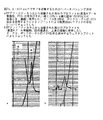
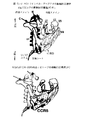
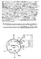
HIV感染機構について最初に発見されたことは、CD4受容体並びにCCR5及びCXCR4共受容体を介した、リンパ球又は他の宿主細胞へのそれの侵入方法であった。次にHIVエンベロープタンパク質構造が研究され(図10a−b)、gp120ループの3Dの変動性並びにCD4及び共受容体の識別及び吸着のためのgp120−gp41複合体形成の重要な役割が基本原則として設定された。ウイルスenvタンパク質を見つけ、HIV細胞侵入に寄与するそれらのエピトープに結合し、又はCD4受容体及び共受容体上の各ドメイン若しくはエピトープに結合し、その結果段階的HIV感染プロセス若しくは細胞結合を妨害すると理解されるモノクローナル抗体は、HIV中和抗体と称された。
Type 1: Monoclonal HIV-specific antibody-based HIV / AIDS therapeutic vaccine, among others neutralizing antibody as mAb, or a cocktail of 2-3 HIV-neutralizing mAbs [5, 14, 28]
The first thing discovered for the mechanism of HIV infection was its entry into lymphocytes or other host cells via the CD4 receptor and CCR5 and CXCR4 co-receptors. The HIV envelope protein structure was then studied (FIGS. 10a-b), with 3D variability of the gp120 loop and the important role of gp120-gp41 complex formation for CD4 and co-receptor discrimination and adsorption as basic principles. Was set. Find viral env proteins and bind to their epitopes that contribute to HIV cell invasion, or bind to each domain or epitope on CD4 receptor and co-receptor, thus interfering with the stepwise HIV infection process or cell binding The understood monoclonal antibody was called HIV neutralizing antibody.
抗体−ベースのワクチン開発における大きい問題点は、HIV遺伝子変動性によっても引き起こされ、これは、一部のHIV抗原により誘発された組換抗体は、HIV−1の異なる単離体を中和することが不可能であるということである。免疫化により誘発された抗−HIV−1モノクローナル抗体の大部分は、貧弱な交差−中和活性を有するか又はこれを有さず、かつ典型的には、変異のためにウイルス毎に異なるか又は感染性ビリオンの表面に余り露出されていないかのいずれかである決定基に結合する。中和mAbのいくつかの異形が作出されたが、その後臨床試験が、エンベローブタンパク質gp120及びgp41に対する中和抗体をベースにしたワクチンは、標的HIVタンパク質の表面エピトープの変動性及び変化という同じ理由により、1〜2ヶ月以内に働きを停止する(稀に、これらが開始時から働く場合)ことを示した。 A major problem in the development of antibody-based vaccines is also caused by HIV gene variability, where recombinant antibodies elicited by some HIV antigens neutralize different isolates of HIV-1. It is impossible. The majority of anti-HIV-1 monoclonal antibodies elicited by immunization have or do not have poor cross-neutralizing activity and typically differ from virus to virus due to mutations Alternatively, it binds to a determinant that is either less exposed on the surface of the infectious virion. Several variants of neutralizing mAbs have been created, but subsequent clinical trials have shown that vaccines based on neutralizing antibodies against the envelope proteins gp120 and gp41 have the same reasons for variability and changes in the surface epitopes of the target HIV protein. It has been shown that it stops working within 1-2 months (rarely when they work from the start).
説明されたワクチン開発アプローチの欠点は、動物の免疫化のためのウイルス抗原のどちらかの抗体のモノクローナル性の選択である。ウイルス標的−タンパク質の種々の変種に特異的な中和抗体のパネルが作製されるような場合であっても、各mAbは、細菌システムにおける組換モノクローンとして作製される。更に原核生物の組換抗体はそれらの抗原に対する親和性が、動物又はヒトの血清中の未変性のAbと比べ、少なくとも10倍低い。動物において誘発されたポリクローナルHIV−特異的免疫グロブリンは通常、ヒトなどの種々の生物について免疫毒性である。それらを、診断目的に使用することは可能であるが、アナフィラキシー反応の発生機会の多さは、それらの免疫療法適用に関する当然の制約である。ハイブリドーマmAb作製技術は、免疫グロブリンにおける生物学的種の差異の問題点を解決しない。ヒト化mAb又はキメラmAbの作製技術は、非常に面倒であり、かなり時間がかかりかつ費用もかかる。従ってこの技術により、抗−HIV免疫療法のための数十又は数百のmAb異形を作製することは不可能である。 A drawback of the described vaccine development approach is the selection of the monoclonality of either antibody of the viral antigen for animal immunization. Each mAb is made as a recombinant monoclonal in a bacterial system, even when a panel of neutralizing antibodies specific to different variants of the viral target-protein is made. In addition, prokaryotic recombinant antibodies have at least 10 times lower affinity for their antigens than native Abs in animal or human serum. Polyclonal HIV-specific immunoglobulins elicited in animals are usually immunotoxic for various organisms such as humans. Although they can be used for diagnostic purposes, the high chance of an anaphylactic reaction is a natural limitation for their immunotherapy application. Hybridoma mAb production technology does not solve the problem of biological species differences in immunoglobulins. The technology for producing humanized or chimeric mAbs is very tedious, quite time consuming and expensive. Thus, this technique makes it impossible to create dozens or hundreds of mAb variants for anti-HIV immunotherapy.
2型:HIV粒子破壊−ベースのワクチン[9, 20]。天然のHIVビリオン及びHIVペプチドを使用する発想は、15年以上前に明らかにされ、かついくつかの形で生まれ変わった。中でも、小型ウイルスにとって致死的であるが、ペプチド結合及びタンパク質の高次構造に対する破壊作用が比較的低いことが周知である、β−プロピオラクトン、ソラレン又は類似物質による、HIV粒子の感染性活性の保存が発想された。超遠心分離法による患者の血流由来の未変性ウイルスの濃縮は、一部の免疫化に適用可能なウイルス量を生じることができないことが急速に明らかになり始め、これはかろうじて研究分析のために若干の材料を送達することができる。そのため、この種のワクチンの実際上の変形は、実験室株のインビトロ感染培養、又は初代単離株の感染及びそれらのドナーリンパ球との培養のいずれかである。両方の場合において、免疫化後のHIVタンパク質の免疫応答形成に必要な大量のウイルス粒子を提供するための、数百リットルの発酵槽中での大規模生成が唱えられている。 Type 2: HIV particle disruption-based vaccine [9, 20]. The idea of using native HIV virions and HIV peptides was unveiled over 15 years ago and was reborn in several ways. Among them, the infectious activity of HIV particles by β-propiolactone, psoralen or similar substances, which are lethal to small viruses but are well known to have relatively low disruption effects on peptide bonds and protein conformation. Was conceived. Concentration of native virus from the patient's bloodstream by ultracentrifugation has begun to become rapidly apparent that it is not possible to produce viral load applicable to some immunizations, which is barely for research analysis Some material can be delivered. Therefore, a practical variant of this type of vaccine is either in vitro infection culture of laboratory strains or infection of primary isolates and their culture with donor lymphocytes. In both cases, large-scale production in the hundreds of liters of fermentor has been advocated to provide the large amount of virus particles necessary to form an immune response of the HIV protein after immunization.
この発想それ自身は完全に悪くはなく、これは他の3種のワクチン型の以前には利点さえ有する。第一に、免疫化のための不活化されたウイルス粒子使用の安全性は、ショ糖ピロー(pillow)勾配における超遠心分離後に、HIV RNAコピーの実時間定量を試みる場合に、より自明のものとなる。ウイルスRNAは大部分、小片に崩壊され、かつショ糖勾配における濃縮後に得られたHIVビリオン又はそれらのタンパク質の実濃度よりも、104〜105より低い数のレベルへと破壊される。第二に、未変性ウイルスタンパク質の獲得は、現存する種々のHIV envタンパク質エピトープを対象とする見込みがよりあるように見える。しかしこの最後の説明は、この種のワクチンが働かない真の理由である。 The idea itself is not completely bad, and it even has advantages before the other three vaccine types. First, the safety of using inactivated virus particles for immunization is more obvious when attempting real-time quantification of HIV RNA copies after ultracentrifugation in a sucrose pillow gradient. It becomes. Viral RNA is largely disrupted into small pieces and destroyed to a number of levels lower than 10 4 to 10 5 than the actual concentration of HIV virions or their proteins obtained after concentration in a sucrose gradient. Secondly, the acquisition of native viral proteins appears more likely to target various existing HIV env protein epitopes. But this last explanation is the real reason why this kind of vaccine doesn't work.
HIV粒子破壊−ベースのワクチン開発は、遺伝子変異選択のインビトロ条件が、動物又はヒト生物における同じプロセスの境界からどの程度異なるかの最良の例である。ウイルスペプチドの分析は、異なるウイルス亜型にのみではなく、同じ患者から単離されたウイルス変異株にさえも特異的である、抗原エピトープの高い変動性を明らかにした。しかし全ての実験室株、中でも高度に感染性のBIIIのA455は、envペプチドの配列の一定でありかつより均質な組成を有する。実験室HIV株に関して質量分析法又は3D構造的方法により分析されたenvペプチドライブラリーの多様性は、単独の一患者から採取した同等物から最大5%である。同じ傾向が、ドナー血液リンパ球又はCD4、CCR5若しくはCXCR4−有するヒト細胞培養物とインビトロで共培養された初代HIV単離株について認められる。これは、ウイルスのインビトロ感染に関する選択条件は、生物における天然のウイルス複製及びビリオン形成プロセスとは非常に異なり、ヒト生物におけるウイルス生存率(survivorship)に関するゲートは、インビトロ培養時よりも95%より広いことを意味する。従って大規模インビトロ生成後の不活化ウイルス粒子を使用し抗−HIVワクチンを調製する全ての試みは失敗し、更には実験室HIV株から供給されたペプチド−ベースのワクチンも失敗した。 HIV particle disruption-based vaccine development is the best example of how in vitro conditions for gene mutation selection differ from the same process boundaries in animals or human organisms. Analysis of viral peptides revealed high variability of antigenic epitopes that are specific not only to different viral subtypes but also to viral variants isolated from the same patient. However, all laboratory strains, especially the highly infectious BIII A455, have a constant and more homogeneous composition of the env peptide sequence. The diversity of env peptide libraries analyzed by mass spectrometry or 3D structural methods for laboratory HIV strains is up to 5% from equivalents taken from a single patient. The same trend is observed for primary HIV isolates co-cultured in vitro with donor blood lymphocytes or human cell cultures with CD4, CCR5 or CXCR4-. This is because the selection conditions for in vitro infection of the virus are very different from the natural viral replication and virion formation processes in the organism, and the gate for virus survival in human organisms is 95% wider than in in vitro culture. Means that. Thus, all attempts to prepare anti-HIV vaccines using inactivated virus particles after large-scale in vitro production have failed, as well as peptide-based vaccines supplied from laboratory HIV strains.
3型:HIV−ペプチド−ベースのワクチン[3, 6, 13, 15, 27, 33, 36]。この最新型のワクチンは、小型のHIVペプチド、受容体認識及び感染活性に寄与するウイルスタンパク質のより大きいHIVタンパク質偽エピトープの複数の小さい15〜20個のアミノ酸断片、これらの小型ペプチドのパネルを含む。小さいレンチウイルスファミリーのメンバーとして、HIVは、小数のペプチド(合計18)からなり、HIVペプチドワクチンの大部分は、gp120(gp140、gp160)又はgp120及びgp41の両方のenvタンパク質の断片を含有し、残りは、維持が困難な(little easy-to-maintain)マトリックスペプチド及びp24断片を含む。このクラスの他の部分は、HIV生活環に関して天然の提供されたグリコシル化により酵母において生成された完全長envペプチド若しくはそれらの大型断片、又はいわゆる糖質−ベースのHIVワクチンである。HIVペプチドワクチンの一部は、治療的免疫化が意図され、一部は、予防活性を有することが言明されている。 Type 3: HIV-peptide-based vaccine [3, 6, 13, 15, 27, 33, 36]. This state-of-the-art vaccine includes small HIV peptides, multiple small 15-20 amino acid fragments of larger HIV protein pseudo-epitopes of viral proteins that contribute to receptor recognition and infectious activity, a panel of these small peptides . As a member of the small lentiviral family, HIV consists of a small number of peptides (18 total), the majority of HIV peptide vaccines contain gp120 (gp140, gp160) or fragments of both gp120 and gp41 env proteins, The rest includes a matrix peptide and p24 fragment that are little easy-to-maintain. The other part of this class is the full-length env peptides produced in yeast by the naturally provided glycosylation for the HIV life cycle, or large fragments thereof, or so-called carbohydrate-based HIV vaccines. Some of the HIV peptide vaccines are intended for therapeutic immunization and some have been declared to have prophylactic activity.
しかしこれまで組換HIVペプチドのカクテルも合成15〜20個アミノ酸ペプチドのカクテルも、ウイルス感染及び複製からの防御を提供することができなかった。その主な理由は、どのようにこれらのペプチドが得られたかの原理の分析から明らかにすることができる。組換ペプチド配列は、長いTaq−ポリメラーゼPCR(通常1000〜3000bp)により増幅されたHIVゲノム断片のステージを含む、患者由来のウイルス材料からのRT−PCRにより得られた試料の、又はHIV−特異的プライマーPCR後の患者のリンパ球由来のDNAの配列の自動DNA配列決定の技術、その後の形質転換された大腸菌株コロニーの選択により作製される。現在の技術は、高くないとして平均で多様な変種105〜106から1配列ケースの頻度での、ランダム様式のHIV遺伝子型のモノクローナル選択を基礎としており、それから平均感染性ウイルス力価は1%であり、そのためこれは感染活性のあるウイルスの103〜104コピーである。1個体の患者血液の同じ試料からこの技術で作製された二つの配列の完全なHIVゲノムのデータは、劇的に異なることは、HIVゲノム配列を作製しそれら自身を分析する研究者には周知である。従ってこれらの組換ペプチド又は3〜4種の組換ペプチドのカクテルによる免疫化は、例え真核生物発現システムにおいて適切にグリコシル化された(糖鎖付加された(carbohydrated))としても、現在対処されるべきウイルス変異株の不活化に特異的な免疫応答の形成を提供することはできない。従って、HIVワクチン開発アプローチは、これらの標準とは無関係であり、組換ペプチド配列の情報は、他の方法で作成されなければならない。 However, to date, neither a cocktail of recombinant HIV peptides nor a cocktail of synthetic 15-20 amino acid peptides has been able to provide protection from viral infection and replication. The main reason can be clarified from an analysis of the principle how these peptides were obtained. Recombinant peptide sequences include samples of samples obtained by RT-PCR from patient-derived viral material, including HIV genomic fragment stages amplified by long Taq-polymerase PCR (usually 1000-3000 bp), or HIV-specific By means of an automated DNA sequencing technique of DNA sequences from patient lymphocytes after selective primer PCR followed by selection of transformed E. coli strain colonies. Current technology is based on the random selection of HIV genotype monoclonals with a frequency of 10 5 to 10 6 to one sequence case on average, but not on average, and the average infectious virus titer is 1 %, So this is 10 3 to 10 4 copies of an infectious virus. It is well known to researchers who create HIV genome sequences and analyze themselves that the complete HIV genome data of the two sequences generated by this technique from the same sample of a single patient blood is dramatically different. It is. Therefore, immunization with these recombinant peptides or cocktails of 3-4 recombinant peptides is currently addressed even if appropriately glycosylated (carbohydrated) in eukaryotic expression systems. It cannot provide for the formation of an immune response that is specific to the inactivation of virus variants to be done. Thus, the HIV vaccine development approach is independent of these standards, and recombinant peptide sequence information must be generated in other ways.
合成アミノ酸の小型HIVペプチド[27]は、矛盾したアプローチにより作製され−ペプチド結合形成の各サイクルに関して、公知のHIV配列における可能性のあるアミノ酸変種の混合物が添加される場合、数百の変種が自動ペプチドシンセサイザーにおいて混合物として作製されている。envタンパク質の可変領域の多くの変種は、ペプチドシンセサイザーを使用し得ることができる。しかしこれらのペプチドのサイズは、15〜20、最大30個のアミノ酸に限定され、より長いペプチド型は、組換システムにおいてのみ作製が可能である。実際に、小型の合成ペプチド及びそれらのカクテルによる免疫化は、HIV免疫反応について十分に高いが、それへの特異性が低いか若しくはないように追加免疫する。個々に、動物(アカゲザル(Rhesus macaques))の合成HIVペプチド免疫化の試みであっても、標準ELISPOT法により試験されたそれらの血液中にHIV特異抗体が存在しないという不満足な結果を引き出す。恐らくHAARTと組合せた治療目的の組成物としての、現存するペプチド−ベースのHIVワクチンは、若干見込みがあるであろう。しかしペプチドワクチン組成物はひとつも、現在まで免疫化後のHIV感染症−予防作用を示していない。 Synthetic amino acid small HIV peptides [27] are made by a contradictory approach-for each cycle of peptide bond formation, when a mixture of possible amino acid variants in known HIV sequences is added, hundreds of variants Made as a mixture in an automated peptide synthesizer. Many variants of the variable region of the env protein can use a peptide synthesizer. However, the size of these peptides is limited to 15-20, up to 30 amino acids, and longer peptide forms can only be made in recombinant systems. Indeed, immunization with small synthetic peptides and their cocktails is sufficiently high for the HIV immune response, but boosts with low or no specificity for it. Individually, attempts to immunize synthetic HIV peptides in animals (Rhesus macaques) elicit unsatisfactory results in the absence of HIV specific antibodies in their blood tested by standard ELISPOT methods. The existing peptide-based HIV vaccine, perhaps as a therapeutic composition in combination with HAART, is somewhat promising. However, none of the peptide vaccine compositions has shown an HIV infection-preventing effect after immunization to date.
4型:HIVペプチドの遺伝子をコードするDNAプラスミド又はウイルス(アデノ随伴、鶏痘、ワクシニア、レトロウイルスなど)ベクターワクチン[11, 12, 16, 21, 26, 29, 30]。世界中で99の臨床試験に関して承認が得られた55種の抗−HIVワクチン中でほとんどは、DNA−ベースのクラスに属している。しかしひとつの候補だけが、第III相臨床試験を通過し、かつ第IIb相を通過する若干の見込みがある[37, 42]。この種のワクチンを使用する発想は、DNA免疫化は、自己免疫合併症及びアナフィラキシー反応などの即時型副作用を生じないという、健常なバックグラウンドを有し、そのためその臨床適用は、安全かつ容易である。この利点にもかかわらず、全てのウイルス及び非ウイルスDNAワクチンは、それらの実際の抗−HIV有効性の可能性に関して、希望を弱める多くの欠点を含む。 Type 4: DNA plasmid or virus (such as adeno-associated, fowlpox, vaccinia, retrovirus, etc.) vector vaccine encoding the gene for HIV peptide [11, 12, 16, 21, 26, 29, 30]. Most of the 55 anti-HIV vaccines approved for 99 clinical trials worldwide belong to the DNA-based class. However, only one candidate has some chance to pass a phase III clinical trial and to pass phase IIb [37, 42]. The idea of using this type of vaccine has a healthy background that DNA immunization does not produce immediate side effects such as autoimmune complications and anaphylactic reactions, so its clinical application is safe and easy. is there. Despite this advantage, all viral and non-viral DNA vaccines contain a number of drawbacks that weaken hope with regard to their actual anti-HIV efficacy potential.
DNAは、それ自身いかなる免疫反応も引き起こさないので、このワクチン有効性は、下記の3つの条件の大きさであり、これらは各々同等に重要である:
1)トランスフェクション/感染効率、又はいかに多くの細胞が、一旦適用されたある量のDNAから遺伝物質が供給され得るか;
2)発現レベル、又はどのくらい多くのタンパク質が、1種又は複数の遺伝子コピーを得る細胞において発現されているか;
3)免疫応答の継続、又はいかに長くMHCが、標的化病原体を認識するmAbの誘発を続けるか。
Since DNA does not itself cause any immune response, this vaccine efficacy is the magnitude of the following three conditions, each of equal importance:
1) Transfection / infection efficiency, or how many cells can be supplied with genetic material from a certain amount of DNA once applied;
2) level of expression, or how many proteins are expressed in cells that obtain one or more gene copies;
3) Continue the immune response, or how long MHC continues to induce mAbs that recognize the targeted pathogen.
インビトロトランスフェクション/感染効率の測定は、細胞が分裂の次サイクルを通過するまでに遺伝子導入後24時間に計測された現在のタンパク質を発現している細胞の割合であり、この割合は、同じ条件で同時に導入された蛍光タンパク質又はLacZを発現している細胞について計測される。非−ウイルス系プラスミドベクターに関して、インビトロ効率は、40〜90%を達成することができるが、同じベクターに関してインビボ静脈内投与は、最良の状態で1〜5%生じる。これらの40〜90%(インビボにおいて1〜5%)から、98〜99%は、2週間後には消滅する一過性又はエピソーム性の発現であり、かつトランスフェクトされた遺伝物質のわずかに1〜2%が、細胞ゲノムに挿入され、長期発現を提供する。プラスミドDNAワクチン[16]の量は、その送達物質−それらにより作製された陽イオン性脂質及びリポソーム、陽イオン性ポリマー(ポリエチレンイミン、ポリリシン)、プルロニック並びにそれらの種々の組合せに関する、最大耐量により制限される。実際に、負帯電したDNAに結合しかつ運搬することができる全ての陽イオン物質は、濃度105〜104M及びそれ以上で高度毒性である。非−ウイルスベクターの発現レベルは、ウイルスベクター発現と比べ、比較的高い。 In vitro transfection / infection efficiency measurements are the percentage of cells expressing the current protein measured 24 hours after gene transfer before the cells pass the next cycle of division, and this percentage is subject to the same conditions. The cells expressing the fluorescent protein or LacZ introduced at the same time are measured. For non-viral plasmid vectors, in vitro efficiencies can be achieved between 40-90%, but in vivo intravenous administration occurs at 1-5% at best for the same vectors. Of these 40-90% (1-5% in vivo), 98-99% are transient or episomal expression that disappears after 2 weeks and only 1 of the transfected genetic material. ~ 2% is inserted into the cell genome to provide long term expression. The amount of plasmid DNA vaccine [16] is limited by the maximum tolerated dose for its delivery materials-cationic lipids and liposomes made by them, cationic polymers (polyethyleneimine, polylysine), pluronics and various combinations thereof Is done. In fact, all cationic substances that can bind to and carry negatively charged DNA are highly toxic at concentrations of 10 5 to 10 4 M and above. The expression level of non-viral vectors is relatively high compared to viral vector expression.
ウイルスDNAベクターの感染効率は可変性であるが、通常インビトロ実験の10〜20%を超えない。しかしウイルスベクターは、遺伝物質の直接ゲノムへの送達を提供するそれらの能力において魅力的となり始めている。そのためインビボ投与に関するウイルスベクターの感染効率は平均2〜5%であるにもかかわらず、標的タンパク質の発現は、主として長期間であり、一過性ではない。従ってウイルスDNAベクターは、治療又は予防目的で免疫応答及び抗−HIV活性の十分な持続を有すると仮定された。 Viral DNA vector infection efficiency is variable, but usually does not exceed 10-20% of in vitro experiments. However, viral vectors are beginning to be attractive in their ability to provide delivery of genetic material directly to the genome. Thus, despite the average viral vector infection efficiency for in vivo administration, the expression of the target protein is primarily long-term and not transient. It was therefore hypothesized that the viral DNA vector had a sufficient persistence of immune response and anti-HIV activity for therapeutic or prophylactic purposes.
しかしウイルスDNAワクチン成分、及びいかにそれらが段階的に作用するかを研究するために、それらの予測される活性様式の制限を観察することができる。臨床試験に入っているDNAベクターの第一のクラスは、アデノウイルス構築物(construction)であった。それらの最新型は既に0とは異なる感染効率を示し、及び免疫化mAb後に誘発された力価は全ての免疫化学法により検出可能であるにもかかわらず、これらは決して単独投与において使用されない。このポイントは、アデノウイルスベクター−ADV[11]、又はアデノ随伴ウイルスベクター−AAV[29]は、予定された免疫化の2週間後のELISA、INF−γELISPOT又はウェスタンブロットアッセイにより通常認識されるタンパク質を送達する比較的低い発現のみを引き起こすことである。任意の組換タンパク質又はタンパク質混合物による標準免疫化の2週間後に抗体力価によりADV及びAAVに関するこれらのデータを比較する場合、ADV及びAAVワクチン接種に関して絶対数は5〜10倍より低いことは明らかになり始める。これらの数に注目し、本研究者らは、可能性のある免疫応答の期間についていくつか結論づけることができる。
However, in order to study viral DNA vaccine components and how they act in stages, one can observe the limitations of their expected mode of activity. The first class of DNA vectors that are in clinical trials were adenovirus constructions. Even though their latest forms already show an infection efficiency different from 0, and titers elicited after immunization mAb are detectable by all immunochemical methods, they are never used in a single dose. The point is that adenoviral vector-ADV [11] or adeno-associated viral vector-AAV [29] is a protein normally recognized by ELISA, INF-γ ELISPOT or
第III相臨床試験に到達し、かつ2003年10月以降タイにおいて16000名の未感染個体に適用されたただ一つのワクチン組成物は、2種のポックスウイルス(ワクシニアウイルス)−HIVワクチン接種(ALVAC−HIV)後の、プラスミドDNA−gag−pol−envワクチン(AIDSVAX B/E)による並べられたワクチン接種を基にしていた[12]。この特許のデータの検証は、ワクチン接種されたアカゲザルの血液試料由来の誘発された抗体の力価は、各免疫化後1〜3週間増加し、かつワクチン接種の残り1年間は、対照数からプラスのプロットへやや外れていくことを示している[12]。免疫応答の継続は、この場合これをいかにして評価するかという問題である。アデノウイルス及びポックスウイルスは、中でも最大のウイルスファミリーであり、これらは表面上及びウイルスマトリックス内にそれら自身のタンパク質を数百個露出していることも忘れてはならない。これは、投与後短期間(1〜2週間)でブーストされた免疫応答は、高度であるが、ほとんど非特異的であり、かつ加えて非特異性は、副作用として免疫毒性反応を引き起こすということを意味している。 The only vaccine composition that reached Phase III clinical trials and applied to 16000 uninfected individuals in Thailand since October 2003 was two poxvirus (vaccinia viruses) -HIV vaccinations (ALVAC). -Based on ordered vaccination with plasmid DNA-gag-pol-env vaccine (AIDSVAX B / E) after HIV) [12]. Validation of the data in this patent shows that the titer of elicited antibodies from vaccinated rhesus monkey blood samples increased 1-3 weeks after each immunization and for the remaining year of vaccination from the number of controls. It shows a slight deviation from the positive plot [12]. The continuation of the immune response is a question of how to evaluate this in this case. It should also be remembered that adenoviruses and poxviruses are among the largest virus families and that they expose hundreds of their own proteins on the surface and in the viral matrix. This means that the boosted immune response in the short period after administration (1-2 weeks) is high but almost nonspecific, and in addition, nonspecificity causes an immunotoxic response as a side effect. Means.
ウイルスワクチンの有効性における唯一の例外は、レトロ−(レンチ−)ウイルスベクター−ベースのアプローチである[26]。HIVはそれ自身、レンチウイルスファミリーの良い例である。レトロウイルスベクターは、十分に高い(最大5%)インビボ感染効率を提供し、送達された遺伝子タンパク質の発現は、細胞ゲノムの感染のために安定していない場合にも十分でありかつ長い。レトロウイルスベクターは、癌治療用ワクチンとして、臨床試験において、あらゆる他の遺伝子構築物よりも、著しくより良い抗腫瘍反応を示した。HIVを含む全てのレトロウイルスのみが、それらの治療的適用であっても疑わしく、予防的ワクチン接種は注目に値しないものにするある性質を有し−これは、移動性遺伝エレメントとしてヒトゲノムへ侵入するそれらの能力、並びにある期間後そのカスケードが制御できなくなり始める複数の遺伝的変異を駆動し、種々の細胞及び組織において複数の癌悪性転換を引き起こす能力である。 The only exception to the effectiveness of viral vaccines is the retro- (lenti-) viral vector-based approach [26]. HIV itself is a good example of the lentivirus family. Retroviral vectors provide sufficiently high (up to 5%) in vivo infection efficiency and the expression of the delivered gene protein is sufficient and long even when it is not stable due to infection of the cellular genome. Retroviral vectors have shown significantly better anti-tumor responses in clinical trials than any other gene construct as a vaccine for cancer treatment. Only all retroviruses, including HIV, are suspicious, even in their therapeutic application, and have certain properties that make prophylactic vaccination unnoticeable-this penetrates the human genome as a mobile genetic element As well as the ability to drive multiple genetic mutations where the cascade begins to become uncontrollable after a period of time and cause multiple cancer malignant transformations in various cells and tissues.
DNA−ベースのHIVワクチンの一般的欠点は、PCR及びモノクローニング後の標準DNA配列決定などの、組換HIVペプチド組成物に関して先に説明された方法と同じ方法で得られた当初のヌクレオチド配列に関するものである。105〜106変種と等しい患者1名の血流中のHIV遺伝的変異の平均数を理解することは真実に近い。ランダム様式のこの方法で得られた一つ又はいくつかの配列データで作製された遺伝的構築体は、同じ患者に関してであってもHIV変異株の大半が基本的には働くことができない。並びに全てのプラスミドDNA及び任意のウイルスベクター−ベースのDNAのHIVワクチンは、単独のenv、pol、gag及びそれらの組合せ領域に関するHIVゲノムの配列をベースにしている。これらの構築物がモノクローナルヌクレオチドHIVゲノム領域の配列からなるようになるまで、HIVワクチン開発は袋小路にある。HIV遺伝的変動性及び変異性を根絶するために、その現存する変異の定量分析を継続し、かつより頻繁に存在する変異株に関する見込みのあるワクチンを製剤することは必要である。 A general disadvantage of DNA-based HIV vaccines relates to the original nucleotide sequence obtained in the same way as described above for the recombinant HIV peptide composition, such as PCR and standard DNA sequencing after monocloning. Is. Understanding the average number of HIV genetic variations in the bloodstream of a patient equal to 10 5 to 10 6 variants is close to truth. Genetic constructs made with one or several sequence data obtained in this manner in a random fashion are essentially unable to work for the majority of HIV variants, even for the same patient. In addition, all plasmid DNA and any viral vector-based DNA HIV vaccines are based on the sequence of the HIV genome for a single env, pol, gag and combinations thereof. Until these constructs are composed of sequences of monoclonal nucleotide HIV genomic regions, HIV vaccine development remains in a pouch. In order to eradicate HIV genetic variability and variability, it is necessary to continue quantitative analysis of its existing mutations and formulate promising vaccines for more frequently present mutants.
先に説明されたように、DNA−ベースのHIVワクチンの有効性に関する他の主な制限は、ウイルス及び非−ウイルスの遺伝子治療用ベクターのインビボ送達に関する不完全な公知の方法に起因した貧弱な免疫応答である。あらゆる種類の抗−感染症免疫化を提供するDNA−ベースのワクチン型の見込みが低いことを理解するための科学者の正確な比較は、以下である。任意のタンパク質又は抗原、及び原核生物大腸菌システムにおいて作出されたそれらの組換連結されたIgG軽鎖−重鎖変形に関する仮定のモノクローナル抗体(mAb)を想定して欲しい。ここで本発明者らは、ELISA、ELISPOT、イムノドットブロット、ウェスタンブロット、フローサイトメトリー、蛍光顕微鏡などの、全ての可能な実験室免疫反応アッセイにより、抗原がこれらの2種のmAb型へ結合するための親和性の比較を行うことを試みる。本発明者らが、これらの型がひとつのアッセイに収められた各状況において認めるものは、以下である−組換mAbの親和性は、常に天然の動物モノクローナル抗体の親和性よりも少なくとも10倍低く、更には力価決定時の最小結合活性の差異は、100〜200倍に達し得る。ワクチン免疫原性のインビボ評価に関する同じ状況は、研究者が、動物免疫化に使用されるDNA−ベース組成物及びタンパク質−ベース組成物の活性を分析する場合に、認められる。免疫化された動物の血中の現在の抗原に関するmAbの力価として測定された特異的免疫応答の有効性は、遺伝子ベクターとして送達される抗原の方が、当初のタンパク質−抗原よりも、数倍より低いであろう。DNA変種に関する特異的抗原免疫応答の強度は、その「陽性対照」−タンパク質変種よりも常に5〜20倍より低い。 As explained earlier, other major limitations on the effectiveness of DNA-based HIV vaccines are poor due to imperfect known methods for in vivo delivery of viral and non-viral gene therapy vectors. It is an immune response. An accurate comparison of scientists to understand that the promise of DNA-based vaccine types that provide all kinds of anti-infection immunizations is low: Imagine a hypothetical monoclonal antibody (mAb) for any protein or antigen and their recombinantly linked IgG light-heavy chain variants produced in a prokaryotic E. coli system. Here we will bind the antigen to these two mAb types by all possible laboratory immune reaction assays, such as ELISA, ELISPOT, immunodot blot, Western blot, flow cytometry, fluorescence microscopy, etc. Try to do an affinity comparison. What we recognize in each situation where these types are contained in one assay is the following-the affinity of the recombinant mAb is always at least 10 times greater than the affinity of the natural animal monoclonal antibody. Low and even the difference in minimum binding activity when determining the titer can reach 100-200 times. The same situation regarding in vivo assessment of vaccine immunogenicity is observed when researchers analyze the activity of DNA-based and protein-based compositions used for animal immunization. The effectiveness of the specific immune response, measured as mAb titers with respect to the current antigen in the blood of the immunized animal, is greater for antigen delivered as a gene vector than for the original protein-antigen. Will be lower than twice. The intensity of a specific antigenic immune response for a DNA variant is always 5-20 times lower than its “positive control” —protein variant.
可能性のあるHIVワクチン候補として説明されている、更にひとつの小さい組成物の範疇が存在し−これは樹状細胞ワクチンとも称される。それらの開発は、幹細胞科学を基本にし、かつ樹状細胞ワクチンは、かなり経費が高い(平均患者1名の治療に45000〜60000米ドル)にもかかわらず、適度に十分な治療結果を伴う化学療法又は放射線治療と組合せて、数種類の腫瘍の治療に適用される。しかしいくつかのある種の病理又は微生物を識別しかつ殺傷することがインサイチュにおいて示された樹状細胞−マクロファージ前駆体は、同じ患者の血流に自家的にのみ適用されることができるので、HIV治療、更には感染症予防に関するそれらの効能はかなり疑わしい。マクロファージの「教え(teaching)」に関するウイルスのペプチドを得る場所に関する疑問は、同じであり、組換体は、数年配列が固定され、かつ未変性のものは、単離するために実在しないような高濃度で提供されなければならない。従って樹状細胞適用は、重大な抗−HIVワクチンの候補としては提唱することができない。 There is yet another small composition category that has been described as a potential HIV vaccine candidate-also referred to as a dendritic cell vaccine. Their development is based on stem cell science and dendritic cell vaccines are quite expensive (45,000-60000 USD for the treatment of an average patient), but chemotherapy with reasonably sufficient treatment outcome Or in combination with radiation therapy, it applies to the treatment of several types of tumors. However, since dendritic cell-macrophage precursors that have been shown in situ to identify and kill some certain pathologies or microorganisms can only be applied to the same patient's bloodstream only on their own, Their efficacy with respect to HIV treatment and even infection prevention is quite questionable. The question about where to obtain viral peptides for macrophage “teaching” is the same: recombinants are fixed in sequence for several years, and native ones are not real to isolate Must be provided in high concentration. Thus, dendritic cell application cannot be proposed as a significant anti-HIV vaccine candidate.
HIV−1大流行の制御の唯一の可能性のある方法は、非−感染個体、特に高リスク群の代表者の免疫化を通じて、HIV−1感染を防止及び/又はその発症を停止することができる、ある種のワクチンである。そのようなワクチンは、個別の天然のHIV−1ペプチドのエピトープの混合物、正確にはウイルス表面上外側にのみ存在する主要なHIV−1エンベロープタンパク質gp120、その断片のエピトープ、及び同じく好適な外側部分を伴うenv gp120−gp41四量体の原料としてのgp41ペプチド及び/又はワクチン接種される個体の免疫系を認識可能なエピトープを含有しなければならない。これらのペプチドは、先に言及された理由(3〜4頁13〜30、1〜20行)のために、ウイルス由来の未変性のものであることはできない。並びに組換ペプチドに関して、正確な配列情報が誘導されなければならない。本発明者らは、以下を基にしたenv配列研究を含む、本特許出願において詳細に特定されたHIVワクチン開発の代替法を開発した。
1)未変性ウイルスペプチドのファージディスプレイリバースパンニング技術による収集及びアフィン(affine)精製;
2)HIV−感染した個体の現在のコホート中の多数の変異株において表されたgp120及びその断片の配列に関する情報を送達する一群のLC−MS法を使用する、未変性ウイルスペプチドの引き続きの定量的かつ配列決定分析;
3)HIV及び真核生物のグリコシル化と同じ組換envペプチド産生のためにリーシュマニア属システムを使用する、天然envペプチドエピトープの再構築;
4)a)必要な免疫ブースト期間の延長、b)免疫毒性の制御:を提供する、免疫原性envペプチドのための立体的に安定したリポソームパッケージング又はビロソームのいずれかのアプローチを使用する、HIV予防ワクチン組成物。
The only possible method of controlling the HIV-1 pandemic is to prevent and / or stop the development of HIV-1 infection through immunization of non-infected individuals, particularly representatives of high risk groups It is a kind of vaccine that can. Such a vaccine comprises a mixture of individual natural HIV-1 peptide epitopes, exactly the major HIV-1 envelope protein gp120 present only on the outer surface of the virus, its fragment epitopes, and also a suitable outer part. Must contain the gp41 peptide as a source of the env gp120-gp41 tetramer and / or an epitope that can recognize the immune system of the individual to be vaccinated. These peptides cannot be virus-derived native for the reasons mentioned above (3-4 pages 13-30, lines 1-20). As well as the recombinant peptide, the correct sequence information must be derived. The inventors have developed an alternative method for the development of an HIV vaccine that has been identified in detail in this patent application, including env sequencing studies based on:
1) Collection of native viral peptides by phage display reverse panning technique and affine purification;
2) Subsequent quantification of native viral peptides using a group of LC-MS methods that deliver information about the sequence of gp120 and its fragments represented in multiple variants in the current cohort of HIV-infected individuals And sequencing analysis;
3) Reconstruction of natural env peptide epitopes using the Leishmania system for the production of the same recombinant env peptide as HIV and eukaryotic glycosylation;
4) Use either a sterically stable liposome packaging or virosome approach for immunogenic env peptides that provides: a) prolonging the required immune boost period, b) controlling immunotoxicity: HIV prophylactic vaccine composition.
現在までに実行されたgp120のプロテオーム解析は稀であり、かつ他のウイルスペプチド及び細胞タンパク質のカクテルから精製された未変性のペプチド変種は存在しないので、不完全であった。十分な吸着能を持つカラムでのウイルスenvペプチドのアフィン吸着によるリバースパンニング技術は、この問題点を解決することができる。HIV感染に対する免疫化のためのワクチン組成物が製造される前に、HIV感染した個体の現在のコホートの大半において提示されているenvペプチドのアイソフォームを選択することが必要である。 The proteomic analysis of gp120 performed to date is rare and incomplete as there are no native peptide variants purified from cocktails of other viral peptides and cellular proteins. The reverse panning technique by affine adsorption of the viral env peptide on a column having sufficient adsorption ability can solve this problem. Before vaccine compositions for immunization against HIV infection are manufactured, it is necessary to select env peptide isoforms that are presented in the majority of current cohorts of HIV infected individuals.
1名の患者について平均105の遺伝的変種という大きい変動性にもかかわらず、最も採用されかつより高い感染生存率を有する変種の選択が、各感染者において実行される。疫学的変動性のデータは、HIV変異株の蔓延は、縄張り的境界、存在する遺伝子配列に応じた性的−又は注射薬物乱用(IDU)−伝染の個人的接触依存性を有することを証明している。優性の(dominating)ウイルスペプチド変種の数は、遺伝的変種よりも決定的にはるかに小さいが、十分迅速に種々の数の優性に変更することができる。及び優性である感染的に危険な変種であるヌクレオチド配列は、情報を生じず、プロテオーム定量及び配列分析のみ行うことができる。本発明者らが試みたこの方法は、液体クロマトグラフィー、エレクトロスプレーイオン化質量分析である。 Despite the large variability of an average of 10 5 genetic variants per patient, the selection of the variant that is most employed and that has a higher infection survival rate is carried out in each infected individual. Epidemiological variability data demonstrate that the prevalence of HIV variants has territorial boundaries, sexual- or injectable drug abuse (IDU) depending on the gene sequence present, and personal contact dependence of infection. ing. The number of dominating viral peptide variants is significantly smaller than the genetic variants, but can be changed quickly enough to various numbers of dominances. Nucleotide sequences that are dominantly infectious and dangerous variants produce no information and can only be proteome quantified and sequence analyzed. The methods we have tried are liquid chromatography, electrospray ionization mass spectrometry.
未変性のgp120 HIVペプチドは、高い免疫原性を有するが、エピトープ同一性を失っていない組換変種について同レベルを維持するためには、同様のグリコシル化を持つ組換システムが必要である。この問題点を解決するためには、細胞培養物、酵母培養物及びリーシュマニア属システムを使用することが可能である。真核細胞培養生成は、大腸菌における1000と比べ平均数1000万(decades of million)であるような多数の自己細胞タンパク質のために、ごくわずかな量の組換ペプチドをもたらす。酵母培養物は、十分な生産を提供するが、酵母における糖合成(carbohydration)は、先に提唱されたように、真核生物及びHIVと非常に似ている訳ではない。従って本発明者らは、誘導性かつ高い発現で、真核生物に典型的なグリコシル化様式を伴うリーシュマニア属システムを選択した。リーシュマニア属において作製されたgp120組換変種は、高度かつ100%のHIV−特異的免疫応答を提供し、次の段階は、感染症発症の防止のためにこの反応を延長することであった。 The native gp120 HIV peptide is highly immunogenic, but a recombinant system with similar glycosylation is required to maintain the same level for recombinant variants that have not lost epitope identity. In order to solve this problem, it is possible to use cell cultures, yeast cultures and Leishmania systems. Eukaryotic cell culture production results in negligible amounts of recombinant peptides because of the large number of autologous proteins, which averages decades of million compared to 1000 in E. coli. Although yeast cultures provide sufficient production, sugar synthesis in yeast is not very similar to eukaryotes and HIV, as previously proposed. We therefore selected the Leishmania system with inducible and high expression and a glycosylation pattern typical of eukaryotes. The gp120 recombinant variant produced in Leishmania provided a high and 100% HIV-specific immune response, and the next step was to prolong this response to prevent infection development .
立体的に安定したリポソームを、ペプチドワクチン担体として使用することができる方法には、下記のふたつの可能性がある:ペプチドが、リポソームベシクルの水内容物中に封入されるか、又は活性化された遠位PEG末端に結合され、リポソームの表面上に提示されるかのいずれか。両方の場合において、envペプチドは、迅速なプロテアーゼ切断及び分解から保護され、その結果免疫ブースト期間は延長される。立体的に安定したリポソームは、無毒であり、かつそれら自身無害である。これらのベシクル(visicle)は、内部に数週間又は数ヶ月にわたり装荷された(enloaded)免疫原性ペプチドを保持することができ、かつ一度にではなくこの十分長い期間内にそれらの内容物を次第に放出する(lease)ことができる。このことは、1回のワクチン接種についてより多くのタンパク質量を使用することを可能にする。HCCに関する外来タンパク質とのより長い永久的接触が提供される場合に、より強力かつより長い免疫応答が形成される。予防的HIV感染症の伝染及び発症に関するワクチンの成功は、重要であろう。 There are two possibilities for the use of sterically stable liposomes as peptide vaccine carriers: Peptides are encapsulated or activated in the water content of liposome vesicles. Either attached to the distal PEG terminus and presented on the surface of the liposome. In both cases, the env peptide is protected from rapid protease cleavage and degradation, resulting in an extended immune boost period. Sterically stable liposomes are non-toxic and harmless themselves. These vesicles can retain the immunogenic peptides loaded within for weeks or months, and gradually their contents within this sufficiently long period of time rather than once. Can be released. This makes it possible to use a larger amount of protein for a single vaccination. A stronger and longer immune response is formed when longer permanent contact with foreign proteins for HCC is provided. The success of vaccines for the transmission and development of prophylactic HIV infections will be important.
HIVワクチン候補の分析について留意する最後の点は、それらの有効性の前臨床評価のための適切なインビボモデルが存在しないことである。抗−レトロウイルス化学療法薬の開発を伴う更なる治療によるHIV感染症のモデル作製にチンパンジーを使用しようとする全ての試みは納得できかつ妥当であったが、チンパンジー又はアカゲザルにおいて抗−HIV免疫応答を評価することは、不可能である。類人猿及びサルにおいて誘発される免疫原性反応は、同じ抗原免疫化によりヒトにおいて生じる反応とはスペクトルにおいて極めて異なる。更に例えばチンパンジーは、あらゆるHIV亜型に感染し、ヒトにとって致命的であるウイルス負荷のレベルで、疾患発症症状のささいな徴候を伴わずに数年間幸せに生存することができ、更にはチンパンジー独自のシミアンウイルス感染症が生じる。そのため、抗−HIV免疫原性組成物の試験に関して、通常の実験マウスは、類人猿よりも劣ることはないが、統計学的に有意な数が入手でき、かつより頻繁な血液免疫検定ができる。臨床試験のみが、免疫防御作用が現在の新規HIVワクチンにより提供されるかどうかを判定することができる。 The final point to note about the analysis of HIV vaccine candidates is that there is no suitable in vivo model for preclinical evaluation of their effectiveness. Although all attempts to use chimpanzees to model HIV infection by further treatment with the development of anti-retroviral chemotherapeutic drugs were satisfactory and valid, the anti-HIV immune response in chimpanzees or rhesus monkeys It is impossible to evaluate The immunogenic response elicited in apes and monkeys is very different in spectrum from the response that occurs in humans with the same antigen immunization. In addition, for example, chimpanzees can infect all HIV subtypes and can survive happily for several years without the minor signs of disease onset at levels of viral load that are fatal to humans. Simian virus infection occurs. Thus, for testing anti-HIV immunogenic compositions, normal laboratory mice are not inferior to apes, but a statistically significant number is available and more frequent blood immunoassays are possible. Only clinical trials can determine whether immunoprotective effects are provided by current novel HIV vaccines.
本発明は、本ワクチンによりチャレンジされた個体において誘発された特異化された免疫応答により、その保護活性を提供する、HIV、好ましくはHIV−1亜型A及びBの予防ワクチンを提供する。従って本活性物質は、以下に詳細に説明された複合技術により調製及び選択された組換ポリペプチド/ペプチド混合物である。基本的ワクチン成分は、ウイルスの表面タンパク質及びエンベロープタンパク質並びにそれらの断片により表され、これは好ましい実施態様に従い、グリコシル化の状態が異なるHIVエンベロープタンパク質gp120、gp140、gp160(図1)及びgp41、gp120のV1−V3ループの保存ドメイン、gp120 V1−V5ループの耐性−関連した可変部分に対する抗体、gp41のグリコシル化された変種;HIV−1エンベロープスパイク及びgp41タンパク質の外部部分によるCD4受容体の結合時に高次構造の変化を受ける近位V1/V2及びV3ループを伴う、ウイルスエンベロープタンパク質gp120、gp140、gp160へのCD4結合エピトープ;ウイルスエンベロープタンパク質のCXCR5及びCCR4共受容体結合部位;p24ウイルスペプチドの種々のエピトープ:を含んでなる。 The present invention provides a prophylactic vaccine for HIV, preferably HIV-1 subtypes A and B, which provides its protective activity by a specific immune response elicited in an individual challenged with the vaccine. The active agent is therefore a recombinant polypeptide / peptide mixture prepared and selected by the combined techniques described in detail below. The basic vaccine components are represented by viral surface proteins and envelope proteins and fragments thereof, which, according to a preferred embodiment, are HIV envelope proteins gp120, gp140, gp160 (FIG. 1) and gp41, gp120 with different glycosylation states. Conserved domain of the V1-V3 loop of gp120, resistance to gp120 V1-V5 loop-antibodies to related variable parts, glycosylated variants of gp41; upon binding of CD4 receptor by HIV-1 envelope spike and external part of gp41 protein CD4 binding epitope to viral envelope proteins gp120, gp140, gp160 with proximal V1 / V2 and V3 loops subject to conformational changes; CXCR of viral envelope protein And CCR4 coreceptor binding site; various epitopes of p24 viral peptide: comprising.
これらの組換ポリペプチド及びそれらの混合物は、種々のドナーB−リンパ球mRNAから作製された抗体の組換ファージディスプレイライブラリーを用い、収集され、同定され、かつクローニングされる。各々の作製されたファージ抗体ライブラリーは、組換gp120−、gp41及び未変性のHIV−ポリペプチドの種々のエピトープの結合に対し、好ましくは組換gp140−、gp160−及びp24 HIV−1亜型Aタンパク質上に存在するエピトープの結合に対しても、特異的である。 These recombinant polypeptides and mixtures thereof are collected, identified, and cloned using recombinant phage display libraries of antibodies generated from various donor B-lymphocyte mRNAs. Each generated phage antibody library preferably has recombinant gp140-, gp160- and p24 HIV-1 subtypes for binding of various epitopes of recombinant gp120-, gp41 and native HIV-polypeptides. It is also specific for the binding of epitopes present on the A protein.
これらの組換ファージ抗体ライブラリーは、検出、分析及び/又は精製の手段として種々の適用において使用されてよい[23]。前記抗体ライブラリーを使用する適用は、免疫検定、免疫ブロット、クロマトグラフィーなどを含むが、これらに限定されるものではない。本発明の抗体は、HIV治療及び/又は予防のための新規医薬品の開発に関しても有用である。
本発明の好ましい実施態様においては、M13KO7ファージ上に提示された組換抗体が、HIV予防ワクチンの開発に使用される。
These recombinant phage antibody libraries may be used in various applications as a means of detection, analysis and / or purification [23]. Applications using the antibody library include, but are not limited to, immunoassays, immunoblots, chromatography and the like. The antibodies of the present invention are also useful for the development of new pharmaceuticals for HIV treatment and / or prevention.
In a preferred embodiment of the invention, recombinant antibodies displayed on M13KO7 phage are used in the development of HIV prophylactic vaccines.
ファージミドライブラリーにより展示された抗体断片は、本質的に保存された高次構造のHIVタンパク質エピトープに結合するので、該抗体の標的は、HIV感染症に対するワクチンとして適格であり、チャレンジ時に、個体の免疫系は、これらのエピトープに対する特異的免疫応答を生じるだろうから、すなわち最終的に記憶細胞となる成熟したB細胞及びT細胞はその個体に免疫を伝達することを示す。 Because antibody fragments displayed by the phagemid library bind to HIV protein epitopes that are essentially conserved conformations, the target of the antibody is eligible as a vaccine against HIV infection and upon challenge, The immune system will generate a specific immune response against these epitopes, i.e., mature B cells and T cells, which ultimately become memory cells, will transmit immunity to the individual.
本発明のHIVワクチンは、組換gp41及びp24 HIV−1亜型Aタンパク質、並びにそれらの断片(実施例3の表9)は本発明の方法により調製された抗体に結合する、gp120、gp140及びgp160の断片、加えて通常の担体及び賦形剤及び任意に免疫刺激剤を含有する。 The HIV vaccine of the present invention comprises recombinant gp41 and p24 HIV-1 subtype A proteins, and fragments thereof (Table 9 in Example 3) that bind to antibodies prepared by the method of the present invention, gp120, gp140 and Contains a fragment of gp160, plus conventional carriers and excipients and optionally an immunostimulant.
本ワクチンは、任意のHIVウイルス、更には変異したHIVウイルス上に存在し得るエピトープに特異的な、記憶細胞による個体の免疫系を提供することにより、HIV感染症の獲得を防止し、かつ更なる進行も防止するであろう。 The vaccine prevents the acquisition of HIV infection by providing an individual's immune system with memory cells specific for epitopes that may be present on any HIV virus, as well as mutated HIV viruses, and further. Will also prevent progress.
前記組換タンパク質及び/又は断片は、本発明の方法により得られたHIV−特異抗体により選択された未変性のHIV−1エンベロープタンパク質の結合及び分析により獲得された配列情報を基にしている。
詳細には、エンベロープタンパク質などのタンパク質は、超遠心分離及びウイルス粒子の溶解などの好適な方法により破壊されたウイルス粒子から得られる。
Said recombinant protein and / or fragment is based on sequence information obtained by binding and analysis of native HIV-1 envelope protein selected by the HIV-specific antibody obtained by the method of the present invention.
Specifically, proteins such as envelope proteins are obtained from virus particles that have been disrupted by suitable methods such as ultracentrifugation and lysis of virus particles.
好適なタンパク質の選択は、当業者に公知の任意の好適なスクリーニング法により実行されてよい。好ましい実施態様において、この選択は、(i)ウイルスのエンベロープタンパク質の収集のための提示された抗体による組換ファージを使用するファージパンニング、及び/又は(ii)培養のためにプラスチック表面に接着されたHIV−特異抗体のアフィン吸着、及び/又は(iii)カラムに包埋されたHIV−特異抗体による、ウイルスエンベロープタンパク質のアフィンクロマトグラフィー選択のいずれかにより実行されてよい。 Selection of suitable proteins may be performed by any suitable screening method known to those skilled in the art. In a preferred embodiment, this selection is (i) phage panning using recombinant phage with the displayed antibody for collection of viral envelope proteins, and / or (ii) attached to a plastic surface for culture. Affine adsorption of HIV-specific antibodies and / or (iii) affine chromatographic selection of viral envelope proteins with HIV-specific antibodies embedded in a column.
次工程において、入手されかつ選択された未変性のウイルスタンパク質の配列及び/又はアイソフォームの3D高次構造が、同定されてよい。従って手順は、HIV−1罹患個体の血流中、更にはNRTI、NNRTI及びHAARTの変形などの種々の投与計画で抗レトロウイルス療法を受け取った患者の血流中を循環するgp120、gp41及びp24などの、ウイルスタンパク質の高度に特異的な可変断片及び/又は定常断片の数種の変種の混合物を提供する。 In the next step, the sequence of the native virus protein obtained and / or the 3D conformation of the isoform may be identified. Thus, the procedure involves gp120, gp41 and p24 circulating in the bloodstream of HIV-1 affected individuals, as well as in the bloodstream of patients receiving antiretroviral therapy in various dosage regimes such as NRTI, NNRTI, and HAART variants. Provides a mixture of several variants of highly specific variable and / or constant fragments of viral proteins, etc.
前記配列を基に、ウイルスタンパク質の組換ポリペプチド及び/又は断片が作出される。これらの配列は、それらに対する好適な免疫応答を誘導するために免疫系により認識され得るポリペプチドの作製に適した方法を使用することにより得ることができる。 Based on the sequence, recombinant polypeptides and / or fragments of viral proteins are generated. These sequences can be obtained by using methods suitable for the production of polypeptides that can be recognized by the immune system to induce a suitable immune response against them.
前記組換ポリペプチドは、例えばリーシュマニア誘導型発現システムのような真核生物発現システム及び真核生物−様グリコシル化を備える酵母などの、任意の好適な発現システムにおいて得ることができる。 The recombinant polypeptide can be obtained in any suitable expression system, such as a eukaryotic expression system such as a Leishmania inducible expression system and a yeast with eukaryotic-like glycosylation.
本発明のHIV−1 A及びB亜型予防ワクチンの種々の変種の調製のための例証的かつ一般的技術は、以下により詳細に例証されている、工程1−9を含む:
1.HIV−特異的ScFv抗体の断片を含む、ヒト組換IgGファージミドライブラリーの作製(ファージディスプレイ技術);
2.HIV−特異抗体のScFv断片を提示している組換ファージミドライブラリーの濃厚化(バイオパンニング);
3.任意に、PBMC−MT感染法によるインサイチュでの抗レトロウイルス療法ナイーブのウイルス材料の増殖;
4.HIV粒子及びペプチドの濃縮;ウイルス不活化及び破壊;
5.リバースパンニング技術によりファージミドライブラリーを提示しているHIV−特異的組換ScFvによる未変性のHIV envペプチドの収集;並びに
6.任意に、液体クロマトグラフィー質量分析(LC−MS)法による、envペプチド変動性及び頻度の定量分析及び配列分析;
7.任意に、主要HIV envペプチドのクローニング、及びリーシュマニア・タレントラエにおけるワクチン開発のための組換ペプチドの作製;
8.任意に、組換HIV envペプチドのクロマトグラフィー精製及び3D構造解析;並びに
9.好ましくはワクチン送達のビヒクルとして立体的に安定したリポソーム又はビロソームを使用する、HIV予防ワクチン免疫ブースト組成物の調製。
An exemplary and general technique for the preparation of various variants of the HIV-1 A and B subtype preventive vaccines of the present invention includes steps 1-9, illustrated in more detail below:
1. Generation of a human recombinant IgG phagemid library containing fragments of HIV-specific ScFv antibodies (phage display technology);
2. Enrichment of the recombinant phagemid library displaying the ScFv fragment of the HIV-specific antibody (biopanning);
3. Optionally, propagation of antiretroviral therapy naive viral material in situ by the PBMC-MT infection method;
4). Concentration of HIV particles and peptides; virus inactivation and destruction;
5). 5. Collection of native HIV env peptide with HIV-specific recombinant ScFv displaying phagemid library by reverse panning technique; Optionally, quantitative and sequence analysis of env peptide variability and frequency by liquid chromatography mass spectrometry (LC-MS) method;
7). Optionally, cloning of the major HIV env peptide and production of a recombinant peptide for vaccine development in Leishmania tarentrae;
8). Optionally, chromatographic purification and 3D structural analysis of the recombinant HIV env peptide; and Preparation of an HIV preventive vaccine immune boost composition, preferably using sterically stable liposomes or virosomes as a vaccine delivery vehicle.
1. HIV−特異的ScFv抗体の断片を含む、ヒト組換IgGファージミドライブラリーの作製(ファージディスプレイ技術);
ここで本発明の方法において、ファージミドライブラリーが、以下を含む段階i)からiii)に従い、工程1)において作製される:
i)各々、HIVに感染した多数の個体から得られたBリンパ球において発現されたIgGの、軽鎖及び重鎖の可変領域をコードするRNA由来のDNA断片の増幅が、調製される段階;
ii)IgG重鎖の可変領域をコードする核酸に会合されているIgG軽鎖の可変領域をコードする核酸を含むひとつの構築体への、段階i)で得られた軽鎖及び重鎖の二つのDNA断片の集成;
iii)pCANTABファージミドベクターへの形質転換。
1. Generation of a human recombinant IgG phagemid library containing fragments of HIV-specific ScFv antibodies (phage display technology) ;
Here, in the method of the invention, a phagemid library is prepared in step 1) according to steps i) to iii) comprising:
i) amplification of DNA fragments derived from RNA encoding the light and heavy variable regions of IgG each expressed in B lymphocytes obtained from a number of individuals infected with HIV;
ii) a light chain and a heavy chain duplex obtained in step i) into one construct comprising a nucleic acid encoding an IgG light chain variable region associated with a nucleic acid encoding an IgG heavy chain variable region. Assembly of two DNA fragments;
iii) Transformation into pCANTAB phagemid vector.
詳細には、最初に、HIVにより感染していることがわかっており、かつその中にHIV特異抗体が存在することが予想される多くの個体から、B及び/又はTリンパ球が単離される。抵抗性HIV変異株を持つ個体も含まれてよい。これらのB細胞の単離は、任意の公知の技術、例えばロイコフェレーシス、引き続きのリンパ球集団からのB/T細胞の単離[19]などにより実行されてよい。引き続きRNAが、B/Tリンパ球から、例えば論文[23]に例証されたような当該技術分野において周知の技術により、単離される。 Specifically, B and / or T lymphocytes are first isolated from many individuals who are known to be infected by HIV and in which HIV-specific antibodies are expected to be present. . Individuals with resistant HIV variants may also be included. Isolation of these B cells may be performed by any known technique such as leucopheresis followed by isolation of B / T cells from a lymphocyte population [19]. Subsequently, RNA is isolated from B / T lymphocytes by techniques well known in the art, for example as illustrated in the article [23].
ScFvライブラリー作製のために、HIV−特異的免疫グロブリンの配列を含むmRNAが単離されることが好ましい。これに関して、Bリンパ球の数は、RNA単離前のHIV−感染者の血液中の、例えば共焦点顕微鏡分析によるCD45 mAb免疫検定により、評価される。図1に示されたデータは、AIDS疾患及び症状の進行した段階の患者の一部は、総単離されたリンパ球に対するBリンパ球の非常に低い比率を有し(図1c、d)、かつ通常低いCD45免疫染色は、高いウイルス負荷及び非常に低いCD4:CD8状態に相関していることを示している。他のもの(図1a、b)とは異なり、これらの患者は、HIV−特異的ファージミドライブラリー作製のコホートに関してかなり不良な供給源である。先行する抗レトロウイルス治療に沿った又はその過程の高いウイルス負荷並びにそれらの頻度は、HIV−特異的ScFvライブラリーを得る機会を制限するものではない。 For the creation of a ScFv library, it is preferred that mRNA comprising HIV-specific immunoglobulin sequences is isolated. In this regard, the number of B lymphocytes is assessed by CD45 mAb immunoassay, for example by confocal microscopy analysis, in the blood of HIV-infected persons prior to RNA isolation. The data shown in FIG. 1 shows that some patients with advanced stages of AIDS disease and symptoms have a very low ratio of B lymphocytes to total isolated lymphocytes (FIGS. 1c, d) And usually low CD45 immunostaining indicates that it correlates with high viral load and very low CD4: CD8 status. Unlike the others (FIGS. 1a, b), these patients are a rather poor source for cohorts of HIV-specific phagemid library generation. The high viral load along with or in the course of prior antiretroviral therapy as well as their frequency does not limit the opportunity to obtain an HIV-specific ScFv library.
そのように得られた総RNAは、免疫グロブリン重鎖及び軽鎖の定常領域に特異的な、例えばオリゴdTを使用するか、又は好ましい実施態様による、プライマーとしてのオリゴヌクレオチドの使用により、cDNAに転写される。免疫グロブリン重鎖及び軽鎖の異なる定常領域の配列は、当該技術分野において周知であり、その結果cDNAへの転写に好適なプライマーは、容易にデザインすることができる。従って手順は、関心のない材料はその第一工程において排除されるので、RNAプール(複数)中の免疫グロブリン転写産物の最初の選択、及び種々のドナーからの種々のRNA−試料のより容易な取り扱いを可能にする。同じくmRNAをcDNAへ転写する前の種々のドナーからのRNA−プールの組合せも構想され、かつこれはより大きい多様性が得られる(下記参照)ので好ましい。こうして調製されたcDNAの相補体は、当該技術分野において周知の技術により合成される。 The total RNA so obtained can be converted to cDNA by using, for example, oligo dT specific for immunoglobulin heavy and light chain constant regions, or by using oligonucleotides as primers, according to a preferred embodiment. Transcribed. The sequences of the different constant regions of immunoglobulin heavy and light chains are well known in the art, so that suitable primers for transcription into cDNA can be readily designed. The procedure is therefore easier for the initial selection of immunoglobulin transcripts in the RNA pool (s) and for different RNA-samples from different donors, since the uninteresting material is eliminated in the first step. Enable handling. Also contemplated are RNA-pool combinations from various donors prior to transcribing mRNA to cDNA, and this is preferred because it provides greater diversity (see below). The cDNA complement thus prepared is synthesized by techniques well known in the art.
十分量のDNA断片を調製するために、関心対象の領域は、B/Tリンパ球から得られたmRNA、cDNA又は鋳型としてのcDNAから調製された二本鎖DNAを直接用いて増幅することができる。 In order to prepare a sufficient amount of DNA fragments, the region of interest can be amplified directly using double-stranded DNA prepared from mRNA, cDNA or cDNA as a template obtained from B / T lymphocytes. it can.
前記PCR反応に関して、増幅される核酸配列の5’末端及び3’末端にアニーリングする好適なプライマーが使用され、これらは一般に、長さ約10〜40、好ましくは15〜30、より好ましくは20〜30ヌクレオチドである、オリゴヌクレオチドである。 For the PCR reaction, suitable primers are used that anneal to the 5 ′ and 3 ′ ends of the nucleic acid sequence to be amplified, which are generally about 10-40, preferably 15-30, more preferably 20-30, in length. An oligonucleotide that is 30 nucleotides.
リバースプライマーとして、その配列が、免疫グロブリンの定常領域に由来したオリゴヌクレオチドが使用されてよい。好ましくは該リバースオリゴヌクレオチドプライマーは、対応する重鎖のCH1領域に、又はλ型及びκ型軽鎖のCλ又はCκ領域にハイブリダイズする。使用されるフォワードプライマーは、重鎖及び軽鎖の可変領域の反対末端にハイブリダイズする。 As a reverse primer, an oligonucleotide whose sequence is derived from an immunoglobulin constant region may be used. Preferably, the reverse oligonucleotide primer hybridizes to the CH1 region of the corresponding heavy chain or to the Cλ or Cκ region of the λ-type and κ-type light chains. The forward primer used hybridizes to the opposite ends of the heavy and light chain variable regions.
好ましい実施態様において、一次PCR増幅のためのフォワードプライマー及びリバースプライマーは、V BASEデータ-ベース(http://vbase.mrc-cpe.cam.ac.uk)から選ばれた、表1から3に示された核酸配列からなる群から選択される。このPCR反応は概して、長さ約750の断片を生じる。 In a preferred embodiment, the forward and reverse primers for primary PCR amplification were selected from the V BASE data base (http://vbase.mrc-cpe.cam.ac.uk) in Tables 1 to 3 Selected from the group consisting of the indicated nucleic acid sequences. This PCR reaction generally yields a fragment of about 750 in length.

次工程において、段階i)において得られた軽鎖及び重鎖の二つのDNA−断片の、IgG重鎖の可変領域をコードする核酸に会合されているIgG軽鎖の可変領域をコードする核酸を含むひとつの構築体への連結は、各々、IgG軽鎖及び重鎖の可変領域ScFvを含むポリペプチドの発現を可能にする。 In the next step, the nucleic acid encoding the variable region of the IgG light chain associated with the nucleic acid encoding the variable region of the IgG heavy chain of the two DNA-fragments of the light chain and heavy chain obtained in step i) Linking to a single construct comprising allows expression of the polypeptide comprising the IgG light chain and heavy chain variable regions ScFv, respectively.
好ましい実施態様により、可変軽鎖及び重鎖をコードするDNA断片間の特異的連結を得るために、段階i)において得られた試料の量は、例えば二つのパーツにアリコートとされ、かつ任意に希釈される。次に、mRNAからの増幅によるcDNA、cDNA又はcDNA由来の二本鎖DNAのいずれかにより調製された該DNA断片は、軽鎖又は重鎖に特異的なリンカーと個別に接触されてよく、その結果このリンカーは、各試料パーツ中のみのそれぞれのDNA断片に結合する。すなわち一方のパーツは、軽鎖のみに関するリンカーを有するのに対し、他方のパーツは、重鎖のみに関するリンカーを有するということである。使用されるリンカーは、互いに好適な条件下でのハイブリダイゼーションを可能にし、軽鎖の可変領域及び重鎖の可変領域を含むDNA断片を生じるであろう。再度、これら二つのDNA断片の会合は、これら二つのDNA断片の連結がインフレームで実行され、その結果は軽鎖可変領域及び重鎖可変領域のアミノ酸配列を持つポリペプチドを生じるであろう。同じ事が、望ましいならば、二つの重鎖及び二つの軽鎖を特異的に得るために実行することができる。表4及び5において、好ましいプライマーが列記されている。 According to a preferred embodiment, the amount of sample obtained in step i) is aliquoted, for example in two parts, and optionally to obtain a specific linkage between the DNA fragments encoding the variable light and heavy chains Diluted. Next, the DNA fragments prepared by either cDNA, amplification by amplification from mRNA, or double-stranded DNA derived from cDNA may be individually contacted with a light chain or heavy chain specific linker, Results This linker binds to the respective DNA fragment only in each sample part. That is, one part has a linker for only the light chain, while the other part has a linker for only the heavy chain. The linker used will allow hybridization under suitable conditions to each other, resulting in a DNA fragment comprising the light chain variable region and the heavy chain variable region. Again, the association of these two DNA fragments will result in a polypeptide having the light chain variable region and heavy chain variable region amino acid sequences where the ligation of these two DNA fragments is performed in-frame. The same can be done to specifically obtain two heavy chains and two light chains if desired. In Tables 4 and 5, preferred primers are listed.
本発明のより好ましい実施態様において、((Gly)4Ser)3ポリペプチドリンカーをコードするリンカー断片が、免疫グロブリンの可変性の重鎖及び軽鎖をコードする核酸配列に追加される。この重鎖及び軽鎖のリンカーパーツは、互いにアニーリングし、例えばTaqSE DNAポリメラーゼなどの、TaqSE DNAポリメラーゼの存在下でフィルイン反応を刺激する。最後に重鎖及び軽鎖は、それらのDNAリンカー断片パーツを使用し、単独の遺伝子に集成される。 In a more preferred embodiment of the invention, linker fragments encoding ((Gly) 4 Ser) 3 polypeptide linkers are added to the nucleic acid sequences encoding immunoglobulin variable heavy and light chains. The heavy and light chain linker parts anneal to each other and stimulate the fill-in reaction in the presence of TaqSE DNA polymerase, eg, TaqSE DNA polymerase. Finally, the heavy and light chains are assembled into a single gene using their DNA linker fragment parts.
従って手順は、免疫グロブリン軽鎖又は重鎖の可変領域をコードする核酸の、各々、当初得られたRNAプール中に存在しない抗原結合部位を形成するために軽鎖及び重鎖の組合せも含む、別の免疫グロブリン軽鎖又は重鎖の可変領域をコードする核酸との無作為連結により、人工的に作製された非常に多くの抗体を得ることを可能にする。示されるように、免疫グロブリンの可変領域上の既に天然に予め形成された抗原結合部位のパーツを使用し、かつそれらを無作為に組合せて、HIVに感染した個体において天然に産生される抗体と比べ、HIVタンパク質に対し増強されかつ一定の結合親和性を示す抗体も作製される。 Thus, the procedure also includes a combination of light and heavy chains to form an antigen binding site for each of the nucleic acid encoding immunoglobulin light or heavy chain variable regions, each not present in the originally obtained RNA pool. Random ligation with a nucleic acid encoding the variable region of another immunoglobulin light or heavy chain makes it possible to obtain a large number of artificially produced antibodies. As shown, antibodies that are naturally produced in individuals infected with HIV using parts of an antigen-binding site that are already naturally pre-formed on the variable region of an immunoglobulin and combining them randomly In comparison, antibodies that are enhanced and have a constant binding affinity for HIV proteins are also produced.
加えて制限部位が、こうして得られたDNA−断片に導入され、これは、例えばクローニング工程などの、後続の適用において有用である。原則として、任意の好適な制限部位が、必要に応じて使用されるが、当業者の知識内でこれはCHO−K1oseの好適なものである。制限部位は、例えば制限部位の核酸配列を含むオリゴヌクレオチドプライマーの使用、又は各々、5’末端及び/若しくは3’末端に組合せられた制限部位の核酸配列を含むアダプター分子の使用などの、当該技術分野において公知のいずれか適当な方法により導入されてよい。 In addition, restriction sites are introduced into the DNA-fragment thus obtained, which is useful in subsequent applications, for example in the cloning step. In principle, any suitable restriction site is used as needed, but within the knowledge of those skilled in the art, this is the preferred one for CHO-Klose. Restriction sites are known in the art, such as the use of oligonucleotide primers that contain the nucleic acid sequence of the restriction site, or adapter molecules that contain the nucleic acid sequence of the restriction site combined with the 5 ′ end and / or the 3 ′ end, respectively. It may be introduced by any suitable method known in the art.
本発明の好ましい実施態様において、SfiI及びNotI制限部位が、好ましい実施態様に従い軽鎖及び重鎖核酸配列を含む連結された核酸断片の末端に導入され、そこでこれらの制限部位は、クローニングベクターへの更なるクローニング工程に使用される。SfiI及びNotI制限部位は、各々、該連結された断片(ScFv遺伝子)の5’末端及び3’末端に付加される。これらの特定の制限部位は、抗体遺伝子において非常に低い頻度で生じ、例えば軽鎖及び重鎖核酸配列を含む、得られた連結断片のほとんどが、単独のSfiI/NotI断片としてクローニングされることを可能にする。本発明のより好ましい実施態様において、SfiI及びNotI制限部位は、オリゴヌクレオチドプライマーを介して導入される。好ましい使用されるSfiI−部位−及びNotI−部位−含有するオリゴヌクレオチドプライマーは、論文[21]のプライマー配列を基にデザインされる。軽鎖及び重鎖核酸配列を含む得られた連結断片の末端でのSfiI及びNotI制限部位の導入に有用なプライマーは、表6に示されている。 In a preferred embodiment of the invention, SfiI and NotI restriction sites are introduced at the ends of the ligated nucleic acid fragments comprising light and heavy chain nucleic acid sequences according to the preferred embodiment, where these restriction sites are introduced into the cloning vector. Used for further cloning steps. SfiI and NotI restriction sites are added to the 5 'and 3' ends of the ligated fragment (ScFv gene), respectively. These specific restriction sites occur very infrequently in antibody genes, indicating that most of the resulting ligation fragments, including for example light and heavy chain nucleic acid sequences, are cloned as a single SfiI / NotI fragment. enable. In a more preferred embodiment of the invention, SfiI and NotI restriction sites are introduced via oligonucleotide primers. Oligonucleotide primers containing the SfiI-site- and NotI-sites that are preferably used are designed based on the primer sequence of article [21]. Primers useful for the introduction of SfiI and NotI restriction sites at the ends of the resulting ligation fragment containing light and heavy chain nucleic acid sequences are shown in Table 6.
軽鎖及び重鎖核酸配列を含む得られた連結断片のクローニング及び発現に関して、当業者に公知のいずれか好適なクローニング及び/又は発現ベクターが使用されてよい。好ましい実施態様において、例えば、pCANTAB 5E.coliファージミドベクターを含む、ファージミドベクターが使用される。 For the cloning and expression of the resulting ligation fragment comprising light and heavy chain nucleic acid sequences, any suitable cloning and / or expression vector known to those skilled in the art may be used. In a preferred embodiment, for example, pCANTAB 5E. Phagemid vectors are used, including E. coli phagemid vectors.
ファージミドpCANTAB 5Eは、M13及びCo1E1プラスミドの両複製起点を含み、その結果好都合なことにプラスミドとして増幅されるか、あるいはM13KO7などのヘルパーファージの助けにより、組換M13ファージとしてパッケージされることができる。SfiI及びNotI消化された抗体可変領域遺伝子は、pCANTAB 5Eファージミドベクター内のリーダー配列とM13遺伝子3の本体(main body)の間でクローニングされる。得られる融合タンパク質は、両方の親タンパク質の機能を保持している。g3pリーダー配列は、本タンパク質の大腸菌の内膜/ペリプラスムへの輸送を指示し、そこで主g3pドメインは、該融合タンパク質を該集成ファージの先端へ結合する。pCANTAB 5Eは、クローニングされたScFvとg3pの配列の間の接合部に、アンバー翻訳停止コドンも含む。得られるscFv断片を含むpCANTAB 5Eプラスミド誘導体のプールは、TG1などの、大腸菌supE株の形質転換に使用される。supEにおいて、大腸菌株翻訳は、pCANTAB 5E内のアンバー停止コドンまで続き、そのファージ先端に展示されたScFv−g3p融合タンパク質を作製する。
The phagemid pCANTAB 5E contains both M13 and Co1E1 plasmid origins of replication and can therefore be conveniently amplified as a plasmid or packaged as a recombinant M13 phage with the help of helper phage such as M13KO7. . SfiI and NotI digested antibody variable region genes are cloned between the leader sequence in the pCANTAB 5E phagemid vector and the main body of
HB2151などの非サプレッサー株において、停止コドンが認識され、タンパク質合成は、scFv遺伝子の末端で中断され、かつg3p融合タンパク質は合成されない。この場合、得られるScFvタンパク質は、細胞周辺腔に輸送されるが、これは該遺伝子3ドメインを欠いているので、ファージ粒子には集成されない。むしろ可溶型抗体断片が、ペリプラスム中に蓄積し、かつ延長されたインキュベーション時に、培地に漏出する。従って、HB2151及び類似の大腸菌株は、選択された抗原陽性ファージによる、それらの感染後の可溶型抗体の産生に使用され、かつ現在の適用においては使用することができない。scFvライブラリー作製の工程は、実施例1に示されている。
In non-suppressor strains such as HB2151, a stop codon is recognized, protein synthesis is interrupted at the end of the scFv gene, and no g3p fusion protein is synthesized. In this case, the resulting ScFv protein is transported to the periplasmic space, but it is not assembled into phage particles because it lacks the
2. HIV−特異抗体のScFv断片を提示している組換ファージミドライブラリーの濃厚化(バイオパンニング);
抗体の発現は、抗原に結合することが可能であるポリペプチドを得るのに適した宿主において得ることができ、及びこうして得られたポリペプチドは、異なるドナーから単離された組換gp120−、gp41−及び未変性のHIV−ポリペプチドと共に得られた。これにより、発現されたポリペプチドが宿主の表面上に発現される場合に、これは有利ではあるが、必須ではない。抗体の発現に適した宿主は、ウイルスシステム、原核細胞及び真核細胞及び/又は細胞培養物を含む。
2. Enrichment of the recombinant phagemid library displaying the ScFv fragment of the HIV-specific antibody (biopanning) ;
Expression of the antibody can be obtained in a suitable host to obtain a polypeptide capable of binding to the antigen, and the polypeptide thus obtained can be recombinant gp120-, isolated from a different donor. Obtained with gp41- and native HIV-polypeptide. This is advantageous but not essential when the expressed polypeptide is expressed on the surface of the host. Suitable hosts for antibody expression include viral systems, prokaryotic and eukaryotic cells and / or cell cultures.
好ましい実施態様において、該抗体の断片は、ファージディスプレイライブラリーを作製するバクテリオファージM13において発現され、これはファージディスプレイ技術の使用のために異なるファージにより各々提示される非常に多数の異なる構築体の展示を可能にする。このファージディスプレイアプローチは、免疫グロブリン遺伝子のクローニング並びに機能性抗体の発現及び検出のための強力なツールである。抗体の可変性の重鎖及び軽鎖断片を、モノクローナル抗体選択の段階を伴わずに、HIV−特異抗体のプール又はライブラリーとしてファージ表面上に展示された融合タンパク質として、得ることができる。このアプローチは、任意の抗原に対する抗体を迅速に見つけ、かつ必要な場合には、他の発現システムにおけるグリコシル化を伴う及び/又は伴わずに、それらの可溶型変種を作製することを可能にする。 In a preferred embodiment, the antibody fragment is expressed in bacteriophage M13 creating a phage display library, which represents a large number of different constructs each displayed by different phage for use in phage display technology. Enable display. This phage display approach is a powerful tool for the cloning of immunoglobulin genes and the expression and detection of functional antibodies. The variable heavy and light chain fragments of the antibody can be obtained as a fusion protein displayed on the phage surface as a pool or library of HIV-specific antibodies without the step of monoclonal antibody selection. This approach makes it possible to quickly find antibodies against any antigen and, if necessary, create their soluble variants with and / or without glycosylation in other expression systems. To do.
ファージミドライブラリーパンニングは、多数のクローンの非常に迅速なスクリーニングを可能にするインビトロ技術であり、ここで選択されたHIVポリペプチドへの結合親和性を示しているそれらの表面に抗体を提示しているファージは、組換ファージミドの維持及び更なるスクリーニング工程のための新規ファージの作製のために同定されかつ使用することができる。ファージ−提示している抗体ライブラリーは、抗原−陽性クローンを同定するために、当該技術分野におけるSDS−PAGE、ウェスタンブロット及びELISAスクリーニングのクロスサイクルにより、結合親和性に関して分析することができる。 Phagemid library panning is an in vitro technique that allows a very rapid screening of a large number of clones, where antibodies are displayed on their surface showing binding affinity to the selected HIV polypeptide. These phages can be identified and used for the maintenance of recombinant phagemids and the generation of new phage for further screening steps. The phage-displaying antibody library can be analyzed for binding affinity by cross-cycle SDS-PAGE, Western blot and ELISA screening in the art to identify antigen-positive clones.
展示されたScFv抗体断片は、それらの抗原−結合能を保持しているので、従って、親和性選択により、特異抗体を発現している組換ファージを濃厚化することは可能である。このアプローチにより、規定された特異性及び親和性の抗体は、集団から迅速に選択される。得られた抗体遺伝子ライブラリーは、抗原結合能を改善するように、スクリーニングされる。この技術の工程は、パンニングと称され、HIV−特異的ScFv断片を提示しているファージが、引き続き:
i)組換gp120−、gp140−、gp160−、gp41 HIV−1亜型A及びB envペプチド、
ii)種々のHIV−感染ドナーから単離された未変性のHIV envポリペプチド:
に結合し、かつ収集されることを含む。好ましい実施態様において、先に列記された全てのポリペプチドに結合親和性を示すファージライブラリーが選択されるので、展示された抗体の数は、107〜1012から102〜103へと減少する。該ポリペプチドの特異的組換HIVポリペプチドとの独立した接触サイクルのために、ここでこれらのポリペプチドの配列は、(i)公知であり、かつ(ii)一定であり、並びに異なるドナーから単離された未変性のHIV−ポリペプチドにより、ここでこれらのポリペプチドにおいて変異が生じ、抗体を選択することが可能であり、これは本質的に公知のHIV変異体全てに結合し、このことは本抗体は、本質的に該HIVポリペプチド上の一定の高次構造を認識することができることを指摘している。
Since the displayed ScFv antibody fragments retain their antigen-binding ability, it is therefore possible to enrich recombinant phages expressing specific antibodies by affinity selection. With this approach, antibodies of defined specificity and affinity are rapidly selected from the population. The obtained antibody gene library is screened so as to improve the antigen binding ability. The process of this technique is called panning, and phage displaying HIV-specific ScFv fragments continues:
i) Recombinant gp120-, gp140-, gp160-, gp41 HIV-1 subtype A and B env peptides,
ii) Native HIV env polypeptide isolated from various HIV-infected donors:
To be collected and collected. In a preferred embodiment, since a phage library is selected that exhibits binding affinity for all the polypeptides listed above, the number of displayed antibodies ranges from 10 7 to 10 12 to 10 2 to 10 3 . Decrease. Due to the independent contact cycle of the polypeptide with a specific recombinant HIV polypeptide, the sequence of these polypeptides is here (i) known and (ii) constant, as well as from different donors. The isolated native HIV-polypeptide now mutates in these polypeptides and allows the selection of antibodies, which binds essentially all known HIV variants, This indicates that the antibody can essentially recognize certain higher order structures on the HIV polypeptide.
従って手順は、ファージの表面上に提示されたHIV−特異的組換抗体ライブラリーを誘導し、かつ感染した個体からのものの巨大なプールから選択されることを可能にし、これらは、二つの異なる方法:
i)標準バイオパンニング手法[4]
ii)固形状態の固定された未変性のHIVペプチドのニトロセルロースメンブレンでの包埋:により、例えこれらのポリペプチドにおいて変異が生じても、選択されたHIVポリペプチドへの結合親和性を示す。
The procedure thus allows to derive an HIV-specific recombinant antibody library displayed on the surface of the phage and to be selected from a huge pool of those from infected individuals, which are two different Method:
i) Standard biopanning method [4]
ii) Embedding of solid immobilized native HIV peptides with nitrocellulose membrane: thus shows binding affinity to selected HIV polypeptides even if these polypeptides are mutated.
第一の方法に従い、i)組換ファージ作製のために、M13KO7ヘルパーファージ4×10¹⁰pfuを添加し、1時間のプレインキュベーション、並びに100μg/mlアンピシリン及び50μg/mlカナマイシンの存在下、37℃で、250rpmで攪拌しながら12時間のインキュベーションの間に、対数期形質転換されたTG1大腸菌培養物が調製された(典型的ファージは、1010〜1011アンピシリン−導入単位/mlを生じた)。ファージは、他のプラスチック表面には、非特異的に吸着するので、ポリプロピレンチューブが推奨される。 According to the first method, i) for the production of recombinant phage, 4 × 10 10 pfu of M13KO7 helper phage was added and preincubated for 1 hour and in the presence of 100 μg / ml ampicillin and 50 μg / ml kanamycin at 37 ° C. A log phase transformed TG1 E. coli culture was prepared during a 12 hour incubation with agitation at 250 rpm (typical phage yielded 10 10 to 10 11 ampicillin-trans units / ml). . Polypropylene tubes are recommended because phages adsorb nonspecifically to other plastic surfaces.
その後PEG沈降を行う。細菌培養物を、1000gで10分間遠心し、上清を収集し、冷却する。上清に、1/5v/v冷溶液、20%PEG/2.5M NaClを添加し、0℃で60分間インキュベーションし、その後Beckman JA-20ローターにおいて10000gで、4℃で20分間遠心する。上清は廃棄する。ペレット(これは容易に視認できないことがある)を、0.01%チメロサールを含む2×YT培地16ml中に再懸濁する。本発明者らは、上清が貯蔵される(4℃で)場合には、これを0.45μmフィルターを通らせ濾過することを推奨した。この組換ファージを含有する溶液は、パンニングに使用する。 Thereafter, PEG precipitation is performed. The bacterial culture is centrifuged at 1000 g for 10 minutes and the supernatant is collected and cooled. To the supernatant, add 1/5 v / v cold solution, 20% PEG / 2.5M NaCl, incubate for 60 minutes at 0 ° C., then centrifuge at 10,000 g in a Beckman JA-20 rotor for 20 minutes at 4 ° C. Discard the supernatant. Resuspend the pellet (which may not be readily visible) in 16 ml of 2xYT medium containing 0.01% thimerosal. We recommended that if the supernatant was stored (at 4 ° C.), it was filtered through a 0.45 μm filter. The solution containing this recombinant phage is used for panning.
PEG沈降及びファージパンニングのサイクルは、一部のファージディスプレイ組換抗体調製品は不安定であるので、レスキュー後できる限り直ぐに実行されなければならない。最小培地プレートから対数期TG1細胞コロニーを、2×YT培地5mlに移し、250rpmで振盪しながら、37℃で一晩インキュベーションする。次に新鮮な2×YT培地10mlに、この一晩培養物100μlを接種し、かつ250rpmで振盪しながら、37℃で、この培養物のA600が0.3に到達するまで、インキュベーションする。 The cycle of PEG precipitation and phage panning must be performed as soon as possible after rescue because some phage display recombinant antibody preparations are unstable. Log phase TG1 cell colonies from the minimal medium plate are transferred to 5 ml of 2 × YT medium and incubated overnight at 37 ° C. with shaking at 250 rpm. Then 10 ml of fresh 2 × YT medium is inoculated with 100 μl of this overnight culture and incubated at 37 ° C. with shaking at 250 rpm until the A600 of the culture reaches 0.3.
25cm2組織培養フラスコを、例えばPBS又は0.05M Na2CO3(pH9.6)などの、好適な緩衝液中に10μg/mlに希釈された抗原5mlでコーティングする。抗原によるコーティングは、室温で1〜2時間、又は4℃で一晩行うことができる。プレートのコーティングの条件、すなわち緩衝液並びにインキュベーションの温度及び時間は、抗原によって決まり、かつ該新規組換体が由来した当初のポリクローナル抗体又はモノクローナル抗体のために使用される免疫検定の条件に類似していなければならない。抗原のコーティング濃度は、所望の組換ファージ抗体の親和性(抗原−結合能)に応じて変動することができる。高親和性抗体について、低親和性抗体よりも、より少ない量の抗原が必要である。しかし特異的親和性を伴う抗体の単離のためには、溶液−ベースの選択のほうが、選択に使用される抗原の量を、より正確に制御することができるので、固相選択よりも好ましい。 A 25 cm 2 tissue culture flask is coated with 5 ml of antigen diluted to 10 μg / ml in a suitable buffer, such as PBS or 0.05 M Na 2 CO 3 (pH 9.6). Coating with antigen can be done for 1-2 hours at room temperature or overnight at 4 ° C. Plate coating conditions, i.e. buffer and incubation temperature and time, depend on the antigen and are similar to the immunoassay conditions used for the original polyclonal or monoclonal antibody from which the new recombinant was derived. There must be. The coating concentration of the antigen can vary depending on the affinity (antigen-binding ability) of the desired recombinant phage antibody. For high affinity antibodies, a lower amount of antigen is required than for low affinity antibodies. However, for the isolation of antibodies with specific affinity, solution-based selection is preferred over solid-phase selection because it allows more precise control of the amount of antigen used for selection. .
このフラスコを、PBSで3回洗浄し、各洗浄後にはこれを完全に空にする。次にこのフラスコを、フラスコ表面上のあらゆる残存する部位をブロックするために、ブロッキング緩衝液で完全に満たし、室温で1時間インキュベーションする。このフラスコを、PBSで再度3回洗浄し、各洗浄後にはこれを完全に空にする。 The flask is washed 3 times with PBS and completely emptied after each wash. The flask is then completely filled with blocking buffer and incubated at room temperature for 1 hour to block any remaining sites on the flask surface. The flask is washed again 3 times with PBS, which is completely emptied after each wash.
保存剤として0.01%チメロサール又は0.01%アジ化ナトリウムを含有するブロッキング緩衝液を、新たに調製する。PEG−沈降された組換ファージ16mlを、ブロッキング緩衝液(保存剤を含有する)14mlで希釈し、かつ室温で10〜15分間インキュベーションする。パンニング工程時に、非−特異的疎水性タンパク質−タンパク質相互作用が、未変性M13ファージタンパク質と一部の抗原の間で生じることがある。この相互作用は、Triton X−100が希釈されたファージ上清に最終濃度0.1%で添加された場合に、低下することができる。あるいは、グリシン又はトリプシン溶液による特異的結合ファージの溶離を行うことができる。希釈された組換ファージ20mlをフラスコに注ぎ、かつ37℃で2時間インキュベーションする。次にこのフラスコを空にし、PBS 30〜50mlで20回、及び0.1%Tween 20含有するPBSで20回洗浄する(洗浄瓶は、洗浄液の分注にうまく働く)。各回、フラスコは完全に空にする。 A freshly prepared blocking buffer containing 0.01% thimerosal or 0.01% sodium azide as a preservative is prepared. 16 ml of PEG-precipitated recombinant phage is diluted with 14 ml of blocking buffer (containing preservative) and incubated for 10-15 minutes at room temperature. During the panning process, non-specific hydrophobic protein-protein interactions may occur between the native M13 phage protein and some antigens. This interaction can be reduced when Triton X-100 is added to a diluted phage supernatant at a final concentration of 0.1%. Alternatively, specific binding phage can be eluted with a glycine or trypsin solution. Pour 20 ml of diluted recombinant phage into the flask and incubate at 37 ° C. for 2 hours. The flask is then emptied and washed 20 times with 30-50 ml PBS and 20 times with PBS containing 0.1% Tween 20 (the wash bottle works well for dispensing wash solution). Each time, the flask is completely emptied.
対数期のTG1細胞全量10ml(工程1参照)を、フラスコ又はパンニング容器に加え、37℃で1時間インキュベーションする。1時間後、10mlの細胞懸濁液の100μlを取り出す。それらから、2×YT培地中に、細胞懸濁液の10倍希釈液を調製する(1:10、1:100、1:1000)。未希釈細胞100μl及び各希釈物100μlを、個別のSOBAGプレート上に、滅菌したガラス製スプレッダーを使用し、配置する。乾燥した場合、これらのプレートは倒置し、30℃で一晩インキュベーションする。これらのコロニーがインキュベーション後に採取するには余りにも小さい場合は、このプレートを、30℃で更に4〜8時間放置することができる。これらのSOBAGプレートは、以下のように操作することができる:a)細胞をプレートからこすりとり、ストック培養物を作製する。このプレートを、2×YT培地5mlに浸し、滅菌したガラス製スプレッダーで細胞を培地へこすりとる。グリセロールを最終濃度15〜30%となるよう添加し、−70℃で貯蔵する。b)このプレートを密封し、後日レスキューするために、4℃で最高2週間貯蔵する。
A total of 10 ml of log phase TG1 cells (see step 1) is added to a flask or panning vessel and incubated for 1 hour at 37 ° C. After 1 hour, 100 μl of 10 ml cell suspension is removed. From them, a 10-fold dilution of the cell suspension is prepared in 2 × YT medium (1:10, 1: 100, 1: 1000).
[25]ii)から改変された第二の方法に従い、未変性のHIVペプチドの混合物を、10%SDS−PAGEゲル上を泳動させ、引き続きウェスタン転写緩衝液(25mMトリス、193mMグリシン、及び20%メタノール)中で、ニトロセルロースメンブレン上に電気的に転写する。この抗原の位置は、メンブレンをポンソーレッド又はクマシーブリリアントブルーのいずれかで染色することにより決定される。タンパク質バンドを含むメンブレンの7×30mm2切片を切り出し、10%ブタゼラチン、5×10¹¹ CFU/mlヘルパーファージと共に、4℃で一晩インキュベーションすることによりブロックする。ブロック後、このメンブレンを、結合緩衝液(5%ゼラチン、3×1011 CFU/mlヘルパーファージ、0.5M NaCl)に移し、scFvファージミド抗体ライブラリー1012 CFUを添加する。ファージライブラリーは、メンブレンと共に、4℃で4時間、穏やかに揺動しながらインキュベーションする。このメンブレンを、PBS、0.1%Tween 20で6回(各洗浄につき容量100ml)、及びPBSで6回(各洗浄につき容量100ml)洗浄する。あるいは、これらのスポットを、0.1%Tween 20を含有するPBS(PBST)で5分間かけて3回、25%グリセロール含有する10%MPBSで20分間かけて5回、及び最後にPBSで5分間かけて3回洗浄する。タンパク質バンドを含むメンブレンを、レーザーブレードで切り出し、ファージを、100mM TEAにより、室温で10分間かけて溶離する。中和後、溶離されたファージ粒子を、ゼラチン−ブロットされたメンブレン又はゼラチン−コーティングされたイムノチューブと共に、室温で30分間インキュベーションする。その後上清を、TG1を感染するために使用する。ファージは、先に説明されたように、次の選択ラウンドのために大腸菌から調製される。
[25] According to a second method modified from ii), a mixture of native HIV peptides was run on a 10% SDS-PAGE gel followed by Western transcription buffer (25 mM Tris, 193 mM glycine, and 20% In methanol) on a nitrocellulose membrane. The location of this antigen is determined by staining the membrane with either Ponceau Red or Coomassie Brilliant Blue. A 7 × 30 mm 2 section of the membrane containing the protein band is excised and blocked by overnight incubation at 4 ° C. with 10% porcine gelatin, 5 × 10 11 CFU / ml helper phage. After blocking, the membrane is transferred to binding buffer (5% gelatin, 3 × 10 11 CFU / ml helper phage, 0.5 M NaCl) and scFv
3. PBMC−MT法による抗レトロウイルス療法ナイーブ患者のウイルス材料単離体の増殖;
HIVは、CD4−CCR5−CXCR4受容体が濃厚化された細胞培養物においてうまく増殖することができることは公知であるが、実際にはこの方法には多くの制約がある。第一に、患者由来の未変性ウイルス材料又はインビトロ感染症の実験室株の感染力価は、種々の方法(リアルタイムRT PCR、p24 ELISAなど)で測定された総ウイルス濃度の1〜2%より多くなることはない。このことは、例えば、感染材料中のウイルスコピーの数が105である場合、研究者がインビトロ増殖から分析することができる最初のコピー数は、わずかに103であり、残りの102の当初のHIVの可能性のある異形は、分析から外れるということを意味する。第二に、インビトロ感染選択を通過したHIV変異株の数は、最良の場合において、当初の103個からの数個の配列変種であり、従って実験室ウイルス株は、HIV遺伝子及びペプチドの変動性の実際の状況を決して表していない。第三に、HAART又は他の抗レトロウイルス療法で治療した患者由来のHIV は、インビトロで増殖する能力を失っており、従って抵抗性HIV変異株は、インビトロにおいて培養することができない。第二に、本発明者らの未変性ウイルス培養実験は、下記の場合に、最良の結果が得られることを証明している:
i)HIV−感染患者のリンパ球を、説明されたように[19]、フィコール−プラーク溶液を使用する、ヘパリン処理した新鮮な血液由来の健常ドナーのリンパ球と共にインキュベーションする。ロシア連邦領土に広がったHIV−1亜型Aに関して、HIV−感染したリンパ球が、説明されたような[19]健常ドナーの新鮮血液から単離された単球と共にインキュベーションされる場合に、ほとんどの場合において、感染はうまくいくことは、留意する価値がある。
ii)濃度0.25×10⁶/mlで調製されたHIVとのインキュベーションのために使用されるMT−2又はMT−4又は任意の他の株化細胞(CCR5F−CEM、PM−I、HeLa、U937など)は、その後、RPMI−1640培地の総容量50mlまでの添加、及び425gで10分間の遠心により、2回収集されるHIV−繁殖された同数の単球と共培養する。細胞混合物は、IL−2 10μl/mlを添加したCL培地中に再懸濁し、25cm2組織培養フラスコ中で直立した位置で37℃でインキュベーションする。ウイルス−含有培地は、培養培地の半量を取り出し、これを同量の新鮮培地(RPMI+10%FCS)と交換することにより、3〜4日毎に収集する。
3. Growth of viral material isolates of naïve patients with antiretroviral therapy by PBMC-MT method ;
Although HIV is known to be able to successfully grow in cell cultures enriched for the CD4-CCR5-CXCR4 receptor, in practice this method has many limitations. First, infectious titers of patient-derived native viral material or laboratory strains of in vitro infection are from 1-2% of the total virus concentration measured by various methods (real-time RT PCR, p24 ELISA, etc.) It will never increase. This means that, for example, if the number of virus copies in the infectious material is 10 5 , the initial copy number that a researcher can analyze from in vitro growth is only 10 3 and the remaining 10 2 A possible variant of the original HIV means that it is out of the analysis. Secondly, the number of HIV mutants passed in vitro infection selection is in the best case, a few sequence variants from original 10 3, therefore laboratory viral strain, variations in the HIV genetic and peptides It never represents the actual situation of sex. Third, HIV from patients treated with HAART or other antiretroviral therapy has lost the ability to grow in vitro, and thus resistant HIV mutants cannot be cultured in vitro. Secondly, our native virus culture experiments have demonstrated that the best results are obtained when:
i) HIV-infected patient lymphocytes are incubated with healthy donor lymphocytes from fresh heparinized blood using Ficoll-plaque solution as described [19]. With respect to HIV-1 subtype A spread to the territory of the Russian Federation, when HIV-infected lymphocytes are incubated with monocytes isolated from fresh blood of healthy donors as described [19] In that case, it is worth noting that the infection works.
ii) MT-2 or MT-4 or any other cell line (CCR5F-CEM, PM-I, HeLa) used for incubation with HIV prepared at a concentration of 0.25 × 10 6 / ml , U937, etc.) are then co-cultured with the same number of HIV-propagated monocytes collected twice by addition of RPMI-1640 medium to a total volume of 50 ml and centrifugation at 425 g for 10 minutes. The cell mixture is resuspended in CL medium supplemented with 10 μl / ml IL-2 and incubated at 37 ° C. in an upright position in a 25 cm 2 tissue culture flask. Virus-containing medium is collected every 3-4 days by removing half of the culture medium and replacing it with the same amount of fresh medium (RPMI + 10% FCS).
ウイルス感染活性の有効性は、細胞死及びシンシチウム形成の顕微鏡分析並びに同じくp24 ELISA試験によって、制御される。収穫された培養培地は、3000rpm(1000g)で15分間の遠心により、細胞を透明化し、−80℃で貯蔵する。 The effectiveness of viral infectious activity is controlled by microscopic analysis of cell death and syncytium formation and also by the p24 ELISA test. Harvested culture medium clarifies the cells by centrifugation at 3000 rpm (1000 g) for 15 minutes and stores at −80 ° C.
4. HIVペレットの濃縮(限外濾過、超遠心分離による)、ウイルス不活化及び破壊;
ウイルス粒子を約20重量%含有するストック溶液を、血漿又は培養上清から作製する。最初に、上清を、3000rpm(1000g)で15分間遠心し、次に得られた上清を、13200rpm(16000g)で次の15分間遠心する。20%ショ糖の試料総容積の約半量を、超遠心チューブの底に積層し(ショ糖溶液の密度は1.16−1.18g/sm3である)、その後レトロウイルス粒子を含有する上清を、このチューブの上方に注ぐ。チューブを、38000rpmで、MLS−50ローターOptima MAX, Beckmann(160000g)で、1時間35分遠心する[19]。ペレットを、少量の培養培地(例えばRPMI1640)に溶解する。
4). Concentration of HIV pellet (by ultrafiltration, ultracentrifugation), virus inactivation and disruption ;
A stock solution containing about 20% by weight of virus particles is made from plasma or culture supernatant. First, the supernatant is centrifuged at 3000 rpm (1000 g) for 15 minutes, and then the resulting supernatant is centrifuged at 13200 rpm (16000 g) for the next 15 minutes. Approximately half of the total sample volume of 20% sucrose is laminated to the bottom of the ultracentrifuge tube (the density of the sucrose solution is 1.16 to 1.18 g / sm 3 ) and then contains the retroviral particles. Clean is poured over the tube. The tube is centrifuged at 38000 rpm in an MLS-50 rotor Optima MAX, Beckmann (160000 g) for 1 hour and 35 minutes [19]. The pellet is dissolved in a small amount of culture medium (eg RPMI 1640).
HIVの不活化
HIVペレットの溶解及びHIVタンパク質の獲得
第一の方法を、論文[1]に説明されたように行う。HIV溶解緩衝液(放射線免疫沈降緩衝液)の組成は、20mMトリス−Cl、pH8.0、120mM NaCl、2mM EDTA、0.5%デオキシコール酸、0.5%NP−40、2μg PMSF、10μg/mlアポプロテイン、10μg/mlペプスタチンAを含有する。界面活性剤の添加後、磁気スターラーの上で低温加熱(50℃)しながら、穏やかに混合する。
Inactivation of HIV
Lysis of HIV pellet and acquisition of HIV protein The first method is performed as described in the paper [1]. The composition of the HIV lysis buffer (radioimmunoprecipitation buffer) was 20 mM Tris-Cl, pH 8.0, 120 mM NaCl, 2 mM EDTA, 0.5% deoxycholic acid, 0.5% NP-40, 2 μg PMSF, 10 μg. / Ml apoprotein, 10 μg / ml pepstatin A. After the addition of the surfactant, mix gently with low temperature heating (50 ° C.) on a magnetic stirrer.
第二の方法は、質量分析及び結晶解析のためのペプチドの混合物の調製に関する標準である。得られたHIV−1タンパク質混合物のpHは、2N HClにより2.5に調節し、0.15%(wt/vol)ブタペプシン(Sigma Chemical社, セントルイス, MO)と共に37℃で4時間インキュベーションする。80℃で15分間加熱することにより、加水分解を停止し、その後pHを、2M NaOHの添加により、7.5〜8に調節する。その後加水分解したタンパク質混合物を、10kDa加水分解メンブレン及びペプシンにより限外濾過し、残りの加水分解されないタンパク質を除去する。濾過された加水分化されたタンパク質混合物は、凍結乾燥し、−80℃で貯蔵する。 The second method is a standard for the preparation of peptide mixtures for mass spectrometry and crystallography. The pH of the resulting HIV-1 protein mixture is adjusted to 2.5 with 2N HCl and incubated with 0.15% (wt / vol) porcine pepsin (Sigma Chemical Co., St. Louis, MO) for 4 hours at 37 ° C. Hydrolysis is stopped by heating at 80 ° C. for 15 minutes, after which the pH is adjusted to 7.5-8 by addition of 2M NaOH. The hydrolyzed protein mixture is then ultrafiltered through a 10 kDa hydrolysis membrane and pepsin to remove the remaining unhydrolyzed protein. The filtered hydrolyzed protein mixture is lyophilized and stored at -80 ° C.
5.リバースパンニング技術によりファージミドライブラリーを提示しているHIV−特異的組換ScFvによる未変性のHIV−1 envペプチドの収集;
抗原ファージを提示したライブラリーを使用するワクチン開発のためにファージディスプレイ技術を使うアプローチは、論文[35]において曖昧に説明されている。組換ファージScFvライブラリーカラム包埋の手法を開始する前に、M13提示されたライブラリーを、改変されたウェスタンブロット法により特異性に関してチェックする。プローブを、勾配SDS−PAGEで泳動し、引き続きニトロセルロース上に電気的に転写する[25]。この抗原スポットを、1%Tween 20を含有するPBS中に1時間最初に浸し、ブロットされたタンパク質を再生する。これらのメンブレンを、PBS中の4%ゼラチン溶液により、37℃で2時間更にブロックし、ファージ1012 CFU/ml(4%ゼラチン溶液中1.5% BSAと室温で30分間プレインキュベーションする)と共に室温で1時間インキュベーションする。その後メンブレンを、PBS、0.1%Tween 20で3回、及びPBSで3回洗浄し、5%スキムミルク/PBS中のHRP−複合された抗−M13の1:8000希釈物と共に室温で1時間インキュベーションし、ファージ結合を検出する。PBS/0.1%Tween 20により3回及びPBSにより3回洗浄後、これらのバンドを、ECL検出(Amersham社)により可視化する。TPBS−ブロットによる過剰な洗浄後、これらのメンブレンを、ECL試薬中で1分間インキュベーションする。各メンブレンは引き続き、Hyperfilm−ECLと共にインキュベーションし、現像する。
5. Collection of native HIV-1 env peptide with HIV-specific recombinant ScFv displaying phagemid library by reverse panning technique;
The approach of using phage display technology for vaccine development using a library displaying antigenic phage is vaguely described in a paper [35]. Prior to initiating the recombinant phage ScFv library column embedding procedure, the M13 displayed library is checked for specificity by modified Western blotting. The probe is run on a gradient SDS-PAGE and subsequently electrically transferred onto nitrocellulose [25]. This antigen spot is first soaked in PBS containing 1
組換mAbは通常、その生物から単離された未変性の抗体と比べ、約10〜30%の親和性を示す。しかしウイルス変異株の各コホートに関する個体(患者)のパネルのファージディスプレイ技術から作製された、HIV−特異的mAb(ファージミドライブラリー)は、抗−HIV−1予防ワクチンの開発のための主要なHIV env及び他のペプチド及びタンパク質を選択するのに十分である(図7a、b、8a、b)。 Recombinant mAbs usually show about 10-30% affinity compared to native antibody isolated from the organism. However, an HIV-specific mAb (phagemid library), generated from the individual (patient) panel phage display technology for each cohort of virus variants, is the primary HIV for the development of anti-HIV-1 prophylactic vaccines. It is sufficient to select env and other peptides and proteins (FIGS. 7a, b, 8a, b).
組換ファージ作製を提示しているファージミドライブラリーに関して、M13KO7ヘルパーファージがTG1大腸菌一晩培養物へ添加され、1時間プレインキュベーションされ、並びに100μg/mlアンピシリン及び50μg/mlカナマイシンの存在下、37℃で12時間インキュベーションされる(典型的ファージ収量は、1010〜1011アンピシリン−導入単位/ml)。この培養物を、1000gで10分間遠心し、上清を収集しかつ冷却する。その後PEG8000/NaCl(20%PEG/2.5M NaCl)溶液の1/5v/vを上清に添加し、氷中で1時間インキュベーションし、その後10000g、4℃で20分間遠心し、沈殿を形成する。このペレットを、LB又は10mMトリスHCl(pH8.0)に溶解し、0.45μmを通して濾過する。組換ファージは、0.01%チメロサールを添加する場合は、4℃で貯蔵することができる。 For phagemid libraries displaying recombinant phage production, M13KO7 helper phage was added to the TG1 E. coli overnight culture, preincubated for 1 hour, and at 37 ° C. in the presence of 100 μg / ml ampicillin and 50 μg / ml kanamycin. Incubate for 12 hours (typical phage yield is 10 10 to 10 11 ampicillin-introduction units / ml). The culture is centrifuged at 1000 g for 10 minutes and the supernatant is collected and cooled. Then add 1/5 v / v of PEG8000 / NaCl (20% PEG / 2.5M NaCl) solution to the supernatant, incubate in ice for 1 hour, then centrifuge at 10000g, 4 ° C for 20 minutes to form a precipitate To do. The pellet is dissolved in LB or 10 mM Tris HCl (pH 8.0) and filtered through 0.45 μm. Recombinant phage can be stored at 4 ° C. when 0.01% thimerosal is added.
i)不動化されたM13−特異的mAbによる超マクロ多孔質モノリス構造のエポキシ活性化されたクリオゲル(Protista Biotechnology社)クロマトグラフィーカラムでの包埋
M13−特異的mAbは、超マクロ多孔質モノリス構造のエポキシ-活性化されたクリオゲル(Protista Biotechnology社)に包埋する。このために、乾燥吸着剤を、0.5M 0.1M NaHCO3(pH8.3)を含有するNaCl緩衝液中に再懸濁する。M13−特異的mAbは、同じ緩衝液中に濃度10mg/mlとなるよう溶解し、該吸着剤へ添加し、室温で1時間機械的に攪拌しながらインキュベーションする。インキュベーション後、この吸着剤を、同じ0.1M NaHCO3(pH8.3)/0.5M NaCl緩衝液の5容量で洗浄する。非特異的反応基をブロックするために、この吸着剤を、0.1Mトリス−HCl緩衝液(pH8.0)又は1Mエタノールアミン(pH8.0)と共に、室温で2時間インキュベーションし、その後5mlクロマトグラフィーカラムに調節する。
i) Embedding an ultra-macroporous monolith structure with immobilized M13-specific mAb on an epoxy-activated cryogel (Protista Biotechnology) chromatography column M13-specific mAb is an ultra-macroporous monolith structure Embedded in an epoxy-activated cryogel (Protista Biotechnology). For this, the dry adsorbent is resuspended in NaCl buffer containing 0.5M 0.1M NaHCO 3 (pH 8.3). M13-specific mAb is dissolved in the same buffer to a concentration of 10 mg / ml, added to the adsorbent and incubated for 1 hour at room temperature with mechanical agitation. After incubation, the adsorbent is washed with 5 volumes of the same 0.1 M NaHCO 3 (pH 8.3) /0.5 M NaCl buffer. In order to block non-specific reactive groups, the adsorbent was incubated with 0.1 M Tris-HCl buffer (pH 8.0) or 1 M ethanolamine (pH 8.0) for 2 hours at room temperature, followed by 5 ml chromatography. Adjust to the chromatography column.
両方の方法に関して、gp120、gp140、gp160及びそれらの断片、gp41、p24に特異的な第一のファージM13粒子は、先に説明されたように得られた(段階4)加水分解されたHIV−1ペプチド混合物と共に、37℃で40分間インキュベーションする。その後ファージ粒子を、不動化されたM13ファージ-特異抗体の助けにより、いずれかで包埋する。 For both methods, the first phage M13 particles specific for gp120, gp140, gp160 and their fragments, gp41, p24 were obtained as previously described (step 4) hydrolyzed HIV- Incubate with 1 peptide mixture at 37 ° C. for 40 minutes. The phage particles are then embedded either with the help of immobilized M13 phage-specific antibody.
包埋されたM13−特異的mAbを伴う調製された超マクロ多孔質モノリス構造のエポキシ−活性化されたクリオゲルカラムは、0.05Mトリス−HCl(pH8.0)緩衝液で平衡とし、その後同じ緩衝液中の組換M13を、液体クロマトグラフィーシステムActaPrime Plus(GE Healthcare社)を用い、速度0.5ml/分で、5時間かけて調節する。その後このカラムを、同じ0.05Mトリス−HCl(pH8.0)緩衝液の5容量で洗浄する。 The prepared ultra-macroporous monolith structure epoxy-activated cryogel column with embedded M13-specific mAb was equilibrated with 0.05 M Tris-HCl (pH 8.0) buffer and then Recombinant M13 in the same buffer is adjusted using the liquid chromatography system ActaPrime Plus (GE Healthcare) at a rate of 0.5 ml / min over 5 hours. The column is then washed with 5 volumes of the same 0.05 M Tris-HCl (pH 8.0) buffer.
組換ファージの包埋は、走査型プローブ顕微鏡法(原子力顕微鏡)により検査する。うまく包埋されたファージHIV−特異的ScFvライブラリーを伴うクリオゲルは、図6bに示されており、対照の超マクロ多孔質モノリス構造のエポキシ−活性化されたクリオゲルカラム構造は、図6aに示されている。 The embedding of the recombinant phage is examined by scanning probe microscopy (atomic force microscope). A cryogel with a well-embedded phage HIV-specific ScFv library is shown in FIG. 6b, and a control ultra-macroporous monolithic structure epoxy-activated cryogel column structure is shown in FIG. 6a. It is shown.
0.05Mトリス−HCl(pH8.0)緩衝液中で加水分解されたHIV−1ペプチド混合物は、包埋されたアフィンカラムに、速度0.5ml/分で5時間かけて注がれる。その後このカラムを、同じ0.05Mトリス−HCl(pH8.0)緩衝液の5容量で洗浄する。 The HIV-1 peptide mixture hydrolyzed in 0.05 M Tris-HCl (pH 8.0) buffer is poured over 5 hours at a rate of 0.5 ml / min onto the embedded affine column. The column is then washed with 5 volumes of the same 0.05 M Tris-HCl (pH 8.0) buffer.
HIVペプチドに結合するファージは、0.1M グリシン(pH2.2)勾配により溶離される。得られた画分は、ファージ−抗原複合体が完全に再調節されるまで、0.001M PMSFの存在下、グリシン溶離緩衝液中、室温で5時間インキュベーションされる。 Phage that bind to the HIV peptide are eluted with a 0.1 M glycine (pH 2.2) gradient. The resulting fraction is incubated for 5 hours at room temperature in glycine elution buffer in the presence of 0.001 M PMSF until the phage-antigen complex is completely readjusted.
このHIVペプチドは、高速液体クロマトグラフィー(HPLC、Waters社)を用い、分析し[2]かつ精製する。分析用逆相HPLCを、Symmetry C18カラム(5μm、4.6mm×150mm、流量0.5ml/分)を装着したWaters 1525 HPLCシステム上で行う。分取用逆相HPLCを、Waters 1525 HPLCシステム上で、Symetry C−18カラム(10μm、5.0cm×25cm)及びWaters UV検出器を用いて行う。水/0.1%トリフルオロ酢酸(TFA)中のアセトニトリルの直線勾配を、結合したペプチドの溶離に使用する。 The HIV peptide is analyzed [2] and purified using high performance liquid chromatography (HPLC, Waters). Analytical reverse phase HPLC is performed on a Waters 1525 HPLC system equipped with a Symmetry C18 column (5 μm, 4.6 mm × 150 mm, flow rate 0.5 ml / min). Preparative reverse phase HPLC is performed on a Waters 1525 HPLC system using a Symmetry C-18 column (10 μm, 5.0 cm × 25 cm) and a Waters UV detector. A linear gradient of acetonitrile in water / 0.1% trifluoroacetic acid (TFA) is used to elute bound peptide.
6. 液体クロマトグラフィー質量分析(LC−MS)法による、envペプチド変動性及び頻度の定量分析及び配列分析;
未変性のHIV−1ペプチドを、リバースパンニングHIV−特異的ファージライブラリー由来の試料の給源として収集する。envペプチドの定量的選択、質量分布及び特徴決定は、一次元液体クロマトグラフィー−質量分析(LC−MS−MS分析)により行う。
6). Quantitative and sequence analysis of env peptide variability and frequency by liquid chromatography mass spectrometry (LC-MS) method ;
Native HIV-1 peptide is collected as a source of samples from a reverse panning HIV-specific phage library. Quantitative selection, mass distribution and characterization of env peptides are performed by one-dimensional liquid chromatography-mass spectrometry (LC-MS-MS analysis).
論文[10]に類似しているように見えるSDS−PAGEゲルからのタンパク質ストリップ、及びペプチド質量フィンガープリントとして同定されない2Dからの単独のスポットを、Esquire 6000プラス装置(Bruker Daltonics社, ブレーメン, 独国)を用いるエレクトロスプレーイオン化四重極質量分析装置によるトラップ法により分析する。これらの試料の捕捉は、低速クロマトグラフィーシステムUltimate LC Packing及び試料選択装置Famos LC Packing(Dionx社, CA, 米国)オンラインレジメンから提供される。このクロマトグラフィーパーツは、二つの縦列に(consequently)連結されたカラムで、それらの間に電磁バルブを備えるものからなる。第一のカラム(100μm×3sm)は、大きい細孔直径の、C8類似体の疎水性ポリマー相Poros R2を備え、これは試料の濃縮及び脱塩のために使用される。第二のカラム(75μm×25sm)は、粒度5μm、細孔直径300Å、C18類似体のPhenomex吸着剤を備え、これはトリプシン処理されたペプチドの脱塩された混合物の分離のために使用される。クロマトグラフィー分離の条件は、以下である:200μl/分で、スプリッター前の実際の排出(exaust)速度900nl/分、及び分離時200nl/分。5%から60%までの溶液B(75%アセトニトリル、25%イソプロパノール、0.1%ギ酸)の直線勾配で、48分間にわたるペプチド分離を行う。
A protein strip from an SDS-PAGE gel that appears to be similar to the paper [10], and a single spot from 2D that is not identified as a peptide mass fingerprint, was obtained from an
全ての測定は、300〜2500m/zの間で行われ、捕捉質量最適化は700と等しい。電荷数が2以上、かつ強度が閾値よりも高いイオンが、タンデム実験に使用される。得られた質量−プリントは、MASCOT検索システムへ送付される。この検索は、プロテオームデータ−ベースを介して試行され、結果は、ペプチドアイデンティフィケーションの確認のためのソフトウェア複合体Scaffold 01-07-00 (http://www.proteomesoftware.com)により検証される。95%を超えるアイデンティフィケーション予測値(identification expectancy)を持つペプチドは、最終スケジュールに入れる。全ての観測されたペプチド質量は、計算された平均質量と0.5Da以内で合致する。 All measurements are made between 300-2500 m / z and the trapped mass optimization is equal to 700. Ions with a charge number of 2 or more and an intensity higher than a threshold are used for tandem experiments. The resulting mass-print is sent to the MASCOT search system. This search was attempted through a proteome database and the results were verified by the software complex Scaffold 01-07-00 (http://www.proteomesoftware.com) for confirmation of peptide identification. The Peptides with an identification expectancy greater than 95% are put into the final schedule. All observed peptide masses agree with the calculated average mass within 0.5 Da.
これらのチャートは、envタンパク質分子におけるポリペプチド配列の疎水性断片(Y軸正の指標)及び親水性断片(負の指標)の分布を表している(図3)。
onf:信頼性(0=低い、9=高い)
Pred:推定された二次構造(H=ヘリックス、E=鎖、C=コイル)
AA:標的配列
疎水性アミノ酸相対指数:
These charts represent the distribution of hydrophobic fragments (Y-axis positive index) and hydrophilic fragments (negative index) of the polypeptide sequence in the env protein molecule (FIG. 3).
onf: Reliability (0 = low, 9 = high)
Pred: Estimated secondary structure (H = helix, E = chain, C = coil)
AA: Target sequence hydrophobic amino acid relative index:
7. 主要HIV envペプチドのクローニング及びリーシュマニア・タレントラエにおけるワクチン開発のための組換ペプチドの作製;
HIV生活環は、ヒト、サル又は齧歯類において生じ、かつそのタンパク質のグリコシル化は、哺乳類代謝に近い。真核生物発現システムは、酵母システム、糸状菌を含むが、昆虫、哺乳類及び/又は植物からの細胞培養物も含む。gp120及びgp41は両方とも、それらの外部ドメインにおいて高度にグリコシル化される。発現された断片又はタンパク質のグリコシル化が望ましい場合、発現は、真核生物システム、例えば酵母、哺乳類細胞培養物、リーシュマニア細胞培養物、バキュロウイルス発現培養物において実行されなければならない。CHO−K1(チャイニーズハムスター細胞)又はCos−7(アフリカミドリザル腎上皮細胞)などの哺乳類細胞における発現は可能であるが、哺乳類細胞は細胞代謝における数百万のタンパク質を有するので、その組換物の発現はかなり低く、かつ作製された組換体は、クロマトグラフィーによる単離が難しい。結果的に本発明者らは、リーシュマニア・タレントラエをenvペプチド産生システムとして選択した。
7). Cloning of major HIV env peptides and production of recombinant peptides for vaccine development in Leishmania tarentrae ;
The HIV life cycle occurs in humans, monkeys or rodents, and the glycosylation of the protein is close to mammalian metabolism. Eukaryotic expression systems include yeast systems, filamentous fungi, but also include cell cultures from insects, mammals and / or plants. Both gp120 and gp41 are highly glycosylated in their ectodomain. If glycosylation of the expressed fragment or protein is desired, expression must be carried out in eukaryotic systems such as yeast, mammalian cell culture, Leishmania cell culture, baculovirus expression culture. Although expression in mammalian cells such as CHO-K1 (Chinese hamster cells) or Cos-7 (African green monkey kidney epithelial cells) is possible, mammalian cells have millions of proteins in cell metabolism, so the recombinant Expression is quite low and the recombinants produced are difficult to isolate by chromatography. As a result, the inventors selected Leishmania tarentrae as the env peptide production system.
定量的質量分析後に、そのプール中の圧倒的多数を提示しているgp120変種を、配列決定し、かつクローニングに進める。多数の刊行物に示されたように、gp41配列異形は、HIV−特異的免疫応答にとっては重要ではない(図10d、実施例4)。gp41グリコシル化レベル及びgp120へのカップリングは、その配列変動性よりもHIV−特異抗体の誘発にとって重要であるので[31]、本発明者らは、クローニングのための標準成分として、患者コホートからの1種の変種の遺伝子のみを採取することを考えた。得られたgp120タンパク質配列を基に、gp120 envペプチド遺伝子をコードする対応するプロウイルスのDNA断片を、特異的プライマー対(表8)を使用し、患者のリンパ球cDNAマトリックスから、2段階入れ子型PCRにより増幅する。プライマーそれ自身及びそれらのセットは、LC−MS分析の結果を基に変化することができる。 After quantitative mass spectrometry, gp120 variants displaying the overwhelming majority in the pool are sequenced and proceeded to cloning. As shown in numerous publications, the gp41 sequence variant is not important for HIV-specific immune responses (FIG. 10d, Example 4). Since gp41 glycosylation levels and coupling to gp120 are more important for the induction of HIV-specific antibodies than their sequence variability [31], we have derived from patient cohorts as standard components for cloning. We considered collecting only the genes of one of these variants. Based on the resulting gp120 protein sequence, the corresponding proviral DNA fragment encoding the gp120 env peptide gene was nested from the patient's lymphocyte cDNA matrix using a specific primer pair (Table 8). Amplify by PCR. The primers themselves and their sets can vary based on the results of LC-MS analysis.
gp120及びgp41の全ペプチド、gp120内部ドメイン及びgp120外部ドメイン、gp41エクトドメインをコードするDNA断片のクローニングを行うことは可能である(図10a、b、cのgp120構造を参照のこと)。gp120、gp41及びそれらの主要ドメインをコードするHIV−1 DNA断片のPCR増幅スキームは、図11a、bに示されている。env gp120、gp41及びそれらのドメインの増幅のためのプライマーのセットは、表8に示されている。制限部位は、クローニングベクター変種に従い選択され、NcoIに関してピンク色で、XbaIに関して青色で、NotIに関してオレンジ色で、NheIに関して緑色で印をつけている。どの領域が、各場合の最良の免疫化の結果のためのクローニングに最も適しているかは、かなり当該技術分野で重要であり、又は経験のある研究者の選択による。 It is possible to clone DNA fragments encoding the entire gp120 and gp41 peptides, the gp120 internal domain, the gp120 ectodomain, and the gp41 ectodomain (see the gp120 structure of FIGS. 10a, b, and c). A PCR amplification scheme for HIV-1 DNA fragments encoding gp120, gp41 and their major domains is shown in FIGS. 11a, b. The set of primers for amplification of env gp120, gp41 and their domains is shown in Table 8. Restriction sites are selected according to the cloning vector variant and are marked pink for NcoI, blue for XbaI, orange for NotI and green for NheI. Which region is best suited for cloning for the best immunization result in each case is quite important in the art or depends on the choice of experienced researchers.
ワクチン開発のための組換タンパク質製造に関する発現システムの選択に関して、いくつかの特徴が重要である。それらの発現は:i)誘導性;ii)同様にグリコシル化されるか又は継代される哺乳類翻訳後修飾でなければならない。 Several features are important regarding the choice of expression system for recombinant protein production for vaccine development. Their expression must be: i) inducible; ii) a mammalian post-translational modification that is also glycosylated or passaged.
i)誘導性発現は、組換ペプチドの妥当な量及び濃度を達成するために必要である。誘導性システムにおいて図12に示されたように、組換タンパク質の発現は、SDS−PAゲル電気泳動の走査により可視化できる(図12a)。細胞が非誘導性発現ベクターによりトランスフェクションされる場合、通常これは、SDS−PAGEにおいては明白ではないので、これはウェスタンブロットにより検出されなければならない(図12b)。 i) Inducible expression is necessary to achieve reasonable amounts and concentrations of recombinant peptides. As shown in FIG. 12 in the inducible system, recombinant protein expression can be visualized by scanning SDS-PA gel electrophoresis (FIG. 12a). If the cells are transfected with a non-inducible expression vector, this must usually be detected by Western blot since this is not evident in SDS-PAGE (Figure 12b).
ii)ワクチン接種ために作製された組換ペプチドのグリコシル化は、可能な限りウイルス宿主−真核生物リンパ球細胞に関する天然の典型にマッチしなければならない。数百万のそれら自身のタンパク質の中で真核細胞培養物において組換タンパク質の十分な産生を得ることは、困難でありかつ経費がかかる。そのため、酵母株、昆虫細胞又は真核細胞の寄生システムにおいて、HIV−1エンベロープタンパク質(gp120、gp41及び全gp160)の生成を試行することが可能である。本発明者らの考えた選択肢は、トリパノソーマ原虫宿主のリーシュマニア・タレントラエであり、これは容易な操作で真核生物タンパク質の発現/フォールディング/修飾型を組合せ、かつ哺乳類に対し病原性でもない。この発現システムの主な利点は、標的タンパク質の哺乳類型の翻訳後修飾、例えばグリコシル化、リン酸化又はプレニル化などである(図13)。 ii) Glycosylation of recombinant peptides made for vaccination should match the natural typical of viral host-eukaryotic lymphocyte cells as much as possible. Obtaining sufficient production of recombinant proteins in eukaryotic cell cultures among millions of their own proteins is difficult and expensive. Therefore, it is possible to try to produce HIV-1 envelope proteins (gp120, gp41 and total gp160) in a yeast strain, insect cell or eukaryotic parasite system. Our considered option is the Trypanosoma protozoan host Leishmania tarentrae, which combines expression / folding / modification forms of eukaryotic proteins with easy manipulation and is not pathogenic to mammals. The main advantage of this expression system is the mammalian post-translational modification of the target protein, such as glycosylation, phosphorylation or prenylation (FIG. 13).
最も簡便な方法は、Jena Bioscience社により設計されたLEXSYcon2発現キット及びLEXSinduce2発現キットからの、pLEXSYベクターファミリーにおけるHIV−1エンベロープタンパク質のクローニングである。トリパノソーマ原虫において、mRNAは、遺伝子間領域内のトランススプライシング及びポリアデニル化により、個々のmRNAへ転写後プロセシングされる、ポリシストロン性前駆体として転写される。これらの種におけるタンパク質発現の調節は、RNAレベルについて主に生じ、かつ遺伝子間領域の構造により影響を受けることがある。pLEXSYベクターにおいて、L.タレントラエにおける異種タンパク質の発現に関して最適化された遺伝子間領域が使用される(Jena Bioscience社)。 The simplest method is the cloning of the HIV-1 envelope protein in the pLEXSY vector family from the LEXSYcon2 expression kit and the LEXSinduce2 expression kit designed by Jena Bioscience. In Trypanosoma protozoa, mRNA is transcribed as a polycistronic precursor that is post-transcriptionally processed into individual mRNAs by trans-splicing and polyadenylation within the intergenic region. Regulation of protein expression in these species occurs primarily for RNA levels and may be affected by the structure of the intergenic region. In the pLEXSY vector, L. An intergenic region optimized for the expression of heterologous proteins in Talentrae is used (Jena Bioscience).
pLEXSY−2ベクターは、発現カセットの染色体18S rRNA座(ssu)への組込み後に、分泌シグナルペプチド(図14のSP)を伴う又は伴わずのいずれかで、標的タンパク質の構成的発現を可能にする。従って同じベクターを、サイトゾル型発現又は分泌型発現のいずれかのために、ORFのクローニングに使用することができる。これらのベクター上にコードされたLmSAPシグナルペプチドは、リーシュマニア・メキシカナの分泌された酸性ホスファターゼ(lmsap1)の遺伝子に由来する。標的HIV−1タンパク質のORFのこのシグナルペプチドへのインフレーム融合は、LEXSY宿主における分泌型発現を可能にするが、このシグナルペプチド−コード配列の5’末端での任意の制限部位へのクローニングは、サイトゾル型発現を生じるであろう。 The pLEXSY-2 vector allows constitutive expression of the target protein, either with or without the secretion signal peptide (SP in FIG. 14) after integration of the expression cassette into the chromosome 18S rRNA locus (ssu). . Thus, the same vector can be used for ORF cloning for either cytosolic or secreted expression. The LmSAP signal peptide encoded on these vectors is derived from the Leishmania mexicana secreted acid phosphatase (lmsap1) gene. In-frame fusion of the target HIV-1 protein ORF to this signal peptide allows for secretory expression in the LEXSY host, but cloning to any restriction site at the 5 ′ end of this signal peptide-coding sequence is Will result in cytosolic expression.
標的遺伝子のpLEXSY発現ベクターへの挿入
pLEXSY−2ベクターは、1kbスタッファー断片の置換により、標的遺伝子カセットの方向性のある挿入を可能にする。得られたライゲーション混合物は、リーシュマニア配列に忍容性のあるコンピテント大腸菌細胞(Stbl2、Stbl4、XL−1、XL−10、SUREなど)の形質転換に使用される。この組換大腸菌クローンの選択は、アンピシリンにより実行される。大腸菌における構築後、この発現プラスミドは、SwaIによる完全な消化により線状とされ、その後標的遺伝子を伴うこの発現カセットは、相同組換によりLEXSY宿主P10の染色体18S rRNA ssu座に組込まれる。標的遺伝子挿入部位に先行する大腸菌における転写及び/又は翻訳に関するシグナルは存在せず、その結果大腸菌における遺伝子発現の欠如は、大腸菌にとって毒性であるタンパク質の構築体の作製にとって利点である。
Insertion of Target Gene into pLEXSY Expression Vector The pLEXSY-2 vector allows for directional insertion of the target gene cassette by replacement of the 1 kb stuffer fragment. The resulting ligation mixture is used to transform competent E. coli cells (Stbl2, Stbl4, XL-1, XL-10, SURE, etc.) that are tolerant of Leishmania sequences. Selection of this recombinant E. coli clone is performed with ampicillin. After construction in E. coli, the expression plasmid is linearized by complete digestion with SwaI, after which the expression cassette with the target gene is integrated into the chromosome 18S rRNA ssu locus of LEXSY host P10 by homologous recombination. There is no signal for transcription and / or translation in E. coli that precedes the target gene insertion site, so the lack of gene expression in E. coli is an advantage for the production of protein constructs that are toxic to E. coli.
HIV−1エンベロープシグナルペプチドにより支持された構成性サイトゾル型発現又は構成性分泌型発現に関して、HIV−1エンベロープ遺伝子(シグナルペプチド、gp120及びgp41をコードするgp120、gp41及び全env遺伝子)は、NcoI(フォワード)及びNheI(リバース)部位を含むプライマーにより増幅され(表8)、NcoI/NheIにより消化され、かつpLEXSY−2ベクターにおいてクローニングされる。そのような配置において、本標的HIV−1タンパク質は、C−末端His6ストレッチに融合される。他方で、HIV−1エンベロープ遺伝子は、NcoI(フォワード)及びNotI(リバース)部位を含むプライマーにより増幅され、NcoI/NheIにより消化され、かつpLEXSY−2ベクターにおいてクローニングされる。この場合、得られた標的HIV−1タンパク質は、C−末端His6ストレッチを欠いている。 For constitutive cytosolic expression or constitutive secretory expression supported by HIV-1 envelope signal peptide, the HIV-1 envelope gene (gp120, gp41 and all env genes encoding gp120 and gp41) is NcoI. Amplified with primers containing (forward) and NheI (reverse) sites (Table 8), digested with NcoI / NheI, and cloned in the pLEXSY-2 vector. In such an arrangement, the target HIV-1 protein is fused to a C-terminal His6 stretch. On the other hand, the HIV-1 envelope gene is amplified with primers containing NcoI (forward) and NotI (reverse) sites, digested with NcoI / NheI and cloned in the pLEXSY-2 vector. In this case, the resulting target HIV-1 protein lacks the C-terminal His6 stretch.
pLEXSY−2ベクターからのLmSAPシグナルペプチドにより保証された構成的分泌型発現に関して、HIV−1エンベロープ遺伝子(シグナルペプチドパーツを欠いている、gp120、gp41及び全env遺伝子)は、XbaI(フォワード)及びNheI(リバース)を含むプライマーにより増幅され(表7)、XbaI/NheIにより消化され、pLEXSY−2ベクターにおいてクローニングされる。そのような配置において、本標的HIV−1タンパク質は、C−末端His6ストレッチに融合される。他方で、HIV−1エンベロープ遺伝子は、XbaI(フォワード)及びNotI(リバース)部位を含むプライマーにより増幅され、XbaI/NotIにより消化され、かつpLEXSY−2ベクターにおいてクローニングされる。この場合、得られた標的HIV−1タンパク質は、C−末端His6ストレッチを欠いている。
注意:前述のXbaI、NcoI、NheI及びNotI制限部位は、前の(former)SU由来のHIV−1亜型A1 env遺伝子に関しては稀である。pLEXSY−2ベクターのマップは、図14に示されている。LEXSinduce2発現キットは、pLEXSY_I−neo2(アミノグルコシドリン酸転移酵素をコードする)を備え、LEXSY宿主T7−TRにおけるテトラサイクリン−誘導性バクテリオファージ−T7ポリメラーゼ−駆動した発現に適している。
For constitutive secreted expression guaranteed by the LmSAP signal peptide from the pLEXSY-2 vector, the HIV-1 envelope genes (gp120, gp41 and all env genes lacking the signal peptide part) are expressed as XbaI (forward) and NheI. Amplified with primers containing (reverse) (Table 7), digested with XbaI / NheI and cloned in the pLEXSY-2 vector. In such an arrangement, the target HIV-1 protein is fused to a C-terminal His6 stretch. On the other hand, the HIV-1 envelope gene is amplified with primers containing XbaI (forward) and NotI (reverse) sites, digested with XbaI / NotI and cloned in the pLEXSY-2 vector. In this case, the resulting target HIV-1 protein lacks the C-terminal His6 stretch.
Note: The aforementioned XbaI, NcoI, NheI and NotI restriction sites are rare for the HIV-1 subtype A1 env gene from the former SU. A map of the pLEXSY-2 vector is shown in FIG. The LEXInduce2 expression kit comprises pLEXSY_I-neo2 (encoding aminoglucoside phosphotransferase) and is suitable for tetracycline-inducible bacteriophage-T7 polymerase-driven expression in the LEXSY host T7-TR.
組換タンパク質発現
pLEXSY_I−2ベクターは、分泌シグナルペプチドを伴う又は伴わずのいずれかで、標的タンパク質の誘導性発現を可能にする。従って同じベクターを、誘導性サイトゾル型発現又は誘導性分泌型発現のいずれかのために、ORFのクローニングに使用することができる。これらのベクター上にコードされたLmSAPシグナルペプチドは、リーシュマニア・メキシカナの分泌された酸性ホスファターゼ(lmsap1)の遺伝子から誘導される。標的タンパク質のORFのこのシグナルペプチドへのインフレーム融合は、LEXSY宿主における分泌型発現を可能にするが、このシグナルペプチド−コード配列の5’末端での任意の制限部位へのクローニングは、サイトゾル型発現を生じるであろう(図5)。pLEXSY_I−2ベクターファミリーは、本発現カセットのリーシュマニア・タレントラエT7−TRレシピエント株の染色体オルニチンデカルボキシラーゼ(odc)座への組込み後の標的タンパク質の誘導性発現を保証し、これは宿主RNAポリメラーゼIの制御下で、バクテリオファージT7 RNAポリメラーゼ及びTETリプレッサーを構成的に発現する。第一のクローニング工程において、本標的遺伝子は、T7プロモーター/TETオペレーター配置の下流でpLEXSY_I−2ベクターに挿入可能である制限部位を含むリンカー配列と共に、供給される。これらのベクターは、標的遺伝子挿入部位の側方に位置する最適化された非翻訳領域を含み、これは転写後mRNAプロセシングのためのスプライシングシグナルを提供する。大腸菌における構築後、本発現プラスミドは線状とされ、相同組換により、LEXSY宿主T7−TRのodc座に組込まれる。
Recombinant protein expression pLEXSY_I-2 vector allows inducible expression of the target protein, either with or without a secretory signal peptide. Thus, the same vector can be used for ORF cloning for either inducible cytosolic or inducible secretory expression. The LmSAP signal peptide encoded on these vectors is derived from the gene for secreted acid phosphatase (lmsap1) of Leishmania mexicana. In-frame fusion of the target protein ORF to this signal peptide allows for secretory expression in the LEXSY host, but cloning to any restriction site at the 5 ′ end of this signal peptide-coding sequence is Type expression will occur (Figure 5). The pLEXSY_I-2 vector family ensures inducible expression of the target protein after integration of the expression cassette into the chromosomal ornithine decarboxylase (odc) locus of the Leishmania tarentrae T7-TR recipient strain, which is a host RNA polymerase. Under control of I, constitutively expresses bacteriophage T7 RNA polymerase and TET repressor. In the first cloning step, the target gene is supplied with a linker sequence containing a restriction site that can be inserted into the pLEXSY_I-2 vector downstream of the T7 promoter / TET operator arrangement. These vectors contain an optimized untranslated region located to the side of the target gene insertion site, which provides a splicing signal for post-transcriptional mRNA processing. After construction in E. coli, the expression plasmid is linearized and integrated into the odc locus of LEXSY host T7-TR by homologous recombination.
HIV−1エンベロープシグナルペプチドにより保証されたテトラサイクリン誘導性サイトゾル型発現又はテトラサイクリン誘導性分泌型発現に関して、HIV−1エンベロープ遺伝子(シグナルペプチド、gp120及びgp41をコードする、gp120、gp41及び全env遺伝子)は、NcoI(フォワード)及びNheI(リバース)部位を含むプライマーにより増幅され、NcoI/NheIにより消化され、かつpLEXSY−2ベクターにおいてクローニングされる。そのような配置において、本標的HIV−1タンパク質は、C−末端His6ストレッチに融合される。他方で、HIV−1エンベロープ遺伝子は、NcoI(フォワード)及びNotI(リバース)部位を含むプライマーにより増幅され、NcoI/NheIにより消化され、かつpLEXSY−2ベクターにおいてクローニングされる。この場合、得られた標的HIV−1タンパク質は、C−末端His6ストレッチを欠いている。 HIV-1 envelope genes (gp120, gp41 and all env genes encoding signal peptides, gp120 and gp41) for tetracycline inducible cytosolic expression or tetracycline inducible secretory expression guaranteed by HIV-1 envelope signal peptide Is amplified with primers containing NcoI (forward) and NheI (reverse) sites, digested with NcoI / NheI and cloned in the pLEXSY-2 vector. In such an arrangement, the target HIV-1 protein is fused to a C-terminal His6 stretch. On the other hand, the HIV-1 envelope gene is amplified with primers containing NcoI (forward) and NotI (reverse) sites, digested with NcoI / NheI and cloned in the pLEXSY-2 vector. In this case, the resulting target HIV-1 protein lacks the C-terminal His6 stretch.
該ベクター由来のLmSAPシグナルペプチドにより保証されたテトラサイクリン誘導性分泌型発現に関して、HIV−1エンベロープ遺伝子(シグナルペプチドパーツを欠いている、gp120、gp41及び全env遺伝子)は、XbaI(フォワード)及びNheI(リバース)を含むプライマーにより増幅され、XbaI/NheIにより消化され、かつpLEXSY−2ベクターにおいてクローニングされる。そのような配置において、本標的HIV−1タンパク質は、C−末端His6ストレッチに融合される。他方で、HIV−1エンベロープ遺伝子は、XbaI(フォワード)及びNotI(リバース)部位を含むプライマーにより増幅され、XbaI/NotIにより消化され、かつpLEXSY−2ベクターにおいてクローニングされる。この場合、得られた標的HIV−1タンパク質は、C−末端His6ストレッチを欠いている。 For tetracycline-inducible secretory expression guaranteed by the LmSAP signal peptide from the vector, the HIV-1 envelope genes (gp120, gp41 and all env genes lacking the signal peptide part) are XbaI (forward) and NheI ( Amplified with primers containing (reverse), digested with XbaI / NheI and cloned in the pLEXSY-2 vector. In such an arrangement, the target HIV-1 protein is fused to a C-terminal His6 stretch. On the other hand, the HIV-1 envelope gene is amplified with primers containing XbaI (forward) and NotI (reverse) sites, digested with XbaI / NotI and cloned in the pLEXSY-2 vector. In this case, the resulting target HIV-1 protein lacks the C-terminal His6 stretch.
NcoI/NheI又はNcoI/NotI部位においてpLEXSYベクターファミリーにクローニングされた全HIV−1 env遺伝子、又は固有のシグナルペプチドを欠き、代わりpLEXSYベクター由来のLmSAPシグナルペプチドに融合されているHIV−1 env遺伝子(XbaI/NheI若しくはXbaI/NotI部位でpLEXSYベクターにクローニングされる)は、プラスミド構築体の作製に使用することができ、特にgp120配列の、種々のHIV−1ウイルス株から得られた他のgp120配列変種による迅速な置換が可能である。この目的のために追加のXbaI部位が、位置特異的変異誘発により、gp120末端とgp41始端の間でenv遺伝子配列に導入される。その後、pLEXSY::HIV−1 envプラスミド構築体が、NcoI/XbaI(全env遺伝子が、NcoI/NheI若しくはNcoI/NotI部位でクローニングされる場合)又はXbaI単独(その固有のシグナルペプチドを欠いたHIV−1 envが、XbaI/NheI若しくはXbaI/NotI部位においてクローニングされる場合)により消化され、gp120配列が除去される。得られたプラスミド誘導体は、NcoI(フォワード)又はXbaI(フォワード)及びXbaI(リバース)部位を含むプライマーでの、PCR増幅により、他のHIV−1ウイルス変異株から得られた、gp120配列のクローニングに適している。 The entire HIV-1 env gene cloned into the pLEXSY vector family at the NcoI / NheI or NcoI / NotI site, or the HIV-1 env gene lacking a unique signal peptide and instead fused to the LmSAP signal peptide from the pLEXSY vector ( Cloned into the pLEXSY vector at the XbaI / NheI or XbaI / NotI sites) can be used to create plasmid constructs, particularly gp120 sequences, other gp120 sequences obtained from various HIV-1 virus strains. Rapid replacement by variants is possible. For this purpose, an additional XbaI site is introduced into the env gene sequence between the gp120 and gp41 ends by position-directed mutagenesis. Subsequently, pLEXSY :: HIV-1 env plasmid constructs were transformed into NcoI / XbaI (when all env genes are cloned at the NcoI / NheI or NcoI / NotI sites) or XbaI alone (HIV lacking its unique signal peptide). -1 env is digested (if cloned at the XbaI / NheI or XbaI / NotI sites) and the gp120 sequence is removed. The resulting plasmid derivative was used to clone gp120 sequences obtained from other HIV-1 virus mutants by PCR amplification with primers containing NcoI (forward) or XbaI (forward) and XbaI (reverse) sites. Is suitable.
LEXSY−2宿主及び発現株の培養
リーシュマニア属は、下記の二段階で、好気性条件下で増殖する:鞭毛を持つ前鞭毛型(昆虫宿主における野生型)及び脊椎動物宿主における無鞭毛型。インビトロにおいて、T7−TR LEXSY−2宿主の両方の段階は、複合培地(LEXSY BHI、若しくはLEXSY YS)又は化学的に規定された培地(合成LEXSY培地)中で、暗所、26℃で培養することができる。培地は、LEXSY BHI粉末37g/lから調製され、オートクレーブ処理され(琥珀色)、最大6ヶ月間貯蔵される。使用前に、細菌感染の防止のために、培地には、5μg/mlヘミン、及び100μg/mlペニシリン及び50μg/mlストレプトマイシンが補充される。この培地は、暗所で4℃で貯蔵され、補充後2週間以内に使用される。複合培地への血清の添加は不要であり、ウシ胎仔血清は、L.タレントラエの増殖を増強しない。前記宿主又はLEXSY株の増殖阻害の場合、細胞は2000gで5分間遠心し、新鮮培地中に慎重に再懸濁し、かつインキュベーションを継続する。この株は、1:10〜1:50の割合で通常希釈される連続懸濁培養物として維持することができる。最良の結果は、中程度(mid-late)増殖相(OD 2〜3;8×10⁷〜1.4×10⁸細胞/ml)時の接種により得られる。株維持に関して、10ml培養物を、月曜日及び金曜日に1:20で希釈し、かつTCフラスコを直立してインキュベーションすることが都合がよい。細胞生存能は、運動している鞭毛を持つ運動型前鞭毛型として顕微鏡下で目視可能であり;死滅細胞は、丸形であるか又は破壊された形状であり、これらは運動しない。
The LEXSY-2 host and expression strain cultured Leishmania grows under aerobic conditions in two stages: a flagellar proflagellate type (wild type in an insect host) and a non-flagellate type in a vertebrate host. In vitro, both stages of the T7-TR LEXSY-2 host are cultured at 26 ° C. in the dark in complex medium (LEXSY BHI, or LEXSY YS) or chemically defined medium (synthetic LEXSY medium). be able to. The medium is prepared from 37 g / l LEXSY BHI powder, autoclaved (amber) and stored for up to 6 months. Prior to use, the medium is supplemented with 5 μg / ml hemin, and 100 μg / ml penicillin and 50 μg / ml streptomycin to prevent bacterial infection. This medium is stored at 4 ° C. in the dark and used within 2 weeks after supplementation. The addition of serum to the complex medium is unnecessary and fetal bovine serum is Does not enhance the growth of Talentrae. In the case of growth inhibition of the host or LEXSY strain, the cells are centrifuged at 2000 g for 5 minutes, carefully resuspended in fresh medium, and incubation is continued. This strain can be maintained as a continuous suspension culture, usually diluted at a ratio of 1:10 to 1:50. Best results are obtained by inoculation during the mid-late growth phase (OD 2-3; 8 × 10 7 to 1.4 × 10 8 cells / ml). For strain maintenance, it is convenient to dilute the 10 ml culture 1:20 on Monday and Friday and incubate the TC flask upright. Cell viability is visible under the microscope as a motility type preflagellate with moving flagella; dead cells are round or disrupted and they do not move.
組換タンパク質発現培養は、懸濁培養のための、培養容量10〜200mlの通気した組織培養(TC)フラスコ、又は約140rpmでインキュベーター内で攪拌され、培養容量50ml〜1Lのエレンマイヤーフラスコにおいて、又は最大100Lの標準バイオリアクターにおいて行うことができる。ベクターpLEXSY−neo2に関する組換体の選択は、50μg/mlネオマイシンの存在下で行う。
Recombinant protein expression cultures can be used in suspension cultures in 10-200 ml aerated tissue culture (TC) flasks or in Erlenmeyer flasks stirred in an incubator at about 140 rpm and
LEXSY宿主株及びLEXSY発現株は、20%グリセロール中で、−80℃で、少なくとも1年間貯蔵することができる。オートクレーブにかけたグリセロール(80%)容量の1/4及び中増殖相からのLEXSY BHI*培地中で増殖した培養物4〜8×10⁷細胞/ml(OD 1.2〜1.8)の容量の3/4を、15mlファルコン管に加え、グリセロールと混合し、滅菌したクリオバイアルに分注する。バイアルは、室温で10分間維持し、その後水の入った氷上で1時間、−20℃でしばらくの間維持し、長期間貯蔵のために−80℃に移す。グリセロールストックの再活性化に関して、クリオバイアルを、氷上で解凍し、その内容物を、補充培地10個に注ぎ、直立の通気フラスコにおいて、26℃で静止位置で、培養物が不透明となるまで2日間、インキュベーションする。
LEXSY host strains and LEXSY expression strains can be stored in 20% glycerol at −80 ° C. for at least one year. 1/4 of autoclaved glycerol (80%) volume and 4-8 × 10 7 cells / ml (OD 1.2-1.8) volume of culture grown in LEXSY BHI * medium from
LEXSY宿主トランスフェクションのための発現プラスミドの調製
大腸菌から得られた標的遺伝子を含む発現プラスミド1〜5μgを、SwaIにより完全に消化する。作製された大腸菌パーツを示している2.9kbp断片、及びLEXSY宿主の染色体ssu座に組み込まれるべき標的遺伝子を伴う線状化された発現カセットを示しているより大きい断片を、アガロースゲルで泳動する。より大きい断片の発現カセットは、アガロースゲル抽出キットを用いて単離する。酵素及び緩衝塩は、PCR精製キットにより除去することができる。あるいは、この消化物をエタノールにより沈殿し、70%エタノールにより洗浄し、かつ最大50μlの滅菌2回蒸留水又は10mMトリス(pH8)/トランスフェクションへ再溶解する。
Preparation of expression plasmid for LEXSY host transfection 1-5 μg of expression plasmid containing the target gene obtained from E. coli is completely digested with SwaI. Run the agarose gel with a 2.9 kbp fragment showing the E. coli part made and a larger fragment showing a linearized expression cassette with the target gene to be integrated into the chromosomal ssu locus of the LEXSY host . Larger fragment expression cassettes are isolated using an agarose gel extraction kit. Enzymes and buffer salts can be removed with a PCR purification kit. Alternatively, the digest is precipitated with ethanol, washed with 70% ethanol and redissolved in up to 50 μl of sterile double distilled water or 10 mM Tris (pH 8) / transfection.
LEXSY宿主株の電気穿孔法によるトランスフェクション
効率的トランスフェクションのために、L.タレントラエ予備培養物を、10ml LEXSY BHI培地中に1:20で接種し、組織培養(TC)フラスコ中で直立して、26℃でインキュベーションし、予備培養の2日後、10ml培地中に1:10で希釈し、同じ条件で一晩インキュベーションする。増殖培養物は、6×10⁷細胞/ml(OD 1.4、波長550〜600nm、3%ホルマリン)含有しなければならず;これらの細胞は、生存し、かつ液滴状の形状であることは、顕微鏡により確証される。細胞は、2000g、室温で5分間遠心し、上清の1/2量を除去する。このペレットは、残留する培地中に再度懸濁し(108細胞/ml)、水の入った氷上に10分間放置する。最大50μlの水又はトリス緩衝液中0.1〜5μg形質転換しているDNAは、並行したチューブ中で水の入った氷上に直ちに配置する。予め氷冷した細胞350μlを、DNAの入ったチューブに加え、気泡を避けるために水の入った氷上のd=2mmの電気穿孔キュベットに移す。電気穿孔のパラメータは、450V、450μF、パルス時間5〜6ミリ秒である。電気穿孔後、キュベットを、氷に、正確に10分間戻す。その後電気穿孔された細胞を、キャピラリーにより、10ml LEXSY BHIへ移し、26℃で一晩インキュベーションする(約20時間、OD 0.3〜0.4)。
Transfection of LEXSY host strains by electroporation For efficient transfection, L. Talentrae precultures are inoculated 1:20 in 10 ml LEXSY BHI medium, upright in tissue culture (TC) flasks and incubated at 26 ° C., 2 days after preculture, 1:10 in 10 ml medium. And incubate overnight under the same conditions. Growth cultures must contain 6 × 10 7 cells / ml (OD 1.4, wavelength 550-600 nm, 3% formalin); these cells are viable and in droplet shape This is confirmed by a microscope. Cells are centrifuged at 2000 g for 5 minutes at room temperature to remove 1/2 volume of supernatant. The pellet is resuspended in the remaining medium (10 8 cells / ml) and left on ice with water for 10 minutes. Up to 50 μl of water or 0.1-5 μg of transformed DNA in Tris buffer is placed immediately on ice with water in parallel tubes. Add 350 μl of pre-chilled cells to the tube with DNA and transfer to a d = 2 mm electroporation cuvette on ice with water to avoid air bubbles. The electroporation parameters are 450V, 450 μF, pulse time 5-6 milliseconds. After electroporation, return the cuvette to ice for exactly 10 minutes. The electroporated cells are then transferred by capillary to 10 ml LEXSY BHI and incubated overnight at 26 ° C. (approximately 20 hours, OD 0.3-0.4).
トランスジェニックLEXSY株の選択
発現株の確立のために、下記の二つの方法を、並行して慣習的に使用することが可能である。染色体の組込みのためにデザインされた線状化された発現カセットによるトランスフェクション後、クローン性又は非クローン性選択に由来した培養物を比較した場合、同様の発現レベルが、繰り返し認められる。しかし、環状発現プラスミドのトランスフェクションは、クローン性選択を必要とし、その理由は、これらのエピソームは、不均質様式で増幅しかつ最終的にゲノムへ組込む傾向があるからである。環状DNAによるトランスフェクション後の、懸濁培養液中の非クローン性選択は、通常低下した発現レベルを生じる。
The following two methods can be routinely used in parallel for the establishment of a selective expression strain of a transgenic LEXSY strain . Similar levels of expression are repeatedly observed when comparing cultures derived from clonal or non-clonal selection after transfection with a linearized expression cassette designed for chromosomal integration. However, transfection of circular expression plasmids requires clonal selection because these episomes tend to amplify and eventually integrate into the genome in a heterogeneous manner. Non-clonal selection in suspension culture after transfection with circular DNA usually results in reduced expression levels.
固形培地上への播種によるクローン選択
LEXSY 宿主細胞は、新たに調製したカンテンプレート上で選択する。トランスフェクションされた10mlのo/n培養物から2mlのバッチを1〜4個採取し、残りの培養物は、非クローン性選択のために並行して使用する。細胞は、2000g、20℃で5分間遠心し、そのペレットは、残渣培地50〜100μl中に再懸濁し、再懸濁した細胞は、50μg/mlネオマイシンが補充された新たに調製されたLEXSY BHIカンテン上にカンテンの表面上に、配置されたニトロセルロースフィルター上に細胞を画線する方法により播種される。播種は、これらのメンブレン上で直接1%カンテン上よりもより容易であり、細胞の遊走(swarming)は消滅する。それ以外に、メンブレン上の播種は、例えば蛍光スキャニング又は所与の標的タンパク質に特異的検出法による、クローン性集団の発現プロファイルの試験のために、フィルターを持ち上げることができる。プレートは、パラフィルムで密封され、26℃で逆さまで(bottom up)インキュベーションする。
Clone selection by seeding on solid medium LEXSY host cells are selected on freshly prepared can templates. 1-4 batches of 2 ml are taken from the transfected 10 ml o / n culture and the remaining cultures are used in parallel for non-clonal selection. Cells are centrifuged at 2000 g, 20 ° C. for 5 minutes, the pellet is resuspended in 50-100 μl of residual medium, and the resuspended cells are freshly prepared LEXSY BHI supplemented with 50 μg / ml neomycin. It is seeded by the method of streaking the cells on the surface of the agar and on the nitrocellulose filter placed on the agar. Seeding is easier on these membranes than directly on 1% agar and cell swarming disappears. Alternatively, seeding on the membrane can lift the filter for examination of the expression profile of the clonal population, for example by fluorescence scanning or detection methods specific for a given target protein. Plates are sealed with parafilm and incubated at 26 ° C. bottom up.
播種後5〜7日目に、小さい限定されたコロニーが出現し始め、播種後7〜9日目に、コロニーが直径1〜2mmまで増殖した時点で、これらをピペットチップを用い、96−ウェルプレート中の選択増殖培地0.2mlに移し、1日インキュベーションした後−24−ウェルプレート中の選択培地1mlへ移すことができる。26℃で更に24〜48時間インキュベーションした後、その培養物を、TCフラスコ中の選択培地10mlに増殖し(expand)、評価及び凍結保存に使用することができる。 Small limited colonies begin to appear on days 5-7 after seeding, and when colonies grow to 1-2 mm in diameter 7-9 days after seeding, these are 96-well using a pipette tip. It can be transferred to 0.2 ml of selective growth medium in the plate, incubated for 1 day and then transferred to 1 ml of selective medium in a 24-well plate. After an additional 24-48 hour incubation at 26 ° C., the culture can be expanded to 10 ml of selective medium in a TC flask and used for evaluation and cryopreservation.
懸濁培養における選択
トランスフェクション実験から得た10ml o/n培養物が、わずかに不透明になり始めた(OD600 0.4、約107細胞/ml;通常電気穿孔の約20時間後)直後に(4.4.参照)、ネオマイシン50μg/mlを添加し、インキュベーションを26℃で7日間継続する。組換細胞は、顕微鏡下で運動型であり、液滴様の形状であり、かつ「曇った」懸濁培養物として増殖するのに対し、陰性対照中の細胞は、選択時に、死滅し始め、顕微鏡下で鞭毛を伴わない、球形又は不規則な形状のように見える。通常、選択の7日目の1:10接種割合のネオマイシンを含む新鮮な培地への1回の連続転移は、抗生物質−抵抗性組換株化細胞の不透明な培養物を得るのに十分である。
Immediately after the 10 ml o / n culture obtained from selective transfection experiments in suspension culture began to become slightly opaque (OD 600 0.4, about 10 7 cells / ml; usually about 20 hours after electroporation) (See 4.4.), Add
ゲノム組込み及び組換envペプチド発現の確認
本発現カセットのssu座への組込みは、鋳型としてトランスジェニック株のゲノムDNAを使用する、診断的PCR又は配列決定により確認することができる。pLEXSY I−2ベクター診断的PCR(アニーリング温度55℃)は、抗生物質耐性カセットフォワードプライマー及びodcリバースプライマーP1510により行う(表9)。本発現カセットのodc座への組込みは、対照反応においては認められない特徴的断片(各々、1.9又は2.0kbp)を生じる。加えて、odcフォワードプライマーA1304及びaprtリバースプライマーA1715(標的遺伝子の5’utr内にハイブリダイズする)により、診断的PCR(アニーリング温度60℃)を行うことができる。本発現カセットのodc座への組込みは、特徴的1.1kbp断片を生じるが、対照反応においては得られず、ここで鋳型は、LEXSY宿主株の発現プラスミド又はゲノムDNAである。
Confirmation of genomic integration and recombinant env peptide expression Integration of the expression cassette into the ssu locus can be confirmed by diagnostic PCR or sequencing using the genomic DNA of the transgenic strain as a template. pLEXSY I-2 vector diagnostic PCR (annealing temperature 55 ° C.) is performed with antibiotic resistance cassette forward primer and odc reverse primer P1510 (Table 9). Integration of the expression cassette into the odc locus results in characteristic fragments (1.9 or 2.0 kbp, respectively) that are not found in the control reaction. In addition, diagnostic PCR (annealing
組換LEXSY株における標的タンパク質の発現は、細胞抽出物の、又は分泌型発現の場合は上清のアリコートの、SDS−PAGE及びウェスタンブロットにより評価する。最適な発現を得るために、任意に種々のテトラサイクリン濃度、各個別のタンパク質の培養条件及び収穫時間での、平均1μg/mlのテトラサイクリン発現の誘導にキャリブレーションされる。 Target protein expression in the recombinant LEXSY strain is assessed by SDS-PAGE and Western blots of cell extracts or, in the case of secreted expression, aliquots of the supernatant. To obtain optimal expression, it is optionally calibrated to induce tetracycline expression at an average of 1 μg / ml at various tetracycline concentrations, culture conditions and harvest time for each individual protein.
8.組換HIV envペプチドの生成及びクロマトグラフィー精製
タンパク質生成に関して、本組換株は、複合LEXSYブロスBHI(Jena Bioscience社)において、OD 600 D2(108細胞/ml)まで増幅される。このタンパク質生成は、新鮮な培地に細胞を移して1時間後の、5mg/l テトラサイクリンの添加により誘導され、かつこの培養物は、MultitronIIインキュベーター−振盪機(Infors AG社, スイス)において130rpmで攪拌しながら26℃で、24〜72時間、ODが約1.8に達するまで、インキュベーションされる。培養物上清中及び細胞中の組換gp120の存在は、ドデシル硫酸ナトリウム(SDS−PAGE)の存在下でのポリアクリルアミドゲル電気泳動及びウェスタンブロットにより決定する。N−グリコシル化の存在の確認のために、本発明者らは、培養物上清又は細胞を、N−グリコシダーゼで処理し、かつ処理したタンパク質の電気泳動移動度を分析する。
8). For production of recombinant HIV env peptide and chromatographic purification protein production, this recombinant strain is amplified to OD 600 D2 (10 8 cells / ml) in complex LEXSY broth BHI (Jena Bioscience). This protein production was induced by addition of 5 mg / l tetracycline one hour after transfer of the cells to fresh medium and the culture was agitated at 130 rpm in a Multitron II incubator-shaker (Infors AG, Switzerland). Incubate at 26 ° C. for 24-72 hours until the OD reaches approximately 1.8. The presence of recombinant gp120 in the culture supernatant and in the cells is determined by polyacrylamide gel electrophoresis and Western blot in the presence of sodium dodecyl sulfate (SDS-PAGE). To confirm the presence of N-glycosylation, we treat the culture supernatant or cells with N-glycosidase and analyze the electrophoretic mobility of the treated protein.
タンパク質を発現しているリーシュマニア細胞は、2500gで10分間遠心し、そのペレットを、20mMトリス(pH8.0)、150mM NaCl、5mM EDTA、1mM PMSF中に再懸濁する。この細胞溶解液は、20kHzで、超音波処理機を用い、19mm探針による、氷中で1分間10回のパルス適用が、パルス間の間隔2分間で行われる。透明化された上清を収集し、0.45μm細孔メンブレンを通して濾過し、アガロースに結合されかつニッケルで帯電されたニトリロ三酢酸(Ni−NTA)を使用する、不動化された金属イオンを含むクロマトグラフィーカラム(GE Healthcare社)における、組換gp120のアフィニティ精製に使用される。簡単に述べると、このNi−NTAカラムは、20mMトリス(pH8.0)、150mM NaCl、5mM EDTA、1mM PMSFの緩衝液の3容量で、流量1mL/分ですすがれる(LC Akta Prime Plus社, GE Healthcare社)。次にこのカラムに、流量0.25mL/分を利用し、組換gp120(r−gp120)を含有する濾過した上清が充填される。充填後、このカラムは、洗浄緩衝液(20mMトリス−HCl、500mM NaCl、5mMイミダゾール、pH7.4)の3容量ですすがれる。このr−gp120は、カラム内部のエンテロキナーゼにより切断され、ポリヒスチジン尾部を除去する。この目的のために、Ekの1国際単位(IU)が、10mMトリス−HCl、10mM CaCl2、pH8.0を含む緩衝液中のカラムへ導入され、この切断反応は、25℃で18時間進行される。あるいは、ポリヒスチジン尾部により標的化されたタンパク質は、イミダゾール勾配(150mM NaClを含有する100mMトリス−HCl(pH8.0)中の0〜0.5Mイミダゾール)中で溶離される。タンパク質含有画分はプールされ、限外濾過により濃縮される。画分は、SDS−PAGE/銀染色及び抗−ヒトgp120抗体によるウェスタンブロットにより分析される。r−gp120含有画分は、プールされ、0.1Mトリス−EDTA緩衝液(pH8.0)に対して透析され、標準アプローチ[2]を使用し、同じ緩衝液で平衡とされた陰イオン交換カラム(Q-PEEK 10um AXC Biosuite, Waters社)上に装加される。このタンパク質は、0〜1M NaClの勾配により溶離される。r−gp120含有画分は最終的に、Sephacryl S−200HR(GE-Healthcare社)を使用するゲル濾過により精製される。 Leishmania cells expressing the protein are centrifuged at 2500 g for 10 minutes and the pellet is resuspended in 20 mM Tris (pH 8.0), 150 mM NaCl, 5 mM EDTA, 1 mM PMSF. This cell lysate is 20 kHz, and an ultrasonic treatment machine is used, and a pulse is applied 10 times in ice for 1 minute using a 19 mm probe at an interval between pulses of 2 minutes. The clarified supernatant is collected, filtered through a 0.45 μm pore membrane, and contains immobilized metal ions using nitrilotriacetic acid (Ni-NTA) bound to agarose and charged with nickel Used for affinity purification of recombinant gp120 in a chromatography column (GE Healthcare). Briefly, this Ni-NTA column is rinsed with 3 volumes of 20 mM Tris (pH 8.0), 150 mM NaCl, 5 mM EDTA, 1 mM PMSF buffer at a flow rate of 1 mL / min (LC Akta Prime Plus). GE Healthcare). The column is then packed with filtered supernatant containing recombinant gp120 (r-gp120) using a flow rate of 0.25 mL / min. After packing, the column is rinsed with 3 volumes of wash buffer (20 mM Tris-HCl, 500 mM NaCl, 5 mM imidazole, pH 7.4). This r-gp120 is cleaved by enterokinase inside the column to remove the polyhistidine tail. For this purpose, one international unit (IU) of Ek was introduced into a column in a buffer containing 10 mM Tris-HCl, 10 mM CaCl 2 , pH 8.0, and this cleavage reaction proceeded at 25 ° C. for 18 hours. Is done. Alternatively, the protein targeted by the polyhistidine tail is eluted in an imidazole gradient (0-0.5 M imidazole in 100 mM Tris-HCl (pH 8.0) containing 150 mM NaCl). Protein-containing fractions are pooled and concentrated by ultrafiltration. Fractions are analyzed by SDS-PAGE / silver staining and Western blot with anti-human gp120 antibody. Fractions containing r-gp120 were pooled, dialyzed against 0.1 M Tris-EDTA buffer (pH 8.0) and anion exchange equilibrated with the same buffer using standard approach [2]. It is loaded onto a column (Q-PEEK 10um AXC Biosuite, Waters). The protein is eluted with a gradient of 0-1M NaCl. The r-gp120 containing fraction is finally purified by gel filtration using Sephacryl S-200HR (GE-Healthcare).
精製されたrhEPOのN−末端配列決定は、自動エドマン分解により実行される。精製されたタンパク質の濃度は、BCAアッセイにより決定される。タンパク質発現及び精製の種々の段階を通じて得られた画分の分析は、SDS−PAGEにより実行される。本タンパク質バンドは、クマーシーブリリアントブルーR−250又は銀染色により可視化される。 N-terminal sequencing of purified rhEPO is performed by automated Edman degradation. The concentration of purified protein is determined by BCA assay. Analysis of fractions obtained through various stages of protein expression and purification is performed by SDS-PAGE. This protein band is visualized by Coomassie Brilliant Blue R-250 or silver staining.
a. ワクチン送達のビヒクルとして立体的に安定したリポソームを使用する、HIV予防ワクチン免疫ブースト組成物の調製
最も一般的には「ワクチン」と称される、将来の該疾患と接触する可能性のある症例において、いくつかの細菌又はウイルス感染症に対する特異的免疫応答を提供する目的で、経口(os)、又は皮下、筋肉内、静脈内のいずれかによる投与のための任意の医薬品は、多くの必要要件を満たさなければならない。これらの必要要件の主なものは:
iii)免疫応答が、ある種の病原性微生物又は感染症に対し高度に特異的であること;
iv)免疫応答が、疾患症状の出現を妨げるよう、この特定の感染症の発症と闘うのに十分に強力であること;
v)免疫応答が、長期間、数ヶ月及び数年間持続すること;
vi)前述の全てにもかかわらず、本ワクチンは、ヒト生物に関して反応原性(免疫毒性)でないこと:である。
a. Preparation of an HIV preventive vaccine immune boost composition using sterically stable liposomes as a vaccine delivery vehicle Most commonly referred to as "vaccines" in cases that may come into contact with the disease in the future In order to provide a specific immune response against some bacterial or viral infections, any pharmaceutical for oral (os) or subcutaneous, intramuscular or intravenous administration has many requirements Must be met. The main of these requirements are:
iii) the immune response is highly specific for certain pathogenic microorganisms or infections;
iv) the immune response is strong enough to combat the onset of this particular infection so as to prevent the appearance of disease symptoms;
v) the immune response persists for long periods, months and years;
vi) Despite all the foregoing, the vaccine is not reactive (immunotoxic) with respect to human organisms:
本発明のHIV予防ワクチンの有効性は、これを、免疫賦活剤又は免疫原性担体、例えばアジュバントなどと会合することにより増強される。未変性のHIV envタンパク質混合物に加え、gp120糖鎖付加された組換型は、高度に免疫原性であり、皮下に接種されることを容易に忍容できず、それら自身により強力な免疫反応をもたらす(実施例4)。しかし、全ての純粋なタンパク質に関して、生物におけるそれらの生分解は、迅速であり、かつペプチドが、任意の保存剤若しくはプロテアーゼインヒビターアジュバントと固定されない場合、免疫応答は2〜3週間以内に使い果たされる。本発明者らの最初の考えは、envペプチドを分解から保護し、細網内皮系に不可視であるよう、それらを立体的に安定化されたリポソーム(SSL)にパッケージすることであった。しかしマウスにおける極初期の実験から、SSLは、十分に長いことができるある期間装荷されるか又は結合されたペプチドを保持することができ、かつそれらの急性免疫毒性を減少することができることが見出された。ペプチドに関して、立体的に安定化されたリポソームのプラットフォームは、アントラサイクリンのリポソーム薬剤形状に関して示されたように、低い総免疫毒性及びより良い薬力学(薬剤の適時放出)の利点に順応する。リポソームペプチドは、正に細胞増殖抑制剤又は他の低分子量剤のようにゆっくりSSLの外側で誘発され得る。即時型免疫反応の阻害は、単回投与のためのペプチド用量の増加、及び長い十分にブーストされた免疫化のためのウイルスenvペプチドのMHCとの接合部接触の延長を可能にする。この方法のSSLは、免疫ブーストアジュバントとして及びワクチン送達システムとして同時に活用される。 The effectiveness of the HIV prophylactic vaccine of the present invention is enhanced by associating it with an immunostimulant or immunogenic carrier such as an adjuvant. In addition to the native HIV env protein mixture, the gp120 glycosylated recombinant form is highly immunogenic and cannot be easily tolerated subcutaneously, making itself a more potent immune response (Example 4). However, for all pure proteins, their biodegradation in the organism is rapid and if the peptide is not fixed with any preservative or protease inhibitor adjuvant, the immune response is exhausted within 2-3 weeks. . Our initial idea was to package the env peptides into sterically stabilized liposomes (SSL) so that they protect against degradation and are invisible to the reticuloendothelial system. However, very early experiments in mice show that SSL can retain peptides loaded or bound for a period that can be long enough and reduce their acute immunotoxicity. It was issued. For peptides, the sterically stabilized liposomal platform accommodates the advantages of low total immunotoxicity and better pharmacodynamics (timely release of the drug), as demonstrated for the anthracycline liposomal drug form. Liposomal peptides can be slowly induced outside of SSL just like cytostatic or other low molecular weight agents. Inhibition of the immediate immune response allows for an increase in peptide dose for a single dose and prolongation of the junction of the viral env peptide with MHC for long, fully boosted immunization. This method of SSL is exploited simultaneously as an immune boost adjuvant and as a vaccine delivery system.
従って本発明の効果的な組成物の具体的製剤は、該製剤が投与される際に被験者において、アジュバントを生分解性、安全かつ有効とするのに適した様式で実行されてよい。二つの考え方が更に説明される:
i)envペプチド混合物は、立体的に安定化されたリポソーム(SSL)に装荷される;
ii)envペプチドは、SSLのPEG−活性化された基に共有的に連結される;
iii)可能性のある構築体(pNL3−4、IRIVなど)のビロソーム上に提示されたenvペプチド。
Thus, a specific formulation of an effective composition of the invention may be performed in a manner suitable to make the adjuvant biodegradable, safe and effective in the subject when the formulation is administered. Two ideas are further explained:
i) The env peptide mixture is loaded into sterically stabilized liposomes (SSL);
ii) the env peptide is covalently linked to the PEG-activated group of SSL;
iii) The env peptide displayed on the virosome of a potential construct (pNL3-4, IRIV, etc.).
i)立体的に安定化されたリポソーム調製品及びペプチド装荷
立体的に安定化されたリポソームは、リン脂質:コレステロールがおよそ7:3及び0.2〜0.5mol/%ポリエチレングリコール−ジステアロイル(ホスファチジル)エタノールアミン(PEG−DSPE)からなる混合物からの、クロロホルムの真空乾燥法、及び窒素流下でのベシクル形成[40]を用いて調製される。使用される脂質混合物は:DOPC/Chol/DSPE−PEG350、DOPC/Chol/DSPE−PEG400などである(Avanti Polar Lipids社, バーミンガム, AL)。本リポソームの主成分は、ジオレオイル−ホスファチジルコリン(DOPC)であり、これは卵黄、脳組織又はダイズ豆などの天然の給源から抽出されるか、又は合成により調製することができる。コレステロールは、リポソーム膜内のリン脂質二層を安定化するために必要であり、PE−PEGは、膜の安定性及び硬度を提供し、これは懸濁液中のリポソームが融合及び分解することを防ぎ、かつこれらが数ヶ月間それらのサイズ分布及び漏出を伴わずに内側に装荷された物質を保存することができるようにする。SSL中のPEGの理想的分子量は、400〜700であり、1000〜2000のより長いPEG鎖は、リポソーム膜の硬度が、それらの内容物送達に必要なものよりも高くなり、かつ長いPEG SSL組成物は自己生分解の必要要件を満たさないので、SSLデザインにおいては利点ではない。DSPE−PEGの割合は、望ましい特性のリポソームを得るための細かい調整の主なものである。
i) Sterically stabilized liposome preparation and peptide loaded sterically stabilized liposomes are approximately 7: 3 phospholipid: cholesterol and 0.2-0.5 mol /% polyethylene glycol-distearoyl ( Prepared using a vacuum drying method of chloroform from a mixture consisting of phosphatidyl) ethanolamine (PEG-DSPE) and vesicle formation under nitrogen flow [40]. Lipid mixtures used are: DOPC / Chol / DSPE-PEG350, DOPC / Chol / DSPE-PEG400, etc. (Avanti Polar Lipids, Birmingham, AL). The main component of the liposome is dioleoyl-phosphatidylcholine (DOPC), which can be extracted from natural sources such as egg yolk, brain tissue or soybeans, or prepared synthetically. Cholesterol is necessary to stabilize the phospholipid bilayer in the liposome membrane, and PE-PEG provides membrane stability and hardness, which means that the liposomes in suspension are fused and degraded. And allow them to preserve the material loaded inside without their size distribution and leakage for several months. The ideal molecular weight of PEG in SSL is 400-700, with longer PEG chains of 1000-2000, the hardness of the liposome membranes is higher than that required for their content delivery, and longer PEG SSL Since the composition does not meet the requirements of autobiodegradation, it is not an advantage in SSL design. The proportion of DSPE-PEG is the main fine-tuning to obtain liposomes with desirable properties.
乾燥脂質は、有機溶媒−クロロホルム又はエタノール-クロロホルム−中で混合され、これらは次に回転蒸発器(Buchi R-200)において蒸発され、薄い脂質フィルムが形成される。リポソーム懸濁液は、溶解した物質(例えば50mM NaH2PO4、400mM NaCl、pH8.0)を含む水性緩衝液中に更に水和され、300〜400rpm、温度+45℃で、30分間の攪拌時に調製される。大型多層ベシクル(MLV、300nm〜1μm)及び小型単層ベシクル(SUV、80〜250nm)の混合物が、生成される。ペプチドなどの任意の水溶性物質の送達に関して、小型単層ベシクルが必要であり、従って超音波処理(600mV、Avanti Polar Lipids社)、及びポリカーボネート0.4〜0.2〜0.1μmメンブレン(Avanti Polar Lipids社)を通るフィルター押出の数回のサイクルが実行される。加えて、0.2〜0.1μmメンブレンを通る無菌条件(層流(laminar)及び無菌のシリンジ、メンブレン及びフラスコ)での押出は、免疫化にそのまま使用できる調製品を生じる。水性懸濁液中のリポソームのサイズ分布及び安定性は、DLS Nicomp−380装置を使用する、動的光散乱レーザーサブミクロン粒子サイズ分析法により決定される(図17)。 The dried lipids are mixed in an organic solvent—chloroform or ethanol—chloroform—which are then evaporated in a rotary evaporator (Buchi R-200) to form a thin lipid film. The liposome suspension is further hydrated in an aqueous buffer containing dissolved material (eg 50 mM NaH 2 PO 4 , 400 mM NaCl, pH 8.0) and stirred at 300-400 rpm, temperature + 45 ° C. for 30 minutes. Prepared. A mixture of large multilayer vesicles (MLV, 300 nm-1 μm) and small single layer vesicles (SUV, 80-250 nm) is produced. For delivery of any water soluble substance such as a peptide, a small monolayer vesicle is required, thus sonication (600 mV, Avanti Polar Lipids) and polycarbonate 0.4-0.2-0.1 μm membrane (Avanti Several cycles of filter extrusion through Polar Lipids) are carried out. In addition, extrusion under sterile conditions (laminar and sterile syringes, membranes and flasks) through a 0.2-0.1 μm membrane yields a preparation that can be used directly for immunization. The size distribution and stability of liposomes in aqueous suspension is determined by dynamic light scattering laser submicron particle size analysis using a DLS Nicomp-380 instrument (FIG. 17).
免疫化のための組換ペプチドの混合物は、脂質フィルムの水和の段階で、リポソーム組成物に導入され−ペプチド混合物は、リン酸緩衝食塩水中に溶解され、リポソームベシクルの内側水相として装荷され始める[40]。押出プロセス後、SSLは、Sephacryl S-200HR及びAkta Prime LCシステム(GE Healthcare社)を使用する、サイズ排除ゲル濾過クロマトグラフィーにより移行され、かつベシクルの外側に出現した過剰なペプチドは、分離されかつカラム内に留まる。その後SSL懸濁液は、必要ならば透析により濃縮され、免疫化にそのまま使用できるワクチン組成物を含む。 The mixture of recombinant peptides for immunization is introduced into the liposome composition at the stage of lipid film hydration-the peptide mixture is dissolved in phosphate buffered saline and loaded as the inner aqueous phase of the liposome vesicles. Start [40]. After the extrusion process, the SSL was transferred by size exclusion gel filtration chromatography using Sephacryl S-200HR and Akta Prime LC system (GE Healthcare), and excess peptide that appeared outside the vesicles was separated and Stay in the column. The SSL suspension then contains a vaccine composition that can be concentrated by dialysis if necessary and used directly for immunization.
SSLワクチン組成物の皮下投与は、MHCリポソームに対し中性の範囲外の(out of neutral)env組換ペプチドの誘発の遅延のために、免疫化作用を阻害することがある。このプロセスは、感熱性リポソーム−tSSLを用いて、調節することができる。tSSLは、特別な量的組合せのメンブレン成分、又はリポソーム膜を温度がある温度、通常40〜45℃に到達すると直ちに融解するようにする、いくつかの追加のリン脂質成分を伴い、他のものから識別される。局所的加熱時に、感熱性リポソームは、破壊され、それらの内容物−ペプチド−は、その組織の外側に負荷される。例えば通常の立体的に安定化されたリポソームは、融解温度約54〜58℃を有し、脂質フィルム形成のための乾燥重量混合物は、ホスファチジルコリン:コレステロール:ジステアロイル−ホスファチジルエタノール−アミン−PEGからなり、その比は、PC:Chol:DSPE−PEG−400について6.85:2.75:0.4(最大0.5)モル/%であり、及びより長いPEG鎖PC:Chol:DSPE−PEG−2000について6.9:2.95:0.15(最大0.25)モル/%である。感熱性リポソームを調製するために、本研究者らは、いくつかのパラメータを変動することができ、第一に混合物中の脂質の比を、各々、コレステロール量を27〜29モル/%から30〜35モル/%に増加し、PE−PEGの割合を2〜5モル/%から1.5〜2モル/%へ減少する。リポソーム膜を柔らかくし、かつそれらの融解温度をより低い温度にシフトする別法は、より短い脂肪酸尾部のリン脂質を使用することであり、これは:ジミリストイル−ホスファチジルコリン(DMPC、C−14)、ジステアロイル−ホスファチジルコリン(DSPC、C−16)、又はDOPCの同等部の代わりに、むしろDMPC若しくはDSPCの30〜40モル/%である。 Subcutaneous administration of the SSL vaccine composition may inhibit the immunization effect due to delayed induction of env recombinant peptides out of neutral to MHC liposomes. This process can be controlled using thermosensitive liposome-tSSL. tSSL involves a special quantitative combination of membrane components, or some additional phospholipid components that allow the liposome membrane to melt as soon as the temperature reaches a certain temperature, usually 40-45 ° C, and others. Identified from Upon local heating, the thermosensitive liposomes are broken and their contents-peptides are loaded outside the tissue. For example, normal sterically stabilized liposomes have a melting temperature of about 54-58 ° C., and the dry weight mixture for lipid film formation consists of phosphatidylcholine: cholesterol: distearoyl-phosphatidylethanol-amine-PEG. The ratio is 6.85: 2.75: 0.4 (up to 0.5) mol /% for PC: Chol: DSPE-PEG-400 and the longer PEG chain PC: Chol: DSPE-PEG It is 6.9: 2.95: 0.15 (maximum 0.25) mol /% for −2000. In order to prepare thermosensitive liposomes, the investigators can vary several parameters, firstly the ratio of lipids in the mixture, respectively, the amount of cholesterol from 27-29 mol /% to 30 Increase to -35 mol /% and decrease the PE-PEG ratio from 2-5 mol /% to 1.5-2 mol /%. Another way to soften liposome membranes and shift their melting temperature to lower temperatures is to use shorter fatty acid tail phospholipids, which are: dimyristoyl-phosphatidylcholine (DMPC, C-14) Instead of the equivalent of distearoyl-phosphatidylcholine (DSPC, C-16), or DOPC, it is rather 30-40 mol /% of DMPC or DSPC.
ii)SSLのPEG−活性化された基に結合するenvペプチド;
env組換ペプチド脂質混合物のためのリポソーム担体の第二の型は、ペプチドの結合のために活性化されたより長いDSPE−PEG2000型を表している:PDP−PEG2000−DSPE/Chol/DOPC、マレイミド(フェニルブチレート)−PEG2000−DSPE/Chol/DOPC、p−ニトロフェニル(カルボニル)−PEG2000−DSPE/Chol/DOPC。これらの脂質混合物中のPEG−2000の濃度は、1.5〜2モル/%を超えてはならず、その理由は比較的長いポリエチレングリコールは、比較的短い型よりもより効果的にリポソーム安定性を増大し、かつ同じ濃度は、ワクチン放出には余りにも硬いリポソーム膜及び脂質の有害な生分解を生じるからである。
ii) an env peptide that binds to a PEG-activated group of SSL ;
The second type of liposome carrier for env recombinant peptide lipid mixtures represents the longer DSPE-PEG2000 type activated for peptide attachment: PDP-PEG2000-DSPE / Chol / DOPC, maleimide ( Phenylbutyrate) -PEG2000-DSPE / Chol / DOPC, p-nitrophenyl (carbonyl) -PEG2000-DSPE / Chol / DOPC. The concentration of PEG-2000 in these lipid mixtures should not exceed 1.5-2 mol /% because the relatively long polyethylene glycol is more effective in liposome stabilization than the relatively short form This is because the same concentration results in harmful biodegradation of liposome membranes and lipids that are too hard for vaccine release.
PEGの活性化された遠位端[38]とのペプチド複合の第一の方法は、0.1Mクエン酸緩衝液(pH4.0〜5.0)中の脂質25〜40mgにつきペプチド1mgの比での、リポソーム懸濁液中のペプチドアミノ基による、p−ニトロフェニル(カルボニル)−PEG−2000−DSPE反応である(総懸濁液量は5.5〜9ml)。反応は、NaOH添加により、pHを7.5〜8.5に上昇することにより終結され、かつ特別なペプチド処理は不要である。 The first method of peptide conjugation with the activated distal end of PEG [38] is the ratio of 1 mg peptide to 25-40 mg lipid in 0.1 M citrate buffer (pH 4.0-5.0). In p-nitrophenyl (carbonyl) -PEG-2000-DSPE reaction with peptide amino groups in the liposome suspension (total suspension volume is 5.5-9 ml). The reaction is terminated by adding NaOH to raise the pH to 7.5-8.5, and no special peptide treatment is required.
マレイミド−PEG−2000−DSPEとの方法[39]は、Trautt試薬(2−イミノチオラン)による先行するペプチドチオール化が必要である。ICO−25 1mgを、Na3BO3及びEDTAを含有するホウ酸緩衝液に溶解し、次に乾燥Trautt試薬50〜70μgを添加し、この混合物を、室温で1時間インキュベーションし、次に過剰なタンパク質を、PBS(pH8.0)との同時緩衝液交換を伴う限外濾過により洗浄する。サイズ及び装荷された細胞増殖抑制剤濃度が均質であるリポソーム画分は、Sepharose CL−6B(GE Healthcare社, スウェーデン)での液体クロマトグラフィーにより抽出される。 The method with maleimide-PEG-2000-DSPE [39] requires prior peptide thiolation with Trautt's reagent (2-iminothiolane). 1 mg of ICO-25 is dissolved in borate buffer containing Na 3 BO 3 and EDTA, then 50-70 μg of dry Trautt's reagent is added, the mixture is incubated for 1 hour at room temperature and then excess The protein is washed by ultrafiltration with simultaneous buffer exchange with PBS (pH 8.0). Liposomal fractions with a uniform size and loaded cytostatic concentration are extracted by liquid chromatography on Sepharose CL-6B (GE Healthcare, Sweden).
PDP−PEG−PE含有リポソームに結合するODN−HIV envペプチド
PDP−ペプチド誘導体の調製のために、ペプチドを、25mM HEPES、140mM NaCl、pH7.4に、濃度10mg/mlで溶解し、次にこのペプチド溶液に、DMF中の25mMスクシンイミジル−4−MPB(SMPB)溶液を、モル比20:1(SMPB:ペプチド)までゆっくり添加し、室温で30分間インキュベーションする。未結合のSMPBは、25mM HEPES、25mM MES、140mM NaCl(pH6.7)緩衝液中での、Sephacryl S−200HRカラム(GE-Healthcare社)を使用するゲル濾過により、比較的低いpHで除去される。
For the preparation of an ODN-HIV env peptide PDP-peptide derivative that binds to PDP-PEG-PE-containing liposomes , the peptide is dissolved in 25 mM HEPES, 140 mM NaCl, pH 7.4 at a concentration of 10 mg / ml, then this To the peptide solution, a 25 mM succinimidyl-4-MPB (SMPB) solution in DMF is slowly added to a molar ratio of 20: 1 (SMPB: peptide) and incubated at room temperature for 30 minutes. Unbound SMPB is removed at a relatively low pH by gel filtration using a Sephacryl S-200HR column (GE-Healthcare) in 25 mM HEPES, 25 mM MES, 140 mM NaCl (pH 6.7) buffer. The
PEG鎖の遠位端のピリジルジチオ基は、ジチオスレイトール(DTT)の最終濃度20mMまでの添加、及び室温で30分間のインキュベーションにより、減少される。DTTは、25mM HEPES、25mM MES、140mM NaCl(pH6.7)で溶離されたSephadex G−25カラム上のリポソームの通過により、上昇したpH中で分離される。チオール化されたリポソームは、MPB−ペプチド誘導体と共に、ペプチド/脂質比が約1:1000で、室温で一晩インキュベーションされる。未結合のペプチドは、25mM HEPES、140mM NaCl(pH7.4)により、Sephacryl S−200HRカラムを通りリポソームが通過する、ゲル濾過により除去される。 The pyridyldithio group at the distal end of the PEG chain is reduced by the addition of dithiothreitol (DTT) to a final concentration of 20 mM and incubation at room temperature for 30 minutes. DTT is separated at elevated pH by passage of liposomes over Sephadex G-25 columns eluted with 25 mM HEPES, 25 mM MES, 140 mM NaCl (pH 6.7). Thiolated liposomes are incubated overnight with MPB-peptide derivatives at a peptide / lipid ratio of about 1: 1000 at room temperature. Unbound peptide is removed by gel filtration with 25 mM HEPES, 140 mM NaCl, pH 7.4, through which the liposomes pass through a Sephacryl S-200HR column.
COOH−PEG−PE−リポソームに結合するODN−HIV envペプチド
MES緩衝液(pH4〜5.5)中のHO2C−PEG−PE−含有リポソームの懸濁液の300ul(合計3umol脂質)へ、水中の0.25M 1−エチル−3−(3−ジメチル−アミノプロピル)カルボジイミド溶液の120ul、及び0.25M N−ヒドロキシスルホスクシンイミド120ulを添加する。この混合物を、室温で10分間インキュベーションし、1M NaOHによりpH7.5に中和する。HIV envペプチド15μMを、活性化されたリポソームへ添加し、この反応混合物を、穏やかに攪拌しながら、4℃で8時間インキュベーションする。ペプチド−結合したリポソームを、PBSにより予め平衡とされたSephacryl S−200HRカラム(GE Healthcare社)上の未結合のペプチドから分離される。空隙容量で溶離されたペプチド-結合したリポソームのピーク画分を収集し、プールし、必要ならば食塩水により必要な容量に希釈する。
To a 300 ul (total 3 umol lipid) suspension of HO2C-PEG-PE-containing liposomes in ODN-HIV env peptide MES buffer (
リポソーム組成物DOPC/DOGS−NTA−Ni/Chol/DSPE−PEG−2000において、envペプチドを、PC尾部とPEG−DSPE尾部の間に定着(settle)されたニッケル−修飾されたリン脂質に結合することも可能である。しかし、DOGS−NTAは、粘膜及び他のB−リンパ球免疫応答を刺激することはわかっているにもかかわらず、envペプチドのPEG位置の下側への隠蔽は、その抗−HIV特異性を弱める。この方法は以下に説明されている。 In the liposomal composition DOPC / DOGS-NTA-Ni / Chol / DSPE-PEG-2000, the env peptide is bound to a nickel-modified phospholipid that is settled between the PC tail and the PEG-DSPE tail. It is also possible. However, despite the fact that DOGS-NTA is known to stimulate mucosal and other B-lymphocyte immune responses, concealment of the env peptide under the PEG position impairs its anti-HIV specificity. Weaken. This method is described below.
この複合反応において、組換(His)6−ペプチド(10〜80ug)は、総容量50μlのリン酸緩衝液(50mM NaH2PO4、400mM NaCl、pH8)中で、リポソーム(1μM)と一緒に、回転振盪しながら、37℃又は室温で30分間インキュベーションされる[17]。リポソームへのタンパク質複合は、複合反応の終了時の遊離タンパク質の量の測定により、間接的に定量される。未結合のタンパク質は、Microcon−100遠心装置を用いて分離される。遠心分離の前に、このリポソーム−ペプチド混合物は、リン酸緩衝液中に最終容量250μlに希釈される。12,000gで13分間の遠心分離後、濾液20ulを、遊離タンパク質含量について、micro−BCAアッセイを用いてアッセイされる。リポソームに結合したペプチドの量は、使用される総量から遊離タンパク質の量を減算することにより、決定される。このリポソームに結合しているHis−タンパク質の間接定量法は、リポソーム−結合したペプチドが、Sephacryl S−200HRゲルマトリックス(GE Healthcare社)において、遊離タンパク質のサイズ排除ゲル濾過クロマトグラフィーによる分離後、micro−BCAアッセイを用い直接定量される直接法と比較し、同じ結果を生じる。 In this conjugation reaction, recombinant (His) 6-peptide (10-80 ug) was combined with liposomes (1 μM) in a total volume of 50 μl phosphate buffer (50 mM NaH 2 PO 4 , 400 mM NaCl, pH 8). Incubate for 30 minutes at 37 ° C. or room temperature with rotary shaking [17]. Protein complexation to liposomes is quantified indirectly by measuring the amount of free protein at the end of the complexation reaction. Unbound protein is separated using a Microcon-100 centrifuge. Prior to centrifugation, the liposome-peptide mixture is diluted to a final volume of 250 μl in phosphate buffer. After centrifugation at 12,000 g for 13 minutes, 20 ul of filtrate is assayed for free protein content using a micro-BCA assay. The amount of peptide bound to the liposomes is determined by subtracting the amount of free protein from the total amount used. This indirect method for quantifying His-protein bound to liposomes is based on the separation of liposome-bound peptides in a Sephacryl S-200HR gel matrix (GE Healthcare) by size-exclusion gel filtration chromatography of the free protein. -Produces the same result as compared to the direct method quantified directly using the BCA assay.
iii)可能性のある構築体(pNL3−4、IRIVなど)のビロソーム上に提示されたenvペプチド
小型のウイルス及びそれらで構築されたベクターは、envペプチドHIVワクチンの発現及び提示に使用することが可能である。ビロソームは、予防ワクチン技術にあまり適してはおらず、かつHIV−特異的免疫ブースト作用は、いずれの場合にも、ワクチン送達のSSL技術がもたらすものよりも低い。このワクチン組成物は、小型ウイルス表面−ビロソーム−上に発現されたHIV envペプチドのみを含み、ウイルスベクターにおいて送達されたenvペプチドの遺伝子は含まない。アデノウイルス、アデノ随伴ウイルス、ワクシニアウイルスのような大型ウイルスは、ビロソームの候補としては良好ではなく、その理由はこれらは、それらのキャプシド上に発現された数百のペプチドを有し、かつこれらは投与後に特異的よりもむしろより非特異的に免疫応答をブーストするからである。ビロソームベクターは、欠損HIV誘導体pNL3−4、マラリア及びA型肝炎ワクチンにより有効性が証明されたインフルエンザベクターIRIV、麻疹ウイルス誘導体、種々の脳炎病原体のαウイルス、黄熱病ウイルスのベクター及びその他の可能性のある変種を含む。
iii) env peptide small viruses presented on virosomes of potential constructs (pNL3-4, IRIV, etc.) and vectors constructed with them can be used for the expression and presentation of env peptide HIV vaccines Is possible. Virosomes are not well suited for prophylactic vaccine technology and the HIV-specific immune boosting action in each case is lower than that provided by SSL technology for vaccine delivery. This vaccine composition contains only the HIV env peptide expressed on the small viral surface-virosome-and not the gene of the env peptide delivered in the viral vector. Large viruses such as adenovirus, adeno-associated virus, and vaccinia virus are not good candidates for virosome because they have hundreds of peptides expressed on their capsids, and these This is because the immune response is boosted non-specifically rather than specifically after administration. Virosome vectors include deficient HIV derivative pNL3-4, influenza vector IRIV, measles virus derivatives, alphaviruses of various encephalitis pathogens, yellow fever virus vectors and other possibilities Includes sexual variants.
本発明のアジュバント及びアジュバント-含有ワクチン組成物が有用に投与される宿主動物は、霊長類に加え、齧歯類又は他の哺乳類を含む。BalbCマウスは、初回免疫応答ブーストバリデーションに使用された。アジュバント装荷している2種のリポソームは、個別に使用され、かつ種々の割合で一緒に混合されることができる。 Host animals to which the adjuvants and adjuvant-containing vaccine compositions of the present invention are usefully administered include rodents or other mammals in addition to primates. BalbC mice were used for initial immune response boost validation. The two liposomes loaded with adjuvant can be used separately and mixed together in various proportions.
3週齢のBalbCマウスに、該動物にとって純粋なペプチドの投与量20〜50μgが皮下的に免疫化され、乾燥脂質MWのための懸濁液中のアジュバント濃度は、5mg/mlである。各群には、7〜8匹又はそれよりも多いマウスが含まれる。この免疫化は、開始時固形食を摂食しかつ体重11〜14gである動物について3週齢マウスで初回、5週齢になった後2週間2回目、マウスが9週齢になった時1ヶ月後に3回目が実行される。組換gp120及びそのドメイン並びに組換gp41及びそのエクトドメインは、個別に又は一緒に組成物を完成するために使用される。HIV envペプチド抗体の力価は、r−gp120(gp110、gp160)変種、ファージミドライブラリーバイオパンニングに関して先に使用されたr−gp41、更には未変性のHIVタンパク質混合物で、ELISAにより測定される。ELISA試験は、最後の皮下投与後、3日目、14日目及び28日目に行われる。結果の一部は、実施例4に示されている。
Three week old BalbC mice are immunized subcutaneously with a dose of 20-50 μg of peptide pure for the animal, and the adjuvant concentration in suspension for dry lipid MW is 5 mg / ml. Each group includes 7-8 or more mice. This immunization is based on animals fed a solid diet at the start and weighing 11-14 g in a 3-week-old mouse for the first time, twice a week for 2 weeks, and once a mouse is 9 weeks old. The third time is executed after months. Recombinant gp120 and its domain and recombinant gp41 and its ectodomain are used individually or together to complete the composition. The titer of HIV env peptide antibody is measured by ELISA on r-gp120 (gp110, gp160) variants, r-gp41 previously used for phagemid library biopanning, and even a native HIV protein mixture. The ELISA test is performed on
二つの主要な結論を、マウスの実験から引き出すことができる:
1.BalbCマウスにおいて皮下接種される、gpl20及び全てのその誘導体、組換体に加え未変性のペプチドは、高度に免疫原性であり、かつ強力でHIV−特異的及び延長された免疫応答を誘発する。同じ様式で接種された組換gp41及びそのエクトドメイン変種は、数倍低い力価の特異的モノクローナル及びポリクローナル抗体を誘発する。Ab力価の同じ状況が、HIV−感染したヒト血清中において、及びファージM13上のAbライブラリー提示において認められる。患者において、この状況は、ウイルスエンベロープ中のgp120下流(under)のgp41の内側位置で提供される。しかし、組換タンパク質免疫化による同じ現象は、HIV予防ワクチン開発のために、gp120配列及びその組換体の代表の正しいアイデンティフィケーションが重要であり、かつgp41は、複合ペプチド「材料」として使用されるが、その配列の異形はあまり重要でないという本発明者らの立場を証明する。
Two main conclusions can be drawn from mouse experiments:
1. Gpl20 and all its derivatives, recombinant peptides as well as native peptides inoculated subcutaneously in BalbC mice are highly immunogenic and elicit strong, HIV-specific and prolonged immune responses. Recombinant gp41 and its ectodomain variants inoculated in the same manner elicit specific monoclonal and polyclonal antibodies with titers that are several times lower. The same situation of Ab titers is observed in HIV-infected human serum and in Ab library display on phage M13. In the patient, this situation is provided at an internal position of gp41 under gp120 in the viral envelope. However, the same phenomenon due to recombinant protein immunization is important for the development of HIV preventive vaccines, the correct identification of gp120 sequences and representatives of their recombinants is important, and gp41 is used as a complex peptide “material” However, it proves our position that the sequence variants are not very important.
2.リポソームアジュバント組成物は、ペプチドのみの免疫化よりもより長い期間免疫応答ブーストを提供することができ、かつ同時に即時型免疫毒性反応を縮小することができ、その結果HIV−予防的反応発生のために、ペプチド投与量を増加することができる。
本発明のアジュバント及びワクチン組成物に関する投与量の割合及び好適な剤形は、通常の抗体力価決定技術及び通常の生体有効性/生体適合性プロトコールの使用により、並びに特定のアジュバント型、望ましい治療作用、及び望ましい生体活性期間に応じて、過度な実験をすることなく、当業者により容易に決定されることができる。本ワクチン及びその成分の投与は、皮下注射、経皮、経真皮、鼻腔内及び筋肉内投与などの非経口法を含んでよい。
2. Liposomal adjuvant compositions can provide an immune response boost for a longer period of time than peptide-only immunization, and at the same time can reduce the immediate immunotoxic response, resulting in the generation of an HIV-prophylactic response In addition, the peptide dosage can be increased.
Dosage percentages and suitable dosage forms for the adjuvants and vaccine compositions of the present invention are determined by the use of conventional antibody titering techniques and conventional bioefficacy / biocompatibility protocols, as well as specific adjuvant types, desirable treatments. Depending on the action and the desired duration of bioactivity, it can be readily determined by one skilled in the art without undue experimentation. Administration of the vaccine and its components may include parenteral methods such as subcutaneous injection, transdermal, transdermal, intranasal and intramuscular administration.
開発されたHIV予防ワクチンは、HIV−抗体ライブラリーがその選択及び作製のために試行された感染者のコホートのHIV変異株から蔓延した感染症に対するその活性に関して、個別化された医薬品に至る途中の工程である。本ワクチンは、単独のかつて開発された組成物として、蔓延しているHIV感染症に対する普遍的武器として働くことはできない。しかし25年間にわたるHIV研究及びAIDSとの戦いで集められた全てのHIV疫学の知識は、その実践的開発に対し多くの支持をもたらすであろう。 The developed HIV prophylactic vaccine is on the way to personalized medicines with regard to its activity against infections prevalent from HIV variants of the cohort of infected individuals whose HIV-antibody libraries have been tried for its selection and production It is this process. The vaccine cannot serve as a universal weapon against prevalent HIV infections as a single previously developed composition. However, all HIV epidemiological knowledge gathered in the 25 years of HIV research and the fight against AIDS will provide much support for its practical development.
実施例1:下記電気泳動データは、HIV−特異的ScFv抗体の断片作製を含む、ヒト組換IgGファージミドライブラリーの段階を図示している(ファージディスプレイ技術):
PCR−I結果−対応する部分的Cκ又はCL断片を伴う、κ−及びλ−可変部。
ゲルから切り出した関心対象のバンドには、矢印で印をつけている。
略号:1VL−部分的CL断片を伴うλ可変部
2VL−部分的CL断片を伴うλ可変部
1VK−部分的Cκ断片を伴うκ可変部
1a−10−種々のプライマー対
−VK−PCR陰性対照
100bp−GeneRuler(商標)100bp DNAラダー(Fermentas社)
GeneRuler(商標)、100bp DNAラダー
PCR-I results-κ- and λ-variable regions with corresponding partial Cκ or CL fragments.
The band of interest cut out from the gel is marked with an arrow.
Abbreviations: 1 VL-λ variable region with
GeneRuler ™, 100bp DNA ladder
PCR−I結果−部分的Cκ断片を伴うκ可変部、ゲルから切り出した関心対象のバンドには、矢印で印をつけている。
1a−10−種々のプライマー対1VK(2VK、3VK、4VK)
部分的Cκ断片を伴うκ可変部
100bp−GeneRuler(商標)100bp DNAラダー(Fermentas社)
1a-10-Various primer pairs 1VK (2VK, 3VK, 4VK)
Κ variable region with
PCR−I結果−部分的CL断片を伴うλ可変部、並びに部分的CH1断片を伴うIgM及びIgG重(H)鎖可変部、ゲルから切り出した関心対象のバンドには、矢印で印をつけている。
1VL−ライブラリー1、部分的CL断片を伴うλ可変部
1VHM(2VHM、3VHM、4VHM)−部分的CH1断片を伴う重鎖可変部(IgM)
1VHG(2VHM、3VHM、4VHM)−ライブラリー1、2、3、4、部分的CH1断片を伴う重鎖可変部(IgG)
1a−10−種々のプライマー対
−VL;−VHG;−VHM−PCR陰性対照
100bp−GeneRuler(商標)100bp DNAラダー(Fermentas社)
1VL-
1VHG (2VHM, 3VHM, 4VHM)-heavy chain variable region (IgG) with
1a-10-various primer pairs -VL; -VHG; -VHM-PCR negative control 100bp-GeneRuler ™ 100bp DNA ladder (Fermentas)
PCR−II結果−((Gly)4Ser)3をコードする、追加されたリンカー断片を伴う重(H)鎖可変部、ゲルから切り出した関心対象のバンドには、矢印で印をつけている。
1HG(2HG、3HG、4HG)−ライブラリー1、2、3、4、IgG cDNAプール由来の重鎖可変部;
1HM(2HM、3HM、4HM)−ライブラリー1、2、3、4、IgM cDNAプール由来の重鎖可変部
1a−7a−種々のリンカー−含有するプライマー対
−H−PCR陰性対照
1HG (2HG, 3HG, 4HG)-heavy chain variable region from
1HM (2HM, 3HM, 4HM)-heavy chain variable region from
PCR−II結果−((Gly)4Ser)3をコードする、追加されたリンカー断片を伴うκ−及びλ−可変部、ゲルから切り出した関心対象のバンドには、矢印で印をつけている。
1K(2K、3K)−ライブラリー1、2、3、κ可変部
1L(2L、3L、4L)−ライブラリー1、2、3、4、λ可変部
1a−8−種々のリンカー−含有するプライマー対
−K及び−L−PCR陰性対照
100bp−GeneRuler(商標)100bp DNAラダー(Fermentas社)
1K (2K, 3K)-
PCR−II結果−((Gly)4Ser)3をコードする、追加されたリンカー断片を伴うκ−、λ−、及び重鎖(H)可変部、関心対象のバンドには、矢印で印をつけている。
1K(2K、3K、4K)−κ可変部
1L−ライブラリー1、λ可変部
2HG(3HG、4HG)−ライブラリー2、3及び4、IgG cDNAプール由来の重鎖可変部;
3HM(4HM)−ライブラリー3及び4、IgM cDNA プール由来の重鎖可変部
1a−8−種々のリンカー−含有するプライマー対
−K及び−L−PCR陰性対照
100bp−GeneRuler(商標)100bp DNAラダー(Fermentas社)
1K (2K, 3K, 4K) -κ variable region 1L-
3HM (4HM)-
集成されたScFv断片のゲル定量。種々のVH−リンカー−Vkappa ScFv変種(ライブラリー4)
VH−リンカー−Vkappa ScFv混合物(ライブラリー4由来)の再増幅。ゲルから切り出した関心対象のバンドには、矢印で印をつけている。
κ−100bp VH−リンカー−Vkappa ScFv混合物
κ-100 bp V H -linker-V kappa ScFv mixture
実施例2:組換HIV−特異的ライブラリーに関する特異性定量の結果
a)HIV−1陽性血清による組換mAbクローンのELISA結果
b)ウイルスペプチドA455選択されたファージライブラリー由来の38種のファージモノクローンのELISA結果(SigmaPlot 10.0統計解析)
c)組換gp110−、gp160選択されたファージライブラリー由来の26種のファージモノクローンのELISA結果(SigmaPlot 10.0統計解析)
実施例3:HIV envペプチドの変動性
HIVは、その極めて高いペプチド配列の異原性により、他の病原性ウイルスから区別される。変更された配列ペプチドの3D構造は、非常に異なっており、かつ多くの場合、これらの変更は、ウイルスの抵抗性表現型の出現後の同じ変異により引き起こされる。従ってモノクローナル抗体ライブラリーを使用し、頻繁に合致する変種を収集し、かつ表面ウイルスタンパク質の組換型を得ることが可能である。亜型A及びBに関するHIV envペプチド配列の共通の異形の一部は、以下に示されている。変動性のアミノ酸は青色で、保存性のアミノ酸は赤色で印をつけている。これらの配列のいくつかは、本発明者らの実験室で以前に行われた。
Example 3 : Variability of HIV env peptide HIV is distinguished from other pathogenic viruses by its very high peptide sequence heterogeneity. The 3D structures of the altered sequence peptides are very different, and in many cases these changes are caused by the same mutation after the emergence of the viral resistance phenotype. Thus, it is possible to use a monoclonal antibody library to collect frequently matching variants and obtain a recombinant form of the surface viral protein. Some of the common variants of the HIV env peptide sequence for subtypes A and B are shown below. Variable amino acids are marked in blue and conservative amino acids are marked in red. Some of these sequences have been performed previously in our laboratory.
実施例4:動物の免疫化の予備的結果(SigmaPlot 10.0統計解析)
体重11〜14gの3週齢のBalbCマウスを、該動物にとって純粋なペプチドの投与量20〜50μgで皮下により免疫化し、脂質濃度MWは5mg/mlであった。この免疫化は、3週齢マウスにおいて行い、それらが5週齢になった後2週間2回目を、マウスが9週齢になった時1ヶ月後に3回目を行った。組換gp120は、組換gp41エクトドメインよりも、平均で5倍高いレベルの免疫応答を誘発した。患者の血清から単離されたヒトポリクローナル抗体を、同濃度の組換gp120及びgp41のELISA染色に使用した場合にも、特異抗体力価の同じ差異が認められた。
Three week old BalbC mice weighing 11-14 g were immunized subcutaneously with a dose of 20-50 μg of peptide pure for the animals, and the lipid concentration MW was 5 mg / ml. This immunization was performed in 3 week old mice, 2
参考文献
Claims (18)
i)HIV−1亜型A又はBに感染した多数の個体から得られたB−リンパ球で発現されるモノクローナル抗体のファージミドライブラリーを作製する工程、
ii)未変性の又は組換HIV−1ペプチドパンニングにより、抗体ファージミドライブラリーを濃厚化する工程、
iii)支持体に結合されたHIV−1ファージミドライブラリーを使用するリバースパンニング手法により、HIV−1ペプチド/ポリペプチド/タンパク質材料を収集する工程、
iv)工程iii)において得られたペプチドの材料の同定及び特徴決定の工程、並びに
v)発現システムにおけるグリコシル化された組換HIV−1 envペプチドの作出において、工程iv)の結果を使用する工程、
(vi)組換HIV−1 envペプチドを精製し、かつワクチン組成物を製造する工程を含んでなる方法。 A method for producing an HIV-vaccine to prevent infection, comprising:
i) creating a phagemid library of monoclonal antibodies expressed on B-lymphocytes obtained from a number of individuals infected with HIV-1 subtype A or B;
ii) enriching the antibody phagemid library by native or recombinant HIV-1 peptide panning;
iii) collecting HIV-1 peptide / polypeptide / protein material by reverse panning technique using an HIV-1 phagemid library bound to a support;
iv) identifying and characterizing the material of the peptide obtained in step iii), and v) using the result of step iv) in producing the glycosylated recombinant HIV-1 env peptide in the expression system. ,
(Vi) A method comprising the steps of purifying a recombinant HIV-1 env peptide and producing a vaccine composition.
a)HIVに感染した多数の個体から得られたB−リンパ球において発現された免疫グロブリンの、各々、軽鎖及び重鎖の可変領域をコードする核酸由来のDNA−断片を調製する工程;
b)免疫グロブリン軽鎖及び重鎖をコードするDNA−断片を連結し、免疫グロブリンの、各々、軽鎖及び重鎖の可変領域を含むポリペプチドの発現を可能にし、多数の異なる特異性を作製する工程;
c)ファージミドベクター内の連結された断片をクローニングし、かつバクテリオファージの表面上での発現のために細菌株を形質転換する工程により調製される、請求項5記載の方法。 The phagemid library is:
a) preparing a DNA-fragment derived from a nucleic acid encoding the light chain and heavy chain variable regions, respectively, of immunoglobulin expressed in B-lymphocytes obtained from a number of individuals infected with HIV;
b) Ligation of immunoglobulin light chain and heavy chain encoding DNA-fragments to allow the expression of polypeptides comprising immunoglobulin light chain and heavy chain variable regions, respectively, creating a number of different specificities The step of:
6. The method of claim 5, wherein the method is prepared by c) cloning linked fragments in a phagemid vector and transforming a bacterial strain for expression on the surface of a bacteriophage.
Applications Claiming Priority (2)
| Application Number | Priority Date | Filing Date | Title |
|---|---|---|---|
| US97853607P | 2007-10-09 | 2007-10-09 | |
| PCT/EP2008/008544 WO2009046984A1 (en) | 2007-10-09 | 2008-10-09 | Hiv preventive vaccine based on hiv specific antibodies |
Publications (2)
| Publication Number | Publication Date |
|---|---|
| JP2010540674A true JP2010540674A (en) | 2010-12-24 |
| JP2010540674A5 JP2010540674A5 (en) | 2013-10-03 |
Family
ID=40342551
Family Applications (1)
| Application Number | Title | Priority Date | Filing Date |
|---|---|---|---|
| JP2010528318A Pending JP2010540674A (en) | 2007-10-09 | 2008-10-09 | HIV preventive vaccine based on HIV specific antibody |
Country Status (12)
| Country | Link |
|---|---|
| US (1) | US20100215695A1 (en) |
| EP (1) | EP2205736B1 (en) |
| JP (1) | JP2010540674A (en) |
| KR (1) | KR20100098597A (en) |
| CN (1) | CN101970659A (en) |
| AU (1) | AU2008309940B2 (en) |
| BR (1) | BRPI0818354A2 (en) |
| CA (1) | CA2701948A1 (en) |
| MX (1) | MX2010003762A (en) |
| RU (1) | RU2505604C2 (en) |
| WO (1) | WO2009046984A1 (en) |
| ZA (1) | ZA201002445B (en) |
Families Citing this family (13)
| Publication number | Priority date | Publication date | Assignee | Title |
|---|---|---|---|---|
| EP2422618A1 (en) | 2010-08-27 | 2012-02-29 | Technologie Integrale Ltd. | Animal model for the evaluation of the efficacy of an HIV vaccine |
| CN102212133B (en) * | 2011-03-30 | 2013-01-16 | 中国医学科学院病原生物学研究所 | Fab segment of human-source human immunodeficiency virus (HIV) antibody and coded gene and application thereof |
| CN102190726B (en) * | 2011-03-30 | 2013-03-27 | 中国医学科学院病原生物学研究所 | Fab segment of human HIV antibody, and coding gene and application thereof |
| AU2012308212A1 (en) * | 2011-09-17 | 2014-03-27 | Y-Clone Medical Sciences Co., Ltd | Antibody recognizing arbitrarily designed epitope of three or more amino acid residues in a peptide and method of generating thereof |
| US10660951B2 (en) | 2012-09-17 | 2020-05-26 | Zhiwei Allen Wu | Antibody recognizing arbitrarily designed epitope of three or more amino acid residues in a peptide and method of generating thereof |
| EP2745845A1 (en) * | 2012-12-19 | 2014-06-25 | Centre Hospitalier Universitaire de Bordeaux | A method for preventing or treating an HIV infection |
| US11116774B2 (en) | 2014-07-11 | 2021-09-14 | Gilead Sciences, Inc. | Modulators of toll-like receptors for the treatment of HIV |
| RU2600031C2 (en) * | 2014-11-11 | 2016-10-20 | Публичное акционерное общество "Фармсинтез" | Dosage form of specific immunobiological medicine for treating and preventing hiv infection and preparation method thereof |
| EP3464348A4 (en) * | 2016-05-25 | 2020-01-22 | University Of Maryland, Baltimore | Methods of making active antibodies from biological fluids |
| CN106800603B (en) * | 2017-01-24 | 2020-07-28 | 中国食品药品检定研究院 | Method for detecting ADCC activity of anti-HIV antibody |
| EP3641806A4 (en) | 2017-06-22 | 2021-06-23 | University of Maryland, Baltimore | Broadly neutralizing antibodies against hiv |
| IT202100004172A1 (en) * | 2021-02-23 | 2022-08-23 | Univ Degli Studi Milano | NEW COMPOSITION |
| IT202100004160A1 (en) * | 2021-02-23 | 2022-08-23 | Univ Degli Studi Milano | LEISHMANIA GUEST CELL |
Family Cites Families (2)
| Publication number | Priority date | Publication date | Assignee | Title |
|---|---|---|---|---|
| RU2237065C2 (en) * | 2002-10-03 | 2004-09-27 | Государственный научный центр вирусологии и биотехнологии "Вектор" | Chimeric peptide immunogenic library mimicking genetic diversity of hypervariable region of protein v3 of human immunodeficiency virus envelope gp120 |
| US20040109876A1 (en) * | 2002-11-25 | 2004-06-10 | Kureha Chemical Industry Co., Ltd. | Vaccine composition, HIV-infection suppression factor and method for the vaccination against HIV |
-
2008
- 2008-10-09 AU AU2008309940A patent/AU2008309940B2/en not_active Ceased
- 2008-10-09 MX MX2010003762A patent/MX2010003762A/en active IP Right Grant
- 2008-10-09 WO PCT/EP2008/008544 patent/WO2009046984A1/en active Application Filing
- 2008-10-09 RU RU2010116849/10A patent/RU2505604C2/en not_active IP Right Cessation
- 2008-10-09 KR KR1020107009984A patent/KR20100098597A/en not_active Application Discontinuation
- 2008-10-09 US US12/526,038 patent/US20100215695A1/en not_active Abandoned
- 2008-10-09 CN CN2008801156626A patent/CN101970659A/en active Pending
- 2008-10-09 EP EP08802850.1A patent/EP2205736B1/en not_active Not-in-force
- 2008-10-09 JP JP2010528318A patent/JP2010540674A/en active Pending
- 2008-10-09 BR BRPI0818354A patent/BRPI0818354A2/en not_active IP Right Cessation
- 2008-10-09 CA CA2701948A patent/CA2701948A1/en not_active Abandoned
-
2010
- 2010-04-07 ZA ZA2010/02445A patent/ZA201002445B/en unknown
Non-Patent Citations (3)
| Title |
|---|
| JPN7013000442; Xueni Chen, et al., Nature Medicine, 2001, 7(11), pp1225-1231 * |
| JPN7013000443; D.R.Burton, et al., Nature Immunology, 2004, 5(3), pp233-236 * |
| JPN7013000444; V.Choudhry, Virology, 2007.May, 363(1), pp79-90 * |
Also Published As
| Publication number | Publication date |
|---|---|
| WO2009046984A1 (en) | 2009-04-16 |
| ZA201002445B (en) | 2011-03-30 |
| EP2205736B1 (en) | 2014-04-02 |
| BRPI0818354A2 (en) | 2017-05-16 |
| MX2010003762A (en) | 2010-09-10 |
| US20100215695A1 (en) | 2010-08-26 |
| CA2701948A1 (en) | 2009-04-16 |
| KR20100098597A (en) | 2010-09-08 |
| AU2008309940A1 (en) | 2009-04-16 |
| CN101970659A (en) | 2011-02-09 |
| RU2010116849A (en) | 2011-11-20 |
| EP2205736A1 (en) | 2010-07-14 |
| RU2505604C2 (en) | 2014-01-27 |
| AU2008309940B2 (en) | 2013-11-14 |
Similar Documents
| Publication | Publication Date | Title |
|---|---|---|
| JP2010540674A (en) | HIV preventive vaccine based on HIV specific antibody | |
| JP5932647B2 (en) | Novel chimeric polynucleotides and polypeptides that enable secretion of a polypeptide of interest bound to exosomes and uses thereof | |
| RU2539765C2 (en) | HIV VACCINE BASED ON MAXIMISED Gag AND Nef TARGETING ON DENDRITE CELLS | |
| KR101998431B1 (en) | Liposomal formulations | |
| JP4580410B2 (en) | Expression vector capable of inducing improved immune response and method of using this vector | |
| ES2251734T3 (en) | SYNTHETIC GENES OF HIV. | |
| US6749856B1 (en) | Mucosal cytotoxic T lymphocyte responses | |
| JP2009060891A (en) | Chimeric molecule containing module able to target specific cell and module regulating apoptogenic function of permeability transition pore complex (ptpc) | |
| CZ302878B6 (en) | Protein vaccine | |
| JP2019126343A (en) | Chimeric antigen receptor for efficient directed amplification in vitro and applications thereof | |
| US20230151385A1 (en) | Selectable marker proteins, expression vectors, engineered cells and extracellular vesicles for the production of virus-like particles for therapeutic and prophylactic applications | |
| ES2366185T3 (en) | SYNTHETIC PEPTIDES VACCINES FOR HIV: THE EPDOPE OF CBD AS AN EFFECTIVE IMMUNOGEN TO PROVIDE WIDE NEUTRALIZING ANTIBODIES AGAINST HIV. | |
| JP2004509601A (en) | Recombinant expression of non-infectious HIV-like particles | |
| WO2015127108A1 (en) | Trimeric hiv-1 envelopes compositions and uses thereof | |
| US20100284967A1 (en) | Modified phage for displaying post-translationally modified proteins and uses thereof | |
| Wo et al. | Engineering probiotic-derived outer membrane vesicles as functional vaccine carriers to enhance immunity against SARS-CoV-2 | |
| Urban et al. | Retroviral display in gene therapy, protein engineering, and vaccine development | |
| US20210363194A1 (en) | Ferritin nanoparticle displaying an hiv trimer | |
| CA2303410A1 (en) | Mucosal cytotoxic t lymphocyte responses | |
| KR100592825B1 (en) | Immunogenic liposome compositions | |
| US20110212119A1 (en) | Methods for making hiv vaccines and related compositions | |
| Takeda et al. | Hemagglutinating virus of Japan protein is efficient for induction of CD4+ T-cell response by a hepatitis B core particle-based HIV vaccine | |
| AU4148999A (en) | Hiv virus mimotopes | |
| KR101756202B1 (en) | Novel gp41 antigens | |
| US20220288181A1 (en) | Antiviral vaccines using spherical nucleic acids |
Legal Events
| Date | Code | Title | Description |
|---|---|---|---|
| A621 | Written request for application examination |
Free format text: JAPANESE INTERMEDIATE CODE: A621 Effective date: 20110810 |
|
| A131 | Notification of reasons for refusal |
Free format text: JAPANESE INTERMEDIATE CODE: A131 Effective date: 20130212 |
|
| A601 | Written request for extension of time |
Free format text: JAPANESE INTERMEDIATE CODE: A601 Effective date: 20130510 |
|
| A602 | Written permission of extension of time |
Free format text: JAPANESE INTERMEDIATE CODE: A602 Effective date: 20130517 |
|
| A524 | Written submission of copy of amendment under article 19 pct |
Free format text: JAPANESE INTERMEDIATE CODE: A524 Effective date: 20130812 |
|
| A131 | Notification of reasons for refusal |
Free format text: JAPANESE INTERMEDIATE CODE: A131 Effective date: 20131126 |
|
| A601 | Written request for extension of time |
Free format text: JAPANESE INTERMEDIATE CODE: A601 Effective date: 20140225 |
|
| A602 | Written permission of extension of time |
Free format text: JAPANESE INTERMEDIATE CODE: A602 Effective date: 20140304 |
|
| A02 | Decision of refusal |
Free format text: JAPANESE INTERMEDIATE CODE: A02 Effective date: 20140715 |