JP2010525298A - How to detect influenza virus - Google Patents
How to detect influenza virus Download PDFInfo
- Publication number
- JP2010525298A JP2010525298A JP2009547325A JP2009547325A JP2010525298A JP 2010525298 A JP2010525298 A JP 2010525298A JP 2009547325 A JP2009547325 A JP 2009547325A JP 2009547325 A JP2009547325 A JP 2009547325A JP 2010525298 A JP2010525298 A JP 2010525298A
- Authority
- JP
- Japan
- Prior art keywords
- influenza
- protein
- domain
- antibody
- sample
- Prior art date
- Legal status (The legal status is an assumption and is not a legal conclusion. Google has not performed a legal analysis and makes no representation as to the accuracy of the status listed.)
- Pending
Links
Images


Classifications
-
- G—PHYSICS
- G01—MEASURING; TESTING
- G01N—INVESTIGATING OR ANALYSING MATERIALS BY DETERMINING THEIR CHEMICAL OR PHYSICAL PROPERTIES
- G01N33/00—Investigating or analysing materials by specific methods not covered by groups G01N1/00 - G01N31/00
- G01N33/48—Biological material, e.g. blood, urine; Haemocytometers
- G01N33/50—Chemical analysis of biological material, e.g. blood, urine; Testing involving biospecific ligand binding methods; Immunological testing
- G01N33/53—Immunoassay; Biospecific binding assay; Materials therefor
- G01N33/569—Immunoassay; Biospecific binding assay; Materials therefor for microorganisms, e.g. protozoa, bacteria, viruses
- G01N33/56983—Viruses
-
- G—PHYSICS
- G01—MEASURING; TESTING
- G01N—INVESTIGATING OR ANALYSING MATERIALS BY DETERMINING THEIR CHEMICAL OR PHYSICAL PROPERTIES
- G01N2333/00—Assays involving biological materials from specific organisms or of a specific nature
- G01N2333/005—Assays involving biological materials from specific organisms or of a specific nature from viruses
- G01N2333/08—RNA viruses
- G01N2333/11—Orthomyxoviridae, e.g. influenza virus
-
- G—PHYSICS
- G01—MEASURING; TESTING
- G01N—INVESTIGATING OR ANALYSING MATERIALS BY DETERMINING THEIR CHEMICAL OR PHYSICAL PROPERTIES
- G01N2500/00—Screening for compounds of potential therapeutic value
- G01N2500/02—Screening involving studying the effect of compounds C on the interaction between interacting molecules A and B (e.g. A = enzyme and B = substrate for A, or A = receptor and B = ligand for the receptor)
-
- Y—GENERAL TAGGING OF NEW TECHNOLOGICAL DEVELOPMENTS; GENERAL TAGGING OF CROSS-SECTIONAL TECHNOLOGIES SPANNING OVER SEVERAL SECTIONS OF THE IPC; TECHNICAL SUBJECTS COVERED BY FORMER USPC CROSS-REFERENCE ART COLLECTIONS [XRACs] AND DIGESTS
- Y02—TECHNOLOGIES OR APPLICATIONS FOR MITIGATION OR ADAPTATION AGAINST CLIMATE CHANGE
- Y02A—TECHNOLOGIES FOR ADAPTATION TO CLIMATE CHANGE
- Y02A50/00—TECHNOLOGIES FOR ADAPTATION TO CLIMATE CHANGE in human health protection, e.g. against extreme weather
- Y02A50/30—Against vector-borne diseases, e.g. mosquito-borne, fly-borne, tick-borne or waterborne diseases whose impact is exacerbated by climate change
Landscapes
- Health & Medical Sciences (AREA)
- Life Sciences & Earth Sciences (AREA)
- Immunology (AREA)
- Engineering & Computer Science (AREA)
- Virology (AREA)
- Chemical & Material Sciences (AREA)
- Biomedical Technology (AREA)
- Urology & Nephrology (AREA)
- Molecular Biology (AREA)
- Hematology (AREA)
- Microbiology (AREA)
- Cell Biology (AREA)
- Biotechnology (AREA)
- Tropical Medicine & Parasitology (AREA)
- Food Science & Technology (AREA)
- Medicinal Chemistry (AREA)
- Physics & Mathematics (AREA)
- Analytical Chemistry (AREA)
- Biochemistry (AREA)
- General Health & Medical Sciences (AREA)
- General Physics & Mathematics (AREA)
- Pathology (AREA)
- Peptides Or Proteins (AREA)
Abstract
本出願は、インフルエンザAおよび/もしくはインフルエンザBを検出するための方法、ならびに/または病原性インフルエンザA亜型と季節性インフルエンザA亜型を区別するための方法を記載する。これらの好ましい様式の多くは、インフルエンザAおよび/またはインフルエンザBの存在を検出するために、汎特異的抗体(即ち、インフルエンザ型内の全ての株または少なくとも複数の株と反応する抗体)を使用し、かつ病原性インフルエンザA亜型と季節性インフルエンザA亜型を区別するために、インフルエンザAに対する汎特異的抗体と組み合わせて、PDZドメインを使用する。

Description
関連出願の相互参照
本出願は、全ての目的に関して全体が参照により組み入れられる、2007年1月26日出願の米国特許出願第11/698,798号の一部継続出願である。
CROSS REFERENCE TO RELATED APPLICATIONS This application is a continuation-in-part of US patent application Ser. No. 11 / 698,798, filed Jan. 26, 2007, incorporated by reference in its entirety for all purposes.
発明の背景
インフルエンザは、オルトミクソウイルス(orthomyxoviridae)科のRNAウイルスによって引き起こされる。これらのウイルスは三種類が存在し、かつそれらは、A、BおよびC型の異なる種類のインフルエンザを引き起こす。インフルエンザウイルスA型ウイルスは、哺乳動物(ヒト、ブタ、イタチ(ferret)、ウマ)およびトリに感染する。これは、世界的パンデミックを引き起こしているウイルス種類であるため、これは人類にとって非常に重要である。インフルエンザウイルスB型(単に、インフルエンザBとしても公知)は、ヒトのみに感染する。それは時折、インフルエンザの地域的な大発生を引き起こす。インフルエンザC型ウイルスも、ヒトのみに感染する。若年の場合、それらは大多数の人間に感染し、かつ深刻な疾病を引き起こす。
Background of the Invention Influenza is caused by RNA viruses of the family Orthomyxoviridae. There are three types of these viruses, and they cause different types of influenza, types A, B and C. Influenza virus type A virus infects mammals (humans, pigs, ferrets, horses) and birds. This is very important for mankind because this is the type of virus that is causing the global pandemic. Influenza virus type B (also known simply as influenza B) infects only humans. It sometimes causes regional outbreaks of flu. Influenza C virus also infects only humans. When young, they infect the majority of humans and cause serious illness.
「Binax NOW FluA and FluB(商標)」(Binax, Inc., Portland, ME)、「Directigen Flu A+B(商標)」(Becton Dickinson, Franklin Lakes, NJ)、「Flu OIA(商標)」(Biostar Inc., Boulder, CO)、「Quick Vue(商標)」(Quidel, Sand Diego, CA)、「Influ AB Quick(商標)」(Denka Sieken Co., Ltd., Japan)および「Xpect Flu A & B」(Remel Inc., Lenexa, KS)のような、インフルエンザ抗原に関する現在の迅速な免疫診断検査は、報告によれば、インフルエンザAを検出するか、またはインフルエンザAとBを区別するかのいずれかが可能である。検査様式の複雑性によって、特別な訓練が必要とされる場合がある。さらに、陽性検査結果を得るためには、一般に顕著な量のビリオン粒子が必要であり、これらの使用はウイルス排出が最高レベルにある短い時間に限定される。アッセイ感度も、特定のアッセイにおいては変動しやすく、偽陰性の検査結果は最大20%であり、現在の重大な懸念である(例えば、「WHO recommendations on the use of rapid testing for influenza diagnosis」2005年7月(非特許文献1)を参照のこと)。逆転写酵素PCRベースの診断(RT-PCR)は、性能において利点をもたらしているが、難儀でありかつ高度に熟練した担当者が必要であるために、現場またはフィールドでの検査を困難にしている。逆転写酵素が相対的に非効率であるために、効率的にウイルスRNAを検出するためには顕著な量のウイルス(例えば、104ビリオン粒子)および20ものプライマーが必要となる場合がある。不都合なことに、RT PCRは流行状況下での対象のハイスループットスクリーニングまたは農業状況下もしくはポイントオブケア状況下での現場使用に対して容易に適合されない。 “Binax NOW FluA and FluB ™” (Binax, Inc., Portland, ME), “Directigen Flu A + B ™” (Becton Dickinson, Franklin Lakes, NJ), “Flu OIA ™” (Biostar Inc., Boulder, CO), “Quick Vue ™” (Quidel, Sand Diego, Calif.), “Influ AB Quick ™” (Denka Sieken Co., Ltd., Japan) and “Xpect Flu A & B "Current rapid immunodiagnostic tests for influenza antigens, such as (Remel Inc., Lenexa, KS), reportedly either detect influenza A or distinguish between influenza A and B Is possible. Depending on the complexity of the test format, special training may be required. Furthermore, in order to obtain a positive test result, a significant amount of virion particles is generally required, and their use is limited to the short time when virus excretion is at its highest level. Assay sensitivity is also variable in certain assays, with false negative test results of up to 20%, a current serious concern (eg “WHO recommendations on the use of rapid testing for influenza diagnosis” 2005 July (see Non-Patent Document 1)). Reverse transcriptase PCR-based diagnostics (RT-PCR) offer benefits in performance, but are difficult and require highly trained personnel, making field or field testing difficult Yes. Due to the relative inefficiency of reverse transcriptase, significant amounts of virus (eg, 10 4 virion particles) and as many as 20 primers may be required to efficiently detect viral RNA. Unfortunately, RT PCR is not easily adapted for high-throughput screening of subjects under epidemic conditions or field use in agricultural or point-of-care situations.
さらに、新しいインフルエンザ株の複雑性、多様性、および迅速な出現によって、高リスク株の診断が困難になっており、かつしたがって、現在では迅速な対応がほぼ不可能である。疫学者にとって、新しい株が発生してPCRのための新しい診断用プライマーの適時導入に対応し得る場所を予測することは、高い突然変異率および遺伝子再集合に起因する多様性により、困難である。結果として(現時点では)、インフルエンザの多様性は、複合PCRアプローチの必要性を決定付ける。2006年10月21日出願のPCT/US06/41748(特許文献1)、米国特許出願第11/481,411号(特許文献2)、2006年4月14日出願の米国特許出願第60/792,274号(特許文献3)、2006年2月2日出願の米国特許出願第60/765,292号(特許文献4)、2005年10月13日出願の米国特許出願第60/726,377号(特許文献5)、および2005年7月1日出願の米国特許出願第60/696,221号(特許文献6)は、関連する主題(subject matter)に向けられており、かつ全ての目的に関してその全体が参照により組み入れられる。 Furthermore, the complexity, diversity, and rapid emergence of new influenza strains make it difficult to diagnose high-risk strains and, therefore, a rapid response is almost impossible today. For epidemiologists, it is difficult to predict where new strains will be generated to accommodate the timely introduction of new diagnostic primers for PCR, due to high mutation rates and diversity due to gene reassembly . As a result (currently), influenza diversity dictates the need for a combined PCR approach. PCT / US06 / 41748 filed on October 21, 2006 (Patent Document 1), US Patent Application No. 11 / 481,411 (Patent Document 2), US Patent Application No. 60 / 792,274 filed on April 14, 2006 ( Patent Document 3), US Patent Application No. 60 / 765,292 filed February 2, 2006 (Patent Document 4), US Patent Application No. 60 / 726,377 filed October 13, 2005 (Patent Document 5), and US Patent Application No. 60 / 696,221, filed July 1, 2005, is directed to related subject matter and is incorporated by reference in its entirety for all purposes.
発明の簡単な概要
本発明は、インフルエンザAを検出する方法を提供する。本方法は、対象由来の試料を、インフルエンザAの病原性株のNS1タンパク質のPLに特異的に結合するPDZドメインと接触させる工程;試料における病原性インフルエンザAの存在または不在を決定するために、試料における病原性インフルエンザAのNS1タンパク質に対するPDZドメインの特異的な結合の存在または不在を検出する工程;患者試料を、季節性亜型インフルエンザAのNS1タンパク質のPLに特異的に結合するPDZドメインと接触させる工程;および試料における季節性亜型インフルエンザAの存在または不在を決定するために、季節性亜型インフルエンザAのNS1タンパク質に対するPDZドメインの特異的な結合の存在または不在を検出する工程を包含する。
BRIEF SUMMARY OF THE INVENTION The present invention provides a method for detecting influenza A. The method comprises contacting a sample from a subject with a PDZ domain that specifically binds to the PL of the NS1 protein of an influenza A pathogenic strain; to determine the presence or absence of pathogenic influenza A in the sample; Detecting the presence or absence of specific binding of the PDZ domain to the NS1 protein of pathogenic influenza A in the sample; a PDZ domain that specifically binds to the PL of the NS1 protein of seasonal subtype influenza A; Contacting; and detecting the presence or absence of specific binding of the PDZ domain to the NS1 protein of seasonal subtype influenza A to determine the presence or absence of seasonal subtype influenza A in the sample To do.
任意で、病原性インフルエンザAのNS1タンパク質のPLに特異的に結合するPDZドメインは、PSD95ドメインである。任意で、季節性亜型インフルエンザAのNS1タンパク質のPLに特異的に結合するPDZドメインは、INADLドメイン8である。任意で、試料は、口部で(orally)得られた試料である。任意で、対象はインフルエンザの症状を示すヒトである。任意で、NS1タンパク質に対するPSD95 PDZドメインの特異的な結合は、NS1タンパク質に結合する抗体と試料とを接触させ、かつPSD95 PDZドメインと前記抗体(両者ともNS1タンパク質に特異的に結合している)の複合体を検出するサンドイッチアッセイによって、検出される。任意で、NS1タンパク質に対するINADL PDZドメインの特異的な結合は、INADL PDZドメインと抗体(両者ともNS1タンパク質に特異的に結合している)の複合体を検出するサンドイッチアッセイによって、検出される。任意で、PSD95の少なくとも一つのPDZドメインは、PSD95のPDZドメイン2を含む。任意で、少なくとも一つのPDZドメインは、少なくとも3コピーのPSD95ドメイン2を含む。任意で、少なくとも一つのPDZドメインは、PSD95のドメイン1、2、および3を含む。任意で、INADLの少なくとも一つのPDZドメインは、INADLのドメイン8を含む。任意で、INADLの少なくとも一つのPDZドメインは、3コピーのINADLドメイン8を含む。
Optionally, the PDZ domain that specifically binds to the PL of NS1 protein of pathogenic influenza A is a PSD95 domain. Optionally, the PDZ domain that specifically binds to the PL of the NS1 protein of seasonal subtype influenza A is INADL domain 8. Optionally, the sample is a sample obtained orally. Optionally, the subject is a human who exhibits symptoms of influenza. Optionally, the specific binding of the PSD95 PDZ domain to the NS1 protein involves contacting the sample with an antibody that binds to the NS1 protein, and the PSD95 PDZ domain and said antibody (both are specifically bound to the NS1 protein). This is detected by a sandwich assay that detects the complex. Optionally, specific binding of the INADL PDZ domain to the NS1 protein is detected by a sandwich assay that detects a complex of the INADL PDZ domain and an antibody (both specifically binding to the NS1 protein). Optionally, at least one PDZ domain of PSD95 comprises
本発明は、インフルエンザAを検出する方法をさらに提供する。本方法は、対象由来の試料を、インフルエンザAのNS1タンパク質の異なるエピトープに結合する第一および第二の汎特異的抗体と接触させる工程;インフルエンザAの存在または不在を示すために、第一および第二の抗体とNS1タンパク質との間の複合体の存在または不在を検出する工程を包含する。任意で、第一および第二の抗体は各々、図1Aの残基8〜21位、9〜20位、29〜38位、または45〜49位内のエピトープに結合する。任意で、第一および第二の抗体は、F64 3H3、F68 8E6、F64 6G12、F68 10A5、F80 7E8、F80 8F6、F80 9B1、F81 1C12、F81 1F3、F81 4D5、およびF64 1A10からなる群より選択される異なる抗体と競合する。 The present invention further provides a method of detecting influenza A. The method comprises contacting a sample from a subject with first and second panspecific antibodies that bind to different epitopes of the NS1 protein of influenza A; to indicate the presence or absence of influenza A, Detecting the presence or absence of a complex between the second antibody and the NS1 protein. Optionally, the first and second antibodies each bind to an epitope within residues 8-21, 9-20, 29-38, or 45-49 of FIG. 1A. Optionally, the first and second antibodies are selected from the group consisting of F64 3H3, F68 8E6, F64 6G12, F68 10A5, F80 7E8, F80 8F6, F80 9B1, F81 1C12, F81 1F3, F81 4D5, and F64 1A10 Compete with different antibodies.
本発明は、インフルエンザAを検出する方法をさらに提供する。本方法は、対象由来の試料を、少なくとも一つのPDZドメインと、インフルエンザAのNS1タンパク質に結合する少なくとも一つの汎特異的抗体とに接触させる工程; NS1タンパク質に特異的に結合している汎特異的抗体と、少なくとも一つのPDZドメインとの複合体の存在または不在から、試料におけるインフルエンザAのNS1タンパク質の存在または不在を検出する工程を包含する。任意で、汎特異的抗体は、固相に固定化された捕捉抗体である。任意で、汎特異的抗体は、検出抗体である。任意で、汎特異的抗体は、NS1タンパク質のエピトープに、図1Aの残基9〜20位、29〜38位、または45〜49位で特異的に結合する。任意で、汎特異的抗体はモノクローナルである。任意で、汎特異的抗体は二つのモノクローナルの混合物である。任意で、汎特異的抗体はNS1タンパク質に対する特異的な結合に関して、F64 3H3、F68 8E6、F64 6G12、F68 10A5、F80 7E8、F80 8F6、F80 9B1、F81 1C12、F81 1F3、F81 4D5、およびF64 1A10からなる群より選択される抗体と競合するモノクローナル抗体である。任意で、患者試料は、支持体の異なる領域に付着している少なくとも二つのPDZドメインと接触させる。任意で、少なくとも二つのPDZドメインは、PSD95ドメインおよびINADLドメインである。 The present invention further provides a method of detecting influenza A. The method comprises contacting a sample from a subject with at least one PDZ domain and at least one panspecific antibody that binds to an influenza A NS1 protein; panspecific specifically binding to the NS1 protein Detecting the presence or absence of an influenza A NS1 protein in the sample from the presence or absence of a complex of the target antibody and at least one PDZ domain. Optionally, the pan-specific antibody is a capture antibody immobilized on a solid phase. Optionally, the panspecific antibody is a detection antibody. Optionally, the pan-specific antibody specifically binds to an epitope of NS1 protein at residues 9-20, 29-38, or 45-49 in FIG. 1A. Optionally, the panspecific antibody is monoclonal. Optionally, the panspecific antibody is a mixture of two monoclonals. Optionally, the pan-specific antibody is directed to specific binding to the NS1 protein with F64 3H3, F68 8E6, F64 6G12, F68 10A5, F80 7E8, F80 8F6, F80 9B1, F81 1C12, F81 1F3, F81 4D5, and F64 1A10 A monoclonal antibody that competes with an antibody selected from the group consisting of: Optionally, the patient sample is contacted with at least two PDZ domains attached to different regions of the support. Optionally, the at least two PDZ domains are a PSD95 domain and an INADL domain.
本発明は、インフルエンザBを検出する方法をさらに提供する。本方法は、試料を、インフルエンザBのNS1タンパク質の異なるエピトープに結合する第一および第二の汎特異的抗体と接触させる工程;インフルエンザBの存在または不在を示すために、第一および第二の抗体とNS1タンパク質との間の複合体の存在または不在を検出する工程を包含する。任意で、第一および第二の抗体は各々、図2の残基10〜28位、40〜45位、50〜57位、67〜74位、84〜100位、154〜159位、169〜173位、185〜191位、212〜224位、226〜240位内のエピトープに結合する。任意で、第一および第二の抗体は、F89 1F4、F94 3A1、およびF89-1F8からなる群より選択される異なる抗体と競合する。 The present invention further provides a method of detecting influenza B. The method comprises contacting a sample with first and second panspecific antibodies that bind to different epitopes of the NS1 protein of influenza B; first and second to indicate the presence or absence of influenza B; Detecting the presence or absence of a complex between the antibody and the NS1 protein. Optionally, the first and second antibodies are respectively residues 10-28, 40-45, 50-57, 67-74, 84-100, 154-159, 169- Binds to epitopes within positions 173, 185-191, 212-224, 226-240. Optionally, the first and second antibodies compete with different antibodies selected from the group consisting of F89 1F4, F94 3A1, and F89-1F8.
本発明は、インフルエンザを検出する方法をさらに提供する。本方法は、対象由来の試料を、インフルエンザB NS1タンパク質の異なるエピトープに結合する第一および第二の汎特異的抗体と、インフルエンザA NS1タンパク質の異なるエピトープに結合する第一および第二の汎特異的抗体とに接触させる工程;試料におけるインフルエンザBの存在または不在を示すために、インフルエンザB NS1タンパク質と、それに結合する第一および特異的な汎特異的抗体との間で形成される複合体の存在または不在を決定し、かつ試料におけるインフルエンザAの存在または不在を示すために、インフルエンザA NS1タンパク質と、それに結合する第一および第二の汎特異的抗体との間で形成される複合体の存在または不在を決定する工程を含む。任意で、本方法は、患者試料を、インフルエンザAの病原性株由来のNS1タンパク質のPLに対して特異的なPDZドメインと接触させる工程;およびインフルエンザAの病原性株の存在または不在を示すために、インフルエンザAの病原性株のNS1タンパク質に対するPDZドメインの特異的な結合の存在または不在を検出する工程をさらに含む。任意で、インフルエンザAに対する第一および第二の汎特異的抗体は、各々、捕捉抗体および検出抗体であり、かつNS1タンパク質に対するPDZドメインの特異的な結合の存在は、PDZドメインと、NS1タンパク質と、検出抗体との間で形成される複合体を検出することによって検出される。任意で、本方法は、患者試料を、インフルエンザAの季節性亜型のNS1タンパク質のPLに対して特異的なPDZドメインと接触させる工程;およびインフルエンザAの季節性亜型の存在または不在を示すために、インフルエンザAの季節性亜型のNS1タンパク質に対するPDZドメインの特異的な結合の存在または不在を検出する工程をさらに含む。 The present invention further provides a method of detecting influenza. The method comprises first and second pan-specific antibodies that bind samples from a subject to different epitopes of influenza B NS1 protein and first and second pan-specific antibodies that bind to different epitopes of influenza A NS1 protein. Contacting a specific antibody; of a complex formed between the influenza B NS1 protein and a first and specific panspecific antibody that binds to it to indicate the presence or absence of influenza B in the sample Of the complex formed between the influenza A NS1 protein and the first and second panspecific antibodies that bind to it to determine the presence or absence and to indicate the presence or absence of influenza A in the sample Determining the presence or absence. Optionally, the method comprises contacting a patient sample with a PDZ domain specific for PL of NS1 protein from an influenza A pathogenic strain; and to indicate the presence or absence of an influenza A pathogenic strain Further comprising detecting the presence or absence of specific binding of the PDZ domain to the NS1 protein of an influenza A pathogenic strain. Optionally, the first and second panspecific antibodies to influenza A are capture and detection antibodies, respectively, and the presence of specific binding of the PDZ domain to NS1 protein is defined as PDZ domain, NS1 protein and , By detecting the complex formed with the detection antibody. Optionally, the method indicates contacting a patient sample with a PDZ domain specific for PL of NS1 protein of influenza A seasonal subtype; and the presence or absence of influenza A seasonal subtype Thus, further comprising detecting the presence or absence of specific binding of the PDZ domain to the NS1 protein of influenza A seasonal subtype.
定義
「トリインフルエンザA」とは、トリ対象に感染し、トリ対象間で伝播可能であるインフルエンザA亜型を意味する。トリインフルエンザ血球凝集素亜型の典型的な例はH5、H6、H7、H9、およびH10を含み、典型的な株にはH5N1、H6N2、H7N3、H7N7、H9N2、H10N4、およびH10N5が含まれる。トリインフルエンザのいくつかの株はヒトにも感染しうる。
Definitions “Avian influenza A” means an influenza A subtype that is capable of infecting and transmitting between avian subjects. Typical examples of avian influenza hemagglutinin subtypes include H5, H6, H7, H9, and H10, and typical strains include H5N1, H6N2, H7N3, H7N7, H9N2, H10N4, and H10N5. Some strains of avian influenza can also infect humans.
「トリ対象」とは、野生のトリ(野禽など)および家畜化された種(家禽など)を含めて、すべてのトリの種を含む検査または治療に適した対象を意味する。好ましくは、検査または治療されるべきトリ対象は、ニワトリ、七面鳥、アヒル、ガチョウ、ウズラ、ダチョウ、エミュー、ならびにオウム(parrot)、バタン(cockatoo)、およびオカメインコ(cockatiel)などのエキゾチックバードからなる群より選択される。より好ましくは、検査されるべきトリ対象はニワトリ、七面鳥、ガチョウ、またはウズラである。 By “bird subject” is meant a subject suitable for testing or treatment that includes all bird species, including wild birds (such as wild birds) and domesticated species (such as poultry). Preferably, the avian subject to be examined or treated is a group consisting of chickens, turkeys, ducks, geese, quails, ostriches, emu, and exotic birds such as parrot, cockatoo, and cockatiel More selected. More preferably, the bird object to be examined is a chicken, turkey, goose or quail.
インフルエンザウイルスの異なる株を区別する状況において用いられる場合、「インフルエンザAの病原性株」は、例えばOIEの「Manual of Diagnostic Tests and Vaccines for Terrestrial Animals, 5th edition, 2004 (www.oie.int)」において公示されるような、OIE世界動物保健機構、世界保健機関、またはそれらの指定代理機関によって公示されるガイドラインに基づく「届出トリインフルエンザ」(NAI)ウイルスを意味する。さらに、対象の病原性株は、毒性ウイルスにおいて認められている、即ち、OIEまたは代表的な同様の国内のもしくは国際的な機関または業界団体によって定められる任意の配列と同様のインフルエンザA血球凝集素(HA)前駆タンパク質HAO開裂部位のアミノ酸配列を用いた毒性またはH5もしくはH7ウイルスに関する典型的検査において「高病原性」を有する。インフルエンザAの毒性H5およびH7株におけるHAO開裂部位アミノ酸配列の典型的な例は、例えば、H7ウイルスの低毒株が
![]()
![]()
![]()
![]()
「インフルエンザAの低病原性株」は、届出義務のあるトリインフルエンザA、即ち、NAI単離物(前記)であるが、ニワトリに関して病原性でなく、毒性ウイルスで認められている任意の配列と同様のHAO開裂部位アミノ酸配列を持たない、即ち、OIEにより「低病原性トリインフルエンザ(LPAI)」と呼ばれる株である。低病原性株の例は得られうるか。 A “low pathogenic strain of influenza A” is a reportable avian influenza A, ie, an NAI isolate (described above), but any sequence that is not pathogenic for chickens and is recognized by virulent viruses. A strain that does not have a similar HAO cleavage site amino acid sequence, ie, called “low pathogenic avian influenza (LPAI)” by OIE. Can examples of low pathogenic strains be obtained?
高度に病原性であるとも、より病原性が低いとも分類されなかったインフルエンザA株は、季節性インフルエンザと呼ばれる。インフルエンザA H1N1のほとんどの株は、季節性インフルエンザである。しかしながら、1918年スペイン風邪の原因である一つの株は、高度に病原性である。 Influenza A strains that were not classified as highly pathogenic or less pathogenic are called seasonal influenza. Most strains of influenza A H1N1 are seasonal influenza. However, one strain responsible for the 1918 Spanish cold is highly pathogenic.
「PDZドメイン」は、約90の連続したアミノ酸、好ましくは約80〜90、より好ましくは約70〜80、さらに好ましくは約50〜70のアミノ酸を通して相同であるアミノ酸配列、ならびに脳シナプスタンパク質PSD-95、ショウジョウバエ中隔結合タンパク質Discs-Large(DLG)、および/または上皮密着結合タンパク質ZO1(ZO1)を有する。PDZドメインの典型的な例は、当技術分野では、Discs-Large相同反復(「DHR」)および「GLGF」反復(SEQ ID NO:26)としても公知である。PDZドメインの例は、グアニル酸キナーゼ同族体のMAGUKファミリーのメンバー、複数のタンパク質ホスファターゼおよびキナーゼ、神経型一酸化窒素合成酵素、腫瘍抑制タンパク質、および総称してシントロフィンとして公知である複数のジストロフィン関連タンパク質を含む様々な膜関連タンパク質に見られる。このPDZドメインは天然および非天然のアミノ酸配列を含む。PDZドメインの典型的な例には、PDZタンパク質の多型変異型、ならびに2種類の異なるPDZタンパク質などの部分を含むキメラPDZドメインを含む。好ましくは、このPDZドメインは、参照によりそれらの全体が本明細書に組み込まれる米国特許出願第10/485,788号(2004年2月3日出願)、国際特許出願PCT/US03/285/28508(2003年9月9日出願)、国際特許出願PCT/US01/44138(2001年11月9日出願)に開示されているものと実質的に同一のアミノ酸配列を含む。典型的な非天然PDZドメインは、例えば、PLへの結合または結合特異性が変化する(強化または減弱化)アミノ酸変化を生じるように内部のアミノ酸配列に関する対応する遺伝コードが突然変異しているドメインを含む。任意で、PDZドメインまたはその変異型は脳シナプスタンパク質PSD-95、ショウジョウバエ中隔結合タンパク質Discs-Large(DLG)および/または上皮密接結合タンパク質Z01(Z01)、および動物の同族体の少なくとも一つに由来するPDZドメインと少なくとも50、60、70、80、または90%配列同一性を持つ。任意で、天然PDZドメインの変異型は天然PDZドメインと少なくとも90%の配列同一性を持つ。PDZドメインの配列同一性は、PDZドメイン内の少なくとも70のアミノ酸、好ましくは80のアミノ酸、およびより好ましくは80〜90または80〜100のアミノ酸を通して求められる。類似体のアミノ酸には、類似体およびヒト配列を最大限に整列させて、天然ヒト配列における対応するアミノ酸と同一の番号が割り付けられる。類似体は、一般的には、時に保存的置換によって1カ所、2カ所、または数カ所の位置において天然型ペプチドと異なる。「対立遺伝子変異型」という用語は、同一動物種の異なる個体の遺伝子間およびその遺伝子によってコードされるタンパク質における対応する変異型の間の変異を指すために用いられる。PSD95 d2の例示的PDZドメインはSEQ ID NO:1として示される。 A “PDZ domain” is an amino acid sequence that is homologous through about 90 contiguous amino acids, preferably about 80-90, more preferably about 70-80, and even more preferably about 50-70 amino acids, as well as the brain synaptic protein PSD- 95, it has a Drosophila septum binding protein Discs-Large (D LG), and / or epithelial tight junction protein ZO1 (ZO1). Typical examples of PDZ domains are also known in the art as Discs-Large homologous repeats (“DHR”) and “GLGF” repeats (SEQ ID NO: 26). Examples of PDZ domains are members of the MAGUK family of guanylate kinase congeners, multiple protein phosphatases and kinases, neuronal nitric oxide synthase, tumor suppressor proteins, and multiple dystrophin-related proteins collectively known as syntrophins Found in a variety of membrane-related proteins, including This PDZ domain includes natural and non-natural amino acid sequences. Typical examples of PDZ domains include chimeric PDZ domains that include polymorphic variants of PDZ proteins, as well as portions such as two different PDZ proteins. Preferably, the PDZ domain is a U.S. patent application Ser. No. 10 / 485,788 (filed Feb. 3, 2004), International Patent Application PCT / US03 / 285/28508 (2003), which is hereby incorporated by reference in its entirety. Filed on Sep. 9, 2001), and contains substantially the same amino acid sequence as disclosed in International Patent Application PCT / US01 / 44138 (filed on Nov. 9, 2001). A typical non-natural PDZ domain is, for example, a domain in which the corresponding genetic code for the internal amino acid sequence is mutated to result in an amino acid change that changes (enhancement or attenuation) binding to PL or binding specificity including. Optionally, the PDZ domain or variant thereof is present in at least one of brain synaptic protein PSD-95, Drosophila septum-binding protein Discs-Large (DLG) and / or epithelial tight-binding protein Z01 (Z01), and animal homologues Has at least 50, 60, 70, 80, or 90% sequence identity with the derived PDZ domain. Optionally, a variant of the native PDZ domain has at least 90% sequence identity with the native PDZ domain. The sequence identity of the PDZ domain is determined through at least 70 amino acids, preferably 80 amino acids, and more preferably 80-90 or 80-100 amino acids within the PDZ domain. Analog amino acids are assigned the same number as the corresponding amino acid in the native human sequence with maximum alignment of the analog and human sequences. Analogs generally differ from native peptides at one, two, or several positions, sometimes by conservative substitutions. The term “allelic variant” is used to refer to a variation between genes of different individuals of the same animal species and between corresponding variants in the protein encoded by that gene. An exemplary PDZ domain of PSD95 d2 is shown as SEQ ID NO: 1.
「PDZドメインを含むポリペプチド」および「PDZポリペプチド」と互換的に用いられる「PDZタンパク質」とは、PDZドメインを有する天然型または非天然型タンパク質(前記)を意味する。PDZタンパク質の典型的な例については既に開示されており(前記)、CASK、MPP1、DLG1、DLG2、PSD95、NeDLG、TIP-33、TIP-43、LDP、LIM、LIMK1、LIMK2、MPP2、AF6、GORASP1、INADL、KIAA0316、KIAA1284、MAGI1、MAST2、MINT1、NSP、NOS1、PAR3、PAR3L、PAR6β、PICK1、Shank1、Shank2、Shank3、SITAC-18、TIP1、およびZO-1が含まれる。スクリーニングアッセイにおいて有用なこの非天然PDZドメインポリペプチドは、例えば、天然PDZドメインよりも小さなPDZドメインを含み得る。例えば、非天然PDZドメインは、「GLGF」モチーフ、即ち、例えばN末端の約10〜20アミノ酸以内など一般にPDZドメインの近位に位置するGLGFアミノ酸配列(SEQ ID NO:26)を有するモチーフを含んでもよい。後者のGLGFモチーフ(SEQ ID NO:26)およびGLGFモチーフ(SEQ ID NO:26)までのN末端の直近の3個のアミノ酸はしばしばPDZ結合活性に必要とされる。同様に、PDZドメインのC末端においてβシートを欠く非天然PDZドメインが構築され得て、つまり、この領域はしばしばPLの結合に影響を及ぼすことなく天然PDZドメインから欠失する可能性がある。いくつかの例示的PDZタンパク質が提供され、括弧内にGIまたはアクセッション番号を示す:PSMD9(9184389)、af6(430993)、AIPC(12751451)、ALP(2773059)、APXL-1(13651263)、MAGI2(2947231)、CARDI1(1282772)、CARDI4(13129123)、CASK(3087815)、CNK1(3930780)、CBP(3192908)、Densin 180(16755892)、DLG1(475816)、DLG2(12736552)、DLG5(3650451)、DLG6スプライスvar 1(14647140)、DLG6スプライスvar 2(AB053303)、DVL1(2291005)、DVL2(2291007)、DVL3(6806886)、ELFIN 1(2957144)、ENIGMA(561636)、ERBIN(8923908)、EZRIN結合タンパク質50(3220018)、FLJ00011(10440342)、FLJ11215(11436365)、FLJ12428(BC012040)、FLJ12615(10434209)、FLJ20075 Semcap2(7019938)、FLJ21687(10437836)、FLJ31349(AK055911)、FLJ32798(AK057360)、GoRASPl(NM031899)、GoRASP2(13994253)、GRIP1(4539083)、GTPアーゼ活性化酵素(2389008)、グアニン交換因子(6650765)、HEMBA 1000505(10436367)、HEMBA 1003117(7022001)、HSPC227(7106843)、HTRA3(AY040094)、HTRA4(AL576444)、INADL(2370148)、KIAA0147 Vartul(1469875)、KIAA0303 MAST4(2224546)、KIAA0313(7657260)、KIAA0316(6683123)、KIAA0340(2224620)、KIAA0380(2224700)、KIAA0382(7662087)、KIAA0440(2662160)、KIAA0545(14762850)、KIAA0559(3043641)、KIAA0561 MAST3(3043645)、KIAA0613(3327039)、KIAA0751 RIM2(12734165)、KIAA0807 MAST2(3882334)、KIAA0858(4240204)、KIAA0902(4240292)、KIAA0967(4589577)、KIAA0973 SEMCAP3(5889526)、KIAA1202(6330421)、KIAA1222(6330610)、KIAA1284(6331369)、KIAA1389(7243158)、KIAA1415(7243210)、KIAA1526(5817166)、KIAA1620(10047316)、KIAA1634 MAGI3(10047344)、KIAA1719(1267982)、LIM Mystique(12734250)、LIM(3108092)、LIMK1(4587498)、LIMK2(1805593)、LIM-RIL(1085021)、LU-1(U52111)、MAGI1(3370997)、MGC5395(BC012477)、MINT1(2625024)、MINT3(3169808) MPP1(189785)、MPP2(939884)、MPP3(1022812)、MUPP1(2104784)、NeDLG(10853920)、ノイラビンII(AJ401189)、NOS1(642525)、新規PDZ遺伝子(7228177)、新規セリンプロテアーゼ(1621243)、Numb結合タンパク質(AK056823)、外膜タンパク質(7023825)、p55T(12733367)、PAR3(8037914)、PAR3様(AF428250)、PAR6(2613011)、PAR6β(13537116)、PAR6γ(13537118)、PDZ-73(5031978)、PDZK1(2944188)、PICK1(4678411)、PIST(98394330)、prIL16(1478492)、PSAP(6409315)、PSD95(3318652)、PTN-3(179912)、PTN-4(190747)、PTPL1(515030)、RGS12(3290015)、RGS3(18644735)、Rho-GAP10(NM020824)、ローフィリン様(14279408)、セリンプロテアーゼ(2738914)、Shank 2(6049185)、Shank 3(AC000036)、Shroom(18652858)、GRASP65類似型(14286261)、Numb px2リガンド類似型(BC036755)、PTP同族体(21595065)、SIP1(2047327)、SITAC-18(8886071)、SNPCIIA(20809633)、Shank 1(7025450)、シンテニン(2795862)、シントロフィン1α(1145727)、シントロフィンβ2(476700)、シントロフィンγ1(9507162)、シントロフィンγ2(9507164)、TAX2様タンパク質(3253116)、TIAM 1(4507500)、TIAM 2(6912703)、TIP1(2613001)、TIP2(2613003)、TIP33(2613007)、TIP43(2613011)、X-11β(3005559)、ZO-1(292937)、ZO-2(12734763)、ZO-3(10092690)。 “PDZ protein” used interchangeably with “polypeptide containing PDZ domain” and “PDZ polypeptide” means a natural or non-natural protein (described above) having a PDZ domain. Typical examples of PDZ proteins have already been disclosed (above), CASK, MPP1, DLG1, DLG2, PSD95, NeDLG, TIP-33, TIP-43, LDP, LIM, LIMK1, LIMK2, MPP2, AF6, GORASP1, INADL, KIAA0316, KIAA1284, MAGI1, MAST2, MINT1, NSP, NOS1, PAR3, PAR3L, PAR6β, PICK1, Shank1, Shank2, Shank3, SITAC-18, TIP1, and ZO-1. This non-natural PDZ domain polypeptide useful in screening assays can include, for example, a PDZ domain that is smaller than the natural PDZ domain. For example, a non-natural PDZ domain includes a “GLGF” motif, ie, a motif having a GLGF amino acid sequence (SEQ ID NO: 26) generally located proximal to the PDZ domain, eg, within about 10-20 amino acids at the N-terminus. But you can. The last three amino acids at the N-terminus up to the latter GLGF motif (SEQ ID NO: 26) and GLGF motif (SEQ ID NO: 26) are often required for PDZ binding activity. Similarly, a non-natural PDZ domain can be constructed that lacks a β-sheet at the C-terminus of the PDZ domain, that is, this region can often be deleted from the native PDZ domain without affecting PL binding. Several exemplary PDZ proteins are provided and indicate the GI or accession number in parentheses: PSMD9 (9184389), af6 (430993), AIPC (12751451), ALP (2773059), APXL-1 (13651263), MAGI2 (2947231), CARDI1 (1282772), CARDI4 (13129123), CASK (3087815), CNK1 (3930780), CBP (3192908), Densin 180 (16755892), DLG1 (475816), DLG2 (12736552), DLG5 (3650451), DLG6 splice var 1 (14647140), DLG6 splice var 2 (AB053303), DVL1 (2291005), DVL2 (2291007), DVL3 (6806886), ELFIN 1 (2957144), ENIGMA (561636), ERBIN (8923908), EZRIN binding protein 50 (3220018), FLJ00011 (10440342), FLJ11215 (11436365), FLJ12428 (BC012040), FLJ12615 (10434209), FLJ20075 Semcap2 (7019938), FLJ21687 (10437836), FLJ31349 (AK055911), FLJ32798 (AK057360), GoRAS899 , GoRASP2 (13994253), GRIP1 (4539083), GTPase activating enzyme (2389008) ), Guanine exchange factor (6650765), HEMBA 1000505 (10436367), HEMBA 1003117 (7022001), HSPC227 (7106843), HTRA3 (AY040094), HTRA4 (AL576444), INADL (2370148), KIAA0147 Vartul (1469875), KIAA0303 MAST4 ( 2224546), KIAA0313 (7657260), KIAA0316 (6683123), KIAA0340 (2224620), KIAA0380 (2224700), KIAA0382 (7662087), KIAA0440 (2662160), KIAA0545 (14762850), KIAA0559 (3043641), KIAA0561 MAST3 (3043645) (3327039), KIAA0751 RIM2 (12734165), KIAA0807 MAST2 (3882334), KIAA0858 (4240204), KIAA0902 (4240292), KIAA0967 (4589577), KIAA0973 SEMCAP3 (5889526), KIAA1202 (6330421), KIAA1221 (12830634), AA ), KIAA1389 (7243158), KIAA1415 (7243210), KIAA1526 (5817166), KIAA1620 (10047316), KIAA1634 MAGI3 (10047344), KIAA1719 (1267982), LIM Mystique (12734250), LIM (3108092), LIMK1 (4587498), LIMK1 (1805593), LIM-RIL (1085021) LU-1 (U52111), MAGI1 (3370997), MGC5395 (BC012477), MINT1 (2625024), MINT3 (3169808) MPP1 (189785), MPP2 (939884), MPP3 (1022812), MUPP1 (2104784), NeDLG (10853920) , Neurabin II (AJ401189), NOS1 (642525), novel PDZ gene (7228177), novel serine protease (1621243), Numb binding protein (AK056823), outer membrane protein (7023825), p55T (12733367), PAR3 (8037914), PAR3 (AF428250), PAR6 (2613011), PAR6β (13537116), PAR6γ (13537118), PDZ-73 (5031978), PDZK1 (2944188), PICK1 (4678411), PIST (98394330), prIL16 (1478492), PSAP ( 6409315), PSD95 (3318652), PTN-3 (179912), PTN-4 (190747), PTPL1 (515030), RGS12 (3290015), RGS3 (18644735), Rho-GAP10 (NM020824), Lohfilin-like (14279408), Serine protease (2738914), Shank 2 (6049185), Shank 3 (AC000036), Shroom (18652858), GRASP65 analog (142 86261), Numb px2 ligand analog (BC036755), PTP homolog (21595065), SIP1 (2047327), SITAC-18 (8886071), SNPCIIA (20809633), Shank 1 (7025450), syntenin (2795862), syntrophin 1α ( 1145727), Syntrophin β2 (476700), Syntrophin γ1 (9507162), Syntrophin γ2 (9507164), TAX2-like protein (3253116), TIAM 1 (4507500), TIAM 2 (6912703), TIP1 (2613001), TIP2 (2613003), TIP33 (2613007), TIP43 (2613011), X-11β (3005559), ZO-1 (292937), ZO-2 (12734763), ZO-3 (10092690).
「PL]と略される「PDZリガンド」は、PDZドメインを持つ分子相互作用複合体に結合するおよびこれを形成するアミノ酸配列を持つ天然のタンパク質を意味する。PLの典型的な例は、先行の米国および国際特許出願(前記)に既に示されている。インフルエンザA PLのさらなる例は以下の実施例に示される。 “PDZ ligand”, abbreviated “PL”, refers to a natural protein having an amino acid sequence that binds to and forms a molecular interaction complex with a PDZ domain. Typical examples of PL have already been shown in previous US and international patent applications (supra). Further examples of influenza APL are shown in the examples below.
例えば抗体またはPDZドメインおよびNS1タンパク質などの結合物質間の「特異的な結合」とは、捕捉物質または検出物質が、異なるウイルス分析物の混合物中に存在する特定のウイルス分析物に、優先的に結合する能力を指す。例えば、本出願において記載されるいくつかの抗体は、インフルエンザA由来のNS1に特異的に結合することなくインフルエンザB由来のNS1に特異的に結合し、逆もまた同様である。特異的な結合はまた、約10-6 M未満;好ましくは約10-7 M未満;かつ最も好ましくは10-8 M未満の解離定数(KD)をも意味する。いくつかの方法においては、特異的な結合相互作用によって、約10〜約100倍より大きい;かつ好ましくは約1000〜約10,000倍より大きい識別能力で、PLを有するまたは欠如するタンパク質を識別することが可能である。 For example, `` specific binding '' between a binding agent such as an antibody or PDZ domain and an NS1 protein is preferential to a particular viral analyte in which the capture or detection agent is present in a mixture of different viral analytes. Refers to the ability to combine. For example, some of the antibodies described in this application specifically bind to NS1 from influenza B without specifically binding to NS1 from influenza A, and vice versa. Specific binding also means a dissociation constant (KD) of less than about 10 −6 M; preferably less than about 10 −7 M; and most preferably less than 10 −8 M. In some methods, by specific binding interactions, identifying proteins having or lacking PL with a discrimination ability greater than about 10 to about 100-fold; and preferably greater than about 1000 to about 10,000-fold Is possible.
「捕捉物質/分析物複合体」は、捕捉物質と、例えばインフルエンザウイルスNS1タンパク質などの分析物との特異的結合から生じる複合体である。捕捉物質および分析物は、特異的結合に適した条件下において特異的に、即ち、一方が他方に結合し、このような物理化学的条件は、例えば塩濃度、pH、界面活性剤濃度、タンパク質濃度、温度、および時間に関して簡便に表される。対象の条件は、例えば、溶液中で、または結合メンバーの一つが固相に固定化された場合に生じるような結合に適している。そのように適切な典型的条件は、例えば、Harlow and Lane, 「Antibodies: A Laboratory Manual」, Cold Spring Harbor Laboratory, Cold Spring Harbor, N.Y. (1989)に記載されている。好ましくは、適切な条件は、約10-6Mよりも少ない、好ましくは約10-7Mよりも少ない、最も好ましくは約10-8Mよりも少ない解離定数(KD)を持つ結合相互作用を生じる。 A “capture substance / analyte complex” is a complex that results from the specific binding of a capture substance to an analyte, such as an influenza virus NS1 protein. Capture substances and analytes are specific under conditions suitable for specific binding, i.e. one binds to the other, such physicochemical conditions include, for example, salt concentration, pH, detergent concentration, protein Conveniently expressed in terms of concentration, temperature, and time. The conditions of interest are suitable for binding, such as occurs in solution or when one of the binding members is immobilized on a solid phase. Typical such conditions are described, for example, in Harlow and Lane, “Antibodies: A Laboratory Manual”, Cold Spring Harbor Laboratory, Cold Spring Harbor, NY (1989). Preferably, suitable conditions are binding interactions having a dissociation constant (K D ) of less than about 10 −6 M, preferably less than about 10 −7 M, most preferably less than about 10 −8 M Produce.
「固相」とは、1つまたは複数の反応物が静電気的、疎水的、または共有結合的に付着し得る表面を意味する。典型的な固相には、例えば、ナイロン6、ナイロン66、ポリスチレン、ラテックスビーズ、磁気ビーズ、ガラスビーズ、ポリエチレン、ポリプロピレン、ポリブチレン、ブタジエン-スチレンコポリマー、サイラスティックゴム、ポリエステル、ポリアミド、セルロースおよび誘導体、アクリレート、メタクリル酸、ポリビニル、塩化ビニル、ポリ塩化ビニル、ポリフッ化ビニル、ポリスチレンのコポリマー、シリカゲル、シリカウェハーガラス、アガロース、デキストラン、リポソーム、不溶性タンパク質金属、ならびにニトロセルロースが含まれる。典型的な固相は、ビーズ、チューブ、条片、ディスク、フィルター、プレートなどとして形成される相を含む。フィルターは、分析物を例えば濾液として捕捉する役割を果たしてもよく、あるいは封入(entrapment)によって、または共有結合によって機能させてもよい。ユーザーへの流通のための固相捕捉試薬は、「捕捉試薬」でコーティングされて、生物学的試料中のインフルエンザNS1分析物への捕捉試薬の結合を保持および/または最大化するために(例えば、窒素大気下で)包装される固相からなってもよい。 By “solid phase” is meant a surface to which one or more reactants can be attached electrostatically, hydrophobicly or covalently. Typical solid phases include, for example, nylon 6, nylon 66, polystyrene, latex beads, magnetic beads, glass beads, polyethylene, polypropylene, polybutylene, butadiene-styrene copolymers, silastic rubber, polyester, polyamide, cellulose and derivatives, Examples include acrylate, methacrylic acid, polyvinyl, vinyl chloride, polyvinyl chloride, polyvinyl fluoride, polystyrene copolymers, silica gel, silica wafer glass, agarose, dextran, liposomes, insoluble protein metals, and nitrocellulose. Typical solid phases include phases formed as beads, tubes, strips, disks, filters, plates, and the like. The filter may serve to capture the analyte, for example, as a filtrate, or may function by entrapment or by covalent bonding. Solid phase capture reagents for distribution to users are coated with “capture reagents” to retain and / or maximize binding of capture reagents to influenza NS1 analytes in biological samples (eg, Or under a nitrogen atmosphere).
生物学的試料には、組織液、組織切片、例えば、生物テロの脅威などを評価するために、空中または水中に運搬されてそこから例えば濾過、遠心分離などによって採集された生物学的材料が含まれる。別の生物学的試料は、胎児または卵、卵黄、および羊膜液から採取され得る。典型的な生物学的液体には、尿、血液、血漿、血清、脳脊髄液、精液、肺洗浄液、糞便、痰、粘液、生物学的材料を運搬する水などが含まれる。または、生物学的試料には、鼻咽頭または口喉頭のスワブ、鼻腔洗浄液、ならびに気管、肺、気嚢、腸、脾臓、腎臓、脳、肝臓、および心臓に由来する組織、痰、粘液、生物学的材料を運搬する水、総排出腔のスワブ、痰、鼻腔および口腔の粘液などが含まれる。典型的な生物学的試料には、例えば、肉、加工食品、家禽、ブタなどの試料などの食品も含まれる。生物学的試料には、汚染された溶液(例えば、食品加工液など)、外来部門、病院、医院、食品調理施設(例えば、レストラン、屠殺場、低温貯蔵施設、スーパーマーケット包装部門など)から得られるスワブ試料も含まれる。生物学的試料はインサイチューの組織および体液(即ち、検査のために採取されない試料)も含むことができ、例えば、この方法は、例えば点眼薬、結膜に直接適用される検査条片(test strip)を用いるなど眼におけるウイルス感染の存在もしくは程度を検出する際に;または、例えば検査対象の口または鼻咽頭に指示薬カプセルを挿入することによって肺感染の存在もしくは程度を検出する際に有用であり得る。または、スワブまたは検査条片は口に適用されてもよい。生物学的試料は、対象の任意の組織、臓器または細胞群から由来し得る。いくつかの態様において、対象から切屑、生検標本、または洗浄液が得られる。生物学的試料は、血液、尿、痰、および口腔液のような体液;ならびに鼻腔の洗浄液、スワブまたは吸引液、気管吸引液、下疳スワブ、および糞便試料などの試料を含み得る。例えば、鼻腔のスワブ、洗浄咳もしくは吸引液、または呼吸器疾患に関連する高リスクインフルエンザAウイルスの場合における気管吸引液、口腔スワブのような鼻咽頭標本など、関心対象の個々の病原体の検出に適した生物学的標本の採取のための方法は当業者に公知である。任意で、生物学的試料は、ペニシリン、ストレプトマイシン、ゲンタマイシン、およびミコスタチンのような抗生物質を含む等張液に懸濁されてもよい。 Biological samples include biological material collected from, for example, filtration, centrifugation, etc., in the air or water to assess tissue fluids, tissue sections, e.g., threats of biological terrorism, etc. It is. Another biological sample can be taken from a fetus or egg, egg yolk, and amniotic fluid. Typical biological fluids include urine, blood, plasma, serum, cerebrospinal fluid, semen, lung lavage fluid, stool, sputum, mucus, water carrying biological material, and the like. Alternatively, biological samples include nasopharyngeal or oropharyngeal swabs, nasal lavage fluids, and tissues, sputum, mucus, biology from the trachea, lungs, air sac, intestine, spleen, kidney, brain, liver, and heart This includes water that carries the target material, swabs in the total drainage cavity, sputum, nasal cavity and mucus in the mouth. Typical biological samples also include food products such as samples of meat, processed food, poultry, pigs and the like. Biological samples are obtained from contaminated solutions (eg, food processing fluids), outpatient departments, hospitals, clinics, food preparation facilities (eg, restaurants, slaughterhouses, cold storage facilities, supermarket packaging departments, etc.) Swab samples are also included. Biological samples can also include in situ tissues and body fluids (ie, samples that are not collected for testing), for example, this method can be applied to, for example, eye drops, test strips that are applied directly to the conjunctiva. Is useful in detecting the presence or degree of viral infection in the eye, such as by using the obtain. Alternatively, a swab or test strip may be applied to the mouth. The biological sample can be derived from any tissue, organ or cell group of interest. In some embodiments, a chip, biopsy specimen, or wash fluid is obtained from the subject. Biological samples can include body fluids such as blood, urine, sputum, and oral fluid; and samples such as nasal wash, swab or aspirate, tracheal aspirate, lower mandrel swab, and stool sample. For example, to detect individual pathogens of interest, such as nasal swabs, lavage coughs or aspirates, or tracheal aspirates in the case of high-risk influenza A virus associated with respiratory disease, nasopharyngeal specimens such as oral swabs Methods for the collection of suitable biological specimens are known to those skilled in the art. Optionally, the biological sample may be suspended in an isotonic solution containing antibiotics such as penicillin, streptomycin, gentamicin, and mycostatin.
「実質的に同一」という用語は、2つのペプチドの配列が、初期設定のギャップ重量を用いてGAPまたはBESTFITのプログラムなどによって最適に整列させた際に、少なくとも65%の配列同一性、好ましくは少なくとも80または90%の配列同一性、より好ましくは少なくとも95%またはそれよりも高い配列同一性(例えば、99%の配列同一性またはそれよりも高い同一性)を共有することを意味する。好ましくは、同一でない残基の位置は保存的アミノ酸置換基により異なる。 The term `` substantially identical '' means at least 65% sequence identity, preferably when the sequences of two peptides are optimally aligned, such as by a GAP or BESTFIT program, using a default gap weight. Means sharing at least 80 or 90% sequence identity, more preferably at least 95% or higher sequence identity (eg, 99% sequence identity or higher identity). Preferably, residue positions that are not identical differ by conservative amino acid substituents.
「単離された」または「精製された」とは、一般に、物質がそれが属する試料のうちの顕著な割合(例えば、2%よりも多い、5%よりも多い、10%よりも多い、20%よりも多い、50%よりも多い、またはそれよりも多い、通常は約90%〜100%まで)を含むような物質(化合物、ポリヌクレオチド、タンパク質、ポリペプチド、ポリペプチド組成物)を単離することを指す。一部の態様において、実質的に精製された成分が試料の少なくとも50%、80%〜85%、または90〜95%を構成する。関心対象のポリヌクレオチドおよびポリペプチドを精製するための技術は当技術分野において周知であり、例えば、イオン交換クロマトグラフィー、アフィニティークロマトグラフィー、および密度に基づく沈降が含まれる。一般に、物質は、試料のその他の成分に比して、それが試料中に自然の状態では認められない量で存在する場合に精製される。 “Isolated” or “purified” generally refers to a significant percentage of the sample to which the material belongs (eg, greater than 2%, greater than 5%, greater than 10%, Substances (compounds, polynucleotides, proteins, polypeptides, polypeptide compositions) containing more than 20%, more than 50% or more, usually up to about 90% to 100%) Refers to isolation. In some embodiments, the substantially purified component comprises at least 50%, 80% -85%, or 90-95% of the sample. Techniques for purifying polynucleotides and polypeptides of interest are well known in the art and include, for example, ion exchange chromatography, affinity chromatography, and density-based precipitation. In general, a substance is purified if it is present in the sample in an amount not naturally found in the sample relative to the other components of the sample.
本明細書において、「対象」は、ヒト、および例えば、哺乳動物、魚、鳥類、爬虫類、両生類などの家畜動物を指すために用いられる。 As used herein, “subject” is used to refer to humans and domestic animals such as, for example, mammals, fish, birds, reptiles, amphibians.
「シグナル発生化合物」、略して「SGC」は、(例えば、以下に詳細に開示される化学的結合方法を用いて)PLまたはPDZに結合することができ、本開示のアッセイで検出可能な化学的または物理的実体(即ち、反応産物)を形成するために反応することができる分子を意味する。反応産物の典型的な例には、沈殿物、蛍光シグナル、色を持つ化合物などが含まれる。典型的なSGCには、例えば、生物発光化合物(例えば、ルシフェラーゼ)、フルオロフォア(例えば、以下)、生物発光および化学発光化合物、放射性同位体(例えば、131I、125I、14C、3H、35S、32Pなど)、酵素(例えば、以下)、結合タンパク質(例えば、ビオチン、アビジン、ストレプトアビジンなど)、磁気粒子、化学的反応性化合物(例えば、有色色素)、標識オリゴヌクレオチド、分子プローブ(例えば、CY3、Research Organics, Inc.)などが含まれる。典型的なフルオロフォアには、フルオレセインイソチオシアネート、サクシニルフルオレセイン、ローダミンB、リサミン、9,10,-ジフェニルアントラセン、ペリレン、ルブレン、ピレン、およびイソシアネート、イソチオシアネート、塩酸または塩化スルホニル、ウンベリフェロン、ユーロピウム(Eu)のようなランタニドの希土類キレートなどの蛍光誘導体が含まれる。シグナル発生抱合体において有用な典型的SGCには次の酵素が含まれる:IUBクラス1、特に1.1.1および1.6(例えば、アルコールデヒドロゲナーゼ、グリセロールデヒドロゲナーゼ、乳酸デヒドロゲナーゼ、リンゴ酸デヒドロゲナーゼ、グルコース-6-リン酸デヒドロゲナーゼ、グリセルアルデヒド-3-リン酸デヒドロゲナーゼなど);IUBクラス1.11.1(例えば、カタラーゼ、ペルオキシダーゼ、アミノ酸オキシダーゼ、ガラクトースオキシダーゼ、グルコースオキシダーゼ、アスコルビン酸オキシダーゼ、ジアフォラーゼ、ウレアーゼなど);IUBクラス2、特に2.7および2.7.1(例えば、ヘキソキナーゼなど);IUBクラス3、特に3.2.1および3.1.3(例えば、アルファアミラーゼ、セルラーゼ、(β-ガラクツロニダーゼ、アミログルコシダーゼ、β-グルクロニダーゼ、アルカリホスファターゼ、酸性ホスファターゼなど);IUBクラス4(例えば、リアーゼ);IUBクラス5、特に5.3および5.4(例えば、ホスホグルコースイソメラーゼ、トリオースホスファターゼイソメラーゼ、ホスホグルコースムターゼなど)。シグナル発生化合物には、その産物が例えば、ルシフェラーゼ、152Euのような蛍光金属またはその他のランタン系列など;ルミノール、イソルミノール、アクリジニウム塩などのような化合物;ルシフェリンのような生物発光化合物;蛍光タンパク質など、蛍光および化学発光波長によって検出可能なSGCも含まれる。蛍光タンパク質には次が含まれるが、これらに限定されるものではない:つまり、(i)緑色蛍光タンパク質(GFP)、つまり天然型ヌクレオチド配列のコドンが交換されてヒトコドンバイアスにより密接に適合するGFPの「ヒト化」型を含むがこれらに限定される訳ではない;(ii)発光オワンクラゲ(Aequoria victoria)に由来するGFP、および例えばClontech, Inc.から市販されている高感度GFPのような「ヒト化」誘導体などのその誘導体;(iii)例えば国際公開公報第99/49019号およびPeelleら、(2001) J. Protein Chem. 20:507-519に記載されるようなウミシイタケ(Renilla reniformis)、レニラムレリ(Renilla mulleri)、またはオレンジシーペン(Ptilosarcus guernyi)のようなその他の種由来のGFP;(iv)「ヒト化」組換え型GFP(hrGFP)(Stratagene);ならびに(v)Matz et al. (1999) Nature Biotechnol. 17:969-973に記載されるような花虫類に由来するその他の蛍光および有色タンパク質など。対象のシグナル発生化合物はPLまたはPDZドメインポリペプチドに共役され得る。一部のSGCのタンパク質への付着は、EDTAなどの金属キレート官能基を介して達成することができる。対象のSGCは、試験試料中のインフルエンザPL分析物の検出および/または定量を可能とする一般的特性を共有する。対象のSGCは視覚的な方法、好ましくは分光光度法、蛍光法、化学発光法、例えばコンダクタンス、インピーダンス、抵抗などの変化に関与する電気的ナノメーター法、および磁場法のような自動化に適した方法を用いて検出可能である。 A “signal generating compound”, or “SGC” for short, can bind to PL or PDZ (eg, using the chemical binding methods disclosed in detail below) and is detectable in the assay of the present disclosure. A molecule that can react to form a physical or physical entity (ie, reaction product). Typical examples of reaction products include precipitates, fluorescent signals, compounds with color, and the like. Typical SGCs include, for example, bioluminescent compounds (eg, luciferase), fluorophores (eg, below), bioluminescent and chemiluminescent compounds, radioisotopes (eg, 131 I, 125 I, 14 C, 3 H , 35 S, 32 P, etc.), enzymes (eg, below), binding proteins (eg, biotin, avidin, streptavidin, etc.), magnetic particles, chemically reactive compounds (eg, colored dyes), labeled oligonucleotides, molecules Probes (eg, CY3, Research Organics, Inc.) and the like are included. Typical fluorophores include fluorescein isothiocyanate, succinyl fluorescein, rhodamine B, lissamine, 9,10, -diphenylanthracene, perylene, rubrene, pyrene, and isocyanate, isothiocyanate, hydrochloric acid or sulfonyl chloride, umbelliferone, europium Fluorescent derivatives such as lanthanide rare earth chelates such as (Eu) are included. Typical SGCs useful in signal-generating conjugates include the following enzymes: IUB class 1, especially 1.1.1 and 1.6 (eg, alcohol dehydrogenase, glycerol dehydrogenase, lactate dehydrogenase, malate dehydrogenase, glucose-6-phosphorus Acid dehydrogenase, glyceraldehyde-3-phosphate dehydrogenase, etc.); IUB class 1.11.1 (eg, catalase, peroxidase, amino acid oxidase, galactose oxidase, glucose oxidase, ascorbate oxidase, diaphorase, urease, etc.); IUB class 2, Especially 2.7 and 2.7.1 (eg hexokinase etc.); IUB class 3, especially 3.2.1 and 3.1.3 (eg alpha amylase, cellulase, (β-galacturonidase, amyloglucosidase, β-glucuronidase, IUB class 4 (eg, lyase); IUB class 5, especially 5.3 and 5.4 (eg, phosphoglucose isomerase, triose phosphatase isomerase, phosphoglucose mutase, etc.) Signal generating compounds include: Products include, for example, luciferases, fluorescent metals such as 152 Eu or other lanthanum series; compounds such as luminol, isoluminol, acridinium salts, etc .; bioluminescent compounds such as luciferin; Fluorescent proteins include, but are not limited to: (i) green fluorescent protein (GFP), the codon of the native nucleotide sequence is exchanged Human codon bias Including, but not limited to, “humanized” forms of GFP that more closely match; (ii) GFPs derived from luminescent Aequoria victoria, and high levels commercially available from, for example, Clontech, Inc. Sensitive derivatives thereof such as “humanized” derivatives such as GFP; (iii) as described eg in WO 99/49019 and Peelle et al. (2001) J. Protein Chem. 20: 507-519 GFP from other species such as Renilla reniformis, Renilla mulleri, or orange pen (Ptilosarcus guernyi); (iv) “humanized” recombinant GFP (hrGFP) (Stratagene); v) Other fluorescent and colored proteins from flower reptiles such as described in Matz et al. (1999) Nature Biotechnol. 17: 969-973. The signal-generating compound of interest can be conjugated to a PL or PDZ domain polypeptide. Attachment of some SGCs to proteins can be achieved through metal chelate functional groups such as EDTA. The subject SGC shares the general characteristics that allow detection and / or quantification of influenza PL analytes in a test sample. The target SGC is suitable for automation such as visual methods, preferably spectrophotometry, fluorescence methods, chemiluminescence methods, eg electrical nanometer methods involving changes in conductance, impedance, resistance, etc., and magnetic field methods It can be detected using the method.
mAbのエピトープは、mAbが結合するその抗原の領域である。一方が、抗原(下記のアッセイにおける、インフルエンザAまたはインフルエンザBのNS1タンパク質)に対する競合群を規定する原型抗体の結合を競合的に阻害する(遮断する)場合には、二つの抗体は、同一か、または重複するエピトープに結合する。即ち、競合抗体を欠如する対照と比較して、競合結合アッセイにおいて測定される際、一方の抗体の3倍または5倍過剰量によって、少なくとも50%、しかし好ましくは75%、90%、またはさらに99%他の抗体の結合が阻害される(例えば、参照により本明細書に組み入れられる、Junghans et al., Cancer Res. 50:1495, 1990を参照のこと)。あるいは、一方の抗体の結合を減少させるかまたは除去する抗原中の全てのアミノ酸変異が、他方の結合を減少させるかまたは除去する場合には、二つの抗体は同一のエピトープを有する。もし一方の抗体の結合を減少させるかまたは除去するいくつかのアミノ酸変異が、他方の結合を減少させるかまたは除去する場合には、二つの抗体は重複するエピトープを有する。 The epitope of a mAb is the region of its antigen to which the mAb binds. If one side competitively inhibits (blocks) the binding of a prototype antibody that defines a competitive group to an antigen (influenza A or influenza B NS1 protein in the assay below), are the two antibodies the same? Or bind to overlapping epitopes. That is, at least 50%, but preferably 75%, 90%, or even more, as measured in a competitive binding assay, as compared to a control lacking a competing antibody, by a 3-fold or 5-fold excess of one antibody. 99% binding of other antibodies is inhibited (see, eg, Junghans et al., Cancer Res. 50: 1495, 1990, incorporated herein by reference). Alternatively, two antibodies have the same epitope if all amino acid mutations in the antigen that reduce or eliminate the binding of one antibody reduce or eliminate the binding of the other. If some amino acid mutations that reduce or eliminate the binding of one antibody reduce or eliminate the binding of the other, the two antibodies have overlapping epitopes.
分析物の「存在」または「不在」を検出することは、分析物の存在または不在のみが検出される定量的アッセイ、および分析物の存在のみならず存在する分析物の量が検出される定量的アッセイを包含する。 Detecting the “presence” or “absence” of an analyte is a quantitative assay in which only the presence or absence of the analyte is detected, and a quantification in which the amount of analyte present as well as the presence of the analyte is detected. A specific assay.
発明の詳細な説明
I.一般論
所有者が共通する出願である、PCT/US06/41748および米国特許出願第11/481,411号は、インフルエンザタンパク質のNS1タンパク質は、インフルエンザAおよびBに感染した対象中に豊富に存在するタンパク質であり、かつしたがって、これらのウイルスの検出に有用であるという一般的概念を記述している。'411号出願はまた、インフルエンザA(インフルエンザBではないが)のNS1タンパク質が、PL領域を含むことも示している。これらのPL領域は、PDZドメインを使用して容易に検出することができ、かつしたがって、インフルエンザAを検出し、かつそれを他の種類のインフルエンザと区別するための基盤が提供される。さらに、インフルエンザAの病原性亜型由来のPLは、インフルエンザの季節性亜型のものとは異なる。したがって、異なるPDZドメインを使用するPLの差次的検出によって、インフルエンザAの病原性亜型と季節性亜型を区別するための基盤が提供される。
Detailed Description of the Invention
I. PCT / US06 / 41748 and U.S. Patent Application No. 11 / 481,411, which are common applications by the generalists, show that the NS1 protein of the influenza protein is abundant in subjects infected with influenza A and B. And therefore describes the general concept of being useful in the detection of these viruses. The '411 application also shows that the NS1 protein of influenza A (but not influenza B) contains the PL region. These PL regions can be easily detected using the PDZ domain, and thus provide a basis for detecting influenza A and distinguishing it from other types of influenza. Furthermore, the PL from the pathogenic subtype of influenza A is different from that of the seasonal subtype of influenza. Thus, differential detection of PL using different PDZ domains provides a basis for distinguishing between influenza A pathogenic and seasonal subtypes.
本出願は、上に記述された概念のいくつかを反復し、かつインフルエンザAおよびその亜型ならびに/またはインフルエンザBを検出するための好ましい様式を記載する。これらの好ましい様式の多数は、汎特異的抗体(即ち、インフルエンザ型内の全てか、または少なくとも複数の株と反応する)を利用する。 This application repeats some of the concepts described above and describes a preferred mode for detecting influenza A and its subtypes and / or influenza B. Many of these preferred modes utilize pan-specific antibodies (ie, react with all or at least multiple strains within the influenza type).
II.インフルエンザウイルスおよびそれらのNS1タンパク質
インフルエンザウイルスは、オルトミクソウイルス科に属し、かつそれらの核タンパク質(NP)およびマトリックスタンパク質(M1)における抗原性の相違に基づき、A、BおよびC型へ分類される。株へのさらなる亜型分類は、一般に二つのビリオン糖タンパク質中に存在する抗原、つまり血球凝集素(HA;H)およびノイラミニダーゼ(NA;N)中に存在する抗原の種類の評価に基づく。HAおよびNPは、宿主細胞の表面へのビリオンの付着を媒介する毒性因子である。したがって、H5N1、H1N1、およびH3N2は、インフルエンザAの亜型の例である。各亜型内に、数百の株が存在する。M1タンパク質は、ウイルス集合および出芽において機能すると考えられ、一方NPは、RNA複製および転写において機能する。これらのビリオンタンパク質に加えて、非構造タンパク質1および2と称される(NS1;NS2)二つの他の非構造的な、即ち非ビリオンタンパク質が、ウイルスに感染した細胞中で発現される。非構造ウイルスタンパク質NS1は、細胞mRNAのスプライシングおよび核外輸送の調節ならびに翻訳の刺激、ならびに宿主インターフェロン能力の相殺を含む複数の機能を有する。
II. Influenza viruses and their NS1 proteins Influenza viruses belong to the Orthomyxoviridae family and are classified into A, B and C types based on antigenic differences in their nucleoprotein (NP) and matrix protein (M1) . Further subtyping into strains is generally based on an assessment of the types of antigens present in the two virion glycoproteins, namely hemagglutinin (HA; H) and neuraminidase (NA; N). HA and NP are virulence factors that mediate virion attachment to the surface of host cells. Thus, H5N1, H1N1, and H3N2 are examples of influenza A subtypes. There are hundreds of strains within each subtype. M1 protein is thought to function in virus assembly and budding, while NP functions in RNA replication and transcription. In addition to these virion proteins, two other nonstructural, ie non-virion proteins, referred to as
NS1タンパク質はインフルエンザウイルス中で同定され、かつシーケンシングされており、かつ例示的な配列はNCBIデータベース中で見出され得る。インフルエンザA、B、およびC由来のNS1タンパク質は、一般に抗原交差反応性を示さない。型内では(例えば、インフルエンザA)、亜型間の配列中には相当の多様性が存在するが、どの抗体が使用されるかに依存して、いくらかの抗原交差反応性が存在する。インフルエンザA型の亜型H1N1、H3N2、およびH5N1由来のいくつかの例示的なNS1配列のGenbankアクセッション番号は、各々CY003340、CY003324、DQ266101である。インフルエンザB型由来のいくつかの例示的なNS1配列のGenbankアクセッション番号は、各々AAA43690およびBAD29872である。インフルエンザA型、B型、またはC型のいずれかの他のインフルエンザ株におけるNS1タンパク質とは、例えば、上に指定のGenbankアクセッション番号の一つの配列を使用して、同一亜型の公知のインフルエンザ株においてNS1タンパク質として同定されたタンパク質の一つに対して、最大の配列類似性を有するタンパク質を意味する。 NS1 protein has been identified and sequenced in influenza viruses, and exemplary sequences can be found in the NCBI database. NS1 proteins from influenza A, B, and C generally do not show antigen cross-reactivity. Within a type (eg, influenza A), there is considerable diversity in sequences between subtypes, but there is some antigen cross-reactivity depending on which antibody is used. The Genbank accession numbers of some exemplary NS1 sequences from influenza A subtypes H1N1, H3N2, and H5N1 are CY003340, CY003324, DQ266101, respectively. The Genbank accession numbers of some exemplary NS1 sequences from influenza B are AAA43690 and BAD29872, respectively. NS1 protein in any other influenza strain of influenza A, B, or C, for example, a known influenza of the same subtype using one sequence of the Genbank accession numbers specified above It means the protein with the greatest sequence similarity to one of the proteins identified as NS1 protein in the strain.
II.インフルエンザAの検出のためのPDZドメイン
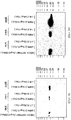
下記の表1は、インフルエンザAの亜型H5N1、H1N1、およびH3N2のPL領域を列挙する。H5N1は、最も臨床的に関連性のある病原性株の亜型である。H1N1およびH3N2は、最も臨床的に関連性のある季節性インフルエンザAの亜型である。表はまた、様々なPDZドメインが、示されたPLに結合するか否かをも示す。インフルエンザAの病原性および季節性亜型の差次的な検出に関して、PDZドメインを選択するために表が使用され得る。例えばPSD95ドメインは、インフルエンザAの病原性亜型を検出するのに有用であり、かつINADLドメイン8は、インフルエンザAの季節性亜型を検出するのに有用である。PSD95ドメインは、PSD95のPDZドメイン1、2、および3のうちの任意のもの、またはそれらの組み合わせであり得る。好ましい検出試薬は、PSD95中の3コピーのPSD95ドメイン2から形成されるタンパク質である。即ち、そのPDZドメインに隣接するPSD95のセグメントによって分散された、三つのタンデムコピーである。そのようなタンパク質において、PSD95ドメイン2のコピーのうちの二つは、PSD95の天然のドメイン1および3に有効に取って代わるものである。別の好ましい検出試薬は、PSD95のPDZドメイン1、2、および3を含むタンパク質である。
II. PDZ Domain for Detection of Influenza A Table 1 below lists the PL regions of influenza A subtypes H5N1, H1N1, and H3N2. H5N1 is the most clinically relevant subtype of pathogenic strains. H1N1 and H3N2 are the most clinically relevant seasonal influenza A subtypes. The table also shows whether the various PDZ domains bind to the indicated PL. A table can be used to select PDZ domains for differential detection of influenza A pathogenicity and seasonal subtypes. For example, PSD95 domain is useful for detecting pathogenic subtypes of influenza A, and INADL domain 8 is useful for detecting seasonal subtypes of influenza A. The PSD95 domain can be any of
緩衝液および温度のようなアッセイ条件は、特定の株の検出または異なる株間の差異の検出が有利になるよう、結合を調節するために使用され得る。表中で使用されるシンボルは、以下を意味する:++相対的に強い結合、+検出可能であるが相対的に弱い結合、+/-検出可能であるが相対的に弱い結合または検出不可能な結合、-検出不可能な結合。検出可能な結合とは、実験誤差による無作為な変動を考慮しても、示された亜型のNS1を欠如する対照と比べて、示された亜型のNS1を含む試料中の結合からのシグナルが、有意な程度により大きいことを意味する。検出不可能な結合とは、示された亜型のNS1を含む試料への結合からのシグナルが、示された亜型のNS1を欠如する対照におけるシグナルから見て誤差範囲内であることを意味する。 Assay conditions such as buffer and temperature can be used to modulate binding such that detection of a particular strain or detection of differences between different strains is advantageous. The symbols used in the table mean the following: ++ relatively strong binding, + detectable but relatively weak binding, +/- detectable but relatively weak binding or no detection Possible bond,-Undetectable bond. Detectable binding refers to binding from a sample containing NS1 of the indicated subtype compared to a control lacking NS1 of the indicated subtype, taking into account random variation due to experimental error. It means that the signal is significantly greater. Undetectable binding means that the signal from binding to a sample containing the indicated subtype of NS1 is within an error range relative to the signal in a control lacking the indicated subtype of NS1. To do.
インフルエンザAを亜型分類する好ましい様式においては、INDAL PDZドメイン8との組み合わせにおいて、表中に示されるようなPSD95由来のPDZを使用する。一般的な法則として、INDALドメインの結合、またはINADLドメインのものよりも有意により強い(即ち、実験誤差を超えてより強い)PSD95ドメインの結合が存在しない場合、PSD95ドメインの検出可能な結合によって、インフルエンザAの亜型がH5N1(病原性)であることが示唆される。逆に、試料に対するPSD95ドメインの検出可能な結合、または試料に対するPSD95のものよりも有意により強い試料に対するINADLドメインの結合が存在しない場合、試料に対するINADLドメインの検出可能な結合によって、試料がインフルエンザAの亜型H1N1またはH3N2(両方とも季節性インフルエンザ)を含むことが示唆される。検出不可能な結合と比較して、試料に対するPSD95ドメイン2の検出可能ではあるが弱い結合によって、表中に示されるようにH1N1がH3N2と区別される。試料に対するINADLの結合と比較して、試料に対するPSD95ドメイン1、2、および3の検出可能であるが相対的に弱い結合によっても、亜型がH1N1であることが示唆される。
In a preferred mode of subtyping influenza A, a PSD95-derived PDZ as shown in the table is used in combination with INDAL PDZ domain 8. As a general rule, in the absence of PSD95 domain binding, which is significantly stronger than that of the INDAL domain, or significantly stronger than that of the INADL domain (ie, stronger than experimental error), by detectable binding of the PSD95 domain, It is suggested that the influenza A subtype is H5N1 (pathogenic). Conversely, if there is no detectable binding of the PSD95 domain to the sample, or the binding of the INADL domain to the sample is significantly stronger than that of PSD95 to the sample, the detectable binding of the INADL domain to the sample causes the sample to become influenza A Of subtypes H1N1 or H3N2 (both seasonal influenza). Compared to undetectable binding, detectable but weak binding of
III.インフルエンザAおよびインフルエンザBの検出に関する抗体
本発明は、インフルエンザAの検出のための汎特異的抗体のコレクションを提供する。インフルエンザAに対する汎特異的抗体は、少なくとも2つ、3つ、もしくは5つまたは全てのまたは実質的に全ての公知のインフルエンザA株由来のNS1タンパク質に特異的に結合する。同様に、インフルエンザBに対する汎特異的抗体は、少なくとも2つ、3つ、もしくは5つまたは全てのまたは実質的に全ての公知のインフルエンザB株由来のNS1タンパク質に特異的に結合する。
III. Antibodies for Detection of Influenza A and Influenza B The present invention provides a collection of pan-specific antibodies for detection of influenza A. A panspecific antibody against influenza A specifically binds to NS1 protein from at least 2, 3, or 5 or all or substantially all known influenza A strains. Similarly, panspecific antibodies against influenza B specifically bind to NS1 protein from at least 2, 3, or 5 or all or substantially all known influenza B strains.
汎特異的抗体は、数的に規定されるエピトープを参照することによるか、または例示的な抗体を参照することにより規定される競合群によるかのいずれかによって定義され得る。インフルエンザAに対して、汎特異的抗体は、好ましくは図1Aまたは図2の残基8〜21位、9〜20位、29〜38位、もしくは45〜49位内のエピトープに特異的に結合する。この配列中のXは、任意のアミノ酸であり得るが、好ましくは、インフルエンザの株由来のNS1タンパク質中の対応する位置を占めるアミノ酸であり、かつより好ましくは、少なくとも二つまたは好ましくは全ての公知のインフルエンザA株由来の対応する位置を占めるコンセンサスアミノ酸である。インフルエンザAのコンセンサス配列は、図2中に提供される。いくつかの汎特異的抗体は、図1Aの残基9〜11位または13〜16位内のエピトープに特異的に結合する。 A panspecific antibody can be defined either by reference to a numerically defined epitope or by a competition group defined by reference to an exemplary antibody. For influenza A, the pan-specific antibody preferably specifically binds to an epitope within residues 8-21, 9-20, 29-38, or 45-49 of FIG. 1A or FIG. To do. X in this sequence can be any amino acid, but is preferably an amino acid occupying the corresponding position in the NS1 protein from an influenza strain, and more preferably at least two or preferably all known Is a consensus amino acid occupying the corresponding position from the influenza A strain. A consensus sequence for influenza A is provided in FIG. Some pan-specific antibodies specifically bind to an epitope within residues 9-11 or 13-16 of FIG. 1A.
汎特異的抗体はまた、競合群によっても定義され得る;競合群内の抗体は、同一抗原(即ち、インフルエンザAまたはインフルエンザBのSN1タンパク質)に対する特異的な結合に関して互いに競合する。表2は、インフルエンザAのNS1タンパク質に結合する汎特異的抗体の競合群を示す。 A panspecific antibody can also be defined by a competitor group; antibodies within a competitor group compete with each other for specific binding to the same antigen (ie, influenza A or influenza B SN1 protein). Table 2 shows competing groups of pan-specific antibodies that bind to the influenza A NS1 protein.
各群は、群中の他の抗体(第3列)と競合する原型抗体(第2列)によって規定される。A群、B群、およびC群が好ましい。これらの抗体の全ては、少なくともH5N1、H1N1、およびH3N2株由来のNS1タンパク質に結合する。異なる群の抗体は、互いに競合しない。 Each group is defined by a prototype antibody (second column) that competes with the other antibodies in the group (third column). Groups A, B, and C are preferred. All of these antibodies bind to NS1 protein from at least H5N1, H1N1, and H3N2 strains. Different groups of antibodies do not compete with each other.
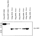
表3は、インフルエンザのH5N1病原性株のサンドイッチ検出での使用のための、好ましい抗体を示す。そのようなアッセイにおいて、好ましい捕捉物質はPSD95ドメイン1、2、および3であり、かつ好ましい検出物質は、好ましくはA群、またはC群もしくはD群由来の抗体である。
Table 3 shows preferred antibodies for use in sandwich detection of influenza H5N1 pathogenic strains. In such assays, preferred capture agents are
表4は、インフルエンザBのNS1タンパク質に結合する汎特異的抗体に関する競合群を示す。 Table 4 shows competition groups for panspecific antibodies that bind to the influenza B NS1 protein.
表5は、競合する捕捉抗体および検出抗体の対を示す。検出抗体が表の第一行に示され、かつ捕捉抗体が第一列に示される。競合は、Cで示される。 Table 5 shows competing capture and detection antibody pairs. The detection antibody is shown in the first row of the table and the capture antibody is shown in the first column. The conflict is indicated by C.
インフルエンザB型に対する汎特異的抗体はまた、図2中に示されるインフルエンザB株由来のNS1タンパク質のコンセンサス配列に関連するエピトープ特異性によっても記載され得る。好ましい抗体は、図2の残基10〜28位、40〜45位、50〜57位、67〜74位、84〜100位、154〜159位、169〜173位、185〜191位、212〜224位、226〜240位内に生じるエピトープに、およびとりわけ、インフルエンザB型の異なる株間で不変の残基を示すその下線領域に、特異的に結合する。下線の施されていない上記領域のうちの一つに含まれる残基(即ち、インフルエンザB型株間で変動する)は、図2中に示されるその位置を占めるコンセンサス残基によって、またはインフルエンザB型の任意の株においてその位置を占める残基によって占められ得る。 A panspecific antibody against influenza B can also be described by the epitope specificity associated with the consensus sequence of NS1 protein from the influenza B strain shown in FIG. Preferred antibodies are residues 10-28, 40-45, 50-57, 67-74, 84-100, 84-100, 154-159, 169-173, 185-191, 212 of FIG. It binds specifically to epitopes occurring within positions 224, 226 to 240, and in particular to its underlined regions that represent residues that are invariant among different strains of influenza B. Residues included in one of the above ununderlined regions (ie, vary between influenza B strains) are either due to the consensus residue occupying that position as shown in FIG. 2, or influenza B Can be occupied by residues occupying that position in any strain.
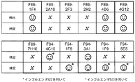
サンドイッチアッセイでの使用のための、インフルエンザBのNS1に対する抗体の好ましい組み合わせは、図13中の笑顔で示される。 Preferred combinations of antibodies against influenza B NS1 for use in sandwich assays are indicated by smiles in FIG.
使用される抗体は、非ヒト抗体、ヒト化抗体、キメラ抗体、ベニア化(veneered)抗体またはヒト抗体であり得る。そのような抗体の使用は、試料中のHAMAまたはヘテロ親和性抗体の存在による偽陽性または偽陰性を回避する際に有利である(米国特許第6,680,209号)。上の表中に列挙される抗体のヒト化、キメラ、またはベニア化様式が好ましい。そのような抗体はまた、インフルエンザAまたはBの処置において、薬学的物質としても使用され得る。抗体は、抗体の種類に応じた標準的手法によって、タンパク質の抗原含有断片から作製され得る(例えば、(各々が全ての目的に関して、参照により組み入れられる)Kohler, et al., Nature 256:495, (1975);ならびにHarlow & Lane, Antibodies, A Laboratory Manual (C.S.H.P., NY, 1988)、Queen et al., Proc. Natl. Acad. Sci. USA 86:10029-10033 (1989)、および国際公開公報第90/07861号;Dower et al., 国際公開公報第91/17271号およびMcCafferty et al., 国際公開公報第92/01047号を参照のこと)。 The antibody used can be a non-human antibody, a humanized antibody, a chimeric antibody, a veneered antibody or a human antibody. Use of such antibodies is advantageous in avoiding false positives or false negatives due to the presence of HAMA or heterophilic antibodies in the sample (US Pat. No. 6,680,209). Preferred are humanized, chimeric, or veneered modes of antibodies listed in the table above. Such antibodies can also be used as pharmaceutical agents in the treatment of influenza A or B. Antibodies can be generated from antigen-containing fragments of a protein by standard techniques depending on the type of antibody (eg, Kohler, et al., Nature 256: 495, each incorporated by reference for all purposes). (1975); and Harlow & Lane, Antibodies, A Laboratory Manual (CSHP, NY, 1988), Queen et al., Proc. Natl. Acad. Sci. USA 86: 10029-10033 (1989), and International Publication No. 90/07861; see Dower et al., WO 91/17271 and McCafferty et al., WO 92/01047).
免疫化は、汎特異的抗体を生成するために、インフルエンザAもしくはBの複数株で免疫することによって、または一つの株で免疫し、かつ他の株で追加免疫することによって、バイアスをかけることが可能である。あるいは、インフルエンザAの高度に保存された領域(例えば、8〜21位、9〜20位、29〜38位もしくは45〜49位、またはSEQ ID NO:1のこれらの任意の少なくとも三個の連続したアミノ酸)、またはB NS1(例えば、SEQ ID NO:4の10〜28位、40〜45位、50〜57位、67〜74位、84〜100位、154〜159位、169〜173位、185〜191位、212〜224位もしくは226〜240位、またはその少なくとも三個の連続したアミノ酸の部分断片)由来の断片が、免疫抗原として使用され得る。逆に、単一特異性抗体を生成するためには、単一株のNS1または非保存的領域由来のNS1の断片(例えば、インフルエンザAのPL領域)での免疫化が好ましい。 Immunization is biased by immunizing with multiple strains of influenza A or B or by immunizing with one strain and boosting with another strain to generate pan-specific antibodies Is possible. Alternatively, a highly conserved region of influenza A (eg, positions 8-21, 9-20, 29-38 or 45-49, or any at least three consecutive of these in SEQ ID NO: 1 Amino acids), or B NS1 (for example, SEQ ID NO: 4, positions 10-28, 40-45, 50-57, 67-74, 84-100, 154-159, 169-173) 185-191, 212-224 or 226-240, or a partial fragment of at least three consecutive amino acids thereof), can be used as an immunizing antigen. Conversely, to generate monospecific antibodies, immunization with a single strain of NS1 or a fragment of NS1 from a non-conservative region (eg, the influenza A PL region) is preferred.
「抗体」または「免疫グロブリン」という用語は、無傷の抗体およびその結合断片を包含するように使用される。典型的に、断片は、個別の重鎖、軽鎖、Fab、Fab'、F(ab')2、Fabc、およびFvを含む抗原断片への特異的な結合に関して、派生元の無傷の抗体と競合する。断片は、組換えDNA技術によって、または無傷の免疫グロブリンの酵素的な分離もしくは化学的な分離によって産生される。「抗体」という用語はまた、他のタンパク質に化学的に結合しているか、または他のタンパク質との融合タンパク質として発現される、一つまたは複数の免疫グロブリン鎖も包含する。「抗体」という用語は、二重特異性抗体も包含する。 The terms “antibody” or “immunoglobulin” are used to encompass intact antibodies and binding fragments thereof. Typically, a fragment is derived from an intact antibody from which it is derived for specific binding to an antigenic fragment comprising individual heavy chains, light chains, Fab, Fab ′, F (ab ′) 2, Fabc, and Fv. Competing. Fragments are produced by recombinant DNA technology or by enzymatic or chemical separation of intact immunoglobulins. The term “antibody” also includes one or more immunoglobulin chains that are chemically linked to other proteins or expressed as fusion proteins with other proteins. The term “antibody” also encompasses bispecific antibodies.
別途示されていない限り、本出願において記載される抗体は、ハイブリドーマから産生されるマウス抗体である。 Unless indicated otherwise, the antibodies described in this application are murine antibodies produced from hybridomas.
IV.他の結合物質
NS1タンパク質を検出する際の使用のためには汎特異的抗体が好ましいが、インフルエンザのNS1に対して特異的な親和性を有する任意の結合物質が、抗体の代理物質として使用され得る。代理物質は、インフルエンザAまたはB由来のNS1に対してスクリーニングされた、無作為のファージディスプレイライブラリー由来のペプチドを包含する。代理物質はまた、アプタマーも包含する。アプタマーは、膨大な集団の無作為配列からインビトロで選択され、結合ポケットを形成することによって特異的な結合を認識するRNAまたはDNA分子である。アロステリックリボザイムは、活性部位から離れて位置するアプタマードメインへのエフェクター分子の結合によって活性が調節されるRNA酵素である。これらのRNAは、特異的なエフェクターの存在または不在によって制御される精密な分子スイッチとして作用する。アプタマーは核酸、タンパク質、さらには生物体全体に結合することができる。アプタマーは抗体とは異なるものの、様々な診断様式において抗体の特性を模倣する。したがって、アプタマーは、汎特異的抗体に代わる代理物質として使用することができる。
IV. Other binding substances
Although panspecific antibodies are preferred for use in detecting NS1 protein, any binding agent with specific affinity for influenza NS1 can be used as a surrogate for the antibody. Surrogate materials include peptides from random phage display libraries screened against NS1 from influenza A or B. Surrogate materials also include aptamers. Aptamers are RNA or DNA molecules that are selected in vitro from a vast population of random sequences and recognize specific binding by forming a binding pocket. Allosteric ribozymes are RNA enzymes whose activity is regulated by the binding of effector molecules to aptamer domains located away from the active site. These RNAs act as precise molecular switches that are controlled by the presence or absence of specific effectors. Aptamers can bind to nucleic acids, proteins, and even whole organisms. Although aptamers are different from antibodies, they mimic the properties of antibodies in various diagnostic modalities. Thus, aptamers can be used as surrogates instead of panspecific antibodies.
同様に、NS1のPL領域を検出するためにはPDZドメインが好ましいが、インフルエンザAの特定のNS1タンパク質のPL領域へ特異的に結合する抗体が、その領域に特異的に結合するPDZドメインに代わる代理物質として使用され得る。 Similarly, a PDZ domain is preferred for detecting the PL region of NS1, but an antibody that specifically binds to the PL region of a specific NS1 protein of influenza A replaces the PDZ domain that specifically binds to that region. Can be used as surrogate material.
V.検出方法
1.インフルエンザA亜型の検出
本発明は、上に議論されるように、インフルエンザAの病原性亜型と季節性亜型を区別する方法を提供する。好ましい様式は、捕捉試薬として一つまたは複数のPDZドメインを使用し、かつ検出試薬として一つまたは複数の汎特異的抗体を使用する。上に議論されるように、PSD95のドメイン2もしくはドメイン1、2、および3、および/またはINADLのドメイン8をPDZドメイン検出試薬として使用することが好ましい。PDZ捕捉試薬と共に使用するための好ましい汎特異的抗体は、検出抗体としての汎特異的抗体F68 8E6(もしくはそれと競合する抗体)、またはF68 4B2(もしくはそれと競合する抗体)である。同一または異なる汎特異的抗体が、同一のアッセイにおいて異なるPDZドメインと共に使用され得る。
V. Detection method
1. Detection of Influenza A Subtypes The present invention provides a method for distinguishing between influenza A pathogenic and seasonal subtypes, as discussed above. A preferred mode uses one or more PDZ domains as capture reagents and one or more panspecific antibodies as detection reagents. As discussed above,
2.汎特異的抗体を用いたインフルエンザAの検出
本発明はまた、インフルエンザAの亜型間を必ずしも区別しないが、インフルエンザAとインフルエンザB(またはC)を区別し得る様式で、インフルエンザAを検出する方法も提供する。そのような方法は、異なるエピトープに結合するインフルエンザAのNS1タンパク質に対する少なくとも二つの汎特異的抗体を使用して遂行される。二つの汎特異的抗体は、上に記載されるような数的に規定される異なるエピトープに特異的に結合するか、または異なる競合群から選択され得る。検出は、好ましくは、下記により詳細に記載されるようなサンドイッチまたはラテラルフロー様式を使用して遂行される。インフルエンザAを検出するための、一つの好ましい抗体の組み合わせは、捕捉抗体としてF64 3H3(またはそれと競合する抗体)、および検出抗体としてF80 3D5(またはそれと競合する抗体)である。別の好ましい組み合わせは、捕捉抗体としてF68 4H9(またはそれと競合する抗体)、および検出抗体としてF68 8E6(またはそれと競合する抗体)である。
2. Detection of influenza A using pan-specific antibodies The present invention also does not necessarily distinguish between influenza A subtypes, but a method of detecting influenza A in a manner that can distinguish between influenza A and influenza B (or C) Also provide. Such a method is accomplished using at least two pan-specific antibodies to the influenza A NS1 protein that bind to different epitopes. The two panspecific antibodies can specifically bind to differently defined different epitopes as described above or can be selected from different competitor groups. Detection is preferably accomplished using a sandwich or lateral flow format as described in more detail below. One preferred antibody combination for detecting influenza A is F64 3H3 (or an antibody that competes therewith) as a capture antibody and F80 3D5 (or an antibody that competes therewith) as a detection antibody. Another preferred combination is F68 4H9 (or an antibody that competes with it) as a capture antibody and F68 8E6 (or an antibody that competes with it) as a detection antibody.
二つの汎特異的抗体を使用するインフルエンザAの検出は、上の(1)において記載されるような、インフルエンザA亜型の差次的検出と組み合わせることが可能である。そのようなアッセイによって、インフルエンザAが存在するか否か、およびもしそうであるならば、病原性亜型または季節性亜型が存在するか否かの両方が示される。非亜型特異的アッセイおよび亜型特異的アッセイが、別個にまたは組み合わせて遂行され得る。アッセイを組み合わせることに関する一つの適切な様式は、非亜型特異的解析における使用のための抗体捕捉試薬として、差次的解析における使用のためのPDZドメインを同一固相の異なる領域に付着させることである。試料におけるNS1タンパク質へのPDZドメインの結合は汎特異的抗体を使用して検出され得る。NS1タンパク質へのPDZドメインの結合を検出するために使用される汎特異的検出抗体は、非亜型特異的解析のために使用される汎特異的抗体と同一であってもよく、または異なっていてもよい。したがって、好ましい様式においては、PSD95ドメイン、INADLドメイン8、およびインフルエンザAに対する汎特異的捕捉抗体を、支持体の異なる領域に付着させ、かつ上に議論されるように、もし試料中に存在する場合には、インフルエンザA NS1タンパク質への各捕捉試薬の結合を検出するように、共通の汎特異的検出抗体(汎特異的捕捉抗体とは異なるエピトープに結合する)が使用される。 Detection of influenza A using two panspecific antibodies can be combined with differential detection of influenza A subtypes as described in (1) above. Such an assay indicates both whether influenza A is present and, if so, whether a pathogenic or seasonal subtype is present. Non-subtype-specific and subtype-specific assays can be performed separately or in combination. One suitable format for combining assays is to attach PDZ domains for use in differential analysis to different regions of the same solid phase as antibody capture reagents for use in non-subtype specific analysis. It is. Binding of the PDZ domain to the NS1 protein in the sample can be detected using a panspecific antibody. The panspecific detection antibody used to detect the binding of the PDZ domain to the NS1 protein may be the same as or different from the panspecific antibody used for non-subtype specific analysis. May be. Thus, in a preferred manner, PSD95 domain, INADL domain 8, and pan-specific capture antibodies against influenza A are attached to different regions of the support and, if present in the sample, as discussed above Uses a common pan-specific detection antibody (which binds to a different epitope than the pan-specific capture antibody) so as to detect the binding of each capture reagent to the influenza A NS1 protein.
3.汎特異的抗体を用いたインフルエンザBの検出
上に記載されるようなインフルエンザAのNS1タンパク質の検出に関して記載されるアッセイと類似の様式で、インフルエンザBのNS1タンパク質に対する第一および第二の汎特異的抗体を使用して、インフルエンザBが検出され得る。そのような方法は、異なるエピトープに結合するインフルエンザBのNS1タンパク質に対する少なくとも二つの汎特異的抗体を使用して遂行される。二つの汎特異的抗体は、上に記載されるような、数的に規定される異なるエピトープに結合するか、または異なる競合群から選択され得る。検出は、好ましくは、下記に詳細に記載されるようなサンドイッチまたはラテラルフロー様式を使用して遂行される。インフルエンザBの検出のための好ましい抗体の組み合わせは、捕捉抗体としてF89 6B5(もしくはそれと競合する抗体)、および検出もしくは検出抗体としてF94 3A1(もしくはそれと競合する抗体)またはF94 1F9(もしくはそれと競合する抗体)を使用する。抗体の競合は、インフルエンザBのNS1タンパク質への結合によって決定される。
3. Detection of influenza B using pan-specific antibodies First and second pan-specificity for influenza B NS1 protein in a manner similar to the assay described for detection of influenza A NS1 protein as described above Influenza B can be detected using specific antibodies. Such a method is accomplished using at least two pan-specific antibodies to the influenza B NS1 protein that bind to different epitopes. The two panspecific antibodies can bind to differently defined different epitopes, as described above, or can be selected from different competitor groups. Detection is preferably accomplished using a sandwich or lateral flow format as described in detail below. Preferred antibody combinations for detection of influenza B are F89 6B5 (or an antibody that competes with it) as a capture antibody and F94 3A1 (or an antibody that competes with it) or F94 1F9 (or an antibody that competes with it) as a detection or detection antibody ). Antibody competition is determined by the binding of influenza B to the NS1 protein.
4.インフルエンザAおよびインフルエンザBの組み合わせ検出
上に記載される三種類のアッセイの各々は、インフルエンザA(非亜型特異的)、インフルエンザB(非亜型特異的)、インフルエンザA(病原性亜型)およびインフルエンザA(季節性亜型)の検出が可能なアッセイを提供するために、効果的に組み合わせることが可能である。個々のアッセイは、別個にまたは一緒に遂行され得る。アッセイを組み合わせるための一つの適切な様式は、インフルエンザAのNS1タンパク質に対する汎特異的捕捉抗体、インフルエンザBのNS1タンパク質に対する汎特異的捕捉抗体、インフルエンザAの病原性亜型のPLに対するPDZドメイン(例えば、上に議論されるようなPSD95ドメイン)、およびインフルエンザAの季節性亜型のPLに対するPDZドメイン(例えば、INADL 8ドメイン)を単一の支持体に付着させることである。支持体は、対象由来の試料、および少なくとも二つの汎特異的検出抗体と接触させる。一方の検出抗体は、インフルエンザAのNS1タンパク質に対する捕捉抗体とは異なるエピトープで、インフルエンザAのNS1タンパク質に特異的に結合する。他方の検出抗体は、インフルエンザBのNS1タンパク質に対する捕捉抗体とは異なるエピトープで、インフルエンザBのNS1タンパク質に特異的に結合する。形成する複合体は、インフルエンザAおよび/またはBが存在するか否か、ならびにもしインフルエンザAが存在する場合には、インフルエンザAが病原性であるか、または季節性であるかを示す。
Four. Influenza A and influenza B combination detection Each of the three assays described above includes influenza A (non-subtype specific), influenza B (non-subtype specific), influenza A (pathogenic subtype) and It can be combined effectively to provide an assay capable of detecting influenza A (seasonal subtype). Individual assays can be performed separately or together. One suitable format for combining the assays is a pan-specific capture antibody against the NS1 protein of influenza A, a pan-specific capture antibody against the NS1 protein of influenza B, a PDZ domain against the PL of the influenza A pathogenic subtype (eg, , PSD95 domains as discussed above), and PDZ domains for influenza A seasonal subtype PL (eg, INADL 8 domain) are attached to a single support. The support is contacted with a sample from the subject and at least two panspecific detection antibodies. One detection antibody specifically binds to the influenza A NS1 protein at a different epitope than the capture antibody to the influenza A NS1 protein. The other detection antibody specifically binds to the influenza B NS1 protein at a different epitope than the capture antibody to the influenza B NS1 protein. The complex that forms indicates whether influenza A and / or B is present, and if influenza A is present, whether influenza A is pathogenic or seasonal.
VI.検出様式
本発明は、様々な異なる種類の生物学的試料中のインフルエンザAおよび/またはBウイルスを同定するためのアッセイ方法において有用な、診断的捕捉および検出試薬を提供する。そのような様式は、免疫沈降反応、ウェスタンブロッティング、ELISA、放射免疫検定、競合アッセイおよび免疫測定(immunometric)アッセイを包含する。Harlow & Lane, Antibodies, A Laboratory Manual(CSHP NY, 1988);米国特許第3,791,932号;第3,839,153号;第3,850,752号;第3,879,262号;第4,034,074号;第3,791,932号;第3,817,837号;第3,839,153号;第3,850,752号;第3,850,578号;第3,853,987号;第3,867,517号;第3,879,262号;第3,901,654号;第3,935,074号;第3,984,533号;第3,996,345号;第4,034,074号;および第4,098,876号を参照のこと。
VI. Detection Formats The present invention provides diagnostic capture and detection reagents useful in assay methods for identifying influenza A and / or B viruses in a variety of different types of biological samples. Such modalities include immunoprecipitation reactions, Western blotting, ELISA, radioimmunoassay, competition assays and immunometric assays. Harlow & Lane, Antibodies, A Laboratory Manual (CSHP NY, 1988); U.S. Patent Nos. 3,791,932; 3,839,153; 3,850,752; 3,879,262; 4,034,074; 3,791,932; 3,850,578; 3,853,987; 3,867,517; 3,879,262; 3,901,654; 3,935,074; 3,984,533; 3,996,345; 4,034,074; and 4,098,876.
免疫測定アッセイまたはサンドイッチアッセイが、好ましい様式である(米国特許第4,376,110号、第4,486,530号、第5,914,241号、および5,965,375号を参照のこと)。そのようなアッセイは、捕捉物質として、固相に固定化された一つの抗体もしくは抗体集団またはPDZドメインを、および検出物質として、溶液中の他方の抗体もしくは抗体集団またはPDZドメインを使用する。上に議論されるように、捕捉PDZドメインと検出抗体の組み合わせ、またはその逆の組み合わせが、インフルエンザAの検出に関して好ましい。典型的には、検出物質が標識される。もし抗体集団が使用される場合には、集団は典型的には標的抗原内の異なるエピトープ特異性で結合する抗体を含む。従って、同一集団が、捕捉物質および検出物質の両方に関して使用され得る。もしモノクローナル抗体が、検出および検出物質として使用される場合には、異なる結合特異性を有する第一および第二のモノクローナル抗体が、固相および液相に関して使用される。捕捉物質および検出物質を、いずれかの順序か、または同時に標的抗原と接触させ得る。捕捉物質を最初に接触させる場合には、アッセイはフォワードアッセイと呼ばれる。逆に、検出物質を最初に接触させる場合には、アッセイはリバースアッセイと呼ばれる。標的を捕捉物質および検出物質の両方に同時に接触させる場合には、アッセイは同時アッセイと呼ばれる。試料を捕捉抗体および検出抗体と接触させた後、試料は、通常は約10分間〜約24時間まで変動する期間の間、通常は約1時間の間、インキュベートされる。続いて、検出物質に特異的に結合していない試料の要素を除去するために、洗浄段階が遂行され得る。捕捉物質および検出物質が個別の段階で結合する際には、いずれかの結合段階の後、または両方の結合段階の後に洗浄が遂行され得る。典型的には、標識された溶液抗体の結合により固相に連結された標識を検出することによって、洗浄後に結合が定量化される。通常は、所定の捕捉物質および検出物質の対、ならびに所定の反応条件に関して、既知濃度の標的抗原を含む試料から補正曲線が作成される。続いて、試験されている試料中の抗原の濃度が、補正曲線から補間によって判読される。分析物は、平衡状態で結合して標識された溶液抗体の量から、または平衡状態に到達する前の時系列で結合して標識された溶液抗体の動態測定値のいずれかによって、測定され得る。そのような曲線の勾配は、試料中の標的の濃度の尺度である。 An immunometric assay or sandwich assay is the preferred format (see US Pat. Nos. 4,376,110, 4,486,530, 5,914,241, and 5,965,375). Such assays use one antibody or antibody population or PDZ domain immobilized on a solid phase as the capture agent and the other antibody or antibody population or PDZ domain in solution as the detection agent. As discussed above, a combination of capture PDZ domain and detection antibody, or vice versa, is preferred for detection of influenza A. Typically, the detection substance is labeled. If an antibody population is used, the population typically includes antibodies that bind with different epitope specificities within the target antigen. Thus, the same population can be used for both capture and detection substances. If monoclonal antibodies are used as detection and detection substances, first and second monoclonal antibodies with different binding specificities are used for the solid and liquid phases. The capture substance and the detection substance can be contacted with the target antigen in either order or simultaneously. If the capture agent is contacted first, the assay is called a forward assay. Conversely, if the detection substance is contacted first, the assay is called a reverse assay. If the target is contacted with both the capture agent and the detection agent simultaneously, the assay is called a simultaneous assay. After contacting the sample with the capture antibody and the detection antibody, the sample is typically incubated for a period varying from about 10 minutes to about 24 hours, usually about 1 hour. Subsequently, a washing step may be performed to remove sample elements that are not specifically bound to the detection substance. When the capture substance and the detection substance are combined in separate steps, washing can be performed after either binding step or after both binding steps. Typically, binding is quantified after washing by detecting the label linked to the solid phase by binding of labeled solution antibody. Typically, a correction curve is generated from a sample containing a known concentration of target antigen for a given capture and detection material pair and for a given reaction condition. Subsequently, the concentration of the antigen in the sample being tested is read by interpolation from the correction curve. Analytes can be measured either from the amount of solution antibody bound and labeled at equilibrium, or by kinetic measurements of solution antibody bound and labeled in time series before reaching equilibrium. . The slope of such a curve is a measure of the concentration of target in the sample.
競合アッセイも使用され得る。いくつかの方法において、試料中の標的抗原は、外因的に供給され標識された標的抗原と、抗体またはPDZ検出試薬への結合に関して競合する。検出試薬に結合して標識された標的抗原の量は、試料中の標的抗原の量に反比例している。検出試薬は、検出前に、試料から結合複合体を分離することを容易にするために固定化され得るか(不均一アッセイ)、または均一アッセイ様式において実施される際には、分離は不必要である場合がある。他の方法においては、検出試薬が標識される。検出試薬が標識される際に、その結合部位は、試料における標識抗原への結合に関して、および例えば、固相に固定化された標的抗原であり得る外因的に供給された形状の標的抗原への結合に関して競合する。さらに別の競合様式において、標識された検出試薬と同一の標的抗原に結合する試料中の抗体を検出するために、標識された検出試薬はまた使用され得る。上の様式の各々において、検出試薬は、アッセイされる標的と概ね同一濃度で、限定量(limiting amount)存在する。 Competitive assays can also be used. In some methods, the target antigen in the sample competes with the exogenously supplied and labeled target antigen for binding to the antibody or PDZ detection reagent. The amount of target antigen bound and labeled with the detection reagent is inversely proportional to the amount of target antigen in the sample. The detection reagent can be immobilized to facilitate separation of the bound complex from the sample prior to detection (heterogeneous assay) or separation is not required when performed in a homogeneous assay format. It may be. In other methods, the detection reagent is labeled. When the detection reagent is labeled, its binding site is related to binding to the labeled antigen in the sample and to, for example, an exogenously supplied form of the target antigen, which can be a target antigen immobilized on a solid phase. Compete for binding. In yet another competitive mode, labeled detection reagents can also be used to detect antibodies in a sample that bind to the same target antigen as the labeled detection reagent. In each of the above modes, the detection reagent is present in a limiting amount at approximately the same concentration as the target being assayed.
ラテラルフロー装置が、好ましい様式である。家庭用妊娠検査と同様に、ラテラルフロー装置は、特殊な生物学的製剤で処理しておいた検査条片に液体を適用することによって機能する。液体試料によって運搬されることで、対応する生物学的製剤で標識されたリンが条片を移動して、かつそれらが特定のゾーン内に達すると捕捉される。条片上に見られるリンシグナルの量は標的分析物の量に比例する。 A lateral flow device is the preferred mode. Similar to the home pregnancy test, the lateral flow device works by applying a liquid to a test strip that has been treated with a special biological product. By being transported by the liquid sample, the phosphorus labeled with the corresponding biological product moves through the strip and is captured when they reach a specific zone. The amount of phosphorus signal found on the strip is proportional to the amount of target analyte.
インフルエンザを含むことが疑われる試料を、ラテラルフロー装置へ加え、拡散によって試料を移動させ、ラインまたは着色ゾーンによってインフルエンザAの存在を示す。ラテラルフローは三つの特異的区画を含む固体支持体(例えば、ニトロセルロース膜)を典型的には含む:試料添加区画、NS1に対する一つまたは複数の抗体を含む捕捉区画、および一つまたは複数のゾーンを含み、各ゾーンが一つまたは複数の標識を含む、読み取り区画。ラテラルフローは、陽性対照および陰性対照も含み得る。したがって、例えばラテラルフロー装置は、次の通りに使用され得る:生物学的試料のアリコートを検査条片の一方の端に接触させ、続いて、例えば、ラテラルフローのような毛管現象により、タンパク質を検査条片上で移動させることによって、インフルエンザAおよび/またはB NS1タンパク質は、生物学的試料中のその他のウイルス性および細胞性タンパク質と分離される。一つまたは複数の抗体、および/またはアプタマーが、捕捉試薬および/または検出試薬として含まれる。ラテラルフローによる分離、検出、および定量のための方法ならびに装置は、例えば、参照により全体が本明細書に組み入れられる、米国特許第5,569,608号、第6,297,020号、および第6,403,383号などによって記載される。一つの例において、検査条片は、試料をロードするための近位領域(試料ローディング領域)、ならびにNS1タンパク質に対する抗体および緩衝試薬、ならびに移動する生物学的試料中の任意のインフルエンザB NS1タンパク質に対する抗体間の相互作用を確立するために適切な添加物質を含む遠位検査領域を含み得る。別の例においては、検査条片は、二つの異なるインフルエンザB亜型に由来するNS1に対する異なる抗体を含み、即ち、各々が異なるインフルエンザB分析物と特異的に相互作用することができる、二つの検査領域を含む。 A sample suspected of containing influenza is added to a lateral flow device, the sample is moved by diffusion, and the presence of influenza A is indicated by a line or colored zone. Lateral flow typically includes a solid support (eg, a nitrocellulose membrane) containing three specific compartments: a sample addition compartment, a capture compartment containing one or more antibodies to NS1, and one or more A reading section comprising zones, each zone comprising one or more indicators. Lateral flow can also include positive and negative controls. Thus, for example, a lateral flow device can be used as follows: an aliquot of a biological sample is brought into contact with one end of a test strip, followed by capillary action such as lateral flow, for example. By moving on the test strip, influenza A and / or B NS1 protein is separated from other viral and cellular proteins in the biological sample. One or more antibodies and / or aptamers are included as capture reagents and / or detection reagents. Methods and apparatus for separation, detection, and quantification by lateral flow are described, for example, by US Pat. Nos. 5,569,608, 6,297,020, and 6,403,383, which are incorporated herein by reference in their entirety. In one example, a test strip is against a proximal region for loading a sample (sample loading region), as well as antibodies and buffer reagents to NS1 protein, and any influenza B NS1 protein in a moving biological sample. A distal examination region can be included that contains appropriate additive materials to establish the interaction between the antibodies. In another example, the test strip includes two different antibodies against NS1 from two different influenza B subtypes, i.e., two that each can specifically interact with different influenza B analytes. Includes inspection area.
上記の方法における使用のための適切な検出可能な標識は、分光学的、光化学的、生化学的、免疫化学的、電気的、光学的、化学的または他の手段によって検出可能な任意の部分を含む。例えば、適切な標識は、標識されたストレプトアビジン結合体を用いた染色のためのビオチン、蛍光色素(例えば、フルオレセイン、テキサスレッド、ローダミン、緑色蛍光タンパク質等)、放射性標識(例えば、3H、125I、35S、14Cまたは32P)、酵素(例えば、西洋わさびペルオキシダーゼ、アルカリフォスファターゼ、およびELISAにおいて一般に使用される他の酵素)、ならびにコロイド金または色ガラスもしくはプラスチック(例えば、ポリスチレン、ポリプロピレン、ラテックスビーズ)のような比色分析用標識を含む。そのような標識の使用を記載する特許は、米国特許第3,817,837号;第3,850,752号;第3,939,350号;第3,996,345号;第4,277,437号;第4,275,149号;および第4,366,241号を包含する。Handbook of Fluorescent Probes and Research Chemicals (6th Ed., Molecular Probes, Inc., Eugene Oreg.)も参照のこと。放射性標識は、写真フィルムまたはシンチレーションカウンターを使用して検出することができ、蛍光マーカーは、発光を検出する光検出器を使用して検出することができる。酵素標識は、基質と共に酵素を提供し、かつ基質上の酵素の作用によって産生される反応産物を検出することによって典型的には検出され、かつ比色分析用標識は、単に有色の標識を可視化することによって検出される。 Suitable detectable labels for use in the above methods are any moiety detectable by spectroscopic, photochemical, biochemical, immunochemical, electrical, optical, chemical or other means including. For example, suitable labels include biotin for staining with labeled streptavidin conjugates, fluorescent dyes (eg, fluorescein, Texas red, rhodamine, green fluorescent protein, etc.), radioactive labels (eg, 3 H, 125 I, 35 S, 14 C or 32 P), enzymes (eg, horseradish peroxidase, alkaline phosphatase, and other enzymes commonly used in ELISA), and colloidal gold or colored glass or plastic (eg, polystyrene, polypropylene, Colorimetric labels such as latex beads). Patents describing the use of such labels include U.S. Pat. Nos. 3,817,837; 3,850,752; 3,939,350; 3,996,345; 4,277,437; 4,275,149; and 4,366,241. See also Handbook of Fluorescent Probes and Research Chemicals (6th Ed., Molecular Probes, Inc., Eugene Oreg.). The radioactive label can be detected using a photographic film or a scintillation counter, and the fluorescent marker can be detected using a photodetector that detects luminescence. Enzymatic labels are typically detected by providing the enzyme with the substrate and detecting the reaction product produced by the action of the enzyme on the substrate, and the colorimetric label simply visualizes the colored label To be detected.
試料中のインフルエンザB NS1タンパク質のレベルは、定量することができ、かつ/または対照と比較することができる。適切な陰性対照試料は、例えば、インフルエンザウイルス感染を有しないことが既知の個体などの、健康であることが既知である個体から得られる。特異性対照は、既知のインフルエンザAもしくはインフルエンザC感染を有する個体、またはインフルエンザ以外のウイルスに感染された個体から収集されてもよい。対照試料は、試験されている対象と遺伝学的に関係する個体に由来し得るが、遺伝学的に無関係な個体にも由来し得る。適切な陰性対照試料はまた、感染のより初期段階、即ち試験試料が収集される時点よりも、より早い時点の個体から収集された試料でもあり得る。インフルエンザBの組換えNS1が、陽性対照として使用され得る。 The level of influenza B NS1 protein in the sample can be quantified and / or compared to a control. A suitable negative control sample is obtained from an individual known to be healthy, for example, an individual known to have no influenza virus infection. Specificity controls may be collected from individuals with a known influenza A or influenza C infection, or individuals infected with a virus other than influenza. A control sample can be from an individual genetically related to the subject being tested, but can also be from an individual that is not genetically related. A suitable negative control sample can also be a sample collected from an individual at an earlier stage of infection, ie, an earlier time point than when the test sample is collected. Recombinant NS1 of influenza B can be used as a positive control.
様々な異なる可能な免疫アッセイ様式において、生物学的試料中のNS1レベルが、これらの抗原の検出を可能とするのに十分であることが、ウェスタンブロットによって示された。しかしながら、特定の生物学的試料中のNS1のレベルが、特定の免疫アッセイ様式における検出に関して限定的であると判明している場合には、その際は、生物学的試料中の生存ウイルスは、インビトロで細胞に感染させることによって増幅してもよく、即ちウイルス増幅された試料中のNS1タンパク質は、約6時間〜約12時間内に検出可能となるはずである。生物学的試料およびウイルス増殖された試料におけるNS1抗原の収量はまた、プロテアーゼ阻害剤およびプロテアソーム阻害剤を含むことによっても改善され得る。 In various different possible immunoassay formats, Western blots have shown that NS1 levels in biological samples are sufficient to allow detection of these antigens. However, if the level of NS1 in a particular biological sample is found to be limited for detection in a particular immunoassay format, then the live virus in the biological sample is It may be amplified by infecting the cells in vitro, ie the NS1 protein in the virus amplified sample should be detectable within about 6 hours to about 12 hours. The yield of NS1 antigen in biological and virus-grown samples can also be improved by including protease inhibitors and proteasome inhibitors.
VII.試料
検出可能な濃度のインフルエンザタンパク質、および好ましくはNS1を含むか、または含むであろうと考えられる対象由来の任意の生物学的試料が使用され得る。例えば、往々にして、試料は、インフルエンザを有するか、もしくは有することが疑われる、またはインフルエンザを有するリスクが上昇した(例えば、インフルエンザを有する他者との接触による)ヒトから得られる。使用され得る試料の例は、例えば、肺浸出物、細胞抽出物(呼吸系、鼻腔を裏打ちする上皮)、血液、粘液、および鼻腔スワブである。高濃度のNS1が、鼻腔スワブ中に見出され得る。したがって、NS1の同定のための好ましい試料は、鼻分泌物である。
VII. Any biological sample from a subject that contains or is likely to contain a sample detectable concentration of influenza protein and preferably NS1 may be used. For example, often samples are obtained from humans who have, are suspected of having influenza, or have an increased risk of having influenza (eg, by contact with others who have influenza). Examples of samples that can be used are, for example, lung exudates, cell extracts (respiratory system, epithelium lining the nasal cavity), blood, mucus, and nasal swabs. High concentrations of NS1 can be found in nasal swabs. Thus, a preferred sample for NS1 identification is nasal secretions.
NS1の抗体への結合は、0.03%および0.01%を含む、最大0.05%のSDSの存在下で起こる。したがって、鼻またはその他の身体分泌物が、ラテラルフロー様式において容易に用いられる可能性が低い場合は、SDSで処理してもよい。好ましくは、加えられるSDSの量は0.01%の最終濃度までであり、より好ましくは0.03%、さらにより好ましくは0.05%までである。 NS1 binding to the antibody occurs in the presence of up to 0.05% SDS, including 0.03% and 0.01%. Thus, if nasal or other body secretions are unlikely to be easily used in a lateral flow manner, they may be treated with SDS. Preferably, the amount of SDS added is up to a final concentration of 0.01%, more preferably 0.03%, even more preferably up to 0.05%.
VIII.診断用キットおよび治療用キット
本方法を実施するためのキットが提供される。このキットは、一つまたは複数の結合物質であって、典型的にはインフルエンザAおよび/もしくはBのNS1に特異的に結合する抗体、またはPDZドメインを包含する。本キットは、任意で、試薬、緩衝液、または実施例中で開示される付加組成物もしくは試薬の一つまたは複数を含む。キットはまた、生物学的試料中のその他の潜在的干渉物質からインフルエンザウイルスNS1を除去するための、装置またはシステムなどの手段も含み得る。本キットは、所望ならば、例えば、一つもしくは複数のアッセイ容器;一つもしくは複数の対照もしくは検量試薬;アッセイを実施するための一つもしくは複数の固相表面;または一つもしくは複数の緩衝液、添加物、または検出試薬もしくは抗体;例えば、アッセイの実施において用いられるべきそれぞれの成分の量、およびアッセイの結果の評価に関する目安を明示するための、添付文書ならびに/または容器のラベルなどの、インフルエンザAおよび/またはBを検出するためのキットの使用法を詳述する一つもしくは複数の印刷された説明書など、アッセイの実施に有用な一つまたは複数の様々な要素をさらに含み得る。本キットは、例えば、検査条片、サンドイッチELISA、ウェスタンブロットアッセイ、ラテックス凝集等を含む、様々な異なる種類のアッセイ様式を実施するために有用な要素を含み得る。
VIII. Diagnostic kits and therapeutic kits Kits for carrying out the method are provided. The kit includes one or more binding agents, typically antibodies or PDZ domains that specifically bind to influenza A and / or B NS1. The kit optionally includes one or more of reagents, buffers, or additional compositions or reagents disclosed in the Examples. The kit may also include means such as a device or system for removing influenza virus NS1 from other potential interfering substances in the biological sample. If desired, the kit may be, for example, one or more assay vessels; one or more controls or calibration reagents; one or more solid phase surfaces for performing the assay; or one or more buffers. Fluids, additives, or detection reagents or antibodies; for example, package inserts and / or container labels to clearly indicate the amount of each component to be used in the performance of the assay and the assessment of assay results May further include one or more various elements useful for performing the assay, such as one or more printed instructions detailing the use of the kit to detect influenza A and / or B . The kit may include elements useful for performing a variety of different types of assay formats, including, for example, test strips, sandwich ELISAs, Western blot assays, latex agglutination, and the like.
IX.抗体アレイ
本発明は、異なる領域に固定化された抗体および/またはPDZドメインのアレイを、さらに提供する。そのようなアレイは、アレイの異なる領域内において、各々がインフルエンザAおよび/またはBのNS1に関して特異性を有する複数の異なる抗体および/またはPDZドメインを含む。異なる抗体は、インフルエンザAおよび/もしくはBの異なる亜型ならびに/または株に関して特異性を有するよう選択され得る。インフルエンザAおよび/またはBのNS1に対して汎特異的である抗体も含まれ得る。インフルエンザAまたはCのNS1タンパク質に対する抗体も含まれ得る。そのようなアレイは、インフルエンザAおよび/またはインフルエンザB、および/またはインフルエンザCの検出に関して、かつこれらのウイルスの亜型と株の区別に関して有用である。
IX. Antibody Array The present invention further provides an array of antibodies and / or PDZ domains immobilized on different regions. Such arrays comprise a plurality of different antibodies and / or PDZ domains each having specificity for NS1 of influenza A and / or B within different regions of the array. Different antibodies can be selected to have specificity for different subtypes and / or strains of influenza A and / or B. Antibodies that are panspecific to influenza A and / or B NS1 may also be included. Antibodies against influenza A or C NS1 protein may also be included. Such arrays are useful for detecting influenza A and / or influenza B, and / or influenza C, and for distinguishing between these virus subtypes and strains.
アレイのための多数の様式が提案されている。米国特許第5,922,615号は、アレイ中で複数の標的抗原を検出するために、膜上に固定化された抗体の複数の個別のゾーンを利用する装置を記載する。米国特許第5,458,852号、第6,019,944号、第6,143,576号および米国特許出願第08/902,775号は、複数の標的抗原のアッセイのために、膜上でなく、装置中に固定化された複数の別個の抗体ゾーンを有する診断用装置を記載する。国際公開公報第99/67641号は、マイクロスフェアを光学ファイバーの末端に固定化した後に、個々のマイクロスフェア上に固定化された特定の結合物質(抗体を含む)の解読および同定を可能にするタグを用いて、マイクロスフェアのアレイを生成することを記載する。米国特許第5,981,180号においては、結合物質(抗体を含む)を固定化するためにまたマイクロスフェアを使用し、かつマイクロスフェアに付着した特定の結合物質を同定するために、マイクロスフェア中に含まれる二つの異なる蛍光体の相対的な量を検出することによって、マイクロスフェアを、それらを試料から分離することなく互いに区別する。 A number of styles for arrays have been proposed. US Pat. No. 5,922,615 describes a device that utilizes multiple individual zones of antibody immobilized on a membrane to detect multiple target antigens in an array. U.S. Pat.Nos. 5,458,852, 6,019,944, 6,143,576 and U.S. Patent Application No. 08 / 902,775 provide multiple discrete antigens immobilized in a device rather than on a membrane for assay of multiple target antigens. A diagnostic device having an antibody zone is described. WO 99/67641 allows the decoding and identification of specific binding substances (including antibodies) immobilized on individual microspheres after the microspheres are immobilized at the end of an optical fiber The creation of an array of microspheres using tags is described. In US Pat. No. 5,981,180, the microspheres are used to immobilize binding substances (including antibodies) and are included in the microspheres to identify specific binding substances attached to the microspheres By detecting the relative amounts of two different phosphors, the microspheres are distinguished from each other without separating them from the sample.
各々個々の刊行物または特許が、具体的かつ個別に、参照により組み入れられるべきであることが示されるように、本明細書中に引用される全ての刊行物、および特許出願は参照によって本明細書に組み入れられる。Genbank同定(GID)またはアクセッション番号により参照されるGenbank記録、特に任意のポリペプチド配列、ポリヌクレオチド配列、またはそれらの注解は、参照により本明細書に組み入れられる。配列の一つより多いバージョンが、異なる時点で同一のアクセッション番号と関連する場合、寄託番号の参照は、寄託が優先出願中にも参照されている場合には、優先出願まで遡る本出願の有効出願日に存在するバージョンへと適用されると解釈されるべきである。様々な変更が施される可能性があり、かつ等価物が発明の真の精神および範囲を逸脱することなく代用とされる可能性がある。例えば、本発明は、主にインフルエンザAおよびインフルエンザBに関して記載されているが、類似のストラテジーが、必要であれば変更を加えて(mutatis mutandis)インフルエンザCを検出するために使用され得る。文脈からそうでないことが明確でない限りは、任意の特徴、工程、または態様が、任意の他の特徴、工程、または態様との組み合わせにおいて使用され得る。 All publications and patent applications cited herein are hereby incorporated by reference so that each individual publication or patent is specifically and individually indicated to be incorporated by reference. Incorporated into the book. Genbank records referenced by Genbank identification (GID) or accession number, in particular any polypeptide sequence, polynucleotide sequence or their annotations, are hereby incorporated by reference. If more than one version of the sequence is associated with the same accession number at different times, the reference to the deposit number is that of this application that goes back to the priority application if the deposit is also referenced in the priority application. It should be construed to apply to the version that exists on the effective filing date. Various changes may be made and equivalents may be substituted without departing from the true spirit and scope of the invention. For example, although the present invention has been described primarily with respect to influenza A and influenza B, similar strategies can be used to detect influenza C, mutatis mutandis, if necessary. Any feature, process, or embodiment may be used in combination with any other feature, process, or embodiment unless it is clear from the context.
実施例
実施例1:ヒト臨床標本において発現されるNS1タンパク質
ヒト鼻分泌物について、インフルエンザA由来NS1の存在および量を調べた。ヒト鼻腔吸引液を回収して、M4ウイルス輸送培地(Remel, Inc, Lenexa, KS)に入れて-80℃で保存した。保存した材料を融解して、10% SDS-PAGEにかけた。NS1に対するモノクローナル抗体である3H3および1A10(Arbor Vita Corporation, Sunnyvale, CA)を用いてウェスタンブロット解析を実施した。6検体の試料の結果を表4に示す。結果は、NS1が鼻分泌物中に大量に存在することを示す。
Examples Example 1: NS1 protein expressed in human clinical specimens The presence and amount of influenza A-derived NS1 was examined for human nasal secretions. Human nasal aspirate was collected and stored in M4 virus transport medium (Remel, Inc, Lenexa, KS) at -80 ° C. Stored material was thawed and subjected to 10% SDS-PAGE. Western blot analysis was performed using 3H3 and 1A10 monoclonal antibodies against NS1 (Arbor Vita Corporation, Sunnyvale, CA). Table 4 shows the results of 6 samples. The results indicate that NS1 is present in large amounts in nasal secretions.
インフルエンザAウイルスが感染した細胞によってNS1が産生および分泌される時系列を調べるために、MDCK細胞にヒトインフルエンザA/PR/8を0.1のMOIにて感染させた。上清および細胞を回収して1% Triton X-100で溶解し、SDS-PAGEおよびNS1に対して汎反王制のモノクローナル抗体3H3を用いたウェスタン解析に供した。NS1は感染後24時間以内に感染細胞中に検出されて、感染細胞の上清中には48時間以内に検出された(図5を参照)。これは、NS1に基づく診断がインフルエンザAによる感染を48時間以内に、場合によっては24時間以内に検出可能であり得ることを示唆する。 To examine the time series of NS1 production and secretion by cells infected with influenza A virus, MDCK cells were infected with human influenza A / PR / 8 at a MOI of 0.1. Supernatants and cells were collected, lysed with 1% Triton X-100, and subjected to Western analysis using SDS-PAGE and NS1 pan-anti-monoclonal monoclonal antibody 3H3. NS1 was detected in infected cells within 24 hours after infection and in the supernatant of infected cells within 48 hours (see FIG. 5). This suggests that NS1-based diagnosis may be able to detect influenza A infection within 48 hours and possibly within 24 hours.
実施例2:細胞のPDZと相互に反応するNS1
NS1が細胞内のPDZタンパク質と相互作用することを確認するために、一連のPDZプルダウン実験を実施した。293 HEK細胞を、HA-NS1-H5N1Bを含むプラスミドまたはHA-NS1-H3N2でトランスフェクトした。溶解物は本明細書に記載する通りに調製した。グルタチオン-セファロース-PDZビーズを調製して(DLG1d1,2 10μg、NeDLGd1,2 10μg、およびPSD95d1,2,3 10μg)、図6および7に示す通り、トランスフェクトした293ET細胞の溶解物150μgをプルダウンするために使用した。4℃にて一晩インキュベートしてHNTG緩衝液で繰り返し洗浄した後、プルダウンにて膜を調製した。F63-3G1上清(1:5)を用いて膜をプローブした。検査した3つのすべてのPDZがHA-H5N1Bを発現している細胞からNS1をうまくプルダウンした(図6を参照)。
Example 2: NS1 interacts with cellular PDZ
To confirm that NS1 interacts with intracellular PDZ protein, a series of PDZ pull-down experiments were performed. 293 HEK cells were transfected with a plasmid containing HA-NS1-H5N1B or HA-NS1-H3N2. Lysates were prepared as described herein. Prepare glutathione-sepharose-PDZ beads (DLG1d1,2 10 μg, NeDLGd1,2 10 μg, and PSD95d1,2,3 10 μg) and pull down 150 μg of lysates of transfected 293ET cells as shown in FIGS. 6 and 7 Used for. After overnight incubation at 4 ° C. and repeated washing with HNTG buffer, a membrane was prepared by pull-down. The membrane was probed with F63-3G1 supernatant (1: 5). All three examined PDZs successfully pulled down NS1 from cells expressing HA-H5N1B (see FIG. 6).
同様に、グルタチオン-セファロース-PDZビーズを調製して(INADL d8 40μg)、H3N2をトランスフェクトした293ET細胞由来の溶解物150μgのプルダウンに使用した。4℃にて一晩インキュベートしてPBSで繰り返し洗浄した後、ウェスタンブロットを調製して、a-HA(1:500)(Roche)をプローブした。INADLd8は細胞溶解物からHA-H3N2 NS1をうまくプルダウンした(図7)。 Similarly, glutathione-Sepharose-PDZ beads were prepared (INADL d8 40 μg) and used to pull down 150 μg of lysate from 293ET cells transfected with H3N2. After overnight incubation at 4 ° C. and repeated washing with PBS, a Western blot was prepared and probed with a-HA (1: 500) (Roche). INADLd8 successfully pulled down HA-H3N2 NS1 from cell lysates (Figure 7).
以上より、NS1 PLは細胞内で機能性であり、MATRIXアッセイによって調べられた通り、PDZドメインと相互作用することができる。 Thus, NS1 PL is functional in the cell and can interact with the PDZ domain as examined by the MATRIX assay.
実施例3:NS1に対するモノクローナル抗体
NS1タンパク質サブタイプ(例えばH5N1)、NS1 PLクラス(例えばESEV)に特異的に結合するように、および汎特異性に(インフルエンザA)、モノクローナル抗体を調製した。NS1に対するモノクローナル抗体の作出方法は以下の通りである。
1.NS1のGSTおよびMBP融合タンパク質を作成した。クローニングベクターはPharmacia(GST)またはNew England Biolabs(MBP)から入手した。NS1コード領域は、DNA 2.0(Menlo Park, CA)より重複オリゴヌクレオチドを用いて合成した。
2.CFA、IFAおよびPBSを順次用いて、マウスをMBP-NS1融合タンパク質で投与1回当たり10〜100μgの範囲の用量で免疫した。
3.Kohler and Milstein(Nature 1975)に従って、対応するGST-NS1融合タンパク質の最終の追加免疫の3日後にスペノサイトおよびリンパ球を回収してFOX-NY骨髄腫細胞と融合させた。
4.ハイブリドーマを、先ず、ELISAにおいてMBP-NS1を用いてスクリーニングした。陽性の細胞をクローン化して、MBPおよびGST NS1の群を用いて再度スクリーニングし、汎反応性または亜型反応性に分類した。
5.NS1と一致する標的タンパク質の分子量を確認するために、ウェスタンブロットを用いてさらなるスクリーニングを実施した。
6.PDZ捕捉との適合性に関して、S2アッセイ様式(実施例4を参照)を用いて追加のスクリーニングを実施した。
7.細胞溶解物の形の、真核生物で発現されたNS1を用いて、工程5および6を反復実施した。
8.実施例4に記載されるラテラルフロー様式との適合性について、抗体を確認する。
Example 3: Monoclonal antibody against NS1
Monoclonal antibodies were prepared to specifically bind to the NS1 protein subtype (eg, H5N1), NS1 PL class (eg, ESEV), and panspecific (influenza A). A method for producing a monoclonal antibody against NS1 is as follows.
1. NS1 GST and MBP fusion proteins were made. Cloning vectors were obtained from Pharmacia (GST) or New England Biolabs (MBP). NS1 coding region was synthesized from DNA 2.0 (Menlo Park, CA) using overlapping oligonucleotides.
2. Mice were immunized with MBP-NS1 fusion protein at doses ranging from 10 to 100 μg per dose using CFA, IFA and PBS sequentially.
3. Spenosites and lymphocytes were harvested and fused with FOX-
Four. Hybridomas were first screened with MBP-NS1 in ELISA. Positive cells were cloned and screened again with the MBP and GST NS1 groups and classified as pan-reactive or subtype-reactive.
Five. Further screening was performed using Western blot to confirm the molecular weight of the target protein consistent with NS1.
6. Additional screening was performed using the S2 assay format (see Example 4) for compatibility with PDZ capture.
7. Steps 5 and 6 were repeated using NS1 expressed in eukaryotes in the form of cell lysates.
8. The antibody is checked for compatibility with the lateral flow format described in Example 4.
最後に、臨床標本中のNS1検出能について抗体を確認する。 Finally, the antibody is confirmed for NS1 detectability in clinical specimens.
実施例4:ラテラルフロー
NS1の検出のためのラテラルフロー様式の例を図8、9および10A〜Fに示す。図8は、PDZ捕捉を用いたラテラルフローおよびその後のモノクローナル抗体検出を示す。すべての場合において、ストライパー(striper)を用いてRF120 Millipore膜に組換え型PDZドメインタンパク質または抗体を沈着させた。図8では、PDZタンパク質であるPSD95D1-3、およびINADL D8を0.5mg/mlの濃度で沈着させた。ヤギ抗マウス抗体(GAM)から構成される対照のバンドも同じく0.5mg/mlで沈着させた。NS1タンパク質は、TBS-T緩衝液中100μlの液量にて、4B2のような金結合モノクローナル抗NS1と混合した。用いられたNS1タンパク質はH1N1、H3N2、H5N197、H5N1由来であり、対照レーンはNS1を含まなかった。すべての場合について、ヒト鼻腔吸引液を希釈して、記載通り、生理食塩液またはM4で保存した。試料は金結合抗体と以下に記載する量にて直接混合した。
Example 4: Lateral flow
Examples of lateral flow modalities for NS1 detection are shown in Figures 8, 9 and 10A-F. FIG. 8 shows lateral flow using PDZ capture followed by monoclonal antibody detection. In all cases, the recombinant PDZ domain protein or antibody was deposited on the RF120 Millipore membrane using a striper. In FIG. 8, PD95 proteins PSD95D1-3 and INADL D8 were deposited at a concentration of 0.5 mg / ml. A control band composed of goat anti-mouse antibody (GAM) was also deposited at 0.5 mg / ml. NS1 protein was mixed with gold-binding monoclonal anti-NS1 such as 4B2 in a volume of 100 μl in TBS-T buffer. The NS1 protein used was derived from H1N1, H3N2, H5N197, H5N1, and the control lane did not contain NS1. In all cases, human nasal aspirate was diluted and stored in saline or M4 as described. Samples were mixed directly with gold-bound antibodies in the amounts described below.
PDZで縞を付けた(striped)膜をNS1/抗NS1タンパク質の溶液に挿入して、毛管現象およびウィッキングパッドによりフローを開始した。NS1はPDZ反応のパターンに基づいて亜型を分類した;H1N1はPSD95およびINADL d8の両方に結合する;H3N2はINADL d8にのみ結合する;H5N1はPSD95のみに結合する。インフルエンザAの亜型分類は、PDZに対する反応性および金結合汎反応性抗NS1モノクローナル抗体を用いた検出によりNS1ラテラルフローの結果に基づいて行った。 The PDZ striped membrane was inserted into the NS1 / anti-NS1 protein solution and the flow was initiated by capillary action and wicking pads. NS1 classified subtypes based on the PDZ response pattern; H1N1 binds to both PSD95 and INADL d8; H3N2 binds only to INADL d8; H5N1 binds to PSD95 only. Influenza A subtyping was performed on the basis of NS1 lateral flow results by detection with PDZ and detection with a gold-binding panreactive anti-NS1 monoclonal antibody.
図9では、13種類の異なるモノクローナル抗体をラテラルフロー装置に沈着させた。用いられた13種類の抗体は、F64-1A0、F64-3H3、F64-6G12、F64-7A8、F64-7D1、F68-1D10、F68-4B2、F68-4H9、F68-6A12、F68-6B7、F68-6D6、F68-7B10である。亜型特異的金結合型汎NS1抗体をH1N1インフルエンザウイルスを含む試料に加えた。試料をラテラルフロー装置に適用して、結果を図9に示す。結果は、検査には汎特異的抗体が用いられ得て、アッセイではどの抗体がH1N1への結合に最も適しているかが特定されたことを示す。結合強度は次の記号を用いて定量される:(-)結合なし、(+)弱い結合、(+++)強い結合、および(++)中等度の結合。 In FIG. 9, 13 different monoclonal antibodies were deposited in a lateral flow device. The 13 types of antibodies used were F64-1A0, F64-3H3, F64-6G12, F64-7A8, F64-7D1, F68-1D10, F68-4B2, F68-4H9, F68-6A12, F68-6B7, F68. -6D6, F68-7B10. Subtype-specific gold-binding pan-NS1 antibody was added to the sample containing H1N1 influenza virus. The sample was applied to a lateral flow device and the results are shown in FIG. The results show that pan-specific antibodies can be used in the test and the assay has identified which antibody is best suited for binding to H1N1. Bond strength is quantified using the following symbols: (-) no bond, (+) weak bond, (+++) strong bond, and (++) moderate bond.
患者試料中の病原性インフルエンザAを同定するためのラテラルフローアッセイは、膜上に沈着された汎特異的抗体を用いて産生される。患者試料はすべてのNS1 PLを認識する金標識抗体の混合物と混合する。試料をラテラルフロー検査条片に適用して、インフルエンザAの病原性株が存在する場合は、ラインが条片上に形成される。 Lateral flow assays to identify pathogenic influenza A in patient samples are produced using pan-specific antibodies deposited on membranes. The patient sample is mixed with a mixture of gold-labeled antibodies that recognize all NS1 PL. If the sample is applied to a lateral flow test strip and a pathogenic strain of influenza A is present, a line is formed on the strip.
条片検査(strip test)は次のプロトコールおよび材料を用いて実施された:予めヤギ抗マウス/PSD95 d1,2,3/INADL d8で縞を付けた条片;TBST/2% BSA/0.25% Tween 20緩衝液;NS1タンパク質であるMBP-H1N1、MBP-H3N2、MBP-H5N1AおよびMBP-H5N1Bの「古い」(Jon's)早い金結合F68-4B2抗体のストック;およびMaxisorp ELISAプレート。手順は次の通り実施した。
1)NS1タンパク質のストックをTBST/2% BSA/0.25% Tween 20で100ng/μLに希釈した(希釈の実施には5μLを下回らないタンパク質を使用)。
2.)このタンパク質10μLをTBST/2% BSA/0.25% Tween 20の10μLに加えることによって、100ng/μLの希釈液を50ng/μLに希釈した。
3.)TBST/2% BSA/0.25% Tween 20緩衝液中金結合抗体の原液を調製した。抗体の4μLを緩衝液の各100μLに加えて、6検体の100μL反応に備えて十分な緩衝液を調製した(余分なデッドボリュームを与える)。
4.)抗体/緩衝液混合物の98μLをELISAプレートの別々のウェルに加えた。
5.)緩衝液入りのウェルにNS1希釈液の2μLを加えた(ウェル当たり1つのNS1)。
6.)1つのウェルは、「NS1なし」の陰性対照とするために抗体および緩衝液のみのまま残した。
7.)ウェルの内容物を混合するため、ELISAプレートを複数回、軽く叩いた。
8.)前もって縞を付けた条片をウェルに入れて、展開中、観察を行った。
Strip test was performed using the following protocol and materials: Strips previously striped with goat anti-mouse / PSD95 d1,2,3 / INADL d8; TBST / 2% BSA / 0.25
1) The NS1 protein stock was diluted with TBST / 2% BSA / 0.25
2.) A 100 ng / μL dilution was diluted to 50 ng / μL by adding 10 μL of this protein to 10 μL of TBST / 2% BSA / 0.25
3.) A stock solution of gold-bound antibody in TBST / 2% BSA / 0.25
4.) 98 μL of the antibody / buffer mixture was added to separate wells of the ELISA plate.
5.) 2 μL of NS1 dilution was added to buffered wells (1 NS1 per well).
6.) One well was left with only antibody and buffer to serve as a “no NS1” negative control.
7.) Tap ELISA plate multiple times to mix well contents.
8.) A pre-striped strip was placed in the well and observed during deployment.
約15分後(すべての液体が吸収されたが、条片はまだ乾燥してない)、条片をウェルから取り出して、コンピュータでスキャンした。 After about 15 minutes (all the liquid was absorbed, but the strip was not yet dry), the strip was removed from the well and scanned with a computer.
図10Aおよび10Bに示される検査は次の通り調製された:トリ検査(avian test)に備えてGST-PSD95 d1,2,3タンパク質を用いて3mg/mLで膜上に縞を作り、または汎インフルエンザA検査(pan-flu A test)に備えて2種類のモノクローナル抗体の混合物を用いてもよい(1.1mg/mL F64-3H3および0.075mg/mL F68-4H9)。2本目のラインである1mg/mLポリクローナルヤギ抗マウス抗体を検査捕捉ラインとして使用した。工程を以下に示す。
1.試料膜および吸い込み用パッドを伴うカードを調製する。
2.PDZタンパク質および/または抗体を用いて膜に縞を作る(濃度については上記を参照)。
3.膜を56℃で一晩乾燥させた後、カードを幅4.26mmの条片にカットする。
4.条片の底部のガラス繊維の試料パッドをセットして、条片全体を検査用カセットに入れる。
5.検査すべき試料を融解して、試料の80μlを緩衝液20μlに加える。混合するため、ピペットで複数回、吸引および排出を行う。
6.金結合(Au-)検出物質混合液の8μlを試料/緩衝液に加える。この検出物質混合液はAu-F68-3D5の4μlを含むAu-F68-4B2の4μlである。混合するために、ピペットで複数回、吸引および排出を行う。
7.調製した試料の100μlをカセットの試料ウェルに加える。
8.試料添加の15分後にカセットの検査および対照ラインを判定する。任意の検査結果が信頼性を持って判定されるためには、対照ラインは透明に見える。インフルエンザA陽性試料は(+)で示される。インフルエンザA陰性試料は(-)で示される。一番上の矢印は対照を示し、一番下の矢印は検査試料を示す。いずれも、最上部のラインは対照ライン(ヤギ抗マウスmAb)であり、下の二番目のラインが検査ラインである(汎インフルエンザA検査では、F64-3H3およびF68-4H9 mAbの混合物、トリ検査ではPSD95 d1,2,3)。トリ検査では、H5N1タンパク質の2ngを検査した。底部の丸いスポットは試料のウェルである。図10Aでは、両検査が陽性である。
The tests shown in FIGS. 10A and 10B were prepared as follows: in preparation for the avian test, the GST-PSD95 d1,2,3 protein was striped on the membrane at 3 mg / mL, or the pan A mixture of two monoclonal antibodies may be used in preparation for an influenza A test (1.1 mg / mL F64-3H3 and 0.075 mg / mL F68-4H9). The second line, 1 mg / mL polyclonal goat anti-mouse antibody, was used as the test capture line. The process is shown below.
1. Prepare card with sample membrane and suction pad.
2. Strip the membrane with PDZ protein and / or antibody (see above for concentration).
3. After the membrane is dried at 56 ° C overnight, the card is cut into 4.26 mm wide strips.
Four. A glass fiber sample pad at the bottom of the strip is set and the entire strip is placed in the test cassette.
Five. Thaw the sample to be examined and add 80 μl of sample to 20 μl of buffer. To mix, aspirate and drain multiple times with a pipette.
6. Add 8 μl of gold-bonded (Au-) detector mixture to the sample / buffer. This detection substance mixed solution is 4 μl of Au-F68-4B2 containing 4 μl of Au-F68-3D5. To mix, aspirate and drain multiple times with a pipette.
7. Add 100 μl of the prepared sample to the sample well of the cassette.
8. The cassette inspection and control lines are determined 15 minutes after sample addition. In order for any test result to be determined reliably, the control line appears transparent. Influenza A positive samples are indicated by (+). Influenza A negative samples are indicated by (-). The top arrow indicates the control and the bottom arrow indicates the test sample. In both cases, the top line is the control line (goat anti-mouse mAb) and the second line below is the test line (for pan-influenza A test, F64-3H3 and F68-4H9 mAb mixture, bird test Then PSD95 d1,2,3). In the bird test, 2 ng of H5N1 protein was tested. The round spot at the bottom is the sample well. In FIG. 10A, both tests are positive.
図10Cは、図10Aおよび10Bに示す様式で検査した20検体の内の3検体のヒト試料を示す。試料は様々な結果を示したが、例えば、試料1はインフルエンザA陽性であったが、トリインフルエンザA(即ち、H5N1)は陰性であり、試料14は両方とも(即ち、インフルエンザAおよびH5N1)陰性であった。図10Dは、H1N1、H3N2およびH5N1の組換えタンパク質に関する同一検査を示す。汎インフルエンザA検査は全3件について陽性であった。トリインフルエンザ検査はH5N1についてのみ陽性であった。
FIG. 10C shows 3 human samples of 20 samples examined in the manner shown in FIGS. 10A and 10B. Samples showed various results, for example,
図10Eでは、金結合PDZを検出物質として使用して、単一または複数のmAbを捕捉のために使用した。図10Eは、液体の金をAu-PSD95 d1,2,3の形でF68-4B2 mAb捕捉と共に加えた。NS1 H5N1タンパク質の1.7ngは検査陽性であった。これは、トリインフルエンザ(即ち、H5N1)特異的検査であった。 In FIG. 10E, single or multiple mAbs were used for capture using gold-bound PDZ as the detector. FIG. 10E added liquid gold in the form of Au-PSD95 d1,2,3 with F68-4B2 mAb capture. NS1 H5N1 protein 1.7ng was positive. This was an avian influenza (ie H5N1) specific test.
図10Fでは、乾燥金法を用いた。カードは、試料パッドをストライピング(striping)の前にカードに固定したこと以外は液体金のプロトコールと同様に調製した。捕捉物質を縞状としたら、金結合検出物質混合物(ここでは抱合体希釈液も含む)をカード底部の試料パッドに噴霧した。カードを乾燥させてカットし、液体検査と同様にカセットにセットした。ヒト試料が調製されたら、それらは緩衝液のみで処理した後、100μlをカセットに適用した(追加の金結合検出物質混合物は加えなかった)。インフルエンザA陽性試料は(+)で示し、インフルエンザA陰性試料は(-)で示す。これらのカセットは、液体金カセットと同一の方法で設計および判定された。図10Fでは、試料7および9はインフルエンザAおよびトリインフルエンザ(即ち、H5N1)の両方について陽性であり、試料12はインフルエンザAおよびトリインフルエンザ(即ち、H5N1)の両方について陰性であった。
In FIG. 10F, the dry gold method was used. The card was prepared similar to the liquid gold protocol, except that the sample pad was secured to the card prior to striping. Once the capture material was striped, the gold-binding detection material mixture (here also including conjugate dilution) was sprayed onto the sample pad at the bottom of the card. The card was dried and cut and set in a cassette as in the liquid inspection. Once human samples were prepared, they were treated with buffer only, and then 100 μl was applied to the cassette (no additional gold-binding detector mixture was added). Influenza A positive samples are indicated by (+), and influenza A negative samples are indicated by (-). These cassettes were designed and determined in the same way as liquid gold cassettes. In FIG. 10F,
実施例5:汎特異的抗体を使用したインフルエンザBウイルスの検出
上記の方法に従って生成された抗インフルエンザB NS1モノクローナル抗体を使用して、インフルエンザB NS1を検出するために、ラテラルフロー試験が開発された。ストライパーを用いてHF075 Millipore膜に、モノクローナル抗インフルエンザB NS1抗体を〜0.7 mg/mlの濃度で沈着させた。捕捉物質として沈着された抗体のいくつかの例は、以下のものである:F89 1F4、F94 3A1、F89 4D5。ヤギ抗マウス抗体(GAM)から構成される対照バンドも、同じく1 mg/mlで沈着させた。インフルエンザB NS1タンパク質は、100μlの液量のAVCインフルエンザB緩衝液中において、F94 3A1(F94 3A1が捕捉として使用されない場合には)のような金結合モノクローナル抗NS1と混合した。用いられたインフルエンザB NS1タンパク質は、組換えAVC ID 522(B/BA/78 NS1)およびAVC ID 523(B/YM/222/2002)またはインフルエンザBに感染したことが既知である患者由来の臨床試料のいずれかであった。
Example 5: Detection of influenza B virus using a panspecific antibody A lateral flow test was developed to detect influenza B NS1 using an anti-influenza B NS1 monoclonal antibody generated according to the method described above. . Monoclonal anti-influenza B NS1 antibody was deposited at a concentration of ˜0.7 mg / ml on the HF075 Millipore membrane using a striper. Some examples of antibodies deposited as capture substances are: F89 1F4, F94 3A1, F89 4D5. A control band composed of goat anti-mouse antibody (GAM) was also deposited at 1 mg / ml. Influenza B NS1 protein was mixed with gold-binding monoclonal anti-NS1 such as F94 3A1 (if F94 3A1 is not used as capture) in a 100 μl volume of AVC influenza B buffer. The influenza B NS1 protein used was clinically derived from patients with known recombinant AVC ID 522 (B / BA / 78 NS1) and AVC ID 523 (B / YM / 222/2002) or influenza B infection One of the samples.
抗インフルエンザB NS1抗体で縞を付けた膜を、インフルエンザB NS1/抗NS1タンパク質溶液中へ挿入し、かつ毛管現象およびウィッキングパッドによりフローを開始した。 A membrane striped with anti-influenza B NS1 antibody was inserted into the influenza B NS1 / anti-NS1 protein solution and flow was initiated by capillary action and wicking pads.
抗インフルエンザB NS1捕捉物質および検出物質のいくつかの組み合わせが、複数の実験において使用された。以下は例示的プロトコールである。条片検査は、ヤギ抗マウス/F89 1F4抗インフルエンザB NS1モノクローナル抗体を用いて予め縞を付けた条片;90% M4ウイルス輸送培地、10%の10×AVCインフルエンザB緩衝液;NS1タンパク質MBP-インフルエンザB NS1(AVC 522およびAVC 523)のストック;金結合F94 3A1抗体;ならびにMaxisorp ELISAプレートを使用して実施した。手順は次の通り実施した。
1)NS1タンパク質のストックを90% M4ウイルス輸送培地、10%の10×AVCインフルエンザB緩衝液で希釈した。
9.)90% M4/10%の10×AVCインフルエンザB緩衝液で希釈することによって、NS1タンパク質のストックを0.5 ng/μLに希釈した。
10.)金結合抗体4μLを、緩衝液100μL毎に加えた。
11.)ELISAプレート中の個別のウェルへ、抗体/緩衝液混合物98μLを加えた。
12.)所望の最終タンパク質濃度(例、インフルエンザB NS1 1 ng)を達成するために、NS1希釈液2μLを緩衝液含有ウェル(ウェル当たり一つのNS1)へ加えた。
13.)一つのウェルは、「NS1なし」の陰性対照として役割を果たすように、抗体および緩衝液のみのまま残した。
14.)ウェルの内容物を混合するため、ELISAプレートを複数回軽く叩いた。
15.)前もって縞を付けた条片をウェルに入れて、展開中、観察を行った。
Several combinations of anti-influenza B NS1 capture and detection materials were used in multiple experiments. The following is an exemplary protocol. Strip test was performed with strips pre-striped with goat anti-mouse / F89 1F4 anti-influenza B NS1 monoclonal antibody; 90% M4 virus transport medium, 10% 10 × AVC influenza B buffer; NS1 protein MBP- Stocks of influenza B NS1 (AVC 522 and AVC 523); gold-conjugated F94 3A1 antibody; and Maxisorp ELISA plates were used. The procedure was performed as follows.
1) NS1 protein stock was diluted in 90% M4 virus transport medium, 10% 10 × AVC influenza B buffer.
9.) NS1 protein stock was diluted to 0.5 ng / μL by diluting with 90% M4 / 10% 10 × AVC influenza B buffer.
10.) 4 μL of gold-bound antibody was added every 100 μL of buffer.
11.) 98 μL of antibody / buffer mixture was added to individual wells in the ELISA plate.
12.) To achieve the desired final protein concentration (eg, 1 ng of influenza B NS1), 2 μL of NS1 dilution was added to buffer-containing wells (one NS1 per well).
13.) One well was left with only antibody and buffer to serve as a negative control for “no NS1”.
14.) Tap ELISA plate multiple times to mix well contents.
15.) A pre-striped strip was placed in the well and observed during deployment.
約15分後(すべての液体が吸収されたが、条片はまだ乾燥してない)、条片をウェルから取り出し、かつコンピュータでスキャンした。 After about 15 minutes (all the liquid was absorbed, but the strip was not yet dry), the strip was removed from the well and scanned with a computer.
図11は、B/BA78(522株としても公知)、およびB/Yagamata\222\2002(523株としても公知)の二つのインフルエンザB株に対する、捕捉試薬および検出試薬としての様々なモノクローナル抗体の対の検査結果を示す。四つの異なるパネルは、四つの抗体の組み合わせを示す。各パネル中、トラック3および6は陰性対照である。トラック1および2は522株由来の組換えNS1であり、かつトラック4および5は523株由来の組換えNS1である。第一パネルのトラック4および5に存在するが、トラック1および2には存在しない付加的なバンドの存在は、F89-F4捕捉抗体、F89-4G12検出抗体の組み合わせによって、523株は検出されるが522株は検出されないことを示す。他のパネルは、同一の様式で解析され得る。この実験および他の類似の実験の結果を表13にまとめる。表13は、どの抗体が捕捉抗体として、かつどの抗体が検出抗体として役割を果たし得るか、ならびに抗体がインフルエンザBの両方の株(522および523)に対して汎特異的であるか、または522もしくは523に対して単一特異的であるか否かを示す。例えば、F89-1F4抗体は、捕捉抗体または検出抗体のいずれとしても役割を果たすことができ、かつ汎特異的である。F94-4C10は、捕捉抗体としてではなく検出抗体として作用し、かつインフルエンザB 523に対して特異的である。F89-1F4およびF94-3A1は、ラテラルフロー様式における使用のために好ましい抗体である。
FIG. 11 shows various monoclonal antibodies as capture and detection reagents for two influenza B strains, B / BA78 (also known as 522 strain) and B /
患者試料中のインフルエンザBを同定するために、膜上に沈着された汎特異的抗体を用いてラテラルフローアッセイを使用する。患者試料は、全てのインフルエンザB NS1を認識する金標識抗体の混合物と混合する。試料をラテラルフロー検査条片に適用する。インフルエンザBが存在する場合は、条片上に形成されるラインによってラインが示される。図12は、陽性および陰性対照と比較した、異なる希釈の患者試料の結果を示す。図の上部は、インフルエンザBの存在を示すラインの、実際の出現を示す。図の下部は、バンドの相対強度を示す。インフルエンザBは、少なくとも400倍の希釈まで容易に検出可能であった。 In order to identify influenza B in patient samples, a lateral flow assay is used with pan-specific antibodies deposited on the membrane. The patient sample is mixed with a mixture of gold labeled antibodies that recognize all influenza B NS1. Apply the sample to the lateral flow test strip. If influenza B is present, the line is indicated by the line formed on the strip. FIG. 12 shows the results of different dilutions of patient samples compared to positive and negative controls. The top of the figure shows the actual appearance of a line indicating the presence of influenza B. The lower part of the figure shows the relative intensity of the bands. Influenza B was readily detectable up to a dilution of at least 400 times.
実施例6:3コピーのPSD95ドメイン2を含むポリペプチドの、NS1への結合
ラテラルフローアッセイが、試料中のH5N1タンパク質の存在を検出するために使用された。全ての場合に関して、ストライパーを使用して組換えPDZドメインタンパク質または抗体を、RF120 Millipore膜上に沈着させた。PSD95ドメイン2の単一コピーを含む増加量(0、25、100または500ピコグラム)のポリペプチドを、膜上にバンドとして沈着させた。3コピーのPSD95 PDZドメイン2(PSD95の天然のPDZドメイン1および3が、各々PSD95 PDZドメイン2のコピーで置換された(図14A))を含むポリペプチドも等量沈着させた。図14Aはまた、固定化のために使用され得る任意のGST配列の配列もイタリック体で示す。同様に、3コピーのPSD95 PDZドメイン1、2、および3を含むポリペプチドも等量沈着させた。全ての三つの場合において、幅4.26 mmの条片に、3 mg/mlタンパク質溶液を0.05μl/mm条片でプリントした。対照バンドとして、ヤギ抗マウス抗体(GAM)を沈着させた。100μl容量の緩衝液中で、金結合モノクローナル抗(マウス配列の)NS1抗体にNS1タンパク質を組み合わせた。縞を付けた膜をNS1/抗NS1溶液に挿入して、毛管現象およびウィッキングパッドによりフローを開始した。図14Bに見られるように、H5N1 NS1と接触させた際には、3コピーのPSD95ドメイン2を含むポリペプチドおよび単一コピーのPSD95ドメイン2を含むポリペプチドは、両方ともに検出可能なシグナルを生成した。しかしながら、H5N1 NS1の所定の濃度では、シグナルは、3コピーのPSD95ドメイン2を含むポリペプチドに関してより強かった。図14Cに見られるように、PSD95ドメイン1、2、および3を含むポリペプチドも、同一のH5N1 NS1濃度で検出可能なシグナルを生成したが、このシグナルは、3コピーのPSD95ドメイン2を含むポリペプチドのものほど強くはなかった。陰性対照は、金結合抗NS1抗体を含有するが、いかなるNS1分析物も含有しない緩衝液溶液を含む。陰性対照では、過剰量の金結合抗NS1抗体を示すGAMの濃い対照バンドラインが得られ、未結合の金結合抗NS1抗体が、真に対照バンドを通過したことを示す。
Example 6: Binding of a polypeptide containing 3 copies of
Claims (38)
(a)対象由来の試料を、インフルエンザAの病原性亜型のNS1タンパク質のPLに特異的に結合するPDZドメインと接触させる工程;
(b)試料における病原性インフルエンザA亜型の存在または不在を決定するために、試料における病原性インフルエンザAのNS1タンパク質に対するPDZドメインの特異的な結合の存在または不在を検出する工程;
(c)患者試料を、インフルエンザAの季節性亜型のNS1タンパク質のPLに特異的に結合するPDZドメインと接触させる工程;および
(d)試料における季節性インフルエンザA亜型の存在または不在を決定するために、季節性亜型インフルエンザAのNS1タンパク質に対するPDZドメインの特異的な結合の存在または不在を検出する工程。 A method for detecting influenza A comprising the following steps:
(A) contacting the sample from the subject with a PDZ domain that specifically binds to the PL of the NS1 protein of the influenza A pathogenic subtype;
(B) detecting the presence or absence of specific binding of the PDZ domain to the NS1 protein of pathogenic influenza A in the sample to determine the presence or absence of pathogenic influenza A subtype in the sample;
(C) contacting a patient sample with a PDZ domain that specifically binds to PL of the NS1 protein of influenza A seasonal subtype; and (d) determining the presence or absence of seasonal influenza A subtype in the sample. Detecting the presence or absence of specific binding of the PDZ domain to the NS1 protein of seasonal subtype influenza A.
インフルエンザAの季節性亜型のNS1タンパク質のPLに特異的に結合するPDZドメインが、INADLドメイン8であり;
PSD95ドメインの特異的な結合の存在、およびINADLドメイン8の特異的な結合の不在、またはINADLドメイン8の特異的な結合と比べてより強いPSD95ドメインの特異的な結合によって、試料が病原性インフルエンザA亜型H5N1を含むことが示され;かつ
INADLドメイン8の特異的な結合の存在、およびPSD95ドメインの特異的な結合の不在、またはPSD95ドメインの特異的な結合と比べてより強いINADLドメインの特異的な結合によって、試料が季節性インフルエンザA亜型H3N2もしくはH1N1を含むことが示される、
請求項1記載の方法。 The PDZ domain that specifically binds to the PL of the NS1 protein of the influenza A pathogenic subtype is the PSD95 domain;
The PDZ domain that specifically binds to the PL of the NS1 protein of influenza A seasonal subtype is INADL domain 8;
The presence of specific binding of PSD95 domain and absence of specific binding of INADL domain 8 or stronger binding of PSD95 domain compared to specific binding of INADL domain 8 causes the sample to become pathogenic influenza Shown to contain A subtype H5N1; and
Due to the presence of specific binding of INADL domain 8 and the absence of specific binding of PSD95 domain, or the specific binding of INADL domain stronger than that of PSD95 domain, the sample becomes seasonal influenza A Indicated to contain subtype H3N2 or H1N1,
The method of claim 1.
対象由来の試料を、インフルエンザAのNS1タンパク質の異なるエピトープに結合する第一および第二の汎特異的抗体と接触させる工程;
インフルエンザAの存在または不在を示すために、第一および第二の抗体とNS1タンパク質との間の複合体の存在または不在を検出する工程。 A method for detecting influenza A comprising the following steps:
Contacting a sample from the subject with first and second panspecific antibodies that bind to different epitopes of the NS1 protein of influenza A;
Detecting the presence or absence of a complex between the first and second antibodies and the NS1 protein to indicate the presence or absence of influenza A.
対象由来の試料を、インフルエンザAのNS1タンパク質に結合する少なくとも一つの汎特異的抗体と、少なくとも一つのPDZドメインとに接触させる工程;
NS1タンパク質に特異的に結合している汎特異的抗体と、少なくとも一つのPDZドメインとの複合体の存在または不在から、試料におけるインフルエンザAのNS1タンパク質の存在または不在を検出する工程。 A method for detecting influenza A comprising the following steps:
Contacting a sample from a subject with at least one pan-specific antibody that binds to an influenza A NS1 protein and at least one PDZ domain;
Detecting the presence or absence of an influenza A NS1 protein in a sample from the presence or absence of a complex of a pan-specific antibody specifically binding to NS1 protein and at least one PDZ domain.
試料を、インフルエンザBのNS1タンパク質の異なるエピトープに結合する第一および第二の汎特異的抗体と接触させる工程;
インフルエンザBの存在または不在を示すために、第一および第二の抗体とNS1タンパク質との間の複合体の存在または不在を検出する工程。 A method for detecting influenza B comprising the following steps:
Contacting the sample with first and second panspecific antibodies that bind to different epitopes of the NS1 protein of influenza B;
Detecting the presence or absence of a complex between the first and second antibodies and the NS1 protein to indicate the presence or absence of influenza B.
対象由来の試料を、インフルエンザB NS1タンパク質の異なるエピトープに結合する第一および第二の汎特異的抗体と、インフルエンザA NS1タンパク質の異なるエピトープに結合する第一および第二の汎特異的抗体とに接触させる工程;
試料におけるインフルエンザBの存在または不在を示すために、インフルエンザB NS1タンパク質と、それに結合する第一および特異的な汎特異的抗体との間で形成される複合体の存在または不在を決定し、かつ試料におけるインフルエンザAの存在または不在を示すために、インフルエンザA NS1タンパク質と、それに結合する第一および第二の汎特異的抗体との間で形成される複合体の存在または不在を決定する工程。 A method for detecting influenza comprising the following steps:
Samples from a subject are first and second pan-specific antibodies that bind to different epitopes of influenza B NS1 protein and first and second pan-specific antibodies that bind to different epitopes of influenza A NS1 protein. Contacting with;
Determining the presence or absence of a complex formed between the influenza B NS1 protein and the first and specific panspecific antibodies that bind to it to indicate the presence or absence of influenza B in the sample; and Determining the presence or absence of a complex formed between the influenza A NS1 protein and the first and second panspecific antibodies that bind to it to indicate the presence or absence of influenza A in the sample.
Applications Claiming Priority (2)
| Application Number | Priority Date | Filing Date | Title |
|---|---|---|---|
| US11/698,798 US7595152B2 (en) | 2005-07-01 | 2007-01-26 | Detection of influenza virus |
| PCT/US2008/001123 WO2008094525A2 (en) | 2007-01-26 | 2008-01-28 | Detection of influenza virus |
Publications (2)
| Publication Number | Publication Date |
|---|---|
| JP2010525298A true JP2010525298A (en) | 2010-07-22 |
| JP2010525298A5 JP2010525298A5 (en) | 2012-04-12 |
Family
ID=39708739
Family Applications (1)
| Application Number | Title | Priority Date | Filing Date |
|---|---|---|---|
| JP2009547325A Pending JP2010525298A (en) | 2007-01-26 | 2008-01-28 | How to detect influenza virus |
Country Status (7)
| Country | Link |
|---|---|
| US (3) | US7595152B2 (en) |
| EP (1) | EP2118323A4 (en) |
| JP (1) | JP2010525298A (en) |
| CN (1) | CN101842497A (en) |
| CA (1) | CA2676555A1 (en) |
| IL (1) | IL200042A0 (en) |
| WO (1) | WO2008094525A2 (en) |
Cited By (3)
| Publication number | Priority date | Publication date | Assignee | Title |
|---|---|---|---|---|
| JP2010261912A (en) * | 2009-05-11 | 2010-11-18 | Bl:Kk | Immunology-detection method of human influenza virus h3 subtype |
| JPWO2013145767A1 (en) * | 2012-03-30 | 2015-12-10 | 田中貴金属工業株式会社 | Influenza A virus detection kit |
| KR20160018479A (en) * | 2013-04-01 | 2016-02-17 | 벡톤 디킨슨 앤드 컴퍼니 | Method and kits for the diagnosis of influenza |
Families Citing this family (16)
| Publication number | Priority date | Publication date | Assignee | Title |
|---|---|---|---|---|
| US7595152B2 (en) * | 2005-07-01 | 2009-09-29 | Arbor Vita Corporation | Detection of influenza virus |
| CA2613482A1 (en) * | 2005-07-01 | 2007-02-15 | Arbor Vita Corporation | Methods and compositions for diagnosis and treatment of influenza |
| WO2007053487A2 (en) * | 2005-10-28 | 2007-05-10 | Binax, Inc. | Methods and devices for detection of the strain of a pathogen |
| WO2009073163A1 (en) * | 2007-12-03 | 2009-06-11 | American Type Culture Collection (Atcc) | Avian influenza antibodies, compositions, and methods thereof |
| WO2009091501A1 (en) * | 2008-01-18 | 2009-07-23 | Response Biomedical Corporation | Heated assays for influenza |
| US8163474B2 (en) * | 2008-03-15 | 2012-04-24 | Arbor Vita Corporation | NS1-NP diagnostics of influenza virus infection |
| EP2291545B1 (en) * | 2008-06-14 | 2013-05-08 | Veredus Laboratories Pte Ltd | Influenza sequences |
| CN102213949B (en) * | 2010-04-08 | 2014-12-03 | 中国石油化工股份有限公司 | Method for optimizing value of ethylene device |
| EP2707496A1 (en) | 2011-05-11 | 2014-03-19 | Diagon Kft. | Procedure for rapid determination of viruses using nucleic acid-based molecular diagnostics, and a kit for this purpose |
| KR101587645B1 (en) | 2014-07-08 | 2016-01-22 | 주식회사 녹십자엠에스 | Multi-influenza Detection Kit and Method for Detecting Influenza Using the Same |
| CN107674123B (en) * | 2016-08-02 | 2021-09-14 | 厦门大学 | Anti-idiotype antibody and application thereof |
| WO2020123997A1 (en) * | 2018-12-13 | 2020-06-18 | The Regents Of The University Of California | Non-living surrogate indicators and methods for sanitation validation |
| CN109596591A (en) * | 2019-01-11 | 2019-04-09 | 遵义医学院 | A kind of method of Determination of Gatifloxacin in detection sample liquid |
| CN110592280A (en) * | 2019-09-09 | 2019-12-20 | 华侨大学 | Kit and detection method for rapidly diagnosing H1N1 influenza virus based on double-aptamer RCA technology |
| WO2021171268A1 (en) * | 2020-02-29 | 2021-09-02 | Alakhmini Nuha Khalid | Colorimetric detection system for rapid detection of infectious pulmonary diseases and a face mask with said colorimetric detection system |
| WO2023077490A1 (en) * | 2021-11-06 | 2023-05-11 | 江汉大学 | Combination of mnp markers of influenza a, b and c viruses, primer pair combination, kit, and uses of combination, primer pair combination and kit |
Family Cites Families (13)
| Publication number | Priority date | Publication date | Assignee | Title |
|---|---|---|---|---|
| AU620330B2 (en) | 1987-05-29 | 1992-02-20 | Smithkline Beckman Corporation | Immunodiagnostic assays using chimeric antigens |
| GB9502489D0 (en) | 1995-02-09 | 1995-03-29 | Animal Health Trust | Expression of the non-structural protein NS1 of influenza virus and detection of anti-NS1 antibody in serum |
| US6942981B1 (en) * | 1999-05-14 | 2005-09-13 | Arbor Vita Corporation | Method of determining interactions with PDZ-domain polypeptides |
| US6528325B1 (en) * | 2000-10-13 | 2003-03-04 | Dexall Biomedical Labs, Inc. | Method for the visual detection of specific antibodies in human serum by the use of lateral flow assays |
| US7312041B2 (en) * | 2001-02-16 | 2007-12-25 | Arbor Vita Corporation | Methods of diagnosing cervical cancer |
| US20050282743A1 (en) | 2001-08-03 | 2005-12-22 | Arbor Vita Corporation | Molecular interactions in cells |
| US20030119203A1 (en) | 2001-12-24 | 2003-06-26 | Kimberly-Clark Worldwide, Inc. | Lateral flow assay devices and methods for conducting assays |
| CN102023220A (en) * | 2002-09-09 | 2011-04-20 | 生命树股份有限公司 | Methods of diagnosing cervical cancer |
| JP2006067979A (en) | 2004-09-06 | 2006-03-16 | Bl:Kk | Immunity detecting method for influenza a type virus |
| CA2613012A1 (en) * | 2005-06-23 | 2007-01-04 | Arbor Vita Corporation | Methods and compositions for modulating cox |
| US7595152B2 (en) * | 2005-07-01 | 2009-09-29 | Arbor Vita Corporation | Detection of influenza virus |
| CA2613482A1 (en) * | 2005-07-01 | 2007-02-15 | Arbor Vita Corporation | Methods and compositions for diagnosis and treatment of influenza |
| US7946135B2 (en) * | 2007-01-02 | 2011-05-24 | Draka Comteq, B.V. | Extended-baking process for glass deposition tubes |
-
2007
- 2007-01-26 US US11/698,798 patent/US7595152B2/en not_active Expired - Fee Related
-
2008
- 2008-01-28 EP EP08724900A patent/EP2118323A4/en not_active Withdrawn
- 2008-01-28 WO PCT/US2008/001123 patent/WO2008094525A2/en active Application Filing
- 2008-01-28 CA CA002676555A patent/CA2676555A1/en not_active Abandoned
- 2008-01-28 CN CN200880008836A patent/CN101842497A/en active Pending
- 2008-01-28 US US12/524,565 patent/US20100143884A1/en not_active Abandoned
- 2008-01-28 JP JP2009547325A patent/JP2010525298A/en active Pending
-
2009
- 2009-07-23 IL IL200042A patent/IL200042A0/en unknown
- 2009-08-18 US US12/543,079 patent/US20100092944A1/en not_active Abandoned
Cited By (4)
| Publication number | Priority date | Publication date | Assignee | Title |
|---|---|---|---|---|
| JP2010261912A (en) * | 2009-05-11 | 2010-11-18 | Bl:Kk | Immunology-detection method of human influenza virus h3 subtype |
| JPWO2013145767A1 (en) * | 2012-03-30 | 2015-12-10 | 田中貴金属工業株式会社 | Influenza A virus detection kit |
| KR20160018479A (en) * | 2013-04-01 | 2016-02-17 | 벡톤 디킨슨 앤드 컴퍼니 | Method and kits for the diagnosis of influenza |
| KR102348484B1 (en) | 2013-04-01 | 2022-01-07 | 벡톤 디킨슨 앤드 컴퍼니 | Methods and kits for the diagnosis of influenza |
Also Published As
| Publication number | Publication date |
|---|---|
| WO2008094525A9 (en) | 2008-12-04 |
| EP2118323A2 (en) | 2009-11-18 |
| IL200042A0 (en) | 2010-04-15 |
| US20100092944A1 (en) | 2010-04-15 |
| CA2676555A1 (en) | 2008-08-07 |
| US7595152B2 (en) | 2009-09-29 |
| CN101842497A (en) | 2010-09-22 |
| US20070224594A1 (en) | 2007-09-27 |
| WO2008094525A2 (en) | 2008-08-07 |
| EP2118323A4 (en) | 2010-09-01 |
| US20100143884A1 (en) | 2010-06-10 |
| WO2008094525A3 (en) | 2010-02-25 |
Similar Documents
| Publication | Publication Date | Title |
|---|---|---|
| US7595152B2 (en) | Detection of influenza virus | |
| US8163474B2 (en) | NS1-NP diagnostics of influenza virus infection | |
| JP2009500611A (en) | Methods and compositions for the diagnosis and treatment of influenza | |
| JP5490725B2 (en) | Binding protein and epitope blocking ELISA for universal detection of H5 subtype influenza virus | |
| JP5597128B2 (en) | H5 subtype-specific binding protein useful for diagnosis and monitoring of H5 avian influenza | |
| US20060246429A1 (en) | Dot-elisa for the detection of animal viruses | |
| CN101287988B (en) | Methods and compositions for diagnosis and treatment of influenza | |
| US20060188519A1 (en) | Peptides, antibodies, and methods for the diagnosis of SARS | |
| JP2012529009A (en) | Direct fluorescence immunoassay for viral antigens | |
| JP5435844B2 (en) | Immunodetection method for influenza virus H5 subtype | |
| US20230358757A1 (en) | Antigen for 2019 novel coronavirus and detection use thereof | |
| US20110027775A1 (en) | Detection of influenza virus | |
| Haines et al. | Immunohistochemical detection of swine influenza A virus in formalin-fixed and paraffin-embedded tissues. | |
| US20240110913A1 (en) | A multiplex assay for the diagnosis of brucella canis infection | |
| Lee et al. | Detection of virulent Newcastle disease virus using a phage-capturing dot blot assay | |
| Li et al. | Development and application of a novel triplex protein microarray method for rapid detection of antibodies against avian influenza virus, Newcastle disease virus, and avian infectious bronchitis virus | |
| KR102196159B1 (en) | Monoclonal antibody against hemagglutinin of avian influenza virus subtype H7, hybridoma cell line producing the same, and uses thereof | |
| US20130266968A1 (en) | Detection of influenza virus | |
| US20100028855A1 (en) | Detection of influenza virus type b | |
| KR20160025148A (en) | Kit for fluorescence-linked immunochromatographic assay to diagnose avian influenza virus | |
| KR102009737B1 (en) | Process for detecting neutralizing antibody for influenza virus | |
| CN113072625B (en) | Polypeptide, novel detection test paper and detection kit for coronavirus antibody | |
| CN117178190A (en) | Assay for the detection of SARS-COV-2 | |
| KR101329342B1 (en) | Antibody for Detecting Neuraminidase of Swine Influenza and Use Thereof |
Legal Events
| Date | Code | Title | Description |
|---|---|---|---|
| A521 | Request for written amendment filed |
Free format text: JAPANESE INTERMEDIATE CODE: A523 Effective date: 20100803 |
|
| A621 | Written request for application examination |
Free format text: JAPANESE INTERMEDIATE CODE: A621 Effective date: 20110126 |
|
| A521 | Request for written amendment filed |
Free format text: JAPANESE INTERMEDIATE CODE: A523 Effective date: 20110615 |
|
| A072 | Dismissal of procedure [no reply to invitation to correct request for examination] |
Free format text: JAPANESE INTERMEDIATE CODE: A073 Effective date: 20120607 |