JP2010522208A - Stable antibody formulation - Google Patents
Stable antibody formulation Download PDFInfo
- Publication number
- JP2010522208A JP2010522208A JP2009554753A JP2009554753A JP2010522208A JP 2010522208 A JP2010522208 A JP 2010522208A JP 2009554753 A JP2009554753 A JP 2009554753A JP 2009554753 A JP2009554753 A JP 2009554753A JP 2010522208 A JP2010522208 A JP 2010522208A
- Authority
- JP
- Japan
- Prior art keywords
- imc
- formulation
- antibody
- concentration
- liquid formulation
- Prior art date
- Legal status (The legal status is an assumption and is not a legal conclusion. Google has not performed a legal analysis and makes no representation as to the accuracy of the status listed.)
- Pending
Links
- 239000000203 mixture Substances 0.000 title abstract description 197
- 238000009472 formulation Methods 0.000 title abstract description 192
- 108010031794 IGF Type 1 Receptor Proteins 0.000 claims abstract description 6
- 102000038455 IGF Type 1 Receptor Human genes 0.000 claims abstract 3
- 229950006647 cixutumumab Drugs 0.000 claims description 168
- DHMQDGOQFOQNFH-UHFFFAOYSA-N Glycine Chemical compound NCC(O)=O DHMQDGOQFOQNFH-UHFFFAOYSA-N 0.000 claims description 56
- FAPWRFPIFSIZLT-UHFFFAOYSA-M Sodium chloride Chemical group [Na+].[Cl-] FAPWRFPIFSIZLT-UHFFFAOYSA-M 0.000 claims description 50
- KRKNYBCHXYNGOX-UHFFFAOYSA-K Citrate Chemical compound [O-]C(=O)CC(O)(CC([O-])=O)C([O-])=O KRKNYBCHXYNGOX-UHFFFAOYSA-K 0.000 claims description 47
- 239000000872 buffer Substances 0.000 claims description 46
- 235000010482 polyoxyethylene sorbitan monooleate Nutrition 0.000 claims description 38
- 229920000053 polysorbate 80 Polymers 0.000 claims description 38
- 239000012931 lyophilized formulation Substances 0.000 claims description 29
- 239000004471 Glycine Substances 0.000 claims description 28
- 239000012669 liquid formulation Substances 0.000 claims description 25
- 239000011780 sodium chloride Substances 0.000 claims description 25
- HDTRYLNUVZCQOY-UHFFFAOYSA-N α-D-glucopyranosyl-α-D-glucopyranoside Natural products OC1C(O)C(O)C(CO)OC1OC1C(O)C(O)C(O)C(CO)O1 HDTRYLNUVZCQOY-UHFFFAOYSA-N 0.000 claims description 23
- HDTRYLNUVZCQOY-WSWWMNSNSA-N Trehalose Natural products O[C@@H]1[C@@H](O)[C@@H](O)[C@@H](CO)O[C@@H]1O[C@@H]1[C@H](O)[C@@H](O)[C@@H](O)[C@@H](CO)O1 HDTRYLNUVZCQOY-WSWWMNSNSA-N 0.000 claims description 23
- HNDVDQJCIGZPNO-YFKPBYRVSA-N L-histidine Chemical compound OC(=O)[C@@H](N)CC1=CN=CN1 HNDVDQJCIGZPNO-YFKPBYRVSA-N 0.000 claims description 22
- HDTRYLNUVZCQOY-LIZSDCNHSA-N alpha,alpha-trehalose Chemical compound O[C@@H]1[C@@H](O)[C@H](O)[C@@H](CO)O[C@@H]1O[C@@H]1[C@H](O)[C@@H](O)[C@H](O)[C@@H](CO)O1 HDTRYLNUVZCQOY-LIZSDCNHSA-N 0.000 claims description 21
- HNDVDQJCIGZPNO-UHFFFAOYSA-N histidine Natural products OC(=O)C(N)CC1=CN=CN1 HNDVDQJCIGZPNO-UHFFFAOYSA-N 0.000 claims description 17
- 239000003381 stabilizer Substances 0.000 claims description 16
- 239000004094 surface-active agent Substances 0.000 claims description 16
- 239000004067 bulking agent Substances 0.000 claims description 14
- 239000001509 sodium citrate Substances 0.000 claims description 8
- NLJMYIDDQXHKNR-UHFFFAOYSA-K sodium citrate Chemical compound O.O.[Na+].[Na+].[Na+].[O-]C(=O)CC(O)(CC([O-])=O)C([O-])=O NLJMYIDDQXHKNR-UHFFFAOYSA-K 0.000 claims description 8
- FBPFZTCFMRRESA-KVTDHHQDSA-N D-Mannitol Chemical compound OC[C@@H](O)[C@@H](O)[C@H](O)[C@H](O)CO FBPFZTCFMRRESA-KVTDHHQDSA-N 0.000 claims description 7
- 229930195725 Mannitol Natural products 0.000 claims description 7
- 239000000594 mannitol Substances 0.000 claims description 7
- 235000010355 mannitol Nutrition 0.000 claims description 7
- CZMRCDWAGMRECN-UGDNZRGBSA-N Sucrose Chemical compound O[C@H]1[C@H](O)[C@@H](CO)O[C@@]1(CO)O[C@@H]1[C@H](O)[C@@H](O)[C@H](O)[C@@H](CO)O1 CZMRCDWAGMRECN-UGDNZRGBSA-N 0.000 claims description 6
- 229930006000 Sucrose Natural products 0.000 claims description 6
- 239000005720 sucrose Substances 0.000 claims description 6
- 239000000244 polyoxyethylene sorbitan monooleate Substances 0.000 claims description 5
- 229940068968 polysorbate 80 Drugs 0.000 claims description 5
- 229920001213 Polysorbate 20 Polymers 0.000 claims description 4
- 239000000256 polyoxyethylene sorbitan monolaurate Substances 0.000 claims description 4
- 235000010486 polyoxyethylene sorbitan monolaurate Nutrition 0.000 claims description 4
- 150000003839 salts Chemical class 0.000 claims description 4
- 229920001451 polypropylene glycol Polymers 0.000 claims description 3
- 229940068977 polysorbate 20 Drugs 0.000 claims description 3
- UOQHWNPVNXSDDO-UHFFFAOYSA-N 3-bromoimidazo[1,2-a]pyridine-6-carbonitrile Chemical compound C1=CC(C#N)=CN2C(Br)=CN=C21 UOQHWNPVNXSDDO-UHFFFAOYSA-N 0.000 claims description 2
- FEWJPZIEWOKRBE-JCYAYHJZSA-N Dextrotartaric acid Chemical compound OC(=O)[C@H](O)[C@@H](O)C(O)=O FEWJPZIEWOKRBE-JCYAYHJZSA-N 0.000 claims description 2
- 235000003704 aspartic acid Nutrition 0.000 claims description 2
- CKLJMWTZIZZHCS-REOHCLBHSA-N aspartic acid group Chemical group N[C@@H](CC(=O)O)C(=O)O CKLJMWTZIZZHCS-REOHCLBHSA-N 0.000 claims description 2
- OQFSQFPPLPISGP-UHFFFAOYSA-N beta-carboxyaspartic acid Natural products OC(=O)C(N)C(C(O)=O)C(O)=O OQFSQFPPLPISGP-UHFFFAOYSA-N 0.000 claims description 2
- 239000003833 bile salt Substances 0.000 claims description 2
- 229940093761 bile salts Drugs 0.000 claims description 2
- 229940099563 lactobionic acid Drugs 0.000 claims description 2
- 229940049920 malate Drugs 0.000 claims description 2
- BJEPYKJPYRNKOW-UHFFFAOYSA-N malic acid Chemical compound OC(=O)C(O)CC(O)=O BJEPYKJPYRNKOW-UHFFFAOYSA-N 0.000 claims description 2
- KDYFGRWQOYBRFD-UHFFFAOYSA-L succinate(2-) Chemical compound [O-]C(=O)CCC([O-])=O KDYFGRWQOYBRFD-UHFFFAOYSA-L 0.000 claims description 2
- 229940095064 tartrate Drugs 0.000 claims description 2
- 125000000647 trehalose group Chemical group 0.000 claims description 2
- 239000008351 acetate buffer Substances 0.000 claims 1
- 125000000487 histidyl group Chemical group [H]N([H])C(C(=O)O*)C([H])([H])C1=C([H])N([H])C([H])=N1 0.000 claims 1
- 239000008362 succinate buffer Substances 0.000 claims 1
- 239000000243 solution Substances 0.000 abstract description 73
- 238000000034 method Methods 0.000 abstract description 35
- 230000006641 stabilisation Effects 0.000 abstract description 4
- 238000011105 stabilization Methods 0.000 abstract description 4
- 230000000087 stabilizing effect Effects 0.000 abstract description 4
- 238000012545 processing Methods 0.000 abstract description 3
- 238000003860 storage Methods 0.000 abstract description 3
- 239000013011 aqueous formulation Substances 0.000 abstract description 2
- 230000000144 pharmacologic effect Effects 0.000 abstract description 2
- 239000000178 monomer Substances 0.000 description 84
- LOKCTEFSRHRXRJ-UHFFFAOYSA-I dipotassium trisodium dihydrogen phosphate hydrogen phosphate dichloride Chemical compound P(=O)(O)(O)[O-].[K+].P(=O)(O)([O-])[O-].[Na+].[Na+].[Cl-].[K+].[Cl-].[Na+] LOKCTEFSRHRXRJ-UHFFFAOYSA-I 0.000 description 56
- 239000002953 phosphate buffered saline Substances 0.000 description 55
- 230000008859 change Effects 0.000 description 54
- 230000006870 function Effects 0.000 description 43
- 238000003998 size exclusion chromatography high performance liquid chromatography Methods 0.000 description 43
- 230000000694 effects Effects 0.000 description 42
- 238000012360 testing method Methods 0.000 description 41
- 238000011534 incubation Methods 0.000 description 38
- 206010028980 Neoplasm Diseases 0.000 description 34
- 230000027455 binding Effects 0.000 description 34
- 238000004108 freeze drying Methods 0.000 description 33
- 239000000523 sample Substances 0.000 description 32
- 239000012615 aggregate Substances 0.000 description 30
- 201000011510 cancer Diseases 0.000 description 26
- 102000036639 antigens Human genes 0.000 description 23
- 108091007433 antigens Proteins 0.000 description 23
- 239000002577 cryoprotective agent Substances 0.000 description 23
- 239000000427 antigen Substances 0.000 description 22
- 239000007857 degradation product Substances 0.000 description 22
- 210000004027 cell Anatomy 0.000 description 18
- 239000012634 fragment Substances 0.000 description 18
- 229960002885 histidine Drugs 0.000 description 18
- 108090000623 proteins and genes Proteins 0.000 description 18
- 235000018102 proteins Nutrition 0.000 description 17
- 102000004169 proteins and genes Human genes 0.000 description 17
- 238000004458 analytical method Methods 0.000 description 15
- XLYOFNOQVPJJNP-UHFFFAOYSA-N water Substances O XLYOFNOQVPJJNP-UHFFFAOYSA-N 0.000 description 14
- 239000012901 Milli-Q water Substances 0.000 description 12
- 238000002844 melting Methods 0.000 description 12
- 230000008018 melting Effects 0.000 description 12
- 230000002776 aggregation Effects 0.000 description 11
- 238000004220 aggregation Methods 0.000 description 11
- 238000013019 agitation Methods 0.000 description 11
- 108090000765 processed proteins & peptides Proteins 0.000 description 11
- 125000003275 alpha amino acid group Chemical group 0.000 description 10
- 235000001014 amino acid Nutrition 0.000 description 10
- 239000002246 antineoplastic agent Substances 0.000 description 10
- 239000007979 citrate buffer Substances 0.000 description 10
- 239000000499 gel Substances 0.000 description 10
- 238000001155 isoelectric focusing Methods 0.000 description 10
- 238000012216 screening Methods 0.000 description 10
- 238000002415 sodium dodecyl sulfate polyacrylamide gel electrophoresis Methods 0.000 description 10
- 108090001117 Insulin-Like Growth Factor II Proteins 0.000 description 9
- 238000000113 differential scanning calorimetry Methods 0.000 description 9
- 239000011521 glass Substances 0.000 description 9
- 239000003446 ligand Substances 0.000 description 9
- 239000007981 phosphate-citrate buffer Substances 0.000 description 9
- 102000005962 receptors Human genes 0.000 description 9
- 108020003175 receptors Proteins 0.000 description 9
- 206010006187 Breast cancer Diseases 0.000 description 8
- 208000026310 Breast neoplasm Diseases 0.000 description 8
- 108090000723 Insulin-Like Growth Factor I Proteins 0.000 description 8
- 102000004218 Insulin-Like Growth Factor I Human genes 0.000 description 8
- 102000048143 Insulin-Like Growth Factor II Human genes 0.000 description 8
- 102100039688 Insulin-like growth factor 1 receptor Human genes 0.000 description 8
- 229940024606 amino acid Drugs 0.000 description 8
- 150000001413 amino acids Chemical class 0.000 description 8
- 238000001035 drying Methods 0.000 description 8
- 238000001727 in vivo Methods 0.000 description 8
- 230000008569 process Effects 0.000 description 8
- 230000007704 transition Effects 0.000 description 8
- 210000004881 tumor cell Anatomy 0.000 description 8
- QFVHZQCOUORWEI-UHFFFAOYSA-N 4-[(4-anilino-5-sulfonaphthalen-1-yl)diazenyl]-5-hydroxynaphthalene-2,7-disulfonic acid Chemical compound C=12C(O)=CC(S(O)(=O)=O)=CC2=CC(S(O)(=O)=O)=CC=1N=NC(C1=CC=CC(=C11)S(O)(=O)=O)=CC=C1NC1=CC=CC=C1 QFVHZQCOUORWEI-UHFFFAOYSA-N 0.000 description 7
- 238000002835 absorbance Methods 0.000 description 7
- 230000015572 biosynthetic process Effects 0.000 description 7
- 239000003814 drug Substances 0.000 description 7
- 238000007710 freezing Methods 0.000 description 7
- 230000008014 freezing Effects 0.000 description 7
- 230000007062 hydrolysis Effects 0.000 description 7
- 238000006460 hydrolysis reaction Methods 0.000 description 7
- 230000002018 overexpression Effects 0.000 description 7
- AOJJSUZBOXZQNB-TZSSRYMLSA-N Doxorubicin Chemical compound O([C@H]1C[C@@](O)(CC=2C(O)=C3C(=O)C=4C=CC=C(C=4C(=O)C3=C(O)C=21)OC)C(=O)CO)[C@H]1C[C@H](N)[C@H](O)[C@H](C)O1 AOJJSUZBOXZQNB-TZSSRYMLSA-N 0.000 description 6
- 102000003746 Insulin Receptor Human genes 0.000 description 6
- 108010001127 Insulin Receptor Proteins 0.000 description 6
- 229940034982 antineoplastic agent Drugs 0.000 description 6
- 230000015556 catabolic process Effects 0.000 description 6
- 238000001816 cooling Methods 0.000 description 6
- 238000006731 degradation reaction Methods 0.000 description 6
- 238000012792 lyophilization process Methods 0.000 description 6
- 239000011159 matrix material Substances 0.000 description 6
- 238000005457 optimization Methods 0.000 description 6
- 239000000047 product Substances 0.000 description 6
- 230000011664 signaling Effects 0.000 description 6
- AXAVXPMQTGXXJZ-UHFFFAOYSA-N 2-aminoacetic acid;2-amino-2-(hydroxymethyl)propane-1,3-diol Chemical compound NCC(O)=O.OCC(N)(CO)CO AXAVXPMQTGXXJZ-UHFFFAOYSA-N 0.000 description 5
- PEDCQBHIVMGVHV-UHFFFAOYSA-N Glycerine Chemical compound OCC(O)CO PEDCQBHIVMGVHV-UHFFFAOYSA-N 0.000 description 5
- 101710184277 Insulin-like growth factor 1 receptor Proteins 0.000 description 5
- 230000004913 activation Effects 0.000 description 5
- 230000006907 apoptotic process Effects 0.000 description 5
- 238000013461 design Methods 0.000 description 5
- 238000012417 linear regression Methods 0.000 description 5
- 239000006174 pH buffer Substances 0.000 description 5
- 229920001184 polypeptide Polymers 0.000 description 5
- 102000004196 processed proteins & peptides Human genes 0.000 description 5
- 230000005855 radiation Effects 0.000 description 5
- 238000003756 stirring Methods 0.000 description 5
- 230000001225 therapeutic effect Effects 0.000 description 5
- 108010021625 Immunoglobulin Fragments Proteins 0.000 description 4
- 102000008394 Immunoglobulin Fragments Human genes 0.000 description 4
- 108091007960 PI3Ks Proteins 0.000 description 4
- 102000003993 Phosphatidylinositol 3-kinases Human genes 0.000 description 4
- 108090000430 Phosphatidylinositol 3-kinases Proteins 0.000 description 4
- 238000002983 circular dichroism Methods 0.000 description 4
- 238000001142 circular dichroism spectrum Methods 0.000 description 4
- 208000029742 colonic neoplasm Diseases 0.000 description 4
- 230000021615 conjugation Effects 0.000 description 4
- 230000006240 deamidation Effects 0.000 description 4
- 230000003247 decreasing effect Effects 0.000 description 4
- 229940079593 drug Drugs 0.000 description 4
- 238000006062 fragmentation reaction Methods 0.000 description 4
- 230000014509 gene expression Effects 0.000 description 4
- 238000000338 in vitro Methods 0.000 description 4
- NOESYZHRGYRDHS-UHFFFAOYSA-N insulin Chemical compound N1C(=O)C(NC(=O)C(CCC(N)=O)NC(=O)C(CCC(O)=O)NC(=O)C(C(C)C)NC(=O)C(NC(=O)CN)C(C)CC)CSSCC(C(NC(CO)C(=O)NC(CC(C)C)C(=O)NC(CC=2C=CC(O)=CC=2)C(=O)NC(CCC(N)=O)C(=O)NC(CC(C)C)C(=O)NC(CCC(O)=O)C(=O)NC(CC(N)=O)C(=O)NC(CC=2C=CC(O)=CC=2)C(=O)NC(CSSCC(NC(=O)C(C(C)C)NC(=O)C(CC(C)C)NC(=O)C(CC=2C=CC(O)=CC=2)NC(=O)C(CC(C)C)NC(=O)C(C)NC(=O)C(CCC(O)=O)NC(=O)C(C(C)C)NC(=O)C(CC(C)C)NC(=O)C(CC=2NC=NC=2)NC(=O)C(CO)NC(=O)CNC2=O)C(=O)NCC(=O)NC(CCC(O)=O)C(=O)NC(CCCNC(N)=N)C(=O)NCC(=O)NC(CC=3C=CC=CC=3)C(=O)NC(CC=3C=CC=CC=3)C(=O)NC(CC=3C=CC(O)=CC=3)C(=O)NC(C(C)O)C(=O)N3C(CCC3)C(=O)NC(CCCCN)C(=O)NC(C)C(O)=O)C(=O)NC(CC(N)=O)C(O)=O)=O)NC(=O)C(C(C)CC)NC(=O)C(CO)NC(=O)C(C(C)O)NC(=O)C1CSSCC2NC(=O)C(CC(C)C)NC(=O)C(NC(=O)C(CCC(N)=O)NC(=O)C(CC(N)=O)NC(=O)C(NC(=O)C(N)CC=1C=CC=CC=1)C(C)C)CC1=CN=CN1 NOESYZHRGYRDHS-UHFFFAOYSA-N 0.000 description 4
- 102000028416 insulin-like growth factor binding Human genes 0.000 description 4
- 108091022911 insulin-like growth factor binding Proteins 0.000 description 4
- 239000002773 nucleotide Substances 0.000 description 4
- 125000003729 nucleotide group Chemical group 0.000 description 4
- 230000003647 oxidation Effects 0.000 description 4
- 238000007254 oxidation reaction Methods 0.000 description 4
- 229920000136 polysorbate Polymers 0.000 description 4
- 238000001556 precipitation Methods 0.000 description 4
- 230000002829 reductive effect Effects 0.000 description 4
- 210000002966 serum Anatomy 0.000 description 4
- 230000004614 tumor growth Effects 0.000 description 4
- 206010009944 Colon cancer Diseases 0.000 description 3
- 108010047041 Complementarity Determining Regions Proteins 0.000 description 3
- 238000012424 Freeze-thaw process Methods 0.000 description 3
- 102000043136 MAP kinase family Human genes 0.000 description 3
- 108091054455 MAP kinase family Proteins 0.000 description 3
- 241000124008 Mammalia Species 0.000 description 3
- 241000699666 Mus <mouse, genus> Species 0.000 description 3
- 108700020796 Oncogene Proteins 0.000 description 3
- DNIAPMSPPWPWGF-UHFFFAOYSA-N Propylene glycol Chemical compound CC(O)CO DNIAPMSPPWPWGF-UHFFFAOYSA-N 0.000 description 3
- 108091008611 Protein Kinase B Proteins 0.000 description 3
- 125000000539 amino acid group Chemical group 0.000 description 3
- 238000013459 approach Methods 0.000 description 3
- 230000008033 biological extinction Effects 0.000 description 3
- 230000033228 biological regulation Effects 0.000 description 3
- 150000001720 carbohydrates Chemical group 0.000 description 3
- 230000004663 cell proliferation Effects 0.000 description 3
- 230000010307 cell transformation Effects 0.000 description 3
- 229940044683 chemotherapy drug Drugs 0.000 description 3
- 230000007423 decrease Effects 0.000 description 3
- 239000000539 dimer Substances 0.000 description 3
- 208000037265 diseases, disorders, signs and symptoms Diseases 0.000 description 3
- 229960004679 doxorubicin Drugs 0.000 description 3
- 229940126534 drug product Drugs 0.000 description 3
- 238000013401 experimental design Methods 0.000 description 3
- 239000010419 fine particle Substances 0.000 description 3
- 238000013467 fragmentation Methods 0.000 description 3
- 239000011544 gradient gel Substances 0.000 description 3
- 238000002347 injection Methods 0.000 description 3
- 239000007924 injection Substances 0.000 description 3
- 239000000463 material Substances 0.000 description 3
- 239000011859 microparticle Substances 0.000 description 3
- 238000010172 mouse model Methods 0.000 description 3
- 239000012908 multicomponent buffer Substances 0.000 description 3
- 230000003204 osmotic effect Effects 0.000 description 3
- 238000002823 phage display Methods 0.000 description 3
- 239000000546 pharmaceutical excipient Substances 0.000 description 3
- 239000000825 pharmaceutical preparation Substances 0.000 description 3
- 230000026731 phosphorylation Effects 0.000 description 3
- 238000006366 phosphorylation reaction Methods 0.000 description 3
- 230000009467 reduction Effects 0.000 description 3
- 230000019491 signal transduction Effects 0.000 description 3
- 239000001488 sodium phosphate Substances 0.000 description 3
- 229910000162 sodium phosphate Inorganic materials 0.000 description 3
- 239000007858 starting material Substances 0.000 description 3
- 239000011550 stock solution Substances 0.000 description 3
- 239000000126 substance Substances 0.000 description 3
- 235000000346 sugar Nutrition 0.000 description 3
- 150000005846 sugar alcohols Chemical class 0.000 description 3
- 230000004083 survival effect Effects 0.000 description 3
- 238000011830 transgenic mouse model Methods 0.000 description 3
- RYFMWSXOAZQYPI-UHFFFAOYSA-K trisodium phosphate Chemical compound [Na+].[Na+].[Na+].[O-]P([O-])([O-])=O RYFMWSXOAZQYPI-UHFFFAOYSA-K 0.000 description 3
- 230000005740 tumor formation Effects 0.000 description 3
- CNJLMVZFWLNOEP-UHFFFAOYSA-N 4,7,7-trimethylbicyclo[4.1.0]heptan-5-one Chemical compound O=C1C(C)CCC2C(C)(C)C12 CNJLMVZFWLNOEP-UHFFFAOYSA-N 0.000 description 2
- FWMNVWWHGCHHJJ-SKKKGAJSSA-N 4-amino-1-[(2r)-6-amino-2-[[(2r)-2-[[(2r)-2-[[(2r)-2-amino-3-phenylpropanoyl]amino]-3-phenylpropanoyl]amino]-4-methylpentanoyl]amino]hexanoyl]piperidine-4-carboxylic acid Chemical compound C([C@H](C(=O)N[C@H](CC(C)C)C(=O)N[C@H](CCCCN)C(=O)N1CCC(N)(CC1)C(O)=O)NC(=O)[C@H](N)CC=1C=CC=CC=1)C1=CC=CC=C1 FWMNVWWHGCHHJJ-SKKKGAJSSA-N 0.000 description 2
- 229920000936 Agarose Polymers 0.000 description 2
- 108010032595 Antibody Binding Sites Proteins 0.000 description 2
- NBSCHQHZLSJFNQ-QTVWNMPRSA-N D-Mannose-6-phosphate Chemical compound OC1O[C@H](COP(O)(O)=O)[C@@H](O)[C@H](O)[C@@H]1O NBSCHQHZLSJFNQ-QTVWNMPRSA-N 0.000 description 2
- 206010059866 Drug resistance Diseases 0.000 description 2
- 241000588724 Escherichia coli Species 0.000 description 2
- 102000009109 Fc receptors Human genes 0.000 description 2
- 108010087819 Fc receptors Proteins 0.000 description 2
- 239000004606 Fillers/Extenders Substances 0.000 description 2
- 241000282412 Homo Species 0.000 description 2
- 108060003951 Immunoglobulin Proteins 0.000 description 2
- 102000004877 Insulin Human genes 0.000 description 2
- 108090001061 Insulin Proteins 0.000 description 2
- CSNNHWWHGAXBCP-UHFFFAOYSA-L Magnesium sulfate Chemical compound [Mg+2].[O-][S+2]([O-])([O-])[O-] CSNNHWWHGAXBCP-UHFFFAOYSA-L 0.000 description 2
- 241000699660 Mus musculus Species 0.000 description 2
- 241000699670 Mus sp. Species 0.000 description 2
- BKAYIFDRRZZKNF-VIFPVBQESA-N N-acetylcarnosine Chemical compound CC(=O)NCCC(=O)N[C@H](C(O)=O)CC1=CN=CN1 BKAYIFDRRZZKNF-VIFPVBQESA-N 0.000 description 2
- 208000008589 Obesity Diseases 0.000 description 2
- 239000002033 PVDF binder Substances 0.000 description 2
- 229930012538 Paclitaxel Natural products 0.000 description 2
- RVGRUAULSDPKGF-UHFFFAOYSA-N Poloxamer Chemical compound C1CO1.CC1CO1 RVGRUAULSDPKGF-UHFFFAOYSA-N 0.000 description 2
- 206010060862 Prostate cancer Diseases 0.000 description 2
- 208000000236 Prostatic Neoplasms Diseases 0.000 description 2
- 102000004022 Protein-Tyrosine Kinases Human genes 0.000 description 2
- 108090000412 Protein-Tyrosine Kinases Proteins 0.000 description 2
- 201000004681 Psoriasis Diseases 0.000 description 2
- 108010003723 Single-Domain Antibodies Proteins 0.000 description 2
- VMHLLURERBWHNL-UHFFFAOYSA-M Sodium acetate Chemical compound [Na+].CC([O-])=O VMHLLURERBWHNL-UHFFFAOYSA-M 0.000 description 2
- 230000001133 acceleration Effects 0.000 description 2
- 229940100198 alkylating agent Drugs 0.000 description 2
- 239000002168 alkylating agent Substances 0.000 description 2
- 230000002424 anti-apoptotic effect Effects 0.000 description 2
- 230000000340 anti-metabolite Effects 0.000 description 2
- 230000000890 antigenic effect Effects 0.000 description 2
- 229940100197 antimetabolite Drugs 0.000 description 2
- 239000002256 antimetabolite Substances 0.000 description 2
- 239000000074 antisense oligonucleotide Substances 0.000 description 2
- 238000012230 antisense oligonucleotides Methods 0.000 description 2
- -1 betaine Amines Chemical class 0.000 description 2
- 230000004071 biological effect Effects 0.000 description 2
- 210000004369 blood Anatomy 0.000 description 2
- 239000008280 blood Substances 0.000 description 2
- AIYUHDOJVYHVIT-UHFFFAOYSA-M caesium chloride Chemical compound [Cl-].[Cs+] AIYUHDOJVYHVIT-UHFFFAOYSA-M 0.000 description 2
- 230000024245 cell differentiation Effects 0.000 description 2
- 239000002738 chelating agent Substances 0.000 description 2
- 239000003638 chemical reducing agent Substances 0.000 description 2
- 239000003795 chemical substances by application Substances 0.000 description 2
- DQLATGHUWYMOKM-UHFFFAOYSA-L cisplatin Chemical compound N[Pt](N)(Cl)Cl DQLATGHUWYMOKM-UHFFFAOYSA-L 0.000 description 2
- 229960004316 cisplatin Drugs 0.000 description 2
- 238000003776 cleavage reaction Methods 0.000 description 2
- 230000000295 complement effect Effects 0.000 description 2
- 239000013068 control sample Substances 0.000 description 2
- 230000006378 damage Effects 0.000 description 2
- 238000000354 decomposition reaction Methods 0.000 description 2
- 230000001419 dependent effect Effects 0.000 description 2
- 238000011161 development Methods 0.000 description 2
- 230000018109 developmental process Effects 0.000 description 2
- 230000029087 digestion Effects 0.000 description 2
- 201000010099 disease Diseases 0.000 description 2
- 238000010494 dissociation reaction Methods 0.000 description 2
- 230000005593 dissociations Effects 0.000 description 2
- 239000012636 effector Substances 0.000 description 2
- 230000002124 endocrine Effects 0.000 description 2
- 230000002255 enzymatic effect Effects 0.000 description 2
- 235000011187 glycerol Nutrition 0.000 description 2
- 230000003463 hyperproliferative effect Effects 0.000 description 2
- 230000028993 immune response Effects 0.000 description 2
- 102000018358 immunoglobulin Human genes 0.000 description 2
- 230000002401 inhibitory effect Effects 0.000 description 2
- 230000000977 initiatory effect Effects 0.000 description 2
- 229940125396 insulin Drugs 0.000 description 2
- 230000003993 interaction Effects 0.000 description 2
- UWKQSNNFCGGAFS-XIFFEERXSA-N irinotecan Chemical compound C1=C2C(CC)=C3CN(C(C4=C([C@@](C(=O)OC4)(O)CC)C=4)=O)C=4C3=NC2=CC=C1OC(=O)N(CC1)CCC1N1CCCCC1 UWKQSNNFCGGAFS-XIFFEERXSA-N 0.000 description 2
- 239000007788 liquid Substances 0.000 description 2
- 208000020816 lung neoplasm Diseases 0.000 description 2
- 238000004519 manufacturing process Methods 0.000 description 2
- 230000001404 mediated effect Effects 0.000 description 2
- 201000001441 melanoma Diseases 0.000 description 2
- 239000012528 membrane Substances 0.000 description 2
- HEBKCHPVOIAQTA-UHFFFAOYSA-N meso ribitol Natural products OCC(O)C(O)C(O)CO HEBKCHPVOIAQTA-UHFFFAOYSA-N 0.000 description 2
- 239000003226 mitogen Substances 0.000 description 2
- 230000037230 mobility Effects 0.000 description 2
- 230000004048 modification Effects 0.000 description 2
- 238000012986 modification Methods 0.000 description 2
- 231100000219 mutagenic Toxicity 0.000 description 2
- 230000003505 mutagenic effect Effects 0.000 description 2
- 230000035772 mutation Effects 0.000 description 2
- 235000020824 obesity Nutrition 0.000 description 2
- 229960001592 paclitaxel Drugs 0.000 description 2
- 239000002245 particle Substances 0.000 description 2
- 230000037361 pathway Effects 0.000 description 2
- 229920002981 polyvinylidene fluoride Polymers 0.000 description 2
- 238000002360 preparation method Methods 0.000 description 2
- 239000003755 preservative agent Substances 0.000 description 2
- 230000004044 response Effects 0.000 description 2
- 230000007017 scission Effects 0.000 description 2
- 239000001632 sodium acetate Substances 0.000 description 2
- 235000017281 sodium acetate Nutrition 0.000 description 2
- 230000009870 specific binding Effects 0.000 description 2
- 230000002269 spontaneous effect Effects 0.000 description 2
- 238000006467 substitution reaction Methods 0.000 description 2
- 230000008685 targeting Effects 0.000 description 2
- RCINICONZNJXQF-MZXODVADSA-N taxol Chemical compound O([C@@H]1[C@@]2(C[C@@H](C(C)=C(C2(C)C)[C@H](C([C@]2(C)[C@@H](O)C[C@H]3OC[C@]3([C@H]21)OC(C)=O)=O)OC(=O)C)OC(=O)[C@H](O)[C@@H](NC(=O)C=1C=CC=CC=1)C=1C=CC=CC=1)O)C(=O)C1=CC=CC=C1 RCINICONZNJXQF-MZXODVADSA-N 0.000 description 2
- 229940124597 therapeutic agent Drugs 0.000 description 2
- 238000002560 therapeutic procedure Methods 0.000 description 2
- 230000004565 tumor cell growth Effects 0.000 description 2
- 125000001493 tyrosinyl group Chemical group [H]OC1=C([H])C([H])=C(C([H])=C1[H])C([H])([H])C([H])(N([H])[H])C(*)=O 0.000 description 2
- FDKXTQMXEQVLRF-ZHACJKMWSA-N (E)-dacarbazine Chemical compound CN(C)\N=N\c1[nH]cnc1C(N)=O FDKXTQMXEQVLRF-ZHACJKMWSA-N 0.000 description 1
- STQGQHZAVUOBTE-UHFFFAOYSA-N 7-Cyan-hept-2t-en-4,6-diinsaeure Natural products C1=2C(O)=C3C(=O)C=4C(OC)=CC=CC=4C(=O)C3=C(O)C=2CC(O)(C(C)=O)CC1OC1CC(N)C(O)C(C)O1 STQGQHZAVUOBTE-UHFFFAOYSA-N 0.000 description 1
- QTBSBXVTEAMEQO-UHFFFAOYSA-M Acetate Chemical compound CC([O-])=O QTBSBXVTEAMEQO-UHFFFAOYSA-M 0.000 description 1
- 206010000599 Acromegaly Diseases 0.000 description 1
- 108020000948 Antisense Oligonucleotides Proteins 0.000 description 1
- KWIUHFFTVRNATP-UHFFFAOYSA-N Betaine Natural products C[N+](C)(C)CC([O-])=O KWIUHFFTVRNATP-UHFFFAOYSA-N 0.000 description 1
- 206010005949 Bone cancer Diseases 0.000 description 1
- 208000018084 Bone neoplasm Diseases 0.000 description 1
- 208000005623 Carcinogenesis Diseases 0.000 description 1
- 102000014914 Carrier Proteins Human genes 0.000 description 1
- 208000017667 Chronic Disease Diseases 0.000 description 1
- CMSMOCZEIVJLDB-UHFFFAOYSA-N Cyclophosphamide Chemical compound ClCCN(CCCl)P1(=O)NCCCO1 CMSMOCZEIVJLDB-UHFFFAOYSA-N 0.000 description 1
- FBPFZTCFMRRESA-FSIIMWSLSA-N D-Glucitol Natural products OC[C@H](O)[C@H](O)[C@@H](O)[C@H](O)CO FBPFZTCFMRRESA-FSIIMWSLSA-N 0.000 description 1
- HEBKCHPVOIAQTA-QWWZWVQMSA-N D-arabinitol Chemical compound OC[C@@H](O)C(O)[C@H](O)CO HEBKCHPVOIAQTA-QWWZWVQMSA-N 0.000 description 1
- FBPFZTCFMRRESA-JGWLITMVSA-N D-glucitol Chemical compound OC[C@H](O)[C@@H](O)[C@H](O)[C@H](O)CO FBPFZTCFMRRESA-JGWLITMVSA-N 0.000 description 1
- 239000004386 Erythritol Substances 0.000 description 1
- UNXHWFMMPAWVPI-UHFFFAOYSA-N Erythritol Natural products OCC(O)C(O)CO UNXHWFMMPAWVPI-UHFFFAOYSA-N 0.000 description 1
- 102000018997 Growth Hormone Human genes 0.000 description 1
- 108010051696 Growth Hormone Proteins 0.000 description 1
- 108010054477 Immunoglobulin Fab Fragments Proteins 0.000 description 1
- 102000001706 Immunoglobulin Fab Fragments Human genes 0.000 description 1
- 238000003109 Karl Fischer titration Methods 0.000 description 1
- 238000012449 Kunming mouse Methods 0.000 description 1
- 206010058467 Lung neoplasm malignant Diseases 0.000 description 1
- 206010027476 Metastases Diseases 0.000 description 1
- 241001529936 Murinae Species 0.000 description 1
- 108091007491 NSP3 Papain-like protease domains Proteins 0.000 description 1
- 108091034117 Oligonucleotide Proteins 0.000 description 1
- 102000043276 Oncogene Human genes 0.000 description 1
- 206010033128 Ovarian cancer Diseases 0.000 description 1
- 206010061535 Ovarian neoplasm Diseases 0.000 description 1
- 206010061902 Pancreatic neoplasm Diseases 0.000 description 1
- 108090000526 Papain Proteins 0.000 description 1
- 102000057297 Pepsin A Human genes 0.000 description 1
- 108090000284 Pepsin A Proteins 0.000 description 1
- 206010035226 Plasma cell myeloma Diseases 0.000 description 1
- 239000004698 Polyethylene Substances 0.000 description 1
- 239000004365 Protease Substances 0.000 description 1
- 102000005765 Proto-Oncogene Proteins c-akt Human genes 0.000 description 1
- 102000004278 Receptor Protein-Tyrosine Kinases Human genes 0.000 description 1
- 108090000873 Receptor Protein-Tyrosine Kinases Proteins 0.000 description 1
- 208000007135 Retinal Neovascularization Diseases 0.000 description 1
- 206010039491 Sarcoma Diseases 0.000 description 1
- 102000004584 Somatomedin Receptors Human genes 0.000 description 1
- 108010017622 Somatomedin Receptors Proteins 0.000 description 1
- 101100289792 Squirrel monkey polyomavirus large T gene Proteins 0.000 description 1
- 208000005718 Stomach Neoplasms Diseases 0.000 description 1
- TVXBFESIOXBWNM-UHFFFAOYSA-N Xylitol Natural products OCCC(O)C(O)C(O)CCO TVXBFESIOXBWNM-UHFFFAOYSA-N 0.000 description 1
- 229940022663 acetate Drugs 0.000 description 1
- 230000009471 action Effects 0.000 description 1
- 239000002671 adjuvant Substances 0.000 description 1
- 230000009824 affinity maturation Effects 0.000 description 1
- 230000004075 alteration Effects 0.000 description 1
- 230000008485 antagonism Effects 0.000 description 1
- 230000001028 anti-proliverative effect Effects 0.000 description 1
- 230000010056 antibody-dependent cellular cytotoxicity Effects 0.000 description 1
- 238000003782 apoptosis assay Methods 0.000 description 1
- 239000007864 aqueous solution Substances 0.000 description 1
- 230000003305 autocrine Effects 0.000 description 1
- 230000035578 autophosphorylation Effects 0.000 description 1
- 210000000270 basal cell Anatomy 0.000 description 1
- 230000008901 benefit Effects 0.000 description 1
- 229960003237 betaine Drugs 0.000 description 1
- 108091008324 binding proteins Proteins 0.000 description 1
- 229960002685 biotin Drugs 0.000 description 1
- 239000011616 biotin Substances 0.000 description 1
- 238000009530 blood pressure measurement Methods 0.000 description 1
- 210000004556 brain Anatomy 0.000 description 1
- 239000007853 buffer solution Substances 0.000 description 1
- 210000004899 c-terminal region Anatomy 0.000 description 1
- 230000036952 cancer formation Effects 0.000 description 1
- 125000003178 carboxy group Chemical group [H]OC(*)=O 0.000 description 1
- 231100000504 carcinogenesis Toxicity 0.000 description 1
- 239000000969 carrier Substances 0.000 description 1
- 230000003197 catalytic effect Effects 0.000 description 1
- 230000030833 cell death Effects 0.000 description 1
- 230000007248 cellular mechanism Effects 0.000 description 1
- 238000006243 chemical reaction Methods 0.000 description 1
- 238000002512 chemotherapy Methods 0.000 description 1
- 229940001468 citrate Drugs 0.000 description 1
- 150000001860 citric acid derivatives Chemical class 0.000 description 1
- 230000004540 complement-dependent cytotoxicity Effects 0.000 description 1
- 230000009918 complex formation Effects 0.000 description 1
- 150000001875 compounds Chemical class 0.000 description 1
- 238000007796 conventional method Methods 0.000 description 1
- 239000013078 crystal Substances 0.000 description 1
- 125000004122 cyclic group Chemical group 0.000 description 1
- 229960004397 cyclophosphamide Drugs 0.000 description 1
- 231100000433 cytotoxic Toxicity 0.000 description 1
- 229940127089 cytotoxic agent Drugs 0.000 description 1
- 231100000599 cytotoxic agent Toxicity 0.000 description 1
- 230000001472 cytotoxic effect Effects 0.000 description 1
- 230000003013 cytotoxicity Effects 0.000 description 1
- 231100000135 cytotoxicity Toxicity 0.000 description 1
- 239000002619 cytotoxin Substances 0.000 description 1
- 229960003901 dacarbazine Drugs 0.000 description 1
- STQGQHZAVUOBTE-VGBVRHCVSA-N daunorubicin Chemical compound O([C@H]1C[C@@](O)(CC=2C(O)=C3C(=O)C=4C=CC=C(C=4C(=O)C3=C(O)C=21)OC)C(C)=O)[C@H]1C[C@H](N)[C@H](O)[C@H](C)O1 STQGQHZAVUOBTE-VGBVRHCVSA-N 0.000 description 1
- 229960000975 daunorubicin Drugs 0.000 description 1
- 238000011188 deamidation reaction Methods 0.000 description 1
- 238000003795 desorption Methods 0.000 description 1
- 230000000368 destabilizing effect Effects 0.000 description 1
- 208000035475 disorder Diseases 0.000 description 1
- 238000005516 engineering process Methods 0.000 description 1
- 230000007515 enzymatic degradation Effects 0.000 description 1
- 210000002919 epithelial cell Anatomy 0.000 description 1
- UNXHWFMMPAWVPI-ZXZARUISSA-N erythritol Chemical compound OC[C@H](O)[C@H](O)CO UNXHWFMMPAWVPI-ZXZARUISSA-N 0.000 description 1
- 235000019414 erythritol Nutrition 0.000 description 1
- 229940009714 erythritol Drugs 0.000 description 1
- 238000011156 evaluation Methods 0.000 description 1
- 238000002474 experimental method Methods 0.000 description 1
- 230000004578 fetal growth Effects 0.000 description 1
- 210000002950 fibroblast Anatomy 0.000 description 1
- 206010017758 gastric cancer Diseases 0.000 description 1
- 230000002068 genetic effect Effects 0.000 description 1
- 239000003102 growth factor Substances 0.000 description 1
- 239000000122 growth hormone Substances 0.000 description 1
- 239000001963 growth medium Substances 0.000 description 1
- 229940022353 herceptin Drugs 0.000 description 1
- 210000004408 hybridoma Anatomy 0.000 description 1
- 230000036571 hydration Effects 0.000 description 1
- 238000006703 hydration reaction Methods 0.000 description 1
- 230000002163 immunogen Effects 0.000 description 1
- 230000006872 improvement Effects 0.000 description 1
- 239000012535 impurity Substances 0.000 description 1
- 230000006698 induction Effects 0.000 description 1
- 229960004768 irinotecan Drugs 0.000 description 1
- 208000032839 leukemia Diseases 0.000 description 1
- 230000000670 limiting effect Effects 0.000 description 1
- 210000004185 liver Anatomy 0.000 description 1
- 201000005202 lung cancer Diseases 0.000 description 1
- 208000037841 lung tumor Diseases 0.000 description 1
- 230000002535 lyotropic effect Effects 0.000 description 1
- 229910052943 magnesium sulfate Inorganic materials 0.000 description 1
- 235000019341 magnesium sulphate Nutrition 0.000 description 1
- 238000012423 maintenance Methods 0.000 description 1
- 208000015486 malignant pancreatic neoplasm Diseases 0.000 description 1
- 238000005259 measurement Methods 0.000 description 1
- 230000007246 mechanism Effects 0.000 description 1
- 229960001924 melphalan Drugs 0.000 description 1
- SGDBTWWWUNNDEQ-LBPRGKRZSA-N melphalan Chemical compound OC(=O)[C@@H](N)CC1=CC=C(N(CCCl)CCCl)C=C1 SGDBTWWWUNNDEQ-LBPRGKRZSA-N 0.000 description 1
- 230000009401 metastasis Effects 0.000 description 1
- 230000001394 metastastic effect Effects 0.000 description 1
- 206010061289 metastatic neoplasm Diseases 0.000 description 1
- 125000002496 methyl group Chemical group [H]C([H])([H])* 0.000 description 1
- 230000002297 mitogenic effect Effects 0.000 description 1
- 238000003032 molecular docking Methods 0.000 description 1
- 238000012544 monitoring process Methods 0.000 description 1
- LPUQAYUQRXPFSQ-DFWYDOINSA-M monosodium L-glutamate Chemical compound [Na+].[O-]C(=O)[C@@H](N)CCC(O)=O LPUQAYUQRXPFSQ-DFWYDOINSA-M 0.000 description 1
- 239000004223 monosodium glutamate Substances 0.000 description 1
- 235000013923 monosodium glutamate Nutrition 0.000 description 1
- 239000003471 mutagenic agent Substances 0.000 description 1
- 201000000050 myeloid neoplasm Diseases 0.000 description 1
- 230000001613 neoplastic effect Effects 0.000 description 1
- 230000003472 neutralizing effect Effects 0.000 description 1
- 230000009871 nonspecific binding Effects 0.000 description 1
- 201000008968 osteosarcoma Diseases 0.000 description 1
- 201000002528 pancreatic cancer Diseases 0.000 description 1
- 208000008443 pancreatic carcinoma Diseases 0.000 description 1
- 229940055729 papain Drugs 0.000 description 1
- 235000019834 papain Nutrition 0.000 description 1
- 230000003076 paracrine Effects 0.000 description 1
- 230000036961 partial effect Effects 0.000 description 1
- 239000013618 particulate matter Substances 0.000 description 1
- 230000007170 pathology Effects 0.000 description 1
- 230000006320 pegylation Effects 0.000 description 1
- 229940111202 pepsin Drugs 0.000 description 1
- 238000005191 phase separation Methods 0.000 description 1
- 230000004962 physiological condition Effects 0.000 description 1
- 229920001983 poloxamer Polymers 0.000 description 1
- 229920001993 poloxamer 188 Polymers 0.000 description 1
- 229920000573 polyethylene Polymers 0.000 description 1
- 229920005862 polyol Polymers 0.000 description 1
- 150000003077 polyols Chemical class 0.000 description 1
- 229950008882 polysorbate Drugs 0.000 description 1
- 230000004481 post-translational protein modification Effects 0.000 description 1
- 230000007542 postnatal development Effects 0.000 description 1
- 230000009596 postnatal growth Effects 0.000 description 1
- 230000001323 posttranslational effect Effects 0.000 description 1
- 230000005522 programmed cell death Effects 0.000 description 1
- 230000035755 proliferation Effects 0.000 description 1
- 210000002307 prostate Anatomy 0.000 description 1
- 230000001681 protective effect Effects 0.000 description 1
- 230000017854 proteolysis Effects 0.000 description 1
- 230000001185 psoriatic effect Effects 0.000 description 1
- 230000007115 recruitment Effects 0.000 description 1
- 230000001105 regulatory effect Effects 0.000 description 1
- 238000011160 research Methods 0.000 description 1
- 230000000717 retained effect Effects 0.000 description 1
- 238000010850 salt effect Methods 0.000 description 1
- 230000035945 sensitivity Effects 0.000 description 1
- 238000001542 size-exclusion chromatography Methods 0.000 description 1
- 239000002904 solvent Substances 0.000 description 1
- 239000000600 sorbitol Substances 0.000 description 1
- 235000010356 sorbitol Nutrition 0.000 description 1
- 238000001694 spray drying Methods 0.000 description 1
- 238000013112 stability test Methods 0.000 description 1
- 230000000638 stimulation Effects 0.000 description 1
- 201000011549 stomach cancer Diseases 0.000 description 1
- 238000000859 sublimation Methods 0.000 description 1
- 230000008022 sublimation Effects 0.000 description 1
- 229940086735 succinate Drugs 0.000 description 1
- 150000008163 sugars Chemical class 0.000 description 1
- 238000010257 thawing Methods 0.000 description 1
- 210000001519 tissue Anatomy 0.000 description 1
- 229960000303 topotecan Drugs 0.000 description 1
- UCFGDBYHRUNTLO-QHCPKHFHSA-N topotecan Chemical compound C1=C(O)C(CN(C)C)=C2C=C(CN3C4=CC5=C(C3=O)COC(=O)[C@]5(O)CC)C4=NC2=C1 UCFGDBYHRUNTLO-QHCPKHFHSA-N 0.000 description 1
- 231100000419 toxicity Toxicity 0.000 description 1
- 230000001988 toxicity Effects 0.000 description 1
- 239000013638 trimer Substances 0.000 description 1
- 241001515965 unidentified phage Species 0.000 description 1
- 239000013598 vector Substances 0.000 description 1
- 239000000811 xylitol Substances 0.000 description 1
- 235000010447 xylitol Nutrition 0.000 description 1
- HEBKCHPVOIAQTA-SCDXWVJYSA-N xylitol Chemical compound OC[C@H](O)[C@@H](O)[C@H](O)CO HEBKCHPVOIAQTA-SCDXWVJYSA-N 0.000 description 1
- 229960002675 xylitol Drugs 0.000 description 1
Images
Classifications
-
- A—HUMAN NECESSITIES
- A61—MEDICAL OR VETERINARY SCIENCE; HYGIENE
- A61K—PREPARATIONS FOR MEDICAL, DENTAL OR TOILETRY PURPOSES
- A61K9/00—Medicinal preparations characterised by special physical form
- A61K9/08—Solutions
-
- A—HUMAN NECESSITIES
- A61—MEDICAL OR VETERINARY SCIENCE; HYGIENE
- A61K—PREPARATIONS FOR MEDICAL, DENTAL OR TOILETRY PURPOSES
- A61K39/00—Medicinal preparations containing antigens or antibodies
- A61K39/395—Antibodies; Immunoglobulins; Immune serum, e.g. antilymphocytic serum
- A61K39/39591—Stabilisation, fragmentation
-
- A—HUMAN NECESSITIES
- A61—MEDICAL OR VETERINARY SCIENCE; HYGIENE
- A61K—PREPARATIONS FOR MEDICAL, DENTAL OR TOILETRY PURPOSES
- A61K39/00—Medicinal preparations containing antigens or antibodies
- A61K39/395—Antibodies; Immunoglobulins; Immune serum, e.g. antilymphocytic serum
-
- A—HUMAN NECESSITIES
- A61—MEDICAL OR VETERINARY SCIENCE; HYGIENE
- A61K—PREPARATIONS FOR MEDICAL, DENTAL OR TOILETRY PURPOSES
- A61K47/00—Medicinal preparations characterised by the non-active ingredients used, e.g. carriers or inert additives; Targeting or modifying agents chemically bound to the active ingredient
- A61K47/06—Organic compounds, e.g. natural or synthetic hydrocarbons, polyolefins, mineral oil, petrolatum or ozokerite
- A61K47/08—Organic compounds, e.g. natural or synthetic hydrocarbons, polyolefins, mineral oil, petrolatum or ozokerite containing oxygen, e.g. ethers, acetals, ketones, quinones, aldehydes, peroxides
- A61K47/14—Esters of carboxylic acids, e.g. fatty acid monoglycerides, medium-chain triglycerides, parabens or PEG fatty acid esters
-
- A—HUMAN NECESSITIES
- A61—MEDICAL OR VETERINARY SCIENCE; HYGIENE
- A61K—PREPARATIONS FOR MEDICAL, DENTAL OR TOILETRY PURPOSES
- A61K47/00—Medicinal preparations characterised by the non-active ingredients used, e.g. carriers or inert additives; Targeting or modifying agents chemically bound to the active ingredient
- A61K47/06—Organic compounds, e.g. natural or synthetic hydrocarbons, polyolefins, mineral oil, petrolatum or ozokerite
- A61K47/26—Carbohydrates, e.g. sugar alcohols, amino sugars, nucleic acids, mono-, di- or oligo-saccharides; Derivatives thereof, e.g. polysorbates, sorbitan fatty acid esters or glycyrrhizin
-
- A—HUMAN NECESSITIES
- A61—MEDICAL OR VETERINARY SCIENCE; HYGIENE
- A61P—SPECIFIC THERAPEUTIC ACTIVITY OF CHEMICAL COMPOUNDS OR MEDICINAL PREPARATIONS
- A61P17/00—Drugs for dermatological disorders
- A61P17/06—Antipsoriatics
-
- A—HUMAN NECESSITIES
- A61—MEDICAL OR VETERINARY SCIENCE; HYGIENE
- A61P—SPECIFIC THERAPEUTIC ACTIVITY OF CHEMICAL COMPOUNDS OR MEDICINAL PREPARATIONS
- A61P35/00—Antineoplastic agents
-
- A—HUMAN NECESSITIES
- A61—MEDICAL OR VETERINARY SCIENCE; HYGIENE
- A61P—SPECIFIC THERAPEUTIC ACTIVITY OF CHEMICAL COMPOUNDS OR MEDICINAL PREPARATIONS
- A61P35/00—Antineoplastic agents
- A61P35/02—Antineoplastic agents specific for leukemia
-
- A—HUMAN NECESSITIES
- A61—MEDICAL OR VETERINARY SCIENCE; HYGIENE
- A61P—SPECIFIC THERAPEUTIC ACTIVITY OF CHEMICAL COMPOUNDS OR MEDICINAL PREPARATIONS
- A61P37/00—Drugs for immunological or allergic disorders
- A61P37/02—Immunomodulators
-
- C—CHEMISTRY; METALLURGY
- C07—ORGANIC CHEMISTRY
- C07K—PEPTIDES
- C07K16/00—Immunoglobulins [IGs], e.g. monoclonal or polyclonal antibodies
- C07K16/18—Immunoglobulins [IGs], e.g. monoclonal or polyclonal antibodies against material from animals or humans
- C07K16/28—Immunoglobulins [IGs], e.g. monoclonal or polyclonal antibodies against material from animals or humans against receptors, cell surface antigens or cell surface determinants
- C07K16/2863—Immunoglobulins [IGs], e.g. monoclonal or polyclonal antibodies against material from animals or humans against receptors, cell surface antigens or cell surface determinants against receptors for growth factors, growth regulators
Landscapes
- Health & Medical Sciences (AREA)
- Chemical & Material Sciences (AREA)
- Life Sciences & Earth Sciences (AREA)
- Medicinal Chemistry (AREA)
- General Health & Medical Sciences (AREA)
- Animal Behavior & Ethology (AREA)
- Veterinary Medicine (AREA)
- Public Health (AREA)
- Pharmacology & Pharmacy (AREA)
- Immunology (AREA)
- Organic Chemistry (AREA)
- Engineering & Computer Science (AREA)
- General Chemical & Material Sciences (AREA)
- Chemical Kinetics & Catalysis (AREA)
- Epidemiology (AREA)
- Bioinformatics & Cheminformatics (AREA)
- Nuclear Medicine, Radiotherapy & Molecular Imaging (AREA)
- Molecular Biology (AREA)
- Mycology (AREA)
- Biochemistry (AREA)
- Microbiology (AREA)
- Proteomics, Peptides & Aminoacids (AREA)
- Genetics & Genomics (AREA)
- Biophysics (AREA)
- Oil, Petroleum & Natural Gas (AREA)
- Dermatology (AREA)
- Hematology (AREA)
- Oncology (AREA)
- Medicines Containing Antibodies Or Antigens For Use As Internal Diagnostic Agents (AREA)
- Medicinal Preparation (AREA)
- Peptides Or Proteins (AREA)
- Medicines That Contain Protein Lipid Enzymes And Other Medicines (AREA)
Abstract
本発明は、抗体の安定化のための処方物および方法を提供する。一実施形態では、本発明は、インスリン様増殖因子I受容体に特異的に結合するIgG1抗体の安定な溶液処方物を提供する。別の実施形態では、本発明は、インスリン様増殖因子I受容体に特異的に結合するIgG1抗体の安定化方法であって、その抗体の水系処方物を凍結乾燥する工程を含む方法を提供する。この処方物は、加工および保存中に抗体を安定化するために凍結乾燥することができ、その後にこの処方物は、薬理学的投与のために再構成することができる。The present invention provides formulations and methods for antibody stabilization. In one embodiment, the present invention provides a stable solution formulation of an IgG1 antibody that specifically binds to insulin-like growth factor I receptor. In another embodiment, the present invention provides a method for stabilizing an IgG1 antibody that specifically binds to insulin-like growth factor I receptor, comprising lyophilizing an aqueous formulation of the antibody. . The formulation can be lyophilized to stabilize the antibody during processing and storage, after which the formulation can be reconstituted for pharmacological administration.
Description
(相互参照)
本願は、2007年3月22出願の米国仮特許出願第60/919744号の利益を主張する。この仮特許出願の内容は本明細書の一部を構成する。
(Cross-reference)
This application claims the benefit of US Provisional Patent Application No. 60/919744, filed Mar. 22, 2007. The contents of this provisional patent application form part of this specification.
本発明は、インスリン様増殖因子1受容体に結合する抗体の安定化のための方法および処方物に関する。
The present invention relates to methods and formulations for the stabilization of antibodies that bind to insulin-
液体処方物中の抗体は、ヒンジ領域における加水分解、凝集、酸化、脱アミド化、および断片化を含めて、種々の化学プロセスおよび物理的プロセスを受けやすい。これらのプロセスは、機能抗体の利用可能性を低下させ、抗原結合の特徴を低下または除去することによって、治療抗体の臨床的効力を変更または排除することがある。本発明は、インスリン様増殖因子受容体(IGF−IR)に対するIgG1サブクラスのモノクローナル抗体の安定な処方物に対するニーズに対応し、これらの抗体についての安定な溶液処方物および安定な凍結乾燥処方物を提供する。 Antibodies in liquid formulations are susceptible to various chemical and physical processes, including hydrolysis, aggregation, oxidation, deamidation, and fragmentation at the hinge region. These processes may alter or eliminate the clinical efficacy of therapeutic antibodies by reducing the availability of functional antibodies and reducing or eliminating antigen binding characteristics. The present invention addresses the need for stable formulations of monoclonal antibodies of the IgG1 subclass against insulin-like growth factor receptor (IGF-IR), and stable solution formulations and stable lyophilized formulations for these antibodies. provide.
IGF−IRは、正常な胎児成長および出生後の成長および発育にとって必須である偏在的な膜貫通型チロシンキナーゼ受容体である。IGF−IRは、細胞増殖、細胞分化、細胞の大きさの変化を刺激することができ、かつアポトーシスから細胞を保護することができる。それはまた、細胞形質転換のために準必須(quasi−obligatory)であると考えられてきた(非特許文献1;非特許文献2に概説されている)。IGF−IRは、ほとんどの細胞型の表面に存在し、増殖因子IGF−IおよびIGF−II(以後、集合的にIGFと呼ぶ)のためにシグナル伝達分子としての役割を果たす。IGF−IRはまた、インスリンに結合するが、それがIGFに結合する親和性よりも3桁低い親和性で結合するに過ぎない。IGF−IRは、ジスルフィド結合によって共有結合的に結合している2つのα鎖および2つのβ鎖を含有する、予め形成されたヘテロ四量体である。この受容体サブユニットは、180kdの1本のポリペプチド鎖の一部として合成され、このポリペプチド鎖は、次いでα(130kd)サブユニットおよびβ(95kd)サブユニットへとタンパク質分解的に処理される。α鎖全体は細胞外にあり、リガンド結合のための部位を含有する。β鎖は、膜貫通型ドメイン、チロシンキナーゼドメイン、ならびに細胞分化および形質転換のためには必要であるが、マイトジェンシグナル伝達およびアポトーシスからの保護のためには無くてもよいC末端伸張部を保有する。
IGF-IR is an ubiquitous transmembrane tyrosine kinase receptor that is essential for normal fetal growth and postnatal growth and development. IGF-IR can stimulate cell proliferation, cell differentiation, changes in cell size, and can protect cells from apoptosis. It has also been considered quasi-obligatory for cell transformation (reviewed in
IGF−IRは、インスリン受容体(IR)に非常に類似している。β鎖配列の内部では特にそうである(70%相同性)。この相同性に起因して、最近の研究は、これらの受容体が1つのIR二量体および1つのIGF−IR二量体を含有するハイブリッドを形成できることを明らかにした(非特許文献3)。ハイブリッドの形成は、正常細胞および形質転換細胞の両方において起こり、ハイブリッド含有量は、その細胞内のこの2つのホモ二量体受容体(IRおよびIGF−IR)の濃度に依存する。39個の乳癌の検体の一研究において、IRおよびIGF−IRはともにすべての腫瘍試料において過剰発現されていたが、ハイブリッド受容体含有量は、常に、両方のホモ受容体のレベルを、およそ3倍超えていた(非特許文献4)。ハイブリッド受容体はIRおよびIGF−IRの対から構成されているが、このハイブリッドは、IGF−IRの親和性と同様の親和性でIGFに選択的に結合し、インスリンとは弱く結合するだけである(非特許文献5)。それゆえ、これらのハイブリッドは、IGFに結合して、正常細胞および形質転換細胞の両方において、シグナルを伝達することができる。 IGF-IR is very similar to the insulin receptor (IR). This is especially true within the β chain sequence (70% homology). Due to this homology, recent studies have revealed that these receptors can form hybrids containing one IR dimer and one IGF-IR dimer (Non-Patent Document 3). . Hybrid formation occurs in both normal and transformed cells, and the hybrid content depends on the concentration of the two homodimeric receptors (IR and IGF-IR) in the cell. In one study of 39 breast cancer specimens, both IR and IGF-IR were overexpressed in all tumor samples, but the hybrid receptor content always reduced the level of both homoreceptors to approximately 3 Doubled (Non-Patent Document 4). The hybrid receptor is composed of an IR and IGF-IR pair, but this hybrid selectively binds to IGF with an affinity similar to that of IGF-IR and only weakly binds to insulin. Yes (Non-Patent Document 5). Therefore, these hybrids can bind to IGF and transmit signals in both normal and transformed cells.
第2のIGF受容体、IGF−IIR、またはマンノース−6−リン酸(M6P)受容体もまた、高い親和性でIGF−IIリガンドに結合するが、チロシンキナーゼ活性を欠く(非特許文献6)。それはIGF−IIの分解を生じるため、IGF−IIについてのシンク(sink)であると考えられ、このリガンドの増殖促進効果に拮抗する。腫瘍細胞におけるIGF−IIRの損失は、IGF−IIとIGF−IRとの結合に対するその拮抗作用の解放によって、増殖の可能性を高め得る(非特許文献7)。 A second IGF receptor, IGF-IIR, or mannose-6-phosphate (M6P) receptor also binds to IGF-II ligand with high affinity but lacks tyrosine kinase activity (Non-Patent Document 6). . It is thought to be a sink for IGF-II because it causes degradation of IGF-II and antagonizes the growth-promoting effect of this ligand. Loss of IGF-IIR in tumor cells can increase the likelihood of proliferation by releasing its antagonism of IGF-II binding to IGF-IR (Non-Patent Document 7).
IGF−Iの内分泌性発現は、主に成長ホルモンによって調節され、肝臓で産生されるが、最近の証拠は、多くの他の組織型もまたIGF−Iを発現することができることを示唆している。それゆえこのリガンドは、内分泌調節およびパラクリン調節を受けやすく、加えて、多くの種類の腫瘍細胞の場合には自己分泌調節を受けやすい(非特許文献8)。 Although endocrine expression of IGF-I is primarily regulated by growth hormone and produced in the liver, recent evidence suggests that many other tissue types can also express IGF-I. Yes. Therefore, this ligand is susceptible to endocrine and paracrine regulation, and in addition, it is susceptible to autocrine regulation in the case of many types of tumor cells (Non-patent Document 8).
IGFに対して特異的結合親和性を有する6つのIGF結合タンパク質(IGFBP)が血清中で同定された(非特許文献8)。IGFBPは、IGFの作用を高めるかまたは阻害するかのいずれかをなすことができ、これは、翻訳後の改変の結果として生じる結合タンパク質の分子構造によって決定される。それらの一次的役割は、IGFの輸送、タンパク質的分解からのIGFの保護、およびIGFとIGF−IRとの相互作用の調節である。血清IGF−Iの約1%のみが遊離リガンドとして存在し、残りはIGFBPと会合している(非特許文献8)。 Six IGF binding proteins (IGFBP) having specific binding affinity for IGF were identified in serum (Non-patent Document 8). IGFBP can either enhance or inhibit the action of IGF, which is determined by the molecular structure of the binding protein resulting from post-translational modifications. Their primary role is IGF transport, protection of IGF from proteolytic degradation, and regulation of the interaction between IGF and IGF-IR. Only about 1% of serum IGF-I is present as a free ligand and the rest is associated with IGFBP (Non-patent Document 8).
リガンド(IGF)の結合の際に、このIGF−IRは、β鎖の触媒ドメイン内の保存されたチロシン残基で自己リン酸化を受ける。そのβ鎖内のさらなるチロシン残基の引き続くリン酸化は、シグナル伝達カスケードにとって非常に重要な下流分子の動員(recruitment)のためのドッキング部位を提供する。IGFシグナルの伝達のための原則的な経路は、マイトジェン活性化タンパク質キナーゼ(MAPK)およびホスファチジルイノシトール 3−キナーゼ(PI3K)である(非特許文献9に概説されている)。このMAPK経路は、IGF刺激後に顕在化されるマイトジェニックシグナルに主に関与し、PI3Kは、抗アポトーシスプロセスまたは生存プロセスのIGF依存性誘発に関与する。 Upon binding of the ligand (IGF), this IGF-IR undergoes autophosphorylation at a conserved tyrosine residue within the catalytic domain of the β chain. Subsequent phosphorylation of additional tyrosine residues in the β chain provides a docking site for the recruitment of downstream molecules critical to the signaling cascade. The principal pathways for IGF signaling are mitogen-activated protein kinase (MAPK) and phosphatidylinositol 3-kinase (PI3K) (reviewed in Non-Patent Document 9). This MAPK pathway is primarily responsible for mitogenic signals that are manifested after IGF stimulation, and PI3K is involved in IGF-dependent induction of anti-apoptotic or survival processes.
IGF−IRシグナル伝達の非常に重要な役割は、その抗アポトーシス機能または生存機能である。活性化されたIGF−IRは、PI3Kおよび下流のAkt、またはタンパク質キナーゼBのリン酸化にシグナル伝達する。Aktは、リン酸化を介して、プログラムされた細胞死の開始のために必須であるBADなどの分子を効果的に遮断することができ、そしてアポトーシスの開始を阻害することができる(非特許文献10)。アポトーシスは、正常な発育プロセスにとって非常に重要な細胞機構である(非特許文献11)。それは、重篤な損傷を受けた細胞の排除を行い、腫瘍形成を促し得る突然変異誘発性損傷の残存の可能性を低下させるための非常に重要な機構である。この目的のために、IGFシグナル伝達の活性化が、マウスのトランスジェニックモデルにおいて自然発症的な腫瘍の形成を促すことができることが明らかになった(非特許文献12)。さらに、IGF過剰発現は、化学療法によって誘発される細胞死から細胞を救うことができ、腫瘍細胞の薬物耐性における重要な要因であり得る(非特許文献13)。結果として、IGFシグナル伝達経路の調節は、化学療法薬に対する腫瘍細胞の感受性を高めることが示された(非特許文献14)。 A very important role of IGF-IR signaling is its anti-apoptotic function or survival function. Activated IGF-IR signals phosphorylation of PI3K and downstream Akt, or protein kinase B. Akt can effectively block molecules such as BAD, which are essential for the initiation of programmed cell death, through phosphorylation, and can inhibit the initiation of apoptosis (Non-Patent Documents). 10). Apoptosis is a very important cellular mechanism for normal developmental processes (Non-patent Document 11). It is a very important mechanism for eliminating cells that have been severely damaged and reducing the likelihood of remaining mutagenic damage that can promote tumorigenesis. For this purpose, it has been clarified that activation of IGF signaling can promote spontaneous tumor formation in a transgenic mouse model (Non-Patent Document 12). Furthermore, IGF overexpression can save cells from cell death induced by chemotherapy and may be an important factor in drug resistance of tumor cells (13). As a result, modulation of the IGF signaling pathway has been shown to increase the sensitivity of tumor cells to chemotherapeutic drugs (Non-Patent Document 14).
非常に多くの研究および臨床研究が、癌の発生、維持および進行におけるIGF−IRおよびそのリガンド(IGF)の関与を示している。腫瘍細胞では、受容体の過剰発現は、IGFリガンドの過剰発現と呼応することが多いが、これらのシグナルの増強、およびその結果として、細胞増殖および生存の増大につながる。IGF−IおよびIGF−IIは、前立腺癌(非特許文献15;非特許文献16)、乳癌(非特許文献13)、肺癌、結腸癌(非特許文献17)、胃癌、白血病、膵臓癌、脳癌、ミエローマ(非特許文献18)、メラノーマ(非特許文献19)、および卵巣癌(非特許文献20に概説されている)を含めた実に様々な癌細胞株についての強力なマイトジェンであることが示されており、この作用は、IGF−IRによって媒介される。血清中の高濃度のIGF−Iは、乳癌、前立腺癌、および結腸癌の危険性の上昇と関連づけられてきた(非特許文献21)。結腸癌のマウスモデルにおいて、生体内での血中IGF−I濃度の上昇は、腫瘍増殖および転移の発生率の顕著な上昇につながる(非特許文献22)。トランスジェニックマウスの上皮基底細胞におけるIGF−Iの構成的発現が自然発症的な腫瘍形成を促すことが示されている(非特許文献12;非特許文献23)。細胞株および腫瘍におけるIGF−IIの過剰発現は高い頻度で発生し、それはIGF−II遺伝子のゲノム刷り込みの損失から生じ得る(非特許文献24)。受容体の過剰発現は、肺腫瘍(非特許文献25)、乳房の腫瘍(非特許文献26;非特許文献27;非特許文献28)、肉腫(非特許文献29;非特許文献30)、前立腺腫瘍(非特許文献15)、および結腸腫瘍(非特許文献17)を含めた多くの多様なヒトの腫瘍型で明らかにされている。加えて、非常に転移性が高い癌細胞は、転移する傾向が低い腫瘍細胞よりも、高いIGF−IIおよびIGF−IRの発現を保有することが示されている(非特許文献31)。細胞増殖および形質転換におけるIGF−IRの非常に重大な役割が、IGF−IRノックアウト由来のマウスの胚線維芽細胞の実験において明らかにされた。これらの初代細胞は、10%血清を含む培養培地中で顕著に低下した速度で増殖し、SV40ラージTを含めた種々の癌遺伝子によって形質転換することができない(非特許文献32)。最近、いくつかの形態の乳癌における薬物ハーセプチンへの耐性は、それらの癌におけるIGF−IRシグナル伝達の活性化に起因する可能性があることが明らかにされた(非特許文献33)。IGF−IRの過剰発現または活性化は、それゆえ、腫瘍形成能においてだけでなく、腫瘍細胞の薬物耐性においても主要な決定因子である可能性がある。
Numerous studies and clinical studies have shown the involvement of IGF-IR and its ligand (IGF) in the development, maintenance and progression of cancer. In tumor cells, receptor overexpression often correlates with IGF ligand overexpression, but leads to enhancement of these signals and consequently cell proliferation and survival. IGF-I and IGF-II are prostate cancer (Non-patent
IGF系の活性化はまた、先端巨大症(非特許文献34)、網膜血管新生(非特許文献35)、および乾癬(非特許文献36)を含めた、癌とは別のいくつかの病態にも関係しているとされてきた。後者の研究では、IGF−IRを標的とするアンチセンスオリゴヌクレオチド調製物が、マウスモデルにおけるヒト乾癬性皮膚移植片中での上皮細胞の過剰増殖を顕著に阻害することにおいて有効であった。これは、抗IGF−IR療法がこの慢性障害の効果的な治療法である可能性があることを示唆する。 Activation of the IGF system has also been implicated in several pathologies apart from cancer, including acromegaly (34), retinal neovascularization (35), and psoriasis (36). Has also been implicated. In the latter study, antisense oligonucleotide preparations targeting IGF-IR were effective in significantly inhibiting epithelial cell hyperproliferation in human psoriatic skin grafts in a mouse model. This suggests that anti-IGF-IR therapy may be an effective treatment for this chronic disorder.
種々の戦略が、細胞におけるIGF−IRシグナル伝達経路を阻害するために開発されてきた。アンチセンスオリゴヌクレオチドは、乾癬について上で示したように、生体外、および実験的マウスモデルにおいて有効であった。加えて、生体外および生体内で抗増殖活性を保有する、IGF−IRを標的とする阻害性ペプチドが生成された(非特許文献37;非特許文献38)。IGF−IRのC末端由来の合成ペプチド配列がアポトーシスを誘発し、腫瘍増殖を顕著に阻害することが示された(非特許文献39)。腫瘍細胞株における過剰発現の際にリガンドについて野生型IGF−IRと競合し、生体外および生体内で腫瘍細胞増殖を効果的に阻害する、IGF−IRのいくつかのドミナントネガティブな変異体もまた、生成された(非特許文献40;非特許文献41)。加えて、IGF−IRの可溶形態も、生体内で腫瘍増殖を阻害することが明らかにされた(非特許文献42)。ヒトIGF−IRに対する抗体もまた、乳癌(非特許文献43)、ユーイング骨肉腫(非特許文献44)、およびメラノーマ(非特許文献45)由来の細胞株を含めた、生体内での腫瘍細胞増殖および生体内での腫瘍形成を阻害することが示された。抗体は、魅力的な治療薬である。その主な理由は、それらが、1)特定のタンパク質抗原に対する高い選択性を保有することができ、2)その抗原への高い親和性結合を示すことができ、3)生体内で長い半減期を有し、そして、それらが天然の免疫産物であるため、4)低い生体内毒性を示すはずだからである(非特許文献46)。しかしながら、非ヒト源、例えばマウス由来の抗体は、反復的な適用後に治療抗体に対して免疫応答をもたらすことがあり、それによって抗体の有効性を無力化することがある。完全ヒト抗体は、天然に存在する免疫応答性抗体と同様に、ヒトにおいてマウス抗体またはキメラ抗体ほどには免疫原性ではないようであるため、ヒトの治療薬として最大の成功の可能性を提供する。
Various strategies have been developed to inhibit the IGF-IR signaling pathway in cells. Antisense oligonucleotides were effective in vitro and in experimental mouse models, as shown above for psoriasis. In addition, an inhibitory peptide targeting IGF-IR that possesses antiproliferative activity in vitro and in vivo has been generated (Non-Patent Document 37; Non-Patent Document 38). It has been shown that a synthetic peptide sequence derived from the C-terminus of IGF-IR induces apoptosis and significantly inhibits tumor growth (Non-patent Document 39). Some dominant negative mutants of IGF-IR that compete with wild-type IGF-IR for ligand upon overexpression in tumor cell lines and effectively inhibit tumor cell growth in vitro and in vivo are also (
この目的のために、治療用途として、高親和性のヒト抗IGF−IRモノクローナル抗体の安定な処方物を開発するニーズが存在する。 To this end, there is a need to develop stable formulations of high affinity human anti-IGF-IR monoclonal antibodies for therapeutic use.
本発明は、抗体調製物の安定化のための処方物および方法に関する。一実施形態では、本発明は、インスリン様増殖因子I受容体に特異的に結合するIgG1抗体と、緩衝液とを含む、安定な溶液(または液体)処方物を提供する。さらなる実施形態では、その液体処方物中の抗体濃度は約5mg/ml〜約30mg/mlの範囲にある。好ましくは、その抗体は、IMC−A12またはIMC−2F8である。より好ましくは、その抗体はIMC−A12である。 The present invention relates to formulations and methods for the stabilization of antibody preparations. In one embodiment, the present invention provides a stable solution (or liquid) formulation comprising an IgG1 antibody that specifically binds to insulin-like growth factor I receptor and a buffer. In a further embodiment, the antibody concentration in the liquid formulation is in the range of about 5 mg / ml to about 30 mg / ml. Preferably, the antibody is IMC-A12 or IMC-2F8. More preferably, the antibody is IMC-A12.
一実施形態では、この安定な抗体溶液処方物は、クエン酸塩緩衝液を含有する。さらなる実施形態では、このクエン酸塩緩衝液は、約5mM〜約50mMの間の濃度である。さらなる実施形態では、このクエン酸塩緩衝液は、約10mMの濃度である。 In one embodiment, the stable antibody solution formulation contains a citrate buffer. In a further embodiment, the citrate buffer is at a concentration between about 5 mM and about 50 mM. In a further embodiment, the citrate buffer is at a concentration of about 10 mM.
一実施形態では、上記安定な抗体溶液処方物はグリシンを含有する。さらなる実施形態では、グリシン濃度は約75mM〜約150mMである。さらなる実施形態では、このグリシン濃度は約100mMである。 In one embodiment, the stable antibody solution formulation contains glycine. In a further embodiment, the glycine concentration is from about 75 mM to about 150 mM. In a further embodiment, the glycine concentration is about 100 mM.
一実施形態では、上記安定な抗体溶液処方物はNaClを含有する。さらなる実施形態では、このNaClは、約75〜約150mMの濃度である。さらなる実施形態では、このNaClは約100mMの濃度である。 In one embodiment, the stable antibody solution formulation contains NaCl. In a further embodiment, the NaCl is at a concentration of about 75 to about 150 mM. In a further embodiment, the NaCl is at a concentration of about 100 mM.
一実施形態では、上記安定な抗体溶液処方物は界面活性剤を含有する。さらなる実施形態では、この界面活性剤は、ポリソルベート20またはポリソルベート80などのポリソルベート(ツイーン(TWEEN)、ポリエチレン−ポリプロピレングルコールとしても公知)である。さらなる実施形態では、この界面活性剤は、約0.001%〜約1.0%(重量/体積)の濃度であるポリソルベート80(ツイーン80)である。さらなる実施形態では、ツイーン80は約0.01%(重量/体積)の濃度である。
In one embodiment, the stable antibody solution formulation contains a surfactant. In a further embodiment, the surfactant is a polysorbate such as
一実施形態では、上記安定な抗体溶液処方物は、約6.0〜約7.0のpHを有する。さらなる実施形態では、そのpHは約6.0〜約6.5である。さらなる実施形態では、そのpHは約6.5である。 In one embodiment, the stable antibody solution formulation has a pH of about 6.0 to about 7.0. In a further embodiment, the pH is from about 6.0 to about 6.5. In a further embodiment, the pH is about 6.5.
一実施形態では、上記安定な抗体溶液処方物は、約5mg/ml IMC−A12、約10mM クエン酸ナトリウム、約100mM グリシン、約100mM NaCl、および約0.01% ツイーン80を含み、前記処方物は約6.5のpHである。
In one embodiment, the stable antibody solution formulation comprises about 5 mg / ml IMC-A12, about 10 mM sodium citrate, about 100 mM glycine, about 100 mM NaCl, and about 0.01
一実施形態では、本発明は、インスリン様増殖因子I受容体に特異的に結合するIgG1抗体を含み、かつ凍結乾燥されている、安定な、凍結乾燥抗体処方物を提供する。一実施形態では、この抗体はIMC−A12である。さらなる実施形態では、このIMC−A12濃度は、凍結乾燥の前は30mg/mlである。 In one embodiment, the present invention provides a stable, lyophilized antibody formulation comprising an IgG1 antibody that specifically binds to an insulin-like growth factor I receptor and is lyophilized. In one embodiment, the antibody is IMC-A12. In a further embodiment, the IMC-A12 concentration is 30 mg / ml prior to lyophilization.
一実施形態では、上記安定な、凍結乾燥した抗体処方物はヒスチジン緩衝液を含有する。さらなる実施形態では、ヒスチジン濃度は、凍結乾燥前は約10mM〜約50mMである。さらなる実施形態では、このヒスチジン濃度は、凍結乾燥の前は、約10mMである。さらなる実施形態では、この緩衝液は、凍結乾燥の前は、約pH6.5である。 In one embodiment, the stable, lyophilized antibody formulation contains a histidine buffer. In a further embodiment, the histidine concentration is about 10 mM to about 50 mM before lyophilization. In a further embodiment, the histidine concentration is about 10 mM prior to lyophilization. In a further embodiment, the buffer is about pH 6.5 prior to lyophilization.
一実施形態では、上記安定な、凍結乾燥した抗体処方物は凍結保護剤(lyoprotectant)を含有する。さらなる実施形態では、この凍結保護剤は糖類である。さらなる実施形態では、この凍結保護剤はトレハロースである。さらなる実施形態では、このトレハロース濃度は、凍結乾燥の前は、約4.6%である。一実施形態では、抗体濃度に対するトレハロース濃度の比は、凍結乾燥の前は、約200〜約1000の間である。さらなる実施形態では、抗体濃度に対するトレハロース濃度の比は、凍結乾燥の前は、約600である。 In one embodiment, the stable, lyophilized antibody formulation contains a lyoprotectant. In a further embodiment, the cryoprotectant is a saccharide. In a further embodiment, the cryoprotectant is trehalose. In a further embodiment, the trehalose concentration is about 4.6% prior to lyophilization. In one embodiment, the ratio of trehalose concentration to antibody concentration is between about 200 and about 1000 prior to lyophilization. In a further embodiment, the ratio of trehalose concentration to antibody concentration is about 600 prior to lyophilization.
一実施形態では、上記安定な、凍結乾燥した抗体処方物は増量剤を含有する。さらなる実施形態では、この増量剤はマンニトールまたはグリシンである。 In one embodiment, the stable, lyophilized antibody formulation contains a bulking agent. In a further embodiment, the bulking agent is mannitol or glycine.
一実施形態では、上記安定な、凍結乾燥した抗体処方物は、約30mg/ml IMC−A12、約10mM ヒスチジン、および約4.6% トレハロース(重量/体積)を含み、かつ前記処方物は、約pH6.5である(濃度およびpHは凍結乾燥前のものである)。 In one embodiment, the stable, lyophilized antibody formulation comprises about 30 mg / ml IMC-A12, about 10 mM histidine, and about 4.6% trehalose (weight / volume), and the formulation comprises: About pH 6.5 (concentration and pH are before lyophilization).
液体処方物中の抗体は、ヒンジ領域における加水分解、凝集、酸化、脱アミド化、および断片化を含めて、種々の化学プロセスおよび物理的プロセスを受けやすい。これらのプロセスは、機能抗体の利用可能性を低下させ、抗原結合の特徴を低下または除去することによって、治療抗体の臨床的効力を変化または排除することがある。本発明は、モノクローナル抗体の安定な処方物に対するニーズに対処し、これらの抗体を凍結乾燥するための方法および処方物を提供する。 Antibodies in liquid formulations are susceptible to various chemical and physical processes, including hydrolysis, aggregation, oxidation, deamidation, and fragmentation at the hinge region. These processes may change or eliminate the clinical efficacy of therapeutic antibodies by reducing the availability of functional antibodies and reducing or eliminating antigen binding characteristics. The present invention addresses the need for stable formulations of monoclonal antibodies and provides methods and formulations for lyophilizing these antibodies.
一実施形態では、本発明は、インスリン様増殖因子I受容体に特異的に結合するIgG1抗体と、緩衝液とを含む安定な溶液処方物(本願明細書において「液体処方物」とも呼ばれる)を提供する。さらなる実施形態では、この抗体はIMC−A12である。別の実施形態では、この抗体はIMC−2F8である。 In one embodiment, the present invention provides a stable solution formulation (also referred to herein as a “liquid formulation”) comprising an IgG1 antibody that specifically binds to an insulin-like growth factor I receptor and a buffer. provide. In a further embodiment, the antibody is IMC-A12. In another embodiment, the antibody is IMC-2F8.
IMC−A12は、インスリン様増殖因子1受容体(IGF−1R)に対するIgG1サブクラスの完全ヒトモノクローナル抗体である。このIMC−A12抗体は、本明細書の一部を構成する国際公開第2005/016970号に開示されている。IMC−A12についての重鎖のヌクレオチドおよびアミノ酸配列は、それぞれ配列番号1および配列番号2に表されている。IMC−A12についての軽鎖のヌクレオチドおよびアミノ酸配列は、それぞれ配列番号3および配列番号4に表されている。IMC−A12を用いた骨癌の治療方法は、本明細書の一部を構成する国際公開第2006/138729号に開示されている。
IMC-A12 is a fully human monoclonal antibody of the IgG 1 subclass to insulin-
IMC−2F8は、同様にインスリン様増殖因子1受容体(IGF−1R)に対するIgG1サブクラスの完全ヒトモノクローナル抗体である。IMC−A12抗体は、本明細書の一部を構成する国際公開第2005/016970号に開示されている。IMC−2F8についての重鎖のヌクレオチドおよびアミノ酸配列は、それぞれ配列番号1および配列番号2に表されている。IMC−2F8についての軽鎖のヌクレオチドおよびアミノ酸配列は、それぞれ配列番号5および配列番号6に表されている。
IMC-2F8 is a fully human monoclonal antibody of the IgG 1 subclass for similar insulin-
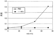
初期処方物、pH7.2のリン酸緩衝食塩水(PBS)の構造安定性(robustness)を測定するために、処方物スクリーニングが実施された。スクリーニング試験から、PBS中のIMC−A12は凝集、沈殿、分解、加水分解および光に対して感受性を有することが決定的となった。これに加えて、それは、少量の注射液に関する微粒子の問題の試験に合格することができなかった。pH6.5の、5mg/mL IMC−A12、10mM クエン酸ナトリウム、100mM グリシン、100mM NaClおよび0.01% ツイーン80からなる改良された溶液処方物が開発された。このクエン酸塩処方物は、PBS処方物とは異なり、微粒子を含まず、改善された安定性を有する。
Formulation screening was performed to determine the structural stability of the initial formulation, phosphate buffered saline (PBS) at pH 7.2. From screening tests, it became crucial that IMC-A12 in PBS was sensitive to aggregation, precipitation, degradation, hydrolysis and light. In addition to this, it failed to pass the particulate matter test for small volumes of injection. An improved solution formulation was developed consisting of 5 mg / mL IMC-A12, 10 mM sodium citrate, 100 mM glycine, 100 mM NaCl and 0.01
ヒンジ領域で起こる加水分解を最少にするさらなる改良品では、凍結乾燥された処方物は、30mg/mL IMC−A12、10mM ヒスチジン pH6.5、および4.6% トレハロースを含有する。加水分解は、凍結乾燥された処方物中のIMC−A12では停止した。 In a further improvement that minimizes hydrolysis that occurs in the hinge region, the lyophilized formulation contains 30 mg / mL IMC-A12, 10 mM histidine pH 6.5, and 4.6% trehalose. Hydrolysis was stopped with IMC-A12 in the lyophilized formulation.
本発明は、上記抗体の分解を減少または排除する溶液処方物を提供する。この処方物は、以下のものの1種以上を含むことができる:特定のpHの緩衝液、塩、界面活性剤、安定剤、保存料、還元剤、およびキレート剤。 The present invention provides solution formulations that reduce or eliminate the degradation of the antibody. The formulation can include one or more of the following: buffers, salts, surfactants, stabilizers, preservatives, reducing agents, and chelating agents of a particular pH.
本発明は、非酵素的切断を受けやすい抗体(その機能的断片を含む)の凍結乾燥のための処方物を提供する。この処方物は、安定剤、界面活性剤、還元剤、担体、保存料、アミノ酸、およびキレート剤などのさらなる要素を含んでいてよい。本発明はまた、抗体組成物を安定化する方法であって、凍結保護剤の存在下で抗体の水系処方物を凍結乾燥する工程を含む方法をも提供する。この処方物は、加工および保存中に抗体を安定化するために凍結乾燥され、次いで薬理学的投与に先立って再構成されてもよい。好ましくは、この抗体は、製造から投与までその物理的および化学的安定性、ならびに完全性を実質的に保持する。本発明に従って安定性を高めるために、緩衝液、界面活性剤、糖類、糖アルコール、糖誘導体、およびアミノ酸を含めた種々の処方物成分が適切であり得る。本発明に従って安定性を高めるために、pHおよび処方物成分の濃度を含めた種々の処方物特性が適切であり得る。 The present invention provides a formulation for lyophilization of antibodies (including functional fragments thereof) susceptible to non-enzymatic cleavage. The formulation may contain additional elements such as stabilizers, surfactants, reducing agents, carriers, preservatives, amino acids, and chelating agents. The present invention also provides a method of stabilizing an antibody composition comprising the step of lyophilizing an aqueous formulation of an antibody in the presence of a cryoprotectant. This formulation may be lyophilized to stabilize the antibody during processing and storage and then reconstituted prior to pharmacological administration. Preferably, the antibody substantially retains its physical and chemical stability and integrity from manufacture to administration. Various formulation components including buffers, surfactants, saccharides, sugar alcohols, sugar derivatives, and amino acids may be appropriate to enhance stability in accordance with the present invention. Various formulation characteristics, including pH and concentration of formulation components, may be appropriate to enhance stability in accordance with the present invention.
本発明によれば、上記処方物のpHを維持するために、緩衝液が用いられてもよい。この緩衝液は、外的な変化に起因するpHの変動を最少にする。本発明の処方物は、適切なpH、好ましくは約6.0〜約7.0、より好ましくは約6.0〜約6.5、最も好ましくは約6.5の処方物を提供するための1種以上の緩衝液を含有する。例示的な緩衝液としては、ヒスチジン、クエン酸塩、リンゴ酸塩、酒石酸塩、コハク酸塩、および酢酸塩などの有機緩衝液一般が挙げられるが、これらに限定されない。一実施形態では、緩衝液濃度は約5mM〜約50mMである。さらなる実施形態では、緩衝液濃度は約10mMである。 According to the present invention, a buffer may be used to maintain the pH of the formulation. This buffer minimizes pH fluctuations due to external changes. The formulations of the present invention provide formulations at a suitable pH, preferably about 6.0 to about 7.0, more preferably about 6.0 to about 6.5, and most preferably about 6.5. Containing one or more buffers. Exemplary buffers include, but are not limited to, organic buffers in general such as histidine, citrate, malate, tartrate, succinate, and acetate. In one embodiment, the buffer concentration is about 5 mM to about 50 mM. In a further embodiment, the buffer concentration is about 10 mM.
本発明の処方物は、抗体の凝集および分解を防止するのを助け得る1種以上の安定剤を含有してよい。適切な安定剤としては、多価糖類、糖アルコール、糖誘導体、およびアミノ酸が挙げられるが、これらに限定されない。好ましい安定剤としては、アスパラギン酸、ラクトビオン酸、グリシン、トレハロース、マンニトール、およびスクロースが挙げられるが、これらに限定されない。 The formulations of the present invention may contain one or more stabilizers that can help prevent antibody aggregation and degradation. Suitable stabilizers include, but are not limited to, polyvalent saccharides, sugar alcohols, sugar derivatives, and amino acids. Preferred stabilizers include but are not limited to aspartic acid, lactobionic acid, glycine, trehalose, mannitol, and sucrose.
本発明の処方物は、1種以上の界面活性剤を含んでいてよい。抗体溶液は、空気−水界面において高表面張力を有する。この表面張力を低下させるために、抗体は空気−水界面に凝集する傾向がある。界面活性剤は、空気−水界面での抗体の凝集を最少にし、それによって溶液中での抗体の生物活性を維持するのを助ける。例えば、0.01%のツイーン80を加えると、溶液中での抗体の凝集を低下させることができる。この処方物が凍結乾燥された場合、この界面活性剤はまた、再構成された処方物における微粒子の形成を減少させ得る。本発明の凍結乾燥した処方物では、この界面活性剤は、凍結乾燥前の処方物、凍結乾燥した処方物、および再構成された処方物の1種以上に加えることができるが、凍結乾燥前の処方物に加えることが好ましい。例えば、0.01%のツイーン80は、凍結乾燥前に抗体溶液に加えることができる。界面活性剤としては、ポリソルベート20(ツイーン20)、ポリソルベート80(ツイーン80)、ポリエチレン−ポリプロピレングルコール(プルロニック(PLURONIC)F−68、CAS番号9003−11−6)、および胆汁酸塩が挙げられるが、これらに限定されない。一実施形態では、界面活性剤濃度は約0.001%〜約1.0%である。
The formulations of the present invention may contain one or more surfactants. The antibody solution has a high surface tension at the air-water interface. In order to reduce this surface tension, antibodies tend to aggregate at the air-water interface. Surfactants help minimize antibody aggregation at the air-water interface, thereby maintaining the biological activity of the antibody in solution. For example, the addition of 0.01
凍結乾燥プロセスは、タンパク質またはポリペプチドを変性し得る種々のストレスを発生し得る。これらのストレスとしては、温度低下、氷の結晶の形成、イオン強度の上昇、pH変化、相分離、水和殻(hydration shell)の除去、および濃度変化が挙げられる。凍結および/または乾燥プロセスのストレスに対して感受性を有する抗体は、1種以上の凍結保護剤を添加することによって、安定化できる。凍結保護剤は、凍結乾燥に伴うストレスから保護する化合物である。それゆえ、凍結保護剤は、1つの種類として、凍結プロセスから保護する冷却保護剤(cryoprotectant)を含む。1種以上の凍結保護剤が、凍結乾燥に伴うストレスから保護するために使用されてもよく、それらは、例えば、スクロースまたはトレハロースなどの糖類;グルタミン酸一ナトリウムまたはヒスチジンなどのアミノ酸;ベタインなどのメチルアミン;硫酸マグネシウムなどの離液性塩(lyotropic salt);三価またはより多価の糖アルコールなどのポリオール、例えば、グリセリン、エリスリトール、グリセロール、アラビトール、キシリトール、ソルビトール、およびマンニトール;プロピレングルコール;ポリエチレングルコール;プルロニック;ならびにこれらの組み合わせであってよい。好ましい凍結保護剤の例としては、上記の安定剤および界面活性剤が挙げられるが、これらに限定されない。 The lyophilization process can generate a variety of stresses that can denature the protein or polypeptide. These stresses include temperature drop, ice crystal formation, ionic strength increase, pH change, phase separation, hydration shell removal, and concentration change. Antibodies that are sensitive to the stress of the freezing and / or drying process can be stabilized by the addition of one or more cryoprotectants. A cryoprotectant is a compound that protects against stress associated with lyophilization. Therefore, cryoprotectants include, as one type, cryoprotectants that protect against the freezing process. One or more cryoprotectants may be used to protect against stress associated with lyophilization, for example, sugars such as sucrose or trehalose; amino acids such as monosodium glutamate or histidine; methyls such as betaine Amines; lyotropic salts such as magnesium sulfate; polyols such as trihydric or more polyvalent sugar alcohols such as glycerin, erythritol, glycerol, arabitol, xylitol, sorbitol, and mannitol; propylene glycol; polyethylene Glucol; Pluronic; as well as combinations thereof. Examples of preferred cryoprotectants include, but are not limited to, the stabilizers and surfactants described above.
本発明は、凍結乾燥のプロセスによって調製できる安定化された処方物を提供する。凍結乾燥は、物質がまず凍結され、次いで、まず昇華(一次乾燥プロセス)によって、次いで脱着(二次乾燥プロセス)によって、生物活性または化学反応をもはや支えない値まで溶媒の量が減らされる、安定化プロセスである。凍結乾燥した処方物では、溶液に関連した加水分解、脱アミド化、酸化および断片化反応は、回避または顕著に緩慢化できる。凍結乾燥した処方物はまた、運搬中の短期の温度変動に起因する損傷を回避することができ、そして室温での保存を可能にする。本発明の処方物は、噴霧乾燥および気泡乾燥(bubble drying)などの当該技術分野で公知の他の方法によって乾燥されてもよい。特記しない限り、本発明の処方物は、凍結乾燥前の処方物中で測定された成分濃度によって記載される。 The present invention provides a stabilized formulation that can be prepared by a lyophilization process. Freeze drying is a stable process in which the material is first frozen and then the amount of solvent is reduced to a value that no longer supports biological activity or chemical reaction, first by sublimation (primary drying process) and then by desorption (secondary drying process). Process. In lyophilized formulations, solution-related hydrolysis, deamidation, oxidation and fragmentation reactions can be avoided or significantly slowed down. The lyophilized formulation can also avoid damage due to short-term temperature fluctuations during transportation and allows storage at room temperature. The formulations of the present invention may be dried by other methods known in the art such as spray drying and bubble drying. Unless otherwise stated, formulations of the present invention are described by component concentrations measured in the formulation prior to lyophilization.
一実施形態では、本発明は、ヒンジ領域で起こり得る非酵素的分解を受けやすい抗体を安定化するための方法および処方物を提供する。抗体を非酵素的に切断される要因としては、アミノ酸配列、コンホメーションおよび翻訳後の処理が挙げられる。 In one embodiment, the present invention provides methods and formulations for stabilizing antibodies that are susceptible to non-enzymatic degradation that can occur at the hinge region. Factors that cause non-enzymatic cleavage of the antibody include amino acid sequence, conformation, and post-translational processing.
抗体がヒンジ領域で加水分解、凝集、酸化、脱アミド化、沈殿、および/または断片化されるのは、水系溶液中での抗体のインキュベーションによって測定できる。典型的には、このインキュベーションは、試験の継続時間を短くするために、高温で実施される。例えば、40℃または50℃で3ヶ月間のインキュベーション。インキュベーション後、分解生成物は、サイズ排除クロマトグラフィ−高性能液体クロマトグラフィ(SEC−HPLC)を用いて分析することができる。 The hydrolysis, aggregation, oxidation, deamidation, precipitation, and / or fragmentation of the antibody at the hinge region can be measured by incubation of the antibody in an aqueous solution. Typically, this incubation is performed at an elevated temperature to shorten the duration of the test. For example, incubation at 40 ° C. or 50 ° C. for 3 months. After incubation, the degradation products can be analyzed using size exclusion chromatography-high performance liquid chromatography (SEC-HPLC).
加えて、機械的ストレスによって誘発される抗体の分解、凝集、および沈殿に対する処方物成分の保護効果を検討するため、抗体処方物は撹拌してもよい。 In addition, the antibody formulation may be agitated to study the protective effect of the formulation components against antibody degradation, aggregation and precipitation induced by mechanical stress.
当該技術分野で公知の種々の分析手法によって、溶液処方物の抗体安定性または再構成された凍結乾燥処方物の抗体安定性を測定することができる。かかる手法としては、例えば、(i)メインの融解温度(Tm)を測定するための示差走査熱量測定(DSC)を用いて熱安定性を;(ii)室温での制御された撹拌を用いて機械的安定性を;(iii)約−20℃、約4℃、室温(約23℃−27℃)、約40℃、および約50℃の温度でのリアルタイム等温加速温度安定性(isothermal accelerated temperature stability)を;(iv)約350nmで吸光度をモニターすることにより溶液濁度を、ならびに(v)SEC−HPLCを用いて単量体、凝集体および分解生成物の量を、測定することが挙げられる。安定性は、選択された温度で選択された時間の間測定することができる。 Various analytical techniques known in the art can measure the antibody stability of a solution formulation or the antibody stability of a reconstituted lyophilized formulation. Such techniques include, for example, (i) thermal stability using differential scanning calorimetry (DSC) to measure the main melting temperature (Tm); (ii) using controlled agitation at room temperature. Mechanical stability; (iii) real-time isothermal accelerated temperature stability at temperatures of about −20 ° C., about 4 ° C., room temperature (about 23 ° C.-27 ° C.), about 40 ° C., and about 50 ° C. (iv) measuring solution turbidity by monitoring absorbance at about 350 nm, and (v) measuring the amount of monomers, aggregates and degradation products using SEC-HPLC. It is done. Stability can be measured for a selected time at a selected temperature.
一実施形態では、上記凍結乾燥した処方物は、再構成の際に、高濃度の抗体を提供する。さらなる実施形態では、安定な凍結乾燥した処方物は、液体を用いて再構成可能であり、凍結乾燥前の処方物の抗体濃度よりも約1−10倍高い抗体濃度を有する溶液を形成できる。例えば、一実施形態では、この凍結乾燥した処方物は、1mL以下の水を用いて再構成され、約50mg/mL〜約200mg/mLの抗体濃度を有する粒子を含まない再構成された処方物が得られる。 In one embodiment, the lyophilized formulation provides a high concentration of antibody upon reconstitution. In a further embodiment, a stable lyophilized formulation can be reconstituted with a liquid and can form a solution having an antibody concentration that is about 1-10 times higher than the antibody concentration of the formulation prior to lyophilization. For example, in one embodiment, the lyophilized formulation is reconstituted with 1 mL or less of water and does not contain particles having an antibody concentration of about 50 mg / mL to about 200 mg / mL. Is obtained.
天然に存在する抗体は、典型的には、2つの同一の重鎖および2つの同一の軽鎖を有し、各軽鎖は、鎖間ジスルフィド結合によって重鎖に共有結合されている。多数のジスルフィド結合がさらに、2つの重鎖を互いに結合している。個々の鎖は、類似の大きさ(110−125個のアミノ酸)および構造を有するが異なる機能を有するドメインへと折り重なることができる。軽鎖は、1つの可変ドメイン(VL)および/または1つの定常ドメイン(CL)を含むことができる。重鎖もまた、1つの可変ドメイン(VH)および/または、抗体の種類またはアイソタイプに依存して、3つまたは4つの定常ドメイン(CH1、CH2、CH3およびCH4)を含むことができる。ヒトでは、このアイソタイプは、IgA、IgD、IgE、IgG、およびIgMであり、IgAおよびIgGはさらにサブクラスまたはサブタイプ(IgA1−2およびIgG1−4)に細分される。
Naturally occurring antibodies typically have two identical heavy chains and two identical light chains, each light chain covalently linked to the heavy chain by an interchain disulfide bond. A number of disulfide bonds further link the two heavy chains together. Individual chains can fold into domains with similar size (110-125 amino acids) and structure but different functions. The light chain can comprise one variable domain (V L ) and / or one constant domain (C L ). The heavy chain also has one variable domain (V H ) and / or three or four constant domains (
一般に、可変ドメインは、抗体ごとにかなりのアミノ酸配列の変動性を示し、特に抗原結合部位の場所ではそうである。高頻度可変領域または相補性決定領域(CDR)と呼ばれる3つの領域が、VLおよびVHの各々において見出され、これらの領域は、フレームワーク可変領域(framework variable region)と呼ばれる、さほど可変的でない領域によって支持されている。 In general, variable domains show considerable amino acid sequence variability from antibody to antibody, especially at the location of the antigen binding site. Three regions called hypervariable regions or complementarity determining region (CDR) are found in each of V L and V H, these regions are called the framework variable region (framework variable region), less variable Supported by unreasonable areas.
VLドメインおよびVHドメインからなる抗体の一部分は、Fv(可変断片(fragment variable))と表され、抗原−結合部位を構成する。単鎖Fv(scFv)は、1本のポリペプチド鎖上にVLドメインおよびVHドメインを含有する抗体断片であり、一方のドメインのN末端および他方のドメインのC末端は、柔軟性のあるリンカーによって繋がれている(例えば、米国特許第4,946,778号(Ladnerら);国際公開第88/09344号(Hustonら)を参照)。国際公開第92/01047号(McCaffertyら)は、バクテリオファージなどの可溶性組換え型遺伝子ディスプレイパッケージ(soluble recombinant genetic display package)の表面上のscFv断片のディスプレイを記載している。 A part of an antibody consisting of a VL domain and a VH domain is expressed as Fv (fragment variable) and constitutes an antigen-binding site. Single-chain Fv (scFv) is an antibody fragment containing a VL domain and a VH domain on one polypeptide chain, and the N-terminus of one domain and the C-terminus of the other domain are flexible Linked by a linker (see, eg, US Pat. No. 4,946,778 (Ladner et al.); WO 88/09344 (Huston et al.)). WO 92/01047 (McCafferty et al.) Describes the display of scFv fragments on the surface of a soluble recombinant genetic display package such as bacteriophage.
単鎖抗体は、それらが由来する全抗体の定常ドメインの一部またはすべてを欠く。それゆえ、それらは全抗体の使用に伴う問題のいくつかを克服することができる。例えば、単鎖抗体は、重鎖定常部と他の生体分子との間の特定の所望されない相互作用を有しない傾向がある。加えて、単鎖抗体は、全抗体よりもかなり小さく、全抗体よりも大きい透過性を有することができ、このため、単鎖抗体は標的抗原結合部位により効率よく局在化し結合することができる。さらに、単鎖抗体は、比較的小さいサイズのゆえに、全抗体よりも、レシピエントにおいて望まれない免疫応答を引き起こす可能性が少なくなる。 Single chain antibodies lack some or all of the constant domains of the entire antibody from which they are derived. They can therefore overcome some of the problems associated with the use of whole antibodies. For example, single chain antibodies tend not to have certain undesired interactions between the heavy chain constant region and other biomolecules. In addition, single chain antibodies can be much smaller and more permeable than whole antibodies, so that single chain antibodies can be more efficiently localized and bound to the target antigen binding site. . Furthermore, single chain antibodies are less likely to cause unwanted immune responses in the recipient than whole antibodies because of their relatively small size.
各単鎖が、第1のペプチドリンカーによって共有結合されている1つのVHドメインおよび1つのVLドメインを有する多数の単鎖抗体は、少なくとも1種以上のペプチドリンカーによって共有結合され、単一特異的、または多重特異的である多価の単鎖抗体を形成できる。多価の単鎖抗体の各鎖は、可変軽鎖断片および可変重鎖断片を含み、ペプチドリンカーによって少なくとも1つの他の鎖に結合されている。このペプチドリンカーは、少なくとも15個のアミノ酸残基から構成される。アミノ酸残基の最大数は、約100である。 Multiple single chain antibodies, each single chain having one V H domain and one VL domain covalently linked by a first peptide linker, are covalently linked by at least one or more peptide linkers, Multivalent single chain antibodies can be formed that are specific or multispecific. Each chain of a multivalent single chain antibody comprises a variable light chain fragment and a variable heavy chain fragment, and is linked to at least one other chain by a peptide linker. This peptide linker is composed of at least 15 amino acid residues. The maximum number of amino acid residues is about 100.
2つの単鎖抗体は、組み合わさって、二価の二量体として公知の二重抗体を形成することができる。二重抗体は、2本の鎖および2つの結合部位を有し、単一特異的または二重特異的となることができる。この二重抗体の各鎖は、VLドメインに連結されたVHドメインを含む。これらのドメインは同一鎖上のドメイン間の対合を妨げるのに十分短いリンカーで連結され、従って、異なる鎖上の相補的ドメイン間の対合を促して、2つの抗原結合部位を作り直す。 Two single chain antibodies can be combined to form a double antibody known as a divalent dimer. Biantibodies have two chains and two binding sites and can be monospecific or bispecific. Each chain of the diabody includes a V H domain connected to a V L domain. These domains are linked by a linker that is short enough to prevent pairing between domains on the same chain, thus facilitating pairing between complementary domains on different chains, recreating the two antigen binding sites.
3つの単鎖抗体は組み合わさって、三価の三量体としても公知の三重抗体を形成できる。三重抗体は、VLドメインまたはVHドメインのアミノ酸末端がVLドメインまたはVHドメインのカルボキシル末端に直接融合することにより、すなわち、何らのリンカー配列を有さずに、構築される。この三重抗体は、ポリペプチドが環状の、頭−尾様式で配列した、3つのFv頭部を有する。この三重抗体の可能なコンホメーションは平面状であり、3つの結合部位が、互いから120°の角度で1つの平面上に存在する。三重抗体は単一特異的、二重特異的または三重特異的となることができる。 Three single chain antibodies can be combined to form a known triple antibody, also known as a trivalent trimer. Triabodies, by amino acid terminus of a V L or V H domain is directly fused to the carboxyl terminus of a V L or V H domain, i.e., without a any linker sequence is constructed. This triple antibody has three Fv heads in which the polypeptides are arranged in a cyclic, head-to-tail manner. The possible conformation of this triple antibody is planar, with three binding sites present on one plane at an angle of 120 ° from each other. Triple antibodies can be monospecific, bispecific or trispecific.
Fab(断片、抗原結合)は、VLCLVHおよびCH1ドメインからなる抗体の断片を指す。パパイン消化後に生成されるものは、単にFabと呼ばれ、重鎖ヒンジ領域を保持しない。ペプシン消化後、重鎖ヒンジを保持する種々のFabが生成される。鎖間ジスルフィド結合が無傷のままであるそれらの二価の断片は、F(ab’)2と呼ばれるが、ジスルフィド結合が保持されない場合は、一価のFab’が生じる。F(ab’)2断片は、一価のFab断片よりも、抗原に対して高い結合活性を有する。
Fab (fragment, antigen binding) refers to a fragment of an antibody consisting of the V L C L V H and
Fc(結晶性断片)は、対合した重鎖定常ドメインを含む抗体の一部分または断片についての記号表示である。IgG抗体では、例えば、Fcは、CH2ドメインおよびCH3ドメインを含む。IgAまたはIgM抗体のFcはさらに、CH4ドメインを含む。Fcは、Fc受容体結合、補体媒介性細胞傷害性の活性化、および抗体依存性細胞傷害性(ADCC)に関連する。多数のIgG様タンパク質の複合体であるIgAおよびIgMなどの抗体については、複合体形成は、Fc定常ドメインを必要とする。
Fc (crystalline fragment) is a symbolic designation for a portion or fragment of an antibody that contains paired heavy chain constant domains. For IgG antibodies, for example, Fc comprises a
最後に、ヒンジ領域は、その抗体のFab部分とFc部分とを分離し、Fab同士の可動性、およびFcに対するFabの可動性を提供し、加えて2つの重鎖の共有結合のための多数のジスルフィド結合を含む。 Finally, the hinge region separates the Fab and Fc portions of the antibody, providing Fab-to-Fab mobilities and Fab mobilities for Fc, plus multiple for covalent attachment of the two heavy chains. Of disulfide bonds.
従って、本発明の抗体としては、抗原と特異的に結合する、天然に存在する抗体、(Fab’)2などの二価の断片、Fabなどの一価の断片、単鎖抗体、単鎖Fv(scFv)、単一ドメイン抗体、多価の単鎖抗体、二重抗体、三重抗体などが含まれるが、これらに限定されない。 Accordingly, the antibodies of the present invention include naturally occurring antibodies that specifically bind to antigens, bivalent fragments such as (Fab ′) 2 , monovalent fragments such as Fab, single chain antibodies, single chain Fv (ScFv), single domain antibody, multivalent single chain antibody, double antibody, triple antibody and the like.
本発明の抗体、またはその断片は、例えば、単一特異的または二重特異的であってよい。二重特異的抗体(BsAb)は、2つの異なる抗原結合特異性または部位を有する抗体である。抗体が複数の特異性を有する場合、認識されたエピトープは、単一の抗原と会合するものであってもよいし、複数の抗原と会合するものであってもよい。従って、本発明は、2つの異なる抗原に結合する二重特異的抗体またはその断片を提供する。 The antibodies of the invention, or fragments thereof, can be, for example, monospecific or bispecific. Bispecific antibodies (BsAbs) are antibodies that have two different antigen binding specificities or sites. When the antibody has a plurality of specificities, the recognized epitope may be associated with a single antigen or may be associated with a plurality of antigens. Accordingly, the present invention provides bispecific antibodies or fragments thereof that bind to two different antigens.
抗体またはその断片の特異性は、親和性および/または結合活性に基づいて決定することができる。抗体からの抗原の解離についての平衡定数(Kd)によって表される親和性は、抗原の決定基と抗体結合部位との間の結合の強さの尺度となる。結合活性は、抗体とその抗原との間の結合の強さの尺度である。結合活性は、エピトープと抗体上のその抗原結合部位との間の親和性、および抗体の価数(特定のエピトープの抗原結合部位の数を指す)の両方に関連する。抗体は、典型的には、10−5〜10−11リットル/molの解離定数(Kd)で結合する。10−4リットル/mol未満のあらゆるKdは、一般に非特異的結合を示すとみなされる。Kdの値が小さいほど、抗原決定基と抗体結合部位との間の結合の強さは強い。 The specificity of the antibody or fragment thereof can be determined based on affinity and / or binding activity. The affinity, expressed by the equilibrium constant (K d ) for the dissociation of the antigen from the antibody, is a measure of the strength of binding between the antigenic determinant and the antibody binding site. Binding activity is a measure of the strength of binding between an antibody and its antigen. Binding activity is related both to the affinity between the epitope and its antigen binding site on the antibody, and to the valency of the antibody (which refers to the number of antigen binding sites for a particular epitope). Antibodies typically bind with a dissociation constant (K d ) of 10 −5 to 10 −11 liters / mol. Any K d below 10 −4 liter / mol is generally considered to indicate non-specific binding. The smaller the value of Kd , the stronger the binding between the antigenic determinant and the antibody binding site.
本願明細書で使用する場合、「抗体」および「抗体断片」は、特異的抗原への特異性を保持する改変を含む。かかる改変としては、化学療法薬(例えば、シスプラチン、タキソール、ドキソルビシン)などのエフェクター分子、または細胞毒(例えば、タンパク質もしくは非タンパク質の有機化学療法薬)への接合が挙げられるが、これらに限定されない。抗体は、検出できるレポーター部分への接合によって改変することができる。半減期などの非結合的特徴に影響を及ぼす変更(例えば、ペグ化)を有する抗体もまた含まれる。 As used herein, “antibodies” and “antibody fragments” include modifications that retain specificity for a specific antigen. Such modifications include, but are not limited to, effector molecules such as chemotherapeutic drugs (eg, cisplatin, taxol, doxorubicin), or conjugation to cytotoxins (eg, protein or non-protein organic chemotherapeutic drugs). . An antibody can be modified by conjugation to a detectable reporter moiety. Also included are antibodies with alterations that affect non-binding characteristics such as half-life (eg, pegylation).
タンパク質剤および非タンパク質剤は、当該技術分野で公知の方法によって上記抗体に接合してよい。接合方法としては、直接結合、共有結合されたリンカーを介する結合、および特異的結合対のメンバーを介する結合(例えば、アビジン−ビオチン)が挙げられる。かかる方法としては、例えば、ドキソルビシンの接合についてGreenfieldら、Cancer Research 50,6600−6607(1990)に記載される方法、ならびに白金化合物の接合についてArnonら、Adv.Exp.Med.Biol.303,79−90(1991)およびKiselevaら、Mol.Biol.(USSR)25,508−514(1991)によって記載される方法が挙げられる。
Protein agents and non-protein agents may be conjugated to the antibody by methods known in the art. Conjugation methods include direct binding, binding through a covalently linked linker, and binding through a member of a specific binding pair (eg, avidin-biotin). Such methods include, for example, the method described in Greenfield et al.,
本発明の抗体はさらに、結合の特徴が直接的な変異、親和性成熟の方法、ファージディスプレイ、またはチェーンシャフリング(chain shuffling)によって改良されたものを含む。親和性および特異性は、CDRを変異させて、所望の特徴を有する抗原結合部位についてスクリーニングすることにより、改変または改良することができる(例えば、Yangら、J.Mol.Biol.,254:392−403(1995)を参照)。CDRは種々の方法で変異される。1つの方法は、個々の残基または残基の組合せをランダム化して、本来は同一である抗原結合部位の集団において、20個のアミノ酸すべてが特定の位置で見出されるようにすることである。あるいは、変異は、エラープローンPCR法によってある範囲のCDR残基にわたって誘導される(例えば、Hawkinsら、J.Mol.Biol.,226:889−896(1992)を参照)。例えば、重鎖および軽鎖の可変領域遺伝子を含有するファージディスプレイベクターを、E.coliの突然変異誘発株(mutator strain)で増殖させることができる(例えば、Lowら、J.Mol.Biol.,250:359−368(1996)を参照)。これらの変異原性法は、当業者に公知の多くの方法の一例である。 The antibodies of the present invention further include those in which binding characteristics have been improved by direct mutation, methods of affinity maturation, phage display, or chain shuffling. Affinity and specificity can be modified or improved by mutating CDRs and screening for antigen binding sites with the desired characteristics (eg, Yang et al., J. Mol. Biol., 254: 392). -403 (1995)). CDRs are mutated in various ways. One method is to randomize individual residues or combinations of residues so that all 20 amino acids are found at a particular position in a population of antigen binding sites that are originally identical. Alternatively, mutations are induced over a range of CDR residues by error-prone PCR methods (see, eg, Hawkins et al., J. Mol. Biol., 226: 889-896 (1992)). For example, phage display vectors containing heavy and light chain variable region genes can be obtained from E. coli. can be grown on a mutator strain of E. coli (see, eg, Low et al., J. Mol. Biol., 250: 359-368 (1996)). These mutagenic methods are examples of many methods known to those skilled in the art.
抗体はまた、Fc受容体への結合を変え、従って抗体依存性細胞媒介性細胞傷害性および補体依存性細胞傷害性などのエフェクター機能を高めるかまたは低下させることになる、Fc領域の1種以上のアミノ酸置換を含むように改変してもよい。 An antibody is also a member of the Fc region that alters binding to an Fc receptor and thus increases or decreases effector functions such as antibody-dependent cell-mediated cytotoxicity and complement-dependent cytotoxicity. You may modify | change so that the above amino acid substitution may be included.
本発明の抗体の各ドメインは、完全免疫グロブリンドメイン(例えば、重鎖もしくは軽鎖の可変または定常ドメイン)であってよいし、あるいはそれは、天然に存在するドメインの機能的等価体もしくは変異体もしくは誘導体、または例えば、国際公開第93/11236号(Griffithsら)に記載されている手法などを用いて生体外で構築された合成ドメインであってもよい。例えば、少なくとも1つのアミノ酸を欠いている抗体可変ドメインに対応するドメインを一緒に繋ぐことが可能である。この抗体の重要な特徴は、抗原結合部位の存在である。可変の重鎖断片および軽鎖断片という用語は、特異性に対して重要な効果を有しない変種を排除すると解釈されるべきではない。 Each domain of an antibody of the invention may be a complete immunoglobulin domain (eg, a heavy or light chain variable or constant domain) or it may be a functional equivalent or variant of a naturally occurring domain or It may be a derivative or a synthetic domain constructed in vitro using, for example, the technique described in WO 93/11236 (Griffiths et al.). For example, domains corresponding to antibody variable domains lacking at least one amino acid can be linked together. An important feature of this antibody is the presence of an antigen binding site. The terms variable heavy and light chain fragments should not be construed to exclude variants that do not have a significant effect on specificity.
本発明の抗体および抗体断片は、例えば、天然に存在する抗体、またはFabもしくはscFvファージディスプレイライブラリから得ることができる。VHドメインおよびVLドメインを含む抗体から単一ドメインの抗体を作製するため、CDRの外部の特定のアミノ酸置換を所望し、結合、発現または溶解性を高めてもよいことは理解されている。例えば、本来VH−VL界面に埋もれているアミノ酸残基を改変するのが望ましい場合がある。 The antibodies and antibody fragments of the invention can be obtained, for example, from naturally occurring antibodies, or from Fab or scFv phage display libraries. It is understood that certain amino acid substitutions outside of the CDRs may be desired to enhance binding, expression or solubility in order to generate single domain antibodies from antibodies containing V H and V L domains. . For example, it may be desirable to modify amino acid residues that are originally buried in the V H -V L interface.
さらに、本発明の抗体および抗体断片は、ヒト免疫グロブリンγ重鎖およびκ軽鎖を産生するトランスジェニックマウス(例えば、カリフォルニア州、サンノゼのメダレックス社(Medarex)より入手のKMマウス)を使用して、標準的なハイブリドーマ技術(本明細書の一部を構成する、HarlowおよびLane編、Antibodies:A Laboratory Manual、Cold Spring Harbor、211−213(1998))によって得ることができる。好ましい実施形態では、ゲノムを産生するヒト抗体の実質的な部分がマウスのゲノムに挿入され、内因性マウス抗体の産生が欠失するようにする。かかるマウスは、完全フロイントアジュバント中の標的分子の一部またはすべてを用いて、皮下的に(s.c.)免疫化してもよい。 Further, the antibodies and antibody fragments of the present invention can be obtained using transgenic mice that produce human immunoglobulin gamma heavy chains and kappa light chains (eg, KM mice obtained from Medarex, San Jose, Calif.). , Standard hybridoma technology (Harlow and Lane, Ed., Antibodies: A Laboratory Manual, Cold Spring Harbor, 211-213 (1998), which forms part of this specification). In a preferred embodiment, a substantial portion of the human antibody that produces the genome is inserted into the mouse genome such that production of the endogenous mouse antibody is deleted. Such mice may be immunized subcutaneously (sc) with some or all of the target molecule in complete Freund's adjuvant.
本発明はまた、再構成された処方物を投与することを含む、治療方法をも提供する。この再構成された処方物は、本発明の凍結乾燥した処方物を、例えば1mLの水を用いて再構成することにより調製される。再構成時間は、好ましくは1分未満である。濃い再構成された処方物は、投与の際に融通性を与える。例えば、再構成された処方物は、希釈した形で静脈内投与することができ、またはより濃縮された形で注射投与することもできる。本発明の濃い再構成された処方物は、特定の被験者および/または特定の投与経路に適合する濃度へと希釈することができる。従って、本発明は、治療有効量の抗体を、それを必要とする哺乳動物、特にヒトに投与することを含む、治療方法を提供する。本願明細書で使用する場合、投与という用語は、求められる結果を成就することができる任意の方法によって、本発明の抗体組成物を哺乳動物に送達することを意味する。再構成された処方物は、例えば、静脈内または筋肉内に投与することができる。一実施形態では、濃い再構成された処方物は、注射によって投与される。 The present invention also provides a method of treatment comprising administering a reconstituted formulation. This reconstituted formulation is prepared by reconstituting the lyophilized formulation of the present invention with, for example, 1 mL of water. The reconstitution time is preferably less than 1 minute. A thick reconstituted formulation provides flexibility upon administration. For example, the reconstituted formulation can be administered intravenously in a diluted form, or can be administered by injection in a more concentrated form. The concentrated reconstituted formulation of the present invention can be diluted to a concentration that is compatible with the particular subject and / or the particular route of administration. Accordingly, the present invention provides a therapeutic method comprising administering a therapeutically effective amount of an antibody to a mammal in need thereof, particularly a human. As used herein, the term administration means delivering an antibody composition of the invention to a mammal by any method that can achieve the desired result. The reconstituted formulation can be administered, for example, intravenously or intramuscularly. In one embodiment, the thick reconstituted formulation is administered by injection.
本発明の処方物における抗体は、好ましくはヒト用である。一実施形態では、本発明の組成物は、固形腫瘍および非固形腫瘍を含めた腫瘍性疾患を治療するため、過剰増殖性障害を治療するため、肥満症を治療するために、使用することができる。 The antibodies in the formulations of the present invention are preferably for human use. In one embodiment, the compositions of the invention can be used to treat neoplastic diseases, including solid tumors and non-solid tumors, to treat hyperproliferative disorders, to treat obesity. it can.
治療有効量とは、哺乳動物に投与されたときに、IGF−IR活性を低下させるかまたは無力化する、腫瘍増殖の阻害、非癌性の過剰増殖性疾患を治療する、肥満症を治療するなどの、所望の治療効果を生み出すのに有効である本発明の抗体の量を意味する。上記のような抗体の投与は、他の抗体または任意の従来の治療薬(例えば、抗悪性腫瘍薬)の投与と組合わせることができる。 A therapeutically effective amount is one that reduces or neutralizes IGF-IR activity when administered to a mammal, inhibits tumor growth, treats a non-cancerous hyperproliferative disease, treats obesity Means the amount of an antibody of the invention that is effective to produce the desired therapeutic effect. Administration of antibodies as described above can be combined with administration of other antibodies or any conventional therapeutic agent (eg, antineoplastic agent).
本発明の一実施形態では、上記組成物は、1種以上の抗悪性腫瘍薬と組合せて投与することができる。化学療法薬、放射線およびこれらの組合せなどの任意の適切な抗悪性腫瘍薬が使用できる。抗悪性腫瘍薬は、アルキル化剤または代謝拮抗薬であってもよい。アルキル化剤の例としては、シスプラチン、シクロホスファミド、メルファラン、およびダカルバジンが挙げられるが、これらに限定されない。代謝拮抗薬の例としては、ドキソルビシン、ダウノルビシン、パクリタキセル、イリノテカン(CPT−11)、およびトポテカンが挙げられるが、これらに限定されない。抗悪性腫瘍薬が放射線である場合、放射線源は、治療されている患者にとって外部(外照射療法−EBRT)または内部(近接照射療法−BT)のいずれかであってよい。投与される抗悪性腫瘍薬の用量は、例えば、薬剤の種類、治療されている腫瘍の種類および重症度、ならびにその薬剤の投与経路を含めた多くの要因に依存する。しかしながら、本発明はいずれかの特定の用量に限定されないということを強調しておきたい。 In one embodiment of the invention, the composition can be administered in combination with one or more antineoplastic agents. Any suitable antineoplastic agent can be used, such as chemotherapeutic agents, radiation and combinations thereof. The antineoplastic agent may be an alkylating agent or an antimetabolite. Examples of alkylating agents include, but are not limited to, cisplatin, cyclophosphamide, melphalan, and dacarbazine. Examples of antimetabolite drugs include, but are not limited to, doxorubicin, daunorubicin, paclitaxel, irinotecan (CPT-11), and topotecan. When the antineoplastic agent is radiation, the radiation source can be either external (external radiation therapy-EBRT) or internal (brachial radiation therapy-BT) for the patient being treated. The dose of antineoplastic agent administered depends on a number of factors including, for example, the type of drug, the type and severity of the tumor being treated, and the route of administration of the drug. However, it should be emphasized that the present invention is not limited to any particular dose.
本発明の抗体、またはその断片の等価体はまた、本発明で提供される全長IMC−A12抗体の可変領域または高頻度可変領域のアミノ酸配列と実質的に同じアミノ酸配列を有するポリペプチドをも含む。実質的に同じアミノ酸配列は、本願明細書においては、PearsonおよびLipmanによるFASTA検索法(Proc.Natl.Acad.Sci.USA 85,2444−8(1988))によって決定した場合に、少なくとも約70%、好ましくは少なくとも約80%、より好ましくは少なくとも約90%の相同性を有する配列として定義される。
The equivalent of the antibody of the present invention or a fragment thereof also includes a polypeptide having substantially the same amino acid sequence as the variable region or hypervariable region of the full-length IMC-A12 antibody provided in the present invention. . Substantially the same amino acid sequence herein is at least about 70% as determined by the FASTA search method by Pearson and Lipman (Proc. Natl.
以下の実施例は本発明をさらに例証するが、本発明の範囲を限定するとは決して解釈されるべきではない。タンパク質の分析において用いられるものなどの従来の方法の詳細な説明は、本明細書の一部を構成するCurrent Protocols in Immunology((ジョン・ワイリー・アンド・サンズ(John Wiley & Sons))などの多くの刊行物から得ることができる。 The following examples further illustrate the invention but should in no way be construed as limiting the scope of the invention. Detailed descriptions of conventional methods, such as those used in protein analysis, are many, such as Current Protocols in Immunology ((John Wiley & Sons)), which forms part of this specification. Can be obtained from
すべての液体処方物スクリーニング試験について、タンパク質濃度を5mg/mLに固定した。10mM リン酸ナトリウム、10mM クエン酸ナトリウム、10mM 酢酸ナトリウム、10mM L−ヒスチジンおよび125mM 塩化ナトリウムからなる多成分緩衝液を、最適のpHについてスクリーニングするために使用した。緩衝液の種類、ツイーン80、グリシン濃度、およびNaCl濃度についての必要条件を、実験計画法のアプローチ(DOE、JMPソフトウェア)を用いて検討した。線形回帰分析を実施して、試験された変数の有意性を決定した。予測した処方物を、慣用的な一時一事法(one−factor−at−a−time methodology)を用いて確認した。熱安定性に及ぼす試験された変数の効果を、示差走査熱量測定(DSC)およびリアルタイム等温試験(real−time isothermal study)を用いて検討した。室温での毎分300回転の制御された撹拌を、機械的安定性についての試験として用いた。液体処方物の光安定性を、ICH指針に基づき検討した。凍結−解凍安定性を、試験試料を−20℃および−70℃に冷却して4℃で解凍することにより決定した。
For all liquid formulation screening tests, the protein concentration was fixed at 5 mg / mL. A multi-component buffer consisting of 10 mM sodium phosphate, 10 mM sodium citrate, 10 mM sodium acetate, 10 mM L-histidine and 125 mM sodium chloride was used to screen for optimal pH. The requirements for buffer type,
凍結乾燥したIMC−A12処方物について、緩衝液の種類、安定剤、および増量剤を、20mg/mLのA12濃度での実験計画法の一部実施モデル(fractional factorial model)を用いて検討した。IMC−A12の濃度、IMC−A12濃度に対するトレハロース濃度の比、およびツイーン80の濃度を、混合計画モデル(mixture design model)を用いて最適化した。予測した最適の凍結乾燥処方物を、一時一事法を用いて、PBSおよびクエン酸塩溶液処方物と比較した。熱安定性に及ぼす変数の効果を、リアルタイム等温試験によって検討した。凍結乾燥した処方物の光安定性を、ICH指針に基づき検討した。
For lyophilized IMC-A12 formulations, buffer types, stabilizers, and bulking agents were studied using a fractional factorial model of experimental design at an A12 concentration of 20 mg / mL. The concentration of IMC-A12, the ratio of trehalose concentration to IMC-A12 concentration, and the concentration of
スクリーニング試験において使用するためのIMC−A12を、50Kカットオフ(YM 50)centriprep遠心濾過装置およびAllegra X−12R遠心分離機(ベックマン(Beckman))を用いて、実験的緩衝液に緩衝液交換することによって、調製した。タンパク質濃度を、1.50の吸光係数および適切な緩衝液で5mg/mLに調製した濃度を用いて280nmでの吸光度によって測定した。ツイーン80を、タンパク質濃度調整後に、10%(重量/体積)原液から加えた。PBS処方物中の5mg/mLのIMC−A12を対照として使用した。すべての試料を、シリンジフィルター(Durapore PVDF膜)を通して0.22μmで濾過した。
IMC-A12 for use in screening tests is buffer exchanged into experimental buffer using a 50K cut-off (YM 50) centriprep centrifugal filter and an Allegra X-12R centrifuge (Beckman) Prepared. The protein concentration was measured by absorbance at 280 nm using an extinction coefficient of 1.50 and a concentration adjusted to 5 mg / mL with the appropriate buffer.
凍結乾燥プロセスを、Lyostar II凍結乾燥機を用いて実施した。生成物を室温で凍結乾燥機に充填した。棚温度を、0.5℃/分の冷却速度で−50℃まで冷却した。−50℃での保持時間は2時間であった。一次乾燥および二次乾燥を、−30℃および20℃で各々12時間実施した。温度を0.5℃/分で変化させた。一次乾燥中および二次乾燥中のチャンバ圧は50mT(約6.7Pa)であった。凍結乾燥が完了した後、凍結乾燥機のチャンバを、N2を用いて大気圧の半分まで戻し、蓋をした。 The lyophilization process was performed using a Lyostar II lyophilizer. The product was loaded into a lyophilizer at room temperature. The shelf temperature was cooled to −50 ° C. at a cooling rate of 0.5 ° C./min. The holding time at −50 ° C. was 2 hours. Primary drying and secondary drying were performed at −30 ° C. and 20 ° C. for 12 hours, respectively. The temperature was varied at 0.5 ° C / min. The chamber pressure during primary drying and secondary drying was 50 mT (about 6.7 Pa). After lyophilization was complete, the lyophilizer chamber was returned to half atmospheric pressure using N2 and capped.
(実施例1:pH最適化試験)
10mM リン酸ナトリウム、10mM クエン酸ナトリウム、10mM 酢酸ナトリウム、10mM L−ヒスチジンおよび125mM 塩化ナトリウムからなる多成分緩衝液(MCB)を使用して、最適pHを決定した。この緩衝系は、pHのみより大きい影響を及ぼし得る対イオン(塩効果)の最少化を意図していた。このpHスクリーニング計画マトリクスを表2に示す。IMC−A12濃度を5mg/mLに保った。検討したpH範囲は、0.5pH単位の間隔で5.0−8.0であった。熱安定性および機械的安定性に及ぼすpHの影響を試験した。結果を以下に提示する。
The optimum pH was determined using a multi-component buffer (MCB) consisting of 10 mM sodium phosphate, 10 mM sodium citrate, 10 mM sodium acetate, 10 mM L-histidine and 125 mM sodium chloride. This buffer system was intended to minimize counterions (salt effects) that could have a greater effect on pH alone. This pH screening plan matrix is shown in Table 2. The IMC-A12 concentration was kept at 5 mg / mL. The pH range studied was 5.0-8.0 with 0.5 pH unit intervals. The effect of pH on thermal and mechanical stability was tested. The results are presented below.
(示差走査熱量測定(DSC)試験)
試験条件下におけるIMC−A12についての転移温度(Tm)を評価するため、(表2に示す)実験的処方物中のIMC−A12についての熱融解曲線を、示差走査熱量測定(DSC)によってアッセイした。タンパク質濃度は5mg/mLであり、温度変化は、1.5℃/分の走査速度で、5℃〜95℃であった。融解曲線を、3つのTmの合計にフィッティングした。pHの関数として最初の転移ピークに対応する融解温度であるTm1を図1に示す。Tm1は、pH6.5−8.0の間ではほとんど同じであった。
(Differential scanning calorimetry (DSC) test)
To assess the transition temperature (Tm) for IMC-A12 under the test conditions, the thermal melting curve for IMC-A12 in the experimental formulation (shown in Table 2) was assayed by differential scanning calorimetry (DSC). did. The protein concentration was 5 mg / mL and the temperature change was 5 ° C. to 95 ° C. with a scanning rate of 1.5 ° C./min. The melting curve was fitted to the sum of 3 Tm. Tm1, which is the melting temperature corresponding to the first transition peak as a function of pH, is shown in FIG. Tm1 was almost the same between pH 6.5-8.0.
(撹拌試験)
試料に、プラットフォームシェーカー上での撹拌によってストレスを加えた。27.5mLのガラスバイアル中に5mg/mLのIMC−A12の5mLを含む表2に記載した試料を、ヘッドスペースを81.8%であるように設定して、毎分300回転で撹拌した。この試験を、室温で72時間実施した。pHの関数として、不溶性凝集体の形成に起因する損失の割合(%)および残っている単量体の割合(%)を、それぞれ図2および図3に示す。pH6.0−7.0の間で、損失の割合(%)は最少であり、単量体の割合(%)は最も高かった。
(Agitation test)
The sample was stressed by agitation on a platform shaker. The sample described in Table 2 containing 5 mL of 5 mg / mL IMC-A12 in a 27.5 mL glass vial was stirred at 300 revolutions per minute with the headspace set to 81.8%. This test was conducted at room temperature for 72 hours. The percentage loss due to the formation of insoluble aggregates and the percentage monomer remaining as a function of pH are shown in FIGS. 2 and 3, respectively. Between pH 6.0-7.0, the percentage loss was the lowest and the percentage monomer was the highest.
(40℃および50℃でのリアルタイム加速温度安定性)
種々のpH緩衝液(表2)中の5mg/mLのIMC−A12を、40℃で3週間、および50℃で1週間インキュベートした。単量体の割合(%)に及ぼすpHの影響を、SEC−HPLCによって分析した。40℃で3週間のインキュベーション後(図4)、および50℃で1週間のインキュベーション後(図5)の残存した単量体の割合(%)の変化が、pHの関数として示されている。pH6.0−6.5の間で、残っている単量体の割合(%)は最も大きかった。
(Real-time accelerated temperature stability at 40 ° C and 50 ° C)
5 mg / mL IMC-A12 in various pH buffers (Table 2) was incubated at 40 ° C. for 3 weeks and 50 ° C. for 1 week. The effect of pH on the percent monomer was analyzed by SEC-HPLC. The change in percent monomer remaining after 3 weeks incubation at 40 ° C. (FIG. 4) and after 1 week incubation at 50 ° C. (FIG. 5) is shown as a function of pH. Between pH 6.0-6.5, the percentage of monomer remaining was the largest.
(−20℃および−70℃でのリアルタイム凍結温度安定性)
(表2に列挙した)種々のpH緩衝液中の5mg/mLのIMC−A12を、−20℃および−70℃で3週間インキュベートした。単量体の割合(%)に対するpHの効果を、SEC−HPLCによって分析した。3週間のインキュベーション後の単量体の割合(%)の変化をpHの関数として図6に示す。−20℃または−70℃のいずれにおいても、pHは、単量体の割合(%)に有意な影響は及ぼさなかった。
(Real-time freezing temperature stability at -20 ° C and -70 ° C)
5 mg / mL IMC-A12 in various pH buffers (listed in Table 2) was incubated at −20 ° C. and −70 ° C. for 3 weeks. The effect of pH on the percent monomer was analyzed by SEC-HPLC. The change in percent monomer after 3 weeks incubation is shown in FIG. 6 as a function of pH. At either −20 ° C. or −70 ° C., pH did not significantly affect the monomer percentage.
(pH最適化の総括)
5mg/mLのIMC−A12の最適のpHは、6.0〜6.5の間であることが判明した。
(Overview of pH optimization)
The optimal pH of 5 mg / mL IMC-A12 was found to be between 6.0 and 6.5.
(実施例2:溶液処方物についての賦形剤スクリーニング試験)
実施例1でのpH最適化試験は、IMC−A12が、pH6.0〜6.5の間で最も大きい安定性を有することを明らかにした。この実施例では、本発明者らは、pH6.0および6.5でのIMC−A12の安定性に対する緩衝液の種類、クエン酸塩およびヒスチジンの影響を試験した。ツイーン80およびNaClおよびグリシンの濃度の必要条件もまた検討した。タンパク質濃度を、5mg/mLに一定に保った。賦形剤スクリーニングの計画マトリクスを表3に示す。
The pH optimization test in Example 1 revealed that IMC-A12 has the greatest stability between pH 6.0 and 6.5. In this example, we tested the effect of buffer type, citrate and histidine on the stability of IMC-A12 at pH 6.0 and 6.5.
(浸透圧測定)
表3の処方物の浸透圧を、Wescor蒸気圧浸透圧計を用いて測定した。結果を表4に示す。試験した処方物の浸透圧は、260−320mOsm/Kgという所望の範囲内であった。
The osmotic pressure of the formulations in Table 3 was measured using a Wescor vapor pressure osmometer. The results are shown in Table 4. The osmotic pressure of the tested formulations was in the desired range of 260-320 mOsm / Kg.
(示差走査熱量測定による試験)
(表3に列挙した)実験的処方物中のIMC−A12についての熱融解曲線を、試験条件下におけるIMC−A12についての転移温度(Tm)を評価するため、DSCを用いてアッセイした。タンパク質濃度は5mg/mLであり、温度変化は、1.5℃/分の走査速度で、5℃〜95℃であった。メインの転移ピークに対応する融解温度を線形回帰モデルにフィッティングし、試験された変数の効果を評価した。このフィッティングについてのpおよびRsqは、それぞれ0.003および0.99であった。転移温度に及ぼす緩衝液の種類、pH、ツイーン80濃度、NaCl濃度、グリシン濃度の影響について、予測プロファイラ(prediction profiler)を図7に示す。最適緩衝液は、pH6.5のクエン酸塩緩衝液であると決定した。グリシンは、融解温度を上昇させ、ツイーン80は融解温度をわずかに低下させた。NaClは、融解温度に対して有意な効果を与えなかった。
(Test by differential scanning calorimetry)
Thermal melting curves for IMC-A12 in the experimental formulations (listed in Table 3) were assayed using DSC to evaluate the transition temperature (Tm) for IMC-A12 under the test conditions. The protein concentration was 5 mg / mL and the temperature change was 5 ° C. to 95 ° C. with a scanning rate of 1.5 ° C./min. The melting temperature corresponding to the main transition peak was fitted to a linear regression model to evaluate the effect of the tested variables. The p and R sq for this fitting were 0.003 and 0.99, respectively. FIG. 7 shows a prediction profiler for the effects of buffer type, pH,
(撹拌試験)
試料に、プラットフォームシェーカー上での撹拌によってストレスを加えた。表3に記載した試料を、27.5mLのガラスバイアル中の5mg/mLのIMC−A12の5mLとともに、毎分300回転で撹拌した。この試験を、室温で72時間まで実施した。溶液濁度および単量体の割合(%)を、撹拌時間の関数として測定した。濁度および単量体の割合(%)に及ぼす試験された変数の影響を、応答をJMPソフトウェアを用いて線形回帰モデルにフィッティングすることによって評価した。濁度および単量体の割合(%)の両方に及ぼす実測対予測プロットのp値は、<0.001であった。統計的に有意な変数は、緩衝液、ツイーンおよび時間であった。濁度および単量体の割合(%)に及ぼす緩衝液、ツイーン80、および時間の影響についての予測プロファイラを、図8に示す。0.01% ツイーンを含むクエン酸塩緩衝液は、最少の濁度および最高の単量体含有量を有していた。
(Agitation test)
The sample was stressed by agitation on a platform shaker. The samples listed in Table 3 were stirred at 300 revolutions per minute with 5 mL of 5 mg / mL IMC-A12 in a 27.5 mL glass vial. This test was carried out at room temperature for up to 72 hours. Solution turbidity and percent monomer were measured as a function of stirring time. The effect of the tested variables on turbidity and percent monomer was evaluated by fitting the response to a linear regression model using JMP software. The p-value of the measured vs. predicted plot on both turbidity and percent monomer was <0.001. Statistically significant variables were buffer, tween and time. A predicted profiler for the effects of buffer,
(40℃および50℃におけるリアルタイム加速温度安定性)
表3の処方物中の5mg/mLのIMC−A12を、40℃で4週間、および50℃で2週間インキュベートした。出発物質ならびに40℃で4週間のインキュベーション後および50℃で2週間のインキュベーション後の試験処方物の単量体の割合(%)を、それぞれ図9および図10に示す。温度ストレスを加えた試料のDOE分析も、図11に示した。40℃では、試験した処方物のほとんどで、単量体の割合(%)は、ほとんど同じであったが、PBSよりは良好であった。50℃では、クエン酸塩緩衝液中の処方物(処方物6−10)は、ヒスチジン緩衝液(処方物1−5)よりも優れていた。図11のDOE分析は、IMC−A12がpH6.0−6.5の間でほとんど同じ安定性を有し、NaClが不安定化作用を有するが、他方、グリシンは比較的小さい影響を及ぼすことを示す。処方物9および10は、ほとんど同じであることが判明した。しかしながら、処方物10はより低いグリシン濃度(生理的条件により近い)を有するため、処方物10の方が好ましい。
(Real-time accelerated temperature stability at 40 ° C and 50 ° C)
5 mg / mL IMC-A12 in the formulations in Table 3 were incubated at 40 ° C. for 4 weeks and 50 ° C. for 2 weeks. The starting material and percent monomer of the test formulation after 4 weeks incubation at 40 ° C. and 2 weeks incubation at 50 ° C. are shown in FIGS. 9 and 10, respectively. The DOE analysis of the sample subjected to temperature stress is also shown in FIG. At 40 ° C., the percent monomer was almost the same for most of the formulations tested, but better than PBS. At 50 ° C., the formulation in citrate buffer (Formulation 6-10) was superior to the histidine buffer (Formulation 1-5). The DOE analysis in FIG. 11 shows that IMC-A12 has almost the same stability between pH 6.0-6.5 and NaCl has a destabilizing effect, whereas glycine has a relatively small effect. Indicates.
(賦形剤スクリーニング試験の総括)
DSC試験は、クエン酸塩緩衝液、グリシンおよびpH6.5がIMC−A12の熱安定性を高めることを示した。ツイーン80はわずかに安定性を低下させたが、NaClは大きな影響を及ぼさなかった。IMC−A12は、機械的ストレスに対して感受性を有する。従って、ツイーン80は、機械的ストレスに対する安定化に必要である。IMC−A12は、加速された温度で、クエン酸塩処方物中では、ヒスチジン中よりも良好な安定性を有する。ヒスチジンおよびクエン酸塩緩衝液はともに、PBS処方物よりも優れている。5mg/mL IMC−A12、10mM クエン酸塩、100mM グリシン、100mM NaCl、0.01% ツイーン80、pH6.5(クエン酸塩)を含有する処方物10を、最適の処方物として選択した。
(Summary of excipient screening test)
DSC tests showed that citrate buffer, glycine and pH 6.5 increase the thermal stability of IMC-A12.
(実施例3:PBS溶液処方物とクエン酸塩溶液処方物との間の比較)
上で考察したとおり、本発明者らは、pH6.5(クエン酸塩)で5mg/mL IMC−A12、10mM クエン酸ナトリウム、100mM グリシン、100mM NaCl、0.01% ツイーン80を含有するIMC−A12についての新しい溶液処方物を開発した。この実施例では、本発明者らは、クエン酸塩処方物中のIMC−A12の安定性をPBS処方物と比較した。
Example 3: Comparison between PBS solution formulation and citrate solution formulation
As discussed above, we have IMC- containing 5 mg / mL IMC-A12, 10 mM sodium citrate, 100 mM glycine, 100 mM NaCl, 0.01
(撹拌試験)
試料に、プラットフォームシェーカー上での撹拌によってストレスを加えた。27.5mLのガラスバイアル中に5mg/mLのIMC−A12を含む試料を、毎分300回転で撹拌した。この試験を、室温で72時間まで実施した。濃度および濁度測定を、Shimatzu 1601 biospec分光光度計を用いて実施した。IMC−A12溶液の濃度は、吸光係数1.5を用いて、280nmでの吸光度から算出した。溶液濁度は、350nmでの吸光度によって測定した。撹拌時間の関数として、溶液濁度、(不溶性凝集体の形成に起因する)物質損失の割合(%)、および残っている単量体の割合(%)を、それぞれ図12、図13および図14に示す。IMC−A12のPBS処方物については、溶液濁度および損失の割合(%)は撹拌時間とともに上昇したが、単量体の割合(%)は減少した。濁度、損失の割合(%)および単量体の割合(%)のすべてが、クエン酸塩処方物では変化しないままであった。
(Agitation test)
The sample was stressed by agitation on a platform shaker. A sample containing 5 mg / mL IMC-A12 in a 27.5 mL glass vial was stirred at 300 revolutions per minute. This test was carried out at room temperature for up to 72 hours. Concentration and turbidity measurements were performed using a Shimadzu 1601 biospec spectrophotometer. The concentration of the IMC-A12 solution was calculated from the absorbance at 280 nm using an extinction coefficient of 1.5. Solution turbidity was measured by absorbance at 350 nm. As a function of stirring time, the solution turbidity, the percentage of material loss (due to the formation of insoluble aggregates), and the percentage of remaining monomer (%) are shown in FIG. 12, FIG. 13 and FIG. 14 shows. For the IMC-A12 PBS formulation, the solution turbidity and percent loss increased with stirring time, but the percent monomer decreased. Turbidity, percent loss and percent monomer all remained unchanged in the citrate formulation.
(40℃でのリアルタイム加速温度安定性)
PBSまたはクエン酸塩処方物中の5mg/mLのIMC−A12を、40℃で3ヶ月までインキュベートした。インキュベーション後、試料をSEC−HPLC、SDS−PAGEおよびIEFによって分析した。結果を以下に示す。
(Real-time acceleration temperature stability at 40 ℃)
5 mg / mL IMC-A12 in PBS or citrate formulation was incubated at 40 ° C. for up to 3 months. After incubation, samples were analyzed by SEC-HPLC, SDS-PAGE and IEF. The results are shown below.
SEC−HPLC分析:サイズ排除クロマトグラフィは、Agilent 1100シリーズ LC クロマトグラフおよびTosoh Biosep G3000SWXLカラムを用いて実施した。移動相は、10mM リン酸ナトリウム、0.5M CsCl、pH7.0であった。50μgの試料を、10μLの体積で注入した。インキュベーション時間の変化の関数として、単量体、凝集体、および分解生成物の割合(%)の変化を、それぞれ図15、図16、および図17に示す。両方の処方物で、単量体の割合(%)は減少し、凝集体および分解生成物の割合(%)は上昇したが、速度は、クエン酸塩処方物ではPBS処方物と比べて緩やかであった。 SEC-HPLC analysis: Size exclusion chromatography was performed using an Agilent 1100 series LC chromatograph and a Tosoh Biosep G3000SWXL column. The mobile phase was 10 mM sodium phosphate, 0.5M CsCl, pH 7.0. 50 μg of sample was injected in a volume of 10 μL. The change in percent monomer, aggregates and degradation products as a function of change in incubation time is shown in FIGS. 15, 16, and 17, respectively. In both formulations, the percentage of monomer decreased and the percentage of aggregates and degradation products increased, but the rate was slower for the citrate formulation compared to the PBS formulation. Met.
SDS−PAGE分析:40℃で3ヶ月間のインキュベーション後のPBSおよびクエン酸塩処方物中のIMC−A12を、4−20% トリス−グリシン勾配ゲル上で還元および非還元のSDS−PAGEによって分析した。10μgの試料を10μlの体積で充填した。ゲルをクーマシーブルーで染色した。結果を、それぞれ図18および図19に示す。比較すると、クエン酸塩処方物中よりもPBS処方物中のほうが、より強い不純物バンドが検出された。 SDS-PAGE analysis: IMC-A12 in PBS and citrate formulations after 3 months incubation at 40 ° C. analyzed by reducing and non-reducing SDS-PAGE on a 4-20% Tris-Glycine gradient gel did. 10 μg of sample was filled in a volume of 10 μl. The gel was stained with Coomassie blue. The results are shown in FIGS. 18 and 19, respectively. In comparison, a stronger impurity band was detected in the PBS formulation than in the citrate formulation.
IEF分析:等電点電気泳動法(IEF)は、6.0〜10.5のpH範囲でアイソゲル(登録商標)アガロースIEFプレートを用いて実施した。試験試料を、0.5% ツイーン80を含有するmiliQ水へと緩衝液交換した。10μgの試料を10μlの体積で充填した。ゲルをクーマシーブルーで染色した。40℃で3ヶ月間のインキュベーション後のPBSおよびクエン酸塩処方物中のIMC−A12を、IEFによって分析した。結果を図20に示す。比較すると、クエン酸塩処方物中よりもPBS処方物で、より拡散したより明確でないバンドが検出された。
IEF analysis: Isoelectric focusing (IEF) was performed using Isogel® agarose IEF plates in the pH range of 6.0 to 10.5. Test samples were buffer exchanged into milliQ water containing 0.5
(−20℃および−70℃でのIMC−A12の凍結温度安定性)
PBSおよびクエン酸塩処方物中の5mg/mLのIMC−A12を、−20℃および−70℃で3ヶ月までインキュベートした。インキュベーション後の単量体の割合(%)を、SEC−HPLCによって分析した。−20℃および−70℃での時間の関数として、単量体の割合(%)の変化を、それぞれ図21および図22に示す。単量体の割合(%)は、いずれの処方物においても、時間とともに変化しなかった。
(Stability of freezing temperature of IMC-A12 at -20 ° C and -70 ° C)
5 mg / mL IMC-A12 in PBS and citrate formulation was incubated at −20 ° C. and −70 ° C. for up to 3 months. The percentage of monomer after incubation was analyzed by SEC-HPLC. The change in percent monomer as a function of time at −20 ° C. and −70 ° C. is shown in FIGS. 21 and 22, respectively. The percent monomer did not change with time in any of the formulations.
(−20℃および−70℃でのIMC−A12の凍結−解凍安定性)
IMC−A12の凍結−解凍安定性を、試験試料を、凍結乾燥機(FTSによって製造されたLyo−star II)中で、1℃/分の温度変化率で−20℃または−70℃のいずれかまで凍結させることによって評価した。この試料を、1時間インキュベートし、1℃/分の温度変化率で、4℃で解凍した。この凍結−解凍プロセスを15回まで繰り返した。−20℃および−70℃での凍結−解凍サイクルの数の関数として、単量体の割合(%)の変化を、それぞれ図23および図24に示す。図示するように、クエン酸塩処方物中のIMC−A12は、PBS処方物中よりも良好な凍結−解凍安定性を有する。PBS処方物での単量体の割合(%)の減少は、主に凝集体の割合(%)の減少に起因するものであった。
(Freeze-thaw stability of IMC-A12 at -20 ° C and -70 ° C)
The freeze-thaw stability of IMC-A12 was determined by examining whether the test sample was -20 ° C or -70 ° C at a temperature change rate of 1 ° C / min in a freeze dryer (Lyo-star II manufactured by FTS). Evaluation was carried out by freezing. The sample was incubated for 1 hour and thawed at 4 ° C. at a temperature change rate of 1 ° C./min. This freeze-thaw process was repeated up to 15 times. The change in percent monomer as a function of the number of freeze-thaw cycles at −20 ° C. and −70 ° C. is shown in FIGS. 23 and 24, respectively. As shown, IMC-A12 in the citrate formulation has better freeze-thaw stability than in the PBS formulation. The decrease in percent monomer in the PBS formulation was primarily due to the decrease in percent aggregate.
(IMC−A12溶液処方物の光安定性)
IMC−A12の光安定性試験を、ICH指針に基づき実施した。PBSおよびクエン酸塩処方物中の5mg/mLのIMC−A12を室温で光に曝露した。総露光量は、200ワット時/m2の近紫外+120万ルクス時の蛍光であった。対照試料は、光を遮断するために、黒い紙で包んだ。対照試料および試験試料を、光安定性チャンバ(Caron 6500シリーズ、Caron、オハイオ州、マリエッタ)の内部に置いた。光への曝露後、対照試料および試験試料の両方をSEC−HPLCによって分析した。対照および光に曝露した試料について、単量体、凝集体、および分解生成物の割合(%)を、表5に示す。IMC−A12は、両方の処方物において光に敏感であることが見出された。しかしながら、光安定性は、PBS処方物よりも、クエン酸塩処方物において有意に改善された。
The light stability test of IMC-A12 was performed based on ICH guidelines. 5 mg / mL IMC-A12 in PBS and citrate formulation was exposed to light at room temperature. The total exposure was 200 watt hours /
(PBSとクエン酸塩処方物との間の比較の総括)
IMC−A12は、10mM クエン酸ナトリウム、100mM グリシン、100mM NaCl、0.01% ツイーン80、pH6.5(クエン酸塩)の処方物中では、PBS処方物中よりも有意に良好な安定性を示した。クエン酸塩は、粒子を含まず、機械的に誘発される凝集または沈殿に対して安定であり、温度によって誘発される凝集および分解が最少であり、凍結−解凍不安定性に対して安定化されており、かつ高められた光安定性を有する等張性処方物である。
(Summary of comparison between PBS and citrate formulations)
IMC-A12 has significantly better stability in 10 mM sodium citrate, 100 mM glycine, 100 mM NaCl, 0.01
(実施例4:凍結乾燥した処方物についての緩衝液、冷却保護剤および凍結保護剤ならびに増量剤のスクリーニング)
凍結乾燥した処方物について、緩衝液の種類、安定剤および増量剤を、20mg/mLのIMC−A12濃度で検討した。計画マトリクスを表6に示す。一部実施要因計画モデルを使用した。IMC−A12の濃度最適化、IMC−A12濃度に対するトレハロース濃度の比、およびツイーン80濃度についての計画マトリクスを、表7に示す。混合計画モデルを使用した。
For lyophilized formulations, buffer types, stabilizers and bulking agents were studied at an IMC-A12 concentration of 20 mg / mL. The planning matrix is shown in Table 6. A partial factorial design model was used. The design matrix for IMC-A12 concentration optimization, ratio of trehalose concentration to IMC-A12 concentration, and
凍結乾燥のために、IMC−A12を、実験室スケールのTFFおよびペリコン(Pellicon)(登録商標)XLフィルター、50Kカットオフフィルター(ミリポア社(Millipore、Corporation))を用いて、無希釈のpH6.5の10mM ヒスチジン、またはpH6.5の10mM クエン酸塩のいずれかへと緩衝液交換した。緩衝液交換を行った後に、凍結保護剤および冷却保護剤を濃厚な原液から加えた。タンパク質濃度を、吸光係数1.50を用いて、280nmでの吸光度によって測定した。タンパク質濃度調整後に、ツイーン80を10%(重量/体積、DI水中)原液から加えた。すべての試料を、0.22μmカットオフ(Durapose PVDF膜)シリンジフィルターを通して濾過した。
For lyophilization, IMC-A12 was prepared using lab-scale TFF and Pellicon® XL filter, 50K cutoff filter (Millipore, Corporation),
緩衝液の種類、冷却保護剤および凍結保護剤ならびに増量剤を、表6に示した処方物中の20mg/mLのIMC−A12の単量体、凝集体、分解生成物および濁度に及ぼす影響について、スクリーニングした。凍結乾燥した薬物生成物を、40℃および50℃で3ヶ月間インキュベートした。インキュベーション後、凍結乾燥した薬物生成物をmiliQ水に5mg/mLまで再構成した。再構成した生成物を、SEC−HPLCおよび濁度分析によって分析した。結果を、統計ソフトウェアJMPを用いてフィッティングした。結果を以下にまとめる。 Effect of buffer type, cryoprotectant and cryoprotectant and bulking agent on 20 mg / mL IMC-A12 monomer, aggregate, degradation products and turbidity in the formulations shown in Table 6 Were screened for. The lyophilized drug product was incubated at 40 ° C. and 50 ° C. for 3 months. Following incubation, the lyophilized drug product was reconstituted in milliQ water to 5 mg / mL. The reconstituted product was analyzed by SEC-HPLC and turbidity analysis. The results were fitted using statistical software JMP. The results are summarized below.
(予測した単量体、凝集体、分解生成物および濁度に及ぼす変数の効果)
再構成した薬物生成物を、SEC−HPLCおよび濁度分析によって分析した。緩衝液の種類、冷却保護剤および凍結保護剤、ならびに増量剤の関数として、単量体、凝集体、分解生成物の割合(%)および濁度の変化を、それぞれ図25、図26、図27、および図28に示す。結果は、以下のことを明らかにした。(1)ヒスチジン緩衝液は、クエン酸塩緩衝液よりも多くの単量体およびより少ない凝集体をもたらす。(2)トレハロースおよびスクロースは、単量体含有量を上昇させ、凝集を低下させる。(3)増量剤、マンニトールおよびグリシンは、単量体または凝集体の割合(%)に対して有意な効果は示さなかった。試験した変数のうちで、分解生成物に対して有意な効果を示すものはなかった。
(Effect of variables on predicted monomers, aggregates, degradation products and turbidity)
The reconstituted drug product was analyzed by SEC-HPLC and turbidity analysis. The percentage of monomer, aggregates, degradation products and percent turbidity as a function of buffer type, cryoprotectant and cryoprotectant, and extender are shown in FIGS. 27 and FIG. The results revealed the following. (1) Histidine buffer results in more monomers and fewer aggregates than citrate buffer. (2) Trehalose and sucrose increase the monomer content and reduce aggregation. (3) The bulking agents, mannitol and glycine did not show a significant effect on the percentage of monomer or aggregate. None of the variables tested showed a significant effect on the degradation products.
(一時一事法のアプローチによる予測した結果の確認)
統計的な予測結果を確認するために、表6の処方物5、6、9および10を、一時一事法のアプローチを用いて分析した。単量体、凝集体、分解生成物の割合(%)および濁度に及ぼす40℃および50℃での3ヶ月までのインキュベーションの効果を、それぞれ図29、図30、図31、および図32に示す。結果は、(1)ヒスチジンはクエン酸塩よりも優れた緩衝液であり、(2)トレハロースはスクロースよりも良好な安定剤であることを確認した。
(Confirming the predicted results using the temporary method approach)
To confirm the statistical prediction results,
(緩衝液の種類、冷却保護剤および凍結保護剤、および増量剤スクリーニングの総括)
凍結乾燥したIMC−A12処方物は、ヒスチジン緩衝液中でクエン酸塩緩衝液よりもより大きい安定性を有する。トレハロースは、スクロースよりも良好な安定化効果を有する。増量剤、マンニトールおよびグリシンが存在しても、有意に安定性はもたらさない。
(Overview of buffer types, cryoprotectants and cryoprotectants, and bulking agent screening)
The lyophilized IMC-A12 formulation has greater stability in histidine buffer than citrate buffer. Trehalose has a better stabilizing effect than sucrose. The presence of bulking agents, mannitol and glycine does not provide significant stability.
(実施例5:最適の凍結乾燥した処方物のためのIMC−A12、トレハロースおよびツイーン80濃度の最適化)
混合計画モデルを用いて、最適処方物のために、IMC−A12濃度、トレハロース:IMC−A12の比、およびツイーン80の濃度を最適化した。実験計画マトリクスを表7に示す。凍結乾燥したIMC−A12を、4℃、40℃および50℃で4ヶ月までインキュベートした。結果を以下で考察する。
Example 5: Optimization of IMC-A12, trehalose and
A mixed design model was used to optimize the IMC-A12 concentration, trehalose: IMC-A12 ratio, and
(処方物の関数としての単量体の割合(%)の変化)
表7からの凍結乾燥したIMC−A12処方物を、4℃、40℃および50℃で4ヶ月までインキュベートした。凍結乾燥した試料を、MiliQ水を用いて5mg/mLまで再構成した。再構成した試料を、SEC−HPLCによって分析し、残っている単量体の割合(%)を測定した。結果を図33に示す。
(Change in monomer percentage as a function of formulation)
Lyophilized IMC-A12 formulations from Table 7 were incubated at 4 ° C, 40 ° C and 50 ° C for up to 4 months. Lyophilized samples were reconstituted to 5 mg / mL using MiliQ water. The reconstituted sample was analyzed by SEC-HPLC to determine the percent monomer remaining. The results are shown in FIG.
(単量体変化の速度に及ぼすIMC−A12濃度、トレハロース:A12の比およびツイーン80の濃度の効果)
単量体変化の速度を、時間の関数としての単量体変化の傾きとして定義する。エクセルソフトウェアを用いて、その傾きを算出した。単量体変化の速度は、最も低いIMC−A12濃度および最も高いトレハロース:IMC−A12比で最小であった。ツイーン80は、有意な効果を示さなかった。
Effect of IMC-A12 concentration, trehalose: A12 ratio and
The rate of monomer change is defined as the slope of monomer change as a function of time. The slope was calculated using Excel software. The rate of monomer change was minimal at the lowest IMC-A12 concentration and the highest trehalose: IMC-A12 ratio.
(最適化試験の総括)
予測した単量体含有量は、IMC−A12濃度の減少およびトレハロース:IMC−A12比の増加とともに増加した。一定のIMC−A12濃度では、単量体含有量は、トレハロース:IMC−A12比を大きくすることによって増加した。ツイーン80は、単量体の割合(%)に対して最小の効果しか示さなかった。30mg/mLのIMC−A12および600のトレハロース:IMC−A12比を有する処方物4を、好ましい処方物として選択した。
(Summary of optimization test)
The predicted monomer content increased with decreasing IMC-A12 concentration and increasing trehalose: IMC-A12 ratio. At a constant IMC-A12 concentration, the monomer content was increased by increasing the trehalose: IMC-A12 ratio.
(実施例6:凍結乾燥したIMC−A12の特性解析)
カールフィッシャー分析により測定した場合の凍結乾燥した生成物の水分含量は、約1.0%であることが判明した。凍結乾燥したIMC−A12を、miliQ水を用いて5mg/mLまで再構成した。再構成時間は約1−2分であった。
(Example 6: Characteristic analysis of freeze-dried IMC-A12)
The water content of the lyophilized product as determined by Karl Fischer analysis was found to be about 1.0%. Lyophilized IMC-A12 was reconstituted to 5 mg / mL using miriQ water. The reconstitution time was about 1-2 minutes.
(IMC−A12の安定性に及ぼす凍結乾燥の影響)
凍結乾燥プロセスがIMC−A12の安定性を変化させていないことを確認するために、IMC−A12を、凍結乾燥の前後でSEC−HPLCによって分析した。凍結乾燥したIMC−A12を、SEC−HPLC分析に先立って再構成した。凍結乾燥前および凍結乾燥後のA12についての単量体、凝集体および分解生成物の割合(%)を表8に示す。
To confirm that the lyophilization process did not change the stability of IMC-A12, IMC-A12 was analyzed by SEC-HPLC before and after lyophilization. Lyophilized IMC-A12 was reconstituted prior to SEC-HPLC analysis. Table 8 shows the ratio (%) of monomers, aggregates and degradation products for A12 before lyophilization and after lyophilization.
(IMC−A12のコンホメーション安定性に及ぼす凍結乾燥の影響)
凍結乾燥プロセスがA12の二次構造を変えていないことを確認するために、凍結乾燥前および凍結乾燥後のIMC−A12の二次構造を円二色性によって検討した。このCDスペクトルはJasco 810円二色性分光光度計を用いて収集し、IMC−A12濃度は、0.1mg/mLであった。凍結乾燥前ならびに凍結乾燥および再構成後のCDスペクトルを図34に示す。IMC−A12の二次構造は、凍結乾燥によっては変わらなかった。
(Effect of freeze-drying on conformational stability of IMC-A12)
To confirm that the lyophilization process did not change the secondary structure of A12, the secondary structure of IMC-A12 before and after lyophilization was examined by circular dichroism. The CD spectrum was collected using a Jasco 810 circular dichroism spectrophotometer and the IMC-A12 concentration was 0.1 mg / mL. CD spectra before lyophilization and after lyophilization and reconstitution are shown in FIG. The secondary structure of IMC-A12 was not changed by lyophilization.
(IMC−A12についての微粒子総数に及ぼす凍結乾燥の影響)
IMC−A12の微粒子総数に対する凍結乾燥の効果を、ハイアック・ロイコモデル(HIAC ROYCO MODEL)9703液中微粒子システムを用いて測定した。凍結乾燥前後のIMC−A12を5mg/mLまで希釈/再構成した。結果を表9に示す。微粒子総数は、有意には変化しなかった。
The effect of lyophilization on the total number of fine particles of IMC-A12 was measured using a HIAC ROYCO MODEL 9703 fine particle system. IMC-A12 before and after lyophilization was diluted / reconstituted to 5 mg / mL. The results are shown in Table 9. The total number of microparticles did not change significantly.
(実施例7:溶液IMC−A12処方物と凍結乾燥したIMC−A12処方物との間の比較)
以下の処方物を比較した。
(1)PBS溶液処方物、PBS中5mg/mLのIMC−A12。
(2)クエン酸塩溶液処方物、10mM クエン酸ナトリウム、100mM NaCl、100mM グリシン、0.01% ツイーン80(重量/体積)、pH6.5中の5mg/mL IMC−A12。
(3)凍結乾燥した処方物、30mg/mL IMC−A12、10mM L−ヒスチジン、4.6%トレハロース、pH6.5。
Example 7: Comparison between solution IMC-A12 formulation and lyophilized IMC-A12 formulation
The following formulations were compared:
(1) PBS solution formulation, 5 mg / mL IMC-A12 in PBS.
(2) 5 mg / mL IMC-A12 in citrate solution formulation, 10 mM sodium citrate, 100 mM NaCl, 100 mM glycine, 0.01% Tween 80 (weight / volume), pH 6.5.
(3) Lyophilized formulation, 30 mg / mL IMC-A12, 10 mM L-histidine, 4.6% trehalose, pH 6.5.
(リアルタイム加速温度安定性)
PBSおよびクエン酸塩溶液処方物、ならびに凍結乾燥した処方物を、4℃、40℃、50℃でインキュベートした。凍結乾燥したIMC−A12を、分析に先立って、milli−Q水を用いて5mg/mLまで再構成した。この溶液処方物および再構成した凍結乾燥した処方物を、SEC−HPLCおよびSDS−PAGEによって分析した。
(Real-time acceleration temperature stability)
PBS and citrate solution formulations and lyophilized formulations were incubated at 4 ° C, 40 ° C, 50 ° C. Lyophilized IMC-A12 was reconstituted to 5 mg / mL with milli-Q water prior to analysis. The solution formulation and the reconstituted lyophilized formulation were analyzed by SEC-HPLC and SDS-PAGE.
PBSおよびクエン酸塩緩衝液中のIMC−A12溶液処方物、ならびに好ましい凍結乾燥した処方物中のIMC−A12を、40℃および50℃で4ヶ月間インキュベートした。凍結乾燥した試料をMilli−Q水中で再構成し、単量体の割合(%)をSEC−HPLCによって分析した。40℃および50℃で4ヶ月インキュベーション後の単量体の割合(%)を、それぞれ図35および図36に示す。40℃および50℃で4ヶ月インキュベーション後の凝集体の割合(%)をそれぞれ図37および図38に示す。40℃および50℃で4ヶ月インキュベーション後の分解生成物の割合(%)を、それぞれ図39および図40に示す。 IMC-A12 solution formulations in PBS and citrate buffer, and IMC-A12 in preferred lyophilized formulations were incubated at 40 ° C. and 50 ° C. for 4 months. Lyophilized samples were reconstituted in Milli-Q water and the percent monomer was analyzed by SEC-HPLC. The percentage of monomer after 4 months incubation at 40 ° C. and 50 ° C. is shown in FIGS. 35 and 36, respectively. The percentage of aggregates after 4 months incubation at 40 ° C. and 50 ° C. is shown in FIGS. 37 and 38, respectively. The percentage degradation products after 4 months incubation at 40 ° C. and 50 ° C. are shown in FIGS. 39 and 40, respectively.
4℃、40℃および50℃で4ヶ月インキュベーション後の試料のSDS−page(還元)分析を図41に示す。PBSおよびクエン酸塩緩衝液中のIMC−A12溶液処方物、ならびに好ましい凍結乾燥した処方物中のIMC−A12を、4℃、40℃および50℃で4ヶ月間インキュベートした。凍結乾燥した試料をMilli−Q水中で再構成し、10μgを4−20% トリス−グリシンゲルに充填した。このゲルをクーマシーブルーで染色した。 The SDS-page (reduction) analysis of the sample after 4 months incubation at 4 ° C., 40 ° C. and 50 ° C. is shown in FIG. IMC-A12 solution formulations in PBS and citrate buffer, and IMC-A12 in preferred lyophilized formulations were incubated for 4 months at 4 ° C, 40 ° C and 50 ° C. Lyophilized samples were reconstituted in Milli-Q water and 10 μg was loaded onto 4-20% Tris-Glycine gel. The gel was stained with Coomassie blue.
(凍結乾燥した処方物の光安定性)
光安定性を上に記載したようにして実施した。凍結乾燥したIMC−A12ならびに溶液処方物PBSおよびクエン酸塩を室温で光に曝露した。総露光量は200ワット時/m2の近紫外+120万ルクス時の蛍光であった。対照の試料は、光を遮断するために、黒い紙で包んだ。対照試料および試験試料を、光安定性チャンバ(Caron 6500シリーズ、Caron、オハイオ州、マリエッタ)の内部に置いた。光への曝露後、対照試料および試験試料の両方をSEC−HPLCによって分析した。対照および光に曝露した試料について、単量体、凝集体、および分解生成物の割合(%)を、表10に示す。IMC−A12は、両方の処方物において光に敏感であることが見出された。しかしながら、光安定性は、PBS処方物よりも、クエン酸塩処方物において有意に良好であった。
Light stability was performed as described above. Lyophilized IMC-A12 and solution formulation PBS and citrate were exposed to light at room temperature. The total exposure was 200 watt hours /
Claims (20)
Applications Claiming Priority (2)
| Application Number | Priority Date | Filing Date | Title |
|---|---|---|---|
| US91974407P | 2007-03-22 | 2007-03-22 | |
| PCT/US2008/057718 WO2008116103A2 (en) | 2007-03-22 | 2008-03-20 | Stable antibody formulations |
Publications (2)
| Publication Number | Publication Date |
|---|---|
| JP2010522208A true JP2010522208A (en) | 2010-07-01 |
| JP2010522208A5 JP2010522208A5 (en) | 2011-04-28 |
Family
ID=39766776
Family Applications (1)
| Application Number | Title | Priority Date | Filing Date |
|---|---|---|---|
| JP2009554753A Pending JP2010522208A (en) | 2007-03-22 | 2008-03-20 | Stable antibody formulation |
Country Status (18)
| Country | Link |
|---|---|
| US (1) | US20100260766A1 (en) |
| EP (1) | EP2136839A4 (en) |
| JP (1) | JP2010522208A (en) |
| KR (1) | KR20090113340A (en) |
| CN (1) | CN101668540A (en) |
| AU (1) | AU2008228823A1 (en) |
| BR (1) | BRPI0809112A2 (en) |
| CA (1) | CA2681743A1 (en) |
| CR (1) | CR11005A (en) |
| DO (1) | DOP2009000222A (en) |
| EA (1) | EA200970880A1 (en) |
| EC (1) | ECSP099642A (en) |
| IL (1) | IL200321A0 (en) |
| MX (1) | MX2009010179A (en) |
| TN (1) | TN2009000382A1 (en) |
| UA (1) | UA96473C2 (en) |
| WO (1) | WO2008116103A2 (en) |
| ZA (1) | ZA200905636B (en) |
Cited By (4)
| Publication number | Priority date | Publication date | Assignee | Title |
|---|---|---|---|---|
| JP2016520075A (en) * | 2013-05-17 | 2016-07-11 | アブリンクス エン.ヴェー. | Stable preparation of immunoglobulin single variable domain and use thereof |
| JP2017071631A (en) * | 2010-09-17 | 2017-04-13 | バクスアルタ ゲーエムベーハー | Stabilization of immunoglobulin through aqueous formulation with histidine at weak acidic to neutral ph |
| JP2017184730A (en) * | 2016-03-31 | 2017-10-12 | 東ソー株式会社 | Methods for producing denatured antibody measuring reagent |
| CN108472369A (en) * | 2015-11-03 | 2018-08-31 | 詹森生物科技公司 | The subcutaneous preparations and application thereof of 8 antibody of AntiCD3 McAb |
Families Citing this family (35)
| Publication number | Priority date | Publication date | Assignee | Title |
|---|---|---|---|---|
| US20160279239A1 (en) | 2011-05-02 | 2016-09-29 | Immunomedics, Inc. | Subcutaneous administration of anti-cd74 antibody for systemic lupus erythematosus and autoimmune disease |
| US20160355591A1 (en) | 2011-05-02 | 2016-12-08 | Immunomedics, Inc. | Subcutaneous anti-hla-dr monoclonal antibody for treatment of hematologic malignancies |
| TW200838559A (en) * | 2006-11-29 | 2008-10-01 | Imclone Systems Inc | Insulin-like growth factor-1 receptor antagonists for modulation of weight and liposity |
| PE20090368A1 (en) | 2007-06-19 | 2009-04-28 | Boehringer Ingelheim Int | ANTI-IGF ANTIBODIES |
| CA2738243C (en) | 2008-10-29 | 2020-09-29 | Wyeth Llc | Formulations of single domain antigen binding molecules |
| CA2739352C (en) | 2008-10-29 | 2021-07-13 | Wyeth Llc | Methods for purification of single domain antigen binding molecules |
| CA2743469C (en) * | 2008-11-12 | 2019-01-15 | Medimmune, Llc | Antibody formulation |
| EP2196476A1 (en) * | 2008-12-10 | 2010-06-16 | Novartis Ag | Antibody formulation |
| UY32317A (en) | 2008-12-12 | 2010-07-30 | Boehringer Ingelheim Int | ANTI-IGF ANTIBODIES |
| AR080428A1 (en) | 2010-01-20 | 2012-04-11 | Chugai Pharmaceutical Co Ltd | FORMULATIONS STABILIZED LIQUID CONTAINERS OF ANTIBODIES |
| WO2012028683A1 (en) | 2010-09-02 | 2012-03-08 | Novartis Ag | Antibody gel system for sustained drug delivery |
| KR20180019247A (en) | 2010-11-05 | 2018-02-23 | 노파르티스 아게 | Methods of treating rheumatoid arthritis using il-17 antagonists |
| CA2831572C (en) | 2011-05-02 | 2019-11-26 | Immunomedics, Inc. | Ultrafiltration concentration of allotype selected antibodies for small-volume administration |
| EP2771038B1 (en) * | 2011-10-26 | 2018-10-10 | Amgen Inc. | Methods of reducing or eliminating protein modification and degradation arising from exposure to uv light |
| PL2771031T3 (en) | 2011-10-28 | 2018-09-28 | Prothena Biosciences Limited Co. | Humanized antibodies that recognize alpha-synuclein |
| WO2013112945A1 (en) | 2012-01-27 | 2013-08-01 | Neotope Biosciences Limited | Humanized antibodies that recognize alpha-synuclein |
| AR094821A1 (en) * | 2012-07-25 | 2015-09-02 | Hanmi Pharm Ind Co Ltd | LIQUID FORMULATION OF AN INSULINOTROPIC PEPTIDE CONJUGATE OF PROLONGED ACTION |
| AR092862A1 (en) * | 2012-07-25 | 2015-05-06 | Hanmi Pharm Ind Co Ltd | LIQUID FORMULATION OF PROLONGED ACTION INSULIN AND AN INSULINOTROPIC PEPTIDE AND PREPARATION METHOD |
| AR091902A1 (en) * | 2012-07-25 | 2015-03-11 | Hanmi Pharm Ind Co Ltd | LIQUID FORMULATION OF A PROLONGED INSULIN CONJUGATE |
| UA118441C2 (en) | 2012-10-08 | 2019-01-25 | Протена Біосаєнсиз Лімітед | Antibodies recognizing alpha-synuclein |
| CN104870469A (en) | 2012-12-26 | 2015-08-26 | 沃克哈特有限公司 | Pharmaceutical composition |
| US20140255413A1 (en) | 2013-03-07 | 2014-09-11 | Boehringer Ingelheim International Gmbh | Combination therapy for neoplasia treatment |
| US9700485B2 (en) | 2013-04-24 | 2017-07-11 | Corning Incorporated | Delamination resistant pharmaceutical glass containers containing active pharmaceutical ingredients |
| US10513555B2 (en) * | 2013-07-04 | 2019-12-24 | Prothena Biosciences Limited | Antibody formulations and methods |
| US9732154B2 (en) | 2014-02-28 | 2017-08-15 | Janssen Biotech, Inc. | Anti-CD38 antibodies for treatment of acute lymphoblastic leukemia |
| US9603927B2 (en) | 2014-02-28 | 2017-03-28 | Janssen Biotech, Inc. | Combination therapies with anti-CD38 antibodies |
| CA2944402A1 (en) | 2014-04-08 | 2015-10-15 | Prothena Biosciences Limited | Blood-brain barrier shuttles containing antibodies recognizing alpha-synuclein |
| JP6784683B2 (en) * | 2015-02-09 | 2020-11-11 | ユーシービー バイオファルマ エスアールエル | Pharmaceutical preparation |
| KR102602754B1 (en) | 2015-05-20 | 2023-11-14 | 얀센 바이오테크 인코포레이티드 | Anti-CD38 antibodies for treating light chain amyloidosis and other CD38-positive hematological malignancies |
| AR104847A1 (en) * | 2015-06-17 | 2017-08-16 | Lilly Co Eli | FORMULATION OF ANTI-CGRP ANTIBODY |
| US20170044265A1 (en) | 2015-06-24 | 2017-02-16 | Janssen Biotech, Inc. | Immune Modulation and Treatment of Solid Tumors with Antibodies that Specifically Bind CD38 |
| WO2018187074A1 (en) | 2017-04-03 | 2018-10-11 | Immunomedics, Inc. | Subcutaneous administration of antibody-drug conjugates for cancer therapy |
| MA50514A (en) | 2017-10-31 | 2020-09-09 | Janssen Biotech Inc | HIGH-RISK MULTIPLE MYELOMA TREATMENT METHODS |
| BR112021015034A2 (en) | 2019-02-18 | 2021-10-05 | Eli Lilly And Company | THERAPEUTIC ANTIBODY FORMULATION |
| CN112206320A (en) * | 2019-07-12 | 2021-01-12 | 鲁南制药集团股份有限公司 | CD47 monoclonal antibody freeze-dried powder preparation and preparation process thereof |
Citations (6)
| Publication number | Priority date | Publication date | Assignee | Title |
|---|---|---|---|---|
| JPH0565233A (en) * | 1991-03-08 | 1993-03-19 | Mitsui Toatsu Chem Inc | Monoclonal antibody-containing lyophilized preparation |
| JP2004292455A (en) * | 2002-02-14 | 2004-10-21 | Chugai Pharmaceut Co Ltd | Antibody-containing solution pharmaceutical |
| JP2004538287A (en) * | 2001-07-25 | 2004-12-24 | プロテイン デザイン ラブス インコーポレイティド | Stable lyophilized pharmaceutical preparation of IgG antibody |
| WO2005016970A2 (en) * | 2003-05-01 | 2005-02-24 | Imclone Systems Incorporated | Fully human antibodies directed against the human insulin-like growth factor-1 receptor |
| JP2006517581A (en) * | 2003-02-13 | 2006-07-27 | ファイザー・プロダクツ・インク | Use of anti-insulin-like growth factor I receptor antibody |
| WO2006138315A2 (en) * | 2005-06-15 | 2006-12-28 | Schering Corporation | Anti-igf1r antibody formulations |
Family Cites Families (8)
| Publication number | Priority date | Publication date | Assignee | Title |
|---|---|---|---|---|
| WO1992022653A1 (en) * | 1991-06-14 | 1992-12-23 | Genentech, Inc. | Method for making humanized antibodies |
| US6267958B1 (en) * | 1995-07-27 | 2001-07-31 | Genentech, Inc. | Protein formulation |
| US6875432B2 (en) * | 2000-10-12 | 2005-04-05 | Genentech, Inc. | Reduced-viscosity concentrated protein formulations |
| YU103003A (en) * | 2001-06-26 | 2006-05-25 | Abgenix Inc. | Antibodies to opgl |
| US7579157B2 (en) * | 2003-07-10 | 2009-08-25 | Hoffmann-La Roche Inc. | Antibody selection method against IGF-IR |
| MXPA06001634A (en) * | 2003-08-13 | 2006-04-28 | Pfizer Prod Inc | Modified human igf-1r antibodies. |
| MX2007014148A (en) * | 2005-05-19 | 2008-01-11 | Amgen Inc | Compositions and methods for increasing the stability of antibodies. |
| EP1998806A1 (en) * | 2006-03-28 | 2008-12-10 | F. Hoffmann-Roche AG | Anti-igf-1r human monoclonal antibody formulation |
-
2008
- 2008-03-20 AU AU2008228823A patent/AU2008228823A1/en not_active Abandoned
- 2008-03-20 UA UAA200909552A patent/UA96473C2/en unknown
- 2008-03-20 EP EP08744136A patent/EP2136839A4/en not_active Withdrawn
- 2008-03-20 CN CN200880009446A patent/CN101668540A/en active Pending
- 2008-03-20 WO PCT/US2008/057718 patent/WO2008116103A2/en active Application Filing
- 2008-03-20 CA CA002681743A patent/CA2681743A1/en not_active Abandoned
- 2008-03-20 MX MX2009010179A patent/MX2009010179A/en not_active Application Discontinuation
- 2008-03-20 EA EA200970880A patent/EA200970880A1/en unknown
- 2008-03-20 JP JP2009554753A patent/JP2010522208A/en active Pending
- 2008-03-20 KR KR1020097019642A patent/KR20090113340A/en active IP Right Grant
- 2008-03-20 BR BRPI0809112-9A patent/BRPI0809112A2/en not_active IP Right Cessation
- 2008-03-20 US US12/528,882 patent/US20100260766A1/en not_active Abandoned
-
2009
- 2009-08-10 IL IL200321A patent/IL200321A0/en unknown
- 2009-08-13 ZA ZA200905636A patent/ZA200905636B/en unknown
- 2009-08-28 CR CR11005A patent/CR11005A/en not_active Application Discontinuation
- 2009-09-18 TN TNP2009000382A patent/TN2009000382A1/en unknown
- 2009-09-18 DO DO2009000222A patent/DOP2009000222A/en unknown
- 2009-09-21 EC EC2009009642A patent/ECSP099642A/en unknown
Patent Citations (6)
| Publication number | Priority date | Publication date | Assignee | Title |
|---|---|---|---|---|
| JPH0565233A (en) * | 1991-03-08 | 1993-03-19 | Mitsui Toatsu Chem Inc | Monoclonal antibody-containing lyophilized preparation |
| JP2004538287A (en) * | 2001-07-25 | 2004-12-24 | プロテイン デザイン ラブス インコーポレイティド | Stable lyophilized pharmaceutical preparation of IgG antibody |
| JP2004292455A (en) * | 2002-02-14 | 2004-10-21 | Chugai Pharmaceut Co Ltd | Antibody-containing solution pharmaceutical |
| JP2006517581A (en) * | 2003-02-13 | 2006-07-27 | ファイザー・プロダクツ・インク | Use of anti-insulin-like growth factor I receptor antibody |
| WO2005016970A2 (en) * | 2003-05-01 | 2005-02-24 | Imclone Systems Incorporated | Fully human antibodies directed against the human insulin-like growth factor-1 receptor |
| WO2006138315A2 (en) * | 2005-06-15 | 2006-12-28 | Schering Corporation | Anti-igf1r antibody formulations |
Cited By (10)
| Publication number | Priority date | Publication date | Assignee | Title |
|---|---|---|---|---|
| JP2017071631A (en) * | 2010-09-17 | 2017-04-13 | バクスアルタ ゲーエムベーハー | Stabilization of immunoglobulin through aqueous formulation with histidine at weak acidic to neutral ph |
| JP2016520075A (en) * | 2013-05-17 | 2016-07-11 | アブリンクス エン.ヴェー. | Stable preparation of immunoglobulin single variable domain and use thereof |
| JP2019034967A (en) * | 2013-05-17 | 2019-03-07 | アブリンクス エン.ヴェー. | Stable formulations of immunoglobulin single variable domains and uses thereof |
| CN108472369A (en) * | 2015-11-03 | 2018-08-31 | 詹森生物科技公司 | The subcutaneous preparations and application thereof of 8 antibody of AntiCD3 McAb |
| JP2018537525A (en) * | 2015-11-03 | 2018-12-20 | ヤンセン バイオテツク,インコーポレーテツド | Subcutaneous preparation of anti-CD38 antibody and use thereof |
| JP7027321B2 (en) | 2015-11-03 | 2022-03-01 | ヤンセン バイオテツク,インコーポレーテツド | Subcutaneous preparation of anti-CD38 antibody and its use |
| JP2022185004A (en) * | 2015-11-03 | 2022-12-13 | ヤンセン バイオテツク,インコーポレーテツド | Subcutaneous formulations of anti-cd38 antibodies and their uses |
| JP7374275B2 (en) | 2015-11-03 | 2023-11-06 | ヤンセン バイオテツク,インコーポレーテツド | Subcutaneous preparation of anti-CD38 antibody and its use |
| JP2017184730A (en) * | 2016-03-31 | 2017-10-12 | 東ソー株式会社 | Methods for producing denatured antibody measuring reagent |
| JP6992262B2 (en) | 2016-03-31 | 2022-02-15 | 東ソー株式会社 | Manufacturing method of denaturing antibody measurement reagent |
Also Published As
| Publication number | Publication date |
|---|---|
| EA200970880A1 (en) | 2010-02-26 |
| EP2136839A4 (en) | 2010-04-07 |
| DOP2009000222A (en) | 2009-12-15 |
| ECSP099642A (en) | 2009-11-30 |
| WO2008116103A3 (en) | 2009-01-08 |
| EP2136839A2 (en) | 2009-12-30 |
| BRPI0809112A2 (en) | 2014-08-26 |
| UA96473C2 (en) | 2011-11-10 |
| KR20090113340A (en) | 2009-10-29 |
| AU2008228823A1 (en) | 2008-09-25 |
| CR11005A (en) | 2010-08-05 |
| MX2009010179A (en) | 2010-03-15 |
| WO2008116103A2 (en) | 2008-09-25 |
| CN101668540A (en) | 2010-03-10 |
| CA2681743A1 (en) | 2008-09-25 |
| TN2009000382A1 (en) | 2010-12-31 |
| ZA200905636B (en) | 2010-10-27 |
| US20100260766A1 (en) | 2010-10-14 |
| IL200321A0 (en) | 2010-04-29 |
Similar Documents
| Publication | Publication Date | Title |
|---|---|---|
| JP2010522208A (en) | Stable antibody formulation | |
| JP7204651B2 (en) | Formulation of antibody-drug conjugate and method of lyophilization thereof | |
| EP3506942B1 (en) | Carrier-pd-l1 binding agent compositions for treating cancers | |
| JP2022065061A (en) | Paclitaxel-albumin-binding agent compositions and methods for using and making the same | |
| US20090306348A1 (en) | Antibody Formulation | |
| US7951368B2 (en) | Compositions of specific binding agents to hepatocyte growth factor | |
| JP2009531371A (en) | Anti-IGF-1R human monoclonal antibody preparation | |
| MX2008015852A (en) | Lyophilized formulations of anti-egfr antibodies. | |
| TW201422235A (en) | Anti-prolactin receptor antibody formulations | |
| JP2017519009A (en) | Formulated receptor polypeptides and related methods | |
| WO2010069858A1 (en) | Pharmaceutical composition | |
| JP2022546454A (en) | Pharmaceutical formulations and dosage regimens of multispecific binding proteins that bind HER2, NKG2D and CD16 for cancer treatment | |
| US20230348596A1 (en) | Pharmaceutical formulation | |
| JP2024526920A (en) | Engineered compositions for bone-targeted therapy - Patents.com | |
| CN118510794A (en) | Interleukin-12 variants and methods of use |
Legal Events
| Date | Code | Title | Description |
|---|---|---|---|
| A521 | Request for written amendment filed |
Free format text: JAPANESE INTERMEDIATE CODE: A523 Effective date: 20110311 |
|
| A621 | Written request for application examination |
Free format text: JAPANESE INTERMEDIATE CODE: A621 Effective date: 20110311 |
|
| A131 | Notification of reasons for refusal |
Free format text: JAPANESE INTERMEDIATE CODE: A131 Effective date: 20121106 |
|
| A02 | Decision of refusal |
Free format text: JAPANESE INTERMEDIATE CODE: A02 Effective date: 20130507 |

