JP2010519194A - Binding elements for IgE molecules - Google Patents
Binding elements for IgE molecules Download PDFInfo
- Publication number
- JP2010519194A JP2010519194A JP2009549471A JP2009549471A JP2010519194A JP 2010519194 A JP2010519194 A JP 2010519194A JP 2009549471 A JP2009549471 A JP 2009549471A JP 2009549471 A JP2009549471 A JP 2009549471A JP 2010519194 A JP2010519194 A JP 2010519194A
- Authority
- JP
- Japan
- Prior art keywords
- ige
- binding
- antibody
- amino acid
- human
- Prior art date
- Legal status (The legal status is an assumption and is not a legal conclusion. Google has not performed a legal analysis and makes no representation as to the accuracy of the status listed.)
- Pending
Links
Images


Classifications
-
- C—CHEMISTRY; METALLURGY
- C07—ORGANIC CHEMISTRY
- C07K—PEPTIDES
- C07K16/00—Immunoglobulins [IGs], e.g. monoclonal or polyclonal antibodies
- C07K16/42—Immunoglobulins [IGs], e.g. monoclonal or polyclonal antibodies against immunoglobulins
-
- C—CHEMISTRY; METALLURGY
- C07—ORGANIC CHEMISTRY
- C07K—PEPTIDES
- C07K16/00—Immunoglobulins [IGs], e.g. monoclonal or polyclonal antibodies
- C07K16/42—Immunoglobulins [IGs], e.g. monoclonal or polyclonal antibodies against immunoglobulins
- C07K16/4283—Immunoglobulins [IGs], e.g. monoclonal or polyclonal antibodies against immunoglobulins against an allotypic or isotypic determinant on Ig
- C07K16/4291—Immunoglobulins [IGs], e.g. monoclonal or polyclonal antibodies against immunoglobulins against an allotypic or isotypic determinant on Ig against IgE
-
- A—HUMAN NECESSITIES
- A61—MEDICAL OR VETERINARY SCIENCE; HYGIENE
- A61K—PREPARATIONS FOR MEDICAL, DENTAL OR TOILETRY PURPOSES
- A61K39/00—Medicinal preparations containing antigens or antibodies
- A61K39/395—Antibodies; Immunoglobulins; Immune serum, e.g. antilymphocytic serum
-
- A—HUMAN NECESSITIES
- A61—MEDICAL OR VETERINARY SCIENCE; HYGIENE
- A61P—SPECIFIC THERAPEUTIC ACTIVITY OF CHEMICAL COMPOUNDS OR MEDICINAL PREPARATIONS
- A61P1/00—Drugs for disorders of the alimentary tract or the digestive system
- A61P1/04—Drugs for disorders of the alimentary tract or the digestive system for ulcers, gastritis or reflux esophagitis, e.g. antacids, inhibitors of acid secretion, mucosal protectants
-
- A—HUMAN NECESSITIES
- A61—MEDICAL OR VETERINARY SCIENCE; HYGIENE
- A61P—SPECIFIC THERAPEUTIC ACTIVITY OF CHEMICAL COMPOUNDS OR MEDICINAL PREPARATIONS
- A61P11/00—Drugs for disorders of the respiratory system
-
- A—HUMAN NECESSITIES
- A61—MEDICAL OR VETERINARY SCIENCE; HYGIENE
- A61P—SPECIFIC THERAPEUTIC ACTIVITY OF CHEMICAL COMPOUNDS OR MEDICINAL PREPARATIONS
- A61P11/00—Drugs for disorders of the respiratory system
- A61P11/02—Nasal agents, e.g. decongestants
-
- A—HUMAN NECESSITIES
- A61—MEDICAL OR VETERINARY SCIENCE; HYGIENE
- A61P—SPECIFIC THERAPEUTIC ACTIVITY OF CHEMICAL COMPOUNDS OR MEDICINAL PREPARATIONS
- A61P11/00—Drugs for disorders of the respiratory system
- A61P11/06—Antiasthmatics
-
- A—HUMAN NECESSITIES
- A61—MEDICAL OR VETERINARY SCIENCE; HYGIENE
- A61P—SPECIFIC THERAPEUTIC ACTIVITY OF CHEMICAL COMPOUNDS OR MEDICINAL PREPARATIONS
- A61P17/00—Drugs for dermatological disorders
-
- A—HUMAN NECESSITIES
- A61—MEDICAL OR VETERINARY SCIENCE; HYGIENE
- A61P—SPECIFIC THERAPEUTIC ACTIVITY OF CHEMICAL COMPOUNDS OR MEDICINAL PREPARATIONS
- A61P17/00—Drugs for dermatological disorders
- A61P17/04—Antipruritics
-
- A—HUMAN NECESSITIES
- A61—MEDICAL OR VETERINARY SCIENCE; HYGIENE
- A61P—SPECIFIC THERAPEUTIC ACTIVITY OF CHEMICAL COMPOUNDS OR MEDICINAL PREPARATIONS
- A61P27/00—Drugs for disorders of the senses
- A61P27/02—Ophthalmic agents
- A61P27/14—Decongestants or antiallergics
-
- A—HUMAN NECESSITIES
- A61—MEDICAL OR VETERINARY SCIENCE; HYGIENE
- A61P—SPECIFIC THERAPEUTIC ACTIVITY OF CHEMICAL COMPOUNDS OR MEDICINAL PREPARATIONS
- A61P29/00—Non-central analgesic, antipyretic or antiinflammatory agents, e.g. antirheumatic agents; Non-steroidal antiinflammatory drugs [NSAID]
-
- A—HUMAN NECESSITIES
- A61—MEDICAL OR VETERINARY SCIENCE; HYGIENE
- A61P—SPECIFIC THERAPEUTIC ACTIVITY OF CHEMICAL COMPOUNDS OR MEDICINAL PREPARATIONS
- A61P37/00—Drugs for immunological or allergic disorders
- A61P37/08—Antiallergic agents
-
- A—HUMAN NECESSITIES
- A61—MEDICAL OR VETERINARY SCIENCE; HYGIENE
- A61K—PREPARATIONS FOR MEDICAL, DENTAL OR TOILETRY PURPOSES
- A61K39/00—Medicinal preparations containing antigens or antibodies
- A61K2039/505—Medicinal preparations containing antigens or antibodies comprising antibodies
-
- C—CHEMISTRY; METALLURGY
- C07—ORGANIC CHEMISTRY
- C07K—PEPTIDES
- C07K2317/00—Immunoglobulins specific features
- C07K2317/20—Immunoglobulins specific features characterized by taxonomic origin
- C07K2317/21—Immunoglobulins specific features characterized by taxonomic origin from primates, e.g. man
-
- C—CHEMISTRY; METALLURGY
- C07—ORGANIC CHEMISTRY
- C07K—PEPTIDES
- C07K2317/00—Immunoglobulins specific features
- C07K2317/30—Immunoglobulins specific features characterized by aspects of specificity or valency
- C07K2317/33—Crossreactivity, e.g. for species or epitope, or lack of said crossreactivity
-
- C—CHEMISTRY; METALLURGY
- C07—ORGANIC CHEMISTRY
- C07K—PEPTIDES
- C07K2317/00—Immunoglobulins specific features
- C07K2317/50—Immunoglobulins specific features characterized by immunoglobulin fragments
- C07K2317/56—Immunoglobulins specific features characterized by immunoglobulin fragments variable (Fv) region, i.e. VH and/or VL
- C07K2317/565—Complementarity determining region [CDR]
-
- C—CHEMISTRY; METALLURGY
- C07—ORGANIC CHEMISTRY
- C07K—PEPTIDES
- C07K2317/00—Immunoglobulins specific features
- C07K2317/60—Immunoglobulins specific features characterized by non-natural combinations of immunoglobulin fragments
- C07K2317/62—Immunoglobulins specific features characterized by non-natural combinations of immunoglobulin fragments comprising only variable region components
- C07K2317/622—Single chain antibody (scFv)
-
- C—CHEMISTRY; METALLURGY
- C07—ORGANIC CHEMISTRY
- C07K—PEPTIDES
- C07K2317/00—Immunoglobulins specific features
- C07K2317/70—Immunoglobulins specific features characterized by effect upon binding to a cell or to an antigen
- C07K2317/76—Antagonist effect on antigen, e.g. neutralization or inhibition of binding
-
- C—CHEMISTRY; METALLURGY
- C07—ORGANIC CHEMISTRY
- C07K—PEPTIDES
- C07K2317/00—Immunoglobulins specific features
- C07K2317/90—Immunoglobulins specific features characterized by (pharmaco)kinetic aspects or by stability of the immunoglobulin
- C07K2317/92—Affinity (KD), association rate (Ka), dissociation rate (Kd) or EC50 value
-
- C—CHEMISTRY; METALLURGY
- C07—ORGANIC CHEMISTRY
- C07K—PEPTIDES
- C07K2317/00—Immunoglobulins specific features
- C07K2317/90—Immunoglobulins specific features characterized by (pharmaco)kinetic aspects or by stability of the immunoglobulin
- C07K2317/94—Stability, e.g. half-life, pH, temperature or enzyme-resistance
Abstract
本発明は、IgEに対する結合要素、特に抗体分子に関する。これらの結合要素はとりわけ、アレルギーおよび喘息を含むIgEにより媒介される障害の処置に有用である。 The present invention relates to binding elements for IgE, in particular antibody molecules. These binding members are particularly useful for the treatment of disorders mediated by IgE, including allergies and asthma.
Description
本発明は、IgEに対する結合要素、特に抗体分子に関する。この結合要素は、とりわけ、アレルギーおよび喘息を含むIgEにより媒介される障害の処置に有用である。 The present invention relates to binding elements for IgE, in particular antibody molecules. This binding member is particularly useful for the treatment of disorders mediated by IgE, including allergies and asthma.
IgEは免疫グロブリンファミリーの一員であり、喘息、食物アレルギー、1型過敏症および副鼻腔炎症などのアレルギー性反応を媒介する。
IgE is a member of the immunoglobulin family and mediates allergic reactions such as asthma, food allergies,
IgEは、B細胞により分泌され、B細胞の表面で発現する。要するに、IgEは、短い膜結合領域を介して成熟IgE分子に連結された膜貫通ドメインによってB細胞膜に固定されている。IgEはまた、そのFc領域により、低親和性IgE受容体(FcεRII、CD23としても知られる)を介してB細胞、単球、好酸球および血小板と結合し得る。アレルゲンに曝された際、アレルゲン特異的IgEを産生するB細胞がクローン的に増幅される。その後、B細胞によりアレルゲン特異的IgEが循環中へ放出され、そこでFcεRIIを介してB細胞が結合するとともに、高親和性受容体(FcεRI)を通じて肥満細胞および好塩基球が結合する。それにより、このような肥満細胞および好塩基球はアレルゲンに感作される。次にそのアレルゲンに曝されると、肥満細胞および好塩基球上のFcεRIが架橋され、それによりヒスタミンならびに臨床的過敏症およびアナフィラキシーの一因となる他の因子の放出が活性化される。 IgE is secreted by B cells and expressed on the surface of B cells. In short, IgE is anchored to the B cell membrane by a transmembrane domain linked to a mature IgE molecule through a short membrane-bound region. IgE can also bind to B cells, monocytes, eosinophils and platelets via its low affinity IgE receptor (FcεRII, also known as CD23) through its Fc region. When exposed to allergens, B cells producing allergen-specific IgE are clonally amplified. Thereafter, allergen-specific IgE is released into the circulation by B cells, where B cells bind through FcεRII, and mast cells and basophils bind through a high affinity receptor (FcεRI). Thereby, such mast cells and basophils are sensitized to allergens. Subsequent exposure to the allergen crosslinks FcεRI on mast cells and basophils, thereby activating the release of histamine and other factors that contribute to clinical hypersensitivity and anaphylaxis.
FcERIIの同時阻害を伴って、または伴わずにFcERIとの結合およびFcERIを介した機能活性を阻害する結合要素は、アレルギーおよび喘息などのIgE介在疾患の阻害に有用である。 Binding members that inhibit FcERI binding and FcERI-mediated functional activity with or without simultaneous inhibition of FcERII are useful for inhibiting IgE-mediated diseases such as allergies and asthma.
一般に、FcεRIおよびFcεRIIはIgE定常(Fc)ドメイン内の認識部位と結合すると理解されている。これらの認識部位を同定するために種々の研究が行われてきた。例えば、IgE分子の特定の部分に相当するペプチドがIgE−受容体結合の競合的阻害剤として(Burt et al., Eur. J. Immun, 17:437-440 [1987]; Helm et al., Nature, 331:180-183 [1988]; Helm et al., Proc. Natl. Acad. Sci., 86:9465-9469 [1989]; Vercelli et al., Nature, 338:649-651 [1989]; Nio et al., Peptide Chemistry, 203-208 [1990])またはIgE−受容体相互作用を遮断し得る抗IgE抗体を惹起するために(Burt et al., Molec. Immun. 24:379-389 [1987]; Robertson et al., Molec. Immun., 25:103-113 [1988]; Baniyash et al., Molec. Immun. 25:705-711 [1988])用いられている。 In general, FcεRI and FcεRII are understood to bind to recognition sites within IgE constant (Fc) domains. Various studies have been conducted to identify these recognition sites. For example, peptides corresponding to specific parts of the IgE molecule may be used as competitive inhibitors of IgE-receptor binding (Burt et al., Eur. J. Immun, 17: 437-440 [1987]; Helm et al., Nature, 331: 180-183 [1988]; Helm et al., Proc. Natl. Acad. Sci., 86: 9465-9469 [1989]; Vercelli et al., Nature, 338: 649-651 [1989]; Nio et al., Peptide Chemistry, 203-208 [1990]) or to elicit anti-IgE antibodies that can block IgE-receptor interactions (Burt et al., Molec. Immun. 24: 379-389 [ 1987]; Robertson et al., Molec. Immun., 25: 103-113 [1988]; Baniyash et al., Molec. Immun. 25: 705-711 [1988]).
より最近では、Xolair(登録商標)(オマリズマブ(Omalizumab))が生産され、喘息患者の処置のために市販されている。Xolair(登録商標)は、ヒトIgEと選択的に結合し、それによりIgEと、肥満細胞および好塩基球の表面上の少なくともFcεRIとの結合を減らすヒト化IgG1kモノクローナル抗体である。FcεRI担持細胞上での表面結合IgEを減らすことにより、Xolair(登録商標)はアレルギー性反応のメディエーターの放出の程度をいくらか減らす。Xolair(登録商標)は国際特許出願公報:WO93/04173およびWO97/04807に開示されている。 More recently, Xolair® (Omalizumab) has been produced and marketed for the treatment of asthmatic patients. Xolair® is a humanized IgG1k monoclonal antibody that selectively binds human IgE, thereby reducing the binding of IgE to at least FcεRI on the surface of mast cells and basophils. By reducing surface-bound IgE on FcεRI-bearing cells, Xolair® somewhat reduces the extent of mediator release of allergic reactions. Xolair® is disclosed in International Patent Application Publications: WO 93/04173 and WO 97/04807.
しかしながら、この有望な治療戦略を改良するためには、Xolair(登録商標)よりも高い親和性および/または効力を有するものなど、IgEに対する他の結合要素が必要である。 However, to improve this promising therapeutic strategy, other binding elements for IgE are required, such as those with higher affinity and / or potency than Xolair®.
適切に設計された選択技術およびアッセイを用いることで、本発明者らは、肥満細胞上に存在する高親和性IgE受容体FcεRIとの結合を阻害するIgEbに対する結合要素を開発した。 Using appropriately designed selection techniques and assays, the inventors have developed binding elements for IgEb that inhibit binding to the high affinity IgE receptor FcεRI present on mast cells.
本発明の結合要素はIgEとFcεRIの結合を阻害する。結合の阻害は、例えばIgEを中和するなどの直接的阻害によるものであり得る。本発明の結合要素は一般に、RBL−ER51カルシウムシグナル伝達アッセイによって測定したときに約10nM未満のIC50で、別の実施態様では、RBL−ER51カルシウムシグナル伝達アッセイによって測定したときに約3.0nMまたは約1nM未満または約0.5nM未満のIC50で、ヒトIgEを中和する。 The binding member of the present invention inhibits the binding of IgE and FcεRI. Inhibition of binding can be by direct inhibition, eg, neutralizing IgE. The binding members of the invention generally have an IC 50 of less than about 10 nM when measured by the RBL-ER51 calcium signaling assay, and in another embodiment, about 3.0 nM when measured by the RBL-ER51 calcium signaling assay. Alternatively, human IgE is neutralized with an IC 50 of less than about 1 nM or less than about 0.5 nM.
本発明の別の実施態様では、免疫グロブリンEに特異的な単離された結合要素が提供され、該結合要素はRBL−ER51細胞において25ng/mlのIgEにより誘導されたカルシウムシグナル伝達の阻害に対して1nM未満、あるいはまた0.6nM未満、0.5nM未満、0.4nM未満、0.3nM未満、0.25nM未満または0.2nM未満のIC50幾何平均を有する。 In another embodiment of the invention, an isolated binding element specific for immunoglobulin E is provided, which binding element inhibits calcium signaling induced by 25 ng / ml IgE in RBL-ER51 cells. On the other hand, it has an IC 50 geometric mean of less than 1 nM, alternatively less than 0.6 nM, less than 0.5 nM, less than 0.4 nM, less than 0.3 nM, less than 0.25 nM or less than 0.2 nM.
本発明の結合要素はまた、非ヒトIgE(ヒト以外の種において天然に存在するIgE相同分子種を意味する)と結合し、それを中和することもできる。 The binding members of the invention can also bind to and neutralize non-human IgE (meaning an IgE homologous species that occurs naturally in non-human species).
本発明の結合要素は通常、他の免疫グロブリンよりもIgEに特異的であり、従って、IgEと選択的に結合する。このような選択性は、例えば標準的な競合アッセイで判定または証明することができる。 The binding members of the present invention are usually more specific for IgE than other immunoglobulins and thus selectively bind IgE. Such selectivity can be determined or verified by, for example, standard competitive assays.
これらの結合要素は、IgEにより媒介される障害、特にアレルギーおよび喘息を処置および/または予防するのに有用である。 These binding members are useful for treating and / or preventing disorders mediated by IgE, particularly allergies and asthma.
これらの結合要素は、哺乳類において循環遊離IgEを減らすのに有用であり、また、in vivoまたはin vitroのいずれかでアレルゲンにより誘発される肥満細胞の脱顆粒を阻害するのに有用である。 These binding members are useful in reducing circulating free IgE in mammals and are also useful in inhibiting mast cell degranulation induced by allergens either in vivo or in vitro.
これらの結合要素はさらにin vivoまたはin vitroのいずれかで、FcERIIの同時阻害を伴って、または伴わずに、FcεR1により媒介される生物学的応答を阻害するために有用である。 These binding members are further useful for inhibiting biological responses mediated by FcεR1, either in vivo or in vitro, with or without simultaneous inhibition of FcERII.
本発明の結合要素はまた、喘息またはアレルギー性患者由来のサンプルなどの目的のサンプルにおいてIgEの存在もしくは量、またはアレルゲン特異的IgEの存在もしくは量を検出するためなどに診断有用性を有する。 The binding members of the invention also have diagnostic utility, such as to detect the presence or amount of IgE or the presence or amount of allergen-specific IgE in a sample of interest, such as a sample from an asthma or allergic patient.
本発明の結合要素はXolair(登録商標)のものとは異なるIgEのエピトープと結合する。よって、Xolair(登録商標)と比較した場合、本発明の結合要素は、IgEとの結合に関して他の抗IgE抗体と競合する能力が異なる。 The binding member of the present invention binds to an epitope of IgE that is different from that of Xolair®. Thus, when compared to Xolair®, the binding members of the invention differ in their ability to compete with other anti-IgE antibodies for binding to IgE.
結合要素により結合される残基の配列を決定するためには、いずれの好適な方法を用いてもよい。例えば、本明細書の他所に詳細に記載されているPEPSCANに基づく酵素結合免疫アッセイ(ELISA)などのペプチド結合スキャンを用いてもよい。PEPSCAN Systemsにより提供されている種類のものなどのペプチド結合スキャンでは、抗原に由来する短い重複ペプチドが結合要素との結合に関して体系的にスクリーニングされる。これらのペプチドは支持体表面と共有結合させてペプチドアレイを形成してもよい。ペプチドは直鎖であっても、または拘束コンフォメーションであってもよい。拘束コンフォメーションはペプチド配列の各末端に末端Cys残基を有するペプチドを用いて製造することができる。Cys残基は、そのペプチドがループ型コンフォメーションで保持されるように支持体表面に直接的または間接的に共有結合させることができる。よって、本方法に用いられるペプチドは、抗原のフラグメントに相当するペプチド配列の各末端に付加されたCys残基を有し得る。また、ダブルループ型ペプチドを用いてもよく、この場合にはCys残基はペプチド配列の中央または中央付近に付加的に配置される。これらのCys残基は、中央のCys残基の各側に1つのループを有するダブルループ型コンフォメーションを形成するように支持体表面に直接的または間接的に共有結合させることができる。ペプチドは合成によって作り出すこともでき、従って、Cys残基は、IgE配列には天然に存在しないにもかかわらず、所望の位置に操作することができる。所望により、直鎖および拘束ペプチドは双方ともペプチド結合アッセイでスクリーニングすることができる。ペプチド結合スキャンは、結合要素が結合するペプチドのセットを同定すること(例えば、ELISAを使用)(ここで、これらのペプチドはIgEのフラグメント(IgEの約5、10または15の連続する残基のペプチド)に相当するアミノ酸配列を有する)、および結合要素により結合される残基のフットプリント(このフットプリントは重複ペプチドに共通する残基を含む)を決定するためにペプチドをアラインすることを含み得る。 Any suitable method may be used to determine the sequence of residues bound by the binding member. For example, peptide binding scans such as the PEPSCAN-based enzyme-linked immunoassay (ELISA) described in detail elsewhere in this specification may be used. In peptide binding scans, such as those of the kind provided by PEPSCAN Systems, short overlapping peptides derived from antigens are screened systematically for binding to binding elements. These peptides may be covalently bonded to the support surface to form a peptide array. The peptide may be linear or constrained conformation. A constrained conformation can be produced using a peptide having a terminal Cys residue at each end of the peptide sequence. The Cys residue can be covalently linked directly or indirectly to the support surface so that the peptide is retained in a looped conformation. Thus, the peptide used in the method can have a Cys residue added to each end of the peptide sequence corresponding to the fragment of the antigen. In addition, a double loop type peptide may be used, and in this case, the Cys residue is additionally arranged at or near the center of the peptide sequence. These Cys residues can be covalently linked directly or indirectly to the support surface to form a double loop conformation with one loop on each side of the central Cys residue. Peptides can also be created synthetically, so the Cys residue can be manipulated to the desired position, even though it does not occur naturally in the IgE sequence. If desired, both linear and constrained peptides can be screened in a peptide binding assay. Peptide binding scans identify the set of peptides to which a binding member binds (e.g., using an ELISA) (where these peptides are fragments of IgE (about 5, 10 or 15 consecutive residues of IgE) And aligning the peptides to determine the footprint of the residues bound by the binding element (this footprint includes residues common to overlapping peptides) obtain.
代わりに、または加えて、ペプチド結合スキャン法は、結合要素が結合するペプチドを、少なくとも所定のシグナル:ノイズ比で同定することを含み得る。結合を測定するのに好適なペプチド結合スキャンの詳細は当技術分野で公知である。当技術分野で周知の、抗体により結合された残基を決定するため、および/またはペプチド結合スキャン結果を確認するために使用可能な他の方法としては、部位特異的突然変異誘発、水素重水素交換、質量分析、NMRおよびX線結晶学が含まれる。 Alternatively or additionally, the peptide binding scanning method can include identifying the peptide to which the binding member binds with at least a predetermined signal: noise ratio. Details of peptide bond scans suitable for measuring binding are known in the art. Other methods well known in the art that can be used to determine residues bound by antibodies and / or to confirm peptide binding scan results include site-directed mutagenesis, hydrogen deuterium Exchange, mass spectrometry, NMR and X-ray crystallography are included.
本発明の結合要素はIgE変異体と結合し、および/またはそれを中和してもしなくてもよい。よって、本発明の結合要素は、IgE変異体とFcERIIの結合の同時阻害を伴って、または伴わずに、IgE変異体とFcεR1の結合を阻害してもしなくてもよい。 The binding members of the invention may bind to and / or neutralize IgE variants. Thus, a binding member of the invention may or may not inhibit binding of an IgE variant and FcεR1 with or without simultaneous inhibition of binding of the IgE variant and FcERII.
IgEの直鎖エピトープ配列は、例えば、単離されたペプチドフラグメントまたはそれを含むポリペプチドとして、本発明の結合要素を同定、製造、単離および/または試験するために使用可能である。 The linear epitope sequence of IgE can be used to identify, produce, isolate and / or test a binding member of the invention, for example, as an isolated peptide fragment or a polypeptide comprising it.
以下にさらに詳細に記載されるように、本発明の結合要素は高い効力でIgEを中和することが示されている。中和とは、IgEの生物活性の阻害を意味する。本発明の結合要素はIgEの1以上の生物活性を中和するが、一般に、IgEとFcεR1の結合を阻害する。 As described in further detail below, the binding members of the invention have been shown to neutralize IgE with high potency. Neutralization means inhibition of IgE biological activity. The binding members of the invention neutralize one or more biological activities of IgE, but generally inhibit the binding of IgE and FcεR1.
IgEとFcERIIの結合の同時中和を伴った、または伴わないIgEとFcεR1の結合の中和は、所望により、アレルゲンにより誘発される肥満細胞の脱顆粒などのその受容体の生物活性の関数として測定可能である。 Neutralization of IgE and FcεR1 binding, with or without simultaneous neutralization of IgE and FcERII binding, is optionally as a function of its receptor's biological activity, such as allergen-induced mast cell degranulation. It can be measured.
本発明の結合要素によりIgEの中和を測定するのに好適なアッセイとしては、リガンド受容体生化学アッセイおよび表面プラズモン共鳴(SPR)、例えばBIACOREが含まれる。 Suitable assays for measuring IgE neutralization by the binding members of the present invention include ligand receptor biochemical assays and surface plasmon resonance (SPR) such as BIACORE.
生物活性の阻害は部分的であってもまたは完全なものであってもよい。結合要素は、IgE生物活性、このような受容体結合または肥満細胞の脱顆粒を、結合要素の不在下での活性の100%、あるいはまた少なくとも95%、少なくとも90%、少なくとも85%、少なくとも80%、少なくとも75%、少なくとも70%、少なくとも60%、または少なくとも50%阻害し得る。 Inhibition of biological activity may be partial or complete. The binding member is responsible for IgE bioactivity, such receptor binding or mast cell degranulation, or 100% of activity in the absence of the binding member, or alternatively at least 95%, at least 90%, at least 85%, at least 80%. %, At least 75%, at least 70%, at least 60%, or at least 50%.
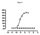
結合要素の中和効力は、通常、特に断りのない限り、IC50値(nM)で表される。機能アッセイでは、IC50は、生物学的応答をその最大の50%低下させる結合要素の濃度である。リガンド結合研究では、IC50は、受容体結合を最大特異的結合レベルの50%低下させる濃度である。IC50は、最大生物学的応答の%を結合要素濃度のlogの関数としてプロットし、Prism(GraphPad)などのソフトウエアプログラムを用い、そのデータにシグモイド関数を当てはめてIC50値を求めることにより計算することができる。効力は、当業者に公知であり、かつ/または本明細書に記載もしくは言及されているような1種以上のアッセイを用いて決定または測定することができる。 The neutralizing potency of a binding element is usually expressed as an IC 50 value (nM) unless otherwise specified. In a functional assay, the IC 50 is the concentration of binding member that reduces the biological response by 50% of its maximum. In ligand binding studies, the IC 50 is the concentration that reduces receptor binding by 50% of the maximum specific binding level. IC 50 is determined by plotting the% of maximum biological response as a function of log of binding element concentration and using a software program such as Prism (GraphPad) and applying the sigmoid function to the data to determine the IC 50 value. Can be calculated. Efficacy can be determined or measured using one or more assays known to those of skill in the art and / or as described or referred to herein.
結合要素の中和効力は幾何平均として表すことができる。本明細書において幾何平均(geomean)(幾何平均(geometric mean)としても知られる)は、10を底として変換し直したデータセットの対数値の平均を意味する。これには、例えば少なくとも2回、好ましくは少なくとも5回、より好ましくは少なくとも10回の反復など、少なくとも2回の測定である必要がある。当業者ならば、反復数が多いほど幾何平均値が確固としたものになることが分かるであろう。反復数の選択は当業者の裁量に任される。 The neutralizing potency of the binding element can be expressed as a geometric mean. As used herein, geometrican (also known as geometric mean) means the average of the logarithmic values of a data set retransformed with a base of 10. This requires at least 2 measurements, for example at least 2 times, preferably at least 5 times, more preferably at least 10 repetitions. One skilled in the art will recognize that the greater the number of iterations, the more robust the geometric mean value. The selection of the number of iterations is left to the discretion of the skilled person.
本明細書に記載のアッセイにおける結合要素によるIgE活性の中和は、その結合要素がIgEと結合し、それを中和することを示す。結合要素とIgEの結合を決定するために用い得る他の方法としては、ELISA、ウエスタンブロッティング、免疫沈降、アフィニティークロマトグラフィーおよび生化学アッセイが含まれる。 Neutralization of IgE activity by a binding member in the assays described herein indicates that the binding member binds and neutralizes IgE. Other methods that can be used to determine binding of binding elements to IgE include ELISA, Western blotting, immunoprecipitation, affinity chromatography and biochemical assays.
2種のIgEに対する結合要素の交差反応性の程度を評価するためには、第一の種(例えば、ヒト)由来のIgEを用いたアッセイにおいて算出された結合要素の中和効力を、第二の種(例えば、カニクイザル)由来のIgEを用いた、同様の条件下での同様のアッセイにおける結合要素の中和効力と比較すればよい。あるいは、交差反応性は、本明細書の他所により詳細に記載されているように競合結合アッセイで評価することもできる。 In order to assess the extent of binding element cross-reactivity to two IgEs, the neutralizing potency of the binding element calculated in the assay using IgE from the first species (eg, human) is Comparison with the neutralizing potency of the binding member in similar assays under similar conditions using IgE from different species (eg, cynomolgus monkeys). Alternatively, cross-reactivity can be assessed in a competitive binding assay as described in more detail elsewhere herein.
本発明の結合要素は、ヒトIgE結合または生物学的アッセイの方がヒト以外の種に由来するIgEを用いた同様のアッセイよりも大きな中和効力を持ち得る。よって、ヒトIgEを用いたアッセイにおける結合要素の中和効力は、ヒト以外の種に由来するIgEを用いた同様のアッセイの場合よりも大きいものであり得る。ヒトIgE結合または生物学的アッセイでの効力は、例えば、カニクイザルのIgEを用いた同様のアッセイよりも約10倍大きく、または他の実施態様では、約25倍または約125倍大きい。より具体的には、ヒトRBL−ER51カルシウムシグナル伝達アッセイでの効力は、25ng/ml濃度のヒトIgEに関して決定し、他の点では同様の条件下で100ng/mlのカニクイザルIgEを用いた場合の抗力と比較すればよい。ヒトIgEとカニクイザルIgEを用いた同様のRBL−ER51カルシウムシグナル伝達アッセイで得られたデータの例を表5bに示す。 The binding members of the invention may have greater neutralizing potency for human IgE binding or biological assays than similar assays using IgE from species other than human. Thus, the neutralizing potency of a binding member in an assay using human IgE can be greater than in a similar assay using IgE from a species other than human. Efficacy in human IgE binding or biological assays is, for example, about 10-fold greater than similar assays using cynomolgus IgE, or in other embodiments about 25-fold or about 125-fold greater. More specifically, potency in the human RBL-ER51 calcium signaling assay was determined for a 25 ng / ml concentration of human IgE, and otherwise using 100 ng / ml cynomolgus IgE under similar conditions. Compare with drag. An example of data obtained in a similar RBL-ER51 calcium signaling assay using human IgE and cynomolgus IgE is shown in Table 5b.
本発明の結合要素は、他種のIgEよりもヒトIgEに対して強い親和性を持ち得る。ヒトIgEに対する結合要素の親和性は、例えば、カニクイザルIgEに対するよりも約5倍強く、他の実施態様では、約10倍強いものであり得る。 The binding members of the invention may have a stronger affinity for human IgE than for other types of IgE. The affinity of the binding member for human IgE can be, for example, about 5 times stronger than for cynomolgus IgE, and in other embodiments about 10 times stronger.
本発明の結合要素は、25ng/ml濃度のヒトIgEで、IgE中和効力または約10nM未満のIC50を持ち得る。あるいは、IC50は約3nM未満である。他の実施態様では、IC50は約1nM未満、または約0.5nM未満、または約0.2nM未満である。 A binding member of the invention can have an IgE neutralizing potency or an IC 50 of less than about 10 nM with human IgE at a concentration of 25 ng / ml. Alternatively, the IC 50 is less than about 3 nM. In other embodiments, the IC 50 is less than about 1 nM, or less than about 0.5 nM, or less than about 0.2 nM.
ヒトIgEに対するIgE結合要素の結合動態および親和性(平衡解離定数KDとして表される)は、例えば、表面プラズモン共鳴(BIACORE)を用いて決定することができる。本発明の結合要素のヒトIgEに対する親和性(KD)は通常約80nM未満であり、いくつかの実施態様では、KDは約10nM未満であり、他の実施態様では5nM未満であり、他の実施態様では2nM未であり、他の実施態様では1nM未満である。カニクイザルIgEに対する親和性は通常、約15nM未満である。 The binding kinetics and affinity (expressed as equilibrium dissociation constant KD) of the IgE binding element for human IgE can be determined using, for example, surface plasmon resonance (BIACORE). The binding member's affinity (KD) for human IgE is typically less than about 80 nM, in some embodiments, the KD is less than about 10 nM, in other embodiments, less than 5 nM, In embodiments, it is less than 2 nM and in other embodiments less than 1 nM. The affinity for cynomolgus IgE is usually less than about 15 nM.
in vivoにおいて内因性IgEはグリコシル化可能であり、したがって、グリコシル化されたヒトIgEはヒトの療法のための治療標的となる。細菌に由来し、グリコシル化され得ない組換えヒトIgEも本明細書に記載のアッセイにおいて使用可能であるが、本発明の結合要素は、U266.B1などの骨髄腫細胞系統により生産されたIgEなど、グリコシル化されたヒトIgEと結合し得る。グリコシル化ヒトIgEはin vivoヒト適用の標的抗原であることから、これは本発明の結合要素の大きな利点となる。 In vivo, endogenous IgE can be glycosylated, thus glycosylated human IgE is a therapeutic target for human therapy. Although recombinant human IgE that is derived from bacteria and cannot be glycosylated can also be used in the assays described herein, the binding member of the present invention is an IgE produced by a myeloma cell line such as U266.B1. And can bind to glycosylated human IgE. This is a major advantage of the binding member of the present invention because glycosylated human IgE is a target antigen for in vivo human applications.
本発明の結合要素は抗体分子、好ましくはヒト抗体分子またはヒト化抗体分子を含み得る。本発明の一態様において、抗体分子はモノクローナル抗体である。 A binding member of the invention may comprise an antibody molecule, preferably a human antibody molecule or a humanized antibody molecule. In one embodiment of the invention, the antibody molecule is a monoclonal antibody.
抗原結合部位は一般に重鎖可変(VH)および軽鎖可変(VL)免疫グロブリンドメインによって形成され、相補性決定領域(CDR)と呼ばれる6つの表面ポリペプチドループによって形成された抗原結合インターフェースを伴う。VH(HCDR1、HCDR2およびHCDR3)およびVL(LCDR1、LCDR2およびLCDR3)にはそれぞれ3つのCDRが存在し、フレームワーク領域(FR)が伴う。 Antigen binding sites are generally formed by heavy chain variable (VH) and light chain variable (VL) immunoglobulin domains, with an antigen binding interface formed by six surface polypeptide loops called complementarity determining regions (CDRs). There are three CDRs in VH (HCDR1, HCDR2 and HCDR3) and VL (LCDR1, LCDR2 and LCDR3), respectively, with a framework region (FR).
本発明の結合要素は通常、抗体VHおよび/またはVLドメインを含む。本発明のVHドメインはHCDRセットを含み、VLドメインはLCDRセットを含む。抗体分子はVH CDR1、CDR2およびCDR3とフレームワークを含む抗体VHドメインを含み得る。それは、その代わりにまたはそれに加えて、VL CDR1、CDR2およびCDR3とフレームワーク抗体を含むVLドメインも含み得る。本発明の抗体VHドメイン(配列番号:2、298、338、318、328、118、309、28、68、8、48、288、158、268、168、38、128、78、138、188、198、98、18、88、58、108、218、248、228、238、178、208、278、148、426、278および258)とVLドメイン(配列番号:353、358、360、362、364、366、368、370、372、374、376、378、380、382、384、386、388、390、392、394、396、398、400、402、404、406、408、410、412、414、416、418、420、422、431および424)とCDR(配列番号:3−5、9−11、14−16、19−21、24−26、29−31、34−36、39−41、44−46、49−51、54−56、59−61、64−66、69−71、74−76、79−81、84−86、89−91、94−96、99−101、104−106、109−111、114−116、119−121、124−126、129−131、134−136、139−141、144−146、149−151、154−156、159−161、164−166、169−171、174−176、179−181、184−186、189−191、194−196、199−201、204−206、209−211、214−216、219−221、224−226、229−231、234−236、239−241、244−246、249−251、254−256、259−261、264−266、269−271、274−276、279−281、284−286、289−291、294−296、299−301、304−306、309−311、314−316、319−321、324−326、329−331、334−336、339−341、344−346、350−352、354−356および427−429)の例は本開示の位置をなす添付の配列表に挙げられている通りである。さらなるCDRは下記および表1に開示されている。本明細書に開示されているVHおよびVL配列、CDR配列、CDRセットおよびHCDRセットおよびLCDRセットは全て本発明の態様および実施態様を表す。 The binding members of the invention typically comprise antibody VH and / or VL domains. The VH domain of the present invention includes an HCDR set, and the VL domain includes an LCDR set. The antibody molecule may comprise an antibody VH domain comprising VH CDR1, CDR2 and CDR3 and a framework. It may alternatively or additionally include VL domains comprising VL CDR1, CDR2 and CDR3 and framework antibodies. Antibody VH domains of the invention (SEQ ID NOs: 2, 298, 338, 318, 328, 118, 309, 28, 68, 8, 48, 288, 158, 268, 168, 38, 128, 78, 138, 188, 198, 98, 18, 88, 58, 108, 218, 248, 228, 238, 178, 208, 278, 148, 426, 278 and 258) and the VL domain (SEQ ID NOs: 353, 358, 360, 362, 364) 366, 368, 370, 372, 374, 376, 378, 380, 382, 384, 386, 388, 390, 392, 394, 396, 398, 400, 402, 404, 406, 408, 410, 412, 414 416, 418, 420, 422, 431 and 424) and CDR (SEQ ID NOs: 3-5, 9-11, 14- 6, 19-21, 24-26, 29-31, 34-36, 39-41, 44-46, 49-51, 54-56, 59-61, 64-66, 69-71, 74-76, 79-81, 84-86, 89-91, 94-96, 99-101, 104-106, 109-111, 114-116, 119-121, 124-126, 129-131, 134-136, 139- 141, 144-146, 149-151, 154-156, 159-161, 164-166, 169-171, 174-176, 179-181, 184-186, 189-191, 194-196, 199-201, 204-206, 209-211, 214-216, 219-221, 224-226, 229-231, 234-236, 239-241, 244 246, 249-251, 254-256, 259-261, 264-266, 269-271, 274-276, 279-281, 284-286, 289-291, 294-296, 299-301, 304-306, 309-311, 314-316, 319-321, 324-326, 329-331, 334-336, 339-341, 344-346, 350-352, 354-356 and 427-429) are examples of this disclosure. It is as listed in the attached sequence listing. Additional CDRs are disclosed below and in Table 1. The VH and VL sequences, CDR sequences, CDR sets and HCDR sets and LCDR sets disclosed herein all represent aspects and embodiments of the present invention.
本明細書に記載されている、「CDRセット」はCDR1、CDR2およびCDR3を含む。よって、HCDRセットはHCDR1、HCDR2およびHCDR3を表し、LCDRセットはLCDR1、LCDR2およびLCDR3を表す。特に断りのない限り、「CDRセット」はHCDRおよびLCDRを含む。 As described herein, “CDR sets” include CDR1, CDR2 and CDR3. Thus, the HCDR set represents HCDR1, HCDR2, and HCDR3, and the LCDR set represents LCDR1, LCDR2, and LCDR3. Unless otherwise specified, “CDR set” includes HCDR and LCDR.
あるいは、本発明の結合要素は非抗体分子内に抗原結合部位を含んでもよく、通常、1以上のCDR、例えば、非抗体タンパク質スキャフォールド内のCDRセットによって提供される。 Alternatively, a binding member of the invention may include an antigen binding site within a non-antibody molecule and is typically provided by one or more CDRs, eg, a CDR set within a non-antibody protein scaffold.
本明細書に記載されている、親抗体分子としては、表1に示されるようなCDR配列セットを有するものが単離された(表1の抗体1を参照)。至適化の過程を通じて、本発明者らは、親CDR配列に由来し、表1に示されている位置に修飾を有するCDR配列番号を有する抗体クローン2〜36番のパネルを作製した。よって、例えば、表1から、抗体2が親HCDR1、HCDR3およびLCDR3配列を有し、Kabat残基56がRで置換された親HCDR2配列、Kabat残基31がTで置換された親LCDR1、およびKabat残基53がRで置換されたLCDR2を有することが分かる。
The parent antibody molecules described herein were isolated having the CDR sequence set as shown in Table 1 (see
本明細書には、HCDR1が配列番号3(Kabat残基31〜35)であり、HCDR2が配列番号4(Kabat残基50〜65)であり、HCDR3が配列番号5(Kabat残基95〜102)であり、LCDR1が配列番号354(Kabat残基24〜34)であり、LCDR2が配列番号355(Kabat残基50〜56)であり、LCDR3が配列番号356(Kabat残基89〜97)である、表1(抗体1)に示されるような親CDRセットを含む結合要素が記載される。本発明の結合要素はまた、1以上のCDRが1以上のアミノ酸付加、置換、欠失および/または挿入を有する、表1に示されるような親結合要素であってもよい。一実施態様では、結合要素は8個までのアミノ酸付加、置換、欠失および/または挿入を有する。
In the present specification, HCDR1 is SEQ ID NO: 3 (Kabat residues 31 to 35), HCDR2 is SEQ ID NO: 4 (
本発明の結合要素は、本明細書に記載されているとおりCDRの1つまたは組合せを含み得る。 The binding elements of the invention may comprise one or a combination of CDRs as described herein.
例えば、本発明の結合要素またはVHドメインは、Kabat残基31がGで置換されているか、またはKabat残基33がPで置換されているか、またはKabat残基34がVで置換されている親HCDR1を含み得る(表1参照)。 For example, a binding member or VH domain of the invention is a parent wherein Kabat residue 31 is substituted with G, or Kabat residue 33 is substituted with P, or Kabat residue 34 is substituted with V. HCDR1 may be included (see Table 1).
本発明の結合要素またはVHドメインは以下の修飾:
Kabat残基51がVで置換されている;
GがKabat残基52Bにある;
Kabat残基53がGで置換されている;
Kabat残基56がRで置換されている;
Kabat残基63がAで置換されている
の1以上を有する親HCDR2を含み得る。
The binding elements or VH domains of the invention are modified as follows:
Kabat residue 51 is replaced with V;
G is at Kabat residue 52B;
Kabat residue 53 is replaced with G;
It may include a parent HCDR2 having one or more of Kabat residues 63 replaced with A.
ある特定の実施態様では、結合要素は親HCDR2のKabat残基52BにG、そしてKabat残基56を有し、これらの結合要素はIgEに対して低いKDおよび/またはIgEにより媒介される生物活性の阻害に関して高い効力を示す傾向がある。親HCDR2のKabat残基52BにG、そしてKabat残基56にRを有するいくつかの抗体を示す表1を参照。
In certain embodiments, the binding member has a G at Kabat residue 52B of parent HCDR2 and
本発明の結合要素またはVHドメインは以下の置換:
LまたはSで置換されたKabat残基95;
S、T、WまたはFで置換されたKabat残基96;
YまたはFで置換されたKabat残基97;
PまたはFで置換されたKabat残基98;
Yで置換されたKabat残基99;
IまたはSで置換されたKabat残基100
の1以上を有する親HCDR3を含み得る。
The binding elements or VH domains of the invention have the following substitutions:
Kabat residue 95 substituted with L or S;
Kabat residue 96 substituted with S, T, W or F;
Kabat residue 97 substituted with Y or F;
Kabat residue 98 substituted with P or F;
Kabat residue 99 substituted with Y;
A parent HCDR3 having one or more of:
本発明の結合要素またはVHドメインは以下の置換:
H、LまたはSで置換されたKabat残基95;
I、S、T、WまたはFで置換されたKabat残基96;
A、YまたはFで置換されたKabat残基97;
V、PまたはFで置換されたKabat残基98;
AまたはYで置換されたKabat残基99;
G、IまたはSで置換されたKabat残基100
の1以上を有する親HCDR3(配列番号5)を含み得る。
The binding elements or VH domains of the invention have the following substitutions:
Kabat residue 95 substituted with H, L or S;
Kabat residue 96 substituted with I, S, T, W or F;
Kabat residue 97 substituted with A, Y or F;
Kabat residue 98 substituted with V, P or F;
Kabat residue 99 substituted with A or Y;
The parent HCDR3 (SEQ ID NO: 5) having one or more of:
結合要素またはそのVLドメインは、Kabat残基31および/またはKabat残基33がTで置換されている親LCDR1を含み得る。例えば、LCDR1のKabat残基31がTである結合要素はIgEに対して高い親和性および/またはIgEにより媒介される生物活性の阻害において高い効力を示す傾向があった。表1に示されるように、抗体2〜9、11〜19、22〜30および32〜36は全てLCDR1のKabat残基31にTを有する。他の抗体またはLCDR1配列も、例えば部位特異的突然変異誘発を用い、この残基にTを有するように操作することができる。 The binding member or its VL domain may comprise a parent LCDR1 in which Kabat residue 31 and / or Kabat residue 33 is replaced with T. For example, binding members in which the Kabat residue 31 of LCDR1 is T tended to show high affinity for IgE and / or high potency in inhibiting IgE-mediated biological activity. As shown in Table 1, antibodies 2-9, 11-19, 22-30 and 32-36 all have a T at Kabat residue 31 of LCDR1. Other antibodies or LCDR1 sequences can also be engineered to have a T at this residue, for example using site-directed mutagenesis.
結合要素またはそのVLドメインは、Kabat残基53がRで置換され、かつ/またはKabat残基56がPで置換されている親LCDR2を含み得る。
The binding member or its VL domain may comprise a parent LCDR2 in which Kabat residue 53 is replaced with R and / or
結合要素またはそのVLドメインはまた、1〜5置換など、1以上のアミノ酸付加、置換、欠失および/または挿入を有する親LCDR3も含み得る。 The binding member or its VL domain may also include a parent LCDR3 having one or more amino acid additions, substitutions, deletions and / or insertions, such as 1-5 substitutions.
別の実施態様では、本発明は、HCDR1が配列番号279のアミノ酸配列を有し、HCDR2が配列番号280のアミノ酸配列を有し、HCDR3が配列番号281のアミノ酸配列を有し、LCDR1が配列番号284のアミノ酸配列を有し、LCDR2が配列番号285のアミノ酸配列を有し、LCDR3が配列番号286のアミノ酸配列を有する結合要素である。例えば、表1の抗体33参照。本発明のさらに他の実施態様は、ヒトIgEとの結合に関して表1の抗体33と競合し得る抗体分子などの結合要素である。 In another embodiment, the invention provides that HCDR1 has the amino acid sequence of SEQ ID NO: 279, HCDR2 has the amino acid sequence of SEQ ID NO: 280, HCDR3 has the amino acid sequence of SEQ ID NO: 281, and LCDR1 has SEQ ID NO: LCDR2 is a binding element having the amino acid sequence of 284, LCDR2 has the amino acid sequence of SEQ ID NO: 285, and LCDR3 is the amino acid sequence of SEQ ID NO: 286. See, for example, antibody 33 in Table 1. Yet another embodiment of the invention is a binding member such as an antibody molecule that can compete with antibody 33 of Table 1 for binding to human IgE.
本発明は、抗体1〜36のいずれかのHCDR1、HCDR2および/もしくはHCDR3および/または抗体1〜36のいずれかのLCDR1、LCDR2および/もしくはLCDR3、例えば、表1に示されている抗体1〜36のいずれかのCDRセットを含む結合要素を提供する。この結合要素はこれらの抗体の1つのVH CDRセットを含み得る。所望によりそれはまた、これらの抗体の1つのVL CDRを含んでもよく、このVL CDRはVH CDRと同じ抗体に由来するものでも、または異なる抗体に由来するものでもよい。抗体1〜36のいずれかのHCDRセットを含むVHドメインおよび/または抗体1〜36のいずれかのLCDRセットを含むVLドメインも本発明により提供される。
The invention relates to any HCDR1, HCDR2 and / or HCDR3 of antibodies 1-36 and / or LCDR1, LCDR2 and / or LCDR3 of any of antibodies 1-36, eg,
一般に、VHドメインをVLドメインと対合して抗体抗原結合部位を提供するが、以下にさらに述べるとおりVHまたはVLドメイン単独を、抗原と結合させるのに用いてもよい。抗体1 VHドメイン(表3参照)を抗体1 VLドメイン(表2参照)と対合すると、抗体1 VHドメインとVLドメインの双方を含む抗体抗原結合部位が形成される。本明細書に開示されている他のVHおよびVLドメインにも類似の実施態様が提供される。他の実施態様では、抗体1 VHを抗体1 VL以外のVLドメインと対合する。軽鎖の無差別性は当技術分野で十分確立されている。ここでもまた、本発明により、本明細書に開示されている他のVHおよびVLドメインに類似の実施態様が提供される。よって、親の、または抗体2〜36のいずれかのVHを、親の、または抗体2〜36のいずれかのVLと対合することができる。
In general, a VH domain is paired with a VL domain to provide an antibody antigen binding site, but a VH or VL domain alone may be used to bind an antigen as further described below. When the
結合要素は、開示されているHおよび/またはL CDRセット内に20、16、10、9といった多くの、またはそれより少ない、例えば、1、2、3、4または5つのアミノ酸付加、置換、欠失および/または挿入を有する親抗体の、または抗体2〜36のいずれかのHおよび/またはL CDRセットを含み得る。あるいは、結合要素は、開示されているHおよび/またはL CDRセット内に20、16、10、9といった多くの、またはそれより少ない、例えば、1、2、3、4または5つのアミノ酸置換を有する親抗体の、または抗体2〜36のいずれかのHおよび/またはL CDRセットを含み得る。このような修飾は潜在的にこれらのCDRセット内のいずれの残基で行ってもよい。例えば、修飾は、表1に示されるように、抗体2〜36のいずれかにおいて改変された位置で行うことができる。よって、これら1以上の置換が、以下の残基:HCDRのKabat残基31、33、34、51、52B、53、56、63、95、96、97、98、99および100、ならびにLCDRのKabat残基31、33、53および56における1以上の置換を含み得る。
The binding member may be many, such as 20, 16, 10, 9, or fewer, such as 1, 2, 3, 4 or 5 amino acid additions, substitutions, within the disclosed H and / or L CDR sets. It may include a H and / or L CDR set of a parent antibody with deletions and / or insertions, or any of antibodies 2-36. Alternatively, the binding member has many, or fewer, such as 1, 2, 3, 4 or 5 amino acid substitutions, such as 20, 16, 10, 9, within the disclosed H and / or L CDR sets. It may comprise a H and / or L CDR set of the parent antibody having, or any of antibodies 2-36. Such modifications can potentially be made at any residue within these CDR sets. For example, modifications can be made at altered positions in any of antibodies 2-36 as shown in Table 1. Thus, one or more of these substitutions are the following residues:
結合要素は抗体フレームワーク内に1以上のCDR、例えば、CDRセットを有する抗体分子を含み得る。例えば、抗体分子を得るには、抗体の1以上のCDRまたはCDRセットをフレームワーク(例えば、ヒトフレームワーク)にグラフトすればよい。これらのフレームワーク領域はヒト生殖細胞系遺伝子配列のものであってもよいし、または非生殖細胞系のものでもよい。例えば、フレームワークは生殖系列化されてもよく、フレームワーク内の1以上の残基が、最も類似性の高いヒト生殖細胞系フレームワークの等しい位置の残基と一致するように変更される。よって、本発明の結合要素は、ヒト生殖細胞系フレームワーク、例えば、ヒト生殖細胞系IgG VHフレームワークにHCDRセットを含むVHドメインを有する単離されたヒト抗体分子であり得る。通常、結合要素はまた、例えば、ヒト生殖細胞系IgG VLフレームワークにLCDRセットを含むVLドメインも有する。 A binding member can comprise an antibody molecule having one or more CDRs, eg, CDR sets, within an antibody framework. For example, to obtain an antibody molecule, one or more CDRs or CDR sets of an antibody can be grafted onto a framework (eg, a human framework). These framework regions may be of human germline gene sequence or non-germline. For example, the framework may be germlined, and one or more residues in the framework are changed to match residues at the same position in the most similar human germline framework. Thus, a binding member of the invention can be an isolated human antibody molecule having a VH domain comprising an HCDR set in a human germline framework, eg, a human germline IgG VH framework. Typically, the binding member also has a VL domain that includes the LCDR set, eg, in the human germline IgG VL framework.
VHおよび/またはVLフレームワーク残基は、本明細書に記述および例示されているとおり、例えば部位特異的突然変異誘発を用いて改変することができる。 VH and / or VL framework residues can be modified using, for example, site-directed mutagenesis, as described and exemplified herein.
本発明のVHドメインまたはこのようなVHドメインを含む結合要素は、表3の抗体1〜36番のいずれかのVHドメインを有していてもよい。例えば、本発明のVHドメインは以下の修飾:
Kabat残基19がSで置換されている;
Kabat残基25がPで置換されている;
Kabat残基26がEで置換されている;
Kabat残基27がLで置換されている;
Kabat残基29がLで置換されている;
Kabat残基30がGで置換されている;
Kabat残基68がAで置換されている;
Kabat残基69がVで置換されている;
Kabat残基71がKで置換されている;
Kabat残基77がMで置換されている;
Kabat残基82BがGで置換されている;
Kabat残基82CがPで置換されている;
Kabat残基107がAで置換されている;
Kabat残基108がFで置換されている;
Kabat残基110がAで置換されている;
Kabat残基111がIで置換されている;および/または
Kabat残基112がPで置換されている
の1以上を有する親フレームワークを含み得る(表3の抗体1)。
The VH domain of the present invention or a binding member comprising such a VH domain may have any of the VH domains of antibodies 1-36 in Table 3. For example, the VH domain of the present invention has the following modifications:
Kabat residue 19 is replaced with S;
Kabat residue 26 is replaced by E;
Kabat residue 27 is substituted with L;
Kabat residue 29 is substituted with L;
Kabat residue 68 is replaced with A;
Kabat residue 69 is replaced with V;
Kabat residue 71 is replaced by K;
Kabat residue 77 is replaced with M;
Kabat residue 82B is replaced with G;
Kabat residue 82C is replaced with P;
Kabat residue 107 is replaced with A;
Kabat residue 108 is substituted with F;
It may include a parent framework having one or more of Kabat residue 111 substituted with I; and / or Kabat residue 112 substituted with P (
ある特定の実施態様では、Kabat残基19がSで置換され、Kabat残基25がPで置換され、Kabat残基52BがGで置換され、Kabat残基56がRで置換され、Kabat残基71がKで置換されている。
In certain embodiments, Kabat residue 19 is substituted with S,
一般に、本発明の結合要素またはVHドメインは、VHフレームワーク領域に1〜10個の置換を有する表3の抗体1〜36のいずれかのVHフレームワークを含み得る。 In general, a binding member or VH domain of the invention can comprise the VH framework of any of antibodies 1-36 of Table 3 having 1-10 substitutions in the VH framework region.
別の実施態様では、本発明の結合要素またはVHドメインは、VH領域に1〜7個の置換を有する表3の抗体1のVHフレームワークを含み得る。
In another embodiment, a binding member or VH domain of the invention may comprise the VH framework of
別の実施態様では、本発明の結合要素またはVHドメインは、VH領域に1〜10個の置換を有する表3の抗体33のVHフレームワークを含み得る。 In another embodiment, a binding member or VH domain of the invention may comprise the VH framework of antibody 33 of Table 3 having 1-10 substitutions in the VH region.
本発明のVLドメインまたはこのようなVLドメインを含む結合要素は、表2の抗体1〜36番のいずれかのVLドメインを有していてもよい。例えば、抗体VLドメインはフレームワーク領域に以下の修飾:
Kabat残基2がFで置換されていてもよい;
Kabat残基3がMまたはEで置換されていてもよい;
Kabat残基5がSで置換されていてもよい;
Kabat残基13がAで置換されていてもよい;
Kabat残基22がAで置換されていてもよい;
Kabat残基37がQで置換されている;
Kabat残基39がKで置換されている;
Kabat残基40がPで置換されている;
Kabat残基42がLで置換されている;
Kabat残基45がAで置換されている;
Kabat残基58がVで置換されている;
Kabat残基69がDで置換されている;
Kabat残基70がAで置換されている;
Kabat残基76がGまたはRで置換されている;
Kabat残基77がRで置換されている;
Kabat残基79がQまたはRで置換されている;
Kabat残基80がAで置換されている;
Kabat残基103がEで置換されている;
Kabat残基105がSで置換されている;および/または
Kabat残基106がAで置換されている
の1以上を有する表2の抗体の配列を有し得る。
The VL domain of the present invention or a binding member comprising such a VL domain may have any of the VL domains of
Kabat residue 13 may be substituted with A;
Kabat residue 22 may be substituted with A;
Kabat residue 37 is replaced with Q;
Kabat residue 39 is replaced by K;
Kabat residue 58 is replaced with V;
Kabat residue 69 is replaced with D;
Kabat residue 76 is substituted with G or R;
Kabat residue 77 is substituted with R;
Kabat residue 79 is substituted with Q or R;
The antibody sequences of Table 2 may have one or more of which Kabat residue 105 is substituted with S; and / or Kabat residue 106 is substituted with A.
ある特定の実施態様では、Kabat残基42がLで置換され、Kabat残基45がAで置換されている。
In certain embodiments,
一般に、本発明の結合要素またはVLドメインは、VLフレームワーク領域に1〜10個の置換を有する表2の抗体1〜36のいずれかのVLフレームワークを含み得る。 In general, a binding member or VL domain of the invention can comprise the VL framework of any of antibodies 1-36 of Table 2 having 1-10 substitutions in the VL framework region.
一般に、本発明の結合要素またはVLドメインは、VL領域に1〜9個の置換を有する表2の抗体1のいずれかのVLフレームワークを含み得る。
In general, a binding member or VL domain of the invention may comprise any VL framework of
一般に、本発明の結合要素またはVLドメインは、VL領域に1〜6個の置換を有する表2の抗体33のいずれかのVLフレームワークを含み得る。 In general, a binding member or VL domain of the invention may comprise any VL framework of antibody 33 of Table 2 having 1 to 6 substitutions in the VL region.
生殖系列化されていない抗体分子は生殖系列化されている抗体分子と比べると、同じCDRを有するが、フレームワークは異なる。生殖系列化されている抗体は、これらの抗体に関して本明細書で示されているVHおよびVLドメイン配列のフレームワーク領域を生殖系列化することによって製造し得る。 Non-germlined antibody molecules have the same CDRs but different frameworks compared to germlined antibody molecules. Germlined antibodies can be produced by germlineating the framework regions of the VH and VL domain sequences set forth herein for these antibodies.
本発明の結合要素は、IgEとの結合に関して本発明の他のいずれかの結合要素と競合するものであり得る。このような結合要素は競合的にIgEと結合すると言われ、同じエピトープと結合する。結合要素間の競合は、例えばELISAの使用および/または1以上の他のタグ無し結合要素の存在下で検出可能な1つの結合要素に特異的リポーター分子をタグ付けし、それにより、同じエピトープと結合する結合要素ならびに重複エピトープと結合する結合要素の検出を可能とすることにより、in vitroで容易にアッセイすることができる。このような方法は当業者には容易に分かり、本明細書にさらに詳細に記載される。よって、本発明のさらなる態様は、抗体分子、例えば特に、IgEとの結合に関して、VHおよび/またはVLドメイン、CDR、例えば、HCDR3または親抗体もしくは抗体1〜36のいずれかのCDRセットを含む抗体分子と競合するヒト抗体抗原結合部位を含む結合要素を提供する。一実施態様では、本発明の結合要素は表2および3の抗体33と競合する。 The binding member of the invention can be one that competes with any other binding member of the invention for binding to IgE. Such binding elements are said to bind IgE competitively and bind to the same epitope. Competition between binding members can tag specific reporter molecules with one binding member that is detectable in the presence of, for example, ELISA and / or the presence of one or more other untagged binding members, thereby By allowing detection of binding elements that bind as well as binding elements that bind overlapping epitopes, they can be readily assayed in vitro. Such methods are readily apparent to those skilled in the art and are described in further detail herein. Thus, a further aspect of the invention relates to antibody molecules, eg, antibodies comprising a CDR set of VH and / or VL domains, CDRs, eg HCDR3 or parent antibody or any of antibodies 1-36, particularly for binding to IgE. A binding element comprising a human antibody antigen binding site that competes with a molecule is provided. In one embodiment, a binding member of the invention competes with antibody 33 of Tables 2 and 3.
さらなる態様において、本発明は、IgEとの結合に関して抗体抗原結合部位と競合するヒト抗体抗原結合部位を含む結合要素を提供し、この抗体抗原結合部位はVHドメインとVLドメインからなり、このVHおよびVLドメインは本明細書に開示されているような親抗体または抗体1〜36のいずれかのCDRセットを含む。 In a further aspect, the invention provides a binding element comprising a human antibody antigen binding site that competes with an antibody antigen binding site for binding to IgE, the antibody antigen binding site consisting of a VH domain and a VL domain, wherein the VH and The VL domain comprises the CDR set of either the parent antibody or antibodies 1-36 as disclosed herein.
さらなる態様において、本発明は、本発明の結合要素、VHドメインおよび/またはVLドメインをコードする配列を含む単離された核酸を提供する。本発明のVHドメインをコードする例示的核酸は添付の配列表の配列番号:1、7、17、27、37、47、57、67、77、87、97、107、117、127、137、147、157、167、177、187、197、207、217、227、237、247、257、267、277、287、297、307、317、327、337および425である。本発明のVLドメインをコードする例示的核酸は添付の配列表の配列番号:6、357、359、361、363、365、367、369、371、373、375、377、379、381、383、385、387、389、391、393、395、397、399、401、403、405、407、409、411、413、415、417、419、421、430および423である。本発明はまた、本発明の結合要素、VHドメインおよび/またはVLドメインを作製する方法も含み、それは該結合要素、VHドメインおよび/またはVLドメインの生産を行うための条件下で該核酸を発現させること、およびその結合要素を単離または精製することによりそれを回収することを含む。 In a further aspect, the present invention provides an isolated nucleic acid comprising a sequence encoding a binding member, VH domain and / or VL domain of the present invention. Exemplary nucleic acids encoding the VH domains of the invention are SEQ ID NOs: 1, 7, 17, 27, 37, 47, 57, 67, 77, 87, 97, 107, 117, 127, 137, in the attached sequence listing. 147, 157, 167, 177, 187, 197, 207, 217, 227, 237, 247, 257, 267, 277, 287, 297, 307, 317, 327, 337 and 425. Exemplary nucleic acids encoding VL domains of the invention are SEQ ID NOs: 6, 357, 359, 361, 363, 365, 367, 369, 371, 373, 375, 377, 379, 381, 383, of the attached sequence listing. 385, 387, 389, 391, 393, 395, 397, 399, 401, 403, 405, 407, 409, 411, 413, 415, 417, 419, 421, 430 and 423. The invention also includes a method of making a binding element, VH domain and / or VL domain of the invention, which expresses the nucleic acid under conditions to effect production of the binding element, VH domain and / or VL domain And recovering it by isolating or purifying its binding member.
本発明の別の態様は、本明細書に開示されているVH CDRまたはVL CDR配列をコードする、一般に単離された核酸を提供する。 Another aspect of the invention provides a generally isolated nucleic acid encoding a VH CDR or VL CDR sequence disclosed herein.
さらなる態様は、本発明の核酸を含む、または本発明の核酸で形質転換された宿主細胞を提供する。 A further aspect provides a host cell comprising or transformed with a nucleic acid of the invention.
本発明のさらなる態様は、本発明の結合要素を含有する組成物、および治療によるヒトまたは動物の処置方法を含む、IgEを阻害および/または中和する方法における使用を提供する。 Further aspects of the invention provide for use in methods of inhibiting and / or neutralizing IgE, including compositions containing the binding members of the invention and methods of treatment of humans or animals by therapy.
例えば、本発明の結合要素は、ヒトもしくは動物身体(例えば、ヒト患者)またはin vitroにおける生物学的応答、疾病、障害または症状の処置および/または予防方法に使用可能であり、あるいは診断方法に使用可能である。 For example, the binding members of the invention can be used in methods of treatment and / or prevention of biological responses, diseases, disorders or symptoms in the human or animal body (eg, human patients) or in vitro, or in diagnostic methods. It can be used.
この治療および/または予防方法は、該患者に本発明の結合要素を、IgEを測定可能に中和するのに十分な量で投与することを含み得る。本発明に従って処置可能な症状としては、アレルギーおよび喘息など、IgEが役割を果たすいずれのものも含まれる。 This method of treatment and / or prophylaxis may comprise administering to the patient a binding member of the invention in an amount sufficient to measurable neutralize IgE. Symptoms that can be treated according to the present invention include any of which IgE plays a role, such as allergies and asthma.
本発明のこれら、およびその他の態様を以下にさらに詳細に記載する。
ここで、本明細書で用いられる「および/または」は、他を伴って、または伴わずに2つの明示された特徴または成分の各々の具体的開示と考えるべきであることを指摘しておくのが適切である。例えば、「Aおよび/またはB」は、本明細書にそれぞれ個々に示されているかのように、それぞれ(i)A、(ii)Bおよび(iii)AとBの具体的開示と考えるべきである。
These and other aspects of the invention are described in further detail below.
Here it is pointed out that “and / or” as used herein should be considered a specific disclosure of each of the two specified features or components, with or without the other. Is appropriate. For example, “A and / or B” should be considered a specific disclosure of (i) A, (ii) B, and (iii) A and B, respectively, as if each were individually indicated herein. It is.
IgEは免疫グロブリンEである。ヒトIgE定常領域のアミノ酸配列は公的に入手可能である。いくつかの実施態様では、IgEはヒトまたはカニクイザルIgEである。本明細書の他所に記載されているように、IgEは組換え型であってもよいし、かつ/またはグリコシル化型もしくは非グリコシル化型であってもよい。IgEはin vivoにおいて天然に、グリコシル化型で発現される。グリコシル化IgEはまた、例えばU266.B1細胞を用い、組換え系で発現させることも可能である。 IgE is immunoglobulin E. The amino acid sequence of the human IgE constant region is publicly available. In some embodiments, the IgE is human or cynomolgus IgE. As described elsewhere herein, IgE may be recombinant and / or glycosylated or non-glycosylated. IgE is naturally expressed in glycosylated form in vivo. Glycosylated IgE can also be expressed in a recombinant system using, for example, U266.B1 cells.
結合要素は一般に、互いに結合する分子対の一方の要素をいう。結合対の要素は天然由来のものであってもよいし、あるいは完全にまたは部分的に合成することもできる。分子対の一方の要素は、その分子対のもう一方の要素と結合し、従って、その特定の空間的および極性的構成に相補的な、その表面上の面積またはキャビティーを有する。結合対のタイプの例としては、抗原−抗体、ビオチン−アビジン、ホルモン−ホルモン受容体、受容体−リガンド、酵素−基質がある。本発明は一般に抗原−抗体型反応と関連する。 A binding element generally refers to one element of a molecular pair that binds to each other. The elements of the binding pair may be naturally derived or may be synthesized completely or partially. One element of a molecular pair binds to the other element of the molecular pair and thus has an area or cavity on its surface that is complementary to its specific spatial and polar configuration. Examples of types of binding pairs are antigen-antibody, biotin-avidin, hormone-hormone receptor, receptor-ligand, enzyme-substrate. The present invention generally relates to antigen-antibody type reactions.
結合要素は通常、抗原結合部位を有する分子を含む。例えば、結合要素は、抗原結合部位を含む抗体分子または非抗体タンパク質であり得る。 A binding member typically comprises a molecule having an antigen binding site. For example, the binding member can be an antibody molecule or a non-antibody protein that includes an antigen binding site.
抗原結合部位は、フィブロネクチンまたはシトクロムBなどの非抗体タンパク質スキャフォールド上でのCDRの配置によるか[1、2、3]、または所望の標的に対する結合特異性を付与するためにタンパク質スキャフォールド内のループのアミノ酸残基を無作為化または突然変異させることにより提供し得る。タンパク質において新規な結合部位を操作するためのスキャフォールドはNygren et al.[3]により詳細に検証されている。抗体ミミックスのためのタンパク質スキャフォールドがWO/0034784(出典明示によりその全内容が本明細書の一部とされる)に開示されており、ここで発明者らは、少なくとも1つの無作為化ループを有するIII型フィブロネクチンドメインを含むタンパク質(抗体ミミックス)を記載している。1以上のCDR、例えばHCDRセットをグラフトするのに好適なスキャフォールドは免疫グロブリン遺伝子スーパーファミリーのいずれのドメイン要素によって提供されてもよい。このスキャフォールドはヒトまたは非ヒトタンパク質であり得る。非抗体タンパク質スキャフォールドの利点は、少なくともいくつかの抗体分子よりも小さく、および/または製造が容易なスキャフォールド分子において抗原結合部位を提供し得ることである。小型の結合要素は細胞に入る、組織に深く浸透する、または標的を他の構造内へ到達させる能力、あるいは標的抗原のタンパク質キャビティー内で結合する能力など、有用な生理学的特性を付与し得る。非抗体タンパク質スキャフォールドにおける抗原結合部位の使用はWess, 2004[4]で検証されている。安定な主鎖と1以上の可変ループを有し、標的抗原と結合する抗原結合部位を製造するためにそのループのアミノ酸配列が特異的または無作為に突然変異されているタンパク質が典型である。このようなタンパク質としては、黄色ブドウ球菌(S. aureus)由来のAタンパク質のIgG結合ドメイン、トランスフェリン、テトラネクチン、フィブロネクチン(例えば、第10番フィブロネクチンIII型ドメイン)、リポカリンならびにγ−クリスタリンおよび他のアフィリン(Affikin)(商標)スキャフォールド(Scil Proteins)が含まれる。他のアプローチの例としては、サイクロチドに基づく合成「マイクロボディー」(分子内ジスルフィド結合を有する小タンパク質)、マイクロプロテイン(Versabodies(商標)、Amunix)およびアンキリンリピートタンパク質(DARPins、Molecular Partners)が挙げられる。 The antigen binding site is either by placement of the CDRs on a non-antibody protein scaffold such as fibronectin or cytochrome B [1,2,3], or within the protein scaffold to confer binding specificity to the desired target. It can be provided by randomizing or mutating the amino acid residues of the loop. Scaffolds for manipulating novel binding sites in proteins have been verified in detail by Nygren et al. [3]. A protein scaffold for antibody mimix is disclosed in WO / 0034784, the entire contents of which are hereby incorporated by reference, where we have at least one randomized loop. A protein (antibody mimix) containing a type III fibronectin domain having Scaffolds suitable for grafting one or more CDRs, eg, HCDR sets, may be provided by any domain element of the immunoglobulin gene superfamily. The scaffold can be a human or non-human protein. An advantage of a non-antibody protein scaffold is that it can provide an antigen binding site in a scaffold molecule that is smaller and / or easier to manufacture than at least some antibody molecules. Small binding elements can confer useful physiological properties, such as the ability to enter cells, penetrate tissue deeply, or allow targets to reach other structures, or bind within the protein cavity of the target antigen. . The use of antigen binding sites in non-antibody protein scaffolds has been verified in Wess, 2004 [4]. Typically, a protein has a stable backbone and one or more variable loops, and the amino acid sequence of that loop is specifically or randomly mutated to produce an antigen binding site that binds to the target antigen. Such proteins include the IgG binding domain of protein A from S. aureus, transferrin, tetranectin, fibronectin (eg, No. 10 fibronectin type III domain), lipocalin and γ-crystallin and other Affikin ™ scaffold (Scil Proteins) is included. Examples of other approaches include cyclotide-based synthetic “microbodies” (small proteins with intramolecular disulfide bonds), microproteins (Versabodies ™, Amunix) and ankyrin repeat proteins (DARPins, Molecular Partners) .
抗体配列および/または抗原結合部位の他、本発明の結合要素は、例えば、折りたたまれたドメインのようなペプチドまたはポリペプチドを形成する、あるいはその分子に抗原結合能に加えて別の機能的特徴を与えるための他のアミノ酸を含んでもよい。本発明の結合要素は検出可能な標識を有してもよく、あるいは毒素または標的化部分または酵素とコンジュゲートさせてもよい(例えば、ペプチジル結合またはリンカーを介して)。例えば、結合要素は、触媒部位(例えば、酵素ドメイン内)ならびに抗原結合部位を含んでもよく、この抗原結合部位は抗原と結合するので、この触媒部位を抗原へ向ける。触媒部位は、例えば切断によって抗原の生物学的機能を阻害し得る。 In addition to antibody sequences and / or antigen binding sites, the binding elements of the invention may form peptides or polypeptides such as, for example, a folded domain, or other functional features in addition to antigen binding ability to the molecule. Other amino acids may be included to provide The binding members of the invention may have a detectable label or may be conjugated to a toxin or targeting moiety or enzyme (eg, via a peptidyl bond or linker). For example, a binding member may include a catalytic site (eg, within an enzyme domain) as well as an antigen binding site, which binds the antigen and directs the catalytic site to the antigen. The catalytic site may inhibit the biological function of the antigen, for example by cleavage.
述べたようにCDRは非抗体スキャフォールドによって保持させることができるが、本発明のCDRまたはCDRセットを担持するための構造は一般に、そのCDRまたはCDRセットが、再配列された免疫グロブリン遺伝子によってコードされている天然VHおよびVL抗体可変ドメインのCDRまたはCDRセットに相当する位置に置かれている抗体重鎖もしくは軽鎖配列またはその実質的部分である。これらの免疫グロブリン可変ドメインの構造および位置は任意のインターネット検索エンジンを用いて「Kabat」で見つけられるKabat, et al., 1987[5]の参照文献およびその更新版により決定することができる。 As stated, CDRs can be retained by a non-antibody scaffold, but the structure for carrying a CDR or CDR set of the invention is generally encoded by a rearranged immunoglobulin gene. Antibody heavy or light chain sequence, or a substantial portion thereof, located at a position corresponding to a CDR or CDR set of a native VH and VL antibody variable domain that has been The structure and location of these immunoglobulin variable domains can be determined by reference to Kabat, et al., 1987 [5] and their updates, found at “Kabat” using any internet search engine.
CDR領域またはCDRとは、Kabat et al. 1991[6]およびその後続版により定義される免疫グロブリンの重鎖および軽鎖の超可変領域を示すものとする。抗体は一般に3つの重鎖CDRと3つの軽鎖CDRを含む。本明細書においてCDRとは、この場合、抗体が認識する抗原またはエピトープに対する抗体の親和性による結合に関与する主要なアミノ酸残基を含む、これらの領域1つ、またはこれらの領域のいくつか、またはさらには全部を示すために用いる。 CDR regions or CDRs shall refer to the hypervariable regions of immunoglobulin heavy and light chains as defined by Kabat et al. 1991 [6] and subsequent versions. An antibody generally comprises three heavy chain CDRs and three light chain CDRs. As used herein, CDR refers in this case to one of these regions, or some of these regions, including the major amino acid residues involved in the binding of the antibody to the antigen or epitope recognized by the antibody. Or even used to show everything.
6つの短いCDR配列のうち、重鎖の3番目のCDR(HCDR3)は大きなサイズ変動を持つ(大きな多様性は本質的に、それを生じる遺伝子の再配列機構によるものである)。それは短ければアミノ酸2個である場合もあるが、知られている最長サイズは26である。CDRの長さはまた、基礎にある特定のフレームワークによって適合可能な長さに応じて多様であり得る。機能上、HCDR3は、1つには抗体の特異性の決定に役割を果たす[参照文献7、8、9、10、11、12、13、14参照]。
Of the six short CDR sequences, the third CDR of the heavy chain (HCDR3) has a large size variation (the great diversity is essentially due to the rearrangement mechanism of the gene that produces it). It may be as short as 2 amino acids, but the longest known size is 26. The length of a CDR can also vary depending on the length that can be accommodated by the particular underlying framework. Functionally, HCDR3, in part, plays a role in determining antibody specificity [see
抗体分子は天然であれ、部分的もしくは完全合成生産されたものであれ、免疫グロブリンを指す。この用語はまた、抗体抗原結合部位を含む任意のポリペプチドまたはタンパク質も包含する。ここで、本発明は天然型の抗体に関するのではなく、すなわち、その天然環境にはなく、天然源からの精製によって単離または入手されたものであるか、または非天然アミノ酸による修飾を含む、遺伝子組換えもしくは化学合成によって得られたものであることを理解しなければならない。抗体抗原結合部位を含む抗体フラグメントとしては、限定されるものではないが、Fab、Fab’、Fab’−SH、scFv、Fv、dAbおよびFdなどの分子が含まれる。例えば、Fab2、Fab3、ダイアボディー、トリアボディー、テトラボディーおよびミニボディーを含む、1以上の抗体抗原結合部位を含む他の様々な抗体分子が加工されている。抗体分子およびそれらの構築のための方法および使用は[15]に記載されている。
Antibody molecule, whether natural or partially or fully synthetically produced, refers to an immunoglobulin. The term also encompasses any polypeptide or protein comprising an antibody antigen binding site. Here, the present invention does not relate to naturally occurring antibodies, i.e., not in its natural environment, isolated or obtained by purification from natural sources, or including modifications with unnatural amino acids, It must be understood that it was obtained by genetic recombination or chemical synthesis. Antibody fragments that include antibody antigen binding sites include, but are not limited to, molecules such as Fab, Fab ′, Fab′-SH, scFv, Fv, dAb and Fd. For example,
モノクローナルおよびその他の抗体を採用すること、ならびに標的抗原と結合する他の抗体またはキメラ分子を作製するために組換えDNA技術を使用することが可能である。このような技術は、抗体の免疫グロブリン可変領域またはCDRをコードするDNAを、異なる免疫グロブリンの定常領域、または定常領域およびフレームワーク領域へ導入することを含み得る。例えば、EP−A−184187、GB2188638AまたはEP−A−239400、およびそれに続く相当数の文献を参照。抗体を産生するハイブリドーマまたは他の細胞に対して、遺伝子突然変異または他の変異を起こさせることもでき、これらは産生される抗体の結合特異性を変化させるものでも変化させないものでもよい。 Recombinant DNA technology can be used to employ monoclonal and other antibodies and to generate other antibodies or chimeric molecules that bind to the target antigen. Such techniques may include introducing DNA encoding an immunoglobulin variable region or CDR of an antibody into different immunoglobulin constant regions, or constant and framework regions. See, for example, EP-A-184187, GB2188638A or EP-A-239400, and a substantial number of references that follow. Genetic mutations or other mutations can also be made to hybridomas or other cells that produce antibodies, which may or may not change the binding specificity of the antibody produced.
抗体は多くの方法で修飾することができるので、「抗体分子」とは、必要な特異性および/または抗原結合性を備えた抗体抗原結合部位を有するいずれの結合要素または物質も包含すると考えるべきである。よって、この用語は、天然型のものであれ、完全もしくは部分合成のものであれ、抗体抗原結合部位を含むポリペプチドを含む抗体フラグメントおよび誘導体を包含する。従って、抗体抗原結合部位を含むキメラ分子、または別のポリペプチド(例えば、別の種に由来するか、または別の抗体クラスもしくはサブクラスに属するもの)と融合された等価物も含まれる。キメラ抗体のクローニングおよび発現は、EP−A−0120694およびEP−A−0125023、ならびに相当数の後続文献に記載されている。 Since antibodies can be modified in a number of ways, an “antibody molecule” should be considered to include any binding element or substance that has an antibody antigen binding site with the required specificity and / or antigen binding properties. It is. Thus, this term encompasses antibody fragments and derivatives, including polypeptides comprising antibody antigen binding sites, whether natural or fully or partially synthetic. Accordingly, also included are chimeric molecules comprising antibody antigen binding sites, or equivalents fused to another polypeptide (eg, from another species or belonging to another antibody class or subclass). Cloning and expression of chimeric antibodies are described in EP-A-0120694 and EP-A-0125023 and a considerable number of subsequent literature.
抗体工学分野に利用可能なさらなる技術は、ヒトおよびヒト化抗体の単離も可能としてきた。例えば、ヒトハイブリドーマをKontermann & Dubel[16]が記載しているように作製することができる。結合要素を作り出すための別の確立された技術であるファージディスプレーが、Kontermann & Dubel[16]およびWO92/01047(下記でさらに記載)、および米国特許第5,969,108号、同第5,565,332号、同第5,733,743号、同第5,858,657号、同第5,871,907号、同第5,872,215号、同第5,885,793号、同第5,962,255号、同第6,140,471号、同第6,172,197号、同第6,225,447号、同第6,291,650号、同第6,492,160号、同第6,521,404号などの多くの刊行物に詳細に記載されている。 Additional techniques available in the field of antibody engineering have also allowed the isolation of human and humanized antibodies. For example, human hybridomas can be made as described by Kontermann & Dubel [16]. Another established technique for creating binding elements, phage display, is described by Kontermann & Dubel [16] and WO 92/01047 (described further below), and US Pat. No. 5,969,108, 5, No. 565,332, No. 5,733,743, No. 5,858,657, No. 5,871,907, No. 5,872,215, No. 5,885,793, No. 5,962,255, No. 6,140,471, No. 6,172,197, No. 6,225,447, No. 6,291,650, No. 6,492 No. 160, No. 6,521,404, and many other publications.
ヒト抗体を単離するには、マウス免疫系の他の成分はそのままに、マウス抗体遺伝子が不活性化され、機能的にヒト抗体遺伝子に置き換えられているトランスジェニックマウスが使用可能である[17]。ヒト化抗体は、例えばWO91/09967、米国特許第5,585,089号、EP592106、米国特許第5,565,332号およびWO93/17105に開示されているものなど、当技術分野で公知の技術を用いて作製することができる。さらに、WO2004/006955には、非ヒト抗体の可変領域のCDR配列に関してカノニカル(canonical)なCDR構造タイプとヒト抗体配列、例えば、生殖細胞系抗体遺伝子セグメントのライブラリーに由来する対応するCDRに関するカノニカルなCDR構造タイプを比較することによる、ヒト抗体遺伝子からの可変領域フレームワーク配列の選択に基づき、抗体をヒト化する方法が記載されている。非ヒトCDRと類似のカノニカルなCDR構造タイプを有するヒト抗体可変領域は、ヒトフレームワーク配列を選択するためのヒト抗体配列の要素のサブセットを形成する。これらのサブセット要素は、ヒトおよび非ヒトCDR配列の間のアミノ酸類似性によってさらにランク付けすることができる。WO2004/006955の方法では、選択されたサブセット要素のヒトフレームワークを用い、ヒトCDR配列が非ヒトCDR相手に機能的に置き換わっているキメラ抗体を構築するためのフレームワーク配列を提供するためにトップランクのヒト配列が選択され、それにより、非ヒト抗体とヒト抗体の間でフレームワーク配列を比較する必要なく、高親和性かつ低免疫原性のヒト化抗体が提供される。この方法に従って作製されたキメラ抗体も開示されている。 To isolate a human antibody, a transgenic mouse in which the mouse antibody gene is inactivated and functionally replaced with a human antibody gene can be used with the other components of the mouse immune system intact. ]. Humanized antibodies are known in the art, such as those disclosed in, for example, WO91 / 09967, US Pat. No. 5,585,089, EP592106, US Pat. No. 5,565,332 and WO93 / 17105. Can be used. Furthermore, WO2004 / 006955 discloses canonical CDR structure types and CDRs of the variable region of non-human antibodies and canonicals for corresponding CDRs derived from human antibody sequences, eg, a library of germline antibody gene segments. Methods have been described for humanizing antibodies based on the selection of variable region framework sequences from human antibody genes by comparing different CDR structure types. Human antibody variable regions with canonical CDR structure types similar to non-human CDRs form a subset of elements of the human antibody sequence for selecting human framework sequences. These subset elements can be further ranked by amino acid similarity between human and non-human CDR sequences. In the method of WO 2004/006955, a human framework of selected subset elements is used to provide a framework sequence for constructing a chimeric antibody in which a human CDR sequence is functionally replaced by a non-human CDR partner. Rank human sequences are selected, thereby providing high affinity and low immunogenic humanized antibodies without the need to compare framework sequences between non-human and human antibodies. Chimeric antibodies made according to this method are also disclosed.
合成抗体分子は、例えば、Knappik et al.[18]またはKrebs et al.[19]により記載されているように、合成され、好適な発現ベクター内にアセンブルされたオリゴヌクレオチドの手段によって作製された遺伝子からの発現により製造することができる。 Synthetic antibody molecules were generated by means of oligonucleotides synthesized and assembled into suitable expression vectors, for example, as described by Knappik et al. [18] or Krebs et al. [19]. It can be produced by expression from a gene.
完全抗体のフラグメントは抗原と結合する機能を遂行できることが示されている。結合フラグメントの例としては、(i)VL、VH、CLおよびCH1ドメインからなるFabフラグメント;(ii)VHおよびCH1ドメインからなるFdフラグメント;(iii)単鎖抗体のVLおよびVHドメインからなるFvフラグメント;(iv)VHまたはVLドメインからなるdAbフラグメント[20、21、22];(v)単離されたCDR領域;(vi)連結した2本のFabフラグメントを含む二価フラグメントであるF(ab’)フラグメント;(vii)VHドメインとVLドメインが、これら2つのドメインを結合させて抗原結合部位を形成可能とするペプチドリンカーによって連結されている単鎖Fv分子(scFv)[23、24];(viii)二重特異性単鎖Fv二量体(PCT/US92/09965)および(ix)遺伝子融合によって構築された多価または多重特異性フラグメントである「ダイアボディー」(WO94/13804;[25])がある。Fv、scFvまたはダイアボディー分子はVHドメインとVLドメインを連結するジスルフィド橋の組み込みによって安定化させることができる[26]。CH3ドメインと連結されたscFvを含むミニボディーもまた作製可能である[27]。結合フラグメントの他の例としては、重鎖CH1ドメインのカルボキシル末端において、抗体ヒンジ領域に由来する1以上のシステインを含む数個の残基が付加されていることでFabフラグメントとは異なるFab’、および定常ドメインのシステイン残基が遊離のチオール基を持っているFab’フラグメントであるFab’−SHがある。 It has been shown that whole antibody fragments can perform the function of binding antigens. Examples of binding fragments include: (i) a Fab fragment consisting of VL, VH, CL and CH1 domains; (ii) an Fd fragment consisting of VH and CH1 domains; (iii) an Fv fragment consisting of VL and VH domains of a single chain antibody (Iv) a dAb fragment consisting of a VH or VL domain [20, 21, 22]; (v) an isolated CDR region; (vi) a bivalent fragment comprising two linked Fab fragments, F (ab ') Fragments; (vii) single chain Fv molecules (scFv) [23, 24] in which the VH and VL domains are linked by a peptide linker that allows these two domains to join to form an antigen binding site; (viii) bispecific single chain Fv dimers (PCT / US92 / 09965) and (ix) multivalent or multiple constructed by gene fusion There is a “diabody” (WO 94/13804; [25]) which is a specific fragment. Fv, scFv or diabody molecules can be stabilized by the incorporation of disulfide bridges linking the VH and VL domains [26]. Minibodies containing scFv linked to a CH3 domain can also be made [27]. Other examples of binding fragments include Fab ′, which differs from the Fab fragment by adding several residues containing one or more cysteines derived from the antibody hinge region at the carboxyl terminus of the heavy chain CH1 domain. And Fab′-SH, which is a Fab ′ fragment in which the cysteine residues of the constant domain have a free thiol group.
本発明の抗体フラグメントは親抗体分子または抗体分子1〜36のいずれかから出発し、例えばペプシンまたはパパインなどの酵素による消化、および/または化学的還元によるジスルフィド橋の切断などの方法によって得ることができる。別法では、本発明に含まれる抗体フラグメントは、当業者に周知の遺伝子組換え技術によるか、あるいはまた、例えば、Applied Biosystemsなどによって供給されているような自動ペプチド合成装置の手段によるペプチド合成、または核酸の合成および発現によって得ることができる。 Antibody fragments of the invention can be obtained from methods such as digestion with enzymes such as pepsin or papain and / or cleavage of disulfide bridges by chemical reduction, starting from either the parent antibody molecule or antibody molecules 1-36. it can. Alternatively, antibody fragments included in the present invention may be synthesized by means of genetic recombination techniques well known to those skilled in the art, or alternatively by means of automated peptide synthesizers such as those supplied by Applied Biosystems, Alternatively, it can be obtained by nucleic acid synthesis and expression.
本発明の機能的抗体フラグメントとしては、化学修飾、特にペグ化により、またはリポソームへの封入によりその半減期が延長されたいずれの機能的フラグメントも含む。 Functional antibody fragments of the present invention include any functional fragment whose half-life has been extended by chemical modification, in particular by pegylation, or by encapsulation in liposomes.
dAb(ドメイン抗体)は、抗体の小さな単量体型抗原結合フラグメント、すなわち、抗体重鎖または軽鎖の可変領域である[22]。VH dAbはラクダ科動物(例えば、ラクダ、ラマ)に天然に存在し、ラクダ科動物に標的抗原を感作させ、抗原特異的B細胞を単離し、個々のB細胞からのdAb遺伝子をそのままクローニングすることによって生産することができる。また、dAbは細胞培養でも生産可能である。それらの小さなサイズ、良好な溶解度および温度安定性は、それらを特に生理学上有用かつ選択および親和性成熟に好適なものとする。ラクダ科動物のVH dAbは「ナノボディー(商標)」の名称で治療用に開発中である。本発明の結合要素は、実質的に本明細書に示されているようなVHまたはVLドメインを含むdAb、あるいは実質的に本明細書に示されているようなCDRセットを含むVHまたはVLドメインであり得る。 A dAb (domain antibody) is a small monomeric antigen-binding fragment of an antibody, ie, the variable region of an antibody heavy or light chain [22]. VH dAbs are naturally present in camelids (eg, camels, llamas), sensitize camelids to target antigens, isolate antigen-specific B cells, and clone dAb genes from individual B cells as is Can be produced. DAbs can also be produced in cell culture. Their small size, good solubility and temperature stability make them particularly physiologically useful and suitable for selection and affinity maturation. Camelid VH dAbs are being developed for therapeutic use under the name “Nanobodies ™”. A binding member of the invention comprises a dAb comprising a VH or VL domain substantially as set forth herein, or a VH or VL domain comprising a CDR set substantially as set forth herein. It can be.
二重特異性または二機能性抗体は、同じ分子内で2つの異なる可変領域が合わされた第二世代のモノクローナル抗体をなす[28]。それらの使用は、診断分野および治療分野の双方で、それらの新しいエフェクター機能を補充する能力または腫瘍細胞の表面のいくつかの分子を標的とする能力から実証されている。二重特異性抗体が用いられる場合、これらは種々の方法で作成可能な、例えば、化学的にまたはハイブリッドハイブリドーマから作製することができる便宜な二重特異性抗体であってもよいし、あるいは上述の二重特異性抗体フラグメントのいずれであってもよい。これらの抗体は化学的方法[30、31]または体細胞的方法[32、33]によって得ることができるが、同様に好ましくは、ヘテロ二量体形成を課し、従って、求める抗体の精製過程を助けることを可能とする遺伝子工学的技術によって得ることもできる[34]。二重特異性抗体の例としては、特異性の異なる2つの抗体の結合ドメインを用い、短い柔軟なペプチドを介して直接連結させることができるBiTE(商標)技術のものが含まれる。これは短い単鎖ポリペプチド上に2つの抗体を合わせる。ダイアボディーおよびscFvは、Fc領域を含まずに、可変ドメインだけを用いて構築することができ、潜在的に抗イディオタイプ反応の影響を軽減する。 Bispecific or bifunctional antibodies form a second generation monoclonal antibody that combines two different variable regions within the same molecule [28]. Their use has been demonstrated both in the diagnostic and therapeutic fields due to their ability to recruit new effector functions or to target several molecules on the surface of tumor cells. If bispecific antibodies are used, these may be convenient bispecific antibodies that can be made in a variety of ways, for example, chemically or from hybrid hybridomas, or as described above. Any of the bispecific antibody fragments may be used. These antibodies can be obtained by chemical methods [30, 31] or somatic methods [32, 33] but also preferably impose heterodimer formation and thus the desired antibody purification process. It can also be obtained by genetic engineering techniques that make it possible to help [34]. Examples of bispecific antibodies include those of BiTE ™ technology that can be linked directly through a short flexible peptide using the binding domains of two antibodies of different specificities. This combines two antibodies on a short single chain polypeptide. Diabodies and scFvs can be constructed using only variable domains without the Fc region, potentially reducing the effects of anti-idiotypic responses.
二重特異性抗体は全IgGとして、二重特異性Fab’2として、Fab’PEGとして、ダイアボディーとして、あるいはまた二重特異性scFvとして構築することができる。さらに、当技術分野で公知の方法を用いて2つの二重特異性抗体を連結し、四価抗体を形成することができる。 Bispecific antibodies can be constructed as whole IgG, as bispecific Fab'2, as Fab'PEG, as a diabody, or alternatively as a bispecific scFv. Furthermore, two bispecific antibodies can be linked using methods known in the art to form tetravalent antibodies.
二重特異性完全抗体に対して二重特異性ダイアボディーも、容易に構築でき、大腸菌(E. coli)で発現させることができるので特に有用であり得る。好適な結合特異性のダイアボディー(および抗体フラグメントなどの他の多くのポリペプチド)は、ファージディスプレー(WO94/13804)を用いてライブラリーから容易に選択することができる。ダイアボディーの一方の腕を、例えばIgEに対して特異性を持ったまま維持すべきである場合、他方の腕が多様なライブラリーを作製し、適当な特異性の抗体を選択することができる。二重特異性完全抗体は、Ridgeway et al., 1996[35]に記載されているような択一的加工法により作製することができる。 Bispecific diabodies against bispecific complete antibodies can also be particularly useful because they can be easily constructed and expressed in E. coli. Suitable binding specific diabodies (and many other polypeptides such as antibody fragments) can be readily selected from the library using phage display (WO94 / 13804). If one arm of a diabody should be maintained with specificity for eg IgE, the other arm can create a diverse library and select antibodies of appropriate specificity. . Bispecific complete antibodies can be made by alternative processing methods as described in Ridgeway et al., 1996 [35].
当技術分野において、IgEに対する抗体を得るには種々の方法を利用することができる。これらの抗体は特にヒト、マウス、キメラまたはヒト化起源のモノクローナル抗体であってよく、当業者に周知の標準的な方法に従って得ることができる。 In the art, various methods can be used to obtain antibodies against IgE. These antibodies may in particular be monoclonal antibodies of human, murine, chimeric or humanized origin and can be obtained according to standard methods well known to those skilled in the art.
一般に、特にネズミ起源のモノクローナル抗体またはそれらの機能的フラグメントの作製のためには、特に手引き書「Antibodies」[36]またはKohler and Milstein[37]により記載されているハイブリドーマから作製する技術を参照することができる。 In general, particularly for the production of monoclonal antibodies of murine origin or functional fragments thereof, reference is made in particular to techniques made from hybridomas described by the handbooks “Antibodies” [36] or Kohler and Milstein [37]. be able to.
モノクローナル抗体は例えば、IgEに感作した動物細胞または該モノクローナル抗体によって認識されるエピトープを含むそのフラグメントの1つから得ることができる。好適なフラグメントおよびそれらを含むペプチドまたはポリペプチドが本明細書に記載され、IgEに対する抗体を生成するため動物に感作させるのに使用することができる。該IgEまたはそのフラグメントの1つは、特に、IgEまたはそのフラグメントをコードするcDNA配列に含まれる核酸配列で出発する遺伝子組換えによる、IgEおよび/またはそのフラグメントのペプチド配列に含まれるアミノ酸配列から出発するペプチド合成によるなど、通常の操作法に従って作製することができる。 Monoclonal antibodies can be obtained, for example, from animal cells sensitized with IgE or one of its fragments comprising an epitope recognized by the monoclonal antibody. Suitable fragments and peptides or polypeptides containing them are described herein and can be used to sensitize animals to generate antibodies against IgE. Said IgE or one of its fragments starts from the amino acid sequence contained in the peptide sequence of IgE and / or its fragments, in particular by genetic recombination starting with the nucleic acid sequence contained in the cDNA sequence encoding IgE or its fragments. It can be prepared according to ordinary operating methods such as by peptide synthesis.
モノクローナル抗体は、例えば、そのモノクローナル抗体によって認識されるエピトープを含むIgEまたはそのフラグメントの1つが予め固定されているアフィニティーカラムで精製することができる。より詳しくは、これらのモノクローナル抗体はAタンパク質および/またはGタンパク質上でのクロマトグラフィーによって精製することができ、その後に本質的に残留タンパク質夾雑物ならびにDNAおよびLPSを排除する目的のイオン交換クロマトグラフィーを行って行わなくてもよく、その後に二量体または他の多量体の存在による凝集の可能性を排除するためにセファロースゲルでの排除クロマトグラフィーを行っても行わなくてもよい。一実施態様では、これらの技術を全て同時または順次に使用することができる。 The monoclonal antibody can be purified, for example, on an affinity column on which IgE containing one of the epitopes recognized by the monoclonal antibody or one of its fragments has been immobilized. More specifically, these monoclonal antibodies can be purified by chromatography on A and / or G proteins, followed by ion exchange chromatography for purposes of essentially eliminating residual protein contaminants and DNA and LPS. Followed by exclusion chromatography on a Sepharose gel to eliminate the possibility of aggregation due to the presence of dimers or other multimers. In one embodiment, all of these techniques can be used simultaneously or sequentially.
抗原結合部位は、標的抗原と結合し、標的抗原の全てまたは一部と相補的である分子の部分である。抗体分子においては、それは抗体抗原結合部位を指し、標的抗原と結合し、標的抗原の全てまたは一部と相補的である抗体の部分を含む。抗原が大きい場合には、抗体は抗原の特定の部分とだけ結合することができ、この部分はエピトープと呼ばれる。抗体抗原結合部位は1以上の抗体可変ドメインによって提供され得る。抗体抗原結合部位は抗体軽鎖可変領域(VL)と抗体重鎖可変領域(VH)を含み得る。 An antigen binding site is a portion of a molecule that binds to a target antigen and is complementary to all or part of the target antigen. In an antibody molecule, it refers to the antibody antigen binding site and includes the portion of the antibody that binds to the target antigen and is complementary to all or part of the target antigen. If the antigen is large, the antibody can only bind to a specific part of the antigen, which part is called an epitope. An antibody antigen-binding site can be provided by one or more antibody variable domains. The antibody antigen binding site may comprise an antibody light chain variable region (VL) and an antibody heavy chain variable region (VH).
単離されたとは、本発明の結合要素またはこのような結合要素をコードする核酸は一般に本発明に従うものであることを示す。よって、本発明の、結合要素、VHおよび/またはVLドメイン、ならびにコード核酸分子およびベクターは、例えば、それらの天然環境から、実質的に純粋または均質な形態で、または核酸の場合には、必要な機能を有するポリペプチドをコードする配列以外の起源の核酸または遺伝子を含まない、または実質的に含まない形態で単離および/または精製されたものが提供され得る。単離された要素および単離された核酸は、それらの天然環境、またはこのような生成がin vitroまたはin vivoにおいて実施される組換えDNA技術によるものである場合には、それらが生成される環境(例えば、細胞培養)で、一緒に見られる他のポリペプチドまたは核酸など、それらが天然に会合している材料を含まない、または実質的に含まない。要素および核酸は希釈剤またはアジュバントとともに処方されてもよく、さらに実施目的では単離されてもよく、例えば、免疫アッセイで使用するためのマイクロタイタープレートをコーティングするために用いるときには、これらの要素は通常ゼラチンまたは他の担体と混合され、あるいは診断または治療で用いる場合には、薬学上許容される担体または希釈剤と混合される。結合要素は自然に、または異種真核細胞(例えば、CHOまたはNS0(ECACC 85110503)細胞)系によってグリコシル化されてもよく、あるいはそれらは非グリコシル化型であってもよい(例えば、原核細胞での発現によって生産される場合)。 Isolated means that a binding element of the invention or a nucleic acid encoding such a binding element is generally according to the invention. Thus, the binding elements, VH and / or VL domains, and encoding nucleic acid molecules and vectors of the present invention are necessary, for example, in their pure environment, in a substantially pure or homogeneous form, or in the case of nucleic acids. Isolated and / or purified in a form that is free or substantially free of nucleic acids or genes of origin other than sequences that encode polypeptides having specific functions. Isolated elements and isolated nucleic acids are produced if they are in their natural environment, or if such production is by recombinant DNA technology performed in vitro or in vivo. They are free or substantially free of materials with which they are naturally associated, such as other polypeptides or nucleic acids found together in the environment (eg, cell culture). Elements and nucleic acids may be formulated with diluents or adjuvants, and may be isolated for practical purposes, such as when used to coat microtiter plates for use in immunoassays. Usually mixed with gelatin or other carriers, or mixed with a pharmaceutically acceptable carrier or diluent for use in diagnosis or therapy. The binding members may be glycosylated either naturally or by a heterologous eukaryotic cell (eg CHO or NS0 (ECACC 85110503) cell) system, or they may be non-glycosylated (eg in prokaryotic cells). Produced by expression).
抗IgE抗体分子を含む異種調製物も本発明の一部をなす。例えば、このような調製物は、種々の程度のグリコシル化を有し、かつ/またはピログルタミン酸残基を形成するためのN末端グルタミン酸の環化など、誘導体化されたアミノ酸を有する、抗体と全長重鎖およびC末端リシンを欠いた重鎖との混合物であり得る。 Heterologous preparations containing anti-IgE antibody molecules also form part of the present invention. For example, such preparations can be obtained from antibodies and full-length antibodies having varying degrees of glycosylation and / or having derivatized amino acids, such as cyclization of N-terminal glutamic acid to form pyroglutamic acid residues. It can be a mixture of heavy chain and heavy chain lacking C-terminal lysine.
本明細書において「示されているように実質的に」とは、本明細書に記載の結合要素のVHまたはVLドメインの関連のCDRの特徴が、その配列が本明細書に示されている特定の領域と同一であるか、または類似性が高いことを指す。本明細書に記載されているとおり、1以上の可変ドメインの特定の領域に関して「類似性が高い」とは、CDRおよび/またはVHまたはVLドメイン内に1〜約5、例えば1〜4(1〜3、あるいは1または2、あるいは3または4を含む)のアミノ酸置換が製造可能であることを意図する。 As used herein, “substantially as indicated” refers to the CDR characteristics associated with the VH or VL domains of the binding elements described herein, the sequences of which are shown herein. It is the same as a specific region or refers to high similarity. As described herein, “highly similar” with respect to a particular region of one or more variable domains refers to 1 to about 5, such as 1 to 4 (1) within a CDR and / or VH or VL domain. It is intended that amino acid substitutions (including ~ 3, or 1 or 2, or 3 or 4) can be produced.
詳細な説明
上述のように、本発明の結合要素はIgEの生物活性を調節および中和し得る。本明細書に記載されているように、本発明のIgE結合要素は中和効力に関して至適化することができる。一般に、効力の至適化は、選択された結合要素の配列(通常、抗体の可変ドメイン配列)を突然変異させて結合要素のライブラリーを作製し、その後これを効力に関してアッセイし、より効力のある結合要素を選択することを含む。このようにして選択された「効力が至適化された」結合要素は、そのライブラリーが作製された結合要素よりも高い効力を有する傾向がある。しかしながらやはり、効力の高い結合要素はまた至適化を行わずに得てもよく、例えば、効力の高い結合要素は最初のスクリーニング、例えば生化学中和アッセイから直接得ることもできる。「効力が至適化された」結合要素とは、特定の活性または下流機能の中和の効力が至適化された結合要素を指す。アッセイおよび効力については、本明細書の他所により詳細に記載されている。本発明は、効力が至適化された結合要素と至適化されていない結合要素の双方、ならびに選択された結合要素からの効力の至適化のための方法を提供する。よって、本発明は、当業者に高い効力を有する結合要素を製造させる。
DETAILED DESCRIPTION As mentioned above, the binding members of the invention can modulate and neutralize IgE biological activity. As described herein, the IgE binding elements of the invention can be optimized for neutralizing potency. In general, optimization of potency involves mutating a selected binding member sequence (usually the variable domain sequence of an antibody) to generate a library of binding members, which are then assayed for potency and are more potent. Including selecting a connecting element. The “efficiency optimized” binding elements thus selected tend to have higher potency than the binding elements from which the library was made. However, high potency binding members may also be obtained without optimization, for example, high potency binding elements may be obtained directly from an initial screen, such as a biochemical neutralization assay. A “binding potency optimized” binding member refers to a binding member that is optimized for neutralization of a particular activity or downstream function. Assays and potencies are described in more detail elsewhere herein. The present invention provides methods for optimizing efficacy from both binding members that have been optimized and non-optimized binding members, as well as selected binding members. Thus, the present invention allows those skilled in the art to produce a binding element with high efficacy.
与えられた結合要素からより高い効力の結合要素を製造するために効力の至適化を用いることができるが、効力の至適化を用いなくても高い効力の結合要素が得られることも述べておく。 It also states that optimization of potency can be used to produce higher potency binding elements from a given binding element, but that high potency binding elements can be obtained without using potency optimization. Keep it.
さらなる態様において、本発明は、抗原と結合し得る1以上の結合要素を得る方法を提供し、その方法は、本発明の結合要素のライブラリーと抗原を接触させ、該抗原と結合し得るライブラリーの1以上の結合要素を選択することを含む。 In a further aspect, the present invention provides a method of obtaining one or more binding members capable of binding to an antigen, wherein the method comprises contacting the antigen with a library of binding members of the invention and binding to said antigen. Selecting one or more coupling elements of the rally.
このライブラリーは粒子または分子複合体、例えば酵母、細菌またはバクテリオファージ(例えば、T7)粒子、ウイルス、細胞または共有結合性、リボゾーム性もしくはその他のin vitroディスプレー系などの複製可能な遺伝子パッケージ上にディスプレーすることができ、各粒子または分子複合体は、その上にディスプレーされた、抗体VH可変ドメインをコードする核酸を含み、および所望により存在する場合には、ディスプレーされたVLドメインも含む。ファージディスプレーはWO92/01047、例えば、米国特許第5,969,108号、同第5,565,332号、同第5,733,743号、同第5,858,657号、同第5,871,907号、同第5,872,215号、同第5,885,793号、同第5,962,255号、同第6,140,471号、同第6,172,197号、同第6,225,447号、同第6,291,650号、同第6,492,160号および同第6,521,404号(それぞれ出典明示によりその全開示内容が本明細書の一部とされる)に記載されている。 The library is on a replicable gene package such as a particle or molecular complex, such as a yeast, bacteria or bacteriophage (eg T7) particle, virus, cell or covalent, ribosomal or other in vitro display system. Each particle or molecular complex can be displayed and includes a nucleic acid encoding an antibody VH variable domain displayed thereon and, if present, also a displayed VL domain. Phage display is disclosed in WO 92/01047, for example, US Pat. Nos. 5,969,108, 5,565,332, 5,733,743, 5,858,657, 871,907, 5,872,215, 5,885,793, 5,962,255, 6,140,471, 6,172,197, No. 6,225,447, No. 6,291,650, No. 6,492,160 and No. 6,521,404 (each of which is incorporated herein by reference) Part).
抗原と結合し、バクテリオファージまたは粒子もしくは分子複合体の他のライブラリー上にディスプレーされ得る結合要素を選択した後、選択された結合要素をディスプレーするバクテリオファージまたは他の粒子もしくは分子複合体から核酸を採取することができる。このような核酸は次に、該選択された結合要素をディスプレーするバクテリオファージまたは粒子もしくは分子複合体から採取された核酸の配列を有する核酸から発現させることによる、結合要素または抗体VHもしくはVL可変ドメインの産生に使用可能である。 After selecting a binding element that binds to an antigen and can be displayed on other libraries of bacteriophages or particles or molecular complexes, nucleic acids from the bacteriophage or other particles or molecular complexes that display the selected binding elements Can be collected. Such nucleic acid is then expressed from a nucleic acid having the sequence of the nucleic acid taken from a bacteriophage or particle or molecular complex that displays the selected binding element, thereby binding element or antibody VH or VL variable domain. Can be used for the production of
該選択された結合要素の抗体VH可変ドメインのアミノ酸配列を有する抗体VH可変ドメインは、このようなVHドメインを含む結合要素と同様に、単離形態で提供することができる。 Antibody VH variable domains having the amino acid sequence of the antibody VH variable domain of the selected binding member can be provided in isolated form, as can binding members comprising such VH domains.
さらにIgEと結合する能力、また、IgEとの結合に関して親抗体分子または抗体分子1〜36(例えば、scFv形式および/またはIgG形式、例えば、IgG1)と競合する能力を試験することができる。IgEを中和する能力は、本明細書の他所でさらに述べられているように試験することができる。 In addition, the ability to bind IgE and the ability to compete with the parent antibody molecule or antibody molecule 1-36 (eg, scFv format and / or IgG format, eg, IgG1) for binding to IgE can be tested. The ability to neutralize IgE can be tested as further described elsewhere herein.
本発明の結合要素は、親抗体もしくは他の抗体分子、例えばscFv、または抗体1〜36の1つ、例えばIgG1の親和性で、あるいはより良好な親和性でIgEと結合し得る。 A binding member of the invention may bind IgE with the affinity of the parent antibody or other antibody molecule, eg scFv, or one of antibodies 1-36, eg IgG1, or with better affinity.
本発明の結合要素は、親抗体もしくは他の抗体分子、抗体1〜36の1つ、例えばscFvまたはIgG1の効力で、あるいはより良好な効力でIgEの生物活性を中和し得る。 A binding member of the invention can neutralize the biological activity of IgE with the potency of a parent antibody or other antibody molecule, one of antibodies 1-36, eg, scFv or IgG1, or with better potency.
異なる結合要素の結合親和性および中和効力を適当な条件下で比較することができる。 The binding affinity and neutralization potency of different binding elements can be compared under appropriate conditions.
アミノ酸配列が本明細書に示されているもの、およびIgEに対する結合要素に使用可能なものを含む、本発明のVHおよびVLドメインならびにCDRの変異体は、配列変更または突然変異の方法および所望の特徴を有する抗原結合要素のスクリーニングによって得ることができる。所望の特徴の例としては、限定されるものではないが、以下のものが挙げられる。
・その抗原に特異的な既知の抗体に比べて増強された抗原に対する結合親和性
・活性が既知であれば、その抗原に特異的な既知の抗体に比べて増強された抗原活性の中和
・その抗原に対する既知の抗体またはリガンドとの特定のモル比における特定の競合能
・複合体を免疫沈降させる能力
・特定のエピトープと結合する能力
・直鎖および/または拘束コンフォメーションにおいてスクリーニングされたペプチドを用い、本明細書に記載されているようなペプチド結合スキャンを用いて同定された直鎖エピトープ、例えば、ペプチド配列
・非連続残基により形成されたコンフォメーションエピトープ
・IgEまたは下流分子の新規な生物活性を調節する能力
このような方法も本明細書で提供される。
Variants of the VH and VL domains and CDRs of the present invention, including those whose amino acid sequences are shown herein, and those that can be used as binding elements for IgE, can be used for methods of sequence alteration or mutation and desired It can be obtained by screening for antigen-binding elements with characteristics. Examples of desired features include, but are not limited to:
・ Neutralization of enhanced antigenic activity compared to known antibodies specific to the antigen, if the binding affinity / activity to the antigen enhanced relative to known antibodies specific to the antigen is known. Specific competence at specific molar ratios with known antibodies or ligands for that antigen. Ability to immunoprecipitate complexes. Ability to bind to specific epitopes. Peptides screened in linear and / or constrained conformations. Linear epitopes identified using peptide binding scans as described herein, eg, peptide sequences, conformational epitopes formed by non-contiguous residues, novel organisms of IgE or downstream molecules The ability to modulate activity Such a method is also provided herein.
本明細書に開示されている抗体分子の変異体は本発明において作製および使用可能である。構造/特性−活性関係に多変量データ解析技術の適用においてコンピューター化学を導入[38]した後、抗体の定量的活性−特性関係を、統計学的回帰、パターン認識および分類などの周知の数学的技術を用いて導くことができる[39、40、41、42、43、44]。抗体の特性は抗体配列、機能および三次元構造の経験的および理論的モデル(例えば、可能性のある接触残基の分析または算出された物理化学的特性)から導くことができ、これらの特性は単独で、また組み合わせて考慮することができる。 Variants of the antibody molecules disclosed herein can be made and used in the present invention. After introducing computer chemistry in the application of multivariate data analysis techniques to the structure / property-activity relationship [38], the quantitative activity-property relationship of antibodies can be transformed into well-known mathematical techniques such as statistical regression, pattern recognition and classification. Can be derived using technology [39, 40, 41, 42, 43, 44]. Antibody properties can be derived from empirical and theoretical models of antibody sequence, function and three-dimensional structure (e.g. analysis of potential contact residues or calculated physicochemical properties), which are It can be considered alone or in combination.
VHドメインおよびVLドメインからなる抗体抗原結合部位は一般に、軽鎖可変ドメイン(VL)由来の3つと重鎖可変ドメイン(VH)由来の3つの、6つのループのポリペプチドにより形成される。原子構造が既知の抗体の分析は、抗体結合部位の配列と三次元構造の間の関係が解明された[45、46]。これらの関係は、VHドメインの三番目の領域(ループ)を除き、結合部位ループは少数の主鎖コンフォメーション:カノニカルな構造の1つを有することを意味する。特定のループにおいて形成されたカノニカルな構造は、その大きさならびに両ループおよびフレームワーク領域の鍵となる部位における特定の残基の存在によって決定されることが示されている[45、46]。 Antibody antigen-binding sites consisting of a VH domain and a VL domain are generally formed by six loop polypeptides, three from the light chain variable domain (VL) and three from the heavy chain variable domain (VH). Analysis of antibodies with known atomic structure has elucidated the relationship between antibody binding site sequence and three-dimensional structure [45, 46]. These relationships mean that, except for the third region (loop) of the VH domain, the binding site loop has one of a few main-chain conformations: canonical structures. The canonical structure formed in a particular loop has been shown to be determined by its size and the presence of specific residues at key sites in both loops and framework regions [45, 46].
この配列−構造関係の研究は、既知配列の抗体における残基の推定に使用できるが、そのCDRループの三次元構造の維持に重要であり、従って結合特異性を維持する未知の三次元構造における残基の推定には使用できない。これらの推定は、その推定と主要な至適化実験からの出力との比較によってバックアップすることができる。構造アプローチでは、WAM[48]など、いずれかの無料利用可能なまたは市販のパッケージを用い、抗体分子のモデルを製造することができる[48]。次に、Insight II (Accelrys, Inc.)またはDeep View[49]などのタンパク質可視化および分析ソフトウエアパッケージを用いて、CDRの各位置に存在し得る置換を評価することができる。次に、この情報を用い、活性に対して最小のまたは有益な作用を持ち得る置換を作り出すことができる。 This sequence-structure relationship study can be used to estimate residues in antibodies of known sequence, but is important in maintaining the three-dimensional structure of its CDR loops, and thus in unknown three-dimensional structures that maintain binding specificity. It cannot be used to estimate residues. These estimates can be backed up by comparing the estimates with the output from the main optimization experiment. In the structural approach, a model of the antibody molecule can be produced using any freely available or commercially available package, such as WAM [48] [48]. A protein visualization and analysis software package such as Insight II (Accelrys, Inc.) or Deep View [49] can then be used to evaluate possible substitutions at each position in the CDR. This information can then be used to create substitutions that can have minimal or beneficial effects on activity.
当技術分野において、一般に、CDR、抗体VHまたはVLドメインおよび結合要素のアミノ酸配列内に置換を作り出すために必要な技術は利用可能である。活性に対して最小のまたは有益な作用を持つと推定され得る、または推定されない置換を有する変異体配列を作り出し、IgEと結合し、かつ/またはそれを中和する能力、および/または他のいずれかの所望の特性に関して試験することができる。 In the art, generally the techniques necessary to create substitutions within the amino acid sequences of CDRs, antibody VH or VL domains and binding elements are available. Ability to create variant sequences with substitutions that may be presumed to have minimal or beneficial effects on activity, or have no presumed substitutions, bind IgE and / or neutralize it, and / or any other Can be tested for any desired properties.
その配列が本明細書に具体的に開示されているVHおよびVLドメインのいずれかの可変ドメインアミノ酸配列変異体は、述べられているように、本発明に従って使用することができる。 Variable domain amino acid sequence variants of any of the VH and VL domains whose sequences are specifically disclosed herein can be used in accordance with the present invention, as described.
本発明のさらなる態様は、表3および添付の配列表に示されている抗体1〜36のいずれかのVHドメインと、または表1に示されているHCDR(例えば、HCDR1、HCDR2またはHCDR3)と少なくとも60、70、80、85、90、95、98または99%のアミノ酸配列同一性を有するVHドメインを含む抗体分子である。この抗体分子はまた所望により、抗体1〜36のいずれかのVLドメインと、または表2に示されているLCDR(例えば、LCDR1、LCDR2またはLCDR3)と少なくとも60、70、80、85、90、95、98または99%のアミノ酸配列同一性を有するVLドメインも含む。2つのアミノ酸配列の同一性%を算出するために使用可能なアルゴリズムとしては、例えば、BLAST[50]、FASTA[51]またはSmith-Watermanアルゴリズム[52](例えば、デフォルトパラメーターを用いる)が含まれる。 A further aspect of the invention is a VH domain of any of the antibodies 1-36 shown in Table 3 and the attached sequence listing, or an HCDR (eg HCDR1, HCDR2 or HCDR3) shown in Table 1. An antibody molecule comprising a VH domain having at least 60, 70, 80, 85, 90, 95, 98 or 99% amino acid sequence identity. The antibody molecule also optionally has at least 60, 70, 80, 85, 90, VL domain of any of antibodies 1-36, or LCDR as shown in Table 2 (eg, LCDR1, LCDR2 or LCDR3). Also included are VL domains with 95, 98 or 99% amino acid sequence identity. Algorithms that can be used to calculate the percent identity between two amino acid sequences include, for example, BLAST [50], FASTA [51] or Smith-Waterman algorithm [52] (eg, using default parameters) .
特定の変異体は、1以上のアミノ酸配列変異(アミノ酸残基の付加、欠失、置換および/または挿入)を含み得る。ある特定の実施態様では、これらの変異体は約20未満のこのような変異を有する。 Certain variants may include one or more amino acid sequence variations (addition, deletion, substitution and / or insertion of amino acid residues). In certain embodiments, these variants have less than about 20 such mutations.
変異は1以上のフレームワーク領域および/または1以上のCDRで作り出すことができる。変異は通常、機能欠損を招かず、従って、このように変異したアミノ酸配列を含む結合要素はIgEと結合し、かつ/またはそれを中和する能力を保持し得る。それは、例えば本明細書に記載のアッセイで測定した場合、変異が起こっていない結合要素と同量の結合および/または中和能を保持し得る。このように変異したアミノ酸配列を含む結合要素はIgEと結合し、かつ/またはそれを中和する能力の向上を示す場合もある。 Mutations can be made in one or more framework regions and / or one or more CDRs. Mutations usually do not result in loss of function, and thus binding elements comprising amino acid sequences so mutated may retain the ability to bind and / or neutralize IgE. It may retain the same amount of binding and / or neutralizing capacity as a binding element in which no mutation has occurred, for example as measured in the assays described herein. A binding element comprising such a mutated amino acid sequence may show improved ability to bind to and / or neutralize IgE.
変異は、1以上のアミノ酸残基の非天然または非標準アミノ酸での置換、1以上のアミノ酸残基の非天然または非標準的型への改変、またはその配列への1以上の非天然または非標準アミノ酸の挿入を含み得る。本発明の配列における変異の数および位置の例は本明細書の他所に記載されている。天然アミノ酸としては、標準的な一文字コードでG、A、V、L、I、M、P、F、W、S、T、N、Q、Y、C、K、R、H、D、Eとして表される20の「標準」L−アミノ酸が含まれる。非標準アミノ酸としては、ポリペプチド主鎖に組み込み可能な、または既存のアミノ酸残基の修飾から生じ得る他のいずれの残基も含まれる。非標準アミノ酸は天然に存在する場合も天然に存在しない場合もある。4−ヒドロキシプロリン、5−ヒドロキシリシン、3−メチルヒスチジン、N−アセチルセリンなど、いくつかの天然型非標準アミノ酸が当技術分野で知られている[53]。そのN−α位で誘導体化されたアミノ酸残基はアミノ酸配列のN末端にしか存在しない。通常、本発明では、アミノ酸はL−アミノ酸であるが、D−アミノ酸であってもよい。従って、変異はL−アミノ酸のD−アミノ酸への修飾、またはL−アミノ酸のD−アミノ酸での置換を含み得る。メチル化、アセチル化および/またはリン酸化型のアミノ酸も知られており、本発明のアミノ酸はこのような修飾を受け得る。 A mutation is a substitution of one or more amino acid residues with a non-natural or non-standard amino acid, a modification of one or more amino acid residues to a non-natural or non-standard form, or one or more non-natural or non-standard to the sequence. Can include standard amino acid insertions. Examples of the number and location of mutations in the sequences of the invention are described elsewhere in this specification. Natural amino acids include standard single letter codes such as G, A, V, L, I, M, P, F, W, S, T, N, Q, Y, C, K, R, H, D, E Twenty “standard” L-amino acids, expressed as Non-standard amino acids include any other residue that can be incorporated into a polypeptide backbone or can result from modification of an existing amino acid residue. Non-standard amino acids may or may not occur naturally. Several naturally occurring non-standard amino acids are known in the art, such as 4-hydroxyproline, 5-hydroxylysine, 3-methylhistidine, N-acetylserine [53]. The amino acid residue derivatized at its N-α position exists only at the N-terminus of the amino acid sequence. Usually, in the present invention, an amino acid is an L-amino acid, but may be a D-amino acid. Thus, mutations can include modification of L-amino acids to D-amino acids, or substitution of L-amino acids with D-amino acids. Methylated, acetylated and / or phosphorylated amino acids are also known, and the amino acids of the present invention can undergo such modifications.
本発明の抗体ドメインおよび結合要素のアミノ酸配列は、上記の非天然または非標準アミノ酸を含み得る。非標準アミノ酸(例えば、D−アミノ酸)は合成の際に、またはアミノ酸配列の合成の後に「元の」標準アミノ酸を修飾または置換することによって、アミノ酸配列へ組み込むことができる。 The amino acid sequences of the antibody domains and binding members of the invention can include the non-natural or non-standard amino acids described above. Non-standard amino acids (eg, D-amino acids) can be incorporated into an amino acid sequence either during synthesis or by modification or substitution of the “original” standard amino acid after synthesis of the amino acid sequence.
非標準的および/または非天然アミノ酸の使用は構造および機能の多様性を高め、従って、本発明の結合要素において所望のIgE結合および中和特性を達成する可能性を高め得る。さらに、D−アミノ酸および類似体は、動物、例えばヒトに投与した後に、L−アミノ酸を有するポリペプチドのin vivo分解のために、標準的なL−アミノ酸と比べて良好な薬物動態特性を有することが示されている。 The use of non-standard and / or unnatural amino acids can increase the diversity of structure and function and thus increase the likelihood of achieving the desired IgE binding and neutralization properties in the binding members of the invention. Furthermore, D-amino acids and analogs have better pharmacokinetic properties compared to standard L-amino acids due to in vivo degradation of polypeptides with L-amino acids after administration to animals, such as humans. It has been shown.
本発明のCDR由来配列を含む新規なVHまたはVL領域は、全長可変ドメイン内に突然変異を作り出すための1以上の選択VHおよび/またはVL遺伝子のランダム突然変異誘発を用いて作製することができる。このような技術は、error−prone PCRを用いたGram et al.[54]によって記載されている。いくつかの実施態様では、全長可変ドメインまたはCDRセット内に1または2つのアミノ酸置換が製造される。 Novel VH or VL regions comprising CDR-derived sequences of the invention can be generated using random mutagenesis of one or more selected VH and / or VL genes to create mutations within the full-length variable domain. . Such a technique is described by Gram et al. [54] using error-prone PCR. In some embodiments, 1 or 2 amino acid substitutions are made within the full length variable domain or CDR set.
使用可能な別法として、VHまたはVL遺伝子のCDR領域に対する突然変異誘発を指定するものがある。このような技術はBarbas et al.[55]およびSchier et al.[56]に記載されている。 Another method that can be used is to specify mutagenesis to the CDR regions of the VH or VL gene. Such techniques are described in Barbas et al. [55] and Schier et al. [56].
上記の技術は全てそれ自体当技術分野で既知であり、当業者ならばこのような技術を用い、当技術分野で慣例の方法を用いて本発明の結合要素を提供することができる。 All of the above techniques are known per se in the art, and those skilled in the art can use such techniques to provide the binding elements of the present invention using methods conventional in the art.
本発明のさらなる態様は、IgEに対する抗体抗原結合部位を得るための方法を提供し、その方法は、本明細書に示されるVHドメインのアミノ酸配列における、1以上のアミノ酸を付加、欠失、置換または挿入によってVHドメインのアミノ酸配列変異体であるVHドメインを準備すること、所望により、このように準備されたVHドメインと1以上のVLドメインを組み合わせること、および所望により1以上の所望の特性、例えばIgE活性中和能を有する、IgEに対する結合要素または抗体抗原結合部位を同定するためにこのVHドメインまたはVH/VLの組合せを試験することを含む。該VLドメインは本明細書に実質的に示されているアミノ酸配列を持ち得る。本明細書に開示されているVLドメインの1以上の配列変異体を1以上のVHドメインと組み合わせる類似の方法も使用可能である。 A further aspect of the invention provides a method for obtaining an antibody antigen binding site for IgE, the method comprising adding, deleting, substituting one or more amino acids in the amino acid sequence of the VH domain set forth herein. Or providing a VH domain that is an amino acid sequence variant of the VH domain by insertion, optionally combining the VH domain thus prepared with one or more VL domains, and optionally one or more desired properties; For example, testing this VH domain or VH / VL combination to identify binding elements or antibody antigen binding sites for IgE that have the ability to neutralize IgE activity. The VL domain can have an amino acid sequence substantially as set out herein. Similar methods of combining one or more sequence variants of the VL domain disclosed herein with one or more VH domains can also be used.
上述のとおり、実質的に本明細書に示されているCDRアミノ酸配列は、ヒト抗体可変ドメインまたはその実質的一部にCDRとして保持され得る。実質的に本明細書に示されているようなHCDR3配列は本発明の実施態様に相当し、これらはそれぞれヒト重鎖可変ドメインまたはその実質的一部にHCDR3として保持され得る。 As noted above, the CDR amino acid sequences substantially as set out herein can be retained as CDRs in a human antibody variable domain or a substantial portion thereof. HCDR3 sequences substantially as set forth herein represent embodiments of the present invention, each of which can be retained as HCDR3 in a human heavy chain variable domain or a substantial portion thereof.
本発明で用いられる可変ドメインは、いずれの生殖細胞系または再配列ヒト可変ドメインから取得される、または由来してもよく、あるいは既知のヒト可変ドメインのコンセンサス配列または実際の配列に基づく合成可変ドメインであってもよい。可変ドメインは非ヒト抗体に由来してもよい。本発明のCDR配列(例えば、CDR3)は、組換えDNA技術を用い、CDR(例えば、CDR3)を欠いた可変ドメインのレパートリーに導入してもよい。例えば、Marks et al.[57]は、CDR3を欠いたVH可変ドメイン可変ドメインのレパートリーを提供するために、領域の5’末端に、またはそれに隣接して指定されたコンセンサスプライマーが、ヒトVH遺伝子の3番目のフレームワーク領域に対するコンセンサスプライマーとともに用いられる、抗体可変ドメインレパートリー作製法を記載している。Marks et al.はさらに、このレパートリーを特定の抗体のCDR3といかに組み合わせればよいかも記載している。類似の技術を用い、本発明のCDR3由来配列を、CDR3を欠いたVHまたはVLドメインのレパートリーとシャッフルし、シャッフルされた完全VHまたはVLドメインをコグネイトVLまたはVHドメインと組み合わせて本発明の結合要素を提供することができる。次に、このレパートリーを、出典明示によりその全開示内容が本明細書の一部とされるWO92/01047、またはKay, Winter & McCafferty[58]をはじめとする相当数の後続文献のファージディスプレー系など、好適な宿主系でディスプレーし、このようにして好適な結合要素を選択することができる。レパートリーは104以上、例えば、少なくとも105、少なくとも106、少なくとも107、少なくとも108、少なくとも109または少なくとも1010またはそれを超える個々の要素のいずれかからなり得る。他の好適な宿主系としては、限定されるものではないが、酵母ディスプレー、細菌ディスプレー、T7ディスプレー、ウイルスディスプレー、細胞ディスプレー、リボソームディスプレーおよび共有結合ディスプレーが含まれる。 The variable domain used in the present invention may be derived from or derived from any germline or rearranged human variable domain, or a synthetic variable domain based on a consensus or actual sequence of known human variable domains It may be. The variable domain may be derived from a non-human antibody. The CDR sequences of the present invention (eg, CDR3) may be introduced into a repertoire of variable domains lacking a CDR (eg, CDR3) using recombinant DNA techniques. For example, Marks et al. [57] described a consensus primer designated at or adjacent to the 5 ′ end of a region to provide a repertoire of VH variable domain variable domains that lack CDR3. Describes a method for generating an antibody variable domain repertoire used in conjunction with consensus primers for the third framework region. Marks et al. Further describe how this repertoire may be combined with the CDR3 of a particular antibody. Using similar techniques, the CDR3-derived sequences of the invention are shuffled with a repertoire of VH or VL domains lacking CDR3, and the shuffled complete VH or VL domain is combined with the cognate VL or VH domain Can be provided. This repertoire is then referred to WO 92/01047, the disclosure of which is incorporated herein by reference, or a substantial number of subsequent phage display systems including Kay, Winter & McCafferty [58]. Etc., and can be displayed in a suitable host system, thus selecting a suitable binding element. The repertoire may consist of any of individual elements greater than or equal to 10 4 , eg, at least 10 5 , at least 10 6 , at least 10 7 , at least 10 8 , at least 10 9 or at least 10 10 or more. Other suitable host systems include but are not limited to yeast displays, bacterial displays, T7 displays, virus displays, cell displays, ribosome displays and covalent displays.
IgE抗原に対する結合要素を作製する方法が提供され、その方法は、
(a)置換されるCDR3を含むか、またはCDR3コード領域を欠いたVHドメインをコードする核酸の出発レパートリーを準備し、
(b)ドナー核酸がレパートリーのCDR3領域に挿入されて、VHドメインをコードする核酸の生成物レパートリーが得られるように、該レパートリーを、VH CDR3に関して実質的に本明細書に示されているアミノ酸配をコードするドナー核酸と合わせ、
(c)該生成物レパートリーの核酸を発現させ、
(d)IgEに対する結合要素を選択し、そして
(e)該結合要素またはそれをコードする核酸を回収する
ことを含む。
A method of making a binding member for an IgE antigen is provided, the method comprising:
(a) providing a starting repertoire of nucleic acids encoding a VH domain comprising a CDR3 to be substituted or lacking a CDR3 coding region;
(b) the amino acid substantially as set forth herein for VH CDR3 so that the donor nucleic acid is inserted into the CDR3 region of the repertoire to obtain a product repertoire of nucleic acids encoding the VH domain. Combined with the donor nucleic acid encoding
(c) expressing the nucleic acid of the product repertoire;
(d) select a binding element for IgE, and
(e) recovering the binding member or the nucleic acid encoding it.
また、本発明のVL CDR3を、置換されるCDR3を含むか、またはCDR3コード領域を欠いたVLドメインをコードする核酸のレパートリーと合わせる類似の方法も使用可能である。 Similar methods can also be used in which the VL CDR3 of the invention is combined with a repertoire of nucleic acids encoding a VL domain that includes a CDR3 that is substituted or lacks a CDR3 coding region.
同様に、3つのCDRの1以上または全てをVHまたはVLドメインのレパートリーのグラフトした後、IgEに対する結合要素をスクリーニングしてもよい。 Similarly, one or more or all three CDRs may be screened for binding elements for IgE after grafting a repertoire of VH or VL domains.
例えば、親抗体もしくは抗体1〜36のHCDR1、HCDR2およびHCDR3の1以上、または親抗体もしくは抗体1〜36のHCDRセットが使用可能であり、かつ/または親抗体もしくは抗体1〜36のLCDR1、LCDR2およびLCDR3の1以上、または親抗体もしくは抗体1〜36のLCDRセットが使用可能である。 For example, one or more of the HCDR1, HCDR2 and HCDR3 of the parent antibody or antibodies 1-36, or the HCDR set of the parent antibody or antibodies 1-36 can be used and / or LCDR1, LCDR2 of the parent antibody or antibodies 1-36 And one or more of LCDR3, or an LCDR set of parent antibodies or antibodies 1-36 can be used.
同様に、本明細書に開示されている他のVHおよびVLドメイン、CDRセットおよびHCDRセット、ならびに/またはLCDRセットも使用可能である。 Similarly, other VH and VL domains, CDR sets and HCDR sets, and / or LCDR sets disclosed herein can be used.
免疫グロブリン可変ドメインの実質的一部は、少なくとも3つのCDR領域を、それらの介在フレームワーク領域とともに含み得る。この部分はまた、1番目と4番目のフレームワーク領域のいずれかまたは双方の少なくとも約50%を含んでもよく、この50%は1番目のフレームワーク領域のC末端50%、そして4番目のフレームワーク領域のN末端50%である。この可変ドメインの実質的一部のN末端またはC末端における付加的残基は、天然可変ドメイン領域とは通常会合していないものであり得る。例えば、組換えDNA技術によって行われる本発明の結合要素の構築は、クローニングまたは他の操作工程を容易にするために導入されるリンカーによってコードされているN末端またはC末端残基の導入をもたらし得る。他の操作工程には、本発明の可変ドメインと、本明細書の多少により詳細に記載されているように、抗体定常領域、その他の可変ドメイン(例えば、ダイアボディーの作製の場合)または検出可能な/機能的標識を含むさらなるタンパク質配列とを連結するためのリンカーの導入が含まれる。 A substantial portion of an immunoglobulin variable domain can include at least three CDR regions along with their intervening framework regions. This portion may also include at least about 50% of either or both of the first and fourth framework regions, where 50% is the C-terminal 50% of the first framework region, and the fourth frame N-terminal 50% of work area. Additional residues at the N-terminus or C-terminus of a substantial portion of the variable domain may be those not normally associated with the natural variable domain region. For example, the construction of the binding elements of the invention performed by recombinant DNA technology results in the introduction of N-terminal or C-terminal residues encoded by linkers that are introduced to facilitate cloning or other manipulation steps. obtain. Other manipulation steps include variable domains of the invention and antibody constant regions, other variable domains (eg, in the case of production of diabodies) or detectable as described in more detail herein. Incorporation of a linker to link additional protein sequences that contain the active / functional label.
本発明のいくつかの態様では、結合要素は1対のVHドメインとVLドメインを含むが、VHドメインまたはVLドメイン配列のいずれかに基づく単鎖結合ドメインも本発明のさらなる態様をなす。単鎖免疫グロブリンドメイン、特に、VHドメインは、特異的な様式で標的抗原と結合し得る。例えば、上記のdAbの考察を参照。 In some aspects of the invention, the binding member comprises a pair of VH and VL domains, although single chain binding domains based on either VH domains or VL domain sequences form a further aspect of the invention. Single chain immunoglobulin domains, in particular VH domains, can bind to the target antigen in a specific manner. For example, see the discussion of dAbs above.
これらいずれかの単鎖結合ドメインの場合、これらのドメインは、IgEと結合し得る2ドメイン結合要素を形成し得る相補的ドメインをスクリーニングするために使用可能である。これは、出典明示によりその全開示内容が本明細書の一部とされるWO92/01047に開示されているように、H鎖またはL鎖クローンのいずれかを含む個々のコロニーを用いて、他方の鎖(LまたはH)をコードするクローンの完全ライブラリーに感染させ、結果として得られる2鎖結合要素を、その参照文献に記載されているものなどのファージディスプレー技術に従って選択する、いわゆる階層的デュアルコンビナトリアルアプローチを用いたファージディスプレースクリーニング方によって果たすことができる。この技術はMarks et al.(同書)にも開示されている。 In the case of any of these single chain binding domains, these domains can be used to screen for complementary domains that can form a two-domain binding element that can bind IgE. This is done using individual colonies containing either heavy or light chain clones, as disclosed in WO 92/01047, the entire disclosure of which is hereby incorporated by reference. So-called hierarchically, infecting a complete library of clones encoding a single strand (L or H) and selecting the resulting double-stranded binding elements according to phage display techniques such as those described in that reference This can be accomplished by a phage display screening method using a dual combinatorial approach. This technique is also disclosed in Marks et al.
本発明の結合要素は、抗体定常領域またはその一部、例えば、ヒト抗体定常領域またはその一部を含み得る。例えば、VLドメインは、ヒトCκまたはCγ鎖を含む抗体軽鎖定常ドメインのC末端に結合させ得る。同様に、VHドメインに基づく結合要素は、抗体イソ型、例えば、IgG、IgA、IgEおよびIgMのいずれか、また、イソ型サブクラス、特に、IgG1およびIgG2のいずれかに由来する免疫グロブリン重鎖の全部または一部(例えば、CH1ドメイン)のC末端に結合させ得る。IgG1が、製造が容易で安定な(例えば半減期)ために有利である。これらの特性を有し、かつ、可変領域を安定化させる、任意の合成または他の定常領域変異体も本発明において有用であり得る。 A binding member of the invention can comprise an antibody constant region or a portion thereof, eg, a human antibody constant region or a portion thereof. For example, the VL domain can be attached to the C-terminus of an antibody light chain constant domain comprising a human Cκ or Cγ chain. Similarly, binding elements based on VH domains may be of immunoglobulin heavy chains derived from any of the antibody isoforms, eg, IgG, IgA, IgE and IgM, and any of the isoform subclasses, particularly IgG1 and IgG2. It can be attached to the C-terminus of all or part (eg, CH1 domain). IgG1 is advantageous because it is easy to manufacture and stable (eg, half-life). Any synthetic or other constant region variant that has these properties and stabilizes the variable region may be useful in the present invention.
本発明の結合要素は検出可能なまたは機能的標識で標識し得る。よって、結合要素または抗体分子は、検出可能なおよび/または定量可能なシグナルを得るための免疫複合体の形態で存在させ得る。免疫複合体は、検出可能なまたは機能的標識とコンジュゲートされた本発明の抗体分子を含み得る。標識はシグナルを生成するまたはシグナル生成を誘発し得るいずれの分子でもよく、限定されるものではないが、蛍光剤、放射性標識、酵素、化学発光剤または光増感剤が含まれる。よって、結合は、蛍光または化学発光、放射能、酵素活性または吸光度を検出することによって検出および/または測定可能である。 The binding members of the invention can be labeled with a detectable or functional label. Thus, the binding member or antibody molecule may be present in the form of an immune complex to obtain a detectable and / or quantifiable signal. The immune complex may comprise an antibody molecule of the invention conjugated with a detectable or functional label. The label can be any molecule that produces or can induce signal generation, including but not limited to fluorescent agents, radioactive labels, enzymes, chemiluminescent agents, or photosensitizers. Thus, binding can be detected and / or measured by detecting fluorescence or chemiluminescence, radioactivity, enzyme activity or absorbance.
好適な標識としては、限定されるものではないが例を挙げれば、下記の通りである。
・アルカリ性ホスファターゼ、グルコース−6−リン酸デヒドロゲナーゼ(「G6PDH」)、α−D−ガラクトシダーゼ、グルコースオキシダーゼ、グルコースアミラーゼ、炭酸脱水酵素、アセチルコリンエステラーゼ、リゾチーム、リンゴ酸デヒドロゲナーゼおよびペルオキシダーゼ、例えば、セイヨウワサビペルオキシダーゼなどの酵素;
・色素;
・フルオレセインおよびその誘導体、蛍光色素、ローダミン化合物および誘導体、GFP(GFPは「緑色蛍光タンパク質」)、ダンシル、ウンベリフェロン、フィコエリトリン、フィコシアニン、アロフィコシアニン、o−フタルアルデヒド、ならびにフルオレスカミンなどの蛍光標識または蛍光剤;ランタニドクリプテートなどの蛍光団およびユウロピウム(Perkin Elmer and Cis Biointernational)などのキレート剤;
・イソルミノール、ルミノールおよびジオキセタンなどの化学発光標識または化学発光剤;
・ルシフェラーゼおよびルシフェリンなどの生物発光標識;
・増感剤;
・補酵素;
・酵素基質;
・限定されるものではないが、臭素77、炭素14、コバルト57、フッ素8、ガリウム67、ガリウム68、水素3(トリチウム)、インジウム111、インジウム113m、ヨウ素123m、ヨウ素125、ヨウ素126、ヨウ素131、ヨウ素133、水銀107、水銀203、リン32、レニウム99m、レニウム101、レニウム105、ルテニウム95、ルテニウム97、ルテニウム103、ルテニウム105、スカンジウム47、セレン75、硫黄35、テクネチウム99、テクネチウム99m、テルル121m、テルル122m、テルル125m、ツリウム165、ツリウム167、ツリウム168、イットリウム199および本明細書に記載の他の放射性標識を含む、放射性標識;
・ラテックスまたは炭素粒子などの粒子;金属ゾル;クリスタライト;リポソーム;細胞など(色素、触媒または他の検出可能な基でさらに標識してもよい);
・ビオチン、ジゴキシゲニンまたは5−ブロモデオキシウリジンなどの分子;
・例えば、シュードモナス外毒素(PEまたはその細胞傷害性フラグメントもしくは変異株)、ジフテリア毒またはその細胞傷害性フラグメントもしくは変異株、ボツリヌス菌毒素A、B、C、D、EまたはF、リシンまたはその細胞傷害性フラグメント、例えば、リシンA、アブリンまたはその細胞傷害性フラグメント、サポリンまたはその細胞傷害性フラグメント、アメリカヤマゴボウ抗ウイルス毒素またはその細胞傷害性フラグメント、ならびにボリョジン(bryodin)1またはその細胞傷害性フラグメントの群から選択される毒素部分などの毒素部分。
Suitable labels include, but are not limited to, the following examples.
Alkaline phosphatase, glucose-6-phosphate dehydrogenase (“G6PDH”), α-D-galactosidase, glucose oxidase, glucose amylase, carbonic anhydrase, acetylcholinesterase, lysozyme, malate dehydrogenase and peroxidase such as horseradish peroxidase The enzymes of
・ Dye;
Fluorescence such as fluorescein and its derivatives, fluorescent dyes, rhodamine compounds and derivatives, GFP (GFP is “green fluorescent protein”), dansyl, umbelliferone, phycoerythrin, phycocyanin, allophycocyanin, o-phthalaldehyde, and fluorescamine Labels or fluorescent agents; fluorophores such as lanthanide cryptate and chelating agents such as Europium (Perkin Elmer and Cis Biointernational);
Chemiluminescent labels or chemiluminescent agents such as isoluminol, luminol and dioxetane;
Bioluminescent labels such as luciferase and luciferin;
・ Sensitizers;
・ Coenzymes;
Enzyme substrates;
Without limitation, bromine 77,
Particles such as latex or carbon particles; metal sols; crystallites; liposomes; cells, etc. (may be further labeled with dyes, catalysts or other detectable groups);
A molecule such as biotin, digoxigenin or 5-bromodeoxyuridine;
For example, Pseudomonas exotoxin (PE or cytotoxic fragment or mutant thereof), diphtheria toxin or cytotoxic fragment or mutant thereof, Clostridium botulinum toxin A, B, C, D, E or F, ricin or cells thereof Toxic fragments such as ricin A, abrin or cytotoxic fragments thereof, saporin or cytotoxic fragments thereof, pokeweed antiviral toxin or cytotoxic fragments thereof, and
好適な酵素および補酵素はLitman, et al.の米国特許第4,275,149号およびBoguslaski, et al.の米国特許第4,318,980号(それぞれ出典明示によりその全開示内容が本明細書の一部とされる)に開示されている。好適な蛍光剤および化学発光剤はLitman, et al.の米国特許第4275149号(出典明示によりその全開示内容が本明細書の一部とされる)に開示されている。標識はさらに、例えば標識アビジンまたはストレプトアビジンなど、特異的コグネイト検出部分との結合を介して検出可能なビオチンのような化学部分をさらに含む。検出可能な標識は、当技術分野で公知の慣例の化学法を用いて本発明の抗体と結合させることができる。 Suitable enzymes and coenzymes are described in US Pat. No. 4,275,149 to Litman, et al. And US Pat. No. 4,318,980 to Boguslaski, et al., Each of which is hereby incorporated by reference in its entirety. As a part of the document). Suitable fluorescent and chemiluminescent agents are disclosed in Litman, et al. US Pat. No. 4,275,149, the entire disclosure of which is incorporated herein by reference. The label further includes a chemical moiety, such as biotin, that can be detected via binding to a specific cognate detection moiety, such as labeled avidin or streptavidin. The detectable label can be conjugated to the antibody of the invention using conventional chemical methods known in the art.
免疫複合体またはそれらの機能的フラグメントは、当業者に公知の方法によって作製することができる。それらは酵素または蛍光標識に直接またはグルタルアルデヒドのようなポリアルデヒド、エチレンジアミン四酢酸(EDTA)、ジエチレン−トリアミン五酢酸(DPTA)などのスペーサー基または架橋基の介在により、または治療用コンジュゲートに関して上述したものなどのカップリング剤の存在下で結合させることができる。フルオレセインタイプの標識を含むコンジュゲートは、イソチオシアネートと反応させることにより作製することができる。 Immune complexes or functional fragments thereof can be made by methods known to those skilled in the art. They are described above with respect to enzyme or fluorescent labels or via spacer or cross-linking groups such as polyaldehydes such as glutaraldehyde, ethylenediaminetetraacetic acid (EDTA), diethylene-triaminepentaacetic acid (DPTA), or for therapeutic conjugates. Can be coupled in the presence of a coupling agent such as Conjugates containing a fluorescein type label can be made by reacting with an isothiocyanate.
治療用放射性同位元素と抗体を直接、または上述のEDTA、DTPAなどのキレート剤を介して結合させるための当業者に公知の既存の方法が、診断に用い得る放射性元素に対しても使用可能である。同様に、クロラミンT法[59]によるナトリウム125での標識、あるいはまたCrockford et al.(出典明示によりその全開示内容が本明細書の一部とされる米国特許第4,424,200号)の技術によるテクネチウム99mでの標識を行うか、あるいはHnatowich(出典明示によりその全開示内容が本明細書の一部とされる米国特許第4,479,930号)により記載されているようにDTPAを介して結合させることができる。 Existing methods known to those skilled in the art for conjugating therapeutic radioisotopes and antibodies directly or via chelating agents such as EDTA, DTPA as described above can also be used for radioelements that can be used for diagnosis. is there. Similarly, labeling with sodium 125 by the chloramine T method [59], or alternatively Crockford et al. (US Pat. No. 4,424,200, the entire disclosure of which is hereby incorporated by reference). Or DTPA as described by Hnatowich (US Pat. No. 4,479,930, the entire disclosure of which is hereby incorporated by reference). It can be combined via.
標識が外的手段、例えば、目視検査、電磁放射線、熱および化学試薬によって検出可能なシグナルを生成し得る多くの方法がある。標識はまた、本発明の抗体または支持体と結合する別の結合要素と結合させることもできる。 There are many ways in which a label can produce a signal that can be detected by external means such as visual inspection, electromagnetic radiation, heat and chemical reagents. The label can also be bound to another binding member that binds to the antibody or support of the invention.
標識は直接シグナルを生成することができ、従って、シグナル生成のために他の成分は必要としない。多くの有機分子、例えば、蛍光剤は紫外線および可視光を吸収することができ、この光の吸収がこれらの分子にエネルギーを移し、それらを励起されたエネルギー状態にまで引き上げる。この吸収されたエネルギーは次に、第二の波長における発光によって放散される。この第二の波長の放出も標識されたアクセプター分子にエネルギーを移すことができ、生じたエネルギーは、発光、例えば蛍光共鳴エネルギー移動(FRET)によってアクセプター分子から放散される。直接シグナルを生じる他の標識としては、放射性同位元素および色素が含まれる。 The label can directly generate a signal, and therefore no other components are required for signal generation. Many organic molecules, such as fluorescent agents, can absorb ultraviolet light and visible light, and this absorption of light transfers energy to these molecules, raising them to an excited energy state. This absorbed energy is then dissipated by light emission at the second wavelength. This second wavelength emission can also transfer energy to the labeled acceptor molecule, and the resulting energy is dissipated from the acceptor molecule by luminescence, eg, fluorescence resonance energy transfer (FRET). Other labels that produce a direct signal include radioisotopes and dyes.
あるいは、標識はシグナルを生成するために他の成分を必要とする場合もあり、このシグナル生成系は測定可能なシグナルを生成するために必要な全ての成分を含み、基質、補酵素、エンハンサー、付加的酵素、酵素生成物と反応する物質、触媒、アクチベーター、補因子、阻害剤、スカベンジャー、金属イオン、およびシグナル生成物質の結合に必要な特定の結合物質を含み得る。好適なシグナル生成系の詳細な考察は、出典明示によりその全開示内容が本明細書の一部とされるUllman et al.の米国特許第5,185,243号に見出せる。 Alternatively, the label may require other components to generate a signal, and this signal generation system contains all the components necessary to generate a measurable signal, including substrates, coenzymes, enhancers, It may include additional enzymes, substances that react with the enzyme product, catalysts, activators, cofactors, inhibitors, scavengers, metal ions, and specific binding substances necessary for the binding of signal generating substances. A detailed discussion of suitable signal generating systems can be found in Ullman et al. US Pat. No. 5,185,243, the entire disclosure of which is hereby incorporated by reference.
本発明は、本明細書で提供される結合要素とIgEの結合を生じさせるまたは可能とすることを含む方法を提供する。述べたように、このような結合はin vivoにおいて、例えば、結合要素またはコード核酸をヒトまたは動物(例えば、哺乳類)に投与した後に起こり得るか、あるいはin vitro、例えば、ELISA、ウエスタンブロッティング、免疫細胞化学、免疫沈降、アフィニティークロマトグラフィー、および生化学または細胞に基づくアッセイにおいて起こり得る。 The present invention provides a method comprising causing or allowing binding of IgE to a binding member provided herein. As stated, such binding can occur in vivo, for example after administration of the binding member or encoding nucleic acid to a human or animal (eg, mammal), or in vitro, eg, ELISA, Western blotting, immunization It can occur in cytochemistry, immunoprecipitation, affinity chromatography, and biochemistry or cell-based assays.
一般に、本発明の結合要素とIgEの間の複合体はとりわけ、酵素結合免疫アッセイ、ラジオアッセイ、免疫沈降、蛍光免疫アッセイ、化学発光アッセイ、免疫ブロットアッセイ、ラテラルフローアッセイ、凝集アッセイおよび粒子に基づくアッセイによって検出可能である。 In general, complexes between binding elements of the invention and IgE are based on enzyme-linked immunoassays, radioassays, immunoprecipitations, fluorescent immunoassays, chemiluminescent assays, immunoblot assays, lateral flow assays, agglutination assays and particles, among others. Detectable by assay.
本発明はまた、例えばバイオセンサー系において本発明の結合要素を使用することにより抗原のレベルを直接測定するために提供される。例えば、本発明は、IgEとの結合を検出および/または測定する方法を含み、その方法は、(i)該結合要素をIgEに曝すこと、および(ii)該結合要素とIgEの結合を検出すること(結合は本明細書に記載のいずれかの方法または検出可能な標識を用いて検出される)を含む。この結合検出法および本明細書に記載の他のいずれかの結合検出法は、当業者によれば、例えば検出可能な標識を目視により観察することによる方法を行うことと直接解釈できる。あるいは、この方法または本明細書に記載の他のいずれかの結合検出法は、オートラジオグラフ、写真、コンピューターのプリントアウト、フローサイトメトリーレポート、グラフ、チャート、結果を含む試験管もしくは容器もしくはウェル、または本方法の結果の他の視覚的もしくは物理的提示の形態のレポートを作成し得る。 The invention is also provided for directly measuring the level of antigen, for example by using the binding member of the invention in a biosensor system. For example, the invention includes a method of detecting and / or measuring binding to IgE, the method comprising: (i) exposing the binding member to IgE; and (ii) detecting binding of the binding member to IgE. (Binding is detected using any of the methods described herein or a detectable label). This binding detection method and any other binding detection method described herein can be directly interpreted by those skilled in the art as performing a method, for example, by visually observing a detectable label. Alternatively, this method or any other binding detection method described herein can be a test tube or container or well containing autoradiographs, photographs, computer printouts, flow cytometry reports, graphs, charts, results. Or a report in the form of other visual or physical presentation of the results of the method.
結合要素とIgEの結合量も測定可能である。定量は、診断対象であり得る試験サンプル中の抗原の量に関するものであり得る。IgE結合に関するスクリーニングおよび/またはその定量は、例えば、本明細書で言及されている疾病もしくは障害、および/または異常なIgEの産生、発現および/または活性を含む他のいずれかの疾病もしくは障害に関して患者をスクリーニングする上で有用であり得る。 The amount of binding between the binding element and IgE can also be measured. Quantification can relate to the amount of antigen in a test sample that can be the subject of diagnosis. Screening for IgE binding and / or quantification thereof is for example with respect to the disease or disorder referred to herein and / or any other disease or disorder involving abnormal IgE production, expression and / or activity. Can be useful in screening patients.
本発明の診断方法は、(i)被験体から組織または液体サンプルを得ること、(ii)この組織または液体サンプルを1以上の本発明の結合要素に曝すこと、および(iii)結合したIgEを対照サンプルと比較して検出し、対照と比較した際のIgE結合量の増加がIgEの産生、発現または活性の異常なレベルを示し得ることを含む。供試組織または液体サンプルとしては、血液、血清、尿、生検材料、腫瘍または異常なIgEレベルを含むと思われるいずれの組織も含まれる。異常なIgEレベルまたは活性に関して陽性と判定された被験体もまた、本明細書の以降で開示される処置方法から利益が得られる。 The diagnostic method of the invention comprises (i) obtaining a tissue or fluid sample from a subject, (ii) exposing the tissue or fluid sample to one or more binding members of the invention, and (iii) binding IgE. Detected relative to a control sample, including that an increased amount of IgE binding when compared to a control can indicate an abnormal level of IgE production, expression or activity. A test tissue or fluid sample includes blood, serum, urine, biopsy material, tumor or any tissue that appears to contain abnormal IgE levels. Subjects who are determined to be positive for abnormal IgE levels or activity will also benefit from the treatment methods disclosed hereinafter.
本発明の診断方法は、結合要素とIgEの複合体を固定された抗原によって捕捉することをさらに含み得る。例えば、対象サンプル中の抗原特異的IgEを捕捉するため、ラテラルストリップアッセイ上に抗原を固定すればよい。 The diagnostic method of the present invention may further comprise capturing the complex of the binding member and IgE with the immobilized antigen. For example, the antigen may be immobilized on a lateral strip assay in order to capture antigen-specific IgE in the subject sample.
当業者ならば、本明細書に開示されている方法に照らして、それらの優先性と全般知識に従って結合要素と抗原の結合を測定する好適な様式を選択することができる。 One of ordinary skill in the art, in light of the methods disclosed herein, can select a suitable manner of measuring binding member-antigen binding in accordance with their preferences and general knowledge.
サンプル中の結合要素の反応性は適当ないずれかの方法によって測定することができる。ラジオイムノアッセイ(RIA)が1つの可能性である。放射性標識抗原を非標識抗原(試験サンプル)と混合し、結合要素と結合させる。結合した抗原を結合しなかった抗原から物理的に分離し、結合要素と結合した放射性抗原の量を測定する。試験サンプル中に存在する抗原が多いほど、結合要素と結合する放射性抗原は少なくなる。また、リポーター分子に結合させた抗原または類似体を用い、非放射性抗原との競合的結合アッセイを用いてもよい。リポーター分子は、スペクトル的に独立した吸収または発光特性を有する蛍光色素、燐光体またはレーザー色素であり得る。好適な蛍光色素としては、フルオレセイン、ローダミン、フィコエリトリンおよびテキサスレッド、ならびにランタニドキレートまたはクリプテートが挙げられる。好適な発色色素としては、ジアミノベンジジンがある。 The reactivity of the binding element in the sample can be measured by any suitable method. Radioimmunoassay (RIA) is one possibility. Radiolabeled antigen is mixed with unlabeled antigen (test sample) and allowed to bind to the binding element. Bound antigen is physically separated from unbound antigen and the amount of radioactive antigen bound to the binding element is determined. The more antigen present in the test sample, the less radioactive antigen will bind to the binding member. Alternatively, a competitive binding assay with a non-radioactive antigen may be used using an antigen or analog conjugated to a reporter molecule. The reporter molecule can be a fluorescent dye, phosphor or laser dye having spectrally independent absorption or emission properties. Suitable fluorescent dyes include fluorescein, rhodamine, phycoerythrin and Texas red, and lanthanide chelates or cryptates. A suitable coloring dye is diaminobenzidine.
他のリポーターとしては、高分子コロイド粒子または粒状物質(有色、磁性または常磁性であるラテックスビーズなど)、および検出可能なシグナルを直接または間接的に、視覚により観察させ得るか、または電気的に検出させ得るか、またはたの方法で記録させ得る生物学的または化学的に活性な薬剤が含まれる。これらの分子は、例えば発色または変色または電気的特性を変化させ得る反応を触媒する酵素であってもよい。それらは、エネルギー状態間の電気的遷移が特徴的なスペクトル吸収または発光をもたらすように分子的に励起可能であり得る。それらは、バイオセンサーと併用される化学存在を含み得る。ビオチン/アビジンまたはビオチン/ストレプトアビジンおよびアルカリ性ホスファターゼ検出系が使用可能である。 Other reporters include polymer colloidal particles or particulate matter (such as colored, magnetic or paramagnetic latex beads) and detectable signals that can be observed visually, directly or indirectly, or electrically. Biologically or chemically active agents that can be detected or recorded by other methods are included. These molecules may be enzymes that catalyze reactions that may e.g. develop or change color or change electrical properties. They can be molecularly excitable such that electrical transitions between energy states result in characteristic spectral absorption or emission. They can include chemical entities used in conjunction with biosensors. Biotin / avidin or biotin / streptavidin and alkaline phosphatase detection systems can be used.
個々の結合要素−リポーターコンジュゲートにより生成されたシグナルは、サンプル(正常および試験)において関連のある結合要素の結合の定量可能な絶対的または相対的データを導くために使用することができる。 The signals generated by the individual binding member-reporter conjugates can be used to derive quantifiable absolute or relative data of the binding of the relevant binding member in the sample (normal and test).
また、本発明のいずれかの態様または実施態様の結合要素を含むキットも、本発明の一態様として提供される。このキットにおいて、結合要素は、例えばさらに以下に記載されるように、サンプル中のその反応性を測定可能とするよう標識することができる。さらに、結合要素は固相支持体に結合させても結合させなくてもよい。キットの成分は一般に無菌であり、密閉バイアルまたはその他の容器内にある。キットは診断分析または結合要素が有用な他の方法で使用可能である。キットは、例えば本発明の方法などの方法においてこれらの成分の使用説明書を含み得る。このような方法の実施を助けるまたは可能とする補助材料が本発明のキットに含まれてよい。補助材料としては、第一の結合要素と結合し、検出可能な標識(例えば、蛍光標識、放射性同位元素または酵素)とコンジュゲートされる第二の、異なる結合要素を含む。抗体に基づくキットはまた、免疫沈降を行うためのビーズも含み得る。キットの各成分は一般にその固有の好適な容器内にある。よって、これらのキットは一般に、各結合要素に好適な別個の容器を含む。さらに、これらのキットは、アッセイおよびアッセイの実行から得られるデータを解釈および分析するための方法を実行するための説明書を含み得る。 A kit comprising the binding member of any aspect or embodiment of the present invention is also provided as one aspect of the present invention. In this kit, the binding member can be labeled to allow its reactivity in the sample to be measured, eg, as described further below. Further, the binding element may or may not be bound to the solid support. The components of the kit are generally sterile and are contained in sealed vials or other containers. The kit can be used in diagnostic analysis or other methods where a binding element is useful. The kit may include instructions for using these components in a method such as, for example, the method of the invention. Auxiliary materials that assist or enable the performance of such methods may be included in the kits of the invention. Ancillary materials include a second, different binding member that binds to the first binding member and is conjugated with a detectable label (eg, a fluorescent label, radioisotope or enzyme). Antibody-based kits can also include beads for performing immunoprecipitation. Each component of the kit is generally in its own suitable container. Thus, these kits generally include a separate container suitable for each binding element. In addition, these kits may include instructions for performing the assay and methods for interpreting and analyzing the data resulting from the performance of the assay.
本発明はまた、競合アッセイにおいて抗原レベルを測定するための上記結合要素の使用、すなわち、競合アッセイにおいて本発明により提供される結合要素を利用することによってサンプル中の抗原のレベルを測定する方法を提供する。これは非結合抗原から結合抗原を物理的に分離する必要のない方法であり得る。結合の際に物理的または光学的変化が生じるようなリポーター分子と結合要素の連結が1つの可能性である。このリポーター分子は直接または間接的に、定量可能である検出可能なシグナルを生成し得る。このリポーター分子の連結は直接的または間接的、共有結合的(例えば、ペプチド結合を介する)または非共有結合的であり得る。ペプチド結合を介した連結は、抗体およびリポーター分子をコードする遺伝子融合物の組換え発現の結果としてのものであり得る。 The present invention also provides a method for measuring the level of an antigen in a sample by using the binding member as described above for measuring antigen levels in a competition assay, ie, utilizing the binding member provided by the present invention in a competition assay. provide. This may be a method that does not require physical separation of bound antigen from unbound antigen. One possibility is to link the reporter molecule and the binding element such that a physical or optical change occurs upon binding. The reporter molecule can produce a detectable signal that can be quantified, either directly or indirectly. The linking of the reporter molecule can be direct or indirect, covalent (eg, via a peptide bond) or non-covalent. Linkage via peptide bonds may be as a result of recombinant expression of gene fusions encoding antibodies and reporter molecules.
種々の態様および実施態様において、本発明は、IgEとの結合に関して本明細書で定義されたいずれかの結合要素、例えば、親抗体または抗体1〜36のいずれか、例えば、IgG1形式のものと競合する結合要素に及ぶ。結合要素間の競合は、同じエピトープまたは重複するエピトープと結合する結合要素の同定を可能とするため、in vitroにおいて、例えば、検出可能な特異的リポーター分子をある結合要素に、他のタグ無しの結合要素の存在下でタグ付けすることにより、容易にアッセイすることができる。競合は例えば、IgEがプレートに固定され、第一のタグ付きまたは標識結合要素が1以上の他のタグ無しまたは非標識結合要素とともにプレートに添加されるELISAを用いて判定することができる。タグ付き結合要素と競合するタグ無し結合要素の存在は、タグ付き結合要素によって発せられるシグナルの低下によって観察される。 In various aspects and embodiments, the present invention relates to any binding member defined herein for binding to IgE, such as any of the parent antibodies or antibodies 1-36, such as those of the IgG1 format. Covers competing binding elements. Competition between binding elements allows the identification of binding elements that bind to the same or overlapping epitopes, so that in vitro, for example, a detectable specific reporter molecule is bound to one binding element and other untagged It can be easily assayed by tagging in the presence of the binding member. Competition can be determined, for example, using an ELISA in which IgE is immobilized on the plate and a first tagged or labeled binding member is added to the plate along with one or more other untagged or unlabeled binding members. The presence of an untagged binding element that competes with the tagged binding element is observed by a decrease in the signal emitted by the tagged binding element.
例えば、本発明は、IgE結合化合物を同定する方法を含み、その方法は、(i)IgEを支持体に固定すること、(ii)該固定化IgEを同時にまたは段階的に本発明の少なくとも1つのタグ付きまたは標識結合要素および1以上のタグ無しまたは非標識試験結合化合物と接触させること、および(iii)タグ付き結合要素に由来する結合タグの量の低下を観察することによって新規なIgE結合化合物を同定することを含む。このような方法はマルチウェルまたはアレイ形式を用い、ハイスループット様式で行うことができる。このようなアッセイはまた溶液中でも行うことができる。例えば、出典明示によりその全開示内容が本明細書の一部とされる米国特許第5,814,468号参照。上記のように、結合の検出は、例えば、検出可能な標識またはその存在の低下を視覚的に観察することによるなど、本方法の実施者が直接解釈することが可能である。あるいは、本発明の結合方法はオートラジオグラフ、写真、コンピューターのプリントアウト、フローサイトメトリーレポート、グラフ、チャート、結果を含む試験管もしくは容器もしくはウェル、または本方法の結果の他の視覚的もしくは物理的提示の形態のレポートを作成し得る。 For example, the present invention includes a method of identifying an IgE binding compound, the method comprising: (i) immobilizing IgE to a support; (ii) at least one of the present invention simultaneously or stepwise with the immobilized IgE. Novel IgE binding by contacting with one tagged or labeled binding member and one or more untagged or unlabeled test binding compounds, and (iii) observing a decrease in the amount of bound tag from the tagged binding member Identifying the compound. Such methods can be performed in a high-throughput manner using a multiwell or array format. Such assays can also be performed in solution. See, for example, US Pat. No. 5,814,468, the entire disclosure of which is incorporated herein by reference. As noted above, detection of binding can be directly interpreted by the practitioner of the method, for example, by visually observing a detectable label or a decrease in its presence. Alternatively, the binding methods of the present invention can be autoradiographs, photographs, computer printouts, flow cytometry reports, graphs, charts, test tubes or containers or wells containing results, or other visual or physical results of the methods. A report in the form of a static presentation may be created.
競合アッセイはまた、エピトープマッピングにも使用できる。1つの事例では、エピトープマッピングは、所望により、至適化された中和および/または調節特性を持ち得るIgE結合要素によって結合されるエピトープを同定するために使用可能である。このようなエピトープは直鎖であってもコンフォメーション型であってもよい。コンフォメーショナルエピトープは少なくとも2つの異なるIgEフラグメントを含むことができ、該フラグメントは、IgEがその三次元または四次元構造に折りたたまれて、IgE結合要素などのIgEの阻害剤により認識されるコンフォメーショナルエピトープを形成する場合には、互いに近接して配置される。競合の試験では、抗原のペプチドフラグメント、特に対象エピトープを含む、または本質的に対象エピトープからなるペプチドを使用することができる。いずれかの末端にエピトープ配列と1以上のアミノ酸を有するペプチドが使用可能である。本発明の結合要素は、抗原に対するそれらの結合が与えられた配列番号を有する、または含むペプチドによって阻害されるようなものであり得る。 Competition assays can also be used for epitope mapping. In one instance, epitope mapping can be used to identify epitopes that are bound by IgE binding elements that can optionally have optimized neutralizing and / or regulatory properties. Such epitopes may be linear or conformational. The conformational epitope can comprise at least two different IgE fragments, which conformation is recognized by an inhibitor of IgE, such as an IgE binding element, when IgE is folded into its three-dimensional or four-dimensional structure. When forming the national epitope, they are placed close to each other. For competition testing, peptide fragments of the antigen, in particular peptides comprising or consisting essentially of the epitope of interest can be used. Peptides having an epitope sequence and one or more amino acids at either end can be used. The binding members of the invention can be such that their binding to the antigen is inhibited by a peptide having or comprising a given SEQ ID NO.
本発明はさらに本発明の結合要素をコードする単離された核酸を提供する。核酸はDNAおよび/またはRNAを含み得る。1つには、本発明は、上記で定義されたような本発明のCDRまたはCDRセットまたはVHドメインまたはVLドメインまたは抗体抗原結合部位または抗体分子、例えばscFvまたはIgG1をコードする核酸を提供する。 The invention further provides an isolated nucleic acid encoding a binding member of the invention. The nucleic acid can include DNA and / or RNA. In one, the present invention provides a nucleic acid encoding a CDR or CDR set or VH domain or VL domain or antibody antigen binding site or antibody molecule, eg, scFv or IgG1, as defined above.
本発明はまた、上記のような少なくとも1つのポリヌクレオチドを含むプラスミド、ベクター、転写または発現カセットの形態の構築物を提供する。 The present invention also provides a construct in the form of a plasmid, vector, transcription or expression cassette comprising at least one polynucleotide as described above.
本発明はまた、上記のような1以上の構築物を含む組換え宿主細胞を提供する。提供されるようなCDRまたはCDRセットまたはVHドメインまたはVLドメインまたは抗体抗原結合部位または抗体分子、例えばscFvまたはIgG1のいずれかをコードする核酸はそれ自体、コードされる産物の生産方法(この方法はそのコード核酸からの発現を含む)と同様に本発明の態様をなす。発現は便宜には、核酸を含む組換え宿主細胞を適当な条件下で培養することによって達成され得る。発現による生産の後、VHもしくはVLドメインまたは結合要素は、いずれかの好適な技術を用いて単離および/または精製した後、適宜使用することができる。 The invention also provides a recombinant host cell comprising one or more constructs as described above. A nucleic acid encoding a CDR or CDR set or VH domain or VL domain or antibody antigen binding site or antibody molecule, such as scFv or IgG1, as provided is itself a method of producing the encoded product (this method is As well as expression from the encoding nucleic acid). Expression may conveniently be achieved by culturing a recombinant host cell containing the nucleic acid under suitable conditions. Following production by expression, the VH or VL domain or binding element can be used as appropriate after being isolated and / or purified using any suitable technique.
本発明の核酸はDNAまたはRNAを含んでよく、完全または部分合成品であってもよい。本明細書に示されているようにヌクレオチド配列という場合には、特定の配列を有するDNA分子を包含し、文脈から他の解釈が必要ではない限り、UがTに置き換えられている特定の配列を有するRNA分子を包含する。 The nucleic acids of the present invention may include DNA or RNA and may be fully or partially synthetic. Reference to a nucleotide sequence as set forth herein includes a specific sequence that includes a DNA molecule having a specific sequence and in which U is replaced by T, unless the context requires otherwise. RNA molecules having
なおさらなる態様は抗体VH可変ドメインの生産方法を提供し、その方法はコード核酸から発現させることを含む。このような方法は、宿主細胞を該抗体VH可変ドメインの生産のための条件下で培養することを含み得る。 A still further aspect provides a method for producing an antibody VH variable domain, the method comprising expressing from an encoding nucleic acid. Such methods can include culturing host cells under conditions for production of the antibody VH variable domain.
VHおよび/またはVLドメインを含むVL可変ドメインおよび結合要素の生産のための類似の方法が、本発明のさらなる態様として提供される。 Similar methods for the production of VL variable domains and binding elements comprising VH and / or VL domains are provided as a further aspect of the invention.
生産方法は、生成物の単離および/または精製の工程を含み得る。生産方法は、生成物を薬学上許容される賦形剤などの少なくとも1つの付加的成分を含む組成物に処方することを含み得る。 The production method may include product isolation and / or purification steps. The production method can include formulating the product into a composition comprising at least one additional ingredient, such as a pharmaceutically acceptable excipient.
多様な異なる宿主細胞におけるポリペプチドのクローニングおよび発現のための系は周知である。好適な宿主細胞としては、細菌、哺乳類細胞、植物細胞、微細線維真菌、酵母およびバキュロウイルス系ならびにトランスジェニック植物および動物が含まれる。原核細胞における抗体および抗体フラグメントの発現は当技術分野で十分確立されている。総説としては、例えば、Pluckthun[60]参照。一般的な細菌宿主は大腸菌である。 Systems for cloning and expression of polypeptides in a variety of different host cells are well known. Suitable host cells include bacteria, mammalian cells, plant cells, microfibrous fungi, yeast and baculovirus systems and transgenic plants and animals. Expression of antibodies and antibody fragments in prokaryotic cells is well established in the art. For a review, see, for example, Pluckthun [60]. A common bacterial host is E. coli.
培養真核細胞における発現も、結合要素の生産のための選択肢として当業者に利用可能である[61、62、63]。異種ポリペプチドの発現のために当技術分野で利用可能な哺乳類細胞系統としては、チャイニーズハムスター卵巣(CHO)細胞、HeLa細胞、ベビーハムスター腎臓細胞、NS0マウス黒色腫細胞、YB2/0ラット骨髄腫細胞、ヒト胚腎臓細胞、ヒト胚網膜細胞その他多数が含まれる。 Expression in cultured eukaryotic cells is also available to those skilled in the art as an option for the production of binding elements [61, 62, 63]. Mammalian cell lines available in the art for expression of heterologous polypeptides include Chinese hamster ovary (CHO) cells, HeLa cells, baby hamster kidney cells, NS0 mouse melanoma cells, YB2 / 0 rat myeloma cells. , Human embryonic kidney cells, human embryonic retinal cells and many others.
好適なベクターとしては、プロモーター配列、ターミネーター配列、ポリアデニル化配列、エンハンサー配列、マーカー遺伝子および適当であればその他の配列を含む適当な調節配列を含むものが選択または構築可能である。ベクターは、適当であれば、プラスミド、例えばファージミド、またはウイルス、例えばファージであり得る[64]。例えば、核酸構築物の作製、突然変異誘発、配列決定、DNAの細胞への導入および遺伝子発現、ならびにタンパク質の分析における核酸の操作のための多くの既知の技術およびプロトコールがAusubel et al.[65]に詳細に記載されている。 Suitable vectors can be selected or constructed that contain appropriate regulatory sequences including promoter sequences, terminator sequences, polyadenylation sequences, enhancer sequences, marker genes and other sequences as appropriate. The vector may be a plasmid, such as a phagemid, or a virus, such as a phage, if appropriate [64]. For example, Ausubel et al. [65] many known techniques and protocols for the production of nucleic acid constructs, mutagenesis, sequencing, introduction of DNA into cells and gene expression, and manipulation of nucleic acids in protein analysis. Are described in detail.
本発明のさらなる態様は、本明細書に開示されるような核酸を含む宿主細胞を提供する。このような宿主細胞はin vitroであってもよいし、培養系であってもよい。このような宿主細胞はin vivoであってもよい。in vivoにおける宿主細胞の存在は、「イントラボディー」または細胞内抗体としての本発明の結合要素の細胞内発現を可能とする。イントラボディーは遺伝子療法に使用可能である。 A further aspect of the invention provides a host cell comprising a nucleic acid as disclosed herein. Such a host cell may be in vitro or a culture system. Such host cells may be in vivo. The presence of host cells in vivo allows intracellular expression of the binding elements of the invention as “intrabodies” or intracellular antibodies. Intrabodies can be used for gene therapy.
なおさらなる態様は、本発明の核酸を宿主細胞に導入することを含む方法を提供する。この導入には利用可能ないずれの技術を用いてもよい。真核細胞のためには、好適な技術として、リン酸カルシウムトランスフェクション、DEAE−デキストラン、エレクトロポレーション、リポソーム媒介トランスフェクションおよびレトロウイルスもしくは他のウイルス(例えば、ワクシニア)または昆虫細胞にはバキュロウイルスを用いた形質導入が含まれる。宿主細胞、特に真核細胞における核酸の導入には、ウイルスまたはプラスミドに基づく系を用い得る。プラスミド系はエピソームによって維持されてもよいし、あるいは宿主細胞または人工染色体へ組み込まれてもよい。組込みは単一または複数の遺伝子座における1以上のコピーの無作為または標的化された組込みのいずれかによるものであってよい。細菌細胞のためには、好適な技術として、塩化カルシウム形質転換、エレクトロポレーションおよびバクテリオファージを用いたトランスフェクションが含まれる。 A still further aspect provides a method comprising introducing a nucleic acid of the invention into a host cell. Any available technique may be used for this introduction. For eukaryotic cells, suitable techniques include calcium phosphate transfection, DEAE-dextran, electroporation, liposome-mediated transfection, and retrovirus or other viruses (eg vaccinia) or baculoviruses for insect cells. Transduction was included. Virus or plasmid-based systems can be used for introduction of nucleic acids into host cells, particularly eukaryotic cells. The plasmid system may be maintained episomally or may be integrated into a host cell or artificial chromosome. Integration may be by either random or targeted integration of one or more copies at single or multiple loci. For bacterial cells, suitable techniques include calcium chloride transformation, electroporation and transfection using bacteriophage.
導入の後に、例えば宿主細胞を遺伝子の発現のための条件下で培養することにより、核酸からの発現を引き起こすまたは可能とすることができる。発現産物の精製は当業者に公知の方法によって達成することができる。 Following introduction, expression from the nucleic acid can be caused or allowed to occur, for example, by culturing host cells under conditions for expression of the gene. Purification of the expression product can be achieved by methods known to those skilled in the art.
本発明の核酸は宿主細胞のゲノム(例えば、染色体)に組み込まれ得る。組込みは、標準的な技術に従い、ゲノムとの組換えを促進する配列の包含により促進することができる。 The nucleic acids of the invention can be integrated into the genome (eg, chromosome) of the host cell. Integration can be facilitated by inclusion of sequences that facilitate recombination with the genome, according to standard techniques.
本発明はまた、上記のような結合要素またはポリペプチドを発現させるために、発現系において上述の構築物を用いることを含む方法を提供する。 The present invention also provides a method comprising using a construct as described above in an expression system to express a binding member or polypeptide as described above.
本発明の結合要素はヒトまたは動物対象、特にヒトにおける診断または処置の方法に使用可能である。IgEに対する結合要素はIgEにより媒介される生物作用を特徴とする障害、特にアレルギーおよび喘息を処置するために使用可能である。例えば、本発明の結合要素は、アレルギー性鼻炎、アレルギー性接触性皮膚炎、アトピー性皮膚炎、アナフィラキシー反応、食物アレルギー、蕁麻疹、炎症性腸疾患、好酸球性胃腸炎、薬剤性発疹、アレルギー性眼症、アレルギー性結膜炎、気管支喘息、気道過敏症、化粧品アレルギー、薬剤性アレルギー、薬剤性過敏性症候群、金属アレルギー、職業性過敏性肺炎、慢性過敏性肺炎、寒冷過敏症、蠕虫感染誘発性過敏症、ラテックスアレルギーまたは枯草熱の処置に使用可能である。 The binding members of the invention can be used in methods of diagnosis or treatment in human or animal subjects, particularly humans. Binding elements for IgE can be used to treat disorders characterized by biological effects mediated by IgE, particularly allergies and asthma. For example, the binding element of the present invention includes allergic rhinitis, allergic contact dermatitis, atopic dermatitis, anaphylactic reaction, food allergy, urticaria, inflammatory bowel disease, eosinophilic gastroenteritis, drug rash, Allergic ophthalmopathy, allergic conjunctivitis, bronchial asthma, airway hypersensitivity, cosmetic allergy, drug allergy, drug hypersensitivity syndrome, metal allergy, occupational hypersensitivity pneumonia, chronic hypersensitivity pneumonia, cold hypersensitivity, helminth infection induction Can be used to treat sexual hypersensitivity, latex allergy or hay fever.
IgEに対する結合要素は、in vivoまたはin vitroでアレルゲンにより誘発される肥満細胞の脱顆粒を阻害するため、in vivoまたはin vitroでFcεR1により媒介される生物学的応答を軽減するため、ならびにヒトまたは動物患者において循環IgEを低減するために使用可能である。 Binding elements for IgE inhibit allergen-induced mast cell degranulation in vivo or in vitro, reduce biological responses mediated by FcεR1 in vivo or in vitro, and human or It can be used to reduce circulating IgE in animal patients.
よって、本発明は、哺乳類においてアレルゲンにより誘発される肥満細胞の脱顆粒を阻害するための方法を提供し、その方法は、該哺乳類に本発明の結合要素、抗体、VHドメインまたはVLドメインを、IgEを中和するのに十分な量で投与することを含む。 Thus, the present invention provides a method for inhibiting allergen-induced mast cell degranulation in a mammal, wherein the method comprises the binding element, antibody, VH domain or VL domain of the present invention in the mammal. Administration in an amount sufficient to neutralize IgE.
本発明はさらに、FcεR1により媒介される生物学的応答を軽減するための方法を提供し、その方法は、FcεR1を発現する細胞をIgEの存在下で本発明の結合要素、抗体、VHドメインまたはVLとドメインと接触させることを含む。 The present invention further provides a method for reducing a biological response mediated by FcεR1, wherein the method comprises transforming a cell expressing FcεR1 in the presence of IgE, an antibody, a VH domain or Including contacting the VL with the domain.
試験細胞がin vitroにおいて本発明の結合要素と接触される場合、対照細胞はまた陽性対照(例えば、結合要素を含まない反応)および/または陰性対照(例えば、IgEおよび/または抗原を含まない反応)としても使用可能である。 When test cells are contacted in vitro with a binding member of the invention, control cells can also be positive controls (eg, reactions that do not contain binding members) and / or negative controls (eg, reactions that do not contain IgE and / or antigens). ) Can also be used.
細胞が、本発明の結合要素を、FcεR1により媒介される生物学的応答を示す哺乳類に投与することにより、in vivoにおいて結合要素により接触される場合、本発明の結合要素はIgEを中和するのに十分な量で投与される。 When cells are contacted by a binding member in vivo by administering the binding member of the invention to a mammal that exhibits a biological response mediated by FcεR1, the binding member of the invention neutralizes IgE. Is administered in an amount sufficient.
なおさらに、本発明は、ヒトなどの哺乳類において循環IgEを低減するための方法を提供し、その方法は、本発明の結合要素、抗体、VHドメインまたはVLドメインを、循環中の遊離IgEを中和し、低減するのに十分な量で投与することを含む。 Still further, the present invention provides a method for reducing circulating IgE in a mammal, such as a human, which method comprises binding elements, antibodies, VH domains or VL domains of the present invention with circulating free IgE in the middle. Including administration in an amount sufficient to sum and reduce.
本発明の結合要素は、限定されるものではないが、アレルギー性鼻炎、アレルギー性接触性皮膚炎、アトピー性皮膚炎、アナフィラキシー反応、食物アレルギー、蕁麻疹、炎症性腸疾患、好酸球性胃腸炎、薬剤性発疹、アレルギー性眼症、鼻結膜炎およびアレルギー性結膜炎のいずれか1以上を含む疾病または障害の診断または処置に使用可能である。 The binding elements of the present invention include, but are not limited to, allergic rhinitis, allergic contact dermatitis, atopic dermatitis, anaphylactic reaction, food allergy, urticaria, inflammatory bowel disease, eosinophilic gastrointestinal It can be used for diagnosis or treatment of diseases or disorders including any one or more of inflammation, drug rash, allergic ophthalmopathy, rhinoconjunctivitis and allergic conjunctivitis.
上記の障害におけるIgEの関与の証拠は当技術分野で公知である。 Evidence of IgE involvement in the above disorders is known in the art.
よって、IgEの結合および中和に関して本明細書に示されたデータは、本発明の結合要素がこのような障害の重篤度の軽減を含む、該障害を治療または予防するために使用可能である。よって、本発明は、本明細書に述べられているいずれかの障害の少なくとも1つの症状を処置またはその重篤度を軽減する方法を提供し、その方法は、それを必要とする患者に、有効量の1以上の本発明の結合要素を単独で、または上記障害のいずれかの少なくとも1つの症状の重篤度が軽減されるように、当技術分野で公知のまたは本明細書に記載の別の適当な薬剤との併用治療計画で投与することを含む。 Thus, the data presented herein regarding IgE binding and neutralization can be used to treat or prevent disorders of the invention in which the binding elements of the invention include a reduction in the severity of such disorders. is there. Thus, the present invention provides a method of treating or reducing the severity of at least one symptom of any disorder described herein, the method comprising: An effective amount of one or more of the binding members of the invention alone or as known in the art or as described herein such that the severity of at least one symptom of any of the above disorders is reduced. Administration in a combined treatment regimen with another suitable agent.
本発明の結合要素は適当な動物および動物疾病モデル、特にサルで使用可能である。 The binding elements of the invention can be used in suitable animals and animal disease models, particularly monkeys.
よって、本発明の結合要素は、IgE、例えば、IgEの産生、発現および/または活性、特に異常な産生、発現または活性を含む疾病または障害の処置において治療薬として有用である。処置方法は有効量の本発明の結合要素を、それを必要とする患者に投与することを含み、それにより、IgEの異常な産生、発現および/または活性は低減される。処置方法は、(i)例えば上記の診断方法を用いて異常なIgEレベルまたは活性を示す患者を特定すること、および(ii)その患者に有効量の本発明の結合要素を投与し、IgEの異常な産生、発現および/または活性が低減されることを含む。本発明有効量は、IgEの異常な産生、発現および/または活性を低減させ、その結果、処置される特定の疾病または症状の少なくとも1つの症状の重篤度を低減または減らすが、必ずしもその疾病または障害を治癒させるものではない量である。 Thus, the binding members of the invention are useful as therapeutic agents in the treatment of diseases or disorders involving IgE, eg, IgE production, expression and / or activity, particularly abnormal production, expression or activity. The method of treatment includes administering an effective amount of a binding member of the invention to a patient in need thereof, thereby reducing abnormal production, expression and / or activity of IgE. Methods of treatment include: (i) identifying a patient exhibiting abnormal IgE levels or activity using, for example, the diagnostic methods described above; and (ii) administering to the patient an effective amount of a binding member of the invention, Including a reduction in abnormal production, expression and / or activity. An effective amount of the present invention reduces abnormal production, expression and / or activity of IgE, thereby reducing or reducing the severity of at least one symptom of the particular disease or condition being treated, but not necessarily the disease Or an amount that does not cure the disorder.
本発明はまた、IgEの少なくとも1つの作用に拮抗作用を及ぼす方法も提供し、有効量の本発明の1以上の結合要素と接触させるか、またはそれを投与し、その結果、IgEの少なくとも1つの作用が拮抗作用を受けることを含む。本発明の方法により拮抗作用を受け得るIgEの作用としては、FcεR1により媒介される生物学的応答、およびこれらの結合反応の結果として生じるいずれの下流作用も含む。 The invention also provides a method of antagonizing at least one action of IgE, contacted with or administered an effective amount of one or more binding members of the invention, so that at least one of IgE One action includes antagonism. The effects of IgE that can be antagonized by the methods of the invention include biological responses mediated by FcεR1, and any downstream effects resulting from these binding reactions.
よって、本発明のさらなる態様は、本明細書に述べるIgEと関連する、またはIgEとにより媒介される障害を処置するための薬剤の製造を目的とする本発明の単離された結合要素、抗体、VHドメインまたはVLドメインの使用を提供する。薬剤または医薬組成物のこのような使用または製造方法は、結合要素を薬学上許容される賦形剤とともに処方することを含む。 Thus, a further aspect of the present invention provides an isolated binding member, antibody of the present invention for the manufacture of a medicament for treating disorders associated with or mediated by IgE as described herein Provide the use of VH or VL domains. Such use or method of manufacture of a medicament or pharmaceutical composition involves formulating a binding element with a pharmaceutically acceptable excipient.
薬学上許容される賦形剤は、二次反応を引き起こさず、例えば、有効化合物の投与の助長、その寿命および/または身体におけるその有効性の増大、溶液中でのその溶解度の増大、あるいはまたその変換の改良を可能とする医薬組成物に入った化合物または化合物の組合せであり得る。これらの薬学上許容されるビヒクルは周知のものであり、当業者により、選択された有効化合物の性質および投与様式の関数として適合される。 Pharmaceutically acceptable excipients do not cause secondary reactions, such as facilitating administration of the active compound, increasing its lifetime and / or its effectiveness in the body, increasing its solubility in solution, or alternatively It can be a compound or a combination of compounds in a pharmaceutical composition that allows an improvement of the conversion. These pharmaceutically acceptable vehicles are well known and are adapted by those skilled in the art as a function of the nature and mode of administration of the active compound chosen.
本発明の結合要素は通常、結合要素に加えて少なくとも1つの成分を含み得る医薬組成物の形態で投与される。よって、本発明の、本発明に従って用いられる医薬組成物は、有効成分に加えて、薬学上許容される賦形剤、担体、バッファー、安定剤または当業者に周知の他の材料を含み得る。このような材料は無毒であるべきであり、有効成分の有効性を妨げてはならない。担体または他の材料の厳密な性質は、下記に述べられるように、経口、吸入、気管内、局所的、小胞内または注射であり得る投与経路によって異なる。 The binding elements of the invention are typically administered in the form of a pharmaceutical composition that can include at least one component in addition to the binding element. Thus, the pharmaceutical composition of the present invention used in accordance with the present invention may contain, in addition to the active ingredient, pharmaceutically acceptable excipients, carriers, buffers, stabilizers or other materials well known to those skilled in the art. Such materials should be non-toxic and should not interfere with the effectiveness of the active ingredient. The exact nature of the carrier or other material will depend on the route of administration, which can be oral, inhalation, intratracheal, topical, intravesicular or injection, as described below.
本発明では、例えば単鎖ドメイン抗体分子(例えば、「ナノボディー(商標))などのような経口投与用の医薬組成物も考えられる。このような経口製剤は錠剤、カプセル剤、散剤、液剤または半固体状であり得る。錠剤はゼラチンまたはアジュバントなどの固体担体を含み得る。液体医薬組成物は一般に、水、石油、動物もしくは植物油、鉱油もしくは合成油などの液体担体を含む。生理食塩水、デキストロースまたは他の糖溶液、またはエチレングリコール、プロピレングリコールもしくはポリエチレングリコールなどのグリコールが含まれ得る。 The present invention also contemplates pharmaceutical compositions for oral administration such as single chain domain antibody molecules (eg, “Nanobodies ™”), etc. Such oral formulations include tablets, capsules, powders, solutions or Tablets can include a solid carrier such as gelatin or an adjuvant Liquid pharmaceutical compositions generally include a liquid carrier such as water, petroleum, animal or vegetable oils, mineral oil or synthetic oil. Dextrose or other sugar solutions or glycols such as ethylene glycol, propylene glycol or polyethylene glycol can be included.
静脈内注射または罹患部位における注射では、有効成分は、発熱物質不含であり、好適なpH、等張性および安定性を有する非経口的に許容される水溶液の形態である。当業者ならば、例えば塩化ナトリウム注射液、リンゲル注射液、乳酸リンゲル液注射液などの等張ビヒクルを用い、好適な溶液を調製することが十分できる。必要であれば、リン酸、クエン酸およびその他の有機酸などのバッファー;アスコルビン酸およびメチオニンなどの酸化防止剤;保存剤(塩化オクタデシルジメチルベンジルアンモニウム;塩化ヘキサメトニウム;塩化ベンザルコニウム;塩化ベンゼトニウム;フェノール、ブチルまたはベンジルアルコール;メチルパラベンまたはプロピルパラベンなどのアルキルパラベン;カテコール;レゾルシノール;シクロヘキサノール;3’−ペンタノール;およびm−クレゾールなど);低分子量ポリペプチド;血清アルブミン、ゼラチンまたは免疫グロブリンなどのタンパク質;ポリビニルピロリドンなどの親水性ポリマー;グリシン、グルタミン、アスパラギン、ヒスチジン、アルギニンまたはリシンなどのアミノ酸;単糖類、二糖類およびその他の炭水化物(グルコース、マンノースまたはデキストリンを含む);EDTAなどのキレート剤;スクロース、マンニトール、トレハロースまたはソルビトールなどの糖類;ナトリウムなどの塩形成対イオン;金属錯体(例えば、Zn−タンパク質複合体);および/またはTWEEN(商標)、PLURONICS(商標)またはポリエチレングリコール(PEG)などの非イオン性界面活性剤を含む、保存剤、安定剤、バッファー、酸化防止剤および/またはその他の添加剤が使用可能である。 For intravenous injection or injection at the affected site, the active ingredient is pyrogen free and in the form of a parenterally acceptable aqueous solution with suitable pH, isotonicity and stability. A person skilled in the art can sufficiently prepare a suitable solution using an isotonic vehicle such as sodium chloride injection, Ringer's injection, lactated Ringer's solution or the like. Buffers such as phosphoric acid, citric acid and other organic acids; antioxidants such as ascorbic acid and methionine; preservatives (octadecyldimethylbenzylammonium chloride; hexamethonium chloride; benzalkonium chloride; benzethonium chloride, if necessary Phenol, butyl or benzyl alcohol; alkyl parabens such as methyl paraben or propyl paraben; catechol; resorcinol; cyclohexanol; 3'-pentanol; and m-cresol); low molecular weight polypeptides; serum albumin, gelatin or immunoglobulin, etc. Proteins; hydrophilic polymers such as polyvinylpyrrolidone; amino acids such as glycine, glutamine, asparagine, histidine, arginine or lysine; monosaccharides, disaccharides and Other carbohydrates (including glucose, mannose or dextrin); chelating agents such as EDTA; sugars such as sucrose, mannitol, trehalose or sorbitol; salt-forming counterions such as sodium; metal complexes (eg, Zn-protein complexes); And / or preservatives, stabilizers, buffers, antioxidants and / or other additives can be used, including non-ionic surfactants such as TWEEN ™, PLURONICS ™ or polyethylene glycol (PEG) It is.
本発明の結合要素は、その分子の物理化学的特性および送達経路によって、液体、半固体または固体形態で処方され得る。製剤は賦形剤、または賦形剤の組合せ、例えば、糖類、アミノ酸および界面活性剤を含み得る。液体製剤は広範囲の抗体濃度およびpHを含み得る。固体製剤は例えば凍結乾燥、噴霧乾燥または超臨界流体技術による乾燥によって製造され得る。抗IgE製剤は意図する送達経路によって異なり、例えば、肺送達用の製剤は、吸入の際に肺深部への浸透を確保する物理的特性を有する粒子からなってよく;局所用製剤(例えば、瘢痕、例えば皮膚瘢痕の処置のため)は、薬剤がその作用部位に留まる時間を延長する粘度改質剤を含み得る。結合要素は、インプラント、経皮パッチおよびマイクロカプセル送達系を含む徐放性製剤など、結合要素を即時放出から保護する担体を用いて調製してもよい。エチレンビニルアセテート、ポリアンヒドリド、ポリグリコール酸、コラーゲン、ポリオルトエステルおよびポリ乳酸などの生分解性、生体適合性ポリマーが使用可能である。このような製剤を調製するための多くの方法が当業者に公知である[66]。 The binding members of the invention can be formulated in liquid, semi-solid or solid form, depending on the physicochemical properties of the molecule and the delivery route. The formulation can include an excipient, or a combination of excipients, such as sugars, amino acids and surfactants. Liquid formulations can include a wide range of antibody concentrations and pH. Solid formulations can be produced, for example, by freeze drying, spray drying or drying by supercritical fluid techniques. Anti-IgE formulations depend on the intended delivery route; for example, formulations for pulmonary delivery may consist of particles with physical properties that ensure penetration into the deep lung upon inhalation; topical formulations (eg, scars) (Eg, for the treatment of skin scars) may include a viscosity modifier that prolongs the time that the drug stays at its site of action. The binding elements may be prepared using carriers that protect the binding elements from immediate release, such as sustained release formulations, including implants, transdermal patches, and microcapsule delivery systems. Biodegradable, biocompatible polymers such as ethylene vinyl acetate, polyanhydrides, polyglycolic acid, collagen, polyorthoesters, and polylactic acid can be used. Many methods for preparing such formulations are known to those skilled in the art [66].
抗IgE処置は経口(例えば、単鎖ドメイン抗体分子(例えば、「ナノボディー(商標)」)、注射(例えば、皮下、関節内、静脈内、腹腔内、動脈内または筋肉内)、吸入、気管内、小胞内経路(膀胱への点滴)、または局所的(例えば、眼内、鼻腔内、直腸、創傷内、皮膚上)により与えることができる。処置は、パルス注入により、特に結合要素の用量を引き下げながら投与することができる。投与経路は処置の物理化学的特徴、疾病の特殊な考慮または、有効性を至適化するため、もしくは副作用を最小化するための要件によって決定され得る。1つの特定の投与経路は静脈内である。本発明の医薬組成物を投与するもう1つの経路は皮下である。抗IgE処置は診療所での使用に限定されないと考えられる。よって、無針デバイスを用いた皮下注射も有利である。 Anti-IgE treatment can be oral (eg, single chain domain antibody molecules (eg, “Nanobodies ™”), injection (eg, subcutaneous, intraarticular, intravenous, intraperitoneal, intraarterial or intramuscular), inhalation, air It can be given intratracheally, intravesicular route (instillation into the bladder), or topically (eg, intraocular, intranasal, rectal, intra-wound, on the skin). The route of administration can be determined by the physicochemical characteristics of the treatment, the specific considerations of the disease or the requirements to optimize efficacy or to minimize side effects. One particular route of administration is intravenous, another route for administering the pharmaceutical composition of the invention is subcutaneous, and anti-IgE treatment is not considered limited to use in the clinic. Subcutaneous injection using the device It is also advantageous.
組成物は処置される症状に応じて単独でまたは他の処置と組み合わせて、同時または逐次に投与され得る。 The composition may be administered simultaneously or sequentially, alone or in combination with other treatments, depending on the condition being treated.
IgEに対する結合要素は、併用療法の一部として、付加的な医薬成分とともに使用することができる。併用処置、特に、抗IgE結合要素と1以上の他の薬剤との組合せは、有意な相乗作用を得るために用い得る。IgEに対する結合要素は、本明細書に挙げられている1以上の症状の処置のために同時または逐次に、あるいは別の治療薬との組合せ製剤として投与し得る。 A binding element for IgE can be used with additional pharmaceutical ingredients as part of a combination therapy. Combination treatment, particularly a combination of an anti-IgE binding element and one or more other agents, can be used to obtain significant synergy. A binding member for IgE may be administered simultaneously or sequentially for the treatment of one or more of the conditions listed herein, or as a combined formulation with another therapeutic agent.
本発明の結合要素は、喘息およびアレルギー性障害、またはIgE介在作用を含む他の障害のためのその他の利用可能な処置と組み合わせて処方および/または使用し得る。 The binding members of the invention may be formulated and / or used in combination with other available treatments for asthma and allergic disorders, or other disorders involving IgE-mediated effects.
本発明の結合要素は、以下の薬剤の1以上と組み合わせて、またはそれらを加えて提供することができる:
・サイトカインまたはサイトカイン機能のアゴニストもしくはアンタゴニスト(例えば、サイトカインシグナル伝達経路に作用する薬剤、例えばSOCS系のモジュレーター)、例えば、α−、β−および/またはγ−インターフェロン;インスリン様増殖因子I型(IGF−1)、その受容体および関連の結合タンパク質;インターロイキン(IL)、例えば、1以上のIL−1〜33および/またはインターロイキンアンタゴニストまたは阻害剤、例えば、アナキンラ;インターロイキンファミリー要素の受容体の阻害剤またはこのような受容体の特異的サブユニットの阻害剤、腫瘍壊死因子α(TNF−α)阻害剤、例えば、抗TNFモノクローナル抗体(例えば、インフリキシマブ(infliximab)、アダリムマブ(adalimumab)および/またはCDP−870)および/またはTNF受容体アンタゴニスト、例えば、免疫グロブリン分子(エタネルセプト(etanercept)など)および/またはペントキシフィリンなどの低分子量薬剤;
・B細胞のモジュレーター、例えば、Bリンパ球を標的とするモノクローナル抗体(CD20(リツキシマブ)またはMRA−aILl6Rなど)またはTリンパ球(例えば、CTLA4−Ig、HuMax Il−15またはアバタセプト(Abatacept));
・破骨細胞活性を阻害するモジュレーター、例えば、RANKLに対する抗体;
・ケモカインまたはケモカイン受容体機能のモジュレーター、例えば、CCR1、CCR2、CCR2A、CCR2B、CCR3、CCR4、CCR5、CCR6、CCR7、CCR8、CCR9、CCR10およびCCR11(C−Cファミリーに対する);CXCR1、CXCR2、CXCR3、CXCR4、CXCR5およびCXCR6(C−X−Cファミリーに対する)ならびにC−X3−Cファミリーに対するCX3CR1のアンタゴニスト;
・マトリックスメタロプロテアーゼ(MMP)の阻害剤、すなわち、1以上のストロメライシン、コラゲナーゼおよびゼラチナーゼ、ならびにアグレカナーゼ、特にコラゲナーゼ−1(MMP−1)、コラゲナーゼ−2(MMP−8)、コラゲナーゼ−3(MMP−13)、ストロメライシン−1(MMP−3)、ストロメライシン−2(MMP−10)および/またはストロメライシン−3(MMP−11)および/またはMMP−9および/またはMMP−12、例えば、ドキシサイクリンなどの薬剤;
・ロイコトリエン生合成阻害剤、5−リポキシゲナーゼ(5−LO)阻害剤または5−リポキシゲナーゼ活性化タンパク質(FLAP)アンタゴニスト、例えばジレウトン;ABT−761;フェンレウトン(fenleuton);テポキサリン(tepoxalin);Abbott−79175;Abbott−85761;N−(5−置換)−チオフェン−2−アルキルスルホンアミド;2,6−ジ−tert−ブチルフェノールヒドラゾン;メトキシテトラヒドロピラン、例えばZeneca ZD−2138;化合物SB−210661;ピリジニル−置換2−シアノナフタレン化合物、例えば、L−739,010;2−シアノキノリン化合物、例えば、L−746,530;インドールおよび/またはキノリン化合物、例えば、MK−591、MK−886および/またはBAY x 1005;
・L−651,392などのフェノチアジン−3−1;CGS−25019cなどのアミジノ化合物;オンタゾラスト(ontazolast)などのベンゾキサラミン;BIIL 284/260などのベンゼンカルボキシイミドアミド;ならびにザフィルルカスト(zafirlukast)、アブルカスト(ablukast))、モンテルカスト(montelukast)、プランルカスト(pranlukast)、ベルルカスト(verlukast)(MK−679)、RG−12525、Ro−245913、イラルカスト(iralukast)(CGP 45715A)およびBAY x 7195などの化合物からなる群から選択されるロイコトリエン(LT)B4、LTC4、LTD4およびLTE4の受容体アンタゴニスト;
・メチルキサンタニン、例えば、テオフィリンおよび/またはアミノフィリン;および/または選択性PDEイソ酵素阻害剤、例えば、PDE4阻害剤および/またはイソ型PDE4Dの阻害剤および/またはPDE5の阻害剤などのホスホジエステラーゼ(PDE)阻害剤;
・ヒスタミン1型受容体アンタゴニスト、例えば、セチリジン(cetirizine)、ロラタジン(loratadine)、デスロラタジン(desloratadine)、フェキソフェナジン(fexofenadine)、アクリバスチン(acrivastine)、テルフェナジン(terfenadine)、アステミゾール(astemizole)、アゼラスチン(azelastine)、レボカバスチン(levocabastine)、クロロフェニラミン(chlorpheniramime)、プロメタジン(promethazine)、サイクリジン(cyclizine)および/またはミゾラスチン(mizolastine)(一般に経口、局所または非経口適用される);
・プロトンポンプ阻害剤(例えば、オメプラゾール)または胃保護ヒスタミン2型受容体アンタゴニスト;
・ヒスタミン4型受容体のアンタゴニスト;
・プロピルヘキセドリン、フェニレフリン、フェニルプロパノールアミン、エフェドリン、シュードエフェドリン、塩酸ナファゾリン、塩酸オキシメタゾリン、塩酸テトラヒドロゾリン、塩酸キシロメタゾリン、塩酸トラマゾリンおよび塩酸エチルノルエピネフリンなどのα−1/α−2アドレノセプターアゴニスト血管収縮交感神経作用薬;
・抗コリン作用薬、例えば、ムスカリン性受容体(M1、M2およびM3)アンタゴニスト、例えば、アトロピン、ヒヨスチン、グリコピロレート、臭化イプラトロピウム、臭化チオトロピウム、臭化オキシトロピウム、ピレンゼピンおよびテレンゼピン;
・β−アドレノセプターアゴニスト(β受容体サブタイプ1〜4)を含む、例えば、イソプレナリン、サルブタモール、フォルモテロール、サルメテロール、テルブタリン、オルシプレナリン、メシル酸ビトルテロールおよび/またはピルブテロール、例えば、そのキラル鏡像異性体;
・クロモン、例えば、クロモグリク酸ナトリウムおよび/またはネドクロミルナトリウム;
・グルココルチコイド、例えば、フルニソリド、トリアムシノロンアセトニド、二プロピオン酸ベクロメタゾン、ブデソニド、プロピオン酸フルチカゾン、シクレソニドおよび/またはフロ酸モメタゾン;
・核ホルモン受容体を調節する薬剤、例えば、PPAR;
・Ig機能を調節する免疫グロブリン(Ig)またはIg製剤またはアンタゴニストまたは抗体、例えば、本発明の結合要素と同じまたは異なるエピトープと結合する抗IgE;
・その他の全身適用または局所適用抗炎症薬、例えば、サリドマイドまたはその誘導体、レチノイド、ジトラノールおよび/またはカルシポトリオール;
・アミノサリチレートとスルファピリジン(例えば、スルファサラジン、メサラジン、バルサラジドおよびオルサラジン)との;免疫調節薬、例えば、チオプリンとの;コルチコステロイド、例えば、ブデソニドとの組合せ;
・抗菌薬、例えば、ペニシリン誘導体、テトラサイクリン、マクロライド、β−ラクタム、フルオロキノロン、メトロニダゾールおよび/または吸入アミノグリコシド;および/または抗ウイルス薬、例えば、アシクロビル(acyclovir)、ファムシクロビル(famciclovir)、バラシクロビル(valaciclovir)、ガンシクロビル(ganciclovir)、シドフォビル(cidofovir);アマンタジン(amantadine)、リマンタジン(rimantadine);リバビリン(ribavirin);ザナマビル(zanamavir)および/またはオセルタマビル(oseltamavir);プロテアーゼ阻害剤、例えば、インジナビル(indinavir)、ネルフィナビル(nelfinavir)、リトナビル(ritonavir)および/またはサキナビル(saquinavir);ヌクレオシド逆転写酵素阻害剤、例えば、ジダノシン(didanosine)、ラミブジン(lamivudine)、スタブジン(stavudine)、ザルシタビン(zalcitabine)、ジドブジン(zidovudine);非ヌクレオシド逆転写酵素阻害剤、例えば、ネビラピン(nevirapine)、エファビレンズ(efavirenz);
・心血管薬、例えば、カルシウムチャネル遮断薬、β−アドレノセプター遮断薬、アンギオテンシン変換酵素(ACE)阻害剤、アンギオテンシン−2受容体アンタゴニスト;脂質降下薬、例えば、スタチンおよび/またはフィブラート;血液細胞形態のモジュレーター、例えば、ペントキシフィリン;血栓溶解薬および/または抗凝固薬、例えば、血小板凝集阻害剤;
・CNS薬、例えば、抗鬱薬(例えば、セルトラリン)、抗パーキンソン病薬(例えば、デプレニル、L−ドーパ、ロピニロール(ropinirole)、プラニペキソール(pramipexole);MAOB阻害剤、例えば、セレジン(selegine)およびラサギリン(rasagiline);comP阻害剤、例えば、タスマール(tasmar);A−2阻害剤、ドーパミン再取り込み阻害剤、NMDAアンタゴニスト、ニコチンアゴニスト、ドーパミンアゴニストおよび/またはニューロン一酸化窒素合成酵素阻害剤)および抗アルツハイマー病薬、例えば、ドネペジル(donepezil)、リバスチグミン(rivastigmine)、タクリン(tacrine)、COX−2阻害剤、プロペントフィリン(propentofyllin)またはメトリフォネート(metrifonate);
・急性および慢性疼痛の処置のための薬剤、例えば、中枢または末梢作用性鎮痛薬、例えば、オピオイド類似体または誘導体、カルバマゼピン(carmazepine)、フェニトイン(phenytoin)、バルプロ酸ナトリウム、アミトリプチリン(amitryptiline)または他の抗鬱薬、パラセタモール(paracetamol)または非ステロイド系抗炎症薬;
・非経口適用または局所適用(吸入を含む)局部麻酔薬、例えば、リグノカインまたはその類似体;
・抗骨粗鬆症薬、例えば、ホルモン剤、例えば、ラロキシフェン(raloxifene)、またはビホスホネート、例えば、アレンドロネート(alendronate);
・(i)トリプターゼ阻害剤;(ii)血小板活性化因子(PAF)アンタゴニスト;(iii)インターロイキン変換酵素(ICE)阻害剤;(iv)IMPDH阻害剤;(v)VLA−4アンタゴニストを含む接着分子阻害剤;(vi)カテプシン;(vii)キナーゼ阻害剤、例えば、チロシンキナーゼの阻害剤(例えば、Btk、Itk、Jak3 MAPの阻害剤の例としては、ゲフィチニブ、メシル酸イマチニブが含まれる)、セリン/トレオニンキナーゼの阻害剤(例えば、MAPキナーゼ、例えば、p38、JNK、プロテインキナーゼA、BおよびC、ならびにIKKの阻害剤)または細胞周期の調節に関与するキナーゼ(例えば、サイクリン依存性キナーゼ(cylin dependent kinase)の阻害剤;(viii)グルコース−6リン酸デヒドロゲナーゼ阻害剤;(ix)キニン−B1−および/またはB2−受容体アンタゴニスト;(x)抗痛風薬、例えば、コルヒチン;(xi)キサンチンオキシダーゼ阻害剤、例えば、アロプリノール(allopurinol);(xii)尿酸排泄薬、例えば、プロベネシド(probenecid)、スルフィンピラゾン(sulfinpyrazone)および/またはベンズブロマロン(benzbromarone);(xiii)成長ホルモン分泌促進薬;(xiv)トランスフォーミング増殖因子(TGFβ);(xv)血小板由来増殖因子(PDGF);(xvi)繊維芽細胞増殖因子、例えば、塩基性繊維芽細胞増殖因子(bFGF);(xvii)顆粒球マクロファージコロニー刺激因子(GM−CSF);(xviii)カプサイシンクリーム;(xix)タキキニンNK1および/またはNK3受容体アンタゴニスト、例えば、NKP−608C、SB−233412(タルネタント(talnetant))および/またはD−4418;(xx)エラスターゼ阻害剤、例えば、UT−77および/またはZD−0892;(xxi)TNF−α変換酵素阻害剤(TACE);(xxii)誘発性一酸化窒素シンターゼ(iNOS)阻害剤または(xxiii)TH2細胞上で発現される化学遊走物質受容体ホモ分子(CRTH2アンタゴニスト);(xxiv)P38の阻害剤、(xxv)Toll様受容体(TLR)の機能を調節する薬剤および(xxvi)プリン受容体の活性を調節する薬剤、例えば、P2X7;(xxvii)転写因子活性化の阻害剤、例えば、NFkB、APIおよび/またはSTATS。
The binding elements of the invention can be provided in combination with or in addition to one or more of the following agents:
Cytokines or agonists or antagonists of cytokine function (eg agents acting on the cytokine signaling pathway, eg modulators of the SOCS system), eg α-, β- and / or γ-interferon; insulin-like growth factor type I (IGF -1) its receptors and related binding proteins; interleukins (IL), eg one or more IL-1 to 33 and / or interleukin antagonists or inhibitors, eg anakinra; receptors for interleukin family elements Or inhibitors of specific subunits of such receptors, tumor necrosis factor α (TNF-α) inhibitors, such as anti-TNF monoclonal antibodies (eg, infliximab, adalimumab and / or (Or CDP-870) Beauty / or TNF receptor antagonist, e.g., immunoglobulin molecules (such as etanercept (etanercept)) and / or pentoxifylline low molecular weight drugs, such as phosphorus;
Modulators of B cells, such as monoclonal antibodies that target B lymphocytes (such as CD20 (rituximab) or MRA-aILl6R) or T lymphocytes (eg CTLA4-Ig, HuMax Il-15 or Abatacept);
A modulator that inhibits osteoclast activity, eg antibodies to RANKL;
A modulator of chemokine or chemokine receptor function, eg CCR1, CCR2, CCR2A, CCR2B, CCR3, CCR4, CCR5, CCR6, CCR7, CCR8, CCR9, CCR10 and CCR11 (for C-C family); CXCR1, CXCR2, CXCR3 , antagonists of CX 3 CR1 for CXCR4, (for CX-C family) CXCR5 and CXCR6 and CX 3 -C family;
Inhibitors of matrix metalloproteinases (MMPs), ie one or more stromelysin, collagenase and gelatinase, and aggrecanases, in particular collagenase-1 (MMP-1), collagenase-2 (MMP-8), collagenase-3 ( MMP-13), stromelysin-1 (MMP-3), stromelysin-2 (MMP-10) and / or stromelysin-3 (MMP-11) and / or MMP-9 and / or MMP- 12, drugs such as doxycycline;
Leukotriene biosynthesis inhibitors, 5-lipoxygenase (5-LO) inhibitors or 5-lipoxygenase activating protein (FLAP) antagonists such as zileuton; ABT-761; fenleuton; tepoxalin; Abbott-79175; Abbott-85761; N- (5-substituted) -thiophene-2-alkylsulfonamide; 2,6-di-tert-butylphenol hydrazone; methoxytetrahydropyran, such as Zeneca ZD-2138; compound SB-210661; pyridinyl-substituted 2 -Cyanonaphthalene compounds such as L-739,010; 2-cyanoquinoline compounds such as L-746,530; indole and / or quinoline compounds such as MK-591, MK-886 and / or BAY x 1005;
Phenothiazine-3-1 such as L-651,392; amidino compound such as CGS-25019c; benzoxalamine such as ontazolast; benzenecarboximidamide such as BIIL 284/260; and zafirlukast, abrukast (ablukast)), montelukast, pranlukast, verlukast (MK-679), RG-12525, Ro-245913, iralukast (CGP 45715A) and BAY x 7195 A receptor antagonist of leukotriene (LT) B4, LTC4, LTD4 and LTE4 selected from the group consisting of:
Phosphodiesterases (PDEs) such as methylxanthanine, such as theophylline and / or aminophylline; and / or selective PDE isoenzyme inhibitors, such as inhibitors of PDE4 and / or isoform PDE4D and / or inhibitors of PDE5 ) Inhibitors;
A proton pump inhibitor (eg omeprazole) or a
An antagonist of the
Α-1 / α-2 adrenoceptor agonists such as propylhexedrine, phenylephrine, phenylpropanolamine, ephedrine, pseudoephedrine, naphazoline hydrochloride, oxymetazoline hydrochloride, tetrahydrozoline hydrochloride, xylometazoline hydrochloride, tramazoline hydrochloride, and ethyl norepinephrine hydrochloride Vasoconstrictive sympathomimetic drugs;
An anticholinergic agent, such as a muscarinic receptor (M1, M2 and M3) antagonist, such as atropine, hyoscine, glycopyrrolate, ipratropium bromide, tiotropium bromide, oxitropium bromide, pirenzepine and telenzepine;
Including β-adrenoceptor agonists (
Chromones, such as sodium cromoglycate and / or nedocromil sodium;
Glucocorticoids such as flunisolide, triamcinolone acetonide, beclomethasone dipropionate, budesonide, fluticasone propionate, ciclesonide and / or mometasone furoate;
An agent that modulates a nuclear hormone receptor, eg PPAR;
An immunoglobulin (Ig) or Ig preparation or antagonist or antibody that modulates Ig function, eg, anti-IgE that binds to the same or different epitope as the binding member of the invention;
Other systemically or topically applied anti-inflammatory drugs such as thalidomide or derivatives thereof, retinoids, dithranol and / or calcipotriol;
A combination of aminosalicylate and sulfapyridine (eg sulfasalazine, mesalazine, balsalazide and olsalazine); with an immunomodulator, eg thiopurine; a combination with a corticosteroid, eg budesonide;
Antibacterial agents such as penicillin derivatives, tetracycline, macrolides, β-lactams, fluoroquinolones, metronidazole and / or inhaled aminoglycosides; and / or antiviral agents such as acyclovir, famciclovir, valacyclovir (valaciclovir), ganciclovir, cidofovir; amantadine, rimantadine; ribavirin; zanamavir and / or oseltamavir; protease inhibitors such as indinavir ), Nelfinavir, ritonavir and / or saquinavir; nucleoside reverse transcriptase inhibitors such as didanosine, lamivudine, stavudine, zalcitabine ine), zidovudine; non-nucleoside reverse transcriptase inhibitors such as nevirapine, efavirenz;
Cardiovascular drugs such as calcium channel blockers, beta-adrenoceptor blockers, angiotensin converting enzyme (ACE) inhibitors, angiotensin-2 receptor antagonists; lipid lowering drugs such as statins and / or fibrates; blood cells Form modulators such as pentoxifylline; thrombolytics and / or anticoagulants such as platelet aggregation inhibitors;
CNS drugs such as antidepressants (eg sertraline), antiparkinsonian drugs (eg deprenyl, L-dopa, ropinirole, pranipexole; MAOB inhibitors such as selegine and rasagiline ( comP inhibitors such as tasmar; A-2 inhibitors, dopamine reuptake inhibitors, NMDA antagonists, nicotine agonists, dopamine agonists and / or neuronal nitric oxide synthase inhibitors) and anti-Alzheimer's disease Drugs such as donepezil, rivastigmine, tacrine, COX-2 inhibitors, propentofyllin or metrifonate;
Drugs for the treatment of acute and chronic pain, e.g. central or peripheral acting analgesics, e.g. opioid analogues or derivatives, carbamazepine, phenytoin, sodium valproate, amitryptiline or others Antidepressants, paracetamol or non-steroidal anti-inflammatory drugs;
Parenteral or topical (including inhalation) local anesthetics such as lignocaine or analogs thereof;
An anti-osteoporosis agent, such as a hormonal agent, such as raloxifene, or a biphosphonate, such as alendronate;
(I) a tryptase inhibitor; (ii) a platelet activating factor (PAF) antagonist; (iii) an interleukin converting enzyme (ICE) inhibitor; (iv) an IMPDH inhibitor; (v) an adhesion comprising a VLA-4 antagonist. (Vi) cathepsins; (vii) kinase inhibitors, eg, inhibitors of tyrosine kinases (eg, examples of inhibitors of Btk, Itk, Jak3 MAP include gefitinib, imatinib mesylate), Inhibitors of serine / threonine kinases (eg, MAP kinases such as p38, JNK, protein kinases A, B and C, and IKK inhibitors) or kinases involved in cell cycle regulation (eg, cyclin dependent kinases ( (viii) glucose -6-phosphate dehydrogenase inhibitors; inhibitors of cylin dependent kinase) (ix) kinin -B 1 - and / or B 2 - receptor antagonists; (x) anti-gout agents, e.g., colchicine; (xi) xanthine oxidase inhibitors, e.g., allopurinol (allopurinol); (xii) uricosuric agents, e.g., probenecid (probenecid), sulfinpyrazone (sulfinpyrazone) and / or benzbromarone; (xiii) growth hormone secretagogue; (xiv) transforming growth factor (TGFβ); (xv) platelet derived growth factor (PDGF); (xvi) fibroblasts Growth factor, eg, basic fibroblast growth factor (bFGF); (xvii) granulocyte macrophage colony stimulating factor (GM-CSF); (xviii) capsaicin cream; (xix) tachykinin NK 1 and / or NK 3 receptor Antagonists such as NKP-608C, SB-233412 (talnetant) and / or D-4418; (xx) elastase Harmful agents such as UT-77 and / or ZD-0892; (xxi) TNF-α converting enzyme inhibitor (TACE); (xxii) inducible nitric oxide synthase (iNOS) inhibitor or (xxiii) on TH2 cells (Xxiv) inhibitors of P38, (xxv) agents that modulate the function of Toll-like receptor (TLR) and (xxvi) activity of purine receptors Modulating agents such as P2X7; (xxvii) inhibitors of transcription factor activation, such as NFkB, API and / or STATS.
阻害剤は特異的であっても、または混合型阻害剤、例えば、上述の1を超える(例えば、受容体)または分子種を標的とする阻害剤であり得る。 Inhibitors may be specific or mixed inhibitors, for example, inhibitors that target more than one (eg, receptor) or molecular species as described above.
結合要素はまた、同時投与で、または免疫複合体も形態で化学療法薬または別のチロシンキナーゼ阻害剤と併用可能である。該抗体のフラグメントもまた、組換え機構または生化学的カップリングおよびその後の上記抗体の特異性と、IgEが関連する活性に関与する他の分子を認識し得る他の抗体の特異性との関連づけによって得られる二重特異性抗体において使用可能である。 The binding member can also be combined with a chemotherapeutic agent or another tyrosine kinase inhibitor, either co-administered or in the form of an immune complex. Fragments of the antibody also link the recombination mechanism or biochemical coupling and subsequent specificity of the antibody to the specificity of other antibodies capable of recognizing other molecules involved in the activity associated with IgE. Can be used in bispecific antibodies obtained by:
炎症性疾患、例えば、関節リウマチ、骨関節炎、喘息、アレルギー性鼻炎、慢性閉塞性肺疾患(COPD)または乾癬の処置のためには、本発明の結合要素を、局所適用であれ全身適用であれ非選択性シクロオキシゲナーゼ(COX)−1/COX−2阻害剤(例えば、ピロキシカム(piroxicam)、ジクロフェナク(diclofenac)、プロピオン酸(例えば、ナプロキセン(naproxen)、フルビプロフェン(flurbiprofen)、フェノプロフェン(fenoprofen)、ケトプロフェン(ketoprofen)およびイブプロフェン(ibuprofen)、フェナム酸(例えば、メフェナム酸、インドメタシン(indomethacin)、スリンダック(sulindac)、アザプロパゾン(azapropazone))、ピラゾロン(例えば、フェニルブタゾン)、サリチレート(例えば、アスピリン);選択性COX−2阻害剤(例えば、メロキシカム(meloxicam)、セレコキシブ(celecoxib)、ロフェコキシブ(rofecoxib)、バルデコキシブ(valdecoxib)、ルマロコキシブ(lumarocoxib)、パレコシキブ(parecoxib)およびエトリコキシブ(etoricoxib));一酸化窒素供与体阻害シクロオキシゲナーゼ(CINOD);グルココルチコステロイド(局所投与されるものであれ、経口投与されるものであれ、筋肉内投与されるものであれ、静脈内投与されるものであれ、あるいは関節内経路により投与されるものであれ);メトトレキサート(methotrexate)、レフルノミド(leflunomide);ヒドロキシクロロキン(hydroxychloroquine)、d−ペニシラミン(de-penicillamine)、オーラノフィン(auranofin)またはその他の非経口または経口金製剤;鎮痛薬;ジアセレイン(diacerein);関節内治療薬(例えば、ヒアルロン酸誘導体);および栄養サプリメント(例えば、グルコサミン)を含む非ステロイド系抗炎症薬(以下、NSAID)などの1以上の薬剤と組み合わせればよい。 For the treatment of inflammatory diseases such as rheumatoid arthritis, osteoarthritis, asthma, allergic rhinitis, chronic obstructive pulmonary disease (COPD) or psoriasis, the binding element of the invention may be applied locally or systemically. Non-selective cyclooxygenase (COX) -1 / COX-2 inhibitors (eg piroxicam, diclofenac, propionic acid (eg naproxen, flurbiprofen), fenoprofen (fenoprofen) ), Ketoprofen and ibuprofen, fenamic acid (e.g. mefenamic acid, indomethacin, sulindac, azapropazone), pyrazolone (e.g. phenylbutazone), salicylate (e.g. aspirin). ); Selective COX-2 inhibitors (eg, meloxicam, celecoxib ( celecoxib, rofecoxib, valdecoxib, lumarocoxib, parecoxib and etoricoxib); nitric oxide donor-inhibited cyclooxygenase (CINOD); glucocorticosteroid (locally administered) Whether administered orally, intramuscularly, intravenously, or administered by intraarticular route); methotrexate, leflunomide ); Hydroxychloroquine, d-penicillamine, auranofin or other parenteral or oral gold formulations; analgesics; diacerein; intratherapeutic drugs (eg, hyaluronic acid) Derivatives); and non-sterols containing nutritional supplements (eg, glucosamine) De antiinflammatory drugs (hereinafter, NSAID) may be combined with one or more agents, such as.
本発明の結合要素と1以上の上記の付加的医薬成分は薬剤の製造において使用可能である。この薬剤は個々への個別投与または組合せ投与のためのものであってよく、従って、結合要素および付加的成分を組合せ製剤または個別製剤として含み得る。個別製剤は個別投与および逐次または同時投与するために使用可能であり、例えば経口投与および非経口投与などの種々の経路によるこれら成分の投与を可能とする。 The binding elements of the present invention and one or more of the above additional pharmaceutical ingredients can be used in the manufacture of a medicament. This agent may be for individual or combined administration to the individual and thus may comprise the binding element and additional components as a combined or separate formulation. Individual formulations can be used for individual administration and sequential or co-administration, allowing administration of these components by various routes such as oral administration and parenteral administration.
本発明によれば、提供される組成物は哺乳類に投与し得る。投与は通常「治療上有効な量」であり、これは患者に利益を示すのに十分なものである。このような利益は少なくとも1つの症状の少なくとも改善であり得る。投与される実際の量ならびに投与の頻度およびタイムコースは、処置されるものの性質および重篤度、処置される特定の哺乳類、個々の患者の臨床状態、その障害の経過、その組成物の送達部位、結合要素のタイプ、投与方法、投与計画および医師に知られているその他の因子によって異なる。例えば用量の判断などの処置の処方は一般医その他の医師の責任の範囲内であり、症状の重篤度および/または処置される疾病の進行によって異なり得る。抗体の適当な用量は当技術分野で周知であり[67、68]。投与される薬剤のタイプに適当であれば、本明細書またはPhysician’s Desk Reference (2003)に示されている特定の用量が使用可能である。本発明の結合要素の治療上有効な量または好適な用量は、そのin vitro活性と動物モデルにおけるそのin vivo活性を比較することによって決定することができる。ヒトに対してマウスおよびその他の試験動物の有効用量を外挿する方法は公知である。厳密な用量は、その抗体が診断用のものであるか、予防用のものであるか、治療用のものであるか、処置される領域の大きさおよび場所、抗体の厳密な性質(例えば、完全抗体かフラグメントかダイアボディーか)および抗体と結合される任意の検出可能な標識またはその他の分子の性質を含む多数の因子に依存する。典型的な抗体用量は全身適用では100μg〜1g、局所適用では1μg〜1mgの範囲である。最初により高い導入用量、次いで1以上のより低い用量を投与することができる。一般に、この抗体は完全抗体、例えばIgG1イソ型である。これは成人患者の単回処置のための用量であり、小児および乳児にも比例的に調整でき、また、他の抗体分子についても分子量と比例して調整可能である。処置は、医師の判断で毎日、毎週または毎月の間隔で反復可能である。処置は、皮下投与では2〜4週間おき、静脈内投与では4〜8週間おきであり得る。処置は周期的であり得、投与間の期間は約2週間またはそれ以上、例えば約3週間またはそれ以上、約4週間またはそれ以上、または約1か月である。処置は術前および/または術後に行ってもよく、かつ/または手術処置の解剖学的部位に直接投与または適用してもよい。 According to the present invention, provided compositions can be administered to mammals. Administration is usually a “therapeutically effective amount” which is sufficient to benefit the patient. Such benefit may be at least an improvement of at least one symptom. The actual amount administered and the frequency and time course of administration will depend on the nature and severity of what is being treated, the particular mammal being treated, the clinical status of the individual patient, the course of the disorder, the site of delivery of the composition , Depending on the type of binding element, method of administration, dosing schedule and other factors known to the physician. Treatment prescriptions such as dose determination are within the responsibility of general practitioners and other physicians and may vary depending on the severity of the symptoms and / or the progression of the disease being treated. Appropriate doses of antibody are well known in the art [67, 68]. If appropriate to the type of drug being administered, the specific doses indicated herein or in the Physician's Desk Reference (2003) can be used. A therapeutically effective amount or suitable dose of a binding member of the invention can be determined by comparing its in vitro activity and its in vivo activity in animal models. Methods for extrapolating effective doses of mice and other test animals to humans are known. The exact dose depends on whether the antibody is diagnostic, prophylactic, therapeutic, the size and location of the area to be treated, the exact nature of the antibody (e.g. It depends on a number of factors including the nature of the complete antibody or fragment or diabody) and any detectable label or other molecule attached to the antibody. Typical antibody doses range from 100 μg to 1 g for systemic application and 1 μg to 1 mg for topical application. A higher induction dose can be administered first, followed by one or more lower doses. Generally, this antibody is a complete antibody, eg, an IgG1 isoform. This is a dose for a single treatment of an adult patient and can be adjusted proportionally for children and infants as well as for other antibody molecules in proportion to the molecular weight. The treatment can be repeated at daily, weekly or monthly intervals at the physician's discretion. Treatment may be every 2-4 weeks for subcutaneous administration and every 4-8 weeks for intravenous administration. Treatment can be periodic, with a period between administrations of about 2 weeks or longer, such as about 3 weeks or longer, about 4 weeks or longer, or about 1 month. The treatment may be performed before and / or after surgery and / or administered or applied directly to the anatomical site of the surgical procedure.
表および図の簡単な説明
表1a−fは抗体1〜36のそれぞれの重鎖CDRおよび軽鎖CDRのアミノ酸配列を示す。
表2a−fは抗体1〜36のそれぞれの軽鎖のアミノ酸配列を示す。
表3a−hは抗体1〜36のそれぞれの重鎖のアミノ酸配列を示す。
表4は添付の配列表(配列番号は表4に示されているものに相当)に示されるような本発明の例示的結合要素の配列を示す。
表4bは添付の配列表(配列番号は表4bに示されているものに相当)に示される仮出願からの本発明の例示的結合要素のVL DNA配列およびVLアミノ配列を示す。
表5aは受容体−リガンド結合HTRF(登録商標)アッセイで試験した場合に、標的化突然変異誘発ライブラリーから同定されたクローンの効力例を示す。
表5bはSPR(BIACORE)を用いた、ヒトIgEおよびカニクイザルIgEに対する本発明の例示的結合要素の結合親和性(KD)を示す。表5bはさらに、RBL−ER51カルシウムシグナル伝達アッセイ(25ng/mlヒトまたは100ng/mlカニクイザルIgEを用いて4時間)における、本発明の例示的結合要素の効力(IC50として表される)を示す。
表6はBIAcoreを用いた結合親和性算出の例および生殖系列化された抗体に関するRBL−ER51カルシウムシグナル伝達アッセイを用いた効力測定値を示す。
表7は抗IgEモノクローナル抗体(mAb)の一般的安全性および能力の評価のための試験計画の概要を示す。
BRIEF DESCRIPTION OF THE TABLES AND FIGURES Tables 1a-f show the amino acid sequences of the heavy and light chain CDRs of antibodies 1-36, respectively.
Tables 2a-f show the amino acid sequence of each light chain of antibodies 1-36.
Tables 3a-h show the amino acid sequence of each heavy chain of antibodies 1-36.
Table 4 shows the sequences of exemplary binding elements of the present invention as shown in the accompanying sequence listing (SEQ ID NO: corresponding to those shown in Table 4).
Table 4b shows the VL DNA and VL amino acid sequences of exemplary binding elements of the present invention from the provisional application shown in the attached sequence listing (SEQ ID NO: corresponds to that shown in Table 4b).
Table 5a shows examples of potency of clones identified from targeted mutagenesis libraries when tested in a receptor-ligand binding HTRF® assay.
Table 5b shows the binding affinity (KD) of exemplary binding elements of the invention for human IgE and cynomolgus IgE using SPR (BIACORE). Table 5b further shows the potency (expressed as IC 50 ) of an exemplary binding element of the invention in the RBL-ER51 calcium signaling assay (4 hours with 25 ng / ml human or 100 ng / ml cynomolgus IgE). .
Table 6 shows examples of binding affinity calculations using BIAcore and potency measurements using RBL-ER51 calcium signaling assay for germlined antibodies.
Table 7 outlines the test plan for the general safety and performance evaluation of anti-IgE monoclonal antibodies (mAb).
繊維状ファージM13に基づくファージミドベクターにクローニングされたナイーブヒト単鎖Fv(scFv)ファージディスプレーライブラリーを選択に用いた[69、70]。 A naive human single chain Fv (scFv) phage display library cloned into a phagemid vector based on filamentous phage M13 was used for selection [69, 70].
組換えヒトIgEに対する一連の選択サイクルを用い、ファージディスプレーライブラリーから抗IgE特異的scFv抗体を単離する。 Anti-IgE-specific scFv antibodies are isolated from a phage display library using a series of selection cycles against recombinant human IgE.
選択されたscFv抗体をヒトIgEとの結合および/または効力に関して至適化し、IgG抗体として再フォーマットする。 Selected scFv antibodies are optimized for binding and / or potency with human IgE and reformatted as IgG antibodies.
配列
本発明の例示的結合要素の配列は添付の配列表に示され、それらの配列番号は次のように対応する。
i)抗体番号の後にGLが続く場合(例えば33GL)、これは1以上の残基が生殖細胞系の構成に復帰突然変異された抗体を指し、一般にGLが用いられる場合、感知できる活性の損失無く生殖細胞系に復帰突然変異され得る全ての非生殖細胞系残基が生殖系列化されている。

i) If the antibody number is followed by GL (eg 33GL), this refers to an antibody in which one or more residues have been back-mutated to the germline configuration, and in general, when GL is used, a loss of appreciable activity All non-germline residues that can be backmutated back to germline without being germlined.
仮出願とともに提出された配列表では、VL DNAおよび対応するVLアミノ酸(aminio acid)のヌクレオチド配列およびアミノ酸配列に示されている3’ggtコドンの配列および対応するグリシン残基は、発現されたこの抗体のscFvおよびIgG配列に含まれていた。この配列のC末端グリシン残基はKabat残基108に相当する。この末端グリシンはVL配列の一部ではなく、表4aに示されている配列からは除かれている。仮出願のVL DNAおよびVLアミノ酸の配列は配列表とともに含まれ、下記表4bに示されている。この残基の起源およびそのコードするトリプレットggtは以下に説明する。 In the sequence listing filed with the provisional application, the nucleotide sequence of VL DNA and the corresponding VL amino acid (aminio acid) and the sequence of the 3'ggt codon shown in the amino acid sequence and the corresponding glycine residue are expressed It was included in the scFv and IgG sequences of the antibody. The C-terminal glycine residue of this sequence corresponds to Kabat residue 108. This terminal glycine is not part of the VL sequence and is excluded from the sequence shown in Table 4a. The VL DNA and VL amino acid sequences of the provisional application are included with the sequence listing and are shown in Table 4b below. The origin of this residue and its encoded triplet ggt are described below.
IgGの軽鎖を発現させるため、VLドメインをコードする第一エキソン、CLドメインをコードする第二エキソンおよび第一エキソンと第二エキソンを隔てるを含む抗体軽鎖をコードするヌクレオチド配列を準備した。通常の環境下で、イントロンは細胞mRNAプロセシング機構によってスプライシングで除かれ、第一エキソンの3’末端と第二エキソンの5’末端が連結される。よって、該ヌクレオチド配列を有するDNAがRNAとして発現された場合、第一エキソンと第二エキソンはともにスプライシングされた。このスプライシングされたRNAが翻訳されると、VLドメインとCLドメインを含むポリペプチドが産生される。スプライシングの後、Kabat残基108のGlyは、VLドメインフレームワーク4配列の最後の塩基(g)とCLドメインの最初の2つの塩基(gt)によりコードされる。
To express an IgG light chain, a nucleotide sequence encoding an antibody light chain comprising a first exon encoding a VL domain, a second exon encoding a CL domain and separating the first and second exons was prepared. Under normal circumstances, introns are spliced out by the cellular mRNA processing machinery and the 3 'end of the first exon is linked to the 5' end of the second exon. Therefore, when DNA having the nucleotide sequence was expressed as RNA, both the first exon and the second exon were spliced. When this spliced RNA is translated, a polypeptide containing a VL domain and a CL domain is produced. After splicing, the Gly at Kabat residue 108 is encoded by the last base (g) of the
従って、Kabat残基108のグリシン残基は仮出願のVL配列の配列表に含まれていたが、上記のように、抗体分子のVLドメインのC末端残基と考えるべきではないので、表4aの配列表からは削除した。
仮出願の配列表で抗体35として示されている配列は、表4aでは抗体33GLと示されている。抗体33GLは抗体33と共通のVHドメインを有し、従って、配列番号347は仮出願の配列表(sequence lising)では空欄である。結局、33GLのVHドメイン配列は配列番号277(DNA)および配列番号278(タンパク質)である。これについては表4aで修正されている。
The sequence shown as
実施例1. 主要な単離
1.1 選択
繊維状ファージM13に基づくファージミドベクターにクローニングされたナイーブヒト単鎖Fv(scFv)ファージディスプレーライブラリーを選択に用いた(Vaughan et al., Nature Biotechnology 14: 309-314 (1996), Hutchings, Antibody Engineering, R.Kontermann and S. Dubel, Editors. 2001, Springer Laboratory Manuals, Berlin. P93)。本質的にVaughan et al (Vaughan et al., Nature Biotechnology 14: 309-314 (1996)によって従前に記載されているように、血漿精製ヒトIgEκ(Calbiochem)または血漿精製ヒトIgEλ(Biodesign)のいずれかでの一連の選択サイクルを用い、抗IgE特異的scFv抗体をファージディスプレーライブラリーから単離した。要するに、パンニング選択のため、PBS(ダルベッコのPBS、pH7.4)中のヒトIgEを4℃で一晩、Maxisorpマイクロタイタープレート(Nunc)のウェルに吸着させた。ウェルをPBSで洗浄した後、1時間、PBS−Marvel(3%w/v)でブロッキングした。これらのウェルにPBS−Marvel(3%w/v)中の精製ファージを加え、1時間、コーディングされた抗原と結合させた。結合していないファージを、PBS−Tween(0.1%v/v)およびPBSを用いた一連の洗浄サイクルによって除去した。結合しているファージ粒子を溶出させ、細菌に感染させ、次の選択のために救済した(Vaughan et al., Nature Biotechnology 14: 309-314 (1996))。κ型とλ型のIgEを用い、交互に選択を行った。
Example 1. Major isolation 1.1 Selection A naive human single chain Fv (scFv) phage display library cloned into a phagemid vector based on filamentous phage M13 was used for selection (Vaughan et al., Nature Biotechnology 14 : 309-314 (1996), Hutchings, Antibody Engineering, R. Kontermann and S. Dubel, Editors. 2001, Springer Laboratory Manuals, Berlin. P93). Either plasma purified human IgEκ (Calbiochem) or plasma purified human IgEλ (Biodesign), essentially as previously described by Vaughan et al (Vaughan et al., Nature Biotechnology 14: 309-314 (1996)) The anti-IgE-specific scFv antibody was isolated from a phage display library using a series of selection cycles at 1. In short, human IgE in PBS (Dulbecco's PBS, pH 7.4) was added at 4 ° C. for panning selection. Adsorbed overnight in wells of Maxisorp microtiter plates (Nunc) Wells were washed with PBS and then blocked with PBS-Marvel (3% w / v) for 1 hour. Purified phage in 3% w / v) was added and allowed to bind to the encoded antigen for 1 hour. Removed by a series of wash cycles with een (0.1% v / v) and PBS Bound phage particles were eluted, infected with bacteria and rescued for subsequent selection (Vaughan et al , Nature Biotechnology 14: 309-314 (1996)). Selection was performed alternately using κ-type and λ-type IgE.
1.2 未精製scFvによるIgEとFcεRIの結合の阻害
2回目の選択から得られた典型数の各scFvを96ウェルプレートで増殖させた。ScFvは細菌の縁質で発現され、ヒトIgE/ヒトFcεRI−結合アッセイに基づく均質FRET(蛍光共鳴エネルギー移動)で、それらの阻害活性に関してスクリーニングした。このアッセイでは、サンプルは、ユウロピウムキレート(Perkin Elmer 1244-302)で標識されたヒトIgEとの結合に関して(Calbiochem 401152)ヒトFcεRI−Fc(自家生産NS0細胞)と競合した。詳細なアッセイ方法は材料および方法の節に示されている。
1.2 Inhibition of IgE and FcεRI binding by unpurified scFv A typical number of each scFv from the second round of selection was grown in 96-well plates. ScFvs are expressed in bacterial limbus and screened for their inhibitory activity with homogeneous FRET (fluorescence resonance energy transfer) based on human IgE / human FcεRI-binding assay. In this assay, samples competed with human FcεRI-Fc (autologous NS0 cells) for binding to human IgE labeled with europium chelate (Perkin Elmer 1244-302) (Calbiochem 401152). Detailed assay methods are given in the Materials and Methods section.
1.3 精製scFvによるIgEとFcεRIとの結合の阻害
未精製周縁質抽出物と同様にIgE:FcεRI相互作用に対して有意な阻害作用を示したScFvに対してDNA配列決定を行った(Osbourn 1996;Immunotechnology. 2, 181-196)。ユニークなscFvを細菌で再び発現させ、アフィニティークロマトグラフィーにより精製した(Bannister et al (2006) Biotechnology and bioengineering, 94. 931-937に記載の通り)。これらのサンプルの効力は、精製調製物の連続希釈液をユウロピウムキレート(Perkin Elmer 1244-302)で標識されたヒトIgE(Calbiochem 401152)との結合に関してFcεRI(自家生産NS0細胞)と競合させることにより測定した。精製scFv調製物、例えば、抗体1はIgE−FcεRI相互作用を阻害することができた。詳細なプロトコールは材料および方法の節に示されている。
1.3 Inhibition of IgE binding to FcεRI by purified scFv DNA sequencing was performed on ScFv that showed significant inhibitory effects on the IgE: FcεRI interaction as in the unpurified periplasmic extract (Osbourn 1996; Immunotechnology. 2, 181-196). Unique scFvs were re-expressed in bacteria and purified by affinity chromatography (as described in Bannister et al (2006) Biotechnology and bioengineering, 94. 931-937). The potency of these samples was determined by competing serial dilutions of the purified preparation with FcεRI (autologous NS0 cells) for binding to human IgE (Calbiochem 401152) labeled with europium chelate (Perkin Elmer 1244-302). It was measured. A purified scFv preparation, eg,
1.4 scFvからIgGへの再フォーマット
VHおよびVLドメインを、それぞれ完全抗体重鎖および軽鎖を発現するベクターにサブクローニングすることにより、クローンをscFv形式からIgG形式へ変換した。VHドメインを、ヒト重鎖定常ドメインと哺乳類細胞で完全IgG1またはIgG2重鎖を発現させるための調節エレメントを含有するベクター(pEU15.1 1またはpEU9.2)にクローニングした。同様に、VLドメインを、ヒトλ軽鎖定常ドメインを、哺乳類細胞において完全IgG軽鎖を発現させるための調節エレメントとともに発現させるため、ベクターpEU4.4にクローニングした。重鎖および軽鎖の発現のためのベクターはPersic et al. (Persic, L., et al. (1997) Gene 187, 9-18)により最初に記載されたものである。Cambridge Antibody Technologyのベクターが、EBNA1タンパク質との組合せにおいてプラスミドのエピソーム複製を可能とするEBV OriPエレメントを含むように操作されている。IgGを得るため、この重鎖および軽鎖IgG発現ベクターをEBNA−HEK293哺乳類細胞にトランスフェクトした。IgGは発現され、培地中に分泌された。採取物をプールし、精製前に濾過した。このIgGを、Aタンパク質クロマトグラフィーを用いて精製した。培養上清をセラミックAタンパク質カラム(BioSepra)にのせ、50mM Tris−HCl pH8.0、250mM NaClで洗浄した。結合したIgGを、0.1Mクエン酸ナトリウム(pH3.0)を用いてカラムから溶出させ、Tris−HCl(pH9.0)を添加することで中和した。Nap10カラム(Amersham, #17-0854-02)を用い、この溶出材料のバッファーをPBSに交換し、IgG濃度を、IgGのアミノ酸配列に基づく吸光係数を用いて分光光度的に測定した(Mach et al Anal. Biochem. 200(1): 20-26, 1992)。SEC−HPLCを用い、また、SDS−PAGEにより、精製IgGの凝集または分解を分析した。
1.4 Reformatting from scFv to IgG Clones were converted from scFv format to IgG format by subcloning the VH and VL domains into vectors expressing full antibody heavy and light chains, respectively. The VH domain was cloned into a vector (pEU 15.1 1 or pEU 9.2) containing human heavy chain constant domains and regulatory elements for expressing full IgG1 or IgG2 heavy chains in mammalian cells. Similarly, the VL domain was cloned into vector pEU4.4 for expression of the human lambda light chain constant domain along with regulatory elements to express the complete IgG light chain in mammalian cells. Vectors for the expression of heavy and light chains are those first described by Persic et al. (Persic, L., et al. (1997) Gene 187, 9-18). The Cambridge Antibody Technology vector has been engineered to contain an EBV OriP element that allows episomal replication of the plasmid in combination with the EBNA1 protein. To obtain IgG, the heavy and light chain IgG expression vectors were transfected into EBNA-HEK293 mammalian cells. IgG was expressed and secreted into the medium. The harvests were pooled and filtered before purification. The IgG was purified using A protein chromatography. The culture supernatant was placed on a ceramic A protein column (BioSepra) and washed with 50 mM Tris-HCl pH 8.0, 250 mM NaCl. Bound IgG was eluted from the column with 0.1M sodium citrate (pH 3.0) and neutralized by adding Tris-HCl (pH 9.0). Using a Nap10 column (Amersham, # 17-0854-02), the elution material buffer was replaced with PBS and the IgG concentration was measured spectrophotometrically using an extinction coefficient based on the amino acid sequence of IgG (Mach et al. al Anal. Biochem. 200 (1): 20-26, 1992). Aggregation or degradation of purified IgG was analyzed using SEC-HPLC and by SDS-PAGE.
1.5 精製scFvおよびIgGによるRBL−ER51細胞におけるカルシウムシグナル伝達の阻害
FcεRIにより媒介されるヒトIgE生活性に対する精製scFvおよびIgG調製物の中和効力を、RBL−ER51カルシウムシグナル伝達アッセイを用いて評価した。RBL−2H3細胞(ラット好塩基球細胞系統)をヒトFcεRIで安定的にトランスフェクトした(RBL−ER51細胞)。細胞近傍の遊離のIgEは細胞表面のFcεRIと結合し、その後の受容体結合IgEの架橋が、Fluorometric Imaging Plate Reader (FLIPR)を用いて検出可能なカルシウムの可動化をもたらす。このプロトコールの詳細な説明は材料および方法の節に示されている。
1.5 Inhibition of calcium signaling in RBL-ER51 cells by purified scFv and IgG The neutralizing potency of purified scFv and IgG preparations against human IgE viability mediated by FcεRI was determined using the RBL-ER51 calcium signaling assay. evaluated. RBL-2H3 cells (rat basophil cell line) were stably transfected with human FcεRI (RBL-ER51 cells). Free IgE in the vicinity of the cells binds to cell surface FcεRI, and subsequent cross-linking of receptor-bound IgE results in calcium mobilization detectable using a Fluorometric Imaging Plate Reader (FLIPR). A detailed description of this protocol is given in the Materials and Methods section.
抗体1の精製scFv調製物は、試験した最大濃度で、IgEにより誘発されるRBL−ER51細胞のカルシウムシグナル伝達を阻害することができた。精製IgGと同様に試験した際、抗体1のIC50は267nM(n=3)と算出された。
The purified scFv preparation of
1.6 DELFIA(登録商標)エピトープ競合アッセイにおける抗体の選択性および種交差反応性
IgEおよび構造的に関連のある分子IgA、IgM、IgDおよびIgGに対する抗体の種交差反応性および選択性を、DELFIA(登録商標)エピトープ競合アッセイを用いて確認した。このアッセイは、ビオチニル化IgE(血漿精製品、BIODESIGN International)と、それぞれ固定された抗IgE抗体との結合の阻害を測定することによって、相対的交差反応性を決定する。
1.6 Antibody Selectivity and Species Cross-Reactivity in the DELFIA® Epitope Competition Assay Species cross-reactivity and selectivity of antibodies against IgE and the structurally related molecules IgA, IgM, IgD and IgG are expressed as DELFIA. Confirmed using the ® epitope competition assay. This assay determines the relative cross-reactivity by measuring the inhibition of the binding of biotinylated IgE (plasma purified product, BIODESIGN International) and each immobilized anti-IgE antibody.
各アッセイで精製IgA、IgM、IgDおよびIgG(全てCalbiochem)の滴定物を試験し、アッセイにおいてIC50値により測定される、構造的に関連のある各タンパク質の特異性プロファイルを確立した。 In each assay, purified IgA, IgM, IgD and IgG (all Calbiochem) titrations were tested to establish a specificity profile for each structurally related protein as measured by IC 50 values in the assay.
各アッセイで、カニクイザルIgE Cε3−Cε4ドメイン(自家誘導HEK−EBNA)、ヒトIgE Cε3−Cε4ドメイン(自家誘導HEK−EBNA)およびヒトIgEλ(BIODESIGN International)を含むIgE種の滴定物を試験し、これらの抗体の種交差反応性を確認した。全長ヒトIgEλで阻害曲線を作成した。ヒトまたはカニクイザルIgE Cε3−Cε4ドメインで、または構造的に関連のあるタンパク質では阻害は見られなかった。これらのデータは、抗体1はヒトIgEλと結合するが、Cε3−Cε4ドメインとは結合しないことを示す。さらに、抗体1はIgEと最も関連の強いヒトタンパク質のいずれとも結合しない。プロトコールの詳細は材料および方法の節に示されている。
Each assay tested titers of IgE species containing cynomolgus monkey IgE Cε3-Cε4 domain (autologous HEK-EBNA), human IgE Cε3-Cε4 domain (autologous HEK-EBNA) and human IgEλ (BIODESIGN International). The cross-reactivity of the antibodies was confirmed. Inhibition curves were generated with full length human IgEλ. No inhibition was seen with human or cynomolgus IgE Cε3-Cε4 domains or with structurally related proteins. These data indicate that
1.7 精製IgGによるIgEとCD23の結合の阻害
IM9細胞(ヒトB細胞系統)は基本条件下でCD23を発現するが、FcεRIは発現しないことが示されている。IgEはIM9細胞の表面でCD23と結合する。CD23と結合したIgEはその後、抗IgE−フィコエリトリン(Caltag)と結合し、フローサイトメトリー(FACSCalibur, BD Biosciences)により検出することができる。
1.7 Inhibition of IgE and CD23 binding by purified IgG IM9 cells (human B cell line) have been shown to express CD23 under basic conditions but not FcεRI. IgE binds to CD23 on the surface of IM9 cells. IgE bound to CD23 can then be bound to anti-IgE-phycoerythrin (Caltag) and detected by flow cytometry (FACSCalibur, BD Biosciences).
抗体をIgE/CD23相互作用の阻害に関して評価した。この手順の詳細なプロトコールは材料および方法の節に示されている。要するに、試験IgGの滴定物をIgEと混合した後、IM9細胞とともにインキュベートした。1時間のインキュベーション後、細胞を洗浄し、結合したIgEを抗IgE−フィコエリトリン(Caltag)で検出した。抗体1によるIgE/CD23相互作用の検出可能な阻害は見られなかった。
Antibodies were evaluated for inhibition of IgE / CD23 interaction. A detailed protocol for this procedure is given in the Materials and Methods section. Briefly, test IgG titers were mixed with IgE and then incubated with IM9 cells. After 1 hour incubation, cells were washed and bound IgE was detected with anti-IgE-phycoerythrin (Caltag). There was no detectable inhibition of the IgE / CD23 interaction by
1.8 FcεRI結合IgEの架橋
RBL−ER51カルシウムシグナル伝達アッセイを用い、抗体のFcεRI結合IgE架橋能を評価した。材料および方法に記載されているRBL−ER51細胞にIgEを加えた。抗体をこのIgE添加細胞とともにインキュベートし、それらのカルシウム応答刺激能を評価した。抗体1は検出可能なカルシウム応答を誘発することができなかった。
1.8 Cross-linking of FcεRI-binding IgE The RBL-ER51 calcium signaling assay was used to evaluate the FcεRI-binding IgE cross-linking ability of antibodies. IgE was added to RBL-ER51 cells described in Materials and Methods. Antibodies were incubated with the IgE-added cells and their ability to stimulate calcium response was evaluated.
実施例2. 抗体の至適化
2.1 親クローンの至適化
抗体1をIgEに対する親和性を高めるよう至適化した。これは、標的化またはランダム突然変異誘発アプローチのいずれかを用いて行った。標的化突然変異誘発アプローチでは、抗体1から大きなscFv−ファージライブラリーを誘導し、これは、Clackson and Lowman (2204) Phage Display A Practical Approach, 2004. Oxford University Pressにより記載されているような標準的な分子生物学的技術を用い、可変重鎖(VH)相補性決定領域3(CDR3)のオリゴヌクレオチド指定突然変異誘発により製造した。
Example 2. Optimization of antibody 2.1 Optimization of
これらのライブラリーに対し、ヒトIgEに対してより高い親和性を有する変異体を選択するため、親和性に基づくファージディスプレー選択を行った。結果として、これらはIgEとその受容体の結合に関して阻害活性の向上を示した。これらの選択は本質的に従前に記載されている通りに行った(Thompson. Journal of Molecular Biology. 256:77-88, 1996)。要するに、scFvファージ粒子を溶液中で組換えビオチニル化ヒトIgEλ(U266由来[Ikeyama et. al. 1986. Molecular Immunology 23 (2); p159-167]で、自家改良したもの)とともにインキュベートした。次に、抗原と結合したScFv−ファージをストレプトアビジンコーティング常磁性ビーズ(Dynabeads(登録商標)M280)上に、製造者の推奨に従って捕捉した。次に、選択されたscFv−ファージ粒子を従前に記載されている通りに(Osbourn, JK. Et al. Immunotechnology, 2(3):181-96, 1996)救済し、この選択工程を漸減濃度(4回で250nMから250pMへ)のビオチニル化IgEの存在下で繰り返した。続いて、標準的な分子生物学的技術を用いた部位指定突然変異誘発により、可変重鎖(VH)相補性決定領域2(CDR2)および可変軽鎖(VL)相補性決定領域1(CDR1)にさらなる突然変異を導入した。ランダム突然変異誘発アプローチでは、標準的な分子生物学的技術を用い、可変重鎖(VH)および軽鎖(VL)領域のerror−prone PCRにより主要クローン由来の大きなscFvリボソームディスプレーライブラリーを製造した。 In order to select mutants having higher affinity for human IgE against these libraries, phage display selection based on affinity was performed. As a result, they showed improved inhibitory activity with respect to IgE binding to its receptor. These selections were made essentially as previously described (Thompson. Journal of Molecular Biology. 256: 77-88, 1996). Briefly, scFv phage particles were incubated with recombinant biotinylated human IgEλ (from U266 [Ikeyama et. Al. 1986. Molecular Immunology 23 (2); p159-167], self-improved) in solution. The ScFv-phage bound to the antigen was then captured on streptavidin-coated paramagnetic beads (Dynabeads® M280) according to the manufacturer's recommendations. The selected scFv-phage particles are then rescued as previously described (Osbourn, JK. Et al. Immunotechnology, 2 (3): 181-96, 1996) and this selection step is reduced to decreasing concentrations ( Repeated in the presence of biotinylated IgE (from 250 nM to 250 pM in 4 rounds). Subsequently, variable heavy chain (VH) complementarity determining region 2 (CDR2) and variable light chain (VL) complementarity determining region 1 (CDR1) by site-directed mutagenesis using standard molecular biology techniques. Additional mutations were introduced into. The random mutagenesis approach uses standard molecular biology techniques to generate large scFv ribosome display libraries from major clones by error-prone PCR of the variable heavy chain (V H ) and light chain (V L ) regions. Manufactured.
これらのライブラリーに対し、IgEに対してより高い親和性を有する変異体を選択するため、親和性に基づくリボソームディスプレー選択を行った。結果として、これらはIgEとその受容体の結合に関して阻害活性の向上を示した。これらの選択は本質的に従前に記載されている通りに行った(Hanes et al. 2000. Methods in Enzymology 328, 404)。要するに、mRNA−リボソーム−scFv複合体(omplexes)組換えビオチニル化ヒトIgEλ(U266由来[Ikeyama et. al. 1986. Molecular Immunology 23 (2); p159-167]で、自家改良したもの)とともにインキュベートした。次に、抗原と結合したmRNA−リボソーム−scFv複合体をストレプトアビジンコーティング常磁性ビーズ(Dynabeads(登録商標)280)上に、製造者の推奨に従って捕捉した。次に、選択されたmRNA−リボソーム−scFv複合体を解離させ、mRNAを精製し、従前に記載されているように逆転写およびPCR増幅に用いた(Hanes et al. 2000. Methods in Enzymology 328, 404)。この選択工程を漸減濃度(5回で100nMから100pMへ)のビオチニル化ヒトIgEλの存在下で繰り返した。 For these libraries, affinity-based ribosome display selection was performed to select mutants with higher affinity for IgE. As a result, they showed improved inhibitory activity with respect to IgE binding to its receptor. These selections were made essentially as previously described (Hanes et al. 2000. Methods in Enzymology 328, 404). In short, it was incubated with mRNA-ribosome-scFv complex (omplexes) recombinant biotinylated human IgEλ (derived from U266 [Ikeyama et. Al. 1986. Molecular Immunology 23 (2); p159-167], self-improved). . The antigen-bound mRNA-ribosome-scFv complex was then captured on streptavidin-coated paramagnetic beads (Dynabeads® 280) according to manufacturer's recommendations. The selected mRNA-ribosome-scFv complex was then dissociated and the mRNA was purified and used for reverse transcription and PCR amplification as previously described (Hanes et al. 2000. Methods in Enzymology 328, 404). This selection process was repeated in the presence of biotinylated human IgEλ at decreasing concentrations (from 5 to 100 nM to 100 pM).
2.2 抗体−リガンド生化学アッセイを用いたランダム突然変異誘発からの改良されたクローンの同定
ランダム突然変異誘発選択出力からのScFvをpCantab6ベクター(Cambridge Antibody Technology)にサブクローニングした後、細菌の周縁質で発現させ、エピトープ競合HTRF(登録商標)(均質時間分解蛍光)アッセイ形式で、クリプト酸ユウロピウム(CIS bio International 62EUSPEA)で標識されたヒトIgE(U266由来[Ikeyama et. al. 1986. Molecular Immunology 23 (2); p159-167])と、抗ヒトIgE(実施例1で単離された抗体1)との結合の阻害に関してスクリーニングした。詳細なアッセイ方法は材料および方法の節に示されている。有意な阻害作用を示したscFvに対してDNA配列決定を行い、ユニークなscFvを精製調製物として調製した。
2.2 Identification of improved clones from random mutagenesis using an antibody-ligand biochemical assay After subcloning ScFv from the random mutagenesis selection output into the pCantab6 vector (Cambridge Antibody Technology), bacterial periplasm Human IgE (U266 derived [Ikeyama et. Al. 1986. Molecular Immunology 23) (2); p159-167]) and anti-human IgE (
2.3 精製scFvによるIgEとFcεRIとの結合の阻害
精製scFvを、受容体−リガンド結合HTRF(登録商標)(均質時間分解蛍光)アッセイ形式で、クリプト酸ユウロピウム(CIS bio International 62EUSPEA)で標識されたヒトIgE(U266由来[Ikeyama et. al. 1986. Molecular Immunology 23 (2); p159-167])またはcyno IgE(組換え型、材料および方法を参照)のいずれかと、ヒトFcεR1−Fc(自家生産NS0細胞)の結合の阻害に関して試験した。scFv効力データの例は表5aに示す。
2.4 精製IgGによるRBL−ER51細胞におけるカルシウムシグナル伝達の阻害
IgGとしての再フォーマットの後、至適化されたクローンの効力を、改良型RBL−ER51カルシウムシグナル伝達アッセイを用いて測定した。このアッセイは、より強力な抗体が検出できるよう感度を改良するために、主要な単離の際に用いられる方法から適合させた。このプロトコールの詳細な説明は材料および方法の節に示されている。ヒトおよびカニクイザルのIC50効力データを表5bに示す。
2.5. 生殖系列化
至適化抗IgE抗体のVHおよびVLドメインのアミノ酸配列を、VBASEデータベース(Tomlinson 1997; Journal of Molecular biology. 224. 487-499)にて既知のヒト生殖細胞系配列に対してアラインし、配列類似性により最も近い生殖細胞系を同定した。至適化抗体系列のVHドメインでは、これはVh3 DP−47(3−23)であった。VLドメインでは、それはVλ3 DPL23(3r)であった。
2.5. Germlineization The human germline known from the VBASE database (Tomlinson 1997; Journal of Molecular biology. 224. 487-499) for the amino acid sequences of the VH and VL domains of optimized anti-IgE antibodies. Aligned to the sequence, the closest germline was identified by sequence similarity. In the VH domain of the optimized antibody series, this was Vh3 DP-47 (3-23). In the VL domain it was Vλ3 DPL23 (3r).
変化せずに残ったVernier残基(Foote & Winter 1992)を考慮しなければ、抗体1のVHドメインのフレームワークにおいて、また、VLドメインの6において生殖細胞系との違いはなかった。このVLドメインにおける変化は全て、ヒト抗体と等しく合致するように最も近いヒト生殖細胞系配列に復帰させた。これらのアミノ酸残基の生殖系列化は、適当な突然変異誘発プライマーとともに標準的な部位特異的突然変異誘発技術を用いて行った。次に、生殖系列化されたIgGを、効力または親和性が低下されていることを確認するために再評価した。
Without considering the Vernier residues that remained unchanged (Foote & Winter 1992), there was no difference from the germline in the framework of the VH domain of
生殖系列化された(GL)抗体の親和性および効力の例を表6に示す。
2.6 精製IgGによるIgEとCD23の結合の阻害
これらの至適化抗体のいくつかを、従前に記載されたようなIM9結合アッセイを用い、IgE/CD23相互作用の阻害に関して評価した。この系において試験した抗体33は、IC50 83nM(n=2)でIgE/CD23の相互作用を阻害することが分かっている。
2.6 Inhibition of IgE and CD23 binding by purified IgG Several of these optimized antibodies were evaluated for inhibition of IgE / CD23 interaction using the IM9 binding assay as previously described. Antibody 33 tested in this system has been found to inhibit the IgE / CD23 interaction with an IC 50 of 83 nM (n = 2).
2.7 FcεRI結合IgEの架橋
至適化抗体を、RBL−ER51カルシウムシグナル伝達アッセイを用い、FcεRI結合IgEを架橋する能力に関して評価した。材料および方法に記載されているRBL−ER51細胞はIgE付加が最大であった。至適化抗体をIgE付加細胞とともにインキュベートし、それらのカルシウム応答刺激能を評価した。シグナル伝達は検出されなかった(図1)。
2.7 Crosslinking of FcεRI binding IgE Optimized antibodies were evaluated for their ability to crosslink FcεRI binding IgE using the RBL-ER51 calcium signaling assay. The RBL-ER51 cells described in the materials and methods had the highest IgE addition. Optimized antibodies were incubated with IgE loaded cells and their ability to stimulate calcium response was evaluated. Signal transduction was not detected (FIG. 1).
2.8. DELFIA(登録商標)エピトープ競合アッセイにおける至適化抗体の選択性および種交差反応性
主要抗体の選択性および種交差反応性を、従前に記載されたようなDELFIA(登録商標)エピトープ競合アッセイを用いて再評価した(第1.6節 材料および方法を参照)。
2.8. Optimizing Antibody Selectivity and Species Cross-Reactivity in the DELFIA® Epitope Competition Assay The selectivity and species cross-reactivity of the main antibody is determined according to the DELFIA® epitope as previously described. Reassessed using a competitive assay (see Section 1.6 Materials and Methods).
各アッセイで、このアッセイにおいてIC50値で測定される、構造的に関連のある各タンパク質に対する特異性プロファイルを確立するため、精製IgA、IgM、IgDおよびIgG(全てCalbiochem)の滴定物を試験した。 In each assay, purified IgA, IgM, IgD and IgG (all Calbiochem) titrants were tested to establish a specificity profile for each structurally related protein as measured by IC 50 values in this assay. .
各アッセイにおいて抗体の種交差反応性を確認するため、ヒトIgEλ(U266由来)およびIgEκ(Calbiochem)、カニクイザルIgE Cε3−Cε4ドメイン(自家誘導HEK−EBNA)、ヒトIgE Cε3−Cε4ドメイン(自家誘導HEK−EBNA)およびカニクイザルIgE(自家誘導HEK−EBNA)を含むIgE種の滴定物を試験した。全長ヒトIgEλおよびIgEκおよびcyno IgEにより阻害曲線を作成した。ヒトまたはカニクイザルIgE Cε3−Cε4ドメインで、または構造的に関連のあるいずれのタンパク質でも阻害は見られなかった。これらのデータは、抗体33GLは全長ヒトおよびcyno IgEと結合するが、Cε3−Cε4ドメインとは結合しないことを示す。さらに、抗体33GLはIgEと最も関連の強いヒトタンパク質のいずれとも結合しない。プロトコールの詳細は材料および方法の節に示されている。 To confirm species cross-reactivity of antibodies in each assay, human IgEλ (from U266) and IgEκ (Calbiochem), cynomolgus monkey IgE Cε3-Cε4 domain (autologous HEK-EBNA), human IgE Cε3-Cε4 domain (autologous HEK) -Titrates of IgE species including -EBNA) and cynomolgus monkey IgE (autologous HEK-EBNA) were tested. Inhibition curves were generated with full length human IgEλ and IgEκ and cyno IgE. No inhibition was seen with human or cynomolgus IgE Cε3-Cε4 domains or with any structurally related proteins. These data indicate that antibody 33GL binds to full-length human and cyno IgE but not to the Cε3-Cε4 domain. Furthermore, antibody 33GL does not bind to any of the human proteins most strongly associated with IgE. Details of the protocol are given in the Materials and Methods section.
2.9 BIAcoreを用いた至適化クローンに関する親和性データの結合親和性の算出
ヒトおよびカニクイザルIgEに対する典型数のクローンの精製IgGサンプルの結合親和性を、本質的にKarlsson et al 1991; Journal of Immunological Methods 145 (1-2) 229-240に記載されているようにBIAcore 2000バイオセンサー(BIAcore AB)を用い、表面プラズモン共鳴により測定した。要するに、精製ヒトまたはカニクイザルIgEを、標準的なアミンカップリング試薬を製造者の説明書に従って用い、表面密度が100RUとなるようにCM5センサーチップ(BIAcore)の表面に共有結合させた。250nM〜15.6nMの間の濃度範囲でHBS−EPバッファー(BIAcore AB)中に調製したIgGサンプルをこのセンサーチップ表面に通した。抗体注入の度にこの表面を、10mMグリシン、pH1.75を用いて再生した。得られたセンサーグラムを、BIA評価3.1ソフトウエアを用いて評価し、二価分析物モデルに当てはめ、相対的結合データを得た。試験したIgGの親和性の例を表5bおよび表6に示す。
2.9 Calculation of binding affinity of affinity data for optimized clones using BIAcore The binding affinity of purified IgG samples of a typical number of clones to human and cynomolgus IgE was essentially determined by Karlsson et al 1991; Journal of Measurements were made by surface plasmon resonance using a BIAcore 2000 biosensor (BIAcore AB) as described in Immunological Methods 145 (1-2) 229-240. Briefly, purified human or cynomolgus IgE was covalently bound to the surface of a CM5 sensor chip (BIAcore) using a standard amine coupling reagent according to the manufacturer's instructions so that the surface density was 100 RU. IgG samples prepared in HBS-EP buffer (BIAcore AB) at a concentration range between 250 nM and 15.6 nM were passed over the sensor chip surface. The surface was regenerated with 10 mM glycine, pH 1.75 for each antibody injection. The resulting sensorgram was evaluated using BIA Evaluation 3.1 software and fitted to a bivalent analyte model to obtain relative binding data. Examples of affinity of the tested IgG are shown in Table 5b and Table 6.
実施例1および2の材料および方法
未精製scFvによるIgEとFcεRIの結合の阻害
選択出力を、受容体−リガンド結合均質FRET(蛍光共鳴エネルギー移動)に基づくアッセイ形式で、ユウロピウムキレート(Perkin Elmer 1244-302)で標識されたヒトIgE(Calbiochem 401152)とヒトFcεRI−Fc(自家生産NS0細胞)の結合の阻害に関してスクリーニングした。
Example 1 and 2 Materials and Methods Inhibition of IgE and FcεRI Binding by Unpurified scFv The selected output was measured in an assay format based on receptor-ligand binding homogenous FRET (fluorescence resonance energy transfer). (302) -labeled human IgE (Calbiochem 401152) and human FcεRI-Fc (self-produced NS0 cells) were screened for inhibition of binding.
主要単離の際の出力を、50mM MOPSバッファー pH7.4、0.5mM EDTAおよび0.5Mソルビトール中に調製した非精製scFvを含有する非希釈周縁質抽出物としてスクリーニングした。 The output during the main isolation was screened as an undiluted periplasmic extract containing unpurified scFv prepared in 50 mM MOPS buffer pH 7.4, 0.5 mM EDTA and 0.5 M sorbitol.
15μlの未精製scFvサンプルを384ウェルアッセイプレート(Perkin Elmer 6006280)に加えた。その後、15μlの11nMヒトFcεRI−Fc(MW260kDaに基づく)、XL665(CIS Bio International 61HFCXLA)で標識された15μlの40nM抗ヒトFc IgG、次いで15μlの0.75nMユウロピウム標識ヒトIgEを加えた。非特異的対照結合は、300nMヒトIgE(Calbiochem)を用いて定義した。希釈は全て、250mM塩化ナトリウムおよび0.05%Tween20を含有する50mM Tris−HCl(pH7.8)(アッセイバッファー)で行った。
15 μl of crude scFv sample was added to a 384 well assay plate (Perkin Elmer 6006280). Subsequently, 15 μl of 11 nM human FcεRI-Fc (based on
次に、アッセイプレートを室温で1.5時間インキュベートした後、VICTOR2プレートリーダー(Perkin Elmer)を用い、発光波長615nmおよび665nmにて時間分解蛍光を順次読み取った。 Next, after incubating the assay plate at room temperature for 1.5 hours, time-resolved fluorescence was sequentially read at emission wavelengths of 615 nm and 665 nm using a PICTOR2 plate reader (Perkin Elmer).
counts per second (CPS)を算出するため、VICTOR2ソフトウエアによりデータをノーマライズした。次に、CPS値を用いて、方程式1に記載のように特異的結合%を算出した。
方程式1:
Equation 1:
精製scFvによるIgEとFcεRIの結合の阻害
スクリーニングから同定された陽性クローン由来の精製scFvを、受容体−リガンド結合均質FRET(蛍光共鳴エネルギー移動)に基づくアッセイ形式で、ユウロピウムキレート(Perkin Elmer 1244-302)で標識されたヒトIgE(Calbiochem 401152)とヒトFcεR1−Fc(自家生産NS0細胞)の結合の阻害に関して試験した。
Inhibition of IgE and FcεRI binding by purified scFv Purified scFv from positive clones identified from the screening was analyzed using a europium chelate (Perkin Elmer 1244-302) in an assay format based on receptor-ligand binding homogeneous FRET (fluorescence resonance energy transfer). ) -Labeled human IgE (Calbiochem 401152) and human FcεR1-Fc (autologous NS0 cells) were tested for inhibition of binding.
このアッセイにおいてIC50値により測定されるscFv効力を確立するため、scFv濃度の滴定を用いた。384ウェルアッセイプレート(Perkin Elmer 6006280)に精製scFvサンプルの滴定物15μlを加えた。その後、15μlの11nMヒトFcεRI−Fc(MW260kDaに基づく)、XL665(CIS Bio International 61HFCXLA)で標識された15μlの40nM抗ヒトFc IgG、次いで、15μlの0.75nMユウロピウム標識ヒトIgEを加えた。非特異的対照結合は300nMヒトIgE(Calbiochem)を用いて定義した。希釈は全て、250mM塩化ナトリウムおよび0.05%Tween20を含有する50mM Tris−HCl(pH7.8)(アッセイバッファー)で行った。
Titration of scFv concentration was used to establish scFv potency as measured by IC 50 values in this assay. To a 384 well assay plate (Perkin Elmer 6006280) was added 15 μl of a titration of purified scFv sample. Subsequently, 15 μl of 11 nM human FcεRI-Fc (based on
次に、アッセイプレートを室温で1.5時間インキュベートした後、VICTOR2プレートリーダー(Perkin Elmer)を用い、発光波長615nmおよび665nmにて時間分解蛍光を順次読み取った。 Next, after incubating the assay plate at room temperature for 1.5 hours, time-resolved fluorescence was sequentially read at emission wavelengths of 615 nm and 665 nm using a PICTOR2 plate reader (Perkin Elmer).
counts per second (CPS)を算出するため、VICTOR2ソフトウエアによりデータをノーマライズした。次に、CPS値を用いて、方程式1に記載のように特異的結合%を算出した。
方程式1:
Equation 1:
IC50値は、GraphPad Prismソフトウエアを用い、4パラメーターロジスティック方程式(方程式2)を用いた曲線の当てはめにより求めた。
方程式2:
![]()
Equation 2:
![]()
ヒトFcεR1で安定的にトランスフェクトされたRBL−2H3細胞(RBL−ER51細胞)における精製scFvおよびIgGによるカルシウムシグナル伝達の阻害
FcεRIにより媒介されるヒトIgE生活性に対する精製scFvおよびIgG調製物の中和効力を、RBL−ER51カルシウムシグナル伝達アッセイを用いて評価した。ヒトFcεRIをヒト末梢血リンパ球からpcDNA3.1ベクターにクローニングし、標準的なエレクトロポレーション法を用いてRBL−2H3細胞(ラット好塩基球細胞系統)にトランスフェクトした。トランスフェクト細胞を制限希釈によりクローニングし、表面FcεRI発現に関して分析した。得られたRBL−ER51細胞を、安定な受容体発現を維持するためにG418(Invitrogen 10131-027)を含有する培地で維持した。
Inhibition of calcium signaling by purified scFv and IgG in RBL-2H3 cells (RBL-ER51 cells) stably transfected with human FcεR1 Neutralization of purified scFv and IgG preparations against human IgE viability mediated by FcεRI Efficacy was assessed using the RBL-ER51 calcium signaling assay. Human FcεRI was cloned from human peripheral blood lymphocytes into the pcDNA3.1 vector and transfected into RBL-2H3 cells (rat basophil cell line) using standard electroporation methods. Transfected cells were cloned by limiting dilution and analyzed for surface FcεRI expression. The resulting RBL-ER51 cells were maintained in media containing G418 (Invitrogen 10131-027) to maintain stable receptor expression.
細胞近傍の遊離のIgEはFcεRIと結合し、その後の受容体結合IgEの架橋が、Fluorometric Imaging Plate Reader (FLIPR)を用いて検出可能なカルシウムの可動化をもたらす。 Free IgE in the vicinity of the cells binds to FcεRI, and subsequent cross-linking of receptor-bound IgE results in calcium mobilization detectable using a Fluorometric Imaging Plate Reader (FLIPR).
RBL−ER51細胞を、96ウェルの、壁面が黒く、平底の組織培養処理プレート(Costar)中、培地[9%v/vFBS Non−Heat Inactivated(Invitrogen 10100-147)および400μg/mL G418(Invitrogen 10131-027)を含むDMEM(Invitrogen 41966)]に5×10⁴/100μl/ウェルで播種し、37℃、5%CO2にて18〜24時間インキュベートした。その後、細胞単層をそのまま残して培地を吸引し、37℃、5%CO2にて1〜2時間、100μL/ウェルのFLUO−4AMローディングバッファー[0.1%FBS、20mM HEPES、2.5mMプロベニシドおよび2μg/mL FLUO−4AM(Teff Labs)を含むDMEM]に置き換えた。ローディングバッファーを吸引し、細胞を200μL/ウェルのPBSで3回洗浄した。最後の洗液を吸引し、70μL/ウェルのFLIPRバッファー[125mM NaCl2、5nM KCl、1mM MgCl2、1.5mM CaCl2、30mM Hepes、2.5mMプロベニシド、5mMグルコース、0.01%v/vFCS]に置き換えた。プレートを37℃、5%CO2にて5〜45分間インキュベートした。 RBL-ER51 cells were cultured in 96-well, black-walled, flat-bottomed tissue culture-treated plates (Costar) medium [9% v / v FBS Non-Heat Inactivated (Invitrogen 10100-147) and 400 μg / mL G418 (Invitrogen 10131). in DMEM (Invitrogen 41966)] were seeded at 5 × 10 4 / 100μl / well containing -027), 37 ℃, incubated for 18-24 h at 5% CO 2. Thereafter, the medium was aspirated leaving the cell monolayer intact, and 100 μL / well of FLUO-4AM loading buffer [0.1% FBS, 20 mM HEPES, 2.5 mM at 37 ° C., 5% CO 2 for 1-2 hours. DMEM with probenicid and 2 μg / mL FLUO-4AM (Teff Labs)]. The loading buffer was aspirated and the cells were washed 3 times with 200 μL / well PBS. The last wash was aspirated and 70 μL / well of FLIPR buffer [125 mM NaCl 2 , 5 nM KCl, 1 mM MgCl 2 , 1.5 mM CaCl 2 , 30 mM Hepes, 2.5 mM probenicid, 5 mM glucose, 0.01% v / v FCS Replaced with]. Plates were incubated for 5-45 minutes at 37 ° C., 5% CO 2 .
精製scFvまたはIgGの試験溶液(二反復)をV底プレート(Greiner)でFLIPRバッファー中に所望の濃度まで希釈した。IgEに向けられない無関連の抗体を陰性対照として用いた。IgE(CalbiochemまたはU266由来[Ikeyama et. al. 1986. Molecular Immunology 23 (2); p159-167])をFLIPRバッファー中に調製し、適当な試験抗体と混合し、総量40μl/ウェルで、IgE終濃度3.33μg/mLとした。アッセイに用いたIgEの濃度は、最終アッセイ濃度が最大カルシウム応答のおよそ80%となるような用量として選択した。全てのサンプルを室温で30分間インキュベートした後、30μlのIgE/抗体混合物を上記で調製した色素付加細胞に移した。アッセイプレートを37℃で10分間インキュベートし、遊離のIgEをRBL−ER51細胞に結合させた。 Purified scFv or IgG test solutions (duplicate) were diluted to the desired concentration in FLIPR buffer on V-bottom plates (Greiner). An irrelevant antibody not directed at IgE was used as a negative control. IgE (from Calbiochem or U266 [Ikeyama et. Al. 1986. Molecular Immunology 23 (2); p159-167]) was prepared in FLIPR buffer, mixed with the appropriate test antibody, and a total volume of 40 μl / well for IgE termination. The concentration was 3.33 μg / mL. The concentration of IgE used in the assay was selected as a dose such that the final assay concentration was approximately 80% of the maximum calcium response. After incubating all samples at room temperature for 30 minutes, 30 μl of IgE / antibody mixture was transferred to the dyed cells prepared above. The assay plate was incubated at 37 ° C. for 10 minutes to allow free IgE to bind to RBL-ER51 cells.
架橋抗IgEを添加した後のカルシウムの可動化を測定するため、製造者の説明書に従って好適な曝露のためにFLIPR(Molecular Devices)を較正した。FLIPRバッファーに希釈した抗IgE(Biosource AHI0501)をアッセイプレートに加え、終濃度10μg/mLとした。FLUO−4AM色素の蛍光を1秒間隔で測定80回、その後、8秒間隔で測定40回記録した。各ウェルからのピーク応答をエクスポートした後、Graphpad Prismソフトウエアを用いてデータを分析した。 To measure calcium mobilization after addition of cross-linked anti-IgE, the FLIPR (Molecular Devices) was calibrated for appropriate exposure according to the manufacturer's instructions. Anti-IgE (Biosource AHI0501) diluted in FLIPR buffer was added to the assay plate to a final concentration of 10 μg / mL. The fluorescence of the FLUO-4AM dye was recorded 80 times at 1 second intervals and then 40 times at 8 second intervals. After exporting the peak response from each well, the data was analyzed using Graphpad Prism software.
RBL−ER51細胞における抗IgE架橋の測定
精製IgGのFcεRI結合IgE架橋能を測定するため、RBL−ER51細胞を調製し、阻害アッセイに記載のように色素を付加した。細胞を、FLIPRバッファーに希釈した1μg/mLのヒトIgE(CalbiochemまたはU266由来[Ikeyama et. al. 1986. Molecular Immunology 23 (2); p159-167])100μL中で10分間インキュベートし、IgEを細胞表面のFcεRIと結合させた。アッセイに用いたIgEの濃度は、最大カルシウム応答のおよそ80%となる用量として選択した。架橋抗IgEを添加した後のカルシウムの可動化を測定するため、製造者の説明書に従って好適な曝露のためにFLIPR(Molecular Devices)を較正した。FLIPRバッファーに適当な濃度となるように希釈した30μlの試験抗体を、IgEを付加したアッセイプレートに加えた。抗IgE(Biosource AHI0501)を陽性対照として用いた。FLUO−4AM色素(Teff Labs)の蛍光を1秒間隔で測定80回、その後、8秒間隔で測定40回記録した。各ウェルからのピーク応答をエクスポートした後、Graphpad Prismソフトウエアを用いてデータを分析した。
Measurement of anti-IgE cross-linking in RBL-ER51 cells To measure the FcεRI-binding IgE cross-linking ability of purified IgG, RBL-ER51 cells were prepared and dyes added as described in the inhibition assay. Cells are incubated for 10 minutes in 100 μL of 1 μg / mL human IgE (Calbiochem or U266 derived [Ikeyama et. Al. 1986. Molecular Immunology 23 (2); p159-167]) diluted in FLIPR buffer and IgE is cells Bound to surface FcεRI. The concentration of IgE used in the assay was selected as the dose that would be approximately 80% of the maximum calcium response. To measure calcium mobilization after addition of cross-linked anti-IgE, the FLIPR (Molecular Devices) was calibrated for appropriate exposure according to the manufacturer's instructions. 30 μl of test antibody diluted to the appropriate concentration in FLIPR buffer was added to the assay plate supplemented with IgE. Anti-IgE (Biosource AHI0501) was used as a positive control. The fluorescence of FLUO-4AM dye (Teff Labs) was recorded 80 times at 1 second intervals and then 40 times at 8 second intervals. After exporting the peak response from each well, the data was analyzed using Graphpad Prism software.
DELFIA(登録商標)エピトープ競合アッセイにおける抗体の選択性および種交差反応性
精製IgGをPBS中、ビオチニル化ヒトIgEがその特定のIgGに対するそのおよその推定KDで加えた際に有意なシグナルをもたらした濃度で96ウェルMaxisorpマイクロタイタープレート(Nunc)に吸収させた。過剰なIgGをPBS−Tween(0.1%v/v)で洗い流し、これらのウェルをPBS−Marvel(3%w/v)で1時間ブロッキングした。ビオチニル化ヒトIgEと個々のIgG;ヒトIgEλ(BIODESIGN InternationalまたはU266由来[Ikeyama et. al. 1986. Molecular Immunology 23 (2); p159-167])、ヒトIgEκ(Calbiochem)、カニクイザルIgE(自家誘導HEK−EBNA)、ヒトIgE Cε3−Cε4ドメイン(自家誘導HEK−EBNA)、カニクイザルIgE Cε3−Cε4ドメイン(自家誘導HEK−EBNA)、ヒトIgA、IgM、IgDおよびIgD(全てCalbiochem)の間の相互作用のKD値のおよそ1000倍の濃度で始め、以下の競合因子のそれぞれの希釈系をPBSで調製した。この系に、等量のビオチニル化ヒトIgEをおよそKDの濃度で加えた(競合抗原:ビオチニル化ヒトIgE比およそ1000:1で始まる系が得られる)。次に、これらの混合物をブロッキングされたIgGに導入し、1時間平衡化した。非結合抗原をPBS−Tween(0.1%v/v)で洗浄することにより除去し、一方、残っているビオチニル化ヒトIgEはストレプトアビジン−ユウロピウム3+コンジュゲート(DELFIA(登録商標)検出、PerkinElmer)により検出した。時間分解蛍光をEnVisionプレートリーダー(PerkinElmer)にて620nmで測定した。蛍光データを、Graphpad PrismまたはMicrosoft Excelソフトウエアのいずれかを用いて分析した。
Antibody selectivity and species cross-reactivity in the DELFIA® epitope competition assay. Purified IgG produced a significant signal when biotinylated human IgE was added at its estimated KD for that particular IgG in PBS. Absorbed at a concentration in 96 well Maxisorp microtiter plates (Nunc). Excess IgG was washed away with PBS-Tween (0.1% v / v) and the wells were blocked with PBS-Marvel (3% w / v) for 1 hour. Biotinylated human IgE and individual IgG; human IgEλ (derived from BIODESIGN International or U266 [Ikeyama et. Al. 1986. Molecular Immunology 23 (2); p159-167]), human IgEκ (Calbiochem), cynomolgus IgE (autologous HEK) -EBNA), human IgE Cε3-Cε4 domain (autologous HEK-EBNA), cynomolgus IgE Cε3-Cε4 domain (autologous HEK-EBNA), human IgA, IgM, IgD and IgD (all Calbiochem) Starting with a concentration approximately 1000 times the KD value, each dilution system of the following competitors was prepared in PBS. To this system, an equal amount of biotinylated human IgE was added at a concentration of approximately KD (resulting in a system starting with a competitive antigen: biotinylated human IgE ratio of approximately 1000: 1). These mixtures were then introduced into the blocked IgG and allowed to equilibrate for 1 hour. Unbound antigen is removed by washing with PBS-Tween (0.1% v / v), while the remaining biotinylated human IgE is streptavidin-
抗体−リガンド生化学アッセイを用いた改良型クローンの同定
選択出力を、エピトープ競合HTRF(登録商標)(均質時間分解蛍光)アッセイ形式で、クリプト酸ユウロピウム(CIS bio International 62EUSPEA)で標識されたクリプテートヒトIgE(U266由来[Ikeyama et. al. 1986. Molecular Immunology 23 (2); p159-167])と抗ヒトIgE抗体(抗体1)の結合の阻害に関してスクリーニングした。
Identification of improved clones using an antibody-ligand biochemical assay. Selection output is cryptate labeled with Europium Cryptoate (CIS bio International 62EUSPEA) in an epitope-competitive HTRF® (homogeneous time-resolved fluorescence) assay format. Screening for inhibition of binding of human IgE (U266 derived [Ikeyama et. Al. 1986. Molecular Immunology 23 (2); p159-167]) and anti-human IgE antibody (antibody 1).
主要な至適化の際、選択出力を、50mM MOPSバッファー pH7.4、0.5mM EDTAおよび0.5Mソルビトール中に調製された未精製scFvを含有する非希釈または希釈された周縁質抽出物としてスクリーニングした。 During the main optimization, the selected output was taken as undiluted or diluted periplasmic extract containing unpurified scFv prepared in 50 mM MOPS buffer pH 7.4, 0.5 mM EDTA and 0.5 M sorbitol. Screened.
4nMの抗ヒトIgE抗体を、XL665(CIS Bio International 61HFCXLA)で標識された20nMの抗ヒトFc IgGと予め混合した。10μlの未精製scFvサンプルを384ウェル低容量アッセイプレート(Costar 3676)に加えた。その後、5μlの抗ヒトIgE抗体抗Fc−XL665混合物を加え、次いで5μlの1/245希釈のクリプテート標識ヒトIgE(およそ2.3nMのクリプテート標識ヒトIgE)を加えた。非特異的対照結合は300nMのヒトIgE(U266由来[Ikeyama et. al. 1986. Molecular Immunology 23 (2); p159-167])を用いて定義した。希釈は全て、0.4Mフッ化カリウムおよび0.1%BSAを含有するリン酸緩衝生理食塩水(PBS)(アッセイバッファー)で行った。 4 nM anti-human IgE antibody was premixed with 20 nM anti-human Fc IgG labeled with XL665 (CIS Bio International 61HFCXLA). 10 μl of crude scFv sample was added to a 384 well low volume assay plate (Costar 3676). Subsequently, 5 μl of anti-human IgE antibody anti-Fc-XL665 mixture was added followed by 5 μl of 1/245 dilution of cryptate labeled human IgE (approximately 2.3 nM cryptate labeled human IgE). Non-specific control binding was defined using 300 nM human IgE (U266 derived [Ikeyama et. Al. 1986. Molecular Immunology 23 (2); p159-167]). All dilutions were made in phosphate buffered saline (PBS) (assay buffer) containing 0.4M potassium fluoride and 0.1% BSA.
次に、アッセイプレートを室温にて1000rpmで1分間遠心分離し、室温で3時間インキュベートした後、EnVisionプレートリーダー(Perkin Elmer)を用い、620nmおよび665nm発光波長で時間分解蛍光を読み取った。 The assay plate was then centrifuged at 1000 rpm for 1 minute at room temperature, incubated for 3 hours at room temperature, and then time-resolved fluorescence was read at 620 nm and 665 nm emission wavelengths using an EnVision plate reader (Perkin Elmer).
データは各サンプルについてΔF%値を算出することにより分析した。ΔFは方程式1に従って求めた。
方程式1:
Equation 1:
次に、ΔF%値を用いて、方程式2に記載されているように特異的結合%を算出した。
方程式2:
Equation 2:
改良されたscFv(精製)によるIgEとFcεRIの結合の阻害
精製scFvを、受容体−リガンド結合HTRF(登録商標)(均質時間分解蛍光)アッセイ形式で、クリプト酸ユウロピウム(CIS bio International 62EUSPEA)で標識されたヒトIgE(U266由来[Ikeyama et. al. 1986. Molecular Immunology 23 (2); p159-167])またはcyno IgE(組換え型、材料および方法を参照)のいずれかとヒトFcεR1−Fc(自家生産NS0細胞)の結合の阻害に関して試験した。
Inhibition of IgE binding to FcεRI by improved scFv (purification) Purified scFv is labeled with europium cryptoate (CIS bio International 62EUSPEA) in a receptor-ligand binding HTRF® (homogeneous time-resolved fluorescence) assay format Human IgE (from U266 [Ikeyama et. Al. 1986. Molecular Immunology 23 (2); p159-167]) or cyno IgE (see recombinants, materials and methods) and human FcεR1-Fc (autologous) Tested for inhibition of binding of production NS0 cells).
このアッセイにおいてIC50値で測定されるscFv効力を確認するため、scFv濃度の滴定を用いた。1.9nMのヒトFcεR1−Fc(MW260kDaに基づく)を、XL665(CIS Bio International 61HFCXLA)で標識された20nMの抗ヒトFc IgGと予め混合した。精製scFvサンプルの滴定物10μlを384ウェル低容量アッセイプレート(Costar 3676)に加えた。その後、5μlのFcεR1−Fc抗Fc−XL665混合物を加え、次いで5μlの1/197希釈のクリプテート標識ヒトまたはCyno IgE(およそ2.9nMのクリプテート標識ヒトまたはcyno IgE)を加えた。非特異的対照結合は300nMのヒトまたはカニクイザルIgE(自家誘導)を用いて定義した。希釈は全て、0.4Mフッ化カリウムおよび0.1%BSAを含有するリン酸緩衝生理食塩水(PBS)(アッセイバッファー)で行った。
To confirm the scFv potency as measured by an IC 50 value in this assay, with titration of scFv concentrations. 1.9 nM human FcεR1-Fc (based on
次に、アッセイプレートを室温にて1000rpmで1分間遠心分離し、室温で3時間インキュベートした後、EnVisionプレートリーダー(Perkin Elmer)を用い、620nmおよび665nm発光波長で時間分解蛍光を読み取った。 The assay plate was then centrifuged at 1000 rpm for 1 minute at room temperature, incubated for 3 hours at room temperature, and then time-resolved fluorescence was read at 620 nm and 665 nm emission wavelengths using an EnVision plate reader (Perkin Elmer).
データは、各サンプルでΔF%値を算出することにより分析した。ΔFは方程式1に従って求めた。
方程式1:
Equation 1:
次に、ΔF%値を用い、方程式2に記載のように特異的結合%を算出した。
方程式2:
Equation 2:
IC50値は、GraphPad Prismソフトウエアを用い、4パラメーターロジスティック方程式(方程式3)を用いた曲線の当てはめにより求めた。
方程式3:
![]()
Equation 3:
![]()
RBL−ER51カルシウムシグナル伝達アッセイにおける改良されたクローンの同定
改良された抗体からの精製IgG調製物の中和効力を、主要な単離に関して記載された改良型のRBL−ER51カルシウムシグナル伝達アッセイで評価した。
Identification of improved clones in the RBL-ER51 calcium signaling assay The neutralizing potency of purified IgG preparations from improved antibodies was evaluated in the improved RBL-ER51 calcium signaling assay described for major isolation did.
RBL−ER51細胞を、96ウェルの、壁面が黒く、平底の組織培養処理プレート(Costar)中、培地[9%v/vFBS Non−Heat Inactivated(Invitrogen 10100-147)および400μg/mL G418(Invitrogen 10131-027)を含むDMEM(Invitrogen 41966)]に5×10⁴/100μl/ウェルで播種し、37℃、5%CO2にて18〜24時間インキュベートした。その後、培地を吸引し、アッセイ培地[9%v/v FBS Non−Heat Inactivated(Invitrogen 10100-147)、400μg/mL G418(Invitrogen 10131-027)および1.6%ペニシリン/ストレプトマイシン(Invitrogen 15140-122)を含むDMEM(Invitrogen 41966)]中、50μL/ウェルの試験抗体の希釈液(66.7nM〜13.3pM)に置き換えた後、それぞれ終濃度が25ng/mlおよび100ng/mlとなるようにアッセイ培地で希釈したIgE[ヒト(U266由来[Ikeyama et. al. 1986. Molecular Immunology 23 (2); p159-167])またはカニクイザル(組換え型、材料および方法を参照)]を加えた。アッセイプレートを37℃、5%CO2にて4時間インキュベートした。 RBL-ER51 cells were cultured in 96-well, black-walled, flat-bottomed tissue culture-treated plates (Costar) medium [9% v / v FBS Non-Heat Inactivated (Invitrogen 10100-147) and 400 μg / mL G418 (Invitrogen 10131). in DMEM (Invitrogen 41966)] were seeded at 5 × 10 4 / 100μl / well containing -027), 37 ℃, incubated for 18-24 h at 5% CO 2. The medium was then aspirated and assay medium [9% v / v FBS Non-Heat Inactivated (Invitrogen 10100-147), 400 μg / mL G418 (Invitrogen 10131-027) and 1.6% penicillin / streptomycin (Invitrogen 15140-122 ) In DMEM (Invitrogen 41966)]], followed by assay to a final concentration of 25 ng / ml and 100 ng / ml after replacement with 50 μL / well of a dilution of test antibody (66.7 nM to 13.3 pM), respectively. IgE [human (from U266 [Ikeyama et. Al. 1986. Molecular Immunology 23 (2); p159-167]) or cynomolgus monkey (see recombinant, materials and methods)] diluted in medium was added. The assay plate was incubated for 4 hours at 37 ° C., 5% CO 2 .
その後、細胞単層をそのまま残して抗体/IgE混合物を吸引し、37℃、5%CO2にて1〜2時間、100μL/ウェルのFLUO−4AMローディングバッファー[0.1%FBS、20mM HEPES、2.5mMプロベニシドおよび2μg/mL FLUO−4AM(Invitrogen)を含むDMEM]に置き換えた。ローディングバッファーを吸引し、細胞を200μL/ウェルのPBSで3回洗浄した。最後の洗液を吸引し、100μL/ウェルのFLIPRバッファー[125mM NaCl2、5nM KCl、1mM MgCl2、1.5mM CaCl2、30mM Hepes、2.5mMプロベニシド、5mMグルコース、0.01%v/vFCS]に置き換えた。プレートを37℃、5%CO2にて5〜45分間インキュベートした。 Then, while leaving the cell monolayers were aspirated antibody / IgE mixture, 37 ° C., 1 to 2 hours at 5% CO 2, 100μL / well of FLUO-4AM loading buffer [0.1% FBS, 20mM HEPES, DMEM with 2.5 mM probenicid and 2 μg / mL FLUO-4AM (Invitrogen)]. The loading buffer was aspirated and the cells were washed 3 times with 200 μL / well PBS. The last wash was aspirated and 100 μL / well FLIPR buffer [125 mM NaCl 2 , 5 nM KCl, 1 mM MgCl 2 , 1.5 mM CaCl 2 , 30 mM Hepes, 2.5 mM probenicid, 5 mM glucose, 0.01% v / v FCS Replaced with]. Plates were incubated for 5-45 minutes at 37 ° C., 5% CO 2 .
架橋抗IgEを添加した後のカルシウムの可動化を測定するため、製造者の説明書に従って好適な曝露のためにFLIPR(Molecular Devices)を較正した。FLIPRバッファーに希釈した抗IgE(Biosource AHI0501)をアッセイプレートに加え、終濃度2.3μg/mL(架橋ヒトIgE)または20μg/mL(架橋カニクイザルIgE)とした。FLUO−4AM色素の蛍光を1秒間隔で測定80回、その後、3秒間隔で測定40回記録した。各ウェルからのピーク応答をエクスポートした後、Graphpad Prismソフトウエアを用いてデータを分析した。 To measure calcium mobilization after addition of cross-linked anti-IgE, the FLIPR (Molecular Devices) was calibrated for appropriate exposure according to the manufacturer's instructions. Anti-IgE (Biosource AHI0501) diluted in FLIPR buffer was added to the assay plate to give a final concentration of 2.3 μg / mL (cross-linked human IgE) or 20 μg / mL (cross-linked cynomolgus IgE). The fluorescence of the FLUO-4AM dye was recorded 80 times at 1 second intervals and then 40 times at 3 second intervals. After exporting the peak response from each well, the data was analyzed using Graphpad Prism software.
至適化抗体によるFcεRI結合IgEの架橋の測定
精製IgGのFcεRI結合IgE架橋能を測定するため、改良抗体の評価のための阻害アッセイに記載されているようにRBL−ER51細胞を調製した。RBL−ER51細胞を、96ウェルの、壁面が黒く、平底の組織培養処理プレート(Costar)中、培地[9%v/vFBS Non−Heat Inactivated(Invitrogen 10100-147)および400μg/mL G418(Invitrogen 10131-027)を含むDMEM(Invitrogen 41966)]に5×10⁴/100μl/ウェルで播種し、37℃、5%CO2にて18〜24時間インキュベートした。その後、培地を吸引し、アッセイ培地[9%v/v FBS Non−Heat Inactivated(Invitrogen 10100-147)、400μg/mL G418(Invitrogen 10131-027)および1.6%ペニシリン/ストレプトマイシン(Invitrogen 15140-122)を含むDMEM(Invitrogen 41966)]中、1μg/mLに希釈した100μL/ウェルのヒトIgE[(U266由来[Ikeyama et. al. 1986. Molecular Immunology 23 (2); p159-167])に置き換えた。IgE濃度は、RBL−ER51細胞の最大付加となるように選択した。アッセイプレートを37℃、5%CO2にて4時間インキュベートした。
Measurement of FcεRI-binding IgE cross-linking by optimized antibody To measure the FcεRI-binding IgE cross-linking ability of purified IgG, RBL-ER51 cells were prepared as described in inhibition assays for improved antibody evaluation. RBL-ER51 cells were cultured in 96-well, black-walled, flat-bottomed tissue culture-treated plates (Costar) medium [9% v / v FBS Non-Heat Inactivated (Invitrogen 10100-147) and 400 μg / mL G418 (Invitrogen 10131). in DMEM (Invitrogen 41966)] were seeded at 5 × 10 4 / 100μl / well containing -027), 37 ℃, incubated for 18-24 h at 5% CO 2. The medium was then aspirated and assay medium [9% v / v FBS Non-Heat Inactivated (Invitrogen 10100-147), 400 μg / mL G418 (Invitrogen 10131-027) and 1.6% penicillin / streptomycin (Invitrogen 15140-122 ) In DMEM (Invitrogen 41966)] was replaced with 100 μL / well human IgE [(U266 derived [Ikeyama et. Al. 1986. Molecular Immunology 23 (2); p159-167]) diluted to 1 μg / mL. . The IgE concentration was selected for maximum addition of RBL-ER51 cells. The assay plate was incubated for 4 hours at 37 ° C., 5% CO 2 .
その後、細胞単層をそのまま残してIgE溶液を吸引し、37℃、5%CO2にて1〜2時間、100μL/ウェルのFLUO−4AMローディングバッファー[0.1%FBS、20mM HEPES、2.5mMプロベニシドおよび2μg/mL FLUO−4AM(Invitrogen)を含むDMEM]に置き換えた。ローディングバッファーを吸引し、細胞を200μL/ウェルのPBSで3回洗浄した。最後の洗液を吸引し、100μL/ウェルのFLIPRバッファー[125mM NaCl2、5nM KCl、1mM MgCl2、1.5mM CaCl2、30mM Hepes、2.5mMプロベニシド、5mMグルコース、0.01%v/vFCS]に置き換えた。プレートを37℃、5%CO2にて5〜45分間インキュベートした。 Then, the IgE solution was aspirated, leaving intact cell monolayers, 37 ° C., 1 to 2 hours at 5% CO 2, of 100 [mu] L / well FLUO-4AM loading buffer [0.1% FBS, 20mM HEPES, 2. DMEM with 5 mM probenicid and 2 μg / mL FLUO-4AM (Invitrogen). The loading buffer was aspirated and the cells were washed 3 times with 200 μL / well PBS. The last wash was aspirated and 100 μL / well FLIPR buffer [125 mM NaCl 2 , 5 nM KCl, 1 mM MgCl 2 , 1.5 mM CaCl 2 , 30 mM Hepes, 2.5 mM probenicid, 5 mM glucose, 0.01% v / v FCS Replaced with]. Plates were incubated for 5-45 minutes at 37 ° C., 5% CO 2 .
架橋抗IgE(1.53μM〜2.33nM)を添加した後のカルシウムの可動化を測定するため、製造者の説明書に従って好適な曝露のためにFLIPR(Molecular Devices)を較正した。FLIPRバッファーに適当な濃度となるように希釈した30μLの試験抗体をアッセイプレートに加えた。抗IgE(Biosource AHI0501)を陽性対照として用いた。FLUO−4AM色素(Invitrogen)の蛍光を1秒間隔で測定80回、その後、3秒間隔で測定40回記録した。各ウェルからのピーク応答をエクスポートした後、Graphpad Prismソフトウエアを用いてデータを分析した。 To measure calcium mobilization after addition of cross-linked anti-IgE (1.53 μM to 2.33 nM), FLIPR (Molecular Devices) was calibrated for suitable exposure according to the manufacturer's instructions. 30 μL of test antibody diluted to the appropriate concentration in FLIPR buffer was added to the assay plate. Anti-IgE (Biosource AHI0501) was used as a positive control. The fluorescence of FLUO-4AM dye (Invitrogen) was recorded 80 times at 1 second intervals and then 40 times at 3 second intervals. After exporting the peak response from each well, the data was analyzed using Graphpad Prism software.
精製IgGによるIgEとIM9細胞上のCD23の結合の阻害
IM9細胞結合アッセイを用い、抗体をIgE/CD23相互作用に関して評価した。標準的な組織培養手順を用い、IM9細胞(ヒトB細胞系統)を培養培地[RPMI 1640 glutamax(Invitrogen 61870-010);9%v/v熱不活性化FBS(Invitrogen 10100-147)]で維持した。
Inhibition of IgE binding to CD23 on IM9 cells by purified IgG Antibodies were evaluated for IgE / CD23 interaction using the IM9 cell binding assay. IM9 cells (human B cell line) are maintained in culture medium [
至適化IgGを試験するため、CD23発現をアップレギュレートするために、IM9細胞を37℃/5%CO2にて3日間、25ng/mlヒトIL−4(組換え型、Peprotech 200-04)で前処理した。 To test for optimized IgG, to upregulate CD23 expression, IM9 cells were treated with 25 ng / ml human IL-4 (recombinant, Peprotech 200-04 for 3 days at 37 ° C./5% CO 2 . ).
IM9細胞を採取し、フローバッファー[1%ヤギ血清(Sigma)および0.1%BSA画分V(Sigma)を含むPBS]に1×10⁶細胞/mLで再懸濁させた。Fc受容体のブロッキングは、終濃度5μg/mLとなるようにFcフラグメント(TEBU-bio)を加えることで行った。この細胞懸濁液をU底ポリプロピレンプレート(Greiner)に100μL/ウェルでプレーティングし、氷上で30分間インキュベートした。 IM9 cells were harvested and resuspended at 1 × 10 6 cells / mL in flow buffer [PBS containing 1% goat serum (Sigma) and 0.1% BSA fraction V (Sigma)]. Fc receptor blocking was performed by adding an Fc fragment (TEBU-bio) to a final concentration of 5 μg / mL. This cell suspension was plated at 100 μL / well on a U-bottom polypropylene plate (Greiner) and incubated on ice for 30 minutes.
U底ポリプロピレンプレート(Greiner)で抗体希釈液(667nM〜1nM)を調製し、室温で30分間、IgE終濃度が10μg/mLとなるようにIgE(U266由来[Ikeyama et. al. 1986. Molecular Immunology 23 (2); p159-167]と混合した。細胞プレートを2000rpmで2分間回転させ、細胞ペレットをそのまま残して上清を吸引した。細胞を100μL/ウェルの抗体/IgE混合物に再懸濁させ、氷上で1時間インキュベートした。細胞プレートを2000rpmで2分間遠心分離し、抗体/IgE上清を吸引した。細胞を200μL/ウェルのフローバッファーに再懸濁させ、上記のように遠心分離することで洗浄した。 Antibody dilutions (667 nM to 1 nM) were prepared on a U-bottom polypropylene plate (Greiner), and IgE (from U266 [Ikeyama et. Al. 1986. Molecular Immunology] was prepared so that the final IgE concentration was 10 μg / mL at room temperature for 30 minutes. 23 (2); p159-167] The cell plate was spun for 2 minutes at 2000 rpm, the supernatant was aspirated leaving the cell pellet intact, and the cells were resuspended in 100 μL / well antibody / IgE mixture. Incubate for 1 hour on ice, centrifuge cell plate at 2000 rpm for 2 minutes, aspirate antibody / IgE supernatant, resuspend cells in 200 μL / well flow buffer and centrifuge as above. Washed with.
細胞表面に結合したIgEは、1/30v/v希釈した100μL/ウェルの抗IgE−フィコエリシン(Caltag)で検出した。アッセイプレートを暗所、氷上で20分間インキュベートした後、2000rpmで2分間遠心分離し、上記のように2×200μLのフローバッファーで洗浄した。細胞を100μLのCell Fix(BD biosciences)に再懸濁させ、FL2染色を検出するため、FACSCalibur(BD Biosciences)を用いて分析した。 IgE bound to the cell surface was detected with 100 μL / well of anti-IgE-phycoerysin (Caltag) diluted 1/30 v / v. The assay plate was incubated for 20 minutes on ice in the dark, then centrifuged at 2000 rpm for 2 minutes and washed with 2 × 200 μL flow buffer as described above. Cells were resuspended in 100 μL Cell Fix (BD biosciences) and analyzed using a FACSCalibur (BD Biosciences) to detect FL2 staining.
データはCellQuestソフトウエア(BD biosciences)を用いて分析した。FL2幾何平均蛍光をエクスポートした後、Microsoft ExcelおよびGraphpad Prismソフトウエアを用いてデータを分析した。 Data was analyzed using CellQuest software (BD biosciences). After exporting FL2 geometric mean fluorescence, the data was analyzed using Microsoft Excel and Graphpad Prism software.
C末端にFLAG His10タグが付けられたヒトIgE Ce3−4の作製
ヒトIgE Ce3−4のフラグメントは、従前にWurzburg et. al. (2000) Structure of the Human IgE-Fc C3-C4 Reveals Conformational Flexibility in the Antibody Effector Domainsに記載されている通りであった。ヌクレオチド2135−2868(GenBank受託番号J00222)を含んだcDNAフラグメントを、IL13刺激ヒトPBMCの全RNAからRT−PCRを用いて増幅した。このPCR産物をpCR2.1 TA(Invitrogen)にクローニングした。
Generation of human IgE Ce3-4 with FLAG His10 tag at the C-terminus The fragment of human IgE Ce3-4 was previously described in Wurzburg et. Al. (2000) Structure of the Human IgE-Fc C3-C4 Reveals Conformational Flexibility in As described in the Antibody Effector Domains. A cDNA fragment containing nucleotides 2135-2868 (GenBank accession number J00222) was amplified from total RNA of IL13 stimulated human PBMC using RT-PCR. This PCR product was cloned into pCR2.1 TA (Invitrogen).
発現タンパク質を分泌させ、インフレームC末端FLAGエピトープおよびポリヒスチジンタグ(His10)を組み込んだ配列を作製するため、5’BssHII部位、ならびに3’FLAGエピトープ、ポリヒスチジンタグ(His10)およびXbaI部位を組み込んだプライマーを用いてIgE Ce3−4フラグメントをPCR増幅した後、pEU8.2ベクターへ挿入した。この改良型pEU8.2ベクターはEF−1プロモーター、ネズミIgG1リーダーペプチドのゲノム配列、EBNA−1遺伝子産物を発現する細胞系統(HEK293−EBNA細胞など)へトランスフェクトした際にエピソームプラスミド複製を可能とするoriP複製起点を含む。 To secrete the expressed protein and create a sequence incorporating an in-frame C-terminal FLAG epitope and a polyhistidine tag (His10), incorporate a 5'BssHII site, and a 3'FLAG epitope, a polyhistidine tag (His10) and an XbaI site. The IgE Ce3-4 fragment was PCR amplified using the primers and inserted into the pEU8.2 vector. This improved pEU8.2 vector enables episomal plasmid replication when transfected into cell lines (such as HEK293-EBNA cells) that express the EF-1 promoter, the murine IgG1 leader peptide genomic sequence, and the EBNA-1 gene product. Including an oriP replication origin.
IMACクロマトグラフィー、次いでサイズ排除クロマトグラフィーを用い、細胞馴化培地からタンパク質を精製した。 Proteins were purified from cell conditioned media using IMAC chromatography followed by size exclusion chromatography.
C末端にFLAG His10タグが付けられたカニクイザルIgE Ce3−4の作製
カニクイザルIgE定常領域は、ヒトIgHE遺伝子座のヌクレオチド1174−2989(GenBank受託番号J00222)を包含するプライマーを用い、ゲノムDNAから増幅させたPCR産物の直接配列決定により判定した。
Production of cynomolgus IgE Ce3-4 with FLAG His10 tag at the C-terminus The cynomolgus IgE constant region was amplified from genomic DNA using a primer containing nucleotide 1174-2899 (GenBank accession number J00222) of the human IgHE locus. The PCR product was determined by direct sequencing.
エキソンはヒト配列との相同性により同定し、よって、カニクイザルIgE重鎖定常領域のcDNA配列を推定することができた。 Exons were identified by homology with human sequences, and thus the cDNA sequence of the cynomolgus IgE heavy chain constant region could be deduced.
ネズミIgG1リーダーペプチド、カニクイザルCe3−4ならびにC末端FLAGエピトープおよびポリヒスチジンタグの配列をコードするcDNAを合成し(DNA2.0)、pDONR221(Invitrogen)にクローニングした。そして、LR Gateway(登録商標)反応(Invitrogen)を用い、目的の遺伝子を、EBNA−1遺伝子産物を発現する細胞系統(HEK293−EBNA細胞など)にトランスフェクトした際にエピソームプラスミド複製を可能とするためのpCEP4ベクター(Invitrogen)由来のoriP複製起点を挿入することにより改変された発現ベクターpDEST12.2(Invitrogen)に導入した。 A cDNA encoding the murine IgG1 leader peptide, cynomolgus Ce3-4 and the sequence of the C-terminal FLAG epitope and polyhistidine tag was synthesized (DNA 2.0) and cloned into pDONR221 (Invitrogen). Then, using the LR Gateway (registered trademark) reaction (Invitrogen), episomal plasmid replication is enabled when the gene of interest is transfected into a cell line (such as HEK293-EBNA cells) that expresses the EBNA-1 gene product. Into the modified expression vector pDEST12.2 (Invitrogen) by inserting an oriP origin of replication from the pCEP4 vector (Invitrogen).
IMACクロマトグラフィー、次いでサイズ排除クロマトグラフィーを用い、細胞馴化培地からタンパク質を精製した。 Proteins were purified from cell conditioned media using IMAC chromatography followed by size exclusion chromatography.
キメラD12可変領域およびカニクイザルIgE定常領域の作製
ヒト抗エストラジオールscFv(D12_VH)および2つの異なるハプロタイプカニクイザルIgHE遺伝子(cyIGHE TQおよびcyIGHE ME)のいずれか1つをコードした可変重鎖領域をコードするcDNAを合成し(DNA2.0)、pDONR221(Invitrogen)にクローニングした。また、可変軽鎖ヒト抗エストラジオールscFv(D12_VL)および2つのカニクイザルλ定常領域遺伝子(cyIGLC4およびcyIGLC7)のいずれか1つを提供するcDNAを合成し(DNA2.0)、pDONR221(Invitrogen)にクローニングした。
Generation of chimeric D12 variable region and cynomolgus IgE constant region cDNA encoding the human heavy anti-estradiol scFv (D12_VH) and the variable heavy region encoding any one of two different haplotype cynomolgus IgHE genes (cyIGHE TQ and cyIGHE ME) Synthesized (DNA 2.0) and cloned into pDONR221 (Invitrogen). Also, a cDNA providing variable light human anti-estradiol scFv (D12_VL) and one of two cynomolgus monkey λ constant region genes (cyIGLC4 and cyIGLC7) was synthesized (DNA2.0) and cloned into pDONR221 (Invitrogen). .
そして、LR Gateway(登録商標)反応(Invitrogen)を用い、目的の遺伝子を、EBNA−1遺伝子産物を発現する細胞系統(HEK293−EBNA細胞など)にトランスフェクトした際にエピソームプラスミド複製を可能とするためのpCEP4ベクター(Invitrogen)由来のoriP複製起点を挿入することにより改変された発現ベクターpDEST12.2(Invitrogen)に導入した。 Then, using the LR Gateway (registered trademark) reaction (Invitrogen), episomal plasmid replication is enabled when the gene of interest is transfected into a cell line (such as HEK293-EBNA cells) that expresses the EBNA-1 gene product. Into the modified expression vector pDEST12.2 (Invitrogen) by inserting an oriP origin of replication from the pCEP4 vector (Invitrogen).
HEK293 EBNA細胞から発現された、カニクイザルIgE Ce1−4と融合されたヒト抗エストラジオールscFvの可変重鎖領域(図3および4)と、カニクイザルλ定常領域と融合されたヒト抗エストラジオールscFvの可変軽鎖領域(図5および6)を提供する組換えキメラIgEタンパク質を、Ikeyam et. al. (1986) Mol Immunol 23 p159-67に記載されている方法を用いて精製した。 The variable heavy chain region of human anti-estradiol scFv fused with cynomolgus IgE Ce1-4 expressed from HEK293 EBNA cells (FIGS. 3 and 4) and the variable light chain of human anti-estradiol scFv fused with cynomolgus λ constant region Recombinant chimeric IgE protein providing the region (Figures 5 and 6) was purified using the method described in Ikeyam et. Al. (1986) Mol Immunol 23 p159-67.
ヒトFc□RI−Fc(自家生産NS0細胞)
ヌクレオチド67−711(GenBank受託番号NM_002001)を包含するFceRIを、最初にPersic et al. (1997) Gene 187; 9-18に記載されたpEU1.2からの場合と同様に、ヒトIgG1 Fcのゲノム領域の上流にクローニングした。これをpcDNA3.1 EcoRI−XbaIにクローニングした。組換え融合タンパク質FceRI_Fcの発現は、NS0細胞をpcDNA3.1 FceRI_Fc構築物で安定的にトランスフェクトすることにより果たした。G418を用いた選択、制限希釈によるクローンの単離および発現レベルの高いクローンの同定によって安定な発現を確立した。次に、Aタンパク質アフィニティークロマトグラフィー、次いで分取サイズ排除クロマトグラフィーを用い、細胞馴化培地からFceRI_Fc融合タンパク質を精製した。
Human Fc □ RI-Fc (self-produced NS0 cells)
FceRI encompassing nucleotides 67-711 (GenBank accession number NM_002001) was obtained from the human IgG1 Fc genome as was the case from pEU1.2 originally described in Persic et al. (1997) Gene 187; 9-18. Cloned upstream of the region. This was cloned into pcDNA3.1 EcoRI-XbaI. Expression of the recombinant fusion protein FceRI_Fc was achieved by stably transfecting NS0 cells with the pcDNA3.1 FceRI_Fc construct. Stable expression was established by selection with G418, isolation of clones by limiting dilution and identification of clones with high expression levels. The FceRI_Fc fusion protein was then purified from cell conditioned medium using protein A affinity chromatography followed by preparative size exclusion chromatography.
実施例3 IgEと精製IgGの間の複合体形成の測定
精製ヒトIgEと精製抗IgE IgG(抗体33)の間で形成された免疫複合体の特性決定を、高速サイズ排除液体クロマトグラフィーによって行った。さらに、オンライン多角度光散乱(MALS)を用い、複合体のサイズを評価した。複合体は、18℃で1時間、ダルベッコのPBS中、IgEとIgGを1:1のモル比でともにインキュベートすることにより形成した。サンプル中の各タンパク質の濃度は2.5μMであった。サンプルを直列配置した2本のBio−Sep−SEC−S 4000カラム(300×7.8mm)で分析した。これらのカラムを平衡化し、Agilent HP1100 HPLCシステムにて、ダルベッコのPBS中、流速1ml/分でサンプルを分析した。ピークはダイオードアレイデテクターを用い、220および280nmで検出し、また、溶出液をWyatt Technologies DAWN EOS(MALS)およびOptilab rEX(屈折率)にも向けた。
Example 3 Measurement of complex formation between IgE and purified IgG The immune complex formed between purified human IgE and purified anti-IgE IgG (antibody 33) was characterized by high-speed size exclusion liquid chromatography. . In addition, the size of the complex was evaluated using on-line multi-angle light scattering (MALS). Complexes were formed by incubating IgE and IgG together at a 1: 1 molar ratio in Dulbecco's PBS for 1 hour at 18 ° C. The concentration of each protein in the sample was 2.5 μM. Samples were analyzed on two Bio-Sep-SEC-S 4000 columns (300 x 7.8 mm) arranged in series. These columns were equilibrated and samples were analyzed on an Agilent HP1100 HPLC system in Dulbecco's PBS at a flow rate of 1 ml / min. Peaks were detected using a diode array detector at 220 and 280 nm, and the eluate was also directed to Wyatt Technologies DAWN EOS (MALS) and Optilab rEX (refractive index).
1:1モル比のサンプルのクロマトグラフィーにより3つのピーク(UV吸光度により検出)が得られ、これらは完全には分離できず、保持時間13.38分、14.24分および15.64分に相当した。これらの保持時間は、IgEとIgGの非共有結合的複合体の形成を示す。MALS分析では、分子量1239kDa(13.88分ピーク)、914kDa(14.24分ピーク)および576kDa(15.64分ピーク)が得られ、これらは全てIgGタンパク質およびIgEタンパク質単独の分子量よりも大きい。ボイド用量に溶出するタンパク質の証拠は見られず、大きな分子量の凝集物(>200万Da)は形成されなかったことを示す。 Chromatography of the 1: 1 molar ratio of the sample gave three peaks (detected by UV absorbance) that could not be completely separated, with retention times of 13.38 minutes, 14.24 minutes and 15.64 minutes. It was equivalent. These retention times indicate the formation of a non-covalent complex of IgE and IgG. The MALS analysis gave molecular weights of 1239 kDa (13.88 min peak), 914 kDa (14.24 min peak) and 576 kDa (15.64 min peak), all of which are larger than the molecular weights of IgG protein and IgE protein alone. There is no evidence of protein eluting at the void dose, indicating that no large molecular weight aggregates (> 2 million Da) were formed.
実施例4:ヒトB細胞−細胞内IgEの阻害
ヒトヘパリン処理全血から、フィコール−パーク勾配(Pharmacia)での遠心分離により末梢血単核細胞(PBMC)を単離した。次に、このPBMC集団から、磁気ビーズ(Miltenyi)による陽性抗CD19選択を用い、B細胞を単離した。この磁気カラムに細胞を通すと、陽性B細胞画分と陰性B細胞画分の双方が回収された。B細胞以外の全PBMC細胞を含む陰性B細胞画分からの細胞をマイトマイシンCで処理して増殖を妨げた。これらの細胞を50μg/mLのマイトマイシンCとともに30分間インキュベートした後、組織培養培地(Glutamax(Gibco)/10%FCS(Gibco)/50U/mLペニシリン/50μg/mLストレプトマイシン(Gibco)を含むRPMI 1640)で洗浄し、さらにPBSとともに70分間インキュベートし、その後、最終洗浄を行い、全てのマイトマイシンCを除去した。B細胞の分化を誘導するため、4×10⁴のB細胞および9.2×10⁵のB細胞陰性画分由来細胞を、3.5μMのβ−メルカプトエタノール(Sigma)および20μg/mLのトランスフェリン(Chemicon)を添加した組織培養培地中、96ウェルプレートにプレーティングし、0.001〜100nMの抗IgE Mabまたは対照抗体とともに30分間プレインキュベートした後、0〜100ng/mLのヒトインターロイキン−4(IL−4)を加えた。次に、これらの細胞を加湿CO2インキュベーターで12日間インキュベートした。12日目に、これらのプレートを軽く回転させ、上清を回収し、細胞を、下記のプロトコールを用いて細胞内IgEに関して染色した。
Example 4: Inhibition of human B cells-intracellular IgE Peripheral blood mononuclear cells (PBMC) were isolated from human heparin-treated whole blood by centrifugation through a Ficoll-Park gradient (Pharmacia). B cells were then isolated from this PBMC population using positive anti-CD19 selection with magnetic beads (Miltenyi). When cells were passed through this magnetic column, both a positive B cell fraction and a negative B cell fraction were collected. Cells from the negative B cell fraction containing all PBMC cells other than B cells were treated with mitomycin C to prevent proliferation. After incubating these cells with 50 μg / mL mitomycin C for 30 minutes, tissue culture medium (
細胞をまず、1%ヒト血清を含むPBSで10分間インキュベートしてFc受容体結合をブロッキングした。次に、Becton Dickinson製のCytofix/Cytopermキットを用い、細胞を氷上で固定し、透過処理を施した。その後、これらの細胞を洗浄した後、1:6最終希釈のポリクローナルウサギ抗ヒトIgE−FITC抗体(DAKO)および10倍希釈のモノクローナルマウス抗ヒトCD19−RPE/Cy5抗体(DAKO)を加えた。残留する抗IgE MAb(モノクローナル抗体)からの干渉を避けるために細胞を十分洗浄することが重要である。30分インキュベートした後に、細胞を洗浄し、HTS 96ウェルプレートローダーデバイスを用い、FACS Caliburにてサンプルを分析した。次に、CD19+集団中の、IgEを同時発現する細胞のパーセンテージを記録し、細胞内IgEの発現を、ブロッキング抗IgEで処理してない細胞における最大IgE発現阻害率%として表す。 Cells were first incubated with PBS containing 1% human serum for 10 minutes to block Fc receptor binding. Next, using a Cytofix / Cytoperm kit manufactured by Becton Dickinson, the cells were fixed on ice and subjected to permeabilization. After washing these cells, a 1: 6 final dilution of polyclonal rabbit anti-human IgE-FITC antibody (DAKO) and a 10-fold dilution of monoclonal mouse anti-human CD19-RPE / Cy5 antibody (DAKO) were added. It is important to thoroughly wash the cells to avoid interference from residual anti-IgE MAb (monoclonal antibody). After incubating for 30 minutes, the cells were washed and the samples were analyzed on a FACS Calibur using an HTS 96 well plate loader device. Next, the percentage of cells co-expressing IgE in the CD19 + population is recorded and the expression of intracellular IgE is expressed as the maximum percent inhibition of IgE expression in cells not treated with blocking anti-IgE.
細胞内染色を有する抗IgE MAbの干渉がないことを確認するため、上清を並行して、用いる抗IgEクローンによる有意な干渉を示さないことが確認されているアッセイ、すなわち、Pharmacia diagnostics(現在、Phadia) ImmunoCapアッセイを用い、IgE産生に関して分析した。 In order to confirm that there is no interference of anti-IgE MAbs with intracellular staining, the supernatant was run in parallel, an assay that was confirmed to show no significant interference with the anti-IgE clone used, ie Pharmacia diagnostics (currently Phadia) ImmunoCap assay was used to analyze for IgE production.
実施例5 若齢カニクイザルにおける抗IgE mAbの一般的安全性および血小板数低減誘発能の評価
導入
抗IgE mAb抗体33および他の2つの抗IgE抗体、E85−50 IgG1とE85−50 IgG2の一般的安全性および相対的血小板数低減誘発能を評価するため、若齢カニクイザルにおいて検討(非GLP準拠)研究を行った。
Example 5 Evaluation introduction of general safety of anti-IgE mAb and ability to induce platelet count reduction in young cynomolgus monkeys Anti-IgE mAb antibody 33 and two other anti-IgE antibodies, E85-50 IgG 1 and E85-50 IgG 2 A study (non-GLP compliant) study was conducted in young cynomolgus monkeys to evaluate general safety and the ability to induce relative platelet count reduction.
この研究の目的は、1)若齢カニクイザルにおいて、3つの候補抗IgE mAbの一般的安全性および相対的血小板数/血小板減少症(thrombocyopaenia)(TCP)および関連の影響の低減を誘発する能力を決定すること、2)サルにおいてmAbの予備的薬物動態パラメーターを決定すること、3)3つの候補mAbの遊離IgE低減誘発能およびmAb濃度と遊離IgEレベルの間の関係(PK/PD)を決定することであった。 The purpose of this study is to: 1) the ability of three candidate anti-IgE mAbs to induce general safety and the reduction of relative platelet count / thrombocyopaenia (TCP) and related effects in young cynomolgus monkeys. 2) determining preliminary pharmacokinetic parameters of mAb in monkeys 3) determining the ability of the three candidate mAbs to induce free IgE reduction and the relationship between mAb concentration and free IgE level (PK / PD) Was to do.
実施例5の材料および方法
18匹の目的を持って繁殖した(purpose-bred)カニクイザル(Macaca fascicularis)をBioculture, Mauritiusから入手した。これらの動物は投与開始時に63〜67週齢の間であった。高いIgEレベル(U/ml)を持ったサルを予備選択し(100匹のより大きなプールから)、各雄3匹と雌3匹を含み、E85−50 IgG1(群1)、E85−50 IgG2(群2)または抗体33(群3)を受容する3群間でノーマライズした。3種類のmAbをそれぞれ50mM酢酸ナトリウム、100mM NaCl、pH5.5中に処方し、2mL/kgの用量で、速度1mL/分の緩慢静注(電動シリンジ/注入ポンプを使用)によって動物に投与した。これらの動物に、1、8、16、22および29日目に用量を1mg/kg、30mg/kgおよび100mg/kg(3倍)に引き上げつつ毎週1回投与した(表7)。群1と2にはそれぞれ37日目に追加量のE85−50 IgG1およびE85−50 IgG2を投与した。最も高い2つの用量レベルは、若齢カニクイザルにおいてXolairを用いてTCPをもたらすことが従前に示されている血清濃度に達するものと推定された。低用量が、遊離IgEレベルの低減を果たすmAbの能力の決定を可能とすると考えられた。
28日目の投与後8週間、動物を観察し、毒物学的作用からの回復を調べた。
Eight weeks after dosing on
全ての動物の病的徴候または明白な毒性を毎日観察し、体重および食物消費を記録した。さらに、各動物に、毎日投与時間中に、また、少なくとも毎週1回は非投与時間中に詳細な身体検査を行った。また、各投与前および投与後0.5、2、6、24、48および168時間目にも全ての動物を観察した。 All animals were observed daily for pathological signs or overt toxicity, and body weight and food consumption were recorded. In addition, each animal was subjected to a detailed physical examination during the daily dosing time and at least once weekly during the non-dosing time. All animals were also observed before each administration and at 0.5, 2, 6, 24, 48 and 168 hours after administration.
標準的な血液学パラメーター(血小板総数を含む;EDTA中に採取)および凝固パラメーター(クエン酸三ナトリウム中に採取)の分析用の血液サンプルを処置前(2週間前および1週間前)に2回、大腿静脈/動脈から採取した。さらに、血小板総数および標準的な血液学用のサンプルを、各投与(2、7、9、14、17、22、23、26、30および35日目;38日目と43日目は群1および2からのサンプルのみ)後24時間目および144時間目、および8週間の回復期間中は2週間おき(43、57、71および82日目)に採取した。凝固用サンプルは各投与(7、14、22、26および35日目)後144時間目と回復期間の終了時(82日目)に採取した。凝固用サンプルはまた、57日目にも採取した。補体活性化(C3a、C3bおよびBBフラグメント)用の血液サンプルは、処置前に1回(1週間前)および処置時間が終了してからおよそ24時間後に採取した(30日目)。
Blood samples for analysis of standard hematology parameters (including platelet count; collected in EDTA) and coagulation parameters (collected in trisodium citrate) twice before treatment (2 weeks and 1 week before) Collected from femoral vein / arteries. In addition, platelet counts and standard hematology samples were collected for each dose (
TK分析用の血清サンプルは、1日目の投与前、投与後0.5、6、12、24、48、144時間目、8、16、22および29日目の投与後0.5、24および144時間目、29日目(群3および4のみ)の投与後336、672、1008、1272時間目、37日目(群1および2のみ)の投与後0.5、24、144、480、816、1080時間目に全ての群から採取した。一般的なサンドイッチ免疫アッセイ(mAbとしてのビオチニル化ヒトIgE、およびAlexa−647標識ネズミ抗ヒトIgG検出試薬を使用)およびGyrolab Bioaffyプラットフォーム(ストレプトアビジンビーズカラム内蔵)を用い、サンプルのmAbを分析した。IgE分析用のさらなる血清サンプルを、1日目の投与前、投与後0.5、6、12、24、48、144時間目、8、16、22および29日の投与後0.5、144時間目、および試験の終了時(82日目)の最後の投与後1272時間目(群3および4)または1080時間目(群1および2)に採取した。ImmunoCapシステム(Phadia AB, Uppsala, Sweden)を、遊離IgEの捕捉のためのヒトIgG−FcεRIaおよび検出のためのウサギ抗ヒトIgE(PCSコンジュゲート)とともに用いる免疫アッセイによりサンプルの遊離IgEを分析した。
Serum samples for TK analysis were 0.5, 24 before administration on
85日の動物の屠殺の際に、病理学者の全般的管理下で詳細な肉眼的検査を行い、全ての病変を記録した。絶対的臓器重および臓器:体重比を求めた。一定範囲の臓器からの組織を採取し、冷凍保存したが、顕微鏡検査は行わなかった(肉眼的異常または不時の死亡以外、下記参照)。 A detailed macroscopic examination was performed under the general supervision of a pathologist at the time of sacrifice of the 85 day animals and all lesions were recorded. Absolute organ weights and organ: body weight ratios were determined. Tissues from a range of organs were collected and stored frozen, but microscopic examination was not performed (except for gross abnormalities or occasional death, see below).
実施例5の結果
一般的安全性の観察
3種類のmAbは全て、臨床症状の悪化および体重減少のために計画より早く剖検に送った1匹のE85−IgG1受容動物以外は、本試験中に病的な臨床徴候は無く、全般的に十分許容されるものであった。この悪化した動物は十分回復期にあり、この動物(および他の任意のもの)の病理学的または血液学的検証の際に見られる所見が無かったことから、見られた影響はmAb関連のものであるとは考えられない。全ての群で軟便または水便の発生が見られたが、これらの所見は投与に関連せず、全ての動物に見られたわけではなく、あるいは同じ動物の全ての時点で見られたわけでなく、投与期間中も回復期間中に頻繁に見られたことから、mAbに関連するとは思えない。平均体重および平均体重増加は、本研究中、各群内の動物でいくらか個体変動を示した。しかしながら、予測されたように、処置期間にわたって全ての動物が体重増を示し(上述の1匹を除く)、群間で明白な差は無かった。どの群でも、絶対的または相対的臓器重に処置に関連する明白な影響は見られなかった。大まかな病理学的検査および顕微的病理学的検査では、限られた範囲の被験組織には、mAb処置の影響を示唆する所見は見られなかった。
Results of Example 5 General Safety Observations All three mAbs are in the study except for one E85-IgG 1 recipient animal sent to necropsy earlier than planned due to worsening clinical symptoms and weight loss. There were no pathological clinical signs, and it was generally well tolerated. The exacerbated animal was in full recovery and there were no findings seen during pathological or hematological validation of this animal (and any other), so the observed effect was mAb related. I can't think of it. Soft stool or water stool development was seen in all groups, but these findings were not related to administration and were not seen in all animals or at all time points in the same animal, It is not likely to be related to mAb because it was frequently seen during the administration period and during the recovery period. Average body weight and average body weight gain showed some individual variability in animals within each group during the study. However, as expected, all animals showed weight gain over the treatment period (except for one above) and there was no obvious difference between groups. There were no obvious treatment-related effects on absolute or relative organ weights in any group. In a rough pathological examination and a microscopic pathological examination, there were no findings suggesting the effect of mAb treatment in a limited range of test tissues.
毒物動態学およびIgEレベル
本試験ではTKにおける性差は見られなかった。一般に、これら3種類のmAbで曝露は同程度であり、1〜100mg/kgの用量範囲では、用量に応じた直線性が見られた。E85−50 IgG1、E85−50 IgG2および抗体33の平均TKプロファイルを図8に示す。TKに対する明白なIgEシンク効果は、最低用量レベルでさえ見られなかった。カニクイザルにおいてこれら3種類の抗体TKは、ヒトIgGの典型であると思われた。
Toxic Kinetics and IgE Levels There were no gender differences in TK in this study. In general, the exposure was similar with these three mAbs, and in the dose range of 1-100 mg / kg, linearity according to the dose was seen. E85-50 IgG 1, Figure 8 shows the mean TK profiles of E85-50 IgG 2 and Antibody 33. No apparent IgE sink effect on TK was seen even at the lowest dose level. In cynomolgus monkeys, these three antibodies TK appeared to be typical of human IgG.
最後の100mg/kgの投与後に見られた平均最大濃度(Cmax)は、E85−50 IgG1、E85−50 IgG2および抗体33でそれぞれ18700、15900および24000nMであった。最後の100mg/kgの投与後の平均最終TK半減期はおよそ10〜13日であった。これらの動物では、霊長類抗ヒト抗体の発達の可能性のために、TK曝露低減の証拠は見られなかった。 Mean maximum concentrations (Cmax) seen after the last 100 mg / kg dose were 18700, 15900 and 24000 nM for E85-50 IgG 1 , E85-50 IgG 2 and Antibody 33, respectively. The average final TK half-life after the last 100 mg / kg dose was approximately 10-13 days. In these animals, there was no evidence of reduced TK exposure due to the potential development of primate anti-human antibodies.
カニクイザルにおいて種々の用量のE85−50 IgG1、E85−50 IgG2および抗体33を毎週投与した後の平均遊離IgEプロファイルを図9に示す。動物が最初の投与を受ける前の平均ベースラインIgEはは、E85−50 IgG1、E85−50 IgG2および抗体33群でそれぞれ514、414および690ng/mLであった。1日目では、1mg/kgの投与が、投与後1時間で遊離IgEに75〜80%減をもたらした。1mg/kg投与後の曝露は低いために、遊離IgEは1週間以内にベースラインに戻った。用量が高いと、処置期間中、遊離IgEの一貫した抑制が起こった。2つのE85−50群では試験の終了時に遊離IgEがベースラインに戻ったが、抗体33群の遊離IgEは抑制されたままであった。
The mean free IgE profile after weekly administration of various doses of E85-50 IgG 1 , E85-50 IgG 2 and antibody 33 in cynomolgus monkeys is shown in FIG. The mean baseline IgE before the animals received the first dose was 514, 414 and 690 ng / mL for the E85-50 IgG 1 , E85-50 IgG 2 and Antibody 33 groups, respectively. On
血小板に対する影響
29日目の1時点(3回目の、28日目の100mg/kg投与後24時間)で血小板に減少をもたらした(ベースラインから34.9%減)1匹のE85−IgG1受容動物を除いては、3つの候補mAb(E85−50 IgG1、E85−50 IgG2または抗体33)の中に、いずれの動物のいずれの時点でも平均ベースラインレベルから血小板総数を有意に低下させるものは無かった。4回目の、37日目のE85−50 IgG1およびE85−50 IgG2投与はこの動物に、またはこれら2群のいずれの動物にもさらなる血小板の減少をもたらさなかった。
Effects on platelets One E85-IgG 1 caused a decrease in platelets (34.9% reduction from baseline) at one time point on day 29 (3 hours, 24 hours after 100 mg / kg administration on day 28). With the exception of recipient animals, among the three candidate mAbs (E85-50 IgG 1 , E85-50 IgG 2 or antibody 33), the total platelet count was significantly reduced from the mean baseline level at any time point in any animal There was nothing to make it happen. The fourth day of E85-50 IgG 1 and E85-50 IgG 2 administration on day 37 did not result in further platelet reduction in this animal or in either of these two groups.
図10は、群3(抗体33で処置)の動物からの血漿濃度に対する、2つの投与前値の平均からの変化率%として表した血小板数(×109/L)のプロットを示す。このプロットは、血小板に対して有意な影響を示さなかった3つの群の他の16匹の動物を代表するものである。 FIG. 10 shows a plot of platelet count (× 10 9 / L) expressed as% change from the mean of the two pre-dose values versus plasma concentration from animals in group 3 (treated with antibody 33). This plot is representative of the other 16 animals in the three groups that did not show significant effects on platelets.
興味深いことに、29日目の投与後に血小板数に一次的な低下を示したE85−50 IgG1処置のサルは、この時点で最高のCmax値(29400nmol/L)(曝露無しであるが)を示した。その後、血漿レベルは急に低下し、血小板総数は投与前の値に戻った。このことは、血小板数に低下をもたらすには、もっと高い血漿濃度閾値が必要とされる可能性を暗示している。しかしながら、相当する血小板作用無く同レベル(28500nm/L)に達したのは抗体33処置動物1匹だけであった(図8)。 Interestingly, E85-50 IgG 1 treated monkeys that showed a primary decrease in platelet count after administration on day 29 had the highest Cmax value (29400 nmol / L) (although no exposure) at this point. Indicated. Thereafter, plasma levels suddenly dropped and platelet counts returned to pre-dose values. This implies that a higher plasma concentration threshold may be required to bring about a reduction in platelet count. However, only one antibody 33 treated animal reached the same level (28500 nm / L) without corresponding platelet action (FIG. 8).
その他の血液学的影響
血小板総数(以下参照)を除き、血液学的パラメーター(ヘモグロビン濃度、血球容積、平均血球容量、平均血球ヘモグロビン濃度、赤血球分布幅、血小板クリット、血小板分布幅、赤血球細胞総数、平均血球ヘモグロビン、ヘモグロビン分布幅、平均血小板容量、網状赤血球総数、総白血球数および分画白血球数)ならびに血液凝固パラメーター(プロトロンビン時間、活性化部分トロンボプラスチン時間)の大多数にmAb処置の一貫した影響は見られなかった。全ての群で網状赤血球数の増加が見られたが、これらの変化は投与/曝露に関連が無く、1つの群内で一貫性が無く(1つの群内の動物で投与前値より高かったり、低かったり、変わらなかったりした)、またはある動物内でも一貫性が無く(動物内の値は曝露とは独立に時点によって上下した)、並行対照群が無いので、この時点では、mAb処置との関係は十分に判定できない。このような変化は一般に、回復期間の終了時には戻っていた。補体活性化(C5a、C3aまたはBBフラグメント)には有意な処置関連の影響は見られなかった。
Other hematological effects Except for the total platelet count (see below), hematological parameters (hemoglobin concentration, blood cell volume, average blood cell volume, average blood hemoglobin concentration, red blood cell distribution width, platelet crit, platelet distribution width, red blood cell count, The consistent effect of mAb treatment on the majority of blood cell clotting parameters (prothrombin time, activated partial thromboplastin time) and mean blood cell hemoglobin, hemoglobin distribution width, mean platelet volume, reticulocyte count, total white blood cell count and fractional white blood cell count) I couldn't see it. There was an increase in reticulocyte counts in all groups, but these changes were not dose / exposure related and were not consistent within one group (higher than pre-dose values for animals in one group) Low or unchanged) or inconsistent within one animal (intra-animal values fluctuated with time independent of exposure), and at this time there was no parallel control group, The relationship cannot be determined sufficiently. Such changes generally returned at the end of the recovery period. There was no significant treatment-related effect on complement activation (C5a, C3a or BB fragment).
考察および結論
本研究は、抗IgE mAb E85−50 IgG1、E85−50 IgG2および抗体33が、有意な有害毒物学的作用無く、若齢カニクイザルに高反復用量レベル(100mg/kgまで)で投与した際に十分許容されたことを示した。18匹のうち1匹だけが、100mg/kgのE85−50 IgG1を投与した後、mAbの血漿濃度がほぼ30000nmol/Lに達した一時点で、平均ベースラインレベルからの血小板数の有意な低下を示した。本研究において3種類のmAbの全てで到達した血漿Cmax濃度は、臨床上達せられるものを遙かに超えていると思われる(例えば、200nmol/L)。
DISCUSSION AND CONCLUSION This study shows that anti-IgE mAb E85-50 IgG 1 , E85-50 IgG 2 and antibody 33 can be administered to young cynomolgus monkeys at high repeated dose levels (up to 100 mg / kg) without significant toxicological effects. It was well tolerated when administered. Significant platelet counts from mean baseline levels were observed when only one of 18 animals received 100 mg / kg E85-50 IgG 1 and the mAb plasma concentration reached approximately 30000 nmol / L. Showed a decline. The plasma Cmax concentration achieved with all three mAbs in this study appears to be well beyond what can be achieved clinically (eg, 200 nmol / L).
参考文献
1 Haan & Maggos (2004) BioCentury, 12(5): A1-A6
2 Koide et al. (1998) Journal of Molecular Biology, 284: 1141-1151.
3 Nygren et al. (1997) Current Opinion in Structural Biology, 7: 463-469
4 Wess, L. In: BioCentury, The Bernstein Report on BioBusiness, 12(42), A1-A7, 2004
5 Kabat, E.A. et al, Sequences of Proteins of Immunological Interest. 4th Edition. US Department of Health and Human Services. 1987
6 Kabat, E.A. et al. (1991) Sequences of Proteins of Immunological Interest, 5th Edition. US Department of Health and Human Services, Public Service, NIH, Washington
7 Segal et al., PNAS, 71:4298-4302, 1974
8 Amit et al., Science, 233:747-753, 1986
9 Chothia et al., J. Mol. Biol., 196:901-917, 1987
10 Chothia et al., Nature, 342:877- 883, 1989
11 Caton et al., J. Immunol., 144:1965-1968, 1990
12 Sharon et al., PNAS, 87:4814-4817, 1990
13 Sharon et al., J. Immunol., 144:4863-4869, 1990
14 Kabat et al., J. Immunol., 147:1709-1719, 1991
15 Holliger & Hudson, Nature Biotechnology 23(9):1126-1136 2005
16 Kontermann, R & Dubel, S, Antibody Engineering, Springer-Verlag New York, LLC; 2001, ISBN: 3540413545
17 Mendez, M. et al. (1997) Nature Genet, 15(2): 146-156
18 Knappik et al. J. Mol. Biol. (2000) 296, 57-86
19 Krebs et al. Journal of Immunological Methods 254 2001 67-84
20 Ward, E.S. et al., Nature 341, 544-546 (1989)
21 McCafferty et al (1990) Nature, 348, 552-554
22 Holt et al (2003) Trends in Biotechnology 21, 484-490
23 Bird et al, Science, 242, 423-426, 1988
24 Huston et al, PNAS USA, 85, 5879-5883, 1988
25 Holliger, P. et al, Proc. Natl. Acad. Sci. USA 90 6444-6448, 1993
26 Reiter, Y. et al, Nature Biotech, 14, 1239-1245, 1996
27 Hu, S. et al, Cancer Res., 56, 3055-3061, 1996
28 Holliger and Bohlen 1999 Cancer and metastasis rev. 18: 411-419
29 Holliger, P. and Winter G. Current Opinion Biotechnol 4, 446-449 1993
30 Glennie M J et al., 1987 J. Immunol. 139, 2367-2375
31 Repp R. et al., 1995 J. Hemat. 377-382
32 Staerz U. D. and Bevan M. J. 1986 PNAS 83
33 Suresh M. R. et al., 1986 Method Enzymol. 121: 210-228
34 Merchand et al., 1998 Nature Biotech. 16:677-681
35 Ridgeway, J. B. B. et al, Protein Eng., 9, 616-621, 1996
36 Harlow and Lane, Antibodies: A Laboratory Manual, Cold Spring Harbor Laboratory, Cold Spring Harbor N.Y., pp. 726, 1988
37 Kohler and Milstein, Nature, 256:495-497, 1975
38 Wold, et al. Multivariate data analysis in chemistry. Chemometrics -Mathematics and Statistics in Chemistry (Ed.: B. Kowalski), D. Reidel Publishing Company, Dordrecht, Holland, 1984 (ISBN 90-277-1846-6)
39 Norman et al. Applied Regression Analysis. Wiley-Interscience; 3rd edition (April 1998) ISBN: 0471170828
40 Kandel, Abraham & Backer, Eric. Computer-Assisted Reasoning in Cluster Analysis. Prentice Hall PTR, (May 11, 1995), ISBN: 0133418847
41 Krzanowski, Wojtek. Principles of Multivariate Analysis: A User’s Perspective (Oxford Statistical Science Series, No 22 (Paper)). Oxford University Press; (December 2000), ISBN: 0198507089
42 Witten, Ian H. & Frank, Eibe. Data Mining: Practical Machine Learning Tools and Techniques with Java Implementations. Morgan Kaufmann; (October 11, 1999), ISBN: 1558605525
43 Denison David G. T. (Editor), Christopher C. Holmes, Bani K. Mallick, Adrian F. M. Smith. Bayesian Methods for Nonlinear Classification and Regression (Wiley Series in Probability and Statistics). John Wiley & Sons; (July 2002), ISBN: 0471490369
44 Ghose, Arup K. & Viswanadhan, Vellarkad N.. Combinatorial Library Design and Evaluation Principles, Software, Tools, and Applications in Drug Discovery. ISBN: 0-8247-0487-8
45 Chothia C. et al. Journal Molecular Biology (1992) 227, 799-817
46 Al-Lazikani, et al. Journal Molecular Biology (1997) 273(4), 927-948
47 Chothia, et al. Science, 223,755-758 (1986)
48 Whitelegg, N.R.u. and Rees, A.R (2000). Prot. Eng., 12, 815-824
49 Guex, N. and Peitsch, M.C. Electrophoresis (1997) 18, 2714-2723
50 Altschul et al. (1990) J. Mol. Biol. 215: 405-410
51 Pearson and Lipman (1988) PNAS USA 85: 2444-2448
52 Smith and Waterman (1981) J. Mol Biol. 147: 195-197
53 Voet & Voet, Biochemistry, 2nd Edition, (Wiley) 1995.
54 Gram et al., 1992, Proc. Natl. Acad. Sci., USA, 89:3576-3580
55 Barbas et al., 1994, Proc. Natl. Acad. Sci., USA, 91:3809-3813
56 Schier et al., 1996, J. Mol. Biol. 263:551-567
57 Marks et al Bio/Technology, 1992, 10:779-783
58 Kay, B.K., Winter, J., and McCafferty, J. (1996) Phage Display of Peptides and Proteins: A Laboratory Manual, San Diego: Academic Press
59 Hunter W. M. and Greenwood F. C. (1962) Nature 194:495
60 Plueckthun, A. Bio/Technology 9: 545-551 (1991)
61 Chadd HE and Chamow SM (2001) Current Opinion in Biotechnology 12: 188-194
62 Andersen DC and Krummen L (2002) Current Opinion in Biotechnology 13: 117
63 Larrick JW and Thomas DW (2001) Current Opinion in Biotechnology 12:411-418
64 Sambrook and Russell, Molecular Cloning: a Laboratory Manual: 3rd edition, 2001, Cold Spring Harbor Laboratory Press
65 Ausubel et al. eds., Short Protocols in Molecular Biology: A Compendium of Methods from Current Protocols in Molecular Biology, John Wiley & Sons, 4th edition 1999
66 Robinson, J. R. ed., Sustained and Controlled Release Drug Delivery Systems, Marcel Dekker, Inc., New York, 1978
67 Ledermann J.A. et al. (1991) Int. J. Cancer 47: 659-664
68 Bagshawe K.D. et al. (1991) Antibody, Immunoconjugates and Radiopharmaceuticals 4: 915-922
69 Vaughan, T. J., et al. (1996). Nature Biotechnology 14, 309-314.
70 Hutchings, C. Generation of Naive Human Antibody Libraries, in Antibody Engineering, R. Kontermann and S. Dubel, Editors. 2001, Springer Laboratory Manuals, Berlin. p. 93
References
1 Haan & Maggos (2004) BioCentury, 12 (5): A1-A6
2 Koide et al. (1998) Journal of Molecular Biology, 284: 1141-1151.
3 Nygren et al. (1997) Current Opinion in Structural Biology, 7: 463-469
4 Wess, L. In: BioCentury, The Bernstein Report on BioBusiness, 12 (42), A1-A7, 2004
5 Kabat, EA et al, Sequences of Proteins of Immunological Interest. 4th Edition. US Department of Health and Human Services. 1987
6 Kabat, EA et al. (1991) Sequences of Proteins of Immunological Interest, 5th Edition.US Department of Health and Human Services, Public Service, NIH, Washington
7 Segal et al., PNAS, 71: 4298-4302, 1974
8 Amit et al., Science, 233: 747-753, 1986
9 Chothia et al., J. Mol. Biol., 196: 901-917, 1987
10 Chothia et al., Nature, 342: 877-883, 1989
11 Caton et al., J. Immunol., 144: 1965-1968, 1990
12 Sharon et al., PNAS, 87: 4814-4817, 1990
13 Sharon et al., J. Immunol., 144: 4863-4869, 1990
14 Kabat et al., J. Immunol., 147: 1709-1719, 1991
15 Holliger & Hudson, Nature Biotechnology 23 (9): 1126-1136 2005
16 Kontermann, R & Dubel, S, Antibody Engineering, Springer-Verlag New York, LLC; 2001, ISBN: 3540413545
17 Mendez, M. et al. (1997) Nature Genet, 15 (2): 146-156
18 Knappik et al. J. Mol. Biol. (2000) 296, 57-86
19 Krebs et al. Journal of Immunological Methods 254 2001 67-84
20 Ward, ES et al., Nature 341, 544-546 (1989)
21 McCafferty et al (1990) Nature, 348, 552-554
22 Holt et al (2003) Trends in Biotechnology 21, 484-490
23 Bird et al, Science, 242, 423-426, 1988
24 Huston et al, PNAS USA, 85, 5879-5883, 1988
25 Holliger, P. et al, Proc. Natl.
26 Reiter, Y. et al, Nature Biotech, 14, 1239-1245, 1996
27 Hu, S. et al, Cancer Res., 56, 3055-3061, 1996
28 Holliger and Bohlen 1999 Cancer and metastasis rev. 18: 411-419
29 Holliger, P. and Winter G.
30 Glennie MJ et al., 1987 J. Immunol. 139, 2367-2375
31 Repp R. et al., 1995 J. Hemat. 377-382
32 Staerz UD and Bevan MJ 1986 PNAS 83
33 Suresh MR et al., 1986 Method Enzymol. 121: 210-228
34 Merchand et al., 1998 Nature Biotech. 16: 677-681
35 Ridgeway, JBB et al, Protein Eng., 9, 616-621, 1996
36 Harlow and Lane, Antibodies: A Laboratory Manual, Cold Spring Harbor Laboratory, Cold Spring Harbor NY, pp. 726, 1988
37 Kohler and Milstein, Nature, 256: 495-497, 1975
38 Wold, et al. Multivariate data analysis in chemistry. Chemometrics -Mathematics and Statistics in Chemistry (Ed .: B. Kowalski), D. Reidel Publishing Company, Dordrecht, Holland, 1984 (ISBN 90-277-1846-6)
39 Norman et al. Applied Regression Analysis. Wiley-Interscience; 3rd edition (April 1998) ISBN: 0471170828
40 Kandel, Abraham & Backer, Eric.Computer-Assisted Reasoning in Cluster Analysis.Prentice Hall PTR, (May 11, 1995), ISBN: 0133418847
41 Krzanowski, Wojtek. Principles of Multivariate Analysis: A User's Perspective (Oxford Statistical Science Series, No 22 (Paper)). Oxford University Press; (December 2000), ISBN: 0198507089
42 Witten, Ian H. & Frank, Eibe.Data Mining: Practical Machine Learning Tools and Techniques with Java Implementations.Morgan Kaufmann; (October 11, 1999), ISBN: 1558605525
43 Denison David GT (Editor), Christopher C. Holmes, Bani K. Mallick, Adrian FM Smith.Bayesian Methods for Nonlinear Classification and Regression (Wiley Series in Probability and Statistics) .John Wiley &Sons; (July 2002), ISBN: 0471490369
44 Ghose, Arup K. & Viswanadhan, Vellarkad N .. Combinatorial Library Design and Evaluation Principles, Software, Tools, and Applications in Drug Discovery.ISBN: 0-8247-0487-8
45 Chothia C. et al. Journal Molecular Biology (1992) 227, 799-817
46 Al-Lazikani, et al. Journal Molecular Biology (1997) 273 (4), 927-948
47 Chothia, et al. Science, 223,755-758 (1986)
48 Whitelegg, NRu and Rees, AR (2000). Prot. Eng., 12, 815-824
49 Guex, N. and Peitsch, MC Electrophoresis (1997) 18, 2714-2723
50 Altschul et al. (1990) J. Mol. Biol. 215: 405-410
51 Pearson and Lipman (1988) PNAS USA 85: 2444-2448
52 Smith and Waterman (1981) J. Mol Biol. 147: 195-197
53 Voet & Voet, Biochemistry, 2nd Edition, (Wiley) 1995.
54 Gram et al., 1992, Proc. Natl. Acad. Sci., USA, 89: 3576-3580
55 Barbas et al., 1994, Proc. Natl. Acad. Sci., USA, 91: 3809-3813
56 Schier et al., 1996, J. Mol. Biol. 263: 551-567
57 Marks et al Bio / Technology, 1992, 10: 779-783
58 Kay, BK, Winter, J., and McCafferty, J. (1996) Phage Display of Peptides and Proteins: A Laboratory Manual, San Diego: Academic Press
59 Hunter WM and Greenwood FC (1962) Nature 194: 495
60 Plueckthun, A. Bio / Technology 9: 545-551 (1991)
61 Chadd HE and Chamow SM (2001) Current Opinion in Biotechnology 12: 188-194
62 Andersen DC and Krummen L (2002) Current Opinion in Biotechnology 13: 117
63 Larrick JW and Thomas DW (2001) Current Opinion in Biotechnology 12: 411-418
64 Sambrook and Russell, Molecular Cloning: a Laboratory Manual: 3rd edition, 2001, Cold Spring Harbor Laboratory Press
65 Ausubel et al. Eds., Short Protocols in Molecular Biology: A Compendium of Methods from Current Protocols in Molecular Biology, John Wiley & Sons, 4th edition 1999
66 Robinson, JR ed., Sustained and Controlled Release Drug Delivery Systems, Marcel Dekker, Inc., New York, 1978
67 Ledermann JA et al. (1991) Int. J. Cancer 47: 659-664
68 Bagshawe KD et al. (1991) Antibody, Immunoconjugates and Radiopharmaceuticals 4: 915-922
69 Vaughan, TJ, et al. (1996).
70 Hutchings, C. Generation of Naive Human Antibody Libraries, in Antibody Engineering, R. Kontermann and S. Dubel, Editors. 2001, Springer Laboratory Manuals, Berlin. P. 93
Claims (17)
HCDR1が配列番号279のアミノ酸配列を有し;
HCDR2が配列番号280のアミノ酸配列を有し;
HCDR3が配列番号281のアミノ酸配列を有し;
LCDR1が配列番号284のアミノ酸配列を有し;
LCDR2が配列番号285のアミノ酸配列を有し;
LCDR3が配列番号286のアミノ酸配列を有する、
ヒト免疫グロブリンEに特異的な単離された結合要素。 CDR: includes a set of HCDR1, HCDR2, HCDR3, LCDR1, LCDR2 and LCDR3, wherein the CDR set has no more than 8 amino acid substitutions from the reference CDR set;
HCDR1 has the amino acid sequence of SEQ ID NO: 279;
HCDR2 has the amino acid sequence of SEQ ID NO: 280;
HCDR3 has the amino acid sequence of SEQ ID NO: 281;
LCDR1 has the amino acid sequence of SEQ ID NO: 284;
LCDR2 has the amino acid sequence of SEQ ID NO: 285;
LCDR3 has the amino acid sequence of SEQ ID NO: 286,
An isolated binding element specific for human immunoglobulin E.
HCDR1が配列番号279のアミノ酸配列を有し;
HCDR2が配列番号280のアミノ酸配列を有し;
HCDR3が配列番号281のアミノ酸配列を有し;
LCDR1が配列番号284のアミノ酸配列を有し;
LCDR2が配列番号285のアミノ酸配列を有し;
LCDR3が配列番号286のアミノ酸配列を有する、
請求項5に記載の単離された結合要素。 CDR: includes a set of HCDR1, HCDR2, HCDR3, LCDR1, LCDR2 and LCDR3, in the CDR set,
HCDR1 has the amino acid sequence of SEQ ID NO: 279;
HCDR2 has the amino acid sequence of SEQ ID NO: 280;
HCDR3 has the amino acid sequence of SEQ ID NO: 281;
LCDR1 has the amino acid sequence of SEQ ID NO: 284;
LCDR2 has the amino acid sequence of SEQ ID NO: 285;
LCDR3 has the amino acid sequence of SEQ ID NO: 286,
6. An isolated binding element according to claim 5.
H、LまたはSで置換されたKabat残基95;
I、S、T、WまたはFで置換されたKabat残基96;
A、YまたはFで置換されたKabat残基97;
V、PまたはFで置換されたKabat残基98;
AまたはYで置換されたKabat残基99;
G、IまたはSで置換されたKabat残基100
の1以上を有する抗体1 HCDR3(配列番号5)を含む、単離された結合要素またはVHドメイン。 The following replacement:
Kabat residue 95 substituted with H, L or S;
Kabat residue 96 substituted with I, S, T, W or F;
Kabat residue 97 substituted with A, Y or F;
Kabat residue 98 substituted with V, P or F;
Kabat residue 99 substituted with A or Y;
Kabat residue 100 substituted with G, I or S
An isolated binding element or VH domain comprising antibody 1 HCDR3 (SEQ ID NO: 5) having one or more of:
Applications Claiming Priority (2)
| Application Number | Priority Date | Filing Date | Title |
|---|---|---|---|
| US90130507P | 2007-02-15 | 2007-02-15 | |
| PCT/GB2008/000524 WO2008099188A1 (en) | 2007-02-15 | 2008-02-15 | Binding members for ige molecules |
Publications (2)
| Publication Number | Publication Date |
|---|---|
| JP2010519194A true JP2010519194A (en) | 2010-06-03 |
| JP2010519194A5 JP2010519194A5 (en) | 2011-03-24 |
Family
ID=39522300
Family Applications (1)
| Application Number | Title | Priority Date | Filing Date |
|---|---|---|---|
| JP2009549471A Pending JP2010519194A (en) | 2007-02-15 | 2008-02-15 | Binding elements for IgE molecules |
Country Status (8)
| Country | Link |
|---|---|
| US (1) | US20110212084A1 (en) |
| EP (2) | EP2592094A1 (en) |
| JP (1) | JP2010519194A (en) |
| KR (1) | KR20090114397A (en) |
| CN (1) | CN101687932A (en) |
| AU (1) | AU2008215926B2 (en) |
| CA (1) | CA2677597A1 (en) |
| WO (1) | WO2008099188A1 (en) |
Cited By (1)
| Publication number | Priority date | Publication date | Assignee | Title |
|---|---|---|---|---|
| JP2016142704A (en) * | 2015-02-05 | 2016-08-08 | 国立大学法人名古屋大学 | Use of ige binding epitope peptide |
Families Citing this family (6)
| Publication number | Priority date | Publication date | Assignee | Title |
|---|---|---|---|---|
| JP2010519194A (en) * | 2007-02-15 | 2010-06-03 | アストラゼネカ・アクチエボラーグ | Binding elements for IgE molecules |
| AR065368A1 (en) | 2007-02-15 | 2009-06-03 | Astrazeneca Ab | ANTIBODIES FOR IGE MOLECULES |
| EP2427497B1 (en) | 2009-05-07 | 2016-12-07 | Stallergenes | Use of igg1 immunoglobulins and/or ligands of the cd32 receptor for treating inflammatory diseases and incidents via the mucosa |
| TWI691512B (en) | 2015-02-20 | 2020-04-21 | 日商橘生藥品工業股份有限公司 | Fc fusion high affinity IgE receptor alpha chain |
| US11896619B2 (en) * | 2020-03-10 | 2024-02-13 | Massachusetts Institute Of Technology | Compositions and methods for immunotherapy of NPM1c-positive cancer |
| CN112023062A (en) * | 2020-09-18 | 2020-12-04 | 北京基因安科技有限公司 | Method for inhibiting allergic reactions using soluble IgE receptors |
Citations (3)
| Publication number | Priority date | Publication date | Assignee | Title |
|---|---|---|---|---|
| JP2002501905A (en) * | 1998-01-29 | 2002-01-22 | タノックス インコーポレイテッド | Treatment of atopic dermatitis with IgE antagonist |
| JP2004538238A (en) * | 1999-02-25 | 2004-12-24 | グラクソスミスクライン バイオロジカルズ ソシエテ アノニム | Epitope or mimotope derived from the C-epsilon-2 domain of IgE, antagonists and therapeutic uses thereof |
| JP2005514414A (en) * | 2002-01-09 | 2005-05-19 | ノバルティス アクチエンゲゼルシャフト | Combination therapy for allergic diseases comprising administering an anti-IgE antibody and an antiallergic compound |
Family Cites Families (7)
| Publication number | Priority date | Publication date | Assignee | Title |
|---|---|---|---|---|
| US5530101A (en) * | 1988-12-28 | 1996-06-25 | Protein Design Labs, Inc. | Humanized immunoglobulins |
| US5994511A (en) * | 1997-07-02 | 1999-11-30 | Genentech, Inc. | Anti-IgE antibodies and methods of improving polypeptides |
| US6172213B1 (en) * | 1997-07-02 | 2001-01-09 | Genentech, Inc. | Anti-IgE antibodies and method of improving polypeptides |
| US20050169909A1 (en) * | 2004-02-02 | 2005-08-04 | Sanjaya Singh | Identification of novel IgE epitopes |
| AU2004315197B2 (en) * | 2004-02-02 | 2009-06-04 | Tanox, Inc. | Identification of novel IgE epitopes |
| JP2010519194A (en) * | 2007-02-15 | 2010-06-03 | アストラゼネカ・アクチエボラーグ | Binding elements for IgE molecules |
| AR065368A1 (en) * | 2007-02-15 | 2009-06-03 | Astrazeneca Ab | ANTIBODIES FOR IGE MOLECULES |
-
2008
- 2008-02-15 JP JP2009549471A patent/JP2010519194A/en active Pending
- 2008-02-15 EP EP13151217.0A patent/EP2592094A1/en not_active Withdrawn
- 2008-02-15 KR KR1020097017008A patent/KR20090114397A/en not_active Application Discontinuation
- 2008-02-15 AU AU2008215926A patent/AU2008215926B2/en not_active Ceased
- 2008-02-15 US US12/527,135 patent/US20110212084A1/en not_active Abandoned
- 2008-02-15 CA CA002677597A patent/CA2677597A1/en not_active Abandoned
- 2008-02-15 CN CN200880012210A patent/CN101687932A/en active Pending
- 2008-02-15 EP EP08709415A patent/EP2121762A1/en not_active Withdrawn
- 2008-02-15 WO PCT/GB2008/000524 patent/WO2008099188A1/en active Application Filing
Patent Citations (3)
| Publication number | Priority date | Publication date | Assignee | Title |
|---|---|---|---|---|
| JP2002501905A (en) * | 1998-01-29 | 2002-01-22 | タノックス インコーポレイテッド | Treatment of atopic dermatitis with IgE antagonist |
| JP2004538238A (en) * | 1999-02-25 | 2004-12-24 | グラクソスミスクライン バイオロジカルズ ソシエテ アノニム | Epitope or mimotope derived from the C-epsilon-2 domain of IgE, antagonists and therapeutic uses thereof |
| JP2005514414A (en) * | 2002-01-09 | 2005-05-19 | ノバルティス アクチエンゲゼルシャフト | Combination therapy for allergic diseases comprising administering an anti-IgE antibody and an antiallergic compound |
Cited By (1)
| Publication number | Priority date | Publication date | Assignee | Title |
|---|---|---|---|---|
| JP2016142704A (en) * | 2015-02-05 | 2016-08-08 | 国立大学法人名古屋大学 | Use of ige binding epitope peptide |
Also Published As
| Publication number | Publication date |
|---|---|
| CA2677597A1 (en) | 2008-08-21 |
| CN101687932A (en) | 2010-03-31 |
| AU2008215926B2 (en) | 2012-07-19 |
| AU2008215926A1 (en) | 2008-08-21 |
| US20110212084A1 (en) | 2011-09-01 |
| WO2008099188A1 (en) | 2008-08-21 |
| EP2121762A1 (en) | 2009-11-25 |
| EP2592094A1 (en) | 2013-05-15 |
| KR20090114397A (en) | 2009-11-03 |
Similar Documents
| Publication | Publication Date | Title |
|---|---|---|
| US20210371513A1 (en) | Compounds | |
| JP5385784B2 (en) | Antibody molecules against human IL-17 | |
| AU2008216008B2 (en) | Binding members for IgE molecules | |
| EP2074146B1 (en) | Antibodies specific for the complex of interleukin-6 and the interleukin-6 receptor | |
| JP2014508511A (en) | Anti-IL-18 antibodies and their use | |
| JP2010519194A (en) | Binding elements for IgE molecules | |
| AU2012204078A1 (en) | Binding members for IgE molecules | |
| AU2012238233A1 (en) | Binding members for IgE molecules |
Legal Events
| Date | Code | Title | Description |
|---|---|---|---|
| A521 | Written amendment |
Free format text: JAPANESE INTERMEDIATE CODE: A523 Effective date: 20110201 |
|
| A621 | Written request for application examination |
Free format text: JAPANESE INTERMEDIATE CODE: A621 Effective date: 20110201 |
|
| A131 | Notification of reasons for refusal |
Free format text: JAPANESE INTERMEDIATE CODE: A131 Effective date: 20130402 |
|
| A521 | Written amendment |
Free format text: JAPANESE INTERMEDIATE CODE: A523 Effective date: 20130701 |
|
| A02 | Decision of refusal |
Free format text: JAPANESE INTERMEDIATE CODE: A02 Effective date: 20130806 |