JP2010516301A - Computer-aided therapy monitoring apparatus and method - Google Patents
Computer-aided therapy monitoring apparatus and method Download PDFInfo
- Publication number
- JP2010516301A JP2010516301A JP2009544460A JP2009544460A JP2010516301A JP 2010516301 A JP2010516301 A JP 2010516301A JP 2009544460 A JP2009544460 A JP 2009544460A JP 2009544460 A JP2009544460 A JP 2009544460A JP 2010516301 A JP2010516301 A JP 2010516301A
- Authority
- JP
- Japan
- Prior art keywords
- lesion
- data
- functional
- patient
- model
- Prior art date
- Legal status (The legal status is an assumption and is not a legal conclusion. Google has not performed a legal analysis and makes no representation as to the accuracy of the status listed.)
- Pending
Links
- 238000002560 therapeutic procedure Methods 0.000 title claims abstract description 13
- 238000000034 method Methods 0.000 title claims description 59
- 238000012544 monitoring process Methods 0.000 title claims description 6
- 230000003902 lesion Effects 0.000 claims abstract description 170
- 230000004044 response Effects 0.000 claims abstract description 26
- 238000002059 diagnostic imaging Methods 0.000 claims abstract description 17
- 238000011282 treatment Methods 0.000 claims description 63
- 238000003384 imaging method Methods 0.000 claims description 52
- 230000033001 locomotion Effects 0.000 claims description 29
- 239000000700 radioactive tracer Substances 0.000 claims description 20
- 201000010099 disease Diseases 0.000 claims description 17
- 208000037265 diseases, disorders, signs and symptoms Diseases 0.000 claims description 17
- 230000000877 morphologic effect Effects 0.000 claims description 10
- 230000006399 behavior Effects 0.000 claims description 8
- 210000003484 anatomy Anatomy 0.000 claims description 7
- 206010028980 Neoplasm Diseases 0.000 claims description 6
- 230000006870 function Effects 0.000 claims description 6
- 230000008859 change Effects 0.000 claims description 5
- 238000002512 chemotherapy Methods 0.000 claims description 5
- 230000005855 radiation Effects 0.000 claims description 5
- 206010021143 Hypoxia Diseases 0.000 claims description 4
- 230000000694 effects Effects 0.000 claims description 4
- 230000007954 hypoxia Effects 0.000 claims description 4
- 230000002503 metabolic effect Effects 0.000 claims description 4
- 230000004663 cell proliferation Effects 0.000 claims description 3
- 238000004891 communication Methods 0.000 claims description 3
- 238000012360 testing method Methods 0.000 claims description 3
- 238000002725 brachytherapy Methods 0.000 claims description 2
- 238000011347 external beam therapy Methods 0.000 claims description 2
- 239000012216 imaging agent Substances 0.000 claims description 2
- 238000009126 molecular therapy Methods 0.000 claims description 2
- 238000012937 correction Methods 0.000 claims 4
- 238000010317 ablation therapy Methods 0.000 claims 1
- 238000002591 computed tomography Methods 0.000 description 14
- 238000012423 maintenance Methods 0.000 description 10
- 238000002600 positron emission tomography Methods 0.000 description 10
- 238000012545 processing Methods 0.000 description 8
- 238000004195 computer-aided diagnosis Methods 0.000 description 6
- ZCXUVYAZINUVJD-AHXZWLDOSA-N 2-deoxy-2-((18)F)fluoro-alpha-D-glucose Chemical compound OC[C@H]1O[C@H](O)[C@H]([18F])[C@@H](O)[C@@H]1O ZCXUVYAZINUVJD-AHXZWLDOSA-N 0.000 description 5
- 238000001514 detection method Methods 0.000 description 5
- 238000002603 single-photon emission computed tomography Methods 0.000 description 5
- 206010058467 Lung neoplasm malignant Diseases 0.000 description 4
- 201000005202 lung cancer Diseases 0.000 description 4
- 208000020816 lung neoplasm Diseases 0.000 description 4
- 210000000056 organ Anatomy 0.000 description 4
- 238000010176 18-FDG-positron emission tomography Methods 0.000 description 3
- 238000011161 development Methods 0.000 description 3
- 230000018109 developmental process Effects 0.000 description 3
- 238000011156 evaluation Methods 0.000 description 3
- 230000006872 improvement Effects 0.000 description 3
- 230000008569 process Effects 0.000 description 3
- 238000001959 radiotherapy Methods 0.000 description 3
- HIIJZYSUEJYLMX-UHFFFAOYSA-N 1-fluoro-3-(2-nitroimidazol-1-yl)propan-2-ol Chemical compound FCC(O)CN1C=CN=C1[N+]([O-])=O HIIJZYSUEJYLMX-UHFFFAOYSA-N 0.000 description 2
- 238000012879 PET imaging Methods 0.000 description 2
- 238000004458 analytical method Methods 0.000 description 2
- 201000011510 cancer Diseases 0.000 description 2
- 206010012601 diabetes mellitus Diseases 0.000 description 2
- 239000003814 drug Substances 0.000 description 2
- 238000005516 engineering process Methods 0.000 description 2
- 238000002599 functional magnetic resonance imaging Methods 0.000 description 2
- NOESYZHRGYRDHS-UHFFFAOYSA-N insulin Chemical compound N1C(=O)C(NC(=O)C(CCC(N)=O)NC(=O)C(CCC(O)=O)NC(=O)C(C(C)C)NC(=O)C(NC(=O)CN)C(C)CC)CSSCC(C(NC(CO)C(=O)NC(CC(C)C)C(=O)NC(CC=2C=CC(O)=CC=2)C(=O)NC(CCC(N)=O)C(=O)NC(CC(C)C)C(=O)NC(CCC(O)=O)C(=O)NC(CC(N)=O)C(=O)NC(CC=2C=CC(O)=CC=2)C(=O)NC(CSSCC(NC(=O)C(C(C)C)NC(=O)C(CC(C)C)NC(=O)C(CC=2C=CC(O)=CC=2)NC(=O)C(CC(C)C)NC(=O)C(C)NC(=O)C(CCC(O)=O)NC(=O)C(C(C)C)NC(=O)C(CC(C)C)NC(=O)C(CC=2NC=NC=2)NC(=O)C(CO)NC(=O)CNC2=O)C(=O)NCC(=O)NC(CCC(O)=O)C(=O)NC(CCCNC(N)=N)C(=O)NCC(=O)NC(CC=3C=CC=CC=3)C(=O)NC(CC=3C=CC=CC=3)C(=O)NC(CC=3C=CC(O)=CC=3)C(=O)NC(C(C)O)C(=O)N3C(CCC3)C(=O)NC(CCCCN)C(=O)NC(C)C(O)=O)C(=O)NC(CC(N)=O)C(O)=O)=O)NC(=O)C(C(C)CC)NC(=O)C(CO)NC(=O)C(C(C)O)NC(=O)C1CSSCC2NC(=O)C(CC(C)C)NC(=O)C(NC(=O)C(CCC(N)=O)NC(=O)C(CC(N)=O)NC(=O)C(NC(=O)C(N)CC=1C=CC=CC=1)C(C)C)CC1=CN=CN1 NOESYZHRGYRDHS-UHFFFAOYSA-N 0.000 description 2
- 210000004072 lung Anatomy 0.000 description 2
- 238000012986 modification Methods 0.000 description 2
- 230000004048 modification Effects 0.000 description 2
- 238000009206 nuclear medicine Methods 0.000 description 2
- 230000035479 physiological effects, processes and functions Effects 0.000 description 2
- 238000002360 preparation method Methods 0.000 description 2
- 238000007674 radiofrequency ablation Methods 0.000 description 2
- 238000001356 surgical procedure Methods 0.000 description 2
- 230000001225 therapeutic effect Effects 0.000 description 2
- 238000002604 ultrasonography Methods 0.000 description 2
- 206010006187 Breast cancer Diseases 0.000 description 1
- 208000026310 Breast neoplasm Diseases 0.000 description 1
- 102000004877 Insulin Human genes 0.000 description 1
- 108090001061 Insulin Proteins 0.000 description 1
- 238000002679 ablation Methods 0.000 description 1
- 238000011374 additional therapy Methods 0.000 description 1
- 230000010261 cell growth Effects 0.000 description 1
- 238000013170 computed tomography imaging Methods 0.000 description 1
- 229940079593 drug Drugs 0.000 description 1
- 238000011221 initial treatment Methods 0.000 description 1
- 230000000977 initiatory effect Effects 0.000 description 1
- 229940125396 insulin Drugs 0.000 description 1
- 230000003993 interaction Effects 0.000 description 1
- 238000002595 magnetic resonance imaging Methods 0.000 description 1
- 238000009607 mammography Methods 0.000 description 1
- 230000037323 metabolic rate Effects 0.000 description 1
- 235000020938 metabolic status Nutrition 0.000 description 1
- 230000000771 oncological effect Effects 0.000 description 1
- 238000005457 optimization Methods 0.000 description 1
- 230000000737 periodic effect Effects 0.000 description 1
- 238000011160 research Methods 0.000 description 1
- 230000000241 respiratory effect Effects 0.000 description 1
- 230000011218 segmentation Effects 0.000 description 1
- 230000035945 sensitivity Effects 0.000 description 1
- 239000000126 substance Substances 0.000 description 1
- 230000009466 transformation Effects 0.000 description 1
- 238000000844 transformation Methods 0.000 description 1
Images

Classifications
-
- G—PHYSICS
- G06—COMPUTING; CALCULATING OR COUNTING
- G06T—IMAGE DATA PROCESSING OR GENERATION, IN GENERAL
- G06T7/00—Image analysis
- G06T7/20—Analysis of motion
-
- G—PHYSICS
- G06—COMPUTING; CALCULATING OR COUNTING
- G06T—IMAGE DATA PROCESSING OR GENERATION, IN GENERAL
- G06T7/00—Image analysis
- G06T7/0002—Inspection of images, e.g. flaw detection
- G06T7/0012—Biomedical image inspection
- G06T7/0014—Biomedical image inspection using an image reference approach
- G06T7/0016—Biomedical image inspection using an image reference approach involving temporal comparison
-
- G—PHYSICS
- G16—INFORMATION AND COMMUNICATION TECHNOLOGY [ICT] SPECIALLY ADAPTED FOR SPECIFIC APPLICATION FIELDS
- G16H—HEALTHCARE INFORMATICS, i.e. INFORMATION AND COMMUNICATION TECHNOLOGY [ICT] SPECIALLY ADAPTED FOR THE HANDLING OR PROCESSING OF MEDICAL OR HEALTHCARE DATA
- G16H20/00—ICT specially adapted for therapies or health-improving plans, e.g. for handling prescriptions, for steering therapy or for monitoring patient compliance
- G16H20/40—ICT specially adapted for therapies or health-improving plans, e.g. for handling prescriptions, for steering therapy or for monitoring patient compliance relating to mechanical, radiation or invasive therapies, e.g. surgery, laser therapy, dialysis or acupuncture
-
- G—PHYSICS
- G16—INFORMATION AND COMMUNICATION TECHNOLOGY [ICT] SPECIALLY ADAPTED FOR SPECIFIC APPLICATION FIELDS
- G16H—HEALTHCARE INFORMATICS, i.e. INFORMATION AND COMMUNICATION TECHNOLOGY [ICT] SPECIALLY ADAPTED FOR THE HANDLING OR PROCESSING OF MEDICAL OR HEALTHCARE DATA
- G16H30/00—ICT specially adapted for the handling or processing of medical images
- G16H30/20—ICT specially adapted for the handling or processing of medical images for handling medical images, e.g. DICOM, HL7 or PACS
-
- G—PHYSICS
- G16—INFORMATION AND COMMUNICATION TECHNOLOGY [ICT] SPECIALLY ADAPTED FOR SPECIFIC APPLICATION FIELDS
- G16H—HEALTHCARE INFORMATICS, i.e. INFORMATION AND COMMUNICATION TECHNOLOGY [ICT] SPECIALLY ADAPTED FOR THE HANDLING OR PROCESSING OF MEDICAL OR HEALTHCARE DATA
- G16H50/00—ICT specially adapted for medical diagnosis, medical simulation or medical data mining; ICT specially adapted for detecting, monitoring or modelling epidemics or pandemics
- G16H50/20—ICT specially adapted for medical diagnosis, medical simulation or medical data mining; ICT specially adapted for detecting, monitoring or modelling epidemics or pandemics for computer-aided diagnosis, e.g. based on medical expert systems
-
- G—PHYSICS
- G16—INFORMATION AND COMMUNICATION TECHNOLOGY [ICT] SPECIALLY ADAPTED FOR SPECIFIC APPLICATION FIELDS
- G16H—HEALTHCARE INFORMATICS, i.e. INFORMATION AND COMMUNICATION TECHNOLOGY [ICT] SPECIALLY ADAPTED FOR THE HANDLING OR PROCESSING OF MEDICAL OR HEALTHCARE DATA
- G16H50/00—ICT specially adapted for medical diagnosis, medical simulation or medical data mining; ICT specially adapted for detecting, monitoring or modelling epidemics or pandemics
- G16H50/50—ICT specially adapted for medical diagnosis, medical simulation or medical data mining; ICT specially adapted for detecting, monitoring or modelling epidemics or pandemics for simulation or modelling of medical disorders
-
- A—HUMAN NECESSITIES
- A61—MEDICAL OR VETERINARY SCIENCE; HYGIENE
- A61B—DIAGNOSIS; SURGERY; IDENTIFICATION
- A61B6/00—Apparatus or devices for radiation diagnosis; Apparatus or devices for radiation diagnosis combined with radiation therapy equipment
- A61B6/02—Arrangements for diagnosis sequentially in different planes; Stereoscopic radiation diagnosis
- A61B6/03—Computed tomography [CT]
- A61B6/037—Emission tomography
-
- G—PHYSICS
- G06—COMPUTING; CALCULATING OR COUNTING
- G06T—IMAGE DATA PROCESSING OR GENERATION, IN GENERAL
- G06T2207/00—Indexing scheme for image analysis or image enhancement
- G06T2207/30—Subject of image; Context of image processing
- G06T2207/30004—Biomedical image processing
Landscapes
- Engineering & Computer Science (AREA)
- Health & Medical Sciences (AREA)
- Medical Informatics (AREA)
- Public Health (AREA)
- General Health & Medical Sciences (AREA)
- Primary Health Care (AREA)
- Epidemiology (AREA)
- Biomedical Technology (AREA)
- Nuclear Medicine, Radiotherapy & Molecular Imaging (AREA)
- Radiology & Medical Imaging (AREA)
- General Physics & Mathematics (AREA)
- Theoretical Computer Science (AREA)
- Computer Vision & Pattern Recognition (AREA)
- Data Mining & Analysis (AREA)
- Databases & Information Systems (AREA)
- Pathology (AREA)
- Physics & Mathematics (AREA)
- Quality & Reliability (AREA)
- Multimedia (AREA)
- Surgery (AREA)
- Urology & Nephrology (AREA)
- Apparatus For Radiation Diagnosis (AREA)
- Measuring And Recording Apparatus For Diagnosis (AREA)
- Ultra Sonic Daignosis Equipment (AREA)
- Magnetic Resonance Imaging Apparatus (AREA)
Abstract
コンピュータ支援治療装置(100)は、適用される治療に対する患者の反応を評価するように患者の機能的医用イメージング検査からのデータを用いる。病変トラッカ(112)は、医用イメージング検査において検出される病変を追跡する。傾向分析器(116)は、機能的特徴における傾向を決定する数値的情報を用いる。 The computer-aided therapy device (100) uses data from the patient's functional medical imaging examination to evaluate the patient's response to the applied therapy. The lesion tracker (112) tracks lesions detected in the medical imaging examination. The trend analyzer (116) uses numerical information to determine trends in functional features.
Description
本発明は、医療におけるコンピュータ支援治療に関する。本発明は、例えば、癌研究における核医学(NM)検査及びコンピュータ断層撮影(CT)検査からの情報の使用と結び付けられる、治療における機能的画像データの使用への適用に関する。 The present invention relates to computer-aided therapy in medicine. The present invention relates to application to the use of functional image data in therapy, for example, coupled with the use of information from nuclear medicine (NM) and computed tomography (CT) examinations in cancer research.
近年、コンピュータ支援診断(CAD)システムが進展してきている。そのようなシステムは、疑わしい病変を特定するために医用画像データを分析することにより放射線医師及び他の医療専門家を支援する。例えば、X線マンモグラフィにおいて、CADシステムが、乳癌の特定における支援のために使用されてきている。CTの適用において、CADシステムは、肺ガンの特性及び分類と結び付けて使用されてきている。実際には、CADシステムの開発は、シングルフォトンエミッションコンピュータ断層撮影法(SPECT)、陽電子放出断層撮影法(PET)、並びにPET/CTシステム及びSPECT/CTシステム等のマルチモダリティイメージングシステムの進展と共に、癌を検出する及び特定する能力を著しく改善するようになってきている。 In recent years, computer-aided diagnosis (CAD) systems have been developed. Such a system assists radiologists and other medical professionals by analyzing medical image data to identify suspicious lesions. For example, in X-ray mammography, the CAD system has been used to assist in the identification of breast cancer. In CT applications, CAD systems have been used in conjunction with lung cancer characteristics and classification. In fact, the development of CAD systems has progressed with the development of single photon emission computed tomography (SPECT), positron emission tomography (PET), and multi-modality imaging systems such as PET / CT and SPECT / CT systems. There has been a significant improvement in the ability to detect and identify cancer.
勿論、成功裏の臨床的結果は有効な治療に依存する。従来、治療反応評価は、治療に対する反応を評価するための形態的基準を用いる。そのような技術の1つにおいては、治療の有効性は、腫瘍の大きさの変化を判定するために手術の前後で撮影された画像を用いることにより評価されてきた。しかしながら、理解できるであろうように、それらの形態学的技術は、腫瘍の特性又は治療の有効性についての比較的限定された且つ時間的にずれた情報を提供する。 Of course, successful clinical results depend on effective treatment. Traditionally, treatment response assessment uses morphological criteria for assessing response to treatment. In one such technique, the effectiveness of treatment has been assessed by using images taken before and after surgery to determine changes in tumor size. However, as will be appreciated, these morphological techniques provide relatively limited and time-shifted information about tumor characteristics or therapeutic effectiveness.
PETイメージング技術における進展は、FDG、FLT及びFMISO等のトレーサの開発と共に、治療評価ツールとしてのPETイメージングにおける高い関心に繋がっている。特定のイメージングモダリティ、トレーサ及び疾病の種類等の因子に依存して、イメージング検査からのデータが、腫瘍の良性又は悪性の程度、適用された放射又は他の治療に対する予測感度等を評価するように用いられることが可能である。このような情報は、多くの場合、初期治療を調節するように、及び、適用される治療の有効性を評価するように、若しくは、例えば、適用される放射又は他の治療線量を調節すること、異なる治療又は付加治療を導入すること、又は一時的療法に患者を再方向付けることにより、適用する治療を調節する又は更に合わせ込むように、用いられることが可能である。更に、その情報はしばしば、治療過程の比較的初期に得ることが可能であり、それにより、成功裏の結果の確立が改善され、効果の無い治療又は最終的に不必要な治療の適用の可能性を低減することが可能である。 Advances in PET imaging technology have led to high interest in PET imaging as a therapeutic evaluation tool, along with the development of tracers such as FDG, FLT and FMISO. Depending on factors such as specific imaging modality, tracer and disease type, data from imaging tests will assess the degree of benign or malignant tumors, predictive sensitivity to applied radiation or other treatments, etc. Can be used. Such information is often used to adjust the initial treatment and to assess the effectiveness of the applied treatment, or to adjust the applied radiation or other treatment dose, for example. It can be used to adjust or further tailor the applied therapy by introducing different or additional therapies, or redirecting the patient to a temporary therapy. Furthermore, the information can often be obtained relatively early in the course of treatment, thereby improving the establishment of successful results and enabling the application of ineffective or ultimately unnecessary treatment. Can be reduced.
現在の臨床の現場では、ベースラインFDG−PET/CTスキャンがしばしば、治療に先行して得られる。追跡スキャンが、治療の過程で、例えば、化学療法の1つ又はそれ以上のサイクルの後にとられる。医師は、画像データにおける病変又は他の関心領域(ROI)、並びに特定された病変の標準化されたSUV(Standardized Uptake Value)を手動で特定してきている。 In current clinical settings, baseline FDG-PET / CT scans are often obtained prior to treatment. Follow-up scans are taken during the course of treatment, for example after one or more cycles of chemotherapy. Physicians have manually identified lesions or other regions of interest (ROI) in the image data, as well as the standardized uptake value (SUV) of identified lesions.
そのような方法は、従来の形態学的支援技術における改善を表す一方、それにも拘らず、改善のための余地が残されている。例えば、手動の病変の特定及び描写はしばしば、労働集約的であり、医師間変動に又は医師における変動にさえ影響される主観的タスクである。他の例としては、イメージング検査から導き出される機能的情報又は代謝的情報は、イメージングプロトコル及び患者人口における違いの関数として変化し得るものである。特定の患者の場合、装置能力における変動、患者準備時間、代謝状態等が機能的イメージングデータに、それ故、治療過程における異なる時間に取得されたデータから導き出される結果に、影響する可能性がある。適用されるイメージングプロトコル(例えば、イメージング時間、イメージング設定及び/トレーサ投与)における他の変動が、同様に、その患者における変動及び患者間変動、並びに複数の医師間及び装置施設間の変動に繋がる可能性がある。更に、機能的データ及びその機能的データの評価は、疾病に特有の因子及びトレーサに特有の因子により変化する可能性がある。 While such a method represents an improvement in conventional morphological assistive technologies, there is nevertheless room for improvement. For example, manual lesion identification and delineation is often a labor intensive, subjective task that is affected by inter-doctoral variations or even variations in physicians. As another example, functional or metabolic information derived from imaging studies can change as a function of differences in imaging protocol and patient population. For certain patients, variations in device capabilities, patient preparation time, metabolic status, etc. can affect functional imaging data and therefore the results derived from data acquired at different times during the course of treatment. . Other variations in the applied imaging protocol (eg, imaging time, imaging settings and / or tracer administration) can similarly lead to variations in the patient and between patients, as well as between multiple physicians and equipment facilities. There is sex. Furthermore, functional data and evaluation of that functional data can vary due to disease specific factors and tracer specific factors.
従って、特定の患者の時系列の画像の場合に、それらの変動は、腫瘍の代謝状態及び特定の治療に対する反応を評価することをより困難にする可能性がある。一般に、それらの変動はまた、目的の治療の経緯及び多様な患者人口に対する治療反応評価基準を複雑にしている。 Thus, in the case of time series images of a particular patient, these variations can make it more difficult to assess the metabolic state of the tumor and the response to a particular treatment. In general, these variations also complicate the course of treatment desired and treatment response metrics for diverse patient populations.
本発明の特徴は、上記の事項及び他の事項に対処することである。 A feature of the present invention is to address the above and other matters.
一特徴に従って、装置は、患者のイメージング検査からの医用画像データにおける病変を検出する病変検出器と、その病変検出器と作動的に連結している病変数量化器と、傾向分析器とを有する。病変数量化器は、第1の検出された病変について第1病変機能的データを生成するように患者への治療の適用前に行われる患者の第1イメージング検査からの第1機能的画像データと、第1の検出された病変についての第2病変機能的データを生成するように治療の適用後に行われる患者の第2イメージング検査からの第2機能的画像データと、を用いる。傾向分析器は、第1病変機能的データと第2病変機能的データとの間の違いを特定する。 According to one feature, the apparatus includes a lesion detector that detects a lesion in medical image data from a patient imaging examination, a lesion quantifier operatively coupled to the lesion detector, and a trend analyzer. . The lesion quantifier includes first functional image data from a first imaging examination of the patient that is performed prior to application of treatment to the patient to generate first lesion functional data for the first detected lesion. Second functional image data from a second imaging examination of the patient performed after application of the treatment to generate second lesion functional data for the first detected lesion. The trend analyzer identifies the difference between the first lesion functional data and the second lesion functional data.
他の特徴に従って、方法は、患者の第1イメージング検査からの第1医用画像データにおける病変を検出する段階と、患者への治療の適用後に行われる患者の第2イメージング検査からの第2医用画像データにおける病変を検出する段階と、病変の機能的特徴を表す第1機能的データを生成するように第1イメージング検査からのデータを用いる段階と、
病変の機能的特徴を表す第2機能的データを生成するように第2イメージング検査からのデータを用いる段階と、第1較正機能データを生成するように第1機能的データを較正する段階と、第2較正機能データを生成するように第2機能的データを較正する段階と、治療に対する病変の反応を評価するように第1較正機能的データ及び第2較正機能的データを用いる段階と、を有する。
In accordance with other features, the method detects a lesion in the first medical image data from the first imaging examination of the patient, and a second medical image from the second imaging examination of the patient performed after applying the treatment to the patient. Detecting a lesion in the data, using the data from the first imaging examination to generate first functional data representative of the functional characteristics of the lesion;
Using data from the second imaging examination to generate second functional data representative of the functional characteristics of the lesion; calibrating the first functional data to generate first calibration function data; Calibrating the second functional data to generate second calibration functional data; and using the first calibration functional data and the second calibration functional data to evaluate the response of the lesion to the treatment. Have.
他の特徴に従って、コンピュータ読み出し可能記憶媒体は、コンピュータにより実行されるときに、コンピュータが方法を実行するようにさせる命令を有する。その方法は、患者の解剖学的構造に存在する病変について第1病変機能的データを生成するように患者の第1機能的医用イメージング検査からの医用画像データを用いる段階を有する。その方法はまた、適用された治療に対する病変の反応を評価するように患者の第2機能的医用イメージング検査から得られる病変について第1病変機能的データ及び第2病変機能的データを用いる段階を有する。 In accordance with other features, a computer-readable storage medium has instructions that, when executed by a computer, cause the computer to perform a method. The method includes using medical image data from a first functional medical imaging examination of a patient to generate first lesion functional data for a lesion present in the patient's anatomy. The method also includes using the first lesion functional data and the second lesion functional data for a lesion obtained from the patient's second functional medical imaging examination to assess the response of the lesion to the applied treatment. .
他の特徴に従って、コンピュータ読み出し可能記憶媒体はデータ構造を有する。そのデータ構造は、第1動きモデル及び第1生理学的モデルを有する。第1動きモデルは、病変トラッカによりアクセスされるとき、患者の医用イメージング検査からのデータにおける検出された病変の予測される動きを表すデータを含む。生理学的モデルは、病変数値化器によりアクセスされるとき、機能的医用イメージング検査に結び付けて患者に適用される第1トレーサの予測される挙動を表すデータを有する。 In accordance with other features, the computer-readable storage medium has a data structure. The data structure has a first motion model and a first physiological model. The first motion model includes data representing the predicted motion of the detected lesion in the data from the patient's medical imaging examination when accessed by the lesion tracker. The physiological model has data representing the predicted behavior of the first tracer applied to the patient in conjunction with a functional medical imaging examination when accessed by a lesion digitizer.
他の特徴に従って、方法は、病変数値化器によりアクセスされるとき、機能的医用イメージング検査と結び付けて患者の生理学的特徴及びイメージング剤の予測挙動のうちの少なくとも1つをモデル化する生理学的モデルを受信する段階と、病変数値化器にアクセス可能なコンピュータ読み出し可能メモリにコンピュータ読み出し可能データを記憶する段階と、を有する。 In accordance with other features, a method is a physiological model that, when accessed by a lesion digitizer, models at least one of a patient's physiological characteristics and the predictive behavior of an imaging agent in conjunction with a functional medical imaging examination. And storing computer readable data in a computer readable memory accessible to the lesion digitizer.
他の特徴に従って、コンピュータ支援治療モニタリングで用いる方法は、モデルデータを選択するように患者の機能的イメージング検査と結び付けて画像プロトコルを表す情報を用いる段階と、コンピュータ支援治療モニタリングシステムの構成要素の動作を変えるように選択されたモデルデータを用いる段階と、を有する。 According to other features, a method for use in computer-aided therapy monitoring uses information representing an image protocol in conjunction with a functional imaging examination of a patient to select model data, and operation of components of a computer-aided therapy monitoring system. Using model data selected to change.
本発明の更なる特徴については、当業者は、以下の詳細説明を読むことにより、理解することができる。 Further features of the present invention can be understood by one of ordinary skill in the art upon reading the following detailed description.
本発明は、種々の構成要素及び構成要素の組み合わせ、並びに種々のステップ及びステップの組み合わせで具現化することが可能である。添付図は、好適な実施形態の単なる例示を目的としていて、本発明を限定するとして解釈されるべきではない。 The present invention can be embodied in various components and combinations of components, and in various steps and combinations of steps. The accompanying drawings are only for the purpose of illustrating preferred embodiments and are not to be construed as limiting the invention.
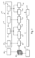
図1を参照するに、コンピュータ支援治療モニタリングシステム100は、機能的医用イメージャ102と、検査中に対象の患者又は他のオブジェクトを表すボリューム画像データ106を生成する構造的医用イメージャ104と、を有する。機能的医用イメージャ102は機能的情報又は代謝的情報を提供する一方、構造的イメージャ104はオブジェクトの構造又は形状を表す情報を提供する。例示としての機能的イメージングモダリティは、PET、SPECT、機能的磁気共鳴イメージング(fMRI)及び分子イメージングを有する。PETシステム及びSPECTシステムは、患者の解剖学的構造に導入された放射性核種の減衰を測定する。用いられるトレーサに依存して、上記の検査は、FDG、FLTの場合の細胞増殖及びFMISOの場合の低酸素症等の機能的特徴を表す情報を提供するように用いられることが可能である。
Referring to FIG. 1, a computer-aided
構造的イメージングモダリティの例は、CT、MRI、X線及び超音波(US)である。機能的イメージャ102及び構造的イメージャ104は、別個のシステムとして示されているが、単独のシステム、例えば、PET/CT、SPECT/CT、PET/MR又は他のスキャナに組み合わされることが可能である。勿論、上記の例は非制限的であり、単独のモダリティが、構造的イメージャ及び機能的イメージャの両方としての役割を果たすことが可能である。
Examples of structural imaging modalities are CT, MRI, X-ray and ultrasound (US).
システム100はまた、複数の画像処理構成要素、例えば、病変検出器108、登録プロセッサ110、病変トラッカ112、病変数値化器114及び傾向分析器116等を有する。それらの画像処理構成要素は、コンピュータプロセッサにより実行されるときに、コンピュータがそれぞれの構成要素の機能を実行するようにさせるコンピュータ読み出し可能命令により有利に実行される。モデルデータ118(1つ又はそれ以上の解剖学的構造モデル120、運動モデル122、生理学的モデル124及び疾病モデル126を含む)及び患者に特有のデータ128が、種々の構成要素の一部である又はそれらの構成要素にアクセス可能であるコンピュータ読み出し可能メモリに記憶される。
The
モデルデータ118は、種々の画像処理構成要素の実行可能コードと区別可能なモジュラ構造の状態で有利に維持される。そのような一実施形態においては、モデルデータ118及び患者に特有のデータ128は、病院の情報システム/放射線情報システム(HIS/RIS)に記憶され、適切な通信ネットワークを介してアクセスされる。他の実施形態においては、それらのデータ118、128の一部又は全てが、システム100のコンピュータによりアクセスされるメモリに保存される。他の実施形態においては、それらのデータの一部又は全てが、遠隔の場所に保存されるデータベースに記憶され、高域ネットワーク(WAN)又は他の適切な通信ネットワークを介してアクセスされる。特定のアプリケーションの要求に応じて、そのようなデータ構造は、異なるモデルデータ118を有する共通の画像処理構成要素の使用、特定の患者に対して又は複数の患者、医師又は命令においてモデルデータ118の一貫した適用、並びに/若しくはモデルデータ118へのアップグレード、更新又は他の変更の実施を容易にするように用いられることが可能である。
ディスプレイ、並びにマウス及び/又はキーボード等の他の出力装置及び入力装置を含むオペレータインタフェース130は、グラフィカルユーザインタフェース(GUI)又は他の適切なインタフェースを用いて、ユーザがシステム100の種々の構成要素の動作を制御すること又はそれらの構成要素と相互作用することを可能にする。
The
CADシステムとして有利に実行される病変検出器108は、例えば、有効な形態的画像データ及び機能的画像データの組み合わされた分析に基づいて、候補の病変を特定するように画像データ106を分析する。病変検出器108は、特定されるべき構造又は特徴を示す推測情報を提供する解剖学的モデル120と組み合わされて動作する。例示としての解剖学的モデルは、器官の境界の表面ベースの表現及び三次元解剖学的構造のボリュームベースの表現又はボリューム表現を有する。
A
病変検出器108はまた、解剖学的モデルデータ120を患者に特有のデータ128と組み合わせた補助情報と関連付けて動作する。そのような補助情報の例には、解剖学的ランドマーク、大域的幾何関係等がある。種々のイメージングモダリティの特徴における違いのために、解剖学的モデル及び補助情報は、機能的画像102及び構造的画像104について選択されたモダリティに基づいて変えることが可能であることをまた、理解することができる。
The
モデルデータ118及び他の情報の一貫した適用は、通常、特定の患者について又は複数の患者において病変の検出及び描写の一貫性を改善するように予測されることが可能であることに留意されたい。それにも拘わらず、オペレータインタフェース130を介して医師に候補の病変を提供することは好ましく、病変の1つ又はそれ以上を受容する又は拒否する、それらの病変の描写を調整する、等の機会をその医師は与えられる。医師はまた、更なる他の病変を手動で特定する機会を与えられることが可能である。他の実施形態においては、病変検出は、オペレータが介入することなく、自動的に実行される。どちらの場合も、特定の病変はタグ付けされ、又は特定され、その情報は、更なる使用のために、適切なコンピュータ読み出し可能メモリに記憶される。
Note that consistent application of
登録プロセッサ110は、種々の画像間の位置合わせミスを明らかにするように画像データ106の座標系を登録する。例えば、登録プロセッサ110は、所定のスキャンの進行における全体の及び/又は周期的な患者の運動を明らかにするように機能的画像102及び形態学的画像104からの画像データ106の座標系を一致させる。治療法の進行において複数回、取得される画像データ106の場合、登録プロセッサ110は、時系列の画像の座標系を一致させる。
例示としての登録技術は、表面ベースの技術及びボリュームベースの技術の両方を含む。表面ベースの登録は、一般に、複数の座標系を位置合わせするように、器官、病変又は他の境界(例えば、病変検出器108により特定される)を用いる。放射治療の投与の計画は従来、CT画像からもたらされる器官又は病変の表面輪郭を用いているため、表面ベースの登録は、放射治療のアプリケーションで用いるために特に適するものである。他方、ボリューム登録技術は、典型的には、明示的セグメント化動作を回避し、そしてボリュームデータに代えて動作する。 Exemplary registration techniques include both surface-based techniques and volume-based techniques. Surface-based registration typically uses organs, lesions, or other boundaries (eg, identified by lesion detector 108) to align multiple coordinate systems. Since radiotherapy administration planning traditionally uses the surface contours of organs or lesions derived from CT images, surface-based registration is particularly suitable for use in radiotherapy applications. On the other hand, volume registration techniques typically avoid explicit segmentation operations and operate instead of volume data.
登録プロセッサは、例えば、呼吸運動、心臓の運動又は膀胱又は直腸の充填における違いのための予測される運動についての推測又は他の情報を提供するように用いられることが可能である運動モデルと関連付けて動作する。一般に、予測される運動パターンの適切な数学的表現を与え、位置合わせミスを表現するための妥当な初期値を決定し、好ましい位置合わせを得るようにそれらのパラメータの最適化に導くことは好ましい。例示としての運動モデル120は、予測される運動パターンに基づいて選択されることが可能である1つ又はそれ以上の代替の数学的変換、視野(FOV)又は関心領域における若しくは解剖学的ランドマークにおける典型的な動きを与えるベクトル場、若しくは動的表面モデル等の形にある解剖学的モデル120に直接、統合されることが可能であるモデルを有する。モデルデータ118及び他の情報の一貫性のある適用はまた、登録処理の一貫性を改善するように実行されることが可能である一方、登録は、病変検出の登録と類似する半自動的に又は自動的に実効されることが可能である。
The registration processor is associated with a motion model that can be used to provide inferences or other information about predicted motion, for example, due to differences in respiratory motion, heart motion, or bladder or rectal filling Works. In general, it is preferable to give an appropriate mathematical representation of the predicted motion pattern, determine a reasonable initial value to represent the misregistration, and lead to optimization of those parameters to obtain a preferred alignment . The
同様に、運動モデル122と関連付けて動作する病変トラッカ112は、時系列の画像の進行において1つ又はそれ以上の候補の病変を追跡する。この関連で、病変追跡はまた、完全な画像登録なしで達成されることが可能であることに留意する必要がある。例えば、病変トラッカ112は、時系列の画像の1つ又はそれ以上において検出される病変の一致性を判定するように解剖学的モデル120及び運動モデル122の1つ又は両方に関連付けて動作することができる。病変トラッカ112は、同様に、半自動的に又は自動的に動作することが可能である。半自動の実施形態においては、例えば、ユーザは、時系列の画像の種々の画像において検出される病変間の与えられる一致性を許容する又は拒否する機会、新しい一致性又は異なる一致性を規定する機会、等を与えられることが可能である。また、病変トラッカ112は、システム100の一部である又はそのシステムにとってアクセス可能であるメモリに適切なデータ構造の状態で種々の病変間の一致について記憶する。
Similarly, a
病変数値化器114は、数値化情報を種々の追跡された病変に与える。特に、数値的に適切な及び再生可能な病変機能的データに到達するように画像データが較正される又は正規化されることは好ましいことである。従って、病変数値化器114は、例えば、機能的画像102及び構造的画像104の異なる空間分解能から結果が得られるパーシャルボリューム効果を補償するように患者特有の解剖学的形態を用いて、患者に特有のデータ128に関連付けて動作する。病変数値化器はまた、患者間のトレーサの変動又は生理学的変動の動的な挙動について明らかにするように生理学的モデル124と関連付けて動作する。従って、その生理学的モデルは、予測されるトレーサ取り込み場所、取り込み及び洗い流し時間、生理学的関係、関心のある生理現象と関連する特定のトレーサの予測される挙動をモデル化する他の情報のうちの1つ又はそれ以上のような情報を有する。
A lesion digitizer 114 provides digitized information to various tracked lesions. In particular, it is preferred that the image data be calibrated or normalized to arrive at numerically relevant and reproducible lesion functional data. Thus, the lesion digitizer 114 may use a patient specific anatomy to compensate for partial volume effects resulting from different spatial resolutions of the
病変数値化器114は、イメージングプロトコル(例えば、患者の準備、トレーサ線量又はイメージャ設定における)における違い、患者の生理現象における違い等の1つ又はそれ以上のような因子から得られる変化を明らかにするようにこの情報を有利に用いる。機能的データは、機関間変動、医師間変動、患者間変動、患者内変動、又は他の変動の影響を低減させるように、較正又は正規化されることが可能である。FDG等のトレーサと関連して特に有用である病変数値化器114により生成される病変機能的データの一例は、種々の病変についての正規化された標準摂取率(SUV)を含む。他の機能的指標は、
細胞増殖、低酸素症又は他の機能的指標を含み、機能的指標は、典型的には、適用されるトレーサの関数であることが理解できる。
The lesion digitizer 114 accounts for changes resulting from one or more factors such as differences in imaging protocols (eg, in patient preparation, tracer dose or imager settings), differences in patient physiology, etc. This information is advantageously used as such. Functional data can be calibrated or normalized to reduce the effects of inter-institutional, inter-doctoral, inter-patient, intra-patient, or other variability. One example of lesion functional data generated by a lesion digitizer 114 that is particularly useful in connection with a tracer such as FDG includes normalized standard uptake rate (SUV) for various lesions. Other functional indicators are
It can be understood that the functional index is typically a function of the applied tracer, including cell proliferation, hypoxia or other functional index.
生理学的モデル124は、種々の形態で提供される。一実施形態においては、生理学的モデル情報124は、異なる解剖学的区分又は生理学的区分間のトレーサの交換を表す解剖学的表現を介して提供される。特定の病状及びトレーサ、与えられる治療、好ましい正確さ等の因子に依存して、パラメータ及び値が医薬品データベースから導き出されることが可能である。モデルデータはまた、特定の機関における患者の、特定の患者のクラス又は集団の、個々の患者モデルの観測される変動に基づいて、若しくは異なるモデルの又は種類のイメージャ102、104、異なる供給メーカにより製造されたイメージャ等の使用によりもたらされる観測される変動に基づいて、経験的に導き出されることが可能である。
The
傾向分析器116は、画像取得の時系列の1つ又はそれ以上の点において治療反応指標を生成し、それ故、適用される治療への反応を評価するように、病変数値化器114により生成される正規化病変機能的データを評価する。機能的反応指標に加えて、傾向分析器はまた、構造的イメージャ104からの情報を用いて決定される病変の大きさ、形状、境界又は他の形態的特徴等の形態的反応指標を考慮することが可能である。このために、傾向分析器126は、病状及び/又は患者に特有の因子を明らかにするように、疾病モデル126及び患者に特有のデータ128と関連付けて動作する。
The
FDGベースのイメージング技術にまた、特に適する一実施形態においては、傾向分析器116の治療反応指標は、病変数値化器114により生成される正規化SUV(又は、正規化SUVにおける変化)を評価する閾値ベースの基準を含む。更なる他の分析がまた、好ましい機能的反応指標又は形態的反応指標の時間的推移の分析的評価、統計的評価又は発見的評価に基づいて、考慮される。理解できるであろうように、種々の評価及び関連基準が、疾病モデル126により与えられる。また、非画像ベースのデータ(例えば、患者の人口統計情報、化学分析結果又は他の調査情報)も、治療反応評価処理に含まれることが可能であることに留意されたい。
In one embodiment that is also particularly suitable for FDG-based imaging techniques, the treatment response measure of the
疾病モデル126及び患者に特有のデータ128と関連付けてまた、動作する治療システム132は、特定の治療を決定する又は特定の治療を提案するように傾向分析器により与えられる反応評価を用いる。上記のように、例えば、治療システム132は、その治療に対する調節、異なる又は付加的な治療経路、若しくは一時的療法への迂回を提案することが可能である。その治療又は治療調節によりまた、医師が種々の代替の間での選択における情報を用いることが可能であるように、予測される成功又は結果を表す信頼水準又は他の情報を提示されることが可能である。典型的な病状特有の及び患者特有の例示としての治療は、例示として適用される放射治療、化学療法、高周波切除(RF)又は他の技術による切除、近接照射療法、外科手術及び分子治療等を含む。治療システム32は、医師が治療を許容する又は調節する機会を明らかにするように、半自動的に有利に動作することができるが、自動的な実施も検討されることが可能である。
In conjunction with the
また、図示しているように、知識維持エンジン134は、適切なモデルデータ118を選択するように、又は、適切な特定の基準に基づいて、種々のシステム構成要素の動作を支配するルールを実施するように用いられることが可能である。従って、解剖学的モデル120、運動モデル122,生理学的モデル124又は疾病モデル126のうちの1つ又はそれ以上が、特定の患者、トレーサ、イメージングプロトコル、疾病等に基づいて選択される複数のパラメータ値を有することが可能である。同様に、知識維持エンジン134は、種々の画像処理構成要素により用いられる2つ以上の有効なアルゴリズム間で選択するように用いられることが可能である。例えば、上記のように、病変検出器108は、機能的イメージャ102及び構造的イメージャ104のモダリティに依存して、異なるパラメータ値又は検出アルゴリズムを用いることが可能である。
Also, as shown,
また、モデルデータ118及びシステム構成ルールの一貫性のある適用は通常、治療反応評価の改善するように予測することができる。何れの場合も、ユーザは、適切なアプリケーションに特有の基準に基づいて種々の選択の有効性及び/又は一貫性を確実にするように調べる知識維持エンジン134を用いて、構成オプションのメニューが与えられる、又は、システム構成に影響を与える機会が与えられることが可能である(例えば、FDGを用いるPETスキャンからの画像の処理において、知識維持エンジンは、FDGに適切な解剖学的モデル120、運動モデル122、生理学的モデル124又は疾病モデル126が用いられることを確実にするように用いられる)。半自動の実施形態においては、知識維持エンジン134は、ユーザによる許容のための適切なモデル118を提供するようにアプリケーションに特定の基準を用いる。
Also, consistent application of
システム100の動作について、ここでは、肺癌の治療に関連してFDG PET/CT検査の例示としての場合について、図2に関連付けて説明する。参照番号200において、治療に先行して、ベースラインスキャンが得られる。ベースラインスキャンは、患者に特有の形態的情報を与える診断品質CTと、参照番号203で一般に示される機能的データを与えるPETスキャンとを有する。その情報203はまた、病変機能的データに影響を与えることが可能である代謝速度を表す患者特有の生理学的情報を有することが可能である。
The operation of the
参照番号201においては、知識維持エンジン134は、例えば、適切なモデルデータ(118)を選択するように及び/又は適切なシステムルールの一貫性のある適用を確実にするように、システムを構成するように用いられる。従って、知識維持エンジン134は、運動モデル122(例えば、例示としての肺癌への適用において肺に適切な運動モデル)、解剖学的モデル120及び生理学的モデル124(例えば、FDG−PET画像のための肺についての解剖学的モデル)及び疾病モデル126(例えば、肺癌についての疾病モデル)を選択するように用いられることが可能である。例えば、選択されたモデルが、それと同様の場合に以前に選択されたモデル又は類似する場合に用いられたモデルと一貫性を有することを確実にするように、知識維持エンジン134はまた、上記の処理中の種々の時点で動作することが可能であることに留意されたい。種々のイメージング処理構成要素はまた、ルールベースに直接、アクセスすることが可能であり、又は、知識維持エンジン134の機能の一部又は全てを実行するようにどれが用いられるかを設定することが可能である。
At
参照番号204において、病変検出器108は、1つ又はそれ以上の腫瘍の存在を検出するようにベースラインスキャンにより生成された画像データ106を分析する。
At
登録プロセッサ110は、ステップ206において画像を登録する。組み合わされたPET/CTイメージング検査の例示としての場合に、登録プロセッサは通常、画像取得のPET部分とCT部分との間で又は画像取得中に生じる患者の動き及び/又は器官の動きを補償するように用いられる。
参照番号208においては、病変数値化器114は、正規化された病変機能的データを生成するように生理学的モデル124と関連付けて動作する。FDG−PET検査の場合には、例えば、病変数値化器114は、種々の病変の初期的アクティビティを特徴付けるベースラインが較正されたSUVを演算する。患者、プロトコル、疾病又は他のアプリケーションに特有の要求に依存して、付加的な又は異なる病変機能的データ、機能的属性又は構造的属性を表す較正データがまた、生成されることが可能である。
At
第1追尾スキャンが、ステップ210において、典型的には、化学療法、外部の放射線療法又は他の好ましい療法の1つ又はそれ以上の治療の後に、得られる。CTスキャンは、例えば、画像登録目的の十分な品質の画像データ106を生成する比較的低い線量を用いて、診断品質より低いことが可能である。また、追尾機能的画像取得のプロトコルは、意図的に又はそうではないに拘わらず、ベースライン取得のプロトコルと異なることが可能である。一実施例として、FDG又は他のトレーサの投与と機能的イメージング検査との間の時間は、装置又は専門家の能力、患者の調整時間における違い等の因子のために変更可能である。患者の生理機能における違いも影響する可能性がある。例えば、糖尿病の患者においては、初期スキャン及び追尾スキャンのときに異なるインスリンレベルを示す可能性がある。
A first tracking scan is typically obtained in
参照番号212においては、病変検出器108は、病変の存在を検出するように追尾スキャンからの画像データ106を分析する。
At
参照番号214においては、病変トラッカ112は、ベースラインに示される病変と追尾画像との間の一致性のみを、又はその一致性を登録プロセッサ110からの情報と組み合わせて、示す。
At
参照番号216においては、病変数値化器114は、追尾スキャンで示される病変についての較正又は正規化された病変機能的データを生成する。従って、関連トレーサの投与と機能的イメージング検査との間の時間が異なる例示としての場合、又は糖尿病患者の例示としての場合には、生理学的モデル124からの情報は、変更の影響を補正する、又はその影響を低減する役割を果たす。
At
参照番号218においては、傾向分析器116は、適用される治療に対する病変の反応を評価するように較正された機能的データを分析し、その応答はまた、適切なメモリに記憶される。また、FDG PET画像取得の例示としての場合においては、傾向分析器は、種々の病変の較正されたSUVにおける他の因子の変化について考慮することが可能である。種々の特定された病変についての反応は、種々の病変の応答が別個に考慮されることが可能であるように、別個に分析及び評価されることが可能であることに留意されたい。
At
参照番号220においては、傾向分析器116からの情報が、提案される治療に対する反応を予測するように用いられることが可能である。
At
参照番号222、224、226及び228で一般に示すように、1つ又はそれ以上の付加追尾スキャンが得られ、複数の治療が、必要に応じて、適用されることが可能である。また、例示としての腫瘍学的アプリケーションにおいては、追尾スキャンは、化学療法の複数のサイクルの各々の後に得られることが可能である。
As generally indicated by
上記のステップの順序、それ故、種々のシステム間の機能的関係は、知識維持エンジン134等の制御下で変更されることが可能である。そのような一実施例においては、登録プロセッサ110は、画像登録が病変検出動作に先行して実行されるように、病変検出器110に先行して適用されることが可能である。他の実施例においては、病変数値化が、例えば、時系列の画像の種々の画像の登録又は病変トラッカ112の動作に先行して、処理における比較的初期に実行されることが可能である。
The order of the above steps, and thus the functional relationships between the various systems, can be changed under the control of the
本発明については、上で好適な実施形態を参照して詳述している。上の詳細説明を読んで、理解することにより、修正及び変形が可能であることを当業者は理解することができる。本発明においては、そのような修正及び変更が同時提出の特許請求の範囲及びそれと同等なものにおける範囲内にある全てを包含するように、意図されている。 The invention has been described in detail with reference to the preferred embodiments above. Those skilled in the art can appreciate that modifications and variations can be made by reading and understanding the above detailed description. The present invention is intended to embrace all such modifications and changes that fall within the scope of the appended claims and their equivalents.
Claims (49)
前記病変検出器と作動的に連結している病変数値化器であって、該病変数値化器は、第1検出病変についての第1病変機能的データを生成するように前記患者に治療を適用する前に行われる前記患者の第1イメージング検査からの第1機能的画像データを用い、前記病変数値化器は、第1検出病変についての第2病変機能的データを生成するように治療を適用した後に行われる前記患者の第2イメージング検査からの第2機能的画像データを用いる、病変数値化器;及び
前記第1病変機能的データと前記第2病変機能的データとの間の違いを示す傾向分析器;
を有する装置。 A lesion detector for detecting lesions in medical image data from patient imaging examinations;
A lesion digitizer operatively coupled to the lesion detector, wherein the lesion digitizer applies treatment to the patient to generate first lesion functional data for a first detected lesion Using the first functional image data from the first imaging examination of the patient performed before, the lesion digitizer applies treatment to generate second lesion functional data for the first detected lesion A lesion digitizer using second functional image data from the second imaging examination of the patient performed after; and showing the difference between the first lesion functional data and the second lesion functional data Trend analyzer;
Having a device.
前記第1イメージング検査からの画像データにおいて検出された病変と前記第2イメージング検査からの画像データにおいて検出された病変との間の一致性を決定する病変トラッカ;
を更に有する、装置。 The apparatus of claim 1, wherein:
A lesion tracker that determines the consistency between the lesion detected in the image data from the first imaging examination and the lesion detected in the image data from the second imaging examination;
Further comprising an apparatus.
前記第1機能的画像データ及び前記第2機能的画像データを登録する登録プロセッサ;
を更に有する、装置。 The apparatus of claim 1, wherein:
A registration processor for registering the first functional image data and the second functional image data;
Further comprising an apparatus.
生理学的モデルであって、前記病変数値化器は、前記第1機能的医用イメージング検査と組み合わせて前記患者に適用されるトレーサの予測される挙動をモデル化する生理学的モデルを用いる、生理学的モデル;
を更に有する、装置。 The apparatus of claim 1, wherein:
A physiological model, wherein the lesion digitizer uses a physiological model that models a predicted behavior of a tracer applied to the patient in combination with the first functional medical imaging examination. ;
Further comprising an apparatus.
疾病モデルであって、前記傾向分析器は、適用される治療に対する前記第1検出病変の反応をモデル化する前記疾病モデルを用いる、疾病モデル;
を更に有する、装置。 The apparatus of claim 1, wherein:
A disease model, wherein the trend analyzer uses the disease model to model the response of the first detected lesion to an applied treatment;
Further comprising an apparatus.
PET/CTスキャナ;
を更に有する、装置。 The apparatus of claim 1, wherein:
PET / CT scanner;
Further comprising an apparatus.
前記病変の前記機能的特徴を表す第2較正機能的データを生成するように前記患者の第1治療の適用後に行われる前記患者の第2イメージング検査からのデータを用いて生成される第2機能的データを較正する段階;並びに
前記第1治療に対する前記病変の反応を評価するように前記第1較正機能的データ及び前記第2較正機能的データを用いる段階;
を有する方法。 Calibrating the first functional data generated using data from the first imaging examination of the patient to generate first calibration functional data representative of the functional characteristics of the lesion;
A second function generated using data from the patient's second imaging examination performed after application of the patient's first treatment to generate second calibration functional data representative of the functional characteristics of the lesion. Calibrating functional data; and using the first calibration functional data and the second calibration functional data to evaluate the response of the lesion to the first treatment;
Having a method.
前記第1機能的データを生成するように患者の第1イメージング検査からのデータを用いる段階;及び
前記第2機能的データを生成するように前記患者の第2イメージング検査からのデータを用いる段階;;
を有する、方法。 The method of claim 16, wherein:
Using data from a first imaging examination of the patient to generate the first functional data; and using data from the second imaging examination of the patient to generate the second functional data; ;
Having a method.
前記第1イメージング検査からの第1医用画像データにおける前記病変を特定する段階;及び
前記第2イメージング検査からの第2医用画像データにおける前記病変を特定する段階;
を有する、方法。 The method of claim 16, wherein:
Identifying the lesion in first medical image data from the first imaging examination; and identifying the lesion in second medical image data from the second imaging examination;
Having a method.
前記第2イメージングデータにおける複数の病変を特定する段階;及び
前記第2画像データにおいて検出された病変と前記第1画像データにおいて特定された病変との間の一致性を決定する段階;
を有する、方法。 The method of claim 16, wherein:
Identifying a plurality of lesions in the second imaging data; and determining a match between a lesion detected in the second image data and a lesion identified in the first image data;
Having a method.
検出される病変の予測される動きをモデル化する運動モデルを用いる段階;
を有する、方法。 The method of claim 16, wherein:
Using a motion model to model the predicted motion of the detected lesion;
Having a method.
前記検出された病変について病変機能的データを演算するように前記第2イメージング検査からのデータを用いる段階;
を有する、方法。 The method of claim 16, wherein:
Using data from the second imaging examination to compute lesion functional data for the detected lesion;
Having a method.
コンピュータ通信ネットワークにおいて生理学的モデルデータを受信する段階;
を有する、方法。 The method of claim 16, wherein:
Receiving physiological model data in a computer communication network;
Having a method.
患者の解剖学的構造に存在する病変についての第1病変機能的データを生成するように前記患者の第1機能的医用イメージング検査からの医用画像データを用いる段階;並びに
適用された治療に対する病変の反応を評価するように前記患者の第2機能的医用イメージング検査から得られた前記病変についての第1病変機能的データ及び第2病変機能的データを用いる段階;
を有する、コンピュータ読み出し可能記憶媒体。 A computer-readable storage medium having instructions that, when executed by a computer, cause the computer to perform the method, the method comprising:
Using medical image data from the first functional medical imaging examination of the patient to generate first lesion functional data for a lesion present in the patient's anatomy; and of the lesion for the applied treatment Using first lesion functional data and second lesion functional data for the lesion obtained from a second functional medical imaging examination of the patient to assess response;
A computer-readable storage medium.
病変数値化器によりアクセスされるとき、機能的医用イメージング検査と結び付けて前記患者に適用される第1トラッカの予測される挙動を表すデータを有する第1生理学的モデル;
を有するデータ構造を有するコンピュータ読み出し可能記憶媒体。 A first motion model having data representing the predicted motion of the lesion detected in the data from the medical imaging examination of the patient when accessed by the lesion tracker; and functional medical when accessed by the lesion digitizer A first physiological model having data representing a predicted behavior of a first tracker applied to the patient in conjunction with an imaging test;
A computer readable storage medium having a data structure having:
前記病変数値化器によりアクセスされるとき、機能的医用イメージング検査と関連付けて患者に適用される第2トレーサの予測される挙動を表すデータを有する第2生理学的モデル;
を有する、コンピュータ読み出し可能記憶媒体。 37. The computer readable storage medium of claim 36, wherein the data structure is:
A second physiological model having data representing a predicted behavior of a second tracer applied to the patient in association with a functional medical imaging examination when accessed by the lesion digitizer;
A computer-readable storage medium.
前記病変数値化器にアクセス可能なコンピュータ読み出し可能メモリにおけるコンピュータ読み出し可能データを記憶する段階;
を有する方法。 Receiving a physiological model that, when accessed by a lesion digitizer, models at least one of a patient physiological characteristic and an expected behavior of the imaging agent in conjunction with a functional medical imaging examination; Storing computer readable data in a computer readable memory accessible to the lesion digitizer;
Having a method.
複数の生理学的モデルを受け入れる段階;
を有する、方法。 43. The method of claim 42, wherein:
Accepting multiple physiological models;
Having a method.
モデルデータを選択するように患者の機能的イメージング検査に関連付けて用いられる画像プロトコルを表す情報を用いる段階;及び
コンピュータ支援治療モニタリングシステムの構成要素の動作を変えるように前記選択されたモデルデータを用いる段階;
を有する方法。 Methods used in computer-assisted therapy monitoring include:
Using information representing an imaging protocol used in association with the functional imaging examination of the patient to select model data; and using the selected model data to alter the operation of components of the computer-aided therapy monitoring system Stage;
Having a method.
前記モデルデータを選択するように当該解剖学的構造を表す情報を用いる段階;
を有する、方法。 45. The method of claim 44, wherein:
Using information representing the anatomical structure to select the model data;
Having a method.
Applications Claiming Priority (2)
| Application Number | Priority Date | Filing Date | Title |
|---|---|---|---|
| US88318007P | 2007-01-03 | 2007-01-03 | |
| PCT/IB2007/054935 WO2008081365A2 (en) | 2007-01-03 | 2007-12-05 | Computer assisted therapy monitoring |
Publications (1)
| Publication Number | Publication Date |
|---|---|
| JP2010516301A true JP2010516301A (en) | 2010-05-20 |
Family
ID=39589066
Family Applications (1)
| Application Number | Title | Priority Date | Filing Date |
|---|---|---|---|
| JP2009544460A Pending JP2010516301A (en) | 2007-01-03 | 2007-12-05 | Computer-aided therapy monitoring apparatus and method |
Country Status (5)
| Country | Link |
|---|---|
| US (1) | US20100317967A1 (en) |
| EP (1) | EP2115697A2 (en) |
| JP (1) | JP2010516301A (en) |
| CN (1) | CN101578630A (en) |
| WO (1) | WO2008081365A2 (en) |
Cited By (3)
| Publication number | Priority date | Publication date | Assignee | Title |
|---|---|---|---|---|
| JP2012500036A (en) * | 2008-08-15 | 2012-01-05 | コーニンクレッカ フィリップス エレクトロニクス エヌ ヴィ | Imaging enhanced by the model |
| JP2015510180A (en) * | 2012-01-27 | 2015-04-02 | コーニンクレッカ フィリップス エヌ ヴェ | Medical selection system |
| WO2016151618A1 (en) * | 2015-03-23 | 2016-09-29 | 日本電気株式会社 | Predictive model updating system, predictive model updating method, and predictive model updating program |
Families Citing this family (17)
| Publication number | Priority date | Publication date | Assignee | Title |
|---|---|---|---|---|
| JP2010082428A (en) * | 2008-09-04 | 2010-04-15 | Toshiba Corp | X-ray computer tomography apparatus |
| JP5764069B2 (en) * | 2009-01-19 | 2015-08-12 | コーニンクレッカ フィリップス エヌ ヴェ | Region reconstruction and quantitative evaluation in list-mode PET imaging |
| WO2010092494A1 (en) | 2009-02-11 | 2010-08-19 | Koninklijke Philips Electronics N.V. | Group-wise image registration based on motion model |
| CN105011962B (en) * | 2009-03-19 | 2018-08-14 | 皇家飞利浦电子股份有限公司 | Functional imaging |
| WO2010115885A1 (en) * | 2009-04-03 | 2010-10-14 | Oslo Universitetssykehus Hf | Predictive classifier score for cancer patient outcome |
| BR112012013554A2 (en) * | 2009-12-08 | 2017-10-10 | Koninl Philips Electronics Nv | correction method of marker absorption measurements for a patient, computer program and correction system |
| EP2407927B1 (en) | 2010-07-16 | 2013-01-30 | BVBA dr. K. Coenegrachts | A method and device for evaluating evolution of tumoral lesions |
| WO2013001471A2 (en) | 2011-06-29 | 2013-01-03 | Koninklijke Philips Electronics N.V. | Displaying a plurality of registered images |
| US10674983B2 (en) * | 2013-09-25 | 2020-06-09 | Richard R. Black | Patient-specific analysis of positron emission tomography data |
| CN104867077A (en) * | 2014-02-25 | 2015-08-26 | 华为技术有限公司 | Method for storing medical image, method for exchanging information and device thereof |
| JP6548393B2 (en) | 2014-04-10 | 2019-07-24 | キヤノンメディカルシステムズ株式会社 | Medical image display apparatus and medical image display system |
| CN107167830B (en) * | 2017-03-25 | 2019-02-26 | 浙江君安检测技术有限公司 | A kind of radiation monitoring system based on CT scan device |
| EP3557588A1 (en) * | 2018-04-16 | 2019-10-23 | Siemens Healthcare GmbH | Integrated method for cancer screening |
| CN113168921A (en) * | 2018-12-18 | 2021-07-23 | 墨尼克医疗用品有限公司 | Method for selecting wound products for a patient |
| EP3794550B1 (en) | 2019-08-04 | 2022-03-23 | Brainlab AG | Comparison of a region of interest along a time series of images |
| US11590367B2 (en) * | 2020-12-16 | 2023-02-28 | Varian Medical Systems International Ag | Neural network calibration for radiotherapy |
| CN116687353B (en) * | 2023-08-01 | 2023-12-19 | 宁波杜比医疗科技有限公司 | New adjuvant chemotherapy curative effect evaluation system, equipment and medium |
Citations (5)
| Publication number | Priority date | Publication date | Assignee | Title |
|---|---|---|---|---|
| WO2003025837A1 (en) * | 2001-09-17 | 2003-03-27 | Virtualscopics, Llc. | System and method for quantitative assessment of cancers and their change over time |
| US20050041843A1 (en) * | 2003-08-05 | 2005-02-24 | Sawyer Timothy E. | Dynamic tumor treatment system |
| WO2006119340A2 (en) * | 2005-05-04 | 2006-11-09 | Imquant, Inc. | Dynamic tumor diagnostic and treatment system |
| JP2007505672A (en) * | 2003-09-17 | 2007-03-15 | コニンクリユケ フィリップス エレクトロニクス エヌ.ブイ. | Repetitive inspection report |
| JP2008503258A (en) * | 2004-06-18 | 2008-02-07 | シーメンス メディカル ソリューションズ ユーエスエー インコーポレイテッド | System and method for monitoring disease progression or therapeutic effect using multi-mode visualization |
Family Cites Families (3)
| Publication number | Priority date | Publication date | Assignee | Title |
|---|---|---|---|---|
| US7910093B2 (en) * | 2003-08-19 | 2011-03-22 | New York University | Method for detecting cancer cells and monitoring cancer therapy |
| US7935055B2 (en) * | 2003-09-19 | 2011-05-03 | Siemens Medical Solutions Usa, Inc. | System and method of measuring disease severity of a patient before, during and after treatment |
| CN1918601A (en) * | 2004-02-13 | 2007-02-21 | 皇家飞利浦电子股份有限公司 | Apparatus and method for registering images of a structured object |
-
2007
- 2007-12-05 US US12/521,601 patent/US20100317967A1/en not_active Abandoned
- 2007-12-05 EP EP07849345A patent/EP2115697A2/en not_active Withdrawn
- 2007-12-05 JP JP2009544460A patent/JP2010516301A/en active Pending
- 2007-12-05 CN CNA200780049217XA patent/CN101578630A/en active Pending
- 2007-12-05 WO PCT/IB2007/054935 patent/WO2008081365A2/en active Application Filing
Patent Citations (6)
| Publication number | Priority date | Publication date | Assignee | Title |
|---|---|---|---|---|
| WO2003025837A1 (en) * | 2001-09-17 | 2003-03-27 | Virtualscopics, Llc. | System and method for quantitative assessment of cancers and their change over time |
| JP2005516643A (en) * | 2001-09-17 | 2005-06-09 | ヴァーチャルスコピックス リミテッド ライアビリティ カンパニー | System and method for quantitative evaluation of cancer and time variation of cancer |
| US20050041843A1 (en) * | 2003-08-05 | 2005-02-24 | Sawyer Timothy E. | Dynamic tumor treatment system |
| JP2007505672A (en) * | 2003-09-17 | 2007-03-15 | コニンクリユケ フィリップス エレクトロニクス エヌ.ブイ. | Repetitive inspection report |
| JP2008503258A (en) * | 2004-06-18 | 2008-02-07 | シーメンス メディカル ソリューションズ ユーエスエー インコーポレイテッド | System and method for monitoring disease progression or therapeutic effect using multi-mode visualization |
| WO2006119340A2 (en) * | 2005-05-04 | 2006-11-09 | Imquant, Inc. | Dynamic tumor diagnostic and treatment system |
Cited By (4)
| Publication number | Priority date | Publication date | Assignee | Title |
|---|---|---|---|---|
| JP2012500036A (en) * | 2008-08-15 | 2012-01-05 | コーニンクレッカ フィリップス エレクトロニクス エヌ ヴィ | Imaging enhanced by the model |
| JP2015510180A (en) * | 2012-01-27 | 2015-04-02 | コーニンクレッカ フィリップス エヌ ヴェ | Medical selection system |
| WO2016151618A1 (en) * | 2015-03-23 | 2016-09-29 | 日本電気株式会社 | Predictive model updating system, predictive model updating method, and predictive model updating program |
| JPWO2016151618A1 (en) * | 2015-03-23 | 2017-12-21 | 日本電気株式会社 | Prediction model update system, prediction model update method, and prediction model update program |
Also Published As
| Publication number | Publication date |
|---|---|
| WO2008081365A3 (en) | 2009-06-04 |
| WO2008081365A2 (en) | 2008-07-10 |
| CN101578630A (en) | 2009-11-11 |
| US20100317967A1 (en) | 2010-12-16 |
| EP2115697A2 (en) | 2009-11-11 |
Similar Documents
| Publication | Publication Date | Title |
|---|---|---|
| JP2010516301A (en) | Computer-aided therapy monitoring apparatus and method | |
| US7876939B2 (en) | Medical imaging system for accurate measurement evaluation of changes in a target lesion | |
| US8010184B2 (en) | Method and apparatus for automatically characterizing a malignancy | |
| US9275451B2 (en) | Method, a system, and an apparatus for using and processing multidimensional data | |
| JP6220310B2 (en) | Medical image information system, medical image information processing method, and program | |
| JP5468905B2 (en) | Tools to help diagnose neurodegenerative diseases | |
| US8929624B2 (en) | Systems and methods for comparing different medical images to analyze a structure-of-interest | |
| US20130329973A1 (en) | Subvolume identification for prediction of treatment outcome | |
| US20110015520A1 (en) | Perfusion imaging | |
| US20110075900A1 (en) | Diagnosis assisting system, computer readable recording medium having diagnosis assisting program recorded thereon, and diagnosis assisting method | |
| Salimi et al. | Deep learning-based fully automated Z-axis coverage range definition from scout scans to eliminate overscanning in chest CT imaging | |
| JP2008537691A (en) | How to expand the field of imaging software in diagnostic workups | |
| JP2019511268A (en) | Determination of rotational orientation in three-dimensional images of deep brain stimulation electrodes | |
| US10628963B2 (en) | Automatic detection of an artifact in patient image | |
| CN105120738A (en) | Stenosis therapy planning | |
| JP2008503259A (en) | System and method for loading multiple time points to analyze disease progression or therapeutic effect | |
| US11715212B2 (en) | Heatmap and atlas | |
| US20110148861A1 (en) | Pet data processing system, an arrangement, a method and a computer program product for determining a distribution of a tracer uptake | |
| JP2019106122A (en) | Hospital information device, hospital information system, and program | |
| WO2019176407A1 (en) | Learning assisting device, learning assisting method, learning assisting program, region-of-interest discriminating device, region-of-interest discriminating method, region-of-interest discriminating program, and learned model | |
| JP2021513054A (en) | Correction of standard capture value (SUV) scaling differences in serial positron emission tomography (PET) examinations using image alignment and regression analysis | |
| JP6734111B2 (en) | Finding information creation device and system | |
| US12029920B2 (en) | Automated qualitative description of anatomical changes in radiotherapy | |
| Roy et al. | 5 Enhancing with Modality-Based Patient Care Image Registration in Modern Healthcare | |
| Roy et al. | Enhancing Patient Care with Modality-Based Image Registration in Modern Healthcare |
Legal Events
| Date | Code | Title | Description |
|---|---|---|---|
| A621 | Written request for application examination |
Free format text: JAPANESE INTERMEDIATE CODE: A621 Effective date: 20101201 |
|
| A131 | Notification of reasons for refusal |
Free format text: JAPANESE INTERMEDIATE CODE: A131 Effective date: 20120807 |
|
| A02 | Decision of refusal |
Free format text: JAPANESE INTERMEDIATE CODE: A02 Effective date: 20130115 |