JP2010507594A - Fully human anti-VEGF antibodies and methods of use - Google Patents
Fully human anti-VEGF antibodies and methods of use Download PDFInfo
- Publication number
- JP2010507594A JP2010507594A JP2009533598A JP2009533598A JP2010507594A JP 2010507594 A JP2010507594 A JP 2010507594A JP 2009533598 A JP2009533598 A JP 2009533598A JP 2009533598 A JP2009533598 A JP 2009533598A JP 2010507594 A JP2010507594 A JP 2010507594A
- Authority
- JP
- Japan
- Prior art keywords
- antibody
- seq
- vegf
- xpa
- antigen
- Prior art date
- Legal status (The legal status is an assumption and is not a legal conclusion. Google has not performed a legal analysis and makes no representation as to the accuracy of the status listed.)
- Pending
Links
- 241000282414 Homo sapiens Species 0.000 title claims abstract description 52
- 238000000034 method Methods 0.000 title claims description 46
- 238000009739 binding Methods 0.000 claims abstract description 110
- 230000027455 binding Effects 0.000 claims abstract description 109
- 108010019530 Vascular Endothelial Growth Factors Proteins 0.000 claims abstract description 73
- 230000033115 angiogenesis Effects 0.000 claims abstract description 35
- 230000011664 signaling Effects 0.000 claims abstract description 10
- 230000002159 abnormal effect Effects 0.000 claims abstract description 9
- 102000005789 Vascular Endothelial Growth Factors Human genes 0.000 claims abstract 7
- 229960000397 bevacizumab Drugs 0.000 claims description 69
- 239000000427 antigen Substances 0.000 claims description 41
- 108091007433 antigens Proteins 0.000 claims description 41
- 102000036639 antigens Human genes 0.000 claims description 41
- 239000012634 fragment Substances 0.000 claims description 41
- 206010028980 Neoplasm Diseases 0.000 claims description 39
- 239000013598 vector Substances 0.000 claims description 22
- 108010047041 Complementarity Determining Regions Proteins 0.000 claims description 21
- 201000011510 cancer Diseases 0.000 claims description 20
- 108091008605 VEGF receptors Proteins 0.000 claims description 15
- 102000009484 Vascular Endothelial Growth Factor Receptors Human genes 0.000 claims description 15
- 201000010099 disease Diseases 0.000 claims description 14
- 208000037265 diseases, disorders, signs and symptoms Diseases 0.000 claims description 14
- 108091033319 polynucleotide Proteins 0.000 claims description 13
- 102000040430 polynucleotide Human genes 0.000 claims description 13
- 239000002157 polynucleotide Substances 0.000 claims description 13
- FWMNVWWHGCHHJJ-SKKKGAJSSA-N 4-amino-1-[(2r)-6-amino-2-[[(2r)-2-[[(2r)-2-[[(2r)-2-amino-3-phenylpropanoyl]amino]-3-phenylpropanoyl]amino]-4-methylpentanoyl]amino]hexanoyl]piperidine-4-carboxylic acid Chemical compound C([C@H](C(=O)N[C@H](CC(C)C)C(=O)N[C@H](CCCCN)C(=O)N1CCC(N)(CC1)C(O)=O)NC(=O)[C@H](N)CC=1C=CC=CC=1)C1=CC=CC=C1 FWMNVWWHGCHHJJ-SKKKGAJSSA-N 0.000 claims description 12
- 230000026731 phosphorylation Effects 0.000 claims description 10
- 238000006366 phosphorylation reaction Methods 0.000 claims description 10
- 239000008194 pharmaceutical composition Substances 0.000 claims description 9
- 239000002246 antineoplastic agent Substances 0.000 claims description 8
- 229940127089 cytotoxic agent Drugs 0.000 claims description 8
- 230000002401 inhibitory effect Effects 0.000 claims description 7
- 239000003937 drug carrier Substances 0.000 claims description 6
- 208000002780 macular degeneration Diseases 0.000 claims description 5
- 206010039073 rheumatoid arthritis Diseases 0.000 claims description 5
- 102000004127 Cytokines Human genes 0.000 claims description 4
- 108090000695 Cytokines Proteins 0.000 claims description 4
- 206010012689 Diabetic retinopathy Diseases 0.000 claims description 4
- 230000001154 acute effect Effects 0.000 claims description 4
- 230000000903 blocking effect Effects 0.000 claims description 4
- 208000006545 Chronic Obstructive Pulmonary Disease Diseases 0.000 claims description 3
- 201000004681 Psoriasis Diseases 0.000 claims description 3
- 206010039710 Scleroderma Diseases 0.000 claims description 3
- 208000006673 asthma Diseases 0.000 claims description 3
- 208000027866 inflammatory disease Diseases 0.000 claims description 3
- 239000003053 toxin Substances 0.000 claims description 3
- 231100000765 toxin Toxicity 0.000 claims description 3
- 108700012359 toxins Proteins 0.000 claims description 3
- 125000003275 alpha amino acid group Chemical group 0.000 claims 18
- 101000808011 Homo sapiens Vascular endothelial growth factor A Proteins 0.000 abstract description 88
- 102000058223 human VEGFA Human genes 0.000 abstract description 88
- 108010073929 Vascular Endothelial Growth Factor A Proteins 0.000 abstract description 75
- 238000001727 in vivo Methods 0.000 abstract description 8
- 230000002018 overexpression Effects 0.000 abstract description 4
- 238000000338 in vitro Methods 0.000 abstract description 3
- 102000009524 Vascular Endothelial Growth Factor A Human genes 0.000 description 69
- 210000004027 cell Anatomy 0.000 description 69
- -1 Bayer) Chemical compound 0.000 description 23
- 238000011282 treatment Methods 0.000 description 20
- 230000005764 inhibitory process Effects 0.000 description 19
- 238000010186 staining Methods 0.000 description 15
- 210000001519 tissue Anatomy 0.000 description 15
- 239000003446 ligand Substances 0.000 description 14
- 230000014509 gene expression Effects 0.000 description 13
- 239000000546 pharmaceutical excipient Substances 0.000 description 13
- 230000004614 tumor growth Effects 0.000 description 13
- 238000004458 analytical method Methods 0.000 description 12
- 239000002609 medium Substances 0.000 description 12
- 150000001413 amino acids Chemical group 0.000 description 10
- 239000000872 buffer Substances 0.000 description 10
- 230000003993 interaction Effects 0.000 description 10
- 230000037361 pathway Effects 0.000 description 10
- 108090000623 proteins and genes Proteins 0.000 description 10
- 102000005962 receptors Human genes 0.000 description 10
- 108020003175 receptors Proteins 0.000 description 10
- 239000003814 drug Substances 0.000 description 9
- 239000007924 injection Substances 0.000 description 9
- 238000002347 injection Methods 0.000 description 9
- 239000000203 mixture Substances 0.000 description 9
- 239000000243 solution Substances 0.000 description 9
- 238000004448 titration Methods 0.000 description 9
- 108060003951 Immunoglobulin Proteins 0.000 description 8
- 238000003556 assay Methods 0.000 description 8
- 230000000694 effects Effects 0.000 description 8
- 102000018358 immunoglobulin Human genes 0.000 description 8
- 108010082117 matrigel Proteins 0.000 description 8
- 230000004048 modification Effects 0.000 description 8
- 238000012986 modification Methods 0.000 description 8
- 230000035755 proliferation Effects 0.000 description 8
- 235000018102 proteins Nutrition 0.000 description 8
- 102000004169 proteins and genes Human genes 0.000 description 8
- 230000009257 reactivity Effects 0.000 description 8
- WQZGKKKJIJFFOK-GASJEMHNSA-N Glucose Natural products OC[C@H]1OC(O)[C@H](O)[C@@H](O)[C@@H]1O WQZGKKKJIJFFOK-GASJEMHNSA-N 0.000 description 7
- 108010029485 Protein Isoforms Proteins 0.000 description 7
- 102000001708 Protein Isoforms Human genes 0.000 description 7
- 210000004204 blood vessel Anatomy 0.000 description 7
- 239000003153 chemical reaction reagent Substances 0.000 description 7
- 239000000758 substrate Substances 0.000 description 7
- 238000012360 testing method Methods 0.000 description 7
- 238000002965 ELISA Methods 0.000 description 6
- PEDCQBHIVMGVHV-UHFFFAOYSA-N Glycerine Chemical compound OCC(O)CO PEDCQBHIVMGVHV-UHFFFAOYSA-N 0.000 description 6
- 241001465754 Metazoa Species 0.000 description 6
- 238000006243 chemical reaction Methods 0.000 description 6
- 238000010790 dilution Methods 0.000 description 6
- 239000012895 dilution Substances 0.000 description 6
- 230000008030 elimination Effects 0.000 description 6
- 238000003379 elimination reaction Methods 0.000 description 6
- 239000000463 material Substances 0.000 description 6
- 210000003101 oviduct Anatomy 0.000 description 6
- 108090000765 processed proteins & peptides Proteins 0.000 description 6
- 210000002307 prostate Anatomy 0.000 description 6
- 210000000626 ureter Anatomy 0.000 description 6
- XLYOFNOQVPJJNP-UHFFFAOYSA-N water Substances O XLYOFNOQVPJJNP-UHFFFAOYSA-N 0.000 description 6
- 206010009944 Colon cancer Diseases 0.000 description 5
- LFQSCWFLJHTTHZ-UHFFFAOYSA-N Ethanol Chemical compound CCO LFQSCWFLJHTTHZ-UHFFFAOYSA-N 0.000 description 5
- 206010038389 Renal cancer Diseases 0.000 description 5
- 208000000208 Wet Macular Degeneration Diseases 0.000 description 5
- 230000004913 activation Effects 0.000 description 5
- 235000001014 amino acid Nutrition 0.000 description 5
- 230000008045 co-localization Effects 0.000 description 5
- 210000002889 endothelial cell Anatomy 0.000 description 5
- 238000003364 immunohistochemistry Methods 0.000 description 5
- 239000013642 negative control Substances 0.000 description 5
- 238000004091 panning Methods 0.000 description 5
- 238000007911 parenteral administration Methods 0.000 description 5
- 239000013641 positive control Substances 0.000 description 5
- 102000004196 processed proteins & peptides Human genes 0.000 description 5
- 210000002966 serum Anatomy 0.000 description 5
- 230000019491 signal transduction Effects 0.000 description 5
- 239000002904 solvent Substances 0.000 description 5
- 239000011534 wash buffer Substances 0.000 description 5
- 208000005590 Choroidal Neovascularization Diseases 0.000 description 4
- 206010060823 Choroidal neovascularisation Diseases 0.000 description 4
- SHZGCJCMOBCMKK-UHFFFAOYSA-N D-mannomethylose Natural products CC1OC(O)C(O)C(O)C1O SHZGCJCMOBCMKK-UHFFFAOYSA-N 0.000 description 4
- AOJJSUZBOXZQNB-TZSSRYMLSA-N Doxorubicin Chemical compound O([C@H]1C[C@@](O)(CC=2C(O)=C3C(=O)C=4C=CC=C(C=4C(=O)C3=C(O)C=21)OC)C(=O)CO)[C@H]1C[C@H](N)[C@H](O)[C@H](C)O1 AOJJSUZBOXZQNB-TZSSRYMLSA-N 0.000 description 4
- PNNNRSAQSRJVSB-SLPGGIOYSA-N Fucose Natural products C[C@H](O)[C@@H](O)[C@H](O)[C@H](O)C=O PNNNRSAQSRJVSB-SLPGGIOYSA-N 0.000 description 4
- 108010021625 Immunoglobulin Fragments Proteins 0.000 description 4
- 102000008394 Immunoglobulin Fragments Human genes 0.000 description 4
- 208000008839 Kidney Neoplasms Diseases 0.000 description 4
- SHZGCJCMOBCMKK-DHVFOXMCSA-N L-fucopyranose Chemical compound C[C@@H]1OC(O)[C@@H](O)[C@H](O)[C@@H]1O SHZGCJCMOBCMKK-DHVFOXMCSA-N 0.000 description 4
- 241001529936 Murinae Species 0.000 description 4
- 241000699666 Mus <mouse, genus> Species 0.000 description 4
- 241000699670 Mus sp. Species 0.000 description 4
- FAPWRFPIFSIZLT-UHFFFAOYSA-M Sodium chloride Chemical compound [Na+].[Cl-] FAPWRFPIFSIZLT-UHFFFAOYSA-M 0.000 description 4
- 108010073919 Vascular Endothelial Growth Factor D Proteins 0.000 description 4
- 102000016549 Vascular Endothelial Growth Factor Receptor-2 Human genes 0.000 description 4
- 108010053099 Vascular Endothelial Growth Factor Receptor-2 Proteins 0.000 description 4
- 102100038234 Vascular endothelial growth factor D Human genes 0.000 description 4
- WQZGKKKJIJFFOK-VFUOTHLCSA-N beta-D-glucose Chemical compound OC[C@H]1O[C@@H](O)[C@H](O)[C@@H](O)[C@@H]1O WQZGKKKJIJFFOK-VFUOTHLCSA-N 0.000 description 4
- 230000004663 cell proliferation Effects 0.000 description 4
- 238000001514 detection method Methods 0.000 description 4
- 239000008121 dextrose Substances 0.000 description 4
- 239000002552 dosage form Substances 0.000 description 4
- 239000003995 emulsifying agent Substances 0.000 description 4
- 238000002474 experimental method Methods 0.000 description 4
- 238000009472 formulation Methods 0.000 description 4
- 201000010982 kidney cancer Diseases 0.000 description 4
- 239000008176 lyophilized powder Substances 0.000 description 4
- 210000002540 macrophage Anatomy 0.000 description 4
- 238000002823 phage display Methods 0.000 description 4
- 229920001184 polypeptide Polymers 0.000 description 4
- 238000002360 preparation method Methods 0.000 description 4
- 230000003405 preventing effect Effects 0.000 description 4
- 230000002829 reductive effect Effects 0.000 description 4
- 239000000725 suspension Substances 0.000 description 4
- 229940124597 therapeutic agent Drugs 0.000 description 4
- 230000001225 therapeutic effect Effects 0.000 description 4
- WYWHKKSPHMUBEB-UHFFFAOYSA-N tioguanine Chemical compound N1C(N)=NC(=S)C2=C1N=CN2 WYWHKKSPHMUBEB-UHFFFAOYSA-N 0.000 description 4
- 241000283707 Capra Species 0.000 description 3
- CMSMOCZEIVJLDB-UHFFFAOYSA-N Cyclophosphamide Chemical compound ClCCN(CCCl)P1(=O)NCCCO1 CMSMOCZEIVJLDB-UHFFFAOYSA-N 0.000 description 3
- KCXVZYZYPLLWCC-UHFFFAOYSA-N EDTA Chemical compound OC(=O)CN(CC(O)=O)CCN(CC(O)=O)CC(O)=O KCXVZYZYPLLWCC-UHFFFAOYSA-N 0.000 description 3
- 241000588724 Escherichia coli Species 0.000 description 3
- GHASVSINZRGABV-UHFFFAOYSA-N Fluorouracil Chemical compound FC1=CNC(=O)NC1=O GHASVSINZRGABV-UHFFFAOYSA-N 0.000 description 3
- HTTJABKRGRZYRN-UHFFFAOYSA-N Heparin Chemical compound OC1C(NC(=O)C)C(O)OC(COS(O)(=O)=O)C1OC1C(OS(O)(=O)=O)C(O)C(OC2C(C(OS(O)(=O)=O)C(OC3C(C(O)C(O)C(O3)C(O)=O)OS(O)(=O)=O)C(CO)O2)NS(O)(=O)=O)C(C(O)=O)O1 HTTJABKRGRZYRN-UHFFFAOYSA-N 0.000 description 3
- 241000282412 Homo Species 0.000 description 3
- 101000595923 Homo sapiens Placenta growth factor Proteins 0.000 description 3
- VEXZGXHMUGYJMC-UHFFFAOYSA-N Hydrochloric acid Chemical compound Cl VEXZGXHMUGYJMC-UHFFFAOYSA-N 0.000 description 3
- 108010067060 Immunoglobulin Variable Region Proteins 0.000 description 3
- 102000017727 Immunoglobulin Variable Region Human genes 0.000 description 3
- FBOZXECLQNJBKD-ZDUSSCGKSA-N L-methotrexate Chemical compound C=1N=C2N=C(N)N=C(N)C2=NC=1CN(C)C1=CC=C(C(=O)N[C@@H](CCC(O)=O)C(O)=O)C=C1 FBOZXECLQNJBKD-ZDUSSCGKSA-N 0.000 description 3
- NWIBSHFKIJFRCO-WUDYKRTCSA-N Mytomycin Chemical compound C1N2C(C(C(C)=C(N)C3=O)=O)=C3[C@@H](COC(N)=O)[C@@]2(OC)[C@@H]2[C@H]1N2 NWIBSHFKIJFRCO-WUDYKRTCSA-N 0.000 description 3
- 206010061309 Neoplasm progression Diseases 0.000 description 3
- 102100035194 Placenta growth factor Human genes 0.000 description 3
- DNIAPMSPPWPWGF-UHFFFAOYSA-N Propylene glycol Chemical compound CC(O)CO DNIAPMSPPWPWGF-UHFFFAOYSA-N 0.000 description 3
- HEMHJVSKTPXQMS-UHFFFAOYSA-M Sodium hydroxide Chemical compound [OH-].[Na+] HEMHJVSKTPXQMS-UHFFFAOYSA-M 0.000 description 3
- 108060008682 Tumor Necrosis Factor Proteins 0.000 description 3
- 108010073925 Vascular Endothelial Growth Factor B Proteins 0.000 description 3
- 108010073923 Vascular Endothelial Growth Factor C Proteins 0.000 description 3
- 102100038217 Vascular endothelial growth factor B Human genes 0.000 description 3
- 102100038232 Vascular endothelial growth factor C Human genes 0.000 description 3
- 241000700605 Viruses Species 0.000 description 3
- 230000003321 amplification Effects 0.000 description 3
- 239000003242 anti bacterial agent Substances 0.000 description 3
- 239000003963 antioxidant agent Substances 0.000 description 3
- 235000006708 antioxidants Nutrition 0.000 description 3
- 210000004507 artificial chromosome Anatomy 0.000 description 3
- KRKNYBCHXYNGOX-UHFFFAOYSA-N citric acid Chemical compound OC(=O)CC(O)(C(O)=O)CC(O)=O KRKNYBCHXYNGOX-UHFFFAOYSA-N 0.000 description 3
- 208000029742 colonic neoplasm Diseases 0.000 description 3
- 230000004540 complement-dependent cytotoxicity Effects 0.000 description 3
- 230000009260 cross reactivity Effects 0.000 description 3
- 238000010494 dissociation reaction Methods 0.000 description 3
- 230000005593 dissociations Effects 0.000 description 3
- 229940079593 drug Drugs 0.000 description 3
- 239000000839 emulsion Substances 0.000 description 3
- 210000002919 epithelial cell Anatomy 0.000 description 3
- 210000003238 esophagus Anatomy 0.000 description 3
- 229960002949 fluorouracil Drugs 0.000 description 3
- 230000006870 function Effects 0.000 description 3
- 239000008103 glucose Substances 0.000 description 3
- 235000011187 glycerol Nutrition 0.000 description 3
- 230000012010 growth Effects 0.000 description 3
- 239000003102 growth factor Substances 0.000 description 3
- 229960002897 heparin Drugs 0.000 description 3
- 229920000669 heparin Polymers 0.000 description 3
- 230000005745 host immune response Effects 0.000 description 3
- 230000005847 immunogenicity Effects 0.000 description 3
- 238000011534 incubation Methods 0.000 description 3
- 238000007912 intraperitoneal administration Methods 0.000 description 3
- 230000000670 limiting effect Effects 0.000 description 3
- 201000007270 liver cancer Diseases 0.000 description 3
- 208000014018 liver neoplasm Diseases 0.000 description 3
- 229960004961 mechlorethamine Drugs 0.000 description 3
- HAWPXGHAZFHHAD-UHFFFAOYSA-N mechlorethamine Chemical class ClCCN(C)CCCl HAWPXGHAZFHHAD-UHFFFAOYSA-N 0.000 description 3
- GLVAUDGFNGKCSF-UHFFFAOYSA-N mercaptopurine Chemical compound S=C1NC=NC2=C1NC=N2 GLVAUDGFNGKCSF-UHFFFAOYSA-N 0.000 description 3
- 229960000485 methotrexate Drugs 0.000 description 3
- 238000013508 migration Methods 0.000 description 3
- KKZJGLLVHKMTCM-UHFFFAOYSA-N mitoxantrone Chemical compound O=C1C2=C(O)C=CC(O)=C2C(=O)C2=C1C(NCCNCCO)=CC=C2NCCNCCO KKZJGLLVHKMTCM-UHFFFAOYSA-N 0.000 description 3
- 230000035772 mutation Effects 0.000 description 3
- 238000003199 nucleic acid amplification method Methods 0.000 description 3
- 239000002245 particle Substances 0.000 description 3
- 239000000843 powder Substances 0.000 description 3
- 108091008598 receptor tyrosine kinases Proteins 0.000 description 3
- 102000027426 receptor tyrosine kinases Human genes 0.000 description 3
- 230000008929 regeneration Effects 0.000 description 3
- 238000011069 regeneration method Methods 0.000 description 3
- 230000002207 retinal effect Effects 0.000 description 3
- 239000011780 sodium chloride Substances 0.000 description 3
- 210000002784 stomach Anatomy 0.000 description 3
- 230000004083 survival effect Effects 0.000 description 3
- 210000001685 thyroid gland Anatomy 0.000 description 3
- 210000004881 tumor cell Anatomy 0.000 description 3
- 230000005751 tumor progression Effects 0.000 description 3
- 230000008728 vascular permeability Effects 0.000 description 3
- 230000000007 visual effect Effects 0.000 description 3
- YBJHBAHKTGYVGT-ZKWXMUAHSA-N (+)-Biotin Chemical compound N1C(=O)N[C@@H]2[C@H](CCCCC(=O)O)SC[C@@H]21 YBJHBAHKTGYVGT-ZKWXMUAHSA-N 0.000 description 2
- STQGQHZAVUOBTE-UHFFFAOYSA-N 7-Cyan-hept-2t-en-4,6-diinsaeure Natural products C1=2C(O)=C3C(=O)C=4C(OC)=CC=CC=4C(=O)C3=C(O)C=2CC(O)(C(C)=O)CC1OC1CC(N)C(O)C(C)O1 STQGQHZAVUOBTE-UHFFFAOYSA-N 0.000 description 2
- CIWBSHSKHKDKBQ-JLAZNSOCSA-N Ascorbic acid Chemical compound OC[C@H](O)[C@H]1OC(=O)C(O)=C1O CIWBSHSKHKDKBQ-JLAZNSOCSA-N 0.000 description 2
- MLDQJTXFUGDVEO-UHFFFAOYSA-N BAY-43-9006 Chemical compound C1=NC(C(=O)NC)=CC(OC=2C=CC(NC(=O)NC=3C=C(C(Cl)=CC=3)C(F)(F)F)=CC=2)=C1 MLDQJTXFUGDVEO-UHFFFAOYSA-N 0.000 description 2
- 101100381481 Caenorhabditis elegans baz-2 gene Proteins 0.000 description 2
- GAGWJHPBXLXJQN-UORFTKCHSA-N Capecitabine Chemical compound C1=C(F)C(NC(=O)OCCCCC)=NC(=O)N1[C@H]1[C@H](O)[C@H](O)[C@@H](C)O1 GAGWJHPBXLXJQN-UORFTKCHSA-N 0.000 description 2
- KRKNYBCHXYNGOX-UHFFFAOYSA-K Citrate Chemical compound [O-]C(=O)CC(O)(CC([O-])=O)C([O-])=O KRKNYBCHXYNGOX-UHFFFAOYSA-K 0.000 description 2
- 208000001333 Colorectal Neoplasms Diseases 0.000 description 2
- 108010092160 Dactinomycin Proteins 0.000 description 2
- 239000006144 Dulbecco’s modified Eagle's medium Substances 0.000 description 2
- 102000003951 Erythropoietin Human genes 0.000 description 2
- 108090000394 Erythropoietin Proteins 0.000 description 2
- LYCAIKOWRPUZTN-UHFFFAOYSA-N Ethylene glycol Chemical compound OCCO LYCAIKOWRPUZTN-UHFFFAOYSA-N 0.000 description 2
- 101001046870 Homo sapiens Hypoxia-inducible factor 1-alpha Proteins 0.000 description 2
- 101001001487 Homo sapiens Phosphatidylinositol-glycan biosynthesis class F protein Proteins 0.000 description 2
- 102000002265 Human Growth Hormone Human genes 0.000 description 2
- 108010000521 Human Growth Hormone Proteins 0.000 description 2
- 239000000854 Human Growth Hormone Substances 0.000 description 2
- 206010021143 Hypoxia Diseases 0.000 description 2
- 102100022875 Hypoxia-inducible factor 1-alpha Human genes 0.000 description 2
- 108010054477 Immunoglobulin Fab Fragments Proteins 0.000 description 2
- 102000001706 Immunoglobulin Fab Fragments Human genes 0.000 description 2
- 206010058467 Lung neoplasm malignant Diseases 0.000 description 2
- 102000009151 Luteinizing Hormone Human genes 0.000 description 2
- 108010073521 Luteinizing Hormone Proteins 0.000 description 2
- 102000004083 Lymphotoxin-alpha Human genes 0.000 description 2
- 108090000542 Lymphotoxin-alpha Proteins 0.000 description 2
- 206010027476 Metastases Diseases 0.000 description 2
- ZDZOTLJHXYCWBA-VCVYQWHSSA-N N-debenzoyl-N-(tert-butoxycarbonyl)-10-deacetyltaxol Chemical compound O([C@H]1[C@H]2[C@@](C([C@H](O)C3=C(C)[C@@H](OC(=O)[C@H](O)[C@@H](NC(=O)OC(C)(C)C)C=4C=CC=CC=4)C[C@]1(O)C3(C)C)=O)(C)[C@@H](O)C[C@H]1OC[C@]12OC(=O)C)C(=O)C1=CC=CC=C1 ZDZOTLJHXYCWBA-VCVYQWHSSA-N 0.000 description 2
- 208000022873 Ocular disease Diseases 0.000 description 2
- ISWSIDIOOBJBQZ-UHFFFAOYSA-N Phenol Chemical compound OC1=CC=CC=C1 ISWSIDIOOBJBQZ-UHFFFAOYSA-N 0.000 description 2
- 102100040681 Platelet-derived growth factor C Human genes 0.000 description 2
- 101100372762 Rattus norvegicus Flt1 gene Proteins 0.000 description 2
- 239000008156 Ringer's lactate solution Substances 0.000 description 2
- 206010039491 Sarcoma Diseases 0.000 description 2
- 206010041067 Small cell lung cancer Diseases 0.000 description 2
- 208000005718 Stomach Neoplasms Diseases 0.000 description 2
- NKANXQFJJICGDU-QPLCGJKRSA-N Tamoxifen Chemical compound C=1C=CC=CC=1C(/CC)=C(C=1C=CC(OCCN(C)C)=CC=1)/C1=CC=CC=C1 NKANXQFJJICGDU-QPLCGJKRSA-N 0.000 description 2
- FOCVUCIESVLUNU-UHFFFAOYSA-N Thiotepa Chemical compound C1CN1P(N1CC1)(=S)N1CC1 FOCVUCIESVLUNU-UHFFFAOYSA-N 0.000 description 2
- 108010041111 Thrombopoietin Proteins 0.000 description 2
- 102000036693 Thrombopoietin Human genes 0.000 description 2
- 102000011923 Thyrotropin Human genes 0.000 description 2
- 108010061174 Thyrotropin Proteins 0.000 description 2
- 102000000852 Tumor Necrosis Factor-alpha Human genes 0.000 description 2
- 208000035896 Twin-reversed arterial perfusion sequence Diseases 0.000 description 2
- 101150046474 Vhl gene Proteins 0.000 description 2
- 239000002253 acid Substances 0.000 description 2
- RJURFGZVJUQBHK-UHFFFAOYSA-N actinomycin D Natural products CC1OC(=O)C(C(C)C)N(C)C(=O)CN(C)C(=O)C2CCCN2C(=O)C(C(C)C)NC(=O)C1NC(=O)C1=C(N)C(=O)C(C)=C2OC(C(C)=CC=C3C(=O)NC4C(=O)NC(C(N5CCCC5C(=O)N(C)CC(=O)N(C)C(C(C)C)C(=O)OC4C)=O)C(C)C)=C3N=C21 RJURFGZVJUQBHK-UHFFFAOYSA-N 0.000 description 2
- 239000000654 additive Substances 0.000 description 2
- 239000002671 adjuvant Substances 0.000 description 2
- 210000002556 adrenal cortex cell Anatomy 0.000 description 2
- 229940088710 antibiotic agent Drugs 0.000 description 2
- 238000013459 approach Methods 0.000 description 2
- 210000001106 artificial yeast chromosome Anatomy 0.000 description 2
- 230000001580 bacterial effect Effects 0.000 description 2
- 239000000090 biomarker Substances 0.000 description 2
- 210000000481 breast Anatomy 0.000 description 2
- 229910002091 carbon monoxide Inorganic materials 0.000 description 2
- 210000001638 cerebellum Anatomy 0.000 description 2
- 210000003710 cerebral cortex Anatomy 0.000 description 2
- 210000003679 cervix uteri Anatomy 0.000 description 2
- 239000002738 chelating agent Substances 0.000 description 2
- 229960004630 chlorambucil Drugs 0.000 description 2
- JCKYGMPEJWAADB-UHFFFAOYSA-N chlorambucil Chemical compound OC(=O)CCCC1=CC=C(N(CCCl)CCCl)C=C1 JCKYGMPEJWAADB-UHFFFAOYSA-N 0.000 description 2
- OSASVXMJTNOKOY-UHFFFAOYSA-N chlorobutanol Chemical compound CC(C)(O)C(Cl)(Cl)Cl OSASVXMJTNOKOY-UHFFFAOYSA-N 0.000 description 2
- 238000010367 cloning Methods 0.000 description 2
- 150000001875 compounds Chemical class 0.000 description 2
- 229960004397 cyclophosphamide Drugs 0.000 description 2
- STQGQHZAVUOBTE-VGBVRHCVSA-N daunorubicin Chemical compound O([C@H]1C[C@@](O)(CC=2C(O)=C3C(=O)C=4C=CC=C(C=4C(=O)C3=C(O)C=21)OC)C(C)=O)[C@H]1C[C@H](N)[C@H](O)[C@H](C)O1 STQGQHZAVUOBTE-VGBVRHCVSA-N 0.000 description 2
- 230000001419 dependent effect Effects 0.000 description 2
- 238000011161 development Methods 0.000 description 2
- 230000018109 developmental process Effects 0.000 description 2
- 239000000539 dimer Substances 0.000 description 2
- 238000006471 dimerization reaction Methods 0.000 description 2
- 239000002270 dispersing agent Substances 0.000 description 2
- 239000000975 dye Substances 0.000 description 2
- 239000012636 effector Substances 0.000 description 2
- 229950007919 egtazic acid Drugs 0.000 description 2
- 238000001378 electrochemiluminescence detection Methods 0.000 description 2
- CTSPAMFJBXKSOY-UHFFFAOYSA-N ellipticine Chemical compound N1=CC=C2C(C)=C(NC=3C4=CC=CC=3)C4=C(C)C2=C1 CTSPAMFJBXKSOY-UHFFFAOYSA-N 0.000 description 2
- 239000003623 enhancer Substances 0.000 description 2
- 229940105423 erythropoietin Drugs 0.000 description 2
- 235000019441 ethanol Nutrition 0.000 description 2
- DEFVIWRASFVYLL-UHFFFAOYSA-N ethylene glycol bis(2-aminoethyl)tetraacetic acid Chemical compound OC(=O)CN(CC(O)=O)CCOCCOCCN(CC(O)=O)CC(O)=O DEFVIWRASFVYLL-UHFFFAOYSA-N 0.000 description 2
- 239000013604 expression vector Substances 0.000 description 2
- 239000000284 extract Substances 0.000 description 2
- 210000002950 fibroblast Anatomy 0.000 description 2
- 238000004108 freeze drying Methods 0.000 description 2
- 230000002538 fungal effect Effects 0.000 description 2
- CHPZKNULDCNCBW-UHFFFAOYSA-N gallium nitrate Chemical compound [Ga+3].[O-][N+]([O-])=O.[O-][N+]([O-])=O.[O-][N+]([O-])=O CHPZKNULDCNCBW-UHFFFAOYSA-N 0.000 description 2
- 206010017758 gastric cancer Diseases 0.000 description 2
- 210000001035 gastrointestinal tract Anatomy 0.000 description 2
- 239000000499 gel Substances 0.000 description 2
- 210000004907 gland Anatomy 0.000 description 2
- 210000003714 granulocyte Anatomy 0.000 description 2
- 230000009036 growth inhibition Effects 0.000 description 2
- 230000036541 health Effects 0.000 description 2
- 229940088597 hormone Drugs 0.000 description 2
- 239000005556 hormone Substances 0.000 description 2
- JYGXADMDTFJGBT-VWUMJDOOSA-N hydrocortisone Chemical compound O=C1CC[C@]2(C)[C@H]3[C@@H](O)C[C@](C)([C@@](CC4)(O)C(=O)CO)[C@@H]4[C@@H]3CCC2=C1 JYGXADMDTFJGBT-VWUMJDOOSA-N 0.000 description 2
- 229960001101 ifosfamide Drugs 0.000 description 2
- HOMGKSMUEGBAAB-UHFFFAOYSA-N ifosfamide Chemical compound ClCCNP1(=O)OCCCN1CCCl HOMGKSMUEGBAAB-UHFFFAOYSA-N 0.000 description 2
- 230000004968 inflammatory condition Effects 0.000 description 2
- 239000003112 inhibitor Substances 0.000 description 2
- NOESYZHRGYRDHS-UHFFFAOYSA-N insulin Chemical compound N1C(=O)C(NC(=O)C(CCC(N)=O)NC(=O)C(CCC(O)=O)NC(=O)C(C(C)C)NC(=O)C(NC(=O)CN)C(C)CC)CSSCC(C(NC(CO)C(=O)NC(CC(C)C)C(=O)NC(CC=2C=CC(O)=CC=2)C(=O)NC(CCC(N)=O)C(=O)NC(CC(C)C)C(=O)NC(CCC(O)=O)C(=O)NC(CC(N)=O)C(=O)NC(CC=2C=CC(O)=CC=2)C(=O)NC(CSSCC(NC(=O)C(C(C)C)NC(=O)C(CC(C)C)NC(=O)C(CC=2C=CC(O)=CC=2)NC(=O)C(CC(C)C)NC(=O)C(C)NC(=O)C(CCC(O)=O)NC(=O)C(C(C)C)NC(=O)C(CC(C)C)NC(=O)C(CC=2NC=NC=2)NC(=O)C(CO)NC(=O)CNC2=O)C(=O)NCC(=O)NC(CCC(O)=O)C(=O)NC(CCCNC(N)=N)C(=O)NCC(=O)NC(CC=3C=CC=CC=3)C(=O)NC(CC=3C=CC=CC=3)C(=O)NC(CC=3C=CC(O)=CC=3)C(=O)NC(C(C)O)C(=O)N3C(CCC3)C(=O)NC(CCCCN)C(=O)NC(C)C(O)=O)C(=O)NC(CC(N)=O)C(O)=O)=O)NC(=O)C(C(C)CC)NC(=O)C(CO)NC(=O)C(C(C)O)NC(=O)C1CSSCC2NC(=O)C(CC(C)C)NC(=O)C(NC(=O)C(CCC(N)=O)NC(=O)C(CC(N)=O)NC(=O)C(NC(=O)C(N)CC=1C=CC=CC=1)C(C)C)CC1=CN=CN1 NOESYZHRGYRDHS-UHFFFAOYSA-N 0.000 description 2
- 230000003834 intracellular effect Effects 0.000 description 2
- 238000007918 intramuscular administration Methods 0.000 description 2
- UWKQSNNFCGGAFS-XIFFEERXSA-N irinotecan Chemical compound C1=C2C(CC)=C3CN(C(C4=C([C@@](C(=O)OC4)(O)CC)C=4)=O)C=4C3=NC2=CC=C1OC(=O)N(CC1)CCC1N1CCCCC1 UWKQSNNFCGGAFS-XIFFEERXSA-N 0.000 description 2
- 239000007951 isotonicity adjuster Substances 0.000 description 2
- 210000003734 kidney Anatomy 0.000 description 2
- JVTAAEKCZFNVCJ-UHFFFAOYSA-N lactic acid Chemical compound CC(O)C(O)=O JVTAAEKCZFNVCJ-UHFFFAOYSA-N 0.000 description 2
- 210000004185 liver Anatomy 0.000 description 2
- 239000003589 local anesthetic agent Substances 0.000 description 2
- 229960005015 local anesthetics Drugs 0.000 description 2
- 210000004072 lung Anatomy 0.000 description 2
- 201000005202 lung cancer Diseases 0.000 description 2
- 208000020816 lung neoplasm Diseases 0.000 description 2
- 229940040129 luteinizing hormone Drugs 0.000 description 2
- 210000005073 lymphatic endothelial cell Anatomy 0.000 description 2
- 239000006166 lysate Substances 0.000 description 2
- 210000005075 mammary gland Anatomy 0.000 description 2
- 229960001428 mercaptopurine Drugs 0.000 description 2
- 230000009401 metastasis Effects 0.000 description 2
- 125000002496 methyl group Chemical group [H]C([H])([H])* 0.000 description 2
- 230000005012 migration Effects 0.000 description 2
- 235000013336 milk Nutrition 0.000 description 2
- 239000008267 milk Substances 0.000 description 2
- 210000004080 milk Anatomy 0.000 description 2
- 229960004857 mitomycin Drugs 0.000 description 2
- 229960001156 mitoxantrone Drugs 0.000 description 2
- 210000001616 monocyte Anatomy 0.000 description 2
- 210000004126 nerve fiber Anatomy 0.000 description 2
- 231100000252 nontoxic Toxicity 0.000 description 2
- 230000003000 nontoxic effect Effects 0.000 description 2
- 210000000496 pancreas Anatomy 0.000 description 2
- 239000008188 pellet Substances 0.000 description 2
- 230000003285 pharmacodynamic effect Effects 0.000 description 2
- 230000000144 pharmacologic effect Effects 0.000 description 2
- 108010017992 platelet-derived growth factor C Proteins 0.000 description 2
- BASFCYQUMIYNBI-UHFFFAOYSA-N platinum Chemical compound [Pt] BASFCYQUMIYNBI-UHFFFAOYSA-N 0.000 description 2
- 229920001223 polyethylene glycol Polymers 0.000 description 2
- 235000010482 polyoxyethylene sorbitan monooleate Nutrition 0.000 description 2
- 229920000053 polysorbate 80 Polymers 0.000 description 2
- OXCMYAYHXIHQOA-UHFFFAOYSA-N potassium;[2-butyl-5-chloro-3-[[4-[2-(1,2,4-triaza-3-azanidacyclopenta-1,4-dien-5-yl)phenyl]phenyl]methyl]imidazol-4-yl]methanol Chemical compound [K+].CCCCC1=NC(Cl)=C(CO)N1CC1=CC=C(C=2C(=CC=CC=2)C2=N[N-]N=N2)C=C1 OXCMYAYHXIHQOA-UHFFFAOYSA-N 0.000 description 2
- 239000000047 product Substances 0.000 description 2
- 230000002062 proliferating effect Effects 0.000 description 2
- RXWNCPJZOCPEPQ-NVWDDTSBSA-N puromycin Chemical compound C1=CC(OC)=CC=C1C[C@H](N)C(=O)N[C@H]1[C@@H](O)[C@H](N2C3=NC=NC(=C3N=C2)N(C)C)O[C@@H]1CO RXWNCPJZOCPEPQ-NVWDDTSBSA-N 0.000 description 2
- 238000003156 radioimmunoprecipitation Methods 0.000 description 2
- 229960004622 raloxifene Drugs 0.000 description 2
- GZUITABIAKMVPG-UHFFFAOYSA-N raloxifene Chemical compound C1=CC(O)=CC=C1C1=C(C(=O)C=2C=CC(OCCN3CCCCC3)=CC=2)C2=CC=C(O)C=C2S1 GZUITABIAKMVPG-UHFFFAOYSA-N 0.000 description 2
- 230000009467 reduction Effects 0.000 description 2
- 230000010076 replication Effects 0.000 description 2
- 239000011347 resin Substances 0.000 description 2
- 229920005989 resin Polymers 0.000 description 2
- 239000012146 running buffer Substances 0.000 description 2
- 150000003839 salts Chemical class 0.000 description 2
- 239000003352 sequestering agent Substances 0.000 description 2
- 208000000587 small cell lung carcinoma Diseases 0.000 description 2
- 210000000813 small intestine Anatomy 0.000 description 2
- 229960003787 sorafenib Drugs 0.000 description 2
- 210000000278 spinal cord Anatomy 0.000 description 2
- 206010041823 squamous cell carcinoma Diseases 0.000 description 2
- 239000008223 sterile water Substances 0.000 description 2
- 230000001954 sterilising effect Effects 0.000 description 2
- 238000004659 sterilization and disinfection Methods 0.000 description 2
- 230000000638 stimulation Effects 0.000 description 2
- 201000011549 stomach cancer Diseases 0.000 description 2
- PVYJZLYGTZKPJE-UHFFFAOYSA-N streptonigrin Chemical compound C=1C=C2C(=O)C(OC)=C(N)C(=O)C2=NC=1C(C=1N)=NC(C(O)=O)=C(C)C=1C1=CC=C(OC)C(OC)=C1O PVYJZLYGTZKPJE-UHFFFAOYSA-N 0.000 description 2
- 238000007920 subcutaneous administration Methods 0.000 description 2
- 239000000126 substance Substances 0.000 description 2
- WINHZLLDWRZWRT-ATVHPVEESA-N sunitinib Chemical compound CCN(CC)CCNC(=O)C1=C(C)NC(\C=C/2C3=CC(F)=CC=C3NC\2=O)=C1C WINHZLLDWRZWRT-ATVHPVEESA-N 0.000 description 2
- 239000006228 supernatant Substances 0.000 description 2
- RCINICONZNJXQF-MZXODVADSA-N taxol Chemical compound O([C@@H]1[C@@]2(C[C@@H](C(C)=C(C2(C)C)[C@H](C([C@]2(C)[C@@H](O)C[C@H]3OC[C@]3([C@H]21)OC(C)=O)=O)OC(=O)C)OC(=O)[C@H](O)[C@@H](NC(=O)C=1C=CC=CC=1)C=1C=CC=CC=1)O)C(=O)C1=CC=CC=C1 RCINICONZNJXQF-MZXODVADSA-N 0.000 description 2
- 229960001196 thiotepa Drugs 0.000 description 2
- 229960003087 tioguanine Drugs 0.000 description 2
- 230000009258 tissue cross reactivity Effects 0.000 description 2
- 230000000699 topical effect Effects 0.000 description 2
- 230000005026 transcription initiation Effects 0.000 description 2
- LWIHDJKSTIGBAC-UHFFFAOYSA-K tripotassium phosphate Chemical compound [K+].[K+].[K+].[O-]P([O-])([O-])=O LWIHDJKSTIGBAC-UHFFFAOYSA-K 0.000 description 2
- 210000004291 uterus Anatomy 0.000 description 2
- 230000002792 vascular Effects 0.000 description 2
- 210000003556 vascular endothelial cell Anatomy 0.000 description 2
- 239000003981 vehicle Substances 0.000 description 2
- 230000003612 virological effect Effects 0.000 description 2
- 230000003442 weekly effect Effects 0.000 description 2
- UBWXUGDQUBIEIZ-UHFFFAOYSA-N (13-methyl-3-oxo-2,6,7,8,9,10,11,12,14,15,16,17-dodecahydro-1h-cyclopenta[a]phenanthren-17-yl) 3-phenylpropanoate Chemical compound CC12CCC(C3CCC(=O)C=C3CC3)C3C1CCC2OC(=O)CCC1=CC=CC=C1 UBWXUGDQUBIEIZ-UHFFFAOYSA-N 0.000 description 1
- WDQLRUYAYXDIFW-RWKIJVEZSA-N (2r,3r,4s,5r,6r)-4-[(2s,3r,4s,5r,6r)-3,5-dihydroxy-4-[(2r,3r,4s,5s,6r)-3,4,5-trihydroxy-6-(hydroxymethyl)oxan-2-yl]oxy-6-[[(2r,3r,4s,5s,6r)-3,4,5-trihydroxy-6-(hydroxymethyl)oxan-2-yl]oxymethyl]oxan-2-yl]oxy-6-(hydroxymethyl)oxane-2,3,5-triol Chemical compound O[C@@H]1[C@@H](CO)O[C@@H](O)[C@H](O)[C@H]1O[C@H]1[C@H](O)[C@@H](O[C@H]2[C@@H]([C@@H](O)[C@H](O)[C@@H](CO)O2)O)[C@H](O)[C@@H](CO[C@H]2[C@@H]([C@@H](O)[C@H](O)[C@@H](CO)O2)O)O1 WDQLRUYAYXDIFW-RWKIJVEZSA-N 0.000 description 1
- CGMTUJFWROPELF-YPAAEMCBSA-N (3E,5S)-5-[(2S)-butan-2-yl]-3-(1-hydroxyethylidene)pyrrolidine-2,4-dione Chemical compound CC[C@H](C)[C@@H]1NC(=O)\C(=C(/C)O)C1=O CGMTUJFWROPELF-YPAAEMCBSA-N 0.000 description 1
- MZOFCQQQCNRIBI-VMXHOPILSA-N (3s)-4-[[(2s)-1-[[(2s)-1-[[(1s)-1-carboxy-2-hydroxyethyl]amino]-4-methyl-1-oxopentan-2-yl]amino]-5-(diaminomethylideneamino)-1-oxopentan-2-yl]amino]-3-[[2-[[(2s)-2,6-diaminohexanoyl]amino]acetyl]amino]-4-oxobutanoic acid Chemical compound OC[C@@H](C(O)=O)NC(=O)[C@H](CC(C)C)NC(=O)[C@H](CCCN=C(N)N)NC(=O)[C@H](CC(O)=O)NC(=O)CNC(=O)[C@@H](N)CCCCN MZOFCQQQCNRIBI-VMXHOPILSA-N 0.000 description 1
- XRBSKUSTLXISAB-XVVDYKMHSA-N (5r,6r,7r,8r)-8-hydroxy-7-(hydroxymethyl)-5-(3,4,5-trimethoxyphenyl)-5,6,7,8-tetrahydrobenzo[f][1,3]benzodioxole-6-carboxylic acid Chemical compound COC1=C(OC)C(OC)=CC([C@@H]2C3=CC=4OCOC=4C=C3[C@H](O)[C@@H](CO)[C@@H]2C(O)=O)=C1 XRBSKUSTLXISAB-XVVDYKMHSA-N 0.000 description 1
- AESVUZLWRXEGEX-DKCAWCKPSA-N (7S,9R)-7-[(2S,4R,5R,6R)-4-amino-5-hydroxy-6-methyloxan-2-yl]oxy-6,9,11-trihydroxy-9-(2-hydroxyacetyl)-4-methoxy-8,10-dihydro-7H-tetracene-5,12-dione iron(3+) Chemical compound [Fe+3].COc1cccc2C(=O)c3c(O)c4C[C@@](O)(C[C@H](O[C@@H]5C[C@@H](N)[C@@H](O)[C@@H](C)O5)c4c(O)c3C(=O)c12)C(=O)CO AESVUZLWRXEGEX-DKCAWCKPSA-N 0.000 description 1
- FPVKHBSQESCIEP-UHFFFAOYSA-N (8S)-3-(2-deoxy-beta-D-erythro-pentofuranosyl)-3,6,7,8-tetrahydroimidazo[4,5-d][1,3]diazepin-8-ol Natural products C1C(O)C(CO)OC1N1C(NC=NCC2O)=C2N=C1 FPVKHBSQESCIEP-UHFFFAOYSA-N 0.000 description 1
- IEXUMDBQLIVNHZ-YOUGDJEHSA-N (8s,11r,13r,14s,17s)-11-[4-(dimethylamino)phenyl]-17-hydroxy-17-(3-hydroxypropyl)-13-methyl-1,2,6,7,8,11,12,14,15,16-decahydrocyclopenta[a]phenanthren-3-one Chemical compound C1=CC(N(C)C)=CC=C1[C@@H]1C2=C3CCC(=O)C=C3CC[C@H]2[C@H](CC[C@]2(O)CCCO)[C@@]2(C)C1 IEXUMDBQLIVNHZ-YOUGDJEHSA-N 0.000 description 1
- FDKXTQMXEQVLRF-ZHACJKMWSA-N (E)-dacarbazine Chemical compound CN(C)\N=N\c1[nH]cnc1C(N)=O FDKXTQMXEQVLRF-ZHACJKMWSA-N 0.000 description 1
- LKJPYSCBVHEWIU-KRWDZBQOSA-N (R)-bicalutamide Chemical compound C([C@@](O)(C)C(=O)NC=1C=C(C(C#N)=CC=1)C(F)(F)F)S(=O)(=O)C1=CC=C(F)C=C1 LKJPYSCBVHEWIU-KRWDZBQOSA-N 0.000 description 1
- AGNGYMCLFWQVGX-AGFFZDDWSA-N (e)-1-[(2s)-2-amino-2-carboxyethoxy]-2-diazonioethenolate Chemical compound OC(=O)[C@@H](N)CO\C([O-])=C\[N+]#N AGNGYMCLFWQVGX-AGFFZDDWSA-N 0.000 description 1
- OJYIBEYSBXIQOP-UHFFFAOYSA-N 1-methoxy-4-[2-(4-methoxyphenyl)propan-2-yl]benzene Chemical compound C1=CC(OC)=CC=C1C(C)(C)C1=CC=C(OC)C=C1 OJYIBEYSBXIQOP-UHFFFAOYSA-N 0.000 description 1
- NFGXHKASABOEEW-UHFFFAOYSA-N 1-methylethyl 11-methoxy-3,7,11-trimethyl-2,4-dodecadienoate Chemical compound COC(C)(C)CCCC(C)CC=CC(C)=CC(=O)OC(C)C NFGXHKASABOEEW-UHFFFAOYSA-N 0.000 description 1
- VSNHCAURESNICA-NJFSPNSNSA-N 1-oxidanylurea Chemical compound N[14C](=O)NO VSNHCAURESNICA-NJFSPNSNSA-N 0.000 description 1
- BTOTXLJHDSNXMW-POYBYMJQSA-N 2,3-dideoxyuridine Chemical compound O1[C@H](CO)CC[C@@H]1N1C(=O)NC(=O)C=C1 BTOTXLJHDSNXMW-POYBYMJQSA-N 0.000 description 1
- VNBAOSVONFJBKP-UHFFFAOYSA-N 2-chloro-n,n-bis(2-chloroethyl)propan-1-amine;hydrochloride Chemical compound Cl.CC(Cl)CN(CCCl)CCCl VNBAOSVONFJBKP-UHFFFAOYSA-N 0.000 description 1
- CQOQDQWUFQDJMK-SSTWWWIQSA-N 2-methoxy-17beta-estradiol Chemical compound C([C@@H]12)C[C@]3(C)[C@@H](O)CC[C@H]3[C@@H]1CCC1=C2C=C(OC)C(O)=C1 CQOQDQWUFQDJMK-SSTWWWIQSA-N 0.000 description 1
- QTWJRLJHJPIABL-UHFFFAOYSA-N 2-methylphenol;3-methylphenol;4-methylphenol Chemical compound CC1=CC=C(O)C=C1.CC1=CC=CC(O)=C1.CC1=CC=CC=C1O QTWJRLJHJPIABL-UHFFFAOYSA-N 0.000 description 1
- NDMPLJNOPCLANR-UHFFFAOYSA-N 3,4-dihydroxy-15-(4-hydroxy-18-methoxycarbonyl-5,18-seco-ibogamin-18-yl)-16-methoxy-1-methyl-6,7-didehydro-aspidospermidine-3-carboxylic acid methyl ester Natural products C1C(CC)(O)CC(CC2(C(=O)OC)C=3C(=CC4=C(C56C(C(C(O)C7(CC)C=CCN(C67)CC5)(O)C(=O)OC)N4C)C=3)OC)CN1CCC1=C2NC2=CC=CC=C12 NDMPLJNOPCLANR-UHFFFAOYSA-N 0.000 description 1
- PWMYMKOUNYTVQN-UHFFFAOYSA-N 3-(8,8-diethyl-2-aza-8-germaspiro[4.5]decan-2-yl)-n,n-dimethylpropan-1-amine Chemical compound C1C[Ge](CC)(CC)CCC11CN(CCCN(C)C)CC1 PWMYMKOUNYTVQN-UHFFFAOYSA-N 0.000 description 1
- FWBHETKCLVMNFS-UHFFFAOYSA-N 4',6-Diamino-2-phenylindol Chemical compound C1=CC(C(=N)N)=CC=C1C1=CC2=CC=C(C(N)=N)C=C2N1 FWBHETKCLVMNFS-UHFFFAOYSA-N 0.000 description 1
- AOJJSUZBOXZQNB-VTZDEGQISA-N 4'-epidoxorubicin Chemical compound O([C@H]1C[C@@](O)(CC=2C(O)=C3C(=O)C=4C=CC=C(C=4C(=O)C3=C(O)C=21)OC)C(=O)CO)[C@H]1C[C@H](N)[C@@H](O)[C@H](C)O1 AOJJSUZBOXZQNB-VTZDEGQISA-N 0.000 description 1
- GIGCDIVNDFQKRA-LTCKWSDVSA-N 4-[(2s)-2-amino-2-carboxyethyl]-n,n-bis(2-chloroethyl)benzeneamine oxide;dihydrochloride Chemical compound Cl.Cl.OC(=O)[C@@H](N)CC1=CC=C([N+]([O-])(CCCl)CCCl)C=C1 GIGCDIVNDFQKRA-LTCKWSDVSA-N 0.000 description 1
- DODQJNMQWMSYGS-QPLCGJKRSA-N 4-[(z)-1-[4-[2-(dimethylamino)ethoxy]phenyl]-1-phenylbut-1-en-2-yl]phenol Chemical compound C=1C=C(O)C=CC=1C(/CC)=C(C=1C=CC(OCCN(C)C)=CC=1)/C1=CC=CC=C1 DODQJNMQWMSYGS-QPLCGJKRSA-N 0.000 description 1
- TVZGACDUOSZQKY-LBPRGKRZSA-N 4-aminofolic acid Chemical compound C1=NC2=NC(N)=NC(N)=C2N=C1CNC1=CC=C(C(=O)N[C@@H](CCC(O)=O)C(O)=O)C=C1 TVZGACDUOSZQKY-LBPRGKRZSA-N 0.000 description 1
- IDPUKCWIGUEADI-UHFFFAOYSA-N 5-[bis(2-chloroethyl)amino]uracil Chemical compound ClCCN(CCCl)C1=CNC(=O)NC1=O IDPUKCWIGUEADI-UHFFFAOYSA-N 0.000 description 1
- NMUSYJAQQFHJEW-KVTDHHQDSA-N 5-azacytidine Chemical compound O=C1N=C(N)N=CN1[C@H]1[C@H](O)[C@H](O)[C@@H](CO)O1 NMUSYJAQQFHJEW-KVTDHHQDSA-N 0.000 description 1
- WYXSYVWAUAUWLD-SHUUEZRQSA-N 6-azauridine Chemical compound O[C@@H]1[C@H](O)[C@@H](CO)O[C@H]1N1C(=O)NC(=O)C=N1 WYXSYVWAUAUWLD-SHUUEZRQSA-N 0.000 description 1
- XZIIFPSPUDAGJM-UHFFFAOYSA-N 6-chloro-2-n,2-n-diethylpyrimidine-2,4-diamine Chemical compound CCN(CC)C1=NC(N)=CC(Cl)=N1 XZIIFPSPUDAGJM-UHFFFAOYSA-N 0.000 description 1
- 229960005538 6-diazo-5-oxo-L-norleucine Drugs 0.000 description 1
- YCWQAMGASJSUIP-YFKPBYRVSA-N 6-diazo-5-oxo-L-norleucine Chemical compound OC(=O)[C@@H](N)CCC(=O)C=[N+]=[N-] YCWQAMGASJSUIP-YFKPBYRVSA-N 0.000 description 1
- ZGXJTSGNIOSYLO-UHFFFAOYSA-N 88755TAZ87 Chemical compound NCC(=O)CCC(O)=O ZGXJTSGNIOSYLO-UHFFFAOYSA-N 0.000 description 1
- HDZZVAMISRMYHH-UHFFFAOYSA-N 9beta-Ribofuranosyl-7-deazaadenin Natural products C1=CC=2C(N)=NC=NC=2N1C1OC(CO)C(O)C1O HDZZVAMISRMYHH-UHFFFAOYSA-N 0.000 description 1
- QTBSBXVTEAMEQO-UHFFFAOYSA-M Acetate Chemical compound CC([O-])=O QTBSBXVTEAMEQO-UHFFFAOYSA-M 0.000 description 1
- 108010059616 Activins Proteins 0.000 description 1
- 102000005606 Activins Human genes 0.000 description 1
- 229920001817 Agar Polymers 0.000 description 1
- 108010012934 Albumin-Bound Paclitaxel Proteins 0.000 description 1
- 239000012103 Alexa Fluor 488 Substances 0.000 description 1
- 239000012110 Alexa Fluor 594 Substances 0.000 description 1
- 102000002260 Alkaline Phosphatase Human genes 0.000 description 1
- 108020004774 Alkaline Phosphatase Proteins 0.000 description 1
- CEIZFXOZIQNICU-UHFFFAOYSA-N Alternaria alternata Crofton-weed toxin Natural products CCC(C)C1NC(=O)C(C(C)=O)=C1O CEIZFXOZIQNICU-UHFFFAOYSA-N 0.000 description 1
- 229930183010 Amphotericin Natural products 0.000 description 1
- QGGFZZLFKABGNL-UHFFFAOYSA-N Amphotericin A Natural products OC1C(N)C(O)C(C)OC1OC1C=CC=CC=CC=CCCC=CC=CC(C)C(O)C(C)C(C)OC(=O)CC(O)CC(O)CCC(O)C(O)CC(O)CC(O)(CC(O)C2C(O)=O)OC2C1 QGGFZZLFKABGNL-UHFFFAOYSA-N 0.000 description 1
- 206010061424 Anal cancer Diseases 0.000 description 1
- 102000008076 Angiogenic Proteins Human genes 0.000 description 1
- 108010074415 Angiogenic Proteins Proteins 0.000 description 1
- 208000007860 Anus Neoplasms Diseases 0.000 description 1
- 102000014654 Aromatase Human genes 0.000 description 1
- 108010078554 Aromatase Proteins 0.000 description 1
- 241000228212 Aspergillus Species 0.000 description 1
- 108090001008 Avidin Proteins 0.000 description 1
- 235000014469 Bacillus subtilis Nutrition 0.000 description 1
- 241000894006 Bacteria Species 0.000 description 1
- 206010060999 Benign neoplasm Diseases 0.000 description 1
- VGGGPCQERPFHOB-MCIONIFRSA-N Bestatin Chemical compound CC(C)C[C@H](C(O)=O)NC(=O)[C@@H](O)[C@H](N)CC1=CC=CC=C1 VGGGPCQERPFHOB-MCIONIFRSA-N 0.000 description 1
- 206010005003 Bladder cancer Diseases 0.000 description 1
- 108010006654 Bleomycin Proteins 0.000 description 1
- 201000004569 Blindness Diseases 0.000 description 1
- 206010006187 Breast cancer Diseases 0.000 description 1
- 208000026310 Breast neoplasm Diseases 0.000 description 1
- COVZYZSDYWQREU-UHFFFAOYSA-N Busulfan Chemical compound CS(=O)(=O)OCCCCOS(C)(=O)=O COVZYZSDYWQREU-UHFFFAOYSA-N 0.000 description 1
- KLWPJMFMVPTNCC-UHFFFAOYSA-N Camptothecin Natural products CCC1(O)C(=O)OCC2=C1C=C3C4Nc5ccccc5C=C4CN3C2=O KLWPJMFMVPTNCC-UHFFFAOYSA-N 0.000 description 1
- GAGWJHPBXLXJQN-UHFFFAOYSA-N Capecitabine Natural products C1=C(F)C(NC(=O)OCCCCC)=NC(=O)N1C1C(O)C(O)C(C)O1 GAGWJHPBXLXJQN-UHFFFAOYSA-N 0.000 description 1
- 201000009030 Carcinoma Diseases 0.000 description 1
- AOCCBINRVIKJHY-UHFFFAOYSA-N Carmofur Chemical compound CCCCCCNC(=O)N1C=C(F)C(=O)NC1=O AOCCBINRVIKJHY-UHFFFAOYSA-N 0.000 description 1
- DLGOEMSEDOSKAD-UHFFFAOYSA-N Carmustine Chemical compound ClCCNC(=O)N(N=O)CCCl DLGOEMSEDOSKAD-UHFFFAOYSA-N 0.000 description 1
- 206010008342 Cervix carcinoma Diseases 0.000 description 1
- JWBOIMRXGHLCPP-UHFFFAOYSA-N Chloditan Chemical compound C=1C=CC=C(Cl)C=1C(C(Cl)Cl)C1=CC=C(Cl)C=C1 JWBOIMRXGHLCPP-UHFFFAOYSA-N 0.000 description 1
- XCDXSSFOJZZGQC-UHFFFAOYSA-N Chlornaphazine Chemical compound C1=CC=CC2=CC(N(CCCl)CCCl)=CC=C21 XCDXSSFOJZZGQC-UHFFFAOYSA-N 0.000 description 1
- MKQWTWSXVILIKJ-LXGUWJNJSA-N Chlorozotocin Chemical compound OC[C@@H](O)[C@@H](O)[C@H](O)[C@H](C=O)NC(=O)N(N=O)CCCl MKQWTWSXVILIKJ-LXGUWJNJSA-N 0.000 description 1
- 102100021809 Chorionic somatomammotropin hormone 1 Human genes 0.000 description 1
- 102000007644 Colony-Stimulating Factors Human genes 0.000 description 1
- 108010071942 Colony-Stimulating Factors Proteins 0.000 description 1
- 206010052358 Colorectal cancer metastatic Diseases 0.000 description 1
- 108010060123 Conjugate Vaccines Proteins 0.000 description 1
- 229920000858 Cyclodextrin Polymers 0.000 description 1
- 206010011732 Cyst Diseases 0.000 description 1
- 201000005171 Cystadenoma Diseases 0.000 description 1
- 208000026292 Cystic Kidney disease Diseases 0.000 description 1
- 201000003883 Cystic fibrosis Diseases 0.000 description 1
- UHDGCWIWMRVCDJ-CCXZUQQUSA-N Cytarabine Chemical compound O=C1N=C(N)C=CN1[C@H]1[C@@H](O)[C@H](O)[C@@H](CO)O1 UHDGCWIWMRVCDJ-CCXZUQQUSA-N 0.000 description 1
- XUIIKFGFIJCVMT-GFCCVEGCSA-N D-thyroxine Chemical compound IC1=CC(C[C@@H](N)C(O)=O)=CC(I)=C1OC1=CC(I)=C(O)C(I)=C1 XUIIKFGFIJCVMT-GFCCVEGCSA-N 0.000 description 1
- WEAHRLBPCANXCN-UHFFFAOYSA-N Daunomycin Natural products CCC1(O)CC(OC2CC(N)C(O)C(C)O2)c3cc4C(=O)c5c(OC)cccc5C(=O)c4c(O)c3C1 WEAHRLBPCANXCN-UHFFFAOYSA-N 0.000 description 1
- 241001414890 Delia Species 0.000 description 1
- 241000702421 Dependoparvovirus Species 0.000 description 1
- 208000007342 Diabetic Nephropathies Diseases 0.000 description 1
- 102000016607 Diphtheria Toxin Human genes 0.000 description 1
- 108010053187 Diphtheria Toxin Proteins 0.000 description 1
- 241000255581 Drosophila <fruit fly, genus> Species 0.000 description 1
- 238000012286 ELISA Assay Methods 0.000 description 1
- 239000004278 EU approved seasoning Substances 0.000 description 1
- 241000196324 Embryophyta Species 0.000 description 1
- 206010014733 Endometrial cancer Diseases 0.000 description 1
- 206010014759 Endometrial neoplasm Diseases 0.000 description 1
- SAMRUMKYXPVKPA-VFKOLLTISA-N Enocitabine Chemical compound O=C1N=C(NC(=O)CCCCCCCCCCCCCCCCCCCCC)C=CN1[C@H]1[C@@H](O)[C@H](O)[C@@H](CO)O1 SAMRUMKYXPVKPA-VFKOLLTISA-N 0.000 description 1
- 102000004190 Enzymes Human genes 0.000 description 1
- 108090000790 Enzymes Proteins 0.000 description 1
- HTIJFSOGRVMCQR-UHFFFAOYSA-N Epirubicin Natural products COc1cccc2C(=O)c3c(O)c4CC(O)(CC(OC5CC(N)C(=O)C(C)O5)c4c(O)c3C(=O)c12)C(=O)CO HTIJFSOGRVMCQR-UHFFFAOYSA-N 0.000 description 1
- OBMLHUPNRURLOK-XGRAFVIBSA-N Epitiostanol Chemical compound C1[C@@H]2S[C@@H]2C[C@]2(C)[C@H]3CC[C@](C)([C@H](CC4)O)[C@@H]4[C@@H]3CC[C@H]21 OBMLHUPNRURLOK-XGRAFVIBSA-N 0.000 description 1
- 241000701959 Escherichia virus Lambda Species 0.000 description 1
- 241001524679 Escherichia virus M13 Species 0.000 description 1
- KGWDUNBJIMUFAP-KVVVOXFISA-N Ethanolamine Oleate Chemical compound NCCO.CCCCCCCC\C=C/CCCCCCCC(O)=O KGWDUNBJIMUFAP-KVVVOXFISA-N 0.000 description 1
- JOYRKODLDBILNP-UHFFFAOYSA-N Ethyl urethane Chemical compound CCOC(N)=O JOYRKODLDBILNP-UHFFFAOYSA-N 0.000 description 1
- 241000272190 Falco peregrinus Species 0.000 description 1
- 102000018233 Fibroblast Growth Factor Human genes 0.000 description 1
- 108050007372 Fibroblast Growth Factor Proteins 0.000 description 1
- 229930091371 Fructose Natural products 0.000 description 1
- 239000005715 Fructose Substances 0.000 description 1
- RFSUNEUAIZKAJO-ARQDHWQXSA-N Fructose Chemical compound OC[C@H]1O[C@](O)(CO)[C@@H](O)[C@@H]1O RFSUNEUAIZKAJO-ARQDHWQXSA-N 0.000 description 1
- 206010017993 Gastrointestinal neoplasms Diseases 0.000 description 1
- 206010064571 Gene mutation Diseases 0.000 description 1
- 108700028146 Genetic Enhancer Elements Proteins 0.000 description 1
- 229930182566 Gentamicin Natural products 0.000 description 1
- CEAZRRDELHUEMR-URQXQFDESA-N Gentamicin Chemical compound O1[C@H](C(C)NC)CC[C@@H](N)[C@H]1O[C@H]1[C@H](O)[C@@H](O[C@@H]2[C@@H]([C@@H](NC)[C@@](C)(O)CO2)O)[C@H](N)C[C@@H]1N CEAZRRDELHUEMR-URQXQFDESA-N 0.000 description 1
- 102000003886 Glycoproteins Human genes 0.000 description 1
- 108090000288 Glycoproteins Proteins 0.000 description 1
- 102000006771 Gonadotropins Human genes 0.000 description 1
- 108010086677 Gonadotropins Proteins 0.000 description 1
- BLCLNMBMMGCOAS-URPVMXJPSA-N Goserelin Chemical compound C([C@@H](C(=O)N[C@H](COC(C)(C)C)C(=O)N[C@@H](CC(C)C)C(=O)N[C@@H](CCCN=C(N)N)C(=O)N1[C@@H](CCC1)C(=O)NNC(N)=O)NC(=O)[C@H](CO)NC(=O)[C@H](CC=1C2=CC=CC=C2NC=1)NC(=O)[C@H](CC=1NC=NC=1)NC(=O)[C@H]1NC(=O)CC1)C1=CC=C(O)C=C1 BLCLNMBMMGCOAS-URPVMXJPSA-N 0.000 description 1
- 108010069236 Goserelin Proteins 0.000 description 1
- ZIXGXMMUKPLXBB-UHFFFAOYSA-N Guatambuinine Natural products N1C2=CC=CC=C2C2=C1C(C)=C1C=CN=C(C)C1=C2 ZIXGXMMUKPLXBB-UHFFFAOYSA-N 0.000 description 1
- 208000002927 Hamartoma Diseases 0.000 description 1
- 102000001554 Hemoglobins Human genes 0.000 description 1
- 108010054147 Hemoglobins Proteins 0.000 description 1
- 241000238631 Hexapoda Species 0.000 description 1
- 101000599951 Homo sapiens Insulin-like growth factor I Proteins 0.000 description 1
- 108010001336 Horseradish Peroxidase Proteins 0.000 description 1
- 102000038455 IGF Type 1 Receptor Human genes 0.000 description 1
- 108010031794 IGF Type 1 Receptor Proteins 0.000 description 1
- MPBVHIBUJCELCL-UHFFFAOYSA-N Ibandronate Chemical compound CCCCCN(C)CCC(O)(P(O)(O)=O)P(O)(O)=O MPBVHIBUJCELCL-UHFFFAOYSA-N 0.000 description 1
- XDXDZDZNSLXDNA-TZNDIEGXSA-N Idarubicin Chemical compound C1[C@H](N)[C@H](O)[C@H](C)O[C@H]1O[C@@H]1C2=C(O)C(C(=O)C3=CC=CC=C3C3=O)=C3C(O)=C2C[C@@](O)(C(C)=O)C1 XDXDZDZNSLXDNA-TZNDIEGXSA-N 0.000 description 1
- XDXDZDZNSLXDNA-UHFFFAOYSA-N Idarubicin Natural products C1C(N)C(O)C(C)OC1OC1C2=C(O)C(C(=O)C3=CC=CC=C3C3=O)=C3C(O)=C2CC(O)(C(C)=O)C1 XDXDZDZNSLXDNA-UHFFFAOYSA-N 0.000 description 1
- 108010004250 Inhibins Proteins 0.000 description 1
- 102000002746 Inhibins Human genes 0.000 description 1
- 102000004877 Insulin Human genes 0.000 description 1
- 108090001061 Insulin Proteins 0.000 description 1
- 108090000723 Insulin-Like Growth Factor I Proteins 0.000 description 1
- 102100037852 Insulin-like growth factor I Human genes 0.000 description 1
- 102000006992 Interferon-alpha Human genes 0.000 description 1
- 108010047761 Interferon-alpha Proteins 0.000 description 1
- 102000003996 Interferon-beta Human genes 0.000 description 1
- 108090000467 Interferon-beta Proteins 0.000 description 1
- 102000008070 Interferon-gamma Human genes 0.000 description 1
- 108010074328 Interferon-gamma Proteins 0.000 description 1
- 102000014150 Interferons Human genes 0.000 description 1
- 108010050904 Interferons Proteins 0.000 description 1
- 108010002352 Interleukin-1 Proteins 0.000 description 1
- 102000003814 Interleukin-10 Human genes 0.000 description 1
- 108090000174 Interleukin-10 Proteins 0.000 description 1
- 102000003815 Interleukin-11 Human genes 0.000 description 1
- 108090000177 Interleukin-11 Proteins 0.000 description 1
- 102000013462 Interleukin-12 Human genes 0.000 description 1
- 108010065805 Interleukin-12 Proteins 0.000 description 1
- 108010002350 Interleukin-2 Proteins 0.000 description 1
- 102000000588 Interleukin-2 Human genes 0.000 description 1
- 108010002386 Interleukin-3 Proteins 0.000 description 1
- 102000000646 Interleukin-3 Human genes 0.000 description 1
- 102000004388 Interleukin-4 Human genes 0.000 description 1
- 108090000978 Interleukin-4 Proteins 0.000 description 1
- 108010002616 Interleukin-5 Proteins 0.000 description 1
- 102100039897 Interleukin-5 Human genes 0.000 description 1
- 108090001005 Interleukin-6 Proteins 0.000 description 1
- 102000004889 Interleukin-6 Human genes 0.000 description 1
- 108010002586 Interleukin-7 Proteins 0.000 description 1
- 102100021592 Interleukin-7 Human genes 0.000 description 1
- 108090001007 Interleukin-8 Proteins 0.000 description 1
- 102000004890 Interleukin-8 Human genes 0.000 description 1
- 108010002335 Interleukin-9 Proteins 0.000 description 1
- 102000000585 Interleukin-9 Human genes 0.000 description 1
- 102000015696 Interleukins Human genes 0.000 description 1
- 108010063738 Interleukins Proteins 0.000 description 1
- XUJNEKJLAYXESH-REOHCLBHSA-N L-Cysteine Chemical compound SC[C@H](N)C(O)=O XUJNEKJLAYXESH-REOHCLBHSA-N 0.000 description 1
- 239000002147 L01XE04 - Sunitinib Substances 0.000 description 1
- 239000005511 L01XE05 - Sorafenib Substances 0.000 description 1
- 229920001491 Lentinan Polymers 0.000 description 1
- 241000713666 Lentivirus Species 0.000 description 1
- GDBQQVLCIARPGH-UHFFFAOYSA-N Leupeptin Natural products CC(C)CC(NC(C)=O)C(=O)NC(CC(C)C)C(=O)NC(C=O)CCCN=C(N)N GDBQQVLCIARPGH-UHFFFAOYSA-N 0.000 description 1
- 108010000817 Leuprolide Proteins 0.000 description 1
- GQYIWUVLTXOXAJ-UHFFFAOYSA-N Lomustine Chemical compound ClCCN(N=O)C(=O)NC1CCCCC1 GQYIWUVLTXOXAJ-UHFFFAOYSA-N 0.000 description 1
- 108010074338 Lymphokines Proteins 0.000 description 1
- 102000008072 Lymphokines Human genes 0.000 description 1
- 206010025323 Lymphomas Diseases 0.000 description 1
- 102100026632 Mimecan Human genes 0.000 description 1
- 229930192392 Mitomycin Natural products 0.000 description 1
- 108010050619 Monokines Proteins 0.000 description 1
- 102000013967 Monokines Human genes 0.000 description 1
- 241000699660 Mus musculus Species 0.000 description 1
- 108010025020 Nerve Growth Factor Proteins 0.000 description 1
- 102000007072 Nerve Growth Factors Human genes 0.000 description 1
- SYNHCENRCUAUNM-UHFFFAOYSA-N Nitrogen mustard N-oxide hydrochloride Chemical compound Cl.ClCC[N+]([O-])(C)CCCl SYNHCENRCUAUNM-UHFFFAOYSA-N 0.000 description 1
- 229930187135 Olivomycin Natural products 0.000 description 1
- 241000283973 Oryctolagus cuniculus Species 0.000 description 1
- 101800002327 Osteoinductive factor Proteins 0.000 description 1
- 206010033128 Ovarian cancer Diseases 0.000 description 1
- 206010061535 Ovarian neoplasm Diseases 0.000 description 1
- 102000038030 PI3Ks Human genes 0.000 description 1
- 108091007960 PI3Ks Proteins 0.000 description 1
- 229910019142 PO4 Inorganic materials 0.000 description 1
- 229930012538 Paclitaxel Natural products 0.000 description 1
- 208000000407 Pancreatic Cyst Diseases 0.000 description 1
- 206010061902 Pancreatic neoplasm Diseases 0.000 description 1
- 108090000526 Papain Proteins 0.000 description 1
- 241001631646 Papillomaviridae Species 0.000 description 1
- 102000003982 Parathyroid hormone Human genes 0.000 description 1
- 108090000445 Parathyroid hormone Proteins 0.000 description 1
- 235000019483 Peanut oil Nutrition 0.000 description 1
- 108010057150 Peplomycin Proteins 0.000 description 1
- 102000057297 Pepsin A Human genes 0.000 description 1
- 108090000284 Pepsin A Proteins 0.000 description 1
- 108090000430 Phosphatidylinositol 3-kinases Proteins 0.000 description 1
- 108010004729 Phycoerythrin Proteins 0.000 description 1
- KMSKQZKKOZQFFG-HSUXVGOQSA-N Pirarubicin Chemical compound O([C@H]1[C@@H](N)C[C@@H](O[C@H]1C)O[C@H]1C[C@@](O)(CC=2C(O)=C3C(=O)C=4C=CC=C(C=4C(=O)C3=C(O)C=21)OC)C(=O)CO)[C@H]1CCCCO1 KMSKQZKKOZQFFG-HSUXVGOQSA-N 0.000 description 1
- 108010003044 Placental Lactogen Proteins 0.000 description 1
- 239000000381 Placental Lactogen Substances 0.000 description 1
- 108010038512 Platelet-Derived Growth Factor Proteins 0.000 description 1
- 102000010780 Platelet-Derived Growth Factor Human genes 0.000 description 1
- 239000004698 Polyethylene Substances 0.000 description 1
- 239000002202 Polyethylene glycol Substances 0.000 description 1
- HFVNWDWLWUCIHC-GUPDPFMOSA-N Prednimustine Chemical compound O=C([C@@]1(O)CC[C@H]2[C@H]3[C@@H]([C@]4(C=CC(=O)C=C4CC3)C)[C@@H](O)C[C@@]21C)COC(=O)CCCC1=CC=C(N(CCCl)CCCl)C=C1 HFVNWDWLWUCIHC-GUPDPFMOSA-N 0.000 description 1
- HCBIBCJNVBAKAB-UHFFFAOYSA-N Procaine hydrochloride Chemical compound Cl.CCN(CC)CCOC(=O)C1=CC=C(N)C=C1 HCBIBCJNVBAKAB-UHFFFAOYSA-N 0.000 description 1
- 108010076181 Proinsulin Proteins 0.000 description 1
- 108010057464 Prolactin Proteins 0.000 description 1
- 102100024819 Prolactin Human genes 0.000 description 1
- XBDQKXXYIPTUBI-UHFFFAOYSA-M Propionate Chemical compound CCC([O-])=O XBDQKXXYIPTUBI-UHFFFAOYSA-M 0.000 description 1
- 206010060862 Prostate cancer Diseases 0.000 description 1
- 208000000236 Prostatic Neoplasms Diseases 0.000 description 1
- 239000004365 Protease Substances 0.000 description 1
- 102000004022 Protein-Tyrosine Kinases Human genes 0.000 description 1
- 108090000412 Protein-Tyrosine Kinases Proteins 0.000 description 1
- 206010056658 Pseudocyst Diseases 0.000 description 1
- 239000012980 RPMI-1640 medium Substances 0.000 description 1
- 101100431670 Rattus norvegicus Ybx3 gene Proteins 0.000 description 1
- 101000916532 Rattus norvegicus Zinc finger and BTB domain-containing protein 38 Proteins 0.000 description 1
- 208000015634 Rectal Neoplasms Diseases 0.000 description 1
- 108090000103 Relaxin Proteins 0.000 description 1
- 102000003743 Relaxin Human genes 0.000 description 1
- 208000006265 Renal cell carcinoma Diseases 0.000 description 1
- 206010038423 Renal cyst Diseases 0.000 description 1
- 108700008625 Reporter Genes Proteins 0.000 description 1
- 206010038934 Retinopathy proliferative Diseases 0.000 description 1
- 108010039491 Ricin Proteins 0.000 description 1
- SUYXJDLXGFPMCQ-INIZCTEOSA-N SJ000287331 Natural products CC1=c2cnccc2=C(C)C2=Nc3ccccc3[C@H]12 SUYXJDLXGFPMCQ-INIZCTEOSA-N 0.000 description 1
- 206010061934 Salivary gland cancer Diseases 0.000 description 1
- 229920002305 Schizophyllan Polymers 0.000 description 1
- BQCADISMDOOEFD-UHFFFAOYSA-N Silver Chemical compound [Ag] BQCADISMDOOEFD-UHFFFAOYSA-N 0.000 description 1
- 241000700584 Simplexvirus Species 0.000 description 1
- VMHLLURERBWHNL-UHFFFAOYSA-M Sodium acetate Chemical compound [Na+].CC([O-])=O VMHLLURERBWHNL-UHFFFAOYSA-M 0.000 description 1
- 102000013275 Somatomedins Human genes 0.000 description 1
- 241000256248 Spodoptera Species 0.000 description 1
- 108010090804 Streptavidin Proteins 0.000 description 1
- 229930006000 Sucrose Natural products 0.000 description 1
- CZMRCDWAGMRECN-UGDNZRGBSA-N Sucrose Chemical compound O[C@H]1[C@H](O)[C@@H](CO)O[C@@]1(CO)O[C@@H]1[C@H](O)[C@@H](O)[C@H](O)[C@@H](CO)O1 CZMRCDWAGMRECN-UGDNZRGBSA-N 0.000 description 1
- 229940123237 Taxane Drugs 0.000 description 1
- BPEGJWRSRHCHSN-UHFFFAOYSA-N Temozolomide Chemical compound O=C1N(C)N=NC2=C(C(N)=O)N=CN21 BPEGJWRSRHCHSN-UHFFFAOYSA-N 0.000 description 1
- CBPNZQVSJQDFBE-FUXHJELOSA-N Temsirolimus Chemical compound C1C[C@@H](OC(=O)C(C)(CO)CO)[C@H](OC)C[C@@H]1C[C@@H](C)[C@H]1OC(=O)[C@@H]2CCCCN2C(=O)C(=O)[C@](O)(O2)[C@H](C)CC[C@H]2C[C@H](OC)/C(C)=C/C=C/C=C/[C@@H](C)C[C@@H](C)C(=O)[C@H](OC)[C@H](O)/C(C)=C/[C@@H](C)C(=O)C1 CBPNZQVSJQDFBE-FUXHJELOSA-N 0.000 description 1
- CGMTUJFWROPELF-UHFFFAOYSA-N Tenuazonic acid Natural products CCC(C)C1NC(=O)C(=C(C)/O)C1=O CGMTUJFWROPELF-UHFFFAOYSA-N 0.000 description 1
- 102000002262 Thromboplastin Human genes 0.000 description 1
- 108010000499 Thromboplastin Proteins 0.000 description 1
- 208000024770 Thyroid neoplasm Diseases 0.000 description 1
- IWEQQRMGNVVKQW-OQKDUQJOSA-N Toremifene citrate Chemical compound OC(=O)CC(O)(C(O)=O)CC(O)=O.C1=CC(OCCN(C)C)=CC=C1C(\C=1C=CC=CC=1)=C(\CCCl)C1=CC=CC=C1 IWEQQRMGNVVKQW-OQKDUQJOSA-N 0.000 description 1
- 102000006747 Transforming Growth Factor alpha Human genes 0.000 description 1
- 102000004887 Transforming Growth Factor beta Human genes 0.000 description 1
- 108090001012 Transforming Growth Factor beta Proteins 0.000 description 1
- 108010009583 Transforming Growth Factors Proteins 0.000 description 1
- 102000009618 Transforming Growth Factors Human genes 0.000 description 1
- 101800004564 Transforming growth factor alpha Proteins 0.000 description 1
- UMILHIMHKXVDGH-UHFFFAOYSA-N Triethylene glycol diglycidyl ether Chemical compound C1OC1COCCOCCOCCOCC1CO1 UMILHIMHKXVDGH-UHFFFAOYSA-N 0.000 description 1
- 239000007983 Tris buffer Substances 0.000 description 1
- 102000044209 Tumor Suppressor Genes Human genes 0.000 description 1
- 108700025716 Tumor Suppressor Genes Proteins 0.000 description 1
- 206010054094 Tumour necrosis Diseases 0.000 description 1
- 102000014384 Type C Phospholipases Human genes 0.000 description 1
- 108010079194 Type C Phospholipases Proteins 0.000 description 1
- 208000007097 Urinary Bladder Neoplasms Diseases 0.000 description 1
- 208000006105 Uterine Cervical Neoplasms Diseases 0.000 description 1
- 208000002495 Uterine Neoplasms Diseases 0.000 description 1
- 102000016663 Vascular Endothelial Growth Factor Receptor-3 Human genes 0.000 description 1
- 108010053100 Vascular Endothelial Growth Factor Receptor-3 Proteins 0.000 description 1
- JXLYSJRDGCGARV-WWYNWVTFSA-N Vinblastine Natural products O=C(O[C@H]1[C@](O)(C(=O)OC)[C@@H]2N(C)c3c(cc(c(OC)c3)[C@]3(C(=O)OC)c4[nH]c5c(c4CCN4C[C@](O)(CC)C[C@H](C3)C4)cccc5)[C@@]32[C@H]2[C@@]1(CC)C=CCN2CC3)C JXLYSJRDGCGARV-WWYNWVTFSA-N 0.000 description 1
- 108700031765 Von Hippel-Lindau Tumor Suppressor Proteins 0.000 description 1
- 208000004354 Vulvar Neoplasms Diseases 0.000 description 1
- TVXBFESIOXBWNM-UHFFFAOYSA-N Xylitol Natural products OCCC(O)C(O)C(O)CCO TVXBFESIOXBWNM-UHFFFAOYSA-N 0.000 description 1
- 240000008042 Zea mays Species 0.000 description 1
- 235000005824 Zea mays ssp. parviglumis Nutrition 0.000 description 1
- 235000002017 Zea mays subsp mays Nutrition 0.000 description 1
- SPJCRMJCFSJKDE-ZWBUGVOYSA-N [(3s,8s,9s,10r,13r,14s,17r)-10,13-dimethyl-17-[(2r)-6-methylheptan-2-yl]-2,3,4,7,8,9,11,12,14,15,16,17-dodecahydro-1h-cyclopenta[a]phenanthren-3-yl] 2-[4-[bis(2-chloroethyl)amino]phenyl]acetate Chemical compound O([C@@H]1CC2=CC[C@H]3[C@@H]4CC[C@@H]([C@]4(CC[C@@H]3[C@@]2(C)CC1)C)[C@H](C)CCCC(C)C)C(=O)CC1=CC=C(N(CCCl)CCCl)C=C1 SPJCRMJCFSJKDE-ZWBUGVOYSA-N 0.000 description 1
- IFJUINDAXYAPTO-UUBSBJJBSA-N [(8r,9s,13s,14s,17s)-17-[2-[4-[4-[bis(2-chloroethyl)amino]phenyl]butanoyloxy]acetyl]oxy-13-methyl-6,7,8,9,11,12,14,15,16,17-decahydrocyclopenta[a]phenanthren-3-yl] benzoate Chemical compound C([C@@H]1[C@@H](C2=CC=3)CC[C@]4([C@H]1CC[C@@H]4OC(=O)COC(=O)CCCC=1C=CC(=CC=1)N(CCCl)CCCl)C)CC2=CC=3OC(=O)C1=CC=CC=C1 IFJUINDAXYAPTO-UUBSBJJBSA-N 0.000 description 1
- IHGLINDYFMDHJG-UHFFFAOYSA-N [2-(4-methoxyphenyl)-3,4-dihydronaphthalen-1-yl]-[4-(2-pyrrolidin-1-ylethoxy)phenyl]methanone Chemical compound C1=CC(OC)=CC=C1C(CCC1=CC=CC=C11)=C1C(=O)C(C=C1)=CC=C1OCCN1CCCC1 IHGLINDYFMDHJG-UHFFFAOYSA-N 0.000 description 1
- XZSRRNFBEIOBDA-CFNBKWCHSA-N [2-[(2s,4s)-4-[(2r,4s,5s,6s)-4-amino-5-hydroxy-6-methyloxan-2-yl]oxy-2,5,12-trihydroxy-7-methoxy-6,11-dioxo-3,4-dihydro-1h-tetracen-2-yl]-2-oxoethyl] 2,2-diethoxyacetate Chemical compound O([C@H]1C[C@](CC2=C(O)C=3C(=O)C4=CC=CC(OC)=C4C(=O)C=3C(O)=C21)(O)C(=O)COC(=O)C(OCC)OCC)[C@H]1C[C@H](N)[C@H](O)[C@H](C)O1 XZSRRNFBEIOBDA-CFNBKWCHSA-N 0.000 description 1
- 210000001015 abdomen Anatomy 0.000 description 1
- 229940028652 abraxane Drugs 0.000 description 1
- 229940022663 acetate Drugs 0.000 description 1
- DPXJVFZANSGRMM-UHFFFAOYSA-N acetic acid;2,3,4,5,6-pentahydroxyhexanal;sodium Chemical compound [Na].CC(O)=O.OCC(O)C(O)C(O)C(O)C=O DPXJVFZANSGRMM-UHFFFAOYSA-N 0.000 description 1
- 150000007513 acids Chemical class 0.000 description 1
- 229930183665 actinomycin Natural products 0.000 description 1
- RJURFGZVJUQBHK-IIXSONLDSA-N actinomycin D Chemical compound C[C@H]1OC(=O)[C@H](C(C)C)N(C)C(=O)CN(C)C(=O)[C@@H]2CCCN2C(=O)[C@@H](C(C)C)NC(=O)[C@H]1NC(=O)C1=C(N)C(=O)C(C)=C2OC(C(C)=CC=C3C(=O)N[C@@H]4C(=O)N[C@@H](C(N5CCC[C@H]5C(=O)N(C)CC(=O)N(C)[C@@H](C(C)C)C(=O)O[C@@H]4C)=O)C(C)C)=C3N=C21 RJURFGZVJUQBHK-IIXSONLDSA-N 0.000 description 1
- 239000000488 activin Substances 0.000 description 1
- 208000009956 adenocarcinoma Diseases 0.000 description 1
- 210000004100 adrenal gland Anatomy 0.000 description 1
- 229940009456 adriamycin Drugs 0.000 description 1
- 230000002411 adverse Effects 0.000 description 1
- 239000008272 agar Substances 0.000 description 1
- 206010064930 age-related macular degeneration Diseases 0.000 description 1
- 108010035879 albumin-bilirubin complex Proteins 0.000 description 1
- 229940100198 alkylating agent Drugs 0.000 description 1
- 239000002168 alkylating agent Substances 0.000 description 1
- SHGAZHPCJJPHSC-YCNIQYBTSA-N all-trans-retinoic acid Chemical compound OC(=O)\C=C(/C)\C=C\C=C(/C)\C=C\C1=C(C)CCCC1(C)C SHGAZHPCJJPHSC-YCNIQYBTSA-N 0.000 description 1
- 229960000473 altretamine Drugs 0.000 description 1
- 150000001412 amines Chemical class 0.000 description 1
- 125000000539 amino acid group Chemical group 0.000 description 1
- 229960003437 aminoglutethimide Drugs 0.000 description 1
- ROBVIMPUHSLWNV-UHFFFAOYSA-N aminoglutethimide Chemical compound C=1C=C(N)C=CC=1C1(CC)CCC(=O)NC1=O ROBVIMPUHSLWNV-UHFFFAOYSA-N 0.000 description 1
- 229960002749 aminolevulinic acid Drugs 0.000 description 1
- 229960003896 aminopterin Drugs 0.000 description 1
- 229940009444 amphotericin Drugs 0.000 description 1
- APKFDSVGJQXUKY-INPOYWNPSA-N amphotericin B Chemical compound O[C@H]1[C@@H](N)[C@H](O)[C@@H](C)O[C@H]1O[C@H]1/C=C/C=C/C=C/C=C/C=C/C=C/C=C/[C@H](C)[C@@H](O)[C@@H](C)[C@H](C)OC(=O)C[C@H](O)C[C@H](O)CC[C@@H](O)[C@H](O)C[C@H](O)C[C@](O)(C[C@H](O)[C@H]2C(O)=O)O[C@H]2C1 APKFDSVGJQXUKY-INPOYWNPSA-N 0.000 description 1
- 229960000723 ampicillin Drugs 0.000 description 1
- 239000003708 ampul Substances 0.000 description 1
- 229960001220 amsacrine Drugs 0.000 description 1
- XCPGHVQEEXUHNC-UHFFFAOYSA-N amsacrine Chemical compound COC1=CC(NS(C)(=O)=O)=CC=C1NC1=C(C=CC=C2)C2=NC2=CC=CC=C12 XCPGHVQEEXUHNC-UHFFFAOYSA-N 0.000 description 1
- 230000003698 anagen phase Effects 0.000 description 1
- 239000012491 analyte Substances 0.000 description 1
- BBDAGFIXKZCXAH-CCXZUQQUSA-N ancitabine Chemical compound N=C1C=CN2[C@@H]3O[C@H](CO)[C@@H](O)[C@@H]3OC2=N1 BBDAGFIXKZCXAH-CCXZUQQUSA-N 0.000 description 1
- 229950000242 ancitabine Drugs 0.000 description 1
- 239000003098 androgen Substances 0.000 description 1
- 229940030486 androgens Drugs 0.000 description 1
- 230000002491 angiogenic effect Effects 0.000 description 1
- 210000004102 animal cell Anatomy 0.000 description 1
- 230000002280 anti-androgenic effect Effects 0.000 description 1
- 230000006909 anti-apoptosis Effects 0.000 description 1
- 230000000844 anti-bacterial effect Effects 0.000 description 1
- 229940046836 anti-estrogen Drugs 0.000 description 1
- 230000001833 anti-estrogenic effect Effects 0.000 description 1
- 230000000340 anti-metabolite Effects 0.000 description 1
- 239000000051 antiandrogen Substances 0.000 description 1
- 229940030495 antiandrogen sex hormone and modulator of the genital system Drugs 0.000 description 1
- 239000013059 antihormonal agent Substances 0.000 description 1
- 229940100197 antimetabolite Drugs 0.000 description 1
- 239000002256 antimetabolite Substances 0.000 description 1
- 229940045686 antimetabolites antineoplastic purine analogs Drugs 0.000 description 1
- 229940045687 antimetabolites folic acid analogs Drugs 0.000 description 1
- 229940045688 antineoplastic antimetabolites pyrimidine analogues Drugs 0.000 description 1
- 201000011165 anus cancer Diseases 0.000 description 1
- 150000008209 arabinosides Chemical class 0.000 description 1
- 235000010323 ascorbic acid Nutrition 0.000 description 1
- 229960005070 ascorbic acid Drugs 0.000 description 1
- 239000011668 ascorbic acid Substances 0.000 description 1
- 239000012131 assay buffer Substances 0.000 description 1
- 230000035578 autophosphorylation Effects 0.000 description 1
- 229940120638 avastin Drugs 0.000 description 1
- 229960002756 azacitidine Drugs 0.000 description 1
- VSRXQHXAPYXROS-UHFFFAOYSA-N azanide;cyclobutane-1,1-dicarboxylic acid;platinum(2+) Chemical compound [NH2-].[NH2-].[Pt+2].OC(=O)C1(C(O)=O)CCC1 VSRXQHXAPYXROS-UHFFFAOYSA-N 0.000 description 1
- 229950011321 azaserine Drugs 0.000 description 1
- 150000001541 aziridines Chemical class 0.000 description 1
- 210000003719 b-lymphocyte Anatomy 0.000 description 1
- 230000003385 bacteriostatic effect Effects 0.000 description 1
- 239000007640 basal medium Substances 0.000 description 1
- 230000008901 benefit Effects 0.000 description 1
- 229960000686 benzalkonium chloride Drugs 0.000 description 1
- UREZNYTWGJKWBI-UHFFFAOYSA-M benzethonium chloride Chemical compound [Cl-].C1=CC(C(C)(C)CC(C)(C)C)=CC=C1OCCOCC[N+](C)(C)CC1=CC=CC=C1 UREZNYTWGJKWBI-UHFFFAOYSA-M 0.000 description 1
- 229960001950 benzethonium chloride Drugs 0.000 description 1
- CADWTSSKOVRVJC-UHFFFAOYSA-N benzyl(dimethyl)azanium;chloride Chemical compound [Cl-].C[NH+](C)CC1=CC=CC=C1 CADWTSSKOVRVJC-UHFFFAOYSA-N 0.000 description 1
- 102000005936 beta-Galactosidase Human genes 0.000 description 1
- 108010005774 beta-Galactosidase Proteins 0.000 description 1
- 229960000997 bicalutamide Drugs 0.000 description 1
- 239000011230 binding agent Substances 0.000 description 1
- 230000008827 biological function Effects 0.000 description 1
- 239000012472 biological sample Substances 0.000 description 1
- 238000001574 biopsy Methods 0.000 description 1
- 238000001815 biotherapy Methods 0.000 description 1
- 229960002685 biotin Drugs 0.000 description 1
- 235000020958 biotin Nutrition 0.000 description 1
- 239000011616 biotin Substances 0.000 description 1
- 201000000053 blastoma Diseases 0.000 description 1
- 229960001561 bleomycin Drugs 0.000 description 1
- OYVAGSVQBOHSSS-UAPAGMARSA-O bleomycin A2 Chemical compound N([C@H](C(=O)N[C@H](C)[C@@H](O)[C@H](C)C(=O)N[C@@H]([C@H](O)C)C(=O)NCCC=1SC=C(N=1)C=1SC=C(N=1)C(=O)NCCC[S+](C)C)[C@@H](O[C@H]1[C@H]([C@@H](O)[C@H](O)[C@H](CO)O1)O[C@@H]1[C@H]([C@@H](OC(N)=O)[C@H](O)[C@@H](CO)O1)O)C=1N=CNC=1)C(=O)C1=NC([C@H](CC(N)=O)NC[C@H](N)C(N)=O)=NC(N)=C1C OYVAGSVQBOHSSS-UAPAGMARSA-O 0.000 description 1
- 210000004369 blood Anatomy 0.000 description 1
- 239000008280 blood Substances 0.000 description 1
- 108010006025 bovine growth hormone Proteins 0.000 description 1
- 210000004556 brain Anatomy 0.000 description 1
- 239000006172 buffering agent Substances 0.000 description 1
- 229960002092 busulfan Drugs 0.000 description 1
- 229960005084 calcitriol Drugs 0.000 description 1
- GMRQFYUYWCNGIN-NKMMMXOESA-N calcitriol Chemical compound C1(/[C@@H]2CC[C@@H]([C@]2(CCC1)C)[C@@H](CCCC(C)(C)O)C)=C\C=C1\C[C@@H](O)C[C@H](O)C1=C GMRQFYUYWCNGIN-NKMMMXOESA-N 0.000 description 1
- 235000020964 calcitriol Nutrition 0.000 description 1
- 239000011612 calcitriol Substances 0.000 description 1
- BPKIGYQJPYCAOW-FFJTTWKXSA-I calcium;potassium;disodium;(2s)-2-hydroxypropanoate;dichloride;dihydroxide;hydrate Chemical compound O.[OH-].[OH-].[Na+].[Na+].[Cl-].[Cl-].[K+].[Ca+2].C[C@H](O)C([O-])=O BPKIGYQJPYCAOW-FFJTTWKXSA-I 0.000 description 1
- BMLSTPRTEKLIPM-UHFFFAOYSA-I calcium;potassium;disodium;hydrogen carbonate;dichloride;dihydroxide;hydrate Chemical compound O.[OH-].[OH-].[Na+].[Na+].[Cl-].[Cl-].[K+].[Ca+2].OC([O-])=O BMLSTPRTEKLIPM-UHFFFAOYSA-I 0.000 description 1
- 229930195731 calicheamicin Natural products 0.000 description 1
- HXCHCVDVKSCDHU-LULTVBGHSA-N calicheamicin Chemical compound C1[C@H](OC)[C@@H](NCC)CO[C@H]1O[C@H]1[C@H](O[C@@H]2C\3=C(NC(=O)OC)C(=O)C[C@](C/3=C/CSSSC)(O)C#C\C=C/C#C2)O[C@H](C)[C@@H](NO[C@@H]2O[C@H](C)[C@@H](SC(=O)C=3C(=C(OC)C(O[C@H]4[C@@H]([C@H](OC)[C@@H](O)[C@H](C)O4)O)=C(I)C=3C)OC)[C@@H](O)C2)[C@@H]1O HXCHCVDVKSCDHU-LULTVBGHSA-N 0.000 description 1
- IVFYLRMMHVYGJH-PVPPCFLZSA-N calusterone Chemical compound C1C[C@]2(C)[C@](O)(C)CC[C@H]2[C@@H]2[C@@H](C)CC3=CC(=O)CC[C@]3(C)[C@H]21 IVFYLRMMHVYGJH-PVPPCFLZSA-N 0.000 description 1
- 229950009823 calusterone Drugs 0.000 description 1
- 229940127093 camptothecin Drugs 0.000 description 1
- VSJKWCGYPAHWDS-FQEVSTJZSA-N camptothecin Chemical compound C1=CC=C2C=C(CN3C4=CC5=C(C3=O)COC(=O)[C@]5(O)CC)C4=NC2=C1 VSJKWCGYPAHWDS-FQEVSTJZSA-N 0.000 description 1
- 229960004117 capecitabine Drugs 0.000 description 1
- 229960004562 carboplatin Drugs 0.000 description 1
- 239000001768 carboxy methyl cellulose Substances 0.000 description 1
- 230000000747 cardiac effect Effects 0.000 description 1
- 210000004413 cardiac myocyte Anatomy 0.000 description 1
- 229960003261 carmofur Drugs 0.000 description 1
- 229960005243 carmustine Drugs 0.000 description 1
- 108010047060 carzinophilin Proteins 0.000 description 1
- 230000020411 cell activation Effects 0.000 description 1
- 230000012292 cell migration Effects 0.000 description 1
- 238000012054 celltiter-glo Methods 0.000 description 1
- 210000003169 central nervous system Anatomy 0.000 description 1
- 238000005119 centrifugation Methods 0.000 description 1
- 201000010881 cervical cancer Diseases 0.000 description 1
- 229960005395 cetuximab Drugs 0.000 description 1
- 239000003795 chemical substances by application Substances 0.000 description 1
- 238000002512 chemotherapy Methods 0.000 description 1
- 208000011654 childhood malignant neoplasm Diseases 0.000 description 1
- 210000004978 chinese hamster ovary cell Anatomy 0.000 description 1
- 229950008249 chlornaphazine Drugs 0.000 description 1
- 229960004926 chlorobutanol Drugs 0.000 description 1
- 229960001480 chlorozotocin Drugs 0.000 description 1
- 238000004587 chromatography analysis Methods 0.000 description 1
- 239000003593 chromogenic compound Substances 0.000 description 1
- ZYVSOIYQKUDENJ-WKSBCEQHSA-N chromomycin A3 Chemical compound O([C@@H]1C[C@@H](O[C@H](C)[C@@H]1OC(C)=O)OC=1C=C2C=C3C[C@H]([C@@H](C(=O)C3=C(O)C2=C(O)C=1C)O[C@@H]1O[C@H](C)[C@@H](O)[C@H](O[C@@H]2O[C@H](C)[C@@H](O)[C@H](O[C@@H]3O[C@@H](C)[C@H](OC(C)=O)[C@@](C)(O)C3)C2)C1)[C@H](OC)C(=O)[C@@H](O)[C@@H](C)O)[C@@H]1C[C@@H](O)[C@@H](OC)[C@@H](C)O1 ZYVSOIYQKUDENJ-WKSBCEQHSA-N 0.000 description 1
- 230000001684 chronic effect Effects 0.000 description 1
- 229960004316 cisplatin Drugs 0.000 description 1
- DQLATGHUWYMOKM-UHFFFAOYSA-L cisplatin Chemical compound N[Pt](N)(Cl)Cl DQLATGHUWYMOKM-UHFFFAOYSA-L 0.000 description 1
- 239000000701 coagulant Substances 0.000 description 1
- 238000000576 coating method Methods 0.000 description 1
- 210000001072 colon Anatomy 0.000 description 1
- 229940047120 colony stimulating factors Drugs 0.000 description 1
- 239000003086 colorant Substances 0.000 description 1
- 201000010989 colorectal carcinoma Diseases 0.000 description 1
- 238000012875 competitive assay Methods 0.000 description 1
- 230000009137 competitive binding Effects 0.000 description 1
- 230000002860 competitive effect Effects 0.000 description 1
- 239000002299 complementary DNA Substances 0.000 description 1
- 238000010668 complexation reaction Methods 0.000 description 1
- 229940031670 conjugate vaccine Drugs 0.000 description 1
- 238000007796 conventional method Methods 0.000 description 1
- 235000005822 corn Nutrition 0.000 description 1
- 235000005687 corn oil Nutrition 0.000 description 1
- 239000002285 corn oil Substances 0.000 description 1
- 235000012343 cottonseed oil Nutrition 0.000 description 1
- 239000002385 cottonseed oil Substances 0.000 description 1
- 230000008878 coupling Effects 0.000 description 1
- 238000010168 coupling process Methods 0.000 description 1
- 238000005859 coupling reaction Methods 0.000 description 1
- 229930003836 cresol Natural products 0.000 description 1
- 239000012228 culture supernatant Substances 0.000 description 1
- 208000031513 cyst Diseases 0.000 description 1
- 235000018417 cysteine Nutrition 0.000 description 1
- XUJNEKJLAYXESH-UHFFFAOYSA-N cysteine Natural products SCC(N)C(O)=O XUJNEKJLAYXESH-UHFFFAOYSA-N 0.000 description 1
- 229960000684 cytarabine Drugs 0.000 description 1
- 210000000805 cytoplasm Anatomy 0.000 description 1
- 210000001151 cytotoxic T lymphocyte Anatomy 0.000 description 1
- 230000001472 cytotoxic effect Effects 0.000 description 1
- 229960003901 dacarbazine Drugs 0.000 description 1
- 229960000640 dactinomycin Drugs 0.000 description 1
- 125000001295 dansyl group Chemical group [H]C1=C([H])C(N(C([H])([H])[H])C([H])([H])[H])=C2C([H])=C([H])C([H])=C(C2=C1[H])S(*)(=O)=O 0.000 description 1
- 238000007405 data analysis Methods 0.000 description 1
- 229960000975 daunorubicin Drugs 0.000 description 1
- 230000003247 decreasing effect Effects 0.000 description 1
- 230000001934 delay Effects 0.000 description 1
- 229950003913 detorubicin Drugs 0.000 description 1
- 239000008355 dextrose injection Substances 0.000 description 1
- UQLDLKMNUJERMK-UHFFFAOYSA-L di(octadecanoyloxy)lead Chemical compound [Pb+2].CCCCCCCCCCCCCCCCCC([O-])=O.CCCCCCCCCCCCCCCCCC([O-])=O UQLDLKMNUJERMK-UHFFFAOYSA-L 0.000 description 1
- 206010012601 diabetes mellitus Diseases 0.000 description 1
- 208000033679 diabetic kidney disease Diseases 0.000 description 1
- WVYXNIXAMZOZFK-UHFFFAOYSA-N diaziquone Chemical compound O=C1C(NC(=O)OCC)=C(N2CC2)C(=O)C(NC(=O)OCC)=C1N1CC1 WVYXNIXAMZOZFK-UHFFFAOYSA-N 0.000 description 1
- 229950002389 diaziquone Drugs 0.000 description 1
- 238000007865 diluting Methods 0.000 description 1
- 239000003085 diluting agent Substances 0.000 description 1
- 239000013024 dilution buffer Substances 0.000 description 1
- 238000006212 diphosphorylation reaction Methods 0.000 description 1
- 239000007884 disintegrant Substances 0.000 description 1
- BFMYDTVEBKDAKJ-UHFFFAOYSA-L disodium;(2',7'-dibromo-3',6'-dioxido-3-oxospiro[2-benzofuran-1,9'-xanthene]-4'-yl)mercury;hydrate Chemical compound O.[Na+].[Na+].O1C(=O)C2=CC=CC=C2C21C1=CC(Br)=C([O-])C([Hg])=C1OC1=C2C=C(Br)C([O-])=C1 BFMYDTVEBKDAKJ-UHFFFAOYSA-L 0.000 description 1
- VSJKWCGYPAHWDS-UHFFFAOYSA-N dl-camptothecin Natural products C1=CC=C2C=C(CN3C4=CC5=C(C3=O)COC(=O)C5(O)CC)C4=NC2=C1 VSJKWCGYPAHWDS-UHFFFAOYSA-N 0.000 description 1
- 239000003534 dna topoisomerase inhibitor Substances 0.000 description 1
- 229960003668 docetaxel Drugs 0.000 description 1
- 231100000673 dose–response relationship Toxicity 0.000 description 1
- 229960004679 doxorubicin Drugs 0.000 description 1
- 229940088679 drug related substance Drugs 0.000 description 1
- VLCYCQAOQCDTCN-UHFFFAOYSA-N eflornithine Chemical compound NCCCC(N)(C(F)F)C(O)=O VLCYCQAOQCDTCN-UHFFFAOYSA-N 0.000 description 1
- 238000001962 electrophoresis Methods 0.000 description 1
- 238000010828 elution Methods 0.000 description 1
- 201000008184 embryoma Diseases 0.000 description 1
- 208000016139 endolymphatic sac tumor Diseases 0.000 description 1
- 230000003511 endothelial effect Effects 0.000 description 1
- 210000003038 endothelium Anatomy 0.000 description 1
- 229950011487 enocitabine Drugs 0.000 description 1
- 229940088598 enzyme Drugs 0.000 description 1
- 210000000918 epididymis Anatomy 0.000 description 1
- 201000010063 epididymitis Diseases 0.000 description 1
- 229960001904 epirubicin Drugs 0.000 description 1
- 229950002973 epitiostanol Drugs 0.000 description 1
- 229950002017 esorubicin Drugs 0.000 description 1
- ITSGNOIFAJAQHJ-BMFNZSJVSA-N esorubicin Chemical compound O([C@H]1C[C@@](O)(CC=2C(O)=C3C(=O)C=4C=CC=C(C=4C(=O)C3=C(O)C=21)OC)C(=O)CO)[C@H]1C[C@H](N)C[C@H](C)O1 ITSGNOIFAJAQHJ-BMFNZSJVSA-N 0.000 description 1
- 229960001842 estramustine Drugs 0.000 description 1
- FRPJXPJMRWBBIH-RBRWEJTLSA-N estramustine Chemical compound ClCCN(CCCl)C(=O)OC1=CC=C2[C@H]3CC[C@](C)([C@H](CC4)O)[C@@H]4[C@@H]3CCC2=C1 FRPJXPJMRWBBIH-RBRWEJTLSA-N 0.000 description 1
- 239000000328 estrogen antagonist Substances 0.000 description 1
- 229940031124 ethanolamine oleate Drugs 0.000 description 1
- WHRIKZCFRVTHJH-UHFFFAOYSA-N ethylhydrazine Chemical compound CCNN WHRIKZCFRVTHJH-UHFFFAOYSA-N 0.000 description 1
- 229960005237 etoglucid Drugs 0.000 description 1
- VJJPUSNTGOMMGY-MRVIYFEKSA-N etoposide Chemical compound COC1=C(O)C(OC)=CC([C@@H]2C3=CC=4OCOC=4C=C3[C@@H](O[C@H]3[C@@H]([C@@H](O)[C@@H]4O[C@H](C)OC[C@H]4O3)O)[C@@H]3[C@@H]2C(OC3)=O)=C1 VJJPUSNTGOMMGY-MRVIYFEKSA-N 0.000 description 1
- 230000001747 exhibiting effect Effects 0.000 description 1
- 229940043168 fareston Drugs 0.000 description 1
- 229940126864 fibroblast growth factor Drugs 0.000 description 1
- 239000000945 filler Substances 0.000 description 1
- 229960000390 fludarabine Drugs 0.000 description 1
- GIUYCYHIANZCFB-FJFJXFQQSA-N fludarabine phosphate Chemical compound C1=NC=2C(N)=NC(F)=NC=2N1[C@@H]1O[C@H](COP(O)(O)=O)[C@@H](O)[C@@H]1O GIUYCYHIANZCFB-FJFJXFQQSA-N 0.000 description 1
- GNBHRKFJIUUOQI-UHFFFAOYSA-N fluorescein Chemical compound O1C(=O)C2=CC=CC=C2C21C1=CC=C(O)C=C1OC1=CC(O)=CC=C21 GNBHRKFJIUUOQI-UHFFFAOYSA-N 0.000 description 1
- 229960002074 flutamide Drugs 0.000 description 1
- MKXKFYHWDHIYRV-UHFFFAOYSA-N flutamide Chemical compound CC(C)C(=O)NC1=CC=C([N+]([O-])=O)C(C(F)(F)F)=C1 MKXKFYHWDHIYRV-UHFFFAOYSA-N 0.000 description 1
- 229940014144 folate Drugs 0.000 description 1
- 235000019152 folic acid Nutrition 0.000 description 1
- 239000011724 folic acid Substances 0.000 description 1
- OVBPIULPVIDEAO-LBPRGKRZSA-N folic acid Chemical compound C=1N=C2NC(N)=NC(=O)C2=NC=1CNC1=CC=C(C(=O)N[C@@H](CCC(O)=O)C(O)=O)C=C1 OVBPIULPVIDEAO-LBPRGKRZSA-N 0.000 description 1
- 150000002224 folic acids Chemical class 0.000 description 1
- 235000011194 food seasoning agent Nutrition 0.000 description 1
- 229960004783 fotemustine Drugs 0.000 description 1
- YAKWPXVTIGTRJH-UHFFFAOYSA-N fotemustine Chemical compound CCOP(=O)(OCC)C(C)NC(=O)N(CCCl)N=O YAKWPXVTIGTRJH-UHFFFAOYSA-N 0.000 description 1
- 230000033581 fucosylation Effects 0.000 description 1
- 230000001408 fungistatic effect Effects 0.000 description 1
- 108020001507 fusion proteins Proteins 0.000 description 1
- 102000037865 fusion proteins Human genes 0.000 description 1
- 229940044658 gallium nitrate Drugs 0.000 description 1
- 229960005277 gemcitabine Drugs 0.000 description 1
- SDUQYLNIPVEERB-QPPQHZFASA-N gemcitabine Chemical compound O=C1N=C(N)C=CN1[C@H]1C(F)(F)[C@H](O)[C@@H](CO)O1 SDUQYLNIPVEERB-QPPQHZFASA-N 0.000 description 1
- 230000002068 genetic effect Effects 0.000 description 1
- 229960002518 gentamicin Drugs 0.000 description 1
- 208000005017 glioblastoma Diseases 0.000 description 1
- 235000001727 glucose Nutrition 0.000 description 1
- 229930182470 glycoside Natural products 0.000 description 1
- 239000002622 gonadotropin Substances 0.000 description 1
- 229960002913 goserelin Drugs 0.000 description 1
- 239000001963 growth medium Substances 0.000 description 1
- 201000010536 head and neck cancer Diseases 0.000 description 1
- 208000014829 head and neck neoplasm Diseases 0.000 description 1
- 201000002222 hemangioblastoma Diseases 0.000 description 1
- 206010073071 hepatocellular carcinoma Diseases 0.000 description 1
- 231100000844 hepatocellular carcinoma Toxicity 0.000 description 1
- UUVWYPNAQBNQJQ-UHFFFAOYSA-N hexamethylmelamine Chemical compound CN(C)C1=NC(N(C)C)=NC(N(C)C)=N1 UUVWYPNAQBNQJQ-UHFFFAOYSA-N 0.000 description 1
- 125000000487 histidyl group Chemical group [H]N([H])C(C(=O)O*)C([H])([H])C1=C([H])N([H])C([H])=N1 0.000 description 1
- 230000003054 hormonal effect Effects 0.000 description 1
- 231100000508 hormonal effect Toxicity 0.000 description 1
- 210000005260 human cell Anatomy 0.000 description 1
- 229960000890 hydrocortisone Drugs 0.000 description 1
- WGCNASOHLSPBMP-UHFFFAOYSA-N hydroxyacetaldehyde Natural products OCC=O WGCNASOHLSPBMP-UHFFFAOYSA-N 0.000 description 1
- 239000001866 hydroxypropyl methyl cellulose Substances 0.000 description 1
- 235000010979 hydroxypropyl methyl cellulose Nutrition 0.000 description 1
- 229920003088 hydroxypropyl methyl cellulose Polymers 0.000 description 1
- UFVKGYZPFZQRLF-UHFFFAOYSA-N hydroxypropyl methyl cellulose Chemical compound OC1C(O)C(OC)OC(CO)C1OC1C(O)C(O)C(OC2C(C(O)C(OC3C(C(O)C(O)C(CO)O3)O)C(CO)O2)O)C(CO)O1 UFVKGYZPFZQRLF-UHFFFAOYSA-N 0.000 description 1
- 206010020718 hyperplasia Diseases 0.000 description 1
- 229940015872 ibandronate Drugs 0.000 description 1
- 229950006359 icrucumab Drugs 0.000 description 1
- 229960000908 idarubicin Drugs 0.000 description 1
- 238000003384 imaging method Methods 0.000 description 1
- RAXXELZNTBOGNW-UHFFFAOYSA-N imidazole Substances C1=CNC=N1 RAXXELZNTBOGNW-UHFFFAOYSA-N 0.000 description 1
- 230000001900 immune effect Effects 0.000 description 1
- 238000010166 immunofluorescence Methods 0.000 description 1
- 230000016784 immunoglobulin production Effects 0.000 description 1
- 229940072221 immunoglobulins Drugs 0.000 description 1
- 238000002991 immunohistochemical analysis Methods 0.000 description 1
- 230000002055 immunohistochemical effect Effects 0.000 description 1
- DBIGHPPNXATHOF-UHFFFAOYSA-N improsulfan Chemical compound CS(=O)(=O)OCCCNCCCOS(C)(=O)=O DBIGHPPNXATHOF-UHFFFAOYSA-N 0.000 description 1
- 229950008097 improsulfan Drugs 0.000 description 1
- 239000012535 impurity Substances 0.000 description 1
- 238000010348 incorporation Methods 0.000 description 1
- 230000006698 induction Effects 0.000 description 1
- 208000015181 infectious disease Diseases 0.000 description 1
- 239000004615 ingredient Substances 0.000 description 1
- 239000000893 inhibin Substances 0.000 description 1
- 238000011221 initial treatment Methods 0.000 description 1
- 229940125396 insulin Drugs 0.000 description 1
- 102000006495 integrins Human genes 0.000 description 1
- 108010044426 integrins Proteins 0.000 description 1
- 229940047124 interferons Drugs 0.000 description 1
- 230000004068 intracellular signaling Effects 0.000 description 1
- 238000007917 intracranial administration Methods 0.000 description 1
- 238000001990 intravenous administration Methods 0.000 description 1
- 230000009545 invasion Effects 0.000 description 1
- 229960004768 irinotecan Drugs 0.000 description 1
- BPHPUYQFMNQIOC-NXRLNHOXSA-N isopropyl beta-D-thiogalactopyranoside Chemical compound CC(C)S[C@@H]1O[C@H](CO)[C@H](O)[C@H](O)[C@H]1O BPHPUYQFMNQIOC-NXRLNHOXSA-N 0.000 description 1
- 229940043355 kinase inhibitor Drugs 0.000 description 1
- 238000002372 labelling Methods 0.000 description 1
- 239000004310 lactic acid Substances 0.000 description 1
- 235000014655 lactic acid Nutrition 0.000 description 1
- 150000002596 lactones Chemical class 0.000 description 1
- 229910052747 lanthanoid Inorganic materials 0.000 description 1
- 150000002602 lanthanoids Chemical class 0.000 description 1
- 239000010410 layer Substances 0.000 description 1
- 229940115286 lentinan Drugs 0.000 description 1
- 208000032839 leukemia Diseases 0.000 description 1
- GDBQQVLCIARPGH-ULQDDVLXSA-N leupeptin Chemical compound CC(C)C[C@H](NC(C)=O)C(=O)N[C@@H](CC(C)C)C(=O)N[C@H](C=O)CCCN=C(N)N GDBQQVLCIARPGH-ULQDDVLXSA-N 0.000 description 1
- 108010052968 leupeptin Proteins 0.000 description 1
- GFIJNRVAKGFPGQ-LIJARHBVSA-N leuprolide Chemical compound CCNC(=O)[C@@H]1CCCN1C(=O)[C@H](CCCNC(N)=N)NC(=O)[C@H](CC(C)C)NC(=O)[C@@H](CC(C)C)NC(=O)[C@@H](NC(=O)[C@H](CO)NC(=O)[C@H](CC=1C2=CC=CC=C2NC=1)NC(=O)[C@H](CC=1N=CNC=1)NC(=O)[C@H]1NC(=O)CC1)CC1=CC=C(O)C=C1 GFIJNRVAKGFPGQ-LIJARHBVSA-N 0.000 description 1
- 229960004338 leuprorelin Drugs 0.000 description 1
- 239000002502 liposome Substances 0.000 description 1
- 239000007788 liquid Substances 0.000 description 1
- 239000006193 liquid solution Substances 0.000 description 1
- 239000006194 liquid suspension Substances 0.000 description 1
- 230000004807 localization Effects 0.000 description 1
- 229960002247 lomustine Drugs 0.000 description 1
- DHMTURDWPRKSOA-RUZDIDTESA-N lonafarnib Chemical compound C1CN(C(=O)N)CCC1CC(=O)N1CCC([C@@H]2C3=C(Br)C=C(Cl)C=C3CCC3=CC(Br)=CN=C32)CC1 DHMTURDWPRKSOA-RUZDIDTESA-N 0.000 description 1
- 229960003538 lonidamine Drugs 0.000 description 1
- WDRYRZXSPDWGEB-UHFFFAOYSA-N lonidamine Chemical compound C12=CC=CC=C2C(C(=O)O)=NN1CC1=CC=C(Cl)C=C1Cl WDRYRZXSPDWGEB-UHFFFAOYSA-N 0.000 description 1
- 239000000314 lubricant Substances 0.000 description 1
- 229940076783 lucentis Drugs 0.000 description 1
- 230000035168 lymphangiogenesis Effects 0.000 description 1
- 239000012139 lysis buffer Substances 0.000 description 1
- 230000014759 maintenance of location Effects 0.000 description 1
- 230000036210 malignancy Effects 0.000 description 1
- 230000003211 malignant effect Effects 0.000 description 1
- 208000015486 malignant pancreatic neoplasm Diseases 0.000 description 1
- ZGXXNVOBEIRACL-VOGCUZRYSA-N manassantin A Chemical compound C1=C(OC)C(OC)=CC=C1[C@@H](O)[C@@H](C)OC1=CC=C([C@@H]2[C@@H]([C@@H](C)[C@H](O2)C=2C=C(OC)C(O[C@H](C)[C@H](O)C=3C=C(OC)C(OC)=CC=3)=CC=2)C)C=C1OC ZGXXNVOBEIRACL-VOGCUZRYSA-N 0.000 description 1
- WYQXLNNPKKNBEF-FZBBBUCASA-N manassantin A Natural products COc1ccc(cc1O)[C@@H](O)[C@@H](C)Oc2ccc(cc2OC)[C@H]3O[C@@H]([C@H](C)[C@H]3C)c4ccc(O[C@H](C)[C@H](O)c5ccc(OC)c(OC)c5)c(OC)c4 WYQXLNNPKKNBEF-FZBBBUCASA-N 0.000 description 1
- MQXVYODZCMMZEM-ZYUZMQFOSA-N mannomustine Chemical compound ClCCNC[C@@H](O)[C@@H](O)[C@H](O)[C@H](O)CNCCCl MQXVYODZCMMZEM-ZYUZMQFOSA-N 0.000 description 1
- 229950008612 mannomustine Drugs 0.000 description 1
- 238000005259 measurement Methods 0.000 description 1
- 238000002483 medication Methods 0.000 description 1
- SGDBTWWWUNNDEQ-LBPRGKRZSA-N melphalan Chemical compound OC(=O)[C@@H](N)CC1=CC=C(N(CCCl)CCCl)C=C1 SGDBTWWWUNNDEQ-LBPRGKRZSA-N 0.000 description 1
- 229960001924 melphalan Drugs 0.000 description 1
- 239000012528 membrane Substances 0.000 description 1
- QSHDDOUJBYECFT-UHFFFAOYSA-N mercury Chemical compound [Hg] QSHDDOUJBYECFT-UHFFFAOYSA-N 0.000 description 1
- 229910052753 mercury Inorganic materials 0.000 description 1
- HEBKCHPVOIAQTA-UHFFFAOYSA-N meso ribitol Natural products OCC(O)C(O)C(O)CO HEBKCHPVOIAQTA-UHFFFAOYSA-N 0.000 description 1
- 229910021645 metal ion Inorganic materials 0.000 description 1
- 230000001394 metastastic effect Effects 0.000 description 1
- 206010061289 metastatic neoplasm Diseases 0.000 description 1
- WDWDWGRYHDPSDS-UHFFFAOYSA-N methanimine Chemical compound N=C WDWDWGRYHDPSDS-UHFFFAOYSA-N 0.000 description 1
- 239000004292 methyl p-hydroxybenzoate Substances 0.000 description 1
- 235000010270 methyl p-hydroxybenzoate Nutrition 0.000 description 1
- HPNSFSBZBAHARI-UHFFFAOYSA-N micophenolic acid Natural products OC1=C(CC=C(C)CCC(O)=O)C(OC)=C(C)C2=C1C(=O)OC2 HPNSFSBZBAHARI-UHFFFAOYSA-N 0.000 description 1
- 229960003539 mitoguazone Drugs 0.000 description 1
- MXWHMTNPTTVWDM-NXOFHUPFSA-N mitoguazone Chemical compound NC(N)=N\N=C(/C)\C=N\N=C(N)N MXWHMTNPTTVWDM-NXOFHUPFSA-N 0.000 description 1
- 229960000350 mitotane Drugs 0.000 description 1
- 238000002156 mixing Methods 0.000 description 1
- 239000003068 molecular probe Substances 0.000 description 1
- 229950010718 mopidamol Drugs 0.000 description 1
- FOYWNSCCNCUEPU-UHFFFAOYSA-N mopidamol Chemical compound C12=NC(N(CCO)CCO)=NC=C2N=C(N(CCO)CCO)N=C1N1CCCCC1 FOYWNSCCNCUEPU-UHFFFAOYSA-N 0.000 description 1
- 229940126619 mouse monoclonal antibody Drugs 0.000 description 1
- 230000000869 mutational effect Effects 0.000 description 1
- 229960000951 mycophenolic acid Drugs 0.000 description 1
- HPNSFSBZBAHARI-RUDMXATFSA-N mycophenolic acid Chemical compound OC1=C(C\C=C(/C)CCC(O)=O)C(OC)=C(C)C2=C1C(=O)OC2 HPNSFSBZBAHARI-RUDMXATFSA-N 0.000 description 1
- 229940086322 navelbine Drugs 0.000 description 1
- 210000003739 neck Anatomy 0.000 description 1
- 210000002569 neuron Anatomy 0.000 description 1
- 230000007935 neutral effect Effects 0.000 description 1
- 229960002653 nilutamide Drugs 0.000 description 1
- XWXYUMMDTVBTOU-UHFFFAOYSA-N nilutamide Chemical compound O=C1C(C)(C)NC(=O)N1C1=CC=C([N+]([O-])=O)C(C(F)(F)F)=C1 XWXYUMMDTVBTOU-UHFFFAOYSA-N 0.000 description 1
- 229960001420 nimustine Drugs 0.000 description 1
- VFEDRRNHLBGPNN-UHFFFAOYSA-N nimustine Chemical compound CC1=NC=C(CNC(=O)N(CCCl)N=O)C(N)=N1 VFEDRRNHLBGPNN-UHFFFAOYSA-N 0.000 description 1
- 229950008607 nitracrine Drugs 0.000 description 1
- YMVWGSQGCWCDGW-UHFFFAOYSA-N nitracrine Chemical compound C1=CC([N+]([O-])=O)=C2C(NCCCN(C)C)=C(C=CC=C3)C3=NC2=C1 YMVWGSQGCWCDGW-UHFFFAOYSA-N 0.000 description 1
- OSTGTTZJOCZWJG-UHFFFAOYSA-N nitrosourea Chemical compound NC(=O)N=NO OSTGTTZJOCZWJG-UHFFFAOYSA-N 0.000 description 1
- 239000000041 non-steroidal anti-inflammatory agent Substances 0.000 description 1
- 229940021182 non-steroidal anti-inflammatory drug Drugs 0.000 description 1
- 239000000346 nonvolatile oil Substances 0.000 description 1
- 239000002773 nucleotide Substances 0.000 description 1
- 125000003729 nucleotide group Chemical group 0.000 description 1
- 238000011580 nude mouse model Methods 0.000 description 1
- 229920001542 oligosaccharide Polymers 0.000 description 1
- 150000002482 oligosaccharides Chemical class 0.000 description 1
- CZDBNBLGZNWKMC-MWQNXGTOSA-N olivomycin Chemical compound O([C@@H]1C[C@@H](O[C@H](C)[C@@H]1O)OC=1C=C2C=C3C[C@H]([C@@H](C(=O)C3=C(O)C2=C(O)C=1)O[C@H]1O[C@@H](C)[C@H](O)[C@@H](OC2O[C@@H](C)[C@H](O)[C@@H](O)C2)C1)[C@H](OC)C(=O)[C@@H](O)[C@@H](C)O)[C@H]1C[C@H](O)[C@H](OC)[C@H](C)O1 CZDBNBLGZNWKMC-MWQNXGTOSA-N 0.000 description 1
- 229950005848 olivomycin Drugs 0.000 description 1
- 229950011093 onapristone Drugs 0.000 description 1
- 230000003287 optical effect Effects 0.000 description 1
- 210000003300 oropharynx Anatomy 0.000 description 1
- 210000001672 ovary Anatomy 0.000 description 1
- DWAFYCQODLXJNR-BNTLRKBRSA-L oxaliplatin Chemical compound O1C(=O)C(=O)O[Pt]11N[C@@H]2CCCC[C@H]2N1 DWAFYCQODLXJNR-BNTLRKBRSA-L 0.000 description 1
- 229960001756 oxaliplatin Drugs 0.000 description 1
- 238000010979 pH adjustment Methods 0.000 description 1
- 239000006179 pH buffering agent Substances 0.000 description 1
- 229960001592 paclitaxel Drugs 0.000 description 1
- 210000002741 palatine tonsil Anatomy 0.000 description 1
- 201000002528 pancreatic cancer Diseases 0.000 description 1
- 208000008443 pancreatic carcinoma Diseases 0.000 description 1
- 208000021010 pancreatic neuroendocrine tumor Diseases 0.000 description 1
- 229940055729 papain Drugs 0.000 description 1
- 235000019834 papain Nutrition 0.000 description 1
- 229960001319 parathyroid hormone Drugs 0.000 description 1
- 239000000199 parathyroid hormone Substances 0.000 description 1
- 239000008010 parenteral excipient Substances 0.000 description 1
- 230000036961 partial effect Effects 0.000 description 1
- 239000000312 peanut oil Substances 0.000 description 1
- 208000030940 penile carcinoma Diseases 0.000 description 1
- 201000008174 penis carcinoma Diseases 0.000 description 1
- 229960002340 pentostatin Drugs 0.000 description 1
- FPVKHBSQESCIEP-JQCXWYLXSA-N pentostatin Chemical compound C1[C@H](O)[C@@H](CO)O[C@H]1N1C(N=CNC[C@H]2O)=C2N=C1 FPVKHBSQESCIEP-JQCXWYLXSA-N 0.000 description 1
- QIMGFXOHTOXMQP-GFAGFCTOSA-N peplomycin Chemical compound N([C@H](C(=O)N[C@H](C)[C@@H](O)[C@H](C)C(=O)N[C@@H]([C@H](O)C)C(=O)NCCC=1SC=C(N=1)C=1SC=C(N=1)C(=O)NCCCN[C@@H](C)C=1C=CC=CC=1)[C@@H](O[C@H]1[C@H]([C@@H](O)[C@H](O)[C@H](CO)O1)O[C@@H]1[C@H]([C@@H](OC(N)=O)[C@H](O)[C@@H](CO)O1)O)C=1NC=NC=1)C(=O)C1=NC([C@H](CC(N)=O)NC[C@H](N)C(N)=O)=NC(N)=C1C QIMGFXOHTOXMQP-GFAGFCTOSA-N 0.000 description 1
- 229950003180 peplomycin Drugs 0.000 description 1
- 229940111202 pepsin Drugs 0.000 description 1
- FWZLYKYJQSQEPN-SKLAJPBESA-N peregrine Chemical compound OC1[C@H]2[C@@H]3C4([C@@H]5C6OC(C)=O)C(OC)CC[C@@]5(C)CN(CC)[C@H]4C6[C@@]2(OC)C[C@H](OC)[C@H]1C3 FWZLYKYJQSQEPN-SKLAJPBESA-N 0.000 description 1
- FWZLYKYJQSQEPN-UHFFFAOYSA-N peregrine Natural products OC1C2C3C4(C5C6OC(C)=O)C(OC)CCC5(C)CN(CC)C4C6C2(OC)CC(OC)C1C3 FWZLYKYJQSQEPN-UHFFFAOYSA-N 0.000 description 1
- 201000002628 peritoneum cancer Diseases 0.000 description 1
- 239000012071 phase Substances 0.000 description 1
- WVDDGKGOMKODPV-ZQBYOMGUSA-N phenyl(114C)methanol Chemical compound O[14CH2]C1=CC=CC=C1 WVDDGKGOMKODPV-ZQBYOMGUSA-N 0.000 description 1
- 208000028591 pheochromocytoma Diseases 0.000 description 1
- NBIIXXVUZAFLBC-UHFFFAOYSA-K phosphate Chemical compound [O-]P([O-])([O-])=O NBIIXXVUZAFLBC-UHFFFAOYSA-K 0.000 description 1
- 239000010452 phosphate Substances 0.000 description 1
- 239000003757 phosphotransferase inhibitor Substances 0.000 description 1
- DCWXELXMIBXGTH-UHFFFAOYSA-N phosphotyrosine Chemical compound OC(=O)C(N)CC1=CC=C(OP(O)(O)=O)C=C1 DCWXELXMIBXGTH-UHFFFAOYSA-N 0.000 description 1
- 229960001221 pirarubicin Drugs 0.000 description 1
- 230000001817 pituitary effect Effects 0.000 description 1
- 210000002826 placenta Anatomy 0.000 description 1
- 230000003169 placental effect Effects 0.000 description 1
- 230000036470 plasma concentration Effects 0.000 description 1
- 239000013612 plasmid Substances 0.000 description 1
- 150000003057 platinum Chemical class 0.000 description 1
- 229910052697 platinum Inorganic materials 0.000 description 1
- 229920000573 polyethylene Polymers 0.000 description 1
- 239000000244 polyoxyethylene sorbitan monooleate Substances 0.000 description 1
- 229940068968 polysorbate 80 Drugs 0.000 description 1
- 239000001267 polyvinylpyrrolidone Substances 0.000 description 1
- 229920000036 polyvinylpyrrolidone Polymers 0.000 description 1
- 235000013855 polyvinylpyrrolidone Nutrition 0.000 description 1
- 229910000160 potassium phosphate Inorganic materials 0.000 description 1
- 235000011009 potassium phosphates Nutrition 0.000 description 1
- 229960004694 prednimustine Drugs 0.000 description 1
- 239000003755 preservative agent Substances 0.000 description 1
- 229960001309 procaine hydrochloride Drugs 0.000 description 1
- CPTBDICYNRMXFX-UHFFFAOYSA-N procarbazine Chemical compound CNNCC1=CC=C(C(=O)NC(C)C)C=C1 CPTBDICYNRMXFX-UHFFFAOYSA-N 0.000 description 1
- 229960000624 procarbazine Drugs 0.000 description 1
- 230000008569 process Effects 0.000 description 1
- 229940097325 prolactin Drugs 0.000 description 1
- 230000001737 promoting effect Effects 0.000 description 1
- 235000010232 propyl p-hydroxybenzoate Nutrition 0.000 description 1
- 239000004405 propyl p-hydroxybenzoate Substances 0.000 description 1
- QELSKZZBTMNZEB-UHFFFAOYSA-N propylparaben Chemical compound CCCOC(=O)C1=CC=C(O)C=C1 QELSKZZBTMNZEB-UHFFFAOYSA-N 0.000 description 1
- 108010087851 prorelaxin Proteins 0.000 description 1
- 230000006337 proteolytic cleavage Effects 0.000 description 1
- WOLQREOUPKZMEX-UHFFFAOYSA-N pteroyltriglutamic acid Chemical compound C=1N=C2NC(N)=NC(=O)C2=NC=1CNC1=CC=C(C(=O)NC(CCC(=O)NC(CCC(=O)NC(CCC(O)=O)C(O)=O)C(O)=O)C(O)=O)C=C1 WOLQREOUPKZMEX-UHFFFAOYSA-N 0.000 description 1
- 238000000746 purification Methods 0.000 description 1
- 150000003212 purines Chemical class 0.000 description 1
- 229950010131 puromycin Drugs 0.000 description 1
- 150000003230 pyrimidines Chemical class 0.000 description 1
- 238000001959 radiotherapy Methods 0.000 description 1
- 229960002633 ramucirumab Drugs 0.000 description 1
- 229960003876 ranibizumab Drugs 0.000 description 1
- BMKDZUISNHGIBY-UHFFFAOYSA-N razoxane Chemical compound C1C(=O)NC(=O)CN1C(C)CN1CC(=O)NC(=O)C1 BMKDZUISNHGIBY-UHFFFAOYSA-N 0.000 description 1
- 229960000460 razoxane Drugs 0.000 description 1
- 239000000376 reactant Substances 0.000 description 1
- 238000011084 recovery Methods 0.000 description 1
- 206010038038 rectal cancer Diseases 0.000 description 1
- 201000001275 rectum cancer Diseases 0.000 description 1
- 230000001105 regulatory effect Effects 0.000 description 1
- 201000010174 renal carcinoma Diseases 0.000 description 1
- 230000004044 response Effects 0.000 description 1
- 210000001525 retina Anatomy 0.000 description 1
- 201000002220 retinal hemangioblastoma Diseases 0.000 description 1
- 210000003583 retinal pigment epithelium Anatomy 0.000 description 1
- 229930002330 retinoic acid Natural products 0.000 description 1
- 238000003757 reverse transcription PCR Methods 0.000 description 1
- 230000002441 reversible effect Effects 0.000 description 1
- 201000009410 rhabdomyosarcoma Diseases 0.000 description 1
- PYWVYCXTNDRMGF-UHFFFAOYSA-N rhodamine B Chemical compound [Cl-].C=12C=CC(=[N+](CC)CC)C=C2OC2=CC(N(CC)CC)=CC=C2C=1C1=CC=CC=C1C(O)=O PYWVYCXTNDRMGF-UHFFFAOYSA-N 0.000 description 1
- VHXNKPBCCMUMSW-FQEVSTJZSA-N rubitecan Chemical compound C1=CC([N+]([O-])=O)=C2C=C(CN3C4=CC5=C(C3=O)COC(=O)[C@]5(O)CC)C4=NC2=C1 VHXNKPBCCMUMSW-FQEVSTJZSA-N 0.000 description 1
- 201000003804 salivary gland carcinoma Diseases 0.000 description 1
- 239000000523 sample Substances 0.000 description 1
- 190014017285 satraplatin Chemical compound 0.000 description 1
- 229960005399 satraplatin Drugs 0.000 description 1
- HFHDHCJBZVLPGP-UHFFFAOYSA-N schardinger α-dextrin Chemical compound O1C(C(C2O)O)C(CO)OC2OC(C(C2O)O)C(CO)OC2OC(C(C2O)O)C(CO)OC2OC(C(O)C2O)C(CO)OC2OC(C(C2O)O)C(CO)OC2OC2C(O)C(O)C1OC2CO HFHDHCJBZVLPGP-UHFFFAOYSA-N 0.000 description 1
- 238000012216 screening Methods 0.000 description 1
- 239000008159 sesame oil Substances 0.000 description 1
- 235000011803 sesame oil Nutrition 0.000 description 1
- 230000007781 signaling event Effects 0.000 description 1
- 229910052709 silver Inorganic materials 0.000 description 1
- 239000004332 silver Substances 0.000 description 1
- 239000002356 single layer Substances 0.000 description 1
- 210000002363 skeletal muscle cell Anatomy 0.000 description 1
- 210000003491 skin Anatomy 0.000 description 1
- 210000002460 smooth muscle Anatomy 0.000 description 1
- 210000000329 smooth muscle myocyte Anatomy 0.000 description 1
- 239000001632 sodium acetate Substances 0.000 description 1
- 235000017281 sodium acetate Nutrition 0.000 description 1
- WBHQBSYUUJJSRZ-UHFFFAOYSA-M sodium bisulfate Chemical compound [Na+].OS([O-])(=O)=O WBHQBSYUUJJSRZ-UHFFFAOYSA-M 0.000 description 1
- 229910000342 sodium bisulfate Inorganic materials 0.000 description 1
- 235000019812 sodium carboxymethyl cellulose Nutrition 0.000 description 1
- 229920001027 sodium carboxymethylcellulose Polymers 0.000 description 1
- 238000002415 sodium dodecyl sulfate polyacrylamide gel electrophoresis Methods 0.000 description 1
- 239000001488 sodium phosphate Substances 0.000 description 1
- 229910000162 sodium phosphate Inorganic materials 0.000 description 1
- 239000007787 solid Substances 0.000 description 1
- 239000007909 solid dosage form Substances 0.000 description 1
- 239000007790 solid phase Substances 0.000 description 1
- 239000008137 solubility enhancer Substances 0.000 description 1
- 229940035044 sorbitan monolaurate Drugs 0.000 description 1
- 238000001228 spectrum Methods 0.000 description 1
- 229950006315 spirogermanium Drugs 0.000 description 1
- 230000006641 stabilisation Effects 0.000 description 1
- 238000011105 stabilization Methods 0.000 description 1
- 239000003381 stabilizer Substances 0.000 description 1
- 239000012128 staining reagent Substances 0.000 description 1
- 238000010561 standard procedure Methods 0.000 description 1
- 239000008174 sterile solution Substances 0.000 description 1
- 239000012089 stop solution Substances 0.000 description 1
- ZSJLQEPLLKMAKR-GKHCUFPYSA-N streptozocin Chemical compound O=NN(C)C(=O)N[C@H]1[C@@H](O)O[C@H](CO)[C@@H](O)[C@@H]1O ZSJLQEPLLKMAKR-GKHCUFPYSA-N 0.000 description 1
- 210000002536 stromal cell Anatomy 0.000 description 1
- 238000010254 subcutaneous injection Methods 0.000 description 1
- 239000007929 subcutaneous injection Substances 0.000 description 1
- 239000005720 sucrose Substances 0.000 description 1
- 229960001796 sunitinib Drugs 0.000 description 1
- 238000001356 surgical procedure Methods 0.000 description 1
- 239000000375 suspending agent Substances 0.000 description 1
- 208000024891 symptom Diseases 0.000 description 1
- 239000006188 syrup Substances 0.000 description 1
- 235000020357 syrup Nutrition 0.000 description 1
- 229960001603 tamoxifen Drugs 0.000 description 1
- 230000008685 targeting Effects 0.000 description 1
- RCINICONZNJXQF-XAZOAEDWSA-N taxol® Chemical compound O([C@@H]1[C@@]2(CC(C(C)=C(C2(C)C)[C@H](C([C@]2(C)[C@@H](O)C[C@H]3OC[C@]3(C21)OC(C)=O)=O)OC(=O)C)OC(=O)[C@H](O)[C@@H](NC(=O)C=1C=CC=CC=1)C=1C=CC=CC=1)O)C(=O)C1=CC=CC=C1 RCINICONZNJXQF-XAZOAEDWSA-N 0.000 description 1
- 229940063683 taxotere Drugs 0.000 description 1
- 229960004964 temozolomide Drugs 0.000 description 1
- 229960000235 temsirolimus Drugs 0.000 description 1
- QFJCIRLUMZQUOT-UHFFFAOYSA-N temsirolimus Natural products C1CC(O)C(OC)CC1CC(C)C1OC(=O)C2CCCCN2C(=O)C(=O)C(O)(O2)C(C)CCC2CC(OC)C(C)=CC=CC=CC(C)CC(C)C(=O)C(OC)C(O)C(C)=CC(C)C(=O)C1 QFJCIRLUMZQUOT-UHFFFAOYSA-N 0.000 description 1
- NRUKOCRGYNPUPR-QBPJDGROSA-N teniposide Chemical compound COC1=C(O)C(OC)=CC([C@@H]2C3=CC=4OCOC=4C=C3[C@@H](O[C@H]3[C@@H]([C@@H](O)[C@@H]4O[C@@H](OC[C@H]4O3)C=3SC=CC=3)O)[C@@H]3[C@@H]2C(OC3)=O)=C1 NRUKOCRGYNPUPR-QBPJDGROSA-N 0.000 description 1
- 229960001278 teniposide Drugs 0.000 description 1
- MPLHNVLQVRSVEE-UHFFFAOYSA-N texas red Chemical compound [O-]S(=O)(=O)C1=CC(S(Cl)(=O)=O)=CC=C1C(C1=CC=2CCCN3CCCC(C=23)=C1O1)=C2C1=C(CCC1)C3=[N+]1CCCC3=C2 MPLHNVLQVRSVEE-UHFFFAOYSA-N 0.000 description 1
- ZRKFYGHZFMAOKI-QMGMOQQFSA-N tgfbeta Chemical compound C([C@H](NC(=O)[C@H](C(C)C)NC(=O)CNC(=O)[C@H](CCC(O)=O)NC(=O)[C@H](CCCNC(N)=N)NC(=O)[C@H](CC(N)=O)NC(=O)[C@H](CC(C)C)NC(=O)[C@H]([C@@H](C)O)NC(=O)[C@H](CCC(O)=O)NC(=O)[C@H]([C@@H](C)O)NC(=O)[C@H](CC(C)C)NC(=O)CNC(=O)[C@H](C)NC(=O)[C@H](CO)NC(=O)[C@H](CCC(N)=O)NC(=O)[C@@H](NC(=O)[C@H](C)NC(=O)[C@H](C)NC(=O)[C@@H](NC(=O)[C@H](CC(C)C)NC(=O)[C@@H](N)CCSC)C(C)C)[C@@H](C)CC)C(=O)N[C@@H]([C@@H](C)O)C(=O)N[C@@H](C(C)C)C(=O)N[C@@H](CC=1C=CC=CC=1)C(=O)N[C@@H](C)C(=O)N1[C@@H](CCC1)C(=O)N[C@@H]([C@@H](C)O)C(=O)N[C@@H](CC(N)=O)C(=O)N[C@@H](CCC(O)=O)C(=O)N[C@@H](C)C(=O)N[C@@H](CC=1C=CC=CC=1)C(=O)N[C@@H](CCCNC(N)=N)C(=O)N[C@@H](C)C(=O)N[C@@H](CC(C)C)C(=O)N1[C@@H](CCC1)C(=O)N1[C@@H](CCC1)C(=O)N[C@@H](CCCNC(N)=N)C(=O)N[C@@H](CCC(O)=O)C(=O)N[C@@H](CCCNC(N)=N)C(=O)N[C@@H](CO)C(=O)N[C@@H](CCCNC(N)=N)C(=O)N[C@@H](CC(C)C)C(=O)N[C@@H](CC(C)C)C(O)=O)C1=CC=C(O)C=C1 ZRKFYGHZFMAOKI-QMGMOQQFSA-N 0.000 description 1
- 239000002562 thickening agent Substances 0.000 description 1
- RTKIYNMVFMVABJ-UHFFFAOYSA-L thimerosal Chemical compound [Na+].CC[Hg]SC1=CC=CC=C1C([O-])=O RTKIYNMVFMVABJ-UHFFFAOYSA-L 0.000 description 1
- 229940033663 thimerosal Drugs 0.000 description 1
- 230000002992 thymic effect Effects 0.000 description 1
- 201000002510 thyroid cancer Diseases 0.000 description 1
- 229940034208 thyroxine Drugs 0.000 description 1
- XUIIKFGFIJCVMT-UHFFFAOYSA-N thyroxine-binding globulin Natural products IC1=CC(CC([NH3+])C([O-])=O)=CC(I)=C1OC1=CC(I)=C(O)C(I)=C1 XUIIKFGFIJCVMT-UHFFFAOYSA-N 0.000 description 1
- YFTWHEBLORWGNI-UHFFFAOYSA-N tiamiprine Chemical compound CN1C=NC([N+]([O-])=O)=C1SC1=NC(N)=NC2=C1NC=N2 YFTWHEBLORWGNI-UHFFFAOYSA-N 0.000 description 1
- 229950011457 tiamiprine Drugs 0.000 description 1
- PLHJCIYEEKOWNM-HHHXNRCGSA-N tipifarnib Chemical compound CN1C=NC=C1[C@](N)(C=1C=C2C(C=3C=C(Cl)C=CC=3)=CC(=O)N(C)C2=CC=1)C1=CC=C(Cl)C=C1 PLHJCIYEEKOWNM-HHHXNRCGSA-N 0.000 description 1
- 206010044008 tonsillitis Diseases 0.000 description 1
- 229940044693 topoisomerase inhibitor Drugs 0.000 description 1
- UCFGDBYHRUNTLO-QHCPKHFHSA-N topotecan Chemical compound C1=C(O)C(CN(C)C)=C2C=C(CN3C4=CC5=C(C3=O)COC(=O)[C@]5(O)CC)C4=NC2=C1 UCFGDBYHRUNTLO-QHCPKHFHSA-N 0.000 description 1
- 229960000303 topotecan Drugs 0.000 description 1
- 229960005026 toremifene Drugs 0.000 description 1
- XFCLJVABOIYOMF-QPLCGJKRSA-N toremifene Chemical compound C1=CC(OCCN(C)C)=CC=C1C(\C=1C=CC=CC=1)=C(\CCCl)C1=CC=CC=C1 XFCLJVABOIYOMF-QPLCGJKRSA-N 0.000 description 1
- 239000011573 trace mineral Substances 0.000 description 1
- 235000013619 trace mineral Nutrition 0.000 description 1
- 238000013518 transcription Methods 0.000 description 1
- 230000035897 transcription Effects 0.000 description 1
- 229950001353 tretamine Drugs 0.000 description 1
- IUCJMVBFZDHPDX-UHFFFAOYSA-N tretamine Chemical compound C1CN1C1=NC(N2CC2)=NC(N2CC2)=N1 IUCJMVBFZDHPDX-UHFFFAOYSA-N 0.000 description 1
- 229960001727 tretinoin Drugs 0.000 description 1
- 229960001670 trilostane Drugs 0.000 description 1
- KVJXBPDAXMEYOA-CXANFOAXSA-N trilostane Chemical compound OC1=C(C#N)C[C@]2(C)[C@H]3CC[C@](C)([C@H](CC4)O)[C@@H]4[C@@H]3CC[C@@]32O[C@@H]31 KVJXBPDAXMEYOA-CXANFOAXSA-N 0.000 description 1
- 229950000212 trioxifene Drugs 0.000 description 1
- LENZDBCJOHFCAS-UHFFFAOYSA-N tris Chemical compound OCC(N)(CO)CO LENZDBCJOHFCAS-UHFFFAOYSA-N 0.000 description 1
- RYFMWSXOAZQYPI-UHFFFAOYSA-K trisodium phosphate Chemical compound [Na+].[Na+].[Na+].[O-]P([O-])([O-])=O RYFMWSXOAZQYPI-UHFFFAOYSA-K 0.000 description 1
- IHIXIJGXTJIKRB-UHFFFAOYSA-N trisodium vanadate Chemical compound [Na+].[Na+].[Na+].[O-][V]([O-])([O-])=O IHIXIJGXTJIKRB-UHFFFAOYSA-N 0.000 description 1
- 229960000875 trofosfamide Drugs 0.000 description 1
- UMKFEPPTGMDVMI-UHFFFAOYSA-N trofosfamide Chemical compound ClCCN(CCCl)P1(=O)OCCCN1CCCl UMKFEPPTGMDVMI-UHFFFAOYSA-N 0.000 description 1
- HDZZVAMISRMYHH-KCGFPETGSA-N tubercidin Chemical compound C1=CC=2C(N)=NC=NC=2N1[C@@H]1O[C@H](CO)[C@@H](O)[C@H]1O HDZZVAMISRMYHH-KCGFPETGSA-N 0.000 description 1
- 230000005740 tumor formation Effects 0.000 description 1
- 102000003390 tumor necrosis factor Human genes 0.000 description 1
- 229950009811 ubenimex Drugs 0.000 description 1
- 210000003606 umbilical vein Anatomy 0.000 description 1
- 241000701161 unidentified adenovirus Species 0.000 description 1
- 241000701447 unidentified baculovirus Species 0.000 description 1
- 241001529453 unidentified herpesvirus Species 0.000 description 1
- 241001515965 unidentified phage Species 0.000 description 1
- 241001430294 unidentified retrovirus Species 0.000 description 1
- 229960001055 uracil mustard Drugs 0.000 description 1
- 229960005088 urethane Drugs 0.000 description 1
- 210000003932 urinary bladder Anatomy 0.000 description 1
- 201000005112 urinary bladder cancer Diseases 0.000 description 1
- 206010046766 uterine cancer Diseases 0.000 description 1
- 229910052720 vanadium Inorganic materials 0.000 description 1
- 229940124676 vascular endothelial growth factor receptor Drugs 0.000 description 1
- 235000013311 vegetables Nutrition 0.000 description 1
- 229960003048 vinblastine Drugs 0.000 description 1
- JXLYSJRDGCGARV-XQKSVPLYSA-N vincaleukoblastine Chemical compound C([C@@H](C[C@]1(C(=O)OC)C=2C(=CC3=C([C@]45[C@H]([C@@]([C@H](OC(C)=O)[C@]6(CC)C=CCN([C@H]56)CC4)(O)C(=O)OC)N3C)C=2)OC)C[C@@](C2)(O)CC)N2CCC2=C1NC1=CC=CC=C21 JXLYSJRDGCGARV-XQKSVPLYSA-N 0.000 description 1
- 229960004528 vincristine Drugs 0.000 description 1
- OGWKCGZFUXNPDA-XQKSVPLYSA-N vincristine Chemical compound C([N@]1C[C@@H](C[C@]2(C(=O)OC)C=3C(=CC4=C([C@]56[C@H]([C@@]([C@H](OC(C)=O)[C@]7(CC)C=CCN([C@H]67)CC5)(O)C(=O)OC)N4C=O)C=3)OC)C[C@@](C1)(O)CC)CC1=C2NC2=CC=CC=C12 OGWKCGZFUXNPDA-XQKSVPLYSA-N 0.000 description 1
- OGWKCGZFUXNPDA-UHFFFAOYSA-N vincristine Natural products C1C(CC)(O)CC(CC2(C(=O)OC)C=3C(=CC4=C(C56C(C(C(OC(C)=O)C7(CC)C=CCN(C67)CC5)(O)C(=O)OC)N4C=O)C=3)OC)CN1CCC1=C2NC2=CC=CC=C12 OGWKCGZFUXNPDA-UHFFFAOYSA-N 0.000 description 1
- 229960004355 vindesine Drugs 0.000 description 1
- UGGWPQSBPIFKDZ-KOTLKJBCSA-N vindesine Chemical compound C([C@@H](C[C@]1(C(=O)OC)C=2C(=CC3=C([C@]45[C@H]([C@@]([C@H](O)[C@]6(CC)C=CCN([C@H]56)CC4)(O)C(N)=O)N3C)C=2)OC)C[C@@](C2)(O)CC)N2CCC2=C1N=C1[C]2C=CC=C1 UGGWPQSBPIFKDZ-KOTLKJBCSA-N 0.000 description 1
- GBABOYUKABKIAF-IELIFDKJSA-N vinorelbine Chemical compound C1N(CC=2C3=CC=CC=C3NC=22)CC(CC)=C[C@H]1C[C@]2(C(=O)OC)C1=CC([C@]23[C@H]([C@@]([C@H](OC(C)=O)[C@]4(CC)C=CCN([C@H]34)CC2)(O)C(=O)OC)N2C)=C2C=C1OC GBABOYUKABKIAF-IELIFDKJSA-N 0.000 description 1
- 229960002066 vinorelbine Drugs 0.000 description 1
- CILBMBUYJCWATM-PYGJLNRPSA-N vinorelbine ditartrate Chemical compound OC(=O)[C@H](O)[C@@H](O)C(O)=O.OC(=O)[C@H](O)[C@@H](O)C(O)=O.C1N(CC=2C3=CC=CC=C3NC=22)CC(CC)=C[C@H]1C[C@]2(C(=O)OC)C1=CC([C@]23[C@H]([C@@]([C@H](OC(C)=O)[C@]4(CC)C=CCN([C@H]34)CC2)(O)C(=O)OC)N2C)=C2C=C1OC CILBMBUYJCWATM-PYGJLNRPSA-N 0.000 description 1
- 230000009978 visual deterioration Effects 0.000 description 1
- 201000005102 vulva cancer Diseases 0.000 description 1
- 239000008215 water for injection Substances 0.000 description 1
- 238000009736 wetting Methods 0.000 description 1
- 239000000080 wetting agent Substances 0.000 description 1
- 229940053867 xeloda Drugs 0.000 description 1
- 238000002689 xenotransplantation Methods 0.000 description 1
- 239000000811 xylitol Substances 0.000 description 1
- 235000010447 xylitol Nutrition 0.000 description 1
- HEBKCHPVOIAQTA-SCDXWVJYSA-N xylitol Chemical compound OC[C@H](O)[C@@H](O)[C@H](O)CO HEBKCHPVOIAQTA-SCDXWVJYSA-N 0.000 description 1
- 229960002675 xylitol Drugs 0.000 description 1
- 210000005253 yeast cell Anatomy 0.000 description 1
- 229960000641 zorubicin Drugs 0.000 description 1
- FBTUMDXHSRTGRV-ALTNURHMSA-N zorubicin Chemical compound O([C@H]1C[C@@](O)(CC=2C(O)=C3C(=O)C=4C=CC=C(C=4C(=O)C3=C(O)C=21)OC)C(\C)=N\NC(=O)C=1C=CC=CC=1)[C@H]1C[C@H](N)[C@H](O)[C@H](C)O1 FBTUMDXHSRTGRV-ALTNURHMSA-N 0.000 description 1
Images


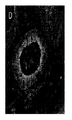


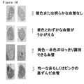


Classifications
-
- C—CHEMISTRY; METALLURGY
- C07—ORGANIC CHEMISTRY
- C07K—PEPTIDES
- C07K16/00—Immunoglobulins [IGs], e.g. monoclonal or polyclonal antibodies
- C07K16/18—Immunoglobulins [IGs], e.g. monoclonal or polyclonal antibodies against material from animals or humans
- C07K16/22—Immunoglobulins [IGs], e.g. monoclonal or polyclonal antibodies against material from animals or humans against growth factors ; against growth regulators
-
- A—HUMAN NECESSITIES
- A61—MEDICAL OR VETERINARY SCIENCE; HYGIENE
- A61P—SPECIFIC THERAPEUTIC ACTIVITY OF CHEMICAL COMPOUNDS OR MEDICINAL PREPARATIONS
- A61P11/00—Drugs for disorders of the respiratory system
-
- A—HUMAN NECESSITIES
- A61—MEDICAL OR VETERINARY SCIENCE; HYGIENE
- A61P—SPECIFIC THERAPEUTIC ACTIVITY OF CHEMICAL COMPOUNDS OR MEDICINAL PREPARATIONS
- A61P11/00—Drugs for disorders of the respiratory system
- A61P11/06—Antiasthmatics
-
- A—HUMAN NECESSITIES
- A61—MEDICAL OR VETERINARY SCIENCE; HYGIENE
- A61P—SPECIFIC THERAPEUTIC ACTIVITY OF CHEMICAL COMPOUNDS OR MEDICINAL PREPARATIONS
- A61P17/00—Drugs for dermatological disorders
-
- A—HUMAN NECESSITIES
- A61—MEDICAL OR VETERINARY SCIENCE; HYGIENE
- A61P—SPECIFIC THERAPEUTIC ACTIVITY OF CHEMICAL COMPOUNDS OR MEDICINAL PREPARATIONS
- A61P17/00—Drugs for dermatological disorders
- A61P17/06—Antipsoriatics
-
- A—HUMAN NECESSITIES
- A61—MEDICAL OR VETERINARY SCIENCE; HYGIENE
- A61P—SPECIFIC THERAPEUTIC ACTIVITY OF CHEMICAL COMPOUNDS OR MEDICINAL PREPARATIONS
- A61P19/00—Drugs for skeletal disorders
- A61P19/02—Drugs for skeletal disorders for joint disorders, e.g. arthritis, arthrosis
-
- A—HUMAN NECESSITIES
- A61—MEDICAL OR VETERINARY SCIENCE; HYGIENE
- A61P—SPECIFIC THERAPEUTIC ACTIVITY OF CHEMICAL COMPOUNDS OR MEDICINAL PREPARATIONS
- A61P27/00—Drugs for disorders of the senses
- A61P27/02—Ophthalmic agents
-
- A—HUMAN NECESSITIES
- A61—MEDICAL OR VETERINARY SCIENCE; HYGIENE
- A61P—SPECIFIC THERAPEUTIC ACTIVITY OF CHEMICAL COMPOUNDS OR MEDICINAL PREPARATIONS
- A61P29/00—Non-central analgesic, antipyretic or antiinflammatory agents, e.g. antirheumatic agents; Non-steroidal antiinflammatory drugs [NSAID]
-
- A—HUMAN NECESSITIES
- A61—MEDICAL OR VETERINARY SCIENCE; HYGIENE
- A61P—SPECIFIC THERAPEUTIC ACTIVITY OF CHEMICAL COMPOUNDS OR MEDICINAL PREPARATIONS
- A61P35/00—Antineoplastic agents
-
- A—HUMAN NECESSITIES
- A61—MEDICAL OR VETERINARY SCIENCE; HYGIENE
- A61P—SPECIFIC THERAPEUTIC ACTIVITY OF CHEMICAL COMPOUNDS OR MEDICINAL PREPARATIONS
- A61P43/00—Drugs for specific purposes, not provided for in groups A61P1/00-A61P41/00
-
- A—HUMAN NECESSITIES
- A61—MEDICAL OR VETERINARY SCIENCE; HYGIENE
- A61P—SPECIFIC THERAPEUTIC ACTIVITY OF CHEMICAL COMPOUNDS OR MEDICINAL PREPARATIONS
- A61P9/00—Drugs for disorders of the cardiovascular system
- A61P9/10—Drugs for disorders of the cardiovascular system for treating ischaemic or atherosclerotic diseases, e.g. antianginal drugs, coronary vasodilators, drugs for myocardial infarction, retinopathy, cerebrovascula insufficiency, renal arteriosclerosis
-
- C—CHEMISTRY; METALLURGY
- C07—ORGANIC CHEMISTRY
- C07K—PEPTIDES
- C07K2317/00—Immunoglobulins specific features
- C07K2317/20—Immunoglobulins specific features characterized by taxonomic origin
- C07K2317/21—Immunoglobulins specific features characterized by taxonomic origin from primates, e.g. man
-
- C—CHEMISTRY; METALLURGY
- C07—ORGANIC CHEMISTRY
- C07K—PEPTIDES
- C07K2317/00—Immunoglobulins specific features
- C07K2317/60—Immunoglobulins specific features characterized by non-natural combinations of immunoglobulin fragments
- C07K2317/62—Immunoglobulins specific features characterized by non-natural combinations of immunoglobulin fragments comprising only variable region components
- C07K2317/622—Single chain antibody (scFv)
-
- C—CHEMISTRY; METALLURGY
- C07—ORGANIC CHEMISTRY
- C07K—PEPTIDES
- C07K2317/00—Immunoglobulins specific features
- C07K2317/70—Immunoglobulins specific features characterized by effect upon binding to a cell or to an antigen
- C07K2317/73—Inducing cell death, e.g. apoptosis, necrosis or inhibition of cell proliferation
-
- C—CHEMISTRY; METALLURGY
- C07—ORGANIC CHEMISTRY
- C07K—PEPTIDES
- C07K2317/00—Immunoglobulins specific features
- C07K2317/70—Immunoglobulins specific features characterized by effect upon binding to a cell or to an antigen
- C07K2317/76—Antagonist effect on antigen, e.g. neutralization or inhibition of binding
-
- C—CHEMISTRY; METALLURGY
- C07—ORGANIC CHEMISTRY
- C07K—PEPTIDES
- C07K2317/00—Immunoglobulins specific features
- C07K2317/90—Immunoglobulins specific features characterized by (pharmaco)kinetic aspects or by stability of the immunoglobulin
- C07K2317/92—Affinity (KD), association rate (Ka), dissociation rate (Kd) or EC50 value
Landscapes
- Health & Medical Sciences (AREA)
- Chemical & Material Sciences (AREA)
- Organic Chemistry (AREA)
- General Health & Medical Sciences (AREA)
- Medicinal Chemistry (AREA)
- Life Sciences & Earth Sciences (AREA)
- Nuclear Medicine, Radiotherapy & Molecular Imaging (AREA)
- Chemical Kinetics & Catalysis (AREA)
- Pharmacology & Pharmacy (AREA)
- General Chemical & Material Sciences (AREA)
- Animal Behavior & Ethology (AREA)
- Public Health (AREA)
- Veterinary Medicine (AREA)
- Bioinformatics & Cheminformatics (AREA)
- Engineering & Computer Science (AREA)
- Immunology (AREA)
- Pulmonology (AREA)
- Proteomics, Peptides & Aminoacids (AREA)
- Biophysics (AREA)
- Dermatology (AREA)
- Rheumatology (AREA)
- Molecular Biology (AREA)
- Genetics & Genomics (AREA)
- Biochemistry (AREA)
- Cardiology (AREA)
- Heart & Thoracic Surgery (AREA)
- Ophthalmology & Optometry (AREA)
- Vascular Medicine (AREA)
- Pain & Pain Management (AREA)
- Orthopedic Medicine & Surgery (AREA)
- Physical Education & Sports Medicine (AREA)
- Urology & Nephrology (AREA)
- Medicines Containing Antibodies Or Antigens For Use As Internal Diagnostic Agents (AREA)
- Peptides Or Proteins (AREA)
- Medicines That Contain Protein Lipid Enzymes And Other Medicines (AREA)
- Preparation Of Compounds By Using Micro-Organisms (AREA)
- Micro-Organisms Or Cultivation Processes Thereof (AREA)
Abstract
血管内皮増殖因子(VEGF)の過剰発現は、異常な脈管形成を伴うさまざまな状態と関係している。本明細書中で開示されるものは、ヒトVEGFに特異的に結合し、そして、VEGF−R1およびVEGF−R2に結合するVEGFを阻害し、したがって、VEGFシグナル伝達を阻害する、完全ヒト抗体である。VEGFシグナル伝達を阻害するそれらの能力に基づいて、本明細書中で開示される抗体は、生体内および試験管内の両方で、脈管形成および脈管形成を伴う状態を処置するのに用いられうる。 Overexpression of vascular endothelial growth factor (VEGF) is associated with a variety of conditions with abnormal angiogenesis. Disclosed herein are fully human antibodies that specifically bind to human VEGF and inhibit VEGF binding to VEGF-R1 and VEGF-R2, and thus inhibit VEGF signaling. is there. Based on their ability to inhibit VEGF signaling, the antibodies disclosed herein are used to treat angiogenesis and conditions involving angiogenesis both in vivo and in vitro. sell.
Description
(関連分野)
本出願は、2006年10月20日に出願された米国仮出願番号第60/853,260号(出典明示により完全に本明細書の一部とされる)についての優先権を主張する。
(Related fields)
This application claims priority to US Provisional Application No. 60 / 853,260, filed Oct. 20, 2006, which is fully incorporated herein by reference.
血管内皮増殖因子(VEGF)は、特に、網膜増殖疾患および腫瘍形成時における内皮細胞活性化、増殖および生存に関与する、脈管形成蛋白質の主要ファミリーである。VEGFは、VEGF−PDGF(血小板由来増殖因子)超遺伝子ファミリーに属しており、脈管内皮細胞とリンパ内皮細胞上に発現される受容体に結合するスモール糖タンパク質二量体である。VEGFファミリーには、現在、7つの既知リガンド:VEGF−A(VEGF)、VEGF−B、VEGF−C、VEGF−D、VEGF−E(ウイルス由来)および胎盤増殖(PIGF)−1と−2がある。これらのVEGFリガンドは、それぞれが受容体チロシンキナーゼ活性を備えている、3つの既知のVEGF受容体(VEGF−R)の1つまたは複数と結合することによって、それらの効果をメディエートする。VEGF−R1(Flt−1)は、内皮細胞と単球上で主に発現され、VEGFとVEGF−Bに結合して、内皮および単球遊走をメディエートするようである。VEGF−R2(すなわち、ヒトKDRまたはネズミFlk−1)は、主に内皮細胞上に発現され、VEGF(および、VEGF−CおよびVEGF−Dの特定の断片)に選択性があり、VEGF誘導性内皮細胞増殖、生存および遊走、ならびに血管透過性をメディエートする。VEGF−R3(Flt−4)は、主にリンパ内皮細胞上に発現され、VEGF−CとVEGF−Dに結合してリンパ管発生を促進する。VEGF−R1、−R2、および−R3は、それぞれ、いくつかの腫瘍細胞上に発現される。VEGFのVEGF受容体への結合は、受容体の二量体化を引き起こし、そして後の受容体活性化およびシグナル伝達を導く。VEGF−R2に結合するVEGFは、脈管形成を促進する際に優性となるシグナル伝達経路を開始する。この経路は、細胞内シグナル伝達の後の誘導を伴う、受容体活性化と関係する。この場合の受容体活性化は、3つの基本的な事象:(i)VEGF−R2に対するVEGFの結合、(ii)受容体の二量体化、そして(iii)受容体チロシンキナーゼによる受容体の自己リン酸化(それによる、活性化)、を必然的に伴う。細胞内メッセンジャー、例えば、ホスホリパーゼCおよびホスファチジルイノシトール−3−キナーゼは、自己リン酸化型のVEGF−R2と直接結合し、そして、その受容体チロシンキナーゼによってリン酸化され、その後、それが、最終的に細胞増殖、遊走および生存(抗−アポトーシス)を促進し、そして血管透過性を増大させる核シグナルに至る、シグナル伝達事象の細胞内カスケードを誘発する。 Vascular endothelial growth factor (VEGF) is a major family of angiogenic proteins involved in endothelial cell activation, proliferation and survival, particularly during retinal proliferative diseases and tumor formation. VEGF belongs to the VEGF-PDGF (platelet-derived growth factor) supergene family, and is a small glycoprotein dimer that binds to receptors expressed on vascular and lymphatic endothelial cells. The VEGF family currently has seven known ligands: VEGF-A (VEGF), VEGF-B, VEGF-C, VEGF-D, VEGF-E (virus-derived) and placental growth (PIGF) -1 and -2. is there. These VEGF ligands mediate their effects by binding to one or more of the three known VEGF receptors (VEGF-R), each with receptor tyrosine kinase activity. VEGF-R1 (Flt-1) is predominantly expressed on endothelial cells and monocytes and appears to bind VEGF and VEGF-B to mediate endothelial and monocyte migration. VEGF-R2 (ie, human K D R or murine Flk-1) is expressed primarily on endothelial cells, is selective for VEGF (and certain fragments of VEGF-C and VEGF-D), and VEGF Mediates induced endothelial cell proliferation, survival and migration, and vascular permeability. VEGF-R3 (Flt-4) is mainly expressed on lymphatic endothelial cells and binds to VEGF-C and VEGF-D to promote lymphangiogenesis. VEGF-R1, -R2, and -R3 are each expressed on several tumor cells. Binding of VEGF to the VEGF receptor causes receptor dimerization and leads to subsequent receptor activation and signal transduction. VEGF binding to VEGF-R2 initiates a signaling pathway that becomes dominant in promoting angiogenesis. This pathway is associated with receptor activation with subsequent induction of intracellular signaling. In this case, receptor activation has three basic events: (i) binding of VEGF to VEGF-R2, (ii) dimerization of the receptor, and (iii) receptor activation by receptor tyrosine kinases. Inevitably involves autophosphorylation (and thereby activation). Intracellular messengers such as phospholipase C and phosphatidylinositol-3-kinase bind directly to the autophosphorylated form of VEGF-R2 and are phosphorylated by its receptor tyrosine kinase, after which it is finally It induces an intracellular cascade of signaling events leading to nuclear signals that promote cell proliferation, migration and survival (anti-apoptosis) and increase vascular permeability.
異常脈管形成は、癌を含む、さまざまな疾患状態と関係する(Holash 2002)。VEGF経路は、正常な脈管発生と様々な疾患に付随する異常な脈管形成の両方に役割を果たすことが実証された、唯一のシグナル伝達経路である(Erikkson 1999; Ferrara 1999; Yancopoulos 2000)。VEGFは、脈管血管内皮細胞増殖を促進し、血管透過性を増大させる(Ferrara 2004)。 Abnormal angiogenesis is associated with a variety of disease states, including cancer (Holash 2002). The VEGF pathway is the only signaling pathway that has been demonstrated to play a role in both normal angiogenesis and abnormal angiogenesis associated with various diseases (Erikkson 1999; Ferrara 1999; Yancopoulos 2000) . VEGF promotes vascular vascular endothelial cell proliferation and increases vascular permeability (Ferrara 2004).
先行研究は、大多数の腫瘍型における高いVEGF発現レベルを明らかにした(Berkman 1993; Brown 1993; Brown 1995; Dvorak 1995; Mattern 1996)。研究は、眼性脈管形成疾患、例えば湿潤型AMDを患う被験者における増大したVEGFレベルも明らかにした。湿潤型AMDは、AMD疾患全体の約10%を占めるだけであるが、AMDに起因する失明の約90%を引き起こす。湿潤型AMDは、脈絡膜新生血管(CNV)、網膜の網膜色素上皮層下の異常な血管の発生によって特徴づけられる。VEGF−Aは、これらの脈管形成において主要な役割を果たすと考えられており、それは、斑の下に滲み出し、レチナール歪曲および視力悪化を引き起こす。 Previous studies have revealed high VEGF expression levels in the majority of tumor types (Berkman 1993; Brown 1993; Brown 1995; Dvorak 1995; Mattern 1996). Studies have also revealed increased VEGF levels in subjects with ocular angiogenic diseases such as wet AMD. Wet AMD only accounts for about 10% of all AMD disease, but causes about 90% of blindness due to AMD. Wet AMD is characterized by the development of choroidal neovascularization (CNV), abnormal blood vessels beneath the retinal pigment epithelium layer of the retina. VEGF-A is believed to play a major role in these angiogenesis, which oozes under the plaques, causing retinal distortion and visual deterioration.
VEGFとその受容体との相互作用を阻害する、またはVEGF受容体のチロシンキナーゼ活性を阻害するいくつかの分子が開発され、そして臨床開発のさまざまなステージにある。例えば、BAY 43−9006(Sorafenib, Bayer)、SU11248(Sunitinib)およびバタラニブ(Novartis)は、潜在的な癌治療として開発されたVEGF−R2キナーゼ阻害剤である。VEGF−TRAP(Regeneron, Sanofi-Aventis)は、VEGF−R1の細胞外ドメインとVEGFと高い結合能を有するVEGF−R2のハイブリッドである(Holash 2002; Dupont 2005)。VEGF−TRAPは、現在、固形腫瘍の処置に関して臨床試験中である。抗体も、VEGF経路の阻害剤として使用するために広く開発されている。例えば、IMC-1C11(Imclone)は、VEGF−R2と結合するキメラ・モノクローナル抗体である(Hunt 2001)。転移結腸直腸癌腫の処置におけるIMC−1C11の効果を決定するための、臨床研究が、現在行われている。CDP−791(Imclone)は、VEGF−R2に結合するPEG化されたヒト化Fabである。IMC−1121B(Imclone)は、VEGF−R2と高親和性でもって結合し、そしてVEGFとVEGF−R2との相互作用を阻害する、完全ヒト・モノクローナル抗体である(Miao, H. Q.ら、2005. Biochem Biophys Res Commun 345:438-445)。IMC−18F1(Imclone)は、VEGF−R1と高親和性でもって結合し、そしてVEGFとVEGF−R1との相互作用をブロックする、完全ヒト・モノクローナル抗体である。2C3(Peregrine)は、VEGFと結合し、そしてVEGFとVEGF−R2(VEGF−R1ではなく)と結合をブロックする、マウス抗体である。ラニビズマブ(Ranibizumab)(Lucentis(登録商標), Genentech)は、湿潤型の急性黄斑変性(AMD)の処置に関して最近承認された、ベバシズマブから誘導された、ヒト化Fabである(Michels 2004; Rosenfeld 2005)。 Several molecules have been developed that inhibit the interaction of VEGF with its receptor or that inhibit the tyrosine kinase activity of the VEGF receptor and are in various stages of clinical development. For example, BAY 43-9006 (Sorafenib, Bayer), SU11248 (Sunitinib) and Batalanib (Novartis) are VEGF-R2 kinase inhibitors developed as potential cancer treatments. VEGF-TRAP (Regeneron, Sanofi-Aventis) is a hybrid of the extracellular domain of VEGF-R1 and VEGF-R2 having a high binding ability to VEGF (Holash 2002; Dupont 2005). VEGF-TRAP is currently in clinical trials for the treatment of solid tumors. Antibodies have also been widely developed for use as inhibitors of the VEGF pathway. For example, IMC-1C11 (Imclone) is a chimeric monoclonal antibody that binds to VEGF-R2 (Hunt 2001). Clinical studies are currently underway to determine the effect of IMC-1C11 in the treatment of metastatic colorectal carcinoma. CDP-791 (Imclone) is a PEGylated humanized Fab that binds to VEGF-R2. IMC-1121B (Imclone) is a fully human monoclonal antibody that binds with high affinity to VEGF-R2 and inhibits the interaction between VEGF and VEGF-R2 (Miao, HQ et al., 2005. Biochem). Biophys Res Commun 345: 438-445). IMC-18F1 (Imclone) is a fully human monoclonal antibody that binds with high affinity to VEGF-R1 and blocks the interaction between VEGF and VEGF-R1. 2C3 (Peregrine) is a mouse antibody that binds to VEGF and blocks binding to VEGF and VEGF-R2 (but not VEGF-R1). Ranibizumab (Lucentis®, Genentech) is a recently approved humanized Fab derived from bevacizumab for the treatment of wet acute macular degeneration (AMD) (Michels 2004; Rosenfeld 2005) .
最も一般に用いられている、VEGFを標的とする生物学的治療は、ヒト化IgGIモノクローナル抗体、ベバシズマブである(別名、アバスチン(Avastin)(登録商標)、Genentech;また、BM−1とも称される)。ベバシズマブは、マウス・モノクローナル抗体A.4.6.1をヒト化することによって開発され、そして、それは、約93%のヒト配列と7%のネズミ配列を含む(Presta 1997; Ferrara 2004)。ベバシズマブは、約500pMの親和性でもってVEGFと結合し、VEGFのVEGF−R1とVEGF−R2に対する結合を阻害する。ヒトにおけるベバシズマブの消失半減期は、17日〜21日である(Ferrara 2004)。ベバシズマブは、転移結腸直腸癌の処置に関して承認されている(Presta 1997; Rosenfeld 2005)。研究は、ベバシズマブが血管新生AMDの処置でも有用でありうることを示唆する(Michels 2005)。 The most commonly used biological therapy targeting VEGF is the humanized IgGI monoclonal antibody, bevacizumab (also known as Avastin®, Genentech; also referred to as BM-1). ). Bevacizumab is a mouse monoclonal antibody A.I. Developed by humanizing 4.6.1 and it contains about 93% human and 7% murine sequences (Presta 1997; Ferrara 2004). Bevacizumab binds to VEGF with an affinity of about 500 pM and inhibits binding of VEGF to VEGF-R1 and VEGF-R2. The elimination half-life of bevacizumab in humans is 17-21 days (Ferrara 2004). Bevacizumab has been approved for the treatment of metastatic colorectal cancer (Presta 1997; Rosenfeld 2005). Studies suggest that bevacizumab may also be useful in the treatment of neovascular AMD (Michels 2005).
VEGF経路の阻害のために開発された分子の多様性にもかかわらず、改善された結合特徴および治療プロフィールを有するさらなる分子の必要性がある。 Despite the diversity of molecules developed for inhibition of the VEGF pathway, there is a need for additional molecules with improved binding characteristics and therapeutic profiles.
本発明は、VEGFに結合してVEGF経路を阻害する、完全ヒト抗体を提供する。これらの抗体は完全ヒト抗体であるため、それらは、以前に開発されたヒト以外の抗体、キメラ抗体またはヒト化抗体に付随する不都合な免疫原性がなく、治療薬として投与することができる。 The present invention provides fully human antibodies that bind to VEGF and inhibit the VEGF pathway. Because these antibodies are fully human antibodies, they can be administered as therapeutic agents without the disadvantageous immunogenicity associated with previously developed non-human, chimeric or humanized antibodies.
特定の実施形態において、配列番号:2記載の重鎖配列と配列番号:3記載の軽鎖配列を有する抗体が、提供される。特定の他の実施形態において、配列番号:4記載の重鎖配列と配列番号:5記載の軽鎖配列を有する抗体が、提供される。上記実施形態のいくつかにおいて、抗体は、IgG2定常領域を有する。特定の実施形態において、抗体は、<2.0nM.のKDでもってhVEGF165と結合する。特定の実施形態において、抗体は、ベバシズマブにより結合されるエピトープと重複するhVEGF165上のエピトープと結合する。特定の実施形態において、抗体は、hVEGF165のVEGF−R1およびVEGF−R2に対する結合をブロックする。 In certain embodiments, an antibody having the heavy chain sequence set forth in SEQ ID NO: 2 and the light chain sequence set forth in SEQ ID NO: 3 is provided. In certain other embodiments, an antibody having the heavy chain sequence set forth in SEQ ID NO: 4 and the light chain sequence set forth in SEQ ID NO: 5 is provided. In some of the above embodiments, the antibody has an IgG2 constant region. In certain embodiments, the antibody has < 2.0 nM. Binding to hVEGF 165 with at the K D. In certain embodiments, the antibody binds to an epitope on hVEGF 165 that overlaps with the epitope bound by bevacizumab. In certain embodiments, the antibody blocks binding of hVEGF 165 to VEGF-R1 and VEGF-R2.
特定の実施形態において、それを必要とする被検体に、治療上有効量の、配列番号:2記載の重鎖配列と配列番号:3記載の軽鎖配列を有する抗体を投与することによって、該被検体における脈管形成を阻害するための方法が、提供される。特定の他の実施形態において、それを必要とする被検体に、治療上有効量の、配列番号:4記載の重鎖配列と配列番号:5記載の軽鎖配列を有する抗体を投与することによって、該被検体における脈管形成を阻害するための方法が、提供される。 In certain embodiments, the subject in need thereof is administered a therapeutically effective amount of an antibody having the heavy chain sequence set forth in SEQ ID NO: 2 and the light chain sequence set forth in SEQ ID NO: 3. A method for inhibiting angiogenesis in a subject is provided. In certain other embodiments, by administering to a subject in need thereof a therapeutically effective amount of an antibody having the heavy chain sequence set forth in SEQ ID NO: 4 and the light chain sequence set forth in SEQ ID NO: 5 A method is provided for inhibiting angiogenesis in the subject.
特定の実施形態において、それを必要とする被検体における異常脈管形成を伴う疾患を処置するための方法であって、該被検体に、治療上有効量の、配列番号:2記載の重鎖配列と配列番号:3記載の軽鎖配列を有する抗体を投与することを含む方法が、提供される。特定の他の実施形態において、それを必要とする被検体における異常脈管形成を伴う疾患を処置するための方法であって、該被検体に、治療上有効量の、配列番号:4記載の重鎖配列と配列番号:5記載の軽鎖配列を有する抗体を投与することを含む方法が、提供される。 In certain embodiments, a method for treating a disease associated with abnormal angiogenesis in a subject in need thereof, the subject comprising a therapeutically effective amount of the heavy chain of SEQ ID NO: 2. A method is provided comprising administering an antibody having the sequence and the light chain sequence set forth in SEQ ID NO: 3. In certain other embodiments, a method for treating a disease associated with abnormal angiogenesis in a subject in need thereof, the subject comprising a therapeutically effective amount of SEQ ID NO: 4 A method is provided comprising administering an antibody having a heavy chain sequence and a light chain sequence set forth in SEQ ID NO: 5.
特定の実施形態において、それを必要とする被検体におけるVEGFシグナル伝達と関係する炎症性疾患を処置するための方法であって、該被検体に、治療上有効量の、配列番号:2記載の重鎖配列と配列番号:3記載の軽鎖配列を有する抗体を投与することを含む方法が、提供される。特定の他の実施形態において、それを必要とする被検体におけるVEGFシグナル伝達と関係する炎症性疾患を処置するための方法であって、該被検体に、治療上有効量の、配列番号:4記載の重鎖配列と配列番号:5記載の軽鎖配列を有する抗体を投与することを含む方法が、提供される。 In certain embodiments, a method for treating an inflammatory disease associated with VEGF signaling in a subject in need thereof, wherein the subject is treated with a therapeutically effective amount of SEQ ID NO: 2. A method is provided comprising administering an antibody having a heavy chain sequence and a light chain sequence set forth in SEQ ID NO: 3. In certain other embodiments, a method for treating an inflammatory disease associated with VEGF signaling in a subject in need thereof, wherein the subject is treated with a therapeutically effective amount of SEQ ID NO: 4. A method is provided comprising administering an antibody having the described heavy chain sequence and the light chain sequence set forth in SEQ ID NO: 5.
特定の実施形態において、それを必要とする被検体における湿潤型の急性黄斑変性または糖尿病網膜症を処置するための方法であって、該被検体に、治療上有効量の、配列番号:2記載の重鎖配列と配列番号:3記載の軽鎖配列を有する抗体を投与することを含む方法が、提供される。特定の他の実施形態において、それを必要とする被検体における湿潤型の急性黄斑変性または糖尿病網膜症を処置するための方法であって、該被検体に、治療上有効量の、配列番号:4記載の重鎖配列と配列番号:5記載の軽鎖配列を有する抗体を投与することを含む方法が、提供される。 In certain embodiments, a method for treating wet acute macular degeneration or diabetic retinopathy in a subject in need thereof, wherein the subject has a therapeutically effective amount of SEQ ID NO: 2. A method comprising administering an antibody having the heavy chain sequence of and the light chain sequence set forth in SEQ ID NO: 3 is provided. In certain other embodiments, a method for treating wet acute macular degeneration or diabetic retinopathy in a subject in need thereof, wherein the subject has a therapeutically effective amount of SEQ ID NO: A method is provided comprising administering an antibody having the heavy chain sequence set forth in 4 and the light chain sequence set forth in SEQ ID NO: 5.
特定の実施形態において、それを必要とする被検体における増大したVEGFシグナル伝達と関係する癌を処置するための方法であって、該被検体に、治療上有効量の、配列番号:2記載の重鎖配列と配列番号:3記載の軽鎖配列を有する抗体を投与することを含む方法が、提供される。特定の他の実施形態において、それを必要とする被検体における増大したVEGFシグナル伝達と関係する癌を処置するための方法であって、該被検体に、治療上有効量の、配列番号:4記載の重鎖配列と配列番号:5記載の軽鎖配列を有する抗体を投与することを含む方法が、提供される。 In certain embodiments, a method for treating cancer associated with increased VEGF signaling in a subject in need thereof, wherein the subject is treated with a therapeutically effective amount of SEQ ID NO: 2. A method is provided comprising administering an antibody having a heavy chain sequence and a light chain sequence set forth in SEQ ID NO: 3. In certain other embodiments, a method for treating cancer associated with increased VEGF signaling in a subject in need thereof, wherein the subject is treated with a therapeutically effective amount of SEQ ID NO: 4. A method is provided comprising administering an antibody having the described heavy chain sequence and the light chain sequence set forth in SEQ ID NO: 5.
特定の実施形態において、配列番号:2記載の重鎖配列と配列番号:3記載の軽鎖配列を有する抗体を含むキットが、提供される。特定の他の実施形態において、配列番号:4記載の重鎖配列と配列番号:5記載の軽鎖配列を有する抗体を含むキットが、提供される。 In certain embodiments, a kit comprising an antibody having a heavy chain sequence set forth in SEQ ID NO: 2 and a light chain sequence set forth in SEQ ID NO: 3 is provided. In certain other embodiments, a kit comprising an antibody having the heavy chain sequence set forth in SEQ ID NO: 4 and the light chain sequence set forth in SEQ ID NO: 5 is provided.
特定の実施形態において、配列番号:2、配列番号:3、配列番号:4、および/または配列番号:5記載のアミノ酸配列をコードするポリヌクレオチドが、提供される。特定の他の実施形態において、これらのポリヌクレオチドを含むベクターが、提供され、そして、特定の他の実施形態において、これらのベクターを含む宿主細胞が、提供される。 In certain embodiments, a polynucleotide encoding the amino acid sequence set forth in SEQ ID NO: 2, SEQ ID NO: 3, SEQ ID NO: 4, and / or SEQ ID NO: 5 is provided. In certain other embodiments, vectors comprising these polynucleotides are provided, and in certain other embodiments, host cells comprising these vectors are provided.
本発明の以下の説明は、単に本発明のさまざまな実施形態を例示することを目的とするものである。このように、記載の特定のモディフィケーションは、本発明の範囲を制限するものと解釈されるべきでない。当業者であれば、さまざまな均等物、変更およびモディフィケーションが、本発明の範囲から逸脱することなくなしうることは認められよう、さらに、そのような均等な実施形態は、本明細書中に含まれるものと理解される。 The following description of the present invention is merely intended to illustrate various embodiments of the present invention. As such, the specific modifications described should not be construed as limiting the scope of the invention. Those skilled in the art will recognize that various equivalents, modifications, and modifications can be made without departing from the scope of the present invention, and such equivalent embodiments are described herein. Is understood to be included.
略語
以下の略語が、本明細書中で用いられる:ADCC、抗体依存性細胞障害活性;AMD、加齢性黄斑変性;CDC、補体依存性細胞毒性;CNV、脈絡膜新生血管;COPD、慢性閉塞性肺疾患;HUVEC、ヒト臍静脈内皮細胞;hVEGF、ヒトVEGF;mVEGF、ネズミVEGF;PD、薬力学;PK、薬物動力学;RA、関節リウマチ;RIA、放射性免疫沈降法;VEGF、血管内皮増殖因子;VEGF−R、血管内皮成長因子受容体;VHL、フォンヒッペル/リンダウ;X−反応性、交差反応性。
Abbreviations The following abbreviations are used herein: ADCC, antibody-dependent cytotoxic activity; AMD, age-related macular degeneration; CDC, complement-dependent cytotoxicity; CNV, choroidal neovascularization; COPD, chronic occlusion HUVEC, human umbilical vein endothelial cells; hVEGF, human VEGF; mVEGF, murine VEGF; PD, pharmacodynamics; PK, pharmacokinetics; RA, rheumatoid arthritis; RIA, radioimmunoprecipitation; VEGF, vascular endothelial growth Factor: VEGF-R, vascular endothelial growth factor receptor; VHL, von Hippel / Lindau; X-reactivity, cross-reactivity.
定義
本明細書中で用いられる用語「抗体」は、特定の抗原と結合する、モノクローナル抗体、ポリクローナル抗体または多特異的もしくは二重特異的抗体を含む、いずれかの免疫グロブリンを意味する。完全抗体は、2個の重鎖と2個の軽鎖を含む。各軽鎖は、可変領域と定常領域からなるが、各重鎖は、可変領域と第1、第2および第3の定常領域からなる。抗体は、ジスルフィド結合を介して互いに結合した2個の重鎖の第2および第3の定常領域からなる「Y」字の幹を有する、「Y」字型の形状を有する。「Y」の各腕は、単一の軽鎖の可変部分と定常領域に結合された、単一の重鎖の可変領域と第1の定常領域からなる。軽鎖および重鎖の可変領域が、抗原結合に関与する。両方の鎖の可変領域は、相補性決定領域(CDR)(LCDR1、LCDR2およびLCDR3を含む軽鎖(L)CDR、HCDR1、HCDR2およびHCDR3を含む重鎖(H)CDR)と呼ばれる3つの超可変ループを、一般に含む。この3つのCDRは、CDRと比べて高度に保存された、超可変ループを支持する足場を形成する、枠組み構造領域(FR)として知られる、フランキング・ストレッチ(flanking stretch)間に挿入されている。重鎖および軽鎖の定常領域は、抗原結合に関与しないが、さまざまなエフェクター機能を示す。抗体は、それらの重鎖の定常領域のアミノ酸配列に基づいて、クラス分けされる。抗体の主要なクラスは、IgA、IgD、IgE、IgGおよびIgMがあり、これらのクラスのいくつかは、サブクラス、例えばIgG1、IgG2、IgG3、IgG4、IgA1またはlgA2に分けられる。
Definitions As used herein, the term “antibody” means any immunoglobulin, including monoclonal antibodies, polyclonal antibodies, or multispecific or bispecific antibodies, that bind to a specific antigen. A complete antibody contains two heavy chains and two light chains. Each light chain is composed of a variable region and a constant region, while each heavy chain is composed of a variable region and first, second and third constant regions. The antibody has a “Y” -shaped shape with a “Y” -shaped trunk consisting of the second and third constant regions of two heavy chains linked together through disulfide bonds. Each arm of “Y” consists of a single heavy chain variable region and a first constant region joined to a single light chain variable portion and constant region. The light and heavy chain variable regions are responsible for antigen binding. The variable regions of both chains are three hypervariables called complementarity determining regions (CDRs) (light chain (L) CDRs including LCDR1, LCDR2 and LCDR3, heavy chain (H) CDRs including HCDR1, HCDR2 and HCDR3) A loop is generally included. The three CDRs are inserted between flanking stretches, known as framework regions (FR), which form a scaffold that supports hypervariable loops, which is highly conserved compared to CDRs. Yes. The heavy and light chain constant regions are not involved in antigen binding but exhibit various effector functions. Antibodies are classified based on the amino acid sequence of the constant region of their heavy chains. The major classes of antibodies are IgA, IgD, IgE, IgG and IgM, and some of these classes are divided into subclasses such as IgG1, IgG2, IgG3, IgG4, IgA1 or IgA2.
本明細書中で用いられる用語「抗原結合性断片」は、免疫グロブリン断片またはその抗体断片(すなわち、免疫グロブリン分子の少なくとも1つの免疫活性部分)、例えば、Fab、Fab’、F(ab’)2、Fv断片、一本鎖抗体分子、1個または複数のCDRを含む免疫グロブリン分子のいずれかの断片から形成される多特異的な抗体を、さらに意味する。加えて、本明細書中で用いられる「抗原結合性断片」は、1個または複数の異なるヒト免疫グロブリンからの枠組み構造領域に継ぎ足された、特定のヒト免疫グロブリンからの1個または複数のCDRを含んでもよい。抗原結合性断片は、親の免疫グロブリンまたは抗体が結合する標的に結合しうる。 The term “antigen-binding fragment” as used herein refers to an immunoglobulin fragment or antibody fragment thereof (ie, at least one immunologically active portion of an immunoglobulin molecule), eg, Fab, Fab ′, F (ab ′) 2 , further means a multispecific antibody formed from any fragment of an Fv fragment, a single chain antibody molecule, an immunoglobulin molecule comprising one or more CDRs. In addition, an “antigen-binding fragment” as used herein is one or more CDRs from a particular human immunoglobulin, spliced to a framework region from one or more different human immunoglobulins. May be included. An antigen-binding fragment can bind to a target to which a parent immunoglobulin or antibody binds.
抗体に関する「Fab」は、ジスルフィド結合により、単一の重鎖の可変領域と第1の定常領域に結合された単一の軽鎖(可変領域と定常領域の両方)からなる、抗体部分を意味する。
「Fab’」は、ヒンジ領域の一部を含む、Fab断片を意味する。
「F(ab’)2」は、Fabの二量体を意味する。
抗体に関する「Fc」は、ジスルフィド結合を介し、第2の重鎖の第2と第3の定常領域と結合した、第1の重鎖の第2と第3の定常領域からなる、抗体の部分を意味する。抗体のFc部分は、ADCCおよびCDCなどのさまざまなエフェクター機能の要因となるが、抗原結合には機能しない。
“Fab” with respect to an antibody refers to the antibody portion consisting of a single light chain (both variable and constant regions) joined to the variable region of a single heavy chain and a first constant region by disulfide bonds. To do.
“Fab ′” refers to a Fab fragment that includes a portion of the hinge region.
“F (ab ′) 2 ” means a dimer of Fab.
“Fc” for an antibody is the portion of an antibody that consists of the second and third constant regions of the first heavy chain linked to the second and third constant regions of the second heavy chain via disulfide bonds. Means. The Fc portion of an antibody is responsible for various effector functions such as ADCC and CDC but does not function for antigen binding.
抗体に関する「Fv」は、完全な抗原結合部位を有する、抗体の最小断片を意味する。Fv断片は、単一の重鎖の可変領域に結合された、単一の軽鎖の可変領域からなる。
「一本鎖Fv抗体」または「scFv」は、互いに直接もしくはペプチドリンカー配列を介して連結された、軽鎖可変領域および重鎖可変領域からなる、改変抗体を意味する(Houston 1988)。
「一本鎖Fv−Fc抗体」または「scFv−Fc」は、抗体のFc領域に連結されたscFvからなる、改変抗体を意味する。
“Fv” with respect to antibody means the smallest fragment of an antibody that has a complete antigen-binding site. An Fv fragment consists of a single light chain variable region joined to a single heavy chain variable region.
“Single-chain Fv antibody” or “scFv” refers to a modified antibody consisting of a light chain variable region and a heavy chain variable region linked directly to each other or via a peptide linker sequence (Houston 1988).
“Single-chain Fv-Fc antibody” or “scFv-Fc” refers to a modified antibody consisting of an scFv linked to the Fc region of an antibody.
本明細書中で用いられる用語「エピトープ」は、抗体が結合する抗原分子上の原子またはアミノ酸群を意味する。本明細書中で用いられるように、それらがある抗原に対して競争的結合を示す場合、その2つの抗体は、その抗原内の同じエピトープに結合する。例えば、本明細書中で開示されるように、抗体が、VEGF結合に対してベバシズマブと競争する場合、その抗体はベバシズマブと同じエピトープに結合すると考えられる。 The term “epitope” as used herein refers to an atom or group of amino acids on an antigen molecule to which an antibody binds. As used herein, two antibodies bind to the same epitope within an antigen if they exhibit competitive binding to that antigen. For example, as disclosed herein, an antibody is considered to bind to the same epitope as bevacizumab when it competes with bevacizumab for VEGF binding.
本明細書中で用いられる「VEGF」または「VEGFリガンド」は、現在既知の、7つのVEGFリガンド:VEGF−A、VEGF−B、VEGF−C1、VEGF−D、VEGF−E(ウイルス誘導体)または胎盤成長因子(PIGF)−1または−2、のうちのいずれかを意味する。VEGF−Aに関して、現在、それぞれ固有の生体機能を示す、4つの既知のスプライシング・アイソフォームがある。165アミノ酸アイソフォーム(VEGF165、配列番号:1)は、ヘパリン結合型および可溶性型の両方に存在する。VEGF165の115残基と159残基間の領域に対応する断片を失っている、121アミノ酸アイソフォーム(VEGF12I)は、可溶性型にのみ存在する。より長い189アミノ酸および206アミノ酸アイソフォーム(それぞれ、VEGF189およびVEGF206)は、ヘパリンに結合する能力を維持している。本明細書中で提供される抗体は、ヒトVEGF165に対して高い結合能を示すが、いくつかの実施形態において、ヒト以外のVEGF蛋白質と、もしくは他のVEGFアイソフォームと、交差反応しうるか、または低レベルの結合能を示しうる。 As used herein, “VEGF” or “VEGF ligand” refers to currently known seven VEGF ligands: VEGF-A, VEGF-B, VEGF-C1, VEGF-D, VEGF-E (viral derivatives) or Means either placental growth factor (PIGF) -1 or -2. With respect to VEGF-A, there are currently four known splicing isoforms, each showing unique biological functions. The 165 amino acid isoform (VEGF 165 , SEQ ID NO: 1) exists in both heparin-bound and soluble forms. The 121 amino acid isoform (VEGF12I), missing the fragment corresponding to the region between residues 115 and 159 of VEGF 165 , exists only in the soluble form. Longer 189 amino acid and 206 amino acid isoforms (VEGF189 and VEGF206, respectively) retain the ability to bind heparin. Although the antibodies provided herein exhibit high binding ability to human VEGF 165 , in some embodiments, can they cross-react with non-human VEGF proteins or with other VEGF isoforms? Or a low level of binding ability.
本明細書中で用いられる、ある状態を「処置する」または「処置」は、その状態を予防または緩和すること、その状態の発症または進行速度を低下させること、その状態が進行する危険性を低下させること、その状態を伴う症状の侵攻を予防または遅延させること、その状態の完全もしくは部分的な回復を果たすこと、あるいはそれらのいくつかの組み合わせを意味しうる。腫瘍に関して、「処置する」または「処置」は、腫瘍の全部または一部を根絶すること、腫瘍増殖および転移を阻害または遅延させること、腫瘍の進行を予防または遅延させること、あるいはそれらのいくつかの組み合わせを意味しうる。 As used herein, “treating” or “treatment” of a condition refers to preventing or alleviating the condition, reducing the rate of onset or progression of the condition, and the risk that the condition will progress. It can mean reducing, preventing or delaying the invasion of symptoms associated with the condition, achieving full or partial recovery of the condition, or some combination thereof. With respect to tumors, “treat” or “treatment” refers to eradicating all or part of the tumor, inhibiting or delaying tumor growth and metastasis, preventing or delaying tumor progression, or some of them Can be a combination of
本明細書中で用いられる用語「特異的に結合する」は、2個の分子間、例えば、抗体とその抗体が産生されたリガンド間の無作為でない結合反応を意味する。本明細書中で用いられるように、第1のリガンドと特異的に結合する抗体は、第2のリガンドと交差反応性または低レベルの結合能を示しうる。特定の実施形態において、リガンドに特異的に結合する抗体は、<10−7M(例えば5×10⁻⁸M、10−8M、5×10⁻⁹M、10−9M、10−10M)の結合親和性(KD)でもってリガンドと結合する。本明細書中で用いられるKDは、解離速度の結合速度に対する割合(koff/kon)を意味し、当分野で既知の方法を用いて決定されうる。他の実施形態において、抗体は、その抗体が第2のリガンドに結合する結合親和性よりも10倍以上高いもしくは低い(例えば、>15倍、>20倍、>50倍、>102倍、>103倍、または>104倍)結合親和性でもって、第1のリガンドと特異的に結合する。他の実施形態において、特異的にリガンドと結合する抗体は、<10−7の結合親和性でもってそのリガンドと結合するが、第2のリガンドとはほとんど結合親和性を示さない。 As used herein, the term “specifically binds” refers to a non-random binding reaction between two molecules, eg, between an antibody and the ligand from which the antibody was produced. As used herein, an antibody that specifically binds to a first ligand can exhibit cross-reactivity or a low level of binding ability with a second ligand. In certain embodiments, an antibody that specifically binds a ligand is < 10 −7 M (eg, 5 × 10 −8 M, 10 −8 M, 5 × 10 −9 M, 10 −9 M, 10 −10 It binds to the ligand with a binding affinity (K D ) of M). K D, as used herein, refers to the ratio (k off / k on) for binding rate of dissociation rate can be determined using methods known in the art. In other embodiments, the antibody is more than or less than 10 times the binding affinity that the antibody binds to the second ligand (eg, > 15 times, > 20 times, > 50 times, > 10 2 times, > 10 3 fold, or > 10 4 fold) binds specifically to the first ligand with binding affinity. In other embodiments, an antibody that specifically binds a ligand binds that ligand with a binding affinity of < 10-7 , but exhibits little binding affinity with a second ligand.
本明細書中で用いられる用語「単離された」は、天然状態から「ヒトの手により」改変される手段を意味する。「単離された」組成物または基質が天然に生じる場合、それは、変更されまたはその元の環境から取り出されたものであり、または、その両方である。例えば、生きている動物中に天然に存在するポリヌクレオチドまたはポリペプチドは「単離されていない」が、それが、実質的に純粋な状態で存在するよう、その天然状態の並存する材料から十分に分離された場合には、その同じポリヌクレオチドまたはポリペプチドは「単離されている」。本明細書中で用いられる「単離された」は、人工物または他の化合物もしくは材料との合成混合物、または活性を妨げない不純物の存在を排除しない。 The term “isolated” as used herein means a means that is “modified by the hand of man” from the natural state. When an “isolated” composition or substrate occurs in nature, it has been modified or removed from its original environment, or both. For example, a polynucleotide or polypeptide that occurs naturally in a living animal is not “isolated” but is sufficient from its coexisting material in its native state so that it exists in a substantially pure state. The same polynucleotide or polypeptide is “isolated” when separated into two. “Isolated” as used herein does not exclude the presence of artifacts or synthetic mixtures with other compounds or materials, or the presence of impurities that do not interfere with activity.
本明細書中で用いられる用語「ベクター」は、ある蛋白質をコードするポリヌクレオチドが、その蛋白質の発現を示すよう、操作可能に挿入されうる、媒体を意味する。ベクターは、ある宿主細胞内に、それが運搬する遺伝因子の発現を示すよう、宿主細胞を形質転換、形質導入またはトランスフェクトするのに用いられうる。ベクターの例は、プラスミド、ファージミド、コスミド、人工染色体、例えば酵母人工染色体(YAC)、大腸菌人工染色体(BAC)またはP1誘導人工染色体(PAC)、バクテリオファージ、例えばラムダファージまたはM13ファージ、および動物ウイルスを、含む。ベクターとして用いられる動物ウイルスのカテゴリーは、レトロウイルス(レンチウイルスを含む)、アデノウイルス、アデノ−付随ウイルス、ヘルペスウイルス(例えば、単純ヘルペスウイルス)、ポックスウイルス、バキュロウイルス、乳頭腫ウイルス、およびパポーバウイルス(例えばSV40)を含む。ベクターは、プロモーター配列、転写開始配列、エンハンサー配列、選択可能な因子およびとレポーター遺伝子を含み、発現を制御するための様々な構成部分を含んでいてよい。加えて、ベクターは、複製開始点を含みうる。ベクターは、限定するものではないが、ウイルス粒子、リポソームまたは蛋白質コーティングを含み、細胞へのその侵入を補助するための材料を含んでもよい。 As used herein, the term “vector” refers to a medium into which a polynucleotide encoding a protein can be operatively inserted to show expression of that protein. A vector can be used to transform, transduce or transfect a host cell to show expression of the genetic factor it carries within a host cell. Examples of vectors include plasmids, phagemids, cosmids, artificial chromosomes such as yeast artificial chromosome (YAC), E. coli artificial chromosome (BAC) or P1-derived artificial chromosome (PAC), bacteriophages such as lambda phage or M13 phage, and animal viruses including. The categories of animal viruses used as vectors include retroviruses (including lentiviruses), adenoviruses, adeno-associated viruses, herpes viruses (eg, herpes simplex virus), poxviruses, baculoviruses, papilloma viruses, and papovaviruses ( For example, SV40). Vectors include promoter sequences, transcription initiation sequences, enhancer sequences, selectable factors, and reporter genes, and may include various components to control expression. In addition, the vector may contain an origin of replication. Vectors may include, but are not limited to, viral particles, liposomes or protein coatings and materials to assist in their entry into the cell.
本明細書中で用いられる成句「宿主細胞」は、ベクターが導入された細胞を意味する。宿主細胞は、例えば、細菌細胞、例えば大腸菌(E. coli)または古草菌(B. subtilis)細胞、菌類の細胞、例えば酵母細胞またはアスペルギルス細胞、昆虫細胞、例えばショウジョウバエS2またはスポドプテラ(Spodoptera)、Sf9細胞、または動物細胞、例えば繊維芽細胞、CHO細胞、COS細胞、NSO細胞、HeLa細胞、BHK細胞、HEK293細胞もしくはヒト細胞を含む様々な細胞型から選択されうる。 The phrase “host cell” as used herein refers to a cell into which a vector has been introduced. Host cells are, for example, bacterial cells such as E. coli or B. subtilis cells, fungal cells such as yeast cells or Aspergillus cells, insect cells such as Drosophila S2 or Spodoptera, It can be selected from various cell types including Sf9 cells or animal cells such as fibroblasts, CHO cells, COS cells, NSO cells, HeLa cells, BHK cells, HEK293 cells or human cells.
本明細書中で用いられる「異常脈管形成を伴う疾患」は、VEGFシグナル伝達経路により生じる、増悪される、あるいは増大した脈管形成に関連する、VEGFシグナル伝達経路と関係する脈管形成を特異的に増大する、いずれかの状態を意味する。そのような状態は、増殖が血管新生に依存する癌型、眼疾患、例えば湿潤型AMD、および炎症状態、例えば関節リウマチ、乾癬、強皮症、慢性閉塞性肺疾患または喘息を、含む。 As used herein, “disease with abnormal angiogenesis” refers to angiogenesis associated with the VEGF signaling pathway that is caused, exacerbated, or associated with increased angiogenesis by the VEGF signaling pathway. It means any state that specifically increases. Such conditions include cancer types whose growth depends on angiogenesis, ocular diseases such as wet AMD, and inflammatory conditions such as rheumatoid arthritis, psoriasis, scleroderma, chronic obstructive pulmonary disease or asthma.
本明細書中で用いられる「結合をブロックする」または「結合を競合する」能力は、2個の分子間結合相互作用を、50%もしくはそれ以上まで阻害する抗体の能力を、意味する。特定の実施形態において、この阻害は、60%を超えるでもよく、特定の実施形態においては、70%を超え、特定の実施形態においては、80%を超え、そして特定の実施形態においては、90%を超えてもよい。特定の実施形態において、阻害される結合相互作用は、ベバシズマブのhVEGF165に対する結合相互作用でありうる。特定の他の実施形態において、阻害される相互作用は、hVEGF165のVEGF−R1および/またはVEGF−R2に対する相互作用であってよい。 As used herein, the ability to “block binding” or “compete binding” refers to the ability of an antibody to inhibit two intermolecular binding interactions by 50% or more. In certain embodiments, this inhibition may be greater than 60%, in certain embodiments greater than 70%, in certain embodiments greater than 80%, and in certain embodiments 90%. % May be exceeded. In certain embodiments, the binding interaction that is inhibited can be a binding interaction of bevacizumab to hVEGF 165 . In certain other embodiments, the interaction that is inhibited may be an interaction of hVEGF 165 with VEGF-R1 and / or VEGF-R2.
本明細書中で用いられる用語「治療上の有効量」は、疾患または状態を処置するのに有効な薬物の量または濃度を意味する。例えば、癌を処置するための本明細書中で開示される抗体の使用に関しては、抗体の治療上の有効量は、腫瘍の全部または一部を根絶すること、腫瘍成長および転移を抑制または緩徐すること、腫瘍の進行を予防または遅延させること、あるいはそれらの幾つかの組み合わせを可能にする抗体の投薬量または濃度である。別の例について、処置のための投薬量は、約0.01mg/kgから約100mg/kgまでの範囲、例えば、約0.01mg/kg〜約50mg/kgの間(例えば、約0.1mg/kg、1mg/kgまたは10mg/kg)にある。特定の実施形態において、初期の処置用量は、約100mg/kgまたは50mg/kgを上回らない、そして継続処置投薬量は、少なくとも0.01mg/kgである。所定の投薬量が、毎日、隔週、毎週または毎月投与されうる。 The term “therapeutically effective amount” as used herein refers to the amount or concentration of a drug effective to treat a disease or condition. For example, with respect to the use of the antibodies disclosed herein for treating cancer, a therapeutically effective amount of the antibody can eradicate all or part of the tumor, inhibit or slow tumor growth and metastasis. The dosage or concentration of the antibody that enables, prevents or delays tumor progression, or some combination thereof. For another example, the dosage for treatment ranges from about 0.01 mg / kg to about 100 mg / kg, such as between about 0.01 mg / kg to about 50 mg / kg (eg, about 0.1 mg / Kg, 1 mg / kg or 10 mg / kg). In certain embodiments, the initial treatment dose does not exceed about 100 mg / kg or 50 mg / kg, and the continuous treatment dosage is at least 0.01 mg / kg. A given dosage may be administered daily, biweekly, weekly or monthly.
完全ヒトVEGF抗体
完全ヒト抗体は、安全性と効能に関して、ネズミ、キメラ、ヒト化抗体より、いくつかの潜在的な利点を有する。第1に、ヒト以外の残基がないことは、完全ヒト抗体が、投与後の宿主免疫応答を生じさせないようにする。第2に、完全ヒト抗体は、一般に、他の抗体型よりも低いクリアランス速度を示す。この減少したクリアランス速度は、低投薬量および低頻度の使用を可能にする。
Fully human VEGF antibodies Fully human antibodies have several potential advantages over murine, chimeric, humanized antibodies in terms of safety and efficacy. First, the absence of non-human residues prevents fully human antibodies from generating a host immune response after administration. Second, fully human antibodies generally exhibit a lower clearance rate than other antibody types. This reduced clearance rate allows for low dosages and low frequency use.
本明細書中で提供されるものは、hVEGF165に特異的に結合する、2個の完全ヒト抗体、XPA.10.064とXPA.10.072である。XPA.10.064とXPA.10.072は、ファージディスプレイscFvライブラリをhVEGF165に対してパニングして同定された。XPA.10.072の重鎖および軽鎖可変領域配列は、配列番号:2および3にそれぞれ記載され、XPA.10.064の重鎖および軽鎖可変領域配列は、配列番号:4および5にそれぞれ記載される。配列番号:2に記載されるように、XPA.10.072の重鎖可変領域は、CDRを、31残基〜35残基(CDRH1、配列番号:6)、50残基〜66残基(CDRH2、配列番号:7)、そして99残基〜108残基(CDRH3、配列番号:8)に含む。配列番号:3に記載されるようにXPA.10.072の軽鎖可変領域は、CDRを、26残基〜35残基(072CDRL1、配列番号:9)、51残基〜57残基(072CDRL2、配列番号:10)、そして90残基〜100残基(072CDRL3、配列番号11)に含む。配列番号:4に記載されるように、XPA.10.064の重鎖可変領域は、CDRを、31残基〜35残基(CNRH1、配列番号:6)、50残基〜66残基(CDRH2、配列番号:7)、そして99残基〜108残基(CDRH3、配列番号:8)に含む。配列番号:5に記載されるように、XPA.10.064の軽鎖可変領域は、CDRを、26残基〜35残基(064CDRL1、配列番号:12)、51残基〜57残基(064CDRL2、配列番号:13)、そして90残基〜100残基(064CDRL3、配列番号:14)に含む。 Provided herein are two fully human antibodies that specifically bind to hVEGF 165 , XPA. 10.064 and XPA. 10.072. XPA. 10.064 and XPA. 10.072 was identified by panning a phage display scFv library against hVEGF 165 . XPA. The heavy chain and light chain variable region sequences of 10.072 are set forth in SEQ ID NOs: 2 and 3, respectively, and XPA. The heavy chain and light chain variable region sequences of 10.064 are set forth in SEQ ID NOs: 4 and 5, respectively. As described in SEQ ID NO: 2, XPA. The heavy chain variable region of 10.072 has CDRs from 31 to 35 residues (CDRH1, SEQ ID NO: 6), 50 to 66 residues (CDRH2, SEQ ID NO: 7), and 99 residues to 108 residues (CDRH3, SEQ ID NO: 8). XPA. As described in SEQ ID NO: 3. The light chain variable region of 10.072 has CDRs from 26 to 35 residues (072CDRL1, SEQ ID NO: 9), 51 to 57 residues (072CDRL2, SEQ ID NO: 10), and 90 residues to Contains 100 residues (072CDRL3, SEQ ID NO: 11). As described in SEQ ID NO: 4, XPA. The heavy chain variable region of 10.064 has CDRs from 31 to 35 residues (CNRH1, SEQ ID NO: 6), 50 to 66 residues (CDRH2, SEQ ID NO: 7), and 99 residues to 108 residues (CDRH3, SEQ ID NO: 8). As described in SEQ ID NO: 5, XPA. The light chain variable region of 10.064 has CDRs of 26 to 35 residues (064CDRL1, SEQ ID NO: 12), 51 to 57 residues (064CDRL2, SEQ ID NO: 13), and 90 residues to Contains 100 residues (064CDRL3, SEQ ID NO: 14).
ELISAにおいてhVEGF165と高親和性でもって結合し、そしてhVEGF165のVEGF−R1およびVEGF−R2に対する結合を阻害する、XPA.10.064とXPA.10.072scFvsの能力に基づいて、それらの抗体は、さらなる機能的研究のためにscFv−FcおよびIgG2への転換のために選択された。ビアコア(Biacore)分析により決定されたところでは、XPA.10.064およびXPA.10.072IgG2は、hVEGF165に対して類似の高い結合能を示し、その一方で、mVEGF165とは弱い結合を示すのみであった。両方の抗体、hVEGF121とも結合する。XPA.10.064およびXPA.10.072IgG2のhVEGF165に対する結合親和性は、それぞれ1.5nMおよび1.7nMであり、そして、両方の抗体は、ベバシズマブの約二倍の速さの解離速度を示した。ビアコア分析は、XPA.10.064およびXPA.10.072IgG2が、ベバシズマブより高い程度で、hVEGF165のVEGF−R1およびVEGF−R2に対する結合をブロックすることも明らかにした。VEGFシグナル伝達を阻害するXPA.10.064とXPA.10.072の能力は、両方の抗体がhVEGF165誘導性VEGF−R2リン酸化を阻害することを示す、ELISA実験により確認された。 XPA. Binds with high affinity to hVEGF 165 in ELISA and inhibits binding of hVEGF 165 to VEGF-R1 and VEGF-R2. 10.064 and XPA. Based on the ability of 10.072 scFvs, those antibodies were selected for conversion to scFv-Fc and IgG2 for further functional studies. As determined by Biacore analysis, XPA. 10.064 and XPA. 10.072IgG2 showed high similarity ability to bind to hVEGF 165, on the other hand, showed only weak binding to the mVEGF 165. It also binds to both antibodies, hVEGF121. XPA. 10.064 and XPA. The binding affinity of 10.072 IgG2 to hVEGF 165 was 1.5 nM and 1.7 nM, respectively, and both antibodies showed dissociation rates about twice as fast as bevacizumab. Biacore analysis is XPA. 10.064 and XPA. It was also shown that 10.072 IgG2 blocks hVEGF 165 binding to VEGF-R1 and VEGF-R2 to a greater extent than bevacizumab. XPA that inhibits VEGF signaling. 10.064 and XPA. The ability of 10.072 was confirmed by an ELISA experiment showing that both antibodies inhibit hVEGF 165- induced VEGF-R2 phosphorylation.
エピトープ分析は、XPA.10.064およびXPA.10.072が、hVEGF165のリニア・エピトープと結合すること、ならびに、これらのエピトープが、ベバシズマブにより結合されるエピトープと少なくともある程度重複することを明らかにした。免疫組織化学分析は、ベバシズマブとは異なり、XPA.10.064とXPA.10.072の両方が、広範囲の組織交差反応性を示した。両方の抗体は、HUVEC増殖を阻害し、そして両方とも、生体内で脈管形成および腫瘍成長を阻害する。XPA.10.064とXPA.10.072の特徴の概要は、表1に記載される。 Epitope analysis was performed using XPA. 10.064 and XPA. We found that 10.072 binds to the linear epitopes of hVEGF 165 and that these epitopes overlap at least to some extent with the epitope bound by bevacizumab. Immunohistochemical analysis is different from bevacizumab and XPA. 10.064 and XPA. Both 10.072 showed a wide range of tissue cross-reactivity. Both antibodies inhibit HUVEC proliferation and both inhibit angiogenesis and tumor growth in vivo. XPA. 10.064 and XPA. A summary of the 10.072 features is set forth in Table 1.
従って、本明細書中の特定の実施形態において提供されるものは、配列番号:4および5記載の、XPA.10.064の重鎖および軽鎖を含む、抗体である。他の実施形態において、それぞれ配列番号:2および3に記載の、XPA.10.072の重鎖および軽鎖を含む抗体が、提供される。hVEGF165とhVEGF121に対するそれらの高い結合能、およびVEGF−R1およびVEGF−R2に対するVEGF結合およびVEGF誘導性受容体リン酸化を阻害するそれらの能力に基づいて、本明細書中で提供される抗体は、VEGFシグナル伝達を阻害して、VEGF経路をブロックするために用いられうる。この基礎に則り、本抗体は、VEGF発現に付随する様々な状態を処置するのに用いられうる。 Accordingly, provided in certain embodiments herein are XPA.s described in SEQ ID NOs: 4 and 5. An antibody comprising 10.064 heavy and light chains. In other embodiments, the XPA. Antibodies comprising 10.072 heavy and light chains are provided. Based on their high binding ability to hVEGF 165 and hVEGF121, and their ability to inhibit VEGF binding and VEGF-induced receptor phosphorylation to VEGF-R1 and VEGF-R2, the antibodies provided herein are Can be used to inhibit VEGF signaling and block the VEGF pathway. On this basis, the antibodies can be used to treat various conditions associated with VEGF expression.
本明細書中で提供される抗体は、HUVEC増殖を阻害し、試験管内で脈管形成を阻害することが見出された。したがって、該抗体は、増大した脈管形成を伴うさまざまな状態を処置するために用いられうる。例えば、該抗体は、腫瘍部位からの血管の増殖を妨げて、かくして腫瘍成長を阻害することによって、癌を処置するために用いられうる。同様に、該抗体は、腫瘍部位で血管を破壊し、結果として腫瘍壊死とすることにより、癌を処置するために用いられうる。癌の処置に関する本明細書中で開示される抗体の効能は、生体内で確認された。 It has been found that the antibodies provided herein inhibit HUVEC proliferation and inhibit angiogenesis in vitro. Thus, the antibodies can be used to treat a variety of conditions with increased angiogenesis. For example, the antibody can be used to treat cancer by preventing the growth of blood vessels from the tumor site and thus inhibiting tumor growth. Similarly, the antibodies can be used to treat cancer by destroying blood vessels at the tumor site, resulting in tumor necrosis. The efficacy of the antibodies disclosed herein for the treatment of cancer has been confirmed in vivo.
特定の実施形態において、本明細書中で提供される抗体は、ベバシズマブと類似もしくはより大きなレベルで、脈管形成および/または腫瘍成長を阻害しうる。生体内A673腫瘍成長阻害実験において、XPA.10.064およびXPA.10.072の両方が、試験されたすべての投薬量で、ベバシズマブと類似のレベルで、A673腫瘍成長を阻害した。特定の実施形態において、本明細書中で開示される抗体は、低投薬量または濃度で投与された場合に、ベバシズマブのレベルと類似もしくはそれ以上のレベルで脈管形成および/または腫瘍成長を阻害しうる。本明細書中で提供される抗体は、ベバシズマブと比較して、類似のまたは改善された薬物動力学(PK)特性を示しうる。例えば、抗体は、ベバシズマブと比較して、増加した循環半減期または低下された免疫原性を示しうる。抗体がベバシズマブに対する類似のまたは改善された薬物動力学特性を示す、特定の実施形態において、該抗体は、ベバシズマブ投与の増加した間隔に伴う悪影響を示すことなく、ベバシズマブよりも長期の間隔にわたって投与されうる。 In certain embodiments, the antibodies provided herein can inhibit angiogenesis and / or tumor growth at a level similar to or greater than bevacizumab. In an in vivo A673 tumor growth inhibition experiment, XPA. 10.064 and XPA. Both 10.072 inhibited A673 tumor growth at a similar level to bevacizumab at all doses tested. In certain embodiments, the antibodies disclosed herein inhibit angiogenesis and / or tumor growth at a level similar to or greater than that of bevacizumab when administered at low dosages or concentrations. Yes. The antibodies provided herein may exhibit similar or improved pharmacokinetic (PK) properties compared to bevacizumab. For example, the antibody may exhibit increased circulating half-life or reduced immunogenicity compared to bevacizumab. In certain embodiments where the antibody exhibits similar or improved pharmacokinetic properties to bevacizumab, the antibody is administered over a longer interval than bevacizumab without exhibiting the adverse effects associated with increased intervals of bevacizumab administration. sell.
本明細書中で開示される抗体は、少なくとも部分的にVEGF経路により制御される異常脈管形成を伴ういずれかの状態の処置に、用いられうる。増加したVEGF発現レベルを一般に伴うこれらの状態は、増加した脈管形成を伴う眼疾患、例えば湿潤型AMDまたは増殖性網膜症、例えば、糖尿病網膜症、糖尿病性腎臓病および他の糖尿病性脈管増殖疾患、嚢胞性線維症およびさまざまな腫瘍型を含む(Amoroso 1997; McColley 2000; Khamaisi 2003)。 The antibodies disclosed herein can be used to treat any condition involving abnormal angiogenesis that is at least partially controlled by the VEGF pathway. These conditions generally associated with increased VEGF expression levels are associated with ocular diseases with increased angiogenesis such as wet AMD or proliferative retinopathy such as diabetic retinopathy, diabetic kidney disease and other diabetic vessels. Includes proliferative diseases, cystic fibrosis and various tumor types (Amoroso 1997; McColley 2000; Khamaisi 2003).
本明細書中で開示される抗体を用いて処置されうる腫瘍型は、限定するものではないが、癌腫、リンパ腫、芽細胞腫、肉腫、白血病またはリンパ系悪性腫瘍を含む。より詳細には、本明細書中で開示される抗体を用いて処置されうる腫瘍型は、限定するものではないが、扁平上皮癌(例えば、上皮扁平上皮癌)、肺癌(小細胞肺癌および非小細胞肺癌を含む)、腹膜の癌、肝細胞癌、胃の癌または胃癌、例えば消化管癌、膵癌、グリア芽細胞腫、子宮頸癌、卵巣癌、肝癌、膀胱癌、肝癌、子宮内膜癌または子宮癌、唾液腺癌腫、腎臓の癌または腎臓癌、前立腺癌、乳癌、大腸癌、直腸癌、結腸直腸癌、外陰部癌、甲状腺癌、肝癌、肛門癌、陰茎癌腫、頭頸部癌、および小児癌(例えば、小児肉腫)を含む。加えて、腫瘍は、悪性(例えば、癌)であっても、良性(例えば、過形成、嚢胞、偽嚢胞、過誤腫および良性新生物)であってもよい。 Tumor types that can be treated using the antibodies disclosed herein include, but are not limited to, carcinoma, lymphoma, blastoma, sarcoma, leukemia or lymphoid malignancy. More particularly, tumor types that can be treated using the antibodies disclosed herein include, but are not limited to, squamous cell carcinoma (eg, epithelial squamous cell carcinoma), lung cancer (small cell lung cancer and non-cell lung cancer). Small cell lung cancer), peritoneal cancer, hepatocellular carcinoma, stomach cancer or stomach cancer, eg gastrointestinal cancer, pancreatic cancer, glioblastoma, cervical cancer, ovarian cancer, liver cancer, bladder cancer, liver cancer, endometrium Cancer or uterine cancer, salivary gland carcinoma, kidney cancer or kidney cancer, prostate cancer, breast cancer, colon cancer, rectal cancer, colorectal cancer, vulva cancer, thyroid cancer, liver cancer, anal cancer, penile carcinoma, head and neck cancer, and Includes childhood cancer (eg, childhood sarcoma). In addition, the tumor may be malignant (eg, cancer) or benign (eg, hyperplasia, cyst, pseudocyst, hamartoma and benign neoplasm).
本明細書中で開示される抗体を用いて処置されうる腫瘍型は、特定のバイオマーカを伴う癌も含む。例えば、バイオマーカは、限定されるものではないが、フォンヒッペル・リンダウ(VHL)癌抑制遺伝子における変異および/または低酸素誘導因子−1a(HIF−1)の過剰発現を含む。特定の実施形態において、抗体は、VHL癌抑制遺伝子における変異を示す癌を処置するために用いられうる。VHL遺伝子における変異は、結果として低酸素誘導性転写因子−1αおよび2αを構成的に安定化し、それがVEGF遺伝子中のエンハンサー因子と結合し、脈管形成を刺激する(Harris 2000)。本明細書中で開示される抗体を用いて処置されうるVHL変異腫瘍型は、例えば、中枢神経系の血管芽腫、レチナール血管芽腫、内リンパ嚢腫瘍、明細胞、腎細胞癌、および/または腎嚢胞、褐色細胞腫、膵嚢胞、神経内分泌腫瘍、ならびに精巣上体および広間膜嚢腺腫を含む。被検体は、既知の方法、例えば、ネステッド(nested)逆転写ポリメーラゼ連鎖反応またはネステッド一本鎖高次構造多型分析を用いた分子検出を通じて、VHL遺伝子変異の存在につて、選択またはスクリーニングされる(Ashidaら、J. Urol. 169:20898-93 (2003))。確認された被検体は、次いで、本発明による抗体を用いた処置を受ける。他の実施形態において、抗体は、被検体のHIF−1αの過剰発現を示す癌を処置するために、用いられてもよい。このHIF−1α過剰発現は、組織(例えば、脳、胸部、頸部、食道、口腔咽頭、卵巣および前立腺組織)の生検を通じて調べることができる。確認された被検体は、抗体による処置、または抗体とHIF−1α阻害剤、例えば2−メトキシエストラジオール、4−O−メチルサルセルネオール(methylsarcerneol)、マナッサチン(manassantin)A、マナサッチンB1、NSC−134754、NSC−643735、トポテカン、SCH66336、PX−478、R115777、セテュキマブ(Cetuximab)、103D5RおよびNSAID(KimbroおよびSimons, Endocrine-Related Cancer, 13:739-749 (2006)を参照のこと)との組み合わせによる処置について、選択およびスクリーニングされる。 Tumor types that can be treated with the antibodies disclosed herein also include cancers with certain biomarkers. For example, biomarkers include, but are not limited to, mutations in the von Hippel Lindau (VHL) tumor suppressor gene and / or overexpression of hypoxia-inducible factor-1a (HIF-1). In certain embodiments, the antibodies can be used to treat cancers that exhibit mutations in the VHL tumor suppressor gene. Mutations in the VHL gene result in constitutive stabilization of hypoxia-inducible transcription factors-1α and 2α that bind to enhancer factors in the VEGF gene and stimulate angiogenesis (Harris 2000). VHL mutant tumor types that can be treated using the antibodies disclosed herein include, for example, CNS hemangioblastoma, retinal hemangioblastoma, endolymphatic sac tumor, clear cell, renal cell carcinoma, and / or Or renal cysts, pheochromocytomas, pancreatic cysts, neuroendocrine tumors, and epididymis and mesenteric cystadenomas Analytes are selected or screened for the presence of a VHL gene mutation through known methods, eg, molecular detection using nested reverse transcription polymerase chain reaction or nested single strand conformation polymorphism analysis. (Ashida et al., J. Urol. 169: 20898-93 (2003)). The confirmed subject is then subjected to treatment with the antibody according to the invention. In other embodiments, the antibody may be used to treat a cancer that exhibits HIF-1α overexpression in a subject. This HIF-1α overexpression can be examined through biopsy of tissues (eg, brain, breast, neck, esophagus, oropharynx, ovary and prostate tissue). The confirmed analyte may be treated with an antibody or an antibody and a HIF-1α inhibitor, such as 2-methoxyestradiol, 4-O-methylsarcerneol, manassantin A, manassatin B1, NSC- 134754, NSC-64735, Topotecan, SCH66336, PX-478, R115777, Cetuximab, 103D5R and NSAID (see Kimbro and Simons, Endocrine-Related Cancer, 13: 739-749 (2006)) Selected and screened for treatment by
本明細書中で記載される抗体によって処置されうる他の状態は、炎症性条件、例えば関節リウマチ、乾癬、強皮症、慢性閉塞性肺疾患および喘息を含む。特定の実施形態において、本明細書中で提供される抗体は、ベバシズマブにより処置に対して耐性となる状態を処置するのに用いられうる。 Other conditions that can be treated by the antibodies described herein include inflammatory conditions such as rheumatoid arthritis, psoriasis, scleroderma, chronic obstructive pulmonary disease and asthma. In certain embodiments, the antibodies provided herein can be used to treat conditions that are resistant to treatment with bevacizumab.
本明細書中で提供される抗体は、さまざまな治療目的ではない用途に利用することもできる。特定の実施形態において、該抗体は、hVEGF165、他のVEGFアイソフォームまたはそれらの断片を精製するための結合性精製物質として用いることもできる。これらの実施形態において、抗体は、当分野で既知の方法を用いて、レジンまたは濾紙などの固相上に固定されうる。該抗体は、溶液からhVEGF165、他のVEGFアイソフォームまたはそれらの断片を沈殿させるために用いることもできる。他の治療目的ではない実施形態において、該抗体は、さまざまな試験管内または生体内での診断用途または検出用途に用いられうる。これらの実施形態のいくつかにおいて、該抗体は、検出可能な標識とコンジュゲートされていてもよい。他の実施形態において、該抗体は、検出可能な標識とコンジュテートされていなくてもよく、該抗体と結合する標識化抗体を用いて検出されてもよい。特定の実施形態において、本明細書中で開示される抗体は、hVEGF165発現を検出するために用いることもできる。これらの実施形態のいくつかにおいて、該抗体は、増加したhVEGF165発現に付随する状態を診断するために用いることもできる。例えば、該抗体は、被検体における増大したhVEGF165に付随する状態を診断するために、被検体からの生体試料と接触させうる。同様に、該抗体は、直接被検体に投与されてもよく、そして、hVEGF165に対する結合は、当分野で既知の方法を用いていて検出される。 The antibodies provided herein can also be utilized for applications that are not for various therapeutic purposes. In certain embodiments, the antibody can also be used as a binding purification material to purify hVEGF 165 , other VEGF isoforms, or fragments thereof. In these embodiments, the antibody can be immobilized on a solid phase such as a resin or filter paper using methods known in the art. The antibody can also be used to precipitate hVEGF 165 , other VEGF isoforms or fragments thereof from solution. In other non-therapeutic embodiments, the antibodies can be used for various in vitro or in vivo diagnostic or detection applications. In some of these embodiments, the antibody may be conjugated with a detectable label. In other embodiments, the antibody may not be conjugated to a detectable label and may be detected using a labeled antibody that binds to the antibody. In certain embodiments, the antibodies disclosed herein can also be used to detect hVEGF 165 expression. In some of these embodiments, the antibody can also be used to diagnose conditions associated with increased hVEGF 165 expression. For example, the antibody can be contacted with a biological sample from a subject to diagnose a condition associated with increased hVEGF 165 in the subject. Similarly, the antibody may be administered directly to the subject and binding to hVEGF 165 is detected using methods known in the art.
変異体エピトープ結合試験は、本明細書中で開示される抗体が、少なくとも部分的にベバシズマブにより認識されるエピトープと重複するVEGF上のリニア・エピトープに結合することを、示す。従って、特定の実施形態において、本明細書中で開示される抗体は、hVEGF165(配列番号:1)の79残基〜94残基からなるもしくは含むエピトープと結合しうる。同様に、抗体は、完全にもしくは部分的に、配列番号:1記載の79残基〜94残基に相当する配列と重複するエピトープに結合しうる。エピトープの重複に基づき、特定の実施形態において、本明細書中で開示される抗体は、hVEGF165に結合するベバシズマブを競争的に阻害しうる。 Variant epitope binding studies show that the antibodies disclosed herein bind to a linear epitope on VEGF that overlaps at least partially with an epitope recognized by bevacizumab. Thus, in certain embodiments, an antibody disclosed herein can bind to an epitope consisting of or comprising residues 79-94 of hVEGF 165 (SEQ ID NO: 1). Similarly, the antibody may bind fully or partially to an epitope that overlaps with a sequence corresponding to residues 79-94 set forth in SEQ ID NO: 1. Based on epitope duplication, in certain embodiments, the antibodies disclosed herein can competitively inhibit bevacizumab binding to hVEGF 165 .
好ましくは、本明細書中で提供される抗体は、ヒトにおける消失半減期(T1/2)が17日〜21日であるベバシズマブの消失半減期と同等もしくはそれ以上の、ヒトにおける消失半減期を有していてもよい。消失半減期は、投与された抗体の血漿濃度が半分にまで減少するのに要する時間を意味し、それは、当分野で既知の方法を用いて算出することができる。特定の実施形態において、本明細書中で開示された抗体の消失半減期は、少なくとも17日でありうる。特定の他の実施形態において、それは17日〜21日であってもよいし、これらの実施形態のいくつかにおいて、21日を超えてもよい。 Preferably, the antibodies provided herein have an elimination half-life in humans that is equal to or greater than the elimination half-life of bevacizumab having a elimination half-life (T 1/2 ) in humans of 17 to 21 days. You may have. The elimination half-life means the time taken for the plasma concentration of the administered antibody to decrease by half, which can be calculated using methods known in the art. In certain embodiments, the elimination half-life of the antibodies disclosed herein can be at least 17 days. In certain other embodiments, it may be 17 to 21 days, and in some of these embodiments it may exceed 21 days.
本明細書中で開示される抗原結合断片は、全長抗体の断片または断片群、例えば、Fab、Fab’、F(ab’)2、FvまたはscFv断片を含みうる。これらの断片は、当分野で周知の方法、例えばFab断片を産生するパパインまたはF(ab’)2を産生するペプシンなどの酵素を用いた蛋白分解開裂を用いて、全長の抗体から産生されうる。特定の実施形態において、本明細書中で開示される抗体は、1つまたは複数のヒト枠組み構造領域に継ぎ足された配列番号:2〜5の1つまたは複数のCDRを含んでもよい。 The antigen-binding fragments disclosed herein can include full-length antibody fragments or fragments, such as Fab, Fab ′, F (ab ′) 2 , Fv or scFv fragments. These fragments can be produced from full-length antibodies using methods well known in the art, for example, proteolytic cleavage using enzymes such as papain producing Fab fragments or pepsin producing F (ab ′) 2. . In certain embodiments, the antibodies disclosed herein may comprise one or more CDRs of SEQ ID NOs: 2-5 appended to one or more human framework regions.
さまざまなコンジュゲートが、本明細書にて提供される抗体と、連結されてもよいし、会合されてもよく、または、組み合わせて用いられてもよいことは、当業者には認められよう(例えば、「Conjugate Vaccines」, Contributions to Microbiology and Immunology, J. M. CruseおよびR. E. Lewis, Jr. (編), Carger Press, New York, (1989)を参照のこと)提供した。これらのコンジュゲートは、共有結合、親和結合、相互作用、配位結合、複合体化、会合化、混和または付加、その他の方法により抗体と連結されてもよい。特定の実施形態において、本発明の抗体は、1つまたは複数のコンジュゲートに結合するために利用されうるエピトープ結合部分の外側に特異的な部位を含むよう作製されてもよい。例えば、そのような部位には、コンジュゲートに対する共有結合を容易にするための1つまたは複数の反応性のアミノ酸残基、例えばシステインまたはヒスチジン残基が挙げられる。特定の実施形態において、抗体は、コンジュゲートと間接的に連結されてもよく、または別のコンジュゲートを通じて連結されてもよい。例えば、抗体は、ビオチンに接合され、次いで、アビジンに連結された第2のコンジュゲートと間接的に連結されてもよい。 One skilled in the art will recognize that various conjugates may be linked, associated, or used in combination with the antibodies provided herein ( See, for example, “Conjugate Vaccines”, Contributions to Microbiology and Immunology, JM Cruse and RE Lewis, Jr. (ed.), Carger Press, New York, (1989). These conjugates may be linked to the antibody by covalent bonds, affinity bonds, interactions, coordinate bonds, complexation, association, incorporation or addition, or other methods. In certain embodiments, the antibodies of the invention may be made to contain specific sites outside of the epitope binding moiety that can be utilized to bind to one or more conjugates. For example, such sites include one or more reactive amino acid residues to facilitate covalent attachment to the conjugate, such as cysteine or histidine residues. In certain embodiments, the antibody may be linked indirectly to the conjugate or may be linked through another conjugate. For example, the antibody may be conjugated to biotin and then indirectly linked to a second conjugate linked to avidin.
特定の実施形態において、本明細書中で開示される抗体に連結され、または組み合わせて用いられるコンジュゲートは、該抗体の、例えば半減期を増加させる、または該抗体の免疫原性を低下させるポリエチレングリコール(PEG)、1つまたは複数の薬物動力学(PK)特性を改変することを意味する、1つまたは複数の試薬を含んでいてよい(例えば、Katre 1990を参照のこと)。 In certain embodiments, conjugates that are linked to or used in combination with the antibodies disclosed herein are polyethylenes that increase, for example, the half-life of the antibody or reduce the immunogenicity of the antibody. Glycol (PEG) may include one or more reagents, meaning to modify one or more pharmacokinetic (PK) properties (see, eg, Katre 1990).
特定の実施形態において、コンジュゲートは、本明細書中で用いられるような細胞間メディエイタとして細胞に作用するいずれかの蛋白質を意味する、1つまたは複数のサイトカインを含んでもよい。サイトカインの例には、限定するものではないが、リンフォカイン、モノカイン、ヒト成長ホルモン、N−メチオニル・ヒト成長ホルモン、ウシ成長ホルモン、副甲状腺ホルモン、サイロキシン、インスリン、プロインスリン、レラキシン、プロレラキシン、卵胞刺激ホルモン(FSH)、甲状腺刺激ホルモン(TSH)、黄体形成ホルモン(LH)、肝臓の成長因子、線維芽細胞成長因子、プロラクチン、胎盤性ラクトゲン、腫瘍壊死因子αおよびβ、ミュラー阻害物質、マウス・ゴナドトロピン関連ペプチド、インヒビン、アクチビン、インテグリン、トロンボポエチン(TPO)、神経成長因子、例えばNGF−β、血小板成長因子、トランスフォーミング成長因子、例えばTGF−αおよびTGF−β、インスリン様成長因子IおよびII、エリスロポイエチン(EPO)、骨誘導因子(osteoinductive factor)、インターフェロン、例えばインターフェロン−α、−βおよび−γ、コロニー刺激因子、例えばマクロファージCSF、顆粒球・マクロファージCSFおよび顆粒球CSF、インターロイキン、例えばIL−1、IL−1α、IL−2、IL−3、IL−4、IL−5、IL−6、IL−7、IL−8、IL−9、IL−10、IL−11およびIL−12、腫瘍壊死因子、例えばTNF−αおよびTNF−β、そして他のポリペプチド因子が含まれる。本発明の抗体およびその抗原結合断片は、前述のサイトカインのいずれかと組み合わせて提供および/または投与されてよい。 In certain embodiments, the conjugate may comprise one or more cytokines, meaning any protein that acts on a cell as an intercellular mediator as used herein. Examples of cytokines include, but are not limited to, lymphokines, monokines, human growth hormone, N-methionyl human growth hormone, bovine growth hormone, parathyroid hormone, thyroxine, insulin, proinsulin, relaxin, prorelaxin, follicle stimulation Hormone (FSH), Thyroid Stimulating Hormone (TSH), Luteinizing Hormone (LH), Liver Growth Factor, Fibroblast Growth Factor, Prolactin, Placental Lactogen, Tumor Necrosis Factor α and β, Mueller Inhibitor, Mouse Gonadotropin Related peptides, inhibins, activins, integrins, thrombopoietin (TPO), nerve growth factors such as NGF-β, platelet growth factors, transforming growth factors such as TGF-α and TGF-β, insulin-like growth factors I and I I, erythropoietin (EPO), osteoinductive factor, interferons such as interferon-α, -β and -γ, colony stimulating factors such as macrophage CSF, granulocyte / macrophage CSF and granulocyte CSF, interleukin Eg, IL-1, IL-1α, IL-2, IL-3, IL-4, IL-5, IL-6, IL-7, IL-8, IL-9, IL-10, IL-11 and IL-12, tumor necrosis factors such as TNF-α and TNF-β, and other polypeptide factors are included. The antibodies and antigen-binding fragments thereof of the present invention may be provided and / or administered in combination with any of the aforementioned cytokines.
特定の実施形態において、本明細書中で開示された抗体と連結され、または組み合わせて用いられるコンジュゲートは、本明細書中で用いられるような癌の処置に有用ないずれかの試薬を意味する、1つまたは複数の化学療法剤を含んでいてもよい。化学療法剤の例には、限定するものではないが、テモゾロマイド、特異的にIGF1受容体と結合する抗体、ロナファーニブ、カルシトリオール、テムシロリムス、イリノテカン、カンプトセシン、ドキスルビシン(doxurubicin)、アドリアマイシン、毒素(例えば、リシン)、ジフテリア(diptheria)毒素または別の細菌の、菌類の、植物のまたは動物起源の毒素、凝血剤、アルキル化剤(例えば、チオテパおよびシクロスフォスミド(CYTOXAN(登録商標))、スルホン酸アルキル類(例えば、ブスルファン、インプロスルファンおよびピポスルファン)、アジリジン(例えば、ベンゾドーパ、カルボクオン、メツレドーパ(meturedopa)およびウレドーパ(uredopa))、メチレンイミンおよびメチルアメラミン(例えば、アルトレタミン、トリエチレンメラミン、トリエチレンホスホラミド(trietylenephosphoramide)、トリエチレンチオホスホルアミド(triethylenethiophosphaoramide)およびトリメチロールメラミン(trimethylolomelamine))、ナイトロジェンマスタード(例えば、クロランブシル、クロルナファジン、クロフォスファミド(cholophosphamide)、エストラムスチン、イホスファミド、メクロレタミン、塩酸メクロレタミンオキシド(mechlorethamine oxide hydrochloride)、メルファラン、ノベムビチン(novembichin)、フェネステリン(phenesterine)、プレドニムスチン(prednimustine)、トロホスファミド(trofosfamide)およびウラシルマスタードなど)、ニトロソウレア(nitrosurea)(例えば、カルムスチン、クロロゾトシン(chlorozotocin)、ホテムスチン(fotemustine)、ロムスチン、ニムスチン、ラニムスチン)、 In certain embodiments, a conjugate used in conjunction with or in combination with an antibody disclosed herein refers to any reagent useful for the treatment of cancer as used herein. One or more chemotherapeutic agents may be included. Examples of chemotherapeutic agents include, but are not limited to, temozolomide, antibodies that specifically bind to the IGF1 receptor, lonafanib, calcitriol, temsirolimus, irinotecan, camptothecin, doxurubicin, adriamycin, toxins (eg, Ricin), diphtheria toxin or another bacterial, fungal, plant or animal origin toxin, coagulant, alkylating agents (eg, thiotepa and cyclophosphamide (CYTOXAN®), sulfonic acid Alkyls (eg, busulfan, improsulfan and piperosulfan), aziridines (eg, benzodopa, carboxone, meturedopa and ureedopa), methyleneimine and methylameramine (eg, altretamine, Triethylenemelamine, triethylene glycolramide, triethylenethiophosphaoramide and trimethylolomelamine, nitrogen mustard (eg chlorambucil, chlornaphazine, crophosphamide), Estramustine, ifosfamide, mechlorethamine, mechlorethamine oxide hydrochloride, melphalan, novembichin, phenesterine, prednimustine, trofosfamide, uracil mustard, etc., nitrosourea nitrosurea) (eg, carmustine, chlorozotocin, fotemustine, lomustine, nimustine, rani) Mustine),
抗生物質(例えば、アクラシノマイシン類(aclacinomysins)、アクチノマイシン、オースラマイシン(authramycin)、アザセリン、ブレオマイシン、カクチノマイシン、カリケアマイシン、カラビシン(carabicin)、カルノマイシン(carnomycin)、カルジノフィリン(carzinophilin)、クロモマイシン、ダクチノマイシン、ダウノルビシン、デトルビシン(detorubicin)、6−ジアゾ−5−オキソ−L−ノルロイシン、ドキソルビシン、エピルビシン、エソルビシン(esorubicin)、イダルビシン、マルセロマイシン、マイトマイシン、ミコフェノール酸、ノガラマイシン、オリボマイシン(olivomycin)、ペプロマイシン、ポトフィロマイシン(potfiromycin)、ピューロマイシン、ケラマイシン(quelamycin)、ロドルビシン(rodorubicin)、ストレプトニグリン、ストレプトゾシン、ツベルシジン、ウベニメクス、ジノスタチン、ゾルビシン)、抗代謝剤(例えば、メトトレキサートおよび5‐フルオロウラシル(5−FU))、葉酸類似体(例えばデノプテリン、メトトレキサート、プテロプテリン、トリメトレキサート)、プリン類似体(例えば、フルダラビン、6‐メルカプトプリン、チアミプリン(thiamiprine)、チオグアニン)、ピリミジン類似体(例えば、アンシタビン、アザシチジン(azacitidine)、6‐アザウリジン、カルモフール、シタラビン、ジデオキシウリジン、ドキシフルリジン、エノシタビン、フロキシウリジン、5−FU)、アンドロゲン類(例えば、カルステロン(calusterone)、プロピオン酸ドロモスタノロン、エピチオスタノール、メピチオスタン、テストラクトン)、抗副腎(例えば、アミノグルテチミド、ミトタン、トリロスタン)、葉酸リプレニッシャー(replenisher)(例えば、フロリン酸(frolinic acid))、アセグラトン、アルドホスファミドグリコシド、アミノレブリン酸、アムサクリン、ベストラブシル(bestrabucil)、ビサントレン、エダトラキセート(edatraxate)、デフォファミン(defofamine)、デメコルチン、ジアジクオン、エルフォルニチン(elformithine)、エリプチニウム・アセテート、エトグルシド(etoglucid)、硝酸ガリウム、ヒドロキシ尿素、レンチナン、ロニダミン(Ionidamine)、ミトグアゾン(mitoguazone)、ミトキサントロン、モピダモール(mopidamol)、ニトラクリン(nitracrine)、ペントスタチン、フェナメット(phenamet)、ピラルビシン、ポドフィリン酸、2−エチルヒドラジン、プロカルバジン、 Antibiotics (eg aclacinomysins, actinomycin, authramycin, azaserine, bleomycin, kactinomycin, calicheamicin, carabicin, carnomycin, cardinophilin ( carzinophilin), chromomycin, dactinomycin, daunorubicin, detorubicin, 6-diazo-5-oxo-L-norleucine, doxorubicin, epirubicin, esorubicin, idarubicin, marceromycin, mitomycin, mycophenolic acid, nogaramycin , Olivomycin, peplomycin, potfiromycin, puromycin, quelamycin, rhodorubicin, streptonigrin, stre Ptozocine, tubercidine, ubenimex, dinostatin, zorubicin), antimetabolites (eg methotrexate and 5-fluorouracil (5-FU)), folic acid analogues (eg denopterine, methotrexate, pteropterin, trimethrexate), purine analogues (eg , Fludarabine, 6-mercaptopurine, thiamiprine, thioguanine), pyrimidine analogues (eg, ancitabine, azacitidine, 6-azauridine, carmofur, cytarabine, dideoxyuridine, doxyfluridine, enocitabine, furoxyuridine, 5-FU) Androgens (eg, calusterone, drmostanolone propionate, epithiostanol, mepithiostan, test lactone), anti-adrenal glands (eg Aminoglutethimide, mitotan, trilostane), folate replenisher (eg, frolinic acid), acegraton, aldophosphamide glycoside, aminolevulinic acid, amsacrine, bestrabucil, Bisantren, edatraxate, defofamine, demecoltin, diaziquone, elformithine, ellipticine acetate, etoglucid, gallium nitrate, hydroxyurea, lentinan, lonidamine, mitoguazone Mitoxantrone, mopidamol, nitracrine, pentostatin, phennamet, pirarubicin, podophyllic acid, 2-ethylhydrazine, procarbazine,
PSK(登録商標)、ラゾキサン、シゾフィラン、スピロゲルマニウム、テヌアゾン酸、トリアジクオン、2,2’,2’’−トリクロロトリエチルアミン、ウレタン、ビンデシン、ダカルバジン、マンノムスチン、ミトブロニトール、ミト・ラクトール、ピポブロマン、ガシトシン(gacytosine)、アラビノシド(「AraC」)、シクロホスファミド、チオテパ、タキサン類(例えば、パクリタキセル(TAXOL(登録商標)、Bristol-Myers Squibb Oncology、プリンストン、ニュージャージー州)およびドセタキセル(TAXOTERE(登録商標)、Rhone-Poulenc Rorer、アントニー、フランス)、ABRAXANE(登録商標)(約130nmの粒径を有する注射用懸濁剤のためのパクリタキセル・アルブミン結合粒子))、クロランブシル、ゲムシタビン、6−チオグアニン、メルカプトプリン、メトトレキサート、プラチナ類似体(例えば、シスプラチンおよびカルボプラチン)、サトラプラチン、オキサリプラチン、ビンブラスチン、プラチナ、エトポシド(VP−16)、イホスファミド、マイトマイシンC、ミトキサントロン、ビンクリスチン、ビノレルビン、ナベルビン(navelbine)、ノバントロン(novantrone)、テニポシド、ダウノマイシン、アミノプテリン、キセローダ(xeloda)、イバンドロナート(ibandronate)、CPT−11、トポイソメラーゼ阻害剤RFS2000、ジフルオロメチルオルニチン(DMFO)、レチン酸、エスペラマイシン(esperamicin)、カペシタビン、ゲムシタビン、および上記のいずれかの医薬上許容される塩類、酸類および/または誘導体が、含まれる。化学療法剤には、腫瘍におけるホルモン作用を調節もしくは阻害するよう働く抗ホルモン剤(例えば、タモキシフェン、ラロキシフェン、アロマターゼ阻害性4(5)−イミダゾール、4−ヒドロキシタモキシフェン、トリオキシフェン(trioxifene)、ケオキシフェン(keoxifene)、LY117018、オナプリストン(onapristone)およびトレミフェン(Fareston)を含む抗エストロゲン、ならびにフルタミド、ニルタミド(nilutamide)、ビカルタミド(bicalutamide)、ロイプロリドおよびゴセレリンを含む抗アンドロゲン物質)が、含まれうる。本発明の抗体およびその抗原結合断片は、前述の化学療法剤のいずれかと組み合わせて提供および/または投与されうる。 PSK (registered trademark), razoxane, schizophyllan, spirogermanium, tenuazonic acid, triadicone, 2,2 ', 2' '-trichlorotriethylamine, urethane, vindesine, dacarbazine, mannomustine, mitoblonitol, mit lactol, piperoman, gacytosine , Arabinoside (“AraC”), cyclophosphamide, thiotepa, taxanes (eg, paclitaxel (TAXOL®, Bristol-Myers Squibb Oncology, Princeton, NJ) and docetaxel (TAXOTERE®), Rhone- Poulenc Rorer, Antony, France), ABRAXANE (R) (paclitaxel-albumin binding particles for injectable suspensions with particle size of about 130 nm), chlorambucil, gem Cytabine, 6-thioguanine, mercaptopurine, methotrexate, platinum analogues (eg cisplatin and carboplatin), satraplatin, oxaliplatin, vinblastine, platinum, etoposide (VP-16), ifosfamide, mitomycin C, mitoxantrone, vincristine, vinorelbine Navelbine, novantrone, teniposide, daunomycin, aminopterin, xeloda, ibandronate, CPT-11, topoisomerase inhibitor RFS2000, difluoromethylornithine (DMFO), retinoic acid, es Eperamicin, capecitabine, gemcitabine, and any of the above pharmaceutically acceptable salts, acids and / or derivatives, It is. Chemotherapeutic agents include antihormonal agents that act to regulate or inhibit hormonal effects in tumors (eg, tamoxifen, raloxifene, aromatase inhibitory 4 (5) -imidazole, 4-hydroxy tamoxifen, trioxifene, keoxifen (Keoxifene), LY11018, antiestrogens including onapristone and toremifene (Fareston) and antiandrogens including flutamide, nilutamide, bicalutamide, leuprolide and goserelin. The antibodies and antigen-binding fragments thereof of the present invention can be provided and / or administered in combination with any of the aforementioned chemotherapeutic agents.
特定の実施形態において、本明細書中で開示される抗体に連結されるコンジュゲートは、1つまたは複数の検出可能な標識を含んでもよい。そのような標識には、限定するものではないが、123I、124I、125I、131I、35S、3H、111In、112In、14C、64Cu、67Cu、86Y、88Y、90Y、177Lu、211At、186Re、188Re、153Sm、212Biおよび32Pのような放射性同位元素、他のランタニド、発光標識、蛍光標識(例えば、フルオレセイン、ローダミン、ダンシル、フィコエリトリンまたはテキサス・レッド)および酵素−基質標識(例えば、西洋ワサビペルオキシダーゼ、アルカリホスファターゼまたはβ−D−ガラクトシダーゼ)が、含まれる。 In certain embodiments, the conjugates linked to the antibodies disclosed herein may include one or more detectable labels. Such labels include, but are not limited to, 123 I, 124 I, 125 I, 131 I, 35 S, 3 H, 111 In, 112 In, 14 C, 64 Cu, 67 Cu, 86 Y, Radioisotopes such as 88 Y, 90 Y, 177 Lu, 211 At, 186 Re, 188 Re, 153 Sm, 212 Bi and 32 P, other lanthanides, luminescent labels, fluorescent labels (eg, fluorescein, rhodamine, dansyl) , Phycoerythrin or Texas Red) and enzyme-substrate labels (eg, horseradish peroxidase, alkaline phosphatase or β-D-galactosidase).
高VEGF発現レベルおよび/または増加した新脈管形成を伴う疾患の処置のために、本発明の抗体は、医薬上許容される培地内で製剤として有効量で調製されうる。この製剤は、生理学的に寛容される液剤、ゲル剤、固体担体、希釈剤、アジュバントもしくは賦形剤またはそれらいくつかの組み合わせを含んでもよい。有効な投薬量は、部分的に被検体の体重、年齢および健康状態ならびに投与経路や腫瘍進行の程度に依存するであろう。抗体を含む医薬製剤は、単独で、または、他の既知の治療薬と組み合わせて投与されてよい。例えば、抗体は、VEGF経路を阻害することが知られている、他の抗体または抗体に基づく治療薬を含む、いずれかの治療薬と組み合わせて投与されてよい。同様に、抗体は、VEGF経路を阻害しないが、代わりに、標的疾患に関連する他の経路または徴候を標的とする、治療化合物と組み合わせて投与されてもよい。例えば、本明細書中で開示される抗体は、癌の処置のための化学療法、放射線治療および/または手術と組み合わせて投与されてもよい。他の治療薬と「組み合わせ」て投与される抗体は、その試薬と同時に投与される必要はない。この成句が本明細書中で用いられる場合、他の試薬の前後に投与される抗体は、その試薬と「組み合わせて」投与されると考えられる。抗体は、皮下、腹膜内、血管内、筋肉内、皮内、経皮注射、その他の方法によって投与されてもよい。 For the treatment of diseases associated with high VEGF expression levels and / or increased angiogenesis, the antibodies of the invention can be prepared in a pharmaceutically effective amount in a pharmaceutically acceptable medium. The formulation may comprise a physiologically tolerated solution, gel, solid carrier, diluent, adjuvant or excipient or some combination thereof. Effective dosages will depend in part on the weight, age and health of the subject and the route of administration and extent of tumor progression. The pharmaceutical formulation comprising the antibody may be administered alone or in combination with other known therapeutic agents. For example, the antibody may be administered in combination with any therapeutic agent, including other antibodies or antibody-based therapeutics known to inhibit the VEGF pathway. Similarly, antibodies may be administered in combination with therapeutic compounds that do not inhibit the VEGF pathway, but instead target other pathways or indications associated with the target disease. For example, the antibodies disclosed herein may be administered in combination with chemotherapy, radiation therapy and / or surgery for the treatment of cancer. Antibodies administered in “combination” with other therapeutic agents need not be administered at the same time as the reagents. As this phrase is used herein, an antibody administered before or after another reagent is considered to be administered “in combination” with that reagent. The antibody may be administered by subcutaneous, intraperitoneal, intravascular, intramuscular, intradermal, transdermal injection, or other methods.
特定の実施形態において、本明細書中で開示される抗体を発現するための発現系が、提供される。これらの発現系は、抗体をコードするポリヌクレオチド、これらのポリヌクレオチドを含むベクターおよびこれらのベクターを含む宿主細胞を包含する。抗体をコードするポリヌクレオチドは、当分野で既知の方法を用いて、単離もしくは合成され、そして増幅またはクローニングのための複製ベクターに挿入されてよい。抗体の可変軽鎖(VL)および可変重鎖(VH)をコードするポリヌクレオチドは、単一のベクターから発現されてもよいし、または、それらは、二個の別々のベクターを用いて発現され、続いて試験管内でアセンブリされてもよい。特定の実施形態において、それらは、同じ細胞およびアッセンブルされた細胞内で二個の別々のベクターから共発現されてもよい(米国特許第5,595,898号を参照のこと)。適切なベクターは、種々の調節配列、例えばプロモーター、エンハンサーまたは転写開始配列、ならびに表現型選択のためのマーカーをコードする遺伝子を含んでもよい。そのようなさらなる配列は当分野で周知である。特定の実施形態において、ベクターは、ヒトIgG2免疫グロブリンの重鎖(CH)および軽鎖(CL)の定常領域をコードするポリヌクレオチド配列を含んでもよい。別には、ベクターは、抗体全体というよりもFv断片を含む発現されたポリペプチドによる、抗体のVH鎖とVL鎖だけ発現してもよい。ベクターは、ポリヌクレオチド配列の増幅または発現のために適切な宿主細胞に挿入されうる。宿主細胞は、当分野で既知のさまざまな培地、例えば最小必須培地(Minimal Essential Medium)(MEM)(Sigma)、RPMI-1640(Sigma)、ダルベッコ改変イーグル培地(Dulbecco's Modified Eagle's Medium)(DMEM)(Sigma)とハム(Ham's)F10(Sigma)において抗体産生のために培養されうる。培地は、様々な試薬、例えばホルモン類、成長因子、塩類、緩衝剤、ヌクレオチド、抗生物質、微量元素(trace element)、グルコースまたは他のエネルギー源が補充されてもよい。温度およびpHなどの培養条件は、当分野で周知のパラメータを用いて調整されうる。 In certain embodiments, an expression system for expressing the antibodies disclosed herein is provided. These expression systems include polynucleotides encoding antibodies, vectors containing these polynucleotides and host cells containing these vectors. The polynucleotide encoding the antibody may be isolated or synthesized using methods known in the art and inserted into a replication vector for amplification or cloning. Polynucleotides encoding the variable light chain (VL) and variable heavy chain (VH) of an antibody may be expressed from a single vector or they may be expressed using two separate vectors. Subsequently, it may be assembled in a test tube. In certain embodiments, they may be co-expressed from two separate vectors within the same cell and assembled cell (see US Pat. No. 5,595,898). Suitable vectors may include genes encoding various regulatory sequences, such as promoters, enhancers or transcription initiation sequences, as well as markers for phenotypic selection. Such additional sequences are well known in the art. In certain embodiments, the vector may comprise a polynucleotide sequence encoding the constant region of the heavy chain (C H ) and light chain (C L ) of a human IgG2 immunoglobulin. Alternatively, the vector may express only the V H and V L chains of the antibody, with the expressed polypeptide comprising the Fv fragment rather than the whole antibody. The vector can be inserted into a suitable host cell for amplification or expression of the polynucleotide sequence. Host cells can be a variety of media known in the art, such as Minimal Essential Medium (MEM) (Sigma), RPMI-1640 (Sigma), Dulbecco's Modified Eagle's Medium (DMEM) ( Sigma) and Ham's F10 (Sigma) can be cultured for antibody production. The medium may be supplemented with various reagents such as hormones, growth factors, salts, buffers, nucleotides, antibiotics, trace elements, glucose or other energy sources. Culture conditions such as temperature and pH can be adjusted using parameters well known in the art.
本発明の抗体は、癌細胞に対する特異的な送達のためのコンジュゲートを含んでもよい。加えて、腫瘍細胞に対する抗体の結合は、宿主の免疫応答を集めるために用いられてもよい。この宿主免疫応答は、本明細書中で提供される完全ヒト抗体に対応する1つの結合部位と、細胞障害性T細胞を認識する別の結合部位を有する、二価の抗体を利用することにより増大されうる。 The antibody of the present invention may comprise a conjugate for specific delivery to cancer cells. In addition, binding of antibodies to tumor cells may be used to collect host immune responses. This host immune response is by utilizing a bivalent antibody that has one binding site corresponding to the fully human antibody provided herein and another binding site that recognizes cytotoxic T cells. Can be increased.
特定の実施形態において、本発明の抗体は、高フコース含量のオリゴ糖を含んでもよい。他の実施形態において、本発明の抗体は、低フコース含量であってよく、例えばフコースを含まないFc抗体であってもよい。本発明の低フコース抗体は、低フコシル化活性を有する細胞系、例えばラットYB2/0細胞(Shinkawa 2003)またはCHO変異細胞系統Led3(Shields 2002))を用いて生成することができる。 In certain embodiments, the antibodies of the invention may comprise oligosaccharides with a high fucose content. In other embodiments, the antibodies of the invention may have a low fucose content, eg, Fc antibodies that do not contain fucose. The low fucose antibody of the present invention can be produced using a cell line having low fucosylation activity, such as rat YB2 / 0 cells (Shinkawa 2003) or CHO mutant cell line Led3 (Shields 2002)).
医薬組成物(群)。医薬上許容される担体と共に本発明の抗体またはその抗原結合性断片を含む医薬組成物も、本発明の範囲内にある(例えば、単回組成物またはキットで別々に)。医薬組成物は、医薬当分野で周知のいずれかの方法によって調製されうる:例えば、Gilmanら、(編)(1990), The Pharmacological Bases of Therapeutics, 第8版, Pergamon Press; A. Gennaro(編),Remington's Pharmaceutical Sciences, 第18版,(1990), Mack Publishing Co., Easton, Pennsylvania.; Avisら、(編)(1993)Pharmaceutical Dosage Forms: Parenteral Medications Dekker, New York; Liebermanら、(編)(1990)Pharmaceutical Dosage Forms: Tablets Dekker, New York;およびLiebermanら、(編)(1990), Pharmaceutical Dosage Forms: Disperse Systems Dekker, New Yorkを参照のこと。 Pharmaceutical composition (s). Pharmaceutical compositions comprising an antibody of the invention or antigen-binding fragment thereof together with a pharmaceutically acceptable carrier are also within the scope of the invention (eg, separately in a single composition or kit). The pharmaceutical composition may be prepared by any method well known in the pharmaceutical arts: for example, Gilman et al., (Eds.) (1990), The Pharmacological Bases of Therapeutics , 8th edition, Pergamon Press; A. Gennaro (eds. ), Remington's Pharmaceutical Sciences , 18th edition, (1990), Mack Publishing Co., Easton, Pennsylvania .; Avis et al. (Ed.) (1993) Pharmaceutical Dosage Forms: Parenteral Medications Dekker, New York; Lieberman et al. (Ed.) (1990) Pharmaceutical Dosage Forms: Tablets Dekker, New York; and Lieberman et al. (Ed.) (1990), Pharmaceutical Dosage Forms: Disperse Systems Dekker, New York.
さらなる化学療法剤を伴ってもよい、本発明の抗体またはその抗原結合性断片を含む医薬組成物は、典型的に医薬上許容される賦形剤および添加剤そして通常の技術を用いて調製されうる。そのような医薬上許容される賦形剤および添加剤には、無毒で適合性のある充填剤、結合剤、崩壊剤、緩衝剤、防腐剤、抗酸化剤、潤滑剤、調味料、増粘剤、着色剤、乳化剤およびその類似物が含まれる。すべての投与経路が考えられ、限定するものではないが、非経口投与、(例えば、皮下、静脈内、腹腔内、筋肉内、局所、腹腔内、吸入、頭蓋内)、および非経口でない投与(例えば、経口、経皮、鼻腔内、眼内、舌下、直腸および局所)が含まれる。 A pharmaceutical composition comprising an antibody of the invention or antigen-binding fragment thereof, which may be accompanied by additional chemotherapeutic agents, is typically prepared using pharmaceutically acceptable excipients and additives and conventional techniques. sell. Such pharmaceutically acceptable excipients and additives include non-toxic and compatible fillers, binders, disintegrants, buffers, preservatives, antioxidants, lubricants, seasonings, thickeners. Agents, colorants, emulsifiers and the like. All routes of administration are contemplated, including but not limited to parenteral administration (eg, subcutaneous, intravenous, intraperitoneal, intramuscular, topical, intraperitoneal, inhalation, intracranial) and non-parenteral administration ( For example, oral, transdermal, intranasal, intraocular, sublingual, rectal and topical).
注射可能物質は、通常の剤形、溶液剤または懸濁剤、注射前に液体の液剤または懸濁剤に適した固体剤形として、あるいはエマルジョンとしてのいずれかで、調製されてもよい。注射可能薬物質、液剤およびエマルジョンは、1つまたは複数の賦形剤を含んでもよい。賦形剤には、例えば、水、食塩水、デキストロース、グリセリンまたはエタノールが含まれる。加えて、所望により、投与される医薬組成物は、湿潤剤または乳化剤、pH緩衝剤、安定剤、溶解性増化剤および他のそのような試薬(例えば、酢酸ナトリウム、ソルビタンモノラウレート、トリエタノールアミンオレアートおよびシクロデキストリン)など、少量の無毒な補助剤を含んでもよい。 Injectables may be prepared either in conventional dosage forms, solutions or suspensions, solid dosage forms suitable for liquid solutions or suspensions prior to injection, or as emulsions. Injectable drug substances, solutions and emulsions may contain one or more excipients. Excipients include, for example, water, saline, dextrose, glycerin or ethanol. In addition, if desired, the pharmaceutical composition administered can be a wetting or emulsifying agent, pH buffering agent, stabilizer, solubility enhancer and other such reagents (eg, sodium acetate, sorbitan monolaurate, Small amounts of non-toxic adjuvants such as ethanolamine oleate and cyclodextrin) may be included.
本発明の実施形態において、非経口調製で用いられる医薬上許容される担体には、水性賦形剤、非水性賦形剤、抗菌剤、等張剤、緩衝剤、抗酸化剤、局所麻酔薬、懸濁剤および分散剤、乳化剤、金属イオン封鎖剤またはキレート試薬、および医薬上許容される物質が含まれる。 In embodiments of the invention, pharmaceutically acceptable carriers used in parenteral preparations include aqueous excipients, non-aqueous excipients, antibacterial agents, isotonic agents, buffers, antioxidants, local anesthetics. Suspensions and dispersants, emulsifiers, sequestering or chelating agents, and pharmaceutically acceptable substances.
水性賦形剤の例には、生食注射、リンゲル注射、等張デキストロース注射、無菌水注射、デキストロースと乳酸加リンゲル注射が含まれる。非水性非経口賦形剤には、植物起源の不揮発性油、綿実油、とうもろこし油、胡麻油および落花生油が含まれる。静菌性または静真菌性濃度での抗菌物質が、フェノールまたはクレゾール、水銀剤、ベンジルアルコール、クロロブタノール、メチルおよびプロピルp−ヒドロキシ安息香酸エステル、チメロサール、塩化ベンザルコニウムおよび塩化ベンゼトニウムを含む、多回投与容器にパッケージにされた非経口調製に加えられてもよい。等張剤には、塩化ナトリウムおよびデキストロースが含まれる。緩衝剤には、リン酸塩とクエン酸塩が含まれる。抗酸化剤には、重硫酸ナトリウムが含まれる。局所麻酔薬には、塩酸プロカインが含まれる。懸濁剤および分散剤には、カルボキシメチルセルロースナトリウム、ヒドロキシプロピルメチルセルロースおよびポリビニルピロリドンが含まれる。。乳化剤には、ポリソルベート(Polysorbate)80(TWEEN−80)が含まれる。金属イオン封鎖剤または金属イオンのキレート剤は、EDTA(エチレンジアミン四酢酸)またはEGTA(エチレングリコール四酢酸)を含む。医薬担体は、水混和性賦形剤に関するエチルアルコール、ポリエチレングリコールおよびプロピレングリコール;そして、pH調整のための水酸化ナトリウム、塩酸、クエン酸または乳酸を含んでもよい。 Examples of aqueous excipients include saline injection, Ringer's injection, isotonic dextrose injection, sterile water injection, dextrose and lactated Ringer's injection. Non-aqueous parenteral excipients include non-volatile oils of vegetable origin, cottonseed oil, corn oil, sesame oil and peanut oil. Antibacterial substances at bacteriostatic or fungistatic concentrations include phenol or cresol, mercury, benzyl alcohol, chlorobutanol, methyl and propyl p-hydroxybenzoate, thimerosal, benzalkonium chloride and benzethonium chloride It may be added to parenteral preparations packaged in a single dose container. Isotonic agents include sodium chloride and dextrose. Buffering agents include phosphate and citrate. Antioxidants include sodium bisulfate. Local anesthetics include procaine hydrochloride. Suspending and dispersing agents include sodium carboxymethylcellulose, hydroxypropylmethylcellulose and polyvinylpyrrolidone. . Emulsifiers include polysorbate 80 (TWEEN-80). The sequestering agent or metal ion chelating agent includes EDTA (ethylenediaminetetraacetic acid) or EGTA (ethyleneglycoltetraacetic acid). Pharmaceutical carriers may include ethyl alcohol, polyethylene glycol and propylene glycol for water miscible excipients; and sodium hydroxide, hydrochloric acid, citric acid or lactic acid for pH adjustment.
本発明の実施形態において、非経口投与のための調剤には、注射に直ぐ使用できる滅菌液剤、
使用直前に溶媒と組み合わせて直ぐ使用できる、皮下注射用錠剤を含む凍結乾燥粉剤などの滅菌乾燥可溶性品、注射に直ぐ使用できる滅菌懸濁剤、使用直前に賦形剤と組み合わせて直ぐに使用できる滅菌乾燥不溶性製品、および滅菌乳剤が含まれる。溶液は、水性でも、または非水系でもよい。
In an embodiment of the present invention, the preparation for parenteral administration includes a sterile solution ready for injection,
Sterile dry soluble products such as freeze-dried powders including tablets for subcutaneous injection that can be used immediately in combination with a solvent immediately before use, sterile suspensions that can be used immediately for injection, and sterilization that can be used immediately in combination with excipients just before use Includes dry insoluble products, and sterile emulsions. The solution may be aqueous or non-aqueous.
さらなる化学療法剤と組み合わされてもよい本発明の抗体またはその抗原結合断片の濃度は、注射が所望の薬理効果を発揮するのに有効な量を提供するよう、調整されうる。当分野で知られていているように、その正確な用量は、特に患者または動物の年齢、体重および状態に依存する。 The concentration of an antibody of the invention or antigen-binding fragment thereof that may be combined with an additional chemotherapeutic agent can be adjusted to provide an amount effective for injection to exert the desired pharmacological effect. As is known in the art, the exact dose depends in particular on the age, weight and condition of the patient or animal.
本発明の実施形態において、単位用量の非経口調剤は、アンプル、バイアルまたは針を有する注射器にパーケージされる。非経口投与のための調剤はすべて、当分野で知られている、もしくは熟知されているように、無菌でなければならない In an embodiment of the invention, the unit dose parenteral formulation is packaged in a syringe with an ampoule, vial or needle. All preparations for parenteral administration must be sterile, as is known or well known in the art
本発明の実施形態において、無菌の、凍結乾燥粉剤は、抗体またはその抗原結合性断片を溶解することによって調製され、それは、適切な溶媒中で、さらなる化学療法剤またはその医薬上許容される誘導体と組み合わされていてもよい。溶媒は、粉剤またはその粉剤から調製された再構成された溶剤の安定性または他の医薬成分を改善する賦形剤を含んでもよい。用いられうる賦形剤は、限定するものではないが、デキストロース、ソルビトール(sorbital)、フルクトース、コーンシロップ、キシリトール、グリセリン、グルコース、シュークロースまたは他の適当な物質が挙げられる。溶媒は、緩衝液、例えば、クエン酸塩、リン酸ナトリウムまたはリン酸カリウム、または当業者には既知の他の緩衝液を含んでいてもよく、1つの実施例においては、およそ中性のpHである。次に、当業者であれば既知の一般的な条件下での凍結乾燥前の溶液の濾過滅菌法により、所望の製剤が提供される。1つの実施形態において、結果として生じる溶液は、凍結乾燥のためにバイアルに分配される。各バイアルは、抗VEGF抗体またはその抗原結合断片あるいはその組み合わせの単回投薬量または複数回分の投薬量を含んでもよい。厳密な試料撤回および厳密な投薬を助けるため、投薬量または投薬量セットに必要とされる量よりも少し多い量(例えば、約10%)で過剰充填したバイアルが許容される。凍結乾燥粉剤は、適当な条件下、例えば4℃〜室温で保存することができる。 In an embodiment of the invention, a sterile, lyophilized powder is prepared by dissolving the antibody or antigen-binding fragment thereof, which is further chemotherapeutic agent or a pharmaceutically acceptable derivative thereof in a suitable solvent. May be combined. The solvent may include excipients that improve the stability of the powder or a reconstituted solvent prepared from the powder or other pharmaceutical ingredients. Excipients that can be used include, but are not limited to, dextrose, sorbital, fructose, corn syrup, xylitol, glycerin, glucose, sucrose or other suitable material. The solvent may include a buffer, such as citrate, sodium phosphate or potassium phosphate, or other buffers known to those skilled in the art, and in one embodiment, approximately neutral pH. It is. The desired formulation is then provided by methods of filter sterilization of the solution prior to lyophilization under known general conditions by those skilled in the art. In one embodiment, the resulting solution is dispensed into vials for lyophilization. Each vial may contain a single dose or multiple doses of an anti-VEGF antibody or antigen-binding fragment thereof or a combination thereof. To aid in strict sample withdrawal and strict dosing, overfilled vials are tolerated in amounts slightly higher than required for a dose or dose set (eg, about 10%). The lyophilized powder can be stored under appropriate conditions, for example, at 4 ° C. to room temperature.
凍結乾燥粉剤の注射用蒸留水による再構成は、非経口投与に使用するための製剤を提供する。本発明の実施形態において、再構成のために、凍結乾燥粉粉剤は、滅菌水または他の適当な液体担体に加えられる。正確な量は、所定の選択された治療に依存する。その量は、経験的に決定されてもよい。 Reconstitution of lyophilized powder with water for injection provides a formulation for use in parenteral administration. In an embodiment of the invention, the lyophilized powder is added to sterile water or other suitable liquid carrier for reconstitution. The exact amount will depend on the predetermined treatment chosen. The amount may be determined empirically.
以下の実施例は、特許請求される発明をよりよく例示するために提供されるものであって、本発明の範囲を制限するものと解釈されるべきではない。それは単に例示の目的になされるものであって、特定の材料が言及される範囲に、本発明を制限することを目的とするものでない。当業者であれば、発明の能力を行使することなく、そして、本発明の範囲から逸脱することなく、同等の手段または反応物を創り出すことができる。まだ本発明の範囲内に属しながらも、多くの改変が、本明細書中に記載の手順においてなされ得ることは理解されよう。そのような改変が本発明の範囲内に含まれることは、発明者の意図するところである。 The following examples are provided to better illustrate the claimed invention and should not be construed as limiting the scope of the invention. It is for illustrative purposes only and is not intended to limit the invention to the extent that specific materials are mentioned. Those skilled in the art can create equivalent means or reactants without exercising the capabilities of the invention and without departing from the scope of the invention. It will be appreciated that many modifications may be made in the procedures described herein while still falling within the scope of the invention. It is the intention of the inventors that such modifications are included within the scope of the present invention.
実施例
実施例1:hVEGF 165 に結合する抗体の創出:
hVEGF165に結合する能力を有する抗体断片パネルを同定するために、ヒト一本鎖Fv(scFv)ファージディスプレイ・ライブラリを、固定したhVEGF165に対してパニングした。パニングは、標準的なプロトコルを用いて行った(例えば、Methods in Molecular Biology, vol. 178: Antibody Phage Display: Methods and Protocols: P.M. O'BrienおよびR. Aitken(編), Humana Press;「Panning of Antibody Phage-Display Libraries」、Coomber, D.W.J. , pp. 133-145、および「Selection of Antibodies Against Biotinylated Antigens」、Chames, P.ら、pp. 147-157を参照のこと)。
Example
Example 1: Creation of an antibody that binds to hVEGF 165 :
To identify a panel of antibody fragments that have the ability to bind hVEGF 165 , a human single chain Fv (scFv) phage display library was panned against immobilized hVEGF 165 . Panning was performed using standard protocols (eg, Methods in Molecular Biology, vol. 178: Antibody Phage Display: Methods and Protocols: PM O'Brien and R. Aitken (ed.), Humana Press; “Panning of Antibody Phage-Display Libraries ", Coomber, DWJ, pp. 133-145, and" Selection of Antibodies Against Biotinylated Antigens ", Chames, P. et al., Pp. 147-157).
簡単には、NUNC(登録商標)MAXISORPプレートの3個のウェルを、PBS中、濃度10μg/mlの組み換えhVEGF165(R&Dシステムズ、カタログ番号293−VE)50μlを用いて被覆した。4℃で一晩インキュベートした後、結合していない部分を、PBS中の5%ミルクを用いて、室温で1時間ブロックした。次いで、5%ミルク/PBS中の約200μlのファージ・ライブラリーを、そのブロックしたウェルに加え、室温で約1時間〜2時間インキュベートした。ウェルを洗浄し、結合したファージを標準的な方法(例えば、SambrookおよびRussell, Molecule Cloning:A Laboratory Manual, 第3版, Cold Spring Harbor Laboratory Press, 2001を参照のこと)を用いて溶出した。溶出されたファージを、対数増殖期の大腸菌TG1宿主細胞への37℃で1時間の感染を介して増幅した。感染TG1細胞を、2,500rpm、5分の遠心分離により回収し、15cm、2YT−アンピシリン2%グルコース・アガー・プレート上に撒き、そして30℃で一晩インキュベートした。次いで、このパニング処理を、増幅したファージを用いて再び行った。
Briefly, three wells of NUNC® MAXISORP plates were coated with 50 μl of recombinant hVEGF 165 (R & D Systems, catalog number 293-VE) at a concentration of 10 μg / ml in PBS. After overnight incubation at 4 ° C., unbound portions were blocked with 5% milk in PBS for 1 hour at room temperature. Approximately 200 μl of phage library in 5% milk / PBS was then added to the blocked wells and incubated at room temperature for approximately 1 to 2 hours. Wells were washed and bound phage was eluted using standard methods (see, eg, Sambrook and Russell, Molecule Cloning: A Laboratory Manual, 3rd edition, Cold Spring Harbor Laboratory Press, 2001). Eluted phage were amplified via infection of E. coli TG1 host cells in logarithmic growth phase at 37 ° C. for 1 hour. Infected TG1 cells were harvested by centrifugation at 2500 rpm for 5 minutes, plated on 15 cm, 2YT-
この、パニング、溶出および増幅のサイクルは、濃度を低下させながら(例えば、ラウンド1では50μg/mlhVEGF165、ラウンド2では10μg/ml、そしてラウンド3では10μg/ml)、3ラウンド繰り返し、撒かれたTG1細胞からポイント単一コロニーを用いて、96ウェル・プレート中の培地に植菌した。ミクロ培養をOD600で0.6まで生育させ、可溶性のscFvのポイント発現を、1mM IPTGの添加により誘導し、30℃で振盪器にて一晩インキュベートした。細菌をスピンダウンし、ペリプラズム抽出物を使用して、固定したhVEGF165に結合するscFvを標準的なELISAアッセイを用いて試験した。
This panning, elution and amplification cycle was repeated 3 rounds with decreasing concentrations (eg, 50 μg / ml hVEGF 165 in
実施例2:XPA.10.064およびXPA.10.072 scFvによる、VEGF受容体に結合するhVEGF 165 のブロッキング:
hVEGF165結合を示す、実施例1からのファージ・クローンを、マイクロプレート・ベースド・コンペティテブ・スクリーニングDELFIA(登録商標)アッセイ(Perkins Elmer, Waltham, MA)を用いて、VEGF−R1および/またはVEGF−R2に結合するhVEGF165をブロックする、その能力について試験した。
Example 2: XPA. 10.064 and XPA. Blocking hVEGF 165 binding to the VEGF receptor by 10.072 scFv :
Phage clones from Example 1 showing hVEGF 165 binding were analyzed using VEGF-R1 and / or VEGF- using the microplate-based competitive screening DELFIA® assay (Perkins Elmer, Waltham, Mass.). We tested for its ability to block hVEGF 165 binding to R2.
簡単には、ビオチン化hVEGF165溶液を、1:1の容量で、終濃度0.5μg/mlとなるよう、実施例1からのペリプラズム抽出物に加えた。この混合物の100μlを、VEGF−R1またはVEGF−R2(R&Dシステムズ:VEGF−R1/Flt−1、カタログ番号321−FL;VEGF−R2/KDR/Flk−1、カタログ番号357−KD)で被覆したプレートに加え、室温で1.5時間インキュベートした。プレートを、PBSTで洗浄し、1:250希釈の、DELIAアッセイ・バッファー中のユーロピュリウム−ストレプトアビジン(Europrium-Streptavidin)を50μl/ウェルで加えた。プレートを室温で1時間インキュベートし、次いで、DELFIAウォッシュ・バッファーを用いて洗浄した。DELFIAエンハンスメント・バッファーを50μl/ウェルで加え、プレートを室温で5分間インキュベートした。プレートを、タイム−リゾルブド・フルオレセンス・ジェミニ(Time-Resolved Fluorescence Gemini)プレート・リーダーで読み取った。 Briefly, biotinylated hVEGF 165 solution was added to the periplasmic extract from Example 1 to a final volume of 0.5 μg / ml in a 1: 1 volume. The 100μl of this mixture, VEGF-R1 or VEGF-R2 (R & D Systems: VEGF-R1 / Flt-1 , Catalog No. 321-FL; VEGF-R2 / K D R / Flk-1, catalog number 357-K D) And incubated for 1.5 hours at room temperature. The plates were washed with PBST and Europrium-Streptavidin in 1: 250 dilution in DELIA assay buffer was added at 50 μl / well. Plates were incubated for 1 hour at room temperature and then washed with DELFIA wash buffer. DELFIA enhancement buffer was added at 50 μl / well and the plate was incubated at room temperature for 5 minutes. The plate was read on a Time-Resolved Fluorescence Gemini plate reader.
実施例3:scFvのscFv−FcおよびIgGへの転換
60%以上までVEGF−R1またはVEGF−R2に結合するhVEGF165を阻害する、実施例2からの2個のscFv、XPA.10.064およびXPA.10.072を、scFv−Fcおよび/またはIgG転換のために選択した。XPA.10.064およびXPA.10.072の重鎖可変領域(重鎖CDRを含む)および軽鎖可変領域(軽鎖CDRを含む)を、図1に示す。その重鎖CDR(例えば、HCDR1、HCDR2およびHCDR3)と軽鎖CDR(例えば、LCDR1、LCDR2およびLCDR3)は、カバット・ナンバリング・システム(Kabat, E.A.ら、1987, in Sequences of Proteins of Immunological Interest, US Department of Health and Human Services, NIH, USA)により決定された。XPA.10.064およびXPA.10.072の両方のHCDR1、HCDR2そしてHCDR3は、それぞれ、配列番号:6、7および8記載のアミノ酸配列であると決定された。XPA.10.064のLCDR1、LCDR2およびLCDR3は、それぞれ、配列番号:12、13および14記載のアミノ酸配列と決定された。XPA.10.072のLCDR1、LCDR2およびLCDR3は、それぞれ、配列番号:9、10および11記載のアミノ酸配列であると決定された。
Example 3: Conversion of scFv to scFv-Fc and IgG Two scFv from Example 2, XPA.2, which inhibit hVEGF 165 binding to VEGF-R1 or VEGF-R2 by more than 60%. 10.064 and XPA. 10.072 was selected for scFv-Fc and / or IgG conversion. XPA. 10.064 and XPA. The 10.072 heavy chain variable region (including heavy chain CDRs) and light chain variable region (including light chain CDRs) are shown in FIG. Its heavy chain CDRs (eg, HCDR1, HCDR2 and HCDR3) and light chain CDRs (eg, LCDR1, LCDR2 and LCDR3) are available from the Kabat numbering system (Kabat, EA et al., 1987, in Sequences of Proteins of Immunological Interest, US Department of Health and Human Services, NIH, USA). XPA. 10.064 and XPA. Both 10.072 HCDR1, HCDR2 and HCDR3 were determined to be amino acid sequences set forth in SEQ ID NOs: 6, 7 and 8, respectively. XPA. 10.064 LCDR1, LCDR2, and LCDR3 were determined to have the amino acid sequences described in SEQ ID NOs: 12, 13, and 14, respectively. XPA. 10.072 LCDR1, LCDR2 and LCDR3 were determined to be the amino acid sequences set forth in SEQ ID NOs: 9, 10 and 11, respectively.
XPA.10.064およびXPA.10.072のscFv−Fc融合蛋白質への転換については、scFvのcDNAを、ガンマ2(γ2)重鎖定常領域遺伝子のCH2およびCH3ドメインをコードするようモディファイされた真核生物用発現ベクター(米国特許第7,192,737;国際特許公報第2004/033683)にクローン化した。 XPA. 10.064 and XPA. For conversion to the 10.072 scFv-Fc fusion protein, the scFv cDNA was transformed into a eukaryotic expression vector (US) modified to encode the CH2 and CH3 domains of the gamma 2 (γ2) heavy chain constant region gene. Patent No. 7,192,737; International Patent Publication No. 2004/033683).
XPA.10.064およびXPA.10.072のIgGへの転換については、重鎖および軽鎖の両方の可変領域が、カッパ(κ)、ラムダ(λ)またはガンマ2(γ2)重鎖および軽鎖定常領域をコードする真核生物用発現ベクター(米国第2006/0121604)にクローン化した。 XPA. 10.064 and XPA. For conversion to 10.072 IgG, both heavy and light chain variable regions are eukaryotic encoding kappa (κ), lambda (λ) or gamma 2 (γ2) heavy and light chain constant regions. It was cloned into a biological expression vector (US 2006/0121604).
XPA.10.064およびXPA.10.072のscFv−FcおよびIgG抗体を、既報のように(米国特許第2006/0121604)293E細胞に一過性に発現させた。形質転換した細胞からの上清を、培養の6日目に回収し、IgGを、プロテインAクロマトグラフィーにより精製した。 XPA. 10.064 and XPA. 10.072 scFv-Fc and IgG antibodies were transiently expressed in 293E cells as previously reported (US 2006/0121604). Supernatants from the transformed cells were collected on day 6 of culture and IgG was purified by protein A chromatography.
実施例4:XPA.10.064およびXPA.10.072のscFv−FcおよびIgG結合動力学のビアコア分析:
XPA.10.064およびXPA.10.072のscFv−Fcの結合親和性を、BIACORE2000とプロテインA/G(Piece)を高密度でフローセルに固定化したCM5センサーチップ(Biacore)を用いて、評価した。この試験のための希釈バッファーおよびランニングバッファーは、1:50希釈のChemiblocker(登録商標)(Ckemicon)を含むHBS−EP(Biacore)であった。抗体キャプチャー(捕捉)は、約50RU〜70RUの抗体キャプチャーを達成するため、様々な容量の希釈したXPA.10.064およびXPA.10.072のscFv−Fcを20μl/分でフローセル2(fc2)に注入して行った。抗体濃度は、約0.5μg/mlであった。sf21細胞により発現されるhVEGF165を、5分かけ、30μg/mlで、fc1およびfc2に対して15分の解離を含むKinjectの態様を用いて注入した。3倍希釈系列のhVEGF165の4個の希釈物、濃度5μg/ml(119nM)、1.667μg/ml(39.7nM)、0.55μg/ml(13.2nM)そして0.185μg/ml(4.4nM)を、調製した。リジェネレーション(再生)は、50μl/分で100mM HClを2回、各12秒で注入することで行った。データは、対照フローセルとブランク注入を二重に参照した後、Scrubber2と1:1ラングミュア・インターラクション・モデル(Langmuir interaction model)で処理した。XPA.10.064およびXPA.10.072のscFv−Fcは、hVEGF165に対して高親和性で、ほぼ同一の結合親和性を示した。
Example 4: XPA. 10.064 and XPA. Biacore analysis of 10.072 scFv-Fc and IgG binding kinetics:
XPA. 10.064 and XPA. The binding affinity of 10.072 scFv-Fc was evaluated using a CM5 sensor chip (Biacore) in which BIACORE2000 and Protein A / G (Piece) were immobilized at high density on the flow cell. The dilution buffer and running buffer for this study was HBS-EP (Biacore) with a 1:50 dilution of Chemiblocker® (Chemicon). Antibody capture (capture) can be achieved with various volumes of diluted XPA.5 to achieve antibody capture of about 50 RU to 70 RU. 10.064 and XPA. 10.072 scFv-Fc was injected into flow cell 2 (fc2) at 20 μl / min. The antibody concentration was about 0.5 μg / ml. hVEGF 165 expressed by sf21 cells was injected using Kinect's mode with 15 min dissociation for fc1 and fc2 at 30 μg / ml over 5 min. Four dilutions of a 3-fold dilution series of hVEGF 165 ,
同様のプロトコルを、XPA.10.064およびXPA.10.072のIgG2の結合動力学を評価するために用いた。XPA.10.064およびXPA.10.072のIgG2は両方とも、類似の低い一桁台のナノモーラーの親和性でもってhVEGF165と特異的に結合し(図2〜4)、mVEGF165に対しては弱い結合を示すのみであった。この抗体は両方とも、ベバシズマブのka値と類似するka値を示した(表2)。 A similar protocol is used for XPA. 10.064 and XPA. Used to evaluate the binding kinetics of 10.072 IgG2. XPA. 10.064 and XPA. Both IgG2 is of 10.072, with an affinity of nanomolar similar low single digits hVEGF 165 and specifically binds (Fig. 2-4), but only shows weak binding to mVEGF 165 It was. Both antibodies showed ka values similar to those of bevacizumab (Table 2).
実施例5:XPA.10.064およびXPA.10.072のIgGによるVEGF受容体に結合するhVEGF 165 のブロッキング
hVEGF165のVEGF−R1および/またはVEGF−R2への結合をブロックする、XPA.10.064およびXPA.10.072IgG2の能力を、CM5チップを用いたBiacore2000により評価した。
Example 5: XPA. 10.064 and XPA. Blocking hVEGF 165 binding to VEGF receptor by 10.072 IgG Blocking binding of hVEGF 165 to VEGF-R1 and / or VEGF-R2. 10.064 and XPA. The ability of 10.072 IgG2 was evaluated by Biacore 2000 using a CM5 chip.
VEGF受容体(R&Dシステムズ)を、アミンカップリング(Biacore)を介して約15,000の密度でCM5チップに固定した。VEGF−R2はfc2に固定し、VEGF−R1はfc4に固定した。フローセル1および3は、リファレンスとして用い、受容体を固定化したフローセルと同様に活性化およびブロックした。HBS−EPランニングバッファー中の0.15μg/mlのhVEGF165は、抗体試料またはバッファーを用いて1:1で混合された。最終の抗体濃度は、15μg/ml、5μg/ml、1.667μg/ml、0.556μg/ml、0.185μg/ml、0.0617μg/ml、0.0206μg/ml、および0μg/mlであった。試料は、ビアコア分析試験による阻害前に少なくとも1時間インキュベートされた。すべての試料は2回で注入され、各組みの抗体の重複測定は、それ自身の陽性対照および陰性対照(それぞれ、VEGFを用いるが抗体を用いない、およびVEGFを用いない)を含んだ。試料は、10μl/分で15分間すべてのフローセルに対して注入された。リジェネレーションは、グリセン(Glycene)、pH1.75を50μl/分で12秒間注入することにより行われた。
The VEGF receptor (R & D Systems) was immobilized on a CM5 chip at a density of about 15,000 via amine coupling (Biacore). VEGF-R2 was fixed to fc2, and VEGF-R1 was fixed to fc4.
データ解析について、結合相における結合の直線部分の傾き(30秒〜1分)は、直線フィットを用いて決定された。各点からのシグナルは、最も近いブランクを差し引き、次いで、適合する100%シグナル(抗体を用いない)対照で割り付けられ、そのサイクルの阻害パーセントが得られた。データはGraphPad Prismで作図され、S字用量反応曲線を用いてフィットして、EC50を算出した。 For data analysis, the slope of the linear portion of the binding in the binding phase (30 seconds to 1 minute) was determined using a linear fit. The signal from each point was subtracted from the nearest blank and then assigned with a matching 100% signal (no antibody) control to give the percent inhibition for that cycle. Data were plotted with GraphPad Prism and fitted using a sigmoidal dose response curve to calculate EC50.
XPA.10.064とXPA.10.072のIgG2は両方とも、ベバシズマブと同様のレベルで、hVEGF165のVEGF−R1およびVEGF−R2に対する結合を阻害した(図5−6、表3)。

実施例6:XPA.10.064およびXPA.10.072によって結合されるhVEGF 165 エピトープの解析:
XPA.10.064およびXPA.10.072により認識されるhVEGF165エピトープがリニアであるか、それとも立体構造依存的であるかを決定するため、非還元または還元の、加熱変性組み換えhVEGF165の200ng試料3つを用い、3つの別々のSDS−PAGEゲルにおいて電気泳動にかけた。電気泳動にかけられた蛋白質は、Immulon−Pメンブレンにトランスファーされ、そして、そのブロットは、XPA.10.064 IgG、XPA.10.072 IgGまたはベバシズマブ抗体を用いてハイブリダイズされ、1μg/mlのヤギ抗ヒトIgG HRP複合二次抗体と共にインキュベートされた。結合は、増強されたケミルミネセンス(ECL)基質(Pierce)で検出された。
XPA.10.064、XPA.10.072およびベバシズマブはすべて、hVEGF165上のリニア・エピトープに結合した(図7)。
Example 6: XPA. 10.064 and XPA. Analysis of the hVEGF 165 epitope bound by 10.072 :
XPA. 10.064 and XPA. To determine whether the hVEGF 165 epitope recognized by 10.072 is linear or conformation dependent, three 200 ng samples of non-reduced or reduced, heat-denatured recombinant hVEGF 165 were used, Electrophoresis was performed on a separate SDS-PAGE gel. The electrophoresed protein was transferred to an Immulon-P membrane and the blot was analyzed with XPA. 10.064 IgG, XPA. Hybridized with 10.072 IgG or bevacizumab antibody and incubated with 1 μg / ml goat anti-human IgG HRP conjugated secondary antibody. Binding was detected with enhanced chemiluminescence (ECL) substrate (Pierce).
XPA. 10.064, XPA. 10.072 and bevacizumab all bound to a linear epitope on hVEGF 165 (FIG. 7).
実施例7:XPA.10.064およびXPA.10.072 hVEGF121エピトープ結合試験:
XPA.10.064およびXPA.10.072が、ベバシズマブと同じhVEGF121エピトープと結合するかどうかを決定するため、3回のELISA競合結合アッセイを、さまざまなhVEGF121変異体を用いて実施した。
これまでの突然変異分析は、VEGF残基M81、G88、Q89およびG92が、hVEGF165に対するベバシズマブ結合に重要であることを明らかにした(Fuh 2006)。これらの残基が、XPA.10.064とXPA.10.072の結合に重要であるかどうかを決定するため、以下のhVEGF121変異体を作製した:hVEGF121−M81A、hVEGF121−Q89A、hVEGF121−G92AおよびhVEGF121−G88S。変異体を、CHO−K1細胞で一過性に発現させ、そして、細胞上清を、ELISAによる結合分析のために回収した。
Example 7: XPA. 10.064 and XPA. 10.072 hVEGF121 epitope binding test:
XPA. 10.064 and XPA. To determine whether 10.072 binds to the same hVEGF121 epitope as bevacizumab, three ELISA competitive binding assays were performed with various hVEGF121 variants.
Previous mutational analysis revealed that VEGF residues M81, G88, Q89 and G92 are important for bevacizumab binding to hVEGF 165 (Fuh 2006). These residues are XPA. 10.064 and XPA. To determine if it is important for 10.072 binding, the following hVEGF121 variants were generated: hVEGF121-M81A, hVEGF121-Q89A, hVEGF121-G92A and hVEGF121-G88S. Mutants were transiently expressed in CHO-K1 cells and cell supernatants were collected for binding analysis by ELISA.
マイクロタイタープレートを、1μg/ml、2μg/mlもしくは5μg/mlの濃度で、XPA.10.064、XPA.10.072、ベバシズマブまたは対照ポリクローナル・ヤギ抗ヒトVEGF(PAB)を用いて被覆し、4℃で一晩インキュベートした。そのプレートを、室温で1時間、PBS中の30% ChemiBlock(登録商標)(Millipore)を用いてブロックし、30μl、60μl、もしくは100μlの、各変異体のCHO−K1培養上清、または1μg/mlの野生型hVEGF121、組み換えhVEGF165、もしくは組み換えmVEGF165を、適当なウェルに加えた。1時間のインキュベーション後に、そのプレートを洗浄し、室温で1時間、ビオチン化ヤギ・ポリクローナル抗VEGF抗体とともにインキュベートした。検出は、製造業者プロトコルを用いて、HRPコンジュゲート・ストレプトアビジン、続いて、TMB色素基質(Calbiochem)を用いて行われた。 Microtiter plates were prepared at a concentration of 1 μg / ml, 2 μg / ml or 5 μg / ml at XPA. 10.064, XPA. Coated with 10.072, bevacizumab or control polyclonal goat anti-human VEGF (PAB) and incubated overnight at 4 ° C. The plates were blocked with 30% ChemiBlock® (Millipore) in PBS for 1 hour at room temperature, 30 μl, 60 μl, or 100 μl of each mutant CHO-K1 culture supernatant, or 1 μg / ml of wild type hVEGF121, recombinant hVEGF 165 , or recombinant mVEGF 165 was added to the appropriate wells. After 1 hour incubation, the plates were washed and incubated with biotinylated goat polyclonal anti-VEGF antibody for 1 hour at room temperature. Detection was performed using HRP-conjugated streptavidin followed by TMB chromogenic substrate (Calbiochem) using the manufacturer's protocol.
XPA.10.064とXPA.10.072のhVEGF121変異体に対する結合パターンは、ベバシズマブのそれと類似しており(表4−5)、このことは、これらの抗体が、重複するまたは類似するエピトープと結合することを示す。 XPA. 10.064 and XPA. The binding pattern for the 10.072 hVEGF 121 mutant is similar to that of bevacizumab (Table 4-5), indicating that these antibodies bind to overlapping or similar epitopes.
実施例8:XPA.10.064とXPA.10.072の組織交差反応性:
凍結された正常なヒト組織アレイ(TMA)は、単一色発色技術(single-color chromogenic technique)を用い、XPA.10.064とXPA.10.072の免疫組織化学(IHC)反応を評価するために用いられた。TMAは、32の正常なヒト組織型を含み、各型は、2人〜3人の異なるドナーからの組織からなる。TMAに加えて、正常なヒト肝臓、腎臓、ファローピウス管、膵臓、尿管および副腎のより多数の切片が、TMAでの染色結果を確認するために、あるいはTMAに含まれない組織に代えて用いられた。陽性対照は、UV−樹脂スライド上にスポットされたhVEGF蛋白質、および抗hVEGFウサギ・モノクローナル抗体による強い染色によって評価された、高レベルのhVEGFを発現する腎臓癌腫組織を含んだ。
Example 8: XPA. 10.064 and XPA. 10.072 tissue cross-reactivity:
Frozen normal human tissue array (TMA) uses a single-color chromogenic technique and uses XPA. 10.064 and XPA. Used to assess 10.072 immunohistochemistry (IHC) response. TMA includes 32 normal human tissue types, each type consisting of tissue from 2 to 3 different donors. In addition to TMA, more sections of normal human liver, kidney, fallopian tube, pancreas, ureter, and adrenal gland were used to confirm staining results with TMA, or to replace tissues not included in TMA Used. Positive controls included hVEGF protein spotted on UV-resin slides and kidney carcinoma tissue expressing high levels of hVEGF assessed by intense staining with anti-hVEGF rabbit monoclonal antibody.
TMAおよび正常なヒト組織、そしてhVEGF蛋白質スポットおよび腎臓癌腫の陽性対照は、ヒト−ヒトIHC染色プロトコルを用いて、20μg/mlのXPA.10.064、XPA.10.072(ヒトIgG2)またはベバシズマブを用いて染色された。ヒト扁桃炎の場合も、染色プロトコルの効果を観察するために含まれた。最終的なプロトコルは、扁桃組織のB細胞領域中の、組織の内在性の免疫グロブリンと反応性を示さなかった。陰性対照抗体は、ヒトIgGIとIgG2(Sigma, St. Louis, MO)およびヒトKLH抗体のCHO.KLHG2.60(IgG2)であった。 TMA and normal human tissues, and hVEGF protein spots and kidney cancer positive controls were tested using 20 μg / ml XPA. 10.064, XPA. Stained with 10.072 (human IgG2) or bevacizumab. A case of human tonsillitis was also included to observe the effect of the staining protocol. The final protocol showed no reactivity with the tissue's endogenous immunoglobulin in the B cell region of tonsil tissue. Negative control antibodies include human IgGI and IgG2 (Sigma, St. Louis, MO) and human KLH antibody CHO. It was KLHG2.60 (IgG2).
XPA.10.064、XPA.10.072およびベバシズマブは、すべて、hVEGF蛋白質スポットと、0−4+の尺度で2−3+(ここで、4+は、最も強い染色強度を示す)で反応した。腎癌組織に関して、XPA.10.064とXPA.10.072が、腫瘍細胞の細胞原形質を染色したが、ベバシズマブは不確かな染色を示した。 XPA. 10.064, XPA. 10.072 and bevacizumab all reacted with the hVEGF protein spot at 2-3 + on a 0-4 + scale, where 4+ indicates the strongest staining intensity. For renal cancer tissue, XPA. 10.064 and XPA. 10.072 stained the cytoplasm of tumor cells, but bevacizumab showed uncertain staining.
ヒトIgG1とIgG2は、最小のバックグラウンド染色を与えるのみで、いずれの組織因子も染色しなかった。CHO.KLHG2.60は、副腎皮質の細胞と、食道、乳腺、膵臓、前立腺、胃、甲状腺、尿管、子宮頸部およびファローピウス管の上皮細胞と、反応性を示した。この反応性のため、ヒトIgG1およびIgG2は、陰性対照の参照物として用いられた。 Human IgG1 and IgG2 only gave minimal background staining and did not stain any tissue factor. CHO. KLHG 2.60 showed reactivity with cells in the adrenal cortex and epithelial cells of the esophagus, breast, pancreas, prostate, stomach, thyroid, ureter, cervix and fallopian tube. Because of this reactivity, human IgG1 and IgG2 were used as negative control references.
XPA.10.064は、試験抗体の中で、最も幅広い組織反応性スペクトルを示した。XPA.10.064は、膀胱、GI管、ファローピウス管、乳腺、前立腺、尿管および子宮の平滑筋細胞で、そして、ファローピウス管、前立腺、皮膚、小腸、胃、甲状腺、尿管、子宮の子宮内膜腺および子宮頸部の上皮細胞で、強く染色した。加えて、XPA.10.064は、小脳、大脳皮質および脊髄のニューロンおよび神経線維のいくつか、ならびに心臓筋細胞と骨格筋細胞、下垂体の細胞、腎糸球体、肝臓シヌソイド内皮、胸腺の間質細胞、肺のマクロファージおよび副腎皮質の細胞を染色した。XPA.10.072は、小脳、大脳皮質および脊髄で神経繊維を強く染色した。XPA.10.072は、GI管、ファローピウス管、前立腺、尿管および子宮の平滑筋、食道、ファローピウス管、乳腺、前立腺、胃、小腸、甲状腺および尿管の上皮細胞、肺のマクロファージと胎盤の繊維芽細胞/組織球も染色した。ベバシズマブは、正常なヒト組織を染色しなかった。 XPA. 10.064 showed the broadest tissue reactivity spectrum among the test antibodies. XPA. 10.064 is the smooth muscle cells of the bladder, GI tract, fallopian tube, mammary gland, prostate, ureter and uterus, and fallopian tube, prostate, skin, small intestine, stomach, thyroid gland, ureter, uterus uterus Staining was intensely stained with epithelial cells of the intimal gland and cervix. In addition, XPA. 10.064 is a number of neurons and nerve fibers in the cerebellum, cerebral cortex and spinal cord, and cardiac and skeletal muscle cells, pituitary cells, kidney glomeruli, liver sinusoid endothelium, thymic stromal cells, lung Macrophages and adrenal cortex cells were stained. XPA. 10.072 strongly stained nerve fibers in the cerebellum, cerebral cortex and spinal cord. XPA. 10.072 is the GI tract, fallopian tube, prostate, ureter and uterine smooth muscle, esophagus, fallopian tube, mammary gland, prostate, stomach, small intestine, thyroid and ureter epithelial cells, lung macrophages and placenta Fibroblasts / histocytes were also stained. Bevacizumab did not stain normal human tissue.
XPA.10.064とXPA.10.072の免疫組織化学的な反応性が、標的結合のオン−もしくはオフ−に応答するかを決定するため、多色の免疫蛍光法に基づくアプローチを利用した。このアプローチは、抗体を試験するための、既知の「金基準(究極の判断基準)」である抗VEGF抗体の免疫反応性との同時比較に基づく。共局在していないことは、標的なしの反応性を示すが、試験抗体の標的ありの反応性は、「金基準」抗体との共局在として現れる。 XPA. 10.064 and XPA. A multicolor immunofluorescence based approach was utilized to determine if 10.072 immunohistochemical reactivity was responsive to target binding on- or off-. This approach is based on a simultaneous comparison with the immunoreactivity of anti-VEGF antibodies, the known “gold standard (ultimate criteria)” for testing antibodies. Non-colocalization indicates no target reactivity, but the target reactivity of the test antibody appears as co-localization with the “gold standard” antibody.
陽性対照細胞(Du145)および陰性対照細胞(Hek293)からの細胞ペレットの凍結切片を、最初の実験で用いられたものと同じ発色IHC方法論を用いて、商業的なマウス抗ヒト抗VEGF抗体(BD Pharmingen、クローンG153−694)により染色した。この抗体はDu145細胞を染色したが、Hek293細胞を染色しなかった。加えて、この抗体を用いて染色した結腸癌の凍結切片は、良好な内部の陰性対照を含む、上皮および腫瘍関連基質成分の反応性において特徴的パターンを示した。従って、G153−694を、「金基準」ポジコン抗体に指定した。 Cryosections of cell pellets from positive control cells (Du145) and negative control cells (Hek293) were obtained from commercial mouse anti-human anti-VEGF antibody (BD) using the same chromogenic IHC methodology used in the first experiment. Staining with Pharmingen, clone G153-694). This antibody stained Du145 cells but did not stain Hek293 cells. In addition, frozen sections of colon cancer stained with this antibody showed a characteristic pattern in the reactivity of epithelial and tumor-related substrate components, including good internal negative controls. Therefore, G153-694 was designated as the “gold standard” positive control antibody.
ゼノンIgGラベリング・キット(Molecular Probes)のプロトコルを用い、一次抗体を、蛍光色素コンジュゲートされたFcを標的とする抗ヒトFabと予めインキュベートし、続いて、適当な正常血清のモル過剰量で未反応のFabを中和した。染色試薬として蛍光抗体−Fab複合体を用い、続いてDAPIにより核の対比染色を行った。結腸(組織番号4558)の腺癌からの凍結切片を、この共局在研究の対照VEGF陽性組織として用いた。XPA.10.064およびXPA.10.072抗体は、赤色(Alexa Fluor594)で標識され、そして、「金基準」抗体は、緑色(Alexa Fluor488)で標識された。このアッセイを、逆の色の組合せを用いて繰り返し行ったが、本質的に同じ結果が得られた。画像は、ライカTCS−SP、モデルDM RXEレーザー走査型共焦点顕微鏡、およびライカ・コンフォーカル・ソフトウェア、バージョン2.0(Leica Microsystems、ウェッツラー、ドイツ)を用いて取得した。複数の視野は、400×で画像化され(少なくとも3回)、そして、代表的な視野を、イメージ・プロ・ソフトウェア(Image Pro software)(Media Cybernetics, Silver Spring, MD)を用いて、共局在を分析した。ベバシズマブが、結腸癌と凍結されたペレット切片の両方で最初のIHC研究のすべてで本質的に陰性だったので、それは共局在研究に含めなかった。
最初のIHC実験に基づいて予想されたように、XPA.10.064とXPA.10.072は、似通った高い程度の局在を示し、そしてXPA.10.064は組織染色で最も大きな強度を示した(図8−9)。
Using the protocol of the Zenon IgG Labeling Kit (Molecular Probes), the primary antibody is pre-incubated with an anti-human Fab that targets fluorochrome-conjugated Fc, followed by a molar excess of appropriate normal serum. The reaction Fab was neutralized. A fluorescent antibody-Fab complex was used as a staining reagent, followed by nuclear counterstaining with DAPI. Cryosections from adenocarcinoma of the colon (tissue number 4558) were used as control VEGF positive tissues for this colocalization study. XPA. 10.064 and XPA. The 10.072 antibody was labeled in red (Alexa Fluor 594) and the “gold standard” antibody was labeled in green (Alexa Fluor 488). This assay was repeated with the reverse color combination, but essentially the same results were obtained. Images were acquired using a Leica TCS-SP, model DM RXE laser scanning confocal microscope, and Leica Confocal Software, version 2.0 (Leica Microsystems, Wetzlar, Germany). Multiple fields of view are imaged at 400x (at least 3 times) and representative fields of view are co-located using Image Pro software (Media Cybernetics, Silver Spring, MD). The presence was analyzed. Since bevacizumab was essentially negative in all of the initial IHC studies in both colon cancer and frozen pellet sections, it was not included in the colocalization study.
As expected based on initial IHC experiments, XPA. 10.064 and XPA. 10.072 shows a similar high degree of localization and XPA. 10.064 showed the greatest intensity in tissue staining (FIGS. 8-9).
実施例9:XPA.10.064およびXPA.10.072によるHUVEC増殖の阻害:
XPA.10.064およびXPA.10.072 scFv−FcおよびIgG2は、HUVECの増殖をブロックする、それらの能力用について試験された。
プールされたHUVEC(Clonetics番号CC−2519)は、BulletKit−2(rhEGF、rhFGF、rhVGEF、アスコルビン酸、ヒドロコルチゾン、IGF、ヘパリン、ゲンタマイシン/アンホテリシンおよび2%FBSが補充される)を含むECGM完全培地(Clonetics番号CC−3024)中で、生育された。細胞は、T−75フラスコにて2〜3×105細胞で播種され、3〜4日目にコンフルエントに達した。コンフルエント前の単層を、PBSを用いて洗浄し、トリプシン処理し、PBSを含む完全培地で中和した。
Example 9: XPA. 10.064 and XPA. Inhibition of HUVEC proliferation by 10.072:
XPA. 10.064 and XPA. 10.072 scFv-Fc and IgG2 were tested for their ability to block HUVEC proliferation.
Pooled HUVEC (Clonetics number CC-2519) is ECGM complete medium (supplemented with BulletKit-2 (supplemented with rhEGF, rhFGF, rhVGEF, ascorbic acid, hydrocortisone, IGF, heparin, gentamicin / amphotericin and 2% FBS). Grown in Clonetics No. CC-3024). Cells were seeded at 2-3 × 10 5 cells in T-75 flasks and reached confluence on days 3-4. The pre-confluent monolayer was washed with PBS, trypsinized, and neutralized with complete medium containing PBS.
hVEGF165の存在下でのHUVEC増殖を測定するため、HEK293細胞から発現されたhVEGF165の16点の用量滴定を、基礎増殖培養液中に希釈することによって設けた(最終的に0−200ng/ml、2×希釈、2×濃度、50μg/ウェル)。HUVEC細胞を、冷基礎培地/0.1% BSA中に2×10⁵細胞/mlで再懸濁し、そして50μlの細胞(1×104c/w)を、最終容量100μl/ウェルのhVEGF165滴定プレートの各ウェルに加えた。外側のウェルをPBSで浸水し、プレートが、脱水するのを防げるため、パラフィルムで密閉した。プレートを、96時間、5%CO2、37℃でインキュベートし、次いで、約15分〜20分、室温に移した。Cell TiterGlo(TTG、Promega)を解凍して、基質温度にし、100μlの基質/バッファー混合液を、各ウェルに加えた。プレートは、1分〜2分間、オービタルプレート撹拌器で攪拌し、各ウェルから150μlを、白底、白壁のプレートに移し、暗所で5分〜10分間インキュベートした。このプレートを、1秒積算の照度計で読み取った。 To measure HUVEC proliferation in the presence of hVEGF 165, a dose titration of 16 points hVEGF 165 expressed from HEK293 cells, provided by diluting in basal growth medium (final 0-200Ng / ml, 2 × dilution, 2 × concentration, 50 μg / well). HUVEC cells were resuspended at 2 × 10 5 cells / ml in cold basal medium / 0.1% BSA and 50 μl of cells (1 × 10 4 c / w) were added to a final volume of 100 μl / well hVEGF 165 Added to each well of the titration plate. The outer wells were soaked with PBS and sealed with parafilm to prevent the plates from dehydrating. The plate was incubated for 96 hours at 5% CO 2 , 37 ° C. and then transferred to room temperature for about 15-20 minutes. Cell TiterGlo (TTG, Promega) was thawed to substrate temperature and 100 μl of substrate / buffer mixture was added to each well. The plate was agitated with an orbital plate agitator for 1-2 minutes and 150 μl from each well was transferred to a white-bottom, white-walled plate and incubated for 5-10 minutes in the dark. This plate was read with a luminometer integrated for one second.
増殖阻害アッセイに関して、XPA.10.064、XPA.10.072およびベバシズマブの滴定が、作られた(最終的に0μg/ml〜50μg/ml、3×希釈、4×濃度、25μg/ウェルの最終容量)。1:1のhVEGF165:抗体を、成長因子と抗体の両方の4×濃度で加え、そして、プレートを、2時間、5%CO2、37℃でインキュベートした。50μl/ウェルのVEGF/抗体複合体を、再懸濁したHUVEC細胞に加え、そのプレートを、96時間インキュベートし、そして上記のように、TiterGloバッファーで処理した。 For growth inhibition assays, XPA. 10.064, XPA. A titration of 10.072 and bevacizumab was made (final 0 μg / ml to 50 μg / ml, 3 × dilution, 4 × concentration, final volume of 25 μg / well). 1: 1 hVEGF 165 : antibody was added at a 4 × concentration of both growth factor and antibody, and the plate was incubated for 2 hours at 37 ° C., 5% CO 2 . 50 μl / well of VEGF / antibody complex was added to the resuspended HUVEC cells, the plates were incubated for 96 hours and treated with TiterGlo buffer as described above.
XPA.10.064およびXPA.10.072 scFvsおよびIgG2は、HUVEC増殖を阻害した。IgG2の結果は、図10および表6に記載する。
表6:XPA.10.064、XPA.10.072およびベバシズマブによるHUVEC増殖の阻害
Table 6: XPA. 10.064, XPA. Inhibition of HUVEC proliferation by 10.072 and bevacizumab
実施例10:XPA.10.064およびXPA.10.072によるVEGF−R2リン酸化の阻害:
hVEGF165によるVEGF−R2リン酸化を阻害するXPA.10.064およびXPA.10.072の能力は、ELISAにより分析した。
溶菌産物プレートを作製するため、2〜6継代のHUVEC細胞を溶解し、EGM2完全培地(Lonza)のTCフラスコに蒔き、そして、1〜2継代生育させた。コンフルエント前の細胞をトリプシン処理し、完全培地を用いてで中和し、PBSで2回洗浄し、そして計数した。細胞を、24w形式(三重のウェル)において、完全培地中1×10⁵細胞/ウェルで蒔き、24時間37℃でインキュベートした。インキュベーション後、細胞を、室温のPBSで2回洗浄し、5時間0.1%BSAを含むEBM2培地(Lonza)中で枯渇させた。PBSを静かに注ぎ、細胞を、用量滴定のhVEGF165(刺激)または複合体形成前のVPA.10.064+hVEGF165、VPA.10.072+hVEGF165もしくはベバシズマブ+hVEGF165(阻害)とともに5分間インキュベートした。VPA.10.064+hVEGF165およびVPA.10.072+hVEGF165は、2×の用量滴定の抗体と2×のhVEGF165を1:1で混合し(終濃度:20ng/ml)、そして、24時間37℃でインキュベートすることにより、作製された。hVEGF165、VPA.10.064+hVEGF165およびVPA.10.072+hVEGF165を静かに注ぎ、細胞を氷冷PBSで2回洗浄した。65μg/ウェルの溶菌バッファー/ウェル(1% NP−40、20mMトリス、pH 8.0、137mM NaCI、10%グリセリン、2mM EDTA、1mM 活性オルトバナジウム酸ナトリウム、10μg/ml ロイペプチン)を加え、細胞を、必要とされるまで、30分間4℃で揺らした。
Example 10: XPA. 10.064 and XPA. Inhibition of VEGF-R2 phosphorylation by 10.072:
XPA that inhibits VEGF-R2 phosphorylation by hVEGF 165 . 10.064 and XPA. The capacity of 10.072 was analyzed by ELISA.
To make a lysate plate, HUVEC cells at passages 2-6 were lysed, seeded in TC flasks in EGM2 complete medium (Lonza) and grown for passages 1-2. Pre-confluent cells were trypsinized, neutralized with complete medium, washed twice with PBS and counted. Cells were seeded at 1 × 10 5 cells / well in complete medium in 24w format (triple wells) and incubated for 24 hours at 37 ° C. Following incubation, cells were washed twice with room temperature PBS and depleted in EBM2 medium (Lonza) containing 0.1% BSA for 5 hours. PBS was gently poured and cells were titrated for hVEGF 165 (stimulation) or VPA. 10.064 + hVEGF 165 , VPA. Incubated for 5 minutes with 10.072 + hVEGF 165 or bevacizumab + hVEGF 165 (inhibition). VPA. 10.064 + hVEGF 165 and VPA. 10.072 + hVEGF 165 was made by mixing 2 × dose titration antibody and 2 × hVEGF 165 1: 1 (final concentration: 20 ng / ml) and incubating for 24 hours at 37 ° C. . hVEGF 165 , VPA. 10.064 + hVEGF 165 and VPA. 10.072 + hVEGF 165 was gently poured and the cells were washed twice with ice cold PBS. 65 μg / well of lysis buffer / well (1% NP-40, 20 mM Tris, pH 8.0, 137 mM NaCI, 10% glycerin, 2 mM EDTA, 1 mM active sodium orthovanadate, 10 μg / ml leupeptin) and Shake at 4 ° C. for 30 minutes until needed.
VEGF−R2に特異的な捕捉抗体(R&D Systems、VEGF−R2/KDR/Flk−1、カタログ番号357−KD)を、PBS中8.0g/mlの実施濃度に希釈し、100μl/ウェルで96ウェル・マイクロプレートに被覆した。VEGF−R2/KDR/Flk−1は、リン酸化および非リン酸化VEGF−R2の両方と結合する。このプレートは、密封され、一晩インキュベートされた。各ウェルを、吸水し、洗浄バッファーで5回洗浄し、そして、プレートを300μg/ウェルのブロックバッファーを加えることによりブロックし、そして室温で1時間〜2時間インキュベートした。各ウェルを、吸水し、洗浄バッファーでさらに5回以上洗浄し、100μlのHUVEC溶菌産物を、各ウェルに加えた。プレートを、2時間室温でインキュベートし、そして、ウェルを吸水し、洗浄バッファーで5回洗浄した。100μlのリン酸化チロシンに特異的なHPRコンジュゲート検出抗体を、各ウェルに加え、プレートを被覆し、2時間室温で直射光を遮ってインキュベートした。ウェルを吸水し、洗浄バッファーで5回洗浄し、そして基質溶液100μlを、各ウェルに加えた。プレートは、20分間室温で直射光を遮ってインキュベートされ、そして、停止液50μlを各ウェルに加えた。各ウェルの光学濃度は、450nmにてマイクロプレート・リーダーで読み取られた。 A capture antibody specific for VEGF-R2 (R & D Systems, VEGF-R2 / K D R / Flk-1, Catalog No. 357-K D ) was diluted to a working concentration of 8.0 g / ml in PBS to 100 μl / Wells were coated on 96-well microplates. VEGF-R2 / K D R / Flk-1 binds to both phosphorylated and non-phosphorylated VEGF-R2. The plate was sealed and incubated overnight. Each well was water-absorbed, washed 5 times with wash buffer, and the plate was blocked by adding 300 μg / well block buffer and incubated at room temperature for 1-2 hours. Each well was imbibed and washed 5 more times with wash buffer and 100 μl of HUVEC lysate was added to each well. The plate was incubated for 2 hours at room temperature and the wells were watered and washed 5 times with wash buffer. 100 μl of HPR-conjugated detection antibody specific for phosphotyrosine was added to each well, the plate was covered and incubated for 2 hours at room temperature in the absence of direct light. The wells were imbibed, washed 5 times with wash buffer, and 100 μl of substrate solution was added to each well. The plate was incubated for 20 minutes at room temperature in the absence of direct light and 50 μl of stop solution was added to each well. The optical density of each well was read with a microplate reader at 450 nm.
hVEGF165の用量滴定で処置されたHUVECは、リン酸化VEGF−R2において増加を示した(図11)。ベバシズマブ+hVEGF165の用量滴定で処置されたHUVECは、VEGF−Rの2リン酸化で低下を示した(図12)。XPA.10.064+hVEGF165またはXPA.10.072+hVEGF165の用量滴定で処置されたHUVECは、VEGF−R2リン酸化で低下を示した。各抗体の結果は、図13にまとめた。 HUVEC treated with a dose titration of hVEGF 165 showed an increase in phosphorylated VEGF-R2 (FIG. 11). HUVEC treated with a dose titration of bevacizumab + hVEGF 165 showed a decrease in VEGF-R diphosphorylation (FIG. 12). XPA. 10.064 + hVEGF 165 or XPA. HUVEC treated with a dose titration of 10.072 + hVEGF 165 showed a decrease in VEGF-R2 phosphorylation. The results for each antibody are summarized in FIG.
実施例11:XPA.10.064およびXPA.10.072による脈管形成阻害:
マトリゲル・プラグ・アッセイを用いて、生体内で脈管形成を阻害するXPA.10.064とXPA.10.072の能力を測定した。
6〜7週齢の雌のNU/NUマウスは、脈管形成を誘導するためのヒトVEGFを産生する、2×106DU145細胞を含む0.5mlのマトリゲルを腹部にs.c.注射された。マウスは、0日と3日目に賦形剤対照または0.1μg/kg、1μg/kg、または5mg/kgのXPA.10.064、XPA.10.072またはベバシズマブでi.p.注射された。7日目に、マウスは屠殺され、そして、マトリゲル・プラグが取り出され、測量され、撮影された。プラグは、以下のスキーム:0は、着色または明らかな血管がない;1は、着色とわずかな血管をうかがわせる;2は、黄色から赤色ではっきり識別できる血管がある;そして、3は、均一な赤もしくはピンク色の黒ずんだ血管、に基づいて、0〜3の視覚によるスコアが与えられた(図14)。プラグは、プラグの順序はばらばらにされた状態で、写真を受け取った盲検式の記録者により評価された。
Example 11: XPA. 10.064 and XPA. Angiogenesis inhibition by 10.072:
XPA. Which inhibits angiogenesis in vivo using Matrigel plug assay. 10.064 and XPA. A capacity of 10.072 was measured.
Female NU / NU mice aged 6-7 weeks produce 0.5 ml Matrigel in the abdomen containing 2 × 10 6 DU145 cells that produce human VEGF to induce angiogenesis. c. Injected. Mice were treated with vehicle control or 0.1 μg / kg, 1 μg / kg, or 5 mg / kg XPA. 10.064, XPA. 10.072 or bevacizumab i. p. Injected. On
XPA.10.072の投与が、1mg/kgと5mg/kgで脈管形成に有意な減少をもたらした一方で、XPA.10.064の投与は、試験したすべての投薬量で脈管形成に有意な減少をもたらした(図15および16)。脈管形成阻害のレベルは、ベバシズマブ存在下で観察されたレベルと類似した。
マウス血清中のXPA.10.064、XPA.10.072およびベバシズマブ濃度は、最後の抗体投薬後の4日目に、ELISAにより測定した。試験したいずれの投薬量でも、3つの抗体の間には、抗体レベルに有意な差はなかった。
XPA. While administration of 10.072 resulted in a significant reduction in angiogenesis at 1 mg / kg and 5 mg / kg, XPA. Administration of 10.064 resulted in a significant reduction in angiogenesis at all doses tested (FIGS. 15 and 16). The level of angiogenesis inhibition was similar to the level observed in the presence of bevacizumab.
XPA in mouse serum. 10.064, XPA. 10.072 and bevacizumab concentrations were measured by ELISA on
実施例12:XPA.10.064とXPA.10.072による腫瘍成長の阻害:
腫瘍成長を阻害するXPA.10.064とXPA.10.072の能力は、以前に開示されたプロトコル(Liang 2006)を用いた、A673横紋筋肉腫腫瘍増殖モデルにより試験された。培養液中で維持されたA673細胞は、コンフルエントまで生育され、次いで、回収され、そして無菌のマトリゲル(Matrigel)に再懸濁された。異種移植は、マトリゲル中の5×10⁶個の細胞を6週間齢の雌のヌードマウスの横腹に、s.c.注射することにより樹立された。腫瘍サイズが約100mm3に達したときに、マウスを、無作為に10匹の8群にわけ、賦形剤のみ(群1)、0.5mg/kgのXPA.10.064IgG2(群2)、5mg/kgのXPA.10.064IgG2(群3)、0.5mg/kgのXPA.10.072IgG2(群4)、5mg/kgのXPA.10.072IgG2(群5)、5mg/kgのCHO.KLHIgG2(群6)、0.5mg/kgのベバシズマブ(群7)、または5mg/kgのベバシズマブ(群8)のみを用いて、18日間週に2回(総投薬6回)i.p.注射した。血液試料および組織試料は、最後の投薬後24時間、72時間および168時間で採取した。
Example 12: XPA. 10.064 and XPA. Inhibition of tumor growth by 10.072:
XPA that inhibits tumor growth. 10.064 and XPA. The ability of 10.072 was tested with the A673 rhabdomyosarcoma tumor growth model using a previously disclosed protocol (Liang 2006). A673 cells maintained in culture were grown to confluence, then harvested and resuspended in sterile Matrigel. Xenotransplantation was performed by transferring 5 × 10 6 cells in Matrigel to the flank of 6 week old female nude mice. c. Established by injection. When the tumor size reached approximately 100 mm 3 , the mice were randomly divided into 8 groups of 10 animals, vehicle alone (Group 1), 0.5 mg / kg XPA. 10.064 IgG2 (Group 2), 5 mg / kg XPA. 10.064 IgG2 (Group 3), 0.5 mg / kg XPA. 10.072 IgG2 (Group 4), 5 mg / kg XPA. 10.072 IgG2 (Group 5), 5 mg / kg CHO. KLHIgG2 (group 6), 0.5 mg / kg bevacizumab (group 7), or 5 mg / kg bevacizumab (group 8) alone, twice weekly for 18 days (6 total doses) i. p. Injected. Blood and tissue samples were collected at 24, 72 and 168 hours after the last dose.
XPA.10.064とXPA.10.072の投与は、試験した両方の投薬量で、有意に生体内の腫瘍成長を阻害する結果となった(図17)。両方の抗体の成長阻害のレベルは、試験した投薬量すべてで、ベバシズマブにより観察されたレベルよりもわずかに高かった。血清レベルは、すべての投与された抗体について、所定の投薬量で類似していた(0.5mg/kgの血清レベルは約5μg/ml〜7μg/mlであり;5mg/kgの血清レベルは約75μg/ml〜100μg/mlであった)。 XPA. 10.064 and XPA. Administration of 10.072 resulted in significant inhibition of tumor growth in vivo at both doses tested (FIG. 17). The level of growth inhibition for both antibodies was slightly higher than that observed with bevacizumab at all doses tested. Serum levels were similar at a given dosage for all administered antibodies (0.5 mg / kg serum level is about 5 μg / ml to 7 μg / ml; 5 mg / kg serum level is about 75 μg / ml to 100 μg / ml).
上述のように、これまでの記載は、単に本発明のさまざまな実施形態を例示することを目的とするものである。上述の具体的なモディフィケーションは、本発明の範囲を制限するものと解釈されるべきではない。当業者であれば、本発明の範囲から逸脱することなく、さまざまな均等物、変更およびモディフィケーションを行いうることは認められよう、そして、そのように均等な実施形態が本明細書中に含まれることも理解されよう。本明細書中で言及されるすべての引用文献は、あたかも本明細書中に記載されたかのように、完全に出典明示により本明細書の一部とされる。 As mentioned above, the foregoing description is merely intended to illustrate various embodiments of the present invention. The specific modifications described above should not be construed as limiting the scope of the invention. Those skilled in the art will recognize that various equivalents, modifications, and modifications may be made without departing from the scope of the present invention, and such equivalent embodiments are described herein. It will be understood that it is included. All references cited in this specification are hereby incorporated by reference in their entirety as if set forth herein.
参考文献
1.Amoroso, A.ら、1997. Eur Rev Med Pharmacol Sci 1:17-25.
2.Azzazy, H.E., Highsmith, W.E. Jr. 2002. Clin Biochem 35:425-445.
3.Berkman, R.A.ら、1993. J Clin Invest 91:153-159.
4.Brown, L.F.ら、1993. Cancer Res 53:4727-4735.
5.Brown, L.F.ら、1995. Human Pathol 26:86-91.
6.Cumbers, S.J.ら、2002. Nat Biotechnol 20:1129-1134.
7.Dimmeler, S., Dernbach, E., Zeiher, A.M. 2000. FEBS Lett 477:258-262.
8.Dupont, J.ら、2005. Proc Am Soc Clin Oncol 23:199s.
9.Dvorak, H.F.ら、1995. Am J Pathol 146:1029-1039.
10.Erikkson, U., Alitalo, K. 1999. Curr Top Microbiol Immunol 237:41-57.
11.Ferrara, N. 1999. Curr Top Microbiol Immunol 237:1-30.
12.Ferrara, N.ら、2004. Nat Rev Drug Discov 3:391-395.
13.Fishwild, D.M.ら、1996. Nat Biotechnol 14:845-851.
14.Fuh, G.ら、2006. J Bio Chem 281:6625-6631.
15.Giri, J.G.ら、1994. EMBO J 13:28222830.
16.Harris, A.L. 2000. Oncologist 5 Suppl 1:32-36.
17.Hawkins, R.E., Russell, S.J., Winter, G. 1992. J. Mol Biol. 226:889-896.
18.Holash, J.ら、2002. Proc Natl Acad Sci USA 99:11393-11398.
19.Houstonら、1988. Proc Natl Acad Sci USA 85:5879-5883.
20.Hunt, S. 2001. Curr Opin Mol Ther 3:418-424.
21.Jianhua, H., Kontos, C.D. 2002. J Biol Chem 277:10760-10766.
22.Jostock, T.ら、2004. J Immunol Methods 289:65-80.
23.Katre, N.V. 1990. J Immunol 144:209-213.
24.Khamaisi, M.ら、2003. Nephrol Dial Transplant 18:1427-1430.
25.Kim, K.S.ら、2003. J. Biol Chem 278:11449.
26.Lee, Y.K.ら、2004. Blood 104:788-794.
27.Li, J.ら、2006. Proc Natl Acad Sci USA 103:3557-3562.
28.Liang, W-Cら、2006. J Biol Chem 281:951-961.
29.Lonberg, N. 2005. Nat Biotechnol 23:1117-1125
30.Lowman, H.B. 1998. Phage display of peptide libraries on protein scaffolds. In S. Cabilly, Editor, Methods in Molecular Biology, vol. 87: Combinatorial Peptide Library Protocols, Humana Press, Totowa, NJ, USA, pp. 249264.
31.Mattern, J., Koomaqi, R., Volm, M. 1996. Brit J Cancer 73:931-934.
32.McColley, S.A.ら、2000. Am J Respir Crit Care Med 161:1877-1880.
33.Mendez, M.J.ら、1997. Nat Genet 15:146-156.
34.Michels, S., Rosenfeld, P.J. 2004. Retinal Physician 1:16-22.
35.Michels, S.ら、2005. Opthamology 112:1035-1047.
36.Murohara, T.ら、1998. Circulation 97:99-107.
37.Presta, L.G.ら、1997. Cancer Res 57:4593-4599.
38.Rosenfeld, P.J., Moshfeghi, A.A., Puliafito, C.A. 2005. Ophthalmic Surg Lasers Imaging 36:331-335.
39.Shields, R.L., Lai, J., Keck, R. 2002. J Biol Chem 277:26733-26740.
40.Shinkawa, T.ら、2003. J Biol Chem 278:3466-3473.
41.Tamura, M.ら、1998. Science 280:1614-1617.
42.Tomizuka, K.ら、2000. Proc Natl Acad Sci USA 97:722-727.
43.Yancopoulos, G.D.ら、2000. Nature (London)407:242-248.
2. Azzazy, HE, Highsmith, WE Jr. 2002. Clin Biochem 35: 425-445.
3. Berkman, RA et al., 1993. J Clin Invest 91: 153-159.
4). Brown, LF et al., 1993. Cancer Res 53: 4727-4735.
5). Brown, LF et al., 1995. Human Pathol 26: 86-91.
6). Cumbers, SJ et al., 2002. Nat Biotechnol 20: 1129-1134.
7). Dimmeler, S., Dernbach, E., Zeiher, AM 2000. FEBS Lett 477: 258-262.
8). Dupont, J. et al., 2005. Proc Am Soc Clin Oncol 23: 199s.
9. Dvorak, HF et al., 1995. Am J Pathol 146: 1029-1039.
10. Erikkson, U., Alitalo, K. 1999. Curr Top Microbiol Immunol 237: 41-57.
11. Ferrara, N. 1999. Curr Top Microbiol Immunol 237: 1-30.
12 Ferrara, N. et al., 2004. Nat Rev Drug Discov 3: 391-395.
13. Fishwild, DM et al., 1996. Nat Biotechnol 14: 845-851.
14 Fuh, G. et al., 2006. J Bio Chem 281: 6625-6631.
15. Giri, JG et al., 1994. EMBO J 13: 28222830.
16. Harris, AL 2000.
17. Hawkins, RE, Russell, SJ, Winter, G. 1992. J. Mol Biol. 226: 889-896.
18. Holash, J. et al., 2002. Proc Natl Acad Sci USA 99: 11393-11398.
19. Houston et al., 1988. Proc Natl Acad Sci USA 85: 5879-5883.
20. Hunt, S. 2001. Curr Opin Mol Ther 3: 418-424.
21. Jianhua, H., Kontos, CD 2002. J Biol Chem 277: 10760-10766.
22. Jostock, T. et al., 2004. J Immunol Methods 289: 65-80.
23. Katre, NV 1990. J Immunol 144: 209-213.
24. Khamaisi, M. et al., 2003. Nephrol Dial Transplant 18: 1427-1430.
25. Kim, KS et al., 2003. J. Biol Chem 278: 11449.
26. Lee, YK et al., 2004. Blood 104: 788-794.
27. Li, J. et al., 2006. Proc Natl Acad Sci USA 103: 3557-3562.
28. Liang, WC et al., 2006. J Biol Chem 281: 951-961.
29. Lonberg, N. 2005. Nat Biotechnol 23: 1117-1125
30. Lowman, HB 1998. Phage display of peptide libraries on protein scaffolds.In S. Cabilly, Editor, Methods in Molecular Biology, vol. 87: Combinatorial Peptide Library Protocols, Humana Press, Totowa, NJ, USA, pp. 249264.
31. Mattern, J., Koomaqi, R., Volm, M. 1996. Brit J Cancer 73: 931-934.
32. McColley, SA et al., 2000. Am J Respir Crit Care Med 161: 1877-1880.
33. Mendez, MJ et al., 1997. Nat Genet 15: 146-156.
34. Michels, S., Rosenfeld, PJ 2004. Retinal Physician 1: 16-22.
35. Michels, S. et al., 2005.Opthamology 112: 1035-1047.
36. Murohara, T. et al., 1998. Circulation 97: 99-107.
37. Presta, LG et al. 1997. Cancer Res 57: 4593-4599.
38. Rosenfeld, PJ, Moshfeghi, AA, Puliafito, CA 2005.Ophthalmic Surg Lasers Imaging 36: 331-335.
39. Shields, RL, Lai, J., Keck, R. 2002. J Biol Chem 277: 26733-26740.
40. Shinkawa, T. et al., 2003. J Biol Chem 278: 3466-3473.
41. Tamura, M. et al., 1998. Science 280: 1614-1617.
42. Tomizuka, K. et al., 2000. Proc Natl Acad Sci USA 97: 722-727.
43. Yancopoulos, GD et al., 2000. Nature (London) 407: 242-248.
Claims (48)
2)(a)配列番号:9、配列番号:10または配列番号:11、あるいは(b)配列番号:12、配列番号:13または配列番号:14記載の軽鎖CDRアミノ酸配列を含む軽鎖可変領域
を含む、単離された抗体またはその抗原結合断片。 1) heavy chain variable region comprising the heavy chain CDR amino acid sequence set forth in SEQ ID NO: 6, SEQ ID NO: 7 or SEQ ID NO: 8, and / or 2) (a) SEQ ID NO: 9, SEQ ID NO: 10 or SEQ ID NO: Or (b) an isolated antibody or antigen-binding fragment thereof comprising a light chain variable region comprising the light chain CDR amino acid sequence set forth in SEQ ID NO: 12, SEQ ID NO: 13 or SEQ ID NO: 14.
(1)該抗体がmVEGF165に結合するKDよりも10倍以上小さいKDでもってhVEGF165に結合すること;
(2)hVEGF165にKD <2.0nMでもって結合すること;
(3)ベバシズマブにより結合されるエピトープと重複する、hVEGF165上のエピトープに結合すること;
(4)hVEGF165のVEGF受容体に対する結合をブロックすること;および
(5)VEGF受容体のhVEGF165誘導性リン酸化を阻害すること
よりなる群から選択される特徴を1つまたは複数有する、抗体。 A fully human antibody, with the following:
(1) the antibody binds to hVEGF 165 with a small K D 10 times more than the K D of binding to mVEGF 165;
(2) it is coupled with a K D <2.0 nM to hVEGF 165;
(3) bind to an epitope on hVEGF 165 that overlaps with the epitope bound by bevacizumab;
(4) an antibody having one or more characteristics selected from the group consisting of blocking binding of hVEGF 165 to the VEGF receptor; and (5) inhibiting hVEGF 165- induced phosphorylation of the VEGF receptor. .
Applications Claiming Priority (2)
| Application Number | Priority Date | Filing Date | Title |
|---|---|---|---|
| US85326006P | 2006-10-20 | 2006-10-20 | |
| PCT/US2007/082164 WO2008133706A2 (en) | 2006-10-20 | 2007-10-22 | Fully human anti-vegf antibodies and methods of using |
Publications (2)
| Publication Number | Publication Date |
|---|---|
| JP2010507594A true JP2010507594A (en) | 2010-03-11 |
| JP2010507594A5 JP2010507594A5 (en) | 2010-12-09 |
Family
ID=39926241
Family Applications (1)
| Application Number | Title | Priority Date | Filing Date |
|---|---|---|---|
| JP2009533598A Pending JP2010507594A (en) | 2006-10-20 | 2007-10-22 | Fully human anti-VEGF antibodies and methods of use |
Country Status (7)
| Country | Link |
|---|---|
| US (1) | US20110076279A1 (en) |
| EP (1) | EP2086583A4 (en) |
| JP (1) | JP2010507594A (en) |
| CN (1) | CN102006885A (en) |
| CA (1) | CA2666974A1 (en) |
| MX (1) | MX2009004027A (en) |
| WO (1) | WO2008133706A2 (en) |
Cited By (2)
| Publication number | Priority date | Publication date | Assignee | Title |
|---|---|---|---|---|
| JPWO2014104406A1 (en) * | 2012-12-27 | 2017-01-19 | 学校法人慶應義塾 | Osteoporosis therapeutic agent and method for screening therapeutic agent |
| WO2017094733A1 (en) * | 2015-11-30 | 2017-06-08 | 日産化学工業株式会社 | Dna aptamer binding to molecular targeting drug and method for detecting molecular targeting drug using same |
Families Citing this family (29)
| Publication number | Priority date | Publication date | Assignee | Title |
|---|---|---|---|---|
| KR101477824B1 (en) | 2006-09-29 | 2015-01-02 | 온코메드 파마슈티칼스, 인크. | Compositions and methods for diagnosing and treating cancer |
| JP2011500086A (en) * | 2007-10-22 | 2011-01-06 | シェーリング コーポレイション | Fully human anti-VEGF antibodies and methods of use |
| KR101093717B1 (en) * | 2008-11-26 | 2011-12-19 | 한국생명공학연구원 | VEGF?specific human antibody |
| WO2010124009A2 (en) * | 2009-04-21 | 2010-10-28 | Schering Corporation | Fully human anti-vegf antibodies and methods of using |
| US9067986B2 (en) | 2009-04-27 | 2015-06-30 | Oncomed Pharmaceuticals, Inc. | Method for making heteromultimeric molecules |
| CN102002104A (en) * | 2009-08-28 | 2011-04-06 | 江苏先声药物研究有限公司 | Anti-VEGF monoclonal antibody and medicinal composition containing same |
| TR201818814T4 (en) | 2009-10-16 | 2019-01-21 | Oncomed Pharm Inc | Therapeutic combination and use of Dll4 antagonist antibodies and anti-hypertensive agents. |
| CN102167740B (en) * | 2010-02-25 | 2014-06-04 | 上海百迈博制药有限公司 | Fully human anti-VEGF (Vascular Endothelial Growth Factor) monoclonal antibody and preparation method as well as application thereof |
| US20120225081A1 (en) * | 2010-09-03 | 2012-09-06 | Boehringer Ingelheim International Gmbh | Vegf-binding molecules |
| US8551479B2 (en) | 2010-09-10 | 2013-10-08 | Oncomed Pharmaceuticals, Inc. | Methods for treating melanoma |
| US9527925B2 (en) | 2011-04-01 | 2016-12-27 | Boehringer Ingelheim International Gmbh | Bispecific binding molecules binding to VEGF and ANG2 |
| CN102757495A (en) * | 2011-04-26 | 2012-10-31 | 中国人民解放军第二军医大学 | Holistic anti-VEGF antibody, preparation method and application thereof |
| CN102875676B (en) * | 2011-07-13 | 2014-11-05 | 无锡天演生物技术有限公司 | Human anti-human VEGF monoclonal antibody molecule and application thereof |
| RS58201B1 (en) | 2011-09-23 | 2019-03-29 | Oncomed Pharm Inc | Vegf/dll4 binding agents and uses thereof |
| EP2914961A4 (en) | 2012-10-31 | 2016-04-20 | Oncomed Pharm Inc | Methods and monitoring of treatment with a dll4 antagonist |
| US20160130336A1 (en) * | 2013-12-31 | 2016-05-12 | Development Center For Biotechnology | Anti-vegf antibodies and use thereof |
| US9840553B2 (en) | 2014-06-28 | 2017-12-12 | Kodiak Sciences Inc. | Dual PDGF/VEGF antagonists |
| WO2016070051A2 (en) | 2014-10-31 | 2016-05-06 | Oncomed Pharmaceuticals, Inc. | Combination therapy for treatment of disease |
| AU2016326609B2 (en) | 2015-09-23 | 2023-03-09 | Mereo Biopharma 5, Inc. | Methods and compositions for treatment of cancer |
| WO2017117464A1 (en) | 2015-12-30 | 2017-07-06 | Kodiak Sciences Inc. | Antibodies and conjugates thereof |
| ES2836286T3 (en) * | 2016-08-23 | 2021-06-24 | Medimmune Ltd | Anti-VEGF-A antibodies and their uses |
| JP2021508471A (en) | 2017-12-29 | 2021-03-11 | エフ.ホフマン−ラ ロシュ アーゲーF. Hoffmann−La Roche Aktiengesellschaft | How to Improve VEGF Receptor Blocking Selectivity of Anti-VEGF Antibodies |
| US12071476B2 (en) | 2018-03-02 | 2024-08-27 | Kodiak Sciences Inc. | IL-6 antibodies and fusion constructs and conjugates thereof |
| CN113272323A (en) * | 2018-12-21 | 2021-08-17 | 豪夫迈·罗氏有限公司 | Methods for improving inhibition of binding of VEGF to VEGF-R1 by anti-VEGF antibodies |
| WO2021013064A1 (en) * | 2019-07-19 | 2021-01-28 | 神州细胞工程有限公司 | Humanized vegfr2 antibody and application thereof |
| JP2022553640A (en) | 2019-10-10 | 2022-12-26 | コディアック サイエンシーズ インコーポレイテッド | Methods of treating eye disorders |
| CN116265015A (en) * | 2021-12-16 | 2023-06-20 | 石药集团中奇制药技术(石家庄)有限公司 | Combination of docetaxel albumin composition and VEGF inhibitor or VEGFR inhibitor and application thereof |
| WO2023192842A2 (en) * | 2022-03-30 | 2023-10-05 | Orimabs Ltd. | Anti-psma antibodies, variants, and uses thereof |
| CN115850470B (en) * | 2022-12-12 | 2023-07-07 | 三门峡市眼科医院 | VEGF antibodies and uses thereof |
Citations (2)
| Publication number | Priority date | Publication date | Assignee | Title |
|---|---|---|---|---|
| WO2005054273A2 (en) * | 2003-11-26 | 2005-06-16 | Abmaxis, Inc. | Humanized antibodies against vascular endothelial growth factor |
| US20060115477A1 (en) * | 2002-12-20 | 2006-06-01 | Unger Christine M | Neuropilin-1 inhibitors |
Family Cites Families (6)
| Publication number | Priority date | Publication date | Assignee | Title |
|---|---|---|---|---|
| ES2273415T3 (en) * | 1997-04-07 | 2007-05-01 | Genentech, Inc. | ANTI-VEGF ANTIBODIES. |
| DE60131456T2 (en) * | 2000-11-30 | 2008-07-10 | Medarex, Inc., Milpitas | TRANCHROMOSOMAL TRANSGEN RODENTS FOR THE MANUFACTURE OF HUMAN ANTIBODIES |
| US20050232921A1 (en) * | 2001-04-13 | 2005-10-20 | Rosen Craig A | Vascular endothelial growth factor 2 |
| US20060073141A1 (en) * | 2001-06-28 | 2006-04-06 | Domantis Limited | Compositions and methods for treating inflammatory disorders |
| SI1851315T1 (en) * | 2005-02-02 | 2014-06-30 | University Of Massachusetts | Human antibodies against rabies and uses thereof |
| JP2011500086A (en) * | 2007-10-22 | 2011-01-06 | シェーリング コーポレイション | Fully human anti-VEGF antibodies and methods of use |
-
2007
- 2007-10-22 CN CN2007800475418A patent/CN102006885A/en active Pending
- 2007-10-22 EP EP07874227A patent/EP2086583A4/en not_active Withdrawn
- 2007-10-22 JP JP2009533598A patent/JP2010507594A/en active Pending
- 2007-10-22 MX MX2009004027A patent/MX2009004027A/en unknown
- 2007-10-22 US US12/446,438 patent/US20110076279A1/en not_active Abandoned
- 2007-10-22 CA CA002666974A patent/CA2666974A1/en not_active Abandoned
- 2007-10-22 WO PCT/US2007/082164 patent/WO2008133706A2/en active Application Filing
Patent Citations (2)
| Publication number | Priority date | Publication date | Assignee | Title |
|---|---|---|---|---|
| US20060115477A1 (en) * | 2002-12-20 | 2006-06-01 | Unger Christine M | Neuropilin-1 inhibitors |
| WO2005054273A2 (en) * | 2003-11-26 | 2005-06-16 | Abmaxis, Inc. | Humanized antibodies against vascular endothelial growth factor |
Non-Patent Citations (1)
| Title |
|---|
| JPN6012051088; Biochem. Biophys. Res. Commun., 333(2005) p.328-335 * |
Cited By (3)
| Publication number | Priority date | Publication date | Assignee | Title |
|---|---|---|---|---|
| JPWO2014104406A1 (en) * | 2012-12-27 | 2017-01-19 | 学校法人慶應義塾 | Osteoporosis therapeutic agent and method for screening therapeutic agent |
| WO2017094733A1 (en) * | 2015-11-30 | 2017-06-08 | 日産化学工業株式会社 | Dna aptamer binding to molecular targeting drug and method for detecting molecular targeting drug using same |
| US10640772B2 (en) | 2015-11-30 | 2020-05-05 | Nissan Chemical Industries, Ltd. | DNA aptamers binding to molecular targeted agents and detection method of molecular targeted medicine using the same |
Also Published As
| Publication number | Publication date |
|---|---|
| WO2008133706A3 (en) | 2009-02-19 |
| MX2009004027A (en) | 2009-09-28 |
| EP2086583A2 (en) | 2009-08-12 |
| CA2666974A1 (en) | 2008-11-06 |
| WO2008133706A2 (en) | 2008-11-06 |
| CN102006885A (en) | 2011-04-06 |
| US20110076279A1 (en) | 2011-03-31 |
| WO2008133706A8 (en) | 2010-05-14 |
| EP2086583A4 (en) | 2010-05-26 |
Similar Documents
| Publication | Publication Date | Title |
|---|---|---|
| JP2010507594A (en) | Fully human anti-VEGF antibodies and methods of use | |
| JP6661734B2 (en) | Monoclonal antibody against fibroblast growth factor receptor 2 | |
| ES2435727T3 (en) | Antibody against PDGFR-alpha for use in the treatment of tumors | |
| JP5769969B2 (en) | AXL antibody | |
| ES2527871T3 (en) | Fully human antibodies directed against human insulin-like growth factor 1 receptor | |
| CA2741127C (en) | Fibroblast growth factor receptor-3 (fgfr-3) inhibitors and methods of treatment | |
| US20090246205A1 (en) | Inhibition of macrophage-stimulating protein receptor (ron) | |
| JP2011505120A5 (en) | ||
| KR20100075559A (en) | Heparin-binding epidermal growth factor-like growth factor antigen binding proteins | |
| KR102360967B1 (en) | Novel anti-netrin-1 antibody | |
| EP2694552A1 (en) | Novel egfr binding proteins | |
| WO2017048699A2 (en) | Highly potent monoclonal antibodies to angiogenic factors | |
| US20150110788A1 (en) | Bispecific antibodies with an fgf2 binding domain | |
| ES2871598T3 (en) | Novel antibody against EGFRvIII and composition comprising the same | |
| US20210238297A1 (en) | Highly potent antibodies binding to death receptor 4 and death receptor 5 | |
| TW201716439A (en) | HER3 antibodies | |
| US20150152193A1 (en) | Axl antibodies | |
| CN116940597A (en) | Multispecific antibodies and uses thereof | |
| EA046745B1 (en) | METHOD FOR TREATING CANCER WITH AN ACTIVATE ANTIBODY AGAINST PDL1 |
Legal Events
| Date | Code | Title | Description |
|---|---|---|---|
| A521 | Request for written amendment filed |
Free format text: JAPANESE INTERMEDIATE CODE: A523 Effective date: 20101022 |
|
| A621 | Written request for application examination |
Free format text: JAPANESE INTERMEDIATE CODE: A621 Effective date: 20101022 |
|
| RD04 | Notification of resignation of power of attorney |
Free format text: JAPANESE INTERMEDIATE CODE: A7424 Effective date: 20120309 |
|
| A131 | Notification of reasons for refusal |
Free format text: JAPANESE INTERMEDIATE CODE: A131 Effective date: 20121002 |
|
| A02 | Decision of refusal |
Free format text: JAPANESE INTERMEDIATE CODE: A02 Effective date: 20130305 |