JP2010506601A - Method and apparatus for photochemical ophthalmoplasty / keratoplasty - Google Patents
Method and apparatus for photochemical ophthalmoplasty / keratoplasty Download PDFInfo
- Publication number
- JP2010506601A JP2010506601A JP2009519701A JP2009519701A JP2010506601A JP 2010506601 A JP2010506601 A JP 2010506601A JP 2009519701 A JP2009519701 A JP 2009519701A JP 2009519701 A JP2009519701 A JP 2009519701A JP 2010506601 A JP2010506601 A JP 2010506601A
- Authority
- JP
- Japan
- Prior art keywords
- ultraviolet
- treatment
- pattern
- radiation
- treatment area
- Prior art date
- Legal status (The legal status is an assumption and is not a legal conclusion. Google has not performed a legal analysis and makes no representation as to the accuracy of the status listed.)
- Withdrawn
Links
- 238000000034 method Methods 0.000 title claims abstract description 48
- 238000011282 treatment Methods 0.000 claims abstract description 68
- 239000003504 photosensitizing agent Substances 0.000 claims abstract description 31
- 230000005855 radiation Effects 0.000 claims abstract description 26
- 210000005252 bulbus oculi Anatomy 0.000 claims abstract description 15
- 230000002165 photosensitisation Effects 0.000 claims abstract description 10
- 230000001678 irradiating effect Effects 0.000 claims abstract description 8
- 210000001508 eye Anatomy 0.000 claims description 33
- AUNGANRZJHBGPY-SCRDCRAPSA-N Riboflavin Chemical compound OC[C@@H](O)[C@@H](O)[C@@H](O)CN1C=2C=C(C)C(C)=CC=2N=C2C1=NC(=O)NC2=O AUNGANRZJHBGPY-SCRDCRAPSA-N 0.000 claims description 28
- AUNGANRZJHBGPY-UHFFFAOYSA-N D-Lyxoflavin Natural products OCC(O)C(O)C(O)CN1C=2C=C(C)C(C)=CC=2N=C2C1=NC(=O)NC2=O AUNGANRZJHBGPY-UHFFFAOYSA-N 0.000 claims description 14
- 229960002477 riboflavin Drugs 0.000 claims description 14
- 235000019192 riboflavin Nutrition 0.000 claims description 14
- 239000002151 riboflavin Substances 0.000 claims description 14
- 230000003287 optical effect Effects 0.000 claims description 10
- 230000005540 biological transmission Effects 0.000 claims description 2
- 230000008602 contraction Effects 0.000 abstract description 14
- 201000010041 presbyopia Diseases 0.000 abstract description 7
- 230000004438 eyesight Effects 0.000 abstract description 3
- 210000001519 tissue Anatomy 0.000 description 28
- 210000000695 crystalline len Anatomy 0.000 description 15
- 238000012937 correction Methods 0.000 description 12
- 230000004048 modification Effects 0.000 description 11
- 238000012986 modification Methods 0.000 description 11
- 230000008901 benefit Effects 0.000 description 9
- 238000001356 surgical procedure Methods 0.000 description 9
- 210000004087 cornea Anatomy 0.000 description 8
- 230000004075 alteration Effects 0.000 description 7
- 238000010586 diagram Methods 0.000 description 7
- 238000011065 in-situ storage Methods 0.000 description 7
- 230000008569 process Effects 0.000 description 7
- 102000008186 Collagen Human genes 0.000 description 5
- 108010035532 Collagen Proteins 0.000 description 5
- 229920001436 collagen Polymers 0.000 description 5
- 230000000694 effects Effects 0.000 description 5
- 238000005516 engineering process Methods 0.000 description 5
- 230000003595 spectral effect Effects 0.000 description 5
- 239000007921 spray Substances 0.000 description 5
- 238000012876 topography Methods 0.000 description 5
- 238000010521 absorption reaction Methods 0.000 description 4
- 201000009310 astigmatism Diseases 0.000 description 4
- 210000003038 endothelium Anatomy 0.000 description 4
- 238000005286 illumination Methods 0.000 description 4
- 230000035515 penetration Effects 0.000 description 4
- 230000009467 reduction Effects 0.000 description 4
- IAZDPXIOMUYVGZ-UHFFFAOYSA-N Dimethylsulphoxide Chemical compound CS(C)=O IAZDPXIOMUYVGZ-UHFFFAOYSA-N 0.000 description 3
- PEDCQBHIVMGVHV-UHFFFAOYSA-N Glycerine Chemical compound OCC(O)CO PEDCQBHIVMGVHV-UHFFFAOYSA-N 0.000 description 3
- 206010020675 Hypermetropia Diseases 0.000 description 3
- 239000003963 antioxidant agent Substances 0.000 description 3
- 235000006708 antioxidants Nutrition 0.000 description 3
- 230000001886 ciliary effect Effects 0.000 description 3
- 238000004132 cross linking Methods 0.000 description 3
- 201000006318 hyperopia Diseases 0.000 description 3
- 230000004305 hyperopia Effects 0.000 description 3
- 239000007943 implant Substances 0.000 description 3
- 238000005259 measurement Methods 0.000 description 3
- 238000000059 patterning Methods 0.000 description 3
- 230000002787 reinforcement Effects 0.000 description 3
- 230000006641 stabilisation Effects 0.000 description 3
- 238000011105 stabilization Methods 0.000 description 3
- 238000003860 storage Methods 0.000 description 3
- 230000003313 weakening effect Effects 0.000 description 3
- CIWBSHSKHKDKBQ-JLAZNSOCSA-N Ascorbic acid Chemical compound OC[C@H](O)[C@H]1OC(=O)C(O)=C1O CIWBSHSKHKDKBQ-JLAZNSOCSA-N 0.000 description 2
- MYMOFIZGZYHOMD-UHFFFAOYSA-N Dioxygen Chemical compound O=O MYMOFIZGZYHOMD-UHFFFAOYSA-N 0.000 description 2
- 208000003923 Hereditary Corneal Dystrophies Diseases 0.000 description 2
- 206010034972 Photosensitivity reaction Diseases 0.000 description 2
- 238000002679 ablation Methods 0.000 description 2
- 206010064930 age-related macular degeneration Diseases 0.000 description 2
- 230000005284 excitation Effects 0.000 description 2
- 239000000835 fiber Substances 0.000 description 2
- 230000006870 function Effects 0.000 description 2
- 238000009499 grossing Methods 0.000 description 2
- 230000036571 hydration Effects 0.000 description 2
- 238000006703 hydration reaction Methods 0.000 description 2
- 208000015181 infectious disease Diseases 0.000 description 2
- AGBQKNBQESQNJD-UHFFFAOYSA-N lipoic acid Chemical compound OC(=O)CCCCC1CCSS1 AGBQKNBQESQNJD-UHFFFAOYSA-N 0.000 description 2
- 239000007788 liquid Substances 0.000 description 2
- 238000001459 lithography Methods 0.000 description 2
- 208000002780 macular degeneration Diseases 0.000 description 2
- 239000000463 material Substances 0.000 description 2
- QSHDDOUJBYECFT-UHFFFAOYSA-N mercury Chemical compound [Hg] QSHDDOUJBYECFT-UHFFFAOYSA-N 0.000 description 2
- 229910052753 mercury Inorganic materials 0.000 description 2
- 238000012806 monitoring device Methods 0.000 description 2
- 238000012544 monitoring process Methods 0.000 description 2
- 208000001491 myopia Diseases 0.000 description 2
- 230000004379 myopia Effects 0.000 description 2
- 231100000252 nontoxic Toxicity 0.000 description 2
- 230000003000 nontoxic effect Effects 0.000 description 2
- 210000003786 sclera Anatomy 0.000 description 2
- 238000001228 spectrum Methods 0.000 description 2
- 230000008961 swelling Effects 0.000 description 2
- 230000007704 transition Effects 0.000 description 2
- 238000011269 treatment regimen Methods 0.000 description 2
- 230000029663 wound healing Effects 0.000 description 2
- FPIPGXGPPPQFEQ-UHFFFAOYSA-N 13-cis retinol Natural products OCC=C(C)C=CC=C(C)C=CC1=C(C)CCCC1(C)C FPIPGXGPPPQFEQ-UHFFFAOYSA-N 0.000 description 1
- GOLORTLGFDVFDW-UHFFFAOYSA-N 3-(1h-benzimidazol-2-yl)-7-(diethylamino)chromen-2-one Chemical compound C1=CC=C2NC(C3=CC4=CC=C(C=C4OC3=O)N(CC)CC)=NC2=C1 GOLORTLGFDVFDW-UHFFFAOYSA-N 0.000 description 1
- 102000009027 Albumins Human genes 0.000 description 1
- 108010088751 Albumins Proteins 0.000 description 1
- 102100026605 Aldehyde dehydrogenase, dimeric NADP-preferring Human genes 0.000 description 1
- 206010002091 Anaesthesia Diseases 0.000 description 1
- 208000002177 Cataract Diseases 0.000 description 1
- 208000032544 Cicatrix Diseases 0.000 description 1
- ZZZCUOFIHGPKAK-UHFFFAOYSA-N D-erythro-ascorbic acid Natural products OCC1OC(=O)C(O)=C1O ZZZCUOFIHGPKAK-UHFFFAOYSA-N 0.000 description 1
- 208000003556 Dry Eye Syndromes Diseases 0.000 description 1
- 206010013774 Dry eye Diseases 0.000 description 1
- 208000010412 Glaucoma Diseases 0.000 description 1
- 101000717964 Homo sapiens Aldehyde dehydrogenase, dimeric NADP-preferring Proteins 0.000 description 1
- 201000002287 Keratoconus Diseases 0.000 description 1
- 208000001344 Macular Edema Diseases 0.000 description 1
- 206010025415 Macular oedema Diseases 0.000 description 1
- 208000028389 Nerve injury Diseases 0.000 description 1
- 229910019142 PO4 Inorganic materials 0.000 description 1
- 208000017493 Pelizaeus-Merzbacher disease Diseases 0.000 description 1
- 239000000026 Pentaerythritol tetranitrate Substances 0.000 description 1
- 102000035195 Peptidases Human genes 0.000 description 1
- 108091005804 Peptidases Proteins 0.000 description 1
- 206010036346 Posterior capsule opacification Diseases 0.000 description 1
- 241000549435 Pria Species 0.000 description 1
- 206010057430 Retinal injury Diseases 0.000 description 1
- 208000034189 Sclerosis Diseases 0.000 description 1
- 230000037338 UVA radiation Effects 0.000 description 1
- FPIPGXGPPPQFEQ-BOOMUCAASA-N Vitamin A Natural products OC/C=C(/C)\C=C\C=C(\C)/C=C/C1=C(C)CCCC1(C)C FPIPGXGPPPQFEQ-BOOMUCAASA-N 0.000 description 1
- 229930003779 Vitamin B12 Natural products 0.000 description 1
- 229930003268 Vitamin C Natural products 0.000 description 1
- 239000000853 adhesive Substances 0.000 description 1
- 230000001070 adhesive effect Effects 0.000 description 1
- FPIPGXGPPPQFEQ-OVSJKPMPSA-N all-trans-retinol Chemical compound OC\C=C(/C)\C=C\C=C(/C)\C=C\C1=C(C)CCCC1(C)C FPIPGXGPPPQFEQ-OVSJKPMPSA-N 0.000 description 1
- 230000037005 anaesthesia Effects 0.000 description 1
- 230000003466 anti-cipated effect Effects 0.000 description 1
- 230000003078 antioxidant effect Effects 0.000 description 1
- 230000006907 apoptotic process Effects 0.000 description 1
- 230000003851 biochemical process Effects 0.000 description 1
- 230000015572 biosynthetic process Effects 0.000 description 1
- 230000008859 change Effects 0.000 description 1
- 238000006243 chemical reaction Methods 0.000 description 1
- 239000007795 chemical reaction product Substances 0.000 description 1
- 239000003795 chemical substances by application Substances 0.000 description 1
- 210000004240 ciliary body Anatomy 0.000 description 1
- 230000015271 coagulation Effects 0.000 description 1
- 238000005345 coagulation Methods 0.000 description 1
- FDJOLVPMNUYSCM-WZHZPDAFSA-L cobalt(3+);[(2r,3s,4r,5s)-5-(5,6-dimethylbenzimidazol-1-yl)-4-hydroxy-2-(hydroxymethyl)oxolan-3-yl] [(2r)-1-[3-[(1r,2r,3r,4z,7s,9z,12s,13s,14z,17s,18s,19r)-2,13,18-tris(2-amino-2-oxoethyl)-7,12,17-tris(3-amino-3-oxopropyl)-3,5,8,8,13,15,18,19-octamethyl-2 Chemical compound [Co+3].N#[C-].N([C@@H]([C@]1(C)[N-]\C([C@H]([C@@]1(CC(N)=O)C)CCC(N)=O)=C(\C)/C1=N/C([C@H]([C@@]1(CC(N)=O)C)CCC(N)=O)=C\C1=N\C([C@H](C1(C)C)CCC(N)=O)=C/1C)[C@@H]2CC(N)=O)=C\1[C@]2(C)CCC(=O)NC[C@@H](C)OP([O-])(=O)O[C@H]1[C@@H](O)[C@@H](N2C3=CC(C)=C(C)C=C3N=C2)O[C@@H]1CO FDJOLVPMNUYSCM-WZHZPDAFSA-L 0.000 description 1
- 238000004590 computer program Methods 0.000 description 1
- 206010011005 corneal dystrophy Diseases 0.000 description 1
- 201000007717 corneal ulcer Diseases 0.000 description 1
- 238000002316 cosmetic surgery Methods 0.000 description 1
- 230000008878 coupling Effects 0.000 description 1
- 238000010168 coupling process Methods 0.000 description 1
- 238000005859 coupling reaction Methods 0.000 description 1
- 238000005520 cutting process Methods 0.000 description 1
- 230000006378 damage Effects 0.000 description 1
- 238000004925 denaturation Methods 0.000 description 1
- 230000036425 denaturation Effects 0.000 description 1
- 238000011161 development Methods 0.000 description 1
- 230000018109 developmental process Effects 0.000 description 1
- 238000009826 distribution Methods 0.000 description 1
- 238000004945 emulsification Methods 0.000 description 1
- 238000007687 exposure technique Methods 0.000 description 1
- 238000001914 filtration Methods 0.000 description 1
- 239000012530 fluid Substances 0.000 description 1
- 230000004313 glare Effects 0.000 description 1
- 102000010705 glucose-6-phosphate dehydrogenase activity proteins Human genes 0.000 description 1
- 108040005050 glucose-6-phosphate dehydrogenase activity proteins Proteins 0.000 description 1
- 239000003102 growth factor Substances 0.000 description 1
- 125000001475 halogen functional group Chemical group 0.000 description 1
- 238000007654 immersion Methods 0.000 description 1
- 230000006872 improvement Effects 0.000 description 1
- 238000001727 in vivo Methods 0.000 description 1
- 230000002458 infectious effect Effects 0.000 description 1
- 239000003112 inhibitor Substances 0.000 description 1
- 201000000766 irregular astigmatism Diseases 0.000 description 1
- 206010023332 keratitis Diseases 0.000 description 1
- 210000003041 ligament Anatomy 0.000 description 1
- 235000019136 lipoic acid Nutrition 0.000 description 1
- 230000007774 longterm Effects 0.000 description 1
- 239000006166 lysate Substances 0.000 description 1
- 201000010230 macular retinal edema Diseases 0.000 description 1
- 238000012423 maintenance Methods 0.000 description 1
- 239000011159 matrix material Substances 0.000 description 1
- 230000000813 microbial effect Effects 0.000 description 1
- 239000004005 microsphere Substances 0.000 description 1
- 239000003607 modifier Substances 0.000 description 1
- 239000007922 nasal spray Substances 0.000 description 1
- 229940097496 nasal spray Drugs 0.000 description 1
- 230000008764 nerve damage Effects 0.000 description 1
- 230000007935 neutral effect Effects 0.000 description 1
- 230000004297 night vision Effects 0.000 description 1
- 239000002245 particle Substances 0.000 description 1
- 244000052769 pathogen Species 0.000 description 1
- 230000001717 pathogenic effect Effects 0.000 description 1
- 230000002093 peripheral effect Effects 0.000 description 1
- NBIIXXVUZAFLBC-UHFFFAOYSA-K phosphate Chemical compound [O-]P([O-])([O-])=O NBIIXXVUZAFLBC-UHFFFAOYSA-K 0.000 description 1
- 239000010452 phosphate Substances 0.000 description 1
- 238000006552 photochemical reaction Methods 0.000 description 1
- 238000002428 photodynamic therapy Methods 0.000 description 1
- 238000011321 prophylaxis Methods 0.000 description 1
- 235000019833 protease Nutrition 0.000 description 1
- 210000001747 pupil Anatomy 0.000 description 1
- 210000000486 refractive cornea Anatomy 0.000 description 1
- 230000008929 regeneration Effects 0.000 description 1
- 238000011069 regeneration method Methods 0.000 description 1
- 238000011160 research Methods 0.000 description 1
- 239000011347 resin Substances 0.000 description 1
- 229920005989 resin Polymers 0.000 description 1
- 230000004044 response Effects 0.000 description 1
- 210000001525 retina Anatomy 0.000 description 1
- 230000002207 retinal effect Effects 0.000 description 1
- 239000004576 sand Substances 0.000 description 1
- 229920006298 saran Polymers 0.000 description 1
- 231100000241 scar Toxicity 0.000 description 1
- 230000037387 scars Effects 0.000 description 1
- 238000005507 spraying Methods 0.000 description 1
- 238000005728 strengthening Methods 0.000 description 1
- 239000013589 supplement Substances 0.000 description 1
- 238000004381 surface treatment Methods 0.000 description 1
- 230000001225 therapeutic effect Effects 0.000 description 1
- 238000002560 therapeutic procedure Methods 0.000 description 1
- 229960002663 thioctic acid Drugs 0.000 description 1
- 239000003106 tissue adhesive Substances 0.000 description 1
- 229940075469 tissue adhesives Drugs 0.000 description 1
- 230000007838 tissue remodeling Effects 0.000 description 1
- 210000001585 trabecular meshwork Anatomy 0.000 description 1
- 230000005945 translocation Effects 0.000 description 1
- 238000002834 transmittance Methods 0.000 description 1
- 231100000397 ulcer Toxicity 0.000 description 1
- 210000005166 vasculature Anatomy 0.000 description 1
- 235000019155 vitamin A Nutrition 0.000 description 1
- 239000011719 vitamin A Substances 0.000 description 1
- 235000019163 vitamin B12 Nutrition 0.000 description 1
- 239000011715 vitamin B12 Substances 0.000 description 1
- 235000019154 vitamin C Nutrition 0.000 description 1
- 239000011718 vitamin C Substances 0.000 description 1
Images


Classifications
-
- A—HUMAN NECESSITIES
- A61—MEDICAL OR VETERINARY SCIENCE; HYGIENE
- A61N—ELECTROTHERAPY; MAGNETOTHERAPY; RADIATION THERAPY; ULTRASOUND THERAPY
- A61N5/00—Radiation therapy
- A61N5/06—Radiation therapy using light
- A61N5/0613—Apparatus adapted for a specific treatment
- A61N5/062—Photodynamic therapy, i.e. excitation of an agent
-
- A—HUMAN NECESSITIES
- A61—MEDICAL OR VETERINARY SCIENCE; HYGIENE
- A61F—FILTERS IMPLANTABLE INTO BLOOD VESSELS; PROSTHESES; DEVICES PROVIDING PATENCY TO, OR PREVENTING COLLAPSING OF, TUBULAR STRUCTURES OF THE BODY, e.g. STENTS; ORTHOPAEDIC, NURSING OR CONTRACEPTIVE DEVICES; FOMENTATION; TREATMENT OR PROTECTION OF EYES OR EARS; BANDAGES, DRESSINGS OR ABSORBENT PADS; FIRST-AID KITS
- A61F9/00—Methods or devices for treatment of the eyes; Devices for putting-in contact lenses; Devices to correct squinting; Apparatus to guide the blind; Protective devices for the eyes, carried on the body or in the hand
- A61F9/007—Methods or devices for eye surgery
-
- A—HUMAN NECESSITIES
- A61—MEDICAL OR VETERINARY SCIENCE; HYGIENE
- A61N—ELECTROTHERAPY; MAGNETOTHERAPY; RADIATION THERAPY; ULTRASOUND THERAPY
- A61N5/00—Radiation therapy
- A61N5/06—Radiation therapy using light
-
- A—HUMAN NECESSITIES
- A61—MEDICAL OR VETERINARY SCIENCE; HYGIENE
- A61F—FILTERS IMPLANTABLE INTO BLOOD VESSELS; PROSTHESES; DEVICES PROVIDING PATENCY TO, OR PREVENTING COLLAPSING OF, TUBULAR STRUCTURES OF THE BODY, e.g. STENTS; ORTHOPAEDIC, NURSING OR CONTRACEPTIVE DEVICES; FOMENTATION; TREATMENT OR PROTECTION OF EYES OR EARS; BANDAGES, DRESSINGS OR ABSORBENT PADS; FIRST-AID KITS
- A61F9/00—Methods or devices for treatment of the eyes; Devices for putting-in contact lenses; Devices to correct squinting; Apparatus to guide the blind; Protective devices for the eyes, carried on the body or in the hand
- A61F9/007—Methods or devices for eye surgery
- A61F9/008—Methods or devices for eye surgery using laser
- A61F2009/00861—Methods or devices for eye surgery using laser adapted for treatment at a particular location
- A61F2009/00872—Cornea
-
- A—HUMAN NECESSITIES
- A61—MEDICAL OR VETERINARY SCIENCE; HYGIENE
- A61F—FILTERS IMPLANTABLE INTO BLOOD VESSELS; PROSTHESES; DEVICES PROVIDING PATENCY TO, OR PREVENTING COLLAPSING OF, TUBULAR STRUCTURES OF THE BODY, e.g. STENTS; ORTHOPAEDIC, NURSING OR CONTRACEPTIVE DEVICES; FOMENTATION; TREATMENT OR PROTECTION OF EYES OR EARS; BANDAGES, DRESSINGS OR ABSORBENT PADS; FIRST-AID KITS
- A61F9/00—Methods or devices for treatment of the eyes; Devices for putting-in contact lenses; Devices to correct squinting; Apparatus to guide the blind; Protective devices for the eyes, carried on the body or in the hand
- A61F9/007—Methods or devices for eye surgery
- A61F9/0079—Methods or devices for eye surgery using non-laser electromagnetic radiation, e.g. non-coherent light or microwaves
-
- A—HUMAN NECESSITIES
- A61—MEDICAL OR VETERINARY SCIENCE; HYGIENE
- A61N—ELECTROTHERAPY; MAGNETOTHERAPY; RADIATION THERAPY; ULTRASOUND THERAPY
- A61N5/00—Radiation therapy
- A61N5/06—Radiation therapy using light
- A61N2005/065—Light sources therefor
- A61N2005/0654—Lamps
-
- A—HUMAN NECESSITIES
- A61—MEDICAL OR VETERINARY SCIENCE; HYGIENE
- A61N—ELECTROTHERAPY; MAGNETOTHERAPY; RADIATION THERAPY; ULTRASOUND THERAPY
- A61N5/00—Radiation therapy
- A61N5/06—Radiation therapy using light
- A61N2005/0658—Radiation therapy using light characterised by the wavelength of light used
- A61N2005/0661—Radiation therapy using light characterised by the wavelength of light used ultraviolet
-
- A—HUMAN NECESSITIES
- A61—MEDICAL OR VETERINARY SCIENCE; HYGIENE
- A61N—ELECTROTHERAPY; MAGNETOTHERAPY; RADIATION THERAPY; ULTRASOUND THERAPY
- A61N5/00—Radiation therapy
- A61N5/10—X-ray therapy; Gamma-ray therapy; Particle-irradiation therapy
- A61N5/1001—X-ray therapy; Gamma-ray therapy; Particle-irradiation therapy using radiation sources introduced into or applied onto the body; brachytherapy
- A61N5/1014—Intracavitary radiation therapy
- A61N5/1017—Treatment of the eye, e.g. for "macular degeneration"
Abstract
ヒト眼球表面に光増感溶液を適用することを含む眼球形成術を実行する方法及び装置は、ヒト眼球表面内に治療領域を画定することを含む。治療領域は、所定の空間的パターンと関連する。方法は、制御された光活性化放射線によって治療領域を照射することを更に含む。老視治療に関して、強膜棘付近の傍角膜輪部の強膜領域の収縮は、遠方視力を損失せずに短焦点の改善をもたらす。 A method and apparatus for performing an ophthalmoplasty that includes applying a photosensitizing solution to a human eyeball surface includes defining a treatment region within the human eyeball surface. The treatment area is associated with a predetermined spatial pattern. The method further includes irradiating the treatment area with controlled photoactivating radiation. With respect to presbyopia treatment, contraction of the scleral area of the paracorneal limbus near the scleral spine results in improved short focus without losing distance vision.
Description
(関連出願の相互参照)
出願データシート参照。
(Cross-reference of related applications)
See application data sheet.
(連邦政府の支援による研究又は開発でなされた発明の権利に関する記述)
該当なし
(Description of the right of invention made by research or development supported by the federal government)
Not applicable
(コンパクトディスクで提出された「配列表」、表、又はコンピュータプログラムリスト付録の参照)
該当なし
本発明は、眼科手術に関する。
(See “Sequence Listing”, Table, or Computer Program List Appendix submitted on Compact Disc)
N / A The present invention relates to ophthalmic surgery.
現在の眼科手術は、しばしば眼組織の切断、アブレーション、インプラント、乳化/吸引及び熱凝固を利用する矯正を含む。時間とともに、これら全ての処置は、しばしば局所構造組織の弱化をもたらす。侵襲性処置、例えばLASIK、PRK、INTAC、CK/LTK、及びRLE/IOL、表層移植片、並びに移植及びレーザ/非レーザ・フラップメーカは、多くの場合、生体力学的弱化、強度の創傷の治癒、退縮、並びに瘢痕及び不透明組織の形成をもたらす。これらの手術は、眼組織、例えば角膜/レンズに影響を及ぼし、それにより近視、遠視、老視、白内障及び/又は乱視を減少又は除去する。現在の臨床転帰は、望ましくない副作用とりわけ予測可能性、コントラスト損失、夜間視力、グレア、ハロー、もや等、並びに長期安定度の欠如、及び若干の極端な場合に:角膜拡張症又は限局性角膜菲薄化及び膨張をもたらす。不正確な手術結果を矯正するための修正は、同様の欠点に悩まされる。フラップ作成中の粒子浸潤物は、合併症(例えば、DLK−「サハラの砂」及び微生物感染)を引き起こすことが知られている。手術直後のドライアイの病状は、角膜神経損傷のためによく起こる。誘発性高次収差(HOA)、伝染性角膜炎、上皮内方成長/間質性溶解物、不正乱視、 線条(stria)/マイクロ−リンクル及びシフトされた/ボタン穴のフラップは、既刊文献において報告された。RKによる弱化は、多くの場合、誘発性遠視をもたらす。 Current ophthalmic surgery often involves corrections utilizing eye tissue cutting, ablation, implants, emulsification / aspiration and thermal coagulation. Over time, all these treatments often result in weakening of the local structural tissue. Invasive procedures such as LASIK, PRK, INTAC, CK / LTK, and RLE / IOL, superficial grafts, and transplanted and laser / non-laser flap manufacturers often have biomechanical weakening, strong wound healing Leading to regression, and the formation of scars and opaque tissue. These surgeries affect ocular tissue, such as the cornea / lens, thereby reducing or eliminating myopia, hyperopia, presbyopia, cataracts and / or astigmatism. Current clinical outcomes include undesirable side effects, especially predictability, loss of contrast, night vision, glare, halo, haze, etc., and lack of long-term stability, and in some extreme cases: corneal dilatation or localized cornea Causes thinning and swelling. Modifications to correct inaccurate surgical results suffer from similar drawbacks. Particle infiltrates during flap making are known to cause complications such as DLK— “Saharan sand” and microbial infections. The condition of dry eye immediately after surgery is common due to corneal nerve injury. Induced higher order aberrations (HOA), infectious keratitis, epithelial ingrowth / interstitial lysate, irregular astigmatism, stria / micro-wrinkle and shifted / buttonhole flaps are published Reported in Attenuation by RK often results in induced hyperopia.
エキシマ/フェムト秒技術は、一般的に高コスト/高い保守/大きな設置面積のシステムを必要とする。LTK/CK技術は、熱コラーゲン変性のために、効果の退縮に悩まされることが知られている。 Excimer / femtosecond technology generally requires high cost / high maintenance / large footprint systems. The LTK / CK technology is known to suffer from regression of effects due to thermal collagen denaturation.
最近のラピッド・プロトタイピング/ステレオ・リソグラフィ・ツールは、UVA DLP DMD技術によって照明される硬化性液状樹脂表面を使用したが、カスタム(特注)の物体を作るための逐次架橋であると意味され、かつ生体組織の非不透明化も、収縮も、主要な所望の効果、主に急速な剛性と考えられない。 A recent rapid prototyping / stereo lithography tool used a curable liquid resin surface illuminated by UVA DLP DMD technology, but is meant to be a sequential cross-link to create custom objects, Neither the opacity of the living tissue nor the shrinkage is considered the main desired effect, mainly rapid stiffness.
従って、眼組織の屈折特性を修正するための改善された方法及び装置が技術的に必要である。 Accordingly, there is a need in the art for an improved method and apparatus for modifying the refractive properties of ocular tissue.
本発明によれば、反応性目標領域の成形又は治療に、特に眼組織の構造修正を実行することに、眼組織の非不透明化収縮及び補強を生じさせるための紫外/青色放射線を利用する。カスタマイズされた、画素ベースの治療領域及び低・中間紫外/青色放射線フルエンスは、眼組織の屈折修正を生じさせる。老視治療に関して、強膜棘付近の傍角膜輪部(paralimbal)の強膜領域の収縮は、遠方視力を損失せずに短焦点の改善をもたらす。 In accordance with the present invention, ultraviolet / blue radiation is used to shape or treat the reactive target area, particularly to perform non-opaque shrinkage and reinforcement of the ocular tissue, particularly in performing structural modifications of the ocular tissue. Customized, pixel-based treatment areas and low- and mid-ultraviolet / blue radiation fluences cause refractive correction of the ocular tissue. With respect to presbyopia treatment, the contraction of the paralimbal scleral area near the scleral spine results in improved short focus without losing distance vision.
本発明の実施態様によれば、眼球形成術を実行する方法は、ヒト眼球表面に光増感溶液を適用すること、ヒト眼球表面内に治療領域を画定することを含む。治療領域は、所定の空間的パターンと関連する。方法は、制御された光活性化放射線によって治療領域を照射することを更に含む。 According to an embodiment of the present invention, a method for performing ophthalmoplasty includes applying a photosensitizing solution to the human eyeball surface and defining a treatment region within the human eyeball surface. The treatment area is associated with a predetermined spatial pattern. The method further includes irradiating the treatment area with controlled photoactivating radiation.
本発明のもう1つの実施態様によれば、生体組織を治療する方法は、組織の表面に光増感溶液を適用すること、表面内に治療領域を画定することを含む。治療領域は、強度に関する所定の空間的パターンと関連する。方法は、空間的パターンに従って、有効量の制御された紫外/青色放射線によって治療領域を照射することも含む。 According to another embodiment of the present invention, a method for treating biological tissue includes applying a photosensitizing solution to the surface of the tissue and defining a treatment region within the surface. The treatment area is associated with a predetermined spatial pattern for intensity. The method also includes irradiating the treatment area with an effective amount of controlled ultraviolet / blue radiation according to a spatial pattern.
本発明の更にもう1つの実施態様によれば、眼球形成術を実行する装置は、ヒト眼球表面に光増感溶液を適用するアプリケータを含む。装置は、強度に関する所定の空間的パターンに従って、有効量の制御された紫外/青色放射線によってヒト眼球表面内の画定された治療領域を照射するイルミネータも含む。 According to yet another embodiment of the present invention, an apparatus for performing ophthalmoplasty includes an applicator that applies a photosensitizing solution to the surface of the human eyeball. The apparatus also includes an illuminator that illuminates a defined treatment area within the human eye surface with an effective amount of controlled ultraviolet / blue radiation according to a predetermined spatial pattern of intensity.
多くの利益が、従来の技術に勝る、本発明によって得られる。例えば、本発明の実施態様は、非不透明化、非侵襲性眼球形成術治療を提供する。その上、利益には、眼組織の収縮及び補強を提供するように構成された低電力紫外/青色光源を使用する治療プロトコルを含む。本発明の実施態様は、特定の患者のニーズのためにカスタマイズされたデジタル画素ベースの治療領域を利用する治療を提供する。実施態様に応じて、これらの利益の1つ以上、並びに他の利益が得られることがある。これら及び他の利益は、本明細書を通じて更に詳細に記載され、かつ特には次の図面と併せて以下で記載される。 Many benefits are gained by the present invention over the prior art. For example, embodiments of the present invention provide a non-opaque, non-invasive ophthalmoplasty treatment. Moreover, benefits include treatment protocols that use low power ultraviolet / blue light sources configured to provide ocular tissue contraction and reinforcement. Embodiments of the present invention provide treatments that utilize digital pixel-based treatment regions that are customized for specific patient needs. Depending on the implementation, one or more of these benefits, as well as other benefits, may be obtained. These and other benefits are described in more detail throughout the specification and are described in particular below in conjunction with the following figures.
図1Aは、本発明の実施態様による眼治療システムの簡明な略図である。本明細書を通じて記載されるように、本発明の実施態様は、次の部品の幾つかを利用する: 空間光変調器(SLM)、例えばテキサス州ダラスのTexas InstrumentsのDLP(登録商標)システム、PCインタフェース、治療用の光源(例えば、水銀アーク又は電力安定化制御装置を有する類似の光源)、厚み測定/波面センシング等の光源、平行光学システム、UVA/可視又は他の波長の1つ以上のフィルタ、(例えば分光光度計、可視/IRカメラ、又は他の監視装置の)1つ以上のビーム・スプリッタ、シャッタ・ビーム・ブロック、複数のリザーバ・ミキサ及び温度制御装置を有するスプレー・ノズル、及びCCDカメラ/監視装置。以上に記載された部品は、単に例として提供される。代替的な実施態様において、追加の部品が提供され、部品が除去され、かつ/又は幾つかの部品の複数の例が提供される。当業者は、多くの変形例、修正及び代替案を認めるであろう。 FIG. 1A is a simplified schematic diagram of an eye treatment system according to an embodiment of the present invention. As described throughout this specification, embodiments of the present invention utilize some of the following components: Spatial Light Modulator (SLM), such as the DLP® system from Texas Instruments, Dallas, Texas, PC interface, therapeutic light source (eg mercury arc or similar light source with power stabilization controller), light source such as thickness measurement / wavefront sensing, collimated optical system, one or more of UVA / visible or other wavelengths A spray nozzle having one or more beam splitters, shutter beam blocks, a plurality of reservoir mixers and a temperature control device (eg, of a spectrophotometer, visible / IR camera, or other monitoring device), and CCD camera / monitoring device. The components described above are provided merely as examples. In alternative embodiments, additional parts are provided, parts are removed, and / or multiple examples of some parts are provided. Those skilled in the art will recognize many variations, modifications, and alternatives.
図1Aに示すように、眼治療システム100は、ヒト眼球110の治療を提供するように構成された装置を含む。この図は、単に一例であり、本明細書の請求項の範囲を不当に限定すべきではない。当業者は、多くの他の変形例、修正及び代替案を認めるであろう。他の実施態様は、特定の用途に応じて追加の、又は少ない部品を利用できる。
As shown in FIG. 1A, the
貯蔵タンク130からの光増感剤は、電子制御システム136の制御下でバルブ132及びオリフィス134を通して提供される。オリフィス136を通る光増感剤の計量、ドーズ(用量)、タイミング、及び他の制御は、同一出願人による、かつあらゆる目的のために全体が参照によって本明細書に組み込まれる、2004年10月4日に出願された米国特許出願第10/958711号に更に詳細に記載される。具体的な実施態様において、光増感剤は、リボフラビンを含む。
Photosensitizer from
眼治療システム100は、紫外/青色光源120、コリメーティング・レンズ122、画素ベースの空間光変調器124、投影レンズ126及び反射鏡128も含む。特定の実施態様に応じて、パルス光、CW光、又はその組み合わせが治療中に利用される。紫外/青色光源からの放射線は、画素ベースの空間光変調器124を照明するために、コリメーティング・レンズ124によって焦点を合わせられる。画素ベースの空間光変調器124の追加の詳細は、本明細書を通して、かつ特には以下に提供される。眼治療システム100を利用して、ヒト眼球110は、オリフィス134を利用して貯蔵タンク130からの光増感剤によって治療され、かつ次に、制御電子機器(図示せず)の使用により、画素ベースの空間光変調器124の制御によって所定の空間的パターンで照射される。
The
図2A〜図2Bを参照すると、多くのスペクトル成分を含む広帯域光源の使用により、治療波長が特定の用途に調整されるシステムを、本発明の実施態様が提供することが可能になることが認められるであろう。例えば、光源のスペクトル・フィルタリングにより、システムが、所定の吸収及び透過の深さで操作することが可能になる。以下で論じるように、促進液は、非不透明化するように、透過の深さを時間的に修正するために利用され、治療中に透過の深さを増加させ、かつ治療後に透過の深さを正常のレベルに戻す。それ故に、本発明の実施態様は、表面治療並びに例えば治療波長によって決まる、より深い硬化治療を提供する。 Referring to FIGS. 2A-2B, it will be appreciated that the use of a broadband light source containing many spectral components allows embodiments of the present invention to provide a system in which the treatment wavelength is tuned to a particular application. Will be done. For example, spectral filtering of the light source allows the system to operate at a predetermined absorption and transmission depth. As discussed below, the facilitating fluid is used to temporally modify the depth of penetration so as to be non-opaque, increasing the depth of penetration during treatment, and the depth of penetration after treatment. To the normal level. Therefore, embodiments of the present invention provide a surface treatment as well as a deeper sclerosis treatment, for example depending on the treatment wavelength.
実施態様において、画素ベースの空間光変調器124は、制御可能なマイクロ・ミラーの二次元配列である。具体的な実施態様において、制御可能な画素ベースの配列124は、1000レベルのプログラム可能な「グレース・ケール」強度変調の能力を持ち、1024×768画素の解像度を有する。光学システムは、治療を受ける眼球表面と並んだ焦点面で40μmの画素サイズを提供するために構築される。本発明の実施態様は、1024×768画素の解像度に限定されず、異なる画素数、画素サイズ、配列形状、及び「グレー・スケール」強度レベル数を利用できる。代替的な実施態様において、光子の送達は、DLP、ファイバ、他の接触/非接触手段、これらの組合せ等によって提供される。当業者は、多くの変形例、修正及び代替案を認めるであろう。
In an embodiment, the pixel-based spatial
カスタマイズされたマイクロ・ミラー・ベースのデジタル光投影(例えばテキサス州ダラスのTexas Instruments から入手可能なDLP(登録商標)プロジェクタ)システムを使用するカスタム(特注)パターニングは、それを超えると収縮が生成される、強度変調(すなわちグレイスケール)治療パターンを提供する。図1Aに示すように、マイクロ・ミラー・ベースのシステムが、紫外/青色放射線及び特にUVA放射線の送達を眼の所定の領域に調整するために使用される。図1Aは、マイクロ・ミラー・ベースの投影システムの使用を示すが、他の画素ベースの光学投影システムが、本発明の実施態様により含まれる。例えば、LCDベースのシステム、LOCOSベースのシステム等が利用できる。当業者は、多くの変形例、修正及び代替案を認めるであろう。 Custom patterning using a customized micro-mirror based digital light projection (eg DLP® projector available from Texas Instruments, Dallas, Texas) system will produce shrinkage beyond that Provide an intensity modulated (ie, gray scale) treatment pattern. As shown in FIG. 1A, a micro-mirror based system is used to tailor the delivery of ultraviolet / blue radiation and in particular UVA radiation to a predetermined area of the eye. Although FIG. 1A shows the use of a micro mirror based projection system, other pixel based optical projection systems are included according to embodiments of the invention. For example, an LCD-based system, a LOCOS-based system, or the like can be used. Those skilled in the art will recognize many variations, modifications, and alternatives.
本明細書に記載された本発明の実施態様は、ヒト組織、特に眼組織が、切開又は熱送達又は不透明化なしに、屈折矯正又は生体力学的変調に有利である光増感剤及び光子励起のみで、正確に再形成及び強化され得ると言う、驚くべき発見を利用する。本発明の実施態様は、下層のコラーゲンに光化学的に影響を及ぼすことにより、眼組織(例えば角膜、強膜、毛様体、レンズ、TM等)の架橋を介した、眼球形成術および(眼球形成術の一部である)角膜形成術、及び、非熱非侵襲性(非切断)の分子サイズ変更/コラーゲン収縮、屈折率及び生体力学的変調を含む方法及び技術を提供する。正確な眼パターニング、制御された効果の深さ及びオンライン計量光増感剤吹付けのための新規なリソグラフィ曝露技術は、幾つかの実施態様に含まれる。例えば、実施態様において、方法及びシステムは、UVA領域まで作動する(例えばTexas Instruments から入手可能であるような)市販のDLP(登録商標)技術及びValoisから入手可能であるような、カスタマイズされた計量ドーズの鼻スプレー技術を活用する。光子源としてのレーザの使用は、水銀アークランプ(又はLED若しくは光源)が、ファイバ結合あり又はなしで変換のために必要とされるスペクトル・パワーを送達できるので、必須ではなく、かつ出力ビームの均一性のために部分的でもある。DLP(登録商標)チップセットは、オンラインでの治療曝露中のトポグラフィ投影機能、並びに視線追跡機能のために利用できる。 The embodiments of the present invention described herein provide for photosensitizers and photon excitation that are advantageous for refractive correction or biomechanical modulation in human tissue, particularly ocular tissue, without incision or thermal delivery or opacification. Only take advantage of the surprising discovery that it can be accurately reshaped and strengthened. Embodiments of the present invention provide ophthalmoplasty and (ocular) via cross-linking of ocular tissues (eg, cornea, sclera, ciliary body, lens, TM, etc.) by photochemically affecting the underlying collagen. Methods and techniques are provided that include keratoplasty (which is part of plastic surgery) and non-thermal non-invasive (non-cut) molecular resizing / collagen contraction, refractive index and biomechanical modulation. Novel lithography exposure techniques for accurate eye patterning, controlled depth of effect and on-line metered photosensitizer spraying are included in some embodiments. For example, in an embodiment, the method and system are commercially available DLP® technology (eg, as available from Texas Instruments) and customized metering as available from Valois that operates to the UVA region. Utilize Doze's nasal spray technology. The use of a laser as a photon source is not essential as the mercury arc lamp (or LED or light source) can deliver the spectral power required for conversion with or without fiber coupling, and the output beam Also partial for uniformity. The DLP® chipset is available for topographic projection functions during on-line treatment exposure as well as eye tracking functions.
幾つかの実施態様において、眼表面は、切開がない(incisonless) ので、手術中波面感知、厚み測定/OCT及びトポグラフィ監視の1つ以上が、提供され得る。その上、平均フルエンスを減少させるが、最大の交差収縮(cross-shrinkage) 分裂効率を得るために、光子放射線のパルシング(例えばフェムト秒パルス、又はより長いか、短いパルス)を利用する治療法が、幾つかの実施態様において提供される。眼組織をより良く透過し、曝露中に治療済み/未治療組織をより良く保護し、かつ曝露後により良い全体的な結果(例えばアポトーシス/不透明化/水和/再生/足場)をもたらすような、スプレー予混合、複数吹付け、治療前、中及び後に熱的又は化学的に変調する光化学反応生成物は、同様に本発明の実施態様の範囲内に含まれる。例えば、麻酔、高浸透圧剤(例えば、DMSO及びグリセロールの組合せ等)、粘弾性物、液体コラーゲン、pH調整剤、水和/膨張改質剤、ある種の成長因子、創傷治癒促進剤、メタロプロテイナーゼの組織阻害剤、組織接着剤、架橋ブレーカ、装填されたマイクロスフェア及び抗酸化剤の、光増感剤に加えた使用/組み合わせは、幾つかの実施態様において曝露前/中/後に提供される。幾つかの実施態様において、分光光度計は、インサイチュで(原位置に)存在する光増感剤濃度を監視し、かつ分配制御システムへのフィードバックによってその残留一重項酸素を発生させる可能性を測定/特徴付けるために利用される。一重項酸素は、可視放射線並びにUVA/青色放射線によって生成できる。更に、リボフラビンに加えて、それと組み合わせて、又はその代わりに他の光増感剤が、幾つかの実施態様において利用される。組織厚の変化を監視するOCT/厚み計は、光学システムの実施態様において提供される。システムの特徴には、虹彩認識又は瞳孔追跡による患者の整列を含むが、それに限定されない。(例えば、3ヵ月離れた)複数の患者治療診察、又は低いフルエンス曝露又は低ドーズのエネルギーが、一度に好まれる場合、パターンの低下させた強度変調が、本発明の実施態様を利用して容易に投影される。内部又は外部のトポグラフィ/波面/厚み測定マップ・データ又は基本的な所望の屈折矯正等の手動入力は、矯正ノモグラム/治療方針を発生させるために、このシステムへの入力であっても良い。 In some embodiments, the ocular surface is incisonless, so one or more of intraoperative wavefront sensing, thickness measurement / OCT, and topography monitoring can be provided. In addition, therapies that use photon radiation pulsing (eg, femtosecond pulses, or longer or shorter pulses) to reduce average fluence, but achieve maximum cross-shrinkage splitting efficiency. In some embodiments. Better penetration of ocular tissue, better protection of treated / untreated tissue during exposure, and better overall results after exposure (eg apoptosis / opaque / hydration / regeneration / scaffold) Also included within the scope of embodiments of the present invention are spray premixes, multiple sprays, photochemical reaction products that are thermally or chemically modulated before, during and after treatment. For example, anesthesia, hyperosmotic agents (eg, combinations of DMSO and glycerol, etc.), viscoelastics, liquid collagen, pH adjusters, hydration / swelling modifiers, certain growth factors, wound healing promoters, metallo The use / combination of proteinase tissue inhibitors, tissue adhesives, crosslink breakers, loaded microspheres and antioxidants in addition to photosensitizers is provided in some embodiments before / during / after exposure. The In some embodiments, the spectrophotometer monitors the photosensitizer concentration present in situ and measures its potential to generate its residual singlet oxygen by feedback to the distribution control system. / Used to characterize. Singlet oxygen can be generated by visible radiation as well as UVA / blue radiation. In addition, other photosensitizers may be utilized in some embodiments in addition to, in combination with, or instead of riboflavin. An OCT / thickness meter that monitors changes in tissue thickness is provided in an optical system embodiment. System features include, but are not limited to, patient alignment by iris recognition or pupil tracking. Multiple patient treatment visits (eg, 3 months apart), or reduced intensity modulation or reduced dose modulation is facilitated using embodiments of the present invention if low fluence exposure or low dose energy is preferred at once Projected on. Manual input such as internal or external topography / wavefront / thickness measurement map data or basic desired refractive correction may be input to the system to generate a correction nomogram / treatment strategy.
代替的な実施態様において、Reichert ORA 又は PriaVision SonicEye によるような、眼組織剛性を測定する方法及びシステムは、治療計画を改良するために本発明の実施態様と併せて使用できる。光学屈折/収差矯正/変化に加えて、眼の明瞭さ、屈折率及び線条平滑化の改善が、本発明の実施態様によって提供される。表層移植片/レンチキュラは、本発明の実施態様によりカスタムで予備成形又はインサイチュで治療できる。順次架橋される眼組織上に設置されるコラーゲン浴/スプレーから作られる3D組織層は、同様に本発明の範囲内に含まれる。手術中に、本発明の実施態様は、「映画」を治療領域上に直接「再生」できるように、(例えば、XGA解像度以上までの)高速な映像フレーム速度の画像を送達できるソフトウェアを利用する。投影システムは、角膜、レンズ及び網膜にも、いかなるPC発生画像の直接焦点を合わせた送達が可能である。ある種の実施形態において、単一/複数のDLPを有するかかるシステムは、(例えば、近方及び遠方)スネレン視力表を投影することが可能であっても良い。 In alternative embodiments, methods and systems for measuring ocular tissue stiffness, such as by Reichert ORA or PriaVision SonicEye, can be used in conjunction with embodiments of the present invention to improve treatment planning. In addition to optical refraction / aberration correction / change, improvements in eye clarity, refractive index and line smoothing are provided by embodiments of the present invention. The superficial graft / lenticular can be custom preformed or in situ treated according to embodiments of the present invention. 3D tissue layers made from collagen baths / sprays placed on sequentially cross-linked ocular tissues are also included within the scope of the present invention. During surgery, embodiments of the present invention utilize software that can deliver high-speed video frame rate images (eg, up to XGA resolution or higher) so that a “movie” can be “played” directly onto the treatment area. . The projection system can deliver any PC-generated image directly in focus to the cornea, lens and retina. In certain embodiments, such a system with single / multiple DLPs may be capable of projecting a snellen chart (eg, near and far).
本明細書に記載された技術の他の非限定的な適用が、予期され、例えば:CNVのための光力学的療法(PDT)治療、黄斑浮腫、及び加齢関連黄斑変性(AMD)、IOP制御等のためのインサイチュ・マイクロ・フルイディクス(「チャネル」)、又はインサイチュ領域支柱(INTACS状)作成、PatriaVision老眼PACT処置送達システム、フィルタ選択後のUVA及び青色波長に加えた、緑色、赤色、赤外のような多重スペクトル光源からの他のスペクトルの送達、及びレンズ照明による光調節可能レンズ(LAL)の調節、生体内/生体外でのいかなる組織/血管系のインサイチュ架橋/パターニング、全身組織病原体減少等である。LASlK、PRK、LTK/CK、INTACS、RK及び表層又はPKP手術後の修正は、同様に本発明の実施態様により提供される。その上、幾つかの実施態様は、屈折中性移植片のドナー組織再形成/安定化の方法及びシステムを含む。 Other non-limiting applications of the techniques described herein are anticipated, such as: photodynamic therapy (PDT) treatment for CNV, macular edema, and age-related macular degeneration (AMD), IOP In-situ micro fluidics (“channel”) for control etc., or in-situ area strut (INTACS-like) creation, PatriaVision Presbyopia PACT treatment delivery system, filter selection UVA and blue wavelength plus green, red, Delivery of other spectra from multispectral light sources such as infrared, and adjustment of light adjustable lenses (LAL) by lens illumination, in situ cross-linking / patterning of any tissue / vasculature in vivo / ex vivo, whole body tissue Pathogen reduction etc. Modifications after LASlK, PRK, LTK / CK, INTACS, RK and superficial or PKP surgery are also provided by embodiments of the present invention. Moreover, some embodiments include methods and systems for donor tissue remodeling / stabilization of refractive neutral grafts.
本発明の実施態様によれば、屈折矯正手術並びに+/−3ジオプタの老視矯正を実行するための方法及び技術が、提供される。本発明者は、老視治療に関して、強膜棘付近の傍角膜輪部の強膜領域の収縮が、遠方視力を損失せずに短焦点の改善をもたらすことを決定した。本発明の実施態様は、切開のない、非弱化プロセスの利点を含む独特な利益を提供する。本明細書に記載された方法及びシステムは、眼組織の著しい安定化及び強化を生じさせる。 In accordance with embodiments of the present invention, methods and techniques are provided for performing refractive surgery and +/- 3 diopter presbyopia correction. In connection with presbyopia treatment, the inventor has determined that contraction of the scleral area of the paracorneal limbus near the scleral spine results in improved short focus without losing distance vision. Embodiments of the present invention provide unique benefits, including the benefits of a non-weakening process without an incision. The methods and systems described herein result in significant stabilization and strengthening of ocular tissue.
局所光増感剤の選択は、潜在的な内皮、レンズ及び網膜損傷の分析を含む。本明細書を通じてより十分に記載されるように、ヒト眼球110の目標領域内のリボフラビン(ビタミンB12)溶液の適用は、スペクトルのUV−A部分で吸収放射線を増加させる。一般的に、UV−A放射線は、320nm〜400nm範囲の放射線として定義される。発明者は、種々の波長での励起後にリボフラビン蛍光が、375nm及び436nmを含むことを証明する研究を実行した。
The selection of local photosensitizers includes analysis of potential endothelium, lens and retinal damage. As described more fully throughout the present application, application of a riboflavin (vitamin B12) solution within the target area of the
本発明の実施態様において、内皮、レンズ及び網膜UVA損傷を軽減する使い捨てアプリケータを利用して送達される光増感溶液が、利用される。従って、本発明の実施態様を利用して角膜ジストロフィ/円錐角膜(keratoconics)のためにインサイチュ「支柱」が、作成できる(すなわち、INTACSと類似するが、インプラントがない)。更に、本発明の実施態様は、IOP減少、PACT老視のための強膜収縮、及びレンズ収差又は乱視矯正の毛様小帯収縮を提供する。その上、LASIK後フラップ線条減少は、同様に本発明の実施態様により実行される治療に含まれる。具体的な実施態様において、治療は、本明細書に提供されたシステムを使用して照明されるUVA硬化接着剤を利用する白内障手術後に提供される。 In an embodiment of the invention, a photosensitizing solution delivered utilizing a disposable applicator that reduces endothelium, lens and retinal UVA damage is utilized. Thus, in-situ “struts” can be created for corneal dystrophies / keratoconics utilizing the embodiments of the present invention (ie, similar to INTACS but without implants). In addition, embodiments of the present invention provide IOP reduction, scleral contraction for PACT presbyopia, and ciliary ligament contraction for lens aberration or astigmatism correction. Moreover, post-LASIK flap streak reduction is also included in the treatments performed by embodiments of the present invention. In a specific embodiment, treatment is provided after cataract surgery utilizing a UVA curable adhesive that is illuminated using the system provided herein.
本発明の実施態様を利用して実行をした代表的な実施例において、ブタ角膜が、フルエンス約12mW/cm2 にて、波長365nmの蝶ネクタイ型パターンによって照射された。蝶ネクタイ型パターンは、10分間、曝露された。照射の前に、(対照のための)BSS滴又はPriaLight光増感剤が、ブタ角膜に適用された。BSS滴又はPriaLight光増感剤は、治療中に5分間隔で分配された。1cm直径の角膜表面積の約4分の1が曝露されたと仮定して、〜2Jの全体UVAドーズ(用量)が送達された。他の代表的な治療には、この技術を使用するディスク、アニュラス(annulus) 、複数アニュラス、逐次アニュラス及びテキスト形状インプリンティングを含んだ。 In a representative example practiced using embodiments of the present invention, porcine cornea was irradiated with a bow tie pattern at a wavelength of 365 nm at a fluence of about 12 mW / cm 2 . The bow tie pattern was exposed for 10 minutes. Prior to irradiation, BSS drops (for controls) or PriaLight photosensitizers were applied to the porcine cornea. BSS drops or PriaLight photosensitizers were dispensed at 5-minute intervals during treatment. Assuming approximately one quarter of the 1 cm diameter corneal surface area was exposed, a total UVA dose of ˜2 J was delivered. Other exemplary treatments included discs, annulus, multiple annulus, sequential annulus and text shape imprinting using this technique.
図1Aに示したシステムを利用して、光増感剤により治療された試料眼球の曝露は、局所光学遷移ゾーン/応力曲線/ホットスポットのない均一であった100〜150μmの非不透明化軸方向収縮/インプリントをもたらした。治療された領域は、組織安定性(すなわち補強)の実質的な増加及び著しい平滑化及び線条減少(すなわち「サランラップ」状の効果)によって治療後に特徴付けられた。治療されないゾーンは、治療後12時間で同種の均一の収縮/応力曲線を示した。試料をVeeco干渉計および高倍率顕微鏡で検査したところ、表面不透明化を示さず、かつホットスポットのない鮮明/明瞭な収縮領域を示した。 Using the system shown in FIG. 1A, exposure of the sample eye treated with the photosensitizer was uniform with no local optical transition zone / stress curve / hot spot 100-150 μm non-opaque axial direction Shrinkage / imprint resulted. The treated area was characterized after treatment by a substantial increase in tissue stability (ie reinforcement) and significant smoothing and streak reduction (ie a “saran wrap” -like effect). The untreated zone showed a homogeneous homogeneous contraction / stress curve at 12 hours after treatment. Samples were examined with a Veeco interferometer and a high magnification microscope, showing no surface opacification and clear / clear shrinkage regions without hot spots.
対照液(すなわちBSS)によって治療された試料は、顕微鏡を使用する検査中に認識できる収縮を示さず、かつ(RayTech Minitemp IR レーザ熱スキャナを使用して測定される)温度上昇が、全ての治療中及びそれらを通して曝露ゾーンで観察されなかった。 Samples treated with the control solution (ie BSS) showed no discernible contraction during examination using a microscope and increased temperature (measured using a RayTech Minitemp IR laser thermal scanner) It was not observed in and through the exposure zone.
本明細書に記載されたシステムは、LASIKのような従来の屈折治療システムよりも著しく少ないシステムコストを特徴とする。市販のマイクロ・ミラー・ベースのプロジェクタを使用するレーザのない屈折システムのコストは、一般的にLASIKシステムよりも少なくなる。特定の実施態様において、テキサス州ダラスのTexas Instruments の、かつ10000ドル未満であるDLP(登録商標)エンジンが、1000ドル未満である紫外/青色電球及び他のシステム部品と使用される。 The systems described herein are characterized by significantly lower system costs than conventional refractive treatment systems such as LASIK. The cost of a laserless refraction system using a commercially available micro mirror based projector is generally lower than the LASIK system. In certain embodiments, a Texas Instruments, Dallas, Texas, DLP® engine that is less than $ 10,000 is used with UV / blue bulbs and other system components that are less than $ 1000.
幾つかのLASIKシステムと対照的に、幾つかの実施態様は、極めて精巧な視線追跡器を利用しない。本明細書を通して記載されるように、本発明の実施態様によって利用されるプロセスは、性質から、アブレーション又は限局性収縮プロセスよりもむしろ比較的遅い生化学的プロセスである。それ故に、本発明の幾つかの実施態様は、高価で、かつ複雑な手術中の波面に加えてトポグラフィ監視の必要性を減少させる。 In contrast to some LASIK systems, some embodiments do not utilize very sophisticated eye trackers. As described throughout this specification, the process utilized by embodiments of the present invention is by nature a relatively slow biochemical process rather than an ablation or localized contraction process. Therefore, some embodiments of the present invention reduce the need for topography monitoring in addition to expensive and complex intraoperative wavefronts.
本発明の代替的な実施態様は、オンライン・リアルタイム矯正のための1つ以上のトポグラフィ・センサ、波面センサ、及び/又は視線追跡器を組み込む。これらの代替的な実施態様においてこれらの機器から制御可能な空間光変調器へのフィードバック・ループが提供される。かかるフィードバック・ループを有する幾つかの実施態様において、測定された波面収差データ又は他のデータに応答して治療中の所定の空間的パターンを調節する、波面収差データ又は他のデータから所定の空間的パターンの修正を導く方法が、提供される。他の実施態様において、角膜トポグラフィが、波面収差データの代わりに、又はそれを補足するために利用される。 Alternative embodiments of the present invention incorporate one or more topographic sensors, wavefront sensors, and / or line-of-sight trackers for online real-time correction. In these alternative embodiments, a feedback loop from these devices to a controllable spatial light modulator is provided. In some embodiments having such a feedback loop, a predetermined space from the wavefront aberration data or other data that adjusts the predetermined spatial pattern being treated in response to the measured wavefront aberration data or other data. A method is provided that guides the modification of the target pattern. In other embodiments, corneal topography is utilized instead of or to supplement the wavefront aberration data.
図1Bは、本発明の代替的な実施態様による代替的な眼治療システムの簡明な略図である。近視、遠視、乱視、HOA、PACT矯正等を含む幾つかの用途において、図1Aに示したようなシステムは、安価なパターン・イルミネータを使用して、種々の所定の照明パターンを提供することによってシステム・コストを著しく減少させる。 FIG. 1B is a simplified schematic diagram of an alternative eye treatment system according to an alternative embodiment of the present invention. In several applications, including myopia, hyperopia, astigmatism, HOA, PACT correction, etc., the system as shown in FIG. 1A uses an inexpensive pattern illuminator to provide a variety of predetermined illumination patterns. Significantly reduce system costs.
貯蔵タンク230からの光増感剤は、電子制御システム236の制御下でバルブ232及びオリフィス234を通して提供される。オリフィス236を通る光増感剤の計量、ドーズ、タイミング、及び他の制御は、上記で参照された米国特許出願第10/958711号に更に詳細に記載される。具体的な実施態様において、光増感剤は、リボフラビンを含む。
Photosensitizer from
眼治療システム200は、紫外/青色光源220、コリメーティング・レンズ222、パターン・イルミネータ224、投影レンズ226及び反射鏡228も含む。紫外/青色光源220からの放射線は、パターン・イルミネータ224を照明するために、コリメーティング・レンズ224によって焦点を合わせられる。特定のパターン・イルミネータを利用して、予め定義されたパターンは、治療を受けるヒト眼球110の表面上に形成できる。一例として、放射状パターンがヒト眼球110の表面上に望まれるならば、放射状パターン・イルミネータが利用される。もう1つの用途において、眼球の末梢領域の制御された組織収縮は、環状パターンを有するパターン・イルミネータを使用して実行される。当業者は、多くの変形例、修正及び代替案を認めるであろう。
The
図1Aに示した眼治療システム200は、特定の用途に応じて交換可能なパターン・イルミネータ224を提供する。それ故に、眼治療システム200は、制御可能な画素ベースの空間光変調器124を利用するシステムよりもコストが低いソリューションを提供する。
The
図5は、本発明のもう1つの代替的な実施態様によるもう1つの代替的な眼治療システムの簡明な略図である。図5は、図1Aに示したシステムと幾つかの共通の部品を共有する。図5に示すシステムは、センサ1及びセンサ2のような1つ以上のセンサを含む追加のシステム部品も提供する。特殊な実施態様において、センサ1は、眼球位置に関する画像データを提供するCCDセンサであり、かつセンサ2は、PCにスペクトル・データを提供する分光計のようなスペクトル・センサである。光学システムに結合された光学コヒーレンス・トモグラファ(OCT)/厚み計も示される。複数のリザーバ及び適切なバルブ調節がスプレー・システムに提供される。当業者は、多くの変形例、修正及び代替案を認めるであろう。図5に示したシステムは、本発明の実施態様の範囲を限定することが意図されず、本発明の実施態様の範囲内に提供される種々のシステム形状を単に示すことが意図される。
FIG. 5 is a simplified schematic diagram of another alternative eye treatment system according to another alternative embodiment of the present invention. FIG. 5 shares some common components with the system shown in FIG. 1A. The system shown in FIG. 5 also provides additional system components that include one or more sensors, such as
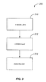
図3は、本発明の実施態様による治療プロセスを示す簡明なフローチャートである。治療プロセス300は、ヒト眼球表面に光増感溶液を適用すること(310)を含む。実施態様において、光増感溶液は、リボフラビンを含み、濃度は、約0.05%から約0.2%の範囲である。方法は、ヒト眼球表面内に治療領域を画定すること(312)も含む。治療領域は、所定の空間的パターンと関連する。方法は、制御された紫外/青色放射線によって治療領域を照射すること(314)を更に含む。
FIG. 3 is a simplified flowchart illustrating a treatment process according to an embodiment of the present invention. The
一例として、眼球の照射は、多数の選択可能なグレイ・スケール強度を特徴とする画素ベースの出力を提供するマイクロ・ミラーの配列を利用して行われる。実施態様において、グレイスケール強度数は、1000以上である。 As an example, eyeball illumination is performed using an array of micro mirrors that provide a pixel-based output characterized by a large number of selectable gray scale intensities. In an embodiment, the gray scale intensity number is 1000 or more.
図3に示した具体的なステップが、本発明の実施態様による眼治療を実行する特定の方法を提供することが認められるべきである。ステップの他のシーケンスは、代替的な実施態様により同様に実行できる。例えば、本発明の代替的な実施態様は、異なる順序で以上に概説したステップを実行できる。更に、図3に示した個別のステップは、必要に応じて個別のステップへの種々のシーケンスで実行できる複数のサブステップを含むことができる。更に追加のステップが、特定の用途に応じて追加又は除去できる。当業者は、多くの変形例、修正及び代替案を認めるであろう。 It should be appreciated that the specific steps shown in FIG. 3 provide a particular method of performing eye treatment according to embodiments of the present invention. Other sequences of steps can be similarly performed by alternative implementations. For example, alternative embodiments of the invention can perform the steps outlined above in a different order. In addition, the individual steps shown in FIG. 3 can include multiple sub-steps that can be performed in various sequences to the individual steps as needed. Further additional steps can be added or removed depending on the particular application. Those skilled in the art will recognize many variations, modifications, and alternatives.
本発明の実施態様によれば、非毒性抗酸化剤が、光増感剤の適用前に予め装填され、かつ内皮/ACを保護するために利用される。これらの非毒性抗酸化剤には:クマリン、PENT、ALDH3A1ビタミンC/A/E、アルファリポ酸、アルブミン、G6PDH Pentoic リン酸塩等を含む。内皮/AC領域で光増感剤濃度を監視する機器は、手術中の安全性/鮮度点検のために治療中にリアルタイム・モニタとして幾つかの実施態様において利用される。安全性閾値の基線として、Spoerlは、リボフラビン装填による角膜基質内の20〜1UVAの吸収を報告し、他方で、Slineyは、(いかなるリボフラビン装填もなしに)365nmで1mW/cm2 の連続(16分の最大時間)曝露を発表した。これらの結果に基づき、我々は、角膜が完全にリボフラビン装填されるならば、20mW/cm2までの「連続」UVA閾値が、許容可能であることを推測した。 According to an embodiment of the present invention, a non-toxic antioxidant is preloaded prior to application of the photosensitizer and utilized to protect the endothelium / AC. These non-toxic antioxidants include: coumarin, PENT, ALDH3A1 vitamin C / A / E, alpha lipoic acid, albumin, G6PDH Pentoic phosphate and the like. A device that monitors the photosensitizer concentration in the endothelium / AC region is utilized in some embodiments as a real-time monitor during treatment for safety / freshness checks during surgery. As a safety threshold baseline, Sporl reports absorption of 20-1 UVA in the corneal matrix due to riboflavin loading, while Sliney (without any riboflavin loading) is 1 mW / cm 2 continuous (16 (Maximum time in minutes) Announced exposure. Based on these results, we speculated that a “continuous” UVA threshold of up to 20 mW / cm 2 would be acceptable if the cornea was fully loaded with riboflavin.
本発明の実施態様は、インプラントなしで2〜4倍に生体力学的安定性を改善するために組織の角膜/眼架橋によるインサイチュINTACS作成; 強膜/毛様小帯UVA収縮及び毛様体転位による老視の偽水晶体矯正; PACT−UVA; レンズ収差矯正; IOL、ICL調節; 強膜棘小柱網での収縮によるIOP減少のための緑内障治療、予防的治療及び退縮減少のための前後のLASIK(pre-post LASIK)を含む多種多様な用途に適用できる。本発明の実施態様は、円錐角膜、PMD、角膜ジストロフィ、潰瘍等のようなKeraCureの全ての公知の利益を提供する。KeraCureプロセスの追加の考察は、上記で参照された米国特許出願第10/958711号に提供される。 Embodiments of the present invention provide in situ INTACS creation by cornea / ocular cross-linking of tissue to improve biomechanical stability by 2-4 fold without implant; sclera / ciliary zonal UVA contraction and ciliary translocation PACT-UVA; Lens aberration correction; IOL, ICL adjustment; before and after glaucoma treatment for prophylactic treatment and reduced regression with reduced IOP due to contraction in the scleral trabecular meshwork It can be applied to a wide variety of uses including LASIK (pre-post LASIK). Embodiments of the present invention provide all the known benefits of KeraCure such as keratoconus, PMD, corneal dystrophy, ulcers and the like. Additional discussion of the KeraCure process is provided in US patent application Ser. No. 10 / 958,711 referenced above.
発明者らによって実行された幾つかの研究の結果を、以下に記載する。これらの研究に関して、測定されたフルエンスは、12mW/cm2〜15mW/cm2で375nmであり、かつ130mW/cm2で436nmであり、曝露前浸漬期間は、5分であり、かつ曝露中の滴下は、3分毎に注ぎ込まれ、ブタ眼球は、脱上皮化しなかった。これらの代表的研究において、曝露期間は、10分から45分であった。 The results of several studies carried out by the inventors are described below. For these studies, the measured fluence is 375nm at 12mW / cm 2 ~15mW / cm 2 , and a 436nm at 130 mW / cm 2, exposure before immersion period is 5 minutes, and during exposure The instillation was poured every 3 minutes and the porcine eyeball did not de-epithelialize. In these representative studies, the duration of exposure was 10 to 45 minutes.
第1の代表的研究は、UVA/青色波長を使用する非光増感照明と比較して、光増感された正確なパターン化領域角膜収縮の実現可能性を証明するために部分的に行われた。一例として、対照BSS装填の10個のブタ眼球、及びPriaLight装填の10個の試料が10〜40分間、蝶ネクタイ型又は円形パターンによってパターン化された。 The first representative study has been conducted in part to demonstrate the feasibility of photosensitized accurate patterned area corneal contraction compared to non-photosensitized illumination using UVA / blue wavelengths. It was broken. As an example, 10 pig eyes with a control BSS load and 10 samples with a PriaLight load were patterned with a bowtie or circular pattern for 10-40 minutes.
第2の代表的研究は、テキストの形状の複雑なパターンのリソグラフィ実行を証明するために部分的に行われた。「PRIA」及び「HELLO」を含むテキストは、PriaLight光増感剤+UVA/青色曝露によってリソグラフィ実行がなされた。 A second representative study was conducted in part to prove lithographic performance of complex patterns of text shapes. Texts containing “PRIA” and “HELLO” were lithographically performed with the PriaLight photosensitizer + UVA / blue exposure.
第3の代表的研究は、UVA/青色パターン化PriaLight光増感剤収縮による屈折角膜修正を証明するために部分的に行われた。一例として、12個のブタ眼球は、PriaLightを装填され、かつディスク、遷移ゾーンを有するディスク、アニュラス、複数のアニュラス、時系列アニュラス、記録されたトポグラフィ等によってパターン化された。 A third representative study was conducted in part to demonstrate refractive cornea correction due to UVA / blue patterned PriaLight photosensitizer shrinkage. As an example, twelve pig eyes were loaded with PriaLight and patterned with a disc, a disc with transition zones, an annulus, multiple annulus, time series annulus, recorded topography, and the like.
本発明は、特定の実施態様及びその具体的な実施例に関して記載されたが、他の実施態様が本発明の精神及び範囲内に入り得ることが理解されるべきである。従って本発明の範囲は、添付の請求項を、同等物のその全範囲と共に参照して決定されるべきである。 Although the invention has been described with reference to particular embodiments and specific examples thereof, it is to be understood that other embodiments can fall within the spirit and scope of the invention. Accordingly, the scope of the invention should be determined by reference to the appended claims, along with their full scope of equivalents.
100 眼治療システム、110 ヒト眼球、200 眼治療システム、224 イルミネータ 100 eye treatment system, 110 human eyeball, 200 eye treatment system, 224 illuminator
Claims (19)
ヒト眼球表面に光増感溶液を適用し、
前記ヒト眼球表面内に、所定の空間的パターンと関連する治療領域を画定し、
制御された光活性化放射線によって前記治療領域を照射する
ことを含む方法。 A method for performing ophthalmoplasty,
Apply photosensitizing solution to human eyeball surface,
Defining a treatment area in the human eye surface associated with a predetermined spatial pattern;
Irradiating the treatment area with controlled photoactivating radiation.
前記組織の表面に光増感溶液を適用し、
前記表面内に、所定の強度空間的パターンと関連する治療領域を画定し、
前記空間的パターンに従って、有効量の制御された紫外/青色放射線によって前記治療領域を照射する
ことを含む方法。 A method of treating living tissue,
Applying a photosensitizing solution to the surface of the tissue;
Defining a treatment area in the surface associated with a predetermined intensity spatial pattern;
Irradiating the treatment area with an effective amount of controlled ultraviolet / blue radiation according to the spatial pattern.
ヒト眼球表面に光増感溶液を適用するアプリケータと、
所定の強度空間的パターンに従って、有効量の制御された紫外/青色放射線によって前記ヒト眼球表面内の画定された治療領域を照射するイルミネータと
を含む装置。 An apparatus for performing ophthalmoplasty,
An applicator for applying a photosensitizing solution to the human eyeball surface;
An illuminator that irradiates a defined treatment area within the surface of the human eye with an effective amount of controlled ultraviolet / blue radiation according to a predetermined intensity spatial pattern.
Applications Claiming Priority (3)
| Application Number | Priority Date | Filing Date | Title |
|---|---|---|---|
| US83088506P | 2006-07-13 | 2006-07-13 | |
| US11/776,470 US20080015660A1 (en) | 2006-07-13 | 2007-07-11 | Method And Apparatus For Photo-Chemical Oculoplasty/Keratoplasty |
| PCT/US2007/073394 WO2008008914A2 (en) | 2006-07-13 | 2007-07-12 | Method and apparatus for photo-chemical oculoplasty/keratoplasty |
Publications (1)
| Publication Number | Publication Date |
|---|---|
| JP2010506601A true JP2010506601A (en) | 2010-03-04 |
Family
ID=38924199
Family Applications (1)
| Application Number | Title | Priority Date | Filing Date |
|---|---|---|---|
| JP2009519701A Withdrawn JP2010506601A (en) | 2006-07-13 | 2007-07-12 | Method and apparatus for photochemical ophthalmoplasty / keratoplasty |
Country Status (7)
| Country | Link |
|---|---|
| US (1) | US20080015660A1 (en) |
| EP (1) | EP2043742A4 (en) |
| JP (1) | JP2010506601A (en) |
| KR (1) | KR20090046832A (en) |
| AU (1) | AU2007272443A1 (en) |
| CA (1) | CA2657414A1 (en) |
| WO (1) | WO2008008914A2 (en) |
Cited By (8)
| Publication number | Priority date | Publication date | Assignee | Title |
|---|---|---|---|---|
| JP2011212115A (en) * | 2010-03-31 | 2011-10-27 | Sony Corp | Posterior sclera curing apparatus |
| JP2013521988A (en) * | 2010-03-19 | 2013-06-13 | アヴェドロ・インコーポレーテッド | Systems and methods for applying and monitoring eye treatments |
| JP2013525014A (en) * | 2010-04-30 | 2013-06-20 | セロス メディカル, エルエルシー | Method and apparatus for treatment of ocular tissue using multidisciplinary methods |
| JP2014519866A (en) * | 2011-04-20 | 2014-08-21 | アヴェドロ・インコーポレーテッド | Controlled cross-linking initiation and corneal topography feedback system for directing cross-linking |
| JP2015526156A (en) * | 2012-07-16 | 2015-09-10 | アヴェドロ・インコーポレーテッドAvedro,Inc. | System and method for corneal cross-linking by pulsed light |
| WO2015186723A1 (en) * | 2014-06-03 | 2015-12-10 | 株式会社 坪田ラボ | Myopia prevention article |
| WO2017094886A1 (en) * | 2015-12-02 | 2017-06-08 | 株式会社坪田ラボ | Irradiation device |
| US10823982B2 (en) | 2014-06-03 | 2020-11-03 | Tsubota Laboratory, Inc. | Myopia treatment device |
Families Citing this family (53)
| Publication number | Priority date | Publication date | Assignee | Title |
|---|---|---|---|---|
| US20080139671A1 (en) * | 2006-12-07 | 2008-06-12 | Priavision, Inc. | Method and material for in situ corneal structural augmentation |
| US8202272B2 (en) | 2007-07-19 | 2012-06-19 | Avedro, Inc. | Eye therapy system |
| US8992516B2 (en) * | 2007-07-19 | 2015-03-31 | Avedro, Inc. | Eye therapy system |
| WO2009073213A1 (en) * | 2007-12-05 | 2009-06-11 | Avedro, Inc. | Eye therapy system |
| US20100057060A1 (en) * | 2007-12-07 | 2010-03-04 | Seros Medical, Llc | In Situ UV/Riboflavin Ocular Treatment System |
| US8348935B2 (en) * | 2008-01-23 | 2013-01-08 | Avedro, Inc. | System and method for reshaping an eye feature |
| US20090187173A1 (en) * | 2008-01-23 | 2009-07-23 | David Muller | System and method for reshaping an eye feature |
| US8469952B2 (en) | 2008-01-23 | 2013-06-25 | Avedro, Inc. | System and method for positioning an eye therapy device |
| US8409189B2 (en) * | 2008-01-23 | 2013-04-02 | Avedro, Inc. | System and method for reshaping an eye feature |
| DE102008011811B3 (en) * | 2008-02-29 | 2009-10-15 | Anton Dr. Kasenbacher | Dental laser processing device for processing dental material |
| US20090275936A1 (en) * | 2008-05-01 | 2009-11-05 | David Muller | System and method for applying therapy to an eye using energy conduction |
| AU2009256236A1 (en) | 2008-06-04 | 2009-12-10 | Neovista, Inc. | Handheld radiation delivery system for advancing a radiation source wire |
| US8398628B2 (en) * | 2008-09-19 | 2013-03-19 | Avedro, Inc. | Eye therapy system |
| WO2010039854A1 (en) * | 2008-09-30 | 2010-04-08 | Neal Marshall | Eye therapy system |
| US8460278B2 (en) * | 2008-10-01 | 2013-06-11 | Avedro, Inc. | Eye therapy system |
| WO2010056848A1 (en) | 2008-11-11 | 2010-05-20 | Avedro, Inc. | Eye therapy system |
| DE102009005194B4 (en) * | 2009-01-20 | 2016-09-08 | Anton Kasenbacher | Laser processing device for processing a material |
| EP2395953A4 (en) * | 2009-02-12 | 2013-06-19 | Univ Rochester | Aberration control by corneal collagen crosslinking combined with beam-shaping technique |
| WO2010115109A1 (en) * | 2009-04-02 | 2010-10-07 | Avedro, Inc. | Eye therapy system |
| US8712536B2 (en) * | 2009-04-02 | 2014-04-29 | Avedro, Inc. | Eye therapy system |
| US8574277B2 (en) | 2009-10-21 | 2013-11-05 | Avedro Inc. | Eye therapy |
| US20130245536A1 (en) * | 2009-10-21 | 2013-09-19 | Avedro, Inc. | Systems and methods for corneal cross-linking with pulsed light |
| US8177778B2 (en) * | 2009-10-30 | 2012-05-15 | Avedro, Inc. | System and method for stabilizing corneal tissue after treatment |
| US20120215155A1 (en) * | 2010-03-19 | 2012-08-23 | Avedro Inc. | Controlled cross-linking initiation and corneal topography feedback systems for directing cross-linking |
| EP2380535B1 (en) * | 2010-04-21 | 2015-04-08 | IROC Innocross AG | Device for integrating ocular tissue with electromagnetic radiation |
| US9622911B2 (en) | 2010-09-30 | 2017-04-18 | Cxl Ophthalmics, Llc | Ophthalmic treatment device, system, and method of use |
| ES2590127T3 (en) | 2011-01-12 | 2016-11-18 | Sooft Italia S.P.A. | Corneal administration of iontophoresis crosslinking agents for the treatment of keratoconus and related ophthalmic compositions |
| WO2012112543A2 (en) * | 2011-02-15 | 2012-08-23 | Seros Medical, Llc | Method and apparatus for the delivery of photo-chemical (cross-linking) treatment to scleral tissue |
| US9044308B2 (en) | 2011-05-24 | 2015-06-02 | Avedro, Inc. | Systems and methods for reshaping an eye feature |
| JP6555884B2 (en) * | 2011-05-31 | 2019-08-07 | クラレンシュウ・プロプライエタリー・リミテッド | Methods for preventing and treating exercise-related neurological disorders |
| EP2713849B1 (en) | 2011-06-02 | 2017-02-15 | Avedro, Inc. | Systems for monitoring time based photo active agent delivery or photo active marker presence |
| US20120330291A1 (en) * | 2011-06-24 | 2012-12-27 | The Regents Of The University Of California | Nonlinear optical photodynamic therapy (nlo-pdt) of the cornea |
| US9566301B2 (en) | 2012-03-29 | 2017-02-14 | Cxl Ophthalmics, Llc | Compositions and methods for treating or preventing diseases associated with oxidative stress |
| WO2013148895A1 (en) | 2012-03-29 | 2013-10-03 | Cxl Ophthalmics, Llc | Ocular cross-linking system and method for sealing corneal wounds |
| WO2013148896A1 (en) | 2012-03-29 | 2013-10-03 | Cxl Ophthalmics, Llc | Ocular treatment solutions, delivery devices and delivery augmentation methods |
| US9265458B2 (en) | 2012-12-04 | 2016-02-23 | Sync-Think, Inc. | Application of smooth pursuit cognitive testing paradigms to clinical drug development |
| US9380976B2 (en) | 2013-03-11 | 2016-07-05 | Sync-Think, Inc. | Optical neuroinformatics |
| CN105307586B (en) | 2013-03-15 | 2018-03-16 | 阿莱耶恩公司 | Sclera metathesis elastomeric method of adjustment and device |
| US9498122B2 (en) | 2013-06-18 | 2016-11-22 | Avedro, Inc. | Systems and methods for determining biomechanical properties of the eye for applying treatment |
| US9498114B2 (en) | 2013-06-18 | 2016-11-22 | Avedro, Inc. | Systems and methods for determining biomechanical properties of the eye for applying treatment |
| US9861526B1 (en) * | 2014-06-30 | 2018-01-09 | TECLens, LLC | Programmable patterning and masking array for corneal collagen crosslinking |
| EP3212140B1 (en) | 2014-10-27 | 2021-12-01 | Avedro, Inc. | Systems for cross-linking treatments of an eye |
| US10114205B2 (en) | 2014-11-13 | 2018-10-30 | Avedro, Inc. | Multipass virtually imaged phased array etalon |
| DE102014016990B3 (en) | 2014-11-18 | 2016-02-11 | Wavelight Gmbh | Nozzle unit for cross-linking of eye tissue |
| US10258809B2 (en) | 2015-04-24 | 2019-04-16 | Avedro, Inc. | Systems and methods for photoactivating a photosensitizer applied to an eye |
| PL3288588T3 (en) | 2015-04-29 | 2019-02-28 | Sooft Italia S.P.A. | Improved cross-linking agents of collagen fibers for the use in the treatment of corneal ectasia |
| EP3297589A4 (en) | 2015-05-22 | 2019-03-06 | Avedro Inc. | Systems and methods for monitoring cross-linking activity for corneal treatments |
| CN116832158A (en) | 2015-07-21 | 2023-10-03 | 艾维德洛公司 | Systems and methods for treating eyes with photosensitizers |
| US10646372B2 (en) | 2015-12-03 | 2020-05-12 | Avedro, Inc. | Systems and methods for treating an eye with a mask device |
| EP3761928A1 (en) | 2018-03-08 | 2021-01-13 | Avedro, Inc. | Micro-devices for treatment of an eye |
| JP2022508020A (en) * | 2018-09-19 | 2022-01-19 | アヴェドロ・インコーポレーテッド | Systems and methods for treating corneal ectatic disorders |
| US20210322214A1 (en) * | 2018-10-28 | 2021-10-21 | Belkin Laser Ltd. | Protection for Direct Selective Laser Trabeculoplasty |
| CA3147045A1 (en) | 2019-08-06 | 2021-02-11 | Desmond C. Adler | Photoactivation systems and methods for corneal cross-linking treatments |
Family Cites Families (10)
| Publication number | Priority date | Publication date | Assignee | Title |
|---|---|---|---|---|
| US5426425A (en) * | 1992-10-07 | 1995-06-20 | Wescom, Inc. | Intelligent locator system with multiple bits represented in each pulse |
| US7655002B2 (en) * | 1996-03-21 | 2010-02-02 | Second Sight Laser Technologies, Inc. | Lenticular refractive surgery of presbyopia, other refractive errors, and cataract retardation |
| JP2000060893A (en) * | 1998-08-20 | 2000-02-29 | Kowa Co | Ophthalmological treatment device |
| US7073510B2 (en) * | 2000-02-11 | 2006-07-11 | The General Hospital Corporation | Photochemical tissue bonding |
| US6923802B2 (en) * | 2000-03-13 | 2005-08-02 | Memphis Eye & Cataract Assoc. | System for generating ablation profiles for laser refractive eye surgery |
| US6607522B1 (en) * | 2000-03-16 | 2003-08-19 | General Hospital Corporation | Methods for tissue welding using laser-activated protein solders |
| US6887261B1 (en) * | 2001-04-25 | 2005-05-03 | Gholam A. Peyman | System and method for thermally and chemically treating cells at sites of interest in the body to impede cell proliferation |
| US20050149006A1 (en) * | 2001-11-07 | 2005-07-07 | Peyman Gholam A. | Device and method for reshaping the cornea |
| DE50307918D1 (en) * | 2002-01-10 | 2007-09-27 | Zeiss Carl Meditec Ag | Arrangement for illuminating the lens of a human eye |
| US7311723B2 (en) * | 2003-07-11 | 2007-12-25 | University Of Washington | Scanning laser device and methods of use |
-
2007
- 2007-07-11 US US11/776,470 patent/US20080015660A1/en not_active Abandoned
- 2007-07-12 KR KR1020097002885A patent/KR20090046832A/en not_active Application Discontinuation
- 2007-07-12 WO PCT/US2007/073394 patent/WO2008008914A2/en active Application Filing
- 2007-07-12 AU AU2007272443A patent/AU2007272443A1/en not_active Abandoned
- 2007-07-12 CA CA002657414A patent/CA2657414A1/en not_active Abandoned
- 2007-07-12 JP JP2009519701A patent/JP2010506601A/en not_active Withdrawn
- 2007-07-12 EP EP07799546A patent/EP2043742A4/en not_active Withdrawn
Cited By (12)
| Publication number | Priority date | Publication date | Assignee | Title |
|---|---|---|---|---|
| JP2013521988A (en) * | 2010-03-19 | 2013-06-13 | アヴェドロ・インコーポレーテッド | Systems and methods for applying and monitoring eye treatments |
| JP2011212115A (en) * | 2010-03-31 | 2011-10-27 | Sony Corp | Posterior sclera curing apparatus |
| JP2013525014A (en) * | 2010-04-30 | 2013-06-20 | セロス メディカル, エルエルシー | Method and apparatus for treatment of ocular tissue using multidisciplinary methods |
| JP2014519866A (en) * | 2011-04-20 | 2014-08-21 | アヴェドロ・インコーポレーテッド | Controlled cross-linking initiation and corneal topography feedback system for directing cross-linking |
| JP2015526156A (en) * | 2012-07-16 | 2015-09-10 | アヴェドロ・インコーポレーテッドAvedro,Inc. | System and method for corneal cross-linking by pulsed light |
| WO2015186723A1 (en) * | 2014-06-03 | 2015-12-10 | 株式会社 坪田ラボ | Myopia prevention article |
| US10133092B2 (en) | 2014-06-03 | 2018-11-20 | Tsubota Laboratory, Inc. | Myopia prevention device |
| US10823982B2 (en) | 2014-06-03 | 2020-11-03 | Tsubota Laboratory, Inc. | Myopia treatment device |
| WO2017094886A1 (en) * | 2015-12-02 | 2017-06-08 | 株式会社坪田ラボ | Irradiation device |
| JP6175210B1 (en) * | 2015-12-02 | 2017-08-02 | 株式会社坪田ラボ | Irradiation device |
| KR20180089422A (en) * | 2015-12-02 | 2018-08-08 | 가부시키가이샤 쓰보타 라보 | Irradiation device |
| KR102639025B1 (en) * | 2015-12-02 | 2024-02-20 | 가부시키가이샤 쓰보타 라보 | investigation device |
Also Published As
| Publication number | Publication date |
|---|---|
| EP2043742A2 (en) | 2009-04-08 |
| EP2043742A4 (en) | 2010-07-07 |
| CA2657414A1 (en) | 2008-01-17 |
| AU2007272443A1 (en) | 2008-01-17 |
| KR20090046832A (en) | 2009-05-11 |
| WO2008008914A3 (en) | 2008-09-04 |
| WO2008008914A2 (en) | 2008-01-17 |
| US20080015660A1 (en) | 2008-01-17 |
Similar Documents
| Publication | Publication Date | Title |
|---|---|---|
| JP2010506601A (en) | Method and apparatus for photochemical ophthalmoplasty / keratoplasty | |
| US11259963B2 (en) | Method and apparatus for treatment of ocular tissue using combined modalities | |
| KR101862364B1 (en) | Crosslinking control | |
| US9707126B2 (en) | Systems and methods for corneal cross-linking with pulsed light | |
| CA2586214C (en) | Apparatus and processes for preventing or delaying one or more symptoms of presbyopia | |
| US10463735B2 (en) | Method and apparatus for adjusting corneal curvature through digital corneal crosslinking | |
| US20130245536A1 (en) | Systems and methods for corneal cross-linking with pulsed light | |
| US20140066835A1 (en) | Systems and methods for corneal cross-linking with pulsed light | |
| WO2014066636A1 (en) | Controlled application of cross-linking agent | |
| Ren et al. | Laser refractive surgery: a review and current status | |
| WO2014071408A1 (en) | Systems and methods for reshaping an eye feature | |
| Kareliotis et al. | Laser-intraocular lenses interaction: Aspects to consider for in situ vision correction | |
| McDonald | Excimer Laser Photorefractive Keratectomy for Correction of Low to Moderate Myopia: A Better Mousetrap |
Legal Events
| Date | Code | Title | Description |
|---|---|---|---|
| A621 | Written request for application examination |
Free format text: JAPANESE INTERMEDIATE CODE: A621 Effective date: 20100712 |
|
| A761 | Written withdrawal of application |
Free format text: JAPANESE INTERMEDIATE CODE: A761 Effective date: 20100812 |
|
| A521 | Written amendment |
Free format text: JAPANESE INTERMEDIATE CODE: A821 Effective date: 20100812 |