JP2010075190A - Cdr-grafted type iii anti-cea humanized mouse monoclonal antibody - Google Patents
Cdr-grafted type iii anti-cea humanized mouse monoclonal antibody Download PDFInfo
- Publication number
- JP2010075190A JP2010075190A JP2009260907A JP2009260907A JP2010075190A JP 2010075190 A JP2010075190 A JP 2010075190A JP 2009260907 A JP2009260907 A JP 2009260907A JP 2009260907 A JP2009260907 A JP 2009260907A JP 2010075190 A JP2010075190 A JP 2010075190A
- Authority
- JP
- Japan
- Prior art keywords
- antibody
- monoclonal antibody
- cdr
- seq
- cea
- Prior art date
- Legal status (The legal status is an assumption and is not a legal conclusion. Google has not performed a legal analysis and makes no representation as to the accuracy of the status listed.)
- Granted
Links
Images


Classifications
-
- C—CHEMISTRY; METALLURGY
- C07—ORGANIC CHEMISTRY
- C07K—PEPTIDES
- C07K16/00—Immunoglobulins [IGs], e.g. monoclonal or polyclonal antibodies
- C07K16/18—Immunoglobulins [IGs], e.g. monoclonal or polyclonal antibodies against material from animals or humans
- C07K16/28—Immunoglobulins [IGs], e.g. monoclonal or polyclonal antibodies against material from animals or humans against receptors, cell surface antigens or cell surface determinants
- C07K16/30—Immunoglobulins [IGs], e.g. monoclonal or polyclonal antibodies against material from animals or humans against receptors, cell surface antigens or cell surface determinants from tumour cells
- C07K16/3007—Carcino-embryonic Antigens
-
- A—HUMAN NECESSITIES
- A61—MEDICAL OR VETERINARY SCIENCE; HYGIENE
- A61K—PREPARATIONS FOR MEDICAL, DENTAL OR TOILETRY PURPOSES
- A61K47/00—Medicinal preparations characterised by the non-active ingredients used, e.g. carriers or inert additives; Targeting or modifying agents chemically bound to the active ingredient
- A61K47/50—Medicinal preparations characterised by the non-active ingredients used, e.g. carriers or inert additives; Targeting or modifying agents chemically bound to the active ingredient the non-active ingredient being chemically bound to the active ingredient, e.g. polymer-drug conjugates
- A61K47/51—Medicinal preparations characterised by the non-active ingredients used, e.g. carriers or inert additives; Targeting or modifying agents chemically bound to the active ingredient the non-active ingredient being chemically bound to the active ingredient, e.g. polymer-drug conjugates the non-active ingredient being a modifying agent
- A61K47/68—Medicinal preparations characterised by the non-active ingredients used, e.g. carriers or inert additives; Targeting or modifying agents chemically bound to the active ingredient the non-active ingredient being chemically bound to the active ingredient, e.g. polymer-drug conjugates the non-active ingredient being a modifying agent the modifying agent being an antibody, an immunoglobulin or a fragment thereof, e.g. an Fc-fragment
- A61K47/6835—Medicinal preparations characterised by the non-active ingredients used, e.g. carriers or inert additives; Targeting or modifying agents chemically bound to the active ingredient the non-active ingredient being chemically bound to the active ingredient, e.g. polymer-drug conjugates the non-active ingredient being a modifying agent the modifying agent being an antibody, an immunoglobulin or a fragment thereof, e.g. an Fc-fragment the modifying agent being an antibody or an immunoglobulin bearing at least one antigen-binding site
- A61K47/6851—Medicinal preparations characterised by the non-active ingredients used, e.g. carriers or inert additives; Targeting or modifying agents chemically bound to the active ingredient the non-active ingredient being chemically bound to the active ingredient, e.g. polymer-drug conjugates the non-active ingredient being a modifying agent the modifying agent being an antibody, an immunoglobulin or a fragment thereof, e.g. an Fc-fragment the modifying agent being an antibody or an immunoglobulin bearing at least one antigen-binding site the antibody targeting a determinant of a tumour cell
- A61K47/6853—Carcino-embryonic antigens
-
- A—HUMAN NECESSITIES
- A61—MEDICAL OR VETERINARY SCIENCE; HYGIENE
- A61P—SPECIFIC THERAPEUTIC ACTIVITY OF CHEMICAL COMPOUNDS OR MEDICINAL PREPARATIONS
- A61P35/00—Antineoplastic agents
-
- A—HUMAN NECESSITIES
- A61—MEDICAL OR VETERINARY SCIENCE; HYGIENE
- A61K—PREPARATIONS FOR MEDICAL, DENTAL OR TOILETRY PURPOSES
- A61K38/00—Medicinal preparations containing peptides
-
- C—CHEMISTRY; METALLURGY
- C07—ORGANIC CHEMISTRY
- C07K—PEPTIDES
- C07K2317/00—Immunoglobulins specific features
- C07K2317/20—Immunoglobulins specific features characterized by taxonomic origin
- C07K2317/24—Immunoglobulins specific features characterized by taxonomic origin containing regions, domains or residues from different species, e.g. chimeric, humanized or veneered
-
- C—CHEMISTRY; METALLURGY
- C07—ORGANIC CHEMISTRY
- C07K—PEPTIDES
- C07K2317/00—Immunoglobulins specific features
- C07K2317/50—Immunoglobulins specific features characterized by immunoglobulin fragments
- C07K2317/56—Immunoglobulins specific features characterized by immunoglobulin fragments variable (Fv) region, i.e. VH and/or VL
- C07K2317/565—Complementarity determining region [CDR]
-
- C—CHEMISTRY; METALLURGY
- C07—ORGANIC CHEMISTRY
- C07K—PEPTIDES
- C07K2319/00—Fusion polypeptide
Abstract
Description
本発明は、結腸及び他のガンの診断及び治療用の免疫学的試薬に関する。特に、本発明は、対応するマウス抗ガン胎児性抗原(「CEA」)モノクローナル抗体(「mAb」)(MN14)の結合親和特性並びにヒト抗体の抗原性及びエフェクター特性を有するヒト化抗CEAモノクローナル抗体に関する。さらに、本発明は、抗CEAマウスmAbの相補性決定領域(「CDR」)がヒト抗体のフレームワーク領域に移植されているヒト化mAb、そのようなCDRを移植された抗体をコードするDNA、そのDNAを増殖させ、発現させるためのベクター及び形質転換宿主、並びに診断及び治療用途に有用な抗体の複合体に関する。 The present invention relates to immunological reagents for the diagnosis and treatment of colon and other cancers. In particular, the present invention provides a humanized anti-CEA monoclonal antibody having the binding affinity properties of the corresponding mouse anti-carcinoembryonic antigen (“CEA”) monoclonal antibody (“mAb”) (MN14) and the antigenicity and effector properties of human antibodies. About. Furthermore, the present invention provides a humanized mAb in which the complementarity determining region (“CDR”) of an anti-CEA mouse mAb is grafted into the framework region of a human antibody, DNA encoding an antibody grafted with such a CDR, It relates to vectors and transformed hosts for propagating and expressing the DNA, and antibody complexes useful for diagnostic and therapeutic applications.
発明の背景
ガンの診断及び治療への有望なアプローチは、診断及び治療作用物質を悪性腫瘍に直接搬送するターゲッティング抗体の使用を包含する。過去10年にわたって、様々な腫瘍特異的抗体及び抗体断片が、これらの抗体を薬物、毒素、放射性核種もしくは他の作用物質に複合する方法、及びこれらの複合体を患者に投与する方法と同様に、開発されている。これらの努力は多大な進歩をもたらしているが、様々のほとんど予期し得ない問題が、ある程度まで開発されている試薬の幾つかの診断上及び治療上の実用性を制限している。
BACKGROUND OF THE INVENTION A promising approach to cancer diagnosis and treatment involves the use of targeting antibodies that deliver diagnostic and therapeutic agents directly to malignant tumors. Over the past decade, various tumor-specific antibodies and antibody fragments have been combined with methods for conjugating these antibodies to drugs, toxins, radionuclides or other agents, and for administering these conjugates to patients. Have been developed. While these efforts have resulted in great progress, various almost unforeseen problems have limited the diagnostic and therapeutic utility of some of the reagents that have been developed to some extent.
その中で、最も扱いにくい問題は、ターゲッティング複合体に外来抗原として応答し得る、ヒト免疫系自体によって引き起こされるものである。すなわち、(ヒトに最も普通に用いられているターゲッティング抗体である)マウスモノクローナル抗体と複合体を形成している薬物又は放射性核種で処置された患者は、体内を循環しているヒト抗マウス抗体(HAMA)及びその複合体の抗体部分に対する一般化された即時型III型過敏性反応を発現する。さらに、副作用が最小である場合(例えば、単一の投与における場合)でさえ、循環HAMAは患者におけるターゲッティング作用物質の有効濃度を低下させ、したがって、診断又は治療作用物質が標的部位に到達するのを制限する。 Among them, the most cumbersome problem is caused by the human immune system itself, which can respond to the targeting complex as a foreign antigen. That is, a patient treated with a drug or radionuclide complexed with a mouse monoclonal antibody (which is the most commonly used targeting antibody for humans) will have a human anti-mouse antibody circulating in the body ( HAMA) and a generalized immediate type III hypersensitivity reaction to the antibody portion of the complex. Further, even when side effects are minimal (eg, in a single administration), circulating HAMA reduces the effective concentration of the targeting agent in the patient, thus allowing the diagnostic or therapeutic agent to reach the target site. Limit.
この問題を克服又は回避するため、幾つかのアプローチが開発されているが、限られた成功しか収めていない。戦略の1つは、ターゲッティング抗体を化学的に修飾してその抗原性を抑制することである。例えば、ターゲッティング抗体へのポリエチレングリコールの複合(PEG化)が、抗体の抗原性を低下させることが報告されている。別のアプローチは、抗体における抗原性の部位を特徴付けた後、それを除去することである。この手法においては、IgG全体の代わりに、Fab’、F(ab)2及び他の抗体断片が用いられている。加えて、血液からHAMAを血漿交換で除去することによりHAMAの副作用を低下させる試みがなされている。また、外来抗体の副作用を十分に低下させてターゲッティング作用物質での複数の治療を可能にするのに、免疫抑制技術も用いられている。 Several approaches have been developed to overcome or avoid this problem, but with limited success. One strategy is to chemically modify the targeting antibody to suppress its antigenicity. For example, it has been reported that conjugation (PEGylation) of polyethylene glycol to a targeting antibody reduces the antigenicity of the antibody. Another approach is to characterize the antigenic site in the antibody and then remove it. In this approach, Fab ′, F (ab) 2 and other antibody fragments are used instead of whole IgG. In addition, attempts have been made to reduce the side effects of HAMA by removing HAMA from blood by plasma exchange. Immunosuppressive techniques have also been used to sufficiently reduce the side effects of foreign antibodies and allow multiple treatments with targeting agents.
これらのアプローチで完全に満足に改善するものはない。ターゲッティング抗体及び抗体複合体に対する不利な免疫応答を減少または取り除く手段が、これらの診断及び治療作用物質の完全な利益を得るために、依然として要求されている。 None of these approaches improve completely satisfactorily. There remains a need for means to reduce or eliminate adverse immune responses to targeting antibodies and antibody conjugates in order to obtain the full benefit of these diagnostic and therapeutic agents.
この目標は、以下に説明されるCDRを移植されたヒト化マウス抗ヒトCEA mAbで達成される。 This goal is achieved with humanized mouse anti-human CEA mAbs implanted with CDRs as described below.
本発明の目的は、マウスクラスIII抗CEA mAb(MN14)のCDRがヒト抗体又は抗体断片のアミノ酸配列に機能的に移植されたヒト化クラスIII抗CEA mAbを提供し、マウスクラスIII、抗CEA mAbの抗CEA結合特性及びヒト患者におけるヒトmAbの免疫原性を有する免疫学的試薬を提供することにある。 An object of the present invention is to provide a humanized class III anti-CEA mAb in which a CDR of a mouse class III anti-CEA mAb (MN14) is functionally grafted to the amino acid sequence of a human antibody or antibody fragment. It is to provide an immunological reagent having the anti-CEA binding properties of mAb and the immunogenicity of human mAb in human patients.
本発明の別の目的は、このような抗体をコードするDNA構築体を提供することにある。これに関する具体的な目的は、細胞培養及び抗体産生における有利な特性を有する、改良された抗体及びその抗体をコードするDNAを生成する遺伝子操作を容易にする基質DNAである。 Another object of the present invention is to provide a DNA construct encoding such an antibody. A specific objective in this regard is substrate DNA that facilitates genetic manipulation to produce improved antibodies and DNA encoding the antibodies that have advantageous properties in cell culture and antibody production.
本発明のさらに別の目的は、このDNAを増殖させ、かつこの抗体を発現させるためのベクターを提供することにある。これに関連する本発明の目的は、保存、増殖、抗体産生及び治療用途のための、ベクターを有する細胞を提供することにある。 Yet another object of the present invention is to provide a vector for growing the DNA and expressing the antibody. A related object of the present invention is to provide cells with vectors for storage, proliferation, antibody production and therapeutic use.
本発明のさらに別の目的は、診断及び治療において用いられる、抗体を含有する組成物を提供することにある。これに関しては、とりわけ、ex vivo及びin vivoにおける造影、診断、予後及び治療のための、造影剤及び治療剤と複合体を形成する抗体を含む複合体を提供することが目的である。 Still another object of the present invention is to provide an antibody-containing composition for use in diagnosis and therapy. In this regard, it is an object to provide complexes comprising, inter alia, contrast agents and antibodies that are complexed with therapeutic agents, for ex vivo and in vivo imaging, diagnosis, prognosis and treatment.
前述の目的の達成において、本発明の一側面に従い、異種(ヒト)抗体のフレームワーク領域に移植されたマウスクラスIII、抗CEA mAb(MN14)のCDRを含むヒト化マウスmAbであって、このようにヒト化されたmAb抗体がクラスIII、抗CEA結合特異性は保持するものの、患者において親MN14マウスモノクローナル抗体よりも免疫原性が小さいヒト化マウスmAbが提供される。 In achieving the foregoing objectives, in accordance with one aspect of the present invention, a humanized mouse mAb comprising a CDR of a mouse class III, anti-CEA mAb (MN14) grafted into a framework region of a heterologous (human) antibody, comprising: Thus, a humanized mouse mAb is provided that is less immunogenic in the patient than the parental MN14 mouse monoclonal antibody, while the humanized mAb antibody retains class III anti-CEA binding specificity.
特に好ましい態様においては、このヒト化抗体の軽鎖可変領域は下記式で特徴付けられる。FRL1−CDRL1−FRL2−CDRL2−FRL3−CDRL3−FRL4ここで、FRの各々は個別にヒト抗体のフレームワーク領域であり、かつCDRの各々は個別にMN14の軽鎖の相補性決定領域にあり、及び下付文字は軽「L」)鎖領域を指す。 In a particularly preferred embodiment, the light chain variable region of the humanized antibody is characterized by the following formula: FR L1 -CDR L1 -FR L2 -CDR L2 -FR L3 -CDR L3 -FR L4 wherein each FR is a framework region of individual human antibodies, and CDR each of the light chains of individually MN14 of In the complementarity determining region, and the subscript refers to the light “L”) chain region.
重鎖可変領域は下記式で特徴付けられる。
FRH1−CDRH1−FRH2−CDRH2−FRH3−CDRH3−FRH4ここで、FR及びCDRは上と同じ意味を有し、かつ下付文字「H」は重鎖領域を指す。)
The heavy chain variable region is characterized by:
Here FR H1 -CDR H1 -FR H2 -CDR H2 -FR H3 -CDR H3 -FR H4, FR and CDR have the same meaning as above, the character "H" subscript and refers to a heavy chain region. )
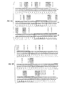
態様の1つにおいて、CDRL1はアミノ酸配列 KASQD VGTSVA(配列番号20)を有し、CDRL2はアミノ酸配列 WTSTR HT(配列番号21)を有し、CDRL3はアミノ酸配列 QQYSL YRS(配列番号22)を有し、CDRH1はアミノ酸配列 TYWMS(配列番号23)を有し、CDRH2はアミノ酸配列 EIHP DSSTI NYAPS LKD(配列番号24)を有し、CDRH3はアミノ酸配列 LYFGF PWFAY(配列番号25)を有する。 In one embodiment, CDR L1 has the amino acid sequence KASQD VGTSVA (SEQ ID NO: 20), CDR L2 has the amino acid sequence WTSTR HT (SEQ ID NO: 21), and CDR L3 has the amino acid sequence QQYSL YRS (SEQ ID NO: 22). CDR H1 has the amino acid sequence TYWMS (SEQ ID NO: 23), CDR H2 has the amino acid sequence EIHP DSSTI NYAPS LKD (SEQ ID NO: 24), and CDR H3 has the amino acid sequence LYFGF PWFFAY (SEQ ID NO: 25). Have.
他の態様において、FRL1はアミノ酸配列 DIQLT QSPSS LSASV GDRVT ITC(配列番号26)を有し、FRL2はアミノ酸配列 WYQQK PGKAP KLLIY(配列番号27)を有し、FRL3はアミノ酸配列 GVP(S又はD)F SGS(G又はV)S GTDFT FTISS LQPED IATYY V(配列番号28)を有し、FRL4はアミノ酸配列 FGQGT KVIEK(配列番号29)を有し、FRH1はアミノ酸配列 EVQLV ESGGG VVQPG RSLRL SCSSS GFDFT(配列番号30)、EVQLV ESGGG VVQPG RSLRL SCSAS GFDFT(配列番号31)又はQVQLQ ESGPG LVRPS QTLSL TCTSS GFDFT(配列番号32)を有し、FRH2はアミノ酸配列 WVRQA PGKGL EWVA(配列番号33)、WVRQA PGKGL EWIA(配列番号34)、又はWVRQP PGRGL EWIA(配列番号35)を有し、FRH3はアミノ酸配列 RFTIS RDNSK NTLFL QMDSL RPEDT GVYFC AS(配列番号36)、RFTIS RDNAK NTLFL QMDSL RPEDT GVYFC AS(配列番号37)、又はRVTML RDTSK NGSFL RLSSV TAADT AVYYC AS(配列番号38)を有し、FRH4はアミノ酸配列 WGQGT PVTVS S(配列番号39)、又はWGQGT TVTVS S(配列番号40)を有し、以上において、Cはスルフィドリル又はジスルフィドの形であってもよい。 In another embodiment, FR L1 has the amino acid sequence DIQLT QSPSS LSASV GDRVT ITC (SEQ ID NO: 26), FR L2 has the amino acid sequence WYQQK PGKAP KLLIY (SEQ ID NO: 27), and FR L3 has the amino acid sequence GVP (S or D) F SGS (G or V) S GTDFT FTISS LQPED IATYY V (SEQ ID NO: 28), FR L4 has the amino acid sequence FGQGT KVIEK (SEQ ID NO: 29), FR H1 has the amino acid sequence EVQLV ESGGG VVQPG RSLRLSC GFDFT (SEQ ID NO: 30), has a EVQLV ESGGG VVQPG RSLRL SCSAS GFDFT (SEQ ID NO: 31) or QVQLQ ESGPG LVRPS QTLSL TCTSS GFDFT (SEQ ID NO: 32), FR H2 is a Acid sequence WVRQA PGKGL EWVA (SEQ ID NO: 33), WVRQA PGKGL EWIA (SEQ ID NO: 34), or WVRQP PGRGL has EWIA (SEQ ID NO: 35), FR H3 is amino acid sequence RFTIS RDNSK NTLFL QMDSL RPEDT GVYFC AS (SEQ ID NO: 36 ), RFTIS RDNAK NTLFL QMDSL RPEDT GVYFC AS (SEQ ID NO: 37), or RVTML RDTSK NGSFL RLSSV TAADT AVYYC AS (SEQ ID NO: 38), FR H4 has the amino acid sequence WGQGT PVTVS S (SEQ ID NO: 39) In the above, C may be in the form of sulfhydryl or disulfide.
別の好ましい態様は、クラスIII、抗CEAヒト化mAbと複合体を形成する診断又は治療剤を包含する。ここで、このクラスIII、抗CEAヒト化mAbにおいて、この抗体のCDRはMN14マウスmAbのCDRに由来し、FRは異種(ヒト)抗体のFRに由来し、この複合体はMN14のクラスIII、抗CEA結合特異性を保持するものの、ヒトにおいてマウスMN14よりも免疫原性が小さい。そのような態様の1つにおいては、その軽鎖及び重鎖可変領域は上に示される通りに特徴付けられ、かつ同様に上に説明されるアミノ酸配列を有する。 Another preferred embodiment includes a diagnostic or therapeutic agent that forms a complex with a class III, anti-CEA humanized mAb. Here, in this class III, anti-CEA humanized mAb, the CDR of this antibody is derived from the CDR of a MN14 mouse mAb, the FR is derived from the FR of a heterologous (human) antibody, and this complex is a class III of MN14, Although retaining anti-CEA binding specificity, it is less immunogenic in humans than mouse MN14. In one such embodiment, the light and heavy chain variable regions are characterized as shown above and have the amino acid sequences described above as well.
さらに別の好ましい態様において、患者の診断又は治療方法が、前述の好ましい態様の複合体を適当な投与計画の下に投与するステップを含む。 In yet another preferred embodiment, a method for diagnosing or treating a patient comprises administering the complex of the preferred embodiment described above under an appropriate dosing schedule.
別の好ましい態様は、上述のヒト化抗体の軽鎖、重鎖またはその両鎖をコードする単離精製DNAを含む。
別の好ましい態様は、上述のCDR及びFRのDNA配列を含む。
Another preferred embodiment includes isolated and purified DNA encoding the light chain, heavy chain or both chains of the humanized antibody described above.
Another preferred embodiment includes the CDR and FR DNA sequences described above.
本発明の他の目的、特徴及び利点は、以下の詳細な説明及び添付の請求の範囲から明らかとなる。 Other objects, features and advantages of the present invention will become apparent from the following detailed description and appended claims.
用語の説明
本出願においては、以下の用語又は略語が用いられる。この用語集に記される意味は、説明のためのみである。これらの用語の完全な意味は、当該技術分野の熟練者には明らかである。
Explanation of Terms In this application, the following terms or abbreviations are used. The meanings given in this glossary are for explanation only. The full meaning of these terms will be apparent to those skilled in the art.
「CDR」は、相補性決定領域に対する略語として用いられる。これらは、主として、しかしながら他の可能性を除くわけではないが、抗原抗体結合の原因である、抗体の可変領域内の領域である。 “CDR” is used as an abbreviation for complementarity determining region. These are primarily regions within the variable region of an antibody that are responsible for antigen-antibody binding, but not excluding other possibilities.
「FR」は、フレームワーク領域に対する略語である。広く言えば、これらは、抗体の可変領域の、CDRに隣接し、もしくは並列する部分である。一般には、これらの領域は、その可変領域の立体配座に影響を与え、そのフレームワーク領域が抗体と抗原との相互作用に影響を及ぼし得るにもかかわらず、抗体への抗原の特異的結合の直接の原因とはほとんどならない多くの構造上の機能を有する。 “FR” is an abbreviation for framework area. Broadly speaking, these are the portions of the variable region of an antibody that are adjacent to or juxtaposed with the CDRs. In general, these regions affect the conformation of the variable region, and specific binding of the antigen to the antibody, even though the framework region can affect the interaction between the antibody and the antigen. It has many structural functions that are hardly the direct cause of.
「キメラ」は、その可変領域がマウス抗体に由来し、その定常領域が異種(別)の種の抗体に由来する抗体を指す。 “Chimera” refers to an antibody whose variable region is derived from a murine antibody and whose constant region is derived from an antibody of a heterologous (another) species.
「ヒト化」は、上に定義されるキメラ抗体ではあるが、FR可変領域がヒト抗体に由来するものを指す。 “Humanized” refers to a chimeric antibody as defined above, but with the FR variable region derived from a human antibody.
「HAHA」は、ヒト化マウス抗体に対するヒト抗体を指す。
「CEA」は、内胚葉起源の消化器系上皮のほとんどのアデノカルシノーマ並びに乳ガン及び非小細胞肺ガンのような他のガンにおいて発現する180kDa糖タンパク質であるガン胎児性抗原を指す。
“HAHA” refers to a human antibody to a humanized mouse antibody.
“CEA” refers to carcinoembryonic antigen, a 180 kDa glycoprotein that is expressed in most adenocarcinomas of digestive system epithelium of endoderm origin and other cancers such as breast cancer and non-small cell lung cancer.
接頭辞としての「h」という文字は、「ヒト化」を意味する。
他の略語は、Roitt et al., IMMUNOLOGY, 3rd ed. Mosby Year Book Europe Ltd. (1993)に従って用いられる。この文献は、引用することにより本明細書にそのまま組込まれる。
The letter “h” as a prefix means “humanized”.
Other abbreviations are used according to Roitt et al., IMMUNOLOGY, 3rd ed. Mosby Year Book Europe Ltd. (1993). This document is incorporated herein by reference in its entirety.
本明細書の開示において用いられるこれらの用語及び他の用語は、本発明が属する技術分野において通常用いられるものと同じ意味で用いられる。 These terms and other terms used in the disclosure of the present specification are used in the same meaning as those usually used in the technical field to which the present invention belongs.
過去に、MN14のCEA結合特性を有する有効な非HAMA誘発性の抗CEA抗体を開発することには失敗しているが、MN14 mAbのCDRをヒト抗体のFRに移植して、HAMAの誘発が低下され、かつエフェクター活性が増大されていながら、MN14の抗CEA mAbの抗原結合特性を有する抗体及び抗体誘導試薬を得ることが可能であることが見出されている。 In the past, it has been unsuccessful to develop effective non-HAMA-inducing anti-CEA antibodies with CEA binding properties of MN14, but the CDR of MN14 mAb was transplanted into the FR of human antibody to induce HAMA. It has been found that it is possible to obtain antibodies and antibody-derived reagents having the antigen-binding properties of anti-CEA mAb of MN14 while being reduced and having increased effector activity.
マウス抗CEAのIgG1モノクローナル抗体MN14、及びその生成は、以前に説明されている。Hansen et al., Cancer, 71:3478 (1993)、Primus et al.,米国特許第4,818,709号を参照。MN14はクラスIII、抗CEAモノクローナル抗体の基準の全てを満たし、EIAで胎便と非反応性であり、かつ通常の組織とは反応しない。 Mouse anti-CEA IgG1 monoclonal antibody MN14 and its production have been previously described. See Hansen et al., Cancer, 71: 3478 (1993), Primus et al., US Pat. No. 4,818,709. MN14 meets all criteria for class III, anti-CEA monoclonal antibodies, is non-reactive with meconium with EIA, and does not react with normal tissues.
遮断解析は、Hansen et al., 1993 上記、Losman et al., Int. Cancer, 56:580 (1994)、Hansen et al., Clin. Chem., 35:146 (1989)に従って行う。CEAの定量にこれらの参考文献に記述されるものと同じ条件を用いて、標識MN14プローブに対するヒト化MN14の結合を評価することができる。典型的なプローブは、ホースラディッシュペルオキシダーゼ(HRP)に複合しているMN14である。標識及び非標識MN14の両者を、マイクロタイタープレートのウェルのような固体支持体に固定されているCEA試料に加える。CEAに対する標識MN14の結合の「遮断」の程度は、非標識MN14の活性の直接の反映である。標準MN14を用いて、ヒト化MN14又はそれらの誘導体の未知試料の相対活性を決定することができる。典型的には、この反応はマイクロタイタープレートのウェルにおいて行われる。例えば25μg/ウェルのレベルでこのウェルをCEAで直接満たされ、又は間接的に満たされる。間接的に満たされる場合には、CEAと反応はするがMN14が相互作用するものとは異なるエピトープに反応する抗体が予め満たされている。このような抗体は、MN15 mAbであってもよい。その後、CEAを間接的にそのウェルに固定することができる。次に、そのような満たされたプレートを用いて、競合結合EIA検定を行うことができる。 Blockage analysis is performed according to Hansen et al., 1993, Losman et al., Int. Cancer, 56: 580 (1994), Hansen et al., Clin. Chem., 35: 146 (1989). The same conditions as described in these references for CEA quantification can be used to assess the binding of humanized MN14 to labeled MN14 probes. A typical probe is MN14 conjugated to horseradish peroxidase (HRP). Both labeled and unlabeled MN14 are added to a CEA sample that is immobilized on a solid support such as a well of a microtiter plate. The degree of “blocking” of the binding of labeled MN14 to CEA is a direct reflection of the activity of unlabeled MN14. Standard MN14 can be used to determine the relative activity of unknown samples of humanized MN14 or derivatives thereof. Typically, this reaction is performed in the wells of a microtiter plate. This well is filled directly with CEA, for example at a level of 25 μg / well, or indirectly filled. If it is indirectly filled, it is pre-filled with an antibody that reacts with CEA but reacts with an epitope different from that with which MN14 interacts. Such an antibody may be a MN15 mAb. The CEA can then be indirectly fixed to the well. Such filled plates can then be used to perform competitive binding EIA assays.
前述のHRP標識mAbの代わりに、抗体を、通常、例えば131Iを用いて、クロラミンT法により、約10mCi/μgの比活性まで放射ヨウ素化することが可能であり、遊離した放射性同位体はアクリルアミドゲルカラムでのクロマトグラフィーにより除去する(上記Hansen et al., 1993を参照)。 Instead of the aforementioned HRP-labeled mAb, the antibody can be radioiodinated, usually using, for example, 131 I by the chloramine T method to a specific activity of about 10 mCi / μg, the released radioisotope is Remove by chromatography on an acrylamide gel column (see Hansen et al., 1993, above).
本明細書に説明される発明の実施に適切な分子生物学上の技術も、当該技術分野における熟練者には公知である。適切な技術は、とりわけ、Sambrook et al., MOLECULAR CLONING: A LABORATORY MANUAL, 2nd Ed., Cold Spring Harbor Laboratory Press, Cold Spring Harbor, N.Y. (1989), PROTOCOLS IN MOLECULAR BIOLOGY, Ausubel et al., Eds., Green Publishing Associates and Wiley-Interscience, John Wiley and Sons, New York (1987, 1988, 1989)(これらは、引用することにより、本明細書に補遺を含むそれら全体を組込むものとする)を含む多くのマニュアルや主要な刊行物に記述されている。 Molecular biology techniques suitable for the practice of the invention described herein are also known to those skilled in the art. Suitable techniques include, among others, Sambrook et al., MOLECULAR CLONING: A LABORATORY MANUAL, 2nd Ed., Cold Spring Harbor Laboratory Press, Cold Spring Harbor, NY (1989), PROTOCOLS IN MOLECULAR BIOLOGY, Ausubel et al., Eds. , Green Publishing Associates and Wiley-Interscience, John Wiley and Sons, New York (1987, 1988, 1989), which are hereby incorporated by reference in their entirety, including the addendum. Described in manuals and major publications.
本明細書に開示されるMN14軽鎖及び重鎖CDR及び修飾したMN14のCDRは、上記参考文献に記述されているもののような公知の組換え技術を用いて、他の抗体に組込むことができる。 The MN14 light and heavy chain CDRs and modified MN14 CDRs disclosed herein can be incorporated into other antibodies using known recombinant techniques, such as those described in the above references. .
この目的に適切な具体的な方法は、下記例に示されている。本明細書に説明されるアミノ酸配列に基づいて、MN14のCDRをコードするオリゴヌクレオチドを合成することができる。本明細書に説明されるアミノ酸配列を正確にコードするものに加えて、修飾CDRをコードするオリゴヌクレオチドを作製することもできる。また、このオリゴヌクレオチドは、MN14のCDRのものに加えて、例えばクローニングを容易にするヌクレオチドを有していてもよい。オリゴヌクレオチドの合成技術は公知であり、多くの製造者から入手可能な自動装置で行うことができる。さらに、あらゆる特定の配列のオリゴヌクレオチドを購入することができる。 A specific method suitable for this purpose is shown in the examples below. Based on the amino acid sequences described herein, oligonucleotides encoding the CDRs of MN14 can be synthesized. In addition to those that accurately encode the amino acid sequences described herein, oligonucleotides that encode modified CDRs can also be made. In addition to the CDR of MN14, this oligonucleotide may have, for example, a nucleotide that facilitates cloning. Oligonucleotide synthesis techniques are known and can be performed with automated equipment available from many manufacturers. Furthermore, oligonucleotides of any specific sequence can be purchased.
MN14のCDR及び/又は特定のFR残基をコードするオリゴヌクレオチド又はそれらの相補鎖に相当するオリゴヌクレオチドを、そのオリゴヌクレオチドの末端、一般的には12ヌクレオチドがテンプレートDNAに完全にアニーリングするように設計されているという条件の下で、部位指向性突然変異誘発によるVH又はVK DNAへのこれらの残基のコドンの導入に用いることができる。このテンプレートDNAは、典型的には、必須FRをコードする可変領域DNAを坦持するM13ベクターに表される一本鎖DNAである。方法の1つにおいて、変異誘発性オリゴヌクレオチドをそれらの5’末端でリン酸化し、5’から可変領域DNAへの伸長を誘発するオリゴヌクレオチドと一緒に、このssDNAテンプレートにアニーリングする。このオリゴヌクレオチドを、T7ポリメラーゼ及びT4DNAリガーゼによって一緒に連結する断片を用いて伸長させ、可変領域全体を包含する完全な変異鎖を得る。この変異鎖をテンプレートとして用い、熱サイクル反応においてTaq DNAポリメラーゼを用いて適切なプライマーからその相補鎖の複数のコピーを合成することができる。変異鎖がこのように選択的に増幅されると、通常のPCRにより、クローニング、配列決定及び発現のためにDNAを増幅することが可能である。 MN14 CDRs and / or oligonucleotides corresponding to specific FR residues, or oligonucleotides corresponding to their complementary strands, so that the ends of the oligonucleotides, typically 12 nucleotides, are fully annealed to the template DNA Under the condition that it is designed, it can be used to introduce codons for these residues into VH or VK DNA by site-directed mutagenesis. This template DNA is typically a single-stranded DNA represented by an M13 vector carrying a variable region DNA encoding an essential FR. In one method, mutagenic oligonucleotides are phosphorylated at their 5 'ends and annealed to the ssDNA template along with oligonucleotides that induce 5' to variable region DNA extension. This oligonucleotide is extended with a fragment that is ligated together by T7 polymerase and T4 DNA ligase to obtain a complete variant that encompasses the entire variable region. Using this mutant strand as a template, multiple copies of its complementary strand can be synthesized from appropriate primers using Taq DNA polymerase in a thermal cycling reaction. Once the mutant strand is thus selectively amplified, it is possible to amplify the DNA for cloning, sequencing and expression by conventional PCR.
抗体をコードしている適切なDNAが開示により本明細書に示されるが、事実上、そのようなDNAのあらゆるものが含まれる。様々なヒト抗体遺伝子が、公的に利用可能な寄託の形態で利用することができる。抗体及び抗体をコードしている遺伝子の多くの配列が公開されており、上述のように適切な抗体遺伝子をこれらの配列から合成することができる。 Appropriate DNA encoding the antibody is shown herein by disclosure, but virtually any such DNA is included. Various human antibody genes are available in publicly available deposit forms. Many sequences of antibodies and genes encoding antibodies have been published, and appropriate antibody genes can be synthesized from these sequences as described above.
本発明の範囲には、本明細書に説明されるDNA配列の対立遺伝子、変種及び変異が含まれる。 The scope of the present invention includes alleles, variants and mutations of the DNA sequences described herein.
本開示によるCDR移植は、確立されている技術を用いて行うことができる。抗体産生細胞系は、熟練技術者に公知の技術を用いて選択し、かつ培養することができる。そのような技術は、様々な実験マニュアル及び主要な刊行物に記述されている。例えば、以下に記述されるような本発明での使用に適する技術は、CURRENT PROTOCOLS IN IMMUNOLOGY, Coligan et al., Eds., Green Publishing Associates and Wiley-Interscience, John Wiley and Sons, New York (1991)に記述されておる。この文献は引用することにより補遺を含むその全体が本明細書に組込まれる。 CDR grafting according to the present disclosure can be performed using established techniques. Antibody producing cell lines can be selected and cultured using techniques known to the skilled artisan. Such techniques are described in various laboratory manuals and major publications. For example, techniques suitable for use in the present invention as described below include CURRENT PROTOCOLS IN IMMUNOLOGY, Coligan et al., Eds., Green Publishing Associates and Wiley-Interscience, John Wiley and Sons, New York (1991). It is described in. This document is incorporated herein by reference in its entirety, including the addendum.
RNAは、元のハイブリドーマ細胞から、グアニジニウムイソチオシアネート抽出及び沈殿と、それに続く遠心もしくはクロマトグラフィーのような標準技術により、単離することができる。所望であれば、オリゴdTセルロースでのクロマトグラフィーのような標準技術により、mRNAを全RNAから単離することができる。これらの目的に適する技術は、前述の参考文献に記述されるように、当該技術分野において公知である。 RNA can be isolated from the original hybridoma cells by standard techniques such as guanidinium isothiocyanate extraction and precipitation followed by centrifugation or chromatography. If desired, mRNA can be isolated from total RNA by standard techniques such as chromatography on oligo dT cellulose. Techniques suitable for these purposes are known in the art as described in the aforementioned references.
抗体の軽鎖及び重鎖をコードするcDNAは、公知の方法に従って逆転写酵素及びDNAポリメラーゼを用いて、同時に又は別々に作製することができる。これは、コンセンサス定常領域プライマーで、あるいは公開されている重鎖及び軽鎖DNA及びアミノ酸配列に基づくより特異的なプライマーで開始することができる。 CDNA encoding the light chain and heavy chain of an antibody can be prepared simultaneously or separately using reverse transcriptase and DNA polymerase according to known methods. This can be initiated with consensus constant region primers or with more specific primers based on published heavy and light chain DNA and amino acid sequences.
抗体の軽鎖及び重鎖をコードするDNAクローンの単離にPCRを用いることもできる。この場合、コンセンサスプライマー、又はマウスコンセンサス領域プローブのようなより大きな同種プローブでライブラリーをスクリーニングすることが可能である。必要な技術は当該技術分野における熟練者に公知であり、前述のSambrook及びAusubelの参考文献に説明されており、かつ以下に述べられる例により説明される。 PCR can also be used to isolate DNA clones encoding the light and heavy chains of the antibody. In this case, it is possible to screen the library with larger consensus probes such as consensus primers or mouse consensus region probes. The required techniques are known to those skilled in the art and are described in the aforementioned Sambrook and Ausubel references and illustrated by the examples set forth below.
抗体の軽鎖及び重鎖をコードするcDNAは、CDRを単離する前に、あらゆる適切な宿主のあらゆる適切なベクターにおいて増殖させることができる。以下の例に説明されるように、このクローンは、しばしば、この目的のために、大腸菌内で最も都合よく増殖される。しかしながら、熟練者に公知の他の様々なベクター及び宿主細胞を本発明のこの側面において有利に用いることができる。そのようなベクターの様々なものが前述の参考文献に説明されている。 The cDNA encoding the light and heavy chains of the antibody can be propagated in any suitable vector in any suitable host prior to isolating the CDRs. As explained in the examples below, this clone is often propagated most conveniently in E. coli for this purpose. However, various other vectors and host cells known to those skilled in the art can be advantageously used in this aspect of the invention. Various such vectors are described in the aforementioned references.
DNA、典型的にはプラスミドDNAを、組換えDNA技術に関連する前述の参考文献に詳細に説明されている標準の公知技術に従い、細胞から単離し、制限地図を作製し、かつ配列決定することができる。 DNA, typically plasmid DNA, is isolated from cells, generated a restriction map, and sequenced according to standard known techniques detailed in the aforementioned references relating to recombinant DNA technology Can do.
ベクターにより抗体重鎖及び軽鎖並びにそれらの断片をコードするDNAを、キメラ及びCDRを移植したヒト化MN14抗体の構築に用いることができる。 DNA encoding antibody heavy and light chains and fragments thereof can be used in the construction of humanized MN14 antibodies grafted with chimeras and CDRs.
MN14の抗CEA mAbのCDRが本明細書で同定され、説明され、かつ図1及び図2(それぞれ、配列番号2及び配列番号4)に示されている。これらの配列を用いて、MN14重鎖及び軽鎖のCDRを本発明で用いるために合成することができる。天然資源からMN14のCDRを再クローニングする必要はない。そのDNA及びアミノ酸配列は本明細書に示されている。本発明のこの側面に適するオリゴヌクレオチド合成技術は熟練技術者に公知であり、幾つかの市販されている自動合成器のあらゆるものを用いて行うことができる。加えて、本明細書に説明されるCDRをコードするDNAは、商業DNA合成販売者のサービスにより得ることができる。 The CDRs of the anti-CEA mAb of MN14 have been identified and described herein and are shown in FIGS. 1 and 2 (SEQ ID NO: 2 and SEQ ID NO: 4, respectively). Using these sequences, MN14 heavy and light chain CDRs can be synthesized for use in the present invention. There is no need to reclon the CDRs of MN14 from natural sources. Its DNA and amino acid sequences are shown herein. Oligonucleotide synthesis techniques suitable for this aspect of the invention are known to the skilled artisan and can be performed using any of several commercially available automated synthesizers. In addition, the DNA encoding the CDRs described herein can be obtained through the services of commercial DNA synthesis vendors.
本発明のこの側面によって合成されるポリヌクレオチドには、MN14のCDRを構成するものに加えて、このCDRに由来するものではないものも含まれ得る。異種からのFRへのこのCDRの結合を容易にするさらなる塩基を含んでいてもよい。この目的のために、制限部位又はオーバーラップした相補的な領域を含むこともできる。短い重複一本鎖DNAからの長い二本鎖DNAの合成は当該技術分野における熟練者に公知である。同様に、平滑末端DNA及び少なくとも部分的に重複する相補的な末端を有するものを含むDNAの末端間結合は公知である。これらの技術は、例えば、組換えDNA技術に関する前述の参考文献に説明されている。 Polynucleotides synthesized according to this aspect of the invention can include those not derived from this CDR in addition to those comprising the CDR of MN14. Additional bases may be included that facilitate binding of this CDR to a heterologous FR. For this purpose, restriction sites or overlapping complementary regions can also be included. The synthesis of long double stranded DNA from short overlapping single stranded DNA is known to those skilled in the art. Similarly, end-to-end binding of DNA, including blunt end DNA and DNA having at least partially overlapping complementary ends, is known. These techniques are described, for example, in the aforementioned references on recombinant DNA techniques.
また、MN14重鎖及び軽鎖のCDRも、特にはキメラもしくはヒト化抗体に組込んだ後に、クローニングしたDNA又はRNA、もしくは合成DNA又はRNAにおける塩基の削除、挿入及び変更を行うための公知の組換えDNA技術を用いて、修飾することが可能である。この目的に適切な部位特異的突然変異誘発技術は当該技術分野における熟練者に公知であり、組換えDNA技術に関する前述の参考文献に説明されている。また、削除及び挿入技術も説明されている。これらの方法は、例えば、MN14のCDRをコードするポリヌクレオチド又はクローニングした重鎖もしくは軽鎖遺伝子の他の領域への所望の変更の導入に用いることができる。 In addition, CDRs of MN14 heavy chain and light chain are also known for performing base deletion, insertion and modification in cloned DNA or RNA, or synthetic DNA or RNA, particularly after incorporation into a chimeric or humanized antibody. Modifications can be made using recombinant DNA techniques. Suitable site-directed mutagenesis techniques for this purpose are known to those skilled in the art and are described in the aforementioned references on recombinant DNA technology. Deletion and insertion techniques are also described. These methods can be used, for example, to introduce desired changes into other regions of the polynucleotide encoding the CDR of MN14 or the cloned heavy or light chain gene.
本発明に従い、MN14のCDR及び修飾MN14のCDRをFRのあらゆるセットに実用的に導入することができる。これに関して、ポリヌクレオチドをクローニングし、操作する様々な公知の技術を効果的に使用できることを当該技術分野における熟練者は予期するであろう。このような技術は、前述の組換えDNAに関連する参考文献に説明される方法によって示されている。 In accordance with the present invention, CDRs of MN14 and CDRs of modified MN14 can be practically introduced into any set of FRs. In this regard, those skilled in the art will expect that various known techniques for cloning and manipulating polynucleotides can be used effectively. Such techniques are demonstrated by the methods described in the references related to recombinant DNA described above.
本発明の特に好ましい態様において、MN14のCDRがヒト抗体に移植される。この文脈において、ヒト抗体は、ヒト体内に生じるあらゆる抗体又は、幾つかの点において、ヒト免疫系と適合し得るように設計されている加工抗体を指すことは理解されるであろう。この目的のためには、広くは、患者において不利な免疫応答を引き起こすことがない抗体が特に好ましい。より具体的には、「ヒト抗体」という表現は、ヒト体内において実際に生じる遺伝子、又はそれらの対立遺伝子、変種もしくは変異体によってコードされる抗体を意味することが意図されている。 In a particularly preferred embodiment of the invention, the CDRs of MN14 are transplanted into human antibodies. In this context, it will be understood that a human antibody refers to any antibody that occurs in the human body or, in some respects, a engineered antibody that is designed to be compatible with the human immune system. For this purpose, antibodies that do not cause adverse immune responses in patients are particularly preferred. More specifically, the expression “human antibody” is intended to mean an antibody encoded by a gene that actually occurs in the human body, or an allele, variant or variant thereof.
いったんMN14に由来するCDRを移植された抗体をコードするDNAが、MN14のVH及びVK領域DNA及び、それらにより形成されるヒト定常ドメインの軽鎖及び重鎖の各々と結合する可変領域から構築されると、通常の技術により、それを増殖及び発現のためのベクターに挿入することが可能である。この方法で、所望の量の抗体を得ることができる。 DNA encoding antibodies once grafted with CDRs derived from MN14 is constructed from the variable regions that bind to each of the VH and VK region DNAs of MN14 and the light chain and heavy chain of the human constant domain formed thereby. It can then be inserted into a vector for propagation and expression by conventional techniques. In this way, a desired amount of antibody can be obtained.
このMN14のCDRを移植されたヒト抗体は、造影化合物又は同位体と結合するこのヒト化抗体又はそれらのFab’を被検体に投与することにより、造影用途に用いることができる。 The human antibody transplanted with the CDR of MN14 can be used for imaging purposes by administering this humanized antibody or their Fab 'binding to a contrasting compound or isotope to a subject.
造影のためには、通常の方法を用いて、この抗体を標識に複合する。このような通常の方法には、1)抗体タンパク質又はそれらの断片の直接放射性ヨウ素化又は2)抗体又はそれらの断片への金属性核種の直接付着(例えば、Hansen et al., Cancer, 73: 761 (1994)を参照)が含まれるが、これらに限定されるものではない。様々な診断用及び治療用金属の抗体又はそれらの断片への結合に用いることができる二官能性キレートの使用も本発明の範囲内にある(Antibodies in Radiodiagnosis and Therapy, ed. M. R. Zalutsky, 1989, CRC Press, Boca Raton, FL、及びCancer Therapy with Radiolabeled Antibodies, ed. D. M. Goldenberg, 1994, CRC Press, Boca Raton, FLを参照)。複合手順及び生成物の特徴付けに続いて、ヒト患者において使用するための診断用組成物の生成に適する優良製造規範(Good Manufacturing procedures、「GMP」)に合致する条件下において、十分に標識された複合体を均質に精製する。 For imaging, the antibody is conjugated to a label using conventional methods. Such conventional methods include 1) direct radioiodination of antibody proteins or fragments thereof or 2) direct attachment of metal nuclides to antibodies or fragments thereof (eg Hansen et al., Cancer, 73: 761 (1994)), but is not limited thereto. The use of bifunctional chelates that can be used to bind various diagnostic and therapeutic metals to antibodies or fragments thereof is also within the scope of the present invention (Antibodies in Radiodiagnosis and Therapy, ed. MR Zalutsky, 1989, CRC Press, Boca Raton, FL, and Cancer Therapy with Radiolabeled Antibodies, ed. DM Goldenberg, 1994, CRC Press, Boca Raton, FL). Following the characterization of the combined procedure and product, it is well labeled under conditions consistent with Good Manufacturing Procedures (“GMP”) suitable for the production of diagnostic compositions for use in human patients. The complex is purified to homogeneity.
MN14のCDRを移植された抗体及びこの複合体の造影剤部分との血清抗体の反応は、複合体の投与の前に得られた対照血清の反応を含めて、その診断手順の過程の全てにわたって決定することができる。同様の決定は、MN14それ自体の類似の複合体で処置されている他の患者においても行われる。これらの試験によって検出される、CDRを移植されたMN14ヒト抗体と反応する血清抗体は、マウスMN14含有複合体で処置されている患者において複合体の抗体部分と反応する抗体ではほとんどあり得ない。 The serum antibody reaction with the MN14 CDR-grafted antibody and the contrast agent portion of this complex is throughout the course of the diagnostic procedure, including the response of the control serum obtained prior to administration of the complex. Can be determined. Similar determinations are made in other patients being treated with a similar complex of MN14 itself. Serum antibodies that react with CDR-grafted MN14 human antibodies detected by these studies are unlikely to be antibodies that react with the antibody portion of the complex in patients treated with mouse MN14-containing complexes.
アミノデキストランに、及びホウ素に複合するヒト化MN14抗体は、診断用途に用いることができる。アミノデキストラン−ホウ素付加物に複合するためのMN14及びCDRを移植されたMN14抗体は、上に説明されるように調製することができる。アミノデキストラン−ホウ素付加物は、適切なホウ素ケージ化合物(例えば、アミノデキストラン官能基で適切に誘導形成されている12−ホウ素炭酸塩)の反応により調製することができる。好ましい態様においては、アミノデキストランを過剰のハロアセチル酸エステル又は(無水ヨード酢酸のような)無水物と反応させ、それにより、通常、反応条件及びアミノデキストランのサイズに応じて10〜1000基の範囲をとる、所望の数のハロアセチル基を有するアミノデキストランを生成させる。メルカプトカルボランB12のような適切なホウ素誘導体を、所望のモル過剰で、アルキル化反応により、ハロアセチル−アミノデキストランと反応させる。好ましい態様においては、ホウ素化ハロアセチルアミノデキストラン上の多数のハロアセチル基が未反応のまま残り、この付加物をタンパク質のチオール基に付着させるための「ハンドル」として用いることが可能である。 Humanized MN14 antibodies conjugated to aminodextran and to boron can be used for diagnostic applications. MN14 antibodies grafted with MN14 and CDRs for conjugation to aminodextran-boron adducts can be prepared as described above. Aminodextran-boron adducts can be prepared by reaction of a suitable boron cage compound (eg, 12-boron carbonate appropriately derivatized with an aminodextran functional group). In a preferred embodiment, aminodextran is reacted with an excess of haloacetyl ester or anhydride (such as iodoacetic anhydride), thereby typically ranging from 10 to 1000 groups depending on the reaction conditions and the size of aminodextran. To produce an aminodextran having the desired number of haloacetyl groups. A suitable boron derivative such as mercaptocarborane B12 is reacted with the haloacetyl-aminodextran by the alkylation reaction in the desired molar excess. In a preferred embodiment, a number of haloacetyl groups on the boronated haloacetylaminodextran remain unreacted and can be used as a “handle” to attach this adduct to a protein thiol group.
MN14のCDRを移植されたヒト化抗体及びそれらの誘導体は、それらの免疫原性の低下により、治療において、血清疾患もしくはアナフィラキーショックのような望まれない免疫反応を伴うことのない受動免疫、上述のような腫瘍の位置測定及びin vivo造影、活性作用物質の局部濃度が重要な因子である疾患細胞の特定の処置、例えば、細胞毒、免疫調節剤もしくは他の薬学的に活性な分子の部位指向性送達等に有用であり、それらにより、これらのヒト化抗体の実用上の用途が確立される。上述のように、in vivo造影のためには、ヒト化したCDR移植MN14モノクローナル抗体を放射性標識し、又は放射性核種、例えばヨウ素、イットリウム、テクネチウム等と複合体を形成する金属キレート剤と複合させ、一次及び転移性CEA腫瘍の検出に放射性走査技術を用いることができる。この目的のため、この放射性抗体を、例えば静脈内に、注射し、ガンマ造影機を用いて規則的な間隔で患者を走査する。CEAを発現する腫瘍は他の組織よりも多くの放射性抗体を取り込み、造影カメラにより容易に認識される。131Iで標識されているモノクローナル抗体が、優先的に、体重kg当り15〜30μCiに相当する3〜10μgの量で用いられる。細胞障害作用物質を用いる治療のためには、この抗体を、ドキソルビシン、メトトレキセート、タキソール、リシンA、放射性原子、毒性作用物質のような様々な既知の治療剤に複合させ、そのような複合体を薬学的に許容し得る無菌担体中に処方し、その製剤を通常の手段により投与する。治療上の投与量は、これらの技術分野における平均的な技量を有する利用者により、通常通りに、容易に決定することができる。哺乳動物の治療用量は、患者の状態及び投与様式に応じて、モノクローナル抗体自体については体重kg当り約1mg〜5mg、細胞障害薬剤との複合体については体重kg当り0.1mg〜5mgである。あるいは、CEAが存在するガン細胞を被験者から除去するために、このヒト化抗体を、宿主の免疫系の要素、例えば補体系もしくは細胞介在応答と組み合わせて用いることができる。患者の免疫応答は、先行する方法により監視することができる。放射性造影及び治療のためのさらなる手順については、EP 0 323,806号、Hansen et al., Cancer 71: 3478-85 (1993)、及び米国特許第4,818,709号並びにそれらに含まれるを参考文献を参照のこと。これらの全ては引用することにより本明細書に組込まれる。
Humanized antibodies and their derivatives transplanted with CDRs of MN14 have passive immunity in treatment that is not accompanied by unwanted immune responses such as serum disease or anaphylactic shock, due to their reduced immunogenicity, Tumor localization and in vivo imaging as described above, specific treatment of diseased cells where local concentration of active agent is an important factor, such as cytotoxins, immunomodulators or other pharmaceutically active molecules They are useful for site-directed delivery and the like, which establish practical uses for these humanized antibodies. As described above, for in vivo imaging, humanized CDR-grafted MN14 monoclonal antibody is radiolabeled or complexed with a metal chelator that forms a complex with a radionuclide such as iodine, yttrium, technetium, etc. Radioscan technology can be used to detect primary and metastatic CEA tumors. For this purpose, the radioactive antibody is injected, for example intravenously, and the patient is scanned at regular intervals using a gamma imager. CEA-expressing tumors capture more radioactive antibodies than other tissues and are easily recognized by contrast cameras. Monoclonal antibodies labeled with 131 I are preferentially used in an amount of 3-10 μg, corresponding to 15-30 μCi per kg body weight. For treatment with cytotoxic agents, the antibody is conjugated to various known therapeutic agents such as doxorubicin, methotrexate, taxol, ricin A, radioactive atoms, toxic agents, and such conjugates. It is formulated in a pharmaceutically acceptable sterile carrier and the preparation is administered by conventional means. The therapeutic dose can be readily determined as usual by a user with average skill in these technical fields. The therapeutic dose for mammals is about 1 mg to 5 mg per kg body weight for monoclonal antibodies themselves and 0.1 mg to 5 mg per kg body weight for conjugates with cytotoxic drugs, depending on the patient's condition and mode of administration. Alternatively, the humanized antibody can be used in combination with elements of the host immune system, such as the complement system or cell-mediated response, to remove cancer cells in which CEA is present from the subject. The patient's immune response can be monitored by prior methods. See
Remington's Pharmaceutical Sciences, Mack Publishing Co., Easton, PA, 1989に記述されるもののような非経口投与のための医薬調製品が好ましい。最終調製品は、0.01%〜50%の活性成分を含有する。このような複合体の製造方法及び診断及び治療におけるそれらの使用は、例えば、Shih et al., 米国特許第5,057,313号、Shih et al., Int. J. Cancer 41: 832 (1988)、同時継続中の同一出願人による米国特許出願第08/162,912号、及びMcKearn et al., 米国特許第5,156,840号に示されており、それらの内容は引用することにより本明細書に組込まれる。 Pharmaceutical preparations for parenteral administration such as those described in Remington's Pharmaceutical Sciences, Mack Publishing Co., Easton, PA, 1989 are preferred. The final preparation contains 0.01% to 50% active ingredient. Methods for producing such complexes and their use in diagnosis and therapy are described, for example, in Shih et al., US Pat. No. 5,057,313, Shih et al., Int. J. Cancer 41: 832 (1988), co-continuation. US patent application Ser. No. 08 / 162,912 and McKearn et al., US Pat. No. 5,156,840, both of which are incorporated herein by reference.
上述のように、治療のためには、ヒト化抗体複合体及び薬学的に許容し得る担体を治療上有効な量で患者に投与する。投与される量が生理学的に意味のあるものである場合、複合体と薬学的に許容し得る担体との組み合わせは、「治療上有効な量」で投与されると言われる。ある作用物質が存在することで、投与される患者の生理状態に検出可能な変化を生じる場合、その作用物質は「生理学的に意味がある」。標的とされる治療剤は、等価の非標的作用物質を全身投与したときに、それが投与された用量のより高い割合を目的の標的に送達し、その標的に付着する場合、「治療上有効」である。 As described above, for treatment, the humanized antibody conjugate and pharmaceutically acceptable carrier are administered to the patient in a therapeutically effective amount. Where the amount to be administered is physiologically meaningful, the combination of the conjugate and the pharmaceutically acceptable carrier is said to be administered in a “therapeutically effective amount”. An agent is “physiologically significant” if the presence of the agent causes a detectable change in the physiological state of the patient being administered. A targeted therapeutic agent is “therapeutically effective” when an equivalent non-target agent is administered systemically and delivers a higher percentage of the dose administered to the target and adheres to that target. Is.
治療上有効であるためには、複合体及び担体は、他の治療剤と組み合わせるか、又はより広範な治療投薬計画の一部として投与することが必要となることがある。現在、組み合わせ治療アプローチで用いられる場合に標的治療剤の効果がしばしば大きく増大し得るという意見が医師らの間にはある。例えば、単独で用いられた場合重度の血液毒性を引き起こすB細胞リンパ腫の高用量の放射免疫治療は、自己骨髄再注入と組み合わせて用いた場合、非常に効果的であることが示されている。Press et al., “Treatment of Relapsed B Cell Lymphomas with High Dose Radioimmunotherapy and Bone Marrow Transplantation" in CANCER THERAPY WITH RADIOLABELED ANTIBODIES, Goldenberg, ed. (CRC Press, Boca Raton, 1994) ch. 17参照。別の例においては、腫瘍が予備照射されている場合、腫瘍による放射標識抗体の取り込みの5倍の増強が観察される。Leichner et al., Int. J. Radiat. Oncol. Biol. Phys. 14: 1033 (1987)参照。放射免疫治療の臨床上の効力を改善する潜在力を有することが示されている機構が、DeNardo et al., “Overview of Obstacles and Opportunities for Radioimmunotherapy of Cancer" in CANCER THERAPY WITH RADIOLABELED ANTIBODIES, Goldenberg, Ed. (CRC Press, Boca Raton, 1994) ch. 11にも論じられている。用量制限副作用の研究並びに標的の狙い付け、取り込み及び有益な副作用の増強及び増幅に加えて、そのような組み合わせプロトコルを開発する方法はこの分野における熟練臨床技術者に公知であり、開発に過度の実験を必要としない。 To be therapeutically effective, the conjugate and carrier may need to be combined with other therapeutic agents or administered as part of a broader therapeutic regimen. Currently, there are opinions among physicians that the effects of targeted therapeutic agents can often be greatly increased when used in combination therapy approaches. For example, high-dose radioimmunotherapy of B-cell lymphoma that causes severe hematologic toxicity when used alone has been shown to be very effective when used in combination with autologous bone marrow reinfusion. See Press et al., “Treatment of Relapsed B Cell Lymphomas with High Dose Radioimmunotherapy and Bone Marrow Transplantation” in CANCER THERAPY WITH RADIOLABELED ANTIBODIES, Goldenberg, ed. (CRC Press, Boca Raton, 1994) ch. In another example, when the tumor is pre-irradiated, a 5-fold enhancement of uptake of radiolabeled antibody by the tumor is observed. See Leichner et al., Int. J. Radiat. Oncol. Biol. Phys. 14: 1033 (1987). A mechanism that has been shown to have the potential to improve the clinical efficacy of radioimmunotherapy is DeNardo et al., “Overview of Obstacles and Opportunities for Radioimmunotherapy of Cancer” in CANCER THERAPY WITH RADIOLABELED ANTIBODIES, Goldenberg, Ed (CRC Press, Boca Raton, 1994) also discussed in ch. 11. In addition to dose-limiting side effect studies and target targeting, uptake and enhancement and amplification of beneficial side effects, methods for developing such combinatorial protocols are known to skilled clinical technicians in the field and are Does not require experimentation.
診断及び治療剤とのヒト化MN14の複合体を用いるin vivoでの実験は、動物モデル及びヒト患者で実施されている(以下の例11を参照)。CDRを移植されたヒト化抗体複合体は、良好な治療上のプロフィールを示し、親であるMN14抗体複合体よりも長い治療投与計画で用いることが可能であった。CDRを移植された抗体複合体は、対照マウス抗体複合体よりも治療効果が良好であり、有害な副作用が少なかった。 In vivo experiments using conjugates of humanized MN14 with diagnostic and therapeutic agents have been performed in animal models and human patients (see Example 11 below). CDR grafted humanized antibody conjugates showed a good therapeutic profile and could be used in a longer therapeutic regimen than the parent MN14 antibody conjugate. Antibody conjugates grafted with CDRs had better therapeutic effects and fewer harmful side effects than control mouse antibody conjugates.
例えば、誘導体形成の目的で、上記Shih et al.によって記述される方法を用い、炭水化物ヒドロキシル基を用いて、この抗体をアミノデキストラン結合メトトレキセートと共有結合により複合体を形成させた。免疫活性に対する抗体炭水化物基の寄与を決定するため、哺乳動物細胞において遮断遺伝子が発現する前にグリコシル化部位、NVTを導入するように、hMN14(接頭辞「h」は「ヒト化」を意味することが意図されている)のVK FR1領域の18〜20位に変異を導入することができる。遮断細胞結合検定におけるhMN14抗体と変異hMN14−NTVとの比較(図8)は、18位の炭水化物部分にはこのヒト化抗体の免疫反応性に対する影響がないことを示している。 For example, for the purpose of derivatization, the antibody was conjugated covalently with aminodextran-conjugated methotrexate using the carbohydrate hydroxyl group using the method described by Shih et al., Supra. In order to determine the contribution of antibody carbohydrate groups to immune activity, hMN14 (prefix “h” means “humanization” so as to introduce a glycosylation site, NVT, before the blocking gene is expressed in mammalian cells. Mutations can be introduced at positions 18-20 of the VK FR1 region. Comparison of hMN14 antibody and mutant hMN14-NTV in a blocked cell binding assay (FIG. 8) shows that the carbohydrate moiety at position 18 has no effect on the immunoreactivity of this humanized antibody.
平均分子量40kDaのアミノデキストランをNaIO4で酸化して(ヒドロキシル基の酸化により)アルデヒドを形成する。反応条件及びタイミングを注意深く制御することにより、アミノデキストランのモル当たり約50〜150モルのアルデヒド基が導入される。次に、このアルデヒドを過剰の1,3−ジアミノ−2−ヒドロキシプロパンと反応させ、実質的に全てのアルデヒドとシッフ塩基を形成させる。次いで、このシッフ塩基を過剰のNaBH4で処理することにより還元する。その後、このアミン誘導デキストランをゲル排除クロマトグラフィーにより精製する。
An aminodextran having an average molecular weight of 40 kDa is oxidized with NaIO4 (by oxidation of the hydroxyl group) to form an aldehyde. By carefully controlling the reaction conditions and timing, about 50 to 150 moles of aldehyde groups are introduced per mole of aminodextran. The aldehyde is then reacted with
細胞障害性薬剤メトトレキセート(MTX)は、ジメチルホルムアミド中で、ジシクロカルボジアミドで処理し、次いでN−ヒドロキシスクシンイミドと反応させることにより活性化する。活性化されたMTXを、水溶液中において、50:1の比でアミノ誘導デキストランと混合する。この生成物から、精製後に、モル当たり約35MTXモルを有するMTX誘導デキストランが得られる。このように得られたMTX付加物を、前出のShih et al.に記述される方法を用いて、MN14のCDRを移植された抗体に結合させる。例えば、抗体の炭水化物を酸化し、得られたアルデヒドを付加物中のデキストランの残留アミンと反応させる。それにより得られたシッフ塩基産物を、抗体の10倍モル過剰のシアノホウ水素化ナトリウムで処理することにより還元する。この還元抗体−デキストラン−MTX産物を検定の前に完全に精製し、患者に投与するために処方する。 The cytotoxic drug methotrexate (MTX) is activated by treatment with dicyclocarbodiamide in dimethylformamide and subsequent reaction with N-hydroxysuccinimide. Activated MTX is mixed with amino-derived dextran in a 50: 1 ratio in aqueous solution. From this product, MTX-derived dextran having about 35 MTX moles per mole is obtained after purification. The MTX adduct thus obtained is bound to the grafted antibody using the method described in Shih et al., Supra. For example, the carbohydrate of the antibody is oxidized and the resulting aldehyde is reacted with the residual amine of dextran in the adduct. The resulting Schiff base product is reduced by treatment with a 10-fold molar excess of sodium cyanoborohydride over antibody. The reduced antibody-dextran-MTX product is thoroughly purified prior to assay and formulated for administration to the patient.
対照として、同じ方法で、親MN14抗体をデキストラン−MTXに結合させる。 As a control, the parent MN14 antibody is bound to dextran-MTX in the same manner.
このCDRを移植された精製した抗体複合体は、CEA産生ガンを有する患者に投与することができる(上を参照)。この治療に対する副作用、特に患者の免疫系が介在するものを含む応答を監視する。このCDRを移植された抗体複合体で治療した患者は、治療結果の改善、その作用物質に対する免疫応答の減少及び治療の免疫介在有害作用の顕著な減少を示す。このCDRを移植された抗体複合体を用いる治療は、親マウスMN14抗体を用いるものより高い投与量で、かつより長い期間行うことが可能であり、より積極的な治療と応答の改善を可能にする。 This purified CDR-grafted antibody conjugate can be administered to patients with CEA-producing cancer (see above). Monitor responses including side effects to this treatment, particularly those mediated by the patient's immune system. Patients treated with this CDR-grafted antibody conjugate show improved treatment outcome, reduced immune response to the agent, and markedly reduced immune mediated adverse effects of the treatment. This CDR-grafted antibody conjugate can be administered at higher doses and for longer periods than those using the parent mouse MN14 antibody, enabling more aggressive treatment and improved response To do.
本発明を、以下の説明のための例を参照することによりさらに説明する。MN−14軽鎖及び重鎖遺伝子をコードするDNAクローンの単離に関する技術が、産生細胞からのあらゆる抗体の軽鎖及び重鎖遺伝子の単離に有用なクローニング技術によって説明されることは、予期されるであろう。配列が開示されているので、本発明を実施するためにMN14重鎖及び軽鎖遺伝子を再単離する必要はない。 The invention will be further described by reference to the following illustrative examples. It is anticipated that the technology relating to the isolation of DNA clones encoding MN-14 light and heavy chain genes is explained by cloning techniques useful for the isolation of any antibody light and heavy chain genes from producer cells. Will be done. Since the sequence is disclosed, it is not necessary to re-isolate the MN14 heavy and light chain genes to practice the present invention.
この詳細な説明及び具体的な例は、本発明の好ましい態様を示すものではあるが、説明のためだけに示されるものであることは理解されるべきである。これは、本発明の精神及び範囲内にある様々な変更および変形が、以下の説明のための記述から、当該技術分野における熟練者に明らかとなるためである。 It should be understood that this detailed description and specific examples, while indicating the preferred embodiment of the invention, are provided for purposes of illustration only. This is because various changes and modifications within the spirit and scope of the present invention will become apparent to those skilled in the art from the following description.
実施態様例
以下は、本発明の実施態様例である。
1.親マウスクラスIII抗CEAモノクローナル抗体の相補性決定領域(CDR)と、ヒト抗体由来の軽鎖の可変領域及び重鎖の可変領域のフレームワーク(FR)の各々とを含むヒト化モノクローナル抗体またはその断片であって、該ヒト化抗体は、該親マウスクラスIII抗CEAモノクローナル抗体の特異性を保持するものの、ヒト患者において該親マウスクラスIII抗CEAモノクローナル抗体よりも免疫原性が小さく、該親マウスクラスIII抗CEAモノクローナル抗体の軽鎖可変領域のCDRが、KASQD VGTSV A(配列番号20)を含むCDRL1と、WTSTR HT(配列番号21)を含むCDRL2と、QQYSL YRS(配列番号22)を含むCDRL3とを含んでなり、該親マウスクラスIII抗CEAモノクローナル抗体の重鎖可変領域のCDRが、TYWMS(配列番号23)を含むCDRH1と、EIHPD SSTIN YAPSL KD(配列番号24)を含むCDRH2と、LYFGF PWFAY(配列番号25)を含むCDRH3とを含んでなることを特徴とするヒト化モノクローナル抗体またはその断片。
Embodiment Examples The following are example embodiments of the present invention.
1. A humanized monoclonal antibody comprising a complementarity determining region (CDR) of a parent mouse class III anti-CEA monoclonal antibody and each of a light chain variable region and a heavy chain variable region framework (FR) derived from a human antibody or The fragment, wherein the humanized antibody retains the specificity of the parent mouse class III anti-CEA monoclonal antibody, is less immunogenic in the human patient than the parent mouse class III anti-CEA monoclonal antibody, and The CDR of the light chain variable region of the mouse class III anti-CEA monoclonal antibody is CDR L1 containing KASQD VGTSV A (SEQ ID NO: 20), CDR L2 containing WTSTR HT (SEQ ID NO: 21), and QQYSL YRS (SEQ ID NO: 22). and a CDR L3 comprising becomes heavy chain variable parent murine class III anti-CEA monoclonal antibody The CDR of the region comprises CDR H1 containing TYWMS (SEQ ID NO: 23), CDR H2 containing EIHPD SSTIN YAPSL KD (SEQ ID NO: 24), and CDR H3 containing LYFGF PWFAY (SEQ ID NO: 25). Characterized humanized monoclonal antibody or fragment thereof.
2.(a)軽鎖可変領域が、式FRL1−CDRL1−FRL2−CDRL2−FRL3−CDRL3−FRL4(ここで、FRの各々はヒト抗体のフレームワーク領域であり、CDRの各々は該親マウスクラスIII抗CEAモノクローナル抗体の軽鎖の相補性決定領域である。)により特徴付けられ、(b)重鎖可変領域が、式FRH1−CDRH1−FRH2−CDRH2−FRH3−CDRH3−FRH4(ここで、FRの各々は、ヒト抗体のフレームワーク領域であり、CDRの各々は親マウスクラスIII抗CEAモノクローナル抗体の重鎖の相補性決定領域である。)により特徴付けられるパラグラフ1に記載のヒト化モノクローナル抗体。
2. (A) a light chain variable region, wherein FR L1 -CDR L1 -FR L2 -CDR L2 -FR L3 -CDR L3 -FR L4 ( wherein each FR is a framework region of a human antibody, each CDR is a complementarity determining region of the light chain of the parent murine class III anti-CEA monoclonal antibodies.) by characterized, (b) a heavy chain variable region, wherein FR H1 -CDR H1 -FR H2 -CDR H2 -FR H3 -CDR H3 -FR H4 (wherein each FR is a framework region of a human antibody, each CDR is a complementarity determining region of the heavy chain of the parent murine class III anti-CEA monoclonal antibodies.) by 2. The humanized monoclonal antibody of
3.FRL1が、ヒト抗体のFRL1に天然に生じる約23個のアミノ酸の領域を含み、FRL2が、ヒト抗体のFRL2に天然に生じる約15個のアミノ酸の領域を含み、FRL3が、ヒト抗体のFRL3に天然に生じる約32個のアミノ酸の領域を含み、FRL4が、ヒト抗体のFRL4に天然に生じる約10個のアミノ酸の領域を含み、FRH1が、ヒト抗体のFRH1に天然に生じる約28〜32個のアミノ酸の領域を含み、FRH2が、ヒト抗体のFRH2に天然に生じる約28〜32個のアミノ酸の領域を含み、FRH3が、ヒト抗体のFRH3に天然に生じる約30〜34個のアミノ酸の領域を含み、FRH4が、ヒト抗体のFRH4に天然に生じる約9〜13個のアミノ酸の領域を含むことを特徴とするパラグラフ2に記載のヒト化モノクローナル抗体。
3. FR L1 comprises a region of about 23 amino acids naturally occurring in FR L1 of a human antibody, FR L2 comprises a region of about 15 amino acids naturally occurring in FR L2 of a human antibody, and FR L3 comprises The human antibody FR L3 contains a region of about 32 amino acids that naturally occurs, FR L4 contains a region of about 10 amino acids that naturally occurs in the human antibody FR L4 , and FR H1 contains an FR of human antibodies. comprises a region of approximately 28 to 32 amino acids naturally occurring in H1, FR H2 comprises a region of about 28 to 32 amino acids occurring naturally in FR H2 of a human antibody, FR H3 is a human antibody FR comprises a region of approximately 30 to 34 amino acids naturally occurring in H3, FR H4 is, according to
4.FRL1のアミノ酸配列が、DIQLT QSPSS LSASV GDRVT ITC(配列番号26)であり、FRL2のアミノ酸配列が、WYQQK PGKAP KLLIY(配列番号27)であり、FRL3のアミノ酸配列が、GVP(S又はD)R FSGS(G又はV) SGTDF TFTIS SLQPE DIATY YC(配列番号28)であり、FRL4のアミノ酸配列が、FGQGT KVIEK(配列番号29)であり、FRH1のアミノ酸配列が、EVQLV ESGGG VVQPG RSLRL SCSSS GFDFT(配列番号30)、EVQLV ESGGG VVQPG RSLRL SCSAS GFDFT(配列番号31)又はQVQLQ ESGPG LVRPS QTLSL TCTSS GFDFT(配列番号32)であり、FRH2のアミノ酸配列が、WVRQA PGKGL EWVA(配列番号33)、WVRQA PGKGL EWIA(配列番号34)又はWVRQP PGRGL EWIA(配列番号35)であり、FRH3のアミノ酸配列が、RFTIS RDNSK NTLFL QMDSL RPEDT GVYFC AS(配列番号36)、RFTIS RDNAK NTLFL QMDSL RPEDT GVYFC AS(配列番号37)又はRVTML RDTSK NGSFL RLSSV TAADT AVYYC AS(配列番号38)であり、FRH4のアミノ酸配列が、WGQGT PVTVS S(配列番号39)又はWGQGT TVTVS S(配列番号40)である(ここで、Cはスルフィドリル型又はジスルフィド型であってもよい)パラグラフ3に記載のヒト化モノクローナル抗体。 4). The amino acid sequence of FR L1 is DIQLT QSPSS LSASV GDRVT ITC (SEQ ID NO: 26), the amino acid sequence of FR L2 is WYQQK PGKAP KLLIY (SEQ ID NO: 27), and the amino acid sequence of FR L3 is GVP (S or D ) R FSGS (G or V) SGTDF TFTIS SLQPE DIATY YC (SEQ ID NO: 28), FR L4 amino acid sequence is FGQGT KVIEK (SEQ ID NO: 29), FR H1 amino acid sequence is EVQLV ESGGG VVQPG RSLRL SCSSS GFDFT (SEQ ID NO: 30), EVQLV ESGGG VVQPG RSLRL SCSAS GFDFT (SEQ ID NO: 31) or QVQLQ ESGPG LVRPS QTLSL TCTSS GFDFT (SEQ ID NO: 32), and FR H2 amino acid sequence is WVRQA PGKGL EWVA (SEQ ID NO: 33), WVRQA PGKGL EWIA (SEQ ID NO: 34) or WVRQP PGRGL EWIA (SEQ ID NO: 35), and the amino acid sequence of FR H3 is RFTIS RDNSK NTLFL QMDSL RPEDT GVYFC AS (SEQ ID NO: 36), RFTIS RDNAK NTLFL QMDSL RPEDT GVYFC AS (SEQ ID NO: 37) ) Or RVTML RDTSK NGSFL RLS SV TAADT AVYYC AS (SEQ ID NO: 38), and the amino acid sequence of FR H4 is WGQGT PVTVS S (SEQ ID NO: 39) or WGQGT TVTVS S (SEQ ID NO: 40) (where C is a sulfhydryl type or a disulfide type) The humanized monoclonal antibody of paragraph 3.
5.親マウスクラスIII抗CEAモノクローナル抗体又はその断片の軽鎖可変領域のCDRを含んでなるヒト化抗体軽鎖又はその断片であって、該CDRが、KASQD VGTSV A(配列番号20)を含むCDRL1と、WTSTR HT(配列番号21)を含むCDRL2と、QQYSL YRS(配列番号23)を含むCDRL3とを含んでなることを特徴とするヒト化抗体軽鎖又はその断片。 5. A parent murine Class III anti-CEA monoclonal antibodies or humanized antibody light chain or a fragment thereof comprising a CDR of a light chain variable region of the fragments, CDR L1 said CDR is comprising KASQD VGTSV A (SEQ ID NO: 20) A humanized antibody light chain or a fragment thereof, comprising: CDR L2 comprising WTSTR HT (SEQ ID NO: 21); and CDR L3 comprising QQYSL YRS (SEQ ID NO: 23).
6.親マウスクラスIII抗CEAモノクローナル抗体又はその断片の重鎖可変領域のCDRを含んでなるヒト化抗体重鎖又はその断片であって、該CDRが、TYWMS(配列番号23)を含むCDRH1と、EIHPD SSTIN YAPSL KD(配列番号24)を含むCDRH2と、LYFGF PWFAY(配列番号25)を含むCDRH3とを含んでなることを特徴とするヒト化抗体重鎖又はその断片。 6). A humanized antibody heavy chain comprising a heavy chain variable region CDR of a parent mouse class III anti-CEA monoclonal antibody or fragment thereof, wherein the CDR comprises a CDR H1 comprising TYWMS (SEQ ID NO: 23); A humanized antibody heavy chain or a fragment thereof, comprising CDR H2 containing EIHPD SSTIN YAPSL KD (SEQ ID NO: 24) and CDR H3 containing LYFGF PWFAY (SEQ ID NO: 25).
7.親マウスクラスIII抗CEAモノクローナル抗体の軽鎖可変領域及び重鎖可変領域と、ヒト抗体の軽鎖定常領域及び重鎖定常領域とを含んでなるキメラモノクローナル抗体又はその断片であって、該キメラ抗体は、該親マウスクラスIII抗CEAモノクローナル抗体の特異性を保持しつつ、ヒト患者において該親マウスクラスIII抗CEAモノクローナル抗体よりも免疫原性が小さく、該キメラモノクローナル抗体が、配列番号4の軽鎖可変領域と、配列番号2の重鎖可変領域とを含んでなることを特徴とするキメラモノクローナル抗体またはその断片。 7. A chimeric monoclonal antibody or fragment thereof comprising a light chain variable region and a heavy chain variable region of a parent mouse class III anti-CEA monoclonal antibody, and a light chain constant region and a heavy chain constant region of a human antibody, the chimeric antibody Is less immunogenic in human patients than the parent mouse class III anti-CEA monoclonal antibody while retaining the specificity of the parent mouse class III anti-CEA monoclonal antibody, and the chimeric monoclonal antibody A chimeric monoclonal antibody or a fragment thereof comprising the variable chain region and the heavy chain variable region of SEQ ID NO: 2.
8.配列番号4を含むマウスクラスIII抗CEA抗体軽鎖又はその断片。
9.配列番号2を含むマウスクラスIII抗CEA抗体重鎖又はその断片。
8). A mouse class III anti-CEA antibody light chain comprising SEQ ID NO: 4 or a fragment thereof.
9. A mouse class III anti-CEA antibody heavy chain comprising SEQ ID NO: 2 or a fragment thereof.
10.該マウスクラスIII抗CEAモノクローナル抗体が、MN14モノクローナル抗体であることを特徴とするパラグラフ1〜9のいずれか一に記載のモノクローナル抗体、軽鎖、又は重鎖。 10. 10. The monoclonal antibody, light chain, or heavy chain of any one of paragraphs 1-9, wherein the mouse class III anti-CEA monoclonal antibody is a MN14 monoclonal antibody.
11.該断片が、F(ab')2 or Fab'であるパラグラフ1〜4又は7のいずれか一に記載のモノクローナル抗体又はその断片。
12.ガン患者の治療に使用するためのパラグラフ1〜4、7、10又は11の何れかに記載のモノクローナル抗体又はその断片。
13.ガン患者の治療に使用するためのパラグラフ12に記載のモノクローナル抗体又はその断片と治療物質との治療用組み合わせ物。
11. 8. The monoclonal antibody or fragment thereof according to any one of
12 12. Monoclonal antibody or fragment thereof according to any of paragraphs 1-4, 7, 10, or 11 for use in the treatment of cancer patients.
13. A therapeutic combination of a monoclonal antibody or fragment thereof according to paragraph 12 and a therapeutic substance for use in the treatment of cancer patients.
14.ガン患者の診断又は治療に使用するための、パラグラフ1〜4、7、10又は11に記載のモノクローナル抗体又はその断片に結合している診断物質又は治療物質を含んでなる複合体。 14 A complex comprising a diagnostic or therapeutic substance bound to the monoclonal antibody or fragment thereof according to paragraphs 1-4, 7, 10, or 11 for use in diagnosis or treatment of cancer patients.
15.該治療物質が細胞障害剤を含むことを特徴とする、パラグラフ12に記載のモノクローナル抗体又はその断片、パラグラフ13に記載の治療用組み合わせ物、又はパラグラフ14に記載の複合体。 15. The monoclonal antibody or fragment thereof according to paragraph 12, the therapeutic combination according to paragraph 13, or the complex according to paragraph 14, characterized in that the therapeutic substance comprises a cytotoxic agent.
16.該細胞傷害剤が、ドキソルビシン、メソトレキセート、タキソール、リシンA、又は放射性核種であることを特徴とする、パラグラフ15に記載のモノクローナル抗体、治療用組み合わせ物又は複合体。
17.該放射性核種が131Iであることを特徴とする、パラグラフ16に記載のモノクローナル抗体、治療用組み合わせ物、又は複合体。
16. 16. Monoclonal antibody, therapeutic combination or complex according to paragraph 15, characterized in that the cytotoxic agent is doxorubicin, methotrexate, taxol, ricin A or a radionuclide.
17. 17. Monoclonal antibody, therapeutic combination or complex according to paragraph 16, characterized in that the radionuclide is 131I .
18.該治療物質が免疫調節剤であることを特徴とする、パラグラフ12のモノクローナル抗体又はその断片、パラグラフ13の治療用組み合わせ物、又はパラグラフ14の複合体。
19.該治療物質が、ガン患者の治療に使用するための治療物質と結合しているパラグラフ14の複合体であることを特徴とするパラグラフ13の治療用組み合わせ物。
18. Paragraph 12 monoclonal antibody or fragment thereof, Paragraph 13 therapeutic combination, or Paragraph 14 complex, characterized in that the therapeutic agent is an immunomodulator.
19. 14. The therapeutic combination of paragraph 13 wherein the therapeutic agent is a complex of paragraph 14 combined with a therapeutic agent for use in treating cancer patients.
20.該治療物質が、ガン患者の診断に使用するためのイメージング剤を含むことを特徴とするパラグラフ14の複合体。
21.該イメージング剤が、放射性核種であることを特徴とするパラグラフ20の複合体。
20. The complex of paragraph 14, wherein the therapeutic agent comprises an imaging agent for use in diagnosing cancer patients.
21. The complex of
22.パラグラフ1〜11のいずれか一に記載のモノクローナル抗体、軽鎖、又は重鎖をコードする単離DNA。
23.パラグラフ22のDNA配列を発現するためのベクター。
24.パラグラフ22のDNA配列を発現する形質転換した細胞。
22. An isolated DNA encoding the monoclonal antibody, light chain, or heavy chain according to any one of paragraphs 1-11.
23. A vector for expressing the DNA sequence of
24. A transformed cell expressing the DNA sequence of
25.パラグラフ20の形質転換した細胞を培養して、モノクローナル抗体、軽鎖又は重鎖を生成するステップを含む、パラグラフ1〜11のいずれか一に記載のモノクローナル抗体、軽鎖、又は重鎖を発現するための方法。
25. The transformed cell of
例1
抗体産生細胞の培養
上記Hansen et al. (1993)及び上記Primus et al. (1983)に従い、クラスIII、抗CEAモノクローナル抗体を産性するマウス/マウスハイブリドーマ細胞系を確立した。
Example 1
Culture of antibody-producing cells According to Hansen et al. (1993) and Primus et al. (1983), a mouse / mouse hybridoma cell line producing a class III anti-CEA monoclonal antibody was established.
標準イソタイプ決定技術を用いて調整培地(conditioned medium)を試験することにより、κIgG1の分泌について細胞を選択した。このための様々なキットが市販されている。これらの細胞を、上述の標準遮断検定を用いて調整培地を試験することにより、抗体の産生についてスクリーニングした。この検定において確認された産生細胞のストックを増殖させ、液体窒素中で凍結した。 Cells were selected for kappa IgG1 secretion by testing the conditioned medium using standard isotyping techniques. Various kits for this purpose are commercially available. These cells were screened for antibody production by testing conditioned media using the standard blocking assay described above. Production cell stocks identified in this assay were grown and frozen in liquid nitrogen.
例2
産生細胞系からのRNAの単離
MN14産生細胞を培養で増殖させ、遠心によって集めて洗浄した。全RNAを、このペレットの細胞から、Favaloro et al., Methods in Enzymology 65: 718 (1980)及びOrlandi et al., Proc. Natl. Acad. Sci., USA 86: 3833 (1989)に従って単離した。これらは、参照することにより組込まれる。
Example 2
Isolation of RNA from production cell lines MN14 producing cells were grown in culture, collected by centrifugation and washed. Total RNA was isolated from cells of this pellet according to Favaloro et al., Methods in Enzymology 65: 718 (1980) and Orlandi et al., Proc. Natl. Acad. Sci., USA 86: 3833 (1989). . These are incorporated by reference.
例3
cDNA合成及び重鎖可変領域の増幅
MN14産生細胞からのmRNAを、以下に記述されるように、cDNA合成の標準技術を用いるcDNA合成及びPCRによるDNA増幅に用いた。一般には、PCRに用いられるプライマーは、増幅産物のクローニングを容易にするため、それらの5’末端に制限エンドヌクレアーゼ切断部位を有していた。マウスIgG1重鎖の第1の定常領域ドメイン(「CH1」)をコードするDNAのセンス鎖の末端に相補的なオリゴヌクレオチドを、逆転写酵素による第1鎖cDNA合成の誘発に用いた。このプライマーの配列、CG1FOR、を表1に示す。下記表1には、本明細書で用いられる他のオリゴヌクレオチド配列も示される。
Example 3
cDNA Synthesis and Heavy Chain Variable Region Amplification mRNA from MN14 producing cells was used for cDNA synthesis using standard techniques of cDNA synthesis and DNA amplification by PCR, as described below. In general, primers used for PCR have a restriction endonuclease cleavage site at their 5 'end to facilitate cloning of amplification products. An oligonucleotide complementary to the end of the sense strand of DNA encoding the first constant region domain (“CH1”) of the mouse IgG 1 heavy chain was used to induce first strand cDNA synthesis by reverse transcriptase. The sequence of this primer, CG1FOR, is shown in Table 1. Table 1 below also shows other oligonucleotide sequences used herein.

クローニングを容易にするためにプライマーに組込まれている制限部位には下線が付されている。 Restriction sites incorporated into the primers are underlined to facilitate cloning.
次に、重鎖(「VH」)cDNAの可変領域を、上に引用されるOrlandi et al. (1989)に記述されるように、PCRにより、同じプライマー、CG1FOR、及びVH遺伝子の5’末端のコンセンサス配列に基づくプライマー(VH1BACK)を用いて増幅した。この反応のPCR産物をアガロースゲル電気泳動により分析した。これにより、エチジウムブロマイド染色及び蛍光照明で、期待通り約400bpの1つの主要バンドが明らかになった。 Next, the variable region of the heavy chain (“VH”) cDNA is subjected to the same primer, CG1FOR, and 5 ′ end of the VH gene by PCR as described in Orlando et al. (1989), cited above. Amplification was performed using a primer (VH1BACK) based on the consensus sequence. The PCR product of this reaction was analyzed by agarose gel electrophoresis. This revealed one major band of about 400 bp as expected with ethidium bromide staining and fluorescent illumination.
第2のcDNA調製品からの配列を確かめるため、PCRにおいてシグナル配列プライマーを用いてN末端の真正アミノ酸の決定を可能にした。重鎖シグナル配列コード領域に基づく分解オリゴヌクレオチドであるSH1BACK及びSH2BACKを、CG1FORに関連する別々の反応に用いた。CG1FOR、SH1BACK増幅から拡散産物バンドが得られた。 To verify the sequence from the second cDNA preparation, a signal sequence primer was used in PCR to allow determination of the N-terminal authentic amino acid. Degradation oligonucleotides SH1BACK and SH2BACK based on the heavy chain signal sequence coding region were used in separate reactions associated with CG1FOR. A diffusion product band was obtained from CG1FOR and SH1BACK amplification.
この産物のVH含量を増大させるため、これを低融点アガロースから切り出し、SH1BACK及び第4のフレームワーク領域コンセンサス配列に相補的なオリゴヌクレオチド、VH1FOR、を用いて増幅した。この反応の産物は、アガロースゲル電気泳動で分析した場合、別のバンドであった。 In order to increase the VH content of this product, it was excised from low melting agarose and amplified with SH1BACK and an oligonucleotide complementary to the fourth framework region consensus sequence, VH1FOR. The product of this reaction was another band when analyzed by agarose gel electrophoresis.
例4
PCRにより得られた、MN14重鎖可変領域をコードするDNAのクローニング及び配列決定
CG1FOR、VH1BACKプライマー対を用いて得られた増幅産物をHindIII及びPstIで別々に消化した。これらの酵素の開裂部位はこれらのPCRプライマーに含まれる。このVH内部にも部位があるのかどうかを決定することが好ましかった。この制限断片のアガロースゲル分析は、このDNAの一端に近接する内部PstI部位の存在を示した。このPCR産物をHindIII及びPstIで消化し、M13mp18及びM13mp19にクローニングして、それぞれのクローンの挿入断片(インサート)のDNA配列を決定した。大部分のクローンが同じVHのDNAの挿入断片を有していた。
Example 4
Cloning and sequencing of DNA encoding MN14 heavy chain variable region obtained by PCR . The amplification products obtained using CG1FOR, VH1BACK primer pairs were digested separately with HindIII and PstI. The cleavage sites for these enzymes are included in these PCR primers. It was preferred to determine if there was a site within this VH. Agarose gel analysis of this restriction fragment indicated the presence of an internal PstI site close to one end of the DNA. This PCR product was digested with HindIII and PstI and cloned into M13mp18 and M13mp19, and the DNA sequence of the insert fragment (insert) of each clone was determined. Most clones had the same VH DNA insert.
この配列決定により、このさらなる予期せざるPstI部位の存在が確認された。この部位は、VHの最後の2つのアミノ酸を部分的にコードするCG1FORプライマーの配列の3’末端に近接していた。この方法により幾つかの完全長VHクローンが得られたが、さらなるPCR産物DNAをPstI−PstI断片としてクローニングした。このため、これらのクローンは完全VH配列は有するが、CG1FORによって与えられる定常領域は有していなかった。VK1BACK、CG1FOR産物から、合計16の完全長クローンが得られた。これらの実験において、分析されたクローンの約25%がVH領域と無関係の挿入断片を有していた。 This sequencing confirmed the presence of this additional unexpected PstI site. This site was close to the 3 'end of the sequence of the CG1FOR primer that partially encodes the last two amino acids of VH. Although several full-length VH clones were obtained by this method, additional PCR product DNA was cloned as PstI-PstI fragments. For this reason, these clones had the complete VH sequence but no constant region conferred by CG1FOR. A total of 16 full-length clones were obtained from the VK1BACK and CG1FOR products. In these experiments, approximately 25% of the analyzed clones had inserts unrelated to the VH region.
第2のcDNA調製品からのVH配列を確認し、同時に、このVHのN末端に対応する、プライマーが指定するものではない本当のDNA配列を得るため、VH1FOR及びSH1BACKプライマーからのPCR産物をクローニングした。これらのプライマーは、上述のCG1FOR及びVH1BACKのPstI及びHindIII部位ではなく、BstEII及びEcoRI制限部位を有する。この反応のPCR産物を、BstEIIで消化し、このBstEII末端を埋め、EcoRIで消化し、このEcoRI及び平滑末端をEcoRI及びHindIIで消化されているベクターに連結することによりクローニングした。このクローニング断片の配列を決定した。このVH断片の収量は比較的少なかった。これは、おそらく、SH1BACKの縮重によって引き起こされるPCRにおける特異性の欠如を反映している。しかしながら、配列決定された18のクローンのうちの4つは、以前に配列決定されている通りのMN14のVH領域をコードするDNAを有していた。他の挿入断片はVHをコードしているDNA由来ではなかった。 Cloning PCR products from VH1FOR and SH1BACK primers to confirm the VH sequence from the second cDNA preparation and at the same time to obtain a true DNA sequence that corresponds to the N-terminus of this VH and that the primer does not specify did. These primers have BstEII and EcoRI restriction sites rather than the PstI and HindIII sites of CG1FOR and VH1BACK described above. The PCR product of this reaction was cloned by digesting with BstEII, filling in the BstEII ends, digesting with EcoRI, and ligating the EcoRI and blunt ends into a vector digested with EcoRI and HindII. The sequence of this cloning fragment was determined. The yield of this VH fragment was relatively low. This probably reflects the lack of specificity in PCR caused by the degeneracy of SH1BACK. However, 4 of the 18 sequenced clones had DNA encoding the VH region of MN14 as previously sequenced. Other inserts were not derived from DNA encoding VH.
全部で、20の完全長MN14のVHクローンが得られた。これらのMN14のVH領域クローンにおける配列の中で、5つの一過性変異が観察された。これらの変異は、増幅の間に、Taqポリメラーゼによる誤った取り込みの結果として導入されたもののようである。 In total, 20 full-length MN14 VH clones were obtained. Among the sequences in these MN14 VH region clones, five transient mutations were observed. These mutations appear to have been introduced during amplification as a result of misincorporation by Taq polymerase.
例5
MN14の重鎖可変領域のアミノ酸配列の分析
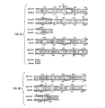
VHのDNA配列から翻訳されたマウスMN14重鎖可変領域のアミノ酸配列を図1に示す(配列番号1及び配列番号2)。この配列とマウスVHサブグループを表す配列との比較は、MN14の重鎖可変領域がサブグループIIIB(Kabat et al. SEQUENCES OF PROTEINS OF IMMUNOLOGICAL INTEREST, U. S. Government Printing Office, 1987を参照)に属することを示した。
Example 5
Analysis of the amino acid sequence of the heavy chain variable region of MN14 The amino acid sequence of the mouse MN14 heavy chain variable region translated from the DNA sequence of VH is shown in FIG. 1 (SEQ ID NO: 1 and SEQ ID NO: 2). A comparison of this sequence with the sequence representing the mouse VH subgroup shows that the heavy chain variable region of MN14 belongs to subgroup IIIB (see Kabat et al. SEQUENCES OF PROTEINS OF IMMUNOLOGICAL INTEREST, US Government Printing Office, 1987). Indicated.
MN14のCDRの配列は、前出のKabat et al.(1987)によって報告されているいかなるものとも異なる。さらに、MN14重鎖VHフレームワーク領域の4つの位置のアミノ酸が、他のサブグループIIIBのVH配列のフレームワーク領域のものとは異なる。これらの4つの置換(Ser14、Thr30、Ser94及びPro108)は、IIIB VHサブグループ以外の他のマウスVH領域において観察されているが、Kabatの108位のプロリンが最も異例である。 The sequence of the CDR of MN14 differs from anything reported by Kabat et al. (1987), supra. Furthermore, the amino acids at the 4 positions of the MN14 heavy chain VH framework region differ from those of the framework regions of the other subgroup IIIB VH sequences. These four substitutions (Ser14, Thr30, Ser94 and Pro108) have been observed in other mouse VH regions other than the IIIB VH subgroup, with Kabat's 108th proline being the most unusual.
VH又はVKにおけるあらゆる異例の残基は、マウスMN14の結合に有利であることが立証されている体細胞変異を表すことがある。 Any unusual residue in VH or VK may represent a somatic mutation that has been demonstrated to favor mouse MN14 binding.
例6
cDNA合成及びMN14 VKをコードするDNAの増幅
MN14のκ軽鎖をコードするcDNAを、上述の重鎖の可変領域をコードするcDNAと同様の方法でクローニングした。κ鎖cDNAの第1鎖を合成するための逆転写酵素を誘発するのに、幾つかのプライマーを用いた。プライマーの1つであるCK2FORの配列は、κ軽鎖遺伝子の定常領域(「CK」)の5’末端の配列から誘導した。他の2つのプライマー、VK1FOR及びVK3FOR、の配列は、κ軽鎖遺伝子の可変領域(「VK」)の3’末端の配列をベースとした。
Example 6
cDNA Synthesis and Amplification of DNA Encoding MN14 VK cDNA encoding the kappa light chain of MN14 was cloned in the same manner as the cDNA encoding the variable region of the heavy chain described above. Several primers were used to induce reverse transcriptase to synthesize the first strand of kappa chain cDNA. The sequence of CK2FOR, one of the primers, was derived from the sequence at the 5 ′ end of the constant region (“CK”) of the kappa light chain gene. The sequences of the other two primers, VK1FOR and VK3FOR, were based on the sequence at the 3 ′ end of the variable region (“VK”) of the kappa light chain gene.
第1鎖DNA産物を、PCRにより、多くのプライマー対を用いて増幅した。一方向への合成を第1鎖の作製に用いたプライマーにより誘発した。もう一方の、「逆」方向への重合は、VK領域の5’末端の配列であるVK1BACK、VK2BACK、VK3BACK、VK4BACK及びVK8BACK、又はシグナルペプチドの最後の4つのアミノ酸及び可変領域の最初の4つのアミノ酸をコードする配列であるVK5BACK、VK6BACK及びVK7BACKのいずれかに基づく配列を有する一連のκ軽鎖特異的プライマーで開始した。加えて、CK2FORで誘発したcDNAも、VK1FOR及びVK8BACKを用いて増幅した。 The first strand DNA product was amplified by PCR using a number of primer pairs. Unidirectional synthesis was induced by the primers used to generate the first strand. The other polymerization in the “reverse” direction is VK1BACK, VK2BACK, VK3BACK, VK4BACK and VK8BACK, or the last four amino acids of the signal peptide and the first four of the variable region. We started with a series of kappa light chain specific primers with sequences based on any of the amino acid-encoding sequences VK5BACK, VK6BACK and VK7BACK. In addition, CK2FOR-induced cDNA was also amplified using VK1FOR and VK8BACK.
これらの増幅産物を、上記の例に記述されている方法で、ゲル電気泳動により分析した。VK1BACK、VK3BACK、VK5BACK、VK7BACK及びVK8BACKで誘発する反応からの生成物が、期待通りの350bpのバンドを生じた。 These amplification products were analyzed by gel electrophoresis as described in the examples above. The products from the reactions elicited with VK1BACK, VK3BACK, VK5BACK, VK7BACK and VK8BACK produced the expected 350 bp band.
例7
PCRによって得られたMN14κ軽鎖可変領域のクローニング及び配列決定
選択されたPCR産物を、上記例4においてVHについて説明されているのと同様の様式で増幅プライマーに含まれる制限部位を用いて、M13mp18及びM13mp19にクローニングした。ヌクレオチド配列決定により、大部分の挿入断片がVK関連ではなかったことが明らかとなった。これは、VKのcDNAのクローニングを試みる場合、異例のことではなく、VH特異的プライマーよりもVK特異的プライマーを設計することが困難であるように思われる。
Example 7
Cloning and sequencing of the MN14 kappa light chain variable region obtained by PCR. The selected PCR product was converted to M13mp18 using the restriction sites contained in the amplification primers in the same manner as described for VH in Example 4 above. And cloned into M13mp19. Nucleotide sequencing revealed that most inserts were not VK related. This is not unusual when attempting to clone VK cDNA, and it seems more difficult to design VK-specific primers than VH-specific primers.
VK1FOR/VK8BACKの組み合わせからVKのcDNA挿入断片が得られたが、CDRL3をコードするcDNA内でのフレームシフト及び23位に不変Cysが存在しないことにより、これは機能的なVKを生じなかった。このVKのcDNAは他のハイブリドーマから単離されており、これはSp2/0融合パートナーに由来する。CK2/VK1BACK産物は、この場合フレームワーク4の保存残基を欠く、さらなる4つの異なる異常VK cDNA挿入断片を生じた。このプライマー対を用いて得られた50のVK挿入断片は、明らかにこのプライマーのミスマッチが誘発するずれによる現象であるVK1BACKの3’末端でのフレーム移動を除いて、機能的VKのものであった。この問題は、VK遺伝子までは伸長しないVK8BACKを用いることにより、回避することができる。しかしながら、VK1FOR/VK8BACK産物からのさらなるクローンの分析は所望の挿入断片を生じなかった。
The VK1 FOR / VK8BACK combination resulted in a VK cDNA insert, but this did not result in a functional VK due to the frameshift in the cDNA encoding CDR L3 and the absence of an invariant Cys at position 23. . The VK cDNA has been isolated from other hybridomas and is derived from the Sp2 / 0 fusion partner. The CK2 / VK1BACK product resulted in four additional aberrant VK cDNA inserts that in this case lacked
推定VKを優先的に増幅するため、その真正第4フレームワーク配列からVK3FORを設計し、VK1FORの代替物として合成した。VK3FOR及びVK8BACKを用いるVK3FOR誘発cDNAの増幅により所望のVKを含む4つのクローンが生じ、この戦略は成功した。 In order to preferentially amplify the putative VK, VK3FOR was designed from its authentic fourth framework sequence and synthesized as an alternative to VK1FOR. Amplification of VK3FOR-induced cDNA using VK3FOR and VK8BACK resulted in four clones containing the desired VK, and this strategy was successful.
マウスMN14κ軽鎖可変領域のDNA及びアミノ酸配列が図2に示されている(配列番号3及び配列番号4)。このMN14のVKは、KabatのVKサブグループVに位置し得る。このMN14 VKフレームワーク領域の僅か3つの残基(Met10、Val66及びThr76)だけがこのサブグループの他のメンバーに現れない。Met10及びVal66がこの3つのうちで最も異例である。これらは、Kabatに列挙されているマウスVKのいかなるものにも見出されない。これらのMN14のVKのCDRには、Kabatにおいて以前報告されている配列はない。 The DNA and amino acid sequences of the mouse MN14κ light chain variable region are shown in FIG. 2 (SEQ ID NO: 3 and SEQ ID NO: 4). This MN 14 VK may be located in Kabat's VK subgroup V. Only three residues of this MN14 VK framework region (Met10, Val66 and Thr76) do not appear in the other members of this subgroup. Met10 and Val66 are the most unusual of the three. These are not found in any of the mouse VKs listed in Kabat. These MN14 VK CDRs have no previously reported sequence in Kabat.
例8
ヒト抗体の可変領域へのMN14のVH及びVKのCDRの移植
8.1 キメラ抗体発現ベクターの構築
マウス可変領域及びヒト定常領域からなるキメラ抗体の生成において、親マウス抗体と一緒に試験することは、正しい可変領域cDNAが単離されていることをチェックするのに役立った。また、成功したキメラ抗体は、ヒト化されたものの結合を評価する際、有用な対照としても機能する。発現のための可変領域のクローニングにおいて用いられた配列は前出のOrlandi et al. (1989)に記述されており、図3及び4に示されている。
Example 8
Implantation of MN14 VH and VK CDRs into the variable region of a human antibody 8.1 Construction of a chimeric antibody expression vector In producing a chimeric antibody consisting of a mouse variable region and a human constant region, testing with a parent mouse antibody is Helped to check that the correct variable region cDNA was isolated. Successful chimeric antibodies also serve as useful controls in assessing the binding of humanized ones. The sequences used in cloning the variable region for expression are described in Orlando, et al. (1989) supra, and are shown in FIGS.
VHのDNAを、オリゴヌクレオチドVH1BACK及びVH1FORを使用するPCRを用いて、M13クローンMNVH41から増幅した。これらのプライマー中のPstI及びBstEII制限部位は、発現のための正しい背景の下で、M13VHPCR1にVHが挿入されることを可能にする。この時点で、VHの内部BamHI制限部位を部位指向性突然変異により除去した。M13VHPCR1のHindIII−BamHI断片全体を包含するこの反応生成物をpSVgptにクローニングし、VH配列を確認した。 VH DNA was amplified from M13 clone MNVH41 using PCR using oligonucleotides VH1BACK and VH1FOR. The PstI and BstEII restriction sites in these primers allow VH to be inserted into M13VHPCR1 under the correct background for expression. At this point, the internal BamHI restriction site of VH was removed by site-directed mutagenesis. This reaction product encompassing the entire HindIII-BamHI fragment of M13VHPCR1 was cloned into pSVgpt to confirm the VH sequence.
その後、上記Takahashi et al., Cell, 29: 671-679 (1962)によって公開されているヒトIgG1定常領域遺伝子をこの構築物にBamHI断片として加え、これにより、pSVgptMN14MuVHHuIgG1と呼ばれるベクターを得た。 Subsequently, the human IgG1 constant region gene published by Takahashi et al., Cell, 29: 671-679 (1962) was added to this construct as a BamHI fragment, thereby obtaining a vector called pSVgptMN14MuVHHIgG1.
同様に、プライマーVK8BACK及びVK3FOR並びにM13VKPCR1にクローニングされているPvuII、BglII消化産物を用いるPCR増幅より、M13クローンMNVK154からVK DNAを得、それにより可変領域の配列をチェックした。HindIII−BamHI断片をRFのDNAから切り出し、プラスミドpSVhygに移した。この構築物は、Hieter et al., Cell, 22: 197-207 (1980)に記述されるように、既にヒトκ定常領域遺伝子を有する。このようにして得られた最終ベクターはpSVhygMN14MuVKHuCKと名付けられた。 Similarly, VK DNA was obtained from M13 clone MNVK154 by PCR amplification using primers VK8BACK and VK3FOR and PvuII, BglII digested products cloned in M13VKPCR1, thereby checking the sequence of the variable region. The HindIII-BamHI fragment was excised from the RF DNA and transferred to the plasmid pSVhyg. This construct already has the human kappa constant region gene as described in Hieter et al., Cell, 22: 197-207 (1980). The final vector thus obtained was named pSVhygMN14MuVKHuCK.
8.2 ハイブリッド抗体の発現及び試験
M13KLHuVHAIGAのHindIII−BamHI断片をプラスミドpSVgptに挿入し、発現ベクターpSVgptKLHuAIGAHuIgG1を得た。同様に、M13HuVKのHindIII−BamHI断片をプラスミドpSVhygに挿入し、発現ベクターpSVhygMN14REIHuVKHuCKを得た。約5μgのpSVgptMN14MuVHHuIgG1及び10μgのpSVhygMN14MuVKHuCK DNAをPvulで直鎖状にして、170V、960μFのシングルパルスのBioRadモデル165BR1160ジーン・パルサー・エレクトロポレーター(Gene Pulser Electroporator)を通常通り用いるエレクトロポレーションにより、約107個の半集密SP2/0ミエローマ細胞に移した。24ウェルプレートにおいて、DMEM+10%FCS成長培地にマイコフェノール酸及びキサンチンを添加することにより、gpt遺伝子の発現について細胞を選択した。
8.2 Expression and test of hybrid antibody The HindIII-BamHI fragment of M13KLHuVHAIGA was inserted into the plasmid pSVgpt to obtain the expression vector pSVgptKLHuAIGAHuIgG1. Similarly, the HindIII-BamHI fragment of M13HuVK was inserted into the plasmid pSVhyg to obtain the expression vector pSVhygMN14REIHuVKHuCK. About 5 μg of pSVgptMN14MuVHHuIgG1 and 10 μg of pSVhygMN14MuVKHuCK DNA are linearized with Pvul, using a 170 V, 960 μF single pulse BioRad model 165BR1160 Gene Pulser Electroporator as usual using an Electroporator. The cells were transferred to 107 semi-confluent SP2 / 0 myeloma cells. Cells were selected for gpt gene expression by adding mycophenolic acid and xanthine to DMEM + 10% FCS growth medium in 24-well plates.
生存細胞のコロニーを含むウェルを同定した。その培地上清をこれらのウェルから取り除き、ヒト抗体について検定した。抗体を分泌するコロニーを増殖させ、より多量の抗体を単離するため0.5Lの調整培地を得た。 Wells containing viable cell colonies were identified. The media supernatant was removed from these wells and assayed for human antibodies. Colonies secreting the antibody were grown and 0.5 L of conditioned medium was obtained to isolate more antibody.
通常通り、最初に、プロテインAアガロースアフィニティクロマトグラフィーにより、この培地から抗体を精製した。精製した抗体を、未変性MN14抗体、ヒト抗体及び他の対照を参照して、さらに特徴付けた。 As usual, antibodies were first purified from this medium by protein A agarose affinity chromatography. The purified antibody was further characterized with reference to native MN14 antibody, human antibody and other controls.
また、この抗体を、MN14遮断検定におけるその反応プロフィールによっても特徴付けた。これにより、このハイブリッド抗体のCEA結合親和性と、ポジティブコントロールとして役に立つMN14マウス抗体によるCEA結合とによって、情報を提示する比較が得られた。 The antibody was also characterized by its response profile in the MN14 block assay. This provided an informative comparison by CEA binding affinity of this hybrid antibody and CEA binding by MN14 mouse antibody which served as a positive control.
8.3 MN14抗体のヒト化
ヒトNEWM VH、KOL VH及びREI VKフレームワークがヒト体内で寛容性であるように思われるため、それらを抗体の再構成のための基礎として選択した。MN14のVH(配列番号2)及びVK(配列番号4)とこれらのヒト可変領域とを並べたものを図5に示す(配列番号5〜7)。
The humanized human NEWM VH, KOL VH and REI VK frameworks of the 8.3 MN14 antibody appeared to be tolerated in the human body and were selected as the basis for antibody reconstitution. MN14 VH (SEQ ID NO: 2) and VK (SEQ ID NO: 4) aligned with these human variable regions are shown in FIG. 5 (SEQ ID NOs: 5 to 7).
A.NEWMベースのヒト化
MN14のCDRを導入するための開始点は、必須FR及び無関係のCDRをコードするDNAである。これらのテンプレートの可変領域は、用いられる発現ベクターに適合する形態、すなわち、HindIII−BamHI断片中にあり、本明細書にはプロモーター領域、シグナルペプチド及びイントロンDNAも含まれる(図3及び図4)。NEWM VHでのものについては、このテンプレートはM13VHPCR1(上記Orlandi et al.、及び下記8.3項)である。KOL FR及び無関係のCDRを含む、このテンプレートの誘導体を、KOLコード領域の生成に用いた。M13VKPCR1(上記Orlandi et al.)の誘導体をHuVKベクターの作製において用いた。得られたベクターを、M13NMHuVH、M13KLHuVH及びM13HuVKと名付けた。ヒト化MN14可変領域DNAを有するHindIII−BamHI断片を、本質的に例8.1においてキメラMN14発現ベクターの構築について記述される通りに、これらのM13ベクターから発現ベクターに移した。
A. NEWM-based humanized The starting point for introducing the CDRs of MN14 is DNA encoding the essential FRs and an unrelated CDR. The variable regions of these templates are in a form compatible with the expression vector used, i.e., in the HindIII-BamHI fragment, which also includes the promoter region, signal peptide and intron DNA (Figures 3 and 4). . For those with NEWM VH, this template is M13VHPCR1 (Orlandi et al., Above, and 8.3 below). A derivative of this template, including KOL FR and an irrelevant CDR, was used to generate the KOL coding region. A derivative of M13VKPCR1 (Orlandi et al., Above) was used in the construction of the HuVK vector. The resulting vectors were named M13NMHuVH, M13KLHuVH and M13HuVK. HindIII-BamHI fragments with humanized MN14 variable region DNA were transferred from these M13 vectors to expression vectors essentially as described for the construction of chimeric MN14 expression vectors in Example 8.1.
NEWM FRは、Poljak et al. Biochemistry 16: 3412-20 (1977)に説明されている。そのマウス等価物のものに匹敵する、CEAに対する親和性を有するhMN14の構築を、段階的なアプローチで達成した。キメラ抗体の生成は、ヒト化されたものの結合を評価する場合に、有用な対照を提供する。ヒトNEWM VH及びREI VKフレームワークを抗体を再構成するための基礎として最初に選択した。これは、これらがヒト体内において寛容性であることが知られているためである。MN14の残基Phe27、Asp28及びThr30は保持した。これは、Kabatの超可変領域であるCDR1残基31〜35の一部ではないものの、それらのアミノ酸がCDR1構造ループ(Chothia et al., J. Mol. Biol. 176: 901-917 (1987))の一部であるためである。加えて、以下の理由により、ヒト化VHへの組込みのために次の残基も選択した。すなわち、Ala24、この残基はCDR1に接触する。Arg71、この残基の側鎖はこのドメインの中心を通して突き出し、CDR1及びCDR2と相互作用する。ほんの少しのValの置換はこれらのCDRループの立体配座を変更し得る。ならびに、Ser94、大部分の抗体はこの位置にArgを有しており、そこではCDR3のAsp残基と相互作用するものと考えられ、NEWMがArgを含むとマウスCDRとの望ましくない相互作用を形成することがあり得るという理由である。他の再構成分子において重要であることが立証されている領域に相当する3つの領域において、このバージョン(NMHuVH)に対する他の変更がなされた。これらの変更は以下のものであった。 NEWM FR is described in Poljak et al. Biochemistry 16: 3412-20 (1977). Construction of hMN14 with affinity for CEA comparable to that of its mouse equivalent was achieved in a step-wise approach. Generation of chimeric antibodies provides a useful control when assessing the binding of humanized ones. Human NEWM VH and REI VK frameworks were initially selected as the basis for antibody reconstitution. This is because they are known to be tolerated in the human body. Residues Phe27, Asp28 and Thr30 of MN14 were retained. Although this is not part of CDR1 residues 31-35, which is the hypervariable region of Kabat, these amino acids are CDR1 structural loops (Chothia et al., J. Mol. Biol. 176: 901-917 (1987) ). In addition, the following residues were also selected for incorporation into humanized VH for the following reasons. That is, Ala24, this residue contacts CDR1. Arg71, the side chain of this residue protrudes through the center of this domain and interacts with CDR1 and CDR2. Only a few Val substitutions can alter the conformation of these CDR loops. As well, Ser94, most antibodies have Arg at this position, where they are thought to interact with the Asp residue of CDR3, and when NEWM contains Arg, it has an undesirable interaction with mouse CDRs. That is why it can be formed. Other changes to this version (NMHuVH) were made in three areas corresponding to areas that have proven to be important in other reconstituted molecules. These changes were as follows:
(i)Gln77Phe78Ser79からThrLeuTyr(NMHuVhHTLY、配列番号9)
(ii)Ser82Thr83Ala84Ala85からLysArgSerGlu(NMHuVhHKRSE、配列番号10)
(iii)Arg66Val67Thr68Met69Leu70からLysPheIleValSer(NMHuVhKFIVS、配列番号11)
(I) Gln77Phe78Ser79 to ThrLeuTyr (NMHuVhHTLY, SEQ ID NO: 9)
(Ii) Ser82Thr83Ala84Ala85 to LysArgSerGlu (NMHuVhHKRSE, SEQ ID NO: 10)
(Iii) Arg66Val67Thr68Met69Leu70 to LysPheIleValSer (NMHuVhKFIVS, SEQ ID NO: 11)
このNEWM VHフレームワークの異なるバージョン(配列番号5及び8〜11)とMN14 VH(配列番号2)とのアラインメントを図6に示す。これらのバージョンの各々は、同じHuVKと対を形成している。TLY又はKFIVSモチーフのいずれかを含むことにより、約2倍の改善が得られた。 The alignment of the different versions of this NEWM VH framework (SEQ ID NO: 5 and 8-11) and MN14 VH (SEQ ID NO: 2) is shown in FIG. Each of these versions is paired with the same HuVK. Inclusion of either TLY or KFIVS motif resulted in an approximately 2-fold improvement.
上記Kabat et al.(1987)に示されているNEWNフレームワーク配列とは2つの相違がある。すなわち、S107からT及びL108からT。Kabatは、残基1をPCAとして、かつ残基5及び6をE又はQとして列挙している。
There are two differences from the NEWN framework sequence shown in Kabat et al. (1987) above. That is, S107 to T and L108 to T. Kabat lists
B.KOLベースのヒト化
NEWM VHの使用と平行して、我々はヒトKOL VHも再構成している。KOL VHは、Schmidt et al.,Z.physiol.Chem,364:713−747(1987)に説明されている。抗体のヒト化に用いられたFRはKabatに示されるものと同様である。このKOLベースのVHの本来のバージョンは、残基の可変性の観点で定義される(上記Kabat et al.)、MN14のCDR及び3つのさらなるマウス残基を有していた。NEWM VHを用いた場合と同様に、これらの置換のうちの2つが生じた。これは、βシートフレームワークから伸びる実際のペプチドCDR1構造ループが残基25〜32からなる(上記Chothia et al. 1987)ためである。28位及び30位での変更は、このループが全体としてマウス抗体から移植されるのを可能にする。KOL VHのArg残基がAsp110との塩架橋に関与し、NEWM VHを用いた場合と同様に、Arg94の保持がMN14のCDR3構造を撹乱することがあるように思われたため、MN14残基94が含められた。残基94の側鎖はCDR1の残基とも相互作用し得る。この基本MN14 KOLHuVH(配列番号12)に対してなされた他の変更は以下の通りである。
B. In parallel with the use of KOL-based humanized NEWM VH, we are also reconstituting human KOL VH. KOL VH is described in Schmidt et al. , Z. physiol. Chem, 364: 713-747 (1987). The FR used for antibody humanization is similar to that shown in Kabat. The original version of this KOL-based VH had a CDR for MN14 and three additional mouse residues, defined in terms of residue variability (Kabat et al., Supra). As with NEWM VH, two of these substitutions occurred. This is because the actual peptide CDR1 structural loop extending from the β-sheet framework consists of residues 25-32 (Chothia et al. 1987 above). Changes at
(i)Ser24からAla24及びVal48Ala49からIleGly(KLHuVhAIG、配列番号13)。
(ii)Ser24からAla24、Val48Ala49からIleGly、及びSer74からAla(KLHuVhAIGA、配列番号14)。
(iii)Ser24からAla24、Val48Ala49からIleGly、Ser74からAla及びPhe79からTyr(KLHuVhAIGAY、配列番号15)。
(I) Ser24 to Ala24 and Val48Ala49 to IleGly (KLHuVhAIG, SEQ ID NO: 13).
(Ii) Ser24 to Ala24, Val48Ala49 to IleGly, and Ser74 to Ala (KLHuVhAIGA, SEQ ID NO: 14).
(Iii) Ser24 to Ala24, Val48Ala49 to IleGly, Ser74 to Ala and Phe79 to Tyr (KLHuVhAIGAY, SEQ ID NO: 15).
変異の論理的説明
Ala24−CDR1のループは、残基29の側鎖のフレームワークへの浸透によって固定されている。残基24は、それが相互作用するものの1つである(Chothia et al., J. Mol. Biol. 227: 799-817 (1992))。
Logical explanation of the mutation The loop of Ala24-CDR1 is fixed by penetration of the side chain of residue 29 into the framework.
Ile48Gly49−これらの残基の両者はCDR2超可変領域に隣接するものの、実際の構造ループからはかけ離れている。両残基は完全に埋もれており(Padlan, Mol. Immunolog. 28: 489-498, 1991)、これらがそれらのパッキング相互作用により結合を行うことが可能であるように思われた。 Ile48Gly49—both of these residues are adjacent to the CDR2 hypervariable region, but far from the actual structural loop. Both residues were completely buried (Padlan, Mol. Immunolog. 28: 489-498, 1991) and it appeared that they could be bound by their packing interactions.
Ala74−この残基はVH抗原結合表面に見出される第4ループの一部であり、その側鎖は溶媒にほとんど完全に露出している(上記Padlan, 1991)。この残基と抗原との直接相互作用を予想することができた。 Ala74—This residue is part of the fourth loop found on the VH antigen binding surface, and its side chain is almost completely exposed to solvent (Padlan, 1991, supra). A direct interaction between this residue and the antigen could be expected.
Tyr79−残基74と同様に、この残基は抗原結合部位に近接し、結合を行うことが可能である。
Similar to Tyr79-
KOL VHフレームワークの異なるバージョン(配列番号7及び12〜15)とNEWMベースのバージョン(配列番号5及び8〜11)及びマウスMN14(配列番号2)のアラインメントを図6に示す。 The alignment of the different versions of the KOL VH framework (SEQ ID NO: 7 and 12-15) and the NEWM-based version (SEQ ID NO: 5 and 8-11) and mouse MN14 (SEQ ID NO: 2) is shown in FIG.
MN14HuVH及びMN14HuVLのDNA配列及び翻訳産物をそれぞれ図7及び8に示す(それぞれ、配列番号16〜19)。 The DNA sequences and translation products of MN14HuVH and MN14HuVL are shown in FIGS. 7 and 8, respectively (SEQ ID NOs: 16 to 19, respectively).
抗体KLHuVHAIGA/HuVKのようなヒト化KLHuVHの変種を精製し、以下のように遮断検定において試験を行った。抗体を、指定された濃度で、HRP標識MN14と共に添加し、最終容積0.1mlとした。37℃で30分間インキュベートし、洗浄して未結合の抗体を除去した後、残留結合ペルオキシダーゼ活性から抗体の相対親和力を決定した。図9の検定データによって示されるように、「再構成」(すなわち、FRにCDRが移植されている)ヒト化抗体の活性は、キメラ及びマウス陽性対照のものと同等であった。 Variants of humanized KLHuVH, such as the antibody KLHuVHAIGA / HuVK, were purified and tested in a block assay as follows. Antibody was added at the indicated concentration with HRP-labeled MN14 to a final volume of 0.1 ml. After incubation at 37 ° C. for 30 minutes and washing to remove unbound antibody, the relative affinity of the antibody was determined from the residual bound peroxidase activity. As shown by the assay data in FIG. 9, the activity of “reconstituted” (ie, FR grafted CDRs) humanized antibodies was comparable to that of chimeric and mouse positive controls.
KLHuVHAIG/HuVK及びKLHuVHAIGAY/HuVK抗体を分泌する細胞からの上清液を用いて得られた結果は、これらが、KLHuVHAIGA/HuVK抗体のものと同等の遮断活性を有することを示唆する。 The results obtained using supernatants from cells secreting KLHuVHAIG / HuVK and KLHuVHAIGAY / HuVK antibodies suggest that they have blocking activity comparable to that of KLHuVHAIGA / HuVK antibodies.
C.REIベースのヒト化
REI VLは、Epp et al., Eur. J. Biochem., 45: 513-24 (1974)に説明されている。REIフレームワークにおいては、結合を改善するための変更は行わなかった。Kabat et al. (1987)に示される配列からの変更は、M4からL、T39からK、Y71からF、L104からV、Q105からE、及びT107からKである。これらの変更は、MN14 CDRを移植するときに用いられるテンプレートに予め存在し、結合を改善するために特異的に作製されはしなかった。M4のLへの変更は制限部位を組込むが、残りの相違がREIフレームワークの異例の残基を取り除く。類似のフレームワークが、Foote et al., J. Mol. Biol. 224: 487-9 (1992)により、ヒトκサブグループIのコンセンサスと呼ばれている。
C. REI-based humanization REI VL is described in Epp et al., Eur. J. Biochem., 45: 513-24 (1974). In the REI framework, no changes were made to improve binding. Changes from the sequence shown in Kabat et al. (1987) are M4 to L, T39 to K, Y71 to F, L104 to V, Q105 to E, and T107 to K. These changes were pre-existing in the template used when transplanting the MN14 CDRs and were not made specifically to improve binding. Changing M4 to L incorporates a restriction site, but the remaining differences remove unusual residues of the REI framework. A similar framework is referred to as the human kappa subgroup I consensus by Foote et al., J. Mol. Biol. 224: 487-9 (1992).
例9
MN14のCDRを移植されたヒト化抗体の発現
MN14のCDRを移植されたヒト抗体の発現について安定に形質転換された細胞を上述の方法で選択し、クローニングして個々の産生細胞系を確立した。これらの系の各々を検定し、正しい抗体の産生を決定して、産生の効力を評価した。抗体のクラスを決定し、抗CEA結合親和性を評価した。
Example 9
Stably transformed cells were selected in the manner described above for the expression of transplanted human antibody CDR expression MN14 of MN14 humanized antibodies the CDR is transplanted in and cloned to establish individual producer cell system . Each of these systems was assayed to determine correct antibody production and to assess the efficacy of production. The class of antibodies was determined and anti-CEA binding affinity was evaluated.
最良の産生細胞を産生される抗体の全量についてさらに特徴付け、これらのうちの最良のものについて、トランスフェクション、組込み、増殖又は選別の間に抗体遺伝子に変異が生じていないことを保証するためそのmRNAから配列を得た。 Further characterize the total amount of antibody produced to produce the best production cells, and the best of these to ensure that no mutations have occurred in the antibody gene during transfection, integration, propagation or selection Sequence was obtained from mRNA.
例10
細胞培養物中で発現したMN14のCDRを移植されたヒト化抗体の精製
MN14のCDRを移植されたヒト抗体の最良産生細胞系を培養し、その成長培地を集めて、0.2ミクロン膜を通して濾過した。その後、抗体を、プロテインAクロマトグラフィー、次いでイオン交換及びサイズ排除クロマトグラフィーのような他の通常の精製ステップにより精製した。通常の遠心により、細胞をペレット化した。その上清液から、上述のように抗体を精製した。
Example 10
Purification of Humanized Antibody Implanted with MN14 CDRs Expressed in Cell Culture The best producer cell line of human antibody engrafted with MN14 CDRs is cultured and the growth medium is collected and passed through a 0.2 micron membrane. Filtered. The antibody was then purified by other conventional purification steps such as protein A chromatography followed by ion exchange and size exclusion chromatography. Cells were pelleted by normal centrifugation. The antibody was purified from the supernatant as described above.
例11
診断におけるヒト化MN14モノクローナル抗体の使用
A.動物研究
ヒト結腸ガンを有するヌードマウスにおいて、標識ヒト化MN14 IgGの生体内の分配を測定した。放射性位置測定研究に対しては、4〜5週齢雌胸腺欠損マウス(nu/nu、Harlan、Indianapolis、IN)に、胸腺欠損マウスにおいて順次増殖させた異種移植片から調製したLS174Tヒト結腸アデノカルシノーマ(Sharkey et al., Cancer Res., 50: 828-34 (1990))の10%懸濁液0.2mlを皮下に移植した。腫瘍の発達を2週間待った後、これらのマウスに、20μCi(約2μg)の131I標識ヒト化MN14モノクローナル抗体を静脈内注射した。その後、時々、4〜5匹のマウスの群を屠殺し、上記Sharkey et al.に従い、組織中で放射活性の位置を測定した。表2のデータは注射された用量/組織gの%及び腫瘍:非腫瘍比を示す。
Example 11
Use of humanized MN14 monoclonal antibody in diagnosis Animal studies In vivo distribution of labeled humanized MN14 IgG was measured in nude mice with human colon cancer. For radiolocation studies, LS174T human colon adenocalci prepared from xenografts grown sequentially in athymic mice in 4-5 week old female athymic mice (nu / nu, Harlan, Indianapolis, IN). 0.2 ml of a 10% suspension of normal (Sharkey et al., Cancer Res., 50: 828-34 (1990)) was implanted subcutaneously. After waiting 2 weeks for tumor development, these mice were injected intravenously with 20 μCi (approximately 2 μg) of 131 I-labeled humanized MN14 monoclonal antibody. Thereafter, groups of 4-5 mice were sometimes sacrificed and the location of radioactivity was determined in the tissues according to Sharkey et al. The data in Table 2 shows the injected dose /% tissue g and the tumor: non-tumor ratio.
これらの結果は、この抗体の優れた腫瘍結合性を示し、その最大結合活性は2日以内に生じる。hMN14抗体の血液クリアランスは、親mMN14抗体よりも迅速であった。加えて、脾臓によるhMN14の取り込みはmMN14よりも高く、これはhMN14抗体がマウスにとって「外来性」であるという事実を反映している。腫瘍:非腫瘍比は優れたものであった。これらの結果は、本発明のhMN14 mAbがCEA産生腫瘍を標的とすることが可能であることを示す。 These results show the excellent tumor binding properties of this antibody, with its maximum binding activity occurring within 2 days. The blood clearance of hMN14 antibody was faster than the parent mMN14 antibody. In addition, the uptake of hMN14 by the spleen is higher than mMN14, reflecting the fact that the hMN14 antibody is “foreign” to the mouse. Tumor: non-tumor ratio was excellent. These results indicate that the hMN14 mAb of the present invention can target CEA producing tumors.
B.131I標識ヒト化MN14のIgGを用いた臨床研究
ヒト化MN14 IgGのターゲッティング及び薬物速度論的挙動の先行調査のため、患者を、米国分子薬剤及び免疫学センター(Center for Molecular Medicine and Immunology)、Newark、NJで、インスティチューショナル・レビュー・ボード(Institutional Review Board)認可プロトコルに処した。図12に示されている結果の場合、その男性患者は、肝臓に転移している結腸直腸ガンを有していた。彼に131I−hMN14 IgG(8mCi、0.6mg抗体)を静脈注射し、6日間にわたって撮像した。続いて、この患者に等用量のmMN14 IgGを注射した。図12に示される像は、各注射の約140時間後の腹部前面の像を示す。これらの像は、直接比較することができるように、正確に同じ濃度に調整されている。これらの結果は、親マウス抗体のみならずこのヒト化抗体がCEA産生腫瘍によって取り込まれることを示す。これらの実験は、本発明のヒト化MN14 mAbを用いるヒトCEA産生結腸ガンの診断が実用上有用なものであることを立証する。
B. 131 I-Labeled Humanized MN14 IgG Clinical Study For the targeting and pharmacokinetic behavior of humanized MN14 IgG, patients were referred to the Center for Molecular Medicine and Immunology, In Newark, NJ, submitted to Institutional Review Board authorization protocol. In the case of the results shown in FIG. 12, the male patient had colorectal cancer that had metastasized to the liver. He was injected intravenously with 131 I-hMN14 IgG (8 mCi, 0.6 mg antibody) and imaged over 6 days. Subsequently, the patient was injected with an equal dose of mMN14 IgG. The image shown in FIG. 12 shows an image of the front of the abdomen about 140 hours after each injection. These images are adjusted to exactly the same density so that they can be directly compared. These results indicate that this humanized antibody as well as the parent mouse antibody is taken up by CEA-producing tumors. These experiments demonstrate that the diagnosis of human CEA-producing colon cancer using the humanized MN14 mAb of the present invention is practically useful.
Claims (5)
Applications Claiming Priority (2)
| Application Number | Priority Date | Filing Date | Title |
|---|---|---|---|
| US08/318,157 US5874540A (en) | 1994-10-05 | 1994-10-05 | CDR-grafted type III anti-CEA humanized mouse monoclonal antibodies |
| US08/318,157 | 1994-10-05 |
Related Parent Applications (1)
| Application Number | Title | Priority Date | Filing Date |
|---|---|---|---|
| JP2001357367A Division JP2002218990A (en) | 1994-10-05 | 2001-11-22 | Cdr-grafted type iii anti-cea humanized mouse monoclonal antibody |
Publications (2)
| Publication Number | Publication Date |
|---|---|
| JP2010075190A true JP2010075190A (en) | 2010-04-08 |
| JP5183611B2 JP5183611B2 (en) | 2013-04-17 |
Family
ID=23236916
Family Applications (3)
| Application Number | Title | Priority Date | Filing Date |
|---|---|---|---|
| JP51258896A Expired - Fee Related JP3437580B2 (en) | 1994-10-05 | 1995-09-28 | Type III anti-CEA humanized mouse monoclonal antibody grafted with CDR |
| JP2001357367A Pending JP2002218990A (en) | 1994-10-05 | 2001-11-22 | Cdr-grafted type iii anti-cea humanized mouse monoclonal antibody |
| JP2009260907A Expired - Lifetime JP5183611B2 (en) | 1994-10-05 | 2009-11-16 | Type III anti-CEA humanized mouse monoclonal antibody transplanted with CDR |
Family Applications Before (2)
| Application Number | Title | Priority Date | Filing Date |
|---|---|---|---|
| JP51258896A Expired - Fee Related JP3437580B2 (en) | 1994-10-05 | 1995-09-28 | Type III anti-CEA humanized mouse monoclonal antibody grafted with CDR |
| JP2001357367A Pending JP2002218990A (en) | 1994-10-05 | 2001-11-22 | Cdr-grafted type iii anti-cea humanized mouse monoclonal antibody |
Country Status (8)
| Country | Link |
|---|---|
| US (3) | US5874540A (en) |
| EP (3) | EP0783313B1 (en) |
| JP (3) | JP3437580B2 (en) |
| AT (2) | ATE464906T1 (en) |
| CA (1) | CA2200868C (en) |
| DE (2) | DE69535755D1 (en) |
| ES (1) | ES2307294T3 (en) |
| WO (1) | WO1996011013A1 (en) |
Families Citing this family (141)
| Publication number | Priority date | Publication date | Assignee | Title |
|---|---|---|---|---|
| US7354587B1 (en) * | 1994-07-06 | 2008-04-08 | Immunomedics, Inc. | Use of immunoconjugates to enhance the efficacy of multi-stage cascade boosting vaccines |
| US5798100A (en) | 1994-07-06 | 1998-08-25 | Immunomedics, Inc. | Multi-stage cascade boosting vaccine |
| US6730300B2 (en) | 1996-03-20 | 2004-05-04 | Immunomedics, Inc. | Humanization of an anti-carcinoembryonic antigen anti-idiotype antibody and use as a tumor vaccine and for targeting applications |
| CA2250579A1 (en) | 1996-05-04 | 1997-11-13 | Zeneca Limited | Monoclonal antibody to cea, conjugates comprising said antibody, and their therapeutic use in an adept system |
| US6417337B1 (en) * | 1996-10-31 | 2002-07-09 | The Dow Chemical Company | High affinity humanized anti-CEA monoclonal antibodies |
| US6703488B1 (en) * | 1998-01-15 | 2004-03-09 | Center For Molecular Medicine And Immunology | Antibody/receptor targeting moiety for enhanced delivery of armed ligand |
| AU752494B2 (en) * | 1998-02-25 | 2002-09-19 | Dow Chemical Company, The | High affinity humanized ANTI-CEA monoclonal antibodies |
| US6962702B2 (en) * | 1998-06-22 | 2005-11-08 | Immunomedics Inc. | Production and use of novel peptide-based agents for use with bi-specific antibodies |
| MY133346A (en) * | 1999-03-01 | 2007-11-30 | Biogen Inc | Kit for radiolabeling ligands with yttrium-90 |
| US20020102208A1 (en) | 1999-03-01 | 2002-08-01 | Paul Chinn | Radiolabeling kit and binding assay |
| US20030099629A1 (en) * | 1999-03-11 | 2003-05-29 | Immunomedics, Inc. | Recombinant onconase and chemical conjugates and fusion proteins of recombinant onconase |
| US7829064B2 (en) * | 1999-05-10 | 2010-11-09 | Immunomedics, Inc. | Anti-CD74 immunoconjugates and methods |
| US8119101B2 (en) * | 1999-05-10 | 2012-02-21 | The Ohio State University | Anti-CD74 immunoconjugates and methods of use |
| US7527787B2 (en) * | 2005-10-19 | 2009-05-05 | Ibc Pharmaceuticals, Inc. | Multivalent immunoglobulin-based bioactive assemblies |
| US8383081B2 (en) | 1999-05-10 | 2013-02-26 | Immunomedics, Inc. | Anti-CD74 immunoconjugates and methods of use |
| JP4762478B2 (en) * | 2000-07-03 | 2011-08-31 | キャタレント ファーマ ソリューションズ リミテッド ライアビリティ カンパニー | Expression vector |
| US6852510B2 (en) * | 2000-07-03 | 2005-02-08 | Gala Design Inc | Host cells containing multiple integrating vectors |
| US20040235173A1 (en) * | 2000-07-03 | 2004-11-25 | Gala Design, Inc. | Production of host cells containing multiple integrating vectors by serial transduction |
| US20030224415A1 (en) * | 2001-06-29 | 2003-12-04 | Gala Design, Inc. | Selection free growth of host cells containing multiple integrating vectors |
| WO2002002738A2 (en) * | 2000-07-03 | 2002-01-10 | Gala Design, Inc. | Host cells containing multiple integrating vectors |
| CA2417185A1 (en) * | 2000-07-25 | 2002-01-31 | Shui-On Leung | Multivalent target binding protein |
| US20020165360A1 (en) * | 2000-11-30 | 2002-11-07 | Junghans Richard P. | Chimeric effector cell receptors against carcinoembryonic antigen |
| WO2002078638A2 (en) * | 2001-03-30 | 2002-10-10 | University Of Massachusetts | Morpholino imaging and therapy |
| EP1444267B1 (en) * | 2001-10-15 | 2016-08-10 | Immunomedics, Inc. | Affinity enhancement agents |
| US7591994B2 (en) | 2002-12-13 | 2009-09-22 | Immunomedics, Inc. | Camptothecin-binding moiety conjugates |
| EP2865688A1 (en) | 2002-03-01 | 2015-04-29 | Immunomedics, Inc. | Internalizing anti-CD74 antibodies and methods of use |
| US20040018557A1 (en) * | 2002-03-01 | 2004-01-29 | Immunomedics, Inc. | Bispecific antibody point mutations for enhancing rate of clearance |
| DE60325184D1 (en) | 2002-03-01 | 2009-01-22 | Immunomedics Inc | RS7 ANTIBODY |
| US20160279239A1 (en) | 2011-05-02 | 2016-09-29 | Immunomedics, Inc. | Subcutaneous administration of anti-cd74 antibody for systemic lupus erythematosus and autoimmune disease |
| US8658773B2 (en) | 2011-05-02 | 2014-02-25 | Immunomedics, Inc. | Ultrafiltration concentration of allotype selected antibodies for small-volume administration |
| US9770517B2 (en) | 2002-03-01 | 2017-09-26 | Immunomedics, Inc. | Anti-Trop-2 antibody-drug conjugates and uses thereof |
| US7384738B2 (en) * | 2002-03-28 | 2008-06-10 | Bremel Robert D | Retrovirus-based genomic screening |
| US20040038304A1 (en) * | 2002-03-28 | 2004-02-26 | Gala Design, Inc. | Antibody libraries |
| WO2003101495A1 (en) * | 2002-05-29 | 2003-12-11 | Immunomedics, Inc. | Methods and compositions for radioimmunotherapy of brain and cns tumors |
| CA2501616C (en) * | 2002-10-08 | 2015-05-12 | Immunomedics, Inc. | Antibody therapy |
| KR20050073488A (en) * | 2002-10-08 | 2005-07-13 | 이뮤노메딕스, 인코오포레이티드 | Combination therapy with class Ⅲ anti-CEA monoclonal antibodies and therapeutic agents |
| US8420086B2 (en) | 2002-12-13 | 2013-04-16 | Immunomedics, Inc. | Camptothecin conjugates of anti-CD22 antibodies for treatment of B cell diseases |
| WO2004054622A1 (en) * | 2002-12-13 | 2004-07-01 | Immunomedics, Inc. | Immunoconjugates with an intracellularly-cleavable linkage |
| JP5026072B2 (en) * | 2003-07-01 | 2012-09-12 | イミューノメディクス、インコーポレイテッド | Multispecific carrier of bispecific antibody |
| JP2007524649A (en) * | 2003-07-29 | 2007-08-30 | イミューノメディクス、インコーポレイテッド | Fluorinated carbohydrate complex |
| US6878531B1 (en) * | 2003-11-10 | 2005-04-12 | Medical College Of Georgia Research Institute | Method for multiple site-directed mutagenesis |
| US20050132544A1 (en) * | 2003-12-23 | 2005-06-23 | Jayshree Seth | Split hook fastener |
| US20050221429A1 (en) * | 2004-01-16 | 2005-10-06 | Cardinal Health Pts, Llc | Host cells containing multiple integrating vectors comprising an amplifiable marker |
| US8435539B2 (en) * | 2004-02-13 | 2013-05-07 | Immunomedics, Inc. | Delivery system for cytotoxic drugs by bispecific antibody pretargeting |
| US8883160B2 (en) * | 2004-02-13 | 2014-11-11 | Ibc Pharmaceuticals, Inc. | Dock-and-lock (DNL) complexes for therapeutic and diagnostic use |
| US8562988B2 (en) * | 2005-10-19 | 2013-10-22 | Ibc Pharmaceuticals, Inc. | Strategies for improved cancer vaccines |
| US8652484B2 (en) | 2004-02-13 | 2014-02-18 | Immunomedics, Inc. | Delivery system for cytotoxic drugs by bispecific antibody pretargeting |
| US8551480B2 (en) | 2004-02-13 | 2013-10-08 | Immunomedics, Inc. | Compositions and methods of use of immunotoxins comprising ranpirnase (Rap) show potent cytotoxic activity |
| US8491914B2 (en) * | 2004-02-13 | 2013-07-23 | Ibc Pharmaceuticals, Inc. | Dock-and-lock (DNL) complexes for delivery of interference RNA |
| US9550838B2 (en) | 2004-02-13 | 2017-01-24 | Ibc Pharmaceuticals, Inc. | Dock-and-lock (DNL) complexes for therapeutic and diagnostic use |
| US20110020273A1 (en) * | 2005-04-06 | 2011-01-27 | Ibc Pharmaceuticals, Inc. | Bispecific Immunocytokine Dock-and-Lock (DNL) Complexes and Therapeutic Use Thereof |
| US9481878B2 (en) | 2004-02-13 | 2016-11-01 | Immunomedics, Inc. | Compositions and methods of use of immunotoxins comprising ranpirnase (Rap) show potent cytotoxic activity |
| US20110171126A1 (en) * | 2010-01-11 | 2011-07-14 | Center For Molecular Medicine And Immunology | Enhanced Cytotoxicity of Anti-CD74 and Anti-HLA-DR Antibodies with Interferon-Gamma |
| US20110064754A1 (en) * | 2005-03-03 | 2011-03-17 | Center For Molecular Medicine And Immunology | Immunoconjugates Comprising Poxvirus-Derived Peptides and Antibodies Against Antigen-Presenting Cells for Subunit-Based Poxvirus Vaccines |
| US20060127311A1 (en) * | 2004-11-17 | 2006-06-15 | Duke University | Radiation dosimetry and blocking antibodies and methods and uses therefor in the treatment of cancer |
| US20160355591A1 (en) | 2011-05-02 | 2016-12-08 | Immunomedics, Inc. | Subcutaneous anti-hla-dr monoclonal antibody for treatment of hematologic malignancies |
| US10058621B2 (en) | 2015-06-25 | 2018-08-28 | Immunomedics, Inc. | Combination therapy with anti-HLA-DR antibodies and kinase inhibitors in hematopoietic cancers |
| US9707302B2 (en) | 2013-07-23 | 2017-07-18 | Immunomedics, Inc. | Combining anti-HLA-DR or anti-Trop-2 antibodies with microtubule inhibitors, PARP inhibitors, bruton kinase inhibitors or phosphoinositide 3-kinase inhibitors significantly improves therapeutic outcome in cancer |
| US8475794B2 (en) | 2005-04-06 | 2013-07-02 | Ibc Pharmaceuticals, Inc. | Combination therapy with anti-CD74 antibodies provides enhanced toxicity to malignancies, Autoimmune disease and other diseases |
| US8349332B2 (en) | 2005-04-06 | 2013-01-08 | Ibc Pharmaceuticals, Inc. | Multiple signaling pathways induced by hexavalent, monospecific and bispecific antibodies for enhanced toxicity to B-cell lymphomas and other diseases |
| US20060257317A1 (en) * | 2005-05-04 | 2006-11-16 | Duke University | Combination therapy in the treatment of cancer |
| CN101534865A (en) * | 2005-10-19 | 2009-09-16 | Ibc药品公司 | Methods and compositions for generating bioactive assemblies of increased complexity and uses |
| WO2008069999A2 (en) | 2006-12-01 | 2008-06-12 | Selexys Pharmaceuticals Corporation | Anti-p-selectin antibodies and methods of using the same to treat inflammatory diseases |
| EP2288715B1 (en) | 2008-04-11 | 2014-09-24 | Merrimack Pharmaceuticals, Inc. | Human serum albumin linkers and conjugates thereof |
| AU2009282830B2 (en) * | 2008-08-20 | 2013-11-28 | Ibc Pharmaceuticals, Inc. | Dock-and-lock (DNL) vaccines for cancer therapy |
| JP2012504961A (en) | 2008-10-06 | 2012-03-01 | ミネルバ バイオテクノロジーズ コーポレーション | MUC1 * antibody |
| WO2010059315A1 (en) | 2008-11-18 | 2010-05-27 | Merrimack Pharmaceuticals, Inc. | Human serum albumin linkers and conjugates thereof |
| EP3243527B1 (en) | 2009-02-13 | 2019-06-05 | Immunomedics, Inc. | Immunoconjugates with an intracellularly-cleavable linkage |
| CA2774260C (en) | 2009-09-16 | 2018-10-09 | Immunomedics, Inc. | Class i anti-cea antibodies and uses thereof |
| EP2506881B1 (en) | 2009-12-02 | 2024-03-06 | Immunomedics, Inc. | Combining radioimmunotherapy and antibody-drug conjugates for improved cancer therapy |
| CN101928347B (en) * | 2010-05-05 | 2013-03-27 | 上海海抗中医药科技发展有限公司 | Anti-carcinoembryonic-antigen (CEA) antibody and application thereof |
| BR112013009083B1 (en) * | 2010-10-13 | 2021-08-10 | Janssen Biotech, Inc. | HUMAN ANTIBODIES TO ONCOSTATIN M, ITS MANUFACTURING PROCESS, ANTIGEN-BINDING FRAGMENT, PHARMACEUTICAL COMPOSITION, ISOLATED POLYNUCLEOTIDE, RECOMBINANT HOST CELL STABLELY TRANSFORMED OR TRANSFECTED |
| JP2012171088A (en) * | 2011-02-24 | 2012-09-10 | Olympus Corp | Master operation input device, and master-slave manipulator |
| NZ614125A (en) * | 2011-03-02 | 2015-07-31 | Roche Glycart Ag | Cea antibodies |
| EA201892619A1 (en) | 2011-04-29 | 2019-04-30 | Роше Гликарт Аг | IMMUNOCONJUGATES CONTAINING INTERLEUKIN-2 MUTANT POLYPETIPS |
| CA2853138A1 (en) | 2011-12-05 | 2013-06-13 | Immunomedics, Inc. | Therapeutic use of anti-cd22 antibodies for inducing trogocytosis |
| US9757458B2 (en) | 2011-12-05 | 2017-09-12 | Immunomedics, Inc. | Crosslinking of CD22 by epratuzumab triggers BCR signaling and caspase-dependent apoptosis in hematopoietic cancer cells |
| US9315567B2 (en) | 2012-08-14 | 2016-04-19 | Ibc Pharmaceuticals, Inc. | T-cell redirecting bispecific antibodies for treatment of disease |
| US9345766B2 (en) | 2012-08-30 | 2016-05-24 | Merrimack Pharmaceuticals, Inc. | Combination therapies comprising anti-ERBB3 agents |
| US9492566B2 (en) | 2012-12-13 | 2016-11-15 | Immunomedics, Inc. | Antibody-drug conjugates and uses thereof |
| US10137196B2 (en) | 2012-12-13 | 2018-11-27 | Immunomedics, Inc. | Dosages of immunoconjugates of antibodies and SN-38 for improved efficacy and decreased toxicity |
| US9107960B2 (en) | 2012-12-13 | 2015-08-18 | Immunimedics, Inc. | Antibody-SN-38 immunoconjugates with a CL2A linker |
| US10206918B2 (en) | 2012-12-13 | 2019-02-19 | Immunomedics, Inc. | Efficacy of anti-HLA-DR antiboddy drug conjugate IMMU-140 (hL243-CL2A-SN-38) in HLA-DR positive cancers |
| HRP20220399T1 (en) | 2012-12-13 | 2022-05-13 | Immunomedics, Inc. | Dosages of immunoconjugates of antibodies and sn-38 for improved efficacy and decreased toxicity |
| US9931417B2 (en) | 2012-12-13 | 2018-04-03 | Immunomedics, Inc. | Antibody-SN-38 immunoconjugates with a CL2A linker |
| US10744129B2 (en) | 2012-12-13 | 2020-08-18 | Immunomedics, Inc. | Therapy of small-cell lung cancer (SCLC) with a topoisomerase-I inhibiting antibody-drug conjugate (ADC) targeting Trop-2 |
| US10413539B2 (en) | 2012-12-13 | 2019-09-17 | Immunomedics, Inc. | Therapy for metastatic urothelial cancer with the antibody-drug conjugate, sacituzumab govitecan (IMMU-132) |
| EP2774930A1 (en) | 2013-03-07 | 2014-09-10 | Aptenia S.R.L. | Metallocene compounds and labeled molecules comprising the same for in vivo imaging. |
| US11253606B2 (en) | 2013-07-23 | 2022-02-22 | Immunomedics, Inc. | Combining anti-HLA-DR or anti-Trop-2 antibodies with microtubule inhibitors, PARP inhibitors, Bruton kinase inhibitors or phosphoinositide 3-kinase inhibitors significantly improves therapeutic outcome in cancer |
| EP3066470A4 (en) * | 2013-11-05 | 2017-05-31 | Immunomedics, Inc. | Humanized anti-ceacam5 antibody and uses thereof |
| MX2016010683A (en) | 2014-02-21 | 2017-05-11 | Ibc Pharmaceuticals Inc | Disease therapy by inducing immune response to trop-2 expressing cells. |
| CN106029098A (en) | 2014-02-25 | 2016-10-12 | 免疫医疗公司 | Humanized RFB4 anti-CD22 antibody |
| SG11201608194VA (en) | 2014-04-03 | 2016-10-28 | Igm Biosciences Inc | Modified j-chain |
| US9580495B2 (en) | 2014-06-24 | 2017-02-28 | Immunomedics, Inc. | Anti-histone therapy for vascular necrosis in severe glomerulonephritis |
| US10442863B2 (en) | 2014-09-08 | 2019-10-15 | Lutana Gmbh | Construct for the delivery of a molecule into the cytoplasm of a cell |
| CN106999517A (en) | 2014-10-07 | 2017-08-01 | 免疫医疗公司 | The new adjuvant purposes of antibody drug conjugate |
| JP6895890B2 (en) | 2015-02-10 | 2021-06-30 | ミネルバ バイオテクノロジーズ コーポレーション | Humanized anti-MUC1 * antibody |
| EP3286224A4 (en) | 2015-04-22 | 2018-11-14 | Immunomedics, Inc. | Isolation, detection, diagnosis and/or characterization of circulating trop-2-positive cancer cells |
| US10195175B2 (en) | 2015-06-25 | 2019-02-05 | Immunomedics, Inc. | Synergistic effect of anti-Trop-2 antibody-drug conjugate in combination therapy for triple-negative breast cancer when used with microtubule inhibitors or PARP inhibitors |
| WO2016210108A1 (en) | 2015-06-25 | 2016-12-29 | Immunomedics, Inc. | Combining anti-hla-dr or anti-trop-2 antibodies with microtubule inhibitors, parp inhibitors, bruton kinase inhibitors or phosphoinositide 3-kinase inhibitors significantly improves therapeutic outcome in cancer |
| SI3316885T1 (en) | 2015-07-01 | 2021-09-30 | Immunomedics, Inc. | Antibody-sn-38 immunoconjugates with a cl2a linker |
| US10618978B2 (en) | 2015-09-30 | 2020-04-14 | Igm Biosciences, Inc. | Binding molecules with modified J-chain |
| ES2819870T3 (en) | 2015-09-30 | 2021-04-19 | Igm Biosciences Inc | Modified J-chain binding molecules |
| CA3011372A1 (en) | 2016-02-10 | 2017-08-17 | Immunomedics, Inc. | Combination of abcg2 inhibitors with sacituzumab govitecan (immu-132) overcomes resistance to sn-38 in trop-2 expressing cancers |
| WO2017189279A1 (en) | 2016-04-27 | 2017-11-02 | Immunomedics, Inc. | Efficacy of anti-trop-2-sn-38 antibody drug conjugates for therapy of tumors relapsed/refractory to checkpoint inhibitors |
| WO2018183041A1 (en) | 2017-03-27 | 2018-10-04 | Immunomedics, Inc. | Treatment of trop-2 expressing triple negative breast cancer with sacituzumab govitecan and a rad51 inhibitor |
| EP3606964A4 (en) | 2017-04-03 | 2020-12-09 | Immunomedics, Inc. | Subcutaneous administration of antibody-drug conjugates for cancer therapy |
| MX2021003184A (en) | 2018-09-26 | 2021-08-11 | Ascendis Pharma As | Degradable hyaluronic acid hydrogels. |
| CA3142887A1 (en) | 2019-06-13 | 2020-12-17 | Bolt Biotherapeutics, Inc. | Aminobenzazepine compounds, immunoconjugates, and uses thereof |
| AR119382A1 (en) | 2019-07-12 | 2021-12-15 | Hoffmann La Roche | PRE-TARGETING ANTIBODIES AND METHODS OF USE |
| CN114630684A (en) | 2019-09-03 | 2022-06-14 | 博尔特生物治疗药物有限公司 | Aminoquinoline compounds, immunoconjugates and uses thereof |
| AU2020359446A1 (en) | 2019-09-30 | 2022-04-21 | Bolt Biotherapeutics, Inc. | Amide-linked, aminobenzazepine immunoconjugates, and uses thereof |
| TW202128226A (en) | 2019-10-25 | 2021-08-01 | 美商博特生物治療公司 | Thienoazepine immunoconjugates, and uses thereof |
| WO2021226440A1 (en) | 2020-05-08 | 2021-11-11 | Bolt Biotherapeutics, Inc. | Elastase-substrate, peptide linker immunoconjugates, and uses thereof |
| WO2022010797A2 (en) | 2020-07-07 | 2022-01-13 | Bionecure Therapeutics, Inc. | Novel maytansinoids as adc payloads and their use for the treatment of cancer |
| JP2023533173A (en) | 2020-07-09 | 2023-08-02 | エフ. ホフマン-ラ ロシュ アーゲー | Concentrated compositions of proteins, their preparation and their use |
| CR20230076A (en) | 2020-07-10 | 2023-03-13 | Hoffmann La Roche | Antibodies which bind to cancer cells and target radionuclides to said cells |
| CA3186059A1 (en) | 2020-08-13 | 2022-02-17 | Romas Kudirka | Pyrazoloazepine immunoconjugates, and uses thereof |
| EP4097486A4 (en) | 2020-08-20 | 2023-09-06 | A2 Biotherapeutics, Inc. | Compositions and methods for treating ceacam positive cancers |
| CA3188862A1 (en) | 2020-08-20 | 2022-02-24 | Carl Alexander Kamb | Compositions and methods for treating mesothelin positive cancers |
| WO2022125891A2 (en) | 2020-12-11 | 2022-06-16 | Bolt Biotherapeutics, Inc. | Anti-cea immunoconjugates, and uses thereof |
| US20220195066A1 (en) | 2020-12-11 | 2022-06-23 | Bolt Biotherapeutics, Inc. | Anti-cea immunoconjugates, and uses thereof |
| AU2022207615A1 (en) | 2021-01-12 | 2023-07-13 | F. Hoffmann-La Roche Ag | Split antibodies which bind to cancer cells and target radionuclides to said cells |
| EP4277668A1 (en) | 2021-01-13 | 2023-11-22 | F. Hoffmann-La Roche AG | Combination therapy |
| IL306114A (en) | 2021-03-26 | 2023-11-01 | Bolt Biotherapeutics Inc | 2-amino-4-carboxamide-benzazepine immunoconjugates, and uses thereof |
| WO2022204536A1 (en) | 2021-03-26 | 2022-09-29 | Bolt Biotherapeutics, Inc. | 2-amino-4-carboxamide-benzazepine immunoconjugates, and uses thereof |
| EP4342497A1 (en) | 2021-05-10 | 2024-03-27 | Kawasaki Institute of Industrial Promotion | Antibody having reduced binding affinity for antigen |
| WO2023016488A1 (en) | 2021-08-13 | 2023-02-16 | 昆山新蕴达生物科技有限公司 | Microtubule inhibitor-based antibody-drug conjugate |
| CN117881431A (en) | 2021-08-24 | 2024-04-12 | 昆山新蕴达生物科技有限公司 | Antibody-conjugated drug conjugated by cleavable linker |
| TW202330504A (en) | 2021-10-28 | 2023-08-01 | 美商基利科學股份有限公司 | Pyridizin-3(2h)-one derivatives |
| US11919869B2 (en) | 2021-10-29 | 2024-03-05 | Gilead Sciences, Inc. | CD73 compounds |
| WO2023076599A1 (en) | 2021-10-29 | 2023-05-04 | Bolt Biotherapeutics, Inc. | Tlr agonist immunoconjugates with cysteine-mutant antibodies, and uses thereof |
| WO2023122615A1 (en) | 2021-12-22 | 2023-06-29 | Gilead Sciences, Inc. | Ikaros zinc finger family degraders and uses thereof |
| US20240124412A1 (en) | 2021-12-22 | 2024-04-18 | Gilead Sciences, Inc. | Ikaros zinc finger family degraders and uses thereof |
| TW202340168A (en) | 2022-01-28 | 2023-10-16 | 美商基利科學股份有限公司 | Parp7 inhibitors |
| WO2023178181A1 (en) | 2022-03-17 | 2023-09-21 | Gilead Sciences, Inc. | Ikaros zinc finger family degraders and uses thereof |
| WO2023180489A1 (en) | 2022-03-23 | 2023-09-28 | Synaffix B.V. | Antibody-conjugates for targeting of tumours expressing carcinoembyronic antigen |
| WO2023201267A1 (en) | 2022-04-13 | 2023-10-19 | Gilead Sciences, Inc. | Combination therapy for treating trop-2 expressing cancers |
| WO2023201268A1 (en) | 2022-04-13 | 2023-10-19 | Gilead Sciences, Inc. | Combination therapy for treating tumor antigen expressing cancers |
| TW202400138A (en) | 2022-04-21 | 2024-01-01 | 美商基利科學股份有限公司 | Kras g12d modulating compounds |
| WO2024006929A1 (en) | 2022-07-01 | 2024-01-04 | Gilead Sciences, Inc. | Cd73 compounds |
Family Cites Families (17)
| Publication number | Priority date | Publication date | Assignee | Title |
|---|---|---|---|---|
| US5156840A (en) | 1982-03-09 | 1992-10-20 | Cytogen Corporation | Amine-containing porphyrin derivatives |
| US4818709A (en) * | 1983-01-21 | 1989-04-04 | Primus Frederick J | CEA-family antigens, Anti-CEA antibodies and CEA immunoassay |
| US5618920A (en) * | 1985-11-01 | 1997-04-08 | Xoma Corporation | Modular assembly of antibody genes, antibodies prepared thereby and use |
| US5057313A (en) | 1986-02-25 | 1991-10-15 | The Center For Molecular Medicine And Immunology | Diagnostic and therapeutic antibody conjugates |
| GB8607679D0 (en) * | 1986-03-27 | 1986-04-30 | Winter G P | Recombinant dna product |
| GB8800078D0 (en) * | 1988-01-05 | 1988-02-10 | Ciba Geigy Ag | Novel antibodies |
| AU625613B2 (en) * | 1988-01-05 | 1992-07-16 | Novartis Ag | Novel chimeric antibodies |
| US5530101A (en) * | 1988-12-28 | 1996-06-25 | Protein Design Labs, Inc. | Humanized immunoglobulins |
| US5081235A (en) * | 1989-07-26 | 1992-01-14 | City Of Hope | Chimeric anti-cea antibody |
| GB8928874D0 (en) * | 1989-12-21 | 1990-02-28 | Celltech Ltd | Humanised antibodies |
| GB9014932D0 (en) * | 1990-07-05 | 1990-08-22 | Celltech Ltd | Recombinant dna product and method |
| ES2206447T3 (en) * | 1991-06-14 | 2004-05-16 | Genentech, Inc. | HUMANIZED ANTIBODY FOR HEREGULINE. |
| US5443953A (en) * | 1993-12-08 | 1995-08-22 | Immunomedics, Inc. | Preparation and use of immunoconjugates |
| US5798100A (en) * | 1994-07-06 | 1998-08-25 | Immunomedics, Inc. | Multi-stage cascade boosting vaccine |
| ATE306930T1 (en) * | 1994-08-12 | 2005-11-15 | Immunomedics Inc | IMMUNE CONJUGATES AND HUMAN ANTIBODIES SPECIFIC FOR B-CELL LYMPHOMA AND LEUKEMIA CELLS |
| IL115177A0 (en) * | 1994-09-16 | 1995-12-31 | Immunomedics Inc | Phosphorus-32 labeling of antibodies for cancer therapy |
| US7321026B2 (en) * | 2001-06-27 | 2008-01-22 | Skytech Technology Limited | Framework-patched immunoglobulins |
-
1994
- 1994-10-05 US US08/318,157 patent/US5874540A/en not_active Expired - Lifetime
-
1995
- 1995-09-28 AT AT06076426T patent/ATE464906T1/en not_active IP Right Cessation
- 1995-09-28 JP JP51258896A patent/JP3437580B2/en not_active Expired - Fee Related
- 1995-09-28 WO PCT/US1995/011964 patent/WO1996011013A1/en active Application Filing
- 1995-09-28 EP EP95935020A patent/EP0783313B1/en not_active Expired - Lifetime
- 1995-09-28 EP EP06076426A patent/EP1736163B1/en not_active Expired - Lifetime
- 1995-09-28 DE DE69535755T patent/DE69535755D1/en not_active Expired - Lifetime
- 1995-09-28 AT AT95935020T patent/ATE395068T1/en not_active IP Right Cessation
- 1995-09-28 CA CA2200868A patent/CA2200868C/en not_active Expired - Lifetime
- 1995-09-28 EP EP08008840.4A patent/EP1967528B1/en not_active Expired - Lifetime
- 1995-09-28 ES ES95935020T patent/ES2307294T3/en not_active Expired - Lifetime
- 1995-09-28 DE DE69536072T patent/DE69536072D1/en not_active Expired - Lifetime
-
1999
- 1999-02-22 US US09/253,794 patent/US6676924B2/en not_active Expired - Fee Related
-
2001
- 2001-11-22 JP JP2001357367A patent/JP2002218990A/en active Pending
-
2004
- 2004-01-13 US US10/755,382 patent/US20050089538A1/en not_active Abandoned
-
2009
- 2009-11-16 JP JP2009260907A patent/JP5183611B2/en not_active Expired - Lifetime
Also Published As
| Publication number | Publication date |
|---|---|
| DE69536072D1 (en) | 2010-06-02 |
| AU3719695A (en) | 1996-05-02 |
| EP0783313A4 (en) | 2005-01-12 |
| EP1967528B1 (en) | 2015-03-04 |
| US6676924B2 (en) | 2004-01-13 |
| CA2200868A1 (en) | 1996-04-18 |
| US20020018750A1 (en) | 2002-02-14 |
| EP1967528A2 (en) | 2008-09-10 |
| JP2002218990A (en) | 2002-08-06 |
| ES2307294T3 (en) | 2008-11-16 |
| JP3437580B2 (en) | 2003-08-18 |
| EP0783313A1 (en) | 1997-07-16 |
| JP5183611B2 (en) | 2013-04-17 |
| WO1996011013A1 (en) | 1996-04-18 |
| ATE395068T1 (en) | 2008-05-15 |
| US20050089538A1 (en) | 2005-04-28 |
| EP1967528A3 (en) | 2008-12-17 |
| AU689331B2 (en) | 1998-03-26 |
| ATE464906T1 (en) | 2010-05-15 |
| EP0783313B1 (en) | 2008-05-14 |
| DE69535755D1 (en) | 2008-06-26 |
| JPH10508466A (en) | 1998-08-25 |
| EP1736163A1 (en) | 2006-12-27 |
| US5874540A (en) | 1999-02-23 |
| EP1736163B1 (en) | 2010-04-21 |
| CA2200868C (en) | 2010-04-13 |
Similar Documents
| Publication | Publication Date | Title |
|---|---|---|
| JP5183611B2 (en) | Type III anti-CEA humanized mouse monoclonal antibody transplanted with CDR | |
| US8404817B2 (en) | Humanized anti-prostate stem cell antigen monoclonal antibody | |
| JP3904238B2 (en) | Glycosylated humanized B cell specific antibody | |
| Sharkey et al. | Evaluation of a complementarity-determining region-grafted (humanized) anti-carcinoembryonic antigen monoclonal antibody in preclinical and clinical studies | |
| EP0672063B1 (en) | Minimum recognition unit of a pem mucin tandem repeat specific monoclonal antibody | |
| JP3492373B2 (en) | Monoclonal antibody | |
| US20020165387A1 (en) | High affinity humanized anti-CEA monoclonal antibodies | |
| US9056911B2 (en) | Antibody against carcinoembryonic antigen and uses thereof | |
| JP2935520B2 (en) | New high-affinity modified antibody families used for cancer treatment | |
| JPH07504334A (en) | Humanized antibody against A33 antigen | |
| AU666029B2 (en) | Granulocyte-binding antibody fragments, their preparation and use | |
| AU6338898A (en) | High affinity humanized anti-cea monoclonal antibodies | |
| AU689331C (en) | CDR-grafted type III anti-CEA humanized mouse monoclonal antibodies | |
| AU723073B2 (en) | CDR-grafted type III anti-CEA humanized mouse monoclonal antibodies | |
| Casey | Investigation of the effect of physical parameters on the design of tumour targeting agents |
Legal Events
| Date | Code | Title | Description |
|---|---|---|---|
| A131 | Notification of reasons for refusal |
Free format text: JAPANESE INTERMEDIATE CODE: A131 Effective date: 20120321 |
|
| RD03 | Notification of appointment of power of attorney |
Free format text: JAPANESE INTERMEDIATE CODE: A7423 Effective date: 20120411 |
|
| RD04 | Notification of resignation of power of attorney |
Free format text: JAPANESE INTERMEDIATE CODE: A7424 Effective date: 20120416 |
|
| A521 | Request for written amendment filed |
Free format text: JAPANESE INTERMEDIATE CODE: A821 Effective date: 20120501 |
|
| A02 | Decision of refusal |
Free format text: JAPANESE INTERMEDIATE CODE: A02 Effective date: 20120731 |
|
| A521 | Request for written amendment filed |
Free format text: JAPANESE INTERMEDIATE CODE: A821 Effective date: 20120925 |
|
| A521 | Request for written amendment filed |
Free format text: JAPANESE INTERMEDIATE CODE: A523 Effective date: 20121203 |
|
| A61 | First payment of annual fees (during grant procedure) |
Free format text: JAPANESE INTERMEDIATE CODE: A61 Effective date: 20130115 |
|
| R150 | Certificate of patent or registration of utility model |
Free format text: JAPANESE INTERMEDIATE CODE: R150 |
|
| FPAY | Renewal fee payment (event date is renewal date of database) |
Free format text: PAYMENT UNTIL: 20160125 Year of fee payment: 3 |
|
| EXPY | Cancellation because of completion of term |