JP2010032547A - Method for producing monoclonal antibodies - Google Patents
Method for producing monoclonal antibodies Download PDFInfo
- Publication number
- JP2010032547A JP2010032547A JP2009256591A JP2009256591A JP2010032547A JP 2010032547 A JP2010032547 A JP 2010032547A JP 2009256591 A JP2009256591 A JP 2009256591A JP 2009256591 A JP2009256591 A JP 2009256591A JP 2010032547 A JP2010032547 A JP 2010032547A
- Authority
- JP
- Japan
- Prior art keywords
- antigen
- antibody
- chip
- cells
- protein
- Prior art date
- Legal status (The legal status is an assumption and is not a legal conclusion. Google has not performed a legal analysis and makes no representation as to the accuracy of the status listed.)
- Granted
Links
- 238000004519 manufacturing process Methods 0.000 title abstract description 13
- 108091007433 antigens Proteins 0.000 claims abstract description 195
- 102000036639 antigens Human genes 0.000 claims abstract description 195
- 239000000427 antigen Substances 0.000 claims abstract description 191
- 210000004027 cell Anatomy 0.000 claims abstract description 85
- 108090000623 proteins and genes Proteins 0.000 claims abstract description 71
- 102000004169 proteins and genes Human genes 0.000 claims abstract description 69
- 239000006228 supernatant Substances 0.000 claims abstract description 57
- 238000012216 screening Methods 0.000 claims abstract description 26
- 239000006285 cell suspension Substances 0.000 claims abstract description 25
- 210000000628 antibody-producing cell Anatomy 0.000 claims abstract description 18
- 238000000034 method Methods 0.000 claims description 106
- 210000003719 b-lymphocyte Anatomy 0.000 claims description 6
- 210000001744 T-lymphocyte Anatomy 0.000 claims description 4
- 210000000130 stem cell Anatomy 0.000 claims description 4
- 239000005357 flat glass Substances 0.000 claims description 2
- 241001465754 Metazoa Species 0.000 abstract description 62
- 238000002965 ELISA Methods 0.000 description 56
- 239000012228 culture supernatant Substances 0.000 description 31
- 238000010208 microarray analysis Methods 0.000 description 27
- 210000004408 hybridoma Anatomy 0.000 description 22
- 238000004458 analytical method Methods 0.000 description 18
- 150000007523 nucleic acids Chemical class 0.000 description 18
- 108020004707 nucleic acids Proteins 0.000 description 16
- 102000039446 nucleic acids Human genes 0.000 description 16
- 239000002299 complementary DNA Substances 0.000 description 15
- 230000003053 immunization Effects 0.000 description 15
- 238000002649 immunization Methods 0.000 description 14
- 239000002671 adjuvant Substances 0.000 description 12
- 230000003302 anti-idiotype Effects 0.000 description 10
- 238000001514 detection method Methods 0.000 description 10
- 239000000203 mixture Substances 0.000 description 10
- 210000000952 spleen Anatomy 0.000 description 10
- 241000699670 Mus sp. Species 0.000 description 9
- LOKCTEFSRHRXRJ-UHFFFAOYSA-I dipotassium trisodium dihydrogen phosphate hydrogen phosphate dichloride Chemical compound P(=O)(O)(O)[O-].[K+].P(=O)(O)([O-])[O-].[Na+].[Na+].[Cl-].[K+].[Cl-].[Na+] LOKCTEFSRHRXRJ-UHFFFAOYSA-I 0.000 description 9
- 230000004927 fusion Effects 0.000 description 9
- 238000002493 microarray Methods 0.000 description 9
- 239000002953 phosphate buffered saline Substances 0.000 description 9
- 239000011521 glass Substances 0.000 description 8
- 238000002347 injection Methods 0.000 description 8
- 239000007924 injection Substances 0.000 description 8
- 210000002845 virion Anatomy 0.000 description 8
- 206010035226 Plasma cell myeloma Diseases 0.000 description 7
- 239000012634 fragment Substances 0.000 description 7
- 201000000050 myeloid neoplasm Diseases 0.000 description 7
- 239000000126 substance Substances 0.000 description 7
- 239000006144 Dulbecco’s modified Eagle's medium Substances 0.000 description 6
- PEDCQBHIVMGVHV-UHFFFAOYSA-N Glycerine Chemical compound OCC(O)CO PEDCQBHIVMGVHV-UHFFFAOYSA-N 0.000 description 6
- 238000011161 development Methods 0.000 description 6
- 239000007850 fluorescent dye Substances 0.000 description 6
- 230000003308 immunostimulating effect Effects 0.000 description 6
- 210000001165 lymph node Anatomy 0.000 description 6
- 239000000463 material Substances 0.000 description 6
- 229940035032 monophosphoryl lipid a Drugs 0.000 description 6
- 239000002245 particle Substances 0.000 description 6
- 230000008569 process Effects 0.000 description 6
- 239000013598 vector Substances 0.000 description 6
- 210000001185 bone marrow Anatomy 0.000 description 5
- 230000007910 cell fusion Effects 0.000 description 5
- 238000012512 characterization method Methods 0.000 description 5
- CTMZLDSMFCVUNX-VMIOUTBZSA-N cytidylyl-(3'->5')-guanosine Chemical group O=C1N=C(N)C=CN1[C@H]1[C@H](O)[C@H](OP(O)(=O)OC[C@@H]2[C@H]([C@@H](O)[C@@H](O2)N2C3=C(C(N=C(N)N3)=O)N=C2)O)[C@@H](CO)O1 CTMZLDSMFCVUNX-VMIOUTBZSA-N 0.000 description 5
- FZHAPNGMFPVSLP-UHFFFAOYSA-N silanamine Chemical compound [SiH3]N FZHAPNGMFPVSLP-UHFFFAOYSA-N 0.000 description 5
- 210000001082 somatic cell Anatomy 0.000 description 5
- DRHZYJAUECRAJM-DWSYSWFDSA-N (2s,3s,4s,5r,6r)-6-[[(3s,4s,4ar,6ar,6bs,8r,8ar,12as,14ar,14br)-8a-[(2s,3r,4s,5r,6r)-3-[(2s,3r,4s,5r,6s)-5-[(2s,3r,4s,5r)-4-[(2s,3r,4r)-3,4-dihydroxy-4-(hydroxymethyl)oxolan-2-yl]oxy-3,5-dihydroxyoxan-2-yl]oxy-3,4-dihydroxy-6-methyloxan-2-yl]oxy-5-[(3s,5s, Chemical compound O([C@H]1[C@H](O)[C@H](O[C@H]([C@@H]1O[C@H]1[C@@H]([C@@H](O)[C@@H](O)[C@@H](CO)O1)O)O[C@H]1CC[C@]2(C)[C@H]3CC=C4[C@@H]5CC(C)(C)CC[C@@]5([C@@H](C[C@@]4(C)[C@]3(C)CC[C@H]2[C@@]1(C=O)C)O)C(=O)O[C@@H]1O[C@H](C)[C@@H]([C@@H]([C@H]1O[C@H]1[C@@H]([C@H](O)[C@@H](O[C@H]2[C@@H]([C@@H](O[C@H]3[C@@H]([C@@](O)(CO)CO3)O)[C@H](O)CO2)O)[C@H](C)O1)O)O)OC(=O)C[C@@H](O)C[C@H](OC(=O)C[C@@H](O)C[C@@H]([C@@H](C)CC)O[C@H]1[C@@H]([C@@H](O)[C@H](CO)O1)O)[C@@H](C)CC)C(O)=O)[C@@H]1OC[C@@H](O)[C@H](O)[C@H]1O DRHZYJAUECRAJM-DWSYSWFDSA-N 0.000 description 4
- 241000283707 Capra Species 0.000 description 4
- 241000699666 Mus <mouse, genus> Species 0.000 description 4
- 241000700605 Viruses Species 0.000 description 4
- 210000003050 axon Anatomy 0.000 description 4
- 230000008901 benefit Effects 0.000 description 4
- 238000010828 elution Methods 0.000 description 4
- 238000002474 experimental method Methods 0.000 description 4
- FDGQSTZJBFJUBT-UHFFFAOYSA-N hypoxanthine Chemical compound O=C1NC=NC2=C1NC=N2 FDGQSTZJBFJUBT-UHFFFAOYSA-N 0.000 description 4
- 230000028993 immune response Effects 0.000 description 4
- 239000003550 marker Substances 0.000 description 4
- 229930182490 saponin Natural products 0.000 description 4
- 150000007949 saponins Chemical class 0.000 description 4
- 235000017709 saponins Nutrition 0.000 description 4
- 239000006152 selective media Substances 0.000 description 4
- 239000004094 surface-active agent Substances 0.000 description 4
- YYGNTYWPHWGJRM-UHFFFAOYSA-N (6E,10E,14E,18E)-2,6,10,15,19,23-hexamethyltetracosa-2,6,10,14,18,22-hexaene Chemical compound CC(C)=CCCC(C)=CCCC(C)=CCCC=C(C)CCC=C(C)CCC=C(C)C YYGNTYWPHWGJRM-UHFFFAOYSA-N 0.000 description 3
- 108010039939 Cell Wall Skeleton Proteins 0.000 description 3
- IAZDPXIOMUYVGZ-UHFFFAOYSA-N Dimethylsulphoxide Chemical compound CS(C)=O IAZDPXIOMUYVGZ-UHFFFAOYSA-N 0.000 description 3
- 108091034117 Oligonucleotide Proteins 0.000 description 3
- 241000276498 Pollachius virens Species 0.000 description 3
- 108010026552 Proteome Proteins 0.000 description 3
- 241000700159 Rattus Species 0.000 description 3
- BHEOSNUKNHRBNM-UHFFFAOYSA-N Tetramethylsqualene Natural products CC(=C)C(C)CCC(=C)C(C)CCC(C)=CCCC=C(C)CCC(C)C(=C)CCC(C)C(C)=C BHEOSNUKNHRBNM-UHFFFAOYSA-N 0.000 description 3
- 101710120037 Toxin CcdB Proteins 0.000 description 3
- ZMANZCXQSJIPKH-UHFFFAOYSA-N Triethylamine Chemical compound CCN(CC)CC ZMANZCXQSJIPKH-UHFFFAOYSA-N 0.000 description 3
- JLCPHMBAVCMARE-UHFFFAOYSA-N [3-[[3-[[3-[[3-[[3-[[3-[[3-[[3-[[3-[[3-[[3-[[5-(2-amino-6-oxo-1H-purin-9-yl)-3-[[3-[[3-[[3-[[3-[[3-[[5-(2-amino-6-oxo-1H-purin-9-yl)-3-[[5-(2-amino-6-oxo-1H-purin-9-yl)-3-hydroxyoxolan-2-yl]methoxy-hydroxyphosphoryl]oxyoxolan-2-yl]methoxy-hydroxyphosphoryl]oxy-5-(5-methyl-2,4-dioxopyrimidin-1-yl)oxolan-2-yl]methoxy-hydroxyphosphoryl]oxy-5-(6-aminopurin-9-yl)oxolan-2-yl]methoxy-hydroxyphosphoryl]oxy-5-(6-aminopurin-9-yl)oxolan-2-yl]methoxy-hydroxyphosphoryl]oxy-5-(6-aminopurin-9-yl)oxolan-2-yl]methoxy-hydroxyphosphoryl]oxy-5-(6-aminopurin-9-yl)oxolan-2-yl]methoxy-hydroxyphosphoryl]oxyoxolan-2-yl]methoxy-hydroxyphosphoryl]oxy-5-(5-methyl-2,4-dioxopyrimidin-1-yl)oxolan-2-yl]methoxy-hydroxyphosphoryl]oxy-5-(4-amino-2-oxopyrimidin-1-yl)oxolan-2-yl]methoxy-hydroxyphosphoryl]oxy-5-(5-methyl-2,4-dioxopyrimidin-1-yl)oxolan-2-yl]methoxy-hydroxyphosphoryl]oxy-5-(5-methyl-2,4-dioxopyrimidin-1-yl)oxolan-2-yl]methoxy-hydroxyphosphoryl]oxy-5-(6-aminopurin-9-yl)oxolan-2-yl]methoxy-hydroxyphosphoryl]oxy-5-(6-aminopurin-9-yl)oxolan-2-yl]methoxy-hydroxyphosphoryl]oxy-5-(4-amino-2-oxopyrimidin-1-yl)oxolan-2-yl]methoxy-hydroxyphosphoryl]oxy-5-(4-amino-2-oxopyrimidin-1-yl)oxolan-2-yl]methoxy-hydroxyphosphoryl]oxy-5-(4-amino-2-oxopyrimidin-1-yl)oxolan-2-yl]methoxy-hydroxyphosphoryl]oxy-5-(6-aminopurin-9-yl)oxolan-2-yl]methoxy-hydroxyphosphoryl]oxy-5-(4-amino-2-oxopyrimidin-1-yl)oxolan-2-yl]methyl [5-(6-aminopurin-9-yl)-2-(hydroxymethyl)oxolan-3-yl] hydrogen phosphate Polymers Cc1cn(C2CC(OP(O)(=O)OCC3OC(CC3OP(O)(=O)OCC3OC(CC3O)n3cnc4c3nc(N)[nH]c4=O)n3cnc4c3nc(N)[nH]c4=O)C(COP(O)(=O)OC3CC(OC3COP(O)(=O)OC3CC(OC3COP(O)(=O)OC3CC(OC3COP(O)(=O)OC3CC(OC3COP(O)(=O)OC3CC(OC3COP(O)(=O)OC3CC(OC3COP(O)(=O)OC3CC(OC3COP(O)(=O)OC3CC(OC3COP(O)(=O)OC3CC(OC3COP(O)(=O)OC3CC(OC3COP(O)(=O)OC3CC(OC3COP(O)(=O)OC3CC(OC3COP(O)(=O)OC3CC(OC3COP(O)(=O)OC3CC(OC3COP(O)(=O)OC3CC(OC3COP(O)(=O)OC3CC(OC3COP(O)(=O)OC3CC(OC3CO)n3cnc4c(N)ncnc34)n3ccc(N)nc3=O)n3cnc4c(N)ncnc34)n3ccc(N)nc3=O)n3ccc(N)nc3=O)n3ccc(N)nc3=O)n3cnc4c(N)ncnc34)n3cnc4c(N)ncnc34)n3cc(C)c(=O)[nH]c3=O)n3cc(C)c(=O)[nH]c3=O)n3ccc(N)nc3=O)n3cc(C)c(=O)[nH]c3=O)n3cnc4c3nc(N)[nH]c4=O)n3cnc4c(N)ncnc34)n3cnc4c(N)ncnc34)n3cnc4c(N)ncnc34)n3cnc4c(N)ncnc34)O2)c(=O)[nH]c1=O JLCPHMBAVCMARE-UHFFFAOYSA-N 0.000 description 3
- 238000013019 agitation Methods 0.000 description 3
- 238000013459 approach Methods 0.000 description 3
- 239000000969 carrier Substances 0.000 description 3
- 238000004113 cell culture Methods 0.000 description 3
- 210000004520 cell wall skeleton Anatomy 0.000 description 3
- PRAKJMSDJKAYCZ-UHFFFAOYSA-N dodecahydrosqualene Natural products CC(C)CCCC(C)CCCC(C)CCCCC(C)CCCC(C)CCCC(C)C PRAKJMSDJKAYCZ-UHFFFAOYSA-N 0.000 description 3
- 239000003814 drug Substances 0.000 description 3
- 239000000839 emulsion Substances 0.000 description 3
- 150000004676 glycans Chemical class 0.000 description 3
- 229960001438 immunostimulant agent Drugs 0.000 description 3
- 239000003022 immunostimulating agent Substances 0.000 description 3
- 239000007788 liquid Substances 0.000 description 3
- 229920001223 polyethylene glycol Polymers 0.000 description 3
- 235000010482 polyoxyethylene sorbitan monooleate Nutrition 0.000 description 3
- 229920001282 polysaccharide Polymers 0.000 description 3
- 239000005017 polysaccharide Substances 0.000 description 3
- 229920000136 polysorbate Polymers 0.000 description 3
- 229920000053 polysorbate 80 Polymers 0.000 description 3
- 239000013641 positive control Substances 0.000 description 3
- 239000001397 quillaja saponaria molina bark Substances 0.000 description 3
- 229940031439 squalene Drugs 0.000 description 3
- TUHBEKDERLKLEC-UHFFFAOYSA-N squalene Natural products CC(=CCCC(=CCCC(=CCCC=C(/C)CCC=C(/C)CC=C(C)C)C)C)C TUHBEKDERLKLEC-UHFFFAOYSA-N 0.000 description 3
- 239000002699 waste material Substances 0.000 description 3
- YBJHBAHKTGYVGT-ZKWXMUAHSA-N (+)-Biotin Chemical compound N1C(=O)N[C@@H]2[C@H](CCCCC(=O)O)SC[C@@H]21 YBJHBAHKTGYVGT-ZKWXMUAHSA-N 0.000 description 2
- AGNGYMCLFWQVGX-AGFFZDDWSA-N (e)-1-[(2s)-2-amino-2-carboxyethoxy]-2-diazonioethenolate Chemical compound OC(=O)[C@@H](N)CO\C([O-])=C\[N+]#N AGNGYMCLFWQVGX-AGFFZDDWSA-N 0.000 description 2
- IJGRMHOSHXDMSA-UHFFFAOYSA-N Atomic nitrogen Chemical compound N#N IJGRMHOSHXDMSA-UHFFFAOYSA-N 0.000 description 2
- 108091003079 Bovine Serum Albumin Proteins 0.000 description 2
- 241000700199 Cavia porcellus Species 0.000 description 2
- RTZKZFJDLAIYFH-UHFFFAOYSA-N Diethyl ether Chemical compound CCOCC RTZKZFJDLAIYFH-UHFFFAOYSA-N 0.000 description 2
- 241000701044 Human gammaherpesvirus 4 Species 0.000 description 2
- UGQMRVRMYYASKQ-UHFFFAOYSA-N Hypoxanthine nucleoside Natural products OC1C(O)C(CO)OC1N1C(NC=NC2=O)=C2N=C1 UGQMRVRMYYASKQ-UHFFFAOYSA-N 0.000 description 2
- 108060003951 Immunoglobulin Proteins 0.000 description 2
- 102000007651 Macrophage Colony-Stimulating Factor Human genes 0.000 description 2
- 108010046938 Macrophage Colony-Stimulating Factor Proteins 0.000 description 2
- 108091028043 Nucleic acid sequence Proteins 0.000 description 2
- 241000283973 Oryctolagus cuniculus Species 0.000 description 2
- 229920003171 Poly (ethylene oxide) Polymers 0.000 description 2
- IQFYYKKMVGJFEH-XLPZGREQSA-N Thymidine Chemical compound O=C1NC(=O)C(C)=CN1[C@@H]1O[C@H](CO)[C@@H](O)C1 IQFYYKKMVGJFEH-XLPZGREQSA-N 0.000 description 2
- 108060008682 Tumor Necrosis Factor Proteins 0.000 description 2
- 241000212749 Zesius chrysomallus Species 0.000 description 2
- 229910052782 aluminium Inorganic materials 0.000 description 2
- XAGFODPZIPBFFR-UHFFFAOYSA-N aluminium Chemical compound [Al] XAGFODPZIPBFFR-UHFFFAOYSA-N 0.000 description 2
- PNEYBMLMFCGWSK-UHFFFAOYSA-N aluminium oxide Inorganic materials [O-2].[O-2].[O-2].[Al+3].[Al+3] PNEYBMLMFCGWSK-UHFFFAOYSA-N 0.000 description 2
- 239000007864 aqueous solution Substances 0.000 description 2
- 229950011321 azaserine Drugs 0.000 description 2
- 230000001580 bacterial effect Effects 0.000 description 2
- 230000001413 cellular effect Effects 0.000 description 2
- 238000005119 centrifugation Methods 0.000 description 2
- 239000003795 chemical substances by application Substances 0.000 description 2
- 238000010367 cloning Methods 0.000 description 2
- 239000011248 coating agent Substances 0.000 description 2
- 238000000576 coating method Methods 0.000 description 2
- 230000000052 comparative effect Effects 0.000 description 2
- OPTASPLRGRRNAP-UHFFFAOYSA-N cytosine Chemical compound NC=1C=CNC(=O)N=1 OPTASPLRGRRNAP-UHFFFAOYSA-N 0.000 description 2
- 229940079593 drug Drugs 0.000 description 2
- 239000003937 drug carrier Substances 0.000 description 2
- 238000001035 drying Methods 0.000 description 2
- 238000005516 engineering process Methods 0.000 description 2
- 230000002255 enzymatic effect Effects 0.000 description 2
- 210000003743 erythrocyte Anatomy 0.000 description 2
- 150000002148 esters Chemical class 0.000 description 2
- 108020001507 fusion proteins Proteins 0.000 description 2
- 102000037865 fusion proteins Human genes 0.000 description 2
- 230000014509 gene expression Effects 0.000 description 2
- 238000013537 high throughput screening Methods 0.000 description 2
- 230000036039 immunity Effects 0.000 description 2
- 238000003018 immunoassay Methods 0.000 description 2
- 102000018358 immunoglobulin Human genes 0.000 description 2
- 230000016784 immunoglobulin production Effects 0.000 description 2
- 238000002955 isolation Methods 0.000 description 2
- 150000002632 lipids Chemical class 0.000 description 2
- 239000002502 liposome Substances 0.000 description 2
- 239000008267 milk Substances 0.000 description 2
- 210000004080 milk Anatomy 0.000 description 2
- 235000013336 milk Nutrition 0.000 description 2
- 230000003278 mimic effect Effects 0.000 description 2
- 239000013642 negative control Substances 0.000 description 2
- 229940066429 octoxynol Drugs 0.000 description 2
- 229920002113 octoxynol Polymers 0.000 description 2
- -1 polyoxyethylene Polymers 0.000 description 2
- 238000002360 preparation method Methods 0.000 description 2
- 102000004196 processed proteins & peptides Human genes 0.000 description 2
- 108090000765 processed proteins & peptides Proteins 0.000 description 2
- 239000012047 saturated solution Substances 0.000 description 2
- 238000007423 screening assay Methods 0.000 description 2
- 210000002966 serum Anatomy 0.000 description 2
- 239000002002 slurry Substances 0.000 description 2
- 238000003756 stirring Methods 0.000 description 2
- 239000010414 supernatant solution Substances 0.000 description 2
- 210000001519 tissue Anatomy 0.000 description 2
- 238000012546 transfer Methods 0.000 description 2
- XETCRXVKJHBPMK-MJSODCSWSA-N trehalose 6,6'-dimycolate Chemical compound C([C@@H]1[C@H]([C@H](O)[C@@H](O)[C@@H](O[C@@H]2[C@@H]([C@@H](O)[C@H](O)[C@@H](COC(=O)C(CCCCCCCCCCC3C(C3)CCCCCCCCCCCCCCCCCC)C(O)CCCCCCCCCCCCCCCCCCCCCCCCC)O2)O)O1)O)OC(=O)C(C(O)CCCCCCCCCCCCCCCCCCCCCCCCC)CCCCCCCCCCC1CC1CCCCCCCCCCCCCCCCCC XETCRXVKJHBPMK-MJSODCSWSA-N 0.000 description 2
- 102000003390 tumor necrosis factor Human genes 0.000 description 2
- 241000701161 unidentified adenovirus Species 0.000 description 2
- 229960005486 vaccine Drugs 0.000 description 2
- 239000013603 viral vector Substances 0.000 description 2
- 238000012800 visualization Methods 0.000 description 2
- XLYOFNOQVPJJNP-UHFFFAOYSA-N water Substances O XLYOFNOQVPJJNP-UHFFFAOYSA-N 0.000 description 2
- 108091032973 (ribonucleotides)n+m Proteins 0.000 description 1
- JKMHFZQWWAIEOD-UHFFFAOYSA-N 2-[4-(2-hydroxyethyl)piperazin-1-yl]ethanesulfonic acid Chemical compound OCC[NH+]1CCN(CCS([O-])(=O)=O)CC1 JKMHFZQWWAIEOD-UHFFFAOYSA-N 0.000 description 1
- LRSASMSXMSNRBT-UHFFFAOYSA-N 5-methylcytosine Chemical compound CC1=CNC(=O)N=C1N LRSASMSXMSNRBT-UHFFFAOYSA-N 0.000 description 1
- YSRSHJWUXJXMSP-UHFFFAOYSA-N 6-aminosilylhexan-1-amine Chemical compound NCCCCCC[SiH2]N YSRSHJWUXJXMSP-UHFFFAOYSA-N 0.000 description 1
- 241000710929 Alphavirus Species 0.000 description 1
- 108090001008 Avidin Proteins 0.000 description 1
- DWRXFEITVBNRMK-UHFFFAOYSA-N Beta-D-1-Arabinofuranosylthymine Natural products O=C1NC(=O)C(C)=CN1C1C(O)C(O)C(CO)O1 DWRXFEITVBNRMK-UHFFFAOYSA-N 0.000 description 1
- 108091026890 Coding region Proteins 0.000 description 1
- 108020004635 Complementary DNA Proteins 0.000 description 1
- 241000699800 Cricetinae Species 0.000 description 1
- 102000004127 Cytokines Human genes 0.000 description 1
- 108090000695 Cytokines Proteins 0.000 description 1
- YVGGHNCTFXOJCH-UHFFFAOYSA-N DDT Chemical compound C1=CC(Cl)=CC=C1C(C(Cl)(Cl)Cl)C1=CC=C(Cl)C=C1 YVGGHNCTFXOJCH-UHFFFAOYSA-N 0.000 description 1
- 108020004414 DNA Proteins 0.000 description 1
- 102000053602 DNA Human genes 0.000 description 1
- 238000000018 DNA microarray Methods 0.000 description 1
- 241000702421 Dependoparvovirus Species 0.000 description 1
- 108090000790 Enzymes Proteins 0.000 description 1
- 102000004190 Enzymes Human genes 0.000 description 1
- CEAZRRDELHUEMR-URQXQFDESA-N Gentamicin Chemical compound O1[C@H](C(C)NC)CC[C@@H](N)[C@H]1O[C@H]1[C@H](O)[C@@H](O[C@@H]2[C@@H]([C@@H](NC)[C@@](C)(O)CO2)O)[C@H](N)C[C@@H]1N CEAZRRDELHUEMR-URQXQFDESA-N 0.000 description 1
- 229930182566 Gentamicin Natural products 0.000 description 1
- 108090000288 Glycoproteins Proteins 0.000 description 1
- 102000003886 Glycoproteins Human genes 0.000 description 1
- 239000007995 HEPES buffer Substances 0.000 description 1
- 102000014150 Interferons Human genes 0.000 description 1
- 108010050904 Interferons Proteins 0.000 description 1
- 108010002352 Interleukin-1 Proteins 0.000 description 1
- 108010002350 Interleukin-2 Proteins 0.000 description 1
- 108090000978 Interleukin-4 Proteins 0.000 description 1
- 108010002616 Interleukin-5 Proteins 0.000 description 1
- 108090001005 Interleukin-6 Proteins 0.000 description 1
- 108010002586 Interleukin-7 Proteins 0.000 description 1
- 108010063738 Interleukins Proteins 0.000 description 1
- 102000015696 Interleukins Human genes 0.000 description 1
- ZDXPYRJPNDTMRX-VKHMYHEASA-N L-glutamine Chemical compound OC(=O)[C@@H](N)CCC(N)=O ZDXPYRJPNDTMRX-VKHMYHEASA-N 0.000 description 1
- 229930182816 L-glutamine Natural products 0.000 description 1
- 108091026898 Leader sequence (mRNA) Proteins 0.000 description 1
- 108090001030 Lipoproteins Proteins 0.000 description 1
- 102000004895 Lipoproteins Human genes 0.000 description 1
- 241000124008 Mammalia Species 0.000 description 1
- 206010028980 Neoplasm Diseases 0.000 description 1
- 102000011931 Nucleoproteins Human genes 0.000 description 1
- 108010061100 Nucleoproteins Proteins 0.000 description 1
- 239000004677 Nylon Substances 0.000 description 1
- XEQLGWAGMYUVTR-UHFFFAOYSA-N O=C1NC=NC2=C1NC=N2.C1=CN=C2C(=O)NC(NN)=NC2=N1 Chemical compound O=C1NC=NC2=C1NC=N2.C1=CN=C2C(=O)NC(NN)=NC2=N1 XEQLGWAGMYUVTR-UHFFFAOYSA-N 0.000 description 1
- 102000035195 Peptidases Human genes 0.000 description 1
- 108091005804 Peptidases Proteins 0.000 description 1
- 229920000954 Polyglycolide Polymers 0.000 description 1
- 239000004365 Protease Substances 0.000 description 1
- 229930182558 Sterol Natural products 0.000 description 1
- 108010090804 Streptavidin Proteins 0.000 description 1
- 108091036066 Three prime untranslated region Proteins 0.000 description 1
- XSQUKJJJFZCRTK-UHFFFAOYSA-N Urea Chemical compound NC(N)=O XSQUKJJJFZCRTK-UHFFFAOYSA-N 0.000 description 1
- 230000004913 activation Effects 0.000 description 1
- 150000005215 alkyl ethers Chemical class 0.000 description 1
- AZDRQVAHHNSJOQ-UHFFFAOYSA-N alumane Chemical class [AlH3] AZDRQVAHHNSJOQ-UHFFFAOYSA-N 0.000 description 1
- WNROFYMDJYEPJX-UHFFFAOYSA-K aluminium hydroxide Chemical compound [OH-].[OH-].[OH-].[Al+3] WNROFYMDJYEPJX-UHFFFAOYSA-K 0.000 description 1
- ILRRQNADMUWWFW-UHFFFAOYSA-K aluminium phosphate Chemical compound O1[Al]2OP1(=O)O2 ILRRQNADMUWWFW-UHFFFAOYSA-K 0.000 description 1
- DIZPMCHEQGEION-UHFFFAOYSA-H aluminium sulfate (anhydrous) Chemical compound [Al+3].[Al+3].[O-]S([O-])(=O)=O.[O-]S([O-])(=O)=O.[O-]S([O-])(=O)=O DIZPMCHEQGEION-UHFFFAOYSA-H 0.000 description 1
- 150000001413 amino acids Chemical class 0.000 description 1
- 230000000890 antigenic effect Effects 0.000 description 1
- 238000003556 assay Methods 0.000 description 1
- WZSDNEJJUSYNSG-UHFFFAOYSA-N azocan-1-yl-(3,4,5-trimethoxyphenyl)methanone Chemical compound COC1=C(OC)C(OC)=CC(C(=O)N2CCCCCCC2)=C1 WZSDNEJJUSYNSG-UHFFFAOYSA-N 0.000 description 1
- 239000011324 bead Substances 0.000 description 1
- IQFYYKKMVGJFEH-UHFFFAOYSA-N beta-L-thymidine Natural products O=C1NC(=O)C(C)=CN1C1OC(CO)C(O)C1 IQFYYKKMVGJFEH-UHFFFAOYSA-N 0.000 description 1
- 229960002685 biotin Drugs 0.000 description 1
- 235000020958 biotin Nutrition 0.000 description 1
- 239000011616 biotin Substances 0.000 description 1
- 201000011510 cancer Diseases 0.000 description 1
- 239000004202 carbamide Substances 0.000 description 1
- 230000010261 cell growth Effects 0.000 description 1
- 239000008004 cell lysis buffer Substances 0.000 description 1
- 210000002421 cell wall Anatomy 0.000 description 1
- 230000003196 chaotropic effect Effects 0.000 description 1
- 230000002860 competitive effect Effects 0.000 description 1
- 238000007796 conventional method Methods 0.000 description 1
- 229920001577 copolymer Polymers 0.000 description 1
- 239000006059 cover glass Substances 0.000 description 1
- 230000009089 cytolysis Effects 0.000 description 1
- 229940104302 cytosine Drugs 0.000 description 1
- 238000013461 design Methods 0.000 description 1
- 238000002405 diagnostic procedure Methods 0.000 description 1
- 230000000694 effects Effects 0.000 description 1
- 239000003480 eluent Substances 0.000 description 1
- 239000002158 endotoxin Substances 0.000 description 1
- 239000003623 enhancer Substances 0.000 description 1
- 230000006862 enzymatic digestion Effects 0.000 description 1
- 238000011156 evaluation Methods 0.000 description 1
- 239000012091 fetal bovine serum Substances 0.000 description 1
- 239000012894 fetal calf serum Substances 0.000 description 1
- 239000012530 fluid Substances 0.000 description 1
- 238000009472 formulation Methods 0.000 description 1
- 229960002518 gentamicin Drugs 0.000 description 1
- 230000012010 growth Effects 0.000 description 1
- 229960000789 guanidine hydrochloride Drugs 0.000 description 1
- PJJJBBJSCAKJQF-UHFFFAOYSA-N guanidinium chloride Chemical compound [Cl-].NC(N)=[NH2+] PJJJBBJSCAKJQF-UHFFFAOYSA-N 0.000 description 1
- 210000000987 immune system Anatomy 0.000 description 1
- 230000001939 inductive effect Effects 0.000 description 1
- 229940047124 interferons Drugs 0.000 description 1
- 229940047122 interleukins Drugs 0.000 description 1
- 239000004816 latex Substances 0.000 description 1
- 229920000126 latex Polymers 0.000 description 1
- 239000003446 ligand Substances 0.000 description 1
- 229920006008 lipopolysaccharide Polymers 0.000 description 1
- 229920002521 macromolecule Polymers 0.000 description 1
- 210000004962 mammalian cell Anatomy 0.000 description 1
- 210000003519 mature b lymphocyte Anatomy 0.000 description 1
- 229910052751 metal Inorganic materials 0.000 description 1
- 239000002184 metal Substances 0.000 description 1
- JMUHBNWAORSSBD-WKYWBUFDSA-N mifamurtide Chemical compound CCCCCCCCCCCCCCCC(=O)OC[C@@H](OC(=O)CCCCCCCCCCCCCCC)COP(O)(=O)OCCNC(=O)[C@H](C)NC(=O)CC[C@H](C(N)=O)NC(=O)[C@H](C)NC(=O)[C@@H](C)O[C@H]1[C@H](O)[C@@H](CO)OC(O)[C@@H]1NC(C)=O JMUHBNWAORSSBD-WKYWBUFDSA-N 0.000 description 1
- 229960005225 mifamurtide Drugs 0.000 description 1
- 238000002156 mixing Methods 0.000 description 1
- 238000012986 modification Methods 0.000 description 1
- 230000004048 modification Effects 0.000 description 1
- 125000001446 muramyl group Chemical group N[C@@H](C=O)[C@@H](O[C@@H](C(=O)*)C)[C@H](O)[C@H](O)CO 0.000 description 1
- 229910052757 nitrogen Inorganic materials 0.000 description 1
- 239000012457 nonaqueous media Substances 0.000 description 1
- 229920001778 nylon Polymers 0.000 description 1
- 239000007764 o/w emulsion Substances 0.000 description 1
- 239000008188 pellet Substances 0.000 description 1
- 230000035479 physiological effects, processes and functions Effects 0.000 description 1
- 239000013600 plasmid vector Substances 0.000 description 1
- 229920001983 poloxamer Polymers 0.000 description 1
- 229920000747 poly(lactic acid) Polymers 0.000 description 1
- 229940093430 polyethylene glycol 1500 Drugs 0.000 description 1
- 229920000642 polymer Polymers 0.000 description 1
- 238000007639 printing Methods 0.000 description 1
- 230000035755 proliferation Effects 0.000 description 1
- 238000011002 quantification Methods 0.000 description 1
- 230000001105 regulatory effect Effects 0.000 description 1
- 230000010076 replication Effects 0.000 description 1
- 150000003839 salts Chemical class 0.000 description 1
- 230000003248 secreting effect Effects 0.000 description 1
- 239000000243 solution Substances 0.000 description 1
- 241000894007 species Species 0.000 description 1
- 210000004989 spleen cell Anatomy 0.000 description 1
- 150000003432 sterols Chemical class 0.000 description 1
- 235000003702 sterols Nutrition 0.000 description 1
- 238000003860 storage Methods 0.000 description 1
- 239000000725 suspension Substances 0.000 description 1
- 230000001225 therapeutic effect Effects 0.000 description 1
- 238000002560 therapeutic procedure Methods 0.000 description 1
- 229940104230 thymidine Drugs 0.000 description 1
- 241001529453 unidentified herpesvirus Species 0.000 description 1
- 241001430294 unidentified retrovirus Species 0.000 description 1
Images


Classifications
-
- C—CHEMISTRY; METALLURGY
- C07—ORGANIC CHEMISTRY
- C07K—PEPTIDES
- C07K16/00—Immunoglobulins [IGs], e.g. monoclonal or polyclonal antibodies
Abstract
Description
本発明は、モノクローナル抗体を生成する方法に関する。特に、本発明は、従来法よりも迅速にモノクローナル抗体を生成するハイスループット法に関する。 The present invention relates to a method for producing monoclonal antibodies. In particular, the present invention relates to a high-throughput method that produces monoclonal antibodies more rapidly than conventional methods.
B細胞と骨髄腫細胞との体細胞融合によるモノクローナル抗体産生細胞株の開発は、25年以上前に、KohlerおよびMilsteinにより最初に記載された(Kohler & Milstein,1975(非特許文献1))。それ以来、モノクローナル抗体は、ヒト生理学、生化学、および遺伝学の我々の理解の指数関数的な成長において重要な役割を果たしてきた。モノクローナル抗体は、特定の生物学的分子を検出および局在化するための多目的かつ高感度のツールである。モノクローナル抗体は、任意の細胞分子に対して作製され得、その分子を同定、局在化、および精製することを可能にする。いくつかの場合、モノクローナル抗体はまた、それらが結合する分子の機能を同定する。 Development of monoclonal antibody-producing cell lines by somatic cell fusion between B cells with myeloma cells, more than 25 years ago, was first described by Kohler and Milstein (Kohler & Milstein, 1975 (Non-Patent Document 1)). Since then, monoclonal antibodies have played an important role in the exponential growth of our understanding of human physiology, biochemistry, and genetics. Monoclonal antibodies are versatile and sensitive tools for detecting and localizing specific biological molecules. Monoclonal antibodies can be generated against any cellular molecule, allowing the molecule to be identified, localized, and purified. In some cases, monoclonal antibodies also identify the function of the molecule to which they bind.
ハイブリドーマ技術が1970年代半ばにそれらの開発を可能にした後、モノクローナル抗体の診断および治療能力もまたただちに実現した。モノクローナル抗体は、広範な臨床実験診断試験において重要な要素となった。さらに、多くの承認を受けた薬品は、モノクローナル抗体を含み、開発中の薬品の多くは、モノクローナル抗体である。モノクローナル抗体の臨床用途は、患者における副作用を模倣するキメラおよび完全ヒト化モノクローナル抗体の開発により改善された。 After hybridoma technology enabled their development in the mid 1970s, the diagnostic and therapeutic capabilities of monoclonal antibodies were also realized immediately. Monoclonal antibodies have become an important element in a wide range of clinical laboratory diagnostic tests. In addition, many approved drugs include monoclonal antibodies, and many of the drugs under development are monoclonal antibodies. The clinical use of monoclonal antibodies has been improved by the development of chimeric and fully humanized monoclonal antibodies that mimic side effects in patients.
しかし、医薬および分子生物学におけるこれらの発達におけるモノクローナル抗体の重要な役割にもかかわらず、モノクローナル抗体を産生およびスクリーニングするプロセスは、KohlerおよびMilsteinによって1970年代半ばに最初に開発されて以来ほとんど変わっていない。特定の抗原に対するモノクローナル抗体を産生するのに最もしばしば使用されるアプローチは、抗原特異的抗体を産生する成熟B細胞の活性化および増殖を増強する数週間のタイムコースにわたり抗原でのマウスまたはラットの一連の免疫を必要とする。複数のマウスが一般的に免疫され、そして関連する血清免疫グロブリン力価の存在について、体細胞融合前に定期的に試験される。融合後、ハイブリドーマの上清は、免疫アッセイを用いてスクリーニングされ、抗原に対して高い特異性を有するモノクローナル抗体が同定される。このアプローチを用いてモノクローナル抗体を開発するために必要とされるタイムフレームは、約3〜9月である。 However, despite the important role of monoclonal antibodies in their development in medicine and molecular biology, the process of producing and screening monoclonal antibodies has changed since it was first developed by Kohler and Milstein in the mid 1970s. Absent. The most frequently used approach to producing monoclonal antibodies against specific antigens is that of mice or rats with antigens over a several week time course that enhances activation and proliferation of mature B cells producing antigen-specific antibodies. Requires a series of immunizations. Multiple mice are generally immunized and periodically tested for the presence of associated serum immunoglobulin titers prior to somatic cell fusion. After fusion, the hybridoma supernatant is screened using an immunoassay to identify monoclonal antibodies with high specificity for the antigen. The time frame required to develop monoclonal antibodies using this approach is about 3-9 months.
RIMMS(競合的、多部位免疫ストラテジー)の開発は、体細胞融合を、免疫開始後わずか8〜14日で起こすことを可能にした(Kilpatrickら,1997(非特許文献2))。次いで、産生されるハイブリドーマの上清は、標準的な免疫アッセイを用いてスクリーニングされ得、特定の抗原に対するモノクローナル抗体がずっと迅速に単離することを可能にする。 RIMMS (competitive, multi-site immune Strategy) development of the somatic cell fusion, made it possible to cause in just 8-14 days after the start of immunization (Kilpatrick et al., 1997 (Non-Patent Document 2)). The hybridoma supernatant produced can then be screened using standard immunoassays, allowing monoclonal antibodies to specific antigens to be isolated much more rapidly.
しかし、RIMMを用いても、多数の異なる抗原に対するモノクローナル抗体の産生およびスクリーニングは、多くの時間および供給源を必要とする。ますます新規のタンパク質が発見されれば、それらのさらなる特徴づけのために、これらのタンパク質に対するモノクローナル抗体を産生およびスクリーニングするためのより迅速かつ効果的な方法が必要となる。 However, even with RIMM, the production and screening of monoclonal antibodies against many different antigens requires a lot of time and source. As more and more new proteins are discovered, more rapid and effective methods for producing and screening monoclonal antibodies against these proteins are needed for their further characterization.
(発明の要旨)
従って、本発明は、モノクローナル抗体を生成する方法を提供する。この方法は、以下:
a)少なくとも1つの候補抗原を動物に導入する工程;
b)該動物から抗体産生細胞を回収し、これらの細胞を単一細胞懸濁液にする工程;
c)該単一細胞懸濁液から不死化細胞株を生成する工程;
d)該候補抗原が提示されるタンパク質チップに対して該不死化細胞株の上清をスクリーニングする工程;および
e)該候補抗原に結合する抗体を、該モノクローナル抗体として選択する工程、
を包含する。
(Summary of the Invention)
Thus, the present invention provides a method for producing monoclonal antibodies. This method is as follows:
a) introducing at least one candidate antigen into the animal;
b) recovering antibody producing cells from the animal and making these cells into a single cell suspension;
c) generating an immortalized cell line from the single cell suspension;
d) screening the supernatant of the immortalized cell line against a protein chip on which the candidate antigen is presented; and e) selecting an antibody that binds to the candidate antigen as the monoclonal antibody;
Is included.
本発明の方法は、現在利用されているモノクローナル抗体抗体を生成する方法をはるかに凌ぐ利点を有する。すでに議論した通り、現在の、1つ以上の抗原に対するモノクローナル抗体生成方法は、各々の抗原について骨の折れる免疫および単離プロトコルを伴う。対照的に、本発明の方法においては、動物に複数の抗原が注射され得、複数の抗原に対するモノクローナル抗体が同時に生成され、モノクローナル抗体生成の速度および効率が増加する。本発明の方法におけるタンパク質チップの使用は、動物に注射された抗原に結合するモノクローナル抗体を検出するためのスクリーニングプロセスを加速させる。さらに、タンパク質チップは、従来のスクリーニングアッセイ(例えば、酵素連結イムノソルベントアッセイ(ELISA))よりも高感度であり、従来のスクリーニング方法を用いてはなされない分泌の遅いハイブリドーマ細胞の検出速度を改善する。さらに、本発明の方法におけるタンパク質チップの使用は、各々の上清が、抗原に対して複数回スクリーニングされることを可能にし、そして従来のスクリーニングアッセイ(例えば、ELISA)において一回のスクリーニングにおいて必要とされる抗原の量のフラクションのみを使用する。例えば、各上清は、抗原に対して、二回、三回、または四回スクリーニングされ得る。 The method of the present invention has advantages over the currently used methods of generating monoclonal antibody antibodies. As already discussed, current methods of generating monoclonal antibodies against one or more antigens involve laborious immunization and isolation protocols for each antigen. In contrast, in the methods of the present invention, an animal can be injected with multiple antigens, and monoclonal antibodies against multiple antigens are generated simultaneously, increasing the rate and efficiency of monoclonal antibody production. The use of protein chips in the methods of the present invention accelerates the screening process for detecting monoclonal antibodies that bind to antigens injected into animals. In addition, protein chips are more sensitive than conventional screening assays (eg, enzyme linked immunosorbent assays (ELISA)) and improve the detection rate of slow-secreting hybridoma cells that are not made using conventional screening methods. . Furthermore, the use of a protein chip in the method of the present invention allows each supernatant to be screened multiple times for antigen and is required in a single screening in conventional screening assays (eg, ELISA). Only a fraction of the amount of antigen to be used is used. For example, each supernatant can be screened twice, three times, or four times for antigen.
本発明の方法の工程a)における動物は、非ヒト哺乳動物であり得る。好ましくは、動物はマウス、ラット、ウサギ、ハムスター、またはモルモットである。好ましくは、動物はマウスである。 The animal in step a) of the method of the invention can be a non-human mammal. Preferably, the animal is a mouse, rat, rabbit, hamster or guinea pig. Preferably, the animal is a mouse.
工程a)における候補抗原は、好ましくは、精製候補抗原である。精製候補抗原または精製候補抗原の混合物のいずれかが、動物中に導入され得る。「精製候補抗原」は、その抗原が、任意の他の成分を実質的に含まない抗原の均一な調製物であることを意味する。「精製候補抗原の混合物」は、1つ以上の精製抗原が、免疫に使用される組成物中に存在するが、その調製物は、抗体の産生を誘導する意図のない混入成分を含まないことを意味する。例えば、従来手順を使用して、抗体は、単に、ホモゲナイズした組織で免疫することにより複数の抗原で免疫し得るが、このような免疫は、本明細書中で定義されるような精製された候補抗原での免疫ではない。なぜならば、その抗原には、細胞片が混入しているからである。 The candidate antigen in step a) is preferably a purified candidate antigen. Either a purified candidate antigen or a mixture of purified candidate antigens can be introduced into an animal. “Purified candidate antigen” means that the antigen is a homogeneous preparation of the antigen substantially free of any other components. A “mixture of purified candidate antigens” means that one or more purified antigens are present in the composition used for immunization, but the preparation does not contain contaminating components that are not intended to induce the production of antibodies. Means. For example, using conventional procedures, antibodies can be immunized with multiple antigens simply by immunizing with homogenized tissue, but such immunity has been purified as defined herein. It is not immunization with a candidate antigen. This is because cell fragments are mixed in the antigen.
任意の多くの精製候補抗原が、動物に導入され得る。好ましくは、1〜50の精製候補抗原が動物に導入され得る。好ましくは、1つ以上の精製候補抗原が動物に導入され得る。例えば、2、3、4、5、6、7、8、9、10、15、20、30、40、50または50以上の精製候補抗原が、動物に導入され得る。抗原は、それら全てが一緒に混合されているという意味で、同時に導入され得る。あるいは、抗原は、別々に導入され得る。異なる抗原の導入は、日単位で分離され得る。好ましくは、異なる抗原の導入を分離する期間は、48時間未満、好ましくは24時間未満である。好ましくは、導入方法は、動物への抗原の注射を伴う。 Any number of purified candidate antigens can be introduced into the animal. Preferably, 1-50 purified candidate antigens can be introduced into the animal. Preferably, one or more purified candidate antigens can be introduced into the animal. For example, 2, 3, 4, 5, 6, 7, 8, 9, 10, 15, 20, 30, 40, 50, or 50 or more purified candidate antigens can be introduced into an animal. Antigens can be introduced at the same time in the sense that they are all mixed together. Alternatively, the antigens can be introduced separately. The introduction of different antigens can be separated on a daily basis. Preferably, the period separating the introduction of different antigens is less than 48 hours, preferably less than 24 hours. Preferably, the introduction method involves injection of the antigen into the animal.
「候補抗原」は、その候補抗原が動物に導入された場合に動物において免疫応答を誘導し得る任意の物質を意味する。従って、この用語は、タンパク質性物質および非タンパク質性物質を含む。 “Candidate antigen” means any substance capable of inducing an immune response in an animal when the candidate antigen is introduced into the animal. The term thus includes proteinaceous materials and non-proteinaceous materials.
抗原となるタンパク質性物質としては、タンパク質およびその誘導体(例えば、糖タンパク質、リポタンパク質、およびヌクレオタンパク質、ならびにペプチド)が挙げられる。このようなタンパク質性物質のフラグメントもまた、用語「抗原」に含まれる。好ましくは、このようなフラグメントは、抗原決定基からなるかまたはそれを含む。抗原となる非タンパク質性物質としては、多糖、リポ多糖、および核酸が挙げられる。特に、用語「抗原」は、それらがコードするタンパク質に対する免疫応答を誘導する核酸分子を含む。このような非タンパク質性物質のフラグメントもまた、用語「抗原」に含まれる。用語「抗原」はさらに、免疫応答を誘導し得るキャリアに連結されたタンパク質性物質または非タンパク質性物質を含む(例えば、キャリアに連結された場合に抗原性を与える脂質またはハプテン)。本発明の抗原は、天然に存在する物質であり得るかまたは当該分野で公知の方法により合成され得る。 Examples of proteinaceous substances that serve as antigens include proteins and derivatives thereof (for example, glycoproteins, lipoproteins, nucleoproteins, and peptides). Such fragments of proteinaceous material are also included in the term “antigen”. Preferably, such a fragment consists of or comprises an antigenic determinant. Non-proteinaceous substances that serve as antigens include polysaccharides, lipopolysaccharides, and nucleic acids. In particular, the term “antigen” includes nucleic acid molecules that induce an immune response against the protein they encode. Such fragments of non-proteinaceous substances are also included in the term “antigen”. The term “antigen” further includes proteinaceous or non-proteinaceous substances linked to a carrier that can induce an immune response (eg, a lipid or hapten that confers antigenicity when linked to a carrier). The antigens of the present invention can be naturally occurring substances or can be synthesized by methods known in the art.
1つの精製抗原が免疫に使用される場合、抗原は好ましくは、タンパク質性物質または核酸分子である。精製抗原がタンパク質性物質である場合、単独でまたは融合タンパク質の形態で導入され得る。特に、本発明においては、抗原は、組み換えビリオンの表面に発現される融合タンパク質の形態であり得、動物は、組み換えビリオンを注射される。このようなタンパク質性抗原をコードする核酸配列を用いる組み換えビリオンの生成は、Lindleyら,2000に記載されている。 Where one purified antigen is used for immunization, the antigen is preferably a proteinaceous material or a nucleic acid molecule. If the purified antigen is a proteinaceous material, it can be introduced alone or in the form of a fusion protein. In particular, in the present invention, the antigen can be in the form of a fusion protein expressed on the surface of the recombinant virion, and the animal is injected with the recombinant virion. The production of recombinant virions using nucleic acid sequences encoding such proteinaceous antigens is described in Lindley et al., 2000.
1つ以上の精製抗原が免疫のために使用される場合、任意の精製抗原の組み合わせが使用され得る。動物は、タンパク質性抗原のみ、非タンパク質性抗原のみ、または両方の混合物を注射され得る。本発明の1つの実施形態に従って、精製抗原は全てタンパク質性である。精製抗原は、同一タンパク質または異なるタンパク質由来のフラグメントであり得る。精製抗原は、cDNAライブラリー由来の組み換えビリオンであり得、各々の組み換えビリオンは、その表面にライブラリー由来のcDNAによりコードされるタンパク質を発現する。 When more than one purified antigen is used for immunization, any combination of purified antigens can be used. Animals can be injected with proteinaceous antigen alone, non-proteinaceous antigen alone, or a mixture of both. According to one embodiment of the invention, all purified antigens are proteinaceous. The purified antigen can be a fragment from the same protein or a different protein. The purified antigen can be a recombinant virion from a cDNA library, each recombinant virion expressing on its surface a protein encoded by the library-derived cDNA.
さらなる実施形態に従って、本発明は、複数の精製抗原がモノクローナル抗体を精製することが望まれるタンパク質をコードする核酸分子の形態で導入され得る。核酸分子は、DNA分子、cDNA分子、またはRNA分子であり得る。好ましくは、本発明のこの局面において、cDNAライブラリーが、動物に導入され得る。従って、動物に、未知のアイデンティティーのタンパク質をコードする核酸分子を注射することが可能であり、記載されるように、細胞チップが、そのタンパク質に対する抗体を単離するのに使用され得、次いで、そのタンパク質が精製される。 According to a further embodiment, the present invention may be introduced in the form of a nucleic acid molecule that encodes a protein in which multiple purified antigens are desired to purify monoclonal antibodies. The nucleic acid molecule can be a DNA molecule, a cDNA molecule, or an RNA molecule. Preferably, in this aspect of the invention, a cDNA library can be introduced into an animal. Thus, it is possible to inject an animal with a nucleic acid molecule encoding a protein of unknown identity and, as described, a cell chip can be used to isolate an antibody against that protein, then The protein is purified.
精製抗原が核酸分子である場合、好ましくは、免疫応答が誘導されるタンパク質をコードするDNA、cDNA、またはRNA配列を含む。核酸分子は、裸の核酸分子であり得、またはベクターの形態であり得る。 Where the purified antigen is a nucleic acid molecule, it preferably includes a DNA, cDNA, or RNA sequence encoding a protein from which an immune response is induced. The nucleic acid molecule can be a naked nucleic acid molecule or can be in the form of a vector.
ベクターは、ウイルスベクター(好ましくは、レトロウイルス、アデノウイルス、アデノ随伴ウイルス(AAV)、ヘルペスウイルス、アルファウイルスベクターまたは当業者に明らかな任意の他の適切なベクター)であり得る。あるいは、核酸分子は、非ウイルスベクター(好ましくは、プラスミドベクター)の形態であり得る。多くのこのようなベクターは、当該分野で公知であり、記載されている(例えば、Femandes J.M.& Hoeffler J.P.,Gene expression sysytems
Using nature for the art of expression ed.Academic Press,San Diego,London,Boston,New York,Sydney,Tokyo,Tronto,1998を参照のこと)。このようなベクターはさらに、そのコード配列が適切な転写および翻訳されることを確実にするのに必要とされる調節配列(例えば、エンハンサー、プロモーター、リボソーム結合部位、および終結シグナル)を、遺伝子の5’および3’非翻訳領域に含み得る。
The vector can be a viral vector, preferably a retrovirus, adenovirus, adeno-associated virus (AAV), herpes virus, alphavirus vector or any other suitable vector apparent to those of skill in the art. Alternatively, the nucleic acid molecule can be in the form of a non-viral vector, preferably a plasmid vector. Many such vectors are known and described in the art (eg, Femandes JM & Hoeffler JP, Gene expression systems).
Using nature for the art of expression ed. (See Academic Press, San Diego, London, Boston, New York, Sydney, Tokyo, Toronto, 1998). Such vectors further contain the regulatory sequences (eg, enhancers, promoters, ribosome binding sites, and termination signals) required to ensure that the coding sequence is properly transcribed and translated. It can be included in the 5 ′ and 3 ′ untranslated regions.
あるいは、核酸分子は、ポリカチオン縮合DNA(死滅したアデノウイルス単独に連結されていても連結されていなくともよい)(例えば、Curiel(1992)Hum Gene Ther 3:147−154を参照のこと)であり得るかまたは、リガンド結合DNA(例えば、Wu(1989)J Biol Chem 264:169850−16987を参照のこと)であり得る。核酸分子はまた、DNAコートラテックスビーズの形態であり得る。あるいは、核酸分子は、例えば、WO95/13796、WO94/23697、WO91/14445、およびEP524,968.SA91(24):11581−11585に記載されるように、リポソーム中にカプセル化され得る。 Alternatively, the nucleic acid molecule is polycation-condensed DNA (which may or may not be linked to dead adenovirus alone) (see, eg, Curiel (1992) Hum Gene Ther 3: 147-154). It can be a ligand-binding DNA (see for example Wu (1989) J Biol Chem 264: 169850-16987). The nucleic acid molecule can also be in the form of DNA-coated latex beads. Alternatively, the nucleic acid molecule can be, for example, WO 95/13796, WO 94/23697, WO 91/14445, and EP 524,968. SA91 (24): 11581-11585 can be encapsulated in liposomes.
抗原は、任意の適切な手段により動物中に導入され得る。好ましくは、導入方法は、注射を伴う。動物は、精製抗原で、脾臓内、静脈内、腹腔内、皮内、もしくは皮下的に、または任意の他の適切な手段により免疫され得る。動物は、これらの1つ以上の経路を通じて精製抗原で免疫され得る。例えば、精製抗原のいくつかは、腹腔内注射され得、残りは皮下注射され得る。注射手段は、注射される抗原に依存する。例えば、核酸分子の注射の場合、US5,149,655に記載されるような手持ち遺伝子移入粒子銃が、核酸分子を注射するのに使用され得る。 The antigen can be introduced into the animal by any suitable means. Preferably, the introduction method involves injection. Animals can be immunized with the purified antigen intraspleen, intravenously, intraperitoneally, intradermally, or subcutaneously, or by any other suitable means. Animals can be immunized with purified antigens through one or more of these routes. For example, some of the purified antigen can be injected intraperitoneally and the rest can be injected subcutaneously. The means of injection depends on the antigen to be injected. For example, in the case of injection of nucleic acid molecules, a hand-held gene transfer particle gun as described in US 5,149,655 can be used to inject nucleic acid molecules.
タンパク質性抗原の場合、各抗原の用量は、好ましくは、0.01〜1000μgの範囲であるべきである。 In the case of proteinaceous antigens, the dose of each antigen should preferably range from 0.01 to 1000 μg.
好ましくは、本発明の方法は、抗体産生細胞の除去前に動物に導入された抗原のいくつかまたは全てのブースター用量を動物に供給するさらなる工程を包含する。動物は、最初の注射の1〜365日後にブースターを与えられ得る。好ましくは、動物は、1〜20回ブーストされ得る。 Preferably, the method of the invention comprises the further step of supplying the animal with some or all booster doses of antigen introduced into the animal prior to removal of antibody producing cells. Animals can be boosted 1-35 days after the first injection. Preferably, the animal can be boosted 1 to 20 times.
好ましくは、本発明の方法において使用される免疫プロトコルは、1つの抗原が免疫優勢となるのを防止するために1つ以上の抗原が使用される場合、短い。好ましくは、動物が1つ以上の抗原で免疫される場合、各抗原のブースターまたは1つ以上の抗原のブースターの組み合わせが、最初の注射の3日後に注射され、最初の注射の5日後に、さらなるブースターが注射されて、脾臓組織またはリンパ節が、第6日〜第15日に除去される。より長い免疫プロトコルが所望される場合、動物には、例えば、最初の注射の21日後に各抗原のブースターまたは1つ以上の抗原のブースターの組み合わせが注射され得、脾臓組織またはリンパ節が第26日に除去される。 Preferably, the immunization protocol used in the methods of the invention is short when one or more antigens are used to prevent one antigen from becoming immunodominant. Preferably, if the animal is immunized with one or more antigens, each antigen booster or combination of one or more antigen boosters is injected 3 days after the first injection, and 5 days after the first injection, Additional boosters are injected and spleen tissue or lymph nodes are removed from day 6 to day 15. If a longer immunization protocol is desired, the animal can be injected with a booster of each antigen or a combination of one or more antigens, for example, 21 days after the first injection, and the spleen tissue or lymph node is number 26. Removed on the day.
動物の免疫は、薬学的キャリアを使用してまたは使用せずに実施され得る。適切なキャリアは、代表的に大きく、ゆっくりと代謝される高分子(例えば、タンパク質、多糖、ポリ乳酸、ポリグリコール酸、ポリマーアミノ酸、アミノ酸コポリマー、脂質凝集体(例えば、油滴またはリポソーム)、および不活性ウイルス粒子)である。このようなキャリアは、当該分野で周知である。さらに、これらのキャリアは、免疫刺激剤(「アジュバント」)として機能し得る。動物の免疫は、薬学的キャリアに加えて、アジュバントを使用してかまたは使用せずに実施され得る。 Animal immunization can be performed with or without a pharmaceutical carrier. Suitable carriers are typically large, slowly metabolized macromolecules (eg, proteins, polysaccharides, polylactic acids, polyglycolic acids, polymeric amino acids, amino acid copolymers, lipid aggregates (eg, oil droplets or liposomes), and Inactive virus particles). Such carriers are well known in the art. In addition, these carriers can function as immunostimulants (“adjuvants”). Animal immunization can be performed with or without an adjuvant in addition to a pharmaceutical carrier.
この組成物の効果を強化する好ましいアジュバントとしては、以下が挙げられるがこれらに限定されない:(1)アルミニウム塩(アルミナ)(例えば、水酸化アルミニウム、リン酸アルミニウム、硫酸アルミニウムなど);(2)オイルインウォーター処方物(他の特別な免疫刺激剤(例えば、ムラミルペプチド(以下を参照のこと)または細菌細胞壁成分)を有するものまたは有さないもの)、例えば、(a)MF59TM(WO90/14837;Vaccine design:the subunit and adjuvant approach,Powell & Newman編、Plenum Press,1995の第10章)(5% Squalene、0.8% Tween80、および0.5% Span85を含有し(必要に応じて、MTP−PEを含有する)マイクロフルイダイザーを用いてサブミクロン粒子に処方される)、(b)SAF(10% Squalene、0.4% Tween80、5%プルロニックブロックポリマーL121、およびthr−MDPを含有し、サブミクロンエマルジョンまでマイクロフルイダイズされるか、または大きな粒子サイズのエマルジョンを生成するためにボルテックスされる)、および(c)RibiTMアジュバントシステム(RAS)(Ribi Immunochem,Hamilton,MT)(2% Squalene、0.2% Tween80、およびモノホスホリルリピドA(MPL)、トレハロースジミコレート(TDM)、および細胞壁骨格(CWS)からなる群から選択される1つ以上の細菌細胞壁成分を含有する(好ましくは、MPL+CWS(DetoxTM)));(3)サポニンアジュバント(例えば、QS21またはStimulonTM(Cambridge Bioscience,Worcester,MA)は、使用され得るかまたは粒子がそれらから生成される(例えば、ISCOM(免疫刺激複合体)(ISCOMは、さらなる界面活性剤を含まない(例えば、WO00/07621)));(4)完全フロイントアジュバント(CFA)および不完全フロイントアジュバント(IFA);(5)サイトカイン(例えば、インターロイキン(例えば、IL−1、IL−2、IL−4、IL−5、IL−6、IL−7、Il−12(WO99/44636)など);インターフェロン(例えば、γインターフェロン)、マクロファージコロニー刺激因子(M−CSF)、腫瘍壊死因子(TNF)など;(6)モノホスホリルリピドA(MPL)または3−O−デアシル化MPL(3dMPL)(例えば、GB2220221、EP−A−0689454)、必要に応じて、肺炎球菌多糖が使用される場合、アルミナの非存在下で(例えば、WO00/56358);(7)3dMPLと例えば、QS21および/またはオイルインウォーターエマルジョンの組み合わせ(例えば、EP−A−0835318、EP−A−0735898、EP−A−0761231);(8)CpGモチーフを含むオリゴヌクレオチド(例えば、Romanら,Nat.Med.1997,3,849−854;Weinerら,PNAS USA,1997,94,10833−10837;Davisら,J.Immunol.,1998,160,870−876;Chuら,J.Exp.Med.,1997,186,1623−1631;Lipfordら,Eur.J.Immunol.1997,27,2340−2344;Moldoveanuら,Vaccine,1998,16,1216−1224,Kriegら,Nature,1995,374,546−549;Klinmanら,PNAS USA,1996,93,2879−2883;Ballasら,J.Immunol.,1996.,157,1840−1845;Cowderyら,J.Immunol.,1996,156,4570−4575;Halpenら,Cell Immunol,1996,167,72−78;Yamamotoら,Jpn J.Cancer Res.,1988,79,866−873;Staceyら,J.Immunol.,1996,157,2116−2122;Messinaら,J.Immunol.,1991,147,1759−1764;Yiら,J.Immunol.,1996,157,4918−4925;Yiら,J.Immunol.,1996,157,5394−5402;Yiら,J.Immunol.,1998,160,4755−4761;およびYiら,J.Immunol.,1998,160,5898−5906;国際特許出願WO96/02555、WO98/16247、WO98/16247、WO98/40100、WO98/55495、WO98/37919、およびWO98/52581)(すなわち、少なくとも1つのCGジヌクレオチドを含有し、必要に応じて、5−メチルシトシンがシトシンの代わりに使用される);(9)ポリオキシレチレンエーテルまたはポリオキシエチレンエステル(例えば、WO99/52549);(10)オクトキシノールとポリオキシエチレンソルビタンエステル界面活性剤の組み合わせ(WO01/21207)または少なくとも1つのさらなる非イオン性海面活性剤(例えば、オクトキシノール)とポリオキシエチレンアルキルエーテルまたはエステル界面活性剤との組み合わせ(WO01/21152);(11)サポニンおよび免疫刺激オリゴヌクレオチド(例えば、CpGオリゴヌクレオチド)(WO00/62800);(12)免疫刺激剤および金属塩粒子(例えば、WO00/23105);(13)サポニンおよびオイルインウォーターエマルジョン(例えば、WO98/57659);(14)サポニン(例えば、QS21)+3dMPL+IL−12(必要に応じて、+ステロール)(例えば、WO98/57659);(15)免疫刺激剤として作用し組成物の効果を強化する他の物質。使用され得るアジュバントのさらなる例としては、Montanide ISA50、Hunter’s TiterMax、およびGerbu Adjuvantが挙げられる。
Preferred adjuvants that enhance the effectiveness of the composition include, but are not limited to: (1) aluminum salts (alumina) (eg, aluminum hydroxide, aluminum phosphate, aluminum sulfate, etc.); (2) Oil-in-water formulations (with or without other special immunostimulants such as muramyl peptides (see below) or bacterial cell wall components), for example (a) MF59 ™ (WO90 / 14837; Vaccine design: the subunit and adjuvant apprach, Powell & Newman,
本発明において使用するのに好ましい抗体産生細胞としては、B細胞、T細胞、および幹細胞が挙げられる。本発明において使用するこれらの抗体産生細胞は、動物から免疫系の任意の適切な細胞成分を取り出すことにより回収され得る。好ましくは、抗体産生細胞は、脾臓、リンパ節、もしくは骨髄、またはそれらの一部を取り出すことにより動物から回収される。これらは、任意の適切な手段によって、本発明の方法の工程b)に従って単一の細胞懸濁物にされ得る。好ましくは、動物から取り出された脾臓組織、リンパ節、または骨髄は、機械的破壊またはプロテアーゼによる酵素消化により単一の細胞懸濁物にされ得る。赤血球細胞は、低張溶解により細胞懸濁物から取り出され得る。 Preferred antibody producing cells for use in the present invention include B cells, T cells, and stem cells. These antibody-producing cells used in the present invention can be recovered by removing any appropriate cellular component of the immune system from the animal. Preferably, antibody producing cells are recovered from the animal by removing the spleen, lymph nodes, or bone marrow, or portions thereof. These can be made into a single cell suspension according to step b) of the method of the invention by any suitable means. Preferably, spleen tissue, lymph nodes, or bone marrow removed from the animal can be made into a single cell suspension by mechanical disruption or enzymatic digestion with proteases. Red blood cells can be removed from the cell suspension by hypotonic lysis.
好ましくは、本発明の方法の工程c)において特定される不死化細胞株は、単一細胞懸濁物中の細胞と骨髄腫細胞との体細胞融合により生成されるハイブリドーマ細胞である。単一細胞懸濁物中の細胞は、融合原(fusogen)により骨髄腫細胞に融合される。使用され得る骨髄腫細胞の例としては、SP2、NS1、およびNS0が挙げられる。好ましくは、融合原は、PEG、ウイルス、または電気融合の方法である(Zimmermannら,1990)。 Preferably, the immortal cell line identified in step c) of the method of the invention is a hybridoma cell produced by somatic cell fusion of cells in a single cell suspension with myeloma cells. Cells in a single cell suspension are fused to myeloma cells by a fusogen. Examples of myeloma cells that can be used include SP2, NS1, and NS0. Preferably, the fusion source is a method of PEG, virus, or electrofusion (Zimmermann et al., 1990).
単一細胞懸濁物と骨髄腫細胞との融合により生成されるハイブリドーマ細胞は培養される。好ましくは、ハイブリドーマ細胞は、選択培地(例えば、アザセリンヒポキサンチンまたはヒポキサンチンアミノプテリンチミジン)中で最初に培養され、次いで、非選択培地中に移される。好ましくは、ハイブリドーマ細胞は、選択培地中で7日間培養され、次いで、非選択培地に3日間移される。このことにより、細胞の増殖速度がスクリーニング工程前に増加する。好ましくは、ハイブリドーマ生成に伴う工程は、そのプロセスの速度向上のためにロボットにより実施される。実施例は、ハイブリドーマ細胞生成に伴う工程をロボットにより実施する1つの方法を示している。 Hybridoma cells produced by the fusion of a single cell suspension and myeloma cells are cultured. Preferably, the hybridoma cells are first cultured in a selective medium (eg, azaserine hypoxanthine or hypoxanthine aminopterin thymidine) and then transferred into a non-selective medium. Preferably, the hybridoma cells are cultured for 7 days in a selective medium and then transferred to a non-selective medium for 3 days. This increases the cell growth rate prior to the screening process. Preferably, the steps associated with hybridoma generation are performed by a robot to increase the speed of the process. The example shows one method in which the steps involved in hybridoma cell generation are performed by a robot.
あるいは、不死化細胞株は、単一細胞懸濁物中の細胞を不死化ウイルス感染させることにより生成された細胞株であり得る。好ましくは、不死化ウイルスは、エプスタイン−バーウイルスである(例えば、Epstein Barr Virus Protocols,WilsonおよびMay編,Humana Press;ISBN:0896036901を参照のこと)。 Alternatively, an immortalized cell line can be a cell line generated by infecting cells in a single cell suspension with an immortalized virus. Preferably, the immortalized virus is an Epstein-Barr virus (see, eg, Epstein Barr Virus Protocols, Wilson and May, Humana Press; ISBN: 0896036901).
本発明の方法の工程d)は、不死化細胞株(好ましくは、ハイブリドーマ細胞株)の上清を、動物が免疫された候補抗原を含むタンパク質チップに対してスクリーニングする工程を包含する。本明細書中で使用される場合、「タンパク質チップ」は、候補抗原が取り付けられた支持手段で構成される任意のマイクロアレイを包含するよう使用される。 Step d) of the method of the invention comprises screening the supernatant of an immortal cell line (preferably a hybridoma cell line) against a protein chip containing a candidate antigen to which the animal has been immunized. As used herein, a “protein chip” is used to encompass any microarray comprised of support means to which candidate antigens are attached.
1つのみの精製抗原が動物に導入される場合、その精製抗原がタンパク質チップ上に取り付けられ、さらなる抗原は取り付けられない。1つ以上の精製抗原が動物に導入される場合、各々の精製抗原は、タンパク質チップ上の異なる位置に(好ましくは、予め決定された位置で)提示され得る。従って、タンパク質チップ上の各位置は、異なる抗原を提示し得る。あるいは、同一抗原が、タンパク質チップのある列またはカラムに各位置に取り付けられ得、異なる抗原が、各列またはカラムに提示される。いくつかの場合、動物が1つ以上の精製抗原で免疫される場合でさえも、各抗原について異なるチップを有することが望ましい。タンパク質チップは、チップ上の予め決定された位置に、多数の(例えば、1〜1000の)精製抗原を有し得る。 If only one purified antigen is introduced into an animal, that purified antigen is attached on the protein chip and no further antigen is attached. When more than one purified antigen is introduced into an animal, each purified antigen can be presented at a different location (preferably at a predetermined location) on the protein chip. Thus, each location on the protein chip can present a different antigen. Alternatively, the same antigen can be attached at each position to a row or column of protein chips, and a different antigen is presented to each row or column. In some cases, it is desirable to have a different chip for each antigen, even if the animal is immunized with one or more purified antigens. A protein chip can have multiple (eg, 1-1000) purified antigens at predetermined locations on the chip.
当該分野で公知の任意のタイプのタンパク質チップが、本発明の方法において使用され得る。例えば、タンパク質チップは、精製抗原が取り付けられたガラススライドであり得る。1つのみの抗原が試験される場合、このようなスライドは、単に、アミノシランン(Ansorge,Faulstich)で顕微鏡ガラススライドをコーティングし、そのスライドに抗原含有溶液を添加し、そして乾燥させることにより調製され得る。アミノシランでコーティングされたスライドは、精製抗原のコーティングのために、TelechemおよびPierceから入手され得る。好ましくは、このようなガラススライドは、(6−アミノヘキシル)アミノシランでコーティングされ得る。 Any type of protein chip known in the art can be used in the methods of the invention. For example, the protein chip can be a glass slide with a purified antigen attached. If only one antigen is tested, such a slide is prepared by simply coating a microscope glass slide with aminosilane (Ansorge, Faulstich), adding the antigen-containing solution to the slide and drying. Can be done. Aminosilane coated slides can be obtained from Telechem and Pierce for coating of purified antigen. Preferably, such glass slides can be coated with (6-aminohexyl) aminosilane.
本発明の方法において使用され得る他のタイプのタンパク質チップとしては、3Dゲルパッド(Arkenorら,2000)およびマイクロウェルチップが挙げられる。当業者に明らかなように、想定されていないが将来考案され得るタイプのタンパク質チップは、本発明に従う使用に適していることが十分証明されている。 Other types of protein chips that can be used in the methods of the present invention include 3D gel pads (Arkenor et al., 2000) and microwell chips. As will be apparent to those skilled in the art, protein chips of a type that are not envisioned but that could be devised in the future have been well proven to be suitable for use in accordance with the present invention.
用語「タンパク質チップ」はまた、「細胞チップ」と称される、規定されたcDNAを発現する細胞のマイクロアレイ(Ziauddinら,2001)を包含する。この技術において、哺乳動物細胞は、規定位置に異なるcDNAがプリントされたガラススライド上で培養される。プリントされた位置で培養した細胞はそのcDNAを取り込み、発現する。細胞チップは特に、上記のように、動物にcDNAもしくはcDNAライブラリーまたは組み換えビリオンもしくはcDNAライブラリーから生成されたビリオンが注射される場合に有用である。このような場合、そのcDNA配列によりコードされるタンパク質は、単離され得ない。動物に、そのタンパク質をコードするcDNAまたはそのcDNAを発現する組み換えビリオンを注射することによって、そのcDNAにより発現されるタンパク質に対するモノクローナル抗体を生成することが可能である。同一のcDNAが細胞チップを用いて発現される場合、これらの抗体は結合し、その結合は以下のようにして検出され得る。そのタンパク質をコードする核酸配列が利用可能にすることにより、本発明は、この様式において、タンパク質自体を精製するのに使用され得るタンパク質に対するモノクローナル抗体の検出および単離を可能にする。 The term “protein chip” also encompasses a microarray of cells expressing a defined cDNA, referred to as a “cell chip” (Ziauddin et al., 2001). In this technique, mammalian cells are cultured on glass slides printed with different cDNAs at defined locations. Cells cultured at the printed location take up and express the cDNA. Cell chips are particularly useful when an animal is injected with a cDNA or cDNA library or a recombinant virion or a virion generated from a cDNA library, as described above. In such cases, the protein encoded by the cDNA sequence cannot be isolated. By injecting an animal with a cDNA encoding the protein or a recombinant virion expressing the cDNA, it is possible to generate monoclonal antibodies against the protein expressed by the cDNA. When the same cDNA is expressed using a cell chip, these antibodies bind and the binding can be detected as follows. By making the nucleic acid sequence encoding the protein available, the present invention allows in this manner the detection and isolation of monoclonal antibodies against the protein that can be used to purify the protein itself.
抗体の選択のために、またはこのような抗体を生成する不死化細胞株の選択のために、不死化細胞株の上清が、タンパク質チップの規定位置にスポットされる。上清のスポットは、好ましくは、ロボットにより実施される(例えば、Telechemピンを用いるGenemachines Ominigridアレイヤーを用いて)。好ましくは、タンパク質チップ上にスポットされる上清は、スライド上での抗体の乾燥および固定を最小限に抑えるためにグリセロールを含む。例えば、0〜99.%のグリセロールが使用され得る。次いで、チップは、洗浄されて未結合上清が除去される。この段階で、不死化細胞株により生成される、すなわち、上清中の任意のモノクローナル抗体がチップ上の抗原に結合され得る。 For selection of antibodies, or for selection of immortalized cell lines that produce such antibodies, the supernatant of the immortalized cell line is spotted at a defined location on the protein chip. The spot of supernatant is preferably performed by a robot (eg, using a Genemachines Ominigrid alayer using Telechem pins). Preferably, the supernatant spotted on the protein chip contains glycerol to minimize drying and fixing of the antibody on the slide. For example, 0 to 99. % Glycerol can be used. The chip is then washed to remove unbound supernatant. At this stage, any monoclonal antibody produced by the immortalized cell line, ie, in the supernatant, can be bound to the antigen on the chip.
異なる溶出条件を用いることによって、この方法は、その結合パートナーに対するモノクローナル抗体の結合親和性のおよその定量を可能にする。使用され得る溶出剤としては、カオトロピック剤(例えば、0.01重量%〜100重量%の塩酸グアニジンまたは尿素)が挙げられる。溶出はまた、0.01モラー〜飽和溶液の間の濃度(好ましくは200mM)、pH9〜pH1の間のpH(好ましくは、pH3.2)の水性溶液または非水性溶液を用いて実施され得る。高pH溶出は、1μモラー〜飽和溶液(好ましくは、100mM)のトリエチルアミン、pH8〜pH13の間(好ましくは、pH 11.5)の水性または非水性溶液を用いて実施され得る。
By using different elution conditions, this method allows for approximate quantification of the binding affinity of a monoclonal antibody for its binding partner. Eluents that can be used include chaotropic agents such as 0.01% to 100% by weight guanidine hydrochloride or urea. Elution can also be carried out using aqueous or non-aqueous solutions with a concentration between 0.01 molar and saturated solutions (preferably 200 mM), pH between 9 and pH 1, preferably pH 3.2. High pH elution can be performed using 1 μmolar to saturated solution (preferably 100 mM) of triethylamine, an aqueous or non-aqueous solution between
本発明の方法の工程e)は、抗原に結合するモノクローナル抗体の選択を含む。好ましくは、この工程は、モノクローナル抗体に結合し、そしてその存在を示すマーカーを添加することによるような検出工程を取り込む。好ましくは、このマーカーは、その存在を検出されるようにする酵素または蛍光標識のような標識で標識される。例えば、マーカーは、標識されたプロテインAまたは標識されたプロテインGであり得る。プロテインAまたはプロテインGは、Cy3またはCy5のような蛍光標識で標識され得る。あるいは、プロテインAまたはプロテインGは、その存在が標識されたストレプトアビジンまたはアビジンの結合により検出され得るビオチンのような酵素的標識で標識され得る。 Step e) of the method of the invention involves the selection of monoclonal antibodies that bind to the antigen. Preferably, this step incorporates a detection step such as by adding a marker that binds to and indicates the presence of the monoclonal antibody. Preferably, the marker is labeled with a label such as an enzyme or fluorescent label that allows its presence to be detected. For example, the marker can be labeled protein A or labeled protein G. Protein A or protein G can be labeled with a fluorescent label such as Cy3 or Cy5. Alternatively, protein A or protein G can be labeled with an enzymatic label such as biotin whose presence can be detected by the binding of labeled streptavidin or avidin.
好ましくは、このマーカーは、第1の抗体に結合する抗体である。好ましくは、この抗体は、酵素的または蛍光標識のような標識で標識されている。好ましくは、この抗体は、蛍光標識で標識され、これは、検出のために用いられるDNAチップの走査のために灰初される装置を可能にする。 Preferably, the marker is an antibody that binds to the first antibody. Preferably, the antibody is labeled with a label such as an enzymatic or fluorescent label. Preferably, the antibody is labeled with a fluorescent label, which allows the device to be ashed for scanning the DNA chip used for detection.
好ましくは、抗原に結合したモノクローナル抗体を検出する工程は、モノクローナル抗体をイソタイピングすることを包含する。好ましくは、モノクローナル抗体を検出およびイソタイピングするこの工程は、イソタイプ特異的抗免疫グロブリン抗体を上記タンパク質チップに添加することであって、ここで、異なるイソタイプ特異性を有する各抗免疫グロブリン抗体は異なる標識を有すること、およびこれら標識の存在を検出することを包含する。この方法は、モノクローナル抗体の同時検出およびそのイソタイプの決定を可能にする。 Preferably, detecting the monoclonal antibody bound to the antigen comprises isotyping the monoclonal antibody. Preferably, the step of detecting and isotyping the monoclonal antibody is adding an isotype-specific anti-immunoglobulin antibody to the protein chip, where each anti-immunoglobulin antibody having a different isotype specificity is different. Including having labels and detecting the presence of these labels. This method allows simultaneous detection of monoclonal antibodies and determination of their isotype.
この方法は、免疫化された動物中の抗体イソタイプが存在するのと同じ数の多くの異なるイソタイプ特異的抗免疫グロブリン抗体を採用し得ることが認識される。例えば、マウスのような哺乳動物が抗原を注射された場合、この抗原に結合したモノクローナル抗体を検出およびイソタイピングする工程は、第1の標識で標識された抗−IgA抗体、および/または第2の標識で標識された抗−IgD抗体、および/または第3の標識で標識された抗−IgE抗体、および/または第4の標識で標識された抗−IgG1抗体、および/または第5の標識で標識された抗−IgG2a抗体、および/または第6の標識で標識された抗−IgG2b抗体、および/または第7の標識で標識された抗−IgG3抗体、および/または第8の標識で標識された抗−IgG4抗体、および/または第9の標識で標識された抗−IgM抗体を添加することを包含する。あるいは、この抗原に結合したモノクローナル抗体を検出およびイソタイピングする工程は、可能なイソタイプのサブセットに結合するイソタイプ特異的抗免疫グロブリン抗体を添加することを包含する。好ましくは、このイソタイプ特異的抗免疫グロブリン抗体は、第1の標識で標識された抗IgM抗体、および第2の標識で標識された抗IgG抗体を含む。好ましくは、この標識は、蛍光標識である。 It will be appreciated that this method can employ as many different isotype-specific anti-immunoglobulin antibodies as there are antibody isotypes in the immunized animal. For example, when a mammal, such as a mouse, is injected with an antigen, the step of detecting and isotyping a monoclonal antibody bound to the antigen can include an anti-IgA antibody labeled with a first label, and / or a second An anti-IgD antibody labeled with a label of, and / or an anti-IgE antibody labeled with a third label, and / or an anti-IgG1 antibody labeled with a fourth label, and / or a fifth label. Labeled with anti-IgG2a antibody labeled with and / or anti-IgG2b antibody labeled with a sixth label and / or anti-IgG3 antibody labeled with a seventh label and / or labeled with an eighth label Adding an anti-IgG4 antibody and / or an anti-IgM antibody labeled with a ninth label. Alternatively, detecting and isotyping a monoclonal antibody bound to the antigen comprises adding an isotype-specific anti-immunoglobulin antibody that binds to a subset of possible isotypes. Preferably, the isotype-specific anti-immunoglobulin antibody comprises an anti-IgM antibody labeled with a first label and an anti-IgG antibody labeled with a second label. Preferably the label is a fluorescent label.
標識の検出は、抗原に結合したモノクローナル抗体の存在を示し、そして好ましくはロボットによってなされる。標識が蛍光標識の場合、標識の検出、そしてそれ故モノクローナル抗体の存在は、走査タンパク質チップのために利用可能な装置を用いてなされ得る。例えば、これらチップの走査は、GenePix 4000Bスキャナー(Axon Instruments、Inc.)で、またはTecan LS200またはLS400スキャナーでなされ得る。走査は、1〜4の間のレーザーおよび複数蛍光標識の可視化を可能にするフィルターもの組み合わせで実施され得る。好ましくは、複数蛍光標識の可視化は、同時に実施されるが、それは逐次的に実施され得る。 Detection of the label indicates the presence of monoclonal antibody bound to the antigen and is preferably done by a robot. If the label is a fluorescent label, the detection of the label, and hence the presence of the monoclonal antibody, can be done using equipment available for scanning protein chips. For example, scanning of these chips can be done with a GenePix 4000B scanner (Axon Instruments, Inc.) or with a Tecan LS200 or LS400 scanner. The scan can be performed with a combination of lasers that allow visualization of between 1 and 4 lasers and multiple fluorescent labels. Preferably, visualization of multiple fluorescent labels is performed simultaneously, but it can be performed sequentially.
イメージは、GenePix Proソフトウェア(Axon Instruments Inc.)、Chipskipperソフトウェア(Schwager、Ansorge)またはTecan LS200またはLS400ソウトウェアのような適切なソフトウェアを用いて得られ、かつ分析され得る。
モノクローナル抗体の検出が信頼性あることを確実にするために、このスクリーニング法は、好ましくは、種々のコントロールを採用する。例えば、1つの抗原で被覆されたタンパク質チップの場合、本発明の方法によって生産された不死化細胞株からの上清のみならず、ポジティブおよびネガティブコントロールが、タンパク質チップ上にスポットされる。
The images can be obtained and analyzed using suitable software such as GenePix Pro software (Axon Instruments Inc.), Chipshipper software (Schwager, Answer) or Tecan LS200 or LS400 software.
In order to ensure that the detection of the monoclonal antibody is reliable, this screening method preferably employs various controls. For example, in the case of a protein chip coated with one antigen, positive and negative controls as well as supernatants from immortalized cell lines produced by the method of the invention are spotted on the protein chip.
ポジティブコントロールは、先に試験されたモノクローナル抗体であるか、または市販で入手可能なポリクローナル抗体であり得る。あるいは、ポジティブコントロールは、免疫化マウスから、追加免疫後の適切な期間、またはB細胞の供給源の収集のために屠殺されるとき先に回収された希釈または非希釈血清からなり得る。 The positive control can be a previously tested monoclonal antibody or a commercially available polyclonal antibody. Alternatively, positive controls can consist of immunized mice, diluted or undiluted serum previously collected when sacrificed for an appropriate period after boost, or for collection of a source of B cells.
ネガティブコントロールは、規定された位置におけるモック上清の形態であり得る。別のレベルのコントロールが、各上清は、いくつかの抗原に対してスクリーニングされるという事実によって決定される。唯一の抗原に対して得られるシグナルが、上清を含む可能なポジティブモノクローナル抗体であると考えられる。 The negative control can be in the form of a mock supernatant at a defined location. Another level of control is determined by the fact that each supernatant is screened against several antigens. It is believed that the signal obtained for a single antigen is a possible positive monoclonal antibody containing the supernatant.
タンパク質チップ上のポジティブシグナルは、本発明の方法の工程e)によって単離されるモノクローナル抗体を可能にする特定の不死化細胞株に由来し得る。同定される抗体のさらなる特徴付けが、次いで、実施され得る。 The positive signal on the protein chip can be derived from a specific immortal cell line that allows the monoclonal antibody isolated by step e) of the method of the invention. Further characterization of the identified antibodies can then be performed.
抗体のさらなる特徴付けを実施するための方法は、例えば、同定されるモノクローナル抗体の特異性を決定するさらなる工程を包含し得る。例えば、本発明の方法によって同定されるモノクローナル抗体は、その表面に係留される多数の異なるタンパク質を有する第2のタンパク質チップを走査するために用いられ得、モノクローナル抗体が1つの抗原に特異的に結合するか否かを確立する。タンパク質の初期同定における上記に記載の走査方法は、その特異性を走査するために用いられ得る。本発明の方法により産生されたモノクローナル抗体の結合特異性および親和性は、上記に記載のように、タンパク質チップ上の抗原の濃度を改変することによるか、または溶出条件のストリンジェンシーを改変することによりさらに特徴付けられ得る。 Methods for performing further characterization of the antibody can include, for example, an additional step of determining the specificity of the identified monoclonal antibody. For example, a monoclonal antibody identified by the method of the present invention can be used to scan a second protein chip having a number of different proteins anchored on its surface, wherein the monoclonal antibody is specific for one antigen. Establish whether or not to join. The scanning method described above in the initial identification of a protein can be used to scan for its specificity. The binding specificity and affinity of the monoclonal antibody produced by the method of the invention can be determined by altering the concentration of antigen on the protein chip or by altering the stringency of the elution conditions, as described above. Can be further characterized.
本発明の第1の局面のさらなる実施形態によれば、目的のモノクローナル抗体を産生する不死化細胞株を生成する方法が提供され、この方法は以下の工程:
a)少なくとも1つの候補抗原を動物中に導入する工程;
b)この動物から抗体産生細胞を回収し、そしてこれら細胞を単一細胞懸濁液に与える工程;
c)この単一細胞懸濁液から、不死化細胞株を生成する工程;
d)この不死化細胞株の上清を、候補抗原がその上に提示されるタンパク質チップに対してスクリーニングする工程;および
e)上記不死化細胞株として、上記候補抗原に結合する抗体を含む上清を生成する細胞株を選択する工程、を包含する。
According to a further embodiment of the first aspect of the invention, there is provided a method of generating an immortalized cell line that produces a monoclonal antibody of interest comprising the following steps:
a) introducing at least one candidate antigen into the animal;
b) recovering antibody producing cells from the animal and feeding these cells into a single cell suspension;
c) generating an immortalized cell line from this single cell suspension;
d) screening the supernatant of the immortal cell line against a protein chip on which the candidate antigen is presented; and e) containing an antibody that binds to the candidate antigen as the immortal cell line. Selecting a cell line that produces a liquid.
このような方法によって産生される不死化細胞株は、仕立てられた特異性をもつ抗体の生成において莫大な有用性がある。 Immortalized cell lines produced by such methods have enormous utility in producing antibodies with tailored specificity.
本発明の第1の局面の詳細な実施形態によれば、複数のモノクローナル抗体であって、それらの各々が異なる候補抗原に結合するモノクローナル抗体のハイスループット方法が提供され:
a)複数の候補抗原を動物中に導入する工程;
b)上記動物から抗体生産性細胞を回収する工程であって、そしてこれら細胞を単一細胞の懸濁液にする工程;
c)上記単一細胞懸濁物から不死化細胞株を生成する工程;
d)この不死化細胞株を、候補抗原がデッスプレイされる1つ以上のタンパク質チップに対して上記上清液をスクリーニングする工程;および
e)上記モノクローナル抗体として、上記候補抗原に結合する抗体を選択する工程を包含する。
According to a detailed embodiment of the first aspect of the present invention, there is provided a high-throughput method of a plurality of monoclonal antibodies, each of which binds to a different candidate antigen:
a) introducing a plurality of candidate antigens into the animal;
b) recovering antibody-producing cells from the animal and making these cells into a single cell suspension;
c) generating an immortalized cell line from the single cell suspension;
d) screening the supernatant from the immortalized cell line against one or more protein chips on which the candidate antigen is displayed; and e) selecting an antibody that binds to the candidate antigen as the monoclonal antibody. The process of carrying out is included.
候補抗原は、好ましくは、上記のように、精製された候補抗原である。この候補抗原
動物中に導入し、抗体産生細胞を回収し、不死化細胞株を生成し、そして不死化細胞株の上清液をスクリーニングするための適切な手順は、上記に記載されている。
The candidate antigen is preferably a purified candidate antigen as described above. Suitable procedures for introducing into this candidate antigen animal, recovering antibody-producing cells, generating immortalized cell lines, and screening for supernatants of immortalized cell lines are described above.
先行技術の方法は、単一の抗原に対するモノクローナル抗体を生成かつスクリーニングするための骨が折れ、かつ時間を消費する手順を含む。対照的に、本発明の方法は、複数の抗原に対して同時にモノクローナル抗体の生成およびハイスループットスクリーニングを可能にする。抗体のハイスループットスクリーニングを実施するためのタンパク質チップの使用は、従来のアッセイの使用より効率的かつ正確であり、そして必要とする候補抗原はより少ない。 Prior art methods include laborious and time consuming procedures to generate and screen monoclonal antibodies against a single antigen. In contrast, the methods of the present invention allow for the production and high-throughput screening of monoclonal antibodies against multiple antigens simultaneously. The use of protein chips to perform high-throughput screening of antibodies is more efficient and accurate than the use of conventional assays, and requires fewer candidate antigens.
好ましくは、本実施形態の工程e)は、上記のように、モノクローナル抗体をイソタイプ分けすることを包含する。これは、モノクローナル抗体の同時の検出およびイソタイプ分けを開示しない先行技術の方法に対してさらなる利点を提供する。 Preferably, step e) of this embodiment includes isotyping the monoclonal antibody as described above. This provides further advantages over prior art methods that do not disclose simultaneous detection and isotyping of monoclonal antibodies.
本発明の第2の局面によれば、本発明の方法により産生されたモノクローナル抗体が提供される。本発明はまた、例えば、全生物のプロテオームを含む仕様の抗体のバンクを生成するために用いられ得る。 According to a second aspect of the present invention, a monoclonal antibody produced by the method of the present invention is provided. The invention can also be used, for example, to generate a bank of speci fi ed antibodies that contain the proteome of the whole organism.
本発明の第3の局面によれば、好ましくはハイブリドーマ細胞株である不死化細胞株が提供され、これは、本発明の第2の局面によるモノクローナル抗体を産生する。本発明のこの局面はまた、好ましくはハイブリドーマ細胞株のバンクである不死化細胞株のバンクを含む。本発明はまた、例えば、全生物のプロテオームを包含する抗体を産生するハイブリドーマ細胞株のバンクを生成するために用いられ得る。 According to a third aspect of the invention, there is provided an immortalized cell line, preferably a hybridoma cell line, which produces a monoclonal antibody according to the second aspect of the invention. This aspect of the invention also includes a bank of immortalized cell lines, preferably a bank of hybridoma cell lines. The invention can also be used to generate banks of hybridoma cell lines that produce antibodies, including, for example, the proteome of whole organisms.
本発明の第4の局面によれば、複数のモノクローナル抗体を産生するための方法が提供され、その各々は、異なる精製候補抗原に結合し、複数の精製候補抗原を動物中に導入する工程を包含する。 According to a fourth aspect of the present invention, there is provided a method for producing a plurality of monoclonal antibodies, each of which comprises the steps of binding to a different purified candidate antigen and introducing the plurality of purified candidate antigens into an animal. Include.
好ましくは、各候補抗原は、異なる供給源に由来し得る。これは、各抗原が異なるタンパク質、異なる核酸などに由来することを意味する。同じタンパク質の異なるフラグメントを動物に注入することを含む抗体産生の方法は、本発明のこの局面の範囲から排除されることが意図される。例えば、精製候補抗原は、それらが必ずしもすべてが同じタンパク質のフラグメントではないことを前提に、すべてタンパク質性の物質であり得る。 Preferably, each candidate antigen can be derived from a different source. This means that each antigen is derived from different proteins, different nucleic acids and the like. It is contemplated that methods of antibody production comprising injecting animals with different fragments of the same protein are excluded from the scope of this aspect of the invention. For example, purified candidate antigens can be all proteinaceous materials, provided that they are not necessarily all fragments of the same protein.
この方法は、先行技術に開示される方法に対し、それが、1つ以上のモノクローナル抗体の同時産生を可能にし、その各々は、異なる精製候補抗原に結合するという利点を有する。 This method has the advantage over the methods disclosed in the prior art that it allows the simultaneous production of one or more monoclonal antibodies, each of which binds to a different purified candidate antigen.
動物は、本明細書に記載の任意の技法を用いて精製抗原で注射され得る。例えば、本発明のこの局面の方法は、免疫化動物から、脾臓組織、リンパ節または骨髄を取り出すこと、およびそれらを単一細胞懸濁物にすることによるような、B細胞、T細胞および幹細胞のような抗体産生細胞を回収する工程をさらに包含し得る。この方法は、この単一細胞懸濁物の細胞から、好ましくはハイブリドーマ細胞株である、不死化細胞株を精製する工程をさらに包含し得る。好ましくは、本発明のこの局面の方法は、これら工程を包含し、そしてさらに、候補抗原が提示されるタンパク質チップ(単数または複数)に対し、好ましくはハイブリドーマ細胞株である不死化細胞株の上清液をスクリーニングする工程;および、好ましくは、モノクローナル抗体を産生するこれらおよび/または不死化細胞株を単離する工程をさらに包含する。不死化細胞株を精製し、そして次いで上清液のスクリーニングのための適切な手順は、本発明の第1の局面の方法と組み合せて上記に記載のものと同じである。特に、モノクローナル抗体を検出する工程は、上記のように、同時のモノクローナル抗体の検出とこのイソタイプの決定とを含み得る。さらに、この方法は、上記のような、モノクローナル抗体のさらなる特徴付けを含み得る。 Animals can be injected with purified antigen using any of the techniques described herein. For example, the method of this aspect of the invention provides for B cells, T cells and stem cells, such as by removing spleen tissue, lymph nodes or bone marrow from an immunized animal and making them into a single cell suspension. The step of recovering antibody-producing cells such as can be further included. The method may further comprise purifying an immortalized cell line, preferably a hybridoma cell line, from the cells of the single cell suspension. Preferably, the method of this aspect of the invention includes these steps, and further over an immortalized cell line, preferably a hybridoma cell line, against the protein chip (s) on which the candidate antigen is presented. Further comprising screening the sap; and preferably isolating these and / or immortalized cell lines producing monoclonal antibodies. Suitable procedures for purifying immortalized cell lines and then screening supernatants are the same as described above in combination with the method of the first aspect of the invention. In particular, the step of detecting a monoclonal antibody can include simultaneous detection of the monoclonal antibody and determination of this isotype, as described above. Further, the method can include further characterization of the monoclonal antibody as described above.
本発明はまた、本発明のこの局面の方法によって産生されるモノクローナル抗体を提供する。ここで、再び、本発明のこの局面は、例えば、全生物体のプロテオソーム中のタンパク質の特異性をもち抗体を包含する抗体のバンクを生成するために用いられ得る。このような抗体のバンクは、本発明のさらなる局面を表す。 The invention also provides a monoclonal antibody produced by the method of this aspect of the invention. Here again, this aspect of the invention can be used, for example, to generate a bank of antibodies, including antibodies with the specificity of a protein in the proteosome of the whole organism. Such a bank of antibodies represents a further aspect of the invention.
本発明はまた、好ましくはハイブリドーマ細胞株である不死化細胞株を提供し、これは、上記のようにモノクローナル抗体を産生する。本発明はまた、例えば、全生物体のプロテオームを包含する抗体を産生する、好ましくはハイブリドーマ細胞株のバンクである不死化細胞株のバンクを生成するために用いられ得る。 The present invention also provides an immortalized cell line, preferably a hybridoma cell line, which produces monoclonal antibodies as described above. The present invention can also be used, for example, to generate banks of immortalized cell lines, preferably banks of hybridoma cell lines, that produce antibodies including the proteome of the whole organism.
本発明の第5の局面によれば、本発明の第2の局面によるモノクローナル抗体に結合する抗イディオタイプ抗体が生成され得る。抗イディオタイプ抗体は、それらは、それに対して当初の抗体が惹起される分子の形状を模倣する可変領域を有するので、有用である。抗イディオタイプ抗体は、それ故、当初の抗体が惹起された分子の置換物として治療において有用であり得る。抗イディオタイプ抗体は、生成候補抗原として本発明の第2の局面によるモノクローナル抗体を用い、本発明の第1の局面または本発明の第5の局面の方法を採用することにより産生され得る。 According to the fifth aspect of the invention, an anti-idiotype antibody can be generated that binds to the monoclonal antibody according to the second aspect of the invention. Anti-idiotype antibodies are useful because they have variable regions that mimic the shape of the molecule against which the original antibody is raised. Anti-idiotypic antibodies may therefore be useful in therapy as a replacement for the molecule from which the original antibody was raised. An anti-idiotype antibody can be produced by employing the method of the first aspect of the present invention or the fifth aspect of the present invention using the monoclonal antibody according to the second aspect of the present invention as a production candidate antigen.
従って、本発明のこの局面は、本発明の第2の局面によるモノクローナル抗体に結合する抗イディオタイプ抗体を産生する方法を提供し、この方法は、本発明の第1の局面または本発明の第4の局面の方法において、精製候補抗原として本発明の第2の局面によるモノクローナル抗体を用いる工程を包含する。本発明はまた、このような方法により生成される抗イディオタイプ抗体を包含する。 Accordingly, this aspect of the invention provides a method of producing an anti-idiotype antibody that binds to a monoclonal antibody according to the second aspect of the invention, which method comprises the first aspect of the invention or the first aspect of the invention. The method according to the fourth aspect includes the step of using the monoclonal antibody according to the second aspect of the present invention as a purified candidate antigen. The invention also encompasses anti-idiotype antibodies produced by such methods.
本発明の第6の局面によれば、本発明の第5の局面に従って産生された抗イディオタイプ抗体に結合する抗−抗イディオタイプ抗体が生成され得る。このような抗−抗イディオタイプ抗体は、精製候補抗原として上記に記載のような抗イディオタイプ抗体を用い、本発明の第1の局面または本発明の第4の局面の方法を採用することにより産生され得る。本発明のこの局面は、それ故、本発明の第5の局面によって生成された抗イディオタイプ抗体に結合する抗−抗イディオタイプ抗体を産生する方法を提供し、この方法は、本発明の第5の局面または本発明の第4の局面の方法において、精製候補抗原として上記に記載のような抗イディオタイプ抗体を用いる工程を包含する。 According to the sixth aspect of the invention, anti-anti-idiotype antibodies can be generated that bind to the anti-idiotype antibodies produced according to the fifth aspect of the invention. Such an anti-anti-idiotype antibody uses the anti-idiotype antibody as described above as a purified candidate antigen, and adopts the method of the first aspect or the fourth aspect of the present invention. Can be produced. This aspect of the invention therefore provides a method of producing an anti-anti-idiotype antibody that binds to the anti-idiotype antibody produced by the fifth aspect of the invention, which method comprises the step of the invention. The method according to the fifth aspect or the fourth aspect of the present invention includes a step of using an anti-idiotype antibody as described above as a purified candidate antigen.
本発明の種々の局面および実施形態は、いまや、実施例によってより詳細に説明される。詳細の改変が本発明の範囲から逸脱することなくなされ得ることが認識される。
(項目1)
モノクローナル抗体を生成する方法であって:
a)少なくとも1つの候補抗原を動物に導入する工程;
b)該動物から抗体産生細胞を回収し、これらの細胞を単一細胞懸濁液にする工程;
c)該単一細胞懸濁液から不死化細胞株を生成する工程;
d)該候補抗原が提示されるタンパク質チップに対して該不死化細胞株の上清をスクリーニングする工程;および
e)該候補抗原に結合する抗体を、モノクローナル抗体として選択する工程、
を包含する、方法。
(項目2)
前記動物が、マウス、ラット、モルモット、またはウサギである、項目1に記載の方法。
(項目3)
前記候補抗原が精製候補抗原である、項目1または2に記載の方法。
(項目4)
1〜50の異なる精製候補抗原が前記動物に導入される、項目3に記載の方法。
(項目5)
0.001〜1000μgの各抗原が前記動物に導入される、項目4に記載の方法。
(項目6)
前記抗体産生細胞の除去前に前記動物に導入された抗原のいくつかまたは全てのブースター用量を該動物に供給するさらなる工程を包含する、項目1〜5のいずいれか1項に記載の方法。
(項目7)
前記抗体産生細胞が、B細胞、T細胞、または幹細胞である、項目1〜6のいずれか1項に記載の方法。
(項目8)
前記抗体産生細胞が、前記動物の脾臓組織、リンパ節、または骨髄を取り出すことにより回収される、項目1〜7のいずれか1項に記載の方法。
(項目9)
前記不死化細胞株が、前記単一細胞懸濁物中の細胞の、骨髄腫細胞への体細胞融合により生成されるハイブリドーマ細胞株である、項目1〜8のいずれか1項に記載の方法。
(項目10)
前記タンパク質チップが、平面ガラススライド、3Dゲルパッドチップ、マイクロウェルチップ、または細胞チップである、項目1〜9のいずれか1項に記載の方法。
(項目11)
前記抗原に結合したモノクローナル抗体を検出する工程が、該モノクローナル抗体をアイソタイピングする工程をさらに包含する、項目1〜10のいずれか1項に記載の方法。
(項目12)
前記モノクローナル抗体を検出およびアイソタイピングする工程が、
前記タンパク質チップにアイソタイプ特異的抗免疫グロブリン抗体を添加する工程であって、異なるアイソタイプ特異性を有する各免疫グロブリン抗体は、異なる標識を有する、工程、および
該標識の存在を検出する工程、
を包含する、項目11に記載の方法。
(項目13)
抗原を含むタンパク質チップを用いて、各単離されたモノクローナル抗体が該抗原に結合する特異性を評価する工程をさらに包含する、項目1〜12のいずれか1項に記載の方法。
(項目14)
各々が異なる候補抗原に結合する複数のモノクローナル抗体を生成するためのハイスループットの方法であって:
a)複数の候補抗原を動物に導入する工程;
b)該動物から抗体産生細胞を回収し、これらの細胞を単一細胞懸濁液にする工程;
c)該単一細胞懸濁液から不死化細胞株を生成する工程;
d)該候補抗原が提示される1つ以上のタンパク質チップに対して該不死化細胞株の上清をスクリーニングする工程;および
e)該候補抗原に結合する抗体を、モノクローナル抗体として選択する工程、
を包含する、方法。
(項目15)
項目1〜13のいずれか1項に記載の工程のいずれかをさらに含む、項目14に記載の方法。
(項目16)
目的のモノクローナル抗体を産生する不死化細胞株を生成する方法であって:
a)少なくとも1つの候補抗原を動物に導入する工程;
b)該動物から抗体産生細胞を回収し、これらの細胞を単一細胞懸濁液にする工程;
c)該単一細胞懸濁液から不死化細胞株を生成する工程;
d)該候補抗原が提示されるタンパク質チップに対して該不死化細胞株の上清をスクリーニングする工程;および
e)該候補抗原に結合する抗体を含む上清を生成する細胞株を、不死化細胞株として選択する工程、
を包含する、方法。
(項目17)
項目16に記載の方法により単離された、不死化細胞株。
(項目18)
各々が異なる精製された候補抗原に結合する複数のモノクローナル抗体を生成する方法であって、複数の精製候補抗原を動物に導入する工程を包含し、各精製候補抗原が異なる供給源に由来する、方法。
(項目19)
項目1〜13のいずれか1項に記載の工程のいずれかをさらに包含する、項目18に記載の方法。
(項目20)
項目1〜16または18〜19のいずれか1項に記載の方法により単離された、モノクローナル抗体。
(項目21)
抗イディオタイプ抗体である、項目20に記載の抗体。
(項目22)
抗抗イディオタイプ抗体である、項目21に記載の抗体。
(項目23)
項目20、21、または22に記載のモノクローナル抗体を産生する、不死化細胞株。
(項目24)
ハイブリドーマ細胞株である、項目23に記載の不死化細胞。
(項目25)
項目20、21、または22に記載の抗体のバンク。
(項目26)
項目15、21、または22に記載の不死化細胞株のバンク。
Various aspects and embodiments of the invention are now described in more detail by way of examples. It will be appreciated that modification of detail may be made without departing from the scope of the invention.
(Item 1)
A method for producing a monoclonal antibody comprising:
a) introducing at least one candidate antigen into the animal;
b) recovering antibody producing cells from the animal and making these cells into a single cell suspension;
c) generating an immortalized cell line from the single cell suspension;
d) screening the immortalized cell line supernatant against a protein chip on which the candidate antigen is presented; and
e) selecting an antibody that binds to the candidate antigen as a monoclonal antibody;
Including the method.
(Item 2)
Item 2. The method according to Item 1, wherein the animal is a mouse, a rat, a guinea pig, or a rabbit.
(Item 3)
Item 3. The method according to Item 1 or 2, wherein the candidate antigen is a purified candidate antigen.
(Item 4)
4. The method of item 3, wherein 1-50 different purified candidate antigens are introduced into the animal.
(Item 5)
(Item 6)
6. A method according to any one of items 1-5, comprising the further step of supplying the animal with some or all booster doses of antigen introduced into the animal prior to removal of the antibody producing cells. .
(Item 7)
(Item 8)
(Item 9)
Item 9. The method according to any one of Items 1 to 8, wherein the immortal cell line is a hybridoma cell line produced by somatic cell fusion of cells in the single cell suspension to myeloma cells. .
(Item 10)
(Item 11)
(Item 12)
Detecting and isotyping the monoclonal antibody comprises:
Adding an isotype-specific anti-immunoglobulin antibody to the protein chip, each immunoglobulin antibody having a different isotype specificity having a different label, and
Detecting the presence of the label;
12. The method according to
(Item 13)
13. The method according to any one of items 1 to 12, further comprising a step of evaluating the specificity of each isolated monoclonal antibody binding to the antigen using a protein chip containing the antigen.
(Item 14)
A high-throughput method for generating a plurality of monoclonal antibodies, each binding to a different candidate antigen, comprising:
a) introducing a plurality of candidate antigens into the animal;
b) recovering antibody producing cells from the animal and making these cells into a single cell suspension;
c) generating an immortalized cell line from the single cell suspension;
d) screening the immortalized cell line supernatant against one or more protein chips on which the candidate antigen is presented; and
e) selecting an antibody that binds to the candidate antigen as a monoclonal antibody;
Including the method.
(Item 15)
Item 15. The method according to Item 14, further comprising any of the steps according to any one of Items 1-13.
(Item 16)
A method for generating an immortalized cell line producing a monoclonal antibody of interest comprising:
a) introducing at least one candidate antigen into the animal;
b) recovering antibody producing cells from the animal and making these cells into a single cell suspension;
c) generating an immortalized cell line from the single cell suspension;
d) screening the immortalized cell line supernatant against a protein chip on which the candidate antigen is presented; and
e) selecting a cell line that produces a supernatant containing an antibody that binds to the candidate antigen as an immortalized cell line;
Including the method.
(Item 17)
An immortalized cell line isolated by the method according to item 16.
(Item 18)
A method of generating a plurality of monoclonal antibodies, each binding to a different purified candidate antigen, comprising introducing a plurality of purified candidate antigens into an animal, wherein each purified candidate antigen is derived from a different source, Method.
(Item 19)
Item 19. The method according to Item 18, further comprising any of the steps according to any one of Items 1-13.
(Item 20)
The monoclonal antibody isolated by the method of any one of items 1-16 or 18-19.
(Item 21)
(Item 22)
Item 22. The antibody according to
(Item 23)
An immortalized cell line that produces the monoclonal antibody according to
(Item 24)
24. The immortalized cell according to
(Item 25)
23. A bank of antibodies according to
(Item 26)
23. A bank of immortalized cell lines according to
(実施例1:10のタンパク質抗原での免疫化)
(免疫化)
8週齢の雌のBalb/cマウスに、100μlのリン酸緩衝化生理食塩水(PBS)中の10のタンパク質抗原の各々の10μgを、脾臓内に注入した。3日目に(3日)、マウスは、100μlのPBS中の同じ10のタンパク質抗原を脾臓内に注入された。5日目に、同じマウスに、0.1μgの同じ10のタンパク質抗原の各々を静脈内注射した。8日目に、マウスを頸部脱臼により殺傷し、そして脾臓を取り出し、そしてダルベッコの改変イーグル培地(DMEM:Life Technologies Inc.)中に集めた。
Example 1: Immunization with 10 protein antigens
(Immunization)
Eight week old female Balb / c mice were injected intraspleen with 10 μg of each of the 10 protein antigens in 100 μl phosphate buffered saline (PBS). On day 3 (day 3), mice were injected intraspleen with the same 10 protein antigens in 100 μl PBS. On
(融合)
すべての工程は、層流フード内の滅菌または無菌条件下で実施する。脾臓を、2つのフロスティング端部ガラス顕微鏡スライド間の機械的破壊により単一細胞懸濁物にした。この懸濁物を、50mlのバーコードを付けた円錐底チューブ(BD Falcon)中に、70μmのナイロン細胞ストレイナー(BD Falcon)を通じて濾過し、そしてロボットシステムに移した。
(fusion)
All steps are performed under sterile or aseptic conditions in a laminar flow hood. The spleen was made into a single cell suspension by mechanical disruption between two frosted end glass microscope slides. This suspension was filtered through a 70 μm nylon cell strainer (BD Falcon) into a 50 ml barcoded conical bottom tube (BD Falcon) and transferred to the robotic system.
別個に、SP2骨髄融合パートナー(ATCC)を、融合前5日間の間、HM20(DMEM、20%規定ウシ胎児血清(Hyclone Defined)、10mM L−グルタミン、50μMゲンタマイシン)中で培養し、そして融合の日に、HM20/HCF/2×OPI(10%ハイブリドーマクローニング因子(Origen)および2%OPIクローニング上清液(Sigma)を含むHM20)に、37℃で少なくとも1時間の間、5%CO2インキュベーター中に移した。 Separately, SP2 bone marrow fusion partner (ATCC) was cultured in HM20 (DMEM, 20% normal fetal bovine serum (Hyclone Defined), 10 mM L-glutamine, 50 μM gentamicin) for 5 days prior to fusion and HM20 / HCF / 2 × OPI (HM20 with 10% hybridoma cloning factor (Origen) and 2% OPI cloning supernatant (Sigma)) on a 5% CO 2 incubator for at least 1 hour at 37 ° C. Moved in.
バーコードは、バーコードリーダーによって読み取られ、そして50mlのFalconチューブは、genesis Freedom system(Tecan)上のRoMaアームによって、ローター上に載せられた。このローターは、作業デッキを通って遠心機中に載せられた。 The barcode was read by a barcode reader, and a 50 ml Falcon tube was placed on the rotor by a RoMa arm on the Genesis Freedom system (Tecan). The rotor was placed in a centrifuge through a work deck.
チューブを、100g、10分間、室温(RT)で遠心分離し、そして遠心分離機からローターを取り出した。チューブをローターから取り出し、そしてバーコードを再び読み取り、バランスチューブから区別した。細胞を、5mlの赤血球細胞溶解緩衝液(Sigma)中に、RTで9分間再懸濁した。HM20を50mlまで添加し、そしてチューブをもう一度ブレーキなしでRTで10分間遠心分離した。上清溶液を吸引して棄て、そして細胞を37℃に予熱したDMEM中に再懸濁した。細胞を、遠心分離および再懸濁の工程により2回以上洗浄した。50μlの細胞懸濁物をロボットにより1.5mlの微量遠心分離機チューブにピペットでとった。細胞を、血球計の計数チャンバーを用いて計数した。 The tube was centrifuged at 100 g for 10 minutes at room temperature (RT) and the rotor was removed from the centrifuge. The tube was removed from the rotor and the bar code read again to distinguish it from the balance tube. Cells were resuspended in 5 ml red blood cell lysis buffer (Sigma) for 9 minutes at RT. HM20 was added to 50 ml and the tube was centrifuged once more at RT for 10 minutes without a brake. The supernatant solution was aspirated and discarded, and the cells were resuspended in DMEM preheated to 37 ° C. Cells were washed more than once by centrifugation and resuspension steps. 50 μl of cell suspension was pipetted by robot into a 1.5 ml microcentrifuge tube. Cells were counted using a hemocytometer counting chamber.
同時に、SP2細胞を同様の様式で3回洗浄し、そして同様のアリコート(50μl)が、血球計計数のために1.5mlチューブに「引き渡された(handed off)」。SP2骨髄腫および脾臓細胞を、1:5(SP2:脾臓)の比で混合し、そして再び、100gで10分間ブレーキなしで再び遠心分離した。
上清溶液を、全て、廃液に吸引し、そして予め37℃に加熱した50% HEPES中のポリエチレングリコール1500(PEG:Roche Molecular Biochemicals)を、Te−シェイクシェイカー(Tecan AG)で450rpmで回転させながら、1分間かけてスムーズにかつ徐々にロボットによりピペッティングして、均一な混合を確実にした。この細胞/PEG混合物を、穏やかに攪拌しながら、37℃で1分間インキュベートした。1mlのDMEMを、同様に攪拌しながら、同様にして37℃で1分間かけて添加した。この混合物を、穏やかに攪拌しながら、37℃で1分間インキュベートした。さらに1mlのDMEMを、穏やかに攪拌しながら、37℃で1分間かけてロボットで添加し、そしてさらに1分間、同様にインキュベートした。7mlのHM20を、穏やかに攪拌しながら、37℃で3分間かけてロボットで添加した。次いで、この管を、ブレークを用いて、90gで5分間回転させた。上清を廃液に吸引し、ペレットを、20mlのHM20/HCF/OPI/AH(HM20/HCF/OPI+10% アザセリンヒポキサンチン(Sigma)に再懸濁した。
At the same time, SP2 cells were washed three times in a similar manner and a similar aliquot (50 μl) was “handed off” to a 1.5 ml tube for hemacytometer counting. SP2 myeloma and spleen cells were mixed at a ratio of 1: 5 (SP2: spleen) and again centrifuged at 100 g for 10 minutes without brake.
All the supernatant solution was aspirated into the waste liquid and while rotating polyethylene glycol 1500 (PEG: Roche Molecular Biochemicals) in 50% HEPES preheated to 37 ° C. at 450 rpm on a Te-Shake Shaker (Tecan AG). Smooth and gradual pipetting with a robot over 1 minute to ensure uniform mixing. The cell / PEG mixture was incubated for 1 minute at 37 ° C. with gentle agitation. 1 ml of DMEM was added in the same manner with stirring at 37 ° C. over 1 minute. This mixture was incubated for 1 minute at 37 ° C. with gentle agitation. An additional 1 ml of DMEM was added robotically over 1 minute at 37 ° C. with gentle agitation and incubated for another 1 minute as well. 7 ml of HM20 was robotically added over 3 minutes at 37 ° C. with gentle stirring. The tube was then spun for 5 minutes at 90 g using a break. The supernatant was aspirated into the waste and the pellet was resuspended in 20 ml HM20 / HCF / OPI / AH (HM20 / HCF / OPI + 10% azaserine hypoxanthine (Sigma).
このコニカル管を再びロボットワークデッキ(workdeck)上に配置し、融合後の細胞スラリーを、このロボットの液体操作アームの8個のワイドボアピペットチップの各々によって吸引した。200μlの細胞スラリーを、次いで、96ウェルディープウェルプレート(Greiner Masterblock)の各ウェルにピペッティングした。 The conical tube was again placed on the robot work deck, and the fused cell slurry was aspirated by each of the eight wide bore pipette tips of the robot's fluid handling arm. 200 μl of cell slurry was then pipetted into each well of a 96 well deep well plate (Greiner Masterblock).
このディープウェルプレートを、次いで、Genesisワークデッキに組み込まれたTeMo96ウェルピペッティングロボットに、ロボットで移し、そして供給源プレートとして使用して、20個の滅菌96ウェル組織培養プレートにプレートアウトした。 This deep well plate was then robotically transferred to a TeMo 96 well pipetting robot incorporated into a Genesis work deck and used as a source plate to plate out into 20 sterile 96 well tissue culture plates.
この融合後混合物を、次いで、ロボットに取り付けられかつ組み込まれたカルーセルからの20個の96ウェル滅菌プレート(Nunc)にロボットで、100μl/ウェルでプレートアウトし、そして組み込まれたエアロックを通して10%CO2を含む一体型の37℃のインキュベーターにロボットで移した。プレートを、このインキュベータと共に含まれるカルーセルに貯蔵した。 This post-fusion mixture was then robotized out to 20 96-well sterile plates (Nunc) from the carousel attached and incorporated into the robot, 100 μl / well, and 10% through the incorporated air lock. The robot transferred to an integrated 37 ° C. incubator containing CO 2 . Plates were stored in the carousel included with this incubator.
(細胞培養物)
3日目に、融合細胞をインキュベータからワークデッキにロボットで移し、さらなる100μlのHM20/HCF/OPI/AHをロボットで添加した。次いで、これらのプレートを、インキュベーターにロボットで戻した。
(Cell culture)
On the third day, the fused cells were robotically transferred from the incubator to the work deck and an additional 100 μl HM20 / HCF / OPI / AH was robotically added. These plates were then robotized back into the incubator.
7日目に、同様に、これらのプレートを再びインキュベーターからワークデッキに移し、そして200μl/ウェルの培養物上清を廃液に吸引し、そして150μlの新たなHM20/HCFと交換した。
On
11日目に、これらのプレートを、ワークデッキに再び移し、そして30μlの上清を各ウェル(Temo head:Tecan Inc.)から回収し、そしてカルーセルプレートスタッカー(Techan Inc)によってワークデッキに供給された384ウェルプレートに移した。
On
(マイクロアレイスクリーニング)
アミノシラン被覆ガラススライドを、1〜5μgの抗原を含む40μlのddH2Oを滴下することによって、精製抗原で均一に被覆し、そして室温の湿潤チャンバ中で60分間、20*60mmのカバーガラスで覆った。
(Microarray screening)
Aminosilane-coated glass slides are uniformly coated with purified antigen by dropwise addition of 40 μl ddH 2 O containing 1-5 μg antigen and covered with a 20 * 60 mm coverslip in a room temperature humid chamber for 60 minutes. It was.
被覆スライドをPBSで軽くリンスし、そしてPBS中5%のミルク、0.1%のTween中で60分間ブロックし、次いで、PBSで10分間洗浄した。次いで、このチップを、2000rpmで10秒間遠心分離によって乾燥した。 Coated slides were lightly rinsed with PBS and blocked in 5% milk in PBS, 0.1% Tween for 60 minutes and then washed with PBS for 10 minutes. The chip was then dried by centrifugation at 2000 rpm for 10 seconds.
培養物上清を、Beckman Biomek FXロボットを使用して、384ウェルプレートに固定した。 The culture supernatant was fixed to a 384 well plate using a Beckman Biomek FX robot.
培養物上清を、GeneMachines OmniGridマイクロアレイプリンターを使用して、9600スポット/チップの密度、および約120μmのスポットサイズで、3つの同一の抗原被覆スライドに別々にプリントした。 Culture supernatants were printed separately on three identical antigen-coated slides using a GeneMachines OmniGrid microarray printer at a density of 9600 spots / chip and a spot size of about 120 μm.
これらのマイクロアレイチップを、室温で60分間、湿潤チャンバ内でインキュベートし、次いで、5×5分間、PBS−0.1%Tween(PBST)で洗浄した。 These microarray chips were incubated in a humid chamber for 60 minutes at room temperature and then washed with PBS-0.1% Tween (PBST) for 5 × 5 minutes.
40μlのCy3結合体化ヤギ抗マウスIgG特異的抗体およびCy5結合体化ヤギ抗マウスIgM特異的抗体を、PBSTで1:1000に希釈し、混合し、そしてこれらのチップに均一に塗布し、そして22×60mmのカバーガラスで覆い、そして湿潤チャンバ中で、室温で30分間インキュベートした。次いで、これらのチップを、2×10分間、PBSTで、2×10分間PBSで、そして1×10分間ddH2Oで洗浄した。これらのチップを、2000rpmで10秒間遠心分離することによって乾燥した。 40 μl of Cy3-conjugated goat anti-mouse IgG specific antibody and Cy5-conjugated goat anti-mouse IgM specific antibody were diluted 1: 1000 with PBST, mixed and evenly applied to these chips, and Covered with a 22 × 60 mm coverslip and incubated in a humid chamber for 30 minutes at room temperature. The chips were then washed 2 × 10 minutes with PBST, 2 × 10 minutes with PBS, and 1 × 10 minutes with ddH 2 O. These chips were dried by centrifuging at 2000 rpm for 10 seconds.
(ヒットピッキング)
チップを、10μm・ピクセル−1の解像度で、GenePix 4000Bスキャナ(Axon Instruments)でスキャンした。PMT電圧は、Cy3チャネルおよびCy5チャネルについて、それぞれ、540Vおよび610Vであった。両方のレーザーを、100%強度に設定した。スキャンされたチップの各々を、一致したグリッドに割当て、そして全てのスポットをGenePix Pro 3.0(Axon Instruments)によって分析した。
(Hit picking)
The chip was scanned with a GenePix 4000B scanner (Axon Instruments) at a resolution of 10 μm pixel- 1 . The PMT voltages were 540V and 610V for Cy3 channel and Cy5 channel, respectively. Both lasers were set at 100% intensity. Each scanned chip was assigned to a matched grid and all spots were analyzed by GenePix Pro 3.0 (Axon Instruments).
全てのチップを、GenePix Pro 3.0ソフトウエアによって分析し、そして本発明者らは、Cy3チャネルおよびCy5チャネルについて、各スポットの補正強度(Ic)(強度の中央値−バックグラウンド)に関する情報を各チップについて収集した。このデータから、本発明者らは、各チップについて、各チャネルの全補正強度に対する各スポットの寄与を得た。 All chips were analyzed by GenePix Pro 3.0 software and we have information about the corrected intensity (I c ) (median intensity-background) of each spot for Cy3 and Cy5 channels. Were collected for each chip. From this data, we obtained the contribution of each spot to the total corrected intensity of each channel for each chip.
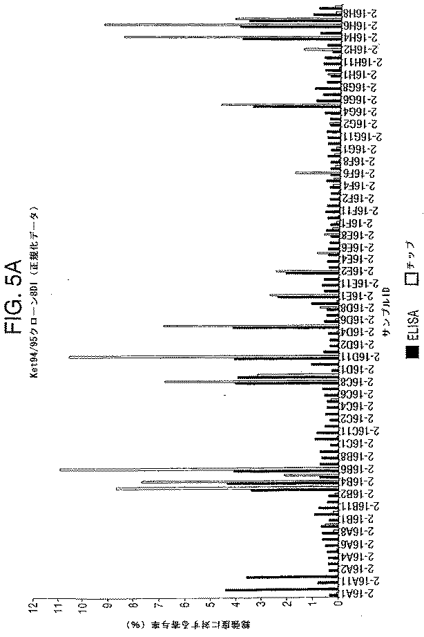
(ポストスクリーニング処理)
ポジティブウェル由来の細胞を、ウェルに再懸濁し、そして20μlを96ディープウェルプレート(これはあらかじめ1.5mlのHM20/HCFで満たされている)に移し、そして37℃、5%CO2で48時間、インキュベータに戻した。
(Post screening process)
Cells from positive wells are resuspended in the wells and 20 μl are transferred to a 96 deep well plate (pre-filled with 1.5 ml HM20 / HCF) and 48 ° C. at 37 ° C., 5% CO 2 . Returned to the incubator for hours.
1.4mlの培養物のそれぞれの上清を別のディープウェルプレートに移し、そして残りの100μlを使用して、細胞を再懸濁し、これを凍結バイアル(90%ウシ胎児血清/10%DMSO(Sigma)を含む)に移し、これから液体窒素貯蔵所に−80℃で2時間かけて移した。次いで、培養物上清を、生成したモノクローナル抗体の評価およびさらなる特徴付けのために使用した。 Transfer each supernatant of 1.4 ml culture to a separate deep well plate and use the remaining 100 μl to resuspend the cells and place them in frozen vials (90% fetal calf serum / 10% DMSO ( Sigma) and was then transferred to a liquid nitrogen storage at −80 ° C. for 2 hours. The culture supernatant was then used for evaluation and further characterization of the monoclonal antibody produced.
(結果)
マイクロアレイスクリーニングの結果を、図1に示す。この図は、多数のポジティブモノクローナル抗体が、スライド上の候補抗原に結合して検出されたことを示す。緑色のスポットは、候補抗原に結合したIgGモノクローナル抗体であり、一方、赤色のスポットは、候補抗原に結合したIgMモノクローナル抗体である。従って、図1は、本発明の方法を使用して、候補抗原に結合するモノクローナル抗体と、これらのモノクローナル抗体のアイソタイプとを同時に同定することができることを示す。
(result)
The results of microarray screening are shown in FIG. This figure shows that a number of positive monoclonal antibodies were detected bound to the candidate antigen on the slide. Green spots are IgG monoclonal antibodies bound to candidate antigens, while red spots are IgM monoclonal antibodies bound to candidate antigens. Thus, FIG. 1 shows that the methods of the invention can be used to simultaneously identify monoclonal antibodies that bind to candidate antigens and the isotypes of these monoclonal antibodies.
タンパク質チップの代わりにELISAを使用して比較実験を行った場合、図2に示されるように、マイクロアレイスクリーニングにおいて候補抗原に結合すると同定された多数のモノクローナル抗体は、ELISAを使用して候補抗原に結合すると同定されなかった(マイクロアレイの結果とELISAの結果との比較を参照のこと)。マイクロアレイスクリーニングを使用してネガティブであることが見出された上清(Ia:0.234)はまた、ELISAを使用してもネガティブリブであることが見出された。しかし、マイクロアレイスクリーニングを使用してポジティブであることが見出された4つの上清(Ia:5.18、1.96、3.64および2.02)のうち、わずか2つしか、ELISAを使用してポジティブであることが見出されなかった(図2を参照のこと)。ELISA実験においてネガティブリブであった上清が、より正確かつ高感度のマイクロアレイアプローチによって検出された場合、どのように実際のポジティブサンプルになり得るかに注意することが重要である。 When a comparative experiment was performed using an ELISA instead of a protein chip, as shown in FIG. 2, a number of monoclonal antibodies identified as binding to the candidate antigen in the microarray screening were identified as candidate antigens using the ELISA. Not identified as binding (see comparison of microarray and ELISA results). The supernatant that was found to be negative using microarray screening (Ia: 0.234) was also found to be a negative rib using ELISA. However, only two of the four supernatants that were found positive using microarray screening (Ia: 5.18, 1.96, 3.64 and 2.02) were subject to ELISA. It was not found to be positive using (see Figure 2). It is important to note how supernatants that were negative ribs in ELISA experiments can become actual positive samples if detected by a more accurate and sensitive microarray approach.
(実施例2:9種の抗原を用いる免疫)
(免疫)
マウスに、25μgの9種の抗原(抗原B5とKet94/95との融合物25μgを含む)を注射した。各抗原は、10μgのCpG DNAと混合され、そしてアルミニウムアジュバント(Pierce製のImject Alum)に吸着されている。各抗原の半分を、腹腔内投与し、半分を皮下投与した。
(Example 2: Immunization using 9 kinds of antigens)
(Immunity)
Mice were injected with 25 μg of 9 antigens (including 25 μg of fusion of antigen B5 and Ket94 / 95). Each antigen is mixed with 10 μg of CpG DNA and adsorbed on aluminum adjuvant (Image Alum from Pierce). Half of each antigen was administered intraperitoneally and half was administered subcutaneously.
このマウスを、21日後に、10μgのCpG DNAと混合しアルミニウムアジュバントに吸着させた各抗原10μgでブーストし、ここで、この抗原の半分を腹腔内投与し、半分を皮下投与した。 The mice were boosted 21 days later with 10 μg of each antigen mixed with 10 μg CpG DNA and adsorbed to aluminum adjuvant, where half of the antigen was administered intraperitoneally and half was administered subcutaneously.
ブーストの5日後、脾臓を取り出した。 Five days after the boost, the spleen was removed.
(融合および細胞培養)
融合および細胞培養を、実施例1に記載されるようにして実施した。
(Fusion and cell culture)
Fusion and cell culture were performed as described in Example 1.
(B5およびKet94/95に対する抗体のマイクロアレイスクリーニング)
アミノシラン被覆ガラスを、5μgの精製B5を含む40μlのddH2Oを滴下することによって、精製B5で均一に被覆し、そして室温の湿潤チャンバ中で60分間、20*60mmのカバーガラスで覆った。同じ手順を使用して、精製Ket94/95で均一に被覆されたアミノシランガラススライドを作製した。
(Microarray screening of antibodies against B5 and Ket94 / 95)
The aminosilane-coated glass was uniformly coated with purified B5 by dropwise addition of 40 μl ddH 2 O containing 5 μg purified B5 and covered with a 20 * 60 mm cover glass for 60 minutes in a room temperature humid chamber. The same procedure was used to make aminosilane glass slides uniformly coated with purified Ket94 / 95.
これらの被覆スライドを、PBS中3%のBSAをPBS中5%のミルクの代わりに使用してスライドをブロックしたこと以外は実施例1に記載されるようにして、リンスし、ブロックし、洗浄し、そして乾燥した。 These coated slides were rinsed, blocked and washed as described in Example 1 except that 3% BSA in PBS was used instead of 5% milk in PBS to block the slides. And dried.
培養物上清を、実施例1に記載されるように、384ウェルプレートに固定し、そしてB5で被覆したスライドおよびKet94/95で被覆したスライドに、三連で、約16000スポット/チップの密度、約150μmのスポットサイズで、Midrogrid
II 610マイクロアレイプリンター(ApogentDiscoveries)を使用してプリントした。
Culture supernatants were fixed to 384 well plates as described in Example 1 and triplicate on B5 coated slides and Ket94 / 95 coated slides at a density of about 16000 spots / chip. With a spot size of about 150 μm, Midgrid
Printing was done using an II 610 microarray printer (ApogenDiscoveries).
これらのマイクロアレイチップをインキュベートし、そしてCy3結合体化ヤギ抗マウスIgG特異的抗体およびCy5結合体化ヤギ抗マウスIgM特異的抗体を、実施例1に記載されるようにして、これらにチップに塗布した。 These microarray chips were incubated and Cy3-conjugated goat anti-mouse IgG specific antibody and Cy5-conjugated goat anti-mouse IgM specific antibody were applied to the chip as described in Example 1 did.
(ヒットピッキング)
これらのチップを、実施例1と同じ手順を使用してスキャンした。平均補正強度(Ia)を、2つの培養物上清のセットの各々について計算した。
(Hit picking)
These chips were scanned using the same procedure as in Example 1. Average corrected intensity (Ia) was calculated for each of the two sets of culture supernatants.
(マイクロアレイスクリーニングとELISAの比較)
比較実験を実施した。ここで、各培養物上清をELISAによって試験した。各培養物上清を、B5抗原またはKet94−95抗原200ngを含むウェルに添加し、そしてこの培養物上清中の抗原に結合するモノクローナル抗体の存在を、従来のELISAを使用して検出した。
(Comparison between microarray screening and ELISA)
A comparative experiment was performed. Here, each culture supernatant was tested by ELISA. Each culture supernatant was added to wells containing 200 ng of B5 antigen or Ket94-95 antigen, and the presence of monoclonal antibodies binding to the antigen in the culture supernatant was detected using a conventional ELISA.
ELISAを使用して5376個の培養物上清をスクリーニングし、5376個の結果(各上清について1つ)を作成するには、1週間必要であった。対照的に、マイクロアレイチップを使用して三連で同じ3576個の培養物上清をスクリーニングし、16128個の結果を作成するのに48時間かかった。このことは、ELISAと比較して、マイクロアレイスクリーニングが効率的であることを示す。 One week was required to screen 5376 culture supernatants using ELISA and generate 5376 results (one for each supernatant). In contrast, the same 3576 culture supernatants were screened in triplicate using a microarray chip and it took 48 hours to generate 16128 results. This indicates that microarray screening is more efficient compared to ELISA.
さらに、わずか5μgのB5抗原および5μgのKet94/95しか、マイクロアレイスクリーニングを行うのに必要ではなかった(1チップあたり5μgの抗原)。対照的に、96μgの抗原が、各ELISAプレートに必要であり、そして5個のELISAプレートが、各抗原をスクリーニングする必要があった。従って、ELISAによるスクリーニングは、マイクロアレイスクリーニングより有意に費用がかかりかつ時間がかかった。 Furthermore, only 5 μg of B5 antigen and 5 μg of Ket94 / 95 were required to perform microarray screening (5 μg of antigen per chip). In contrast, 96 μg of antigen was required for each ELISA plate, and 5 ELISA plates were required to screen each antigen. Thus, screening by ELISA was significantly more expensive and time consuming than microarray screening.
各ELISAプレートについて得られたデータを正規化して、各培養物上清の全強度に対する寄与%を得た。マイクロアレイスクリーニングによって得られた同じ培養物上清の平均複製強度もまた正規化して、ELISAデータとの比較を可能にした。 The data obtained for each ELISA plate was normalized to obtain a% contribution to the total intensity of each culture supernatant. The average replication intensity of the same culture supernatant obtained by microarray screening was also normalized to allow comparison with ELISA data.
培養物上清を、B5に結合するモノクローナル抗体について、ELISAを使用してスクリーニングする場合、53個のポジティブな上清が5個のELISAプレートにおいて同定された。マイクロアレイスクリーニングを使用する同じ培養物上清のスクリーニングは、48個のポジティブ上清(ELISAによって同定された上清の90.57%)を同定した。マイクロアレイスクリーニングはまた、ELISAによって同定されなかった4つの新規のポジティブ上清を同定した。 When the culture supernatant was screened using ELISA for monoclonal antibodies that bind to B5, 53 positive supernatants were identified in 5 ELISA plates. Screening the same culture supernatant using microarray screening identified 48 positive supernatants (90.57% of the supernatant identified by ELISA). Microarray screening also identified four new positive supernatants that were not identified by ELISA.
培養物上清を、Ket94/95に結合するモノクローナル抗体について、ELISAを使用してスクリーニングする場合、15個のポジティブ上清が、単一のELISAプレートにおいて同定された。マイクロアレイスクリーニングを使用する同じ培養物上清のスクリーニングは、13個のポジティブ上清(ELISAによって同定された上清の88.66%)を同定した。マイクロアレイスクリーニングはまた、ELISAによって同定されなかった8個の新規なポジティブ上清を同定した。 When culture supernatants were screened using ELISA for monoclonal antibodies that bind to Ket94 / 95, 15 positive supernatants were identified in a single ELISA plate. Screening the same culture supernatant using microarray screening identified 13 positive supernatants (88.66% of the supernatant identified by ELISA). Microarray screening also identified 8 novel positive supernatants that were not identified by ELISA.
これらの結果から、マイクロアレイスクリーニングが、特異的抗原に結合するモノクローナル抗体の同定において、少なくともELISAと同程度に有効であることが明らかである。実際、ELISAによって同定されないポジティブ上清の同定は、マイクロアレイスクリーニングがELISAより高感度であることを示唆する。マイクロアレイスクリーニングはさらに、同定されたモノクローナル抗体のIGアイソタイプまたはIgMアイソタイプの同時決定を可能にするという利点を有する。 From these results, it is clear that microarray screening is at least as effective as ELISA in identifying monoclonal antibodies that bind to specific antigens. Indeed, the identification of positive supernatants not identified by ELISA suggests that microarray screening is more sensitive than ELISA. Microarray screening further has the advantage of allowing simultaneous determination of the IG or IgM isotype of the identified monoclonal antibodies.
図3Aは、B5に結合するポジティブサンプルを含むELISAプレート中の各培養物上清の全強度に対する寄与%の正規化した値(黒四角)を、B5被覆スライドのマイクロアレイスクリーニングによって得られた同じ培養物上清の寄与%の正規化した値(白四角)と比較して示す。図3Bは、これらの実験におけるバックグラウンドノイズのレベルを示す。ポジティブ上清が、ELISAと比較して、マイクロアレイスクリーニングを使用して、全強度に対するよろい大きな寄与%を示したことが分かり得る。結果として、ELISAと比較してマイクロアレイスクリーニングにおいて、バックグラウンドノイズとポジティブ上清との間により大きな差異が存在し、これによりポジティブ上清がより容易にかつより正確に同定され得る。 FIG. 3A shows normalized values (black squares) of% contribution to total intensity of each culture supernatant in an ELISA plate containing positive samples that bind to B5, the same culture obtained by microarray screening of B5 coated slides. It is shown in comparison with the normalized value (white square) of the contribution percentage of the supernatant. FIG. 3B shows the level of background noise in these experiments. It can be seen that the positive supernatant showed a significant contribution to the total intensity using microarray screening compared to the ELISA. As a result, there is a greater difference between background noise and positive supernatant in microarray screening compared to ELISA, which allows positive supernatants to be more easily and more accurately identified.
図4Aは、B5に結合する単一のポジティブサンプルを含むELISAプレートにおける各培養物上清の全強度に対する寄与%の正規化した値(黒四角)と、B5被覆スライドのマイクロアレイスクリーニングによって得られた同じ培養物上清の寄与%の正規化した値(白四角)を比較する。バックグラウンドノイズレベルは、図4Bに示され、そしてポジティブサンプルが、ELISAと比較して、マイクロアレイスクリーニングを使用して、バックグラウンドノイズより上でより容易に検出可能であることが分かり得る。 FIG. 4A was obtained by normalized values (black squares) of% contribution to the total intensity of each culture supernatant in an ELISA plate containing a single positive sample that binds to B5 and microarray screening of B5 coated slides. Compare the normalized values (open squares) of the% contribution of the same culture supernatant. The background noise level is shown in FIG. 4B, and it can be seen that positive samples are more easily detectable above background noise using microarray screening compared to ELISA.
図5Aは、Ket94/95に結合するポジティブ上清を含むことが見出されたELISAプレート中の各培養物上清の全強度に対する寄与%の正規化した値(黒四角)を、Ket94/95被覆スライドのマイクロアレイスクリーニングによって得られた同じ培養物上清の寄与%の正規化した値(白四角)と比較する。これらのポジティブ上清は、図5Bに示されるように、ELISAと比較して、マイクロアレイスクリーニングを使用してバックグラウンドノイズより上で容易に検出可能であった。 FIG. 5A shows normalized values (black squares) of% contribution to the total intensity of each culture supernatant in an ELISA plate found to contain positive supernatants that bind to Ket94 / 95. Compare with normalized values (open squares) of% contribution of the same culture supernatant obtained by microarray screening of coated slides. These positive supernatants were readily detectable above background noise using microarray screening compared to ELISA, as shown in FIG. 5B.
図6Aは、ポジティブ上清が存在しないELISAプレートから得られたデータ(黒四角)を、同じ培養物上清のマイクロアレイスクリーニングを使用して得られたデータ(白四角)と比較する。図6Bに示されるように、両方の事例の読み取りは、バックグラウンドノイズに起因した。 FIG. 6A compares data obtained from ELISA plates without black supernatant (black squares) with data obtained using microarray screening of the same culture supernatant (white squares). As shown in FIG. 6B, the reading in both cases was due to background noise.
これらの結果は、本発明の方法が、1つより多くの抗原に対するモノクローナル抗体を同時に同定するために使用され得ることを実証する。本発明の方法におけるマイクロアレイスクリーニングの使用は、ELISAのような従来の抗体スクリーニング方法の使用よりも、迅速で、安価でかつより正確である。 These results demonstrate that the method of the invention can be used to simultaneously identify monoclonal antibodies against more than one antigen. The use of microarray screening in the methods of the present invention is faster, cheaper and more accurate than the use of conventional antibody screening methods such as ELISA.
(参考文献)
Kilpatrickら(1997)Hybridoma 16:381−389
Kohler G,Milstein C(1975)Nature 7:256:485−7
Lindleyら(2000)J Immunol Methods 234:123−135
Ziaudin J,Sabatini D(2001)Nature 411:107−110
Zimmermann U.(1990)J Immunol Methods Nov
6;134(1):43−50。
(References)
Kilpatrick et al. (1997) Hybridoma 16: 381-389.
Kohler G, Milstein C (1975) Nature 7: 256: 485-7
Lindley et al. (2000) J Immunol Methods 234: 123-135.
Ziaudin J, Sabatini D (2001) Nature 411: 107-110.
Zimmermann U. (1990) J Immunol Methods Nov
6; 134 (1): 43-50.
Claims (6)
a)該候補抗原が提示される1つ以上のタンパク質チップに対して不死化細胞株の上清をスクリーニングする工程;およびa) screening the supernatant of an immortalized cell line against one or more protein chips on which the candidate antigen is presented; and
b)該候補抗原に結合する抗体を、モノクローナル抗体として選択する工程、b) selecting an antibody that binds to the candidate antigen as a monoclonal antibody;
を包含し、該方法は、該不死化細胞株が、複数の抗原に対する抗体を産生する抗体産生細胞の単一細胞懸濁液から生成されることを特徴とする、方法。And the method is characterized in that the immortalized cell line is generated from a single cell suspension of antibody-producing cells that produce antibodies against multiple antigens.
前記タンパク質チップにアイソタイプ特異的抗免疫グロブリン抗体を添加する工程であって、異なるアイソタイプ特異性を有する各抗免疫グロブリン抗体は、異なる標識を有する、工程、およびAdding an isotype-specific anti-immunoglobulin antibody to the protein chip, each anti-immunoglobulin antibody having a different isotype specificity having a different label, and
該標識の存在を検出する工程、Detecting the presence of the label;
を包含する、請求項4に記載の方法。The method of claim 4 comprising:
Applications Claiming Priority (2)
| Application Number | Priority Date | Filing Date | Title |
|---|---|---|---|
| GB0208817.7 | 2002-04-17 | ||
| GBGB0208817.7A GB0208817D0 (en) | 2002-04-17 | 2002-04-17 | Method for producing monoclonal antibodies |
Related Parent Applications (1)
| Application Number | Title | Priority Date | Filing Date |
|---|---|---|---|
| JP2003586190A Division JP4429736B2 (en) | 2002-04-17 | 2003-04-17 | Methods for producing monoclonal antibodies |
Publications (2)
| Publication Number | Publication Date |
|---|---|
| JP2010032547A true JP2010032547A (en) | 2010-02-12 |
| JP5006923B2 JP5006923B2 (en) | 2012-08-22 |
Family
ID=9935021
Family Applications (2)
| Application Number | Title | Priority Date | Filing Date |
|---|---|---|---|
| JP2003586190A Expired - Fee Related JP4429736B2 (en) | 2002-04-17 | 2003-04-17 | Methods for producing monoclonal antibodies |
| JP2009256591A Expired - Fee Related JP5006923B2 (en) | 2002-04-17 | 2009-11-09 | Methods for producing monoclonal antibodies |
Family Applications Before (1)
| Application Number | Title | Priority Date | Filing Date |
|---|---|---|---|
| JP2003586190A Expired - Fee Related JP4429736B2 (en) | 2002-04-17 | 2003-04-17 | Methods for producing monoclonal antibodies |
Country Status (14)
| Country | Link |
|---|---|
| US (2) | US20050181483A1 (en) |
| EP (1) | EP1506235B1 (en) |
| JP (2) | JP4429736B2 (en) |
| CN (1) | CN100402553C (en) |
| AT (1) | ATE363490T1 (en) |
| AU (1) | AU2003227873B2 (en) |
| CA (1) | CA2480717C (en) |
| DE (1) | DE60314132T2 (en) |
| DK (1) | DK1506235T3 (en) |
| ES (1) | ES2288214T3 (en) |
| GB (2) | GB0208817D0 (en) |
| IL (1) | IL164625A (en) |
| PT (1) | PT1506235E (en) |
| WO (1) | WO2003089471A1 (en) |
Families Citing this family (13)
| Publication number | Priority date | Publication date | Assignee | Title |
|---|---|---|---|---|
| WO2006117007A1 (en) * | 2005-05-03 | 2006-11-09 | The Antibody Project Aps | Methods for obtaining antigen specific antibodies |
| US8859209B2 (en) | 2006-01-12 | 2014-10-14 | Carviar Aps | Reimmunization and antibody design |
| DK2027158T3 (en) | 2006-05-02 | 2013-01-14 | Carviar Aps | Method of immunizing a bird species |
| GB0609119D0 (en) * | 2006-05-09 | 2006-06-21 | Univ Birmingham | Histones |
| PL2865387T3 (en) | 2008-11-21 | 2019-12-31 | Københavns Universitet (University Of Copenhagen) | Priming of an immune response |
| AU2010315547C1 (en) | 2009-10-26 | 2015-06-04 | Société des Produits Nestlé S.A. | Assays for the detection of anti-TNF drugs and autoantibodies |
| KR101934247B1 (en) * | 2010-10-18 | 2019-01-02 | 네스텍 소시에테아노님 | Methods for determining anti-drug antibody isotypes |
| SG10201605516YA (en) | 2011-07-06 | 2016-08-30 | Nestec Sa | Assays for detecting neutralizing autoantibodies to biologic therapy with tnf alpha |
| US20150110802A1 (en) * | 2012-02-29 | 2015-04-23 | Medlmmune, Llc | Antibody production methods |
| CN103255128A (en) * | 2013-04-27 | 2013-08-21 | 中国人民解放军军事医学科学院放射与辐射医学研究所 | Methods for preparing monoclonal antibody and hybridoma cell strain thereof by multiple antigens in immune high-flux manner |
| CN103808922B (en) * | 2013-04-27 | 2015-09-09 | 无锡国盛生物工程有限公司 | A kind of screening technique of hybridoma supernatant stage trace antibody |
| WO2016088104A2 (en) | 2014-12-05 | 2016-06-09 | Nestec S.A. | Indirect homogeneous mobility shift assays for the detection of biologics in patient samples |
| CN114324898B (en) * | 2022-03-11 | 2022-05-31 | 南京岚煜生物科技有限公司 | Binding pad treatment solution for heparin binding protein HBP detection |
Citations (2)
| Publication number | Priority date | Publication date | Assignee | Title |
|---|---|---|---|---|
| WO1996033735A1 (en) * | 1995-04-27 | 1996-10-31 | Abgenix, Inc. | Human antibodies derived from immunized xenomice |
| WO2001014425A1 (en) * | 1999-08-19 | 2001-03-01 | Diachip Limited | Multipurpose diagnostic systems using protein chips |
Family Cites Families (17)
| Publication number | Priority date | Publication date | Assignee | Title |
|---|---|---|---|---|
| GB1419981A (en) * | 1973-01-09 | 1976-01-07 | Halifax Tool Co Ltd | Percussion drills |
| US4102534A (en) * | 1977-01-10 | 1978-07-25 | Ajax Tool Works, Inc. | Pneumatic hammer |
| US4821812A (en) * | 1987-05-27 | 1989-04-18 | Ingersoll-Rand Company | Down hole drill improvement |
| US6150584A (en) * | 1990-01-12 | 2000-11-21 | Abgenix, Inc. | Human antibodies derived from immunized xenomice |
| US6075181A (en) * | 1990-01-12 | 2000-06-13 | Abgenix, Inc. | Human antibodies derived from immunized xenomice |
| US5989846A (en) * | 1992-07-31 | 1999-11-23 | Rutgers, The State University Of New Jersey | Assays to identify inducers of plant defense resistance |
| US5277260A (en) * | 1993-02-24 | 1994-01-11 | Ranck Gerald L | Air hammer |
| GB9326238D0 (en) * | 1993-12-23 | 1994-02-23 | Sinvent As | Method of assay |
| US6720149B1 (en) * | 1995-06-07 | 2004-04-13 | Affymetrix, Inc. | Methods for concurrently processing multiple biological chip assays |
| CA2232344C (en) * | 1995-10-20 | 2005-04-05 | University Of Nebraska Board Of Regents | Compositions and methods for enhancing immune responses mediated by antigen-presenting cells |
| US6252050B1 (en) * | 1998-06-12 | 2001-06-26 | Genentech, Inc. | Method for making monoclonal antibodies and cross-reactive antibodies obtainable by the method |
| CN1274085A (en) * | 2000-04-13 | 2000-11-22 | 陈学银 | Protein chip, its preparing process and its application in screening monoclonal antibody |
| WO2001083827A1 (en) * | 2000-05-04 | 2001-11-08 | Yale University | High density protein arrays for screening of protein activity |
| WO2002014866A2 (en) * | 2000-08-11 | 2002-02-21 | Qianjin Hu | Methods and universal monoclonal antibody array |
| US20030044849A1 (en) * | 2001-08-22 | 2003-03-06 | Steven Kessler | Methods for screening monoclonal antibodies on heterogeneous antigen substrates |
| NZ531211A (en) * | 2001-08-23 | 2007-04-27 | Genmad As | Human antibodies specific for interleukin 15 (IL-15) |
| WO2003019192A1 (en) * | 2001-08-27 | 2003-03-06 | The Trustees Of Boston University | Apparatus, composition and method for proteome profiling |
-
2002
- 2002-04-17 GB GBGB0208817.7A patent/GB0208817D0/en not_active Ceased
-
2003
- 2003-04-17 CA CA2480717A patent/CA2480717C/en not_active Expired - Fee Related
- 2003-04-17 ES ES03725333T patent/ES2288214T3/en not_active Expired - Lifetime
- 2003-04-17 US US10/511,148 patent/US20050181483A1/en not_active Abandoned
- 2003-04-17 DK DK03725333T patent/DK1506235T3/en active
- 2003-04-17 DE DE60314132T patent/DE60314132T2/en not_active Expired - Lifetime
- 2003-04-17 EP EP03725333A patent/EP1506235B1/en not_active Expired - Lifetime
- 2003-04-17 AU AU2003227873A patent/AU2003227873B2/en not_active Ceased
- 2003-04-17 PT PT03725333T patent/PT1506235E/en unknown
- 2003-04-17 CN CNB038133539A patent/CN100402553C/en not_active Expired - Fee Related
- 2003-04-17 AT AT03725333T patent/ATE363490T1/en active
- 2003-04-17 WO PCT/GB2003/001684 patent/WO2003089471A1/en active IP Right Grant
- 2003-04-17 JP JP2003586190A patent/JP4429736B2/en not_active Expired - Fee Related
- 2003-04-17 GB GB0308976A patent/GB2387847B/en not_active Expired - Lifetime
-
2004
- 2004-10-14 IL IL164625A patent/IL164625A/en not_active IP Right Cessation
-
2009
- 2009-11-09 JP JP2009256591A patent/JP5006923B2/en not_active Expired - Fee Related
-
2011
- 2011-05-23 US US13/113,927 patent/US20110287960A1/en not_active Abandoned
Patent Citations (2)
| Publication number | Priority date | Publication date | Assignee | Title |
|---|---|---|---|---|
| WO1996033735A1 (en) * | 1995-04-27 | 1996-10-31 | Abgenix, Inc. | Human antibodies derived from immunized xenomice |
| WO2001014425A1 (en) * | 1999-08-19 | 2001-03-01 | Diachip Limited | Multipurpose diagnostic systems using protein chips |
Also Published As
| Publication number | Publication date |
|---|---|
| CA2480717A1 (en) | 2003-10-30 |
| JP2006500912A (en) | 2006-01-12 |
| EP1506235B1 (en) | 2007-05-30 |
| EP1506235A1 (en) | 2005-02-16 |
| DE60314132T2 (en) | 2008-01-17 |
| ES2288214T3 (en) | 2008-01-01 |
| AU2003227873B2 (en) | 2008-12-11 |
| US20110287960A1 (en) | 2011-11-24 |
| AU2003227873A1 (en) | 2003-11-03 |
| JP5006923B2 (en) | 2012-08-22 |
| DE60314132D1 (en) | 2007-07-12 |
| CN100402553C (en) | 2008-07-16 |
| GB0208817D0 (en) | 2002-05-29 |
| CN1659185A (en) | 2005-08-24 |
| GB2387847B (en) | 2005-07-27 |
| US20050181483A1 (en) | 2005-08-18 |
| JP4429736B2 (en) | 2010-03-10 |
| DK1506235T3 (en) | 2007-09-24 |
| CA2480717C (en) | 2012-08-21 |
| WO2003089471A1 (en) | 2003-10-30 |
| GB0308976D0 (en) | 2003-05-28 |
| IL164625A (en) | 2010-04-29 |
| PT1506235E (en) | 2007-08-29 |
| IL164625A0 (en) | 2005-12-18 |
| ATE363490T1 (en) | 2007-06-15 |
| GB2387847A (en) | 2003-10-29 |
Similar Documents
| Publication | Publication Date | Title |
|---|---|---|
| JP5006923B2 (en) | Methods for producing monoclonal antibodies | |
| US5595915A (en) | Method of determining antigenically active amino acid sequences | |
| Wang et al. | Review on the identification and role of Toxoplasma gondii antigenic epitopes | |
| KR20090039707A (en) | Antibodies for norovirus | |
| Dechamma et al. | Identification of T-helper and linear B epitope in the hypervariable region of nucleocapsid protein of PPRV and its use in the development of specific antibodies to detect viral antigen | |
| ES2581880T3 (en) | Method for differentiating infected animals from vaccinated animals using Ehrlichia canis polypeptides | |
| JP2008505106A (en) | High throughput proteomics | |
| JP2004512529A (en) | Method for in vivo identification of intracellular epitopes | |
| US7608277B2 (en) | Tuberculosis nucleic acids, polypeptides and immunogenic compositions | |
| Tafi et al. | Identification of HCV core mimotopes: improved methods for the selection and use of disease-related phage-displayed peptides | |
| US20060222658A1 (en) | Vaccinia virus nucleic acids, polypeptides and immunogenic compositions | |
| Xin et al. | A subtractive fluorescence-activated cell-sorting strategy to identify mimotopes of HBV–preS protein from bacterially displayed peptide library | |
| JP2003515340A (en) | Antigenic protein LppQ of Mycoplasma mycoides subsp. Mycoides SC, its preparation and use | |
| US20060128628A1 (en) | Human tissue antigen-binding peptides and their amino acid sequences | |
| US6800462B2 (en) | Production of recombinant proteins in vivo and use for generating antibodies | |
| EP1144002A2 (en) | A method for selecting and producing vaccine and diagnostic preparations | |
| JP2011072284A (en) | Method for producing antibody and immunogenic composition usable therefor | |
| Chiroscience et al. | Session 3: Emerging technologies | |
| Bedian | Monoclonal antibodies in the analysis of embryonic development |
Legal Events
| Date | Code | Title | Description |
|---|---|---|---|
| A621 | Written request for application examination |
Free format text: JAPANESE INTERMEDIATE CODE: A621 Effective date: 20091109 |
|
| A521 | Request for written amendment filed |
Free format text: JAPANESE INTERMEDIATE CODE: A523 Effective date: 20101027 |
|
| A131 | Notification of reasons for refusal |
Free format text: JAPANESE INTERMEDIATE CODE: A131 Effective date: 20120127 |
|
| TRDD | Decision of grant or rejection written | ||
| A521 | Request for written amendment filed |
Free format text: JAPANESE INTERMEDIATE CODE: A821 Effective date: 20120411 |
|
| A01 | Written decision to grant a patent or to grant a registration (utility model) |
Free format text: JAPANESE INTERMEDIATE CODE: A01 Effective date: 20120507 |
|
| A01 | Written decision to grant a patent or to grant a registration (utility model) |
Free format text: JAPANESE INTERMEDIATE CODE: A01 |
|
| A61 | First payment of annual fees (during grant procedure) |
Free format text: JAPANESE INTERMEDIATE CODE: A61 Effective date: 20120525 |
|
| FPAY | Renewal fee payment (event date is renewal date of database) |
Free format text: PAYMENT UNTIL: 20150601 Year of fee payment: 3 |
|
| R150 | Certificate of patent or registration of utility model |
Ref document number: 5006923 Country of ref document: JP Free format text: JAPANESE INTERMEDIATE CODE: R150 Free format text: JAPANESE INTERMEDIATE CODE: R150 |
|
| R250 | Receipt of annual fees |
Free format text: JAPANESE INTERMEDIATE CODE: R250 |
|
| R250 | Receipt of annual fees |
Free format text: JAPANESE INTERMEDIATE CODE: R250 |
|
| R250 | Receipt of annual fees |
Free format text: JAPANESE INTERMEDIATE CODE: R250 |
|
| R250 | Receipt of annual fees |
Free format text: JAPANESE INTERMEDIATE CODE: R250 |
|
| R250 | Receipt of annual fees |
Free format text: JAPANESE INTERMEDIATE CODE: R250 |
|
| R250 | Receipt of annual fees |
Free format text: JAPANESE INTERMEDIATE CODE: R250 |
|
| LAPS | Cancellation because of no payment of annual fees |