JP2009531120A - Soft tissue bulking - Google Patents
Soft tissue bulking Download PDFInfo
- Publication number
- JP2009531120A JP2009531120A JP2009502317A JP2009502317A JP2009531120A JP 2009531120 A JP2009531120 A JP 2009531120A JP 2009502317 A JP2009502317 A JP 2009502317A JP 2009502317 A JP2009502317 A JP 2009502317A JP 2009531120 A JP2009531120 A JP 2009531120A
- Authority
- JP
- Japan
- Prior art keywords
- particle
- particles
- soft tissue
- bulking
- carrier medium
- Prior art date
- Legal status (The legal status is an assumption and is not a legal conclusion. Google has not performed a legal analysis and makes no representation as to the accuracy of the status listed.)
- Withdrawn
Links
Images


Classifications
-
- A—HUMAN NECESSITIES
- A61—MEDICAL OR VETERINARY SCIENCE; HYGIENE
- A61L—METHODS OR APPARATUS FOR STERILISING MATERIALS OR OBJECTS IN GENERAL; DISINFECTION, STERILISATION OR DEODORISATION OF AIR; CHEMICAL ASPECTS OF BANDAGES, DRESSINGS, ABSORBENT PADS OR SURGICAL ARTICLES; MATERIALS FOR BANDAGES, DRESSINGS, ABSORBENT PADS OR SURGICAL ARTICLES
- A61L27/00—Materials for grafts or prostheses or for coating grafts or prostheses
- A61L27/40—Composite materials, i.e. containing one material dispersed in a matrix of the same or different material
- A61L27/44—Composite materials, i.e. containing one material dispersed in a matrix of the same or different material having a macromolecular matrix
- A61L27/48—Composite materials, i.e. containing one material dispersed in a matrix of the same or different material having a macromolecular matrix with macromolecular fillers
-
- A—HUMAN NECESSITIES
- A61—MEDICAL OR VETERINARY SCIENCE; HYGIENE
- A61L—METHODS OR APPARATUS FOR STERILISING MATERIALS OR OBJECTS IN GENERAL; DISINFECTION, STERILISATION OR DEODORISATION OF AIR; CHEMICAL ASPECTS OF BANDAGES, DRESSINGS, ABSORBENT PADS OR SURGICAL ARTICLES; MATERIALS FOR BANDAGES, DRESSINGS, ABSORBENT PADS OR SURGICAL ARTICLES
- A61L27/00—Materials for grafts or prostheses or for coating grafts or prostheses
- A61L27/50—Materials characterised by their function or physical properties, e.g. injectable or lubricating compositions, shape-memory materials, surface modified materials
- A61L27/54—Biologically active materials, e.g. therapeutic substances
-
- A—HUMAN NECESSITIES
- A61—MEDICAL OR VETERINARY SCIENCE; HYGIENE
- A61L—METHODS OR APPARATUS FOR STERILISING MATERIALS OR OBJECTS IN GENERAL; DISINFECTION, STERILISATION OR DEODORISATION OF AIR; CHEMICAL ASPECTS OF BANDAGES, DRESSINGS, ABSORBENT PADS OR SURGICAL ARTICLES; MATERIALS FOR BANDAGES, DRESSINGS, ABSORBENT PADS OR SURGICAL ARTICLES
- A61L27/00—Materials for grafts or prostheses or for coating grafts or prostheses
- A61L27/50—Materials characterised by their function or physical properties, e.g. injectable or lubricating compositions, shape-memory materials, surface modified materials
- A61L27/58—Materials at least partially resorbable by the body
-
- A—HUMAN NECESSITIES
- A61—MEDICAL OR VETERINARY SCIENCE; HYGIENE
- A61L—METHODS OR APPARATUS FOR STERILISING MATERIALS OR OBJECTS IN GENERAL; DISINFECTION, STERILISATION OR DEODORISATION OF AIR; CHEMICAL ASPECTS OF BANDAGES, DRESSINGS, ABSORBENT PADS OR SURGICAL ARTICLES; MATERIALS FOR BANDAGES, DRESSINGS, ABSORBENT PADS OR SURGICAL ARTICLES
- A61L2300/00—Biologically active materials used in bandages, wound dressings, absorbent pads or medical devices
-
- A—HUMAN NECESSITIES
- A61—MEDICAL OR VETERINARY SCIENCE; HYGIENE
- A61L—METHODS OR APPARATUS FOR STERILISING MATERIALS OR OBJECTS IN GENERAL; DISINFECTION, STERILISATION OR DEODORISATION OF AIR; CHEMICAL ASPECTS OF BANDAGES, DRESSINGS, ABSORBENT PADS OR SURGICAL ARTICLES; MATERIALS FOR BANDAGES, DRESSINGS, ABSORBENT PADS OR SURGICAL ARTICLES
- A61L2430/00—Materials or treatment for tissue regeneration
- A61L2430/34—Materials or treatment for tissue regeneration for soft tissue reconstruction
Abstract
軟組織かさ増し物質は複数の粒子を含む。各粒子は、内腔を画定し、50μm〜250μmの最大外寸を有する球面状のポリマー殻を含む。殻内には、ポートまたは開口部が設けられている。したがって、ポートまたは開口部は該腔への進入路を提供する。ポートまたは開口部は、該粒子の外寸の10分の1から該粒子の外寸までの範囲のサイズまたは寸法を有する。 The soft tissue bulking material includes a plurality of particles. Each particle includes a spherical polymer shell that defines a lumen and has a maximum outer dimension of 50 μm to 250 μm. A port or opening is provided in the shell. Thus, the port or opening provides an access path to the cavity. The port or opening has a size or dimension that ranges from one-tenth of the outer dimension of the particle to the outer dimension of the particle.
Description
本発明は、軟組織のかさ増し(bulking)に関する。本発明は、特に、軟組織かさ増し物質、および注入可能な軟組織かさ増し組成物に関する。 The present invention relates to bulking soft tissue. The present invention particularly relates to soft tissue bulking materials and injectable soft tissue bulking compositions.
本発明の第1の局面によれば、複数の粒子を含み、該粒子の各々が、内腔を画定し、50μm〜250μmの最大外寸を有する球面状のポリマー殻であって、その殻内にポートまたは開口部を含み、したがって、該ポートまたは開口部は該腔への進入路を提供し、該ポートまたは開口部は、該粒子の外寸の10分の1から該粒子の外寸までの範囲のサイズまたは寸法を有する、軟組織かさ増し物質が提供される。 According to a first aspect of the present invention, a spherical polymer shell comprising a plurality of particles, each of which defines a lumen and has a maximum outer dimension of 50 μm to 250 μm, wherein And therefore the port or opening provides an access path to the cavity, the port or opening extending from one tenth of the outer dimension of the particle to the outer dimension of the particle Soft tissue bulking materials having a size or dimension in the range of are provided.
本発明のかさ増し物質は、胃食道逆流性疾患(GERD)、泌尿器系逆流性疾患、腹圧性尿失禁(SUI)、便失禁、皮膚のでこぼこの増大の処置、声帯ひだ麻痺の処置のための声帯ひだの増大などにおける軟組織かさ増し物質としての使用に適している。これは、本明細書において後述する担体媒体に懸濁し、かさ増しまたは増大を必要とする軟組織に注入することにより適用される。 The bulking substance of the present invention is used for the treatment of gastroesophageal reflux disease (GERD), urinary reflux disease, stress urinary incontinence (SUI), fecal incontinence, the treatment of bumpy skin, the treatment of vocal cord palsy It is suitable for use as a soft tissue bulking substance for increasing vocal folds. This is applied by suspending in a carrier medium as described later herein and injecting into soft tissue that requires bulking or augmentation.
内腔は、上記のようにして殻によって囲まれ、該腔への外部進入路を提供するポートまたは開口部を有する。 The lumen is surrounded by a shell as described above and has a port or opening that provides an external access path to the lumen.
該粒子の殻は、好ましくは、外径が50μm〜250μmであるように実質的に球形である。換言すると、粒子は、好ましくは、各々が単一の主要な大きなポートまたは開口部をその殻内に有する中空微小球である。微小球の直径は、典型的に約100μmであり得る。 The particle shell is preferably substantially spherical so that the outer diameter is between 50 μm and 250 μm. In other words, the particles are preferably hollow microspheres each having a single major large port or opening in its shell. The diameter of the microspheres can typically be about 100 μm.
該粒子のポートは、そのため実質的に円形であり得、したがって、そのサイズまたは直径は、該粒子の直径の10分の1から該粒子のそれぞれの直径までの範囲である。ポートの直径は、20μm〜100μmの範囲であり得る。例えば、ポート直径は約60μmであり得る。 The port of the particle can therefore be substantially circular, and therefore its size or diameter ranges from one-tenth of the particle diameter to the respective diameter of the particle. The port diameter can range from 20 μm to 100 μm. For example, the port diameter can be about 60 μm.
少なくとも一部の該粒子の殻はミクロ細孔を有し得る。ミクロ細孔は、<10μmの寸法、例えば<10μmの直径を有し得る。 At least some of the particle shells may have micropores. The micropores can have a dimension of <10 μm, for example a diameter of <10 μm.
少なくとも一部の該粒子の殻は、マクロ細孔を有し得る。マクロ細孔は、10μm〜50μmの寸法、例えば、直径を有し得る。 At least some of the particle shells may have macropores. Macropores can have a size, for example a diameter, of 10 μm to 50 μm.
本発明の一態様において、該粒子の殻の外表面積の20%未満、すなわち、存在する任意のミクロ-またはマクロ細孔の内表面積以外が、マクロ細孔に占められ得る、すなわち、微小球は、限定的なマクロ細孔性を有し得る。 In one embodiment of the invention, less than 20% of the outer surface area of the shell of the particles, i.e. other than the inner surface area of any micro- or macropores present, can be occupied by macropores, i.e. the microspheres are May have limited macroporosity.
殻は、ポリマーとの複合体として、またはポリマーに吸収された(あるいは結合された)状態で、リン酸カルシウム化合物、造影剤、治療剤、成長因子、自己血小板富化血漿、正常ヒト細胞、および自己幹細胞から選択される少なくとも1種類の添加剤を含み得る。 The shell is in a complex with or absorbed (or bound to) the polymer, calcium phosphate compound, contrast agent, therapeutic agent, growth factor, autologous platelet-rich plasma, normal human cells, and autologous stem cells At least one additive selected from:
本発明の第2の局面によれば、
(i) 上記の軟組織かさ増し物質;および
(ii) かさ増し物質粒子が懸濁された適合性の担体媒体
を含む注入可能な軟組織かさ増し組成物であって、軟組織内に注入することによる適用に適するような粘稠度を有する組成物が提供される。
According to a second aspect of the present invention,
(i) the soft tissue bulking material as described above; and
(ii) An injectable soft tissue bulking composition comprising a compatible carrier medium in which bulking material particles are suspended, the composition having a consistency suitable for application by injection into the soft tissue. Is provided.
担体媒体としては、溶媒に溶解させたコラーゲン、キトサン、アルギネート、ポリビニルピロリドン、シリコーン油、ゼラチン、脂肪、ヒアルロン酸、食塩水、水、血漿、水溶液、グリコール、中鎖トリグリセリド、グリセリド、グリセロール、B-グルカンおよびアガロース溶液、乳酸エチル、ヒドロキシプロピルメチルセルロース、ポロキサマーもしくはポリ(N-イソプロピルアクリルアミド)またはその誘導体から選択される担体媒体材料が挙げられ得る。 Carrier media include collagen, chitosan, alginate, polyvinylpyrrolidone, silicone oil, gelatin, fat, hyaluronic acid, saline, water, plasma, aqueous solution, glycol, medium chain triglycerides, glycerides, glycerol, B- Mention may be made of carrier medium materials selected from glucan and agarose solutions, ethyl lactate, hydroxypropylmethylcellulose, poloxamer or poly (N-isopropylacrylamide) or derivatives thereof.
溶媒に対する担体媒体材料の比は、1:1〜200:1(溶媒mlに対する担体媒体のmg)であり得る。 The ratio of carrier medium material to solvent can be 1: 1 to 200: 1 (mg carrier medium to ml solvent).
使用される担体媒体材料に応じて、水、酸または塩基など任意の適当な溶媒が使用され得る。したがって、コラーゲンが担体媒体として使用される場合、溶媒は酢酸であり得る。アルギネートが担体媒体として使用される場合、水酸化ナトリウムなどの塩基が溶媒として使用され得る。 Depending on the carrier medium material used, any suitable solvent such as water, acid or base can be used. Thus, when collagen is used as the carrier medium, the solvent can be acetic acid. When alginate is used as the carrier medium, a base such as sodium hydroxide can be used as the solvent.
担体媒体は、注入した場合などの高剪断下で低い粘性を可能にするが、注入後に注入された材料を安定化または懸濁するために高い粘性を有する、水に溶解させたヒアルロン酸(好ましくは非動物供給源由来)などの偽塑性液体であり得る。代わりに、担体媒体は、室温で液体であるが、軟組織内に注入されると、すなわち、ヒト身体条件では、例えば液体からゲルに相変化を受けるように適合されたものであってもよい。特に、担体媒体は、体温および/または体内pHで、すなわち、ヒト軟組織内への注入後、液体からゲルへの相変化を受けるように、温度および/またはpH応答性であり得る。好ましくは、高度に精製されたグルタルアルデヒド架橋ウシコラーゲンが担体媒体材料として使用される。その一例は、INAMED Aesthetics of Santa Barbara、California、USAによって製造され、C. R. Bard of Murray Hill、New Jersey、USAによって販売されているコラーゲン調製物である。 The carrier medium allows for low viscosity under high shear, such as when injected, but has a high viscosity to stabilize or suspend the injected material after injection, preferably hyaluronic acid dissolved in water (preferably May be a pseudoplastic liquid such as from non-animal sources. Alternatively, the carrier medium may be liquid at room temperature but adapted to undergo a phase change, eg, from liquid to gel, when injected into soft tissue, ie, in human body conditions. In particular, the carrier medium can be temperature and / or pH responsive to undergo a phase change from liquid to gel at body temperature and / or body pH, ie, after injection into human soft tissue. Preferably highly purified glutaraldehyde crosslinked bovine collagen is used as the carrier medium material. One example is a collagen preparation manufactured by INAMED Aesthetics of Santa Barbara, California, USA and sold by CR Bard of Murray Hill, New Jersey, USA.
組成物は、かさ増し物質および担体媒体を合わせることにより形成される。かさ増し物質は、好ましくは、担体媒体に添加される前に滅菌される。これは、25kGyの線量までγ-滅菌することによって行なわれ得る。担体媒体はまた、かさ増し物質が添加される前に滅菌され得る。コラーゲン水溶液は、インライン滅菌フィルター(0.22マイクロメートル)での濾過、および厳格な滅菌調製手順のいずれかによって滅菌され得る。 The composition is formed by combining the bulking material and the carrier medium. The bulking material is preferably sterilized before being added to the carrier medium. This can be done by γ-sterilizing to a dose of 25 kGy. The carrier medium can also be sterilized before the bulking material is added. The aqueous collagen solution can be sterilized by either filtration through an in-line sterilization filter (0.22 micrometers) and rigorous sterilization preparation procedures.
担体媒体は、造影剤、自己血小板富化血漿、正常ヒト細胞または自己幹細胞などの前述の添加剤を含み得る。 The carrier medium may contain the aforementioned additives such as contrast agents, autologous platelet-rich plasma, normal human cells or autologous stem cells.
軟組織かさ増し物質は、
(i) 細孔形成剤を溶媒に溶解させたポリマーの溶液に分散させ、油(O)相を形成する工程;
(ii) 油相を、水に溶解させた乳化剤/界面活性剤を含む水(W)相に添加し、水中油型エマルジョン(O/W)を形成する工程;または
(iii) 水に溶解させた乳化剤/界面活性剤を含む水相(W)を油相に添加し、エマルジョンを形成した後、このエマルジョンを、乳化剤/界面活性剤を含有する第2の油相に添加し、油中油中水型エマルジョン((W/O)/O)を形成する工程;
(iv) 適宜、工程(ii)または(iii)のエマルジョンに酸を添加する工程、ここで、該酸は細孔形成剤と反応し、それにより、該粒子の各々が、内腔を画定し、50μm〜250μmの最大外寸を有する球面状のポリマー殻を形成し、その殻内にポートまたは開口部を含み、したがって、該ポートまたは開口部は該腔への進入路を提供し、該ポートまたは開口部が、100μmの最大寸法および20μmの最小寸法を有する、
により調製され得る。
Soft tissue bulking substances
(i) a step of dispersing an pore-forming agent in a polymer solution dissolved in a solvent to form an oil (O) phase;
(ii) adding an oil phase to a water (W) phase containing an emulsifier / surfactant dissolved in water to form an oil-in-water emulsion (O / W); or
(iii) After the aqueous phase (W) containing the emulsifier / surfactant dissolved in water is added to the oil phase to form an emulsion, this emulsion is then added to the second oil phase containing the emulsifier / surfactant To form a water-in-oil-in-oil emulsion ((W / O) / O);
(iv) optionally adding an acid to the emulsion of step (ii) or (iii), wherein the acid reacts with a pore-forming agent so that each of the particles defines a lumen; Forming a spherical polymer shell having a maximum outer dimension of 50 μm to 250 μm and including a port or opening in the shell, the port or opening thus providing an access path to the cavity, the port Or the opening has a maximum dimension of 100 μm and a minimum dimension of 20 μm,
Can be prepared.
代わりに、軟組織かさ増し物質を、
(v) 水に溶解させた乳化剤/界面活性剤を含む水相(W)を、溶媒に溶解させたポリマーの溶液を含む油相(O)に添加し、(W/O)エマルジョンを形成し、細孔形成剤をこのエマルジョンに添加する工程;その後、
(vi) このエマルジョンを、水に溶解させた乳化剤/界面活性剤を含む水相(W)に添加して戻し、水中油中水型エマルジョン((W/O)/W)を形成し、次いで、
(vii) 適宜、((W/O)/W)エマルジョンに酸を添加する工程、ここで、該酸は細孔形成剤と反応し、それにより、該粒子の各々が、内腔を画定し、50μm〜250μmの最大外寸を有する球面状のポリマー殻を形成し、その殻内にポートまたは開口部を含み、したがって、該ポートまたは開口部は該腔への進入路を提供し、該ポートまたは開口部が、100μmの最大寸法および20μmの最小寸法を有する、
により調製してもよい。
Instead, a soft tissue bulking substance
(v) An aqueous phase (W) containing an emulsifier / surfactant dissolved in water is added to an oil phase (O) containing a solution of the polymer dissolved in a solvent to form a (W / O) emulsion. Adding a pore former to the emulsion;
(vi) This emulsion is added back to the aqueous phase (W) containing the emulsifier / surfactant dissolved in water to form a water-in-oil-in-water emulsion ((W / O) / W), then ,
(vii) optionally adding an acid to the ((W / O) / W) emulsion, where the acid reacts with a pore-forming agent so that each of the particles defines a lumen. Forming a spherical polymer shell having a maximum outer dimension of 50 μm to 250 μm and including a port or opening in the shell, the port or opening thus providing an access path to the cavity, the port Or the opening has a maximum dimension of 100 μm and a minimum dimension of 20 μm,
May be prepared.
したがって、細孔形成剤および酸は、酸がエマルジョンに添加されると、細孔形成剤と反応し、泡立ちまたは発泡が起こり、該粒子内にポートの形成をもたらすように選択され得る。 Thus, the pore former and acid can be selected such that when the acid is added to the emulsion, it reacts with the pore former, causing foaming or foaming, resulting in the formation of ports within the particles.
細孔形成剤は、炭酸カルシウム、炭酸ナトリウム、重炭酸ナトリウム、炭酸アンモニウム、重炭酸アンモニウム、インビボ使用に許容され得る硝酸塩、塩化ナトリウム、クエン酸ナトリウム、サッカロースおよびグルコースから選択され得る固形細孔形成剤またはポロジェン(porogen)であり得る。典型的に、重炭酸ナトリウムが使用される。しかしながら、代わりに、パーフルオロカーボンなどの不活性液体細孔形成剤を使用してもよい。しかしながら、使用されるポロジェンは、上記の物質に限定されない。 The pore-forming agent may be selected from calcium carbonate, sodium carbonate, sodium bicarbonate, ammonium carbonate, ammonium bicarbonate, nitrates acceptable for in vivo use, sodium chloride, sodium citrate, saccharose and glucose Or it can be a porogen. Typically sodium bicarbonate is used. However, an inert liquid pore former such as perfluorocarbon may be used instead. However, the porogen used is not limited to the above substances.
酸をエマルジョンに添加する工程は、適切な場合、すなわち、固体または揮発性有機溶媒の細孔形成剤またはポロジェンそれ自体が充分なガスを生成しない場合にのみ使用されることは認識されよう。化学的に不活性な液体細孔形成剤が使用される場合、酸添加しない。さらにまた、酸添加はある種の固形細孔形成剤が使用される場合、不要であり得る。例えば、重炭酸アンモニウムがポロジェンとして使用される場合、重炭酸アンモニウムが、酸が存在しなくても細孔形成が起こるのに充分に反応性であるため、酸添加は必要とされない。 It will be appreciated that the step of adding an acid to the emulsion is used only when appropriate, i.e. when the solid or volatile organic solvent pore former or porogen itself does not produce sufficient gas. If a chemically inert liquid pore former is used, no acid is added. Furthermore, acid addition may be unnecessary if certain solid pore formers are used. For example, when ammonium bicarbonate is used as the porogen, no acid addition is required because ammonium bicarbonate is sufficiently reactive to cause pore formation in the absence of acid.
細孔形成剤に対するポリマーの質量比は、1:5〜2:1、典型的に約1:2であり得る。 The weight ratio of polymer to pore former can be 1: 5 to 2: 1, typically about 1: 2.
固形細孔形成剤またはポロジェンが使用される場合、これは、0.01μm〜250μmの粒径範囲、典型的に約150μmを有し得る。 If a solid pore former or porogen is used, it can have a particle size range of 0.01 μm to 250 μm, typically about 150 μm.
細孔形成剤が溶液に添加された後、ポロジェン溶液混合物は、好ましくは、均質な分散が達成されるまで攪拌またはホモジナイズされる。 After the pore former is added to the solution, the porogen solution mixture is preferably stirred or homogenized until a homogeneous dispersion is achieved.
溶液中の溶媒に対するポリマーの比は、ポリマー溶解度の限界に応じて1:20〜1:5(溶媒mlに対するポリマーg)、典型的に約1:6であり得る。 The ratio of polymer to solvent in solution can be from 1:20 to 1: 5 (g polymer per ml of solvent), typically about 1: 6, depending on the limit of polymer solubility.
ポリマーは、ポリ(ε-カプロラクトン)、ポリラクチド、ポリグリコリド、ポリラクチド-コ-グリコリド、ポリ(ε-カプロラクトン)-コ-グリコリド、ポリヒドロキシブチレート、ポリヒドロキシバレレート、ポリブチロラクトン、ポリバレロラクトン、ポリ(エチレンカーボネート)、ポリ(エチレンテレフタレート)、ポリジオキサノン、ポリウレタン、ポリエチレングリコール、ポリメチルメタクリレート、ポリ酢酸ビニルおよびポリ(2-ヒドロキシエチルメタクリレート)から選択される合成ポリマー、またはコラーゲン、ヒアルロン酸、キトサン、フィブリンおよびアルギネートから選択される天然ポリマーであり得る。 Polymers include poly (ε-caprolactone), polylactide, polyglycolide, polylactide-co-glycolide, poly (ε-caprolactone) -co-glycolide, polyhydroxybutyrate, polyhydroxyvalerate, polybutyrolactone, polyvalerolactone, polyvalerolactone Synthetic polymers selected from (ethylene carbonate), poly (ethylene terephthalate), polydioxanone, polyurethane, polyethylene glycol, polymethyl methacrylate, polyvinyl acetate and poly (2-hydroxyethyl methacrylate), or collagen, hyaluronic acid, chitosan, fibrin And a natural polymer selected from alginate.
したがって、溶液は、例えば攪拌または均質化を使用し、ポリマーを溶媒に溶解することにより形成される。溶媒は、工程(ii)、(iii)または(v)において使用される水相の一部で予め飽和させてもよい。 Thus, a solution is formed by dissolving the polymer in a solvent using, for example, stirring or homogenization. The solvent may be presaturated with a portion of the aqueous phase used in step (ii), (iii) or (v).
溶媒は、芳香族炭化水素、塩素系溶媒 (ジクロロメタン、クロロホルム、CCl4など)、アルコール(ベンジルアルコール、ポリプロピレングリコール、n-ブタノールなど)、エステル(酢酸エチル、酢酸ブチル、安息香酸メチル、酢酸メチルなど)、または酢酸もしくはプロピオン酸などの有機酸などの水溶性の有機溶媒であり得る。 Solvents include aromatic hydrocarbons, chlorinated solvents (dichloromethane, chloroform, CCl4, etc.), alcohols (benzyl alcohol, polypropylene glycol, n-butanol, etc.), esters (ethyl acetate, butyl acetate, methyl benzoate, methyl acetate, etc.) Or a water-soluble organic solvent such as an organic acid such as acetic acid or propionic acid.
水相は、脱イオン水に対して乳化剤/界面活性剤を、1:10〜1:1000 (水mlに対して乳化剤/界面活性剤g)、典型的に約1:50の比で含み得る。 The aqueous phase may contain an emulsifier / surfactant relative to deionized water from 1:10 to 1: 1000 (emulsifier / surfactant g per ml of water), typically in a ratio of about 1:50. .
上記の工程(v)および(vi)において、使用される水相の構成または組成は同じであり得る。 In the above steps (v) and (vi), the composition or composition of the aqueous phase used can be the same.
乳化剤/界面活性剤は、ポリビニルアルコール、ゼラチン、ポリエチレングリコール、ドデシル硫酸ナトリウム、ポリソルベート、ポリビニルピロリドン、ポロキサマー、モノオレイン酸グリセリル、モノステアリン酸グリセリル、ポリオキシエチレンアルキルエーテル、ヒドロキシプロピルセルロース、ヒドロキシプロピルメチルセルロース、およびその混合物から選択され得る。 The emulsifier / surfactant is polyvinyl alcohol, gelatin, polyethylene glycol, sodium dodecyl sulfate, polysorbate, polyvinyl pyrrolidone, poloxamer, glyceryl monooleate, glyceryl monostearate, polyoxyethylene alkyl ether, hydroxypropyl cellulose, hydroxypropyl methylcellulose, And mixtures thereof.
水相は、乳化剤/界面活性剤が添加された後、乳化剤/界面活性剤が溶解されるまで、脱イオン水を攪拌または均質化することにより形成される。脱イオン水は、上記の工程(i)の溶液に使用される溶媒で予め飽和させてもよい。 The aqueous phase is formed by stirring or homogenizing deionized water after the emulsifier / surfactant has been added until the emulsifier / surfactant is dissolved. Deionized water may be presaturated with the solvent used in the solution of step (i) above.
したがって、第2の油相は、油および乳化剤/界面活性剤を含む。第2の油相の油は、他方または第1の油相の油、すなわち溶媒と異なる。第2の油相の油は、植物油、鉱物油、ホホバ油、アボカド油またはパーム核油であり得る。第2の油相における乳化剤/界面活性剤に対する油の容量比は、20:1〜2000:1 、典型的に約200:1であり得る。 Thus, the second oil phase comprises an oil and an emulsifier / surfactant. The second oil phase oil is different from the other or first oil phase oil, ie the solvent. The oil of the second oil phase can be vegetable oil, mineral oil, jojoba oil, avocado oil or palm kernel oil. The volume ratio of oil to emulsifier / surfactant in the second oil phase can be 20: 1 to 2000: 1, typically about 200: 1.
酸が添加されるエマルジョンは、したがって、O/Wエマルジョンまたは(W/0)/Wエマルジョンまたは(W/O)/Oエマルジョンである。 The emulsion to which the acid is added is therefore an O / W emulsion or (W / 0) / W emulsion or (W / O) / O emulsion.
O/Wエマルジョンは、W相の攪拌またはホモジナイズを続けながら、油相(O)を水相(W)に添加することにより形成される。O相に対するW相の比は、5:1〜200:1(O相mlに対するW相ml)、典型的に約20:1であり得る。 An O / W emulsion is formed by adding the oil phase (O) to the aqueous phase (W) while continuing to stir or homogenize the W phase. The ratio of W phase to O phase can be 5: 1 to 200: 1 (W phase ml to O phase ml), typically about 20: 1.
(W/0)/Wエマルジョンは、O相にW相(最初のW相)を添加し、エマルジョンを形成し、次いで、このエマルジョンをW相に添加して戻し、それにより、(W/0)/Wエマルジョンを形成することにより形成される。 The (W / 0) / W emulsion adds the W phase (initial W phase) to the O phase to form an emulsion, and then this emulsion is added back to the W phase, thereby (W / 0 ) / W to form an emulsion.
(W/O)/Oエマルジョンは、W相をO相に添加し、エマルジョンを形成し、次いで、このエマルジョンを第2の油相に添加し、それにより、(W/O)/Oエマルジョンを形成することにより形成される。 The (W / O) / O emulsion adds the W phase to the O phase to form an emulsion, and then this emulsion is added to the second oil phase, thereby creating the (W / O) / O emulsion. It is formed by forming.
すべての場合において、乳化は、磁気攪拌、膜乳化、ローター-ステーター均質化、高圧均質化または超音波均質化によって行なわれ得る。攪拌/均質化は、完全な乳化が達成されるまで継続される。また、所望の微粒子を作製するために噴霧乾燥などの当業者に公知の他の乳化方法が代わりに使用され得ることは認識され得よう。 In all cases, emulsification can be performed by magnetic stirring, membrane emulsification, rotor-stator homogenization, high pressure homogenization or ultrasonic homogenization. Stirring / homogenization is continued until complete emulsification is achieved. It will also be appreciated that other emulsification methods known to those skilled in the art such as spray drying can be used instead to produce the desired microparticles.
酸添加は、乳化してエマルジョンを攪拌または均質化しながら行なわれ得る。使用されるポロジェンの量と化学量論的に均衡するように充分な酸が使用される。酸は、酢酸、アスコルビン酸、サリチル酸、リン酸、塩酸、プロピオン酸およびその混合物から選択され得る。 The acid addition can be performed while emulsifying and stirring or homogenizing the emulsion. Sufficient acid is used to stoichiometrically balance the amount of porogen used. The acid can be selected from acetic acid, ascorbic acid, salicylic acid, phosphoric acid, hydrochloric acid, propionic acid and mixtures thereof.
エマルジョンへの酸添加後、得られた混合物は、殻およびその内部にポートの形成が可能となるように、50〜1000mbar(abs)の真空下で溶媒蒸発され得る。典型的に、溶媒蒸発は、酸/ポロジェン反応が終了するまで約500mbar(abs)の真空で行なわれ得る。 After acid addition to the emulsion, the resulting mixture can be solvent evaporated under a vacuum of 50-1000 mbar (abs) to allow the formation of shells and ports therein. Typically, solvent evaporation can be performed at a vacuum of about 500 mbar (abs) until the acid / porogen reaction is complete.
造影剤は、水溶性または水不溶性のいずれかであり得、ポリマー殻に組み込まれるように、および/または担体媒体に添加されるようにO相に添加され得る。水溶性造影剤の例としては、メトリザミド、イオパミドール、イオタラム酸ナトリウム、ヨードミド(iodomide)ナトリウム、およびメグルミンが挙げられる。水不溶性造影剤としては、タンタル、酸化タンタル、金、タングステン、白金、パーフルオロカーボンおよびバリウムが挙げられる。これらの添加剤により、組成物を注入した人が、蛍光透視、ラジオグラフィー、超音波光学的コヒーレンス断層撮影法および/または他の可視的画像形成装置によって、注入中の軟組織増大の程度を目で見ることが可能になり、より制御された手順が可能になる。 The contrast agent can be either water soluble or water insoluble and can be added to the O phase to be incorporated into the polymer shell and / or to be added to the carrier medium. Examples of water-soluble contrast agents include metrizamide, iopamidol, sodium iotaramate, iodomide sodium, and meglumine. Water-insoluble contrast agents include tantalum, tantalum oxide, gold, tungsten, platinum, perfluorocarbon and barium. With these additives, the person injecting the composition can see the extent of soft tissue increase during the injection by fluoroscopy, radiography, ultrasound optical coherence tomography and / or other visible imaging devices. It becomes possible to see and a more controlled procedure is possible.
抗生物質または抗炎症剤などの治療剤は、ポリマー殻への組み込みのためにW相に添加され得る、および/または担体媒体に添加され得る。 A therapeutic agent, such as an antibiotic or anti-inflammatory agent, can be added to the W phase for incorporation into the polymer shell and / or can be added to the carrier medium.
新しい組織形成および血管形成の初期段階を刺激するための成長因子は、ポリマー殻および/または担体媒体中に組み込まれ得る。 Growth factors for stimulating new tissue formation and early stages of angiogenesis can be incorporated into the polymer shell and / or carrier medium.
使用され得る成長因子としては、ヘパリン、上皮成長因子、トランスホーミング増殖因子-α、トランスホーミング増殖因子-β 血小板由来成長因子、線維芽細胞成長因子、結合組織活性化ペプチド、β-トロンボグロブリン(thromboglobulin)、インスリン様成長因子、腫瘍壊死因子、インターロイキン、コロニー刺激因子、エリトロポイエチン、神経成長因子、インターフェロン、骨形成因子および骨形成タンパク質が挙げられる。 Growth factors that can be used include heparin, epidermal growth factor, transforming growth factor-α, transforming growth factor-β platelet-derived growth factor, fibroblast growth factor, connective tissue activating peptide, β-thromboglobulin ), Insulin-like growth factor, tumor necrosis factor, interleukin, colony stimulating factor, erythropoietin, nerve growth factor, interferon, osteogenic factor and osteogenic protein.
自己血小板富化血漿(PRP)もまた、所望により、ポリマー殻および/または担体媒体内に組み込まれ得る。かかる添加剤は、種々の関連成長因子のカクテルの富化供給源として機能を果たし得、新しい組織形成および血管形成の初期段階を刺激する。 Autologous platelet-rich plasma (PRP) can also be incorporated into the polymer shell and / or carrier medium if desired. Such additives can serve as an enriched source of a variety of related growth factor cocktails, stimulating the initial stages of new tissue formation and angiogenesis.
また、新しい組織形成および血管形成をさらに刺激する関連正常ヒト細胞および/または自己幹細胞を、ポリマー殻および/または担体媒体内に組み込んでもよい。これらとしては、成人または分化前脂肪細胞由来幹細胞、筋芽細胞、骨芽細胞、線維芽細胞、上皮細胞および内皮細胞、平滑筋細胞、好ましくは、成人脂肪細胞由来幹細胞、ならびに正常ヒト平滑筋細胞および上皮細胞が挙げられ得る。幹細胞は、インビトロで分化前のものであってもよい。 Also associated normal human cells and / or autologous stem cells that further stimulate new tissue formation and angiogenesis may be incorporated into the polymer shell and / or carrier medium. These include adult or pre-differentiated adipocyte-derived stem cells, myoblasts, osteoblasts, fibroblasts, epithelial cells and endothelial cells, smooth muscle cells, preferably adult adipocyte-derived stem cells, and normal human smooth muscle cells And epithelial cells. Stem cells may be pre-differentiation in vitro.
したがって、本発明のかさ増し組成物は、軟組織をかさ増しさせるために、組成物を、かさ増しを必要とする軟組織に注入することにより使用され得る。組成物の注入は、細胞検査的、内視鏡検査的または腹腔鏡検査的に行なわれ得る。 Thus, the bulking composition of the present invention can be used by injecting the composition into soft tissue that requires bulking in order to bulk the soft tissue. The injection of the composition can be done cytologically, endoscopically or laparoscopically.
本明細書において上記のように、軟組織適用としては、GERD、泌尿器系逆流性疾患、腹圧性尿失禁(SUI)、便失禁、皮膚のでこぼこの増大の処置、および声帯ひだ麻痺の処置のための声帯ひだの増大が挙げられ得る。 As described herein above, soft tissue applications include GERD, urinary reflux disease, stress urinary incontinence (SUI), fecal incontinence, treatment of skin bumps, and treatment of vocal cord palsy. An increase in vocal folds may be mentioned.
次に、本発明を、添付の図面を参照しながら詳細に説明する。 The present invention will now be described in detail with reference to the accompanying drawings.
図面において、同様の特徴は同じ参照番号で示す。 In the drawings, similar features are indicated with the same reference numerals.
図1を参照する場合、参照番号10は、一般的に本発明の第1の態様による軟組織かさ増し物質の粒子を示す。
Referring to FIG. 1,
粒子10は中実の球形殻12、即ち非多孔質ポリマーを含む。殻12は、通常約100μmの外径を有する。ポリマーは、通常ポリ(εカプロラクトン)である。
The
球形殻12は、中心の囲まれた球面状の空洞14を示す。
The
殻12は、通常、1□m〜10□mの範囲の壁の厚さを有する。
The
参照番号16で示される1つの主要なポートが殻12に設けられる。ポートの直径は、通常約60μmである。従って、ポート16は、空洞14と外部との進入路を提供する。
One major port, indicated by
図2を参照する場合、参照番号20は、一般的に本発明の第2の態様による軟組織かさ増し物質の粒子を示す。
Referring to FIG. 2,
粒子20の場合において、殻12には直径が10μm以下のミクロ細孔が設けられている。
In the case of the
図3を参照する場合、参照番号30は、一般的に本発明の第3の態様による軟組織かさ増し物質粒子を示す。
Referring to FIG. 3,
粒子30の殻12には、直径10〜50μmのミクロ細孔32が設けられている。
The
図4を参照する場合、参照番号40は、一般的に本発明の第4の態様による軟組織かさ増し物質の粒子を示す。
Referring to FIG. 4,
粒子40は、限られたマクロ細孔性を有する、すなわち、該粒子のマクロ細孔32の全開口面積は、全微小殻の外表面積の20%未満である。
The
粒子10、20、30および40は以下に説明されるように作製されるか、または合成される。
注入可能軟組織かさ増し組成物および注入される軟組織かさ増し物質の容積は、必要とされる軟組織の増大量に応じておよそ0.5〜20mlである。ポリマー微小殻は、通常10〜20%の注入容量を構成し、従って1回の治療当り内視鏡的におよそ0.5〜4mlのポリマー微小殻が必要である。以下に記載される合成手順により作製されるポリマー微小殻の容積はおよそ1mlである。 The volume of the injectable soft tissue bulking composition and the soft tissue bulking material to be injected is approximately 0.5-20 ml depending on the amount of soft tissue increase required. Polymer microshells usually constitute an infusion volume of 10-20%, thus approximately 0.5-4 ml of polymer microshells are needed endoscopically per treatment. The volume of the polymer microshell made by the synthetic procedure described below is approximately 1 ml.
(実施例)
実施例1 - O/W手順
150mlの蒸留水中1%(w/v)のポリビニルアルコール(PVA)溶液を作製する(W)。1.5gのポリ(εカプロラクトン)(PCL)を10mlのジクロロメタン中に溶解しO相を形成する。ポロジェンとして作用する3gのNaHCO3(粒径:25〜40μm)をO相に添加する。油-ポロジェン混合物を緩やかに1分間磁気撹拌する。PVA溶液(W相)に油相を添加して300rpmで2分間均質にする。その後、20℃で2時間、800rpmで、溶媒が完全蒸発するまで磁気撹拌する。撹拌1時間後、2.4mlの酢酸を添加する。発泡性の反応を生じさせる。適切なメッシュサイズを使用して溶液を濾過し、余分な水を除去して、所望の粒径範囲を得、所望の粒径範囲の80%の粒子の容量由来収率を達成する。以降の実施例2〜4において、同様の濾過手順を使用する。粒子を真空乾燥させ、典型的な凝集手順を使用して分離し、本発明の軟組織かさ増し物質を得る。レーザー光散乱により得られた体積平均粒径は185μmであった。本発明の粒子の走査電子顕微鏡写真を示す図5参照。
(Example)
Example 1-O / W procedure
Make a 1% (w / v) polyvinyl alcohol (PVA) solution in 150 ml of distilled water (W). 1.5 g of poly (εcaprolactone) (PCL) is dissolved in 10 ml of dichloromethane to form the O phase. 3 g NaHCO 3 (particle size: 25-40 μm) acting as a porogen is added to the O phase. Gently magnetically stir the oil-porogen mixture for 1 minute. Add oil phase to PVA solution (W phase) and homogenize at 300 rpm for 2 minutes. Thereafter, magnetic stirring is performed at 800 rpm for 2 hours at 20 ° C. until the solvent is completely evaporated. After 1 hour of stirring, 2.4 ml of acetic acid is added. Causes an effervescent reaction. Filter the solution using an appropriate mesh size and remove excess water to obtain the desired size range and achieve a volume-derived yield of 80% of the desired size range. In the following Examples 2-4, similar filtration procedures are used. The particles are vacuum dried and separated using typical agglomeration procedures to obtain the soft tissue bulking material of the present invention. The volume average particle diameter obtained by laser light scattering was 185 μm. See FIG. 5, which shows a scanning electron micrograph of the particles of the present invention.
実施例2 - W/O/W手順
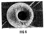
1.5gのPCLを10mlのジクロロメタン(DCM)中に溶解し油(O)相を得る。0.5mlの1%(w/v)ポリビニルアルコール(PVA)溶液(W)を油相に添加する。磁気撹拌し、第1のエマルジョン(W/O)を形成する。ポロジェンとして作用する3gのCaCO3(粒径100〜150μm)を第1のエマルジョンに添加する。第1のエマルジョンを150mlの1%PVA(w/v)に添加する(W/O/W)。800rpmでこの混合物を磁気撹拌して第2のエマルジョンを形成し、溶媒が完全に蒸発するまで撹拌し続ける。撹拌1時間後、2.4mlの酢酸を添加する。発泡性の反応を生じさせる。蒸留水で球状体を3回すすぐ。溶液を濾過して粒子を真空乾燥させ、典型的な凝集手順を使用して分離し、微小殻、即ち本発明の軟組織かさ増し物質を得る。レーザー光散乱により得られた体積平均粒径は196μmであった。本発明の粒子の走査電子顕微鏡写真を示す図6を参照。
Example 2-W / O / W procedure
Dissolve 1.5 g of PCL in 10 ml of dichloromethane (DCM) to obtain an oil (O) phase. 0.5 ml of 1% (w / v) polyvinyl alcohol (PVA) solution (W) is added to the oil phase. Magnetically stir to form a first emulsion (W / O). 3 g CaCO 3 (particle size 100-150 μm) acting as a porogen is added to the first emulsion. Add the first emulsion to 150 ml of 1% PVA (w / v) (W / O / W). This mixture is magnetically stirred at 800 rpm to form a second emulsion and continued to stir until the solvent has completely evaporated. After 1 hour of stirring, 2.4 ml of acetic acid is added. Causes an effervescent reaction. Rinse the spheres 3 times with distilled water. The solution is filtered and the particles are vacuum dried and separated using typical agglomeration procedures to obtain a microshell, the soft tissue bulking material of the present invention. The volume average particle diameter obtained by laser light scattering was 196 μm. See FIG. 6, which shows a scanning electron micrograph of the particles of the present invention.
実施例3 - W/O/O手順
5mlの蒸留水中0.33%(w/v)のポリビニルアルコール(PVA)溶液を作製する(W)。1gのポリ(εカプロラクトン) (PCL)を10mlのジクロロメタン中に溶解して第1の油(O1)相を形成する。ポロジェンとして作用する3gのNaHCO3(粒径150〜212μm)をO1相に添加し、第1のエマルジョン(W/O1)をポロジェンと共に1分間、緩やかに磁気撹拌する。1%Span 60(v/v)を使用して200mlの植物油から第2の油相(O2)を作製する。Span 60(登録商標)は、市販のモノステアリン酸ソルビタン、即ち界面活性剤/乳化剤である。第1のエマルジョンを第2の油相に添加し(W/O1/O2)、2000rpmで2時間、溶媒が完全に蒸発するまで磁気撹拌してエマルジョンを形成する。最終エマルジョンを濾過し、脱イオン水で3回洗浄する。適切な溶媒で、形成された微小粒子上の残留油を除去する。粒子を真空乾燥して典型的な凝集手順を使用して分離し、本発明の軟組織かさ増し物質を得る。
Example 3-W / O / O procedure
A 0.33% (w / v) polyvinyl alcohol (PVA) solution in 5 ml distilled water is prepared (W). 1 g of poly (εcaprolactone) (PCL) is dissolved in 10 ml of dichloromethane to form the first oil (O 1 ) phase. 3 g of NaHCO 3 (particle size 150-212 μm) acting as a porogen is added to the O 1 phase and the first emulsion (W / O 1 ) is gently magnetically stirred with the porogen for 1 minute. Make a second oil phase (O 2 ) from 200 ml vegetable oil using 1% Span 60 (v / v). Span 60® is a commercially available sorbitan monostearate, a surfactant / emulsifier. The first emulsion is added to the second oil phase (W / O 1 / O 2 ) and magnetically stirred at 2000 rpm for 2 hours until the solvent has completely evaporated to form an emulsion. Filter the final emulsion and wash 3 times with deionized water. Remove the residual oil on the formed microparticles with a suitable solvent. The particles are vacuum dried and separated using typical agglomeration procedures to obtain the soft tissue bulking material of the present invention.
実施例4 - 液体ポロジェンを用いた(O/W)手順
150mlの蒸留水中に1%[w/v]のポリビニルアルコール(PVA)溶液を作製する(W)。1.5gのポリ(εカプロラクトン)(PCL)を10mlのジクロロメタンに溶解してO相を形成する。固形ポロジェンとして作用する3gのNaHCO3(粒径:25〜40μm)および液体ポロジェンとして作用する1mlのパーフルオロカーボン(PFC)をO相に添加する。油-ポロジェン-PFC混合物を1分間撹拌する。PVA溶液に油相を添加して(O/W)300rpmで2分間均質化する。その後、20℃で2時間、溶媒が完全に蒸発するまで800rpmで磁気撹拌する。溶液を濾過して粒子を真空乾燥し、典型的な凝集手順を使用して分離し、本発明の軟組織かさ増し物質を得る。
Example 4-(O / W) Procedure with Liquid Porogen
Make a 1% [w / v] polyvinyl alcohol (PVA) solution in 150 ml distilled water (W). 1.5 g of poly (εcaprolactone) (PCL) is dissolved in 10 ml of dichloromethane to form the O phase. 3 g NaHCO 3 (particle size: 25-40 μm) acting as a solid porogen and 1 ml perfluorocarbon (PFC) acting as a liquid porogen are added to the O phase. Stir the oil-porogen-PFC mixture for 1 minute. Add oil phase to PVA solution (O / W) and homogenize at 300 rpm for 2 minutes. Thereafter, magnetic stirring is performed at 800 rpm for 2 hours at 20 ° C. until the solvent is completely evaporated. The solution is filtered and the particles are vacuum dried and separated using typical agglomeration procedures to obtain the soft tissue bulking material of the present invention.
上述の手順を続ける場合、軟組織かさ増し物質はそれぞれの場合において、図1、2、3および4の各粒子10、20、30および40の混合物を含むことが理解されよう。
As the above procedure continues, it will be appreciated that the soft tissue bulking material in each case comprises a mixture of each
実施例5 - インビトロ平滑筋増殖
平滑筋細胞が実施例1の軟組織かさ増し物質に接着する能力を調べるために、古典的な微小担体細胞培養技術を使用して、新鮮な平滑筋細胞を上記の微粒子の存在下で培養した。120時間後、ほとんどの細胞が、ポリマー内部への選択的な結合を示した微粒子空洞内へと移動した。本発明の軟組織かさ増し物質上で培養した細胞の走査電子顕微鏡写真を示す、図7を参照。
Example 5-In Vitro Smooth Muscle Proliferation To examine the ability of smooth muscle cells to adhere to the soft tissue bulking material of Example 1, using fresh micromuscular cell culture techniques, Incubated in the presence of microparticles. After 120 hours, most cells migrated into the microparticle cavities that showed selective binding inside the polymer. See FIG. 7, which shows a scanning electron micrograph of cells cultured on the soft tissue bulking material of the present invention.
本発明の態様は、単に例示を目的としており、当業者は本明細書で使用される具体的な手順の無数の均等物を確認することができる。全てのこのような均等物は本発明の範囲内にあると見なされる。 The embodiments of the present invention are merely illustrative and one of ordinary skill in the art can ascertain the myriad equivalents of the specific procedures used herein. All such equivalents are considered to be within the scope of the present invention.
好ましくは、高度に精製されたグルタルアルデヒド架橋ウシコラーゲンを、担体媒体物質として使用する。この最も良く知られた例は、Collagen Corporation(Inamed Corporation)により製造され、C. R. Bardにより販売されるコラーゲン調製物である。患者がウシコラーゲンに対してアレルギー性であることが分かった場合RestylaneTM(Q-Med)のヒアルロン酸を使用することもできる。 Preferably highly purified glutaraldehyde cross-linked bovine collagen is used as the carrier medium material. The best known example of this is a collagen preparation manufactured by Collagen Corporation (Innamed Corporation) and sold by CR Bard. If the patient is found to be allergic to bovine collagen, Restylane ™ (Q-Med) hyaluronic acid can also be used.
本発明の組成物、特にかさ増し物質を失禁または逆流に基づく疾患の実質的な治療に使用することができるが、本発明の組成物は、主に、下部食道括約筋(LES)の一過的な弛緩により生じ、胃酸を食道に逆流させるGERDの治療において特定の応用を有することが考えられている。他の例において、GERDは、LESの休止時の緊張の低下に起因し得る。このことは、下部食道括約筋へのかさ増し物質の内視鏡または腹腔鏡注入により増大する。かさ増し物質の粒子の微小球または殻の特定の配置により容積の増大が生じ、粒子の空洞内に組織が伸長することにより組織が再生する。特に、粒子中の1つの最も大きなポートが、粒子の機械的な完全性を障害することなく、促進されるかまたはより早い新しい組織の伸長をもたらすと考えられている。理論により拘束されることを望まないが、粒子中の空洞および最も大きなポートが、新しい組織の形成にストレスのない環境をもたらし、それにより、「組織の収容所(harbour)」を形成し、細胞が相互作用する、真の三次元の環境を与えると考えられている。 Although the compositions of the present invention, particularly bulking substances, can be used for the substantial treatment of diseases based on incontinence or reflux, the compositions of the present invention are primarily used for transient esophageal sphincter (LES) transients. It is considered to have particular application in the treatment of GERD, which is caused by natural relaxation and causes gastric acid to flow back into the esophagus. In other examples, GERD may be due to a decrease in LES resting tension. This is increased by endoscopic or laparoscopic injection of bulking material into the lower esophageal sphincter. The specific placement of the microspheres or shells of the bulking material particles causes an increase in volume, and the tissue regenerates by stretching the tissue into the particle cavity. In particular, it is believed that one largest port in the particle is facilitated or results in faster new tissue elongation without compromising the mechanical integrity of the particle. Although not wishing to be bound by theory, the cavities and largest ports in the particle provide a stress-free environment for the formation of new tissue, thereby forming a “tissue harbour” and cells Are believed to give a true three-dimensional environment in which they interact.
新しい組織の形成のためにストレスのない環境または「組織の収容所」を提供することは、線維症(瘢痕組織)を最小限にし、組織形成の体積を最大限にすることで、筋肉の機能を改善すると考えられている。このことは新しい組織の形成を高い割合で生じるはずである。このストレスのない環境もまた、粒子内に配置された細胞が移植の際に受ける強い剪断力から保護されるように、移植の前に粒子を適切な細胞と共に前もって培養し、1つの大きなポートを通じて周囲の組織と接触しながら、ポートおよびミクロ細孔またはマクロ細孔が存在する場合にはそれを介して栄養分および酸素を供給する場合に有用であるはずである。 Providing a stress-free environment or “tissue camp” for the formation of new tissue minimizes fibrosis (scar tissue) and maximizes tissue formation volume, It is thought to improve. This should result in a high rate of new tissue formation. This stress-free environment is also pre-incubated with appropriate cells prior to transplantation through one large port so that the cells placed within the particle are protected from the strong shear forces experienced during transplantation. It should be useful in supplying nutrients and oxygen through ports and micropores or macropores, if present, in contact with the surrounding tissue.
本発明のかさ増し物質でGERDを治療することにより、特に良好な結果が達成され、GERDを治療する他の方法に関連する課題が回避されるであろう。例えば、患者は、GERDを治療するための毎日の医療を受けることを回避し、大手術を避けることができる。 By treating GERD with the bulking material of the present invention, particularly good results will be achieved and the problems associated with other methods of treating GERD will be avoided. For example, patients can avoid receiving daily medical care to treat GERD and avoid major surgery.
さらに、時間が進むにつれて、粒子は生分解され、それにより長期間の組織再生が促進される。 Furthermore, as time progresses, the particles biodegrade, thereby promoting long term tissue regeneration.
効果的には、2つのかさ増し機構は、すなわち:注入された組成物の最初の体積により生じる初期のかさ増し、および新しい組織の形成によりもたらされる後期のかさ増しおよび可能性の高い機能の回復という役割を果たす。 Effectively, the two bulking mechanisms are: an initial bulking caused by the initial volume of the injected composition, and a later bulking and likely functional recovery caused by the formation of new tissue To play a role.
前述のように、粒子が添加剤を含む場合には、さらなる利点が生じ得る。従って、粒子がヒト細胞または(前もって平滑筋に分化させることが可能な)自己幹細胞を含む場合、これはさらに新しい組織の形成および血管形成を刺激し、筋肉の機能を回復させることができる。粒子が関連のある増殖因子を含む場合、これらは早期の新しい組織の形成を促進することができる。さらに、このような増殖因子は粒子が生物分解されるにつれて長期にわたり放出される。粒子がリン酸三カルシウムを含む場合、これは機械的な強度をもたらし、長期にわたり体にカルシウムを供給するカルシウムレザバーとしても機能する。リン酸三カルシウムは良好に確立された生物作用性物質であり、そのためそれのかさ増し物質への取り込みは粒子の生物活性を加速させ、改善された組織の粒子内への伸長を生じる。また、カルシウムは血液凝固カスケードに参加し、血小板の顆粒化をもたらす。 As previously mentioned, additional advantages can arise when the particles include additives. Thus, if the particles contain human cells or autologous stem cells (which can be pre-differentiated into smooth muscle), this can further stimulate new tissue formation and angiogenesis and restore muscle function. If the particles contain relevant growth factors, these can promote early new tissue formation. Furthermore, such growth factors are released over time as the particles are biodegraded. When the particles contain tricalcium phosphate, this provides mechanical strength and also functions as a calcium reservoir that supplies calcium to the body over time. Tricalcium phosphate is a well-established bioactive substance, so its incorporation into the bulking substance accelerates the biological activity of the particle, resulting in improved tissue elongation into the particle. Calcium also participates in the blood coagulation cascade, leading to platelet granulation.
自己由来の血小板富化血漿(PRP)は、増殖因子の「カクテル」の豊富な供給源を提供し、さらなる潜在的な利益付加物である。PRPは、通常、粒子殻よりも担体媒体に取り込まれ、担体媒体が迅速に分解されるために体へと迅速に放出され得る。PRPは、体が適合する場合には増殖因子を放出し、カルシウムを血小板の顆粒化に寄与させる。 Autologous platelet-rich plasma (PRP) provides a rich source of growth factor “cocktails” and is a further potential benefit adjunct. PRP is usually taken up by the carrier medium rather than the particle shell and can be rapidly released into the body because the carrier medium is rapidly degraded. PRP releases growth factors when the body is compatible and contributes calcium to platelet granulation.
担体媒体へのPRPの取り込みは、PRPの顆粒が「自然に決定される」速度で、即ち体が必要とするように関連のある増殖因子を放出するので、新しい組織の形成を刺激する。 Incorporation of PRP into the carrier medium stimulates the formation of new tissue because the PRP granules release the relevant growth factors at a “naturally determined” rate, that is, as the body requires.
関連のある増殖因子の放出速度は、リン酸三カルシウムの殻への取り込みの結果または製造の際のポロジェンとしての炭酸カルシウムの使用の結果であるカルシウムの局所的な供給の存在下で加速され得る。 The release rate of the relevant growth factor can be accelerated in the presence of a local supply of calcium that is the result of incorporation of tricalcium phosphate into the shell or the use of calcium carbonate as a porogen in production. .
従って、かさ増し物質粒子は、かさ増しを必要とする部位に注入することができるような具体的な範囲の大きさ、即ち最大外形50μm〜250μmを有している必要があるということが重要である。これより大きな粒子は内視鏡の適用では注入が容易ではない。通常、内視鏡の針は、最小ゲージサイズ23を有しており、腹腔鏡の針は最大ゲージサイズ16を有する。従って、(粒子が肝臓移植などの適用に必要な場合と同様)本発明の粒子が脈管形成能を有する必要なく、即ち粒子は、通常、粒子が0.5mmよりも大きい場合に生じるような粒子内で血管を形成する大きさである必要はない。同様に、本発明のポートの開口部20μm〜100μmは、粒子の内部に組織が伸長することを可能にするが、粒子内部での血管形成にはより大きな開口部が必要である。本発明のかさ増し物質粒子は、前述の方法により製造することができる。一方で、血管形成のための大きさを有するより大きな粒子、典型的には100μm、典型的には 200μm以上よりも大きなポートを有する0.5mm〜3mmの大きさの粒子の製造方法は、一般的には本発明による小さなポートサイズを有する小さな粒子の製造には適していない。 Therefore, it is important that the bulking material particles should have a specific range of sizes that can be injected into a site that requires bulking, i.e., a maximum outer shape of 50 μm to 250 μm. is there. Larger particles are not easy to inject for endoscopic applications. Typically, an endoscopic needle has a minimum gauge size of 23 and a laparoscopic needle has a maximum gauge size of 16. Thus, it is not necessary for the particles of the present invention to have angiogenic potential (as is the case if the particles are necessary for applications such as liver transplantation), i.e. the particles are usually those that occur when the particles are larger than 0.5 mm. It need not be sized to form blood vessels within. Similarly, the opening 20 μm to 100 μm of the port of the present invention allows the tissue to extend into the interior of the particle, but a larger opening is required for blood vessel formation within the particle. The bulking substance particles of the present invention can be produced by the method described above. On the other hand, a method for producing larger particles with a size for angiogenesis, typically 0.5 μm to 3 mm with ports larger than 100 μm, typically greater than 200 μm, is common. Is not suitable for the production of small particles with a small port size according to the invention.
Claims (15)
(ii)かさ増し物質粒子が懸濁され、組成物が軟組織への注入による適用に適切な粘稠度を有する、適合性担体媒体
を含む注入用軟組織かさ増し組成物。 (i) the soft tissue bulking material of any of claims 1-8; and
(ii) A soft tissue bulking composition for injection comprising a compatible carrier medium in which the bulking material particles are suspended and the composition has a consistency suitable for application by soft tissue injection.
Applications Claiming Priority (2)
| Application Number | Priority Date | Filing Date | Title |
|---|---|---|---|
| ZA200602676 | 2006-03-31 | ||
| PCT/IB2007/051153 WO2007113762A2 (en) | 2006-03-31 | 2007-03-30 | Bulking of soft tissue |
Publications (2)
| Publication Number | Publication Date |
|---|---|
| JP2009531120A true JP2009531120A (en) | 2009-09-03 |
| JP2009531120A5 JP2009531120A5 (en) | 2010-02-04 |
Family
ID=38564052
Family Applications (1)
| Application Number | Title | Priority Date | Filing Date |
|---|---|---|---|
| JP2009502317A Withdrawn JP2009531120A (en) | 2006-03-31 | 2007-03-30 | Soft tissue bulking |
Country Status (9)
| Country | Link |
|---|---|
| US (1) | US20090311328A1 (en) |
| EP (1) | EP2010160A2 (en) |
| JP (1) | JP2009531120A (en) |
| CN (1) | CN101495102A (en) |
| AU (1) | AU2007232187A1 (en) |
| BR (1) | BRPI0709348A2 (en) |
| CA (1) | CA2647549A1 (en) |
| WO (1) | WO2007113762A2 (en) |
| ZA (1) | ZA200807834B (en) |
Families Citing this family (10)
| Publication number | Priority date | Publication date | Assignee | Title |
|---|---|---|---|---|
| US8586089B2 (en) * | 2009-01-03 | 2013-11-19 | Russell J. Anderson | Enhanced carriers for the delivery of microparticles to bodily tissues and fluids |
| US20100291027A1 (en) * | 2009-05-14 | 2010-11-18 | Jason Campbell | Hyaluronic acid (ha) injection vehicle |
| IT1394570B1 (en) * | 2009-07-02 | 2012-07-05 | Fidia Farmaceutici | BIOLOGICAL MATERIAL SUITABLE FOR THE OSTEOARTROSIS THERAPY OF TAPER DAMAGE AND FOR THE TREATMENT OF JOINT-PATHOLOGIES. |
| ITMI20111168A1 (en) * | 2011-06-27 | 2012-12-28 | Professional Dietetics Srl | PHARMACEUTICAL COMPOSITIONS INCLUDING HYDROXYPROPYLMETHYLCELLULA AND / OR HYALURONIC ACID AND THEIR USE |
| CN104383357A (en) * | 2014-10-30 | 2015-03-04 | 四川金堂海纳生物医药技术研究所 | Medicinal decoction for treating vocal cord paralysis and preparation method thereof |
| CN106492284B (en) * | 2016-11-18 | 2018-10-02 | 李世荣 | A kind of preparation method of biodegradable filler and products thereof and application |
| IT201700008651A1 (en) * | 2017-01-26 | 2018-07-26 | Beauty System Pharma Ltd | Crosslinked hyaluronic acid with natural or semi-synthetic crosslinking agents |
| CN114225105A (en) * | 2021-12-20 | 2022-03-25 | 南京思元医疗技术有限公司 | Preparation method of microporous structure polycaprolactone/polyethylene glycol-poly-racemic lactic acid composite microspheres and injectable soft tissue filler |
| CN114246989B (en) * | 2021-12-21 | 2022-09-27 | 上海交通大学医学院附属第九人民医院 | 3D bio-printed active bone repair material and preparation method and application thereof |
| CN115490826A (en) * | 2022-11-01 | 2022-12-20 | 华东理工大学 | Polyurethane porous material and preparation method thereof |
Family Cites Families (23)
| Publication number | Priority date | Publication date | Assignee | Title |
|---|---|---|---|---|
| DE3841401A1 (en) * | 1988-12-08 | 1990-06-13 | Martin Lemperle | ALLOPLASTIC IMPLANT |
| US5019400A (en) * | 1989-05-01 | 1991-05-28 | Enzytech, Inc. | Very low temperature casting of controlled release microspheres |
| US5487897A (en) * | 1989-07-24 | 1996-01-30 | Atrix Laboratories, Inc. | Biodegradable implant precursor |
| US6537574B1 (en) * | 1992-02-11 | 2003-03-25 | Bioform, Inc. | Soft tissue augmentation material |
| US5204382A (en) * | 1992-02-28 | 1993-04-20 | Collagen Corporation | Injectable ceramic compositions and methods for their preparation and use |
| WO1993019702A1 (en) * | 1992-04-06 | 1993-10-14 | Uroplasty, Inc. | Treatment of reflux disorder by microparticles injection |
| FR2715853B1 (en) * | 1994-02-08 | 1996-04-26 | Centre Nat Rech Scient | Composition for bio-material; preparation process. |
| US5516532A (en) * | 1994-08-05 | 1996-05-14 | Children's Medical Center Corporation | Injectable non-immunogenic cartilage and bone preparation |
| US5599852A (en) * | 1994-10-18 | 1997-02-04 | Ethicon, Inc. | Injectable microdispersions for soft tissue repair and augmentation |
| US6214331B1 (en) * | 1995-06-06 | 2001-04-10 | C. R. Bard, Inc. | Process for the preparation of aqueous dispersions of particles of water-soluble polymers and the particles obtained |
| US6129761A (en) * | 1995-06-07 | 2000-10-10 | Reprogenesis, Inc. | Injectable hydrogel compositions |
| US6210715B1 (en) * | 1997-04-01 | 2001-04-03 | Cap Biotechnology, Inc. | Calcium phosphate microcarriers and microspheres |
| CA2286001C (en) * | 1997-04-05 | 2007-07-31 | Giltech Limited | Implantation composition comprising glass particles |
| JP5095049B2 (en) * | 1998-03-06 | 2012-12-12 | バイオスフィアー メディカル,インク. | Implantable particles for the treatment of tissue bulking and gastroesophageal reflux disease, urinary incontinence, skin wrinkles |
| US6660301B1 (en) * | 1998-03-06 | 2003-12-09 | Biosphere Medical, Inc. | Injectable microspheres for dermal augmentation and tissue bulking |
| US6231613B1 (en) * | 1998-12-15 | 2001-05-15 | Enteric Medical Technologies, Inc. | Methods for soft tissue augmentation in mammals |
| US6338345B1 (en) * | 1999-04-07 | 2002-01-15 | Endonetics, Inc. | Submucosal prosthesis delivery device |
| US6277392B1 (en) * | 1999-09-16 | 2001-08-21 | Carbon Medical Technologies, Inc. | Tissue injectable composition |
| US6328990B1 (en) * | 1999-11-12 | 2001-12-11 | The Trustees Of The University Of Pennsylvania | Bioactive, degradable composite for tissue engineering |
| US6652883B2 (en) * | 2000-03-13 | 2003-11-25 | Biocure, Inc. | Tissue bulking and coating compositions |
| BR0315130A (en) * | 2002-10-11 | 2005-08-16 | Novocell Inc | Therapeutically effective cell therapy and biological material encapsulation compositions, their method and use |
| CA2530113C (en) * | 2003-06-26 | 2013-08-13 | Control Delivery Systems, Inc. | Bioerodible sustained release drug delivery systems |
| US20060018942A1 (en) * | 2004-05-28 | 2006-01-26 | Rowe Charles W | Polymeric microbeads having characteristics favorable for bone growth, and process including three dimensional printing upon such microbeads |
-
2007
- 2007-03-30 BR BRPI0709348-9A patent/BRPI0709348A2/en not_active IP Right Cessation
- 2007-03-30 CA CA002647549A patent/CA2647549A1/en not_active Abandoned
- 2007-03-30 CN CNA2007800121165A patent/CN101495102A/en active Pending
- 2007-03-30 JP JP2009502317A patent/JP2009531120A/en not_active Withdrawn
- 2007-03-30 US US12/225,707 patent/US20090311328A1/en not_active Abandoned
- 2007-03-30 AU AU2007232187A patent/AU2007232187A1/en not_active Abandoned
- 2007-03-30 EP EP07735343A patent/EP2010160A2/en not_active Withdrawn
- 2007-03-30 WO PCT/IB2007/051153 patent/WO2007113762A2/en active Application Filing
-
2008
- 2008-09-11 ZA ZA200807834A patent/ZA200807834B/en unknown
Also Published As
| Publication number | Publication date |
|---|---|
| ZA200807834B (en) | 2010-03-31 |
| US20090311328A1 (en) | 2009-12-17 |
| WO2007113762A3 (en) | 2008-01-17 |
| BRPI0709348A2 (en) | 2011-07-12 |
| EP2010160A2 (en) | 2009-01-07 |
| CA2647549A1 (en) | 2007-10-11 |
| WO2007113762A2 (en) | 2007-10-11 |
| CN101495102A (en) | 2009-07-29 |
| AU2007232187A1 (en) | 2007-10-11 |
| WO2007113762A8 (en) | 2007-11-22 |
Similar Documents
| Publication | Publication Date | Title |
|---|---|---|
| JP2009531120A (en) | Soft tissue bulking | |
| KR100954741B1 (en) | Biodegradable injectable implants and related methods of manufacture and use | |
| US5702716A (en) | Polymeric compositions useful as controlled release implants | |
| Zhu et al. | Injectable, porous, biohybrid hydrogels incorporating decellularized tissue components for soft tissue applications | |
| US5945115A (en) | Polymeric compositions useful as controlled release implants | |
| CN104001209B (en) | Microparticles comprising PCL and uses thereof | |
| Dziubla et al. | Evaluation of porous networks of poly (2-hydroxyethyl methacrylate) as interfacial drug delivery devices | |
| RU2363496C2 (en) | Way of soft tissues volume increase | |
| KR101577010B1 (en) | Medical composition and medical kit | |
| CN101249077A (en) | Preparation of degradable pollutant polyalcohol stephanoporate microballoons and uses thereof | |
| JP2009019038A (en) | Non-polymeric sustained release delivery system | |
| EP2898902A1 (en) | Advanced heart failure treatment material as myocardial/cardiovascular regeneration device | |
| AU2002315505A1 (en) | Biodegradable injectable implants and related methods of manufacture and use | |
| CN101869514A (en) | Medicament discharges control combination thing and medicament release property medical apparatus | |
| JP2009528437A (en) | Biodegradable foam | |
| TW200835469A (en) | Porous bioresorbable linked dressing comprising microspheres and methods of making same | |
| JP2003514006A (en) | Biodegradable polymer composition | |
| JP2009531120A5 (en) | ||
| CN101536987A (en) | Sodium alginate microballoon vein suppository containing hemangioma-resisting medicant, preparation method and application thereof | |
| Chen et al. | Expandable, biodegradable, bioactive quaternized gelatin sponges for rapidly controlling incompressible hemorrhage and promoting wound healing | |
| Leng et al. | Size-tunable and biodegradable thrombin-functionalized carboxymethyl chitin microspheres for endovascular embolization | |
| CN114288262B (en) | Drug-loaded microsphere and preparation method and application thereof | |
| CN101309706A (en) | Controlled drug release composition and drug releasing medical device | |
| CN114288463B (en) | Development method of fluid collagen hemostatic material | |
| CN101304708A (en) | Sclerotic ring cingulum and manufacturing method thereof |
Legal Events
| Date | Code | Title | Description |
|---|---|---|---|
| A521 | Written amendment |
Free format text: JAPANESE INTERMEDIATE CODE: A523 Effective date: 20091210 |
|
| A621 | Written request for application examination |
Free format text: JAPANESE INTERMEDIATE CODE: A621 Effective date: 20091210 |
|
| A761 | Written withdrawal of application |
Free format text: JAPANESE INTERMEDIATE CODE: A761 Effective date: 20110622 |