JP2008547006A - Osteoarthritis protein profile - Google Patents
Osteoarthritis protein profile Download PDFInfo
- Publication number
- JP2008547006A JP2008547006A JP2008517179A JP2008517179A JP2008547006A JP 2008547006 A JP2008547006 A JP 2008547006A JP 2008517179 A JP2008517179 A JP 2008517179A JP 2008517179 A JP2008517179 A JP 2008517179A JP 2008547006 A JP2008547006 A JP 2008547006A
- Authority
- JP
- Japan
- Prior art keywords
- expression
- osteoarthritis
- subject
- sample
- difference
- Prior art date
- Legal status (The legal status is an assumption and is not a legal conclusion. Google has not performed a legal analysis and makes no representation as to the accuracy of the status listed.)
- Ceased
Links
Images


Classifications
-
- G—PHYSICS
- G01—MEASURING; TESTING
- G01N—INVESTIGATING OR ANALYSING MATERIALS BY DETERMINING THEIR CHEMICAL OR PHYSICAL PROPERTIES
- G01N33/00—Investigating or analysing materials by specific methods not covered by groups G01N1/00 - G01N31/00
- G01N33/48—Biological material, e.g. blood, urine; Haemocytometers
- G01N33/50—Chemical analysis of biological material, e.g. blood, urine; Testing involving biospecific ligand binding methods; Immunological testing
- G01N33/68—Chemical analysis of biological material, e.g. blood, urine; Testing involving biospecific ligand binding methods; Immunological testing involving proteins, peptides or amino acids
- G01N33/6893—Chemical analysis of biological material, e.g. blood, urine; Testing involving biospecific ligand binding methods; Immunological testing involving proteins, peptides or amino acids related to diseases not provided for elsewhere
-
- A—HUMAN NECESSITIES
- A61—MEDICAL OR VETERINARY SCIENCE; HYGIENE
- A61P—SPECIFIC THERAPEUTIC ACTIVITY OF CHEMICAL COMPOUNDS OR MEDICINAL PREPARATIONS
- A61P19/00—Drugs for skeletal disorders
- A61P19/02—Drugs for skeletal disorders for joint disorders, e.g. arthritis, arthrosis
-
- A—HUMAN NECESSITIES
- A61—MEDICAL OR VETERINARY SCIENCE; HYGIENE
- A61P—SPECIFIC THERAPEUTIC ACTIVITY OF CHEMICAL COMPOUNDS OR MEDICINAL PREPARATIONS
- A61P43/00—Drugs for specific purposes, not provided for in groups A61P1/00-A61P41/00
-
- G—PHYSICS
- G01—MEASURING; TESTING
- G01N—INVESTIGATING OR ANALYSING MATERIALS BY DETERMINING THEIR CHEMICAL OR PHYSICAL PROPERTIES
- G01N2800/00—Detection or diagnosis of diseases
- G01N2800/10—Musculoskeletal or connective tissue disorders
- G01N2800/105—Osteoarthritis, e.g. cartilage alteration, hypertrophy of bone
Abstract
本発明は、変形性関節症(OA)に臨床的関連性をもつタンパク質発現プロフィールの同定および使用に関する。特に、本発明は、その発現がOAおよびOA進行と相関するマーカータンパク質の本体を提供する。OAの研究および/または診断において、OAの進行の程度の決定において、ならびに処置養生法の選択および/またはモニタニリングにおいてこれらのタンパク質発現プロフィールを用いるための方法およびキットが記載される。本発明はまた、これらタンパク質またはこれらタンパク質をコードする核酸分子の発現を調節する薬物のスクリーニング、特に疾患改変OA薬剤の開発に関する。The present invention relates to the identification and use of protein expression profiles that have clinical relevance for osteoarthritis (OA). In particular, the present invention provides a marker protein body whose expression correlates with OA and OA progression. Methods and kits are described for using these protein expression profiles in OA research and / or diagnosis, in determining the extent of OA progression, and in selecting and / or monitoring treatment regimens. The present invention also relates to screening for drugs that modulate the expression of these proteins or nucleic acid molecules encoding these proteins, and in particular to the development of disease modifying OA agents.
Description
(関連出願)
本発明は、2005年6月17日に出願され、そして「変形性関節症のタンパク質プロフィール」と題する仮出願番号第60/692,040号の優先権を主張している。この仮出願は、その全体が参考として本明細書中に援用される。
(Related application)
The present invention was filed on June 17, 2005 and claims priority to provisional application No. 60 / 692,040 entitled “Protein Profile of Osteoarthritis”. This provisional application is incorporated herein by reference in its entirety.
(発明の背景)
筋骨格疾患は、世界中で数百万人の人々に影響し、そしてこの形態は、2020年までには50を超える母集団の予測される倍化に起因して迅速に増加することが予期されている(非特許文献1)。筋骨格疾患は、莫大な健康管理支出および経済的生産性の損失を生じ、そしてそれ故、社会に対して巨大な影響力を有する。米国単独で、筋骨格疾患は、1995年には、2140億ドルの費用を有すると推定された(非特許文献2)。この千年紀の初期には、米国は、2000〜2010年は、平均余命が増加するとき、世界の健康に対して有し得る、増加する影響力の整形外科疾患を強調し、そしてこれら疾患の理解を進め、そして改良され、費用効果的な処置を開発する目的とともに研究努力を促進することを試みる「骨および関節の10年間」を宣言した(http://www.boneandjointdecade.org)。多くのタイプの筋骨格疾患が存在するが、変形性関節症は、世界中で医師によって遭遇される最も共通した慢性の筋骨格疾患障害の1つである。
(Background of the Invention)
Musculoskeletal diseases affect millions of people worldwide and this form is expected to increase rapidly due to the expected doubling of more than 50 populations by 2020 (Non-Patent Document 1). Musculoskeletal diseases result in enormous health care expenditure and loss of economic productivity and therefore have a huge impact on society. In the United States alone, musculoskeletal diseases were estimated to cost $ 214 billion in 1995 (Non-patent Document 2). In the early millennium, the United States emphasized the increasing impact of orthopedic diseases that could have on world health when life expectancy increased between 2000 and 2010, and understanding these diseases And announced a “Decade of Bone and Joints” that attempts to promote research efforts with the goal of developing improved and cost effective treatments (http://www.boneandjointdecade.org). Although there are many types of musculoskeletal diseases, osteoarthritis is one of the most common chronic musculoskeletal disease disorders encountered by physicians around the world.
変形性関節症(OA)は、関節軟骨の崩壊によって特徴付けられる非炎症性の関節疾患である。それは、指、首、肩、尻、膝、下部脊椎領域、および脚を含む、身体中の1つ以上の関節に影響し得る。OAは、痛みを引き起こし得、そして可動性および下肢を重篤に損傷し得(非特許文献3;非特許文献4;非特許文献5;非特許文献6)、これらは、独立を維持する無能力および困難性に至り得る(非特許文献7;非特許文献8;非特許文献9)。OAは加齢にともない:X線撮影法による変形性関節症の離間率は、30歳以下の人々では1%より少ないが、年齢とともに増加し、罹患率は急激に上昇し、そして65を超える個体では約80%であることが見出された(非特許文献10;非特許文献11;非特許文献12)。停年後の集団に最も問題を引き起こす症状であるにもかかわらず、OAはまた、米国およびヨーロッパにおける失業の最も高い原因であると見積もられている。年齢に加え、OAと関連することが知られるリスク因子は、肥満、外傷損傷およびスポーツまたは職業ストレスに起因する酷使を含む。しかし、変形性関節症の正確な病因はなお未知である。
Osteoarthritis (OA) is a non-inflammatory joint disease characterized by the destruction of articular cartilage. It can affect one or more joints in the body, including fingers, neck, shoulders, buttocks, knees, lower spinal region, and legs. OA can cause pain and can seriously damage mobility and lower limbs (Non-patent
目下のところ、OAの診断は、代表的には、放射線学検査、および局所的柔軟性、使用関連の痛み、骨または軟組織膨潤、関節不安定性、制限された関節機能、筋肉痙攣、および関節摩擦音(すなわち、裂け目または削合感覚)を含む臨床観察を基く。OAの診断は、しばしば、医師の検査で示唆されるが、放射線学評価が、この疾患の診断を確認するか、またはこの疾患の重篤度を評価するために用いられる。OAの放射線学的な顕著な特徴は、不均質な関節スペース損失、骨棘形成、嚢形成、および肋軟骨下硬化症を含む。これらの特徴的な特徴は、「重篤」または「後期」OAのX線像に一般に存在するが、「初期」OAをもつ患者は、骨変化、関節スペース狭窄および/または骨棘の放射線学的証拠を示さないかも知れず、この診断を不明瞭また確立することを困難にする。信頼性のある診断の不在下で、医師は、この疾患の経過の初期、すなわち、関節破壊の徴候が生じる前に介入することができない。磁気共鳴(MRI)は、特に、膝のような大関節で、関節軟骨形態および組成の輪郭を描くために有用であり、そして放射線学では見えない軟骨欠損および関節の薄くなった領域を示し得る(非特許文献13;非特許文献14;非特許文献15)。しかし、この画像形成技法は、半月板裂け、または靱帯損傷のようなその他の損傷が診断目的のために除かれる必要がないのであれば、患者に対しては慣用的に実施されない。
Currently, the diagnosis of OA is typically based on radiological examination and local flexibility, use-related pain, bone or soft tissue swelling, joint instability, limited joint function, muscle spasms, and joint friction sounds. Based on clinical observations (ie, tears or agitation sensation). Diagnosis of OA is often suggested by a doctor's examination, but radiological evaluation is used to confirm the diagnosis of the disease or to assess the severity of the disease. Prominent radiological features of OA include heterogeneous joint space loss, osteophyte formation, sac formation, and subchondral sclerosis. Although these characteristic features are generally present in X-ray images of “severe” or “late” OA, patients with “early” OA may experience bone changes, joint space constriction and / or osteophyte radiology. May not show any evidence, making this diagnosis ambiguous and difficult to establish. In the absence of a reliable diagnosis, the physician cannot intervene early in the course of the disease, ie before signs of joint destruction occur. Magnetic resonance (MRI) is useful for delineating articular cartilage morphology and composition, particularly in large joints such as the knee, and can show cartilage defects and thinned areas of the joint that are not visible in radiology (Non-patent document 13; Non-patent
目下のところ、OAに対する治癒はなく、そして変形性関節症治療は、痛みの軽減、ならびに関節機能の改善および維持を取り扱う。さらに、最近のCOX−2インヒビターの使用中止の意味で、医師は、OAの治療の彼らの選択がなお制限されている。OAに対する疾患改善薬物の需要が、この流行し、かつ衰弱させる障害の根深い社会的および経済的影響の認識が広く広がったので、相当に増加した。しかし、このような薬物の臨床試験は、単純なX線を用いて観察された関節スペースにおける変化の評価に依存している(非特許文献16)。関節軟骨損失によって引き起こされる変化は小さいので(1〜2mm/年)、検出可能な十分な変化が起こる前、そしてそれ故、薬物の効き目が評価され得る前に、最小限1年が必要である。
明らかに、OAおよびOA進行の生物学的マーカーに対する大きな必要性が存在する。特に、この疾患の初期ステージで信頼性ある診断およびモニタリングを可能にし得、そして痛みおよび長期間の無能力化を潜在的に防ぐための初期の介入を許容する生物マーカー(バイオマーカー)が高度に所望される。また必要なのは、目下、放射線学的な変化の評価のために必要とされる1年より有意に短い時間フレームで、疾患改変OA薬物の効き目を評価し得る生物マーカーおよび設計アッセイシステムである。 Clearly there is a great need for OA and biological markers of OA progression. In particular, there are highly biomarkers that can enable reliable diagnosis and monitoring at an early stage of the disease and allow early intervention to potentially prevent pain and long-term disability Desired. What is also needed is a biomarker and design assay system that can assess the efficacy of disease-modifying OA drugs in a time frame significantly shorter than the one year currently required for assessment of radiological changes.
(発明の要旨)
本発明は、変形性関節症に臨床的関連性をもつタンパク質発現プロフィールの使用に関する。特に、本発明は、タンパク質の本体を提供し、その発現が、OAと、およびこの疾患の進行の異なる相と相関付けられる。これらのタンパク質の発現プロフィールは、OAの診断およびステージ分けに適用され得る。診断の現存する方法と比較して、本明細書中に開示されるタンパク質プロフィールは、OAおよびOA進行のより確固とした特徴を構成し、そして適切な治療養生法の選択のためのより信頼性ある基礎を提供する。本発明はまた、これらの生物マーカーを標的にする薬物、特に、OAの処置のための新たな治療薬の開発のためのスクリーニングに関する。
(Summary of the Invention)
The present invention relates to the use of protein expression profiles that have clinical relevance for osteoarthritis. In particular, the present invention provides a body of protein whose expression is correlated with OA and with different phases of the progression of the disease. The expression profiles of these proteins can be applied to the diagnosis and staging of OA. Compared to existing methods of diagnosis, the protein profile disclosed herein constitutes a more robust feature of OA and OA progression and is more reliable for selection of an appropriate treatment regimen Provides a foundation. The present invention also relates to screening for the development of new therapeutics for the treatment of drugs that target these biomarkers, particularly OA.
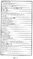
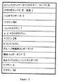
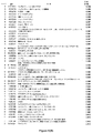
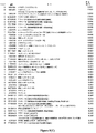
一般に、本発明は、図1〜7に列挙されるマーカータンパク質の発現プロフィールの使用を含む。 In general, the present invention involves the use of marker protein expression profiles listed in FIGS.
より詳細には、1つの局面では、本発明は、被験体における変形性関節症を診断するための方法を提供し、この方法は、この被験体から得られた生物学的サンプルを提供する工程;この生物学的サンプルにおいて、図1〜図7に列挙されたタンパク質からなる群から選択される複数のポリペプチド、そのアナログおよびフラグメントの発現のレベルを決定する工程であって、試験タンパク質発現プロフィールを得る工程;この試験タンパク質発現プロフィールを、コントロールタンパク質発現プロフィールと比較する工程であって、ここで、上記試験タンパク質発現プロフィールと、上記コントロールタンパク質発現プロフィールとの差異が、上記被験体における変形性関節症の存在、不在またはステージの指標である工程;およびこの比較に基き、上記被験体に対する診断を提供する工程を包含する。 More particularly, in one aspect, the invention provides a method for diagnosing osteoarthritis in a subject, the method comprising providing a biological sample obtained from the subject. Determining the level of expression of a plurality of polypeptides, analogs and fragments thereof selected from the group consisting of the proteins listed in FIGS. 1-7 in this biological sample, the test protein expression profile; Comparing the test protein expression profile with a control protein expression profile, wherein the difference between the test protein expression profile and the control protein expression profile is a deformable joint in the subject. A process that is an indicator of the presence, absence or stage of the disease; and this comparison Based comprises providing a diagnosis for the subject.
上記生物学的サンプルは、血液または血液産物のサンプル、尿のサンプル、関節流体のサンプル、唾液のサンプルまたは滑液のサンプルであり得る。特定の好ましい実施形態では、上記生物学的サンプルは、滑液のサンプルである。本発明による複数のポリペプチドの発現のレベルの決定は、上記生物学的サンプルを、上記ポリペプチドの少なくとも1つに特異的な少なくとも1つの抗体に曝す工程を包含し得る。 The biological sample may be a blood or blood product sample, a urine sample, a joint fluid sample, a saliva sample or a synovial fluid sample. In certain preferred embodiments, the biological sample is a sample of synovial fluid. Determining the level of expression of a plurality of polypeptides according to the present invention can include exposing the biological sample to at least one antibody specific for at least one of the polypeptides.
特定の実施形態では、上記被験体は、ヒト、例えば、変形性関節症を有すると疑われる患者である。 In certain embodiments, the subject is a human, eg, a patient suspected of having osteoarthritis.
特定の実施形態では、図7(A)中に列挙されたタンパク質、そのアナログおよびフラグメントから選択される1つ以上のポリペプチドの発現のレベルが測定され、そして上記試験タンパク質発現レベルと上記コントロールタンパク質発現プロフィールとの差異が、上記被験体における変形性関節症の存在の指標である。 In certain embodiments, the level of expression of one or more polypeptides selected from the proteins listed in FIG. 7A, analogs and fragments thereof is measured, and the test protein expression level and the control protein Difference from the expression profile is an indication of the presence of osteoarthritis in the subject.
その他の実施形態では、図7(B)中に列挙されたタンパク質、そのアナログおよびフラグメントから選択される1つ以上のポリペプチドの発現のレベルが測定され、そして上記試験タンパク質発現プロフィールと上記コントロールタンパク質発現プロフィールとの差異が、変形性関節症のステージの指標である。このステージは、初期変形性関節症または後期変形性関節症であり得る。 In other embodiments, the level of expression of one or more polypeptides selected from the proteins listed in FIG. 7B, analogs and fragments thereof is measured, and the test protein expression profile and the control protein The difference from the expression profile is an indicator of the stage of osteoarthritis. This stage can be early or late osteoarthritis.
特定の実施形態では、本発明の診断方法で用いられるコントロールタンパク質発現プロフィールは、正常タンパク質発現プロフィールである。これらの方法では、図1および図2に列挙されたタンパク質からなる群から選択される1つ以上のポリペプチドの発現のレベルにおける増加が、被験体における変形性関節症の存在の指標である。図4および図5に列挙されたタンパク質からなる群から選択される1つ以上のポリペプチドの発現のレベルにおける減少は、被験体における変形性関節症の存在の指標である。図1に列挙されたタンパク質からなる群から選択される1つ以上のポリペプチドの発現のレベルにおける増加は、被験体における初期変形性関節症の指標である。図2に列挙されたタンパク質からなる群から選択される1つ以上のポリペプチドの発現のレベルにおける増加は、被験体における末期変形性関節症の指標である。図4に列挙されたタンパク質からなる群から選択される1つ以上のポリペプチドの発現のレベルにおける減少は、被験体における初期変形性関節症の指標である。図5に列挙されたタンパク質からなる群から選択される1つ以上のポリペプチドの発現のレベルにおける減少は、被験体における後期変形性関節症の指標である。 In certain embodiments, the control protein expression profile used in the diagnostic methods of the invention is a normal protein expression profile. In these methods, an increase in the level of expression of one or more polypeptides selected from the group consisting of the proteins listed in FIGS. 1 and 2 is an indication of the presence of osteoarthritis in the subject. A decrease in the level of expression of one or more polypeptides selected from the group consisting of the proteins listed in FIGS. 4 and 5 is an indication of the presence of osteoarthritis in the subject. An increase in the level of expression of one or more polypeptides selected from the group consisting of the proteins listed in FIG. 1 is an indication of early osteoarthritis in the subject. An increase in the level of expression of one or more polypeptides selected from the group consisting of the proteins listed in FIG. 2 is an indication of end-stage osteoarthritis in the subject. A decrease in the level of expression of one or more polypeptides selected from the group consisting of the proteins listed in FIG. 4 is an indication of early osteoarthritis in the subject. A decrease in the level of expression of one or more polypeptides selected from the group consisting of the proteins listed in FIG. 5 is an indication of late osteoarthritis in the subject.
その他の実施形態では、本発明の診断方法で用いられるコントロールタンパク質発現プロフィールは、初期OAタンパク質発現プロフィールである。これらの方法では、図3に列挙されたタンパク質からなる群から選択される1つ以上のポリペプチドの発現のレベルにおける増加が、被験体における後期変形性関節症の指標であり;そして図7に列挙されたタンパク質からなる群から選択される1つ以上のポリペプチドの発現のレベルにおける減少が、後期変形性関節症の指標である。 In other embodiments, the control protein expression profile used in the diagnostic methods of the invention is an initial OA protein expression profile. In these methods, an increase in the level of expression of one or more polypeptides selected from the group consisting of the proteins listed in FIG. 3 is an indicator of late osteoarthritis in the subject; and in FIG. A decrease in the level of expression of one or more polypeptides selected from the group consisting of the listed proteins is an indicator of late osteoarthritis.
別の局面では、本発明は、図1〜図7に列挙されたタンパク質、そのアナログおよびフラグメントからなる群から選択されるポリペプチドをコードするポリヌクレオチド配列を含む核酸分子、およびこれらのポリヌクレオチド配列の全部または一部とハイブリダイズする核酸分子を提供する。また提供されるのは、被験体における変形性関節症を診断および/またはステージ分けするためのこれら核酸分子の使用である。 In another aspect, the invention provides nucleic acid molecules comprising a polynucleotide sequence encoding a polypeptide selected from the group consisting of the proteins listed in FIGS. 1-7, analogs and fragments thereof, and polynucleotide sequences thereof. Nucleic acid molecules that hybridize to all or a portion of Also provided is the use of these nucleic acid molecules to diagnose and / or stage osteoarthritis in a subject.
別の局面では、本発明は、図1〜図7に提示されるタンパク質、そのアナログ、およびそのフラグメントからなる群から選択される複数のポリペプチドの発現レベル情報を含むOA発現プロフィールマップを提供する。このOA発現プロフィールマップは、正常個体、変形性関節症をもつ個体、初期変形性関節症をもつ個体、または後期変形性関節症をもつ個体から得られる生物学的サンプルの発現レベル情報を含み得る。この生物学的サンプルは、血液または血液のサンプル、尿のサンプル、関節流体のサンプル、唾液のサンプル、および滑液のサンプルであり得る。 In another aspect, the present invention provides an OA expression profile map that includes expression level information of a plurality of polypeptides selected from the group consisting of the proteins presented in FIGS. 1-7, analogs thereof, and fragments thereof. . The OA expression profile map may include expression level information for biological samples obtained from normal individuals, individuals with osteoarthritis, individuals with early osteoarthritis, or individuals with late osteoarthritis. . The biological sample can be a blood or blood sample, a urine sample, a joint fluid sample, a saliva sample, and a synovial fluid sample.
なお別の局面では、被験体における変形性関節症を診断およびステージ分けするためのキットを提供する。本発明のキットは、図1〜図7に提示されたタンパク質、そのアナログおよびフラグメントからなる群から選択されるポリペプチド、および図1〜図7に提示されたタンパク質、そのアナログおよびフラグメントからなる群から選択されるポリペプチドをコードするポリヌクレオチド配列を含む核酸分子;からなる群から選択される少なくとも1つの生物マーカーの発現レベルを特異的に検出する少なくとも1つの試薬;ならびに本発明の方法に従って、被験体において変形性関節症を診断および/またはステージ分けするためにこのキットを用いるための指示書を含む。 In yet another aspect, a kit for diagnosing and staging osteoarthritis in a subject is provided. The kit of the present invention comprises a polypeptide selected from the group consisting of the proteins presented in FIGS. 1 to 7, analogs and fragments thereof, and the group consisting of the proteins, analogs and fragments presented in FIGS. A nucleic acid molecule comprising a polynucleotide sequence encoding a polypeptide selected from: at least one reagent that specifically detects the expression level of at least one biomarker selected from the group consisting of: Includes instructions for using the kit to diagnose and / or stage osteoarthritis in a subject.
特定の実施形態において、上記少なくとも1つの試薬は、上記ポリペプチドの少なくとも1つに特異的に結合する抗体を含む。その他の実施形態では、上記少なくとも1つの試薬は、上記少なくとも1つのポリペプチドをコードするポリヌクオチド配列に相補的な核酸プローブを含む。例えば、上記核酸プローブは、cDNAまたはオリゴヌクレオチドであり、そして基質表面上に固定化され得る。 In certain embodiments, the at least one reagent comprises an antibody that specifically binds to at least one of the polypeptides. In other embodiments, the at least one reagent comprises a nucleic acid probe complementary to a polynucleotide sequence encoding the at least one polypeptide. For example, the nucleic acid probe is a cDNA or an oligonucleotide and can be immobilized on a substrate surface.
上記キットは、インビトロ診断製品における使用のための米国食品医薬品局によって要求される指示書;以下の1つ以上:抽出緩衝液/試薬およびプロトコール、増幅緩衝液/試薬およびプロトコール、ハイブリダイゼーション緩衝液/試薬およびプロトコール、免疫検出緩衝液/試薬およびプロトコール、および標識緩衝液/試薬およびプロトコール、および/または上記に記載のような少なくとも1つのOA発現プロフィールマップをさらに含み得る。 The kit includes instructions required by the US Food and Drug Administration for use in in vitro diagnostic products; one or more of the following: extraction buffer / reagent and protocol, amplification buffer / reagent and protocol, hybridization buffer / Reagents and protocols, immunodetection buffers / reagents and protocols, and labeling buffers / reagents and protocols, and / or at least one OA expression profile map as described above may further be included.
なお別の局面では、本発明は、システム中のOA生物マーカーの発現を調節する化合物を同定するための方法を提供する。本発明の方法は:図1〜図7に列挙されたタンパク質、そのアナログおよびフラグメントからなる群から選択されるポリペプチド、および図1〜図7に列挙されたタンパク質、そのアナログおよびフラグメントからなる群から選択されるポリペプチドをコードするポリヌクレオチド配列を含む核酸分子:からなる群から選択される生物マーカーの発現のレベルを、該システムを上記候補化合物に曝す前後に決定する工程;およびこれらのレベルが異なる場合に、上記OA生物マーカーの発現を調節する化合物としてこの候補化合物を同定する工程を包含する。 In yet another aspect, the invention provides a method for identifying a compound that modulates the expression of an OA biomarker in the system. The methods of the invention are: a polypeptide selected from the group consisting of the proteins listed in FIGS. 1-7, analogs and fragments thereof, and the group consisting of the proteins, analogs and fragments listed in FIGS. Determining the level of expression of a biomarker selected from the group consisting of: a nucleic acid molecule comprising a polynucleotide sequence encoding a polypeptide selected from: before and after exposing the system to the candidate compound; and these levels The candidate compound is identified as a compound that modulates the expression of the OA biomarker.
これらの方法で用いられるシステムは、細胞、生物学的流体、生物学的組織、または動物であり得る。 The system used in these methods can be a cell, biological fluid, biological tissue, or animal.
候補化合物は、変形性関節症における減少した発現によって特徴付けられる生物マーカーの発現を増大し得;変形性関節症における増加した発現によって特徴付けられる生物マーカーの発現を減少し得;初期変形性関節症における減少した発現によって特徴付けられる生物マーカーの発現を増大し得;初期変形性関節症における減少した発現によって特徴付けられる生物マーカーの発現を減少し得;後期変形性関節症における減少した発現によって特徴付けられる生物マーカーの発現を増大し得;および/または後期変形性関節症における増加した発現によって特徴付けられる生物マーカーの発現を減少し得るOA生物マーカーの発現を調節する化合物として同定される。 Candidate compounds can increase the expression of biomarkers characterized by decreased expression in osteoarthritis; can decrease the expression of biomarkers characterized by increased expression in osteoarthritis; early osteoarthritis May increase expression of biomarkers characterized by decreased expression in disease; may decrease expression of biomarkers characterized by decreased expression in early osteoarthritis; by decreased expression in late osteoarthritis Identified as a compound that modulates the expression of an OA biomarker that may increase the expression of the characterized biomarker; and / or decrease the expression of the biomarker characterized by increased expression in late osteoarthritis.
本発明は、本発明のスクリーニング方法によって同定されるOA治療薬、これらのOA治療薬を含む薬学的組成物、およびこれらOA治療薬の少なくとも1つの有効量を患者に投与することにより患者中の変形性関節症を処置する方法をさらに提供する。 The present invention provides a method for administering OA therapeutic agents identified by the screening methods of the present invention, pharmaceutical compositions comprising these OA therapeutic agents, and administering at least one effective amount of these OA therapeutic agents to a patient. Further provided are methods of treating osteoarthritis.
(定義)
明細書全体を通じて、以下の段落で規定されるいくつかの用語が採用される。
(Definition)
Throughout the specification, several terms are employed that are defined in the following paragraphs.
用語「被験体」、「個体」および「患者」は、本明細書では交換可能に用いられる。それらは、変形性関節症を罹患し得るヒトまたは別の哺乳動物(例えば、霊長類、イヌ、ネコ、ヤギ、ウマ、ブタ、マウス、ラット、ウサギなど)をいうが、この疾患を有していても良いし、有さなくてもよい。多くの実施形態では、この被験体はヒトである。 The terms “subject”, “individual” and “patient” are used interchangeably herein. They refer to a human or another mammal (eg, primates, dogs, cats, goats, horses, pigs, mice, rats, rabbits, etc.) that may suffer from osteoarthritis, but have this disease. You may or may not have it. In many embodiments, the subject is a human.
用語「OAを有すると疑われる被験体」は、1つ以上のOAの徴候指標(例えば、関節の痛み、局在化した柔軟性、骨または軟組織膨潤、関節不安定性、関節摩擦音)を提示するか、または(例えば、慣用の身体検査の間に)OAについてスクリーニングされるいる被験体をいう。OAを有すると疑われる被験体はまた、1つ以上のリスク因子(例えば、年齢、肥満、外傷損傷、スポーツに起因する酷使または職業的ストレス、家族歴)を有し得る。この用語は、OAについて試験されていない個体、および(例えば、放射線学的検査に基く)初期診断を受けたが、OAのステージが知られていない個体を包含する。 The term “subject suspected of having OA” presents one or more OA symptom indicators (eg, joint pain, localized flexibility, bone or soft tissue swelling, joint instability, joint friction sounds). Or refers to a subject being screened for OA (eg, during routine physical examination). A subject suspected of having OA may also have one or more risk factors (eg, age, obesity, trauma injury, abuse or occupational stress due to sports, family history). The term encompasses individuals who have not been tested for OA and individuals who have received an initial diagnosis (eg, based on radiological examination) but whose OA stage is unknown.
用語「変形性関節症ステージ」および「変形性関節症フェーズ」は、本明細書では交換可能に用いられ、そして疾患の進行または経過の程度をいう。本発明は、この疾患のステージを決定するための手段を提供する。特に、本明細書で提供される方法は、「温和」または「初期」OAの、および「重篤」または「末期」OAの検出を可能にする。当該技術分野で公知のその他のステージ分けシステムは、例えば、Marshalによって開発されたものを含む(W.Marchall、J.Rheumatol.、1996、23:582〜584)。 The terms “osteoarthritis stage” and “osteoarthritis phase” are used interchangeably herein and refer to the extent of disease progression or course. The present invention provides a means for determining the stage of this disease. In particular, the methods provided herein allow for the detection of “mild” or “early” OA and “severe” or “late” OA. Other staging systems known in the art include, for example, those developed by Marshall (W. Marchall, J. Rheumatol., 1996, 23: 582-584).
本明細書で用いられるとき、用語「診断」は、個体が疾患または病気に罹患しているか否かを決定することを狙うプロセスをいう。本発明の文脈では、「OAの診断」は、以下の1つ以上を狙うプロセスをいう:個体がOAに罹患しているか否かを決定すること、およびこの疾患のステージを決定すること(例えば、初期OAまたは末期OA)。 As used herein, the term “diagnosis” refers to a process aimed at determining whether an individual is afflicted with a disease or condition. In the context of the present invention, “diagnosis of OA” refers to a process that targets one or more of the following: determining whether an individual has OA and determining the stage of the disease (eg, , Early OA or late OA).
用語「生物学的サンプル」は、本明細書では、その最も広い意味で用いられる。生物学的サンプルは、被験体(例えば、ヒト)から、または被験体の構成要素(例えば、組織)から得られ得る。このサンプルは、これとともに本発明の生物マーカーがアッセイされ得る任意の生物学的組織または流体からであり得る。頻繁に、このサンプルは、「臨床サンプル」、すなわち、患者に由来するサンプルである。このようなサンプルは、制限されないで、細胞を含んでも良いし、含まなくても良い体液、例えば、血液、尿、滑液、唾液、および関節流体;骨または軟骨からのような組織または微細ニードル生検サンプル;および既知の診断、処置および/または結果歴をもつ保管サンプルを含み得る。生物学的サンプルはまた、組織学的目的からとられた凍結サンプルのような組織の切片を含み得る。この用語、生物学的サンプルはまた、この生物学的サンプルを処理することにより派生する任意の材料を包含する。派生する材料は、制限されないで、このサンプルが単離される細胞(またはそれらの子孫)、このサンプルから抽出されるタンパク質または核酸分子を含む。この生物学的サンプルの処理は:濾過、蒸留、抽出、濃縮、妨害成分の不活性化、試薬の添加などをの1つ以上を含み得る。 The term “biological sample” is used herein in its broadest sense. A biological sample can be obtained from a subject (eg, a human) or from a component (eg, tissue) of the subject. The sample can be from any biological tissue or fluid with which a biomarker of the invention can be assayed. Frequently, this sample is a “clinical sample”, ie a sample derived from a patient. Such samples include, but are not limited to, body fluids that may or may not contain cells, such as blood, urine, synovial fluid, saliva, and joint fluids; tissue or fine needles such as from bone or cartilage Biopsy samples; and stored samples with a known diagnosis, treatment and / or history of results. Biological samples can also include sections of tissue such as frozen samples taken for histological purposes. The term biological sample also encompasses any material derived from processing the biological sample. Derived materials include, but are not limited to, the cells from which the sample is isolated (or their progeny), proteins or nucleic acid molecules extracted from the sample. The processing of the biological sample may include one or more of: filtration, distillation, extraction, concentration, inactivation of interfering components, addition of reagents, and the like.
用語「正常」および「健常」は、本明細書では交換可能に用いられる。それらは、関節の痛みを含む任意のOA症状を示さず、そして軟骨損傷またはOAと診断されていない個体または個体の群をいう。好ましくは、この正常個体(または個体の群)は、OAに影響する薬物適用はなく、そして任意のその他の疾患と診断されていない。より好ましくは、正常個体は、試験されるべきサンプルが得られた個体と比較して、同様の性、年齢、身体嵩指数を有する。用語「正常」はまた、健常個体から単離されたサンプルを適任とするために用いられる。 The terms “normal” and “healthy” are used interchangeably herein. They refer to an individual or group of individuals who do not exhibit any OA symptoms including joint pain and have not been diagnosed with cartilage damage or OA. Preferably, this normal individual (or group of individuals) has no drug application affecting OA and has not been diagnosed with any other disease. More preferably, normal individuals have similar gender, age and body bulk index compared to the individual from which the sample to be tested was obtained. The term “normal” is also used to qualify samples isolated from healthy individuals.
本発明の文脈では、用語「コントロールサンプル」は、正常(すなわち、健常)である個体または個体の群から単離された1つ以上の生物学的サンプルをいう。コントロールサンプルはまた、OAの特定ステージ(例えば、初期OAまたは後期OA)と診断された患者または患者の群から単離された生物学的サンプルをいう。用語「コントロールサンプル」(または「コントロール」)はまた、正常と分類された1つ以上の個体、またはOAもしくはOAの特定ステージと診断された1つ以上の個体、またはOAの処置を受けていた1つ以上の個体のサンプル由来のデータの編集物をいい得る。 In the context of the present invention, the term “control sample” refers to one or more biological samples isolated from an individual or group of individuals that is normal (ie, healthy). A control sample also refers to a biological sample isolated from a patient or group of patients diagnosed with a particular stage of OA (eg, early OA or late OA). The term “control sample” (or “control”) also had one or more individuals classified as normal, or one or more individuals diagnosed with OA or a specific stage of OA, or had been treated for OA A compilation of data from a sample of one or more individuals may be referred to.
用語「OA生物マーカー」および「生物マーカー」は、本明細書では交換可能に用いられる。それらは、本発明によって提供され、そしてその発現プロフィールが、OAおよび/またはOAの特定ステージの指標であることが見出されたタンパク質のセットから選択されるタンパク質をいう。用語「生物マーカー」はまた、本発明のマーカータンパク質をコードするヌクレオチド配列を含む核酸分子、およびこれらの核酸分子の一部分とハイブリダイズするポリヌクレオチドを包含する。 The terms “OA biomarker” and “biomarker” are used interchangeably herein. They refer to proteins provided by the present invention and whose expression profile is selected from a set of proteins found to be indicative of OA and / or a specific stage of OA. The term “biomarker” also encompasses nucleic acid molecules comprising nucleotide sequences encoding the marker proteins of the invention, and polynucleotides that hybridize to portions of these nucleic acid molecules.
本明細書で用いられるとき、用語「OAの指標」は、生物マーカーに用いられるとき、OAまたはOAのステージの特徴である発現パターンまたはプロフィールをいい、この発現パターンは、この疾患またはこの疾患の別のステージのない患者におけるより、(最小95%の信頼性レベルを設定する慣用の統計学的方法を用いて決定されるとき)この疾患またはこの疾患の1つのステージにある患者で有意によりしばしば見出される。好ましくは、OAの指標である発現パターンは、この疾患を有する患者の少なくとも60%で見出され、そしてこの疾患を有さない被験体の10%未満で見出される。より好ましくは、OAの指標である発現パターンは、この疾患を有する患者の少なくとも70%、少なくと75%、少なくとも80%、少なくとも85%、少なくとも90%、少なくとも95%またはそれ以上で見出され、そしてこの疾患を有さない被験体の10%未満、8%未満、5%未満、2.5%未満、または1%未満で見出される。 As used herein, the term “indicator of OA”, when used in a biomarker, refers to an expression pattern or profile that is characteristic of OA or the stage of OA, the expression pattern being the disease or the disease Significantly more often in patients with this disease or one stage of this disease (as determined using conventional statistical methods setting a minimum 95% confidence level) than in patients without another stage Found. Preferably, an expression pattern indicative of OA is found in at least 60% of patients with the disease and is found in less than 10% of subjects without the disease. More preferably, an expression pattern that is indicative of OA is found in at least 70%, at least 75%, at least 80%, at least 85%, at least 90%, at least 95% or more of patients with the disease. And less than 10%, less than 8%, less than 5%, less than 2.5%, or less than 1% of subjects without this disease.
本明細書で用いられるとき、用語「鑑別的に発現される生物マーカー」は、その発現レベルが、被験体(または被験体の集団)において、健常もしくは正常被験体(または健常もしくは正常被験体)におけるその発現レベルに対して異なる生物マーカーをいう。この用語はまた、その発現レベルがこの疾患の異なるステージ(例えば、温和もしくは初期OA、重篤もしはく末期OA)で異なる生物マーカーを包含する。異なる発現は、この生物マーカーの一時的または細胞発現パターンにおける、定量的、および定性的差異を含む。以下により詳細に記載されるように、鑑別的に発現される生物マーカーは、単独またはその他の鑑別的に発現される生物マーカーとの組み合わせは、診断、ステージ分け、治療、薬物開発および関連領域における種々の異なる適用で有用である。本明細書中に開示されるこれら鑑別的に発現される生物マーカーの発現パターンは、OAおよびOA進行のフィンガープリントまたは特徴として記載され得る。それらは、未知サンプルおよびそれらについてさらなる情報が求められるサンプルを比較し、そして特徴付けるための参照の点として用いられ得る。本明細書で用いられるとき、用語「発現の減少したレベル」は、本明細書中に記載される1つ以上の方法によって測定されるとき、発現における少なくとも10%以上、例えば、20%、30%、40%、または50%、60%、70%、80%、90%またはそれ以上の、あるいは発現における1倍、2倍、3倍、4倍、5倍、10倍、50倍、100倍またはそれ以上の減少をいう。本明細書で用いられるとき、用語「発現の増加したレベル」は、本明細書中に記載される1つ以上の方法によって測定されるとき、発現における少なくとも10%以上、例えば、20%、30%、40%、または50%、60%、70%、80%、90%またはそれ以上の、あるいは発現における1倍、2倍、3倍、4倍、5倍、10倍、50倍、100倍またはそれ以上の増加をいう。 As used herein, the term “differentially expressed biomarker” refers to a healthy or normal subject (or healthy or normal subject) whose expression level is in a subject (or population of subjects). Refers to biomarkers that differ with respect to their expression level. The term also encompasses biomarkers whose expression levels differ at different stages of the disease (eg, mild or early OA, severe or late OA). Different expression includes quantitative and qualitative differences in the temporal or cellular expression pattern of this biomarker. As described in more detail below, differentially expressed biomarkers can be used alone or in combination with other differentially expressed biomarkers in diagnostic, staging, therapy, drug development and related areas. Useful in a variety of different applications. The expression pattern of these differentially expressed biomarkers disclosed herein can be described as a fingerprint or characteristic of OA and OA progression. They can be used as a point of reference for comparing and characterizing unknown samples and samples for which more information is sought. As used herein, the term “reduced level of expression” as measured by one or more methods described herein is at least 10% or more in expression, eg, 20%, 30 %, 40%, or 50%, 60%, 70%, 80%, 90% or more, or 1x, 2x, 3x, 4x, 5x, 10x, 50x, 100 in expression, 100 Refers to a fold reduction or more. As used herein, the term “increased level of expression” as measured by one or more methods described herein is at least 10% or more in expression, eg, 20%, 30 %, 40%, or 50%, 60%, 70%, 80%, 90% or more, or 1x, 2x, 3x, 4x, 5x, 10x, 50x, 100 in expression, 100 A double or more increase.
用語「タンパク質」、「ポリペプチド」、および「ペプチド」は、本明細書では交換可能に用いられ、そしてそれらの自然の(未変化)形態にあるか、または塩のとしてのいずれか、および非改変またはグリコシル化、側鎖酸化、もしくはリン酸化による改変されたいずれかである種々の長さのアミノ酸配列をいう。特定の実施形態では、このアミノ酸配列は、完全長のネイティブタンパク質である。その他の実施形態では、このアミノ酸配列は、この完全長タンパク質のより小さいフラグメントである。なおその他の実施形態では、このアミノ酸配列は、グリコシル単位、脂質、またはリン酸イオンのような無機イオンのような、アミノ酸側鎖に付着されるさらなる置換基、およびスルフヒドリル基の酸化のような、これら鎖の化学的変換に関する改変によって改変される。従って、用語「タンパク質」またはその相当する用語は、その特異的性質を変化しないような改変を受ける、完全長ネイティブタンパク質のアミノ酸配列を含むことが意図される。特に、用語「タンパク質」は、タンパク質のイソ形態、すなわち、同じ遺伝子によってコードされるが、それらのpIもしくはMW、またはその両方が異なる改変体を包含する。このようなイソ形態は、それらのアミノ酸配列が異なり得るか(例えば、代替スプライシングまたは制限的タンパク質分解の結果として)、または、それに代わって、差別的翻訳後改変から生じ得る(例えば、グリコシル化、アシル化、リン酸化)。 The terms “protein”, “polypeptide”, and “peptide” are used interchangeably herein and are either in their natural (unaltered) form or as a salt, and non- Refers to amino acid sequences of various lengths that are either modified or modified by glycosylation, side chain oxidation, or phosphorylation. In certain embodiments, the amino acid sequence is a full length native protein. In other embodiments, the amino acid sequence is a smaller fragment of the full-length protein. In still other embodiments, the amino acid sequence comprises a glycosyl unit, a lipid, or additional substituents attached to amino acid side chains, such as inorganic ions such as phosphate ions, and oxidation of sulfhydryl groups, These chains are modified by modifications related to chemical transformations. Thus, the term “protein” or its equivalent term is intended to include the amino acid sequence of the full-length native protein, subject to modifications that do not alter its specific properties. In particular, the term “protein” encompasses variants that are encoded by isoforms of the protein, ie, the same gene, but differ in their pI or MW, or both. Such isoforms can differ in their amino acid sequences (eg, as a result of alternative splicing or restricted proteolysis) or alternatively can result from differential post-translational modifications (eg, glycosylation, Acylation, phosphorylation).
用語「タンパク質アナログ」は、本明細書で用いられるとき、完全長のネイティブタンパク質と類似または同一の機能を所有するが、このタンパク質のアミノ酸配列に類似または同一でるアミノ酸配列を必ずしも含む必要のないか、あるいはこのタンパク質の構造と類似または同一である構造を所有するポリペプチドをいう。好ましくは、本発明の文脈では、タンパク質アナログは、完全長のネイティブタンパク質のアミノ酸配列に少なくとも30%(より好ましくは、少なくとも35%、少なくとも40%、少なくとも45%、少なくとも50%、少なくとも55%、少なくとも60%、少なくとも65%、少なくとも70%、少なくとも75%、少なくとも80%、少なくとも85%、少なくとも90%、少なくとも95%、または少なくとも99%)同一であるアミノ酸配列を有する。 The term “protein analog” as used herein does not necessarily have to contain an amino acid sequence that possesses a similar or identical function as the full-length native protein, but is similar or identical to the amino acid sequence of the protein. Or a polypeptide possessing a structure that is similar or identical to the structure of this protein. Preferably, in the context of the present invention, the protein analog is at least 30% (more preferably at least 35%, at least 40%, at least 45%, at least 50%, at least 55%) in the amino acid sequence of the full-length native protein. At least 60%, at least 65%, at least 70%, at least 75%, at least 80%, at least 85%, at least 90%, at least 95%, or at least 99%) having amino acid sequences that are identical.
用語「タンパク質フラグメント」は、本明細書で用いられるとき、第2のポリペプチドのアミノ酸配列の少なくとも5つのアミノ酸残基(好ましくは、少なくとも10のアミノ酸残基、少なくとも15のアミノ酸残基、少なくとも20のアミノ酸残基、、少なくとも25のアミノ酸残基、少なくとも40のアミノ酸残基、少なくとも50のアミノ酸残基、少なくとも60のアミノ酸残基、少なくとも70のアミノ酸残基、少なくとも80のアミノ酸残基、少なくとも90のアミノ酸残基、少なくとも100のアミノ酸残基、少なくとも125のアミノ酸残基、少なくとも150のアミノ酸残基、少なくとも175のアミノ酸残基、少なくとも200のアミノ酸残基、または少なくとも250のアミノ酸残基)のアミノ酸配列を含むポリペプチドをいう。マーカータンパク質のフラグメントは、完全長のネイティブタンパク質の機能的活性を所有していても良いし、していなくても良い。 The term “protein fragment” as used herein refers to at least 5 amino acid residues of the second polypeptide amino acid sequence (preferably at least 10 amino acid residues, at least 15 amino acid residues, at least 20 Amino acid residues, at least 25 amino acid residues, at least 40 amino acid residues, at least 50 amino acid residues, at least 60 amino acid residues, at least 70 amino acid residues, at least 80 amino acid residues, at least 90 Amino acid residues, at least 100 amino acid residues, at least 125 amino acid residues, at least 150 amino acid residues, at least 175 amino acid residues, at least 200 amino acid residues, or at least 250 amino acid residues) Polypeptide containing sequence The say. Marker protein fragments may or may not possess the functional activity of the full-length native protein.
用語「核酸分子」および「ポリヌクレオチド」は、本明細書では交換可能に用いられる。それらは、一本鎖または二本鎖形態のいずかにあるデオキシリボヌクレオチドまたはリボヌクレオチドポリマーをいい、そしてその他であることが陳述されていなければ、天然に存在するヌクレオチドと類似の様式で機能し得る天然のヌクレオチドの既知のアナログを包含する。これらの用語は、合成のバックボーンをもつ核酸様構造、および増幅産物を包含する。 The terms “nucleic acid molecule” and “polynucleotide” are used interchangeably herein. They refer to deoxyribonucleotides or ribonucleotide polymers in either single-stranded or double-stranded form, and function in a manner similar to naturally occurring nucleotides unless otherwise stated. Includes known analogs of natural nucleotides obtained. These terms encompass nucleic acid-like structures with a synthetic backbone and amplification products.
本明細書で用いられるとき、用語「発現レベルを特異的の検出する試薬」は、1つ以上の生物マーカー(例えば、本明細書中で提供されるマーカータンパク質、マーカータンパク質をコードするポリヌレオチド配列を含む核酸分子、またはこの核酸分子の少なくとも一部分とハイブリダイズするポリヌクレオチド)の発現レベルを検出するために用いられる1つ以上の試薬をいう。適切な試薬の例は、制限されずに、目的のマーカータンパク質に特異的に結合し得る抗体、目的のポリヌクレオチド配列に特異的にハイブリダイズし得る核酸プローブ、または目的のポリヌクレオチド配列を特異的に増幅し得るPCRプライマーを含む。用語「増幅する」は、本明細書では、広い意味で用いられ、増幅産物を創生/生成ことを意味する。「増幅」は、本明細書で用いられるとき、一般に、所望の配列、特にサンプルの配列の複数のコピーを生成するプロセスをいう。「コピー」は、テンプレート配列に完全に相補的な配列または同一であることを必ずしも意味しない。 As used herein, the term “reagent that specifically detects expression levels” refers to one or more biomarkers (eg, a marker protein provided herein, a polynucleotide sequence encoding a marker protein). One or more reagents used to detect the expression level of a nucleic acid molecule comprising, or a polynucleotide that hybridizes to at least a portion of, the nucleic acid molecule. Examples of suitable reagents include, but are not limited to, an antibody that can specifically bind to the marker protein of interest, a nucleic acid probe that can specifically hybridize to the polynucleotide sequence of interest, or a specific polynucleotide sequence of interest. PCR primers that can be amplified. The term “amplify” is used herein in a broad sense and means to create / generate an amplification product. “Amplification”, as used herein, generally refers to the process of generating multiple copies of a desired sequence, particularly a sample sequence. “Copy” does not necessarily mean that the sequence is completely complementary or identical to the template sequence.
用語「ハイブリダイズ」は、相補的塩基対合を経由する2つの一本鎖核酸の結合をいう。用語「特異的なハイブリダイゼーション」は、核酸分子が、特定の核酸配列に、ストリンジェントな条件(例えば、ハイブリダイズする鎖により低い程度の相補性をもつ競合者核酸の存在下)下で優先的に結合、二本鎖になる、またはハイブリダイズするプロセスをいう。本発明の特定の実施形態では、これらの用語は、試験サンプルからの核酸フラグメント(またはセグメント)が、特定のプローブに、例えば、これらプローブがアレイ上に固定化されるとき優先的に結合し、そしてその他のプローブにはより低い程度で結合するか、または全く結合しないプロセスをより詳細にいう。 The term “hybridize” refers to the binding of two single stranded nucleic acids via complementary base pairing. The term “specific hybridization” means that a nucleic acid molecule preferentially binds to a specific nucleic acid sequence under stringent conditions (eg, in the presence of a competitor nucleic acid that has a lower degree of complementarity to the hybridizing strand). A process that binds, becomes double-stranded, or hybridizes. In certain embodiments of the invention, these terms preferentially bind nucleic acid fragments (or segments) from a test sample to specific probes, for example when these probes are immobilized on an array, And more specifically, the process of binding to other probes to a lesser extent or not at all.
用語「アレイ」、「マイクロアレイ」、および「バイオチップ」は、本明細書では交換可能に用いられる。それらは、好ましくは、既知の配列の複数の核酸分子であるハイブリダイズ可能なアレイ要素の、基板表面上の配列をいう。各核酸分子は、基板表面上の別個のスポット(すなわち、規定された場所または割り当てられた位置)に固定化される。用語
「マイクロアレイ」は、視覚評価のために顕微鏡検査を必要とするようにミニチュア化されるアレイをより特定していう。
The terms “array”, “microarray”, and “biochip” are used interchangeably herein. They preferably refer to a sequence on the substrate surface of a hybridizable array element that is a plurality of nucleic acid molecules of known sequence. Each nucleic acid molecule is immobilized at a separate spot (ie, a defined location or assigned location) on the substrate surface. The term “microarray” refers more specifically to an array that is miniaturized to require microscopic examination for visual evaluation.
用語「プローブ」は、本明細書で用いられるとき、既知の配列の核酸分子をいい、これは、短いDNA配列(すなわち、オリゴヌクレオチド)、PCR産物、またはmRNA単離物であり得る。プローブは、試験サンプルからの核酸フラグメントがハイブリダイズする特異的DNA配列である。プローブは、通常は水素結合形成による、1つ以上のタイプの化学的結合を通じて相補的または実質的に相補的な配列の核酸に特異的に結合する。 The term “probe” as used herein refers to a nucleic acid molecule of known sequence, which can be a short DNA sequence (ie, an oligonucleotide), a PCR product, or an mRNA isolate. A probe is a specific DNA sequence to which a nucleic acid fragment from a test sample hybridizes. Probes specifically bind to complementary or substantially complementary sequences of nucleic acids through one or more types of chemical bonds, usually by hydrogen bond formation.
用語「標識され」、「検出可能な物質で標識」および「検出可能な成分で標識」は、本明細書では交換可能に用いられる。これらの用語は、例えば、ある実体(例えば、プローブ)が、その他の実体(例えば、ポリヌクレオチドまたはポリペプチド)への結合後に可視化され得ることを特定するために用いられる。好ましくは、この検出可能な物質または成分は、それが測定され得、そしてその強度が結合した実体の量に関連する信号を生成するように選択される。アレイを基礎にした方法では、この検出可能な物質または成分はまた、好ましくは、それが局在化した信号を生成し、それによって、アレイ上の各スポットからの信号の空間的解像を可能にするように選択される。ポリペプチドまたはポリヌクレオチドを標識する方法は、当該技術分野で周知である。標識されたポリペプチドまたはポリヌクレオチドは、分光光度的に、光化学的に、生化学的に、免疫化学的に、電気的に、光学的に、または化学的手段によって直接的または間接的に検出可能である標識の取り込み、またはそれとの複合体化によって調製され得る。適切な検出可能な物質は、制限されないで、種々のリガンド、放射線核種、蛍光色素、化学発光物質、マイクロパーティクル、酵素、比色標識、磁性標識、およびハプテンを含む。検出可能な成分はまた、分子ビーコンおよびアプタマービーコンのような生物学的分子であり得る。 The terms “labeled”, “labeled with a detectable substance” and “labeled with a detectable moiety” are used interchangeably herein. These terms are used, for example, to specify that an entity (eg, a probe) can be visualized after binding to another entity (eg, a polynucleotide or polypeptide). Preferably, the detectable substance or component is selected such that it can be measured and generates a signal whose intensity is related to the amount of bound entity. In an array-based method, this detectable substance or component also preferably generates a localized signal, thereby allowing spatial resolution of the signal from each spot on the array Selected to be. Methods for labeling polypeptides or polynucleotides are well known in the art. The labeled polypeptide or polynucleotide can be detected directly or indirectly spectrophotometrically, photochemically, biochemically, immunochemically, electrically, optically, or by chemical means Can be prepared by incorporation of, or complexing with, a label. Suitable detectable substances include, but are not limited to, various ligands, radionuclides, fluorescent dyes, chemiluminescent materials, microparticles, enzymes, colorimetric labels, magnetic labels, and haptens. Detectable components can also be biological molecules such as molecular beacons and aptamer beacons.
用語「OA発現プロフィールマップ」は、OAの特定ステージ(例えば、疾患の不在、OA、初期OAおよび末期OA)における生物マーカーのセットの発現レベルの表示をいう。このマップは、グラフ表示(例えば、紙またはコンピュータースクリーン上)、物理的表示(例えば、ゲルまたはアレイ)、またはコンピューター読み出し可能な媒体中に記録されるデジタル表示として提示され得る。各マップは、この疾患の特定の状況に対応し(例えば、疾患の不在、OA、初期OAおよび末期OA)、そしてそれ故、患者サンプルに対する比較のためのテンプレートを提供する。特定の好ましい実施形態では、マップは、有意な数の正常個体、またはOAの同じステージの個体から得た複数のサンプルから生成される。マップは、一致した年齢、性および身体嵩指数(body mass index)をもつ個体について確立され得る。 The term “OA expression profile map” refers to an indication of the expression level of a set of biomarkers at a particular stage of OA (eg, absence of disease, OA, early OA and late OA). The map can be presented as a graphical display (eg, on paper or a computer screen), a physical display (eg, gel or array), or a digital display recorded in a computer readable medium. Each map corresponds to a particular situation of the disease (eg, absence of disease, OA, early OA, and late OA) and therefore provides a template for comparison against patient samples. In certain preferred embodiments, the map is generated from multiple samples obtained from a significant number of normal individuals or individuals at the same stage of OA. A map can be established for individuals with matched age, gender and body mass index.
用語「コンピューター読み出し可能媒体」は、コンピュータープロセッサーに情報(例えば、データおよび指令)を記録または提供するための任意のデバイスまたはシステムをいう。コンピューター読み出し可能媒体の例は、制限されないで、ネットワーク上に媒体をストリーミングするための、DVD、CD、ハードディスクドライバー、磁気テープおよびサーバーを含む。 The term “computer-readable medium” refers to any device or system for recording or providing information (eg, data and instructions) to a computer processor. Examples of computer readable media include, but are not limited to, DVDs, CDs, hard disk drivers, magnetic tapes, and servers for streaming media over a network.
用語「化合物」および「物質」は、本明細書では交換可能に用いられる。それらは、生物学的高分子(例えば、核酸、ポリペプチドまたはタンパク質)、有機または無機分子、または細菌、植物、真菌、もしくは動物(特に、ヒトを含む哺乳動物)、細胞または組織のような、任意の天然に存在するか、または天然に存在しない(すなわち、合成または組換え)分子をいう。この化合物は、単一の分子、または少なくとも2つの分子の混合物もしくは複合体であり得る。 The terms “compound” and “substance” are used interchangeably herein. They are biological macromolecules (eg nucleic acids, polypeptides or proteins), organic or inorganic molecules, or bacteria, plants, fungi or animals (especially mammals including humans), cells or tissues, Refers to any naturally occurring or non-naturally occurring (ie, synthetic or recombinant) molecule. The compound can be a single molecule or a mixture or complex of at least two molecules.
用語「候補化合物」は、目的の活性について試験されるべき(上記で規定されるような)化合物また物質をいう。本発明のスクリーニング方法では、候補化合物は、本明細書中に提供される生物マーカーの1つ以上の発現レベルを調整(例えば、増加または減少する)それらの能力について評価される。特に興味深いのは、OAをもつ患者の1つ以上の疾患指標生物マーカーの発現プロフィールを、この疾患の初期ステージに罹患した個体のそれ、または正常個体のそれにより類似した発現プロフィールに回復させ得る候補化合物である。このような化合物は、可能な「OA治療薬」である。 The term “candidate compound” refers to a compound or substance (as defined above) to be tested for the activity of interest. In the screening methods of the invention, candidate compounds are evaluated for their ability to modulate (eg, increase or decrease) the expression level of one or more of the biomarkers provided herein. Of particular interest are candidates that can restore the expression profile of one or more disease indicator biomarkers of a patient with OA to that of an individual afflicted with an early stage of the disease, or similar to that of a normal individual. A compound. Such compounds are possible “OA therapeutic agents”.
本明細書で用いられるとき、用語「有効量」は、その意図された目的(単数または複数)を満たすに十分である化合物または物質の量をいう。本発明の文脈では、この目的(単数または複数)は、例えば:少なくとも1つの本発明の生物マーカーの発現を調整すること;および/またはOAの発症を遅延または防ぐこと;および/またはこの症状の症状の進行、増悪、または悪化を遅延または停止すること;および/またはこの疾患の症状の改善をもたらすこと、および/またはこの疾患を治癒することであり得る。 As used herein, the term “effective amount” refers to the amount of a compound or substance that is sufficient to meet its intended purpose (s). In the context of the present invention, this purpose (s) may be, for example: modulating the expression of at least one biomarker of the present invention; and / or delaying or preventing the onset of OA; and / or It may be to delay or stop the progression, exacerbation or exacerbation of symptoms; and / or to provide an improvement in the symptoms of the disease and / or to cure the disease.
用語「システム」および「生物学的システム」は、本明細書では交換可能に用いられる。システムは、少なくとも1つの本発明の生物マーカーを発現または含み得る任意の生物学的実体であり得る。本発明の文脈では、インビトロ、インビボ、およびエクスビボシステムが考慮され;そしてこのシステムは、細胞、生物学的流体、生物学的組織、または動物であり得る。例えば、システムは、生存被験体から(例えば、それは、血液を抜くことによるか、またはニードル生検によってであり得る)、または疾患被験体から(例えば、それは、解剖で得られ得る)派生し得る。 The terms “system” and “biological system” are used interchangeably herein. The system can be any biological entity that can express or include at least one biomarker of the invention. In the context of the present invention, in vitro, in vivo, and ex vivo systems are contemplated; and this system can be a cell, biological fluid, biological tissue, or animal. For example, the system can be derived from a living subject (eg, it can be by drawing blood or by needle biopsy) or from a disease subject (eg, it can be obtained at dissection). .
「薬学的組成物」は、本明細書では、本発明の少なくとも1つの化合物(すなわち、少なくとも1つの本発明の生物マーカーの発現の調節剤として本発明のスクリーニングによって同定される候補化合物)、および少なくとも1つの薬学的に受容可能なキャリアを含むとして規定される。 “Pharmaceutical composition” as used herein refers to at least one compound of the invention (ie, a candidate compound identified by the screening of the invention as a modulator of expression of at least one biomarker of the invention), and It is defined as including at least one pharmaceutically acceptable carrier.
本明細書で用いられるとき、用語「薬学的に受容可能なキャリア」は、活性成分の生物学的活性の有効性を妨害せず、そしてそれが投与される濃度で宿主に過剰に毒性でないキャリア媒体をいう。この用語は、溶媒、分散媒体、被覆、抗細菌および抗真菌剤、等張剤、吸収遅延剤などを含む。薬学的に活性な物質のためのこのような媒体および薬剤の使用は、当該分野で周知である(例えば、Remington’s Pharmaceutical Science、E.W.Martin、第18版、1990、Mack Publishing Co.、Easton、PAを参照のこと)。 As used herein, the term “pharmaceutically acceptable carrier” does not interfere with the effectiveness of the biological activity of the active ingredient and is not excessively toxic to the host at the concentration at which it is administered. Say medium. The term includes solvents, dispersion media, coatings, antibacterial and antifungal agents, isotonic agents, absorption delaying agents and the like. The use of such media and agents for pharmaceutically active substances is well known in the art (see, for example, Remington's Pharmaceutical Science, EW Martin, 18th Edition, 1990, Mack Publishing Co.). , Easton, PA).
用語「処置」は、本明細書では、(1)OAの発症を遅延または防ぐこと;または(2)この疾患の症状の進行、増悪、または悪化を遅延または停止すること;または(3)この疾患の症状の改善をもたらすこと;または(4)この症状を治癒することを狙う方法を特徴付けるために用いられる。処置は、予防または防止的作用のために、この疾患の発症前に投与され得る。それはまた、治療作用のために、この疾患の開始後に投与され得る。 The term “treatment” as used herein refers to (1) delaying or preventing the onset of OA; or (2) delaying or stopping the progression, exacerbation or worsening of the symptoms of the disease; or (3) this Used to characterize methods that aim to ameliorate the symptoms of the disease; or (4) to cure the symptoms. Treatment can be administered prior to the onset of the disease for prophylactic or preventive action. It can also be administered after the onset of the disease for therapeutic action.
(特定の好ましい実施形態の詳細な説明)
上記で述べたように、本発明は、OAの診断およびステージ分けのための改良されたシステムおよび戦略に関する。特に、本発明は、その発現がOAおよびOA進行に相関することが見出された生物マーカーの本体を提供する。
Detailed Description of Certain Preferred Embodiments
As stated above, the present invention relates to an improved system and strategy for OA diagnosis and staging. In particular, the present invention provides a body of biomarkers whose expression has been found to correlate with OA and OA progression.
I−生体マーカー
1つの局面では、本発明は、OAの指標であるタンパク質のセットの本体を提供する。実験のセクションで詳述されるように、これらのタンパク質は、高スループットプロテオミクス技法を用いて同定された。
I-Biomarker In one aspect, the present invention provides a body of a set of proteins that are indicative of OA. As detailed in the experimental section, these proteins were identified using high-throughput proteomic techniques.
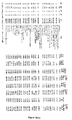
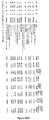
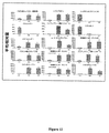
(タンパク質マーカー)本明細書中に提供されるタンパク質マーカーは、図1〜7に提示される表中に列挙される。 (Protein Markers) The protein markers provided herein are listed in the tables presented in FIGS.
より詳細には、健常患者から、および初期OAまたは末期OAをもつ患者から得られた滑液のサンプルを分析することにより、本出願人らは、図7(A)に列挙されるタンパク質が、正常/健常と初期OAとの間、正常/健常と末期OAとの間を区別することを見出した。本出願人らはまた、図7(B)に列挙されるタンパク質が、初期OAと末期OAとの間を区別することを見出した。 More specifically, by analyzing samples of synovial fluid obtained from healthy patients and from patients with early or late OA, Applicants have determined that the proteins listed in FIG. It was found that a distinction was made between normal / healthy and early OA and between normal / healthy and end-stage OA. Applicants have also found that the proteins listed in FIG. 7 (B) distinguish between early and late OA.
さらに、本出願人らは、初期OAおよび末期OAをもつ患者から得た滑液のサンプルが、正常個体から得た滑液のサンプルと比較して、図1および図2にそれぞれ列挙されるタンパク質の過剰発現(すなわち、増加した発現レベル)を示すことを見出した。 In addition, Applicants have compared the synovial fluid samples obtained from patients with early and late OA to the proteins listed in FIGS. 1 and 2, respectively, as compared to synovial fluid samples from normal individuals. Was found to exhibit overexpression (ie increased expression levels).
同様に、本出願人らは、初期OAおよび末期OAをもつ患者から得られた滑液のサンプルが、正常個体から得た滑液のサンプルと比較して、図4および図5にそれぞれ列挙されるタンパク質のより低い発現(すなわち、減少した発現のレベル)を示すことを見出した。 Similarly, Applicants have listed synovial fluid samples obtained from patients with early and late OA in FIGS. 4 and 5, respectively, compared to synovial fluid samples obtained from normal individuals. Were found to exhibit lower expression (ie reduced levels of expression).
さらに、図3に列挙されたタンパク質は、末期OAをもつ患者からの滑液サンプル中で、初期OAをもつ患者から得た滑液サンプルと比較して増加したレベルの発現を示すことが見いだされ;その一方、図7に列挙されたタンパク質は、後期OAをもつ患者からの滑液サンプル中で、初期OAをもつ患者からの滑液サンプルと比較して減少したレベルの発現を示すことが見出された。 In addition, the proteins listed in FIG. 3 were found to show increased levels of expression in synovial fluid samples from patients with terminal OA compared to synovial fluid samples obtained from patients with early OA. On the other hand, the proteins listed in FIG. 7 are seen to show reduced levels of expression in synovial fluid samples from patients with late OA compared to synovial fluid samples from patients with early OA. It was issued.
従って、図1〜図7に提示されるタンパク質の発現プロフィールは、OAを診断するため、およびこの疾患の進行の程度を決定する(すなわち、この疾患のステージを決定する)ために用いられ得る。 Thus, the protein expression profiles presented in FIGS. 1-7 can be used to diagnose OA and to determine the extent of progression of the disease (ie, determine the stage of the disease).
(核酸マーカー)本発明によって提供されるその他のOA生物マーカーは、上記に記載の本発明のタンパク質マーカー(またはそのアナログもしくはフラグメント)をコードするポリヌクレオチド配列、およびこれらの核酸分子の一部分とハイブリダイズするポリヌクレオチドを含む核酸分子を含む。 (Nucleic acid markers) Other OA biomarkers provided by the present invention hybridize to polynucleotide sequences encoding the above-described protein markers of the present invention (or analogs or fragments thereof) and portions of these nucleic acid molecules. A nucleic acid molecule comprising a polynucleotide.
(OA発現プロフィールマップ)この疾患の特定ステージ(例えば、健常被験体、OAをもつ患者、初期OAをもつ患者、および末期OAをもつ患者)に罹患した個体からの生物学的サンプルを用いて得た所定のセットの生物マーカーの発現レベルに関する情報は、OA発現プロフィールマップを形成するためにグループ分けされ得る。好ましくは、OA発現プロフィールマップは、同じ疾患ステージをもつ多数の個体の研究から得る。特定の実施形態では、OA発現プロフィールマップは、一致した年齢、性、および身体指数をもつ個体からのサンプルを用いて確立される。各発現プロフィールマップは、未知の生物学的サンプルから生成された生物マーカー発現パターンに対する比較のためのテンプレートを提供する。OA発現プロフィールマップは、グラフ表示(例えば、紙またはコンピュータースクリーン)、物理的表示(例えば、ゲルまたはアレイ)あるいはコンピューター読み出し可能媒体中に記録されたデジタル表示として提示され得る。 (OA expression profile map) Obtained using biological samples from individuals affected by a specific stage of the disease (eg, healthy subjects, patients with OA, patients with early OA, and patients with end-stage OA). Information regarding the expression level of a given set of biomarkers can be grouped to form an OA expression profile map. Preferably, the OA expression profile map is obtained from the study of multiple individuals with the same disease stage. In certain embodiments, an OA expression profile map is established using samples from individuals with matched age, sex, and body index. Each expression profile map provides a template for comparison against biomarker expression patterns generated from unknown biological samples. The OA expression profile map can be presented as a graphical display (eg, paper or computer screen), a physical display (eg, gel or array), or a digital display recorded in a computer readable medium.
II−診断方法
当業者によって認識され得るように、その発現がOAと相関し、そしてこの疾患の異なるステージ間を区別し得る生物マーカーのセットは、未知の生物学的サンプルを同定/研究するために用いられ得る。従って、本発明は、OAを有すると疑われる被験体から得られる生物学的サンプルを特徴付けるため、被験体中のOAを診断するため、および被験体中のOAの進行を評価するための方法を提供する。このような方法では、被験体から得られた生物学的サンプルについて決定された生物マーカーの発現レベルが、1つ以上のコントロールサンプル中のレベルに比較される。これらコントロールサンプルは、健常個体(または健常個体の群)から、OAを罹患した個体(または個体の群)から、および/またはこの疾患の特定ステージ(例えば、初期OAまたは末期OA)に罹患した個体(または個体の群)から得られ得る。上記で述べたように、目的の生物マーカーのコントロール発現レベルは、好ましくは、有意な数の個体から決定され、そして平均値または中間値が得られる。特定の好ましい実施形態では、調査される生物学的サンプルについて決定された発現レベルは、上記に記載のように、OAについての少なくとも1つの発現プロフィールマップと比較される。
II-Diagnostic Methods As can be appreciated by those skilled in the art, a set of biomarkers whose expression correlates with OA and can distinguish between different stages of the disease is for identifying / studying unknown biological samples Can be used. Accordingly, the present invention provides a method for characterizing a biological sample obtained from a subject suspected of having OA, for diagnosing OA in a subject, and for assessing the progression of OA in a subject. provide. In such methods, the expression level of a biomarker determined for a biological sample obtained from a subject is compared to the level in one or more control samples. These control samples can be from healthy individuals (or groups of healthy individuals), from individuals (or groups of individuals) affected by OA, and / or individuals affected by a particular stage of the disease (eg, early OA or end-stage OA). (Or a group of individuals). As stated above, the control expression level of the biomarker of interest is preferably determined from a significant number of individuals and an average or intermediate value is obtained. In certain preferred embodiments, the expression level determined for the biological sample investigated is compared to at least one expression profile map for OA, as described above.
(生物学的サンプル)
本発明の方法は、任意のタイプの生物学的サンプルの研究に適用され得、1つ以上の本発明の生物マーカーがアッセイされることを可能にする。適切な生物学的サンプルの例は、制限されないで、尿、血液、関節流体、唾液、および滑液を含む。診断の本発明の方法の実施において用いられる生物学的サンプルは、被験体から収集された新鮮または凍結サンプル、既知の診断、処置および/または結果歴をもつ保管サンプルであり得る。生物学的サンプルは、例えば、被験体から血液を抜くこと、または微細ニードル吸引またはニードル生検を用いることによるような、任意の非侵襲的手段によって収集され得る。あるいは、生物学的サンプルは、例えば、外科的生検を含む侵襲的方法によって収集され得る。
(Biological sample)
The methods of the invention can be applied to the study of any type of biological sample, allowing one or more biomarkers of the invention to be assayed. Examples of suitable biological samples include, without limitation, urine, blood, joint fluid, saliva, and synovial fluid. The biological sample used in the practice of the inventive method of diagnosis can be a fresh or frozen sample collected from a subject, a stored sample with a known diagnosis, treatment and / or history of results. The biological sample can be collected by any non-invasive means such as, for example, by drawing blood from the subject or using fine needle aspiration or needle biopsy. Alternatively, the biological sample can be collected by invasive methods including, for example, surgical biopsy.
特定の実施形態では、本発明の方法は、サンプルを処理することなく、または限られた処理とともにこの生物学的サンプル自体に対して実施される。 In certain embodiments, the methods of the invention are performed on the biological sample itself without processing the sample or with limited processing.
その他の実施形態では、本発明の方法は、単一細胞レベル(例えば、生物学的サンプルからの細胞の単離)で実施される。しかし、このような実施形態では、本発明の方法は、好ましくは、多くの細胞を含むサンプルを用いて実施され、ここで、アッセイは、サンプル中に存在する細胞の全体のコレクションに亘る発現を「平均する」。好ましくは、目的の生物マーカーのセットの発現を正確かつ信頼性よく決定するために十分な生物学的サンプルがある。複数の生物学的サンプルは、組織の代表的サンプリングを得るために、同じ組織/身体部分からとられ得る。 In other embodiments, the methods of the invention are performed at the single cell level (eg, isolation of cells from a biological sample). However, in such embodiments, the methods of the invention are preferably carried out with a sample containing a large number of cells, wherein the assay shows expression over the entire collection of cells present in the sample. “Average”. Preferably there are sufficient biological samples to accurately and reliably determine the expression of the set of biomarkers of interest. Multiple biological samples can be taken from the same tissue / body part to obtain a representative sampling of tissue.
なおその他の実施形態では、本発明の方法は、生物学的サンブルから調製されたタンパク質抽出物に対して実施される。好ましくは、このタンパク質抽出物は、総タンパク質含量を含む。しかし、この方法はまた:膜タンパク質、核タンパク質、および細胞質ゾルタンパク質の1つ以上を含む抽出物に対して実施され得る。タンパク質抽出の方法は、当該技術分野で周知である(例えば、「Protein Methods」、D.M.Bollagら、第2版、1996、Wiley−Liss;「Protein Purification Methods:A Practical Approach」、E.L.HarrisおよびS.Angal(編)、1989;「Protein Purification Techniques:A Practical Approach」、S.Roe、第2版、2001、Oxford University Press;「Principles and Reactions of Protein Extraction、Purification、and Characterization」、H.Ahmel、2005、CRC Press:Boca Raton、FL)。多くの異なる、かつ多様なキットが、体液および組織からタンパク質を抽出するために用いられ得、そして、例えば、BioRad Laboratories(Hercules、CA)、BD Biosciences Clonetech(Mountain View、CA)、Chemicon International,Inc.(Temecula、CA)、Calbiochem(San Diego、CA)、Pierce Biotechnology(Rockford、IL)、およびInvitrogen Corp.(Carlsbad、CA)から市販され入手可能である。従われるべきプロトコールを詳細に記載するUser Guideが、通常、これらのキットのすべてに含まれる。感度、処理時間およびコストは、キット毎に異なり得る。当業者は、特定の状況に最も適切なキット(単数または複数)を容易に選択し得る。タンパク質抽出物が得られた後、この抽出物中のタンパク質濃度は、好ましくは、定量化され得るタンパク質の信号を可能にするためにコントロールサンプルのそれと同じである値に標準化される。このような標準化は、測光法または分光測定法またはゲル電気泳動を用いてなされ得る。 In still other embodiments, the methods of the invention are performed on protein extracts prepared from biological samples. Preferably, the protein extract contains the total protein content. However, this method can also be performed on extracts containing one or more of: membrane proteins, nucleoproteins, and cytosolic proteins. Methods of protein extraction are well known in the art (see, eg, “Protein Methods”, DM Bollag et al., 2nd Edition, 1996, Wiley-Liss; “Protein Purification Methods: A Practical Approach”, E. L. Harris and S. Angal (eds.), 1989; “Protein Purification Techniques: A Practical Approach, S. Roe, Second Criteria, Second University, and Oxford University Press”. , H. Ahmel, 2005, CRC Press: Boca Raton, FL). A number of different and diverse kits can be used to extract proteins from body fluids and tissues and are, for example, BioRad Laboratories (Hercules, CA), BD Biosciences Clonetech (Mountain View, CA), Chemicon International, Inc. . (Temecula, CA), Calbiochem (San Diego, CA), Pierce Biotechnology (Rockford, IL), and Invitrogen Corp. Commercially available from (Carlsbad, CA). A User Guide that details the protocol to be followed is usually included in all of these kits. Sensitivity, processing time and cost can vary from kit to kit. One skilled in the art can readily select the kit (s) most appropriate for a particular situation. After the protein extract is obtained, the protein concentration in the extract is preferably normalized to a value that is the same as that of the control sample to allow a protein signal that can be quantified. Such standardization can be done using photometric or spectroscopic methods or gel electrophoresis.
なおその他の実施形態では、本発明の方法は、生物学的サンプルから抽出された核酸分子に対して実施される。例えば、RNAは、分析前にサンプルから抽出され得る。RNA抽出の方法は、当該技術分野で周知である(例えば、J.Sambrookら、「Molecular Cloning:A Laboratory Mannual」、1989、第2版、Cold Spring Harbor Laboratory Press:Cold Spring Harbor、NYを参照のこと)。体液または組織からのRNA単離の大部分の方法は、RNaseを迅速かつ効率的に不活性化するためにタンパク質変性剤の存在下での組織の破壊に基く。単離された総RNAは、次いで、タンパク質夾雑物からさらに精製され得、そして選択的エタノール沈殿、フェノール/クロロホルム抽出、次いで、イソプロパノール沈殿または塩化セシウム、塩化リチウムまたは三フッ化酢酸セシウムグラジエント遠心分離によって濃縮される。体液または組織からRNA(すなわち、総RNAまたはmRNA)を抽出するためにキットもまた利用可能であり、そして例えば、Ambion、Inc.(Austin、TX)、Amersham Biosciences(Piscataway、NJ)、BD Biosciences Clonetech(Palo Alto、CA)、BioRad Laboratoryies(Hercules、CA)、GIBCO BRL(Gaithersburg、MD)、およびQiagen、Inc.(Valencia、CA)から市販され入手可能である。 In still other embodiments, the methods of the invention are performed on nucleic acid molecules extracted from a biological sample. For example, RNA can be extracted from a sample prior to analysis. Methods of RNA extraction are well known in the art (see, eg, J. Sambrook et al., “Molecular Cloning: A Laboratory Manual”, 1989, 2nd Edition, Cold Spring Harbor Laboratory Press: Cold Spring N thing). Most methods of RNA isolation from body fluids or tissues are based on tissue destruction in the presence of protein denaturing agents in order to inactivate RNase quickly and efficiently. The isolated total RNA can then be further purified from protein contaminants and by selective ethanol precipitation, phenol / chloroform extraction followed by isopropanol precipitation or cesium chloride, lithium chloride or cesium trifluoroacetate gradient centrifugation. Concentrated. Kits are also available for extracting RNA (ie, total RNA or mRNA) from body fluids or tissues and are described in, for example, Ambion, Inc. (Austin, TX), Amersham Biosciences (Piscataway, NJ), BD Biosciences Clonetech (Palo Alto, CA), BioRad Laboratories (Hercules, CA), GIBCO BRL (ur Q, BQ) Commercially available from (Valencia, CA).
特定の実施形態では、抽出後、mRNAは増幅され、そしてcDNAに転写され、これは、次いで、適切なRNAポリメラーゼによる複数ラウンドの転写のためのテンプレートとして供され得る。増幅方法は、当該技術分野で周知である(例えば、A.R.KimmelおよびS.L.Berger、Methods Enzymol.1987、152:307〜316;J.Sambrookら、「Molecular Cloning:A Laboratory Mannual」、1989、第2版、Cold Harbour Laboratory Press:New York;「Short Protocols in Molecular Biology」、F.M.Ausubel(編)、2002、第5版、John Wiley&Sons;米国特許第4,683,195号;同第4,683,202号および同第4,800,159号を参照のこと)。逆転写反応は、係留オリゴ−dTプライマー、またはランダム配列プライマーのような非特異的プライマーを用いて、またはモニターされている各プローブに対するRNAに相補的な標的特異的プライマーを用いて、または熱安定性DNAポリメラーゼ(例えば、トリ骨髄芽球症ウイルス逆転写酵素またはMoloneyマウス白血病逆転写酵素)を用いて実施され得る。
In certain embodiments, after extraction, the mRNA is amplified and transcribed into cDNA, which can then serve as a template for multiple rounds of transcription with an appropriate RNA polymerase. Amplification methods are well known in the art (eg, AR Kimmel and SL Berger, Methods Enzymol. 1987, 152: 307-316; J. Sambrook et al., “Molecular Cloning: A Laboratory Manual”). 1989, 2nd edition, Cold Harbor Laboratory Press: New York; "Short Protocols in Molecular Biology", FM Ausubel (ed.), 2002, 5th edition, John Wiley &
(タンパク質発現レベルの決定)
本発明の診断方法は、一般に、被験体から得た生物学的サンプル中の複数のポリペプチドの発現レベルの決定を含む。本発明の方法の実施におけるタンパク質発現レベルの決定は、任意の適切な方法によって実施され得る(例えば、E.HarlowおよびA.Lane、「Antibodies:A Laborarties Mannual」、1988、Cold Spring Harbor Laboratoy:Cold Spring Harbor、NYを参照のこと)。
(Determination of protein expression level)
The diagnostic methods of the invention generally involve determining the expression level of a plurality of polypeptides in a biological sample obtained from a subject. Determination of protein expression levels in the practice of the methods of the invention can be performed by any suitable method (eg, E. Harlow and A. Lane, “Antibodies: A Laboratories Manual”, 1988, Cold Spring Harbor Laboratory: Cold See Spring Harbor, NY).
(結合試薬)一般に、上記発現レベルは、被験体から単離された生物学的システムを、1つ以上のタンパク質マーカーのための結合試薬と接触すること;このサンプル中の、上記結合試薬に結合するポリペプチドのレベルを検出すること;およびこのサンプル中のポリペプチドのレベルを、コントロールサンプル中のポリペプチドのレベルと比較することにより決定される。本明細書中で用いられるとき、用語「結合試薬」は、本発明のタンパク質マーカーに特異的に結合するポリペプチドまたは抗体のような実体をいう。ポリペプチドに「特異的に結合する」実体は、このポリペプチドと検出可能なレベルで反応/相互作用するが、非関連配列または異なるポリペプチドの配列を含むペプチドと検出可能に反応/相互作用しない。 (Binding Reagent) Generally, the expression level is obtained by contacting a biological system isolated from a subject with a binding reagent for one or more protein markers; binding to the binding reagent in the sample. Detecting the level of polypeptide to be determined; and comparing the level of polypeptide in the sample to the level of polypeptide in the control sample. As used herein, the term “binding reagent” refers to an entity such as a polypeptide or antibody that specifically binds to a protein marker of the present invention. An entity that "specifically binds" to a polypeptide reacts / interacts with this polypeptide at a detectable level, but does not detectably react / interact with peptides that contain unrelated sequences or sequences of different polypeptides. .
特定の実施形態では、上記結合試薬は、ペプチド成分を有するかまたは有さないリボソーム、RNA分子、またはポリペプチドであり得る(例えば、タンパク質マーカーのポリペプチド配列を含むポリペプチド、そのペプチド改変体、またはこのような配列の非ペプチド模倣物)。 In certain embodiments, the binding reagent can be a ribosome, RNA molecule, or polypeptide with or without a peptide component (eg, a polypeptide comprising a polypeptide sequence of a protein marker, a peptide variant thereof, Or a non-peptidomimetic of such a sequence).
その他の実施形態では、上記結合試薬は、本発明のタンパク質マーカーに特異的な抗体である。本発明の方法における使用のための適切な抗体は、モノクローナル抗体およびポリクローナル抗体、免疫学的に活性なフラグメント(例えば、Fabまたは(Fab)2フラグメント)、抗体重鎖、ヒト化抗体、抗体軽鎖、およびキメラ抗体を含む。モノクローナル抗体およびポリクローナル抗体、フラグメントおよびキメラを含む抗体は、当該技術分野で公知の方法を用いて調製され得る(例えば、R.G.MageおよびE.Lamoyi、「Monoclonal Antibody Production Techniques and Applications」、1987、Marcel Dekker、Inc.:New York、79〜97頁;G.KohlerおよびC.Milstein、Nature、1975、256:495〜497;D.Kozborら、J.Immunol.Methods、1985、81:31〜42;およびR.J.Coteら、Proc.Natl.Acad.Sci.1983、80:2026から203;R.A.Lerner、Nature、1982、299:593〜596;A.C.Nairnら、Nature、1982、299;734〜736;A.J.Czernikら、Methods Enzymol.1991、201:264〜283;A.J.Czernikら、Neumethods;Regulatory Protein Modification:Techniques&Protocols、1997、30:219〜250;A.J.Czernikら、Neuroprotocols、1995、6:56〜61;H.Zhangら、J.Biol.Chem.2002、277:39379〜39387;S.L.Morrisonら、Proc.Natl.Acad.Sci.、1984、81:6851〜6855;M.S.Neubergerら、Nature、1984、312;604〜608;S.Takedaら、Nature、1985、314:452〜454を参照のこと)。本発明の方法で用いられるべき抗体は、当該技術分野で周知の方法によって精製され得る(例えば、S.A.Minden、「Monoclonal Antibody Purification」、1996、IBC Biomedical Library Series:Southbridge、MAを参照のこと)。例えば、抗体は、タンパク質マーカーまたはそのフラグメントが結合されるカラム上の通過によりアフィニティー精製され得る。結合した抗体は、次いで、高塩濃度をもつ緩衝液を用いてこのカラムから溶出され得る。 In other embodiments, the binding reagent is an antibody specific for the protein marker of the present invention. Suitable antibodies for use in the methods of the invention include monoclonal and polyclonal antibodies, immunologically active fragments (eg, Fab or (Fab) 2 fragments), antibody heavy chains, humanized antibodies, antibody light chains. And chimeric antibodies. Antibodies, including monoclonal and polyclonal antibodies, fragments and chimeras, can be prepared using methods known in the art (see, eg, RG Mage and E. Lamoyi, “Monoclonal Antibody Productions and Applications”, 1987. , Marcel Dekker, Inc .: New York, pages 79-97; G. Kohler and C. Milstein, Nature, 1975, 256: 495-497; D. Kozbor et al., J. Immunol. Methods, 1985, 81: 31-31. 42; and R. J. Cote et al., Proc. Natl. Acad. Sci. 1983, 80: 2026-203; Nature, 1982, 299: 593-596; A. C. Nairn et al., Nature, 1982, 299; 734-736; AJ Czernik et al., Methods Enzymol. 1991, 201: 264-283; Regulate Protein Modification: Techniques & Protocols, 1997, 30: 219-250; AJ Czernik et al., Neuroprotocols, 1995, 6: 56-61; H. Zhang et al., J. Bio. S. L. Morrison et al., Proc. Natl. Acad. Sci., 1984, 81: 6851-6855; .Neuberger et, Nature, 1984,312; 604~608; S.Takeda et, Nature, 1985,314: 452~454 see). The antibodies to be used in the methods of the invention can be purified by methods well known in the art (see, eg, SA Minden, “Monoclonal Antibody Purification”, 1996, IBC Biomedical Library Series: Southbridge, MA). thing). For example, antibodies can be affinity purified by passage over a column to which a protein marker or fragment thereof is bound. The bound antibody can then be eluted from the column using a buffer with a high salt concentration.
調製されることの代わりに、本発明の方法で用いられるべき抗体は、科学的供給源または商業的供給源から得られ得る。 Instead of being prepared, the antibodies to be used in the methods of the invention can be obtained from scientific or commercial sources.
(標識された結合試薬)好ましくは、上記結合試薬は、検出可能成分で直接的または間接的に標識される。検出可能な試薬の役割は、タンパク質マーカー(またはそのアナログもしくはフラグメント)への結合試薬の結合により形成される複合体の可視化を可能にすることによって、上記診断方法の検出ステップを促進することである。好ましくは、この検出可能な試薬は、それが、測定され得、そしてその強度が、分析されるサンプル中に存在するタンパク質マーカーの量に関連する(好ましくは比例する)信号を生成するように選択される。ポリペプチドおよび抗体のような生物学的分子を標識するための方法は、当該技術分野で周知である(例えば、「Affinity Techniques.Enzyme Purification:Part B」、Methods in Enzymol.、1974、Vol.34、W.B.JakobyおよびM.Wilneck(編)、Academic Press:New York、NY;およびM.WilchekおよびE.A.Bayer、Anal.Biochem.、1988、171:1〜32を参照のこと)。 (Labeled Binding Reagent) Preferably, the binding reagent is directly or indirectly labeled with a detectable moiety. The role of the detectable reagent is to facilitate the detection step of the diagnostic method by allowing visualization of the complex formed by binding of the binding reagent to a protein marker (or analog or fragment thereof). . Preferably, the detectable reagent is selected such that it generates a signal that can be measured and whose intensity is related (preferably proportional) to the amount of protein marker present in the sample being analyzed. Is done. Methods for labeling biological molecules such as polypeptides and antibodies are well known in the art (eg, “Affinity Technologies. Enzyme Purification: Part B”, Methods in Enzymol., 1974, Vol. 34). , WB Jakoby and M. Wilneck (eds.), Academic Press: New York, NY; and M. Wilchek and EA Bayer, Anal. Biochem., 1988, 171-1, 32). .
任意の広範な種類の検出可能な試薬が、本発明の実施において用いられ得る。適切な検出可能な試薬は、制限されないで:種々のリガンド、放射線核種、蛍光色素、化学発光試薬、マイクロパーティクル(例えば、量子ドット、ナノクリスタル、リン光体など)、酵素(例えば、ELISAで用いられるものなど、すなわち西洋ワサビペルオキシダーゼ、β−ガラクトシダーゼ、ルシフェラーゼ、アルカリホスファターゼ)、比色定量標識、磁性ビーズ、およびビオチン、ジオシシゲニンまたは抗血清またはモノクローナル抗体が利用可能なその他のハプテンおよびタンパク質)を含む。 Any of a wide variety of detectable reagents can be used in the practice of the present invention. Suitable detectable reagents are not limited: various ligands, radionuclides, fluorescent dyes, chemiluminescent reagents, microparticles (eg, quantum dots, nanocrystals, phosphors, etc.), enzymes (eg, used in ELISA) Such as horseradish peroxidase, β-galactosidase, luciferase, alkaline phosphatase), colorimetric labels, magnetic beads, and other haptens and proteins for which biotin, diocigenin or antisera or monoclonal antibodies are available.
特定の実施形態では、この結合試薬(例えば、抗体)は、キャリアまたは支持体(例えば、ビーズ、磁性粒子、ラテックス粒子、マイクロタイタープレートウェル、キュベット、またはその他の反応ベッセル)上に固定化され得る。適切なキャリアまたは支持体材料は、アガロース、セルロース、ニトロセルロース、デキストラン、Sephadex、Sepharose、リポソーム、カルボキシメチルセルロース、ポリアクリルアミド、ポリスチレン、斑糲岩(gabbros)、フィルターペーパー、磁石、イオン交換樹脂、プラスチックフィルム、プラスチックチューブ、ガラス、ポリアミン−メチルビニル−エーテル−マレイン酸コポリマー、アミノ酸コポリマー、エレチン−マレイン酸コポリマー、ナイロン、シルクなどを含む。結合試薬は、第1の結合試薬に特異的な第2の結合試薬を用いて間接的に固定化され得る(例えば、タンパク質マーカーに特異的なマウス抗体は、キャリアまたは支持体上に被覆されたヒツジ抗マウスIgG Fcフラグメント特異的抗体を用いて固定化され得る)。 In certain embodiments, the binding reagent (eg, antibody) can be immobilized on a carrier or support (eg, beads, magnetic particles, latex particles, microtiter plate wells, cuvettes, or other reaction vessels). . Suitable carrier or support materials are agarose, cellulose, nitrocellulose, dextran, Sephadex, Sepharose, liposomes, carboxymethylcellulose, polyacrylamide, polystyrene, gabbros, filter paper, magnets, ion exchange resins, plastic films. , Plastic tubes, glass, polyamine-methylvinyl-ether-maleic acid copolymers, amino acid copolymers, eletin-maleic acid copolymers, nylon, silk and the like. The binding reagent can be indirectly immobilized using a second binding reagent specific for the first binding reagent (eg, a mouse antibody specific for a protein marker is coated on a carrier or support). Can be immobilized using sheep anti-mouse IgG Fc fragment specific antibody).
本発明の診断方法におけるタンパク質発現レベルは、免疫アッセイを用いて決定され得る。このようなアッセイの例は、ラジオイムノアッセイ、酵素免疫アッセイ(例えば、ELISA)、免疫蛍光免疫沈降、ラテックス凝集、血球凝集、および組織化学的試験であり、これらは、当該技術分野で周知の従来方法である。当業者によって認識され得るように、上記免疫アッセイは、競争的または非競争的であり得る。結合試薬のタンパク質マーカーとの結合によつて形成される複合体によって生成される信号の検出および定量の方法は、アッセイの、および検出可能な成分(例えば、蛍光成分)の性質に依存する。 Protein expression levels in the diagnostic methods of the invention can be determined using immunoassays. Examples of such assays are radioimmunoassays, enzyme immunoassays (eg, ELISA), immunofluorescent immunoprecipitation, latex aggregation, hemagglutination, and histochemical tests, which are conventional methods well known in the art. It is. As can be appreciated by one skilled in the art, the immunoassay can be competitive or non-competitive. The method of detection and quantification of the signal produced by the complex formed by binding of the binding reagent to the protein marker depends on the nature of the assay and the detectable component (eg, fluorescent component).
あるいは、上記タンパク質発現レベルは、質量分析法に基く方法、またはタンパク質の検出のために当該技術分野で公知の方法に基く(標識されたリガンドの使用を含む)イメージを用いて決定され得る。その他の適切な方法は、プロテオミクスを基礎にした方法を含む。サンプル中のタンパク質の発現の全体的変化を研究するプロテオミクスは、代表的には以下のステップを含む:(1)電気泳動(1−D PAGE)によるサンプル中の個々のタンパク質の分離、(2)ゲルから回収される個々のタンパク質の同定(例えば、質量分析法またはN−末端配列決定による)、および(3)生物情報(bioinformation)を用いるデータの分析。 Alternatively, the protein expression level can be determined using images based on mass spectrometry or methods known in the art for detection of proteins (including the use of labeled ligands). Other suitable methods include proteomics-based methods. Proteomics that study global changes in protein expression in a sample typically include the following steps: (1) Separation of individual proteins in the sample by electrophoresis (1-D PAGE), (2) Identification of individual proteins recovered from the gel (eg, by mass spectrometry or N-terminal sequencing), and (3) Analysis of data using bioinformation.
(ポリヌクレオチド発現レベルの決定)
上記で既に述べたように、本発明の診断方法は、本発明のタンパク質マーカーをコードするポリヌクレオチド配列を含む核酸分子のセットの発現レベルの決定を含み得る。本発明の方法の実施における核酸分子の発現レベルの決定は、制限されないで、Southern分析、Northern分析、ポリメラーゼ連鎖反応(PCR)(例えば、米国特許第4,683,195号;同第4,683,202号、および同第6,040,166号;「PCR Protocols:A Guide to Methods and Applications」、Innisら(編)、1990、Academic Press:New Yorkを参照のこと)、逆転写酵素PCR(RT−PCT)、係留PCR、競争PCR(例えば、米国特許第5,747,251号を参照のこと)、cDNA末端の迅速増幅(RACE)(例えば、「Gene Cloning and Analysis:Current Innovations、1997、99〜115頁を参照のこと」);リガーゼ連鎖反応(LCR)(例えば、EP 01 320 308を参照のこと)、一側方PCR(Oharaら、Proc.Natl.Acad.Sci.、1989、86:5673〜5677)、in situハイブリダイゼーション、Taqmanを基礎にしたアッセイ(Hollandら、Proc.Natl.Acad.Sci.、1991、88:7276〜7280)、鑑別ディスプレイ(例えば、Liangら、Nucl.Acid.Res.、1993、21:3269〜3275を参照のこと)およびその他のRNAフィンガープリンティング技法、核酸配列を基礎にした増幅(NASBA)およびその他の転写を基礎にした増幅システム(例えば、米国特許第5,409,818号および同第5,554,527号を参照のこと)、Qβレプリカーゼストランド置換増幅(SDA)、修復鎖反応(PCR)、ヌクレアーゼ保護アッセイ、サブトラクションを基礎にした方法、Rapid−ScanTMなどを含む任意の適切な方法によって実施され得る。
(Determination of polynucleotide expression level)
As already mentioned above, the diagnostic method of the present invention may comprise the determination of the expression level of a set of nucleic acid molecules comprising a polynucleotide sequence encoding a protein marker of the present invention. The determination of the expression level of a nucleic acid molecule in the practice of the method of the present invention is not limited and includes Southern analysis, Northern analysis, polymerase chain reaction (PCR) (eg, US Pat. Nos. 4,683,195; 4,683). 202, and 6,040,166; "PCR Protocols: A Guide to Methods and Applications", Innis et al. (Eds.), 1990, Academic Press: New York), reverse transcriptase PCR (see RT-PCT), tethered PCR, competitive PCR (see, eg, US Pat. No. 5,747,251), rapid amplification of cDNA ends (RACE) (see, eg, Gene Cloning and Analysis: Current Innovat ons, 1997, pages 99-115 "); ligase chain reaction (LCR) (see, eg, EP 01 320 308), unilateral PCR (Ohara et al., Proc. Natl. Acad. Sci. 1989, 86: 5673-5677), in situ hybridization, Taqman-based assays (Holland et al., Proc. Natl. Acad. Sci., 1991, 88: 7276-7280), differential displays (eg Liang et al. , Nucl. Acid. Res., 1993, 21: 3269-3275) and other RNA fingerprinting techniques, nucleic acid sequence based amplification (NASBA) and other transcription based amplification systems (eg, , US Patent No. , See No. and the No. 5,554,527 409,818), Qβ replicase Zest land displacement amplification (SDA), repair chain reaction (PCR), methods nuclease protection assays, subtraction underlying, Rapid-Scan It can be implemented by any suitable method including TM and the like.
生物学的サンプル中のポリヌクレオチド配列の検出における使用のための核酸プローブは、当該技術分野で公知の従来方法を用いて構築され得る。適切なプローブは、タンパク質マーカーをコードする核酸の領域からの少なくとも5つの連続アミノ酸をコードし、そして好ましくは15〜40のヌクレオチドを含む核酸配列に基き得る。核酸プローブは、結合試薬の場合に上記で述べたように、検出可能な成分で標識され得る。この核酸プローブと検出可能な成分との間の会合は、共有結合または非共有結合であり得る。検出可能な成分は、核酸プローブに直接的、またはリンカーを経由して間接的に付着され得る(E.S.Mansfieldら、Mol.Cell.Probes、1995、9:145〜156)。核酸分子を標識するための方法は、当該技術分野で周知である(標識するプロトコール、標識検出技法およびこの分野における最近の発展の総説には、例えば、L.J.Kricka、Ann.Clin.Biochem.2002、39:114〜129;R.P.van Gijswijikら、Expert Rev.Mol.Diagn.2001、1:81〜91;およびS.Joosら、J.Biotechnol.1994、35:135〜153を参照のこと)。 Nucleic acid probes for use in the detection of polynucleotide sequences in biological samples can be constructed using conventional methods known in the art. A suitable probe encodes at least 5 contiguous amino acids from the region of the nucleic acid that encodes the protein marker and may preferably be based on a nucleic acid sequence comprising 15-40 nucleotides. The nucleic acid probe can be labeled with a detectable moiety as described above in the case of a binding reagent. The association between the nucleic acid probe and the detectable moiety can be covalent or non-covalent. The detectable moiety can be attached directly to the nucleic acid probe or indirectly via a linker (ES Mansfield et al., Mol. Cell. Probes, 1995, 9: 145-156). Methods for labeling nucleic acid molecules are well known in the art (for a review of labeling protocols, label detection techniques and recent developments in the field, see, eg, LJ Kricka, Ann. Clin. Biochem. 2002, 39: 114-129; R.P. van Gijswijik et al., Expert Rev. MoI.Dign. 2001, 1: 81-91; and S. Joos et al., J. Biotechnol. 1994, 35: 135-153. See
核酸プローブは、上記タンパク質マーカーをコードするポリヌクレオチドを検出するためのハイブリダイゼーション技法で用いられ得る。この技法は、一般に、被験体から得た生物学的サンプルから単離された核酸分子を、核酸プローブと、この核酸プローブと上記核酸分子中の相補的配列との間で特異的ハイブリダイゼーションが生じ得る条件下で接触すること、およびインキュベートすることを含む。インキュベーションの後、ハイブリダイズしない核酸は除去され、そして上記プローブにハイブリダイズした核酸の存在および量が検出され、そして定量される。 Nucleic acid probes can be used in hybridization techniques to detect polynucleotides that encode the protein markers. This technique generally involves the specific hybridization of a nucleic acid molecule isolated from a biological sample obtained from a subject between the nucleic acid probe and a complementary sequence in the nucleic acid probe and the nucleic acid molecule. Including contacting and incubating under conditions to obtain. After incubation, non-hybridized nucleic acid is removed and the presence and amount of nucleic acid hybridized to the probe is detected and quantified.
タンパク質マーカーをコードするポリヌクレオチド配列を含む核酸分子の検出は、PCRのような増幅方法を用いる特異的ポリヌクレオチド配列の増幅、次いで、当該技術分野で公知の技法を用いる増幅された分子の分析を含む。適切なプライマーは、当業者によって慣用的に設計され得る。アッセイ条件下におけるハイブリダイゼーションを最大にするために、本発明の方法で採用されるプライマーおよびプローブは、タンパク質マーカーをコードする核酸の一部と少なくとも60%、好ましくは少なくとも75%そしてより好ましくは少なくとも90%の同一性を有する。 Detection of a nucleic acid molecule containing a polynucleotide sequence encoding a protein marker involves the amplification of a specific polynucleotide sequence using an amplification method such as PCR, followed by analysis of the amplified molecule using techniques known in the art. Including. Appropriate primers can be routinely designed by those skilled in the art. In order to maximize hybridization under assay conditions, the primers and probes employed in the methods of the invention are at least 60%, preferably at least 75% and more preferably at least 75% with a portion of the nucleic acid encoding the protein marker. 90% identity.
本明細書に記載されるハイブリダイゼーションおよび増幅技法は、本発明のタンパク質マーカーをコードするポリヌクレオチド配列を含む核酸分子の発現の質的および量的局面をアッセイするために用いられ得る。 The hybridization and amplification techniques described herein can be used to assay qualitative and quantitative aspects of expression of a nucleic acid molecule comprising a polynucleotide sequence encoding a protein marker of the present invention.
あるいは、各タンパク質マーカーをコードする核酸由来のオリゴヌクレオチドまたはより長いフラグメントが、マイクロアレイにおける標的として用いられ得る。それらの産生の多くの異なるアレイ形態および方法が当業者に公知である(例えば、米国特許番号第5,445,934号;同第5,532,128号;同第5,556,752号;同第5,242,974号;同第5,384,261号;同第5,405,783号;同第5,412,087号;同第5,424,186号;同第5,429,807号;同第5,436,327号;同第5,472,672号;同第5,527,681号;同第5,529,756号;同第5,545,531号;同第5,554,501号;同第5,561,071号;同第5,571,639号;同第5,593,839号;同第5,599,695号;同第5,624,711号;同第5,658,734号;および同第5,700,637号を参照のこと)。マイクロアレイ技法は、大多数のポリヌクレオチド配列の定常状態の同時の測定を可能にする。現在広く用いられるマイクロアレイは、cDNAアレイおよびオリゴヌクレオチドアレイを含む。マイクロアレイを用いる分析は、一般に、このマイクロアレイ上の既知の位置に固定化された核酸プローブにハイブリダイズするサンプルからのcDNA配列を検出するために用いられる標識されたプローブから受ける信号の強度の測定を基礎にする(例えば、米国特許第6,004,755号;同第6,218,114号;同第6,218,122号;および同第6,271,002号を参照のこと)。アレイを基礎にした遺伝子発現方法は当該技術分野で公知であり、そして多くの科学的刊行物および特許に記載されている(例えば、M.Schenaら、Science、1995、270:467〜470;M.Schenaら、Proc.Natl.Acad.Sci.USA 1996、93:10614〜10619;J.J.Chenら、Genomics、1998、51:313〜324;米国特許第5,143,854号;同第5,445,934号;同第5,807,522号;同第5,837,832号;同第6,040,138号;同第6,045,996号;同第6,284,460号;および同第6,607,885号を参照のこと)。 Alternatively, oligonucleotides or longer fragments from nucleic acids encoding each protein marker can be used as targets in the microarray. Many different array forms and methods for their production are known to those skilled in the art (eg, US Pat. Nos. 5,445,934; 5,532,128; 5,556,752; 5,242,974; 5,384,261; 5,405,783; 5,412,087; 5,424,186; 5,429 No. 5,436,327; No. 5,472,672; No. 5,527,681; No. 5,529,756; No. 5,545,531; No. 5,554,501; No. 5,561,071; No. 5,571,639; No. 5,593,839; No. 5,599,695; No. 5,624 No. 711; No. 5,658,734; and No. 5,700,637 Of it). Microarray technology allows simultaneous measurement of the steady state of the majority of polynucleotide sequences. Currently widely used microarrays include cDNA arrays and oligonucleotide arrays. Analysis using a microarray generally measures the intensity of the signal received from a labeled probe used to detect cDNA sequences from a sample that hybridizes to a nucleic acid probe immobilized at a known location on the microarray. (See, for example, US Pat. Nos. 6,004,755; 6,218,114; 6,218,122; and 6,271,002). Array-based gene expression methods are known in the art and are described in many scientific publications and patents (eg, M. Schena et al., Science, 1995, 270: 467-470; M Schena et al., Proc. Natl.Acad.Sci.USA 1996, 93: 10614-10619; No. 5,445,934; No. 5,807,522; No. 5,837,832; No. 6,040,138; No. 6,045,996; No. 6,284,460 No .; and 6,607,885).
(OA診断およびOAステージ分け)
一旦、(上記のように)分析されている生物学的サンプルの目的の生物マーカーの発現レベルが決定されると、それらは、1つ以上のコントロールサンプル中の発現レベルまたはOAについての少なくとも1つの発現プロフィールマップと比較される。
(OA diagnosis and OA stage division)
Once the expression level of the biomarker of interest of the biological sample being analyzed (as described above) is determined, they are at least one expression level or OA for one or more control samples. Compared to expression profile map.
本発明の方法による発現レベルの比較は、好ましくは、得られた発現レベルが、アッセイされるサンプルの量における差異、および用いられるサンプルの質における変動(例えば、抽出されたタンパク質の量、または試験されたmRNAの量および質)の両方について矯正された後に実施される。矯正は、当該技術分野で周知の異なる方法を用いて実施され得る。サンプルのタンパク質濃度は、サンプルが分析される前に、例えば、(すでに上記で述べたように)測光法もしくは分光光度法またはゲル電気泳動を用いて標準化され得る。核酸分子を含むサンプルの場合、矯正は、同じサンプル中の参照遺伝子(例えば、ハウスキーピング遺伝子)に対してレベルを正規化することによって実施され得る。あるいは、またはそれに加えて、正規化は、すべてのアッセイされた遺伝子またはその大きなサブセットの平均または中間信号(例えば、RT−PCRの場合にはCt)に基き得る(グローバル正規化アプローチ)。 Comparison of expression levels according to the methods of the present invention preferably results in differences in the amount of sample assayed and variations in the quality of the sample used (eg, amount of extracted protein or test Performed after correction for both (quantity and quality of mRNA produced). Correction can be performed using different methods well known in the art. The protein concentration of the sample can be normalized before the sample is analyzed, for example using photometry or spectrophotometry or gel electrophoresis (as already mentioned above). For samples containing nucleic acid molecules, correction can be performed by normalizing levels relative to a reference gene (eg, a housekeeping gene) in the same sample. Alternatively, or in addition, normalization can be based on the average or intermediate signal (eg, Ct in the case of RT-PCR) of all assayed genes or a large subset thereof (global normalization approach).
所定のセットの生物マーカーに対し、生物学的サンプルについて得られた発現パターンの、OAの特定ステージについて確立された発現プロフィールマップに対する比較は、生物マーカー毎の正規化された発現レベルの比較、および/または複数の生物マーカーのセット内の発現レベルの比の比較を含み得る。さらに、分析されている生物学的サンプルについて得られた発現パターンは、発現プロフィールマップの各々(例えば、非OAの発現プロフィールマップ、OAの発現プロフィールマップ、初期OAの発現プロフィールマップ、および末期OAの発現プロフィールマップ)に対して、またはすべてのOA発現プロフィールマップに基き作製された描写を規定する発現プロフィールマップに対して比較され得る。 For a given set of biomarkers, a comparison of the expression pattern obtained for a biological sample to an expression profile map established for a particular stage of OA includes a comparison of normalized expression levels for each biomarker, and A comparison of ratios of expression levels within a set of biomarkers may be included. In addition, the expression pattern obtained for the biological sample being analyzed can be obtained from each of the expression profile maps (eg, non-OA expression profile map, OA expression profile map, early OA expression profile map, and late OA expression profile). Expression profile map) or against an expression profile map that defines a depiction made based on all OA expression profile maps.
(適切な処置の選択)
本明細書中に記載された方法を用いて、熟練医師は、本発明の生物マーカーの発現レベルの決定を通じて患者に提供された診断および疾患ステージ分けを基に、各々の個々の患者に適合された処置を選択および処方し得る。特に、本発明は、医師に、初期OAを診断するための主観的でない手段を提供し、これは、介入がその最大の効果を有する可能性があるとき、痛みおよび長期間の無能力を潜在的に防ぎ、そして患者の生活の質を改善する初期の処置を可能にする。所定の患者のための適切な治療養生法の選択は、本発明の方法によって提供される診断/ステージ分けのみを基になされ得る。あいるは、医師はまた、OAを診断し、そしてその進行を評価するために現存する方法で用いられるその他の臨床的または病理学的パラメーターを考慮し得る。
(Selection of appropriate treatment)
Using the methods described herein, a skilled physician is adapted to each individual patient based on the diagnosis and disease staging provided to the patient through determination of the expression levels of the biomarkers of the invention. Treatment may be selected and prescribed. In particular, the present invention provides physicians with a non-subjective means for diagnosing early OA, which can potentially cause pain and long-term incapacity when the intervention may have its greatest effect. Allows early treatment to prevent and improve the patient's quality of life. The selection of an appropriate treatment regime for a given patient can be made based solely on the diagnosis / staging provided by the method of the present invention. Alternatively, the physician may also consider other clinical or pathological parameters used in existing methods to diagnose OA and assess its progression.
さらに、本発明によって提供されるOA診断およびOAステージ分けは、軟骨破壊の徴候が見えないであろうとき、または関節スペース中の変化がX線画像上では検出可能ではないであろうときでさえ、この疾患の進行がモニターされることを可能にする。 In addition, the OA diagnosis and OA staging provided by the present invention can be used even when signs of cartilage destruction will not be visible, or even when changes in the joint space will not be detectable on x-ray images Allowing the progression of this disease to be monitored.
III−キット
別の局面では、本発明は、本発明の診断方法を実施するために有用な材料を含むキットを提供する。本明細書に記載される診断/ステージ分け手順は、診断試験所、実験試験所、または開業医によって実施され得る。本発明は、これらの異なるセッティングで用いられ得るキットを提供する。
III-Kit In another aspect, the invention provides a kit comprising materials useful for performing the diagnostic methods of the invention. The diagnostic / staging procedures described herein may be performed by diagnostic laboratories, experimental laboratories, or practitioners. The present invention provides kits that can be used in these different settings.
本発明の方法に従って、生物学的サンプルを特徴付け、被験体中のOAを診断し、そして/または被験体中のOAをステージ分けするための材料および試薬は、キット中に一緒にアセンブルされ得る。特定の実施形態では、本発明のキットは、1つ以上の本発明の生物マーカーの発現レベルを特異的に検出する少なくとも1つの試薬、および本発明の方法に従ってキットを用いるための指示書を含む。各キットは、好ましくは、上記手順を詳細にする試薬を含み得る。従って、タンパク質マーカー(またはそのアナログまたはフラグメント)を検出/定量化するために、上記タンパク質の発現レベルを特異的に検出する試薬は、上記タンパク質マーカー(またはそのアナログまたはフラグメント)を特異的に結合する抗体であり得る。タンパク質マーカーをコードするポリヌクレオチド配列を含む核酸分子を検出/定量化するために、発現レベルを特異的に検出する試薬は、このポリヌクレオチド配列に相補的な核酸プローブであり得る(例えば、cDNAまたはオリゴヌクレオチド)。この核酸プローブは、基質表面(例えば、マイクロアレイ)上に固定化されて良いし、されなくても良い。 In accordance with the methods of the present invention, materials and reagents for characterizing biological samples, diagnosing OA in a subject, and / or staging OA in a subject can be assembled together in a kit. . In certain embodiments, the kits of the invention comprise at least one reagent that specifically detects the expression level of one or more biomarkers of the invention, and instructions for using the kit according to the methods of the invention. . Each kit may preferably contain reagents that detail the above procedure. Thus, to detect / quantify a protein marker (or analog or fragment thereof), a reagent that specifically detects the expression level of the protein specifically binds the protein marker (or analog or fragment thereof). It can be an antibody. In order to detect / quantify nucleic acid molecules comprising a polynucleotide sequence encoding a protein marker, the reagent that specifically detects the expression level can be a nucleic acid probe that is complementary to the polynucleotide sequence (eg, cDNA or Oligonucleotide). This nucleic acid probe may or may not be immobilized on a substrate surface (for example, a microarray).
手順に依存して、上記キットは:抽出緩衝液および/または試薬、増幅緩衝液および/または試薬、ハイブリダイゼーション緩衝液および/または試薬、免疫検出緩衝液および/または試薬、標識付け緩衝液および/または試薬、および検出手段の1つ以上をさらに備え得る。この手順の異なるステップを実施するためのこれら緩衝液および試薬を用いるためのプロトコールが、このキット中に含められ得る。 Depending on the procedure, the kit may be: extraction buffer and / or reagent, amplification buffer and / or reagent, hybridization buffer and / or reagent, immunodetection buffer and / or reagent, labeling buffer and / or Alternatively, it may further comprise one or more of reagents and detection means. Protocols for using these buffers and reagents to perform different steps of the procedure can be included in the kit.
これら試薬は、(例えば凍結乾燥された)固体または液体形態で供給され得る。本発明のキットは、必要に応じて、各個々の緩衝液および/または試薬について異なる容器(例えば、バイアル、アンプル、試験管、フラスコまたは瓶)を備え得る。各成分は、一般に、その個々の容器中でアリコートされるとして適合されているか、または濃縮形態で提供される。開示される方法の特定ステップを実施するために適切なその他の容器もまた提供され得る。このキットの個々の容器は、好ましくは、商業的販売のための閉じた区画に維持される。 These reagents can be supplied in solid or liquid form (eg lyophilized). The kits of the invention can include different containers (eg, vials, ampoules, test tubes, flasks or bottles) for each individual buffer and / or reagent, as appropriate. Each component is generally adapted to be aliquoted in its individual container or provided in a concentrated form. Other containers suitable for performing certain steps of the disclosed method may also be provided. The individual containers of the kit are preferably maintained in a closed compartment for commercial sale.
特定の実施形態では、本発明のキットは、コントロールサンプルをさらに備える。その他の実施形態では、本発明のキットは、比較テンプレートとしての使用のために本明細書中に記載されたようなOAおよび/またはOA進行の少なくとも1つの発現プロフィールマップを備える。好ましくは、この発現プロフィールマップは、コンピューター読み出し可能な媒体中に記録されたデジタル情報である。 In certain embodiments, the kit of the invention further comprises a control sample. In other embodiments, the kits of the invention comprise at least one expression profile map of OA and / or OA progression as described herein for use as a comparison template. Preferably, the expression profile map is digital information recorded in a computer readable medium.
本発明の1つ以上の方法に従うキットを用いるための指示書は、被験体から得た生物学的サンプルを処理するため、および/または試験を実施するための指示書、結果を解釈するための指示書ならびに製造、薬剤の使用もしくは販売または生物学的製品を規制する政府機関(例えば、FDA)によって指示された形態の注意書を備え得る。 Instructions for using a kit according to one or more methods of the present invention are for processing a biological sample obtained from a subject and / or for performing a test, for interpreting the results. Instructions may be provided as well as instructions in the form directed by the government agency (eg, FDA) that regulates manufacturing, use or sale of drugs or biological products.
IV−候補化合物のスクリーニング
上記で注記したように、その発現プロフィールが、変形性関節症および変形性関節症進行と相関する本発明の生物マーカーは、(例えば、これらの生物マーカーの発現を阻害するか、または増大する化合物または物質を検出するためのスクリーニングを用いる)新たな治療薬の同定のための魅力的な標的である。従って、本発明は、変形性関節症を処置するため、または変形性関節症進行を調節するために潜在的に有用な化合物の同定のための方法を提供する。
IV—Screening of Candidate Compounds As noted above, the biomarkers of the invention whose expression profile correlates with osteoarthritis and osteoarthritis progression (eg, inhibits the expression of these biomarkers It is an attractive target for the identification of new therapeutic agents (using screening to detect or increase compounds or substances). Accordingly, the present invention provides a method for the identification of compounds that are potentially useful for treating osteoarthritis or for modulating osteoarthritis progression.
本発明の方法は、少なくとも1つの本発明の生物マーカーを発現する生物学的システムを、候補化合物と、この候補化合物が上記生物マーカーの発現を調整し、それによって試験システムを得るに十分な条件下および時間の間インキュベートする工程;この生物学的システムを、この候補化合物の不在下で同じ条件下で、かつ同じ時間インキュベートする工程であって、それによってコントロールシステムを得る工程;上記試験システム中の生物マーカーの発現レベルを測定する工程;上記コントロールシステム中の生物マーカーの発現レベルを測定する工程;および上記試験システム中で測定された発現レベルが上記コントロールシステム中で測定された発現レベルより少ないか、またはより多い場合に、上記候補化合物が上記生物マーカーの発現を調整すると決定する工程を包含する。 The method of the present invention comprises a biological system that expresses at least one biomarker of the present invention, a candidate compound and conditions sufficient for the candidate compound to modulate the expression of the biomarker and thereby obtain a test system. Incubating under and for time; incubating the biological system in the absence of the candidate compound under the same conditions and for the same time, thereby obtaining a control system; in the test system Measuring the expression level of the biomarker of the method; measuring the expression level of the biomarker in the control system; and the expression level measured in the test system being less than the expression level measured in the control system Or when the candidate compound is the biomarker Comprising the step of determining that modulate the expression of.
(生物学的システム)本発明のアッセイおよびスクリーニング方法は、任意のタイプの生物学的システム、例えば、細胞、生物学的流体、生物学的組織、または動物を用いて実施され得る。特定の実施形態では、この方法は、OAに起因して軟骨退化を示し得るシステム(例えば、動物モデル、または身体部分の全部または一部分、例えば膝)を用いて実施される。その他の実施形態では、上記方法は、少なくとも1つの本発明の生物マーカーを発現または含む生物学的実体(例えば、細胞、または血液、尿、唾液、もしくは滑液のサンプル)を用いて実施される。 Biological Systems The assays and screening methods of the present invention can be performed using any type of biological system such as cells, biological fluids, biological tissues, or animals. In certain embodiments, the method is performed using a system (eg, an animal model, or all or part of a body part, such as a knee) that may exhibit cartilage degeneration due to OA. In other embodiments, the method is practiced with a biological entity (eg, a cell or blood, urine, saliva, or synovial fluid sample) that expresses or contains at least one biomarker of the invention. .
特定の好ましい実施形態では、本発明のアッセイおよびスクリーニング方法は、標準的な組織培養プラスチックウェア中で増殖され得る細胞を用いて実施される。このような細胞は、任意の承認された供給源由来のすべての適切な通常細胞および形質転換細胞を含む。好ましくは、細胞は、哺乳動物(ヒト、またはげっ歯類もしくはサルのような動物)起源である。より好ましくは、細胞はヒト起源である。哺乳動物細胞は、任意の器官または組織起源であり得(例えば、骨、軟骨、または滑液)、そして細胞が少なくとも1つの本発明の生物マーカーを発現する限り任意の細胞タイプであり得る。 In certain preferred embodiments, the assays and screening methods of the invention are performed using cells that can be grown in standard tissue culture plasticware. Such cells include all suitable normal and transformed cells from any approved source. Preferably, the cell is of mammalian (human or animal such as rodent or monkey) origin. More preferably, the cell is of human origin. Mammalian cells can be of any organ or tissue origin (eg, bone, cartilage, or synovial fluid) and can be of any cell type as long as the cells express at least one biomarker of the invention.
本発明の方法の実施で用いられる細胞は、原発細胞、二的細胞、または不死化細胞(例えば、確立された細胞株)であり得る。それらは、当該技術分野で周知の技法によって調製され得るか(例えば、細胞は、骨、軟骨または滑液から単離され得る)、または免疫学および微生物学の市販供給源(例えば、American Type Culture Collection、Manassas、VA)から購入され得る。あるいは、またはさらに、細胞は、遺伝子操作されて、例えば、目的の遺伝子を含み得る。 The cells used in the practice of the methods of the invention can be primary cells, dual cells, or immortalized cells (eg, established cell lines). They can be prepared by techniques well known in the art (eg, cells can be isolated from bone, cartilage or synovial fluid) or commercial sources of immunology and microbiology (eg, American Type Culture). Collection, Manassas, VA). Alternatively or additionally, the cells can be genetically engineered to contain, for example, the gene of interest.
本発明によるアッセイを実施するための特定の細胞タイプおよび/または細胞株の選択は、その発現が調整されるべき生物マーカーの性質、およびアッセイの意図される目的のようないくつかの因子によって支配される。例えば、一次薬物スクリーニング(すなわち、スクリーニングの最初のラウンド(単数または複数))のために開発されたアッセイは、好ましくは、確立された細胞株を用いて実施され、これらは、市販され入手可能であり、そして通常比較的増殖が容易であり、その一方、薬物開発プロセスの後期で用いられるアッセイは、好ましくは、原発細胞および二次細胞を用いて実施され、これらは、一般に、不死化細胞より、得ること、維持すること、および/または増殖することがより困難であるが、これらは、インビボ状況のためのより良好な実験モデルを代表する。 The selection of a particular cell type and / or cell line for performing an assay according to the invention is governed by several factors such as the nature of the biomarker whose expression is to be modulated and the intended purpose of the assay. Is done. For example, assays developed for primary drug screening (ie, the first round (s) of screening) are preferably performed using established cell lines, which are commercially available and available. And are usually relatively easy to grow, while assays used later in the drug development process are preferably performed with primary and secondary cells, which are generally more than immortalized cells. Although they are more difficult to obtain, maintain, and / or grow, they represent better experimental models for in vivo situations.
本発明のアッセイおよびスクリーニング方法の実施で用いられ得る確立された細胞株の例は、線維芽細胞および/または骨由来の細胞株を含む。本発明のスクリーニング方法で用いられ得る原発細胞および二次細胞は、制限されないで、軟骨細胞および骨細胞を含む。 Examples of established cell lines that can be used in performing the assays and screening methods of the invention include fibroblasts and / or bone-derived cell lines. Primary cells and secondary cells that can be used in the screening methods of the present invention include, but are not limited to, chondrocytes and bone cells.
本発明のアッセイで用いられる細胞は、標準的な細胞培養技法に従って培養され得る。例えば、細胞は、しばしば、滅菌環境中の適切な容器中、37℃で湿潤化された95%空気−5%CO2雰囲気を含むインキュベーター中で増殖される。容器は、攪拌された培養または静置培養を含み得る。種々の細胞培養培地は、子ウシ血清のような規定されない生物学的流体を含む培地を含んで用いられ得る。細胞培養技法は当該技術分野で周知であり、そして確立されたプロトコールは、多様な細胞タイプの培養について入手可能である(例えば、R.I.Freshney、「Culture of Animal Cells:A Mannual of Basic Technique」、第2版、1987、Alan R.Liss、Inc.を参照のこと)。 The cells used in the assays of the invention can be cultured according to standard cell culture techniques. For example, cells are often grown in an incubator containing a 95% air-5% CO 2 atmosphere moistened at 37 ° C. in a suitable container in a sterile environment. The container may contain a stirred culture or a stationary culture. Various cell culture media can be used including media containing undefined biological fluids such as calf serum. Cell culture techniques are well known in the art, and established protocols are available for culturing a variety of cell types (eg, RI Freshney, “Culture of Animal Cells: A Manual of Basic Techniques). 2nd edition, 1987, Alan R. Liss, Inc.).
特定の実施形態では、上記スクリーニング方法は、マルチウェルアッセイプレートの複数のウェル中に含まれる細胞を用いて実施される。このようなアッセイプレートは、例えば、Stratagene Corp.(La Jolla、CA)およびCorning Inc.(Acton、MA)から市販され入手可能であり、そして、例えば、48ウェル、96ウェル、384ウェルおよび1536ウェルのプレートを含む。 In certain embodiments, the screening method is performed using cells contained in multiple wells of a multi-well assay plate. Such assay plates are described, for example, by Stratagene Corp. (La Jolla, CA) and Corning Inc. (Acton, MA) and are commercially available and include, for example, 48-well, 96-well, 384-well and 1536-well plates.
(候補化合物)当業者によって認識されるように、任意の種類の化合物または作用剤が、本発明の方法を用いて試験され得る。候補化合物は、合成または天然化合物であり得;それは、単一分子または異なる分子の混合物または複合体であり得る。特定の実施形態では、本発明の方法は、1つ以上の化合物を試験するために用いられる。その他の実施形態では、本発明の方法は、化合物のコレクションまたはライブラリーをスクリーニングするために用いられる。本明細書で用いられるとき、用語「コレクション」は、任意のセットの化合物、分子または作用剤をいい、その一方、用語「ライブラリー」は、構造的アナログである化合物、分子または作用剤の任意のセットをいう。 Candidate compounds As recognized by those skilled in the art, any type of compound or agent can be tested using the methods of the present invention. Candidate compounds can be synthetic or natural compounds; it can be a single molecule or a mixture or complex of different molecules. In certain embodiments, the methods of the invention are used to test one or more compounds. In other embodiments, the methods of the invention are used to screen a collection or library of compounds. As used herein, the term “collection” refers to any set of compounds, molecules or agents, while the term “library” refers to any compound, molecule or agent that is a structural analog. The set of.
細菌抽出物、真菌抽出物、植物抽出物および動物抽出物の形態中にある天然化合物のコレクションは、例えば、Pan Laboratories(Bothell、WA)またはMycoSearch(Durham、NC)から入手可能である。本発明の方法を用いてスクリーニングされ得る候補化合物のライブラリーは、調製されるか、または多くの会社から購入されるかのいずれかであり得る。合成化合物のライブラリーは、例えば、Comgenex(Princeton、NJ)、Brandon Associates(Merrimack、NH)、Microsource(New Milford、CT)、およびAldrich(Milwaukee、WI)から市販され入手可能である。候補化合物のライブラリーはまた、例えば、Merck、Glaxo Welcome、Bristol−Meyers−Squibb、Novartis、Monsanto/Searle、およびPharmacia UpJohnを含む大きな化学会社によって開発され、そしてそれらから市販され入手可能である。さらに、天然のコレクション、合成により産生されたライブラリーおよび化合物は、従来の化学的、物理的、および生化学的手段により容易に改変される。化学的ライブラリーは、伝統的な自動化合成、PCR、クローニングまたは所有の合成方法(例えば、S.H.DeWittら、Proc.Natl.Acad.Sci.U.S.S.1993、90:6909〜6913;R.N.Zuckermannら、J.Med.Chem.1994、33:2059〜2060;P.L.Myers、Curr.Opin.Biotechnol.1997、8:701〜707)によって調製することが比較的容易である。 Collections of natural compounds in the form of bacterial, fungal, plant and animal extracts are available, for example, from Pan Laboratories (Bothell, WA) or MyoSearch (Durham, NC). A library of candidate compounds that can be screened using the methods of the present invention can either be prepared or purchased from a number of companies. Synthetic compound libraries are commercially available from, for example, Comgenex (Princeton, NJ), Brandon Associates (Merrimack, NH), Microsoft (New Milford, CT), and Aldrich (Milwaukee, WI). Libraries of candidate compounds have also been developed and are commercially available from large chemical companies including, for example, Merck, Glaxo Welcome, Bristol-Meyers-Squibb, Novartis, Monsanto / Searle, and Pharmacia UpJohn. Furthermore, natural collections, synthetically produced libraries and compounds are easily modified by conventional chemical, physical, and biochemical means. Chemical libraries can be prepared using traditional automated synthesis, PCR, cloning or proprietary synthetic methods (eg, SH DeWitt et al., Proc. Natl. Acad. Sci. US 1993, 90: 6909- 6913; RN Zuckermann et al., J. Med. Chem. 1994, 33: 2059-2060; P.L. Myers, Curr.Opin.Biotechnol.1997, 8: 701-707). Easy.
変形性関節症の処置のための有用な作用剤は、複素環式化合物、ペプチド、糖類、ステロイドなどを含む、広範な種類のクラスの化学品内に見出され得る。特定の実施形態では、本発明のスクリーニング方法は、小分子(すなわち、分子量<600〜700Daの化合物または作用剤)である化合物または作用剤を同定するために用いられる。 Useful agents for the treatment of osteoarthritis can be found within a wide variety of classes of chemicals, including heterocyclic compounds, peptides, saccharides, steroids and the like. In certain embodiments, the screening methods of the invention are used to identify compounds or agents that are small molecules (ie, compounds or agents with a molecular weight <600-700 Da).
本発明の方法によるライブラリーのスクリーニングは、「ヒット」または「リード」、すなわち、所望の、しかし最適化されていない生物学的活性を所有する化合物を提供する。有用な薬物候補の開発おける次のステップは、通常、ヒット化合物の化学的構造と、その生物学的または薬学的活性との間の関係の分析である。分子構造と生物学的活性とは、規定された生物学的終点に対する系統的構造改変の結果を観察することにより関係付けられる。第1ラウンドのスクリーニングから入手可能な構造−活性相関関係情報は、次に、小さな第2のライブラリーを生成するために用いられ得、これは、次により高い親和性をもつ化合物についてスクリーニングされる。臨床有用性のために要求されるすべての立体電子的、物理化学的、薬物動態学的、および毒物学的因子を充足するための生物学的に活性な化合物の合成改変を実施するプロセスは、リード最適化と呼ばれる。 Screening the library according to the methods of the invention provides “hits” or “leads”, ie, compounds possessing the desired but not optimized biological activity. The next step in the development of useful drug candidates is usually an analysis of the relationship between the chemical structure of the hit compound and its biological or pharmaceutical activity. Molecular structure and biological activity are related by observing the results of systematic structural modifications to a defined biological endpoint. The structure-activity relationship information available from the first round of screening can then be used to generate a small second library, which is then screened for compounds with higher affinity. . The process of performing synthetic modification of biologically active compounds to meet all stereoelectronic, physicochemical, pharmacokinetic, and toxicological factors required for clinical utility is: This is called lead optimization.
本発明のスクリーニング方法によって可能なOA治療薬として同定される候補化合物は、同様に、構造−活性関係分析を受け得、そして化学的に改変されて改良された薬物候補を提供する。本発明はまた、これらの改良された薬物候補を包含する。 Candidate compounds identified as possible OA therapeutics by the screening methods of the present invention can also be subjected to structure-activity relationship analysis and chemically modified to provide improved drug candidates. The present invention also encompasses these improved drug candidates.
(OA治療薬の同定および特徴付け)
本発明のスクリーニング方法において、候補化合物は、試験サンプル中の生物マーカーの発現レベルが、コントロールサンプル中の同じ生物マーカーの発現レベルより低いか、またはより大きい場合、少なくとも1つの本発明の生物マーカーの発現のモジュレーターとして同定される。
(Identification and characterization of OA therapeutics)
In the screening method of the present invention, the candidate compound is an agent of at least one biomarker of the present invention when the expression level of the biomarker in the test sample is lower or greater than the expression level of the same biomarker in the control sample. Identified as a modulator of expression.
本発明の方法を用いて得られる結果の再現性は、同じ濃度の同じ候補化合物を用いた1回以上の分析を実施することによって(例えば、細胞をアッセイプレートの1つ以上のウェルでインキュベートすることによって)試験され得る。さらに、候補化合物は、この化合物の性質およびその機構(単数または複数)の性質に依存して変動する濃度で有効であり得、変動する濃度の候補化合物が試験され得る(例えば、アッセイプレート中の細胞を含む異なるウェルで候補化合物の異なる濃度の添加による)。一般に、約1fM〜約10mMの候補化合物濃度が、スクリーニングのために用いられる。好ましいスクリーニング濃度は、約10pMと約100μMとの間である。 The reproducibility of the results obtained using the method of the invention is achieved by performing one or more analyzes with the same candidate compound at the same concentration (eg, incubating cells in one or more wells of an assay plate). Can be tested). Furthermore, the candidate compound can be effective at varying concentrations depending on the nature of the compound and the nature of its mechanism (s), and varying concentrations of the candidate compound can be tested (eg, in an assay plate). By addition of different concentrations of candidate compounds in different wells containing cells). In general, candidate compound concentrations of about 1 fM to about 10 mM are used for screening. A preferred screening concentration is between about 10 pM and about 100 μM.
特定の実施形態では、本発明の方法は、1つ以上のネガティブまたはポジティブ制御化合物の使用をさらに含む。ポジティブ制御化合物は、スクリーニングアッセイで研究される少なくとも1つの生物マーカーの発現を調整することが知られる任意の分子または作用剤であり得る。ネガティブ制御化合物は、スクリーニングアッセイで研究される少なくとも1つの生物マーカーの発現に対し検出可能でない影響を有することが知られる任意の分子または作用剤であり得る。これらの実施形態では、本発明の方法は、上記候補化合物のポジティブまたはネガティブ制御化合物の調整効果(またはその不在)に対する調整効果を比較する工程をさらに包含する。 In certain embodiments, the methods of the invention further comprise the use of one or more negative or positive control compounds. A positive control compound can be any molecule or agent known to modulate the expression of at least one biomarker studied in a screening assay. The negative control compound can be any molecule or agent known to have an undetectable effect on the expression of at least one biomarker studied in the screening assay. In these embodiments, the methods of the invention further comprise comparing the modulating effect of the candidate compound to the modulating effect (or absence thereof) of the positive or negative control compound.
当業者によって認識されるように、一般に、本発明のスクリーニング方法によって同定される化合物をさらに特徴付けることが所望される。例えば、候補化合物が、所定の細胞培養システム(例えば、確立された細胞株)中の特定の生物マーカーの発現のモジュレーターとして同定された場合、この能力を異なる細胞培養システム(例えば、原発細胞または二次細胞)中で試験することが所望され得る。あるいは、またはさらに、この候補化合物の、1つ以上のその他の本発明の生物マーカーの発現に対する影響を評価することが所望され得る。薬物動態学研究および毒物学研究を実施することもまた所望され得る。 As will be appreciated by those skilled in the art, it is generally desirable to further characterize the compounds identified by the screening methods of the present invention. For example, if a candidate compound is identified as a modulator of expression of a particular biomarker in a given cell culture system (eg, an established cell line), this ability may be altered by a different cell culture system (eg, primary cell or secondary cell line). It may be desirable to test in the next cell). Alternatively, or additionally, it may be desirable to assess the effect of this candidate compound on the expression of one or more other inventive biomarkers. It may also be desirable to perform pharmacokinetic studies and toxicology studies.
本発明のスクリーニング方法によって同定された候補化合物はまた、インビボの化合物の性質の決定を可能にするアッセイでさらに試験され得る。変形性関節症の適切な動物モデルは、当該技術分野で公知である。一般に、これらのモデルは、自然および誘導(手術による不安定または遺伝子操作)の2つのカテゴリーに入る。天然に存在するOAの動物モデルは、モルモット、マウスおよびSyrianハムスターの膝関節で起こる。共通して用いられる手術による不安定性モデルは、モルモットおよびラットにおける中央半月板裂傷、ウサギにおける中央または側方部分的半月板切除術、イヌにおける中央部分的または総半月板切除術または前部十字形離断を含む。トランスジェニックモデルがマウスで開発されている。可能なOA治療剤として同定された候補化合物を試験するために適切な変形性関節症の動物モデルの例は、制限されないで、M.J.PondおよびG.Nuki、Ann.Rheum.Dis.、1973、32:387〜388;T.Videman、Acta Orthop.Scand.、1982、53:339〜347;S.B.Christensen、Scand.J.Rheumatol.、1983、12:343〜349;A..M.Bendeleら、Vet.Pathol.、1987、24:436〜443;K.D.Brandtら、J,Rheumatol.、1991、18:436〜446;K.D.Brandt、Ann.NY Acad.Sci.、1994、732:199〜205;C.S.Carlsonら、J.Orthop.Res.、1994、12:331〜339;A.G.Famら、Arthritis Rheum.、1995、38:201〜210;K.W.MarshallおよびA.D.Chan、J.Rheumatol.、1996、23:344〜350;H.J.Helminenら、Rheumatol.、2002、41:848〜856およびそれらの中に引用される参考文献;ならびにJ.L.Henry、Novartis Found Symp.、2004、260:139〜145に記載される動物モデルを含む。 Candidate compounds identified by the screening methods of the invention can also be further tested in assays that allow for the determination of compound properties in vivo. Suitable animal models for osteoarthritis are known in the art. In general, these models fall into two categories: natural and guided (surgical instability or genetic manipulation). Naturally occurring animal models of OA occur in the knee joints of guinea pigs, mice and Syrian hamsters. Commonly used surgical instability models include central meniscal tears in guinea pigs and rats, central or lateral partial meniscectomy in rabbits, central partial or total meniscectomy or anterior cruciform in dogs Includes disconnection. Transgenic models have been developed in mice. Examples of animal models of osteoarthritis that are suitable for testing candidate compounds identified as possible OA therapeutic agents include, but are not limited to: J. et al. Pondo and G.M. Nuki, Ann. Rheum. Dis. 1973, 32: 387-388; Videman, Acta Orthop. Scand. 1982, 53: 339-347; B. Christensen, Scand. J. et al. Rheumatol. 1983, 12: 343-349; . M.M. Bender et al., Vet. Pathol. 1987, 24: 436-443; D. Brandt et al., J, Rheumatol. 1991, 18: 436-446; D. Brandt, Ann. NY Acad. Sci. 1994, 732: 199-205; S. Carlson et al. Orthop. Res. 1994, 12: 331-339; G. Fam et al., Arthritis Rheum. 1995, 38: 201-210; W. Marshall and A.M. D. Chan, J. et al. Rheumatol. 1996, 23: 344-350; J. et al. Helminen et al., Rheumatol. 2002, 41: 848-856 and references cited therein; L. Henry, Novartis Found Symp. 2004, 260: 139-145.
V−同定されたOA治療薬の薬学的組成物
本発明はまた薬学的組成物を提供し、これは、活性成分として、本明細書に開示される生物マーカーの少なくとも1つ、または生物マーカーの1つのセットの発現のモジュレーターとして本発明のスクリーニングアッセイによって同定された少なくとも1つの化合物の有効量を含む。この薬学的組成物は、当該技術分野で周知の方法を用いて処方され得る。このような組成物は、この活性成分(単数または複数)に加え、薬学的ビヒクルとして作用し、そして本明細書で「薬学的に受容可能なキャリア」と称される、少なくとも1つの薬学的に受容可能な液体、半流動体、または固体希釈剤、賦形剤または媒体を含む。
Pharmaceutical Compositions of V-Identified OA Therapeutic Agents The present invention also provides pharmaceutical compositions, which as active ingredients are at least one of the biomarkers disclosed herein, or of biomarkers An effective amount of at least one compound identified by the screening assay of the present invention as a set of modulators of expression is included. The pharmaceutical composition can be formulated using methods well known in the art. Such compositions, in addition to the active ingredient (s), act as a pharmaceutical vehicle and are at least one pharmaceutically acceptable carrier, referred to herein as a “pharmaceutically acceptable carrier”. Contains acceptable liquid, semi-liquid, or solid diluents, excipients or media.
本発明によれば、本発明の薬学的組成物は、本発明の1つ以上のOA治療薬を活性成分として含み得る。あるいは、1つのOA治療薬の有効量を含む薬学的組成物は、異なる本発明のOA治療薬を含む薬学的組成物と同時にまたは逐次的に患者に投与され得る。 According to the present invention, the pharmaceutical composition of the present invention may comprise one or more OA therapeutic agents of the present invention as an active ingredient. Alternatively, a pharmaceutical composition comprising an effective amount of one OA therapeutic agent can be administered to a patient simultaneously or sequentially with a pharmaceutical composition comprising a different OA therapeutic agent of the present invention.
本発明の別の実施形態では、本発明のOA治療薬またはその薬学的組成物は、OAの処置に用いられる従来の治療薬と連続的または組み合わせて投与され得る。このような治療薬は、アセトアミノフェノンのような痛み軽減剤;アスピリン、イブプロフェン、ナプロキセン、およびケトプロフェンのような非ステロイド抗炎症薬物(NSAID);COX−2インヒビター;コルチコステロイド;サプリメントのグルコサミンおよびコンドロイチン硫酸の組み合わせ;およびカプサイシンを含む過カウンター局所処方物を含む。 In another embodiment of the invention, the OA therapeutic agent or pharmaceutical composition thereof of the invention can be administered continuously or in combination with conventional therapeutic agents used to treat OA. Such therapeutic agents include pain relieving agents such as acetaminophenone; nonsteroidal anti-inflammatory drugs (NSAIDs) such as aspirin, ibuprofen, naproxen, and ketoprofen; COX-2 inhibitors; corticosteroids; supplement glucosamine and A chondroitin sulfate combination; and an overcounter topical formulation comprising capsaicin.
あるいは、または加えて、本発明のOA治療薬、またはその薬学的組成物は、連続的に、または、粘性補填、手術、関節形成術(または関節補充手術)、関節固定術(または関節融合)、骨切り術、関節鏡検査および軟骨移植を含む変形性関節症の処置のための従来の治療養生法と組み合わせて投与され得る。 Alternatively, or in addition, the OA therapeutic agent of the present invention, or a pharmaceutical composition thereof, can be used continuously or in a viscous filling, surgery, arthroplasty (or joint replacement surgery), joint fusion (or joint fusion). Can be administered in combination with conventional therapeutic regimens for the treatment of osteoarthritis, including osteotomy, arthroscopy and cartilage transplantation.
VI−処置の方法
別の局面では、本発明は、変形性関節症の処置および/または予防のための方法を提供する。これらの方法は、OAを罹患している被験体に、少なくとも1つの本発明の生物マーカーの発現を調整する化合物の有効量を投与する工程を包含する。この化合物は、当該技術分野で公知であり得、上記少なくとも1つの生物マーカーの発現のモジュレーターととして作用する。あるいは、この化合物は、本発明によって提供されるスクリーニング方法によってOA治療薬として同定され得る。
VI-Method of Treatment In another aspect, the present invention provides a method for the treatment and / or prevention of osteoarthritis. These methods include administering to a subject suffering from OA an effective amount of a compound that modulates the expression of at least one biomarker of the invention. This compound may be known in the art and acts as a modulator of expression of the at least one biomarker. Alternatively, this compound can be identified as an OA therapeutic agent by the screening methods provided by the present invention.
(被験体選択)本発明に従う処置を受けるために適切な被験体は、従来方法(例えば、放射線学的検査、臨床観察)を用いてOAと診断された個体、および本明細書中に提供される診断方法を用いてOAをもつと診断された個体を含む。適切な被験体は、この疾患のために先に伝統的な処置を受けていても良いし、または受けていなくても良い。 Subject selection Suitable subjects for receiving treatment in accordance with the present invention are provided herein as individuals diagnosed with OA using conventional methods (eg, radiological examination, clinical observations) and the like. And individuals diagnosed as having OA using the diagnostic method. A suitable subject may or may not have received traditional treatment for the disease.
(投与)本発明の方法に従う処置は、所定の時間の期間に亘り、単一の用量または複数の用量からなり得る。本発明のOA治療薬、またはその薬学的組成物はまた、治療あたりデポー(depot)形態から放出され得る。投与は、注入(皮下、静脈内、筋肉内、腹膜内など)、経口投与、局所投与、直腸投与、または舌下投与によるような任意の従来様式で実施され得る。 Administration The treatment according to the method of the invention may consist of a single dose or multiple doses over a predetermined period of time. The OA therapeutics of the invention, or pharmaceutical compositions thereof, can also be released from a depot form per treatment. Administration can be performed in any conventional manner such as by infusion (subcutaneous, intravenous, intramuscular, intraperitoneal, etc.), oral administration, topical administration, rectal administration, or sublingual administration.
有効な投薬量および投与養生法は、良好な医療実践および個々の患者の臨床状態によって容易に決定され得る。投与の頻度は、活性成分(単数または複数)の薬物動態学的パラメーターおよび投与の経路に依存する。最適な薬学的処方物は、投与の経路および所望の投薬量に依存して決定され得る。このような処方物は、投与される化合物の生理的状態、安定性、インビボ放出の速度、およびインビボのクリアランスの速度に影響し得る。 Effective dosages and dosing regimens can be readily determined by good medical practice and the clinical status of the individual patient. The frequency of administration depends on the pharmacokinetic parameters of the active ingredient (s) and the route of administration. The optimal pharmaceutical formulation can be determined depending on the route of administration and the desired dosage. Such formulations can affect the physiological state, stability, rate of in vivo release, and rate of clearance in vivo of the administered compound.
投与の経路に依存して、適切な用量は、体重、身体表面積、また器官サイズに従って算出され得る。適切な投薬量の最適化は、ヒト臨床試験で観察された薬物動態学データを考慮して、当業者によって容易になされ得る。最終投薬量養生法は、薬物の作用、例えば、薬物の比活性、患者の損傷の重篤度および応答性、患者の年齢、状態、体重、性および食事、任意の存在する感染の重篤度、投与の時間およびその他の臨床的因子を改変する種々の因子を考慮して、担当医によって決定される。研究が行われるにつれて、OAの進行の種々のステージに対する適切な投薬量レベルおよび処置の持続時間に関するさらなる情報が出現する。 Depending on the route of administration, an appropriate dose can be calculated according to body weight, body surface area, and organ size. Appropriate dosage optimization can be readily accomplished by one of ordinary skill in the art in view of pharmacokinetic data observed in human clinical trials. Final dosage regimen depends on the action of the drug, eg the specific activity of the drug, the severity and responsiveness of the patient's injury, the age, condition, weight, sex and diet of the patient, the severity of any existing infection Determined by the attending physician, taking into account various factors that modify the time of administration and other clinical factors. As studies are conducted, additional information will emerge regarding appropriate dosage levels and duration of treatment for various stages of OA progression.
以下の実施例は、本発明をなし、そして実施する好ましい様式のいくつかを記載する。しかし、これらの実施例は、例示目的のみのためであり、そして本発明の範囲を制限することは意味しないことが理解されるべきである。さらに、実施例中の説明が、過去形で提示されているのでなければ、テキストは、明細書の残りと同様に、実験が実際に実施されたか、またはデータが実際に得られたことを示唆することは意図されない。 The following examples describe some of the preferred ways of making and practicing the present invention. However, it should be understood that these examples are for illustrative purposes only and are not meant to limit the scope of the invention. Further, unless the explanation in the examples is presented in past tense, the text suggests that the experiment was actually performed or that the data was actually obtained, as in the rest of the specification. It is not intended to be.
以下に提示される結果の大部分は、本出願人らにより、科学的刊行物、R.Gobezieら、「Proteomics:Applications to the Study of Rheumatoid Arthritis and Osteoarthritis」、J.Am.Orthop.Surg.、2006、14:325〜332、およびR.Gobezieら、「Highly Sensitive and Specific Candidate Protein Biomarkers for Early and Late Osteoarthritis:A Synovial Fluid Proteome Analysis」に報告されており、これは、このJournal of Proteome Researchに提示された。各々の科学的刊行物は、その全体が参考として本明細書中に援用される。 Most of the results presented below have been published by the Applicants in the scientific publication R.I. Gobezie et al., “Proteomics: Applications to the Study of Rheumatoid Arthritis and Osteoarthritis”, J. Am. Am. Orthop. Surg. 2006, 14: 325-332, and R.A. Gobezie et al., “Highly Sensitive and Specific Candidate Protein Biomarkers for Early and Late Osteartritis: A Synoidal Fluid Proteome. Each scientific publication is incorporated herein by reference in its entirety.
(実施例1:プロテオミクスによるマーカータンパク質の同定)
(概説)
最近の研究は、血清、組織および滑液を含む複合体タンパク質流体のプロテオームを特徴付けるために質量分析法のパワーをまさに開発し始めた。しかし、この技術のOAおよびRAの研究への適用は、非常に限られている。本出願人によって提案されたプロジェクトは、少なくとも4つの患者集団:初期OAをもつ患者、末期ステージOA(または末期OA)をもつ患者、初期RAをもつ患者、および末期ステージRA(または末期RA)をもつ患者における肩および膝からの滑液のプロテオームを特徴付けるためにこの技法を採用する。これらの特徴付けは、OAおよびRAについて別個のタンパク質プロフィールを決定するため、およびこれら疾患の妥当な病因論的候補タンパク質を同定するための努力において、これら疾患状態の各々の間にこれら疾患に特異的な定量的タンパク質プロフィールを決定することを可能にする。
(Example 1: Identification of marker protein by proteomics)
(Outline)
Recent work has just begun to develop the power of mass spectrometry to characterize the proteome of complex protein fluids including serum, tissue and synovial fluid. However, the application of this technique to OA and RA studies is very limited. The project proposed by the Applicant has at least four patient populations: patients with initial OA, patients with terminal stage OA (or terminal OA), patients with initial RA, and terminal stage RA (or terminal RA). This technique is employed to characterize the proteome of synovial fluid from the shoulders and knees in patients with it. These characterizations are specific to these diseases during each of these disease states in an effort to determine distinct protein profiles for OA and RA and to identify valid etiological candidate proteins for these diseases. It is possible to determine a quantitative protein profile.
(提案された研究の場所)
この研究のためのサンプルは、Brigham and Women’sおよびMassachusetts General Hospitalの両方で収集された。これら2つの病院における整形外科手術中の集約的実践は、これら疾患の経過の全体でRAおよびOA両方をもつ被験体を研究を研究するための多くの、そして広範な表出を可能にする。Partners Human Studies OfficeからのInternal Review Boardの認可を両方の病院でこの研究を行うために得た。
(Proposed research location)
Samples for this study were collected at both Brigham and Women's and Massachusetts General Hospital. Intensive practice during orthopedic surgery in these two hospitals allows for many and extensive presentations to study subjects with both RA and OA throughout the course of these diseases. Approval of Internal Review Board from Partners Human Studies Office was obtained to conduct this study at both hospitals.
さらに、Cambridge、MAにある、Harvard Partners Center for Genomics and Genetics(HPCGG)との共同研究が、HPCGGのProteomics LaboratoryのディレクターであるDavid Sarracino、Ph.D.の指示の基、LC−MS/MSを用いるタンパク質分離およびサンプルの処理にともなう彼らの専門知識を採用するために確立された。このHPCGGは、最先端技術施設であり、そしてHarvard Medical School、Partners Healthcare Inc.、および多くの製薬会社間の300百万ドルの援助の結果であり、その使命は、臨床医および科学者にゲノミクスおよびプロテオミクスへのアクセスおよび専門技術を提供することである。 In addition, a collaborative study with Harvard Partners Center for Genomics and Genetics (HPCGG) in Cambridge, MA, David Sarracino, Ph.D., director of HPCGG's Proteomics Laboratory. D. To establish their expertise in protein separation and sample processing using LC-MS / MS. This HPCGG is a state-of-the-art technology facility, and Harvard Medical School, Partners Healthcare Inc. , And the result of $ 300 million assistance among many pharmaceutical companies, whose mission is to provide clinicians and scientists with access and expertise in genomics and proteomics.
(研究される個体)
このパイロット研究は、4つの疾患群、すなわち:初期OA、初期RA、末期OAおよび末期RAの各々からの15人の研究被験体、および以下の基準の包含および排除に合致する健常ボランティアである20人の被験体に焦点をあてた。
(Individual to be studied)
The pilot study is 15 study subjects from each of the four disease groups: early OA, early RA, late OA, and late RA, and healthy volunteers that meet the inclusion and exclusion of the following criteria: 20 Focused on human subjects.
(統計学的分析)
患者(初期OA、初期RA、末期OA、末期RA;群あたり15)の60のサンプルサイズの膝および20の非関節炎コントロールは、90%の統計学的集合数(α=0.001、β=0.10)を提供し、複数比較および2−テイルのα−レベルのためのBonferroni手順(バージョン5.0、nQuery Advisor、Statistical Solutions、Boston、MA)で変動の分析(ANOVA)を用いて質量分析法から同定されたタンパク質に関する有意な群の差異を検出する。
(Statistical analysis)
60 sample size knees and 20 non-arthritic controls for patients (early OA, early RA, late OA, late RA; 15 per group) had a statistical population number of 90% (α = 0.001, β = 0.10) and using the analysis of variation (ANOVA) with the Bonferroni procedure (version 5.0, nQuery Advisor, Statistical Solutions, Boston, Mass.) For multiple comparisons and 2-tail α-levels Detect significant group differences for proteins identified from analytical methods.
(予備研究)
この予備研究の第1の目的は、LC−MS/MSを用いて非関節炎の膝コントロールと比較したとき、初期および末期の原発性特発性のOAをもつ膝関節からのタンパク質プロィールを決定することであった。
(Preliminary study)
The primary objective of this preliminary study was to determine protein profiles from knee joints with early and late primary idiopathic OA when compared to non-arthritic knee controls using LC-MS / MS Met.
仮説:末期OAをもつ膝関節の滑液からのタンパク質プロフィールは、初期OAおよび非関節炎コントロールのそれらの両方とは異なる。 Hypothesis : Protein profiles from synovial fluid of knee joints with terminal OA are different from both early OA and non-arthritic controls.
原理的説明:先の研究は、OAの発症中の種々のステージからのタンパク質の特徴付けは、疾患の経過の間で異なることを示した。タンパク質は、ゲノム発現の機能的単位であるので、疾患に影響する病因論的実体および細胞応答のメディエーターは、疾患の重篤度が進行するにつれて量、本体またはその両方が異なる可能性がある。さらに、非関節炎滑液は、多分OAを起こすタンパク質を含まないので、この疾患における可能な病因論的作用剤と疑われる候補タンパク質は、非関節炎の関節液には存在しないであろう。 Principle description : Previous studies have shown that the characterization of proteins from various stages during the development of OA varies between disease courses. Since proteins are functional units of genomic expression, the etiological entities that affect the disease and mediators of cellular responses can vary in quantity, body, or both as the severity of the disease progresses. Furthermore, since non-arthritic synovial fluid probably does not contain a protein that causes OA, no candidate protein suspected of being a possible etiological agent in this disease would be present in non-arthritic synovial fluid.
アプローチ:コントロール群ならびに初期OAおよび末期OA研究群中の患者の選択は、OAの診断のためのKellgrenおよびLawrence Grading Systemに基づき実施された。糖尿病、その他の炎症性疾患、関節内骨折または先の3ヶ月のステロイド注射、感染、血液疾患、または癌を含む複雑な医療歴をもつ患者は、このプロジェクトの研究群のいずれにも含めなかった。さらに、この研究のアーム(arm)に含められた患者は、滑液の収集前4週間NSAID治療にはなかった。慢性関節リウマチの病歴をもつ患者は、この研究アームから排除され、このプロジェクトの最初の目的に適切である。 Approach : Selection of patients in the control group and in the early and late OA study groups was performed based on the Kellgren and Lawrence Grading System for diagnosis of OA. Patients with a complex medical history, including diabetes, other inflammatory diseases, intra-articular fractures or previous three-month steroid injections, infections, blood disorders, or cancer were not included in any of the project study groups . In addition, patients included in the arm of this study were not on NSAID treatment for 4 weeks prior to synovial fluid collection. Patients with a history of rheumatoid arthritis are excluded from this research arm and are appropriate for the initial purpose of this project.
詳細な包含または排除基準に合致する正常ボランティアは、IRB認可プロトコールを用いるネガティブコントロールとしてこの研究に参加のための出願人の研究所内から勧誘された。これらの患者(受診者)は、年齢が35歳未満で、そして重篤な膝の外傷、炎症性障害、コルチコステロイド使用、血液疾患、癌または血小板減少症の病歴を有さない。この年齢のカットオフは、軟骨軟化症の見える証拠を有し得る分子レベルに関してOAに向かって進行するものを含む亜臨床OAをもつ患者を含む可能性を最小にするために任意に決定された。このコントロール群の各メンバーは、記録された臨床歴、含まれる膝のX線評価(AP/側方/日の出視野(sunrise views))、および出願人の研究所中の外来患者臨床領域中で実施された関節穿刺を有した。関節穿刺の間に収集された滑液は、液体窒素中に即座に急速凍結し、そして−135℃で貯蔵された。 Normal volunteers meeting detailed inclusion or exclusion criteria were recruited from within Applicants' laboratory to participate in this study as a negative control using an IRB approved protocol. These patients (subjects) are under 35 years of age and do not have a history of severe knee trauma, inflammatory disorders, corticosteroid use, blood disorders, cancer or thrombocytopenia. This age cut-off was arbitrarily determined to minimize the likelihood of including patients with subclinical OA, including those that progress towards OA with respect to molecular levels that may have visible evidence of chondromalacia. . Each member of this control group performed in the recorded clinical history, included X-ray assessment of the knee (AP / lateral / sunrise views), and outpatient clinical areas in the applicant's laboratory Had a joint puncture. The synovial fluid collected during the joint puncture was immediately snap frozen in liquid nitrogen and stored at -135 ° C.
初期OA群は、出願人の部門に、半月板裂傷壊死組織切除のための選択的電気関節鏡膝手術を提示する患者の大きなプールの中から選択された。これらの関節からの滑液は、それらの手術のときにIRB認可プロトコールで「廃棄組織」として収集され、そして即座に液体窒素中に凍結され、そして−135℃で貯蔵された。末期OA群では、原発性特発性変形性関節症と診断され、そして出願人の施設で最初の総膝(TKR)置換を提示している研究被験体の同様に大きな集団から連続的系列で選択された患者の中から類似の様式で滑液が回収され、そして処理された。 The initial OA group was selected from a large pool of patients presenting the electoral department with selective electroarthroscopic knee surgery for meniscal tear debridement. Synovial fluid from these joints was collected as “waste tissue” with an IRB approved protocol at the time of their surgery and immediately frozen in liquid nitrogen and stored at −135 ° C. In the terminal OA group, selected in a continuous series from a similarly large population of study subjects diagnosed with primary idiopathic osteoarthritis and presenting the first total knee (TKR) replacement at the applicant's facility Synovial fluid was collected and processed in a similar manner from among the patients who were admitted.
非関節炎コントロールは、初期および末期OAサンプルと同時に分析され、ランダムエラーを最小にした。LC−MS/MS分析の後、候補タンパク質の定量のためのICAT手順が以下の方法に記載されるように実施された。 Non-arthritic controls were analyzed simultaneously with early and late OA samples to minimize random errors. After LC-MS / MS analysis, the ICAT procedure for quantification of candidate proteins was performed as described in the following method.
方法:サンプル調製:各被験体からの滑液の1mLを、マイクロBCA試験で総タンパク質濃度に正規化し、そして6M尿素、100mM中炭酸アンモニウム、1%SDS、中で希釈し、ジスルフィド結合をDTTで還元し、そして得られる遊離のチオールを、ヨードアセタミドでアルキル化した。このサンプルを8倍希釈し、そしてトリプシンを基質に100:1の酵素比で添加した。消化物をギ酸でクエンチし、そしてヒアルロン酸、尿素、およびSDSをSepharose FF SPカラム上で除去した。このカラムからの溶出液を凍結乾燥し、そしてAmerasham AKTAエクスプローラーHPLCワークステーション上の強カチオン交換を経由して分画した。ペプチドは、Mono S 5/5上で、ギ酸アンモニウムのグラジエントで、30のペプチド含有フラクションに分離された。これらのフラクションを凍結乾燥し、そして100μLの5%アセトニトリル、0.1%ギ酸/水中に再懸濁し、そして内部ペプチド標準の混合物を添加した。
Method : Sample preparation : 1 mL of synovial fluid from each subject was normalized to total protein concentration in a micro BCA test and diluted in 6 M urea, 100 mM ammonium carbonate, 1% SDS, and disulfide bonds were DTT The resulting free thiol was alkylated with iodoacetamide. The sample was diluted 8 times and trypsin was added to the substrate at an enzyme ratio of 100: 1. The digest was quenched with formic acid and hyaluronic acid, urea, and SDS were removed on a Sepharose FF SP column. The eluate from this column was lyophilized and fractionated via strong cation exchange on an Amersham AKTA Explorer HPLC workstation. Peptides were separated into 30 peptide-containing fractions on a
LC−MS/MS:最初の稼動のために、この調製物の75μLを、注文パックされた250cm×30cm C18シリカパックキャピラリーHPLCカラム上に注入し、そしてマイクロスプレーインターフェースを経由して、ThermoFinnigan LCQ Deca XPプラスイオントラップMS中に2.5時間のグラディエントに亘って溶出した。第2のMS稼動は、第1のマイクロスプレー稼動から豊富度が低いペプチドの存在を示したサンプルについて実施された。これらの低レベルペプチドフラクションについて、10μLの同じフラクションが、第1のマイクロスプレー稼動から既に同定された質量のセグメント化された排除リストをもつ75cm×15cmのC18シリカパックカラム上に注入され、そして4時間に亘って分離された。 LC-MS / MS : For first run, 75 μL of this preparation is injected onto a custom packed 250 cm × 30 cm C18 silica-packed capillary HPLC column and via a microspray interface, ThermoFinnigan LCQ Deca Elute over a gradient of 2.5 hours in an XP Plus ion trap MS. A second MS run was performed on samples that showed the presence of less abundant peptides from the first microspray run. For these low level peptide fractions, 10 μL of the same fraction is injected onto a 75 cm × 15 cm C18 silica packed column with a segmented exclusion list of masses already identified from the first microspray run, and 4 Separated over time.
分析:LC−MS/MS:生データは、Bioworks(ThermoFinnegan)を用いてペプチドに対して処理され、そしてSequest(University of Washington)を用いて非重複タンパク質データベース(NCBI)に対してサーチした。不一致ペプチドフラグメントを、共通ペプチド改変のための質量シフトを用いて同じデータベースの連続的サーチに返送した。高MS/MSイオンカウントを有し、そしてこのデータベース中の任意のタンパク質に「ヒット」しない任意の残りのペプチドを選択し、そしてDe NovoX(ThermoFinnigan)に提示した。99%以上の信頼性で6アミノ酸より大きい配列タグを生成するフラグメントパターンを、blastデータベースサーチングのために提示した。この反復アプローチは処理時間を節約し、そして先のヒットの有意さの希釈を防いだ。 Analysis : LC-MS / MS: Raw data were processed against peptides using Bioworks (ThermoFinnegan) and searched against the non-overlapping protein database (NCBI) using Sequence (University of Washington). Unmatched peptide fragments were sent back to a continuous search of the same database using mass shifts for common peptide modifications. Any remaining peptides that had high MS / MS ion counts and did not “hit” any protein in this database were selected and presented to De NovoX (ThermoFinnigan). Fragment patterns that generated sequence tags greater than 6 amino acids with greater than 99% confidence were presented for blast database searching. This iterative approach saved processing time and prevented dilution of the significance of previous hits.
結果は、1のRSPとともに、+1に対して1.8より大きいXCorr値を、+2に対して2.5の、そして+3に対して3.0の荷電ペプチドをスコアした。得られたペプチドは、Biowork中で分析し、そして相対ピーク面積は、組み込みの面積計算機を用いて算出した。ICAT標識ペプチドは、Expressを用いて分析された。25%を超える算出された平均ペプチド面積をもつペプチドを単離し、そしてさらなる分析のために次に回した。 The results scored an XCorr value greater than 1.8 for +1, a charged peptide of 2.5 for +2, and 3.0 for +3, with an RSP of 1. The resulting peptides were analyzed in Biowork and relative peak areas were calculated using a built-in area calculator. ICAT-labeled peptides were analyzed using Express. Peptides with a calculated average peptide area greater than 25% were isolated and then passed on for further analysis.
Principle Component Analysis(PCA)およびWilcoxon Rank Sum Testを、このデータを分析するために用い、そしてp<0.001をもつ信頼できる生物マーカーを同定した。 The Principal Component Analysis (PCA) and Wilcoxon Rank Sum Test were used to analyze this data and identified a reliable biomarker with p <0.001.
(実施例2:初期および末期変形性関節症のための高度に感受性かつ特異的な候補タンパク質の同定:滑液プロテオーム分析)
方法
この研究のための実験デザインは、各々が初期および末期OAとそれぞれ診断された21人の患者の2つのコホートに対し、20人の健常被験体[OAなし]から、LC−MS/MSを用いて、膝滑液を鑑別タンパク質プロフィール付けすることを含む。この研究のためのすべてのサンプルは、我々の第三ケア委託センター内の患者から収集した。我々の施設のInternal Review Boardは、この研究のすべての局面を認可した。この研究に含めたすべての滑液サンプルは、膝関節からの獲得の後直ちに液体窒素中に急速凍結された。
Example 2: Identification of highly sensitive and specific candidate proteins for early and late stage osteoarthritis: Synovial fluid proteome analysis
Methods The experimental design for this study was based on LC-MS / MS from 20 healthy subjects [no OA] for 2 cohorts of 21 patients each diagnosed with early and late OA, respectively. And includes differential protein profiling of knee synovial fluid. All samples for this study were collected from patients in our third care contract center. The Internal Review Board at our institution approved all aspects of this study. All synovial fluid samples included in this study were snap frozen in liquid nitrogen immediately after acquisition from the knee joint.
健常被験体。先行する8週間に、膝外傷、慢性の膝の痛み、先の膝手術、血液疾患、癌、軟骨石灰沈着症、コルチコステロイド注入、またはNSAID使用の任意の病歴のない20人の被験体を、それらの右/左膝の平面前部−後部、側方および日の出視野x線のために集めた。全部で78人の被験体を、これらの基準を基に我々の研究への参加を適任とした。関節穿刺を、我々の研究デザインのために必要な20のサンプルを得るためにこれらの患者の各々に対して試みた。血液汚染がなく、そして最少500μLからなるサンプルをこの研究に含めた。 Healthy subject . In the preceding 8 weeks, 20 subjects without knee history, chronic knee pain, previous knee surgery, blood disease, cancer, cartilage calcification, corticosteroid infusion, or any history of NSAID use Collected for x-rays of the plane front-back, lateral and sunrise fields of their right / left knee. A total of 78 subjects were eligible to participate in our study based on these criteria. Joint puncture was attempted on each of these patients to obtain the 20 samples needed for our study design. Samples free of blood contamination and consisting of a minimum of 500 μL were included in this study.
初期OA被験体。45歳が最年少である中央半月板の内側−第3裂傷の選択的関節鏡壊死組織切除を提示する21人の患者からサンプルを獲得した。この内側−第3裂傷は、比較的無血管性であり、そしてそれ故、プロテオミック分析の間にOAに明白に関連するタンパク質発現を混乱させ得る炎症応答を生じる可能性は最も少ない。我々の研究には、臨床的に有意な膝外傷または感染、手術、血液疾患、癌、コルチコステロイド注入または軟骨石灰沈着症の先の病歴をもつ被験体は含めなかった。彼らの半月板裂傷の結果として、先のNSAID使用は、妥当と思われる排除規準ではなかった。初期OAの診断は、関節鏡により見える軟骨腐食の存在により関節鏡検査のときになされた。滑液は、サンプルの血液汚染を避けるように、関節鏡トロカール配置のときに獲得した。 Early OA subject . Samples were obtained from 21 patients presenting selective arthroscopic debridement of the medial-third laceration of the central meniscus, the youngest at 45 years. This medial-third laceration is relatively avascular and is therefore least likely to produce an inflammatory response that can disrupt protein expression that is clearly associated with OA during proteomic analysis. Our study did not include subjects with previous history of clinically significant knee trauma or infection, surgery, hematology, cancer, corticosteroid infusion or chondrocalcinosis. As a result of their meniscal tears, previous NSAID use was not a reasonable exclusion criterion. Early OA was diagnosed at the time of arthroscopy due to the presence of cartilage corrosion visible by the arthroscope. Synovial fluid was obtained during arthroscopic trocar placement to avoid blood contamination of the sample.
末期OA被験体。1つの滑液サンプルは、原発性特発性変形性関節症の診断のために選択的全体膝置換を提示された21人の患者の各々から獲得された。排除規準は上記のものと同じであった。各患者は、平面放射線写真上で膝の3つすべての区画の記録された腐食を有していた。滑液は、血液汚染をさけるために関節切開の前に膝関節から獲得した。 Terminal OA subject . One synovial fluid sample was obtained from each of 21 patients presented with selective total knee replacement for the diagnosis of primary idiopathic osteoarthritis. The exclusion criteria were the same as above. Each patient had recorded erosion of all three sections of the knee on a planar radiograph. Synovial fluid was obtained from the knee joint prior to arthrotomy to avoid blood contamination.
集合(power)分析。管理された対をなす比較を3つの疾患クラス(nNor=20、nEOA=21、nLOA=18)間で実施した。ここで、サイズがそれぞれ18および20の2つの疾患クラスは、これらクラス間の試験されたタンパク質生物マーカーの存在における50%の相対的差異を検出するために0.05レベルの有意差(α)で80%の最小の統計学的集合(power)を所有している。ゼロ仮説は、2つのクラスに試験された生物マーカーの存在に差異はないことである。
Power analysis . Controlled paired comparisons were performed between three disease classes (n Nor = 20, n EOA = 21, n LOA = 18). Here, two disease classes of
滑液サンプルの還元/アルキル化および電気泳動。各サンプルを、電気泳動にかける前に溶解緩衝液中で還元し、そしてアルキル化した。各サンプルを、9の分子量領域に分画した。ゲル内トリプシン消化を各サンプルからの9のスライス上で実施した。トリプシン消化の24時間後、ペプチドを抽出し、そして乾燥するまで凍結乾燥した。凍結乾燥物を質量分析法のためのローディング緩衝液中に溶解した。 Reduction / alkylation and electrophoresis of synovial fluid samples . Each sample was reduced in lysis buffer and alkylated prior to electrophoresis. Each sample was fractionated into 9 molecular weight regions. In-gel trypsin digestion was performed on 9 slices from each sample. After 24 hours of trypsin digestion, the peptides were extracted and lyophilized to dryness. The lyophilizate was dissolved in loading buffer for mass spectrometry.
質量分析法。サンプルを、Thermo−FinniganからのLCQ DECA XPプラスProteome Xワークステーション上で分析(run)する。各分析(2.5時間)には、各サンプルの半分が、C18媒体でパックされた75μm i.d.×18cmカラム上で分離された。各サンプルの間で、5 Angio mix peptides(Michrom BioResources)の標準で、カラム性能を確実にし、そして生じ得る可能の持ち越しを観察した。このLCQは、1つのMS走査および5つのMS/MS走査とともに、トップ5つの形態で稼動される。 Mass spectrometry . Samples are run on an LCQ DECA XP plus Proteome X workstation from Thermo-Finnigan. For each analysis (2.5 hours), half of each sample was 75 μm i. d. Separated on a x18 cm column. Between each sample, 5 Angio mix peptides (Michrom BioResources) standards were used to ensure column performance and observe possible carryovers that could occur. This LCQ is run in the top five forms with one MS scan and five MS / MS scans.
質量分析法データの処理。62人のヒト被験体(20人の健常被験体(N)、21人の初期変形性関節症(EOA)、および21人の末期変形性関節症(LOA))が存在する。各ヒト被験体の臨床パラメーターは、上記に詳述される。 Processing of mass spectrometry data . There are 62 human subjects (20 healthy subjects (N), 21 early stage osteoarthritis (EOA), and 21 end stage osteoarthritis (LOA)). The clinical parameters for each human subject are detailed above.
スペクトルは、ヒトRefSeqHuman(ftp.ncbi.nih.gov)に対し、SEQUESTを用いてコンタミナントを付加しサーチした。酸化メチオニンおよびカルボキシアミドメチル化システインについては可変改変を許可した。データは、以下の規準(1)荷電状態1、2および3に対してそれぞれ1.5、2.5および3.0より大きいかまたは等しいXcorr、(2)0.1より大きいΔCnおよび(3)1に等しいRSpを用いてフィルター処理した。これらの規準をパスしたすべてのペプチドは、次いで、正確な一致についてストリングサーチでRefSeq中のすべてのヒトタンパク質配列に戻ってマップされた。各遺伝子について、各スライスについて、ペプチドの最小(重複を除く)セットを決定した。このリストは、降順にペプチドの総数によってソートされた。このリスト中の最初のペプチドアレイはクラスターとして規定され、そしてN−1比較が、等しかったか、または適正なサブセットであったか否かを決定することによりリスト中のその他のアレイ毎に対で比較された。ペプチドアレイが、等しかったか、または適正なサブセットであった場合、それは、クラスターに付加され、そしてリストから除去された。このプロセスは、すべての比較が終わるまで繰り返された。各クラスターについて、最大数のペプチド要素をもつ遺伝子が、クラスターを指定するために割り当てられた。このクラスター内の複数の遺伝子が、同じ数のペプチドを有する場合、このクラスターの代表として任意の要素が指定された。クラスター間で共有されるペプチドが同定され、そしてさらなる分析から除かれた。 The spectrum was searched for human RefSeqHuman (ftp.ncbi.nih.gov) by adding contaminants using SEQUEST. Variable modifications were allowed for oxidized methionine and carboxyamidomethylated cysteine. The data includes the following criteria: (1) Xcorr greater than or equal to 1.5, 2.5 and 3.0 for charge states 1, 2 and 3, respectively, (2) ΔCn and (3 ) Filtered with RSp equal to 1. All peptides that passed these criteria were then mapped back to all human protein sequences in RefSeq with string search for exact matches. For each gene, a minimum (excluding duplicate) set of peptides was determined for each slice. This list was sorted by the total number of peptides in descending order. The first peptide array in this list was defined as a cluster and was compared in pairs for each other array in the list by determining whether the N-1 comparison was equal or was a proper subset. . If the peptide array was equal or was a proper subset, it was added to the cluster and removed from the list. This process was repeated until all comparisons were completed. For each cluster, the gene with the maximum number of peptide elements was assigned to specify the cluster. If multiple genes in this cluster have the same number of peptides, any element was designated as a representative of this cluster. Peptides shared between the clusters were identified and removed from further analysis.
ペプチド面積は、60の走査ウインドウでBioWorks 3.1(Thermo Electron Corporation)中の面積関数を用いて算出した。遺伝子面積は、クラスター内すべての特有のペプチドについて、各独立の分析物の面積の合計として算出された。最大面積が選択され、そして面積算出において用いられた。独立の分析物は、フィルタリング基準を通過するSEQUESTサーチ中で同定された荷電に対する特有の質量として規定される。 Peptide area was calculated using the area function in BioWorks 3.1 (Thermo Electron Corporation) with 60 scan windows. Gene area was calculated as the sum of the area of each independent analyte for all unique peptides in the cluster. The maximum area was selected and used in the area calculation. An independent analyte is defined as the unique mass for the charge identified in the SEQUEST search that passes the filtering criteria.
特有のGenInfo受託番号(GI#)をもつ135のタンパク質が、各サンプルを9のタンパク質ゲルスライスに分割して、62のすべてのヒトサンプルについてLC/MS/MSによって同定された。同じGI#をもつ1とカウントされた2つのタンパク質が、別個のゲルスライス中で検出された場合、そのときは、342のそのようなゲルで集中する要素があることに注目すべきである。このゲルで集中するカウントスキームを考慮することは、1つのタンパク質(その特有のGI#をもつ)は、別個のゲルスライス中でLC/MS/MSによって検出される別個のペプチド配列に生物学的プロセスの間に分解され得るので合理的である。豊富度の2つの測定が、各ゲルスライス中で各検出されたペプチド/タンパク質について考慮された:面積および被写域(Coverage)である。この研究における豊富度の第1の測定である面積は、クラスター内のすべての特有のペプチドに対する各独立の分析物についての面積の合計をいう負でない実在数である。分析物面積は、60の走査ウインドウでBioWorks 3.1(Thermo Electron Corporation)中の面積関数を用いて算出した。複数の面積が、所定の分析物について同定されたとき、最大の面積が選択され、そしてこの面積算出で用いられた。独立の分析物は、フィルタリング基準を通過するSEQUESTサーチ中で同定された荷電に対する特有の質量として規定される。豊富さの第2の測定である被写域は、遺伝子の長さによって分割される所定の遺伝子にマップされ得る非重複ペプチド残基の数をいう負でない領域数であり−同じペプチドが、しばしば複数回配列決定され、そして我々は、我々のサーチが、内部トリプシン切断部位をもつペプチドを同定することを可能にする。このデータセットは、それらのエントリーが面積または被写域のいずれかである、342らのゲルで集中されるタンパク質要素×62のヒトサンプルの代数的マトリックスとして表現され得る。 135 proteins with unique GenInfo accession numbers (GI #) were identified by LC / MS / MS for all 62 human samples, dividing each sample into 9 protein gel slices. It should be noted that if two proteins counted as 1 with the same GI # are detected in separate gel slices, then there are 342 elements concentrated in such a gel. Considering the gel-centric counting scheme, one protein (with its unique GI #) is biologically separated into distinct peptide sequences that are detected by LC / MS / MS in separate gel slices. It is reasonable because it can be broken down during the process. Two measures of abundance were considered for each detected peptide / protein in each gel slice: area and coverage. The area, the first measure of abundance in this study, is a non-negative real number that refers to the sum of the area for each independent analyte for all unique peptides in the cluster. Analyte area was calculated using the area function in BioWorks 3.1 (Thermo Electron Corporation) with 60 scan windows. When multiple areas were identified for a given analyte, the largest area was selected and used in this area calculation. An independent analyte is defined as the unique mass for the charge identified in the SEQUEST search that passes the filtering criteria. The coverage area, the second measure of abundance, is the number of non-negative regions that refers to the number of non-overlapping peptide residues that can be mapped to a given gene divided by the length of the gene-the same peptide is often Multiple times sequenced, and we allow our search to identify peptides with internal trypsin cleavage sites. This data set can be expressed as an algebraic matrix of protein samples × 62 human samples concentrated in 342 et al. Gels, whose entries are either area or coverage.
主成分分析。主成分分析(PCA)を、62のすべてのサンプルと、342のタンパク質プロフィールとの間の優勢な全体のサンプル変動を評価するために用い、そしてこのデータセットを、この全体のサンプル変動に最も影響する優勢な特徴の減少した数の点から要約した(O.Alterら、Proc.Natl Acad Sci USA、2000、97:10101〜10106;A.T.Khoら、Genes Dev.、2004、18:629〜640;J.Misraら、Genome Res.、2002、12:1112〜1120)。ゲルで集中するタンパク質の豊富さの尺度としての面積で、最初の3つのPCのみで、全体のサンプル変動の98.33%を捕捉した。 Principal component analysis . Principal component analysis (PCA) was used to evaluate the dominant overall sample variability between all 62 samples and 342 protein profiles, and this data set was most affected by this overall sample variability. (O. Alter et al., Proc. Natl Acad Sci USA, 2000, 97: 10101-10106; AT Kho et al., Genes Dev., 2004, 18: 629). ~ 640; J. Misra et al., Genome Res., 2002, 12: 1112-1120). With the area as a measure of protein richness concentrated in the gel, only the first three PCs captured 98.33% of the total sample variation.
Wilconxonのランクサム試験。各タンパク質について、非パラメーターWilcoxonランクサム試験を用いて、任意の2つの別個のヒト疾患状態−N、EOA、またはLOA−からのその豊富さ測定(面積および被写域)が共通の分布に由来するというゼロ仮説を試験した。このゼロ仮説は、p<0.000001について、すなわち、p<0.000001のとき拒絶され、特定のタンパク質は、上記2つの疾患状態間で区区別されて豊富であった。 Wilconxon rank sum test . For each protein, its abundance measurements (area and field of view) from any two separate human disease states -N, EOA, or LOA- are derived from a common distribution using the non-parameter Wilcoxon ranksum test The zero hypothesis was tested. This zero hypothesis was rejected for p <0.000001, ie when p <0.000001, and certain proteins were abundant in the distinction between the two disease states.
結果
サンプル間のプロテオミックプロフィール関係。62のすべての滑液サンプル間のプロティオミックプロフィール関係を調査した。各サンプルを、342のゲルで集中するタンパク質プロフィールとして表した。全体のデータセットは、342−タンパク質×62のヒトサンプルのマトリックスであり、エントリーとして面積ベースの測定をもつ。
result
Proteomic profile relationship between samples . The proteomic profile relationship between all 62 synovial fluid samples was investigated. Each sample was represented as a protein profile concentrated on 342 gels. The entire data set is a matrix of human samples of 342-
62のすべてのヒトサンプルに対するPCAを用い、3つのLOAサンプルプロフィールが、残りの59から統計学的に外にあることが観察された。これら3つの分離物は、引き続くデータ分析から除去され、このデータセットを、342−タンパク質×59ヒトサンプルとして考慮するようにした。サンプル分散の90.35%を占めるサンプル分散の2つの最大かつ重要方向−主要成分1(PC1)および2(PC2)−にあるこのデータのPCAは、図3に示される。健常被験体プロフィール(n=20)は、EOA(n=21)プロフィールおよびLOA(n=19)プロフィールよりプロティオミックではより均一であることが観察された。最大分散PC1の方向は、疾患状態と相関しているようである。顕著なことに、この非管理分析は、342−タンパク質プロフィールレベルでEOAとLOAとの間の明瞭な区別を示さなかった。 Using PCA for all 62 human samples, it was observed that the three LOA sample profiles were statistically out of the remaining 59. These three isolates were removed from subsequent data analysis, allowing this data set to be considered as a 342-protein x 59 human sample. The PCA of this data in the two largest and important directions of the sample variance that account for 90.35% of the sample variance-the principal components 1 (PC1) and 2 (PC2)-is shown in FIG. The healthy subject profile (n = 20) was observed to be more proteomically more uniform than the EOA (n = 21) and LOA (n = 19) profiles. The direction of maximum variance PC1 appears to correlate with the disease state. Notably, this unsupervised analysis did not show a clear distinction between EOA and LOA at the 342-protein profile level.
健常 対 OAプロティオミックプロフィールで区別される豊富さのタンパク質。 Abundant proteins distinguished by healthy versus OA proteomic profiles .
健常群とOA群との間で区別される豊富さ(面積測定による)であったタンパク質を、次いで、ここで調査した。OAは、組み合わせたEOAおよびLOAサンプルから3つのLOA分離物を引いたものをいう。このEOA−LOA統合は、全体のEOAおよびLOAプロティオミックプロフィール間では、区別の欠如を示す先行する非管理PCAによって正当化される。 Proteins that were abundant (by area measurement) were then examined here between the healthy and OA groups. OA refers to the combined EOA and LOA samples minus 3 LOA isolates. This EOA-LOA integration is justified by a preceding unmanaged PCA that shows a lack of distinction between the overall EOA and LOA proteomic profiles.
管理Wilcoxonランクサム試験は、健常群とOA群との間で有意に区別される豊富さをもつ(p<0.00001)15の特有のタンパク質を返した(図4を参照のこと)。この数学的アルゴリズムで用いられた小p値は、より便利な技法を用いる選択的な将来の研究のために適切な管理可能な数に、候補タンパク質生物マーカーの数を減らすために任意に選択された。これら15のタンパク質は、先行するPCAにおけるPC1およびPC2中のトップ100のサンプル分散−寄与遺伝子の中にある。3つのタンパク質の例外を除き、すべては、健常群よりOAにおいて有意により豊富である(図4を参照のこと)。 The managed Wilcoxon ranksum test returned 15 unique proteins (p <0.00001) with an abundance that was significantly different between the healthy and OA groups (see FIG. 4). The small p-values used in this mathematical algorithm are arbitrarily chosen to reduce the number of candidate protein biomarkers to a manageable number appropriate for selective future studies using more convenient techniques. It was. These 15 proteins are among the top 100 sample dispersion-contributing genes in PC1 and PC2 in the preceding PCA. With the exception of three proteins, all are significantly more abundant in OA than in the healthy group (see FIG. 4).
生物マーカーの感度および特異性。上記の3つの比較−健常対EOA、健常対LOA、またはEOA対LOA−の任意の1つの間で区別されて表現される15のタンパク質について、各タンパク質の特異性および感度(それらの鑑別的発現)を算出した(図13)我々は、健常 対 EOA比較における例示のタンパク質Qについて特異性および感度を示す。20人の健常サンプルおよび20人のEOAサンプル中のタンパク質Qの中間発言値をνQと仮定し、しかも、Qレベルは、健常クラスと正に相関しているとする。2×2の密接関連テーブルを、各疾患クラス(健常またはEOA)中のサンプルの数およびνQに対する各サンプル中のタンパク質Qの発現レベルをカウントすることにより形成する: Biomarker sensitivity and specificity . For the 15 proteins expressed differentially between any one of the above three comparisons—healthy vs. EOA, healthy vs. LOA, or EOA vs. LOA—the specificity and sensitivity of each protein (their differential expression) (FIG. 13) We show specificity and sensitivity for the exemplary protein Q in a healthy vs. EOA comparison. Assume that the mean speech value of protein Q in 20 healthy samples and 20 EOA samples is ν Q , and the Q level is positively correlated with the healthy class. A 2 × 2 closely related tables, are formed by counting the expression level of protein Q in each sample for number and [nu Q samples in each disease class (healthy or EOA):

考察
現在、変形性関節症の初期検出のための臨床使用における生物マーカーはない。健常被験体および変形性関節症をもつ患者の膝からの滑液の現在の比較プロティオミック分析は、15の鑑別的に発現されたタンパク質生物マーカーの同定を得た。高感度および特異性の両方を所有する単一の生物マーカーはないが、群としての生物マーカーのパネルは、それぞれ、ほぼ100%の合わせた感度および特異性を示した。本発明者の知識では、この研究は、プロティオミクス分析を用いて同定された変形性関節症のための高感度および特異的候補生物マーカーの最初の成功した同定を示す。
Discussion Currently, there are no biomarkers in clinical use for the initial detection of osteoarthritis. Current comparative proteomic analysis of synovial fluid from the knees of healthy subjects and patients with osteoarthritis has resulted in the identification of 15 differentially expressed protein biomarkers. Although no single biomarker possesses both high sensitivity and specificity, the panel of biomarkers as a group showed nearly 100% combined sensitivity and specificity, respectively. In our knowledge, this study demonstrates the first successful identification of sensitive and specific candidate biomarkers for osteoarthritis identified using proteomic analysis.
OAおよび慢性関節リウマチ(RA)の生物マーカーの発見は、活発な研究および進行中の領域である。いくつかの候補生物マーカーが、変形性関節症について種々の技法をも用いて同定されている。これらの生物マーカーの最も有望な1つは、軟骨分解のためのマーカーCTX−IIである。研究者は、この生物マーカーが、健常コントロールからRAおよびOAを区別する能力を有することを示した(S.Chrisgauら、Bone、2001、29:209〜215)。その他の研究は、尿で軟骨分解を検出するこの候補生物マーカーの可能性を示した(M.Jungら、Pathobiology、2004、71:70〜75)。この候補生物マーカーが、いくつかの研究が示したように(S.Chrisgauら、Bone、2001、29:209〜215;P.Garneroら、Ann.Rheum.Dis.、2001、60:619〜626)疾患の重篤度を定量的に追う場合、開発中の治療薬の効き目のモニターとして有用であり得る。CTX−IIは、1つの研究では、放射線学的疾患進行を予言することが示されている(M.Reigmanら、Arthritis Rheum.、2004、50:2471〜2478)。しかし、候補生物マーカーまたは尺度から臨床的に有用な生物マーカーに真に移行するために、感度、特異性および予想値が、大きな検証された患者集団で決定されることが重要である。 The discovery of biomarkers for OA and rheumatoid arthritis (RA) is an active area of research and ongoing work. Several candidate biomarkers have been identified using various techniques for osteoarthritis. One of the most promising of these biomarkers is the marker CTX-II for cartilage degradation. Researchers have shown that this biomarker has the ability to distinguish RA and OA from healthy controls (S.Chrisgau et al., Bone, 2001, 29: 209-215). Other studies have shown the potential of this candidate biomarker to detect cartilage degradation in urine (M. Jung et al., Pathology, 2004, 71: 70-75). This candidate biomarker, as several studies have shown (S. Chrisgau et al., Bone, 2001, 29: 209-215; P. Garnero et al., Ann. Rheum. Dis., 2001, 60: 619-626). ) When following the severity of disease quantitatively, it can be useful as a monitor for the efficacy of a therapeutic in development. CTX-II has been shown in one study to predict radiological disease progression (M. Reigman et al., Arthritis Rheum., 2004, 50: 2471-2478). However, in order to truly transition from a candidate biomarker or scale to a clinically useful biomarker, it is important that sensitivity, specificity, and expected values are determined in a large validated patient population.
OAおよびRAのための可能な生物マーカーとして同定された別の重要なタンパク質は、軟骨オリゴマトリックスタンパク質(COMP)(C.S.Carlsonら、J.Orthop.Res.、2002、20:92〜100;A.D.Reckliesら、Arthritis Rhem.、1998、41:997〜1006;M.Sharifら、Br.J.Rheumatol.、1995、34:306〜310;M.Skoumalら、Scand.J.Rheumatol.、2003、32:156〜161)。CTX−IIでのように、いく人かの研究者は、この候補生物マーカーは、血清中の疾患進行に従うレベルを有し得、そして放射線学的に関節破壊と関連することを報告した(M.Shrifら、Arthritis Rhem.、2004、50:2479〜2488;V.Vilimら、Arch.Biochem.Biophys.、1997、341:8〜16)。YLK−40は、末期ステージOAおよび活性RAをもつ患者の血清および滑液中に見出される報告された能力をもつ別の候補生物マーカーである。それは初期OAの間に見出されないことを示す証拠は、OAのための可能な生物マーカーとしての候補性をアピールすることをはるかに低くする(T.Conrozierら、Ann.Rhem.Dis.、2000、59:828〜231;S.Harveyら、Scand.J.Rheumatol.、2000、29:391〜393;J.S.Johansenら、Br.J.Rheumatol.、1996、35:553〜559;J.S.Johansenら、Br.J.Rheumatol.、1993、32:949〜955)。別のタンパク質5D4のレベルは、OAおよびRA患者の滑液および血清中で減少することが報告され、示されている(A.R.Pooleら、J.Clin.Invest.、1994、94:35〜33;M.Sharifら、Br.J.Rheumatol.、1996、35:951〜957)が、このデータは、OA患者における上昇したレベルを報告するその他の研究者と混乱している(G.V.Campionら、Ann.Rhem.、1991、34:1254〜1259;F.Mehrabanら、Ann.Rhem.、1991、34:383〜392)。ヒアルロナンと凝集する大きな分子であるアグレカン(Aggrecan)はまた、可能な生物マーカーとして同定され、そして軟骨形成の指標と考えられている(P.Garneroら、Ann.Rhem.2000、43:953〜968)。アグレカン846は、OA患者の滑液および軟骨内に高濃度で見出されている(L.S.Lohmanderら、Ann.Rhem.、1999、42:534〜544;A.R.Pooleら、J.Clin.Invest.、1994、94:25〜33;G.Rizkallaら、J.Clin.Invest.、1992、90:2268〜2277)。アグレカン846の血清レベルは、OAの最も末期の間に最高のレベルであると報告され(A.R.Pooleら、J.Clin.Invest.、1994、94:25〜33)、その一方、RA患者における研究からの関係は、これらのレベルは、疾患のサブタイプとともに変動する(Manssonら、J.Clin.Invest.、1995、1071〜1077)。本発明者らの予備データは、アグレカンを、初期および末期OAの高度に感受性の候補生物マーカーとして、健常な関節炎でない膝の滑液内で最も高いレベルであるレベルで含む(図13を参照のこと)。いくつかの軟骨分解産物およびCOMPは、質量分析計上の本発明者らのサンプルから同定されたが、それらは、一旦、本発明者らのデータの統計学的および数学的分析が実施されると、感度および特異性によって表されるように予測値を保持しなかった。 Another important protein identified as a possible biomarker for OA and RA is cartilage oligomatrix protein (COMP) (C.S. Carlson et al., J. Orthop. Res., 2002, 20: 92-100 AD Reckles et al., Arthritis Rhem., 1998, 41: 997-1006; M. Sharif et al., Br. J. Rheumatol., 1995, 34: 306-310; , 2003, 32: 156-161). As with CTX-II, some researchers have reported that this candidate biomarker may have a level according to disease progression in serum and is radiologically associated with joint destruction (M Shrif et al., Arthritis Rhem., 2004, 50: 2479-2488; V. Vilim et al., Arch. Biochem. Biophys., 1997, 341: 8-16). YLK-40 is another candidate biomarker with the reported ability found in the serum and synovial fluid of patients with end stage OA and active RA. Evidence that it is not found during early OA makes it much less appealing as a potential biomarker for OA (T. Conrozier et al., Ann. Rhem. Dis., 2000 59: 828-231; S. Harvey et al., Scand. J. Rheumatol., 2000, 29: 391-393; JS Johansen et al., Br. J. Rheumatol., 1996, 35: 553-559; S. Johansen et al., Br. J. Rheumatol., 1993, 32: 949-955). Another protein 5D4 level has been reported and shown to be reduced in synovial fluid and serum of OA and RA patients (AR Poole et al., J. Clin. Invest., 1994, 94:35). 33; M. Sharif et al., Br. J. Rheumatol., 1996, 35: 951-957), but this data is confused with other investigators reporting elevated levels in OA patients (G. V. Campion et al., Ann. Rhem., 1991, 34: 1254-1259; F. Mehraban et al., Ann. Aggrecan, a large molecule that aggregates with hyaluronan, has also been identified as a possible biomarker and is considered an indicator of cartilage formation (P. Garnero et al., Ann. Rhem. 2000, 43: 953-968). ). Aggrecan 846 is found in high concentrations in the synovial fluid and cartilage of OA patients (LS Lohmander et al., Ann. Rhem., 1999, 42: 534-544; AR Poole et al., J Clin. Invest., 1994, 94: 25-33; G. Rizkalla et al., J. Clin. Invest., 1992, 90: 2268-2277). Serum levels of aggrecan 846 have been reported to be the highest level during the last phase of OA (AR Poole et al., J. Clin. Invest., 1994, 94: 25-33), while RA Relationships from studies in patients, these levels vary with disease subtype (Mansson et al., J. Clin. Invest., 1995, 1071-1077). Our preliminary data include aggrecan as a highly sensitive candidate biomarker for early and late OA at the highest level in synovial fluid of healthy non-arthritic knees (see FIG. 13). thing). Several cartilage degradation products and COMP were identified from our samples on the mass spectrometer, but once they have been subjected to statistical and mathematical analysis of our data The predicted values were not retained as expressed by sensitivity and specificity.
本発明者らの研究からの変形性関節症サンプル中の細胞外システインプロテアーゼインヒビターであるシスタチンAの不在は、シスタチンの下方制御を変形性関節症の発症にリンクした先の研究からの結果を確認する(M.Abrahamsonら、Biochem.Soc.Symp.、2003、70:179〜199;B.Lenarcicら、Biol.Chem.Hoppe Seyler、1988、369 補遣:257〜261;J.Martel−Pelletierら、J.Orthop.Res.1990、8:336〜344;V.TurkおよびW.Bode、FEBS Lett.、1991、285:213〜219)。この知見はまた、初期変形性関節症の発症におけるカテプシンの重要な役割を示唆する研究(R.A.Doddsら、Ann.Rhem.、1999、42:1588〜1593;D.Gabrijelcicら、J.Clin.Chem.Clin.Biochem.、1990、28:149〜153;W.S.Houら、Ann.Rhem.、2002、46:663〜674;G.M.Keyszerら、Ann.Rhem.、1995、38:976〜984;Y.T.Konttinenら、Ann.Rhem.、2002、46:953〜960;J.P.Morkoら、Ann.Rhem.Dis.、2004、63:649〜655)に対する支持を提供する。この仮説は、アグレカン−1を分解するカテプシンの機能的能力によってさらに支持される。OA滑液中のシスタチンプロテアーゼインヒビターの不在は、アグレカン−1およびその他の軟骨成分の分解を可能にし得、そしてそれによって、OAの病因に寄与し得る。変形性関節症におけるカテプシン、シスタチンおよびアグレカンの間の正確な相互役割は、さらなる研究のための主題のままである。 The absence of cystatin A, an extracellular cysteine protease inhibitor in osteoarthritis samples from our study confirms the results from previous studies that linked cystatin down-regulation to the development of osteoarthritis (M. Abrahamson et al., Biochem. Soc. Symp., 2003, 70: 179-199; B. Lenarcic et al., Biol. Chem. Hope Seyler, 1988, 369 Supplement: 257-261; J. Martel-Pelletier et al. J. Orthop. Res. 1990, 8: 336-344; V. Turk and W. Bode, FEBS Lett., 1991, 285: 213-219). This finding also suggests an important role for cathepsins in the development of early osteoarthritis (RA Dodds et al., Ann. Rhem., 1999, 42: 1588-1593; D. Gabrijelicic et al., J. Biol. Clin.Chem.Clin.Biochem., 1990, 28: 149-153; WS Hou et al., Ann.Rhem., 2002, 46: 663-674; GM Keyszer et al., Ann.Rchem., 1995 38: 976-984; YT Kontinen et al., Ann. Rhem., 2002, 46: 953-960; JP Morko et al., Ann. Rhem.Dis., 2004, 63: 649-655). Provide support. This hypothesis is further supported by the functional ability of cathepsins to degrade aggrecan-1. Absence of cystatin protease inhibitors in OA synovial fluid may allow degradation of aggrecan-1 and other cartilage components, and thereby contribute to the pathogenesis of OA. The exact reciprocal role between cathepsins, cystatins and aggrecan in osteoarthritis remains a subject for further study.
OAに対する信頼性のある生物マーカーの開発は、少なくとも3つの様式でこの疾患の機構の処置および理解を改善することで発展に顕著に寄与し得る。第1は、この生物マーカーは、疾患の初期ステージにある変形性関節症を同定するための診断として用いられ得る。任意の疾患についてこの能力の生物マーカーを用いる臨床的衝撃は、一旦、それが同定されると、この疾患を治癒または停止する現存する治療薬の効き目に関連する。現在、OAを処置するために用いられるいくつかの薬物があり、そしてそれらのいずれも、疾患進行を停止するか、臨床試験で関節破壊を逆行することは納得いくように示されていない。初期疾患の診断薬としてのOA生物マーカーの役割は、関節破壊を逆行し、または疾患進行成熟を防ぐ治療薬の開発として増加して価値あるものとして増加する。初期OAを検出する生物マーカーの第2のかつより即座の必要性は、治療介入の効き目のモニターとしての可能な使用のためである。OAに対する薬物開発の最も高価な局面の1つは、特定の候補の薬学的治療が患者で有効かつ安全であるか否かを決定することに付随するコストと時間である。この困難性は、臨床研究で検証され、そしてそのレベルが疾患重得度を追うOAのための感受性かつ特異的生物マーカーの不在から生じる。OA生物マーカーの第3の重要な適用は、この疾患の臨床的サブクラスを規定するためにそれらを利用する可能性に関する。最近の研究および臨床的経験は、非炎症性関節炎について表現型が異なるサブクラスの存在を含む。しかし、これらの表現型について科学的に知られていることはほとんどなく、そしてこの疾患の初期ステージの間にOAのより攻撃的なサブタイプをもつ患者を臨床的に同定する方法はない。疾患の初期ステージの間にOAないのサブタイプを生化学的に区別する能力は、この障害の病態生理学への価値ある洞察に至り得、そして一旦有効な治療薬が開発されると臨床的決定をなすことを知らせ得る。 The development of reliable biomarkers for OA can contribute significantly to development by improving the treatment and understanding of the mechanism of this disease in at least three ways. First, this biomarker can be used as a diagnostic to identify osteoarthritis in the early stages of the disease. The clinical impact of using biomarkers of this capability for any disease is related to the efficacy of existing therapeutics that cure or stop the disease once it is identified. There are currently several drugs used to treat OA, and none of them has been shown convincingly to stop disease progression or reverse joint destruction in clinical trials. The role of OA biomarkers as a diagnostic agent for early disease increases as it is valuable to develop therapeutics that reverse joint destruction or prevent disease progression maturation. A second and more immediate need for biomarkers to detect early OA is for possible use as a monitor for the effectiveness of therapeutic intervention. One of the most expensive aspects of drug development for OA is the cost and time associated with determining whether a particular candidate pharmaceutical treatment is effective and safe in a patient. This difficulty arises from the absence of sensitive and specific biomarkers for OA that have been validated in clinical studies and whose levels track disease severity. A third important application of OA biomarkers concerns the possibility of utilizing them to define the clinical subclass of this disease. Recent research and clinical experience includes the existence of subclasses with different phenotypes for non-inflammatory arthritis. However, little is known scientifically about these phenotypes, and there is no way to clinically identify patients with more aggressive subtypes of OA during the early stages of the disease. The ability to biochemically distinguish OA-free subtypes during the early stages of the disease can lead to valuable insights into the pathophysiology of this disorder, and clinical decisions once effective therapeutics are developed You can let them know.
本発明の研究、およびより便利な研究技法を用いるその他からの有望な結果にかかわらず、いくつかの重要な原理が、「疾患生物マーカー」の規定に関し、注意深い考慮が必要である。第1に、タンパク質またはタンパク質のセットが生物マーカーであるために、問題の遺伝子またはタンパク質は、2つ以上の生物学的状態間を鑑別する能力を証明する必要がある。この規準は、所定のタンパク質または遺伝子の単純な測定から生物マーカーを区別する。第2に、候補生物マーカーは、それを超えるか、またはそれ未満で疾患の存在を予見し得る閾値を検証する適切な臨床研究で検証される必要がある(J.LaBaer、J.Proteome Res.、2005、4:1053〜1059)。この方法での候補生物マーカーの検証は、臨床的に有用な試験としてのそれらの適用に向けて生体に重要なステップである。第3に、生物マーカーの疾患状態間を区別する能力は、予見値を示す必要がある(J.LaBaer、J.Proteome Res.、2005、4:1053〜1059)。候補生物マーカーとして特定のタンパク質を評価する最も多い公開された研究は、「t検定」またはANOVAのいずれかを用いる、その統計的有意差を決定するために正常コントロールに対して所定の疾患状態における生物マーカーの平均値を比較する。この方法論は、良好な生物マーカーであるべき任意の所定の遺伝子またはタンパク質標的の定性に関して正道を逸脱した結論に至り得る。任意の候補生物マーカーの価値を評価する優れた方法は、その感度および特異性を決定することである。なぜなら、これらの統計学的ツールは、研究者が、相対的なタンパク質の豊富さが、試験された集団にかかわらず、2つの疾患を分離するために「十分に異なる」か否かを決定することを可能にするからである。これらの原理は、本発明者の研究のデザインに取り込まれ、予見値をもつ生物マーカーのパネルは、初期および末期OAが識別され得るために、統計学的に有意差だけであるのではない。 Despite the promising results from the study of the present invention and others using more convenient research techniques, several important principles require careful consideration regarding the definition of “disease biomarkers”. First, because the protein or set of proteins is a biomarker, the gene or protein in question needs to demonstrate the ability to distinguish between two or more biological states. This criterion distinguishes biomarkers from simple measurements of a given protein or gene. Secondly, candidate biomarkers need to be validated in appropriate clinical studies to verify thresholds above or below which can predict the presence of disease (J. LaBaer, J. Proteome Res. 2005, 4: 1053-1059). Validation of candidate biomarkers with this method is an important step in the body towards their application as clinically useful tests. Third, the ability of biomarkers to distinguish between disease states needs to be predictive (J. LaBaer, J. Proteome Res., 2005, 4: 1053-1059). The most published studies evaluating specific proteins as candidate biomarkers use either the “t-test” or ANOVA in a given disease state relative to normal controls to determine their statistical significance. Compare mean values of biomarkers. This methodology can lead to deviant conclusions regarding the qualification of any given gene or protein target that should be a good biomarker. An excellent way to assess the value of any candidate biomarker is to determine its sensitivity and specificity. Because, these statistical tools allow researchers to determine whether relative protein abundance is “sufficiently different” to separate the two diseases, regardless of the population tested. Because it makes it possible. These principles are incorporated into the inventor's study design, and the panel of biomarkers with predictive values is not only statistically significant differences because early and late OA can be distinguished.
本研究からのデータの分析は、さらなる研究を必要とし得るOA病態生理学の本発明者らの理解に関する2つのその他の可能な重要である含意を有している。第1に、ピーク面積を用いる主要な成分分析は、OAコホート内に2つの別個の集団を示した。これらの別個の群は、初期および末期OAの両方に存在した(図11)。OAコホートのために含める規準は、原発性特発性変形性関節症をもつ患者を識別するために設計されたので、この観察は、「原発性」変形性関節症が、事実、不均質疾患であることを示唆する。本発明者らの研究において各患者の医療歴および薬物療法記録の本発明者らの分析は、薬物療法、疾患または個体群統計学から得られるタンパク質発現について変動における任意の統計学的に有意な関係を同定できなかった。従って、これらの候補生物マーカーは、さらなる研究のために患者間のOAの特定のサブクラスを選択することで有用であり得る。第2に、本研究に由来するOAのためのこの候補生物マーカープロフィールは、変形性関節症の病理機構は、疾患の経過全体で分子レベルで有意に変化しないことを示唆する。初期および末期変形性関節症が、分子変化の進行によって表されるのであれば、本発明者らは、疾患進行をもつこれら2つの疾患群のタンパク質発現プロフィールにおいて分散を観察することを期待し得る。むしろ、OAの病態生理学は、「ドロップボール」減少に似ているかも知れない。すなわち、関節炎の関節内の病態生理学的変化の連続的かつ不変サイクルが、多数月〜年の期間に亘って継続し、関節軟骨の破壊を生じ、表現型で末期OAを生じる。 Analysis of the data from this study has two other possible important implications regarding our understanding of OA pathophysiology that may require further study. First, major component analysis using peak areas showed two separate populations within the OA cohort. These distinct groups were present in both early and late OA (Figure 11). Because the criteria included for the OA cohort were designed to identify patients with primary idiopathic osteoarthritis, this observation suggests that “primary” osteoarthritis is indeed a heterogeneous disease. Suggest that there is. In our study, our analysis of each patient's medical history and pharmacotherapy record shows that any statistically significant variation in protein expression obtained from drug therapy, disease or demographics The relationship could not be identified. Thus, these candidate biomarkers may be useful in selecting specific subclasses of OA between patients for further study. Second, this candidate biomarker profile for OA from this study suggests that the pathologic mechanism of osteoarthritis does not change significantly at the molecular level throughout the course of the disease. If early and late stage osteoarthritis is represented by the progression of molecular changes, we can expect to observe dispersion in the protein expression profiles of these two disease groups with disease progression. . Rather, the pathophysiology of OA may resemble “drop ball” reduction. That is, a continuous and invariant cycle of pathophysiological changes within the joint of arthritis continues over a period of many months to years, resulting in destruction of articular cartilage, resulting in phenotypically terminal OA.
本研究で提示されるOAの候補タンパク質生物マーカーは、予見的かつ臨床的に有用なOA生物マーカーを同定することに向けて重要なステップを提示する。しかし、これらの候補生物マーカーのさらなる検証が、それらが臨床的に有用である得る前に必要であり得る。第1に、これらタンパク質の疾患特異的性能が、慢性関節リウマチのような疾患に対して決定される必要がある。第2に、年齢に合致した健常コントロール群が、これら候補生物マーカーの予見値が関節軟骨における年齢関連変化にかかわらず確立され得るように分析される必要がある。第3に、これら候補生物マーカーの臨床的有用性を最大にするために、尿および血液のような、より容易に接近可能な体液におけるそれらの性能が研究される必要がある。最後に、これらの候補生物マーカーの検証規準が首尾良く実施される場合、そのときは、多くの患者が迅速かつ同時に分析され得る、タンパク質マイクロアレイのような、より容易なアッセイプラットホームが開発される必要がある。 The candidate protein biomarkers for OA presented in this study present an important step towards identifying predictive and clinically useful OA biomarkers. However, further validation of these candidate biomarkers may be necessary before they can be clinically useful. First, the disease specific performance of these proteins needs to be determined for diseases such as rheumatoid arthritis. Second, age-matched healthy control groups need to be analyzed so that the predictive value of these candidate biomarkers can be established regardless of age-related changes in articular cartilage. Third, in order to maximize the clinical utility of these candidate biomarkers, their performance in more readily accessible body fluids such as urine and blood needs to be studied. Finally, if the validation criteria for these candidate biomarkers are successfully implemented, then an easier assay platform, such as a protein microarray, where many patients can be analyzed quickly and simultaneously needs to be developed There is.
(その他の実施形態)
本発明のその他の実施形態は、本明細書を考慮することから、または本明細書中に開示される本発明の実施から、当業者に明らかである。明細書および実施例は、例示のみとして考慮されることが意図され、本発明の真の範囲は、添付の請求の範囲によって示される。
(Other embodiments)
Other embodiments of the invention will be apparent to those skilled in the art from consideration of the specification or practice of the invention disclosed herein. It is intended that the specification and examples be considered as exemplary only, with a true scope of the invention being indicated by the appended claims.
Claims (41)
該被験体から得られた生物学的サンプルを提供する工程;
該生物学的サンプルにおいて、図1〜図7に列挙されたタンパク質からなる群から選択される複数のポリペプチド、そのアナログおよびフラグメントの発現のレベルを決定する工程であって、試験タンパク質発現プロフィールを得る工程;
該試験タンパク質発現プロフィールを、コントロールタンパク質発現プロフィールと比較する工程であって、ここで、該試験タンパク質発現プロフィールと、該コントロールタンパク質発現プロフィールとの差異が、該被験体における変形性関節症の存在、不在またはステージの指標である工程;および
該比較に基き、該被験体に対する診断を提供する工程、を包含する、方法。 A method for diagnosing osteoarthritis in a subject comprising:
Providing a biological sample obtained from the subject;
Determining in said biological sample the level of expression of a plurality of polypeptides, analogs and fragments thereof selected from the group consisting of the proteins listed in FIGS. Obtaining step;
Comparing the test protein expression profile to a control protein expression profile, wherein the difference between the test protein expression profile and the control protein expression profile is the presence of osteoarthritis in the subject; A method that is indicative of absence or stage; and providing a diagnosis for the subject based on the comparison.
図1〜図7に提示されたタンパク質、そのアナログおよびフラグメントからなる群から選択されるポリペプチド、および
図1〜図7に提示されたタンパク質、そのアナログおよびフラグメントからなる群から選択されるポリペプチドをコードするポリヌクレオチド配列を含む核酸分子;からなる群から選択される少なくとも1つの生物マーカーの発現レベルを特異的に検出する少なくとも1つの試薬;ならびに
被験体において変形性関節症を診断およびステージ分けするために該キットを用いるための指示書、を備える、キット。 A kit for diagnosing and / or staging osteoarthritis in a subject, comprising:
A polypeptide selected from the group consisting of the proteins, analogs and fragments thereof presented in FIGS. 1-7, and a polypeptide selected from the group consisting of the proteins, analogs, and fragments presented in FIGS. A nucleic acid molecule comprising a polynucleotide sequence encoding: at least one reagent that specifically detects the expression level of at least one biomarker selected from the group consisting of: and diagnosis and staging of osteoarthritis in a subject Instructions for using the kit to do the kit.
図1〜図7に列挙されたタンパク質、そのアナログおよびフラグメントからなる群から選択されるポリペプチド、および
図1〜図7に列挙されたタンパク質、そのアナログおよびフラグメントからなる群から選択されるポリペプチドをコードするポリヌクレオチド配列を含む核酸分子:からなる群から選択される生物マーカーの発現のレベルを、該システムを該候補化合物に曝す前後に決定する工程;
該複数のレベルを比較する工程;および
該複数のレベルが異なる場合に、該OA生物マーカーの発現を調節する化合物として該候補化合物を同定する工程、を包含する、方法。 A method for identifying a compound that modulates expression of an OA biomarker in a system comprising:
A polypeptide selected from the group consisting of the proteins listed in FIGS. 1 to 7, analogs and fragments thereof, and a polypeptide selected from the group consisting of the proteins, analogs and fragments listed in FIGS. Determining the level of expression of a biomarker selected from the group consisting of: a nucleic acid molecule comprising a polynucleotide sequence encoding: before and after exposing the system to the candidate compound;
Comparing the plurality of levels; and identifying the candidate compound as a compound that modulates expression of the OA biomarker if the plurality of levels are different.
Applications Claiming Priority (2)
| Application Number | Priority Date | Filing Date | Title |
|---|---|---|---|
| US69204005P | 2005-06-17 | 2005-06-17 | |
| PCT/US2006/023619 WO2006138646A2 (en) | 2005-06-17 | 2006-06-16 | Protein profile for osteoarthritis |
Related Child Applications (1)
| Application Number | Title | Priority Date | Filing Date |
|---|---|---|---|
| JP2012006528A Division JP2012073274A (en) | 2005-06-17 | 2012-01-16 | Protein profile for osteoarthritis |
Publications (2)
| Publication Number | Publication Date |
|---|---|
| JP2008547006A true JP2008547006A (en) | 2008-12-25 |
| JP2008547006A5 JP2008547006A5 (en) | 2009-07-30 |
Family
ID=37387398
Family Applications (2)
| Application Number | Title | Priority Date | Filing Date |
|---|---|---|---|
| JP2008517179A Ceased JP2008547006A (en) | 2005-06-17 | 2006-06-16 | Osteoarthritis protein profile |
| JP2012006528A Pending JP2012073274A (en) | 2005-06-17 | 2012-01-16 | Protein profile for osteoarthritis |
Family Applications After (1)
| Application Number | Title | Priority Date | Filing Date |
|---|---|---|---|
| JP2012006528A Pending JP2012073274A (en) | 2005-06-17 | 2012-01-16 | Protein profile for osteoarthritis |
Country Status (6)
| Country | Link |
|---|---|
| US (1) | US20100292154A1 (en) |
| EP (1) | EP1894016A2 (en) |
| JP (2) | JP2008547006A (en) |
| AU (1) | AU2006261641B2 (en) |
| CA (1) | CA2615947A1 (en) |
| WO (1) | WO2006138646A2 (en) |
Cited By (2)
| Publication number | Priority date | Publication date | Assignee | Title |
|---|---|---|---|---|
| JP2011117813A (en) * | 2009-12-02 | 2011-06-16 | Hokuriku Univ | Osteoarthritis marker and diagnosis using the same |
| JP2013521505A (en) * | 2010-03-05 | 2013-06-10 | ザ キュレーターズ オブ ザ ユニバーシティ オブ ミズーリ | Biomarkers for osteoarthritis |
Families Citing this family (6)
| Publication number | Priority date | Publication date | Assignee | Title |
|---|---|---|---|---|
| US8224579B2 (en) * | 2005-06-17 | 2012-07-17 | The Brigham And Women's Hospital, Inc. | Method of diagnosing osteoarthritis |
| AU2008247705B2 (en) * | 2007-05-01 | 2012-02-09 | Hill's Pet Nutrition, Inc. | Methods and compositions for diagnosing osteoarthritis in a feline |
| EP2131199A1 (en) * | 2008-06-03 | 2009-12-09 | Université de Liège | Biomarker for osteoarthritis and/or other ageing-related diseases, and use thereof |
| WO2009146957A1 (en) * | 2008-06-03 | 2009-12-10 | Universite De Liege | Biomarker for osteoarthritis and/or other ageing-related diseases, and use thereof. |
| WO2012154705A1 (en) * | 2011-05-10 | 2012-11-15 | Nestec S.A. | Methods for diagnosing degenerative joint disease |
| JP7421744B2 (en) * | 2019-08-28 | 2024-01-25 | 学校法人順天堂 | Diagnosis of osteoarthritis |
Citations (4)
| Publication number | Priority date | Publication date | Assignee | Title |
|---|---|---|---|---|
| WO2004087961A1 (en) * | 2003-03-31 | 2004-10-14 | Kyushu Tlo Company, Limited | Method of detecting polymorphism in apolipoprotein e gene, method of judging genotype of apolopoprotein e gene polymorphism and reagent |
| JP2004533830A (en) * | 2001-05-30 | 2004-11-11 | オリゲネ ゲー エム ベー ハー | Tools for diagnosis, molecular determination, and therapeutic development for chronic inflammatory joint disease |
| JP2004536575A (en) * | 2001-02-28 | 2004-12-09 | コンドロジーン・インコーポレイテッド | Compositions and methods related to osteoarthritis |
| WO2005014795A2 (en) * | 2003-08-08 | 2005-02-17 | Genenews Inc. | Osteoarthritis biomarkers and uses thereof |
Family Cites Families (7)
| Publication number | Priority date | Publication date | Assignee | Title |
|---|---|---|---|---|
| US5026651A (en) * | 1985-04-25 | 1991-06-25 | Board Of Regents, The University Of Texas System | Methods and compositions for the production of human transferrin |
| US5194596A (en) * | 1989-07-27 | 1993-03-16 | California Biotechnology Inc. | Production of vascular endothelial cell growth factor |
| US5350836A (en) * | 1989-10-12 | 1994-09-27 | Ohio University | Growth hormone antagonists |
| NZ257215A (en) * | 1992-10-13 | 1996-12-20 | Univ Duke | Diagnosis or prognosis of alzheimers using the presence of apolipoprotein e type 4 as an indicator and apolipoprotein e isoform specific antibodies |
| US20030224386A1 (en) * | 2001-12-19 | 2003-12-04 | Millennium Pharmaceuticals, Inc. | Compositions, kits, and methods for identification, assessment, prevention, and therapy of rheumatoid arthritis |
| AU2003225563A1 (en) * | 2002-02-12 | 2003-09-04 | Curagen Corporation | Compositions and methods for treatment of osteoarthritis |
| EP1782074A2 (en) * | 2004-08-18 | 2007-05-09 | THE GOVERNMENT OF THE UNITED STATES OF AMERICA, as represented by THE SECRETARY, DEPARTMENT OF HEALTH AND HUMAN SERVICES | Biomarkers for osteoarthritis |
-
2006
- 2006-06-16 WO PCT/US2006/023619 patent/WO2006138646A2/en active Application Filing
- 2006-06-16 JP JP2008517179A patent/JP2008547006A/en not_active Ceased
- 2006-06-16 CA CA002615947A patent/CA2615947A1/en not_active Abandoned
- 2006-06-16 US US11/917,661 patent/US20100292154A1/en not_active Abandoned
- 2006-06-16 AU AU2006261641A patent/AU2006261641B2/en not_active Ceased
- 2006-06-16 EP EP06773428A patent/EP1894016A2/en not_active Withdrawn
-
2012
- 2012-01-16 JP JP2012006528A patent/JP2012073274A/en active Pending
Patent Citations (4)
| Publication number | Priority date | Publication date | Assignee | Title |
|---|---|---|---|---|
| JP2004536575A (en) * | 2001-02-28 | 2004-12-09 | コンドロジーン・インコーポレイテッド | Compositions and methods related to osteoarthritis |
| JP2004533830A (en) * | 2001-05-30 | 2004-11-11 | オリゲネ ゲー エム ベー ハー | Tools for diagnosis, molecular determination, and therapeutic development for chronic inflammatory joint disease |
| WO2004087961A1 (en) * | 2003-03-31 | 2004-10-14 | Kyushu Tlo Company, Limited | Method of detecting polymorphism in apolipoprotein e gene, method of judging genotype of apolopoprotein e gene polymorphism and reagent |
| WO2005014795A2 (en) * | 2003-08-08 | 2005-02-17 | Genenews Inc. | Osteoarthritis biomarkers and uses thereof |
Cited By (3)
| Publication number | Priority date | Publication date | Assignee | Title |
|---|---|---|---|---|
| JP2011117813A (en) * | 2009-12-02 | 2011-06-16 | Hokuriku Univ | Osteoarthritis marker and diagnosis using the same |
| JP2013521505A (en) * | 2010-03-05 | 2013-06-10 | ザ キュレーターズ オブ ザ ユニバーシティ オブ ミズーリ | Biomarkers for osteoarthritis |
| JP2015079000A (en) * | 2010-03-05 | 2015-04-23 | ザ・キュレーターズ・オブ・ザ・ユニバーシティ・オブ・ミズーリThe Curators Of The University Of Missouri | Biomarker of osteoarthritis |
Also Published As
| Publication number | Publication date |
|---|---|
| EP1894016A2 (en) | 2008-03-05 |
| CA2615947A1 (en) | 2006-12-28 |
| WO2006138646A2 (en) | 2006-12-28 |
| US20100292154A1 (en) | 2010-11-18 |
| AU2006261641A1 (en) | 2006-12-28 |
| JP2012073274A (en) | 2012-04-12 |
| WO2006138646A3 (en) | 2007-06-07 |
| AU2006261641B2 (en) | 2011-09-29 |
Similar Documents
| Publication | Publication Date | Title |
|---|---|---|
| Taguchi et al. | Genome-wide gene expression profiling of Randall’s plaques in calcium oxalate stone formers | |
| JP2012073274A (en) | Protein profile for osteoarthritis | |
| D’Amico | Natural history of idiopathic IgA nephropathy and factors predictive of disease outcome | |
| US20180106817A1 (en) | Protein biomarkers and therapeutic targets for renal disorders | |
| WO2010085606A1 (en) | Protein biomarkers and therapeutic targets for osteoarthritis | |
| CN103080339B (en) | For diagnosing the biomarker of palsy and reason thereof | |
| Meves et al. | Tumor cell adhesion as a risk factor for sentinel lymph node metastasis in primary cutaneous melanoma | |
| US20110218116A1 (en) | Biomarkers of osteoarthritis | |
| US20130316921A1 (en) | Methods for diagnosis of kawasaki disease | |
| US8224579B2 (en) | Method of diagnosing osteoarthritis | |
| WO2008064336A9 (en) | Autoimmune disease biomarkers | |
| WO2010045180A1 (en) | Biomarkers for inflammatory bowel disease and methods using the same | |
| US20230313305A1 (en) | Methods and uses of inflammatory bowel disease biomarkers | |
| EP2460007A1 (en) | Serum markers predicting clinical response to anti-tnfalpha antibodies in patients with psoriatic arthritis | |
| CN110662966A (en) | Panel of protein biomarkers for detecting colorectal cancer and advanced adenoma | |
| WO2011044142A1 (en) | Peripheral blood biomarkers for idiopathic interstitial pneumonia and methods of use | |
| US8372594B2 (en) | Secreted phospholipase A2 biomarkers for arthritis | |
| JPWO2012067151A1 (en) | Method for examining cerebral infarction with Cartilage Acidic Protein 1 protein | |
| JP2011237402A (en) | Detection method for cerebral infarction using galectin-3 binding protein | |
| EP3904879A1 (en) | A method for distinguishing healthy individuals from individuals having infectious or inflammatory conditions | |
| Wen et al. | Using protein microarray reveals clinical correlation between self-perception of patient and the apoptosis-related proteins in rheumatoid arthritis | |
| WO2024059913A1 (en) | Methods and kits for assessing medical risk | |
| KR20130141044A (en) | A marker for early diagnosis of acute myocardial infarction | |
| CN117385034A (en) | Application of marker combination in preparation of product for predicting curative effect of prognosis treatment of liver cancer patient | |
| WO2012067150A1 (en) | Method for testing for cerebral infarction via interleukin-6 receptor subunit beta protein |
Legal Events
| Date | Code | Title | Description |
|---|---|---|---|
| A521 | Written amendment |
Free format text: JAPANESE INTERMEDIATE CODE: A523 Effective date: 20090615 |
|
| A621 | Written request for application examination |
Free format text: JAPANESE INTERMEDIATE CODE: A621 Effective date: 20090615 |
|
| A521 | Written amendment |
Free format text: JAPANESE INTERMEDIATE CODE: A523 Effective date: 20100607 |
|
| A977 | Report on retrieval |
Free format text: JAPANESE INTERMEDIATE CODE: A971007 Effective date: 20110714 |
|
| A131 | Notification of reasons for refusal |
Free format text: JAPANESE INTERMEDIATE CODE: A131 Effective date: 20110915 |
|
| A601 | Written request for extension of time |
Free format text: JAPANESE INTERMEDIATE CODE: A601 Effective date: 20111214 |
|
| A602 | Written permission of extension of time |
Free format text: JAPANESE INTERMEDIATE CODE: A602 Effective date: 20111221 |
|
| A521 | Written amendment |
Free format text: JAPANESE INTERMEDIATE CODE: A523 Effective date: 20120116 |
|
| A131 | Notification of reasons for refusal |
Free format text: JAPANESE INTERMEDIATE CODE: A131 Effective date: 20121024 |
|
| A601 | Written request for extension of time |
Free format text: JAPANESE INTERMEDIATE CODE: A601 Effective date: 20130123 |
|
| A602 | Written permission of extension of time |
Free format text: JAPANESE INTERMEDIATE CODE: A602 Effective date: 20130130 |
|
| A521 | Written amendment |
Free format text: JAPANESE INTERMEDIATE CODE: A523 Effective date: 20130204 |
|
| A01 | Written decision to grant a patent or to grant a registration (utility model) |
Free format text: JAPANESE INTERMEDIATE CODE: A01 Effective date: 20131022 |
|
| A045 | Written measure of dismissal of application [lapsed due to lack of payment] |
Free format text: JAPANESE INTERMEDIATE CODE: A045 Effective date: 20140220 |