JP2008545753A - Methods of treating brain tumors with antibodies - Google Patents
Methods of treating brain tumors with antibodies Download PDFInfo
- Publication number
- JP2008545753A JP2008545753A JP2008514858A JP2008514858A JP2008545753A JP 2008545753 A JP2008545753 A JP 2008545753A JP 2008514858 A JP2008514858 A JP 2008514858A JP 2008514858 A JP2008514858 A JP 2008514858A JP 2008545753 A JP2008545753 A JP 2008545753A
- Authority
- JP
- Japan
- Prior art keywords
- mab
- hgf
- monoclonal antibody
- growth factor
- nrg
- Prior art date
- Legal status (The legal status is an assumption and is not a legal conclusion. Google has not performed a legal analysis and makes no representation as to the accuracy of the status listed.)
- Pending
Links
Images


Classifications
-
- A—HUMAN NECESSITIES
- A61—MEDICAL OR VETERINARY SCIENCE; HYGIENE
- A61K—PREPARATIONS FOR MEDICAL, DENTAL OR TOILETRY PURPOSES
- A61K39/00—Medicinal preparations containing antigens or antibodies
- A61K39/395—Antibodies; Immunoglobulins; Immune serum, e.g. antilymphocytic serum
-
- C—CHEMISTRY; METALLURGY
- C07—ORGANIC CHEMISTRY
- C07K—PEPTIDES
- C07K16/00—Immunoglobulins [IGs], e.g. monoclonal or polyclonal antibodies
- C07K16/18—Immunoglobulins [IGs], e.g. monoclonal or polyclonal antibodies against material from animals or humans
- C07K16/22—Immunoglobulins [IGs], e.g. monoclonal or polyclonal antibodies against material from animals or humans against growth factors ; against growth regulators
-
- A—HUMAN NECESSITIES
- A61—MEDICAL OR VETERINARY SCIENCE; HYGIENE
- A61P—SPECIFIC THERAPEUTIC ACTIVITY OF CHEMICAL COMPOUNDS OR MEDICINAL PREPARATIONS
- A61P25/00—Drugs for disorders of the nervous system
-
- A—HUMAN NECESSITIES
- A61—MEDICAL OR VETERINARY SCIENCE; HYGIENE
- A61P—SPECIFIC THERAPEUTIC ACTIVITY OF CHEMICAL COMPOUNDS OR MEDICINAL PREPARATIONS
- A61P35/00—Antineoplastic agents
-
- C—CHEMISTRY; METALLURGY
- C07—ORGANIC CHEMISTRY
- C07K—PEPTIDES
- C07K16/00—Immunoglobulins [IGs], e.g. monoclonal or polyclonal antibodies
- C07K16/18—Immunoglobulins [IGs], e.g. monoclonal or polyclonal antibodies against material from animals or humans
- C07K16/28—Immunoglobulins [IGs], e.g. monoclonal or polyclonal antibodies against material from animals or humans against receptors, cell surface antigens or cell surface determinants
- C07K16/30—Immunoglobulins [IGs], e.g. monoclonal or polyclonal antibodies against material from animals or humans against receptors, cell surface antigens or cell surface determinants from tumour cells
- C07K16/3053—Skin, nerves, brain
-
- A—HUMAN NECESSITIES
- A61—MEDICAL OR VETERINARY SCIENCE; HYGIENE
- A61K—PREPARATIONS FOR MEDICAL, DENTAL OR TOILETRY PURPOSES
- A61K39/00—Medicinal preparations containing antigens or antibodies
- A61K2039/505—Medicinal preparations containing antigens or antibodies comprising antibodies
-
- C—CHEMISTRY; METALLURGY
- C07—ORGANIC CHEMISTRY
- C07K—PEPTIDES
- C07K2317/00—Immunoglobulins specific features
- C07K2317/70—Immunoglobulins specific features characterized by effect upon binding to a cell or to an antigen
- C07K2317/73—Inducing cell death, e.g. apoptosis, necrosis or inhibition of cell proliferation
-
- C—CHEMISTRY; METALLURGY
- C07—ORGANIC CHEMISTRY
- C07K—PEPTIDES
- C07K2317/00—Immunoglobulins specific features
- C07K2317/70—Immunoglobulins specific features characterized by effect upon binding to a cell or to an antigen
- C07K2317/76—Antagonist effect on antigen, e.g. neutralization or inhibition of binding
Abstract
本出願は、患者における脳腫瘍を処置するための方法に関し、この方法は、モノクロナール抗体を全身投与する工程を包含する。本発明は、mAbの全身投与により、患者における脳腫瘍を処置するための方法を提供する。上記脳腫瘍は、神経膠腫(例えば、神経膠星状細胞腫(例えば、神経膠芽細胞腫))であり得る。投与は、例えば、静脈内経路、筋肉内経路、あるいは皮下経路により得る。好ましい実施形態において、上記mAb(例えばヒト化L2G7 mAb)は、肝細胞増殖因子(HGF)に対する中和mAbである。別の好ましい実施形態において、mAb(例えば、中和抗HGF mAb)の全身投与が、脳腫瘍の退縮を誘導するために使用される。 The present application relates to a method for treating a brain tumor in a patient, the method comprising systemically administering a monoclonal antibody. The present invention provides a method for treating brain tumors in a patient by systemic administration of mAb. The brain tumor can be a glioma (eg, astrocytoma (eg, glioblastoma)). Administration can be obtained, for example, by intravenous, intramuscular, or subcutaneous routes. In a preferred embodiment, the mAb (eg, humanized L2G7 mAb) is a neutralizing mAb against hepatocyte growth factor (HGF). In another preferred embodiment, systemic administration of mAb (eg, neutralizing anti-HGF mAb) is used to induce regression of brain tumors.
Description
(関連出願の引用)
本出願は、本出願であり、2005年6月2日に出願された第60/687118号および2005年12月15日に出願された第60/751092号の利益を主張する。両方の出願は、あらゆる目的のためにその全体が本明細書中において援用される。
(Citation of related application)
This application is the present application and claims the benefit of 60/687118 filed June 2, 2005 and 60/751092 filed December 15, 2005. Both applications are incorporated herein in their entirety for all purposes.
(政府権益の陳述)
この出願において記述された仕事は、国立衛生研究所から補助金1R43CA101283−01A1およびRO1 NS32148により一部研究資金を受けた。米国政府は、本発明において一定の権利を有し得る。
(State of government interest)
The work described in this application was partially funded by grants 1R43CA101283-01A1 and RO1 NS32148 from the National Institutes of Health. The US government may have certain rights in the invention.
(発明の分野)
本発明は、抗体を用いた脳腫瘍の処置に一般的に関し、より詳細には、例えば肝細胞増殖因子に結合してこれを中和するモノクロナール抗体を用いた脳腫瘍の処置に関する。
(Field of Invention)
The present invention relates generally to the treatment of brain tumors using antibodies, and more particularly to the treatment of brain tumors using monoclonal antibodies that bind to and neutralize hepatocyte growth factors, for example.
(発明の背景)
肝細胞増殖因子(HGF)は、間葉細胞により産生される多機能へテロ二量体ポリペプチドである。HGFは、新脈管形成、形態発生および細胞移動、ならびに種々の細胞型の成長そして分散を刺激することが示されている(Bussolino et al.,J.Cell.Biol.119:629,1992;Zarnegar および Michalopoulos,J.Cell.Biol.129:1177,1995;Matsumoto et al.,Ciba.Found.Symp.212:198,1997;Birchmeier および Gherardi,Trends Cell.Biol.8:404,1998;Xin et al.Am.J.Pathol.158:1111,2001)。HGFの多面発現活性は、癌原遺伝子cMetにコードされている膜貫通型チロシンキナーゼであるそのレセプターを介して媒介される。種々の細胞機能を調節することに加えて、HGFおよびレセプターc−Metは、腫瘍のイニシエーション、浸潤、および転移に関与することが示されている(Jeffers et al.,J.Mol.Med.74:505,1996;Comoglio および Trusolino,J.Clin.Invest.109:857,2002)。HGFおよびcMetは、様々なヒト固形腫瘍(肺、結腸、直腸、胃、腎臓、卵巣、皮膚、多発性骨髄腫、および甲状腺組織に由来する腫瘍を含む)において共発現し、しばしば過剰発現する(Prat et al.,Int.J.Cancer 49:323,1991;Chan et al.,Oncogene 2:593,1988;Weidner et al.,Am.J.Respir.Cell.Mol.Biol.8:229,1993;Derksen et al.,Blood 99:1405,2002)。HGFは、これら腫瘍に対するオートクライン増殖因子(Rong et al.,Proc.Natl.Acad.Sci.USA 91:4731,1994;Koochekpour et al.,Cancer Res.57:5391,1997)およびパラクリン増殖因子(Weidner et al.,Am.J.Respir.Cell.Mol.Biol.8:229,1993)および抗アポトーシス調節因子(Gao et al.,J.Biol.Chem.276:47257,2001)として働く。したがって、HGF−cMet経路をブロックする拮抗分子(例えば抗体)は、幅広い抗癌治療の可能性を有する。
(Background of the Invention)
Hepatocyte growth factor (HGF) is a multifunctional heterodimeric polypeptide produced by mesenchymal cells. HGF has been shown to stimulate angiogenesis, morphogenesis and cell migration, and the growth and dispersion of various cell types (Bussolino et al., J. Cell. Biol. 119: 629, 1992; Zarnegar and Michalpoulos, J. Cell. Biol. 129: 1177, 1995; Matsumoto et al., Ciba.Found. Symp. 212: 198, 1997; Birchmeier and Gherardi, Trends Cell. Biol. al. Am. J. Pathol. 158: 1111, 2001). The pleiotropic activity of HGF is mediated through its receptor, which is a transmembrane tyrosine kinase encoded by the proto-oncogene cMet. In addition to regulating various cellular functions, HGF and the receptor c-Met have been shown to be involved in tumor initiation, invasion, and metastasis (Jeffers et al., J. Mol. Med. 74). : 505, 1996; Comoglio and Trusolino, J. Clin. Invest. 109: 857, 2002). HGF and cMet are co-expressed and often over-expressed in various human solid tumors, including those derived from lung, colon, rectum, stomach, kidney, ovary, skin, multiple myeloma, and thyroid tissue. Prat et al., Int. J. Cancer 49: 323, 1991; Chan et al., Oncogene 2: 593, 1988; Weidner et al., Am.J.Respir.Cell.Mol.Biol.8: 229, 1993. Derksen et al., Blood 99: 1405, 2002). HGF is an autocrine growth factor (Rong et al., Proc. Natl. Acad. Sci. USA 91: 4731, 1994; Kochekpour et al., Cancer Res. 57: 5391, 1997) and paracrine growth factor ( Weidner et al., Am.J.Respir.Cell.Mol.Biol.8: 229, 1993) and anti-apoptotic regulators (Gao et al., J. Biol. Chem. 276: 47257, 2001). Thus, antagonist molecules (eg, antibodies) that block the HGF-cMet pathway have a wide range of anti-cancer therapeutic potential.
HGFは、プラスミノゲンおよび他の血液凝固の酵素と配列類似性および構造類似性を有する102kDaのタンパク質である(Nakamura et al.,Nature 342:440,1989;Weidner et al.,Am.J.Respir.Cell.Mol.Biol.8:229,1993(それぞれを、本明細書中で参考文献として援用する))。ヒトHGFは、728アミノ酸前駆体(プレプロHGF)として合成され、このプレプロHGFは、細胞内切断をうけて不活性な単一鎖形態(プロHGF)になる(Nakamura et al.,Nature 342:440,1989;Rosen et al.,J.Cell.Biol.127:1783,1994)。細胞外分泌の際に、プロHGFは、αサブユニットおよびβサブユニットから構成される生物学的に活性なジスルフィド結合ヘテロ二量体分子を産するように切断される(Nakamura et al.,Nature342:440,1989;Naldini et al.,EMBO J.11:4825,1992)。上記αサブユニットは、N末端ヘアピンドメインおよび4つのクリングルドメインからなる440残基(グリコシレーションを含めて69kDa)を含む。上記βサブユニットは、234残基(34kDa)を含み、タンパク質分解活性を欠くセリンプロテアーゼ様ドメインを有する。HGFの切断には、レセプターの活性化が必要であるが、レセプターの結合は必要ではない(Hartmann et al.,Proc.Natl.Acad.Sci.USA 89:11574,1992;Lokker et al.,J.Biol.Chem.268:17145,1992)。HGFは、4箇所の推定N−グリコシレーション部位(αサブユニット内に1箇所、βサブユニット内に3箇所)を有する。HGFは、2箇所の独特な細胞特異的結合部位(cMetレセプターに対する高親和性結合部位(Kd=2×10⁻¹⁰M)および硫酸ヘパリンプロテオグリカン(HSPG)に対する低親和性結合部位(Kd=10−9M))を有する。これら結合部位は、細胞表面および細胞外マトリックスに存在する。(Naldini et al.,Oncogene 6:501,1991;Bardelli et al.,J.Biotechnol.37:109,1994;Sakata et al.,J.Biol.Chem.,272:9457,1997)。NK2(αサブユニットのN末端および最初の2箇所のクリングルドメインを含むタンパク質)は、cMetへの結合および運動性のためのシグナル伝達の活性化には十分である。しかし、マイトジェン応答には全長タンパク質が必要である(Weidner et al.,Am.J.Respir.Cell.Mol.Biol.8:229,1993)。HSPGは、HGFのN末端と相互作用することによりHGFと結合する(Aoyama,et al.,Biochem.36:10286,1997;Sakata,et al.,J.Biol.Chem.272:9457,1997)。HSPG−HGF相互作用の想定される役割としては、HGFのバイオアベイラビリティ、生物学的活性およびオリゴマー化の増進が挙げられる(Bardelli,et al.,J.Biotechnol.37:109,1994;Zioncheck et al.,J.Biol.Chem.270:16871,1995)。 HGF is a 102 kDa protein that has sequence and structural similarities to plasminogen and other blood coagulation enzymes (Nakamura et al., Nature 342: 440, 1989; Weidner et al., Am. J. Respir. Cell.Mol.Biol.8: 229, 1993 (each incorporated herein by reference). Human HGF is synthesized as a 728 amino acid precursor (preproHGF), which undergoes intracellular cleavage to an inactive single chain form (proHGF) (Nakamura et al., Nature 342: 440). Rosen et al., J. Cell. Biol. 127: 1783, 1994). Upon extracellular secretion, pro-HGF is cleaved to yield a biologically active disulfide-linked heterodimeric molecule composed of α and β subunits (Nakamura et al., Nature 342: 440, 1989; Naldini et al., EMBO J. 11: 4825, 1992). The α subunit contains 440 residues (69 kDa including glycosylation) consisting of an N-terminal hairpin domain and four kringle domains. The β subunit has a serine protease-like domain containing 234 residues (34 kDa) and lacking proteolytic activity. Cleavage of HGF requires receptor activation but not receptor binding (Hartmann et al., Proc. Natl. Acad. Sci. USA 89: 11574, 1992; Lokker et al., J Biol.Chem.268: 17145, 1992). HGF has four putative N-glycosylation sites (one in the α subunit and three in the β subunit). HGF has two unique cell-specific binding sites (a high affinity binding site for cMet receptor (Kd = 2 × 10 −10 M) and a low affinity binding site for heparin sulfate proteoglycan (HSPG) (Kd = 10 − 9 M)). These binding sites are present on the cell surface and in the extracellular matrix. (Naldini et al., Oncogene 6: 501, 1991; Bardelli et al., J. Biotechnol. 37: 109, 1994; Sakata et al., J. Biol. Chem., 272: 9457, 1997). NK2, a protein containing the N-terminus of the α subunit and the first two kringle domains, is sufficient to activate signaling for binding and motility to cMet. However, full-length proteins are required for mitogenic responses (Weidner et al., Am. J. Respir. Cell. Mol. Biol. 8: 229, 1993). HSPG binds to HGF by interacting with the N-terminus of HGF (Aoyama, et al., Biochem. 36: 10286, 1997; Sakata, et al., J. Biol. Chem. 272: 9457, 1997). . Possible roles for the HSPG-HGF interaction include enhanced bioavailability, biological activity and oligomerization of HGF (Bardelli, et al., J. Biotechnol. 37: 109, 1994; Zioncheck et al. , J. Biol. Chem. 270: 16871, 1995).
cMetは、、チロシンキナーゼレセプターファミリーのクラスIV型タンパク質のメンバーである。全長cMet遺伝子はクローン化され、cMet癌原遺伝子として同定された(Cooper et al.,Nature 311:29,1984;Park et al.,Proc.Natl.Acad.Sci.USA 84:6379,1987)。上記cMetレセプターは、単一鎖の部分的にグリコシレーションされた前駆体であるp170(MET)として最初に合成される(図1)(Park et al.,Proc.Natl.Acad.Sci.USA 84:6379,1987;Giordano et al.,Nature 339:155,1989;Giordano et al.,Oncogene 4:1383,1989;Bardelli et al.,J.Biotechnol.37:109,1994)。さらなるグリコシレーションの際に、このタンパク質は、50kDaのαサブユニット(残基1〜307)および145kDaのβサブユニットからなる、ヘテロ二量体の190kDa成熟タンパク質(1385アミノ酸残基)へとタンパク質分解的に切断される。上記βサブユニットの細胞質チロシンキナーゼドメインは、シグナル伝達に関与する。 cMet is a member of the class IV protein of the tyrosine kinase receptor family. The full-length cMet gene was cloned and identified as a cMet proto-oncogene (Cooper et al., Nature 311: 29, 1984; Park et al., Proc. Natl. Acad. Sci. USA 84: 6379, 1987). The cMet receptor was first synthesized as p170 (MET), a single chain partially glycosylated precursor (FIG. 1) (Park et al., Proc. Natl. Acad. Sci. USA). 84: 6379, 1987; Giordano et al., Nature 339: 155, 1989; Giordano et al., Oncogene 4: 1383, 1989; Bardelli et al., J. Biotechnol. 37: 109, 1994). Upon further glycosylation, the protein is converted into a heterodimeric 190 kDa mature protein (1385 amino acid residues) consisting of a 50 kDa alpha subunit (residues 1-307) and a 145 kDa beta subunit. Cut off degradably. The cytoplasmic tyrosine kinase domain of the β subunit is involved in signal transduction.
多くの異なるアプローチが、HGFあるいはcMetの効果的な拮抗分子(切断されたHGFタンパク質(例えば、NK1(クリングルドメイン1を有するN末端ドメイン;Lokker et al.,J.Biol.Chem.268:17145,1993)、NK2(クリングルドメイン1およびクリングルドメイン2を有するN末端ドメイン;Chan et al.,Science 254:1382,1991)およびNK4(4つのクリングルドメインを有するN末端ドメイン;Kuba et al.,Cancer Res.60:6737,2000))、ならびに抗cMet mAb(Dodge、修士論文、San Francisco State University、1998))を得るために調査されてきた。
A number of different approaches are available for effective antagonists of HGF or cMet (such as cleaved HGF proteins (eg, NK1 (N-terminal domain with
ごく最近、非特許文献1(これは、本明細書中で参考として援用される)は、HGFに対する3種のmAbの併用投与により、マウスにおける皮下神経膠腫異種移植物の増殖を阻害することを報告した。特許文献1(これは、あらゆる目的のためにその全体が本明細書中において援用される)は、単一の抗HGF mAbを用いた処置が、マウスにおける皮下神経膠腫異種移植物の増殖を阻害し得ることを報告した。しかし、これら刊行物は、抗HGFmAbあるいは他のmAbの全身投与が、血液脳関門が障害となる脳内(Rich et al.,Nat.Rev.Drug Discov.3:430,2004)で、腫瘍の増殖を阻害可能か否かに関する問題を扱わなかった。実際、中枢神経系(CNS)腫瘍に対する全身抗体治療に関して以前に観察された、効果がないことは、CNS転移に対してすら血管浸透性を制限されたことに帰せられた(Bendell et al.,Cancer 97:2972,2003)。
このように、mAbの全身投与により脳腫瘍を処置するための方法についての必要性が存在する。本発明は、この必要性および他の必要性を満足させる。 Thus, there is a need for a method for treating brain tumors by systemic administration of mAbs. The present invention satisfies this and other needs.
(発明の簡単な要約)
一つの実施形態において、本発明は、mAbの全身投与により、患者における脳腫瘍を処置するための方法を提供する。上記脳腫瘍は、神経膠腫(例えば、神経膠星状細胞腫(例えば、神経膠芽細胞腫))であり得る。投与は、例えば、静脈内経路、筋肉内経路、あるいは皮下経路により得る。好ましい実施形態において、上記mAb(例えばヒト化L2G7 mAb)は、肝細胞増殖因子(HGF)に対する中和mAbである。別の好ましい実施形態において、mAb(例えば、中和抗HGF mAb)の全身投与が、脳腫瘍の退縮を誘導するために使用される。
(Brief summary of the invention)
In one embodiment, the present invention provides a method for treating brain tumors in a patient by systemic administration of mAb. The brain tumor can be a glioma (eg, astrocytoma (eg, glioblastoma)). Administration can be obtained, for example, by intravenous, intramuscular, or subcutaneous routes. In a preferred embodiment, the mAb (eg, humanized L2G7 mAb) is a neutralizing mAb against hepatocyte growth factor (HGF). In another preferred embodiment, systemic administration of mAb (eg, neutralizing anti-HGF mAb) is used to induce regression of brain tumors.
(発明の詳細な説明)
本発明は、脳腫瘍を処置するための方法を提供し、この処置は、HGFに対する中和mAbまたは他のサイトカイン(例えば、増殖因子)に対する抗体もしくは細胞表面タンパク質(例えば、サイトカインレセプター)に対する抗体の全身投与による。機構の理解は本発明の実施のために必要ではないが、本発明の成功は、脳腫瘍における血液脳関門欠損に起因する血液から脳腫瘍中への抗体の通過に、少なくとも一部起因すると考えられる。
(Detailed description of the invention)
The present invention provides a method for treating brain tumors, which comprises neutralizing mAbs against HGF or antibodies to other cytokines (eg, growth factors) or antibodies to cell surface proteins (eg, cytokine receptors). By administration. Although an understanding of the mechanism is not necessary for the practice of the present invention, the success of the present invention is believed to be due at least in part to the passage of antibodies from the blood into the brain tumor due to blood-brain barrier defects in the brain tumor.
(1.抗体)
抗体とは、複雑な内部構造を有する非常に大きく複雑な分子(分子量約150,000または約1320アミノ酸)である。天然の抗体分子は、同一の2対のポリペプチド鎖を含み、各々の対は、1つの軽鎖および1つの重鎖を有する。各軽鎖および各重鎖はまた、2つの領域(標的抗原の結合に関与する可変(「V」)領域;および免疫系の他の成分と相互作用する定常(「C」)領域)からなる。上記軽鎖可変領域および重鎖可変領域は、三次元空間において一緒に折り畳まれて、抗原(例えば、細胞表面上にあるレセプター)に結合する可変領域を形成する。各軽鎖可変領域または各重鎖可変領域中には、相補性決定領域(「CDR」)と呼ばれる3つの短いセグメント(平均10アミノ酸長)が存在する。抗体可変ドメイン中のこれらの6つのCDR(軽鎖由来の3つのドメインおよび重鎖由来の3つのドメイン)は、三次元空間において一緒に折り畳まれて、標的抗原を追跡する(lock onto)実際の抗原結合部位を形成する。これらのCDRの位置および長さは、正確に定義されている。Kabat,E.et al.,Sequences of Proteins of Immunological Interest,U.S.Deparment of Health and Human Services,1983,1987。可変領域のうちのこれらのCDR中に含まれない部分は、フレームワークと呼ばれ、これらのCDRのための環境を形成する。
(1. Antibody)
An antibody is a very large and complex molecule (molecular weight of about 150,000 or about 1320 amino acids) with a complex internal structure. A natural antibody molecule comprises two identical pairs of polypeptide chains, each pair having one light chain and one heavy chain. Each light chain and each heavy chain also consists of two regions: a variable (“V”) region involved in target antigen binding; and a constant (“C”) region that interacts with other components of the immune system). . The light chain variable region and heavy chain variable region are folded together in three-dimensional space to form a variable region that binds to an antigen (eg, a receptor on the cell surface). Within each light chain variable region or each heavy chain variable region, there are three short segments (average 10 amino acids long) called complementarity determining regions ("CDRs"). These six CDRs in the antibody variable domain (three domains derived from the light chain and three domains derived from the heavy chain) are folded together in three-dimensional space to track the target antigen (lock onto) Form an antigen binding site. The location and length of these CDRs are precisely defined. Kabat, E .; et al. , Sequences of Proteins of Immunological Interest, U.S.A. S. Department of Health and Human Services, 1983, 1987. The portion of the variable region that is not included in these CDRs is called the framework and forms the environment for these CDRs.
モノクローナル抗体(mAb)とは、一分子状抗体種である。従って、このmAbは、動物(例えば、齧歯類、ウサギ、またはヤギ)に抗原を注射してその動物から血清を抽出することによって産生されるポリクローナル抗体を包含しない。ヒト化抗体とは、遺伝子操作(モノクローナル)抗体であり、その抗体においては、マウス抗体(「ドナー抗体」;これは、ラット抗体、ハムスター抗体、または他の同様の種の抗体であり得る)由来のCDRがヒト抗体(「アクセプター抗体」)上に移植されている。ヒト化抗体はまた、マウス抗体に由来する完全ではないCDRを用いて作製され得る(例えば、Pascalis et al.,J.Immunol.169:3076,2002)。従って、ヒト化抗体は、ドナー抗体由来のCDRと、ヒト抗体由来の可変領域フレームワークおよび定常領域とを有する、抗体である。さらに、高い結合親和性を保持するために、2つのさらなる構造エレメントのうちの少なくとも1つが、使用され得る。米国特許第5,530,101号および米国特許第5,585,089号(これらのうちの各々が、本明細書において参考として援用される)を参照のこと。これらの米国特許は、ヒト化抗体の構築に関する詳細な指示を提供する。 A monoclonal antibody (mAb) is a unimolecular antibody species. Thus, this mAb does not include polyclonal antibodies produced by injecting an animal with an antigen (eg, rodent, rabbit, or goat) and extracting serum from that animal. A humanized antibody is a genetically engineered (monoclonal) antibody in which it is derived from a murine antibody (a “donor antibody”; this can be a rat antibody, a hamster antibody, or other similar species of antibody). Of CDRs have been grafted onto human antibodies (“acceptor antibodies”). Humanized antibodies can also be made using non-perfect CDRs derived from mouse antibodies (eg, Pascalis et al., J. Immunol. 169: 3076, 2002). Accordingly, a humanized antibody is an antibody having CDRs derived from a donor antibody and variable region frameworks and constant regions derived from a human antibody. Furthermore, at least one of the two additional structural elements can be used to retain a high binding affinity. See US Pat. No. 5,530,101 and US Pat. No. 5,585,089, each of which is incorporated herein by reference. These US patents provide detailed instructions regarding the construction of humanized antibodies.
第一の構造エレメントにおいて、ヒト化抗体の重鎖可変領域のフレームワークが、ドナー抗体の重鎖可変領域のフレームワークと最大の配列同一性(65%〜95%)を有するように選択される。この選択は、公知の多くのヒト抗体のうちからアクセプター抗体を適切に選択することによる。第二の構造エレメントにおいて、ヒト化抗体を構築する際に、上記ヒトアクセプター抗体のフレームワークにおける(CDRの外側にある)選択されたアミノ酸が、特定の規則に従って、ドナー抗体に由来する対応するアミノ酸で置換される。具体的には、上記フレームワークにおける置換されるべきアミノ酸は、上記CDRと相互作用する能力に基づいて選択される。例えば、置換されるアミノ酸は、ドナー抗体配列中のCDRと隣接し得るか、または三次元空間において測定した場合にそのヒト化抗体中のCDRから4Å〜6Å以内に存在し得る。 In the first structural element, the framework of the heavy chain variable region of the humanized antibody is selected to have the greatest sequence identity (65% -95%) with the framework of the heavy chain variable region of the donor antibody. . This selection is by appropriately selecting an acceptor antibody from many known human antibodies. In the second structural element, when constructing the humanized antibody, the selected amino acid (outside the CDR) in the framework of the human acceptor antibody corresponds to a corresponding one derived from the donor antibody according to certain rules. Substituted with an amino acid. Specifically, the amino acid to be substituted in the framework is selected based on its ability to interact with the CDR. For example, the substituted amino acid can be contiguous with the CDR in the donor antibody sequence, or can be present within 4-6 cm of the CDR in the humanized antibody as measured in three-dimensional space.
キメラ抗体は、マウス(または他の齧歯類)抗体の可変領域がヒト抗体の定常領域と組み合わされた、抗体である。遺伝子操作によるこれらの領域の構築は、周知である。そのような抗体は、マウス抗体の結合特異性を保持し、一方では約3分の2はヒト抗体である。マウス抗体中、キメラ抗体中、およびヒト化抗体中に存在する非ヒト配列の比率は、キメラ抗体の免疫原性がマウス抗体とヒト化抗体との中間であることを示唆する。マウス抗体に対して減少した免疫原性を有し得る他の型の遺伝子操作抗体としては、ファージディスプレイ方法(Dower et al.,WO91/17271;McCafferty et al.,WO92/001047;Winter,WO92/20791;およびWinter,FEBS Lett.23:92,1998(これらのうちの各々は、本明細書において参考として援用される))を使用して作製されたヒト抗体、またはトランスジェニック動物(Lonberg et al.,WO93/12227;Kucherlapati,WO91/10741(これらのうちの各々は、本明細書において参考として援用される))を使用して作製されたヒト抗体が挙げられる。 A chimeric antibody is an antibody in which the variable regions of a murine (or other rodent) antibody are combined with the constant regions of a human antibody. The construction of these regions by genetic manipulation is well known. Such antibodies retain the binding specificity of mouse antibodies, while about two thirds are human antibodies. The ratio of non-human sequences present in the mouse antibody, chimeric antibody, and humanized antibody suggests that the immunogenicity of the chimeric antibody is intermediate between the mouse antibody and the humanized antibody. Other types of engineered antibodies that may have reduced immunogenicity against mouse antibodies include phage display methods (Dower et al., WO 91/17271; McCafferty et al., WO 92/001047; Winter, WO 92 / 20791; and Winter, FEBS Lett. 23:92, 1998, each of which is incorporated herein by reference, or a transgenic animal (Lonberg et al , WO 93/12227; Kucherlapati, WO 91/10741, each of which is incorporated herein by reference).
本明細書において使用される場合、用語「ヒト様」抗体とは、一方の鎖または両方の鎖のアミノ酸配列のうちのかなりの部分(例えば、約50%以上)がヒト免疫グロブリンに由来する、mAbを指す。従って、ヒト様抗体とは、キメラ抗体、ヒト化抗体、およびヒト抗体を包含するが、これらには限定されない。本明細書において使用される場合、「減少した免疫原性」の抗体とは、ヒト患者に投与された場合にマウス抗体よりも顕著に低い免疫原性を有すると予期される抗体である。そのような抗体とは、キメラ抗体、ヒト化抗体、およびヒト抗体、ならびにマウス抗体中にあるB細胞エピトープもしくはT細胞エピトープに寄与し得る特定アミノ酸(例えば、露出した残基)を置換することにより作製された抗体(Padlan,Mol.Immunol.28:489,1991)を包含する。本明細書において使用される場合、「遺伝子操作」抗体とは、その遺伝子が組換えDNA技術の助けを借りて非天然環境において構築されたかまたは配置されている(例えば、マウス中にあるヒト遺伝子、またはバクテリオファージ上にあるヒト遺伝子)、抗体であり、従って、「遺伝子操作」抗体とは、例えば、従来のハイブリドーマ技術を用いて作製されたマウスmAb抗体を包含しない。 As used herein, the term “human-like” antibody refers to a significant portion (eg, about 50% or more) of the amino acid sequence of one chain or both chains derived from human immunoglobulins, Refers to mAb. Accordingly, human-like antibodies include, but are not limited to, chimeric antibodies, humanized antibodies, and human antibodies. As used herein, a “reduced immunogenic” antibody is an antibody that is expected to have significantly less immunogenicity than a murine antibody when administered to a human patient. Such antibodies are by substituting specific amino acids (eg, exposed residues) that can contribute to B-cell or T-cell epitopes in chimeric, humanized, and human antibodies and mouse antibodies. The produced antibody (Padlan, Mol. Immunol. 28: 489, 1991) is included. As used herein, a “genetically engineered” antibody is one in which the gene has been constructed or placed in a non-native environment with the aid of recombinant DNA technology (eg, a human gene in a mouse). Or a human gene on a bacteriophage), and therefore “genetically engineered” antibodies do not include, for example, mouse mAb antibodies produced using conventional hybridoma technology.
mAbのエピトープとは、そのmAbの抗原のうちの、そのmAbが結合する領域である。2つの抗体は、各々がその抗原に対するもう一方の抗体の結合を競合的に阻害(ブロック)する場合には、同じエピトープまたは重複するエピトープに結合する。すなわち、1倍過剰、5倍過剰、10倍過剰、20倍過剰、または100倍過剰である一方の抗体は、競合結合アッセイ(例えば、Junghans et al.,Cancer Res.50:1495,1990(これは、本明細書において参考として援用される)を参照のこと)において測定した場合に、もう一方の抗体の結合を、少なくとも50%、好ましくは75%、90%、または99%さえも阻害する。あるいは、2つの抗体は、一方の抗体の結合を減少もしくは排除するその抗原における実質的にすべてのアミノ酸変異がもう一方の結合を減少もしくは排除する場合に、同じエピトープを有する。2つの抗体は、一方の抗体の結合を減少もしくは排除するその抗原におけるいくつかのアミノ酸変異がもう一方の結合を減少もしくは排除する場合に、重複するエピトープを有する。 The mAb epitope is the region of the mAb antigen to which the mAb binds. Two antibodies bind to the same or overlapping epitopes if each competitively inhibits (blocks) the binding of the other antibody to its antigen. That is, one antibody that is 1 fold excess, 5 fold excess, 10 fold excess, 20 fold excess, or 100 fold excess is used in a competitive binding assay (eg, Junghans et al., Cancer Res. 50: 1495, 1990 (this Which is incorporated herein by reference) to inhibit the binding of the other antibody by at least 50%, preferably 75%, 90%, or even 99%. . Alternatively, two antibodies have the same epitope if substantially all amino acid mutations in the antigen that reduce or eliminate binding of one antibody reduce or eliminate binding of the other. Two antibodies have overlapping epitopes when some amino acid mutation in the antigen that reduces or eliminates the binding of one antibody reduces or eliminates the binding of the other.
(2.中和抗HGF抗体)
HGFに結合するモノクローナル抗体(mAb)(すなわち、抗HGF mAb)は、(すなわち、そのmAbが単一因子として使用された場合に)その結合がHGFの1つ以上の生物学的活性を部分的または完全に阻害する場合に、「HGFを中和する」または「中和」抗体であると言われる。中和抗体が阻害し得るHGFの生物学的特性のうちには、HGFがそのcMetレセプターに結合する能力;HGFが特定の細胞株(例えば、Madin−Darbyイヌ腎臓(MDCK)細胞)の分散を引き起こす能力;HGFが特定の細胞(肝細胞、4MBr−5サル上皮細胞、および種々のヒト腫瘍細胞が挙げられる)の増殖を刺激する(すなわち、マイトジェンである)能力;または(例えば、ヒト脈管内皮細胞(HUVEC)増殖もしくは管形成の刺激により測定した場合、またはニワトリ胚絨毛尿膜(CAM)に適用した場合の血管の誘導により測定した場合に)HGFが新脈管形成を刺激する能力がある。本発明において使用される抗体は、好ましくは、ヒトHGF(すなわち、アクセッション番号D90334を有するGenBank配列によりコードされるタンパク質)に結合する。同様に、何らかのサイトカインまたはサイトカインレセプターに対する中和(すなわち、アンタゴニスト)抗体は、そのサイトカインがそのレセプターに結合するのを阻害し得、かつ/またはそのサイトカインによるその細胞へのシグナルの伝達を阻害し得る。そのサイトカインが増殖因子である場合、そのような抗体は、そのサイトカインにより誘導される細胞の増殖を阻害し得る。
(2. Neutralizing anti-HGF antibody)
A monoclonal antibody (mAb) that binds to HGF (ie, an anti-HGF mAb) is one that partially binds one or more biological activities of HGF (ie, when the mAb is used as a single factor). Or, when completely inhibited, is said to be an “neutralizing” antibody that “neutralizes HGF”. Among the biological properties of HGF that neutralizing antibodies can inhibit are the ability of HGF to bind to its cMet receptor; HGF disperses certain cell lines (eg, Madin-Darby canine kidney (MDCK) cells). The ability to cause; the ability of HGF to stimulate the growth of certain cells, including hepatocytes, 4MBr-5 monkey epithelial cells, and various human tumor cells (ie, are mitogens); or (eg, human intravascular The ability of HGF to stimulate angiogenesis, as measured by stimulation of skin cell (HUVEC) proliferation or tube formation, or as measured by vascular induction when applied to chicken embryo chorioallantoic membrane (CAM). is there. The antibody used in the present invention preferably binds to human HGF (ie, a protein encoded by the GenBank sequence having accession number D90334). Similarly, a neutralizing (ie, antagonist) antibody to any cytokine or cytokine receptor can inhibit the cytokine from binding to the receptor and / or inhibit signal transduction by the cytokine to the cell. . Where the cytokine is a growth factor, such an antibody can inhibit cell proliferation induced by the cytokine.
本発明において使用される中和mAbは、代表的には、例えば0.01μg/ml、0.1μg/ml、0.5μg/ml、1μg/ml、2μg/ml、5μg/ml、10μg/ml、20μg/ml、または50μg/mlなどの濃度において、実施例に記載される方法または当該分野において公知である方法によってアッセイした場合に、サイトカイン(例えば、HGF)の生物学的機能(例えば、増殖の刺激または新脈管形成の刺激)を少なくとも約50%、好ましくは75%、より好ましくは90%もしくは95%、または99%さえも、最も好ましくは約100%(事実上完全に)阻害する。代表的には、阻害の程度は、使用されるサイトカインの量が上記生物学的活性を完全に刺激するために全く充分であるかまたは0.05μg/ml、0.1μg/ml、0.5μg/ml、1μg/ml、3μg/ml、もしくは10μg/mlである場合に、測定される。好ましくは、サイトカインに対する抗体のモル比が0.5倍、1倍、2倍、3倍、5倍、もしくは10倍である場合に、少なくとも50%の阻害、75%の阻害、90%の阻害、もしくは95%の阻害、または事実上完全な阻害が達成される。好ましくは、上記mAbは、単一因子として使用された場合に中和mAbである(すなわち、上記生物学的を阻害する)が、ある方法においては、2つのmAbが一緒に使用されて阻害を提供する。最も好ましくは、上記mAbは、上記に列挙された生物学的活性のうちの1つだけではなくいくつかを中和する。本明細書の目的のためには、単一因子として使用された場合にHGFのすべての生物学的活性を中和する抗HGF mAbは、「完全中和抗体」と呼ばれる。そのようなmAbが最も好ましい。本発明において使用されるmAbは、好ましくはHGFに特異的である。すなわち、それらのmAbは、HGFに関連するタンパク質(例えば、線維芽細胞増殖因子(FGF)および脈管内皮細胞増殖因子(VEGF))には結合しないかまたはかなり少ない程度にしか結合しない。上記mAbは、代表的には、少なくとも107/M、好ましくは108/M以上、最も好ましくは109/M以上、または1010/M以上さえもの結合親和性(Ka)を有する。 The neutralizing mAb used in the present invention is typically 0.01 μg / ml, 0.1 μg / ml, 0.5 μg / ml, 1 μg / ml, 2 μg / ml, 5 μg / ml, 10 μg / ml, for example. The biological function (eg, proliferation) of cytokines (eg, HGF) when assayed by methods described in the Examples or methods known in the art at concentrations such as 20 μg / ml, or 50 μg / ml Inhibition of at least about 50%, preferably 75%, more preferably 90% or 95%, or even 99%, most preferably about 100% (virtually completely) . Typically, the degree of inhibition is quite sufficient for the amount of cytokine used to completely stimulate the biological activity or 0.05 μg / ml, 0.1 μg / ml, 0.5 μg. / Ml, 1 μg / ml, 3 μg / ml, or 10 μg / ml. Preferably, at least 50% inhibition, 75% inhibition, 90% inhibition when the molar ratio of antibody to cytokine is 0.5 fold, 1 fold, 2 fold, 3 fold, 5 fold, or 10 fold Or 95% inhibition or virtually complete inhibition is achieved. Preferably, the mAb is a neutralizing mAb (ie, inhibits the biological) when used as a single factor, but in some methods, two mAbs are used together to inhibit inhibition. provide. Most preferably, the mAb neutralizes some but not only one of the biological activities listed above. For purposes herein, an anti-HGF mAb that neutralizes all biological activities of HGF when used as a single factor is termed a “fully neutralizing antibody”. Such mAbs are most preferred. The mAb used in the present invention is preferably specific for HGF. That is, those mAbs do not bind to FGF-related proteins (eg, fibroblast growth factor (FGF) and vascular endothelial growth factor (VEGF)) or to a much lesser extent. The mAb typically has a binding affinity (Ka) of at least 10 7 / M, preferably greater than 10 8 / M, most preferably greater than 10 9 / M, or even greater than 10 10 / M.
本発明において使用されるmAbは、その天然の四量体形態(2つの軽鎖および2つの重鎖)にある抗体を包含し、そのmAbは、公知のアイソタイプIgG、IgA、IgM、IgD、およびIgE、ならびにそれらのサブタイプ(すなわち、ヒトIgG1、ヒトIgG2、ヒトIgG3、ヒトIgG4、およびマウスIgG1、マウスIgG2a、マウスIgG2b、およびマウスIgG3)のうちのいずれかであり得る。上記mAbはまた、抗体のフラグメント(例えば、Fv、Fab、およびF(ab’)2);二重特異性ハイブリッド抗体(例えば、Lanzavencchia et al.,Eur.J.Immunol.17:105,1987)、一本鎖抗体(Huston et al.,Proc.Natl.Acad.Sci.USA 85:5879,1988;Bird et al.,Science 242:423,1988);および改変型定常領域を有する抗体(例えば、米国特許第5,624,811号)を包含する。上記mAbは、動物(例えば、マウス、ラット、ハムスター、もしくはニワトリ)起源であってもよいし、または遺伝子操作されていてもよい。齧歯類mAbは、当該分野において周知である標準的方法により作製され、それらの方法は、適切なアジュバント中にあるHGFを用いて腹腔内への免疫、静脈内への免疫、または足蹠への免疫を複数回行い、その後、脾臓細胞もしくはリンパ節細胞を抽出して適切な不死化細胞株と融合し、その後、HGFに結合する抗体を産生するハイブリドーマを選択する(例えば、実施例を参照のこと)ことを包含する。キメラmAbおよびヒト化mAb(上記の当該分野で公知である方法により作製される)が、本発明の好ましい実施形態において使用される。例えばファージディスプレイ法またはトランスジェニックマウス法により作製された、ヒト抗体もまた、好ましい(例えば、Dower et al.,McCarfferty et al.,Winter,Lonberg et al.,Kucherlapati(上記)を参照のこと)。より一般的には、本明細書において定義されるような、ヒト様抗体、減少した免疫原性の抗体、および遺伝子操作抗体が、すべて好ましい。 The mAbs used in the present invention include antibodies in their natural tetrameric form (two light chains and two heavy chains), which mAbs are known isotypes IgG, IgA, IgM, IgD, and It can be any of IgE and their subtypes (ie, human IgG1, human IgG2, human IgG3, human IgG4, and mouse IgG1, mouse IgG2a, mouse IgG2b, and mouse IgG3). The mAb is also a fragment of an antibody (eg, Fv, Fab, and F (ab ′) 2); a bispecific hybrid antibody (eg, Lanzavencchia et al., Eur. J. Immunol. 17: 105, 1987). Single chain antibodies (Huston et al., Proc. Natl. Acad. Sci. USA 85: 5879, 1988; Bird et al., Science 242: 423, 1988); and antibodies with modified constant regions (eg, US Pat. No. 5,624,811). The mAb may be of animal (eg, mouse, rat, hamster, or chicken) origin, or may be genetically engineered. Rodent mAbs are made by standard methods that are well known in the art, which include immunization intraperitoneally, intravenously, or to the footpad using HGF in a suitable adjuvant. Spleen cells or lymph node cells are extracted and fused with an appropriate immortal cell line, and then a hybridoma that produces an antibody that binds to HGF is selected (see, eg, Examples) That). Chimeric mAbs and humanized mAbs (made by methods known in the art above) are used in preferred embodiments of the invention. Human antibodies made, for example, by phage display or transgenic mouse methods are also preferred (see, eg, Dower et al., McCarfferty et al., Winter, Lonberg et al., Kucherlapati (supra)). More generally, human-like antibodies, reduced immunogenic antibodies, and genetically engineered antibodies are all preferred as defined herein.
中和抗HGF mAb L2G7(ブダペスト条約に従ってAmerican Type Culture CollectionにATCC番号PTA−5162にて寄託された)が、本発明において使用するためのmAbの好ましい例である。上記の寄託物は、サンプルについての最新の分譲請求が寄託機関により受理された後の少なくとも5年間か、寄託日から少なくとも30年間か、または関連する特許の実施期間の間のうちのいずれかの最も長い期間にわたって、その寄託機関において維持され、変異した場合、死滅した場合、もしくは破壊された場合には置換される。これらの細胞株の公衆による入手可能性に関するすべての制限は、本願からの特許が発行されると取り消し不可能な様式で除去される。L2G7と同じエピトープまたは重複するエピトープを有する中和mAbは、他の例を提供する。L2G7の改変体(例えば、キメラ形態のL2G7またはヒト化形態のL2G7)が、特に好ましい。本明細書において記載されるインビトロアッセイまたはインビボアッセイにおいてHGFに結合してHGFを中和することに関してL2G7と競合するmAbもまた、好ましい。L2G7の他の改変体(例えば、可変領域アミノ酸配列(少なくともCDR)において(例えば、Kabat番号付けシステム(Kabat et al.(前掲引用書))により整列した場合に)L2G7と90%、95%、または99%同一でありかつL2G7の機能的特性を維持するmAb、あるいは少数の機能的に重要ではないアミノ酸の置換(例えば、保存的置換)、欠失、もしくは挿入によってL2G7と異なるmAb)もまた、本発明において使用され得る。他の好ましいmAbとしては、本明細書において定義されるような、ヒト様mAb、減少した還元性のmAb、および遺伝子操作mAbが挙げられる。 Neutralizing anti-HGF mAb L2G7 (deposited with the American Type Culture Collection under ATCC number PTA-5162 according to the Budapest Treaty) is a preferred example of a mAb for use in the present invention. The deposit must be at least 5 years after the latest request for distribution of the sample has been received by the depositary institution, at least 30 years from the date of deposit, or during the relevant patent period. It is maintained at the depository for the longest period and is replaced if it is mutated, killed, or destroyed. All restrictions on the public availability of these cell lines are removed in an irrevocable manner once the patent from this application is issued. Neutralizing mAbs with the same or overlapping epitope as L2G7 provide another example. Particularly preferred are variants of L2G7 (eg, chimeric L2G7 or humanized L2G7). Also preferred are mAbs that compete with L2G7 for binding to and neutralizing HGF in the in vitro or in vivo assays described herein. Other variants of L2G7 (eg 90%, 95% with L2G7 in the variable region amino acid sequence (at least CDR) (eg when aligned by the Kabat numbering system (Kabat et al. (Cited))) Or a mAb that is 99% identical and maintains the functional properties of L2G7, or a mAb that differs from L2G7 by a small number of functionally unimportant amino acid substitutions (eg, conservative substitutions), deletions, or insertions) Can be used in the present invention. Other preferred mAbs include human-like mAbs, reduced reducing mAbs, and genetically engineered mAbs as defined herein.
例示された免疫グロブリンからの任意のアミノ酸置換は、好ましくは保存的アミノ酸置換である。アミノ酸置換を保存的または非保存的であると分類するために、アミノ酸は、以下のようにグループ化され得る:グループI(疎水性側鎖):met、ala、val、leu、ile;グループII(中性親水性側鎖);cys、ser、thr;グループIII(酸性側鎖):asp、glu;グループIV(塩基性側鎖):asn、gln、his、lys、arg;グループV(鎖の方向に影響を与える残基):gly、pro;およびグループVI(芳香族側鎖):trp、tyr、phe。保存的置換は、同じ種類のアミノ酸の間での置換を伴う。非保存的置換は、これらの種類のうちの1つのメンバーを別の種類のメンバーと交換することを構成する。 Any amino acid substitution from the exemplified immunoglobulins is preferably a conservative amino acid substitution. To classify amino acid substitutions as conservative or non-conservative, amino acids can be grouped as follows: Group I (hydrophobic side chains): met, ala, val, leu, ile; Group II (Neutral hydrophilic side chain); cys, ser, thr; group III (acidic side chain): asp, glu; group IV (basic side chain): asn, gln, his, lys, arg; group V (chain) Residues that affect the orientation of Conservative substitutions involve substitutions between amino acids of the same type. Non-conservative substitutions consist of exchanging one member of these types for another type of member.
本発明において使用するために好ましいさらに他のmAbとしては、US 2005/0019327 A1またはWO 2005/017107 A2において記載されるすべての抗HGF mAbが(名前もしくは配列が明示されようと、明示されたmAbに関連する記載により暗示されようとに関わらない)挙げられる(引用された出願は両方とも、その抗体についての開示に関してすべての目的のために参考として本明細書中に援用される)。特に好ましいmAbは、上記の引用された出願において1.24.1、1.29.1、1.60.1、1.61.3、1.74.3、1.75.1、2.4.4、2.12.1、2.40.1、および3.10.1と呼ばれるハイブリドーマにより産生され、それぞれWO2005/017107 A2の配列番号24〜43により提供される重鎖可変領域配列および軽鎖可変領域配列により規定される、mAb;これらの列挙されたmAbのうちのいずれかと同じ個々のCDRを保有するmAb;これらの列挙されたmAbの個々の可変領域と少なくとも90%、95%、もしくは99%同一であるかまたは重要でないアミノ酸の置換、欠失、もしくは挿入によってのみ上記可変領域と異なる、軽鎖可変領域および重鎖可変領域を有するmAb;これらの列挙されたmAbのうちのいずれかと同じHGFエピトープに結合するmAb;ならびに上記の引用された出願において請求項1〜94により包含されるすべてのmAb;である。配列同一性は、Kabat番号付けの慣例を使用して整列された免疫グロブリン可変領域配列の間で決定される。 Still other mAbs preferred for use in the present invention include all anti-HGF mAbs described in US 2005/0019327 A1 or WO 2005/017107 A2 (whether the name or sequence is specified) (Both cited applications are hereby incorporated by reference for all purposes with respect to their antibody disclosures). Particularly preferred mAbs are 1.24.1, 1.29.1, 1.60.1, 1.61.3, 1.74.3, 1.75.1, 2.1.5 in the above cited applications. Heavy chain variable region sequences produced by hybridomas designated 4.4, 2.12.1, 2.40.1, and 3.10.1 and provided by SEQ ID NOs: 24-43, respectively, of WO2005 / 017107 A2 and MAbs defined by light chain variable region sequences; mAbs carrying the same individual CDRs as any of these listed mAbs; individual variable regions of these listed mAbs and at least 90%, 95% M having a light chain variable region and a heavy chain variable region that differ from the variable region only by 99% identical or unimportant amino acid substitutions, deletions, or insertions b; a, all mAb encompassed by the claims 1-94 in the well above cited application; mAb binds to the same HGF epitope as any of these listed mAb. Sequence identity is determined between immunoglobulin variable region sequences aligned using the Kabat numbering convention.
他の実施形態において、本発明において(すなわち、その全身投与により脳腫瘍を処置するために)使用するためのmAbは、以下の増殖因子のうちの1つ以上に結合する:脈管内皮細胞増殖因子(VEGF)、ニューロトロフィン(例えば、神経成長因子(NGF)、脳由来神経栄養因子(BDNF)、もしくはNT−3)、トランスフォーミング増殖因子(例えば、TGF−αまたはTGF−β(TGF−β1および/もしくはTGF−β2));血小板由来増殖因子(PDGF);上皮増殖因子(EGF);ヘレグリン(heregulin);エピレグリン;エンフィレグリン(emphiregulin);ニューレグリン(neuregulin)(NRG−1αおよび/もしくはNRG−1β、NRG−2αおよび/もしくはNRG−2β、NRG−3、またはNRG−4)、インスリン様増殖因子(IGF−1およびIGF−2);あるいは好ましい実施形態においては線維芽細胞増殖因子(FGF)(特に、酸性FGF(FGF−1)または最も好ましくは塩基性FGF(FGF−2)、あるいはFGF−n(nは3〜23の任意の数である))。一般的には、そのようなmAbは、中和mAbである。なお他の実施形態において、本発明において使用するためのmAbは、上記の増殖因子のうちのいずれか1つ以上に関する細胞レセプターに結合する。 In other embodiments, the mAb for use in the present invention (ie, to treat brain tumors by its systemic administration) binds to one or more of the following growth factors: vascular endothelial growth factor (VEGF), neurotrophin (eg, nerve growth factor (NGF), brain-derived neurotrophic factor (BDNF), or NT-3), transforming growth factor (eg, TGF-α or TGF-β (TGF-β1) And / or TGF-β2)); platelet derived growth factor (PDGF); epidermal growth factor (EGF); hereregulin; epiregulin; emphiregulin; neureguulin (NRG-1α and / or NRG-1β, NRG-2α and / or NR -2β, NRG-3, or NRG-4), insulin-like growth factors (IGF-1 and IGF-2); or in a preferred embodiment fibroblast growth factor (FGF) (particularly acidic FGF (FGF-1 ) Or most preferably basic FGF (FGF-2), or FGF-n (n is any number from 3 to 23)). In general, such mAbs are neutralized mAbs. In still other embodiments, the mAb for use in the present invention binds to a cellular receptor for any one or more of the growth factors described above.
本発明において使用するためのネイティブmAbは、それらのハイブリドーマから産生され得る。遺伝子操作されたmAb(例えば、キメラmAbもしくはヒト化mAb)は、当該分野において公知である種々の方法により発現され得る。例えば、それらの軽鎖V領域をコードする遺伝子および重鎖V領域をコードする遺伝子は、重複するオリゴヌクレオチドから合成され得、利用可能なC領域と一緒に、必要な調節領域(例えば、プロモーター、エンハンサー、ポリA部位など)を提供する発現ベクター(例えば、Invitrogenから市販されているもの)中に挿入され得る。CMVプロモーター−エンハンサーの使用が、好ましい。その後、上記発現ベクターは、周知である種々の方法(例えば、リポフェクチンまたはエレクトロポレーション)を使用して種々の哺乳動物細胞株(例えば、CHO)または非産生骨髄腫(Sp2/0およびNS0が挙げられる)中にトランスフェクトされ得、上記抗体を発現する細胞が、適切な抗生物質選択によって選択され得る。例えば、米国特許第5,530,101号を参照のこと。大量の抗体が、市販のバイオリアクターにおいて上記細胞を増殖させることによって産生され得る。 Native mAbs for use in the present invention can be produced from these hybridomas. Genetically engineered mAbs (eg, chimeric mAbs or humanized mAbs) can be expressed by various methods known in the art. For example, genes encoding their light chain V regions and genes encoding heavy chain V regions can be synthesized from overlapping oligonucleotides, together with the necessary C regions, together with the necessary regulatory regions (eg, promoters, Enhancer, poly A site, etc.) can be inserted into an expression vector (eg, commercially available from Invitrogen). The use of a CMV promoter-enhancer is preferred. The expression vector can then be listed in various mammalian cell lines (eg, CHO) or non-producing myeloma (Sp2 / 0 and NS0) using various well-known methods (eg, lipofectin or electroporation). Cells) and expressing the antibody can be selected by appropriate antibiotic selection. See, for example, US Pat. No. 5,530,101. Large amounts of antibody can be produced by growing the cells in a commercial bioreactor.
一旦発現された後は、本発明において使用するためのmAbまたは他の抗体は、当該分野における標準的な手順(例えば、精密濾過、限外濾過、プロテインAアフィニティクロマトグラフィーもしくはプロテインGアフィニティクロマトグラフィー、サイズ排除クロマトグラフィー、アニオン交換クロマトグラフィー、カチオン交換クロマトグラフィー、および/または有機色素をベースとする他の形態のアフィニティクロマトグラフィーなど)に従って精製され得る。薬学的用途のためには、少なくとも約90%または約95%の均一性である実質的に純粋な抗体が好ましく、98%以上または99%以上の均一性が最も好ましい。 Once expressed, mAbs or other antibodies for use in the present invention can be prepared using standard procedures in the art (eg, microfiltration, ultrafiltration, protein A affinity chromatography or protein G affinity chromatography, Size exclusion chromatography, anion exchange chromatography, cation exchange chromatography, and / or other forms of affinity chromatography based on organic dyes). For pharmaceutical use, substantially pure antibodies with at least about 90% or about 95% homogeneity are preferred, with 98% or greater or 99% or greater homogeneity being most preferred.
(3.治療方法)
好ましい実施形態において、本発明は、本明細書において記載されるmAbを含む薬学的処方物を用いる、処置方法を提供する。上記抗体の薬学的処方物は、薬理学的に受容可能なキャリア中にある上記mAbを、必要に応じて賦形剤もしくは安定剤とともに、凍結乾燥形態もしくは水溶液の形態で、含む。受容可能なキャリア、賦形剤、または安定剤は、使用される投与量および濃度においてレシピエントに対して非毒性であり、これらとしては、pHが代表的には5.0〜8.0であり最も頻繁には6.0〜7.0である緩衝剤(例えば、リン酸塩、クエン酸塩、もしくは酢酸塩);等張性にするための塩(例えば、塩化ナトリウム、塩化カリウムなど);抗酸化剤、保存剤、低分子量ポリペプチド、タンパク質、疎水性ポリマー(例えば、ポリソルベート80)、アミノ酸、糖質、キレート剤、糖、および当業者にとって公知である他の標準的成分(Remington’s Pharmaceutical Science,第16版,Osol,A.編,1980)が挙げられる。上記mAbは、代表的には、1mg/ml〜100mg/mlの濃度(例えば、10mg/ml)で存在する。上記mAbはまた、運搬剤(carrying agent)(例えば、リポソーム)中に封入され得る。
(3. Treatment method)
In a preferred embodiment, the present invention provides a method of treatment using a pharmaceutical formulation comprising the mAb described herein. The antibody pharmaceutical formulation comprises the mAb in a pharmacologically acceptable carrier, optionally in combination with an excipient or stabilizer, in lyophilized form or in the form of an aqueous solution. Acceptable carriers, excipients, or stabilizers are nontoxic to recipients at the dosages and concentrations used, including those with a pH typically between 5.0 and 8.0. Buffers that are most often 6.0-7.0 (eg, phosphate, citrate, or acetate); salts to make them isotonic (eg, sodium chloride, potassium chloride, etc.) Antioxidants, preservatives, low molecular weight polypeptides, proteins, hydrophobic polymers (eg, polysorbate 80), amino acids, carbohydrates, chelators, sugars, and other standard ingredients known to those skilled in the art (Remington ' s Pharmaceutical Science, 16th edition, Osol, A. Ed., 1980). The mAb is typically present at a concentration of 1 mg / ml to 100 mg / ml (eg, 10 mg / ml). The mAb can also be encapsulated in a carrying agent (eg, a liposome).
別の好ましい実施形態において、本発明は、mAb(例えば、中和抗HGF mAb、またはサイトカインに対する抗体もしくはサイトカインレセプターに対する抗体)の全身投与によって脳腫瘍を有す患者を処置するための方法を提供する。上記患者は、好ましくはヒトであるが、任意の動物でもあり得る。全身投与によって、本発明者らは本明細書において、上記mAbが循環系に対する全体的アクセスを有し、それゆえ上記mAbが身体の器官(脳の血管が挙げられる)に対する全体的アクセスを有する、投与経路を意味する。換言すれば、上記mAbは、血液脳関門の末梢側に投与される。全身投与の例としては、静脈内注入もしくはボーラス注射、または筋肉内投与もしくは皮下投与もしくは腹腔内投与が挙げられる。しかし、全身投与は、上記腫瘍中への直接的注射も、器官(例えば、脳もしくはその周囲の膜、または脳脊髄液)中への直接的注射も包含しない。静脈内注入は、15分間程度の短い間にわたって、より頻繁には30分間にわたって、または1時間、2時間、3時間もの間にわたって、または4時間もしくはそれ以上の間にわたって、投与され得る。投与される用量は、処置される状態を治癒するため、処置される状態を少なくとも部分的に軽減するため、または処置される状態のさらなる進展を阻害するために、充分である(「治療上有効な用量」)。治療上有効な用量は、好ましくは、上記腫瘍の退縮を引き起こし、より好ましくは上記腫瘍の除去を引き起こす。治療上有効な投与量は、通常は、0.1mg/kg体重〜5mg/kg体重(例えば、1mg/kg体重、2mg/kg体重、3mg/kg体重、もしくは4mg/kg体重)であるが、10mg/kg体重程度の多さであり得、15mg/kg体重もしくは20mg/kg体重さえもの多さであり得る。固定単位用量(例えば、50mg、100mg、200mg、500mg、もしくは1000mg)もまた投与され得、あるいは上記用量は、患者の表面積(例えば、100mg/m2)に基づき得る。処置される状態を治癒するため、処置される状態を少なくとも部分的に軽減するため、または処置される状態のさらなる進展を阻害するために、充分な頻度で投与される治療上有効な投与量は、治療上有効なレジメンと呼ばれる。そのようなレジメンは、好ましくは、上記腫瘍の退縮を引き起こし、より好ましくは上記腫瘍の除去を引き起こす。通常は1用量〜8用量(例えば、1用量、2用量、3用量、4用量、5用量、6用量、7用量、もしくは8用量)が、癌を処置するために投与されるが、10用量、20用量、またはそれ以上の用量が、投与され得る。上記mAbは、1日1回、週2回、週1回、隔週1回、月1回、または例えば上記mAbの半減期に依存する他の何らかの間隔(1週間、2週間、4週間、8週間、3ヶ月〜6ヶ月、もしくはそれ以上)で投与され得る。反復的な処置過程もまた可能であり、同様に長期間投与もまた可能である。
In another preferred embodiment, the present invention provides a method for treating patients with brain tumors by systemic administration of mAbs (eg, neutralizing anti-HGF mAbs, or antibodies to cytokines or antibodies to cytokine receptors). The patient is preferably a human but can be any animal. By systemic administration, the inventors herein have that the mAb has global access to the circulatory system, and thus the mAb has global access to the body's organs, including brain blood vessels. Refers to the route of administration. In other words, the mAb is administered to the peripheral side of the blood brain barrier. Examples of systemic administration include intravenous infusion or bolus injection, or intramuscular or subcutaneous or intraperitoneal administration. However, systemic administration does not include direct injection into the tumor or direct injection into an organ (eg, the brain or surrounding membrane, or cerebrospinal fluid). Intravenous infusion can be administered over as little as 15 minutes, more often over 30 minutes, or over 1 hour, 2 hours, 3 hours, or over 4 hours or more. The dose administered is sufficient to cure the condition being treated, to at least partially alleviate the condition being treated, or to inhibit further development of the condition being treated ("therapeutically effective" The right dose "). A therapeutically effective dose preferably causes regression of the tumor, more preferably causes removal of the tumor. A therapeutically effective dose is usually 0.1 mg / kg body weight to 5 mg / kg body weight (eg, 1 mg / kg body weight, 2 mg / kg body weight, 3 mg / kg body weight, or 4 mg / kg body weight) It can be as high as 10 mg / kg body weight, as high as 15 mg / kg body weight or even 20 mg / kg body weight. A fixed unit dose (eg, 50 mg, 100 mg, 200 mg, 500 mg, or 1000 mg) can also be administered, or the dose can be based on the patient's surface area (eg, 100 mg / m 2 ). A therapeutically effective dose that is administered frequently enough to cure the condition being treated, to at least partially alleviate the condition being treated, or to inhibit further development of the condition being treated. Called a therapeutically effective regimen. Such a regimen preferably causes regression of the tumor and more preferably causes removal of the tumor. Usually 1 to 8 doses (
本発明の方法(例えば、mAb(例えば、抗HGF mAb(特に、L2G7およびその改変体(ヒト化L2G7が挙げられる)))の全身投与)は、すべての脳腫瘍(髄膜腫:神経膠腫(脳室上衣細胞腫、希突起神経膠腫、およびすべての型の神経膠星状細胞腫(低悪性度神経膠星状細胞腫、未分化神経膠星状細胞腫、および多形性神経膠芽細胞腫、または単なる神経膠芽細胞腫)が挙げられる);髄芽細胞腫、神経節膠腫、神経鞘腫、脊索腫;および主に小児の脳腫瘍(未分化神経外胚葉性腫瘍が挙げられる)が挙げられる)を処置するために使用され得る。原発性腫瘍(すなわち、脳において生じる腫瘍)および続発性脳腫瘍もしくは転移性脳腫瘍の両方が、本発明の方法によって処置され得る。Metおよび/またはHGFを(特に高レベルで)発現する脳腫瘍が、中和抗HGF抗体(例えば、L2G7またはその改変体)の全身投与によって処置するために特に適切である。 The methods of the invention (eg, systemic administration of mAbs (eg, anti-HGF mAbs (particularly L2G7 and variants thereof, including humanized L2G7))) can be applied to all brain tumors (meningiomas: gliomas ( Ventricular ependymoma, oligodendroma, and all types of astrocytoma (low-grade astrocytoma, anaplastic astrocytoma, and glioblastoma multiforme) Medulloblastoma, ganglioglioma, schwannoma, chordoma; and mainly childhood brain tumors (undifferentiated neuroectodermal tumors) Can be used to treat). Both primary tumors (ie, tumors that arise in the brain) and secondary or metastatic brain tumors can be treated by the methods of the invention. Brain tumors that express Met and / or HGF (especially at high levels) are particularly suitable for treatment by systemic administration of neutralizing anti-HGF antibodies (eg, L2G7 or variants thereof).
好ましい実施形態において、上記mAbは、他の抗癌療法と組合わせて(すなわち、その前に、その間に、またはその後に)投与される。例えば、上記mAb(例えば、抗HGF mAb(例えば、L2G7およびその改変体))は、腫瘍学の当業者にとって公知である化学療法剤(例えば、アルキル化薬(例えば、カルムスチン、クロラムブシル、シスプラチン、カルボプラチン、オキシプラチン、プロカルバジン、およびシクロホスファミド);代謝拮抗物質(例えば、フルオロウラシル、フロクスウリジン、フルダラビン、ゲムシタビン、メトトレキサート、およびヒドロキシ尿素);天然産物(植物アルカロイドおよび抗生物質(例えば、ブレオマイシン、ドキソルビシン、ダウノルビシン、イダルビシン、エトポシド、マイトマイシン、ミトザントロン、ビンブラスチン、ビンクリスチン、およびTaxol(パクリタキセル)または関連化合物(例えば、Taxotere(登録商標)))が挙げられる);脳腫瘍に関して具体的に認可された薬剤(テモゾロミドおよびカルムスチン含有Gliadel(登録商標)オブラートが挙げられる);ならびに他の薬物(イリノテカンおよびGleevec(登録商標)、ならびにWO2005/017107 A2(これは本明細書において参考として援用される)において列挙される認可された抗癌剤および実験的抗癌剤すべてが挙げられる))のうちのいずれか1以上と一緒に投与され得る。上記mAbは、これらの薬剤のうちの1つ、2つ、3つ、またはそれ以上と組合わせて、例えば、標準的な化学療法レジメンにおいて、投与され得る。抗HGF mAbが投与され得る他の物質としては、生物剤(biologics)(例えば、モノクローナル抗体(HER2抗原に対するHerceptinTM、VEGFに対するAvastinTM、EGFレセプターに対する抗体(例えば、Erbitux(登録商標))、または抗FGF mAbが挙げられる)、ならびに低分子抗血管新生薬もしくはEGFレセプターアンタゴニスト薬(例えば、Iressa(登録商標)およびTarceva(登録商標)))が挙げられる。さらに、上記mAbは、任意の形態の放射線療法(外部ビーム照射、強度変調放射線療法(IMRT)が挙げられる)および任意の形態の放射線手術(Gamma Knife、Cyberknife、Linac)および組織内照射(例えば、移植型放射性シード、GliaSiteバルーン)が挙げられる)と一緒に投与され得る。 In preferred embodiments, the mAb is administered in combination with (ie before, during or after) other anti-cancer therapies. For example, the mAbs (eg, anti-HGF mAbs (eg, L2G7 and variants thereof)) are chemotherapeutic agents (eg, alkylating agents (eg, carmustine, chlorambucil, cisplatin, carboplatin) known to those of skill in oncology. , Oxyplatin, procarbazine, and cyclophosphamide); antimetabolites (eg, fluorouracil, floxuridine, fludarabine, gemcitabine, methotrexate, and hydroxyurea); natural products (plant alkaloids and antibiotics (eg, bleomycin, doxorubicin) , Daunorubicin, idarubicin, etoposide, mitomycin, mitoxantrone, vinblastine, vincristine, and Taxol (paclitaxel) or related compounds (eg Taxote) re (R)))); drugs specifically approved for brain tumors (including temozolomide and carmustine-containing Gliadel (R) oblate); and other drugs (Irinotecan and Gleevec (R)), As well as any of the approved and experimental anticancer agents listed in WO 2005/017107 A2, which are incorporated herein by reference). The mAb can be administered in combination with one, two, three, or more of these agents, eg, in a standard chemotherapy regimen. Other substances to which anti-HGF mAb can be administered include biologics (eg, monoclonal antibodies (Herceptin ™ against HER2 antigen, Avastin ™ against VEGF, antibodies against EGF receptor (eg, Erbitux®)), or Anti-FGF mAbs), and small molecule anti-angiogenic or EGF receptor antagonist drugs (eg, Iressa® and Tarceva®)). Furthermore, the mAb can be any form of radiation therapy (including external beam irradiation, intensity modulated radiation therapy (IMRT)) and any form of radiation surgery (Gamma Knife, Cyberknife, Linac) and intra-tissue irradiation (eg, Implantable radioactive seeds, GliaSite balloons).
本発明の好ましい実施形態において、上記mAbは、他のいかなる物質とも連結も結合体化もされない。他の実施形態において、上記mAbは、放射性同位体、化学療法薬もしくは化学療法プロドラッグ、または毒素と結合体化され得る。例えば、上記mAbは、α線を放射する放射性同位体、β線を放射線する放射性同位体、および/もしくはγ線を放射する放射性同位体(例えば、90Y、ヨウ素同位体(例えば、131I)、またはビスマス同位体(例えば、212Biもしくは214Bi))に;植物タンパク質毒素もしくは細菌タンパク質毒素(例えば、リシンもしくはシュードモナス体外毒素、またはそれらのフラグメント(例えば、PE40))に;低分子毒素(例えば、カリケアマイシン(calicheamicin)関連化合物、カリケアマイシン(calicheamicin)由来化合物、オーリスタチン(auristatin)関連化合物、オーリスタチン(auristatin)由来化合物、もしくはマイタンシン関連化合物、マイスタンシン由来化合物)に;あるいは化学療法薬(例えば、ドキソルビシン、または上記に列挙された他の化学療法薬のうちのいずれか)に;連結され得る。そのような物質をmAbに連結するための方法は、当業者にとって周知である。 In a preferred embodiment of the invention, the mAb is not linked or conjugated to any other substance. In other embodiments, the mAb can be conjugated with a radioisotope, chemotherapeutic drug or chemotherapeutic prodrug, or toxin. For example, the mAb can be a radioisotope that emits alpha rays, a radioisotope that emits beta rays, and / or a radioisotope that emits gamma rays (eg, 90Y, iodine isotope (eg, 131I), or For bismuth isotopes (eg 212Bi or 214Bi); for plant protein toxins or bacterial protein toxins (eg ricin or Pseudomonas exotoxins, or fragments thereof (eg PE40)); small molecule toxins (eg calicheamicin) (Calicheamicin) related compounds, calicheamicin derived compounds, auristatin related compounds, auristatin derived compounds, or maytansine related compounds, The compounds); or chemotherapeutic agents (e.g., doxorubicin or any) of the other chemotherapeutic agents listed above; may be connected. Methods for linking such materials to mAbs are well known to those skilled in the art.
mAb(例えば、中和抗HGF mAb(例えば、L2G7もしくはその改変体))の全身投与と、必要に応じてそれに加えられる他の処置(例えば、化学療法もしくは放射線療法)は、特定の脳腫瘍(例えば、神経膠芽細胞腫)を有する患者の無憎悪生存期間(progression−free survival)中央値もしくは全生存期間中央値を、上記mAbを投与しないコントロールレジメンと比較して、少なくとも30%もしくは40%、好ましくは50%、60%〜70%、もしくは100%またはそれ以上も、増加し得る。抗HGF mAbの投与が他の処置(例えば、化学療法もしくは放射線)を伴う場合には、上記他の処置もまた、上記コントロールレジメンに含まれる。抗HGF mAbが他の処置を伴わずに投与される場合には、上記コントロールレジメンは、プラシーボ処置であるかまたは他の特定の処置がないものである。さらに、もしくはあるいは、mAb(例えば中和抗HGF mAb(例えば、L2G7もしくはその改変体))の全身投与+他の処置(例えば、化学療法もしくは放射線療法)は、特定の脳腫瘍を有する患者の完全奏功率(complete response rate)(その腫瘍の完全な退縮(すなわち、寛解))、部分奏功率(partial response rate)(患者における部分奏功とは、その腫瘍のサイズの部分的(例えば、少なくとも30%もしくは50%の)収縮を意味する)、または客観的奏功率(obkective response rate)(完全奏功率+部分奏功率)を、上記のmAbの投与を行わないコントロールレジメンと比較して、少なくとも30%もしくは40%、好ましくは50%、60%〜70%、もしくは90%またはそれ以上、増加し得る。処置に応答した腫瘍のサイズ変化は、MRI、CTスキャンなどによって測定され得る。 Systemic administration of a mAb (eg, neutralizing anti-HGF mAb (eg, L2G7 or a variant thereof)) and other treatments (eg, chemotherapy or radiation therapy) that may be added to it as needed, may include certain brain tumors (eg, The median progression-free survival or median overall survival of patients with glioblastoma) is at least 30% or 40% compared to a control regimen that does not receive the mAb, Preferably it can be increased by 50%, 60% -70%, or even 100% or more. Where administration of anti-HGF mAb involves other treatments (eg, chemotherapy or radiation), the other treatments are also included in the control regimen. When the anti-HGF mAb is administered without other treatment, the control regimen is either placebo treatment or no other specific treatment. Additionally or alternatively, systemic administration of a mAb (eg, neutralizing anti-HGF mAb (eg, L2G7 or a variant thereof) + other treatment (eg, chemotherapy or radiation therapy) can be a complete response in patients with a particular brain tumor. Complete response rate (complete regression (ie, remission) of the tumor), partial response rate (partial response rate in the patient is a fraction of the size of the tumor (eg, at least 30% or 50%) contraction), or objective response rate (complete response rate + partial response rate) of at least 30% compared to the control regimen without administration of the mAb or 40%, preferably 50%, 6 % To 70%, or 90% or more, it may increase. Tumor size changes in response to treatment can be measured by MRI, CT scans, and the like.
同様に、(例えば下記実施例2において記載されるような)ヒト神経膠腫の頭蓋内移植物を保有する動物(例えば、免疫不全マウス(例えば、ヌードマウスもしくはSCIDマウス))に全身投与された場合、上記中和抗HGF mAbもしくは抗FGF mAb、または他のmAbは、上記動物の生存期間中央値を、少なくとも約25日間もしくは少なくとも約30日間もしくは少なくとも約40日間、好ましくは50日間、60日間、もしくは70日間、またはそれ以上までも延長させる。そのような延長は、統計学的に有意である。このことは、処置開始が腫瘍細胞移植の少なくとも5日間後もしくは18日間後またはそれ以上後にまで遅れた場合でさえ、当てはまる。さらに、そのような処置は、平均して、上記腫瘍を少なくとも25%、好ましくは50%もしくは75%さえも収縮させる。上記mAbで処置した動物における平均腫瘍体積は、コントロールで処置した動物における平均腫瘍体積の50%未満または25%未満もしくは10%未満でさえある。上記腫瘍サイズは、代表的には、腫瘍細胞移植の21日間後もしくは29日間後に測定される。 Similarly, systemically administered to animals (eg, immunodeficient mice (eg, nude mice or SCID mice)) carrying human glioma intracranial implants (eg, as described in Example 2 below). In some cases, the neutralizing anti-HGF mAb or anti-FGF mAb, or other mAb, has a median survival time of the animal of at least about 25 days or at least about 30 days or at least about 40 days, preferably 50 days, 60 days. Or extend for 70 days or more. Such an extension is statistically significant. This is true even if treatment initiation is delayed until at least 5 days or 18 days or more after tumor cell transplantation. Furthermore, such treatment on average shrinks the tumor by at least 25%, preferably 50% or even 75%. The mean tumor volume in animals treated with the mAb is less than 50%, or less than 25% or even less than 10% of the mean tumor volume in animals treated with controls. The tumor size is typically measured 21 or 29 days after tumor cell transplantation.
代表的には、臨床試験(例えば、フェーズII試験、フェーズII/III試験、もしくはフェーズIII試験)において、mAb(例えば、抗HGF mAb)の投与と必要に応じて加えられる他の処置とによって処置された患者の無憎悪生存期間中央値および/または奏功率における、上記抗体を含まないコントロールレジメンを受ける患者と比較した上記の増加は、例えばp=0.05レベルもしくはp=0.01レベルまたはp=0.001レベルまでも、統計学的に有意である。上記完全奏功率および部分奏功率は、癌について臨床試験において一般的に使用される客観的基準(例えば、National Cancer Instituteおよび/またはFood and Drug Administrationによって列挙または認可された基準)によって決定される。 Typically, in clinical trials (eg, Phase II trials, Phase II / III trials, or Phase III trials) treated with administration of mAbs (eg, anti-HGF mAbs) and other treatments added as needed The increase in median progression-free survival and / or response rate of patients who have been treated compared to patients receiving the control regimen without the antibody is, for example, p = 0.05 level or p = 0.01 level or Up to the p = 0.001 level is also statistically significant. The complete response rate and partial response rate are determined by objective criteria commonly used in clinical trials for cancer (eg, criteria listed or approved by the National Cancer Institute and / or Food and Drug Administration).
(1.抗HGF mAbsの生成およびインビトロでの特性)
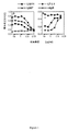
十分に中和する抗HGF mAb L2G7の開発は、米国特許出願公開番号US2005/0019327A1に記載されている。この米国特許出願公開番号US2005/0019327A1を本明細書中で参考として援用する。要するに、Balb/cマウスを、足蹠注射により、組換えヒトHGFで十分に免疫し、そしてハイブリドーマを通常の手法によりこれらマウスから作り出した。Flagペプチドを融合させたHGFからなるキメラ融合タンパク質(HGF−Flag)とヒトIgG1 Fc領域を融合させたMet細胞外領域(Met−Fc)を通常の組換え技術により生成し、そしてそれらを、MetレセプターとHGFの結合を阻害する抗HGF mAbsの能力決定のために用いた。図1aは、3つ別々の抗HGF mAbの能力を示す。これら3つ別々の抗HGF mAbのそれぞれは、溶液中でHGFを捕捉するために別のエピトープを認識する。IgG2a mAb L2G7は、HGFに対して結合力から判断すると中程度の親和力を有するが、ELISA(図1b)においてMet−FcとHGF−Flagの結合を完全にブロックすると確認された唯一のmAbである。mAb L2G7は、HGFに対し特異的である。なぜなら、そのmAb L2G7は、VEGF,FGF,EGFのような他の成長因子に結合を示さないからである。
(1. Production and in vitro properties of anti-HGF mAbs)
Development of a fully neutralizing anti-HGF mAb L2G7 is described in US Patent Application Publication No. US2005 / 0019327A1. This US Patent Application Publication No. US2005 / 0019327A1 is incorporated herein by reference. In short, Balb / c mice were fully immunized with recombinant human HGF by footpad injection and hybridomas were generated from these mice by conventional techniques. A chimeric fusion protein consisting of HGF fused with a Flag peptide (HGF-Flag) and a Met extracellular region fused with a human IgG1 Fc region (Met-Fc) are generated by conventional recombinant techniques, and they are converted to Met Used to determine the ability of anti-HGF mAbs to inhibit receptor-HGF binding. FIG. 1a shows the ability of three separate anti-HGF mAbs. Each of these three separate anti-HGF mAbs recognizes a different epitope to capture HGF in solution. IgG2a mAb L2G7 has moderate affinity for HGF as judged by its binding power, but is the only mAb that was confirmed to completely block the binding of Met-Fc and HGF-Flag in an ELISA (FIG. 1b) . mAb L2G7 is specific for HGF. This is because the mAb L2G7 does not show binding to other growth factors such as VEGF, FGF, EGF.
MetへのHGF結合をブロックするmAb L2G7の能力は、その能力がHGFの誘導するすべての細胞応答を阻害することを示唆した。しかし、この仮説は、検証する必要がある。何故ならば、HGFのαサブユニットおよびβサブユニットは異なった活性を媒介するからである(Lokker et al.,EMBO J.11:2503,1992;Hartmann et al.,Proc.Natl Acad.Sci.USA 89:11574,1992)。HGFのαサブユニットを通して媒介されるHGFのひとつの重要な生物活性は、細胞分散を誘導する能力であり、HGFのαサブユニットの別名である「分散因子」はその生物活性から由来している。 図2aは、L2G7が、MDCK上皮細胞のHGFの誘導による分散を完全に阻害することができることを示しており、そのMDCK上皮細胞のHGFの誘導による分散は、HGF分散活性を定量するための広く用いられる生物活性検定法である。HGFのβサブユニットを通して媒介されるHGFのひとつの鍵となる生物活性は、ある細胞種の有糸分裂誘発である。図2bは、HGFとmAbのモル比が1:1であるL2G7は、Mv1Luミンク肺上皮細胞におけるHGFで誘導した3H−チミジンの取り込みを完全に阻害することを示す。したがって、mAb L2G7は、αーHGFサブユニットおよびβーHGFサブユニットのどちらにも起因するHGFの誘導による生物活性をブロックする。 The ability of mAb L2G7 to block HGF binding to Met suggested that its ability to inhibit all cellular responses induced by HGF. However, this hypothesis needs to be verified. This is because the α subunit and β subunit of HGF mediate different activities (Lokker et al., EMBO J. 11: 2503, 1992; Hartmann et al., Proc. Natl Acad. Sci. USA 89: 11574, 1992). One important biological activity of HGF mediated through the α subunit of HGF is the ability to induce cell dispersion, and the “dispersing factor”, also known as the α subunit of HGF, is derived from its biological activity. . FIG. 2a shows that L2G7 can completely inhibit the HGF-induced dispersion of MDCK epithelial cells, and the HGF-induced dispersion of MDCK epithelial cells is widely used to quantify HGF dispersion activity. The bioactivity assay used. One key biological activity of HGF mediated through the β subunit of HGF is mitogenesis of certain cell types. FIG. 2b shows that L2G7 with a 1: 1 molar ratio of HGF to mAb completely inhibits HGF-induced 3H-thymidine incorporation in Mv1Lu mink lung epithelial cells. Thus, mAb L2G7 blocks the biological activity due to the induction of HGF due to both the α-HGF subunit and the β-HGF subunit.
新脈管形成が固形腫瘍の増殖のために必要である。HGFは、強力な脈管形成因子である(Grant et al.,Proc.Natl Acad.Sci.USA 90:1937,1993)。そして、HGFの腫瘍段階は、神経膠腫を含むヒト悪性疾患の脈管密度と相関する(Schmidt.et al.Int.J.Cancer 84:10,1999)。HGFはまた、VEGFなど他の脈管形成因子の産生を刺激し得、VEGFに誘導された新脈管形成をも増強し得る(Xin et al.Am.J.Pathol.158,1111,2001)。新脈管形成に必要とされる2つの最初の段階は、内皮細胞の増殖および細管形成である。ゆえに、ヒト臍静脈内皮細胞(HUVEC)のHGFの誘導による増殖、および3次元コラーゲンゲル中での脈管様細管のHGFの誘導による形成に対するL2G7の効果を測定した。HGF(50ng/ml、72時間)によるHUVECの増殖の刺激は、mAbとHGFのモル比が1.5:1であるL2G7により完全に阻害された(図2c)。3次元コラーゲンゲル中に懸濁させたHUVECは、HGF(200ng/ml、48時間)で刺激後、相互連結した分枝細管の網様構造を発達させたが、L2G7を添加したHGFで処理した細胞は、このような細管形成はほとんどあるいは全く示さなかった(図2d)。このことから、L2G7は、HGFの誘導する新脈管形成の増殖局面および形態形成の局面をブロックする。 Angiogenesis is necessary for solid tumor growth. HGF is a potent angiogenic factor (Grant et al., Proc. Natl Acad. Sci. USA 90: 1937, 1993). And the tumor stage of HGF correlates with the vascular density of human malignancies including glioma (Schmidt. Et al. Int. J. Cancer 84: 10, 1999). HGF can also stimulate the production of other angiogenic factors such as VEGF, and can also enhance VEGF-induced angiogenesis (Xin et al. Am. J. Pathol. 158, 1111, 2001). . The two first steps required for angiogenesis are endothelial cell proliferation and tubule formation. Therefore, the effect of L2G7 on HGF-induced proliferation of human umbilical vein endothelial cells (HUVEC) and formation of vascular-like tubules induced by HGF in 3D collagen gels was measured. Stimulation of HUVEC proliferation by HGF (50 ng / ml, 72 hours) was completely inhibited by L2G7 with a molar ratio of mAb to HGF of 1.5: 1 (FIG. 2c). HUVECs suspended in 3D collagen gel developed a network structure of interconnected branching tubules after stimulation with HGF (200 ng / ml, 48 hours), but were treated with HGF supplemented with L2G7 The cells showed little or no such tubule formation (FIG. 2d). Thus, L2G7 blocks the proliferative and morphogenic aspects of angiogenesis induced by HGF.
HGFは、癌治療で一般的に使用されているDNA損傷剤を含む多くの治療薬により誘導されるアポトーシス死から腫瘍細胞を守る(Bowers et al.Cancer Res.60:4277,2000;Fan et al.Oncogene 24:1749,2005)。ヒト悪性神経膠腫細胞の多くは、デスレセプターFASを発現する。そのことにより、インビトロで抗FAS抗体により誘導されるアポトーシスに対し、ヒト神経膠腫細胞は感受性になる(Weller et al.J.Clin.Invest.94:954,1994)。従って、アポトーシス性抗FAS mAb CH−11で処理されたU87神経膠腫細胞のHGF誘導性細胞防御に対するL2G7の効果を測定した。CH−11処理(24時間)後、U87細胞の生存は、未処理のコントロールにおける細胞生存の約45%にまで減少した。これは、無関係なアイソタイプコントロール抗体存在下、HGFとともにU87細胞を前培養することにより完全に逆となったが、L2G7の存在下、HGFによってはそうならなかった効果であった(図2e)。 HGF protects tumor cells from apoptotic death induced by many therapeutic agents, including DNA damaging agents commonly used in cancer treatment (Bowers et al. Cancer Res. 60: 4277, 2000; Fan et al. Oncogene 24: 1749, 2005). Many human malignant glioma cells express the death receptor FAS. This makes human glioma cells susceptible to apoptosis induced by anti-FAS antibodies in vitro (Weller et al. J. Clin. Invest. 94: 954, 1994). Therefore, the effect of L2G7 on HGF-induced cellular protection of U87 glioma cells treated with apoptotic anti-FAS mAb CH-11 was measured. After CH-11 treatment (24 hours), survival of U87 cells was reduced to approximately 45% of cell survival in untreated controls. This was completely reversed by pre-culturing U87 cells with HGF in the presence of an irrelevant isotype control antibody, but this was not the case with HGF in the presence of L2G7 (FIG. 2e).
(2.神経膠腫異種移植腫瘍モデルにおける抗HGFmAbの効果)
HGFの多様な腫瘍促進活性をブロックするL2G7の能力は、このmAbが少なくともHGF+/Met+ヒト腫瘍に対する抗腫瘍活性を有することを示唆した。神経膠腫の多くは、MetおよびHGFを発現するようである(Rosen et al.Int.J.Cancer67:248,1996)。神経膠腫細胞株U87及びU118に関して、Metの発現が、フローサイトメトリー分析により確認した。そして、HGF特異的ELISAを使用して7日間のコンフルエント培養からの上清中に約20〜35ng/mlのHGFが検出された。前もって確立されたU118およびU87皮下の異種移植のヌードマウスモデルにおけるL2G7の抗腫瘍効果が、決定された。L2G7は、腫瘍の大きさが約50mm3に達した後、一週間に2回腹腔に投与した(Kim et al.,Nature 362:841,1993(本明細書中で参考文献として援用される))。1回の注射あたり100μg(約5mg/kg)で、L2G7は、完全にU118腫瘍の増殖を阻害した(図3a)。U87異種移植物モデルにおいて、1回の注射あたり、50μgまたは100μgのいずれかのL2G7が、腫瘍の増殖を阻害するだけでなく、実際に腫瘍の退縮を引き起こした(図3b)。コントロールのmAb(1回の注射あたり100μg)は、PBSコントロールと比較してほんの少しだけ腫瘍の増殖を阻害した。L2G7は、U251神経膠腫異種移植物の増殖に対して効果はなかった。Metを発現するが、HGFは分泌しない。これらインビボでの結果は、単一の物質としてL2G7が、特異的にHGF活性をブロックすることにより、腫瘍の増殖を防ぐことを示す。
(2. Effect of anti-HGF mAb in glioma xenograft tumor model)
The ability of L2G7 to block the diverse tumor-promoting activity of HGF suggested that this mAb has antitumor activity against at least HGF + / Met + human tumors. Many gliomas appear to express Met and HGF (Rosen et al. Int. J. Cancer 67: 248, 1996). Met expression was confirmed by flow cytometric analysis for glioma cell lines U87 and U118. Approximately 20-35 ng / ml of HGF was then detected in the supernatant from a 7 day confluent culture using an HGF specific ELISA. The anti-tumor effect of L2G7 in a nude mouse model of U118 and U87 subcutaneous xenografts established previously was determined. L2G7 was administered intraperitoneally twice a week after tumor size reached approximately 50 mm 3 (Kim et al., Nature 362: 841, 1993, incorporated herein by reference). ). At 100 μg per injection (approximately 5 mg / kg), L2G7 completely inhibited the growth of U118 tumors (FIG. 3a). In a U87 xenograft model, either 50 μg or 100 μg of L2G7 per injection not only inhibited tumor growth, but actually caused tumor regression (FIG. 3b). The control mAb (100 μg per injection) inhibited tumor growth only slightly compared to the PBS control. L2G7 had no effect on the growth of U251 glioma xenografts. Expresses Met but does not secrete HGF. These in vivo results indicate that L2G7 as a single substance prevents tumor growth by specifically blocking HGF activity.
次に、L2G7の効力を、前もって確立された頭蓋内U87神経膠腫異種移植物を有するマウスで試験した。マウスに、右尾状核あるいは右被殻への定位注射により、U87ヒト悪性神経膠腫細胞(100,000μg、細胞/動物)を移植した。移植後5日目から52日までに投与されたL2G7(100μg/注射、腹腔内、週二回)は、動物生存期間を有意に延長した(図3c)。コントロールマウスにおいて、生存期間中央値は、39日であった。そしてすべてのマウスは、41日までに進行性腫瘍により死んだ。対照的に、L2G7で処置されたすべてのマウスは、70日まで生存し、そして90日(mAb処置の中止後7週間)で80%が生存した(図3c)。L2G7の3回投与後、21日目で屠殺したマウスでは、コントロール腫瘍が、L2G7で処置した腫瘍の10倍よりも大きかった(6.6+2.7mm3 対 0.54+0.17mm3)(図3d)。 Next, the efficacy of L2G7 was tested in mice with pre-established intracranial U87 glioma xenografts. Mice were transplanted with U87 human malignant glioma cells (100,000 μg, cells / animal) by stereotaxic injection into the right caudate nucleus or right putamen. L2G7 (100 μg / injection, intraperitoneal, twice weekly) administered from day 5 to day 52 after transplantation significantly prolonged animal survival (FIG. 3c). In control mice, the median survival time was 39 days. And all mice died of progressive tumor by day 41. In contrast, all mice treated with L2G7 survived to 70 days and 80% survived at 90 days (7 weeks after mAb treatment was discontinued) (FIG. 3c). In mice sacrificed on day 21 after 3 doses of L2G7, control tumors were more than 10 times larger than those treated with L2G7 (6.6 + 2.7 mm 3 vs. 0.54 + 0.17 mm 3 ) (FIG. 3d). ).
さらなる厳しい条件下でのmAb効力を試験するために、同種の実験において、L2G7処置の開始を18日目まで遅らせた。1サブセットのマウス(1グループあたりn=5)を、処置過程において初期に屠殺した。そして、腫瘍体積を、コンピューター支援型イメージ解析を用いて、H&E染色した脳の部分の腫瘍断面積を測ることにより定量した。L2G7は、かなりの腫瘍の退縮を誘導した(図3e,f)。具体的には、18日目での処置前腫瘍体積が、26.7+2.5mm3であった(範囲19.5〜54mm3、中央値27.9mm3)。L2G7の3回投与後、29日目に、腫瘍は、ほんの11.7+5.0mm3(範囲0〜26.2mm3、中央値7.5mm3)だった。それで、それら腫瘍は、実際平均して50%以上退縮したか、あるいは大きさが収縮した。アイソタイプが一致するコンロトールmAbで処置したマウスからの29日目での腫瘍体積は、134.3+22.0mm3であった(範囲71.2〜196.8mm3、中央値128mm3)。それゆえ、コントロールmAbで処置した腫瘍はほぼ5倍増殖し、平均体積はL2G7で処置した腫瘍と比較して12倍大きかった。屠殺しなかったマウス(1グループあたりn=10)において、コントロールマウスにおける生存期間中央値は、32日であり、そして全てが42日目までに死んだ。一方、L2G7で処置されたマウスは46日目まで全く死なず、そしてL2G7は、生存期間中央値を61日目に延ばした。このように、L2G7は、非常に高い全身腫瘍組織量を有するマウスにおいて、腫瘍退縮を誘導した。
In order to test mAb efficacy under more severe conditions, the start of L2G7 treatment was delayed until day 18 in the same type of experiment. One subset of mice (n = 5 per group) was sacrificed early in the course of treatment. Tumor volume was then quantified by measuring the tumor cross-sectional area of the H & E stained brain portion using computer-assisted image analysis. L2G7 induced significant tumor regression (Figure 3e, f). Specifically, pre-treatment tumor volume at day 18 was the 26.7 + 2.5 mm 3 (range 19.5~54Mm 3, median 27.9 mm 3). After three doses of L2G7, on day 29, tumors, was only 11.7 + 5.0mm 3 (
頭蓋内腫瘍の組織切片のさらに詳細な解析を、L2G7の抗腫瘍効果についてあり得る機構を調査するために行った(図4)。L2G7の3回投与の後、腫瘍細胞の増殖(Ki−67指数)および新脈管形成(脈管密度、すなわち、腫瘍面積のパーセントとしての抗ラミニン染色腫瘍脈管の面積)は、それぞれ51%および62%減少した。一方、活性化されたカスパーゼ−3陽性細胞の総数により定量された腫瘍細胞のアポトーシスの指数は、6倍増加した。L2G7の治療を開始した後に、速やかに起こった明らかな腫瘍退縮は、アゴニスト抗デスレセプター4(TRAIL1)mAb(Chuntharapai et al.J.Immunol.166:4891,2001)で処理されたヒト結腸腫瘍Colo205異種移植物において観察されたものと類似する細胞死の応答を示す。 A more detailed analysis of tissue sections from intracranial tumors was performed to investigate possible mechanisms for the anti-tumor effects of L2G7 (FIG. 4). After three doses of L2G7, tumor cell proliferation (Ki-67 index) and angiogenesis (vascular density, ie area of anti-laminin-stained tumor vessels as a percentage of tumor area) were 51% each And 62% decrease. On the other hand, the apoptosis index of tumor cells quantified by the total number of activated caspase-3 positive cells increased 6-fold. The apparent tumor regression that occurred rapidly after initiating treatment with L2G7 was a human colon tumor Colo205 treated with the agonist anti-death receptor 4 (TRAIL1) mAb (Chuntarapai et al. J. Immunol. 166: 4891, 2001). It shows a cell death response similar to that observed in xenografts.
ここで報告された結果は、トキシンにも放射性核種にも連結していないmAbからの顕著な脳腫瘍応答の例である。比較として、皮下の異種移植物モデルにおいて、抗VEGFマウスmAb A4.6.1、(これは、後に薬Avastin(登録商標)を作製するためにヒト化した)が、mAb L2G7によるU87神経膠腫およびU118神経膠腫の本質的に完全な増殖阻害とは対照的に、G55ヒト神経膠腫の増殖を約50〜60%ほどしか阻害しなかった(Kim et al.,Nature 362:841,1993)。正常位頭蓋内腫瘍モデルにおいて、G55神経膠腫細胞移植と同時に投与された全身抗VEFG mAbは、動物生存期間をわずかに2〜3週間延ばすにとどまった(Rubenstein et al.Neoplasia 2:306,2000)。同じく、EGFレセプターの改変体へのmAbの全身投与は、その改変体EGFレセプターでトランスフェクションした神経膠腫細胞の頭蓋内異種移植物を有するマウスの生存期間中央値を、総体的にささやかにしか延ばさなかった(13日から21日まで、あるいは13日から19日まで、しかしひとつのケースは19日から58日まで:Mishima et al.,Cancer Res.61:4349,2001)。しかし、これらささやかな効果は、mAb投与を異種移植物の移植と同時、あるいは移植後速やかに開始した場合に、達成された。そしてこのことから考えると、これらのささやかな効果は、少なくとも部分的には異種移植物の脈管新生(既に脳腫瘍を有する患者において標的とされ得ない事象)の開始を遅らせることにより引き起こされた可能性があった。対照的に、抗HGFmAb L2G7の全身投与は、生存期間を延ばした。そして、その全身投与は、腫瘍が十分に安定化された移植後5日目あるいはさらに18日目で投与された場合でさえ、腫瘍の退縮を引き起こした。それゆえ、この全身投与の結果は、ヒト患者における状況に対応する。 The results reported here are examples of significant brain tumor responses from mAbs that are not linked to toxins or radionuclides. As a comparison, in a subcutaneous xenograft model, anti-VEGF mouse mAb A4.6.1, which was later humanized to create the drug Avastin®, was a U87 glioma with mAb L2G7. And in contrast to the essentially complete growth inhibition of U118 glioma, it inhibited growth of G55 human glioma by only about 50-60% (Kim et al., Nature 362: 841, 1993). ). In a normal intracranial tumor model, systemic anti-VEFG mAb administered concomitantly with G55 glioma cell transplantation extended animal survival only a few weeks (Rubenstein et al. Neoplasmia 2: 306, 2000). ). Similarly, systemic administration of mAb to a variant of the EGF receptor can only moderately reduce the median survival of mice with intracranial xenografts of glioma cells transfected with that variant EGF receptor. It was not extended (from 13 to 21 or from 13 to 19 but in one case from 19 to 58: Misima et al., Cancer Res. 61: 4349, 2001). However, these modest effects were achieved when mAb administration was initiated simultaneously with xenograft transplantation or promptly after transplantation. And in light of this, these modest effects may be caused, at least in part, by delaying the onset of xenograft angiogenesis (an event that cannot be targeted in patients who already have brain tumors) There was sex. In contrast, systemic administration of anti-HGF mAb L2G7 prolonged survival. And its systemic administration caused tumor regression even when administered 5 days or even 18 days after transplantation when the tumor was well stabilized. Therefore, the result of this systemic administration corresponds to the situation in human patients.
mAb L2G7の顕著な抗腫瘍効果は、その分子標的であるHGFの独特な多機能特性(すなわち、マイトジェン特性、脈管形成特性、そして細胞防御特性)に起因する可能性がある(Birchmeier et al.Nat.Rev.Mol.Cell Bio.4:915,2003;Trusolino et al.Nat.Rev.Cancer 4:289,2002)。神経膠腫の退縮を誘導するL2G7の能力は、MetへのHGFの結合によりブロックされるFas媒介アポトーシス、(Wang et al.Cell.9:411,2002)、もしくは、ホスファチジルイノシトール3−キナーゼ、Akt、およびNF−κB中間体を含むHGF誘導性細胞防御経路の不活性化(Fan et al.Oncogene 24:1749,2005)に起因し得る、細胞死の応答を意味する。HGFの細胞防御および脈管形成の効果をブロックするL2G7の能力は、全身に送達されたL2G7が、悪性脳腫瘍を処置するために現在使用されている細胞毒性治療(たとえば、γ−放射線および化学療法)の効力を増すことを予測する。 The striking anti-tumor effect of mAb L2G7 may be due to the unique multifunctional properties of its molecular target, HGF (ie, mitogenic, angiogenic, and cytoprotective properties) (Birchmeier et al. Nat. Rev. Mol. Cell Bio. 4: 915, 2003; Trusolino et al. Nat. Rev. Cancer 4: 289, 2002). The ability of L2G7 to induce glioma regression is either Fas-mediated apoptosis blocked by HGF binding to Met (Wang et al. Cell. 9: 411, 2002), or phosphatidylinositol 3-kinase, Akt And a cell death response that may result from inactivation of the HGF-induced cytoprotective pathway, including the NF-κB intermediate (Fan et al. Oncogene 24: 1749, 2005). The ability of L2G7 to block HGF's cytoprotective and angiogenic effects is due to the cytotoxicity therapy (eg, γ-radiation and chemotherapy) where L2G7 delivered systemically is currently used to treat malignant brain tumors. ) Is expected to increase.
本発明は、目下好ましい実施例に関して記述されているが、種々の改変が、本発明から逸脱することなくなされ得ることが、理解されるべきである。 Although the present invention has been described with reference to the presently preferred embodiment, it should be understood that various modifications can be made without departing from the invention.
引用した全ての刊行物、特許、および特許出願は、それぞれ個別の刊行物、特許および特許出願があらゆる目的のためにその全体を本明細書中で参考として援用されると具体的かつ個別に示されたのと同じ程度まで、あらゆる目的のためにその全体が参考として本明細書中に援用される。 All cited publications, patents, and patent applications are specifically and individually indicated as individual publications, patents, and patent applications are incorporated herein by reference in their entirety for all purposes. To the same extent as has been done, the entire contents thereof are hereby incorporated by reference for all purposes.
Claims (23)
脳腫瘍を有する患者にモノクローナル抗体(mAb)を全身投与し、それにより該脳腫瘍を処置する工程;
を包含する、方法。 A method for treating a brain tumor in a patient comprising:
Systemically administering a monoclonal antibody (mAb) to a patient having a brain tumor, thereby treating the brain tumor;
Including the method.
脳腫瘍を有する患者にモノクローナル抗体(mAb)を全身投与し、それにより該脳腫瘍の退縮を引き起こす工程;
を包含する、方法。 A method for causing regression of a brain tumor in a patient comprising:
Systemically administering a monoclonal antibody (mAb) to a patient having a brain tumor, thereby causing regression of the brain tumor;
Including the method.
Applications Claiming Priority (3)
| Application Number | Priority Date | Filing Date | Title |
|---|---|---|---|
| US68711805P | 2005-06-02 | 2005-06-02 | |
| US75109205P | 2005-12-15 | 2005-12-15 | |
| PCT/US2006/021293 WO2006130773A2 (en) | 2005-06-02 | 2006-06-01 | Methods of treating brain tumors with antibodies |
Related Child Applications (1)
| Application Number | Title | Priority Date | Filing Date |
|---|---|---|---|
| JP2013002317A Division JP2013136580A (en) | 2005-06-02 | 2013-01-10 | Method of treating brain tumor with antibody |
Publications (2)
| Publication Number | Publication Date |
|---|---|
| JP2008545753A true JP2008545753A (en) | 2008-12-18 |
| JP2008545753A5 JP2008545753A5 (en) | 2009-05-07 |
Family
ID=37482322
Family Applications (2)
| Application Number | Title | Priority Date | Filing Date |
|---|---|---|---|
| JP2008514858A Pending JP2008545753A (en) | 2005-06-02 | 2006-06-01 | Methods of treating brain tumors with antibodies |
| JP2013002317A Pending JP2013136580A (en) | 2005-06-02 | 2013-01-10 | Method of treating brain tumor with antibody |
Family Applications After (1)
| Application Number | Title | Priority Date | Filing Date |
|---|---|---|---|
| JP2013002317A Pending JP2013136580A (en) | 2005-06-02 | 2013-01-10 | Method of treating brain tumor with antibody |
Country Status (14)
| Country | Link |
|---|---|
| US (2) | US20070036797A1 (en) |
| EP (1) | EP1885400A4 (en) |
| JP (2) | JP2008545753A (en) |
| KR (1) | KR20080026562A (en) |
| AU (1) | AU2006252419B2 (en) |
| BR (1) | BRPI0611009A2 (en) |
| CA (1) | CA2607699A1 (en) |
| CR (1) | CR9512A (en) |
| IL (1) | IL187318A0 (en) |
| MA (1) | MA29570B1 (en) |
| MX (1) | MX2007015056A (en) |
| NO (1) | NO20080012L (en) |
| RU (1) | RU2007146986A (en) |
| WO (1) | WO2006130773A2 (en) |
Cited By (1)
| Publication number | Priority date | Publication date | Assignee | Title |
|---|---|---|---|---|
| JP2013532627A (en) * | 2010-07-01 | 2013-08-19 | 武田薬品工業株式会社 | Combination of cMET inhibitor with antibody against HGF and / or cMET |
Families Citing this family (25)
| Publication number | Priority date | Publication date | Assignee | Title |
|---|---|---|---|---|
| US20040208876A1 (en) | 2003-04-18 | 2004-10-21 | Kim Kyung Jin | Monoclonal antibodies to hepatocyte growth factor |
| AU2006294663B2 (en) * | 2005-09-26 | 2012-03-22 | Medarex, Inc. | Human monoclonal antibodies to CD70 |
| AR059922A1 (en) | 2006-04-01 | 2008-05-07 | Galaxy Biotech Llc | HUMANIZED MONOCLONAL ANTIBODIES FOR THE GROWTH FACTOR OF HEPATOCITS |
| ATE495195T1 (en) * | 2006-06-02 | 2011-01-15 | Aveo Pharmaceuticals Inc | HEPATOCYTE GROWTH FACTOR (HGF) BINDING PROTEI E |
| WO2007143098A2 (en) * | 2006-06-02 | 2007-12-13 | Aveo Pharmaceuticals, Inc. | Hepatocyte growth factor (hgf) binding proteins |
| SI2066694T1 (en) | 2006-09-29 | 2016-02-29 | Oncomed Pharmaceuticals, Inc. | Compositions and methods for diagnosing and treating cancer |
| KR20090088946A (en) * | 2006-12-14 | 2009-08-20 | 메다렉스, 인코포레이티드 | Human antibodies that bind cd70 and uses thereof |
| US20110070247A1 (en) * | 2007-08-24 | 2011-03-24 | Novartis Ag | Protein modulators |
| US20110217294A1 (en) * | 2008-04-11 | 2011-09-08 | Daniel Fults | Combination of hgf inhibitor and hedgehog inhibitor to treat cancer |
| PE20091714A1 (en) * | 2008-04-11 | 2009-11-15 | Galaxy Biotech Llc | COMBINATION OF THE INHIBITOR HGF AND THE AGONIST PTEN |
| US8101725B2 (en) * | 2008-05-29 | 2012-01-24 | Galaxy Biotech, Llc | Monoclonal antibodies to basic fibroblast growth factor |
| WO2010119991A2 (en) | 2009-04-17 | 2010-10-21 | Takeda Pharmaceutical Company Limited | Novel method of treating cancer |
| DK2488204T3 (en) | 2009-10-16 | 2016-06-06 | Oncomed Pharm Inc | Therapeutic combination and use of DLL4 antagonist antibodies and blood pressure lowering agents |
| JP5981853B2 (en) * | 2010-02-18 | 2016-08-31 | ジェネンテック, インコーポレイテッド | Neuregulin antagonists and their use in the treatment of cancer |
| US8551479B2 (en) | 2010-09-10 | 2013-10-08 | Oncomed Pharmaceuticals, Inc. | Methods for treating melanoma |
| US9783601B2 (en) | 2010-12-01 | 2017-10-10 | Alderbio Holdings Llc | Methods of preventing inflammation and treating pain using anti-NGF compositions |
| US9884909B2 (en) | 2010-12-01 | 2018-02-06 | Alderbio Holdings Llc | Anti-NGF compositions and use thereof |
| US9067988B2 (en) | 2010-12-01 | 2015-06-30 | Alderbio Holdings Llc | Methods of preventing or treating pain using anti-NGF antibodies |
| US9539324B2 (en) | 2010-12-01 | 2017-01-10 | Alderbio Holdings, Llc | Methods of preventing inflammation and treating pain using anti-NGF compositions |
| US11214610B2 (en) | 2010-12-01 | 2022-01-04 | H. Lundbeck A/S | High-purity production of multi-subunit proteins such as antibodies in transformed microbes such as Pichia pastoris |
| US9078878B2 (en) | 2010-12-01 | 2015-07-14 | Alderbio Holdings Llc | Anti-NGF antibodies that selectively inhibit the association of NGF with TrkA, without affecting the association of NGF with p75 |
| AU2013201095C1 (en) | 2011-09-23 | 2019-12-05 | Oncomed Pharmaceuticals, Inc. | VEGF/DLL4 binding agents and uses thereof |
| WO2014071018A1 (en) | 2012-10-31 | 2014-05-08 | Oncomed Pharmaceuticals, Inc. | Methods and monitoring of treatment with a dll4 antagonist |
| JP6827415B2 (en) | 2014-10-31 | 2021-02-10 | メレオ バイオファーマ 5 インコーポレイテッド | Combination therapy for the treatment of the disease |
| CA2999160A1 (en) | 2015-09-23 | 2017-03-30 | Oncomed Pharmaceuticals, Inc. | Methods and compositions for treatment of cancer |
Citations (1)
| Publication number | Priority date | Publication date | Assignee | Title |
|---|---|---|---|---|
| WO2005017107A2 (en) * | 2003-07-18 | 2005-02-24 | Amgen Inc. | Specific binding agents to hepatocyte growth factor |
Family Cites Families (17)
| Publication number | Priority date | Publication date | Assignee | Title |
|---|---|---|---|---|
| JP2564486B2 (en) * | 1986-07-14 | 1996-12-18 | 修治 橋本 | Hepatocyte growth factor |
| US5837676A (en) * | 1993-10-18 | 1998-11-17 | Long Island Jewish Medical Center | Use of scatter factor to enhance angiogenesis |
| US6498144B1 (en) * | 1993-10-18 | 2002-12-24 | North Shore - Long Island Jewish Research Institute | Use of scatter factor to enhance angiogenesis |
| US5707624A (en) * | 1994-06-03 | 1998-01-13 | The Regents Of The University Of Michigan | Treatment of Kaposi's sarcoma by inhibition of scatter factor |
| US5646036A (en) * | 1995-06-02 | 1997-07-08 | Genentech, Inc. | Nucleic acids encoding hepatocyte growth factor receptor antagonist antibodies |
| US5686292A (en) * | 1995-06-02 | 1997-11-11 | Genentech, Inc. | Hepatocyte growth factor receptor antagonist antibodies and uses thereof |
| US6214344B1 (en) * | 1995-06-02 | 2001-04-10 | Genetech, Inc. | Hepatocyte growth factor receptor antagonists and uses thereof |
| JP2000515735A (en) * | 1996-07-03 | 2000-11-28 | ジェネンテック インコーポレーテッド | Hepatocyte growth factor receptor agonist |
| WO2004019991A2 (en) * | 2002-08-30 | 2004-03-11 | F. Hoffmann-La Roche Ag | Scatter factor/hepatocyte growth factor antagonist nk4 for the treatment of glioma |
| US20040208876A1 (en) * | 2003-04-18 | 2004-10-21 | Kim Kyung Jin | Monoclonal antibodies to hepatocyte growth factor |
| US7220410B2 (en) * | 2003-04-18 | 2007-05-22 | Galaxy Biotech, Llc | Monoclonal antibodies to hepatocyte growth factor |
| EP2409704B1 (en) * | 2003-09-26 | 2017-09-20 | Exelixis, Inc. | c-Met modulators and methods of use |
| AU2005270068B2 (en) * | 2004-07-02 | 2012-04-19 | Exelixis, Inc. | C-Met modulators and method of use |
| US7964365B2 (en) * | 2005-11-08 | 2011-06-21 | The United States of Americam as represented by the Secretary, Department of Health and Human Services | Methods for diagnosing and monitoring the progression of cancer |
| AR059922A1 (en) * | 2006-04-01 | 2008-05-07 | Galaxy Biotech Llc | HUMANIZED MONOCLONAL ANTIBODIES FOR THE GROWTH FACTOR OF HEPATOCITS |
| US20110217294A1 (en) * | 2008-04-11 | 2011-09-08 | Daniel Fults | Combination of hgf inhibitor and hedgehog inhibitor to treat cancer |
| CL2009000843A1 (en) * | 2008-04-11 | 2009-07-24 | Galaxy Biotech Llc | Method of treating cancer in a patient comprising the administration of a first agent that is inhibitory of hepatocyte growth factor (hgf) in combination with a second agent that is inhibitor of a cellular signaling pathway other than the hgf / cmet pathway. |
-
2006
- 2006-06-01 KR KR1020077030521A patent/KR20080026562A/en not_active Application Discontinuation
- 2006-06-01 EP EP06771847A patent/EP1885400A4/en not_active Withdrawn
- 2006-06-01 AU AU2006252419A patent/AU2006252419B2/en not_active Ceased
- 2006-06-01 MX MX2007015056A patent/MX2007015056A/en not_active Application Discontinuation
- 2006-06-01 WO PCT/US2006/021293 patent/WO2006130773A2/en active Application Filing
- 2006-06-01 BR BRPI0611009-6A patent/BRPI0611009A2/en not_active IP Right Cessation
- 2006-06-01 CA CA002607699A patent/CA2607699A1/en not_active Abandoned
- 2006-06-01 JP JP2008514858A patent/JP2008545753A/en active Pending
- 2006-06-01 US US11/446,045 patent/US20070036797A1/en not_active Abandoned
- 2006-06-01 RU RU2007146986/14A patent/RU2007146986A/en not_active Application Discontinuation
-
2007
- 2007-11-12 IL IL187318A patent/IL187318A0/en unknown
- 2007-11-12 CR CR9512A patent/CR9512A/en not_active Application Discontinuation
- 2007-12-27 MA MA30523A patent/MA29570B1/en unknown
-
2008
- 2008-01-02 NO NO20080012A patent/NO20080012L/en not_active Application Discontinuation
-
2010
- 2010-01-19 US US12/690,045 patent/US20100221250A1/en not_active Abandoned
-
2013
- 2013-01-10 JP JP2013002317A patent/JP2013136580A/en active Pending
Patent Citations (1)
| Publication number | Priority date | Publication date | Assignee | Title |
|---|---|---|---|---|
| WO2005017107A2 (en) * | 2003-07-18 | 2005-02-24 | Amgen Inc. | Specific binding agents to hepatocyte growth factor |
Cited By (1)
| Publication number | Priority date | Publication date | Assignee | Title |
|---|---|---|---|---|
| JP2013532627A (en) * | 2010-07-01 | 2013-08-19 | 武田薬品工業株式会社 | Combination of cMET inhibitor with antibody against HGF and / or cMET |
Also Published As
| Publication number | Publication date |
|---|---|
| JP2013136580A (en) | 2013-07-11 |
| KR20080026562A (en) | 2008-03-25 |
| MX2007015056A (en) | 2008-03-11 |
| WO2006130773A2 (en) | 2006-12-07 |
| CA2607699A1 (en) | 2006-12-07 |
| AU2006252419B2 (en) | 2012-02-02 |
| BRPI0611009A2 (en) | 2010-08-10 |
| NO20080012L (en) | 2008-02-20 |
| US20100221250A1 (en) | 2010-09-02 |
| MA29570B1 (en) | 2008-06-02 |
| EP1885400A4 (en) | 2011-01-26 |
| IL187318A0 (en) | 2008-04-13 |
| US20070036797A1 (en) | 2007-02-15 |
| AU2006252419A1 (en) | 2006-12-07 |
| EP1885400A2 (en) | 2008-02-13 |
| WO2006130773A3 (en) | 2009-04-16 |
| CR9512A (en) | 2008-04-16 |
| RU2007146986A (en) | 2009-06-27 |
Similar Documents
| Publication | Publication Date | Title |
|---|---|---|
| AU2006252419B2 (en) | Methods of treating brain tumors with antibodies | |
| CA2645358C (en) | Humanized monoclonal antibodies to hepatocyte growth factor | |
| US20090258014A1 (en) | Combination of hgf inhibitor and egf inhibitor to treat cancer | |
| KR101072736B1 (en) | Monoclonal antibodies to hepatocyte growth factor | |
| US20110217294A1 (en) | Combination of hgf inhibitor and hedgehog inhibitor to treat cancer | |
| US20120052064A1 (en) | Anti-hgf antibody combinational cancer therapies | |
| US20110189169A1 (en) | Combination of hgf inhibitor and pten agonist to treat cancer | |
| KR20110011710A (en) | Monoclonal antibodies to basic fibroblast growth factor | |
| CN101511386A (en) | Methods of treating brain tumors with antibodies |
Legal Events
| Date | Code | Title | Description |
|---|---|---|---|
| A521 | Request for written amendment filed |
Free format text: JAPANESE INTERMEDIATE CODE: A523 Effective date: 20090310 |
|
| A621 | Written request for application examination |
Free format text: JAPANESE INTERMEDIATE CODE: A621 Effective date: 20090310 |
|
| A131 | Notification of reasons for refusal |
Free format text: JAPANESE INTERMEDIATE CODE: A131 Effective date: 20110804 |
|
| A601 | Written request for extension of time |
Free format text: JAPANESE INTERMEDIATE CODE: A601 Effective date: 20111102 |
|
| A602 | Written permission of extension of time |
Free format text: JAPANESE INTERMEDIATE CODE: A602 Effective date: 20111110 |
|
| A601 | Written request for extension of time |
Free format text: JAPANESE INTERMEDIATE CODE: A601 Effective date: 20111201 |
|
| A602 | Written permission of extension of time |
Free format text: JAPANESE INTERMEDIATE CODE: A602 Effective date: 20111208 |
|
| A521 | Request for written amendment filed |
Free format text: JAPANESE INTERMEDIATE CODE: A523 Effective date: 20120206 |
|
| A521 | Request for written amendment filed |
Free format text: JAPANESE INTERMEDIATE CODE: A821 Effective date: 20120206 |
|
| A02 | Decision of refusal |
Free format text: JAPANESE INTERMEDIATE CODE: A02 Effective date: 20120910 |
|
| A521 | Request for written amendment filed |
Free format text: JAPANESE INTERMEDIATE CODE: A523 Effective date: 20130110 |