JP2008539755A - Cell-based platform for high-throughput screening - Google Patents
Cell-based platform for high-throughput screening Download PDFInfo
- Publication number
- JP2008539755A JP2008539755A JP2008511190A JP2008511190A JP2008539755A JP 2008539755 A JP2008539755 A JP 2008539755A JP 2008511190 A JP2008511190 A JP 2008511190A JP 2008511190 A JP2008511190 A JP 2008511190A JP 2008539755 A JP2008539755 A JP 2008539755A
- Authority
- JP
- Japan
- Prior art keywords
- top surface
- cell adhesion
- cell
- group
- modified
- Prior art date
- Legal status (The legal status is an assumption and is not a legal conclusion. Google has not performed a legal analysis and makes no representation as to the accuracy of the status listed.)
- Ceased
Links
Images


Classifications
-
- B—PERFORMING OPERATIONS; TRANSPORTING
- B01—PHYSICAL OR CHEMICAL PROCESSES OR APPARATUS IN GENERAL
- B01L—CHEMICAL OR PHYSICAL LABORATORY APPARATUS FOR GENERAL USE
- B01L3/00—Containers or dishes for laboratory use, e.g. laboratory glassware; Droppers
- B01L3/50—Containers for the purpose of retaining a material to be analysed, e.g. test tubes
- B01L3/508—Containers for the purpose of retaining a material to be analysed, e.g. test tubes rigid containers not provided for above
- B01L3/5085—Containers for the purpose of retaining a material to be analysed, e.g. test tubes rigid containers not provided for above for multiple samples, e.g. microtitration plates
-
- B—PERFORMING OPERATIONS; TRANSPORTING
- B01—PHYSICAL OR CHEMICAL PROCESSES OR APPARATUS IN GENERAL
- B01L—CHEMICAL OR PHYSICAL LABORATORY APPARATUS FOR GENERAL USE
- B01L2300/00—Additional constructional details
- B01L2300/06—Auxiliary integrated devices, integrated components
- B01L2300/069—Absorbents; Gels to retain a fluid
-
- B—PERFORMING OPERATIONS; TRANSPORTING
- B01—PHYSICAL OR CHEMICAL PROCESSES OR APPARATUS IN GENERAL
- B01L—CHEMICAL OR PHYSICAL LABORATORY APPARATUS FOR GENERAL USE
- B01L2300/00—Additional constructional details
- B01L2300/08—Geometry, shape and general structure
- B01L2300/0809—Geometry, shape and general structure rectangular shaped
- B01L2300/0819—Microarrays; Biochips
-
- B—PERFORMING OPERATIONS; TRANSPORTING
- B01—PHYSICAL OR CHEMICAL PROCESSES OR APPARATUS IN GENERAL
- B01L—CHEMICAL OR PHYSICAL LABORATORY APPARATUS FOR GENERAL USE
- B01L2300/00—Additional constructional details
- B01L2300/16—Surface properties and coatings
- B01L2300/161—Control and use of surface tension forces, e.g. hydrophobic, hydrophilic
- B01L2300/165—Specific details about hydrophobic, oleophobic surfaces
Abstract
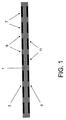
本発明は、実質的に平面な上面(3)及び底面(5)を有する多孔性ブロック(1)を含む、複数の試料化合物の生物学的効果を試験するための装置に関する。上面(3)は多数の細胞接着領域(7)及び細胞非接着領域(9)を含み、そして底面(5)は試料化合物をロードするための複数部位(11)を提供する。これらの部位は、多孔性ブロックの上面上の細胞接着領域の反対側に位置する。ある態様において、本発明は試料化合物をロードするための複数部位を提供する少なくとも1つの可溶性層(13)をさらに含む。
【選択図】図1The present invention relates to an apparatus for testing the biological effect of a plurality of sample compounds comprising a porous block (1) having a substantially planar top surface (3) and bottom surface (5). The top surface (3) includes multiple cell adhesion regions (7) and cell non-adhesion regions (9), and the bottom surface (5) provides multiple sites (11) for loading sample compounds. These sites are located on the opposite side of the cell adhesion region on the top surface of the porous block. In certain embodiments, the present invention further comprises at least one soluble layer (13) that provides multiple sites for loading sample compounds.
[Selection] Figure 1
Description
技術分野
ハイスループットスクリーニング技術は、1つには、医薬工業において、生物学的活性について化合物の大集団をスクリーニングすることの問題を解決するために発展してきた。マイクロアレイ技術及びさまざまな特殊化したアッセイを使用して、多数の化合物を同時にそして同一の条件下で試験して、それらが有用な生物学的活性を保有するかどうかを決定する。生細胞における生物学的活性を測定するアッセイは、何千もの異なる薬剤候補と細胞をインキュベートすることを必要とする。このインキュベーションは、標準化された96、384及び1536ウェル配置を有するマルチウェルタイタープレート中で従来行われる。マルチウェルプレートは、試験化合物を迅速にスクリーニングするために、自動液体分配システム及び標準化アッセイと共に使用される。
TECHNICAL FIELD High throughput screening techniques have been developed, in part, in the pharmaceutical industry to solve the problem of screening large populations of compounds for biological activity. Using microarray technology and various specialized assays, multiple compounds are tested simultaneously and under the same conditions to determine if they possess useful biological activity. Assays that measure biological activity in living cells require incubating the cells with thousands of different drug candidates. This incubation is conventionally performed in multiwell titer plates with standardized 96, 384 and 1536 well configurations. Multiwell plates are used with automated liquid dispensing systems and standardized assays to rapidly screen for test compounds.
発明の概要
本発明は、1つの局面において、実質的に平面な上面及び底面を有する多孔性ブロックを含む、複数の試料化合物の生物学的効果を試験するための装置に関する。上面は多数の細胞接着領域及び細胞非接着領域を含み、そして底面は試料化合物をロードするための複数部位を提供する。これらの部位は、多孔性ブロックの上面上の細胞接着領域の反対側に位置する。
SUMMARY OF THE INVENTION In one aspect, the present invention relates to an apparatus for testing a biological effect of a plurality of sample compounds that includes a porous block having a substantially planar top and bottom surface. The top surface includes a number of cell adhesion regions and cell non-adhesion regions, and the bottom surface provides multiple sites for loading sample compounds. These sites are located on the opposite side of the cell adhesion region on the top surface of the porous block.
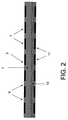
別の態様において、本発明は上記のような装置に関するが、試料化合物をロードするための複数部位を提供する少なくとも1つの可溶性層をさらに含む。これらの部位は、多孔性ブロックの上面上の細胞接着領域の反対側に位置する。 In another aspect, the invention relates to an apparatus as described above, but further comprises at least one soluble layer that provides multiple sites for loading sample compounds. These sites are located on the opposite side of the cell adhesion region on the top surface of the porous block.
ある態様において、上記装置は多孔性ブロックを取り囲むフレームをさらに含む。他の態様において、上記装置は上に上昇しそして上面を取り囲む壁をさらに含む。壁と上面とによって、液体を保持することができる。 In certain embodiments, the device further includes a frame surrounding the porous block. In other embodiments, the apparatus further includes a wall that rises up and surrounds the top surface. A liquid can be held by the wall and the upper surface.
発明の詳細な説明
本発明は、複数の試料化合物の生物学的効果を試験するためのさまざまな種類の装置に関する。これらの装置は、所与の試験化合物が細胞の生物学的活性に影響するかどうかを決定するために使用される。生物学的効果は細胞内又は細胞上の任意の計測可能な変化である。そのような変化は、限定はされないが、細胞外観又は構造における変化、細胞活性における変化、遺伝子発現レベルにおける変化、細胞タンパク質発現レベルにおける変化、細胞タンパク質活性における変化、及び細胞タンパク質安定性における変化を含む。
Detailed Description of the Invention The present invention relates to various types of devices for testing the biological effects of a plurality of sample compounds. These devices are used to determine whether a given test compound affects the biological activity of a cell. A biological effect is any measurable change in or on a cell. Such changes include, but are not limited to, changes in cell appearance or structure, changes in cell activity, changes in gene expression levels, changes in cell protein expression levels, changes in cell protein activity, and changes in cell protein stability. Including.
既存の96及び384ウェルプレートは、目的の細胞株と多数の関連化合物を迅速にそして効率的にスクリーニングすることにその能力が限定される。さらに、1536ウェルプレートに移したときの4倍の処理改善は、重大な液体操作の問題により相殺されてしまう。1536ウェルフォーマットの動作体積は、典型的には10マイクロリットル以下である。そのような小さい体積においては、化学的揮発、溶媒蒸発及び気泡により、試料間の統計的変動が大きく助長される。これらの問題は、ウェル間の大きな用量変動をもたらす、個々のウェルの細胞集団に対応する細胞数分布の高度な不一致性により悪化される。これらの物理的限界は、続くアッセイ測定において大きな統計的変動を助長し、精度及び信頼性を損なう。 Existing 96 and 384 well plates are limited in their ability to rapidly and efficiently screen a cell line of interest and a number of related compounds. Furthermore, the fourfold improvement in processing when transferred to a 1536 well plate is offset by significant liquid handling issues. The operating volume of the 1536 well format is typically 10 microliters or less. In such small volumes, chemical volatilization, solvent evaporation and bubbles greatly facilitate statistical variation between samples. These problems are exacerbated by a high degree of inconsistency in the cell number distribution corresponding to the cell population of individual wells, resulting in large dose variations between wells. These physical limits facilitate large statistical variability in subsequent assay measurements and compromise accuracy and reliability.
本発明の装置及び方法は、前記の複数ウェルフォーマットによる高密度複数ウェルスクリーニング法に関連する多くの液体操作の問題を解決し、そして代わりに、薬剤候補沈殿物のための場所並びに細胞及び他のアッセイ試薬のための支持の両方として多孔性マトリックスを使用する。多孔性マトリックスブロックの両側上に設計された表面特性は、アッセイ試薬の試料との相互作用を局在化し、そして可視化範囲を多孔性ブロックの細胞側表面上のあらかじめ決定された領域に限定することに役立つ。 The apparatus and method of the present invention solves many of the liquid handling problems associated with the high density multi-well screening method by the multi-well format described above, and instead provides a place for drug candidate precipitates as well as cells and other A porous matrix is used as both support for the assay reagents. The surface properties designed on both sides of the porous matrix block localize the interaction of the assay reagent with the sample and limit the visualization range to a predetermined area on the cell side surface of the porous block To help.
本発明の装置は、図1において図解されるように、実質的に平面な上面(3)及び底面(5)を有する多孔性ブロック(1)を含む。上面は、多数の細胞接着領域(7)及び細胞非接着領域(9)を含んでなり、そして底面が試料化合物をロードするための複数部位(11)を有する。これらの部位(11)は、細胞接着領域の反対側に位置する。試料化合物は、化合物を多孔性マトリックスに引きつけそして吸収するように設計された部位(11)内の、多孔性マトリックスの底面上に置かれる。吸収後、細胞は多孔性ブロックの上面上に伸展する。インキュベーター内でのインキュベーション後、細胞は上面に接着する。試料化合物は、多孔性マトリックスを通じてその上面に拡散し、そして上面に固定された細胞と相互作用する。本発明の方法は、試料が底面(5)上に置かれた後に、アッセイ面(上面)(3)が調製されることを可能にする。 The device of the present invention comprises a porous block (1) having a substantially planar top surface (3) and bottom surface (5), as illustrated in FIG. The top surface comprises a number of cell adhesion regions (7) and cell non-adhesion regions (9), and the bottom surface has multiple sites (11) for loading sample compounds. These sites (11) are located on the opposite side of the cell adhesion region. The sample compound is placed on the bottom surface of the porous matrix in the site (11) designed to attract and absorb the compound to the porous matrix. After absorption, the cells extend on the top surface of the porous block. After incubation in the incubator, the cells adhere to the top surface. The sample compound diffuses to its top surface through the porous matrix and interacts with cells immobilized on the top surface. The method of the invention allows the assay surface (top surface) (3) to be prepared after the sample has been placed on the bottom surface (5).
本発明の装置のさまざまな構成要素の記載は以下に従う。
多孔性ブロックは、試験しようとする化合物に透過性である限りは、いかなる材料から作られていてよい。多孔性ブロックは、以下に記載するポリマー、ゲル又は膜を含んでよい。
A description of the various components of the device of the invention follows.
The porous block may be made of any material as long as it is permeable to the compound to be tested. The porous block may comprise a polymer, gel or membrane as described below.
1つの態様において、多孔性ブロックはポリマー化合物から作られる。適したポリマーは、ポリアミド、ポリ(エチレン−酢酸ビニル)(EVAc)、ポリスチレン、ポリプロピレン、ポリカーボネート、ポリエステル、ポリエチレン、ポリビニリデン、ポリメチルメタクリレート、及びポリエチレンテレフタレートである。好ましい態様において、ポリマー層は、光学的観察に役立つために透明または半透明である。ポリマー層は、熱エンボス加工、射出成型、伸展成型、及び当該技術分野で公知の方法のような慣用のポリマー成型技術を使用して加工される。 In one embodiment, the porous block is made from a polymer compound. Suitable polymers are polyamide, poly (ethylene-vinyl acetate) (EVAc), polystyrene, polypropylene, polycarbonate, polyester, polyethylene, polyvinylidene, polymethyl methacrylate, and polyethylene terephthalate. In preferred embodiments, the polymer layer is transparent or translucent to aid in optical observation. The polymer layer is processed using conventional polymer molding techniques such as hot embossing, injection molding, stretch molding, and methods known in the art.
好ましい態様において、ポリマーは、ポリアミドまたはEVAcである。特に好ましい態様において、ポリマーはナイロンである。
他の態様において、多孔性ブロックはゲルの形である。そのようなゲルの例は、ゼラチン、アガロース、及びアクリルアミドゲルを含む。アガロース及びポリアクリルアミドゲルは、物理的に又は化学的に架橋されたスポンジ状ゲルである。これらは99.5%まで水を含有しうるが、これらのゲルの孔のサイズは多くのタンパク質及び核酸のサイズに類似する。ゲルは、マトリックス濃度の適切な選択により広い幅のサイズの分子をふるいをかけるために調整される。ゲルの平均の孔のサイズはゲル中の固体のパーセント、及びポリアクリルアミドについてはさらに架橋の量によっても決定される。好ましい態様において、ゲルは、アガロース、ゼラチン、α−ヒドロ−ω−ヒドロキシポリ(オキシエチレン)ポリ(オキシプロピレン)ポリ(オキシエチレン)ブロックコポリマー、及び刺激感受性ヒドロゲル、及び修飾デンプン/セルロースである。特に好ましい態様において、ゲルはセルロース誘導体から作られる。別の好ましい態様において、ゲルは刺激感受性ヒドロゲルである。別の特に好ましい態様において、刺激感受性ヒドロゲルはポリ(n−イソプロピルアリールアミド)及びポリアクリル酸からなる群より選択される。
In a preferred embodiment, the polymer is polyamide or EVAc. In a particularly preferred embodiment, the polymer is nylon.
In other embodiments, the porous block is in the form of a gel. Examples of such gels include gelatin, agarose, and acrylamide gels. Agarose and polyacrylamide gels are sponge gels that are physically or chemically cross-linked. They can contain up to 99.5% water, but the pore size of these gels is similar to the size of many proteins and nucleic acids. The gel is tailored to screen wide sized molecules with an appropriate choice of matrix concentration. The average pore size of the gel is also determined by the percentage of solids in the gel and, for polyacrylamide, by the amount of crosslinking. In preferred embodiments, the gels are agarose, gelatin, α-hydro-ω-hydroxy poly (oxyethylene) poly (oxypropylene) poly (oxyethylene) block copolymers, and stimulus-sensitive hydrogels, and modified starch / cellulose. In a particularly preferred embodiment, the gel is made from a cellulose derivative. In another preferred embodiment, the gel is a stimulus sensitive hydrogel. In another particularly preferred embodiment, the stimulus-sensitive hydrogel is selected from the group consisting of poly (n-isopropylarylamide) and polyacrylic acid.
さらに別の態様において、多孔性ブロックは膜である。好ましい態様において、膜はナイロン、セルロース、EVAc、PET又はポリスルフォンである。特に好ましい態様において、膜は、試料化合物を拡散または膜に透過することを可能にする特定の孔サイズを有するナイロンである。 In yet another embodiment, the porous block is a membrane. In a preferred embodiment, the membrane is nylon, cellulose, EVAc, PET or polysulfone. In a particularly preferred embodiment, the membrane is nylon having a specific pore size that allows the sample compound to diffuse or permeate the membrane.
多孔性ブロックの上面の細胞接着領域は、細胞が結合又は接着可能な領域である。多孔性ブロックの上面は、元から細胞接着領域を含んでいるか、あるいは多孔性ブロックの上面は、修飾され細胞接着領域を形成可能である。細胞接着領域は、細胞が上面に結合するか又は接着することが可能なように設計される。さまざまな方法が細胞接着領域を調製するために使用可能である。そのような方法の例は、細胞接着領域の親水化を含む。親水化の方法は、合成ポリマーの付着、タンパク質の付着、化学的修飾、光化学的修飾、反応性プラズマを用いた修飾、及び物理的修飾を含む。細胞接着領域の調製のこれらの各方法は以下に詳細に論述される。 The cell adhesion region on the upper surface of the porous block is a region where cells can bind or adhere. The upper surface of the porous block originally includes a cell adhesion region, or the upper surface of the porous block can be modified to form a cell adhesion region. The cell adhesion region is designed to allow cells to bind or adhere to the top surface. Various methods can be used to prepare the cell adhesion region. An example of such a method involves hydrophilizing the cell adhesion region. Hydrophilization methods include synthetic polymer attachment, protein attachment, chemical modification, photochemical modification, modification using reactive plasma, and physical modification. Each of these methods of preparation of the cell adhesion region is discussed in detail below.
ある態様において、上面は局所親水化により修飾され細胞接着領域を形成する。親水化とは、「水を好む」表面、即ち荷電したか又は極性のある分子を含有する領域を修飾するか又は使用することを言う。好ましい態様において、上面は少なくとも1つのペプチドの局所添加により修飾され、細胞接着領域を形成する。 In certain embodiments, the top surface is modified by local hydrophilization to form a cell adhesion region. Hydrophilization refers to modifying or using a “water-preferred” surface, ie, a region containing charged or polar molecules. In a preferred embodiment, the top surface is modified by local addition of at least one peptide to form a cell adhesion region.
ある態様において、表面は合成ポリマーの使用により親水性になる。特に、表面は、細胞接着を容易にするために、ポリスチレン、ポリテトラフルオロエチレン、ポリカーボネート及びポリエチレンテレフタレートのようなポリマーの層で局所的にコートされる。この合成ポリマーコートはプラスチック表面上に一様な正味の正電化を作り出し、これはいくつかの細胞種について、特に無血清及び低血清条件において、細胞付着、増殖及び分化を増強し得る。ポリ−D−リジン表面は、初代神経細胞、グリア細胞、神経芽細胞腫、及びHEK−293を含むトランスフェクトされたさまざまな細胞株の付着及び増殖をしばしば改善する。本発明の好ましい態様において、細胞接着モチーフは、異なった湿潤性により定義される領域内の多孔性マトリックスの上面上に移植される。細胞接着モチーフは、表面上の一様な細胞単層の形成を改善し、そしてさらに試料化合物の接着細胞との生物学的に現実的な相互作用を可能にする。 In some embodiments, the surface becomes hydrophilic through the use of synthetic polymers. In particular, the surface is topically coated with a layer of polymer such as polystyrene, polytetrafluoroethylene, polycarbonate and polyethylene terephthalate to facilitate cell adhesion. This synthetic polymer coat creates a uniform net positive charge on the plastic surface, which can enhance cell attachment, proliferation and differentiation for some cell types, especially in serum-free and low serum conditions. Poly-D-lysine surfaces often improve attachment and proliferation of various transfected cell lines including primary neurons, glial cells, neuroblastoma, and HEK-293. In a preferred embodiment of the invention, the cell adhesion motif is implanted on the top surface of the porous matrix in a region defined by different wettability. The cell adhesion motif improves the formation of a uniform cell monolayer on the surface and further allows biologically realistic interaction of the sample compound with adherent cells.
好ましい態様において、上面は少なくとも1つのペプチド又はタンパク質の局所付加により修飾され、細胞接着領域を形成する。
RGD(アルギニン−グリシン−アスパラギン酸;配列番号1)配列は、いくつかの重要な細胞外マトリックスタンパク質中に見出され、そして細胞表面受容体のインテグリンファミリーのメンバーへの接着リガンドとして働く。典型的なRGD配列は、Gly−Arg−Gly−Asp−Ser−Pro(GRGDSP;配列番号2)である。環状RGDもまた、細胞接着モチーフとして使用可能である。典型的な配列はArg−Gly−Asp−(D−Phe)−Val(配列番号3)である。RGD修飾表面は、膜上にそのままで(in situ)細胞単層の形成を導く。
In a preferred embodiment, the top surface is modified by local addition of at least one peptide or protein to form a cell adhesion region.
The RGD (arginine-glycine-aspartate; SEQ ID NO: 1) sequence is found in several important extracellular matrix proteins and serves as an adhesion ligand to members of the integrin family of cell surface receptors. A typical RGD sequence is Gly-Arg-Gly-Asp-Ser-Pro (GRGDSP; SEQ ID NO: 2). Cyclic RGD can also be used as a cell adhesion motif. A typical sequence is Arg-Gly-Asp- (D-Phe) -Val (SEQ ID NO: 3). The RGD modified surface leads to the formation of a cell monolayer in situ on the membrane.
インテグリンは、細胞を基底膜の細胞外マトリックスタンパク質又は他の細胞上のリガンドに付着させるヘテロ2量体膜貫通糖タンパクの大きなファミリーである。インテグリンは、それぞれサイズ120−170KDa及び90−100KDaの大(α)サブユニット及び小(β)サブユニットを含有する。あるインテグリンは、直接の細胞と細胞の認識及び相互作用を仲介する。インテグリンは、インテグリンの接着機能に必要な2価の陽イオンMg2+及びCa2+の結合部位を含有する。哺乳類のインテグリンは、異なるαサブユニットと結合する共通のβサブユニットを共有するいくつかのサブファミリーを形成する。β2インテグリンは排他的に白血球上で発現し、そして活性化時のβサブユニットのリン酸化を包含する立体構造変化を受ける。しかしながら、このリン酸化は、立体構造的活性化には必要でも十分でもない。活性化状態は、アルファ鎖の膜貫通ドメインのすぐ近傍のGly−Phe−Phe−Lys−Arg(GFFKR;配列番号4)部位により制御される。 Integrins are a large family of heterodimeric transmembrane glycoproteins that attach cells to basement membrane extracellular matrix proteins or ligands on other cells. Integrins contain large (α) and small (β) subunits of size 120-170 KDa and 90-100 KDa, respectively. Certain integrins mediate direct cell-cell recognition and interaction. Integrins contain binding sites for divalent cations Mg 2+ and Ca 2+ necessary for the integrin adhesion function. Mammalian integrins form several subfamilies that share a common β subunit that binds to different α subunits. β2 integrin is expressed exclusively on leukocytes and undergoes conformational changes that include phosphorylation of the β subunit upon activation. However, this phosphorylation is neither necessary nor sufficient for conformational activation. The activation state is controlled by a Gly-Phe-Phe-Lys-Arg (GFFKR; SEQ ID NO: 4) site immediately adjacent to the transmembrane domain of the alpha chain.
ある態様において、表面はフィブロネクチン、ラミニン、ビトロネクチン、コラーゲンのような糖タンパク質で修飾されるかこれを吸収して、そして細胞接着を容易にする。これらは細胞外マトリックスの構成要素であり、そしてインテグリンと相互作用する。他の態様において、表面は局所糖化により修飾される。 In certain embodiments, the surface is modified with or absorbs glycoproteins such as fibronectin, laminin, vitronectin, collagen and facilitates cell adhesion. These are components of the extracellular matrix and interact with integrins. In other embodiments, the surface is modified by local saccharification.
別の好ましい態様において、上面は化学基により修飾され、細胞接着領域を形成する。特に好ましい態様において、化学基は、ヒドロキシル基、ケトン基、アルデヒド基、カルボキシル基及びアミン基からなる群より選択される。特別に好ましい態様において、化学基は、ヒドロキシル基である。あるいは、本発明の好ましい態様は、酸、塩基、又は反応性プラズマ、レーザー、及びUV照射のような他の物理学的手段により切断可能な側鎖を含有する均質なポリマーの選択的暴露により製造される。特定領域内の分子中の疎水性側鎖の選択的切断は、親水的表面の特性をもたらすであろう。例えば、ポリ酢酸ビニル(PVAc)表面のKOHのような強塩基への暴露はヒドロキシル基を生成し、そしてPVAc表面を親水性にする。 In another preferred embodiment, the top surface is modified with a chemical group to form a cell adhesion region. In a particularly preferred embodiment, the chemical group is selected from the group consisting of hydroxyl group, ketone group, aldehyde group, carboxyl group and amine group. In a particularly preferred embodiment, the chemical group is a hydroxyl group. Alternatively, preferred embodiments of the present invention are prepared by selective exposure of homogeneous polymers containing side chains that are cleavable by acid, base, or other physical means such as reactive plasma, laser, and UV irradiation. Is done. Selective cleavage of hydrophobic side chains in molecules within a particular region will result in hydrophilic surface properties. For example, exposure of a polyvinyl acetate (PVAc) surface to a strong base such as KOH generates hydroxyl groups and renders the PVAc surface hydrophilic.
もう1つの方法において、UV光は、均質な疎水性UV透過性ポリマーからなる多孔性マトリックス中の疎水性側鎖を切断するために使用される。側鎖の切断はUV暴露領域を親水性にするが、隠された領域はそのままの側鎖を有する疎水性のままであろう。 In another method, UV light is used to cleave hydrophobic side chains in a porous matrix consisting of a homogeneous hydrophobic UV transparent polymer. Side chain scission will make the UV exposed areas hydrophilic, while the hidden areas will remain hydrophobic with intact side chains.
細胞接着領域を形成する別の方法は、反応性プラズマ処理を利用する。プラズマは、イオン、電子、原子、及び中性種を含有する部分的にイオン化されたガスである。反応性プラズマは、ポリマー表面に高度に反応性である。例えば、酸素プラズマ処理は、酸素存在下でプラズマを使用し、膜の表面上に過酸化物を形成することに関与する。過酸化物は続いて分解し、酸素含有ラジカル基を形成する。ポリマーのプラズマ処理のために一般的に使用されるガスは、酸素、アルゴン、亜酸化窒素、テトラフルオロメタン、及び空気を含む。ポリテトラフルオロエチレン(PTFE)のような疎水性ポリマー表面の酸素プラズマ処理は、処理された表面を親水性にする。 Another method for forming cell adhesion regions utilizes reactive plasma treatment. A plasma is a partially ionized gas containing ions, electrons, atoms, and neutral species. The reactive plasma is highly reactive to the polymer surface. For example, oxygen plasma treatment involves using a plasma in the presence of oxygen to form a peroxide on the surface of the film. The peroxide subsequently decomposes to form oxygen-containing radical groups. Commonly used gases for polymer plasma treatment include oxygen, argon, nitrous oxide, tetrafluoromethane, and air. Oxygen plasma treatment of a hydrophobic polymer surface such as polytetrafluoroethylene (PTFE) renders the treated surface hydrophilic.
別の好ましい態様において、上面は物理的修飾により修飾され細胞接着及び細胞非接着領域を形成する。物理的修飾は、第二ポリマーを有する相互貫入ネットワーク(IPN)の調製、及び細胞接着に必要な抵抗を提供する機械的擦過(scratching)を含む。好ましい態様において、物理的修飾はIPNの生成である。本態様において、疎水性モノマーは親水性基質中に浸され、そして続いてフォトリソグラフィーを使用してポリマー化される。モノマーがポリマー化されていないフォトリソグラフィーに暴露されていない領域は、次いで溶媒で洗浄され、パターンのある疎水性領域を残す。 In another preferred embodiment, the top surface is modified by physical modification to form cell adhesion and non-cell adhesion regions. Physical modifications include the preparation of an interpenetrating network (IPN) with a second polymer and mechanical scratching that provides the necessary resistance for cell adhesion. In a preferred embodiment, the physical modification is the generation of IPN. In this embodiment, the hydrophobic monomer is immersed in a hydrophilic substrate and subsequently polymerized using photolithography. The areas that have not been exposed to photolithography where the monomer has not been polymerized are then washed with a solvent, leaving a patterned hydrophobic area.
細胞非接着領域は、上面に結合するか接着する細胞の分離を可能にするために設計される。言い換えると、細胞が結合又は接着できない上面上の領域を提供することは、別個の細胞の塊を互いに分離することを可能にする。さまざまな方法が細胞非接着領域を調製するために使用可能であり、このような方法は細胞接着領域を調製するために使用される方法を含む。そのような方法の例は、細胞非接着領域を形成するための上面の疎水化を含む。疎水化の方法は、光化学修飾及びレーザーアブレーション(laser ablation)を含む。好ましい態様において、光化学修飾は領域ポリマー化及びIPNの局所形成を含む。細胞非接着領域の各調製法は、以下で詳細に論述される。 The non-cell-adherent region is designed to allow separation of cells that bind or adhere to the top surface. In other words, providing a region on the top surface where the cells cannot bind or adhere allows separate cell masses to be separated from each other. A variety of methods can be used to prepare the cell non-adherent areas, and such methods include those used to prepare the cell adhesive areas. An example of such a method involves hydrophobizing the top surface to form a cell non-adherent region. Hydrophobic methods include photochemical modification and laser ablation. In a preferred embodiment, the photochemical modification includes regional polymerization and local formation of IPN. Each method of preparing the cell non-adherent region is discussed in detail below.
フォトリソグラフィー法は、疎水性表面上の親水性領域を選択的に定義するために使用可能であり、あるいは逆の場合も同様である。アセチレン(C2H2)、トリフルオロクロロエチレン(C2F3Cl)、又はテトラフルオロエチレン(C2F4)のような2量体ガス(dimer gas)が深紫外線源(250−200ナノメートル(nm)波長)により真空で照射されたときに形成される沈殿ポリマーから表面部分を選択的に保護するために、フォトリソグラフィー技術は鋳型又はマスクを使用する。紫外線光はガス中でポリマー化反応を開始し、そして十分な鎖長に達した後に、ポリマーの分子は処理された表面上に沈殿する。この技術は、親水性基質上の疎水性表面領域を選択的に形成するために使用され、あるいは逆の場合も同様である。例えば、反応2量体ガスとしてテトラフルオロエチレンを使用するフォトリソグラフィーは、セルロース又はポリアクリルニトリルのような親水性表面上に疎水性領域を選択的に定義するために使用される疎水性ポリテトラフルオロエチレン(PTFE)の沈着をもたらす。 Photolithographic methods can be used to selectively define hydrophilic regions on a hydrophobic surface, and vice versa. A dimer gas such as acetylene (C 2 H 2 ), trifluorochloroethylene (C 2 F 3 Cl), or tetrafluoroethylene (C 2 F 4 ) is used as a deep ultraviolet source (250-200 nanometers). Photolithographic techniques use a mold or mask to selectively protect the surface portion from the precipitated polymer that is formed when irradiated in a vacuum (meter wavelength). Ultraviolet light initiates the polymerization reaction in the gas, and after reaching a sufficient chain length, the polymer molecules precipitate on the treated surface. This technique is used to selectively form a hydrophobic surface region on a hydrophilic substrate, or vice versa. For example, photolithography using tetrafluoroethylene as a reaction dimer gas is a hydrophobic polytetrafluoro used to selectively define hydrophobic regions on hydrophilic surfaces such as cellulose or polyacrylonitrile. This results in the deposition of ethylene (PTFE).
バルクの疎水性及び親水性領域は、すでに記載した同様のフォトリソグラフィー技術を使用することにより多孔性マトリックス中に取り込まれるが、UV光はポリマー化可能な化合物と漬け込んだ(marinated)透明な多孔性マトリックスに適用される。そのように前処理されたマトリックスのUV光への暴露は、その中に含有されるポリマー化可能な化合物をポリマー化し、暴露された領域をそこに存在する選択されたポリマー化可能な化合物の化学的/物理的性質に依存して疎水性または親水性のいずれかにする。非ポリマー化化合物を使用前に多孔性マトリックスから洗浄してよく、あるいは非ポリマー化化合物を透析して、そして親水性にしてもよい。そのような方法において、第二ポリマーネットワークがUV照射時に形成され、そのネットワークは多孔性マトリックスのあらかじめ存在するネットワークと相互貫入する。この形状を相互貫入ポリマーネットワーク(IPN)と言う。 Bulk hydrophobic and hydrophilic regions are incorporated into the porous matrix by using similar photolithographic techniques described previously, but UV light is marinated with a polymerisable compound and transparent porosity Applied to the matrix. Exposure of the so-treated matrix to UV light polymerizes the polymerisable compound contained therein and chemistry of the selected polymerisable compound present in the exposed region. Depending on the physical / physical properties, it can be either hydrophobic or hydrophilic. The non-polymerized compound may be washed from the porous matrix before use, or the non-polymerized compound may be dialyzed and rendered hydrophilic. In such a method, a second polymer network is formed upon UV irradiation, which interpenetrates with the pre-existing network of the porous matrix. This shape is referred to as an interpenetrating polymer network (IPN).
多孔性ブロックの上面の細胞非接着領域(9)は、細胞接着領域に比べて減少した能力で細胞は結合するか接着する領域である。好ましい態様において、細胞は計測可能な程度には細胞非接着領域に結合しない。多孔性ブロックの上面は本来的に細胞非接着領域を含んでよく、または多孔性ブロックの上面は修飾され細胞非接着領域を形成してよい。 The cell non-adhesion area (9) on the upper surface of the porous block is an area where cells bind or adhere with a reduced ability compared to the cell adhesion area. In preferred embodiments, the cells do not bind measurable to the non-cell-adherent areas. The top surface of the porous block may inherently contain cell non-adhering regions, or the top surface of the porous block may be modified to form a cell non-adhering region.
ある態様において、上面は局所疎水化により修飾され細胞非接着領域を形成する。
あるいは、本発明の好ましい態様は、レーザーアブレーション技術を使用して調製される。広義のレーザーアブレーションは、表面上の物質を除去するか変換するための高エネルギー光の使用に関与する。ポリマーのレーザーアブレーションはポリマーの化学的溶解をもたらし、そしてしたがって暴露された多孔性マトリックス領域を疎水性から親水性特性に変換するため、あるいはその逆にするために使用される。レーザーアブレーションはまた、多孔性マトリックス表面上に望ましい湿潤特性を有する物質を沈殿させるためにも使用可能である。このことは、変換しようとする材料を含有する標的上に衝突するレーザー照射、エネルギーの吸収、局所表面加熱、及び生じた物質の蒸発に関与する。この物質は、標的多孔性マトリックス基材上に沈殿されるプルーム(plume)を形成する。
In certain embodiments, the top surface is modified by local hydrophobization to form a cell non-adherent region.
Alternatively, preferred embodiments of the present invention are prepared using laser ablation techniques. Broadly speaking, laser ablation involves the use of high energy light to remove or convert material on a surface. Laser ablation of the polymer results in chemical dissolution of the polymer and is therefore used to convert the exposed porous matrix region from hydrophobic to hydrophilic properties and vice versa. Laser ablation can also be used to precipitate materials having desirable wetting properties on the surface of the porous matrix. This involves laser irradiation impinging on the target containing the material to be converted, energy absorption, local surface heating, and evaporation of the resulting material. This material forms a plume that is precipitated onto the target porous matrix substrate.
図1に関して、化合物は多孔性ブロック(1)の底面(5)上の複数部位(11)、窓(window)、又はウェル上にロードされる。化合物は前記部位(11)にロードされ、そして多孔性物質の毛細管引力によりブロック中に吸収される(図5を参照されたい)。好ましい態様において、多孔性ブロック(1)の底面(5)上には何千もの小さい部位(11)、窓(window)、又はウェルがある。これらの部位は、多孔性ブロック(1)の上面(3)上の細胞接着領域(7)の反対に位置する。 With reference to FIG. 1, the compound is loaded onto multiple sites (11), windows, or wells on the bottom surface (5) of the porous block (1). The compound is loaded into the site (11) and absorbed into the block by capillary attraction of the porous material (see FIG. 5). In a preferred embodiment, there are thousands of small sites (11), windows, or wells on the bottom surface (5) of the porous block (1). These sites are located opposite the cell adhesion region (7) on the upper surface (3) of the porous block (1).
本発明の態様において、多孔性マトリックス層の上面上の親水性領域は、多孔性マトリックス層の底面上に並んだ幾何学的に同一な親水性領域と垂直に配列する。上面上の親水性領域は、アッセイ構成要素を引き付ける表面特性の調製に役立つ。底面上の親水性領域は、マイクロピペット又は自動微小液体操作技術(automated micro−fluid handling technologies)を使用して多孔性マトリックス層上に置かれた試料化合物及びそのキャリアー溶媒を局在させることに役立つ。アッセイ構成要素を拡散円錐(diffusion cone)の中心軸に対応する最大の試料量に暴露することを確実にすることにより、上面及び底面領域の配列は、次いで多孔性層を通じて上面に固定されたアッセイ構成要素(例えば、細胞)に拡散する、底面上に位置する試料化合物の相互作用を最適化する。 In an embodiment of the invention, the hydrophilic regions on the top surface of the porous matrix layer are aligned perpendicular to the geometrically identical hydrophilic regions aligned on the bottom surface of the porous matrix layer. The hydrophilic region on the top surface helps to adjust the surface properties that attract the assay components. The hydrophilic region on the bottom surface serves to localize the sample compound and its carrier solvent placed on the porous matrix layer using micropipettes or automated micro-fluid handling technologies . By ensuring that the assay components are exposed to the maximum sample volume corresponding to the central axis of the diffusion cone, the top and bottom region arrangements are then immobilized on the top surface through the porous layer. Optimize the interaction of sample compounds located on the bottom surface that diffuse into components (eg, cells).
本発明の別の態様において、多孔性マトリックス層の上面上の円形親水性領域は、多孔性マトリックス層の底面上に並んだ幾何学的に同一な疎水性領域と垂直に配列する。前の態様と同様に、上面上の親水性領域は、アッセイの構成要素を引き付ける表面特性の調製に役立つか、又はこれらのような所定の範囲内に細胞のようなアッセイの構成要素を固定するために機能する。多孔性ブロックの底面上の疎水性領域は、マイクロピペット又は自動微小液体操作技術を使用して、表面上に置かれた溶媒中に運び込まれる試料化合物を局在させることに役立つ。この上面及び底面領域の配列は、試料とアッセイの構成要素の相互作用を最適化する。 In another embodiment of the invention, the circular hydrophilic regions on the top surface of the porous matrix layer are aligned perpendicular to the geometrically identical hydrophobic regions aligned on the bottom surface of the porous matrix layer. Similar to the previous embodiment, the hydrophilic region on the top surface helps to adjust the surface properties that attract the assay components or immobilizes the assay components such as cells within a predetermined range such as these. To work for. The hydrophobic region on the bottom surface of the porous block serves to localize the sample compound that is carried into the solvent placed on the surface using a micropipette or automated microfluidic manipulation technique. This alignment of the top and bottom regions optimizes the interaction of the sample and assay components.
いかなる種類の細胞も本発明に使用可能である。細胞は不死化した細胞又は初代細胞であってよい。ある態様において、細胞は目的の遺伝子でトランスフェクトされている。
好ましい態様において、細胞は接着性の細胞である。別の好ましい態様において、細胞は真核細胞である。特に好ましい態様において、細胞は哺乳類の細胞である。特別に好ましい態様において、細胞はヒト細胞である。
Any type of cell can be used in the present invention. The cell may be an immortalized cell or a primary cell. In certain embodiments, the cell has been transfected with a gene of interest.
In a preferred embodiment, the cell is an adherent cell. In another preferred embodiment, the cell is a eukaryotic cell. In particularly preferred embodiments, the cell is a mammalian cell. In a particularly preferred embodiment, the cell is a human cell.
本発明で利用される細胞の例は、限定されるわけではないが、BHK細胞、C2C12細胞、CHO細胞、COS−1細胞、COS−7細胞、F9細胞、HEK293細胞、HeLa細胞、Jurket細胞、L8細胞、L929細胞、NIH3T3細胞、PC−12細胞、Sf9細胞が含まれる。 Examples of cells used in the present invention include, but are not limited to, BHK cells, C2C12 cells, CHO cells, COS-1 cells, COS-7 cells, F9 cells, HEK293 cells, HeLa cells, Jurket cells, L8 cells, L929 cells, NIH3T3 cells, PC-12 cells, and Sf9 cells are included.
非細胞物質の試料化合物との相互作用を決定するためには、当業者は本発明を修正することも可能であることを認識すべきである。例えば、上面の細胞接着領域上の平板細胞(plating cell)の代わりに、ペプチド及び他の化学化合物が細胞接着領域に接着し、試料化合物との相互作用を試験することも可能である。 It should be recognized that one skilled in the art can modify the present invention to determine the interaction of non-cellular material with the sample compound. For example, instead of a plating cell on the cell adhesion area on the top surface, it is also possible for peptides and other chemical compounds to adhere to the cell adhesion area and test the interaction with the sample compound.
図2に関して、装置は試料化合物をロードするための複数部位を提供する可溶性層(13)を含んでよい。これらの部位は、多孔性ブロック(1)の上面(3)上の細胞接着領域の反対側に位置する。 With reference to FIG. 2, the device may include a soluble layer (13) that provides multiple sites for loading sample compounds. These sites are located on the opposite side of the cell adhesion region on the upper surface (3) of the porous block (1).
可溶性層の目的は、層を通じて薬剤の拡散を制御することである。好ましい態様において、プレート上の細胞の固定化を確実にするために細胞をプレート上に置いた後、約1時間から約2時間で可溶性層は溶解する。 The purpose of the soluble layer is to control the diffusion of the drug through the layer. In a preferred embodiment, the soluble layer dissolves in about 1 hour to about 2 hours after placing the cells on the plate to ensure cell immobilization on the plate.
可溶性層は、温度感受性、湿度感受性、pH感受性又は特定の化学物質(例えば、イオン、グルコース、若しくは尿素)に対して感受性であってよい。ある態様において、可溶性層は、アガロース、ゼラチン、キチン、ポリアクリル酸及びポリ(N−イソプロピルアクリルアミド)、又はプルロニック(Pluronic)TM(α−ヒドロ−ω−ヒドロキシポリ(オキシエチレン)ポリ(オキシプロピレン)ポリ(オキシエチレン)ブロックコポリマー)である。 The soluble layer may be temperature sensitive, humidity sensitive, pH sensitive or sensitive to certain chemicals (eg, ions, glucose, or urea). In some embodiments, the soluble layer comprises agarose, gelatin, chitin, polyacrylic acid and poly (N-isopropylacrylamide), or Pluronic ™ (α-hydro-ω-hydroxy poly (oxyethylene) poly (oxypropylene). Poly (oxyethylene) block copolymer).
図3及び4(図3は図4の断面図)に関して、好ましい態様において、装置は多孔性ブロックを取り囲むフレーム(15)をさらに含む。フレームは、多孔性ブロックをさまざまな操作の間、実質的平面な位置に維持するような構造的支持を提供する。 3 and 4 (FIG. 3 is a cross-sectional view of FIG. 4), in a preferred embodiment, the device further comprises a frame (15) surrounding the porous block. The frame provides structural support to maintain the porous block in a substantially planar position during various operations.
別の態様において、装置は、多孔性マトリックスブロックの上面全体を取り囲む保持壁(retaining wall)(17)をさらに含む。細胞性、タンパク質性又は他の生物学的構成要素が上面上の規定の細胞接着領域と相互作用する間、壁はアッセイ試薬懸濁液が表面上に分注され、そして上面との接触を維持することを可能にする。懸濁液を移しそして表面を任意にすすいだ時、細胞性、タンパク質性又は他の生物学的構成要素は維持されそして規定の細胞接着領域内に固定される。多孔性マトリックス層を通じて拡散した試料化合物は、直ちに多孔性ブロックの上部上のこれら細胞性、タンパク質性又は他の生物学的構成要素と相互作用可能である。ある態様において、多孔性ブロックは壁を含む。別の態様において、フレームは壁を含む。 In another aspect, the device further comprises a retaining wall (17) surrounding the entire top surface of the porous matrix block. While cellular, proteinaceous or other biological components interact with a defined cell adhesion area on the top surface, the wall is dispensed with assay reagent suspension on the surface and remains in contact with the top surface Make it possible to do. When the suspension is transferred and the surface is optionally rinsed, cellular, proteinaceous or other biological components are maintained and fixed within defined cell adhesion areas. Sample compounds diffused through the porous matrix layer can immediately interact with these cellular, proteinaceous or other biological components on top of the porous block. In certain embodiments, the porous block includes a wall. In another aspect, the frame includes a wall.
別の態様において、装置の上面及び/又は底面はふた(19)又はフィルムで覆われる。ふた(19)又はフィルムは、インキュベートされた細胞又はロードされた化合物が汚染されるのを防ぎ、そして装置を無菌状態に維持する。 In another embodiment, the top and / or bottom surface of the device is covered with a lid (19) or film. The lid (19) or film prevents contamination of the incubated cells or loaded compound and keeps the device sterile.
「試料化合物」、「試験化合物」、又は「薬剤候補化合物」と関連する本明細書で使用される本発明の化合物は、本発明の装置と関連して記載される。そのようなものとして、これらの化合物は、合成に又は天然源に由来する有機化合物又は無機化合物を含む。 The compounds of the invention as used herein in connection with “sample compounds”, “test compounds”, or “drug candidate compounds” are described in connection with the devices of the invention. As such, these compounds include organic or inorganic compounds derived synthetically or from natural sources.
コンビナトリアルケミストリーの出現が、潜在的に有望な薬剤候補を同定するために設計されたハイスループットスクリーニング法の発達を推進した。コンビナトリアルケミストリーは、可能性のある薬剤の手がかりの大集団を体系的にそして効率的に作り出す自動工程に依存する。 The advent of combinatorial chemistry has driven the development of high-throughput screening methods designed to identify potentially promising drug candidates. Combinatorial chemistry relies on an automated process that systematically and efficiently creates a large population of potential drug cues.
例えば、非伝統的医薬において使用されるもののような多数の天然化合物をスクリーニング可能であることが望ましい。さらに、多数の既知の以前から存在する薬剤及び化合物を、現在知られている活性を越える生物学的活性についてスクリーニング可能であることが望ましい。また、センス及びアンチセンスオリゴヌクレオチド、RNAi、siRNA、プラスミド、及びオリゴヌクレオチド3リン酸を含む多数の核酸並びに核酸類似体をスクリーニング可能であることが望ましい。 For example, it would be desirable to be able to screen a large number of natural compounds such as those used in non-traditional medicine. Furthermore, it is desirable to be able to screen a large number of known pre-existing drugs and compounds for biological activity that exceeds currently known activities. It is also desirable to be able to screen a large number of nucleic acids and nucleic acid analogs including sense and antisense oligonucleotides, RNAi, siRNA, plasmids, and oligonucleotide triphosphates.
そのようなスクリーニングアッセイは化学ライブラリーのハイスループットスクリーニングの影響を受けやすいアッセイを含み、このことはこのスクリーニングアッセイを特に低分子薬剤候補を同定するために適したものにする。意図される低分子は合成有機又は無機化合物を含み、これらはペプチド、好ましくは可溶性ペプチド、(ポリ)ペプチド免疫グロブリン融合体、抗体を含み、抗体は限定されることなく、ポリ−及びモノクローナル抗体、抗体断片(例えば、FAb断片及びF(ab)2断片)、1本鎖抗体、抗イディオタイプ抗体、及びそのような抗体又は断片のキメラ又はヒト化のもの、並びにヒト抗体及び抗体断片を含む。アッセイは、当該技術分野でよく特性付けられているタンパク質−タンパク質結合アッセイ、生化学スクリーニングアッセイ、免疫アッセイ及び細胞に基づくアッセイを含む様々な形式で実行される。 Such screening assays include those susceptible to high-throughput screening of chemical libraries, making this screening assay particularly suitable for identifying small molecule drug candidates. Small molecules contemplated include synthetic organic or inorganic compounds, which include peptides, preferably soluble peptides, (poly) peptide immunoglobulin fusions, antibodies, including but not limited to poly- and monoclonal antibodies, Antibody fragments (eg, FAb fragments and F (ab) 2 fragments), single chain antibodies, anti-idiotype antibodies, and chimeric or humanized versions of such antibodies or fragments, as well as human antibodies and antibody fragments. Assays are performed in a variety of formats including protein-protein binding assays, biochemical screening assays, immunoassays and cell-based assays that are well characterized in the art.
混合物の構成要素は、必要な活性を提供する任意の順序で追加される。インキュベーションは最適な結合を容易にする任意の温度で実行してよく、典型的には約4℃から40℃、より一般的には約30℃から40℃である。インキュベーション期間は最適な結合のために同様に選択されるが、迅速なハイスループットスクリーニングを容易にするために最小化もされ、そして典型的には約0.1から48時間、好ましくは12時間から36時間、より好ましくは20から24時間である。インキュベーション後、候補化合物の効果は都合のよいアッセイを使用して決定される。そのようなアッセイは、限定されるわけではないが、細胞の外観又は構造の変化を測定するアッセイ、細胞活性の変化を測定するアッセイ、遺伝子発現レベルの変化を測定するアッセイ、細胞タンパク質発現レベルの変化を測定するアッセイ、細胞タンパク質活性の変化を測定するアッセイ、及び細胞タンパク質安定性の変化を測定するアッセイを含む。 The components of the mixture are added in any order that provides the required activity. Incubations may be performed at any temperature that facilitates optimal binding, typically from about 4 ° C to 40 ° C, more commonly from about 30 ° C to 40 ° C. Incubation periods are similarly selected for optimal binding, but are also minimized to facilitate rapid high-throughput screening, and typically from about 0.1 to 48 hours, preferably from 12 hours. 36 hours, more preferably 20 to 24 hours. After incubation, the effect of the candidate compound is determined using a convenient assay. Such assays include, but are not limited to, assays that measure changes in cell appearance or structure, assays that measure changes in cellular activity, assays that measure changes in gene expression levels, cell protein expression levels Assays that measure changes, assays that measure changes in cellular protein activity, and assays that measure changes in cellular protein stability.
細胞に結合する適した化合物は、ポリペプチド又はポリヌクレオチド断片、あるいは例えばペプチド模倣体のような低分子を含む。通常は分子量100KD以下の、より好ましくは10KD以下の低分子化合物は、細胞へより透過しやすく、様々な細胞機構による分解をうけにくく、そしてタンパク質又はポリペプチドが有するような免疫反応を引き起こす可能性がないので、治療剤として好ましい。低分子は、限定されるわけではないが、合成有機又は無機化合物を含む。高分子化合物(100KD以上の化合物)もまた利用可能である。多くの製薬企業は、本発明のアッセイを使用することにより都合よくスクリーニングされるそのような分子の大規模なライブラリーを有している。非限定的な例は、タンパク質、ペプチド、糖タンパク質、糖ペプチド、糖脂質、多糖、オリゴ糖、核酸、生物有機分子、ぺプチド模倣体、薬理作用のある剤及びこれらの代謝物、転写及び翻訳制御配列を含む。 Suitable compounds that bind to cells include polypeptides or polynucleotide fragments, or small molecules such as peptidomimetics. Low molecular weight compounds, usually with a molecular weight of 100 KD or less, more preferably 10 KD or less, are more likely to penetrate cells, are less susceptible to degradation by various cellular mechanisms, and can cause an immune response like a protein or polypeptide has Therefore, it is preferable as a therapeutic agent. Small molecules include, but are not limited to, synthetic organic or inorganic compounds. High molecular compounds (100 KD or higher compounds) can also be used. Many pharmaceutical companies have large libraries of such molecules that are conveniently screened by using the assays of the present invention. Non-limiting examples are proteins, peptides, glycoproteins, glycopeptides, glycolipids, polysaccharides, oligosaccharides, nucleic acids, bioorganic molecules, peptide mimetics, pharmacological agents and their metabolites, transcription and translation Contains control sequences.
細胞と相互作用する化合物を同定するための技術は、キメラ基質(例えば、エピトープタグされた融合免疫付着因子又は融合免疫付着因子)を利用してよい。任意にラベルされた(例えば、放射線ラベルされた)候補化合物の細胞への結合が測定される。 Techniques for identifying compounds that interact with cells may utilize chimeric substrates (eg, epitope-tagged fusion immunoadhesins or fusion immunoadhesins). The binding of arbitrarily labeled (eg, radiolabeled) candidate compounds to cells is measured.
本発明の態様において、細胞が接着した後に、試料が多孔性ブロックを通じて配達されそして拡散する。試料は細胞と相互作用し、そして生じた生物学的効果は試料化合物の潜在的な医薬的効果を示唆する。本発明の装置は、既知の化合物又は他の対照条件に対して高度に予測可能で再現性のある反応を示し、そして正と負の反応間の明確な閾値を示す。本発明はまた、FDA承認薬剤又は他の生物活性分子のコレクションのような少なくとも数百の化合物の多様なコレクションを使用したハイスループットスクリーニング(HTS)のためのアッセイの適合性及び再現性を示す。本発明のある態様は、酵素指示剤、試薬読取のために必要な化学物質、及び十分な反応基質(DNA、RNA、タンパク質、又は酵素基質)を生産するための能力のようなHTSを実行するために必要な試薬の利用可能性を必要とする。当業者は、一次のハイスループットスクリーニングにおいて医薬的活性を示した試料の重要性を評価するための様々な既知の方法を利用可能である。これらの方法は、一次のHTSの試みにおいて同定されうる複数のヒット化合物の評価のために使用可能である。二次スクリーニングはまた、人為的な影響を読み取るために、そしてさらなる試験のために化合物を優先順位付けするために含まれうるカウンタースクリーニング(counterscreen)を理解するためにも使用してもよい。 In embodiments of the invention, the sample is delivered through the porous block and diffuses after the cells have adhered. The sample interacts with the cells and the resulting biological effect suggests a potential pharmaceutical effect of the sample compound. The device of the present invention shows a highly predictable and reproducible response to known compounds or other control conditions, and a clear threshold between positive and negative responses. The present invention also demonstrates the suitability and reproducibility of assays for high-throughput screening (HTS) using a diverse collection of at least several hundred compounds, such as a collection of FDA approved drugs or other bioactive molecules. Certain aspects of the invention implement HTS such as enzyme indicators, chemicals necessary for reagent reading, and the ability to produce sufficient reaction substrate (DNA, RNA, protein, or enzyme substrate). This requires the availability of the necessary reagents. One of skill in the art can use a variety of known methods to assess the importance of a sample that has shown pharmacological activity in a primary high-throughput screen. These methods can be used for the evaluation of multiple hit compounds that can be identified in a primary HTS attempt. Secondary screens may also be used to understand counterscreens that can be included to read for artifacts and to prioritize compounds for further testing.
生物学的効果は、細胞数、形状(configulation)、サイズ、接着、形態(morphology)、又は顕微鏡観察を通じた細胞死を監視することによって測定される効果を含む。 Biological effects include effects measured by monitoring cell number, configuration, size, adhesion, morphology, or cell death through microscopic observation.
顕微鏡観察に加えて、生物学的効果はアッセイを使用して測定可能である。アッセイは、細胞代謝活性、細胞接着、細胞増殖、細胞成長、又は細胞死(ネクローシス又はアポトーシス)を誘導する候補化合物の能力を測定する。これらのアッセイは、MTT(臭化3−(4,5−ジメチルチアゾール−2−イル)−2,5−ジフェニルテトラナトリウム)アッセイ、カスパーゼアッセイ、Cr51放出アッセイ、ATPアッセイ、断片化DNAアッセイ、及び細胞死を測定する他の既知の方法を含む。 In addition to microscopic observation, biological effects can be measured using assays. The assay measures the ability of a candidate compound to induce cell metabolic activity, cell adhesion, cell proliferation, cell growth, or cell death (necrosis or apoptosis). These assays include MTT (3- (4,5-dimethylthiazol-2-yl) -2,5-diphenyltetrasodium bromide) assay, caspase assay, Cr51 release assay, ATP assay, fragmented DNA assay, and Other known methods for measuring cell death are included.
アッセイはまた、遺伝子発現のレポーター、酵素活性のレポーター、又は細胞シグナル分子のような特定の分子マーカーを監視し又は追跡することを含む。
以上に論じたように、多孔性ブロックは多数の細胞接着領域及び細胞非接着領域を含む上面を含む。これらの領域は上面から底面に伸張することが可能であり、又は上面から上面と底面の間のある点まで伸張することが可能である。好ましい態様において、親水性領域は上面と底面の両方の上に配列され、そして多孔性マトリックス層全体を通じて接触している。親水性上部及び下部領域は、前記の態様中のように、アッセイ試薬を固定しそして試料沈着を局在化することに役立つ。多孔性マトリックスを通じたこれら親水性領域の伸張は、親水性領域内に試料化合物を保持することによる拡散を容易にし、これにより上面での試料化合物の量を最大にし、そして多孔性マトリックスを通じた試料化合物の重大な側方拡散から生じる試料の試料との汚染の度合いを最小にする。本態様は、多孔性マトリックス内及びその表面の両方で不連続である。
Assays also include monitoring or tracking specific molecular markers such as gene expression reporters, enzyme activity reporters, or cellular signal molecules.
As discussed above, the porous block includes a top surface that includes a number of cell adhesion regions and cell non-adhesion regions. These regions can extend from the top surface to the bottom surface, or can extend from the top surface to a point between the top and bottom surfaces. In a preferred embodiment, the hydrophilic regions are arranged on both the top and bottom surfaces and are in contact throughout the porous matrix layer. The hydrophilic upper and lower regions serve to immobilize assay reagents and localize sample deposition, as in the previous embodiment. The extension of these hydrophilic regions through the porous matrix facilitates diffusion by retaining the sample compound within the hydrophilic region, thereby maximizing the amount of sample compound at the top surface, and the sample through the porous matrix. Minimize the degree of contamination of the sample resulting from significant lateral diffusion of the compound. This embodiment is discontinuous both within the porous matrix and on its surface.
実施例1−多孔性ブロックの形成
多孔性ブロックは、0.45ミクロンの標準孔サイズを有する直径10mmで厚さおよそ100ミクロンの親水性多孔性ナイロン膜から作られる。次いで、膜は単量体メタクリル酸メチル(MMA)中に浸漬される。次いで、ナイロン膜は無菌条件下で2枚のガラスプレートの間にサンドイッチされる。
Example 1 Formation of Porous Block The porous block is made from a hydrophilic porous nylon membrane having a standard pore size of 0.45 microns and a diameter of 10 mm and a thickness of approximately 100 microns. The membrane is then immersed in monomeric methyl methacrylate (MMA). The nylon membrane is then sandwiched between two glass plates under aseptic conditions.
実施例2−細胞接着領域の形成
細胞接着領域を生産するために、MMA浸漬ナイロン膜を300ミクロンの直径を有するレーザービームに暴露し、5秒間、70℃まで膜の特定の領域を選択的に加熱する。これら選択的加熱領域のMMAはポリマー化しそして疎水性領域を形成し、一方レーザービームに暴露されていないMMA浸漬ナイロン膜の範囲はそのままであり、親水性のままである。このように、96の親水性細胞接着領域を含む多孔性ブロックは生産される。次いで、膜をガラスプレートから取り除き、そしてエタノールで洗浄し過剰なMMAを取り除く。このように、パターンのある疎水性領域を有する親水性ナイロン膜が形成される。
Example 2 Formation of Cell Adhesion Regions To produce a cell adhesion region, an MMA soaked nylon membrane is exposed to a laser beam having a diameter of 300 microns, and a specific region of the membrane is selectively selected for up to 70 ° C. for 5 seconds. Heat. These selectively heated regions of MMA polymerize and form hydrophobic regions, while the area of the MMA-immersed nylon membrane that is not exposed to the laser beam remains intact and remains hydrophilic. In this way, a porous block containing 96 hydrophilic cell adhesion regions is produced. The membrane is then removed from the glass plate and washed with ethanol to remove excess MMA. Thus, a hydrophilic nylon membrane having a patterned hydrophobic region is formed.
次いで、パターンのある膜を型から取り出し、壁を有するフレーム中に挿入する。フレームは多孔性ブロックに追加の支持を提供し、一方壁は液体の閉じ込めを可能にする。
実施例3−細胞のプレーティング
NIH3T3線維芽細胞を、10%子ウシ血清で補われたDMEM培地中で、1−10×104細胞/mlの密度で多孔性ブロック上にプレートし、そして5%CO2インキュベーター中37℃で一晩インキュベートする。
The patterned membrane is then removed from the mold and inserted into a frame with walls. The frame provides additional support for the porous block, while the walls allow liquid confinement.
Plating NIH3T3 fibroblasts Example 3 cells in DMEM medium supplemented with 10% calf serum, the plates were on the porous block at a density of 1-10 × 10 4 cells / ml, and 5 Incubate overnight at 37 ° C. in a% CO 2 incubator.
翌日、培地を交換した。
実施例4−試料化合物のロード
様々な化合物のライブラリーを、合成又は化学的単離及び精製により得る。これら化合物を無菌PBS中に溶解し、そして液体移送機械又はアレイヤー(arrayer)を使用して細胞接着領域から正反対の多孔性ブロックの底面の96の異なる部位に適用する。多孔性ブロックを逆さまにして(即ち、上面の上に底面)ロードを容易にする。次いで、多孔性ブロックを少なくとも6時間インキュベーターに戻す。
The next day, the medium was changed.
Example 4-Loading of sample compounds A library of various compounds is obtained by synthesis or chemical isolation and purification. These compounds are dissolved in sterile PBS and applied to the 96 different sites on the bottom of the diametrically opposite porous block from the cell adhesion area using a liquid transfer machine or arrayer. The porous block is turned upside down (ie, the bottom surface above the top surface) to facilitate loading. The porous block is then returned to the incubator for at least 6 hours.
実施例5−MMTアッセイ
細胞をPBSで3回洗浄する。次いで、細胞を含有するプレートに5mg/mlのMMT溶液を添加し(5:1のDMEM培地:MMT溶液比)、その後CO2インキュベーター中で37℃で5時間プレートと細胞をインキュベートする。次いで、針とシリンジで培地を取り出す。この時点で、各ウェルにDMSOを添加する(2:1のDMEM培地:MMT溶液比)。ピペットで吸ったり出したりしいくつかの結晶を溶解することが必要であるかもしれない。次いで、プレートを37℃で5分間インキュベートし、そしてプレート読取機に移し、そして550nmで吸光度を測定する。
Example 5-MMT assay Cells are washed 3 times with PBS. The 5 mg / ml MMT solution is then added to the plate containing the cells (5: 1 DMEM medium: MMT solution ratio), followed by incubation of the plate and cells for 5 hours at 37 ° C. in a CO 2 incubator. Next, the medium is taken out with a needle and a syringe. At this point, DMSO is added to each well (2: 1 DMEM medium: MMT solution ratio). It may be necessary to dissolve some crystals by pipetting in and out. The plate is then incubated at 37 ° C. for 5 minutes and transferred to a plate reader and the absorbance is measured at 550 nm.
Claims (39)
実質的に平面な上面及び底面を有する多孔性ブロックを含み、ここで前記上面が多数の細胞接着領域及び細胞非接着領域を含み、前記底面が前記試料化合物をロードするための複数部位を提供し、前記部位が前記多孔性ブロックの前記上面上の前記細胞接着領域の反対側に位置する、上記装置。 An apparatus for testing biological effects of a plurality of sample compounds,
A porous block having a substantially planar top and bottom surface, wherein the top surface includes a number of cell adhesion and non-cell adhesion regions, the bottom surface providing a plurality of sites for loading the sample compound. The device, wherein the site is located opposite the cell adhesion region on the top surface of the porous block.
(a)実質的に平面な上面及び底面を有する多孔性ブロック、ここで前記上面が多数の細胞接着領域及び細胞非接着領域を含み;および
(b)前記試料化合物をロードするための複数部位を提供する少なくとも1つの可溶性層、ここで前記部位が前記多孔性ブロックの前記上面上の前記細胞接着領域の反対側に位置し、前記可溶性層が前記底面に近接する、
を含んでなる、上記装置。 An apparatus for testing the biological effects of a plurality of sample compounds comprising:
(A) a porous block having a substantially planar top and bottom surface, wherein the top surface includes a number of cell adhesion regions and cell non-adhesion regions; and (b) a plurality of sites for loading the sample compound. Providing at least one soluble layer, wherein the site is located opposite the cell adhesion region on the top surface of the porous block and the soluble layer is proximate to the bottom surface;
Comprising the above device.
Applications Claiming Priority (2)
| Application Number | Priority Date | Filing Date | Title |
|---|---|---|---|
| US11/129,074 US7629115B2 (en) | 2005-05-13 | 2005-05-13 | Cell-based platform for high throughput screening |
| PCT/US2006/017275 WO2006124318A2 (en) | 2005-05-13 | 2006-05-05 | A cell-based platform for high throughput screening |
Publications (2)
| Publication Number | Publication Date |
|---|---|
| JP2008539755A true JP2008539755A (en) | 2008-11-20 |
| JP2008539755A5 JP2008539755A5 (en) | 2009-10-22 |
Family
ID=37431798
Family Applications (1)
| Application Number | Title | Priority Date | Filing Date |
|---|---|---|---|
| JP2008511190A Ceased JP2008539755A (en) | 2005-05-13 | 2006-05-05 | Cell-based platform for high-throughput screening |
Country Status (5)
| Country | Link |
|---|---|
| US (1) | US7629115B2 (en) |
| EP (1) | EP1879696A2 (en) |
| JP (1) | JP2008539755A (en) |
| CN (1) | CN101218028B (en) |
| WO (1) | WO2006124318A2 (en) |
Cited By (1)
| Publication number | Priority date | Publication date | Assignee | Title |
|---|---|---|---|---|
| JP2012105608A (en) * | 2010-11-18 | 2012-06-07 | Dainippon Printing Co Ltd | Cell culture substrate |
Families Citing this family (11)
| Publication number | Priority date | Publication date | Assignee | Title |
|---|---|---|---|---|
| US8003380B2 (en) * | 2006-01-04 | 2011-08-23 | Agency For Science, Technology And Research | High throughput cell-based assays fabricated with integrated silicon and cell culture technologies |
| US20080108138A1 (en) * | 2006-06-13 | 2008-05-08 | Vermette Patrick | Bioactive compositions and their use in cell patterning |
| JP2008173018A (en) * | 2007-01-16 | 2008-07-31 | Canon Inc | Method for culturing cell and substrate for cell culture |
| EP2498764B1 (en) | 2009-11-09 | 2017-09-06 | Spotlight Technology Partners LLC | Fragmented hydrogels |
| CA2780294C (en) | 2009-11-09 | 2018-01-16 | Spotlight Technology Partners Llc | Polysaccharide based hydrogels |
| CN101856629B (en) * | 2010-05-06 | 2012-07-11 | 重庆医科大学 | Preparation method of agarose gel microfluidic device |
| KR101480206B1 (en) * | 2012-04-24 | 2015-01-07 | 가톨릭대학교 산학협력단 | Three dimensional cell culturing method |
| US9423234B2 (en) | 2012-11-05 | 2016-08-23 | The Regents Of The University Of California | Mechanical phenotyping of single cells: high throughput quantitative detection and sorting |
| CA3043708A1 (en) * | 2016-11-17 | 2018-05-24 | Cleveland State University | Chip platforms for microarray 3d bioprinting |
| US11262349B2 (en) | 2017-10-11 | 2022-03-01 | Cleveland State University | Multiplexed immune cell assays on a micropillar/microwell chip platform |
| CN115461153A (en) * | 2020-03-04 | 2022-12-09 | 浩康生物系统公司 | Particle sorting system and method |
Family Cites Families (11)
| Publication number | Priority date | Publication date | Assignee | Title |
|---|---|---|---|---|
| US3929583A (en) * | 1975-08-14 | 1975-12-30 | Canadian Patents Dev | Apparatus for enumerating microorganisms |
| DE2655977A1 (en) | 1976-12-10 | 1978-06-22 | Macherey Nagel & Co Chem | Test strips with absorbent zone - using filter paper area to remove excess liquid from test area |
| EP0576753B1 (en) | 1986-06-24 | 1997-11-05 | Yissum Research Development Company Of The Hebrew University Of Jerusalem | Devices for carrying out a plurality of microbiological tests |
| US5998220A (en) * | 1991-05-29 | 1999-12-07 | Beckman Coulter, Inc. | Opposable-element assay devices, kits, and methods employing them |
| US5976813A (en) * | 1997-12-12 | 1999-11-02 | Abbott Laboratories | Continuous format high throughput screening |
| CA2318881A1 (en) * | 1998-02-04 | 1999-08-12 | Tina Garyantes | Virtual wells for use in high throughput screening assays |
| US20030098121A1 (en) * | 1999-11-17 | 2003-05-29 | Wilson Moya | Patterned porous structures |
| WO2001064344A2 (en) | 2000-03-02 | 2001-09-07 | Microchips, Inc. | Microfabricated devices for the storage and selective exposure of chemicals and devices |
| FR2817961B1 (en) | 2000-12-08 | 2003-08-01 | Diagast | TEMPORARY SEPARATION BARRIER, CONTAINER COMPRISING SAME AND METHOD FOR IMPLEMENTING A TEST IN THIS CONTAINER |
| WO2004068107A2 (en) | 2003-01-21 | 2004-08-12 | Transform Pharmaceuticals, Inc | Apparatus and method for transcellular testing |
| EP1743169A1 (en) | 2004-04-21 | 2007-01-17 | PamGene B.V. | Device for sensing of motile living organisms and uses thereof |
-
2005
- 2005-05-13 US US11/129,074 patent/US7629115B2/en active Active
-
2006
- 2006-05-05 EP EP06752272A patent/EP1879696A2/en not_active Withdrawn
- 2006-05-05 JP JP2008511190A patent/JP2008539755A/en not_active Ceased
- 2006-05-05 WO PCT/US2006/017275 patent/WO2006124318A2/en active Application Filing
- 2006-05-05 CN CN2006800250231A patent/CN101218028B/en active Active
Cited By (1)
| Publication number | Priority date | Publication date | Assignee | Title |
|---|---|---|---|---|
| JP2012105608A (en) * | 2010-11-18 | 2012-06-07 | Dainippon Printing Co Ltd | Cell culture substrate |
Also Published As
| Publication number | Publication date |
|---|---|
| WO2006124318A3 (en) | 2007-05-10 |
| CN101218028B (en) | 2012-07-11 |
| US7629115B2 (en) | 2009-12-08 |
| WO2006124318A2 (en) | 2006-11-23 |
| CN101218028A (en) | 2008-07-09 |
| EP1879696A2 (en) | 2008-01-23 |
| US20060286661A1 (en) | 2006-12-21 |
Similar Documents
| Publication | Publication Date | Title |
|---|---|---|
| US7629115B2 (en) | Cell-based platform for high throughput screening | |
| EP1444500B1 (en) | Microwell biochip | |
| US20100167950A1 (en) | Microarray chip and method of fabricating for the same | |
| US9244057B2 (en) | Multiphase microarrays and uses thereof | |
| US20080248972A1 (en) | Method of Immobilizing Protein, Protein Chip, Method of Immobilizing Cell and Cell Chip | |
| US20030166015A1 (en) | Multiplexed analysis of cell-substrate interactions | |
| JP2004510130A5 (en) | ||
| US7455816B2 (en) | Support plate for carrying out functional tests on biological cells and method for coating the support plate | |
| KR20020042632A (en) | Microarrays and their manufacture | |
| EP1546406A1 (en) | Bio-chip prepared by gelation on a chip substrate | |
| Quist et al. | Micropatterned surfaces: techniques and applications in cell biology | |
| US20030203366A1 (en) | Microarray channel devices produced by a block mold process | |
| US20060240400A1 (en) | High-density cell array board, process for producing the same and method of using the same | |
| JPH11160314A (en) | Analytical measuring method and its use | |
| JP4328533B2 (en) | A method of immobilizing a plurality of biomolecules together on a surface at a specified ratio and a surface formed by the method | |
| WO2009150583A1 (en) | Diagnostic device | |
| US20070259382A1 (en) | Rapid localized cell trapping on biodegradable polymers | |
| JP2008516249A (en) | Multiplex protein adsorption assay | |
| US20040018507A1 (en) | Support plate and method for carrying out functional tests | |
| WO2003064997A2 (en) | Microarrays produced by cross-sectioning multi-sample plates | |
| CN110191894A (en) | Searching method, oligopeptides, modified peptides and the method for immunity of oligopeptides | |
| Kang et al. | Hydrogel-Mediated Patterning of Cellular and Biomolecular Microarrays for Screening Assays and Biosensing | |
| Kurkuri et al. | Using the BioOdyssey™ Calligrapher™ MiniArrayer to Form Immobilized Protein Microarrays on Surface-Modified Glass Substrates | |
| Fornari et al. | Molecular Recognition Force Spectroscopy Measurements on Two-Dimensional Protein Gradients as a Predictor of Cell Adhesion and Migration | |
| JPH11271216A (en) | Method and device for fixing material to surface of chip to be measured |
Legal Events
| Date | Code | Title | Description |
|---|---|---|---|
| A621 | Written request for application examination |
Free format text: JAPANESE INTERMEDIATE CODE: A621 Effective date: 20081211 |
|
| A045 | Written measure of dismissal of application [lapsed due to lack of payment] |
Free format text: JAPANESE INTERMEDIATE CODE: A043 Effective date: 20090514 |
|
| A072 | Dismissal of procedure [no reply to invitation to correct request for examination] |
Free format text: JAPANESE INTERMEDIATE CODE: A072 Effective date: 20090911 |